KR101579769B1 - Specific binding proteins and uses thereof - Google Patents
Specific binding proteins and uses thereof Download PDFInfo
- Publication number
- KR101579769B1 KR101579769B1 KR1020117021501A KR20117021501A KR101579769B1 KR 101579769 B1 KR101579769 B1 KR 101579769B1 KR 1020117021501 A KR1020117021501 A KR 1020117021501A KR 20117021501 A KR20117021501 A KR 20117021501A KR 101579769 B1 KR101579769 B1 KR 101579769B1
- Authority
- KR
- South Korea
- Prior art keywords
- egfr
- antibody
- tumor
- cells
- mab806
- Prior art date
Links
- 230000009870 specific binding Effects 0.000 title abstract description 89
- 102000014914 Carrier Proteins Human genes 0.000 title description 11
- 108091008324 binding proteins Proteins 0.000 title description 11
- 206010028980 Neoplasm Diseases 0.000 claims abstract description 412
- 238000011282 treatment Methods 0.000 claims abstract description 80
- 239000012634 fragment Substances 0.000 claims abstract description 77
- 201000011510 cancer Diseases 0.000 claims abstract description 66
- 102000009465 Growth Factor Receptors Human genes 0.000 claims abstract description 36
- 108010009202 Growth Factor Receptors Proteins 0.000 claims abstract description 36
- 210000004027 cell Anatomy 0.000 claims description 507
- 125000003275 alpha amino acid group Chemical group 0.000 claims description 148
- 108090000623 proteins and genes Proteins 0.000 claims description 142
- 238000000034 method Methods 0.000 claims description 134
- 230000014509 gene expression Effects 0.000 claims description 115
- 102000004169 proteins and genes Human genes 0.000 claims description 105
- 238000003199 nucleic acid amplification method Methods 0.000 claims description 57
- 230000003321 amplification Effects 0.000 claims description 56
- 101150039808 Egfr gene Proteins 0.000 claims description 54
- 108700021358 erbB-1 Genes Proteins 0.000 claims description 54
- 206010018338 Glioma Diseases 0.000 claims description 45
- 150000007523 nucleic acids Chemical class 0.000 claims description 45
- 208000032612 Glial tumor Diseases 0.000 claims description 38
- 239000008194 pharmaceutical composition Substances 0.000 claims description 38
- 102000039446 nucleic acids Human genes 0.000 claims description 33
- 108020004707 nucleic acids Proteins 0.000 claims description 33
- 102000052116 epidermal growth factor receptor activity proteins Human genes 0.000 claims description 32
- 208000005017 glioblastoma Diseases 0.000 claims description 30
- 239000003795 chemical substances by application Substances 0.000 claims description 24
- 239000003153 chemical reaction reagent Substances 0.000 claims description 19
- 230000002285 radioactive effect Effects 0.000 claims description 13
- AOJJSUZBOXZQNB-TZSSRYMLSA-N Doxorubicin Chemical compound O([C@H]1C[C@@](O)(CC=2C(O)=C3C(=O)C=4C=CC=C(C=4C(=O)C3=C(O)C=21)OC)C(=O)CO)[C@H]1C[C@H](N)[C@H](O)[C@H](C)O1 AOJJSUZBOXZQNB-TZSSRYMLSA-N 0.000 claims description 12
- 208000026310 Breast neoplasm Diseases 0.000 claims description 11
- 102000053602 DNA Human genes 0.000 claims description 11
- 240000004808 Saccharomyces cerevisiae Species 0.000 claims description 11
- 229940127121 immunoconjugate Drugs 0.000 claims description 11
- 241000588724 Escherichia coli Species 0.000 claims description 9
- 108020004511 Recombinant DNA Proteins 0.000 claims description 9
- DQLATGHUWYMOKM-UHFFFAOYSA-L cisplatin Chemical compound N[Pt](N)(Cl)Cl DQLATGHUWYMOKM-UHFFFAOYSA-L 0.000 claims description 8
- 229960004316 cisplatin Drugs 0.000 claims description 8
- 229940127089 cytotoxic agent Drugs 0.000 claims description 8
- BPEGJWRSRHCHSN-UHFFFAOYSA-N Temozolomide Chemical compound O=C1N(C)N=NC2=C(C(N)=O)N=CN21 BPEGJWRSRHCHSN-UHFFFAOYSA-N 0.000 claims description 7
- 230000001613 neoplastic effect Effects 0.000 claims description 7
- 229960004964 temozolomide Drugs 0.000 claims description 7
- 229940121358 tyrosine kinase inhibitor Drugs 0.000 claims description 7
- 239000005483 tyrosine kinase inhibitor Substances 0.000 claims description 7
- 206010003571 Astrocytoma Diseases 0.000 claims description 6
- 229960004679 doxorubicin Drugs 0.000 claims description 6
- 239000003937 drug carrier Substances 0.000 claims description 6
- 210000004962 mammalian cell Anatomy 0.000 claims description 6
- 241000193830 Bacillus <bacterium> Species 0.000 claims description 5
- 241000238631 Hexapoda Species 0.000 claims description 5
- 241000589516 Pseudomonas Species 0.000 claims description 5
- 241000187747 Streptomyces Species 0.000 claims description 5
- CMSMOCZEIVJLDB-UHFFFAOYSA-N Cyclophosphamide Chemical compound ClCCN(CCCl)P1(=O)NCCCO1 CMSMOCZEIVJLDB-UHFFFAOYSA-N 0.000 claims description 4
- UHDGCWIWMRVCDJ-CCXZUQQUSA-N Cytarabine Chemical compound O=C1N=C(N)C=CN1[C@H]1[C@@H](O)[C@H](O)[C@@H](CO)O1 UHDGCWIWMRVCDJ-CCXZUQQUSA-N 0.000 claims description 4
- 229960004397 cyclophosphamide Drugs 0.000 claims description 4
- 239000002254 cytotoxic agent Substances 0.000 claims description 4
- OSTGTTZJOCZWJG-UHFFFAOYSA-N nitrosourea Chemical compound NC(=O)N=NO OSTGTTZJOCZWJG-UHFFFAOYSA-N 0.000 claims description 4
- 150000004917 tyrosine kinase inhibitor derivatives Chemical class 0.000 claims description 4
- 229960004528 vincristine Drugs 0.000 claims description 4
- OGWKCGZFUXNPDA-XQKSVPLYSA-N vincristine Chemical compound C([N@]1C[C@@H](C[C@]2(C(=O)OC)C=3C(=CC4=C([C@]56[C@H]([C@@]([C@H](OC(C)=O)[C@]7(CC)C=CCN([C@H]67)CC5)(O)C(=O)OC)N4C=O)C=3)OC)C[C@@](C1)(O)CC)CC1=C2NC2=CC=CC=C12 OGWKCGZFUXNPDA-XQKSVPLYSA-N 0.000 claims description 4
- OGWKCGZFUXNPDA-UHFFFAOYSA-N vincristine Natural products C1C(CC)(O)CC(CC2(C(=O)OC)C=3C(=CC4=C(C56C(C(C(OC(C)=O)C7(CC)C=CCN(C67)CC5)(O)C(=O)OC)N4C=O)C=3)OC)CN1CCC1=C2NC2=CC=CC=C12 OGWKCGZFUXNPDA-UHFFFAOYSA-N 0.000 claims description 4
- VSNHCAURESNICA-NJFSPNSNSA-N 1-oxidanylurea Chemical compound N[14C](=O)NO VSNHCAURESNICA-NJFSPNSNSA-N 0.000 claims description 3
- 208000022211 Arteriovenous Malformations Diseases 0.000 claims description 3
- 101100348617 Candida albicans (strain SC5314 / ATCC MYA-2876) NIK1 gene Proteins 0.000 claims description 3
- DLGOEMSEDOSKAD-UHFFFAOYSA-N Carmustine Chemical compound ClCCNC(=O)N(N=O)CCCl DLGOEMSEDOSKAD-UHFFFAOYSA-N 0.000 claims description 3
- 241000196324 Embryophyta Species 0.000 claims description 3
- 206010061902 Pancreatic neoplasm Diseases 0.000 claims description 3
- 101100007329 Saccharomyces cerevisiae (strain ATCC 204508 / S288c) COS1 gene Proteins 0.000 claims description 3
- 101100221606 Saccharomyces cerevisiae (strain ATCC 204508 / S288c) COS7 gene Proteins 0.000 claims description 3
- 208000007097 Urinary Bladder Neoplasms Diseases 0.000 claims description 3
- 230000005744 arteriovenous malformation Effects 0.000 claims description 3
- 229960004562 carboplatin Drugs 0.000 claims description 3
- 239000003623 enhancer Substances 0.000 claims description 3
- 239000012216 imaging agent Substances 0.000 claims description 3
- 208000037841 lung tumor Diseases 0.000 claims description 3
- 238000002595 magnetic resonance imaging Methods 0.000 claims description 3
- 206010027191 meningioma Diseases 0.000 claims description 3
- 208000025440 neoplasm of neck Diseases 0.000 claims description 3
- 201000002528 pancreatic cancer Diseases 0.000 claims description 3
- CPTBDICYNRMXFX-UHFFFAOYSA-N procarbazine Chemical compound CNNCC1=CC=C(C(=O)NC(C)C)C=C1 CPTBDICYNRMXFX-UHFFFAOYSA-N 0.000 claims description 3
- 229960000624 procarbazine Drugs 0.000 claims description 3
- 208000023958 prostate neoplasm Diseases 0.000 claims description 3
- 238000009007 Diagnostic Kit Methods 0.000 claims description 2
- 108700015053 epidermal growth factor receptor activity proteins Proteins 0.000 claims 31
- YOHYSYJDKVYCJI-UHFFFAOYSA-N n-[3-[[6-[3-(trifluoromethyl)anilino]pyrimidin-4-yl]amino]phenyl]cyclopropanecarboxamide Chemical compound FC(F)(F)C1=CC=CC(NC=2N=CN=C(NC=3C=C(NC(=O)C4CC4)C=CC=3)C=2)=C1 YOHYSYJDKVYCJI-UHFFFAOYSA-N 0.000 claims 31
- 241000220479 Acacia Species 0.000 claims 2
- 235000010643 Leucaena leucocephala Nutrition 0.000 claims 2
- 190000008236 carboplatin Chemical compound 0.000 claims 2
- 208000029742 colonic neoplasm Diseases 0.000 claims 2
- 125000003438 dodecyl group Chemical group [H]C([H])([H])C([H])([H])C([H])([H])C([H])([H])C([H])([H])C([H])([H])C([H])([H])C([H])([H])C([H])([H])C([H])([H])C([H])([H])C([H])([H])* 0.000 claims 2
- 230000027455 binding Effects 0.000 abstract description 218
- 239000002246 antineoplastic agent Substances 0.000 abstract description 13
- 230000009466 transformation Effects 0.000 abstract description 10
- 238000002560 therapeutic procedure Methods 0.000 abstract description 9
- 238000001784 detoxification Methods 0.000 abstract description 8
- 238000002512 chemotherapy Methods 0.000 abstract description 4
- 230000002547 anomalous effect Effects 0.000 abstract description 3
- 239000012829 chemotherapy agent Substances 0.000 abstract description 2
- 238000003745 diagnosis Methods 0.000 abstract description 2
- 230000008472 epithelial growth Effects 0.000 abstract description 2
- 108090000765 processed proteins & peptides Proteins 0.000 description 123
- 235000018102 proteins Nutrition 0.000 description 102
- 108020003175 receptors Proteins 0.000 description 83
- 102000005962 receptors Human genes 0.000 description 81
- 102000004196 processed proteins & peptides Human genes 0.000 description 73
- 230000000694 effects Effects 0.000 description 59
- 239000013615 primer Substances 0.000 description 55
- 229920001184 polypeptide Polymers 0.000 description 54
- 241000699670 Mus sp. Species 0.000 description 53
- 108091028043 Nucleic acid sequence Proteins 0.000 description 50
- 238000002347 injection Methods 0.000 description 50
- 239000007924 injection Substances 0.000 description 50
- 206010057190 Respiratory tract infections Diseases 0.000 description 49
- 230000009257 reactivity Effects 0.000 description 47
- 210000001519 tissue Anatomy 0.000 description 46
- 239000000427 antigen Substances 0.000 description 44
- 108091007433 antigens Proteins 0.000 description 44
- 102000036639 antigens Human genes 0.000 description 44
- 238000004458 analytical method Methods 0.000 description 43
- 239000013598 vector Substances 0.000 description 43
- 108020004414 DNA Proteins 0.000 description 42
- 108060006698 EGF receptor Proteins 0.000 description 40
- 235000001014 amino acid Nutrition 0.000 description 39
- 229940024606 amino acid Drugs 0.000 description 39
- 150000001413 amino acids Chemical class 0.000 description 38
- 102000001301 EGF receptor Human genes 0.000 description 37
- 241001465754 Metazoa Species 0.000 description 36
- LOKCTEFSRHRXRJ-UHFFFAOYSA-I dipotassium trisodium dihydrogen phosphate hydrogen phosphate dichloride Chemical compound P(=O)(O)(O)[O-].[K+].P(=O)(O)([O-])[O-].[Na+].[Na+].[Cl-].[K+].[Cl-].[Na+] LOKCTEFSRHRXRJ-UHFFFAOYSA-I 0.000 description 35
- 239000002953 phosphate buffered saline Substances 0.000 description 35
- 239000013612 plasmid Substances 0.000 description 35
- 230000012010 growth Effects 0.000 description 34
- 238000002965 ELISA Methods 0.000 description 33
- 108010047041 Complementarity Determining Regions Proteins 0.000 description 32
- 210000004408 hybridoma Anatomy 0.000 description 32
- 238000010186 staining Methods 0.000 description 32
- 230000001965 increasing effect Effects 0.000 description 31
- 238000004519 manufacturing process Methods 0.000 description 31
- 230000004048 modification Effects 0.000 description 30
- 238000012986 modification Methods 0.000 description 30
- 230000035772 mutation Effects 0.000 description 30
- 239000000047 product Substances 0.000 description 29
- 238000003556 assay Methods 0.000 description 28
- 230000001225 therapeutic effect Effects 0.000 description 26
- 230000000875 corresponding effect Effects 0.000 description 25
- 238000001727 in vivo Methods 0.000 description 25
- 230000000692 anti-sense effect Effects 0.000 description 24
- 239000008280 blood Substances 0.000 description 24
- 239000000203 mixture Substances 0.000 description 24
- 230000002159 abnormal effect Effects 0.000 description 23
- 230000013595 glycosylation Effects 0.000 description 23
- 238000006206 glycosylation reaction Methods 0.000 description 23
- 238000011580 nude mouse model Methods 0.000 description 23
- 210000002966 serum Anatomy 0.000 description 23
- 230000000069 prophylactic effect Effects 0.000 description 22
- 241000699666 Mus <mouse, genus> Species 0.000 description 21
- 125000000539 amino acid group Chemical group 0.000 description 21
- 239000003446 ligand Substances 0.000 description 21
- 230000004083 survival effect Effects 0.000 description 21
- 239000003981 vehicle Substances 0.000 description 21
- 108020004705 Codon Proteins 0.000 description 20
- 210000004185 liver Anatomy 0.000 description 20
- 239000000463 material Substances 0.000 description 20
- 230000008685 targeting Effects 0.000 description 20
- 230000015556 catabolic process Effects 0.000 description 19
- 241000699660 Mus musculus Species 0.000 description 18
- 230000010056 antibody-dependent cellular cytotoxicity Effects 0.000 description 18
- 238000006731 degradation reaction Methods 0.000 description 18
- 238000002474 experimental method Methods 0.000 description 18
- 238000001114 immunoprecipitation Methods 0.000 description 18
- 230000008774 maternal effect Effects 0.000 description 18
- 229920001542 oligosaccharide Polymers 0.000 description 18
- GFNNBHLJANVSQV-UHFFFAOYSA-N tyrphostin AG 1478 Chemical group C=12C=C(OC)C(OC)=CC2=NC=NC=1NC1=CC=CC(Cl)=C1 GFNNBHLJANVSQV-UHFFFAOYSA-N 0.000 description 18
- 108091026890 Coding region Proteins 0.000 description 17
- 210000004369 blood Anatomy 0.000 description 17
- 238000001514 detection method Methods 0.000 description 17
- 229940116977 epidermal growth factor Drugs 0.000 description 17
- 239000000126 substance Substances 0.000 description 17
- 101000895926 Streptomyces plicatus Endo-beta-N-acetylglucosaminidase H Proteins 0.000 description 16
- 150000001720 carbohydrates Chemical class 0.000 description 16
- 230000001404 mediated effect Effects 0.000 description 16
- 239000002773 nucleotide Substances 0.000 description 16
- 125000003729 nucleotide group Chemical group 0.000 description 16
- 150000002482 oligosaccharides Chemical class 0.000 description 16
- 239000000243 solution Substances 0.000 description 16
- 108010076504 Protein Sorting Signals Proteins 0.000 description 15
- 230000000259 anti-tumor effect Effects 0.000 description 15
- 238000012217 deletion Methods 0.000 description 15
- 230000037430 deletion Effects 0.000 description 15
- 239000003814 drug Substances 0.000 description 15
- 239000013604 expression vector Substances 0.000 description 15
- 230000006870 function Effects 0.000 description 15
- 238000011081 inoculation Methods 0.000 description 15
- 210000004881 tumor cell Anatomy 0.000 description 15
- 230000000381 tumorigenic effect Effects 0.000 description 15
- 230000008859 change Effects 0.000 description 14
- 230000000295 complement effect Effects 0.000 description 14
- 108020004999 messenger RNA Proteins 0.000 description 14
- 230000002829 reductive effect Effects 0.000 description 14
- 231100000588 tumorigenic Toxicity 0.000 description 14
- 108060003951 Immunoglobulin Proteins 0.000 description 13
- 102000018358 immunoglobulin Human genes 0.000 description 13
- 239000002609 medium Substances 0.000 description 13
- 238000012360 testing method Methods 0.000 description 13
- 102000004190 Enzymes Human genes 0.000 description 12
- 108090000790 Enzymes Proteins 0.000 description 12
- 229940079593 drug Drugs 0.000 description 12
- 238000005516 engineering process Methods 0.000 description 12
- 229940088598 enzyme Drugs 0.000 description 12
- 230000003463 hyperproliferative effect Effects 0.000 description 12
- FWMNVWWHGCHHJJ-SKKKGAJSSA-N 4-amino-1-[(2r)-6-amino-2-[[(2r)-2-[[(2r)-2-[[(2r)-2-amino-3-phenylpropanoyl]amino]-3-phenylpropanoyl]amino]-4-methylpentanoyl]amino]hexanoyl]piperidine-4-carboxylic acid Chemical compound C([C@H](C(=O)N[C@H](CC(C)C)C(=O)N[C@H](CCCCN)C(=O)N1CCC(N)(CC1)C(O)=O)NC(=O)[C@H](N)CC=1C=CC=CC=1)C1=CC=CC=C1 FWMNVWWHGCHHJJ-SKKKGAJSSA-N 0.000 description 11
- 208000003174 Brain Neoplasms Diseases 0.000 description 11
- 241001529936 Murinae Species 0.000 description 11
- 239000004480 active ingredient Substances 0.000 description 11
- 235000014633 carbohydrates Nutrition 0.000 description 11
- 238000006243 chemical reaction Methods 0.000 description 11
- 230000004481 post-translational protein modification Effects 0.000 description 11
- 108091008146 restriction endonucleases Proteins 0.000 description 11
- VBEQCZHXXJYVRD-GACYYNSASA-N uroanthelone Chemical compound C([C@@H](C(=O)N[C@H](C(=O)N[C@@H](CS)C(=O)N[C@@H](CC(N)=O)C(=O)N[C@@H](CS)C(=O)N[C@H](C(=O)N[C@@H]([C@@H](C)CC)C(=O)NCC(=O)N[C@@H](CC=1C=CC(O)=CC=1)C(=O)N[C@@H](CO)C(=O)NCC(=O)N[C@@H](CC(O)=O)C(=O)N[C@@H](CCCNC(N)=N)C(=O)N[C@@H](CS)C(=O)N[C@@H](CCC(N)=O)C(=O)N[C@@H]([C@@H](C)O)C(=O)N[C@@H](CCCNC(N)=N)C(=O)N[C@@H](CC(O)=O)C(=O)N[C@@H](CC(C)C)C(=O)N[C@@H](CCCNC(N)=N)C(=O)N[C@@H](CC=1C2=CC=CC=C2NC=1)C(=O)N[C@@H](CC=1C2=CC=CC=C2NC=1)C(=O)N[C@@H](CCC(O)=O)C(=O)N[C@@H](CC(C)C)C(=O)N[C@@H](CCCNC(N)=N)C(O)=O)C(C)C)[C@@H](C)O)NC(=O)[C@H](CO)NC(=O)[C@H](CC(O)=O)NC(=O)[C@H](CC(C)C)NC(=O)[C@H](CO)NC(=O)[C@H](CCC(O)=O)NC(=O)[C@@H](NC(=O)[C@H](CC=1NC=NC=1)NC(=O)[C@H](CCSC)NC(=O)[C@H](CS)NC(=O)[C@@H](NC(=O)CNC(=O)CNC(=O)[C@H](CC(N)=O)NC(=O)[C@H](CC(C)C)NC(=O)[C@H](CS)NC(=O)[C@H](CC=1C=CC(O)=CC=1)NC(=O)CNC(=O)[C@H](CC(O)=O)NC(=O)[C@H](CC=1C=CC(O)=CC=1)NC(=O)[C@H](CO)NC(=O)[C@H](CO)NC(=O)[C@H]1N(CCC1)C(=O)[C@H](CS)NC(=O)CNC(=O)[C@H]1N(CCC1)C(=O)[C@H](CC=1C=CC(O)=CC=1)NC(=O)[C@H](CO)NC(=O)[C@@H](N)CC(N)=O)C(C)C)[C@@H](C)CC)C1=CC=C(O)C=C1 VBEQCZHXXJYVRD-GACYYNSASA-N 0.000 description 11
- 230000004544 DNA amplification Effects 0.000 description 10
- DHMQDGOQFOQNFH-UHFFFAOYSA-N Glycine Chemical compound NCC(O)=O DHMQDGOQFOQNFH-UHFFFAOYSA-N 0.000 description 10
- 102000003886 Glycoproteins Human genes 0.000 description 10
- 108090000288 Glycoproteins Proteins 0.000 description 10
- 108091034117 Oligonucleotide Proteins 0.000 description 10
- 238000010521 absorption reaction Methods 0.000 description 10
- 230000010261 cell growth Effects 0.000 description 10
- 230000009036 growth inhibition Effects 0.000 description 10
- 238000000338 in vitro Methods 0.000 description 10
- 230000004807 localization Effects 0.000 description 10
- 239000006166 lysate Substances 0.000 description 10
- 238000005259 measurement Methods 0.000 description 10
- 230000002018 overexpression Effects 0.000 description 10
- 230000008569 process Effects 0.000 description 10
- 238000012545 processing Methods 0.000 description 10
- 238000011160 research Methods 0.000 description 10
- 150000003839 salts Chemical class 0.000 description 10
- 238000012163 sequencing technique Methods 0.000 description 10
- 238000002741 site-directed mutagenesis Methods 0.000 description 10
- 238000005406 washing Methods 0.000 description 10
- CSCPPACGZOOCGX-UHFFFAOYSA-N Acetone Chemical compound CC(C)=O CSCPPACGZOOCGX-UHFFFAOYSA-N 0.000 description 9
- 102000053642 Catalytic RNA Human genes 0.000 description 9
- 108090000994 Catalytic RNA Proteins 0.000 description 9
- WQZGKKKJIJFFOK-QTVWNMPRSA-N D-mannopyranose Chemical compound OC[C@H]1OC(O)[C@@H](O)[C@@H](O)[C@@H]1O WQZGKKKJIJFFOK-QTVWNMPRSA-N 0.000 description 9
- 239000006144 Dulbecco’s modified Eagle's medium Substances 0.000 description 9
- 102400001368 Epidermal growth factor Human genes 0.000 description 9
- 101800003838 Epidermal growth factor Proteins 0.000 description 9
- PEDCQBHIVMGVHV-UHFFFAOYSA-N Glycerine Chemical compound OCC(O)CO PEDCQBHIVMGVHV-UHFFFAOYSA-N 0.000 description 9
- XUJNEKJLAYXESH-REOHCLBHSA-N L-Cysteine Chemical compound SC[C@H](N)C(O)=O XUJNEKJLAYXESH-REOHCLBHSA-N 0.000 description 9
- 206010028851 Necrosis Diseases 0.000 description 9
- FAPWRFPIFSIZLT-UHFFFAOYSA-M Sodium chloride Chemical compound [Na+].[Cl-] FAPWRFPIFSIZLT-UHFFFAOYSA-M 0.000 description 9
- 230000006907 apoptotic process Effects 0.000 description 9
- 210000004899 c-terminal region Anatomy 0.000 description 9
- 210000004978 chinese hamster ovary cell Anatomy 0.000 description 9
- 238000010367 cloning Methods 0.000 description 9
- 230000004540 complement-dependent cytotoxicity Effects 0.000 description 9
- 238000010276 construction Methods 0.000 description 9
- 231100000673 dose–response relationship Toxicity 0.000 description 9
- 230000003053 immunization Effects 0.000 description 9
- 238000003364 immunohistochemistry Methods 0.000 description 9
- 239000003112 inhibitor Substances 0.000 description 9
- 238000002372 labelling Methods 0.000 description 9
- 239000007788 liquid Substances 0.000 description 9
- 210000003739 neck Anatomy 0.000 description 9
- 230000017074 necrotic cell death Effects 0.000 description 9
- 238000002360 preparation method Methods 0.000 description 9
- 108091092562 ribozyme Proteins 0.000 description 9
- 238000002415 sodium dodecyl sulfate polyacrylamide gel electrophoresis Methods 0.000 description 9
- 241000894007 species Species 0.000 description 9
- MTCFGRXMJLQNBG-REOHCLBHSA-N (2S)-2-Amino-3-hydroxypropansäure Chemical compound OC[C@H](N)C(O)=O MTCFGRXMJLQNBG-REOHCLBHSA-N 0.000 description 8
- 108091006905 Human Serum Albumin Proteins 0.000 description 8
- 102000008100 Human Serum Albumin Human genes 0.000 description 8
- OUYCCCASQSFEME-QMMMGPOBSA-N L-tyrosine Chemical compound OC(=O)[C@@H](N)CC1=CC=C(O)C=C1 OUYCCCASQSFEME-QMMMGPOBSA-N 0.000 description 8
- 241000700159 Rattus Species 0.000 description 8
- 238000009175 antibody therapy Methods 0.000 description 8
- 239000013592 cell lysate Substances 0.000 description 8
- 210000002472 endoplasmic reticulum Anatomy 0.000 description 8
- 210000003527 eukaryotic cell Anatomy 0.000 description 8
- 239000010931 gold Substances 0.000 description 8
- 238000002649 immunization Methods 0.000 description 8
- 230000002163 immunogen Effects 0.000 description 8
- 230000000670 limiting effect Effects 0.000 description 8
- -1 mannose oligosaccharides Chemical class 0.000 description 8
- 239000012528 membrane Substances 0.000 description 8
- 230000009467 reduction Effects 0.000 description 8
- 238000006722 reduction reaction Methods 0.000 description 8
- 239000007787 solid Substances 0.000 description 8
- 206010041823 squamous cell carcinoma Diseases 0.000 description 8
- 238000013518 transcription Methods 0.000 description 8
- 230000035897 transcription Effects 0.000 description 8
- 238000001890 transfection Methods 0.000 description 8
- OUYCCCASQSFEME-UHFFFAOYSA-N tyrosine Natural products OC(=O)C(N)CC1=CC=C(O)C=C1 OUYCCCASQSFEME-UHFFFAOYSA-N 0.000 description 8
- 108091032973 (ribonucleotides)n+m Proteins 0.000 description 7
- 238000011729 BALB/c nude mouse Methods 0.000 description 7
- 241000124008 Mammalia Species 0.000 description 7
- 108700000711 bcl-X Proteins 0.000 description 7
- 102000055104 bcl-X Human genes 0.000 description 7
- 230000015572 biosynthetic process Effects 0.000 description 7
- 210000004556 brain Anatomy 0.000 description 7
- 210000000481 breast Anatomy 0.000 description 7
- 239000000872 buffer Substances 0.000 description 7
- XUJNEKJLAYXESH-UHFFFAOYSA-N cysteine Natural products SCC(N)C(O)=O XUJNEKJLAYXESH-UHFFFAOYSA-N 0.000 description 7
- 235000018417 cysteine Nutrition 0.000 description 7
- 230000003013 cytotoxicity Effects 0.000 description 7
- 231100000135 cytotoxicity Toxicity 0.000 description 7
- 230000007423 decrease Effects 0.000 description 7
- 230000001419 dependent effect Effects 0.000 description 7
- 238000011161 development Methods 0.000 description 7
- 230000018109 developmental process Effects 0.000 description 7
- 238000000684 flow cytometry Methods 0.000 description 7
- 238000009396 hybridization Methods 0.000 description 7
- 238000009169 immunotherapy Methods 0.000 description 7
- 230000005764 inhibitory process Effects 0.000 description 7
- 230000000977 initiatory effect Effects 0.000 description 7
- 238000007917 intracranial administration Methods 0.000 description 7
- 210000004072 lung Anatomy 0.000 description 7
- 125000000311 mannosyl group Chemical group C1([C@@H](O)[C@@H](O)[C@H](O)[C@H](O1)CO)* 0.000 description 7
- 230000001338 necrotic effect Effects 0.000 description 7
- 210000000056 organ Anatomy 0.000 description 7
- 239000013641 positive control Substances 0.000 description 7
- 230000001105 regulatory effect Effects 0.000 description 7
- 239000000523 sample Substances 0.000 description 7
- 238000006467 substitution reaction Methods 0.000 description 7
- 239000006228 supernatant Substances 0.000 description 7
- 239000003053 toxin Substances 0.000 description 7
- XLYOFNOQVPJJNP-UHFFFAOYSA-N water Substances O XLYOFNOQVPJJNP-UHFFFAOYSA-N 0.000 description 7
- 238000001262 western blot Methods 0.000 description 7
- PZINFSHCXYXHOY-UHFFFAOYSA-N (2,5-dioxopyrrolidin-1-yl) 5-iodopyridine-3-carboxylate Chemical compound IC1=CN=CC(C(=O)ON2C(CCC2=O)=O)=C1 PZINFSHCXYXHOY-UHFFFAOYSA-N 0.000 description 6
- QTBSBXVTEAMEQO-UHFFFAOYSA-N Acetic acid Chemical compound CC(O)=O QTBSBXVTEAMEQO-UHFFFAOYSA-N 0.000 description 6
- 206010006187 Breast cancer Diseases 0.000 description 6
- LYCAIKOWRPUZTN-UHFFFAOYSA-N Ethylene glycol Chemical compound OCCO LYCAIKOWRPUZTN-UHFFFAOYSA-N 0.000 description 6
- 238000012413 Fluorescence activated cell sorting analysis Methods 0.000 description 6
- WQZGKKKJIJFFOK-GASJEMHNSA-N Glucose Natural products OC[C@H]1OC(O)[C@H](O)[C@@H](O)[C@@H]1O WQZGKKKJIJFFOK-GASJEMHNSA-N 0.000 description 6
- WZUVPPKBWHMQCE-UHFFFAOYSA-N Haematoxylin Chemical compound C12=CC(O)=C(O)C=C2CC2(O)C1C1=CC=C(O)C(O)=C1OC2 WZUVPPKBWHMQCE-UHFFFAOYSA-N 0.000 description 6
- ZDXPYRJPNDTMRX-VKHMYHEASA-N L-glutamine Chemical compound OC(=O)[C@@H](N)CCC(N)=O ZDXPYRJPNDTMRX-VKHMYHEASA-N 0.000 description 6
- HNDVDQJCIGZPNO-YFKPBYRVSA-N L-histidine Chemical compound OC(=O)[C@@H](N)CC1=CN=CN1 HNDVDQJCIGZPNO-YFKPBYRVSA-N 0.000 description 6
- 108091000080 Phosphotransferase Proteins 0.000 description 6
- DNIAPMSPPWPWGF-UHFFFAOYSA-N Propylene glycol Chemical compound CC(O)CO DNIAPMSPPWPWGF-UHFFFAOYSA-N 0.000 description 6
- 108060008682 Tumor Necrosis Factor Proteins 0.000 description 6
- 230000001580 bacterial effect Effects 0.000 description 6
- 238000012512 characterization method Methods 0.000 description 6
- 230000003247 decreasing effect Effects 0.000 description 6
- 239000006185 dispersion Substances 0.000 description 6
- 239000000499 gel Substances 0.000 description 6
- PCHJSUWPFVWCPO-UHFFFAOYSA-N gold Chemical compound [Au] PCHJSUWPFVWCPO-UHFFFAOYSA-N 0.000 description 6
- 229910052737 gold Inorganic materials 0.000 description 6
- HNDVDQJCIGZPNO-UHFFFAOYSA-N histidine Natural products OC(=O)C(N)CC1=CN=CN1 HNDVDQJCIGZPNO-UHFFFAOYSA-N 0.000 description 6
- 238000003384 imaging method Methods 0.000 description 6
- 230000026045 iodination Effects 0.000 description 6
- 238000006192 iodination reaction Methods 0.000 description 6
- 230000003902 lesion Effects 0.000 description 6
- 210000002540 macrophage Anatomy 0.000 description 6
- 239000003550 marker Substances 0.000 description 6
- 239000002245 particle Substances 0.000 description 6
- 238000002823 phage display Methods 0.000 description 6
- 238000006366 phosphorylation reaction Methods 0.000 description 6
- 102000020233 phosphotransferase Human genes 0.000 description 6
- 108010005636 polypeptide C Proteins 0.000 description 6
- 230000035755 proliferation Effects 0.000 description 6
- 210000003491 skin Anatomy 0.000 description 6
- UCSJYZPVAKXKNQ-HZYVHMACSA-N streptomycin Chemical compound CN[C@H]1[C@H](O)[C@@H](O)[C@H](CO)O[C@H]1O[C@@H]1[C@](C=O)(O)[C@H](C)O[C@H]1O[C@@H]1[C@@H](NC(N)=N)[C@H](O)[C@@H](NC(N)=N)[C@H](O)[C@H]1O UCSJYZPVAKXKNQ-HZYVHMACSA-N 0.000 description 6
- 239000000758 substrate Substances 0.000 description 6
- 231100000765 toxin Toxicity 0.000 description 6
- 108700012359 toxins Proteins 0.000 description 6
- 238000002054 transplantation Methods 0.000 description 6
- 102000003390 tumor necrosis factor Human genes 0.000 description 6
- 238000011144 upstream manufacturing Methods 0.000 description 6
- YBJHBAHKTGYVGT-ZKWXMUAHSA-N (+)-Biotin Chemical group N1C(=O)N[C@@H]2[C@H](CCCCC(=O)O)SC[C@@H]21 YBJHBAHKTGYVGT-ZKWXMUAHSA-N 0.000 description 5
- 102100024746 Dihydrofolate reductase Human genes 0.000 description 5
- KCXVZYZYPLLWCC-UHFFFAOYSA-N EDTA Chemical compound OC(=O)CN(CC(O)=O)CCN(CC(O)=O)CC(O)=O KCXVZYZYPLLWCC-UHFFFAOYSA-N 0.000 description 5
- 239000004471 Glycine Substances 0.000 description 5
- FFEARJCKVFRZRR-BYPYZUCNSA-N L-methionine Chemical compound CSCC[C@H](N)C(O)=O FFEARJCKVFRZRR-BYPYZUCNSA-N 0.000 description 5
- FBOZXECLQNJBKD-ZDUSSCGKSA-N L-methotrexate Chemical compound C=1N=C2N=C(N)N=C(N)C2=NC=1CN(C)C1=CC=C(C(=O)N[C@@H](CCC(O)=O)C(O)=O)C=C1 FBOZXECLQNJBKD-ZDUSSCGKSA-N 0.000 description 5
- 206010058467 Lung neoplasm malignant Diseases 0.000 description 5
- KDXKERNSBIXSRK-UHFFFAOYSA-N Lysine Natural products NCCCCC(N)C(O)=O KDXKERNSBIXSRK-UHFFFAOYSA-N 0.000 description 5
- 241000283973 Oryctolagus cuniculus Species 0.000 description 5
- 206010035226 Plasma cell myeloma Diseases 0.000 description 5
- 241000700605 Viruses Species 0.000 description 5
- JLCPHMBAVCMARE-UHFFFAOYSA-N [3-[[3-[[3-[[3-[[3-[[3-[[3-[[3-[[3-[[3-[[3-[[5-(2-amino-6-oxo-1H-purin-9-yl)-3-[[3-[[3-[[3-[[3-[[3-[[5-(2-amino-6-oxo-1H-purin-9-yl)-3-[[5-(2-amino-6-oxo-1H-purin-9-yl)-3-hydroxyoxolan-2-yl]methoxy-hydroxyphosphoryl]oxyoxolan-2-yl]methoxy-hydroxyphosphoryl]oxy-5-(5-methyl-2,4-dioxopyrimidin-1-yl)oxolan-2-yl]methoxy-hydroxyphosphoryl]oxy-5-(6-aminopurin-9-yl)oxolan-2-yl]methoxy-hydroxyphosphoryl]oxy-5-(6-aminopurin-9-yl)oxolan-2-yl]methoxy-hydroxyphosphoryl]oxy-5-(6-aminopurin-9-yl)oxolan-2-yl]methoxy-hydroxyphosphoryl]oxy-5-(6-aminopurin-9-yl)oxolan-2-yl]methoxy-hydroxyphosphoryl]oxyoxolan-2-yl]methoxy-hydroxyphosphoryl]oxy-5-(5-methyl-2,4-dioxopyrimidin-1-yl)oxolan-2-yl]methoxy-hydroxyphosphoryl]oxy-5-(4-amino-2-oxopyrimidin-1-yl)oxolan-2-yl]methoxy-hydroxyphosphoryl]oxy-5-(5-methyl-2,4-dioxopyrimidin-1-yl)oxolan-2-yl]methoxy-hydroxyphosphoryl]oxy-5-(5-methyl-2,4-dioxopyrimidin-1-yl)oxolan-2-yl]methoxy-hydroxyphosphoryl]oxy-5-(6-aminopurin-9-yl)oxolan-2-yl]methoxy-hydroxyphosphoryl]oxy-5-(6-aminopurin-9-yl)oxolan-2-yl]methoxy-hydroxyphosphoryl]oxy-5-(4-amino-2-oxopyrimidin-1-yl)oxolan-2-yl]methoxy-hydroxyphosphoryl]oxy-5-(4-amino-2-oxopyrimidin-1-yl)oxolan-2-yl]methoxy-hydroxyphosphoryl]oxy-5-(4-amino-2-oxopyrimidin-1-yl)oxolan-2-yl]methoxy-hydroxyphosphoryl]oxy-5-(6-aminopurin-9-yl)oxolan-2-yl]methoxy-hydroxyphosphoryl]oxy-5-(4-amino-2-oxopyrimidin-1-yl)oxolan-2-yl]methyl [5-(6-aminopurin-9-yl)-2-(hydroxymethyl)oxolan-3-yl] hydrogen phosphate Polymers Cc1cn(C2CC(OP(O)(=O)OCC3OC(CC3OP(O)(=O)OCC3OC(CC3O)n3cnc4c3nc(N)[nH]c4=O)n3cnc4c3nc(N)[nH]c4=O)C(COP(O)(=O)OC3CC(OC3COP(O)(=O)OC3CC(OC3COP(O)(=O)OC3CC(OC3COP(O)(=O)OC3CC(OC3COP(O)(=O)OC3CC(OC3COP(O)(=O)OC3CC(OC3COP(O)(=O)OC3CC(OC3COP(O)(=O)OC3CC(OC3COP(O)(=O)OC3CC(OC3COP(O)(=O)OC3CC(OC3COP(O)(=O)OC3CC(OC3COP(O)(=O)OC3CC(OC3COP(O)(=O)OC3CC(OC3COP(O)(=O)OC3CC(OC3COP(O)(=O)OC3CC(OC3COP(O)(=O)OC3CC(OC3COP(O)(=O)OC3CC(OC3CO)n3cnc4c(N)ncnc34)n3ccc(N)nc3=O)n3cnc4c(N)ncnc34)n3ccc(N)nc3=O)n3ccc(N)nc3=O)n3ccc(N)nc3=O)n3cnc4c(N)ncnc34)n3cnc4c(N)ncnc34)n3cc(C)c(=O)[nH]c3=O)n3cc(C)c(=O)[nH]c3=O)n3ccc(N)nc3=O)n3cc(C)c(=O)[nH]c3=O)n3cnc4c3nc(N)[nH]c4=O)n3cnc4c(N)ncnc34)n3cnc4c(N)ncnc34)n3cnc4c(N)ncnc34)n3cnc4c(N)ncnc34)O2)c(=O)[nH]c1=O JLCPHMBAVCMARE-UHFFFAOYSA-N 0.000 description 5
- 239000002671 adjuvant Substances 0.000 description 5
- WQZGKKKJIJFFOK-VFUOTHLCSA-N beta-D-glucose Chemical compound OC[C@H]1O[C@@H](O)[C@H](O)[C@@H](O)[C@@H]1O WQZGKKKJIJFFOK-VFUOTHLCSA-N 0.000 description 5
- 125000003178 carboxy group Chemical group [H]OC(*)=O 0.000 description 5
- 239000000460 chlorine Substances 0.000 description 5
- 210000000349 chromosome Anatomy 0.000 description 5
- 239000002299 complementary DNA Substances 0.000 description 5
- 238000002591 computed tomography Methods 0.000 description 5
- 238000004925 denaturation Methods 0.000 description 5
- 230000036425 denaturation Effects 0.000 description 5
- 108020001096 dihydrofolate reductase Proteins 0.000 description 5
- 238000010790 dilution Methods 0.000 description 5
- 239000012895 dilution Substances 0.000 description 5
- 238000011156 evaluation Methods 0.000 description 5
- 230000004927 fusion Effects 0.000 description 5
- ZDXPYRJPNDTMRX-UHFFFAOYSA-N glutamine Natural products OC(=O)C(N)CCC(N)=O ZDXPYRJPNDTMRX-UHFFFAOYSA-N 0.000 description 5
- 208000014829 head and neck neoplasm Diseases 0.000 description 5
- 230000028993 immune response Effects 0.000 description 5
- 238000003119 immunoblot Methods 0.000 description 5
- 230000005847 immunogenicity Effects 0.000 description 5
- 238000002991 immunohistochemical analysis Methods 0.000 description 5
- 239000002955 immunomodulating agent Substances 0.000 description 5
- 239000012133 immunoprecipitate Substances 0.000 description 5
- 230000001976 improved effect Effects 0.000 description 5
- 238000011534 incubation Methods 0.000 description 5
- 210000003734 kidney Anatomy 0.000 description 5
- 229960004452 methionine Drugs 0.000 description 5
- 229960000485 methotrexate Drugs 0.000 description 5
- 201000000050 myeloid neoplasm Diseases 0.000 description 5
- 210000003819 peripheral blood mononuclear cell Anatomy 0.000 description 5
- 230000026731 phosphorylation Effects 0.000 description 5
- 230000004044 response Effects 0.000 description 5
- 238000013391 scatchard analysis Methods 0.000 description 5
- 238000002603 single-photon emission computed tomography Methods 0.000 description 5
- 239000011780 sodium chloride Substances 0.000 description 5
- 210000000952 spleen Anatomy 0.000 description 5
- 230000009885 systemic effect Effects 0.000 description 5
- 230000004614 tumor growth Effects 0.000 description 5
- 108010032595 Antibody Binding Sites Proteins 0.000 description 4
- DCXYFEDJOCDNAF-UHFFFAOYSA-N Asparagine Natural products OC(=O)C(N)CC(N)=O DCXYFEDJOCDNAF-UHFFFAOYSA-N 0.000 description 4
- IJGRMHOSHXDMSA-UHFFFAOYSA-N Atomic nitrogen Chemical compound N#N IJGRMHOSHXDMSA-UHFFFAOYSA-N 0.000 description 4
- 108091003079 Bovine Serum Albumin Proteins 0.000 description 4
- 108091035707 Consensus sequence Proteins 0.000 description 4
- 238000011537 Coomassie blue staining Methods 0.000 description 4
- 101150074155 DHFR gene Proteins 0.000 description 4
- WSFSSNUMVMOOMR-UHFFFAOYSA-N Formaldehyde Chemical compound O=C WSFSSNUMVMOOMR-UHFFFAOYSA-N 0.000 description 4
- 108010010803 Gelatin Proteins 0.000 description 4
- 108010001336 Horseradish Peroxidase Proteins 0.000 description 4
- CKLJMWTZIZZHCS-REOHCLBHSA-N L-aspartic acid Chemical compound OC(=O)[C@@H](N)CC(O)=O CKLJMWTZIZZHCS-REOHCLBHSA-N 0.000 description 4
- KDXKERNSBIXSRK-YFKPBYRVSA-N L-lysine Chemical compound NCCCC[C@H](N)C(O)=O KDXKERNSBIXSRK-YFKPBYRVSA-N 0.000 description 4
- 239000004472 Lysine Substances 0.000 description 4
- OVRNDRQMDRJTHS-UHFFFAOYSA-N N-acelyl-D-glucosamine Natural products CC(=O)NC1C(O)OC(CO)C(O)C1O OVRNDRQMDRJTHS-UHFFFAOYSA-N 0.000 description 4
- OVRNDRQMDRJTHS-FMDGEEDCSA-N N-acetyl-beta-D-glucosamine Chemical compound CC(=O)N[C@H]1[C@H](O)O[C@H](CO)[C@@H](O)[C@@H]1O OVRNDRQMDRJTHS-FMDGEEDCSA-N 0.000 description 4
- MBLBDJOUHNCFQT-LXGUWJNJSA-N N-acetylglucosamine Natural products CC(=O)N[C@@H](C=O)[C@@H](O)[C@H](O)[C@H](O)CO MBLBDJOUHNCFQT-LXGUWJNJSA-N 0.000 description 4
- 230000004988 N-glycosylation Effects 0.000 description 4
- 206010060862 Prostate cancer Diseases 0.000 description 4
- 208000000236 Prostatic Neoplasms Diseases 0.000 description 4
- 108010090804 Streptavidin Proteins 0.000 description 4
- 238000000692 Student's t-test Methods 0.000 description 4
- IQFYYKKMVGJFEH-XLPZGREQSA-N Thymidine Chemical compound O=C1NC(=O)C(C)=CN1[C@@H]1O[C@H](CO)[C@@H](O)C1 IQFYYKKMVGJFEH-XLPZGREQSA-N 0.000 description 4
- 239000007983 Tris buffer Substances 0.000 description 4
- 230000001594 aberrant effect Effects 0.000 description 4
- 230000001640 apoptogenic effect Effects 0.000 description 4
- 238000013459 approach Methods 0.000 description 4
- 235000009582 asparagine Nutrition 0.000 description 4
- 229960001230 asparagine Drugs 0.000 description 4
- 230000035578 autophosphorylation Effects 0.000 description 4
- OHDRQQURAXLVGJ-HLVWOLMTSA-N azane;(2e)-3-ethyl-2-[(e)-(3-ethyl-6-sulfo-1,3-benzothiazol-2-ylidene)hydrazinylidene]-1,3-benzothiazole-6-sulfonic acid Chemical compound [NH4+].[NH4+].S/1C2=CC(S([O-])(=O)=O)=CC=C2N(CC)C\1=N/N=C1/SC2=CC(S([O-])(=O)=O)=CC=C2N1CC OHDRQQURAXLVGJ-HLVWOLMTSA-N 0.000 description 4
- 230000008901 benefit Effects 0.000 description 4
- 229960002685 biotin Drugs 0.000 description 4
- 239000011616 biotin Substances 0.000 description 4
- 230000000903 blocking effect Effects 0.000 description 4
- 239000000969 carrier Substances 0.000 description 4
- 238000004113 cell culture Methods 0.000 description 4
- 230000003833 cell viability Effects 0.000 description 4
- 230000001413 cellular effect Effects 0.000 description 4
- 238000002648 combination therapy Methods 0.000 description 4
- 230000002860 competitive effect Effects 0.000 description 4
- OPTASPLRGRRNAP-UHFFFAOYSA-N cytosine Chemical compound NC=1C=CNC(=O)N=1 OPTASPLRGRRNAP-UHFFFAOYSA-N 0.000 description 4
- 230000002950 deficient Effects 0.000 description 4
- 239000003085 diluting agent Substances 0.000 description 4
- 239000012636 effector Substances 0.000 description 4
- 108010087914 epidermal growth factor receptor VIII Proteins 0.000 description 4
- 239000012091 fetal bovine serum Substances 0.000 description 4
- 210000002950 fibroblast Anatomy 0.000 description 4
- 239000008273 gelatin Substances 0.000 description 4
- 229920000159 gelatin Polymers 0.000 description 4
- 235000019322 gelatine Nutrition 0.000 description 4
- 235000011852 gelatine desserts Nutrition 0.000 description 4
- BRZYSWJRSDMWLG-CAXSIQPQSA-N geneticin Chemical compound O1C[C@@](O)(C)[C@H](NC)[C@@H](O)[C@H]1O[C@@H]1[C@@H](O)[C@H](O[C@@H]2[C@@H]([C@@H](O)[C@H](O)[C@@H](C(C)O)O2)N)[C@@H](N)C[C@H]1N BRZYSWJRSDMWLG-CAXSIQPQSA-N 0.000 description 4
- 238000010438 heat treatment Methods 0.000 description 4
- FDGQSTZJBFJUBT-UHFFFAOYSA-N hypoxanthine Chemical compound O=C1NC=NC2=C1NC=N2 FDGQSTZJBFJUBT-UHFFFAOYSA-N 0.000 description 4
- 230000000984 immunochemical effect Effects 0.000 description 4
- 229940121354 immunomodulator Drugs 0.000 description 4
- APFVFJFRJDLVQX-AHCXROLUSA-N indium-111 Chemical compound [111In] APFVFJFRJDLVQX-AHCXROLUSA-N 0.000 description 4
- 230000001939 inductive effect Effects 0.000 description 4
- XEEYBQQBJWHFJM-UHFFFAOYSA-N iron Substances [Fe] XEEYBQQBJWHFJM-UHFFFAOYSA-N 0.000 description 4
- 230000007246 mechanism Effects 0.000 description 4
- 229930182817 methionine Natural products 0.000 description 4
- 238000002703 mutagenesis Methods 0.000 description 4
- 231100000350 mutagenesis Toxicity 0.000 description 4
- 229950006780 n-acetylglucosamine Drugs 0.000 description 4
- 210000004940 nucleus Anatomy 0.000 description 4
- 239000012188 paraffin wax Substances 0.000 description 4
- 239000000546 pharmaceutical excipient Substances 0.000 description 4
- 210000001236 prokaryotic cell Anatomy 0.000 description 4
- 238000003757 reverse transcription PCR Methods 0.000 description 4
- 238000012216 screening Methods 0.000 description 4
- 230000011664 signaling Effects 0.000 description 4
- 239000003381 stabilizer Substances 0.000 description 4
- 230000000946 synaptic effect Effects 0.000 description 4
- 238000003786 synthesis reaction Methods 0.000 description 4
- 239000003826 tablet Substances 0.000 description 4
- 230000014616 translation Effects 0.000 description 4
- 230000005747 tumor angiogenesis Effects 0.000 description 4
- 241001430294 unidentified retrovirus Species 0.000 description 4
- NFGXHKASABOEEW-UHFFFAOYSA-N 1-methylethyl 11-methoxy-3,7,11-trimethyl-2,4-dodecadienoate Chemical compound COC(C)(C)CCCC(C)CC=CC(C)=CC(=O)OC(C)C NFGXHKASABOEEW-UHFFFAOYSA-N 0.000 description 3
- JKMHFZQWWAIEOD-UHFFFAOYSA-N 2-[4-(2-hydroxyethyl)piperazin-1-yl]ethanesulfonic acid Chemical compound OCC[NH+]1CCN(CCS([O-])(=O)=O)CC1 JKMHFZQWWAIEOD-UHFFFAOYSA-N 0.000 description 3
- NHFDRBXTEDBWCZ-ZROIWOOFSA-N 3-[2,4-dimethyl-5-[(z)-(2-oxo-1h-indol-3-ylidene)methyl]-1h-pyrrol-3-yl]propanoic acid Chemical compound OC(=O)CCC1=C(C)NC(\C=C/2C3=CC=CC=C3NC\2=O)=C1C NHFDRBXTEDBWCZ-ZROIWOOFSA-N 0.000 description 3
- OXEUETBFKVCRNP-UHFFFAOYSA-N 9-ethyl-3-carbazolamine Chemical compound NC1=CC=C2N(CC)C3=CC=CC=C3C2=C1 OXEUETBFKVCRNP-UHFFFAOYSA-N 0.000 description 3
- 241000283707 Capra Species 0.000 description 3
- 201000009030 Carcinoma Diseases 0.000 description 3
- 102000016928 DNA-directed DNA polymerase Human genes 0.000 description 3
- 108010014303 DNA-directed DNA polymerase Proteins 0.000 description 3
- 102000004163 DNA-directed RNA polymerases Human genes 0.000 description 3
- 108090000626 DNA-directed RNA polymerases Proteins 0.000 description 3
- LFQSCWFLJHTTHZ-UHFFFAOYSA-N Ethanol Chemical compound CCO LFQSCWFLJHTTHZ-UHFFFAOYSA-N 0.000 description 3
- GHASVSINZRGABV-UHFFFAOYSA-N Fluorouracil Chemical compound FC1=CNC(=O)NC1=O GHASVSINZRGABV-UHFFFAOYSA-N 0.000 description 3
- WHUUTDBJXJRKMK-UHFFFAOYSA-N Glutamic acid Natural products OC(=O)C(N)CCC(O)=O WHUUTDBJXJRKMK-UHFFFAOYSA-N 0.000 description 3
- 239000007995 HEPES buffer Substances 0.000 description 3
- 108010054477 Immunoglobulin Fab Fragments Proteins 0.000 description 3
- 102000001706 Immunoglobulin Fab Fragments Human genes 0.000 description 3
- 108010021625 Immunoglobulin Fragments Proteins 0.000 description 3
- 102000008394 Immunoglobulin Fragments Human genes 0.000 description 3
- 102000006496 Immunoglobulin Heavy Chains Human genes 0.000 description 3
- 108010019476 Immunoglobulin Heavy Chains Proteins 0.000 description 3
- DCXYFEDJOCDNAF-REOHCLBHSA-N L-asparagine Chemical compound OC(=O)[C@@H](N)CC(N)=O DCXYFEDJOCDNAF-REOHCLBHSA-N 0.000 description 3
- AYFVYJQAPQTCCC-GBXIJSLDSA-N L-threonine Chemical compound C[C@@H](O)[C@H](N)C(O)=O AYFVYJQAPQTCCC-GBXIJSLDSA-N 0.000 description 3
- 108060001084 Luciferase Proteins 0.000 description 3
- OKKJLVBELUTLKV-UHFFFAOYSA-N Methanol Chemical compound OC OKKJLVBELUTLKV-UHFFFAOYSA-N 0.000 description 3
- 108700020796 Oncogene Proteins 0.000 description 3
- MUBZPKHOEPUJKR-UHFFFAOYSA-N Oxalic acid Chemical compound OC(=O)C(O)=O MUBZPKHOEPUJKR-UHFFFAOYSA-N 0.000 description 3
- 229930182555 Penicillin Natural products 0.000 description 3
- JGSARLDLIJGVTE-MBNYWOFBSA-N Penicillin G Chemical compound N([C@H]1[C@H]2SC([C@@H](N2C1=O)C(O)=O)(C)C)C(=O)CC1=CC=CC=C1 JGSARLDLIJGVTE-MBNYWOFBSA-N 0.000 description 3
- 239000002202 Polyethylene glycol Substances 0.000 description 3
- KWYUFKZDYYNOTN-UHFFFAOYSA-M Potassium hydroxide Chemical compound [OH-].[K+] KWYUFKZDYYNOTN-UHFFFAOYSA-M 0.000 description 3
- ONIBWKKTOPOVIA-UHFFFAOYSA-N Proline Natural products OC(=O)C1CCCN1 ONIBWKKTOPOVIA-UHFFFAOYSA-N 0.000 description 3
- 108010029485 Protein Isoforms Proteins 0.000 description 3
- 102000001708 Protein Isoforms Human genes 0.000 description 3
- 239000008156 Ringer's lactate solution Substances 0.000 description 3
- 102000007562 Serum Albumin Human genes 0.000 description 3
- 108010071390 Serum Albumin Proteins 0.000 description 3
- HEMHJVSKTPXQMS-UHFFFAOYSA-M Sodium hydroxide Chemical compound [OH-].[Na+] HEMHJVSKTPXQMS-UHFFFAOYSA-M 0.000 description 3
- 108091081024 Start codon Proteins 0.000 description 3
- 239000013504 Triton X-100 Substances 0.000 description 3
- 229920004890 Triton X-100 Polymers 0.000 description 3
- 230000009471 action Effects 0.000 description 3
- 239000000654 additive Substances 0.000 description 3
- 230000004075 alteration Effects 0.000 description 3
- 230000033115 angiogenesis Effects 0.000 description 3
- 229960000074 biopharmaceutical Drugs 0.000 description 3
- 235000020958 biotin Nutrition 0.000 description 3
- 210000004204 blood vessel Anatomy 0.000 description 3
- BMLSTPRTEKLIPM-UHFFFAOYSA-I calcium;potassium;disodium;hydrogen carbonate;dichloride;dihydroxide;hydrate Chemical compound O.[OH-].[OH-].[Na+].[Na+].[Cl-].[Cl-].[K+].[Ca+2].OC([O-])=O BMLSTPRTEKLIPM-UHFFFAOYSA-I 0.000 description 3
- 239000002775 capsule Substances 0.000 description 3
- 230000032823 cell division Effects 0.000 description 3
- 230000004663 cell proliferation Effects 0.000 description 3
- 238000005119 centrifugation Methods 0.000 description 3
- 150000001875 compounds Chemical class 0.000 description 3
- 238000012790 confirmation Methods 0.000 description 3
- 239000012228 culture supernatant Substances 0.000 description 3
- 231100000433 cytotoxic Toxicity 0.000 description 3
- 230000001472 cytotoxic effect Effects 0.000 description 3
- 239000008121 dextrose Substances 0.000 description 3
- 238000002405 diagnostic procedure Methods 0.000 description 3
- MTHSVFCYNBDYFN-UHFFFAOYSA-N diethylene glycol Chemical compound OCCOCCO MTHSVFCYNBDYFN-UHFFFAOYSA-N 0.000 description 3
- 230000009977 dual effect Effects 0.000 description 3
- 238000001962 electrophoresis Methods 0.000 description 3
- 238000004520 electroporation Methods 0.000 description 3
- 230000008030 elimination Effects 0.000 description 3
- 238000003379 elimination reaction Methods 0.000 description 3
- AAKJLRGGTJKAMG-UHFFFAOYSA-N erlotinib Chemical compound C=12C=C(OCCOC)C(OCCOC)=CC2=NC=NC=1NC1=CC=CC(C#C)=C1 AAKJLRGGTJKAMG-UHFFFAOYSA-N 0.000 description 3
- 229960001433 erlotinib Drugs 0.000 description 3
- GNBHRKFJIUUOQI-UHFFFAOYSA-N fluorescein Chemical compound O1C(=O)C2=CC=CC=C2C21C1=CC=C(O)C=C1OC1=CC(O)=CC=C21 GNBHRKFJIUUOQI-UHFFFAOYSA-N 0.000 description 3
- 229960002949 fluorouracil Drugs 0.000 description 3
- XGALLCVXEZPNRQ-UHFFFAOYSA-N gefitinib Chemical compound C=12C=C(OCCCN3CCOCC3)C(OC)=CC2=NC=NC=1NC1=CC=C(F)C(Cl)=C1 XGALLCVXEZPNRQ-UHFFFAOYSA-N 0.000 description 3
- 229960002584 gefitinib Drugs 0.000 description 3
- 238000001502 gel electrophoresis Methods 0.000 description 3
- 239000008103 glucose Substances 0.000 description 3
- 201000010536 head and neck cancer Diseases 0.000 description 3
- 239000005556 hormone Substances 0.000 description 3
- 229940088597 hormone Drugs 0.000 description 3
- 238000010191 image analysis Methods 0.000 description 3
- 229960002411 imatinib Drugs 0.000 description 3
- YLMAHDNUQAMNNX-UHFFFAOYSA-N imatinib methanesulfonate Chemical compound CS(O)(=O)=O.C1CN(C)CCN1CC1=CC=C(C(=O)NC=2C=C(NC=3N=C(C=CN=3)C=3C=NC=CC=3)C(C)=CC=2)C=C1 YLMAHDNUQAMNNX-UHFFFAOYSA-N 0.000 description 3
- 230000016784 immunoglobulin production Effects 0.000 description 3
- 230000005917 in vivo anti-tumor Effects 0.000 description 3
- 230000006698 induction Effects 0.000 description 3
- 208000015181 infectious disease Diseases 0.000 description 3
- 238000003780 insertion Methods 0.000 description 3
- 230000037431 insertion Effects 0.000 description 3
- 238000010253 intravenous injection Methods 0.000 description 3
- 201000005296 lung carcinoma Diseases 0.000 description 3
- 208000020816 lung neoplasm Diseases 0.000 description 3
- 239000012139 lysis buffer Substances 0.000 description 3
- 210000003712 lysosome Anatomy 0.000 description 3
- 230000001868 lysosomic effect Effects 0.000 description 3
- 238000012423 maintenance Methods 0.000 description 3
- 230000014759 maintenance of location Effects 0.000 description 3
- 238000013507 mapping Methods 0.000 description 3
- 230000001394 metastastic effect Effects 0.000 description 3
- 206010061289 metastatic neoplasm Diseases 0.000 description 3
- 230000003278 mimic effect Effects 0.000 description 3
- 238000010369 molecular cloning Methods 0.000 description 3
- 229940126619 mouse monoclonal antibody Drugs 0.000 description 3
- 210000000822 natural killer cell Anatomy 0.000 description 3
- 229910052757 nitrogen Inorganic materials 0.000 description 3
- 238000005457 optimization Methods 0.000 description 3
- 229940049954 penicillin Drugs 0.000 description 3
- 102000013415 peroxidase activity proteins Human genes 0.000 description 3
- 108040007629 peroxidase activity proteins Proteins 0.000 description 3
- 239000012071 phase Substances 0.000 description 3
- 230000008488 polyadenylation Effects 0.000 description 3
- 229920001223 polyethylene glycol Polymers 0.000 description 3
- 230000001124 posttranscriptional effect Effects 0.000 description 3
- 230000001323 posttranslational effect Effects 0.000 description 3
- 230000002028 premature Effects 0.000 description 3
- 230000002062 proliferating effect Effects 0.000 description 3
- 210000002307 prostate Anatomy 0.000 description 3
- 238000000159 protein binding assay Methods 0.000 description 3
- 238000000746 purification Methods 0.000 description 3
- 230000005855 radiation Effects 0.000 description 3
- 238000001959 radiotherapy Methods 0.000 description 3
- 230000022532 regulation of transcription, DNA-dependent Effects 0.000 description 3
- 230000000717 retained effect Effects 0.000 description 3
- 230000035945 sensitivity Effects 0.000 description 3
- 238000013207 serial dilution Methods 0.000 description 3
- 239000011734 sodium Substances 0.000 description 3
- 238000001179 sorption measurement Methods 0.000 description 3
- 238000010561 standard procedure Methods 0.000 description 3
- 238000007619 statistical method Methods 0.000 description 3
- 229960005322 streptomycin Drugs 0.000 description 3
- 238000002198 surface plasmon resonance spectroscopy Methods 0.000 description 3
- 229940124597 therapeutic agent Drugs 0.000 description 3
- 238000003146 transient transfection Methods 0.000 description 3
- 238000013519 translation Methods 0.000 description 3
- 239000012130 whole-cell lysate Substances 0.000 description 3
- 238000002689 xenotransplantation Methods 0.000 description 3
- 229910052727 yttrium Inorganic materials 0.000 description 3
- NHBKXEKEPDILRR-UHFFFAOYSA-N 2,3-bis(butanoylsulfanyl)propyl butanoate Chemical compound CCCC(=O)OCC(SC(=O)CCC)CSC(=O)CCC NHBKXEKEPDILRR-UHFFFAOYSA-N 0.000 description 2
- HJCMDXDYPOUFDY-WHFBIAKZSA-N Ala-Gln Chemical compound C[C@H](N)C(=O)N[C@H](C(O)=O)CCC(N)=O HJCMDXDYPOUFDY-WHFBIAKZSA-N 0.000 description 2
- 108010039627 Aprotinin Proteins 0.000 description 2
- 241000894006 Bacteria Species 0.000 description 2
- DWRXFEITVBNRMK-UHFFFAOYSA-N Beta-D-1-Arabinofuranosylthymine Natural products O=C1NC(=O)C(C)=CN1C1C(O)C(O)C(CO)O1 DWRXFEITVBNRMK-UHFFFAOYSA-N 0.000 description 2
- ZAMOUSCENKQFHK-UHFFFAOYSA-N Chlorine atom Chemical compound [Cl] ZAMOUSCENKQFHK-UHFFFAOYSA-N 0.000 description 2
- 102000010970 Connexin Human genes 0.000 description 2
- 108050001175 Connexin Proteins 0.000 description 2
- 102000004127 Cytokines Human genes 0.000 description 2
- 108090000695 Cytokines Proteins 0.000 description 2
- 230000004543 DNA replication Effects 0.000 description 2
- 238000012286 ELISA Assay Methods 0.000 description 2
- 108700024394 Exon Proteins 0.000 description 2
- ZHNUHDYFZUAESO-UHFFFAOYSA-N Formamide Chemical compound NC=O ZHNUHDYFZUAESO-UHFFFAOYSA-N 0.000 description 2
- SXRSQZLOMIGNAQ-UHFFFAOYSA-N Glutaraldehyde Chemical compound O=CCCCC=O SXRSQZLOMIGNAQ-UHFFFAOYSA-N 0.000 description 2
- VEXZGXHMUGYJMC-UHFFFAOYSA-N Hydrochloric acid Chemical compound Cl VEXZGXHMUGYJMC-UHFFFAOYSA-N 0.000 description 2
- MHAJPDPJQMAIIY-UHFFFAOYSA-N Hydrogen peroxide Chemical compound OO MHAJPDPJQMAIIY-UHFFFAOYSA-N 0.000 description 2
- UGQMRVRMYYASKQ-UHFFFAOYSA-N Hypoxanthine nucleoside Natural products OC1C(O)C(CO)OC1N1C(NC=NC2=O)=C2N=C1 UGQMRVRMYYASKQ-UHFFFAOYSA-N 0.000 description 2
- DGAQECJNVWCQMB-PUAWFVPOSA-M Ilexoside XXIX Chemical compound C[C@@H]1CC[C@@]2(CC[C@@]3(C(=CC[C@H]4[C@]3(CC[C@@H]5[C@@]4(CC[C@@H](C5(C)C)OS(=O)(=O)[O-])C)C)[C@@H]2[C@]1(C)O)C)C(=O)O[C@H]6[C@@H]([C@H]([C@@H]([C@H](O6)CO)O)O)O.[Na+] DGAQECJNVWCQMB-PUAWFVPOSA-M 0.000 description 2
- 108010065825 Immunoglobulin Light Chains Proteins 0.000 description 2
- QNAYBMKLOCPYGJ-REOHCLBHSA-N L-alanine Chemical compound C[C@H](N)C(O)=O QNAYBMKLOCPYGJ-REOHCLBHSA-N 0.000 description 2
- AGPKZVBTJJNPAG-WHFBIAKZSA-N L-isoleucine Chemical compound CC[C@H](C)[C@H](N)C(O)=O AGPKZVBTJJNPAG-WHFBIAKZSA-N 0.000 description 2
- COLNVLDHVKWLRT-QMMMGPOBSA-N L-phenylalanine Chemical compound OC(=O)[C@@H](N)CC1=CC=CC=C1 COLNVLDHVKWLRT-QMMMGPOBSA-N 0.000 description 2
- QIVBCDIJIAJPQS-VIFPVBQESA-N L-tryptophane Chemical compound C1=CC=C2C(C[C@H](N)C(O)=O)=CNC2=C1 QIVBCDIJIAJPQS-VIFPVBQESA-N 0.000 description 2
- GDBQQVLCIARPGH-UHFFFAOYSA-N Leupeptin Natural products CC(C)CC(NC(C)=O)C(=O)NC(CC(C)C)C(=O)NC(C=O)CCCN=C(N)N GDBQQVLCIARPGH-UHFFFAOYSA-N 0.000 description 2
- 101710135898 Myc proto-oncogene protein Proteins 0.000 description 2
- 102100038895 Myc proto-oncogene protein Human genes 0.000 description 2
- 102100023315 N-acetyllactosaminide beta-1,6-N-acetylglucosaminyl-transferase Human genes 0.000 description 2
- 108010056664 N-acetyllactosaminide beta-1,6-N-acetylglucosaminyltransferase Proteins 0.000 description 2
- PXHVJJICTQNCMI-UHFFFAOYSA-N Nickel Chemical compound [Ni] PXHVJJICTQNCMI-UHFFFAOYSA-N 0.000 description 2
- 239000000020 Nitrocellulose Substances 0.000 description 2
- 102000057297 Pepsin A Human genes 0.000 description 2
- 108090000284 Pepsin A Proteins 0.000 description 2
- NBIIXXVUZAFLBC-UHFFFAOYSA-N Phosphoric acid Chemical compound OP(O)(O)=O NBIIXXVUZAFLBC-UHFFFAOYSA-N 0.000 description 2
- 102100024616 Platelet endothelial cell adhesion molecule Human genes 0.000 description 2
- 229920001213 Polysorbate 20 Polymers 0.000 description 2
- 208000006994 Precancerous Conditions Diseases 0.000 description 2
- 239000004365 Protease Substances 0.000 description 2
- 102100030086 Receptor tyrosine-protein kinase erbB-2 Human genes 0.000 description 2
- 108091028664 Ribonucleotide Proteins 0.000 description 2
- 229940124639 Selective inhibitor Drugs 0.000 description 2
- 229920002684 Sepharose Polymers 0.000 description 2
- MTCFGRXMJLQNBG-UHFFFAOYSA-N Serine Natural products OCC(N)C(O)=O MTCFGRXMJLQNBG-UHFFFAOYSA-N 0.000 description 2
- ATJFFYVFTNAWJD-UHFFFAOYSA-N Tin Chemical compound [Sn] ATJFFYVFTNAWJD-UHFFFAOYSA-N 0.000 description 2
- 101710150448 Transcriptional regulator Myc Proteins 0.000 description 2
- QIVBCDIJIAJPQS-UHFFFAOYSA-N Tryptophan Natural products C1=CC=C2C(CC(N)C(O)=O)=CNC2=C1 QIVBCDIJIAJPQS-UHFFFAOYSA-N 0.000 description 2
- 206010046865 Vaccinia virus infection Diseases 0.000 description 2
- GPKUGWDQUVWHIC-UHFFFAOYSA-N [4-(4-hydrazinylphenyl)phenyl]hydrazine tetrahydrochloride Chemical compound Cl.Cl.Cl.Cl.NNC1=CC=C(C=C1)C1=CC=C(NN)C=C1 GPKUGWDQUVWHIC-UHFFFAOYSA-N 0.000 description 2
- 230000021736 acetylation Effects 0.000 description 2
- 238000006640 acetylation reaction Methods 0.000 description 2
- 239000002253 acid Substances 0.000 description 2
- MGSKVZWGBWPBTF-UHFFFAOYSA-N aebsf Chemical compound NCCC1=CC=C(S(F)(=O)=O)C=C1 MGSKVZWGBWPBTF-UHFFFAOYSA-N 0.000 description 2
- 235000004279 alanine Nutrition 0.000 description 2
- 125000003277 amino group Chemical group 0.000 description 2
- 210000004102 animal cell Anatomy 0.000 description 2
- 239000010775 animal oil Substances 0.000 description 2
- 239000005557 antagonist Substances 0.000 description 2
- 230000002424 anti-apoptotic effect Effects 0.000 description 2
- 230000002494 anti-cea effect Effects 0.000 description 2
- 230000000890 antigenic effect Effects 0.000 description 2
- 239000003080 antimitotic agent Substances 0.000 description 2
- 239000003963 antioxidant agent Substances 0.000 description 2
- 229960004405 aprotinin Drugs 0.000 description 2
- 239000007864 aqueous solution Substances 0.000 description 2
- 125000000613 asparagine group Chemical group N[C@@H](CC(N)=O)C(=O)* 0.000 description 2
- 108010030694 avidin-horseradish peroxidase complex Proteins 0.000 description 2
- 230000009286 beneficial effect Effects 0.000 description 2
- IQFYYKKMVGJFEH-UHFFFAOYSA-N beta-L-thymidine Natural products O=C1NC(=O)C(C)=CN1C1OC(CO)C(O)C1 IQFYYKKMVGJFEH-UHFFFAOYSA-N 0.000 description 2
- 230000006037 cell lysis Effects 0.000 description 2
- 230000004700 cellular uptake Effects 0.000 description 2
- 229910052801 chlorine Inorganic materials 0.000 description 2
- 230000002759 chromosomal effect Effects 0.000 description 2
- 239000011248 coating agent Substances 0.000 description 2
- 238000000576 coating method Methods 0.000 description 2
- 238000004132 cross linking Methods 0.000 description 2
- 238000012258 culturing Methods 0.000 description 2
- 229940104302 cytosine Drugs 0.000 description 2
- 231100000599 cytotoxic agent Toxicity 0.000 description 2
- 238000000354 decomposition reaction Methods 0.000 description 2
- 230000007850 degeneration Effects 0.000 description 2
- 201000010099 disease Diseases 0.000 description 2
- 208000037265 diseases, disorders, signs and symptoms Diseases 0.000 description 2
- 150000002031 dolichols Chemical class 0.000 description 2
- 238000004043 dyeing Methods 0.000 description 2
- 238000001493 electron microscopy Methods 0.000 description 2
- 238000001976 enzyme digestion Methods 0.000 description 2
- 210000000981 epithelium Anatomy 0.000 description 2
- 230000008029 eradication Effects 0.000 description 2
- 230000029142 excretion Effects 0.000 description 2
- 230000001747 exhibiting effect Effects 0.000 description 2
- 239000013613 expression plasmid Substances 0.000 description 2
- 239000010408 film Substances 0.000 description 2
- 108020001507 fusion proteins Proteins 0.000 description 2
- 102000037865 fusion proteins Human genes 0.000 description 2
- 238000001415 gene therapy Methods 0.000 description 2
- 235000013922 glutamic acid Nutrition 0.000 description 2
- 239000004220 glutamic acid Substances 0.000 description 2
- 150000002334 glycols Chemical class 0.000 description 2
- 125000003630 glycyl group Chemical group [H]N([H])C([H])([H])C(*)=O 0.000 description 2
- 239000003102 growth factor Substances 0.000 description 2
- 239000001963 growth medium Substances 0.000 description 2
- UYTPUPDQBNUYGX-UHFFFAOYSA-N guanine Chemical compound O=C1NC(N)=NC2=C1N=CN2 UYTPUPDQBNUYGX-UHFFFAOYSA-N 0.000 description 2
- 230000035931 haemagglutination Effects 0.000 description 2
- 210000003128 head Anatomy 0.000 description 2
- 210000005260 human cell Anatomy 0.000 description 2
- 229910052739 hydrogen Inorganic materials 0.000 description 2
- 125000002887 hydroxy group Chemical group [H]O* 0.000 description 2
- 230000036737 immune function Effects 0.000 description 2
- 210000000987 immune system Anatomy 0.000 description 2
- 230000002055 immunohistochemical effect Effects 0.000 description 2
- 238000011532 immunohistochemical staining Methods 0.000 description 2
- 230000002584 immunomodulator Effects 0.000 description 2
- 238000012744 immunostaining Methods 0.000 description 2
- 230000001024 immunotherapeutic effect Effects 0.000 description 2
- 238000010348 incorporation Methods 0.000 description 2
- 238000001802 infusion Methods 0.000 description 2
- ZPNFWUPYTFPOJU-LPYSRVMUSA-N iniprol Chemical compound C([C@H]1C(=O)NCC(=O)NCC(=O)N[C@H]2CSSC[C@H]3C(=O)N[C@@H](CCCCN)C(=O)N[C@@H](C)C(=O)N[C@@H](CCCNC(N)=N)C(=O)N[C@H](C(N[C@H](C(=O)N[C@@H](CCCNC(N)=N)C(=O)N[C@@H](CC=4C=CC(O)=CC=4)C(=O)N[C@@H](CC=4C=CC=CC=4)C(=O)N[C@@H](CC=4C=CC(O)=CC=4)C(=O)N[C@@H](CC(N)=O)C(=O)N[C@@H](C)C(=O)N[C@@H](CCCCN)C(=O)N[C@@H](C)C(=O)NCC(=O)N[C@@H](CC(C)C)C(=O)N[C@@H](CSSC[C@H](NC(=O)[C@H](CC(O)=O)NC(=O)[C@H](CCC(O)=O)NC(=O)[C@H](C)NC(=O)[C@H](CO)NC(=O)[C@H](CCCCN)NC(=O)[C@H](CC=4C=CC=CC=4)NC(=O)[C@H](CC(N)=O)NC(=O)[C@H](CC(N)=O)NC(=O)[C@H](CCCNC(N)=N)NC(=O)[C@H](CCCCN)NC(=O)[C@H](C)NC(=O)[C@H](CCCNC(N)=N)NC2=O)C(=O)N[C@@H](CCSC)C(=O)N[C@@H](CCCNC(N)=N)C(=O)N[C@@H]([C@@H](C)O)C(=O)N[C@@H](CSSC[C@H](NC(=O)[C@H](CC=2C=CC=CC=2)NC(=O)[C@H](CC(O)=O)NC(=O)[C@H]2N(CCC2)C(=O)[C@@H](N)CCCNC(N)=N)C(=O)N[C@@H](CC(C)C)C(=O)N[C@@H](CCC(O)=O)C(=O)N2[C@@H](CCC2)C(=O)N2[C@@H](CCC2)C(=O)N[C@@H](CC=2C=CC(O)=CC=2)C(=O)N[C@@H]([C@@H](C)O)C(=O)NCC(=O)N2[C@@H](CCC2)C(=O)N3)C(=O)NCC(=O)NCC(=O)N[C@@H](C)C(O)=O)C(=O)N[C@@H](CCC(N)=O)C(=O)N[C@H](C(=O)N[C@@H](CC=2C=CC=CC=2)C(=O)N[C@H](C(=O)N1)C(C)C)[C@@H](C)O)[C@@H](C)CC)=O)[C@@H](C)CC)C1=CC=C(O)C=C1 ZPNFWUPYTFPOJU-LPYSRVMUSA-N 0.000 description 2
- 230000003993 interaction Effects 0.000 description 2
- 230000002601 intratumoral effect Effects 0.000 description 2
- 238000011835 investigation Methods 0.000 description 2
- 150000002500 ions Chemical class 0.000 description 2
- AGPKZVBTJJNPAG-UHFFFAOYSA-N isoleucine Natural products CCC(C)C(N)C(O)=O AGPKZVBTJJNPAG-UHFFFAOYSA-N 0.000 description 2
- 229960000310 isoleucine Drugs 0.000 description 2
- 210000003292 kidney cell Anatomy 0.000 description 2
- 239000010410 layer Substances 0.000 description 2
- GDBQQVLCIARPGH-ULQDDVLXSA-N leupeptin Chemical compound CC(C)C[C@H](NC(C)=O)C(=O)N[C@@H](CC(C)C)C(=O)N[C@H](C=O)CCCN=C(N)N GDBQQVLCIARPGH-ULQDDVLXSA-N 0.000 description 2
- 108010052968 leupeptin Proteins 0.000 description 2
- 230000007774 longterm Effects 0.000 description 2
- 201000005202 lung cancer Diseases 0.000 description 2
- 210000004698 lymphocyte Anatomy 0.000 description 2
- 230000002132 lysosomal effect Effects 0.000 description 2
- 230000031852 maintenance of location in cell Effects 0.000 description 2
- 239000002480 mineral oil Substances 0.000 description 2
- 238000007479 molecular analysis Methods 0.000 description 2
- BQJCRHHNABKAKU-KBQPJGBKSA-N morphine Chemical compound O([C@H]1[C@H](C=C[C@H]23)O)C4=C5[C@@]12CCN(C)[C@@H]3CC5=CC=C4O BQJCRHHNABKAKU-KBQPJGBKSA-N 0.000 description 2
- 231100000219 mutagenic Toxicity 0.000 description 2
- 230000003505 mutagenic effect Effects 0.000 description 2
- 229920001220 nitrocellulos Polymers 0.000 description 2
- 238000007899 nucleic acid hybridization Methods 0.000 description 2
- 239000003921 oil Substances 0.000 description 2
- 235000019198 oils Nutrition 0.000 description 2
- 238000011275 oncology therapy Methods 0.000 description 2
- 210000002741 palatine tonsil Anatomy 0.000 description 2
- 230000005298 paramagnetic effect Effects 0.000 description 2
- 239000008188 pellet Substances 0.000 description 2
- 229940111202 pepsin Drugs 0.000 description 2
- 239000003208 petroleum Substances 0.000 description 2
- COLNVLDHVKWLRT-UHFFFAOYSA-N phenylalanine Natural products OC(=O)C(N)CC1=CC=CC=C1 COLNVLDHVKWLRT-UHFFFAOYSA-N 0.000 description 2
- YBYRMVIVWMBXKQ-UHFFFAOYSA-N phenylmethanesulfonyl fluoride Chemical compound FS(=O)(=O)CC1=CC=CC=C1 YBYRMVIVWMBXKQ-UHFFFAOYSA-N 0.000 description 2
- 239000002504 physiological saline solution Substances 0.000 description 2
- 239000000256 polyoxyethylene sorbitan monolaurate Substances 0.000 description 2
- 235000010486 polyoxyethylene sorbitan monolaurate Nutrition 0.000 description 2
- 230000003389 potentiating effect Effects 0.000 description 2
- 239000000843 powder Substances 0.000 description 2
- 239000003755 preservative agent Substances 0.000 description 2
- 239000002510 pyrogen Substances 0.000 description 2
- 238000011363 radioimmunotherapy Methods 0.000 description 2
- 230000004043 responsiveness Effects 0.000 description 2
- 238000012552 review Methods 0.000 description 2
- 239000002336 ribonucleotide Substances 0.000 description 2
- 125000002652 ribonucleotide group Chemical group 0.000 description 2
- 230000003248 secreting effect Effects 0.000 description 2
- 230000019491 signal transduction Effects 0.000 description 2
- 238000001542 size-exclusion chromatography Methods 0.000 description 2
- 150000003384 small molecules Chemical class 0.000 description 2
- 229910052708 sodium Inorganic materials 0.000 description 2
- 239000008354 sodium chloride injection Substances 0.000 description 2
- 210000004989 spleen cell Anatomy 0.000 description 2
- 210000004988 splenocyte Anatomy 0.000 description 2
- 230000002269 spontaneous effect Effects 0.000 description 2
- 238000001356 surgical procedure Methods 0.000 description 2
- 229940104230 thymidine Drugs 0.000 description 2
- RWQNBRDOKXIBIV-UHFFFAOYSA-N thymine Chemical compound CC1=CNC(=O)NC1=O RWQNBRDOKXIBIV-UHFFFAOYSA-N 0.000 description 2
- 231100000167 toxic agent Toxicity 0.000 description 2
- 239000003440 toxic substance Substances 0.000 description 2
- 230000002103 transcriptional effect Effects 0.000 description 2
- 230000001131 transforming effect Effects 0.000 description 2
- 230000001052 transient effect Effects 0.000 description 2
- 239000013638 trimer Substances 0.000 description 2
- GETQZCLCWQTVFV-UHFFFAOYSA-N trimethylamine Chemical compound CN(C)C GETQZCLCWQTVFV-UHFFFAOYSA-N 0.000 description 2
- GPRLSGONYQIRFK-MNYXATJNSA-N triton Chemical compound [3H+] GPRLSGONYQIRFK-MNYXATJNSA-N 0.000 description 2
- 241000701447 unidentified baculovirus Species 0.000 description 2
- 210000003932 urinary bladder Anatomy 0.000 description 2
- 208000007089 vaccinia Diseases 0.000 description 2
- 235000015112 vegetable and seed oil Nutrition 0.000 description 2
- 239000008158 vegetable oil Substances 0.000 description 2
- 230000003612 virological effect Effects 0.000 description 2
- 230000003442 weekly effect Effects 0.000 description 2
- JIAARYAFYJHUJI-UHFFFAOYSA-L zinc dichloride Chemical compound [Cl-].[Cl-].[Zn+2] JIAARYAFYJHUJI-UHFFFAOYSA-L 0.000 description 2
- QBYIENPQHBMVBV-HFEGYEGKSA-N (2R)-2-hydroxy-2-phenylacetic acid Chemical compound O[C@@H](C(O)=O)c1ccccc1.O[C@@H](C(O)=O)c1ccccc1 QBYIENPQHBMVBV-HFEGYEGKSA-N 0.000 description 1
- MHJJUOJOAJLYBS-ZBRNBAAYSA-N (2s)-2-aminopropanoic acid;(2s)-pyrrolidine-2-carboxylic acid Chemical compound C[C@H](N)C(O)=O.OC(=O)[C@@H]1CCCN1 MHJJUOJOAJLYBS-ZBRNBAAYSA-N 0.000 description 1
- MSTNYGQPCMXVAQ-RYUDHWBXSA-N (6S)-5,6,7,8-tetrahydrofolic acid Chemical compound C([C@H]1CNC=2N=C(NC(=O)C=2N1)N)NC1=CC=C(C(=O)N[C@@H](CCC(O)=O)C(O)=O)C=C1 MSTNYGQPCMXVAQ-RYUDHWBXSA-N 0.000 description 1
- 125000003088 (fluoren-9-ylmethoxy)carbonyl group Chemical group 0.000 description 1
- 102000040650 (ribonucleotides)n+m Human genes 0.000 description 1
- GZCWLCBFPRFLKL-UHFFFAOYSA-N 1-prop-2-ynoxypropan-2-ol Chemical compound CC(O)COCC#C GZCWLCBFPRFLKL-UHFFFAOYSA-N 0.000 description 1
- MIJDSYMOBYNHOT-UHFFFAOYSA-N 2-(ethylamino)ethanol Chemical compound CCNCCO MIJDSYMOBYNHOT-UHFFFAOYSA-N 0.000 description 1
- 101150055869 25 gene Proteins 0.000 description 1
- OSJPPGNTCRNQQC-UWTATZPHSA-N 3-phospho-D-glyceric acid Chemical compound OC(=O)[C@H](O)COP(O)(O)=O OSJPPGNTCRNQQC-UWTATZPHSA-N 0.000 description 1
- ZCYVEMRRCGMTRW-UHFFFAOYSA-N 7553-56-2 Chemical compound [I] ZCYVEMRRCGMTRW-UHFFFAOYSA-N 0.000 description 1
- RZVAJINKPMORJF-UHFFFAOYSA-N Acetaminophen Chemical compound CC(=O)NC1=CC=C(O)C=C1 RZVAJINKPMORJF-UHFFFAOYSA-N 0.000 description 1
- 102000013563 Acid Phosphatase Human genes 0.000 description 1
- 108010051457 Acid Phosphatase Proteins 0.000 description 1
- 229930024421 Adenine Natural products 0.000 description 1
- GFFGJBXGBJISGV-UHFFFAOYSA-N Adenine Chemical compound NC1=NC=NC2=C1N=CN2 GFFGJBXGBJISGV-UHFFFAOYSA-N 0.000 description 1
- 241001479434 Agfa Species 0.000 description 1
- 102000002260 Alkaline Phosphatase Human genes 0.000 description 1
- 108020004774 Alkaline Phosphatase Proteins 0.000 description 1
- 108700028369 Alleles Proteins 0.000 description 1
- VHUUQVKOLVNVRT-UHFFFAOYSA-N Ammonium hydroxide Chemical compound [NH4+].[OH-] VHUUQVKOLVNVRT-UHFFFAOYSA-N 0.000 description 1
- 108020000948 Antisense Oligonucleotides Proteins 0.000 description 1
- 101100407422 Arabidopsis thaliana PDV1 gene Proteins 0.000 description 1
- 101100519278 Arabidopsis thaliana PDV2 gene Proteins 0.000 description 1
- 229920003319 Araldite® Polymers 0.000 description 1
- 239000004475 Arginine Substances 0.000 description 1
- BSYNRYMUTXBXSQ-UHFFFAOYSA-N Aspirin Chemical compound CC(=O)OC1=CC=CC=C1C(O)=O BSYNRYMUTXBXSQ-UHFFFAOYSA-N 0.000 description 1
- 208000023275 Autoimmune disease Diseases 0.000 description 1
- 108090001008 Avidin Proteins 0.000 description 1
- 241000283690 Bos taurus Species 0.000 description 1
- 101001099470 Bos taurus Lactoperoxidase Proteins 0.000 description 1
- 108010075254 C-Peptide Proteins 0.000 description 1
- 125000001433 C-terminal amino-acid group Chemical group 0.000 description 1
- UXVMQQNJUSDDNG-UHFFFAOYSA-L Calcium chloride Chemical compound [Cl-].[Cl-].[Ca+2] UXVMQQNJUSDDNG-UHFFFAOYSA-L 0.000 description 1
- 101710132601 Capsid protein Proteins 0.000 description 1
- BVKZGUZCCUSVTD-UHFFFAOYSA-L Carbonate Chemical compound [O-]C([O-])=O BVKZGUZCCUSVTD-UHFFFAOYSA-L 0.000 description 1
- 101800005309 Carboxy-terminal peptide Proteins 0.000 description 1
- 108010006303 Carboxypeptidases Proteins 0.000 description 1
- 102000005367 Carboxypeptidases Human genes 0.000 description 1
- 102000000844 Cell Surface Receptors Human genes 0.000 description 1
- 108010001857 Cell Surface Receptors Proteins 0.000 description 1
- 241000282552 Chlorocebus aethiops Species 0.000 description 1
- 101710094648 Coat protein Proteins 0.000 description 1
- 108700010070 Codon Usage Proteins 0.000 description 1
- 206010009944 Colon cancer Diseases 0.000 description 1
- 208000001333 Colorectal Neoplasms Diseases 0.000 description 1
- 108020004635 Complementary DNA Proteins 0.000 description 1
- 241000699800 Cricetinae Species 0.000 description 1
- 241000699802 Cricetulus griseus Species 0.000 description 1
- 101710112752 Cytotoxin Proteins 0.000 description 1
- 239000003155 DNA primer Substances 0.000 description 1
- 238000001712 DNA sequencing Methods 0.000 description 1
- 230000006820 DNA synthesis Effects 0.000 description 1
- 229920002307 Dextran Polymers 0.000 description 1
- FEWJPZIEWOKRBE-JCYAYHJZSA-N Dextrotartaric acid Chemical compound OC(=O)[C@H](O)[C@@H](O)C(O)=O FEWJPZIEWOKRBE-JCYAYHJZSA-N 0.000 description 1
- 102000016607 Diphtheria Toxin Human genes 0.000 description 1
- 108010053187 Diphtheria Toxin Proteins 0.000 description 1
- 101100118548 Drosophila melanogaster Egfr gene Proteins 0.000 description 1
- 206010059866 Drug resistance Diseases 0.000 description 1
- 239000012983 Dulbecco’s minimal essential medium Substances 0.000 description 1
- 229940122558 EGFR antagonist Drugs 0.000 description 1
- 102100031780 Endonuclease Human genes 0.000 description 1
- 108010042407 Endonucleases Proteins 0.000 description 1
- 229910052691 Erbium Inorganic materials 0.000 description 1
- 241000206602 Eukaryota Species 0.000 description 1
- 229910052693 Europium Inorganic materials 0.000 description 1
- 241000233866 Fungi Species 0.000 description 1
- 229910052688 Gadolinium Inorganic materials 0.000 description 1
- 208000007882 Gastritis Diseases 0.000 description 1
- 108700028146 Genetic Enhancer Elements Proteins 0.000 description 1
- 108700007698 Genetic Terminator Regions Proteins 0.000 description 1
- 229930182566 Gentamicin Natural products 0.000 description 1
- CEAZRRDELHUEMR-URQXQFDESA-N Gentamicin Chemical compound O1[C@H](C(C)NC)CC[C@@H](N)[C@H]1O[C@H]1[C@H](O)[C@@H](O[C@@H]2[C@@H]([C@@H](NC)[C@@](C)(O)CO2)O)[C@H](N)C[C@@H]1N CEAZRRDELHUEMR-URQXQFDESA-N 0.000 description 1
- 239000004366 Glucose oxidase Substances 0.000 description 1
- 108010015776 Glucose oxidase Proteins 0.000 description 1
- 102100021181 Golgi phosphoprotein 3 Human genes 0.000 description 1
- 108090001102 Hammerhead ribozyme Proteins 0.000 description 1
- 229910052689 Holmium Inorganic materials 0.000 description 1
- 241000282412 Homo Species 0.000 description 1
- 101000935040 Homo sapiens Integrin beta-2 Proteins 0.000 description 1
- 101001012157 Homo sapiens Receptor tyrosine-protein kinase erbB-2 Proteins 0.000 description 1
- 238000012450 HuMAb Mouse Methods 0.000 description 1
- 241000701044 Human gammaherpesvirus 4 Species 0.000 description 1
- 241000701806 Human papillomavirus Species 0.000 description 1
- HEFNNWSXXWATRW-UHFFFAOYSA-N Ibuprofen Chemical compound CC(C)CC1=CC=C(C(C)C(O)=O)C=C1 HEFNNWSXXWATRW-UHFFFAOYSA-N 0.000 description 1
- 108700005091 Immunoglobulin Genes Proteins 0.000 description 1
- 102000013463 Immunoglobulin Light Chains Human genes 0.000 description 1
- 108010067060 Immunoglobulin Variable Region Proteins 0.000 description 1
- 102000017727 Immunoglobulin Variable Region Human genes 0.000 description 1
- 108020005350 Initiator Codon Proteins 0.000 description 1
- 102100025390 Integrin beta-2 Human genes 0.000 description 1
- 108010002386 Interleukin-3 Proteins 0.000 description 1
- 108010063738 Interleukins Proteins 0.000 description 1
- 102000015696 Interleukins Human genes 0.000 description 1
- 108020004684 Internal Ribosome Entry Sites Proteins 0.000 description 1
- 108091092195 Intron Proteins 0.000 description 1
- 238000012449 Kunming mouse Methods 0.000 description 1
- ONIBWKKTOPOVIA-BYPYZUCNSA-N L-Proline Chemical compound OC(=O)[C@@H]1CCCN1 ONIBWKKTOPOVIA-BYPYZUCNSA-N 0.000 description 1
- 150000008575 L-amino acids Chemical group 0.000 description 1
- ODKSFYDXXFIFQN-BYPYZUCNSA-P L-argininium(2+) Chemical compound NC(=[NH2+])NCCC[C@H]([NH3+])C(O)=O ODKSFYDXXFIFQN-BYPYZUCNSA-P 0.000 description 1
- WHUUTDBJXJRKMK-VKHMYHEASA-N L-glutamic acid Chemical compound OC(=O)[C@@H](N)CCC(O)=O WHUUTDBJXJRKMK-VKHMYHEASA-N 0.000 description 1
- 229930182816 L-glutamine Natural products 0.000 description 1
- ROHFNLRQFUQHCH-YFKPBYRVSA-N L-leucine Chemical compound CC(C)C[C@H](N)C(O)=O ROHFNLRQFUQHCH-YFKPBYRVSA-N 0.000 description 1
- KZSNJWFQEVHDMF-BYPYZUCNSA-N L-valine Chemical compound CC(C)[C@H](N)C(O)=O KZSNJWFQEVHDMF-BYPYZUCNSA-N 0.000 description 1
- 238000012773 Laboratory assay Methods 0.000 description 1
- 102100038609 Lactoperoxidase Human genes 0.000 description 1
- 108010023244 Lactoperoxidase Proteins 0.000 description 1
- 244000178870 Lavandula angustifolia Species 0.000 description 1
- 235000010663 Lavandula angustifolia Nutrition 0.000 description 1
- ROHFNLRQFUQHCH-UHFFFAOYSA-N Leucine Natural products CC(C)CC(N)C(O)=O ROHFNLRQFUQHCH-UHFFFAOYSA-N 0.000 description 1
- 239000005089 Luciferase Substances 0.000 description 1
- 231100000002 MTT assay Toxicity 0.000 description 1
- 238000000134 MTT assay Methods 0.000 description 1
- FYYHWMGAXLPEAU-UHFFFAOYSA-N Magnesium Chemical compound [Mg] FYYHWMGAXLPEAU-UHFFFAOYSA-N 0.000 description 1
- 101710125418 Major capsid protein Proteins 0.000 description 1
- 108010038049 Mating Factor Proteins 0.000 description 1
- 108091027974 Mature messenger RNA Proteins 0.000 description 1
- 206010027476 Metastases Diseases 0.000 description 1
- 206010063569 Metastatic squamous cell carcinoma Diseases 0.000 description 1
- LSDPWZHWYPCBBB-UHFFFAOYSA-N Methanethiol Chemical compound SC LSDPWZHWYPCBBB-UHFFFAOYSA-N 0.000 description 1
- 108010085220 Multiprotein Complexes Proteins 0.000 description 1
- 102000007474 Multiprotein Complexes Human genes 0.000 description 1
- 125000000729 N-terminal amino-acid group Chemical group 0.000 description 1
- 241001460678 Napo <wasp> Species 0.000 description 1
- 102220512684 Ninjurin-2_N60Q_mutation Human genes 0.000 description 1
- 101710149086 Nuclease S1 Proteins 0.000 description 1
- 101710141454 Nucleoprotein Proteins 0.000 description 1
- 206010033128 Ovarian cancer Diseases 0.000 description 1
- 206010061535 Ovarian neoplasm Diseases 0.000 description 1
- 208000012868 Overgrowth Diseases 0.000 description 1
- 102000004316 Oxidoreductases Human genes 0.000 description 1
- 108090000854 Oxidoreductases Proteins 0.000 description 1
- 108091008606 PDGF receptors Proteins 0.000 description 1
- 101150012394 PHO5 gene Proteins 0.000 description 1
- 239000002033 PVDF binder Substances 0.000 description 1
- 229930012538 Paclitaxel Natural products 0.000 description 1
- 108090000526 Papain Proteins 0.000 description 1
- 229930040373 Paraformaldehyde Natural products 0.000 description 1
- 108091005804 Peptidases Proteins 0.000 description 1
- 102000035195 Peptidases Human genes 0.000 description 1
- 108010055817 Peptide-N4-(N-acetyl-beta-glucosaminyl) Asparagine Amidase Proteins 0.000 description 1
- 102000000447 Peptide-N4-(N-acetyl-beta-glucosaminyl) Asparagine Amidase Human genes 0.000 description 1
- 102000011653 Platelet-Derived Growth Factor Receptors Human genes 0.000 description 1
- 229920002584 Polyethylene Glycol 6000 Polymers 0.000 description 1
- 241001505332 Polyomavirus sp. Species 0.000 description 1
- 101710083689 Probable capsid protein Proteins 0.000 description 1
- 229940124158 Protease/peptidase inhibitor Drugs 0.000 description 1
- 108010001267 Protein Subunits Proteins 0.000 description 1
- 102000002067 Protein Subunits Human genes 0.000 description 1
- 101000762949 Pseudomonas aeruginosa (strain ATCC 15692 / DSM 22644 / CIP 104116 / JCM 14847 / LMG 12228 / 1C / PRS 101 / PAO1) Exotoxin A Proteins 0.000 description 1
- IWYDHOAUDWTVEP-UHFFFAOYSA-N R-2-phenyl-2-hydroxyacetic acid Natural products OC(=O)C(O)C1=CC=CC=C1 IWYDHOAUDWTVEP-UHFFFAOYSA-N 0.000 description 1
- 102000007056 Recombinant Fusion Proteins Human genes 0.000 description 1
- 108010008281 Recombinant Fusion Proteins Proteins 0.000 description 1
- 239000006146 Roswell Park Memorial Institute medium Substances 0.000 description 1
- 241000702670 Rotavirus Species 0.000 description 1
- 230000018199 S phase Effects 0.000 description 1
- 239000012722 SDS sample buffer Substances 0.000 description 1
- 235000013290 Sagittaria latifolia Nutrition 0.000 description 1
- 241000270295 Serpentes Species 0.000 description 1
- 241000702208 Shigella phage SfX Species 0.000 description 1
- BQCADISMDOOEFD-UHFFFAOYSA-N Silver Chemical compound [Ag] BQCADISMDOOEFD-UHFFFAOYSA-N 0.000 description 1
- VMHLLURERBWHNL-UHFFFAOYSA-M Sodium acetate Chemical compound [Na+].CC([O-])=O VMHLLURERBWHNL-UHFFFAOYSA-M 0.000 description 1
- 102220486979 Sodium-dependent phosphate transport protein 1_S76N_mutation Human genes 0.000 description 1
- 238000002105 Southern blotting Methods 0.000 description 1
- 241000251131 Sphyrna Species 0.000 description 1
- 244000297179 Syringa vulgaris Species 0.000 description 1
- 235000004338 Syringa vulgaris Nutrition 0.000 description 1
- 210000001744 T-lymphocyte Anatomy 0.000 description 1
- FEWJPZIEWOKRBE-UHFFFAOYSA-N Tartaric acid Natural products [H+].[H+].[O-]C(=O)C(O)C(O)C([O-])=O FEWJPZIEWOKRBE-UHFFFAOYSA-N 0.000 description 1
- 102220584892 Tectonic-1_Q81K_mutation Human genes 0.000 description 1
- 108020005038 Terminator Codon Proteins 0.000 description 1
- 241000223892 Tetrahymena Species 0.000 description 1
- AYFVYJQAPQTCCC-UHFFFAOYSA-N Threonine Natural products CC(O)C(N)C(O)=O AYFVYJQAPQTCCC-UHFFFAOYSA-N 0.000 description 1
- 239000004473 Threonine Substances 0.000 description 1
- 108010022394 Threonine synthase Proteins 0.000 description 1
- 208000024770 Thyroid neoplasm Diseases 0.000 description 1
- GYDJEQRTZSCIOI-UHFFFAOYSA-N Tranexamic acid Chemical compound NCC1CCC(C(O)=O)CC1 GYDJEQRTZSCIOI-UHFFFAOYSA-N 0.000 description 1
- 108700009124 Transcription Initiation Site Proteins 0.000 description 1
- 102400001320 Transforming growth factor alpha Human genes 0.000 description 1
- 101800004564 Transforming growth factor alpha Proteins 0.000 description 1
- 206010052779 Transplant rejections Diseases 0.000 description 1
- 108060008683 Tumor Necrosis Factor Receptor Proteins 0.000 description 1
- 206010064390 Tumour invasion Diseases 0.000 description 1
- 206010054094 Tumour necrosis Diseases 0.000 description 1
- COQLPRJCUIATTQ-UHFFFAOYSA-N Uranyl acetate Chemical compound O.O.O=[U]=O.CC(O)=O.CC(O)=O COQLPRJCUIATTQ-UHFFFAOYSA-N 0.000 description 1
- 108010046334 Urease Proteins 0.000 description 1
- 102000003990 Urokinase-type plasminogen activator Human genes 0.000 description 1
- 108090000435 Urokinase-type plasminogen activator Proteins 0.000 description 1
- 241000700618 Vaccinia virus Species 0.000 description 1
- KZSNJWFQEVHDMF-UHFFFAOYSA-N Valine Natural products CC(C)C(N)C(O)=O KZSNJWFQEVHDMF-UHFFFAOYSA-N 0.000 description 1
- 208000036142 Viral infection Diseases 0.000 description 1
- 238000012452 Xenomouse strains Methods 0.000 description 1
- SXEHKFHPFVVDIR-UHFFFAOYSA-N [4-(4-hydrazinylphenyl)phenyl]hydrazine Chemical compound C1=CC(NN)=CC=C1C1=CC=C(NN)C=C1 SXEHKFHPFVVDIR-UHFFFAOYSA-N 0.000 description 1
- 230000005856 abnormality Effects 0.000 description 1
- 238000002835 absorbance Methods 0.000 description 1
- 238000009825 accumulation Methods 0.000 description 1
- 235000011054 acetic acid Nutrition 0.000 description 1
- 229960001138 acetylsalicylic acid Drugs 0.000 description 1
- 230000004913 activation Effects 0.000 description 1
- 239000011149 active material Substances 0.000 description 1
- 229960000643 adenine Drugs 0.000 description 1
- 239000011543 agarose gel Substances 0.000 description 1
- 230000002776 aggregation Effects 0.000 description 1
- 238000004220 aggregation Methods 0.000 description 1
- 239000000556 agonist Substances 0.000 description 1
- 230000002152 alkylating effect Effects 0.000 description 1
- 230000000172 allergic effect Effects 0.000 description 1
- 229910000147 aluminium phosphate Inorganic materials 0.000 description 1
- 150000001412 amines Chemical class 0.000 description 1
- 239000000908 ammonium hydroxide Substances 0.000 description 1
- 229960000723 ampicillin Drugs 0.000 description 1
- AVKUERGKIZMTKX-NJBDSQKTSA-N ampicillin Chemical compound C1([C@@H](N)C(=O)N[C@H]2[C@H]3SC([C@@H](N3C2=O)C(O)=O)(C)C)=CC=CC=C1 AVKUERGKIZMTKX-NJBDSQKTSA-N 0.000 description 1
- 206010002224 anaplastic astrocytoma Diseases 0.000 description 1
- 238000010171 animal model Methods 0.000 description 1
- 238000011123 anti-EGFR therapy Methods 0.000 description 1
- 230000001093 anti-cancer Effects 0.000 description 1
- 230000003302 anti-idiotype Effects 0.000 description 1
- 230000002927 anti-mitotic effect Effects 0.000 description 1
- 230000000840 anti-viral effect Effects 0.000 description 1
- 230000005875 antibody response Effects 0.000 description 1
- 210000000628 antibody-producing cell Anatomy 0.000 description 1
- 229940034982 antineoplastic agent Drugs 0.000 description 1
- 239000000074 antisense oligonucleotide Substances 0.000 description 1
- 238000012230 antisense oligonucleotides Methods 0.000 description 1
- ODKSFYDXXFIFQN-UHFFFAOYSA-N arginine Natural products OC(=O)C(N)CCCNC(N)=N ODKSFYDXXFIFQN-UHFFFAOYSA-N 0.000 description 1
- 235000003704 aspartic acid Nutrition 0.000 description 1
- 238000011717 athymic nude mouse Methods 0.000 description 1
- 239000012298 atmosphere Substances 0.000 description 1
- 125000004429 atom Chemical group 0.000 description 1
- 208000010668 atopic eczema Diseases 0.000 description 1
- 230000003190 augmentative effect Effects 0.000 description 1
- JPIYZTWMUGTEHX-UHFFFAOYSA-N auramine O free base Chemical compound C1=CC(N(C)C)=CC=C1C(=N)C1=CC=C(N(C)C)C=C1 JPIYZTWMUGTEHX-UHFFFAOYSA-N 0.000 description 1
- VSRXQHXAPYXROS-UHFFFAOYSA-N azanide;cyclobutane-1,1-dicarboxylic acid;platinum(2+) Chemical compound [NH2-].[NH2-].[Pt+2].OC(=O)C1(C(O)=O)CCC1 VSRXQHXAPYXROS-UHFFFAOYSA-N 0.000 description 1
- 210000003719 b-lymphocyte Anatomy 0.000 description 1
- 210000000270 basal cell Anatomy 0.000 description 1
- 208000022897 benign urinary system neoplasm Diseases 0.000 description 1
- OQFSQFPPLPISGP-UHFFFAOYSA-N beta-carboxyaspartic acid Natural products OC(=O)C(N)C(C(O)=O)C(O)=O OQFSQFPPLPISGP-UHFFFAOYSA-N 0.000 description 1
- 230000003115 biocidal effect Effects 0.000 description 1
- 230000004071 biological effect Effects 0.000 description 1
- 230000008827 biological function Effects 0.000 description 1
- 230000033228 biological regulation Effects 0.000 description 1
- 239000012472 biological sample Substances 0.000 description 1
- 230000005540 biological transmission Effects 0.000 description 1
- 238000001574 biopsy Methods 0.000 description 1
- 230000006287 biotinylation Effects 0.000 description 1
- HOQPTLCRWVZIQZ-UHFFFAOYSA-H bis[[2-(5-hydroxy-4,7-dioxo-1,3,2$l^{2}-dioxaplumbepan-5-yl)acetyl]oxy]lead Chemical compound [Pb+2].[Pb+2].[Pb+2].[O-]C(=O)CC(O)(CC([O-])=O)C([O-])=O.[O-]C(=O)CC(O)(CC([O-])=O)C([O-])=O HOQPTLCRWVZIQZ-UHFFFAOYSA-H 0.000 description 1
- 210000000601 blood cell Anatomy 0.000 description 1
- 230000037396 body weight Effects 0.000 description 1
- 210000002798 bone marrow cell Anatomy 0.000 description 1
- 201000007983 brain glioma Diseases 0.000 description 1
- 239000001110 calcium chloride Substances 0.000 description 1
- 229910001628 calcium chloride Inorganic materials 0.000 description 1
- AXCZMVOFGPJBDE-UHFFFAOYSA-L calcium dihydroxide Chemical compound [OH-].[OH-].[Ca+2] AXCZMVOFGPJBDE-UHFFFAOYSA-L 0.000 description 1
- 239000000920 calcium hydroxide Substances 0.000 description 1
- 229910001861 calcium hydroxide Inorganic materials 0.000 description 1
- 239000001506 calcium phosphate Substances 0.000 description 1
- 229910000389 calcium phosphate Inorganic materials 0.000 description 1
- 235000011010 calcium phosphates Nutrition 0.000 description 1
- ZEWYCNBZMPELPF-UHFFFAOYSA-J calcium;potassium;sodium;2-hydroxypropanoic acid;sodium;tetrachloride Chemical compound [Na].[Na+].[Cl-].[Cl-].[Cl-].[Cl-].[K+].[Ca+2].CC(O)C(O)=O ZEWYCNBZMPELPF-UHFFFAOYSA-J 0.000 description 1
- 230000009400 cancer invasion Effects 0.000 description 1
- 229940022399 cancer vaccine Drugs 0.000 description 1
- 150000001718 carbodiimides Chemical class 0.000 description 1
- 229910002091 carbon monoxide Inorganic materials 0.000 description 1
- 230000000747 cardiac effect Effects 0.000 description 1
- 238000012754 cardiac puncture Methods 0.000 description 1
- 230000003197 catalytic effect Effects 0.000 description 1
- 238000006555 catalytic reaction Methods 0.000 description 1
- 238000013354 cell banking Methods 0.000 description 1
- 239000006143 cell culture medium Substances 0.000 description 1
- 210000000170 cell membrane Anatomy 0.000 description 1
- 239000013553 cell monolayer Substances 0.000 description 1
- 230000010307 cell transformation Effects 0.000 description 1
- 238000007385 chemical modification Methods 0.000 description 1
- 239000007795 chemical reaction product Substances 0.000 description 1
- 238000002038 chemiluminescence detection Methods 0.000 description 1
- 210000000038 chest Anatomy 0.000 description 1
- 238000000546 chi-square test Methods 0.000 description 1
- VDQQXEISLMTGAB-UHFFFAOYSA-N chloramine T Chemical compound [Na+].CC1=CC=C(S(=O)(=O)[N-]Cl)C=C1 VDQQXEISLMTGAB-UHFFFAOYSA-N 0.000 description 1
- 238000011210 chromatographic step Methods 0.000 description 1
- 239000013611 chromosomal DNA Substances 0.000 description 1
- 238000013377 clone selection method Methods 0.000 description 1
- 230000004186 co-expression Effects 0.000 description 1
- 239000005515 coenzyme Substances 0.000 description 1
- 210000001072 colon Anatomy 0.000 description 1
- 235000015246 common arrowhead Nutrition 0.000 description 1
- 238000004891 communication Methods 0.000 description 1
- 230000009137 competitive binding Effects 0.000 description 1
- 230000024203 complement activation Effects 0.000 description 1
- 238000013170 computed tomography imaging Methods 0.000 description 1
- 230000001268 conjugating effect Effects 0.000 description 1
- 210000002808 connective tissue Anatomy 0.000 description 1
- 230000037011 constitutive activity Effects 0.000 description 1
- 230000008878 coupling Effects 0.000 description 1
- 238000010168 coupling process Methods 0.000 description 1
- 238000005859 coupling reaction Methods 0.000 description 1
- 230000009260 cross reactivity Effects 0.000 description 1
- 150000003983 crown ethers Chemical class 0.000 description 1
- 239000013078 crystal Substances 0.000 description 1
- 230000001186 cumulative effect Effects 0.000 description 1
- 239000002619 cytotoxin Substances 0.000 description 1
- 230000002354 daily effect Effects 0.000 description 1
- 230000006378 damage Effects 0.000 description 1
- 230000007812 deficiency Effects 0.000 description 1
- 230000005786 degenerative changes Effects 0.000 description 1
- 230000003412 degenerative effect Effects 0.000 description 1
- 230000018044 dehydration Effects 0.000 description 1
- 238000006297 dehydration reaction Methods 0.000 description 1
- 229960003964 deoxycholic acid Drugs 0.000 description 1
- 239000005547 deoxyribonucleotide Substances 0.000 description 1
- 125000002637 deoxyribonucleotide group Chemical group 0.000 description 1
- 238000009795 derivation Methods 0.000 description 1
- 238000013461 design Methods 0.000 description 1
- 239000003599 detergent Substances 0.000 description 1
- 229960003957 dexamethasone Drugs 0.000 description 1
- UREBDLICKHMUKA-CXSFZGCWSA-N dexamethasone Chemical compound C1CC2=CC(=O)C=C[C@]2(C)[C@]2(F)[C@@H]1[C@@H]1C[C@@H](C)[C@@](C(=O)CO)(O)[C@@]1(C)C[C@@H]2O UREBDLICKHMUKA-CXSFZGCWSA-N 0.000 description 1
- 238000013154 diagnostic monitoring Methods 0.000 description 1
- 238000012631 diagnostic technique Methods 0.000 description 1
- 238000010586 diagram Methods 0.000 description 1
- 230000004069 differentiation Effects 0.000 description 1
- 230000029087 digestion Effects 0.000 description 1
- OZRNSSUDZOLUSN-LBPRGKRZSA-N dihydrofolic acid Chemical compound N=1C=2C(=O)NC(N)=NC=2NCC=1CNC1=CC=C(C(=O)N[C@@H](CCC(O)=O)C(O)=O)C=C1 OZRNSSUDZOLUSN-LBPRGKRZSA-N 0.000 description 1
- 125000005442 diisocyanate group Chemical group 0.000 description 1
- 239000000539 dimer Substances 0.000 description 1
- 238000011038 discontinuous diafiltration by volume reduction Methods 0.000 description 1
- 238000002224 dissection Methods 0.000 description 1
- 238000004090 dissolution Methods 0.000 description 1
- 238000009826 distribution Methods 0.000 description 1
- 208000002173 dizziness Diseases 0.000 description 1
- 230000002526 effect on cardiovascular system Effects 0.000 description 1
- 238000010218 electron microscopic analysis Methods 0.000 description 1
- 239000003995 emulsifying agent Substances 0.000 description 1
- 210000004696 endometrium Anatomy 0.000 description 1
- 230000002708 enhancing effect Effects 0.000 description 1
- 230000007515 enzymatic degradation Effects 0.000 description 1
- YQGOJNYOYNNSMM-UHFFFAOYSA-N eosin Chemical compound [Na+].OC(=O)C1=CC=CC=C1C1=C2C=C(Br)C(=O)C(Br)=C2OC2=C(Br)C(O)=C(Br)C=C21 YQGOJNYOYNNSMM-UHFFFAOYSA-N 0.000 description 1
- 206010015037 epilepsy Diseases 0.000 description 1
- 208000037828 epithelial carcinoma Diseases 0.000 description 1
- UYAHIZSMUZPPFV-UHFFFAOYSA-N erbium Chemical compound [Er] UYAHIZSMUZPPFV-UHFFFAOYSA-N 0.000 description 1
- 210000003743 erythrocyte Anatomy 0.000 description 1
- DNJIEGIFACGWOD-UHFFFAOYSA-N ethyl mercaptane Natural products CCS DNJIEGIFACGWOD-UHFFFAOYSA-N 0.000 description 1
- OGPBJKLSAFTDLK-UHFFFAOYSA-N europium atom Chemical compound [Eu] OGPBJKLSAFTDLK-UHFFFAOYSA-N 0.000 description 1
- 230000003203 everyday effect Effects 0.000 description 1
- 238000013213 extrapolation Methods 0.000 description 1
- 230000002349 favourable effect Effects 0.000 description 1
- 238000000855 fermentation Methods 0.000 description 1
- 230000004151 fermentation Effects 0.000 description 1
- 229960004887 ferric hydroxide Drugs 0.000 description 1
- 238000009472 formulation Methods 0.000 description 1
- 210000001652 frontal lobe Anatomy 0.000 description 1
- UIWYJDYFSGRHKR-UHFFFAOYSA-N gadolinium atom Chemical compound [Gd] UIWYJDYFSGRHKR-UHFFFAOYSA-N 0.000 description 1
- 239000007789 gas Substances 0.000 description 1
- 238000010363 gene targeting Methods 0.000 description 1
- 238000007429 general method Methods 0.000 description 1
- 230000002068 genetic effect Effects 0.000 description 1
- 229960002518 gentamicin Drugs 0.000 description 1
- 210000004602 germ cell Anatomy 0.000 description 1
- 239000011521 glass Substances 0.000 description 1
- 229940116332 glucose oxidase Drugs 0.000 description 1
- 235000019420 glucose oxidase Nutrition 0.000 description 1
- 125000000291 glutamic acid group Chemical group N[C@@H](CCC(O)=O)C(=O)* 0.000 description 1
- 125000000404 glutamine group Chemical group N[C@@H](CCC(N)=O)C(=O)* 0.000 description 1
- 239000003630 growth substance Substances 0.000 description 1
- 230000036541 health Effects 0.000 description 1
- 210000003677 hemocyte Anatomy 0.000 description 1
- 229940000351 hemocyte Drugs 0.000 description 1
- 210000003494 hepatocyte Anatomy 0.000 description 1
- 229940022353 herceptin Drugs 0.000 description 1
- 238000010562 histological examination Methods 0.000 description 1
- KJZYNXUDTRRSPN-UHFFFAOYSA-N holmium atom Chemical compound [Ho] KJZYNXUDTRRSPN-UHFFFAOYSA-N 0.000 description 1
- 238000013415 human tumor xenograft model Methods 0.000 description 1
- 239000001257 hydrogen Substances 0.000 description 1
- 206010020718 hyperplasia Diseases 0.000 description 1
- 230000002390 hyperplastic effect Effects 0.000 description 1
- 229960001680 ibuprofen Drugs 0.000 description 1
- 210000001822 immobilized cell Anatomy 0.000 description 1
- 230000001900 immune effect Effects 0.000 description 1
- 238000003018 immunoassay Methods 0.000 description 1
- 238000013388 immunohistochemistry analysis Methods 0.000 description 1
- 230000002621 immunoprecipitating effect Effects 0.000 description 1
- 230000036046 immunoreaction Effects 0.000 description 1
- 229960003444 immunosuppressant agent Drugs 0.000 description 1
- 239000003018 immunosuppressive agent Substances 0.000 description 1
- 238000000099 in vitro assay Methods 0.000 description 1
- 238000005462 in vivo assay Methods 0.000 description 1
- 238000011503 in vivo imaging Methods 0.000 description 1
- 230000002779 inactivation Effects 0.000 description 1
- 229940055742 indium-111 Drugs 0.000 description 1
- 239000000411 inducer Substances 0.000 description 1
- 230000028709 inflammatory response Effects 0.000 description 1
- 239000004615 ingredient Substances 0.000 description 1
- 230000002401 inhibitory effect Effects 0.000 description 1
- 238000013383 initial experiment Methods 0.000 description 1
- 150000007529 inorganic bases Chemical class 0.000 description 1
- 238000007689 inspection Methods 0.000 description 1
- 230000010354 integration Effects 0.000 description 1
- 102000006495 integrins Human genes 0.000 description 1
- 108010044426 integrins Proteins 0.000 description 1
- 230000002452 interceptive effect Effects 0.000 description 1
- 230000003834 intracellular effect Effects 0.000 description 1
- 230000004068 intracellular signaling Effects 0.000 description 1
- 238000007912 intraperitoneal administration Methods 0.000 description 1
- 239000007928 intraperitoneal injection Substances 0.000 description 1
- 238000007913 intrathecal administration Methods 0.000 description 1
- 238000001990 intravenous administration Methods 0.000 description 1
- 229910052740 iodine Inorganic materials 0.000 description 1
- 239000011630 iodine Substances 0.000 description 1
- PGLTVOMIXTUURA-UHFFFAOYSA-N iodoacetamide Chemical compound NC(=O)CI PGLTVOMIXTUURA-UHFFFAOYSA-N 0.000 description 1
- 239000002555 ionophore Substances 0.000 description 1
- 230000000236 ionophoric effect Effects 0.000 description 1
- 229910052742 iron Inorganic materials 0.000 description 1
- IEECXTSVVFWGSE-UHFFFAOYSA-M iron(3+);oxygen(2-);hydroxide Chemical compound [OH-].[O-2].[Fe+3] IEECXTSVVFWGSE-UHFFFAOYSA-M 0.000 description 1
- 238000002955 isolation Methods 0.000 description 1
- JJWLVOIRVHMVIS-UHFFFAOYSA-N isopropylamine Chemical compound CC(C)N JJWLVOIRVHMVIS-UHFFFAOYSA-N 0.000 description 1
- 150000002540 isothiocyanates Chemical class 0.000 description 1
- 230000003780 keratinization Effects 0.000 description 1
- 210000002510 keratinocyte Anatomy 0.000 description 1
- DKYWVDODHFEZIM-UHFFFAOYSA-N ketoprofen Chemical compound OC(=O)C(C)C1=CC=CC(C(=O)C=2C=CC=CC=2)=C1 DKYWVDODHFEZIM-UHFFFAOYSA-N 0.000 description 1
- 229960000991 ketoprofen Drugs 0.000 description 1
- 229940057428 lactoperoxidase Drugs 0.000 description 1
- 229910052746 lanthanum Inorganic materials 0.000 description 1
- FZLIPJUXYLNCLC-UHFFFAOYSA-N lanthanum atom Chemical compound [La] FZLIPJUXYLNCLC-UHFFFAOYSA-N 0.000 description 1
- 150000002605 large molecules Chemical class 0.000 description 1
- 239000001102 lavandula vera Substances 0.000 description 1
- 235000018219 lavender Nutrition 0.000 description 1
- 208000032839 leukemia Diseases 0.000 description 1
- 210000000265 leukocyte Anatomy 0.000 description 1
- 108020001756 ligand binding domains Proteins 0.000 description 1
- 238000012417 linear regression Methods 0.000 description 1
- 239000002502 liposome Substances 0.000 description 1
- 239000006193 liquid solution Substances 0.000 description 1
- 239000006194 liquid suspension Substances 0.000 description 1
- DLBFLQKQABVKGT-UHFFFAOYSA-L lucifer yellow dye Chemical compound [Li+].[Li+].[O-]S(=O)(=O)C1=CC(C(N(C(=O)NN)C2=O)=O)=C3C2=CC(S([O-])(=O)=O)=CC3=C1N DLBFLQKQABVKGT-UHFFFAOYSA-L 0.000 description 1
- 201000005243 lung squamous cell carcinoma Diseases 0.000 description 1
- 230000002080 lysosomotropic effect Effects 0.000 description 1
- 229920002521 macromolecule Polymers 0.000 description 1
- 230000034701 macropinocytosis Effects 0.000 description 1
- 239000011777 magnesium Substances 0.000 description 1
- 229910052749 magnesium Inorganic materials 0.000 description 1
- 208000030883 malignant astrocytoma Diseases 0.000 description 1
- 208000015486 malignant pancreatic neoplasm Diseases 0.000 description 1
- 229960002510 mandelic acid Drugs 0.000 description 1
- WPBNNNQJVZRUHP-UHFFFAOYSA-L manganese(2+);methyl n-[[2-(methoxycarbonylcarbamothioylamino)phenyl]carbamothioyl]carbamate;n-[2-(sulfidocarbothioylamino)ethyl]carbamodithioate Chemical compound [Mn+2].[S-]C(=S)NCCNC([S-])=S.COC(=O)NC(=S)NC1=CC=CC=C1NC(=S)NC(=O)OC WPBNNNQJVZRUHP-UHFFFAOYSA-L 0.000 description 1
- SKEFKEOTNIPLCQ-LWIQTABASA-N mating hormone Chemical compound C([C@@H](C(=O)NC(CC=1C2=CC=CC=C2NC=1)C(=O)N[C@@H](CC(C)C)C(=O)N[C@@H](CCC(N)=O)C(=O)N[C@@H](CC(C)C)C(=O)N[C@@H](CCCCN)C(=O)N1[C@@H](CCC1)C(=O)NCC(=O)N[C@@H](CCC(N)=O)C(=O)N1[C@@H](CCC1)C(=O)N[C@@H](CCS(C)=O)C(=O)NC(CC=1C=CC(O)=CC=1)C(O)=O)NC(=O)[C@@H](N)CC=1C2=CC=CC=C2NC=1)C1=CN=CN1 SKEFKEOTNIPLCQ-LWIQTABASA-N 0.000 description 1
- 239000011159 matrix material Substances 0.000 description 1
- 238000000691 measurement method Methods 0.000 description 1
- 201000001441 melanoma Diseases 0.000 description 1
- 230000009401 metastasis Effects 0.000 description 1
- 230000011987 methylation Effects 0.000 description 1
- 238000007069 methylation reaction Methods 0.000 description 1
- 235000010446 mineral oil Nutrition 0.000 description 1
- 150000007522 mineralic acids Chemical class 0.000 description 1
- 230000011278 mitosis Effects 0.000 description 1
- 230000000394 mitotic effect Effects 0.000 description 1
- 238000001823 molecular biology technique Methods 0.000 description 1
- 238000003032 molecular docking Methods 0.000 description 1
- 210000001616 monocyte Anatomy 0.000 description 1
- 239000000178 monomer Substances 0.000 description 1
- 150000002772 monosaccharides Chemical class 0.000 description 1
- 229960005181 morphine Drugs 0.000 description 1
- 210000004877 mucosa Anatomy 0.000 description 1
- VELGMVLNORPMAO-JTFNWEOFSA-N n-[(e)-1-[(3r,4r,5s,6r)-5-[(2s,3r,4r,5r,6r)-5-[(2s,3r,4r,5r,6r)-3-acetamido-5-hydroxy-6-(hydroxymethyl)-4-[(2r,3r,4s,5r,6r)-3,4,5-trihydroxy-6-(hydroxymethyl)oxan-2-yl]oxyoxan-2-yl]oxy-3,4-dihydroxy-6-(hydroxymethyl)oxan-2-yl]oxy-3,4-dihydroxy-6-(hydroxym Chemical compound O[C@@H]1[C@@H](O)C(OCC(NC(=O)CCCCCCCCCCCCCCCCC)C(O)\C=C\CCCCCCCCCCCCC)O[C@H](CO)[C@H]1O[C@H]1[C@H](O)[C@@H](O)[C@@H](O[C@H]2[C@@H]([C@@H](O[C@H]3[C@@H]([C@@H](O)[C@@H](O)[C@@H](CO)O3)O)[C@@H](O)[C@@H](CO)O2)NC(C)=O)[C@@H](CO)O1 VELGMVLNORPMAO-JTFNWEOFSA-N 0.000 description 1
- 239000013642 negative control Substances 0.000 description 1
- 210000004498 neuroglial cell Anatomy 0.000 description 1
- 230000003472 neutralizing effect Effects 0.000 description 1
- 229910052759 nickel Inorganic materials 0.000 description 1
- 208000002154 non-small cell lung carcinoma Diseases 0.000 description 1
- 239000000041 non-steroidal anti-inflammatory agent Substances 0.000 description 1
- 229940021182 non-steroidal anti-inflammatory drug Drugs 0.000 description 1
- 230000009871 nonspecific binding Effects 0.000 description 1
- 231100000252 nontoxic Toxicity 0.000 description 1
- 230000003000 nontoxic effect Effects 0.000 description 1
- 235000015097 nutrients Nutrition 0.000 description 1
- 238000002515 oligonucleotide synthesis Methods 0.000 description 1
- 229940127240 opiate Drugs 0.000 description 1
- 238000000399 optical microscopy Methods 0.000 description 1
- 210000003463 organelle Anatomy 0.000 description 1
- 150000007524 organic acids Chemical class 0.000 description 1
- 235000005985 organic acids Nutrition 0.000 description 1
- 150000007530 organic bases Chemical class 0.000 description 1
- 239000012285 osmium tetroxide Substances 0.000 description 1
- 229910000489 osmium tetroxide Inorganic materials 0.000 description 1
- 230000002611 ovarian Effects 0.000 description 1
- 235000006408 oxalic acid Nutrition 0.000 description 1
- 125000004430 oxygen atom Chemical group O* 0.000 description 1
- 239000006179 pH buffering agent Substances 0.000 description 1
- 229960001592 paclitaxel Drugs 0.000 description 1
- 201000002523 pancreas lymphoma Diseases 0.000 description 1
- 208000008443 pancreatic carcinoma Diseases 0.000 description 1
- 229960001972 panitumumab Drugs 0.000 description 1
- 229940055729 papain Drugs 0.000 description 1
- 235000019834 papain Nutrition 0.000 description 1
- 208000003154 papilloma Diseases 0.000 description 1
- 229960005489 paracetamol Drugs 0.000 description 1
- 229920002866 paraformaldehyde Polymers 0.000 description 1
- 230000036961 partial effect Effects 0.000 description 1
- 230000001575 pathological effect Effects 0.000 description 1
- 230000007170 pathology Effects 0.000 description 1
- 230000037361 pathway Effects 0.000 description 1
- 239000000137 peptide hydrolase inhibitor Substances 0.000 description 1
- 125000001997 phenyl group Chemical group [H]C1=C([H])C([H])=C(*)C([H])=C1[H] 0.000 description 1
- DCWXELXMIBXGTH-UHFFFAOYSA-N phosphotyrosine Chemical compound OC(=O)C(N)CC1=CC=C(OP(O)(O)=O)C=C1 DCWXELXMIBXGTH-UHFFFAOYSA-N 0.000 description 1
- 210000002826 placenta Anatomy 0.000 description 1
- 210000004180 plasmocyte Anatomy 0.000 description 1
- 239000004033 plastic Substances 0.000 description 1
- 229920003023 plastic Polymers 0.000 description 1
- 229920002401 polyacrylamide Polymers 0.000 description 1
- 229920000768 polyamine Polymers 0.000 description 1
- 238000003752 polymerase chain reaction Methods 0.000 description 1
- 102000040430 polynucleotide Human genes 0.000 description 1
- 108091033319 polynucleotide Proteins 0.000 description 1
- 239000002157 polynucleotide Substances 0.000 description 1
- 229920002981 polyvinylidene fluoride Polymers 0.000 description 1
- 150000004032 porphyrins Chemical class 0.000 description 1
- 239000002244 precipitate Substances 0.000 description 1
- 239000002243 precursor Substances 0.000 description 1
- 230000003449 preventive effect Effects 0.000 description 1
- 125000002924 primary amino group Chemical group [H]N([H])* 0.000 description 1
- 210000001948 pro-b lymphocyte Anatomy 0.000 description 1
- REQCZEXYDRLIBE-UHFFFAOYSA-N procainamide Chemical compound CCN(CC)CCNC(=O)C1=CC=C(N)C=C1 REQCZEXYDRLIBE-UHFFFAOYSA-N 0.000 description 1
- 229940002612 prodrug Drugs 0.000 description 1
- 239000000651 prodrug Substances 0.000 description 1
- 238000004393 prognosis Methods 0.000 description 1
- 230000002035 prolonged effect Effects 0.000 description 1
- 229940021993 prophylactic vaccine Drugs 0.000 description 1
- 238000011321 prophylaxis Methods 0.000 description 1
- 125000006239 protecting group Chemical group 0.000 description 1
- 229940076372 protein antagonist Drugs 0.000 description 1
- 230000017854 proteolysis Effects 0.000 description 1
- 230000002797 proteolythic effect Effects 0.000 description 1
- 230000000637 radiosensitizating effect Effects 0.000 description 1
- 238000002708 random mutagenesis Methods 0.000 description 1
- 230000008707 rearrangement Effects 0.000 description 1
- 238000003259 recombinant expression Methods 0.000 description 1
- 230000006798 recombination Effects 0.000 description 1
- 238000005215 recombination Methods 0.000 description 1
- 230000010076 replication Effects 0.000 description 1
- 229920005989 resin Polymers 0.000 description 1
- 239000011347 resin Substances 0.000 description 1
- 229910052702 rhenium Inorganic materials 0.000 description 1
- WUAPFZMCVAUBPE-UHFFFAOYSA-N rhenium atom Chemical compound [Re] WUAPFZMCVAUBPE-UHFFFAOYSA-N 0.000 description 1
- PYWVYCXTNDRMGF-UHFFFAOYSA-N rhodamine B Chemical compound [Cl-].C=12C=CC(=[N+](CC)CC)C=C2OC2=CC(N(CC)CC)=CC=C2C=1C1=CC=CC=C1C(O)=O PYWVYCXTNDRMGF-UHFFFAOYSA-N 0.000 description 1
- 102220220520 rs1060503090 Human genes 0.000 description 1
- 102220012488 rs397516129 Human genes 0.000 description 1
- 102220208990 rs755518176 Human genes 0.000 description 1
- 102220131212 rs886046111 Human genes 0.000 description 1
- 238000005070 sampling Methods 0.000 description 1
- 238000010187 selection method Methods 0.000 description 1
- 238000000926 separation method Methods 0.000 description 1
- 239000012679 serum free medium Substances 0.000 description 1
- 238000012807 shake-flask culturing Methods 0.000 description 1
- 229910052709 silver Inorganic materials 0.000 description 1
- 239000004332 silver Substances 0.000 description 1
- 210000001812 small ribosome subunit Anatomy 0.000 description 1
- 239000001632 sodium acetate Substances 0.000 description 1
- 235000017281 sodium acetate Nutrition 0.000 description 1
- FHHPUSMSKHSNKW-SMOYURAASA-M sodium deoxycholate Chemical compound [Na+].C([C@H]1CC2)[C@H](O)CC[C@]1(C)[C@@H]1[C@@H]2[C@@H]2CC[C@H]([C@@H](CCC([O-])=O)C)[C@@]2(C)[C@@H](O)C1 FHHPUSMSKHSNKW-SMOYURAASA-M 0.000 description 1
- 239000007790 solid phase Substances 0.000 description 1
- 238000001228 spectrum Methods 0.000 description 1
- 238000010183 spectrum analysis Methods 0.000 description 1
- 238000009987 spinning Methods 0.000 description 1
- 238000003153 stable transfection Methods 0.000 description 1
- 210000000130 stem cell Anatomy 0.000 description 1
- 150000003431 steroids Chemical class 0.000 description 1
- 239000003351 stiffener Substances 0.000 description 1
- 210000002784 stomach Anatomy 0.000 description 1
- 238000010254 subcutaneous injection Methods 0.000 description 1
- 239000007929 subcutaneous injection Substances 0.000 description 1
- 229910052717 sulfur Inorganic materials 0.000 description 1
- 239000013589 supplement Substances 0.000 description 1
- 230000008093 supporting effect Effects 0.000 description 1
- 239000000725 suspension Substances 0.000 description 1
- 230000002195 synergetic effect Effects 0.000 description 1
- 238000010189 synthetic method Methods 0.000 description 1
- 230000009897 systematic effect Effects 0.000 description 1
- 238000009121 systemic therapy Methods 0.000 description 1
- 239000011975 tartaric acid Substances 0.000 description 1
- 235000002906 tartaric acid Nutrition 0.000 description 1
- RCINICONZNJXQF-MZXODVADSA-N taxol Chemical compound O([C@@H]1[C@@]2(C[C@@H](C(C)=C(C2(C)C)[C@H](C([C@]2(C)[C@@H](O)C[C@H]3OC[C@]3([C@H]21)OC(C)=O)=O)OC(=O)C)OC(=O)[C@H](O)[C@@H](NC(=O)C=1C=CC=CC=1)C=1C=CC=CC=1)O)C(=O)C1=CC=CC=C1 RCINICONZNJXQF-MZXODVADSA-N 0.000 description 1
- 239000005460 tetrahydrofolate Substances 0.000 description 1
- MPLHNVLQVRSVEE-UHFFFAOYSA-N texas red Chemical compound [O-]S(=O)(=O)C1=CC(S(Cl)(=O)=O)=CC=C1C(C1=CC=2CCCN3CCCC(C=23)=C1O1)=C2C1=C(CCC1)C3=[N+]1CCCC3=C2 MPLHNVLQVRSVEE-UHFFFAOYSA-N 0.000 description 1
- 238000011285 therapeutic regimen Methods 0.000 description 1
- 229940021747 therapeutic vaccine Drugs 0.000 description 1
- 239000010409 thin film Substances 0.000 description 1
- 229940113082 thymine Drugs 0.000 description 1
- 230000001988 toxicity Effects 0.000 description 1
- 231100000419 toxicity Toxicity 0.000 description 1
- 230000005030 transcription termination Effects 0.000 description 1
- 238000010361 transduction Methods 0.000 description 1
- 230000026683 transduction Effects 0.000 description 1
- 238000012546 transfer Methods 0.000 description 1
- 230000009261 transgenic effect Effects 0.000 description 1
- 238000011830 transgenic mouse model Methods 0.000 description 1
- 230000010474 transient expression Effects 0.000 description 1
- 230000032258 transport Effects 0.000 description 1
- QORWJWZARLRLPR-UHFFFAOYSA-H tricalcium bis(phosphate) Chemical compound [Ca+2].[Ca+2].[Ca+2].[O-]P([O-])([O-])=O.[O-]P([O-])([O-])=O QORWJWZARLRLPR-UHFFFAOYSA-H 0.000 description 1
- LENZDBCJOHFCAS-UHFFFAOYSA-N tris Chemical compound OCC(N)(CO)CO LENZDBCJOHFCAS-UHFFFAOYSA-N 0.000 description 1
- 102000003298 tumor necrosis factor receptor Human genes 0.000 description 1
- 238000013042 tunel staining Methods 0.000 description 1
- 230000034512 ubiquitination Effects 0.000 description 1
- 238000010798 ubiquitination Methods 0.000 description 1
- 241000701161 unidentified adenovirus Species 0.000 description 1
- 241001515965 unidentified phage Species 0.000 description 1
- 230000003827 upregulation Effects 0.000 description 1
- 239000004474 valine Substances 0.000 description 1
- 229910052720 vanadium Inorganic materials 0.000 description 1
- 230000009385 viral infection Effects 0.000 description 1
- 210000001260 vocal cord Anatomy 0.000 description 1
- 239000011534 wash buffer Substances 0.000 description 1
- 238000009736 wetting Methods 0.000 description 1
- 239000000080 wetting agent Substances 0.000 description 1
- 210000005253 yeast cell Anatomy 0.000 description 1
- 239000011592 zinc chloride Substances 0.000 description 1
- 235000005074 zinc chloride Nutrition 0.000 description 1
- DGVVWUTYPXICAM-UHFFFAOYSA-N β‐Mercaptoethanol Chemical compound OCCS DGVVWUTYPXICAM-UHFFFAOYSA-N 0.000 description 1
Images


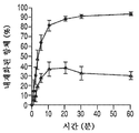


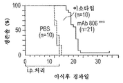


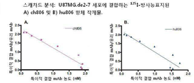


Classifications
-
- A—HUMAN NECESSITIES
- A61—MEDICAL OR VETERINARY SCIENCE; HYGIENE
- A61K—PREPARATIONS FOR MEDICAL, DENTAL OR TOILETRY PURPOSES
- A61K47/00—Medicinal preparations characterised by the non-active ingredients used, e.g. carriers or inert additives; Targeting or modifying agents chemically bound to the active ingredient
- A61K47/50—Medicinal preparations characterised by the non-active ingredients used, e.g. carriers or inert additives; Targeting or modifying agents chemically bound to the active ingredient the non-active ingredient being chemically bound to the active ingredient, e.g. polymer-drug conjugates
- A61K47/51—Medicinal preparations characterised by the non-active ingredients used, e.g. carriers or inert additives; Targeting or modifying agents chemically bound to the active ingredient the non-active ingredient being chemically bound to the active ingredient, e.g. polymer-drug conjugates the non-active ingredient being a modifying agent
- A61K47/68—Medicinal preparations characterised by the non-active ingredients used, e.g. carriers or inert additives; Targeting or modifying agents chemically bound to the active ingredient the non-active ingredient being chemically bound to the active ingredient, e.g. polymer-drug conjugates the non-active ingredient being a modifying agent the modifying agent being an antibody, an immunoglobulin or a fragment thereof, e.g. an Fc-fragment
- A61K47/6835—Medicinal preparations characterised by the non-active ingredients used, e.g. carriers or inert additives; Targeting or modifying agents chemically bound to the active ingredient the non-active ingredient being chemically bound to the active ingredient, e.g. polymer-drug conjugates the non-active ingredient being a modifying agent the modifying agent being an antibody, an immunoglobulin or a fragment thereof, e.g. an Fc-fragment the modifying agent being an antibody or an immunoglobulin bearing at least one antigen-binding site
- A61K47/6849—Medicinal preparations characterised by the non-active ingredients used, e.g. carriers or inert additives; Targeting or modifying agents chemically bound to the active ingredient the non-active ingredient being chemically bound to the active ingredient, e.g. polymer-drug conjugates the non-active ingredient being a modifying agent the modifying agent being an antibody, an immunoglobulin or a fragment thereof, e.g. an Fc-fragment the modifying agent being an antibody or an immunoglobulin bearing at least one antigen-binding site the antibody targeting a receptor, a cell surface antigen or a cell surface determinant
-
- C—CHEMISTRY; METALLURGY
- C07—ORGANIC CHEMISTRY
- C07K—PEPTIDES
- C07K16/00—Immunoglobulins [IGs], e.g. monoclonal or polyclonal antibodies
- C07K16/40—Immunoglobulins [IGs], e.g. monoclonal or polyclonal antibodies against enzymes
-
- A—HUMAN NECESSITIES
- A61—MEDICAL OR VETERINARY SCIENCE; HYGIENE
- A61K—PREPARATIONS FOR MEDICAL, DENTAL OR TOILETRY PURPOSES
- A61K31/00—Medicinal preparations containing organic active ingredients
- A61K31/33—Heterocyclic compounds
- A61K31/395—Heterocyclic compounds having nitrogen as a ring hetero atom, e.g. guanethidine or rifamycins
- A61K31/495—Heterocyclic compounds having nitrogen as a ring hetero atom, e.g. guanethidine or rifamycins having six-membered rings with two or more nitrogen atoms as the only ring heteroatoms, e.g. piperazine or tetrazines
-
- A—HUMAN NECESSITIES
- A61—MEDICAL OR VETERINARY SCIENCE; HYGIENE
- A61K—PREPARATIONS FOR MEDICAL, DENTAL OR TOILETRY PURPOSES
- A61K39/00—Medicinal preparations containing antigens or antibodies
- A61K39/395—Antibodies; Immunoglobulins; Immune serum, e.g. antilymphocytic serum
- A61K39/39533—Antibodies; Immunoglobulins; Immune serum, e.g. antilymphocytic serum against materials from animals
- A61K39/39541—Antibodies; Immunoglobulins; Immune serum, e.g. antilymphocytic serum against materials from animals against normal tissues, cells
-
- A—HUMAN NECESSITIES
- A61—MEDICAL OR VETERINARY SCIENCE; HYGIENE
- A61K—PREPARATIONS FOR MEDICAL, DENTAL OR TOILETRY PURPOSES
- A61K39/00—Medicinal preparations containing antigens or antibodies
- A61K39/395—Antibodies; Immunoglobulins; Immune serum, e.g. antilymphocytic serum
- A61K39/39533—Antibodies; Immunoglobulins; Immune serum, e.g. antilymphocytic serum against materials from animals
- A61K39/39558—Antibodies; Immunoglobulins; Immune serum, e.g. antilymphocytic serum against materials from animals against tumor tissues, cells, antigens
-
- A—HUMAN NECESSITIES
- A61—MEDICAL OR VETERINARY SCIENCE; HYGIENE
- A61K—PREPARATIONS FOR MEDICAL, DENTAL OR TOILETRY PURPOSES
- A61K47/00—Medicinal preparations characterised by the non-active ingredients used, e.g. carriers or inert additives; Targeting or modifying agents chemically bound to the active ingredient
- A61K47/50—Medicinal preparations characterised by the non-active ingredients used, e.g. carriers or inert additives; Targeting or modifying agents chemically bound to the active ingredient the non-active ingredient being chemically bound to the active ingredient, e.g. polymer-drug conjugates
- A61K47/51—Medicinal preparations characterised by the non-active ingredients used, e.g. carriers or inert additives; Targeting or modifying agents chemically bound to the active ingredient the non-active ingredient being chemically bound to the active ingredient, e.g. polymer-drug conjugates the non-active ingredient being a modifying agent
- A61K47/68—Medicinal preparations characterised by the non-active ingredients used, e.g. carriers or inert additives; Targeting or modifying agents chemically bound to the active ingredient the non-active ingredient being chemically bound to the active ingredient, e.g. polymer-drug conjugates the non-active ingredient being a modifying agent the modifying agent being an antibody, an immunoglobulin or a fragment thereof, e.g. an Fc-fragment
- A61K47/6835—Medicinal preparations characterised by the non-active ingredients used, e.g. carriers or inert additives; Targeting or modifying agents chemically bound to the active ingredient the non-active ingredient being chemically bound to the active ingredient, e.g. polymer-drug conjugates the non-active ingredient being a modifying agent the modifying agent being an antibody, an immunoglobulin or a fragment thereof, e.g. an Fc-fragment the modifying agent being an antibody or an immunoglobulin bearing at least one antigen-binding site
- A61K47/6871—Medicinal preparations characterised by the non-active ingredients used, e.g. carriers or inert additives; Targeting or modifying agents chemically bound to the active ingredient the non-active ingredient being chemically bound to the active ingredient, e.g. polymer-drug conjugates the non-active ingredient being a modifying agent the modifying agent being an antibody, an immunoglobulin or a fragment thereof, e.g. an Fc-fragment the modifying agent being an antibody or an immunoglobulin bearing at least one antigen-binding site the antibody targeting an enzyme
-
- A—HUMAN NECESSITIES
- A61—MEDICAL OR VETERINARY SCIENCE; HYGIENE
- A61K—PREPARATIONS FOR MEDICAL, DENTAL OR TOILETRY PURPOSES
- A61K51/00—Preparations containing radioactive substances for use in therapy or testing in vivo
- A61K51/02—Preparations containing radioactive substances for use in therapy or testing in vivo characterised by the carrier, i.e. characterised by the agent or material covalently linked or complexing the radioactive nucleus
- A61K51/04—Organic compounds
- A61K51/08—Peptides, e.g. proteins, carriers being peptides, polyamino acids, proteins
- A61K51/10—Antibodies or immunoglobulins; Fragments thereof, the carrier being an antibody, an immunoglobulin or a fragment thereof, e.g. a camelised human single domain antibody or the Fc fragment of an antibody
- A61K51/1027—Antibodies or immunoglobulins; Fragments thereof, the carrier being an antibody, an immunoglobulin or a fragment thereof, e.g. a camelised human single domain antibody or the Fc fragment of an antibody against receptors, cell-surface antigens or cell-surface determinants
- A61K51/103—Antibodies or immunoglobulins; Fragments thereof, the carrier being an antibody, an immunoglobulin or a fragment thereof, e.g. a camelised human single domain antibody or the Fc fragment of an antibody against receptors, cell-surface antigens or cell-surface determinants against receptors for growth factors or receptors for growth regulators
-
- A—HUMAN NECESSITIES
- A61—MEDICAL OR VETERINARY SCIENCE; HYGIENE
- A61K—PREPARATIONS FOR MEDICAL, DENTAL OR TOILETRY PURPOSES
- A61K51/00—Preparations containing radioactive substances for use in therapy or testing in vivo
- A61K51/02—Preparations containing radioactive substances for use in therapy or testing in vivo characterised by the carrier, i.e. characterised by the agent or material covalently linked or complexing the radioactive nucleus
- A61K51/04—Organic compounds
- A61K51/08—Peptides, e.g. proteins, carriers being peptides, polyamino acids, proteins
- A61K51/10—Antibodies or immunoglobulins; Fragments thereof, the carrier being an antibody, an immunoglobulin or a fragment thereof, e.g. a camelised human single domain antibody or the Fc fragment of an antibody
- A61K51/1045—Antibodies or immunoglobulins; Fragments thereof, the carrier being an antibody, an immunoglobulin or a fragment thereof, e.g. a camelised human single domain antibody or the Fc fragment of an antibody against animal or human tumor cells or tumor cell determinants
-
- A—HUMAN NECESSITIES
- A61—MEDICAL OR VETERINARY SCIENCE; HYGIENE
- A61K—PREPARATIONS FOR MEDICAL, DENTAL OR TOILETRY PURPOSES
- A61K51/00—Preparations containing radioactive substances for use in therapy or testing in vivo
- A61K51/02—Preparations containing radioactive substances for use in therapy or testing in vivo characterised by the carrier, i.e. characterised by the agent or material covalently linked or complexing the radioactive nucleus
- A61K51/04—Organic compounds
- A61K51/08—Peptides, e.g. proteins, carriers being peptides, polyamino acids, proteins
- A61K51/10—Antibodies or immunoglobulins; Fragments thereof, the carrier being an antibody, an immunoglobulin or a fragment thereof, e.g. a camelised human single domain antibody or the Fc fragment of an antibody
- A61K51/1078—Antibodies or immunoglobulins; Fragments thereof, the carrier being an antibody, an immunoglobulin or a fragment thereof, e.g. a camelised human single domain antibody or the Fc fragment of an antibody the antibody being against an immunoglobulin, i.e. being an (anti)-anti-idiotypic antibody
-
- A—HUMAN NECESSITIES
- A61—MEDICAL OR VETERINARY SCIENCE; HYGIENE
- A61P—SPECIFIC THERAPEUTIC ACTIVITY OF CHEMICAL COMPOUNDS OR MEDICINAL PREPARATIONS
- A61P35/00—Antineoplastic agents
-
- A—HUMAN NECESSITIES
- A61—MEDICAL OR VETERINARY SCIENCE; HYGIENE
- A61P—SPECIFIC THERAPEUTIC ACTIVITY OF CHEMICAL COMPOUNDS OR MEDICINAL PREPARATIONS
- A61P43/00—Drugs for specific purposes, not provided for in groups A61P1/00-A61P41/00
-
- C—CHEMISTRY; METALLURGY
- C07—ORGANIC CHEMISTRY
- C07K—PEPTIDES
- C07K16/00—Immunoglobulins [IGs], e.g. monoclonal or polyclonal antibodies
- C07K16/18—Immunoglobulins [IGs], e.g. monoclonal or polyclonal antibodies against material from animals or humans
- C07K16/28—Immunoglobulins [IGs], e.g. monoclonal or polyclonal antibodies against material from animals or humans against receptors, cell surface antigens or cell surface determinants
- C07K16/2863—Immunoglobulins [IGs], e.g. monoclonal or polyclonal antibodies against material from animals or humans against receptors, cell surface antigens or cell surface determinants against receptors for growth factors, growth regulators
-
- C—CHEMISTRY; METALLURGY
- C07—ORGANIC CHEMISTRY
- C07K—PEPTIDES
- C07K16/00—Immunoglobulins [IGs], e.g. monoclonal or polyclonal antibodies
- C07K16/18—Immunoglobulins [IGs], e.g. monoclonal or polyclonal antibodies against material from animals or humans
- C07K16/28—Immunoglobulins [IGs], e.g. monoclonal or polyclonal antibodies against material from animals or humans against receptors, cell surface antigens or cell surface determinants
- C07K16/30—Immunoglobulins [IGs], e.g. monoclonal or polyclonal antibodies against material from animals or humans against receptors, cell surface antigens or cell surface determinants from tumour cells
-
- C—CHEMISTRY; METALLURGY
- C07—ORGANIC CHEMISTRY
- C07K—PEPTIDES
- C07K16/00—Immunoglobulins [IGs], e.g. monoclonal or polyclonal antibodies
- C07K16/42—Immunoglobulins [IGs], e.g. monoclonal or polyclonal antibodies against immunoglobulins
- C07K16/4208—Immunoglobulins [IGs], e.g. monoclonal or polyclonal antibodies against immunoglobulins against an idiotypic determinant on Ig
- C07K16/4241—Immunoglobulins [IGs], e.g. monoclonal or polyclonal antibodies against immunoglobulins against an idiotypic determinant on Ig against anti-human or anti-animal Ig
- C07K16/4258—Immunoglobulins [IGs], e.g. monoclonal or polyclonal antibodies against immunoglobulins against an idiotypic determinant on Ig against anti-human or anti-animal Ig against anti-receptor Ig
-
- G—PHYSICS
- G01—MEASURING; TESTING
- G01N—INVESTIGATING OR ANALYSING MATERIALS BY DETERMINING THEIR CHEMICAL OR PHYSICAL PROPERTIES
- G01N33/00—Investigating or analysing materials by specific methods not covered by groups G01N1/00 - G01N31/00
- G01N33/48—Biological material, e.g. blood, urine; Haemocytometers
- G01N33/50—Chemical analysis of biological material, e.g. blood, urine; Testing involving biospecific ligand binding methods; Immunological testing
- G01N33/53—Immunoassay; Biospecific binding assay; Materials therefor
- G01N33/574—Immunoassay; Biospecific binding assay; Materials therefor for cancer
- G01N33/57484—Immunoassay; Biospecific binding assay; Materials therefor for cancer involving compounds serving as markers for tumor, cancer, neoplasia, e.g. cellular determinants, receptors, heat shock/stress proteins, A-protein, oligosaccharides, metabolites
-
- A—HUMAN NECESSITIES
- A61—MEDICAL OR VETERINARY SCIENCE; HYGIENE
- A61K—PREPARATIONS FOR MEDICAL, DENTAL OR TOILETRY PURPOSES
- A61K39/00—Medicinal preparations containing antigens or antibodies
- A61K2039/505—Medicinal preparations containing antigens or antibodies comprising antibodies
-
- A—HUMAN NECESSITIES
- A61—MEDICAL OR VETERINARY SCIENCE; HYGIENE
- A61K—PREPARATIONS FOR MEDICAL, DENTAL OR TOILETRY PURPOSES
- A61K2300/00—Mixtures or combinations of active ingredients, wherein at least one active ingredient is fully defined in groups A61K31/00 - A61K41/00
-
- C—CHEMISTRY; METALLURGY
- C07—ORGANIC CHEMISTRY
- C07K—PEPTIDES
- C07K2317/00—Immunoglobulins specific features
- C07K2317/20—Immunoglobulins specific features characterized by taxonomic origin
- C07K2317/24—Immunoglobulins specific features characterized by taxonomic origin containing regions, domains or residues from different species, e.g. chimeric, humanized or veneered
-
- C—CHEMISTRY; METALLURGY
- C07—ORGANIC CHEMISTRY
- C07K—PEPTIDES
- C07K2317/00—Immunoglobulins specific features
- C07K2317/30—Immunoglobulins specific features characterized by aspects of specificity or valency
- C07K2317/34—Identification of a linear epitope shorter than 20 amino acid residues or of a conformational epitope defined by amino acid residues
-
- C—CHEMISTRY; METALLURGY
- C07—ORGANIC CHEMISTRY
- C07K—PEPTIDES
- C07K2317/00—Immunoglobulins specific features
- C07K2317/50—Immunoglobulins specific features characterized by immunoglobulin fragments
- C07K2317/51—Complete heavy chain or Fd fragment, i.e. VH + CH1
-
- C—CHEMISTRY; METALLURGY
- C07—ORGANIC CHEMISTRY
- C07K—PEPTIDES
- C07K2317/00—Immunoglobulins specific features
- C07K2317/50—Immunoglobulins specific features characterized by immunoglobulin fragments
- C07K2317/515—Complete light chain, i.e. VL + CL
-
- C—CHEMISTRY; METALLURGY
- C07—ORGANIC CHEMISTRY
- C07K—PEPTIDES
- C07K2317/00—Immunoglobulins specific features
- C07K2317/50—Immunoglobulins specific features characterized by immunoglobulin fragments
- C07K2317/52—Constant or Fc region; Isotype
-
- C—CHEMISTRY; METALLURGY
- C07—ORGANIC CHEMISTRY
- C07K—PEPTIDES
- C07K2317/00—Immunoglobulins specific features
- C07K2317/50—Immunoglobulins specific features characterized by immunoglobulin fragments
- C07K2317/56—Immunoglobulins specific features characterized by immunoglobulin fragments variable (Fv) region, i.e. VH and/or VL
- C07K2317/565—Complementarity determining region [CDR]
-
- C—CHEMISTRY; METALLURGY
- C07—ORGANIC CHEMISTRY
- C07K—PEPTIDES
- C07K2317/00—Immunoglobulins specific features
- C07K2317/70—Immunoglobulins specific features characterized by effect upon binding to a cell or to an antigen
- C07K2317/73—Inducing cell death, e.g. apoptosis, necrosis or inhibition of cell proliferation
- C07K2317/732—Antibody-dependent cellular cytotoxicity [ADCC]
-
- C—CHEMISTRY; METALLURGY
- C07—ORGANIC CHEMISTRY
- C07K—PEPTIDES
- C07K2317/00—Immunoglobulins specific features
- C07K2317/70—Immunoglobulins specific features characterized by effect upon binding to a cell or to an antigen
- C07K2317/73—Inducing cell death, e.g. apoptosis, necrosis or inhibition of cell proliferation
- C07K2317/734—Complement-dependent cytotoxicity [CDC]
-
- C—CHEMISTRY; METALLURGY
- C07—ORGANIC CHEMISTRY
- C07K—PEPTIDES
- C07K2317/00—Immunoglobulins specific features
- C07K2317/70—Immunoglobulins specific features characterized by effect upon binding to a cell or to an antigen
- C07K2317/77—Internalization into the cell
-
- C—CHEMISTRY; METALLURGY
- C07—ORGANIC CHEMISTRY
- C07K—PEPTIDES
- C07K2317/00—Immunoglobulins specific features
- C07K2317/90—Immunoglobulins specific features characterized by (pharmaco)kinetic aspects or by stability of the immunoglobulin
- C07K2317/92—Affinity (KD), association rate (Ka), dissociation rate (Kd) or EC50 value
Abstract
본 발명은 증폭된 상피 성장인자 수용체(EGFR) 및 EGFR의 de2-7 EGFR 절두형에 결합하는, 특이적 결합 구성원, 특히 항체 및 이의 단편에 관한 것이다. 구체적으로, 상기 특이적 결합 구성원, 특히 항체 및 이의 단편에 의해 인식되는 에피토프는 이상 해독후 변형 시 증가되거나 분명해진다. 이러한 특이적 결합 구성원은 암의 진단 및 치료에 유용하다. 또한, 본 발명의 결합 구성원은 화학요법 또는 항암제 및/또는 다른 항체 또는 이의 단편과 함께 치료법에 사용될 수도 있다.The invention relates to specific binding members, particularly antibodies and fragments thereof, that bind to the de2-7 EGFR duplex of the amplified epithelial growth factor receptor (EGFR) and EGFR. Specifically, epitopes recognized by the specific binding members, particularly antibodies and fragments thereof, increase or become evident upon transformation after anomalous detoxification. Such specific binding members are useful for diagnosis and treatment of cancer. In addition, the binding member of the invention may be used in therapy together with chemotherapy or anticancer agents and / or other antibodies or fragments thereof.
Description
관련 출원 데이터Related Application Data
본 국제 PCT 특허 출원은 2009년 2월 18일에 출원된 미국 특허출원 제12/388,504호에 대해 우선권을 주장하며, 이의 교시 전체는 본원에 참고 인용된다. 본 국제 PCT 특허 출원은 또한 2002년 5월 13일에 출원된 미국 특허출원 제10/145,598호(현재, 2009년 9월 15일자로 등록된 미국 특허 제7,589,180호); 2001년 5월 11일에 출원된 미국 임시특허출원 제60/290,410호; 2001년 9월 28일에 출원된 미국 임시특허출원 제60/326,019호; 2001년 12월 21일에 출원된 미국 임시특허출원 제60/342,258호; 2002년 5월 13일에 출원된 국제 PCT 특허출원 제PCT/US02/15185호(2002년 11월 21일에 WO 02/092771로 공개됨); 2008년 8월 14일에 출원된 국제 PCT 특허출원 제PCT/US2008/009771호(2009년 2월 19일에 WO 2009/023265로 공개됨); 및 2007년 8월 14일에 출원된 미국 임시특허출원 제60/964,715호의 각 교시 전체를 참고인용한다.This International PCT patent application claims priority to U.S. Patent Application No. 12 / 388,504, filed February 18, 2009, the entire teachings of which are incorporated herein by reference. This International PCT patent application is also a continuation-in-part of U.S. Patent Application No. 10 / 145,598, filed May 13, 2002 (now U.S. Patent No. 7,589,180, filed September 15, 2009); U.S. Provisional Patent Application No. 60 / 290,410, filed May 11, 2001; U.S. Provisional Patent Application No. 60 / 326,019, filed September 28, 2001; U.S. Provisional Patent Application No. 60 / 342,258, filed December 21, 2001; International PCT Patent Application No. PCT / US02 / 15185, filed May 13, 2002 (published as
발명의 분야Field of invention
본 발명은 증폭된 상피 성장인자 수용체(EGFR) 및 EGFR의 엑손 2 내지 7의 프레임내 결실부에 결합하여 세포외 도메인으로부터 267개 아미노산이 결손된 절두형 EGFR 수용체(de2-7 EGFR)를 산출하는, 특이적 결합 구성원, 특히 항체 및 이의 단편에 관한 것이다. 구체적으로, 상기 특이적 결합 구성원, 특히 항체 및 이의 단편에 의해 인식되는 에피토프는 이상 해독후 변형 시 증가되거나 분명해진다. 이러한 특이적 결합 구성원은 암의 진단 및 치료에 유용하다. 또한, 본 발명의 결합 구성원은 화학요법 또는 항암제 및/또는 다른 항체 또는 이의 단편과 함께 치료법에 사용될 수도 있다.The present invention relates to a method for producing a bisecting EGFR receptor (de2-7 EGFR) in which 267 amino acids are deleted from the extracellular domain by binding to an amplified epithelial growth factor receptor (EGFR) and an intra-frame deletion region of
화학치료 수단에 의한 증식성 질환, 특히 암의 치료는 사람 또는 동물 체내의 표적 증식 세포 및 다른 정상 세포에서의 차이를 개발하는데 의존적이다. 예를 들어, 많은 화학제는 빠르게 복제하는 DNA에 의해 흡수되도록 설계되어, DNA 복제 및 세포 분열 과정을 방해한다. 다른 접근법은 발달된 사람 조직, 예컨대 종양 항원 또는 배아 항원에서 정상적으로 발현되지 않는 종양 세포 또는 다른 비정상 세포의 표면에서 항원을 확인하는 것이다. 이러한 항원은 항원을 차단하거나 중화시킬 수 있는 항체와 같은 결합 단백질로 표적화될 수 있다. 또한, 항체 및 이의 단편을 비롯한 결합 단백질은 종양 부위에서 독성 제제를 직접 또는 간접적으로 활성화시킬 수 있는 독성 제제 또는 다른 물질을 전달할 수 있다.Treatment of proliferative diseases, in particular cancer, by means of chemotherapy is dependent on developing differences in target proliferating cells and other normal cells in a human or animal body. For example, many chemicals are designed to be absorbed by DNA that replicates rapidly, hindering DNA replication and cell division processes. Another approach is to identify antigens on the surface of tumor cells, or other abnormal cells, that are not normally expressed in human tissue, such as tumor antigens or embryonic antigens. Such antigens can be targeted to binding proteins, such as antibodies, that can block or neutralize the antigen. In addition, binding proteins, including antibodies and fragments thereof, can deliver toxic agents or other substances that can directly or indirectly activate the toxic agent at the tumor site.
EGFR은 많은 종류의 상피 종양에서 과발현되기 때문에 종양-표적화된 항체 치료법에 매력적인 표적이다(Voldborg et al.(1997). Epidermal growth factor receptor (EGFR) and EGFR mutations, function and possible role in clinical trials. Ann Oncol. 8, 1197-206; den Eynde, B. and Scott, A. M. Tumor Antigens. In: P. J. Delves and I. M. Roitt (eds.), Encyclopedia of Immunology, Second Edition, pp. 2424-31. London: Academic Press (1998)). 더욱이, EGFR의 발현은 많은 종양 종류, 예컨대 위, 결장, 방광, 유방, 전립선, 내막, 신장 및 뇌(예: 신경아교종)의 나쁜 예후와 연관이 있다. 결과적으로, 많은 EGFR 항체가 문헌에 보고되었고, 몇몇은 임상 평가 중이다(Baselga et al. (2000) Phase I Studies of Anti-Epidermal Growth Factor Receptor Chimeric Antibody C225 Alone and in Combination With Cisplatin. J. Clin . Oncol. 18, 904; Faillot et al. (1996): A phase I study of an anti-epidermal growth factor receptor monoclonal antibody for the treatment of malignant gliomas. Neurosurgery. 39, 478-83; Seymour, L. (1999) Novel anti-cancer agents in development: exciting prospects and new challenges. Cancer Treat . Rev . 25, 301-12)).EGFR is an attractive target for tumor-targeted antibody therapy because it is overexpressed in many types of epithelial tumors (Voldborg et al. (1997).) Epidermal growth factor receptor (EGFR) and EGFR mutations, function and possible role in clinical trials. Ann Oncol . 8, 1197-206; den Eynde, B. and Scott, AM Tumor Antigens. In: PJ Delves and IM Roitt (eds.), Encyclopedia of Immunology , Second Edition, pp. 2424-31. London: Academic Press (1998)). Moreover, the expression of EGFR is associated with a number of tumor types, such as stomach, colon, bladder, breast, prostate, endometrium, kidney, and bad prognosis of the brain (e.g. glioma). As a result, the number of EGFR antibodies have been reported in the literature, and some are under clinical evaluation (Baselga et al. (2000) Phase I Studies of Anti-Epidermal Growth Factor Receptor Chimeric Antibody C225 Alone and in Combination With Cisplatin. J. Clin. Oncol (1996): A phase I study of an anti-epidermal growth factor receptor monoclonal antibody for the treatment of malignant gliomas,
두경부암, 편평세포 폐암, 뇌 신경아교종 및 악성 성상세포종 환자에서 EGFR mAb를 이용한 연구의 결과는 고무적이었다. 대부분의 EGFR 항체의 항종양 활성은 리간드 결합을 차단하는 능력에 의해 증강된다(Sturgis et al. (1994) Effects of antiepidermal growth factor receptor antibody 528 on the proliferation and differentiation of head and neck cancer. Otolaryngol . Head Neck . Surg. 111, 633-43; Goldstein et al. (1995) Biological efficacy of a chimeric antibody to the epidermal growth factor receptor in a human tumor xenograft model. Clin . Cancer Res. 1, 1311-8). 이러한 항체는 세포 증식 조절 및 항체 의존적 면역 기능(예: 보체 활성화)을 통해 자신의 효능을 매개할 수 있다. 하지만, 이러한 항체의 사용은 간 및 피부와 같이 EGFR의 높은 내인성 수준을 보유하는 기관에서 흡수되어 제한될 수 있다(Baselga et al., 2000; Faillot et al., 1996).The results of studies using EGFR mAb in patients with head and neck cancer, squamous cell lung cancer, brain glioma, and malignant astrocytoma were encouraging. Most of the antitumor activity of EGFR antibodies is enhanced by their ability to block ligand binding (Sturgis et al. (1994) Effects of antiepidermal growth
또한, EGFR 유전자의 증폭(즉, EGFR 유전자의 다중 카피)을 함유하는 종양의 상당한 비율은 de2-7 EGFR, △EGFR 또는 △2-7(이 용어들은 본원에서 호환 사용됨)로 알려진 수용체의 절두형(Wikstrand et al. (1998) The class III variant of the epidermal growth factor receptor (EGFR): characterization and utilization as an immunotherapeutic target. J. Neurovirol. 4, 148-158)을 공동발현한다(Olapade-Olaopa et al. (2000) Evidence for the differential expression of a variant EGF receptor protein in human prostate cancer. Br . J. Cancer. 82, 186-94). de2-7 EGFR에서 관찰되는 재배열은 엑손 2-7에 걸쳐 있는 801개 뉴클레오타이드가 결손된 프레임내 성숙 mRNA를 생산한다(Wong et al. (1992) Structural alterations of the epidermal growth factor receptor gene in human gliomas. Proc. Natl. Acad. Sci. U.S. A. 89, 2965-9; Yamazaki et al. (1990) A deletion mutation within the ligand binding domain is responsible for activation of epidermal growth factor receptor gene in human brain tumors. Jpn . J. Cancer Res. 81, 773-9; Yamazaki et al. (1988) Amplification of the structurally and functionally altered epidermal growth factor receptor gene (c-erbB) in human brain tumors. Mol . Cell Biol. 8, 1816-20; Sugawa et al. (1990) Identical splicing of aberrant epidermal growth factor receptor transcripts from amplified rearranged genes in human glioblastomas. Proc . Natl . Acad . Sci. U.S.A. 87, 8602-6). 대응하는 EGFR 단백질은 세포외 도메인의 잔기 6-273 및 융합 연접부에 신규 글리신 잔기를 함유하는 267개 아미노산 결실을 보유한다(Sugawa et al., 1990). 글리신 잔기의 삽입과 함께 상기 결실은 결실 계면에 독특한 연접부 펩타이드를 생산한다(Sugawa et al., 1990).In addition, a significant proportion of tumors containing amplification of the EGFR gene (i. E., Multiple copies of the EGFR gene) have been found to have an EGFR, EGFR or < RTI ID = 0.0 > (1998), co-expressing the class III variant of the epidermal growth factor receptor (EGFR): characterization and utilization as an immunotherapeutic target, J. Neurovirol . 4, 148-158 (Olapade-Olaopa et al (2000) Evidence for differential expression of a variant EGF receptor protein in human prostate cancer. Br . J. Cancer ., 82, 186-94). The rearrangement observed in de2-7 EGFR produces mature mRNA in frames lacking 801 nucleotides across exons 2-7 (Wong et al. (1992) Structural alterations of the epidermal growth factor receptor in human gliomas (1990) A deletion mutation within the ligand binding domain is responsible for activation of the epidermal growth factor receptor gene in human brain tumors. Jpn . J. Proc Natl Acad Sci USA 89, 2965-9 Yamazaki et al. Cancer Res . 81, 773-9; Yamazaki et al. (1988) Amplification of the structurally and functionally altered epidermal growth factor receptor gene (c-erbB) in human brain tumors. Mol . Cell Biol . 8, 1816-20; Sugawa et al. (1990) Identical splicing of aberrant epidermal growth factor receptor transcripts from amplified rearranged genes in human glioblastomas. Proc . Natl . Acad . Sci . USA 87,8602-6). The corresponding EGFR protein retains residues 6-273 of the extracellular domain and 267 amino acid deletions containing a novel glycine residue at the fusion junction (Sugawa et al., 1990). With insertion of the glycine residue, the deletion produces a conformational peptide unique to the deletion interface (Sugawa et al., 1990).
de2-7 EGFR은 신경아교종, 유방암, 폐암, 난소암 및 전립선암을 비롯한 다수의 종양 종류에서 보고되었다(Wikstrand et al. (1997) Cell surface localization and density of the tumor-associated variant of the epidermal growth factor receptor, EGFRvIII. Cancer Res. 57, 4130-40; Olapade-Olaopa et al. (2000) Evidence for the differential expression of a variant EGF receptor protein in human prostate cancer. Br . J. Cancer. 82, 186-94; Wikstrand, et al. (1995) Monoclonal antibodies against EGFRvIII in are tumor specific and react with breast and lung carcinomas and malignant gliomas. Cancer Res. 55, 3140-8; Garcia de Palazzo et al. (1993) Expression of mutated epidermal growth factor receptor by non-small cell lung carcinomas. Cancer Res. 53, 3217-20). 이러한 절두형 수용체는 리간드에 결합하지 않지만, 낮은 구성적 활성을 보유하고 누드 마우스에서 종양 이종이식편으로 성장한 신경아교종 세포에 상당한 성장 이점을 부여하고(Nishikawa et al. (1994) A mutant epidermal growth factor receptor common in human glioma confers enhanced tumorigenicity. Proc . Natl . Acad . Sci. U.S.A. 91, 7727-31), NIH3T3 세포(Batra et al. (1995) Epidermal growth factor ligand independent, unregulated, cell-transforming potential of a naturally occurring human mutant EGFRvIII gene. Cell Growth Differ. 6, 1251-9) 및 MCF-7 세포를 형질전환시킬 수 있다. 신경아교종 세포에서 de2-7 EGFR에 의해 활용되는 세포 기전은 완전히 밝혀지지 않았지만, 아폽토시스 감소(Nagane et al. (1996) A common mutant epidermal growth factor receptor confers enhanced tumorigenicity on human glioblastoma cells by increasing proliferation and reducing apoptosis. Cancer Res. 56, 5079-86) 및 소량의 증식 증강(Nagane et al., 1996)을 포함한다.de2-7 EGFR has been reported in a number of tumor types including glioma, breast, lung, ovarian and prostate cancer (Wikstrand et al. (1997) Cell surface localization and density of the tumor-associated variant of the epidermal growth factor receptor, EGFRvIII. Cancer Res . 57, 4130-40; Olapade-Olaopa et al. (2000) Evidence for differential expression of a variant EGF receptor protein in human prostate cancer. Br . J. Cancer . 82, 186-94; Wikstrand, et al. (1995) Monoclonal antibodies against EGFRvlll are tumor specific and react with breast and lung carcinomas and malignant gliomas. Cancer Res . 55, 3140-8; Garcia de Palazzo et al. (1993) Expression of mutated epidermal growth factor receptor by non-small cell lung carcinomas. Cancer Res . 53,3217-20). These quadriceps receptors do not bind to the ligand, but they have significant structural growth potential in glioma cells that have low constitutive activity and have grown into tumor xenografts in nude mice (Nishikawa et al. (1994) A mutant epidermal growth factor receptor common in human glioma confers enhanced tumorigenicity. Proc. Natl. Acad. Sci. USA 91, 7727-31), NIH3T3 cells (Batra et al. (1995) Epidermal growth factor ligand independent, unregulated, cell-transforming potential of a naturally occurring human mutant EGFRvIII gene. Cell Growth Differ . 6, 1251-9) and MCF-7 cells. The mechanism by which de2-7 EGFR is used in glioma cells is not fully understood, but the apoptosis is reduced (Nagane et al. (1996) A common mutant epidermal growth factor receptor confers enhanced tumorigenicity on glioblastoma cells by increasing proliferation and reducing apoptosis Cancer Res . 56, 5079-86) and small amounts of proliferation enhancement (Nagane et al., 1996).
이러한 절두형 수용체의 발현은 종양 세포에 제한되기 때문에, 항체 치료법에서 매우 특이적인 표적이다. 따라서, 많은 연구진들은 de2-7 EGFR의 독특한 펩타이드에 특이적인 폴리클로널(Humphrey et al. (1990) Anti-synthetic peptide antibody reacting at the fusion junction of deletion mutant epidermal growth factor receptors in human glioblastoma. Proc . Natl . Acad . Sci. U.S.A. 87, 4207-11) 및 모노클로널(Wikstrand et al. (1995) Monoclonal antibodies against EGFRvIII are tumor specific and react with breast and lung carcinomas and malignant gliomas; Okamoto et al. (1996) Monoclonal antibody against the fusion junction of a deletion-mutant epidermal growth factor receptor. Br . J. Cancer . 73, 1366-72; Hills et al. (1995) Specific targeting of a mutant, activated EGF receptor found in glioblastoma using a monoclonal antibody. Int. J. Cancer. 63, 537-43) 항체의 생성을 보고했다. 독특한 de2-7 펩타이드로 면역화 후에 분리된 일련의 마우스 mAb는 모두 누드 마우스에서 성장한 표적화된 de2-7 EGFR 양성 이종이식편과 절두형 수용체에 대해 선택성과 특이성을 보였다(Wikstrand et al. (1995); Reist et al. (1997) Improved targeting of an anti-epidermal growth factor receptor variant III monoclonal antibody in tumor xenografts after labeling using N-succinimidyl 5-iodo-3-pyridinecarboxylate. Cancer Res. 57, 1510-5; Reist et al. (1995) Tumor-specific anti-epidermal growth factor receptor variant III monoclonal antibodies: use of the tyramine-cellobiose radioiodination method enhances cellular retention and uptake in tumor xenografts. Cancer Res. 55, 4375-82).Because the expression of these podophyll receptors is restricted to tumor cells, it is a very specific target in antibody therapy. Therefore, many researchers have specific polyclonal board (Humphrey et al. (1990) Anti-synthetic peptide antibody reacting at the fusion junction of deletion mutant epidermal growth factor receptors in human glioblastoma. Proc. Natl the unique peptide of de2-7 EGFR ... Acad Sci USA 87, 4207-11) and monoclonal board (Wikstrand et al (1995.) monoclonal antibodies against EGFRvIII are tumor specific and react with breast and lung carcinomas and malignant gliomas;. Okamoto et al (1996) monoclonal . antibody against the fusion junction of a deletion-mutant epidermal growth factor receptor Br J. Cancer 73, 1366-72;... Hills et al (1995) Specific targeting of a mutant, activated EGF receptor found in glioblastoma using a monoclonal antibody Int. J. Cancer., 63, 537-43). A series of mouse mAbs isolated after immunization with a unique de2-7 peptide showed selectivity and specificity for targeted de2-7 EGFR positive xenografts and zygospores grown in nude mice (Wikstrand et al. (1995); Reist (1997) Improved targeting of an anti-epidermal growth factor receptor variant III monoclonal antibody in tumor xenografts after labeling using N-succinimidyl 5-iodo-3-pyridinecarboxylate. Cancer Res . 57, 1510-5; Reist et al. (1995) Tumor-specific anti-epidermal growth factor receptor variant III monoclonal antibodies: use of the tyramine-cellobiose radioiodination method enhances cellular retention and uptake in tumor xenografts. Cancer Res . 55, 4375-82).
하지만, de2-7 EGFR 항체의 1가지 잠재적 단점은 EGFR 유전자의 증폭을 나타내는 일정 비율의 종양만이 de2-7EGFR을 발현한다는 점이다(Ekstrand et al. (1992) Amplified and rearranged epidermal growth factor receptor genes in human glioblastomas reveal deletions of sequences encoding portions of the N-and/or C-terminal tails. Proc . Natl . Acad . Sci . U.S. A. 89, 4309-13). de2-7 EGFR을 함유하는 종양의 정확한 비율은 완전하게 확정된 것이 없는데, 그 이유는 다른 기술(즉, PCR 대 면역조직화학) 및 다양한 항체의 사용이 그 존재의 빈도에 대한 매우 광범위한 보고 값을 제공했기 때문이다. 공개된 데이터는 약 25 내지 30%의 신경아교종이 de2-7 EGFR을 발현하고, 이 발현은 역형성 성상세포종에서 가장 낮고 다형성 아교모세포종에서 가장 높다는 것을 나타낸다(Wong et al. (1992); Wikstrand et al. (1998) The class III variant of the epidermal growth factor receptor (EGFR): characterization and utilization as an immunotherapeutic target. J. Neurovirol. 4, 148-58; Moscatello et al. (1995) Frequent expression of a mutant epidermal growth factor receptor in multiple human tumors. Cancer Res. 55, 5536-9). de2-7 EGFR 발현 신경아교종 내에서 양성 세포의 비율은 37-86% 범위로 보고되어 있다(Wikstrand et al. (1997)). 27%의 유방 암종과 17%의 폐암은 de2-7 EGFR 양성인 것으로 발견되었다(Wikstrand et al. (1997); Wikstrand et al. (1995); Wikstrand et al.(1998); 및 Hills et al., 1995). 따라서, de2-7 EGFR 특이적 항체는 일정 비율의 EGFR 양성 종양에서만 유용한 것으로 생각할 수 있을 것이다.However, one potential drawback of the de2-7 EGFR antibody is that only a proportion of tumors expressing the amplification of the EGFR gene express de2-7EGFR (Ekstrand et al. (1992) Amplified and rearranged epidermal growth factor receptor genes in human glioblastomas reveal deletions of sequences encoding portions of the N-and / or C-terminal tails. Proc. Natl. Acad. Sci. US A. 89, 4309-13). The exact ratio of tumors containing de2-7 EGFR has not been completely established because the use of different techniques (i. e., PCR versus immunohistochemistry) and the use of various antibodies has provided a very broad reporting value on the frequency of its presence Because it provided them. Published data indicate that about 25-30% of the glioma gliomas express de2-7 EGFR, which is the lowest in reverse-forming astrocytomas and the highest in polymorphic glioblastomas (Wong et al. (1992); Wikstrand et (1998) The class III variant of the epidermal growth factor receptor (EGFR): Characterization and utilization of an immunotherapeutic target. J. Neurovirol ., 148-58; Moscatello et al. growth factor receptor in multiple human tumors. Cancer Res . 55,5536-9). The proportion of benign cells within de2-7 EGFR-expressing glioma is reported to range from 37-86% (Wikstrand et al. (1997)). 27% of breast cancers and 17% of lung cancers were found to be de2-7 EGFR positive (Wikstrand et al. (1997); Wikstrand et al. (1995); Wikstrand et al. 1995). Thus, de2-7 EGFR-specific antibodies may be thought to be useful only in a proportion of EGFR positive tumors.
따라서, EGFR 항체 활성에 대한 현존하는 증거는 고무적이지만, 앞에서 생각된 적용가능 범위 및 효능에 대한 관찰된 제한은 여전히 남아 있다. 따라서, 광범위한 종양에 효능을 입증하는 항체 및 유사 제제를 개발하는 것이 필요하며, 이는 본 발명이 지향하는 목적 달성을 위한 것이다.Thus, the existing evidence for EGFR antibody activity is encouraging, but the observed limitations on the applicability and efficacy previously contemplated remain. Thus, it is necessary to develop antibodies and similar agents that demonstrate efficacy in a wide range of tumors, and this is for the purpose of the present invention.
본원에 인용된 문헌들은 이것이 본 발명에 대해 선행 기술이라는 인정으로서 간주되지 않아야 할 것이다.The literature cited herein should not be construed as an admission that this is prior art to the present invention.
본 발명은 야생형 EGFR과 어떠한 아미노산 서열 변경 또는 치환도 나타내지 않고 종양발생성, 과증식성 또는 비정상 세포에서 발견되고 정상 세포 또는 야생형 세포에서는 일반적으로 검출할 수 없는 EGFR 에피토프를 인식하는 분리된 특이적 결합 구성원, 특히 항체 또는 이의 단편을 제공한다(본원에 사용된 "야생형 세포"란 용어는 내인성 EGFR을 발현하되, de2-7 EGFR은 발현하지 않는 세포를 고려하며, 이 용어는 특히 EGFR 유전자를 과발현하는 세포는 배제한다; "야생형"이란 용어는 비정상 또는 종양발생성 세포보다 정상 세포에 존재하는 유전자형 또는 표현형 또는 다른 특징을 의미한다). 또 다른 관점에서, 본 발명은 종양발생성, 과증식성 또는 비정상 세포에서 발견되고 일반적으로 정상 세포 또는 야생형 세포에서 검출할 수 없으며 이상 해독후 변형 또는 이상 발현 시에 증가되거나 또는 분명한 EGFR 에피토프를 인식하는 특이적 결합 구성원, 구체적으로 항체 또는 이의 단편을 제공한다. 본 명세서에 제공된 비제한적 특정예에서, EGFR 에피토프는 해독후 변형이 야생형 세포에서 EGFR의 정상 발현 시에 관찰되는 정도까지 충분하지 않거나 완전하지 않은 경우에 EGFR 에피토프가 증가되거나 분명하다. 한 관점에서, EGFR 에피토프는 초기 또는 단순 탄수화물 변형 또는 초기 글리코실화, 특히 높은 만노스 변형 후에 증가되거나 분명하고, 복합 탄수화물 변형의 존재 시에는 감소되거나 분명하지 않다.The present invention relates to a method for the detection of an EGFR epitope that is not found in tumorigenic, hyperproliferative or abnormal cells and which does not exhibit any amino acid sequence alteration or substitution with wild-type EGFR and which is generally undetectable in normal or wild- , Particularly antibodies or fragments thereof. (The term "wild-type cells" as used herein refers to cells expressing endogenous EGFR but not de2-7 EGFR, the term specifically refers to cells that overexpress the EGFR gene Quot; wild type "refers to a genotype or phenotype or other characteristic present in normal cells as compared to abnormal or tumorigenic cells). In yet another aspect, the present invention provides a method of identifying an EGFR epitope that is found in a tumorigenic, hyperproliferative or abnormal cell and which is generally undetectable in normal or wild-type cells, Specific binding member, specifically an antibody or fragment thereof. In the non-limiting specific examples provided herein, the EGFR epitope is increased or evident when the post-translational modification is not sufficient or incomplete to the extent that it is observed in the normal expression of EGFR in wild-type cells. In one aspect, the EGFR epitope is increased or evident after initial or simple carbohydrate modification or early glycosylation, particularly high Mannose deformation, and is not reduced or evident in the presence of complex carbohydrate modifications.
항체 또는 이의 단편, 예컨대 면역원성 단편일 수 있는 특이적 결합 구성원은 이상 발현의 부재 및 정상 EGFR 해독후 변형의 존재 하에 정상 또는 야생형 EGFR 에피토프를 함유하는 정상 세포 또는 야생형 세포에 실질적으로 결합하거나 이를 인식하지 못한다.The antibody or fragment thereof, such as a specific binding member, which may be an immunogenic fragment, binds or recognizes a normal or wild-type cell containing a normal or wild-type EGFR epitope in the presence of aberrant expression and in the presence of a normal EGFR- can not do.
더 상세하게는, 본 발명의 특이적 결합 구성원은 항체 또는 이의 단편일 수 있는데, 이는 특히 이상 해독후 변형의 존재 하에, EGFR을 과발현(예컨대, EGFR 유전자가 증폭된다)하거나 de2-7 EGFR을 발현하는 세포에 존재하고, 특히 정상 해독후 변형의 존재 하에 정상 조건 하에 EGFR을 발현하는 세포에서 일반적으로 검출할 수 없는 EGFR 에피토프를 인식한다.More specifically, the specific binding member of the present invention can be an antibody or fragment thereof, which specifically overexpresses EGFR (e. G., EGFR gene is amplified) or expresses de2-7 EGFR And recognizes an EGFR epitope that is generally undetectable in cells expressing EGFR under normal conditions, especially in the presence of degeneration after normal detoxification.
본 발명자들은 이상 발현된 EGFR을 특이적으로 인식하는, 본원에서 mAb806, ch806, hu806, mAb175, mAb124 및 mAb1133으로 표시한 항체를 예로 들은 신규 모노클로널 항체를 발견했다. 특히, 본 발명의 항체는 종양발생성, 과증식성 또는 비정상 세포에서 발견되고 일반적으로 정상 세포 또는 야생형 세포에서 검출할 수 없으며 이상 해독후 변형 시에 증가하거나 또는 분명한 EGFR 에피토프를 인식한다. 또한, 본 발명의 신규 항체는 증폭된 야생형 EGFR 및 de2-7 EGFR을 인식하나, de2-7 EGFR 돌연변이의 독특한 연접 펩타이드와 다른 에피토프에 결합한다. 본 발명의 항체는, 특히 이상 해독후 변형 후, 증폭된 EGFR 및 돌연변이 EGFR(본원에서 de2-7 돌연변이로 예시됨)을 비롯한 이상 발현된 EGFR을 특이적으로 인식한다. 또한, 이러한 항체들은 EGFR의 정상 양을 발현하는 신경아교종 세포주의 세포 표면에서 발현되는 EGFR을 인식하지 않는 반면, ELISA 플레이트의 표면에 고정화된 EGFR(sEGFR)의 세포외 도메인에 결합하여, 입체형태적 에피토프의 인식을 시사한다. 이 항체들은 EGFR 유전자의 증폭을 보유하나, de2-7 EGFR을 발현하지 않는 A431 세포의 표면에 결합한다. 중요한 것은, 이러한 항체들은 내인성 야생형(wt) EGFR의 발현 수준이 대부분의 다른 정상 조직에서보다 높으나 EGFR이 이상 발현되거나 증폭된 것은 아닌, 간 및 피부와 같은 정상 조직에는 유의적으로 결합하지 않았다.The present inventors have found new monoclonal antibodies which specifically recognize the overexpressed EGFR, exemplified by the antibodies herein designated mAb806, ch806, hu806, mAb175, mAb124 and mAb1133. In particular, the antibody of the invention recognizes an EGFR epitope that is found in tumorigenic, hyperplastic or abnormal cells and which is generally undetectable in normal or wild-type cells and which is increased or evident at the time of post-translational modification. In addition, the novel antibodies of the present invention recognize amplified wild-type EGFR and de2-7 EGFR, but bind to other epitopes with distinct synaptic peptides of the de2-7 EGFR mutation. The antibodies of the present invention specifically recognize overexpressed EGFR, including amplified EGFR and mutant EGFR (herein exemplified by de2-7 mutation), particularly after post-transcriptional transformation. These antibodies also bind to the extracellular domain of EGFR (sEGFR) immobilized on the surface of the ELISA plate, while not recognizing EGFR expressed on the cell surface of glioma cell lines expressing the normal amount of EGFR, Suggesting the recognition of epitopes. These antibodies retain the amplification of the EGFR gene but bind to the surface of A431 cells that do not express de2-7 EGFR. Importantly, these antibodies did not significantly bind to normal tissues such as liver and skin, where the expression level of endogenous wild-type (wt) EGFR is higher than in most other normal tissues, but EGFR is not overexpressed or amplified.
본 발명의 항체는 이상 EGFR 발현, 예컨대 EGFR 증폭 및/또는 EGFR 돌연변이, 특히 de2-7EGFR이 존재하는 종양 또는 세포를 염색하거나 또는 다른 방식으로 인식하여 EGFR 종양 또는 종양발생성 세포의 성질을 특이적으로 분류할 수 있다. 또한, 본 발명의 항체는 증폭된 EGFR을 함유하는 종양 및 de2-7 EGFR 양성 이종이식편에 대하여 유의적인 생체내 항종양 활성을 증명한다.The antibodies of the present invention may be used to specifically stain or otherwise recognize the nature of EGFR tumors or tumorigenic cells by staining or otherwise recognizing abnormal EGFR expression, such as EGFR amplification and / or EGFR mutations, particularly tumors or cells in which de2-7EGFR is present Can be classified. In addition, the antibodies of the present invention demonstrate significant in vivo antitumor activity against tumors containing amplified EGFR and de2-7 EGFR positive xenografts.
de2-7 EGFR 및 증폭된 EGFR에는 결합하지만, 정상 야생형 EGFR에는 결합하지 않는 상기 항체들의 독특한 특이성은 종래 공지된 EGFR 항체로 관찰할 수 있는 정상 조직 흡수와 연관된 문제점 없이, 두경부암, 유방암, 전립선암 및 신경아교종과 같은 다수의 종양 종류를 확인, 특성화 및 표적화하는 진단학적 및 치료요법적 용도를 제공한다.The unique specificity of these antibodies that bind to de2-7 EGFR and amplified EGFR but not to the normal wild-type EGFR can be used to treat head, neck, breast, prostate Lt; RTI ID = 0.0 > glioma, < / RTI > and glioma glioma.
따라서, 본 발명은 연접 펩타이드와 상이한 에피토프에서 de2-7 EGFR에는 결합하지만, EGFR 유전자의 증폭 부재 하에 정상 세포에서 EGFR에 실질적으로 결합하지 않는 특이적 결합 단백질, 예컨대 항체를 제공한다. 증폭이란, 세포가 EGFR 유전자의 다중 카피를 포함하는 것을 의미하기도 한다.Thus, the present invention provides a specific binding protein, e. G., An antibody that binds to de2-7 EGFR but does not substantially bind EGFR in normal cells under the absence of amplification of the EGFR gene in epitopes different from the connective peptides. Amplification also means that the cell contains multiple copies of the EGFR gene.
본 발명의 항체에 의해 인식되는 에피토프는 성숙 정상 또는 야생형 EGFR 서열의 잔기 273-501을 함유하는 영역 내에 위치하고, 성숙 정상 또는 야생형 EGFR 서열의 잔기 287-302(서열번호 14)를 포함하는 것이 바람직하다. 따라서, EGFR 서열의 잔기 273-501 및/또는 287-302(서열번호 14)를 함유하는 영역 내에 위치한 에피토프에서 de2-7 EGFR에 결합하는 특이적 결합 단백질, 예컨대 항체도 제공한다. 이 에피토프는 당업자에게 공지된 임의의 통상적인 에피토프 매핑 기술로 결정할 수 있다. 대안적으로, 잔기 273-501 및/또는 287-302(서열번호 14)를 암호화하는 DNA 서열을 분해할 수 있고, 이 단편을 적당한 숙주에서 발현시킬 수 있다. 항체 결합은 앞에서 언급한 바와 같이 측정할 수 있다.The epitope recognized by the antibody of the present invention is preferably located within the region containing residues 273-501 of the mature normal or wild-type EGFR sequence and preferably comprises residues 287-302 (SEQ ID NO: 14) of the mature normal or wild-type EGFR sequence . Thus, a specific binding protein, such as an antibody, that binds de2-7 EGFR in an epitope located within the region containing residues 273-501 and / or 287-302 (SEQ ID NO: 14) of the EGFR sequence is also provided. This epitope can be determined by any conventional epitope mapping technique known to those skilled in the art. Alternatively, the DNA sequences encoding residues 273-501 and / or 287-302 (SEQ ID NO: 14) may be degraded and the fragments may be expressed in a suitable host. Antibody binding can be measured as described above.
바람직한 관점에서, 항체는 본 발명자들이 확인하고 특성화한 항체, 특히 증폭된 EGFR 및 de2-7EGFR에서 발견되는 것처럼 이상 발현된 EGFR을 인식하는 항체의 특성을 보유하는 것이다.In a preferred aspect, the antibody possesses the characteristics of an antibody recognized by the present inventors, particularly an antibody that recognizes an overexpressed EGFR as found in the amplified EGFR and de2-7EGFR.
다른 관점에서, 본 발명은 본 발명의 항체의 VH 및 VL 서열을 보유하는 항체의 적어도 10%가 ELISA 분석에서 상기 항체와의 경쟁에 의해 de2-7 EGFR에 대한 결합이 차단되는 조건 하에, 본 발명의 항체와 경쟁할 수 있는 항체를 제공한다. 구체적으로, 항-유전자형(anti-idiotype) 항체가 고려되고 본원에 예시된다. 항-유전자형 항체 LMH-11, LMH-12 및 LMH-13이 본원에 제공된다.In another aspect, the present invention provides a method for detecting the presence of at least 10% of an antibody having VH and VL sequences of an antibody of the invention, wherein the binding to de2-7 EGFR is blocked by competition with said antibody in an ELISA assay, Lt; RTI ID = 0.0 > of < / RTI > antibodies. Specifically, anti-idiotype antibodies are contemplated and are exemplified herein. Anti-genotype antibodies LMH-11, LMH-12 and LMH-13 are provided herein.
항체의 이의 표적 항원에 대한 결합은 항체 중쇄 및 경쇄의 상보성-결정 영역(CDR)을 통해 매개되고, CDR3의 역할이 특히 중요하다. 따라서, 본 발명의 항체의 중쇄 또는 경쇄 및 바람직하게는 두 쇄 모두의 CDR3 영역을 기반으로 하는 특이적 결합 구성원은 생체내 치료법에 유용한 특이적 결합 구성원일 것이다.The binding of the antibody to its target antigen is mediated through the complementarity-determining region (CDR) of the antibody heavy and light chains, and the role of CDR3 is of particular importance. Thus, specific binding members based on the CDR3 region of both heavy and light and preferably both chains of an antibody of the invention will be a specific binding member useful for in vivo therapy.
따라서, 확인된 본 발명의 항체의 CDR, 특히 CDR3 영역을 기반으로 하는 항체와 같은 특이적 결합 단백질은 de2-7 EGFR 상태에 상관없이 증폭된 EGFR을 보유하는 종양을 표적화하는데 유용할 것이다. 본 발명의 항체는 정상, 야생형 수용체에 유의적으로 결합하지 않으므로, 정상 조직에 유의적으로 흡수되지 않을 것이며, EGFR 항체의 제한은 현재 개발 중이다.Thus, specific binding proteins, such as antibodies based on CDRs, particularly CDR3 regions, of the antibodies of the invention identified will be useful for targeting tumors harboring amplified EGFR regardless of de2-7 EGFR status. Since the antibodies of the present invention do not significantly bind to normal, wild type receptors, they will not be significantly absorbed into normal tissues, and limitations of EGFR antibodies are currently under development.
다른 관점에서, 종양 세포가 다수의 EGFR 유전자 카피를 함유하는, EGFR 유전자의 증폭을 함유하는 종양 상의 EGFR에 결합할 수 있고, EGFR 수용체의 절두형 de2-7을 발현하는 종양 상의 EGFR에 결합할 수 있는 분리된 항체를 제공하며, 이 항체는 서열번호 13의 아미노산 서열로 이루어진 de2-7 EGFR 연접 펩타이드에 결합하지 않고, 이 항체는 사람 야생형 EGFR의 잔기 287-302(서열번호 14)의 서열 내의 에피토프에 결합하며, 이 항체는 서열번호 2에 제시한 아미노산 서열을 보유하는 중쇄 가변 영역 서열을 포함하지 않고 서열번호 4에 제시한 아미노산 서열을 보유하는 경쇄 가변 영역 서열을 포함하지 않는다.In another aspect, a tumor cell is capable of binding to an EGFR on a tumor that contains an amplification of an EGFR gene, comprising a plurality of EGFR gene copies, and capable of binding to an EGFR on a tumor that expresses truncated de2-7 of the EGFR receptor Wherein the antibody does not bind to a de2-7 EGFR junction peptide consisting of the amino acid sequence of SEQ ID NO: 13 which is an epitope within the sequence of residues 287-302 (SEQ ID NO: 14) of human wild-type EGFR Wherein the antibody does not comprise a heavy chain variable region sequence having the amino acid sequence shown in SEQ ID NO: 2 and does not include the light chain variable region sequence having the amino acid sequence set forth in SEQ ID NO: 4.
다른 관점에서, 서열번호 42에 제시한 아미노산 서열을 보유하는 중쇄와 서열번호 47에 제시한 아미노산 서열을 보유하는 경쇄를 포함하는 분리된 항체를 제공한다.In another aspect, there is provided a separate antibody comprising a heavy chain having the amino acid sequence set forth in SEQ ID NO: 42 and a light chain having the amino acid sequence set forth in SEQ ID NO: 47.
다른 관점에서, 서열번호 129에 제시한 아미노산 서열을 보유하는 중쇄와 서열번호 134에 제시한 아미노산 서열을 보유하는 경쇄를 포함하는 분리된 항체를 제공한다.In another aspect, there is provided a separate antibody comprising a heavy chain having the amino acid sequence shown in SEQ ID NO: 129 and a light chain having the amino acid sequence shown in SEQ ID NO: 134.
다른 관점에서, 서열번호 22에 제시한 아미노산 서열을 보유하는 중쇄와 서열번호 27에 제시한 아미노산 서열을 보유하는 경쇄를 포함하는 분리된 항체를 제공한다.In another aspect, there is provided a separate antibody comprising a heavy chain having the amino acid sequence set forth in SEQ ID NO: 22 and a light chain having the amino acid sequence set forth in SEQ ID NO:
다른 관점에서, 서열번호 32에 제시한 아미노산 서열을 보유하는 중쇄와 서열번호 37에 제시한 아미노산 서열을 보유하는 경쇄를 포함하는 분리된 항체를 제공한다.In another aspect, there is provided a separate antibody comprising a heavy chain having the amino acid sequence set forth in SEQ ID NO: 32 and a light chain having the amino acid sequence set forth in SEQ ID NO: 37.
다른 관점에서, 중쇄의 가변 영역이 서열번호 44, 45 및 46에 제시한 아미노산 서열에 매우 상동성인 아미노산 서열을 보유하는 폴리펩타이드 결합 도메인 영역을 포함하는, 중쇄 및 경쇄를 함유하는 분리된 항체를 제공한다.In another aspect, there is provided a separate antibody comprising a heavy chain and a light chain, wherein the variable region of the heavy chain comprises a polypeptide binding domain region having an amino acid sequence that is highly homologous to the amino acid sequence set forth in SEQ ID NOs: 44, 45 and 46 do.
다른 관점에서, 경쇄의 가변 영역이 서열번호 49, 50 및 51에 제시한 아미노산 서열에 매우 상동성인 아미노산 서열을 보유하는 폴리펩타이드 결합 도메인 영역을 포함하는, 중쇄 및 경쇄를 함유하는 분리된 항체를 제공한다.In another aspect, there is provided a separate antibody comprising a heavy chain and a light chain, wherein the variable region of the light chain comprises a polypeptide binding domain region having an amino acid sequence that is highly homologous to the amino acid sequence set forth in SEQ ID NO: 49, 50 and 51 do.
다른 관점에서, 중쇄의 가변 영역이 서열번호 130, 131 및 132에 제시한 아미노산 서열에 매우 상동성인 아미노산 서열을 보유하는 폴리펩타이드 결합 도메인 영역을 포함하는, 중쇄 및 경쇄를 함유하는 분리된 항체를 제공한다.In another aspect, there is provided a separate antibody comprising a heavy chain and a light chain, wherein the variable region of the heavy chain comprises a polypeptide binding domain region having an amino acid sequence that is highly homologous to the amino acid sequence set forth in SEQ ID NO: 130, 131 and 132 do.
다른 관점에서, 경쇄의 가변 영역이 서열번호 135, 136 및 137에 제시한 아미노산 서열에 매우 상동성인 아미노산 서열을 보유하는 폴리펩타이드 결합 도메인 영역을 포함하는, 중쇄 및 경쇄를 함유하는 분리된 항체를 제공한다.In another aspect, there is provided a separate antibody comprising a heavy chain and a light chain, wherein the variable region of the light chain comprises a polypeptide binding domain region having an amino acid sequence that is highly homologous to the amino acid sequence set forth in SEQ ID NOs: 135, 136 and 137 do.
다른 관점에서, 중쇄의 가변 영역이 서열번호 23, 24 및 25에 제시한 아미노산 서열에 매우 상동성인 아미노산 서열을 보유하는 폴리펩타이드 결합 도메인 영역을 포함하는, 중쇄 및 경쇄를 함유하는 분리된 항체를 제공한다.In another aspect, there is provided a separate antibody comprising a heavy chain and a light chain, wherein the variable region of the heavy chain comprises a polypeptide binding domain region having an amino acid sequence that is highly homologous to the amino acid sequence set forth in SEQ ID NOs: 23, 24, do.
다른 관점에서, 경쇄의 가변 영역이 서열번호 28, 29 및 30에 제시한 아미노산 서열에 매우 상동성인 아미노산 서열을 보유하는 폴리펩타이드 결합 도메인 영역을 포함하는, 중쇄 및 경쇄를 함유하는 분리된 항체를 제공한다.In another aspect, there is provided a separate antibody comprising a heavy chain and a light chain, wherein the variable region of the light chain comprises a polypeptide binding domain region having an amino acid sequence that is highly homologous to the amino acid sequence set forth in SEQ ID NOS: 28, 29, do.
다른 관점에서, 중쇄의 가변 영역이 서열번호 33, 34 및 35에 제시한 아미노산 서열에 매우 상동성인 아미노산 서열을 보유하는 폴리펩타이드 결합 도메인 영역을 포함하는, 중쇄 및 경쇄를 함유하는 분리된 항체를 제공한다.In another aspect, there is provided a separate antibody comprising a heavy chain and a light chain, wherein the variable region of the heavy chain comprises a polypeptide binding domain region having an amino acid sequence that is highly homologous to the amino acid sequence set forth in SEQ ID NOs: 33, do.
다른 관점에서, 경쇄의 가변 영역이 서열번호 38, 39 및 40에 제시한 아미노산 서열에 매우 상동성인 아미노산 서열을 보유하는 폴리펩타이드 결합 도메인 영역을 포함하는, 중쇄 및 경쇄를 함유하는 분리된 항체를 제공한다.In another aspect, there is provided a separate antibody comprising a heavy chain and a light chain, wherein the variable region of the light chain comprises a polypeptide binding domain region having an amino acid sequence that is highly homologous to the amino acid sequence set forth in SEQ ID NOS: 38, 39 and 40 do.
다른 관점에서, 항체 F(ab')2, scFv 단편, 이중항체(diabody), 삼중항체(triabody) 또는 사중항체(tetrabody)의 형태인 분리된 항체를 제공한다.In another aspect, there is provided a separate antibody in the form of an antibody F (ab ') 2, scFv fragment, diabody, triabody or tetrabody.
다른 관점에서, 검출성 또는 기능성 표지를 추가로 포함하는 분리된 항체를 제공한다.In another aspect, there is provided a separate antibody further comprising a detectable or functional marker.
다른 관점에서, 검출성 또는 기능성 표지는 공유 부착된 약물이다.In another aspect, the detectable or functional label is a covalently attached drug.
다른 관점에서, 표지는 방사능표지이다.In another aspect, the label is a radioactive label.
다른 관점에서, 페길화된 분리된 항체를 제공한다.In another aspect, a pegylated, separated antibody is provided.
다른 관점에서, 본 명세서에서 언급된 분리된 항체를 암호화하는 서열을 포함하는 분리된 핵산을 제공한다.In another aspect, there is provided a separate nucleic acid comprising a sequence encoding the isolated antibody referred to herein.
다른 관점에서, 상기 언급한 바와 같은 핵산을 항체 발현을 유발시키는 조건 하에 발현시키는 단계 및 항체를 회수하는 단계를 포함하여, 분리된 항체를 제조하는 방법을 제공한다.In another aspect, there is provided a method for producing a separated antibody, comprising expressing the nucleic acid as mentioned above under conditions that induce antibody expression and recovering the antibody.
다른 관점에서, 본 명세서에서 언급한 분리된 항체의 유효량을 환자에게 투여하는 단계를 포함하여, 사람 환자의 종양을 치료하는 방법을 제공한다.In another aspect, there is provided a method of treating a human patient's tumor, comprising administering to the patient an effective amount of the isolated antibody referred to herein.
다른 관점에서, 본 명세서에서 언급한 분리된 항체를 포함하는, EGFR이 이상 발현되거나 EGFR이 절두형 단백질의 형태로 발현되는 종양의 진단용 키트를 제공한다.In another aspect, there is provided a diagnostic kit for a tumor, wherein the EGFR is overexpressed or the EGFR is expressed in the form of a truncated protein, including the isolated antibody mentioned in the present specification.
다른 관점에서, 키트는 시약 및/또는 사용 설명서를 추가로 포함한다.In another aspect, the kit further comprises reagents and / or instructions for use.
다른 관점에서, 본 명세서에서 언급한 분리된 항체를 함유하는 약제학적 조성물을 제공한다.In another aspect, there is provided a pharmaceutical composition containing the isolated antibody referred to herein.
다른 관점에서, 약제학적 조성물은 약제학적으로 허용되는 비히클, 담체 또는 희석제를 추가로 포함한다.In another aspect, the pharmaceutical composition further comprises a pharmaceutically acceptable vehicle, carrier or diluent.
다른 관점에서, 약제학적 조성물은 추가로 화학요법제, 항-EGFR 항체, 방사성면역치료제 및 이들의 병용물로 이루어진 그룹 중에서 선택되는 항암제를 포함한다.In another aspect, the pharmaceutical composition further comprises an anti-cancer agent selected from the group consisting of a chemotherapeutic agent, an anti-EGFR antibody, a radioactive immunotherapeutic agent, and combinations thereof.
다른 관점에서, 화학치료제는 티로신 키나제 억제제, 인산화 캐스캐이드 억제제, 해독후 조절제, 세포 성장 또는 분열 억제제(예: 항-유사분열제), 시그널 전달 억제제 및 이들의 병용물로 이루어진 그룹 중에서 선택된다.In another aspect, the chemotherapeutic agent is selected from the group consisting of a tyrosine kinase inhibitor, a phosphorylated cascade inhibitor, a post-translational modulator, a cell growth or division inhibitor (e.g., an anti-mitotic agent), a signal transduction inhibitor, .
다른 관점에서, 티로신 키나제 억제제는 AG1478, ZD1839, STI571, OSI-774, SU-6668 및 이들의 병용물로 이루어진 그룹 중에서 선택된다.In another aspect, the tyrosine kinase inhibitor is selected from the group consisting of AG1478, ZD1839, STI571, OSI-774, SU-6668, and combinations thereof.
다른 관점에서, 항-EGFR 항체는 항-EGFR 항체 528, 225, SC-03, DR8.3, L8A4, Y10, ICR62, ABX-EGF 및 이들의 병용물로 이루어진 그룹 중에서 선택된다.In another aspect, the anti-EGFR antibody is selected from the group consisting of
다른 관점에서, 본 명세서에 언급한 약제학적 조성물의 치료학적 유효량을 포유동물에게 투여하는 것을 포함하여, 포유동물의 암을 예방 및/또는 치료하는 방법을 제공한다.In another aspect, there is provided a method of preventing and / or treating cancer in a mammal, including administering to the mammal a therapeutically effective amount of a pharmaceutical composition as referred to herein.
다른 관점에서, 본 명세서에 언급한 약제학적 조성물의 치료학적 유효량을 포유동물에게 투여하는 것을 포함하여, 포유동물에서 이상 발현된 EGFR을 생산하는 뇌-상주성 암(brain-resident cancer)을 치료하는 방법을 제공한다.In another aspect, there is provided a method of treating brain-resident cancer that produces an overexpressed EGFR in a mammal, including administering to the mammal a therapeutically effective amount of a pharmaceutical composition as referred to herein ≪ / RTI >
다른 관점에서, 뇌-상주성 암은 아교모세포종, 수질모세포종, 수막종, 신생물성 성상세포종 및 신생물성 동정맥 기형으로 이루어진 그룹 중에서 선택된다.In another aspect, the brain-resident cancer is selected from the group consisting of glioblastoma, hydroblastoma, meningioma, neoplastic astrocytoma and neoplastic arteriovenous malformation.
다른 관점에서, 본 명세서에 언급한 분리된 항체를 암호화하는 재조합 DNA 분자로 형질전환된 단세포 숙주를 제공한다.In another aspect, there is provided a single cell host transformed with a recombinant DNA molecule encoding the isolated antibody referred to herein.
다른 관점에서, 단세포 숙주는 조직 배양물 중의 이.콜라이(E. coli), 슈도모나스(Pseudomonas), 바실러스(Bacillus), 스트렙토마이세스(Streptomyces), 효모, CHO, YB/20, NSO, SP2/0, R1.1, B-W, L-M, COS 1, COS 7, BSC1, BSC40 및 BMT1O 세포, 식물 세포, 곤충 세포 및 사람 세포로 이루어진 그룹 중에서 선택된다.In another aspect, the single cell host is selected from the group consisting of E. coli, Pseudomonas, Bacillus, Streptomyces, yeast, CHO, YB / 20, NSO, SP2 / , R1.1, BW, LM, COS1, COS7, BSC1, BSC40 and BMT1O cells, plant cells, insect cells and human cells.
다른 관점에서, (a) 증폭된 EGFR, de2-7EGFR 또는 고 만노스 글리코실화를 보유한 EGFR의 존재가 의심되는 포유동물 유래의 생물학적 샘플을, 청구항 1의 분리된 항체에의 EGFR의 결합이 일어나게 하는 조건 하에 상기 분리된 항체와 접촉시키는 단계; 및 (b) 상기 샘플 유래의 EGFR과 분리된 항체 사이에 결합이 일어났는지 여부를 검출하는 단계에 의해 EGFR을 측정하고, 결합의 검출이 샘플 중의 EGFR의 존재 또는 활성을 나타내는, 증폭된 EGFR, de2-7EGFR 또는 고 만노스 글리코실화를 보유한 EGFR의 존재를 검출하는 방법을 제공한다.In another aspect, there is provided a method of detecting the presence of (a) a biological sample derived from a mammal suspected of having an EGFR having an amplified EGFR, de2-7EGFR or high mannose glycosylation, under conditions that cause binding of the EGFR to the isolated antibody of
증폭된 EGFR, de2-7EGFR 또는 고 만노스 글리코실화를 보유한 EGFR의 존재를 검출하는 방법의 다른 관점에서, EGFR 존재의 검출은 포유동물에 종양 또는 암의 존재를 나타낸다.In another aspect of the method of detecting the presence of EGFR, de2-7EGFR or EGFR having high mannose glycosylation, detection of EGFR presence indicates the presence of a tumor or cancer in the mammal.
다른 관점에서, EGFR 유전자의 증폭을 함유하는 종양(여기서, 상기 종양의 세포들은 상기 EGFR 유전자의 다중 카피를 함유한다) 및 EGFR 수용체의 절두형 버전 de2-7을 발현하는 종양 상의 EGFR에 결합할 수 있는 분리된 항체로서, 서열번호 42에 제시한 아미노산 서열에 실질적으로 상동성인 아미노산 서열을 보유하는 중쇄와 서열번호 47에 제시한 아미노산 서열에 실질적으로 상동성인 아미노산 서열을 보유하는 경쇄를 함유하는 분리된 항체를 제공한다.In another aspect, there is provided a method of treating a tumor comprising the amplification of an EGFR gene, wherein the cells of the tumor contain multiple copies of the EGFR gene, and an EGFR receptor capable of binding to an EGFR on a tumor expressing a truncated version of de2-7 Comprising a heavy chain having an amino acid sequence substantially homologous to the amino acid sequence shown in SEQ ID NO: 42 and a light chain having an amino acid sequence substantially homologous to the amino acid sequence shown in SEQ ID NO: 47, Lt; / RTI >
다른 관점에서, 항체의 중쇄는 서열번호 42에 제시한 아미노산 서열을 함유하고, 항체의 경쇄는 서열번호 47에 제시한 아미노산 서열을 함유한다.In another aspect, the heavy chain of the antibody contains the amino acid sequence shown in SEQ ID NO: 42 and the light chain of the antibody contains the amino acid sequence shown in SEQ ID NO: 47.
다른 관점에서, EGFR 유전자의 증폭을 함유하는 종양(여기서, 상기 종양의 세포들은 상기 EGFR 유전자의 다중 카피를 함유한다) 및 EGFR 수용체의 절두형 버전 de2-7을 발현하는 종양 상의 EGFR에 결합할 수 있는 분리된 항체로서, 중쇄 가변 영역이 서열번호 44, 45 및 46에 제시한 아미노산 서열에 매우 상동성인 아미노산 서열을 보유하는 폴리펩타이드 결합 도메인 영역을 함유하고, 경쇄 가변 영역이 서열번호 49, 50 및 51에 제시한 아미노산 서열에 매우 상동성인 아미노산 서열을 보유하는 폴리펩타이드 결합 도메인 영역을 함유하는 분리된 항체를 제공한다.In another aspect, there is provided a method of treating a tumor comprising the amplification of an EGFR gene, wherein the cells of the tumor contain multiple copies of the EGFR gene, and an EGFR receptor capable of binding to an EGFR on a tumor expressing a truncated version of de2-7 Wherein the heavy chain variable region comprises a polypeptide binding domain region having an amino acid sequence that is highly homologous to the amino acid sequence set forth in SEQ ID NOs: 44, 45, and 46, and wherein the light chain variable region comprises SEQ ID NOs: 49, 50, 51. < / RTI > In another aspect, the invention provides isolated antibodies that contain a polypeptide binding domain region that retains an amino acid sequence that is highly homologous to the amino acid sequence set forth in SEQ ID NO:
다른 관점에서, EGFR 유전자의 증폭을 함유하는 종양(여기서, 상기 종양의 세포들은 상기 EGFR 유전자의 다중 카피를 함유한다) 및 EGFR 수용체의 절두형 버전 de2-7을 발현하는 종양 상의 EGFR에 결합할 수 있는 분리된 항체로서, 서열번호 129에 제시한 아미노산 서열에 실질적으로 상동성인 아미노산 서열을 보유하는 중쇄와 서열번호 134에 제시한 아미노산 서열에 실질적으로 상동성인 아미노산 서열을 보유하는 경쇄를 함유하는 분리된 항체를 제공한다.In another aspect, there is provided a method of treating a tumor comprising the amplification of an EGFR gene, wherein the cells of the tumor contain multiple copies of the EGFR gene, and an EGFR receptor capable of binding to an EGFR on a tumor expressing a truncated version of de2-7 Comprising a heavy chain having an amino acid sequence substantially homologous to the amino acid sequence shown in SEQ ID NO: 129 and a light chain having an amino acid sequence substantially homologous to the amino acid sequence shown in SEQ ID NO: 134, Lt; / RTI >
다른 관점에서, 항체의 중쇄는 서열번호 129에 제시한 아미노산 서열을 함유하고, 항체의 경쇄는 서열번호 134에 제시한 아미노산 서열을 함유한다.In another aspect, the heavy chain of the antibody contains the amino acid sequence shown in SEQ ID NO: 129, and the light chain of the antibody contains the amino acid sequence shown in SEQ ID NO: 134.
다른 관점에서, EGFR 유전자의 증폭을 함유하는 종양(여기서, 상기 종양의 세포들은 상기 EGFR 유전자의 다중 카피를 함유한다) 및 EGFR 수용체의 절두형 버전 de2-7을 발현하는 종양 상의 EGFR에 결합할 수 있는 분리된 항체로서, 이 항체가 중쇄 및 경쇄를 함유하고, 이 중쇄의 가변 영역이 서열번호 130, 131 및 132에 제시한 아미노산 서열에 매우 상동성인 아미노산 서열을 보유하는 폴리펩타이드 결합 도메인 영역을 함유하고, 상기 경쇄의 가변 영역이 서열번호 135, 136 및 137에 제시한 아미노산 서열에 매우 상동성인 아미노산 서열을 보유하는 폴리펩타이드 결합 도메인 영역을 함유하는 분리된 항체를 제공한다.In another aspect, there is provided a method of treating a tumor comprising the amplification of an EGFR gene, wherein the cells of the tumor contain multiple copies of the EGFR gene, and an EGFR receptor capable of binding to an EGFR on a tumor expressing a truncated version of de2-7 Wherein the variable region of the heavy chain contains a polypeptide binding domain region having an amino acid sequence that is highly homologous to the amino acid sequence set forth in SEQ ID NO: 130, 131, and 132, wherein the antibody comprises a heavy chain and a light chain, And wherein the variable region of the light chain comprises a polypeptide binding domain region having an amino acid sequence that is highly homologous to the amino acid sequence set forth in SEQ ID NOS: 135, 136, and 137.
다른 관점에서, EGFR 유전자의 증폭을 함유하는 종양(여기서, 상기 종양의 세포들은 상기 EGFR 유전자의 다중 카피를 함유한다) 및 EGFR 수용체의 절두형 버전 de2-7을 발현하는 종양 상의 EGFR에 결합할 수 있는 분리된 항체로서, 이 항체가 중쇄 및 경쇄를 함유하고, 이 중쇄가 서열번호 22에 제시한 아미노산 서열에 실질적으로 상동성인 아미노산 서열을 보유하고, 상기 경쇄가 서열번호 27에 제시한 아미노산 서열에 실질적으로 상동성인 아미노산 서열을 보유하는 분리된 항체를 제공한다.In another aspect, there is provided a method of treating a tumor comprising the amplification of an EGFR gene, wherein the cells of the tumor contain multiple copies of the EGFR gene, and an EGFR receptor capable of binding to an EGFR on a tumor expressing a truncated version of de2-7 Wherein the heavy chain comprises an amino acid sequence that is substantially homologous to the amino acid sequence shown in SEQ ID NO: 22 and the light chain comprises an amino acid sequence shown in SEQ ID NO: 27 To provide a separate antibody having substantially homologous amino acid sequence.
다른 관점에서, 항체의 중쇄는 서열번호 22에 제시한 아미노산 서열을 함유하고, 항체의 경쇄는 서열번호 27에 제시한 아미노산 서열을 함유한다.In another aspect, the heavy chain of the antibody contains the amino acid sequence shown in SEQ ID NO: 22, and the light chain of the antibody contains the amino acid sequence shown in SEQ ID NO: 27.
다른 관점에서, EGFR 유전자의 증폭을 함유하는 종양(여기서, 상기 종양의 세포들은 상기 EGFR 유전자의 다중 카피를 함유한다) 및 EGFR 수용체의 절두형 버전 de2-7을 발현하는 종양 상의 EGFR에 결합할 수 있는 분리된 항체로서, 이 항체가 중쇄 및 경쇄를 함유하고, 이 중쇄의 가변 영역이 서열번호 23, 24 및 25에 제시한 아미노산 서열에 매우 상동성인 아미노산 서열을 보유하는 폴리펩타이드 결합 도메인 영역을 함유하고, 상기 경쇄의 가변 영역이 서열번호 28, 29 및 30에 제시한 아미노산 서열에 매우 상동성인 아미노산 서열을 보유하는 폴리펩타이드 결합 도메인 영역을 함유하는 분리된 항체를 제공한다.In another aspect, there is provided a method of treating a tumor comprising the amplification of an EGFR gene, wherein the cells of the tumor contain multiple copies of the EGFR gene, and an EGFR receptor capable of binding to an EGFR on a tumor expressing a truncated version of de2-7 Wherein the variable region of the heavy chain contains amino acid sequences that are highly homologous to the amino acid sequences set forth in SEQ ID NOS: 23, 24 and 25, wherein the antibody comprises heavy and light chains, and wherein the variable region comprises a polypeptide binding domain region And wherein the variable region of the light chain comprises a polypeptide binding domain region having an amino acid sequence that is highly homologous to the amino acid sequence set forth in SEQ ID NOS: 28, 29 and 30.
다른 관점에서, EGFR 유전자의 증폭을 함유하는 종양(여기서, 상기 종양의 세포들은 상기 EGFR 유전자의 다중 카피를 함유한다) 및 EGFR 수용체의 절두형 버전 de2-7을 발현하는 종양 상의 EGFR에 결합할 수 있는 분리된 항체로서, 이 항체가 중쇄 및 경쇄를 함유하고, 이 중쇄가 서열번호 32에 제시한 아미노산 서열에 실질적으로 상동성인 아미노산 서열을 보유하고, 상기 경쇄가 서열번호 37에 제시한 아미노산 서열에 실질적으로 상동성인 아미노산 서열을 보유하는 분리된 항체를 제공한다.In another aspect, there is provided a method of treating a tumor comprising the amplification of an EGFR gene, wherein the cells of the tumor contain multiple copies of the EGFR gene, and an EGFR receptor capable of binding to an EGFR on a tumor expressing a truncated version of de2-7 Wherein the heavy chain comprises an amino acid sequence substantially homologous to the amino acid sequence as set forth in SEQ ID NO: 32 and the light chain comprises an amino acid sequence as set forth in SEQ ID NO: 37 To provide a separate antibody having substantially homologous amino acid sequence.
다른 관점에서, 항체의 중쇄는 서열번호 32에 제시한 아미노산 서열을 함유하고, 항체의 경쇄는 서열번호 37에 제시한 아미노산 서열을 함유한다.In another aspect, the heavy chain of the antibody comprises the amino acid sequence set forth in SEQ ID NO: 32 and the light chain of the antibody comprises the amino acid sequence set forth in SEQ ID NO: 37.
다른 관점에서, EGFR 유전자의 증폭을 함유하는 종양(여기서, 상기 종양의 세포들은 상기 EGFR 유전자의 다중 카피를 함유한다) 및 EGFR 수용체의 절두형 버전 de2-7을 발현하는 종양 상의 EGFR에 결합할 수 있는 분리된 항체로서, 이 항체가 중쇄 및 경쇄를 함유하고, 이 중쇄의 가변 영역이 서열번호 33, 34 및 35에 제시한 아미노산 서열에 매우 상동성인 아미노산 서열을 보유하는 폴리펩타이드 결합 도메인 영역을 함유하고, 상기 경쇄의 가변 영역이 서열번호 38, 39 및 40에 제시한 아미노산 서열에 매우 상동성인 아미노산 서열을 보유하는 폴리펩타이드 결합 도메인 영역을 함유하는 분리된 항체를 제공한다.In another aspect, there is provided a method of treating a tumor comprising the amplification of an EGFR gene, wherein the cells of the tumor contain multiple copies of the EGFR gene, and an EGFR receptor capable of binding to an EGFR on a tumor expressing a truncated version of de2-7 Wherein the antibody comprises a heavy and light chain and the variable region of the heavy chain comprises a polypeptide binding domain region having an amino acid sequence that is highly homologous to the amino acid sequence set forth in SEQ ID NOs: 33, 34 and 35, And wherein the variable region of the light chain comprises a polypeptide binding domain region having an amino acid sequence that is highly homologous to the amino acid sequence set forth in SEQ ID NOS: 38, 39 and 40.
다른 관점에서, EGFR 유전자의 증폭을 함유하는 종양(여기서, 상기 종양의 세포들은 상기 EGFR 유전자의 다중 카피를 함유한다) 및 EGFR 수용체의 절두형 버전 de2-7을 발현하는 종양 상의 EGFR에 결합할 수 있는 분리된 항체로서, 이 항체가 서열번호 13의 아미노산 서열로 이루어진 de2-7 EGFR 연접 펩타이드에 결합하지 않고, 사람 야생형 EGFR의 잔기 287-302의 서열 내의 에피토프에 결합하며,In another aspect, there is provided a method of treating a tumor comprising the amplification of an EGFR gene, wherein the cells of the tumor contain multiple copies of the EGFR gene, and an EGFR receptor capable of binding to an EGFR on a tumor expressing a truncated version of de2-7 Wherein the antibody binds to an epitope in the sequence of residues 287-302 of human wild-type EGFR without binding to the de2-7 EGFR junction peptide consisting of the amino acid sequence of SEQ ID NO: 13,
상기 항체가 중쇄 및 경쇄를 함유하고, 상기 경쇄의 가변 영역이 하기 서열식 (I)에 제시한 아미노산 서열에 대응하는 아미노산 서열을 보유하는 제1 폴리펩타이드 결합 도메인 영역; 하기 서열식 (II)에 제시한 아미노산 서열에 대응하는 아미노산 서열을 보유하는 제2 폴리펩타이드 결합 도메인 영역; 및 하기 서열식 (III)에 제시한 아미노산 서열에 대응하는 아미노산 서열을 보유하는 제3 폴리펩타이드 결합 도메인 영역을 함유하고;A first polypeptide binding domain domain in which the antibody comprises a heavy chain and a light chain and wherein the variable region of the light chain retains an amino acid sequence corresponding to the amino acid sequence set forth in SEQ ID NO: A second polypeptide binding domain domain having an amino acid sequence corresponding to the amino acid sequence set forth in Sequence Formula (II) below; And a third polypeptide chain binding domain region having an amino acid sequence corresponding to the amino acid sequence set forth in Sequence Formula (III) below;
상기 중쇄의 가변 영역이 하기 서열식 (IV)에 제시한 아미노산 서열에 대응하는 아미노산 서열을 보유하는 제1 폴리펩타이드 결합 도메인 영역; 하기 서열식 (V), (VI) 또는 (VII)에 제시한 아미노산 서열에 대응하는 아미노산 서열을 보유하는 제2 폴리펩타이드 결합 도메인 영역; 및 하기 서열식 (VIII)에 제시한 아미노산 서열에 대응하는 아미노산 서열을 보유하는 제3 폴리펩타이드 결합 도메인 영역을 함유하며;A first polypeptide binding domain region in which the variable region of the heavy chain has an amino acid sequence corresponding to the amino acid sequence set forth in Sequence Formula (IV) below; A second polypeptide binding domain domain having an amino acid sequence corresponding to the amino acid sequence set forth in Sequence Formulas (V), (VI) or (VII) below; And a third polypeptide chain binding domain region having an amino acid sequence corresponding to the amino acid sequence set forth in SEQ ID NO: < RTI ID = 0.0 > (VIII) < / RTI &
상기 항체가 서열번호 2에 제시한 아미노산 서열을 보유하는 중쇄 가변 영역 서열을 포함하지 않고, 서열번호 4에 제시한 아미노산 서열을 보유하는 경쇄 가변 영역 서열을 포함하지 않는 분리된 항체를 제공한다:Wherein said antibody does not comprise a heavy chain variable region sequence having the amino acid sequence set forth in SEQ ID NO: 2 and does not comprise a light chain variable region sequence having the amino acid sequence set forth in SEQ ID NO: 4:
서열식 (I)Sequence formula (I)
HSSQDIXaa1SNIGHSSQDIXaa 1 SNIG
(여기서, Xaa1은 비하전된 극성 R 그룹을 보유하는 아미노산 잔기이다)(서열번호 151);(Wherein Xaa 1 is an amino acid residue having a non-charged polar R group) (SEQ ID NO: 151);
서열식 (II)Sequence formula (II)
HGTNLXaa2DHGTNLXaa 2 D
(여기서, Xaa2는 하전된 극성 R 그룹을 보유하는 아미노산 잔기이다)(서열번호 152);(Wherein Xaa 2 is an amino acid residue having a charged polar R group) (SEQ ID NO: 152);
서열식 (III)Sequence formula (III)
VQYXaa3QFPWTVQYXaa 3 QFPWT
(여기서, Xaa3은 A, G, 및 A 또는 G 대신 보존적 치환되는 아미노산 잔기로 이루어진 그룹 중에서 선택된다)(서열번호 153);(Wherein, Xaa 3 is selected from the group consisting of A, G, and A or G instead conservatively substituted amino acid residues) (SEQ ID NO: 153);
서열식 (IV)Sequence formula (IV)
SDXaa4AWNSDXaa 4 AWN
(여기서, Xaa4는 F, Y, 및 F 또는 Y 대신 보존적 치환되는 아미노산 잔기로 이루어진 그룹 중에서 선택된다)(서열번호 154);(Wherein Xaa 4 is selected from the group consisting of F, Y, and conservatively substituted amino acid residues instead of F or Y) (SEQ ID NO: 154);
서열식 (V)Sequence formula (V)
YISYSGNTRYXaa5PSLKSYISYSGNTRYXaa 5 PSLKS
(여기서, Xaa5는 비하전된 극성 R 그룹을 보유하는 아미노산 잔기이다)(서열번호 155);(Wherein Xaa 5 is an amino acid residue having a non-charged polar R group) (SEQ ID NO: 155);
서열식 (VI)Sequence formula (VI)
YISYSXaa6NTRYNPSLKSYISYSXaa 6 NTRYNPSLKS
(여기서, Xaa6은 G, A, 및 G 또는 A 대신 보존적 치환되는 아미노산 잔기로 이루어진 그룹 중에서 선택된다)(서열번호 156);(Wherein Xaa 6 is selected from the group consisting of G, A, and amino acid residues conservatively substituted for G or A) (SEQ ID NO: 156);
서열식 (VII)Sequence formula (VII)
YISYSGNTRYNPSLXaa7SYISYSGNTRYNPSLXaa 7 S
(여기서, Xaa7은 염기성 아미노산 잔기이다)(서열번호 157);(Wherein Xaa 7 is a basic amino acid residue) (SEQ ID NO: 157);
서열식 (VIII)(VIII)
Xaa8TAGRGFPYXaa 8 TAGRGFPY
(여기서, Xaa8은 V, A, 및 V 또는 A 대신 보존적 치환되는 아미노산 잔기로 이루어진 그룹 중에서 선택된다)(서열번호 158).(Wherein Xaa 8 is selected from the group consisting of V, A, and V or conservatively substituted amino acid residues instead of A) (SEQ ID NO: 158).
다른 관점에서, Xaa1은 N; Xaa2는 D; Xaa3은 A; Xaa4는 F; Xaa5는 비하전된 극성 R 그룹을 보유하는 아미노산 잔기이고; Xaa6은 G; Xaa7은 K; Xaa8은 V이다.In another aspect, Xaa 1 is N; Xaa 2 is D; Xaa < 3 > is A; Xaa 4 is F; Xaa < 5 > is an amino acid residue having an uncharged polar R group; Xaa < 6 > Xaa < 7 > Xaa 8 is V.
다른 관점에서, Xaa5는 N 또는 Q이다.In another aspect, Xaa 5 is N or Q.
다른 관점에서, Xaa1은 N 또는 S이다.In another aspect, Xaa 1 is N or S.
다른 관점에서, Xaa2는 D 또는 E이다.In another aspect, Xaa 2 is D or E.
다른 관점에서, Xaa3은 A 또는 G이다.In another aspect, Xaa 3 is A or G.
다른 관점에서, Xaa4는 F 또는 Y이다.In another aspect, Xaa 4 is F or Y.
다른 관점에서, Xaa5는 N 또는 Q이다.In another aspect, Xaa 5 is N or Q.
다른 관점에서, Xaa6은 G 또는 A이고 Xaa7은 독립적으로 K 또는 R이다.In another aspect, Xaa 6 is G or A and Xaa 7 is independently K or R.
다른 관점에서, Xaa8은 V 또는 A이다.In another aspect, Xaa 8 is V or A.
다른 관점에서, EGFR 유전자의 증폭을 함유하는 종양(여기서, 상기 종양의 세포들은 상기 EGFR 유전자의 다중 카피를 함유한다) 및 EGFR 수용체의 절두형 버전 de2-7을 발현하는 종양 상의 EGFR에 결합할 수 있는 분리된 항체로서, 이 항체가 서열번호 13의 아미노산 서열로 이루어진 de2-7 EGFR 연접 펩타이드에 결합하지 않고, 사람 야생형 EGFR의 잔기 273-501의 서열 내의 에피토프에 결합하며,In another aspect, there is provided a method of treating a tumor comprising the amplification of an EGFR gene, wherein the cells of the tumor contain multiple copies of the EGFR gene, and an EGFR receptor capable of binding to an EGFR on a tumor expressing a truncated version of de2-7 Wherein the antibody binds to an epitope in the sequence of residues 273-501 of human wild-type EGFR without binding to a de2-7 EGFR junction peptide consisting of the amino acid sequence of SEQ ID NO: 13,
상기 항체가 중쇄 및 경쇄를 함유하고, 상기 경쇄의 가변 영역이 아미노산 서열 HSSQDINSNIG(서열번호 18)를 보유하는 제1 폴리펩타이드 결합 도메인 영역; 아미노산 서열 HGTNLDD(서열번호 19)를 보유하는 제2 폴리펩타이드 결합 도메인 영역; 및 아미노산 서열 VQYAQFPWT(서열번호 20)를 보유하는 제3 폴리펩타이드 결합 도메인 영역을 포함하고;A first polypeptide binding domain domain in which the antibody comprises heavy and light chains and wherein the variable region of the light chain retains the amino acid sequence HSSQDINSNIG (SEQ ID NO: 18); A second polypeptide binding domain domain having the amino acid sequence HGTNLDD (SEQ ID NO: 19); And a third polypeptide binding domain domain having the amino acid sequence VQYAQFPWT (SEQ ID NO: 20);
상기 중쇄의 가변 영역이 아미노산 서열 SDFAWN(서열번호 15)을 보유하는 제1 폴리펩타이드 결합 도메인 영역; 하기 서열식 (IX)에 제시한 아미노산 서열에 대응하는 아미노산 서열을 보유하는 제2 폴리펩타이드 결합 도메인 영역; 및 아미노산 서열 VTAGRGFPY(서열번호 17)을 보유하는 제3 폴리펩타이드 결합 도메인 영역을 포함하는 분리된 항체를 제공한다:A first polypeptide binding domain domain in which the variable region of the heavy chain retains the amino acid sequence SDFAWN (SEQ ID NO: 15); A second polypeptide binding domain region having an amino acid sequence corresponding to the amino acid sequence set forth in SEQ ID NO: And a third polypeptide binding domain region having the amino acid sequence VTAGRGFPY (SEQ ID NO: 17).
서열식 (IX)The sequence formula (IX)
YISYSGNTRYXaa9PSLKSYISYSGNTRYXaa 9 PSLKS
(여기서, Xaa9는 비하전된 극성 R 그룹을 보유하는 아미노산 잔기이다)(서열번호 159).(SEQ ID NO: 159), wherein Xaa 9 is an amino acid residue having an uncharged polar R group.
다른 관점에서, 항체는 사람 야생형 EGFR의 잔기 287-302(서열번호 14)의 서열 내의 에피토프에 결합한다.In another aspect, the antibody binds to an epitope in the sequence of residues 287-302 (SEQ ID NO: 14) of human wild-type EGFR.
다른 관점에서, Xaa9는 N 또는 Q이다.In another aspect, Xaa 9 is N or Q.
다른 관점에서, 결합 도메인 영역은 사람 항체 골격에 의해 운반된다.In another aspect, the binding domain region is carried by a human antibody backbone.
다른 관점에서, 사람 항체 골격은 사람 IgG1 항체 골격이다.In another aspect, the human antibody backbone is a human IgG1 antibody backbone.
다른 관점에서, EGFR 유전자의 증폭을 함유하는 종양(여기서, 상기 종양의 세포들은 상기 EGFR 유전자의 다중 카피를 함유한다) 및 EGFR 수용체의 절두형 버전 de2-7을 발현하는 종양 상의 EGFR에 결합할 수 있는 분리된 항체로서, 이 항체가 중쇄 및 경쇄를 함유하고, 이 중쇄가 서열번호 2에 제시한 아미노산 서열에 실질적으로 상동성인 아미노산 서열을 보유하고, 상기 경쇄가 서열번호 4에 제시한 아미노산 서열에 실질적으로 상동성인 아미노산 서열을 보유하는 분리된 항체를 제공한다.In another aspect, there is provided a method of treating a tumor comprising the amplification of an EGFR gene, wherein the cells of the tumor contain multiple copies of the EGFR gene, and an EGFR receptor capable of binding to an EGFR on a tumor expressing a truncated version of de2-7 Wherein the heavy chain comprises an amino acid sequence that is substantially homologous to the amino acid sequence shown in SEQ ID NO: 2 and the light chain comprises an amino acid sequence shown in SEQ ID NO: 4 To provide a separate antibody having substantially homologous amino acid sequence.
다른 관점에서, 항체의 중쇄는 서열번호 2에 제시한 아미노산 서열을 함유하고, 항체의 경쇄는 서열번호 4에 제시한 아미노산 서열을 함유한다.In another aspect, the heavy chain of the antibody comprises the amino acid sequence set forth in SEQ ID NO: 2, and the light chain of the antibody comprises the amino acid sequence set forth in SEQ ID NO: 4.
다른 관점에서, EGFR 유전자의 증폭을 함유하는 종양(여기서, 상기 종양의 세포들은 상기 EGFR 유전자의 다중 카피를 함유한다) 및 EGFR 수용체의 절두형 버전 de2-7을 발현하는 종양 상의 EGFR에 결합할 수 있는 분리된 항체로서, 이 항체가 중쇄 및 경쇄를 함유하고, 이 중쇄의 가변 영역이 서열번호 15, 16 및 17에 제시한 아미노산 서열에 매우 상동성인 아미노산 서열을 보유하는 폴리펩타이드 결합 도메인 영역을 함유하고, 상기 경쇄의 가변 영역이 서열번호 18, 19 및 20에 제시한 아미노산 서열에 매우 상동성인 아미노산 서열을 보유하는 폴리펩타이드 결합 도메인 영역을 함유하는 분리된 항체를 제공한다.In another aspect, there is provided a method of treating a tumor comprising the amplification of an EGFR gene, wherein the cells of the tumor contain multiple copies of the EGFR gene, and an EGFR receptor capable of binding to an EGFR on a tumor expressing a truncated version of de2-7 Wherein the antibody comprises a heavy chain and a light chain and wherein the variable region of the heavy chain comprises a polypeptide binding domain region having an amino acid sequence that is highly homologous to the amino acid sequence set forth in SEQ ID NOs: 15, 16 and 17 And wherein the variable region of the light chain comprises a polypeptide binding domain region that retains an amino acid sequence that is highly homologous to the amino acid sequence set forth in SEQ ID NOs: 18, 19,
다른 목적과 장점은 이하 예시적인 도면과 첨부된 특허청구범위를 참조로 하여 열거한 이하 상세한 설명의 검토로부터 당업자에게 자명해질 것이다.Other objects and advantages will be apparent to those skilled in the art from consideration of the following detailed description, which is set forth by way of example only and with reference to the appended drawings in which:
도 1은 신경아교종 세포주의 유세포분석 결과를 나타낸다. U87MG(밝은 회색 히스토그램) 및 U87MG.△2-7(암회색 히스토그램) 세포는 나타낸 바와 같이 무관련 IgG2b 항체(흰색 히스토그램), DH8.3(de2-7 EGFR에 특이적), mAb806 또는 528(야생형 및 de2-7 EGFR에 결합한다)로 염색했다.
도 2A-D는 mAb806, mAbDH8.3 및 mAb528의 ELISA 결과를 나타낸다. (A) sEGFR 코팅된 ELISA 플레이트에 대한 농도 증가하는 mAb806(▲) DH8.3(●) 또는 528(■) 항체의 결합. (B) 농도 증가하는 가용성 EGFR(sEGFR) 용액에 의한 sEGFR 코팅된 ELISA 플레이트에 대한 mAb806 및 mAb528 결합의 억제. (C) 농도 증가하는 DH8.3의 de2-7 연접 펩타이드에 대한 결합은 고정된 야생형 sEGFR에 대한 mAb806 및 mAb528의 결합 곡선을 예증한다(D).
도 2E 및 2F는 C-말단 비오티닐화된 펩타이드를 사용하고 본 발명의 모노클로널 항체를 다른 공지된 항체, 이 중에서 de2-7 EGFR 돌연변이체의 연접 펩타이드를 인식하는 L8A4 항체 및 대조군과 함께 포함하는 BIAcore 결합 연구의 결과를 그래프로 나타낸 것이다.
도 3은 mAb806 및 DH8.3 항체의 내재화를 도시한다. U87MG.△2-7 세포를 mAb806(▲) 또는 DH8.3(●)과 함께 4℃에서 예비항온처리하고, 37℃로 옮긴 뒤, FACS로 내재화를 측정했다. 데이터는 3회(DH8.3) 또는 4회(mAb806) 분리 실험의 각 시점에서의 평균 내재화 ± SE를 나타낸다.
도 4A 및 4B는 U87MG 및 U87MG.△2-7 이종이식편을 보유하는 누드 마우스에서의 방사능표지된 (a) 125I-mAb806 및 (b) 131I-DH8.3의 생체분포(%ID/g 종양 조직)를 도시한 것이다. 각 점은 n=4인 1시간째 외에는 5마리 마우스의 평균 ± SE를 나타낸다.
도 5A 및 5B는 U87MG.△2-7 이종이식편을 보유하는 누드 마우스에 존재하는 (a) 종양:혈액 또는 (b) 종양:간 비율로 나타낸 방사능표지된 125I-mAb806(흰색 막대) 및 131I-DH8.3(흑색 막대) 항체의 생체분포를 도시한 것이다.
도 6A-C는 EGFR 유전자의 증폭을 함유하는 세포주의 유세포분석을 도시한 것이다. A431 세포는 mAb806, DH8.3 또는 528(흑색 히스토그램)로 염색했고, 무관련 IgG2b 항체(흰색 히스토그램)와 비교했다.
도 7A 및 7B는 U87MG.△2-7 및 A431 이종이식편을 보유한 누드 마우스의 방사능표지된 (a) 125I-mAb806 및 (b) 131I-528의 생체분포(% ID/g 종양 조직)를 도시한 것이다.
도 8A-D는 U87MG.△2-7(A,C) 및 A431 이종이식편(B,D)을 보유한 누드 마우스에 존재하는, 종양:혈액(A,B) 또는 종양:간(C,D) 비율로 나타낸 방사능표지된 125I-mAb806(흰색 막대) 및 131I-528(흑색 막대) 항체의 생체분포를 도시한 것이다.
도 9A 및 9B는 예방적 모델에서 (A) U87MG 및 (B) U87MG.△2-7 이종이식편 성장 속도에 미치는 mAb806의 항종양 효과를 도시한 것이다. 3 x 10⁶ U87MG 또는 U87MG.△2-7 세포는 0일째 4 내지 6주령의 BALB/c 누드마우스(n=5)의 양 옆구리로 s.c. 주사했다. 종양 세포 접종 하루 전부터 1mg의 mAb806(●); 0.1mg의 mAb806(▲); 또는 비히클(○)을 마우스에게 i.p. 주사했다. 주사는 화살표로 표시한 것처럼 2주 동안 매주 3회 투여했다. 데이터는 평균 종양 용적 ± S.E.로 표현했다.
도 10A, 10B 및 10C는 정착된 모델(established model)에서 (A) U87MG, (B) U87MG.△2-7 및 (C) U87MG.wtEGFR 이종이식편에 미치는 mAb806의 항종양 효과를 도시한 것이다. 3x10⁶ U87MG, U87MG.△2-7 또는 U87MG.wtEGFR 세포를 4 내지 6주령의 BALB/c 누드 마우스(n=5)의 양 옆구리에 s.c. 주사했다. 종양이 65 내지 80 ㎣의 평균 종양 용적에 도달했을 때부터 1mg 용량의 mAb806(●); 0.1mg의 mAb806(▲); 또는 비히클(○)을 마우스에게 i.p. 주사했다. 주사는 화살표로 표시한 것처럼 2주 동안 매주 3회 투여했다. 데이터는 평균 종양 용적 ± S.E.로 표현했다.
도 11A 및 11B는 (A) 예방적 모델 및 (B) 정착된 모델에서 A431 이종이식편에 미치는 mAb806의 항종양 효과를 도시한 것이다. 3x10⁶ A431 세포는 4 내지 6주령의 BALB/c 누드 마우스(n=5)의 양 옆구리로 s.c. 주사했다. 마우스에게 주사는 예방적 모델에서는 종양 세포 접종 하루 전부터 또는 종양이 200 ㎣의 평균 종양 용적에 이르렀을 때 1mg 용량의 mAb806(●) 또는 비히클(○)을 i.p 주사했다. 주사는 화살표로 표시한 것처럼 2주 동안 매주 3회 투여했다. 데이터는 평균 종양 용적 ± S.E.로 표현했다.
도 12는 예방적 모델에서 A431 이종이식편에 미치는, AG1478 치료와 병용된 mAb806 치료의 항종양 효과를 도시한 것이다. 데이터는 평균 종양 용적±S.E.로 표현했다.
도 13은 증가하는 AG1478 농도(0.5 μM 및 5μM)의 존재 하에 A431 세포에 대한 mAB806 결합성을 도시한 것이다.
도 14A 및 14B는 806 VH 쇄 유전자의 (A) 핵산 서열 및 (B) 이의 아미노산 해독(각각 서열번호 1 및 서열번호 2)을 도시한 것이다.
도 15A 및 15B는 806 VL 쇄 유전자의 (A) 핵산 서열 및 (B) 이의 아미노산 해독(각각 서열번호 3 및 서열번호 4)을 도시한 것이다.
도 16은 캐뱃(Kabat)에 따라 번호를 매긴 VH 쇄 서열(서열번호 2)을 나타내며, 밑줄친 부분은 CDR(서열번호 15, 16 및 17)이다. VH 쇄 서열(서열번호 2)의 주요 잔기는 24, 37, 48, 67 및 78이다.
도 17은 캐뱃(Kabat)에 따라 번호를 매긴 VL 쇄 서열(서열번호 4)을 나타내며, 밑줄친 부분은 CDR(서열번호 18, 19 및 20)이다. VL 쇄 서열(서열번호 4)의 주요 잔기는 36, 46, 57 및 71이다.
도 18A-18D는 병용 항체 요법, 특히 mAb806 및 528 항체의 치료요법적 효과를 측정하기 위해 설계한 생체내 연구 결과를 도시한 것이다. 마우스는 U87MG.△2-7(A 및 B), U87MG.DK(C) 또는 A431(D) 세포의 접종을 받았다.
도 19A-D는 전자현미경에 의한 내재화 분석을 도시한 것이다. U87MG.△2-7 세포는 mAb806 또는 DH8.3과 예비항온처리하고, 그 다음 4℃에서 금 접합된 항-마우스 IgG와 항온처리하고, 37℃로 옮긴 뒤, 여러 시점마다 전자현미경으로 내재화를 조사했다. (A) 5분 후 피막소(coated pit)(화살표)에 대한 DH8.3 항체의 국재화; (B) 2분 후 거대음세포작용(macropinocytosis)(화살표)에 의한 mAb806의 내재화; (C) 20분 후 리소좀(화살표)에 대한 DH8.3의 국재화; (D) 30분 후 리소좀(화살표)에 대한 mAb806의 국재화. 모든 이미지의 원본 배율은 X30,000이다.
도 20은 125I-mAb806을 주사한 후 8시간 후에 수집한 U87MG.△2-7 이종이식편 절편의 방사능사진촬영을 도시한 것이다.
도 21은 EGFR 유전자의 증폭을 함유하는 세포주의 유세포분석을 도시한 것이다. HN5 및 MDA-468 세포는 무관련 IgG2b 항체(점선 표시된 흰색 히스토그램), mAb806(흑색 히스토그램) 또는 528(실선 표시된 흰색 히스토그램)로 염색했다. DH8.3 항체는 두 세포주 모두에서 전적으로 음성이었다(데이터는 제시되지 않음).
도 22는 세포주 유래의 EGFR의 면역침전을 도시한 것이다. EGFR은 mAb806, sc-03 항체 또는 IgG2b 이소타입 대조군으로 35S-표지된 U87MG.△2-7 또는 A431 세포로부터 면역침전시켰다. 측면의 화살표는 de2-7 및 wtEGFR의 위치를 나타낸다. 3가지 독립 실험에서 동일한 밴딩 패턴을 수득했다.
도 23은 125I-mAb806을 주사한 후 24시간 후에 수집한 A431 이종이식편 절편의 방사능사진촬영을 도시한 것으로, 생조직에 대한 국재화 부위가 표시되어 있다(화살표).
도 24A 및 24B는 전신 mAb806 처리 후, 두개내의 U87MG.△EGFR(A) 및 LN-Z308.EGFR(B) 이종이식편을 보유한 누드 마우스의 생존율 연장을 도시한 것이다. U87MG.EGFR 세포 (1x10⁵) 및 LN-Z308.△EGFR 세포 (5x10⁵)는 누드 마우스 뇌에 이식했고, 동물은 이식 후 0일부터 14일까지 mAb806, PBS 또는 이소타입 IgG로 처리했다.
도 24C 및 24D는 mAb806 처리에 의한 두개내 종양의 성장 억제를 도시한 것이다. mAb806 또는 이소타입 IgG 대조군으로 처리한 누드 마우스(그룹당 5마리)는 U87MG.EGFR(C)에 대해서는 9일째, LN-Z308.△EGFR(D)에 대해서는 15일째 안락사시키고, 뇌를 수거하고, 고정시킨 뒤, 단면화했다. 데이터는 대조군의 종양 용적을 100%로 하여 계산했다. 값은 평균 ± SD이다. ***, P<0.001; 대조군 대 mAb806. 화살표머리, 종양 조직.
도 24E는 종양내 mAb806 처리에 의한 두개내 U87MG.△EGFR 이종이식편을 보유한 누드 마우스의 생존율 연장을 나타낸다. U87MG.△EGFR 세포는 기술한 것처럼 이식했다. 5㎕ 용적에서 mAb806 또는 이소타입 IgG 대조군 10mg을 1일째부터 격일로 종양-주사 부위에 5회 주사했다.
도 25A, 25B 및 25C는 mAb806이 U87MG.DK 또는 U87MG 뇌 종양은 아니고 U87MG.wtEGFR 뇌종양을 보유한 마우스의 생존율을 연장시킨다는 것을 보여준다. U87MG(A), U87MG.DK(B) 또는 U87MG.wtEGFR(C) 세포(5x10⁵)는 누드 마우스 뇌에 이식했고, 동물은 이식 후 0일부터 14일까지 mAb806으로 처리한 다음, 치료를 중단한 후 관찰했다.
도 26A는 U87MG 세포주와 mAb806 반응성의 FACS 분석을 도시한 것이다. U87MG, U87MG.△EGFR, U87MG.DK 및 U87MG.wtEGFR 세포는 항-EGFR mAb 528, EGFR.1 및 항-△EGFR 항체, mAb806으로 염색했다. 모노클로널 EGFR.1 항체는 wtEGFR만을 인식했고, 모노클로널 528 항체는 wtEGFR 및 △EGFR과 반응했다. mAb806은 U87MG.△EGFR 및 U87MG.DK와 강하게 반응했고, U87MG.wtEGFR과는 약하게 반응했다. 가로좌표의 막대, 1차 항체의 부재 하에 세포의 최대 염색성. 결과는 3가지 독립 실험으로 재생되었다.
도 26B는 EGFR 형태의 mAb806 면역침전을 도시한 것이다. 돌연변이체 및 wtEGFR은 U87MG(레인 1), U87△.EGFR(레인 2), U87MG.DK(레인 3) 및 U87MG.wtEGFR 세포(레인 4)로 면역분리하고, 그 다음 항-총 EGFR 항체, C13으로 웨스턴 블로팅하여 검출했다.
도 27A 및 27B는 mAb806의 전신 처리가 U87MG.△EGFR 뇌종양에서의 △EGFR 및 Bel-XL 발현의 인산화를 감소시킨다는 것을 보여준다. U87MG.△EGFR 종양은 mAb806 처리 9일째 절제하고, 액체 질소로 즉시 동결시킨 후, 종양 용해물 준비 전에 -80℃에 보관했다.
(A) △EGFR의 자가인산화 정도 및 발현의 웨스턴 블롯 분석. 종양 용해물 30㎍을 SDS-폴리아크릴아미드 겔로 처리하고, 니트로셀룰로스 막으로 옮긴 뒤, 항-포스포티로신 mAb로 탐침한 다음, 벗겨낸 다음, 항-EGFR 항체 C13으로 재탐침했다.
(B) (A)에서와 동일한 종양 용해물의 사용에 의한 Bcl-XL의 웨스턴 블로팅. 막을 항-사람 Bcl-X 폴리클로널 항체로 탐침했다. 레인 1 및 2, 이소타입 대조군으로 처리한 U87MG.△EGFR 뇌종양; 레인 3 및 4, mAb806으로 처리한 U87MG.△EGFR 뇌종양.
도 28은 mAb806 처리가 U87MG.△EGFR 종양에서 성장 및 혈관형성을 감소시키고 아폽토시스와 대식세포 축적을 증가시킨다는 것을 보여준다. 종양 절편은 Ki-67로 염색했다. 세포 증식성 지수는 각 그룹의 마우스 4마리 유래의 두개내 종양에 존재하는, 무작위 선택한 4개의 고배율시야(X400)로부터의 Ki-67 양성인 총 세포 백분율로 평가했다. 데이터는 평균±SE이다. 아폽토시스 세포는 TUNEL 분석으로 검출했다. 아폽토시스 지수는 각 그룹의 4마리 마우스 유래의 두개내 종양에 존재하는, 무작위 선택한 4개의 고배율시야(X400)로부터의 TUNEL-양성 세포:총 세포수의 비율로 평가했다. 데이터는 평균±SE이다. 종양 절편은 항-CD31 항체로 면역염색했다. MVA는 각 그룹의 4마리 마우스 유래의 두개내 종양에서 무작위 선택한 4개의 시야(X200)로부터의 전산화된 이미지 분석으로 분석했다. mAb806-처리된 U87MG.△EGFR 종양에서 대식세포의 종양주변 침윤. 종양 절편은 항-F4/80 항체로 염색했다.
도 29는 모체 및 형질감염된 U87MG 신경아교종 세포주의 유세포분석을 도시한 것이다. 세포는 표시한 바와 같이 무관련 IgG2b 항체(흰색 히스토그램) 또는 528 항체 또는 mAb806(흑색 히스토그램)로 염색했다.
도 30은 세포주로부터 EGFR의 면역침전을 도시한 것이다. EGFR은 35S-표지된 U87MG.wtEGFR, U87MG.△2-7 및 A431 세포로부터 mAb806(806), sc-03 항체(c-말단) 또는 IgG2b 이소타입 대조군(con)으로 면역침전시켰다. 화살표, de2-7 및 wtEGFR의 위치.
도 31은 U87MG.△2-7 및 U87MG.wtEGFR 이종이식편의 대표적인 H&E-염색된 파라핀 절편을 도시한 것이다. U87MG.△2-7(종양 접종 후 24일 후에 수집함) 및 U87MG.wtEGFR(종양 접종 후 42일 후에 수집함) 이종이식편은 상기 도 10에 설명한 바와 같이 처리한 마우스로부터 절제하고, H&E로 염색했다. 비히클-처리된 U87MG.△2-7(종양 접종 후 18일 후 수집) 및 U87MG.wtEGFR(종양 접종 후 37일 후 수집) 이종이식편은 극소수의 괴사 부위를 보여주었고(왼쪽 패널), mAb806으로 처리된 U87MG.△2-7 및 U87MG.wtEGFR 이종이식편에서는 광범위한 괴사(화살표)가 관찰되었다(오른쪽 패널).
도 32는 U87MG, U87MG.△2-7 및 U87MG.wtEGFR 이종이식편 유래의 동결된 절편에서 EGFR 발현의 면역조직화학 분석을 도시한 것이다. 절편은 상기 도 31에 기술된 시점에서 수집했다. 이종이식편 절편은 528 항체(왼쪽 패널) 및 mAb806(오른쪽 패널)으로 면역염색했다. wtEGFR, 증폭 EGFR 또는 de2-7 EGFR에 대한 면역반응성의 감소는 mAb806 처리된 이종이식편에서는 관찰되지 않았다. 시험관내 데이터와 일치하게, 모체 U87MG 이종이식편은 528 항체에 대해 양성이지만 mAb806 염색에 대해서는 음성이었다.
도 33은 제조된 이중시스트론성(bicistronic) 발현 작제물의 모식도이다. 키메라 항체 쇄의 전사는 연장 인자-1 프로모터에 의해 개시되고, 강한 합성 종결 서열에 의해 종결된다. IRES 서열은 경쇄의 암호화 영역과 NeoR 및 중쇄 및 dhfr 유전자 사이에 도입되었다.
도 34A 및 34B는 U87MG-de2-7 이종이식편 종양을 보유한 BALB/c 누드마우스에서 수행된, (A) 125I 또는 (B) 111In으로 방사능표지된 ch806의 생체분포 분석을 도시한 것이다. 마우스는 방사능표지된 항체 5㎍을 주사맞고, 각 시점마다 그룹 중 마우스 4마리씩 8시간, 28시간, 48시간 또는 74시간에 희생시켰다. 기관을 수집하고, 중량을 잰 다음, 방사능을 감마 계수기로 측정했다.
도 35A 및 35B는 (A) %ID 그램 종양 조직 및 (B) 종양 대 혈액 비를 도시한 것이다. 인듐-111 항체는 약 30% ID/g 조직 및 4.0의 종양 대 혈액 비를 나타낸다.
도 36은 정착된 종양 모델에서 키메라 항체 ch806의 치료 효능을 도시한 것이다. PBS 100 ㎕ 중의 3x10⁶ U87MG.△2-7 세포는 4 내지 6 주령의 암컷 누드 마우스의 양 옆구리에 s.c. 접종했다. mAb806은 양성 대조군으로 포함했다. 치료는 종양의 평균 용적이 50㎣에 도달했을 때 개시했고, 표시된 일자에 총 5회 주사 동안 i.p. 투여된 ch806 또는 mAb806 1mg으로 이루어졌다. 데이터는 각 치료 그룹마다 평균 종양 용적±S.E.로 표현했다.
도 37은 항-EGFR 키메라 IgG1 항체 ch806 및 대조군 cG250이 표적 (A) U87MG.de2-7 및 (B) A431 세포에 미치는 CDC 활성을 보여준다. 3반복 측정값의 평균(막대; ±SD) 세포독성 %를 제시했다.
도 38은 ch806 및 이소타입 대조군 cG250(0-10㎍/ml)에 의해 매개되는, 작용인자(effector):표적 세포 비 50:1에서의 표적 (A) U87MG.de2-7 및 (B) A431 세포에 미치는 ADCC를 도시한 것이다. 결과는 3반복 측정값의 평균(막대; ±SD) 세포독성 %로 표현했다.
도 39는 광범위한 작용인자:표적 비 범위에서 표적 U87MG.de2-7 세포에 대한 1㎍/ml 모체 mAb806 및 ch806에 의해 매개되는 ADCC를 도시한 것이다. 3반복 측정값의 평균(막대; ±SD)을 제시했다.
도 40은 huIgG가 아닌 결합된 ch806이 처음 선택한 항체를 생산하는 25가지 하이브리도마를 보여준다. 이러한 항-ch806 하이브리도마 중 높은 친화도 결합성이 있는 4가지(클론 3E3, 5B8, 9D6 및 4D8)는 이어서 한계 희석으로 단세포로부터 클론 증대를 수행하고, LMH(Ludwig Instritute for Cancer Research Melbourne Hybridoma)-11, -12, -13 및 -14로 각각 표시했다. 또한, huIgG에 특이적인 mAb를 생산하는 2 하이브리도마도 클로닝하고 다시 클론 2C10(LMH-15) 및 2B8(LMH-16)로 특성화했다.
도 41A, 41B 및 41C는 클론 증대 후, sEGFR621로 ch806 또는 mAb806 항원 결합 활성을 중화시키는 능력에 대해 하이브리도마 배양 상청액을 ELISA로 3반복 조사했다. 평균(±SD) 결과는 sEGFR 코팅된 플레이트에 대한 ch806 및 쥐 mAb806 결합의 용액 내 차단으로 항-유전자형 mAb LMH-11, -12, -13 및 -14의 길항제 활성을 증명했다(LMH-14는 제시되지 않음).
도 42A, 42B 및 42C는 10㎍/ml 정제된 (A) LMH-11, (B) LMH-12 및 (C) LMH-13으로 코팅된 미세역가 플레이트를 나타낸다. 3가지 정제된 클론을 혈청 또는 1% FCS/배지에서 ch806 또는 mAb806을 포획하는 능력에 대해 비교했다. 2차 접합체 아비딘-HRP 및 ABTS 기질에 대한 대조군 외에도, 혈청 및 1% FCS/배지 중의 이소타입 대조군 항체 hu3S193 및 m3S193이 포함되었다. 결과는 검출용 비오티닐화된-LMH-12(10㎍/ml)를 사용하여 3반복 샘플의 평균(±SD)으로 제시하고, 포획 및 검출을 위해 사용된 LMH-12가 혈청(3ng/ml) 중의 ch806에 대해 최고 감수성을 나타냈고, 배경 결합은 무시할 수 있는 정도였다.
도 43은 포획 및 검출을 위해 각각 1㎍/ml 항-유전자형 LMH-12 및 1㎍/ml 비오티닐화된 LMH-12를 사용한 최적의 약동학적 ELISA 조건을 증명한다. 3가지 다른 ELISA는 3명의 건강한 공여체 유래의 공여체 혈청(●) 또는 1% BSA/배지(■) 중의 ch806을 혈청(●) 또는 1% BSA/배지(▼) 중의 이소타입 대조군 hu3S193으로 측정하기 위해 4반복으로 수행했다. 또한, 2차 접합체 아비딘-HRP(◆) 및 ABTS 기질(육각형) 단독에 대한 대조군도 각 ELISA에 포함시켰다. 평균(±SD) 결과는 검출 한계 3ng/ml 하에 혈청 중의 ch806(2㎍/ml-1.6ng/ml)을 측정하는데 있어서 매우 재현성 있는 결합 곡선을 증명한다(n=12; 1-100ng/ml, 분산계수 <25%; 100ng/ml-5㎍/ml, 분산계수 <15%). 배경 결합은 검사된 3 혈청 중 어떤 혈청에서도 분명하지 않았고, 이소타입 대조군 hu3S193에서는 무시할 수 있는 정도의 결합이 관찰되었다.
도 44는 CHO 세포에서 발현되고, mAb806으로 블로팅한, 재조합 sEGFR의 면역블롯을 나타낸다. 재조합 sEGFR은 N-결합 글리코실화를 제거하기 위해 PNGaseF로 처리하거나(탈글리코실화), 또는 처리하지 않았고(무처리), 단백질은 SDS-PAGE 상에서 전개시킨 뒤, 막으로 이동시키고 mAb806으로 면역블로팅했다.
도 45는 35S-표지된 세포주(U87MG.△2-7, U87MG-wtEGFR 및 A431) 유래의 EGFR의 여러 항체(SC-03, 806 및 528 항체)에 의한 면역침전을 도시한 것이다.
도 46은 35S-메티오닌/시스테인으로 펄스-표지화한 후 여러 시점(0분 내지 240분)에서 여러 세포(A431 및 U87MG.△2-7) 유래의 EGFR의 면역침전을 도시한 것이다. 항체 528 및 806은 면역침전에 사용했다.
도 47은 고 만노스형 탄수화물을 제거하기 위한 EndoH 분해 후(+) 및 이 분해 없이(-) 다양한 항체(SC-03, 806 및 528)를 이용한 다양한 세포주(U87MG△2-7, U87MG-wtEGFR 및 A431) 유래의 EGFR의 면역침전을 도시한 것이다.
도 48은 A431 및 U87MG.△2-7 세포주의 세포 표면 요오드화, 그 다음 Endo H 분해 하에 또는 이 분해 없이 806 항체에 의해 면역침전을 수행한 결과를 도시한 것으로, A431 세포의 세포 표면에서 mAb806에 의해 결합된 EGFR이 EndoH 민감형인 것이라는 것을 확인시켜준다.
도 49는 pREN ch806 LC Neo 벡터(서열번호 7)를 나타낸다.
도 50은 pREN ch806 HC DHFR 벡터(서열번호 8)를 나타낸다.
도 51A-D는 mAb124 VH 및 VL 쇄 핵산 서열(각각 서열번호 21 및 26) 및 아미노산 서열(각각 서열번호 22 및 27)을 도시한 것이다.
도 52A-D는 mAb1133 VH 및 VL 쇄 핵산 서열(각각 서열번호 31 및 36) 및 아미노산 서열(각각 서열번호 32 및 37)을 도시한 것이다.
도 53은 hu806H(VH+CH) 발현 카트리지를 함유하는 pEE12.4 및 hu806L(VL+CL) 발현 카트리지를 함유하는 pEE6.4를 포함하는 조합된 이중 유전자 론자(Lonza) 플라스미드의 DNA 플라스미드를 도시한 것이다.
도 54는 도 53에서 설명한 조합된 론자 플라스미드의 DNA 서열(서열번호 41; 상보체 서열번호 162)을 나타낸다. 이 서열은 또한 hu806 항체와 관련된 모든 해독(서열번호 42-51 및 163-166)도 보여준다. 플라스미드는 서열 확인하고, 암호화 서열 및 해독을 검사했다. 서열 구역은 관심 영역을 표시하기 위해 음영을 주었고, 음영진 영역은 실제 스플라이스 연접부에 해당한다. 색 코드는 다음과 같다:
(회색): 시그널 영역, 중쇄 및 경쇄 가변 영역에서 모두 발견된 최초 암호화 서열;
(라벤더색): hu806 VH 쇄, 베니어링(veneering)된 중쇄 가변 영역;
(핑크색): hu806 CH 쇄, 코돈 최적화된 중쇄 불변 영역;
(녹색): hu806 VL 쇄, 베니어링된 경쇄 가변 영역; 및
(황색): hu806 CL 쇄, 코돈-최적화된 경쇄 불변 영역.
도 55A 및 55B는 hu806 해독된 아미노산 서열(서열번호 164 및 166의 VH 및 VL 쇄 및 이들 각각의 시그널 펩타이드(서열번호 163 및 165); 서열번호 43 및 48의 CH 및 CL 쇄)을 나타내고, VH 및 VL 쇄의 캐뱃 번호(각각 서열번호 164 및 165)와 밑줄친 CDR(서열번호 44-46 및 49-51)을 제공한다.
도 56A, 56B, 56C, 57A, 57B 및 57C는 베니어링 설계의 초기 단계를 도시한 것으로, 표면 노출되는 mAb806 서열(서열번호 167의 VH 쇄 및 서열번호 12의 VL 쇄)의 아미노산 잔기의 등급화를 나타낸 것이다. 등급은 각 잔기 위에 별표(*) 숫자로 매기고, 가장 노출된 잔기는 3개의 별표로 표시했다. 이 도면은 최초 베니어링된 산물(서열번호 168의 VH 쇄 및 서열번호 180의 VL 쇄)을 형성하기 위해 초기 올리고뉴클레오타이드(VH 쇄: 도 56C 및 서열번호 52 및 169-177); VL 쇄: 도 57C 및 서열번호 62, 66, 68 및 181-187)가 중첩되는 방식을 나타낸 디자인을 포함한다.
도 58은 코돈 최적화된 huIgG1 중쇄 DNA 서열(서열번호 80; 상보체 서열번호 178) 및 아미노산 해독(서열번호 43) 맵을 나타낸 것이다.
도 59는 hu806 VH + CH 아미노산 서열(8C65AAG hu806 VH + CH; 서열번호 81)을 mAb806 VH 쇄(서열번호 167)의 본래 참조 파일에 비교한 단백질 정렬을 나타낸다. 강조된 영역은 VH 쇄의 보존된 아미노산 서열을 나타낸다. CDR은 밑줄친 부분이다. 별표는 최초 베니어링 과정에서 계획되고 수행된 변화를 나타낸다. 번호를 붙인 부위는 이후 변형에 대한 참고용이다.
도 60은 mAb806 VL 쇄(서열번호 179)의 본래 참조 파일에 대해 hu806 VL+CL 아미노산 서열(8C65AAG hu806 시그널 + VL + CL; 서열번호 83)을 대응 정렬한 것이다. 이것은 변형 #7에서 이루어진 변화를 예시하기 위해 포함된 전구체 작제물인 추가 파일(r2vk1 hu806 시그널 + VL + CL; 서열번호 82)을 포함한다.
도 61은 대응하는 ch806 서열(pREN ch806 LC Neo; LICR; 서열번호 189)과 hu806 시그널+VL 및 CL 서열의 뉴클레오타이드 및 아미노산 정렬(8C65AAG hu806 Vl+Cl; 서열번호 190 및 188)을 도시한 것이다. 도 62에 기술된 바와 같이 변형시키고 주석을 달았다.
도 62는 대응하는 mAb806 서열[코돈 변화(cc) 및 베니어링(ven) 전인 mAb806 VH 쇄; 서열번호 191]과 hu806 시그널+VH 서열(8C65AAG hu806 VH 쇄; 서열번호 192)의 뉴클레오타이드 정렬을 도시한 것이다. 도 59 및 60의 아미노산 변화 이면의 뉴클레오타이드 변화를 예시했고, 뿐만 아니라 아미노산에 어떠한 변화도 초래하지 않는 보존적 핵산 변화를 나타냈다. hu806의 시그널과 VH 쇄 사이의 인트론은 더욱 용이한 관찰을 제거했다. 시그널 서열과 CDR은 밑줄친 부분이다. 대응 아미노산 서열(서열번호 42)은 정렬 상에 중첩되어 있다.
도 63은 Biacore에 의해 측정된, 재조합 EGFR-ECD에 대한 일시적 형질감염체 293 세포로부터 수득한 정제된 hu806 항체의 결합성을 도시한 것이다. 정제된 대조군 사람 IgG1 항체에 의해서는 EGFR-ECD에 대한 결합성이 관찰되지 않았다.
도 64는 IgG1 hu806을 암호화하는 플라스미드 8C65AAG의 서열(서열번호 41)과 주석의 진뱅크(GenBank) 형식의 텍스트 문서이다.
도 65는 mAb806 유래 CDR의 아미노산 서열(서열번호 15-18, 20 및 193) 및 mAb175 유래 CDR의 아미노산 서열(서열번호 130-132, 135 및 194-195)을 정렬한 것이다. 두 항체 사이의 서열 차이는 진하게 표시했다.
도 66A 및 66B는 mAb175를 이용한 세포주 및 정상 사람 간의 면역조직화학 염색을 나타낸 것이다. (A) 비오티닐화된 mAb175는 A431 세포(wtEGFR을 과발현한다), U87MG.△2-7 세포(△2-7EGFR 발현) 및 U87MG 세포(중간 수준으로 wtEGFR을 발현)를 함유하는 블록으로부터 제조된 섹션을 염색하는데 사용했다. (B) mAb175(왼쪽 패널), 이소타입 대조군(중심 패널) 및 2차 항체 대조군(오른쪽 패널)을 이용한 정상 사람 간 염색(400x). 특이적인 동양혈관 또는 간세포 염색은 관찰되지 않았다.
도 67A, 67B 및 67C는 효모에서 발현된 EGFR의 단편과 mAb806 및 mAb175의 반응성을 도시한 것이다. (A) 효모-발현된 EGFR 단편의 mAb175 및 mAb806-표지화의 평균 형광 시그널을 표시한 대표적인 유세포분석 히스토그램. 효모 발현 시, 일정 비율의 세포는 2개의 히스토그램 피크를 산출하는 단백질을 표면에 발현하지 않는다. 9E10 항체는 모든 단편이 선형 C-말단 c-myc 태그를 포함하기 때문에 양성 대조군으로 사용했다. (B) 다양한 EGFR 단편에 대한 항체 결합성 정리. (C) EGFR 단편은 효모 펠릿을 800℃로 30분 동안 가열하여 변성시켰다. c-myc 태그는 모든 경우에 9E10 항-myc 항체에 의해 여전히 인식되었고, 이것은 열처리가 효모 표면 발현 단백질을 훼손하지 않는다는 것을 증명한다. 변성 확인을 위해, 입체형태 민감성 EGFR 항체 mAb225를 사용했다.
도 68A, 68B, 68C 및 68D는 뇌 및 전립선암 이종이식편에 미치는 mAb175의 항종양 효과를 나타낸다. (A) U87MG.△2-7 이종이식편을 보유하는 마우스(n=5)에게 PBS, 1mg의 mAb175 또는 mAb806(양성 대조군)을 출발 종양 용적이 100㎣가 되었을 때 6일, 8일, 10일, 13일, 15일 및 17일에 2주 동안 2주 동안 매주 3회씩 i.p. 주사했다. 데이터는 평균 종양 용적±SE로 표현했다. (B) 세포는 2가지 무관련 항체(청색, 솔리드 및 녹색, 중공형), 전체 EGFR을 위한 mAb 528(핑크색, 솔리드), mAb806(밝은 청색, 중공형), 및 mAb175(오렌지색, 중공형)로 염색하고 FACS로 분석했다. (C) DU145 세포는 용해한 뒤, mAb628, mAb806, mAb175 또는 2가지 독립된 무관련 항체로 IP 처리하고, 그 후 EGFR에 대해 면역블로팅했다. (D) DU145 이종이식편을 보유하는 마우스(n=5)는 초기 종양 용적이 85㎣가 되었을 때 18-22일, 25-29일 및 39-43일째 매일 PBS, 1mg mAb175 또는 mAb806을 i.p. 주사로 투여받았다. 데이터는 평균 종양 용적 ±SE로 나타냈다.
도 69A, 69B, 69C, 69D, 69E 및 69F는 Fab 단편에 결합된 EGFR 펩타이드 287-302의 결정 구조를 도시한 것이다: (A) 경쇄, 적색; 중쇄, 청색; 결합된 펩타이드, 황색; 및 EGFR 유래의 중첩된 EGFR287-302, 자주색을 보유하는 Fab 806 그림. (B) 경쇄, 황색; 중쇄, 녹색; 결합된 펩타이드, 라일락색; 및 EGFR(DI-3) 유래의 EGFR287-302, 자주색을 보유하는 Fab 175 그림. (C) FAb 175에 결합된 펩타이드와 수용체 내의 EGFR287-302의 유사성을 보여주는 (B)의 상세도. 펩타이드 주쇄는 Cα 트레이스로 표시하고, 상호작용성 측쇄는 스틱으로 표시했다. 주쇄에 있어서, O 원자는 적색; N, 청색; S, 오렌지색; C로 표시했다. (D) 공간적 중첩을 보여주는 Fab175:펩타이드 복합체와 EGFR의 중첩. (C)에서와 같이 착색, EGFR187-286 표면은 청록색으로 착색함. (E) (D)에 대한 직교 조감도, EGFR187-286은 불투명 청색으로 표시하고 경쇄(오렌지색) 및 중쇄(녹색) 표면은 투명하게 표시했다. (F) 항원 결합 부위를 조감하는 175 Fab 복합체의 상세한 입체조감도. (C)에서와 같이 착색, 측쇄 수소 결합은 흑색 점으로 표시. 복합체 형성 시에 매장된 물 분자는 적색 구로 표시했다.
도 70A, 70B, 70C 및 70D는 EGFR에 대한 mAb806 결합에 미치는 271-283 시스테인 결합의 영향을 도시한 것이다. (A) wtEGFR, EGFR-C271A, EGFR-C283A 또는 C271A/C283A 돌연변이체로 형질감염된 세포는 mAb528(솔리드 핑크색 히스토그램), mAb806(청색선) 또는 단독 2차 항체(자주색)로 염색한 뒤, FACS로 분석했다. 클래스-매칭된 무관련 항체를 이용하여 이득을 설정했다. (B) EGFR-C271A 또는 C271/283A EGFR을 발현하는 BaF3 세포는 기술된 것처럼 MTT 분석에서 EGF에 대한 반응에 대해 조사했다. 데이터 점의 볼츠먼 적합(Bolzman fit)을 사용하여 EC50S를 수득했다. 데이터는 3반복 측정값의 평균 및 sd를 나타낸다. (C) 야생형 또는 EGFR-C271A/C283A를 발현하는 BaF3 세포는 IL-3 및 혈청 결핍화시킨 후, EGF 또는 비히클 대조군에 노출시켰다. 전세포 용해물을 SDS-PAGE로 분리하고, 항-포스포티로신 항체(상단 패널) 또는 항-EGFR 항체(하단 패널)로 면역블로팅했다. (D) 야생형(왼쪽 패널) 또는 C271A/C283A(오른쪽 패널) EGFR을 발현하는 BaF3 세포는 항체 무함유(흰색 기호), 10㎍/ml씩의 mAb528(회색 원) 또는 mAb806(흑색 삼각형) 존재 하에 증가하는 EGF 농도로 자극했다. 데이터는 3반복 측정값의 평균 및 sd로 나타냈다.
도 71A, 71B 및 71C는 (A) 성대의 전이성 편평 세포 암종을 보유한 환자의 111In ch806의 생체분포를 도시한 전신 감마 카메라 영상으로, 오른쪽 목에 있는 종양에서의 정량적으로 높은 흡수율을 보여준다(화살표). 또한, 혈액 풀(pool) 활성 및 간에서 유리 111In의 미소한 분해대사도 관찰된다. (B) 이 환자의 목을 단일 광자 컴퓨터 단층 촬영(SPECT)한 영상으로, 생존 종양(화살표)에서 111In-ch806의 흡수를 보여주고, 중심 흡수의 감소는 괴사를 보여준다. (C) 이 목의 대응 CT 스캔은 중심 괴사와 오른쪽 목의 큰 종양 덩어리(화살표)를 증명해준다.
도 72A 및 72B는 무결박 EGFR1-621의 입체 구조 모델을 도시한 것이다. 수용체 주쇄는 청색으로 그리고 리간드 TGF-α는 적색으로 그렸다. mAb806/175 에피토프는 청록색으로 그리고, 디설파이드 결합은 황색으로 그렸다. 수용체에 에피토프를 고정시키는 디설파이드 결합의 원자는 공간-채움 형식으로 표시했다. 이 모델은 결박된 입체형태 유래의 EGFR-ECD CR2 도메인을 리간드 존재 하에 무결박 EGFR 단량체 구조 위에 도킹시켜 작제했다.
도 73은 EGFR 단편과 mAb806의 반응성을 보여준다. 가용성 1-501 EGFR 단편 또는 GH/EGFR 단편 융합 단백질(GH 274-501, GH-282-501, GH-290-501 및 GH-298-501)을 발현하는 벡터로 형질감염된 293T 세포의 용해물은 SDS-PAGE로 분리하고, 막으로 이동시킨 뒤, mAb806(왼쪽 패널) 또는 항-myc 항체 9B11(오른쪽 패널)로 면역블로팅했다.
도 74A 및 74B는 mAb175 VH 쇄 핵산 서열(서열번호 128) 및 아미노산 서열(들)(서열번호 129)을 각각 도시한 것이다.
도 75A 및 75B는 mAb175 VL 쇄 핵산 서열(서열번호 133) 및 아미노산 서열(들)(서열번호 134)을 각각 도시한 것이다.
도 76A, 76B 및 76C는 소량(100ml)의 진탕 플라스크 배양물 중의 GS-CHO(14D8, 15B2 및 40A10) 및 GS-NS0(36) hu806 형질감염체의 (A) 용적 산물 농도 및 (B) 생세포 농도를 도시한 것이다. 산물 농도는 코팅 항체로서 806 항-유전자형을 사용하고 표준물로서 ch806 임상 로트: J06024를 사용하여 ELISA로 추산했다; (C) 15L 교반 탱크 생물반응기에서의 GS-CHO 40A10 형질감염 세포 성장 및 용적 생산량. 생세포 밀도(◆x105 세포/ml), 세포 생존도(■) 및 생산량(▲mg/L).
도 77A, 77B, 77C, 77D 및 77E는 소량 배양에 의해 생산된 단백질 A 정제된 hu806 항체 작제물 및 대조군 ch806과 mAb 806의 크기 배제 크로마토그래피(Biosep SEC-S3000) 분석을 도시한 것이다. 각 도면의 상단 패널에는 A214nm에서의 크로마토그램을, 하단 패널에는 A280nm에서의 크로마토그램을 제시했다.
도 78은 대량 생산 및 단백질-A 정제 후, 단백질-A 정제된 hu806 항체 작제물 40A10의 크기 배제 크로마토그래피(Biosep SEC-S3000) 분석을 도시한 것이다. A214nm에서의 크로마토그램을 제시했고, 이는 1.2% 응집물이 존재하여 98.8% 순도를 나타냈다.
도 79는 노벡스(Novex, USA) 제품인 사전주조된 4-20% 트리스/글리신 겔을 표준 SDS-PAGE 조건 하에 사용하여, 환원 조건 하에 정제된 형질감염체 hu806 제조물(5㎍) GS CHO(14D8, 15B2 및 40A10) 및 GS-NS0(36) hu806을 분석한 것을 도시한 것이다. 단백질은 쿠마시 블루 염색으로 검출했다.
도 80은 사전주조된 4-20% 트리스/글리신 겔을 표준 SDS-PAGE 조건 하에 사용하여, 비환원 조건 하에 정제된 형질감염체 hu806 제조물(5㎍) GS CHO(14D8, 15B2 및 40A10) 및 GS-NS0(36)을 분석한 것을 도시한 것이다. 단백질은 쿠마시 블루 염색으로 검출했다.
도 81은 사전주조된 4-20% 트리스/글리신 겔을 표준 SDS-PAGE 조건 하에 사용하여, 대량 생산 후 정제된 형질감염체 hu806 GS CHO 40A10 (5㎍)을 분석한 것을 도시한 것이다. 단백질은 쿠마시 블루 염색으로 검출했다.
도 82는 15L 생산 후 정제된 형질감염체 hu806 GS CHO 40A10(5㎍)의 등전점 전기영동 겔 분석을 도시한 것이다. 단백질은 쿠마시 블루 염색으로 검출했다. 레인 1, pI 마커; 레인 2, hu806(3개의 이소폼, pI 8.66 내지 8.82); 레인 3, pI 마커.
도 83은 A431 세포에 대한 결합성을 도시한 것이다: 단백질 A-정제된 hu806 항체 제조물(20㎍/ml) 및 이소타입 대조군 huA33(20㎍/ml)의 유세포분석. 대조군은 2차 항체 단독(녹색) 및 ch806(적색)을 포함한다. hu806 작제물은 소규모 배양으로 생산했다.
도 84는 A431 세포에 대한 결합성을 도시한 것이다: 표시된 바와 같은 세포 표면상의 야생형 EGFR의 약 10%에 결합하는 정제된 mAb806, ch806 및 hu806 40A10 항체 제조물(20 ㎍/ml), 528(야생형 및 de2-7 EGFR에 결합함) 및 무관련 대조군 항체(20㎍/ml)의 유세포분석.
도 85는 U87MG.de2-7 신경아교종 세포에 대한 결합을 도시한 것이다. 정제된 mAb806, ch806 및 hu806 40A10 항체 제조물(20㎍/ml) 및 528 항-EGFR 및 무관련 대조군 항체(20㎍/ml)의 유세포분석.
도 86은 (A) U87MG.de2-7 신경아교종 세포 및 (B) A431 암종 세포에 대한 125I-방사능표지된 806 항체 작제물의 특이적 결합을 도시한 것이다.
도 87은 U87MG.de2-7 세포에 대한 125I-방사능표지된 (A) ch806 및 (B) hu806 항체 작제물 결합의 스캐차드(Scatchard) 분석을 도시한 것이다.
도 88은 A431 세포에 대한 125I-방사능표지된 (A) ch806 및 (B) hu806 항체 작제물 결합의 스캐차드 분석을 도시한 것이다.
도 89A 및 89B는 50nM, 100nM, 150nM, 200nM, 250nM 및 300nM의 농도 증가하는 고정화된 펩타이드를 통과하는 (A) hu806 및 (B) ch806에 의한 287-302 EGFR 806 펩타이드 에피토프의 결합에 대한 BIAcore 분석을 도시한 것이다.
도 90A 및 90B는 (A) 일정 범위의 작용인자 대 표적 세포 비(E:T = 0.78:1 내지 100:1) 1㎍/ml 각 항체에서 측정되고; (B) 표적 A431 상의 각 항체의 농도 범위(3.15 ng/ml-10 ㎍/ml) 상에서 E:T = 50:1에서 측정된 표적 A431 세포에 대한 ch806- 및 hu806-매개의 항체 의존적 세포 세포독성을 도시한 것이다.
도 91은 BALB/c 누드 마우스의 정착된 A431 이종이식편의 치료를 도시한 것이다. 5마리 마우스 그룹들에게 2주 동안 6x1mg 용량의 항체 요법을 표시된 바와 같이(화살표) 제공했다. 연구 종결시까지 평균±SEM 종양 용적을 제시했다.
도 92는 BALB/c 누드마우스의 정착된 U87MG.de2-7 이종이식편의 치료를 도시한 것이다. 5마리 마우스 그룹들에게 2주 동안 6x1mg 용량의 항체 요법을 표시된 바와 같이(화살표) 제공했다. 연구 종결시까지 평균±SEM 종양 용적을 제시했다.
도 93은 mAb806 펩타이드 (A) N, (B) HN 및 (C) HA에 대한 무작위 코일 화학적 이동 값의 편차를 도시한 것이다. 펩타이드는 5% H2O2, 70mM NaCl 및 50mM NaPO4(pH 6.8)를 함유하는 H2O 용액에 제조했다. 순차 정렬을 위해 사용된 모든 스펙트럼은 Bruker Avance500에서 298K 하에 획득했다.
도 94A, 94B, 94C, 94D, 94E 및 94F는 111In-ch806 주입 후 5일째 환자 7의 전신 감마 카메라 영상, A) 앞 및 b) 뒤를 도시한 것이다. 폐의 전이성 병변에서(화살표) 111In-ch806의 높은 흡수가 분명했다. C) 및 D)는 CT 스캔에서의 전이성 병변(화살표)을 도시한 것이다. E) 흉부의 3D SPECT 영상, 및 F) 전이성 병변에서 111In-ch806의 특이적 흡수를 보여주는 SPECT 및 CT의 공동-기록된 수평단면(transaxial) 영상을 도시한 것이다.
도 95A, 95B, 95C, 95D, 95E 및 95F는 111In-ch806의 주입 후 A) 0일, B) 3일 및 C) 7일 후에 수득한 환자 8의 두경부 평면 영상을 도시한 것이다. 초기 혈액 풀 활성은 0일째 관찰했고, 우측 전엽에서 역행성 성상세포종 중에 111In-ch806의 흡수가 3일경(화살표)에 분명히 나타났고, 7일경에 증가했다. 111In-ch806의 특이적 흡수는 D) 뇌의 SPECT 영상(화살표)에서, E) 18F-FDG PET에서 분명한 종양(화살표) 부위에서, F) MRI에서 확인된다.
도 96A, 96B, 96C 및 96D는 종양 샘플에서 선별된 806 항원 발현의 차이에도 불구하고, 환자 4에 비해 환자 3의 종양에서 111In-ch806의 유사한 흡수가 분명하다는 것을 보여준다. A) 환자 4의 SPECT 수평단면 영상에서 폐 전이부(화살표) 중에 111In-ch806 국재화 및 심혈관 풀 활성(B)이 분명했다. B) 대응하는 CT 스캔. 보관된 종양은 806 발현에 있어서 <10% 양성인 것으로 관찰되었다. C) 환자 3의 폐 전이부(화살표)에서 나타나는 111In-ch806 국재화, 및 심장 혈액 풀 활성(B)이 분명하다. D) 대응하는 CT 스캔. 보관된 종양은 806 발현에 있어서 50-75% 양성인 것으로 관찰되었다.
도 97은 ELISA로 측정한 ch806 단백질의 수집된 집단 약동학. 관찰 및 예측된 ch806(%ID/L) 대 주입후 시간(hr).
도 98A 및 98B는 A) 표준화된 전신 제거율 및 B) 5 mg/㎡(■), 10mg/㎡(△), 20mg/㎡(▽) 및 40 mg/㎡(◆) 용량 수준에서 111In-ch806의 간 제거율에 대한 개별 환자의 결과를 도시한 것이다. 데이터의 선형 회귀는 각 패널[ A) r2 = 0.9595; B) r2 = 0.9415]에 나타냈다.Figure 1 shows flow cytometric analysis results of glioma glioma cell lines. U87MG (light gray histogram) and U87MG. [Delta] 2-7 (dark gray histogram) cells were incubated with unrelated IgG2b antibody (white histogram), DH8.3 (specific for de2-7 EGFR), mAb806 or 528 de2-7 EGFR).
Figures 2A-D show ELISA results of mAb806, mAbDH8.3 and mAb528. (A) binding of mAb806 () DH8.3 (.circle-solid.) Or 528 (■) antibody to the concentration-increasing sEGFR-coated ELISA plate. (B) Inhibition of mAb806 and mAb528 binding to sEGFR coated ELISA plates by increasing soluble EGFR (sEGFR) solution. (C) The binding of DH8.3 to increasing concentrations of de2-7 connecting peptide demonstrates the binding curve of mAb806 and mAb528 to immobilized wild-type sEGFR (D).
Figures 2E and 2F use a C-terminal biotinylated peptide and include a monoclonal antibody of the invention together with another known antibody, L8A4 antibody recognizing a conjugated peptide of the de2-7 EGFR mutant and a control The results of the BIAcore binding studies are shown graphically.
Figure 3 shows the internalization of mAb806 and DH8.3 antibodies. U87MG. △ 2-7 cells were pre-incubated at 4 ° C with mAb 806 (▲) or DH8.3 (●), transferred to 37 ° C, and then measured for internalization with FACS. Data represent mean internalization ± SE at 3 time (DH8.3) or 4 times (mAb806) separation experiments at each time point.
Figures 4A and 4B are graphs showing the activity of radiolabeled (a) in nude mice bearing U87MG and U87MG. [Delta] 2-7 xenografts. 125 I-
Figures 5A and 5B are graphs showing the effects of (a) tumor: blood or (b) tumor: liver ratio present in nude mice bearing U87MG. [Delta] 2-7 xenograft 125 I-mAb806 (white bars) and 131 I-DH8.3 (black bars) antibody.
Figures 6A-C show flow cytometric analysis of cell lines containing amplification of the EGFR gene. A431 cells were stained with mAb806, DH8.3 or 528 (black histogram) and compared to unrelated IgG2b antibody (white histogram).
Figures 7A and 7B are graphs showing the activity of the radiolabeled (a) nude mouse with U87MG. [Delta] 2-7 and A431 xenografts. 125 I-
(A, B) or tumor (liver) (C, D) present in nude mice bearing U87MG.? 2-7 (A, C) and A431 xenografts (B, D) Radiolabeled < RTI ID = 0.0 > 125 I-mAb806 (white bars) and 131 I-528 (black bars) antibody.
Figures 9A and 9B illustrate the antitumor effect of mAb806 on (A) U87MG and (B) U87MG. [Delta] 2-7 xenograft growth rates in a prophylactic model. 3 x 10 6 U87MG or U87MG. [Delta] 2-7 cells were sc injected into both sides of BALB / c nude mice (n = 5) at 4 to 6 weeks of age on
Figures 10A, 10B and 10C illustrate the antitumor effect of mAb806 on (A) U87MG, (B) U87MG. [Delta] 2-7 and (C) U87MG.wtEGFR xenografts in an established model. 3x10 6 U87MG, U87MG.? 2-7 or U87MG.wtEGFR cells were sc injected into both sides of 4 to 6 weeks old BALB / c nude mice (n = 5). 1 mg dose of mAb 806 (●) from when the tumor reached an average tumor volume of 65-80;; 0.1 mg mAb 806 (?); Or vehicle (O) was intraperitoneally injected into mice. The injections were administered three times per week for two weeks as indicated by the arrow. Data are expressed as mean tumor volume ± SE.
Figures 11A and 11B illustrate the antitumor effect of mAb806 on A431 xenografts in (A) prophylactic model and (B) fixed model. 3x10 6 A431 cells were sc injected into both sides of 4 to 6 weeks old BALB / c nude mice (n = 5). Mice were injected ip with either 1 mg dose of mAb806 () or vehicle (O) one day prior to inoculation of the tumor cells in the prophylactic model or when the tumors reached a mean tumor volume of 200.. The injections were administered three times per week for two weeks as indicated by the arrow. Data are expressed as mean tumor volume ± SE.
Figure 12 shows the antitumor effect of treatment with mAb806 in combination with AG1478 treatment on A431 xenografts in a prophylactic model. Data are expressed as mean tumor volume ± SE.
Figure 13 shows mAB806 binding to A431 cells in the presence of increasing AG1478 concentration (0.5 [mu] M and 5 [mu] M).
14A and 14B show the nucleic acid sequences of (A) the 806 VH chain gene and (B) the amino acid decoding (SEQ ID NO: 1 and SEQ ID NO: 2, respectively).
15A and 15B show the nucleic acid sequence (A) of the 806 VL chain gene and the amino acid decoding (SEQ ID NO: 3 and SEQ ID NO: 4, respectively) of (B).
Figure 16 shows the VH chain sequence (SEQ ID NO: 2) numbered according to Kabat and the underlined portion is CDR (SEQ ID NOs: 15, 16 and 17). The major residues of the VH chain sequence (SEQ ID NO: 2) are 24, 37, 48, 67 and 78.
Figure 17 shows the VL chain sequence (SEQ ID NO: 4) numbered according to Kabat and the underlined portion is CDR (SEQ ID NOs: 18, 19 and 20). The major residues of the VL chain sequence (SEQ ID NO: 4) are 36, 46, 57 and 71.
Figures 18A-18D illustrate in vivo studies designed to measure the therapeutic effects of combination antibody therapies, particularly
Figures 19A-D illustrate internalization analysis by electron microscopy. U87MG. △ 2-7 cells were pre-incubated with mAb806 or DH8.3, then incubated with gold-conjugated anti-mouse IgG at 4 ° C, transferred to 37 ° C, and internalized electron microscopically at multiple time points I investigated. (A) Localization of DH8.3 antibody against coated pit (arrow) after 5 minutes; (B) internalization of mAb806 by macropinocytosis (arrow) 2 minutes later; (C) Localization of DH8.3 to lysosomes (arrows) after 20 minutes; (D) Localization of mAb806 to lysosomes (arrows) after 30 minutes. The original magnification of all images is X30,000.
20 125 I-mAb806, and the U87MG. [Delta] 2-7 xenograft slice collected 8 hours after injection.
Figure 21 shows flow cytometric analysis of a cell line containing amplification of the EGFR gene. HN5 and MDA-468 cells were stained with unrelated IgG2b antibody (dotted white histogram), mAb806 (black histogram), or 528 (solid histogram white histogram). The DH8.3 antibody was totally negative in both cell lines (data not shown).
Figure 22 shows the immunoprecipitation of EGFR from cell lines. EGFR was detected as mAb806, sc-03 antibody or IgG2b isotype control 35 Immunoprecipitated from S-labeled U87MG. [Delta] 2-7 or A431 cells. Arrows on the sides indicate the positions of de2-7 and wtEGFR. The same banding pattern was obtained in three independent experiments.
23 125 I-mAb806, 24 hours after injection, showing the localization of the raw tissue (arrow).
24A and 24B show the prolongation of the survival rate of nude mice bearing intratumoral U87MG. DELTA EGFR (A) and LN-Z308.EGFR (B) xenografts after systemic mAb806 treatment. U87MG.EGFR cells (1 * 10 < 5 ) And LN-Z308. [Delta] EGFR cells (5x10 5 ) Were implanted in nude mouse brains and animals were treated with mAb806, PBS or isotype IgG from 0 to 14 days post-transplant.
Figures 24C and 24D illustrate growth inhibition of intracranial tumors by treatment with mAb806. Nude mice treated with
Figure 24E shows the survival rate prolongation of nude mice bearing intracranial U87MG. DELTA EGFR xenografts by in-tumor mAb806 treatment. U87MG. EGFR cells were transplanted as described. MAb806 or
25A, 25B, and 25C show that mAb806 extends the survival rate of mice bearing U87MG.wtEGFR brain tumors, not U87MG.DK or U87MG brain tumors. U87MG (A), U87MG.DK (B) or U87MG.wtEGFR (C) cells (5x10 5 ) Were implanted in the nude mouse brain and the animals were treated with mAb806 from 0 to 14 days after transplantation and then discontinued and observed.
Figure 26A shows a FACS analysis of mAb806 reactivity with the U87MG cell line. U87MG, U87MG.ΔEGFR, U87MG.DK and U87MG.wtEGFR cells were stained with
Figure 26B shows mAb806 immunoprecipitation in EGFR form. Mutants and wtEGFR were immunoprecipitated with U87MG (lane 1), U87ΔΔEGFR (lane 2), U87MG.DK (lane 3) and U87MG.wtEGFR cells (lane 4), followed by anti-total EGFR antibody, C13 And detected by Western blotting.
Figures 27A and 27B show that systemic treatment of mAb806 reduces phosphorylation of [Delta] EGFR and Bel-XL expression in U87MG. [Delta] EGFR brain tumors. U87MG. EGFR tumors were resected on
(A) Western blot analysis of the degree and expression of autophosphorylation of? EGFR. 30 쨉 g of the tumor lysate was treated with SDS-polyacrylamide gel, transferred to nitrocellulose membrane, probed with anti-phosphotyrosine mAb, then peeled off, and then re-probed with anti-EGFR antibody C13.
(B) Western blotting of Bcl-XL by use of the same tumor lysate as in (A). The membrane was probed with anti-human Bcl-X polyclonal antibody.
Figure 28 shows that mAb806 treatment decreases growth and angiogenesis in U87MG. DELTA EGFR tumors and increases apoptosis and macrophage accumulation. Tumor sections were stained with Ki-67. Cell proliferation index was assessed by Ki-67 positive total cell percentage from four randomly selected high magnification fields (X400) present in intracranial tumors from four mice in each group. Data are mean ± SE. Apoptotic cells were detected by TUNEL analysis. Apoptosis index was assessed as a ratio of TUNEL-positive cells: total cells from four randomly selected high-power fields (X400) present in intracranial tumors from four mice in each group. Data are mean ± SE. Tumor sections were immunostained with anti-CD31 antibody. MVA was analyzed by computerized image analysis from 4 fields of view (X200) randomly selected from intracranial tumors from each group of 4 mice. mAb806 - treated U87MG. △ Around tumor invasion of macrophages in EGFR tumors. Tumor sections were stained with anti-F4 / 80 antibody.
Figure 29 shows flow cytometric analysis of maternal and transfected U87MG glioma cell line. Cells were stained with unrelated IgG2b antibody (white histogram) or 528 antibody or mAb806 (black histogram) as indicated.
Figure 30 shows immunoprecipitation of EGFR from cell lines. EGFR 35 (C-terminal) or IgG2b isotype control (con) from S-labeled U87MG.wtEGFR, U87MG.? 2-7 and A431 cells. Locations of arrows, de2-7 and wtEGFR.
Figure 31 shows representative H & E-stained paraffin sections of U87MG. [Delta] 2-7 and U87MG.wtEGFR xenografts. U87MG.? 2-7 (collected 24 days after tumor inoculation) and U87MG.wtEGFR (collected 42 days after tumor inoculation) xenografts were excised from the mice treated as described above, and stained with H & E did. Vehicle-treated U87MG. △ 2-7 (collected after 18 days after tumor inoculation) and U87MG.wtEGFR (collected after 37 days after tumor inoculation) xenografts showed very few necrotic areas (left panel), treated with mAb806 U87MG. △ 2-7 and U87MG.wtEGFR Extensive necrosis (arrow) was observed in the xenograft (right panel).
Figure 32 shows immunohistochemical analysis of EGFR expression in frozen sections from U87MG, U87MG. [Delta] 2-7 and U87MG.wtEGFR xenografts. The intercept was collected at the time described in Fig. 31 above. The xenograft sections were immunostained with 528 antibody (left panel) and mAb 806 (right panel). A decrease in the immunoreactivity to wtEGFR, amplified EGFR or de2-7 EGFR was not observed in mAb806-treated xenografts. Consistent with in vitro data, maternal U87MG xenografts were positive for 528 antibody but negative for mAb806 staining.
33 is a schematic diagram of a bicistronic expression construct produced. The transcription of the chimeric antibody chain is initiated by the extension factor-1 promoter and terminated by a strong synthetic termination sequence. The IRES sequence was introduced between the coding region of the light chain and the NeoR and heavy chain and dhfr genes.
Figures 34A and 34B are graphs showing the effect of (A) on BALB / c nude mice carrying U87MG-de2-7 xenograft tumors, 125 I or (B) 111 0.0 > ch806 < / RTI > radiolabeled with In. Mice were sacrificed at 8 hours, 28 hours, 48 hours, or 74 hours, 4 mice per group, at each time point, with 5 占 퐂 of radiolabeled antibody. The organs were collected, weighed, and the radioactivity was measured with a gamma counter.
Figures 35A and 35B show (A)% ID Gamma tumor and (B) tumor vs. blood ratio. The Indium-111 antibody represents approximately 30% ID / g tissue and a tumor-to-blood ratio of 4.0.
Figure 36 shows the therapeutic efficacy of the chimeric antibody ch806 in a fixed tumor model. 3x10 < / RTI > 6 U87MG △ 2-7 cells were inoculated sc on both sides of female nude mice 4-6 weeks of age. mAb806 was included as a positive control. Treatment was initiated when the tumor reached an average volume of 50 pM and consisted of 1 mg ch806 or mAb806 administered ip for a total of 5 injections on the day indicated. Data were expressed as mean tumor volume ± SE for each treatment group.
Figure 37 shows the CDC activity of anti-EGFR chimeric IgG1 antibody ch806 and control cG250 on target (A) U87MG.de2-7 and (B) A431 cells. The mean (bars; ± SD) cytotoxicity% of 3 repeat measurements was presented.
(A) U87MG.de2-7 and (B) A431 at an effector: target cell ratio of 50: 1, mediated by ch806 and the isotype control cG250 (0-10 g / ml) 0.0 > ADCC < / RTI > Results were expressed as mean (bars; ± SD) cytotoxicity% of 3 repeated measures.
Figure 39 shows ADCC mediated by 1 [mu] g / ml of the maternal mAb806 and ch806 against the target U87MG ' de2-7 cells in a wide range of functional factors: target ratio range. The mean (bars; ± SD) of 3 repeat measurements was presented.
Figure 40 shows 25 hybridomas producing non-huIgG-conjugated ch806 first-selected antibodies. Four of these anti-ch806 hybridomas with high affinity binding (clones 3E3, 5B8, 9D6 and 4D8) were then subjected to clonal amplification from single cells with limiting dilution and LMH (Ludwig Institute for Cancer Research Melbourne Hybridoma) -11, -12, -13 and -14, respectively. Two hybridomas producing huIgG specific mAbs were also cloned and again characterized by clones 2C10 (LMH-15) and 2B8 (LMH-16).
41A, 41B, and 41C, after clone amplification, the hybridoma culture supernatants were tested three times in ELISA for the ability to neutralize ch806 or mAb806 antigen binding activity with sEGFR621. Mean (± SD) results demonstrated antagonist activity of anti-genomic mAbs LMH-11, -12, -13, and -14 by blocking in solution of ch806 and murine mAb806 binding to sEGFR coated plates (LMH-14 Not shown).
Figures 42A, 42B and 42C show microtiter plates coated with 10 占 퐂 / ml of purified (A) LMH-11, (B) LMH-12 and (C) LMH-13. Three purified clones were compared for their ability to capture ch806 or mAb806 in serum or 1% FCS / medium. In addition to the control for the secondary conjugate avidin-HRP and ABTS substrates, serotypes and the isotype control antibodies hu3S193 and m3S193 in 1% FCS / media were included. The results are presented as the mean (± SD) of 3 repeated samples using biotinylated -LMH-12 (10 μg / ml) for detection and the LMH-12 used for capture and detection was incubated with serum (3 ng / ml ), And the background binding was negligible.
Figure 43 demonstrates optimal pharmacokinetic ELISA conditions using 1 mu g / ml anti-genotype LMH-12 and 1 mu g / ml biotinylated LMH-12, respectively, for capture and detection. Three different ELISAs were used to measure ch806 in donor serum (3) from three healthy donors or 1% BSA / medium (■) with isotype control hu3S193 in serum (●) or 1% BSA / 4 replicates. Controls for the secondary conjugate avidin-HRP (◆) and ABTS substrate (hexagonal) alone were also included in each ELISA. The mean (± SD) results demonstrate a highly reproducible binding curve in measuring serum ch806 (2 μg / ml-1.6 ng / ml) in serum at a detection limit of 3 ng / ml (n = 12; 1-100 ng / ml, Dispersion coefficient <25%; 100ng / ml-5g / ml, dispersion coefficient <15%). Background binding was not evident in any of the sera tested and negligible binding was observed in the isotype control hu3S193.
Figure 44 shows immunoblots of recombinant sEGFR expressed in CHO cells and blotted with mAb806. The recombinant sEGFR was either treated with PNGaseF (deglycosylated) or untreated (untreated) to remove N-linked glycosylation and the protein was developed on SDS-PAGE, then transferred to the membrane and immunoblotted with mAb806 did.
Figure 45 35 (SC-03, 806 and 528 antibodies) of EGFR from S-labeled cell lines (U87MG. [Delta] 2-7, U87MG-wtEGFR and A431).
Figure 46 35 Immunoprecipitation of EGFR from multiple cells (A431 and U87MG. [Delta] 2-7) at various time points (0-240 min) after pulse-labeled with S-methionine / cysteine.
Figure 47 shows the expression of various cell lines (U87MG △ 2-7, U87MG-wtEGFR and U87MG-wtEGFR) using various antibodies (SC-03, 806 and 528) ≪ / RTI > A431).
Figure 48 shows the results of cell surface iodination of the A431 and U87MG. [Delta] 2-7 cell lines followed by immunoprecipitation with the 806 antibody under or without Endo H degradation. Confirming that the combined EGFR is EndoH-sensitive.
Figure 49 shows the pREN ch806 LC Neo vector (SEQ ID NO: 7).
Figure 50 shows the pREN ch806 HC DHFR vector (SEQ ID NO: 8).
Figures 51A-D show mAb124 VH and VL chain nucleic acid sequences (SEQ ID NOs: 21 and 26, respectively) and amino acid sequences (SEQ ID NOs: 22 and 27, respectively).
Figures 52A-D show mAb 1133 VH and VL chain nucleic acid sequences (SEQ ID NOs: 31 and 36, respectively) and amino acid sequences (SEQ ID NOs: 32 and 37, respectively).
Figure 53 shows a DNA plasmid of a combined dual-gene Lonza plasmid comprising pEE6.4 containing pEE12.4 and hu806L (VL + CL) expression cartridges containing hu806H (VH + CH) expression cartridges will be.
54 shows the DNA sequence (SEQ ID NO: 41; complementary sequence SEQ ID NO: 162) of the assembled donor plasmid described in FIG. This sequence also shows all the decoys associated with the hu806 antibody (SEQ ID NOS: 42-51 and 163-166). The plasmid was sequenced, and the coding sequence and detoxification were examined. The sequence region is shaded to indicate the region of interest, and the sonic region corresponds to the actual splice junction. The color code is:
(Gray): the first coding sequence found in both the signal region, the heavy and light chain variable regions;
(Lavender color): hu806 VH chain, veneered heavy chain variable region;
(Pink): hu806 CH chain, codon optimized heavy chain constant region;
(Green): hu806 VL chain, vignetted light chain variable region; And
(Yellow): hu806 CL chain, codon-optimized light chain constant region.
55A and 55B show the hu806 decoded amino acid sequence (VH and VL chains of SEQ ID NOs: 164 and 166 and their respective signal peptides (SEQ ID NOs: 163 and 165), CH and CL chains of SEQ ID NOs: 43 and 48) And the underlined CDRs (SEQ ID NOS: 44-46 and 49-51), respectively, of the VL chain (SEQ ID NOs: 164 and 165, respectively).
Figures 56A, 56B, 56C, 57A, 57B, and 57C illustrate the initial stages of the veneering design and include grading of the amino acid residues of the surface exposed mAb806 sequence (VH chain of SEQ ID NO: 167 and VL chain of SEQ ID NO: 12) . The rating is marked with an asterisk (*) above each residue and the most exposed residue is marked with three asterisks. This figure shows the initial oligonucleotides (V H chain: Figure 56C and SEQ ID NOs: 52 and 169-177) to form the original veneered product (VH chain of SEQ ID NO: 168 and VL chain of SEQ ID NO: 180); VL chain: Figure 57C and SEQ ID NOS: 62, 66, 68 and 181-187) overlap.
Figure 58 shows a codon-optimized huIgGl heavy chain DNA sequence (SEQ ID NO: 80; complement sequence SEQ ID NO: 178) and amino acid decoding (SEQ ID NO: 43) maps.
Figure 59 shows the protein alignment of the hu806 VH + CH amino acid sequence (8C65AAG hu806 VH + CH; SEQ ID NO: 81) to the original reference file of the mAb806 VH chain (SEQ ID NO: 167). The highlighted region represents the conserved amino acid sequence of the VH chain. The CDR is the underlined part. The asterisk indicates the planned and performed changes in the initial veneering process. The numbered sections are for reference only.
Figure 60 is the corresponding alignment of the hu806 VL + CL amino acid sequence (8C65AAG hu806 signal + VL + CL; SEQ ID NO: 83) to the original reference file of the mAb806 VL chain (SEQ ID NO: 179). This includes an additional file (r2vk1 hu806 signal + VL + CL; SEQ ID NO: 82) which is a contained precursor construct to illustrate the change made in
Figure 61 shows the nucleotide and amino acid alignment (8C65AAG hu806 Vl + Cl; SEQ ID NOs: 190 and 188) of the corresponding ch806 sequence (pREN ch806 LC Neo; LICR; SEQ ID NO: 189) and hu806 signal + VL and CL sequences. Modified and annotated as described in FIG.
Figure 62 shows the corresponding mAb806 sequence (codon change (cc) and veneer mAb806 VH chain; SEQ ID NO: 191] and the hu806 signal + VH sequence (8C65AAG hu806 VH chain; SEQ ID NO: 192). 59 and 60, and showed conservative nucleic acid changes that did not result in any change in amino acid as well. The intron between the signal of hu806 and the VH chain removed the easier observation. Signal sequences and CDRs are underlined. The corresponding amino acid sequence (SEQ ID NO: 42) is superimposed on the alignment phase.
Figure 63 shows the binding of purified hu806 antibody obtained from transient transfectant 293 cells to recombinant EGFR-ECD, as measured by Biacore. No binding to EGFR-ECD was observed by the purified control human IgG1 antibody.
Figure 64 is a text document of the sequence (SEQ ID NO: 41) of the plasmid 8C65AAG encoding IgG1 hu806 and the GenBank format of tin.
Figure 65 is an alignment of the amino acid sequences of the CDRs from mAb806 (SEQ ID NOS: 15-18, 20 and 193) and the amino acid sequences of mAb175 derived CDRs (SEQ ID NOS: 130-132, 135 and 194-195). Sequence differences between the two antibodies were marked.
66A and 66B show immunohistochemical staining between cell line and normal human using mAb175. (A) Biotinylated mAb175 was prepared from blocks containing A431 cells (overexpressing wtEGFR), U87MG.? 2-7 cells (? 2-7EGFR expression) and U87MG cells (expressing wtEGFR at an intermediate level) The sections were used for dyeing. (B) Normal human liver staining (400x) with mAb 175 (left panel), isotype control (center panel) and secondary antibody control (right panel). No specific oriental blood vessels or hepatocyte staining were observed.
67A, 67B and 67C show the reactivity of mAb806 and mAb175 with a fragment of EGFR expressed in yeast. (A) A representative flow cytometry histogram showing mean fluorescence signals of
Figures 68A, 68B, 68C and 68D show the antitumor effect of
69A, 69B, 69C, 69D, 69E and 69F show the crystal structure of the EGFR peptide 287-302 bound to the Fab fragment: (A) light chain, red; Heavy chain, blue; Bound peptide, yellow; And overlapping EGFR from EGFR 287-302 , A
Figures 70A, 70B, 70C and 70D illustrate the effect of 271-283 cysteine binding on mAb806 binding to EGFR. Cells transfected with (A) wtEGFR, EGFR-C271A, EGFR-C283A or C271A / C283A mutants were stained with mAb 528 (solid pink histogram), mAb 806 (blue line) or sole secondary antibody (purple) did. The gain was set using a class-matched unrelated antibody. (B) BaF3 cells expressing EGFR-C271A or C271 / 283A EGFR were investigated for their response to EGF in MTT assay as described. Using the Bolzman fit of the data points, the EC 50S . The data represent the mean and sd of three replicate measurements. (C) BaF3 cells expressing wild type or EGFR-C271A / C283A were exposed to EGF or vehicle control after IL-3 and serum deprivation. Whole cell lysates were separated by SDS-PAGE and immunoblotted with anti-phosphotyrosine antibody (top panel) or anti-EGFR antibody (bottom panel). (D) Wild type (left panel) or C271A / C283A (right panel) BaF3 cells expressing EGFR were incubated in the presence of antibody-free (white), mAb 528 (gray circle) or mAb 806 (black triangle) Stimulated with increasing EGF concentration. Data were expressed as mean and sd of three replicate measurements.
Figures 71A, 71B and 71C show the results of (A) patients with metastatic squamous cell carcinoma of the vocal cord 111 The whole body gamma camera image showing the in-ch806 biodistribution shows a quantitatively high absorption rate in the tumor in the right neck (arrow). In addition, blood pool activity and liver 111 A minute decomposition of In is also observed. (B) The patient's neck was photographed by single-photon computed tomography (SPECT), and the survival tumor (arrow) 111 The absorption of In-ch806 is shown, and the reduction of central absorption shows necrosis. (C) The corresponding CT scan of this neck demonstrates central necrosis and large tumor masses (arrows) in the right neck.
72A and 72B show a three-dimensional structure model of the unbaked EGFR1-621. The receptor backbone was blue and the ligand TGF-alpha was red. The mAb806 / 175 epitope was cyan and the disulfide bond was yellow. The atoms of the disulfide bond that fix the epitope to the receptor are expressed in a space-filled format. This model was constructed by docking the EGFR-ECD CR2 domain derived from the ligated conformation in the presence of a ligand to the unbaked EGFR monomer structure.
Figure 73 shows the reactivity of the EGFR fragment with mAb806. Soluble 293T cells transfected with vectors expressing soluble 1-501 EGFR fragments or GH / EGFR fragment fusion proteins (GH 274-501, GH-282-501, GH-290-501 and GH-298-501) SDS-PAGE, transferred to the membrane, and immunoblotted with mAb 806 (left panel) or anti-myc antibody 9B11 (right panel).
74A and 74B show the
Figures 75A and 75B show the
Figures 76A, 76B and 76C show the (A) volumetric product concentrations of GS-CHO (14D8, 15B2 and 40A10) and GS-NS0 (36) hu806 transfectants in a small volume (100 ml) shake flask culture and Lt; / RTI > The product concentration was estimated by ELISA using the 806 anti-genotype as a coating antibody and ch806 clinical lot as standard: J06024; (C) GS-CHO 40A10 transfected cell growth and volume production in a 15 L stirred tank bioreactor. Live cell density (◆ x10 5 Cells / ml), cell viability (), and production (mg / L).
Figures 77A, 77B, 77C, 77D and 77E illustrate the size exclusion chromatography (Biosep SEC-S3000) analysis of protein A purified hu806 antibody construct produced by small scale cultures and control ch806 and
78 shows the size exclusion chromatography (Biosep SEC-S3000) analysis of Protein-A purified hu806 antibody construct 40A10 after mass production and Protein-A purification. The chromatogram at A214 nm was presented, which showed 98.8% purity due to the presence of 1.2% agglomerates.
79 shows the preparation of the purified transfectant hu806 (5)) GS CHO (14D8) using pre-cast 4-20% tris / glycine gel from Novex, USA under standard SDS- , 15B2 and 40A10) and GS-NS0 (36) hu806. The protein was detected by Coomassie blue staining.
Figure 80 shows the preparation of purified transfectants hu806 (5 [mu] g) GS CHO (14D8, 15B2 and 40A10) and GS (SEQ ID NO: 40) using pre-cast 4-20% tris / glycine gel under standard SDS- -NS0 (36). ≪ / RTI > The protein was detected by Coomassie blue staining.
FIG. 81 shows analysis of purified transfected hu806 GS CHO 40A10 (5 g) after mass production using pre-cast 4-20% tris / glycine gel under standard SDS-PAGE conditions. The protein was detected by Coomassie blue staining.
82 shows an isoelectric point electrophoresis analysis of purified transfected hu806 GS CHO 40A10 (5 g) after 15 L production. The protein was detected by Coomassie blue staining.
Figure 83 shows binding to A431 cells: flow cytometry analysis of protein A-purified hu806 antibody preparation (20 [mu] g / ml) and isotype control huA33 (20 [mu] g / ml). Controls include secondary antibody alone (green) and ch806 (red). The hu806 construct was produced in a small-scale culture.
Figure 84 shows binding to A431 cells: purified
Figure 85 shows binding to U87MG.de2-7 glioma glioma cells. Flow cytometry analysis of purified mAb806, ch806 and hu806 40A10 antibody preparations (20 μg / ml) and 528 anti-EGFR and unrelated control antibodies (20 μg / ml).
86 shows the results of (A) U87MG.de2-7 glioma cells and (B) A431 carcinoma cells 125 Lt; RTI ID = 0.0 > I-radiolabeled < / RTI > antibody construct.
Figure 87 shows the effect of the < RTI ID = 0.0 > 125 Scatchard analysis of I-radiolabeled (A) ch806 and (B) hu806 antibody construct conjugates.
Figure 88 shows the effect of 125 Lt; RTI ID = 0.0 > (I) < / RTI > radioactively labeled (A) ch806 and (B) hu806 antibody constructs.
89A and 89B show BIAcore assays for binding of the 287-302
90A and 90B are (A) measured at 1 [mu] g / ml each antibody in a range of functional agent to target cell ratios (E: T = 0.78: 1 to 100: 1); (B) ch806- and hu806-mediated antibody-dependent cellular cytotoxicity of target A431 cells measured at E: T = 50: 1 on the concentration range of each antibody on target A431 (3.15 ng / ml-10 ug / ml) FIG.
91 shows the treatment of a fixed A431 xenograft of BALB / c nude mice. Five groups of mouse mice were given a 6x1mg dose of antibody therapy (arrows) as indicated for two weeks. The mean ± SEM tumor volume was presented until the end of the study.
92 shows the treatment of a fixed U87MG.de2-7 xenograft of BALB / c nude mice. Five groups of mouse mice were given a 6x1mg dose of antibody therapy (arrows) as indicated for two weeks. The mean ± SEM tumor volume was presented until the end of the study.
93 shows the deviation of random coil chemical shift values for mAb806 peptide (A) N, (B) HN and (C) HA. The peptide is 5% H 2 O 2 , 70 mM NaCl and 50 mM NaPO 4 (pH 6.8) containing H 2 O solution. All spectra used for sequential alignment were obtained at 298 K on the
94A, 94B, 94C, 94D, 94E and 94F 111 On the 5th day after injection of In-ch806, the whole body gamma camera image of
95A, 95B, 95C, 95D, 95E and 95F 111 (A) 0 day, (B) 3 days and (C) 7 days after injection of In-ch806. Early blood pool activity was observed at
Figures 96A, 96B, 96C and 96D show that, despite the difference in 806 antigen expression selected in tumor samples, 111 A similar absorption of In-ch806 is evident. A) On the SPECT horizontal cross-sectional image of
Figure 97 is a collective pharmacokinetic of ch806 protein measured by ELISA. Observed and predicted ch806 (% ID / L) versus time after injection (hr).
98A and 98B show the results of a) standardized systemic removal rates and B) at a dose level of 5 mg /
본 발명에 따르면 당업계의 기술에 속하는 종래 분자생물학, 미생물학 및 재조합 DNA 기술을 이용할 수 있다. 이러한 기술은 다음과 같은 문헌에 상세하게 설명되어 있다. 예컨대, Sambrook et al., "Molecular Cloning: A Laboratory Manual" (1989); "Current Protocols in Molecular Biology" Volumes I-E [Ausubel, R. M., ed. (1994)]; "Cell Biology: A Laboratory Handbook" Volumes I-III [J. E. Celis, ed. (1994))]; "Current Protocols in Immunology" Volumes I-III [Coligan, J. E., ed. (1994)]; "Oligonucleotide Synthesis" (M. J. Gait ed. 1984); "Nucleic Acid Hybridization" [B. D. Hames & S. J. Higgins eds. (1985)]; "Transcription And Translation" [B. D. Hames & S. J. Higgins, eds. (1984)]; "Animal Cell Culture" [R. I. Freshney, ed. (1986)]; "Immobilized Cells And Enzymes" [IRL Press, (1986)]; B. Perbal, "A Practical Guide To Molecular Cloning" (1984) 참조.According to the present invention, conventional molecular biology, microbiology and recombinant DNA technology belonging to the art can be used. These techniques are described in detail in the following references. See, for example, Sambrook et al., "Molecular Cloning: A Laboratory Manual" (1989); &Quot; Current Protocols in Molecular Biology "Volumes I-E [Ausubel, R. M., ed. (1994); "Cell Biology: A Laboratory Handbook" Volumes I-III [J. E. Celis, ed. (1994)); "Current Protocols in Immunology" Volumes I-III [Coligan, J. E., ed. (1994); "Oligonucleotide Synthesis" (M. J. Gait ed., 1984); "Nucleic Acid Hybridization" D. Hames & S. J. Higgins eds. (1985); "Transcription And Translation" [B. D. Hames & S. J. Higgins, eds. (1984); "Animal Cell Culture" I. Freshney, ed. (1986); "Immobilized Cells And Enzymes" [IRL Press, (1986)]; B. Perbal, "A Practical Guide To Molecular Cloning" (1984).
본 명세서에 사용된 다음 용어들은 제한 없이 제공된 정의를 나타내는 것으로 간주한다.The following terms used in the specification are to be regarded as indicating a definition provided without limitation.
"특이적 결합 구성원"이란 용어는 서로 결합 특이성이 있는 분자 쌍의 구성원을 의미한다. 특이적 결합 쌍의 구성원들은 자연 유래인 것이거나 또는 완전 또는 부분 합성 생산된 것일 수 있다. 분자 쌍의 한 구성원은 분자 쌍의 다른 구성원의 특별한 공간 및 극성 조직에 특이적으로 결합하여 상보적인 공동 또는 표면 구역을 보유한다. 따라서, 쌍의 구성원들은 서로에 특이적으로 결합하는 성질이 있다. 특이적 결합 쌍 종류의 예로는 항원-항체, 비오틴-아비딘, 호르몬-호르몬 수용체, 수용체-리간드, 효소-기질이 있다. 본 출원은 항원-항체형 반응에 관한 것이다.The term "specific binding member" refers to a member of a pair of molecules that has binding specificity to each other. The members of the specific binding pair may be naturally occurring or may be a complete or partially synthetic product. One member of a pair of molecules specifically binds to a specific space and polar tissue of another member of the pair of molecules to retain a complementary cavity or surface region. Thus, the members of the pair have the property of specifically binding to each other. Examples of specific binding pairs include antigen-antibody, biotin-avidin, hormone-hormone receptor, receptor-ligand, enzyme-substrate. The present application relates to antigen-antibody-type reactions.
다양한 문법적 형태의 "이상 발현"이란 용어는 과발현되지 않는 상태에 반해 더 많은 단백질이 존재하거나 어떤 1회에 검출될 수 있도록, 증강된 발현 또는 해독, 단백질 프로모터 또는 조절인자의 변조, 단백질용 유전자의 증폭 또는 증강된 반감기 또는 안정성을 비롯한 임의의 수법에 의해 유발되는 단백질 양의 증가와 같은 조직 내 단백질의 임의의 강화된 또는 변경된 발현 또는 과발현을 의미하고, 포함할 수 있다. 이상 발현은 서열 변경, 결실 또는 삽입, 또는 변경된 폴딩으로 인해 돌연변이된 단백질 또는 변형체에서처럼 변경된 단백질이 발현되는 것을 포함해서, 단백질의 증강된 발현 또는 증가된 수준 또는 양으로 인해 세포내 단백질 발현 또는 해독후 변경 기전이 혹사되거나 그렇지 않다면 붕괴되는 임의의 시나리오 또는 변경을 포함하고 관찰한다.The term "overexpression " of various grammatical forms refers to enhanced expression or detoxification, modulation of a protein promoter or regulatory factor, modification of a gene for a protein, etc., such that more proteins are present or can be detected at any one time, Refers to any enhanced or altered expression or over-expression of a protein in a tissue, such as an increase in the amount of protein caused by any technique, including amplification or enhanced half-life or stability. Abnormal expression may be due to enhanced expression or increased levels or amounts of the protein, including altered protein expression, such as in a mutated protein or variant due to sequence alteration, deletion or insertion, or altered folding, Include and observe any scenarios or changes where the mechanism of change is abused or otherwise disrupted.
"이상 발현"이란 용어는 비정상 양 또는 수준의 효과적인 원인에 상관없이 단백질의 비정상(보통 증가된) 양/수준이 존재하는 상태를 포함하기 위해 본 명세서에 특별히 선택된 것임을 이해하는 것이 중요하다. 따라서, 단백질의 비정상적 양은 유전자 증폭의 부재 하에 단백질의 과발현으로부터 초래될 수 있고, 이는 예컨대 암 환자의 두경부로부터 취한 많은 세포/조직 샘플 등에서 나타나는 경우이며, 반면 다른 샘플들은 유전자 증폭에 기인할 수 있는 비정상적 단백질 수준을 나타낸다.It is important to understand that the term " overexpression "is specifically chosen herein to include conditions in which an abnormal (usually increased) amount / level of protein is present regardless of the effective amount of the abnormal amount or level. Thus, an abnormal amount of protein may result from overexpression of the protein in the absence of gene amplification, such as occurs in many cell / tissue samples taken from the head and neck of a cancer patient, while other samples are abnormal Protein levels.
이러한 후자와 관련하여, 본 발명을 예시하기 위해 본 명세서에 제시한 본 발명자들의 특정 작업은 EGFR의 증폭으로부터 산출되는 비정상적 단백질 수준을 나타내는 특정 샘플의 분석을 포함한다. 따라서, 이것은 본 명세서에 증폭을 참고로 하는 실험 발견들의 제공 및 EGFR의 비정상적 수준을 설명하는데 있어서 용어 "증폭/증폭된" 및 이의 유사 용어의 사용을 설명한다. 하지만, 단백질의 비정상적 양 또는 수준의 관찰은, 본 발명의 결합 구성원에 의존하여 임상적 중재를 계획하는 환경 또는 상황을 정의하며, 이러한 이유로 본 명세서는 "이상 발현"이란 용어가 EGFR 수준의 대응하는 비정상을 산출하는 원인 환경을 더욱 광범위하게 포착하는 것으로 생각한다.In connection with this latter, our specific work presented herein to illustrate the present invention involves the analysis of a particular sample that represents an abnormal protein level resulting from the amplification of EGFR. Thus, this describes the use of the term "amplified / amplified" and its analogous terms in describing the abnormal levels of EGFR and providing experimental findings with reference to amplification herein. However, observation of an abnormal amount or level of protein defines an environment or context in which to plan clinical intervention depending on the binding members of the invention, and for this reason the term "overexpressed" We think that it captures the cause environment that produces abnormality more extensively.
따라서, 다양한 문법적 형태의 "과발현" 및 "증폭"이란 용어들은 독특한 기술적 의미가 있는 것으로 생각되지만, 이 용어들이 본 발명의 상황에서 비정상적 EGFR 단백질 수준이 존재하는 상태를 나타내는 한 서로 동등한 것으로 간주되어야 한다. 결과적으로, "이상 발현"이란 용어는 본 발명의 목적 범위 내에서 "과발현" 및 "증폭"이란 용어를 포함하는 것으로 생각될 때 선택되었고, 이에 따라 용어들은 모두 본 명세서에 사용된 것처럼 서로 동등한 것으로 간주할 수 있다.Thus, the terms "overexpression" and "amplification" of the various grammatical forms are considered to be of unique technical significance, but these terms should be regarded as being equivalent to each other as long as they represent a state of abnormal EGFR protein levels in the context of the present invention . Consequently, the term "overexpression" has been chosen to encompass the term "overexpression" and "amplification" within the scope of the present invention and therefore all terms are equivalent to each other as used herein Can be considered.
"항체"란 용어는 자연 생산되거나, 또는 부분적으로 또는 전적으로 합성 생산된 면역글로불린을 의미한다. 이 용어는 또한 항체 결합 도메인이거나 이와 상동성인 결합 도메인을 보유한 임의의 폴리펩타이드 또는 단백질을 포함하기도 한다. CDR 이식 항체도 이 용어에 의해 예상되기도 한다.The term "antibody" means an immunoglobulin that is produced naturally, or partially or wholly synthetically produced. The term also includes any polypeptide or protein having an antibody binding domain or a homologous binding domain thereof. CDR-grafted antibodies are also expected by this term.
항체는 수많은 방식으로 변형될 수 있으므로, "항체"란 용어는 필요한 특이성이 있는 결합 도메인을 보유하는 임의의 특이적 결합 구성원 또는 물질을 포함하는 것으로서 간주되어야 한다. 따라서, 이 용어는 천연이든 또는 부분 또는 완전 합성된 것이든지 간에, 면역글로불린 결합 도메인을 함유하는 임의의 폴리펩타이드를 비롯하여, 항체 단편, 유도체, 기능성 등가물 또는 항체 동족체를 포함한다. 키메라 항체의 클로닝 및 발현은 EP-A-0120694 및 EP-A-0125023 및 미국 특허 4,816,397 및 4,816,567에 설명되어 있다.Since antibodies can be modified in a number of ways, the term "antibody" should be considered as including any specific binding member or material that possesses the binding domain with the required specificity. Thus, the term encompasses antibody fragments, derivatives, functional equivalents or antibody homologs, including any polypeptides containing the immunoglobulin binding domain, whether natural or partially or fully synthesized. Cloning and expression of chimeric antibodies is described in EP-A-0120694 and EP-A-0125023 and in U.S. Patents 4,816,397 and 4,816,567.
완전항체의 단편이 항원 결합 기능을 수행할 수 있다는 것은 밝혀져 있다. 결합 단백질의 예는 (i) VL, VH, CL 및 CH1 도메인으로 이루어진 Fab 단편; (ii) VH 및 CH1 도메인으로 이루어진 Fd 단편; (iii) 단일 항체의 VL 및 VH 도메인으로 이루어진 Fv 단편; (iv) VH 도메인으로 이루어진 dAb 단편(Ward, E.S. et al.(1989) Nature 341, 544-546); (v) 분리된 CDR 영역; (vi) 결합된 두 Fab 단편을 포함하는 2가 단편인 (Fab')2 단편; (vii) VH 도메인과 VL 도메인이 항원 결합 부위를 형성하기 위해 결합하도록 하는 펩타이드 링커에 의해 결합되어 이는 단일쇄 Fv 분자(scFv)(Bird et al.(1988) Science. 242, 423-426; Huston et al.(1988) PNAS USA. 85, 5879-5883); (viii) 다가 항체 단편(scFv 이량체, 삼량체 및/또는 사량체(Power and Hudson(2000) J. Immunol. Methods 242, 193-204); (ix) 이특이적 단일쇄 Fv 이량체(PCT/US92/09965) 및 (x) 유전자 융합에 의해 작제된 다가 또는 다중특이적 단편인 "이중항체"(WO94/13804; P. Holliger et al.(1993) Proc. Natl. Acad. Sci. USA 90, 6444-6448)이다.It has been found that fragments of whole antibodies can perform antigen binding functions. Examples of binding proteins include (i) Fab fragments consisting of the VL, VH, CL, and CH1 domains; (ii) an Fd fragment consisting of the VH and CH1 domains; (iii) an Fv fragment consisting of the VL and VH domains of a single antibody; (iv) a dAb fragment consisting of the VH domain (Ward, E.S. et al. (1989) Nature 341, 544-546); (v) an isolated CDR region; (vi) a (Fab ') 2 fragment that is a divalent fragment comprising two Fab fragments bound; (vii) a single chain Fv molecule (scFv) (Bird et al. (1988) Science. 242, 423-426; Huston et al. (1988)
"항체 결합 부위"는 항원에 특이적으로 결합하는, 경쇄 또는 중쇄 및 경쇄 가변 영역 및 초가변 영역으로 구성된 항체 분자의 구조적 부분이다.An "antibody binding site" is a structural part of an antibody molecule composed of light or heavy and light chain variable regions and hypervariable regions, which specifically bind to an antigen.
본 명세서에 사용된 다양한 문법적 형태의 "항체 분자"란 어구는 무손상 면역글로불린 분자 및 면역글로불린 분자의 면역학적 활성 부분을 모두 고려한다.As used herein, the phrase "antibody molecule" in various grammatical forms refers to both intact immunoglobulin molecules and immunologically active portions of immunoglobulin molecules.
항체 분자의 예는 무손상 면역글로불린 분자, 실질적으로 무손상인 면역글로불린 분자 및 파라토프를 함유하는 면역글로불린 분자의 부분으로, 예컨대 Fab, Fab', F(ab')Z 및 F(v)로 당업계에 알려져 있는 부분을 포함하며, 이 부분들은 본 명세서에 기술된 치료요법적 방법에 사용하기에 바람직하다.An example of an antibody molecule is a portion of an immunoglobulin molecule that contains an intact immunoglobulin molecule, a substantially intact immunoglobulin molecule and a paratope, such as Fab, Fab ', F (ab') Z and F (v) Includes portions known in the art, and these portions are preferred for use in the therapeutic regimens described herein.
또한, 항체는 이특이성으로, 항체의 한 결합 도메인이 본 발명의 특이적 결합 구성원이고 다른 결합 도메인은 다른 특이성, 예컨대 작용인자 기능 등을 보강하는 것일 수 있다. 본 발명의 이특이성 항체는 항체의 한 결합 도메인이 본 발명의 특이적 결합 구성원, 예컨대 이의 단편이고, 다른 결합 도메인이 상이한 항체 또는 이의 단편, 예컨대 상이한 EGFR 항체의 단편, 예를 들어 항체 528(미국 특허 4,943,533), 키메라성 및 사람화된 225 항체(미국 특허 4,943,533 및 WO96/40210), 항-de2-7 항체, 예컨대 DH8/3(Hills, D. et al.(1995) Int.J.Cancer. 63(4), 537-543), 항체 L8A4 및 Y10(Reist, CJ et al.(1995) Cancer Res. 55(19): 4375-4382; Foulon CF et al.(2000) Cancer Res. 60(16):4453-4460), ICR62(Modjtahedi H et al.(1993) Cell Biophys. Jan-Jun; 22(1-3): 29-46; Modjtahedi et al.(2002) P.A.A.C.R.55(14): 3140-3148, 또는 윅스트랜드 등의 항체(Wikstrand C. et al.(1995) Cancer Res. 55(14):3140-3148)를 포함한다. 다른 결합 도메인은 신경 또는 아교세포-특이적 항체에서처럼, 특정 세포 종류를 인식하거나 표적으로 하는 항체일 수 있다. 본 발명의 이특이적 항체에서, 본 발명의 항체의 한 결합 도메인은 특정 세포 수용체를 인식하고/하거나 특정 세포 수용체를 인식하고/하거나 특정 방식으로, 예컨대 면역 조절인자(예컨대, 인터루킨(들)), 성장 조절인자 또는 사이토킨(예컨대, 종양 괴사 인자(TNF), 특히 본원에 전문이 참고인용된 2002년 2월 13일에 출원된 U.S.S.N. 60/355,838에 증명되어 있는 TNF 이특이적 방식), 독소(예: 리신) 또는 항유사분열 또는 아폽토시스성 제제 또는 인자처럼 세포를 조절하는 분자 또는 다른 결합 도메인과 조합될 수 있다.In addition, the antibody may be eukaryotic, wherein one binding domain of the antibody is a specific binding member of the present invention, and the other binding domain is one that enhances other specificity, such as an effector function. The pseudotyped antibodies of the present invention are those in which one binding domain of the antibody is a specific binding member of the invention such as a fragment thereof and the other binding domains are different antibodies or fragments thereof such as fragments of different EGFR antibodies such as
항체 분자의 Fab 및 F(ab')2 부분은 공지된 방법에 의한 실질적 무손상 항체 분자에 대한 파파인 및 펩신 각각의 단백분해 반응에 의해 제조할 수 있다(예컨대, 미국 특허 4,342,566 참조, Theofilopolous et al.). 또한, Fab' 항체 분자 부분도 공지되어 있으며, 이는 두 중쇄 부분을 연결하는 디설파이드 결합을 머캅토에탄올로 환원시키고, 그 다음 수득되는 단백질 머캅탄을 요오도아세트아미드와 같은 시약으로 알킬화함으로써 F(ab')2 부분으로부터 생산한다. 무손상 항체 분자를 함유하는 항체가 본 발명에 바람직하다.The Fab and F (ab ') 2 portions of the antibody molecule can be prepared by proteolytic reaction of papain and pepsin, respectively, against the substantially intact antibody molecule by known methods (see, for example, U.S. Patent 4,342,566, Theophilopolous et al .). Also, a Fab 'antibody molecule moiety is also known, which reduces the disulfide bond connecting the two heavy chain moieties to mercaptoethanol, and then alkylating the resulting protein mercaptan with a reagent such as iodoacetamide to form F (ab ') Two parts. Antibodies containing intact antibody molecules are preferred for the present invention.
다양한 문법적 형태의 "모노클로널 항체"란 어구는 특정 항원과 면역반응할 수 있는 항체 결합 부위를 한 종만 보유하는 항체를 의미한다. 따라서, 모노클로널 항체는 일반적으로 면역반응하는 임의의 항원에 대해 단일 결합 친화성을 나타낸다. 또한, 모노클로널 항체는 각각 다른 항원에 대해 면역특이성인 복수의 항체 결합 부위를 보유하는 항체 분자, 예컨대 이특이성(키메라성) 모노클로널 항체도 포함할 수 있다.The term "monoclonal antibody" in various grammatical forms refers to an antibody having only one antibody binding site capable of immunoreacting with a particular antigen. Thus, monoclonal antibodies generally exhibit a single binding affinity for any antigen that is immunoreactive. In addition, the monoclonal antibody may also comprise an antibody molecule, such as a heterologous (chimeric) monoclonal antibody, having a plurality of antibody binding sites that are immunospecific for each other antigen.
"항원 결합 도메인"이란 용어는 항원 일부 또는 전체에 특이적으로 결합하고 상보적인 구역을 포함하는 항체 부분을 의미한다. 항원이 큰 경우, 항체는 항원의 특정 부분에만 결합할 수 있고, 이 부분은 에피토프라 불린다. 항원 결합 도메인은 하나 이상의 항체 가변 도메인에 의해 제공될 수 있다. 항원 결합 도메인은 항체 경쇄 가변 영역(VL) 및 항체 중쇄 가변 영역(VH)을 포함하는 것이 바람직하다.The term "antigen binding domain" refers to a portion of an antibody that specifically binds to a portion or all of an antigen and that includes complementary regions. When the antigen is large, the antibody can bind only to a specific part of the antigen, and this part is called an epitope. The antigen binding domain may be provided by one or more antibody variable domains. The antigen binding domain preferably comprises an antibody light chain variable region (VL) and an antibody heavy chain variable region (VH).
"해독후 변형"은 해독 완료 후 리보솜에서 방출된 후 또는 초기 폴리펩타이드에서 공동해독적으로 단백질이 겪는, 공유 변형을 비롯한 어느 한 변형 또는 변형(들)의 조합을 포함할 수 있다. 해독후 변형은 인신화, 미리스틸화, 유비퀴틴화, 글리코실화, 조효소 부착, 메틸화 및 아세틸화를 포함하지만, 이에 국한되지 않는다. 해독후 변형은 단백질의 활성, 단백질의 세포내 또는 세포외 운명, 단백질의 안정성 또는 반감기 및/또는 리간드, 수용체 또는 다른 단백질에 의한 인식을 조절하거나 영향을 미칠 수 있다. 해독후 변형은 세포 소기관 내에서, 핵 내에서 또는 세포질 내에서 또는 세포외에서 일어날 수 있다.A "post-translational modification" may include any combination of modifications or variations (s), including covalent modifications, that occur after the release of the ribosome after decoding, or that the protein undergoes coexpression in the initial polypeptide. Posttranslational modifications include, but are not limited to, humanization, myristylation, ubiquitination, glycosylation, coenzyme attachment, methylation and acetylation. The post-translational modification may modulate or affect the activity of the protein, the intracellular or extracellular fate of the protein, the stability or half-life of the protein, and / or recognition by the ligand, receptor or other protein. Posttranslational modifications can occur within the organelle, in the nucleus, or intracellularly or extracellularly.
"특이적"이란 용어는 특이적 결합 쌍의 한 구성원이 이의 특이적 결합 파트너(들) 외에 다른 분자들에 대해 어떠한 유의적 결합을 나타내지 않는 상황을 언급하는데 사용될 수 있다. 또한, 이 용어는 예컨대 항원 결합 도메인이 다수의 항원에 의해 운반되는 특정 에피토프에 특이적인 경우에 적용할 수도 있으며, 이러한 경우에 항원 결합 도메인을 운반하는 특이적 결합 구성원은 에피토프를 운반하는 다양한 항원에 결합할 수 있을 것이다.The term "specific" can be used to refer to a situation where one member of a specific binding pair does not exhibit any significant binding to other molecules other than its specific binding partner (s). The term may also be applied, for example, in cases where the antigen binding domain is specific for a particular epitope carried by multiple antigens, and in this case the specific binding member carrying the antigen binding domain may be attached to various epitopes carrying epitopes It will be able to combine.
"포함한다"란 용어는 하나 이상의 특징 또는 성분의 존재를 포함한다는 의미, 즉 허용한다는 의미에서 일반적으로 사용된다.The term "comprising" is generally used in the sense that it is meant to include the presence of one or more features or components, i.e., permits.
"~로 본질적으로 이루어진"이란 표현은 더 큰 산물에 공유 부착하지 않는, 일정 수의 잔기들의 산물, 특히 펩타이드 서열을 의미한다. 앞에서 언급한 본 발명의 펩타이드의 경우에, 당업자는 펩타이드의 N- 또는 C-말단이 약간 변형된 것을 예상할 수 있고, 예컨대 보호기 등을 첨가하는 말단의 화학적 변형, 예컨대 C-말단의 아미드화를 예상할 수 있다는 것을 알고 있을 것이다.The expression " consisting essentially of "means a product of a certain number of residues, particularly a peptide sequence, that is not covalently attached to a larger product. In the case of the peptides of the present invention mentioned above, those skilled in the art can expect that the N-or C-terminus of the peptide may be slightly modified and that a chemical modification of the terminus to which a protecting group or the like is added, You will know that you can expect.
"분리된"이란 용어는 본 발명의 특이적 결합 구성원, 또는 이러한 결합 구성원을 암호화하는 핵산이 본 발명에 따라 존재하는 상태를 의미한다. 구성원 및 핵산은 이들의 자연 환경에서, 또는 이의 제조가 시험관내 또는 생체내에서 수행되는 재조합 DNA 기술에 의해 이루어질 때 제조된 환경(예컨대, 세포 배양물)에서 함께 발견되는 다른 폴리펩타이드 또는 핵산과 같은 자연 연관된 물질이 없거나 실질적으로 없는 것일 것이다. 구성원 및 핵산은 희석제 또는 보강제와 배합될 수 있고, 실제 목적을 위해 분리될 수 있으며, 예컨대 구성원은 면역분석용 미세역가 플레이트를 코팅하는데 사용된다면 다른 담체 또는 젤라틴과 일반적으로 혼합되거나, 또는 진단이나 치료법에 사용될 때에는 약제학적으로 허용되는 담체 또는 희석제와 혼합될 것이다. 특이적 결합 구성원은 자연적으로 또는 이종 진핵생물 세포계에 의해 글리코실화될 수 있고, 또는 글리코실화되지 않을 수도 있다(예컨대, 원핵생물 세포에서 발현에 의해 생산되는 경우).The term "isolated " means a condition in which a specific binding member of the present invention, or a nucleic acid encoding such a binding member, is present according to the present invention. Members and nucleic acids can be used in their natural environment or in the production of recombinant DNA techniques such as other polypeptides or nucleic acids found together in an environment (e. G., Cell cultures) produced when the production is carried out in vitro or in vivo There will be no or substantially no natural associated material. The members and nucleic acids may be combined with diluents or adjuvants and may be separated for practical purposes, for example if the members are used to coat an immunoassay microtiter plate, they are generally mixed with other carriers or gelatin, When used in combination with a pharmaceutically acceptable carrier or diluent. The specific binding member may be glycosylated naturally or by a heterogeneous eukaryotic cell line, or may be non-glycosylated (e. G. When produced by expression in prokaryotic cells).
또한, 본 명세서에 사용된 "글리코실화" 및 "글리코실화된"이란 용어는 당단백질이라 불리는 단백질의 올리고사카라이드 첨가에 의한 해독후 변형을 포함한다. 올리고사카라이드는 당단백질의 글리코실화 부위에 첨가되며, 예컨대 N-결합된 올리고사카라이드 및 O-결합된 올리고사카라이드가 있다. N-결합된 올리고사카라이드는 Asn 잔기에 첨가되고, 이 Asn 잔기는 특히 N-X-S/T 서열(여기서 X는 Pro 또는 Asp일 수 없다)에 존재하는 것이며, 이것은 당단백질에서 발견되는 가장 일반적인 것이다. N-결합된 당단백질의 생합성에서, 고 만노스 형 올리고사카라이드(일반적으로, 돌리콜, N-아세틸글루코사민, 만노스 및 글루코스로 구성됨)는 소포체(ER)에 가장 먼저 형성된다. 그 다음, 고 만노스 형 당단백질은 ER로부터 골지체로 수송되고, 여기서 올리고사카라이드의 추가 가공 및 변형이 일어난다. O-결합된 올리고사카라이드는 Ser 또는 Thr 잔기의 하이드록실 그룹에 첨가된다. O-결합된 올리고사카라이드에서, N-아세틸글루코사민은 먼저 ER에서 N-아세틸글루코사미닐트랜스퍼라제에 의해 Ser 또는 Thr 잔기로 전달된다. 그 다음, 단백질은 추가 변형과 사슬 연장이 일어나는 골지체로 이동한다. O-결합된 변형은 추가로 다른 조건 하에 글리코실화보다는 인산화될 수 있는 Ser 또는 Thr 부위에서 OGlcNAc 모노사카라이드만을 간단히 첨가함으로써 일어날 수 있다.In addition, the terms "glycosylation" and "glycosylated ", as used herein, include post-translational modifications by addition of an oligosaccharide of a protein called a glycoprotein. Oligosaccharides are added to the glycosylation site of glycoproteins, such as N-linked oligosaccharides and O-linked oligosaccharides. N-linked oligosaccharides are added to the Asn residue, which is particularly present in the N-X-S / T sequence (where X can not be Pro or Asp), which is the most common found in glycoproteins. In the biosynthesis of N-linked glycoproteins, high mannose oligosaccharides (generally composed of dolichol, N-acetylglucosamine, mannose and glucose) are the first to form in the endoplasmic reticulum (ER). The high mannose type glycoprotein is then transported from the ER to the Golgi, where further processing and modification of the oligosaccharide occurs. O-linked oligosaccharides are added to the hydroxyl group of the Ser or Thr moiety. In O-linked oligosaccharides, N-acetylglucosamine is first transferred from the ER to the Ser or Thr residue by N-acetylglucosaminyl transferase. The protein then migrates to the Golgi, where further deformation and chain extension take place. O-linked deformation may also occur by simply adding only the OGlcNAc monosaccharide at the Ser or Thr site, which may be phosphorylated rather than glycosylated under other conditions.
본 명세서에 사용된 "pg"는 피코그램을 의미하고, "ng"는 나노그램을 의미하며, "ug" 또는 "㎍"은 마이크로그램을 의미하고, "mg"은 밀리그램을 의미하고, "ul" 또는 "㎕"는 마이크로리터를 의미하고, "ml"은 밀리리터를 의미하고, "l"은 리터를 의미한다.As used herein, "pg" means picogram, "ng" means nanogram, "ug" or "ig" means microgram, "mg" means milligram, "ul Quot; or "ll" means microliter, "ml" means milliliter, and "l"
"806 항체", "mAb806", "ch806"란 용어, 및 구체적으로 열거되지 않은 임의의 변형체는 호환해서 사용되기도 하며, 본 명세서와 특허청구범위에 사용된 것처럼 단일 또는 다중 단백질을 비롯한 단백질성 물질을 의미하며, 본 명세서에 기술되고 서열번호 2 및 서열번호 4에 제시된 아미노산 서열 데이터를 보유하는 단백질, 및 서열번호 7 및 8에 포함되고 그 일부를 형성하고 본 명세서 및 특허청구범위에 제시된 활성 프로필을 보유하는 키메라 항체 ch806까지 확장된다. 따라서, 실질적으로 동등하거나 변경된 활성을 나타내는 단백질도 마찬가지로 고려한다. 이러한 변형은 예컨대 고의적인 것, 예를 들어 부위 지시된 돌연변이유발을 통해 수득되는 변형일 수 있고, 또는 우연한 것, 예를 들어 복합체 또는 이의 명명된 서브유닛의 생산자인 숙주에서 돌연변이를 통해 수득되는 것일 수 있다. 또한, "806 항체", "mAb806" 및 "ch806"이란 용어는 본 명세서에 구체적으로 언급된 단백질뿐 아니라 실질적 상동성인 모든 유사체 및 대립유전자 변형을 포함하는 것으로 간주한다.The term " 806 antibody ", "mAb806 "," ch806 ", and any variants not specifically listed are used interchangeably and include proteinaceous materials, including single or multiple proteins as used herein and in the claims And includes the proteins described herein and having the amino acid sequence data set forth in SEQ ID NO: 2 and SEQ ID NO: 4, and an active profile that is included in, and forms part of, SEQ ID NOS: 7 and 8, Lt; RTI ID = 0.0 > ch806 < / RTI > Accordingly, proteins that exhibit substantially equivalent or altered activity are also considered. Such modifications may be, for example, those obtained through intentional, for example, mutagenesis induced by site directed mutagenesis, or by mutation in a host which is an accidental one, for example a producer of a complex or a named subunit thereof . In addition, the terms "806 antibody "," mAb806 ", and "ch806" are considered to include all analogs and allelic variants that are substantially homologous as well as the proteins specifically mentioned herein.
"사람화된 806 항체", "hu806" 및 "베니어링된 806 항체"란 용어 및 구체적으로 열거하지 않은 임의의 변형체는 호환해서 사용되기도 하며, 본 명세서와 특허청구범위에 사용된 것처럼 단일 또는 다중 단백질을 비롯한 단백질성 물질을 의미하며, 본 명세서에 기술되고 서열번호 42 및 서열번호 47에 제시된 아미노산 서열 데이터를 보유하고 본 명세서 및 특허청구범위에 제시된 활성 프로필을 보유하는 단백질까지 확장된다. 따라서, 실질적으로 동등하거나 변경된 활성을 나타내는 단백질도 마찬가지로 고려한다. 이러한 변형은 예컨대 고의적인 것, 예를 들어 부위 지시된 돌연변이유발을 통해 수득되는 변형일 수 있고, 또는 우연한 것, 예를 들어 복합체 또는 이의 명명된 서브유닛의 생산자인 숙주에서 돌연변이를 통해 수득되는 것일 수 있다. 또한, "사람화된 806 항체", "hu806" 및 "베니어링된 806 항체"란 용어는 이 범위 내에 본 명세서에 구체적으로 언급된 단백질뿐 아니라 실질적 상동성인 모든 유사체 및 대립유전자 변형을 포함하는 것으로 간주한다.The terms "humanized 806 antibody "," hu806 "and" vignetted 806 antibody "and any variants not specifically listed are used interchangeably and, as used in this specification and claims, Refers to a proteinaceous material, including proteins, and extends to proteins that retain the amino acid sequence data set forth herein and shown in SEQ ID NO: 42 and SEQ ID NO: 47 and possess the active profile set forth herein and in the claims. Accordingly, proteins that exhibit substantially equivalent or altered activity are also considered. Such modifications may be, for example, those obtained through intentional, for example, mutagenesis induced by site directed mutagenesis, or by mutation in a host which is an accidental one, for example a producer of a complex or a named subunit thereof . The terms "humanized 806 antibody "," hu806 ", and "vascularized 806 antibody" also include all analogs and allelic variants that are substantially homologous as well as the proteins specifically mentioned herein within this scope I think.
"175 항체" 및 "mAb175"란 용어 및 구체적으로 열거되지 않은 임의의 변형체는 호환해서 사용되기도 하며, 본 명세서와 특허청구범위에 사용된 것처럼 단일 또는 다중 단백질을 비롯한 단백질성 물질을 의미하며, 본 명세서에 기술되고 서열번호 129 및 서열번호 134에 제시된 아미노산 서열 데이터를 보유하고 본 명세서 및 특허청구범위에 제시된 활성 프로필을 보유하는 단백질까지 확장된다. 따라서, 실질적으로 동등하거나 변경된 활성을 나타내는 단백질도 마찬가지로 고려한다. 이러한 변형은 예컨대 고의적인 것, 예를 들어 부위 지시된 돌연변이유발을 통해 수득하는 변형일 수 있고, 또는 우연한 것, 예를 들어 복합체 또는 이의 명명된 서브유닛의 생산자인 숙주에서 돌연변이를 통해 수득하는 것일 수 있다. 또한, "175 항체" 및 "mAb175"란 용어는 본 명세서에 구체적으로 언급된 단백질뿐 아니라 실질적 상동성인 모든 유사체 및 대립유전자 변형을 포함하는 것으로 간주한다. The terms "
"124 항체" 및 "mAb124"란 용어 및 구체적으로 열거되지 않은 임의의 변형체는 호환해서 사용되기도 하며, 본 명세서와 특허청구범위에 사용된 것처럼 단일 또는 다중 단백질을 비롯한 단백질성 물질을 의미하며, 본 명세서에 기술되고 서열번호 22 및 서열번호 27에 제시된 아미노산 서열 데이터를 보유하고 본 명세서 및 특허청구범위에 제시된 활성 프로필을 보유하는 단백질까지 확장된다. 따라서, 실질적으로 동등하거나 변경된 활성을 나타내는 단백질도 마찬가지로 고려한다. 이러한 변형은 예컨대 고의적인 것, 예를 들어 부위 지시된 돌연변이유발을 통해 수득한 변형일 수 있고, 또는 우연한 것, 예를 들어 복합체 또는 이의 명명된 서브유닛의 생산자인 숙주에서 돌연변이를 통해 수득한 변형일 수 있다. 또한, "124 항체" 및 "mAb124"란 용어는 본 명세서에 구체적으로 언급된 단백질뿐 아니라 실질적 상동성인 모든 유사체 및 대립유전자 변형을 포함하는 것으로 간주한다.The terms " antibody 124 "and" mAb 124 "and any variants not specifically listed are used interchangeably and refer to a proteinaceous material, including single or multiple proteins, as used herein and in the claims, To proteins that retain the amino acid sequence data set forth in the specification and shown in SEQ ID NO: 22 and SEQ ID NO: 27 and possess the active profile set forth herein and in the claims. Accordingly, proteins that exhibit substantially equivalent or altered activity are also considered. Such a modification may be, for example, a modification obtained through deliberate, e.g., induction by site directed mutagenesis, or a variant obtained through mutation in a host which is a random one, for example a producer of a complex or named subunit thereof Lt; / RTI > In addition, the terms " antibody 124 "and" mAb124 "are considered to include all analogs and allelic variants that are substantially homologous as well as the proteins specifically mentioned herein.
"1133 항체" 및 "mAb1133"이란 용어 및 구체적으로 열거되지 않은 임의의 변형체는 호환해서 사용되기도 하며, 본 명세서와 특허청구범위에 사용된 것처럼 단일 또는 다중 단백질을 비롯한 단백질성 물질을 의미하며, 본 명세서에 기술되고 서열번호 32 및 서열번호 37에 제시된 아미노산 서열 데이터를 보유하고 본 명세서 및 특허청구범위에 제시된 활성 프로필을 보유하는 단백질까지 확장된다. 따라서, 실질적으로 동등하거나 변경된 활성을 나타내는 단백질도 마찬가지로 고려한다. 이러한 변형은 예컨대 고의적인 것, 예를 들어 부위 지시된 돌연변이유발을 통해 수득한 변형일 수 있고, 또는 우연한 것, 예를 들어 복합체 또는 이의 명명된 서브유닛의 생산자인 숙주에서 돌연변이를 통해 수득한 변형일 수 있다. 또한, "1133 항체" 및 "mAb1133"이란 용어는 본 명세서에 구체적으로 언급된 단백질뿐 아니라 실질적 상동성인 모든 유사체 및 대립유전자 변형을 포함하는 것으로 간주한다.The terms "antibody 1133" and "mAb 1133" and any variants not specifically listed are used interchangeably and refer to a proteinaceous material, including single or multiple proteins, as used herein and in the claims, To proteins that retain the amino acid sequence data set forth in the specification and set forth in SEQ ID NO: 32 and SEQ ID NO: 37 and possess the active profile set forth herein and in the claims. Accordingly, proteins that exhibit substantially equivalent or altered activity are also considered. Such a modification may be, for example, a modification obtained through deliberate, e.g., induction by site directed mutagenesis, or a variant obtained through mutation in a host which is a random one, for example a producer of a complex or named subunit thereof Lt; / RTI > In addition, the terms "antibody 1133" and "mAb 1133 " are considered to include all analogs and allelic variants that are substantially homologous as well as the proteins specifically mentioned herein.
본 명세서에 기술된 아미노산 잔기는 "L" 이성체형인 것이 바람직하다. 하지만, "D" 이성체형의 잔기가 임의의 L-아미노산 잔기를 대체할 수 있는데, 단 면역글로불린-결합의 원하는 기능적 성질이 그 폴리펩타이드에 의해 유지되어야 한다. NH2는 폴리펩타이드의 아미노산 말단에 존재하는 유리 아미노 그룹을 의미한다. COOH는 폴리펩타이드의 카르복시 말단에 존재하는 유리 카르복시 그룹을 의미한다. 표준 폴리펩타이드 명명법(J.Biol.Chem., 243: 3552-59(1969))에 따라 아미노산 잔기의 약어는 다음 대응표에 제시했다:The amino acid residues described herein are preferably "L " isomeric forms. However, residues of the "D" isomer can replace any L-amino acid residue, and the desired functional properties of the single immunoglobulin-bond must be maintained by the polypeptide. NH 2 means a free amino group present at the amino acid end of the polypeptide. COOH refers to a free carboxyl group present at the carboxy terminus of the polypeptide. The abbreviations for the amino acid residues in accordance with the standard polypeptide peptide nomenclature (J. Biol. Chem., 243: 3552-59 (1969)) are given in the following table:

모든 아미노산 잔기 서열은 왼쪽과 오른쪽 배향이 통상적인 방향인 아미노산 말단에서 카르복시 말단으로 존재하는 식으로 본 명세서에 제시되어 있음을 유의해야 한다. 또한, 아미노산 잔기 서열의 시작 또는 말단에 있는 대시는 하나 이상의 아미노산 잔기의 추가 서열에 대한 펩타이드 결합을 나타낸다는 것을 유의해야 한다. 상기 표는 본 명세서에 대안적으로 나타날 수 있는 3문자 및 1문자 표시법을 상관 지어 제시했다.It should be noted that all amino acid residue sequences are presented herein as such that the left and right orientations are present at the carboxy terminus at the amino acid terminus, which is the conventional direction. It should also be noted that the dashes at the beginning or end of the amino acid residue sequence represent peptide bonds to additional sequences of one or more amino acid residues. The table above correlates the three-letter and one-letter notation that may alternatively appear herein.
"레플리콘(replicon)"은 생체내 DNA 복제의 자율적 단위로서 기능하는, 즉 자신의 조절 하에 복제할 수 있는 임의의 유전 인자(예: 플라스미드, 염색체, 바이러스)이다.A "replicon" is any genetic element (eg, plasmid, chromosome, virus) that functions as an autonomous unit of in vivo DNA replication, ie, can replicate under its own control.
"벡터"는 다른 DNA 분절이 부착되어 분절의 복제를 유발할 수 있는, 플라스미드, 파지 또는 코스미드와 같은 레플리콘이다.A "vector" is a replicon, such as a plasmid, phage or a cosmid, to which other DNA segments can attach and cause cloning of the segment.
"DNA 분자"는 일본쇄 형태 또는 이본쇄 나선형인 데옥시리보뉴클레오타이드(아데닌, 구아닌, 티민 또는 사이토신)의 중합체 형태를 의미한다. 이 용어는 분자의 1차 및 2차 구조만을 의미하며, 임의의 특정 3차 형태에 제한되지 않는다. 따라서, 이 용어는 특히 선형 DNA 분자(예: 제한 단편), 바이러스, 플라스미드 및 염색체에서 발견되는 이본쇄 DNA를 포함한다. 특정 이본쇄 DNA 분자의 구조를 논하는데 있어서, 서열은 DNA의 비전사 쇄(즉, mRNA에 상동성인 서열을 보유한 쇄)를 따라 5'에서 3' 방향의 서열만을 제공하는 통례에 따라 본 명세서에 기술될 수 있다."DNA molecule" means a polymeric form of deoxyribonucleotides (adenine, guanine, thymine or cytosine) that are either in the form of a single stranded or double stranded spiral. This term refers only to the primary and secondary structure of the molecule, and is not limited to any particular tertiary form. Thus, the term specifically includes double stranded DNA found in linear DNA molecules (such as restriction fragments), viruses, plasmids and chromosomes. In discussing the structure of a particular double-stranded DNA molecule, a sequence is referred to herein as a sequence that provides only sequences in the 5 'to 3' direction along a non-full-length strand of DNA (i. E., A strand having a sequence homologous to the mRNA) Can be described.
"복제 오리진"은 DNA 합성에 참여하는 DNA 서열을 의미한다."Cloned origin" refers to the DNA sequence involved in DNA synthesis.
DNA "암호화 서열"은 적당한 조절 서열의 조절 하에 배치되었을 때 생체내에서 폴리펩타이드로 전사 및 해독되는 이본쇄 DNA 서열이다. 암호화 서열의 경계는 5' 말단(아미노 말단)의 개시 코돈과 3'(카르복시) 말단의 해독 종결 코돈에 의해 결정된다. 암호화 서열은 원핵생물 서열, 진핵생물 mRNA 유래의 cDNA, 진핵생물(예: 포유동물) DNA 유래의 게놈 DNA 서열 및 심지어 합성 DNA 서열을 포함할 수 있으나, 이에 국한되는 것은 아니다. 폴리아데닐화 시그널 및 전사 종결 서열은 일반적으로 암호화 서열에 대해 3'에 위치할 것이다.A DNA "coding sequence" is a double-stranded DNA sequence that is transcribed and translated into a polypeptide in vivo when placed under the control of a suitable regulatory sequence. The boundary of the coding sequence is determined by the initiation codon at the 5 'end (amino terminus) and the decryption termination codon at the 3' (carboxy) terminus. The coding sequence may include, but is not limited to, a prokaryotic sequence, a cDNA derived from a eukaryotic mRNA, a genomic DNA sequence derived from an eukaryotic (e.g., mammalian) DNA, and even a synthetic DNA sequence. The polyadenylation signal and transcription termination sequence will generally be located 3 'to the coding sequence.
전사 및 해독 조절 서열은 DNA 조절 서열로, 예컨대 숙주 세포에서 암호화 서열의 발현을 위해 마련한 프로모터, 인핸서, 폴리아데닐화 시그널, 종결인자 등이다.Transcriptional and transcriptional control sequences are DNA regulatory sequences, such as promoters, enhancers, polyadenylation signals, termination factors, etc., provided for the expression of coding sequences in host cells.
"프로모터 서열"은 세포에서 RNA 폴리머라제에 결합할 수 있고 하류(3' 방향) 암호화 서열의 전사를 개시할 수 있는 DNA 조절 영역이다. 본 발명을 정의하기 위해, 프로모터 서열은 그 3' 말단이 전사 개시 부위에 접하고 있고 배경 이상으로 검출할 수 있는 수준으로 전사를 개시하는데 필요한 최소 수의 염기 또는 인자를 포함하기 위해 상류(5' 방향")로 연장된다. 프로모터 서열 내에서는 전사 개시 부위(뉴클레아제 S1로 매핑하여 간편하게 한정됨) 뿐만 아니라 RNA 폴리머라제의 결합에 책임이 있는 단백질 결합 도메인(컨센서스 서열)을 찾을 수 있다. 진핵생물의 프로모터는 종종 "TATA" 박스 및 "CAT" 박스를 포함하지만, 반드시 포함하는 것은 아니다. 원핵생물 프로모터는 -10 및 -35 컨센서스 서열 외에 샤인 달가노 서열을 포함한다.A "promoter sequence" is a DNA regulatory region capable of binding RNA polymerase in a cell and initiating transcription of a downstream (3 'direction) coding sequence. In order to define the present invention, the promoter sequence has to be upstream (in the 5 'direction) to contain the minimum number of bases or factors necessary for initiation of transcription at a 3 ' termini of the promoter sequence, In the promoter sequence, it is possible to find a protein binding domain (consensus sequence) that is responsible for the binding of the RNA polymerase as well as the transcription initiation site (simply defined by mapping to nuclease S1). Promoters often include but are not necessarily "TATA" boxes and "CAT" boxes. Prokaryotic promoters include the Shine Dalgarno sequence in addition to the -10 and -35 consensus sequences.
"발현 조절 서열"은 다른 DNA 서열의 전사 및 해독을 조절하는 DNA 서열이다. 암호화 서열은 RNA 폴리머라제가 암호화 서열을 mRNA로 전사하고, 이어서 암호화 서열에 의해 암호화된 단백질로 해독할 때 세포 내에서 전사 및 해독 조절 서열의 "조절 하에" 있다.An "expression control sequence" is a DNA sequence that regulates transcription and translation of other DNA sequences. The coding sequence is "under control" of transcriptional and transcriptional control sequences within the cell when the RNA polymerase transcribes the coding sequence into mRNA and is subsequently decoded into a protein encoded by the coding sequence.
"시그널 서열"은 암호화 서열 앞에 포함될 수 있다. 이 서열은 폴리펩타이드의 N-말단에 시그널 펩타이드를 암호화하고, 이는 숙주 세포와 소통하여 폴리펩타이드를 세포 표면으로 유도하거나 폴리펩타이드를 배지로 분비하고, 이 시그널 펩타이드는 단백질이 세포를 떠나기 전에 숙주 세포에 의해 절단된다. 시그널 서열은 원핵생물 및 진핵생물에 고유한 다양한 단백질과 결합되어 있는 것을 찾아볼 수 있다.A "signal sequence" may be included before the coding sequence. This sequence encodes a signal peptide at the N-terminus of the polypeptide, which is in communication with the host cell to induce the polypeptide to the cell surface or to secrete the polypeptide into the medium, which signal peptide . The signal sequence can be found to be associated with a variety of proteins unique to prokaryotes and eukaryotes.
본 발명의 프로브를 언급하는 경우에 본 명세서에 사용된 "올리고뉴클레오타이드"란 용어는 2개 이상의 리보뉴클레오타이드로 구성된 분자, 바람직하게는 3개보다 많은 리보뉴클레오타이드로 구성된 분자로서 정의된다. 이의 정확한 크기는 결과적으로 올리고뉴클레오타이드의 최종 기능 및 용도에 따라 달라지는 많은 요인들에 따라 좌우될 것이다.The term "oligonucleotide" as used herein when referring to a probe of the present invention is defined as a molecule composed of two or more ribonucleotides, preferably a molecule composed of more than three ribonucleotides. The exact size of this will ultimately depend on many factors that depend on the final function and use of the oligonucleotide.
본 명세서에 사용된 "프라이머"란 용어는 핵산 쇄에 상보적인 프라이머 연장 산물의 합성이 유도되는 조건, 즉 DNA 폴리머라제와 같은 유도제 및 뉴클레오타이드의 존재 하에 적당한 온도와 pH에서 배치되었을 때, 합성 개시점으로서 작용할 수 있는, 정제된 제한 분해물에서처럼 자연 발생물이든지 또는 합성 생산된 것인지에 상관없는, 올리고뉴클레오타이드를 의미한다. 프라이머는 일본쇄 또는 이본쇄일 수 있고 유도제의 존재 하에 원하는 연장 산물의 합성을 개시하기에 충분히 길어야 한다. 프라이머의 정확한 길이는 많은 요인들, 예컨대 온도, 프라이머의 근원 및 방법의 사용에 따라 달라질 것이다. 예를 들어, 진단 용도의 경우, 표적 서열의 복잡성에 따라, 올리고뉴클레오타이드 프라이머는 일반적으로 15 내지 25개 또는 그 이상의 뉴클레오타이드를 포함하지만, 더 적은 수의 뉴클레오타이드를 포함할 수도 있다.As used herein, the term "primer" refers to a primer that, when placed at a suitable temperature and pH in the presence of an inducer such as a DNA polymerase and a nucleotide, under conditions in which synthesis of a primer extension product complementary to the nucleic acid strand is induced, ≪ / RTI > whether it is a naturally occurring product or a synthetically produced product, as in a purified restriction cleavable product, The primer may be either a single strand or double strand and should be long enough to initiate synthesis of the desired extension product in the presence of the inducing agent. The exact length of the primer will depend on many factors, such as the temperature, the source of the primer and the use of the method. For example, for diagnostic purposes, depending on the complexity of the target sequence, oligonucleotide primers generally comprise 15-25 or more nucleotides, but may also contain fewer nucleotides.
본원에서 프라이머는 특정 표적 DNA 서열의 다른 쇄에 "실질적으로" 상보적인 것으로 선택한다. 이것은 프라이머가 각각의 쇄와 하이브리드화하기에 충분히 상보적이어야 한다는 것을 의미한다. 따라서, 프라이머 서열은 주형의 정확한 서열을 반영할 필요는 없다. 예를 들어, 비상보적 뉴클레오타이드 단편은 프라이머 서열의 나머지가 쇄에 상보적인 프라이머의 5' 말단에 부착될 수 있다. 대안적으로, 프라이머 서열이 하이브리드화할 쇄의 서열과 충분히 상보성이어서 연장 산물의 합성에 주형을 형성하기만 한다면, 프라이머에는 비상보성 염기 또는 더 긴 서열이 산재할 수 있다.The primers herein are chosen to be " substantially "complementary to the other strand of a particular target DNA sequence. This means that the primer should be sufficiently complementary to hybridize with each strand. Thus, the primer sequence need not reflect the correct sequence of the template. For example, non-complementary nucleotide fragments may be attached to the 5 ' end of the primer complementary to the chain of the remainder of the primer sequence. Alternatively, the primer can be sparged with an uncharged base or longer sequence, as long as the primer sequence is sufficiently complementary to the sequence of the hybridizing strand to form a template for the synthesis of the extended product.
본 명세서에 사용된 "제한 엔도뉴클레아제" 및 "제한효소"란 용어는 각각 특정 뉴클레오타이드 서열이나 그 부근에서 이본쇄 DNA를 절단하는 세균 효소를 의미한다.As used herein, the term " restriction endonuclease "and" restriction enzyme " refer to bacterial enzymes that cleave double-stranded DNA at or near a particular nucleotide sequence, respectively.
세포는 외인성 또는 이종 DNA가 세포 내로 도입되었을 때 이 DNA에 의해 "형질전환된" 것이다. 형질전환 DNA는 세포의 게놈을 구성하는 염색체 DNA에 통합(공유 결합)되거나 통합되지 않을 수 있다. 예컨대, 원핵생물, 효모 및 포유동물 세포에서, 형질전환 DNA는 플라스미드와 같은 에피솜 인자에서 유지될 수 있다. 진핵생물 세포와 관련하여, 안정하게 형질전환된 세포는 형질전환 DNA가 염색체 복제를 통해 딸 세포로 유전될 정도로 염색체에 통합된 것이다. 이러한 안정성은 진핵생물 세포가 형질전환 DNA를 함유하는 딸세포 집단으로 구성된 세포주 또는 클론을 확립시키는 능력으로 입증한다. "클론"은 단세포 또는 공통 조상으로부터 유사분열에 의해 유래되는 세포 집단이다. "세포주"는 많은 세대동안 시험관내에서 안정한 성장을 할 수 있는 원발성 세포 클론이다.Cells are "transformed" by this DNA when exogenous or heterologous DNA is introduced into the cell. The transformed DNA may not be integrated (covalently linked) or integrated into the chromosomal DNA that constitutes the genome of the cell. For example, in prokaryotic, yeast, and mammalian cells, the transformed DNA can be maintained in an episomal factor such as a plasmid. Regarding eukaryotic cells, stably transformed cells are integrated on chromosomes so that the transformed DNA is inherited into daughter cells through chromosome replication. This stability is evidenced by the ability of eukaryotic cells to establish cell lines or clones composed of daughter cell populations containing the transformed DNA. A "clone" is a population of cells derived from mitosis from a single cell or common ancestor. "Cell line" is a primary cell clone capable of stable growth in vitro for many generations.
2개의 DNA 서열은 적어도 약 75%(바람직하게는 적어도 약 80%, 가장 바람직하게는 적어도 약 90 또는 95%)의 뉴클레오타이드가 한정된 길이의 DNA 서열 상에서 부합할 때 "실질적으로 상동성"이다. 실질적으로 상동성인 서열은 서열 데이터 뱅크에서 입수할 수 있는 표준 소프트웨어를 사용하여 서열을 비교하거나, 또는 특정 시스템마다 한정된 바와 같은 엄중 조건 등에서 서던 하이브리드화 실험을 통해 확인할 수 있다. 적당한 하이브리드화 조건의 한정은 당업자의 기술 범위내에 있다[예컨대, Maniatis et al., 상기 참조; DNA Cloning, Vol. I & II, 상기 문헌 참조; Nucleic Acid Hybridization, 상기 문헌 참조].The two DNA sequences are "substantially homologous" when at least about 75% (preferably at least about 80%, most preferably at least about 90 or 95%) of the nucleotides meet on a defined length DNA sequence. Substantially homologous sequences can be identified by comparing sequences using standard software available from the Sequence Data Bank, or by Southern hybridization experiments under stringent conditions such as those specific to a particular system. The limitation of suitable hybridization conditions is within the skill of the art (see, e.g., Maniatis et al., Supra; DNA Cloning, Vol. I & II, see above; Nucleic Acid Hybridization, supra).
또한, 개시된 서열을 가진 항체를 암호화하되, 이러한 서열에 축퇴성인 본 발명의 특이적 결합 구성원(항체)을 암호화하는 DNA 서열도 본 발명의 범위에 포함되는 것으로 이해되어야 한다. "축퇴성"이란 특정 아미노산을 특정하기 위해 다른 3문자 코돈이 사용되는 것을 의미한다. 각각 특정 아미노산을 암호화하기 위해 다음과 같은 코돈이 호환해서 사용될 수 있다는 것은 당업계에 공지되어 있다:It is also to be understood that the DNA sequence encoding the antibody having the disclosed sequence and encoding the specific binding member (antibody) of the present invention that is degraded to such a sequence is also included in the scope of the present invention. "Degradation" means that other three-letter codons are used to specify a particular amino acid. It is known in the art that each of the following codons can be used interchangeably to encode a particular amino acid:

상기 특정된 코돈은 RNA 서열에 대한 것으로 이해되어야 한다. DNA의 대응 코돈은 U 대신 T로 치환된 것이다.It should be understood that the specified codon is for the RNA sequence. The corresponding codon of DNA is replaced by T instead of U.
돌연변이는 예컨대 특정 코돈이 다른 아미노산을 암호화하는 코돈으로 변화되도록 개시된 본 발명의 항체 서열에 이루어질 수 있다. 이러한 돌연변이는 일반적으로 가능한 가장 적은 수의 뉴클레오타이드 변화에 의해 이루어진다. 이러한 종류의 치환 돌연변이는 최종 단백질에서 아미노산을 비보존적 방식(즉, 특정 크기 또는 특징이 있는 아미노산 그룹에 속하는 아미노산으로부터 다른 그룹에 속하는 아미노산으로 코돈을 변화시켜) 또는 보존적 방식(즉, 특정 크기 또는 특징을 가진 아미노산 그룹에 속하는 아미노산으로부터 동일한 그룹에 속하는 아미노산으로 변화시켜)으로 변화시키기 위해 이루어질 수 있다. 이러한 보존적 변화는 일반적으로 최종 단백질의 구조 및 기능을 덜 변화시킨다. 비보존적 변화는 최종 단백질의 구조, 활성 또는 기능을 더욱 변경시킬 가능성이 있다. 본 발명은 최종 단백질의 활성 또는 결합 특성을 유의적으로 변경시키지 않는 보존적 변화를 함유하는 서열을 포함하는 것으로 간주되어야 한다.Mutations may be made, for example, in the antibody sequences of the invention disclosed in which a particular codon is changed to a codon encoding another amino acid. These mutations are generally caused by the smallest number of nucleotide changes possible. This type of substitutional mutation can be used to alter the amino acid in the final protein in a non-conservative manner (i.e., changing the codon from an amino acid belonging to a particular size or characteristic amino acid group to an amino acid belonging to another group) Or changing from an amino acid belonging to an amino acid group having a characteristic to an amino acid belonging to the same group). These conservative changes generally change the structure and function of the final protein less. Non-conservative changes are likely to further alter the structure, activity or function of the final protein. The present invention should be considered to include sequences containing conservative changes that do not significantly alter the activity or binding characteristics of the final protein.
다음은 다양한 아미노산 그룹의 한 예이다:The following are examples of various amino acid groups:
비극성 R 그룹을 보유하는 아미노산Amino acids carrying non-polar R groups
알라닌, 발린, 류신, 이소류신, 프롤린, 페닐알라닌, 트립토판, 메티오닌Alanine, valine, leucine, isoleucine, proline, phenylalanine, tryptophan, methionine
비하전된Uncharged 극성 R 그룹을 보유하는 아미노산 Amino acids carrying polar R groups
글리신, 세린, 트레오닌, 시스테인, 티로신, 아스파라긴, 글루타민Glycine, serine, threonine, cysteine, tyrosine, asparagine, glutamine
하전된 극성 R 그룹을 보유하는 아미노산(pH 6.0에서 음하전을 띰) Amino acids carrying a charged polar group R (negatively charged at pH 6.0)
아스파르트산, 글루탐산Aspartic acid, glutamic acid
염기성 아미노산(pH 6.0에서 양하전을 띰) Basic amino acids (positive charge at pH 6.0)
리신, 아르기닌, 히스티딘(pH 6.0)Lysine, arginine, histidine (pH 6.0)
또 다른 그룹은 페닐 그룹을 보유하는 아미노산일 수 있다:Another group may be an amino acid bearing a phenyl group:
페닐알라닌, 트립토판, 티로신Phenylalanine, tryptophan, tyrosine
다른 그룹은 분자량(즉, R 그룹의 크기)에 따라 이루어질 수 있다:Other groups can be made according to the molecular weight (i.e., the size of the R group):

특히 바람직한 치환은 다음과 같다:Particularly preferred substitutions are as follows:
- 양하전이 유지될 수 있도록 Arg 대신 Lys 및 그 반대;- Lys instead of Arg so that the positive charge can be maintained and vice versa;
- 음하전이 유지될 수 있도록 Asp 대신 Glu 및 그 반대;- Glu instead of Asp and vice versa so that negative charge can be maintained;
- 유리 -OH가 유지될 수 있도록 Thr 대신 Ser;Ser instead of Thr so that the glass-OH can be maintained;
- 유리 NH2가 유지될 수 있도록 Asn 대신 Gln.Gln instead of Asn so that the glass NH2 can be maintained.
또한, 아미노산 치환은 특히 바람직한 성질을 가진 아미노산을 대신 사용하기 위해 도입될 수 있다. 예를 들어, Cys은 다른 Cys과 디설파이드 가교를 형성하기 위한 잠재 부위에 도입될 수 있다. His은 특히 "촉매적" 부위로 도입될 수 있다(즉, His은 산 또는 염기로서 작용할 수 있고 생화학적 촉매작용에서 가장 일반적인 아미노산이다). Pro은 이의 특별한 평면 구조로 인해 도입될 수 있고, 이는 단백질 구조에 3-턴을 유도한다.Amino acid substitutions can also be introduced to use amino acids with particularly favorable properties instead. For example, Cys may be introduced into a potential site to form a disulfide bridge with another Cys. His can be specifically introduced into the "catalytic" site (i.e., His can act as an acid or base and is the most common amino acid in biochemical catalysis). Pro can be introduced due to its special planar structure, which leads to a 3-turn in the protein structure.
두 아미노산 서열은 아미노산 잔기의 적어도 약 70%(바람직하게는 적어도 약 80%, 가장 바람직하게는 적어도 약 90% 또는 95%)가 동일하거나, 또는 보존적 치환을 나타낼 때 "실질적으로 상동성"이다.The two amino acid sequences are "substantially homologous" when at least about 70% (preferably at least about 80%, most preferably at least about 90% or 95%) of the amino acid residues are the same or represent conservative substitutions .
DNA 작제물의 "이종성" 영역은 본래 큰 분자와 결합된 상태로 발견되지 않는 큰 DNA 분자 내의 식별가능한 DNA 분절이다. 따라서, 이종성 영역이 포유동물 유전자를 암호화할 때, 이 유전자는 보통 유기체 근원의 게놈 내에서 포유동물 게놈 DNA에 인접해 있지 않은 DNA가 인접해 있을 것이다. 이종성 암호화 서열의 다른 예는 암호화 서열 자체가 자연에서 발견되지 않는 작제물이다(예컨대, 게놈 암호화 서열이 인트론을 함유하는 cDNA 또는 천연 유전자와 다른 코돈을 보유하는 합성 서열). 대립유전자 변형 또는 자연발생의 돌연변이 사건은 본 명세서에 정의된 바와 같은 DNA의 이종 영역을 생성시키지 않는다.The "heterologous" region of a DNA construct is an identifiable segment of DNA within a large DNA molecule that is not found in nature in association with a large molecule. Thus, when a heterologous region encodes a mammalian gene, it will usually be contiguous in the genome of the organism's source, not adjacent to the mammalian genomic DNA. Another example of a heterologous coding sequence is a construct in which the coding sequence itself is not found in nature (e. G., A cDNA containing the genomic coding sequence intron or a synthetic sequence having a different codon from the natural gene). Mutagenic or allogenic mutagenic events do not result in heterologous regions of DNA as defined herein.
"약제학적으로 허용되는"이란 어구는 생리학적으로 허용되고 사람에게 투여했을 때 위 상해, 현기증 등과 같은 알레르기성 또는 유사한 부적당한 반응을 일반적으로 일으키지 않는 분자 실재물 및 조성물을 의미한다. The phrase "pharmaceutically acceptable" refers to molecular entities and compositions that are physiologically tolerable and do not generally cause allergic or similar inadequate responses such as gastritis, dizziness, etc. when administered to a human.
본 명세서에 사용된 "치료학적 유효량"이란 어구는 표적 세포 덩어리, 암세포 또는 종양의 그룹의 성장 또는 진행 또는 유사분열 활성 또는 다른 병리 상태의 특징의 임상적으로 유의적인 변화를 차단하고, 바람직하게는 적어도 약 30%, 바람직하게는 적어도 50%, 바람직하게는 적어도 70%, 바람직하게는 적어도 80%, 바람직하게는 적어도 90% 감소시키기에 충분한 양을 의미하기 위해 본 명세서에 사용한다. 예를 들어, EGFR 활성화 또는 EGFR 양성 세포의 활성이나 양이나 수의 정도, 특히 항체 또는 결합 구성원 반응성 또는 양성 세포의 정도는 감소될 수 있다.As used herein, the phrase "therapeutically effective amount" is intended to denote a clinically significant change in the characteristics of a growth or progression or mitotic activity or other pathological state of a target cell mass, cancer cell or group of tumors, Is used herein to mean an amount sufficient to reduce at least about 30%, preferably at least 50%, preferably at least 70%, preferably at least 80%, preferably at least 90%. For example, the activity, amount or number of EGFR-activated or EGFR-positive cells, especially the level of antibody or binding member reactive or positive cells, may be reduced.
DNA 서열은 발현 조절 서열이 이 DNA 서열의 전사 및 해독을 제어 및 조절하다면 발현 조절 서열에 "작동적으로 결합"된 것이다. "작동적으로 결합된"이란 용어는 발현될 DNA 서열 앞에 적당한 개시 시그널(예: ATG)을 보유하고, 정확한 판독 프레임을 유지하여 DNA 서열이 발현 조절 서열의 조절 하에서 발현되어 이 DNA 서열에 의해 암호화된 목적 산물이 생산될 수 있는 것을 포함한다. 재조합 DNA 분자에 삽입시키고자 하는 유전자가 적당한 개시 시그널을 함유하지 않는 경우에, 이러한 유전자 앞에 개시 시그널을 삽입할 수도 있다.The DNA sequence is "operably linked" to an expression control sequence if the expression control sequence controls and regulates transcription and translation of this DNA sequence. The term "operably linked" means that the DNA sequence is expressed under the control of an expression control sequence, retaining an appropriate initiation signal (e.g., ATG) in front of the DNA sequence to be expressed and maintaining the correct reading frame, Including those in which the desired end product can be produced. In cases where the gene to be inserted into the recombinant DNA molecule does not contain a suitable initiation signal, the initiation signal may be inserted before this gene.
"표준 하이브리드화 조건"이란 용어는 하이브리드화 및 세척 시 5 x SSC 및 65℃와 실질적으로 동일한 염 및 온도 조건을 의미한다. 하지만, 당업자는 이러한 "표준 하이브리드화 조건"이 완충액 중의 나트륨 및 마그네슘 농도, 뉴클레오타이드 서열 길이 및 농도, 부정합 비율, 포름아미드 비율 등을 비롯한 특정 조건에 따라 달라진다는 것을 잘 알고 있을 것이다. 또한, "표준 하이브리드화 조건"의 측정에는 하이브리드화하는 두 서열이 RNA-RNA, DNA-DNA 또는 RNA-DNA인지가 중요하다. 이러한 표준 하이브리드화 조건은 공지된 공식에 따라 당업자라면 쉽게 결정할 수 있는데, 하이브리드화는 보통 예상되거나 측정된 Tm보다 10 내지 20℃ 낮은 온도에서 필요한 경우 더 엄중한 세척 조건으로 실시한다.The term " standard hybridization conditions " refers to salt and temperature conditions substantially equivalent to 5 x SSC and 65 캜 during hybridization and washing. However, those skilled in the art will appreciate that such "standard hybridization conditions" will depend on the particular conditions, including sodium and magnesium concentration in the buffer, nucleotide sequence length and concentration, mismatch ratio, formamide ratio, Also, in the measurement of "standard hybridization conditions ", it is important whether the two sequences to be hybridized are RNA-RNA, DNA-DNA or RNA-DNA. Such standard hybridization conditions can be readily determined by those skilled in the art according to known formulas, and hybridization is usually carried out under heavier washing conditions, if necessary at temperatures between 10 and 20 [deg.] C below the expected or measured Tm.
본 발명은 에피토프가 이상 해독후 변형 후 증강되거나 분명한 종양발생성, 과증식성 또는 비정상 세포에서 발견되고 정상 또는 야생형 세포에서 검출할 수 없는 EGFR 에피토프를 인식하는, 신규 특이적 결합 구성원, 특히 항체 또는 이의 단편, 예컨대 면역원성 단편을 제공한다. 특별한, 하지만 비제한적인 양태에서, 결합 구성원, 예컨대 항체는 단순한 탄수화물 변형 또는 조기 글리코실화 후 증강되거나 분명하고 복잡한 탄수화물 변형 또는 글리코실화의 존재 하에 감소되거나 분명하지 않은 EGFR 에피토프를 인식한다. 특이적 결합 구성원, 예컨대 항체 또는 이의 단편은 과발현의 부재 및 정상 EGFR 해독후 변형의 존재 하에 정상 또는 야생형 EGFR 에피토프를 함유하는 정상 세포 또는 야생형 세포에 결합하거나 인식하지 못한다.The present invention relates to a novel specific binding member, particularly an antibody or an antibody that recognizes an EGFR epitope that is found in a tumorigenic, hyperproliferative or abnormal cell that is enhanced or evidenced after transformation after anomalous detoxification and which is not detectable in normal or wild type cells Fragments, such as immunogenic fragments. In a specific, but non-limiting embodiment, the binding member, such as an antibody, recognizes a reduced or unclear EGFR epitope in the presence of a simple carbohydrate modification or premature glycosylation or in the presence of a clear or complex carbohydrate modification or glycosylation. A specific binding member, such as an antibody or fragment thereof, does not bind or recognize normal or wild-type cells containing normal or wild-type EGFR epitopes in the presence of overexpression and normal EGFR detoxification after transformation.
또한, 본 발명은 에피토프가 증강, 발현 또는 분명한 종양발생성, 과증식성 또는 비정상 세포에서 노출되고, 정상 또는 야생형 세포에서는 검출할 수 없는 EGFR 에피토프, 특히 EGFR 펩타이드(287CGADSYEMEEDGVRKC302 (서열번호 14))를 인식하는 신규 항체 806, 175, 124, 1133, ch806 및 hu806 및 이의 단편, 예컨대 면역원성 단편을 제공한다. 특별하지만, 비제한적인 양태에서, 항체는 단순한 탄수화물 변형 또는 조기 글리코실화 후 증강되거나 분명하고 복잡한 탄수화물 변형 또는 글리코실화의 존재 시에는 감소되거나 분명하지 않은 EGFR 에피토프를 인식한다. 이러한 항체 또는 이의 단편은 과발현, 증폭 또는 종양발생성 사건의 부재 하에 정상 또는 야생형 EGFR 에피토프를 함유하는 정상 세포 또는 야생형 세포에 결합하거나 인식하지 못한다.The invention also relates to the use of an EGFR epitope, in particular an EGFR peptide ( 287 CGADSYEMEEDGVRKC 302 (SEQ ID NO: 2)), which epitope is expressed in augmented, expressed or evident tumorigenic, hyperproliferative or abnormal cells and not detectable in normal or wild- (SEQ ID NO: 14)), and fragments thereof such as immunogenic fragments. In a specific but non-limiting embodiment, the antibody recognizes a reduced or unclear EGFR epitope in the presence of a simple carbohydrate modification or premature glycosylation or in the presence of clear and complex carbohydrate modifications or glycosylation. Such antibodies or fragments thereof do not bind or recognize normal or wild-type cells containing normal or wild-type EGFR epitopes in the absence of over-expression, amplification or tumorigenic events.
본 발명의 특정 관점에 따르면, 그리고 전술한 바와 같이, 본 발명자들은 증폭된 야생형 EGFR 및 de2-7 EGFR을 특이적으로 인식하지만, de2-7 EGFR 돌연변이의 독특한 연접 펩타이드와 상이한 에피토프에 결합하는, 신규 모노클로널 항체 806, 175, 124, 1133, ch806 및 hu806을 발견했다. 또한, mAb806, mAb175, mAb124, mAb1133 및 hu806은 신경아교종 세포의 세포 표면에서 발현된 정상 야생형 EGFR을 인식하지 못하지만, ELISA 플레이트의 표면에 고정된 EGFR의 세포외 도메인에는 결합하여, 폴리펩타이드 면을 지닌 입체형태적 에피토프를 시사한다.According to a particular aspect of the present invention and as described above, the present inventors have found a novel, novel, recombinant EGFR protein that specifically recognizes amplified wild-type EGFR and de2-7 EGFR but binds to an epitope that differs from the unique conformational peptide of the de2-7 EGFR mutation
중요한 것은, mAb806, mAb175, mAb124, mAb1133, ch806 및 hu806은 대부분의 다른 정상 조직에서보다 높은 수준의 내인성 wtEGFR을 발현하지만, EGFR이 과발현되거나 증폭된 것은 아닌 간 및 피부와 같은 정상 조직에는 유의적으로 결합하지 않는다는 점이다. 따라서, mAb806, mAb175, mAb124, mAb1133 및 hu806은 de2-7 EGFR 및 증폭된 EGFR을 인식하지만 정상인 야생형 EGFR 또는 de2-7 EGFR의 특징인 독특한 연접 펩타이드를 인식하지 못하는 신규하고 유용한 특이성을 증명한다. 바람직한 관점에서, 본 발명의 mAb806, mAb175, mAb124, mAb1133 및 hu806은 도 14B 및 15B; 74B 및 75B; 51B 및 51D; 52B 및 52D; 및 55A 및 55B에 각각 도시된 VH 및 VL 쇄 CDR 도메인 아미노산 서열(각각 서열번호 2 및 4; 129 및 134; 22 및 27; 32 및 37; 및 42 및 47; 서열번호 42는 서열번호 163 및 164의 hu806 VH 쇄 시그널 펩타이드 및 VH 쇄 서열을 각각 포함하고, 서열번호 47은 서열번호 165 및 166의 hu806 VL 쇄 시그널 펩타이드 및 VL 쇄 서열을 각각 포함함)을 포함한다.Importantly, mAb806, mAb175, mAb124, mAb1133, ch806 and hu806 express higher levels of endogenous wtEGFR in most other normal tissues, but not in normal tissues such as liver and skin, where EGFR is not overexpressed or amplified It does not combine. Thus, mAb806, mAb175, mAb124, mAb1133 and hu806 demonstrate a novel and useful specificity that recognizes de2-7 EGFR and amplified EGFR but does not recognize unique synaptic peptides characteristic of normal wild-type EGFR or de2-7 EGFR. In a preferred aspect, mAb806, mAb175, mAb124, mAb1133 and hu806 of the present invention are shown in Figures 14B and 15B; 74B and 75B; 51B and 51D; 52B and 52D; (SEQ ID NOS: 2 and 4; 129 and 134; 22 and 27; 32 and 37; and 42 and 47, SEQ ID NO: 42, and SEQ ID NO: 163 and 164, respectively, The hu806 VH chain signal peptide and VH chain sequence, respectively, and SEQ ID NO: 47 comprises the hu806 VL chain signal peptide and VL chain sequence of SEQ ID NO: 165 and 166, respectively).
다른 관점에서, 본 발명은 175 항체의 VH 및 VL 쇄 서열(각각 서열번호 129 및 134)을 보유하는 항체의 적어도 10%가 ELISA 분석에서 상기 항체와 경쟁에 의해 de2-7 EGFR에 대한 결합이 차단되는 조건 하에, 175 항체와 경쟁할 수 있는 항체를 제공한다. 전술한 바와 같이, 항-유전자형 항체도 고려한다.In another aspect, the invention relates to a method of inhibiting the binding of de2-7 EGFR to an antibody, wherein at least 10% of the antibodies carrying the VH and VL chain sequences of the 175 antibody (SEQ ID NOS: 129 and 134, respectively) Lt; RTI ID = 0.0 > 175 < / RTI > antibody. As noted above, anti-genotype antibodies are also contemplated.
본 발명은 증폭된 EGFR을 발현하거나 de2-7 EGFR을 발현하는 세포에 존재하고 정상 또는 야생형 EGFR을 발현하는 세포에서는 검출할 수 없으며, 특히 정상 해독후 변형의 존재 시에는 검출할 수 없는 EGFR 에피토프를 인식하는 특이적 결합 구성원, 특히 항체 또는 이의 단편에 관한 것이다.The present invention is not detectable in cells expressing amplified EGFR or expressing de2-7 EGFR and expressing normal or wild-type EGFR, and in particular, can not detect EGFR epitope in the presence of degeneration after normal decryption Particularly antibodies, or fragments thereof.
또한, 본 발명의 추가 비제한적 관찰 또는 특징으로서, 조기 글리코실화 또는 단순한 탄수화물 변형의 특징인 고 만노스 그룹의 존재 하에서의 에피토프 인식을 주목하고 본원에서 입증했다. 따라서, 변경된 또는 이상 글리코실화는 항체 에피토프의 존재 및/또는 인식을 도모하고 항체 에피토프의 일부를 포함한다.In addition, epithelial recognition in the presence of high mannose group, which is characteristic of premature glycosylation or simple carbohydrate modification, has been noted and demonstrated herein as a further non-limiting observation or feature of the present invention. Thus, altered or aberrant glycosylation facilitates the presence and / or recognition of an antibody epitope and includes a portion of the antibody epitope.
글리코실화는 당단백질이라 불리는 단백질에 올리고사카라이드 첨가에 의한 해독후 변형을 포함한다. 올리고사카라이드는 당단백질의 글리코실화 부위에 첨가되며, 예컨대 N-결합된 올리고사카라이드 및 O-결합된 올리고사카라이드가 있다. N-결합된 올리고사카라이드는 Asn 잔기에 첨가되고, 이 Asn 잔기는 특히 N-X-S/T 서열(여기서 X는 Pro 또는 Asp일 수 없다)에 존재하는 것이며, 이것은 당단백질에서 발견되는 가장 일반적인 것이다. N-결합된 당단백질의 생합성에서, 고 만노스 형 올리고사카라이드(일반적으로, 돌리콜, N-아세틸글루코사민, 만노스 및 글루코스로 구성됨)는 소포체(ER)에 가장 먼저 형성된다. 그 다음, 고 만노스 형 당단백질은 ER로부터 골지체로 수송되고, 여기서 올리고사카라이드의 추가 가공 및 변형이 일어난다. O-결합된 올리고사카라이드는 Ser 또는 Thr 잔기의 하이드록실 그룹에 첨가된다. O-결합된 올리고사카라이드에서, N-아세틸글루코사민은 먼저 ER에서 N-아세틸글루코사미닐트랜스퍼라제에 의해 Ser 또는 Thr 잔기로 전달된다. 그 다음, 단백질은 추가 변형과 사슬 연장이 일어나는 골지체로 이동한다. Glycosylation involves post-translational modification by the addition of oligosaccharides to a protein called glycoprotein. Oligosaccharides are added to the glycosylation site of glycoproteins, such as N-linked oligosaccharides and O-linked oligosaccharides. N-linked oligosaccharides are added to the Asn residue, which is particularly present in the N-X-S / T sequence (where X can not be Pro or Asp), which is the most common found in glycoproteins. In the biosynthesis of N-linked glycoproteins, high mannose oligosaccharides (generally composed of dolichol, N-acetylglucosamine, mannose and glucose) are the first to form in the endoplasmic reticulum (ER). The high mannose type glycoprotein is then transported from the ER to the Golgi, where further processing and modification of the oligosaccharide occurs. O-linked oligosaccharides are added to the hydroxyl group of the Ser or Thr moiety. In O-linked oligosaccharides, N-acetylglucosamine is first transferred from the ER to the Ser or Thr residue by N-acetylglucosaminyl transferase. The protein then migrates to the Golgi, where further deformation and chain extension take place.
본 발명의 특정 관점에 따르면, 그리고 전술한 바와 같이, 본 발명자들은 증폭된 야생형 EGFR 및 de2-7 EGFR을 특이적으로 인식하지만, de2-7 EGFR 돌연변이의 독특한 연접 펩타이드와 상이한 에피토프에 결합하는, 신규 모노클로널 항체, 예컨대 여기서 mAb806(및 이의 키메라형 ch806), mAb175, mAb124, mAb1133 및 hu806으로 지칭되는 항체를 발견했다. 본 발명의 항체는 과발현된 EGFR, 예컨대 증폭된 EGFR 및 돌연변이 EGFR(본원에 예시된 de2-7 돌연변이), 특히 이상 해독후 변형 후의 EGFR을 특이적으로 인식한다. 또한, 이러한 항체들은 신경아교종 세포의 세포 표면에서 발현된 정상 야생형 EGFR을 인식하지 못하지만, ELISA 플레이트의 표면에 고정된 EGFR의 세포외 도메인에는 결합하여, 폴리펩타이드 면을 지닌 입체형태적 에피토프를 시사한다. 중요한 것은, 이 항체들이 대부분의 다른 정상 조직에서보다 높은 수준의 내인성 wtEGFR을 발현하지만, EGFR이 과발현되거나 증폭된 것은 아닌 간 및 피부와 같은 정상 조직에는 유의적으로 결합하지 않는다는 점이다. 따라서, 이 항체들은 de2-7 EGFR 및 증폭된 EGFR을 인식하지만 정상인 야생형 EGFR 또는 de2-7 EGFR의 특징인 독특한 연접 펩타이드를 인식하지 못하는 신규하고 유용한 특이성을 증명한다. According to a particular aspect of the present invention and as described above, the present inventors have found a novel, novel, recombinant EGFR protein that specifically recognizes amplified wild-type EGFR and de2-7 EGFR but binds to an epitope that differs from the unique conformational peptide of the de2-7 EGFR mutation Monoclonal antibodies, such as mAb806 (and its chimeric ch806), mAb175, mAb124, mAb1133 and hu806. The antibodies of the invention specifically recognize over-expressed EGFR, such as amplified EGFR and mutant EGFR (de2-7 mutations exemplified herein), particularly post-transcriptional EGFR. These antibodies also bind to the extracellular domain of EGFR immobilized on the surface of the ELISA plate, suggesting a stereogenic epitope with the polypeptide side, although they do not recognize normal wild-type EGFR expressed on the cell surface of glioma glioma cells . Importantly, these antibodies express a higher level of endogenous wtEGFR in most other normal tissues, but not significantly in normal tissues such as liver and skin, where EGFR is not overexpressed or amplified. Thus, these antibodies demonstrate a novel and useful specificity that recognizes de2-7 EGFR and amplified EGFR but does not recognize unique synaptic peptides characteristic of normal wild-type EGFR or de2-7 EGFR.
바람직한 관점에서, 상기 항체들은 본 발명자들이 확인 및 특성화한 항체, 특히 증폭된 EGFR 및 de2-7 EGFR을 인식하는 항체의 특징을 가진 항체이다. 특히 바람직한 관점에서, 항체는 mAb806, mAb175, mAb124, mAb1133 및 hu806 또는 이의 활성 단편이다. 또 다른 바람직한 관점에서, 본 발명의 항체는 도 16 및 17; 74B 및 75B; 51B 및 51D; 52B 및 52D; 및 55A 및 55B에 각각 도시된 VH 및 VL 쇄 아미노산 서열을 포함한다.In a preferred aspect, the antibodies are antibodies characterized by antibodies that recognize and characterize the present inventors, particularly antibodies that recognize amplified EGFR and de2-7 EGFR. In a particularly preferred aspect, the antibody is mAb806, mAb175, mAb124, mAb1133 and hu806 or active fragments thereof. In another preferred aspect, the antibodies of the present invention are shown in Figures 16 and 17; 74B and 75B; 51B and 51D; 52B and 52D; And VH and VL chain amino acid sequences shown in 55A and 55B, respectively.
바람직하게는, 특이적 결합 구성원 또는 항체의 에피토프는 성숙한 정상 또는 야생형 EGFR 서열의 잔기 273-501을 함유하는 영역 내에 위치하고, 바람직하게는 에피토프는 성숙한 정상 또는 야생형 EGFR 서열의 잔기 287-302(서열번호 14)를 함유한다. 따라서, EGFR 서열의 잔기 273-501을 함유하는 영역 내에 위치한 에피토프에서 de2-7 EGFR에 결합하고 EGFR 서열의 잔기 287-302(서열번호 14)를 함유하는 항체와 같은 특이적 결합 단백질도 제공한다. 이 에피토프는 당업자에게 공지된 종래 임의의 에피토프 매핑 기술에 의해 결정될 수 있다. 대안적으로, 잔기 273-501 및 287-302(서열번호 14)를 암호화하는 DNA 서열을 분해할 수 있고, 수득되는 단편을 적당한 숙주에서 발현시킬 수 있다. 항체 결합은 앞에서 언급한 바와 같이 측정할 수 있다.Preferably, the epitope of the specific binding member or antibody is located in the region containing residues 273-501 of the mature normal or wild-type EGFR sequence, preferably the epitope comprises residues 287-302 of the mature normal or wild-type EGFR sequence 14). Thus, a specific binding protein such as an antibody that binds de2-7 EGFR and contains residues 287-302 (SEQ ID NO: 14) of the EGFR sequence in an epitope located within the region containing residues 273-501 of the EGFR sequence is also provided. This epitope can be determined by any of the conventional epitope mapping techniques known to those skilled in the art. Alternatively, the DNA sequences encoding residues 273-501 and 287-302 (SEQ ID NO: 14) can be degraded and the resulting fragments can be expressed in a suitable host. Antibody binding can be measured as described above.
구체적으로, 구성원은 성숙한 정상 또는 야생형 EGFR의 잔기 273-501, 및 더욱 구체적으로 잔기 287-302(서열번호 14)를 함유하는 에피토프에 결합할 것이다. 하지만, 동일하거나 실질적으로 유사한 반응성 패턴을 나타내는 다른 항체도 본 발명의 관점을 형성한다. 이것은 서열번호 2 및 4; 129 및 134; 22 및 27; 32 및 37; 및 42 및 47에 제시한 VH 및 VL 쇄 도메인을 각각 함유하는 항체와 상기 구성원들을 비교하여 측정할 수 있다. 이러한 비교는 일반적으로 결합 패턴을 직접 비교할 수 있도록 세포의 핵 제조물로부터 만든 2반복 블롯에 결합 구성원을 결합시킨 웨스턴 블롯을 이용하여 수행될 것이다.Specifically, members will bind epitopes containing residues 273-501 of mature normal or wild-type EGFR, and more specifically residues 287-302 (SEQ ID NO: 14). However, other antibodies that exhibit the same or substantially similar reactive pattern also form part of the present invention. SEQ ID NOS: 2 and 4; 129 and 134; 22 and 27; 32 and 37; And 42 and 47, respectively, with antibodies containing the VH and VL chain domains, respectively. These comparisons will generally be performed using western blots in which binding members are bound to two repeat blots made from the nuclear preparation of the cells so that the binding patterns can be directly compared.
다른 관점에서, 본 발명은 항체들 중 하나의 VH 및 VL 쇄 서열을 보유하는 항체의 적어도 10%가 ELISA 분석에서 상기 항체와 경쟁에 의해 de2-7EGFR에 대한 결합이 차단되는 조건 하에 mAb806과 경쟁할 수 있는 항체를 제공한다. 앞서 언급한 바와 같이, 항-유전자형 항체도 고려되며, 본원에 예시한다.In another aspect, the present invention contemplates that at least 10% of the antibodies carrying the VH and VL chain sequences of one of the antibodies compete with mAb806 under conditions in which binding to de2-7EGFR is blocked by competition with the antibody in the ELISA assay Lt; / RTI > antibodies. As mentioned earlier, anti-genotype antibodies are also contemplated and are exemplified herein.
다른 관점에서, 본 발명은 항체들 중 하나의 VH 및 VL 쇄 서열을 보유하는 항체의 적어도 10%가 ELISA 분석에서 상기 항체와 경쟁에 의해 de2-7EGFR에 대한 결합이 차단되는 조건 하에 mAb175, mAb124 및/또는 mAb1133과 경쟁할 수 있는 항체를 제공한다. 앞서 언급한 바와 같이, 항-유전자형 항체도 고려되며, 본원에 예시한다.In another aspect, the present invention provides a method for detecting the binding of mAb175, mAb124, and mAb124 to a subject, wherein at least 10% of the antibodies carrying the VH and VL chain sequences of one of the antibodies are capable of binding to de2-7EGFR by competition with the antibody in ELISA analysis / RTI > and / or < RTI ID = 0.0 > mAb1133. ≪ / RTI > As mentioned earlier, anti-genotype antibodies are also contemplated and are exemplified herein.
다른 관점에서, 본 발명은 항체들 중 하나의 VH 및 VL 쇄 서열을 보유하는 항체의 적어도 10%가 ELISA 분석에서 상기 항체와 경쟁에 의해 de2-7EGFR에 대한 결합이 차단되는 조건 하에 mAb806, mAb175, mAb124, mAb1133 및/또는 hu806과 경쟁할 수 있는 항체를 제공한다. 앞서 언급한 바와 같이, 항-유전자형 항체도 고려되며, 본원에 예시한다.In another aspect, the present invention provides a method for detecting the binding of mAb806, mAb175, mAb175, mAb175, mAb175, mAb175, mAb175, mAb175, mAb175, mAb 124, mAb 1133, and / or hu806. As mentioned earlier, anti-genotype antibodies are also contemplated and are exemplified herein.
성숙한 야생형 EGFR의 잔기 273-501 및 더 구체적으로 잔기 287-302(서열번호 14)를 함유하는 에피토프로 본질적으로 이루어진 분리된 폴리펩타이드는 본 발명의 다른 관점을 형성한다. 본 발명의 펩타이드는 특히 진단 분석 또는 키트에 유용하며, 치료요법적 또는 예방요법적으로, 예컨대 항종양 또는 항암 백신으로서 유용하다. 따라서, 본 발명의 펩타이드 조성물은 약제학적 조성물 및 면역원성 조성물을 포함한다.A separate polypeptide consisting essentially of an epitope comprising residues 273-501 of mature wild-type EGFR and more specifically residues 287-302 (SEQ ID NO: 14) forms another aspect of the present invention. The peptides of the present invention are particularly useful in diagnostic assays or kits and are useful as therapeutically or prophylactically, for example as anti-tumor or anti-cancer vaccines. Thus, the peptide composition of the present invention comprises a pharmaceutical composition and an immunogenic composition.
진단 및 치료 용도Diagnostic and Therapeutic Uses
종양발생성, 과증식성 또는 비정상 세포에서 발견되고 정상 또는 야생형 세포에서 검출할 수 없으며, 이상 해독후 변형 시에 증강되거나 분명한 EGFR 에피토프를 인식하고, wtEGFR이 아닌 de2-7 EGFR 및 증폭된 EGFR에 결합하는 특이적 결합 맴버(들), 특히 항체 또는 이의 단편의 독특한 특이성은 수많은 종양발생성 세포 종류 및 종양 종류, 예컨대 두경부, 유방, 폐, 방광 또는 전립선 종양 및 신경아교종을, 종래 공지된 EGFR 항체에서 볼 수 있는 정상 조직 흡수와 연관된 문제점 없이, 확인, 특성화, 표적화 및 치료, 감소 또는 제거하는 진단 및 치료 용도를 제공한다. 따라서, EGFR을 과발현하는 세포(예컨대, 증폭이나, 돌연변이 또는 변형 EGFR의 발현에 의해), 특히 이상 해독후 변형을 나타내는 항체는 본 발명의 결합 구성원(들), 특히 항체(들) 또는 이의 단편들을 이용해서 인식, 분리, 특성화, 표적화 및 치료 또는 제거할 수 있다.Recognizes EGFR epitopes that are found in tumorigenic, hyperproliferative, or abnormal cells and are not detectable in normal or wild type cells, enhance or distinct EGFR epitopes upon degenerative changes, bind to de2-7 EGFR and amplified EGFR that are not wtEGFR The unique specificity of the specific binding member (s), in particular the antibody or fragment thereof, for the treatment of a variety of tumorigenic cell types and tumor types, such as head and neck, breast, lung, bladder or prostate tumors and glioma, Characterization, targeting and treatment, reduction or elimination, without the problems associated with normal tissue uptake that can be seen. Thus, antibodies that exhibit EGFR overexpressing cells (e. G., By amplification or by the expression of mutated or modified EGFR), in particular post-transcriptional degradation, are capable of modulating the binding member (s), particularly antibody (s) Can be used to identify, separate, characterize, target and treat or remove.
본 발명의 추가 관점에 따르면, mAb806, mAb175, mAb124, mAb1133 및/또는 hu806을 투여하는 것을 포함하여, 종양, 암성 상태, 전암성 상태 및 과증식 세포 성장과 관련되거나 이로부터 산출되는 모든 상태를 치료하는 방법을 제공한다.According to a further aspect of the present invention there is provided a method of treating all conditions associated with or resulting from tumor, cancerous, pre-cancerous, and hyperproliferative cell growth, including administration of mAb806, mAb175, mAb124, mAb1133 and / ≪ / RTI >
따라서, 본 발명의 항체는 EGFR 과발현, 특히 증폭 및/또는 EGFR 돌연변이, 특히 de2-7EGFR이 존재하는 종양 또는 세포를 염색하거나 다른 방식으로 인식하여 EGFR 종양 또는 종양발생성 세포의 성질을 특이적으로 분류할 수 있다. 또한, 본 발명의 항체, 예컨대 mAb806(및 키메라 항체 ch806), mAb175, mAb124, mAb1133 및 hu806은 증폭된 EGFR을 함유하는 종양 및 de2-7 EGFR 양성 이종이식편에 대하여 유의적인 생체내 항종양 활성을 증명한다.Thus, the antibodies of the present invention can be used to specifically classify the nature of EGFR tumors or tumorigenic cells by staining or otherwise recognizing EGFR-overexpressing, particularly amplification and / or EGFR mutations, particularly tumors or cells in which de2-7EGFR is present can do. In addition, the antibodies of the present invention, such as mAb806 (and chimeric antibody ch806), mAb175, mAb124, mAb1133 and hu806, demonstrated significant in vivo antitumor activity against tumors containing amplified EGFR and de2-7 EGFR positive xenografts do.
앞에서 개략한 바와 같이, 본 발명자들은 본 발명의 특이적 결합 구성원이 EGFR의 종양-연관 형태(de2-7EGFR 및 증폭된 EGFR)는 인식하지만, 정상 세포에서 발현될 때의 정상 야생형 수용체는 인식하지 못한다는 것을 발견했다. 항체 인식은 EGFR 유전자의 과발현을 나타내는 세포에서 발현된 EGFR의 이상 해독후 변형(예컨대, 독특한 글리코실화, 아세틸화 또는 인산화 변형체)에 의존적인 것으로 생각된다.As outlined above, the present inventors recognize that the specific binding member of the present invention recognizes the tumor-associated form of EGFR (de2-7EGFR and amplified EGFR) but does not recognize normal wild-type receptors when expressed in normal cells . It is believed that antibody recognition is dependent on the post-translational modification (e. G., Unique glycosylation, acetylation or phosphorylation modification) of EGFR expressed in cells that overexpress the EGFR gene.
이하에 설명되는 바와 같이, 본 발명의 항체는 치료 연구에 사용되었고, 사람 종양의 과발현성(예컨대, 증폭된) EGFR 이종이식편 및 사람 de2-7EGFR 발현성 이종이식편의 성장을 억제하며 이러한 종양 내에서 유의적인 괴사를 유도하는 것으로 밝혀졌다.As described below, the antibodies of the present invention have been used in therapeutic studies to inhibit the growth of overexpressing (e.g., amplified) EGFR xenografts and human de2-7EGFR-expressing xenografts of human tumors, Inducing significant necrosis.
더욱이, 본 발명의 항체는 예방 모델에서 두개내 종양의 성장을 억제한다. 이 모델은 누드 마우스에 de2-7 EGFR을 발현하는 신경아교종 세포를 주사한 뒤, 같은 날 또는 1 내지 3일 내에, 경우에 따라 반복 용량으로 항체를 두개내 주사하는 것을 수반한다. 항체의 용량은 약 10㎍이 적당하다. 항체를 주사한 마우스를 대조군과 비교한 결과, 치료된 마우스의 생존율이 유의적으로 증가한 것을 발견했다.Moreover, the antibodies of the present invention inhibit the growth of intracranial tumors in a prophylactic model. This model involves injecting glioma cells expressing de2-7 EGFR in nude mice and then intracranially injecting the antibody in repeated doses, optionally in the same day or within 1-3 days. The dose of the antibody is suitably about 10 μg. The mice injected with the antibody were compared with the control group, and the survival rate of the treated mice was found to be significantly increased.
따라서, 본 발명의 추가 관점에 따르면 본 발명의 특이적 결합 구성원을 투여하는 것을 포함하여, 종양, 암성 상태, 전암성 상태 및 과증식성 세포 성장과 관련되거나 이로부터 산출되는 임의의 상태를 치료하는 방법을 제공한다.Accordingly, in accordance with a further aspect of the present invention there is provided a method of treating any condition associated with or derived from a tumor, a cancerous condition, a pre-cancerous condition, and a hyperproliferative cell growth, including administering a specific binding member of the invention .
본 발명의 항체는 사람 또는 동물 피검체의 종양, 특히 상피 종양을 진단 및 치료하는 방법에 사용하기 위해 설계된 것이다. 이러한 종양은 신경아교종, 유방, 폐, 전립선, 두경부 종양을 비롯한, 이에 국한되지 않는 임의의 종류의 원발성 또는 속발성 고형 종양일 수 있다.The antibodies of the present invention are designed for use in methods of diagnosing and treating tumors of human or animal subjects, particularly epithelial tumors. Such tumors may be any type of primary or secondary solid tumor, including but not limited to glioma, breast, lung, prostate, head and neck tumors.
결합 구성원 및 항체 생성Binding members and antibody production
하이브리도마에 의해 모노클로널 항체를 제조하는 일반적인 방법론은 공지되어 있다. 또한, 무한증식성 항체 생산 세포주는 융합 외에 다른 기술, 예컨대 종양원성 DNA에 의한 B 림프구의 직접 형질전환, 또는 엡스타인-바르 바이러스에 의한 형질감염에 의해 생성할 수 있다[예컨대, M. Schreier et al., "Hybridoma Techniques"(1980); Hammering et al., "Monoclonal Antibodies and T cell Hybridomas"(1981); Kennett et al., "Monoclonal Antibodies"(1980); 또한, 미국 특허 4,341,761; 4,399,121; 4,427,783; 4,444,887; 4,451,570; 4,466,917; 4,472,500; 4,491,632; 및 4,493,890 참조].The general methodology for preparing monoclonal antibodies by hybridomas is known. In addition, infinitely proliferating antibody-producing cell lines can be produced by other techniques besides fusion, such as direct transformation of B lymphocytes by tumorigenic DNA, or transfection by Epstein-Barr virus [see, for example, M. Schreier et al , "Hybridoma Techniques" (1980); Hammering et al., "Monoclonal Antibodies and T cell Hybridomas" (1981); Kennett et al., "Monoclonal Antibodies" (1980); U.S. Patent 4,341,761; 4,399,121; 4,427,783; 4,444,887; 4,451,570; 4,466,917; 4,472,500; 4,491,632; And 4,493,890].
EGFR에 대하여 생산된 모노클로널 항체 패널은 다양한 성질, 즉 이소형, 에피토프, 친화성 등에 대하여 선별할 수 있다. 특히 유익한 것은, EGFR의 활성을 모방하는 모노클로널 항체 또는 이의 서브유닛이다. 이러한 모노클로널 항체는 특이적 결합 구성원 활성 분석에서 쉽게 확인할 수 있다. 또한, 천연 또는 재조합 특이적 결합 구성원의 면역친화성 정제가 가능한 경우에는 고 친화성 항체도 유용하다.Monoclonal antibody panels produced against EGFR can be screened for various properties, such as isoform, epitope, affinity, and the like. Particularly beneficial are monoclonal antibodies or subunits thereof that mimic the activity of EGFR. Such monoclonal antibodies can be readily identified in specific binding member activity assays. High affinity antibodies are also useful when immunoaffinity purification of natural or recombinant specific binding members is possible.
폴리클로널 항-EGFR 항체를 생산하는 방법도 당업계에 공지되어 있다[예컨대, 미국 특허 4,493,795, Nestor et al. 참조]. 일반적으로 유용한 항체 분자의 Fab 및/또는 F(ab')2 부분을 함유하는 모노클로널 항체는 본원에 참고인용된 서적[Antibodies-A Laboratory Manual, Harlow and Lane, eds., Cold Spring Harbor Laboratory, New York(1988)]에 기술된 하이브리도마 기술을 사용하여 제조할 수 있다. 간략히 설명하면, 모노클로널 항체 조성물이 생산되는 하이브리도마를 제조하기 위해, 적당한 EGFR로 과면역화된 포유동물의 비장으로부터 수득한 림프구와 골수종 또는 다른 자가-영속성 세포주를 융합시킨다.Methods for producing polyclonal anti-EGFR antibodies are also known in the art [see, for example, U.S. Patent 4,493,795, Nestor et al. Reference]. Monoclonal antibodies, which generally contain Fab and / or F (ab ') 2 moieties of useful antibody molecules, are described in the literature [Antibodies-A Laboratory Manual, Harlow and Lane, eds., Cold Spring Harbor Laboratory, New York (1988). ≪ / RTI > Briefly, to produce a hybridoma in which the monoclonal antibody composition is produced, the lymphocytes and other myeloma cells or other self-sustaining cell lines obtained from the spleen of the mammal are overgrown with the appropriate EGFR.
비장세포는 일반적으로 폴리에틸렌 글리콜(PEG) 6000을 이용해 골수종 세포와 융합시킨다. 융합된 하이브리드는 HAT에 대한 민감성으로 선택한다. 본 발명을 수행하는데 유용한 모노클로널 항체 생산성 하이브리도마는 본 발명의 항체 또는 결합 구성원과 면역반응하는 능력 및 표적 세포에서 특정 종양발생성 또는 과증식성 활성을 억제하는 능력에 따라 확인한다.Splenocytes are usually fused with myeloma cells using polyethylene glycol (PEG) 6000. Fused hybrids are chosen for their sensitivity to HAT. Monoclonal antibody producing hybridomas useful for carrying out the present invention are identified by their ability to immunoreact with the antibody or binding member of the invention and their ability to inhibit specific tumorigenic or hyperproliferative activity in target cells.
본 발명을 수행하는데 유용한 모노클로널 항체는 적당한 항원 특이성의 항체 분자를 분비하는 하이브리도마를 함유하는 영양 배지를 포함한 모노클로널 하이브리도마 배양을 개시하여 생산할 수 있다. 이 배양은 하이브리도마가 항체 분자를 배지로 분비하기에 충분한 조건과 시간 기간 동안 유지한다. 그 후, 항체-함유 배지를 수집한다. 그 다음, 항체 분자는 공지의 기술로 추가 분리할 수 있다.Monoclonal antibodies useful for carrying out the invention can be produced by initiating monoclonal hybridoma cultures, including nutrient media containing hybridomas that secrete antibody molecules of appropriate antigen specificity. This incubation is maintained for a period of time and under conditions sufficient for the hybridoma to secrete antibody molecules into the medium. The antibody-containing medium is then collected. The antibody molecules can then be further separated by known techniques.
이러한 조성물을 제조하는데 유용한 배지는 당업계에 공지되어 있고, 시중에서 입수할 수 있으며, 합성 배양 배지, 근교계 마우스 등을 포함한다. 합성 배지의 예는 4.5g/l 글루코스, 20mm 글루타민, 20% 소 태아 혈청이 보충된 둘베코 최소 필수 배지(DMEM; Dulbecco et al., Virol. 8: 396(1959))이다. 근교계 마우스 계통의 예는 Balb/c이다.Media useful for preparing such compositions are well known in the art and are commercially available and include synthetic culture media, proximal mice, and the like. An example of a synthetic medium is Dulbecco's minimal essential medium (DMEM; Dulbecco et al., Virol. 8: 396 (1959)) supplemented with 4.5 g / l glucose, 20 mM glutamine, 20% fetal bovine serum. An example of a nearby mouse line is Balb / c.
모노클로널 항-EGFR 항체를 생산하는 방법도 당업계에 공지되어 있다[Niman et al., Proc. Natl. Acad. Sci. USA, 80:4949-4953(1983)]. 일반적으로, EGFR 또는 이의 펩타이드 유사체는 전술한 항-EGFR 모노클로날 항체 생산 방법에서 면역원으로서 단독으로 사용되거나 면역원성 담체에 접합되어 사용된다. 하이브리도마는 종양발생성, 비정상 또는 과증식성 세포에 존재하는 EGFR과 면역반응하는 항체를 생산하는 능력을 통해 선별한다. 다른 항-EGFR 항체는 HuMAX-EGFr 항체(Genmab/Medarex 제품), 108 항체(ATCC HB9764) 및 미국 특허 6,217,866, 및 항체 14E1(Schering AG 제품, 미국 특허 5,942,602)을 포함하지만, 이에 국한되는 것은 아니다.Methods for producing monoclonal anti-EGFR antibodies are also known in the art [Niman et al., Proc. Natl. Acad. Sci. USA, 80: 4949-4953 (1983)). Generally, EGFR or peptide analogs thereof are used either alone as an immunogen in a method of producing the aforementioned anti-EGFR monoclonal antibody or conjugated to an immunogenic carrier. Hybridomas are screened through their ability to produce antibodies that immunoreact with EGFR present in tumorigenic, abnormal or hyperproliferative cells. Other anti-EGFR antibodies include, but are not limited to, HuMAX-EGFr antibody (Genmab / Medarex product), 108 antibody (ATCC HB9764) and US patent 6,217,866, and antibody 14E1 (Schering AG product, US patent 5,942,602).
재조합 결합 구성원, Recombinant binding members, 키메라chimera , , 이특이성Lee Teuk 및 단편 And fragment
일반적으로, 서열번호 2 및 4; 129 및 134; 22 및 27; 32 및 37; 및 42 및 47의 CDR1 영역으로 각각 제시된 서열과 실질적으로 같은 아미노산 서열을 함유하는 CDR1 영역은 종양 항원에 대한 CDR1 영역의 결합을 허용하는 구조로 운반될 것이다. 예컨대, 서열번호 4의 CDR1 영역인 경우에, 이것은 서열번호 4의 VL 쇄 영역에 의해 운반되는 것이 바람직하다(다른 언급된 서열들도 이와 마찬가지이다).Generally, SEQ ID NOS: 2 and 4; 129 and 134; 22 and 27; 32 and 37; And a CDR1 region containing substantially the same amino acid sequence as the sequence presented in the CDR1 region of 42 and 47, respectively, will be delivered to a structure that allows binding of the CDR1 region to the tumor antigen. For example, in the case of the CDR1 region of SEQ ID NO: 4, it is preferred that it is carried by the VL chain region of SEQ ID NO: 4 (the other mentioned sequences are the same).
일반적으로, 서열번호 2 및 4; 129 및 134; 22 및 27; 32 및 37; 및 42 및 47의 CDR2 영역으로서 각각 제시된 서열과 실질적으로 같은 아미노산 서열을 함유하는 CDR2 영역은 CDR2 영역이 종양 항원에 결합하도록 하는 구조로 운반될 것이다. 서열번호 4의 CDR2 영역인 경우에, 예컨대 이것은 서열번호 4의 VL 쇄영역에 의해 운반되는 것이 바람직하다(다른 언급된 서열들도 이와 마찬가지이다).Generally, SEQ ID NOS: 2 and 4; 129 and 134; 22 and 27; 32 and 37; And a CDR2 region containing substantially the same amino acid sequence as the presented sequence as the CDR2 region of 42 and 47, respectively, will be delivered to a structure that allows the CDR2 region to bind to the tumor antigen. In the case of the CDR2 region of SEQ ID NO: 4, it is preferred, for example, that it be carried by the VL chain region of SEQ ID NO: 4 (the other mentioned sequences are the same).
일반적으로, 서열번호 2 및 4; 129 및 134; 22 및 27; 32 및 37; 및 42 및 47의 CDR3 영역으로서 각각 제시된 서열과 실질적으로 같은 아미노산 서열을 함유하는 CDR3 영역은 CDR3 영역이 종양 항원에 결합하도록 하는 구조로 운반될 것이다. 서열번호 4의 CDR3 영역인 경우에, 예컨대 이것은 서열번호 4의 VL 쇄영역에 의해 운반되는 것이 바람직하다(다른 언급된 서열들도 이와 마찬가지이다).Generally, SEQ ID NOS: 2 and 4; 129 and 134; 22 and 27; 32 and 37; And the CDR3 region containing substantially the same amino acid sequence as the presented sequence as the CDR3 region of 42 and 47, respectively, will be delivered to a structure that allows the CDR3 region to bind to the tumor antigen. In the case of the CDR3 region of SEQ ID NO: 4, it is preferred, for example, that it be carried by the VL chain region of SEQ ID NO: 4 (the other mentioned sequences are equivalent).
"제시된 서열과 실질적으로"란, 본 발명의 CDR 영역, 예컨대 CDR3 영역이 서열번호 2 및 4; 129 및 134; 22 및 27; 32 및 37; 및 42 및 47의 특정 영역과 각각 동일하거나 또는 매우 상동성일 것이라는 것을 의미한다. "매우 상동성인"이란, 하나 이상의 CDR에 몇몇 치환만이, 바람직하게는 1 내지 8개, 바람직하게는 1 내지 5개, 바람직하게는 1 내지 4개, 또는 1 내지 3개 또는 1 내지 2개의 치환만이 이루어질 수 있는 것으로 고려한다. 또한, 이러한 용어들은 CDR에 대한 절두형도 포함하는 것으로 고려하는데, 단 최종 항체는 mAb806, mAb175, mAb124, mAb1133 및 hu806이 나타내는 바와 같은, 본원에서 논한 항체 클래스의 독특한 성질을 나타내야 한다. By "substantially as contemplated sequence" is meant a CDR region of the invention, such as a CDR3 region, 129 and 134; 22 and 27; 32 and 37; And 42 and 47, respectively, or will be very homologous. By "very homologous" is meant that only a few substitutions, preferably 1 to 8, preferably 1 to 5, preferably 1 to 4, or 1 to 3, or 1 to 2 It is considered that only substitution can be made. It is also contemplated that these terms include quadrants for CDRs, but the final antibody should exhibit unique properties of the antibody classes discussed herein, such as mAb806, mAb175, mAb124, mAb1133 and hu806.
본 발명의 CDR, 특히 CDR3을 운반하는 구조는 일반적으로 CDR 영역이 재배열된 면역글로불린 유전자에 의해 암호화된 자연 발생의 VH 및 VL 쇄 항체 가변 도메인의 CDR 영역에 대응하는 위치에 위치해 있는 항체 중쇄 또는 경쇄 서열 또는 이의 실질적인 부분로 이루어질 것이다. 면역글로불린 가변 도메인의 구조와 위치는 문헌[Kabat, E.A. et al., Sequences of Proteins of Imunological Interest. 4th Edition. US Department of Health and Human Services. 1987, 및 현재 인터넷(http://immuno.bme.nwu.edu)에서 이용할 수 있는 업데이트]를 참조로 하여 측정할 수 있다. 또한, 당업자에게 공지된 바와 같이, CDR 결정은 다양한 방식으로 수행할 수 있다. 예컨대, 캐뱃(Kabat), 코티아(Chothia) 및 복합 도메인 결정 분석을 사용할 수 있다. 이와 관련하여 예컨대 http://www.bioinf.org.uk/abs#cdrid를 참조한다.The structure that carries the CDRs of the present invention, particularly CDR3, generally comprises an antibody heavy chain located at a position corresponding to the CDR region of the naturally occurring VH and VL chain antibody variable domains encoded by the rearranged immunoglobulin gene, or Light chain sequence or a substantial portion thereof. The structure and location of immunoglobulin variable domains are described in Kabat, E. A. & et al., Sequences of Proteins of Imunological Interest. 4th Edition. US Department of Health and Human Services. 1987, and updates currently available on the Internet (http://immuno.bme.nwu.edu). Also, as is known to those skilled in the art, CDR determinations can be performed in a variety of ways. For example, Kabat, Chothia and complex domain determination assays can be used. For example, see http://www.bioinf.org.uk/abs#cdrid in this regard.
본 발명의 항체에서 VH 쇄 CDR 잔기로서 제시한 서열과 실질적으로 같은 아미노산 서열은 사람 중쇄 가변 도메인 또는 이의 실질적으로 부분에 존재하고, 본 발명의 항체에서 VL 쇄 CDR 잔기로서 제시한 서열과 실질적으로 같은 아미노산 서열은 사람 경쇄 가변 도메인 또는 이의 실질적인 부분에 존재하는 것이 바람직하다. An amino acid sequence substantially identical to a sequence presented as a VH chain CDR residue in an antibody of the invention is present in the human heavy chain variable domain or a substantial portion thereof and is substantially the same as the sequence presented as a VL chain CDR residue in an antibody of the invention The amino acid sequence is preferably present in the human light chain variable domain or a substantial portion thereof.
가변 도메인은 임의의 생식계열 또는 재배열된 사람 가변 도메인 유래이거나 또는 공지된 사람 가변 도메인의 컨센서스 서열을 기초로 한 합성 가변 도메인일 수 있다. 본 발명의 CDR3 유래의 서열, 예컨대 이전 문단에서 정의한 바와 같은 서열은 재조합 DNA 기술을 이용하여 CDR3 영역이 없는 가변 도메인의 레퍼토리 내로 도입될 수 있다.The variable domain may be from any germline or rearranged human variable domain or it may be a synthetic variable domain based on a consensus sequence of known human variable domains. Sequences derived from the CDR3 of the present invention, such as those defined in the previous paragraph, may be introduced into a repertoire of variable domains without the CDR3 region using recombinant DNA technology.
예를 들어, 막스 등(Marks et al, Bio/Technology, 1992, 10:779-783)은 가변 도메인 영역의 5' 말단에 지향성이거나 인접한 컨센서스 프라이머를 사람 VH 유전자의 제3 골격 영역에 대한 컨센서스 프라이머와 함께 사용하여 CDR3이 없는 VH 가변 도메인의 레퍼토리를 제공하는, 항체 가변 도메인의 레퍼토리를 생산하는 방법을 기술한다. 막스 등은 또한 이 레퍼토리를 특정 항체의 CDR3과 조합할 수 있는 방식도 설명한다. 유사한 기술을 사용하여, 본 발명의 CDR3 유래의 서열을 CDR3이 없는 VH 또는 VL 도메인의 레퍼토리로 셔플링(shuffling)할 수 있고, 이와 같이 셔플링된 전체 VH 또는 VL 도메인은 동족 VL 또는 VH 도메인과 조합되어 본 발명의 특이적 결합 구성원을 제공한다. 그 다음, 이 레퍼토리는 적당한 특이적 결합 구성원을 선택할 수 있도록, WO92/01047의 파지 디스플레이계와 같은 적당한 숙주계에서 디스플레이할 수 있다. 레퍼토리는 104 이상의 개별 구성원 중 어떤 구성원으로도 이루어질 수 있고, 예컨대 106 내지 108 또는 1010 구성원으로 이루어질 수 있다.For example, Marks et al. (Bio / Technology, 1992, 10: 779-783) discloses that consensus primers that are directed or adjacent at the 5'end of the variable domain region are used as consensus primers for the third framework region of the human VH gene To provide a repertoire of antibody variable domains that provides a repertoire of VH variable domains without CDR3. Max et al. Also describe how this repertoire can be combined with CDR3 of a particular antibody. Using similar techniques, a CDR3-derived sequence of the invention can be shuffled into a repertoire of VH or VL domains without CDR3, and the entire shuffled VH or VL domain can be homologous to a homologous VL or VH domain Are combined to provide a specific binding member of the present invention. This repertoire can then be displayed in a suitable host system, such as the phage display system of WO92 / 01047, so that a suitable specific binding member can be selected. The repertoire may be any member of the individual members of 10 4 or more, for example, 10 6 to 10 8 or 10 10 members.
유사 셔플링 또는 조합 기술은 또한 스템머(Stemmer, Nature, 1994, 370: 389-391)에 의해서도 개시되었는데, 스템머는 p-락타마제 유전자에 관한 기술을 설명하지만, 이 접근법이 항체 생산에 사용될 수도 있다는 것을 깨닫는다.Similar shuffling or combinatorial techniques have also been described by Stemmer (Nature, 1994, 370: 389-391), which describes a technique for the p-lactamase gene, although this approach may also be used for antibody production .
또 다른 대안은, 가변 도메인 전체에 돌연변이를 발생시키기 위해, 예컨대 mAb806 VH 또는 VL 유전자의 무작위 돌연변이유발을 이용하여 본 발명의 CDR3 유래의 서열을 운반하는 신규 VH 또는 VL 영역을 제조하는 것이다. 이러한 기술은 오류가 많은(error-prone) PCR을 이용하는 그램 등(Gram et al. 1992, Proc. Natl. Acad. Sci., USA, 89: 3576-3580)이 설명한다.Another alternative is to produce a novel VH or VL region carrying a sequence derived from the CDR3 of the invention using a random mutagenesis of the mAb806 VH or VL gene, for example, to generate a mutation across the variable domain. This technique is described by Gram et al., 1992, Proc. Natl. Acad. Sci., USA, 89: 3576-3580) using error-prone PCR.
사용될 수 있는 다른 방법은 VH 또는 VL 유전자의 CDR 영역에 돌연변이유발을 유도하는 것이다. 이 기술은 문헌[Barbas et al., 1994, Proc. Natl. Acad. Sci. USA, 91: 3809-3813] 및 [Schier et al., 1996, J.Mol.Biol. 263:551-567]에 개시되어 있다.Another method that can be used is to induce mutagenesis in the CDR region of the VH or VL gene. This technique is described in Barbas et al., 1994, Proc. Natl. Acad. Sci. USA, 91: 3809-3813 and Schier et al., 1996, J. Mol. Biol. 263: 551-567.
전술한 모든 기술들은 당업계에 그 자체가 공지되어 있고, 따라서 본 발명의 일부 구성이 아니다. 당업자는 이러한 기술을 사용하여 당업계의 통상적인 방법론에 따라 본 발명의 특이적 결합 구성원을 제공할 수 있을 것이다.All of the above-mentioned techniques are known per se in the art, and thus are not a part of the present invention. Those skilled in the art will be able to use these techniques to provide specific binding members of the invention in accordance with conventional methodology in the art.
면역글로불린 가변 도메인의 실질적인 부분은 개재 골격 영역과 함께 적어도 3개의 CDR 영역을 포함할 것이다. 이 부분은 또한 제1 및 제4 골격 영역 중 어느 하나 또는 둘 모두의 적어도 50%를 포함하는 것이 바람직하고, 상기 50%는 제1 골격 영역의 C-말단 50%와 제4 프레임워크 영역의 N-말단 50%이다. 가변 도메인의 실질적인 부분의 N-말단 또는 C-말단 끝에 있는 추가 잔기는 자연 발생의 가변 도메인 영역과는 일반적으로 결합되지 않은 것일 수 있다. 예를 들어, 재조합 DNA 기술에 의해 제조한 본 발명의 특이적 결합 구성원의 작제는 클로닝 또는 다른 조작 단계를 용이하게 위해 도입시킨 링커에 의해 암호화된 N-말단 또는 C-말단 잔기의 도입을 초래할 수 있다. 다른 조작 단계는 이하에 더 상세히 논하는 바와 같이 면역글로불린 중쇄, 다른 가변 도메인(예컨대, 이중항체의 생산 시에), 또는 단백질 표지를 비롯한 추가 단백질 서열에 본 발명의 가변 도메인을 결합시키기 위한 링커의 도입을 포함한다.A substantial portion of the immunoglobulin variable domain will comprise at least three CDR regions with intervening framework regions. This portion also preferably comprises at least 50% of either or both of the first and fourth framework regions, wherein 50% of the C-terminal 50% of the first framework region and the N of the fourth framework region - The terminal is 50%. The additional residues at the N-terminal or C-terminal end of the substantial portion of the variable domain may not be generally associated with the variable domain region of naturally occurring. For example, constructs of the specific binding members of the invention produced by recombinant DNA techniques can result in the introduction of N-terminal or C-terminal residues encoded by the linker that facilitates cloning or other manipulation steps have. Other manipulation steps may be used to link the variable domains of the invention to additional protein sequences, including immunoglobulin heavy chains, other variable domains (e.g., in the production of dual antibodies), or protein labels, as discussed in more detail below. .
본 발명의 바람직한 관점에 따르면, 서열번호 2 및 4; 129 및 134; 22 및 27; 32 및 37; 및 42 및 47에 각각 제시된 서열과 실질적으로 같은 서열을 기반으로 하는 결합 도메인의 쌍을 함유하는 특이적 결합 구성원이 바람직하지만, 이 서열들을 기반으로 하는 단일 결합 도메인도 본 발명의 추가 관점을 구성한다. VH 쇄에 실질적으로 제시된 서열을 기반으로 하는 결합 도메인인 경우에, 이러한 결합 도메인은 면역글로불린 VH 도메인이 특이적 방식으로 표적 항원에 결합할 수 있다는 것이 알려져 있기 때문에 종양 항원의 표적화 인자로서 사용될 수 있다.According to a preferred aspect of the present invention, SEQ ID NOS: 2 and 4; 129 and 134; 22 and 27; 32 and 37; And 42 and 47, respectively, but a single binding domain based on these sequences also constitutes a further aspect of the present invention . In the case of a binding domain based on a sequence substantially presented in the VH chain, this binding domain can be used as a targeting agent for tumor antigens because it is known that the immunoglobulin VH domain can bind to the target antigen in a specific manner .
어떤 한 단일 쇄 특이적 결합 도메인인 경우에, 이 도메인들은 본 명세서에 개시된 mAb806, ch806, mAb175, mAb124, mAb1133 및 hu806 항체와 동일하거나 또는 이 항체들만큼 양호한 생체내 성질을 보유하는 2-도메인 특이적 결합 구성원을 형성할 수 있는 상보성 도메인을 선별하는데 사용될 수 있다.In the case of any single chain-specific binding domain, these domains can be either 2-domain specific, such as mAb806, ch806, mAb175, mAb124, mAb1133 and hu806 antibodies disclosed herein, or possessing in vivo properties as good as these antibodies Can be used to select complementary domains that may form an immune binding member.
이것은 미국 특허 5,969,108에 개시된 바와 같은 소위 계층적 이중 조합 접근법을 이용하는 파지 디스플레이 선별 방법으로 달성할 수 있으며, 여기서 H 또는 L 쇄 클론을 함유하는 개별 콜로니는 다른 쇄(L 또는 H)를 암호화하는 클론의 전체 라이브러리를 감염시키는데 사용되고, 수득되는 2쇄 특이적 결합 구성원은 상기 문헌에 기술된 바와 같은 파지 디스플레이 기술에 따라 선택한다. 이 기술은 또한 막스 등(상기 동일 문헌)에도 개시되어 있다.This can be accomplished with a phage display sorting method using a so-called hierarchical duplex approach as disclosed in U.S. Patent No. 5,969,108, wherein individual colonies containing H or L chain clones are clones that encode another chain (L or H) Is used to infect the entire library, and the resulting double-stranded specific binding member is selected according to the phage display technique as described in the literature. This technique is also disclosed in Marx et al. (Supra).
본 발명의 특이적 결합 구성원은 또한 항체 불변 영역 또는 이의 일부를 추가로 포함할 수 있다. 예를 들어, VL 쇄 서열을 기반으로 하는 특이적 결합 구성원은 이의 C-말단 끝이 사람 Cλ 쇄의 사람 Ck, 바람직하게는 Cλ쇄를 포함하는 항체 경쇄 불변 도메인에 부착할 수 있다. 이와 유사하게, VH 쇄 서열을 기반으로 하는 특이적 결합 구성원은 그 C-말단 끝이 임의의 항체 이소형 유래의 면역글로불린 중쇄 전부 또는 일부, 예컨대 IgG, IgA, IgE, IgD 및 IgM, 및 임의의 이소형 서브클래스, 구체적으로 IgG1, IgG2b 및 IgG4에 부착할 수 있다. IgG1이 바람직하다.The specific binding member of the present invention may further comprise an antibody constant region or a portion thereof. For example, a specific binding member based on a VL chain sequence may be attached to an antibody light chain constant domain whose C-terminus comprises the human Ck chain, preferably C? Chain, of the human C? Chain. Similarly, a specific binding member based on a VH chain sequence may have the full C-terminus of any immunoglobulin heavy chain derived from any antibody isotype, such as IgG, IgA, IgE, IgD and IgM, Can attach to isoform subclasses, specifically IgG1, IgG2b and IgG4. IgG1 is preferred.
25년 전, 모노클로널 항체(mAb) 기술의 출현은 유용한 연구 시약의 엄청난 레퍼토리를 제공했고, 암 치료법, 자가면역 장애, 이식 거부, 항바이러스 예방에서 승인된 약제 시약으로서, 그리고 혈전방지제로서 항체를 사용할 기회를 창출했다(Glennie and Johnson, 2000). 쥐의 mAb를 키메라 mAb(쥐의 V 영역, 사람 C-영역) 및 mAb 상보성 결정 영역(CDR)만이 쥐 기원인 사람화된 시약으로 전환시키는 분자 조작 기술의 적용은 mAb 요법의 임상적 성공에 중요한 것이었다. 조작된 mAb는 현저히 감소된 면역원성 또는 면역원성 부재, 증가된 혈청 반감기를 나타내고, mAb의 사람 Fc 부분은 세포독성 세포 및 보체의 면역 작용인자를 동원하는 잠재성을 증가시킨다(Clark 2000). 생체분포, 약동학 및 임상적으로 투여된 mAb에 대한 모든 면역 반응의 유도에 대한 조사는 약제 및 내인성 단백질을 서로 구별할 분석의 개발을 필요로 한다.Twenty-five years ago, the advent of monoclonal antibody (mAb) technology provided an enormous repertoire of useful research reagents and was used as an agent drug approved for cancer therapy, autoimmune disorders, graft rejection, antiviral prophylaxis, (Glennie and Johnson, 2000). The application of molecular manipulation techniques to convert murine mAbs to humanized reagents of chimeric mAbs (murine V-region, human C-region) and mAb complementarity determining regions (CDR) alone is important for the clinical success of mAb therapy . Engineered mAbs exhibit significantly reduced immunogenicity or absence of immunogenicity, increased serum half-life, and the human Fc portion of the mAb increases the potential to mobilize cytotoxic cells and complement immunosuppressants (Clark 2000). Investigations into the induction of all immune responses to biodistribution, pharmacokinetics and clinically administered mAbs require the development of assays that distinguish between drugs and endogenous proteins.
또한, 항체 또는 이의 임의의 단편은 임의의 세포 독소, 세균 또는 다른 독소, 예컨대 슈도모나스 외독소, 리신 또는 디프테리아 독소와 접합하거나 재조합 융합될 수 있다. 사용된 독소의 일부는 전체 독소이거나, 또는 독소의 임의의 특정 도메인일 수 있다. 이러한 항체-독소 분자는 여러 종류의 암을 표적화하고 치료하는데 성공적으로 사용되었다(예컨대, Pastan, Biochim Biophys Acta. 1997 Oct 24; 1333 (2):C1-6; Kreitman et al., N. Engl . J. Med. 2001 Jul 26; 345 (4):241-7; Schnell et al., Leukemia. 2000 Jan; 14 (1):129-35; Ghetie et al., Mol . Biotechnol. 2001 Jul; 18 (3):251-68 참조).In addition, the antibody or any fragment thereof may be conjugated or recombinantly fused with any cytotoxin, bacterial or other toxin, such as a Pseudomonas exotoxin, lysine or diphtheria toxin. Some of the toxins used may be whole toxins, or they may be any particular domain of the toxin. Such antibody-toxin molecules have been successfully used to target and treat various types of cancer (see, e. G., Pastan, Biochim Biophys Acta . 1997
이특이적 및 삼특이적 다량체는 여러 scFv 분자를 결합시켜 제조할 수 있고, 종양에 T-세포 동원을 위한 가교 시약(면역요법), 바이러스 재표적화(유전자 요법) 및 적혈구 세포 응집 시약(면역진단)으로서 설계했다(예컨대, Todorovska et al., J. Immunol . Methods. 2001 Feb 1; 248 (1-2):47-66; Tomlinson et al., Methods Enzymol. 2000; 326:461-79; McCall et al., J. Immunol. 2001 May 15; 166 (10):6112-7 참조).These specific and trimer specific oligomers can be prepared by conjugating several scFv molecules and can be used for tumor-mediated cross-linking (immunotherapy), viral re-targeting (gene therapy), and red cell aggregation reagents ) it was designed as a (e.g., Todorovska et al,
완전 사람 항체는 사람 면역글로불린 중쇄 및 경쇄의 큰 부분을 운반하는 유전자전이 마우스를 면역화하여 제조할 수 있다. 이러한 마우스, 이러한 마우스의 예는 XenomouseTM (Abgenix, Inc.)(미국 특허 6,075,181 및 6,150,584), HuMAb-MouseTM(Medarex, Inc./GenPharm)(미국 특허 5,545,806 및 5,569,825), TransChromo Mouse (Kirin) 및 KM Mouse(Medarex/Kirin)는 당업계에 공지되어 있다.Whole human antibodies can be prepared by immunizing a transgenic mouse that carries a large portion of the human immunoglobulin heavy chain and light chain. Examples of these mice, these mice are Xenomouse TM (Abgenix, Inc.) ( U.S. Patent 6,075,181 and 6,150,584), HuMAb-Mouse TM ( Medarex, Inc./GenPharm) ( U.S. Patent 5,545,806 and 5,569,825), TransChromo Mouse (Kirin) and KM Mouse (Medarex / Kirin) is known in the art.
그 다음, 항체는 예컨대 표준 하이브리도마 기술 또는 파지 디스플레이로 제조할 수 있다. 이러한 항체는 완전 사람 아미노산 서열만을 함유할 것이다.Antibodies can then be produced, for example, by standard hybridoma techniques or phage display. Such antibodies will contain only full human amino acid sequences.
완전 사람 항체는 또한 사람 라이브러리로부터 파지 디스플레이를 사용하여 제조할 수도 있다. 파지 디스플레이는 문헌[Hoogenboom HR and Winter G. (1992) J. Mol . Biol. 227 (2):381-8; Marks JD et al. (1991) J. Mol . Biol. 222 (3):581-97; 및 미국 특허 5,885,793 및 5,969,108)]에서와 같이 당업자에게 공지된 방법을 이용하여 수행할 수 있다.Whole human antibodies may also be prepared using phage display from human libraries. Phage display is described in Hoogenboom HR and Winter G. (1992) J. Mol . Biol . 227 (2): 381-8; Marks JD et al. (1991) J. Mol . Biol . 222 (3): 581-97; And U.S. Pat. Nos. 5,885,793 and 5,969,108), using methods known to those skilled in the art.
치료 항체 및 용도Therapeutic antibodies and uses
본 발명의 특이적 결합 구성원의 생체내 성질, 특히 종양:혈액 비 및 제거율에 관한 생체내 성질은 mAb806과 적어도 비슷할 것이다. 이러한 특이적 결합 구성원은 사람 또는 동물 피검체에게 투여된 후, >1:1의 피크 종양 대 혈액 비를 나타낼 것이다. 이러한 비율에서 특이적 결합 구성원은 또한 종양 대 기관 비가 1:1 이상인 것이 바람직하고, 2:1 이상인 것이 더욱 바람직하며, 5:1 이상인 것이 더욱 바람직하다. 이러한 비율에서, 특이적 결합 구성원은 또한 종양 부위에서 떨어진 기관에서 기관 대 혈액 비가 <1:1인 것이 바람직하다. 이러한 비율은 투여된 특이적 결합 구성원의 분해대사 및 분비 기관은 제외한다. 따라서, scFv 및 Fab의 경우(후속 실시예에 제시된 것처럼), 결합 구성원은 신장을 통해 분비되고, 다른 기관보다 여기에 더 많이 존재한다. 전체 IgG의 경우에 제거율은 적어도 부분적으로 간을 통해 이루어질 것이다. 무손상 항체의 피크 국재화 비율은 일반적으로 특이적 결합 구성원을 투여한 후 10 내지 200시간 사이에 달성될 것이다. 더 구체적으로, 상기 비율은 무흉선 누드 마우스의 한쪽 옆구리에 피하 형성된 약 0.2 내지 1.0 g의 종양 이종이식편에서 측정할 수 있다.The in vivo properties of the specific binding members of the invention, in particular the in vivo properties in terms of tumor: blood ratio and clearance, will be at least similar to mAb806. This specific binding member will show a peak tumor to blood ratio of > 1: 1 after administration to a human or animal subject. In this ratio, the specific binding member also preferably has a tumor to organ ratio of 1: 1 or more, more preferably 2: 1 or more, and even more preferably 5: 1 or more. At this rate, the specific binding member also preferably has an organ-to-blood ratio <1: 1 in the organs away from the tumor site. These ratios exclude metabolizing and secretory organs of the specific binding members administered. Thus, in the case of scFv and Fab (as shown in the subsequent examples), the binding members are secreted through the kidneys and are more present here than in other organs. In the case of whole IgG, the elimination rate will be at least partially through the liver. Peak localization rates of intact antibody will generally be achieved between 10 and 200 hours after administration of the specific binding member. More specifically, the ratio can be determined in a tumor xenograft of about 0.2 to 1.0 g formed subcutaneously on one side of athymic nude mice.
본 발명의 항체는 검출성 또는 기능성 표지로 표지될 수 있다. 검출성 표지로는 방사능표지, 예컨대 동위원소, 3H, 14C, 32P, 35S, 36Cl, 51Cr, 57Co, 58Co, 59Fe, 90Y, 121I, 124I, 125I, 131I, 111In, 211At, 198Au, 67CU, 225Ac, 213Bi, 99Tc 및 186Re를 포함하지만, 이에 국한되는 것은 아니며, 이들은 항체 영상화 기술에 공지된 통상적인 화학을 이용하여 본 발명의 항체에 부착될 수 있다. 표지는 또한 형광 표지 및 MRI-CT 영상 기술에 통용되는 표지를 포함한다. 또한, 양고추냉이 퍼옥시다제와 같은 효소 표지도 포함한다. 표지는 또한 특이적 동족 검출성 모이어티, 예컨대 표지화된 아비딘에 대한 결합을 통해 검출할 수 있는 비오틴과 같은 화학적 모이어티를 포함한다.The antibodies of the invention may be labeled with detectable or functional labels. A detectability cover is a radiolabel, such as isotopes, 3 H, 14 C, 32 P, 35 S, 36 Cl, 51 Cr, 57 Co, 58 Co, 59 Fe, 90 Y, 121 I, 124 I, 125 I But are not limited to, 131 I, 111 In, 211 At, 198 Au, 67 CU, 225 Ac, 213 Bi, 99 Tc, and 186 Re, using conventional chemistry known to antibody imaging techniques Can be attached to an antibody of the invention. The cover also includes covers commonly used for fluorescent markers and MRI-CT imaging techniques. Also included are enzyme markers such as horseradish peroxidase. The label also includes chemical moieties such as biotin that can be detected through binding to a specific kinetic detectable moiety, such as a labeled avidin.
기능성 표지는 종양 조직의 파괴를 유발하기 위해 종양 부위에 표적화되도록 설계한 물질을 포함한다. 이러한 기능성 표지는 세포독성 약물, 예컨대 5-플루오로우라실 또는 리신, 및 종양 부위에서 전구약물을 활성 약물로 전환시킬 수 있는, 세균 카르복시펩티다제 또는 니트로리덕타제와 같은 효소도 포함한다.Functional labels include materials designed to be targeted at tumor sites to cause destruction of tumor tissue. Such functional labels also include enzymes such as cytotoxic drugs such as 5-fluorouracil or lysine, and bacterial carboxypeptidases or nitrileductases that are capable of converting a prodrug to an active drug at the tumor site.
또한, 폴리클로널 항체 및 모노클로널 항체를 비롯한 항체, 및 특이적 결합 구성원, 항체 및/또는 이의 서브유닛의 생산 또는 활성을 조절하는 약물은 특정 진단 용도가 있을 수 있으며, 예컨대 암, 전암성 병변, 과증식성 세포 성장과 관련되거나 이로부터 초래되는 상태 등과 같은 상태를 검출 및/또는 측정하기 위한 목적에 이용될 수 있다. 예를 들어, 특이적 결합 구성원, 항체 또는 이의 서브유닛은 예컨대 융합된 마우스 비장 림프구와 골수종 세포를 이용하는 하이브리도마 기술과 같은 공지의 기술에 의해, 다양한 세포 배지에서 자신들에 대한 폴리클로널 항체 및 모노클로널 항체를 생산하는데 사용될 수 있다. 이와 마찬가지로, 본 발명의 특이적 결합 구성원의 활성(들)을 모방하거나 길항작용하는 소분자가 발견되거나 합성될 수 있고, 진단 및/또는 치료 프로토콜에 사용될 수 있다.In addition, antibodies, including polyclonal antibodies and monoclonal antibodies, and drugs that modulate the production or activity of specific binding members, antibodies and / or subunits thereof, may have particular diagnostic uses, including, for example, For example, for detecting and / or measuring conditions such as, for example, conditions associated with, or resulting from, a cell, tissue, lesion, hyperproliferative cell growth. For example, a specific binding member, antibody, or subunit thereof may be conjugated to a polyclonal antibody and / or a fragment thereof in various cell media by known techniques, such as, for example, hybridoma technology using fused mouse spleen lymphocytes and myeloma cells. Can be used to produce monoclonal antibodies. Likewise, small molecules that mimic or antagonize the activity (s) of the specific binding members of the invention can be found or synthesized and used in diagnostic and / or therapeutic protocols.
방사능표지된 특이적 결합 구성원, 구체적으로 항체 및 이의 단편은 시험관내 진단 기술 및 생체내 방사능영상 기술 및 방사능면역요법에 유용하다. 생체내 영상화 기술에서, 본 발명의 특이적 결합 구성원은 방사능동위원소(들)보다는 영상화제, 예컨대 비제한적으로 자기공명영상 증강제와 접합될 수 있으며, 이때 예컨대 항체 분자에는 킬레이트화 그룹을 통해 다수의 상자성 이온이 부하된다. 킬레이트화 그룹의 예로는 EDTA, 포피린, 폴리아민 크라운 에테르 및 폴리옥심이 포함된다. 상자성 이온의 예로는 가돌리늄, 철, 망간, 레늄, 유로퓸, 란탄, 홀뮴 및 에르븀을 포함한다. 본 발명의 추가 관점에서, 방사능표지된 특이적 결합 구성원, 특히 항체 및 이의 단편, 특히 방사능면역접합체는 방사능면역요법에, 특히 암 치료를 위한 방사능표지된 항체로서 유용하다. 또 다른 관점에서, 방사능표지된 특이적 결합 구성원, 특히 항체 및 이의 단편은 방사능면역-지침 수술 기술에 유용한데, 여기서 암 세포를 제거하는 수술 전, 이 수술 동안 또는 수술 후에 암 세포, 전암성 세포, 종양 세포 및 과증식성 세포의 존재 및/또는 위치를 확인하고 나타낼 수 있다.Radiolabeled specific binding members, particularly antibodies and fragments thereof, are useful in in vitro diagnostic techniques and in vivo radioactive imaging techniques and radioactive immunotherapy. In vivo imaging techniques, a specific binding member of the invention can be conjugated to a imaging agent, such as, but not limited to, a magnetic resonance imaging enhancer, rather than a radioactive isotope (s), wherein, for example, Paramagnetic ions are loaded. Examples of chelating groups include EDTA, porphyrin, polyamine crown ethers and polyoximes. Examples of paramagnetic ions include gadolinium, iron, manganese, rhenium, europium, lanthanum, holmium and erbium. In a further aspect of the invention, the radioactively labeled specific binding members, particularly antibodies and fragments thereof, in particular radioactive immunoconjugates, are useful as radioactive immunotherapy, in particular as radioactively labeled antibodies for cancer therapy. In yet another aspect, radiolabeled specific binding members, particularly antibodies and fragments thereof, are useful in radioimmuno-guided surgical techniques, wherein the cancer cells, pre-cancerous cells, pre-cancerous cells , The presence and / or location of tumor cells and hyperproliferative cells.
본 발명의 특이적 결합 구성원, 특히 항체 및 이의 단편이 다른 분자 또는 제제에 접합되거나 부착되어 있는, 본 발명의 면역접합체 또는 항체 융합 단백질은 추가로 화학적 제거제, 독소, 면역조절제, 사이토킨, 세포독성제, 화학요법제 또는 약물에 접합된 결합 구성원을 포함하며, 이에 국한되지는 않는다.The immunoconjugate or antibody fusion protein of the invention, wherein the specific binding member of the invention, particularly the antibody and fragment thereof, is conjugated or attached to another molecule or agent, may further comprise a chemical remover, toxin, immunomodulator, cytokine, cytotoxic agent , A chemotherapeutic agent, or a binding member conjugated to a drug.
방사능면역요법(RAIT)은 임상실험에 사용되어 다양한 항체 면역접합체를 이용하여 효능을 입증했다. 131I 표지된 사람화된 항-암배아 항원(항-CEA) 항체 hMN-14는 결장직장 암에서 평가되었고(Behr TM et al.(2002) Cancer 94(4Suppl): 1373-81), 90Y 표지를 보유한 동일 항체는 갑상선 수질 암종에서 평가되었다(Stein R et al(2002) Cancer 94(1): 51-61). 또한, 모노클로널 항체를 이용한 방사능면역요법도 비-호지킨 림프종 및 췌장암에서 평가되고 보고되었다(Goldenberg DM (2001) Crit. Rev . Oncol . Hematol . 39 (1-2):195-201; Gold DV et al. (2001) Crit . Rev . Oncol . Hematol . 39 (1-2) 147-54). 또한, 특정 항체를 이용한 방사능면역요법은 미국 특허 6,306,393 및 6,331,175에도 기술되어 있다. 방사능면역지침술(RIGS)도 임상실습에 소개되었고, 항-CEA 항체 및 종양-관련 항원에 대해 지향성인 항체를 이용하는 것을 포함한 유용성과 효능을 입증했다(Kim JC et al (2002) Jut . J. Cancer 97(4):542-7; Schneebaum, S. et al. (2001) World J. Surg. 25(12):1495-8; Avital, S. et al. (2000) Cancer 89(8):1692-8; McIntosh DG et al (1997) Cancer Biother . Radiopharm . 12 (4):287-94).Radiation immunotherapy (RAIT) has been used in clinical trials to demonstrate efficacy using a variety of antibody immunoconjugates. 131 I of the humanized labeled anti-cancer embryonic antigen (anti -CEA) antibody hMN-14 has been evaluated in colorectal cancer (. Behr TM et al (2002 ) Cancer 94 (4Suppl): 1373-81), 90Y labeled (Stein R et al (2002) Cancer 94 (1): 51-61) have been evaluated in thyroid carcinomas. In addition, the radiation-based immunotherapy using a monoclonal antibody board also non- has been evaluated in pancreatic cancer, lymphoma, and Hodgkin reported (Goldenberg DM (2001) Crit Rev Oncol Hematol 39 (1-2): 195-201; Gold.... DV et al. (2001) Crit . Rev. Oncol. Hematol. 39 (1-2) 147-54). In addition, radioactive immunotherapy using specific antibodies is also described in U.S. Patent Nos. 6,306,393 and 6,331,175. Radioactive Immunoglobulin Surgery (RIGS) has also been introduced in clinical practice and has proved useful and efficacious, including the use of anti-CEA antibodies and antibodies directed against tumor-associated antigens (Kim JC et al (2002) Jut . Cancer 97 (4):.. 542-7; Schneebaum, S. et al (2001) World J. Surg 25 (12):. 1495-8; Avital, S. et al (2000) Cancer 89 (8): 1692-8; McIntosh DG et al (1997) Cancer Biother . Radiopharm . 12 (4): 287-94).
본 발명의 항체는 치료를 요하는 환자에게 임의의 적당한 경로를 통해, 보통 혈류 또는 CSF 내로 주사에 의해, 또는 종양 부위에 직접 투여할 수 있다. 정확한 용량은 다수의 요인에 따라 달라질 수 있는데, 예컨대 항체가 진단용인지 또는 치료용인지에 따라, 종양의 크기와 위치에 따라, 항체의 정확한 성질에 따라(완전 항체, 단편, 이중항체 여부 등), 및 항체에 부착된 검출성 또는 기능성 표지의 성질에 따라 달라질 것이다. 치료법에 방사능핵종이 사용되는 경우, 적당한 최대 단일 용량은 약 45 mCi/㎡ 내지 최대 약 250 mCi/㎡이다. 바람직한 투여량은 15 내지 40 mCi 범위이고, 더욱 바람직한 투여량 범위는 20 내지 30 mCi 또는 10 내지 30 mCi 범위이다. 이러한 치료법은 골수 또는 줄기 세포 대체를 필요로 할 수 있다. 종양 영상화 또는 종양 치료에 전형적인 항체 용량은 0.5 내지 40mg 범위, 바람직하게는 1 내지 4 mg의 F(ab')2형 항체일 것이다. 노출된 항체는 용량당 20 내지 1000mg 단백질의 용량, 또는 용량당 20 내지 500mg 단백질의 용량, 또는 용량당 20 내지 100mg 단백질의 용량으로 투여되는 것이 바람직하다. 이 용량은 성인 환자의 1회 치료용 용량으로서, 아동 및 유아에 대해서는 비례해서 조정할 수 있고, 다른 항체 형식에 대해서는 분자량에 비례해서 조정할 수도 있다. 치료는 매일, 주당 2회, 매주 또는 매달 간격으로 의사의 판단에 따라 반복할 수 있다.The antibodies of the invention can be administered to the patient in need of treatment via any suitable route, usually by injection into the bloodstream or CSF, or directly to the tumor site. The exact dose will depend on a number of factors, including, depending on the size and location of the tumor, whether the antibody is diagnostic or therapeutic, depending on the exact nature of the antibody (complete antibody, fragment, And the nature of the detectable or functional label attached to the antibody. When radioactive nuclides are used in therapy, the appropriate maximum single dose is from about 45 mCi /
이러한 제제는 제2 결합 단백질, 예컨대 앞에서 설명한 EGFR 결합 단백질을 포함할 수 있다. 특히 바람직한 형태에서, 제2 결합 단백질은 이하에 논하는 528 또는 225와 같은 모노클로널 항체이다.Such agents may comprise a second binding protein, e. G., An EGFR binding protein as described above. In a particularly preferred form, the second binding protein is a monoclonal antibody, such as 528 or 225 discussed below.
약제학적 조성물 및 치료 조성물Pharmaceutical compositions and therapeutic compositions
본 발명의 특이적 결합 구성원은 보통 특이적 결합 구성원 외에 적어도 하나의 성분을 포함할 수 있는 약제학적 조성물의 형태로 투여될 것이다.The specific binding member of the invention will usually be administered in the form of a pharmaceutical composition which may comprise at least one component in addition to the specific binding member.
따라서, 본 발명에 따라 사용하기 위한 본 발명에 따른 약제학적 조성물은 활성 성분 외에, 당업자에게 공지된 약제학적으로 허용되는 부형제, 담체, 완충액, 안정제 또는 다른 물질을 포함할 수 있다. 이러한 물질은 비독성이어야 하고 활성 성분의 효능을 방해하지 않아야 한다. 담체 또는 다른 물질의 정확한 성질은 투여 경로, 즉 경구용 또는 주사용, 예컨대 정맥내 주사용인지에 따라 달라질 것이다.Accordingly, the pharmaceutical composition according to the present invention for use in accordance with the present invention may contain, in addition to the active ingredient, pharmaceutically acceptable excipients, carriers, buffers, stabilizers or other substances known to those skilled in the art. Such materials should be non-toxic and should not interfere with the efficacy of the active ingredient. The exact nature of the carrier or other substance will depend on the route of administration, whether oral or injectable, such as intravenous injection.
경구 투여용 약제학적 조성물은 정제, 캡슐, 분말 또는 액체 형태일 수 있다. 정제는 젤라틴과 같은 고체 담체 또는 보강제를 포함할 수 있다. 액체 약제학적 조성물은 일반적으로 물, 석유, 동식물유, 광유 또는 합성유와 같은 액체 담체를 포함한다. 생리적 식염수, 덱스트로스 또는 다른 당 용액 또는 글리콜, 예컨대 에틸렌 글리콜, 프로필렌 글리콜 또는 폴리에틸렌 글리콜이 포함되기도 한다.Pharmaceutical compositions for oral administration may be in the form of tablets, capsules, powders or liquids. Tablets may include solid carriers or adjuvants such as gelatin. Liquid pharmaceutical compositions generally comprise a liquid carrier such as water, petroleum, animal or vegetable oil, mineral oil or synthetic oil. Physiological saline, dextrose or other sugar solutions or glycols such as ethylene glycol, propylene glycol or polyethylene glycol.
정맥내 주사 또는 환부 주사용인 경우, 활성 성분은 발열원 제거된 비경구적으로 허용되는 수용액 형태일 것이고, 적당한 pH, 등장성 및 안정성이 있는 것이다. 당업자는 예컨대 염화나트륨 주사, 링거 주사, 락트산염 첨가된 링거 주사와 같은 등장성 비히클 등을 이용하여 적당한 용액을 잘 제조할 수 있다. 필요하다면, 보존제, 안정제, 완충액, 항산화제 및/또는 다른 첨가제가 포함될 수도 있다.For intravenous injection or infusion, the active ingredient will be in the form of a parenterally acceptable aqueous solution from which the exogenous source has been removed, with suitable pH, isotonicity and stability. Those skilled in the art will be able to prepare a suitable solution using an isotonic vehicle, such as, for example, sodium chloride injection, Ringer's injection, lactated Ringer's injection, and the like. If necessary, preservatives, stabilizers, buffers, antioxidants and / or other additives may be included.
조성물은 치료하고자 하는 상태에 따라 동시에 또는 순차적으로 다른 치료 또는 치료제 또는 제제와 함께 투여하거나 단독 투여할 수 있다. 또한, 본 발명은 본 명세서에 기술된 결합 구성원, 구체적으로 항체 또는 이의 단편 및 다른 제제 또는 치료제, 예컨대 항암 제제 또는 치료제, 호르몬, 항-EGFR 제제 또는 항체 또는 면역 조절제를 함유하는 조성물을 포함한다. 더욱 일반적으로, 이러한 항암 제제는 티로신 키나제 억제제 또는 인산화 캐스캐이드 억제제, 해독후 조절제, 세포 성장 또는 분열 억제제(예: 항-유사분열제), 또는 시그널 전달 억제제일 수 있다. 다른 치료 또는 치료제는 통증 경감 약물, 예컨대 비스테로이드성 항염증 약물(예: 아스피린, 파라세타몰, 이부프로펜 또는 케토프로펜), 또는 아편제, 예컨대 몰핀 또는 항-구토제를 적당한 용량으로 투여하는 것을 포함할 수 있다. 이 조성물은 티로신 키나제 억제제(예컨대, AG1478 및 ZD1839, STI571, OSI-774, SU-6668), 독소루비신, 테모졸로마이드, 시스플라틴, 카르보플라틴, 니트로소우레아, 프로카바진, 빈크리스틴, 하이드록시우레아, 5-플루오로우라실, 사이토신 아라비노사이드, 사이클로포스파미드, 에피토도필로톡신, 카무스틴, 로무스틴 및/또는 다른 화학요법와 병용해서(순차적으로(즉, 전 또는 후에) 또는 동시에) 투여할 수 있다. 따라서, 이러한 제제들은 항-EGFR 특이적 제제, 또는 티로신 키나제 억제제, 예컨대 AG1478, ZD1839, STI571, OSI-774 또는 SU-6668일 수 있고, 또는 더욱 일반적으로 항암제 및 항신생물제, 예컨대 독소루비신, 시스플라틴, 테모졸로마이드, 니트로소우레아, 프로카바진, 빈크리스틴, 하이드록시우레아, 5-플루오로우라실, 사이토신 아라비노사이드, 사이클로포스파미드, 에피포도필로톡신, 카무스틴 또는 로무스틴일 수 있다. 또한, 조성물은 호르몬, 예컨대 덱사메타손, 면역 조절제, 예컨대 인터루킨, 종양 괴사 인자(TNF) 또는 다른 성장 인자 또는 면역반응을 자극하고 암 세포 또는 종양의 감소 또는 제거를 자극하는 사이토신과 함께 투여될 수 있다.The composition may be administered concurrently or sequentially with other therapeutic or therapeutic agents or agents depending on the condition to be treated, or may be administered alone. The invention also encompasses compositions comprising the binding members described herein, particularly antibodies or fragments thereof, and other agents or therapeutic agents such as anti-cancer agents or therapeutic agents, hormones, anti-EGFR agents or antibodies or immunomodulators. More generally, such anti-cancer agents can be tyrosine kinase inhibitors or phosphorylated cascade inhibitors, post-translational modulators, cell growth or division inhibitors (e.g., anti-mitotic agents), or signal transduction inhibitors. Other therapies or therapies may include administration of a pain relieving drug such as a non-steroidal anti-inflammatory drug (e.g., aspirin, paracetamol, ibuprofen or ketoprofen) or an opiate such as a morphine or anti- . This composition can be used in combination with a tyrosine kinase inhibitor such as AG1478 and ZD1839, STI571, OSI-774, SU-6668, doxorubicin, temozolomide, cisplatin, carboplatin, nitroso urea, procavagin, vincristine, (Sequentially (ie, before or after), or simultaneously with) 5-fluorouracil, cytosine arabinoside, cyclophosphamide, epithidolipotoxin, camustine, can do. Thus, these agents may be anti-EGFR-specific agents or tyrosine kinase inhibitors such as AG1478, ZD1839, STI571, OSI-774 or SU-6668, or more generally anticancer agents and anti-neoplastic agents such as doxorubicin, cisplatin, It may be temozolomide, nitroso urea, procarbazine, vincristine, hydroxyurea, 5-fluorouracil, cytosine arabinoside, cyclophosphamide, epi-grape philatoxin, camestin or rosmutin. The composition may also be administered with a hormone such as dexamethasone, an immunomodulator such as an interleukin, a tumor necrosis factor (TNF) or other growth factor or a cytosine that stimulates an immune response and stimulates the reduction or elimination of cancer cells or tumors.
TNF와 같은 면역 조절제는 TNF 수용체에 대한 결합뿐만 아니라 본 발명의 항체에 의해 인식되는 EGFR 에피토프를 인식하는 이특이성 항체 형태로 본 발명의 구성원과 함께 병용될 수 있다. 또한, 조성물은 다른 항-EGFR 항체, 예컨대 비제한적으로 항-EGFR 항체 528, 225, SC-03, DR8.3, L8A4, Y10, ICR62 및 ABX-EGF와 함께 투여하거나, 또는 그 항체들과 배합물을 포함할 수 있다.Immunomodulators such as TNF may be used in combination with the members of the present invention in the form of an antibody that recognizes an EGFR epitope recognized by an antibody of the invention as well as a binding to a TNF receptor. The compositions may also be administered with other anti-EGFR antibodies, such as but not limited to
종래, 항-EGFR 항체와 함께 독소루비신 및 시스플라틴과 같은 제제의 사용은 증강된 항종양 활성을 산출했다(Fan et al., 1993; Baselga et al., 1993). 독소루비신과 mAb528의 배합은 정착된 A431 이종이식편의 완전한 근절을 초래한 반면, 어느 한 제제의 단독 치료는 일시적인 성장 억제만을 유발했다(Baselga et al., 1993). 이와 마찬가지로, 시스플라틴과 mAb528 또는 225의 배합도 역시 잘 정착된 A431 이종이식편의 근절을 초래했는데, 이는 어느 한 제제의 치료만을 사용했을 때에는 관찰되지 않는 것이었다(Fan et al., 1993).Conventionally, the use of agents such as doxorubicin and cisplatin in combination with anti-EGFR antibodies yielded enhanced antitumor activity (Fan et al., 1993; Baselga et al., 1993). The combination of doxorubicin and
통상적인 방사능요법Conventional radiotherapy
또한, 본 발명은 통상적인 방사능요법과 함께 결합 구성원을 사용하기 위한 치료 조성물을 고려하고 포함한다. EGF 수용체를 표적으로 하는 항체 치료는 통상적인 방사능요법의 효과를 증강시킬 수 있는 것으로 시사된 바 있다(Milas et al., Clin. Cancer Res. 2000 Feb:6(2):701, Huang et al., Clin. Cancer Res. 2000 Jun:6(6):2166).The present invention also contemplates and includes therapeutic compositions for use with binding members in conjunction with conventional radiotherapy. Antibody therapy targeting EGF receptors has been suggested to enhance the efficacy of conventional radiotherapy (Milas et al., Clin. Cancer Res. 2000 Feb: 6 (2): 701, Huang et al. , Clin. Cancer Res. 2000 Jun: 6 (6): 2166).
본원에서 증명되는 것처럼, 본 발명의 결합 구성원, 특히 항체 또는 이의 단편, 바람직하게는 mAb806, ch806, mAb175, mAb124, mAb1133 또는 hu806 또는 이의 단편과 항암 치료제, 특히 항-EGFR 치료제, 예컨대 다른 항-EGFR 항체의 배합물은 이종이식된 종양에 대하여 효과적인 치료, 특히 상승작용을 나타낸다. 실시예에서는 AG1478과 mAb806의 배합물이 각 제제의 단독 치료 시보다 유의적으로 증강된 A431 이종이식 종양 용적의 감소를 초래한다는 것을 입증하고 있다. AG1478(4-(3-클로로아닐리노)-6,7-디메톡시퀴나졸린)은 EGF 수용체 키나제의 강력하고 선택적인 억제제이며, 본원에 전문이 참고인용되는 미국 특허 5,457,105에 특별히 설명되어 있다(Liu, W. et al (1999) J. Cell Sci . 112:2409; Eguchi, S. et al. (1998) J. Biol . Chem. 273:8890; Levitsky, A. and Gazit, A. (1995) Science 267:1782 참조). 본 명세서의 실시예들은 또한 다른 항-EGFR 항체, 특히 528 항-EGFR 항체에 의한 본 발명의 항체의 치료학적 상승작용을 입증한다.As demonstrated herein, the binding members of the invention, particularly antibodies or fragments thereof, preferably mAb806, ch806, mAb175, mAb124, mAb1133 or hu806 or fragments thereof and anticancer therapeutics, especially anti-EGFR therapies such as other anti-EGFR Combinations of antibodies show effective treatment, especially synergy, for xenografted tumors. The examples demonstrate that the combination of AG1478 and mAb806 resulted in a reduction of the A431 xenotransplantation tumor volume significantly enhanced compared to the single treatment of each agent. AG1478 (4- (3-Chloroanilino) -6,7-dimethoxyquinazoline) is a potent and selective inhibitor of EGF receptor kinase and is specifically described in U.S. Patent 5,457,105, which is incorporated herein by reference in its entirety , W. et al (1999) J. Cell Sci . 112: 2409; Eguchi, S. et al. (1998) J. Biol . Chem . 273: 8890; Levitsky, A. and Gazit, A. (1995) Science 267: 1782). The examples herein also demonstrate the therapeutic synergism of the antibodies of the invention with other anti-EGFR antibodies, particularly the 528 anti-EGFR antibodies.
또한, 본 발명은 치료 방법을 수행하는데 유용한 치료 조성물을 고려한다. 피검체의 치료 조성물은 약제학적으로 허용되는 부형제(담체)와 활성 성분으로서 본 명세서에 기술된 하나 이상의 특이적 결합 구성원, 이의 폴리펩타이드 유사체 또는 이의 단편을 혼합물로 포함한다. 바람직한 양태에서, 조성물은 표적 세포와 본 발명의 결합 구성원/항체의 특이적 결합을 조절할 수 있는 항원을 포함한다.The present invention also contemplates therapeutic compositions useful in carrying out the methods of treatment. The therapeutic composition of the subject comprises a pharmaceutically acceptable excipient (carrier) and one or more of the specific binding members, polypeptide mimetics or fragments thereof as described herein as an active ingredient in admixture. In a preferred embodiment, the composition comprises an antigen capable of modulating the specific binding of a binding member / antibody of the invention to a target cell.
활성 성분으로서 폴리펩타이드, 유사체 또는 활성 단편을 함유하는 치료 조성물의 제조는 당업계에 잘 알려져 있다. 전형적으로, 이러한 조성물은 액체 용액 또는 현탁액으로서 주사제로 제조된다. 하지만, 주사 전에 액체 중의 용액 또는 현탁액으로 제조하기에 적합한 고체 형태로도 제조할 수 있다. 또한, 이 제조물은 유화될 수도 있다. 활성 치료 성분은 종종 활성 성분과 융화성이고 약제학적으로 허용되는 부형제와 혼합된다. 적당한 부형제는, 예컨대 물, 식염수, 덱스트로스, 글리세롤, 에탄올 등 및 이의 배합물이다. 또한, 필요하다면, 조성물은 활성 성분의 효과를 증강시키는 습윤제 또는 유화제, pH 완충제와 같은 소량의 보조 물질을 함유할 수 있다.The preparation of therapeutic compositions containing polypeptides, analogs or active fragments as active ingredients is well known in the art. Typically, such compositions are prepared as injectable solutions as liquid solutions or suspensions. However, it can also be prepared in solid form suitable for preparation as a solution or suspension in liquid prior to injection. The product may also be emulsified. Active therapeutic ingredients are often miscible with the active ingredient and are mixed with pharmaceutically acceptable excipients. Suitable excipients are, for example, water, saline, dextrose, glycerol, ethanol and the like and combinations thereof. In addition, if desired, the composition may contain minor amounts of auxiliary substances such as wetting or emulsifying agents, pH buffering agents to enhance the effectiveness of the active ingredient.
폴리펩타이드, 유사체 또는 활성 단편은 중화된 약제학적으로 허용되는 염 형태로서 치료 조성물에 제형화될 수 있다. 약제학적으로 허용되는 염은 염산 또는 인산과 같은 무기산으로 형성되거나, 아세트산, 옥살산, 타르타르산, 만델산 등과 같은 유기산으로 제조되는 산 부가 염(폴리펩타이드 또는 항체 분자의 유리 아미노 그룹에 형성됨)을 포함한다. 유리 카르복시 그룹으로부터 형성된 염은 또한 수산화나트륨, 수산화칼륨, 수산화암모늄, 수산화칼슘 또는 수산화제2철과 같은 무기 염기 및 이소프로필아민, 트리메틸아민, 2-에틸아미노 에탄올, 히스티딘, 프로카인 등과 같은 유기 염기로부터 유래할 수 있다.The polypeptide, analog or active fragment may be formulated into a therapeutic composition as a neutralized pharmaceutically acceptable salt form. Pharmaceutically acceptable salts include those formed with inorganic acids such as hydrochloric acid or phosphoric acid or acid addition salts formed with organic acids such as acetic acid, oxalic acid, tartaric acid, mandelic acid and the like (formed on the free amino group of the polypeptide or antibody molecule) . Salts formed from free carboxy groups can also be prepared from inorganic bases such as sodium hydroxide, potassium hydroxide, ammonium hydroxide, calcium hydroxide or ferric hydroxide and organic bases such as isopropylamine, trimethylamine, 2-ethylamino ethanol, histidine, Can be derived.
치료용 폴리펩타이드, 유사체 또는 활성 단편을 함유하는 조성물을 통상적으로 예컨대 단위 용량의 주사로서 정맥내 투여한다. 본 발명의 치료 조성물과 관련하여 사용된 "단위 용량"이란 용어는 사람에 대한 단위 투여량으로서 적당한 물리적으로 분리된 단위를 의미하며, 각 단위는 필요한 희석제, 즉 담체 또는 비히클과 함께 원하는 치료 효과를 생산하기 위해 계산된 소정량의 활성 물질을 함유한다.A composition containing therapeutic polypeptides, analogs or active fragments is typically administered intravenously, for example, by injection in a unit dose. The term "unit dose ", as used in connection with the therapeutic compositions of the present invention, refers to physically discrete units suitable as unitary doses for humans, each unit containing the desired therapeutic effect in conjunction with the required diluent, And contains a predetermined amount of active material calculated for production.
이 조성물은 투여 제형에 적합한 방식으로 치료학적 유효량으로 투여한다. 투여할 양은 치료받는 피검체, 활성 성분을 이용하는 피검체의 면역계 역량 및 원하는 EGFR 결합능의 정도에 따라 달라진다. 투여해야 하는 활성성분의 정확한 양은 의사의 판단에 따라 달라지고 각 개체마다 특이적이다. 하지만, 적당한 투여량은 하루에 개체의 체중 kg 당 약 0.1 내지 20, 바람직하게는 약 0.5 내지 약 10, 더욱 바람직하게는 1 내지 수 mg 범위의 활성 성분일 수 있고, 투여 경로에 따라 달라진다. 초기 투여 및 추가 샷에 적당한 요법은 달라지기도 하지만, 초기 투여 이후 1시간 이상의 간격으로 반복 용량을 후속 주사 또는 다른 투여 방식으로 수행하는 것이 일반적이다. 대안적으로, 혈액 내에 10 nM 내지 10 μM의 농도를 유지하기에 충분한 연속 정맥내 주입도 고려한다.The composition is administered in a therapeutically effective amount in a manner suitable for the dosage formulation. The amount to be administered will depend on the subject being treated, the immune system competence of the subject using the active ingredient, and the desired level of EGFR binding capacity. The exact amount of active ingredient to be administered depends on the judgment of the physician and is specific for each individual. However, a suitable dosage may be an active ingredient in the range of about 0.1 to 20, preferably about 0.5 to about 10, more preferably 1 to several mg per kg of body weight per day, depending on the route of administration. Although the appropriate regimen for the initial administration and the additional shots may vary, it is common to carry out the repeated doses at intervals of one hour or more after the initial administration with subsequent injections or other administration regimens. Alternatively, consecutive intravenous infusions sufficient to maintain a concentration of 10 nM to 10 [mu] M in the blood are also considered.
경구 투여용 약제학적 조성물은 정제, 캡슐, 분말 또는 액체 형태일 수 있다. 정제는 젤라틴과 같은 고형 담체 또는 보강제를 함유할 수 있다. 액체 약제학적 조성물은 일반적으로 물, 석유, 동물유 또는 식물유, 광유 또는 합성유와 같은 액체 담체를 포함한다. 생리적 식염수 용액, 덱스트로스 또는 다른 당류 용액 또는 글리콜, 예컨대 에틸렌 글리콜, 프로필렌 글리콜 또는 폴리에틸렌 글리콜이 포함될 수도 있다.Pharmaceutical compositions for oral administration may be in the form of tablets, capsules, powders or liquids. Tablets may contain a solid carrier or adjuvant such as gelatin. Liquid pharmaceutical compositions generally include liquid carriers such as water, petroleum, animal or vegetable oils, mineral oils or synthetic oils. Physiological saline solution, dextrose or other saccharide solutions or glycols such as ethylene glycol, propylene glycol or polyethylene glycol.
정맥내 주사 또는 환부에 주사 시에, 활성 성분은 발열원이 없고 적당한 pH, 등장성 및 안정성인 비경구적으로 허용되는 수용액 형태일 것이다. 당업자는 예컨대 염화나트륨 주사액, 링거 주사액, 락트산염 첨가된 링거 주사액과 같은 등장성 비히클을 이용하여 적당한 용액을 제조할 수 있다. 필요하다면, 보존제, 안정제, 완충액, 항산화제 및/또는 다른 첨가제도 첨가할 수 있다.Upon intravenous injection or injection into the lesion, the active ingredient will be in the form of a parenterally acceptable aqueous solution, free of pyrogens and of suitable pH, isotonicity and stability. One skilled in the art can prepare suitable solutions using isotonic vehicles such as, for example, sodium chloride injection, Ringer's injection, Ringer's salt added Ringer's injection. If necessary, preservatives, stabilizers, buffers, antioxidants and / or other additives may also be added.
진단 분석Diagnostic analysis
또한, 본 발명은 본 발명의 특이적 결합 구성원에 의해 인식되는 능력을 참조로 하여 이상 발현된 EGFR과 같은 자극의 존재를 검출하는 방법을 비롯한 다양한 진단 용도에 관한 것이다. 이전에 언급한 바와 같이, EGFR은 다양한 공지의 기술로 자신에 대한 항체를 생산하는데 사용할 수 있고, 이러한 항체는 의심되는 표적 세포에서 특정 EGFR 활성의 존재에 대한 검사에서처럼 분리 및 이용될 수 있다.The present invention also relates to a variety of diagnostic uses, including methods for detecting the presence of a stimulus, such as an overexpressed EGFR, with reference to an ability recognized by a specific binding member of the invention. As previously mentioned, EGFR can be used to produce antibodies against itself in a variety of well known techniques, and such antibodies can be isolated and used as in assays for the presence of specific EGFR activity in a suspected target cell.
본 발명의 특이적 결합 구성원, 특히 항체 및 이의 단편의 진단 용도로는 당업자에게 공지되고 표준적이며 본원의 설명을 기반으로 하는 시험관내 및 생체내 용도를 포함한다. EGFR 상태의 시험관내 측정 및 평가, 특히 EGFR의 이상 발현과 관련하여 평가하기 위한 진단 분석 및 키트는 암, 전암성 상태, 과증식 세포 성장과 관련된 상태를 보유하는 것으로 공지되거나 보유한 것으로 의심되는 샘플을 비롯한 환자 샘플 또는 종양 샘플을 진단, 평가 및 모니터하는데 이용할 수 있다. EGFR 상태의 측정 및 평가는 또한 본 발명의 특정 화학치료제 또는 특이적 결합 구성원, 특히 항체, 예컨대 이의 배합물을 투여하기 위해 또는 약물의 임상 실험을 위해 환자의 적합성을 측정하는데 유용하다. 이러한 유형의 진단 모니터링 및 측정은 이미 유방암에서 HER2 단백질에 대한 항체를 이용하여 수행중에 있고(Hercep Test, Dako Corporation), 여기서 이 분석은 헤르셉틴을 이용하여 항체 요법의 환자를 평가하는데 사용되기도 한다. 생체내 적용은 종양의 영상화 또는 개체의 암 상태의 평가, 예컨대 방사능영상화를 포함한다.Diagnostic uses for the specific binding members of the present invention, particularly antibodies and fragments thereof, include in vitro and in vivo uses known and standard to those skilled in the art and based on the description herein. Diagnostic assays and kits for assessing in vitro measurement and evaluation of EGFR status, particularly in relation to abnormal expression of EGFR, include samples suspected of having known or possessed a status associated with cancer, pre-cancerous conditions, Can be used to diagnose, evaluate and monitor patient or tumor samples. Measurement and evaluation of EGFR status is also useful for administering a particular chemotherapeutic agent or specific binding member of the invention, particularly an antibody, such as a combination thereof, or for determining the suitability of a patient for clinical trials of a drug. This type of diagnostic monitoring and measurement is already being carried out using antibodies against the HER2 protein in breast cancer (Hercep Test, Dako Corporation), where the assay is also used to evaluate patients with antibody therapy using Herceptin. In vivo applications include imaging of tumors or evaluation of the cancerous state of an individual, e.g., radiation imaging.
앞에서 시사한 바와 같이, 본 발명의 진단 방법은 EGFR/단백질에 대한 길항제, 예컨대 항-EGFR 항체, 바람직하게는 친화성-정제된 폴리클로널 항체, 더욱 바람직하게는 mAb의 유효량을 포함하는 분석을 이용하여 세포 샘플 또는 배지를 조사하는 단계를 포함한다. 또한, 본원에 사용된 항-EGFR 항체 분자는 Fab, Fab', F(ab')2 또는 F(v) 부분 또는 전체 항체 분자 형태인 것이 바람직하다. 앞에서 논한 바와 같이, 이 방법으로부터 혜택을 볼 수 있는 환자는 암, 전암성 병변, 바이러스 감염, 과증식 세포 성장 또는 다른 유사 병리적 혼란을 수반하거나 이로부터 초래되는 병리상태를 앓고 있는 환자를 포함한다. 표적 세포의 조사를 돕기 위해 EGFR을 분리하고 항-EGFR 항체를 유도하며, 항-EGFR 항체의 능력을 측정 및 최적화하는 방법은 모두 당업계에 잘 알려져 있다.As suggested above, the diagnostic methods of the present invention comprise an assay comprising an effective amount of an EGFR / protein antagonist, such as an anti-EGFR antibody, preferably an affinity-purified polyclonal antibody, more preferably a mAb To a cell sample or a medium. It is also preferred that the anti-EGFR antibody molecule used herein is in the form of Fab, Fab ', F (ab') 2 or F (v) part or whole antibody molecule. As discussed previously, patients who may benefit from this method include patients suffering from pathologies that are accompanied by or resulting from cancer, pre-cancerous lesions, viral infection, hyperproliferative cell growth or other pseudopathological disruption. Methods for isolating EGFR and inducing anti-EGFR antibodies and for measuring and optimizing the ability of anti-EGFR antibodies to aid in the investigation of target cells are all well known in the art.
본 발명의 진단 방법에 사용된 항-EGFR 항체는 친화성 정제된 폴리클로널 항체인 것이 바람직하다. 특히, 이 항체는 모노클로널 항체(mAb)인 것이 더욱 바람직하다. 또한, 본원에 사용된 항-EGFR 항체 분자는 전체 항체 분자의 Fab, Fab', F(ab')2 또는 F(v) 부분의 형태일 수도 있다.The anti-EGFR antibody used in the diagnostic method of the present invention is preferably an affinity purified polyclonal antibody. In particular, it is more preferable that the antibody is a monoclonal antibody (mAb). In addition, the anti-EGFR antibody molecule used herein may be in the form of a Fab, Fab ', F (ab') 2 or F (v) portion of the whole antibody molecule.
앞에서 상세히 설명한 바와 같이, EGFR에 대한 항체(들)는 공지된 하이브리도마 기술을 포함하는 표준 방법에 의해 생산 및 분리할 수 있다. 편의상, EGFR에 대한 항체(들)는 여기서 Ab1이라 부르며, 다른 종에서 유발된 항체(들)는 Ab2라 부른다.As described in detail above, the antibody (s) for EGFR can be produced and separated by standard methods, including known hybridoma techniques. For convenience, the antibody (s) for EGFR is referred to herein as Ab 1 , and the antibody (s) induced in another species is referred to as Ab 2 .
세포에 EGFR의 존재는 이의 측정에 이용할 수 있는 일반적인 시험관내 또는 생체내 면역학적 절차로 확인할 수 있다. 다수의 유용한 절차가 공지되어 있다. 특히 유용한 절차 3가지는 검출성 표지로 표지된 EGFR, 검출성 표지로 표지된 항체 Ab, 또는 검출성 표지로 표지된 항체 Ab2를 이용한다. 이러한 방법들은 다음과 같은 방정식으로 요약될 수 있고, 여기서 별표는 그 입자가 표지된 것을 나타내고, "R"은 EGFR을 나타낸다:The presence of EGFR in cells can be confirmed by general in vitro or in vivo immunological procedures available for its measurement. A number of useful procedures are known. Three particularly useful procedures employ EGFR labeled with a detectable label, antibody Ab labeled with a detectable label, or antibody Ab2 labeled with a detectable label. These methods can be summarized in the following equation, where an asterisk indicates that the particle is labeled and an "R" indicates EGFR:
A. R* + Ab1 = R*Ab1,A. R * + Ab 1 = R * Ab 1 ,
B. R + Ab* = RAb1*B. R + Ab * = RAb 1 *
C. R + Ab1 + Ab2* = RAb1Ab2*C. R + Ab 1 + Ab 2 * = RAb 1 Ab 2 *
이러한 방법들과 이들의 응용은 당업자에게는 모두 익숙하므로, 본 발명의 범위 안에서 활용될 수 있다. "경쟁적" 방법인 방법 A는 미국 특허 3,654,090 및 3,850,752에 기술되어 있다. "샌드위치" 방법인 방법 C는 미국 특허 RE31,006 및 4,016,043에 기술되어 있다. 또 다른 방법으로서 "이중 항체" 또는 "DASP" 방법 등이 공지되어 있다.These methods and their applications are familiar to those skilled in the art and can be utilized within the scope of the present invention. Method A, which is a "competitive" method, is described in U.S. Patent Nos. 3,654,090 and 3,850,752. Method C, a "sandwich" method, is described in U.S. Patents RE31,006 and 4,016,043. As another method, a "double antibody" or "DASP" method is known.
상기 각 경우마다, EGFR은 하나 이상의 항체(들) 또는 결합 파트너와 복합체를 형성하고, 이 복합체의 한 구성원이 검출성 표지로 표지된다. 복합체가 형성된 사실과, 필요하다면 그 양은, 표지의 검출에 이용할 수 있는 공지된 방법으로 측정할 수 있다.In each of these cases, the EGFR complexes with one or more antibody (s) or binding partners, and one member of the complex is labeled with a detectable label. The fact that the complex is formed and, if necessary, its amount can be determined by known methods available for detection of the label.
상기로부터 Ab1과 반응하는 것이 Ab2의 특징적인 성질이라는 것을 알 수 있을 것이다. 이것은 한 포유동물 종에서 유발된 Ab1이 다른 종에서 항체 Ab2를 유발하는 항원으로서 사용되었기 때문이다. 예를 들어, Ab2는 항원으로서 토끼 항체를 이용하여 염소에서 발생시킬 수 있다. 따라서, Ab2는 염소에서 발생된 항-토끼 항체일 것이다. 본 상세한 설명과 청구범위에서 Ab1은 1차 또는 항-EGFR 항체라 불릴 것이고, Ab2는 2차 또는 항-Ab1 항체라 불릴 것이다.From this it can be seen that reacting with Ab 1 is a characteristic of Ab 2 . This is because Ab 1 induced in one mammalian species was used as an antigen to elicit antibody Ab 2 in another species. For example, Ab 2 may be generated at the chlorine using rabbit antibodies as antigens. Thus, Ab 2 is generated in the anti-goat-rabbit antibody will be. In this specification and claims, Ab 1 will be referred to as a primary or anti-EGFR antibody, and Ab 2 will be referred to as a secondary or anti-Ab 1 antibody.
본 연구에 가장 일반적으로 이용되는 표지는 자외선 등에 노출 시 형광을 발생하는 화학물질, 방사능 원소, 효소이다.The most commonly used markers in this study are chemicals, radioactive elements, and enzymes that generate fluorescence when exposed to ultraviolet light.
다수의 형광 물질이 공지되어 있고 표지로서 이용될 수 있다. 그 예로는 플루오레세인, 로다민, 아우라민, 텍사스 레드, AMCA 블루 및 루시퍼 옐로우가 있다. 특별한 검출 물질은 이소티오시아네이트를 통해 플루오레세인과 접합되고 염소에서 제조된 항-토끼 항체이다.Many fluorescent materials are known and can be used as labels. Examples include fluorescein, rhodamine, auramine, Texas red, AMCA blue and Lucifer yellow. A particular detection substance is an anti-rabbit antibody conjugated with fluorescein via isothiocyanate and made in chlorine.
EGFR 또는 이의 결합 파트너(들), 예컨대 본 발명의 특이적 결합 구성원은 또한 방사능원소 또는 효소로 표지될 수 있다. 방사능 표지는 현재 이용가능한 임의의 계수 절차에 따라 검출할 수 있다. 바람직한 동위원소는 3H, 14C, 32P, 35S, 36Cl, 51Cr, 57Co, 58Co, 59Fe, 90Y, 121I, 124I, 125I, 131I, 111In, 211At, 198Au, 67Cu 225Ac, 213Bi, 99Tc 및 186Re 중에서 선택될 수 있다.EGFR or its binding partner (s), such as the specific binding members of the invention, may also be labeled with radioactive elements or enzymes. The radioactivity label can be detected according to any currently available counting procedure. Preferred isotopes are 3 H, 14 C, 32 P, 35 S, 36 Cl, 51 Cr, 57 Co, 58 Co, 59 Fe, 90 Y, 121 I, 124 I, 125 I, 131 I, 111 In, 211 At, 198 Au, 67 Cu 225 Ac, 213 Bi, 99 Tc and 186 Re.
효소 표지도 마찬가지로 유용하고, 현재 이용되는 임의의 비색기술, 분광광도측정 기술, 형광분광광도측정 기술, 전류측정 기술 또는 가스계량 기술로 검출할 수 있다. 효소는 가교 분자, 예컨대 카르보디이미드, 디이소시아네이트, 글루타르알데하이드 등과 반응에 의해 선택 입자에 접합된다. 이러한 절차에 사용될 수 있는 많은 효소들은 공지되어 있고 이용될 수 있다. 퍼옥시다제, β-글루쿠로니다제, β-D-글루코시다제, β-D-갈락토시다제, 우레아제, 글루코스 옥시다제 + 퍼옥시다제 및 알칼리성 포스파타제가 바람직하다. 미국 특허 3,654,090; 3,850,752; 및 4,016,043은 대체 표지화 물질 및 방법을 개시하기 위해 예를 들어 언급하고 있다.Enzyme labels are equally useful and can be detected by any of the currently available colorimetric techniques, spectrophotometric techniques, fluorescence spectrophotometric techniques, current measurement techniques or gas metering techniques. The enzyme is conjugated to the selected particles by reaction with crosslinking molecules, such as carbodiimide, diisocyanate, glutaraldehyde, and the like. Many enzymes that can be used in such procedures are known and can be used. Peroxidase,? -Glucuronidase,? -D-glucosidase,? -D-galactosidase, urease, glucose oxidase + peroxidase and alkaline phosphatase are preferable. U.S. Patent 3,654,090; 3,850,752; And 4,016,043, for example, disclose alternative labeling materials and methods.
본 발명에 따라 유리하게 이용될 수 있는 특별한 분석 시스템은 수용체 분석으로 알려져 있다. 수용체 분석에서, 분석되는 물질, 예컨대 특이적 결합 구성원은 적당히 표지되고, 그 다음 표지된 물질과 미표지된 물질의 일정량과 함께 특정 세포 검사 콜로니가 접종되고, 그 후 결합 연구를 수행하여 표지된 물질이 세포 수용체에 결합한 정도를 측정한다. 이러한 방식으로 물질 간에 친화성 차이를 확인할 수 있다.A particular analytical system that may be advantageously utilized in accordance with the present invention is known as receptor analysis. In the receptor assay, the substance to be analyzed, such as the specific binding member, is suitably labeled, then a specific cytotoxic colony is inoculated with a quantity of the labeled and unlabeled material, and then a binding study is performed to determine whether the labeled substance The degree of binding to the cell receptor is measured. In this way, differences in affinity between substances can be confirmed.
따라서, 특이적 결합 구성원의 정제된 양은 방사능표지될 수 있고, 예컨대 항체 또는 다른 억제제 등과 배합되어 결합 연구가 수행될 수 있다. 그 다음, 다양한 양의 표지 및 미표지된 미배합 특이적 결합 구성원을 함유하는 용액이 제조될 수 있고, 그 후 세포 샘플이 접종된 다음 항온처리될 수 있다. 그 다음, 수득되는 세포 단층은 세척하고, 가용화한 뒤, 표준 오차가 <5%이도록 충분한 시간 동안 감마 계수기에서 계수한다. 그 다음, 이러한 데이터는 스캐차드 분석으로 처리하고, 그 후 물질 활성에 관한 관찰 및 결론이 도출될 수 있다. 이상은 예시적이지만, 수용체 분석을 수행하고 이용할 수 있는 방식을 예시한 것으로, 이 경우에 분석된 물질의 세포 결합능은 구별가능한 특징으로서 작용할 수 있다.Thus, the purified amount of a specific binding member can be radioactively labeled, and binding studies can be performed, for example, combined with an antibody or other inhibitor. A solution containing various amounts of labeled and unlabeled unmixing specific binding members can then be prepared, after which the cell sample can be inoculated and then incubated. The resulting cell monolayer is then washed, solubilized, and counted in the gamma counter for a sufficient time such that the standard error is < 5%. This data can then be processed with a Scatchard analysis, and then observations and conclusions about the activity of the material can be derived. The above is illustrative but illustrates the manner in which receptor analysis can be performed and utilized, in which case the cell binding ability of the analyzed material can serve as a distinguishable feature.
본 발명에 따라 고려되고 유용한 분석은 "시스/트랜스" 분석으로 알려진 것이다. 간략히 설명하면, 이 분석법은 2개의 유전자 작제물을 이용하며, 이 중 하나는 일반적으로 적당한 세포주로 형질감염되었을 때 당해의 특정 수용체를 지속적으로 발현하는 플라스미드이고, 다른 하나는 수용체/리간드 복합체의 조절 하에서 루시퍼라제와 같은 리포터(reporter)를 발현하는 플라스미드이다. 따라서, 예컨대 특정 수용체의 리간드로서 화합물을 평가하는 것이 바람직하다면 첫 번째 플라스미드는 선택된 세포주에서 수용체를 발현시키는 작제물이며, 두 번째 플라스미드는 특정 수용체에 대한 반응 성분이 삽입되어 있는 루시퍼라제 유전자에 결합된 프로모터를 보유할 것이다. 검사 중인 화합물이 수용체의 작용물질인 경우에 리간드는 수용체와 복합체를 형성할 것이며, 수득되는 복합체는 반응 성분에 결합하여 루시퍼라제 유전자의 전사를 개시시킬 것이다. 그 다음, 산출되는 화학발광도는 분광광도계로 측정하고, 용량 반응 곡선을 작도하여 공지의 리간드와 비교한다. 이러한 방법은 인용이 목적인 미국 특허 4,981,784 및 PCT 국제 공개 번호 WO 88/03168에 상세히 설명되어 있다.The analysis considered and useful according to the present invention is known as "cis / trans" analysis. Briefly, this assay utilizes two gene constructs, one of which is a plasmid that constantly expresses the specific receptor of interest when transfected with a suitable cell line, and the other is a plasmid that regulates the receptor / ligand complex Lt; RTI ID = 0.0 > luciferase. ≪ / RTI > Thus, for example, if it is desired to evaluate a compound as a ligand of a particular receptor, the first plasmid is a construct that expresses the receptor in the selected cell line, and the second plasmid is a construct that binds to a luciferase gene Lt; / RTI > promoter. If the compound being tested is an agonist of the receptor, the ligand will form a complex with the receptor and the resulting complex will bind to the reaction component and initiate the transcription of the luciferase gene. The calculated chemiluminescence is then measured with a spectrophotometer and a dose response curve is plotted against known ligands. Such a method is described in detail in U.S. Patent 4,981,784 and PCT International Publication No. WO 88/03168, the contents of which are incorporated herein by reference.
본 발명의 다른 양태에 따르면, 의학적 전문가가 사용하기에 적합한 시판용 검사 키트가 의심되는 표적 세포 중의 EGFR의 이상 발현, 예컨대 비제한적으로 증폭된 EGFR 및/또는 EGFR 돌연변이의 존재 여부를 측정하기 위해 제조될 수 있다. 앞에서 논한 검사 기술에 따르면, 이러한 키트의 한 분류는 적어도 표지된 EGFR 또는 이의 결합 파트너, 예컨대 특이적인 항체, 및 사용법, 물론 "경쟁적", "샌드위치", "DASP" 등과 같은 선택 방법에 따라 다른 사용법을 포함할 것이다. 또한, 키트는 완충제, 안정화제 등과 같은 기타 시약을 함유할 수도 있다.According to another aspect of the present invention, a commercially available test kit suitable for use by a medical professional is prepared to determine the abnormal expression of EGFR in a suspected target cell, such as the presence of an EGFR and / or EGFR mutation that is not specifically amplified . According to the inspection techniques discussed above, one class of such kits may be used in combination with at least a labeled EGFR or a binding partner thereof, such as a specific antibody, and other uses according to selection methods such as "competitive", "sandwich", "DASP" . The kit may also contain other reagents such as buffers, stabilizers and the like.
따라서, 검사 키트는 EGFR의 이상 발현 또는 해독후 변형에 대한 세포의 존재 또는 능력을 증명하기 위한 용도로 제조할 수 있는 것으로서,Thus, a test kit can be prepared for the purpose of demonstrating the presence or ability of a cell to undergo abnormal expression or post-translational modification of EGFR,
(a) 존재하는 특이적 결합 구성원 또는 이에 대한 특이적인 결합 파트너를 검출성 표지에 직접 또는 간접 부착시켜 수득한 소정량의 적어도 하나의 표지된 면역화학적 반응성 성분;(a) a predetermined amount of at least one labeled immunochemical reactive component obtained by direct or indirect attachment of an existing specific binding member or specific binding partner thereto to a detectable label;
(b) 다른 시약; 및(b) another reagent; And
(c) 본 키트의 사용 안내서를 포함한다.(c) Includes instructions for use of this kit.
더욱 구체적으로, 진단 검사 키트는More specifically, the diagnostic test kit
(a) 일반적으로 고체 상에 결합되어 면역흡착체를 형성하거나, 또는 대안적으로 적당한 태그, 또는 이러한 복수의 말단 산물 등(또는 이들의 결합 파트너)에 각각 하나씩 결합된, 공지된 양의 전술한 바와 같은 특이적 결합 구성원(또는 결합 파트너);(a) contacting a known amount of a compound of formula (I) or a pharmaceutically acceptable salt thereof, which is generally combined with a solid phase to form an immunoadhesin, or alternatively, one of each of the appropriate tags, A specific binding member (or binding partner) such as a bar;
(b) 필요한 경우, 다른 시약; 및(b) other reagents, if necessary; And
(c) 이 검사 키트의 사용 안내서를 포함할 수 있다.(c) may include instructions for use of this test kit.
또 다른 변형으로서, 검사 키트는 소정의 방법(예: "경쟁적", "샌드위치", "이중 항체" 등)에 따라 작용하는 검사 키트를 제조하고 전술한 목적에 사용할 수 있으며,In another variation, a test kit can be used to produce a test kit that operates according to a predetermined method (e.g., "competitive", "sandwich", "double antibody"
(a) 특이적 결합 구성원을 검출성 표지에 커플링시켜 수득한 표지된 성분;(a) a labeled component obtained by coupling a specific binding member to a detectable label;
(b) 적어도 하나의 시약이(b) at least one reagent
(i) 표지된 성분(a)과 결합할 수 있는 리간드;(i) a ligand capable of binding to the labeled component (a);
(ii) 표지된 성분(a)의 결합 파트너와 결합할 수 있는 리간드;(ii) a ligand capable of binding to the binding partner of the labeled component (a);
(iii) 측정될 성분(들) 중 적어도 하나와 결합할 수 있는 리간드; 및(iii) a ligand capable of binding with at least one of the component (s) to be measured; And
(iv) 측정될 성분(들) 중 적어도 한 성분의 적어도 하나의 결합 파트너들과 결합할 수 있는 리간드로 이루어진 그룹 중에서 선택되는 리간드 또는 고정된 리간드인 하나 이상의 추가 면역화학적 시약; 및(iv) at least one additional immunochemical reagent that is a ligand or a fixed ligand selected from the group consisting of a ligand capable of binding at least one binding partner of at least one of the components to be measured; And
(c) EGFR, 특이적 결합 구성원과 이에 대한 특이적 결합 파트너 사이에 면역화학적 반응을 일으키는 하나 이상의 성분을 검출 및/또는 측정하기 위한 방법의 성능에 대한 안내서를 포함한다.(c) a guide to the performance of a method for detecting and / or measuring one or more components that cause an immunochemical reaction between an EGFR, a specific binding member and a specific binding partner thereto.
전술한 바와 같이, EGFR의 활성, EGFR의 이상 발현 또는 해독후 변형 및/또는 특이적 결합 구성원의 활성 또는 결합을 조절하는데 효과적인 잠재 약물을 선별하는 분석 시스템을 제조할 수 있다. 수용체 또는 결합 구성원은 검사 시스템에 첨가될 수 있고, 잠재성이 있는 약물 역시 최종 세포 배양물에 첨가될 수 있으며, 그 후 잠재성이 있는 약물 단독의 첨가에 의하거나 또는 공지된 제제(들)의 첨가 함량의 효과로 인한 세포의 S기 활성의 임의의 변화를 관찰하기 위하여 배양물을 조사한다.As described above, an assay system can be constructed that selects potential drugs effective to modulate EGFR activity, abnormal expression of EGFR, or posttranslational modification and / or activity or binding of specific binding members. The receptor or binding member may be added to the test system, and the potential drug may also be added to the final cell culture, followed by addition of the potential drug alone, or by addition of a known agent (s) Cultures are examined to observe any change in S-phase activity of the cells due to the effect of the added content.
핵산Nucleic acid
본 발명은 본 발명의 특이적 결합 구성원을 암호화하는 분리된 핵산을 제공한다. 핵산은 DNA 및 RNA를 포함한다. 바람직한 관점에서, 본 발명은 앞에서 정의한 본 발명의 폴리펩타이드, 예컨대 본 발명의 항체의 VH 및 VL 쇄의 CDR 잔기로서 제시한 폴리펩타이드를 암호화하는 핵산을 제공한다.The invention provides isolated nucleic acids encoding the specific binding members of the invention. Nucleic acid includes DNA and RNA. In a preferred aspect, the present invention provides a nucleic acid encoding the polypeptide of the invention as defined above, such as a polypeptide presented as a CDR residue of the VH and VL chains of an antibody of the invention.
또한, 본 발명은 전술한 적어도 하나의 폴리뉴클레오타이드를 함유하는 플라스미드, 벡터, 전사 또는 발현 카세트 형태의 작제물을 제공한다.The invention also provides constructs in the form of plasmids, vectors, transcriptional or expression cassettes containing at least one of the polynucleotides described above.
또한, 본 발명은 전술한 하나 이상의 작제물을 함유하는 재조합 숙주 세포를 제공한다. 임의의 특이적 결합 구성원을 암호화하는 핵산은 제공된 그 자체가 본 발명의 한 관점을 구성하고, 이의 암호화 핵산으로부터 발현시키는 것을 포함하는 특이적 결합 구성원의 생산 방법도 마찬가지이다. 발현은 적당한 조건 하에 핵산을 함유하는 재조합 숙주 세포를 배양하여 편리하게 수행할 수 있다. 발현에 의한 생산 후, 특이적 결합 구성원은 임의의 적당한 기술을 이용해 분리 및/또는 정제할 수 있고, 그 다음 적당한 경우 사용할 수 있다.The present invention also provides a recombinant host cell containing one or more constructs as described above. Nucleic acids encoding any specific binding member itself are the same as the methods of producing a specific binding member, including the expression itself from the encoded nucleic acid thereof constituting one aspect of the present invention. Expression can conveniently be carried out by culturing recombinant host cells containing the nucleic acid under appropriate conditions. After production by expression, the specific binding member can be isolated and / or purified using any suitable technique and then used where appropriate.
본 발명에 따른 특이적 결합 구성원 및 암호화 핵산 분자 및 벡터는 예컨대 이의 자연 환경으로부터 실질적으로 순수한 형태 또는 균질한 형태로 분리 및/또는 정제되어 제공될 수 있고, 또는 핵산의 경우에는 필요한 기능을 가진 폴리펩타이드를 암호화하는 서열 외에 다른 기원의 핵산 또는 유전자가 없이 또는 실질적으로 없이 제공될 수 있다. 본 발명에 따른 핵산은 DNA 또는 RNA를 포함할 수 있고, 완전 또는 부분 합성물일 수 있다.The specific binding member and the encoded nucleic acid molecule and vector according to the present invention can be provided, for example, separated and / or purified from a natural environment thereof in substantially pure form or homogeneous form, or can be provided in the form of a poly May be provided without or substantially no nucleic acid or gene of any origin other than the sequence encoding the peptide. The nucleic acid according to the present invention may comprise DNA or RNA, and may be a complete or partial complex.
다양한 여러 숙주 세포에서 폴리펩타이드를 클로닝 및 발현하기 위한 시스템은 공지되어 있다. 적당한 숙주 세포로는 세균, 포유동물 세포, 효모 및 배큘로바이러스계가 포함된다. 이종 폴리펩타이드의 발현을 위해 당업계에서 이용할 수 있는 포유동물 세포주로는 중국 햄스터 난소 세포, HeLa 세포, 어린 햄스터 신장 세포, NSO 마우스 골수종 세포 및 기타 많은 세포를 포함한다. 일반적으로 바람직한 세균 숙주는 이.콜라이이다.Systems for cloning and expressing polypeptides in a variety of different host cells are known. Suitable host cells include bacteria, mammalian cells, yeast, and baculovirus systems. Mammalian cell lines available in the art for the expression of heterologous polypeptides include Chinese hamster ovary cells, HeLa cells, young hamster kidney cells, NSO mouse myeloma cells and many other cells. A generally preferred bacterial host is E. coli.
이.콜라이와 같은 원핵생물 세포에서 항체 및 항체 단편의 발현에 대해서는 당업계에 잘 확립되어 있다. 검토를 위해, 예컨대 문헌[Pluckthun, A., Bio/Technology 9: 545-551(1991)]을 참조한다. 또한, 배양물에서 진핵생물 세포의 발현은 특이적 결합 구성원의 생산을 위한 선택사항으로서 당업자라면 이용가능하며, 최근 논문에 대해서는 예컨대 문헌[Raff, M.E.(1993) Curr. Opinion Biotech. 4:573-576; Trill J.J. et al.(1995) Curr. Opinion Biotech 6: 553-560]을 참조한다.The expression of antibodies and antibody fragments in prokaryotic cells such as E. coli is well established in the art. For review, see, for example, Pluckthun, A., Bio / Technology 9: 545-551 (1991). In addition, expression of eukaryotic cells in cultures is available to those skilled in the art as options for the production of specific binding members, and for a recent paper, see, e.g., Raff, M.E. (1993) Curr. Opinion Biotech. 4: 573-576; Trill J.J. et al. (1995) Curr. Opinion Biotech 6: 553-560.
적당한 벡터는 적당한 조절 서열, 예컨대 프로모터 서열, 터미네이터 서열, 폴리아데닐화 서열, 인핸서 서열, 마커 유전자 및 적당한 다른 서열을 함유하는 것으로 선택 또는 작제할 수 있다. 벡터는 플라스미드, 바이러스성, 적당한 경우 예컨대 파지, 또는 파지미드일 수 있다. 더 상세한 설명은, 예컨대 Molecular Cloning: a Laboratory Manual: 2nd edition, Sambrook et al., 1989, Cold Spring Harbor Laboratory Press를 참조한다. 핵산의 조작을 위한 많은 공지된 기술 및 프로토콜, 예컨대 핵산 작제물 제조, 돌연변이유발, 서열분석, 세포에의 DNA 도입, 유전자 발현 및 단백질 분석은 문헌[Short Protocols in Molecular Biology, Second Edition, Ausubel et al. eds., John Wiley & Sons, 1992]에 더 상세하게 설명되어 있다. 상기 Sambrook 등 및 Ausubel 등의 문헌은 본원에 참고인용된다.Suitable vectors may be selected or constructed to contain appropriate regulatory sequences such as promoter sequences, terminator sequences, polyadenylation sequences, enhancer sequences, marker genes and other suitable sequences. The vector may be plasmid, viral, if appropriate, e.g., phage, or phagemid. For a more detailed description, see, for example, Molecular Cloning: a Laboratory Manual: 2nd edition, Sambrook et al., 1989, Cold Spring Harbor Laboratory Press. Many of the known techniques and protocols for the manipulation of nucleic acids, such as for example the production of nucleic acid constructs, mutagenesis, sequencing, introduction of DNA into cells, gene expression and protein analysis are described in Short Protocols in Molecular Biology, Second Edition, Ausubel et al . eds., John Wiley & Sons, 1992). Sambrook et al. And Ausubel et al. Are incorporated herein by reference.
따라서, 본 발명의 다른 관점은 본원에 개시된 핵산을 함유하는 숙주 세포를 제공한다. 또 다른 관점은 숙주 세포에 상기 핵산을 도입시키는 것을 포함하는 방법을 제공한다. 이 도입은 이용가능한 모든 기술을 이용할 수 있다. 진핵생물 세포에서 적당한 기술은 인산칼슘 형질감염, DEAE-덱스트란, 전기천공, 리포좀-매개 형질감염 및 레트로바이러스 또는 다른 바이러스, 예컨대 백시니아 또는 곤충 세포에는 배큘로바이러스를 이용한 형질도입을 포함할 수 있다. 세균 세포의 경우, 적당한 기술은 염화칼슘 형질전환, 전기천공 및 박테리오파지를 이용한 형질감염을 포함할 수 있다.Accordingly, another aspect of the present invention provides a host cell containing the nucleic acid disclosed herein. Another aspect provides a method comprising introducing said nucleic acid into a host cell. This introduction can take advantage of all available technologies. Suitable techniques in eukaryotic cells may include transfection with baculovirus in calcium phosphate transfection, DEAE-dextran, electroporation, liposome-mediated transfection and retrovirus or other viruses such as vaccinia or insect cells have. For bacterial cells, suitable techniques may include calcium chloride transformation, electroporation and transfection with bacteriophage.
도입은 핵산으로부터 발현을 유발하거나 허용하여, 예컨대 유전자 발현 조건 하에 숙주 세포를 배양한 후 후속될 수 있다.The introduction may be followed by inducing or allowing expression from the nucleic acid, e.g., after culturing the host cell under gene expression conditions.
한 양태에서, 본 발명의 핵산은 숙주 세포의 게놈(예컨대, 염색체)에 통합된다. 통합은 표준 기술에 따라 게놈과의 재조합을 촉진하는 서열의 혼입에 의해 촉진될 수 있다.In one embodiment, the nucleic acid of the invention is integrated into the genome (e.g., a chromosome) of the host cell. Integration may be facilitated by the incorporation of sequences that promote recombination with the genome according to standard techniques.
또한, 본 발명은 전술한 바와 같이 특이적 결합 구성원 또는 폴리펩타이드를 발현하기 위해 발현계 중에 전술한 바와 같은 작제물을 사용하는 것을 포함하는 방법을 제공한다.The invention also provides a method comprising using constructs as described above in an expression system to express a specific binding member or polypeptide as described above.
전술한 바와 같이, 본 발명은 또한 서열번호 2 및 4; 129 및 134; 22 및 27; 32 및 37; 및/또는 42 및 47에 제시된 아미노산 서열을 보유하는, 특이적 결합 구성원, 특히 항체 또는 이의 단편을 암호화하는 재조합 DNA 분자 또는 클로닝된 유전자 또는 이의 축퇴성 변형체에 관한 것이며, 바람직하게는 결합 구성원 또는 항체를 암호화하는 핵산 분자, 특히 재조합 DNA 분자 또는 클로닝된 유전자는 상기 서열들 중 하나를 암호화하는 DNA 서열에 상보적이거나 뉴클레오타이드 서열을 보유한다.As described above, the present invention also relates to a nucleic acid comprising a nucleotide sequence of SEQ ID NO: 2 and 4; 129 and 134; 22 and 27; 32 and 37; And / or 42 or 47, wherein the recombinant DNA molecule encodes a specific binding member, particularly an antibody or fragment thereof, or a cloned gene or a degenerative variant thereof, preferably a binding member or antibody , Particularly a recombinant DNA molecule or a cloned gene, is complementary to a DNA sequence encoding one of the above sequences or retains a nucleotide sequence.
본 발명의 다른 특징은 본원에 개시된 DNA 서열의 발현이다. 당업계에 공지된 바와 같이, DNA 서열은 적당한 발현 벡터에서 발현 조절 서열에 작동적으로 결합시키고, 이 발현 벡터를 이용하여 적당한 단세포 숙주를 형질전환시켜 발현시킬 수 있다.Another aspect of the invention is the expression of the DNA sequences disclosed herein. As is known in the art, a DNA sequence may be operatively linked to an expression control sequence in a suitable expression vector, and the expression vector may be used to transform and express a suitable single cell host.
이와 같은 발현 조절 서열에 본 발명의 DNA 서열의 작동적 결합은 물론 이미 DNA 서열의 일부가 아니라면, DNA 서열의 상류에 정확한 판독 프레임으로 개시 코돈 ATG를 제공하는 것을 포함한다. The operative binding of the DNA sequence of the present invention to such an expression control sequence includes providing the initiation codon ATG in the correct reading frame upstream of the DNA sequence, if not already part of the DNA sequence.
본 발명의 DNA 서열의 발현에는 매우 다양한 숙주/발현 벡터 조합이 이용될 수 있다. 유용한 발현 벡터는 예컨대 염색체성, 비염색체성 및 합성 DNA 서열의 분절들로 이루어질 수 있다. 적당한 벡터에는 SV40 및 공지된 세균 플라스미드의 유도체, 예컨대 이.콜라이 플라스미드 col E1, pCR1, pBR322, pMB9 및 이들의 유도체들과 RP4와 같은 플라스미드; 파지 DNA, 예컨대 파지 X의 다양한 유도체, 예컨대 NM989, 및 다른 파지 DNA, 예컨대 M13 및 필라멘트형 일본쇄 파지 DNA; 효모 플라스미드, 예컨대 2μ 플라스미드 또는 이의 유도체; 진핵생물 세포에 유용한 벡터, 예컨대 곤충 또는 포유동물 세포에 유용한 벡터; 플라스미드와 파지 DNA의 조합에서 유래되는 벡터, 예컨대 파지 DNA 또는 다른 발현 조절 서열을 이용하도록 변형시킨 플라스미드 등이 포함된다.A wide variety of host / expression vector combinations may be used for expression of the DNA sequences of the present invention. Useful expression vectors may comprise, for example, fragments of chromosomal, non-chromosomal and synthetic DNA sequences. Suitable vectors include plasmids such as SV40 and derivatives of known bacterial plasmids such as E. coli plasmids col E1, pCR1, pBR322, pMB9 and their derivatives and RP4; Phage DNA, such as various derivatives of phage X, such as NM989, and other phage DNA, such as M13 and filamentous flank phage DNA; Yeast plasmids such as the 2μ plasmid or derivatives thereof; Vectors useful in eukaryotic cells, such as vectors useful in insect or mammalian cells; Vectors derived from a combination of plasmid and phage DNA, such as phage DNA or plasmids modified to use other expression control sequences, and the like.
다양한 발현 조절 서열 중 임의의 서열, 즉 여기에 작동적으로 결합된 DNA 서열을 발현을 조절하는 서열은 본 발명의 DNA 서열을 발현하기 위해 상기 벡터들에 이용될 수 있다. 이러한 유용한 발현 조절 서열에는 예컨대 SV40, CMV, 백시니아, 폴리오마 또는 아데노바이러스의 초기 프로모터 또는 후기 프로모터, lac 시스템, trp 시스템, TAC 시스템, TRC 시스템, LTR 시스템, 파지 λ의 주요 오퍼레이터 및 프로모터 영역, fd 피막 단백질의 조절 영역, 3-포스포글리세레이트 키나제 또는 다른 해당작용 효소의 프로모터, 산 포스파타제(예: Pho5)의 프로모터, 효모 교배 인자의 프로모터 및 원핵생물 또는 진핵생물 세포 또는 이들의 바이러스들의 유전자 발현을 조절하는 것으로 알려진 다른 서열들, 및 이들의 다양한 조합물이 포함된다.Sequences that regulate expression of any of a variety of expression control sequences, that is, DNA sequences operatively linked thereto, can be used in such vectors to express the DNA sequences of the present invention. Such useful expression control sequences include, for example, the early promoter or late promoter of SV40, CMV, vaccinia, polyoma or adenovirus, the lac system, the trp system, the TAC system, the TRC system, the LTR system, the regulatory region of the fd coat protein, the promoter of 3-phosphoglycerate kinase or other corresponding enzymes, the promoter of acid phosphatase (e.g. Pho5), the promoter of yeast mating factor and the gene of prokaryotic or eukaryotic cells or their viruses Other sequences known to control expression, and various combinations thereof.
또한, 단세포 숙주 세포는 다양한 종류가 본 발명에 따른 DNA 서열의 발현에 유용하다. 이러한 숙주로는 공지된 진핵생물 및 원핵생물 숙주가 포함될 수 있으며, 예컨대 조직 배양물 중의 이.콜라이, 슈도모나스(Pseudomonas), 바실러스(Bacillus), 스트렙토마이세스(Streptomyces), 효모와 같은 진균, 및 동물 세포, 예컨대 CHO, YB/20, NSO, SP2/0, R1.1, B-W 및 L-M 세포, 아프리카 녹색 원숭이 신장 세포(예: COS1, COS7, BSC1, BSC40 및 BMT1O), 곤충 세포(예: Sf9) 및 사람 세포 및 식물 세포가 있다.In addition, various types of single cell host cells are useful for the expression of DNA sequences according to the present invention. The host as is well known eukaryotic and may be included in a prokaryotic host, e.g., a tissue culture is in water E. coli, Pseudomonas (Pseudomonas), Bacillus (Bacillus), Streptomyces (Streptomyces), fungi such as yeasts, and animal Cells such as CHO, YB / 20, NSO, SP2 / 0, R1.1, BW and LM cells, African green monkey kidney cells such as COS1, COS7, BSC1, BSC40 and BMT1O, insect cells such as Sf9, And human cells and plant cells.
일부 벡터, 발현 조절 서열 및 숙주는 본 발명의 DNA 서열을 발현하는 능력이 비슷하게 양호할 수 있다는 것은 당연하다. 하지만, 동일한 발현 시스템에 의해 모든 숙주가 동일한 유효 기능을 발휘하지는 않는다. 그렇지만, 당업자는 본 발명의 범위를 벗어나지 않으면서 원하는 발현을 달성하기 위해 과도한 실험없이도 적당한 벡터, 발현 조절 서열 및 숙주를 선택할 수 있다. 예컨대, 벡터 선택 시, 숙주는 그 벡터가 작동할 수 있는 것인지 고려되어야 한다. 벡터의 카피수, 카피수를 조절하는 능력 및 벡터에 의해 암호화되는 다른 임의의 단백질, 예컨대 항생제 마커의 발현도 고려되어야 한다.It is to be understood that some vectors, expression control sequences and hosts may be similarly capable of expressing the DNA sequences of the present invention. However, not all hosts perform the same effective function by the same expression system. However, one skilled in the art can select appropriate vectors, expression control sequences, and hosts without undue experimentation to achieve the desired expression without departing from the scope of the present invention. For example, when selecting a vector, the host should consider whether the vector is operable. The number of copies of the vector, the ability to control the number of copies, and the expression of any other proteins encoded by the vector, such as antibiotic markers, should also be considered.
발현 조절 서열의 선택 시에는 보통 다양한 요인이 고려된다. 예컨대, 시스템의 상대적 강도, 조절성 및 발현될 특정 DNA 서열 또는 유전자와의 상용성, 및 특히 잠재적인 2차 구조에 관해서 고려된다. 적합한 단세포 숙주는 예컨대 선택된 벡터와의 상용성, 분비 특징, 단백질을 정확하게 폴딩하는 능력, 발효 조건 뿐만 아니라 발현되는 DNA 서열에 의해 암호화된 산물이 숙주에게 미치는 독성 및 발현 산물의 정제 용이성 등을 고려하여 선택한다.Various factors are usually taken into consideration in the selection of expression control sequences. For example, consideration is given to the relative strength of the system, controllability and compatibility with the particular DNA sequence or gene to be expressed, and particularly the potential secondary structure. Suitable single cell hosts include, but are not limited to, for example, compatibility with selected vectors, secretory characteristics, ability to accurately fold proteins, fermentation conditions, toxicity of the product encoded by the expressed DNA sequence and ease of purification of the expressed product Select.
이러한 요인은 물론 다른 요인을 고려하여 당업자는 발효조 또는 대량 동물 배양물에서 본 발명의 DNA 서열을 발현하는 다양한 벡터/발현 조절 서열/숙주 조합을 작제할 수 있다.Given these factors as well as other factors, one of skill in the art can construct a variety of vector / expression control sequences / host combinations that express the DNA sequences of the invention in a fermentor or in a large animal culture.
또한, 본 발명의 범위 안에서 유도된 단백질 복합체/서브유닛의 뉴클레오타이드 서열을 이용하여 펩타이드 유사체를 제조할 수 있을 것으로 생각된다. 단편과 같은 유사체는 상기 펩타이드의 단백분해성 분해, 예컨대 펩신 분해를 통해 제조할 수 있다. 뮤테인과 같은 다른 유사체는 수용체 펩타이드 암호화 서열의 표준 부위 지시된 돌연변이유발법을 통해 제조할 수 있다. 프로모터로서 또는 억제제로서의 작용 여부에 관계없이 소분자와 같은 "특이적 결합 구성원 활성"을 나타내는 유사체는 공지된 생체내 및/또는 시험관내 분석을 통해 확인할 수 있다.It is also contemplated that peptide analogs may be prepared using the nucleotide sequences of the protein complexes / subunits derived within the scope of the present invention. Analogs such as fragments can be produced by proteolytic degradation of the peptide, such as pepsin degradation. Other analogs, such as muteins, can be prepared by standard site directed mutagenesis of the receptor peptide coding sequence. Analogs exhibiting "specific binding member activity" such as small molecules, whether acting as promoters or as inhibitors, can be identified by known in vivo and / or in vitro assays.
앞에서 언급한 바와 같이, 수용체 펩타이드를 암호화하는 DNA 서열은 클로닝보다는 합성법으로 제조할 수 있다. DNA 서열은 특이적 결합 구성원 아미노산 서열에 대한 적당한 코돈을 이용하여 설계할 수 있다. 일반적으로, 서열이 발현에 사용될 경우, 계획한 숙주에 바람직한 코돈을 선택한다. 전체 서열은 표준 방법에 따라 제조한 올리고뉴클레오타이드를 중첩(overlap)시킨 후 완전한 암호화 서열로 어셈블리하여 제조한다[예컨대, Edge, Nature, 292: 756(1981); Nambair et al., Science, 223: 1299(1984); Jay et al., J. Biol . Chem ., 259: 6311(1984)].As mentioned earlier, DNA sequences encoding receptor peptides can be prepared by synthetic methods rather than by cloning. DNA sequences can be designed using the appropriate codons for the specific binding member amino acid sequence. Generally, when a sequence is used for expression, the desired codon is selected for the intended host. The entire sequence is prepared by overlapping oligonucleotides prepared according to standard methods and assembling into complete coding sequences (e.g., Edge, Nature , 292: 756 (1981); Nambair et al., Science , 223: 1299 (1984); Jay et al., J. Biol . Chem . , 259: 6311 (1984)).
합성 DNA 서열은 특이적 결합 구성원 또는 "뮤테인(mutein)"을 발현하는 유전자의 작제를 용이하게 한다. 또는, 뮤테인을 암호화하는 DNA는 천연 특이적 결합 구성원 유전자 또는 cDNA의 부위 지시된 돌연변이유발을 통해 제조할 수 있지만, 통상적인 폴리펩타이드 합성을 사용하여 직접 뮤테인을 제조할 수도 있다.Synthetic DNA sequences facilitate the construction of genes that express a specific binding member or "mutein ". Alternatively, the DNA encoding the mutein may be produced through site-directed mutagenesis of a naturally-occurring binding member gene or cDNA, but direct muteins may also be produced using conventional polypeptide synthesis.
단백질에 비천연 아미노산을 부위 특이적으로 혼입시키는 일반 방법에 대해서는 문헌[Christopher J. Noren, Spencer J. Anthony-Cahill, Michael C. Griffith, Peter G. Schultz, Science, 244: 182-188(April 1989)]에 설명되어 있다. 이 방법은 비천연 아미노산을 이용하여 유사체를 제조하는데 사용할 수 있다.General methods for site-specific incorporation of unnatural amino acids into proteins are described in Christopher J. Noren, Spencer J. Anthony-Cahill, Michael C. Griffith, Peter G. Schultz, Science , 244: 182-188 ). This method can be used to prepare analogs using unnatural amino acids.
본 발명은 해독 수준에서 EGFR의 발현을 방해하는데 사용할 수 있는 안티센스 올리고뉴클레오타이드 및 리보자임의 제조도 포함한다. 이 접근법은 안티센스 핵산 및 리보자임을 이용하여 특이적 mRNA를 안티센스 핵산으로 차폐하거나 리보자임으로 절단하여 특이적 mRNA의 해독을 차단한다.The present invention also includes the production of antisense oligonucleotides and ribozymes that can be used to inhibit the expression of EGFR at the detoxification level. This approach employs antisense nucleic acids and ribozymes to block specific mRNAs by screening with antisense nucleic acids or by cleaving them with ribozymes.
안티센스 핵산은 특이적 mRNA 분자의 적어도 일부에 상보성인 DNA 또는 RNA 분자이다(Weintraub, 1990; Marcus-Sekura, 1988 참조). 이 핵산은 세포에서 상기 mRNA에 하이브리드화하여 이본쇄 분자를 형성한다. 이 세포는 mRNA를 이본쇄 형태로 해독하지 않는다. 따라서, 안티센스 핵산은 mRNA의 단백질로의 발현을 방해한다. 약 15개 뉴클레오타이드의 올리고머와 AUG 개시 코돈에 하이브리드화하는 분자는 합성이 쉽고 큰 분자보다 생산 세포에 도입될 때 문제를 일으킬 가능성이 적기 때문에 특히 효과적일 것이다. 안티센스 방법은 시험관내에서 많은 유전자의 발현을 억제하는데 사용되었다(Marcus-Sekura, 1988; Hambor et al., 1988).Antisense nucleic acid is a DNA or RNA molecule that is complementary to at least a portion of a specific mRNA molecule (Weintraub, 1990; Marcus-Sekura, 1988). The nucleic acid hybridizes to the mRNA in the cell to form a double-stranded molecule. These cells do not decode mRNA in a double-stranded form. Thus, the antisense nucleic acid interferes with the expression of the mRNA into the protein. Oligomers of about 15 nucleotides and molecules that hybridize to the AUG start codon will be particularly effective because they are easy to synthesize and are less likely to cause problems when introduced into production cells than larger molecules. Antisense methods have been used to inhibit the expression of many genes in vitro (Marcus-Sekura, 1988; Hambor et al., 1988).
리보자임은 DNA 제한 엔도뉴클레아제와 다소 유사한 방식으로 다른 일본쇄 RNA 분자를 특이적으로 절단하는 능력을 보유하는 RNA 분자이다. 리보자임은 특정 mRNA가 자신의 인트론을 절제하는 능력이 있다는 관찰로부터 발견되었다. 연구자들은 이러한 RNA의 뉴클레오타이드 서열을 변형시켜 RNA 분자의 특이적 뉴클레오타이드 서열을 인식하고 이를 절단하는 분자를 조작할 수 있었다(Cech, 1988). 이들은 서열 특이적이기 때문에, 특정 서열의 mRNA만이 불활성화된다.Ribozymes are RNA molecules that have the ability to specifically cleave other single-stranded RNA molecules in a manner somewhat similar to DNA-restricted endonuclease. Ribozymes have been discovered from the observation that certain mRNAs are capable of ablating their introns. Researchers have modified the nucleotide sequence of these RNAs to manipulate molecules that recognize and cleave specific nucleotide sequences of RNA molecules (Cech, 1988). Because they are sequence-specific, only mRNAs of a particular sequence are inactivated.
연구자들은 2종류의 리보자임, 테트라히메나형(Tetrahymena-type) 및 "해머헤드"형(hammerhead-type)을 확인했다(Hasselhoff and Gerlach, 1988). 테트리히메나형 리보자임은 4염기 서열을 인식하는 반면, "해머헤드"형은 11 내지 18개 염기 서열을 인식한다. 인식 서열의 길이가 길수록 표적 mRNA 종에서 독점적으로 일어날 가능성이 커진다. 따라서, 해머헤드형 리보자임이 특정 mRNA 종의 불활성화에 있어서 테트라히메나형 리보자임보다 바람직하고, 18개 염기 인식 서열이 이보다 짧은 인식 서열에 비해 바람직하다.The researchers identified two types of ribozyme, Tetrahymena-type and hammerhead-type (Hasselhoff and Gerlach, 1988). Tetrahymena ribozymes recognize quaternary sequences while "hammerhead" types recognize 11 to 18 nucleotide sequences. The longer the length of the recognition sequence, the greater the likelihood that it will occur exclusively in the target mRNA species. Therefore, a hammerhead ribozyme is preferable to a tetrahymene ribozyme in inactivation of a specific mRNA species, and 18 base recognition sequences are preferable to recognition sequences shorter than this.
따라서, 본 명세서에 개시된 DNA 서열은 이에 대한 안티센스 분자 및 EGFR 및 이의 리간드의 mRNA를 절단하는 리보자임을 제조하는데 사용될 수 있다.Thus, the DNA sequences disclosed herein can be used to prepare ribozymes that cleave the antisense molecules thereof and the mRNA of EGFR and its ligands.
본 발명은 이하 비제한적 실시예를 참조로 하면 더욱 쉽게 이해될 수 있으며, 이러한 실시예는 본 발명의 예시로서 제공된 것이다. 이하 실시예는 본 발명의 바람직한 양태를 더욱 완전하게 설명하기 위한 것으로서, 본 발명의 광범위한 범위를 제한하는 것으로서 간주해서는 안 된다.The present invention may be more readily understood by reference to the following non-limiting examples, which are provided as illustrations of the present invention. The following examples are intended to more fully describe preferred embodiments of the invention and should not be construed as limiting the broad scope of the invention.
실시예Example 1 One
항체의 생성 및 분리Generation and isolation of antibodies
세포주Cell line
면역화 및 특이성 분석을 위해, 정상, 야생형, "wtEGFR" 유전자 또는 △2-7 결실 돌연변이를 운반하는 △EGFR 유전자 중 어느 하나로 형질감염되거나 또는 무손상인 여러 세포주를 사용했다: 쥐 섬유아세포 세포주 NR6, NR6△EGFR(△EGFR로 형질감염됨) 및 NR6wtEGFR(wtEGFR로 형질감염됨), 사람 아교모세포종 세포주 U87MG(낮은 수준의 내인성 wtEGFR을 발현함), U87MGwtEGFR(wtEGFR로 형질감염됨), U87MG△ EGFR(△EGFR로 형질감염됨), 및 사람 편평세포 암종 세포주 A431(높은 수준의 wtEGFR을 발현함).For immunization and specificity analysis, several cell lines transfected with or without any of the normal, wild-type, "wtEGFR" gene or ΔEGFR gene carrying Δ2-7 deletion mutants were used: rat fibroblast cell line NR6, (the transfected with wtEGFR) NR6 △ (as transfection with △ EGFR) EGFR and NR6 wtEGFR (the transfected with wtEGFR), (hereinafter the expression of endogenous wtEGFR low levels of) human glioblastoma cell lines U87MG, U87MG wtEGFR, U87MG △ EGFR (transfected with? EGFR), and human squamous cell carcinoma cell line A431 (expressing high levels of wtEGFR).
면역화 및 특이성 분석을 위해, 정상, 야생형, "wtEGFR" 유전자 또는 △2-7 결실 돌연변이를 운반하는 △EGFR 유전자 중 어느 하나로 형질감염되거나 또는 무손상인 여러 세포주를 사용했다: 쥐 섬유아세포 세포주 NR6, NR6△EGFR(△EGFR로 형질감염됨) 및 NR6wtEGFR(wtEGFR로 형질감염됨), 사람 아교모세포종 세포주 U87MG(낮은 수준의 내인성 wtEGFR을 발현함), U87MGwtEGFR 또는 "U87MG.wtEGFR"(wtEGFR로 형질감염됨), U87MG△ EGFR 또는 "U87MG.△2-7"(△EGFR로 형질감염됨), 및 사람 편평세포 암종 세포주 A431(높은 수준의 wtEGFR을 발현함). NR6 , NR6 △ EGFR 및 NR6wtEGFR 세포주는 종래 개시되었다(Batra et al. (1995) Epidermal Growth Factor Ligand-independent, Unregulated, Cell-Transforming Potential of a Naturally Occurring Human Mutant EGFRvIII Gene. Cell Growth Differ . 6(10): 1251-1259). NR6 세포주는 정상 내인성 EGFR이 없다(Batra et al., 1995). U87MG 세포주 및 형질감염은 종래 기술되었다(Nishikawa et al. (1994) A mutant epidermal growth factor receptor common in human glioma confers enhanced tumorigenicity. Proc . Natl . Acad . Sci. U.S.A. 91, 7727-7731).For immunization and specificity analysis, several cell lines transfected with or without any of the normal, wild-type, "wtEGFR" gene or ΔEGFR gene carrying Δ2-7 deletion mutants were used: rat fibroblast cell line NR6, NR6 △ (search transfected with EGFR △) EGFR and NR6 wtEGFR (transfected with wtEGFR search), human glioblastoma cell line U87MG (expressing endogenous wtEGFR also a low level), U87MG wtEGFR or "U87MG.wtEGFR" (transfected with wtEGFR search infection), which U87MG △ EGFR or "U87MG. △ 2-7" (△ transfected with EGFR infection), and human squamous cell carcinoma cell line A431 (also express high levels of wtEGFR). NR6 , NR6 ? EGFR and NR6 wtEGFR cell lines have been previously described (Batra et al. (1995) Epidermal Growth Factor Ligand-independent, Unregulated, Cell-Transforming Potential of a Naturally Occurring Human Mutant EGFRvIII Gene. Cell Growth Differ . ): 1251-1259). NR6 cell lines lack normal endogenous EGFR (Batra et al., 1995). U87MG cell line was transfected and the prior art (Nishikawa et al. (1994) A mutant epidermal growth factor receptor common in human glioma confers enhanced tumorigenicity. Proc. Natl. Acad. Sci. USA. 91, 7727-7731).
낮은 수준의 wtEGFR을 내인적으로 발현하는 U87MG 성상세포종 세포주(Ponten, J. and Macintyre, E. H. (1968) Long term culture of normal and neoplastic human glia. Acta . Pathol . Microbiol . Scand. 74, 465-86)는 de2-7 EGFR을 함유하는 레트로바이러스로 감염시켜 U87MG.△2-7 세포주를 생산했다(Nishikawa et al., 1994). 형질감염된 세포주 U87MG.wtEGFR은 문헌[Nagane et al. (1996) Cancer Res. 56, 5079-5086]에 기술된 바와 같이 생산했다. U87MG 세포는 약 1x10⁵ EGFR을 발현하는 반면, U87MG.wtEGFR 세포는 약 1x10⁶ EGFR을 발현하여 유전자 증폭 시 관찰되는 상황을 모의한다. 또한, 임의의 공지된 EGFR 관련 분자를 발현하지 않는 쥐 전구-B 세포주 BaF/3도 de2-7 EGFR로 형질감염시켜 BaF/3.△2-7 세포주를 수득했다(Luwor et al. (2004) The tumor-specific de2-7 epidermal growth factor receptor (EGFR) promotes cells survival and heterodimerizes with the wild-type EGFR, Oncogene 23: 6095-6104). 사람 편평세포 암종 A431 세포는 ATCC(Rockville, MD)에서 입수했다. 유표피종 암종 세포주 A431은 종래 개시된 바 있다(Sato et al. (1987) Derivation and assay of biological effects of monoclonal antibodies to epidermal growth factor receptors. Methods Enzymol. 146, 63-81).U87MG astrocytoma cell line (Inner expression of low level wtEGFR) (Ponten, J. and Macintyre, EH (1968) Long term culture of normal and neoplastic human glia. Acta . Pathol . Microbiol . Scand . 74, 465-86) Was infected with a retrovirus containing de2-7 EGFR to produce the U87MG. [Delta] 2-7 cell line (Nishikawa et al., 1994). The transfected cell line U87MG.wtEGFR is described in Nagane et al. (1996) Cancer Res . 56, 5079-5086. U87MG cells express approximately 1x10 5 EGFR, while U87MG.wtEGFR cells express approximately 1x10 6 EGFR to simulate the conditions observed during gene amplification. In addition, the mouse progenitor-B cell line BaF / 3, which does not express any known EGFR-related molecule, was also transfected with de2-7 EGFR to obtain a BaF / 3.Δ2-7 cell line (Luwor et al. The tumor-specific de2-7 epidermal growth factor receptor (EGFR) promotes cell survival and heterodimerizes with the wild-type EGFR, Oncogene 23: 6095-6104). Human squamous cell carcinoma A431 cells were obtained from ATCC (Rockville, Md.). Epithelial carcinoma cell line A431 has been previously described (Sato et al. (1987) Derivation and assay of biological effects of monoclonal antibodies to epidermal growth factor receptors. Methods Enzymol . 146, 63-81).
모든 세포주는 10% FCS(CSL, Melbourne, Australia); 2 mM 글루타민(Sigma Chemical Co., St. Louis, MO), 및 페니실린/스트렙토마이신(Life Technologies, Inc., Grand Island, NY)이 보충된 GlutaMAXTM(Life Technologies, Inc., Melbourne, Australia and Grand Island, NY) 보유 DMEM/F-12에서 배양했다. 또한, U87MG.△2-7 및 U87MG.wtEGFR 세포주는 400mg/ml의 제네티신(Life Technologies, Inc., Melbourne, Victoria, Australia) 중에서 유지되었다. 세포주는 5% C02의 미변형 대기 하에 37℃에서 증식시켰다.All cell lines were 10% FCS (CSL, Melbourne, Australia); GlutaMAX ™ (Life Technologies, Inc., Melbourne, Australia and Grand) supplemented with 2 mM glutamine (Sigma Chemical Co., St. Louis, Mo.) and penicillin / streptomycin (Life Technologies, Island, NY) containing DMEM / F-12. In addition, U87MG. [Delta] 2-7 and U87MG.wtEGFR cell lines were maintained in 400 mg / ml geneticin (Life Technologies, Inc., Melbourne, Victoria, Australia). The cell line was grown at 37 ° C under an untransformed atmosphere of 5% CO 2 .
시약reagent
de2-7 EGFR 독특한 연접 펩타이드는 아미노산 서열: LEEKKGNYVVTDH (서열번호 13)를 보유한다. de2-7 EGFR 유래의 비오티닐화된 독특한 연접 펩타이드(비오틴-LEEKKGNYVVTDH (서열번호 5) 및 LEEKKGNYVVTDH-비오틴(서열번호 6))은 표준 Fmoc 화학으로 합성하고, 순도(>96%)는 역상 HPLC 및 질량스펙트럼 분석으로 측정했다(Auspep, Melbourne, Australia).The de2-7 EGFR unique conformational peptide retains the amino acid sequence: LEEKKGNYVVTDH (SEQ ID NO: 13). The biotinylated unique connective peptides (biotin-LEEKKGNYVVTDH (SEQ ID NO: 5) and LEEKKGNYVVTDH-biotin (SEQ ID NO: 6)) from de2-7 EGFR were synthesized by standard Fmoc chemistry and purity (> 96% Mass spectral analysis (Auspep, Melbourne, Australia).
연구에 사용된 항체들Antibodies used in the study
본원의 발견들을 다른 시약들과 비교하기 위해, 추가 mAb를 본 연구에 포함시켰다. 이 시약들은 wtEGFR에 대한 mAb528(Sato et al. (1983) Mol . Biol . Med. 1(5), 511-529) 및 △2-7 EGFR 결실 돌연변이의 연접 서열에 걸쳐 있는 합성 펩타이드에 대해 생성된 DH8.3이었다. de2-7 EGFR에 특이적인 DH8.3 항체(IgG1)는 종래 개시되어 있고(Hills et al. (1995) Specific targeting of a mutant, activated EGF receptor found in glioblastoma using a monoclonal antibody. Int . J. Cancer. 63, 537-43,1995), de2-7 EGFR에서 발견되는 독특한 연접 펩타이드로 마우스를 면역화한 후에 수득했다(Hills et al., 1995).To compare our findings with other reagents, additional mAbs were included in the study. These reagents were generated against synthetic peptides that span the sequence of mAb 528 (Sato et al. (1983) Mol . Biol . Med . 1 (5), 511-529) and Δ 2-7 EGFR deletion mutants for wtEGFR DH8.3. The DH8.3 antibody (IgG1) specific for de2-7 EGFR has been previously described (Hills et al. (1995) Specific targeting of a mutant, activated EGF receptor found in glioblastoma using a monoclonal antibody, Int . J. Cancer . 63, 537-43, 1995) and was obtained after immunization of mice with a unique conformational peptide found in de2-7 EGFR (Hills et al., 1995).
de2-7 및 야생형 EGFR을 모두 인식하는 528 항체는 종래 개시되었고(Masui et al. (1984) Growth inhibition of human tumor cells in athymic mice by anti-epidermal growth factor receptor monoclonal antibodies. Cancer Res. 44, 1002-7), American Type Culture Collection(Rockville, MD)에서 수득한 하이브리도마(ATCC HB-8509)를 이용하여 Biological Production Facility, Ludwig Institute for Cancer Research (Melbourne, Australia)에서 생산했다. 폴리클로널 항체 SC-03은 EGFR의 카르복시 말단 펩타이드에 대하여 유발된, 친화성 정제된 토끼 폴리클로널 항체이다(Santa Cruz Biotechnology Inc.).528 antibodies recognizing both de2-7 and wild-type EGFR have been previously described (Masui et al. (1984) Growth inhibition of human tumor cells in athymic mice by anti-epidermal growth factor receptor monoclonal antibodies. Cancer Res . 44, 1002- 7) and a hybridoma (ATCC HB-8509) obtained from the American Type Culture Collection (Rockville, Md.) At the Biological Production Facility, Ludwig Institute for Cancer Research (Melbourne, Australia). Polyclonal antibody SC-03 is an affinity purified rabbit polyclonal antibody raised against the carboxy terminal peptide of EGFR (Santa Cruz Biotechnology Inc.).
항체 생성Antibody production
쥐의 섬유아세포주 NR6△ EGFR은 면역원으로 사용했다. 마우스 하이브리도마는 BALB/c 마우스에게 보강제 중의 5x10⁵ 내지 2x10⁶ 세포를 2 내지 3주 간격으로 5회 피하 주사로 면역접종하여 수득했다. 1차 주사에는 완전 프로인트 보강제를 사용했다. 그 다음에는 불완전 프로인트 보강제(DifcoTM, Voigt Global Distribution, Lawrence, KS)를 사용했다. 면역화된 마우스 유래의 비장 세포는 마우스 골수종 세포주 SP2/0과 융합시켰다(Shulman et al. (1978) Nature 276:269-270). 새로 생성된 클론의 상청액은 세포주 NR6, NR6wtEGFR, 및 NR6△ EGFR과의 반응성에 대해 혈구흡착 분석으로 선별하고, 그 다음 사람 아교모세포종 세포주 U87MG, U87MGwtEGFR, 및 U87△EGFR를 이용한 혈구흡착 분석으로 분석했다. 그 다음, 선택된 하이브리도마 상청액을 웨스턴 블로팅으로 검사하고, 면역조직화학법으로 추가 분석했다. 예상 반응성 패턴을 보이는 새로 생성된 mAb를 정제했다.Fibroblast cell line NR6 △ EGFR rats were used as immunogens. Mouse hybridomas were obtained by immunization of BALB / c mice with 5x10 5 to 2x10 6 cells in the adjuvant by subcutaneous injection five times at intervals of 2 to 3 weeks. Complete pro-stiffener was used for the first injection. After that, incomplete Freund's adjuvant (Difco ™ , Voigt Global Distribution, Lawrence, KS) was used. Splenocytes from immunized mice were fused with mouse myeloma cell line SP2 / 0 (Shulman et al. (1978) Nature 276: 269-270). Supernatant of the newly generated clones as the cell line NR6, NR6 wtEGFR, and NR6 △ screening the blood absorption analysis for reactivity with EGFR, and then human glioblastoma cell lines U87MG, U87MG wtEGFR, and the blood cells adsorption analysis using U87 △ EGFR I analyzed it. The selected hybridoma supernatants were then examined by Western blotting and further analyzed by immunohistochemistry. A newly generated mAb showing an expected reactivity pattern was purified.
5개의 하이브리도마가 확립되었고, 먼저 3개의 클론 124(IgG2a), 806 (IgG2b) 및 1133(IgG2a)을 NR6△ EGFR의 높은 역가(1:2500)와 로제트 혈구응집 분석에서 NR6 및 NR6wtEGFR 세포의 낮은 배경을 기반으로 하여 추가 특성화하기 위해 선택했다. 4번째 클론 175(IgG2a)는 연속해서 추가 특성화하고, 이하 실시예 23에 따로 논의했다. 후속 혈구응집 분석에서, 이 항체들은 무손상 사람 아교모세포종 세포주 U87MG 및 U87MGwtEGFR과 반응성을 전혀 나타내지 않았고(미희석 상청액 ≤ 10%), 하지만, U87MG△ EGFR과는 강한 반응성이었고 A431과는 약한 반응성이 관찰되었다. 이에 반해, FACS 분석에서, 806은 무손상 U87MG와 무반응성이었고, U87MG△ EGFR은 강하게 염색하고 이보다 적게 U87MGwtEGFR을 염색하여, △EGFR 및 wtEGFR 모두에 대한 806의 결합을 시사했다(이하 참조).5 hybridomas have been established, the first three clones 124 (IgG2a), 806 (IgG2b ) and 1133 (IgG2a) a NR6 △ high activity of EGFR (1: 2500) and the rosette in hemagglutination analysis NR6 and NR6 wtEGFR cells Based on the low background of the selected to further characterization. The fourth clone 175 (IgG2a) was subsequently further characterized, and was discussed separately in Example 23 below. In a subsequent hemagglutination analysis, these antibodies are a weak reactivity with intact human glioblastoma cell line U87MG and U87MG were all represent wtEGFR and reactive (not diluted supernatant was ≤ 10%), but, U87MG △ EGFR and was strong reactivity A431 Respectively. In contrast, in FACS analysis, 806 was to was intact U87MG and non-reactive, U87MG △ EGFR is strongly staining and less staining of U87MG wtEGFR than, △ suggested the binding of 806 to both EGFR and wtEGFR (see below).
웨스턴 블롯 분석에서, mAb124, mAb806 및 mAb1133은 wtEGFR 및 △EGFR과의 반응성에 대해 분석했다. 세정제 용해물을 NR6△ EGFR, U87MG△ EGFR 뿐 아니라 A431로부터 추출했다. 3가지 mAb 모두 wtEGFR(170 kDa) 및 △EGFR 단백질(140 kDa)을 염색하는, 세포 용해물과의 유사한 반응성 패턴을 나타냈다. 참조 시약으로서, wtEGFR과 반응성인 것으로 알려진 mAbR.I.(Waterfield et al. (1982) J. Cell Biochem. 20(2), 149-161)를, 웨스턴 블롯 분석에서 미반응성인 것으로 알려진 mAb528 대신 사용했다. mAbR.I.는 야생형 및 △EGFR과 반응성을 나타냈다. 새로 생성된 3가지 클론 모두 △EGFR과 반응성을 나타냈고, 이보다 강하지 않은 wtEGFR과의 반응성도 나타냈다. DH8.3은 U87MG△ EGFR 및 NR6△ EGFR의 용해물에서 유일하게 양성이었다.In Western blot analysis, mAb124, mAb806 and mAb1133 were analyzed for reactivity with wtEGFR and [Delta] EGFR. The detergent lysate was extracted from A431 as well as NR6 ? EGFR , U87MG ? EGFR . All three mAbs showed a similar pattern of reactivity with cell lysates staining wtEGFR (170 kDa) and DELTA EGFR protein (140 kDa). As a reference reagent, mAbR.I. (Waterfield et al. (1982) J. Cell Biochem . 20 (2), 149-161) was used instead of
클론 124, 806, 1133 뿐 아니라 mAb528 및 mAbDH8.3의 이종이식 종양 U87MG, U87MG△ EGFR, 및 A431에 대한 면역조직화학 분석은 표 1에 제시했다. 모든 mAb는 이종이식편 U87MG△ EGFR을 강하게 염색했다. mAb528만이 무손상 U87MG 이종이식편에서 약한 반응성을 나타냈다. A431 이종이식편에서 mAb528은 강한 균일한 반응성을 보였다. mAb124, mAb806 및 mAb1133은 A431의 편평세포 암종의 기저 위치 세포 대부분과 반응성을 나타냈고, 상위 세포 층 또는 각질화 성분과 반응하지 않았다. DH8.3은 A431 이종이식편에서는 음성이었다.
[표 1]

내인성 마우스 항체의 검출로 인한 간질의 미량 염색Trace staining of epilepsy by detection of endogenous mouse antibody
서열분석Sequencing
mAb806, mAb124 및 mAb1133의 가변 중쇄(VH) 및 가변 경쇄(VL)를 서열분석하고 이의 상보성 결정 영역(CDR)을 다음과 같이 확인했다:The variable heavy chain (VH) and variable light chain (VL) of mAb806, mAb124 and mAb1133 were sequenced and their complementarity determining regions (CDRs) were identified as follows:
mAb806mAb806
mAb806 VH 쇄: 핵산 서열(서열번호 1) 및 시그널 펩타이드 보유 아미노산 서열(서열번호 2)은 각각 도 14A 및 14B(시그널 펩타이드는 도 14B의 밑줄친 부분)에 도시했다. 상보성 결정 영역 CDR1, CDR2 및 CDR3(서열번호 15, 16 및 17, 각각)은 도 16에서 밑줄로 표시했다. 시그널 펩타이드 없이 mAb806 VH 쇄 아미노산 서열(서열번호 11)은 도 16에 도시했다.The mAb806 VH chain: the nucleic acid sequence (SEQ ID NO: 1) and the signal peptide-bearing amino acid sequence (SEQ ID NO: 2) are shown in FIGS. 14A and 14B (the signal peptide is the underlined portion of FIG. 14B), respectively. The complementarity determining regions CDR1, CDR2 and CDR3 (SEQ ID NOS: 15, 16 and 17, respectively) are underlined in FIG. The mAb806 VH chain amino acid sequence (SEQ ID NO: 11) without the signal peptide is shown in FIG.
mAb806 VL 쇄: 핵산 서열(서열번호 3) 및 시그널 펩타이드 보유 아미노산 서열(서열번호 4)은 각각 도 15A 및 15B(시그널 펩타이드는 도 15B의 밑줄친 부분)에 도시했다. 상보성 결정 영역 CDR1, CDR2 및 CDR3(서열번호 18, 19 및 20, 각각)은 도 17에서 밑줄로 표시했다. 시그널 펩타이드 없이 mAb806 VL 쇄 아미노산 서열(서열번호 12)은 도 17에 도시했다.The mAb806 VL chain: the nucleic acid sequence (SEQ ID NO: 3) and the signal peptide-bearing amino acid sequence (SEQ ID NO: 4) are shown in FIGS. 15A and 15B (the signal peptide is the underlined portion of FIG. 15B), respectively. The complementarity determining regions CDR1, CDR2 and CDR3 (SEQ ID NOS: 18, 19 and 20, respectively) are underlined in Fig. The mAb806 VL chain amino acid sequence (SEQ ID NO: 12) without the signal peptide is shown in Fig.
mAb124mAb 124
mAb124 VH 쇄: 핵산 서열(서열번호 21) 및 아미노산 서열(서열번호 22)은 각각 도 51A 및 51B에 도시했다. 상보성 결정 영역 CDR1, CDR2 및 CDR3(서열번호 23, 24 및 25, 각각)은 밑줄로 표시했다.mAb124 VH chain: The nucleic acid sequence (SEQ ID NO: 21) and the amino acid sequence (SEQ ID NO: 22) are shown in Figures 51A and 51B, respectively. The complementarity determining regions CDR1, CDR2 and CDR3 (SEQ ID NOS: 23, 24 and 25, respectively) are underlined.
mAb124 VL 쇄: 핵산 서열(서열번호 26) 및 아미노산 서열(서열번호 27)은 각각 도 51C 및 51D에 도시했다. 상보성 결정 영역 CDR1, CDR2 및 CDR3(서열번호 28, 29 및 30, 각각)은 밑줄로 표시했다.mAb124 VL chain: The nucleic acid sequence (SEQ ID NO: 26) and the amino acid sequence (SEQ ID NO: 27) are shown in Figures 51C and 51D, respectively. The complementarity determining regions CDR1, CDR2 and CDR3 (SEQ ID NOS: 28, 29 and 30, respectively) are underlined.
mAb1133mAb 1133
mAb1133 VH 쇄: 핵산 서열(서열번호 31) 및 아미노산 서열(서열번호 32)은 각각 도 52A 및 52B에 도시했다. 상보성 결정 영역 CDR1, CDR2 및 CDR3(서열번호 33, 34 및 35, 각각)은 밑줄로 표시했다.mAb 1133 VH chain: The nucleic acid sequence (SEQ ID NO: 31) and the amino acid sequence (SEQ ID NO: 32) are shown in Figures 52A and 52B, respectively. The complementarity determining regions CDR1, CDR2 and CDR3 (SEQ ID NOS: 33, 34 and 35, respectively) are underlined.
mAb1133 VL 쇄: 핵산 서열(서열번호 36) 및 아미노산 서열(서열번호 37)은 각각 도 52C 및 52D에 도시했다. 상보성 결정 영역 CDR1, CDR2 및 CDR3(서열번호 38, 39 및 40, 각각)은 밑줄로 표시했다.mAb 1133 VL chain: The nucleic acid sequence (SEQ ID NO: 36) and the amino acid sequence (SEQ ID NO: 37) are shown in Figures 52C and 52D, respectively. The complementarity determining regions CDR1, CDR2 and CDR3 (SEQ ID NOS: 38, 39 and 40, respectively) are underlined.
실시예Example 2 2
FACSFACS 에 의한 세포주에 대한 항체 결합≪ RTI ID = 0.0 >
mAb806은 먼저 본 명세서에 제시되고 이하 실시예에 기술된 바와 같이 추가 특성화하기 위해 선택했다. mAb124 및 mAb1133도 이하 실시예 26에 논의된 바와 같이 추가 특성화하기 위해 선택했고, 본원에 논의된 mAb806의 독특한 성질에 대응하는 성질이 있는 것으로 발견되었다.
mAb806의 특이성을 측정하기 위해, U87MG, U87MG.△2-7 및 U87MG.wtEGFR 세포를 유동 활성화 세포 분류법(FACS)으로 분석했다. 간략히 설명하면, 세포를 관련 항체(10 μg/ml), 및 그 다음 플루오레세인-접합 염소 항-마우스 IgG(1:100 희석률; Calbiochem San Diego, CA, USA; Becton-Dickinson PharMingen, San Diego, CA, US)로 종래 기술된 바와 같이(Nishikawa et al., 1994) 표지화했다. FACS 데이터는 Coulter Epics Elite ESP에서 최소 5,000회 관찰하고 윈도우용 EXPO(version 2)를 이용하여 분석했다. mAb806의 이소타입 대조군으로서 무관련 IgG2b를 포함시켰고, 528 항체는 de2-7과 wtEGFR을 모두 인식하기 때문에 포함시켰다.To determine the specificity of mAb806, U87MG, U87MG. [Delta] 2-7 and U87MG.wtEGFR cells were analyzed by flow activated cell sorting (FACS). Briefly, cells were incubated with relevant antibodies (10 μg / ml) and then fluorescein-conjugated goat anti-mouse IgG (1: 100 dilution; Calbiochem San Diego, CA, USA; Becton-Dickinson PharMingen, San Diego , ≪ / RTI > CA, US) as previously described (Nishikawa et al., 1994). FACS data were analyzed at least 5,000 observations in Coulter Epics Elite ESP and using EXPO (version 2) for Windows. Included was irrelevant IgG2b as an isotype control of mAb806, and the 528 antibody was included because it recognizes both de2-7 and wtEGFR.
528 항체만은 wtEGFR을 발현함을 증명하는 종래 보고(Nishikawa et al, 1994)와 일관되게 모체 U87MG 세포주(도 1)를 염색할 수 있었다. mAb806 및 DH8.3은 대조군 항체와 유사한 결합 수준을 나타냈고, 이것은 이들이 야생형 수용체에 결합할 수 없다는 것을 분명하게 보여주는 것이다(도 1). U87MG.△2-7 및 U87MG.wtEGFR 세포에 대한 이소타입 대조군 항체의 결합은 U87MG 세포에서 관찰된 것과 유사했다.Only the 528 antibody was able to stain the maternal U87MG cell line (Fig. 1) consistent with the previous report (Nishikawa et al, 1994) demonstrating the expression of wtEGFR. mAb806 and DH8.3 showed similar binding levels to control antibodies, which clearly show that they can not bind to wild-type receptors (Fig. 1). Binding of isotype control antibodies to U87MG. [Delta] 2-7 and U87MG.wtEGFR cells was similar to that observed in U87MG cells.
mAb806은 U87MG.△2-7 및 U87MG.wtEGFR 세포를 염색했고, 이는 mAb806이 de2-7 EGFR 및 증폭된 EGFR을 특이적으로 인식한다는 것을 시사한다(도 1). DH8.3 항체는 U87MG.△2-7 세포를 염색했고, 이것은 DH8.3 항체가 de2-7 EGFR을 특이적으로 인식한다는 것을 확인시켜 준다(도 1). 예상과 같이, 528 항체는 U87MG.△2-7 및 U87MG.wtEGFR 세포주를 모두 염색했다(도 1). 예상과 같이, 528 항체는 모세포보다 높은 강도로 U87MG.△2-7을 염색했는데, 이것은 528 항체가 상기 세포들에서 공동발현되는 야생형 수용체 및 de2-7 수용체 모두에 결합하기 때문이다(도 1). 이와 유사한 결과는, 사람 적혈구 세포(그룹 O)로 코팅된 단백질 A가 표적 세포에 출현하여 표면 결합된 IgG를 검출하는 단백질 A 혼합된 혈구흡착법에 의해 수득되었다. 모노클로널 항체 806은 U87MG.△2-7 세포와 반응성이었지만, 야생형 EGFR을 발현하는 U87MG와는 유의적인 반응성(미희석된 상청액 10% 이하)을 나타내지 않았다. 중요한 것은, mAb806이 BaF/3.△2-7 세포주에도 결합했다는 것으로, 이는 wtEGFR의 공동발현이 mAb806 반응성에 필요 사항이 아니라는 것을 입증한다(도 1).mAb806 stained U87MG. [Delta] 2-7 and U87MG.wtEGFR cells suggesting that mAb806 specifically recognizes de2-7 EGFR and amplified EGFR (Fig. 1). The DH8.3 antibody stained U87MG. [Delta] 2-7 cells, confirming that the DH8.3 antibody recognizes de2-7 EGFR specifically (Figure 1). As expected,
실시예Example 3 3
항체 결합 분석Antibody binding assay
mAb806 및 DH8.3 항체의 특이성을 더욱 특성 규명하기 위해, 이들의 결합을 ELISA로 조사했다. 2종류의 ELISA를 사용하여 항체 특이성을 측정했다. 1차 분석에서는 플레이트를 sEGFR(10 μg/ml, 0.1 M 탄산염 완충액 pH 9.2 중에)로 2시간 동안 코팅한 뒤, PBS 중의 2% 사람 혈청 알부민(HSA)로 차단시켰다. sEGFR은 재조합 세포외 도메인(야생형 EGFR의 아미노산 1-621)이고, 종래 기술된 바와 같이 생산했다(Domagala et al. (2000) Stoichiometry, kinetic and binding analysis of the interaction between Epidermal Growth Factor (EGF) and the Extracellular Domain of the EGF receptor. Growth Factors. 18, 11-29). 항체는 2% HSA 함유 인산염 완충 식염수(PBS)에서 농도를 증가시키면서 3반복으로 웰에 첨가했다. 결합된 항체는 기질로서 ABTS(Sigma, Sydney, Australia)를 이용하여 양고추냉이 퍼옥시다제 접합된 양 항-마우스 IgG(Silenus, Melbourne, Australia)로 검출했고, 흡광도는 405nm에서 측정했다.To further characterize the specificity of the mAb806 and DH8.3 antibodies, their binding was investigated by ELISA. Antibody specificity was measured using two ELISAs. In the first analysis, the plates were coated with sEGFR (10 μg / ml in 0.1 M carbonate buffer pH 9.2) for 2 hours and then blocked with 2% human serum albumin (HSA) in PBS. The sEGFR is a recombinant extracellular domain (amino acids 1-621 of wild-type EGFR) and was produced as described previously (Domagala et al. (2000) Stoichiometry, kinetic and binding analysis of the interaction between Epidermal Growth Factor Extracellular Domain of the EGF receptor. Growth Factors . 18, 11-29). Antibodies were added to the wells in three replicates with increasing concentrations in phosphate buffered saline (PBS) containing 2% HSA. Bound antibody was detected with horseradish peroxidase conjugated anti-mouse IgG (Silenus, Melbourne, Australia) using ABTS (Sigma, Sydney, Australia) as substrate and absorbance was measured at 405 nm.
mAb806과 528 항체는 둘다 고정된 야생형 sEGFR에 대해 용량-의존적인 포화 결합 곡선을 나타냈다(도 2A). de2-7 EGFR에서 발견된 독특한 연접 펩타이드가 sEGFR 내에 함유되어 있지 않아, mAb806은 야생형 EGFR 서열 내에 위치한 에피토프에 결합되어야 한다. 528 항체의 결합은 mAb806에서 관찰되는 결합보다 낮았는데, 그 이유는 아마도 입체형태적 결정인자를 인식하기 때문인 것으로 생각된다. 예상한 것처럼, DH8.3 항체는 최고 10㎍/ml의 농도에서도 야생형 sEGFR에 결합하지 않았다(도 2A). 용액 중의 sEGFR은 고정된 sEGFR에 대한 528 항체의 결합을 용량 의존적 방식으로 억제했지만, mAb806의 결합은 억제할 수 없었다(도 2B). 이것은 mAb806이 입체형태적 변화를 유도할 수 있는 과정인 ELISA 플레이트에 일단 고정되면 야생형 EGFR에만 결합할 수 있다는 것을 시사한다. 유사한 결과는 BIAcore에 의해 관찰되었는데, 여기서 mAb806은 고정된 sEGFR에 결합했지만, 고정된 mAb806은 용액 중의 sEGFR에 결합할 수 없었다(도 2C).Both mAb806 and 528 antibodies exhibited a dose-dependent saturation binding curve for immobilized wild-type sEGFR (Figure 2A). Since the unique synaptic peptide found in de2-7 EGFR is not contained within sEGFR, mAb806 should bind to an epitope located within the wild-type EGFR sequence. The binding of
95℃에서 10분 동안 가열하여 변성한 후, 용액 중의 sEGFR은 고정된 sEGFR에 대한 mAb806의 결합을 억제할 수 있었고(도 2C), 이는 mAb806이 특정 조건 하에서 야생형 EGFR에 결합할 수 있다는 것을 확인시켜준다. 흥미롭게도, 변성 sEGFR은 528 항체의 결합을 억제할 수 없었고(도 2C), 이는 이 항체가 입체형태적 에피토프를 인식한다는 것을 입증해준다. DH8.3 항체는 독특한 de2-7 EGFR 펩타이드에 대해 용량 의존적이며 포화 결합성을 나타냈다(도 2D). mAb806 및 528 항체는 DH8.3의 포화 결합을 수득하는데 사용된 농도보다 높은 농도에서도 상기 펩타이드에 결합하지 않았는데, 이는 또한 mAb806이 상기 펩타이드 내의 에피토프 결정인자를 인식하지 못한다는 것을 시사한다.After denaturation by heating at 95 DEG C for 10 minutes, sEGFR in solution was able to inhibit binding of mAb806 to immobilized sEGFR (Fig. 2C), confirming that mAb806 could bind to wild-type EGFR under certain conditions give. Interestingly, the denatured sEGFR failed to inhibit binding of the 528 antibody (Fig. 2C), demonstrating that this antibody recognizes a stereostructural epitope. The DH8.3 antibody showed dose-dependent and saturable binding to the unique de2-7 EGFR peptide (Figure 2D). The mAb806 and 528 antibodies did not bind to the peptides at concentrations higher than those used to obtain saturation binding of DH8.3, which also suggests that mAb806 does not recognize epitope determinants in the peptides.
2차 분석에서, 비오티닐화된 de2-7 특이적 펩타이드(비오틴 LEEKKGNYVVTDH (서열번호 5))는 스트렙타비딘으로 사전코팅된 ELISA 플레이트(Pierce, Rockford, Illinois)에 결합했다. 항체는 결합했고 1차 분석에서처럼 검출했다. mAb806 및 528 항체는 모두 DH8.3의 포화 결합을 수득하는데 사용된 농도보다 높은 농도에서도 상기 펩타이드에 결합하지 않았는데, 이는 또한 mAb806이 상기 펩타이드 내의 에피토프 결정인자를 인식하지 못한다는 것을 시사한다.In the secondary analysis, the biotinylated de2-7 specific peptide (biotin LEEKKGNYVVTDH (SEQ ID NO: 5)) bound to streptavidin precoated ELISA plates (Pierce, Rockford, Ill.). Antibodies were bound and detected as in the first analysis. Both mAb806 and 528 antibodies did not bind to the peptide at concentrations higher than the concentrations used to obtain saturation binding of DH8.3, which also suggests that mAb806 does not recognize epitope determinants in the peptide.
mAb806이 연접 펩타이드와 상이한 에피토프를 인식한다는 것을 다시 입증하기 위해, 추가 실험을 수행했다. de2-7 펩타이드에 대해 생성된 mAb806 및 mAbL8A4를 이용한 연구에 C-말단 비오티닐화된 de2-7 펩타이드(LEEKKGNYVVTDH-비오틴(서열번호 6))를 이용했다(Reist et al. (1995) Cancer Res. 55(19), 4375-4382; Foulon et al. (2000) Cancer Res. 60(16), 4453-4460).To further demonstrate that mAb806 recognizes epitopes that differ from the connective peptides, additional experiments were performed. C- terminal biotinylated de2-7 peptide in studies with mAb806 and a mAbL8A4 generated for the de2-7 peptide (LEEKKGNYVVTDH- biotin (SEQ ID NO: 6)) was used (Reist et al. (1995) Cancer Res . 55 (19), 4375-4382; Foulon et al. (2000) Cancer Res . 60 (16), 4453-4460).
펩타이드Peptides 연구에 사용된 시약 The reagents used in the study
연접 펩타이드: LEEKKGNYVVTDH-OH (Biosource, Camarillo, CA);Conjugated peptides: LEEKKGNYVVTDH-OH (Biosource, Camarillo, Calif.);
펩타이드 C: LEEKKGNYVVTDH(K-Biot)-OH (Biosource, Camarillo, CA);Peptide C: LEEKKGNYVVTDH (K-Biot) -OH (Biosource, Camarillo, Calif.);
sEGFR: 야생형 EGFR의 CHO-세포 유래의 재조합 가용성 세포외 도메인(아미노산 1-621)(LICR Melbourne);sEGFR: Recombinant soluble extracellular domain from CHO-cells of wild-type EGFR (amino acids 1-621) (LICR Melbourne);
mAb806: 마우스 모노클로널 항체, IgG2b (LICR NYB);mAb 806: mouse monoclonal antibody, IgG 2b (LICR NYB);
mAbL8A4: 마우스 모노클로널 항체, IgG1 (Duke University);mAbL8A4: mouse monoclonal antibody, IgG 1 (Duke University);
IgG1 이소타입 대조군 mAb;IgG 1 Isotype control mAb;
IgG2b 이소타입 대조군 mAb.IgG 2b isotype control mAb.
펩타이드 C는 스트렙타비딘 마이크로센서 칩에 350RU(+/- 30RU)의 표면 밀도로 고정시켰다. 이 펩타이드의 반응성에 대해 mAb의 연속 희석물을 시험했다. 비-비오티닐화된 펩타이드를 이용한 차단 실험은 특이성 평가를 위해 수행했다.Peptide C was immobilized on a streptavidin microsensor chip at a surface density of 350 RU (+/- 30 RU). A serial dilution of the mAb was tested for the reactivity of the peptide. Blocking experiments with non-biotinylated peptides were performed for specificity assays.
mAbL8A4는 낮은 항체 농도(6.25 nM)에서도 펩타이드 C와 강한 반응성을 나타냈다(도 2E). mAb806은 최고 100nM(검사된 최고 농도)의 항체 농도까지 펩타이드 C와 검출가능한 특이적 반응성을 나타내지 않았다(도 2E 및 2F). mAbL8A4는 펩타이드 C와 반응할 것으로 예상했는데, 그 이유는 이 펩타이드가 mAbL8A4 생성 시에 면역원으로 사용되었기 때문이다. 연접 펩타이드(비-비오티닐화됨, 50㎍/ml)의 첨가는 펩타이드 C와 mAbL8A4의 반응성을 완전히 차단했고, 이는 연접 펩타이드 에피토프에 대한 상기 항체의 특이성을 확인시켜준다.mAbL8A4 showed strong reactivity with peptide C even at low antibody concentration (6.25 nM) (Figure 2E). mAb806 showed no detectable specific reactivity with peptide C up to an antibody concentration of up to 100 nM (the highest concentration tested) (FIGS. 2E and 2F). mAbL8A4 was expected to react with peptide C because the peptide was used as an immunogen in the generation of mAb L8A4. The addition of the connective peptide (non-biotinylated, 50 μg / ml) completely blocked the reactivity of peptide C with mAbL8A4, which confirms the specificity of the antibody to the connective peptide epitope.
두 번째 세트의 BIAcore 실험에서, sEGFR은 CM 마이크로센서 칩에 약 4000RU의 표면 밀도로 고정시켰다. mAb의 연속 희석물을 sEGFR과의 반응성에 대해 시험했다.In the second set of BIAcore experiments, sEGFR was immobilized on a CM microsensor chip at a surface density of about 4000 RU. Serial dilutions of mAbs were tested for reactivity with sEGFR.
mAb806은 변성된 sEGFR과 강한 반응성인 반면, mAbL8A4는 변성된 sEGFR과 반응하지 않았다. 변성된 sEGFR과 mAb806의 반응성은 항체 농도가 감소할수록 감소한다. 이것은 mAbL8A4가 면역원으로서 연접 펩타이드를 사용하여 생성되었고, sEGFR이 연접 펩타이드를 함유하지 않기 때문에 mAbL8A4가 sEGFR과 반응하지 않는 것으로 생각되었다.mAb806 was highly reactive with denatured sEGFR, whereas mAbL8A4 did not react with denatured sEGFR. The reactivity of the denatured sEGFR with mAb806 decreases with decreasing antibody concentration. It was thought that mAbL8A4 was produced using a connexin peptide as an immunogen and that mAbL8A4 did not react with sEGFR since sEGFR did not contain a connexin peptide.
도트-블롯 면역염색 실험도 수행했다. 펩타이드의 연속 희석물을 PVDF 또는 니트로셀룰로스 막 위에 0.5㎕씩 점적했다. 막을 PBS 중의 2% BSA로 차단시키고, 그 다음 806, L8A4, DH8.3 및 대조군 항체로 탐침했다. 항체 L8A4 및 DH8.3은 막 위의 펩타이드에 결합했다(데이터는 제시하지 않음). mAb806은 L8A4가 분명한 결합을 나타내는 농도에서도 펩타이드에 결합하지 않았다(데이터 미제시). 또한, 대조군 항체는 펩타이드 결합이 음성이었다.Dot-blot immuno staining experiments were also performed. A serial dilution of the peptide was spotted onto the PVDF or nitrocellulose membrane by 0.5 μl. The membranes were blocked with 2% BSA in PBS and then probed with 806, L8A4, DH8.3 and control antibody. Antibodies L8A4 and DH8.3 bound to peptides on the membrane (data not shown). mAb806 did not bind to the peptide even at concentrations where L8A4 exhibits definite binding (data not shown). In addition, the control antibody was negative for the peptide bond.
mAb806은 면역블로팅 이후 세포 용해물에서 wtEGFR에 결합했다(결과 미제시). 이것은 de2-7 EGFR과 반응하되 wtEGFR과 반응하지 않은 DH8.3 항체에서 수득된 결과와는 다른 것이었다. 따라서, mAb806은 변성 후 wtEGFR을 인식할 수 있지만, 수용체가 천연 상태로 세포 표면에 존재할 때에는 인식하지 못한다.mAb806 bound to wtEGFR in cell lysates after immunoblotting (results not shown). This was different from the results obtained with DH2.3 antibodies reacting with de2-7 EGFR but not with wtEGFR. Thus, mAb806 is able to recognize wtEGFR after denaturation, but not when the receptor is naturally present on the cell surface.
실시예Example 4 4
스캐차드Scatchad 분석 analysis
U87MG.△2-7 세포를 이용한 스캐차드 분석은 각 항체의 상대적 친화성을 측정하기 위해 면역반응성에 대해 보정한 후 수행했다. 항체는 클로라민 T 방법으로 125I(Amrad, Melbourne, Australia)로 표지화하고 린드모 분석(Lindmo et al. (1984) Determination of the immunoreactive fraction of radiolabeled monoclonal antibodies by linear extrapolation to binding at infinite antigen excess. J. Immunol . Methods. 72, 77-89)으로 면역반응성을 측정했다.Scatchard analysis using △ 2-7 cells was performed after calibration for immunoreactivity to determine the relative affinity of each antibody. Antibodies chloramine T method with 125 I (Amrad, Melbourne, Australia ) and labeled Lind mode analysis (Lindmo et al. (1984) Determination of the immunoreactive fraction of radiolabeled monoclonal antibodies by linear extrapolation to binding at infinite antigen excess. J. Immunol . Methods ., 72, 77-89).
모든 결합 분석은 1% HSA/PBS에서 1-2 x 10⁶ 생 U87MG.△2-7 또는 A431 세포에 대해 4℃에서 90분 동안 저속 진탕 하에 수행했다. 10ng/ml l25I-표지된 항체의 일정 농도를 적당한 미표지 항체의 농도를 증가시키면서 사용했다. 비특이적 결합은 미표지 항체의 10,000배 과량의 존재 시에 측정되었다. 125I-방사능표지된 mAb806 및 DH8.3 항체는 모두 모체 U87MG 세포에 결합하지 않았다. 항온처리가 끝난 후, 세포는 세척하고 결합된 125I-표지된 항체를 COBRA II 감마 계수기(Packard Instrument Company, Meriden, CT, USA)로 계수했다.All binding assays were performed under low-speed shaking for 90 min at 4 ° C for 1-2
mAb806 및 DH8.3 항체는 모두 요오드화 시 높은 면역반응성을 유지했고, 일반적으로 mAb806은 90% 이상, DH8.3 항체는 45 내지 50% 유지했다. mAb806은 de2-7 EGFR 수용체에 대한 친화성이 1.1x10⁹ M-1인 반면, DH8.3의 친화성은 1.0x10⁸ M-1로 약 10배 낮았다. 요오드화된 항체는 U87MG 모세포에는 결합하지 않았다. mAb806은 세포당 평균 2.4x10⁵ 결합 부위를 인식했고, DH8.3 항체는 평균 5.2x10⁵ 부위에 결합했다. 따라서, 항체 간에 수용체 수에도 잘 일치할 뿐만 아니라, 동일 세포주에 대해 다른 de2-7 EGFR 특이적 항체로 측정된 바와 같이 세포당 2.5x10⁵ de2-7 수용체를 나타낸 종래 보고와도 잘 일치했다(Reist et al. (1997) Improved targeting of an anti-epidermal growth factor receptor variant III monoclonal antibody in tumor xenografts after labeling using N-succinimidyl 5-iodo-3-pyridinecarboxylate. Cancer Res. 57, 1510-5).mAb806 and DH8.3 antibodies all retained high immunoreactivity upon iodination and generally maintained greater than 90% of mAb806 and 45 to 50% of DH8.3 antibody. mAb806 had an affinity for the de2-7 EGFR receptor of 1.1x10 9 M -1 while the affinity of DH8.3 was about 10 times lower for 1.0 x 10 8 M -1 . Iodinated antibodies did not bind to U87MG cells. mAb806 had recognized an average of 2.4x10 5 binding sites per cell, DH8.3 antibody has bound to the average 5.2x10 5 sites. Thus, not only is there a good agreement with the receptor-to-antibody receptor number, but also with the prior reports showing 2.5x10 5 de2-7 receptors per cell as measured by different de2-7 EGFR specific antibodies for the same cell line (Reist (1997) Improved targeting of an anti-epidermal growth factor receptor variant III monoclonal antibody in tumor xenografts after labeling using N-succinimidyl 5-iodo-3-pyridinecarboxylate. Cancer Res . 57, 1510-5).
실시예Example 5 5
U87MGU87MG .△2-7 세포에 의한 항체의 내재화. Internalization of antibodies by △ 2-7 cells
표적 세포에 결합한 후 항체 내재화율은 종양 표적 성질 및 치료 선택사항에 영향을 미친다. 결과적으로, 본 발명자들은 U87MG.△2-7 세포에 결합한 후 mAb806 및 DH8.3 항체의 내재화를 FACS로 조사했다. U87MG.△2-7 세포는 DMEM에서 1시간 동안 mAb806 또는 DH8.3 항체(10㎍/ml)와 4℃에서 항온처리했다. 세척 후, 세포는 37℃로 예비가온된 DMEM으로 옮기고 37℃에서 항온처리 후 다양한 시점에서 분취량을 취했다. 내재화는 빙냉 세척 완충액(1% HSA/PBS)으로 분취량을 즉시 세척하여 정지시켰다. 시간 과정이 끝나면 세포는 전술한 바와 같이 FACS로 염색했다. 내재화 퍼센트는 다음 식을 사용하여 다양한 시점에서의 표면 항체 염색과 시간 0에서의 염색을 비교하여 계산했다: 내재화된 항체 % = (시간x에서의 평균 형광 - 배경 형광)/(시간0에서의 평균 형광 - 배경 형광) x 100. 이 방법은 종래 기술된 바와 같이 내재화를 측정하는 요오드화된 항체(mAb806)를 이용한 한 분석에서 확인되었다(Huang et al. (1997) The enhanced tumorigenic activity of a mutant epidermal growth factor receptor common in human cancers is mediated by threshold levels of constitutive tyrosine phosphorylation and unattenuated signaling. J. Biol . Chem. 272, 2927-35). 여러 시점에서 내재화율의 차이는 스튜던트 t-검정으로 비교했다. 이 연구를 통해, 데이터는 유의성에 대해 스튜던트 t-검정으로 분석하고, 단 생체내 생존율 분석은 윌콕슨 분석으로 분석했다.The antibody internalization rate after binding to the target cell affects the tumor target properties and treatment options. As a result, we examined the internalization of mAb806 and DH8.3 antibodies by FACS after binding to U87MG. [Delta] 2-7 cells. U87MG. △ 2-7 cells were incubated with mAb806 or DH8.3 antibody (10 μg / ml) in DMEM for 1 hour at 4 ° C. After washing, the cells were transferred to pre-warmed DMEM at 37 ° C and aliquots were taken at various time points after incubation at 37 ° C. Internalization was stopped by immediately washing the aliquots with ice-cold wash buffer (1% HSA / PBS). At the end of the time course, the cells were stained with FACS as described above. Percent internalization was calculated by comparing surface antibody staining at various time points and staining at
두 항체는 비교적 빠른 내재화를 보여, mAb806은 10분째, DH8.3은 30분째 안정 상태 수준에 도달했다(도 3). DH8.3의 내재화는 내재화율(10분째 내재화된 DH8.3은 80.5%인데 비해 mAb806은 36.8%임, p<0.01) 및 60분째 내재화된 총량(93.5% 대 30.4%, p<0.001)면에서 모두 훨씬 높았다. mAb806은 수행된 4가지 분석 모두에서 20분에 비해 30분과 60분에 내재화 수준이 약간 낮게 나타났다(도 3). 이러한 결과는 또한 요오드화된 mAb806을 기반으로 한 내재화 분석을 사용하여 확인되었다(데이터는 제시되지 않음).Both antibodies showed relatively rapid internalization, with mAb806 reaching steady state levels at 10 min and DH8.3 at 30 min (Fig. 3). In the internalization of DH8.3, the internalization rate (80.5% for DH8.3, 36.8% for mAb806, p <0.01) and the internalization rate for 60 minutes (93.5% vs. 30.4%, p <0.001) All were much higher. mAb806 showed a slightly lower level of internalization at 30 min and 60 min compared to 20 min in all four assays performed (Figure 3). These results were also confirmed using internalization assays based on iodinated mAb806 (data not shown).
실시예Example 6 6
항체 내재화의 전자현미경 분석Electron microscopic analysis of antibody internalization
앞서 언급한 항체 간에 내재화율의 차이가 있다면, 항체 내재화 트래피킹(trafficking)의 상세한 분석을 전자현미경을 이용하여 수행했다.If there was a difference in internalization rates between the aforementioned antibodies, a detailed analysis of antibody internalization trafficking was performed using an electron microscope.
U87MG.△2-7 세포는 젤라틴 코팅된 챔버 슬라이드(Nunc, Naperville, IL)에서 80% 융합성(confluence)까지 증식시킨 후, 빙냉 DMEM으로 세척했다. 그 다음, 세포를 DMEM에서 mAb806 또는 DH8.3 항체와 4℃에서 45분 동안 항온처리했다. 세척 후, 세포를 금-접합된(20nm 입자) 항-마우스 IgG(BBlnternational, Cardiff, UK)와 4℃에서 추가 30분 동안 항온처리했다. 추가 세척 후, 예비가온된 DMEM/10% PCS를 세포에 첨가하고, 1분부터 60분까지 다양한 시간 동안 37℃에서 항온배양했다. 항체 내재화는 빙냉 배지로 정지시키고, 세포를 PBS/0.1% HSA 중의 2.5% 글루타르알데하이드로 고정시킨 후, 2.5% 사산화오스뮴에서 후-고정시켰다. 일련의 등급별 아세톤을 통한 탈수 후, 샘플은 Epon/Araldite 수지에 포매하고, Reichert Ultracut-S 마이크로톰(Leica)으로 초박편으로 절단하고, 니켈 그리드 위에 수집했다. 박편은 우라닐 아세테이트 및 시트르산납으로 염색한 후, 80kV Philips CM12 투과전자현미경에서 관찰했다. 피막소(coated pit)에 함유된 금 입자의 통계 분석은 카이제곱 검사로 수행했다.U87MG. △ 2-7 cells were grown in gelatin coated chamber slides (Nunc, Naperville, IL) to 80% confluence and then washed with ice-cold DMEM. Cells were then incubated with mAb806 or DH8.3 antibody in DMEM for 45 min at 4 < 0 > C. After washing, the cells were incubated with gold-conjugated (20 nm particle) anti-mouse IgG (BBlnternational, Cardiff, UK) at 4 ° C for an additional 30 minutes. After further washing, pre-warmed DMEM / 10% PCS was added to the cells and incubated at 37 ° C for various times ranging from 1 minute to 60 minutes. Antibody internalization was stopped with ice-cold medium and the cells were fixed with 2.5% glutaraldehyde in PBS / 0.1% HSA followed by post-fixation in 2.5% osmium tetroxide. After dehydration through a series of grades of acetone, the sample was embedded in Epon / Araldite resin, cut into supernatant with a Reichert Ultracut-S microtome (Leica), and collected on a nickel grid. The flakes were stained with uranyl acetate and lead citrate and then observed on a 80 kV Philips CM12 transmission electron microscope. Statistical analysis of the gold particles contained in the coated pits was carried out by chi-square test.
DH8.3 항체는 주로 피막소를 통해 내재화되었지만, mAb806은 거대음세포작용에 의해 내재화되는 것으로 나타났다(도 19). 사실, mAb806과 항온처리된 세포에서 형성된 32개 피막소의 상세한 분석 결과, 이들 모두 항체를 함유하고 있지 않은 것으로 나타났다. 이에 반해, DH8.3과 항온처리된 세포의 모든 피막소 중 약 20%는 항체 양성이고, 다수가 많은 금 입자를 함유하고 있었다. 피막소에 함유된 금 입자의 총 수를 통계 분석하면, 그 차이가 매우 유의적인 것으로 발견되었다(p<0.01). 20 내지 30분 후, 두 항체는 형태학상 리소좀을 닮은 구조를 볼 수 있었다(도 19C). 이 구조들 내의 세포 파편의 존재 역시, 이들의 리소좀 성질과 일치했다.The DH8.3 antibody was mainly internalized through the coat, but mAb806 was shown to be internalized by macrophage action (Fig. 19). In fact, a detailed analysis of 32 capsules formed from mAb806 and incubated cells showed that they did not contain any antibodies. In contrast, about 20% of all coated cows in DH8.3 and incubated cells were antibody positive and contained many gold particles. Statistical analysis of the total number of gold particles contained in the clot revealed that the difference was very significant (p <0.01). After 20 to 30 minutes, the two antibodies were able to see a morphologically reshaped lysome-like structure (Fig. 19C). The presence of cellular debris in these structures was also consistent with their lysosomal properties.
실시예Example 7 7
종양 보유 누드 마우스에서 항체의 생체분포Biodistribution of antibodies in tumor-bearing nude mice
mAb806 및 DH8.3 항체의 생체분포는 한쪽은 U87MG 이종이식편을 함유하는 누드 마우스, 다른 쪽은 U87MG.△2-7 이종이식편을 함유하는 누드 마우스에서 비교했다. 본 연구에는 DH8.3 항체가 4 내지 24시간 사이에 종양 표적화의 최고 수준을 나타냄을 입증한 종래 보고처럼 비교적 짧은 시간 기간을 선택했다(Hills et al. (1995) Specific targeting of a mutant, activated EGF receptor found in glioblastoma using a monoclonal antibody. Int. J. Cancer. 63, 537-43).The biodistribution of mAb806 and DH8.3 antibodies was compared in nude mice, one containing a U87MG xenograft and the other, in a nude mouse containing a U87MG. [Delta] 2-7 xenograft. In this study, we selected a relatively short time period, as previously reported, in which the DH8.3 antibody demonstrated the highest level of tumor targeting between 4 and 24 hours (Hills et al. (1995) Specific targeting of a mutant, activated EGF receptor found in glioblastoma using a monoclonal antibody, Int. J. Cancer., 63, 537-43).
종양 이종이식편은 3x10⁶ U87MG, U87MG.△2-7 또는 A431 세포를 s.c. 주사하여 누드 BALB/c 마우스에 정착시켰다. U87MG.△2-7 이종이식편에서 de2-7 EGFR 발현은 다양한 시점에서 면역조직화학으로 측정했을 때 생체분포 기간 동안 안정하게 유지되었다(데이터 미제시). A431 세포는 면역조직화학으로 측정했을 때 종양 이종이식편으로 성장한 경우 mAb806 반응성을 유지했다. U87MG 또는 A431 세포는 다른 쪽에 U87MG.△2-7 세포를 주사하기 7 내지 10일 전에 한쪽에 주사했는데, 그 이유는 de2-7 발현 이종이식편에서 빠른 성장속도가 관찰되었기 때문이다. 항체는 방사능표지하고, 전술한 바와 같이 면역반응성에 대해 평가한 뒤, 종양의 무게가 100 내지 200mg가 되었을 때 마우스의 안구뒤 경로로 주사했다. 각 마우스는 2종의 다른 항체(항체당 2 ㎍): 2 μCi의 125I-표지된 mAb806 및 2 μCi의 131I 표지된 DH8.3 또는 528을 투여받았다. 표시가 없다면, 5마리 마우스 그룹은 주사후 다양한 시점에서 희생시키고, 심장 천자를 통해 혈액을 수집했다. 종양, 간, 비장, 신장 및 폐는 해부하여 수득했다. 모든 조직은 칭량하고 이중채널 계수 윈도우를 사용해 125I 및 131I 활성을 분석했다. 데이터는 각 항체마다 주사된 용량 기준물과 비교하여 측정한 ID%/g 종양으로 표현하거나, 또는 종양 대 혈액/간 비율(즉, ID%/g 종양을 ID%/g 혈액 또는 간으로 나눈 값)로 나타냈다. 그룹간의 차이는 스튜던트 t-검정으로 분석했다. 방사능표지된 mAb806을 주사한 후, 일부 종양은 포르말린에 고정시키고, 파라핀에 포매한 뒤, 5㎛ 절편으로 절단하고, X선 필름(AGFA, Mortsel, Belgium)에 노출시켜 방사능사진촬영으로 항체 국재화를 측정했다.Tumor xenografts by injection of 3x10 6 U87MG, U87MG. △ 2-7 or A431 cells sc was settled in nude BALB / c mice. The expression of de2-7 EGFR in △ 2-7 xenografts remained stable during the biodistribution period when measured by immunohistochemistry at various time points (data not shown). A431 cells retained mAb806 reactivity when grown as tumor xenografts when measured by immunohistochemistry. U87MG or A431 cells were injected to one side 7-10 days before injection of U87MG .Δ 2-7 cells to the other, because fast growth rates were observed in de2-7-expressing xenografts. Antibodies were radiolabeled and evaluated for immunoreactivity as described above, followed by injection into the posterior segment of the mouse when the weight of the tumor reached 100-200 mg. Each mouse received two different antibodies (2 ug per antibody): 2 μCi of 125 I-labeled mAb806 and 2 μCi of 131 I-labeled DH8.3 or 528. Unless otherwise indicated, the 5-mouse group was sacrificed at various time points after injection and blood was collected through a cardiac puncture. Tumor, liver, spleen, kidney and lung were obtained by dissection. All tissues were weighed and analyzed for 125 I and 131 I activity using a dual channel coefficient window. Data are expressed as ID% / g tumors measured relative to the injected dose reference for each antibody, or tumor-to-blood / liver ratio (ie ID% / g tumors divided by ID% / g blood or liver ). Differences between groups were analyzed using Student's t-test. After injection of radiolabeled mAb806, some tumors were fixed in formalin, embedded in paraffin, cut into 5 μm sections and exposed to X-ray film (AGFA, Mortsel, Belgium) .
ID%/g 종양에 의하면, mAb806은 U87MG.△2-7 이종이식편에서 8시간째 18.6%m/g 종양의 최고 수준에 도달했고(도 4A), 이것은 혈액을 제외한 어떤 다른 조직보다 상당히 높은 것이었다. 또한, DH8.3도 8시간째 최고 종양 수준을 보였지만, 이 수준은 mAb806에 비해서는 통계학상(p<0.001) 낮은 8.8% m/g 종양이었다(도 4B). 두 항체의 수준은 24시간 및 48시간째 서서히 감소했다. 125I-표지된 mAb806을 단독 주사한 후 8시간째 수집한 U87MG.△2-7 이종이식편 조직 절편의 방사능사진촬영은 생존 종양에 항체의 국재화를 분명하게 보여준다(도 20). 두 항체 모두 U87MG 모체 이종이식편의 특이적 표적화는 나타내지 않았다(도 4A 및 4B). 종양 대 혈액/간 비율과 관련하여, mAb806은 혈액(1.3의 비) 및 간(6.1의 비)에서 24시간째 최고의 비율을 보여주었다(도 5A 및 5B). DH8.3 항체는 8시간째 혈액(0.38의 비)과 24시간째 간에서(1.5의 비) 최고의 비율을 나타냈고(도 5A 및 5B), 이 두 비율은 모두 mAb806에서 수득되는 값보다는 상당히 낮은 값이다.According to ID% / g tumors, mAb806 reached the highest level of 18.6% m / g tumors at 8 hours in U87MG. [Delta] 2-7 xenografts (Fig. 4A), which was significantly higher than any other tissue except for blood . DH8.3 also showed the highest tumor level at 8 hours, but this level was 8.8% m / g tumor which was statistically (p <0.001) lower than mAb806 (Fig. 4B). The levels of both antibodies decreased gradually at 24 and 48 hours. Radiographs of U87MG. △ 2-7 xenograft tissue sections collected at 8 hours after single injection of 125 I-labeled mAb806 clearly demonstrate localization of antibodies in surviving tumors (Fig. 20). Neither antibody showed specific targeting of the U87MG maternal xenograft (Figs. 4A and 4B). With respect to tumor to blood / liver ratio, mAb806 showed the highest ratio at 24 hours in blood (ratio of 1.3) and liver (ratio of 6.1) (FIGS. 5A and 5B). The DH8.3 antibody showed the highest ratio (ratio of 1.5) between the blood (ratio of 0.38) and the 24th hour at 8 hours (FIG. 5A and 5B), both of which were significantly lower than those obtained with mAb806 Value.
전술한 바와 같이, 종양에서 mAb806의 수준은 8시간째 최고 수준이었다. 이 최고 수준은 많은 종양-표적화 항체에 비해 비교적 빠르지만, 유사 용량의 항체 사용 시에 주사 후 4 내지 24시간째 최고 수준을 나타내는 de2-7 EGFR 특이적 항체를 사용한 다른 연구들과 완전히 일치한다(Hills et al., 1995; Reist et al., 1997; Reist et al. (1996) Radioiodination of internalizing monoclonal antibodies using N-succinimidyl 5-iodo-3-pyridinecarboxylate. Cancer Res. 56, 4970-7). 실제, 이전 보고서들과 달리, 8시간 시점은 항체 표적화가 빠르게 최고 수준에 이를 것이라는 가정에 포함되었다. mAb806에서 관찰되는 ID%/g 종양은 표준 요오드화 기술을 사용할 때 다른 de2-7 EGFR 특이적 항체에서 보고되는 값과 유사했다(Hills et al., 1995; Huang et al., 1997; Reist et al. (1995) Tumor-specific anti-epidermal growth factor receptor variant III monoclonal antibodies: use of the tyramine-cellobiose radioiodination method enhances cellular retention and uptake in tumor xenografts. Cancer Res. 55, 4375-82).As described above, levels of mAb806 in tumors were at the highest level at 8 hours. This highest level is comparatively faster than many tumor-targeted antibodies, but is in full agreement with other studies using de2-7 EGFR-specific antibodies that exhibit
이러한 조기 최고값의 이유는 아마도 2가지이다. 그 하나는, 형질감염된 U87MG 세포를 비롯한 de2-7 EGFR을 발현하는 종양은 종양 이종이식편으로서 매우 빠르게 성장한다. 따라서, 본 생체분포 연구에 사용된 비교적 짧은 기간 동안에도 종양 크기는 성장이 느린 종양에 비해 ID%/g 종양이 감소될 정도(4일 동안 질량이 5 내지 10배 증가)로 증가한다. 다른 하나는, mAb806의 내재화는 DH8.3에 비해 비교적 느렸지만, 많은 다른 종양 항체/항원계에 비해 여전히 빠르다. 내재화된 항체는 빠른 단백분해작용을 겪고, 분해 산물은 세포로부터 배출된다(Press et al. (1990) Inhibition of catabolism of radiolabeled antibodies by tumor cells using lysosomotropic amines and carboxylic ionophores. Cancer Res. 50, 1243-50). 이러한 내재화, 분해 및 배출 과정은 세포 내에 보유되는 요오드화된 항체의 양을 감소시킨다. 결과적으로, 내재화된 항체는 비-내재화 대응물보다 낮은 수준의 표적화를 나타낸다. 여기에 보고된 전자현미경 데이터는 내재화된 mAb806이 아마도 빠른 분해가 일어나는 리소좀으로 빠르게 수송된다는 것을 입증한다. 이러한 관찰은 세포로부터의 요오드의 빠른 방출과 일치한다.There are probably two reasons for this early peak value. For one, tumors expressing de2-7 EGFR, including transfected U87MG cells, grow very rapidly as tumor xenografts. Thus, even for a relatively short period of time used in this biodistribution study, tumor size increases to such an extent that ID% / g tumors are reduced (5 to 10 fold increase in mass over 4 days) compared to slow-growing tumors. On the other hand, the internalization of mAb806 is relatively slow compared to DH8.3, but still faster than many other tumor antibody / antigen systems. Inhibition of catabolism of radiolabeled antibodies by tumor cells using lysosomotropic amines and carboxylic ionophores. Cancer (1990) Res . 50, 1243-50). This internalization, degradation and excretion process reduces the amount of iodinated antibody retained in the cell. As a result, the internalized antibody exhibits a lower level of targeting than non-internalized counterparts. Electron microscopy data reported here demonstrate that the internalized mAb806 is rapidly transported to the lysosomes, possibly with rapid degradation. This observation is consistent with the rapid release of iodine from cells.
de2-7 EGFR에서 발견된 독특한 연접 펩타이드에 지향성인 전술한 L8A4 모노클로널 항체는 mAb806과 유사한 방식으로 작용한다(Reist et al. (1997) In vitro and in vivo behavior of radiolabeled chimeric anti-EGFRvIII monoclonal antibody: comparison with its murine parent. Nucl . Med . Biol. 24, 639-47). de2-7 EGFR로 형질감염된 U87MG 세포 사용 시, 이 항체는 유사한 내재화율(mAb806이 1시간째 30%인데 비해 1시간째 35%)을 나타냈고, de2-7 EGFR로 형질감염된 3T3 섬유아세포를 사용할 때 비슷한 생체내 표적화를 보였다(mAb806은 8시간째 18% ID/g 종양인데 비해 24시간째 24% ID/g 종양의 최고값)(Reist et al. (1997) Improved targeting of an anti-epidermal growth factor receptor variant III monoclonal antibody in tumor xenografts after labeling using N-succinimidyl 5-iodo-3-pyridinecarboxylate. Cancer Res. 57, 1510-5).The above-mentioned L8A4 monoclonal antibody directed against a unique linked peptide found in de2-7 EGFR acts in a manner similar to mAb806 (Reist et al. (1997) In vitro and in vivo behavior of radiolabeled chimeric anti-EGFRvIII monoclonal antibody: comparison with its murine parent. Nucl . Med . Biol . 24, 639-47). Using the de2-7 EGFR-transfected U87MG cells, this antibody showed similar internalization rates (35% at 1 hour compared to 30% at 1 hour for mAb806) and 3T3 fibroblasts transfected with de2-7 EGFR (27). In contrast to the 18% ID / g tumor at 8 hours, mAb806 showed a similar in vivo targeting (24% ID / g tumors at 24 hours) factor receptor variant III monoclonal antibody in tumor xenografts after labeling using N-succinimidyl 5-iodo-3-pyridinecarboxylate. Cancer Res . 57, 1510-5).
흥미롭게도, 종양 이종이식편에 상기 항체의 생체내 보유는 N-석신이미딜 5-요오도-3-피리딘 카르복실레이트로 표지될 때 상승했다(Reist et al., 1997). 이와 같이 표지된 보결분자단은 리소좀 pH에서 양하전이어서, 상승된 세포 유지를 나타냈다(Reist et al. (1996) Radioiodination of internalizing monoclonal antibodies using N-succinimidyl 5-iodo-3-pyridinecarboxylate. Cancer Res. 56, 4970-7). 상승된 유지는 방사능면역요법용 항체를 고려할 때 매우 유용하며, 이 방법은 요오드화된 mAb806 또는 이의 단편의 유지를 향상시키는데 사용할 수 있을 것이다.Interestingly, in vivo retention of the antibody in tumor xenografts increased when labeled with N-succinimidyl 5-iodo-3-pyridinecarboxylate (Reist et al., 1997). This labeled molecule was positive at the lysosomal pH and showed elevated cell maintenance (Reist et al. (1996) Radioiodination of internalizing monoclonal antibodies using N-succinimidyl 5-iodo-3-pyridinecarboxylate. Cancer Res. 56, 4970-7). Elevated retention is very useful when considering antibodies for radioimmunotherapy and this method could be used to improve maintenance of iodinated mAb806 or fragments thereof.
실시예Example 8 8
증폭된 Amplified EGFREGFR 을 함유하는 세포에 대한 Lt; RTI ID = 0.0 > mAb806mAb806 의 결합Combination of
mAb806이 증폭된 수용체 유전자를 함유하는 세포에서 발현된 EGFR을 인식하는지를 조사하기 위해, A431 세포에 대한 결합성을 분석했다. 전술한 바와 같이, A431 세포는 사람 편평세포 암종 세포로서, 다량의 wtEGFR을 발현한다. A431 세포에 대한 mAb806의 결합성은 FACS 분석에 의해 낮지만, 매우 재현성 있게 관찰되었다(도 6). DH8.3 세포는 A431 세포에 결합하지 않았는데, 이는 mAb806의 결합성이 소량의 de2-7 EGFR 발현의 결과가 아니라는 것을 시사한다(도 6). 예상과 같이, 항-EGFR 528 항체는 A431 세포의 강한 염색성을 보였다(도 6). 이 결과를 바탕으로, mAb806의 A431 세포에 대한 결합성을 스캐차드 분석으로 특성화했다. 요오드화된 mAb806의 결합성은 비교적 낮았지만, 스캐차드의 일정한 데이터를 얻는 것은 가능했다. 이러한 3가지 실험의 평균은 9.5 x 10⁷ M-1의 친화성 값을 제공했고, 이는 세포당 2.4x10⁵ 수용체를 나타낸다. 따라서, 이 수용체에 대한 친화성은 de2-7 EGFR의 친화성보다 약 10배 낮았다. 더욱이, mAb806은 A431 세포의 표면에서 발견되는 EGFR 중 일부만을 인식하는 것으로 보인다. 528 항체는 약 2x10⁶ 수용체를 측정했고, 이는 수많은 다른 연구들과 일치하는 것이다(Santon et al. (1986) Effects of epidermal growth factor receptor concentration on tumorigenicity of A431 cells in nude mice. Cancer Res. 46, 4701-5).To investigate whether mAb806 recognized EGFR expressed in cells containing the amplified receptor gene, binding to A431 cells was analyzed. As described above, A431 cells are human squamous cell carcinoma cells and express a large amount of wtEGFR. The binding of mAb806 to A431 cells was low but very reproducibly observed by FACS analysis (Fig. 6). DH8.3 cells did not bind to A431 cells suggesting that the binding of mAb806 is not the result of a small amount of de2-7 EGFR expression (Figure 6). As expected,
이러한 결과가 A431 세포주에만 단순히 제한되는 것이 아님을 보증하기 위해, EGFR 유전자의 증폭을 나타내는 2가지 다른 세포주에서 mAb806 반응성을 조사했다. HN5 두경부 세포주(Kwok TT and Sutherland RM (1991) Differences in EGF related radiosensitisation of human squamous carcinoma cells with high and low numbers of EGF receptors. Br . J. Cancer. 64, 251-4) 및 MDA-468 유방암 세포주(Filmus et al. (1985) MDA-468, a human breast cancer cell line with a high number of epidermal growth factor (EGF) receptors, has an amplified EGF receptor gene and is growth inhibited by EGF. Biochem. Biophys. Res . Commun. 128, 898-905)는 EGFR 유전자의 복수 카피를 함유하는 것으로 보고되어 있다. 이러한 보고와 일치하게, 528 항체는 이 두 세포주에서 강한 염색을 나타냈다(도 21). A431 세포주와 마찬가지로, mAb806는 두 세포주를 분명하게 염색했지만, 528 항체에서 관찰되는 정도보다는 낮았다(도 21). 따라서, mAb806 결합성은 A431 세포에 단순히 제한되는 것이 아니라, EGFR 유전자의 증폭을 함유하는 세포에서 일반적으로 관찰되는 것으로 보인다.To assure that these results are not simply restricted to the A431 cell line, mAb806 reactivity was investigated in two different cell lines that showed amplification of the EGFR gene. HN5 head and neck cell line (Kwok TT and Sutherland RM (1991) Differences in EGF-related radiosensitization of human squamous carcinoma cells with high and low numbers of EGF receptors, Br .
mAb806에 의한 야생형 sEGFR의 인식은 에피토프의 노출을 위해 약간의 수용체 변성을 분명히 필요로 한다. 필요한 변성 정도는, 플라스틱 표면에 야생형 sEGFR의 흡착이 ELSIA 분석에서 mAb806의 강건한 결합을 유도했을지라도, 약간뿐이었다. mAb806은 A431 세포의 표면에서 EGFR의 약 10%에만 결합했으므로, 이 서브세트의 수용체는 de2-7 EGFR 절두로 유도되는 변경과 유사한 입체형태의 변경을 보유할 것으로 생각하기 쉽다. 실제, A431 세포에서 유전자 증폭에 의해 매개되는 EGFR의 매우 높은 발현은 일부 수용체의 부정확한 프로세싱을 유발하여 입체형태의 변경을 초래하기도 한다. 흥미로운 것은, mAb806을 이용한 A431 세포 용해물의 반정량적 면역블로팅이 SDS-PAGE와 웨스턴 전이 후 대부분의 A431 EGF 수용체를 인식할 수 있는 것으로 나타났다는 점이다. 이 결과는 mAb806이 입체형태가 변경된 A431 세포 표면의 수용체 서브세트에 결합한다는 주장을 더욱 지지한다. A431 세포에서의 이러한 관찰들은 면역조직화학 데이터와 일치하여, mAb806이 EGFR 유전자의 증폭을 함유하는 신경아교종에 결합한다는 것을 입증한다. mAb806 결합성은 모체 U87MG 세포에서는 절대 음성이므로, U87MG 세포 표면 상에 "변성된" 수용체 수준이 검출 수준 이하일지라도, 이 현상은 증폭된 EGFR을 함유하는 세포에 제한적이라 볼 수 있다. 하지만, 요오드화된 mAb806은 1x10⁷ 세포 이하를 함유하는 U87MG 세포 펠릿에 결합하지 않았기 때문에 가능성이 없어 보일 수도 있다.Recognition of the wild type sEGFR by mAb806 clearly requires some receptor denaturation for exposure of the epitope. The degree of denaturation required was only slight, although the adsorption of wild-type sEGFR on the plastic surface induced strong binding of mAb806 in the ELSIA assay. Since mAb806 bound only about 10% of the EGFR at the surface of A431 cells, it is likely that this subset of receptors would retain a conformational change similar to that induced by de2-7 EGFR truncation. In fact, the very high expression of EGFR mediated by gene amplification in A431 cells causes inaccurate processing of some receptors, resulting in conformational changes. Interestingly, semi-quantitative immunoblotting of A431 cell lysate using mAb806 was found to recognize most A431 EGF receptors after SDS-PAGE and Western transfer. This result further supports the assertion that mAb806 binds to a subset of A431 cell surface receptors that have been altered in conformation. These observations in A431 cells, consistent with immunohistochemical data, demonstrate that mAb806 binds to glioma glioma containing amplification of the EGFR gene. Although mAb806 binding is absolute in maternal U87MG cells, this phenomenon may be limited to cells containing amplified EGFR, even if the "denatured" receptor level is below the detection level on the U87MG cell surface. However, iodized mAb806 may not seem likely because it did not bind to U87MG cell pellets containing 1 x 10⁷ cells or less.
실시예Example 9 9
mAb806mAb806 에 의한 A431 세포의 Of A431 cells by 생체내In vivo 표적화Targeting
두번째 생체분포 연구는 A431 종양 이종이식편을 표적화할 수 있는지를 측정하기 위해 mAb806으로 수행했다. 이 연구는 양성 대조군으로서 모든 마우스에 포함된, U87MG.△2-7 이종이식편의 mAb806에 의한 표적화에 관하여 더 많은 정보를 얻기 위해 오랜 시간 동안 수행했다. 또한, A431 이종이식편의 양성 대조군으로서, 항-EGFR 528 항체를 포함했는데, 이것은 종래 연구에서 누드 마우스에서 성장한 A431 세포에 대한 상기 항체의 낮지만 유의적인 표적화가 증명된 바 있기 때문이다(Masui et al. (1984) Growth inhibition of human tumor cells in athymic mice by anti-epidermal growth factor receptor monoclonal antibodies. Cancer Res. 44, 1002-7).A second biodistribution study was performed with mAb806 to determine whether A431 tumor xenografts could be targeted. This study was performed over a prolonged period of time to obtain more information about the targeting of U87MG. [Delta] 2-7 xenografts by mAb806 in all mice as a positive control. In addition, as a positive control for the A431 xenografts, it included an anti-EGFR 528 antibody because of the low but significant sign of targeting of the antibody to A431 cells grown in nude mice in a prior study (Masui et al (1984) Growth inhibition of human tumor cells in athymic mice by anti-epidermal growth factor receptor monoclonal antibodies. Cancer Res . 44,1002-7).
처음 48시간 동안, mAb806은 초기 실험에서 관찰된 것과 거의 동일한 표적화 성질을 나타냈다(도 4A 대비 도 7A). ID %/g 종양 식에서, U87MG.△2-7 이종이식편에 존재하는 mAb806의 수준은 24시간 후 서서히 감소했지만, 정상 조직에서 검출되는 수준보다는 항상 높게 유지되었다. A431 이종이식편에서 흡수는 비교적 낮았지만, 간, 비장, 신장 및 폐와 같은 정상 조직에서 관찰되지 않았던 처음 24시간 동안 ID%/g 종양은 약간 증가했다(도 7A). 528 항체의 흡수는 ID%/g 종양으로 표현하면 두 이종이식편에서 매우 낮았는데(도 7B), 이것은 부분적으로 혈액으로부터 이 항체의 제거율이 더 빠르기 때문이다. 125I-표지된 mAb806을 단독 주사한 후 24시간 후 수집한 A431 이종이식편 조직 절편의 방사능사진촬영은 괴사의 중심 영역이 아닌 종양 주변의 생존 종양에 대한 항체의 국재화를 분명하게 예시한다(도 23). 종양 대 혈액 비율로 표현 시, mAb806은 U87MG.△2-7 이종이식편에서 72시간째, A431 이종이식편에서 100시간째 최고값을 나타냈다(도 8A, B). mAb806의 종양 대 혈액 비율은 A431 종양 대비 1.0을 초과하지 않았지만, 전체 기간 동안 증가하여(도 8B), 조사된 다른 모든 조직보다 높았고(데이터는 미제시), 이는 낮은 표적화 수준을 시사한다.During the first 48 hours, mAb806 exhibited nearly the same targeting properties as observed in the initial experiment (Figure 7A versus Figure 4A). In the ID% / g tumor model, the level of mAb806 present in the U87MG.Δ 2-7 xenografts declined slowly after 24 hours, but remained higher than the level detected in normal tissues. Absorption in the A431 xenografts was relatively low, but ID% / g tumors increased slightly during the first 24 hours that were not observed in normal tissues such as liver, spleen, kidney and lung (Fig. 7A). Absorption of
528 항체의 종양 대 혈액 비율은 A431 이종이식편에서는 높은 수준이 관찰되었지만 mAb806과 유사한 프로필을 보였다(도 8A, B), mAb806은 72시간째, U87MG.△2-7 이종이식편에서 7.6의 최고 종양 대 간 비율을 나타냈고, 이는 정상 조직에 비해 상기 종양에서의 우선적인 흡수를 분명하게 입증해준다(도 8C). mAb806의 다른 종양 대 기관 비율은 간에서 관찰된 값과 유사했다(데이터는 미제시). A431 이종이식편에서 mAb806의 최고 종양 대 간 비율은 100시간째 2.0이었고, 이것은 정상 조직에 비해 종양의 흡수율이 약간 우세하다는 것을 시사한다(도 8D).The tumor-to-blood ratio of the 528 antibody was high in the A431 xenograft, but showed a profile similar to that of mAb806 (Fig. 8A, B). MAb806 was found at 72 hours in the U87MG. Liver ratio, which clearly demonstrates preferential absorption in the tumor relative to normal tissue (FIG. 8C). Other tumor-to-organ ratios of mAb806 were similar to those observed in the liver (data not shown). The highest tumor-to-liver ratio of mAb806 in the A431 xenograft was 2.0 at 100 h, suggesting that the tumor uptake is slightly superior to that of normal tissue (Fig. 8D).
실시예Example 10 10
치료법 연구Therapeutic Research
mAb806의 효과는 2가지 이종이식편 질병 모델에서 평가했다 - 예방적 모델 및 정착된 종양 모델.The effects of mAb806 were evaluated in two xenograft disease models - the prophylactic model and the established tumor model.
이종이식편Xenograft 모델 Model
종래 보고와 일치하게(Nishikawa et al., Proc . Natl . Acad . Sci . U.S.A., 91(16), 7727-7731), de2-7 EGFR로 형질감염된 U87MG 세포는 모 세포 및 wtEGFR 형질감염된 U87MG 세포보다 더욱 빠르게 증식했다. 따라서, 동일한 마우스에서 두 세포 종류를 성장시키는 것은 불가능했다.Prior report matches to (Nishikawa et al., Proc. Natl. Acad. Sci. USA, 91 (16), 7727-7731), U87MG cells transfected with de2-7 EGFR is a more U87MG cells transfected parent cells and wtEGFR It multiplied more rapidly. Thus, it was impossible to grow two cell types in the same mouse.
PBS 100ml 중의 종양 세포(3x10⁶)는 4 내지 6주령 암컷 누드 마우스(Animal Research Centre, Western Australia, Australia)의 양 옆구리로 피하 접종했다. mAb806의 치료 효능은 예방적 종양 모델과 정착된 종양 모델 모두에서 조사했다. 예방적 모델에서, 2가지 이종이식편을 각각 보유하는 5마리 마우스는 종양 세포 접종 전일부터 mAb806 또는 비히클(PBS) 1mg 또는 0.1mg을 복강내 치료받았다. 치료는 2주 동안 매주 3회씩 총 6회 용량으로 계속되었다. 정착된 모델에서는 종양이 평균 용적이 65±6.42㎣(U87MG.△2-7), 84±9.07㎣(U87MG), 73±7.5㎣ (U87MG.wtEGFR) 또는 201±19.09㎣(A431 종양)에 도달했을 때 치료를 개시했다. 종양 용적(㎣)은 (길이 x 폭2)/2의 식으로 측정했다(여기서, 길이는 가장 긴 축이고, 폭은 길이에 대해 직각에서 측정한 값이다)(Clark et al. (2000) Therapeutic efficacy of anti-Lewis (y) humanized 3S 193 radioimmunotherapy in a breast cancer model: enhanced activity when combined with Taxol chemotherapy. Clin . Cancer Res. 6, 3621-3628). 데이터는 각 치료 그룹마다 평균 종양 용적 ± S.E.로 나타냈다. 통계적 분석은 각 시점마다 스튜던트 t-검정을 이용해 수행했다. 이종이식편이 약 1.5㎤ 용적에 이르면 동물은 안락사시키고, 종양은 조직검사를 위해 절제했다. 이 연구 프로젝트는 Animal Ethics Committee of the Austin and Repatriation Medical Centre의 승인을 받았다.Tumor cells in PBS 100ml (3x10 6) were inoculated subcutaneously into both flanks of 4 - 6 week old female nude mice (Animal Research Centre, Western Australia, Australia). The therapeutic efficacy of mAb806 was investigated in both prophylactic and established tumor models. In a prophylactic model, 5 mice bearing two xenografts were treated intraperitoneally with 1 mg or 0.1 mg of mAb806 or vehicle (PBS) from the day before tumor cell inoculation. The treatment was continued for three times a week for two weeks with a total of six doses. In the established model, tumors reached an average volume of 65 ± 6.42 ㎣ (U87MG. △ 2-7), 84 ± 9.07㎣ (U87MG), 73 ± 7.5㎣ (U87MG.wtEGFR) or 201 ± 19.09㎣ I started treatment when I did. Tumor volume (㎣) was measured by the formula (length x width 2 ) / 2, where length is the longest axis and width is measured at right angles to length (Clark et al. (2000) Therapeutic efficacy of anti-Lewis (y) humanized
종양 tumor 이종이식편의Xenograft 조직학적 검사 Histological examination
이종이식편은 절제하여 이등분했다. 반쪽은 10% 포르말린/PBS에 고정시킨 뒤, 파라핀 포매시켰다. 그 다음, 4 미크론 절편으로 절단하고, 통상적인 조직 검사를 위해 헤마톡실린 및 에오신(H&E)으로 염색했다. 다른 반쪽은 Tissue Tek® OCT 화합물(Sakura Finetek, Torrance, CA)에 포매시키고, 액체 질소로 동결한 뒤, -80℃에 보관했다. 박막(5 미크론) 저온유지 절편을 절단하고 빙냉 아세톤에서 10분 동안 고정시킨 뒤, 추가 10분 동안 공기건조했다. 절편은 단백질 차단 시약(Lipshaw Immunon, Pittsburgh U.S.A.)에서 10분 동안 차단시킨 뒤, 비오티닐화된 1차 항체(1mg/ml)와 실온(RT)에서 30분 동안 항온처리했다. 모든 항체는 ECL 단백질 비오티닐화 모듈(Amersham, Baulkham Hills, Australia)을 사용해 제조업체의 지시에 따라 비오티닐화했다. PBS로 세정한 후, 절편은 스트렙트아비딘 양고추냉이 퍼옥시다제 복합체와 추가 30분 동안 항온처리했다(Silenus, Melbourne, Australia). 마지막에 PBS 세정한 후, 절편을 과산화수소 존재 하에 3-아미노-9-에틸카르보졸(AEC) 기질(0.1M 아세트산, 0.1M 아세트산나트륨, 0.02M AEC(Sigma Chemical Co., St Louis, MO))에 30분 동안 노출시켰다. 절편은 물로 세정하고 헤마톡실린으로 5분 동안 대비염색하고 현미경조사했다.The xenografts were abstained and bisected. The halves were fixed in 10% formalin / PBS and embedded in paraffin. It was then cut into 4 micron slices and stained with hematoxylin and eosin (H & E) for routine tissue testing. The other half was embedded in Tissue Tek® OCT compound (Sakura Finetek, Torrance, Calif.), Frozen in liquid nitrogen and stored at -80 ° C. The thin film (5 micron) low temperature maintenance section was cut and fixed in ice cold acetone for 10 minutes and then air dried for an additional 10 minutes. The sections were blocked for 10 minutes in protein blocking reagents (Lipshaw Immunon, Pittsburgh U.S.A.) and incubated for 30 minutes at room temperature (RT) with biotinylated primary antibody (1 mg / ml). All antibodies were biotinylated using the ECL protein biotinylation module (Amersham, Baulkham Hills, Australia) according to the manufacturer's instructions. After washing with PBS, the sections were incubated with streptavidin horseradish peroxidase complex for an additional 30 minutes (Silenus, Melbourne, Australia). After the final PBS wash, the sections were incubated with 3-amino-9-ethylcarbazole (AEC) substrate (0.1 M acetic acid, 0.1 M sodium acetate, 0.02 M AEC (Sigma Chemical Co., St Louis, MO) For 30 minutes. The sections were rinsed with water and stained with hematoxylin for 5 minutes and microscopically examined.
예방적Preventive 모델에서 In the model mAb806mAb806 의 효과Effect of
mAb806은 예방적 이종이식편 모델에서 U87MG 및 U87MG.△2-7 종양에 대한 효능에 대해서 조사했다. 항체 또는 비히클은 종양 접종 전일에 i.p. 투여하고, 2주 동안 매주 3회씩 투여했다. mAb806은 주사당 1mg 용량에서 wtEGFR을 발현하는 모체 U87MG 이종이식편의 성장에 어떠한 효과도 미치지 않았다(도 9A). 이에 반해, mAb806은 U87MG.△2-7 이종이식편의 성장을 용량 의존적 방식으로 유의적으로 억제했다(도 9B). 20일째, 대조군 동물을 희생시켰을 때, 평균 종양 용적은 대조군의 경우 1637±178.98㎣이었고, 주사당 0.1mg 그룹의 경우에는 통계적으로 더 작은 526±94.74㎣였으며(p<0.0001), 1mg 주사 그룹의 경우에는 197±42.06㎣이었다(p<0.0001). 치료 그룹은 24일째 희생시켰고, 이때 평균 종양 용적은 0.1mg 처리 그룹의 경우 1287±243.03㎣, 1mg 그룹의 경우 492±100.8㎣이었다.mAb806 investigated the efficacy of U87MG and U87MG. △ 2-7 tumors in a prophylactic xenograft model. Antibodies or vehicles were injected ip i.p. And administered three times a week for two weeks. mAb806 had no effect on the growth of maternal U87MG xenografts expressing wtEGFR at a dose of 1 mg per week (Fig. 9A). In contrast, mAb806 significantly inhibited the growth of U87MG. [Delta] 2-7 xenografts in a dose-dependent manner (Figure 9B). At
정착된 Established 이종이식편Xenograft 모델에서 In the model mAb806mAb806 의 효능Efficacy of
예방적 이종이식편 모델에서 mAb806 효능이 제공되었다면, 다음으로 정착된 종양 이종이식편의 성장을 억제하는 능력을 조사했다. 항체 처리는 예방적 모델에서 기술한 바와 같지만, 단 종양이 U87MG.△2-7 이종이식편의 경우 평균 종양 용적 65±6.42㎣에 도달할 때, 모체 U87MG 이종이식편의 경우 84±9.07㎣에 도달했을 때 개시했다. 역시, mAb806은 1mg 주사 용량에서 모체 U87MG 이종이식편의 성장에 어떠한 영향도 미치지 않았다(도 10A). 이에 반해, mAb806은 U87MG.△2-7 이종이식편의 성장에 용량 의존적 방식으로 유의적으로 억제했다(도 10B). 대조군 동물을 희생시키기 하루 전인 17일째, 평균 종양 용적은 대조군의 경우 935±215.04㎣, 0.1mg 주사 그룹의 경우 386±57.51㎣(p<0.01) 및 1mg 주사 그룹의 경우 217±58.17㎣(p<0.002)이었다.Once the mAb806 efficacy has been provided in the prophylactic xenograft model, we next examined the ability to inhibit the growth of established xenograft xenografts. Antibody treatment was as described in the prophylactic model, but only when the tumors reached a mean tumor volume of 65 ± 6.42 경우 for U87MG. △ 2-7 xenografts and 84 ± 9.07 모 for maternal U87MG xenografts When it started. Again, mAb806 had no effect on the growth of maternal U87MG xenografts at 1 mg injection dose (Fig. 10A). In contrast, mAb806 significantly inhibited the growth of U87MG. [Delta] 2-7 xenografts in a dose-dependent manner (Fig. 10B). On the 17th day before sacrifice of the control animals, the mean tumor volume was 935 ± 215.04 대 in the control group, 386 짹 57.51 ㎣ in the 0.1 mg injected group and 217 짹 58.17 ㎣ in the 1 mg injected group (p < 0.002).
mAb806에 의해 관찰되는 성장 억제가 de2-7 EGFR을 발현하는 세포에 제한적인지를 조사하기 위해, U87MG.wtEGFR 종양 이종이식편에 대한 효능을 정착된 모델에서 조사했다. 이 세포들은 de2-7 EGFR 발현없이 EGFR 유전자의 증폭을 함유하는 종양 모델로서 작용한다. mAb806 처리는 평균 종양 용적이 73±7.5㎣에 이르렀을 때 시작했다. mAb806은 비히클 처리된 대조군 종양에 비해 정착된 U87MG.wtEGFR 이종이식편의 성장을 유의적으로 억제했다(도 10C). 대조군 동물을 희생시킨 날, 대조군 그룹의 평균 종양 용적은 960±268.9㎣이고 1mg 주사 처리된 그룹은 468±73.38㎣이었다(p<0.04).To investigate whether the growth inhibition observed by mAb806 is restricted to cells expressing de2-7 EGFR, the efficacy on U87MG.wtEGFR tumor xenografts was investigated in established models. These cells function as a tumor model containing amplification of the EGFR gene without de2-7 EGFR expression. Treatment with mAb806 began when the mean tumor volume reached 73 +/- 7.5 pM. mAb806 significantly inhibited the growth of the established U87MG.wtEGFR xenograft compared to the vehicle treated control tumors (Fig. 10C). On the day of sacrifice of the control animals, the mean tumor volume of the control group was 960 268.9 ㎣, and 1 mg injected group was 468 73.38 ((p <0.04).
정착된 종양의 조직학적 및 면역조직화학적 분석Histological and immunohistochemical analysis of established tumors
대조군 및 mAb806-처리된 U87MG.△2-7 및 U87MG.wtEGFR 이종이식편(각각 24일 및 42일에 수집) 간에 잠재적인 조직학적 차이를 평가하기 위해, 포르말린-고정 및 파라핀 포매된 절편을 H&E로 염색했다. mAb806으로 처리된 U87MG.wtEGFR 이종이식편(최종 치료 후 9일째 수집)과 U87MG.△2-7(최종 치료 후 3일째 수집) 유래의 절편에서 괴사 영역이 관찰되었다. 이 결과는 다수의 종양 이종이식편(n=4)에서 일관되게 관찰되었다. 하지만, 대조군으로 처리된 이종이식편 유래의 절편에서는 mAb806 처리 시에 관찰된 동일한 괴사 영역이 보이지 않았다. mAb806 또는 대조군 처리된 U87MG 이종이식편 유래의 절편은 또한 H&E로 염색했고, 두 그룹 간에 세포 생존도는 차이를 보이지 않았고, 이것은 mAb806 결합성이 종양 이종이식편 내에서 세포 생존도/괴사 감소를 유도한다는 가설을 지지한다.To assess potential histological differences between the control and mAb806-treated U87MG.? 2-7 and U87MG.wtEGFR xenografts (collected on 24 and 42 days, respectively), formalin-fixed and paraffin-embedded sections were incubated with H & E Dyed. Necrotic areas were observed in sections from U87MG.wtEGFR xenografts (collected on
U87MG, U87MG.△2-7 및 U87MG.wtEGFR 이종이식편 절편의 면역조직화학 분석은 mAb806 처리 후 de2-7 및 wtEGFR 발현 수준을 측정하기 위해 수행했다. 절편은 앞에서와 같이 24일과 42일에 수집하고, 528 또는 806 항체로 면역염색했다. 예상한 바와 같이, 528 항체는 처리된 종양 및 대조군 종양 간에 강도의 분명한 감소 없이 모든 이종이식편 절편을 염색했다. U87MG 절편의 염색은 mAb806에 의해 검출 불가능했지만, U87MG.△2-7 및 U87MG.wtEGFR 이종이식편 절편은 양성 염색이 관찰되었다. 대조군과 처리된 U87MG.△2-7 및 U87MG.wtEGFR 이종이식편 간에는 mAb806 염색 밀도의 차이가 없었으며, 이는 항체 처리가 de2-7 또는 wtEGFR 발현을 하향조절하지 않는다는 것을 시사한다.Immunohistochemical analysis of U87MG, U87MG. [Delta] 2-7 and U87MG.wtEGFR xenograft sections was performed to measure de2-7 and wtEGFR expression levels after mAb806 treatment. The sections were collected on
mAb806mAb806 을 이용한 A431 Using A431 이종이식편의Xenograft 처리 process
mAb806의 항-종양 효과가 U87MG 세포에 제한적이지 않다는 것을 입증하기 위해, 상기 항체를 A431 이종이식편을 보유한 마우스에게 투여했다. 이 세포는 증폭된 EGFR 유전자를 함유하고 세포당 약 2x10⁶ 수용체를 발현한다. 전술한 바와 같이, mAb806은 이러한 EGFR의 약 10%에 결합하고 A431 이종이식편을 표적화한다. mAb806은 전술한 예방적 이종이식편 모델에서 조사했을 때 A431 이종이식편의 성장을 유의적으로 억제했다(도 11A). 13일째, 대조군 동물을 희생시켰을 때, 평균 종양 용적은 대조군에서 1385±147.54㎣였고, 1mg 주사 처리 그룹의 경우 260±60.33㎣이었다(p<0.0001).To demonstrate that the anti-tumor effect of mAb806 is not limited to U87MG cells, the antibody was administered to mice bearing A431 xenografts. These cells contain the amplified EGFR gene and express about 2 x 10 6 receptors per cell. As described above, mAb806 binds to about 10% of such EGFR and targets A431 xenografts. mAb806 significantly inhibited the growth of A431 xenografts when examined in the aforementioned prophylactic xenograft model (Fig. 11A). On
별도의 실험에서, 0.1mg mAb의 용량도 역시 예방적 모델에서 A431 이종이식편의 성장을 유의적으로 억제했다.In a separate experiment, the dose of 0.1 mg mAb also significantly inhibited the growth of the A431 xenograft in the prophylactic model.
예방적 A431 이종이식편 모델에서 mAb806의 효능이 제공되었다면, 정착된 종양 이종이식편의 성장을 억제하는 능력도 조사했다. 항체 처리는 예방적 모델에서 기술한 바와 같지만, 종양이 평균 종양 용적 201±19.09㎣에 도달할 때까지 시작하지 않았다. mAb806은 정착된 종양 이종이식편의 성장을 유의적으로 억제했다(도 11B). 13일째, 대조군 동물이 죽었을 때, 평균 종양 용적은 대조군 그룹의 경우 1142±120.06㎣이고, 1mg 주사 그룹의 경우 451±65.58㎣이었다(p<0.0001).If the efficacy of mAb806 was provided in the prophylactic A431 xenograft model, the ability to inhibit the growth of established tumor xenografts was also investigated. Antibody treatment was as described in the prophylactic model, but did not begin until tumors reached a mean tumor volume of 201 +/- 19.09 ng. mAb806 significantly inhibited the growth of established tumor xenografts (Figure 11B). On
요약하면, 본원에 기술된 mAb806을 이용한 치료법 연구는 U87MG.△2-7 이종이식편 성장의 용량 의존적 억제를 분명하게 입증했다. 이에 반해, 모체 U87MG 이종이식편은 생체내에서 wtEGFR을 계속 발현한다는 사실에도 불구하고 억제가 전혀 관찰되지 않았다. mAb806은 이종이식편 용적을 유의적으로 감소시킬 뿐만 아니라, 종양 내에 유의적인 괴사도 유도했다. 이것은 사람 de2-7 EGFR 발현성 신경아교종 이종이식편에 대하여 생체내에서 상기 항체의 성공적인 치료학적 용도를 보여주는 최초의 보고이다.In summary, treatment studies with mAb806 described herein clearly demonstrated dose-dependent inhibition of U87MG. [Delta] 2-7 xenograft growth. In contrast, no inhibition was observed despite the fact that maternal U87MG xenografts continue to express wtEGFR in vivo. mAb806 not only significantly reduced the xenograft volume, but also induced significant necrosis within the tumor. This is the first report demonstrating the successful therapeutic use of the antibody in vivo for human de2-7 EGFR expressing glioma xenografts.
EGFR의 유전자 증폭은 많은 여러 종양에서 보고되었고, 신경아교종의 약 50%에서 관찰된다(Voldberg et al., 1997). 수용체 유전자 증폭에 의해 매개된 후속 EGFR 과발현이 세포내 시그널링 및 세포 성장을 증가시켜 성장을 유리하게 할 수 있는 것으로 제안되었다(Filmus et al., 1987). U87MG 세포주는 EGFR 유전자 증폭 과정을 모방하는 신경아교종 세포를 생산하기 위해 wtEGFR로 형질감염시켰다. 정착된 U87MG.wtEGFR 이종이식편의 mAb806으로의 처리는 유의적인 성장 억제를 초래했다. 따라서, mAb806은 EGFR 유전자의 증폭을 함유하는 세포에 대하여 생체내 항종양 활성을 매개한다. 흥미롭게도, U87MG.wtEGFR 이종이식편의 mAb806 억제는 U87MG.△2-7 종양에서 관찰되는 것보다 효과가 적은 것으로 나타난다. 이것은 아마도 mAb806이 증폭된 EGFR에 대해 낮은 친화성을 나타내고, 세포 표면에서 발현된 수용체 중 적은 비율에만 결합한다는 사실을 반영한다. 하지만, U87MG.wtEGFR 이종이식편 용적에 대한 적은 효과에도 불구하고, mAb806 처리는 이러한 이종이식편 내에서 넓은 괴사 영역을 초래한다는 것을 주목해야 한다.Gene amplification of EGFR has been reported in many tumors and is observed in approximately 50% of glioma gliomas (Voldberg et al., 1997). Subsequent EGFR over-expression mediated by receptor gene amplification has been suggested to be able to favor growth by increasing intracellular signaling and cell growth (Filmus et al., 1987). The U87MG cell line was transfected with wtEGFR to produce glioma cells that mimic the EGFR gene amplification process. Treatment of the fixed U87MG.wtEGFR xenograft with mAb806 resulted in significant growth inhibition. Thus, mAb806 mediates in vivo antitumor activity against cells containing amplification of the EGFR gene. Interestingly, mAb806 inhibition of U87MG.wtEGFR xenografts appears to be less effective than that observed in U87MG. △ 2-7 tumors. This probably reflects the fact that mAb806 exhibits low affinity for amplified EGFR and binds only a small proportion of the receptors expressed on the cell surface. However, it should be noted that, despite the small effect on the U87MG.wtEGFR xenograft volume, mAb806 treatment results in a wide necrotic area within these xenografts.
mAb806이 U87MG 유래의 세포주의 억제만을 매개한다는 가능성을 배제하기 위해, A431 이종이식편에 대하여 효능을 검사했다. 이 편평세포 암종 유래의 세포주는 시험관내 및 생체내 모두에서 유지되는 유의적인 EGFR 유전자 증폭을 함유한다. A431 이종이식편의 mAb806으로의 처리는 예방적 모델 및 정착된 모델 모두에서 유의적인 성장 억제를 초래했고, 이는 mAb806의 항종양 효과가 형질감염된 U87MG 세포주에 제한적이지 않다는 것을 시사한다.To rule out the possibility that mAb806 mediates only inhibition of U87MG-derived cell lines, efficacy was tested on A431 xenografts. This cell line derived from squamous cell carcinoma contains significant EGFR gene amplification maintained in vitro and in vivo. Treatment of A431 xenografts with mAb806 resulted in significant growth inhibition in both prophylactic and established models suggesting that the antitumor effect of mAb806 is not limited to transfected U87MG cell lines.
실시예Example 11 11
mAb806mAb806 및 And AG1478AG1478 을 이용한 A431 Using A431 이종이식편의Xenograft 병용 요법 치료 Combination Therapy
AG1478과 병용된 mAb806의 항종양 효과는 A431 이종이식편을 보유한 마우스에서 검사했다. AG1478(4-(3-클로로아닐리노)-6,7-디메톡시퀴나졸린)은 EGFR 키나제 대 HER2-neu 및 혈소판-유래 성장인자 수용체 키나제의 강력하고 선택적인 억제제이다(Calbiochem Cat. No. 658552). 3가지 대조군이 포함되었다: 비히클 단독 처리, 비히클 + mAb806 단독 처리 및 비히클 + AG1478 단독 처리. 결과는 도 12에 도시했다. 0.1mg mAb806은 이종이식편 투여 1일전과 이종이식편 투여 후 1, 3, 6, 8 및 10일 후에 투여했다. 400㎍ AG1478은 이종이식편 투여 후 0, 2, 4, 7, 9 및 11일 후에 투여했다.The antitumor effect of mAb806 in combination with AG1478 was tested in mice bearing A431 xenografts. AG1478 (4- (3-chloroanilino) -6,7-dimethoxyquinazoline) is a potent and selective inhibitor of EGFR kinase vs. HER2-neu and platelet-derived growth factor receptor kinase (Calbiochem Cat. No. 658552 ). Three control groups were included: vehicle alone treatment, vehicle + mAb806 alone treatment and vehicle + AG1478 alone treatment. The results are shown in Fig. 0.1 mg mAb806 was administered 1 day before xenotransplantation and 1, 3, 6, 8 and 10 days after xenotransplantation. 400 [mu] g AG1478 was administered 0, 2, 4, 7, 9 and 11 days after xenograft administration.
AG1478 및 mAb806은 각각 단독 투여했을 때, 둘 다 종양 용적의 유의적인 감소를 초래했다. 하지만, 병용 시, 종양 용적의 감소가 크게 향상되었다.Both AG1478 and mAb806 resulted in a significant reduction in tumor volume when administered alone. However, in combination, the decrease in tumor volume was greatly improved.
또한, A431 세포에 대한 mAb806 대 EGFR의 결합을 AG1478의 부재 및 존재 하에 평가했다. 세포는 무혈청 배지에 밤새 방치한 뒤, AG1478로 37℃에서 10분 동안 처리하고, PBS로 2회 세척한 다음, 1% 트리톤으로 용해하고, 용해물을 12,000g에서 10분 동안 원심분리하여 제조했다. 그 다음, 용해물은 문헌[Schooler and Wiley, Analytical Biochemistry 277, 135-142 (2000)]에 기술된 분석법의 변형으로 ELISA에 의해 806 반응성에 대해 평가했다. 플레이트는 PBS/EDTA 중의 mAb806 10㎍/ml으로 실온에서 밤새 코팅한 뒤, 2회 세척했다. 그 다음, 플레이트는 10% 혈청 알부민/PBS로 2시간 동안 37℃에서 차단시키고 2회 세척했다. 1:20 세포 용해물을 10% 혈청 알부민/PBS에 1시간 동안 37℃에서 첨가한 뒤, 4회 세척했다. 10% 혈청 알부민/PBS 중의 항-EGFR(SC-03; Santa Cruz Biotechnology Inc.)을 실온에서 90분 동안 반응시키고, 플레이트를 4회 세척한 뒤, 10% 혈청 알부민/PBS 중의 항-토끼-HRP(Silenus 제품인 경우 1:2000)를 실온에서 90분 동안 첨가하고, 4회 세척한 뒤, 기질로서 ABTS를 이용하여 발색시켰다. mAb806 결합성은 AG1478의 양이 증가할수록 크게 증가하는 것으로 발견되었다(도 13).In addition, mAb806 to EGFR binding to A431 cells was assessed in the absence and presence of AG1478. Cells were left overnight in serum-free medium, treated with AG1478 for 10 min at 37 ° C, washed twice with PBS, then lysed with 1% Triton, and lysates were centrifuged at 12,000 g for 10 min did. The lysates were then evaluated for 806 reactivity by ELISA as a modification of the assay described in Schooler and Wiley,
실시예Example 12 12
EGFREGFR 상태에 대해 About the status 사전형별화된Pro-typed 사람 Person 아교모세포종에서의In glioblastoma 면역반응성 Immunoreactivity
아교모세포종에서 높은 빈도의 EGFR 발현, 증폭 및 돌연변이가 제공되었다면, 이종이식편 외에 다른 종양에서 806의 특이성을 평가하기 위해 상세한 면역조직화학 연구를 수행했다. 16개 아교모세포종의 패널을 면역조직화학으로 분석했다. 이러한 패널의 16개 아교모세포종은 증폭된 야생형 EGFR 및 de2-7 EGFR 발현의 존재에 대해 RT-PCR로 사전형별화했다. 이 종양들 중 6개는 wtEGFR 전사체만을 발현했고, 10개는 wtEGFR 유전자 증폭을 보유했으며, 이들 중 5개는 야생형 EGFR 전사체만을 나타내고, 5개는 야생형 EGFR과 de2-7 유전자 전사체를 모두 나타냈다.Detailed immunohistochemistry studies were performed to evaluate the specificity of 806 in other tumors other than xenografts, provided that high frequency of EGFR expression, amplification and mutation were provided in glioblastoma. A panel of 16 glioblastomas was analyzed by immunohistochemistry. Sixteen glioblastomas of these panels were pre-categorized by RT-PCR for the presence of amplified wild-type EGFR and de2-7 EGFR expression. Six of these tumors expressed only wtEGFR transcripts, ten had wtEGFR gene amplification, five of which expressed only wild-type EGFR transcripts, five were wild-type EGFR and de2-7 transcripts .
신선 동결된 조직의 5mm 절편을 조직검사 슬라이드에 적용하고 저온 아세톤에서 10분 동안 고정시켜 면역조직화학 분석을 수행했다. 결합된 1차 항체는 비오티닐화된 말 항-마우스 항체로 검출한 뒤, 아비딘-비오틴-복합체 반응으로 검출했다. 발색원으로서, 디아미노벤지딘 테트라하이드로클로라이드(DAB)를 사용했다. 조직에서 면역조직화학 반응성의 정도는 광학현미경으로 추정하고, 다음과 같이 25% 증분씩 면역반응성 세포의 수에 따라 등급을 매겼다:Immunohistochemical analysis was performed by applying 5 mm sections of freshly frozen tissue to tissue examination slides and fixing them in cold acetone for 10 minutes. The bound primary antibody was detected by biotinylated horse anti-mouse antibody followed by avidin-biotin-complex reaction. As a color source, diaminobenzidine tetrahydrochloride (DAB) was used. The degree of immunohistochemical reactivity in tissues was estimated by optical microscopy and graded according to the number of immunoreactive cells by 25% increments as follows:
국소 = 5% 미만Local = less than 5%
+ = 5-25%+ = 5-25%
++ = 25-50%++ = 25-50%
+++ = 50-75%+++ = 50-75%
++++ => 75%++++ => 75%
528 항체는 모든 종양에서 강한 반응성을 보인 반면, DH8.3 면역염색은 de2-7 EGFR을 발현하는 종양에 제한적이었다(표 2). 이전 FACS 및 로제팅 분석의 결과와 일치하게, mAb806은 미증폭된 EGFR 유전자로부터 wtEGFR 전사체를 발현하는 아교모세포종과 반응하지 않았다(표 2). 이러한 mAb806에 대한 반응성 패턴은 이종이식편 연구에서 관찰된 것과 유사하고, 이 역시 이 항체가 세포 표면에서 발현된 wtEGFR이 아닌 de2-7 및 증폭된 EGFR을 인식한다는 것을 시사한다.528 antibody showed strong reactivity in all tumors, while DH8.3 immunostaining was restricted to tumors expressing de2-7 EGFR (Table 2). Consistent with the results of previous FACS and rogetting assays, mAb806 did not react with glioblastomas expressing the wtEGFR transcript from the unamplified EGFR gene (Table 2). This reactive pattern for mAb806 is similar to that observed in xenograft studies, suggesting that this antibody also recognizes de2-7 and amplified EGFR rather than wtEGFR expressed at the cell surface.
[표 2]
[Table 2]
실시예Example 13 13
정상조직에서의 In normal tissue EGFREGFR 면역반응성 Immunoreactivity
de2-7 EGFR이 정상 조직에서 발현되는지를 측정하기 위해, mAb806 및 DH8.3을 이용한 면역조직화학 연구를 25개 조직의 패널에서 수행했다. 어떠한 검사 조직에서도 mAb806 또는 DH8.3과의 강한 면역반응성은 없었고, 이것은 de2-7 EGFR이 정상 조직에는 없다는 것을 시사한다(표 3). mAb806에 의한 편도에 존재하는 약간의 가변적 염색은 상피의 점막 편평세포와 표피의 기저세포 층에 제한적이었다. 태반에서는 영양막 상피의 간헐적 면역염색이 관찰되었다. 흥미롭게도, wtEGFR의 높은 내인성 수준을 발현하는 두 조직, 간과 피부는 어떤 유의적인 mAb806 반응성도 나타내지 못했다. 간 샘플에서는 반응성이 전혀 관찰되지 않았고, 일정하지 않은 약한 국소 반응성만이 피부 샘플의 기저 각질세포와 편도 점막의 편평상피세포에서 간헐적(연구된 모든 샘플의 10% 이하에서)으로 검출되었으며, 이 역시 상기 항체는 세포 표면에서 발현된 wtEGFR에 임의의 유의적인 정도로 결합하지 않는다는 것을 입증하는 것이다(표 3). 모든 조직은 528 항체에 의해 관찰되는 보편적인 염색에 의해 입증되는 것처럼 wtEGFR 양성이었다(표 3).To determine whether de2-7 EGFR is expressed in normal tissues, immunohistochemistry studies using mAb806 and DH8.3 were performed on a panel of 25 tissues. There was no strong immunoreactivity with mAb806 or DH8.3 in any test tissue, suggesting that de2-7 EGFR is absent in normal tissues (Table 3). Some variable staining in the tonsil by mAb806 was limited to epithelial mucosal flat cells and epidermal basal cell layers. In the placenta, intermittent immunohistochemical staining of the epithelium was observed. Interestingly, the two tissues, liver and skin expressing a high endogenous level of wtEGFR did not show any significant mAb806 reactivity. No liver reactivity was observed at all, and only weak, localized reactivity was detected intermittently (less than 10% of all samples studied) in the basal keratinocytes of the skin samples and the squamous epithelium of the tonsil mucosa, Demonstrating that the antibody does not bind to the wtEGFR expressed at the cell surface to any significant degree (Table 3). All tissues were wtEGFR positive as evidenced by universal staining observed by antibody 528 (Table 3).
[표 3]

실시예Example 14 14
다양한 종양에서의 In various tumors EGFREGFR 면역반응성 Immunoreactivity
다른 종양 종류에서 de2-7 EGFR의 정도는 12가지 다른 악성종양의 패널을 사용해 조사했다. 528 항체는 흑색종 및 정상피종을 제외한 분석된 많은 종양에서 종종 균일한 염색을 나타냈다. DH8.3 면역반응성은 존재할 때 간헐적으로 국소 종양 세포에 제한적이었고, 이것은 이 검출 시스템을 사용 시, 뇌 외측 종양에서 de2-7 EGFR 발현이 존재하더라도 거의 없다는 것을 시사한다(표 4). 또한, 일부 종양에서는 DH8.3 항체에 의해 결합 조직의 다양한 미만성 염색과 혈관의 국소 염색이 존재했다(표 4). 이 염색은 사용된 항체 농도에 매우 의존적이었고, 비특이적 배경 반응성이 간주되었다. mAb806은 두경부 종양의 64%와 폐 암종의 50%에서 양성 염색을 나타냈다(표 4). 증례의 30%에서 양성인 요로 종양을 제외한 다른 곳에서는 mAb806 반응성이 거의 없었다.The extent of de2-7 EGFR in other tumor types was examined using a panel of 12 different malignant tumors. 528 antibody showed uniform staining often in many tumors analyzed except for melanoma and normal papilloma. DH8.3 immunoreactivity was limited intermittently to local tumor cells when present, suggesting that using this detection system, there is little de2-7 EGFR expression in extracerebral tumors (Table 4). In addition, in some tumors there were various diffuse staining of connective tissue and local staining of blood vessels by DH8.3 antibody (Table 4). This staining was highly dependent on the antibody concentration used and nonspecific background reactivity was considered. mAb806 showed positive staining in 64% of head and neck tumors and 50% of lung carcinomas (Table 4). In 30% of cases, there was almost no mAb806 reactivity except in benign urinary tract tumors.
두경부암 및 폐암은 DH8.3 항체에 대해 음성이기 때문에, 이들 종양에서 그 mAb에 의해 관찰되는 반응성은 EGFR 유전자 증폭과 관련이 있을 수 있다.Since head and neck cancer and lung cancer are negative for the DH8.3 antibody, the reactivity observed by these mAbs in these tumors may be related to EGFR gene amplification.
[표 4]

실시예Example 15 15
EGFREGFR 상태에 대해 선택되지 않은 사람 People not selected for status 아교모세포종에서의In glioblastoma 면역반응성 Immunoreactivity
mAb806의 독특한 특이성을 확인하고 반응성을 평가하기 위해, EGFR 상태에 대해 사전선택되지 않은 46개의 아교모세포종 패널에서 528 및 DH8.3 항체와 비교했다. 528 항체는 2개(27번 및 29번)를 제외한 모든 샘플에서 강하고 균질하게 양성이었다(44/46, 95.7%). 이 두 케이스는 mAb806 및 mAbDH8.3에 대해서도 음성이었다. mAb806은 27/46(58.7%) 케이스에 양성이었고, 이 중 22개는 종양 50% 이상에서 균질한 면역반응성을 나타냈다. DH8.3 항체는 15/46(32.6%) 아교모세포종에서 양성이었고, 이 중 9개는 균질한 면역반응성을 보였다. 이와 같이 선택되지 않은 종양의 면역화학적 염색은 표 5에 정리했다.To confirm the unique specificity of mAb806 and to assess its reactivity, it was compared to 528 and DH8.3 antibodies in 46 glioblastoma panels that were not preselected for EGFR status. The 528 antibody was strongly and homogeneously positive (44/46, 95.7%) in all samples except 2 (27 and 29). Both cases were negative for mAb806 and mAbDH8.3. mAb806 was positive in 27/46 (58.7%) cases and 22 of them showed homogeneous immunoreactivity in more than 50% of tumors. The DH8.3 antibody was positive in 15/46 (32.6%) glioblastomas, 9 of which showed homogeneous immunoreactivity. Immunochemical staining of these unselected tumors is summarized in Table 5.
하나(35번)를 제외한 모든 케이스에 mAb806과 DH8.3은 서로 일치했다. EGFR 증폭의 존재에 대한 분자 분석은 44가지 케이스에 수행되었다(표 5). 이 중, 30가지 케이스는 종래 확립된 mAb806 면역반응성 패턴과 공동형별화되었고; 예컨대, 16개 mAb806-음성 케이스는 EGFR 증폭을 전혀 나타내지 않았고, 14개 EGFR-증폭된 케이스는 역시 mAb806 면역양성이었다. 하지만, 806 면역반응성을 나타낸 13개 케이스는 EGFR 증폭 음성이었지만, 1가지 EGFR-증폭 케이스는 mAb806 음성이었다. 이러한 증폭 음성과 806 양성인 케이스의 돌연변이 상태에 대한 추가 분석에 대해서는 이하에 설명하며, EGFR 증폭 음성이고 806에 의해 인식되는 13개 케이스 대부분에 대한 설명을 제공한다.In all cases except one (No. 35), the mAb806 and DH8.3 coincided. Molecular analysis of the presence of EGFR amplification was performed in 44 cases (Table 5). Of these, thirty cases were co-typed with the previously established mAb806 immunoreactive pattern; For example, 16 mAb806-negative cases showed no EGFR amplification and 14 EGFR-amplified cases were also mAb806 immunopositive. However, thirteen cases with 806 immunoreactivity were negative for EGFR amplification, while one case for EGFR-amplification was mAb806 negative. Additional analysis of this amplified negative and 806 positive case mutation status is described below and provides explanations for most of the thirteen cases that are EGFR amplified negative and recognized by 806.
이어서, RT-PCR에 의한 결실 돌연변이의 분자 분석은 41/46 케이스에 수행했다(표 5). 이 중에서, 34개 케이스는 결실 돌연변이에 특이적인 DH8.3과 공동형별화되었는데, 12개 케이스는 RT-PCR과 면역조직화학 모두에서 양성이었고, 22개 케이스는 음성/음성이었다. 3개의 케이스(#2, #34, 및 #40)는 결실 돌연변이에 대한 DH8.3 양성/RT-PCR 음성이었고, 3개의 케이스(#12, #18 및 #39)는 DH8.3 음성/RT-PCR 양성이었다. 종래 특이성 분석에 기초하여 예상되는 바와 같이 mAb806 면역반응성은 1개의 케이스(#35)를 제외한 모든 DH8.3 양성 조직에서 관찰되었다.Molecular analysis of deletion mutants by RT-PCR was then performed in 41/46 cases (Table 5). Of these, 34 cases were co-typed with DH8.3 specific for deletion mutations, 12 cases were positive for both RT-PCR and immunohistochemistry, and 22 cases were negative / negative. Three cases (# 2, # 34 and # 40) were DH8.3 positive / RT-PCR negative for deletion mutants and three cases (# 12, # 18 and # 39) -PCR positive. As expected based on conventional specificity assays, mAb806 immunoreactivity was observed in all DH8.3 positive tissues except one case (# 35).
케이스 #3은 또한 de2-7 돌연변이의 서열을 포함하는 돌연변이(표 5에서 A2로 지칭함)를 나타냈지만, 이것은 801 염기가 상실된 고전적 de2-6 결실인 것으로 보이지 않았다(데이터는 미제시). 이 케이스는 DH8.3 반응성은 음성이었지만, 806과는 반응성을 나타냈고, 이것은 806이 다른 아마도 독특한 EGFR 돌연변이를 인식할 수 있다는 것을 시사한다.
[표 5]


806 항체 반응성은 19/27 또는 70% 이상의 케이스에서 증폭된 또는 de2-7 돌연변이 EGFR과 공동형별화되었다. 이러한 8개 케이스 중 2개는 또한 DH8.3 반응성인 것도 주목할 만하다.806 antibody reactivity was co-differentiated with amplified or de2-7 mutant EGFR in 19/27 or 70% or more cases. It is also noteworthy that two of these eight cases are also DH8.3 reactive.
실시예Example 16 16
두개내In two 신경아교종Glioma 종양의 전신 치료 및 분석 Systemic treatment and analysis of tumors
항-△EGFR 모노클로널 항체, mAb806의 효능을 검사하기 위해, 두개내 △EGFR-과발현성 신경아교종 이종이식편을 보유한 누드 마우스를 mAb806, 이소타입 대조군 IgG 또는 PBS의 복강내 주사로 처리했다.To test the efficacy of the anti-DELTA EGFR monoclonal antibody, mAb806, nude mice bearing intrathecal [Delta] EGFR-over-expressing glioma xenografts were treated with intraperitoneal injection of mAb806, isotype control IgG or PBS.
사람 아교모세포종의 1차 체외이식편은 배양물에서 증폭, 재배열된 수용체의 발현을 빠르게 상실하기 때문에, 이러한 발현을 나타내는 기존 아교모세포종 세포주는 없다. 사람 종양에서 관찰되는 수준과 비슷한 발현 수준을 유지하기 위해, U87MG, LN-Z308 및 A1207(S. Aaronson 박사의 선물, Mount Sinai Medical Center, New York, NY) 세포를 △EGFR, 키나제-결손성 △EGFR(DK) 또는 야생형 EGFR(wtEGFR) 바이러스로 감염시키고, 역시 종래 기술된 바와 같이 G418에 대한 내성을 부여했다(Nishikawa et al. (1994) A mutant epidermal growth factor receptor common in human glioma confers enhanced tumorigenicity. Proc . Natl . Acad. Sci . U.S.A., 91, 7727-7731).There is no existing glioblastoma cell line expressing this expression, as the primary ectopic graft of human glioblastoma rapidly depletes the expression of amplified, rearranged receptors in the culture. U87MG, LN-Z308 and A1207 (Dr. S. Aaronson's gift, Mount Sinai Medical Center, New York, NY) cells were incubated with △ EGFR, kinase-deficient EGFR (DK) or wild-type EGFR (wtEGFR) virus, and also confers resistance to G418 as previously described (Nishikawa et al. (1994) A mutant epidermal growth factor receptor common in human glioma confers enhanced tumorigenicity. Proc . Natl . Acad. Sci . USA , 91, 7727-7731).
다양한 EGFR 대립유전자의 유사한 수준을 발현하는 집단(이 발현 수준은 25개 유전자 카피의 증폭 수준에 대략 대응한다; 사람 아교모세포종은 일반적으로 절두된 수용체의 10 내지 50개 유전자 카피의 증폭 수준을 보유한다)은 종래 기술된 바와 같이(Nishikawa et al., 1994) FACS로 선택하여, 각각 U87MG.ΔEGFR, U87MG.DK, U87MG.wtEGFR, LN-Z308.ΔEGFR, LN-Z308.DK, LN-Z308.wtEGFR, A1207.ΔEGFR, A1207.DK, 및 A1207.wtEGFR로 명명했다. 각각 G418 함유 배지에서 유지시켰다(U87MG 세포주, 400 ㎍/ml; LN-Z308 및 A1207 세포주, 800 ㎍/ml).Groups expressing similar levels of various EGFR alleles (this level of expression roughly corresponds to the level of amplification of 25 gene copies; human glioblastomas generally retain amplification levels of 10-50 copies of the truncated receptor gene ) Were selected by FACS as previously described (Nishikawa et al., 1994) and identified as U87MG.ΔEGFR, U87MG.DK, U87MG.wtEGFR, LN-Z308.ΔEGFR, LN-Z308.DK, LN-Z308.wtEGFR , A1207.ΔEGFR, A1207.DK, and A1207.wtEGFR. (U87MG cell line, 400 / / ml; LN-Z308 and A1207 cell lines, 800 / / ml), respectively.
U87MG.△EGFR 세포(1x10⁵) 또는 5x10⁵ LN-Z308.△EGFR, A1207.△EGFR, U87MG, U87MG.DK, 및 U87MG.wtEGFR 세포는 5 ㎕ PBS에서, 종래 기술된 바와 같이 누드 마우스 뇌의 우측 선조체에 이식했다(Mishima et al. (2000) A peptide derived from the non-receptor binding region of urokinase plasminogen activator inhibits glioblastoma growth and angiogenesis in vivo in combination with cisplatin. Proc . Natl . Acad . Sci . U.S.A. 97, 8484-8489). mAb806 또는 IgG2b 이소타입 대조군을 이용한 전신 치료법은 이식후 0일부터 14일까지 격일로 mAb 1㎍을 100㎕ 용적으로 i.p. 주사하여 수행했다. 뇌내 U87MG.△EGFR 종양의 직접 치료법에는 10㎍ mAb806 또는 IgG2b 이소타입 대조군을 5㎕ 용적으로 종양 주사 부위에 1일째부터 5일 동안 격일로 주사했다.U87MG.ΔEGFR cells ( 1 × 10 5 ) or 5 × 10 5 LN-Z308.ΔEGFR, A1207.ΔEGFR, U87MG, U87MG.DK, and U87MG.wtEGFR cells were cultured in 5 μl PBS in the presence of nude mouse brain (Mishima et al., 2000). A peptide derived from the non-receptor binding region of urokinase plasminogen activator inhibits glioblastoma growth and angiogenesis in vivo in combination with cisplatin. Proc . Natl . Acad . Sci . USA . 97, 8484-8489). Systemic therapies using the mAb806 or IgG2b isotype control were performed by intraperitoneally injecting 1 [mu] g of mAb at a volume of 100 [mu] l every day from
PBS 또는 이소타입 대조군 IgG로 처리된 동물은 중간 생존기간이 13일이었고, 반면 mAb806 처리된 마우스는 중간 생존기간이 최고 21일로 61.5% 증가했다(P<0.001; 도 24A).Animals treated with PBS or isotype control IgG had a median survival time of 13 days, whereas mice treated with mAb806 increased 61.5% (P <0.001; Fig. 24A) with a median survival time of up to 21 days.
이식 3일 후, 종양 정착 후, 마우스 처리는 또한 mAb806-처리된 동물의 중간 생존율을 대조군에 비해 46.1%(13일로부터 19일로; P<0.01) 연장시켰다(데이터 미제시).Three days after transplantation, following tumor fixation, mouse treatment also extended the median survival rate of mAb806-treated animals to 46.1% (13 days to 19 days; P <0.01) compared to the control (data not shown).
이러한 mAb806의 항종양 효과가 U87MG.△EGFR 이종이식편 이상까지 확대되는지 측정하기 위해, 유사한 처리를 LN-Z308.△EGFR 및 A1207.△EGFR의 다른 신경아교종 세포 이종이식편을 보유한 동물에게 처치했다. LN-Z308.△EGFR 이종이식편을 보유한 mAb806-처리된 마우스의 중간 생존율은 대조군의 19일로부터 58일로 연장되었다(P<0.001; 도 24B). 놀랍게도, 8마리 mAb806-처리된 동물 중 4마리는 60일 이상 생존했다(도 24B). A1207.△EGFR 이종이식편을 보유한 동물의 중간 생존기간도 역시 대조군의 24일로부터 29일로 연장되었다(P<0.01; 데이터 미제시).To determine whether the antitumor effect of mAb806 extends beyond the U87MG. DELTA EGFR xenograft, a similar treatment was performed on animals bearing different glioma cell xenografts of LN-Z308. DELTA EGFR and A1207. DELTA EGFR. The median survival rate of mAb806-treated mice bearing LN-Z308.ΔEGFR xenografts was extended from
mAb806mAb806 처리는 △ Processing is EGFREGFR -과발현 뇌 종양 성장을 억제한다- Suppresses overgrowth brain tumor growth
U87MG.△EGFR 및 LN-Z308.△EGFR 이종이식편을 보유한 마우스는 각각 9일과 15일째 안락사시켰다. 종양 절편은 조직병리학적으로 분석하고 종양 용적을 측정했다. 동물 생존율에 대한 결과와 일치하게, mAb806 처리는 용적을 U87MG.△EGFR의 약 90% 정도 유의적으로 감소시켰고(P < 0.001; 도 24C), LN-Z308.△EGFR은 대조군에 비해 95% 이상(P < 0.001; 도 24D)의 이종이식편을 감소시켰다. A1207.△EGFR 종양을 보유하는 동물에서 유사한 결과가 수득되었다(65% 용적 감소, P < 0.01; 데이터 미제시).U87MG. △ EGFR and LN-Z308. △ EGFR heterozygous mice were euthanized on
mAb806mAb806 을 이용한 Using 종양내In tumor 처리는 Processing U87MGU87MG .△△ EGFREGFR 뇌 종양을 보유하는 마우스의 생존율을 Survival rate of mice bearing brain tumors 연장시킨다Extend ..
U87MG.△EGFR 이종이식편을 치료하기 위한 mAb806의 직접 종양내 주사 효능도 측정했다. 동물에게 이식 1일 후 mAb806 또는 이소타입 대조군 IgG의 종양내 주사를 제공했다. 대조군 동물은 15일 동안 생존한 반면, mAb806 처리된 마우스는 18일 동안 생존했다(P<0.01; 도 24E). mAb806을 이용한 종양내 치료는 다소 효과적이었지만, 복수의 두개내 주사 및 증가된 감염 위험률의 단점을 수반했다. 따라서 향후 연구에는 전신 치료에 초점을 맞추었다.Direct in vivo injection efficacy of mAb806 to treat U87MG. DELTA EGFR xenografts was also measured. Animals were given intratumoral injection of mAb806 or
mAb806mAb806 처리는 Processing U87MGU87MG 또는 or U87MGU87MG .. DKDK 두개내In two 이종이식편이Xenograft 아닌 not U87MGU87MG .. wtEGFRwtEGFR 을 보유한 마우스의 생존율을 약간 The survival rate of mice having 연장시킨다Extend ..
mAb806에 의한 성장 억제가 △EGFR을 발현하는 종양에 대해 선택적인지를 측정하기 위해, U87MG, U87MG.DK(키나제 결손성 △EGFR) 및 U87MG.wtEGFR 뇌 이종이식편을 보유하는 동물을 처리했다. mAb806 처리는 낮은 수준의 내인성 야생형 EGFR(wtEGFR)을 발현하는 U87MG 종양 이식된 마우스의 생존율을 연장시키지 않았고(도 25A)(Huang et al. (1997) The enhanced tumorigenic activity of a mutant epidermal growth factor receptor common in human cancers is mediated by threshold levels of constitutive tyrosine phosphorylation and unattenuated signaling. J. Biol . Chem., 272, 2927-2935), 또는 낮은 수준의 내인성 wtEGFR 외에도 키나제-결손성 △EGFR을 과발현하는 U87MG/DK 이종이식편을 보유하는 동물의 생존율을 연장시키지 못했다(도 25B). mAb806 처리는 wtEGFR을 과발현하는 U87MG.wtEGFR 종양을 보유한 마우스의 생존율을 약간 연장시켰다(P<0.05, 중간 생존기간 23일 대 대조군 26일)(도 25C).Animals were treated with U87MG, U87MG.DK (kinase deficient? EGFR) and U87MG.wtEGFR brain xenografts to determine if growth inhibition by mAb806 was selective for tumors expressing? EGFR. mAb806 treatment did not prolong survival of U87MG tumor-implanted mice expressing low levels of intrinsic wild-type EGFR (wtEGFR) (Fig. 25A) (Huang et al. ... in human cancers is mediated by threshold levels of constitutive tyrosine phosphorylation and unattenuated signaling J. Biol Chem, 272, 2927-2935), or in addition to the endogenous wtEGFR low levels of kinase-deficient property △ U87MG / DK two kinds overexpressing EGFR But did not prolong the survival rate of the animals carrying the graft (Fig. 25B). Treatment with mAb806 slightly extended the survival rate of mice bearing U87MG.wtEGFR tumors overexpressing wtEGFR (P < 0.05,
mAb806mAb806 반응성은 Reactivity 생체내In vivo 항종양 효능과 상관성이 있다 Correlates with antitumor efficacy
EGFR의 다양한 수준 또는 다른 종류를 발현하는 종양에 대한 mAb806의 다른 효과를 이해하기 위해, 다양한 종양 세포와 mAb806 반응성을 FACS 분석으로 측정했다. 염색된 세포는 Cell Quest 소프트웨어(Becton-Dickinson PharMingen)를 사용하여 FACS Calibur로 분석했다. 1차 항체로는 다음과 같은 mAb을 사용했다: mAb806, 항-EGFR mAb 클론 528 및 클론 EGFR.1. 마우스 IgG2a 또는 IgG2b는 이소타입 대조군으로 사용했다.To understand the different effects of mAb806 on tumors expressing different levels or different classes of EGFR, mAb806 reactivity with various tumor cells was measured by FACS analysis. The stained cells were analyzed by FACS Calibur using Cell Quest software (Becton-Dickinson PharMingen). The following mAbs were used as primary antibodies: mAb806,
종래 보고와 일치하게(Nishikawa et al. (1994) A mutant epidermal growth factor receptor common in human glioma confers enhanced tumorigenicity. Proc . Natl . Acad . Sci . U.S.A., 91, 7727-7731), 항-EGFR mAb528은 △EGFR 및 wtEGFR을 인식했고, U87MG 세포에 비해 U87MG.△EGFR 세포에 대해 더 강한 염색성을 나타냈다(도 26A, 528).Consistent with prior reports (Nishikawa et al. (1994) A mutant epidermal growth factor receptor common in human glioma confers enhanced tumorigenicity. Proc. Natl. Acad. Sci. USA., 91, 7727-7731), wherein -EGFR mAb528 is Recognized EGFR and wtEGFR, and showed stronger staining for U87MG. DELTA EGFR cells compared to U87MG cells (Fig. 26A, 528).
이에 반해, 항체 EGFR.1은 △EGFR이 아닌 wtEGFR과 반응했는데(Nishikawara et al., 1994), 그 이유는 U87MG.△EGFR 세포가 U87MG 세포와 약한 반응성이었기 때문이다(도 26A, 패널 EGFR.1).In contrast, the antibody EGFR.1 reacted with wt EGFR, not wt EGFR (Nishikawara et al., 1994), because U87MG. DELTA EGFR cells were weakly reactive with U87MG cells (Figure 26A, Panel EGFR.1 ).
이 EGFR.1 항체는 U87MG 세포보다 U87MG.wtEGFR과 더욱 강하게 반응했는데, 그 이유는 U87MG.wtEGFR 세포가 wtEGFR을 과발현했기 때문이다(도 26A, 패널 EGFR.1). mAb806은 U87MG 세포가 아닌 U87MG.△EGFR 및 U87MG.DK 세포와 강하게 반응했지만, U87MG.wtEGFR과는 약하게 반응했는데, 이것은 mAb806이 과발현된 wtEGFR에 대해 약한 교차활성이 있는 △EGFR에 대해 선택적이라는 것을 시사했다(도 26A, 패널 mAb806).This EGFR.1 antibody reacted more strongly with U87MG.wtEGFR than U87MG cells because U87MG.wtEGFR cells overexpressed wtEGFR (Fig. 26A, panel EGFR.1). mAb806 reacted strongly with U87MG .ΔEGFR and U87MG.DK cells but not with U87MG cells, but with weak response with U87MG.wtEGFR suggesting that mAb806 is selective for ΔEGFR with weak cross-over activity for overexpressed wtEGFR (Fig. 26A, panel mAb 806).
이러한 U87MG.wtEGFR과의 반응성 수준은 항체 처리에 이해 매개된 생존기간 연장과 정량적 및 정성적으로 유사했다(도 25C).This level of reactivity with U87MG.wtEGFR was quantitatively and qualitatively similar to understanding mediated survival prolongation in antibody treatment (Figure 25C).
본 발명자들은 또한 면역침전에 의해 mAb806 특이성을 측정했다. 다양한 세포주에서 EGFR은 항체 mAb806, 항-EGFR mAb 클론 528(Oncogene Research Products, Boston, MA), 또는 클론 EGFR.1 (Oncogene Research Products)에 의해 면역침전되었다.We also measured mAb806 specificity by immunoprecipitation. EGFR was immunoprecipitated by antibody mAb806, anti-EGFR mAb clone 528 (Oncogene Research Products, Boston, MA) or clone EGFR. 1 (Oncogene Research Products) in various cell lines.
간략히 설명하면, 세포는 50 mM HEPES (pH 7.5), 150 mM NaCl, 10% 글리세롤, 1% Triton X-100, 2 mM EDTA, 0.1% SDS, 0.5% 나트륨 데옥시콜레이트, 10 mM 나트륨 PPi, 1 mM 페닐메틸설포닐 플루오라이드, 2 mM Na3V04, 5 ㎍/ml 류펩틴, 및 5 ㎍/ml 아프로티닌을 함유하는 용해 완충액으로 용해시켰다. 단백질 A- 및-G 세파로스 를 첨가하기 전에 1시간 동안 4℃에서 세포 용해물과 항체를 항온처리했다. 면역침전물은 용해 완충액으로 2회 세척하고, HNTG 완충액[50 mM HEPES (pH 7.5), 150 mM NaCl, 0.1% Triton X-100, 및 10% 글리세롤]으로 1회 세척한 뒤, 전기영동하고, 니트로셀룰로스 막으로 전이시켰다.Briefly, cells were resuspended in 50 mM HEPES (pH 7.5), 150 mM NaCl, 10% glycerol, 1% Triton X-100, 2 mM EDTA, 0.1% SDS, 0.5% sodium deoxycholate, 10 mM sodium PPi, mM phenylmethylsulfonyl fluoride, 2 mM Na 3 VO 4 , 5 / / ml leupeptin, and 5 / / ml aprotinin. Cell lysates and antibodies were incubated at 4 ° C for 1 hour before addition of Protein A- and -G Sepharose. The immunoprecipitates were washed twice with lysis buffer, washed once with HNTG buffer [50 mM HEPES (pH 7.5), 150 mM NaCl, 0.1% Triton X-100, and 10% glycerol], then electrophoresed, Cell membrane.
전기영동 분리된 단백질의 블롯은 면역블롯(Huang et al., 1997)에서 야생형 및 △EGFR의 검출 시에 사용했던 항-EGFR 항체, C13(G. N. Gill 박사로부터 제공받음, University of California, San Diego, CA)으로 탐침하고, 단백질은 ECL 화학발광 검출 시스템(Amersham Pharmacia Biotech.)을 사용하여 가시화했다. Bcl-X에 대한 항체(토끼 폴리클로널 항체; Transduction Laboratories, Lexington, KY) 및 포스포티로신에 대한 항체(4G10, Upstate Biotechnology, Lake Placid, NY)는 종래기술된 바와 같이 웨스턴 블롯 분석에 사용했다(Nagane et al. (1998) Drug resistance of human glioblastoma cells conferred by a tumor-specific mutant epidermal growth factor receptor through modulation of Bcl-XL and caspase-3-like proteases. Proc . Natl . Acad . Sci . U.S.A. 95, 5724-5729).Electrophoresis A blot of separated proteins was obtained from the anti-EGFR antibody, C13 (obtained from Dr. GN Gill, University of California, San Diego, Calif.), Which was used in the detection of wild type and? EGFR in the immunoblot (Huang et al. CA), and proteins were visualized using an ECL chemiluminescence detection system (Amersham Pharmacia Biotech.). Antibodies against Bcl-X (rabbit polyclonal antibody; Transduction Laboratories, Lexington, KY) and phosphotyrosine (4G10, Upstate Biotechnology, Lake Placid, NY) were used for Western blot analysis as described previously (Nagane et al. (1998) . Drug resistance of human glioblastoma cells conferred by a tumor-specific mutant epidermal growth factor receptor through modulation of Bcl-XL and caspase-3-like proteases. Proc. Natl.
FACS 분석과 일치하게, 항체 528은 wtEGFR 및 돌연변이 수용체를 인식했고(도 26B-패널 IP: 528), 반면 항체 EGFR.1은 돌연변이 종(도 26B, 패널 IP: EGFR.1)이 아닌 wtEGFR과 반응했다. 더욱이, U87MG.△EGFR 및 U87MG.DK 세포에서 돌연변이 수용체의 수준은 U87MG.wtEGFR 세포(도 26B, 패널 IP: 528)에서의 wtEGFR의 수준과 비슷하다.Consistent with the FACS analysis,
하지만, 항체 mAb806은 U87MG 세포 유래의 검출불가능한 양 및 U87MG.△EGFR 및 U87MG.DK 세포로부터 다량의 돌연변이 수용체를 침전시키는데 비해 U87MG.wtEGFR 세포 용해물로부터 소량의 wtEGFR만을 침전시킬 수 있었다(도 26B, 패널 IP:mAb806). 종합해보면, 이 데이터는 mAb806이 세포 표면에서 과발현될 때에만 wtEGFR의 소량에도 존재하는 △EGFR의 에피토프를 인식한다는 것을 시사한다(이하 mAb806 에피토프에 대한 언급과 추가 설명을 참조한다).However, antibody mAb806 precipitated only a small amount of wtEGFR from U87MG.wtEGFR cell lysates compared to undetectable amounts from U87MG cells and large amounts of mutant receptors from U87MG. DELTA EGFR and U87MG.DK cells (Fig. 26B, Panel IP: mAb806). Taken together, this data suggests that mAb806 recognizes an epitope of [Delta] EGFR that is also present in small quantities of wtEGFR only when overexpressed at the cell surface (see reference to the mAb806 epitope and further discussion below).
mAb806mAb806 처리는 △ Processing is EGFREGFR 자기인산화를 감소시키고 Reduce self-phosphorylation U87MGU87MG .△△ EGFREGFR 뇌종양에서 Bcl.X Bcl.X in brain tumors LL 발현을 하향조절한다. The expression is down-regulated.
다음으로, mAb806에 의한 성장 억제의 기초를 이루는 기전을 조사했다. △EGFR의 구성적 활성 키나제 활성 및 카르복시 말단의 자기인산화는 생물학적 기능에 필수적인 것이기 때문에(Nishikawa et al. (1994) A mutant epidermal growth factor receptor common in human glioma confers enhanced tumorigenicity. Proc . Natl . Acad. Sci . U.S.A. 91, 7727-7731; Huang et al., 1997; Nagane et al. (1996) A common mutant epidermal growth factor receptor confers enhanced tumorigenicity on human glioblastoma cells by increasing proliferation and reducing apoptosis. Cancer Res., 56, 5079-5086; Nagane et al. (2001) Aberrant receptor signaling in human malignant gliomas: mechanisms and therapeutic implications. Cancer Lett. 162 (Suppl.1), S17-S21), △EGFR 인산화 상태를 처리된 동물과 대조군 동물의 종양에서 측정했다. 도 27A에 도시한 바와 같이, mAb806 처리는 mAb806-처리된 이종이식편에서 수용체 수준이 약간만 감소했을지라도, △EGFR 자기인산화를 급감시켰다. 본 발명자들은 종래 수용체 자기인산화가 △EGFR-과발현 종양의 아폽토시스를 감소시키는데 주요 역할을 하는 항아폽토시스 유전자, Bcl-XL의 상향조절을 유발한다는 것을 밝힌 바 있다(Nagane et al., 1996; Nagane et al., 2001). 따라서, Bcl-XL 발현에 미치는 mAb806 처리 효과를 다음으로 측정했다. mAb806-처리된 동물 유래의 △EGFR 종양은 사실상 감소된 수준의 Bcl-XL을 나타낸다(도 27A).Next, we examined the mechanism underlying growth inhibition by mAb806. Since constitutively active kinase activity and carboxy terminal autophosphorylation of EGFR are essential for biological function (Nishikawa et al. (1994) A mutant epidermal growth factor receptor common in human glioma confers enhanced tumorigenicity. Proc . Natl . Acad. Sci .
mAb806mAb806 처리는 Processing U87MGU87MG .△△ EGFREGFR 종양에서 성장과 혈관형성을 감소시키고 Reduce tumor growth and angiogenesis 아폽토시스를Apoptosis 증가시킨다 Increase
mAb806 처리에 의해 유발되는 생체내 억제 및 수용체 시그널링에 미치는 이의 생화학적 효과에 비추어, 본 발명자들은 대조군 또는 처리된 마우스의 종양 증식률을 측정했다. mAb806-처리된 종양의 Ki-67 염색에 의해 측정되는 증식 지수는 대조군 종양에 비해 유의적으로 낮았다(P < 0.001; 도 28).In view of its biochemical effects on in vivo inhibition and receptor signaling induced by treatment with mAb806, we measured the tumor growth rate of control or treated mice. The proliferation index as measured by Ki-67 staining of mAb806-treated tumors was significantly lower than that of control tumors (P <0.001; Figure 28).
간략히 설명하면, 종양의 혈관형성을 평가하기 위해, 염화아연 함유 용액에 고정시키고, 파라핀 포매한 뒤, 절편화하고, 모노클로널 래트 항-마우스 CD31 항체(Becton-Dickinson PharMingen; 1:200)로 면역염색했다. 종양 세포 증식의 평가는 포르말린-고정된 파라핀-포매된 종양 조직에서 Ki-67 면역조직화학으로 수행했다. 파라핀 제거 및 재수화 후, 조직 절편은 내인성 퍼옥시다제를 반응정지시키기 위해 메탄올 중의 3% 과산화수소와 항원처리했다. 이 절편은 염소 혈청으로 30분 동안 차단시키고 1차 항체와 4℃에서 밤새 항온처리했다. 그 다음, 절편을 PBS로 세척하고, 비오티닐화된 2차 항체와 30분 동안 항온처리했다. PBS로 수회 세척한 후, 산물은 발색원으로서 디아미노벤지딘과 함께 스트렙타비딘 양고추냉이 퍼옥시다제로 가시화하고, 헤마톡실린으로 대비염색했다. 증식의 척도로서, Ki-67 표지화 지수는 고배율(3400) 필드에서 표지된 핵:총 핵의 비율로서 측정되었다.Briefly, to assess tumor angiogenesis, the cells were fixed in a zinc chloride-containing solution, paraffin-embedded and then sectioned and stained with monoclonal rat anti-mouse CD31 antibody (Becton-Dickinson PharMingen; 1: 200) And immunostained. Assessment of tumor cell proliferation was performed by Ki-67 immunohistochemistry in formalin-fixed paraffin-embedded tumor tissue. After paraffin removal and rehydration, tissue sections were treated with 3% hydrogen peroxide in methanol to stop the endogenous peroxidase. The sections were blocked with goat serum for 30 min and incubated with primary antibody overnight at 4 ° C. The sections were then washed with PBS and incubated with the biotinylated secondary antibody for 30 minutes. After washing several times with PBS, the product was visualized as streptavidin horseradish peroxidase with diaminobenzidine as a color development source, and stained with hematoxylin. As a measure of proliferation, the Ki-67 labeling index was measured as the ratio of labeled nuclei to total nuclei in the high magnification (3400) field.
각 케이스마다 체계적인 무작위 표본채취로 약 2000개의 핵을 계수했다. 대식세포 및 NK 세포 염색을 위해, 완충된 4% 파라포름알데하이드 용액으로 고정시킨 동결된 절편을 비오티닐화된 mAbF4/80(Serotec, Raleigh, NC) 및 폴리클로널 토끼 항-아시알로 GM1 항체(Dako Chemicals, Richmond, VA)로 각각 면역염색했다. 혈관형성은 전산화된 분석으로 혈관 면적으로서 정량분석했다. 이 목적을 위해, 절편은 항-CD31로 면역염색하고, 대비염색 없이 전산화된 이미지 분석 시스템으로 분석했다. MVA는 종래 기술된 바와 같이 CCD 컬러 카메라로 3200 배율로 절편의 디지털 이미지를 캡처하여 측정했다(Mishima et al., 2000). 그 다음, 이미지는 Image Pro Plus version 4.0 소프트웨어(Media Cybernetics, Silver Spring, MD)로 분석하고, MVA는 각 절편의 총 염색 양을 측정하여 수득했다. 각 슬라이드마다 4개의 필드가 평가되었다. 이 값은 각 필드 당 총 면적의 비율로서 나타냈다. 결과는 각 실험마다 적어도 2명의 관찰자(K.M., H-J. S. H.)에 의해 확인되었다.Each case counted about 2000 nuclei in a systematic random sampling. For macrophage and NK cell staining, the frozen sections fixed with buffered 4% paraformaldehyde solution were incubated with biotinylated mAbF4 / 80 (Serotec, Raleigh, NC) and polyclonal rabbit anti-asialo GM1 antibody Dako Chemicals, Richmond, Va.). Angiogenesis was quantitatively analyzed as a blood vessel area by computerized analysis. For this purpose, the sections were immunostained with anti-CD31 and analyzed with a computerized image analysis system without contrast dyeing. MVA captured and measured a digital image of the section at 3200 magnification with a CCD color camera as previously described (Mishima et al., 2000). The images were then analyzed with Image Pro Plus version 4.0 software (Media Cybernetics, Silver Spring, MD) and MVA was obtained by measuring the total staining amount of each section. Four fields were evaluated for each slide. This value is expressed as a ratio of the total area per field. The results were confirmed by at least two observers (K.M., H-J.S.H.) for each experiment.
또한, 종양 조직의 아폽토시스 세포는 종래 기술된 바와 같이 TUNEL 방법으로 검출했다(Mishima et al., 2000). TUNEL-양성 세포는 X400에서 계수했다. 아폽토시스 지수는 각 필드마다 아폽토시스 세포 수:총 세포 수의 비율로서 계산했다. TUNEL 염색을 통한 아폽토시스 지수의 분석은 대조군 종양에 비해 mAb806 처리된 종양에서의 아폽토시스 세포 수의 유의적인 증가를 입증했다(P < 0.001; 도 28).In addition, apoptotic cells of tumor tissue were detected by the TUNEL method as previously described (Mishima et al., 2000). TUNEL-positive cells were counted on X400. The apoptosis index was calculated as the ratio of apoptotic cell number to total cell number per field. Analysis of the apoptosis index via TUNEL staining demonstrated a significant increase in the number of apoptotic cells in mAb806 treated tumors compared to control tumors (P <0.001; FIG. 28).
또한, 종양 혈관신생 정도는 CD31에 대해 대조군 표본과 처리된 표본 유래의 종양을 면역염색하여 분석했다. 종양 혈관신생을 정량분석하기 위해, 미세혈관 면적(MVA)은 전산화된 이미지 분석으로 측정했다. mAb806-처리된 종양은 대조군 종양보다 30% 적은 MVA를 나타냈다(P<0.001; 도 28).In addition, the degree of tumor angiogenesis was analyzed by immunoblotting the control specimen and treated specimen-derived tumor against CD31. To quantitate tumor angiogenesis, microvascular area (MVA) was measured by computerized image analysis. mAb806-treated tumors showed 30% less MVA than control tumors (P <0.001; Figure 28).
수용체와 항체 간에 상호작용이 염증 반응을 유발할 수 있는지를 이해하기 위해, 종양 절편을 대식세포 마커, F4/80 및 NK 세포 마커, 아시알로 GM1로 염색했다. 대식세포는 종양 기질 전반에서 확인되었고, 특히 mAb806-처리된-U87MG.△EGFR-종양 주변에 축적되어 있었다(도 28). 본 발명자들은 종양 내 및 종양 주위에 침윤된 NK 세포를 거의 관찰할 수 없었고, mAb806 처리된 종양 및 이소타입-대조군 종양 간에 유의적인 차이를 관찰할 수 없었다(데이터는 미제시).To understand whether the interaction between the receptor and the antibody can cause an inflammatory response, tumor sections were stained with macrophage markers, F4 / 80 and NK cell markers, asialo GM1. Macrophages were identified throughout the tumor matrix, especially accumulating around the mAb806-treated-U87MG. EGFR-tumor (Fig. 28). We could hardly see NK cells infiltrating the tumor and around the tumor, and could not observe a significant difference between the mAb806-treated tumor and the isotype-control tumor (data not shown).
실시예Example 17 17
mAb806mAb806 및 And mAb528mAb528 의 병용 면역요법Combined immunotherapy
본원에 기술된 실험은 본 발명에 따른 항체의 효능을 측정하기 위해 설계한 생체내 연구를 설명한 것이다.The experiments described herein describe in vivo studies designed to measure the efficacy of antibodies according to the invention.
4 내지 6주령의 암컷 누드 마우스는 실험 동물로 사용했다. 마우스 양 옆구리 각각에 3x10⁶ 종양 세포를 피하 접종했다.Female nude mice between 4 and 6 weeks of age were used as experimental animals. Mice were inoculated with 3x10 6 tumor cells in each side.
동물은 위에서 설명한 U87M.D2-7, U87MG.DK 또는 A431 세포 중 어느 하나를 접종받았다. 치료는 종양이 충분한 크기로 성장했을 때 시작했다.Animals were inoculated with any of the U87M.D2-7, U87MG.DK or A431 cells described above. Treatment began when the tumor grew to a sufficient size.
그 다음, 마우스는 (i) 인산염 완충 식염수, (ii) mAb806(0.5mg/주사), (iii) mAb528(0.5mg/주사), 또는 (iv) 두 mAb의 배합물 중 어느 하나를 주사맞았다. "(iv)"에서, 다른 그룹의 마우스는 각 mAb 0.5mg/주사 또는 각 mAb 0.25mg/주사를 맞았다.The mice were then injected either with (i) phosphate buffered saline, (ii) mAb 806 (0.5 mg / injection), (iii) mAb 528 (0.5 mg / injection), or (iv) either mAb. In "(iv)", the other groups of mice were given 0.5 mg / injection of each mAb or 0.25 mg / injection of each mAb.
조사된 마우스의 제1 그룹은 U87MG.D2-7 주사를 맞은 그룹이었다. 치료 프로토콜은 접종 9일 후 시작했고, 2주 동안 주당 3회 지속했다(즉, 세포 주사 후 9일, 11일, 13일, 16일, 18일 및 20일에 접종되었다). 처리 프로토콜을 개시 시, 평균 종양 직경은 115㎣였다. 각 그룹은 각각 두 종양을 보유한 마우스 50마리를 포함했다.The first group of irradiated mice was the group that received U87MG.D2-7 injection. The treatment protocol started 9 days after inoculation and lasted 3 times per week for 2 weeks (i.e., on
항체 배합물(각각 0.5mg/주사)을 투여받은 마우스 그룹 내에는 3개의 완전한 퇴행이 존재했다. 어떤 다른 그룹에서는 어떠한 퇴행도 없었다. 도 18A는 결과를 그래프로 나타낸 것이다.There were three complete regressions in the group of mice receiving the antibody combination (0.5 mg / injection each). There was no regression in any other group. 18A is a graphical representation of the results.
제2 그룹의 마우스에게 주사된 물질은 동일하되, 병용 요법에 포함된 각 항체의 양은 주사당 0.25mg이었다. 이 주사를 세포 접종 후 10일, 12일, 14일, 17일, 19일 및 21일째 제공했다. 치료 시작 시, 평균 종양 크기는 114 ㎣이었다. 결과는 도 18B에 제시했다.The injected material to the second group of mice was the same, the amount of each antibody included in the combination therapy was 0.25 mg per week. This injection was provided on
제3 그룹의 마우스에게는 U87MG.DK를 접종했다. 치료 주사는 세포 접종 후 18일부터 시작하여 20일, 22일, 25일, 27일 및 29일에 지속했다. 치료 시작 시 평균 종양 크기는 107㎣였다. 도 18C는 결과를 정리한 것이다. 치료 주사는 제1 그룹에서와 동일했다.
마지막으로, 제4 그룹의 마우스는 A431 세포를 접종받은 마우스로, 접종 후 8일, 10일, 12일 및 14일째 그룹 I 및 III에서와 같이 주사를 맞았다. 시작 시, 평균 종양 크기는 71㎣였다. 결과는 도 18D에 제시했다.Finally, mice in the fourth group were injected with mice inoculated with A431 cells at
결과는 병용 항체 요법이 종양을 감소시키는데 있어서 상승적 효과를 나타낸다는 것을 시사했다. 도 18A 참조. 유사한 효과는 도 18B에서처럼 낮은 용량에서도 관찰되었고, 이것은 효과가 단순히 용량 수준 때문은 아니라는 것을 시사한다.The results suggest that combination anti-body therapies have synergistic effects in reducing tumors. See Figure 18A. A similar effect was observed at low doses as in Figure 18B, suggesting that the effect is not simply due to the dose level.
병용 요법은 M87MG.DK의 성장을 억제하지 않았고(도 18C), 이것은 항체 면역 기능이 도 18A 및 18B에서 관찰되는 감소 원인은 아니라는 것을 시사한다.The combination therapy did not inhibit the growth of M87MG.DK (Fig. 18C), suggesting that antibody immune function is not the cause of the reduction observed in Figs. 18A and 18B.
도 18D에서 관찰되듯이, 병용 요법은 또한 A431 종양에서도 상승 효능을 나타냈고, 4가지 용량이 60% 완전 반응률을 초래한다는 것도 주목되어야 한다. 이 데이터는 mAb806에 의해 인식되는 EGFR 분자가 528에 의해 억제되는 것과 기능적으로 다르다는 것을 시사한다.As seen in Figure 18D, it should also be noted that the combination therapy also showed elevated efficacy in A431 tumors and that the four doses resulted in a 60% complete response rate. This data suggests that the EGFR molecule recognized by mAb806 is functionally different from that inhibited by 528.
실시예Example 18 18
종양 tumor 이종이식편Xenograft 성장의 Growth mAb806mAb806 억제 control
본원에서 논하고, 본 실시예에서 추가 입증 및 논의되는 것처럼, mAb806은 야생형 EGFR이 아닌 de2-7 또는 증폭된 EGFR을 발현하는 종양 이종이식편의 성장을 억제하는 것으로 예상치않게 발견되었다.As discussed herein, and as further demonstrated and discussed in this example, mAb806 was unexpectedly found to inhibit the growth of tumor xenograft expressing de2-7 or amplified EGFR, but not wild-type EGFR.
세포주 및 항체는 실시예 1에 기술된 바와 같이 제조했다. mAb806의 특이성을 측정하기 위해, U87MG, U87MG.D2-7, 및 U87MG.wtEGFR 세포에 대한 결합성을 FACS로 분석했다. 간략히 설명하면, 배양된 모 U87MG 세포주 및 형질감염된 U87MG 세포주는 528, 806 및 DH8.3 항체를 이용하여 야생형 및 de2-7EGFR 발현에 대해 분석했다. 세포(1 3 10 6)는 적당한 항체 또는 이소타입-부합된 음성 대조군 5 ㎍/ml와 1% HSA 함유 PBS에서 4℃ 하에 30분 동안 항온처리했다. PBS/1% HSA로 3회 세척한 후, 세포는 FTTC-커플링된 염소 항-마우스 항체(1:100 희석; Calbiochem, San Dieo, CA)와 4℃에서 추가 30분 동안 항온처리했다. 3회 연속 세척한 후, 세포를 Epics Elite ESP(Beckman Coulter, Hialeah, FL)에서 최소 20,000개 결과를 관찰하여 분석하고 윈도우용 EXPO(버전 2)로 분석했다. 무관련 IgG2b(사람 항원 A33에 지향성인 mAb 100-310)는 mAb806에 대해 이소타입 대조군으로서 포함되었고, de2-7 및 wtEGFR을 모두 인식하기 때문에 528 항체도 포함시켰다.Cell lines and antibodies were prepared as described in Example 1. To determine the specificity of mAb806, the binding to U87MG, U87MG.D2-7, and U87MG.wtEGFR cells was analyzed by FACS. Briefly, cultured parental U87MG cell line and transfected U87MG cell line were analyzed for wild type and de2-7EGFR
528 항체만이 모 U87MG 세포주를 염색할 수 있었고(도 29), 이것은 이 세포들이 wtEGFR을 발현한다는 것을 입증하는 종래 보고와 일치했다(Nishikawa et al. (1994) A mutant epidermal growth factor receptor common in human glioma confers enhanced tumorigenicity. Proc . Natl . Acad . Sci. U.S.A. 91, 7727-7731). mAb806은 대조군 항체와 유사한 결합 수준을 나타냈고, 이것은 wtEGFR에 결합할 수 없다는 것을 분명하게 입증하는 것이다(도 29). U87MG.D2-7 및 U87MG.wtEGFR 세포주에 대한 이소타입 대조군 항체의 결합은 U87MG 세포에서 관찰된 것과 유사했다. mAb806은 U87MG.D2-7 및 U87MG.wtEGFR 세포를 염색했고, 이것은 mAb806이 de2-7 EGFR 및 과발현된 EGFR의 서브세트를 특이적으로 인식한다는 것을 시사한다(도 29). 예상한 바와 같이, 528 항체는 U87MG.D2-7 및 U87MG.wtEGFR 세포주를 모두 염색했다(도 29). U87MG.wtEGFR 세포를 염색하는 528 항체의 강도는 mAb806보다 훨씬 높았고, 이것은 mAb806만이 과발현된 EGFR의 일부를 인식한다는 것을 시사한다. U87MG.wtEGFR 세포에서 관찰되는 mAb806 반응성은 wtEGFR.3을 과발현하는 다른 세포주인 A431 세포에 의해 수득되는 것과 유사하다.Only the 528 antibody was able to stain the parental U87MG cell line (Figure 29), consistent with previous reports demonstrating that these cells express wtEGFR (Nishikawa et al. (1994) A mutant epidermal growth factor receptor common in human glioma confers enhanced tumorigenicity. Proc. Natl .
U87MG.D2-7 및 A431 세포를 사용하여, 각 세포주에 대한 mAb806의 상대적 친화성 및 결합 부위를 측정하기 위해 스캐차드 분석을 수행했다. mAb806은 de2-7EGFR에 대한 친화성이 1.1x10⁹M- 1 이었고, 실시예 4에서 지적한 바와 같이 평균(3회 독립 실험) 2.4x10⁵ 결합 부위/세포를 인식했다. 이에 반해, A431 세포에서 wtEGFR에 대한 mAb806의 친화성은 실시예 8에서 지적한 바와 같이 겨우 9.5x10⁷ M-1이었다. 흥미롭게도, mAb806은 A431 표면에서 2.3x10⁵ 결합 부위를 인식했는데, 이것은 이들 세포에서 발견된 EGFR의 보고된 수보다 약 10배 낮은 수였다. A431 세포의 표면에 존재하는 EGFR의 수를 확인하기 위해, 125I-표지된 528 항체를 이용하여 스캐차드 분석을 수행했다. 예상한 바와 같이, 이 항체는 A431 세포 표면에서 약 2x10⁶ 부위에 결합했다. 따라서, mAb806은 A431 세포의 표면에 존재하는 EGFR 수용체의 일부에만 결합하는 것으로 보인다. 중요하게도, 125I-표지된 mAb806은 세포수가 1x10⁷으로 증가했을지라도 모체의 U87MG 세포에는 전혀 결합하지 않았다.Using U87MG.D2-7 and A431 cells, a Scatchard analysis was performed to determine the relative affinity and binding site of mAb806 for each cell line. mAb806 has affinity for the de2-7EGFR 1.1x10 9 M - mean, as noted in was 1, Example 4 (3 independent experiments) 2.4x10 recognized the five binding sites / cell. In contrast, the affinity of mAb806 for wtEGFR in A431 cells was only 9.5x10 < 7 > M < -1 > Interestingly, mAb806 recognized a 2.3x10 5 binding site on the A431 surface, about 10 times lower than the reported number of EGFR found in these cells. To identify the number of EGFR present on the surface of A431 cells, a Scattatic assay was performed using 125 I-labeled 528 antibody. As expected, this antibody bound to approximately 2 x 10 6 sites on the A431 cell surface. Thus, mAb806 appears to bind only to a portion of the EGFR receptor present on the surface of A431 cells. Significantly, 125 I-labeled mAb806 did not bind to any of the maternal U87MG cells, even though the number of cells increased to 1x10 7 .
mAb806 반응성은 mAb806, sc-03(EGFR의 COOH-말단 도메인에 특이적인 시판 폴리클로널 항체) 및 IgG2b 이소타입 대조군을 사용하여 35S-표지화한 후 면역침전에 의해 다양한 세포주에서 추가 특성화했다. 간단히 설명하면, 세포는 5% 투석된 FCS가 보충된 메티오닌/시스테인 무함유 DMEM에서 Tran 35S-표지(ICN Biomedicals, Irvine, CA) 100 mCi/ml와 16시간 동안 표지시켰다. PBS로 세척한 후, 세포를 용해 완충액(1% Triton X-100, 30mM HEPES, 15mM NaCl, 500μM 4-(2-아미노에틸)벤젠설포닐플루오라이드(AEBSF), 150nM 아프로티닌, 1μM E-64 프로테아제 억제제, 0.5mM EDTA 및 1μM 류펩틴, pH 7.4)에서 4℃ 하에 1시간 동안 방치했다. 용해물은 12,000g에서 10분 동안 원심분리하여 정화하고, 그 다음 적당한 항체 5㎍과 4℃에서 30분 동안 항온처리한 후, 단백질 A-세파로스를 첨가했다. 면역침전물은 용해 완충액으로 3회 세척하고, SDS 샘플 완충액과 혼합한 뒤, 4-20% Tris/글리신 겔을 사용하여 겔 전기영동하여 분리한 다음, 건조시키고 X선 필름에 노출시켰다.mAb806 reactivity was further characterized in various cell lines by immunoprecipitation after 35 S-labeling using mAb806, sc-03 (a commercial polyclonal antibody specific for the COOH-terminal domain of EGFR) and an IgG2b isotype control. Briefly, cells were labeled with 100 mCi / ml of Tran 35 S-labeled (ICN Biomedicals, Irvine, CA) in methionine / cysteine free DMEM supplemented with 5% dialyzed FCS for 16 h. After washing with PBS, cells were resuspended in lysis buffer (1% Triton X-100, 30 mM HEPES, 15 mM NaCl, 500 μM 4- (2-aminoethyl) benzenesulfonyl fluoride (AEBSF), 150 nM aprotinin, 1 μM E-64 Protease inhibitor, 0.5 mM EDTA, and 1 [mu] M leupeptin, pH 7.4) at 4 [deg.] C for 1 hour. The lysate was clarified by centrifugation at 12,000 g for 10 minutes and then incubated with 5 μg of the appropriate antibody and 4 ° C for 30 minutes before adding protein A-Sepharose. Immunoprecipitates were washed three times with lysis buffer, mixed with SDS sample buffer, separated by gel electrophoresis using 4-20% Tris / glycine gel, then dried and exposed to X-ray film.
sc-03 항체는 U87MG.△2-7 세포로부터 3개의 밴드를 면역침전시켰고; 이중선은 이들 세포에서 관찰되는 2개의 de2-7 EGFR 밴드에 대응하고, 고분자량 밴드는 wtEGFR에 대응한다(도 22 및 30). 이에 반해, mAb806은 2개의 de2-7 EGFR 밴드를 면역침전시켰고, wtEGFR은 전혀 존재하지 않았다. U87MG.wtEGFR 및 A431 세포에서 관찰되는 패턴은 본질적으로 동일했다. sc-03 항체는 A431 세포 유래의 wtEGFR에 대응하는 단일 밴드를 면역침전시켰다(도 22 및 30). 또한, mAb806은 U87MG.wtEGFR 및 A431 세포 모두에서 유래하는 wtEGFR에 대응하는 단일 밴드를 면역침전시켰다(도 22 및 30). FACS 및 스캐차드 데이터와 일치하게, mAb806에 의해 면역침전된 EGFR의 양은 세포 표면에 존재하는 총 EGFR보다 실질적으로 적었다. mAb806 및 sc-03이 유사한 양의 de2-7 EGFR을 면역침전한다면, 이 수용체를 발현하는 세포에서 mAb806 항체는 EGFR의 일부만을 인식한다는 개념을 지지한다. mAb806과 528 항체 간에 비교는 동일한 패턴의 반응성을 보여주었다(데이터는 미제시). 무관련 IgG2b(mAb806의 이소타입 대조군)는 어떤 세포주 유래의 EGFR도 면역침전시키지 않았다(도 22 및 30). 동일한 조건을 사용할 때, mAb806은 모체의 U87MG 세포 유래의 EGFR을 면역침전시키지 않았다(데이터 미제시).The sc-03 antibody immunoprecipitated three bands from U87MG. [Delta] 2-7 cells; The double line corresponds to the two de2-7 EGFR bands observed in these cells and the high molecular weight band corresponds to wtEGFR (Figures 22 and 30). In contrast, mAb806 immunoprecipitated two de2-7 EGFR bands and no wtEGFR was present. The patterns observed in U87MG.wtEGFR and A431 cells were essentially the same. The sc-03 antibody immunoprecipitated a single band corresponding to wtEGFR from A431 cells (Figures 22 and 30). MAb806 also immunoprecipitated a single band corresponding to wtEGFR derived from both U87MG.wtEGFR and A431 cells (Figures 22 and 30). Consistent with FACS and Scatchard data, the amount of EGFR immunoprecipitated by mAb806 was substantially less than the total EGFR present on the cell surface. If mAb806 and sc-03 immunoprecipitate a similar amount of de2-7 EGFR, the mAb806 antibody in cells expressing this receptor supports the notion of recognizing only a portion of EGFR. The comparison between mAb806 and 528 antibodies showed the same pattern of reactivity (data not shown). Unrelated IgG2b (an isotype control of mAb806) did not immunoprecipitate any cell line derived EGFR (FIGS. 22 and 30). When using the same conditions, mAb806 did not immunoprecipitate EGFR from maternal U87MG cells (data not shown).
mAb806은 또한 예방적 이종이식편 모델에서 U87MG 및 U87MG.Δ2-7 종양에 대한 효능에 대해 검사했다. 항체 또는 비히클은 종양 접종 전날에 i.p. 투여하고 2주 동안 주당 3회 투여했다. 1 mg/주사의 용량에서, mAb806은 wtEGFR을 발현하는 모 U87MG 이종이식편의 성장에 어떠한 영향도 없었다 (도 9A). 이에 반해, mAb806은 U87MG.Δ2-7 이종이식편의 성장을 용량-의존적 방식으로 상당히 억제했다 (도 9B). 종양 접종 20일 후, 대조군 동물을 희생시켰을 때, 평균 종양 용적은 대조군의 경우 1600 ± 180 mm3이었고, 0.1 mg/주사 그룹의 경우 상당히 더 작은 500 ± 95 mm3이었고(P < 0.0001), 1 mg/주사 그룹의 경우 200 ± 42 mm3이었다(P < 0.0001). 처리 그룹은 24일째에 희생시켰고, 이때 평균 종양 용적은 0.1 mg 처리 그룹의 경우 1300 ± 240 mm3이었고, 1 mg 그룹의 경우 500 ± 100 mm3이었다(P < 0.005).mAb806 was also tested for efficacy against U87MG and U87MG.Δ2-7 tumors in a prophylactic xenograft model. Antibodies or vehicle were administered ip on the day before tumor inoculation and administered three times per week for two weeks. At a dose of 1 mg / injection, mAb806 had no effect on the growth of parent U87MG xenograft expressing wtEGFR (Fig. 9A). In contrast, mAb806 significantly inhibited the growth of U87MG.Δ2-7 xenografts in a dose-dependent manner (FIG. 9B). After 20 days of tumor inoculation, the mean tumor volume was 1600 ± 180 mm 3 in the control group and 500 ± 95 mm 3 in the 0.1 mg / injection group (P <0.0001) mg / injection group was 200 ± 42 mm 3 (P <0.0001). Treatment groups were sacrificed at
예방적 이종이식편 모델에서의 mAb806의 효능이 주어지면, 정착된 종양 이종이식편의 성장을 억제하는 이의 능력을 검사했다. 항체 처리는 예방적 모델에서 기술한 바와 같았고, 단, 종양의 평균 종양 용적이 U87MG.Δ2-7 이종이식편의 경우 65 mm3 (이식 후 10일)에 이르고 모 U87MG 이종이식편의 경우 84 mm3 (이식 후 19일)에 이르렀을 때 항체 처리를 시작했다 (실시예 10 참조). 마찬가지로, mAb806은 1 mg/주사의 용량에서도 모 U87MG 이종이식편의 성장에 어떠한 영향도 없었다(도 10A). 이에 반해, mAb806은 U87MG.Δ2-7 이종이식편의 성장을 용량-의존적 방식으로 상당히 억제했다(도 10B). 17일째에, 대조군 동물을 희생시키기 1일 전에, 평균 종양 용적은 대조군 그룹의 경우 900 ± 200 mm3이었고 0.1 mg/주사 그룹의 경우 400 ± 60 mm3이었고(P < 0.01), 1 mg/주사 그룹의 경우 220 ± 60 mm3이었다(P < 0.002). U87MG.Δ2-7 이종이식편을 IgG2b 이소타입 대조군으로 처리하여도 종양 성장에 어떠한 영향도 없었다(데이터 미제시).Given the efficacy of mAb806 in a prophylactic xenograft model, we examined its ability to inhibit the growth of established tumor xenografts. Antibody treatment was as described in the prophylactic model, with a mean tumor volume of 65 mm 3 (10 days post-transplantation) for the U87MG.Δ2-7 xenograft and 84 mm 3 for the U87MG xenograft 19 days after transplantation), antibody treatment was started (see Example 10). Similarly, mAb806 had no effect on the growth of parent U87MG xenografts at doses of 1 mg / injection (Fig. 10A). In contrast, mAb806 significantly inhibited the growth of the U87MG.Δ2-7 xenograft in a dose-dependent manner (FIG. 10B). On
또한, mAb806에 의해 관찰된 성장 억제가 de2-7 EGFR을 발현하는 세포에 제한적인지를 조사하기 위해, 정착된 모델에서 U87MG.wtEGFR 이종이식편에 대한 효능도 조사했다. 이 세포들은 de2-7 EGFR 발현 없이 EGFR 유전자의 증폭을 함유하는 종양 모델로 작용한다. mAb806 처리는 평균 종양 용적이 73㎣에 이르면(이식 후 22일) 시작했다. mAb806은 비히클로 처리된 대조군 종양과 비교했을 때 확립된 U87MG.wtEGFR 이종이식편의 성장을 유의적으로 억제했다(도 10C). 대조군 동물을 희생시킨 날, 대조군의 평균 종양 용적은 1000 ± 300㎣이고, 1mg/주사로 처리된 그룹은 500 ± 80㎣이었다(P<0.04). In addition, the efficacy of the U87MG.wtEGFR xenograft in the established model was also investigated to examine whether the growth inhibition observed by mAb806 was restricted to cells expressing de2-7 EGFR. These cells function as a tumor model containing amplification of the EGFR gene without de2-7 EGFR expression. Treatment with mAb806 began when the mean tumor volume reached 73 ((22 days post-transplant). mAb806 significantly inhibited the growth of established U87MG.wtEGFR xenografts as compared to vehicle treated control tumors (Fig. 10C). On the day of sacrifice of the control animals, the mean tumor volume of the control group was 1000 ± 300㎣ and the group treated with 1mg / injection was 500 ± 80 ((P <0.04).
mAb806 처리 및 대조군 U87MG.△2-7 및 U87MG.wtEGFR 이종이식편 간에 잠재적 조직학적 차이를 평가하기 위해, 포르말린-고정된, 파라핀-포매된 절편을 H&E로 염색했다(도 31). mAb806 처리된 U87MG.△2-7 이종이식편(mAb806-처리된 이종이식편은 종양 접종 후 24일 후에 수집하고, 비히클 처리된 이종이식편은 18일째 수집했다) 및 U87MG.wtEGFR 이종이식편(mAb806 이종이식편은 종양 접종 후 42일 후에 수집하고, 비히클 처리된 이종이식편은 37일째 수집했다; 도 31) 유래의 절편에서는 괴사 영역이 관찰되었다. 이 결과는 다수의 종양 이종이식편(각 세포주마다 n=4)에서 일관되게 관찰되었다. 하지만, 비히클로 처리된 U87MG.△2-7 및 U87MG.wtEGFR 이종이식편 유래의 절편(n=5)은 mAb806 처리 후 관찰된 동일한 괴사 영역을 나타내지 않았다(도 31). 또한, 동일한 시점에 분리한 비히클 및 mAb-처리된 이종이식편은 종양 괴사에서도 이러한 차이를 나타냈다(데이터 미제시). 따라서, 관찰된 괴사의 증가는 mAb806-처리된 이종이식편에 사용된 더 긴 성장 기간에 의해 일어나지 않았다. 또한, mAb806-처리된 U87MG 이종이식편 유래의 절편은 H&E에 의해서도 염색되었고, 어떤 괴사 영역도 나타내지 않았으며(데이터 미제시), 이것은 mAb806 결합이 세포 생존도의 감소를 유도하여 종양 이종이식편 내에 증가된 괴사를 초래한다는 가설을 지지한다.The formalin-fixed, paraffin-embedded sections were stained with H & E to evaluate potential histological differences between mAb806 treated and control U87MG. [Delta] 2-7 and U87MG.wtEGFR xenografts (Figure 31). (mAb806-treated xenografts were harvested 24 days after tumor inoculation, vehicle-treated xenografts were collected on day 18), and U87MG.wtEGFR xenografts (mAb806 xenografts Collected 42 days after tumor inoculation, and vehicle-treated xenografts were collected at 37 days; Figure 31) Necrotic areas were observed in the resulting sections. This result was consistently observed in multiple tumor xenografts (n = 4 for each cell line). However, the fraction (n = 5) derived from vehicle treated U87MG.? 2-7 and U87MG.wtEGFR xenografts did not show the same necrotic area observed after treatment with mAb806 (FIG. 31). In addition, vehicle and mAb-treated xenografts that were isolated at the same time showed these differences in tumor necrosis (data not shown). Thus, the observed increase in necrosis was not caused by the longer growth period used for mAb806-treated xenografts. In addition, the sections from the mAb806-treated U87MG xenografts were also stained by H & E and showed no necrotic areas (data not shown), indicating that mAb806 binding induced a decrease in cell viability and increased within the tumor xenograft And necrosis.
U87MG, U87MG.△2-7 및 U87MG.wtEGFR 이종이식편 절편의 면역조직화학 분석은 mAb806 처리 후 de2-7 및 wtEGFR 발현 수준을 측정하기 위해 수행했다 (도 32). 예상한 바와 같이, 528 항체는 처리된 종양과 대조군 종양 간에 분명한 염색 강도의 감소 없이 모든 이종이식편 절편을 염색했다(도 32). U87MG 절편의 염색은 mAb806에 의해 검출할 수 없었고; 하지만, U87MG.△2-7 및 U87MG.wtEGFR 이종이식편 절편의 양성 염색은 관찰되었다(도 32). 대조군과 처리된 U87MG.△2-7 및 U87MG.wtEGFR 이종이식편 간에 mAb806 염색 강도는 차이가 없었는데, 이는 항체 처리가 mAb806 반응성이 없는 변형 클론의 선택을 유도하지는 못한다는 것을 시사한다.Immunohistochemical analysis of U87MG, U87MG. [Delta] 2-7 and U87MG.wtEGFR xenograft sections was performed to measure de2-7 and wtEGFR expression levels after mAb806 treatment (Fig. 32). As expected, the
mAb806의 항종양 효과가 U87MG 세포에 제한적이지 않음을 입증하기 위해, 항체는 A431 이종이식편 함유 마우스에 투여했다. 이 세포들은 증폭된 EGFR 유전자를 함유하고 약 2x10⁶ 수용체/세포를 발현한다. 본 발명자들은 종래 mAb806이 이러한 EGFR의 약 10%에 결합하고 A431 이종이식편을 표적화한다고 보고한 바 있다(Garcia et al. (1993) Expression of mutated epidermal growth factor receptor by non-small cell along carcinomas. Cancer Res. 53, 3217-3220). mAb806은 종래 기술된 예방적 이종이식편 모델에서 조사했을 때 A431 이종이식편의 성장을 유의적으로 억제했다(도 11A). 13일째, 대조군 동물을 희생시켰을 때, 평균 종양 용적은 비히클-처리된 그룹에서는 1400±150㎣이고, 1 mg/주사 처리 그룹에서는 260±60㎣이었다(P<0.001). 별도의 실험에서, mAb 0.1mg의 용량도 역시 예방적 모델에서 A431 이종이식편의 성장을 유의적으로 억제했다(P < 0.05) (데이터 미제시) (실시예 10 참조).To demonstrate that the antitumor effect of mAb806 is not limited to U87MG cells, the antibodies were administered to mice bearing the A431 xenograft. These cells contain the amplified EGFR gene and express about 2x10 6 receptors / cells. We have previously reported that mAb806 binds to about 10% of these EGFRs and targets A431 xenografts (Garcia et al. (1993) Expression of mutated epidermal growth factor receptor by non-small cell along carcinomas. Cancer Res . 53, 3217-3220). mAb806 significantly inhibited the growth of A431 xenografts when examined in the previously described preventative xenograft model (Fig. 11A). On
예방적 A431 이종이식편 모델에서 mAb806의 효능이 제공되면, 정착된 종양 이종이식편의 성장을 억제하는 능력도 조사했다. 항체 처리는 예방적 모델에서 기술된 바와 같고, 단 종양이 평균 종양 용적 200 ± 20㎣에 이를 때까지 시작하지 않았다. mAb806은 정착된 A431 이종이식편의 성장을 유의적으로 억제했다(도 11B). 13일째, 대조군 동물을 희생시킨 날, 평균 종양 용적은 대조군의 경우 1100±100㎣이고, 1mg/주사 그룹의 경우 450±70㎣였다(P<0.0001).When the efficacy of mAb806 was provided in the prophylactic A431 xenograft model, the ability to inhibit the growth of established tumor xenografts was also investigated. Antibody treatment was as described in the prophylactic model, but did not begin until the tumors reached an average tumor volume of 200 +/- 20 ng. mAb806 significantly inhibited the growth of the established A431 xenograft (Figure 11B). On
실시예Example 19 19
키메라chimera 806 항체의 Of the 806 antibody 작제Construction , 발현 및 분석, Expression and analysis
카메라 항체는 예컨대 마우스, 래트 또는 다른 종의 중쇄 및 경쇄 가변 영역이 사람 중쇄 및 경쇄 영역에 결합된 분자의 한 클래스이다. 키메라 항체는 재조합 생산된다. 키메라 항체의 1가지 장점은 이 항체가 이종항원 효과, 즉 비-사람 항체(예컨대, 마우스, 래트 및 다른 종)의 고유 면역원성을 감소시킬 수 있다는 점이다. 또한, 재조합 제조된 키메라 항체는 종종, 특히 높은 수준의 발현 벡터를 이용할 때 다량으로 생산될 수 있다.Camera antibodies are a class of molecules in which, for example, a heavy chain and light chain variable region of a mouse, rat or other species are joined to human heavy and light chain regions. The chimeric antibody is recombinantly produced. One advantage of chimeric antibodies is that they can reduce the heterologous antigenic effect, i.e. the inherent immunogenicity of non-human antibodies (e. G., Mouse, rat and other species). In addition, recombinantly prepared chimeric antibodies can often be produced in large amounts, especially when high levels of expression vectors are used.
높은 수준의 생산 시, 가장 널리 사용되는 포유동물 발현계는 디하이드로폴레이트 리덕타제 결손성("dhfr-") 중국 햄스터 난소 세포에 의해 제공되는 유전자 증폭 절차를 이용하는 것이다. 이 계는 당업자에게 잘 알려져 있다. 이 계는 디하이드로폴레이트의 테트라하이드로폴레이트로의 변환을 촉진하는 DHFR 효소를 암호화하는 디하이드로폴레이트 리덕타제 "dhfr" 유전자를 기반으로 한다. 높은 생산을 달성하기 위해, dhfr-CHO 세포는 원하는 단백질을 암호화하는 유전자와 함께 기능성 DHFR 유전자를 함유하는 발현 벡터로 형질감염시킨다. 이 경우에, 원하는 단백질은 재조합 항체 중쇄 및/또는 경쇄이다.For high-level production, the most widely used mammalian expression system is the gene amplification procedure provided by dehydrofolate reductase-deficient ("dhfr- ") Chinese hamster ovary cells. This system is well known to those skilled in the art. This system is based on the dihydrofolate reductase "dhfr" gene, which encodes the DHFR enzyme, which promotes the conversion of dihydrofolate into tetrahydrofolate. To achieve high production, dhfr-CHO cells are transfected with an expression vector containing a functional DHFR gene along with a gene encoding the desired protein. In this case, the desired protein is a recombinant antibody heavy chain and / or light chain.
경쟁적 DHFR 억제제 메토트렉세이트(MTX)의 양을 증가시키면, 재조합 세포는 dhfr 유전자를 증폭시켜 내성을 발생시킨다. 표준 케이스에서, 사용된 증폭 단위는 dhfr 유전자 크기보다 훨씬 크며, 결과적으로 항체 중쇄가 공동증폭된다.Increasing the amount of the competitive DHFR inhibitor methotrexate (MTX), the recombinant cells amplify the dhfr gene and develop tolerance. In the standard case, the amplification unit used is much larger than the dhfr gene size and consequently the antibody heavy chain is co-amplified.
항체 쇄와 같은 단백질의 대량 생산이 필요한 경우, 발현 수준과 이용되는 세포의 안정성은 모두 중요하다. 장기 배양 시, 재조합 CHO 세포 집단은 하나의 모체 클론에서 유래할지라도 증폭 동안 특이적 항체 생산성 면에서 동질성을 상실한다.When mass production of a protein such as an antibody chain is required, both the expression level and the stability of the cell used are important. In a long-term culture, recombinant CHO cell populations lose homogeneity in terms of specific antibody productivity during amplification, even if they originate from one parent clone.
키메라 항체의 재조합 발현에 사용하기 위해 바이시스트론성 발현 벡터를 제조했다. 이 바이시스트론성 발현 벡터는 "내부 리보솜 진입 부위" 또는 "IRES"를 이용한다. 키메라 항-EGFR을 생산하기 위한 이러한 작제물에서, 면역글로불린 쇄와 선택성 마커 cDNA는 IRES를 통해 연결된다. IRES는 세포의 트랜스-작용성 인자의 도움으로 mRNA에서 내부 개시인자 코돈에 작은 리보솜 서브유닛을 동원하는 시스-작용성 인자이다. IRES는 진핵생물 세포에서 폴리시스트론성 전사 단위로부터 2종 이상의 단백질의 발현을 촉진한다. 선택성 마커 유전자가 캡 의존적 방식으로 해독되는 바이시스트론성 발현 벡터, 및 IRES 의존적 방식의 당해 유전자의 사용은 다양한 실험 방법에 적용했다. IRES 인자는 세포 형질전환, 유전자전이성 동물의 생산, 재조합 단백질 생산, 유전자 요법, 유전자 트래핑 및 유전자 표적화를 위해 벡터에 성공적으로 혼입되었다.A bicistronic expression vector was prepared for use in the recombinant expression of chimeric antibodies. This bicyclonic expression vector uses "internal ribosome entry site" or "IRES ". In such constructs for producing chimeric anti-EGFR, immunoglobulin chain and selectable marker cDNA are linked via IRES. IRES is a cis-acting factor that mobilizes small ribosomal subunits to internal initiation codons in mRNA with the aid of cellular trans-functional factors. IRES promotes the expression of two or more proteins from polycistronic transcription units in eukaryotic cells. The use of bicistronic expression vectors in which the selectable marker gene is decoded in a cap-dependent manner, and the use of the gene in an IRES-dependent manner have been applied to various experimental methods. IRES factors have been successfully incorporated into vectors for cell transformation, production of transgenic animals, recombinant protein production, gene therapy, gene trapping and gene targeting.
키메라chimera 항체 806( Antibody 806 ( ch806ch806 ) ) 작제Construction 개요 summary
키메라 806 항체는 모체 쥐 하이브리도마 유래의 806 항체의 VH 및 VL 쇄를 표준 분자생물학 기술로 클로닝하여 생산했다. 그 다음, VH 및 VL 쇄는 pREN 포유동물 발현 벡터에 클로닝했고, 이의 구성은 서열번호 7과 서열번호 8에 제시했고, 증폭 및 발현을 위해 CHO 세포(DHFR-/-ve)에 형질감염시켰다. 간략히 설명하면, 트립신처리 후, 4x10⁶ CHO 세포를 표준 조건 하에 전기천공을 사용하여 각 LC 및 HC 발현 벡터 10㎍으로 공동형질감염시켰다. 실온에서 10분 휴식 기간 후, 세포를 15ml 배지(첨가제와 함께 10% 소 태아 혈청, 하이포잔틴/티미딘 보충)에 첨가하고 15x10cm 세포 배양 페트리디쉬로 이동시켰다. 그 다음, 플레이트를 정상 조건 하에 항온배양기에 2일 동안 방치했다.The chimeric 806 antibody was produced by cloning the VH and VL chains of the 806 antibody from the maternal murine hybridoma using standard molecular biology techniques. The VH and VL chains were then cloned into the pREN mammalian expression vector, the constructs of which are shown in SEQ ID NO: 7 and SEQ ID NO: 8, and transfected into CHO cells (DHFR - / - ve) for amplification and expression. Briefly, after trypsinization, 4x10 6 CHO cells were co-transfected with 10 μg of each LC and HC expression vector using electroporation under standard conditions. After a 10 minute rest period at room temperature, the cells were added to 15 ml medium (10% fetal bovine serum, supplemented with hypoxanthine / thymidine supplement with additives) and transferred to 15x10 cm cell culture petri dishes. The plate was then left in a thermostatic incubator under normal conditions for 2 days.
이 시점에, 젠타마이신, 5nM 메토트렉세이트의 첨가, 소 태아 혈청의 투석된 소 태아 혈청으로의 교체, 및 하이포잔틴/티미딘 제거를 시작으로 배지로부터 LC 및 HC 모두로 성공적으로 형질감염된 클론을 선택했다. 형질감염 후 17일째, 선택 조건 하에 성장한 개별 클론을 채취하고, 키메라 806 항체의 발현에 대해 선별했다. 선별을 위해 ELISA를 이용했고, 이것은 ELISA 플레이트를 변성된 가용성 EGF 수용체(변성된 EGFR은 806 결합을 허용하는 것으로 알려져 있다)로 코팅하는 것으로 이루어졌다. 이 분석은 개별 클론들의 생산 수준의 선별 및 선별되는 항체의 기능성을 선별한다. 모든 클론은 기능성 ch806을 생산하는 것으로 관찰되었고, 최고의 생산자를 취하고 증폭을 위해 증대시켰다. 생산되는 ch806의 수준을 증폭시키기 위해, 최고 생산 클론을 더 높은 메토트렉세이트 농도(100nM 대 5nM) 하에 재선택했다. 이것은 전술한 절차를 이용하여 수행했다.At this point, clones successfully transfected with both LC and HC were selected from the medium starting with gentamicin, addition of 5 nM methotrexate, replacement of fetal bovine serum with dialyzed fetal bovine serum, and hypoxanthine / thymidine removal . At 17 days after transfection, individual clones grown under selection conditions were picked and screened for the expression of chimeric 806 antibody. ELISA was used for screening, which consisted of coating an ELISA plate with a denatured soluble EGF receptor (denatured EGFR was known to allow 806 binding). This assay selects the level of production of individual clones and the functionality of the selected antibodies. All clones were observed to produce functional ch806, taking the best producers and amplifying for amplification. To amplify the level of ch806 produced, the highest production clone was reselected under higher methotrexate concentrations (100 nM versus 5 nM). This was done using the procedure described above.
그 다음, 100nM MTX에서 성장하는 클론은 생산 수준의 측정, 혈청 탈피 및 세포 뱅킹을 위해 Biological Production Facility, Ludwig Institute(Melbourne, Australia)로 넘겼다. 세포주는 회전병에서 약 10mg/리터을 안정하게 생산하는 것으로 관찰되었다.The clones growing at 100 nM MTX were then passed to the Biological Production Facility, Ludwig Institute (Melbourne, Australia) for production level determination, serum excretion and cell banking. It was observed that the cell line stably produced about 10 mg / liter in the spinning bottle.
pREN ch806 LC neo 벡터의 핵산 서열은 서열번호 7에 제시했다. pREN ch806 HD DHFR 벡터의 핵산 서열은 서열번호 8에 제시했다.The nucleotide sequence of the pREN ch806 LC neo vector is shown in SEQ ID NO: 7. The nucleotide sequence of the pREN ch806 HD DHFR vector is shown in SEQ ID NO: 8.
도 33은 IRES를 이용하는 벡터 pREN-HC 및 pREN-LC를 도시한 것이다. pREN 바이시스트론성 벡터 시스템은 본원에 참고인용되는 2002년 2월 13일자 출원된 공동-계류중인 미국 특허 출원 60/355,838에 기술 및 개시되어 있다.Figure 33 shows vectors pREN-HC and pREN-LC using IRES. The pREN bicyclist vector system is described and disclosed in co-pending
ch806은 쥐의 모체 항체와 동일한 결합 특이성을 키메라 806이 나타낸다는 것을 입증하기 위해 FACS 분석으로 평가했다. 분석은 야생형 세포(U87MG 모 세포), EGF 수용체를 과발현하는 세포(A431 세포 및 UA87.wtEGFR 세포) 및 UA87.△2-7 세포를 사용하여 수행했다(데이터는 미제시). mAb806 및 ch806의 유사한 결합 특이성은 EGFR을 과발현하는 세포와 de2-7 EGFR을 발현하는 세포를 사용하여 수득했다. 야생형 세포에서는 어떠한 결합도 관찰되지 않았다. U87MGde2-7 세포 사용 시, 방사능표지된 ch806의 결합 친화성은 6.4x10⁹ M- 1 인 것으로 나타났다(데이터 미제시).Ch806 was evaluated by FACS analysis to demonstrate that chimeric 806 exhibits the same binding specificity as the rat antibody of mice. Analysis was performed using wild type cells (U87MG parent cells), EGF receptor overexpressing cells (A431 cells and UA87.wtEGFR cells) and UA87.Δ 2-7 cells (data not shown). Similar binding specificities of mAb806 and ch806 were obtained using cells expressing EGFR and cells expressing de2-7 EGFR. No binding was observed in the wild type cells. When U87MGde2-7 cells were used, the binding affinity of radioactively labeled ch806 was 6.4 × 10 9 M - 1 (data not shown).
ch806 항체의 생체분포 분석은 U87MG-de2-7 이종이식편 종양을 보유한 BALB/c 누드마우스에서 수행했고, 결과는 도 34에 제시했다. 마우스에게 5㎍의 방사능표지된 항체를 주사하고, 8시간, 24시간, 48시간 및 74시간째 각 시점마다 4마리씩 희생시켰다. 기관을 수집하고, 칭량한 뒤, 방사능활성을 감마 계수기로 측정했다. 125I-표지된 ch806은, 74시간 동안 높은 종양 흡수 및 누적 종양 잔류를 보유하는 111In-표지된 ch806에 비해, 종양에 대한 감소된 표적화를 나타낸다. 74시간째, 111In-표지된 항체는 약 30% ID/g 조직 및 종양 대 혈액 비 4.0을 나타낸다(도 35). 111In-표지된 ch806은 간, 비장 및 신장에서 약간 비특이적 잔류를 나타낸다. 이것은, 이 동위원소의 사용 시에 일반적인 것으로, 시간이 지날수록 감소하고, 이는 이 결합이 ch806에 비특이적이며 111In 결합 때문이라는 것을 지지한다.Biodistribution analysis of ch806 antibody was performed in BALB / c nude mice bearing U87MG-de2-7 xenograft tumors and the results are presented in Fig. Mice were injected with 5 [mu] g of radiolabeled antibody and sacrificed at 4, 8, 24, 48 and 74 hours, respectively. The organs were collected, weighed and the radioactivity was measured with a gamma counter. 125 I-labeled ch806 exhibits reduced targeting to the tumor, compared to 111 In- labeled ch806, which retains high tumor uptake and cumulative tumor residence for 74 hours. At 74 hours, the 111 In- labeled antibody exhibits about 30% ID / g tissue and tumor-to-blood ratio 4.0 (Figure 35). 111 In- labeled ch806 exhibits a slightly nonspecific residue in the liver, spleen and kidney. This is common with the use of this isotope and decreases over time, supporting that this binding is due to 111 In binding, which is nonspecific in ch806.
키메라 항체 ch806은 정착된 종양 모델에서 치료 효능에 대해 평가했다. PBS 100㎕ 중의 3x10⁶ U87MG.△2-7세포를 4 내지 6 주령의 암컷 누드 마우스(Animal Research Center, Western Australia, Australia)의 양 옆구리에 피하 접종했다. mAb806은 양성 대조군으로 포함되었다. 결과는 도 36에 제시했다. 처리는 종양이 평균 용적이 50㎣에 이르렀을 때 시작했고, 제시된 날짜에 총 5회 주사로 복강내 제공되는 ch806 또는 mAb806 1mg으로 이루어졌다. 종양 용적(㎣)은 (길이x폭2)/2의 식으로 측정했다(여기서, 길이는 최장 축이고, 폭은 길이에 대해 직각에서 측정했다). 데이터는 각 처리군마다 평균 종양 용적 +/- S.E.로 나타냈다. ch806 및 mAb806은 U87MG.△2-7 이종이식편에 대하여 거의 동일한 항-종양 활성을 나타냈다.Chimeric antibody ch806 was evaluated for therapeutic efficacy in established tumor models. 3xlO 6 U87MG.Δ 2-7 cells in 100 μl PBS were subcutaneously inoculated on both sides of female nude mice (Animal Research Center, Western Australia, Australia) at 4-6 weeks of age. mAb806 was included as a positive control. The results are shown in Fig. Treatment started when the tumors reached an average volume of 50 pM and consisted of 1 mg of ch806 or mAb806 given intraperitoneally in a total of 5 injections on the date indicated. Tumor volume (㎣) was measured as (length x width 2 ) / 2 (where length is the longest axis and width is measured at right angles to length). Data were expressed as mean tumor volume +/- SE for each treatment group. ch806 and mAb806 showed almost the same anti-tumor activity against U87MG. [Delta] 2-7 xenografts.
Ch806Ch806 면역 immune 작용인자Action factor 기능 분석 Function analysis
재료 및 방법Materials and methods
항체 및 세포주Antibodies and cell lines
쥐 항-de2-7 EGFR 모노클로널 mAb806, 키메라 항체 ch806(IgG1) 및 대조군 이소타입 부합된 키메라 항-G250 모노클로널 항체 cG250은 Biological Production Facility, Ludwig Institute for Cancer Research(Melbourne, Australia)에서 준비했다. 보체-의존적 세포독성(CDC) 및 항체-의존적 세포-세포독성(ADCC) 분석은 표적 세포로서 U87MG.de2-7 및 A431 세포를 이용했다. 종래 기술된 U87MG.de2-7 세포주는 de2-7EGFR을 함유하는 레트로바이러스로 감염된 사람 성상세포종 세포주이다(Nishikawa et al. (1994) Proc . Natl . Acad . Sci . U.S. A. 91, 7727-31). 사람 편평상피암종 A431 세포는 American Type Culture Collection(Manassas, VA)에서 구입했다. 모든 세포주는 10% 열불활성화된 FCS(CSL, Melbourne, Australia), 100 유닛/ml 페니실린 및 100㎍/ml 스트렙토마이신이 보충된 Glutamax(Life Technologies, Melbourne, Australia) 보유 DMEM/F-12에서 배양했다. 레트로바이러스 형질감염된 U87de2-7 세포에 대한 선택을 유지하기 위해, 400㎍/ml의 G418을 배지에 첨가했다.Rat anti-EGFR monoclonal -de2-7 in a null mAb806, chimeric antibody ch806 (IgG 1) and control isotype antibody The chimeric anti--G250 with null cG250 monoclonal meet the Biological Production Facility, Ludwig Institute for Cancer Research (Melbourne, Australia) Ready. Complement-dependent cytotoxicity (CDC) and antibody-dependent cell-cytotoxicity (ADCC) assays used U87MG.de2-7 and A431 cells as target cells. The prior art U87MG.de2-7 cell line is a human astrocytoma cell line infected with a retrovirus containing the de2-7EGFR (Nishikawa et al. (1994 ) Proc. Natl. Acad. Sci. US A. 91, 7727-31) . Human squamous cell carcinoma A431 cells were purchased from the American Type Culture Collection (Manassas, Va.). All cell lines were cultured in DMEM / F-12 containing Glutamax (Life Technologies, Melbourne, Australia) supplemented with 10% heat-activated FCS (CSL, Melbourne, Australia), 100 units / ml penicillin and 100 μg / ml streptomycin . To maintain selection for retrovirus transfected U87de2-7 cells, 400 [mu] g / ml of G418 was added to the medium.
사람 말초 혈액 Human peripheral blood 단핵 세포Mononuclear cell (( PBMCPBMC ) ) 작용인자Action factor 세포의 제조 Production of cells
PBMC는 건강한 지원자의 공여체 혈액으로부터 분리했다. 헤파리처리된 전체 혈액을 Ficoll-Hypaque(ICN Biomedical Inc., Ohio, USA)에서의 밀도 원심분리에 의해 분별화했다. PBMC 분획을 수집하고, 100 U/ml 페니실린 및 100 ㎍/ml 스트렙토마이신, 2mM L-글루타민 보충되고 5% 열불활성화된 FCS를 함유하는 RPMI+ 1640으로 3회 세척했다.PBMCs were isolated from donor blood from healthy volunteers. The whole heared flasks were fractionated by density centrifugation in Ficoll-Hypaque (ICN Biomedical Inc., Ohio, USA). PBMC fractions were collected and washed three times with RPMI + 1640 supplemented with 100 U / ml penicillin and 100 [mu] g / ml streptomycin, 2 mM L-glutamine supplemented and 5% pyrogen-activated FCS.
표적 세포의 제조Preparation of target cells
CDC 및 ADCC 분석은 종래 공개된 방법의 변법으로 수행했다(Nelson, D. L. et al. (1991) In: J. E. Colignan, A. M. Kruisbeek, D. D. Margulies, E. M. Shevach, and W. Strober (eds.), Current Protocols in Immunology, pp. 7.27.1. New York: Greene Publishing Wiley Interscience). 간략히 설명하면, 5x10⁶ 표적 U87MG.de2-7 및 A431 세포를 1x10⁶ 세포당 50 μCi5lCr(Geneworks, Adelaide, Australia)로 표지화하고, 37℃에서 2시간 동안 항온처리했다. 그 다음, 세포를 PBS(0.05M, pH 7.4)로 3회 세척하고, 4번째는 배양 배지로 세척했다. 표지된 세포의 분취량(1x10⁴ 세포/50㎕)을 96웰 미세역가 플레이트(NUNC, Roskilde, Denmark)의 각 웰에 첨가했다.CDC and ADCC analyzes were performed with modifications of the previously disclosed method (Nelson, DL et al. (1991) In : JE Colignan, AM Kruisbeek, DD Margulies, EM Shevach, and W. Strober (eds.), Current Protocols in Immunology, pp. 7.27.1, New York: Greene Publishing Wiley Interscience). Briefly, 5x10 6 target U87MG.de2-7 and A431 cells with the labeled 1x10 6 50 μCi 5l Cr (Geneworks , Adelaide, Australia) per cell, followed by incubation for 2 hours at 37 ℃. Cells were then washed three times with PBS (0.05M, pH 7.4) and the fourth washed with culture medium. Aliquots of labeled cells (1 x 10 4 cells / 50 μl) were added to each well of a 96 well microtiter plate (NUNC, Roskilde, Denmark).
CDCCDC 분석 analysis
50㎕ 표지된 표적 세포에, 50㎕ ch806 또는 이소타입 대조군 항체 cG250을 농도 범위 0.00315 - 10㎍/ml에서 3반복으로 첨가하고 얼음 상에서 5분 동안 항온처리했다. 그 다음, 1:3 최종 희석률의 혈청을 만들기 위해 새로 제조한 건강한 공여체 보체(혈청) 50㎕를 첨가했다. 미세역가 플레이트는 37℃에서 4시간 동안 항온처리했다. 원심분리 후, 상청액 중의 방출된 51Cr을 계수했다(Cobra II 자동 감마 계수기, Canberra Packard, Melbourne, Australia). 특이적 용해율은 실험적 51Cr 방출, 총(50㎕ 표적 세포 + 100㎕ 10% Tween 20) 및 자발적(50㎕ 표적 세포 + 100㎕ 배지) 방출로부터 계산했다.To 50 [mu] l labeled target cells, 50 [mu] l of ch806 or isotype control antibody cG250 was added in three replicates in the concentration range of 0.00315-10 [mu] g / ml and incubated on ice for 5 minutes. Then, 50 쨉 l of freshly prepared healthy donor complement (serum) was added to make a 1: 3 final dilution of serum. The microtiter plate was incubated at 37 DEG C for 4 hours. After centrifugation, the released 51 Cr in the supernatant was counted (Cobra II automatic gamma counter, Canberra Packard, Melbourne, Australia). Specific dissolution rates were calculated from experimental 51 Cr release, total (50 μl target cells + 100 μl 10% Tween 20) and spontaneous (50 μl target cells + 100 μl media) release.
ADCCADCC 분석 analysis
건강한 공여체 PBMC에 의해 수행되는 ch806-매개의 ADCC는 2회의 4시간 51lCr 방출 분석으로 측정했다. 1차 분석에서, 표지된 표적 세포는 작용인자 세포와 함께 96웰 "U" 바닥 마이크로플레이트(NUNC, Roskilde, Denmark)에 작용인자/표적(E:T) 세포 비 50:1 하에 평판배양했다. ADCC 활성 측정을 위해 0.00315 - 10 ㎍/ml(최종 농도)의 검사 항체 및 대조군 항체를 각 웰에 3반복으로 첨가했다. 2차 ADCC 분석에서, ch806의 ADCC 활성은 1㎍/ml의 일정한 검사 항체 농도 하에 일정 범위의 작용인자:표적 세포 비에서 모체 쥐 mAb806과 비교했다. 두 분석 모두, 미세역가 플레이트는 37℃에서 4시간 동안 항온처리한 뒤, 50㎕ 상청액을 각 웰로부터 수집하고 방출된 51Cr을 감마 계수기(Cobra II 자동 감마 계수기, Canberra Packard, Melbourne, Australia)로 측정했다. 분석에 포함된 대조군으로 자발적 방출(배지 단독) 및 총 방출(10% Tween20/PBS)에 대해 보정했다. 동일한 서브클래스 항체를 이용한 적당한 대조군을 나란히 실험했다.Ch806-mediated ADCC performed by healthy donor PBMCs was measured by two 4 hour 51 lCr release assays. In the primary analysis, the labeled target cells were plated with the effector / target (E: T) cell ratio 50: 1 in 96 well "U " bottom microplates (NUNC, Roskilde, Denmark) with the effector cells. To measure ADCC activity, test antibodies and control antibodies at 0.00315-10 / / ml (final concentration) were added to each well in 3 replicates. In the secondary ADCC assay, the ADCC activity of ch806 was compared to the maternal mAb806 in a range of agent: target cell ratios under a constant test antibody concentration of 1 g / ml. In both analyzes, the microtiter plate was incubated at 37 ° C for 4 hours, then 50 μl of supernatant was collected from each well and the released 51 Cr was quantitated using a gamma counter (Cobra II automatic gamma counter, Canberra Packard, Melbourne, Australia) Respectively. Controls included in the assay were calibrated against spontaneous release (media alone) and total release (10
세포 용해율(세포독성)은 다음 식에 따라 계산했다:The cell lysis rate (cytotoxicity) was calculated according to the following formula:
![]()
![]()
세포독성(%)은 항체 농도(㎍/ml)에 대하여 플로팅했다.Cytotoxicity (%) was plotted against antibody concentration (/ / ml).
결과result
CDC 분석 결과는 도 37에 제시했다. 최소 CDC 활성은 이소타입 대조군 cG250에서 관찰되는 것과 비슷한 CDC를 보유한 최고 10㎍/ml ch806의 존재 하에 관찰되었다.The results of the CDC analysis are shown in Fig. Minimum CDC activity was observed in the presence of up to 10 / / ml ch806 with CDC similar to that observed in the isotype control cG250.
E:T 비율 50:1에서 표적 U87MG.de2-7 및 A431 세포에 대한 ch806 매개의 ADCC는 도 38에 제시했다. 효과적인 ch806 특이적 세포독성은 표적 U87MG.de2-7 세포에 대해 나타났지만, 최소 ADCC는 A431 세포 상의 ch806에 의해 매개되었다. 달성된 세포독성 수준은 두 세포 집단에 존재하는 ch806 결합 부위의 수를 반영한다. 표적 U87MG.de2-7 세포는 ch806에 의해 특이적으로 인식되는 약 1x10⁶ de2-7EGFR을 발현하는 반면, A431 세포에서 발현되는 1x10⁶ 야생형 EGFR 분자의 한 서브세트만이 ch806에 의해 인식된다(상기 실시예 참조).The ch806 mediated ADCC for the target U87MG.de2-7 and A431 cells at the E: T ratio of 50: 1 is presented in FIG. Effective ch806-specific cytotoxicity appeared for the target U87MG.de2-7 cells, but minimal ADCC was mediated by ch806 on A431 cells. The level of cytotoxicity achieved reflects the number of ch806 binding sites present in both cell populations. Target U87MG.de2-7 cells, while expressing approximately 1x10 6 de2-7EGFR is recognized specifically by ch806, only a subset of 1x10 6 wild-type EGFR molecules expressed on A431 cells are recognized by ch806 (the See example).
추가 ADCC 분석은 표적 U87MG.de2-7 세포에 존재하는 1㎍/ml ch806에 의해 매개되는 ADCC와 1㎍/ml 모체 쥐 mAb806에 의해 매개되는 ADCC를 비교하기 위해 수행했다. 결과는 도 39에 제시했다. mA806의 키메라화는 E:T 비율 25:1 및 50:1에서 실시할 때, 모체 쥐 mAb에 의해 달성된 ADCC를 30% 이상의 세포독성으로 현저히 향상시켰다.Additional ADCC analysis was performed to compare ADCC mediated by 1 μg / ml ch806 present in the target U87MG.de2-7 cells and ADCC mediated by 1 μg / ml maternal
모체 쥐 mAb806 면역 작용인자 기능의 결핍은 키메라화 후 현저하게 향상되었다. ch806은 양호한 ADCC를 매개하지만, 최소의 CDC 활성을 매개한다.The deficiency of maternal mAb806 immunoreactor function was markedly improved after chimerization. ch806 mediates good ADCC but mediates minimal CDC activity.
실시예Example 20 20
키메라chimera 항체 Antibody ch806ch806 에 대한 항-유전자형 항체의 제조≪ / RTI >
mAb806 또는 ch806의 임상 평가를 보조하기 위해, 항체의 혈청 약동학을 모니터하고 마우스-사람 키메라 항체에 대한 임의의 면역 반응을 정량분석하기 위해 실험실 분석을 필요로 한다. 마우스 모노클로널 항-유전자형 항체(항-ids)를 제조하고 환자 혈청 샘플에서 ch806을 측정하기 위한 ELISA 시약으로서 적합한지에 대해 특성화하고, 사람 항-키메라 항체 면역 반응 분석에서 양성 대조군으로 사용한다. 이 항-유전자형 항체는 또한 환자 중에서 자연적인 항-EGFR 항체 반응을 생성하여 치료 또는 예방적 백신으로 유용하게 사용되기도 한다.To aid in the clinical evaluation of mAb806 or ch806, laboratory assays are needed to monitor the serum pharmacokinetics of antibodies and quantitate any immune responses to mouse-human chimeric antibodies. Mouse monoclonal anti-genotype antibodies (anti-id) are prepared and characterized as suitable ELISA reagents for the determination of ch806 in patient serum samples and used as a positive control in human anti-chimeric antibody immunoreaction assays. This anti-genotype antibody also produces a natural anti-EGFR antibody response in patients, which is useful as a therapeutic or prophylactic vaccine.
항-유전자형 항체를 제조하는 방법은 당업계에 공지되어 있다(Chatterjee et al., 2001; Uemura et al., 1994; Steffens et al., 1997; Safa and Foon, 2001; Brown and Ling, 1988).Methods for producing anti-genotype antibodies are known in the art (Chatterjee et al., 2001; Uemura et al., 1994; Steffens et al., 1997; Safa and Foon, 2001; Brown and Ling, 1988).
간단히 설명하면, 마우스 모노클로널 항-유전자형 항체(항-ids)를 다음과 같이 제조했다. ch806으로 면여고하된 마우스의 비장세포를 SP2/0-AG14 형질세포종 세포와 융합시키고, 항체 생산 하이브리도마를 ch806에 대한 특이적 결합 및 항원에 대한 경쟁적 결합에 대해 ELISA를 통해 선택했다(도 40). 먼저 25개의 하이브리도마를 선택했고, ch806, mAb806에 대한 특이적 결합을 나타내고 ch806 또는 mAb806 항원 결합 활성을 중화시킬 수 있는 항체를 분비하는, LMH-11, -12, -13 및 -14로 지칭되는 4가지를 선택했다(도 41). ch806/mAb806 유전자형 또는 CDR 영역의 인식은 정제된 폴리클로널 사람 IgG와의 교차반응성의 결여에 의해 증명되었다.Briefly, mouse monoclonal anti-genotype antibodies (anti-id) were prepared as follows. The spleen cells of ch806-fused spleen cells were fused with SP2 / 0-AG14 plasma cell type cells, and antibody-producing hybridomas were selected by ELISA for specific binding to ch806 and competitive binding to antigen 40). First, 25 hybridomas were selected and designated LMH-11, -12, -13, and -14, which secrete antibodies specific for ch806, mAb806 and neutralizing ch806 or mAb806 antigen binding activity (Fig. 41). Recognition of the ch806 / mAb806 genotype or CDR region has been demonstrated by the lack of cross-reactivity with purified polyclonal human IgG.
혈청 샘플에서 ch806의 측정을 돕는 쉽게 입수할 수 있는 재조합 항원 de2-7 EGFR의 부재 하에, 임상 샘플의 ch806을 측정하기 위한 민감한 특이적 ELISA의 개발에 806 가변 영역에 동시 결합하는 신규 항-유전자형 ch806 항체의 능력을 이용했다(도 42). 포획을 위해 LMH-12를 이용하고 검출을 위해 비오티닐화된-LMH-12를 이용하여, 확인된 ELISA는 검출 한계 3ng/ml 하에 혈청의 ch806(2㎍/ml-1.6ng/ml)을 측정하는데 매우 재현성이 있는 결합 곡선을 입증했다(n=12; 1-100ng/ml, 분산계수 <25%; 100ng/ml-5㎍/ml, 분산계수 <15%). 배경 결합은 검사된 3명의 건강한 공여체 혈청에서 분명하지 않았고, 이소타입 대조군 hu3S193에서는 무시할 수 있는 정도의 결합이 관찰되었다. 하이브리도마는 높은 수준의 항체 LMH-12를 생산하고, 임상 샘플에서 임의의 면역 반응을 정량하고 ch806을 측정할 수 있도록 대량 생산을 계획한다(Brown and Ling, 1988).In the absence of the readily available recombinant antigen de2-7 EGFR, which assists in the determination of ch806 in serum samples, the development of a sensitive specific ELISA for the measurement of ch806 in clinical samples was performed using a novel anti-genotype ch806 (Fig. 42). Using LMH-12 for capture and biotinylated -LMH-12 for detection, the identified ELISA measures ch806 (2 μg / ml-1.6 ng / ml) of serum under detection limits of 3 ng / ml (N = 12; 1-100 ng / ml; dispersion coefficient <25%; 100 ng / ml-5 ug / ml, dispersion coefficient <15%). Background binding was not evident in the three healthy donor sera tested, and negligible binding was observed in the isotype control hu3S193. Hybridomas produce high levels of antibody LMH-12, and quantify any immune response and quantify ch806 in clinical samples (Brown and Ling, 1988).
결과result
마우스 면역화 및 면역전 및 면역후 혈청 샘플의 하이브리도마 클론 선택 면역반응성은 고역가 마우스 항-ch806 및 항-huIgG mAb의 발생을 시사했다. huIgG가 아닌 ch806에 결합한 항체를 생산하는 25종의 하이브리도마가 먼저 선택되었다. 이 하이브리도마 중 일부의 결합 특성은 도 42A 및 42B에 제시했다. 이러한 항-ch806 하이브리도마 중 높은 친화성 결합을 나타내는 4가지(클론 3E3, SB8, 9D6 및 4D8)를 이어서 한계 희석에 의해 단세포로부터 클론 증대하도록 하고, LMH(Ludwig Institute for Cancer Research Melbourne Hybridoma)-11, -12, -13 및 -14로 각각 지칭했다(도 42).Murine immunization and hybridoma clone selection immunoreactivity of sera samples after immunization and immunization suggested the generation of high-band mouse anti-ch806 and anti-huIgG mAb. Twenty-five hybridomas that produce antibodies that bind to ch806 but not huIgG were first selected. The binding properties of some of these hybrids are shown in Figures 42A and 42B. Four clones (clones 3E3, SB8, 9D6 and 4D8) exhibiting high affinity binding among these anti-ch806 hybridomas were subsequently cloned from monocytes by limiting dilution and cloned into LMH (Ludwig Institute for Cancer Research Melbourne Hybridoma - 11, -12, -13 and -14, respectively (Fig. 42).
선택된 항-유전자형 항체의 결합 활성 및 차단 활성The binding and blocking activities of selected anti-genotype antibodies
항-ch806 항체가 2종의 ch806 항체에 동시에 결합하는 능력은 혈청 ch806 수준을 측정하는 ELISA의 시약으로서 사용하기에 바람직한 특징이다. 클론 하이브리도마 LMH-11, -12, -13 및 -14는 동시 결합성을 나타냈다(데이터 미제시).The ability of an anti-ch806 antibody to bind to two ch806 antibodies simultaneously is a desirable feature for use as an ELISA reagent to measure serum ch806 levels. The clone hybridomas LMH-11, -12, -13 and -14 showed concurrent binding (data not shown).
클론 증대 후, 하이브리도마 배양 상청액은 sEGFR621과 ch806 또는 mAb806 항원 결합 활성을 중화시키는 능력에 대해 ELISA로 조사했다. 결과는 sEGFR 코팅된 플레이트에 대한 ch806 및 쥐 mAb806 결합이 모두 용액내에서 차단되어 항-유전자형 mAb LMH-11, -12, -13 및 -14의 길항제 활성을 입증했다(LMH-11, -12, -13의 경우 도 41).After clone expansion, hybridoma culture supernatants were examined by ELISA for the ability to neutralize sEGFR621 and ch806 or mAb806 antigen binding activity. The results demonstrated that both ch806 and murine mAb806 binding to sEGFR coated plates were blocked in solution to demonstrate antagonist activity of the anti-genomic mAbs LMH-11, -12, -13 and -14 (LMH-11, -12, -13).
회전병에서 대량 배양 후, 확립된 하이브리도마 클론, LMH-11, -12, -13 및 -14의 결합 특이성은 ELISA로 증명했다. LMH-11 내지 -14 항체는 마우스 모노클로널 항체 이소타입화 키트에 의해 이소타입 IgG1κ로 확인되었다.After mass culture in rotavirus, the binding specificities of established hybridomas clones, LMH-11, -12, -13 and -14 were demonstrated by ELISA. The LMH-11 to -14 antibody was identified as isotype IgGl [kappa] by a mouse monoclonal antibody isotyping kit.
임상 혈청 샘플 중의 In clinical serum samples ch806ch806 약동학 Pharmacokinetics ELISAELISA 분석 개발 Analysis development
혈청 샘플 중의 ch806의 측정을 돕기 위해, 806 가변 영역에 동시에 결합하는 항-유전자형 ch806 항체의 능력은 임상 샘플에서 ch806의 민감하고 특이적인 ELISA 분석의 개발에 이용했다. 3가지 정제된 클론 LMH-11, -12 및 -13(각각 도 49B 및 49C)을 혈청 중의 결합된 ch806을 포획한 뒤 검출하는 능력에 대해 비교했다. 포획에 대해서는 LMH-12(10㎍/ml)를 사용하고 검출에 대해서는 비오티닐화된 LMH-12를 사용하여 나타낸 결과는 혈청 중의 ch806에 대해 최고의 감도를 나타냈고(3ng/ml), 배경 결합은 무시할 정도였다.To aid in the determination of ch806 in serum samples, the ability of the anti-genotype ch806 antibody to bind simultaneously to the 806 variable region was used in the development of a sensitive and specific ELISA assay of ch806 in clinical samples. Three purified clones LMH-11, -12 and -13 (Figures 49B and 49C, respectively) were compared for their ability to capture and detect bound ch806 in serum. The results shown using LMH-12 (10 μg / ml) for capture and biotinylated LMH-12 for detection showed the highest sensitivity for ch806 in serum (3 ng / ml) It was negligible.
포획과 검출을 위해, 각각 1 ㎍/ml 항-유전자형 LMH-12 및 1 ㎍/ml 비오티닐화된 LMH-12를 사용한 최적의 약동학 ELISA 조건을 확립한 후, 이 방법의 유효성을 확인했다. 3가지 다른 ELISA를 이소타입 대조군 hu3S193과 1% BSA/배지 또는 3명의 건강한 공여체 유래의 공여체 혈청 중의 ch806을 측정하기 위해 4반복으로 수행했다. 확인 결과는 도 43에 제시했고, 이는 3ng/ml 검출 한계로 혈청 중의 ch806(2 ㎍/ml 내지 1.6 ng/ml)을 측정하기에 매우 재현성 있는 결합 곡선임을 증명했다(n=12; 1-100ng/ml, 분산 계수 <25%; 100 ng/ml - 5 ㎍/ml, 분산 계수 <15%). 검사된 어떠한 3가지 혈청도 배경 결합은 확인되지 않았고, 이소타입 대조군 hu3S193에 의해 무시할 정도의 결합이 관찰되었다.After the optimal pharmacokinetic ELISA conditions were established using 1 μg / ml anti-genotype LMH-12 and 1 μg / ml biotinylated LMH-12, respectively, for capture and detection, the effectiveness of this method was confirmed. Three different ELISAs were performed in four replicates to determine ch806 in isotype control hu3S193 and 1% BSA / medium or donor serum from three healthy donors. The confirmation results are presented in Figure 43, which demonstrated a highly reproducible binding curve for measuring ch806 (2 ug / ml to 1.6 ng / ml) in serum with a detection limit of 3 ng / ml (n = 12; / ml, dispersion coefficient <25%; 100 ng / ml - 5 ㎍ / ml, dispersion coefficient <15%). No background binding was detected in any of the three sera tested, and negligible binding was observed by the isotype control hu3S193.
실시예Example 21 21
탄수화물 구조와 항체 인식의 평가Assessment of carbohydrate structure and antibody recognition
EGFR, 즉 증폭 EGFR 및 de2-7 EGFR이 mAb806 항체에 의해 결합 및 인식되는데 있어서 탄수화물 구조의 역할을 추가로 평가하기 위해 실험을 수행했다.Experiments were performed to further evaluate the role of carbohydrate structures in binding and recognizing EGFR, the amplified EGFR and de2-7 EGFR, by the mAb806 antibody.
탄수화물 구조가 mAb806 에피토프에 직접 수반되는지 측정하기 위해, CHO 세포에서 발현된 재조합 sEGFR을 PNGase F로 처리하여 N-결합된 글리코실화를 제거했다. 처리 후, 단백질을 SDS-PAGE로 분리하고, 막으로 전이시킨 뒤, mAb806으로 면역블로팅했다(도 44). 예상한 바와 같이, 탈글리코실화된 sEGFR은 SDS-PAGE에서 보다 빠르게 진행했고, 이것은 탄수화물이 성공적으로 제거되었음을 시사한다. mAb806 항체는 탈글리코실화된 물질에 분명하게 결합했고, 이는 항체 에피토프가 본래 펩타이드이며 오로지 글리코실화 에피토프인 것은 아니라는 것을 입증한다.To determine if the carbohydrate structure was directly involved in the mAb806 epitope, recombinant sEGFR expressed in CHO cells was treated with PNGase F to remove N-linked glycosylation. After the treatment, proteins were separated by SDS-PAGE, transferred to membranes, and immunoblotted with mAb806 (FIG. 44). As expected, the deglycosylated sEGFR proceeded faster on SDS-PAGE, suggesting that the carbohydrate was successfully removed. The mAb806 antibody clearly bound to the deglycosylated material, demonstrating that the antibody epitope is the original peptide and not the only glycosylation epitope.
35S로 대사적으로 표지된 세포주로부터 제조된 용해물은 EGFR 지향성인 여러 항체에 의해 면역침전되었다(도 45). 예상한 것처럼, 528 항체는 U87MG.△2-7 세포로부터 3개의 밴드를 면역침전시켰는데, 상위 밴드는 야생형(wt) EGFR에 대응하고, 하위 2개의 밴드는 de2-7 EGFR에 대응한다. 이러한 2개의 de2-7 EGFR 밴드는 종래 보고된 바 있고, 차등 글리코실화를 나타내는 것으로 생각된다(Chu et al. (1997) Biochem. J. Jun 15; 324 (Pt 3): 885-861). 이에 반해, mAb806은 오로지 2개의 de2-7 EGFR 밴드를 면역침전시켰고, 야생형 수용체는 과노출 후에도 전혀 존재하지 않았다(데이터 미제시). 흥미롭게도, mAb806은 528 항체에 비해, 하위 de2-7 EGFR 밴드와 증가된 상대적 반응성을 나타냈으나, 상위 밴드와는 감소된 반응성을 나타냈다. EGFR의 C-말단 도메인에 지향성인 시판 토끼 폴리클로널 항체인 SC-03 항체는, 이 항체에 의해 면역침전된 수용체의 총량이 상당히 적더라도, 528 항체에 의해 관찰되는 것과 같은 3개의 EGFR 밴드를 면역침전시켰다. mAb806의 대조군으로서 무관련 IgG2b 항체를 사용할 때 관찰되는 밴드는 없었다(실시예 18 참조).Lysates prepared from cell lines metabolically labeled with 35 S were immunoprecipitated by several antibodies directed against EGFR (Figure 45). As expected,
528 항체는 U87MG.wtEGFR 세포로부터 야생형 수용체에 대응하는 단일 밴드를 면역침전시켰다(도 45). 또한, mAb806은 이 세포들로부터 단일 밴드를 면역침전시켰지만, 이 EGFR 밴드는 분명히 528 반응성 수용체보다 더 빠르게 이동했다. SC-03 항체는 U87MG.wtEGFR 세포로부터 두 EGFR 반응성 밴드를 면역침전시켰고, 이것은 mAb806과 528이 이 세포의 전세포 용해물에 존재하는 여러 형태의 EGFR을 인식한다는 추가 확인시켜준다.528 antibody immunoprecipitated a single band corresponding to the wild type receptor from U87MG.wtEGFR cells (Figure 45). In addition, mAb806 immunoprecipitated a single band from these cells, but this EGFR band apparently migrated faster than the 528 reactive receptor. The SC-03 antibody immunoprecipitated two EGFR reactive bands from U87MG.wtEGFR cells, further confirming that mAb806 and 528 recognize several forms of EGFR present in whole cell lysates of these cells.
U87MG.wtEGFR 세포에서 관찰되는 것처럼, 528 항체는 A431 세포로부터 단일 EGFR 밴드를 면역침전시켰다(도 45). 528 반응성 EGFR 밴드는 이러한 저농도 겔(6%)에서 매우 넓었고, 이것은 아마도 수용체 글리코실화의 다양성을 반영한다. 또한, mAb806과 면역침전된 후에도 단일 EGFR 밴드가 관찰되었다. 이 EGFR 밴드는 528 전체 넓은 반응성 밴드보다 상당히 빠르게 이동하지 않았지만, 넓은 528 밴드의 첨단에 재현성 있는 방식으로 위치해 있었다. U87MG.△2-7 세포 용해물과 달리, A431 용해물로부터 mAb806에 의해 면역침전된 EGFR의 총 양은 528 항체에 의해서보다 훨씬 적었고, 이는 mAb806이 상기 세포들의 표면에 존재하는 EGFR의 일부만을 인식함을 보여주는 당해의 스캐차드 데이터와 일치하는 결과이다(실시예 4 참조). SC-03에 의한 면역침전은 528 항체에서와 같이 넓은 단일 EGFR 밴드를 산출했다. 유사한 결과는 HN5 세포에 의해서도 수득되었다(데이터 미제시). 종합해보면, 이 데이터는 mAb806이 더 빠르게 이동하는 EGFR 종과 우선적으로 반응한다는 것을 시사하며, 이것은 수용체의 여러 글리코실화된 형태를 나타낼 수 있다.As observed in U87MG.wtEGFR cells,
수용체 프로세싱의 어떤 단계에서 mAb806 반응성이 나타나는지를 측정하기 위해, 펄스/체이스(pulse/chase) 실험을 수행했다. A431 및 U87MG.△2-7 세포에 35S 메티오닌/시스테인으로 5분 동안 펄스를 가하고, mAb806 또는 528과 면역침전 전에 다양한 시간 동안 37℃에서 항온처리했다(도 46). A431 세포에서 528 항체와의 면역침전 패턴은 EGFR에 특이적인 입체형태 의존적 항체에 전형적인 것이었다. 0분(즉, 펄스 5분 후)에 소량의 수용체가 면역침전했고, 표지된 EGFR의 양은 각 시점마다 증가했다. 또한, 수용체의 분자량도 시간이 경과함에 따라 함께 증가했다. 이에 반해, mA806 반응성 EGFR 물질은 0분째 높은 수준으로 존재했고, 20분째 최고점이었으며, 그 다음 각 시점마다 감소했다. 따라서, mAb806은 초기 프로세싱 단계에서 발견되는 EGFR의 형태를 우선적으로 인식하는 것으로 보인다.In order to determine at what stage of receptor processing mAb806 reactivity appears, a pulse / chase experiment was performed. A431 and U87MG. [Delta] 2-7 cells were pulsed with 35 S methionine / cysteine for 5 minutes and incubated at 37 [deg.] C for various times before immunoprecipitation with mAb806 or 528 (Fig. 46). The pattern of immunoprecipitation with the 528 antibody in A431 cells was typical of EGFR-specific steroid-dependent antibodies. A small amount of receptor was immunoprecipitated at 0 min (i.e. after 5 min pulse) and the amount of labeled EGFR increased at each time point. In addition, the molecular weight of the receptor also increased with time. In contrast, the mA806 reactive EGFR material was present at a high level at 0 minutes, peaking at 20 minutes, then decreasing at each subsequent time point. Thus, mAb806 appears to preferentially recognize the type of EGFR found in the initial processing steps.
펄스-표지된 U87MG.△2-7 세포에서 관찰되는 항체 반응성은 더욱 복잡했다. 0분째 528 항체에 의한 면역침전은 하위 de2-7 EGFR 밴드의 소량이 표지되었음을 나타냈다(도 46). 528 반응성 de2-7 EGFR 하위 밴드의 양은 시간이 경과함에 따라 증가했고, 60분째 최고이고, 2시간 및 4시간째 서서히 감소했다. de2-7 EGFR의 표지된 상위 밴드는 60분까지 유의적인 양이 검출되지 않았고, 그 이후에는 마지막 시점까지 계속 증가했다. 이것은 분명히 상위 de2-7 EGFR이 수용체의 더욱 성숙한 형태라는 것을 시사한다. 또한, mAb806 반응성은 시간 경과 연구 동안 변화했지만, mAb806은 de2-7 EGFR의 하위 밴드를 우선적으로 침전시켰다. 실제로, 표지화 후 4시간까지 mAb806 상위 밴드의 유의적인 수준은 관찰되지 않았다.The antibody reactivity observed in pulse-labeled U87MG. [Delta] 2-7 cells was more complex. Immunoprecipitation by the 528 antibody at 0 min indicated that a small amount of the lower de2-7 EGFR band was labeled (Figure 46). The amount of 528 reactive de2-7 EGFR sub-band increased with time, peaking at 60 minutes, and gradually decreasing at 2 hours and 4 hours. The labeled high band of de2-7 EGFR was not detected until 60 minutes, and then continued to increase until the last time. This clearly suggests that the higher de2-7 EGFR is a more mature form of the receptor. In addition, mAb806 reactivity changed during the time course study, but mAb806 preferentially precipitated the lower band of de2-7 EGFR. In fact, no significant level of mAb806 high band was observed until 4 hours after labeling.
상기 실험은 mAb806이 de2-7 및 wtEGFR의 더욱 미성숙한 글리코실화 형태와 우선적으로 반응한다는 것을 시사한다. 이 가능성은 35S 메티오닌/시스테인으로 밤새 표지된 여러 세포주 유래의 EGFR을 면역침전시킨 뒤, 수득되는 침전물을 엔도글리코시다제 H(엔도 H) 분해시켜 검사했다. 이 효소는 복합 탄수화물(즉, 성숙 글리코실화)은 그대로 두면서 단백질로부터 고 만노스 형 탄수화물(즉, 미성숙 글리코실화)을 우선적으로 제거한다. 표지된 U87MG.△2-7 세포 용해물의 528, mAb806 및 SC-03에 의한 면역침전 및 엔도 H에 의한 분해는 유사한 결과를 제공했다(도 47).This experiment suggests that mAb806 preferentially reacts with the more immature glycosylation forms of de2-7 and wtEGFR. This possibility was tested by immunoprecipitating EGFR from several cell lines labeled with 35 S methionine / cysteine overnight followed by degradation of the resulting precipitate by endoglycosidase H (Endo H). The enzyme preferentially removes high mannose-type carbohydrates (ie, immature glycosylation) from proteins while leaving complex carbohydrates (ie, mature glycosylation) intact. Immunoprecipitation by endogenous U87MG .DELTA. 2-7 cell lysate with 528, mAb806 and SC-03 and degradation by endo H provided similar results (FIG. 47).
예상한 바와 같이, 하위 de2-7 EGFR 밴드는 엔도 H 분해에 충분히 민감해서, 엔도 H 분해 후 SDS-PAGE에서 더 빠르게 이동했고, 이것은 이 밴드가 de2-7 EGFR의 고 만노스 형태를 나타낸다는 것을 입증한다. 상위 de2-7 EGFR 밴드는 엔도 H 분해에 본질적으로 내성이어서, 엔도 H 분해 후에도 매우 약간의 이동 차이만을 보여주었고, 이것은 탄수화물 구조의 대부분이 복합 형태인 것을 시사한다. 효소 분해 후 상위 밴드의 분자량의 적지만 재현성이 있는 감소는 상위 de2-7 EGFR 밴드에 존재하는 탄수화물이 주로 복합 형태이지만, 일부 고 만노스 구조를 보유한다는 것을 시사한다. 흥미롭게도, 이 세포들은 또한 528 면역침전 후 분명하게 볼 수 있는 내인성 wtEGFR을 소량 발현한다. 또한, 엔도 H 분해 후 야생형 수용체의 분자량은 적지만, 분명히 감소했고, 이것 역시 고 만노스 구조를 함유한다는 것을 시사한다.As expected, the lower de2-7 EGFR band was sufficiently sensitive to endo H degradation that it migrated more rapidly on SDS-PAGE after endo H degradation, demonstrating that this band exhibited the high mannose form of de2-7 EGFR do. The upper de2-7 EGFR band was inherently resistant to endo-H degradation, showing very slight shifts even after endo-H degradation suggesting that most of the carbohydrate structure is complex. A small but reproducible decrease in the molecular weight of the upper bands after enzymatic degradation suggests that the carbohydrates present in the upper de2-7 EGFR band are predominantly complex but retain some high mannose structure. Interestingly, these cells also express a small amount of endogenous wtEGFR that is clearly visible after 528 immunoprecipitation. In addition, the molecular weight of the wild type receptor after endo H degradation was small, but obviously decreased, suggesting that it also contained high mannose structure.
면역침전된 wtEGFR의 엔도 H 분해에 대한 민감성은 U87MG.wtEGFR 및 A431 세포 모두에서 유사했다(도 47). 528 항체에 의해 침전된 물질의 대부분은 엔도 H 효소에 내성이었지만, 이 물질의 소량은 고 만노스 형태였다. 다시 한번 엔도 H 분해 후 wtEGFR의 분자량이 약간 감소했고, 이것은 약간의 고 만노스 구조를 함유한다는 것을 시사한다. SC-03 항체를 이용한 결과는 528 항체와 유사했다. 이에 반해, mAb806에 의해 침전된 EGFR의 대부분은 U87MG.wtEGFR 및 A431 세포 모두에서 엔도 H에 민감했고, 이것은 mAb806이 EGFR의 고 만노스 형태를 우선적으로 인식한다는 것을 확인시켜준다. HN-5 세포에서도 유사한 결과가 수득되었는데, mAb806에 의해 침전된 물질의 대부분은 엔도 H 분해에 민감했지만, mAb528 및 SC-03에 의해 침전된 물질의 대부분은 엔도 H 분해에 내성이었다(데이터 미제시).The sensitivity of immunoprecipitated wtEGFR to endo H degradation was similar in both U87MG.wtEGFR and A431 cells (Figure 47). Most of the substances precipitated by the 528 antibody were resistant to the endo H enzyme, but a small amount of this substance was in high mannose form. Once again, the molecular weight of wtEGFR after endo-H degradation was slightly reduced, suggesting that it contained some high mannose structure. Results using the SC-03 antibody were similar to the 528 antibody. In contrast, most of the EGFR precipitated by mAb806 was endo H sensitive in both U87MG.wtEGFR and A431 cells, confirming that mAb806 preferentially recognizes the high mannose form of EGFR. Similar results were obtained with HN-5 cells, although most of the material precipitated by mAb806 was sensitive to endo H degradation, but most of the material precipitated by
A431 세포주의 세포 표면 요오드화는 125I로 수행한 뒤, 806 항체와 면역침전시켰다. 표면 요오드화의 프로토콜은 다음과 같다: 세포 용해, 면역침전, 엔도 H 분해, SDS PAGE 및 방사능사진촬영은 전술한 바와 같다. 표지화를 위해, 세포는 10% FCS 보유 배지에서 성장시키고, EDTA로 탈착시킨 뒤, PBS로 2회 세척하고, 그 다음 PBS 400 ㎕에 재현탁시켰다(약 2-3 x 10⁶ 세포). 여기에, 125I(100mCi/ml 스톡) 15㎕, 소 락토퍼옥시다제(1mg/ml) 스톡 100㎕, H2O2(0.1% 스톡) 10㎕를 첨가하고, 5분 동안 항온처리했다. 추가 10 ㎕ H2O2를 첨가하고, 추가 3분 동안 계속 항온처리했다. 그 다음, 세포를 PBS로 3회 세척하고, 1% 트리톤으로 용해시켰다. 락토퍼옥시다제로 A431 세포주의 세포 표면 요오드화 후, 806 항체에 의한 면역침전은 전술한 전세포 용해물과 유사하게 A431 세포의 세포 표면에 결합된 806에 의해 인식되는 EGFR의 주요 형태가 엔도 H 분해에 민감한다는 것을 보여주었다(도 48). 이것은 A431 세포의 세포 표면에서 806에 의해 결합된 EGFR의 형태가 엔도H 민감 형태여서, 고 만노스 형이라는 것을 확인시켜준다.Cell surface iodination of the A431 cell line was performed with 125 I followed by immunoprecipitation with 806 antibody. The protocol for surface iodination is as follows: cell lysis, immunoprecipitation, endo H degradation, SDS PAGE and radiographic photography are as described above. For labeling, cells were grown in 10% FCS medium and retention, washed twice in the back was detached with EDTA, PBS, and resuspended in PBS and then 400 ㎕ (about 2-3
실시예Example 22 22
사람화된Humanized
((
베니어링된Veneered
) 항체 806)
A. A. hu806hu806 작제Construction
사람화된 806 항체(hu806)의 발현 벡터를 작제했다. 이 벡터는 8C65AAG(11891bp; 서열번호 41)로 명명했고, 단일 GS 프로모터-유도성 유전자 발현 카세트에 전체 길이의 hu806의 양 유전자를 함유하도록 설계했다(도 53 및 54).An expression vector of humanized 806 antibody (hu806) was constructed. This vector was designated 8C65AAG (11891 bp; SEQ ID NO: 41) and was designed to contain both genes of full length hu806 in a single GS promoter-inducible gene expression cassette (Figures 53 and 54).
중쇄 가변(VH) 및 불변(CH) 영역(각각 서열번호 42 및 43)은 도 55A에 제시했고, VH 영역 CDR1, CDR2 및 CDR3(각각 서열번호 44, 45 및 46)은 밑줄을 그어 표시했다.The heavy chain variable (VH) and constant (CH) regions (SEQ ID NOS: 42 and 43, respectively) are shown in Figure 55A and the VH regions CDR1, CDR2 and CDR3 (SEQ ID NOS: 44, 45 and 46, respectively) are underlined.
경쇄 가변(VL) 및 불변(CL) 영역(각각 서열번호 47 및 48)은 도 55B에 제시했고, VL 영역 CDR1, CDR2 및 CDR3(각각 서열번호 49, 50 및 51)은 밑줄을 그어 표시했다. The light chain variable (VL) and constant (CL) regions (SEQ ID NOs: 47 and 48, respectively) are shown in FIG. 55B and the VL regions CDR1, CDR2 and CDR3 (SEQ ID NOs: 49, 50 and 51 respectively) are underlined.
사람화된 806 항체 작제물을 얻기 위해, 베니어링(v) 기술(Daugherty et al. (1991) Polymerase chain reaction facilitates the cloning, CDR-grafting, and rapid expression of a murine monoclonal antibody directed against the CD18 component of leukocyte integrins. Nucleic Acids Res . 19(9), 2471-6; 미국 특허 6,797,492, Daugherty; Padlan, E.A. (1991) A possible procedure for reducing the immunogenicity of antibody variable domains while preserving their ligand-binding properties. Mol . Immunol. 28(4-5), 489-98; 유럽 특허 519596, Padlan et al.)을 이용했다. 리간드-결합 성질을 보존하면서 806 항체 가변 도메인의 면역원성을 최소화하기 위해, 사람 항체에서 보통 발견되는 것과 다른 골격 영역 내의 표면-노출 잔기의 교체를 수행했다. 이를 수행하기 위해, 마우스 모노클로널 항체(mAb) 806의 VL 및 VH 쇄는 유전자-합성 및 오버래핑 PCR 프라이머 기술로 재조작했다. CL(카파)쇄는 동일한 방식으로 어셈블리했다. 무손상 결합 부위의 보존을 증명하기 위해, vVL 및 vVH는 scFv 형식에서도 발현되었고, 이것은 표면 플라즈몬 공명(SPR) 분석에 의해 측정되는 바와 같이 재조합 EGF 수용체(EGFR) 세포외 도메인(ECD)에 대해, 그리고 806 항원성 에피토프를 함유하는 합성 펩타이드에 대해 양호한 결합성을 ELISA로 입증했다.(V) technique (Daugherty et al. (1991) polymerase chain reaction facilitates the cloning, CDR-grafting, and rapid expression of a murine monoclonal antibody directed against the CD18 component of the antibody construct to obtain a humanized 806 antibody construct leukocyte integrins. Nucleic Acids Res . 19 (9), 2471-6; U.S. Patent 6,797,492, Daugherty; Padlan, EA (1991) A possible procedure for reducing immunogenicity of antibody variable domains while preserving their ligand-binding properties. Mol . Immunol . 28 (4-5), 489-98; European Patent 519596, Padlan et al.). In order to minimize the immunogenicity of the 806 antibody variable domain while preserving the ligand-binding properties, substitutions of surface-exposed residues within the framework region other than those normally found in human antibodies were performed. To do this, the VL and VH chains of murine monoclonal antibody (mAb) 806 were re-engineered with gene-synthesizing and overlapping PCR primer technology. The CL (kappa) chain was assembled in the same manner. To demonstrate the conservation of intact binding sites, vVL and vVH were also expressed in the scFv format, and this was confirmed for the recombinant EGF receptor (EGFR) extracellular domain (ECD) as determined by surface plasmon resonance (SPR) And good binding to synthetic peptides containing the 806 antigenic epitope was demonstrated by ELISA.
v806VL 및 v806VH는 코돈-최적화 카파-LC 및 새로 디자인된 코돈- 및 스플라이스-부위 최적화된 사람 IgG1 중쇄 불변 영역을 사용하여 전체 길이 사람 IgG1 상황에 유전자조작하여 NS0 및 CHO 세포계에서 안정한 유전자 발현을 달성했다. 이 발현계는 론자 바이올로직스(LONZA Biologics)에 의해 제공되는 바와 같은, pEE12.4 및 pEE6.4 중쇄 및 경쇄 발현 벡터를 이용하는 론자 GS 발현계를 기반으로 한다.v806VL and v806VH achieved stable gene expression in the NS0 and CHO cell lines by genetically engineering the full length human IgGl context using the codon-optimized kappa-LC and the newly designed codon- and splice-site optimized human IgGl heavy chain constant regions did. This expression system is based on the Lonza GS expression system using pEE12.4 and pEE6.4 heavy and light chain expression vectors, as provided by LONZA Biologics.
8C65AAG 벡터의 일시적 발현에 의해 수득된 hu806 항체 산물(도 55)은 SPR에서 재조합 EGFR-ECD와 반응성이었고, ELISA에서 합성 EGFR 806 펩타이드 에피토프와 반응성이었다. 8C65AAG 벡터는 안정한 GF-NS0 hu806 세포주를 제조하기 위해 LICR Affiliate Christoph Renner(취리히 대학)로 옮기고, GS-CHO hu806 세포주의 제조를 위해 LICR(멜버른 센터)로 옮겼다.The hu806 antibody product (Figure 55) obtained by transient expression of the 8C65AAG vector was reactive with recombinant EGFR-ECD in SPR and reactive with the
hu806hu806 항체 유전자의 Of the antibody gene 작제Construction , 증폭 및 , Amplification and 클로닝을Cloning 위한 전략 Strategy for
베니어링Veneering 및 코돈 최적화 And codon optimization
항체 베니어링은 HAMA(사람 항-마우스 항체) 반응을 역행하는 것을 목적으로 하는 사람화 전략이다. 마우스 mAb는 환자의 면역계에 의해 "이종" 항원으로 간주되고, 1회 투여시에도 면역 반응이 유도되어 이 환자들에 추가 시약의 사용을 방해한다. mAb806 베니어링 과정의 제1 단계에서, mAb806의 VL 및 VH 쇄의 아미노산 서열을 분석하고, mAb806 단백질 서열의 각 아미노산 잔기를 표면 노출에 대해 등급화했다(도 56 및 57). 항체 분자의 외측에 존재하는 아미노산들만이 가능한 변형에 대해 고려되었는데, 그 이유는 이들이 항체 인식에 노출될 수 있는 유일한 것이기 때문이다. BLAST를 사용하여, mAb806 단백질 서열을 3가지 사람 항체 서열(VH36germ, CAD26810 및 AAA37941)과 비교했다. mAb806 표면 잔기가 사람 항체 서열의 컨센서스와 부합하지 않는 경우, 이 잔기는 컨센서스 서열로 변화되도록 확인했다. VL의 처음 12개 아미노산은 베니어링으로 처리되었고; ch806의 VH 쇄의 14개 아미노산도 베니어링으로 처리되었다(도 56 및 도 57).Antibody veneering is a humanization strategy aimed at reversing the HAMA (human anti-mouse antibody) response. Mouse mAbs are considered as "heterologous" antigens by the patient's immune system and an immune response is induced in a single dose, which interferes with the use of additional agents in these patients. In the first step of the mAb806 veneering process, the amino acid sequences of the VL and VH chains of mAb806 were analyzed and each amino acid residue of the mAb806 protein sequence was graded for surface exposure (FIGS. 56 and 57). Only the amino acids present outside the antibody molecule have been considered for a possible modification because they are the only ones that can be exposed to antibody recognition. Using BLAST, the mAb806 protein sequence was compared to the three human antibody sequences (VH36germ, CAD26810 and AAA37941). If the mAb806 surface residues do not match the consensus of the human antibody sequence, this residue was identified as changing to a consensus sequence. The first 12 amino acids of VL were processed by veneering; The 14 amino acids of the VH chain of ch806 were also treated by veneering (FIG. 56 and FIG. 57).
코돈 최적화는 항체 발현에 사용된 계의 코돈 편향을 기반으로 하여 항체 또는 다른 단백질의 이종 발현을 향상시키기 위한 수단이다. hu806 생성의 목표 중 하나는 이 항체의 발현 수준을 향상시키기 위해 코돈 최적화를 이용하는 것이었다. 이 발현계는 론자 바이올로직스에서 제공하는 바와 같은 pEE12.4 및 pEE6.4 HC 및 LC 발현 벡터 및 생산 세포로서 NS0 및/또는 CHO 세포를 이용하는 론자 GS 발현계를 기반으로 한다. 따라서, 주어진 아미노산에 대해 사용할 코돈에 대한 결정은 이 코돈이 NS0/CHO 발현계에 유리한 것인지 아닌지를 고찰하여 수행했다.Codon optimization is a means for enhancing the heterologous expression of antibodies or other proteins based on the codon bias of the system used for antibody expression. One of the goals of hu806 generation was to use codon optimization to improve the expression level of this antibody. This expression system is based on the pON12.4 and pEE6.4 HC and LC expression vectors as provided by Lonza Biologics and the Lonja GS expression system using NS0 and / or CHO cells as production cells. Therefore, the determination of the codons to be used for a given amino acid was performed by considering whether this codon is beneficial to the NS0 / CHO expression system.
PCR
hu806 항체의 가변성 중쇄(VH) 및 가변성 경쇄(VL) 영역의 베니어링된, 코돈 최적화된 형태의 서열은 다음과 같은 방식으로 합성했다: 각 영역(VH 또는 VL)을 위해, 8-10개 올리고뉴클레오타이드를 오버래핑 센스 및 안티센스 프라이머로서 디자인했다. 이 올리고는 시그널 서열, 암호화 서열, 인트론을 포함하는 전체 hu806 VH 또는 VL 서열을 커버하도록 서로 중첩시킬 수 있고, 5' 말단에 HindIII 부위와 3' 말단에 3' BamHI 부위를 포함한다. 이 올리고뉴클레오타이드 맵은 도 56B 및 57B에 제시했고, 프라이머 세부사항은 이하에 제공한다.The vonalded, codon-optimized forms of the variable heavy chain (VH) and variable light chain (VL) regions of the hu806 antibody were synthesized in the following manner: For each region (VH or VL) The nucleotides were designed as overlapping sense and antisense primers. The oligonucleotides can overlap each other to cover the entire hu806 VH or VL sequence including the signal sequence, the coding sequence and the intron, and include a HindIII site at the 5 'end and a 3' BamHI site at the 3 'end. This oligonucleotide map is shown in Figures 56B and 57B, and primer details are provided below.
간략히 설명하면, hu806 VH 또는 VL은 다음과 같이 PCR로 어셈블리했다: 먼저, v806hc- 또는 v806lc-올리고 1,2,3,4, 올리고 5,6 및 올리고 7,8,9,10을 3가지 다른 반응에서 배합했다. 각 인접 올리고의 분취량(50pmol) 및각 내부 올리고 5pmol은 뉴클레아제 무함유 물 48㎕ 및 2x HotStar Taq Master Mix(Qiagen) 25㎕를 함유하는 50㎕ PCR 반응물에 첨가했다. 가열 순환 프로그램은 다음과 같다: 95℃, 15"; [94℃, 30"; 58℃, 30"; 72℃, 30"]x20 사이클; 72℃, 10"; 4℃. 이 3단 반응의 산물은 겔 전기영동으로 분리한 후 절제했다. 그 다음, 이들을 염 컬럼(Qiagen-Qiaspin Minipreps)으로 정제하여 배합했다. 이 산물은 프라이머 1 및 10을 이용해 PCR로 추가 증폭시켰다. 2번째 반응의 산물은 HindIII 및 BamHI의 제한효소 부위를 포함하여 발현 플라스미드에 삽입할 수 있다.Briefly, hu806 VH or VL was assembled by PCR as follows: First, v806hc- or v806lc-

hu806hu806 CLCL ::
불변 카파 경쇄(CL)의 코돈-최적화된 형태는 가변 영역에 사용된 것과 유사한 방식으로 제조했다. 하지만, 초기 PCR 단계는 올리고 VK1cons-1, 2, 3, 4; 및 5, 6, 7, 8을 이용하는 2가지 예비 산물만을 작제했다. 또한, 이 산물의 인접 제한효소 부위는 플라스미드 삽입 전에 BamHI 및 NotI이었다.The codon-optimized form of the constant kappa light chain (CL) was prepared in a manner similar to that used for the variable region. However, the initial PCR steps were oligonucleotides VK1cons-1, 2, 3, 4; And 5, 6, 7, and 8 were used. In addition, the adjacent restriction enzyme sites of this product were BamHI and NotI prior to plasmid insertion.

hu806hu806 CHCH ::
IgG1 불변 중쇄(CH) 유전자의 합성, 사람화된 형태(서열번호 80)는 GeneArt(Regensburg, Germany)에서 구입했다. 이 유전자는 CHO/NS0 세포에서 발현에 대해 코돈 최적화했다. 이 유전자 서열의 세부사항, 제한 부위 등은 도 58에 제시했다.The synthetic, humanized form (SEQ ID NO: 80) of the IgG1 constant heavy chain (CH) gene was purchased from GeneArt (Regensburg, Germany). This gene was codon-optimized for expression in CHO / NS0 cells. Details of this gene sequence, restriction sites, etc. are shown in FIG.
발현 플라스미드의 Of the expression plasmid 작제Construction
일시적 형질감염 및 예비 검사를 위해, 전술한 방식으로 제조한 hu806 VH 및 VL 서열은 일반적인 불변 영역을 함유하는 발현 벡터에 연결시켰다. LICR Affiliate Christoph Renner(취리히 대학, 스위스)에게서 제공받은 이 벡터는 pEAK8 HC(일반적인 CH 함유) 및 a33-xm-lc(일반적인 CL 함유)로 알려져 있다. 벡터는 CIP의 존재 하에 BamHI 및 HindIII을 이용해 분해한 뒤, hu806 VH 및 VL을 대응 벡터에 연결시켰다. 수득되는 플라스미드는 Top10 화학적 컴피턴트 이.콜라이(Invitrogen)의 형질전환에 제조업자의 지침에 따라 사용했다. 형질전환된 이.콜라이는 LB+앰피실린 플레이트에 도말하고, 내성 클론을 제한효소 분해 및 PCR로 선별했다. 일반적으로, 이 방식에서 검출된 8개의 양성 클론이 분리될 수 있고, 추가 증폭시켰다. 이 콜로니로부터 정제한 DNA는 자동 DNA 서열결정으로 분석했다.For transient transfection and preliminary testing, the hu806 VH and VL sequences prepared in the manner described above were ligated into an expression vector containing a common constant region. This vector, supplied by LICR Affiliate Christoph Renner (University of Zurich, Switzerland), is known as pEAK8 HC (containing common CH) and a33-xm-lc (containing common CL). The vector was digested with BamHI and HindIII in the presence of CIP, and hu806 VH and VL were ligated to the corresponding vector. The resulting plasmid was used according to the manufacturer's instructions for transformation of Top10 chemical competent E. coli (Invitrogen). The transformed E. coli was plated on LB + ampicillin plates and resistant clones were screened by restriction enzyme digestion and PCR. Generally, the eight positive clones detected in this manner could be isolated and further amplified. The DNA purified from this colony was analyzed by automatic DNA sequencing.
이 불변 영역의 코돈-최적화된 형태는 BamHI 및 NotI을 이용한 제한효소 분해 및 연결로 상기 작제물에 첨가했다. 이 형질전환체는 전술한 바와 같이 선택하고, 서열결정한 뒤, 분석했다. 전체 길이의 항체 쇄가 론자 GS계에 연결되기 전에, 가변 영역 및 불변 영역 서열 사이의 BamHI 부위를, 한 경우에는 BamHI으로 분해하여 파괴하고, DNA 폴리머라제로 채운 뒤, 평활말단 연결시켰다.The codon-optimized form of this constant region was added to the constructs with restriction enzyme digestion and linkage with BamHI and NotI. The transformants were selected as described above, sequenced and analyzed. The BamHI site between the variable region and the constant region sequence was digested with BamHI in one case before it was linked to the full-length antibody chain to the theoretical GS system, and was destroyed, filled with DNA polymerase, and ligated smoothly.
그 다음, hu806(VH+CH) 또는 hu806(VL+CL)을 함유하는 제한 단편은 NotI과 이어서 HindIII로 분해했다. 이 분해는 NotI 부위에 평활 말단을 만들도록 설계했고, 다음과 같은 방식으로 연속해서 수행했다. 먼저, 플라스미드는 NotI로 분해했다. 완전 분해된(단일 절단) 플라스미드를 1% 아가로스 겔을 이용한 전기영동으로 분리했다. 그 다음, 산물을 절제하고, 염 컬럼에서 정제한 뒤, DNA 폴리머라제를 이용해 충진했다. 이 반응의 산물은 염 컬럼 정제된 다음, HindIII으로 분해했다. 그 다음, 이 산물(hu806(VH+CH)의 경우 약 1.3Kb, hu806(VL+CL)의 경우 약 0.8Kb)을 겔 전기영동으로 분리하고, 절제한 뒤, 정제했다.The restriction fragment containing hu806 (VH + CH) or hu806 (VL + CL) was then digested with NotI followed by HindIII. This disassembly was designed to create a smooth end to the NotI site and was performed in succession in the following manner. First, the plasmid was digested with NotI. The fully digested (single cut) plasmid was separated by electrophoresis using 1% agarose gel. The product was then resected, purified on a column of salt, and loaded using a DNA polymerase. The product of this reaction was purified by a salt column and then digested with HindIII. Then, the product (about 1.3 Kb for hu806 (VH + CH) and about 0.8 Kb for hu806 (VL + CL)) was separated by gel electrophoresis and excised and purified.
벡터 pEE12.4 및 pEE6.4(Lonza Biologics plc, Slough, UK)는 각각 HindIII 및 PmlI로 분해했다. hu806(VH+CH)은 pEE12.4에 연결시켜, pEE12.4-hu806H를 만들고, hu806(VL+CL)은 pEE6.4에 연결시켜 pEE6.4-hu806L을 만들었다.Vector pEE12.4 and pEE6.4 (Lonza Biologics plc, Slough, UK) were digested with HindIII and PmlI, respectively. hu806 (VH + CH) was linked to pEE12.4 to create pEE12.4-hu806H and hu806 (VL + CL) to pEE6.4 to create pEE6.4-hu806L.
선별 후, hu806 중쇄 및 경쇄 서열을 모두 함유하는 조합된 이중 유전자 론자 플라스미드가 제조되었다. 간략히 설명하면, pEE12.4-hu806H 및 pEE6.4-hu806L 벡터를 NotI 및 SalI 제한효소로 분해했다. 그 다음, GS 전사 단위 및 hCMV-MIE 프로모터와, hu806 중쇄 또는 경쇄 발현 카세트를 함유하는 최종 단편을 분리하고, 함께 연결시켰다. 그 결과 수득되는 "조합된" 론자 플라스미드(8C65AAG라 명명)는 HEK 293계에서 단일-플라스미드 일시적 형질감염 및 NS0 및 CHO계에서 안정한 형질감염에 사용했다. 플라스미드 맵은 도 53에 도시했다.After screening, a combined dual gene-based plasmid containing both hu806 heavy chain and light chain sequences was prepared. Briefly, the pEE12.4-hu806H and pEE6.4-hu806L vectors were digested with NotI and SalI restriction enzymes. The GS transcript units and the final fragment containing the hCMV-MIE promoter and the hu806 heavy chain or light chain expression cassette were then separated and ligated together. The resulting "combined" longeal plasmid (named 8C65AAG) was used for single-plasmid transfection and stable transfection in NS0 and CHO systems in the HEK 293 system. The plasmid map is shown in Fig.
작제물에On the creation 대한 변형 Variant for
베니어링된 hu806 Hc 및 hu806Lc의 확인된 전체 아미노산 서열은 각각 도 59와 도 60에 mAb806과 비교하여 제시했다. 부록에서 hu806 서열 옆에는 별표(*)가 표시되어 있고, 이는 최초 베니어링 변화를 나타내며, 번호 (1-8)은 본원에 기술된 변형 번호 1 내지 8번을 의미한다.The identified entire amino acid sequence of the veneded hu806 Hc and hu806Lc was presented in comparison to mAb806 in Figures 59 and 60, respectively. In the appendix, an asterisk (*) is displayed next to the hu806 sequence, which indicates the original veneering change, and the number 1-8 means
도 60과 관련하여, 참조 파일(mAb806 LC)은 91번 위치에 정확한 티로신(Y)이아닌 히스티딘(H)을 부정확하게 나타내며, 이것이 변형 #1의 대상이다. 도 60에 포함된 수정되지 않은 원본 파일 서열은 위치 91에서 hu806에 이루어진 필수 변형을 예시하기 위한 것이다.Referring to FIG. 60, the reference file (
hu806 cDNA 서열은 최초 작제 및 서열결정 단계 후 많은 변형이 이루어졌다. 이러한 변형을 만드는 이유는 다음과 같은 이유를 포함한다: 서열 변형 목적을 위해 4개의 제한 효소 부위를 도입하기 위해, PCR 동안 서열에 도입된 2개의 아미노산 오류를 수정하기 위해, 최초 mAb806 문서화 시에 나타난 한 아미노산의 오류를 수정하기 위해, 그리고 추가 베니어링 변형체를 유효하게 하기 위한 4개의 추가 아미노산 변화를 조작하기 위해. 다음과 같은 변형의 8 단계가 수행되었다:The hu806 cDNA sequence has undergone many modifications since the initial construction and sequencing steps. Reasons for making such modifications include the following reasons: to introduce four restriction enzyme sites for sequence modification purposes, to correct for two amino acid errors introduced into the sequence during PCR, to occur in the original mAb806 documentation To correct errors in one amino acid, and to manipulate four additional amino acid changes to validate additional veneering variants. The following eight steps of transformation were performed:
1. One. hu806hu806 VLVL : : CDR3CDR3 H91YH91Y
최초 올리고뉴클레오타이드가 부정확하게 만들어진 문서는 mAb806 VL 서열의 CDR3에서 위치 91번이 CAC(히스티딘, H)이라고 제시했다. TAC(티로신, Y; 특허 WO 02/092771)의 정확한 서열을 만들기 위해 부위-지시된 돌연변이유발을 사용했다. 결과적으로 나타나는 이 위치의 아미노산 서열 변화는 CVQHAQF(서열번호 84)에서 CVQYAQF(서열번호 85)로의 변화였다. 최종 DNA와 해독된 단백질 서열은 ch806과 비교하여 도 61에 제시했다. The first oligonucleotide inaccurate document suggested that
hu806 VL 영역의 히스티딘을 티로신으로 변형시키기 위한 센스 프라이머(PDV1; 40량체)sense primer (PDV1; 40-mer) for transforming the histidine of hu806 VL region into tyrosine,
![]()
![]()
hu806 VL 영역의 히스티딘을 티로신으로 변형시키기 위한 안티센스 프라이머(PDV2; 20량체)an antisense primer (PDV2; 20-mer) for transforming histidine of hu806 VL region into tyrosine,
![]()
![]()
2. hu806 중쇄 : 제한효소 부위 DraIII 및 FseI 의 첨가 2. hu806 Heavy chain: the addition of the restriction site DraIII and FseI
제한효소 부위는 hu806 VH 및 VL 영역 주위의 인트론에 첨가했다. 이러한 제한효소 부위(pREN 벡터계의 고유한 부위, LICR)는 발현 카세트에 대한 변형을 제조하는 방법이 용이하도록 설계했다. 초기 시그널 영역을 포함하지 않는 hu806 VH 서열은 DraIII 부위를 단일 분해하여 제거하거나 삽입할 수 있다. 또한, FseI는 최초 서열로부터 BamHI의 기능을 이행하면서 불변 영역을 절단해내기 위해 NotI(pREN계) 또는 EcoRI(Lonza 계)과 함께 사용될 수 있다.Restriction enzyme sites were added to the intron around the hu806 VH and VL regions. These restriction enzyme sites (the unique site of the pREN vector system, LICR) were designed to facilitate the method of making modifications to the expression cassette. A hu806 VH sequence that does not contain the initial signal region can be removed or inserted by single digestion of the DraIII site. FseI can also be used with NotI (pREN family) or EcoRI family (Lonza family) to cleave constant regions while performing BamHI functions from the original sequence.
이러한 변형은 2단계 PCR 과정으로 달성했다. 그 다음, 산물은 HindIII과 BglII로 분해했다. 그 다음, HindIIII과 BamHI으로 분해된, 코돈-최적화된 불변 영역을 함유하는 pREN 벡터에 연결시켰다. 이 재연결 과정은 BamHI 부위를 파괴했다.This modification was achieved by a two step PCR process. The product was then digested with HindIII and BglII. It was then ligated into a pREN vector containing a codon-optimized constant region, digested with HindIII and BamHI. This reconnection process destroyed the BamHI site.
제1 DraIII 부위 상류의 가변 영역에 대한 센스 프라이머(806 중쇄 DraIII Up; 26량체)The sense primer (806 heavy chain DraIII Up; hexamer) for the variable region upstream of the first DraIII site,
![]()
![]()
DraIII 부위 I을 첨가하는 안티센스 프라이머(806 중쇄 DraIII Down; 28량체)An antisense primer (806 heavy chain DraIII Down; 28-mer), to which DraIII site I was added,
![]()
![]()
두 DraIII 부위 사이의 HC 가변 영역에 대한 센스 프라이머(806 중쇄 DraIII-FseI Up; 49량체)The sense primer (806 heavy chain DraIII-FseI Up; 49-mer) for the HC variable region between the two DraIII sites
![]()
![]()
DraIII 부위 II 및 FseI 부위를 첨가하는 안티센스 프라이머(806 중쇄 DraIII-FseI Down; 44량체)Antisense primer (806 heavy chain DraIII-FseI Down; 44-mer) added DraIII site II and FseI site,
![]()
![]()
3. hu806 경쇄 : 제한효소 부위 RsrII 및 PacI 의 첨가 3. hu806 Light chain: the addition of restriction sites PacI and RsrII
hu806 경쇄를 위해, 첨가된 제한효소 부위는 중쇄에서 DraIII과 같은 기능을 하는 RsrII, 및 FseI의 기능과 일치하는 PacI이었다.For the hu806 light chain, the restriction enzyme sites added were RsrII, which functions as DraIII in the heavy chain, and PacI, which matches the function of FseI.
제1 RsrII 부위의 상류 가변 영역을 위한 센스 프라이머(806 경쇄 RsrII Up; 22량체)A sense primer (806 light chain RsrII Up; 22-mer) for the upstream variable region of the first RsrII site,
![]()
![]()
RsrII 부위 I을 첨가하는 안티센스 프라이머(806 경쇄 RsrII Down; 25량체)An antisense primer (806 light chain RsrII Down; 25-mer) to which RsrII site I was added,
![]()
![]()
두 RsrII 부위 사이의 LC 가변 영역을 위한 센스 프라이머(806 경쇄 RsrII-PacI Up; 45량체)A sense primer (806 light chain RsrII-PacI Up; 45-mer) for the LC variable region between the two RsrII sites,
![]()
![]()
RsrII 부위 II, 및 PacI 부위를 첨가하는 안티센스 프라이머(806 경쇄 RsrII-PacI Down: 50량체)RsrII site II, and an antisense primer (806 light chain RsrII-PacI Down: 50-mer) added with the PacI site,
![]()
![]()
4. hu806 VH : 재베니어링 P85A 4. hu806 VH : Rebuilding P85A
VH 아미노산 81-87에서 모체 mAb806의 단백질 서열은 SVTIEDT (서열번호 96)이다. 베니어링 과정의 일부로서, 위치 84와 85의 이소류신과 글루탐산을 알라닌-프롤린으로 변화시켜 SVTAPDT로 만들었다(서열번호 97; 도 56). 추가 분석을 통해, 이 케이스에서 알라닌이 프롤린보다 더 우수한 선택이었던 것으로 결정되었다. 부위-지시된 돌연변이유발을 사용하여 이하에 기재된 프라이머로 2차 변화(SVTAADT, 서열번호 98)을 수득했다. 최종 DNA와 해독된 단백질 서열은 도 62에 제시했다.The protein sequence of the parent mAb806 in the VH amino acids 81-87 is SVT IE DT (SEQ ID NO: 96). As part of the veneering process, isoleucine and glutamic acid at
센스 프라이머(Fx3; 49량체)Sense primer (Fx3;

안티센스 프라이머(Fx4; 49량체)Antisense primers (Fx4;
![]()
![]()
5. hu806 VH : 추가 베니어링 5. hu806 VH : Additional Veneering
hu806 중쇄 가변 영역 서열은 초기 베니어링 후 3가지 돌연변이, 즉 T70S, S76N 및 Q81K를 추가로 수행했다. 위치 76에서 세린에서 아스파라긴으로의 변화는 mAb806 분자의 초기 서열로의 정확한 복귀를 나타냈다. 이 서열들이 마우스 항체에서는 발견되지 않고 사람 항체에서 발견되는 잔기를 나타내기 때문에 골격에 추가 변화를 포함시켰다. 따라서, 단백질 서열 TRDTSKSQFFLQ(서열번호 101)는 SRDTSKNQFFLK(서열번호 102)로 베니어링되었다. 최종 DNA 및 해독된 단백질 서열은 mAb806과 비교하여 도 62에 제시했다.The hu806 heavy chain variable region sequence was further subjected to three mutations, T70S, S76N and Q81K, after the initial veneering. The change from serine to asparagine at
HC 가변 영역 5' PCR 단편의 센스 프라이머(hu806HCfx2-5p-U; 49량체)The sense primer of the HC variable region 5 'PCR fragment (hu806HCfx2-5p-U;
![]()
![]()
처음 2개의 변화를 첨가하는 5' PCR 단편용 안티센스 프라이머(hu806HCfx2-5p-D; 45량체)The 5 'PCR fragment antisense primer (hu806HCfx2-5p-D; 45-mer), which adds the first two changes,
![]()
![]()
모든 변화를 첨가하는, 3' PCR 단편용 센스 프라이머(hu806HCfx2-3p-U; 55량체)A sense primer (hu806HCfx2-3p-U; 55-mer) for the 3 'PCR fragment,
![]()
![]()
HC 가변 영역 3' PCR 단편용 안티센스 프라이머(hu806HCfx2-3p-D; 44량체)HC variable region 3 'Antisense primer for PCR fragment (hu806HCfx2-3p-D; 44-mer)
![]()
![]()
6. hu806 VL : E79Q 베니어링 6. hu806 VL : E79Q Veneering
이것은 수행된 유일한 작제후 VL 베니어링 변형이다. 79번 위치에서 서열 SSLEPE(서열번호 107)를 SSLQPE(서열번호 108)로 수정하기 위해 부위 지시된 돌연변이유발을 이용했다. 최종 DNA 및 해독된 단백질 서열은 ch806과 비교하여 도 61에 제시했다.This is the only post-VL veneering variant performed. Site directed mutagenesis was used to modify the sequence SSLEPE (SEQ ID NO: 107) to SSLQPE (SEQ ID NO: 108) at
LC 가변 영역 5' PCR 단편용 센스 프라이머(hu806LC-5p-U; 45량체)LC variable region 5 'PCR fragment sense primer (hu806LC-5p-U; 45-mer)
![]()
![]()
의도한 돌연변이를 첨가하는, 5' PCR 단편용 안티센스 프라이머(hu806LC-5p-D; 34량체)An antisense primer (hu806LC-5p-D; 34-mer) for the 5 'PCR fragment, to which the intended mutation was added,
![]()
![]()
의도한 돌연변이를 첨가하는 LC 가변 영역 3' PCR 단편용 센스 프라이머(hu806LC-3p-U; 45량체)The sense primer (hu806LC-3p-U; 45-mer) for the LC variable region 3 ' PCR fragment adding the intended mutation
![]()
![]()
LC 가변 영역 3' PCR 단편용 안티센스 프라이머(hu806LC-3p-D; 50량체)LC variable region 3 'Antisense primer for PCR fragment (hu806LC-3p-D; 50-mer)

7. hu806 경쇄 : 카파 불변 영역 스플라이스 -연접 변형 7. hu806 Light chain: kappa constant region splice-synaptic modification
이 점 돌연변이는 카파 불변 영역의 코돈-최적화된 형태의 스플라이싱 시에 오류를 수정하는데 필요했다. 이 변화 전에, VYACEVTH(서열번호 113)으로 시작해서 이 분자의 끝까지 계속되는 아미노산 쇄의 부분은 최종 항체에 포함되지 않을 것이다(도 60).This point mutation was necessary to correct errors in the splice of the codon-optimized form of the kappa constant region. Before this change, the portion of the amino acid chain starting with VYACEVTH (SEQ ID NO: 113) and continuing to the end of this molecule will not be included in the final antibody (Figure 60).
LC 불변 카파 5' PCR 단편용 센스 프라이머(F1; 21량체)LC-invariant kappa 5 'sense primer for PCR fragment (F1; 21-mer)
![]()
![]()
수정을 첨가하는 LC 불변 카파 5' PCR 단편용 안티센스 프라이머(F2; 59량체)An antisense primer (F2; 59-mer) for the LC-invariant kappa 5 '
![]()
![]()
수정을 첨가하는 LC 불변 카파 3' PCR 단편용 센스 프라이머(F3; 26량체)A sense primer (F3; 26-mer) for the LC-invariant kappa 3 '
![]()
![]()
LC 불변 카파 3' PCR 단편용 안티센스 프라이머(F4; 17량체)An antisense primer (F4; 17-mer) for LC-invariant kappa 3 '
![]()
![]()
8. hu806 VH : N60Q 8. hu806 VH : N60Q
작제의 초기 단계에서 항체 806에 수행된 베니어링 변화 외에, VH CDR2의 위치 60번에 있는 아스파라긴을 이번에는 글루타민으로 변화시켰다. N-글리코실화는 구성 N X S/T(여기서, X는 임의의 아미노산이다)을 따른다. 위치 60의 아미노산 서열은 이 구성을 따라 N P S였다. 하지만, N-글리코실화를 위한 X 위치에서 프롤린(본 실시예에서처럼)이나 시스테인이 발견되는 경우는 드물다. 일정하지 않은 글리코실화가 항체의 반응성에 변화를 초래할 수 있다는 것은 문제였다. 따라서, 아스파라긴을 제거하고, 이와 가장 근연성이 있는 아미노산인 글루타민으로 교체하여, 이 부위가 글리코실화될 임의의 잠재성을 제거했다(도 59 및 도 62).In addition to the veneering changes performed on
베니어링된
초기 항원 결합 확인을 위해 소량의 hu806을 제조할 수 있도록 최종 플라스미드 8C65AAG를 이용한 293FT 세포의 일시적 형질감염을 수행했다. 여러 소량의 반복된 일시적 형질감염 유래의 배양 상청액을 수집해서, 농축하고, 단백질-A 크로마토그래피 단계를 이용하여 hu806 항체를 수집했다. 정량적 huIgG1 ELISA로 측정했을 때 약 1 내지 2 ㎍의 hu806 항체가 수득되었고, 재조합 EGFR-ECD에 대한 결합에 대해 Biacore로 항체를 분석했다(도 63). 세포 배양 배지로부터 소 면역글로불린이 hu806과 공동정제되었고, 총 IgG의 주요 분획을 나타냈으며, 이는 hu806 결합의 정량적 평가를 제한한다.Transient transfection of 293FT cells with the final plasmid 8C65AAG was performed so that a small amount of hu806 could be prepared for initial antigen binding confirmation. Culture supernatants from various small amounts of repeated transient transfection were collected, concentrated, and hu806 antibodies were collected using protein-A chromatography steps. Approximately 1 to 2 [mu] g of hu806 antibody was obtained as determined by quantitative huIgGl ELISA and antibodies were analyzed with Biacore for binding to recombinant EGFR-ECD (Fig. 63). Small immunoglobulin from the cell culture medium was co-purified with hu806 and showed a major fraction of total IgG, which limits the quantitative assessment of hu806 binding.
서열분석 Sequencing 프라이머primer
RenVecUPSTREAM: 센스 프라이머는 피크8 및 a33xm 벡터에 존재하는 가변 영역의 상류부터 서열분석을 시작한다.RenVecUPSTREAM: The sense primer starts sequencing from the upstream of the variable region present in the
![]()
![]()
RenVecDwnstrmHC: 안티센스 프라이머는 피크8 중쇄 플라스미드에 존재하는 가변 영역의 하류부터 서열분석을 시작한다. 코돈 최적화되지 않은 HC 불변 영역 내에서 어닐링한다.RenVecDwnstrmHC: The antisense primer starts sequencing from the downstream of the variable region present in the
![]()
![]()
RenVecDwnstrmLC: 안티센스 프라이머는 a33-xm-lc 경쇄 플라스미드에 존재하는 가변 영역의 하류부터 서열분석을 시작한다. 코돈 최적화되지 않은 LC 불변 영역 내에서 어닐링한다.RenVecDwnstrmLC: The antisense primer starts sequencing from the downstream of the variable region present in the a33-xm-lc light chain plasmid. Lt; / RTI > anneal in a LC constant region that is not codon optimized.
![]()
![]()
Upstrm Lonza: 센스 프라이머는 론자 벡터 pEE12.4 및 pEE 6.4에서 가변 영역의 상류부터 서열분석을 시작한다. 이것은 조합된 플라스미드 내에 중복 영역이기 때문에 조합된 론자와 함께 사용될 수 없다.Upstrm Lonza: The sense primer starts sequencing from the upstream of the variable region in the theoretical vector pEE12.4 and pEE 6.4. This can not be used with combined theorists because it is a redundant region in the combined plasmid.
![]()
![]()
Dnstrm 6-4: 안티센스 프라이머는 론자 벡터 pEE6.4 내의 불변 영역의 하류부터 서열분석을 시작한다.Dnstrm 6-4: The antisense primer starts sequencing from the downstream of the constant region in the theoretical vector pEE6.4.
![]()
![]()
Dnstrm 12-4: 안티센스 프라이머는 론자 벡터 pEE12.4 내의 불변 영역의 하류부터 서열분석을 시작한다.Dnstrm 12-4: The antisense primer starts sequencing from the downstream of the constant region in the theoretical vector pEE12.4.
![]()
![]()
Cod-Opt LC const E: 센스 프라이머, 코돈 최적화된 경쇄 v-카파 불변 영역 내부Cod-Opt LC const E: sense primer, codon optimized light chain v-kappa constant region inside
![]()
![]()
Cod-Opt LC const F: 안티센스 프라이머, 코돈 최적화된 경쇄 v-카파 불변 영역(vk) 내부Cod-Opt LC const F: Antisense primer, codon-optimized light chain v-kappa constant region (vk)
![]()
![]()
806HCspec: 806 HC 가변 영역의 베니어링된 형태에 고유한, 내부 존재하는 센스 프라이머.806HCspec: An internally present sense primer unique to the veneered form of the 806 HC variable region.
![]()
![]()
806LCspec: 806 LC 가변 영역의 베니어링된 형태에 고유한, 내부 존재하는 센스 프라이머.806LCspec: An internally existing sense primer unique to the veneered form of the 806 LC variable region.
![]()
![]()
IgG1 hu806을 암호화하는 플라스미드 8C65AAG의 서열과 주석의 진뱅크 형식화된 텍스트 문서는 도 64에 제시했다.The sequence of the plasmid 8C65AAG encoding IgG1 hu806 and the GeneBank formatted text document of the tin are shown in Fig.
도 53은 벡터 NTI(Invitrogen)를 사용하여 제조했다.Figure 53 was prepared using vector NTI (Invitrogen).
도 59-62는 벡터 NTI AlignX를 사용하여 제조했다.Figures 59-62 were prepared using the vector NTI AlignX.
고찰Review
806 항-EGF 수용체 항체의 베니어링은 포유동물 CHO 또는 NS0 세포에서의 발현을 위해 제시한 바와 같이 코돈 최적화 시, VH 쇄(도 59 및 도 62)에 14개 아미노산의 돌연변이와 VL 쇄(도 60 및 도 61)에 12개 아미노산의 변화를 수반했다. 8C65AAG로 지칭한 최종 이중 유전자 벡터의 서열을 확인하고, 암호화 서열과 해독을 점검했다. 재조합 EGFR 세포외 도메인에 대한 결합은 일시 발현된 hu806 산물을 사용하여 Biacore 분석으로 확인했다.Vectors of the 806 anti-EGF receptor antibody showed a mutation of 14 amino acids in the VH chain (Figure 59 and Figure 62) and a VL chain (Figure 60 (b)) at codon optimization as suggested for expression in mammalian CHO or NS0 cells And Figure 61) involved a change of 12 amino acids. The sequence of the final double gene vector referred to as 8C65AAG was identified and the coding sequence and decoding were checked. Binding to the recombinant EGFR extracellular domain was confirmed by Biacore analysis using the transiently expressed hu806 product.
무손상 hu806 항체를 높은 수준으로 생산하는 안정한 단일 클론은 론자가 권장하는 바와 같은 글루타민-무함유 배지에서 선택했다. 안정한 클론은 무혈청 배양물을 얻기 위해 점차 혈청 없이 배양했다.Stable single clones producing high levels of intact hu806 antibody were selected on the glutamine-free medium as recommended by the theorist. Stable clones were grown gradually without serum to obtain serum-free cultures.
B. B. hu806hu806 의 of 시험관내In vitro 및 And 생체내In vivo 특성화 Characterization
높은 생산성의 안정한 GS-CHO hu806 형질감염체 14D8, 15B2 및 40A10과 GS-NS0 hu806 형질감염체 36을 발달시켜 소량 배양물을 예비 hu806 산물 정제 및 특성화할 수 있도록 조사했다. 결과는 유사한 이화학적 성질을 나타냈다. 따라서, 최고 생산성 형질감염체(GS-CHO hu806 40A10)를 가지고 대량(15L) 교반 탱크 배양을 수행했고, 정제한 산물을 U87MG.de2-7 및 A431 이종이식편 모델에서 추가 시험관내 특성화 및 생체내 치료법 연구를 수행했다.A highly productive stable GS-CHO hu806 transfectant 14D8, 15B2 and 40A10 and a GS-
방법론 및 결과Methodology and Results
생산 및 하류 프로세싱:Production and downstream processing:
소량 생산Small quantity production
진탕 플라스크 실험은 100ml 세포 배양물 용적을 담은 E500 진탕 플라스크로 수행했다. 도 76은 배양 동안 4가지 형질감염체의 세포 생존도 및 항체 생산성 차트를 나타낸 것이다. 산물 농도는 코팅 항체로서 806 항-유전자형 항체 LMH-12(Liu et al. (2003) Generation of anti-idiotype antibodies for application in clinical immunotherapy laboratory analyses. Hybrid Hybridomics. 22(4), 219-28)와 표준물로서 ch806 임상 로트: J06024를 사용하여 ELISA로 평가했다. 수거물의 물질은 원심분리하고, 상청액은 0.2㎛ 여과한 뒤, 항체를 단백질-A 크로마토그래피로 친화성 정제했다.Shaking flask experiments were performed with E500 shake flasks containing 100 ml cell culture volume. 76 shows the cell viability and antibody productivity charts of the four transfectants during the culture. The concentration of the product was determined as a coating antibody using the 806 anti-genotype antibody LMH-12 (Liu et al. (2003) Generation of anti-idiotype antibodies for application in clinical immunotherapy laboratory analyzes. Hybrid Hybridomics . 22 (4), 219-28) and the ch806 clinical lot: J06024 as a standard. The collected material was centrifuged, the supernatant was filtered through 0.2 mu m, and the antibody was subjected to affinity purification by protein-A chromatography.
대량 생산massive production
hu806 후보 클론 40A10을 발현하는 CHO-K1SV 형질감염 세포주는 기본 배지로서 CD-CHO(Invitrogen)/25μM L-메티오닌 설폭시민(MSX; Sigma)/GS 보충물(Sigma)을 사용하여 16일 동안 글루코스 샷 공급 하에 15L 교반 탱크 생물반응기에서 배양했다. 도 76C는 15L 교반 탱크 생물반응기에서의 세포 성장 및 체적 생산을 나타낸다. 최종 수율은 ELISA에서 58mg/L씩 14.7L였다. The CHO-K1SV transfected cell line expressing the hu806 candidate clone 40A10 was cultured for 16 days with CD-CHO (Invitrogen) / 25 占 L L-methionine sulfoximine (MSX; Sigma) / GS supplement (Sigma) Lt; / RTI > in a 15 L stirred tank bioreactor. Figure 76C shows cell growth and volume production in a 15 L stirred tank bioreactor. The final yield was 14.7 L by 58 mg / L in the ELISA.
수거 물질은 원심분리하고 상청액은 0.2㎛ 여과한 뒤, Pall Centrimate 농축기의 2x30K 막을 이용하여 2L로 농축했다. 분취량(4x500ml)은 이어서 250ml 단백질 A 컬럼에 적용하고, 200mM NaCl을 함유하는 50mM 시트르산염 pH 4.5로 용출했다. 4회 실험으로부터 용출된 항체는 모아서, 농축하고, PBS, pH 7.4로 투석했다.The collected material was centrifuged, the supernatant was filtered through 0.2 μm, and then concentrated to 2 L using a 2 × 30 K membrane of a Pall Centrimate concentrator. Aliquots (4x500ml) were then applied to 250ml protein A column and eluted with 50mM citrate salt pH 4.5 containing 200mM NaCl. Eluted antibodies from four rounds of experiments were pooled, concentrated and dialyzed against PBS, pH 7.4.
소량 배양 및 대량 배양 시의 hu806 산물은 OD A280nm로 정량분석했다. rProtein-A로부터 회수한 항체 샘플은 크기 배제 크로마토그래피(SEC)(소량, 도 77; 대량, 도 78), 환원 및 비환원 조건 하의 4-20% 트리스-글리신 SDS-PAGE(도 79-81)로 평가하고, 등전점 전기영동은 Ampholine PAG 플레이트에서 Amersham Multiphor II 전기영동 시스템(pH 3.5-9.5)으로 제조업체의 지시에 따라 수행했다(도 82).The hu806 product was quantitatively assayed at OD A280nm in the small-scale culture and the large-scale culture. The antibody samples recovered from rProtein-A were purified by size exclusion chromatography (SEC) (small amount, Figure 77; bulk, Figure 78), 4-20% tris-glycine SDS-PAGE under reducing and non- And isoelectric point electrophoresis was performed on an Ampholine PAG plate according to the manufacturer's instructions on an Amersham Multiphor II electrophoresis system (pH 3.5-9.5) (Figure 82).
단백질-A 친화성 정제된 hu806 항체는 대칭적인 단백질 피크를 나타냈고 ch806 임상 참조 물질과 동일한 SEC 용출 프로필을 나타냈다. SDS-PAGE 겔 프로필은 면역글로불린과 일치했다. IEF 패턴은 단백질 서열에 대해 계산된 pI인 8.4와 일치하는 8.66 내지 8.82 범위의 pI를 가진 3개의 이소폼을 나타냈다.Protein-A Affinity The purified hu806 antibody exhibited a symmetric protein peak and exhibited the same SEC elution profile as the ch806 clinical reference material. SDS-PAGE gel profile was consistent with immunoglobulin. The IEF pattern showed three isoforms with a pI ranging from 8.66 to 8.82, consistent with 8.4, the calculated pI for the protein sequence.
결합 분석Bond analysis
FACSFACS 분석 analysis
OD A280nm으로 각 샘플에서 측정한 항체 농도의 평가 값은 선암종 세포주 A431 세포(EGFR 유전자 증폭 함유)의 FACS 분석에 이용했다. 본 발명자들은 mAb806이 wtEGFR-특이적 mAb528에 비해 A431 종양 세포에서 발현된 약 2x10⁶ wtEGFR의 약 10%에 결합한다는 것을 관찰한 바 있다(Johns et al. (2002) Novel monoclonal antibody specific for the de2-7 epidermal growth factor receptor (EGFR) that also recognizes the EGFR expressed in cells containing amplification of the EGFR gene. Int . J. Cancer. 98(3), 398-408). 세포를 4가지 hu806 샘플 중 하나, 무관련 IgG2b 항체 또는 양성 대조군 ch806으로 염색했고; 각각 20㎍/ml의 농도에서 평가했다. 2차 항체만의 대조군도 포함시켰다(염소 항 hu-IgG(Fc 특이적) FITC 접합된). 복합 FACS 결합곡선은 도 83에 제시했고, 모든 작제물의 동등한 염색을 나타내고 있다.The evaluation value of antibody concentration measured in each sample at OD A280 nm was used for FACS analysis of adenocarcinoma cell line A431 cells (containing EGFR gene amplification). We have observed that mAb806 binds to about 10% of about 2 x 10 6 wtEGFR expressed in A431 tumor cells compared to wtEGFR-specific mAb 528 (Johns et al. (2002) Novel monoclonal antibody specific for the de2- 7 epidermal growth factor receptor (EGFR), which also recognizes the EGFR expressed in cells containing amplification of the EGFR gene ( Int . J. Cancer 98 (3), 398-408). Cells were stained with one of four hu806 samples, unrelated IgG2b antibody or positive control ch806; Each at a concentration of 20 占 퐂 / ml. A second antibody-only control was also included (goat anti-hu-IgG (Fc specific) FITC conjugated). The composite FACS binding curve is shown in Figure 83 and represents the equivalent staining of all constructs.
대량 배양에 의해 생산된 hu806 40A10 샘플의 세포 결합 특성은 또한 변형 EGFRvIII 수용체(Johns et al., 2002)를 발현하는 U87MG.de2-7 신경아교종 세포뿐 아니라 A431에 결합하는지에 대해 FACS로 평가했다. 2반복 분석의 대표적인 결과는 각각 도 84와 도 85에 제시했다. 대조군으로는 표시된 바와 같이 무관련 IgG2b 항체(음영 표시된 히스토그램), ch806 또는 528(야생형 및 de2-7 EGFR 모두에 결합한다)을 포함했다.Cell binding properties of hu806 40A10 samples produced by high-volume cultures were also evaluated by FACS for binding to A431 as well as U87MG.de2-7 glioma cells expressing the modified EGFRvIII receptor (Johns et al., 2002). Representative results of two iterative analyzes are presented in Figures 84 and 85, respectively. Controls included an irrelevant IgG2b antibody (shaded histogram), ch806 or 528 (bound to both wild type and de2-7 EGFR) as indicated.
ch806 및 hu806 항체는 A431 및 U87MG.de2-7 세포주를 유사하게 염색했고, 이것은 mAb806이 de2-7 EGFR 및 과발현된 EGFR의 서브세트를 특이적으로 인식한다는 본 발명자들의 종래 관찰을 지지한다(Luwor et al. (2001) Monoclonal antibody 806 inhibits the growth of tumor xenografts expressing either the de2-7 or amplified epidermal growth factor receptor (EGFR) but not wild-type EGFR. Cancer Res. 61(14), 5355-61). 예상한 것처럼, 528 항체는 U87MG.de2-7 및 A431 세포주를 모두 염색했다(도 84 및 85).ch806 and hu806 antibodies similarly stained the A431 and U87MG.de2-7 cell lines, which supports our observations that mAb806 specifically recognizes de2-7 EGFR and a subset of over-expressed EGFR (Luwor et (2001)
세포 결합 분석Cell binding assay
방사능면역접합체의 항원 결합능은 증폭된 EGFR 유전자를 발현하는 A431 유표피종 암종 세포 및 U87MG.de2-7 신경아교종 세포주를 사용하여 세포 흡착 분석으로 평가했다(Lindmo et al. (1984) Determination of the immunoreactive fraction of radiolabeled monoclonal antibodies by linear extrapolation to binding at infinite antigen excess. J. Immunol . Methods . 72(1), 77-89).The antigen-binding ability of the radioactive immunoconjugate was evaluated by cell adsorption analysis using A431 epidermoid carcinoma cells expressing the amplified EGFR gene and U87MG.de2-7 glioma cell line (Lindmo et al. (1984) Determination of the immunoreactive fraction of radiolabeled monoclonal antibodies by linear extrapolation to binding at infinite antigen excess. J. Immunol . Methods . 72 (1), 77-89).
hu806 및 ch806 방사능접합체의 면역반응성 분획은 과잉 항원의 존재 하에 항원 발현 세포에 대한 결합을 통해 측정했다. 125I-hu806 및 125I-ch806의 U87MG.de2-6 세포 결합성 결과는 세포 농도 범위 20 x 106 내지 0.03 x 10⁶ 세포/샘플에서 도 86A에 제시했다. 125I-hu806 및 125I-ch806의 A431 세포 결합성의 결과는 세포 농도 범위 200x106 내지 0.39x10⁶ 세포/샘플에서 도 86B에 제시했다.Immunoreactive fractions of hu806 and ch806 radioactivity conjugates were measured by binding to antigen expressing cells in the presence of excess antigen. U87MG.de2-6 cell binding results of 125 I-hu806 and 125 I-ch806 are presented in Figure 86A at a cell concentration range of 20 x 106 to 0.03 x 10⁶ cells / sample. 125 A431 cells results combined resistance of I-125 and I-ch806 hu806 the cell concentration range 200x10 6 to 0.39x10 6 cells / sample in Fig presented in 86B.
결합 상수(Ka)를 계산하기 위해 스캐차드 분석을 이용했다(Lindmo et al., 1984). 소량(20ng)의 표지된 항체 단독의 결합성을 미표지된 과량의 항체의 존재 하에 결합성과 비교했다. 면역반응성 분획은 종래 기술된 바와 같이 유리의 반응성 항체의 양을 계산하는데 감안했고(Clarke et al. (2000) In vivo biodistribution of a humanized anti-Lewis Y monoclonal antibody (hu3S193) in MCF-7 xenografted BALB/c nude mice. Cancer Res. 60(17), 4804-11), 특이적 결합성(nM; 총 항체 x 결합율%)은 특이적 결합/반응성 유리 항체에 대하여 그래프로 도시했다(도 87 및 88). 결합 상수는 선의 음의 기울기로부터 측정했다.We used a Scatchard analysis to calculate the binding constant (Ka) (Lindmo et al., 1984). The binding of small (20 ng) labeled antibody alone was compared for binding in the presence of excess unlabeled antibody. The immunoreactive fraction was considered to calculate the amount of reactive antibody in the glass as previously described (Clarke et al. (2000) In vivo biodistribution of a humanized anti-Lewis Y monoclonal antibody (hu3S193) in MCF-7 xenografted BALB / c nude mice. Cancer Res . 60 (17), 4804-11) and specific binding (nM; total antibody x binding rate%) were graphically plotted against specific binding / reactive glass antibodies (FIGS. 87 and 88). The binding constant was measured from the negative slope of the line.
U87MG.de2-7 세포에 존재하는 EGFRvIII에 결합하는 125I-hu806에 대한 결합 친화성은 1.18x10⁹ M-1인 것으로 측정되었다. 125I-ch806에 대한 Ka는 1.06x10⁹ M-1이었다. 이러한 관찰은 111In 및 125I-ch806에 대한 Ka 값이 모체 쥐 mAb806의 Ka 값 1.1x10⁹ M-1과 매우 비슷한 각각 1.36x10⁹ M-1 및 1.90x10⁹ M-1이라는 보고된 결과와 일치한다(Panousis et al. (2005) Engineering and characterization of chimeric monoclonal antibody 806 (ch806) for targeted immunotherapy of tumours expressing de2-7 EGFR or amplified EGFR. Br . J. Cancer. 92(6), 1069-77).The binding affinity for 125 I-hu806 binding to EGFRvIII present in U87MG.de2-7 cells was determined to be 1.18x10 9 M -1 . The Ka for 125 I-ch806 was 1.06x10 9 M -1 . This observation is in agreement with the reported results that Ka values for 111 In and 125 I-ch806 are 1.36x10 9 M -1 and 1.90x10 9 M -1 , respectively, which are very close to the Ka value 1.1x10 9 M -1 of the
A431 세포에 대한 스캐차드 분석은 이들 세포에 존재하는 소량의 EGFR에 대한 두 806 작제물의 높은 친화성 결합을 입증했다. 125I-ch806에 대한 Ka는 0.61 x 10⁹ M-1이었고, 125I-hu806에 대한 Ka는 0.28x10⁹ M-1이었다.Scatchard analysis of A431 cells demonstrated high affinity binding of two 806 constructs to a small amount of EGFR present in these cells. 125 Ka for the I-ch806 was 0.61 x 10 9 M -1 was, Ka for the 125 I-hu806 was 0.28x10 9 M -1.
바이오센서 분석Biosensor Analysis
바이오센서 분석은 카르복시메틸덱스트란-코팅된 센서 칩(CM5)을 사용하여 BIAcore 2000 바이오센서에서 수행했다. 이 칩은 표준 아민 커플링 화학에 따라, 806 에피토프 펩타이드로 채널 3에서 유도체화되었다(EGFR 아미노산 287-302; 서열번호 14; 미국 특허 출원 11/060,646(2005.2.17) 참조; 미국 임시 특허출원번호 60/546,602(2004.2.20); 및 미국 임시특허출원번호 60/584,623(2004.7.1) 참조, 각 명세서는 전문이 참고인용된다). 채널 2는 시스템 적합성 측정에 사용된 대조군 항원으로 유도체화되었다. 채널 1은 굴절률 효과를 보정하기 위한 블랭크 대조군 채널로서 사용했고, 에탄올아민으로 유도체화했다. hu806 샘플은 HBS 완충액(10 mM HEPES, pH 7.4; 150 mM NaCl; 3.4 mM di-Na-EDTA; 0.005 % Tween-20)에 희석했고, 50nM, 100nM, 150nM, 200 nM, 250 nM 및 300 nM을 함유하는 분취량(120㎕)을 센서 칩 표면에 30㎕/분의 유속으로 주입했다. 주입 단계 후, 해리는 칩 표면에 HBS 완충액을 600초 동안 유동시켜 모니터했다. 결합된 항체는 용출시키고, 칩 표면은 샘플 사이마다 10mM 수산화나트륨 용액 20㎕를 주입하여 재생시켰다. 양성 대조군 ch806도 포함시켰다. 결합 파라미터는 BIAevaluation 소프트웨어의 평형 결합 모델을 사용하여 측정했다. 도 89는 수득되는 센서그램을 나타낸다.Biosensor analysis was performed on a
용량 의존적 결합은 hu806과 양성 대조군 ch806을 가지고 채널 3에서 관찰했다. 시스템 적합성은 대조군 채널 2에 대한 적당한 모노클로널 항체의 용량 의존적 결합을 통해 확인했다. hu806(또는 ch806)과 대조군 항체 간에는 교차 반응성이 관찰되지 않았다. 분석 결과, 겉보기 KD(1/Ka)가 hu806의 경우 37 nM이고 ch806의 경우 94nM인 것으로 측정되었다.The dose-dependent binding was observed in
항체 의존적 세포 세포독성 분석Antibody-dependent cell cytotoxicity assay
ADCC 분석은 표적 A431 선암종 세포 및 새로 분리한 건강한 공여체 말초 혈액 단핵 작용인자 세포와 정제된 hu806 항체 40A10 제조물을 사용하여 수행했다. 간략히 설명하면, 모든 분석은 1) 다양한 작용인자 대 표적 세포 비율(E:T = 0.78:1 내지 100:1)에서 각 항체 1㎍/ml 및 2) 각 항체의 농도 범위(3.15 ng/ml-10㎍/ml)에서 E:T = 50:1 하에 3반복으로 수행했다. 항체 이소타입에 대한 대조군, 자발적 세포독성 및 전체 세포독성도 3반복으로 포함시켰고, 특이적 세포독성에 대한 계산은 종래 기술된 바와 같이 수행했다(Panousis et al., 2005). 결과는 도 90에 제시했다.ADCC analysis was performed using target A431 adenocarcinoma cells and freshly isolated healthy donor peripheral blood mononuclear cells and purified hu806 antibody 40A10 preparations. Briefly, all assays were performed by 1) 1 μg / ml of each antibody at various effector to target cell ratios (E: T = 0.78: 1 to 100: 1) and 2) the concentration range of each antibody (3.15 ng / ml- 10 [mu] g / ml) in three replicates under E: T = 50: 1. The control, voluntary cytotoxicity and total cytotoxicity for antibody isotype were also included in three replicates, and the calculation for specific cytotoxicity was performed as previously described (Panousis et al., 2005). The results are shown in FIG.
hu806은 키메라성 ch806 IgG1보다 우수한 ADCC 활성을 일정하게 입증했다. 제시된 대표적 실험에서, 1㎍/ml의 hu806은 ch806 5% 세포독성에 비해 30% 세포독성의 ADCC를 초래했다.hu806 constantly demonstrated superior ADCC activity over chimeric ch806 IgGl. In the proposed representative experiments, 1 μg / ml of hu806 resulted in 30% cytotoxic ADCC compared to ch806 5% cytotoxicity.
생체내In
hu806의 치료 효능은 BALB/c 누드 마우스에 정착된 A431 선암종 또는 U87MG-de2-7 신경아교종 이종이식편을 사용하여 조사했다. 이종이식편을 정착시키기 위해, 1x10⁶ A431 선암종 세포 또는 1x10⁶ U87MG.de2-7 신경아교종 세포를 PBS 100㎕ 중에서 우측 및 좌측 서혜부 유두선 내로 마우스에게 피하 주사했다. 종양 용적(TV)은 [(길이 x 폭2)/2]의 식으로 계산했고, 여기서 길이는 최장축이고, 폭은 길이에 대해 직각에서 측정했다. 초기 실험에서, 정착된 A431 또는 U87MG.de2-7 이종이식편을 보유한 5마리 BALB/c 누드마우스(n=10 종양/그룹) 그룹은 1mg hu806 또는 1mg ch806 항체 또는 PBS 비히클 대조군으로 IP 주사로 치료 받았다. 치료법은 A431에 대해서는 6, 8, 11, 13, 15 및 18일에, U87MG.de2-7 세포에 대해서는 4, 6, 8, 11, 13 및 15일에 각각 처치했다. 종양 부하의 윤리적 고려로 인해 실험이 종료될 때까지의 평균 ± SEM 종양 용적은 A431 이종이식편에 대해서는 도 91에 25일까지 제시하고, U87MG.de2-7 이종이식편에 대해서는 도 92에 31일까지 제시했다.The therapeutic efficacy of hu806 was investigated using A431 adenocarcinoma or U87MG-de2-7 glioma xenograft grafts fixed in BALB / c nude mice. To fix the xenografts, 1x10 6 A431 adenocarcinoma cells or 1x10 6 U87MG de2-7 glioma glioma cells were subcutaneously injected into the right and left inguinal papillomas in 100 μl of PBS. Tumor volume (TV) was calculated using the formula [(length x width 2 ) / 2], where length was the longest axis and width was measured at right angles to length. In the initial experiment, groups of 5 BALB / c nude mice (n = 10 tumors / group) with fixed A431 or U87MG.de2-7 xenografts were treated with IP injection with 1 mg hu806 or 1 mg ch806 antibody or PBS vehicle control . The treatments were on
hu806을 이용한 생체내 치료법 평가는 PBS 비히클 대조군에 비해 A431 이종이식편 성장의 현저한 감소를 나타냈다. hu806에 대해 관찰된 A431 이종이식편 성장 곡선은 ch806 처리 그룹과 매우 비슷했다. 정착된 U87MG.de2-7 이종이식편에서, PBS 대조군은 20일째 안락사시켰다. hu806 치료법은 PBS 대조군에 비해 20일까지는 종양 성장의 현저한 감소를 나타냈고(P<0.001), 20일 후에도 ch806 그룹과 유사한 종양 성장 지체를 지속시켰다.Evaluation of in vivo therapy with hu806 showed a significant reduction of A431 xenograft growth compared to PBS vehicle control. The A431 xenograft growth curve observed for hu806 was very similar to the ch806 treatment group. In the fixed U87MG.de2-7 xenograft, the PBS control group was euthanized on
고찰Review
단백질-A 친화성 정제된 hu806 항체는 ch806 임상 참조 물질과 동일한 SEC 용출 프로필을 나타냈고, SDS-PAGE 겔 프로필은 면역글로불린과 일치했다. IEF 패턴은 예상 pI 8.4와 일치했다.Protein-A Affinity The purified hu806 antibody exhibited the same SEC elution profile as the ch806 clinical reference material, and the SDS-PAGE gel profile was consistent with immunoglobulin. The IEF pattern was consistent with the expected PI of 8.4.
스캐차드 세포 결합 및 바이오센서 에피토프 결합 분석을 통해 hu806 항체는 ch806 항체와 매우 비슷한 결합 곡선과 친화성 파라미터를 증명했다. EGFRvIII 및 과발현된 야생형 EGFR에 대한 hu806 및 ch806의 결합 친화성은 유사하고 낮은 나노몰 범위이다. FACS 분석을 통한 세포 결합은 이러한 관찰을 지지했다.Through Scatchard cell binding and biosensor epitope binding analysis, the hu806 antibody demonstrated binding curves and affinity parameters very similar to the ch806 antibody. The binding affinities of hu806 and ch806 for EGFRvIII and over-expressed wild-type EGFR are similar and low in the nanomolar range. Cell binding via FACS analysis supported this observation.
또한, hu806은 표적항원 양성 A431 세포에서 ch806 작제물보다 현저히 향상된 ADCC를 나타낸다.Hu806 also exhibits significantly improved ADCC over ch806 constructs in target antigen positive A431 cells.
hu806을 이용한 생체내 치료 평가는 ch806 치료 그룹과 매우 비슷한 A431 이종이식편 성장의 현저한 감소를 나타냈다. 정착된 U87MG.de2-7 이종이식편에서, hu806 치료법은 20일경 PBS 대조군에 비해 종양 성장의 유의적인 감소를 나타냈고, 20일 후 ch806 그룹과 유사한 종양 성장 지체를 지속했다.In vivo treatment evaluation with hu806 showed a significant reduction in A431 xenograft growth very similar to the ch806 treatment group. In the established U87MG.de2-7 xenograft, the hu806 treatment showed a significant reduction of tumor growth compared to the PBS control at about 20 days and continued tumor growth retardation similar to the ch806 group after 20 days.
실시예Example 23 23
모노클로널
실시예 1에서 논한 바와 같이, 클론 175(IgG2a)를 추가 특성화하기 위해 선택했다.As discussed in Example 1, clone 175 (IgG2a) was selected for further characterization.
a. 재료 및 방법a. Materials and methods
세포주Cell line
△2-7EGFR 형질감염된 U87MG.△207(Huang et al. (1997) J. Biol . Chem . 272, 2927-2935) 및 A431 세포주(Ullrich et al. (1984) Nature . 309, 418-425)는 종래 개시되어 있다. 호르몬-비의존적 전립선 세포주 DU145(Mickey et al. (1977) Cancer Res . 37, 4049-4058)는 ATCC(atcc.org)에서 입수했다.△ 2-7EGFR transfected U87MG. △ 207 (Huang et al . (1997) J. Biol. Chem. 272, 2927-2935) and A431 cells (Ullrich et al. (1984) Nature. 309, 418-425) is Has been conventionally disclosed. The hormone-independent prostate cell line DU145 (Mickey et al. (1977) Cancer Res . 37, 4049-4058) was obtained from ATCC (atcc.org).
모든 세포주는 10% FCS (CSL, Melbourne), 2 mM 글루타민(Sigma Chemical Co, St. Louis), 및 페니실린/스트렙타비딘(Life Technologies, Grand Island)을 함유하는 DMEM(Life Technologies, Grand Island, NY)에서 생육시켰다. 또한, U87MG.△2-7 세포주는 400mg/ml 제네티신(Life Technologies, Inc, Grand Island) 중에서 생육시켰다. BaF/3(Palacios et al. (1984) Nature . 309, 126-131) 및 다른 EGF 수용체를 발현하는 BaF/3 세포주(Walker et al. (2004) J. Biol . Chem . 2(79), 22387-22398)는 IL-3 급원으로서 10% 소 태아 혈청(GIBCO BRL) 및 10% WEHI-3B 조정 배지(Ymer et al. (1985) Nature . 19-25;317, 255-258)가 보충된 RPMI 1640 (GIBCO BRL)에서 통상적으로 생육시켰다. 모든 세포주는 공기/CO2 (95%-5%) 대기 중에서 37℃ 하에 성장했다.All cell lines were grown in DMEM (Life Technologies, Grand Island, NY) containing 10% FCS (CSL, Melbourne), 2 mM glutamine (Sigma Chemical Co, St. Louis) and penicillin / streptavidin ). In addition, U87MG. [Delta] 2-7 cell lines were grown in 400 mg / ml geneticin (Life Technologies, Inc, Grand Island). BaF / 3 (Palacios et al. (1984) Nature. 309, 126-131) and BaF / 3 cell line expressing a different EGF receptors (Walker et al. (2004) J. Biol. Chem. 2 (79), 22387 -22398) were cultured in RPMI supplemented with 10% fetal bovine serum (GIBCO BRL) and 10% WEHI-3B conditioned medium (Ymer et al. (1985) Nature . 19-25; 317, 255-258) as IL- 1640 (GIBCO BRL). All cell lines were grown at 37 ° C in air / CO 2 (95% -5%) atmosphere.
항체 및 Antibodies and 펩타이드Peptides
mAb806 및 mAb175는 Ludwig Institute for Cancer Research (LICR) 뉴욕 분소에서 제조하고, Biological Production Facility (Ludwig Institute for Cancer Research, Melbourne)에서 생산 및 정제했다. 면역원으로서 쥐 섬유아세포주 NR6△EGFR을 사용했다. 마우스 하이브리도마는 BALB/c 마우스를 보강제 중의 5x10⁵ 내지 2x10⁶ 세포로 2 내지 3주 간격으로 5회 피하 면역화시켜 생산했다. 1차 주사에는 완전 프로인트 보강제를 사용했다. 그 후, 불완전 프로인트 보강제(Difco)를 사용했다. 면역화된 마우스의 비장 세포는 마우스 골수종 세포주 SP2/0와 융합시켰다. 새로 제조된 클론의 상청액을 세포주 NR6, NR6wtEGFR, NR6△ EGFR과의 반응성에 대해 혈구흡착 분석으로 분석하고, 그 후 사람 아교모세포종 세포주 U87MG, U87MGwtEGFR , 및 U87MG△ EGFR과 혈구흡착 분석으로 분석했다.mAb806 and mAb175 were produced at the Ludwig Institute for Cancer Research (LICR) New York branch and produced and purified at the Biological Production Facility (Ludwig Institute for Cancer Research, Melbourne). Was used as an immunogen a rat fibroblast cell line NR6 △ EGFR. Mouse hybridomas produced by immunized subcutaneously five times with 5x10 5 to 2 to 3 week intervals with 2x10 6 cells in adjuvant to BALB / c mice. Complete pro-stiffener was used for the first injection. Thereafter, incomplete Freund's adjuvant (Difco) was used. Splenocytes from immunized mice were fused with mouse myeloma cell line SP2 / 0. The supernatant of the freshly prepared clones for reactivity with cell line NR6, NR6 wtEGFR, NR6 △ EGFR and analyze the blood absorption analysis, and then was analyzed by human glioblastoma cell lines U87MG, U87MG wtEGFR, and U87MG △ EGFR and blood absorption analysis .
무손상 mAb(50mg)는 PBS에서 1:20의 비율의 활성화된 파파인으로 37℃에서 2-3 시간 동안 분해하고, 파파인은 요오도아세트아미드로 불활성화시켰다. 그 다음, 분해물을 20mM 인산나트륨 완충액 pH 8.0에서 단백질-A 세파로스(Amersham) 컬럼을 통해 통과시키고, 관류물을 모노-S 컬럼(Amersham)을 사용해 양이온 교환으로 추가 정제했다. 그 다음, 단백질은 10,000 MWCO 원심분리 농축기(Millipore)로 농축시켰다. Fab-펩타이드 복합체를 위해, 동결건조된 펩타이드의 몰 과량을 Fab에 직접 첨가하고 결정화 실험을 셋업하기 전에 4℃에서 2시간 동안 항온처리했다.Uninfected mAb (50 mg) was digested with activated papain at a ratio of 1:20 in PBS for 2-3 hours at 37 ° C, and the papain was inactivated with iodoacetamide. The lysates were then passed through a Protein-A Sepharose column at 20 mM sodium phosphate buffer, pH 8.0, and the filtrate was further purified by cation exchange using Mono-S column (Amersham). The protein was then concentrated with a 10,000 MWCO centrifugal concentrator (Millipore). For the Fab-peptide complex, the molar excess of lyophilized peptide was added directly to the Fab and incubated at 4 [deg.] C for 2 hours before setting up the crystallization experiment.
포유동물 세포에서 발현된 Expressed in mammalian cells EGFREGFR 단편을 이용한 Fragment mAb175mAb175 의 of 매핑Mapping
상기 단편들로 형질감염하기 전날에 사람 293T 배아-신장 섬유아세포를 배지 2ml를 함유하는 6웰 조직 배양 플레이트에 8x10⁵/웰로 접종했다. 세포를 리포펙타민 2000(Invitrogen)과 복합체화된 플라스미드 DNA 3 내지 4㎍으로 제조업자의 지시에 따라 형질감염시켰다. 형질감염 후 24 내지 48시간 후에, 세포 배양물은 흡인하고, 세포 단층을 용해 완충액(1 % Triton X-100, 10% 글리세롤, 150 mM NaCl, 50 mM HEPES pH 7.4, 1 mM EGTA 및 완전 프로테아제 억제제 혼합물(Roche)) 250㎕에서 용해시켰다. 세포 용해물의 분취량(10-15㎕)을 1.5% β-머캅토에탄올을 함유하는 SDS 샘플 완충액과 혼합하고, 100℃에서 5분 동안 가열하여 변성시킨 뒤, 10% NuPAGE Bis-Tris 폴리아크릴아미드 겔(Invitrogen)에서 전기영동했다. 그 다음, 샘플을 니트로셀룰로스 막으로 전기이동시키고, TBST 완충액(10mM 트리스-HCl, pH 8.0, 100mM NaCl 및 0.1% Tween-20)에서 세정하고, 2.5% 탈지분유를 함유하는 TBST에서 실온 하에 30분 동안 차단시켰다. 막을 차단 완충액 중의 mAb175 0.5 ㎍/ml와 4℃에서 밤새 항온처리했다. 비교 막을 mAb 9B11(1:5000, Cell Signaling Technology, Danvers, Massachusetts)로 밤새 탐침하여 c-myc 에피토프를 검출했다. 막을 TBST로 세척하고, 1:5000 희석률로 양고추냉이 퍼옥시다제-접합된 토끼 항-마우스 IgG(Biorad)를 함유하는 차단 완충액에서 실온 하에 2시간 동안 항온처리했다. 그 다음, 블롯을 TBST로 세척하고, 웨스턴 피코 화학발광 기질(Western Pico Chemiluminescent Substrate (Pierce, Rockford, Illinois))과 항온처리한 후 방사능사진촬영 필름을 사용하여 발색시켰다.On the day before transfection with the fragments, human 293T embryo-kidney fibroblasts were inoculated at 8xlO < 5 > / well in 6 well tissue culture plates containing 2ml of medium. Cells were transfected with 3-4 [mu] g of plasmid DNA complexed with lipofectamine 2000 (Invitrogen) according to the manufacturer's instructions. Cell cultures were aspirated and the cell monolayers were lysed in lysis buffer (1% Triton X-100, 10% glycerol, 150 mM NaCl, 50 mM HEPES pH 7.4, 1 mM EGTA and complete protease inhibitor Mixture (Roche)). An aliquot (10-15 μl) of cell lysate was mixed with SDS sample buffer containing 1.5% β-mercaptoethanol and denatured by heating at 100 ° C. for 5 minutes, and then 10% NuPAGE Bis-Tris polyacrylate 0.0 > (Invitrogen). ≪ / RTI > The sample was then electrophoretically transferred to a nitrocellulose membrane and rinsed in TBST buffer (10 mM Tris-HCl, pH 8.0, 100 mM NaCl and 0.1% Tween-20) and incubated in TBST containing 2.5% Lt; / RTI > The membranes were incubated overnight at 4 [deg.] C with 0.5 [mu] g /
포유동물 세포 및 Mammalian cells and 효모에서In yeast 발현된 Expressed EGFREGFR 단편을 이용한 Fragment mAb175mAb175 의 of 매핑Mapping
잔기 274, 282, 290 및 298에서 시작하고 아미노산 501에서 모두 종결되고 성장 호르몬에 융합된, 일련의 오버래핑 c-myc-태그화된 EGFR 외부도메인 단편은 종래 개시된 바 있다(Johns et al. (2004) J. Biol . Chem . 279, 30375-30384). 효모 세포 표면에서 EGFR 단백질의 발현은 종래 기술된 바와 같이 수행했다(Johns et al., 2004).A series of overlapping c-myc-tagged EGFR foreign domain fragments, starting at
간략히 설명하면, 형질전환된 콜로니는 효모 질소 염기, 카제인 가수분해물, 덱스트로스 및 인산염 완충액 pH 7.4를 함유하는 최소 배지에서 진탕판 위에서 OD600이 5 내지 6에 이를 때까지 약 1일 동안 30℃에서 증식시켰다. 그 다음, 효모 세포를 갈락토스를 함유하는 최소 배지로 옮겨 단백질 발현을 유도하고, 30℃에서 24시간 동안 진탕 배양했다. 그 다음, 배양물은 분석할 때까지 4℃에서 보관했다. c-myc 모노클로널 항체 9E10을 함유하는 미가공 복수액은 Covance(Richmond, CA)에서 입수했다. 1x10⁶ 효모 세포는 빙냉 FACS 완충액(1mg/ml BSA 함유 PBS)으로 세척하고, 최종 용적 50㎕에서 항-c-myc 복수액(1:50 희석률) 또는 사람 EGFR 모노클로널 항체(10㎍/ml)와 4℃에서 1시간 동안 항온처리했다. 그 다음, 세포는 빙냉 FACS 완충액으로 세척하고, 최종 용적 50㎕에서 피코에리트린-표지된 항-마우스 IgG(1:25 희석률)과 4℃에서 1시간 동안 빛 차단 하에 항온처리했다. 효모 세포를 빙냉 FACS 완충액으로 세척한 후 형광 데이터는 Coulter Epics XL 유세포분석기(Beckman-Coulter)로 수득하고, WinMDI 세포분석 소프트웨어(J. Trotter, Scripps University)로 분석했다. 선형 에피토프 대 입체형태적 에피토프를 측정하기 위해, 효모 세포를 80℃에서 30분 동안 가열하고, 항체 표지화 전에 얼음에서 20분 동안 냉각했다. 표 7에 열거한 일련의 EGFR 돌연변이체는 종래 개시된 것이다(Johns et al., 2004).Briefly, it transformed colonies of yeast nitrogen base, casein hydrolyzate, dextrose and phosphate buffer in a minimal medium containing a pH 7.4 on a shaker plate OD 600 yi at 30 ℃ for about one day up to the 5 to 6 Lt; / RTI > Then, the yeast cells were transferred to a minimal medium containing galactose to induce protein expression, and cultured with shaking at 30 DEG C for 24 hours. The cultures were then stored at 4 < 0 > C until analysis. Raw pools containing c-myc monoclonal antibody 9E10 were obtained from Covance (Richmond, CA). 1x10 6 cells are yeast ice-cold FACS buffer (1mg / ml BSA-containing PBS) by washing, wherein -c-myc plurality solution (1:50 dilution) or human EGFR monoclonal antibody board in a
표면 surface 플라즈몬Plasmon 공명( resonance( BIAcoreBIAcore ))
BIAcore 3000은 모든 실험에 사용했다. 추정상의 mAb806 에피토프를 함유하는 펩타이드를 5㎕/분의 유속 하에 아민, 티올 또는 Pms 커플링을 사용하여 CM5 센서 칩 위에 고정시켰다(Wade et al. (2006) Anal . Biochem . 348, 315-317). mAb806 및 mAb175는 센서 표면 위로 5㎕/분의 유속으로 25℃에서 통과시켰다. 표면은 실험 사이에 10mM HCl을 10㎕/분의 유속으로 주입하여 재생시켰다.
면역침전 및 Immunoprecipitation and 웨스턴Western 블로팅Blotting
세포는 용해 완충액(1% Triton X-IOO, 30 mM HEPES, 150 mM NaCl, 500 mM 4-(2-아미노에틸)벤젠설포닐플루오라이드, 150 nM 아프로티닌, 1 mM E-64 프로테아제 억제제, 0.5 mM EDTA, 및 1 mM 류펩틴, pH 7.4)으로 20분 동안 용해하고, 14,000 x g 에서 30분 동안 원심분리하여 정화하고, 최종 농도가 5㎍/ml인 관련 항체로 60분 동안 면역침전시키고, 세파로스-A 비드로 밤새 포획했다. 그 다음, 샘플은 2X NuPAGE SDS 샘플 완충액(Invitrogen)으로 용출시키고, NuPAGE 겔(3-8% 또는 4-12%)에서 분리하고, Immobilon-P 전이막(Millipore)으로 전기이동시킨 뒤 관련 항체로 탐침한 후, 화학발광 방사능사진촬영으로 검출했다.Cells were resuspended in lysis buffer (1% Triton X-IOO, 30 mM HEPES, 150 mM NaCl, 500 mM 4- (2-aminoethyl) benzenesulfonyl fluoride, 150 nM aprotinin, 1 mM E-64 protease inhibitor, 0.5 mM EDTA, and 1 mM leupeptin, pH 7.4) for 20 minutes, purified by centrifugation at 14,000 xg for 30 minutes, immunoprecipitated for 60 minutes with the relevant antibody at a final concentration of 5 [mu] g / It captured overnight with the Paros-A bead. The samples were then eluted with 2X NuPAGE SDS sample buffer (Invitrogen), separated on NuPAGE gel (3-8% or 4-12%), electrotransferred with Immobilon-P transfer membrane (Millipore) After the probe, it was detected by chemiluminescence radiography.
면역조직화학Immunohistochemistry
동결 절편은 5㎍/ml mAb175 또는 무관련 이소타입 대조군으로 실온에서 60분 동안 염색했다. 결합된 항체는 Dako Envision+ HRP 검출 시스템을 제조업자의 지침에 따라 사용하여 검출했다. 절편은 마지막에 물로 세정하고, 헤마톡실린으로 대비염색하고 검경했다.Frozen sections were stained with 5 / / ml mAb175 or unrelated isotype control for 60 minutes at room temperature. Bound antibodies were detected using the Dako Envision + HRP detection system according to the manufacturer's instructions. The sections were finally rinsed with water, stained contrasted with hematoxylin and examined.
이종이식편Xenograft 모델 Model
PBS 100㎕ 중의 U87MG.△-7 세포(3x10⁶)를 4 내지 6주령의 암컷 Balb/c 누드마우스(Animal Research Centre, Perth, Australia)의 양 옆구리로 피하 접종했다. 모든 연구는 종래 보고된 바와 같은(Perera et al. (2005) Clin . Cancer Res . 11, 6390-6399) 정착된 종양 모델을 사용하여 수행했다. 종양이 적당한 도면 범례에 표시된 평균 용적에 도달하면 즉시 치료를 개시했다. 종양 용적(㎣)은 (길이 x 폭2)/2의 식으로 측정했고, 여기서 길이는 최장축이고, 폭은 직각 측정값이다. 데이터는 각 처리 그룹마다 평균 종양 용적 ± SE로 표현했다. 모든 데이터는 유의성에 대해 일측 스튜던트 t 검정으로 분석하고, 여기서 p<0.05는 통계적 유의값으로 간주했다. 본 연구 프로젝트는 오스틴 병원의 동물 윤리 위원회의 승인을 받았다.U87MG.Δ-7 cells (3 × 10 6 ) in 100 μl of PBS were subcutaneously inoculated into both sides of female Balb / c nude mice (Animal Research Center, Perth, Australia) at 4 to 6 weeks of age. All studies were performed as previously reported (Perera et al. (2005) Clin . Cancer Res . 11, 6390-6399) using a fixed tumor model. Treatment was initiated as soon as the tumor reached the mean volume indicated in the appropriate drawing legend. Tumor volume (㎣) was measured as (length x width 2 ) / 2, where length is the longest axis and width is the perpendicular measurement. Data were expressed as mean tumor volume ± SE for each treatment group. All data were analyzed by one-tailed Student t test for significance, where p < 0.05 was considered statistically significant. This research project was approved by the Austrian Hospital's Animal Ethics Committee.
EGFREGFR 돌연변이 Mutation 작제물을The creation 발현하는 안정한 세포주의 제조 및 특성화 Preparation and Characterization of Expressing Stable Cell Lines
wtEGFR의 돌연변이는 부위 지시된 돌연변이유발 키트(Stratagene, La Jolla, CA)를 사용해 제조했다. 각 돌연변이유발의 주형은 사람 EGFR cDNA(등록번호 x00588) (Ullrich et al. (1984) Nature . 309, 418-425)였다. 각 작제물의 자동 뉴클레오타이드 서열분석은 EGFR 돌연변이의 완전성을 확인하기 위해 수행했다. 야생형 및 돌연변이(C173A/C281A) EGFR은 전기천공으로 BaF/3 세포에 형질감염시켰다.Mutations in wtEGFR were generated using site directed mutagenesis kits (Stratagene, La Jolla, Calif.). Each mutagenic template was human EGFR cDNA (registration number x00588) (Ullrich et al. (1984) Nature . 309, 418-425). Automatic nucleotide sequencing of each construct was performed to confirm the completeness of the EGFR mutation. Wild-type and mutant (C173A / C281A) EGFR were transfected into BaF / 3 cells by electroporation.
돌연변이 EGFR을 발현하는 안정한 세포주는 네오마이신-함유 배지에서 선택하여 수득했다. 최종선택 후, 각 세포주로부터 mRNA를 분리하고, 역전사한 뒤, EGFR 서열을 PCR로 증폭시켰다. 발현된 EGFR의 모든 돌연변이는 PCR 산물을 서열분석하여 확인했다. EGFR 발현 수준은 PBS, 5% FCS, 5mM EDTA에서 10㎍/ml의 항-EGFR 항체 mAb528(Masui et al. (1984) Cancer Res . 44, 1002-1007; Gill et al. (1984) J. Biol . Chem . 259, 7755-7760)과 Alexa 488-표지된 항-마우스 Ig(1:400 최종 희석률)을 이용하여 FACStar(Becton and Dickinson, Franklin Lakes, NJ)에서 FACS 분석하여 측정했다. 배경 형광은 세포를 무관련 클래스-부합된 1차 항체와 항온처리하여 측정했다. 모든 세포는 통상적으로 RPMI, 10% FCS, 10% WEHI3B 조정 배지 및 1.5 mg/ml G418에서 계대배양했다.Stable cell lines expressing mutant EGFR were obtained by selection in a neomycin-containing medium. After the final selection, mRNA was isolated from each cell line, and after reverse transcription, the EGFR sequence was amplified by PCR. All mutations in the expressed EGFR were confirmed by sequencing the PCR products. EGFR expression levels were measured using 10 μg / ml anti-EGFR antibody mAb 528 (Masui et al. (1984) Cancer Res . 44,1002-1007; Gill et al. (1984) J. Biol . Chem . (Becton and Dickinson, Franklin Lakes, NJ) using Alexa 488-labeled anti-mouse Ig (1: 400 final dilution). Background fluorescence was measured by incubating the cells with unrelated class-matched primary antibodies. All cells were routinely subcultured in RPMI, 10% FCS, 10% WEHI3B conditioned medium and 1.5 mg / ml G418.
돌연변이 Mutation EGFREGFR 의 of EGFEGF -의존적 활성화- Dependent activation
wtEGFR 또는 C271A/C283A-EGFR을 발현하는 세포는 세척하고 혈청 또는 IL-3 없는 배지에서 3시간 동안 항온처리했다. 세포를 원심분리로 수집하고, EGF(100ng/ml) 함유 배지 또는 동량의 PBS에 재현탁시켰다. 15분 후 세포를 수집하고, 펠릿화한 뒤, p-머캅토에탄올을 함유하는 SDS/PAGE 샘플 완충액에서 직접 용해했다. 샘플은 NuPAGE 4-12% 구배 겔에서 분리하고, Immobilon PVDF 막으로 이동시킨 뒤, 항-포스포티로신(4G10, Upstate Biotechnology) 또는 항-EGFR 항체(mAb806, LICR에서 생산)로 탐침했다. 반응성 밴드는 화학발광으로 검출했다.The cells expressing wtEGFR or C271A / C283A-EGFR were washed and incubated for 3 hours in medium without serum or IL-3. Cells were harvested by centrifugation and resuspended in EGF (100 ng / ml) containing medium or equivalent volume of PBS. After 15 minutes, the cells were harvested, pelleted and directly solubilized in SDS / PAGE sample buffer containing p-mercaptoethanol. Samples were separated on a NuPAGE 4-12% gradient gel and transferred to Immobilon PVDF membrane and probed with anti-phosphotyrosine (4G10, Upstate Biotechnology) or anti-EGFR antibody (mAb806, produced in LICR). The reactive band was detected by chemiluminescence.
세포 증식에 미치는 Affecting cell proliferation EGFEGF 및 항체의 효과 And antibody effects
대수기 단계로 증식하는 세포를 수거하고 PBS로 2회 세척하여 잔류 IL-3을 제거했다. 세포를 RPMI 1640 + 10% FCS에 재현탁시키고 96웰 플레이트에 담체와 함께 또는 증가 농도의 EGF와 함께 105 세포/웰로 접종했다. 적당한 경우, 배양물에 고정된 농도의 mAb528 또는 mAb806(2㎍/웰)도 첨가했다. 증식은 MTT 분석을 사용하여 측정했다(van de Loosdrecht et al. (1994) J. Immunol . Methods . 174, 311-320).Cells proliferating in the larval stage were harvested and washed twice with PBS to remove residual IL-3. Cells were resuspended in RPMI 1640 + 10% FCS and seeded in 96 well plates with carrier or with increasing concentrations of EGF at 10 5 cells / well. If appropriate, a fixed concentration of
입체형태-특이적 항체와의 반응성Reactivity with stereospecific-specific antibodies
세포는 원심분리로 수집하고 대조군 또는 시험 항체(모두 FACS 완충액 중에 10 ㎍/ml로, 얼음에서 40분 동안, FACS 완충액으로 세척), 그 다음 Alexa 488-표지된 항-마우스 Ig(1:400 최종 희석률, 얼음에서 20분)으로 염색했다. 세포를 빙냉 FACS 완충액으로 세척하고, 원심분리로 수집하고, FACScan 상에서 분석했고; 피크 형광 채널 및 중간 형광은 각 샘플마다 Cell Quest(Becton and Dickinson)의 통계적 도구를 사용하여 측정했다. 배경(음성 대조군) 형광은 모든 측정값에서 감산했다. 중간 형광 값은 피크 형태 및 형광 강도를 가장 대표하는 것으로서 선택했고, mAb806 대 mAb528 결합 비율을 유도하는데 사용했다.Cells were harvested by centrifugation and incubated with either Alexa 488-labeled anti-mouse Ig (1: 400 final), either control or test antibody (all at 10 ug / ml in FACS buffer, Dilution rate, 20 minutes on ice). Cells were washed with ice-cold FACS buffer, collected by centrifugation and analyzed on a FACScan; Peak fluorescence channels and intermediate fluorescence were measured for each sample using a statistical tool from Cell Quest (Becton and Dickinson). Background (negative control) fluorescence was subtracted from all measurements. The intermediate fluorescence values were chosen as the most representative of peak shape and fluorescence intensity and were used to derive mAb806 to mAb528 binding ratios.
Fab175Fab175 , 및 , And Fab806Fab806 , , FabFab -- 펩타이드Peptides 복합체의 결정 구조 측정 및 용액 중의 806 Lt; RTI ID = 0.0 > 806 < / RTI > 펩타Pepta 이드 Id 에피토프의Epitope NMRNMR 구조 rescue
구조는 Fab806에 대해 R=0.225/Rfree=0.289 및 Fab806:펩타이드에 대해 R=0.226/Rfree=0.279; Fab806에 대해 R=0.210/Rfree=0.305 및 Fab806:펩타이드에 대해 R=0.203/Rfree=0.257로 수렴된 분자 교체 및 정제로 측정했다.The structure is R = 0.225 / Rfree = 0.289 for Fab806 and R = 0.226 / Rfree = 0.279 for Fab806: peptide; Was measured by molecular replacement and purification converged to R = 0.210 / Rfree = 0.305 for Fab806 and R = 0.203 / Rfree = 0.257 for Fab806: peptide.
천연 806 Fab의 결정은 10mg/ml Fab 및 0.1M 아세트산나트륨 완충액 pH 4.6, 6-8% PEG6000 및 15-20% 이소프로판올을 함유하는 저장소를 사용하여 현적 증기 확산으로 성장시켰다. 데이터 수집을 위해 결정은 0.1M 아세트산나트륨 완충액 pH 4.6, 10% PEG6000, 15-20% 이소프로판올 및 10% 글리세롤을 함유하는 동결방지 용액으로 이동시켰다. 그 다음, 결정은 나일론 루프에 마운팅하고 액체 질소로 직접 급속 동결시켰다.The crystals of the native 806 Fab were grown by current vapor diffusion using a reservoir containing 10 mg / ml Fab and 0.1 M sodium acetate buffer pH 4.6, 6-8% PEG6000 and 15-20% isopropanol. For data collection, the crystals were transferred to an antifreeze solution containing 0.1 M sodium acetate buffer pH 4.6, 10% PEG 6000, 15-20% isopropanol and 10% glycerol. The crystals were then mounted on a nylon loop and rapidly frozen directly with liquid nitrogen.
806 Fab-펩타이드 복합체의 결정은 10mg/ml Fab-펩타이드 복합체 및 0.2M 아세트산암모늄, 16-18% PEG 5000 모노메틸에테르를 함유하는 저장소를 사용하여 현적 증기 확산으로 성장시켰다. 데이터 수집을 위해 결정은 25% 글리세롤이 보충된 저장액으로 이루어진 동결방지 용액으로 이동시켰다. 그 다음, 결정은 나일론 루프에 마운팅하고 액체 질소로 직접 급속 동결시켰다.The crystals of the 806 Fab-peptide complex were grown with a current vapor diffusion using a reservoir containing 10 mg / ml Fab-peptide complex and 0.2 M ammonium acetate, 16-18% PEG 5000 monomethyl ether. For data collection, the crystals were transferred to an antifreeze solution consisting of a stock solution supplemented with 25% glycerol. The crystals were then mounted on a nylon loop and rapidly frozen directly with liquid nitrogen.
175 Fab-펩타이드 복합체의 결정은 먼저 Topaz 결정화 시스템(Fluidigm, San Francisco)을 사용해 자유 계면 확산으로 성장시켰다. 마이크로결정을 유사 조건 0.1M Bis-tris 프로판 완충액, 0.2M 아세트산암모늄 및 18% PEG 10,000과 7mg/ml Fab를 사용하여 현적 증기 확산으로 성장시켰다. 그 다음, 마이크로결정은 0.15ml 포름산나트륨과 15% PEG1500 내로 스트리킹 접종하여 증가시켜 작은 판형 결정을 수득했다. 데이터 수집을 위해 결정은 25% 글리세롤이 보충된 저장액으로 이루어진 동결방지 용액으로 이동시켰다. 그 다음, 결정은 나일론 루프 위에 마운팅하고 액체 질소로 직접 급속 동결시켰다.Crystals of 175 Fab-peptide complexes were first grown by free-interface diffusion using a Topaz crystallization system (Fluidigm, San Francisco). The microcrystals were grown by a similar vapor diffusion using similar conditions of 0.1 M Bis-tris propane buffer, 0.2 M ammonium acetate and 18% PEG 10,000 and 7 mg / ml Fab. The microcrystal was then increased by streaking in 0.15 ml of sodium formate and 15
806 Fab 및 175 Fab 복합체 결정에 대한 회절 데이터는 AXCO 광학기가 장착된 Rigaku 마이크로맥스-007 발생기 상의 R-AXIS IV 검출기를 사용해 사내에서 수집하고, 그 다음, 이 데이터를 CrystalClear로 처리했다. 806 Fab-펩타이드 복합체 데이터는 ADSC Quantum315 CCD 검출기에서 빔광선 X29(Brookhaven National Laboratory)로 수집하고, 이 데이터는 HKL2000(Otwinowski, Z. and Minor, W. (1997) Processing of X- ray diffraction data collected in oscillation mode . Academic Press (New York))으로 처리했다 (데이터 수집 통계값은 표 9에 제시했다). 천연 806 Fab는 Fab 구조의 좌표 2E8을 이용해 프로그램 MOLREP(Vagin, A. and Teplyakov, A. (1997) J. Appl . Cryst . 30, 1022-1025)을 사용하여 분자 교체로 해석하고, 구조의 정제는 REFMAC5(Murshudov et al. (1997) Acta crystallographica 53, 240-255)로 수행하고 모델 구축은 Coot(Emsley, P. and Cowtan, K. (2004) Acta crystallographica 60, 2126-2132)로 수행했다.Diffraction data for the 806 Fab and 175 Fab complex crystals was collected in-house using an R-AXIS IV detector on a Rigaku micromax-007 generator equipped with an AXCO optics and then this data was processed with CrystalClear. 806 Fab- peptide complex data is collected from Quantum315 ADSC CCD detector light beam (Brookhaven National Laboratory) X29, and this data is HKL2000 (Otwinowski, Z. and Minor, W. (1997) P rocessing of X- ray diffraction data collected in oscillation mode . Academic Press (New York)) (data collection statistics are given in Table 9). The native 806 Fab was analyzed by molecular replacement using the program MOLREP (Vagin, A. and Teplyakov, A. (1997) J. Appl . Cryst . 30, 1022-1025) using the Fab structure coordinates 2E8, (Murshudov et al. (1997) Acta crystallographica 53, 240-255) and the model construction was performed by Coot (Emsley, P. and Cowtan, K. (2004) Acta crystallographica 60, 2126-2132).
806-펩타이드 및 175 Fab-펩타이드 구조는 모두 806 Fab 구조의 좌표를 이용하여 프로그램 MOLREP을 사용해 분자 교체로 해석했고, 정제 및 재구축은 REFMAC5, 및 COOT 및 O로 다시 수행했다. 최종 구조의 확인은 PROCHECK (Laskowski et al. (1993) J. Appl . Cryst . 26, 283-291) 및 WHATCHECK (Hooft et al. (1996) Nature 381, 272)로 수행했다.Both the 806-peptide and 175 Fab-peptide structures were interpreted as molecular alternations using the program MOLREP using the coordinates of the 806 Fab structure, and the purification and reconstitution was performed again with REFMAC5, and COOT and O. Determine the final structure is PROCHECK (Laskowski et al. (1993 ) J. Appl. Cryst. 26, 283-291) and WHATCHECK (Hooft et al. (1996 ) Nature 381, 272).
NMRNMR 연구 Research
NMR 연구를 위해, 15N-표지된 펩타이드를 페어리 등에 의해 종래 기술된 방법(Fairlie et al. (2002) Protein expression and purification 26, 171-178)을 사용하되, 단 이.콜라이를 15NH4Cl 보충된 나이드하르트 최소 배지(Neidhardt et al. (1974) Journal of bacteriology 119, 736-747)에서 증식시키는 방법으로 SHP2의 SH2 도메인에 융합체로서 재조합 생산했다. 펩타이드는 CNBr을 사용해 융합 파트너로부터 절단해내고, 역상 HPLC로 정제하고, 그 동일성은 MALDI-TOF 질량분광분석법 및 N-말단 서열분석으로 확인했다. 806 항체-결합 서열 내의 메티오닌 잔기는 펩타이드 자체 내에서가 아닌 융합 파트너로부터 절단이 이루어질 수 있도록 류신으로 변이시켰다.For NMR studies,15N-labeled peptides were prepared by the method previously described by Fairlie et al. (Fairlie et al. (2002)Protein expression and 정화 26, 171-178). However,15NH4Cl supplemented Nidhart's minimal medium (Neidhardt et al. (1974)Journal of bacteriology 119, 736-747) as a fusion to the SH2 domain of SHP2. The peptides were cleaved from the fusion partner using CNBr and purified by reverse phase HPLC and the identity was confirmed by MALDI-TOF mass spectrometry and N-terminal sequencing. The methionine residue in the 806 antibody-binding sequence was mutated to leucine to allow cleavage from the fusion partner, but not within the peptide itself.
NMR 연구에 사용한 샘플은 5% 2H2O, 70 mM NaCl 및 50mM NaPO4, pH 6.8 함유 H2O 용액에 제조했다. 모든 스펙트럼은 저온탐침을 사용하여 Bruker Avance500 분광계에서 298K 하에 획득했다. m806 Fab의 부재 하에 펩타이드의 연속 지정은 표준 2D TOCSY 및 NOESY뿐만 아니라 15N-편집된 TOCSY 및 NOESY 스펙트럼을 사용하여 확립시켰다. 펩타이드와 Fab806 간의 상호작용은 Fab806의 존재 및 부재 하에 펩타이드의 15N HSQC 스펙트럼을 모니터하여 조사했다. Fab806 존재 하에 펩타이드의 15N HSQC 스펙트럼의 분광 섭동은 현존 용액 조건 하에 펩타이드가 Fab806에 결합할 수 있다는 것을 분명하게 시사한다. 복합체 형태에서 펩타이드의 상세한 입체형태는 측정되지 않았다. mAb806 펩타이드에 대한 무작위 코일 화학적 이동 값으로부터 편차는 도 93에 제시했다.Samples used for NMR studies were prepared in a H 2 O solution containing 5% 2 H 2 O, 70 mM NaCl and 50 mM NaPO 4 , pH 6.8. All spectra were acquired at 298 K on a
환자 내 Within the patient chAb806chAb806 종양의 생체분포 Biodistribution of tumor
생체내 mAb806의 종양 특이성을 입증하기 위해, 키메라 형태(ch806)를 유전자조작하여 cGMP 조건 하에 생산했다(Panousis et al. (2005) Br . J. Cancer . 92, 1069-1077). I기 최초임상(first-in-man) 시험은 806 양성 종양을 보유한 환자에서 ch806의 안전성, 생체분포 및 면역 반응을 평가하기 위해 수행했고, 안전성, 생체분포 및 약동학 결과는 종래 보고된 바 있다(Scott et al. (2007) Proc . Natl . Acad . Sci . U.S.A. 104, 4071-4076). 환자의 정상 조직(즉, 간)과 비교하여 종양에 ch806의 특이성을 밝히기 위해, 종양 및 간에서의 ch806의 정량적 흡수는 5-7mCi(200-280 MBq) 111In-ch806 주사 후 1주 동안 수득한 전신 감마 카메라 이미지로부터 111In-ch806의 주사 용량(ID)의 %를 계산하여 수행했다. 간 및 종양 선량측정 계산은 각 환자 개인의 관심 영역에 기초하여 수행했다. 111In-ch806 주입 이미지 데이터세트는 배경과 감쇠에 대해 보정하여 활성 누적량을 계산할 수 있었다. 선량측정 계산은 주사 후 1주 동안 종양과 간에 존재하는 111In-ch806의 농도를 도출해내기 위해 수행했다.To demonstrate the specificity of the tumor within a living body mAb806, it produced a chimeric form (ch806) under cGMP conditions and genetic manipulation (Panousis et al. (2005) Br. J. Cancer. 92, 1069-1077). Phase I first-in-man testing was performed to evaluate the safety, biodistribution, and immune response of ch806 in patients with 806 benign tumors, and safety, biodistribution, and pharmacokinetic results were previously reported Scott et al. (2007) Proc . Natl.
b. 서열분석b. Sequencing
mAb175의 가변 중쇄(VH) 및 가변 경쇄(VL)를 서열분석하고, 이의 상보성 결정 영역(CDR)을 다음과 같이 확인했다:The variable heavy chain (VH) and the variable light chain (VL) of mAb175 were sequenced and their complementarity determining regions (CDRs) were identified as follows:
mAb175 VH 쇄: 핵산(서열번호 128) 및 아미노산(서열번호 129) 서열은 각각도 74A 및 74B에 제시했다. 상보성 결정 영역 CDR1, CDR2 및 CDR3(각각 서열번호 130, 131 및 132)은 도 74B에 밑줄친 부분이다.The
mAb175 VL 쇄: 핵산(서열번호 133) 및 아미노산(서열번호 134) 서열은 각각도 75A 및 75B에 제시했다. 상보성 결정 영역 CDR1, CDR2 및 CDR3(각각 서열번호 135, 136 및 137)은 도 75B에 밑줄친 부분이다. The
mAb175의 서열 데이터는 세포주가 클론이 아니기 때문에 서열 및 결정 구조 데이터를 기반으로 하고, 따라서, 세포주로부터 다수의 서열의 수득되었다. 전술한 mAb175의 서열은 결정 구조로 확인했고, 표준 서열 데이터만을 기반으로 한 종래 서열과 VL쇄 CDR1 및 CDR2 각각에서 단일 아미노산이 상이했다. 또한, 최종 서열 및 결정 구조 데이터를 기반으로 mAb175의 다른 이소타입(특이한 IgG2a 이소타입)도 수득했다.Sequence data of mAb175 is based on sequence and crystal structure data, since the cell line is not a clone and thus multiple sequences have been obtained from the cell line. The sequence of mAb175 described above was confirmed by the crystal structure, and the single amino acid was different in each of the conventional sequence based on the standard sequence data and the VL chain CDR1 and CDR2, respectively. In addition, another isotype of mAb175 (a specific IgG2a isotype) was also obtained based on the final sequence and crystal structure data.
mAb175mAb175 특이성 Specificity
예비 결합 연구는 mAb175가 mAb806과 유사한, EGFR에 대한 특이성을 나타낸다는 것을 시사했다. mAb806(IgG2b) 및 mAb175(IgG2a)의 CDR 영역에서, 아미노산 서열은 거의 동일하고, 각각 한 아미노산만이 차이가 났다(도 65; 이하 실시예 26 참조). 이러한 차이는 모두 측쇄의 전하 및 크기를 보존하는 차이이다. 이 항체들은 분명히 독립적으로 발생했다.Preliminary binding studies suggest that mAb175 exhibits specificity for EGFR, similar to mAb806. In the CDR regions of mAb806 (IgG2b) and mAb175 (IgG2a), the amino acid sequences were almost the same, with only one amino acid differing (Fig. 65; These differences are all differences that preserve the charge and size of the side chain. These antibodies apparently occurred independently.
c. 실험c. Experiment
면역조직화학 실험 세트는 mAb175 결합의 특이성을 분석하기 위해 수행했다. mAb175는 EGFR을 과발현하는 A431 이종이식편의 절편(도 66A) 및 △2-7EGFR을 발현하는 U87MG.△2-7 신경아교종 이종이식편의 절편(도 66A)을 염색한다. 이에 반해, mAb175는 U87MG 이종이식편 절편을 염색하지 않는다. U87MG 세포주는 단지 야생형 EGFR을 적당한 수준으로 발현하고(도 66A), EGFR 자가분비 루프는 검출불가능했다. 가장 중요한 것은, mAb175가 정상 사람 간 절편에는 결합하지 않는다는 것이다(도 66B). 따라서, mAb175는 mAb806과 동일한 특이성을 나타내는 것으로 보이고, 즉 과발현 및 절두형 사람 EGFR은 검출하지만, 적당한 수준으로 발현된 wtEGFR은 검출하지 못한다.Immunohistochemistry The experimental set was performed to analyze the specificity of mAb175 binding. mAb175 stains sections of A431 xenograft overexpressing EGFR (Fig. 66A) and U87MG. [Delta] 2-7 glioma xenografts expressing △ 2-7EGFR (Fig. 66A). In contrast, mAb175 does not stain U87MG xenograft sections. The U87MG cell line only expressed wild-type EGFR at moderate levels (Fig. 66A), and the EGFR autocrine loop was undetectable. Most importantly,
mAb175mAb175 에피토프의Epitope 확인 Confirm
또한, mAb175는 아미노산 6-273이 결실된 △2-7EGFR 및 EGFR1 -501에 결합하기 때문에, mAb175 에피토프는 잔기 274-501 내에 포함되어 있어야 한다. mAb806의 에피토프를 측정할 때, 본 발명자들은 사람 GH의 카르복시 말단에 융합되고 모두 아미노산 501에서 끝나는 일련의 c-myc-태그화된 EGFR 단편을 발현시켰다(Chao et al. (2004) J. Mol . Biol . 342, 539-550; Johns et al. (2004) J. Biol . Chem . 279, 30375-30384).Also, since
또한, mAb175는 웨스턴 블롯에서 274-501 단편 및 282-501 EGFR 단편과 반응했지만, 아미노산 290 또는 298에서 시작하는 단편은 검출하지 못했다(도 73). 모든 GH-EGFR 융합 단백질의 존재는 c-myc 항체 9E10을 사용하여 확인했다(도 73). 따라서, mAb175 에피토프의 중요한 결정인자는 아미노산 290 부근에 위치한다. 마지막으로, mAb806 에피토프 결실된(△287-302) 274-501 EGFR 단편은 mAb175 결합에 대해 음성이었고(도 73), 이것은 이 영역이 마찬가지로 대부분의 mAb175 결합을 결정한다는 것을 시사한다.Also, mAb175 reacted with the 274-501 fragment and the 282-501 EGFR fragment in the Western Blot, but did not detect fragments starting with
mAb175 에피토프를 더욱 특성화하기 위해 2번째 접근법을 사용했다. EGFR의 세포외 도메인을 포위하는 단편을 효모 표면에서 발현시키고 유세포분석을 이용해 간접 면역형광으로 mAb175 결합을 검사했다. mAb175는 △2-7 EGFR의 세포외 도메인에 대응하는 효모 단편 273-621을 인식했지만, 단편 1-176, 1-294, 294-543 또는 475-621은 인식하지 못했다(도 67A 및 도 67B). 따라서, mAb175 에피토프의 적어도 일부는 아미노산 274-294 사이의 영역 내에 함유되어 있어야 하고, 이것은 EGFR 단편을 이용한 면역블로팅 데이터와 일치한다. mAb175는 273-621의 변성된 단편에 결합하기 때문에(도 67C), 에피토프는 본래 선형이어야 한다(도 73). mAb806과 mAb175는 EGFR의 유사한 영역과 입체형태를 인식하는 것이 분명하다.A second approach was used to further characterize the
표면 플라즈몬 공명(BIAcore)을 사용하여, EGFR 펩타이드(287CGADSYEMEEDGVRKC302; 서열번호 138))에 대한 mAb175의 결합을 조사했다. EGFR287-302는 아민, 티올-디설파이드 교환 또는 Pms-Ser 커플링 화학을 이용하여 바이오센서 표면에 고정시켰다. 후자의 방법은 N-말단 시스테인을 통해 배타적으로 펩타이드를 고정시킨다(Wade et al. (2006) Anal . Biochem . 348, 315-317).Binding of
mAb175는 모든 배향에서 EGFR287 -302에 결합했다 (표 6). EGFR287 -302에 대한 mAb175의 친화성은 Pms-세린의 경우 35nM부터 아민 커플링의 경우 154nM까지의 범위였다. 모든 경우에, EGFR287 -302에 대한 mAb175의 결합 친화성은 mAb806에 대해 수득되는 것보다 낮았다(표 6). 또한, 본 발명자들은 EGFR의 2가지 다른 세포외 단편에 대한 mAb175의 친화성도 측정했다. mAb175는 펩타이드를 사용해 수득되는 것과 유사한 친화성으로 1-501 단편에 결합했다(16nM 대 35nM)(표 6). 예상한 바와 같이, 속박된 입체형태를 형성할 수 있는 1-621 전체 길이의 세포외 도메인에 대한 mAb175의 친화성은 훨씬 낮았다(188nM). mAb806과 mAb175가 EGFR287 -302에 대해 친화성이 유사할지라도, mAb175는 EGFR의 세포외 도메인에 대해 더 높은 친화성을 나타내는 것으로 보인다(표 6). 분명한 것은, mAb175 에피토프가 EGFR287 -302 내에 함유되고, mAb806과 같이 EGFR의 세포외 도메인에 대한 결합 친화성이 입체형태에 의존적이라는 것이다.
[표 6]

효모 표면에서 발현된 273-621 EGFR 단편의 돌연변이체 패널(Chao et al. (2004) J. Mol . Biol . 342, 539-550; Johns et al. (2004) J. Biol . Chem . 279, 30375-30384)을 사용하여 mAb175 에피토프의 정밀 구조를 특성화했다. mAb175 및 mAb806은 돌연변이체에 대해 거의 동일한 반응성 패턴을 보였다(표 7). 287-302 디설파이드 결합의 파괴는 항체가 C287의 모든 돌연변이체 및 C302의 전부는 아니지만 일부 돌연변이체에 결합했을 때 에피토프 반응성에 단지 중간 정도의 영향을 미쳤다(표 7). mAb175 결합에 중요한 아미노산은 E293, G298, V299, R300 및 C302를 포함한다(표 7). mAb175는 돌연변이 V299 및 D297에 대해 약간 더 민감성을 나타냈지만, mAb806은 이들 부위의 일부 돌연변이체에 대해 감소된 결합성을 나타냈다(표 7). 다시, mAb175 에피토프는 mAb806에 의해 인식되는 에피토프와 본질적으로 동일한 것으로 보인다.(2004) J. Mol . Biol . 342, 539-550; Johns et al. (2004) J. Biol . Chem . 279, 30375 -30384) was used to characterize the precise structure of the
[표 7]

△2-7△ 2-7 EGFREGFR 또는 or EGFREGFR 자가분비 루프에 의해 자극된 종양 Tumor stimulated by the autocrine loop 이종이식편에On xenografts 대한 mAb175의 효능 The efficacy of mAb175
U87MG.△2-7 신경아교종 이종이식편에 대한 mAb806 및 mAb175의 생체내 항종양 활성을 조사했다. 이종이식편은 항체 치료(표시한 날에 2주 동안 주당 3회)를 개시하기 전 6일 동안 정착시켰다. 이 때, 평균 종양 용적은 100 ㎣였다(도 68A). mAb175 치료는 비히클 또는 mAb806 치료에 비해 전반적인 종양 성장 속도를 감소시켰고, 대조군이 윤리적 이유로 인해 희생되었을 때 접종 후 19일째 매우 유의적이었다(대조군에 대해 P<0.0001 및 mAb806에 대해 P<0.002). 이때 평균 종양 용적은 비히클, mAb806 및 mAb175 처리 군에서 각각 1530, 300 및 100㎣였고(도 68A), 이것은 △2-7 EGFR을 발현하는 이종이식편에 대한 mAb175의 항종양 활성을 확인시켜준다.We investigated the in vivo antitumor activity of mAb806 and mAb175 on glioma xenografts. The xenografts were fixed for 6 days prior to the initiation of antibody treatment (3 times weekly for 2 weeks on the indicated day). At this time, the mean tumor volume was 100 ㎣ (Fig. 68A). mAb175 treatment reduced overall tumor growth rate compared to vehicle or mAb806 treatment and was highly significant at 19 days post-vaccination when the control was sacrificed for ethical reasons (P <0.0001 for control and P <0.002 for mAb806). At this time, the mean tumor volume was 1530, 300 and 100 에서 in the vehicle, mAb806 and mAb175 treated groups, respectively (Fig. 68A), confirming the antitumor activity of mAb175 on xenograft expressing △ 2-7 EGFR.
U87MG 세포는 세포당 약 1x10⁵ EGFR을 발현할지라도, mAb806은 어떠한 표면 EGFR도 인식할 수 없고, 당연히 U87MG 생체내 성장을 억제하지 않는다. 더욱이, 이 세포들은 어떠한 EGFR 리간드로 공동발현하지 않는다. EGFR 에피토프가 일시적으로 노출되고 이에 따라 EGFR 자가분비 루프를 함유하는 세포에서 mAb7806 및 mAb175에 의해 인식될 수 있는지에 관해 연구를 수행했다. 전립선 세포주 DU145는 U87MG 세포에서 관찰되는 것과 유사한 수준으로 wtEGFR을 발현하지만, U87MG 세포와 달리 DU145 세포는 TGF-α 유전자의 증폭을 함유하여 EGFR/TGF-α 자가분비 루프를 나타낸다. FACS 분석에 의해 측정되는 바와 같이 mAb175 및 mAb806은 DU145 세포에 결합하고(도 68B), 이 세포로부터 추출된 EGFR의 소량을 면역침전시킬 수 있다(도 68C). 두 기술은 모두 mAb175의 우수한 결합을 나타냈지만, L2 도메인에 결합하는 mAb528과 비교했을 때 mAb175 및 mAb806은 이 세포의 표면에 존재하는 EGFR의 서브세트에만 결합한다(도 68B 및 도 68C). 이와 유사한 관찰은 제2 전립선 세포주(LnCap); (데이터 미제시) 및 결장 세포주(LIM1215)에서도 관찰되었고, 이 두 세포주도 역시 EGFR 자가분비 루프를 함유한다(Sizeland, A. M. and Burgess, A. W. (1992) Mol Cell Biol . 3, 1235-1243; Sizeland, A. M. and Burgess, A. W. (1991) Mol Cell Biol . 11, 4005-4014). 분명한 것은, mAb806과 mAb175가 자가분비 자극 루프의 존재 하에 세포에 존재하는 EGFR의 소량만을 인식할 수 있다는 것이다.U87MG cells express approximately 1x10 5 EGFR, even if each cell, mAb806 is any surface EGFR also can not be recognized, of course, does not inhibit the U87MG growth in vivo. Moreover, these cells do not co-express with any EGFR ligand. Studies have been conducted to see if the EGFR epitope is transiently exposed and thus can be recognized by mAb7806 and mAb175 in cells containing the EGFR autocrine loop. Prostate cell line DU145 expresses wtEGFR at a level similar to that observed in U87MG cells, but unlike U87MG cells, DU145 cells contain an amplification of the TGF-a gene to represent the EGFR / TGF- alpha autocrine loop. As measured by FACS analysis,
mAb175 및 mAb806은 U87MG 세포보다 DU145 세포에서 발현된 EGFR에 더욱 효과적으로 결합하기 때문에, 누드 마우스에서 성장한 DU145 이종이식편에서 상기 항체들의 항종양 활성을 분석하는 연구를 수행했다. 이종이식편은 치료를 개시(표시한 날에 3주 동안 주당 3회)하기 전 18일 동안 정착시켰다. 이때, 평균 종양 용적은 90㎣였다(도 68D). mAb175 및 mAb806은 모두 DU145 이종이식편의 성장을 억제했다. 대조군은 67일에 희생시켰고, 평균 종양 용적은 mAb806 및 mAb175 그룹의 경우 각각 605 및 815㎣(각각 p<0.007 및 0.02)인데 비해 1145㎣였다(도 68D).Since mAb175 and mAb806 bind more effectively to EGFR expressed in DU145 cells than U87MG cells, studies were performed to analyze the antitumor activity of the antibodies in DU145 xenografts grown in nude mice. The xenografts were fixed for 18 days prior to initiation of treatment (three times weekly for 3 weeks on the indicated day). At this time, the mean tumor volume was 90. (Fig. 68D). Both mAb175 and mAb806 inhibited the growth of DU145 xenografts. The control group was sacrificed at 67 days and the mean tumor volume was 1145 인 compared to 605 and 815 mA (p <0.007 and 0.02, respectively) for the mAb806 and mAb175 groups (Fig. 68D).
mAb806mAb806 및 And mAb175mAb175 의 of FabFab 단편과 접촉하고 있는 In contact with a fragment EGFREGFR 287287 -302-302 의 3D 구조3D structure of
mAb806 및 mAb175가 전부는 아니더라도 일부 입체형태 중의 EGFR을 인식할 수 있는 방식의 분자 세부사항을 이해하기 위해, 두 항체의 Fab 단편의 결정 구조를 산화된 EGFR287 -302 에피토프와 복합체일 때(각각 2.0 및 1.59 Å 해상도, 도 69A 및 69B) 및 단독으로(각각 2.3Å 및 2.8Å 해상도) 측정했다. 두 경우, 유리 및 복합체화된 Fab 구조는 본질적으로 동일했고, 펩타이드 및 항체 CDR 루프의 입체형태가 잘 밝혀졌다(도 69). 이 에피토프는 β-리본 구조를 채택하고, 이 리본의 한쪽 가장자리는 항원-결합 부위의 중심에 매립된 Fab 및 V299쪽을 향해 있다(도 69C-E). 이 항체들은 훨씬 긴 폴리펩타이드에 결합하는 것과 일관되게, 에피토프의 양 말단은 용매에 노출되어 있다.mAb806 and mAb175 is a part, if not all, the three-dimensional shape in order to understand the molecular details of the method that can recognize the EGFR in the details, when the 287 EGFR epitope as -302 composite oxide crystal structure of the Fab fragments of the two antibodies (each 2.0 And 1.59 A resolution, Figs. 69A and 69B) and alone (2.3 A and 2.8 A resolution, respectively). In both cases, the free and complexed Fab constructs were essentially identical and the stereostyles of the peptide and antibody CDR loops were well understood (Figure 69). This epitope adopts a β-ribbon structure, with one edge of the ribbon facing the Fab and V299 side embedded in the center of the antigen-binding site (FIG. 69C-E). Consistent with the binding of these antibodies to longer polypeptides, both ends of the epitope are exposed to solvents.
에피토프에 접촉해 있는 20개 항체 잔기 중, mAb806과 mAb175 간에는 단 2개의 치환이 존재한다(도 65). mAb175 접촉 잔기는 다음과 같다: 경쇄 S30, S31, N32, Y49, H50, Y91, F94, W96 및 중쇄 D32, Y33, A34, Y51, S53, Y54, S55, N57, R59, A99, G100, R101; mAb806 접촉 잔기는 동일한데, 단 경쇄에서 N30 및 중쇄에서 F33의 서열 차이가 존재했다. EGFR287 -302는 펩타이드 잔기 293-302 사이에 밀접한 접촉을 통해 Fab에 결합하고, 이 접촉의 대부분은 잔기 297과 302 사이에서 이루어진다. EGFR298 -302 및 Fab의 주요 쇄 원자 간에 유일한 수소 결합은 잔기 300과 302에 존재한다(도 69F). 에피토프 서열에 대한 인식은 잔기 E293(Fab의 H50 및 R101에 ), D297(Y51 및 N57에), R300(D32에) 및 K301(Y51 및 W96에 물 분자를 통해)에 대한 측쇄 수소 결합을 통해 일어난다. 소수성 접촉은 G298, V299 및 C302에서 이루어진다.Of the 20 antibody residues in contact with the epitope, there are only two substitutions between mAb806 and mAb 175 (FIG. 65). The mAb175 contact residues are as follows: light chain S30, S31, N32, Y49, H50, Y91, F94, W96 and heavy chain D32, Y33, A34, Y51, S53, Y54, S55, N57, R59, A99, G100, R101; The mAb806 contact residues are the same, with sequence differences of N30 in the single light chain and F33 in the heavy chain. EGFR 287 -302 binds to Fab via close contact between the peptide residues 293-302, most of the contact is between residues 297 and 302. The unique hydrogen bond between EGFR 298- 302 and the major chain atoms of the Fab is at
293 및 302 사이의 에피토프 주쇄의 입체형태는 Fab806 및 Fab175 결정에서 본질적으로 동일하다(rms 편차 = 0.4Å, 이 잔기들에 존재하는 Cα 원자에 대해). 디설파이드 결합에 의해 제한적일지라도, 펩타이드의 N-말단(287-292)은 어떠한 항체 구조에서도 유의적인 접촉을 하지 않으며, 이 영역의 입체형태는 다르다. 하지만, Fab806 복합체에서 이 분절은 다소 불규칙하게 보인다. 더욱 흥미로운 것은 이 항체들과 접촉해 있는 EGFR287 -302 펩타이드의 입체형태가 속박 또는 비속박된 EGFR 구조의 골격에서 관찰되는 EGFR287 -302 입체형태와 상당히 밀접하게 관련되어 있다는 것이다(Li et al., 2005; Garrett et al., 2002). Fab175 복합체 유래의 EGFR287 -302에서, Cα 위치에서의 rms 편차는 각각 0.66 및 0.75Å이다(도 69).The conformational form of the epitope backbone between 293 and 302 is essentially the same in
mAb806과 mAb175에 의한 EGFR의 인식을 더 자세히 조사하기 위해, 15N-표지된 산화된 펩타이드 EGFR287 -302의 입체형태를 용액 중에서, 유리 상태에서 및 806 Fab의 존재 하에 NMR 분광분석으로 조사했다(재료 및 방법 참조). 유리 펩타이드의 경우, 공명은 지정하여 무작위 코일과 비교했다. 본질적으로, 유리 펩타이드는 무작위 코일 구조를 채택했고, 천연 EGFR에서 관찰되는 베타 리본 구조는 없었다(Garrett et al. (2002) Cell 20;110, 763-773).In order to investigate the recognition of the EGFR by mAb806 and mAb175 more and checked by 15 N- from the three-dimensional form of the oxidized peptide EGFR cover 287 -302 solution, NMR spectroscopy in the presence of the glass state and the 806 Fab ( Materials and methods). For the free peptide, the resonance was specified and compared to a random coil. In essence, the free peptide employed a random coil structure, and there was no beta ribbon structure observed in native EGFR (Garrett et al. (2002)
Fab의 첨가 시, 공명 이동이 관찰되었다. 하지만, Fab 첨가 및 성공적인 복합체 결정화 시 유의적인 선 확장으로 인한 약한 시그널 때문에, Fab806-에피토프 복합체의 용액 구조는 더 이상 추적하지 않았다. 하지만 분명히, 상기 펩타이드가 mA806(또는 mAb175)의 Fab 단편에 결합할 때, Fab는 천연 수용체의 펩타이드에 부합하는 펩타이드의 입체형태를 선택하거나 유도하는 것으로 보인다.Upon Fab addition, resonance transfer was observed. However, due to the weak signal due to Fab addition and significant line extension in successful complex crystallization, the solution structure of the Fab806-epitope complex was no longer traceable. However, apparently, when the peptide binds to the Fab fragment of mA806 (or mAb175), it appears that the Fab selects or induces a stereostructure of the peptide that matches the peptide of the natural receptor.
mAb806 및 mAb175가 EGFR의 일부 입체형태만을 인식하는 이유를 연구하기 위해, mAb175의 Fab 단편을 EGFR287 -302를 중첩시켜 EGFR의 세포외 도메인(속박 및 비속박된 단량체) 위에 도킹했다. △2-7 유사 단편의 경우에는 수용체와 유의적인 입체 충돌이 없었다. 비속박 형태에서는 매립된 Fab의 실질적으로 더욱 접근가능한 표면적이 존재했다(속박된 형태에서 550Å2에 비해 920 Å2). 따라서, 이 항원은 효모 발현 돌연변이체에 의해 시사되었던 바처럼 항체의 비-CDR 영역과 추가 접촉을 이룰 수 있다(Chao et al. (2004) J. Mol . Biol . 342, 539-550). 이와 반대로, Fab 위에 전체 EGFR 세포외도메인의 도킹 시에는, 에피토프(잔기 187-286) 전에 선행하고 Fab의 중심을 통해 진행하는 CR1 도메인의 일부와 실질적인 공간 오버랩이 존재한다(도 69D 및 69E). 따라서, CR1 도메인은 속박 또는 비속박 입체형태에서 본질적으로 동일한 구조이므로, mAb806 또는 mAb175는 어떠한 EGFR 형태에도 결합할 수 없을 것이다. 분명한 것은, 공지된 어느 한 입체형태의 wtEGFR에서 CR1 도메인에 대한 에피토프의 배향과 에피토프 결합을 허용하는 배향 사이에 차이가 있어야 한다는 점이다. CR1 도메인 조사는 EGFR287 -302을 선행하는 디설파이드 결합(271-283)이 에피토프에 대한 접근을 차단하는 폴리펩타이드를 속박한다는 것을 나타냈고; 항체에 대한 직접 결합에 수반되지는 않지만, 이 디설파이드 결합의 파괴는 mAb175 또는 mAb806이 이 에피토프에 접근할 수 있도록 CR1 도메인의 부분적인 언폴딩을 허용하는 것이라 생각할 수도 있다.For the mAb806 and mAb175 to study the reason that recognize only conformational portion of EGFR, by the Fab fragment of the mAb175 overlapping EGFR 287 -302 docked on the external domain of the EGFR cells (bound and non-bound monomer). There was no significant steric collision with the receptor in the case of Δ 2-7-like fragments. The non-bound form was substantially more accessible surface area of the buried existing Fab (920 Å 2 than the 550Å 2 in the bound form). Thus, this antigen can make further contact with the non-CDR region of the antibody as suggested by yeast expression mutants (Chao et al. (2004) J. Mol . Biol . 342, 539-550). Conversely, upon docking of the entire EGFR cell extracellular domain on Fab, there is substantial spatial overlap with a portion of the CR1 domain that precedes the epitope (residues 187-286) and proceeds through the center of the Fab (FIGS. 69D and 69E). Thus, mAb806 or mAb175 will not be able to bind to any EGFR form, since the CR1 domain is of essentially the same structure in an immobilized or unconjugated stereostructure. Obviously, there must be a difference between the orientation of the epitope for the CR1 domain and the orientation for allowing epitope binding in any stereospecific wtEGFR known. CR1 domain survey showed that bound the polypeptide to block access to the epitope disulfide bond (271-283) preceding the EGFR 287 -302; Although not involved in direct binding to the antibody, the disruption of this disulfide bond may be thought to allow partial unfolding of the CR1 domain so that mAb175 or mAb806 can access this epitope.
EGFREGFR 271-283 271-283 디설파이드Disulfide 결합의 파괴는 The destruction of the bond mAb806mAb806 결합을 증가시킨다. Increases binding.
단백질 중의 디설파이드 결합은 구조 경직성을 증가시키나, 일부 세포 표면 수용체, 특히 사이토킨 및 성장인자에 대한 수용체에서는 디설파이드 결합의 일시적 파괴 및 디설파이드 교환이 수용체의 기능을 조절할 수 있다(Hogg, P. J. (2003) Trends in biochemical sciences 28, 210-214). 이것은 mAb806과 mAb175가 이의 결합 부위에 접근할 수 있는 1가지 기전이므로, 에피토프의 접근성 증가는 위치 271과 283에 있는 시스테인 잔기 중 어느 하나 또는 둘 모두를 알라닌 잔기(C271A/C283A)로 돌연변이시켜 시도했다. 전체 길이 C271A-, C283A- 또는 C271A/C283A-EGFR을 발현할 수 있는 벡터는 IL-3 의존적 Ba/F3 세포주에 형질감염시켰다. wtEGFR과 동등한 수준으로 C271A- 및 C271A/C283A-EGFR 돌연변이체를 발현하는 안정한 Ba/F3 클론을 선택했다(도 70A). 높은 수준의 돌연변이 C283A-EGFR을 발현하는 Ba/F3 세포는 관찰되지 않았다. 전술한 바와 같이, wtEGFR은 mAb806과 반응성이 불량하지만, 돌연변이 수용체는 mAb528, mAb806 및 항-FLAG 항체와 똑같이 강하게 반응했고, 이것은 상기 수용체가 세포 표면에서 발현되고 정확하게 폴딩되고, 이러한 경우에 mAb806의 에피토프는 완전하게 접근가능하다는 것을 시사한다. mAb806이 wtEGFR보다 더욱 효과적으로 C271A/C283A 돌연변이체를 인식하는지를 확인하기 위해 mAb806 결합 대 mAb528 결합의 비를 측정했다. 야생형 및 C271A/C283A EGFR은 모두 N-말단 FLAG-태그화되어 있기 때문에, M2 항체에 대한 mAb806 및 mAb528 결합의 비율도 측정했다. 종래 보고된 바와 같이, mAb806은 Ba/F3 세포의 표면에서 발현된 총 wtEGFR의 소량만을 인식했다(mAb806/528 결합 비율은 0.08이다)(표 8). 이에 반해, mAb806은 세포 표면에서 발현된 사실상 모든 C271A/C283A 돌연변이 EGFR을 인식했다(mAb806/528 결합 비율은 1.01이다)(도 70A 및 표 8).Disulfide bonds in proteins increase structural stiffness, but transient disruption of disulfide bonds and disulfide exchange can modulate receptor function at some cell surface receptors, particularly cytokines and receptors for growth factors (Hogg, PJ (2003) Trends in
[표 8]

두 시스테인의 돌연변이는 EGF 결합 또는 수용체 기능을 손상시키지 않았다. C271A/C283A EGFR 돌연변이체를 발현하는 BaF3 세포는 EGF의 존재 하에 증식한다(도 70B). C271A/C283A 돌연변이를 발현하는 세포에서 EGF의 용량 반응 곡선의 좌측 이동은 재현성있게 관찰되었고, 이것은 리간드에 대한 더 높은 친화성 또는 돌연변이 수용체에 대한 증강된 시그널링 가능성을 시사한다. 웨스턴 블로팅 분석은 C271A/C283A 돌연변이체가 wtEGFR과 유사한 수준으로 발현되고 EGF 자극에 대한 반응으로 티로신 인산화된다는 것을 확인시켜주었다(도 70C). 다른 세포주에서의 종래 연구와 일관되게, mAb806은 wtEGFR을 발현하는 Ba/F3 세포의 시험관내 EGF-유도 증식에 어떠한 영향도 미치지 않은 반면, mAb528을 차단하는 리간드는 이 세포의 EGF-유도 증식을 완전하게 억제한다(도 70D, 왼쪽 패널). 이에 반해, mAb806은 C271A/C283A 돌연변이체를 발현하는 BaF3 세포에서 EGF-유도 증식을 전적으로 제거했다(도 70D, 오른쪽 패널). 271-283 시스테인 루프가 파괴되면, mAb806은 더욱 효과적으로 결합할 뿐만 아니라, 일단 결합하면, mAb806은 리간드 유도 증식을 방해한다.Mutations of the two cysteines did not compromise EGF binding or receptor function. BaF3 cells expressing C271A / C283A EGFR mutants proliferate in the presence of EGF (Fig. 70B). The left shift of the dose response curve of EGF in the cells expressing the C271A / C283A mutation was reproducibly observed, suggesting a higher affinity for the ligand or enhanced signaling potential for the mutant receptor. Western blotting analysis confirmed that C271A / C283A mutants were expressed at levels similar to wtEGFR and tyrosine phosphorylated in response to EGF stimulation (Fig. 70C). Consistent with previous studies in other cell lines, mAb806 had no effect on in vitro EGF-induced proliferation of Ba / F3 cells expressing wtEGFR, whereas ligand blocking mAb528 inhibited EGF-induced proliferation of this cell (Fig. 70D, left panel). In contrast, mAb806 completely eliminated EGF-induced proliferation in BaF3 cells expressing the C271A / C283A mutant (Figure 70D, right panel). When the 271-283 cysteine loop is destroyed, mAb806 not only binds more effectively, but once bound, mAb806 hinders ligand-induced proliferation.
[표 9]

고찰Review
EGFR287 -302 에피토프를 이용한 구조 연구는 mAb806과 mAb175가 모두 wtEGFR 구조에서 동일한 3D-구조 모티프를 인식했음을 보여주며, 이것은 이 주쇄 입체형태가 △2-7EGFR에서도 나타나고 노출되어 있다는 것을 시사한다. 하지만, 중요한 것은, 이 구조들에서 에피토프의 배향이 관련 아미노산에 대한 항체 접근을 방해할 수 있다는 것이다. 이것은 mAb806이 생리학적 수준에서 세포 표면에 노출된 wtEGFR에 결합하지 않는다는 실험 관찰과 일관성이 있다.Structural studies using the EGFR 287 -302 epitope showed that mAb806 and mAb175 both recognized the same 3D-structural motif in the wtEGFR structure, suggesting that this backbone conformation is also present and exposed at [Delta] 2-7EGFR. It is important, however, that the orientation of the epitopes in these structures may interfere with antibody access to the relevant amino acids. This is consistent with experimental observations that mAb806 does not bind to the wtEGFR exposed to the cell surface at the physiological level.
EGFRC271A / C283A 돌연변이체를 이용한 결과는 CR1 도메인이 개방되어 mAb806과 mAb175가 상기 돌연변이 수용체에 화학량론적으로 결합하도록 할 수 있다는 것을 시사한다. 이 돌연변이 수용체는 EGF 자극에 전적으로 반응적이기 때문에 천연 입체형태를 채택하고 있을 수 있으나, wtEGFR과 달리, mAb806에 의해 전적으로 억제된다. 디설파이드 결합이 파괴된 EGFR의 미스폴딩된 형태가 암세포의 표면에 존재한다면, 데이터는 세포 시그널링을 개시할 수 있고 mAb806 또는 mAb175에 의해 억제되어야 한다는 것을 분명하게 보여준다.Results with the EGFR C271A / C283A mutant suggest that the CR1 domain is open allowing mAb806 and mAb175 to stoichiometrically bind to the mutant receptor. This mutant receptor may adopt a natural stereostructure because it is totally reactive to EGF stimulation, but unlike wtEGFR, it is entirely inhibited by mAb806. If the misfolded form of disulfide bond disrupted EGFR is present on the surface of cancer cells, the data clearly show that it can initiate cell signaling and be inhibited by mAb806 or mAb175.
이 데이터의 다른 설명은 리간드 활성화 동안, 수용체의 구조적 재배열이 에피토프 부근에 국소적 언폴딩을 유도할 수 있고, 이로써 수용체는 결합을 허용하는 입체형태를 채택할 수 있다는 것이다. 결정 구조에서, 에피토프는 EGFR 세포외도메인의 물리적 중심 부근에 위치하고, 에피토프에 대한 접근이 폴딩된 CR1 도메인과 EGFR 세포외도메인의 4차 구조에 의해 차단된다. 속박 및 비속박 입체형태에서, CR1 도메인의 보존성은 L1:리간드:L2 도메인(비속박) 또는 L2:CR2 도메인(속박)과의 추가 상호작용에 의해 안정된다. 하지만, 에피토프 영역은 세포외도메인에서 발견되는 최고의 열 파라미터 중 일부를 보유하고; mAb806/175 에피토프는 구조적으로 불안정하다. 수용체 활성화 동안, 수용체가 속박 입체형태와 비속박 입체형태 간에 전이를 겪고 있을 때, mAb806과 mAb175는 에피토프에 접근할 수 있다. 따라서, 분자 수준에서, 이러한 기전은 mAb806과 mAb175의 정상 세포에 대한 무시해도 좋은 결합 및 과발현 및/또는 활성화된 EGFR을 보유하는 종양 세포에 대한 실질적으로 높은 결합 수준에 기여할 수 있다.Another explanation for this data is that during ligand activation, the structural rearrangement of the receptor can induce local unfolding in the vicinity of the epitope, thereby allowing the receptor to adopt a conformation allowing binding. In the crystal structure, the epitope is located near the physical center of the EGFR cell extracellular domain, and access to the epitope is blocked by the quadrature structure of the folded CR1 domain and EGFR cell extracellular domain. In bound and unconjugated forms, the conservation of the CR1 domain is stabilized by further interactions with the L1: ligand: L2 domain (unconjugated) or the L2: CR2 domain (bound). However, the epitope region retains some of the best thermal parameters found in the extracellular domain; The mAb806 / 175 epitope is structurally unstable. During receptor activation, mAb806 and mAb175 can access the epitope when the receptor undergoes a transition between bound and unbound conformational forms. Thus, at the molecular level, this mechanism may contribute to a substantially higher level of binding to tumor cells bearing negligible binding and overexpression and / or activated EGFR to normal cells of mAb806 and mAb175.
실시예Example 24 24
모노클로널Monoclonal 항체 124 및 1133 Antibodies 124 and 1133
상기 실시예 1에서 논한 바와 같이, mAb124 및 mAb1133은 mAb806과 동시에 제조되었고, 본원에서 논한 mAb806의 독특한 성질과 유사한 성질, 특히 과발현된 야생형 EGFR의 특이성을 나타내는 것으로 발견되었다.As discussed in Example 1 above, mAb 124 and mAb 1133 were produced concurrently with
초기 선별은 뉴욕에서 수행했다(Jungbluth et al. (2003) A Monoclonal Antibody Recognizing Human Cancers with Amplification/Over-Expression of the Human Epidermal Growth Factor Receptor PNAS. 100, 639-644). ELISA 경쟁 평가 및 Biacore 분석은 mAb124 및/또는 mAb1133이 mAb806 또는 대안적인 EGFR 결정인자와 동일한 에피토프를 인식하는지를 측정하기 위해 수행했다. Early selection was performed in New York (Jungbluth et al. (2003) A Monoclonal Antibody Recognizing Human Cancers with Amplification / Over-Expression of the Human Epidermal Growth
FACSFACS 분석 analysis
U87MG.△2-7, A431 및 HN5 세포에 대한 항체 결합은 FACS로 평가했다. 항체는 모두 de2-7 EGFR에의 강한 결합성과 과발현된 야생형 EGFR에의 낮은 결합성을 보유하는 mAb806과 유사한 특이성을 나타냈다.Antibody binding to U87MG. [Delta] 2-7, A431 and HN5 cells was assessed by FACS. All of the antibodies showed similar specificity to mAb806, which possesses strong binding to de2-7 EGFR and low binding to overexpressed wild-type EGFR.
경쟁 compete ELISAELISA
124 및 1133 항체가 mAb806 에피토프와 경쟁하는지를 측정하기 위해 일련의 경쟁 ELISA를 수행했다. 간단히 설명하면, EGFR의 변성된 가용성 도메인(sEGFR)을 ELISA 플레이트에 코팅했다. 그 다음, 플레이트를 따라 비표지된 124 또는 1133 항체를 농도를 증가시키며 첨가했다. 세척 후, 각 웰에 비오티닐화된 mAb806을 첨가하여 sEGFR에 결합할 수 있는지를 측정했다. 결합된 mAb806의 검출은 스트렙타비딘-접합된 HRP를 사용하여 수행했다. 항체가 mAb806과 동일한(또는 오버래핑) 에피토프에 결합한다면, mAb806 결합은 예상되지 않는다.124 < / RTI > and 1133 antibodies competed with the < RTI ID = 0.0 > mAb806 < / RTI > epitope. Briefly, the denatured soluble domain of EGFR (sEGFR) was coated on an ELISA plate. The unlabeled 124 or 1133 antibody was then added along the plate with increasing concentrations. After washing, biotinylated mAb806 was added to each well to determine if it could bind to sEGFR. Detection of bound mAb806 was performed using streptavidin-conjugated HRP. If the antibody binds to the same (or overlapping) epitope as mAb806, then mAb806 binding is not expected.
결과는 표 10에 정리했다. mAb124 및 mAb1133에 대해 농도 의존적 억제성 결합 효과가 관찰되었고, 즉 mAb806 결합은 비표지된 항체의 농도가 감소할수록 증가했고, 이것은 124 및 1133 항체가 mAb806과 동일한 에피토프 또는 근접한 것을 인식할 수 있다는 것을 시사한다.The results are summarized in Table 10. A concentration-dependent inhibitory binding effect was observed for mAb 124 and mAb 1133, i.e., mAb806 binding increased with decreasing concentration of unlabeled antibody, suggesting that antibodies 124 and 1133 can recognize the same epitope or proximity to mAb806 do.
[표 10]

FACSFACS 분석: 세포 결합 경쟁 Analysis: Cell Binding Competition
U87MG.△2-7 세포를 비표지된 항체 124, 1133과 예비항온처리했다. 양성 대조군 806과 이소타입 대조군도 본 분석에 포함시켰다. 세포를 세척한 뒤, Alexa488-접합된 mAb806으로 염색하고, 806 결합 수준을 FACS로 측정했다.U87MG.? 2-7 cells were pre-incubated with unlabeled antibodies 124, 1133.
결과는 표 11에 정리했다. 124 및 1133 항체는 세포 표면에 대한 mAb806 결합을 차단하여 mAb806과 동일한 에피토프 또는 근접한 것의 인식을 시사했다.The results are summarized in Table 11. 124 and 1133 antibodies blocked mAb806 binding to the cell surface suggesting recognition of the same epitope or proximity as mAb806.
[표 11]

BIAcoreBIAcore 분석: analysis: mAb806mAb806 펩타이드Peptides 에피토프에On the epitope 대한 결합 Joining
mAb806 에피토프를 함유하는 EGFR 아미노산 서열 287CGADSYEMEEDGVRKC302(서열번호 14)을 펩타이드로 합성하여, 바이오센서 칩 위에 고정시켰다. 이 펩타이드에 대한 항체 124, 1133 및 806(200nM)의 결합을 측정했다. 수득되는 최대 결합 공명 단위(RU)를 표 12에 정리했다. 124, 1133은 상기 펩타이드에의 분명한 결합성을 나타냈고, 이는 806 펩타이드 에피토프의 인식을 확인시켜준다.The EGFR amino acid sequence 287 CGADSYEMEEDGVRKC 302 (SEQ ID NO: 14) containing the mAb806 epitope was synthesized as a peptide and immobilized on a biosensor chip. Binding of antibodies 124, 1133 and 806 (200 nM) to this peptide was measured. The maximum bond resonance units (RU) obtained are summarized in Table 12. 124, and 1133 showed clear binding to the peptides, confirming the recognition of the 806 peptide epitope.
[표 12]

고찰Review
본 실시예에 예시한 바와 같이, mAb124 및 mAb1133은 mAb806에 의해 인식되는 EGFR 펩타이드에 결합하고 EGFR의 세포외 도메인 및 de2-7 EGFR을 발현하는 세포에 대한 mAb806의 결합을 차단한다. 따라서, 이 3가지 항체는 EGFR 상의 동일한 결정인자를 인식한다.As illustrated in this example, mAb124 and mAb1133 bind to the EGFR peptide recognized by mAb806 and block the binding of mAb806 to cells expressing the extracellular domain of EGFR and de2-7 EGFR. Thus, these three antibodies recognize the same determinant on the EGFR.
실시예Example 25 25
ch806ch806 의 임상 검사Clinical examination of
다양한 종류의 종양을 보유한 환자의 종양 표적화/생체분포/약동학 분석에서 ch806의 생체내 특이성을 조사하기 위해 임상 연구를 디자인했다.Clinical studies were designed to investigate the in vivo specificity of ch806 in tumor targeting / biodistribution / pharmacokinetic analysis of patients with various types of tumors.
1. 재료 및 방법 1 . Materials and methods
실험 디자인Experimental design
본 최초임상 시험은 개방 표지, 용량 상승식 I기 연구이다. 1차 목적은 806 항원을 발현하는 진행성 종양 환자에서 ch806의 1회 주입에 의한 안전성을 평가하는 것이었다. 2차 연구 목적은 111In-ch806의 생체분포, 약동학 및 종양 흡수를 측정하고; ch806에 대한 환자의 면역 반응을 측정하며; ch806의 임상 활성의 초기 증거를 평가하는 것이었다. 본 연구에서 선택한 1회 용량은 종양에서 발현된 EGFR에 대한 ch806의 생체내 특이성을 최적으로 평가하기 위한 것이었다. 본 프로토콜은 연구 개시 전에 Austin Hospital의 Human Research and Ethics Committee의 승인을 받았다. 본 실험은 Australian Therapeutic Goods Administration Clinical Trials Exemption (CTX) 방식 하에 수행했다. 모든 환자는 서면 동의서를 제출했다.The first clinical trial was an open-label, dose-elevation phase I study. The primary objective was to evaluate the safety of single-injection ch806 in patients with advanced tumors expressing the 806 antigen. The second study aimed to measure biodistribution, pharmacokinetics and tumor uptake of 111 In-ch806; measuring the patient ' s immune response to ch806; was to assess the initial evidence of the clinical activity of ch806. The single dose selected in this study was to optimally assess the in vivo specificity of ch806 against EGFR expressed in tumors. This protocol was approved by the Human Research and Ethics Committee of Austin Hospital prior to the commencement of the study. This study was conducted under the Australian Therapeutic Goods Administration Clinical Trials Exemption (CTX). All patients submitted written consent.
적격성 기준은 다음을 포함했다: 보관된 종양 샘플의 발색성 원위치 하이브리드화 또는 면역조직화학을 기반으로 한 806 항원 발현 양성인 진행성 또는 전이성 종양(종양은 보관된 종양 샘플의 면역조직화학 평가가 806 발현 양성인 모든 세포를 나타낸다면 806 양성으로 정의되었다, 하기 참조); 조직학적 또는 세포학적으로 증명된 악성종양; 적어도 하나의 병변이 ≥2cm인 CT 스캔 상의 측정가능한 질환; 카노프스키 수행 스케일(KPS) ≥ 70; 적당한 혈액학적, 간 및 신장 기능; 연령 > 18세; 및 동의서 제공 가능 여부. 배제 기준은 다음을 포함했다: 활성 중추신경계 전이(적절히 치료되고 안정하지 않은 경우); 연구 시작하기 4주 전 안에 화학요법, 면역요법, 생물요법 또는 방사선요법; 항체 노출 전[사람 항-키메라성 항체(HACA)의 증거가 없는 경우]; 이전 암 치료법의 효과로부터 완전한 회복 실패; 전신 코르티코스테로이드 또는 면역억제성 제제의 동시 사용; 비조절된 감염 또는 다른 중대한 질환; 임신 또는 수유; 의학적으로 허용되는 피임 수단을 사용하지 않아 임신 가능성이 있는 여성.Eligibility criteria included: Progressive or metastatic tumors positive for 806 antigen expression based on colorimetric in situ hybridization or immunohistochemistry of archived tumor samples (tumors were evaluated by immunohistochemistry of stored tumor samples for all 806 positive 806 < / RTI > positive if it represents a cell, see below); Histologically or cytologically proven malignant tumors; Measurable disease on a CT scan with at least one lesion ≥2 cm; Kanovsky performance scale (KPS) ≥ 70; Proper hematology, liver and kidney function; Age> 18 years; And availability of consent. Exclusion criteria included: active central nervous system metastases (if properly treated and unstable); Chemotherapy, immunotherapy, biotherapy or radiotherapy within 4 weeks of initiation of the study; Prior to antibody exposure [without evidence of human anti-chimeric antibody (HACA)]; Failure to fully recover from the effects of previous cancer therapies; Simultaneous use of whole body corticosteroids or immunosuppressive agents; Unadjusted infection or other serious disease; Pregnancy or lactation; Women who are pregnant because they do not use medically acceptable means of contraception.
환자는 인듐-111(111In, 200-280 MBq; 5-7mCi) 표지된 ch806 트레이스의 단일 주입액을 일반 식염수/5% 사람 혈청 알부민에서 60분 동안 정맥내 주입하여 제공받았다. 계획된 용량 상승 의도 환자들은 4가지 용량 수준, 5, 10, 20 및 40 mg/㎡ 중 하나에 등록했다. 이 용량들은 종양에서 발현된 EGFR에 대한 ch806의 특이성을 평가하고, 임의의 정상 조직 구획이 생체내에서 ch806에 결합하는지(및 약동학 또는 생체분포에 영향을 미치는지) 측정하기 위해 선택했다. 생체분포, 약동학 및 면역 반응은 모든 환자에서 평가했다.Patients were given a single infusion of indium-111 ( 111 In, 200-280 MBq; 5-7 mCi) labeled ch806 traces intravenously for 60 minutes in normal saline / 5% human serum albumin. Intended Intent to Plan for Elevated Intensity Patients were enrolled in one of four dose levels, 5, 10, 20, and 40 mg / ㎡. These doses were chosen to assess the specificity of ch806 to EGFR expressed in tumors and to determine whether any normal tissue compartment binds ch806 in vivo (and whether it affects pharmacokinetics or biodistribution). Biodistribution, pharmacokinetics and immune responses were assessed in all patients.
생체분포 및 종양 흡수율을 평가하기 위한 전신 감마 카메라 이미지화는 111In-ch806 주입 후 0일, 1일, 2일 또는 3일, 4일 또는 5일, 및 6일 또는 7일에 수행했다. 이들 시점과 추가 14일(±2일) 및 21일(±2일)에 약동학을 위한 혈액 샘플을 수득했다. HACA 수준을 평가하기 위한 혈액 샘플은 기준선 및 30일까지 매주 수득했다. 독성 평가는 각 연구 방문 시에 수행했다. 물리적 조사 및 통상의 혈액학 및 생화학적 조사는 연구 마지막(30일)까지 매주 수행했다. 재등급화는 30일에 수행했다.Whole body gamma camera imaging to assess biodistribution and tumor uptake was performed at
용량 상승 기준Capacity increase standard
각 용량 수준에서 첫 환자는 임의의 추가 환자를 등록하기 전에 4주 동안 관찰했다. 처음 2명의 환자 중 임의의 환자에서 ch8063을 주입한 4주 내에 용량 제한 독성(DLT)이 관찰되지 않았다면, 4명의 환자를 그 다음 가장 높은 투여량 층에 등록시킨다. 환자 2명인 임의의 코호트 내의 한 환자가 1차 용량으로부터 4주 내에 DLT를 경험했다면, 추가 4명의 환자(최대 6명)을 그 투여량 수준에 등록시켰다. 임의의 용량 수준에서 6명의 환자 중 1명 이하의 환자가 ≥ 등급 3 독성을 경험했다면, 후속 환자들은 그 다음 용량 수준에 등록했다.At each dose level, the first patient observed for 4 weeks before enrolling any additional patients. If no dose-limiting toxicity (DLT) is observed within 4 weeks of ch8063 infusion in any of the first two patients, then four patients are enrolled in the next highest dose. If one patient in any two cohort of patients experienced DLT within four weeks of primary dose, an additional four patients (up to six) were enrolled at that dose level. If at least one of the six patients at any dose level experienced ≥
DLT는 부작용에 대한 NCI 일반 용어 기준(CTCAE v3.0)에서 정의하는 바와 같이 등급 3 비혈액학적 독성 또는 등급 4 혈액학적 독성으로 정의했다. 최대 허용 용량(MTD)은 6명 중 2명 이상의 환자가 DLT를 경험하는 용량 이하의 ch806 용량으로서 정의했다.DLT was defined as
Ch806Ch806 의 방사능표지화Radioactive labeling
임상 등급 ch806은 Biological Production Facility of the Ludwig Institute for Cancer Research(Melbourne, Australia)에서 생산했다. 항체 ch806은 종래 기술된 방법(Scott et al. (2000) Cancer Res 60, 3254-3261; Scott et al. (2001) J. Clin . Oncol . 19(19), 3976-3987)에 따라 이중작용기성 금속 이온 킬레이트 CHX-A"-DTPA를 통해 111In(MDS Nordion, Kanata, Canada)으로 표지화했다.Clinical grade ch806 was produced by the Biological Production Facility of the Ludwig Institute for Cancer Research (Melbourne, Australia). Ch806 antibodies is a prior art method (Scott et al. (2000)
감마 카메라 이미지화Gamma camera imaging
111In-ch806 생체분포의 전신 이미지는 모든 환자 중에서 111In-ch806 주입 후 0일째 및 주입 후 7일까지 적어도 추가 3회 수득했다. 또한, 공지된 종양을 가진 신체 영역의 단일 광자 방출 전산화 단층촬영법(SPECT) 이미지도 이 기간 동안 적어도 1회 수득했다. 모든 감마 카메라 이미지는 듀얼헤드 감마 카메라(Picker International, Cleveland, OH)로 촬영했다. 111 In-ch806 whole body image of the living body distribution was obtained 3 times after 111 In-ch806 injection in all patients on
약동학Pharmacokinetics
약동학 분석용 혈액은 0일 - 111In-ch806 주입 전; 그 다음 111In-ch806 주입 후 5분, 60분, 2시간 및 4시간 후 1일, 2 또는 3일, 4 또는 5일 및 6 또는 7일째 수집했다. ch806 단백질의 약동학을 위한 추가 혈액도 14일(±2일) 및 21일(±2일) 및 30일(±2일)에 수득했다.Blood for pharmacokinetic analysis was obtained before 0 day- 111 In-ch806 injection; And then collected 1 day, 2 or 3 days, 4 or 5 days and 6 or 7 days after 5 minutes, 60 minutes, 2 hours and 4 hours after the injection of 111 In-ch806. Additional blood for pharmacokinetics of ch806 protein was also obtained at 14 days (+/- 2 days) and 21 days (+/- 2 days) and 30 days (+/- 2 days).
혈청 샘플은 2반복으로 분취하고, 적당한 111In 표준물과 함께 감마 섬광 계수기(Packard Instruments, Melbourne, Australia)에서 계수했다. 혈청의 결과는 리터당 주사 용량%(ID%/L)로 나타냈다. 각 주입 후 환자 혈청의 ch806 단백질 수준의 측정은 사람 혈청에 존재하는 ch806 단백질의 면역화학적 측정에 확인된 프로토콜을 사용하여 수행했다40. 혈청 샘플에 존재하는 ch806의 정량 한계는 70ng/ml였다. 모든 샘플은 3반복으로 분석하고 적어도 1:2의 계수로 희석했다. ch806의 측정된 혈청 수준은 ㎍/ml로서 나타냈다.Serum samples were aliquoted in two replicates and counted in a gamma scintillation counter (Packard Instruments, Melbourne, Australia) with the appropriate 111 In standards. Serum results were expressed as% injected dose per liter (ID% / L). After each injection the patient measurement of ch806 protein level in serum was performed using the protocol identified in immunochemical determination of ch806 proteins present in human serum 40. The limit of quantification of ch806 present in serum samples was 70 ng / ml. All samples were analyzed with 3 iterations and diluted with a factor of at least 1: 2. The measured serum levels of ch806 were expressed in [mu] g / ml.
약동학 계산은 주입 후 혈청 111In-ch806 측정으로 수행하고, ELISA는 곡선 적합 프로그램(WinNonlin Pro Node 5.0.1, Pharsight Co., Mountain View, CA)을 사용하여 환자 혈청 ch806 단백질 수준을 측정했다. 평가 값은 다음과 같은 파라미터들에 대해 측정했다: T½α 및 T½β(소인의 초기 및 말기 단계의 반감기); V1, 중심 구획의 용적; Cmax(최대 혈청 농도); AUC(무한 시간에 외삽된 혈청 농도 곡선 아래의 면적); 및 CL(총 혈청 제거율).Pharmacokinetic calculations were performed with serum 111 In-ch806 after injection, and ELISA was used to measure patient serum ch806 protein levels using a curve fitting program (WinNonlin Pro Node 5.0.1, Pharsight Co., Mountain View, Calif.). The evaluation values were measured for the following parameters: T½α and T½β (half-life of the early and late stages of the swine); V1, the volume of the central compartment; C max (maximum serum concentration); AUC (area under serum concentration curve extrapolated at infinite time); And CL (total serum clearance).
전신 제거율 및 종양과 기관의 The rate of systemic clearance and tumor and organ 111111 InIn -- ch806ch806 선량측정Dose measurement
전신 및 정상 기관(간, 폐, 신장 및 비장) 선량측정 계산은 각 개개의 환자의 111In-ch806 주입 이미지 데이터세트에서 관심 영역에 기초하여 수행하여, 최종 선량측정 결과를 위해 OLINDA를 이용한 누적 활성 계산 및 분석을 수행했다(Stabin et al. (2005) J. Nucl . Med . 46(6), 1023-1027). 관심 영역은 또한 111In-ch806 이미지 데이터세트에서 각 시점에 적당한 종양에 대해 정의하고, 배경 및 감쇠에 대해 보정하고, 선량측정 계산은 종양/gm 중의 111In-ch806의 농도를 도출하기 위해 수행했다(Scott et al. (2005) Clin . Cancer Res . 11(13), 4810-4817). 이것을 주사된 mg ch806 단백질 용량을 기초로 한 ch806 ㎍/종양 조직 gm으로 변환시켰다.The systemic and normal organ (liver, lung, kidney and spleen) dosimetry calculations were performed based on the area of interest in the 111 In-ch806 injection image data set for each individual patient, and cumulative activity using OLINDA (Stabin et al. (2005) J. Nucl . Med . 46 (6), 1023-1027). The region of interest was also defined for the appropriate tumor at each time point in the 111 In-ch806 image data set, corrected for background and attenuation, and dose calculation was performed to derive the concentration of 111 In-ch806 in tumor / gm (Scott et al. (2005) Clin . Cancer Res . 11 (13), 4810-4817). This was converted to ch806 ug / tumor tissue gm based on the dose of injected mg ch806 protein.
HACAHACA 분석 analysis
HACA 평가를 위한 혈액 샘플은 ch806 주입 전에 채취하고, 그 다음 ch806 주입 후 30일까지 매주 채취했다. 샘플을 ELISA 및 BIAcore2000 기구를 이용한 표면 플라즈몬 공명 기술로 종래 기술된 바와 같이 분석했다(Scott et al., 2005; Liu et al. (2003) Hybrid Hybridomics 22(4), 219-28; Ritter et al. (2001) Cancer Res . 61(18), 685-6859).Blood samples for HACA evaluation were collected prior to ch806 infusion and then weekly for up to 30 days after ch806 infusion. Samples were analyzed as previously described with surface plasmon resonance techniques using ELISA and
면역조직화학 방법Immunohistochemical method
본 실험에서 각 환자 유래의 포르말린-고정 및 파라핀 포매된 종양 조직은 다음과 같이 면역염색했다: 간단히 설명하면, 파라핀 포매된 조직 4 ㎛ 절편을 SuperFrost® Plus 슬라이드(Menzel-Glaser, Germany) 위에 마운팅하고, 탈파라핀화한 뒤, 재수화한 뒤, 표적 회수 용액(pH 6.0)(10분; Dako, Glostrup, Denmark)에 마이크로웨이브 항원 회수를 수행했다. 그 다음, 절편을 3% H2O2로 10분 동안 처리하고, 내인성 퍼옥시다제를 제거한 뒤, 실온에서 m806 항체(4㎍/ml) 또는 적당한 농도의 이소타입-부합된 음성 대조군 항체(IgG2b; Chemicon, Temecula, CA)와 60분 동안 항온처리했다. 항체 결합성은 PowerVision Kit(ImmunoVision Technologies, Brisbane, CA)를 사용하여 검출했다. 면역염색을 가시화하기 위해, 절편을 발색원 3-아미노-9-에틸카르바졸(0.4%, Sigma Chemical Co. MO, USA)과 10분 동안 항온처리하고, 메이어의 헤마톡실린으로 대비염색했다. 면역염색 절차의 음성 대조군은 1차 항체를 제외시켜 준비했다. 결과는 양성 종양 세포 염색 백분율로 나타냈다.In this experiment, formalin-fixed and paraffin-embedded tumor tissues from each patient were immunostained as follows: Briefly, paraffin-embedded
발색원성Chromogen 원위치 Home 하이브리드화Hybridization 방법 Way
본 실험에서 각 환자의 포르말린 고정 및 파라핀 포매된 종양 조직은 절편화한 뒤, SpotFrost® Plus 슬라이드에 마운팅하고, 탈파라핀화하고 재수화한 뒤, SpotLight® Tissue Pre-treatment Kit(Zymed Laboratories Inc. South San Francisco, CA)로 예비처리했다. 그 다음, 절편은 SpotLight® EGFR DNA 탐침으로 도포하고, 95℃에서 10분 동안 변성시킨 뒤, 37℃에서 밤새 항온처리했다. 하이브리드화 후, 슬라이드는 0.5 X SSC로 세척했다. 탐침의 검출은 SpotLight® CISH™ Polymer Detection Kit를 사용하여 수행했다. 암세포의 >25% 에서 ≥5개 개별 시그널 또는 시그널의 클러스터를 나타내는 절편은 m806 반응성과 상관성이 있는 EGFR 유전자의 증폭을 나타내는 것으로 간주했다.In this study, formalin-fixed and paraffin-embedded tumor tissues of each patient were sectioned, mounted on a SpotFrost® Plus slide, deparaffinized and rehydrated, and spotted with a SpotLight® Tissue Pre-treatment Kit (Zymed Laboratories Inc. South San Francisco, CA). The sections were then applied with a SpotLight® EGFR DNA probe, denatured at 95 ° C for 10 minutes and then incubated overnight at 37 ° C. After hybridization, the slides were washed with 0.5 X SSC. Detection of the probe was performed using the SpotLight® CISH ™ Polymer Detection Kit. Sections representing clusters of ≥5 individual signals or signals at> 25% of the cancer cells were considered to represent amplification of the EGFR gene correlated with m806 responsiveness.
2. 결과2. Results
환자patient
8명의 환자(여성 1명 및 남성 7명; 평균 연령 61세(44-75세 범위))로 본 실험을 완료했다(표 13). 치료 이력 전에 1차 종양 부위 및 연구 시작시 질병 부위를 또한 표 13에 제시했다. 8명 환자 모두는 보관된 종양에서 806 항원 양성을 나타냈다(표 13).The study was completed in 8 patients (1 female and 7 male,
모든 환자는 포함 기준을 만족시켰고, 환자 8(원발성 뇌 종양 보유) 외에, 모든 환자는 연구 진입시 전이 질환을 나타냈다. 표적 병변으로 분류된 질환 부위는 다음과 같다: 폐(5명), 뇌(1명), 림프절(1명), 성대위(1명). 전이 질환의 다른 부위(비표적 병변)는 다음과 같다: 신장위 덩이, 뼈 및 림프절(표 13). 중간 카노프스키 수행 상태는 90(80-100 범위)이었다.All patients met inclusion criteria and all patients had metastatic disease at the time of study entry, in addition to patient 8 (primary brain tumor). The lesions classified as target lesions are as follows: lung (5), brain (1), lymph node (1), and vocal cord (1). Other areas of the metastatic disease (non-target lesions) are: kidneys, bones, and lymph nodes (Table 13). The median Karnofsky performance status was 90 (range 80-100).
[표 13]

부작용 및 Side effects and HACAHACA
ch806과 관련된 부작용은 표 14와 15에 열거했다. 주입 관련 부작용은 관찰되지 않았다. DLT는 없었고, 따라서 MTD는 도달되지 않았다. 연구자의 의견으로 ch806에 기인할 수 있는 주요 독성은 다음과 같다: 일시적 가려움, 경미한 오심, 피로/무기력, 및 혈청 ALP 및 GGT 수준에 미칠 수 있는 효과. 환자 5에서 GGT 수준의 CTC 2 등급 상승이 관찰되었지만, 이는 기준선 배경이 1 등급 상승했기 때문이며, 본래 일시적이었다. 3가지 심각한 부작용(SAE)이 보고되었지만, ch806에 기인하는 것은 없었다. 종합해보면, ch806은 안전하고 모든 용량 수준에서 일반적으로 예측할 수 있고 처리할 수 있는 미량의 독성이 관찰되어 충분한 허용성이었다. 추가 용량 상승은 수행하지 않았는데, 그 이유는 본 실험에 이용할 수 있는 cGMP ch806의 제한된 양 때문이다.The side effects associated with ch806 are listed in Tables 14 and 15. Injection-related side effects were not observed. There was no DLT, so the MTD was not reached. The main toxicities attributable to ch806 in the researcher's opinion are: temporary itching, minor nausea, fatigue / lethargy, and possible effects on serum ALP and GGT levels.
ch806에 대한 양성 면역 반응(ELISA 및 BIAcore 방법론 모두와 일치)은 환자 8명 중 1명에서만 관찰되었다(환자 1).Positive immune responses to ch806 (consistent with both ELISA and BIAcore methodology) were observed in only 1 of 8 patients (patient 1).
[표 14]

[표 15]

ch806ch806 의 방사능표지화Radioactive labeling
실험 동안 투여된 111In-ch806은 총 8회 주입이다. 111In-ch806의 평균(±SD) 방사능화학 순도 및 면역반응성은 각각 99.3±0.1% 및 77.4±7.0%인 것으로 측정되었다.The 111 In-ch806 administered during the experiment is a total of eight injections. The mean (± SD) radiochemical purity and immunoreactivity of 111 In-ch806 were measured to be 99.3 ± 0.1% and 77.4 ± 7.0%, respectively.
ch806ch806 의 생체분포Biodistribution of
모든 용량 수준에서 환자 중의 111In-ch806 생체분포의 초기 패턴은 혈액 풀(pool) 활성과 일치했고, 시간이 경과할수록 점차 제거되었다. 주사 후 1주 동안, 간 및 비장에서의 111In-ch806의 흡수는 망상내피계를 통한 111In-킬레이트 대사산물의 정상적인 제거와 일치했다. 111In-ch806의 특이적 국재화는 폐(환자 1, 3, 4, 5 및 7), 복부(환자 1 및 2), 목 오른쪽 성대위 영역(환자 6)에 위치한 표적 병변을 비롯한 모든 환자의 표적 병변(≥2cm)에서 모든 용량 수준에서 관찰되었다(도 94). 뇌 종양(환자 8)에서 111In-ch806의 높은 흡수율도 증명되었다(도 95). 중요한 것은 종양에서 111In-ch806의 흡수가 806 항원 발현 수준에 의존적이지 않다는 것이다. 예를 들어, 환자 4는 보관된 종양에서 806 활성에 대한 IHC 조사 결과 <10% 양성에도 불구하고, 양 폐 표적 병변에 의한 높은 흡수를 입증했다(도 96). 환자 4의 표적 병변에서 111In-ch806의 높은 흡수 정도는, 보관된 샘플 면역조직화학에서 806 항원 염색 시 종양 세포의 50-75%가 양성인 환자 3에서 관찰된 것과 비슷했다(도 96).At all dose levels, the initial pattern of 111 In-ch806 biodistribution in patients was consistent with blood pool activity and gradually diminished over time. For one week after the injection, the liver and the uptake of 111 In-ch806 in the spleen were the same as the normal removal of 111 In- chelating metabolites through the reticuloendothelial system. The specific localization of 111 In-ch806 was found in all patients, including the target lesions located in the lungs (
약동학Pharmacokinetics
111In-ch806의 1회 주입에 대한 개개인의 환자 약동학 파라미터 T½α 및 T½β, V1, Cmax, AUC 및 CL은 표 16에 제시했다. Kruskal-Wallis 순위 총합 검사는 알파 및 베타 반감기, V1 및 제거율에 적용했다. 용량 수준 간에 유의적인 차이는 관찰되지 않았다(P>0.05). The individual patient pharmacokinetic parameters T½α and T½β, V1, Cmax, AUC and CL for a single infusion of 111 In-ch806 are presented in Table 16. The Kruskal-Wallis rank sum test was applied to alpha and beta half-life, V1 and clearance. There was no significant difference between the dose levels (P> 0.05).
수집된 집단 ELISA 데이터에 대한 약동학 곡선 적합은 도 97에 제시했다. 평균 ± SD 약동학 파라미터는 T½α 29.16±21.12시간, T½β 172.40±90.85시간, V1 2984.59±91.91 ml 및 CL 19.44±4.05 ml/hr였다. 측정된 피크 및 골의 ch806 혈청 농도(Cmax 및 Cmin) 데이터는 각 환자마다 표 17에 제시했다. 예상한 바와 같이, Cmax 및 Cmin은 각 용량 수준과 선형 관계가 관찰되었다. ch806 ELISA 약동학 데이터에서 측정된 평균±SD 값은 111In-ch806 약동학 데이터에서 수득되는 값과 잘 일치했다(표 16).The pharmacokinetic curve fit for the collected population ELISA data is presented in FIG. The mean ± SD pharmacokinetic parameters were T½α 29.16 ± 21.12 hours, T½β 172.40 ± 90.85 hours, V1 2984.59 ± 91.91 ml and CL 19.44 ± 4.05 ml / hr. Ch806 serum concentration of the measured peaks and troughs (C max and C min) data are given in Table 17 for each patient. As expected, C max and C min were observed to be linear with each dose level. The mean ± SD values measured in ch806 ELISA pharmacokinetic data were in good agreement with those obtained from 111 In-ch806 pharmacokinetic data (Table 16).
[표 16]

[표 17]

111111 InIn -- ch806ch806 의 of 선량측정Dose measurement
전신 제거율은 모든 용량 수준에 걸쳐 모든 환자에서 유사했고, T½ 생물학적(평균 ± SD) 값은 948.6±378.6시간이었다. 물리적 반감기가 비교적 짧아서, 생물학적 반감기의 계산은 유효 반감기의 작은 변화에 매우 민감했다. 용량 수준 간에 전신 제거율은 통계적으로 유의적인 차이가 없었다[Kruskal-Wallis 순위 총합 검사: P값 = 0.54] (도 98).Systemic clearance rates were similar across all patients across all dose levels, and the T ½ biological (mean ± SD) value was 948.6 ± 378.6 hours. Due to the relatively short physical half-life, the calculation of the biological half-life was very sensitive to small changes in the effective half-life. There was no statistically significant difference in systemic clearance between dose levels (Kruskal-Wallis rank sum test: P value = 0.54) (Fig. 98).
정상 기관(간, 폐, 신장 및 비장)으로부터 111In-ch806의 제거율은 용량 수준 간에 차이가 없었고, 평균 T½ 유효값은 각각 78.3, 48.6, 69.7 및 66.2시간이었다. 이러한 정상 기관 간에 제거율은 통계적으로 유의적인 차이가 없었다. 특히, 간 제거율은 용량 수준 간에 어떠한 차이도 나타내지 않았고(도 98), 이것은 간에는 ch806에 대한 포화성 항원 구획이 없다는 것을 시사한다.The removal rates of 111 In-ch806 from normal organs (liver, lung, kidney and spleen) were not different between the dose levels and the mean T ½ effective values were 78.3, 48.6, 69.7 and 66.2 hours, respectively. There was no statistically significant difference in removal rates between these normal organs. In particular, the liver clearance did not show any difference between the dose levels (FIG. 98), suggesting that the liver lacks a saturable antigenic compartment for ch806.
종양 선량측정 분석은 환자 6명에서 완료했다. 환자 1 및 2는 심장 혈액 풀에 근접한 표적 병변을 보유하거나, 일부 이미지 획득 동안 움직여서, 정확한 분석을 방지했다. 111In-ch806의 측정된 최고 흡수율은 주입 후 5 내지 7일 후에 나타났고, 종양 조직 g당 5.2-13.7x10⁻³ % 주입 용량 범위였다.Tumor dose analysis was completed in six patients.
임상적 활성 평가Clinical activity assessment
1개월 연구 기간의 마지막에, 환자 5명은 안정한 질환을 보유하고, 환자 3명은 진행성 질환을 보유하는 것으로 발견되었다(표 13). 흥미로운 것은, 환자 1명(환자 7, 40mg/㎡ 용량 수준)은 연구 기간 동안 촉진가능한 귀 림프절의 일시적 수축을 나타내는 임상적 증거를 나타냈고(미세바늘 흡인 시에 전이성 SCC인 것으로 증명됨), 이것은 ch806의 가능한 생물학적 활성을 시사한다. 하지만, 이 환자는 연구 종료 시, RECIST에 의해 진행성 질환을 확인시켜주었다.At the end of the 1-month study period, 5 patients had stable disease and 3 patients had progressive disease (Table 13). Interestingly, one patient (
추가 데이터Additional data
8명의 환자(여성 1명 및 남성 7명; 평균 연령 61세(범위44-75))는 보고된 바와 같이 본 단계 1 실험을 완료했다((Scott et al. (2007) Proc . Natl . Acad . Sci . U.S.A. 104, 4071-4076). 모든 환자는 포함 기준을 충족했고, 환자 8(원발성 뇌 종양 보유)을 제외한 모든 환자는 연구 진입 시에 전이성 질환을 보유했다. 종양에 의한 Ab 흡수는 모든 환자에서 관찰되었고, mAb806의 키메라화된 형태인 111In-ch806은 종양에서 즉각적이고 높은 수준의 흡수를 입증했다(도 71). 정상 기관(간, 폐, 신장 및 비장)으로부터 111In-ch806의 제거율은 용량 수준 간에 차이가 없었다(Scott et al., 2007). 특히, 간 제거율은 용량 수준 간에 차이가 없었는데, 이것은 간에는 ch806에 대한 포화성 항원 구획이 없음을 시사한다. 총 간 흡수율은 주입 직후 최대 14.45±2.43%ID였고, 72시간 경에는 8.45±1.63%, 주입 후 1주 경에는 3.18±0.87%ID로 감소했다. 이것은 주입 후 3일 동안 간에서 30%ID 이상에 도달하는 것으로 관찰된 wtEGFR(예: 225)에 대한 항체 흡수율과 현저히 대조적이다(Divgi et al.(1991) J. Natl . Cancer Inst . 83, 97-104). 111In-ch806의 측정된 피크 종양 흡수율은 주입 후 5 내지 7일 후에 나타났다. 환자 1 및 3에서 정량적 종양 흡수율의 계산은 심장 혈액 풀에 표적 병변의 근접성 및 환자 움직임으로 인해 정확하게 수행할 수 없었다. 종양에서 피크 ch806 흡수율은 종양 조직 g당 5.21 내지 13.73 x 10⁻³ %ID 범위였다. 종양 내 실제 ch806 농도의 계산은 (평균±SD)의 최고값, 0.85±0㎍/g(5mg/㎡), 0.92±0㎍/g(10mg/㎡), 3.80±1.10㎍/g(20mg/㎡) 및 7.05±1.40㎍/g(40mg/㎡)을 나타냈다.8 patients (women and men one
고찰Review
본 실시예에서 설명한 바와 같이, 본 연구는 EGFR의 과발현 형태, 돌연변이 형태 또는 리간드 활성화된 형태에서 노출되었을 때만 에피토프에 대하여 키메라 항체의 생체분포 및 종양 표적화가 나타난다는 최초의 보고이다. ch806은 모든 환자에서 종양 부위의 우수한 표적화를 나타냈고, 정상 조직 흡수의 증거 및 유의적인 독성은 전혀 없었다. 이러한 ch806의 시험관내 및 생체내 특성은 EGFR을 표적화하는 다른 모든 항체와 구별된다.As described in this example, this study is the first report to show that biodistribution and tumor targeting of chimeric antibodies against epitopes only occurs when exposed to overexpression, mutant or ligand activated forms of EGFR. ch806 exhibited excellent targeting of tumor sites in all patients, and no evidence of normal tissue uptake and no significant toxicity. Such in vitro and in vivo properties of ch806 distinguish it from all other antibodies that target EGFR.
최고 40mg/㎡ 용량에서, ch806은 충분히 허용성이었고, DLT는 관찰되지 않았고 MTD는 도달되지 않았다. ch806에 기인할 것으로 생각되는 주요 독성은 일시적 가려움, 경미한 오심, 피로/무기력, 및 혈청 ALP 및 GGT 수준에 미치는 가능한 효과였다. 이 환자의 악성종양의 진행 성질은 이 질환이 또한 상기 부작용에 대한 기여 인자였을 수 있다는 것을 의미한다. 연구 약물과 관련이 있을 것으로 생각되는 부작용은 모두 경미했고, 대부분 자기-제한적이었으며, 따라서 어떤 능동적인 치료도 필요로 하지 않았다. 중요한 것은, 최고 용량 수준에서도 모든 환자에서 피부 발진 또는 위장관 장애가 전혀 관찰되지 않았다는 점이다. 이러한 단일 용량 연구에서 ch806의 우수한 허용성은 반복 용량 실험에서 다음 검사 단계를 정당화한다.At a maximum dose of 40 mg / m2, ch806 was fully tolerant, DLT not observed, and MTD not reached. Major toxicities thought to be due to ch806 were possible effects on temporary itching, mild nausea, fatigue / insensitivity, and serum ALP and GGT levels. The progressive nature of the malignant tumor in this patient means that the disease may also be a contributing factor to the side effect. The side effects considered to be associated with the study drug were all minor, mostly self-limiting, and therefore did not require any active treatment. Importantly, no skin rash or gastrointestinal disturbance was observed in any patient at the highest dose level. The excellent tolerance of ch806 in this single dose study justifies the next test step in the repeat dose study.
모든 환자에서 ch806의 생체분포는 혈액 풀 활성의 점차적인 제거를 나타냈고, 정상 조직에 의한 111In-ch806의 분명한 흡수를 나타내지 않았다. 또한, ch806의 우수한 종양 흡수는 폐, 림프절, 및 부신 전이, 및 중피종과 신경아교종을 비롯한 모든 환자에서 분명했다. 이것은 모든 용량 수준에서 관찰되었는데, 예컨대 wtEGFR에 대한 다른 항체들에 의한 종양 내 흡수를 가시화하는데 필요한 용량의 1/10 내지 1/20인 5mg/㎡(연구된 최저 용량)에서도 관찰되었다33. wtEGFR에 대한 항체에 비해 이러한 ch806의 흡수 차이는 항원 싱크로서 작용하는 wtEGFR로 인한 실질적인 정상 조직(간 및 피부) 흡수에 기인할 수 있다33. 또한, 111In-ch806의 국재화는 보관된 종양 샘플의 면역조직화학에 의해 평가된 806의 낮은 발현을 보유하는 환자에서도 높았다(도 96). 신경아교종에서 111In-ch806의 흡수는 특히 인상적이었고(도 97), 전신 또는 심지어 국소 주입 후 뇌종양의 항체 표적화에 대한 임의의 공지된 데이터와 비슷했다. 이 데이터는 광범위한 종양에 의해 발현된 EGFR에 대한 ch806의 독특한 선택성을 지지하고, 사람의 정상 조직에서는 이 항체의 흡수성 결여를 확인시켜준다.The biodistribution of ch806 in all patients showed a gradual elimination of blood pool activity and did not show a clear uptake of 111 In-ch806 by normal tissues. In addition, excellent tumor uptake of ch806 was evident in all patients, including lung, lymph nodes, and adrenal metastases, and mesothelioma and glioma. This was observed at all dose levels, for example at 5 mg / m 2 (the lowest dose studied), which is 1/10 to 1/20 of the dose required to visualize tumor uptake by other antibodies to wtEGFR 33 . This difference in absorption of ch806 relative to antibodies to wtEGFR may be due to substantial normal tissue (liver and skin) uptake due to wtEGFR acting as an antigen sink 33 . In addition, localization of 111 In-ch806 was also higher in patients with low expression of 806 as assessed by immunohistochemistry of stored tumor samples (Figure 96). Absorption of 111 In-ch806 in glioma gliomas was particularly impressive (Figure 97) and was similar to any known data on antibody targeting of brain tumors following systemic or even local injection. This data supports the unique selectivity of ch806 for EGFR expressed by a wide range of tumors and confirms the absorptive loss of this antibody in normal human tissues.
약동학 분석은 ch806이 1주보다 긴 최종 반감기를 보유하고 111In-ch806 혈청 제거율이 용량 의존성이 없음을 보여주었다. 또한, 1㎍/ml 이상의 골 혈청 농도를 달성하는 10mg/㎡ 이상의 용량 수준과, AUC, Cmax 및 Cmin에 대해 선형 관계가 관찰되었다. V1, C1, T½α 및 T½β 값은 용량 수준 간에 일관되었고, 전형적인 IgG1 사람 항체와 일치했다(Scott et al., 2005; Steffens et al. (1997) J. Clin . Oncol 15, 1529-1537; Scott et al. (2001) J. Clin . Oncol . 19(19), 3976-3987). 또한, ELISA ch806 계산을 111In-ch806 측정과 비교했을 때 ch806의 제거율이 더 느린 것으로 측정되었다. 이 차이는 연구한 소수의 환자에 의해 설명될 수 있는 것으로, ch806 ELISA를 위한 더 긴 샘플링 시점은 이 값이 진정한 ch806 제거율을 더욱 대표한다는 것을 지지할 것이다. ch806의 약동학 값은 지금까지 보고된 다른 키메라 항체와 비슷하고(Steffens et al., 1997; Scott et al., 2001); ch806의 주간 용량투여 스케줄을 지지한다.Pharmacokinetic analysis showed that ch806 had a final half-life longer than 1 week and 111 In-ch806 serum clearance was not dose-dependent. In addition, a linear relationship was observed for AUC, Cmax, and Cmin, and a dose level of 10 mg / m < 2 > The values of V1, C1, T½α and T½β were consistent between dose levels and were consistent with typical IgG1 human antibodies (Scott et al., 2005; Steffens et al. (1997) J. Clin .
정량적 선량측정 및 약동학적 결과는 본 실험에서 평가된 용량 수준에서 ch806에 대해 포화가능한 정상 조직 구획은 없다는 것을 시사한다. 중요한 것은, 약동학 및 전신과 간 기관 제거율에 대한 용량 의존성의 결여가 wtEGFR에 대한 항체의 보고된 모든 연구(Baselga J. and Artega C.L. (2005) J. Clin . Oncol . 23, 2445-2449; Divgi et al. J. Natl . Cancer Inst . 83(2), 97-104; Baselga J (2001) Eur . J. Cancer 37 Suppl. 4, S16-22; Gibson et al. (2006) Clin . Colorectal Cancer 6(1), 29-31; Rowinsky et al. (2004) J. Clin . Oncol . 22, 3003-3015; Tan et al. (2006) Clin . Cancer Res . 12(21), 6517-6522)와 현저한 대조를 이루며, 이것은 사람에서 ch806의 종양 특이성과 정상 조직 결합의 결여를 지지한다. 이러한 관찰은 종양에서 EGFR을 선택적으로 표적화하고 다른 EGFR 항체 및 키나제 억제제(특히 피부)의 정상 독성을 피하고(Lacouture AE (2006) Nature Rev . Cancer 6, 803-812; Adams G.P. and Weiner L.M. (2005) Nat . Biotechnol . 23(9), 1147-1157), 더 큰 치료 효과를 잠재적으로 달성하는 ch806(또는 사람화된 형태)의 잠재성에 대한 흥미로운 증거를 제공한다. 더욱이, 페이로드(payload) 전달(종양 세포에서 mAb806의 고속 내재화로 인한) 및 조합된 독성이 최소화될 수 있는 EGFR 항체 및 티로신 키나제 억제제와 같은 여타 생물학적 물질과의 병용 치료의 가능성은 본 실험의 데이터에 의해 강력하게 지지된다. 본 연구는 종양에 특이적인 EGFR 상의 에피토프를 표적화하는 능력의 분명한 증거를 제공하며, 나아가 독특한 본 접근법을 암 치료법으로 사용하기 위한 임상 개발이 진행중이다.Quantitative dosimetry and pharmacokinetic results suggest that there is no saturable normal tissue compartment for ch806 at the dose levels assessed in this study. Importantly, lack of dose dependence on pharmacokinetics and systemic and hepatic organ clearance has been reported for all reported studies of antibodies against wtEGFR (Baselga J. and Artega CL (2005) J. Clin . Oncol . 23, 2445-2449; al., J. Natl . Cancer Inst . 83 (2), 97-104; Baselga J (2001) Eur .
실시예Example 26 26
서열 비교Sequence comparison
각 mAb806, mAb175, mAb124, mAb1133, 및 hu806의 VH 쇄 및 VL 쇄 CDR을 여기에 제시하여 비교한다.The VH and VL chain CDRs of each of mAb806, mAb175, mAb124, mAb1133, and hu806 are presented and compared here.
[표 18]

상기 제시한 각 항체 이소타입의 CDR은 캐뱃(Kabat) 분석을 기반으로 한다. 당업자에게는 분명한 것처럼, CDR은 캐뱃과 초티아의 복합 정의와 같은 다른 분석에 기초해서도 정의될 수 있다. 예를 들어, 캐뱃과 초티아의 복합 정의를 상기 이소타입에 적용하면, 각 이소타입의 VL 쇄 CDR과 VH 쇄 CDR의 서열은 표 19에 제시한 바와 같다.The CDRs of each of the antibody isotypes presented above are based on Kabat analysis. As will be apparent to those skilled in the art, CDRs can also be defined based on other assays such as the combined definition of Kabat and Chrysanthemum. For example, when the combined definition of carbachol and chrysanthemum is applied to the isotype, the sequences of VL chain CDRs and VH chain CDRs of each isotype are as shown in Table 19.
[표 19]

[표 20]

앞에서 밝힌 바와 같이, mAb806, mAb175, mAb124 및 mAb1133 이소타입의 CDR 서열은 에피토프 인식을 위해 상동성 단백질 폴딩을 유발할 것으로 생각되는 고도의 보존적 아미노산 변화를 제외하고는 동일하다. 이 데이터는 상기 실시예에 제공된 결합 및 다른 데이터와 함께, 이러한 이소타입과 hu806이 mAb806에 대해 앞에서 논한 바와 같은 독특한 성질을 나타내고(예컨대, EGFR의 과발현, 돌연변이 또는 리간드 활성화된 형태에만 결합하기 위해 접근할 수 있는 EGFR 상의 에피토프에 결합하여, 정상 조직 중의 wtEGFR이 아닌 종양 발현 EGFR에 대한 독특한 특이성을 초래한다) 독특한 가변 영역 서열의 항체, 특히 다양한 CDR 서열의 항체가 동일한 특성 및 결합능을 보유한다는 것을 입증하는 밀접하게 관련된 패밀리 구성원 변형체라는 것을 보여준다.As indicated above, the CDR sequences of mAb806, mAb175, mAb124 and mAb1133 isotype are identical except for highly conservative amino acid changes which are thought to cause homologous protein folding for epitope recognition. This data, along with the binding and other data provided in the above examples, demonstrate that these isotypes and hu806 exhibit unique properties as discussed above for mAb806 (e. G., Access to overexpression, mutant or ligand- Which results in a unique specificity for tumor-expressing EGFR, not wtEGFR in normal tissues). It has been demonstrated that antibodies of unique variable region sequences, particularly antibodies of various CDR sequences, possess the same properties and binding potency Are closely related families member variants.
참고문헌references


본 발명은 본 발명의 취지 또는 본질적인 특성에서 벗어남이 없이 다른 형태로 구현되거나 다른 방식으로 수행될 수 있다. 따라서, 본원은 모든 관점에서 예시적이며 제한적이지 않은 것으로 간주되어야 하고, 본 발명의 범위는 첨부된 특허청구범위에 제시되며, 동등한 의미와 범위에 속하는 모든 변화는 본 발명에 포함되는 것으로 간주한다.The present invention may be embodied in other forms or carried out in other ways without departing from the spirit or essential characteristics of the invention. Accordingly, the present disclosure is to be considered as illustrative and not restrictive in all respects, and the scope of the present invention is set forth in the appended claims, and all changes that come within the meaning and range of equivalency are intended to be embraced therein.
본 명세서를 통해 다양한 참고문헌이 인용되었고, 상기 참고문헌 목록에 제시되었으며, 이들은 각각 전문이 본원에 참고인용된다.Various references have been cited throughout this specification and are set forth in the references listed above, each of which is incorporated herein by reference in its entirety.
<110> Ludwig Institute for Cancer Research Ltd. <120> Specific binding proteins and uses thereof <130> 9662WOO2 <150> US 12/388,504 <151> 2009-02-18 <160> 195 <170> KopatentIn 1.71 <210> 1 <211> 402 <212> DNA <213> Mus musculus <400> 1 atgagagtgc tgattctttt gtggctgttc acagcctttc ctggtgtcct gtctgatgtg 60 cagcttcagg agtcgggacc tagcctggtg aaaccttctc agtctctgtc cctcacctgc 120 actgtcactg gctactcaat caccagtgat tttgcctgga actggatccg gcagtttcca 180 ggaaacaagc tggagtggat gggctacata agttatagtg gtaacactag gtacaaccca 240 tctctcaaaa gtcgaatctc tatcactcga gacacatcca agaaccaatt cttcctgcag 300 ttgaattctg tgactattga ggacacagcc acatattact gtgtaacggc gggacgcggg 360 tttccttatt ggggccaagg gactctggtc actgtctctg ca 402 <210> 2 <211> 134 <212> PRT <213> Mus musculus <400> 2 Met Arg Val Leu Ile Leu Leu Trp Leu Phe Thr Ala Phe Pro Gly Val 1 5 10 15 Leu Ser Asp Val Gln Leu Gln Glu Ser Gly Pro Ser Leu Val Lys Pro 20 25 30 Ser Gln Ser Leu Ser Leu Thr Cys Thr Val Thr Gly Tyr Ser Ile Thr 35 40 45 Ser Asp Phe Ala Trp Asn Trp Ile Arg Gln Phe Pro Gly Asn Lys Leu 50 55 60 Glu Trp Met Gly Tyr Ile Ser Tyr Ser Gly Asn Thr Arg Tyr Asn Pro 65 70 75 80 Ser Leu Lys Ser Arg Ile Ser Ile Thr Arg Asp Thr Ser Lys Asn Gln 85 90 95 Phe Phe Leu Gln Leu Asn Ser Val Thr Ile Glu Asp Thr Ala Thr Tyr 100 105 110 Tyr Cys Val Thr Ala Gly Arg Gly Phe Pro Tyr Trp Gly Gln Gly Thr 115 120 125 Leu Val Thr Val Ser Ala 130 <210> 3 <211> 384 <212> DNA <213> Mus musculus <400> 3 atggtgtcca cagctcagtt ccttgcattc ttgttgcttt ggtttccagg tgcaagatgt 60 gacatcctga tgacccaatc tccatcctcc atgtctgtat ctctgggaga cacagtcagc 120 atcacttgcc attcaagtca ggacattaac agtaatatag ggtggttgca gcagagacca 180 gggaaatcat ttaagggcct gatctatcat ggaaccaact tggacgatga agttccatca 240 aggttcagtg gcagtggatc tggagccgat tattctctca ccatcagcag cctggaatct 300 gaagattttg cagactatta ctgtgtacag tatgctcagt ttccgtggac gttcggtgga 360 ggcaccaagc tggaaatcaa acgt 384 <210> 4 <211> 128 <212> PRT <213> Mus musculus <400> 4 Met Val Ser Thr Ala Gln Phe Leu Ala Phe Leu Leu Leu Trp Phe Pro 1 5 10 15 Gly Ala Arg Cys Asp Ile Leu Met Thr Gln Ser Pro Ser Ser Met Ser 20 25 30 Val Ser Leu Gly Asp Thr Val Ser Ile Thr Cys His Ser Ser Gln Asp 35 40 45 Ile Asn Ser Asn Ile Gly Trp Leu Gln Gln Arg Pro Gly Lys Ser Phe 50 55 60 Lys Gly Leu Ile Tyr His Gly Thr Asn Leu Asp Asp Glu Val Pro Ser 65 70 75 80 Arg Phe Ser Gly Ser Gly Ser Gly Ala Asp Tyr Ser Leu Thr Ile Ser 85 90 95 Ser Leu Glu Ser Glu Asp Phe Ala Asp Tyr Tyr Cys Val Gln Tyr Ala 100 105 110 Gln Phe Pro Trp Thr Phe Gly Gly Gly Thr Lys Leu Glu Ile Lys Arg 115 120 125 <210> 5 <211> 13 <212> PRT <213> Artificial Sequence <220> <223> biotinylated at position 1 <400> 5 Leu Glu Glu Lys Lys Gly Asn Tyr Val Val Thr Asp His 1 5 10 <210> 6 <211> 13 <212> PRT <213> Artificial Sequence <220> <223> biotinylated at position 13 <400> 6 Leu Glu Glu Lys Lys Gly Asn Tyr Val Val Thr Asp His 1 5 10 <210> 7 <211> 6149 <212> DNA <213> Artificial Sequence <220> <223> synthetic vector <400> 7 ctcgagagcg ggcagtgagc gcaacgcaat taatgtgagt tagctcactc attaggcacc 60 ccaggcttta cactttatgc tcccggctcg tatgttgtgt ggagattgtg agcggataac 120 aatttcacac agaattcgtg aggctccggt gcccgtcagt gggcagagcg cacatcgccc 180 acagtccccg agaagttggg gggaggggtc ggcaattgaa ccggtgccta gagaaggtgg 240 cgcggggtaa actgggaaag tgatgtcgtg tactggctcc gcctttttcc cgagggtggg 300 ggagaaccgt atataagtgc agtagtcgcc gtgaacgttc tttttcgcaa cgggtttgcc 360 gccagaacac aggtaagtgc cgtgtgtggt tcccgcgggc ctggcctctt tacgggttat 420 ggcccttgcg tgccttgaat tacttccacg cccctggctg cagtacgtga ttcttgatcc 480 cgagcttcgg gttggaagtg ggtgggagag ttcgaggcct tgcgcttaag gagccccttc 540 gcctcgtgct tgagttgagg cctggcctgg gcgctggggc cgccgcgtgc gaatctggtg 600 gcaccttcgc gcctgtctcg ctgctttcga taagtctcta gccatttaaa atttttgatg 660 acctgctgcg acgctttttt tctggcaaga tagtcttgta aatgcgggcc aagatctgca 720 cactggtatt tcggtttttg gggccgcggg cggcgacggg gcccgtgcgt cccagcgcac 780 atgttcggcg aggcggggcc tgcgagcgcg gccaccgaga atcggacggg ggtagtctca 840 agctggccgg cctgctctgg tgcctggcct cgcgccgccg tgtatcgccc cgccctgggc 900 ggcaaggctg gcccggtcgg caccagttgc gtgagcggaa agatggccgc ttcccggccc 960 tgctgcaggg agctcaaaat ggaggacgcg gcgctcggga gagcgggcgg gtgagtcacc 1020 cacacaaagg aaaagggcct ttccgtcctc agccgtcgct tcatgtgact ccacggagta 1080 ccgggcgccg tccaggcacc tcgattagtt ctcgagcttt tggagtacgt cgtctttagg 1140 ttggggggag gggttttatg cgatggagtt tccccacact gagtgggtgg agactgaagt 1200 taggccagct tggcacttga tgtaattctc cttggaattt gccctttttg agtttggatc 1260 ttggttcatt ctcaagcctc agacagtggt tcaaagtttt tttcttccat ttcaggtgta 1320 cgcgtctcgg gaagctttag tttaaacgcc gccaccatgg tgtccacagc tcagttcctt 1380 gcattcttgt tgctttggtt tccaggtgca agatgtgaca tcctgatgac ccaatctcca 1440 tcctccatgt ctgtatctct gggagacaca gtcagcatca cttgccattc aagtcaggac 1500 attaacagta atatagggtg gttgcagcag agaccaggga aatcatttaa gggcctgatc 1560 tatcatggaa ccaacttgga cgatgaagtt ccatcaaggt tcagtggcag tggatctgga 1620 gccgattatt ctctcaccat cagcagcctg gaatctgaag attttgcaga ctattactgt 1680 gtacagcatg ctcagtttcc gtggacgttc ggtggaggca ccaagctgga aatcaaacgg 1740 gtgagtggat ccatctggga taagcatgct gttttctgtc tgtccctaac atgccctgtg 1800 attatgcgca aacaacacac ccaagggcag aactttgtta cttaaacacc atcctgtttg 1860 cttctttcct caggaactgt ggctgcacca tctgtcttca tcttcccgcc atctgatgag 1920 cagttgaaat ctggaactgc ctctgttgtg tgcctgctga ataacttcta tcccagagag 1980 gccaaagtac agtggaaggt ggataacgcc ctccaatcgg gtaactccca ggagagtgtc 2040 acagagcagg acagcaagga cagcacctac agcctcagca gcaccctgac gctgagcaaa 2100 gcagactacg agaaacacaa agtctacgcc tgcgaagtca cccatcaggg cctgagctcg 2160 cccgtcacaa agagcttcaa caggggagag tgttgagcta gaactaacta actaagctag 2220 caacggtttc cctctagcgg gatcaattcc gccccccccc cctaacgtta ctggccgaag 2280 ccgcttggaa taaggccggt gtgcgtttgt ctatatgtta ttttccacca tattgccgtc 2340 ttttggcaat gtgagggccc ggaaacctgg ccctgtcttc ttgacgagca ttcctagggg 2400 tctttcccct ctcgccaaag gaatgcaagg tctgttgaat gtcgtgaagg aagcagttcc 2460 tctggaagct tcttgaagac aaacaacgtc tgtagcgacc ctttgcaggc agcggaaccc 2520 cccacctggc gacaggtgcc tctgcggcca aaagccacgt gtataagata cacctgcaaa 2580 ggcggcacaa ccccagtgcc acgttgtgag ttggatagtt gtggaaagag tcaaatggct 2640 ctcctcaagc gtattcaaca aggggctgaa ggatgcccag aaggtacccc attgtatggg 2700 atctgatctg gggcctcggt gcacatgctt tacgtgtgtt tagtcgaggt taaaaaacgt 2760 ctaggccccc cgaaccacgg ggacgtggtt ttcctttgaa aaacacgata ataccatggt 2820 tgaacaagat ggattgcacg caggttctcc ggccgcttgg gtggagaggc tattcggcta 2880 tgactgggca caacagacaa tcggctgctc tgatgccgcc gtgttccggc tgtcagcgca 2940 ggggcgcccg gttctttttg tcaagaccga cctgtccggt gccctgaatg aactgcagga 3000 cgaggcagcg cggctatcgt ggctggccac gacgggcgtt ccttgcgcag ctgtgctcga 3060 cgttgtcact gaagcgggaa gggactggct gctattgggc gaagtgccgg ggcaggatct 3120 cctgtcatct caccttgctc ctgccgagaa agtatccatc atggctgatg caatgcggcg 3180 gctgcatacg cttgatccgg ctacctgccc attcgaccac caagcgaaac atcgcatcga 3240 gcgagcacgt actcggatgg aagccggtct tgtcgatcag gatgatctgg acgaagagca 3300 tcaggggctc gcgccagccg aactgttcgc caggctcaag gcgcgcatgc ccgacggcga 3360 ggatctcgtc gtgacccatg gcgatgcctg cttgccgaat atcatggtgg aaaatggccg 3420 cttttctgga ttcatcgact gtggccggct gggtgtggcg gaccgctatc aggacatagc 3480 gttggctacc cgtgatattg ctgaagagct tggcggcgaa tgggctgacc gcttcctcgt 3540 gctttacggt atcgccgctc ccgattcgca gcgcatcgcc ttctatcgcc ttcttgacga 3600 gttcttctga gtcgatcgac ctggcgtaat agcgaagagg cccgcaccga tcgcccttcc 3660 caacagttgc gcagcctgaa tggcgaatgg gacgcgccct gtagcggcgc attaagcgcg 3720 gcgggtgtgg tggttacgcg cagcgtgacc gctacacttg ccagcgccct agcgcccgct 3780 cctttcgctt tcttcccttc ctttctcgcc acgttcgccg gctttccccg tcaagctcta 3840 aatcgggggc tccctttagg gttccgattt agtgctttac ggcacctcga ccccaaaaaa 3900 cttgattagg gtgatggttc acgtagtggg ccatcgccct gatagacggt ttttcgcctt 3960 tgacgttgga gtccacgttc tttaatagtg gactcttgtt ccaaactgga acaacactca 4020 accctatctc ggtctattta taagggattt tgccgatttc ggcctattgg ttaaaaaatg 4080 agctgattta acaaaattta acgcgaattt taacaaaata ttaacgctta caatttaggt 4140 ggcacttttc ggggaaatgt gcgcggaacc cctatatttg tttatttttc taaatacatt 4200 caaatatgta tccgctcatg agacaataac cctgataaat gcttcaataa tattgaaaaa 4260 ggaagagtat gagtattcaa catttccgtg tcgcccttat tccctttttt gcggcatttt 4320 gccttactgt ttttgctcac ccagaaacgc tggtgaaagt aaaagatgct gaagatcagt 4380 tgggtgcacg agtgggttac atcgaactgg atctcaacag cggtaagatc cttgagagtt 4440 ttcgccccga agaacgtttt ccaatgatga gcacttttaa agttctgcta tgtggcgcgg 4500 tattatcccg tattgacgcc gggcaagagc aactcggtcg ccgcatacac tattctcaga 4560 atgacttggt tgagtactca ccagtcacag aaaagcatat tacggatggc atgacagtaa 4620 gagaattatg cagtgctgcc ataaccatga gtgataacac tgcggccaac ttacttctga 4680 caacgatcgg aggaccgaag gagctaaccg cttttttgca caacatgggg gatcatgtaa 4740 ctcgccttga tcgttgggaa ccggagctga atgaagccat accaaacgac gagcgtgaca 4800 ccacgatgcc tgtagcaatg gcaacaacgt tgcgcaaact attaactggc gaactactta 4860 ctctagcttc ccggcaacaa ttaatagact ggatggaggc ggataaagtt gcaggaccac 4920 ttctgcgctc ggcccttccg gctggctggt ttattgctga taaatctgga gccggtgagc 4980 gtgggtctcg cggtatcatt gcagcactgg ggccagatgg taagccctcc cgtatcgtag 5040 ttatctacac gacggggagt caggcaacta tggatgaacg aaatagacag atcgctgaga 5100 taggtgcctc actgattaag cattggtaac tgtcagacca agtttactca tatatacttt 5160 agattgattt aaaacttcat ttttaattta aaaggatcta ggtgaagatc ctttttgata 5220 atctcatgac caaaatccct taacgtgagt tttcgttcca ctgagcgtca gaccccgtag 5280 aaaagatcaa aggatgttct tgagatcctt tttttctgca cgtaatctgc tgcttgcaaa 5340 caaaaaacca ccgctaccag cggtggtttg tttgccggat caagagctac caactctttt 5400 tccgaaggta actggcttca gcagagcgca gataccaaat actgtccttc tagtgtagcc 5460 gtagttaggc caccacttca agaactctgt agcaccgcct acatacctcg ctctgctaat 5520 cctgttacca gtggctgctg ccagtggcga taagtcgtgt cttaccgggt tggactcaag 5580 acgatagtta ccggataagg cgcagcggtc gggctgaacg gggggttcgt gcacacagcc 5640 cagcttggag cgaacgacct acaccgaact gagataccta cagcgtgagc tatgagaaag 5700 cgccacgctt cccgaaggga gaaaggcgga caggtatccg gtaagcggca gggtcggaac 5760 aggagagcgc acgagggagc ttccaggggg aaacgcctgg tatctttata gtcctgtcgg 5820 gtttcgccac ctctgacttg agcgtcgatt tttgtgatgc tcgtcagggg ggcggagcct 5880 atggaaaaac gccagcaacg cggccttttt acggttcctg gccttttgct ggccttttgc 5940 tcacatgttc tttcctgcgt tatcccctga ttctgtggat aaccgtatta ccgcctttga 6000 gtgagctgat accgctcgcc gcagccgaac gaccgagcgc agcgagtcag tgagcgagga 6060 agcggaagag cgcccaatac gcaaaccgcc tctccccgcg cgttggccga ttcattaatg 6120 caggtatcac gaggcccttt cgtcttcac 6149 <210> 8 <211> 6625 <212> DNA <213> Artificial Sequence <220> <223> synthetic vector <400> 8 ctcgagagcg ggcagtgagc gcaacgcaat taatgtgagt tagctcactc attaggcacc 60 ccaggcttta cactttatgc tcccggctcg tatgttgtgt ggagattgtg agcggataac 120 aatttcacac agaattcgtg aggctccggt gcccgtcagt gggcagagcg cacatcgccc 180 acagtccccg agaagttggg gggaggggtc ggcaattgaa ccggtgccta gagaaggtgg 240 cgcggggtaa actgggaaag tgatgtcgtg tactggctcc gcctttttcc cgagggtggg 300 ggagaaccgt atataagtgc agtagtcgcc gtgaacgttc tttttcgcaa cgggtttgcc 360 gccagaacac aggtaagtgc cgtgtgtggt tcccgcgggc ctggcctctt tacgggttat 420 ggcccttgcg tgccttgaat tacttccacg cccctggctg cagtacgtga ttcttgatcc 480 cgagcttcgg gttggaagtg ggtgggagag ttcgaggcct tgcgcttaag gagccccttc 540 gcctcgtgct tgagttgagg cctggcctgg gcgctggggc cgccgcgtgc gaatctggtg 600 gcaccttcgc gcctgtctcg ctgctttcga taagtctcta gccatttaaa atttttgatg 660 acctgctgcg acgctttttt tctggcaaga tagtcttgta aatgcgggcc aagatctgca 720 cactggtatt tcggtttttg gggccgcggg cggcgacggg gcccgtgcgt cccagcgcac 780 atgttcggcg aggcggggcc tgcgagcgcg gccaccgaga atcggacggg ggtagtctca 840 agctggccgg cctgctctgg tgcctggcct cgcgccgccg tgtatcgccc cgccctgggc 900 ggcaaggctg gcccggtcgg caccagttgc gtgagcggaa agatggccgc ttcccggccc 960 tgctgcaggg agctcaaaat ggaggacgcg gcgctcggga gagcgggcgg gtgagtcacc 1020 cacacaaagg aaaagggcct ttccgtcctc agccgtcgct tcatgtgact ccacggagta 1080 ccgggcgccg tccaggcacc tcgattagtt ctcgagcttt tggagtacgt cgtctttagg 1140 ttggggggag gggttttatg cgatggagtt tccccacact gagtgggtgg agactgaagt 1200 taggccagct tggcacttga tgtaattctc cttggaattt gccctttttg agtttggatc 1260 ttggttcatt ctcaagcctc agacagtggt tcaaagtttt tttcttccat ttcaggtgta 1320 cgcgtctcgg gaagctttag tttaaacgcc gccaccatga gagtgctgat tcttttgtgg 1380 ctgttcacag cctttcctgg tgtcctgtct gatgtgcagc ttcaggagtc gggacctagc 1440 ctggtgaaac cttctcagac tctgtccctc acctgcactg tcactggcta ctcaatcacc 1500 agtgattttg cctggaactg gatccggcag tttccaggaa acaagctgga gtggatgggc 1560 tacataagtt atagtggtaa cactaggtac aacccatctc tcaaaagtcg aatctctatc 1620 actcgagaca catccaagaa ccaattcttc ctgcagttga attctgtgac tattgaggac 1680 acagccacat attactgtgt aacggcggga cgcgggtttc cttattgggg ccaagggact 1740 ctggtcactg tctctgcaca gtgagtggat cctctgcgcc tgggcccagc tctgtcccac 1800 accgcggtca catggcacca cctctcttgc agcctccacc aagggcccat cggtcttccc 1860 cctggcaccc tcctccaaga gcacctctgg gggcacagcg gccctgggct gcctggtcaa 1920 ggactacttc cccgaaccgg tgacggtgtc gtggaactca ggcgccctga ccagcggcgt 1980 gcacaccttc ccggctgtcc tacagtcctc aggactctac tccctcagca gcgtggtgac 2040 cgtgccctcc agcagcttgg gcacccagac ctacatctgc aacgtgaatc acaagcccag 2100 caacaccaag gtggacaaga aagttgagcc caaatcttgt gacaaaactc acacatgccc 2160 accgtgccca gcacctgaac tcctgggggg accgtcagtc ttcctcttcc ccccaaaacc 2220 caaggacacc ctcatgatct cccggacccc tgaggtcaca tgcgtggtgg tggacgtgag 2280 ccacgaagac cctgaggtca agttcaactg gtacgtggac ggcgtggagg tgcataacgc 2340 caagacaaag ccgcgggagg agcagtacaa cagcacgtac cgggtggtca gcgtcctcac 2400 cgtcctgcac caggactggc tgaatggcaa ggagtacaag tgcaaggtct ccaacaaagc 2460 cctcccagcc cccatcgaga aaaccatctc caaagccaaa gggcagcccc gagaaccaca 2520 ggtgtacacc ctgcccccat cccgggagga gatgaccaag aaccaggtca gcctgacctg 2580 cctggtcaaa ggcttctatc ccagcgacat cgccgtggag tgggagagca atgggcagcc 2640 ggagaacaac tacaagacca cgcctcccgt gctggactcc gacggctcct tcttcctcta 2700 cagcaagctc accgtggaca agagcaggtg gcagcagggg aacgtcttct catgctccgt 2760 gatgcatgag gctctgcaca accactacac gcagaagagc ctctccctgt ctccgggtaa 2820 atgagctaga aactaactaa gctagcaacg gtttccctct agcgggatca attccgcccc 2880 ccccccctaa cgttactggc cgaagccgct tggaataagg ccggtgtgcg tttgtctata 2940 tgttattttc caccatattg ccgtcttttg gcaatgtgag ggcccggaaa cctggccctg 3000 tcttcttgac gagcattcct aggggtcttt cccctctcgc caaaggaatg caaggtctgt 3060 tgaatgtcgt gaaggaagca gttcctctgg aagcttcttg aagacaaaca acgtctgtag 3120 cgaccctttg caggcagcgg aaccccccac ctggcgacag gtgcctctgc ggccaaaagc 3180 cacgtgtata agatacacct gcaaaggcgg cacaacccca gtgccacgtt gtgagttgga 3240 tagttgtgga aagagtcaaa tggctctcct caagcgtatt caacaagggg ctgaaggatg 3300 cccagaaggt accccattgt atgggatctg atctggggcc tcggtgcaca tgctttacgt 3360 gtgtttagtc gaggttaaaa aacgtctagg ccccccgaac cacggggacg tggttttcct 3420 ttgaaaaaca cgataatacc atggttcgac cattgaactg catcgtcgcc gtgtcccaaa 3480 atatggggat tggcaagaac ggagacctac cctggcctcc gctcaggaac gagttcaagt 3540 acttccaaag aatgaccaca acctcttcag tggaaggtaa acagaatctg gtgattatgg 3600 gtaggaaaac ctggttctcc attcctgaga agaatcgacc tttaaaggac agaattaatg 3660 gttcgatata gttctcagta gagaactcaa agaaccacca cgaggagctc attttcttgc 3720 caaaagtttg gatgatgcct taagacttat tgaacaaccg gaattggcaa gtaaagtaga 3780 catggtttgg atagtcggag gcagttctgt ttaccaggaa gccatgaatc aaccaggcca 3840 cctcagactc tttgtgacaa ggatcatgca ggaatttgaa agtgacacgt ttttcccaga 3900 aattgatttg gggaaatata aacttctccc agaataccca ggcgtcctct ctgaggtcca 3960 ggaggaaaaa ggcatcaagt ataagtttga agtctacgag aagaaagact aacaggaaga 4020 tgctttcaag ttctctgctc ccctcctaaa gctatgcatt tttataagac catgggactt 4080 ttgctggtcg atcgacctgg cgtaatagcg aagaggcccg caccgatcgc ccttcccaac 4140 agttgcgcag cctgaatggc gaatgggacg cgccctgtag cggcgcatta agcgcggcgg 4200 gtgtggtggt tacgcgcagc gtgaccgcta cacttgccag cgccctagcg cccgctcctt 4260 tcgctttctt cccttccttt ctcgccacgt tcgccggctt tccccgtcaa gctctaaatc 4320 gggggctccc tttagggttc cgatttagtg ctttacggca cctcgacccc aaaaaacttg 4380 attagggtga tggttcacgt agtgggccat cgccctgata gacggttttt cgcctttgac 4440 gttggagtcc acgttcttta atagtggact cttgttccaa actggaacaa cactcaaccc 4500 tatctcggtc tatttataag ggattttgcc gatttcggcc tattggttaa aaaatgagct 4560 gatttaacaa aatttaacgc gaattttaac aaaatattaa cgcttacaat ttaggtggca 4620 cttttcgggg aaatgtgcgc ggaaccccta tatttgttta tttttctaaa tacattcaaa 4680 tatgtatccg ctcatgagac aataaccctg ataaatgctt caataatatt gaaaaaggaa 4740 gagtatgagt attcaacatt tccgtgtcgc ccttattccc ttttttgcgg cattttgcct 4800 tactgttttt gctcacccag aaacgctggt gaaagtaaaa gatgctgaag atcagttggg 4860 tgcacgagtg ggttacatcg aactggatct caacagcggt aagatccttg agagttttcg 4920 ccccgaagaa cgttttccaa tgatgagcac ttttaaagtt ctgctatgtg gcgcggtatt 4980 atcccgtatt gacgccgggc aagagcaact cggtcgccgc atacactatt ctcagaatga 5040 cttggttgag tactcaccag tcacagaaaa gcatattacg gatggcatga cagtaagaga 5100 attatgcagt gctgccataa ccatgagtga taacactgcg gccaacttac ttctgacaac 5160 gatcggagga ccgaaggagc taaccgcttt tttgcacaac atgggggatc atgtaactcg 5220 ccttgatcgt tgggaaccgg agctgaatga agccatacca aacgacgagc gtgacaccac 5280 gatgcctgta gcaatggcaa caacgttgcg caaactatta actggcgaac tacttactct 5340 agcttcccgg caacaattaa tagactggat ggaggcggat aaagttgcag gaccacttct 5400 gcgctcggcc cttccggctg gctggtttat tgctgataaa tctggagccg gtgagcgtgg 5460 gtctcgcggt atcattgcag cactggggcc agatggtaag ccctcccgta tcgtagttat 5520 ctacacgacg gggagtcagg caactatgga tgaacgaaat agacagatcg ctgagatagg 5580 tgcctcactg attaagcatt ggtaactgtc agaccaagtt tactcatata tactttagat 5640 tgatttaaaa cttcattttt aatttaaaag gatctaggtg aagatccttt ttgataatct 5700 catgaccaaa atcccttaac gtgagttttc gttccactga gcgtcagacc ccgtagaaaa 5760 gatcaaagga tgttcttgag atcctttttt tctgcacgta atctgctgct tgcaaacaaa 5820 aaaccaccgc taccagcggt ggtttgtttg ccggatcaag agctaccaac tctttttccg 5880 aaggtaactg gcttcagcag agcgcagata ccaaatactg tccttctagt gtagccgtag 5940 ttaggccacc acttcaagaa ctctgtagca ccgcctacat acctcgctct gctaatcctg 6000 ttaccagtgg ctgctgccag tggcgataag tcgtgtctta ccgggttgga ctcaagacga 6060 tagttaccgg ataaggcgca gcggtcgggc tgaacggggg gttcgtgcac acagcccagc 6120 ttggagcgaa cgacctacac cgaactgaga tacctacagc gtgagctatg agaaagcgcc 6180 acgcttcccg aagggagaaa ggcggacagg tatccggtaa gcggcagggt cggaacagga 6240 gagcgcacga gggagcttcc agggggaaac gcctggtatc tttatagtcc tgtcgggttt 6300 cgccacctct gacttgagcg tcgatttttg tgatgctcgt caggggggcg gagcctatgg 6360 aaaaacgcca gcaacgcggc ctttttacgg ttcctggcct tttgctggcc ttttgctcac 6420 atgttctttc ctgcgttatc ccctgattct gtggataacc gtattaccgc ctttgagtga 6480 gctgataccg ctcgccgcag ccgaacgacc gagcgcagcg agtcagtgag cgaggaagcg 6540 gaagagcgcc caatacgcaa accgcctctc cccgcgcgtt ggccgattca ttaatgcagg 6600 tatcacgagg ccctttcgtc ttcac 6625 <210> 9 <211> 234 <212> PRT <213> Artificial Sequence <220> <223> synthetic vector <400> 9 Met Val Ser Thr Ala Gln Phe Leu Ala Phe Leu Leu Leu Trp Phe Pro 1 5 10 15 Gly Ala Arg Cys Asp Ile Leu Met Thr Gln Ser Pro Ser Ser Met Ser 20 25 30 Val Ser Leu Gly Asp Thr Val Ser Ile Thr Cys His Ser Ser Gln Asp 35 40 45 Ile Asn Ser Asn Ile Gly Trp Leu Gln Gln Arg Pro Gly Lys Ser Phe 50 55 60 Lys Gly Leu Ile Tyr His Gly Thr Asn Leu Asp Asp Glu Val Pro Ser 65 70 75 80 Arg Phe Ser Gly Ser Gly Ser Gly Ala Asp Tyr Ser Leu Thr Ile Ser 85 90 95 Ser Leu Glu Ser Glu Asp Phe Ala Asp Tyr Tyr Cys Val Gln His Ala 100 105 110 Gln Phe Pro Trp Thr Phe Gly Gly Gly Thr Lys Leu Glu Ile Lys Arg 115 120 125 Thr Val Ala Ala Pro Ser Val Phe Ile Phe Pro Pro Ser Asp Glu Gln 130 135 140 Leu Lys Ser Gly Thr Ala Ser Val Val Cys Leu Leu Asn Asn Phe Tyr 145 150 155 160 Pro Arg Glu Ala Lys Val Gln Trp Lys Val Asp Asn Ala Leu Gln Ser 165 170 175 Gly Asn Ser Gln Glu Ser Val Thr Glu Gln Asp Ser Lys Asp Ser Thr 180 185 190 Tyr Ser Leu Ser Ser Thr Leu Thr Leu Ser Lys Ala Asp Tyr Glu Lys 195 200 205 His Lys Val Tyr Ala Cys Glu Val Thr His Gln Gly Leu Ser Ser Pro 210 215 220 Val Thr Lys Ser Phe Asn Arg Gly Glu Cys 225 230 <210> 10 <211> 463 <212> PRT <213> Artificial Sequence <220> <223> synthetic vector <400> 10 Met Arg Val Leu Ile Leu Leu Trp Leu Phe Thr Ala Phe Pro Gly Val 1 5 10 15 Leu Ser Asp Val Gln Leu Gln Glu Ser Gly Pro Ser Leu Val Lys Pro 20 25 30 Ser Gln Thr Leu Ser Leu Thr Cys Thr Val Thr Gly Tyr Ser Ile Thr 35 40 45 Ser Asp Phe Ala Trp Asn Trp Ile Arg Gln Phe Pro Gly Asn Lys Leu 50 55 60 Glu Trp Met Gly Tyr Ile Ser Tyr Ser Gly Asn Thr Arg Tyr Asn Pro 65 70 75 80 Ser Leu Lys Ser Arg Ile Ser Ile Thr Arg Asp Thr Ser Lys Asn Gln 85 90 95 Phe Phe Leu Gln Leu Asn Ser Val Thr Ile Glu Asp Thr Ala Thr Tyr 100 105 110 Tyr Cys Val Thr Ala Gly Arg Gly Phe Pro Tyr Trp Gly Gln Gly Thr 115 120 125 Leu Val Thr Val Ser Ala Ser Thr Lys Gly Pro Ser Val Phe Pro Leu 130 135 140 Ala Pro Ser Ser Lys Ser Thr Ser Gly Gly Thr Ala Ala Leu Gly Cys 145 150 155 160 Leu Val Lys Asp Tyr Phe Pro Glu Pro Val Thr Val Ser Trp Asn Ser 165 170 175 Gly Ala Leu Thr Ser Gly Val His Thr Phe Pro Ala Val Leu Gln Ser 180 185 190 Ser Gly Leu Tyr Ser Leu Ser Ser Val Tyr Ser Val Pro Ser Ser Ser 195 200 205 Leu Gly Thr Gln Thr Tyr Ile Cys Asn Val Asn His Lys Pro Ser Asn 210 215 220 Thr Lys Val Asp Lys Lys Val Glu Pro Lys Ser Cys Asp Lys Thr His 225 230 235 240 Thr Cys Pro Pro Cys Pro Ala Pro Glu Leu Leu Gly Gly Pro Ser Val 245 250 255 Phe Leu Phe Pro Pro Lys Pro Lys Asp Thr Leu Met Ile Ser Arg Thr 260 265 270 Pro Glu Val Thr Cys Val Val Val Asp Val Ser His Glu Asp Pro Glu 275 280 285 Val Lys Phe Asn Trp Tyr Val Asp Gly Val Glu Val His Asn Ala Lys 290 295 300 Thr Lys Pro Arg Glu Glu Gln Tyr Asn Ser Thr Tyr Arg Val Val Ser 305 310 315 320 Val Leu Thr Val Leu His Gln Asp Trp Leu Asn Gly Lys Glu Tyr Lys 325 330 335 Cys Lys Val Ser Asn Lys Ala Leu Pro Ala Pro Ile Glu Lys Thr Ile 340 345 350 Ser Lys Ala Lys Gly Gln Pro Arg Glu Pro Gln Val Tyr Thr Leu Pro 355 360 365 Pro Ser Arg Glu Glu Met Thr Lys Asn Gln Val Ser Leu Thr Cys Leu 370 375 380 Val Lys Gly Phe Tyr Pro Ser Asp Ile Ala Val Glu Trp Glu Ser Asn 385 390 395 400 Gly Gln Pro Glu Asn Asn Tyr Lys Thr Thr Pro Pro Val Leu Asp Ser 405 410 415 Asp Gly Ser Phe Phe Leu Tyr Ser Lys Leu Thr Val Asp Lys Ser Arg 420 425 430 Trp Gln Gln Gly Asn Val Phe Ser Cys Ser Val Met His Glu Ala Leu 435 440 445 His Asn His Tyr Thr Gln Lys Ser Leu Ser Leu Ser Pro Gly Lys 450 455 460 <210> 11 <211> 116 <212> PRT <213> Mus musculus <400> 11 Asp Val Gln Leu Gln Glu Ser Gly Pro Ser Leu Val Lys Pro Ser Gln 1 5 10 15 Ser Leu Ser Leu Thr Cys Thr Val Thr Gly Tyr Ser Ile Thr Ser Asp 20 25 30 Phe Ala Trp Asn Trp Ile Arg Gln Phe Pro Gly Asn Lys Leu Glu Trp 35 40 45 Met Gly Tyr Ile Ser Tyr Ser Gly Asn Thr Arg Tyr Asn Pro Ser Leu 50 55 60 Lys Ser Arg Ile Ser Ile Thr Arg Asp Thr Ser Lys Asn Gln Phe Phe 65 70 75 80 Leu Gln Leu Asn Ser Val Thr Ile Glu Asp Thr Ala Thr Tyr Tyr Cys 85 90 95 Val Thr Ala Gly Arg Gly Phe Pro Tyr Trp Gly Gln Gly Thr Leu Val 100 105 110 Thr Val Ser Ala 115 <210> 12 <211> 108 <212> PRT <213> Mus musculus <400> 12 Asp Ile Leu Met Thr Gln Ser Pro Ser Ser Met Ser Val Ser Leu Gly 1 5 10 15 Asp Thr Val Ser Ile Thr Cys His Ser Ser Gln Asp Ile Asn Ser Asn 20 25 30 Ile Gly Trp Leu Gln Gln Arg Pro Gly Lys Ser Phe Lys Gly Leu Ile 35 40 45 Tyr His Gly Thr Asn Leu Asp Asp Glu Val Pro Ser Arg Phe Ser Gly 50 55 60 Ser Gly Ser Gly Ala Asp Tyr Ser Leu Thr Ile Ser Ser Leu Glu Ser 65 70 75 80 Glu Asp Phe Ala Asp Tyr Tyr Cys Val Gln Tyr Ala Gln Phe Pro Trp 85 90 95 Thr Phe Gly Gly Gly Thr Lys Leu Glu Ile Lys Arg 100 105 <210> 13 <211> 13 <212> PRT <213> Artificial Sequence <220> <223> synthetic <400> 13 Leu Glu Glu Lys Lys Gly Asn Tyr Val Val Thr Asp His 1 5 10 <210> 14 <211> 16 <212> PRT <213> Artificial Sequence <220> <223> synthetic <400> 14 Cys Gly Ala Asp Ser Tyr Glu Met Glu Glu Asp Gly Val Arg Lys Cys 1 5 10 15 <210> 15 <211> 6 <212> PRT <213> Mus musculus <400> 15 Ser Asp Phe Ala Trp Asn 1 5 <210> 16 <211> 16 <212> PRT <213> Mus musculus <400> 16 Tyr Ile Ser Tyr Ser Gly Asn Thr Arg Tyr Asn Pro Ser Leu Lys Ser 1 5 10 15 <210> 17 <211> 9 <212> PRT <213> Mus musculus <400> 17 Val Thr Ala Gly Arg Gly Phe Pro Tyr 1 5 <210> 18 <211> 11 <212> PRT <213> Mus musculus <400> 18 His Ser Ser Gln Asp Ile Asn Ser Asn Ile Gly 1 5 10 <210> 19 <211> 7 <212> PRT <213> Mus musculus <400> 19 His Gly Thr Asn Leu Asp Asp 1 5 <210> 20 <211> 9 <212> PRT <213> Mus musculus <400> 20 Val Gln Tyr Ala Gln Phe Pro Trp Thr 1 5 <210> 21 <211> 348 <212> DNA <213> Mus musculus <400> 21 gatgtgcagc ttcaggagtc gggacctagc ctggtgaaac cttctcagtc tctgtccctc 60 acctgcactg tcactggcta ctcaatcacc agtgactatg cctggaactg gatccggcag 120 tttccaggaa acaaactgga gtggatgggc tacataagtt acagtgctaa cactaggtac 180 aacccatctc tcaaaagtcg aatctctatc actcgagaca catccaagaa ccaattcttc 240 ctgcagttga attctgtgac tactgaggac acagccacat attactgtgc aacggcggga 300 cgcgggtttc cttactgggg ccaagggact ctggtcactg tctctgca 348 <210> 22 <211> 116 <212> PRT <213> Mus musculus <400> 22 Asp Val Gln Leu Gln Glu Ser Gly Pro Ser Leu Val Lys Pro Ser Gln 1 5 10 15 Ser Leu Ser Leu Thr Cys Thr Val Thr Gly Tyr Ser Ile Thr Ser Asp 20 25 30 Tyr Ala Trp Asn Trp Ile Arg Gln Phe Pro Gly Asn Lys Leu Glu Trp 35 40 45 Met Gly Tyr Ile Ser Tyr Ser Ala Asn Thr Arg Tyr Asn Pro Ser Leu 50 55 60 Lys Ser Arg Ile Ser Ile Thr Arg Asp Thr Ser Lys Asn Gln Phe Phe 65 70 75 80 Leu Gln Leu Asn Ser Val Thr Thr Glu Asp Thr Ala Thr Tyr Tyr Cys 85 90 95 Ala Thr Ala Gly Arg Gly Phe Pro Tyr Trp Gly Gln Gly Thr Leu Val 100 105 110 Thr Val Ser Ala 115 <210> 23 <211> 6 <212> PRT <213> Mus musculus <400> 23 Ser Asp Tyr Ala Trp Asn 1 5 <210> 24 <211> 16 <212> PRT <213> Mus musculus <400> 24 Tyr Ile Ser Tyr Ser Ala Asn Thr Arg Tyr Asn Pro Ser Leu Lys Ser 1 5 10 15 <210> 25 <211> 7 <212> PRT <213> Mus musculus <400> 25 Ala Gly Arg Gly Phe Pro Tyr 1 5 <210> 26 <211> 324 <212> DNA <213> Mus musculus <400> 26 gacatcctga tgacccaatc tccatcctcc atgtctctat ctctgggaga cacagtcagt 60 atcacttgcc attcaagtca ggacattaac agtaatatag ggtggttgca gcagaaacca 120 gggaaatcat ttaagggcct gatctatcat ggaaccaact tggacgatgg agttccatca 180 aggttcagtg gcagtggatc tggagccgat tattctctca ccatcagcag cctggaatct 240 gaagattttg tagactatta ctgtgtacag tatggtcagt ttccgtggac gttcggtgga 300 ggcaccaagc tggaaatcaa acgg 324 <210> 27 <211> 108 <212> PRT <213> Mus musculus <400> 27 Asp Ile Leu Met Thr Gln Ser Pro Ser Ser Met Ser Leu Ser Leu Gly 1 5 10 15 Asp Thr Val Ser Ile Thr Cys His Ser Ser Gln Asp Ile Asn Ser Asn 20 25 30 Ile Gly Trp Leu Gln Gln Lys Pro Gly Lys Ser Phe Lys Gly Leu Ile 35 40 45 Tyr His Gly Thr Asn Leu Asp Asp Gly Val Pro Ser Arg Phe Ser Gly 50 55 60 Ser Gly Ser Gly Ala Asp Tyr Ser Leu Thr Ile Ser Ser Leu Glu Ser 65 70 75 80 Glu Asp Phe Val Asp Tyr Tyr Cys Val Gln Tyr Gly Gln Phe Pro Trp 85 90 95 Thr Phe Gly Gly Gly Thr Lys Leu Glu Ile Lys Arg 100 105 <210> 28 <211> 11 <212> PRT <213> Mus musculus <400> 28 His Ser Ser Gln Asp Ile Asn Ser Asn Ile Gly 1 5 10 <210> 29 <211> 7 <212> PRT <213> Mus musculus <400> 29 His Gly Thr Asn Leu Asp Asp 1 5 <210> 30 <211> 9 <212> PRT <213> Mus musculus <400> 30 Val Gln Tyr Gly Gln Phe Pro Trp Thr 1 5 <210> 31 <211> 348 <212> DNA <213> Mus musculus <400> 31 gatgtgcagc ttcaggggtc gggacctagc ctggtgaaac cttctcagtc tctgtccctc 60 acctgcactg tcactggcta ctcaatcacc agtgattatg cctggaactg gatccggcag 120 tttccaggaa acaaactgga gtggatgggc tacataagct acagtggtaa cactagatac 180 aacccatctc tcagaagtcg aatctctatc actcgagaca catccaagaa ccaattcttc 240 ctgcagttga attctgtgac tactgaggac acagccacat attactgtgc aacggcggga 300 cgcggatttc cttactgggg ccaagggact ctggtcactg tctctgca 348 <210> 32 <211> 116 <212> PRT <213> Mus musculus <400> 32 Asp Val Gln Leu Gln Gly Ser Gly Pro Ser Leu Val Lys Pro Ser Gln 1 5 10 15 Ser Leu Ser Leu Thr Cys Thr Val Thr Gly Tyr Ser Ile Thr Ser Asp 20 25 30 Tyr Ala Trp Asn Trp Ile Arg Gln Phe Pro Gly Asn Lys Leu Glu Trp 35 40 45 Met Gly Tyr Ile Ser Tyr Ser Gly Asn Thr Arg Tyr Asn Pro Ser Leu 50 55 60 Arg Ser Arg Ile Ser Ile Thr Arg Asp Thr Ser Lys Asn Gln Phe Phe 65 70 75 80 Leu Gln Leu Asn Ser Val Thr Thr Glu Asp Thr Ala Thr Tyr Tyr Cys 85 90 95 Ala Thr Ala Gly Arg Gly Phe Pro Tyr Trp Gly Gln Gly Thr Leu Val 100 105 110 Thr Val Ser Ala 115 <210> 33 <211> 6 <212> PRT <213> Mus musculus <400> 33 Ser Asp Tyr Ala Trp Asn 1 5 <210> 34 <211> 16 <212> PRT <213> Mus musculus <400> 34 Tyr Ile Ser Tyr Ser Gly Asn Thr Arg Tyr Asn Pro Ser Leu Arg Ser 1 5 10 15 <210> 35 <211> 9 <212> PRT <213> Mus musculus <400> 35 Ala Thr Ala Gly Arg Gly Phe Pro Tyr 1 5 <210> 36 <211> 322 <212> DNA <213> Mus musculus <400> 36 gacatcctga tgacccaatc tccatcctcc atgtctgtgt ctctgggaga cacagtcaac 60 atcacttgcc attcaagtca ggacattaac agtaatatag ggtggttgca gcagaaacca 120 gggaaatcat ttaagggcct gatctatcat ggaaccaact tggacgatgg agttccatca 180 aggttcagtg gcagtggatc tggagccgat tattctctca ccatcagcag cctggaatct 240 gaggattttg cagactatta ctgtgtacag tatggtcagt ttccgtggac gttcggtgga 300 ggcaccaagc tggaaatcaa ac 322 <210> 37 <211> 108 <212> PRT <213> Mus musculus <400> 37 Asp Ile Leu Met Thr Gln Ser Pro Ser Ser Met Ser Val Ser Leu Gly 1 5 10 15 Asp Thr Val Asn Ile Thr Cys His Ser Ser Gln Asp Ile Asn Ser Asn 20 25 30 Ile Gly Trp Leu Gln Gln Lys Pro Gly Lys Ser Phe Lys Gly Leu Ile 35 40 45 Tyr His Gly Thr Asn Leu Asp Asp Gly Val Pro Ser Arg Phe Ser Gly 50 55 60 Ser Gly Ser Gly Ala Asp Tyr Ser Leu Thr Ile Ser Ser Leu Glu Ser 65 70 75 80 Glu Asp Phe Ala Asp Tyr Tyr Cys Val Gln Tyr Gly Gln Phe Pro Trp 85 90 95 Thr Phe Gly Gly Gly Thr Lys Leu Glu Ile Lys Arg 100 105 <210> 38 <211> 11 <212> PRT <213> Mus musculus <400> 38 His Ser Ser Gln Asp Ile Asn Ser Asn Ile Gly 1 5 10 <210> 39 <211> 7 <212> PRT <213> Artificial Sequence <220> <223> synthetic <400> 39 His Gly Thr Asn Leu Asp Asp 1 5 <210> 40 <211> 9 <212> PRT <213> Mus musculus <400> 40 Val Gln Tyr Gly Gln Phe Pro Trp Thr 1 5 <210> 41 <211> 11891 <212> DNA <213> Artificial Sequence <220> <223> synthetic vector <400> 41 aagcttgccg ccaccatgga ttggacctgg cgcattctct ttctggtagc agccgccaca 60 ggtaaggggc tgccaaatcc cagtgaggag gaagggatcg aaggtcacca tcgaagccag 120 tcacccagtg aagggggctt ccatccactc ctgtgtcttc tctacaggtg tccacagcca 180 ggtgcagctc caagagagtg gacctgggct tgtcaagccg agtcaaactt tgtccctaac 240 atgtactgtg tccggatact ctatctcatc agattttgcg tggaattgga taaggcagcc 300 accagggaaa ggtttagaat ggatgggcta catatcatac tctgggaaca ccagatatca 360 accttctctg aaaagccgga tcacaatctc aagggacacg tcgaagaatc agttcttcct 420 gaaactgaac tccgttacag ccgcagacac agcaacatat tactgcgtaa ccgctggcag 480 aggcttcccc tattggggac agggcaccct agtgacagtg agcagcggta agatggcaca 540 ccgtggccgg cctctgcgcc tgggcccagc tctgtcccac accgcggtca catggcacct 600 tttctcttcc agcctccacc aagggcccca gcgtgttccc cctggccccc agcagcaaga 660 gcaccagcgg cggcacagcc gccctgggct gcctggtgaa ggactacttc cccgagcccg 720 tgaccgtgag ctggaacagc ggagccctga cctccggcgt gcacaccttc cccgccgtgc 780 tgcagagcag cggcctgtac agcctgagca gcgtggtgac cgtgcccagc agcagcctgg 840 gcacccagac ctacatctgc aacgtgaacc acaagcccag caacaccaag gtggacaaga 900 aggtggagcc caagagctgc gacaagaccc acacctgccc cccctgccca gccccagagc 960 tgctgggcgg accctccgtg ttcctgttcc cccccaagcc caaggacacc ctgatgatca 1020 gcaggacccc cgaggtgacc tgcgtggtgg tggacgtgag ccacgaggac ccagaggtga 1080 agttcaattg gtatgtggac ggcgtggagg tgcacaacgc caagaccaag cccagagaag 1140 agcagtacaa cagcacctac agggtggtgt ccgtgctgac cgtgctgcac caggactggc 1200 tgaacggcaa ggaatacaaa tgcaaggtct ccaacaaggc cctgccagcc cccatcgaaa 1260 agaccatcag caaggccaag ggccagccac gggagcccca ggtgtacacc ctgcccccct 1320 cccgggacga gtgcaccaag aaccaggtgt ccctgacctg tctggtgaag ggcttctacc 1380 ccagcgacat cgccgtggag tgggagagca acggccagcc cgagaacaac tacaagacca 1440 cccccccagt gctggacagc gacggcagct tcttcctgta cagcaagctg accgtggaca 1500 agagcaggtg gcagcagggc aacgtgttca gctgcagcgt gatgcacgag gccctgcaca 1560 accactacac ccagaagagc ctgagcctgt cccccggcaa gtgatgacga cgcggccgtg 1620 cggacgaccg aattcattga tcataatcag ccataccaca tttgtagagg ttttacttgc 1680 tttaaaaaac ctcccacacc tccccctgaa cctgaaacat aaaatgaatg caattgttgt 1740 tgttaacttg tttattgcag cttataatgg ttacaaataa agcaatagca tcacaaattt 1800 cacaaataaa gcattttttt cactgcattc tagttgtggt ttgtccaaac tcatcaatgt 1860 atcttatcat gtctggcggc cgccgatatt tgaaaatatg gcatattgaa aatgtcgccg 1920 atgtgagttt ctgtgtaact gatatcgcca tttttccaaa agtgattttt gggcatacgc 1980 gatatctggc gatagcgctt atatcgttta cgggggatgg cgatagacga ctttggtgac 2040 ttgggcgatt ctgtgtgtcg caaatatcgc agtttcgata taggtgacag acgatatgag 2100 gctatatcgc cgatagaggc gacatcaagc tggcacatgg ccaatgcata tcgatctata 2160 cattgaatca atattggcca ttagccatat tattcattgg ttatatagca taaatcaata 2220 ttggctattg gccattgcat acgttgtatc catatcataa tatgtacatt tatattggct 2280 catgtccaac attaccgcca tgttgacatt gattattgac tagttattaa tagtaatcaa 2340 ttacggggtc attagttcat agcccatata tggagttccg cgttacataa cttacggtaa 2400 atggcccgcc tggctgaccg cccaacgacc cccgcccatt gacgtcaata atgacgtatg 2460 ttcccatagt aacgccaata gggactttcc attgacgtca atgggtggag tatttacggt 2520 aaactgccca cttggcagta catcaagtgt atcatatgcc aagtacgccc cctattgacg 2580 tcaatgacgg taaatggccc gcctggcatt atgcccagta catgacctta tgggactttc 2640 ctacttggca gtacatctac gtattagtca tcgctattac catggtgatg cggttttggc 2700 agtacatcaa tgggcgtgga tagcggtttg actcacgggg atttccaagt ctccacccca 2760 ttgacgtcaa tgggagtttg ttttggcacc aaaatcaacg ggactttcca aaatgtcgta 2820 acaactccgc cccattgacg caaatgggcg gtaggcgtgt acggtgggag gtctatataa 2880 gcagagctcg tttagtgaac cgtcagatcg cctggagacg ccatccacgc tgttttgacc 2940 tccatagaag acaccgggac cgatccagcc tccgcggccg ggaacggtgc attggaacgc 3000 ggattccccg tgccaagagt gacgtaagta ccgcctatag agtctatagg cccaccccct 3060 tggcttctta tgcatgctat actgtttttg gcttggggtc tatacacccc cgcttcctca 3120 tgttataggt gatggtatag cttagcctat aggtgtgggt tattgaccat tattgaccac 3180 tcccctattg gtgacgatac tttccattac taatccataa catggctctt tgccacaact 3240 ctctttattg gctatatgcc aatacactgt ccttcagaga ctgacacgga ctctgtattt 3300 ttacaggatg gggtctcatt tattatttac aaattcacat atacaacacc accgtcccca 3360 gtgcccgcag tttttattaa acataacgtg ggatctccac gcgaatctcg ggtacgtgtt 3420 ccggacatgg gctcttctcc ggtagcggcg gagcttctac atccgagccc tgctcccatg 3480 cctccagcga ctcatggtcg ctcggcagct ccttgctcct aacagtggag gccagactta 3540 ggcacagcac gatgcccacc accaccagtg tgccgcacaa ggccgtggcg gtagggtatg 3600 tgtctgaaaa tgagctcggg gagcgggctt gcaccgctga cgcatttgga agacttaagg 3660 cagcggcaga agaagatgca ggcagctgag ttgttgtgtt ctgataagag tcagaggtaa 3720 ctcccgttgc ggtgctgtta acggtggagg gcagtgtagt ctgagcagta ctcgttgctg 3780 ccgcgcgcgc caccagacat aatagctgac agactaacag actgttcctt tccatgggtc 3840 ttttctgcag tcaccgtcct tgacacgaag cttgccgcca ccatggattg gacttggaga 3900 atactgtttc ttgtagcagc cgcaacaggt aaggggctgc caaatcccag tgaggaggaa 3960 gggatcgaag gtgaccatcg aagccagtca agggggcgga ccgcttccat ccactcctgt 4020 gtcttctcta caggtgttca cagtgatatt cagatgactc agagtccatc cagcatgtca 4080 gtctccgtgg gagatagggt gacgataacc tgtcattcaa gccaagacat caactccaat 4140 attggatggc tccaacagaa gcctggtaag tccttcaaag gactaatcta tcacggaaca 4200 aacttggacg acggcgtgcc atcgagattt tcagggtctg gcagcgggac cgactataca 4260 ctgaccatct ctagcttaca accagaggac tttgccacat actactgcgt ccagtacgct 4320 cagttcccct ggacattcgg cggcggcaca aaactggaaa tcaaacgtga gtagcggtcc 4380 gttaattaaa gatccttcta aactctgagg gggtcggatg acgtggccat tgttacttaa 4440 acaccatcct gtttgcttct ttcctcagga accgtcgcag ctccctccgt gttcatcttc 4500 cccccatccg acgagcaact gaagtcaggc acagcctccg tggtgtgcct ccttaataac 4560 ttttacccaa gagaggccaa agtccagtgg aaagtggaca acgcactaca gagcgggaac 4620 tctcaggaaa gcgtgacaga gcaggactca aaagattcaa catacagcct atcttctacc 4680 ctgacactgt caaaagctga ttatgaaaag cacaaagtat atgcctgtga agtaactcat 4740 cagggactca gcagccctgt cactaaaagt tttaatagag gcgaatgctg ataagcggcc 4800 gtgcggacga ccgaattcat tgatcataat cagccatacc acatttgtag aggttttact 4860 tgctttaaaa aacctcccac acctccccct gaacctgaaa cataaaatga atgcaattgt 4920 tgttgttaac ttgtttattg cagcttataa tggttacaaa taaagcaata gcatcacaaa 4980 tttcacaaat aaagcatttt tttcactgca ttctagttgt ggtttgtcca aactcatcaa 5040 tgtatcttat catgtctgga tcctctacgc cggacgcatc gtggccggca tcaccggcgc 5100 cacaggtgcg gttgctggcg cctatatcgc cgacatcacc gatggggaag atcgggctcg 5160 ccacttcggg ctcatgagcg cttgtttcgg cgtgggtatg gtggcaggcc ccgtggccgg 5220 gggactgttg ggcgccatct ccttgcatgc accattcctt gcggcggcgg tgctcaacgg 5280 cctcaaccta ctactgggct gcttcctaat gcaggagtcg cataagggag agcgtcgacc 5340 tcgggccgcg ttgctggcgt ttttccatag gctccgcccc cctgacgagc atcacaaaaa 5400 tcgacgctca agtcagaggt ggcgaaaccc gacaggacta taaagatacc aggcgtttcc 5460 ccctggaagc tccctcgtgc gctctcctgt tccgaccctg ccgcttaccg gatacctgtc 5520 cgcctttctc ccttcgggaa gcgtggcgct ttctcatagc tcacgctgta ggtatctcag 5580 ttcggtgtag gtcgttcgct ccaagctggg ctgtgtgcac gaaccccccg ttcagcccga 5640 ccgctgcgcc ttatccggta actatcgtct tgagtccaac ccggtaagac acgacttatc 5700 gccactggca gcagccactg gtaacaggat tagcagagcg aggtatgtag gcggtgctac 5760 agagttcttg aagtggtggc ctaactacgg ctacactaga agaacagtat ttggtatctg 5820 cgctctgctg aagccagtta ccttcggaaa aagagttggt agctcttgat ccggcaaaca 5880 aaccaccgct ggtagcggtg gtttttttgt ttgcaagcag cagattacgc gcagaaaaaa 5940 aggatctcaa gaagatcctt tgatcttttc tacggggtct gacgctcagt ggaacgaaaa 6000 ctcacgttaa gggattttgg tcatgagatt atcaaaaagg atcttcacct agatcctttt 6060 aaattaaaaa tgaagtttta aatcaatcta aagtatatat gagtaaactt ggtctgacag 6120 ttaccaatgc ttaatcagtg aggcacctat ctcagcgatc tgtctatttc gttcatccat 6180 agttgcctga ctccccgtcg tgtagataac tacgatacgg gagggcttac catctggccc 6240 cagtgctgca atgataccgc gagacccacg ctcaccggct ccagatttat cagcaataaa 6300 ccagccagcc ggaagggccg agcgcagaag tggtcctgca actttatccg cctccatcca 6360 gtctattaat tgttgccggg aagctagagt aagtagttcg ccagttaata gtttgcgcaa 6420 cgttgttgcc attgctacag gcatcgtggt gtcacgctcg tcgtttggta tggcttcatt 6480 cagctccggt tcccaacgat caaggcgagt tacatgatcc cccatgttgt gcaaaaaagc 6540 ggttagctcc ttcggtcctc cgatcgttgt cagaagtaag ttggccgcag tgttatcact 6600 catggttatg gcagcactgc ataattctct tactgtcatg ccatccgtaa gatgcttttc 6660 tgtgactggt gagtactcaa ccaagtcatt ctgagaatag tgtatgcggc gaccgagttg 6720 ctcttgcccg gcgtcaatac gggataatac cgcgccacat agcagaactt taaaagtgct 6780 catcattgga aaacgttctt cggggcgaaa actctcaagg atcttaccgc tgttgagatc 6840 cagttcgatg taacccactc gtgcacccaa ctgatcttca gcatctttta ctttcaccag 6900 cgtttctggg tgagcaaaaa caggaaggca aaatgccgca aaaaagggaa taagggcgac 6960 acggaaatgt tgaatactca tactcttcct ttttcaatat tattgaagca tttatcaggg 7020 ttattgtctc atgagcggat acatatttga atgtatttag aaaaataaac aaataggggt 7080 tccgcgcaca tttccccgaa aagtgccacc tgacgtctaa gaaaccatta ttatcatgac 7140 attaacctat aaaaataggc gtatcacgag gccctgatgg ctctttgcgg cacccatcgt 7200 tcgtaatgtt ccgtggcacc gaggacaacc ctcaagagaa aatgtaatca cactggctca 7260 ccttcgggtg ggcctttctg cgtttataag gagacacttt atgtttaaga aggttggtaa 7320 attccttgcg gctttggcag ccaagctaga tccggctgtg gaatgtgtgt cagttagggt 7380 gtggaaagtc cccaggctcc ccagcaggca gaagtatgca aagcatgcat ctcaattagt 7440 cagcaaccag gtgtggaaag tccccaggct ccccagcagg cagaagtatg caaagcatgc 7500 atctcaatta gtcagcaacc atagtcccgc ccctaactcc gcccatcccg cccctaactc 7560 cgcccagttc cgcccattct ccgccccatg gctgactaat tttttttatt tatgcagagg 7620 ccgaggccgc ctcggcctct gagctattcc agaagtagtg aggaggcttt tttggaggcc 7680 taggcttttg caaaaagcta gcttggggcc accgctcaga gcaccttcca ccatggccac 7740 ctcagcaagt tcccacttga acaaaaacat caagcaaatg tacttgtgcc tgccccaggg 7800 tgagaaagtc caagccatgt atatctgggt tgatggtact ggagaaggac tgcgctgcaa 7860 aacccgcacc ctggactgtg agcccaagtg tgtagaagag ttacctgagt ggaattttga 7920 tggctctagt acctttcagt ctgagggctc caacagtgac atgtatctca gccctgttgc 7980 catgtttcgg gaccccttcc gcagagatcc caacaagctg gtgttctgtg aagttttcaa 8040 gtacaaccgg aagcctgcag agaccaattt aaggcactcg tgtaaacgga taatggacat 8100 ggtgagcaac cagcacccct ggtttggaat ggaacaggag tatactctga tgggaacaga 8160 tgggcaccct tttggttggc cttccaatgg ctttcctggg ccccaaggtc cgtattactg 8220 tggtgtgggc gcagacaaag cctatggcag ggatatcgtg gaggctcact accgcgcctg 8280 cttgtatgct ggggtcaaga ttacaggaac aaatgctgag gtcatgcctg cccagtggga 8340 actccaaata ggaccctgtg aaggaatccg catgggagat catctctggg tggcccgttt 8400 catcttgcat cgagtatgtg aagactttgg ggtaatagca acctttgacc ccaagcccat 8460 tcctgggaac tggaatggtg caggctgcca taccaacttt agcaccaagg ccatgcggga 8520 ggagaatggt ctgaagcaca tcgaggaggc catcgagaaa ctaagcaagc ggcaccggta 8580 ccacattcga gcctacgatc ccaagggggg cctggacaat gcccgtggtc tgactgggtt 8640 ccacgaaacg tccaacatca acgacttttc tgctggtgtc gccaatcgca gtgccagcat 8700 ccgcattccc cggactgtcg gccaggagaa gaaaggttac tttgaagacc gcggcccctc 8760 tgccaattgt gacccctttg cagtgacaga agccatcgtc cgcacatgcc ttctcaatga 8820 gactggcgac gagcccttcc aatacaaaaa ctaattagac tttgagtgat cttgagcctt 8880 tcctagttca tcccaccccg ccccagagag atctttgtga aggaacctta cttctgtggt 8940 gtgacataat tggacaaact acctacagag atttaaagct ctaaggtaaa tataaaattt 9000 ttaagtgtat aatgtgttaa actactgatt ctaattgttt gtgtatttta gattccaacc 9060 tatggaactg atgaatggga gcagtggtgg aatgccttta atgaggaaaa cctgttttgc 9120 tcagaagaaa tgccatctag tgatgatgag gctactgctg actctcaaca ttctactcct 9180 ccaaaaaaga agagaaaggt agaagacccc aaggactttc cttcagaatt gctaagtttt 9240 ttgagtcatg ctgtgtttag taatagaact cttgcttgct ttgctattta caccacaaag 9300 gaaaaagctg cactgctata caagaaaatt atggaaaaat attctgtaac ctttataagt 9360 aggcataaca gttataatca taacatactg ttttttctta ctccacacag gcatagagtg 9420 tctgctatta ataactatgc tcaaaaattg tgtaccttta gctttttaat ttgtaaaggg 9480 gttaataagg aatatttgat gtatagtgcc ttgactagag atcataatca gccataccac 9540 atttgtagag gttttacttg ctttaaaaaa cctcccacac ctccccctga acctgaaaca 9600 taaaatgaat gcaattgttg ttgttaactt gtttattgca gcttataatg gttacaaata 9660 aagcaatagc atcacaaatt tcacaaataa agcatttttt tcactgcatt ctagttgtgg 9720 tttgtccaaa ctcatcaatg tatcttatca tgtctggatc tagcttcgtg tcaaggacgg 9780 tgactgcagt gaataataaa atgtgtgttt gtccgaaata cgcgttttga gatttctgtc 9840 gccgactaaa ttcatgtcgc gcgatagtgg tgtttatcgc cgatagagat ggcgatattg 9900 gaaaaatcga tatttgaaaa tatggcatat tgaaaatgtc gccgatgtga gtttctgtgt 9960 aactgatatc gccatttttc caaaagtgat ttttgggcat acgcgatatc tggcgatagc 10020 gcttatatcg tttacggggg atggcgatag acgactttgg tgacttgggc gattctgtgt 10080 gtcgcaaata tcgcagtttc gatataggtg acagacgata tgaggctata tcgccgatag 10140 aggcgacatc aagctggcac atggccaatg catatcgatc tatacattga atcaatattg 10200 gccattagcc atattattca ttggttatat agcataaatc aatattggct attggccatt 10260 gcatacgttg tatccatatc ataatatgta catttatatt ggctcatgtc caacattacc 10320 gccatgttga cattgattat tgactagtta ttaatagtaa tcaattacgg ggtcattagt 10380 tcatagccca tatatggagt tccgcgttac ataacttacg gtaaatggcc cgcctggctg 10440 accgcccaac gacccccgcc cattgacgtc aataatgacg tatgttccca tagtaacgcc 10500 aatagggact ttccattgac gtcaatgggt ggagtattta cggtaaactg cccacttggc 10560 agtacatcaa gtgtatcata tgccaagtac gccccctatt gacgtcaatg acggtaaatg 10620 gcccgcctgg cattatgccc agtacatgac cttatgggac tttcctactt ggcagtacat 10680 ctacgtatta gtcatcgcta ttaccatggt gatgcggttt tggcagtaca tcaatgggcg 10740 tggatagcgg tttgactcac ggggatttcc aagtctccac cccattgacg tcaatgggag 10800 tttgttttgg caccaaaatc aacgggactt tccaaaatgt cgtaacaact ccgccccatt 10860 gacgcaaatg ggcggtaggc gtgtacggtg ggaggtctat ataagcagag ctcgtttagt 10920 gaaccgtcag atcgcctgga gacgccatcc acgctgtttt gacctccata gaagacaccg 10980 ggaccgatcc agcctccgcg gccgggaacg gtgcattgga acgcggattc cccgtgccaa 11040 gagtgacgta agtaccgcct atagagtcta taggcccacc cccttggctt cttatgcatg 11100 ctatactgtt tttggcttgg ggtctataca cccccgcttc ctcatgttat aggtgatggt 11160 atagcttagc ctataggtgt gggttattga ccattattga ccactcccct attggtgacg 11220 atactttcca ttactaatcc ataacatggc tctttgccac aactctcttt attggctata 11280 tgccaataca ctgtccttca gagactgaca cggactctgt atttttacag gatggggtct 11340 catttattat ttacaaattc acatatacaa caccaccgtc cccagtgccc gcagttttta 11400 ttaaacataa cgtgggatct ccacgcgaat ctcgggtacg tgttccggac atgggctctt 11460 ctccggtagc ggcggagctt ctacatccga gccctgctcc catgcctcca gcgactcatg 11520 gtcgctcggc agctccttgc tcctaacagt ggaggccaga cttaggcaca gcacgatgcc 11580 caccaccacc agtgtgccgc acaaggccgt ggcggtaggg tatgtgtctg aaaatgagct 11640 cggggagcgg gcttgcaccg ctgacgcatt tggaagactt aaggcagcgg cagaagaaga 11700 tgcaggcagc tgagttgttg tgttctgata agagtcagag gtaactcccg ttgcggtgct 11760 gttaacggtg gagggcagtg tagtctgagc agtactcgtt gctgccgcgc gcgccaccag 11820 acataatagc tgacagacta acagactgtt cctttccatg ggtcttttct gcagtcaccg 11880 tccttgacac g 11891 <210> 42 <211> 135 <212> PRT <213> Artificial Sequence <220> <223> synthetic <400> 42 Met Asp Trp Thr Trp Arg Ile Leu Phe Leu Val Ala Ala Ala Thr Gly 1 5 10 15 Val His Ser Gln Val Gln Leu Gln Glu Ser Gly Pro Gly Leu Val Lys 20 25 30 Pro Ser Gln Thr Leu Ser Leu Thr Cys Thr Val Ser Gly Tyr Ser Ile 35 40 45 Ser Ser Asp Phe Ala Trp Asn Trp Ile Arg Gln Pro Pro Gly Lys Gly 50 55 60 Leu Glu Trp Met Gly Tyr Ile Ser Tyr Ser Gly Asn Thr Arg Tyr Gln 65 70 75 80 Pro Ser Leu Lys Ser Arg Ile Thr Ile Ser Arg Asp Thr Ser Lys Asn 85 90 95 Gln Phe Phe Leu Lys Leu Asn Ser Val Thr Ala Ala Asp Thr Ala Thr 100 105 110 Tyr Tyr Cys Val Thr Ala Gly Arg Gly Phe Pro Tyr Trp Gly Gln Gly 115 120 125 Thr Leu Val Thr Val Ser Ser 130 135 <210> 43 <211> 330 <212> PRT <213> Artificial Sequence <220> <223> synthetic <400> 43 Ala Ser Thr Lys Gly Pro Ser Val Phe Pro Leu Ala Pro Ser Ser Lys 1 5 10 15 Ser Thr Ser Gly Gly Thr Ala Ala Leu Gly Cys Leu Val Lys Asp Tyr 20 25 30 Phe Pro Glu Pro Val Thr Val Ser Trp Asn Ser Gly Ala Leu Thr Ser 35 40 45 Gly Val His Thr Phe Pro Ala Val Leu Gln Ser Ser Gly Leu Tyr Ser 50 55 60 Leu Ser Ser Val Val Thr Val Pro Ser Ser Ser Leu Gly Thr Gln Thr 65 70 75 80 Tyr Ile Cys Asn Val Asn His Lys Pro Ser Asn Thr Lys Val Asp Lys 85 90 95 Lys Val Glu Pro Lys Ser Cys Asp Lys Thr His Thr Cys Pro Pro Cys 100 105 110 Pro Ala Pro Glu Leu Leu Gly Gly Pro Ser Val Phe Leu Phe Pro Pro 115 120 125 Lys Pro Lys Asp Thr Leu Met Ile Ser Arg Thr Pro Glu Val Thr Cys 130 135 140 Val Val Val Asp Val Ser His Glu Asp Pro Glu Val Lys Phe Asn Trp 145 150 155 160 Tyr Val Asp Gly Val Glu Val His Asn Ala Lys Thr Lys Pro Arg Glu 165 170 175 Glu Gln Tyr Asn Ser Thr Tyr Arg Val Val Ser Val Leu Thr Val Leu 180 185 190 His Gln Asp Trp Leu Asn Gly Lys Glu Tyr Lys Cys Lys Val Ser Asn 195 200 205 Lys Ala Leu Pro Ala Pro Ile Glu Lys Thr Ile Ser Lys Ala Lys Gly 210 215 220 Gln Pro Arg Glu Pro Gln Val Tyr Thr Leu Pro Pro Ser Arg Asp Glu 225 230 235 240 Cys Thr Lys Asn Gln Val Ser Leu Thr Cys Leu Val Lys Gly Phe Tyr 245 250 255 Pro Ser Asp Ile Ala Val Glu Trp Glu Ser Asn Gly Gln Pro Glu Asn 260 265 270 Asn Tyr Lys Thr Thr Pro Pro Val Leu Asp Ser Asp Gly Ser Phe Phe 275 280 285 Leu Tyr Ser Lys Leu Thr Val Asp Lys Ser Arg Trp Gln Gln Gly Asn 290 295 300 Val Phe Ser Cys Ser Val Met His Glu Ala Leu His Asn His Tyr Thr 305 310 315 320 Gln Lys Ser Leu Ser Leu Ser Pro Gly Lys 325 330 <210> 44 <211> 6 <212> PRT <213> Artificial Sequence <220> <223> synthetic <400> 44 Ser Asp Phe Ala Trp Asn 1 5 <210> 45 <211> 16 <212> PRT <213> Artificial Sequence <220> <223> synthetic <400> 45 Tyr Ile Ser Tyr Ser Gly Asn Thr Arg Tyr Gln Pro Ser Leu Lys Ser 1 5 10 15 <210> 46 <211> 9 <212> PRT <213> Artificial Sequence <220> <223> synthetic <400> 46 Val Thr Ala Gly Arg Gly Phe Pro Tyr 1 5 <210> 47 <211> 127 <212> PRT <213> Artificial Sequence <220> <223> synthetic <400> 47 Met Asp Trp Thr Trp Arg Ile Leu Phe Leu Val Ala Ala Ala Thr Gly 1 5 10 15 Val His Ser Asp Ile Gln Met Thr Gln Ser Pro Ser Ser Met Ser Val 20 25 30 Ser Val Gly Asp Arg Val Thr Ile Thr Cys His Ser Ser Gln Asp Ile 35 40 45 Asn Ser Asn Ile Gly Trp Leu Gln Gln Lys Pro Gly Lys Ser Phe Lys 50 55 60 Gly Leu Ile Tyr His Gly Thr Asn Leu Asp Asp Gly Val Pro Ser Arg 65 70 75 80 Phe Ser Gly Ser Gly Ser Gly Thr Asp Tyr Thr Leu Thr Ile Ser Ser 85 90 95 Leu Gln Pro Glu Asp Phe Ala Thr Tyr Tyr Cys Val Gln Tyr Ala Gln 100 105 110 Phe Pro Trp Thr Phe Gly Gly Gly Thr Lys Leu Glu Ile Lys Arg 115 120 125 <210> 48 <211> 106 <212> PRT <213> Artificial Sequence <220> <223> synthetic <400> 48 Thr Val Ala Ala Pro Ser Val Phe Ile Phe Pro Pro Ser Asp Glu Gln 1 5 10 15 Leu Lys Ser Gly Thr Ala Ser Val Val Cys Leu Leu Asn Asn Phe Tyr 20 25 30 Pro Arg Glu Ala Lys Val Gln Trp Lys Val Asp Asn Ala Leu Gln Ser 35 40 45 Gly Asn Ser Gln Glu Ser Val Thr Glu Gln Asp Ser Lys Asp Ser Thr 50 55 60 Tyr Ser Leu Ser Ser Thr Leu Thr Leu Ser Lys Ala Asp Tyr Glu Lys 65 70 75 80 His Lys Val Tyr Ala Cys Glu Val Thr His Gln Gly Leu Ser Ser Pro 85 90 95 Val Thr Lys Ser Phe Asn Arg Gly Glu Cys 100 105 <210> 49 <211> 11 <212> PRT <213> Artificial Sequence <220> <223> synthetic <400> 49 His Ser Ser Gln Asp Ile Asn Ser Asn Ile Gly 1 5 10 <210> 50 <211> 7 <212> PRT <213> Artificial Sequence <220> <223> synthetic <400> 50 His Gly Thr Asn Leu Asp Asp 1 5 <210> 51 <211> 9 <212> PRT <213> Artificial Sequence <220> <223> synthetic <400> 51 Val Gln Tyr Ala Gln Phe Pro Trp Thr 1 5 <210> 52 <211> 40 <212> DNA <213> Artificial Sequence <220> <223> synthetic oligonucleotide <400> 52 gagaagcttg ccgccaccat ggattggacc tggcgcattc 40 <210> 53 <211> 79 <212> DNA <213> Artificial Sequence <220> <223> synthetic oligonucleotide <400> 53 cccttcctcc tcactgggat ttggcagccc cttacctgtg gcggctgcta ccagaaagag 60 aatgcgccag gtccaatcc 79 <210> 54 <211> 79 <212> DNA <213> Artificial Sequence <220> <223> synthetic oligonucleotide <400> 54 cccagtgagg aggaagggat cgaaggtcac catcgaagcc agtcaagggg gcttccatcc 60 actcctgtgt cttctctac 79 <210> 55 <211> 80 <212> DNA <213> Artificial Sequence <220> <223> synthetic oligonucleotide <400> 55 gactcggctt gacaagccca ggtccactct cttggagctg cacctggctg tggacacctg 60 tagagaagac acaggagtgg 80 <210> 56 <211> 84 <212> DNA <213> Artificial Sequence <220> <223> synthetic oligonucleotide <400> 56 gggcttgtca agccgagtca aactttgtcc ctaacatgta ctgtgtccgg atactctatc 60 tcatcagatt ttgcgtggaa ttgg 84 <210> 57 <211> 80 <212> DNA <213> Artificial Sequence <220> <223> synthetic oligonucleotide <400> 57 cccagagtat gatatgtagc ccatccattc taaacctttc cctggtggct gccttatcca 60 attccacgca aaatctgatg 80 <210> 58 <211> 79 <212> DNA <213> Artificial Sequence <220> <223> synthetic oligonucleotide <400> 58 gggctacata tcatactctg ggaacaccag atatcaaccc tctctgaaaa gccggatcac 60 aatcactagg gacacgtcg 79 <210> 59 <211> 83 <212> DNA <213> Artificial Sequence <220> <223> synthetic oligonucleotide <400> 59 gcagtaatat gttgctgtgt ctggggctgt aacggagttc agctgcagga agaactggct 60 cttcgacgtg tccctagtga ttg 83 <210> 60 <211> 81 <212> DNA <213> Artificial Sequence <220> <223> synthetic oligonucleotide <400> 60 ccagacacag caacatatta ctgcgtaacc gctggcagag gcttccccta ttggggacag 60 ggcaccctag tgacagtgag c 81 <210> 61 <211> 39 <212> DNA <213> Artificial Sequence <220> <223> synthetic oligonucleotide <400> 61 cacggatcca tcttaccgct gctcactgtc actagggtg 39 <210> 62 <211> 26 <212> DNA <213> Artificial Sequence <220> <223> synthetic oligonucleotide <400> 62 gagaagcttg ccgccaccat ggattg 26 <210> 63 <211> 80 <212> DNA <213> Artificial Sequence <220> <223> synthetic oligonucleotide <400> 63 ctgggatttg gcagcccctt acctgttgcg gctgctacaa gaaacagtat tctccaagtc 60 caatccatgg tggcggcaag 80 <210> 64 <211> 78 <212> DNA <213> Artificial Sequence <220> <223> synthetic oligonucleotide <400> 64 ggggctgcca aatcccagtg aggaggaagg gatcgaaggt gaccatcgaa gccagtcaag 60 ggggcttcca tccactcc 78 <210> 65 <211> 77 <212> DNA <213> Artificial Sequence <220> <223> synthetic oligonucleotide <400> 65 catgctggat ggactctgag tcatctgaat atcactgtga acacctgtag agaagacaca 60 ggagtggatg gaagccc 77 <210> 66 <211> 80 <212> DNA <213> Artificial Sequence <220> <223> synthetic oligonucleotide <400> 66 ctcagagtcc atccagcatg tcagtctccg tgggagatag ggtgacgata acctgtcatt 60 caagccaaga catcaactcc 80 <210> 67 <211> 82 <212> DNA <213> Artificial Sequence <220> <223> synthetic oligonucleotide <400> 67 gttccgtgat agattagtcc tttgaaggac ttaccaggct tctgttggag ccatccaata 60 ttggagttga tgtcttggct tg 82 <210> 68 <211> 84 <212> DNA <213> Artificial Sequence <220> <223> synthetic oligonucleotide <400> 68 caaaggacta atctatcacg gaacaaactt ggacgacggc gtgccatcga gattttcagg 60 gtctggcagc gggaccgact atac 84 <210> 69 <211> 76 <212> DNA <213> Artificial Sequence <220> <223> synthetic oligonucleotide <400> 69 gtgctggacg cagtagtatg tggcaaagtc ttctggctct aagctagaga tggtcagtgt 60 atagtcggtc ccgctg 76 <210> 70 <211> 79 <212> DNA <213> Artificial Sequence <220> <223> synthetic oligonucleotide <400> 70 catactactg cgtccagcac gctcagttcc cctggacatt cggcggcggc acaaaactgg 60 aaatcaaacg tgagtaggg 79 <210> 71 <211> 28 <212> DNA <213> Artificial Sequence <220> <223> synthetic oligonucleotide <400> 71 ctcggatccc tactcacgtt tgatttcc 28 <210> 72 <211> 37 <212> DNA <213> Artificial Sequence <220> <223> synthetic oligonucleotide <400> 72 gacggatcct tctaaactct gagggggtcg gatgacg 37 <210> 73 <211> 78 <212> DNA <213> Artificial Sequence <220> <223> synthetic oligonucleotide <400> 73 ggagctgcga cggttcctga ggaaagaagc aaacaggatg gtgtttaagt aacaatggcc 60 acgtcatccg accccctc 78 <210> 74 <211> 78 <212> DNA <213> Artificial Sequence <220> <223> synthetic oligonucleotide <400> 74 ggaaccgtcg cagctccctc cgtgttcatc ttccccccat ccgacgagca actgaagtca 60 ggcacagcct ccgtggtg 78 <210> 75 <211> 78 <212> DNA <213> Artificial Sequence <220> <223> synthetic oligonucleotide <400> 75 gtgcgttgtc cactttccac tggactttgg cctctcttgg gtaaaagtta ttaaggaggc 60 acaccacgga ggctgtgc 78 <210> 76 <211> 83 <212> DNA <213> Artificial Sequence <220> <223> synthetic oligonucleotide <400> 76 gtggaaagtg gacaacgcac tacagagcgg gaactctcag gaaagcgtga cagagcagga 60 ctcaaaagat tcaacataca gcc 83 <210> 77 <211> 88 <212> DNA <213> Artificial Sequence <220> <223> synthetic oligonucleotide <400> 77 cttcacaggc atataccttg tgcttttcat aatcagcttt tgacagtgtc agggtagaag 60 ataggctgta tgttgaatct tttgagtc 88 <210> 78 <211> 71 <212> DNA <213> Artificial Sequence <220> <223> synthetic oligonucleotide <400> 78 gcacaaggta tatgcctgtg aagtaactca tcagggactc agcagccctg tcactaaaag 60 ttttaataga g 71 <210> 79 <211> 51 <212> DNA <213> Artificial Sequence <220> <223> synthetic oligonucleotide <400> 79 cctgcggccg cttatcagca ttcgcctcta ttaaaacttt tggtgagagg g 51 <210> 80 <211> 1128 <212> DNA <213> Artificial Sequence <220> <223> synthetic <400> 80 aagatggcac accgtggccg gcctctgcgc ctgggcccag ctctgtccca caccgcggtc 60 acatggcacc ttttctcttc cagcctccac caagggcccc agcgtgttcc ccctggcccc 120 cagcagcaag agcaccagcg gcggcacagc cgccctgggc tgcctggtga aggactactt 180 ccccgagccc gtgaccgtga gctggaacag cggagccctg acctccggcg tgcacacctt 240 ccccgccgtg ctgcagagca gcggcctgta cagcctgagc agcgtggtga ccgtgcccag 300 cagcagcctg ggcacccaga cctacatctg caacgtgaac cacaagccca gcaacaccaa 360 ggtggacaag aaggtggagc ccaagagctg cgacaagacc cacacctgcc ccccctgccc 420 agccccagag ctgctgggcg gaccctccgt gttcctgttc ccccccaagc ccaaggacac 480 cctgatgatc agcaggaccc ccgaggtgac ctgcgtggtg gtggacgtga gccacgagga 540 cccagaggtg aagttcaatt ggtatgtgga cggcgtggag gtgcacaacg ccaagaccaa 600 gcccagagaa gagcagtaca acagcaccta cagggtggtg tccgtgctga ccgtgctgca 660 ccaggactgg ctgaacggca aggaatacaa atgcaaggtc tccaacaagg ccctgccagc 720 ccccatcgaa aagaccatca gcaaggccaa gggccagcca cgggagcccc aggtgtacac 780 cctgcccccc tcccgggacg agtgcaccaa gaaccaggtg tccctgacct gtctggtgaa 840 gggcttctac cccagcgaca tcgccgtgga gtgggagagc aacggccagc ccgagaacaa 900 ctacaagacc acccccccag tgctggacag cgacggcagc ttcttcctgt acagcaagct 960 gaccgtggac aagagcaggt ggcagcaggg caacgtgttc agctgcagcg tgatgcacga 1020 ggccctgcac aaccactaca cccagaagag cctgagcctg tcccccggca agtgatgacg 1080 acgcggccgt gcggacgacc gaattcattg atcataatca gccatacc 1128 <210> 81 <211> 465 <212> PRT <213> Artificial Sequence <220> <223> synthetic <400> 81 Met Asp Trp Thr Trp Arg Ile Leu Phe Leu Val Ala Ala Ala Thr Gly 1 5 10 15 Val His Ser Gln Val Gln Leu Gln Glu Ser Gly Pro Gly Leu Val Lys 20 25 30 Pro Ser Gln Thr Leu Ser Leu Thr Cys Thr Val Ser Gly Tyr Ser Ile 35 40 45 Ser Ser Asp Phe Ala Trp Asn Trp Ile Arg Gln Pro Pro Gly Lys Gly 50 55 60 Leu Glu Trp Met Gly Tyr Ile Ser Tyr Ser Gly Asn Thr Arg Tyr Gln 65 70 75 80 Pro Ser Leu Lys Ser Arg Ile Thr Ile Ser Arg Asp Thr Ser Lys Asn 85 90 95 Gln Phe Phe Leu Lys Leu Asn Ser Val Thr Ala Ala Asp Thr Ala Thr 100 105 110 Tyr Tyr Cys Val Thr Ala Gly Arg Gly Phe Pro Tyr Trp Gly Gln Gly 115 120 125 Thr Leu Val Thr Val Ser Ser Ala Ser Thr Lys Gly Pro Ser Val Phe 130 135 140 Pro Leu Ala Pro Ser Ser Lys Ser Thr Ser Gly Gly Thr Ala Ala Leu 145 150 155 160 Gly Cys Leu Val Lys Asp Tyr Phe Pro Glu Pro Val Thr Val Ser Trp 165 170 175 Asn Ser Gly Ala Leu Thr Ser Gly Val His Thr Phe Pro Ala Val Leu 180 185 190 Gln Ser Ser Gly Leu Tyr Ser Leu Ser Ser Val Val Thr Val Pro Ser 195 200 205 Ser Ser Leu Gly Thr Gln Thr Tyr Ile Cys Asn Val Asn His Lys Pro 210 215 220 Ser Asn Thr Lys Val Asp Lys Lys Val Glu Pro Lys Ser Cys Asp Lys 225 230 235 240 Thr His Thr Cys Pro Pro Cys Pro Ala Pro Glu Leu Leu Gly Gly Pro 245 250 255 Ser Val Phe Leu Phe Pro Pro Lys Pro Lys Asp Thr Leu Met Ile Ser 260 265 270 Arg Thr Pro Glu Val Thr Cys Val Val Val Asp Val Ser His Glu Asp 275 280 285 Pro Glu Val Lys Phe Asn Trp Tyr Val Asp Gly Val Glu Val His Asn 290 295 300 Ala Lys Thr Lys Pro Arg Glu Glu Gln Tyr Asn Ser Thr Tyr Arg Val 305 310 315 320 Val Ser Val Leu Thr Val Leu His Gln Asp Trp Leu Asn Gly Lys Glu 325 330 335 Tyr Lys Cys Lys Val Ser Asn Lys Ala Leu Pro Ala Pro Ile Glu Lys 340 345 350 Thr Ile Ser Lys Ala Lys Gly Gln Pro Arg Glu Pro Gln Val Tyr Thr 355 360 365 Leu Pro Pro Ser Arg Asp Glu Cys Thr Lys Asn Gln Val Ser Leu Thr 370 375 380 Cys Leu Val Lys Gly Phe Tyr Pro Ser Asp Ile Ala Val Glu Trp Glu 385 390 395 400 Ser Asn Gly Gln Pro Glu Asn Asn Tyr Lys Thr Thr Pro Pro Val Leu 405 410 415 Asp Ser Asp Gly Ser Phe Phe Leu Tyr Ser Lys Leu Thr Val Asp Lys 420 425 430 Ser Arg Trp Gln Gln Gly Asn Val Phe Ser Cys Ser Val Met His Glu 435 440 445 Ala Leu His Asn His Tyr Thr Gln Lys Ser Leu Ser Leu Ser Pro Gly 450 455 460 Lys 465 <210> 82 <211> 209 <212> PRT <213> Artificial Sequence <220> <223> synthetic <400> 82 Met Asp Trp Thr Trp Arg Ile Leu Phe Leu Val Ala Ala Ala Thr Gly 1 5 10 15 Val His Ser Asp Ile Gln Met Thr Gln Ser Pro Ser Ser Met Ser Val 20 25 30 Ser Val Gly Asp Arg Val Thr Ile Thr Cys His Ser Ser Gln Asp Ile 35 40 45 Asn Ser Asn Ile Gly Trp Leu Gln Gln Lys Pro Gly Lys Ser Phe Lys 50 55 60 Gly Leu Ile Tyr His Gly Thr Asn Leu Asp Asp Gly Val Pro Ser Arg 65 70 75 80 Phe Ser Gly Ser Gly Ser Gly Thr Asp Tyr Thr Leu Thr Ile Ser Ser 85 90 95 Leu Glu Pro Glu Asp Phe Ala Thr Tyr Tyr Cys Val Gln His Ala Gln 100 105 110 Phe Pro Trp Thr Phe Gly Gly Gly Thr Lys Leu Glu Ile Lys Arg Thr 115 120 125 Val Ala Ala Pro Ser Val Phe Ile Phe Pro Pro Ser Asp Glu Gln Leu 130 135 140 Lys Ser Gly Thr Ala Ser Val Val Cys Leu Leu Asn Asn Phe Tyr Pro 145 150 155 160 Arg Glu Ala Lys Val Gln Trp Lys Val Asp Asn Ala Leu Gln Ser Gly 165 170 175 Asn Ser Gln Glu Ser Val Thr Glu Gln Asp Ser Lys Asp Ser Thr Tyr 180 185 190 Ser Leu Ser Ser Thr Leu Thr Leu Ser Lys Ala Asp Tyr Glu Lys His 195 200 205 Lys <210> 83 <211> 233 <212> PRT <213> Artificial Sequence <220> <223> synthetic <400> 83 Met Asp Trp Thr Trp Arg Ile Leu Phe Leu Val Ala Ala Ala Thr Gly 1 5 10 15 Val His Ser Asp Ile Gln Met Thr Gln Ser Pro Ser Ser Met Ser Val 20 25 30 Ser Val Gly Asp Arg Val Thr Ile Thr Cys His Ser Ser Gln Asp Ile 35 40 45 Asn Ser Asn Ile Gly Trp Leu Gln Gln Lys Pro Gly Lys Ser Phe Lys 50 55 60 Gly Leu Ile Tyr His Gly Thr Asn Leu Asp Asp Gly Val Pro Ser Arg 65 70 75 80 Phe Ser Gly Ser Gly Ser Gly Thr Asp Tyr Thr Leu Thr Ile Ser Ser 85 90 95 Leu Gln Pro Glu Asp Phe Ala Thr Tyr Tyr Cys Val Gln Tyr Ala Gln 100 105 110 Phe Pro Trp Thr Phe Gly Gly Gly Thr Lys Leu Glu Ile Lys Arg Thr 115 120 125 Val Ala Ala Pro Ser Val Phe Ile Phe Pro Pro Ser Asp Glu Gln Leu 130 135 140 Lys Ser Gly Thr Ala Ser Val Val Cys Leu Leu Asn Asn Phe Tyr Pro 145 150 155 160 Arg Glu Ala Lys Val Gln Trp Lys Val Asp Asn Ala Leu Gln Ser Gly 165 170 175 Asn Ser Gln Glu Ser Val Thr Glu Gln Asp Ser Lys Asp Ser Thr Tyr 180 185 190 Ser Leu Ser Ser Thr Leu Thr Leu Ser Lys Ala Asp Tyr Glu Lys His 195 200 205 Lys Val Tyr Ala Cys Glu Val Thr His Gln Gly Leu Ser Ser Pro Val 210 215 220 Thr Lys Ser Phe Asn Arg Gly Glu Cys 225 230 <210> 84 <211> 7 <212> PRT <213> Mus musculus <400> 84 Cys Val Gln His Ala Gln Phe 1 5 <210> 85 <211> 7 <212> PRT <213> Mus musculus <400> 85 Cys Val Gln Tyr Ala Gln Phe 1 5 <210> 86 <211> 40 <212> DNA <213> Artificial Sequence <220> <223> synthetic primer <400> 86 ccacatacta ctgcgtccag tacgctcagt tcccctggac 40 <210> 87 <211> 20 <212> DNA <213> Artificial Sequence <220> <223> synthetic primer <400> 87 ctggacgcag tagtatgtgg 20 <210> 88 <211> 26 <212> DNA <213> Artificial Sequence <220> <223> synthetic primer <400> 88 gagaagcttg ccgccaccat ggattg 26 <210> 89 <211> 28 <212> DNA <213> Artificial Sequence <220> <223> synthetic primer <400> 89 cactgggtga ctggcttcga tggtgacc 28 <210> 90 <211> 49 <212> DNA <213> Artificial Sequence <220> <223> synthetic primer <400> 90 ggtcaccatc gaagccagtc acccagtgaa gggggcttcc atccactcc 49 <210> 91 <211> 44 <212> DNA <213> Artificial Sequence <220> <223> synthetic primer <400> 91 ccaagatctg gccggccacg gtgtgccatc ttaccgctgc tcac 44 <210> 92 <211> 22 <212> DNA <213> Artificial Sequence <220> <223> synthetic primer <400> 92 gagaagcttg ccgccaccat gg 22 <210> 93 <211> 25 <212> DNA <213> Artificial Sequence <220> <223> synthetic primer <400> 93 cggtccgccc ccttgactgg cttcg 25 <210> 94 <211> 45 <212> DNA <213> Artificial Sequence <220> <223> synthetic primer <400> 94 cgaagccagt caagggggcg gaccgcttcc atccactcct gtgtc 45 <210> 95 <211> 50 <212> DNA <213> Artificial Sequence <220> <223> synthetic primer <400> 95 ccaagatctt taattaacgg accgctactc acgtttgatt tccagttttg 50 <210> 96 <211> 7 <212> PRT <213> Mus musculus <400> 96 Ser Val Thr Ile Glu Asp Thr 1 5 <210> 97 <211> 7 <212> PRT <213> Artificial Sequence <220> <223> synthetic <400> 97 Ser Val Thr Ala Pro Asp Thr 1 5 <210> 98 <211> 7 <212> PRT <213> Artificial Sequence <220> <223> synthetic <400> 98 Ser Val Thr Ala Ala Asp Thr 1 5 <210> 99 <211> 49 <212> DNA <213> Artificial Sequence <220> <223> synthetic primer <400> 99 ctgcagctga actccgttac agccgcagac acagcaacat attactgcg 49 <210> 100 <211> 49 <212> DNA <213> Artificial Sequence <220> <223> synthetic primer <400> 100 cgcagtaata tgttgctgtg tctgcggctg taacggagtt cagctgcag 49 <210> 101 <211> 12 <212> PRT <213> Artificial Sequence <220> <223> synthetic <400> 101 Thr Arg Asp Thr Ser Lys Ser Gln Phe Phe Leu Gln 1 5 10 <210> 102 <211> 12 <212> PRT <213> Artificial Sequence <220> <223> synthetic <400> 102 Ser Arg Asp Thr Ser Lys Asn Gln Phe Phe Leu Lys 1 5 10 <210> 103 <211> 49 <212> DNA <213> Artificial Sequence <220> <223> synthetic primer <400> 103 ggtcaccatc gaagccagtc acccagtgaa gggggcttcc atccactcc 49 <210> 104 <211> 45 <212> DNA <213> Artificial Sequence <220> <223> synthetic primer <400> 104 gattcttcga cgtgtccctt gagattgtga tccggctttt cagag 45 <210> 105 <211> 55 <212> DNA <213> Artificial Sequence <220> <223> synthetic primer <400> 105 caagggacac gtcgaagaat cagttcttcc tgaaactgaa ctccgttaca gccgc 55 <210> 106 <211> 44 <212> DNA <213> Artificial Sequence <220> <223> synthetic primer <400> 106 ccaagatctg gccggccacg gtgtgccatc ttaccgctgc tcac 44 <210> 107 <211> 6 <212> PRT <213> Artificial Sequence <220> <223> synthetic <400> 107 Ser Ser Leu Glu Pro Glu 1 5 <210> 108 <211> 6 <212> PRT <213> Artificial Sequence <220> <223> synthetic <400> 108 Ser Ser Leu Gln Pro Glu 1 5 <210> 109 <211> 45 <212> DNA <213> Artificial Sequence <220> <223> synthetic primer <400> 109 cgaagccagt caagggggcg gaccgcttcc atccactcct gtgtc 45 <210> 110 <211> 34 <212> DNA <213> Artificial Sequence <220> <223> synthetic primer <400> 110 ctctggttgt aagctagaga tggtcagtgt atag 34 <210> 111 <211> 45 <212> DNA <213> Artificial Sequence <220> <223> synthetic primer <400> 111 ccatctctag cttacaacca gaggactttg ccacatacta ctgcg 45 <210> 112 <211> 50 <212> DNA <213> Artificial Sequence <220> <223> synthetic primer <400> 112 ccaagatctt taattaacgg accgctactc acgtttgatt tccagttttg 50 <210> 113 <211> 8 <212> PRT <213> Artificial Sequence <220> <223> synthetic <400> 113 Val Tyr Ala Cys Glu Val Thr His 1 5 <210> 114 <211> 21 <212> DNA <213> Artificial Sequence <220> <223> synthetic primer <400> 114 ggcggcacaa aactggaaat c 21 <210> 115 <211> 59 <212> DNA <213> Artificial Sequence <220> <223> synthetic primer <400> 115 gatgagttac ttcacaggca tatactttgt gcttttcata atcagctttt gacagtgtc 59 <210> 116 <211> 26 <212> DNA <213> Artificial Sequence <220> <223> synthetic primer <400> 116 agtatatgcc tgtgaagtaa ctcatc 26 <210> 117 <211> 17 <212> DNA <213> Artificial Sequence <220> <223> synthetic primer <400> 117 gccacgatgc gtccggc 17 <210> 118 <211> 23 <212> DNA <213> Artificial Sequence <220> <223> synthetic primer <400> 118 gcacttgatg taattctcct tgg 23 <210> 119 <211> 19 <212> DNA <213> Artificial Sequence <220> <223> synthetic primer <400> 119 gaagtagtcc ttgaccagg 19 <210> 120 <211> 23 <212> DNA <213> Artificial Sequence <220> <223> synthetic primer <400> 120 gaagatgaag acagatggtg cag 23 <210> 121 <211> 20 <212> DNA <213> Artificial Sequence <220> <223> synthetic primer <400> 121 cggtggaggg cagtgtagtc 20 <210> 122 <211> 21 <212> DNA <213> Artificial Sequence <220> <223> synthetic primer <400> 122 gtgatgctat tgctttattt g 21 <210> 123 <211> 21 <212> DNA <213> Artificial Sequence <220> <223> synthetic primer <400> 123 catacctacc agttctgcgc c 21 <210> 124 <211> 21 <212> DNA <213> Artificial Sequence <220> <223> synthetic primer <400> 124 ccatcctgtt tgcttctttc c 21 <210> 125 <211> 17 <212> DNA <213> Artificial Sequence <220> <223> synthetic primer <400> 125 gacagggctg ctgagtc 17 <210> 126 <211> 22 <212> DNA <213> Artificial Sequence <220> <223> synthetic primer <400> 126 gtgcagctcc aagagagtgg ac 22 <210> 127 <211> 20 <212> DNA <213> Artificial Sequence <220> <223> synthetic primer <400> 127 cagagtccat ccagcatgtc 20 <210> 128 <211> 363 <212> DNA <213> Mus musculus <400> 128 ttagtcaagc tgcaggagtc tggacctagc ctggtgaaac cttctcagtc tctgtccctc 60 acctgcactg tcactggcta ctcaatcacc agtgactatg cctggaactg gatccggcag 120 tttccaggaa acaaactgga gtggatgggc tacataagtt acagtgctaa cactaggtac 180 aacccatctc tcaaaagtcg aatctctatc actcgagaca catccaagaa ccaattcttc 240 ctgcagttga attctgtgac tactgaggac acagccacat attactgtgc aacggcggga 300 cgcgggtttc cttactgggg ccaagggact ctggtcactg tctctgcagc caaaacgaca 360 ccc 363 <210> 129 <211> 116 <212> PRT <213> Mus musculus <400> 129 Leu Val Lys Leu Gln Glu Ser Gly Pro Ser Leu Val Lys Pro Ser Gln 1 5 10 15 Ser Leu Ser Leu Thr Cys Thr Val Thr Gly Tyr Ser Ile Thr Ser Asp 20 25 30 Tyr Ala Trp Asn Trp Ile Arg Gln Phe Pro Gly Asn Lys Leu Glu Trp 35 40 45 Met Gly Tyr Ile Ser Tyr Ser Ala Asn Thr Arg Tyr Asn Pro Ser Leu 50 55 60 Lys Ser Arg Ile Ser Ile Thr Arg Asp Thr Ser Lys Asn Gln Phe Phe 65 70 75 80 Leu Gln Leu Asn Ser Val Thr Thr Glu Asp Thr Ala Thr Tyr Tyr Cys 85 90 95 Ala Thr Ala Gly Arg Gly Phe Pro Tyr Trp Gly Gln Gly Thr Leu Val 100 105 110 Thr Val Ser Ala 115 <210> 130 <211> 6 <212> PRT <213> Mus musculus <400> 130 Ser Asp Tyr Ala Trp Asn 1 5 <210> 131 <211> 16 <212> PRT <213> Mus musculus <400> 131 Tyr Ile Ser Tyr Ser Ala Asn Thr Arg Tyr Asn Pro Ser Leu Lys Ser 1 5 10 15 <210> 132 <211> 9 <212> PRT <213> Mus musculus <400> 132 Ala Thr Ala Gly Arg Gly Phe Pro Tyr 1 5 <210> 133 <211> 324 <212> DNA <213> Mus musculus <400> 133 gacattgtgc tgacccagtc tccatcctcc atgtctctat ctctgggaga cacagtcagt 60 atcacttgcc attcaagtca ggacattaac agtaatatag ggtggttgca gcagaaacca 120 gggaaatcat ttaagggcct gatctatcat ggaaccaact tggacgatgg agttccatca 180 aggttcagtg gcagtggatc tggagccgat tattctctca ccatcagcag cctggaatct 240 gaagattttg tagactatta ctgtgtacag tatggtcagt ttccgtggac gttcggtgga 300 ggcaccaagc tggaaatcaa acgg 324 <210> 134 <211> 107 <212> PRT <213> Mus musculus <400> 134 Asp Ile Val Leu Thr Gln Ser Pro Ser Ser Met Ser Leu Ser Leu Gly 1 5 10 15 Asp Thr Val Ser Ile Thr Cys His Ser Ser Gln Asp Ile Ser Asn Ile 20 25 30 Gly Trp Leu Gln Gln Lys Pro Gly Lys Ser Phe Lys Gly Leu Ile Tyr 35 40 45 His Gly Thr Asn Leu Glu Asp Gly Val Pro Ser Arg Phe Ser Gly Ser 50 55 60 Gly Ser Gly Ala Asp Tyr Ser Leu Thr Ile Ser Ser Leu Glu Ser Glu 65 70 75 80 Asp Phe Val Asp Tyr Tyr Cys Val Gln Tyr Gly Gln Phe Pro Trp Thr 85 90 95 Phe Gly Gly Gly Thr Lys Leu Glu Ile Lys Arg 100 105 <210> 135 <211> 11 <212> PRT <213> Mus musculus <400> 135 His Ser Ser Gln Asp Ile Ser Ser Asn Ile Gly 1 5 10 <210> 136 <211> 7 <212> PRT <213> Mus musculus <400> 136 His Gly Thr Asn Leu Glu Asp 1 5 <210> 137 <211> 10 <212> PRT <213> Mus musculus <400> 137 Cys Val Gln Tyr Gly Gln Phe Pro Trp Thr 1 5 10 <210> 138 <211> 16 <212> PRT <213> Artificial Sequence <220> <223> synthetic <400> 138 Cys Gly Ala Asp Ser Tyr Glu Met Glu Glu Asp Gly Val Arg Lys Cys 1 5 10 15 <210> 139 <211> 7 <212> PRT <213> Mus musculus <400> 139 His Gly Thr Asn Leu Asp Asp 1 5 <210> 140 <211> 7 <212> PRT <213> Mus musculus <400> 140 His Gly Thr Asn Leu Asp Asp 1 5 <210> 141 <211> 7 <212> PRT <213> Mus musculus <400> 141 His Gly Thr Asn Leu Glu Asp 1 5 <210> 142 <211> 7 <212> PRT <213> Mus musculus <400> 142 His Gly Thr Asn Leu Asp Asp 1 5 <210> 143 <211> 11 <212> PRT <213> Mus musculus <400> 143 Gly Tyr Ser Ile Thr Ser Asp Phe Ala Trp Asn 1 5 10 <210> 144 <211> 17 <212> PRT <213> Mus musculus <400> 144 Gly Tyr Ile Ser Tyr Ser Gly Asn Thr Arg Tyr Asn Pro Ser Leu Lys 1 5 10 15 Ser <210> 145 <211> 11 <212> PRT <213> Mus musculus <400> 145 Gly Tyr Ser Ile Thr Ser Asp Tyr Ala Trp Asn 1 5 10 <210> 146 <211> 17 <212> PRT <213> Artificial Sequence <220> <223> synthetic <400> 146 Gly Tyr Ile Ser Tyr Ser Ala Asn Thr Arg Tyr Asn Pro Ser Leu Lys 1 5 10 15 Ser <210> 147 <211> 11 <212> PRT <213> Mus musculus <400> 147 Gly Tyr Ser Ile Thr Ser Asp Tyr Ala Trp Asn 1 5 10 <210> 148 <211> 17 <212> PRT <213> Mus musculus <400> 148 Gly Tyr Ile Ser Tyr Ser Ala Asn Thr Arg Tyr Asn Pro Ser Leu Lys 1 5 10 15 Ser <210> 149 <211> 11 <212> PRT <213> Mus musculus <400> 149 Gly Tyr Ser Ile Thr Ser Asp Tyr Ala Trp Asn 1 5 10 <210> 150 <211> 17 <212> PRT <213> Mus musculus <400> 150 Gly Tyr Ile Ser Tyr Ser Gly Asn Thr Arg Tyr Asn Pro Ser Leu Arg 1 5 10 15 Ser <210> 151 <211> 11 <212> PRT <213> Artificial Sequence <220> <223> synthetic <220> <221> MISC_FEATURE <222> (7)..(7) <223> Xaa is an amino acid residue having an uncharged polar R group <400> 151 His Ser Ser Gln Asp Ile Xaa Ser Asn Ile Gly 1 5 10 <210> 152 <211> 7 <212> PRT <213> Artificial Sequence <220> <223> synthetic <220> <221> MISC_FEATURE <222> (6)..(6) <223> Xaa is an amino acid residue having a charged polar R group <400> 152 His Gly Thr Asn Leu Xaa Asp 1 5 <210> 153 <211> 9 <212> PRT <213> Artificial Sequence <220> <223> synthetic <220> <221> MISC_FEATURE <222> (4)..(4) <223> Xaa is selected from the group consisting of Ala, Gly, and an amino acid residue which is conservatively substituted for Ala or Gly <400> 153 Val Gln Tyr Xaa Gln Phe Pro Trp Thr 1 5 <210> 154 <211> 6 <212> PRT <213> Artificial Sequence <220> <223> synthetic <220> <221> MISC_FEATURE <222> (3)..(3) <223> Xaa is selected from the group consisting of Phe, Tyr, and an amino acid residue which is conservatively substituted for Phe or Tyr <400> 154 Ser Asp Xaa Ala Trp Asn 1 5 <210> 155 <211> 16 <212> PRT <213> Artificial Sequence <220> <223> synthetic <220> <221> MISC_FEATURE <222> (11)..(11) <223> Xaa is an amino acid residue having an uncharged polar R group <400> 155 Tyr Ile Ser Tyr Ser Gly Asn Thr Arg Tyr Xaa Pro Ser Leu Lys Ser 1 5 10 15 <210> 156 <211> 16 <212> PRT <213> Artificial Sequence <220> <223> synthetic <220> <221> MISC_FEATURE <222> (6)..(6) <223> Xaa is selected from the group consisting of Gly, Ala, and an amino acid residue which is conservatively substituted for Gly or Ala <220> <221> MISC_FEATURE <222> (6)..(6) <223> Xaa is selected from the group consisting of Gly, Ala, and an amino acid residue which is conservatively substituted for Gly or Ala <400> 156 Tyr Ile Ser Tyr Ser Xaa Asn Thr Arg Tyr Asn Pro Ser Leu Lys Ser 1 5 10 15 <210> 157 <211> 16 <212> PRT <213> Artificial Sequence <220> <223> synthetic <220> <221> MISC_FEATURE <222> (15)..(15) <223> Xaa is a basic amino acid residue <400> 157 Tyr Ile Ser Tyr Ser Gly Asn Thr Arg Tyr Asn Pro Ser Leu Xaa Ser 1 5 10 15 <210> 158 <211> 9 <212> PRT <213> Artificial Sequence <220> <223> synthetic <220> <221> MISC_FEATURE <222> (1)..(1) <223> Xaa is selected from the group consisting of Val, Ala, and an amino acid residue which is conservatively substituted for Val or Ala <400> 158 Xaa Thr Ala Gly Arg Gly Phe Pro Tyr 1 5 <210> 159 <211> 16 <212> PRT <213> Artificial Sequence <220> <223> synthetic <220> <221> MISC_FEATURE <222> (11)..(11) <223> Xaa is an amino acid residue having an uncharged polar R group <400> 159 Tyr Ile Ser Tyr Ser Gly Asn Thr Arg Tyr Xaa Pro Ser Leu Lys Ser 1 5 10 15 <210> 160 <211> 128 <212> PRT <213> Artificial Sequence <220> <223> synthetic vector <400> 160 Met Val Ser Thr Ala Gln Phe Leu Ala Phe Leu Leu Leu Trp Phe Pro 1 5 10 15 Gly Ala Arg Cys Asp Ile Leu Met Thr Gln Ser Pro Ser Ser Met Ser 20 25 30 Val Ser Leu Gly Asp Thr Val Ser Ile Thr Cys His Ser Ser Gln Asp 35 40 45 Ile Asn Ser Asn Ile Gly Trp Leu Gln Gln Arg Pro Gly Lys Ser Phe 50 55 60 Lys Gly Leu Ile Tyr His Gly Thr Asn Leu Asp Asp Glu Val Pro Ser 65 70 75 80 Arg Phe Ser Gly Ser Gly Ser Gly Ala Asp Tyr Ser Leu Thr Ile Ser 85 90 95 Ser Leu Glu Ser Glu Asp Phe Ala Asp Tyr Tyr Cys Val Gln His Ala 100 105 110 Gln Phe Pro Trp Thr Phe Gly Gly Gly Thr Lys Leu Glu Ile Lys Arg 115 120 125 <210> 161 <211> 134 <212> PRT <213> Artificial Sequence <220> <223> synthetic vector <400> 161 Met Arg Val Leu Ile Leu Leu Trp Leu Phe Thr Ala Phe Pro Gly Val 1 5 10 15 Leu Ser Asp Val Gln Leu Gln Glu Ser Gly Pro Ser Leu Val Lys Pro 20 25 30 Ser Gln Thr Leu Ser Leu Thr Cys Thr Val Thr Gly Tyr Ser Ile Thr 35 40 45 Ser Asp Phe Ala Trp Asn Trp Ile Arg Gln Phe Pro Gly Asn Lys Leu 50 55 60 Glu Trp Met Gly Tyr Ile Ser Tyr Ser Gly Asn Thr Arg Tyr Asn Pro 65 70 75 80 Ser Leu Lys Ser Arg Ile Ser Ile Thr Arg Asp Thr Ser Lys Asn Gln 85 90 95 Phe Phe Leu Gln Leu Asn Ser Val Thr Ile Glu Asp Thr Ala Thr Tyr 100 105 110 Tyr Cys Val Thr Ala Gly Arg Gly Phe Pro Tyr Trp Gly Gln Gly Thr 115 120 125 Leu Val Thr Val Ser Ala 130 <210> 162 <211> 11891 <212> DNA <213> Artificial Sequence <220> <223> plasmid <400> 162 ttcgaacggc ggtggtacct aacctggacc gcgtaagaga aagaccatcg tcggcggtgt 60 ccattccccg acggtttagg gtcactcctc cttccctagc ttccagtggt agcttcggtc 120 agtgggtcac ttcccccgaa ggtaggtgag gacacagaag agatgtccac aggtgtcggt 180 ccacgtcgag gttctctcac ctggacccga acagttcggc tcagtttgaa acagggattg 240 tacatgacac aggcctatga gatagagtag tctaaaacgc accttaacct attccgtcgg 300 tggtcccttt ccaaatctta cctacccgat gtatagtatg agacccttgt ggtctatagt 360 tggaagagac ttttcggcct agtgttagag ttccctgtgc agcttcttag tcaagaagga 420 ctttgacttg aggcaatgtc ggcgtctgtg tcgttgtata atgacgcatt ggcgaccgtc 480 tccgaagggg ataacccctg tcccgtggga tcactgtcac tcgtcgccat tctaccgtgt 540 ggcaccggcc ggagacgcgg acccgggtcg agacagggtg tggcgccagt gtaccgtgga 600 aaagagaagg tcggaggtgg ttcccggggt cgcacaaggg ggaccggggg tcgtcgttct 660 cgtggtcgcc gccgtgtcgg cgggacccga cggaccactt cctgatgaag gggctcgggc 720 actggcactc gaccttgtcg cctcgggact ggaggccgca cgtgtggaag gggcggcacg 780 acgtctcgtc gccggacatg tcggactcgt cgcaccactg gcacgggtcg tcgtcggacc 840 cgtgggtctg gatgtagacg ttgcacttgg tgttcgggtc gttgtggttc cacctgttct 900 tccacctcgg gttctcgacg ctgttctggg tgtggacggg ggggacgggt cggggtctcg 960 acgacccgcc tgggaggcac aaggacaagg gggggttcgg gttcctgtgg gactactagt 1020 cgtcctgggg gctccactgg acgcaccacc acctgcactc ggtgctcctg ggtctccact 1080 tcaagttaac catacacctg ccgcacctcc acgtgttgcg gttctggttc gggtctcttc 1140 tcgtcatgtt gtcgtggatg tcccaccaca ggcacgactg gcacgacgtg gtcctgaccg 1200 acttgccgtt ccttatgttt acgttccaga ggttgttccg ggacggtcgg gggtagcttt 1260 tctggtagtc gttccggttc ccggtcggtg ccctcggggt ccacatgtgg gacgggggga 1320 gggccctgct cacgtggttc ttggtccaca gggactggac agaccacttc ccgaagatgg 1380 ggtcgctgta gcggcacctc accctctcgt tgccggtcgg gctcttgttg atgttctggt 1440 gggggggtca cgacctgtcg ctgccgtcga agaaggacat gtcgttcgac tggcacctgt 1500 tctcgtccac cgtcgtcccg ttgcacaagt cgacgtcgca ctacgtgctc cgggacgtgt 1560 tggtgatgtg ggtcttctcg gactcggaca gggggccgtt cactactgct gcgccggcac 1620 gcctgctggc ttaagtaact agtattagtc ggtatggtgt aaacatctcc aaaatgaacg 1680 aaattttttg gagggtgtgg agggggactt ggactttgta ttttacttac gttaacaaca 1740 acaattgaac aaataacgtc gaatattacc aatgtttatt tcgttatcgt agtgtttaaa 1800 gtgtttattt cgtaaaaaaa gtgacgtaag atcaacacca aacaggtttg agtagttaca 1860 tagaatagta cagaccgccg gcggctataa acttttatac cgtataactt ttacagcggc 1920 tacactcaaa gacacattga ctatagcggt aaaaaggttt tcactaaaaa cccgtatgcg 1980 ctatagaccg ctatcgcgaa tatagcaaat gccccctacc gctatctgct gaaaccactg 2040 aacccgctaa gacacacagc gtttatagcg tcaaagctat atccactgtc tgctatactc 2100 cgatatagcg gctatctccg ctgtagttcg accgtgtacc ggttacgtat agctagatat 2160 gtaacttagt tataaccggt aatcggtata ataagtaacc aatatatcgt atttagttat 2220 aaccgataac cggtaacgta tgcaacatag gtatagtatt atacatgtaa atataaccga 2280 gtacaggttg taatggcggt acaactgtaa ctaataactg atcaataatt atcattagtt 2340 aatgccccag taatcaagta tcgggtatat acctcaaggc gcaatgtatt gaatgccatt 2400 taccgggcgg accgactggc gggttgctgg gggcgggtaa ctgcagttat tactgcatac 2460 aagggtatca ttgcggttat ccctgaaagg taactgcagt tacccacctc ataaatgcca 2520 tttgacgggt gaaccgtcat gtagttcaca tagtatacgg ttcatgcggg ggataactgc 2580 agttactgcc atttaccggg cggaccgtaa tacgggtcat gtactggaat accctgaaag 2640 gatgaaccgt catgtagatg cataatcagt agcgataatg gtaccactac gccaaaaccg 2700 tcatgtagtt acccgcacct atcgccaaac tgagtgcccc taaaggttca gaggtggggt 2760 aactgcagtt accctcaaac aaaaccgtgg ttttagttgc cctgaaaggt tttacagcat 2820 tgttgaggcg gggtaactgc gtttacccgc catccgcaca tgccaccctc cagatatatt 2880 cgtctcgagc aaatcacttg gcagtctagc ggacctctgc ggtaggtgcg acaaaactgg 2940 aggtatcttc tgtggccctg gctaggtcgg aggcgccggc ccttgccacg taaccttgcg 3000 cctaaggggc acggttctca ctgcattcat ggcggatatc tcagatatcc gggtggggga 3060 accgaagaat acgtacgata tgacaaaaac cgaaccccag atatgtgggg gcgaaggagt 3120 acaatatcca ctaccatatc gaatcggata tccacaccca ataactggta ataactggtg 3180 aggggataac cactgctatg aaaggtaatg attaggtatt gtaccgagaa acggtgttga 3240 gagaaataac cgatatacgg ttatgtgaca ggaagtctct gactgtgcct gagacataaa 3300 aatgtcctac cccagagtaa ataataaatg tttaagtgta tatgttgtgg tggcaggggt 3360 cacgggcgtc aaaaataatt tgtattgcac cctagaggtg cgcttagagc ccatgcacaa 3420 ggcctgtacc cgagaagagg ccatcgccgc ctcgaagatg taggctcggg acgagggtac 3480 ggaggtcgct gagtaccagc gagccgtcga ggaacgagga ttgtcacctc cggtctgaat 3540 ccgtgtcgtg ctacgggtgg tggtggtcac acggcgtgtt ccggcaccgc catcccatac 3600 acagactttt actcgagccc ctcgcccgaa cgtggcgact gcgtaaacct tctgaattcc 3660 gtcgccgtct tcttctacgt ccgtcgactc aacaacacaa gactattctc agtctccatt 3720 gagggcaacg ccacgacaat tgccacctcc cgtcacatca gactcgtcat gagcaacgac 3780 ggcgcgcgcg gtggtctgta ttatcgactg tctgattgtc tgacaaggaa aggtacccag 3840 aaaagacgtc agtggcagga actgtgcttc gaacggcggt ggtacctaac ctgaacctct 3900 tatgacaaag aacatcgtcg gcgttgtcca ttccccgacg gtttagggtc actcctcctt 3960 ccctagcttc cactggtagc ttcggtcagt tcccccgcct ggcgaaggta ggtgaggaca 4020 cagaagagat gtccacaagt gtcactataa gtctactgag tctcaggtag gtcgtacagt 4080 cagaggcacc ctctatccca ctgctattgg acagtaagtt cggttctgta gttgaggtta 4140 taacctaccg aggttgtctt cggaccattc aggaagtttc ctgattagat agtgccttgt 4200 ttgaacctgc tgccgcacgg tagctctaaa agtcccagac cgtcgccctg gctgatatgt 4260 gactggtaga gatcgaatgt tggtctcctg aaacggtgta tgatgacgca ggtcatgcga 4320 gtcaagggga cctgtaagcc gccgccgtgt tttgaccttt agtttgcact catcgccagg 4380 caattaattt ctaggaagat ttgagactcc cccagcctac tgcaccggta acaatgaatt 4440 tgtggtagga caaacgaaga aaggagtcct tggcagcgtc gagggaggca caagtagaag 4500 gggggtaggc tgctcgttga cttcagtccg tgtcggaggc accacacgga ggaattattg 4560 aaaatgggtt ctctccggtt tcaggtcacc tttcacctgt tgcgtgatgt ctcgcccttg 4620 agagtccttt cgcactgtct cgtcctgagt tttctaagtt gtatgtcgga tagaagatgg 4680 gactgtgaca gttttcgact aatacttttc gtgtttcata tacggacact tcattgagta 4740 gtccctgagt cgtcgggaca gtgattttca aaattatctc cgcttacgac tattcgccgg 4800 cacgcctgct ggcttaagta actagtatta gtcggtatgg tgtaaacatc tccaaaatga 4860 acgaaatttt ttggagggtg tggaggggga cttggacttt gtattttact tacgttaaca 4920 acaacaattg aacaaataac gtcgaatatt accaatgttt atttcgttat cgtagtgttt 4980 aaagtgttta tttcgtaaaa aaagtgacgt aagatcaaca ccaaacaggt ttgagtagtt 5040 acatagaata gtacagacct aggagatgcg gcctgcgtag caccggccgt agtggccgcg 5100 gtgtccacgc caacgaccgc ggatatagcg gctgtagtgg ctaccccttc tagcccgagc 5160 ggtgaagccc gagtactcgc gaacaaagcc gcacccatac caccgtccgg ggcaccggcc 5220 ccctgacaac ccgcggtaga ggaacgtacg tggtaaggaa cgccgccgcc acgagttgcc 5280 ggagttggat gatgacccga cgaaggatta cgtcctcagc gtattccctc tcgcagctgg 5340 agcccggcgc aacgaccgca aaaaggtatc cgaggcgggg ggactgctcg tagtgttttt 5400 agctgcgagt tcagtctcca ccgctttggg ctgtcctgat atttctatgg tccgcaaagg 5460 gggaccttcg agggagcacg cgagaggaca aggctgggac ggcgaatggc ctatggacag 5520 gcggaaagag ggaagccctt cgcaccgcga aagagtatcg agtgcgacat ccatagagtc 5580 aagccacatc cagcaagcga ggttcgaccc gacacacgtg cttggggggc aagtcgggct 5640 ggcgacgcgg aataggccat tgatagcaga actcaggttg ggccattctg tgctgaatag 5700 cggtgaccgt cgtcggtgac cattgtccta atcgtctcgc tccatacatc cgccacgatg 5760 tctcaagaac ttcaccaccg gattgatgcc gatgtgatct tcttgtcata aaccatagac 5820 gcgagacgac ttcggtcaat ggaagccttt ttctcaacca tcgagaacta ggccgtttgt 5880 ttggtggcga ccatcgccac caaaaaaaca aacgttcgtc gtctaatgcg cgtctttttt 5940 tcctagagtt cttctaggaa actagaaaag atgccccaga ctgcgagtca ccttgctttt 6000 gagtgcaatt ccctaaaacc agtactctaa tagtttttcc tagaagtgga tctaggaaaa 6060 tttaattttt acttcaaaat ttagttagat ttcatatata ctcatttgaa ccagactgtc 6120 aatggttacg aattagtcac tccgtggata gagtcgctag acagataaag caagtaggta 6180 tcaacggact gaggggcagc acatctattg atgctatgcc ctcccgaatg gtagaccggg 6240 gtcacgacgt tactatggcg ctctgggtgc gagtggccga ggtctaaata gtcgttattt 6300 ggtcggtcgg ccttcccggc tcgcgtcttc accaggacgt tgaaataggc ggaggtaggt 6360 cagataatta acaacggccc ttcgatctca ttcatcaagc ggtcaattat caaacgcgtt 6420 gcaacaacgg taacgatgtc cgtagcacca cagtgcgagc agcaaaccat accgaagtaa 6480 gtcgaggcca agggttgcta gttccgctca atgtactagg gggtacaaca cgttttttcg 6540 ccaatcgagg aagccaggag gctagcaaca gtcttcattc aaccggcgtc acaatagtga 6600 gtaccaatac cgtcgtgacg tattaagaga atgacagtac ggtaggcatt ctacgaaaag 6660 acactgacca ctcatgagtt ggttcagtaa gactcttatc acatacgccg ctggctcaac 6720 gagaacgggc cgcagttatg ccctattatg gcgcggtgta tcgtcttgaa attttcacga 6780 gtagtaacct tttgcaagaa gccccgcttt tgagagttcc tagaatggcg acaactctag 6840 gtcaagctac attgggtgag cacgtgggtt gactagaagt cgtagaaaat gaaagtggtc 6900 gcaaagaccc actcgttttt gtccttccgt tttacggcgt tttttccctt attcccgctg 6960 tgcctttaca acttatgagt atgagaagga aaaagttata ataacttcgt aaatagtccc 7020 aataacagag tactcgccta tgtataaact tacataaatc tttttatttg tttatcccca 7080 aggcgcgtgt aaaggggctt ttcacggtgg actgcagatt ctttggtaat aatagtactg 7140 taattggata tttttatccg catagtgctc cgggactacc gagaaacgcc gtgggtagca 7200 agcattacaa ggcaccgtgg ctcctgttgg gagttctctt ttacattagt gtgaccgagt 7260 ggaagcccac ccggaaagac gcaaatattc ctctgtgaaa tacaaattct tccaaccatt 7320 taaggaacgc cgaaaccgtc ggttcgatct aggccgacac cttacacaca gtcaatccca 7380 cacctttcag gggtccgagg ggtcgtccgt cttcatacgt ttcgtacgta gagttaatca 7440 gtcgttggtc cacacctttc aggggtccga ggggtcgtcc gtcttcatac gtttcgtacg 7500 tagagttaat cagtcgttgg tatcagggcg gggattgagg cgggtagggc ggggattgag 7560 gcgggtcaag gcgggtaaga ggcggggtac cgactgatta aaaaaaataa atacgtctcc 7620 ggctccggcg gagccggaga ctcgataagg tcttcatcac tcctccgaaa aaacctccgg 7680 atccgaaaac gtttttcgat cgaaccccgg tggcgagtct cgtggaaggt ggtaccggtg 7740 gagtcgttca agggtgaact tgtttttgta gttcgtttac atgaacacgg acggggtccc 7800 actctttcag gttcggtaca tatagaccca actaccatga cctcttcctg acgcgacgtt 7860 ttgggcgtgg gacctgacac tcgggttcac acatcttctc aatggactca ccttaaaact 7920 accgagatca tggaaagtca gactcccgag gttgtcactg tacatagagt cgggacaacg 7980 gtacaaagcc ctggggaagg cgtctctagg gttgttcgac cacaagacac ttcaaaagtt 8040 catgttggcc ttcggacgtc tctggttaaa ttccgtgagc acatttgcct attacctgta 8100 ccactcgttg gtcgtgggga ccaaacctta ccttgtcctc atatgagact acccttgtct 8160 acccgtggga aaaccaaccg gaaggttacc gaaaggaccc ggggttccag gcataatgac 8220 accacacccg cgtctgtttc ggataccgtc cctatagcac ctccgagtga tggcgcggac 8280 gaacatacga ccccagttct aatgtccttg tttacgactc cagtacggac gggtcaccct 8340 tgaggtttat cctgggacac ttccttaggc gtaccctcta gtagagaccc accgggcaaa 8400 gtagaacgta gctcatacac ttctgaaacc ccattatcgt tggaaactgg ggttcgggta 8460 aggacccttg accttaccac gtccgacggt atggttgaaa tcgtggttcc ggtacgccct 8520 cctcttacca gacttcgtgt agctcctccg gtagctcttt gattcgttcg ccgtggccat 8580 ggtgtaagct cggatgctag ggttcccccc ggacctgtta cgggcaccag actgacccaa 8640 ggtgctttgc aggttgtagt tgctgaaaag acgaccacag cggttagcgt cacggtcgta 8700 ggcgtaaggg gcctgacagc cggtcctctt ctttccaatg aaacttctgg cgccggggag 8760 acggttaaca ctggggaaac gtcactgtct tcggtagcag gcgtgtacgg aagagttact 8820 ctgaccgctg ctcgggaagg ttatgttttt gattaatctg aaactcacta gaactcggaa 8880 aggatcaagt agggtggggc ggggtctctc tagaaacact tccttggaat gaagacacca 8940 cactgtatta acctgtttga tggatgtctc taaatttcga gattccattt atattttaaa 9000 aattcacata ttacacaatt tgatgactaa gattaacaaa cacataaaat ctaaggttgg 9060 ataccttgac tacttaccct cgtcaccacc ttacggaaat tactcctttt ggacaaaacg 9120 agtcttcttt acggtagatc actactactc cgatgacgac tgagagttgt aagatgagga 9180 ggttttttct tctctttcca tcttctgggg ttcctgaaag gaagtcttaa cgattcaaaa 9240 aactcagtac gacacaaatc attatcttga gaacgaacga aacgataaat gtggtgtttc 9300 ctttttcgac gtgacgatat gttcttttaa taccttttta taagacattg gaaatattca 9360 tccgtattgt caatattagt attgtatgac aaaaaagaat gaggtgtgtc cgtatctcac 9420 agacgataat tattgatacg agtttttaac acatggaaat cgaaaaatta aacatttccc 9480 caattattcc ttataaacta catatcacgg aactgatctc tagtattagt cggtatggtg 9540 taaacatctc caaaatgaac gaaatttttt ggagggtgtg gagggggact tggactttgt 9600 attttactta cgttaacaac aacaattgaa caaataacgt cgaatattac caatgtttat 9660 ttcgttatcg tagtgtttaa agtgtttatt tcgtaaaaaa agtgacgtaa gatcaacacc 9720 aaacaggttt gagtagttac atagaatagt acagacctag atcgaagcac agttcctgcc 9780 actgacgtca cttattattt tacacacaaa caggctttat gcgcaaaact ctaaagacag 9840 cggctgattt aagtacagcg cgctatcacc acaaatagcg gctatctcta ccgctataac 9900 ctttttagct ataaactttt ataccgtata acttttacag cggctacact caaagacaca 9960 ttgactatag cggtaaaaag gttttcacta aaaacccgta tgcgctatag accgctatcg 10020 cgaatatagc aaatgccccc taccgctatc tgctgaaacc actgaacccg ctaagacaca 10080 cagcgtttat agcgtcaaag ctatatccac tgtctgctat actccgatat agcggctatc 10140 tccgctgtag ttcgaccgtg taccggttac gtatagctag atatgtaact tagttataac 10200 cggtaatcgg tataataagt aaccaatata tcgtatttag ttataaccga taaccggtaa 10260 cgtatgcaac ataggtatag tattatacat gtaaatataa ccgagtacag gttgtaatgg 10320 cggtacaact gtaactaata actgatcaat aattatcatt agttaatgcc ccagtaatca 10380 agtatcgggt atatacctca aggcgcaatg tattgaatgc catttaccgg gcggaccgac 10440 tggcgggttg ctgggggcgg gtaactgcag ttattactgc atacaagggt atcattgcgg 10500 ttatccctga aaggtaactg cagttaccca cctcataaat gccatttgac gggtgaaccg 10560 tcatgtagtt cacatagtat acggttcatg cgggggataa ctgcagttac tgccatttac 10620 cgggcggacc gtaatacggg tcatgtactg gaataccctg aaaggatgaa ccgtcatgta 10680 gatgcataat cagtagcgat aatggtacca ctacgccaaa accgtcatgt agttacccgc 10740 acctatcgcc aaactgagtg cccctaaagg ttcagaggtg gggtaactgc agttaccctc 10800 aaacaaaacc gtggttttag ttgccctgaa aggttttaca gcattgttga ggcggggtaa 10860 ctgcgtttac ccgccatccg cacatgccac cctccagata tattcgtctc gagcaaatca 10920 cttggcagtc tagcggacct ctgcggtagg tgcgacaaaa ctggaggtat cttctgtggc 10980 cctggctagg tcggaggcgc cggcccttgc cacgtaacct tgcgcctaag gggcacggtt 11040 ctcactgcat tcatggcgga tatctcagat atccgggtgg gggaaccgaa gaatacgtac 11100 gatatgacaa aaaccgaacc ccagatatgt gggggcgaag gagtacaata tccactacca 11160 tatcgaatcg gatatccaca cccaataact ggtaataact ggtgagggga taaccactgc 11220 tatgaaaggt aatgattagg tattgtaccg agaaacggtg ttgagagaaa taaccgatat 11280 acggttatgt gacaggaagt ctctgactgt gcctgagaca taaaaatgtc ctaccccaga 11340 gtaaataata aatgtttaag tgtatatgtt gtggtggcag gggtcacggg cgtcaaaaat 11400 aatttgtatt gcaccctaga ggtgcgctta gagcccatgc acaaggcctg tacccgagaa 11460 gaggccatcg ccgcctcgaa gatgtaggct cgggacgagg gtacggaggt cgctgagtac 11520 cagcgagccg tcgaggaacg aggattgtca cctccggtct gaatccgtgt cgtgctacgg 11580 gtggtggtgg tcacacggcg tgttccggca ccgccatccc atacacagac ttttactcga 11640 gcccctcgcc cgaacgtggc gactgcgtaa accttctgaa ttccgtcgcc gtcttcttct 11700 acgtccgtcg actcaacaac acaagactat tctcagtctc cattgagggc aacgccacga 11760 caattgccac ctcccgtcac atcagactcg tcatgagcaa cgacggcgcg cgcggtggtc 11820 tgtattatcg actgtctgat tgtctgacaa ggaaaggtac ccagaaaaga cgtcagtggc 11880 aggaactgtg c 11891 <210> 163 <211> 19 <212> PRT <213> Artificial Sequence <220> <223> synthetic <400> 163 Met Asp Trp Thr Trp Arg Ile Leu Phe Leu Val Ala Ala Ala Thr Gly 1 5 10 15 Val His Ser <210> 164 <211> 116 <212> PRT <213> Artificial Sequence <220> <223> synthetic <400> 164 Gln Val Gln Leu Gln Glu Ser Gly Pro Gly Leu Val Lys Pro Ser Gln 1 5 10 15 Thr Leu Ser Leu Thr Cys Thr Val Ser Gly Tyr Ser Ile Ser Ser Asp 20 25 30 Phe Ala Trp Asn Trp Ile Arg Gln Pro Pro Gly Lys Gly Leu Glu Trp 35 40 45 Met Gly Tyr Ile Ser Tyr Ser Gly Asn Thr Arg Tyr Gln Pro Ser Leu 50 55 60 Lys Ser Arg Ile Thr Ile Ser Arg Asp Thr Ser Lys Asn Gln Phe Phe 65 70 75 80 Leu Lys Leu Asn Ser Val Thr Ala Ala Asp Thr Ala Thr Tyr Tyr Cys 85 90 95 Val Thr Ala Gly Arg Gly Phe Pro Tyr Trp Gly Gln Gly Thr Leu Val 100 105 110 Thr Val Ser Ser 115 <210> 165 <211> 19 <212> PRT <213> Artificial Sequence <220> <223> synthetic <400> 165 Met Asp Trp Thr Trp Arg Ile Leu Phe Leu Val Ala Ala Ala Thr Gly 1 5 10 15 Val His Ser <210> 166 <211> 108 <212> PRT <213> Artificial Sequence <220> <223> synthetic <400> 166 Asp Ile Gln Met Thr Gln Ser Pro Ser Ser Met Ser Val Ser Val Gly 1 5 10 15 Asp Arg Val Thr Ile Thr Cys His Ser Ser Gln Asp Ile Asn Ser Asn 20 25 30 Ile Gly Trp Leu Gln Gln Lys Pro Gly Lys Ser Phe Lys Gly Leu Ile 35 40 45 Tyr His Gly Thr Asn Leu Asp Asp Gly Val Pro Ser Arg Phe Ser Gly 50 55 60 Ser Gly Ser Gly Thr Asp Tyr Thr Leu Thr Ile Ser Ser Leu Gln Pro 65 70 75 80 Glu Asp Phe Ala Thr Tyr Tyr Cys Val Gln Tyr Ala Gln Phe Pro Trp 85 90 95 Thr Phe Gly Gly Gly Thr Lys Leu Glu Ile Lys Arg 100 105 <210> 167 <211> 116 <212> PRT <213> Mus musculus <400> 167 Asp Val Gln Leu Gln Glu Ser Gly Pro Ser Leu Val Lys Pro Ser Gln 1 5 10 15 Thr Leu Ser Leu Thr Cys Thr Val Thr Gly Tyr Ser Ile Thr Ser Asp 20 25 30 Phe Ala Trp Asn Trp Ile Arg Gln Phe Pro Gly Asn Lys Leu Glu Trp 35 40 45 Met Gly Tyr Ile Ser Tyr Ser Gly Asn Thr Arg Tyr Asn Pro Ser Leu 50 55 60 Lys Ser Arg Ile Ser Ile Thr Arg Asp Thr Ser Lys Asn Gln Phe Phe 65 70 75 80 Leu Gln Leu Asn Ser Val Thr Ile Glu Asp Thr Ala Thr Tyr Tyr Cys 85 90 95 Val Thr Ala Gly Arg Gly Phe Pro Tyr Trp Gly Gln Gly Thr Leu Val 100 105 110 Thr Val Ser Ala 115 <210> 168 <211> 116 <212> PRT <213> Artificial Sequence <220> <223> synthetic <400> 168 Gln Val Gln Leu Gln Glu Ser Gly Pro Gly Leu Val Lys Pro Ser Gln 1 5 10 15 Thr Leu Ser Leu Thr Cys Thr Val Ser Gly Tyr Ser Ile Ser Ser Asp 20 25 30 Phe Ala Trp Asn Trp Ile Arg Gln Pro Pro Gly Lys Gly Leu Glu Trp 35 40 45 Met Gly Tyr Ile Ser Tyr Ser Gly Asn Thr Arg Tyr Gln Pro Ser Leu 50 55 60 Lys Ser Arg Ile Thr Ile Thr Arg Asp Thr Ser Lys Ser Gln Phe Phe 65 70 75 80 Leu Gln Leu Asn Ser Val Thr Ala Pro Asp Thr Ala Thr Tyr Tyr Cys 85 90 95 Val Thr Ala Gly Arg Gly Phe Pro Tyr Trp Gly Gln Gly Thr Leu Val 100 105 110 Thr Val Ser Ser 115 <210> 169 <211> 80 <212> DNA <213> Artificial Sequence <220> <223> synthetic oligonucleotide <400> 169 acctaacctg gaccgcgtaa gagaaagacc atcgtcggcg gtgtccattc cccgacggtt 60 tagggtcact cctccttccc 80 <210> 170 <211> 81 <212> DNA <213> Artificial Sequence <220> <223> synthetic oligonucleotide <400> 170 atcccagtga ggaggaaggg atcgaaggtc accatcgaag ccagtcaagg gggcttccat 60 ccactcctgt gtcttctcta c 81 <210> 171 <211> 80 <212> DNA <213> Artificial Sequence <220> <223> synthetic oligonucleotide <400> 171 ggtgaggaca cagaagagat gtccacaggt gtcggtccac gtcgaggttc tctcacctgg 60 acccgaacag ttcggctcag 80 <210> 172 <211> 85 <212> DNA <213> Artificial Sequence <220> <223> synthetic oligonucleotide <400> 172 tgggcttgtc aagccgagtc aaactttgtc cctaacatgt actgtgtccg gatactctat 60 ctcatcagat tttgcgtgga attgg 85 <210> 173 <211> 82 <212> DNA <213> Artificial Sequence <220> <223> synthetic oligonucleotide <400> 173 gagtagtcta aaacgcacct taacctattc cgtcggtggt ccctttccaa atcttaccta 60 cccgatgtat agtatgagac cc 82 <210> 174 <211> 80 <212> DNA <213> Artificial Sequence <220> <223> synthetic oligonucleotide <400> 174 gggctacata tcatactctg ggaacaccag atatcaaccc tctctgaaaa gccggatcac 60 aatcactagg gacacgtcga 80 <210> 175 <211> 83 <212> DNA <213> Artificial Sequence <220> <223> synthetic oligonucleotide <400> 175 gttagtgatc cctgtgcagc ttctcggtca agaaggacgt cgacttgagg caatgtcggg 60 gtctgtgtcg ttgtataatg acg 83 <210> 176 <211> 82 <212> DNA <213> Artificial Sequence <220> <223> synthetic oligonucleotide <400> 176 ccagacacag caacatatta ctgcgtaacc gctggcagag gcttccccta ttggggacag 60 ggcaccctag tgacagtgag ca 82 <210> 177 <211> 39 <212> DNA <213> Artificial Sequence <220> <223> synthetic oligonucleotide <400> 177 gtgggatcac tgtcactcgt cgccattcta cctaggcac 39 <210> 178 <211> 1128 <212> DNA <213> Artificial Sequence <220> <223> synthetic <400> 178 ttctaccgtg tggcaccggc cggagacgcg gacccgggtc gagacagggt gtggcgccag 60 tgtaccgtgg aaaagagaag gtcggaggtg gttcccgggg tcgcacaagg gggaccgggg 120 gtcgtcgttc tcgtggtcgc cgccgtgtcg gcgggacccg acggaccact tcctgatgaa 180 ggggctcggg cactggcact cgaccttgtc gcctcgggac tggaggccgc acgtgtggaa 240 ggggcggcac gacgtctcgt cgccggacat gtcggactcg tcgcaccact ggcacgggtc 300 gtcgtcggac ccgtgggtct ggatgtagac gttgcacttg gtgttcgggt cgttgtggtt 360 ccacctgttc ttccacctcg ggttctcgac gctgttctgg gtgtggacgg gggggacggg 420 tcggggtctc gacgacccgc ctgggaggca caaggacaag ggggggttcg ggttcctgtg 480 ggactactag tcgtcctggg ggctccactg gacgcaccac cacctgcact cggtgctcct 540 gggtctccac ttcaagttaa ccatacacct gccgcacctc cacgtgttgc ggttctggtt 600 cgggtctctt ctcgtcatgt tgtcgtggat gtcccaccac aggcacgact ggcacgacgt 660 ggtcctgacc gacttgccgt tccttatgtt tacgttccag aggttgttcc gggacggtcg 720 ggggtagctt ttctggtagt cgttccggtt cccggtcggt gccctcgggg tccacatgtg 780 ggacgggggg agggccctgc tcacgtggtt cttggtccac agggactgga cagaccactt 840 cccgaagatg gggtcgctgt agcggcacct caccctctcg ttgccggtcg ggctcttgtt 900 gatgttctgg tgggggggtc acgacctgtc gctgccgtcg aagaaggaca tgtcgttcga 960 ctggcacctg ttctcgtcca ccgtcgtccc gttgcacaag tcgacgtcgc actacgtgct 1020 ccgggacgtg ttggtgatgt gggtcttctc ggactcggac agggggccgt tcactactgc 1080 tgcgccggca cgcctgctgg cttaagtaac tagtattagt cggtatgg 1128 <210> 179 <211> 107 <212> PRT <213> Mus musculus <400> 179 Asp Ile Leu Met Thr Gln Ser Pro Ser Ser Met Ser Val Ser Leu Gly 1 5 10 15 Asp Thr Val Ser Ile Thr Cys His Ser Ser Gln Asp Ile Asn Ser Asn 20 25 30 Ile Gly Trp Leu Gln Gln Arg Pro Gly Lys Ser Phe Lys Gly Leu Ile 35 40 45 Tyr His Gly Thr Asn Leu Asp Asp Glu Val Pro Ser Arg Phe Ser Gly 50 55 60 Ser Gly Ser Gly Ala Asp Tyr Ser Leu Thr Ile Ser Ser Leu Glu Ser 65 70 75 80 Glu Asp Phe Ala Asp Tyr Tyr Cys Val Gln His Ala Gln Phe Pro Trp 85 90 95 Thr Phe Gly Gly Gly Thr Lys Leu Glu Ile Lys 100 105 <210> 180 <211> 107 <212> PRT <213> Artificial Sequence <220> <223> synthetic <400> 180 Asp Ile Leu Met Thr Gln Ser Pro Ser Ser Met Ser Val Ser Leu Gly 1 5 10 15 Asp Thr Val Ser Ile Thr Cys His Ser Ser Gln Asp Ile Asn Ser Asn 20 25 30 Ile Gly Trp Leu Gln Gln Arg Pro Gly Lys Ser Phe Lys Gly Leu Ile 35 40 45 Tyr His Gly Thr Asn Leu Asp Asp Glu Val Pro Ser Arg Phe Ser Gly 50 55 60 Ser Gly Ser Gly Thr Asp Tyr Thr Leu Thr Ile Ser Ser Leu Glu Pro 65 70 75 80 Glu Asp Phe Ala Thr Tyr Tyr Cys Val Gln Tyr Ala Gln Phe Pro Trp 85 90 95 Thr Phe Gly Gly Gly Thr Lys Leu Glu Ile Lys 100 105 <210> 181 <211> 80 <212> DNA <213> Artificial Sequence <220> <223> synthetic oligonucleotide <400> 181 gaacggcggt ggtacctaac ctgaacctct tatgacaaag aacatcgtcg gcgttgtcca 60 ttccccgacg gtttagggtc 80 <210> 182 <211> 80 <212> DNA <213> Artificial Sequence <220> <223> synthetic oligonucleotide <400> 182 aaggggctgc caaatcccag tgaggaggaa gggatcgaag gtgaccatcg aagccagtca 60 agggggcttc catccactcc 80 <210> 183 <211> 80 <212> DNA <213> Artificial Sequence <220> <223> synthetic oligonucleotide <400> 183 tcccccgaag gtaggtgagg acacagaaga gatgtccaca agtgtcacta taagtctact 60 gagtctcagg taggtcgtac 80 <210> 184 <211> 82 <212> DNA <213> Artificial Sequence <220> <223> synthetic oligonucleotide <400> 184 gttcggttct gtagttgagg ttataaccta ccgaggttgt cttcggacca ttcaggaagt 60 ttcctgatta gatagtgcct tg 82 <210> 185 <211> 80 <212> DNA <213> Artificial Sequence <220> <223> synthetic oligonucleotide <400> 185 gaccgtcgcc ctggctgata tgtgactggt agagatcgaa tctcggtctt ctgaaacggt 60 gtatgatgac gcaggtcgtg 80 <210> 186 <211> 80 <212> DNA <213> Artificial Sequence <220> <223> synthetic oligonucleotide <400> 186 catactactg cgtccagcac gctcagttcc cctggacatt cggcggcggc acaaaactgg 60 aaatcaaacg tgagtaggga 80 <210> 187 <211> 28 <212> DNA <213> Artificial Sequence <220> <223> synthetic oligonucleotide <400> 187 cctttagttt gcactcatcc ctaggctc 28 <210> 188 <211> 233 <212> PRT <213> Artificial Sequence <220> <223> synthetic <400> 188 Met Asp Trp Thr Trp Arg Ile Leu Phe Leu Val Ala Ala Ala Thr Gly 1 5 10 15 Val His Ser Asp Ile Gln Met Thr Gln Ser Pro Ser Ser Met Ser Val 20 25 30 Ser Val Gly Asp Arg Val Thr Ile Thr Cys His Ser Ser Gln Asp Ile 35 40 45 Asn Ser Asn Ile Gly Trp Leu Gln Gln Lys Pro Gly Lys Ser Phe Lys 50 55 60 Gly Leu Ile Tyr His Gly Thr Asn Leu Asp Asp Gly Val Pro Ser Arg 65 70 75 80 Phe Ser Gly Ser Gly Ser Gly Thr Asp Tyr Thr Leu Thr Ile Ser Ser 85 90 95 Leu Gln Pro Glu Asp Phe Ala Thr Tyr Tyr Cys Val Gln Tyr Ala Gln 100 105 110 Phe Pro Trp Thr Phe Gly Gly Gly Thr Lys Leu Glu Ile Lys Arg Thr 115 120 125 Val Ala Ala Pro Ser Val Phe Ile Phe Pro Pro Ser Asp Glu Gln Leu 130 135 140 Lys Ser Gly Thr Ala Ser Val Val Cys Leu Leu Asn Asn Val Tyr Pro 145 150 155 160 Arg Glu Ala Lys Val Gln Trp Lys Val Asp Asn Ala Leu Gln Ser Gly 165 170 175 Asn Ser Gln Glu Ser Val Thr Glu Gln Asp Ser Lys Asp Ser Thr Tyr 180 185 190 Ser Leu Ser Ser Thr Leu Thr Leu Ser Lys Ala Asp Tyr Glu Lys Gly 195 200 205 Lys Val Tyr Ala Cys Glu Val Thr His Gln His Leu Ser Ser Pro Val 210 215 220 Thr Lys Ser Phe Asn Arg Gly Glu Cys 225 230 <210> 189 <211> 704 <212> DNA <213> Artificial Sequence <220> <223> synthetic <400> 189 atggtgtcca cagctcagtt ccttgcattc ttgttgcttt ggtttccagg tgcaagatgt 60 gacatcctga tgacccaatc tccatcctcc atgtctgtat ctctgggaga cacagtcagc 120 atcacttgcc attcaagtca ggacattaac agtaatatag ggtggttgca gcagagacca 180 gggaaatcat ttaagggcct gatctatcat ggaaccaact tggacgatga agttccatca 240 aggttcagtg gcagtggatc tggagccgat tattctctca ccatcagcag cctggaatct 300 gaagattttg cagactatta ctgtgtacag tatgctcagt ttccgtggac gttcggtgga 360 ggcaccaagc tggaaatcaa acgaactgtg gctgcaccat ctgtcttcat cttcccgcca 420 tctgatgagc agttgaaatc tggaactgcc tctgttgtgt gcctgctgaa taacttctat 480 cccagagagg ccaaagtaca gtggaaggtg gataacgccc tccaatcggg taactcccag 540 gagagtgtca cagagcagga cagcaaggac agcacctaca gcctcagcag caccctgacg 600 ctgagcaaag cagactacga gaaacacaaa gtctacgcct gcgaagtcac ccatcagggc 660 ctgagctcgc ccgtcacaaa gagcttcaac aggggagagt gttg 704 <210> 190 <211> 702 <212> DNA <213> Artificial Sequence <220> <223> synthetic <400> 190 atggattgga cttggagaat actgtttctt gtagcagccg caacaggtgt tcacagtgat 60 attcagatga ctcagagtcc atccagcatg tcagtctccg tgggagatag ggtgacgata 120 acctgtcatt caagccaaga catcaactcc aatattggat ggctccaaca gaagcctggt 180 aagtccttca aaggactaat ctatcacgga acaaacttgg acgacggcgt gccatcgaga 240 ttttcagggt ctggcagcgg gaccgactat acactgacca tctctagctt acaaccagag 300 gactttgcca catactactg cgtccagtac gctcagttcc cctggacatt cggcggcggc 360 acaaaactgg aaatcaaacg aaccgtcgca gctccctccg tgttcatctt ccccccatcc 420 gacgagcaac tgaagtcagg cacagcctcc gtggtgtgcc tccttaataa cttttaccca 480 agagaggcca aagtccagtg gaaagtggac aacgcactac agagcgggaa ctctcaggaa 540 agcgtgacag agcaggactc aaaagattca acatacagcc tatcttctac cctgacactg 600 tcaaaagctg attatgaaaa gcacaaagta tatgcctgtg aagtaactca tcagggactc 660 agcagccctg tcactaaaag ttttaataga ggcgaatgct ga 702 <210> 191 <211> 408 <212> DNA <213> Artificial Sequence <220> <223> synthetic <400> 191 gccaccatga gagtgctgat tcttttgtgg ctgttcacag cctttcctgg tgtcctgtct 60 gatgtgcagc ttcaggagtc gggacctagc ctggtgaaac cttctcagac tctgtccctc 120 acctgcactg tcactggcta ctcaatcacc agtgattttg cctggaactg gatccggcag 180 tttccaggaa acaagctgga gtggatgggc tacataagtt atagtggtaa cactaggtac 240 aacccatctc tcaaaagtcg aatctctatc actcgagaca catccaagaa ccaattcttc 300 ctgcagttga attctgtgac tattgaggac acagccacat attactgtgt aacggcggga 360 cgcgggtttc cttattgggg ccaagggact ctggtcactg tctctgca 408 <210> 192 <211> 405 <212> DNA <213> Artificial Sequence <220> <223> synthetic <400> 192 atggattgga cctggcgcat tctctttctg gtagcagccg ccacaggtgt ccacagccag 60 gtgcagctcc aagagagtgg acctgggctt gtcaagccga gtcaaacttt gtccctaaca 120 tgtactgtgt ccggatactc tatctcatca gattttgcgt ggaattggat aaggcagcca 180 ccagggaaag gtttagaatg gatgggctac atatcatact ctgggaacac cagatatcaa 240 ccttctctga aaagccggat cacaatctca agggacacgt cgaagaatca gttcttcctg 300 aaactgaact ccgttacagc cgcagacaca gcaacatatt actgcgtaac cgctggcaga 360 ggcttcccct attggggaca gggcacccta gtgacagtga gcagc 405 <210> 193 <211> 8 <212> PRT <213> Mus musculus <400> 193 Tyr His Gly Thr Asn Leu Asp Asp 1 5 <210> 194 <211> 8 <212> PRT <213> Mus musculus <400> 194 Tyr His Gly Thr Asn Leu Glu Asp 1 5 <210> 195 <211> 9 <212> PRT <213> Mus musculus <400> 195 Val Gln Tyr Ala Gln Phe Pro Trp Thr 1 5 <110> Ludwig Institute for Cancer Research Ltd. <120> Specific binding proteins and uses thereof <130> 9662WOO2 ≪ 150 > US 12 / 388,504 <151> 2009-02-18 <160> 195 <170> Kopatentin 1.71 <210> 1 <211> 402 <212> DNA <213> Mus musculus <400> 1 atgagagtgc tgattctttt gtggctgttc acagcctttc ctggtgtcct gtctgatgtg 60 cagcttcagg agtcgggacc tagcctggtg aaaccttctc agtctctgtc cctcacctgc 120 actgtcactg gctactcaat caccagtgat tttgcctgga actggatccg gcagtttcca 180 ggaaacaagc tggagtggat gggctacata agttatagtg gtaacactag gtacaaccca 240 tctctcaaaa gtcgaatctc tatcactcga gacacatcca agaaccaatt cttcctgcag 300 ttgaattctg tgactattga ggacacagcc acatattact gtgtaacggc gggacgcggg 360 tttccttatt ggggccaagg gactctggtc actgtctctg ca 402 <210> 2 <211> 134 <212> PRT <213> Mus musculus <400> 2 Met Arg Val Leu Ile Leu Leu Trp Leu Phe Thr Ala Phe Pro Gly Val 1 5 10 15 Leu Ser Asp Val Gln Leu Gln Glu Ser Gly Pro Ser Leu Val Lys Pro 20 25 30 Ser Gln Ser Leu Ser Leu Thr Cys Thr Val Thr Gly Tyr Ser Ile Thr 35 40 45 Ser Asp Phe Ala Trp Asn Trp Ile Arg Gln Phe Pro Gly Asn Lys Leu 50 55 60 Glu Trp Met Gly Tyr Ile Ser Tyr Ser Gly Asn Thr Arg Tyr Asn Pro 65 70 75 80 Ser Leu Lys Ser Ser Ile Ser Ile Thr Arg Asp Thr Ser Lys Asn Gln 85 90 95 Phe Phe Leu Gln Leu Asn Ser Val Thr Ile Glu Asp Thr Ala Thr Tyr 100 105 110 Tyr Cys Val Thr Ala Gly Arg Gly Phe Pro Tyr Trp Gly Gln Gly Thr 115 120 125 Le Val Thr Val Ser Ala 130 <210> 3 <211> 384 <212> DNA <213> Mus musculus <400> 3 atggtgtcca cagctcagtt ccttgcattc ttgttgcttt ggtttccagg tgcaagatgt 60 gacatcctga tgacccaatc tccatcctcc atgtctgtat ctctgggaga cacagtcagc 120 atcacttgcc attcaagtca ggacattaac agtaatatag ggtggttgca gcagagacca 180 gggaaatcat ttaagggcct gatctatcat ggaaccaact tggacgatga agttccatca 240 aggttcagtg gcagtggatc tggagccgat tattctctca ccatcagcag cctggaatct 300 gaagattttg cagactatta ctgtgtacag tatgctcagt ttccgtggac gttcggtgga 360 ggcaccaagc tggaaatcaa acgt 384 <210> 4 <211> 128 <212> PRT <213> Mus musculus <400> 4 Met Val Ser Thr Ala Gln Phe Leu Ala Phe Leu Leu Leu Trp Phe Pro 1 5 10 15 Gly Ala Arg Cys Asp Ile Leu Met Thr Gln Ser Ser Ser Ser Met Ser 20 25 30 Val Ser Leu Gly Asp Thr Val Ser Ile Thr Cys His Ser Ser Gln Asp 35 40 45 Ile Asn Ser Asn Ile Gly Trp Leu Gln Gln Arg Pro Gly Lys Ser Phe 50 55 60 Lys Gly Leu Ile Tyr His Gly Thr Asn Leu Asp Asp Glu Val Ser Ser 65 70 75 80 Arg Phe Ser Gly Ser Gly Ser Gly Ala Asp Tyr Ser Leu Thr Ile Ser 85 90 95 Ser Leu Glu Ser Glu Asp Phe Ala Asp Tyr Tyr Cys Val Gln Tyr Ala 100 105 110 Gln Phe Pro Trp Thr Phe Gly Gly Gly Thr Lys Leu Glu Ile Lys Arg 115 120 125 <210> 5 <211> 13 <212> PRT <213> Artificial Sequence <220> <223> biotinylated at position 1 <400> 5 Leu Glu Glu Lys Lys Gly Asn Tyr Val Val Thr Asp His 1 5 10 <210> 6 <211> 13 <212> PRT <213> Artificial Sequence <220> <223> biotinylated at position 13 <400> 6 Leu Glu Glu Lys Lys Gly Asn Tyr Val Val Thr Asp His 1 5 10 <210> 7 <211> 6149 <212> DNA <213> Artificial Sequence <220> <223> synthetic vector <400> 7 ctcgagagcg ggcagtgagc gcaacgcaat taatgtgagt tagctcactc attaggcacc 60 ccaggcttta cactttatgc tcccggctcg tatgttgtgt ggagattgtg agcggataac 120 aatttcacac agaattcgtg aggctccggt gcccgtcagt gggcagagcg cacatcgccc 180 acagtccccg agaagttggg gggaggggtc ggcaattgaa ccggtgccta gagaaggtgg 240 cgcggggtaa actgggaaag tgatgtcgtg tactggctcc gcctttttcc cgagggtggg 300 ggagaaccgt atataagtgc agtagtcgcc gtgaacgttc tttttcgcaa cgggtttgcc 360 gccagaacac aggtaagtgc cgtgtgtggt tcccgcgggc ctggcctctt tacgggttat 420 ggcccttgcg tgccttgaat tacttccacg cccctggctg cagtacgtga ttcttgatcc 480 cgagcttcgg gttggaagtg ggtgggagag ttcgaggcct tgcgcttaag gagccccttc 540 gcctcgtgct tgagttgagg cctggcctgg gcgctggggc cgccgcgtgc gaatctggtg 600 gcaccttcgc gcctgtctcg ctgctttcga taagtctcta gccatttaaa atttttgatg 660 acctgctgcg acgctttttt tctggcaaga tagtcttgta aatgcgggcc aagatctgca 720 cactggtatt tcggtttttg gggccgcggg cggcgacggg gcccgtgcgt cccagcgcac 780 atgttcggcg aggcggggcc tgcgagcgcg gccaccgaga atcggacggg ggtagtctca 840 agctggccgg cctgctctgg tgcctggcct cgcgccgccg tgtatcgccc cgccctgggc 900 ggcaaggctg gcccggtcgg caccagttgc gtgagcggaa agatggccgc ttcccggccc 960 tgctgcaggg agctcaaaat ggaggacgcg gcgctcggga gagcgggcgg gtgagtcacc 1020 cacacaaagg aaaagggcct ttccgtcctc agccgtcgct tcatgtgact ccacggagta 1080 ccgggcgccg tccaggcacc tcgattagtt ctcgagcttt tggagtacgt cgtctttagg 1140 ttggggggag gggttttatg cgatggagtt tccccacact gagtgggtgg agactgaagt 1200 taggccagct tggcacttga tgtaattctc cttggaattt gccctttttg agtttggatc 1260 ttggttcatt ctcaagcctc agacagtggt tcaaagtttt tttcttccat ttcaggtgta 1320 cgcgtctcgg gaagctttag tttaaacgcc gccaccatgg tgtccacagc tcagttcctt 1380 gcattcttgt tgctttggtt tccaggtgca agatgtgaca tcctgatgac ccaatctcca 1440 tcctccatgt ctgtatctct gggagacaca gtcagcatca cttgccattc aagtcaggac 1500 attaacagta atatagggtg gttgcagcag agaccaggga aatcatttaa gggcctgatc 1560 tatcatggaa ccaacttgga cgatgaagtt ccatcaaggt tcagtggcag tggatctgga 1620 gccgattatt ctctcaccat cagcagcctg gaatctgaag attttgcaga ctattactgt 1680 gtacagcatg ctcagtttcc gtggacgttc ggtggaggca ccaagctgga aatcaaacgg 1740 gtgagtggat ccatctggga taagcatgct gttttctgtc tgtccctaac atgccctgtg 1800 attatgcgca aacaacacac ccaagggcag aactttgtta cttaaacacc atcctgtttg 1860 cttctttcct caggaactgt ggctgcacca tctgtcttca tcttcccgcc atctgatgag 1920 cagttgaaat ctggaactgc ctctgttgtg tgcctgctga ataacttcta tcccagagag 1980 gccaaagtac agtggaaggt ggataacgcc ctccaatcgg gtaactccca ggagagtgtc 2040 acagagcagg acagcaagga cagcacctac agcctcagca gcaccctgac gctgagcaaa 2100 gcagactacg agaaacacaa agtctacgcc tgcgaagtca cccatcaggg cctgagctcg 2160 cccgtcacaa agagcttcaa caggggagag tgttgagcta gaactaacta actaagctag 2220 caacggtttc cctctagcgg gatcaattcc gccccccccc cctaacgtta ctggccgaag 2280 ccgcttggaa taaggccggt gtgcgtttgt ctatatgtta ttttccacca tattgccgtc 2340 ttttggcaat gtgagggccc ggaaacctgg ccctgtcttc ttgacgagca ttcctagggg 2400 tctttcccct ctcgccaaag gaatgcaagg tctgttgaat gtcgtgaagg aagcagttcc 2460 tctggaagct tcttgaagac aaacaacgtc tgtagcgacc ctttgcaggc agcggaaccc 2520 cccacctggc gacaggtgcc tctgcggcca aaagccacgt gtataagata cacctgcaaa 2580 ggcggcacaa ccccagtgcc acgttgtgag ttggatagtt gtggaaagag tcaaatggct 2640 ctcctcaagc gtattcaaca aggggctgaa ggatgcccag aaggtacccc attgtatggg 2700 atctgatctg gggcctcggt gcacatgctt tacgtgtgtt tagtcgaggt taaaaaacgt 2760 ctaggccccc cgaaccacgg ggacgtggtt ttcctttgaa aaacacgata ataccatggt 2820 tgaacaagat ggattgcacg caggttctcc ggccgcttgg gtggagaggc tattcggcta 2880 tgactgggca caacagacaa tcggctgctc tgatgccgcc gtgttccggc tgtcagcgca 2940 ggggcgcccg gttctttttg tcaagaccga cctgtccggt gccctgaatg aactgcagga 3000 cgaggcagcg cggctatcgt ggctggccac gacgggcgtt ccttgcgcag ctgtgctcga 3060 cgttgtcact gaagcgggaa gggactggct gctattgggc gaagtgccgg ggcaggatct 3120 cctgtcatct caccttgctc ctgccgagaa agtatccatc atggctgatg caatgcggcg 3180 gctgcatacg cttgatccgg ctacctgccc attcgaccac caagcgaaac atcgcatcga 3240 gcgagcacgt actcggatgg aagccggtct tgtcgatcag gatgatctgg acgaagagca 3300 tcaggggctc gcgccagccg aactgttcgc caggctcaag gcgcgcatgc ccgacggcga 3360 ggatctcgtc gtgacccatg gcgatgcctg cttgccgaat atcatggtgg aaaatggccg 3420 cttttctgga ttcatcgact gtggccggct gggtgtggcg gaccgctatc aggacatagc 3480 gttggctacc cgtgatattg ctgaagagct tggcggcgaa tgggctgacc gcttcctcgt 3540 gctttacggt atcgccgctc ccgattcgca gcgcatcgcc ttctatcgcc ttcttgacga 3600 gttcttctga gtcgatcgac ctggcgtaat agcgaagagg cccgcaccga tcgcccttcc 3660 caacagttgc gcagcctgaa tggcgaatgg gacgcgccct gtagcggcgc attaagcgcg 3720 gcgggtgtgg tggttacgcg cagcgtgacc gctacacttg ccagcgccct agcgcccgct 3780 cctttcgctt tcttcccttc ctttctcgcc acgttcgccg gctttccccg tcaagctcta 3840 aatcgggggc tccctttagg gttccgattt agtgctttac ggcacctcga ccccaaaaaa 3900 cttgattagg gtgatggttc acgtagtggg ccatcgccct gatagacggt ttttcgcctt 3960 tgacgttgga gtccacgttc tttaatagtg gactcttgtt ccaaactgga acaacactca 4020 accctatctc ggtctattta taagggattt tgccgatttc ggcctattgg ttaaaaaatg 4080 agctgattta acaaaattta acgcgaattt taacaaaata ttaacgctta caatttaggt 4140 ggcacttttc ggggaaatgt gcgcggaacc cctatatttg tttatttttc taaatacatt 4200 caaatatgta tccgctcatg agacaataac cctgataaat gcttcaataa tattgaaaaa 4260 ggaagagtat gagtattcaa catttccgtg tcgcccttat tccctttttt gcggcatttt 4320 gccttactgt ttttgctcac ccagaaacgc tggtgaaagt aaaagatgct gaagatcagt 4380 tgggtgcacg agtgggttac atcgaactgg atctcaacag cggtaagatc cttgagagtt 4440 ttcgccccga agaacgtttt ccaatgatga gcacttttaa agttctgcta tgtggcgcgg 4500 tattatcccg tattgacgcc gggcaagagc aactcggtcg ccgcatacac tattctcaga 4560 atgacttggt tgagtactca ccagtcacag aaaagcatat tacggatggc atgacagtaa 4620 gagaattatg cagtgctgcc ataaccatga gtgataacac tgcggccaac ttacttctga 4680 caacgatcgg aggaccgaag gagctaaccg cttttttgca caacatgggg gatcatgtaa 4740 ctcgccttga tcgttgggaa ccggagctga atgaagccat accaaacgac gagcgtgaca 4800 ccacgatgcc tgtagcaatg gcaacaacgt tgcgcaaact attaactggc gaactactta 4860 ctctagcttc ccggcaacaa ttaatagact ggatggaggc ggataaagtt gcaggaccac 4920 ttctgcgctc ggcccttccg gctggctggt ttattgctga taaatctgga gccggtgagc 4980 gtgggtctcg cggtatcatt gcagcactgg ggccagatgg taagccctcc cgtatcgtag 5040 ttatctacac gacggggagt caggcaacta tggatgaacg aaatagacag atcgctgaga 5100 taggtgcctc actgattaag cattggtaac tgtcagacca agtttactca tatatacttt 5160 agattgattt aaaacttcat ttttaattta aaaggatcta ggtgaagatc ctttttgata 5220 atctcatgac caaaatccct taacgtgagt tttcgttcca ctgagcgtca gaccccgtag 5280 aaaagatcaa aggatgttct tgagatcctt tttttctgca cgtaatctgc tgcttgcaaa 5340 caaaaaacca ccgctaccag cggtggtttg tttgccggat caagagctac caactctttt 5400 tccgaaggta actggcttca gcagagcgca gataccaaat actgtccttc tagtgtagcc 5460 gtagttaggc caccacttca agaactctgt agcaccgcct acatacctcg ctctgctaat 5520 cctgttacca gtggctgctg ccagtggcga taagtcgtgt cttaccgggt tggactcaag 5580 acgatagtta ccggataagg cgcagcggtc gggctgaacg gggggttcgt gcacacagcc 5640 cagcttggag cgaacgacct acaccgaact gagataccta cagcgtgagc tatgagaaag 5700 cgccacgctt cccgaaggga gaaaggcgga caggtatccg gtaagcggca gggtcggaac 5760 aggagagcgc acgagggagc ttccaggggg aaacgcctgg tatctttata gtcctgtcgg 5820 gtttcgccac ctctgacttg agcgtcgatt tttgtgatgc tcgtcagggg ggcggagcct 5880 atggaaaaac gccagcaacg cggccttttt acggttcctg gccttttgct ggccttttgc 5940 tcacatgttc tttcctgcgt tatcccctga ttctgtggat aaccgtatta ccgcctttga 6000 gtgagctgat accgctcgcc gcagccgaac gaccgagcgc agcgagtcag tgagcgagga 6060 agcggaagag cgcccaatac gcaaaccgcc tctccccgcg cgttggccga ttcattaatg 6120 caggtatcac gaggcccttt cgtcttcac 6149 <210> 8 <211> 6625 <212> DNA <213> Artificial Sequence <220> <223> synthetic vector <400> 8 ctcgagagcg ggcagtgagc gcaacgcaat taatgtgagt tagctcactc attaggcacc 60 ccaggcttta cactttatgc tcccggctcg tatgttgtgt ggagattgtg agcggataac 120 aatttcacac agaattcgtg aggctccggt gcccgtcagt gggcagagcg cacatcgccc 180 acagtccccg agaagttggg gggaggggtc ggcaattgaa ccggtgccta gagaaggtgg 240 cgcggggtaa actgggaaag tgatgtcgtg tactggctcc gcctttttcc cgagggtggg 300 ggagaaccgt atataagtgc agtagtcgcc gtgaacgttc tttttcgcaa cgggtttgcc 360 gccagaacac aggtaagtgc cgtgtgtggt tcccgcgggc ctggcctctt tacgggttat 420 ggcccttgcg tgccttgaat tacttccacg cccctggctg cagtacgtga ttcttgatcc 480 cgagcttcgg gttggaagtg ggtgggagag ttcgaggcct tgcgcttaag gagccccttc 540 gcctcgtgct tgagttgagg cctggcctgg gcgctggggc cgccgcgtgc gaatctggtg 600 gcaccttcgc gcctgtctcg ctgctttcga taagtctcta gccatttaaa atttttgatg 660 acctgctgcg acgctttttt tctggcaaga tagtcttgta aatgcgggcc aagatctgca 720 cactggtatt tcggtttttg gggccgcggg cggcgacggg gcccgtgcgt cccagcgcac 780 atgttcggcg aggcggggcc tgcgagcgcg gccaccgaga atcggacggg ggtagtctca 840 agctggccgg cctgctctgg tgcctggcct cgcgccgccg tgtatcgccc cgccctgggc 900 ggcaaggctg gcccggtcgg caccagttgc gtgagcggaa agatggccgc ttcccggccc 960 tgctgcaggg agctcaaaat ggaggacgcg gcgctcggga gagcgggcgg gtgagtcacc 1020 cacacaaagg aaaagggcct ttccgtcctc agccgtcgct tcatgtgact ccacggagta 1080 ccgggcgccg tccaggcacc tcgattagtt ctcgagcttt tggagtacgt cgtctttagg 1140 ttggggggag gggttttatg cgatggagtt tccccacact gagtgggtgg agactgaagt 1200 taggccagct tggcacttga tgtaattctc cttggaattt gccctttttg agtttggatc 1260 ttggttcatt ctcaagcctc agacagtggt tcaaagtttt tttcttccat ttcaggtgta 1320 cgcgtctcgg gaagctttag tttaaacgcc gccaccatga gagtgctgat tcttttgtgg 1380 ctgttcacag cctttcctgg tgtcctgtct gatgtgcagc ttcaggagtc gggacctagc 1440 ctggtgaaac cttctcagac tctgtccctc acctgcactg tcactggcta ctcaatcacc 1500 agtgattttg cctggaactg gatccggcag tttccaggaa acaagctgga gtggatgggc 1560 tacataagtt atagtggtaa cactaggtac aacccatctc tcaaaagtcg aatctctatc 1620 cctcagttga acagccacat attactgtgt aacggcggga cgcgggtttc cttattgggg ccaagggact 1740 ctggtcactg tctctgcaca gtgagtggat cctctgcgcc tgggcccagc tctgtcccac 1800 accgcggtca catggcacca cctctcttgc agcctccacc aagggcccat cggtcttccc 1860 cctggcaccc tcctccaaga gcacctctgg gggcacagcg gccctgggct gcctggtcaa 1920 ggactacttc cccgaaccgg tgacggtgtc gtggaactca ggcgccctga ccagcggcgt 1980 gcacaccttc ccggctgtcc tacagtcctc aggactctac tccctcagca gcgtggtgac 2040 cgtgccctcc agcagcttgg gcacccagac ctacatctgc aacgtgaatc acaagcccag 2100 caacaccaag gtggacaaga aagttgagcc caaatcttgt gacaaaactc acacatgccc 2160 accgtgccca gcacctgaac tcctgggggg accgtcagtc ttcctcttcc ccccaaaacc 2220 caaggacacc ctcatgatct cccggacccc tgaggtcaca tgcgtggtgg tggacgtgag 2280 ccacgaagac cctgaggtca agttcaactg gtacgtggac ggcgtggagg tgcataacgc 2340 caagacaaag ccgcgggagg agcagtacaa cagcacgtac cgggtggtca gcgtcctcac 2400 cgtcctgcac caggactggc tgaatggcaa ggagtacaag tgcaaggtct ccaacaaagc 2460 cctcccagcc cccatcgaga aaaccatctc caaagccaaa gggcagcccc gagaaccaca 2520 ggtgtacacc ctgcccccat cccgggagga gatgaccaag aaccaggtca gcctgacctg 2580 cctggtcaaa ggcttctatc ccagcgacat cgccgtggag tgggagagca atgggcagcc 2640 ggagaacaac tacaagacca cgcctcccgt gctggactcc gacggctct tcttcctcta 2700 cagcaagctc accgtggaca agagcaggtg gcagcagggg aacgtcttct catgctccgt 2760 gatgcatgag gctctgcaca accactacac gcagaagagc ctctccctgt ctccgggtaa 2820 atgagctaga aactaactaa gctagcaacg gtttccctct agcgggatca attccgcccc 2880 ccccccctaa cgttactggc cgaagccgct tggaataagg ccggtgtgcg tttgtctata 2940 tgttattttc caccatattg ccgtcttttg gcaatgtgag ggcccggaaa cctggccctg 3000 tcttcttgac gagcattcct aggggtcttt cccctctcgc caaaggaatg caaggtctgt 3060 tgaatgtcgt gaaggaagca gttcctctgg aagcttcttg aagacaaaca acgtctgtag 3120 cgaccctttg caggcagcgg aaccccccac ctggcgacag gtgcctctgc ggccaaaagc 3180 cacgtgtata agatacacct gcaaaggcgg cacaacccca gtgccacgtt gtgagttgga 3240 tagttgtgga aagagtcaaa tggctctcct caagcgtatt caacaagggg ctgaaggatg 3300 cccagaaggt accccattgt atgggatctg atctggggcc tcggtgcaca tgctttacgt 3360 gtgtttagtc gaggttaaaa aacgtctagg ccccccgaac cacggggacg tggttttcct 3420 ttgaaaaaca cgataatacc atggttcgac cattgaactg catcgtcgcc gtgtcccaaa 3480 atatggggat tggcaagaac ggagacctac cctggcctcc gctcaggaac gagttcaagt 3540 acttccaaag aatgaccaca acctcttcag tggaaggtaa acagaatctg gtgattatgg 3600 gtaggaaaac ctggttctcc attcctgaga agaatcgacc tttaaaggac agaattaatg 3660 gttcgatata gttctcagta gagaactcaa agaaccacca cgaggagctc attttcttgc 3720 caaaagtttg gatgatgcct taagacttat tgaacaaccg gaattggcaa gtaaagtaga 3780 catggtttgg atagtcggag gcagttctgt ttaccaggaa gccatgaatc aaccaggcca 3840 cctcagactc tttgtgacaa ggatcatgca ggaatttgaa agtgacacgt ttttcccaga 3900 aattgatttg gggaaatata aacttctccc agaataccca ggcgtcctct ctgaggtcca 3960 ggaggaaaaa ggcatcaagt ataagtttga agtctacgag aagaaagact aacaggaaga 4020 tgctttcaag ttctctgctc ccctcctaaa gctatgcatt tttataagac catgggactt 4080 ttgctggtcg atcgacctgg cgtaatagcg aagaggcccg caccgatcgc ccttcccaac 4140 agttgcgcag cctgaatggc gaatgggacg cgccctgtag cggcgcatta agcgcggcgg 4200 gtgtggtggt tacgcgcagc gtgaccgcta cacttgccag cgccctagcg cccgctcctt 4260 tcgctttctt cccttccttt ctcgccacgt tcgccggctt tccccgtcaa gctctaaatc 4320 gggggctccc tttagggttc cgatttagtg ctttacggca cctcgacccc aaaaaacttg 4380 attagggtga tggttcacgt agtgggccat cgccctgata gacggttttt cgcctttgac 4440 gttggagtcc acgttcttta atagtggact cttgttccaa actggaacaa cactcaaccc 4500 tatctcggtc tatttataag ggattttgcc gatttcggcc tattggttaa aaaatgagct 4560 gatttaacaa aatttaacgc gaattttaac aaaatattaa cgcttacaat ttaggtggca 4620 cttttcgggg aaatgtgcgc ggaaccccta tatttgttta tttttctaaa tacattcaaa 4680 tatgtatccg ctcatgagac aataaccctg ataaatgctt caataatatt gaaaaaggaa 4740 gagtatgagt attcaacatt tccgtgtcgc ccttattccc ttttttgcgg cattttgcct 4800 tactgttttt gctcacccag aaacgctggt gaaagtaaaa gatgctgaag atcagttggg 4860 tgcacgagtg ggttacatcg aactggatct caacagcggt aagatccttg agagttttcg 4920 ccccgaagaa cgttttccaa tgatgagcac ttttaaagtt ctgctatgtg gcgcggtatt 4980 atcccgtatt gacgccgggc aagagcaact cggtcgccgc atacactatt ctcagaatga 5040 cttggttgag tactcaccag tcacagaaaa gcatattacg gatggcatga cagtaagaga 5100 attatgcagt gctgccataa ccatgagtga taacactgcg gccaacttac ttctgacaac 5160 gatcggagga ccgaaggagc taaccgcttt tttgcacaac atgggggatc atgtaactcg 5220 ccttgatcgt tgggaaccgg agctgaatga agccatacca aacgacgagc gtgacaccac 5280 gatgcctgta gcaatggcaa caacgttgcg caaactatta actggcgaac tacttactct 5340 agcttcccgg caacaattaa tagactggat ggaggcggat aaagttgcag gaccacttct 5400 gcgctcggcc cttccggctg gctggtttat tgctgataaa tctggagccg gtgagcgtgg 5460 gtctcgcggt atcattgcag cactggggcc agatggtaag ccctcccgta tcgtagttat 5520 ctacacgacg gggagtcagg caactatgga tgaacgaaat agacagatcg ctgagatagg 5580 tgcctcactg attaagcatt ggtaactgtc agaccaagtt tactcatata tactttagat 5640 tgatttaaaa cttcattttt aatttaaaag gatctaggtg aagatccttt ttgataatct 5700 catgaccaaa atcccttaac gtgagttttc gttccactga gcgtcagacc ccgtagaaaa 5760 gatcaaagga tgttcttgag atcctttttt tctgcacgta atctgctgct tgcaaacaaa 5820 aaaccaccgc taccagcggt ggtttgtttg ccggatcaag agctaccaac tctttttccg 5880 aaggtaactg gcttcagcag agcgcagata ccaaatactg tccttctagt gtagccgtag 5940 ttaggccacc acttcaagaa ctctgtagca ccgcctacat acctcgctct gctaatcctg 6000 ttaccagtgg ctgctgccag tggcgataag tcgtgtctta ccgggttgga ctcaagacga 6060 tagttaccgg ataaggcgca gcggtcgggc tgaacggggg gttcgtgcac acagcccagc 6120 ttggagcgaa cgacctacac cgaactgaga tacctacagc gtgagctatg agaaagcgcc 6180 acgcttcccg aagggagaaa ggcggacagg tatccggtaa gcggcagggt cggaacagga 6240 gagcgcacga gggagcttcc agggggaaac gcctggtatc tttatagtcc tgtcgggttt 6300 cgccacctct gacttgagcg tcgatttttg tgatgctcgt caggggggcg gagcctatgg 6360 aaaaacgcca gcaacgcggc ctttttacgg ttcctggcct tttgctggcc ttttgctcac 6420 atgttctttc ctgcgttatc ccctgattct gtggataacc gtattaccgc ctttgagtga 6480 gctgataccg ctcgccgcag ccgaacgacc gagcgcagcg agtcagtgag cgaggaagcg 6540 gaagagcgcc caatacgcaa accgcctctc cccgcgcgtt ggccgattca ttaatgcagg 6600 tatcacgagg ccctttcgtc ttcac 6625 <210> 9 <211> 234 <212> PRT <213> Artificial Sequence <220> <223> synthetic vector <400> 9 Met Val Ser Thr Ala Gln Phe Leu Ala Phe Leu Leu Leu Trp Phe Pro 1 5 10 15 Gly Ala Arg Cys Asp Ile Leu Met Thr Gln Ser Ser Ser Ser Met Ser 20 25 30 Val Ser Leu Gly Asp Thr Val Ser Ile Thr Cys His Ser Ser Gln Asp 35 40 45 Ile Asn Ser Asn Ile Gly Trp Leu Gln Gln Arg Pro Gly Lys Ser Phe 50 55 60 Lys Gly Leu Ile Tyr His Gly Thr Asn Leu Asp Asp Glu Val Ser Ser 65 70 75 80 Arg Phe Ser Gly Ser Gly Ser Gly Ala Asp Tyr Ser Leu Thr Ile Ser 85 90 95 Ser Leu Glu Ser Glu Asp Phe Ala Asp Tyr Tyr Cys Val Gln His Ala 100 105 110 Gln Phe Pro Trp Thr Phe Gly Gly Gly Thr Lys Leu Glu Ile Lys Arg 115 120 125 Thr Val Ala Ala Pro Ser Val Phe Ile Phe Pro Pro Ser Asp Glu Gln 130 135 140 Leu Lys Ser Gly Thr Ala Ser Val Val Cys Leu Leu Asn Asn Phe Tyr 145 150 155 160 Pro Arg Glu Ala Lys Val Gln Trp Lys Val Asp Asn Ala Leu Gln Ser 165 170 175 Gly Asn Ser Gln Glu Ser Val Thr Glu Gln Asp Ser Lys Asp Ser Thr 180 185 190 Tyr Ser Leu Ser Ser Thr Leu Thr Leu Ser Lys Ala Asp Tyr Glu Lys 195 200 205 His Lys Val Tyr Ala Cys Glu Val Thr His Gln Gly Leu Ser Ser Pro 210 215 220 Val Thr Lys Ser Phe Asn Arg Gly Glu Cys 225 230 <210> 10 <211> 463 <212> PRT <213> Artificial Sequence <220> <223> synthetic vector <400> 10 Met Arg Val Leu Ile Leu Leu Trp Leu Phe Thr Ala Phe Pro Gly Val 1 5 10 15 Leu Ser Asp Val Gln Leu Gln Glu Ser Gly Pro Ser Leu Val Lys Pro 20 25 30 Ser Gln Thr Leu Ser Leu Thr Cys Thr Val Thr Gly Tyr Ser Ile Thr 35 40 45 Ser Asp Phe Ala Trp Asn Trp Ile Arg Gln Phe Pro Gly Asn Lys Leu 50 55 60 Glu Trp Met Gly Tyr Ile Ser Tyr Ser Gly Asn Thr Arg Tyr Asn Pro 65 70 75 80 Ser Leu Lys Ser Ser Ile Ser Ile Thr Arg Asp Thr Ser Lys Asn Gln 85 90 95 Phe Phe Leu Gln Leu Asn Ser Val Thr Ile Glu Asp Thr Ala Thr Tyr 100 105 110 Tyr Cys Val Thr Ala Gly Arg Gly Phe Pro Tyr Trp Gly Gln Gly Thr 115 120 125 Leu Val Thr Val Ser Ala Ser Thr Lys Gly Pro Ser Val Phe Pro Leu 130 135 140 Ala Pro Ser Ser Lys Ser Thr Ser Gly Gly Thr Ala Ala Leu Gly Cys 145 150 155 160 Leu Val Lys Asp Tyr Phe Pro Glu Pro Val Thr Val Ser Trp Asn Ser 165 170 175 Gly Ala Leu Thr Ser Gly Val His Thr Phe Pro Ala Val Leu Gln Ser 180 185 190 Ser Gly Leu Tyr Ser Leu Ser Ser Val Ser Ser Val Ser Ser Ser Ser 195 200 205 Leu Gly Thr Gln Thr Tyr Ile Cys Asn Val Asn His Lys Pro Ser Asn 210 215 220 Thr Lys Val Asp Lys Lys Val Glu Pro Lys Ser Cys Asp Lys Thr His 225 230 235 240 Thr Cys Pro Pro Cys Pro Ala Pro Glu Leu Leu Gly Gly Pro Ser Val 245 250 255 Phe Leu Phe Pro Pro Lys Pro Lys Asp Thr Leu Met Ile Ser Arg Thr 260 265 270 Pro Glu Val Thr Cys Val Val Val Asp Val Ser His Glu Asp Pro Glu 275 280 285 Val Lys Phe Asn Trp Tyr Val Asp Gly Val Glu Val His Asn Ala Lys 290 295 300 Thr Lys Pro Arg Glu Glu Gln Tyr Asn Ser Thr Tyr Arg Val Val Ser 305 310 315 320 Val Leu Thr Val Leu His Gln Asp Trp Leu Asn Gly Lys Glu Tyr Lys 325 330 335 Cys Lys Val Ser Asn Lys Ala Leu Pro Ala Pro Ile Glu Lys Thr Ile 340 345 350 Ser Lys Ala Lys Gly Gln Pro Arg Glu Pro Gln Val Tyr Thr Leu Pro 355 360 365 Pro Ser Arg Glu Glu Met Thr Lys Asn Gln Val Ser Leu Thr Cys Leu 370 375 380 Val Lys Gly Phe Tyr Pro Ser Asp Ile Ala Val Glu Trp Glu Ser Asn 385 390 395 400 Gly Gln Pro Glu Asn Asn Tyr Lys Thr Thr Pro Pro Val Leu Asp Ser 405 410 415 Asp Gly Ser Phe Phe Leu Tyr Ser Lys Leu Thr Val Asp Lys Ser Arg 420 425 430 Trp Gln Gln Gly Asn Val Phe Ser Cys Ser Val Met His Glu Ala Leu 435 440 445 His Asn His Tyr Thr Gln Lys Ser Leu Ser Leu Ser Pro Gly Lys 450 455 460 <210> 11 <211> 116 <212> PRT <213> Mus musculus <400> 11 Asp Val Gln Leu Gln Glu Ser Gly Pro Ser Leu Val Lys Pro Ser Gln 1 5 10 15 Ser Leu Ser Leu Thr Cys Thr Val Thr Gly Tyr Ser Ile Thr Ser Asp 20 25 30 Phe Ala Trp Asn Trp Ile Arg Gln Phe Pro Gly Asn Lys Leu Glu Trp 35 40 45 Met Gly Tyr Ile Ser Tyr Ser Gly Asn Thr Arg Tyr Asn Pro Ser Leu 50 55 60 Lys Ser Arg Ile Ser Ile Thr Arg Asp Thr Ser Lys Asn Gln Phe Phe 65 70 75 80 Leu Gln Leu Asn Ser Val Thr Ile Glu Asp Thr Ala Thr Tyr Tyr Cys 85 90 95 Val Thr Ala Gly Arg Gly Phe Pro Tyr Trp Gly Gln Gly Thr Leu Val 100 105 110 Thr Val Ser Ala 115 <210> 12 <211> 108 <212> PRT <213> Mus musculus <400> 12 Asp Ile Leu Met Thr Gln Ser Ser Ser Ser Met Ser Ser Leu Gly 1 5 10 15 Asp Thr Val Ser Ile Thr Cys His Ser Ser Gln Asp Ile Asn Ser Asn 20 25 30 Ile Gly Trp Leu Gln Gln Arg Pro Gly Lys Ser Phe Lys Gly Leu Ile 35 40 45 Tyr His Gly Thr Asn Leu Asp Asp Glu Val Pro Ser Arg Phe Ser Gly 50 55 60 Ser Gly Ser Gly Ala Asp Tyr Ser Leu Thr Ile Ser Ser Leu 65 70 75 80 Glu Asp Phe Ala Asp Tyr Tyr Cys Val Gln Tyr Ala Gln Phe Pro Trp 85 90 95 Thr Phe Gly Gly Gly Thr Lys Leu Glu Ile Lys Arg 100 105 <210> 13 <211> 13 <212> PRT <213> Artificial Sequence <220> <223> synthetic <400> 13 Leu Glu Glu Lys Lys Gly Asn Tyr Val Val Thr Asp His 1 5 10 <210> 14 <211> 16 <212> PRT <213> Artificial Sequence <220> <223> synthetic <400> 14 Cys Gly Ala Asp Ser Tyr Glu Met Glu Glu Asp Gly Val Arg Lys Cys 1 5 10 15 <210> 15 <211> 6 <212> PRT <213> Mus musculus <400> 15 Ser Asp Phe Ala Trp Asn 1 5 <210> 16 <211> 16 <212> PRT <213> Mus musculus <400> 16 Tyr Ile Ser Tyr Ser Gly Asn Thr Arg Tyr Asn Pro Ser Leu Lys Ser 1 5 10 15 <210> 17 <211> 9 <212> PRT <213> Mus musculus <400> 17 Val Thr Ala Gly Arg Gly Phe Pro Tyr 1 5 <210> 18 <211> 11 <212> PRT <213> Mus musculus <400> 18 His Ser Ser Gln Asp Ile Asn Ser Asn Ile Gly 1 5 10 <210> 19 <211> 7 <212> PRT <213> Mus musculus <400> 19 His Gly Thr Asn Leu Asp Asp 1 5 <210> 20 <211> 9 <212> PRT <213> Mus musculus <400> 20 Val Gln Tyr Ala Gln Phe Pro Trp Thr 1 5 <210> 21 <211> 348 <212> DNA <213> Mus musculus <400> 21 gatgtgcagc ttcaggagtc gggacctagc ctggtgaaac cttctcagtc tctgtccctc 60 acctgcactg tcactggcta ctcaatcacc agtgactatg cctggaactg gatccggcag 120 tttccaggaa acaaactgga gtggatgggc tacataagtt acagtgctaa cactaggtac 180 aacccatctc tcaaaagtcg aatctctatc actcgagaca catccaagaa ccaattcttc 240 ctgcagttga attctgtgac tactgaggac acagccacat attactgtgc aacggcggga 300 cgcgggtttc cttactgggg ccaagggact ctggtcactg tctctgca 348 <210> 22 <211> 116 <212> PRT <213> Mus musculus <400> 22 Asp Val Gln Leu Gln Glu Ser Gly Pro Ser Leu Val Lys Pro Ser Gln 1 5 10 15 Ser Leu Ser Leu Thr Cys Thr Val Thr Gly Tyr Ser Ile Thr Ser Asp 20 25 30 Tyr Ala Trp Asn Trp Ile Arg Gln Phe Pro Gly Asn Lys Leu Glu Trp 35 40 45 Met Gly Tyr Ile Ser Tyr Ser Ala Asn Thr Arg Tyr Asn Pro Ser Leu 50 55 60 Lys Ser Arg Ile Ser Ile Thr Arg Asp Thr Ser Lys Asn Gln Phe Phe 65 70 75 80 Leu Gln Leu Asn Ser Val Thr Thr Glu Asp Thr Ala Thr Tyr Tyr Cys 85 90 95 Ala Thr Ala Gly Arg Gly Phe Pro Tyr Trp Gly Gln Gly Thr Leu Val 100 105 110 Thr Val Ser Ala 115 <210> 23 <211> 6 <212> PRT <213> Mus musculus <400> 23 Ser Asp Tyr Ala Trp Asn 1 5 <210> 24 <211> 16 <212> PRT <213> Mus musculus <400> 24 Tyr Ile Ser Tyr Ser Ala Asn Thr Arg Tyr Asn Pro Ser Leu Lys Ser 1 5 10 15 <210> 25 <211> 7 <212> PRT <213> Mus musculus <400> 25 Ala Gly Arg Gly Phe Pro Tyr 1 5 <210> 26 <211> 324 <212> DNA <213> Mus musculus <400> 26 gacatcctga tgacccaatc tccatcctcc atgtctctat ctctgggaga cacagtcagt 60 atcacttgcc attcaagtca ggacattaac agtaatatag ggtggttgca gcagaaacca 120 gggaaatcat ttaagggcct gatctatcat ggaaccaact tggacgatgg agttccatca 180 aggttcagtg gcagtggatc tggagccgat tattctctca ccatcagcag cctggaatct 240 gaagattttg tagactatta ctgtgtacag tatggtcagt ttccgtggac gttcggtgga 300 ggcaccaagc tggaaatcaa acgg 324 <210> 27 <211> 108 <212> PRT <213> Mus musculus <400> 27 Asp Ile Leu Met Thr Gln Ser Ser Ser Ser 1 5 10 15 Asp Thr Val Ser Ile Thr Cys His Ser Ser Gln Asp Ile Asn Ser Asn 20 25 30 Ile Gly Trp Leu Gln Gln Lys Pro Gly Lys Ser Phe Lys Gly Leu Ile 35 40 45 Tyr His Gly Thr Asn Leu Asp Asp Gly Val Pro Ser Arg Phe Ser Gly 50 55 60 Ser Gly Ser Gly Ala Asp Tyr Ser Leu Thr Ile Ser Ser Leu 65 70 75 80 Glu Asp Phe Val Asp Tyr Tyr Cys Val Gln Tyr Gly Gln Phe Pro Trp 85 90 95 Thr Phe Gly Gly Gly Thr Lys Leu Glu Ile Lys Arg 100 105 <210> 28 <211> 11 <212> PRT <213> Mus musculus <400> 28 His Ser Ser Gln Asp Ile Asn Ser Asn Ile Gly 1 5 10 <210> 29 <211> 7 <212> PRT <213> Mus musculus <400> 29 His Gly Thr Asn Leu Asp Asp 1 5 <210> 30 <211> 9 <212> PRT <213> Mus musculus <400> 30 Val Gln Tyr Gly Gln Phe Pro Trp Thr 1 5 <210> 31 <211> 348 <212> DNA <213> Mus musculus <400> 31 gatgtgcagc ttcaggggtc gggacctagc ctggtgaaac cttctcagtc tctgtccctc 60 acctgcactg tcactggcta ctcaatcacc agtgattatg cctggaactg gatccggcag 120 tttccaggaa acaaactgga gtggatgggc tacataagct acagtggtaa cactagatac 180 aacccatctc tcagaagtcg aatctctatc actcgagaca catccaagaa ccaattcttc 240 ctgcagttga attctgtgac tactgaggac acagccacat attactgtgc aacggcggga 300 cgcggatttc cttactgggg ccaagggact ctggtcactg tctctgca 348 <210> 32 <211> 116 <212> PRT <213> Mus musculus <400> 32 Asp Val Gln Leu Gln Gly Ser Gly Pro Ser Leu Val Lys Ser Ser Gln 1 5 10 15 Ser Leu Ser Leu Thr Cys Thr Val Thr Gly Tyr Ser Ile Thr Ser Asp 20 25 30 Tyr Ala Trp Asn Trp Ile Arg Gln Phe Pro Gly Asn Lys Leu Glu Trp 35 40 45 Met Gly Tyr Ile Ser Tyr Ser Gly Asn Thr Arg Tyr Asn Pro Ser Leu 50 55 60 Arg Ser Ser Ile Thr Arg Asp Thr Ser Lys Asn Gln Phe Phe 65 70 75 80 Leu Gln Leu Asn Ser Val Thr Thr Glu Asp Thr Ala Thr Tyr Tyr Cys 85 90 95 Ala Thr Ala Gly Arg Gly Phe Pro Tyr Trp Gly Gln Gly Thr Leu Val 100 105 110 Thr Val Ser Ala 115 <210> 33 <211> 6 <212> PRT <213> Mus musculus <400> 33 Ser Asp Tyr Ala Trp Asn 1 5 <210> 34 <211> 16 <212> PRT <213> Mus musculus <400> 34 Tyr Ile Ser Tyr Ser Gly Asn Thr Arg Tyr Asn Pro Ser Leu Arg Ser 1 5 10 15 <210> 35 <211> 9 <212> PRT <213> Mus musculus <400> 35 Ala Thr Ala Gly Arg Gly Phe Pro Tyr 1 5 <210> 36 <211> 322 <212> DNA <213> Mus musculus <400> 36 gacatcctga tgacccaatc tccatcctcc atgtctgtgt ctctgggaga cacagtcaac 60 atcacttgcc attcaagtca ggacattaac agtaatatag ggtggttgca gcagaaacca 120 gggaaatcat ttaagggcct gatctatcat ggaaccaact tggacgatgg agttccatca 180 aggttcagtg gcagtggatc tggagccgat tattctctca ccatcagcag cctggaatct 240 gaggattttg cagactatta ctgtgtacag tatggtcagt ttccgtggac gttcggtgga 300 ggcaccaagc tggaaatcaa ac 322 <210> 37 <211> 108 <212> PRT <213> Mus musculus <400> 37 Asp Ile Leu Met Thr Gln Ser Ser Ser Ser Met Ser Ser Leu Gly 1 5 10 15 Asp Thr Val Asn Ile Thr Cys His Ser Ser Gln Asp Ile Asn Ser Asn 20 25 30 Ile Gly Trp Leu Gln Gln Lys Pro Gly Lys Ser Phe Lys Gly Leu Ile 35 40 45 Tyr His Gly Thr Asn Leu Asp Asp Gly Val Pro Ser Arg Phe Ser Gly 50 55 60 Ser Gly Ser Gly Ala Asp Tyr Ser Leu Thr Ile Ser Ser Leu 65 70 75 80 Glu Asp Phe Ala Asp Tyr Tyr Cys Val Gln Tyr Gly Gln Phe Pro Trp 85 90 95 Thr Phe Gly Gly Gly Thr Lys Leu Glu Ile Lys Arg 100 105 <210> 38 <211> 11 <212> PRT <213> Mus musculus <400> 38 His Ser Ser Gln Asp Ile Asn Ser Asn Ile Gly 1 5 10 <210> 39 <211> 7 <212> PRT <213> Artificial Sequence <220> <223> synthetic <400> 39 His Gly Thr Asn Leu Asp Asp 1 5 <210> 40 <211> 9 <212> PRT <213> Mus musculus <400> 40 Val Gln Tyr Gly Gln Phe Pro Trp Thr 1 5 <210> 41 <211> 11891 <212> DNA <213> Artificial Sequence <220> <223> synthetic vector <400> 41 aagcttgccg ccaccatgga ttggacctgg cgcattctct ttctggtagc agccgccaca 60 ggtaaggggc tgccaaatcc cagtgaggag gaagggatcg aaggtcacca tcgaagccag 120 tcacccagtg aagggggctt ccatccactc ctgtgtcttc tctacaggtg tccacagcca 180 ggtgcagctc caagagagtg gacctgggct tgtcaagccg agtcaaactt tgtccctaac 240 atgtactgtg tccggatact ctatctcatc agattttgcg tggaattgga taaggcagcc 300 accagggaaa ggtttagaat ggatgggcta catatcatac tctgggaaca ccagatatca 360 accttctctg aaaagccgga tcacaatctc aagggacacg tcgaagaatc agttcttcct 420 gaaactgaac tccgttacag ccgcagacac agcaacatat tactgcgtaa ccgctggcag 480 aggcttcccc tattggggac agggcaccct agtgacagtg agcagcggta agatggcaca 540 ccgtggccgg cctctgcgcc tgggcccagc tctgtcccac accgcggtca catggcacct 600 tttctcttcc agcctccacc aagggcccca gcgtgttccc cctggccccc agcagcaaga 660 gcaccagcgg cggcacagcc gccctgggct gcctggtgaa ggactacttc cccgagcccg 720 tgaccgtgag ctggaacagc ggagccctga cctccggcgt gcacaccttc cccgccgtgc 780 tgcagagcag cggcctgtac agcctgagca gcgtggtgac cgtgcccagc agcagcctgg 840 gcacccagac ctacatctgc aacgtgaacc acaagcccag caacaccaag gtggacaaga 900 aggtggagcc caagagctgc gacaagaccc acacctgccc cccctgccca gccccagagc 960 tgctgggcgg accctccgtg ttcctgttcc cccccaagcc caaggacacc ctgatgatca 1020 gcaggacccc cgaggtgacc tgcgtggtgg tggacgtgag ccacgaggac ccagaggtga 1080 agttcaattg gtatgtggac ggcgtggagg tgcacaacgc caagaccaag cccagagaag 1140 agcagtacaa cagcacctac agggtggtgt ccgtgctgac cgtgctgcac caggactggc 1200 tgaacggcaa ggaatacaaa tgcaaggtct ccaacaaggc cctgccagcc cccatcgaaa 1260 agaccatcag caaggccaag ggccagccac gggagcccca ggtgtacacc ctgcccccct 1320 cccgggacga gtgcaccaag aaccaggtgt ccctgacctg tctggtgaag ggcttctacc 1380 ccagcgacat cgccgtggag tgggagagca acggccagcc cgagaacaac tacaagacca 1440 cccccccagt gctggacagc gacggcagct tcttcctgta cagcaagctg accgtggaca 1500 agagcaggtg gcagcagggc aacgtgttca gctgcagcgt gatgcacgag gccctgcaca 1560 accactacac ccagaagagc ctgagcctgt cccccggcaa gtgatgacga cgcggccgtg 1620 cggacgaccg aattcattga tcataatcag ccataccaca tttgtagagg ttttacttgc 1680 tttaaaaaac ctcccacacc tccccctgaa cctgaaacat aaaatgaatg caattgttgt 1740 tgttaacttg tttattgcag cttataatgg ttacaaataa agcaatagca tcacaaattt 1800 cacaaataaa gcattttttt cactgcattc tagttgtggt ttgtccaaac tcatcaatgt 1860 atcttatcat gtctggcggc cgccgatatt tgaaaatatg gcatattgaa aatgtcgccg 1920 atgtgagttt ctgtgtaact gatatcgcca tttttccaaa agtgattttt gggcatacgc 1980 gatatctggc gatagcgctt atatcgttta cgggggatgg cgatagacga ctttggtgac 2040 ttgggcgatt ctgtgtgtcg caaatatcgc agtttcgata taggtgacag acgatatgag 2100 gctatatcgc cgatagaggc gacatcaagc tggcacatgg ccaatgcata tcgatctata 2160 cattgaatca atattggcca ttagccatat tattcattgg ttatatagca taaatcaata 2220 ttggctattg gccattgcat acgttgtatc catatcataa tatgtacatt tatattggct 2280 catgtccaac attaccgcca tgttgacatt gattattgac tagttattaa tagtaatcaa 2340 ttacggggtc attagttcat agcccatata tggagttccg cgttacataa cttacggtaa 2400 atggcccgcc tggctgaccg cccaacgacc cccgcccatt gacgtcaata atgacgtatg 2460 ttcccatagt aacgccaata gggactttcc attgacgtca atgggtggag tatttacggt 2520 aaactgccca cttggcagta catcaagtgt atcatatgcc aagtacgccc cctattgacg 2580 tcaatgacgg taaatggccc gcctggcatt atgcccagta catgacctta tgggactttc 2640 ctacttggca gtacatctac gtattagtca tcgctattac catggtgatg cggttttggc 2700 agtacatcaa tgggcgtgga tagcggtttg actcacgggg atttccaagt ctccacccca 2760 ttgacgtcaa tgggagtttg ttttggcacc aaaatcaacg ggactttcca aaatgtcgta 2820 acaactccgc cccattgacg caaatgggcg gtaggcgtgt acggtgggag gtctatataa 2880 gcagagctcg tttagtgaac cgtcagatcg cctggagacg ccatccacgc tgttttgacc 2940 tccatagaag acaccgggac cgatccagcc tccgcggccg ggaacggtgc attggaacgc 3000 ggattccccg tgccaagagt gacgtaagta ccgcctatag agtctatagg cccaccccct 3060 tggcttctta tgcatgctat actgtttttg gcttggggtc tatacacccc cgcttcctca 3120 tgttataggt gatggtatag cttagcctat aggtgtgggt tattgaccat tattgaccac 3180 tcccctattg gtgacgatac tttccattac taatccataa catggctctt tgccacaact 3240 ctctttattg gctatatgcc aatacactgt ccttcagaga ctgacacgga ctctgtattt 3300 ttacaggatg gggtctcatt tattatttac aaattcacat atacaacacc accgtcccca 3360 gtgcccgcag tttttattaa acataacgtg ggatctccac gcgaatctcg ggtacgtgtt 3420 ccggacatgg gctcttctcc ggtagcggcg gagcttctac atccgagccc tgctcccatg 3480 cctccagcga ctcatggtcg ctcggcagct ccttgctcct aacagtggag gccagactta 3540 ggcacagcac gatgcccacc accaccagtg tgccgcacaa ggccgtggcg gtagggtatg 3600 tgtctgaaaa tgagctcggg gagcgggctt gcaccgctga cgcatttgga agacttaagg 3660 cagcggcaga agaagatgca ggcagctgag ttgttgtgtt ctgataagag tcagaggtaa 3720 ctcccgttgc ggtgctgtta acggtggagg gcagtgtagt ctgagcagta ctcgttgctg 3780 ccgcgcgcgc caccagacat aatagctgac agactaacag actgttcctt tccatgggtc 3840 ttttctgcag tcaccgtcct tgacacgaag cttgccgcca ccatggattg gacttggaga 3900 atactgtttc ttgtagcagc cgcaacaggt aaggggctgc caaatcccag tgaggaggaa 3960 gggatcgaag gtgaccatcg aagccagtca agggggcgga ccgcttccat ccactcctgt 4020 gtcttctcta caggtgttca cagtgatatt cagatgactc agagtccatc cagcatgtca 4080 gtctccgtgg gagatagggt gacgataacc tgtcattcaa gccaagacat caactccaat 4140 attggatggc tccaacagaa gcctggtaag tccttcaaag gactaatcta tcacggaaca 4200 aacttggacg acggcgtgcc atcgagattt tcagggtctg gcagcgggac cgactataca 4260 ctgaccatct ctagcttaca accagaggac tttgccacat actactgcgt ccagtacgct 4320 cagttcccct ggacattcgg cggcggcaca aaactggaaa tcaaacgtga gtagcggtcc 4380 gttaattaaa gatccttcta aactctgagg gggtcggatg acgtggccat tgttacttaa 4440 acaccatcct gtttgcttct ttcctcagga accgtcgcag ctccctccgt gttcatcttc 4500 cccccatccg acgagcaact gaagtcaggc acagcctccg tggtgtgcct ccttaataac 4560 ttttacccaa gagaggccaa agtccagtgg aaagtggaca acgcactaca gagcgggaac 4620 tctcaggaaa gcgtgacaga gcaggactca aaagattcaa catacagcct atcttctacc 4680 ctgacactgt caaaagctga ttatgaaaag cacaaagtat atgcctgtga agtaactcat 4740 cagggactca gcagccctgt cactaaaagt tttaatagag gcgaatgctg ataagcggcc 4800 gtgcggacga ccgaattcat tgatcataat cagccatacc acatttgtag aggttttact 4860 tgctttaaaa aacctcccac acctccccct gaacctgaaa cataaaatga atgcaattgt 4920 tgttgttaac ttgtttattg cagcttataa tggttacaaa taaagcaata gcatcacaaa 4980 tttcacaaat aaagcatttt tttcactgca ttctagttgt ggtttgtcca aactcatcaa 5040 tgtatcttat catgtctgga tcctctacgc cggacgcatc gtggccggca tcaccggcgc 5100 cacaggtgcg gttgctggcg cctatatcgc cgacatcacc gatggggaag atcgggctcg 5160 ccacttcggg ctcatgagcg cttgtttcgg cgtgggtatg gtggcaggcc ccgtggccgg 5220 gggactgttg ggcgccatct ccttgcatgc accattcctt gcggcggcgg tgctcaacgg 5280 cctcaaccta ctactgggct gcttcctaat gcaggagtcg cataagggag agcgtcgacc 5340 tcgggccgcg ttgctggcgt ttttccatag gctccgcccc cctgacgagc atcacaaaaa 5400 tcgacgctca agtcagaggt ggcgaaaccc gacaggacta taaagatacc aggcgtttcc 5460 ccctggaagc tccctcgtgc gctctcctgt tccgaccctg ccgcttaccg gatacctgtc 5520 cgcctttctc ccttcgggaa gcgtggcgct ttctcatagc tcacgctgta ggtatctcag 5580 ttcggtgtag gtcgttcgct ccaagctggg ctgtgtgcac gaaccccccg ttcagcccga 5640 ccgctgcgcc ttatccggta actatcgtct tgagtccaac ccggtaagac acgacttatc 5700 gccactggca gcagccactg gtaacaggat tagcagagcg aggtatgtag gcggtgctac 5760 agagttcttg aagtggtggc ctaactacgg ctacactaga agaacagtat ttggtatctg 5820 cgctctgctg aagccagtta ccttcggaaa aagagttggt agctcttgat ccggcaaaca 5880 aaccaccgct ggtagcggtg gtttttttgt ttgcaagcag cagattacgc gcagaaaaaa 5940 aggatctcaa gaagatcctt tgatcttttc tacggggtct gacgctcagt ggaacgaaaa 6000 ctcacgttaa gggattttgg tcatgagatt atcaaaaagg atcttcacct agatcctttt 6060 aaattaaaaa tgaagtttta aatcaatcta aagtatatat gagtaaactt ggtctgacag 6120 ttaccaatgc ttaatcagtg aggcacctat ctcagcgatc tgtctatttc gttcatccat 6180 agttgcctga ctccccgtcg tgtagataac tacgatacgg gagggcttac catctggccc 6240 cagtgctgca atgataccgc gagacccacg ctcaccggct ccagatttat cagcaataaa 6300 ccagccagcc ggaagggccg agcgcagaag tggtcctgca actttatccg cctccatcca 6360 gtctattaat tgttgccggg aagctagagt aagtagttcg ccagttaata gtttgcgcaa 6420 cgttgttgcc attgctacag gcatcgtggt gtcacgctcg tcgtttggta tggcttcatt 6480 cagctccggt tcccaacgat caaggcgagt tacatgatcc cccatgttgt gcaaaaaagc 6540 ggttagctcc ttcggtcctc cgatcgttgt cagaagtaag ttggccgcag tgttatcact 6600 catggttatg gcagcactgc ataattctct tactgtcatg ccatccgtaa gatgcttttc 6660 tgtgactggt gagtactcaa ccaagtcatt ctgagaatag tgtatgcggc gaccgagttg 6720 ctcttgcccg gcgtcaatac gggataatac cgcgccacat agcagaactt taaaagtgct 6780 catcattgga aaacgttctt cggggcgaaa actctcaagg atcttaccgc tgttgagatc 6840 cagttcgatg taacccactc gtgcacccaa ctgatcttca gcatctttta ctttcaccag 6900 cgtttctggg tgagcaaaaa caggaaggca aaatgccgca aaaaagggaa taagggcgac 6960 acggaaatgt tgaatactca tactcttcct ttttcaatat tattgaagca tttatcaggg 7020 ttattgtctc atgagcggat acatatttga atgtatttag aaaaataaac aaataggggt 7080 tccgcgcaca tttccccgaa aagtgccacc tgacgtctaa gaaaccatta ttatcatgac 7140 attaacctat aaaaataggc gtatcacgag gccctgatgg ctctttgcgg cacccatcgt 7200 tcgtaatgtt ccgtggcacc gaggacaacc ctcaagagaa aatgtaatca cactggctca 7260 ccttcgggtg ggcctttctg cgtttataag gagacacttt atgtttaaga aggttggtaa 7320 attccttgcg gctttggcag ccaagctaga tccggctgtg gaatgtgtgt cagttagggt 7380 gtggaaagtc cccaggctcc ccagcaggca gaagtatgca aagcatgcat ctcaattagt 7440 cagcaaccag gtgtggaaag tccccaggct ccccagcagg cagaagtatg caaagcatgc 7500 atctcaatta gtcagcaacc atagtcccgc ccctaactcc gcccatcccg cccctaactc 7560 cgcccagttc cgcccattct ccgccccatg gctgactaat tttttttatt tatgcagagg 7620 ccgaggccgc ctcggcctct gagctattcc agaagtagtg aggaggcttt tttggaggcc 7680 taggcttttg caaaaagcta gcttggggcc accgctcaga gcaccttcca ccatggccac 7740 ctcagcaagt tcccacttga acaaaaacat caagcaaatg tacttgtgcc tgccccaggg 7800 tgagaaagtc caagccatgt atatctgggt tgatggtact ggagaaggac tgcgctgcaa 7860 aacccgcacc ctggactgtg agcccaagtg tgtagaagag ttacctgagt ggaattttga 7920 tggctctagt acctttcagt ctgagggctc caacagtgac atgtatctca gccctgttgc 7980 catgtttcgg gaccccttcc gcagagatcc caacaagctg gtgttctgtg aagttttcaa 8040 gtacaaccgg aagcctgcag agaccaattt aaggcactcg tgtaaacgga taatggacat 8100 ggtgagcaac cagcacccct ggtttggaat ggaacaggag tatactctga tgggaacaga 8160 tgggcaccct tttggttggc cttccaatgg ctttcctggg ccccaaggtc cgtattactg 8220 tggtgtgggc gcagacaaag cctatggcag ggatatcgtg gaggctcact accgcgcctg 8280 cttgtatgct ggggtcaaga ttacaggaac aaatgctgag gtcatgcctg cccagtggga 8340 actccaaata ggaccctgtg aaggaatccg catgggagat catctctggg tggcccgttt 8400 catcttgcat cgagtatgtg aagactttgg ggtaatagca acctttgacc ccaagcccat 8460 tcctgggaac tggaatggtg caggctgcca taccaacttt agcaccaagg ccatgcggga 8520 ggagaatggt ctgaagcaca tcgaggaggc catcgagaaa ctaagcaagc ggcaccggta 8580 ccacattcga gcctacgatc ccaagggggg cctggacaat gcccgtggtc tgactgggtt 8640 ccacgaaacg tccaacatca acgacttttc tgctggtgtc gccaatcgca gtgccagcat 8700 ccgcattccc cggactgtcg gccaggagaa gaaaggttac tttgaagacc gcggcccctc 8760 tgccaattgt gacccctttg cagtgacaga agccatcgtc cgcacatgcc ttctcaatga 8820 gactggcgac gagcccttcc aatacaaaaa ctaattagac tttgagtgat cttgagcctt 8880 tcctagttca tcccaccccg ccccagagag atctttgtga aggaacctta cttctgtggt 8940 gtgacataat tggacaaact acctacagag atttaaagct ctaaggtaaa tataaaattt 9000 ttaagtgtat aatgtgttaa actactgatt ctaattgttt gtgtatttta gattccaacc 9060 tatggaactg atgaatggga gcagtggtgg aatgccttta atgaggaaaa cctgttttgc 9120 tcagaagaaa tgccatctag tgatgatgag gctactgctg actctcaaca ttctactcct 9180 ccaaaaaaga agagaaaggt agaagacccc aaggactttc cttcagaatt gctaagtttt 9240 ttgagtcatg ctgtgtttag taatagaact cttgcttgct ttgctattta caccacaaag 9300 gaaaaagctg cactgctata caagaaaatt atggaaaaat attctgtaac ctttataagt 9360 aggcataaca gttataatca taacatactg ttttttctta ctccacacag gcatagagtg 9420 tctgctatta ataactatgc tcaaaaattg tgtaccttta gctttttaat ttgtaaaggg 9480 gttaataagg aatatttgat gtatagtgcc ttgactagag atcataatca gccataccac 9540 atttgtagag gttttacttg ctttaaaaaa cctcccacac ctccccctga acctgaaaca 9600 taaaatgaat gcaattgttg ttgttaactt gtttattgca gcttataatg gttacaaata 9660 aagcaatagc atcacaaatt tcacaaataa agcatttttt tcactgcatt ctagttgtgg 9720 tttgtccaaa ctcatcaatg tatcttatca tgtctggatc tagcttcgtg tcaaggacgg 9780 tgactgcagt gaataataaa atgtgtgttt gtccgaaata cgcgttttga gatttctgtc 9840 gccgactaaa ttcatgtcgc gcgatagtgg tgtttatcgc cgatagagat ggcgatattg 9900 gaaaaatcga tatttgaaaa tatggcatat tgaaaatgtc gccgatgtga gtttctgtgt 9960 aactgatatc gccatttttc caaaagtgat ttttgggcat acgcgatatc tggcgatagc 10020 gcttatatcg tttacggggg atggcgatag acgactttgg tgacttgggc gattctgtgt 10080 gtcgcaaata tcgcagtttc gatataggtg acagacgata tgaggctata tcgccgatag 10140 aggcgacatc aagctggcac atggccaatg catatcgatc tatacattga atcaatattg 10200 gccattagcc atattattca ttggttatat agcataaatc aatattggct attggccatt 10260 gcatacgttg tatccatatc ataatatgta catttatatt ggctcatgtc caacattacc 10320 gccatgttga cattgattat tgactagtta ttaatagtaa tcaattacgg ggtcattagt 10380 tcatagccca tatatggagt tccgcgttac ataacttacg gtaaatggcc cgcctggctg 10440 accgcccaac gacccccgcc cattgacgtc aataatgacg tatgttccca tagtaacgcc 10500 aatagggact ttccattgac gtcaatgggt ggagtattta cggtaaactg cccacttggc 10560 agtacatcaa gtgtatcata tgccaagtac gccccctatt gacgtcaatg acggtaaatg 10620 gcccgcctgg cattatgccc agtacatgac cttatgggac tttcctactt ggcagtacat 10680 ctacgtatta gtcatcgcta ttaccatggt gatgcggttt tggcagtaca tcaatgggcg 10740 tggatagcgg tttgactcac ggggatttcc aagtctccac cccattgacg tcaatgggag 10800 tttgttttgg caccaaaatc aacgggactt tccaaaatgt cgtaacaact ccgccccatt 10860 gacgcaaatg ggcggtaggc gtgtacggtg ggaggtctat ataagcagag ctcgtttagt 10920 gaaccgtcag atcgcctgga gacgccatcc acgctgtttt gacctccata gaagacaccg 10980 ggaccgatcc agcctccgcg gccgggaacg gtgcattgga acgcggattc cccgtgccaa 11040 gagtgacgta agtaccgcct atagagtcta taggcccacc cccttggctt cttatgcatg 11100 ctatactgtt tttggcttgg ggtctataca cccccgcttc ctcatgttat aggtgatggt 11160 atagcttagc ctataggtgt gggttattga ccattattga ccactcccct attggtgacg 11220 atactttcca ttactaatcc ataacatggc tctttgccac aactctcttt attggctata 11280 tgccaataca ctgtccttca gagactgaca cggactctgt atttttacag gatggggtct 11340 catttattat ttacaaattc acatatacaa caccaccgtc cccagtgccc gcagttttta 11400 ttaaacataa cgtgggatct ccacgcgaat ctcgggtacg tgttccggac atgggctctt 11460 ctccggtagc ggcggagctt ctacatccga gccctgctcc catgcctcca gcgactcatg 11520 gtcgctcggc agctccttgc tcctaacagt ggaggccaga cttaggcaca gcacgatgcc 11580 caccaccacc agtgtgccgc acaaggccgt ggcggtaggg tatgtgtctg aaaatgagct 11640 cggggagcgg gcttgcaccg ctgacgcatt tggaagactt aaggcagcgg cagaagaaga 11700 tgcaggcagc tgagttgttg tgttctgata agagtcagag gtaactcccg ttgcggtgct 11760 gttaacggtg gagggcagtg tagtctgagc agtactcgtt gctgccgcgc gcgccaccag 11820 acataatagc tgacagacta acagactgtt cctttccatg ggtcttttct gcagtcaccg 11880 tccttgacac g 11891 <210> 42 <211> 135 <212> PRT <213> Artificial Sequence <220> <223> synthetic <400> 42 Met Asp Trp Thr Trp Arg Ile Leu Phe Leu Val Ala Ala Ala Thr Gly 1 5 10 15 Val His Ser Gln Val Gln Leu Gln Glu Ser Gly Pro Gly Leu Val Lys 20 25 30 Pro Ser Gln Thr Leu Ser Leu Thr Cys Thr Val Ser Gly Tyr Ser Ile 35 40 45 Ser Ser Asp Phe Ala Trp Asn Trp Ile Arg Gln Pro Pro Gly Lys Gly 50 55 60 Leu Glu Trp Met Gly Tyr Ile Ser Tyr Ser Gly Asn Thr Arg Tyr Gln 65 70 75 80 Pro Ser Leu Lys Ser Arg Ile Thr Ile Ser Arg Asp Thr Ser Lys Asn 85 90 95 Gln Phe Phe Leu Lys Leu Asn Ser Val Thr Ala Ala Asp Thr Ala Thr 100 105 110 Tyr Tyr Cys Val Thr Ala Gly Arg Gly Phe Pro Tyr Trp Gly Gln Gly 115 120 125 Thr Leu Val Thr Val Ser Ser 130 135 <210> 43 <211> 330 <212> PRT <213> Artificial Sequence <220> <223> synthetic <400> 43 Ala Ser Thr Lys Gly Pro Ser Val Phe Pro Leu Ala Pro Ser Ser Lys 1 5 10 15 Ser Thr Ser Gly Gly Thr Ala Ala Leu Gly Cys Leu Val Lys Asp Tyr 20 25 30 Phe Pro Glu Pro Val Thr Val Ser Trp Asn Ser Gly Ala Leu Thr Ser 35 40 45 Gly Val His Thr Phe Pro Ala Val Leu Gln Ser Ser Gly Leu Tyr Ser 50 55 60 Leu Ser Ser Val Val Thr Val Ser Ser Ser Leu Gly Thr Gln Thr 65 70 75 80 Tyr Ile Cys Asn Val Asn His Lys Pro Ser Asn Thr Lys Val Asp Lys 85 90 95 Lys Val Glu Pro Lys Ser Cys Asp Lys Thr His Thr Cys Pro Pro Cys 100 105 110 Pro Ala Pro Glu Leu Leu Gly Gly Pro Ser Val Phe Leu Phe Pro Pro 115 120 125 Lys Pro Lys Asp Thr Leu Met Ile Ser Arg Thr Pro Glu Val Thr Cys 130 135 140 Val Val Val Asp Val Ser His Glu Asp Pro Glu Val Lys Phe Asn Trp 145 150 155 160 Tyr Val Asp Gly Val Glu Val His Asn Ala Lys Thr Lys Pro Arg Glu 165 170 175 Glu Gln Tyr Asn Ser Thr Tyr Arg Val Val Ser Val Leu Thr Val Leu 180 185 190 His Gln Asp Trp Leu Asn Gly Lys Glu Tyr Lys Cys Lys Val Ser Asn 195 200 205 Lys Ala Leu Pro Ala Pro Ile Glu Lys Thr Ile Ser Lys Ala Lys Gly 210 215 220 Gln Pro Arg Glu Pro Gln Val Tyr Thr Leu Pro Pro Ser Arg Asp Glu 225 230 235 240 Cys Thr Lys Asn Gln Val Ser Leu Thr Cys Leu Val Lys Gly Phe Tyr 245 250 255 Pro Ser Asp Ile Ala Val Glu Trp Glu Ser Asn Gly Gln Pro Glu Asn 260 265 270 Asn Tyr Lys Thr Thr Pro Pro Val Leu Asp Ser Asp Gly Ser Phe Phe 275 280 285 Leu Tyr Ser Lys Leu Thr Val Asp Lys Ser Arg Trp Gln Gln Gly Asn 290 295 300 Val Phe Ser Cys Ser Val Met His Glu Ala Leu His Asn His Tyr Thr 305 310 315 320 Gln Lys Ser Leu Ser Leu Ser Pro Gly Lys 325 330 <210> 44 <211> 6 <212> PRT <213> Artificial Sequence <220> <223> synthetic <400> 44 Ser Asp Phe Ala Trp Asn 1 5 <210> 45 <211> 16 <212> PRT <213> Artificial Sequence <220> <223> synthetic <400> 45 Tyr Ile Ser Tyr Ser Gly Asn Thr Arg Tyr Gln Pro Ser Leu Lys Ser 1 5 10 15 <210> 46 <211> 9 <212> PRT <213> Artificial Sequence <220> <223> synthetic <400> 46 Val Thr Ala Gly Arg Gly Phe Pro Tyr 1 5 <210> 47 <211> 127 <212> PRT <213> Artificial Sequence <220> <223> synthetic <400> 47 Met Asp Trp Thr Trp Arg Ile Leu Phe Leu Val Ala Ala Ala Thr Gly 1 5 10 15 Val His Ser Asp Ile Gln Met Thr Gln Ser Ser Ser Ser Met Ser Val 20 25 30 Ser Val Gly Asp Arg Val Thr Ile Thr Cys His Ser Ser Gln Asp Ile 35 40 45 Asn Ser Asn Ile Gly Trp Leu Gln Gln Lys Pro Gly Lys Ser Phe Lys 50 55 60 Gly Leu Ile Tyr His Gly Thr Asn Leu Asp Asp Gly Val Pro Ser Arg 65 70 75 80 Phe Ser Gly Ser Gly Ser Gly Thr Asp Tyr Thr Leu Thr Ile Ser Ser 85 90 95 Leu Gln Pro Glu Asp Phe Ala Thr Tyr Tyr Cys Val Gln Tyr Ala Gln 100 105 110 Phe Pro Trp Thr Phe Gly Gly Gly Thr Lys Leu Glu Ile Lys Arg 115 120 125 <210> 48 <211> 106 <212> PRT <213> Artificial Sequence <220> <223> synthetic <400> 48 Thr Val Ala Ala Pro Ser Val Phe Ile Phe Pro Pro Ser Asp Glu Gln 1 5 10 15 Leu Lys Ser Gly Thr Ala Ser Val Val Cys Leu Leu Asn Asn Phe Tyr 20 25 30 Pro Arg Glu Ala Lys Val Gln Trp Lys Val Asp Asn Ala Leu Gln Ser 35 40 45 Gly Asn Ser Gln Glu Ser Val Thr Glu Gln Asp Ser Lys Asp Ser Thr 50 55 60 Tyr Ser Leu Ser Ser Thr Leu Thr Leu Ser Lys Ala Asp Tyr Glu Lys 65 70 75 80 His Lys Val Tyr Ala Cys Glu Val Thr His Gln Gly Leu Ser Ser Pro 85 90 95 Val Thr Lys Ser Phe Asn Arg Gly Glu Cys 100 105 <210> 49 <211> 11 <212> PRT <213> Artificial Sequence <220> <223> synthetic <400> 49 His Ser Ser Gln Asp Ile Asn Ser Asn Ile Gly 1 5 10 <210> 50 <211> 7 <212> PRT <213> Artificial Sequence <220> <223> synthetic <400> 50 His Gly Thr Asn Leu Asp Asp 1 5 <210> 51 <211> 9 <212> PRT <213> Artificial Sequence <220> <223> synthetic <400> 51 Val Gln Tyr Ala Gln Phe Pro Trp Thr 1 5 <210> 52 <211> 40 <212> DNA <213> Artificial Sequence <220> <223> synthetic oligonucleotide <400> 52 gagaagcttg ccgccaccat ggattggacc tggcgcattc 40 <210> 53 <211> 79 <212> DNA <213> Artificial Sequence <220> <223> synthetic oligonucleotide <400> 53 cccttcctcc tcactgggat ttggcagccc cttacctgtg gcggctgcta ccagaaagag 60 aatgcgccag gtccaatcc 79 <210> 54 <211> 79 <212> DNA <213> Artificial Sequence <220> <223> synthetic oligonucleotide <400> 54 cccagtgagg aggaagggat cgaaggtcac catcgaagcc agtcaagggg gcttccatcc 60 actcctgtgt cttctctac 79 <210> 55 <211> 80 <212> DNA <213> Artificial Sequence <220> <223> synthetic oligonucleotide <400> 55 gactcggctt gacaagccca ggtccactct cttggagctg cacctggctg tggacacctg 60 tagagaagac acaggagtgg 80 <210> 56 <211> 84 <212> DNA <213> Artificial Sequence <220> <223> synthetic oligonucleotide <400> 56 gggcttgtca agccgagtca aactttgtcc ctaacatgta ctgtgtccgg atactctatc 60 tcatcagatt ttgcgtggaa ttgg 84 <210> 57 <211> 80 <212> DNA <213> Artificial Sequence <220> <223> synthetic oligonucleotide <400> 57 cccagagtat gatatgtagc ccatccattc taaacctttc cctggtggct gccttatcca 60 attccacgca aaatctgatg 80 <210> 58 <211> 79 <212> DNA <213> Artificial Sequence <220> <223> synthetic oligonucleotide <400> 58 gggctacata tcatactctg ggaacaccag atatcaaccc tctctgaaaa gccggatcac 60 aatcactagg gacacgtcg 79 <210> 59 <211> 83 <212> DNA <213> Artificial Sequence <220> <223> synthetic oligonucleotide <400> 59 gcagtaatat gttgctgtgt ctggggctgt aacggagttc agctgcagga agaactggct 60 cttcgacgtg tccctagtga ttg 83 <210> 60 <211> 81 <212> DNA <213> Artificial Sequence <220> <223> synthetic oligonucleotide <400> 60 ccagacacag caacatatta ctgcgtaacc gctggcagag gcttccccta ttggggacag 60 ggcaccctag tgacagtgag c 81 <210> 61 <211> 39 <212> DNA <213> Artificial Sequence <220> <223> synthetic oligonucleotide <400> 61 cacggatcca tcttaccgct gctcactgtc actagggtg 39 <210> 62 <211> 26 <212> DNA <213> Artificial Sequence <220> <223> synthetic oligonucleotide <400> 62 gagaagcttg ccgccaccat ggattg 26 <210> 63 <211> 80 <212> DNA <213> Artificial Sequence <220> <223> synthetic oligonucleotide <400> 63 ctgggatttg gcagcccctt acctgttgcg gctgctacaa gaaacagtat tctccaagtc 60 caatccatgg tggcggcaag 80 <210> 64 <211> 78 <212> DNA <213> Artificial Sequence <220> <223> synthetic oligonucleotide <400> 64 ggggctgcca aatcccagtg aggaggaagg gatcgaaggt gaccatcgaa gccagtcaag 60 ggggcttcca tccactcc 78 <210> 65 <211> 77 <212> DNA <213> Artificial Sequence <220> <223> synthetic oligonucleotide <400> 65 catgctggat ggactctgag tcatctgaat atcactgtga acacctgtag agaagacaca 60 ggagtggatg gaagccc 77 <210> 66 <211> 80 <212> DNA <213> Artificial Sequence <220> <223> synthetic oligonucleotide <400> 66 ctcagagtcc atccagcatg tcagtctccg tgggagatag ggtgacgata acctgtcatt 60 caagccaaga catcaactcc 80 <210> 67 <211> 82 <212> DNA <213> Artificial Sequence <220> <223> synthetic oligonucleotide <400> 67 gttccgtgat agattagtcc tttgaaggac ttaccaggct tctgttggag ccatccaata 60 ttggagttga tgtcttggct tg 82 <210> 68 <211> 84 <212> DNA <213> Artificial Sequence <220> <223> synthetic oligonucleotide <400> 68 caaaggacta atctatcacg gaacaaactt ggacgacggc gtgccatcga gattttcagg 60 gtctggcagc gggaccgact atac 84 <210> 69 <211> 76 <212> DNA <213> Artificial Sequence <220> <223> synthetic oligonucleotide <400> 69 gtgctggacg cagtagtatg tggcaaagtc ttctggctct aagctagaga tggtcagtgt 60 atagtcggtc ccgctg 76 <210> 70 <211> 79 <212> DNA <213> Artificial Sequence <220> <223> synthetic oligonucleotide <400> 70 catactactg cgtccagcac gctcagttcc cctggacatt cggcggcggc acaaaactgg 60 aaatcaaacg tgagtaggg 79 <210> 71 <211> 28 <212> DNA <213> Artificial Sequence <220> <223> synthetic oligonucleotide <400> 71 ctcggatccc tactcacgtt tgatttcc 28 <210> 72 <211> 37 <212> DNA <213> Artificial Sequence <220> <223> synthetic oligonucleotide <400> 72 gacggatcct tctaaactct gagggggtcg gatgacg 37 <210> 73 <211> 78 <212> DNA <213> Artificial Sequence <220> <223> synthetic oligonucleotide <400> 73 ggagctgcga cggttcctga ggaaagaagc aaacaggatg gtgtttaagt aacaatggcc 60 acgtcatccg accccctc 78 <210> 74 <211> 78 <212> DNA <213> Artificial Sequence <220> <223> synthetic oligonucleotide <400> 74 ggaaccgtcg cagctccctc cgtgttcatc ttccccccat ccgacgagca actgaagtca 60 ggcacagcct ccgtggtg 78 <210> 75 <211> 78 <212> DNA <213> Artificial Sequence <220> <223> synthetic oligonucleotide <400> 75 gtgcgttgtc cactttccac tggactttgg cctctcttgg gtaaaagtta ttaaggaggc 60 acaccacgga ggctgtgc 78 <210> 76 <211> 83 <212> DNA <213> Artificial Sequence <220> <223> synthetic oligonucleotide <400> 76 gtggaaagtg gacaacgcac tacagagcgg gaactctcag gaaagcgtga cagagcagga 60 ctcaaaagat tcaacataca gcc 83 <210> 77 <211> 88 <212> DNA <213> Artificial Sequence <220> <223> synthetic oligonucleotide <400> 77 cttcacaggc atataccttg tgcttttcat aatcagcttt tgacagtgtc agggtagaag 60 ataggctgta tgttgaatct tttgagtc 88 <210> 78 <211> 71 <212> DNA <213> Artificial Sequence <220> <223> synthetic oligonucleotide <400> 78 gcacaaggta tatgcctgtg aagtaactca tcagggactc agcagccctg tcactaaaag 60 ttttaataga g 71 <210> 79 <211> 51 <212> DNA <213> Artificial Sequence <220> <223> synthetic oligonucleotide <400> 79 cctgcggccg cttatcagca ttcgcctcta ttaaaacttt tggtgagagg g 51 <210> 80 <211> 1128 <212> DNA <213> Artificial Sequence <220> <223> synthetic <400> 80 aagatggcac accgtggccg gcctctgcgc ctgggcccag ctctgtccca caccgcggtc 60 acatggcacc ttttctcttc cagcctccac caagggcccc agcgtgttcc ccctggcccc 120 cagcagcaag agcaccagcg gcggcacagc cgccctgggc tgcctggtga aggactactt 180 ccccgagccc gtgaccgtga gctggaacag cggagccctg acctccggcg tgcacacctt 240 ccccgccgtg ctgcagagca gcggcctgta cagcctgagc agcgtggtga ccgtgcccag 300 cagcagcctg ggcacccaga cctacatctg caacgtgaac cacaagccca gcaacaccaa 360 ggtggacaag aaggtggagc ccaagagctg cgacaagacc cacacctgcc ccccctgccc 420 agccccagag ctgctgggcg gaccctccgt gttcctgttc ccccccaagc ccaaggacac 480 cctgatgatc agcaggaccc ccgaggtgac ctgcgtggtg gtggacgtga gccacgagga 540 cccagaggtg aagttcaatt ggtatgtgga cggcgtggag gtgcacaacg ccaagaccaa 600 gcccagagaa gagcagtaca acagcaccta cagggtggtg tccgtgctga ccgtgctgca 660 ccaggactgg ctgaacggca aggaatacaa atgcaaggtc tccaacaagg ccctgccagc 720 ccccatcgaa aagaccatca gcaaggccaa gggccagcca cgggagcccc aggtgtacac 780 cctgcccccc tcccgggacg agtgcaccaa gaaccaggtg tccctgacct gtctggtgaa 840 gggcttctac cccagcgaca tcgccgtgga gtgggagagc aacggccagc ccgagaacaa 900 ctacaagacc acccccccag tgctggacag cgacggcagc ttcttcctgt acagcaagct 960 gaccgtggac aagagcaggt ggcagcaggg caacgtgttc agctgcagcg tgatgcacga 1020 gccccggcac aaccactaca cccagaagag cctgagcctg tcccccggca agtgatgacg 1080 acgcggccgt gcggacgacc gaattcattg atcataatca gccatacc 1128 <210> 81 <211> 465 <212> PRT <213> Artificial Sequence <220> <223> synthetic <400> 81 Met Asp Trp Thr Trp Arg Ile Leu Phe Leu Val Ala Ala Ala Thr Gly 1 5 10 15 Val His Ser Gln Val Gln Leu Gln Glu Ser Gly Pro Gly Leu Val Lys 20 25 30 Pro Ser Gln Thr Leu Ser Leu Thr Cys Thr Val Ser Gly Tyr Ser Ile 35 40 45 Ser Ser Asp Phe Ala Trp Asn Trp Ile Arg Gln Pro Pro Gly Lys Gly 50 55 60 Leu Glu Trp Met Gly Tyr Ile Ser Tyr Ser Gly Asn Thr Arg Tyr Gln 65 70 75 80 Pro Ser Leu Lys Ser Arg Ile Thr Ile Ser Arg Asp Thr Ser Lys Asn 85 90 95 Gln Phe Phe Leu Lys Leu Asn Ser Val Thr Ala Ala Asp Thr Ala Thr 100 105 110 Tyr Tyr Cys Val Thr Ala Gly Arg Gly Phe Pro Tyr Trp Gly Gln Gly 115 120 125 Thr Leu Val Thr Val Ser Ser Ala Ser Thr Lys Gly Pro Ser Val Phe 130 135 140 Pro Leu Ala Pro Ser Ser Lys Ser Thr Ser Gly Gly Thr Ala Ala Leu 145 150 155 160 Gly Cys Leu Val Lys Asp Tyr Phe Pro Glu Pro Val Thr Val Ser Trp 165 170 175 Asn Ser Gly Ala Leu Thr Ser Gly Val His Thr Phe Pro Ala Val Leu 180 185 190 Gln Ser Ser Gly Leu Tyr Ser Leu Ser Ser Val Val Thr Val Ser Ser 195 200 205 Ser Ser Leu Gly Thr Gln Thr Tyr Ile Cys Asn Val Asn His Lys Pro 210 215 220 Ser Asn Thr Lys Val Asp Lys Lys Val Glu Pro Lys Ser Cys Asp Lys 225 230 235 240 Thr His Thr Cys Pro Pro Cys Pro Ala Pro Glu Leu Leu Gly Gly Pro 245 250 255 Ser Val Phe Leu Phe Pro Pro Lys Pro Lys Asp Thr Leu Met Ile Ser 260 265 270 Arg Thr Pro Glu Val Thr Cys Val Val Val Asp Val Ser Ser Glu Asp 275 280 285 Pro Glu Val Lys Phe Asn Trp Tyr Val Asp Gly Val Glu Val His Asn 290 295 300 Ala Lys Thr Lys Pro Arg Glu Glu Gln Tyr Asn Ser Thr Tyr Arg Val 305 310 315 320 Val Ser Val Leu Thr Val Leu His Gln Asp Trp Leu Asn Gly Lys Glu 325 330 335 Tyr Lys Cys Lys Val Ser Asn Lys Ala Leu Pro Ala Pro Ile Glu Lys 340 345 350 Thr Ile Ser Lys Ala Lys Gly Gln Pro Arg Glu Pro Gln Val Tyr Thr 355 360 365 Leu Pro Pro Ser Arg Asp Glu Cys Thr Lys Asn Gln Val Ser Leu Thr 370 375 380 Cys Leu Val Lys Gly Phe Tyr Pro Ser Asp Ile Ala Val Glu Trp Glu 385 390 395 400 Ser Asn Gly Gln Pro Glu Asn Asn Tyr Lys Thr Thr Pro Pro Val Leu 405 410 415 Asp Ser Asp Gly Ser Phe Phe Leu Tyr Ser Lys Leu Thr Val Asp Lys 420 425 430 Ser Arg Trp Gln Gln Gly Asn Val Phe Ser Cys Ser Val Met His Glu 435 440 445 Ala Leu His Asn His Tyr Thr Gln Lys Ser Leu Ser Leu Ser Pro Gly 450 455 460 Lys 465 <210> 82 <211> 209 <212> PRT <213> Artificial Sequence <220> <223> synthetic <400> 82 Met Asp Trp Thr Trp Arg Ile Leu Phe Leu Val Ala Ala Ala Thr Gly 1 5 10 15 Val His Ser Asp Ile Gln Met Thr Gln Ser Ser Ser Ser Met Ser Val 20 25 30 Ser Val Gly Asp Arg Val Thr Ile Thr Cys His Ser Ser Gln Asp Ile 35 40 45 Asn Ser Asn Ile Gly Trp Leu Gln Gln Lys Pro Gly Lys Ser Phe Lys 50 55 60 Gly Leu Ile Tyr His Gly Thr Asn Leu Asp Asp Gly Val Pro Ser Arg 65 70 75 80 Phe Ser Gly Ser Gly Ser Gly Thr Asp Tyr Thr Leu Thr Ile Ser Ser 85 90 95 Leu Glu Pro Glu Asp Phe Ala Thr Tyr Tyr Cys Val Gln His Ala Gln 100 105 110 Phe Pro Trp Thr Phe Gly Gly Gly Thr Lys Leu Glu Ile Lys Arg Thr 115 120 125 Val Ala Ala Pro Ser Val Phe Ile Phe Pro Pro Ser Asp Glu Gln Leu 130 135 140 Lys Ser Gly Thr Ala Ser Val Val Cys Leu Leu Asn Asn Phe Tyr Pro 145 150 155 160 Arg Glu Ala Lys Val Gln Trp Lys Val Asp Asn Ala Leu Gln Ser Gly 165 170 175 Asn Ser Gln Glu Ser Val Thr Glu Gln Asp Ser Lys Asp Ser Thr Tyr 180 185 190 Ser Leu Ser Ser Thr Leu Thr Leu Ser Lys Ala Asp Tyr Glu Lys His 195 200 205 Lys <210> 83 <211> 233 <212> PRT <213> Artificial Sequence <220> <223> synthetic <400> 83 Met Asp Trp Thr Trp Arg Ile Leu Phe Leu Val Ala Ala Ala Thr Gly 1 5 10 15 Val His Ser Asp Ile Gln Met Thr Gln Ser Ser Ser Ser Met Ser Val 20 25 30 Ser Val Gly Asp Arg Val Thr Ile Thr Cys His Ser Ser Gln Asp Ile 35 40 45 Asn Ser Asn Ile Gly Trp Leu Gln Gln Lys Pro Gly Lys Ser Phe Lys 50 55 60 Gly Leu Ile Tyr His Gly Thr Asn Leu Asp Asp Gly Val Pro Ser Arg 65 70 75 80 Phe Ser Gly Ser Gly Ser Gly Thr Asp Tyr Thr Leu Thr Ile Ser Ser 85 90 95 Leu Gln Pro Glu Asp Phe Ala Thr Tyr Tyr Cys Val Gln Tyr Ala Gln 100 105 110 Phe Pro Trp Thr Phe Gly Gly Gly Thr Lys Leu Glu Ile Lys Arg Thr 115 120 125 Val Ala Ala Pro Ser Val Phe Ile Phe Pro Pro Ser Asp Glu Gln Leu 130 135 140 Lys Ser Gly Thr Ala Ser Val Val Cys Leu Leu Asn Asn Phe Tyr Pro 145 150 155 160 Arg Glu Ala Lys Val Gln Trp Lys Val Asp Asn Ala Leu Gln Ser Gly 165 170 175 Asn Ser Gln Glu Ser Val Thr Glu Gln Asp Ser Lys Asp Ser Thr Tyr 180 185 190 Ser Leu Ser Ser Thr Leu Thr Leu Ser Lys Ala Asp Tyr Glu Lys His 195 200 205 Lys Val Tyr Ala Cys Glu Val Thr His Gln Gly Leu Ser Ser Pro Val 210 215 220 Thr Lys Ser Phe Asn Arg Gly Glu Cys 225 230 <210> 84 <211> 7 <212> PRT <213> Mus musculus <400> 84 Cys Val Gln His Ala Gln Phe 1 5 <210> 85 <211> 7 <212> PRT <213> Mus musculus <400> 85 Cys Val Gln Tyr Ala Gln Phe 1 5 <210> 86 <211> 40 <212> DNA <213> Artificial Sequence <220> <223> synthetic primer <400> 86 ccacatacta ctgcgtccag tacgctcagt tcccctggac 40 <210> 87 <211> 20 <212> DNA <213> Artificial Sequence <220> <223> synthetic primer <400> 87 ctggacgcag tagtatgtgg 20 <210> 88 <211> 26 <212> DNA <213> Artificial Sequence <220> <223> synthetic primer <400> 88 gagaagcttg ccgccaccat ggattg 26 <210> 89 <211> 28 <212> DNA <213> Artificial Sequence <220> <223> synthetic primer <400> 89 cactgggtga ctggcttcga tggtgacc 28 <210> 90 <211> 49 <212> DNA <213> Artificial Sequence <220> <223> synthetic primer <400> 90 ggtcaccatc gaagccagtc acccagtgaa gggggcttcc atccactcc 49 <210> 91 <211> 44 <212> DNA <213> Artificial Sequence <220> <223> synthetic primer <400> 91 ccaagatctg gccggccacg gtgtgccatc ttaccgctgc tcac 44 <210> 92 <211> 22 <212> DNA <213> Artificial Sequence <220> <223> synthetic primer <400> 92 gagaagcttg ccgccaccat gg 22 <210> 93 <211> 25 <212> DNA <213> Artificial Sequence <220> <223> synthetic primer <400> 93 cggtccgccc ccttgactgg cttcg 25 <210> 94 <211> 45 <212> DNA <213> Artificial Sequence <220> <223> synthetic primer <400> 94 cgaagccagt caagggggcg gaccgcttcc atccactcct gtgtc 45 <210> 95 <211> 50 <212> DNA <213> Artificial Sequence <220> <223> synthetic primer <400> 95 ccaagatctt taattaacgg accgctactc acgtttgatt tccagttttg 50 <210> 96 <211> 7 <212> PRT <213> Mus musculus <400> 96 Ser Val Thr Ile Glu Asp Thr 1 5 <210> 97 <211> 7 <212> PRT <213> Artificial Sequence <220> <223> synthetic <400> 97 Ser Val Thr Ala Pro Asp Thr 1 5 <210> 98 <211> 7 <212> PRT <213> Artificial Sequence <220> <223> synthetic <400> 98 Ser Val Thr Ala Ala Asp Thr 1 5 <210> 99 <211> 49 <212> DNA <213> Artificial Sequence <220> <223> synthetic primer <400> 99 ctgcagctga actccgttac agccgcagac acagcaacat attactgcg 49 <210> 100 <211> 49 <212> DNA <213> Artificial Sequence <220> <223> synthetic primer <400> 100 cgcagtaata tgttgctgtg tctgcggctg taacggagtt cagctgcag 49 <210> 101 <211> 12 <212> PRT <213> Artificial Sequence <220> <223> synthetic <400> 101 Thr Arg Asp Thr Ser Lys Ser Gln Phe Phe Leu Gln 1 5 10 <210> 102 <211> 12 <212> PRT <213> Artificial Sequence <220> <223> synthetic <400> 102 Ser Arg Asp Thr Ser Lys Asn Gln Phe Phe Leu Lys 1 5 10 <210> 103 <211> 49 <212> DNA <213> Artificial Sequence <220> <223> synthetic primer <400> 103 ggtcaccatc gaagccagtc acccagtgaa gggggcttcc atccactcc 49 <210> 104 <211> 45 <212> DNA <213> Artificial Sequence <220> <223> synthetic primer <400> 104 gattcttcga cgtgtccctt gagattgtga tccggctttt cagag 45 <210> 105 <211> 55 <212> DNA <213> Artificial Sequence <220> <223> synthetic primer <400> 105 caagggacac gtcgaagaat cagttcttcc tgaaactgaa ctccgttaca gccgc 55 <210> 106 <211> 44 <212> DNA <213> Artificial Sequence <220> <223> synthetic primer <400> 106 ccaagatctg gccggccacg gtgtgccatc ttaccgctgc tcac 44 <210> 107 <211> 6 <212> PRT <213> Artificial Sequence <220> <223> synthetic <400> 107 Ser Ser Leu Glu Pro Glu 1 5 <210> 108 <211> 6 <212> PRT <213> Artificial Sequence <220> <223> synthetic <400> 108 Ser Ser Leu Gln Pro Glu 1 5 <210> 109 <211> 45 <212> DNA <213> Artificial Sequence <220> <223> synthetic primer <400> 109 cgaagccagt caagggggcg gaccgcttcc atccactcct gtgtc 45 <210> 110 <211> 34 <212> DNA <213> Artificial Sequence <220> <223> synthetic primer <400> 110 ctctggttgt aagctagaga tggtcagtgt atag 34 <210> 111 <211> 45 <212> DNA <213> Artificial Sequence <220> <223> synthetic primer <400> 111 ccatctctag cttacaacca gaggactttg ccacatacta ctgcg 45 <210> 112 <211> 50 <212> DNA <213> Artificial Sequence <220> <223> synthetic primer <400> 112 ccaagatctt taattaacgg accgctactc acgtttgatt tccagttttg 50 <210> 113 <211> 8 <212> PRT <213> Artificial Sequence <220> <223> synthetic <400> 113 Val Tyr Ala Cys Glu Val Thr His 1 5 <210> 114 <211> 21 <212> DNA <213> Artificial Sequence <220> <223> synthetic primer <400> 114 ggcggcacaa aactggaaat c 21 <210> 115 <211> 59 <212> DNA <213> Artificial Sequence <220> <223> synthetic primer <400> 115 gatgagttac ttcacaggca tatactttgt gcttttcata atcagctttt gacagtgtc 59 <210> 116 <211> 26 <212> DNA <213> Artificial Sequence <220> <223> synthetic primer <400> 116 agtatatgcc tgtgaagtaa ctcatc 26 <210> 117 <211> 17 <212> DNA <213> Artificial Sequence <220> <223> synthetic primer <400> 117 gccacgatgc gtccggc 17 <210> 118 <211> 23 <212> DNA <213> Artificial Sequence <220> <223> synthetic primer <400> 118 gcacttgatg taattctcct tgg 23 <210> 119 <211> 19 <212> DNA <213> Artificial Sequence <220> <223> synthetic primer <400> 119 gaagtagtcc ttgaccagg 19 <210> 120 <211> 23 <212> DNA <213> Artificial Sequence <220> <223> synthetic primer <400> 120 gaagatgaag acagatggtg cag 23 <210> 121 <211> 20 <212> DNA <213> Artificial Sequence <220> <223> synthetic primer <400> 121 cggtggaggg cagtgtagtc 20 <210> 122 <211> 21 <212> DNA <213> Artificial Sequence <220> <223> synthetic primer <400> 122 gtgatgctat tgctttattt g 21 <210> 123 <211> 21 <212> DNA <213> Artificial Sequence <220> <223> synthetic primer <400> 123 catacctacc agttctgcgc c 21 <210> 124 <211> 21 <212> DNA <213> Artificial Sequence <220> <223> synthetic primer <400> 124 ccatcctgtt tgcttctttc c 21 <210> 125 <211> 17 <212> DNA <213> Artificial Sequence <220> <223> synthetic primer <400> 125 gacagggctg ctgagtc 17 <210> 126 <211> 22 <212> DNA <213> Artificial Sequence <220> <223> synthetic primer <400> 126 gtgcagctcc aagagagtgg ac 22 <210> 127 <211> 20 <212> DNA <213> Artificial Sequence <220> <223> synthetic primer <400> 127 cagagtccat ccagcatgtc 20 <210> 128 <211> 363 <212> DNA <213> Mus musculus <400> 128 ttagtcaagc tgcaggagtc tggacctagc ctggtgaaac cttctcagtc tctgtccctc 60 acctgcactg tcactggcta ctcaatcacc agtgactatg cctggaactg gatccggcag 120 tttccaggaa acaaactgga gtggatgggc tacataagtt acagtgctaa cactaggtac 180 aacccatctc tcaaaagtcg aatctctatc actcgagaca catccaagaa ccaattcttc 240 ctgcagttga attctgtgac tactgaggac acagccacat attactgtgc aacggcggga 300 cgcgggtttc cttactgggg ccaagggact ctggtcactg tctctgcagc caaaacgaca 360 ccc 363 <210> 129 <211> 116 <212> PRT <213> Mus musculus <400> 129 Leu Val Lys Leu Gln Glu Ser Gly Pro Ser Leu Val Lys Pro Ser Gln 1 5 10 15 Ser Leu Ser Leu Thr Cys Thr Val Thr Gly Tyr Ser Ile Thr Ser Asp 20 25 30 Tyr Ala Trp Asn Trp Ile Arg Gln Phe Pro Gly Asn Lys Leu Glu Trp 35 40 45 Met Gly Tyr Ile Ser Tyr Ser Ala Asn Thr Arg Tyr Asn Pro Ser Leu 50 55 60 Lys Ser Arg Ile Ser Ile Thr Arg Asp Thr Ser Lys Asn Gln Phe Phe 65 70 75 80 Leu Gln Leu Asn Ser Val Thr Thr Glu Asp Thr Ala Thr Tyr Tyr Cys 85 90 95 Ala Thr Ala Gly Arg Gly Phe Pro Tyr Trp Gly Gln Gly Thr Leu Val 100 105 110 Thr Val Ser Ala 115 <210> 130 <211> 6 <212> PRT <213> Mus musculus <400> 130 Ser Asp Tyr Ala Trp Asn 1 5 <210> 131 <211> 16 <212> PRT <213> Mus musculus <400> 131 Tyr Ile Ser Tyr Ser Ala Asn Thr Arg Tyr Asn Pro Ser Leu Lys Ser 1 5 10 15 <210> 132 <211> 9 <212> PRT <213> Mus musculus <400> 132 Ala Thr Ala Gly Arg Gly Phe Pro Tyr 1 5 <210> 133 <211> 324 <212> DNA <213> Mus musculus <400> 133 gacattgtgc tgacccagtc tccatcctcc atgtctctat ctctgggaga cacagtcagt 60 atcacttgcc attcaagtca ggacattaac agtaatatag ggtggttgca gcagaaacca 120 gggaaatcat ttaagggcct gatctatcat ggaaccaact tggacgatgg agttccatca 180 aggttcagtg gcagtggatc tggagccgat tattctctca ccatcagcag cctggaatct 240 gaagattttg tagactatta ctgtgtacag tatggtcagt ttccgtggac gttcggtgga 300 ggcaccaagc tggaaatcaa acgg 324 <210> 134 <211> 107 <212> PRT <213> Mus musculus <400> 134 Asp Ile Val Leu Thr Ser Ser Ser Ser Ser Ser Ser Ser Ser Ser Ser Ser Ser Ser Ser Ser Ser Ser Ser Ser Ser Ser Glu 1 5 10 15 Asp Thr Val Ser Ile Thr Cys His Ser Ser Gln Asp Ile Ser Asn Ile 20 25 30 Gly Trp Leu Gln Gln Lys Pro Gly Lys Ser Phe Lys Gly Leu Ile Tyr 35 40 45 His Gly Thr Asn Leu Glu Asp Gly Val Ser Ser Arg Phe Ser Gly Ser 50 55 60 Gly Ser Gly Ala Asp Tyr Ser Leu Thr Ile Ser Ser Leu Glu Ser Glu 65 70 75 80 Asp Phe Val Asp Tyr Tyr Cys Val Gln Tyr Gly Gln Phe Pro Trp Thr 85 90 95 Phe Gly Gly Gly Thr Lys Leu Glu Ile Lys Arg 100 105 <210> 135 <211> 11 <212> PRT <213> Mus musculus <400> 135 His Ser Ser Gln Asp Ile Ser Ser Asn Ile Gly 1 5 10 <210> 136 <211> 7 <212> PRT <213> Mus musculus <400> 136 His Gly Thr Asn Leu Glu Asp 1 5 <210> 137 <211> 10 <212> PRT <213> Mus musculus <400> 137 Cys Val Gln Tyr Gly Gln Phe Pro Trp Thr 1 5 10 <210> 138 <211> 16 <212> PRT <213> Artificial Sequence <220> <223> synthetic <400> 138 Cys Gly Ala Asp Ser Tyr Glu Met Glu Glu Asp Gly Val Arg Lys Cys 1 5 10 15 <210> 139 <211> 7 <212> PRT <213> Mus musculus <400> 139 His Gly Thr Asn Leu Asp Asp 1 5 <210> 140 <211> 7 <212> PRT <213> Mus musculus <400> 140 His Gly Thr Asn Leu Asp Asp 1 5 <210> 141 <211> 7 <212> PRT <213> Mus musculus <400> 141 His Gly Thr Asn Leu Glu Asp 1 5 <210> 142 <211> 7 <212> PRT <213> Mus musculus <400> 142 His Gly Thr Asn Leu Asp Asp 1 5 <210> 143 <211> 11 <212> PRT <213> Mus musculus <400> 143 Gly Tyr Ser Ile Thr Ser Asp Phe Ala Trp Asn 1 5 10 <210> 144 <211> 17 <212> PRT <213> Mus musculus <400> 144 Gly Tyr Ile Ser Tyr Ser Gly Asn Thr Arg Tyr Asn Pro Ser Leu Lys 1 5 10 15 Ser <210> 145 <211> 11 <212> PRT <213> Mus musculus <400> 145 Gly Tyr Ser Ile Thr Ser Asp Tyr Ala Trp Asn 1 5 10 <210> 146 <211> 17 <212> PRT <213> Artificial Sequence <220> <223> synthetic <400> 146 Gly Tyr Ile Ser Tyr Ser Ala Asn Thr Arg Tyr Asn Pro Ser Leu Lys 1 5 10 15 Ser <210> 147 <211> 11 <212> PRT <213> Mus musculus <400> 147 Gly Tyr Ser Ile Thr Ser Asp Tyr Ala Trp Asn 1 5 10 <210> 148 <211> 17 <212> PRT <213> Mus musculus <400> 148 Gly Tyr Ile Ser Tyr Ser Ala Asn Thr Arg Tyr Asn Pro Ser Leu Lys 1 5 10 15 Ser <210> 149 <211> 11 <212> PRT <213> Mus musculus <400> 149 Gly Tyr Ser Ile Thr Ser Asp Tyr Ala Trp Asn 1 5 10 <210> 150 <211> 17 <212> PRT <213> Mus musculus <400> 150 Gly Tyr Ile Ser Tyr Ser Gly Asn Thr Arg Tyr Asn Pro Ser Leu Arg 1 5 10 15 Ser <210> 151 <211> 11 <212> PRT <213> Artificial Sequence <220> <223> synthetic <220> <221> MISC_FEATURE <222> (7) (7) ≪ 223 > Xaa is an amino acid residue having an uncharged polar R group <400> 151 His Ser Ser Gln Asp Ile Xaa Ser Asn Ile Gly 1 5 10 <210> 152 <211> 7 <212> PRT <213> Artificial Sequence <220> <223> synthetic <220> <221> MISC_FEATURE <222> (6) ≪ 223 > Xaa is an amino acid residue having a charged polar R group <400> 152 His Gly Thr Asn Leu Xaa Asp 1 5 <210> 153 <211> 9 <212> PRT <213> Artificial Sequence <220> <223> synthetic <220> <221> MISC_FEATURE <222> (4) (4) <223> Xaa is selected from the group consisting of Ala, Gly, and an amino acid residue which is conservatively substituted for Ala or Gly <400> 153 Val Gln Tyr Xaa Gln Phe Pro Trp Thr 1 5 <210> 154 <211> 6 <212> PRT <213> Artificial Sequence <220> <223> synthetic <220> <221> MISC_FEATURE ≪ 222 > (3) ≪ 223 > Xaa is selected from the group consisting of Phe, Tyr, and an amino acid residue which is conservatively substituted for Phe or Tyr <400> 154 Ser Asp Xaa Ala Trp Asn 1 5 <210> 155 <211> 16 <212> PRT <213> Artificial Sequence <220> <223> synthetic <220> <221> MISC_FEATURE ≪ 222 > (11) ≪ 223 > Xaa is an amino acid residue having an uncharged polar R group <400> 155 Tyr Ile Ser Tyr Ser Gly Asn Thr Arg Tyr Xaa Pro Ser Leu Lys Ser 1 5 10 15 <210> 156 <211> 16 <212> PRT <213> Artificial Sequence <220> <223> synthetic <220> <221> MISC_FEATURE <222> (6) <223> Xaa is selected from the group consisting of Gly, Ala, and an amino acid residue which is conservatively substituted for Gly or Ala <220> <221> MISC_FEATURE <222> (6) <223> Xaa is selected from the group consisting of Gly, Ala, and an amino acid residue which is conservatively substituted for Gly or Ala <400> 156 Tyr Ile Ser Tyr Ser Xaa Asn Thr Arg Tyr Asn Pro Ser Leu Lys Ser 1 5 10 15 <210> 157 <211> 16 <212> PRT <213> Artificial Sequence <220> <223> synthetic <220> <221> MISC_FEATURE ≪ 222 > (15) <223> Xaa is a basic amino acid residue <400> 157 Tyr Ile Ser Tyr Ser Gly Asn Thr Arg Tyr Asn Pro Ser Leu Xaa Ser 1 5 10 15 <210> 158 <211> 9 <212> PRT <213> Artificial Sequence <220> <223> synthetic <220> <221> MISC_FEATURE <222> (1) <223> Xaa is selected from the group consisting of Val, Ala, and an amino acid residue which is conservatively substituted for Val or Ala <400> 158 Xaa Thr Ala Gly Arg Gly Phe Pro Tyr 1 5 <210> 159 <211> 16 <212> PRT <213> Artificial Sequence <220> <223> synthetic <220> <221> MISC_FEATURE ≪ 222 > (11) ≪ 223 > Xaa is an amino acid residue having an uncharged polar R group <400> 159 Tyr Ile Ser Tyr Ser Gly Asn Thr Arg Tyr Xaa Pro Ser Leu Lys Ser 1 5 10 15 <210> 160 <211> 128 <212> PRT <213> Artificial Sequence <220> <223> synthetic vector <400> 160 Met Val Ser Thr Ala Gln Phe Leu Ala Phe Leu Leu Leu Trp Phe Pro 1 5 10 15 Gly Ala Arg Cys Asp Ile Leu Met Thr Gln Ser Ser Ser Ser Met Ser 20 25 30 Val Ser Leu Gly Asp Thr Val Ser Ile Thr Cys His Ser Ser Gln Asp 35 40 45 Ile Asn Ser Asn Ile Gly Trp Leu Gln Gln Arg Pro Gly Lys Ser Phe 50 55 60 Lys Gly Leu Ile Tyr His Gly Thr Asn Leu Asp Asp Glu Val Ser Ser 65 70 75 80 Arg Phe Ser Gly Ser Gly Ser Gly Ala Asp Tyr Ser Leu Thr Ile Ser 85 90 95 Ser Leu Glu Ser Glu Asp Phe Ala Asp Tyr Tyr Cys Val Gln His Ala 100 105 110 Gln Phe Pro Trp Thr Phe Gly Gly Gly Thr Lys Leu Glu Ile Lys Arg 115 120 125 <210> 161 <211> 134 <212> PRT <213> Artificial Sequence <220> <223> synthetic vector <400> 161 Met Arg Val Leu Ile Leu Leu Trp Leu Phe Thr Ala Phe Pro Gly Val 1 5 10 15 Leu Ser Asp Val Gln Leu Gln Glu Ser Gly Pro Ser Leu Val Lys Pro 20 25 30 Ser Gln Thr Leu Ser Leu Thr Cys Thr Val Thr Gly Tyr Ser Ile Thr 35 40 45 Ser Asp Phe Ala Trp Asn Trp Ile Arg Gln Phe Pro Gly Asn Lys Leu 50 55 60 Glu Trp Met Gly Tyr Ile Ser Tyr Ser Gly Asn Thr Arg Tyr Asn Pro 65 70 75 80 Ser Leu Lys Ser Ser Ile Ser Ile Thr Arg Asp Thr Ser Lys Asn Gln 85 90 95 Phe Phe Leu Gln Leu Asn Ser Val Thr Ile Glu Asp Thr Ala Thr Tyr 100 105 110 Tyr Cys Val Thr Ala Gly Arg Gly Phe Pro Tyr Trp Gly Gln Gly Thr 115 120 125 Le Val Thr Val Ser Ala 130 <210> 162 <211> 11891 <212> DNA <213> Artificial Sequence <220> <223> plasmid <400> 162 ttcgaacggc ggtggtacct aacctggacc gcgtaagaga aagaccatcg tcggcggtgt 60 ccattccccg acggtttagg gtcactcctc cttccctagc ttccagtggt agcttcggtc 120 agtgggtcac ttcccccgaa ggtaggtgag gacacagaag agatgtccac aggtgtcggt 180 ccacgtcgag gttctctcac ctggacccga acagttcggc tcagtttgaa acagggattg 240 tacatgacac aggcctatga gatagagtag tctaaaacgc accttaacct attccgtcgg 300 tggtcccttt ccaaatctta cctacccgat gtatagtatg agacccttgt ggtctatagt 360 tggaagagac ttttcggcct agtgttagag ttccctgtgc agcttcttag tcaagaagga 420 ctttgacttg aggcaatgtc ggcgtctgtg tcgttgtata atgacgcatt ggcgaccgtc 480 tccgaagggg ataacccctg tcccgtggga tcactgtcac tcgtcgccat tctaccgtgt 540 ggcaccggcc ggagacgcgg acccgggtcg agacagggtg tggcgccagt gtaccgtgga 600 aaagagaagg tcggaggtgg ttcccggggt cgcacaaggg ggaccggggg tcgtcgttct 660 cgtggtcgcc gccgtgtcgg cgggacccga cggaccactt cctgatgaag gggctcgggc 720 actggcactc gaccttgtcg cctcgggact ggaggccgca cgtgtggaag gggcggcacg 780 acgtctcgtc gccggacatg tcggactcgt cgcaccactg gcacgggtcg tcgtcggacc 840 cgtgggtctg gatgtagacg ttgcacttgg tgttcgggtc gttgtggttc cacctgttct 900 tccacctcgg gttctcgacg ctgttctggg tgtggacggg ggggacgggt cggggtctcg 960 acgacccgcc tgggaggcac aaggacaagg gggggttcgg gttcctgtgg gactactagt 1020 cgtcctgggg gctccactgg acgcaccacc acctgcactc ggtgctcctg ggtctccact 1080 tcaagttaac catacacctg ccgcacctcc acgtgttgcg gttctggttc gggtctcttc 1140 tcgtcatgtt gtcgtggatg tcccaccaca ggcacgactg gcacgacgtg gtcctgaccg 1200 acttgccgtt ccttatgttt acgttccaga ggttgttccg ggacggtcgg gggtagcttt 1260 tctggtagtc gttccggttc ccggtcggtg ccctcggggt ccacatgtgg gacgggggga 1320 gggccctgct cacgtggttc ttggtccaca gggactggac agaccacttc ccgaagatgg 1380 ggtcgctgta gcggcacctc accctctcgt tgccggtcgg gctcttgttg atgttctggt 1440 gggggggtca cgacctgtcg ctgccgtcga agaaggacat gtcgttcgac tggcacctgt 1500 tctcgtccac cgtcgtcccg ttgcacaagt cgacgtcgca ctacgtgctc cgggacgtgt 1560 tggtgatgtg ggtcttctcg gactcggaca gggggccgtt cactactgct gcgccggcac 1620 gcctgctggc ttaagtaact agtattagtc ggtatggtgt aaacatctcc aaaatgaacg 1680 aaattttttg gagggtgtgg agggggactt ggactttgta ttttacttac gttaacaaca 1740 acaattgaac aaataacgtc gaatattacc aatgtttatt tcgttatcgt agtgtttaaa 1800 gtgtttattt cgtaaaaaaa gtgacgtaag atcaacacca aacaggtttg agtagttaca 1860 tagaatagta cagaccgccg gcggctataa acttttatac cgtataactt ttacagcggc 1920 tacactcaaa gacacattga ctatagcggt aaaaaggttt tcactaaaaa cccgtatgcg 1980 ctatagaccg ctatcgcgaa tatagcaaat gccccctacc gctatctgct gaaaccactg 2040 gt; cgatatagcg gctatctccg ctgtagttcg accgtgtacc ggttacgtat agctagatat 2160 gtaacttagt tataaccggt aatcggtata ataagtaacc aatatatcgt atttagttat 2220 aaccgataac cggtaacgta tgcaacatag gtatagtatt atacatgtaa atataaccga 2280 gtacaggttg taatggcggt acaactgtaa ctaataactg atcaataatt atcattagtt 2340 aatgccccag taatcaagta tcgggtatat acctcaaggc gcaatgtatt gaatgccatt 2400 taccgggcgg accgactggc gggttgctgg gggcgggtaa ctgcagttat tactgcatac 2460 aagggtatca ttgcggttat ccctgaaagg taactgcagt tacccacctc ataaatgcca 2520 tttgacgggt gaaccgtcat gtagttcaca tagtatacgg ttcatgcggg ggataactgc 2580 agttactgcc atttaccggg cggaccgtaa tacgggtcat gtactggaat accctgaaag 2640 gatgaaccgt catgtagatg cataatcagt agcgataatg gtaccactac gccaaaaccg 2700 tcatgtagtt acccgcacct atcgccaaac tgagtgcccc taaaggttca gaggtggggt 2760 aactgcagtt accctcaaac aaaaccgtgg ttttagttgc cctgaaaggt tttacagcat 2820 tgttgaggcg gggtaactgc gtttacccgc catccgcaca tgccaccctc cagatatatt 2880 cgtctcgagc aaatcacttg gcagtctagc ggacctctgc ggtaggtgcg acaaaactgg 2940 aggtatcttc tgtggccctg gctaggtcgg aggcgccggc ccttgccacg taaccttgcg 3000 cctaaggggc acggttctca ctgcattcat ggcggatatc tcagatatcc gggtggggga 3060 accgaagaat acgtacgata tgacaaaaac cgaaccccag atatgtgggg gcgaaggagt 3120 acaatatcca ctaccatatc gaatcggata tccacaccca ataactggta ataactggtg 3180 aggggataac cactgctatg aaaggtaatg attaggtatt gtaccgagaa acggtgttga 3240 gagaaataac cgatatacgg ttatgtgaca ggaagtctct gactgtgcct gagacataaa 3300 aatgtcctac cccagagtaa ataataaatg tttaagtgta tatgttgtgg tggcaggggt 3360 cacgggcgtc aaaaataatt tgtattgcac cctagaggtg cgcttagagc ccatgcacaa 3420 ggcctgtacc cgagaagagg ccatcgccgc ctcgaagatg taggctcggg acgagggtac 3480 ggaggtcgct gagtaccagc gagccgtcga ggaacgagga ttgtcacctc cggtctgaat 3540 ccgtgtcgtg ctacgggtgg tggtggtcac acggcgtgtt ccggcaccgc catcccatac 3600 acagactttt actcgagccc ctcgcccgaa cgtggcgact gcgtaaacct tctgaattcc 3660 gtcgccgtct tcttctacgt ccgtcgactc aacaacacaa gactattctc agtctccatt 3720 gagggcaacg ccacgacaat tgccacctcc cgtcacatca gactcgtcat gagcaacgac 3780 ggcgcgcgcg gtggtctgta ttatcgactg tctgattgtc tgacaaggaa aggtacccag 3840 aaaagacgtc agtggcagga actgtgcttc gaacggcggt ggtacctaac ctgaacctct 3900 tatgacaaag aacatcgtcg gcgttgtcca ttccccgacg gtttagggtc actcctcctt 3960 ccctagcttc cactggtagc ttcggtcagt tcccccgcct ggcgaaggta ggtgaggaca 4020 cagaagagat gtccacaagt gtcactataa gtctactgag tctcaggtag gtcgtacagt 4080 cagaggcacc ctctatccca ctgctattgg acagtaagtt cggttctgta gttgaggtta 4140 taacctaccg aggttgtctt cggaccattc aggaagtttc ctgattagat agtgccttgt 4200 ttgaacctgc tgccgcacgg tagctctaaa agtcccagac cgtcgccctg gctgatatgt 4260 gactggtaga gatcgaatgt tggtctcctg aaacggtgta tgatgacgca ggtcatgcga 4320 gtcaagggga cctgtaagcc gccgccgtgt tttgaccttt agtttgcact catcgccagg 4380 caattaattt ctaggaagat ttgagactcc cccagcctac tgcaccggta acaatgaatt 4440 tgtggtagga caaacgaaga aaggagtcct tggcagcgtc gagggaggca caagtagaag 4500 gggggtaggc tgctcgttga cttcagtccg tgtcggaggc accacacgga ggaattattg 4560 aaaatgggtt ctctccggtt tcaggtcacc tttcacctgt tgcgtgatgt ctcgcccttg 4620 agagtccttt cgcactgtct cgtcctgagt tttctaagtt gtatgtcgga tagaagatgg 4680 gactgtgaca gttttcgact aatacttttc gtgtttcata tacggacact tcattgagta 4740 gtccctgagt cgtcgggaca gtgattttca aaattatctc cgcttacgac tattcgccgg 4800 cacgcctgct ggcttaagta actagtatta gtcggtatgg tgtaaacatc tccaaaatga 4860 acgaaatttt ttggagggtg tggaggggga cttggacttt gtattttact tacgttaaca 4920 acaacaattg aacaaataac gtcgaatatt accaatgttt atttcgttat cgtagtgttt 4980 aaagtgttta tttcgtaaaa aaagtgacgt aagatcaaca ccaaacaggt ttgagtagtt 5040 acatagaata gtacagacct aggagatgcg gcctgcgtag caccggccgt agtggccgcg 5100 gtgtccacgc caacgaccgc ggatatagcg gctgtagtgg ctaccccttc tagcccgagc 5160 ggtgaagccc gagtactcgc gaacaaagcc gcacccatac caccgtccgg ggcaccggcc 5220 ccctgacaac ccgcggtaga ggaacgtacg tggtaaggaa cgccgccgcc acgagttgcc 5280 ggagttggat gatgacccga cgaaggatta cgtcctcagc gtattccctc tcgcagctgg 5340 agcccggcgc aacgaccgca aaaaggtatc cgaggcgggg ggactgctcg tagtgttttt 5400 agctgcgagt tcagtctcca ccgctttggg ctgtcctgat atttctatgg tccgcaaagg 5460 gggaccttcg agggagcacg cgagaggaca aggctgggac ggcgaatggc ctatggacag 5520 gcggaaagag ggaagccctt cgcaccgcga aagagtatcg agtgcgacat ccatagagtc 5580 aagccacatc cagcaagcga ggttcgaccc gacacacgtg cttggggggc aagtcgggct 5640 ggcgacgcgg aataggccat tgatagcaga actcaggttg ggccattctg tgctgaatag 5700 cggtgaccgt cgtcggtgac cattgtccta atcgtctcgc tccatacatc cgccacgatg 5760 tctcaagaac ttcaccaccg gattgatgcc gatgtgatct tcttgtcata aaccatagac 5820 gcgagacgac ttcggtcaat ggaagccttt ttctcaacca tcgagaacta ggccgtttgt 5880 ttggtggcga ccatcgccac caaaaaaaca aacgttcgtc gtctaatgcg cgtctttttt 5940 tcctagagtt cttctaggaa actagaaaag atgccccaga ctgcgagtca ccttgctttt 6000 gagtgcaatt ccctaaaacc agtactctaa tagtttttcc tagaagtgga tctaggaaaa 6060 tttaattttt acttcaaaat ttagttagat ttcatatata ctcatttgaa ccagactgtc 6120 aatggttacg aattagtcac tccgtggata gagtcgctag acagataaag caagtaggta 6180 tcaacggact gaggggcagc acatctattg atgctatgcc ctcccgaatg gtagaccggg 6240 gtcacgacgt tactatggcg ctctgggtgc gagtggccga ggtctaaata gtcgttattt 6300 ggtcggtcgg ccttcccggc tcgcgtcttc accaggacgt tgaaataggc ggaggtaggt 6360 cagataatta acaacggccc ttcgatctca ttcatcaagc ggtcaattat caaacgcgtt 6420 gcaacaacgg taacgatgtc cgtagcacca cagtgcgagc agcaaaccat accgaagtaa 6480 gtcgaggcca agggttgcta gttccgctca atgtactagg gggtacaaca cgttttttcg 6540 ccaatcgagg aagccaggag gctagcaaca gtcttcattc aaccggcgtc acaatagtga 6600 gtaccaatac cgtcgtgacg tattaagaga atgacagtac ggtaggcatt ctacgaaaag 6660 acactgacca ctcatgagtt ggttcagtaa gactcttatc acatacgccg ctggctcaac 6720 gagaacgggc cgcagttatg ccctattatg gcgcggtgta tcgtcttgaa attttcacga 6780 gtagtaacct tttgcaagaa gccccgcttt tgagagttcc tagaatggcg acaactctag 6840 gtcaagctac attgggtgag cacgtgggtt gactagaagt cgtagaaaat gaaagtggtc 6900 gcaaagaccc actcgttttt gtccttccgt tttacggcgt tttttccctt attcccgctg 6960 tgcctttaca acttatgagt atgagaagga aaaagttata ataacttcgt aaatagtccc 7020 aataacagag tactcgccta tgtataaact tacataaatc tttttatttg tttatcccca 7080 aggcgcgtgt aaaggggctt ttcacggtgg actgcagatt ctttggtaat aatagtactg 7140 taattggata tttttatccg catagtgctc cgggactacc gagaaacgcc gtgggtagca 7200 agcattacaa ggcaccgtgg ctcctgttgg gagttctctt ttacattagt gtgaccgagt 7260 ggaagcccac ccggaaagac gcaaatattc ctctgtgaaa tacaaattct tccaaccatt 7320 taaggaacgc cgaaaccgtc ggttcgatct aggccgacac cttacacaca gtcaatccca 7380 cacctttcag gggtccgagg ggtcgtccgt cttcatacgt ttcgtacgta gagttaatca 7440 gtcgttggtc cacacctttc aggggtccga ggggtcgtcc gtcttcatac gtttcgtacg 7500 tagagttaat cagtcgttgg tatcagggcg gggattgagg cgggtagggc ggggattgag 7560 gcgggtcaag gcgggtaaga ggcggggtac cgactgatta aaaaaaataa atacgtctcc 7620 ggctccggcg gagccggaga ctcgataagg tcttcatcac tcctccgaaa aaacctccgg 7680 atccgaaaac gtttttcgat cgaaccccgg tggcgagtct cgtggaaggt ggtaccggtg 7740 gagtcgttca agggtgaact tgtttttgta gttcgtttac atgaacacgg acggggtccc 7800 actctttcag gttcggtaca tatagaccca actaccatga cctcttcctg acgcgacgtt 7860 ttgggcgtgg gacctgacac tcgggttcac acatcttctc aatggactca ccttaaaact 7920 accgagatca tggaaagtca gactcccgag gttgtcactg tacatagagt cgggacaacg 7980 gtacaaagcc ctggggaagg cgtctctagg gttgttcgac cacaagacac ttcaaaagtt 8040 catgttggcc ttcggacgtc tctggttaaa ttccgtgagc acatttgcct attacctgta 8100 ccactcgttg gtcgtgggga ccaaacctta ccttgtcctc atatgagact acccttgtct 8160 acccgtggga aaaccaaccg gaaggttacc gaaaggaccc ggggttccag gcataatgac 8220 accacacccg cgtctgtttc ggataccgtc cctatagcac ctccgagtga tggcgcggac 8280 gaacatacga ccccagttct aatgtccttg tttacgactc cagtacggac gggtcaccct 8340 tgaggtttat cctgggacac ttccttaggc gtaccctcta gtagagaccc accgggcaaa 8400 gtagaacgta gctcatacac ttctgaaacc ccattatcgt tggaaactgg ggttcgggta 8460 aggacccttg accttaccac gtccgacggt atggttgaaa tcgtggttcc ggtacgccct 8520 cctcttacca gacttcgtgt agctcctccg gtagctcttt gattcgttcg ccgtggccat 8580 ggtgtaagct cggatgctag ggttcccccc ggacctgtta cgggcaccag actgacccaa 8640 ggtgctttgc aggttgtagt tgctgaaaag acgaccacag cggttagcgt cacggtcgta 8700 ggcgtaaggg gcctgacagc cggtcctctt ctttccaatg aaacttctgg cgccggggag 8760 acggttaaca ctggggaaac gtcactgtct tcggtagcag gcgtgtacgg aagagttact 8820 ctgaccgctg ctcgggaagg ttatgttttt gattaatctg aaactcacta gaactcggaa 8880 aggatcaagt agggtggggc ggggtctctc tagaaacact tccttggaat gaagacacca 8940 cactgtatta acctgtttga tggatgtctc taaatttcga gattccattt atattttaaa 9000 aattcacata ttacacaatt tgatgactaa gattaacaaa cacataaaat ctaaggttgg 9060 ataccttgac tacttaccct cgtcaccacc ttacggaaat tactcctttt ggacaaaacg 9120 agtcttcttt acggtagatc actactactc cgatgacgac tgagagttgt aagatgagga 9180 ggttttttct tctctttcca tcttctgggg ttcctgaaag gaagtcttaa cgattcaaaa 9240 aactcagtac gacacaaatc attatcttga gaacgaacga aacgataaat gtggtgtttc 9300 ctttttcgac gtgacgatat gttcttttaa taccttttta taagacattg gaaatattca 9360 tccgtattgt caatattagt attgtatgac aaaaaagaat gaggtgtgtc cgtatctcac 9420 agacgataat tattgatacg agtttttaac acatggaaat cgaaaaatta aacatttccc 9480 caattattcc ttataaacta catatcacgg aactgatctc tagtattagt cggtatggtg 9540 taaacatctc caaaatgaac gaaatttttt ggagggtgtg gagggggact tggactttgt 9600 attttactta cgttaacaac aacaattgaa caaataacgt cgaatattac caatgtttat 9660 ttcgttatcg tagtgtttaa agtgtttatt tcgtaaaaaa agtgacgtaa gatcaacacc 9720 aaacaggttt gagtagttac atagaatagt acagacctag atcgaagcac agttcctgcc 9780 actgacgtca cttattattt tacacacaaa caggctttat gcgcaaaact ctaaagacag 9840 cggctgattt aagtacagcg cgctatcacc acaaatagcg gctatctcta ccgctataac 9900 ctttttagct ataaactttt ataccgtata acttttacag cggctacact caaagacaca 9960 ttgactatag cggtaaaaag gttttcacta aaaacccgta tgcgctatag accgctatcg 10020 cgaatatagc aaatgccccc taccgctatc tgctgaaacc actgaacccg ctaagacaca 10080 cagcgtttat agcgtcaaag ctatatccac tgtctgctat actccgatat agcggctatc 10140 tccgctgtag ttcgaccgtg taccggttac gtatagctag atatgtaact tagttataac 10200 cggtaatcgg tataataagt aaccaatata tcgtatttag ttataaccga taaccggtaa 10260 cgtatgcaac ataggtatag tattatacat gtaaatataa ccgagtacag gttgtaatgg 10320 cggtacaact gtaactaata actgatcaat aattatcatt agttaatgcc ccagtaatca 10380 agtatcgggt atatacctca aggcgcaatg tattgaatgc catttaccgg gcggaccgac 10440 tggcgggttg ctgggggcgg gtaactgcag ttattactgc atacaagggt atcattgcgg 10500 ttatccctga aaggtaactg cagttaccca cctcataaat gccatttgac gggtgaaccg 10560 tcatgtagtt cacatagtat acggttcatg cgggggataa ctgcagttac tgccatttac 10620 cgggcggacc gtaatacggg tcatgtactg gaataccctg aaaggatgaa ccgtcatgta 10680 gatgcataat cagtagcgat aatggtacca ctacgccaaa accgtcatgt agttacccgc 10740 acctatcgcc aaactgagtg cccctaaagg ttcagaggtg gggtaactgc agttaccctc 10800 aaacaaaacc gtggttttag ttgccctgaa aggttttaca gcattgttga ggcggggtaa 10860 ctgcgtttac ccgccatccg cacatgccac cctccagata tattcgtctc gagcaaatca 10920 cttggcagtc tagcggacct ctgcggtagg tgcgacaaaa ctggaggtat cttctgtggc 10980 cctggctagg tcggaggcgc cggcccttgc cacgtaacct tgcgcctaag gggcacggtt 11040 ctcactgcat tcatggcgga tatctcagat atccgggtgg gggaaccgaa gaatacgtac 11100 gatatgacaa aaaccgaacc ccagatatgt gggggcgaag gagtacaata tccactacca 11160 tatcgaatcg gatatccaca cccaataact ggtaataact ggtgagggga taaccactgc 11220 tatgaaaggt aatgattagg tattgtaccg agaaacggtg ttgagagaaa taaccgatat 11280 acggttatgt gacaggaagt ctctgactgt gcctgagaca taaaaatgtc ctaccccaga 11340 gtaaataata aatgtttaag tgtatatgtt gtggtggcag gggtcacggg cgtcaaaaat 11400 aatttgtatt gcaccctaga ggtgcgctta gagcccatgc acaaggcctg tacccgagaa 11460 gaggccatcg ccgcctcgaa gatgtaggct cgggacgagg gtacggaggt cgctgagtac 11520 cagcgagccg tcgaggaacg aggattgtca cctccggtct gaatccgtgt cgtgctacgg 11580 gtggtggtgg tcacacggcg tgttccggca ccgccatccc atacacagac ttttactcga 11640 gcccctcgcc cgaacgtggc gactgcgtaa accttctgaa ttccgtcgcc gtcttcttct 11700 acgtccgtcg actcaacaac acaagactat tctcagtctc cattgagggc aacgccacga 11760 caattgccac ctcccgtcac atcagactcg tcatgagcaa cgacggcgcg cgcggtggtc 11820 tgtattatcg actgtctgat tgtctgacaa ggaaaggtac ccagaaaaga cgtcagtggc 11880 aggaactgtg c 11891 <210> 163 <211> 19 <212> PRT <213> Artificial Sequence <220> <223> synthetic <400> 163 Met Asp Trp Thr Trp Arg Ile Leu Phe Leu Val Ala Ala Ala Thr Gly 1 5 10 15 Val His Ser <210> 164 <211> 116 <212> PRT <213> Artificial Sequence <220> <223> synthetic <400> 164 Gln Val Gln Leu Gln Glu Ser Gly Pro Gly Leu Val Lys Pro Ser Gln 1 5 10 15 Thr Leu Ser Leu Thr Cys Thr Val Ser Gly Tyr Ser Ser Ser Asp 20 25 30 Phe Ala Trp Asn Trp Ile Arg Gln Pro Pro Gly Lys Gly Leu Glu Trp 35 40 45 Met Gly Tyr Ile Ser Tyr Ser Gly Asn Thr Arg Tyr Gln Pro Ser Leu 50 55 60 Lys Ser Arg Ile Thr Ile Ser Arg Asp Thr Ser Lys Asn Gln Phe Phe 65 70 75 80 Leu Lys Leu Asn Ser Val Thr Ala Ala Asp Thr Ala Thr Tyr Tyr Cys 85 90 95 Val Thr Ala Gly Arg Gly Phe Pro Tyr Trp Gly Gln Gly Thr Leu Val 100 105 110 Thr Val Ser Ser 115 <210> 165 <211> 19 <212> PRT <213> Artificial Sequence <220> <223> synthetic <400> 165 Met Asp Trp Thr Trp Arg Ile Leu Phe Leu Val Ala Ala Ala Thr Gly 1 5 10 15 Val His Ser <210> 166 <211> 108 <212> PRT <213> Artificial Sequence <220> <223> synthetic <400> 166 Asp Ile Gln Met Thr Gln Ser Ser Ser Ser Met Ser Ser Ser Val Gly 1 5 10 15 Asp Arg Val Thr Ile Thr Cys His Ser Ser Gln Asp Ile Asn Ser Asn 20 25 30 Ile Gly Trp Leu Gln Gln Lys Pro Gly Lys Ser Phe Lys Gly Leu Ile 35 40 45 Tyr His Gly Thr Asn Leu Asp Asp Gly Val Pro Ser Arg Phe Ser Gly 50 55 60 Ser Gly Ser Gly Thr Asp Tyr Thr Leu Thr Ile Ser Ser Leu Gln Pro 65 70 75 80 Glu Asp Phe Ala Thr Tyr Tyr Cys Val Gln Tyr Ala Gln Phe Pro Trp 85 90 95 Thr Phe Gly Gly Gly Thr Lys Leu Glu Ile Lys Arg 100 105 <210> 167 <211> 116 <212> PRT <213> Mus musculus <400> 167 Asp Val Gln Leu Gln Glu Ser Gly Pro Ser Leu Val Lys Pro Ser Gln 1 5 10 15 Thr Leu Ser Leu Thr Cys Thr Val Thr Gly Tyr Ser Ile Thr Ser Asp 20 25 30 Phe Ala Trp Asn Trp Ile Arg Gln Phe Pro Gly Asn Lys Leu Glu Trp 35 40 45 Met Gly Tyr Ile Ser Tyr Ser Gly Asn Thr Arg Tyr Asn Pro Ser Leu 50 55 60 Lys Ser Arg Ile Ser Ile Thr Arg Asp Thr Ser Lys Asn Gln Phe Phe 65 70 75 80 Leu Gln Leu Asn Ser Val Thr Ile Glu Asp Thr Ala Thr Tyr Tyr Cys 85 90 95 Val Thr Ala Gly Arg Gly Phe Pro Tyr Trp Gly Gln Gly Thr Leu Val 100 105 110 Thr Val Ser Ala 115 <210> 168 <211> 116 <212> PRT <213> Artificial Sequence <220> <223> synthetic <400> 168 Gln Val Gln Leu Gln Glu Ser Gly Pro Gly Leu Val Lys Pro Ser Gln 1 5 10 15 Thr Leu Ser Leu Thr Cys Thr Val Ser Gly Tyr Ser Ser Ser Asp 20 25 30 Phe Ala Trp Asn Trp Ile Arg Gln Pro Pro Gly Lys Gly Leu Glu Trp 35 40 45 Met Gly Tyr Ile Ser Tyr Ser Gly Asn Thr Arg Tyr Gln Pro Ser Leu 50 55 60 Lys Ser Arg Ile Thr Ile Thr Arg Asp Thr Ser Lys Ser Gln Phe Phe 65 70 75 80 Leu Gln Leu Asn Ser Val Thr Ala Pro Asp Thr Ala Thr Tyr Tyr Cys 85 90 95 Val Thr Ala Gly Arg Gly Phe Pro Tyr Trp Gly Gln Gly Thr Leu Val 100 105 110 Thr Val Ser Ser 115 <210> 169 <211> 80 <212> DNA <213> Artificial Sequence <220> <223> synthetic oligonucleotide <400> 169 acctaacctg gaccgcgtaa gagaaagacc atcgtcggcg gtgtccattc cccgacggtt 60 tagggtcact cctccttccc 80 <210> 170 <211> 81 <212> DNA <213> Artificial Sequence <220> <223> synthetic oligonucleotide <400> 170 atcccagtga ggaggaaggg atcgaaggtc accatcgaag ccagtcaagg gggcttccat 60 ccactcctgt gtcttctcta c 81 <210> 171 <211> 80 <212> DNA <213> Artificial Sequence <220> <223> synthetic oligonucleotide <400> 171 ggtgaggaca cagaagagat gtccacaggt gtcggtccac gtcgaggttc tctcacctgg 60 acccgaacag ttcggctcag 80 <210> 172 <211> 85 <212> DNA <213> Artificial Sequence <220> <223> synthetic oligonucleotide <400> 172 tgggcttgtc aagccgagtc aaactttgtc cctaacatgt actgtgtccg gatactctat 60 ctcatcagat tttgcgtgga attgg 85 <210> 173 <211> 82 <212> DNA <213> Artificial Sequence <220> <223> synthetic oligonucleotide <400> 173 gagtagtcta aaacgcacct taacctattc cgtcggtggt ccctttccaa atcttaccta 60 cccgatgtat agtatgagac cc 82 <210> 174 <211> 80 <212> DNA <213> Artificial Sequence <220> <223> synthetic oligonucleotide <400> 174 gggctacata tcatactctg ggaacaccag atatcaaccc tctctgaaaa gccggatcac 60 aatcactagg gacacgtcga 80 <210> 175 <211> 83 <212> DNA <213> Artificial Sequence <220> <223> synthetic oligonucleotide <400> 175 gttagtgatc cctgtgcagc ttctcggtca agaaggacgt cgacttgagg caatgtcggg 60 gtctgtgtcg ttgtataatg acg 83 <210> 176 <211> 82 <212> DNA <213> Artificial Sequence <220> <223> synthetic oligonucleotide <400> 176 ccagacacag caacatatta ctgcgtaacc gctggcagag gcttccccta ttggggacag 60 ggcaccctag tgacagtgag ca 82 <210> 177 <211> 39 <212> DNA <213> Artificial Sequence <220> <223> synthetic oligonucleotide <400> 177 gtgggatcac tgtcactcgt cgccattcta cctaggcac 39 <210> 178 <211> 1128 <212> DNA <213> Artificial Sequence <220> <223> synthetic <400> 178 ttctaccgtg tggcaccggc cggagacgcg gacccgggtc gagacagggt gtggcgccag 60 tgtaccgtgg aaaagagaag gtcggaggtg gttcccgggg tcgcacaagg gggaccgggg 120 gtcgtcgttc tcgtggtcgc cgccgtgtcg gcgggacccg acggaccact tcctgatgaa 180 ggggctcggg cactggcact cgaccttgtc gcctcgggac tggaggccgc acgtgtggaa 240 ggggcggcac gacgtctcgt cgccggacat gtcggactcg tcgcaccact ggcacgggtc 300 gtcgtcggac ccgtgggtct ggatgtagac gttgcacttg gtgttcgggt cgttgtggtt 360 ccacctgttc ttccacctcg ggttctcgac gctgttctgg gtgtggacgg gggggacggg 420 tcggggtctc gacgacccgc ctgggaggca caaggacaag ggggggttcg ggttcctgtg 480 ggactactag tcgtcctggg ggctccactg gacgcaccac cacctgcact cggtgctcct 540 gggtctccac ttcaagttaa ccatacacct gccgcacctc cacgtgttgc ggttctggtt 600 cgggtctctt ctcgtcatgt tgtcgtggat gtcccaccac aggcacgact ggcacgacgt 660 ggtcctgacc gacttgccgt tccttatgtt tacgttccag aggttgttcc gggacggtcg 720 ggggtagctt ttctggtagt cgttccggtt cccggtcggt gccctcgggg tccacatgtg 780 ggacgggggg agggccctgc tcacgtggtt cttggtccac agggactgga cagaccactt 840 cccgaagatg gggtcgctgt agcggcacct caccctctcg ttgccggtcg ggctcttgtt 900 gatgttctgg tgggggggtc acgacctgtc gctgccgtcg aagaaggaca tgtcgttcga 960 ctggcacctg ttctcgtcca ccgtcgtccc gttgcacaag tcgacgtcgc actacgtgct 1020 ccgggacgtg ttggtgatgt gggtcttctc ggactcggac agggggccgt tcactactgc 1080 tgcgccggca cgcctgctgg cttaagtaac tagtattagt cggtatgg 1128 <210> 179 <211> 107 <212> PRT <213> Mus musculus <400> 179 Asp Ile Leu Met Thr Gln Ser Ser Ser Ser Met Ser Ser Leu Gly 1 5 10 15 Asp Thr Val Ser Ile Thr Cys His Ser Ser Gln Asp Ile Asn Ser Asn 20 25 30 Ile Gly Trp Leu Gln Gln Arg Pro Gly Lys Ser Phe Lys Gly Leu Ile 35 40 45 Tyr His Gly Thr Asn Leu Asp Asp Glu Val Pro Ser Arg Phe Ser Gly 50 55 60 Ser Gly Ser Gly Ala Asp Tyr Ser Leu Thr Ile Ser Ser Leu 65 70 75 80 Glu Asp Phe Ala Asp Tyr Tyr Cys Val Gln His Ala Gln Phe Pro Trp 85 90 95 Thr Phe Gly Gly Gly Thr Lys Leu Glu Ile Lys 100 105 <210> 180 <211> 107 <212> PRT <213> Artificial Sequence <220> <223> synthetic <400> 180 Asp Ile Leu Met Thr Gln Ser Ser Ser Ser Met Ser Ser Leu Gly 1 5 10 15 Asp Thr Val Ser Ile Thr Cys His Ser Ser Gln Asp Ile Asn Ser Asn 20 25 30 Ile Gly Trp Leu Gln Gln Arg Pro Gly Lys Ser Phe Lys Gly Leu Ile 35 40 45 Tyr His Gly Thr Asn Leu Asp Asp Glu Val Pro Ser Arg Phe Ser Gly 50 55 60 Ser Gly Ser Gly Thr Asp Tyr Thr Leu Thr Ile Ser Ser Leu Glu Pro 65 70 75 80 Glu Asp Phe Ala Thr Tyr Tyr Cys Val Gln Tyr Ala Gln Phe Pro Trp 85 90 95 Thr Phe Gly Gly Gly Thr Lys Leu Glu Ile Lys 100 105 <210> 181 <211> 80 <212> DNA <213> Artificial Sequence <220> <223> synthetic oligonucleotide <400> 181 gaacggcggt ggtacctaac ctgaacctct tatgacaaag aacatcgtcg gcgttgtcca 60 ttccccgacg gtttagggtc 80 <210> 182 <211> 80 <212> DNA <213> Artificial Sequence <220> <223> synthetic oligonucleotide <400> 182 aaggggctgc caaatcccag tgaggaggaa gggatcgaag gtgaccatcg aagccagtca 60 agggggcttc catccactcc 80 <210> 183 <211> 80 <212> DNA <213> Artificial Sequence <220> <223> synthetic oligonucleotide <400> 183 tcccccgaag gtaggtgagg acacagaaga gatgtccaca agtgtcacta taagtctact 60 gagtctcagg taggtcgtac 80 <210> 184 <211> 82 <212> DNA <213> Artificial Sequence <220> <223> synthetic oligonucleotide <400> 184 gttcggttct gtagttgagg ttataaccta ccgaggttgt cttcggacca ttcaggaagt 60 ttcctgatta gatagtgcct tg 82 <210> 185 <211> 80 <212> DNA <213> Artificial Sequence <220> <223> synthetic oligonucleotide <400> 185 gaccgtcgcc ctggctgata tgtgactggt agagatcgaa tctcggtctt ctgaaacggt 60 gtatgatgac gcaggtcgtg 80 <210> 186 <211> 80 <212> DNA <213> Artificial Sequence <220> <223> synthetic oligonucleotide <400> 186 catactactg cgtccagcac gctcagttcc cctggacatt cggcggcggc acaaaactgg 60 aaatcaaacg tgagtaggga 80 <210> 187 <211> 28 <212> DNA <213> Artificial Sequence <220> <223> synthetic oligonucleotide <400> 187 cctttagttt gcactcatcc ctaggctc 28 <210> 188 <211> 233 <212> PRT <213> Artificial Sequence <220> <223> synthetic <400> 188 Met Asp Trp Thr Trp Arg Ile Leu Phe Leu Val Ala Ala Ala Thr Gly 1 5 10 15 Val His Ser Asp Ile Gln Met Thr Gln Ser Ser Ser Ser Met Ser Val 20 25 30 Ser Val Gly Asp Arg Val Thr Ile Thr Cys His Ser Ser Gln Asp Ile 35 40 45 Asn Ser Asn Ile Gly Trp Leu Gln Gln Lys Pro Gly Lys Ser Phe Lys 50 55 60 Gly Leu Ile Tyr His Gly Thr Asn Leu Asp Asp Gly Val Pro Ser Arg 65 70 75 80 Phe Ser Gly Ser Gly Ser Gly Thr Asp Tyr Thr Leu Thr Ile Ser Ser 85 90 95 Leu Gln Pro Glu Asp Phe Ala Thr Tyr Tyr Cys Val Gln Tyr Ala Gln 100 105 110 Phe Pro Trp Thr Phe Gly Gly Gly Thr Lys Leu Glu Ile Lys Arg Thr 115 120 125 Val Ala Ala Pro Ser Val Phe Ile Phe Pro Pro Ser Asp Glu Gln Leu 130 135 140 Lys Ser Gly Thr Ala Ser Val Val Cys Leu Leu Asn Asn Val Tyr Pro 145 150 155 160 Arg Glu Ala Lys Val Gln Trp Lys Val Asp Asn Ala Leu Gln Ser Gly 165 170 175 Asn Ser Gln Glu Ser Val Thr Glu Gln Asp Ser Lys Asp Ser Thr Tyr 180 185 190 Ser Leu Ser Ser Thr Leu Thr Leu Ser Lys Ala Asp Tyr Glu Lys Gly 195 200 205 Lys Val Tyr Ala Cys Glu Val Thr His Gln His Leu Ser Ser Pro Val 210 215 220 Thr Lys Ser Phe Asn Arg Gly Glu Cys 225 230 <210> 189 <211> 704 <212> DNA <213> Artificial Sequence <220> <223> synthetic <400> 189 atggtgtcca cagctcagtt ccttgcattc ttgttgcttt ggtttccagg tgcaagatgt 60 gacatcctga tgacccaatc tccatcctcc atgtctgtat ctctgggaga cacagtcagc 120 atcacttgcc attcaagtca ggacattaac agtaatatag ggtggttgca gcagagacca 180 gggaaatcat ttaagggcct gatctatcat ggaaccaact tggacgatga agttccatca 240 aggttcagtg gcagtggatc tggagccgat tattctctca ccatcagcag cctggaatct 300 gaagattttg cagactatta ctgtgtacag tatgctcagt ttccgtggac gttcggtgga 360 ggcaccaagc tggaaatcaa acgaactgtg gctgcaccat ctgtcttcat cttcccgcca 420 tctgatgagc agttgaaatc tggaactgcc tctgttgtgt gcctgctgaa taacttctat 480 cccagagagg ccaaagtaca gtggaaggtg gataacgccc tccaatcggg taactcccag 540 gagagtgtca cagagcagga cagcaaggac agcacctaca gcctcagcag caccctgacg 600 ctgagcaaag cagactacga gaaacacaaa gtctacgcct gcgaagtcac ccatcagggc 660 ctgagctcgc ccgtcacaaa gagcttcaac aggggagagt gttg 704 <210> 190 <211> 702 <212> DNA <213> Artificial Sequence <220> <223> synthetic <400> 190 atggattgga cttggagaat actgtttctt gtagcagccg caacaggtgt tcacagtgat 60 attcagatga ctcagagtcc atccagcatg tcagtctccg tgggagatag ggtgacgata 120 acctgtcatt caagccaaga catcaactcc aatattggat ggctccaaca gaagcctggt 180 aagtccttca aaggactaat ctatcacgga acaaacttgg acgacggcgt gccatcgaga 240 ttttcagggt ctggcagcgg gaccgactat acactgacca tctctagctt acaaccagag 300 cctgagacatt acaaaactgg aaatcaaacg aaccgtcgca gctccctccg tgttcatctt ccccccatcc 420 gacgagcaac tgaagtcagg cacagcctcc gtggtgtgcc tccttaataa cttttaccca 480 agagaggcca aagtccagtg gaaagtggac aacgcactac agagcgggaa ctctcaggaa 540 agcgtgacag agcaggactc aaaagattca acatacagcc tatcttctac cctgacactg 600 tcaaaagctg attatgaaaa gcacaaagta tatgcctgtg aagtaactca tcagggactc 660 agcagccctg tcactaaaag ttttaataga ggcgaatgct ga 702 <210> 191 <211> 408 <212> DNA <213> Artificial Sequence <220> <223> synthetic <400> 191 gccaccatga gagtgctgat tcttttgtgg ctgttcacag cctttcctgg tgtcctgtct 60 gatgtgcagc ttcaggagtc gggacctagc ctggtgaaac cttctcagac tctgtccctc 120 acctgcactg tcactggcta ctcaatcacc agtgattttg cctggaactg gatccggcag 180 tttccaggaa acaagctgga gtggatgggc tacataagtt atagtggtaa cactaggtac 240 aacccatctc tcaaaagtcg aatctctatc actcgagaca catccaagaa ccaattcttc 300 ctgcagttga attctgtgac tattgaggac acagccacat attactgtgt aacggcggga 360 cgcgggtttc cttattgggg ccaagggact ctggtcactg tctctgca 408 <210> 192 <211> 405 <212> DNA <213> Artificial Sequence <220> <223> synthetic <400> 192 atggattgga cctggcgcat tctctttctg gtagcagccg ccacaggtgt ccacagccag 60 gtgcagctcc aagagagtgg acctgggctt gtcaagccga gtcaaacttt gtccctaaca 120 tgtactgtgt ccggatactc tatctcatca gattttgcgt ggaattggat aaggcagcca 180 ccagggaaag gtttagaatg gatgggctac atatcatact ctgggaacac cagatatcaa 240 ccttctctga aaagccggat cacaatctca agggacacgt cgaagaatca gttcttcctg 300 aaactgaact ccgttacagc cgcagacaca gcaacatatt actgcgtaac cgctggcaga 360 ggcttcccct attggggaca gggcacccta gtgacagtga gcagc 405 <210> 193 <211> 8 <212> PRT <213> Mus musculus <400> 193 Tyr His Gly Thr Asn Leu Asp Asp 1 5 <210> 194 <211> 8 <212> PRT <213> Mus musculus <400> 194 Tyr His Gly Thr Asn Leu Glu Asp 1 5 <210> 195 <211> 9 <212> PRT <213> Mus musculus <400> 195 Val Gln Tyr Ala Gln Phe Pro Trp Thr 1 5
Claims (86)
Applications Claiming Priority (2)
| Application Number | Priority Date | Filing Date | Title |
|---|---|---|---|
| US12/388,504 US20100056762A1 (en) | 2001-05-11 | 2009-02-18 | Specific binding proteins and uses thereof |
| US12/388,504 | 2009-02-18 |
Related Child Applications (1)
| Application Number | Title | Priority Date | Filing Date |
|---|---|---|---|
| KR1020157020913A Division KR101809441B1 (en) | 2009-02-18 | 2010-02-17 | Specific binding proteins and uses thereof |
Publications (2)
| Publication Number | Publication Date |
|---|---|
| KR20110122848A KR20110122848A (en) | 2011-11-11 |
| KR101579769B1 true KR101579769B1 (en) | 2015-12-28 |
Family
ID=42077108
Family Applications (7)
| Application Number | Title | Priority Date | Filing Date |
|---|---|---|---|
| KR1020157020913A KR101809441B1 (en) | 2009-02-18 | 2010-02-17 | Specific binding proteins and uses thereof |
| KR1020177018394A KR101921046B1 (en) | 2009-02-18 | 2010-02-17 | Specific binding proteins and uses thereof |
| KR1020187032867A KR20180125044A (en) | 2009-02-18 | 2010-02-17 | Specific binding proteins and uses thereof |
| KR1020227004795A KR20220025913A (en) | 2009-02-18 | 2010-02-17 | Specific binding proteins and uses thereof |
| KR1020117021501A KR101579769B1 (en) | 2009-02-18 | 2010-02-17 | Specific binding proteins and uses thereof |
| KR1020217006120A KR20210028271A (en) | 2009-02-18 | 2010-02-17 | Specific binding proteins and uses thereof |
| KR1020197018017A KR20190076069A (en) | 2009-02-18 | 2010-02-17 | Specific binding proteins and uses thereof |
Family Applications Before (4)
| Application Number | Title | Priority Date | Filing Date |
|---|---|---|---|
| KR1020157020913A KR101809441B1 (en) | 2009-02-18 | 2010-02-17 | Specific binding proteins and uses thereof |
| KR1020177018394A KR101921046B1 (en) | 2009-02-18 | 2010-02-17 | Specific binding proteins and uses thereof |
| KR1020187032867A KR20180125044A (en) | 2009-02-18 | 2010-02-17 | Specific binding proteins and uses thereof |
| KR1020227004795A KR20220025913A (en) | 2009-02-18 | 2010-02-17 | Specific binding proteins and uses thereof |
Family Applications After (2)
| Application Number | Title | Priority Date | Filing Date |
|---|---|---|---|
| KR1020217006120A KR20210028271A (en) | 2009-02-18 | 2010-02-17 | Specific binding proteins and uses thereof |
| KR1020197018017A KR20190076069A (en) | 2009-02-18 | 2010-02-17 | Specific binding proteins and uses thereof |
Country Status (34)
| Country | Link |
|---|---|
| US (6) | US20100056762A1 (en) |
| EP (3) | EP2398828B1 (en) |
| JP (6) | JP5859314B2 (en) |
| KR (7) | KR101809441B1 (en) |
| CN (3) | CN104650232B (en) |
| AU (4) | AU2010216168B2 (en) |
| BR (1) | BRPI1012340B1 (en) |
| CA (1) | CA2752584C (en) |
| CL (2) | CL2011002003A1 (en) |
| CO (1) | CO6420350A2 (en) |
| CY (2) | CY1124849T1 (en) |
| DK (2) | DK2913344T3 (en) |
| DO (1) | DOP2020000032A (en) |
| EC (1) | ECSP11011335A (en) |
| ES (2) | ES2540802T3 (en) |
| GT (1) | GT201100223A (en) |
| HK (3) | HK1206752A1 (en) |
| HR (2) | HRP20150656T1 (en) |
| HU (2) | HUE037061T2 (en) |
| IL (1) | IL214643A (en) |
| LT (1) | LT2913344T (en) |
| MX (3) | MX361876B (en) |
| MY (2) | MY165583A (en) |
| NO (1) | NO2913344T3 (en) |
| NZ (4) | NZ595224A (en) |
| PE (3) | PE20200609A1 (en) |
| PH (1) | PH12014502419A1 (en) |
| PL (2) | PL2398828T3 (en) |
| PT (2) | PT2398828E (en) |
| RU (3) | RU2673724C2 (en) |
| SG (3) | SG173688A1 (en) |
| SI (2) | SI2913344T1 (en) |
| UA (2) | UA106607C2 (en) |
| WO (1) | WO2010096434A2 (en) |
Families Citing this family (61)
| Publication number | Priority date | Publication date | Assignee | Title |
|---|---|---|---|---|
| US20110313230A1 (en) | 2001-05-11 | 2011-12-22 | Terrance Grant Johns | Specific binding proteins and uses thereof |
| WO2002092771A2 (en) | 2001-05-11 | 2002-11-21 | Ludwig Institute For Cancer Research | Specific binding proteins and uses thereof |
| US20100056762A1 (en) * | 2001-05-11 | 2010-03-04 | Old Lloyd J | Specific binding proteins and uses thereof |
| US7767792B2 (en) * | 2004-02-20 | 2010-08-03 | Ludwig Institute For Cancer Research Ltd. | Antibodies to EGF receptor epitope peptides |
| EP2126127B1 (en) * | 2007-01-25 | 2016-09-28 | Dana-Farber Cancer Institute, Inc. | Use of anti-egfr antibodies in treatment of egfr mutant mediated disease |
| AU2008227123B2 (en) * | 2007-03-15 | 2014-03-27 | Ludwig Institute For Cancer Research Ltd. | Treatment method using EGFR antibodies and src inhibitors and related formulations |
| EP2188311B1 (en) | 2007-08-14 | 2016-10-05 | Ludwig Institute for Cancer Research Ltd. | Monoclonal antibody 175 targeting the egf receptor and derivatives and uses thereof |
| US8314151B2 (en) | 2008-04-29 | 2012-11-20 | Enzo Therapeutics, Inc. | Sphingosine kinase type 1 inhibitors, and processes for using same |
| EP2282769A4 (en) * | 2008-04-29 | 2012-04-25 | Abbott Lab | Dual variable domain immunoglobulins and uses thereof |
| US20100260668A1 (en) * | 2008-04-29 | 2010-10-14 | Abbott Laboratories | Dual Variable Domain Immunoglobulins and Uses Thereof |
| KR20110016959A (en) * | 2008-06-03 | 2011-02-18 | 아보트 러보러터리즈 | Dual variable domain immunoglobulins and uses thereof |
| JP2011523853A (en) * | 2008-06-03 | 2011-08-25 | アボット・ラボラトリーズ | Dual variable domain immunoglobulins and uses thereof |
| SG192489A1 (en) * | 2008-07-08 | 2013-08-30 | Abbott Lab | Prostaglandin e2 dual variable domain immunoglobulins and uses thereof |
| CN102245773B (en) | 2008-11-03 | 2014-08-27 | 阿莱斯亚生物疗法股份有限公司 | Antibodies that specifically block the biological activity of a tumor antigen |
| UY32808A (en) * | 2009-07-29 | 2011-02-28 | Abbott Lab | IMMUNOGLOBULINS AS A DUAL VARIABLE DOMAIN AND USES OF THE SAME |
| SG178602A1 (en) * | 2009-09-01 | 2012-04-27 | Abbott Lab | Dual variable domain immunoglobulins and uses thereof |
| US20110076232A1 (en) | 2009-09-29 | 2011-03-31 | Ludwig Institute For Cancer Research | Specific binding proteins and uses thereof |
| BR112012008833A2 (en) * | 2009-10-15 | 2015-09-08 | Abbott Lab | double variable domain immunoglobulins and uses thereof |
| UY32979A (en) | 2009-10-28 | 2011-02-28 | Abbott Lab | IMMUNOGLOBULINS WITH DUAL VARIABLE DOMAIN AND USES OF THE SAME |
| EP2534264A4 (en) * | 2010-02-09 | 2014-02-26 | Univ Columbia | Methods for altering polypeptide expression and solubility |
| MX341579B (en) | 2010-08-03 | 2016-08-25 | Abbvie Inc * | Dual variable domain immunoglobulins and uses thereof. |
| US9051370B2 (en) | 2010-08-10 | 2015-06-09 | Glycotope Gmbh | Humanized EGFR antibodies |
| CN103025760A (en) * | 2010-08-10 | 2013-04-03 | 葛莱高托普有限公司 | Humanized EGFR antibodies |
| AU2011293253B2 (en) | 2010-08-26 | 2014-12-11 | Abbvie Inc. | Dual variable domain immunoglobulins and uses thereof |
| WO2012106281A2 (en) | 2011-01-31 | 2012-08-09 | The General Hospital Corporation | Multimodal trail molecules and uses in cellular therapies |
| ES2743913T3 (en) * | 2011-03-31 | 2020-02-21 | Adc Therapeutics Sa | Antibodies against kidney associated antigen 1 and antigen binding fragments thereof |
| US8911992B2 (en) * | 2011-04-28 | 2014-12-16 | Bio-Rad Laboratories, Inc. | DNA removal in target molecule purification |
| CA2861610A1 (en) | 2011-12-30 | 2013-07-04 | Abbvie Inc. | Dual specific binding proteins directed against il-13 and/or il-17 |
| TR201908872T4 (en) | 2012-01-09 | 2019-07-22 | Adc Therapeutics Sa | Agents to treat triple negative breast cancer. |
| CN103382223B (en) * | 2012-04-01 | 2015-06-10 | 上海益杰生物技术有限公司 | Multi-functional antibody polypeptide aiming at epidermal growth factor receptor (EGFR) cryptic epitope and T cell antigen |
| EP3473708B1 (en) | 2012-07-24 | 2021-01-27 | The General Hospital Corporation | Oncolytic virus therapy for resistant tumors |
| US9605074B2 (en) * | 2012-08-30 | 2017-03-28 | The General Hospital Corporation | Multifunctional nanobodies for treating cancer |
| EP2912467A4 (en) * | 2012-10-26 | 2016-04-20 | Univ Queensland | Methods for classifying tumors and uses therefor |
| TW202210507A (en) | 2012-11-01 | 2022-03-16 | 美商艾伯維有限公司 | Anti-vegf/dll4 dual variable domain immunoglobulins and uses thereof |
| US9062108B2 (en) | 2013-03-15 | 2015-06-23 | Abbvie Inc. | Dual specific binding proteins directed against IL-1 and/or IL-17 |
| CN107058354A (en) * | 2013-04-01 | 2017-08-18 | 科济生物医药(上海)有限公司 | The nucleic acid of encoding chimeric antigen receptor protein and the T lymphocytes for expressing Chimeric antigen receptor albumen |
| DK3030581T3 (en) * | 2013-08-07 | 2021-04-12 | Affimed Gmbh | EGFRVIII SPECIFICATION SITES SPECIFIC |
| AU2015229591B2 (en) | 2014-03-11 | 2020-10-22 | Regeneron Pharmaceuticals, Inc. | Anti-EGFRvlll antibodies and uses thereof |
| KR20220025946A (en) | 2014-03-21 | 2022-03-03 | 애브비 인코포레이티드 | Anti-egfr antibodies and antibody drug conjugates |
| GB2541599B (en) | 2014-05-14 | 2020-05-20 | Carsgen Therapeutics Ltd | Nucleic acid for coding chimeric antigen receptor protein and T lymphocyte for expression of chimeric antigen receptor protein |
| US10093733B2 (en) | 2014-12-11 | 2018-10-09 | Abbvie Inc. | LRP-8 binding dual variable domain immunoglobulin proteins |
| TW201710286A (en) | 2015-06-15 | 2017-03-16 | 艾伯維有限公司 | Binding proteins against VEGF, PDGF, and/or their receptors |
| CN106349389B (en) * | 2015-07-21 | 2019-11-15 | 科济生物医药(上海)有限公司 | Tumour-specific anti-egfr antibodies and its application |
| US11360997B2 (en) | 2015-12-21 | 2022-06-14 | Sap Se | Data synchronization error resolution based on UI manipulation actions |
| US10768794B2 (en) | 2015-12-21 | 2020-09-08 | Sap Se | Truncated synchronization of data object instances |
| CN109600993A (en) | 2016-06-08 | 2019-04-09 | 艾伯维公司 | Anti-egfr antibodies drug conjugates |
| WO2017214301A1 (en) | 2016-06-08 | 2017-12-14 | Abbvie Inc. | Anti-egfr antibody drug conjugates |
| PE20190177A1 (en) | 2016-06-08 | 2019-02-01 | Abbvie Inc | ANTI-B7-H3 ANTIBODIES AND ANTIBODY AND DRUG CONJUGATES |
| CR20180614A (en) | 2016-06-08 | 2019-07-29 | Abbvie Inc | CONJUGATES OF ANTIBODY AND ANTI-EGFR DRUG |
| WO2018075823A1 (en) | 2016-10-19 | 2018-04-26 | United States Government As Represented By The Department Of Veterans Affairs | Compositions and methods for treating cancer |
| WO2018183762A1 (en) | 2017-03-29 | 2018-10-04 | United States Government As Represented By The Department Of Veterans Affairs | Methods and compositions for treating cancer |
| US10660879B2 (en) | 2017-06-23 | 2020-05-26 | Enzo Biochem, Inc. | Sphingosine pathway modulating compounds for the treatment of cancers |
| CA3061201A1 (en) | 2017-06-23 | 2018-12-27 | Enzo Biochem, Inc. | Sphingosine pathway modulating compounds for the treatment of cancers |
| CN108220244A (en) * | 2018-01-18 | 2018-06-29 | 东北农业大学 | Chinese hamster ovary celI strain, screening technique and its production technology of a kind of antibody gene containing recombinant human epidermal growth factor acceptor |
| CN107974461A (en) * | 2018-01-18 | 2018-05-01 | 东北农业大学 | A kind of construction of eukaryotic expression vector method of high efficient expression anti-hEGFR genes |
| CN108250297B (en) * | 2018-01-23 | 2021-07-23 | 上海生物制品研究所有限责任公司 | anti-EGFR antibodies, methods of making and uses thereof |
| CA3134363A1 (en) * | 2019-03-22 | 2020-10-01 | Olivia Newton-John Cancer Research Institute | Anti-her2 binding molecules |
| US20230130791A1 (en) * | 2020-03-11 | 2023-04-27 | United States Government As Represented By The Department Of Veterans Affairs | A Combined Inhibition of EGFR and NRF2 in The Treatment of Malignant Glioma |
| JP2023545681A (en) * | 2020-09-30 | 2023-10-31 | インスティテュート オブ ズーオロジー、チャイニーズ アカデミー オブ サイエンシーズ | Chimeric antigen receptor targeting EGFR |
| AR124681A1 (en) | 2021-01-20 | 2023-04-26 | Abbvie Inc | ANTI-EGFR ANTIBODY-DRUG CONJUGATES |
| AU2022298653A1 (en) | 2021-06-22 | 2023-10-12 | Regeneron Pharmaceuticals, Inc. | Anti-egfrviii antibody drug conjugates and uses thereof |
Citations (2)
| Publication number | Priority date | Publication date | Assignee | Title |
|---|---|---|---|---|
| WO2002092771A2 (en) * | 2001-05-11 | 2002-11-21 | Ludwig Institute For Cancer Research | Specific binding proteins and uses thereof |
| US7425619B2 (en) | 2002-08-14 | 2008-09-16 | Macrogenics, Inc. | FcγRIIB specific antibodies and methods of use thereof |
Family Cites Families (298)
| Publication number | Priority date | Publication date | Assignee | Title |
|---|---|---|---|---|
| US439912A (en) | 1890-11-04 | Shutter worker | ||
| US3654090A (en) | 1968-09-24 | 1972-04-04 | Organon | Method for the determination of antigens and antibodies |
| USRE31006E (en) | 1968-09-24 | 1982-08-03 | Akzona Incorporated | Process for the demonstration and determination of reaction components having specific binding affinity for each other |
| NL154598B (en) | 1970-11-10 | 1977-09-15 | Organon Nv | PROCEDURE FOR DETERMINING AND DETERMINING LOW MOLECULAR COMPOUNDS AND PROTEINS THAT CAN SPECIFICALLY BIND THESE COMPOUNDS AND TEST PACKAGING. |
| CU22545A1 (en) | 1994-11-18 | 1999-03-31 | Centro Inmunologia Molecular | OBTAINING A CHEMICAL AND HUMANIZED ANTIBODY AGAINST THE RECEPTOR OF THE EPIDERMAL GROWTH FACTOR FOR DIAGNOSTIC AND THERAPEUTIC USE |
| US4016043A (en) | 1975-09-04 | 1977-04-05 | Akzona Incorporated | Enzymatic immunological method for the determination of antigens and antibodies |
| US4151042A (en) | 1977-03-31 | 1979-04-24 | Takeda Chemical Industries, Ltd. | Method for producing maytansinol and its derivatives |
| US4162940A (en) | 1977-03-31 | 1979-07-31 | Takeda Chemical Industries, Ltd. | Method for producing Antibiotic C-15003 by culturing nocardia |
| US4169888A (en) | 1977-10-17 | 1979-10-02 | The Upjohn Company | Composition of matter and process |
| US4137230A (en) | 1977-11-14 | 1979-01-30 | Takeda Chemical Industries, Ltd. | Method for the production of maytansinoids |
| US4307016A (en) | 1978-03-24 | 1981-12-22 | Takeda Chemical Industries, Ltd. | Demethyl maytansinoids |
| US4265814A (en) | 1978-03-24 | 1981-05-05 | Takeda Chemical Industries | Matansinol 3-n-hexadecanoate |
| JPS5562090A (en) | 1978-10-27 | 1980-05-10 | Takeda Chem Ind Ltd | Novel maytansinoid compound and its preparation |
| US4256746A (en) | 1978-11-14 | 1981-03-17 | Takeda Chemical Industries | Dechloromaytansinoids, their pharmaceutical compositions and method of use |
| JPS55164687A (en) | 1979-06-11 | 1980-12-22 | Takeda Chem Ind Ltd | Novel maytansinoid compound and its preparation |
| JPS5566585A (en) | 1978-11-14 | 1980-05-20 | Takeda Chem Ind Ltd | Novel maytansinoid compound and its preparation |
| US4263294A (en) | 1978-11-20 | 1981-04-21 | Takeda Chemical Industries, Ltd. | Maytansinoids, pharmaceutical compositions thereof and method of use thereof |
| JPS55102583A (en) | 1979-01-31 | 1980-08-05 | Takeda Chem Ind Ltd | 20-acyloxy-20-demethylmaytansinoid compound |
| JPS55162791A (en) | 1979-06-05 | 1980-12-18 | Takeda Chem Ind Ltd | Antibiotic c-15003pnd and its preparation |
| JPS55164685A (en) | 1979-06-08 | 1980-12-22 | Takeda Chem Ind Ltd | Novel maytansinoid compound and its preparation |
| JPS55164686A (en) | 1979-06-11 | 1980-12-22 | Takeda Chem Ind Ltd | Novel maytansinoid compound and its preparation |
| US4309428A (en) | 1979-07-30 | 1982-01-05 | Takeda Chemical Industries, Ltd. | Maytansinoids |
| JPS5622790A (en) | 1979-07-31 | 1981-03-03 | Takeda Chem Ind Ltd | Novel maytansinoid compound and its preparation |
| JPS5645483A (en) | 1979-09-19 | 1981-04-25 | Takeda Chem Ind Ltd | C-15003phm and its preparation |
| JPS5645485A (en) | 1979-09-21 | 1981-04-25 | Takeda Chem Ind Ltd | Production of c-15003pnd |
| EP0028683A1 (en) | 1979-09-21 | 1981-05-20 | Takeda Chemical Industries, Ltd. | Antibiotic C-15003 PHO and production thereof |
| US4491632A (en) | 1979-10-22 | 1985-01-01 | The Massachusetts General Hospital | Process for producing antibodies to hepatitis virus and cell lines therefor |
| US4444887A (en) | 1979-12-10 | 1984-04-24 | Sloan-Kettering Institute | Process for making human antibody producing B-lymphocytes |
| US4342566A (en) | 1980-02-22 | 1982-08-03 | Scripps Clinic & Research Foundation | Solid phase anti-C3 assay for detection of immune complexes |
| EP0043718B1 (en) | 1980-07-07 | 1984-11-28 | National Research Development Corporation | Improvements in or relating to cell lines |
| US4341761A (en) | 1980-07-25 | 1982-07-27 | E. I. Du Pont De Nemours And Company | Antibodies to immunogenic peptides and their use to purify human fibroblast interferon |
| WO1982001188A1 (en) | 1980-10-08 | 1982-04-15 | Takeda Chemical Industries Ltd | 4,5-deoxymaytansinoide compounds and process for preparing same |
| US4413132A (en) | 1980-11-18 | 1983-11-01 | The Upjohn Company | Antibiotic CC-1065 indoline intermediates |
| US4313946A (en) | 1981-01-27 | 1982-02-02 | The United States Of America As Represented By The Secretary Of Agriculture | Chemotherapeutically active maytansinoids from Trewia nudiflora |
| US4466917A (en) | 1981-02-12 | 1984-08-21 | New York University | Malaria vaccine |
| US4493890A (en) | 1981-03-23 | 1985-01-15 | Miles Laboratories, Inc. | Activated apoglucose oxidase and its use in specific binding assays |
| US4451570A (en) | 1981-03-26 | 1984-05-29 | The Regents Of The University Of California | Immunoglobulin-secreting human hybridomas from a cultured human lymphoblastoid cell line |
| US4399121A (en) | 1981-11-04 | 1983-08-16 | Miles Laboratories, Inc. | Iodothyronine immunogens and antibodies |
| US4427783A (en) | 1981-12-14 | 1984-01-24 | Hoffmann-La Roche Inc. | Immunoassay of thymosin α1 |
| US4671958A (en) | 1982-03-09 | 1987-06-09 | Cytogen Corporation | Antibody conjugates for the delivery of compounds to target sites |
| US4867973A (en) | 1984-08-31 | 1989-09-19 | Cytogen Corporation | Antibody-therapeutic agent conjugates |
| FR2523445A1 (en) | 1982-03-17 | 1983-09-23 | Sanofi Sa | NOVEL CONJUGATES ASSOCIATING, BY COVALENT BINDING, AN ENZYME AND ANTIBODY, AND DRUG ASSOCIATIONS USING THE SAME |
| US4418064A (en) | 1982-09-29 | 1983-11-29 | The United States Of America As Represented By The Secretary Of Agriculture | Chemotherapeutically active maytansinoids: treflorine, trenudine, and N-methyltrenudone |
| GB8308235D0 (en) | 1983-03-25 | 1983-05-05 | Celltech Ltd | Polypeptides |
| US4816567A (en) | 1983-04-08 | 1989-03-28 | Genentech, Inc. | Recombinant immunoglobin preparations |
| US4493795A (en) | 1983-10-17 | 1985-01-15 | Syntex (U.S.A.) Inc. | Synthetic peptide sequences useful in biological and pharmaceutical applications and methods of manufacture |
| EP0171407B1 (en) | 1984-01-30 | 1993-11-18 | Imperial Cancer Research Technology Limited | Improvements relating to growth factors |
| US4943533A (en) * | 1984-03-01 | 1990-07-24 | The Regents Of The University Of California | Hybrid cell lines that produce monoclonal antibodies to epidermal growth factor receptor |
| US5807715A (en) | 1984-08-27 | 1998-09-15 | The Board Of Trustees Of The Leland Stanford Junior University | Methods and transformed mammalian lymphocyte cells for producing functional antigen-binding protein including chimeric immunoglobulin |
| US5851526A (en) | 1985-04-19 | 1998-12-22 | Ludwig Institute For Cancer Research | Methods of treating colon cancer utilizing tumor-specific antibodies |
| US5776093A (en) | 1985-07-05 | 1998-07-07 | Immunomedics, Inc. | Method for imaging and treating organs and tissues |
| US5225539A (en) | 1986-03-27 | 1993-07-06 | Medical Research Council | Recombinant altered antibodies and methods of making altered antibodies |
| US4997913A (en) | 1986-06-30 | 1991-03-05 | Oncogen | pH-sensitive immunoconjugates and methods for their use in tumor therapy |
| IN165717B (en) | 1986-08-07 | 1989-12-23 | Battelle Memorial Institute | |
| USRE38008E1 (en) | 1986-10-09 | 2003-02-25 | Neorx Corporation | Methods for improved targeting of antibody, antibody fragments, hormones and other targeting agents, and conjugates thereof |
| US5034223A (en) | 1986-10-09 | 1991-07-23 | Neorx Corporation | Methods for improved targeting of antibody, antibody fragments, hormones and other targeting agents, and conjugates thereof |
| US5071773A (en) | 1986-10-24 | 1991-12-10 | The Salk Institute For Biological Studies | Hormone receptor-related bioassays |
| US5047324A (en) | 1986-12-09 | 1991-09-10 | Miles Inc. | Substantially pure enzyme-antibody conjugate preparations |
| US5332837A (en) | 1986-12-19 | 1994-07-26 | The Upjohn Company | CC-1065 analogs |
| US4952394A (en) | 1987-11-23 | 1990-08-28 | Bristol-Myers Company | Drug-monoclonal antibody conjugates |
| US5563250A (en) | 1987-12-02 | 1996-10-08 | Neorx Corporation | Cleavable conjugates for the delivery and release of agents in native form |
| US4981784A (en) | 1987-12-02 | 1991-01-01 | The Salk Institute For Biological Studies | Retinoic acid receptor method |
| WO1989006692A1 (en) | 1988-01-12 | 1989-07-27 | Genentech, Inc. | Method of treating tumor cells by inhibiting growth factor receptor function |
| US4937183A (en) | 1988-02-03 | 1990-06-26 | Cytogen Corporation | Method for the preparation of antibody-fragment conjugates |
| IL106992A (en) | 1988-02-11 | 1994-06-24 | Bristol Myers Squibb Co | Acylhydrazone derivatives of anthracycline and methods for their preparation |
| EP0329184A3 (en) | 1988-02-19 | 1990-05-23 | Neorx Corporation | Antimers and antimeric conjugation |
| US6010902A (en) | 1988-04-04 | 2000-01-04 | Bristol-Meyers Squibb Company | Antibody heteroconjugates and bispecific antibodies for use in regulation of lymphocyte activity |
| US5028697A (en) | 1988-08-08 | 1991-07-02 | Eli Lilly And Company | Cytotoxic antibody conjugates of hydrazide derivatized methotrexate analogs via simple organic linkers |
| US5541339A (en) | 1988-09-12 | 1996-07-30 | The Upjohn Company | CC-1065 analogs having two CPI subunits |
| AU4128089A (en) * | 1988-09-15 | 1990-03-22 | Rorer International (Overseas) Inc. | Monoclonal antibodies specific to human epidermal growth factor receptor and therapeutic methods employing same |
| US5171563A (en) | 1988-09-30 | 1992-12-15 | Neorx Corporation | Cleavable linkers for the reduction of non-target organ retention of immunoconjugates |
| CA2000549A1 (en) | 1988-10-12 | 1990-04-12 | David V. Woo | Radiotherapeutic immunoconjugates labeled with iodine-125 |
| CA2006408A1 (en) | 1988-12-27 | 1990-06-27 | Susumu Iwasa | Bispecific monoclonal antibody, its production and use |
| US5217713A (en) | 1988-12-27 | 1993-06-08 | Takeda Chemical Industries, Ltd. | Cytotoxic bispecific monoclonal antibody, its production and use |
| US5013547A (en) | 1989-02-07 | 1991-05-07 | Erbamont, Inc. | Anticancer drug - antibody conjugates and method for preparing same |
| US5116964A (en) | 1989-02-23 | 1992-05-26 | Genentech, Inc. | Hybrid immunoglobulins |
| US5980896A (en) | 1989-06-30 | 1999-11-09 | Bristol-Myers Squibb Company | Antibodies reactive with human carcinomas |
| US6020145A (en) | 1989-06-30 | 2000-02-01 | Bristol-Myers Squibb Company | Methods for determining the presence of carcinoma using the antigen binding region of monoclonal antibody BR96 |
| US5332567A (en) | 1989-08-24 | 1994-07-26 | Immunomedics | Detection and treatment of infections with immunoconjugates |
| ATE135373T1 (en) * | 1989-09-08 | 1996-03-15 | Univ Johns Hopkins | MODIFICATIONS OF THE STRUCTURE OF THE EGF RECEPTOR GENE IN HUMAN GLIOMA |
| US5208020A (en) | 1989-10-25 | 1993-05-04 | Immunogen Inc. | Cytotoxic agents comprising maytansinoids and their therapeutic use |
| US6150584A (en) | 1990-01-12 | 2000-11-21 | Abgenix, Inc. | Human antibodies derived from immunized xenomice |
| US6075181A (en) | 1990-01-12 | 2000-06-13 | Abgenix, Inc. | Human antibodies derived from immunized xenomice |
| US5459061A (en) * | 1990-01-26 | 1995-10-17 | W. Alton Jones Cell Science Center, Inc. | Hybridomas producing monoclonal antibodies which specifically bind to continuous epitope on the human EGF receptor and compete with EGF for binding to the EGF receptor |
| WO1991013356A1 (en) | 1990-02-20 | 1991-09-05 | Coulter Corporation | Improved antibody-enzyme direct conjugates and method of making same |
| WO1991016350A1 (en) | 1990-04-20 | 1991-10-31 | Ludwig Institute For Cancer Research | Aberrant, epidermal growth factor receptor dna, rna and protein forms and method |
| EP0527189A1 (en) | 1990-04-25 | 1993-02-17 | PHARMACIA & UPJOHN COMPANY | Novel cc-1065 analogs |
| GB9015198D0 (en) | 1990-07-10 | 1990-08-29 | Brien Caroline J O | Binding substance |
| DK0814159T3 (en) | 1990-08-29 | 2005-10-24 | Genpharm Int | Transgenic, non-human animals capable of forming heterologous antibodies |
| US5545806A (en) | 1990-08-29 | 1996-08-13 | Genpharm International, Inc. | Ransgenic non-human animals for producing heterologous antibodies |
| US6248332B1 (en) | 1990-10-05 | 2001-06-19 | Medarex, Inc. | Targeted immunostimulation with bispecific reagents |
| FI101678B1 (en) | 1990-12-31 | 1998-08-14 | Akzo Nv | Acid labile molecules |
| EP1362868A3 (en) | 1991-03-06 | 2004-02-11 | MERCK PATENT GmbH | Humanized and chimeric monoclonal antibodies that bind epidermal growth factor receptor (EGF-R) |
| DE122009000019I1 (en) * | 1991-04-25 | 2009-07-16 | Chugai Seiyaku K K 5 1 | RECOMBINED HUMAN ANTIBODIES AGAINST THE HUMAN INTERLEUKIN-6 RECEPTOR |
| US6797492B2 (en) | 1991-05-17 | 2004-09-28 | Merck & Co., Inc. | Method for reducing the immunogenicity of antibody variable domains |
| EP0519596B1 (en) | 1991-05-17 | 2005-02-23 | Merck & Co. Inc. | A method for reducing the immunogenicity of antibody variable domains |
| EP0617706B1 (en) | 1991-11-25 | 2001-10-17 | Enzon, Inc. | Multivalent antigen-binding proteins |
| ATE463573T1 (en) | 1991-12-02 | 2010-04-15 | Medimmune Ltd | PRODUCTION OF AUTOANTIBODIES ON PHAGE SURFACES BASED ON ANTIBODIES SEGMENT LIBRARIES |
| US5869619A (en) | 1991-12-13 | 1999-02-09 | Xoma Corporation | Modified antibody variable domains |
| AU661533B2 (en) * | 1992-01-20 | 1995-07-27 | Astrazeneca Ab | Quinazoline derivatives |
| US5622929A (en) | 1992-01-23 | 1997-04-22 | Bristol-Myers Squibb Company | Thioether conjugates |
| US5714350A (en) | 1992-03-09 | 1998-02-03 | Protein Design Labs, Inc. | Increasing antibody affinity by altering glycosylation in the immunoglobulin variable region |
| DK0563475T3 (en) | 1992-03-25 | 2000-09-18 | Immunogen Inc | Conjugates of Cell Binding Agents and Derivatives of CC-1065 |
| DK0586002T3 (en) | 1992-08-18 | 2000-06-19 | Centro Inmunologia Molecular | Monoclonal antibodies that recognize the epidermal growth factor receptor, its cells and methods, and preparations thereof |
| US5639641A (en) | 1992-09-09 | 1997-06-17 | Immunogen Inc. | Resurfacing of rodent antibodies |
| US5635483A (en) | 1992-12-03 | 1997-06-03 | Arizona Board Of Regents Acting On Behalf Of Arizona State University | Tumor inhibiting tetrapeptide bearing modified phenethyl amides |
| GB9225453D0 (en) | 1992-12-04 | 1993-01-27 | Medical Res Council | Binding proteins |
| EP0672142B1 (en) | 1992-12-04 | 2001-02-28 | Medical Research Council | Multivalent and multispecific binding proteins, their manufacture and use |
| US5780588A (en) | 1993-01-26 | 1998-07-14 | Arizona Board Of Regents | Elucidation and synthesis of selected pentapeptides |
| US5674977A (en) | 1993-02-05 | 1997-10-07 | The Ontario Cancer Institute | Branched synthetic peptide conjugate |
| US5556623A (en) | 1993-03-30 | 1996-09-17 | Eli Lilly And Company | Antibody-drug conjugates |
| US6214345B1 (en) | 1993-05-14 | 2001-04-10 | Bristol-Myers Squibb Co. | Lysosomal enzyme-cleavable antitumor drug conjugates |
| US5443953A (en) | 1993-12-08 | 1995-08-22 | Immunomedics, Inc. | Preparation and use of immunoconjugates |
| GB9401182D0 (en) | 1994-01-21 | 1994-03-16 | Inst Of Cancer The Research | Antibodies to EGF receptor and their antitumour effect |
| JPH09510201A (en) | 1994-03-07 | 1997-10-14 | メダレツクス・インコーポレーテツド | Bispecific molecules with clinical utility |
| RU2170257C2 (en) | 1994-03-17 | 2001-07-10 | Мерк Патент Гмбх | Anti-egfr single-chain fv, chimeric anti-egfr antibody and method of its preparing, pharmaceutical composition for treatment of patients with tumors, agent for diagnosis of localization and estimation of tumor growth |
| CU22615A1 (en) | 1994-06-30 | 2000-02-10 | Centro Inmunologia Molecular | PROCEDURE FOR OBTAINING LESS IMMUNOGENIC MONOCLONAL ANTIBODIES. MONOCLONAL ANTIBODIES OBTAINED |
| US5612474A (en) | 1994-06-30 | 1997-03-18 | Eli Lilly And Company | Acid labile immunoconjugate intermediates |
| US5911995A (en) | 1994-08-19 | 1999-06-15 | Regents Of The University Of Minnesota | EGF-genistein conjugates for the treatment of cancer |
| CA2206343C (en) | 1994-11-28 | 2009-04-07 | Thomas Jefferson University | Reagents and processes for targeting mutant epidermal growth factor receptors |
| JP3607407B2 (en) | 1995-04-26 | 2005-01-05 | 株式会社日立製作所 | Semiconductor memory device |
| US7060808B1 (en) | 1995-06-07 | 2006-06-13 | Imclone Systems Incorporated | Humanized anti-EGF receptor monoclonal antibody |
| US6410690B1 (en) | 1995-06-07 | 2002-06-25 | Medarex, Inc. | Therapeutic compounds comprised of anti-Fc receptor antibodies |
| AU6267896A (en) | 1995-06-07 | 1996-12-30 | Imclone Systems Incorporated | Antibody and antibody fragments for inhibiting the growth oftumors |
| WO1996040662A2 (en) | 1995-06-07 | 1996-12-19 | Cellpro, Incorporated | Aminooxy-containing linker compounds and their application in conjugates |
| WO1997012862A1 (en) | 1995-10-03 | 1997-04-10 | The Scripps Research Institute | Cbi analogs of cc-1065 and the duocarmycins |
| US6699715B1 (en) | 1995-11-30 | 2004-03-02 | Bristol-Myers Squibb Co. | Modified sFv molecules which mediate adhesion between cells and uses thereof |
| ES2195036T3 (en) | 1995-12-22 | 2003-12-01 | Bristol Myers Squibb Co | RAMIFIED HYDRAZONE CONNECTORS. |
| US5760041A (en) | 1996-02-05 | 1998-06-02 | American Cyanamid Company | 4-aminoquinazoline EGFR Inhibitors |
| US5708156A (en) * | 1996-05-31 | 1998-01-13 | Ilekis; John V. | Epidermal growth factor receptor-like gene product and its uses |
| CA2255703A1 (en) | 1996-05-31 | 1997-12-04 | Dale L. Boger | Analogs of cc-1065 and the duocarmycins |
| US6759509B1 (en) | 1996-11-05 | 2004-07-06 | Bristol-Myers Squibb Company | Branched peptide linkers |
| US5942602A (en) * | 1997-02-13 | 1999-08-24 | Schering Aktiengessellschaft | Growth factor receptor antibodies |
| US6306393B1 (en) | 1997-03-24 | 2001-10-23 | Immunomedics, Inc. | Immunotherapy of B-cell malignancies using anti-CD22 antibodies |
| US6235883B1 (en) | 1997-05-05 | 2001-05-22 | Abgenix, Inc. | Human monoclonal antibodies to epidermal growth factor receptor |
| US20020173629A1 (en) | 1997-05-05 | 2002-11-21 | Aya Jakobovits | Human monoclonal antibodies to epidermal growth factor receptor |
| CA2290789A1 (en) | 1997-05-22 | 1998-11-26 | Dale L. Boger | Analogs of duocarmycin and cc-1065 |
| CA2300071C (en) | 1997-09-12 | 2009-04-07 | Lumigen, Inc. | Novel compounds for generating chemiluminescence with a peroxidase |
| WO1999019298A1 (en) | 1997-10-14 | 1999-04-22 | The Scripps Research Institute | iso-CBI AND iso-CI ANALOGS OF CC-1065 AND THE DUOCARMYCINS |
| AU1807999A (en) | 1997-12-08 | 1999-06-28 | Scripps Research Institute, The | Synthesis of cc-1065/duocarmycin analogs |
| US6464686B1 (en) | 1998-01-21 | 2002-10-15 | Abbott Laboratories | Polyurethane feeding tube and associated adaptors |
| JP2004510681A (en) | 1998-02-06 | 2004-04-08 | イレクサス・ピーティーワイ・リミテッド | Three-dimensional structure and model of Fc receptor and uses thereof |
| US7553809B2 (en) | 1998-02-06 | 2009-06-30 | Ilexus Pty Limited | Fc receptor modulators and uses thereof |
| US6417168B1 (en) | 1998-03-04 | 2002-07-09 | The Trustees Of The University Of Pennsylvania | Compositions and methods of treating tumors |
| US20030224001A1 (en) | 1998-03-19 | 2003-12-04 | Goldstein Neil I. | Antibody and antibody fragments for inhibiting the growth of tumors |
| CA2327505A1 (en) | 1998-04-28 | 1999-11-04 | Smithkline Beecham Corporation | Monoclonal antibodies with reduced immunogenicity |
| ZA200007412B (en) | 1998-05-15 | 2002-03-12 | Imclone Systems Inc | Treatment of human tumors with radiation and inhibitors of growth factor receptor tyrosine kinases. |
| AUPP380498A0 (en) | 1998-05-29 | 1998-06-25 | Biomolecular Research Institute Limited | Egf receptor agonists and antagonists |
| US6962702B2 (en) | 1998-06-22 | 2005-11-08 | Immunomedics Inc. | Production and use of novel peptide-based agents for use with bi-specific antibodies |
| US20030215387A1 (en) | 1998-09-04 | 2003-11-20 | Peter Harrison | Bifunctional antibodies and their use in targeting anti-tumour agents |
| CA2343414C (en) | 1998-09-11 | 2011-11-08 | Ilexus Pty. Limited | Fc receptor modulators and uses thereof |
| US7396810B1 (en) | 2000-08-14 | 2008-07-08 | Oregon Health Sciences University | Compositions and methods for treating cancer by modulating HER-2 and EGF receptors |
| CA2373815A1 (en) | 1999-05-14 | 2000-11-23 | Imclone Systems Incorporated | Treatment of refractory human tumors with epidermal growth factor receptor antagonists |
| US6946129B1 (en) | 1999-06-08 | 2005-09-20 | Seattle Genetics, Inc. | Recombinant anti-CD40 antibody and uses thereof |
| US6949245B1 (en) | 1999-06-25 | 2005-09-27 | Genentech, Inc. | Humanized anti-ErbB2 antibodies and treatment with anti-ErbB2 antibodies |
| US7303749B1 (en) | 1999-10-01 | 2007-12-04 | Immunogen Inc. | Compositions and methods for treating cancer using immunoconjugates and chemotherapeutic agents |
| CA2385528C (en) | 1999-10-01 | 2013-12-10 | Immunogen, Inc. | Compositions and methods for treating cancer using immunoconjugates and chemotherapeutic agents |
| DE60032633T2 (en) | 1999-11-24 | 2007-10-04 | Immunogen Inc., Cambridge | CYTOTOXIC AGENTS CONTAINING TAXANE AND ITS THERAPEUTIC APPLICATION |
| ES2274823T3 (en) | 1999-12-29 | 2007-06-01 | Immunogen, Inc. | COTOTOXIC AGENTS THAT INCLUDE DOXORRUBICINAS AND DAUNORRUBICINAS AND ITS THERAPEUTIC USE. |
| YU63302A (en) | 2000-02-25 | 2005-09-19 | The Government Of The Un.Stat. | Anti-egfrv iii scfvs with increased citotoxicity and improved output, imunnity toxines based on them and methods for use based thereupon |
| WO2001068711A1 (en) | 2000-03-10 | 2001-09-20 | Thomas Jefferson University | Sensitive detection of wild-type and mutant egfr by specific elisa assays in any biological sample |
| US7097840B2 (en) | 2000-03-16 | 2006-08-29 | Genentech, Inc. | Methods of treatment using anti-ErbB antibody-maytansinoid conjugates |
| ES2637801T3 (en) | 2000-04-11 | 2017-10-17 | Genentech, Inc. | Multivalent antibodies and uses thereof |
| US6596302B2 (en) | 2000-04-13 | 2003-07-22 | Abbott Laboratories | Infant formulas containing long-chain polyunsaturated fatty acids and uses thereof |
| EP1280771B1 (en) | 2000-05-02 | 2004-10-13 | Tietze, Lutz F., Prof. Dr. | Novel prodrugs von 6-hydroxy-2,3-dihydro-1h-indoles, 5-hydroxy-1,2-dihydro-3h-pyrrolo 3,2-e]indoles and 5-hydroxy-1,2-dihydro-3h-benzo(e)indoles as well as of 6-hydroxy-1,2,3,4-tetrahydro-benzo f]quinoline derivatives for use in selective cancer therapy |
| ES2331646T3 (en) | 2000-05-19 | 2010-01-12 | Genentech, Inc. | TEST FOR THE DETECTION OF GENES TO IMPROVE THE PROBABILITY OF AN EFFECTIVE ANSWER TO A THERAPY AGAINST CANCER BASED ON ERBB ANTAGONISTS. |
| AUPQ841800A0 (en) | 2000-06-28 | 2000-07-20 | Biomolecular Research Institute Limited | Truncated egf receptor |
| CA2410551A1 (en) | 2000-06-30 | 2002-01-10 | Vlaams Interuniversitair Instituut Voor Biotechnologie Vzw (Vib) | Heterodimeric fusion proteins |
| JP2004527456A (en) | 2000-08-09 | 2004-09-09 | イムクローン システムズ インコーポレイティド | Treatment of hyperproliferative diseases with EGF receptor antagonists |
| US6333410B1 (en) | 2000-08-18 | 2001-12-25 | Immunogen, Inc. | Process for the preparation and purification of thiol-containing maytansinoids |
| AR030612A1 (en) | 2000-09-12 | 2003-08-27 | Smithkline Beecham Corp | PROCEDURE AND INTERMEDIATES |
| DK1320522T3 (en) | 2000-09-19 | 2006-04-03 | Moses Lee | Achiral analogs of CC-1065 and of duocarmycins as well as preparations and methods for their use |
| DE10048417A1 (en) | 2000-09-29 | 2002-04-11 | Roche Diagnostics Gmbh | Branched linker connections |
| WO2002030924A1 (en) | 2000-10-13 | 2002-04-18 | Astrazeneca Ab | Quinazoline derivatives with anti-tumour activity |
| HUP0303173A2 (en) | 2001-02-19 | 2003-12-29 | Merck Patent Gmbh | Modified anti-egfr antibodies with reduced immunogenicity |
| US20080008704A1 (en) * | 2001-03-16 | 2008-01-10 | Mark Rubin | Methods of treating colorectal cancer with anti-epidermal growth factor antibodies |
| US20020156274A1 (en) | 2001-03-16 | 2002-10-24 | Terfloth Gerald J. | Process for preparing maytansinol |
| EP1243276A1 (en) | 2001-03-23 | 2002-09-25 | Franciscus Marinus Hendrikus De Groot | Elongated and multiple spacers containing activatible prodrugs |
| US6884869B2 (en) | 2001-04-30 | 2005-04-26 | Seattle Genetics, Inc. | Pentapeptide compounds and uses related thereto |
| US20030083263A1 (en) | 2001-04-30 | 2003-05-01 | Svetlana Doronina | Pentapeptide compounds and uses related thereto |
| US7256257B2 (en) | 2001-04-30 | 2007-08-14 | Seattle Genetics, Inc. | Pentapeptide compounds and uses related thereto |
| HUP0303976A3 (en) | 2001-05-08 | 2006-11-28 | Merck Patent Gmbh | Combination therapy using anti-egfr antibodies and hormone antagonists |
| US20100056762A1 (en) * | 2001-05-11 | 2010-03-04 | Old Lloyd J | Specific binding proteins and uses thereof |
| US20110313230A1 (en) * | 2001-05-11 | 2011-12-22 | Terrance Grant Johns | Specific binding proteins and uses thereof |
| US6441163B1 (en) | 2001-05-31 | 2002-08-27 | Immunogen, Inc. | Methods for preparation of cytotoxic conjugates of maytansinoids and cell binding agents |
| CN101671335A (en) | 2001-05-31 | 2010-03-17 | 梅达莱克斯公司 | Cytotoxins, prodrugs, linkers and stabilizers useful therefor |
| US7595378B2 (en) | 2001-06-13 | 2009-09-29 | Genmab A/S | Human monoclonal antibodies to epidermal growth factor receptor (EGFR) |
| IL159225A0 (en) | 2001-06-13 | 2004-06-01 | Genmab As | Human monoclonal antibodies to epidermal growth factor receptor (egfr) |
| US20050107595A1 (en) | 2001-06-20 | 2005-05-19 | Genentech, Inc. | Compositions and methods for the diagnosis and treatment of tumor |
| US7803915B2 (en) | 2001-06-20 | 2010-09-28 | Genentech, Inc. | Antibody compositions for the diagnosis and treatment of tumor |
| WO2003014159A1 (en) | 2001-08-03 | 2003-02-20 | Commonwealth Scientific And Industrial Research Organisation | Methods of screening based on the egf receptor crystal structure |
| DE10138876B4 (en) | 2001-08-08 | 2008-11-06 | Forschungszentrum Jülich GmbH | Genetically modified cyclic nucleotide-driven ion channels and their use |
| US20030109682A1 (en) | 2001-09-07 | 2003-06-12 | Daniel Santi | Maytansines and maytansine conjugates |
| CA2459308A1 (en) | 2001-09-07 | 2003-03-20 | Dale L. Boger | Cbi analogues of cc-1065 and the duocarmycins |
| US7091186B2 (en) | 2001-09-24 | 2006-08-15 | Seattle Genetics, Inc. | p-Amidobenzylethers in drug delivery agents |
| EP1458737A4 (en) | 2001-11-21 | 2006-03-29 | Univ Washington | Biosynthetic gene cluster for the maytansinoid antitumor agent ansamitocin |
| US6716821B2 (en) | 2001-12-21 | 2004-04-06 | Immunogen Inc. | Cytotoxic agents bearing a reactive polyethylene glycol moiety, cytotoxic conjugates comprising polyethylene glycol linking groups, and methods of making and using the same |
| JP2005532258A (en) | 2002-01-03 | 2005-10-27 | スミスクライン・ビーチャム・コーポレイション | Method for preparing immunoconjugate |
| US6790954B2 (en) | 2002-01-29 | 2004-09-14 | Immunogen, Inc. | Mutant Actinosynnema pretiosum strain with increased maytansinoid production |
| JP4498746B2 (en) * | 2002-02-14 | 2010-07-07 | イミューノメディクス、インコーポレイテッド | Anti-CD20 antibody and fusion protein thereof and method of use |
| AU2003215381A1 (en) | 2002-02-25 | 2003-09-09 | Georgetown University | EGFRvIII SPECIFIC MONOCLONAL ANTIBODY AND EGFRvIII RIBOZYMES AND USE TO DETECT, TREAT OR PREVENT EGFRvIII ASSOCIATED CANCER |
| WO2003080569A2 (en) | 2002-03-19 | 2003-10-02 | The Penn State Research Foundation | Egfr ligands and methods of use |
| US6756397B2 (en) | 2002-04-05 | 2004-06-29 | Immunogen, Inc. | Prodrugs of CC-1065 analogs |
| US6534660B1 (en) | 2002-04-05 | 2003-03-18 | Immunogen, Inc. | CC-1065 analog synthesis |
| WO2003096782A2 (en) | 2002-05-13 | 2003-11-27 | Smithkline Beecham Corporation | Process for preparing maytansinol |
| US6596757B1 (en) | 2002-05-14 | 2003-07-22 | Immunogen Inc. | Cytotoxic agents comprising polyethylene glycol-containing taxanes and their therapeutic use |
| EP1575491A4 (en) | 2002-05-20 | 2006-09-27 | Abgenix Inc | Treatment of renal carcinoma using antibodies against the egfr |
| WO2005056606A2 (en) | 2003-12-03 | 2005-06-23 | Xencor, Inc | Optimized antibodies that target the epidermal growth factor receptor |
| AU2003241898A1 (en) | 2002-06-03 | 2003-12-19 | Mitsubishi Pharma Corporation | PREVENTIVES AND/OR REMEDIES FOR SUBJECTS WITH THE EXPRESSION OR ACTIVATION OF Her2 AND/OR EGFR |
| US7696320B2 (en) | 2004-08-24 | 2010-04-13 | Domantis Limited | Ligands that have binding specificity for VEGF and/or EGFR and methods of use therefor |
| JP2006512895A (en) | 2002-06-28 | 2006-04-20 | ドマンティス リミテッド | Ligand |
| ES2544527T3 (en) | 2002-07-31 | 2015-09-01 | Seattle Genetics, Inc. | Drug conjugates and their use to treat cancer, an autoimmune disease or an infectious disease |
| US7390898B2 (en) | 2002-08-02 | 2008-06-24 | Immunogen Inc. | Cytotoxic agents containing novel potent taxanes and their therapeutic use |
| EP1534674A4 (en) | 2002-08-02 | 2007-11-28 | Immunogen Inc | Cytotoxic agents containing novel potent taxanes and their therapeutic use |
| ES2361739T3 (en) | 2002-08-16 | 2011-06-21 | Immunogen, Inc. | RETICULANTS WITH HIGH REACTIVITY AND SOLUBILITY AND ITS USE IN THE PREPARATION OF CONJUGATES FOR THE DIRECTED SUPPLY OF DRUGS OF SMALL MOLECULE. |
| EP1578371A4 (en) | 2002-08-19 | 2009-05-20 | Genentech Inc | Compositions and methods for the diagnosis and treatment of tumor |
| EP1549344B1 (en) | 2002-10-10 | 2015-01-07 | Merck Patent GmbH | Pharmaceutical compositions directed to erb-b1 receptors |
| EP1560599A1 (en) | 2002-11-14 | 2005-08-10 | Syntarga B.V. | Prodrugs built as multiple self-elimination-release spacers |
| US20040147428A1 (en) | 2002-11-15 | 2004-07-29 | Pluenneke John D. | Methods of treatment using an inhibitor of epidermal growth factor receptor |
| US20040202666A1 (en) | 2003-01-24 | 2004-10-14 | Immunomedics, Inc. | Anti-cancer anthracycline drug-antibody conjugates |
| CA2450289A1 (en) | 2003-03-20 | 2005-05-19 | Imclone Systems Incorporated | Method of producing an antibody to epidermal growth factor receptor |
| EP1622941A2 (en) | 2003-03-20 | 2006-02-08 | ImClone Systems Incorporated | Method of producing an antibody to epidermal growth factor receptor |
| US7432088B2 (en) | 2003-05-08 | 2008-10-07 | Immunogen Inc. | Methods for the production of ansamitocins |
| US20050026987A1 (en) | 2003-05-13 | 2005-02-03 | The Scripps Research Institute | CBI analogues of the duocarmycins and CC-1065 |
| WO2004110498A2 (en) | 2003-05-14 | 2004-12-23 | Immunogen, Inc. | Drug conjugate composition |
| US8088387B2 (en) | 2003-10-10 | 2012-01-03 | Immunogen Inc. | Method of targeting specific cell populations using cell-binding agent maytansinoid conjugates linked via a non-cleavable linker, said conjugates, and methods of making said conjugates |
| US7276497B2 (en) | 2003-05-20 | 2007-10-02 | Immunogen Inc. | Cytotoxic agents comprising new maytansinoids |
| EP2457586A1 (en) | 2003-06-27 | 2012-05-30 | Amgen Fremont Inc. | Antibodies directed to the deletion mutants of epidermal growth factor receptor and uses thereof |
| US8101720B2 (en) | 2004-10-21 | 2012-01-24 | Xencor, Inc. | Immunoglobulin insertions, deletions and substitutions |
| EP1694706B1 (en) * | 2003-11-01 | 2012-04-04 | Merck Patent GmbH | Modified anti-cd52 antibody |
| SG149815A1 (en) | 2003-11-06 | 2009-02-27 | Seattle Genetics Inc | Monomethylvaline compounds capable of conjugation to ligands |
| EP2275448A3 (en) | 2003-12-19 | 2013-02-06 | Genentech, Inc. | Monovalent antibody fragments useful as therapeutics |
| WO2005070457A1 (en) | 2004-01-23 | 2005-08-04 | Seattle Genetics, Inc. | Melphalan prodrugs |
| US7767792B2 (en) | 2004-02-20 | 2010-08-03 | Ludwig Institute For Cancer Research Ltd. | Antibodies to EGF receptor epitope peptides |
| EP1718667B1 (en) | 2004-02-23 | 2013-01-09 | Genentech, Inc. | Heterocyclic self-immolative linkers and conjugates |
| JP4942643B2 (en) | 2004-03-02 | 2012-05-30 | シアトル ジェネティックス, インコーポレイテッド | Partially added antibodies and methods for conjugating them |
| TW200533339A (en) | 2004-03-16 | 2005-10-16 | Bristol Myers Squibb Co | Therapeutic synergy of anti-cancer compounds |
| MXPA06010715A (en) | 2004-03-19 | 2007-05-23 | Imclone Systems Inc | Human anti-epidermal growth factor receptor antibody. |
| WO2005092925A2 (en) | 2004-03-24 | 2005-10-06 | Xencor, Inc. | Immunoglobulin variants outside the fc region |
| EP3611273A1 (en) | 2004-03-31 | 2020-02-19 | The General Hospital Corporation | Method to determine responsiveness of cancer to epidermal growth factor receptor targeting treatments |
| US7838519B2 (en) | 2004-04-14 | 2010-11-23 | The Ohio State University Research Foundation | Maytansinoid analogs as antitumor agents |
| US7691962B2 (en) | 2004-05-19 | 2010-04-06 | Medarex, Inc. | Chemical linkers and conjugates thereof |
| BRPI0510909A2 (en) | 2004-05-19 | 2008-12-16 | Medarex Inc | cytotoxic drug-binder binding compound, pharmaceutical formulation, method for killing a cell and method for retarding or stopping tumor growth |
| MXPA06014065A (en) | 2004-06-01 | 2007-01-31 | Genentech Inc | Antibody drug conjugates and methods. |
| JP2008504809A (en) | 2004-06-04 | 2008-02-21 | ジェネンテック・インコーポレーテッド | EGFR mutation |
| PT1773885E (en) * | 2004-08-05 | 2010-07-21 | Genentech Inc | Humanized anti-cmet antagonists |
| US20060029574A1 (en) | 2004-08-06 | 2006-02-09 | Board Of Regents, The University Of Texas System | Biomarkers for diagnosis, prognosis, monitoring, and treatment decisions for drug resistance and sensitivity |
| CN101065151B (en) | 2004-09-23 | 2014-12-10 | 健泰科生物技术公司 | Cysteine engineered antibodies and conjugates |
| JO3000B1 (en) | 2004-10-20 | 2016-09-05 | Genentech Inc | Antibody Formulations. |
| WO2006132670A2 (en) | 2004-11-12 | 2006-12-14 | Seattle Genetics, Inc. | Auristatins having an aminobenzoic acid unit at the n terminus |
| JP2008521828A (en) | 2004-11-29 | 2008-06-26 | シアトル ジェネティックス, インコーポレイテッド | Engineered antibodies and immunoconjugates |
| US20070134243A1 (en) | 2004-12-01 | 2007-06-14 | Gazzard Lewis J | Antibody drug conjugates and methods |
| EP1669358A1 (en) | 2004-12-07 | 2006-06-14 | Aventis Pharma S.A. | Cytotoxic agents comprising new taxanes |
| EP1688415A1 (en) | 2004-12-07 | 2006-08-09 | Aventis Pharma S.A. | Cytotoxic agents comprising new C-2 modified taxanes |
| KR20070085855A (en) | 2004-12-07 | 2007-08-27 | 제넨테크, 인크. | Selecting patients for therapy with a her inhibitor |
| US7301019B2 (en) | 2005-01-21 | 2007-11-27 | Immunogen, Inc. | Method for the preparation of maytansinoid esters |
| AU2006213662B2 (en) | 2005-02-11 | 2010-08-05 | Immunogen, Inc. | Process for preparing stable drug conjugates |
| JP2008536493A (en) | 2005-04-01 | 2008-09-11 | アムジエン・インコーポレーテツド | Copy number of epidermal growth factor receptor gene |
| US7714016B2 (en) | 2005-04-08 | 2010-05-11 | Medarex, Inc. | Cytotoxic compounds and conjugates with cleavable substrates |
| CA2614436C (en) | 2005-07-07 | 2016-05-17 | Seattle Genetics, Inc. | Monomethylvaline compounds having phenylalanine side-chain modifications at the c-terminus |
| WO2007008848A2 (en) | 2005-07-07 | 2007-01-18 | Seattle Genetics, Inc. | Monomethylvaline compounds having phenylalanine carboxy modifications at the c-terminus |
| PL3248613T3 (en) | 2005-07-18 | 2022-04-19 | Seagen Inc. | Beta-glucuronide drug linker conjugates |
| CA2615122A1 (en) | 2005-08-03 | 2007-02-15 | Immunogen, Inc. | Immunoconjugate formulations |
| EP1909846B1 (en) | 2005-08-05 | 2018-12-26 | Syntarga B.V. | Triazole-containing releasable linkers, conjugates comprising the same and processes for their preparation |
| WO2007021674A2 (en) | 2005-08-09 | 2007-02-22 | Millennium Pharmaceuticals, Inc. | Method of acylating maytansinol with chiral amino acids |
| US7612181B2 (en) | 2005-08-19 | 2009-11-03 | Abbott Laboratories | Dual variable domain immunoglobulin and uses thereof |
| NZ609752A (en) | 2005-08-24 | 2014-08-29 | Immunogen Inc | Process for preparing maytansinoid antibody conjugates |
| AU2006294554B2 (en) | 2005-09-26 | 2013-03-21 | E. R. Squibb & Sons, L.L.C. | Antibody-drug conjugates and methods of use |
| ES2375843T3 (en) | 2005-10-26 | 2012-03-06 | Medarex, Inc. | PROCEDURES AND COMPOUNDS FOR THE PREPARATION OF ANC? LOGOS OF CC-1065. |
| ATE540956T1 (en) | 2005-11-08 | 2012-01-15 | Immunogen Inc | METHOD FOR PRODUCING MAYTANSINOL |
| WO2007059404A2 (en) | 2005-11-10 | 2007-05-24 | Medarex, Inc. | Duocarmycin derivatives as novel cytotoxic compounds and conjugates |
| DK1948180T3 (en) | 2005-11-11 | 2013-05-27 | Boehringer Ingelheim Int | Combination treatment of cancer including EGFR / HER2 inhibitors |
| PT1951759E (en) | 2005-11-12 | 2010-04-01 | Lilly Co Eli | Anti-egfr antibodies |
| US20090214541A1 (en) | 2006-01-04 | 2009-08-27 | L'instut National De La Sante Et De La Recherche Medicale | Combination Therapy Using Anti-EGFR and Anti-HER2 Antibodies |
| CA2641899A1 (en) | 2006-02-02 | 2007-08-09 | Syntarga B.V. | Water-soluble cc-1065 analogs and their conjugates |
| US7750116B1 (en) | 2006-02-18 | 2010-07-06 | Seattle Genetics, Inc. | Antibody drug conjugate metabolites |
| WO2007103288A2 (en) | 2006-03-02 | 2007-09-13 | Seattle Genetics, Inc. | Engineered antibody drug conjugates |
| EP1832577A1 (en) | 2006-03-07 | 2007-09-12 | Sanofi-Aventis | Improved prodrugs of CC-1065 analogs |
| EP1996193A2 (en) | 2006-03-13 | 2008-12-03 | OSI Pharmaceuticals, Inc. | Combined treatment with an egfr kinase inhibitor and an agent that sensitizes tumor cells to the effects of egfr kinase inhibitors |
| US20090280503A1 (en) | 2006-09-15 | 2009-11-12 | James Fiore | Method for detecting and treating skin disorders |
| US20110038856A1 (en) | 2006-11-02 | 2011-02-17 | Seattle Genetics, Inc. | Methods of treating neoplastic, autoimmune and inflammatory diseases |
| US7790164B2 (en) | 2006-11-14 | 2010-09-07 | Van Andel Research Institute | Fragments of antibodies to epidermal growth factor receptor and methods of their use |
| US20100034808A1 (en) * | 2006-11-21 | 2010-02-11 | Toru Nakazawa | Compositions and methods for preserving cells of the eye |
| WO2008140493A2 (en) | 2006-11-21 | 2008-11-20 | The Regents Of The University Of Californina | Anti-egfr family antibodies, bispecific anti-egfr family antibodies and methods of use thereof |
| EP2126127B1 (en) | 2007-01-25 | 2016-09-28 | Dana-Farber Cancer Institute, Inc. | Use of anti-egfr antibodies in treatment of egfr mutant mediated disease |
| AU2008227123B2 (en) | 2007-03-15 | 2014-03-27 | Ludwig Institute For Cancer Research Ltd. | Treatment method using EGFR antibodies and src inhibitors and related formulations |
| WO2008152537A2 (en) | 2007-06-14 | 2008-12-18 | Zenotech Laboratories Limited | Humanized monoclonal antibodies against human epidermal growth factor receptor, use and method thereof |
| WO2008154927A1 (en) | 2007-06-21 | 2008-12-24 | Genmab A/S | Novel methods for treating egfr-associated tumors |
| WO2009017394A1 (en) | 2007-08-01 | 2009-02-05 | Syntarga B.V. | Substituted cc-1065 analogs and their conjugates |
| EP2188311B1 (en) * | 2007-08-14 | 2016-10-05 | Ludwig Institute for Cancer Research Ltd. | Monoclonal antibody 175 targeting the egf receptor and derivatives and uses thereof |
| US20090269343A1 (en) | 2008-04-11 | 2009-10-29 | Duke University | Dual Specific Immunotoxin for Brain Tumor Therapy |
| JP5769616B2 (en) | 2008-04-30 | 2015-08-26 | イミュノジェン・インコーポレーテッド | Crosslinkers and their use |
| KR20110016959A (en) | 2008-06-03 | 2011-02-18 | 아보트 러보러터리즈 | Dual variable domain immunoglobulins and uses thereof |
| US8426402B2 (en) | 2009-02-05 | 2013-04-23 | Immunogen, Inc. | Benzodiazepine derivatives |
| MX2011011670A (en) | 2009-05-01 | 2011-11-18 | Abbott Lab | Dual variable domain immunoglobulins and uses thereof. |
| WO2011035465A1 (en) | 2009-09-22 | 2011-03-31 | 上海市肿瘤研究所 | Specific binding proteins and uses thereof |
| US20110076232A1 (en) | 2009-09-29 | 2011-03-31 | Ludwig Institute For Cancer Research | Specific binding proteins and uses thereof |
| US9209965B2 (en) | 2014-01-14 | 2015-12-08 | Microsemi Semiconductor Ulc | Network interface with clock recovery module on line card |
-
2009
- 2009-02-18 US US12/388,504 patent/US20100056762A1/en not_active Abandoned
-
2010
- 2010-02-17 AU AU2010216168A patent/AU2010216168B2/en active Active
- 2010-02-17 DK DK15160629.0T patent/DK2913344T3/en active
- 2010-02-17 JP JP2011551178A patent/JP5859314B2/en active Active
- 2010-02-17 SI SI201031541T patent/SI2913344T1/en unknown
- 2010-02-17 ES ES10704485.1T patent/ES2540802T3/en active Active
- 2010-02-17 DK DK10704485T patent/DK2398828T3/en active
- 2010-02-17 PE PE2020000069A patent/PE20200609A1/en unknown
- 2010-02-17 NZ NZ595224A patent/NZ595224A/en unknown
- 2010-02-17 MX MX2014000475A patent/MX361876B/en unknown
- 2010-02-17 PL PL10704485T patent/PL2398828T3/en unknown
- 2010-02-17 MY MYPI2011003853A patent/MY165583A/en unknown
- 2010-02-17 EP EP10704485.1A patent/EP2398828B1/en active Active
- 2010-02-17 KR KR1020157020913A patent/KR101809441B1/en active IP Right Grant
- 2010-02-17 CN CN201510004733.7A patent/CN104650232B/en active Active
- 2010-02-17 NZ NZ608773A patent/NZ608773A/en unknown
- 2010-02-17 NZ NZ716762A patent/NZ716762A/en unknown
- 2010-02-17 PL PL15160629T patent/PL2913344T3/en unknown
- 2010-02-17 BR BRPI1012340-7A patent/BRPI1012340B1/en active IP Right Grant
- 2010-02-17 US US13/201,061 patent/US9072798B2/en active Active
- 2010-02-17 MX MX2011008767A patent/MX2011008767A/en active IP Right Grant
- 2010-02-17 EP EP15160629.0A patent/EP2913344B1/en active Active
- 2010-02-17 SI SI201030953T patent/SI2398828T1/en unknown
- 2010-02-17 UA UAA201111099A patent/UA106607C2/en unknown
- 2010-02-17 RU RU2014120536A patent/RU2673724C2/en active
- 2010-02-17 ES ES15160629.0T patent/ES2645663T3/en active Active
- 2010-02-17 EP EP17184273.5A patent/EP3301117A3/en active Pending
- 2010-02-17 CN CN201510419051.2A patent/CN105399829A/en active Pending
- 2010-02-17 PT PT107044851T patent/PT2398828E/en unknown
- 2010-02-17 CN CN201080017279.4A patent/CN102405235B/en active Active
- 2010-02-17 WO PCT/US2010/024407 patent/WO2010096434A2/en active Application Filing
- 2010-02-17 CA CA2752584A patent/CA2752584C/en active Active
- 2010-02-17 KR KR1020177018394A patent/KR101921046B1/en active Application Filing
- 2010-02-17 HU HUE15160629A patent/HUE037061T2/en unknown
- 2010-02-17 MY MYPI2016001104A patent/MY185858A/en unknown
- 2010-02-17 PE PE2015002701A patent/PE20160535A1/en unknown
- 2010-02-17 PT PT151606290T patent/PT2913344T/en unknown
- 2010-02-17 LT LTEP15160629.0T patent/LT2913344T/en unknown
- 2010-02-17 KR KR1020187032867A patent/KR20180125044A/en not_active Application Discontinuation
- 2010-02-17 UA UAA201407733A patent/UA117807C2/en unknown
- 2010-02-17 RU RU2011138154/10A patent/RU2549678C2/en active
- 2010-02-17 SG SG2011058476A patent/SG173688A1/en unknown
- 2010-02-17 KR KR1020227004795A patent/KR20220025913A/en active Application Filing
- 2010-02-17 HU HUE10704485A patent/HUE025435T2/en unknown
- 2010-02-17 SG SG10201407281UA patent/SG10201407281UA/en unknown
- 2010-02-17 NO NO15160629A patent/NO2913344T3/no unknown
- 2010-02-17 PE PE2011001515A patent/PE20120569A1/en active IP Right Grant
- 2010-02-17 NZ NZ627103A patent/NZ627103A/en unknown
- 2010-02-17 KR KR1020117021501A patent/KR101579769B1/en active IP Right Grant
- 2010-02-17 KR KR1020217006120A patent/KR20210028271A/en active Application Filing
- 2010-02-17 KR KR1020197018017A patent/KR20190076069A/en active Application Filing
- 2010-02-17 SG SG10201801945TA patent/SG10201801945TA/en unknown
-
2011
- 2011-08-14 IL IL214643A patent/IL214643A/en active IP Right Grant
- 2011-08-17 CL CL2011002003A patent/CL2011002003A1/en unknown
- 2011-08-17 GT GT201100223A patent/GT201100223A/en unknown
- 2011-08-18 MX MX2018015915A patent/MX2018015915A/en unknown
- 2011-09-09 CO CO11116706A patent/CO6420350A2/en not_active Application Discontinuation
- 2011-09-16 EC EC2011011335A patent/ECSP11011335A/en unknown
-
2014
- 2014-10-07 JP JP2014206544A patent/JP6199269B2/en active Active
- 2014-10-28 PH PH12014502419A patent/PH12014502419A1/en unknown
-
2015
- 2015-05-22 CY CY20151100459T patent/CY1124849T1/en unknown
- 2015-06-11 US US14/737,381 patent/US20160046726A1/en not_active Abandoned
- 2015-06-17 HR HRP20150656TT patent/HRP20150656T1/en unknown
- 2015-07-28 HK HK15107189.9A patent/HK1206752A1/en unknown
- 2015-10-19 CL CL2015003090A patent/CL2015003090A1/en unknown
- 2015-11-12 AU AU2015255246A patent/AU2015255246B2/en active Active
-
2016
- 2016-02-25 HK HK16102177.3A patent/HK1214278A1/en unknown
- 2016-04-26 JP JP2016088333A patent/JP6317780B2/en active Active
- 2016-07-25 HK HK16108847.0A patent/HK1220706A1/en unknown
-
2017
- 2017-07-27 US US15/661,864 patent/US20180057606A1/en not_active Abandoned
- 2017-10-24 CY CY20171101103T patent/CY1119591T1/en unknown
- 2017-10-25 HR HRP20171636TT patent/HRP20171636T1/en unknown
- 2017-12-12 AU AU2017276184A patent/AU2017276184A1/en not_active Abandoned
-
2018
- 2018-03-29 JP JP2018063673A patent/JP2018148887A/en active Pending
- 2018-08-20 RU RU2018130075A patent/RU2018130075A/en unknown
-
2020
- 2020-02-12 DO DO2020000032A patent/DOP2020000032A/en unknown
- 2020-03-13 US US16/818,062 patent/US20210032366A1/en not_active Abandoned
- 2020-04-23 JP JP2020076499A patent/JP2020195368A/en active Pending
-
2021
- 2021-03-18 AU AU2021201724A patent/AU2021201724A1/en active Pending
-
2022
- 2022-06-07 JP JP2022092047A patent/JP2022137018A/en active Pending
-
2023
- 2023-04-26 US US18/307,566 patent/US20240117073A1/en active Pending
Patent Citations (2)
| Publication number | Priority date | Publication date | Assignee | Title |
|---|---|---|---|---|
| WO2002092771A2 (en) * | 2001-05-11 | 2002-11-21 | Ludwig Institute For Cancer Research | Specific binding proteins and uses thereof |
| US7425619B2 (en) | 2002-08-14 | 2008-09-16 | Macrogenics, Inc. | FcγRIIB specific antibodies and methods of use thereof |
Non-Patent Citations (1)
| Title |
|---|
| Br J Cancer. 2005 Mar 28;92(6):1069-77. |
Also Published As
Similar Documents
| Publication | Publication Date | Title |
|---|---|---|
| KR101579769B1 (en) | Specific binding proteins and uses thereof | |
| DK2163256T3 (en) | Specific binding proteins and use thereof | |
| TW201124155A (en) | Specific binding proteins and uses thereof | |
| KR100910433B1 (en) | Immunoglobulin variants and uses thereof | |
| CN104140467B (en) | The antibody of anti-CSF 1R | |
| EP3838289A1 (en) | Anti-tigit antibody and uses thereof | |
| KR20060133604A (en) | Humanized anti-tgf-beta antibodies | |
| KR20060127409A (en) | Screening assays and methods of tumor treatment | |
| KR20140069172A (en) | Anti-cd134(ox40) antibodies and uses thereof | |
| JP2019163254A (en) | Lectin-like oxidized ldl receptor 1 antibodies and methods of use | |
| TW201945396A (en) | Optimized anti-TL1A antibodies | |
| TW201249868A (en) | Specific binding proteins and uses thereof | |
| CN111093692A (en) | Chimeric antibodies for treating amyloid deposition diseases | |
| CN114364695A (en) | Antibody specifically recognizing C5A and application thereof | |
| WO2023016538A1 (en) | Antibodies specifically recognizing fcrn and uses thereof | |
| EP4357364A1 (en) | Antibody and use thereof | |
| TWI832808B (en) | Neutralizing anti-tl1a monoclonal antibodies | |
| WO2023283611A1 (en) | Antibodies specifically recognizing tnfr2 and uses thereof | |
| WO2024020407A1 (en) | Antibodies specifically recognizing b- and t-lymphocyte attenuator (btla) and uses thereof | |
| CN116234827A (en) | Antibodies specifically recognizing FcRn and uses thereof | |
| KR20100014279A (en) | Monoclonal antibody directed against the human ldl receptor | |
| TW201829461A (en) | Neutralizing anti-tl1a monoclonal antibodies |
Legal Events
| Date | Code | Title | Description |
|---|---|---|---|
| A201 | Request for examination | ||
| A302 | Request for accelerated examination | ||
| E902 | Notification of reason for refusal | ||
| E701 | Decision to grant or registration of patent right | ||
| GRNT | Written decision to grant | ||
| FPAY | Annual fee payment |
Payment date: 20180928 Year of fee payment: 4 |