JP7621689B2 - In vitro diagnosis of infection - Google Patents
In vitro diagnosis of infection Download PDFInfo
- Publication number
- JP7621689B2 JP7621689B2 JP2023557697A JP2023557697A JP7621689B2 JP 7621689 B2 JP7621689 B2 JP 7621689B2 JP 2023557697 A JP2023557697 A JP 2023557697A JP 2023557697 A JP2023557697 A JP 2023557697A JP 7621689 B2 JP7621689 B2 JP 7621689B2
- Authority
- JP
- Japan
- Prior art keywords
- reagent
- sample
- fluid
- bacterial
- group
- Prior art date
- Legal status (The legal status is an assumption and is not a legal conclusion. Google has not performed a legal analysis and makes no representation as to the accuracy of the status listed.)
- Active
Links
Classifications
-
- C—CHEMISTRY; METALLURGY
- C12—BIOCHEMISTRY; BEER; SPIRITS; WINE; VINEGAR; MICROBIOLOGY; ENZYMOLOGY; MUTATION OR GENETIC ENGINEERING
- C12Q—MEASURING OR TESTING PROCESSES INVOLVING ENZYMES, NUCLEIC ACIDS OR MICROORGANISMS; COMPOSITIONS OR TEST PAPERS THEREFOR; PROCESSES OF PREPARING SUCH COMPOSITIONS; CONDITION-RESPONSIVE CONTROL IN MICROBIOLOGICAL OR ENZYMOLOGICAL PROCESSES
- C12Q1/00—Measuring or testing processes involving enzymes, nucleic acids or microorganisms; Compositions therefor; Processes of preparing such compositions
- C12Q1/02—Measuring or testing processes involving enzymes, nucleic acids or microorganisms; Compositions therefor; Processes of preparing such compositions involving viable microorganisms
- C12Q1/04—Determining presence or kind of microorganism; Use of selective media for testing antibiotics or bacteriocides; Compositions containing a chemical indicator therefor
-
- C—CHEMISTRY; METALLURGY
- C12—BIOCHEMISTRY; BEER; SPIRITS; WINE; VINEGAR; MICROBIOLOGY; ENZYMOLOGY; MUTATION OR GENETIC ENGINEERING
- C12Q—MEASURING OR TESTING PROCESSES INVOLVING ENZYMES, NUCLEIC ACIDS OR MICROORGANISMS; COMPOSITIONS OR TEST PAPERS THEREFOR; PROCESSES OF PREPARING SUCH COMPOSITIONS; CONDITION-RESPONSIVE CONTROL IN MICROBIOLOGICAL OR ENZYMOLOGICAL PROCESSES
- C12Q1/00—Measuring or testing processes involving enzymes, nucleic acids or microorganisms; Compositions therefor; Processes of preparing such compositions
- C12Q1/34—Measuring or testing processes involving enzymes, nucleic acids or microorganisms; Compositions therefor; Processes of preparing such compositions involving hydrolase
- C12Q1/37—Measuring or testing processes involving enzymes, nucleic acids or microorganisms; Compositions therefor; Processes of preparing such compositions involving hydrolase involving peptidase or proteinase
-
- G—PHYSICS
- G01—MEASURING; TESTING
- G01N—INVESTIGATING OR ANALYSING MATERIALS BY DETERMINING THEIR CHEMICAL OR PHYSICAL PROPERTIES
- G01N33/00—Investigating or analysing materials by specific methods not covered by groups G01N1/00 - G01N31/00
- G01N33/48—Biological material, e.g. blood, urine; Haemocytometers
- G01N33/50—Chemical analysis of biological material, e.g. blood, urine; Testing involving biospecific ligand binding methods; Immunological testing
- G01N33/53—Immunoassay; Biospecific binding assay; Materials therefor
- G01N33/536—Immunoassay; Biospecific binding assay; Materials therefor with immune complex formed in liquid phase
- G01N33/542—Immunoassay; Biospecific binding assay; Materials therefor with immune complex formed in liquid phase with steric inhibition or signal modification, e.g. fluorescent quenching
-
- G—PHYSICS
- G01—MEASURING; TESTING
- G01N—INVESTIGATING OR ANALYSING MATERIALS BY DETERMINING THEIR CHEMICAL OR PHYSICAL PROPERTIES
- G01N33/00—Investigating or analysing materials by specific methods not covered by groups G01N1/00 - G01N31/00
- G01N33/48—Biological material, e.g. blood, urine; Haemocytometers
- G01N33/50—Chemical analysis of biological material, e.g. blood, urine; Testing involving biospecific ligand binding methods; Immunological testing
- G01N33/53—Immunoassay; Biospecific binding assay; Materials therefor
- G01N33/543—Immunoassay; Biospecific binding assay; Materials therefor with an insoluble carrier for immobilising immunochemicals
- G01N33/54313—Immunoassay; Biospecific binding assay; Materials therefor with an insoluble carrier for immobilising immunochemicals the carrier being characterised by its particulate form
-
- G—PHYSICS
- G01—MEASURING; TESTING
- G01N—INVESTIGATING OR ANALYSING MATERIALS BY DETERMINING THEIR CHEMICAL OR PHYSICAL PROPERTIES
- G01N33/00—Investigating or analysing materials by specific methods not covered by groups G01N1/00 - G01N31/00
- G01N33/48—Biological material, e.g. blood, urine; Haemocytometers
- G01N33/50—Chemical analysis of biological material, e.g. blood, urine; Testing involving biospecific ligand binding methods; Immunological testing
- G01N33/53—Immunoassay; Biospecific binding assay; Materials therefor
- G01N33/543—Immunoassay; Biospecific binding assay; Materials therefor with an insoluble carrier for immobilising immunochemicals
- G01N33/54353—Immunoassay; Biospecific binding assay; Materials therefor with an insoluble carrier for immobilising immunochemicals with ligand attached to the carrier via a chemical coupling agent
-
- G—PHYSICS
- G01—MEASURING; TESTING
- G01N—INVESTIGATING OR ANALYSING MATERIALS BY DETERMINING THEIR CHEMICAL OR PHYSICAL PROPERTIES
- G01N33/00—Investigating or analysing materials by specific methods not covered by groups G01N1/00 - G01N31/00
- G01N33/48—Biological material, e.g. blood, urine; Haemocytometers
- G01N33/50—Chemical analysis of biological material, e.g. blood, urine; Testing involving biospecific ligand binding methods; Immunological testing
- G01N33/53—Immunoassay; Biospecific binding assay; Materials therefor
- G01N33/569—Immunoassay; Biospecific binding assay; Materials therefor for microorganisms, e.g. protozoa, bacteria, viruses
- G01N33/56911—Bacteria
-
- G—PHYSICS
- G01—MEASURING; TESTING
- G01N—INVESTIGATING OR ANALYSING MATERIALS BY DETERMINING THEIR CHEMICAL OR PHYSICAL PROPERTIES
- G01N33/00—Investigating or analysing materials by specific methods not covered by groups G01N1/00 - G01N31/00
- G01N33/48—Biological material, e.g. blood, urine; Haemocytometers
- G01N33/50—Chemical analysis of biological material, e.g. blood, urine; Testing involving biospecific ligand binding methods; Immunological testing
- G01N33/58—Chemical analysis of biological material, e.g. blood, urine; Testing involving biospecific ligand binding methods; Immunological testing involving labelled substances
- G01N33/582—Chemical analysis of biological material, e.g. blood, urine; Testing involving biospecific ligand binding methods; Immunological testing involving labelled substances with fluorescent label
-
- G—PHYSICS
- G01—MEASURING; TESTING
- G01N—INVESTIGATING OR ANALYSING MATERIALS BY DETERMINING THEIR CHEMICAL OR PHYSICAL PROPERTIES
- G01N2333/00—Assays involving biological materials from specific organisms or of a specific nature
- G01N2333/90—Enzymes; Proenzymes
- G01N2333/914—Hydrolases (3)
- G01N2333/948—Hydrolases (3) acting on peptide bonds (3.4)
- G01N2333/95—Proteinases, i.e. endopeptidases (3.4.21-3.4.99)
- G01N2333/952—Proteinases, i.e. endopeptidases (3.4.21-3.4.99) derived from bacteria
Landscapes
- Health & Medical Sciences (AREA)
- Life Sciences & Earth Sciences (AREA)
- Immunology (AREA)
- Engineering & Computer Science (AREA)
- Chemical & Material Sciences (AREA)
- Molecular Biology (AREA)
- Biomedical Technology (AREA)
- Hematology (AREA)
- Urology & Nephrology (AREA)
- Biotechnology (AREA)
- Analytical Chemistry (AREA)
- Microbiology (AREA)
- General Health & Medical Sciences (AREA)
- Biochemistry (AREA)
- Physics & Mathematics (AREA)
- General Physics & Mathematics (AREA)
- Medicinal Chemistry (AREA)
- Food Science & Technology (AREA)
- Cell Biology (AREA)
- Pathology (AREA)
- Organic Chemistry (AREA)
- Proteomics, Peptides & Aminoacids (AREA)
- Wood Science & Technology (AREA)
- Zoology (AREA)
- Virology (AREA)
- Tropical Medicine & Parasitology (AREA)
- General Engineering & Computer Science (AREA)
- Genetics & Genomics (AREA)
- Bioinformatics & Cheminformatics (AREA)
- Biophysics (AREA)
- Toxicology (AREA)
- Measuring Or Testing Involving Enzymes Or Micro-Organisms (AREA)
- Investigating Or Analysing Biological Materials (AREA)
- Investigating Or Analysing Materials By The Use Of Chemical Reactions (AREA)
- Investigating, Analyzing Materials By Fluorescence Or Luminescence (AREA)
- Apparatus Associated With Microorganisms And Enzymes (AREA)
- Peptides Or Proteins (AREA)
Description
本発明は、体液試料中の感染をインビトロ診断する(又は、体外診断する/in vitro diagnosing)方法、試薬及び炭水化物を含む凍結乾燥ビーズ、酵素アッセイにおける試薬及び炭水化物を含む凍結乾燥ビーズの使用、体液中の感染の存在を検出するためのシステム、及び体液中の感染の存在を検出するためのキットに関する。 The present invention relates to a method for in vitro diagnosing an infection in a body fluid sample, lyophilized beads comprising a reagent and a carbohydrate, the use of lyophilized beads comprising a reagent and a carbohydrate in an enzymatic assay, a system for detecting the presence of an infection in a body fluid, and a kit for detecting the presence of an infection in a body fluid.
体液試料中の感染を検出するための改良された診断法に対する臨床的必要性が増加している。抗生物質耐性の上昇に伴い、バイオフィルムが発生する前の早期段階で感染を診断することがますます重要になってきている。バイオフィルムは、細胞が互いに付着し、しばしば医療機器の表面にも付着する微生物の任意の栄養共生体(syntrophic consortium)を含む層である。このような層は抗生物質で治療するのが難しい。感染の早期診断を可能にすることで、バイオフィルム形成を遅らせ、抗生物質治療の効果を向上させ得る。 There is an increasing clinical need for improved diagnostic methods to detect infections in body fluid samples. With the rise of antibiotic resistance, it is becoming increasingly important to diagnose infections at an early stage before biofilms develop. Biofilms are layers containing any syntrophic consortium of microorganisms in which cells adhere to each other and often to the surfaces of medical devices. Such layers are difficult to treat with antibiotics. Enabling earlier diagnosis of infections could slow biofilm formation and improve the efficacy of antibiotic treatment.
迅速な感染診断が特に必要とされるのは、埋め込み型医療機器の分野である。例えば、ジョイント置換術に伴う感染症。世界で年間約150万件の人工股関節全置換術(total hip arthroplasty-THA)を行っている。これは今後10年以内に世界で年間約300万人に増加する可能性が高い。さらに、膝、肩、足、足首、手、手首、肘、頭蓋-顎顔面及び歯科などの他のタイプのインプラント及びジョイント置換も増加しつつある。世界的には、THAの感染率は1~16%であり、典型的には米国及びヨーロッパでは感染率は3~12%であるが、高齢患者群ではTHAの感染率は24%に上昇する。感染症の治療や再置換術が難しくなることを避けるために、埋め込み型医療機器の感染の術後早期診断が望まれる。 Rapid infection diagnosis is especially needed in the field of implantable medical devices, for example infections associated with joint replacements. Approximately 1.5 million total hip arthroplasties (THA) are performed annually worldwide. This is likely to increase to approximately 3 million per year worldwide within the next decade. In addition, other types of implants and joint replacements, such as knee, shoulder, foot, ankle, hand, wrist, elbow, cranio-maxillofacial and dental, are also on the rise. Globally, the infection rate of THA is 1-16%, typically in the United States and Europe the infection rate is 3-12%, but in elderly patient groups the infection rate of THA rises to 24%. Early postoperative diagnosis of infection in implantable medical devices is desirable to avoid difficult infection treatment and revision surgery.
さらに、人工関節周囲感染などの感染の存在を同定することを目的としたSynovaure(登録商標)(Zimmer Biomet)などの既知の検査では、感染に対する宿主(ヒト)の免疫系応答のバイオマーカーが測定されるため、感染(細菌)を直接検出することはない。宿主(ヒト)免疫系反応に向けた間接試験のマイナス面は、急性期(手術後最初の数週間)では、宿主(ヒト)免疫系炎症反応及び宿主(ヒト)免疫系感染反応との鑑別が不可能であることである。 Furthermore, known tests such as Synovaure® (Zimmer Biomet) aimed at identifying the presence of infections such as periprosthetic joint infections measure biomarkers of the host (human) immune system response to the infection and do not directly detect the infection (bacteria). The downside of indirect tests aimed at the host (human) immune system response is that in the acute phase (first few weeks after surgery), it is not possible to differentiate between the host (human) immune system inflammatory response and the host (human) immune system infectious response.
同様に、脊椎に埋め込まれた医療機器も感染しやすい。術後脊椎感染は、短期的にも長期的にも脊椎手術後の破壊的な合併症となりうる。感染により、患者は偽ジョイント、慢性疼痛、手術室への帰室、有害な神経学的後遺症、長期転帰の悪化、さらには死亡のリスクが高くなる。実施されている脊椎手術の種類によって、感染の発生率には大きなばらつきがあり、報告されている範囲は最大18%である。頚椎後方手術は、腰椎後方手術や脊椎前方手術に比べ感染率が高い。 Similarly, medical devices implanted in the spine are susceptible to infection. Postoperative spinal infection can be a devastating complication following spinal surgery, both in the short and long term. Infection places patients at increased risk for false joints, chronic pain, return to the operating room, adverse neurological sequelae, poor long-term outcomes, and even death. There is a wide variability in the incidence of infection depending on the type of spinal surgery being performed, with reported ranges of up to 18%. Posterior cervical spine procedures are associated with higher infection rates than posterior lumbar and anterior spinal procedures.
迅速な感染診断が必要な別の臨床グループは、腹膜透析を受けている患者である。腹膜透析(PD)は血液透析よりも有利である。例えば、腹膜透析の提供は一般に費用が安く、マイナスの副作用(悪心、嘔吐、痙攣、体重増加など)が少ない。しかし、PDの欠点は腹膜炎である。腹膜炎は腹膜透析の一般的な合併症である。重大な病的状態、カテーテルの喪失、血液透析への移行、限外濾過の一過性喪失、永続的な膜損傷の可能性、及びときに死亡と関連している。腹膜炎は、腹膜透析に直接関連することもあれば、透析に関連しない腹腔内又は全身のプロセスに続発することもある。ほとんどの症例は腹膜透析関連である。腹膜透析関連腹膜炎は、交換時の病原性皮膚細菌による汚染(すなわち、接触汚染)、又は出口部又はトンネル感染のいずれかによる。続発性腹膜炎は、胃腸管の基礎的な病理により、またときに(まれではあるが)血行性の拡がり(すなわち、歯科処置後)により引き起こされる。続発性腹膜炎の原因には、胆のう炎、虫垂炎、憩室破裂、重度の便秘の治療、腸穿孔、腸管虚血、嵌頓ヘルニアなどがある。腹膜炎の迅速な診断は、できるだけ早期に抗生物質治療を開始するために重要である。 Another clinical group in need of rapid infection diagnosis are patients undergoing peritoneal dialysis. Peritoneal dialysis (PD) has advantages over hemodialysis. For example, providing peritoneal dialysis is generally less expensive and has fewer negative side effects (nausea, vomiting, cramps, weight gain, etc.). However, a drawback of PD is peritonitis. Peritonitis is a common complication of peritoneal dialysis. It is associated with significant morbidity, catheter loss, transition to hemodialysis, transient loss of ultrafiltration, possible permanent membrane damage, and sometimes death. Peritonitis may be directly related to peritoneal dialysis or secondary to non-dialysis related intraperitoneal or systemic processes. Most cases are peritoneal dialysis related. Peritoneal dialysis related peritonitis is due to either contamination with pathogenic skin bacteria during exchange (i.e., contact contamination) or exit site or tunnel infection. Secondary peritonitis is caused by underlying pathology of the gastrointestinal tract and sometimes (although rarely) by hematogenous spread (i.e., after dental procedures). Causes of secondary peritonitis include cholecystitis, appendicitis, ruptured diverticulum, treatment of severe constipation, intestinal perforation, intestinal ischemia, and incarcerated hernia. Prompt diagnosis of peritonitis is important in order to start antibiotic treatment as soon as possible.
現在、体液試料は、複雑で、時間のかかる分析技術又は微生物学的アッセイの対象とされる必要があり、これは、迅速な診断がケアの現場で実施できないことを意味する。側方流装置の使用は、試料の不均質性及び外傷性サンプルにおける血液による汚染によって妨げられる。 Currently, body fluid samples need to be subjected to complex, time-consuming analytical techniques or microbiological assays, which means that rapid diagnosis cannot be performed at the point of care. The use of lateral flow devices is hindered by sample heterogeneity and contamination with blood in traumatic samples.
WO2016/076707には、第一切断部位の切断により、第一蛍光剤の発光が検出できるように、対象物表面被膜からの第一非消光剤の放出が同時に生じる対象物表面被膜が開示されている。WO2016/076707は、インビボ(in vivo)で感染を検出するための不均一なシステムを教示している。WO2016/076707は、インビトロ診断法に関係しない。 WO2016/076707 discloses an object surface coating in which cleavage of a first cleavage site results in simultaneous release of a first non-quenching agent from the object surface coating such that emission of a first fluorescent agent can be detected. WO2016/076707 teaches a heterogeneous system for detecting infection in vivo. WO2016/076707 does not relate to in vitro diagnostic methods.
WO2018-224561は、以下の工程を含む食品腐敗微生物を検出する方法を教示する:
a) 第一pH調整剤を食品サンプルに添加して、pH1~5の範囲のpHを有する食品サンプルを提供し、pH調整済食品サンプル中に存在する任意の固体沈殿物を分離して、pH調整済食品サンプルを提供し、第二pH調整剤をpH調整済食品サンプルに添加して、工程b)において使用されるpH6.5~9の範囲のpHを有する食品サンプルを提供する工程、及び、
b) 食品サンプルとペプチド基質とを接触させる工程。
WO2018-224561では、食品の腐敗は「食品の官能特性における不満足な変化」と定義されている。WO2018-224561は、体液のインビトロ診断法とは異なる分野に関するものである。
WO2018-224561 teaches a method for detecting food spoilage microorganisms comprising the following steps:
a) adding a first pH adjuster to the food sample to provide a food sample having a pH in the range of pH 1 to 5, separating any solid precipitate present in the pH-adjusted food sample to provide a pH-adjusted food sample, and adding a second pH adjuster to the pH-adjusted food sample to provide a food sample having a pH in the range of pH 6.5 to 9 for use in step b); and
b) contacting the food sample with a peptide substrate.
In WO2018-224561, food spoilage is defined as "an unsatisfactory change in the sensory characteristics of a food." WO2018-224561 relates to a different field from in vitro diagnostic methods for body fluids.
WO2018-085895は、UHT及び生乳中のプロテアーゼを検出するための化学発光BRETベースのセンサーを開示しており、その実施例は、UHT及び生乳のみを示す。検出されたプロテアーゼは食品腐敗微生物シュードモナス・フルオレッセンス(Pseudomonas fluorescens)から抽出される。WO2018-085895は、食品腐敗の分野、特に乳汁中のシュードモナス・フルオレッセンスを検出するためのバイオセンサーに関するものである。さらに、WO2018-085895は、体液のインビトロ診断法とは異なる分野に関連し、波長410/515nmにおけるRLuc2/Clz400a/GFP2システムを用いた検出を教示する。 WO2018-085895 discloses a chemiluminescence BRET-based sensor for detecting proteases in UHT and raw milk, the examples of which show only UHT and raw milk. The detected proteases are extracted from the food spoilage microorganism Pseudomonas fluorescens. WO2018-085895 relates to the field of food spoilage, in particular to a biosensor for detecting Pseudomonas fluorescens in milk. Furthermore, WO2018-085895 relates to a different field of in vitro diagnostics of body fluids and teaches detection using the RLuc2/Clz400a/GFP2 system at wavelengths of 410/515 nm.
体液試料中の感染を同定し、好ましくはケアの現場で実施することができる迅速なインビトロ診断法が求められている。さらに、表面効果なしに実施できる迅速なインビトロ診断法が求められている。 There is a need for rapid in vitro diagnostic methods that can identify infections in body fluid samples and that can be performed preferably at the point of care. Furthermore, there is a need for rapid in vitro diagnostic methods that can be performed without surface effects.
また、血液で汚染された試料で実施できる迅速なインビトロ診断法がある。 In addition, there are rapid in vitro diagnostic methods that can be performed on blood-contaminated samples.
特に、手術後の急性期の滑液、腹膜液又は脳脊髄液試料の試料において、人工関節感染、腹膜透析関連腹膜炎及び/又は脳脊髄関連感染の存在を同定し得る迅速な診断方法も必要とされている。 There is also a need for rapid diagnostic methods that can identify the presence of prosthetic joint infection, peritoneal dialysis-associated peritonitis and/or cerebrospinal-associated infection, particularly in samples of synovial fluid, peritoneal fluid or cerebrospinal fluid samples during the acute phase after surgery.
本発明は、一般式[a]-[b]-[c](I)を有する試薬を用いて、体液試料中の感染をインビトロ診断する方法を提供し、式中、
[a]は発光波長650~900nmの蛍光剤であり、
[c]は、前記蛍光剤の前記発光を消光するための、650~900nmの吸収波長を有する非蛍光剤であり、
[b]は切断部位を含むペプチドであり、切断部位は細菌バイオマーカー(bacterial biomarker)に特異的であり、
上記方法が以下の工程を含む:
i) 体液、好ましくはヒト体液の試料と試料容器内の試薬とを接触させる工程、及び
ii) 工程i)の試料からの650~900nmの範囲の蛍光発光をモニターする工程であって、650~900nmの範囲の蛍光発光の増加が、体液試料における感染の存在を示す、該工程。
The present invention provides a method for the in vitro diagnosis of infection in a body fluid sample using a reagent having the general formula [a]-[b]-[c] (I), wherein:
[a] is a fluorescent agent having an emission wavelength of 650 to 900 nm,
[c] is a non-fluorescent agent having an absorption wavelength of 650 to 900 nm for quenching the emission of the fluorescent agent;
[b] is a peptide containing a cleavage site, the cleavage site being specific for a bacterial biomarker;
The method comprises the steps of:
i) contacting a sample of a body fluid, preferably a human body fluid, with a reagent in a sample container; and ii) monitoring fluorescence emission in the range of 650-900 nm from the sample of step i), wherein an increase in fluorescence emission in the range of 650-900 nm indicates the presence of infection in the body fluid sample.
本明細書で定義する方法によれば、体液試料中の感染の迅速なインビトロ診断は、体液試料の存在下で試薬からの650~900nmの範囲の蛍光発光を分析することによって可能となる。標的細菌バイオマーカーが体液中に存在する場合、細菌バイオマーカーは、試薬中の切断部位を認識して切断し、蛍光剤と非蛍光剤との間の距離を増加させ、その結果、蛍光シグナルが発せられ、これが適当な検出器によって検出され得る。 According to the method defined herein, rapid in vitro diagnosis of infection in a body fluid sample is possible by analyzing the fluorescence emission in the range of 650-900 nm from the reagent in the presence of the body fluid sample. If the target bacterial biomarker is present in the body fluid, it recognizes and cleaves the cleavage site in the reagent, increasing the distance between the fluorescent and non-fluorescent agents, resulting in the emission of a fluorescent signal that can be detected by a suitable detector.
驚くべきことに、本方法は、例えば、細菌濃縮工程によって細菌細胞総数(count)を濃縮する必要なく、体液試料中で直接実施し得ることが見出された。その結果、本方法は、試薬と試料を接触させた後、例えば30分以内、好ましくは15分以内に細菌感染を迅速に検出し得る。 Surprisingly, it has been found that the method can be performed directly in a body fluid sample without the need to concentrate the bacterial cell count, for example by a bacterial concentration step. As a result, the method can rapidly detect bacterial infection, for example within 30 minutes, preferably within 15 minutes, after contacting the sample with the reagent.
本方法のさらなる利点は、本方法が試料中で直接実行され得ること、すなわち、紫外線波長に基づく検出方法を妨害する赤血球のような成分の存在が、本方法に干渉を引き起こさないので、試料のクリーンアップ工程なしで実行され得ることである。 A further advantage of the method is that it can be performed directly in the sample, i.e. without a sample clean-up step, since the presence of components such as red blood cells that interfere with detection methods based on ultraviolet wavelengths does not cause interference with the method.
第二態様において、炭水化物及び構造[a]-[b]-[c](I)を有する試薬を含む凍結乾燥ビーズが提供され、式中、
[a]は発光波長650~900nmの蛍光剤であり、
[c]は、前記蛍光剤の前記発光を消光するための、650~900nmの吸収波長を有する非蛍光剤であり、
[b]は、切断部位を含むペプチドであり、切断部位は、細菌バイオマーカーに対して特異的である。
In a second aspect, lyophilized beads are provided comprising a carbohydrate and a reagent having the structure [a]-[b]-[c] (I), wherein:
[a] is a fluorescent agent having an emission wavelength of 650 to 900 nm,
[c] is a non-fluorescent agent having an absorption wavelength of 650 to 900 nm for quenching the emission of the fluorescent agent;
[b] is a peptide containing a cleavage site, which cleavage site is specific for a bacterial biomarker.
第三態様において、炭水化物及び構造[a]-[b]-[c](I)を有する試薬を含む凍結乾燥ビーズの酵素アッセイにおける使用が提供され、式中、
[a]は発光波長650~900nmの蛍光剤であり、
[c]は、前記蛍光剤の前記発光を消光するための、650~900nmの吸収波長を有する非蛍光剤であり、
[b]は、切断部位を含むペプチドであり、切断部位は、バイオマーカー、好ましくは細菌バイオマーカーに特異的である。
In a third aspect, there is provided the use of lyophilized beads comprising a carbohydrate and a reagent having the structure [a]-[b]-[c] (I) in an enzyme assay, wherein:
[a] is a fluorescent agent having an emission wavelength of 650 to 900 nm,
[c] is a non-fluorescent agent having an absorption wavelength of 650 to 900 nm for quenching the emission of the fluorescent agent;
[b] is a peptide comprising a cleavage site, which is specific for a biomarker, preferably a bacterial biomarker.
第四態様において、炭水化物及び構造[a]-[b]-[c](I)を有する試薬を含む凍結乾燥ビーズを製造する方法が提供され、
[a]は発光波長650~900nmの蛍光剤であり、
[c]は、前記蛍光剤の前記発光を消光するための、吸収波長が650~900nmの非蛍光剤であり、
[b]は切断部位を含むペプチドであり、切断部位は細菌バイオマーカーに特異的であり、
以下の工程を含む前記方法:
i) 試薬の水溶液と炭水化物とを混合して、炭水化物を含む試薬の水溶液を提供する工程、及び
ii) 炭水化物を含む試薬の水溶液を凍結乾燥して、試薬及び炭水化物を含む乾燥ビーズを得る工程。
In a fourth aspect, a method is provided for producing lyophilized beads comprising a carbohydrate and a reagent having the structure [a]-[b]-[c] (I),
[a] is a fluorescent agent having an emission wavelength of 650 to 900 nm,
[c] is a non-fluorescent agent having an absorption wavelength of 650 to 900 nm for quenching the emission of the fluorescent agent;
[b] is a peptide containing a cleavage site, the cleavage site being specific for a bacterial biomarker;
The method comprising the steps of:
i) mixing an aqueous solution of the reagent with a carbohydrate to provide an aqueous solution of the reagent comprising the carbohydrate; and ii) freeze-drying the aqueous solution of the reagent comprising the carbohydrate to obtain dry beads comprising the reagent and the carbohydrate.
第五態様において、以下を含む、体液におけるインビトロ感染用キットが提供される:
a) [a]-[b]-[c](I)構造を有する試薬を含む容器であって、式中、
[a]は発光波長650~900nmの蛍光剤であり、
[c]は、前記蛍光剤の前記発光を消光するための、吸収波長が650~900nmの非蛍光剤であり、
[b]は切断部位を含むペプチドであり、切断部位は細菌バイオマーカーに特異的である、該容器、
b) 本明細書に記載の方法を実施するための一連の説明書、及び
c) 必要に応じて、希釈剤を入れた容器。
In a fifth aspect, there is provided a kit for in vitro infection in a body fluid comprising:
a) a vessel containing a reagent having the structure [a]-[b]-[c](I), wherein:
[a] is a fluorescent agent having an emission wavelength of 650 to 900 nm,
[c] is a non-fluorescent agent having an absorption wavelength of 650 to 900 nm for quenching the emission of the fluorescent agent;
[b] is a peptide containing a cleavage site, the cleavage site being specific for a bacterial biomarker;
b) a set of instructions for carrying out the methods described herein; and c) optionally, a container containing a diluent.
第6の態様において、
a) 構造[a]-[b]-[c](I)を有する試薬を含む容器であって、式中、
[a]は発光波長650~900nmの蛍光剤であり、
[c]は、前記蛍光剤の前記発光を消光するための、吸収波長が650~900nmの非蛍光剤であり、
[b]は切断部位を含むペプチドであり、切断部位は細菌バイオマーカーに特異的である、該容器と、
b) 上記容器を受け入れ、試薬を用いて体液の試料の存在下で(又は、試薬とともに体液の試料の存在下で)、試薬から発せられる蛍光シグナルをモニターするように構成された装置と、を含むシステムであって、本明細書に記載の方法を実施するように構成されているシステム。
In a sixth aspect,
a) a vessel containing a reagent having the structure [a]-[b]-[c](I), wherein:
[a] is a fluorescent agent having an emission wavelength of 650 to 900 nm,
[c] is a non-fluorescent agent having an absorption wavelength of 650 to 900 nm for quenching the emission of the fluorescent agent;
[b] is a peptide containing a cleavage site, the cleavage site being specific for a bacterial biomarker;
b) a device configured to receive the container and monitor a fluorescent signal emitted from the reagent in the presence of a sample of bodily fluid with the reagent (or in the presence of a sample of bodily fluid together with the reagent), wherein the system is configured to perform a method as described herein.
本明細書中で使用される用語「被験体」とは、感染のリスクがあるか、又は感染が疑われる動物又はヒトの個体を意味する。用語「患者」、「被験体」、「個体」などは、ここでは互換的に使用され、本明細書に記載された方法に適用可能な任意の動物又はヒトを指す。ある非限定的な実施形態では、患者、被験体又は個体はヒトである。 As used herein, the term "subject" refers to an animal or human individual at risk for or suspected of infection. The terms "patient," "subject," "individual," and the like are used interchangeably herein and refer to any animal or human applicable to the methods described herein. In one non-limiting embodiment, the patient, subject, or individual is a human.
本明細書で使用される「体液試料」という用語は、流体状態の、被験体から単離された生体物質を意味する。 As used herein, the term "body fluid sample" refers to biological material in a fluid state and isolated from a subject.
本明細書で使用される用語「バイオマーカー」とは、細胞又は微生物に結合しているか、又はそれらから排出される酵素、トランスペプチダーゼ、ペプチダーゼ又はプロテアーゼを意味し、バイオマーカーは、特定の分析物又は被験体の状態を示すものであり、例えば、バイオマーカーは、特定の細菌の存在を示すか、又は特定の臨床状況を示すものであり、例えば、臨床状況は、感染、病態又は代謝状態であり得る。 As used herein, the term "biomarker" refers to an enzyme, transpeptidase, peptidase, or protease that is bound to or excreted from a cell or microorganism, and that is indicative of a particular analyte or state of a subject, e.g., a biomarker may indicate the presence of a particular bacterium, or may indicate a particular clinical condition, e.g., a clinical condition may be an infection, a disease state, or a metabolic state.
本明細書中で使用される用語「細菌バイオマーカー」とは、細菌又は細菌群に結合し、又はそれらから排泄されるトランスペプチダーゼ、ペプチダーゼ又はプロテアーゼを意味する。細菌バイオマーカーは、体液、好ましくは人工関節周囲感染、腹膜透析関連腹膜炎及び/又は脳脊髄関連感染における感染の存在を示す。 As used herein, the term "bacterial biomarker" refers to a transpeptidase, peptidase or protease that is bound to or excreted from a bacterium or group of bacteria. Bacterial biomarkers indicate the presence of infection in body fluids, preferably periprosthetic joint infections, peritoneal dialysis-associated peritonitis and/or cerebrospinal associated infections.
本明細書中で使用される用語「プロテアーゼ」及び「ペプチダーゼ」は、細菌中に存在するか、細菌によって分泌されるか、又は細菌の膜に存在する、アミノ酸モチーフを切断し得るタンパク質を意味する。 As used herein, the terms "protease" and "peptidase" refer to proteins present in, secreted by, or present in the membrane of bacteria that are capable of cleaving amino acid motifs.
本明細書中で使用される用語「トランスペプチダーゼ」とは、第一タンパク質のアミノ酸モチーフを切断し、切断されたアミノ酸モチーフを第二タンパク質に共有結合させ得る、細菌の膜に存在するタンパク質を意味する。 As used herein, the term "transpeptidase" refers to a protein present in a bacterial membrane that can cleave an amino acid motif in a first protein and covalently attach the cleaved amino acid motif to a second protein.
本明細書中で用いる「ペプチド」という用語は、少なくとも3個のアミノ酸を含む試薬を意味する。好ましくは、ペプチドは20個以下のアミノ酸を含む。使用されるアミノ酸は、任意のアミノ酸であってよく、好ましくは、天然に存在するアミノ酸の群又は合成アミノ酸の群から選択され、特に天然アミノ酸の誘導体である。 As used herein, the term "peptide" refers to a reagent that contains at least three amino acids. Preferably, the peptide contains no more than 20 amino acids. The amino acids used may be any amino acids, preferably selected from the group of naturally occurring amino acids or from the group of synthetic amino acids, in particular derivatives of natural amino acids.
本明細書中で使用される「切断部位」という用語は、特定の化合物によって切断されるアミノ酸モチーフを意味し、それによって切断部位は1つ以上のアミド結合を含む。好ましくは、切断部位は3個のアミノ酸を含む。好ましくは、切断部位はプロテアーゼ又はトランスペプチダーゼの作用によって切断される。 As used herein, the term "cleavage site" refers to an amino acid motif that is cleaved by a particular compound, whereby the cleavage site contains one or more amide bonds. Preferably, the cleavage site contains three amino acids. Preferably, the cleavage site is cleaved by the action of a protease or transpeptidase.
本明細書中で用いられる「試料」又は「生物学的試料」とは、個体から単離された生体物質を意味する。生物学的試料は、所望のバイオマーカーを検出するのに適した任意の生体物質を含み得、個体から得られた細胞性及び/又は非細胞性物質を含み得る。 As used herein, "sample" or "biological sample" refers to biological material isolated from an individual. A biological sample may include any biological material suitable for detecting a desired biomarker, and may include cellular and/or non-cellular material obtained from an individual.
本明細書中で使用される用語「モニタリング(又は、監視)」、「計測」、「測定」、「検知」又は「検出」は、そのような物質の定性的又は定量的な濃度レベルの導出を含めて、臨床又は被験体由来の試料中の所与の物質の存在、不在、数又は量(有効量であり得る)を評価すること、又はそうでなければ被験体の臨床パラメータの値又は分類を評価することを意味する。 As used herein, the terms "monitoring," "measuring," "measuring," "detecting," or "detection" refer to assessing the presence, absence, number, or amount (which may be an effective amount) of a given substance in a clinical or subject-derived sample, including the derivation of a qualitative or quantitative concentration level of such substance, or otherwise assessing the value or classification of a clinical parameter of a subject.
本明細書中で使用される用語「感染」とは、感染の臨床的徴候を示す被験体を示す臨床的に適切な量の細菌を意味する。 As used herein, the term "infection" refers to a clinically relevant amount of bacteria present in a subject exhibiting clinical signs of infection.
本明細書中で使用される「臨床的に適切な量の細菌」とは、細菌コロニー数が0.1CFU/mLを超える、1CFU/mLを超える、1x101CFU/mLを超える、1x102CFU/mLを超える、好ましくは1x103CFU/mLを超える、より好ましくは1x104CFU/mLを超える、さらに好ましくは1x105CFU/mL、さらに好ましくは1x106CFU/mL、さらにより好ましくは1x107CFU/mL、さらにより好ましくは1x108CFU/mLである。 As used herein, a "clinically relevant amount of bacteria" refers to a bacterial colony count greater than 0.1 CFU/mL, greater than 1 CFU/mL, greater than 1x101 CFU/mL, greater than 1x10² CFU/mL, preferably greater than 1x10³ CFU/mL, more preferably greater than 1x10⁴ CFU/mL, even more preferably 1x10⁵ CFU/mL, even more preferably 1x10⁶ CFU/mL, even more preferably 1x10⁷ CFU/mL, and even more preferably 1x10⁸ CFU/mL.
第一態様において、一般式[a]-[b]-[c](I)を有する試薬を用いて体液試料中の感染をインビトロ診断する方法が提供され、式中、
[a]は発光波長650~900nmの蛍光剤であり、
[c]は、前記蛍光剤の前記発光を消光するための、650~900nmの吸収波長を有する非蛍光剤であり、
[b]は切断部位を含むペプチドであり、切断部位は細菌バイオマーカーに特異的であり、
前記方法が以下の工程を含む:
i) 体液、好ましくはヒト体液の試料と試料容器内の試薬とを接触させる工程、及び
ii) 工程i)の容器内の試料からの650~900nmの範囲の蛍光発光をモニターする工程であって、650~900nmの範囲の蛍光発光の増加が、体液試料中の感染の存在を示す、該工程。
In a first aspect, there is provided a method for in vitro diagnosis of infection in a body fluid sample using a reagent having the general formula [a]-[b]-[c] (I), wherein:
[a] is a fluorescent agent having an emission wavelength of 650 to 900 nm,
[c] is a non-fluorescent agent having an absorption wavelength of 650 to 900 nm for quenching the emission of the fluorescent agent;
[b] is a peptide containing a cleavage site, the cleavage site being specific for a bacterial biomarker;
The method comprises the steps of:
i) contacting a sample of a body fluid, preferably a human body fluid, with a reagent in a sample container; and ii) monitoring fluorescence emission in the range of 650-900 nm from the sample in the container of step i), wherein an increase in fluorescence emission in the range of 650-900 nm indicates the presence of infection in the body fluid sample.
好ましくは、体液試料は、歯肉液、羊水、尿、漿液、滑液、腹膜透析液、精液、皮脂及び脳脊髄液からなる群から選択され、好ましくは、体液は、滑液、腹膜透析液及び脳脊髄液からなる群から選択され、より好ましくは、体液は、滑液及び腹膜透析液からなる群から選択される。 Preferably, the body fluid sample is selected from the group consisting of gingival fluid, amniotic fluid, urine, serous fluid, synovial fluid, peritoneal dialysis fluid, semen, sebum and cerebrospinal fluid, preferably, the body fluid is selected from the group consisting of synovial fluid, peritoneal dialysis fluid and cerebrospinal fluid, more preferably, the body fluid is selected from the group consisting of synovial fluid and peritoneal dialysis fluid.
好ましくは、試料は、脳脊髄液、滑液及び腹膜液からなる群から選択される。 Preferably, the sample is selected from the group consisting of cerebrospinal fluid, synovial fluid and peritoneal fluid.
好ましくは、体液試料は、胸水、心膜液、腹膜液及びそれらの組合せからなる群から選択される漿液である。 Preferably, the body fluid sample is a serous fluid selected from the group consisting of pleural fluid, pericardial fluid, peritoneal fluid, and combinations thereof.
本発明の文脈において、「漿液」とは、胸膜腔、心膜腔、及び腹膜腔に存在する液体を意味する。これらの腔は、それぞれ漿膜と呼ばれる2つの膜で裏打ちされている(lined)。一方の膜は腔壁(壁膜)を裏打ちし、もう一方は腔内の臓器(内臓膜)を覆っている。膜の間の液体は漿液と呼ばれる。 In the context of this invention, "serous fluid" refers to the fluid present in the pleural, pericardial, and peritoneal cavities. Each of these cavities is lined with two membranes called the serosa. One membrane lines the cavity wall (parietal membrane) and the other covers the organs within the cavity (visceral membrane). The fluid between the membranes is called serous fluid.
漿液の試料は、それぞれの腔から針吸引により採取する。これらの吸引手技は、胸腔穿刺(胸膜)、心膜穿刺(心膜)、及び穿刺(腹膜)と呼ばれる。 Serous fluid samples are obtained from each cavity by needle aspiration. These aspiration procedures are called thoracentesis (pleural), pericardiocentesis (pericardial), and paracentesis (peritoneal).
漿液は血漿の限外濾液として形成されることが知られている。正常な状態では、血清蛋白による膠質浸透圧は膜の両側の毛細血管で同じである。したがって、頭頂毛細血管と内臓毛細血管の静水圧によって、体液が膜の間に入る。血漿限外濾液の濾過によって毛細血管内の膠質浸透圧が上昇し、毛細血管内への体液の再吸収が促進される。これにより、漿液が連続的に交換され、膜間の正常な体液量が維持される。漿液の例は、胸水、心膜液及び腹膜液である。 Serous fluid is known to form as an ultrafiltrate of plasma. Under normal conditions, the oncotic pressure caused by serum proteins is the same in the capillaries on both sides of the membrane. Therefore, fluid enters between the membranes due to the hydrostatic pressure of the parietal and visceral capillaries. Filtration of the plasma ultrafiltrate increases the oncotic pressure in the capillaries, promoting the reabsorption of fluid into the capillaries. This allows for a continuous exchange of serous fluid, maintaining a normal fluid volume between the membranes. Examples of serous fluid are pleural, pericardial and peritoneal fluids.
好ましくは、本明細書に記載される方法は、漿液中の感染のインビトロ診断に関する。 Preferably, the methods described herein relate to the in vitro diagnosis of infection in serous fluid.
胸壁を覆う壁側胸膜と肺を覆う臓側胸膜の間に位置する胸膜腔内に認められる体液は、胸水と呼ばれる。好ましくは、本明細書に記載される方法は、胸水中の感染のインビトロ診断に関するものであり、より好ましくは、本明細書に記載される方法は、細菌性肺炎及び/又は結核のインビトロ診断に関するものである。 Fluid found within the pleural cavity, located between the parietal pleura covering the chest wall and the visceral pleura covering the lungs, is called pleural fluid. Preferably, the methods described herein relate to the in vitro diagnosis of infection in pleural fluid, and more preferably, the methods described herein relate to the in vitro diagnosis of bacterial pneumonia and/or tuberculosis.
心膜液は心臓周囲の漿膜(又は、漿膜性心膜/pericardial serous membrane)の間にみられる。心膜液が感染することがある。好ましくは、本明細書に記載された方法は、心膜液における感染のインビトロ診断、より好ましくは細菌性心内膜炎及び心膜炎の診断に関する。 Pericardial fluid is found between the serous membranes surrounding the heart. Pericardial fluid can become infected. Preferably, the methods described herein relate to the in vitro diagnosis of infection in pericardial fluid, more preferably the diagnosis of bacterial endocarditis and pericarditis.
腹膜の間の液体は腹水と呼ばれ、その液体は一般に腹水又は腹膜液と呼ばれる。好ましくは、本明細書に記載される方法は、腹水(腹膜液)中の感染のインビトロ診断、より好ましくは腹膜炎の診断に関する。 The fluid between the peritoneum is called ascites, the fluid commonly being called ascites or peritoneal fluid. Preferably, the methods described herein relate to the in vitro diagnosis of infection in ascites (peritoneal fluid), more preferably the diagnosis of peritonitis.
ある状況では、腹膜液は、医師に容易になじみのある従来の経腹腔的技術による腹膜洗浄又は腹腔洗浄によって供給される。 In some circumstances, peritoneal fluid is provided by peritoneal or abdominal lavage using conventional transperitoneal techniques readily familiar to physicians.
本願の文脈において、「腹膜透析液」とは、連続携行式腹膜透析(CAPD)又は連続循環式腹膜透析(CCPD;自動腹膜透析(APD)又は他の類似技術としても知られる)として知られる技術の一部として腹膜(腹部)腔に注入された液体を意味する。これらの技術では、代謝性老廃物や過剰な電解質などの物質が、透析膜として腹膜を用いて、一般に滞留時間と呼ばれる期間を経て、体内から腹腔に注入された体液中に移行する。透析用の液体は、経腹腔的接続によって腹腔内に導入される。腹腔へのこの人工接続の導入により、これらの患者は、一般的に腹膜炎と呼ばれる、増加したリスク又は腹膜炎症又は腹膜感染に曝される。 In the context of this application, "peritoneal dialysis fluid" refers to a fluid infused into the peritoneal (abdominal) cavity as part of a technique known as continuous ambulatory peritoneal dialysis (CAPD) or continuous cycling peritoneal dialysis (CCPD; also known as automated peritoneal dialysis (APD) or other similar techniques). In these techniques, substances such as metabolic waste products and excess electrolytes are transferred from the body into fluid infused into the peritoneal cavity using the peritoneum as the dialysis membrane, over a period commonly referred to as dwell time. The fluid for dialysis is introduced into the peritoneal cavity by a transperitoneal connection. The introduction of this artificial connection to the peritoneal cavity exposes these patients to an increased risk or peritoneal inflammation or infection, commonly referred to as peritonitis.
好ましくは、本明細書に記載される方法は、腹膜透析液中の感染のインビトロ診断、より好ましくは腹膜透析に関連する腹膜炎の診断に関する。 Preferably, the methods described herein relate to the in vitro diagnosis of infection in peritoneal dialysis fluid, more preferably the diagnosis of peritonitis associated with peritoneal dialysis.
腹膜透析液試料は、任意の便利な手段によって提供し得る。患者がCAPD患者の場合、腹腔から老廃物(使用済)液を除去する際に、透析液の試料を採取し得る。あるいは、腹膜透析液試料は、規定の滞留時間が経過した後に、腹膜透析液を収集するための廃棄バッグのサンプルポートを介して試料を取り出すことによって提供され得る。このような廃棄バッグから試料を採取することは、標準的な方法を用いて行うことができる。 The peritoneal dialysate sample may be provided by any convenient means. If the patient is a CAPD patient, a sample of the dialysate may be taken as the waste (spent) fluid is removed from the peritoneal cavity. Alternatively, the peritoneal dialysate sample may be provided by removing a sample through a sample port in a waste bag for collecting peritoneal dialysate after a prescribed dwell time has elapsed. Taking a sample from such a waste bag may be done using standard methods.
本願の文脈において、「滑液」とは、対面する関節表面の軟骨と滑膜との間の関節の滑膜腔に見出される生体液を意味する。滑液は軟骨に栄養を供給し、関節の潤滑剤としても働くことが知られている。軟骨と滑膜の細胞は液体を分泌し、液体は潤滑し、関節表面間の摩擦を減らす。 In the context of this application, "synovial fluid" refers to the biological fluid found in the synovial cavity of a joint between the cartilage and synovial membrane of the opposing articular surfaces. Synovial fluid is known to provide nutrients to the cartilage and also act as a joint lubricant. Cartilage and synovial cells secrete fluid, which lubricates and reduces friction between the articular surfaces.
好ましくは、滑液はヒト又は動物被験体由来であり、より好ましくはヒト被験体由来である。 Preferably, the synovial fluid is from a human or animal subject, more preferably from a human subject.
典型的なヒト滑液は、溶解したタンパク質、グルコース、ミネラルイオン、ホルモンなどに加えて、約85%の水を含む。タンパク質、アルブミン及びグロブリンは、典型的には滑液中に存在し、関節領域の潤滑に重要な役割を果たすと考えられている。他のタンパク質もまたヒトの滑液中に見出され、例えばα-1-酸性糖タンパク質(AGP)、α-1-アンチトリプシン(A1AT)及びルブリシンのような糖タンパク質が含まれる。ヒトの滑液中に存在するもう一つの化合物はヒアルロン酸である。ヒアルロン酸もまた、潤滑に役割を果たすと考えられている。ヒト滑液には、多糖類やリン脂質などの他の化合物がさらに含まれる。リン脂質であるジパルミトイルホスファチジルコリン(DPPC)もヒト滑液中に存在する。DPPCは一般に界面活性剤とみなされており、関節の潤滑にも役割を果たしていると考えられている。 A typical human synovial fluid contains about 85% water in addition to dissolved proteins, glucose, mineral ions, hormones, etc. Proteins, albumin and globulins, are typically present in synovial fluid and are believed to play an important role in lubrication of the joint area. Other proteins are also found in human synovial fluid, including glycoproteins such as alpha-1-acid glycoprotein (AGP), alpha-1-antitrypsin (A1AT), and lubricin. Another compound present in human synovial fluid is hyaluronic acid. Hyaluronic acid is also believed to play a role in lubrication. Human synovial fluid further contains other compounds such as polysaccharides and phospholipids. Dipalmitoylphosphatidylcholine (DPPC), a phospholipid, is also present in human synovial fluid. DPPC is generally considered a surfactant and is also believed to play a role in lubrication of the joints.
滑液の細菌汚染は、化膿性関節炎を引き起こすことがある。好ましくは、本明細書に記載される方法は、滑液中の感染のインビトロ診断、より好ましくは、化膿性関節炎の診断に関する。 Bacterial contamination of synovial fluid can lead to septic arthritis. Preferably, the methods described herein relate to the in vitro diagnosis of infection in synovial fluid, more preferably the diagnosis of septic arthritis.
関節置換術の存在下で滑液が細菌汚染された場合、これが人工関節周囲の感染につながる可能性がある。好ましくは、本明細書に記載される方法は、滑液(腹水)における感染のインビトロ診断、より好ましくは腹膜炎、人工関節周囲感染(PJI)の診断に関する。 If synovial fluid becomes bacterially contaminated in the presence of joint replacement, this can lead to periprosthetic infection. Preferably, the method described herein relates to the in vitro diagnosis of infection in synovial fluid (ascites), more preferably the diagnosis of peritonitis, periprosthetic joint infection (PJI).
本発明の文脈において、「精液」は、雄性生殖器官の一片(section)を意味する。精液に関連する感染症は細菌性前立腺症である。好ましくは、本明細書に記載される方法は、精液中の感染のインビトロ診断、より好ましくは細菌性前立腺の診断に関する。 In the context of the present invention, "semen" means a section of the male reproductive tract. The infectious disease associated with semen is bacterial prostatitis. Preferably, the method described herein relates to the in vitro diagnosis of an infection in semen, more preferably the diagnosis of bacterial prostatitis.
羊水とは、胎児を取り囲む液体のことである。羊水に関連する感染症は羊膜内感染である。好ましくは、本明細書に記載される方法は、羊水中の感染のインビトロ診断、より好ましくは羊膜内感染の診断に関する。 Amniotic fluid is the fluid that surrounds the fetus. Infections associated with amniotic fluid are intra-amniotic infections. Preferably, the methods described herein relate to the in vitro diagnosis of infection in amniotic fluid, more preferably the diagnosis of intra-amniotic infection.
尿はヒト及び他の多くの動物における代謝の液体副産物である。尿は腎臓から尿管を通って膀胱に流れる。排尿の結果、尿道を介して尿が体外に排出される。尿中の細菌感染は、典型的には尿路感染症(UTI)と呼ばれる。好ましくは、本明細書に記載される方法は、尿中の感染のインビトロ診断、より好ましくは尿路感染の診断に関する。 Urine is a liquid by-product of metabolism in humans and many other animals. Urine flows from the kidneys through the ureters to the bladder. Urination results in urine being expelled from the body via the urethra. Bacterial infections in the urine are typically referred to as urinary tract infections (UTIs). Preferably, the methods described herein relate to the in vitro diagnosis of infections in the urine, more preferably the diagnosis of urinary tract infections.
脳脊髄液は、クモ膜と軟膜の間の空間を満たす透明な水様の液体である。好ましくは、本明細書に記載される方法は、脳脊髄液における感染のインビトロ診断に関する。 Cerebrospinal fluid is a clear, watery liquid that fills the space between the arachnoid and pia mater. Preferably, the methods described herein relate to the in vitro diagnosis of infection in cerebrospinal fluid.
好ましくは、体液は歯肉滲出液である。好ましくは、本明細書に開示された方法は、歯肉炎のインビトロ診断に有用である。 Preferably, the body fluid is gingival exudate. Preferably, the methods disclosed herein are useful for the in vitro diagnosis of gingivitis.
好ましくは、一般式[a]-[b]-[c](I)を有する試薬を用いて、人工関節周囲感染、腹膜透析関連腹膜炎及び/又は脳脊髄関連感染をインビトロ診断する方法が提供され、式中、
[a]は発光波長650~900nmの蛍光剤であり、
[c]は、前記蛍光剤の前記発光を消光するための、650~900nmの吸収波長を有する非蛍光剤であり、
[b]は切断部位を含むペプチドであり、切断部位は細菌バイオマーカーに特異的であり、
前記方法が、以下の工程を含む:
i) 滑液、腹水又は脳脊髄液の試料と試料容器内の試薬とを接触させる工程、及び
ii) 工程i)の試料からの650~900nmの範囲の蛍光発光をモニターする工程であって、650~900nmの範囲の蛍光発光の増加が、人工関節周囲感染、腹膜透析関連腹膜炎及び/又は脳脊髄関連感染の存在を示す、該工程。
Preferably, there is provided a method for in vitro diagnosis of periprosthetic joint infection, peritoneal dialysis-associated peritonitis and/or cerebrospinal-associated infection using a reagent having the general formula [a]-[b]-[c] (I), wherein:
[a] is a fluorescent agent having an emission wavelength of 650 to 900 nm,
[c] is a non-fluorescent agent having an absorption wavelength of 650 to 900 nm for quenching the emission of the fluorescent agent;
[b] is a peptide containing a cleavage site, the cleavage site being specific for a bacterial biomarker;
The method comprises the steps of:
i) contacting a sample of synovial fluid, peritoneal fluid or cerebrospinal fluid with a reagent in a sample container; and ii) monitoring fluorescence emission in the range of 650-900 nm from the sample of step i), wherein an increase in fluorescence emission in the range of 650-900 nm indicates the presence of periprosthetic joint infection, peritoneal dialysis associated peritonitis and/or cerebrospinal associated infection.
従って、一般式[a]-[b]-[c](I)を有する試薬を用いて、被験体における腹膜透析関連腹膜炎をインビトロ診断する方法が提供され、
式中、
[a]は発光波長650~900nmの蛍光剤であり、
[c]は、前記蛍光剤の前記発光を消光するための、650~900nmの吸収波長を有する非蛍光剤であり、
[b]は切断部位を含むペプチドであり、切断部位は細菌バイオマーカーに特異的であり、
前記方法が、以下の工程を含む:
i) 腹腔液の試料と試料容器内の試薬とを接触させる工程、及び
ii) 工程i)の試料からの650~900nmの範囲の蛍光発光をモニターする工程であって、650~900nmの範囲の蛍光発光の増加が、腹膜透析関連腹膜炎の存在を示す、該工程。
Accordingly, there is provided a method for in vitro diagnosis of peritoneal dialysis-associated peritonitis in a subject using a reagent having the general formula [a]-[b]-[c] (I):
In the formula,
[a] is a fluorescent agent having an emission wavelength of 650 to 900 nm,
[c] is a non-fluorescent agent having an absorption wavelength of 650 to 900 nm for quenching the emission of the fluorescent agent;
[b] is a peptide containing a cleavage site, the cleavage site being specific for a bacterial biomarker;
The method comprises the steps of:
i) contacting a sample of peritoneal fluid with a reagent in a sample container; and ii) monitoring fluorescent emission in the range of 650-900 nm from the sample of step i), wherein an increase in fluorescent emission in the range of 650-900 nm indicates the presence of peritoneal dialysis associated peritonitis.
本明細書に記載される方法のさらに別の利点は、腹膜透析関連腹膜炎が、腹膜透析液の試料からの650~900nmの範囲の蛍光発光をモニタリングすることによって診断され、前記方法を、必要な滞留時間後に採取された腹膜透析液上で直接実施することが可能となることである。 Yet another advantage of the method described herein is that peritoneal dialysis associated peritonitis is diagnosed by monitoring the fluorescence emission in the range of 650-900 nm from a sample of peritoneal dialysis fluid, allowing the method to be performed directly on peritoneal dialysis fluid collected after the required dwell time.
細菌バイオマーカーによる切断部位の切断によって発生される蛍光発光に関連する利点は、測定された蛍光発光が細菌感染を示すことである。現在の臨床現場では、患者又は医療従事者が細菌の存在をチェックするための標準的な方法は、透析廃液が濁っているかどうかを観察することである。しかし、白血球数や赤血球数の上昇など、腹膜透析液中の細胞成分又は非細胞成分のいずれかの濃度が上昇することによっても、混濁した透析廃液が引き起こされる可能性があるため、いわゆる混濁バッグ試験(cloudy bag test)は偽陽性結果を生じやすい。したがって、本明細書に記載する方法は、650~900nmの範囲の蛍光発光が細菌バイオマーカーの存在を示すので、腹膜透析に関連する細菌感染(腹膜炎)と炎症(非感染)病態とを区別し得る。 An advantage associated with the fluorescent emission generated by cleavage of the cleavage site by a bacterial biomarker is that the measured fluorescent emission is indicative of bacterial infection. In current clinical practice, the standard way for patients or medical personnel to check for the presence of bacteria is to observe whether the dialysis effluent is cloudy. However, the so-called cloudy bag test is prone to false positive results, since a cloudy dialysis effluent can also be caused by an elevated concentration of either cellular or non-cellular components in the peritoneal dialysis fluid, such as an elevated white blood cell or red blood cell count. Thus, the method described herein may distinguish between bacterial infection (peritonitis) and inflammatory (non-infectious) pathologies associated with peritoneal dialysis, since fluorescent emission in the range of 650-900 nm indicates the presence of a bacterial biomarker.
従って、一般式[a]-[b]-[c](I)を有する試薬を用いて、被験体における人工関節周囲感染をインビトロ診断する方法が提供され、式中、
[a]は発光波長650~900nmの蛍光剤であり、
[c]は、前記蛍光剤の前記発光を消光するための、650~900nmの吸収波長を有する非蛍光剤であり、
[b]は切断部位を含むペプチドであり、切断部位は細菌バイオマーカーに特異的であり、
前記方法が以下の工程を含む:
i) 滑液の試料と試料容器を接触させる工程、及び
ii) 工程i)の試料からの650~900nmの範囲の蛍光発光をモニターする工程であって、650~900nmの範囲の蛍光発光の増加が、人工関節周囲感染の存在を示す、該工程。
Accordingly, there is provided a method for in vitro diagnosis of periprosthetic joint infection in a subject using a reagent having the general formula [a]-[b]-[c] (I), wherein:
[a] is a fluorescent agent having an emission wavelength of 650 to 900 nm,
[c] is a non-fluorescent agent having an absorption wavelength of 650 to 900 nm for quenching the emission of the fluorescent agent;
[b] is a peptide containing a cleavage site, the cleavage site being specific for a bacterial biomarker;
The method comprises the steps of:
i) contacting a sample container with a sample of synovial fluid; and ii) monitoring fluorescence emission in the range of 650-900 nm from the sample of step i), wherein an increase in fluorescence emission in the range of 650-900 nm indicates the presence of periprosthetic infection.
本明細書に記載された方法のさらに別の利点は、滑液の試料からの650~900nmの範囲の蛍光発光をモニタリングすることによって人工関節周囲感染を診断することができ、採取された滑液上で直接この方法を行うことが可能になることである。 Yet another advantage of the method described herein is that periprosthetic infection can be diagnosed by monitoring the fluorescence emission in the range of 650-900 nm from a sample of synovial fluid, making it possible to perform the method directly on collected synovial fluid.
細菌バイオマーカーによる切断部位の切断によって発生される蛍光発光に関連する利点は、測定された蛍光発光が細菌感染を示すことである。PJIを検出する最新の方法は、典型的には、細菌感染に関連する炎症性バイオマーカーの上昇を検出する。しかし、炎症性バイオマーカーに基づくこのような方法は、外傷性イベント又は手術直後の期間には、この期間に炎症性バイオマーカーが上昇するため、使用できない。従って、炎症性バイオマーカーに基づく方法は、偽陽性結果を生じやすい。本明細書に記載された方法は、650~900nmの範囲の蛍光発光が細菌バイオマーカーの存在を示すので、細菌感染(PJI)と外傷又は手術(例えば、関節置換)に対する炎症とを区別し得る。その結果、本明細書に記載の方法は、外傷又は手術後、8週間以内、好ましくは6週間以内、好ましくは4週間以内、好ましくは2週間以内、好ましくは1週間以内に行うことができる。 An advantage associated with the fluorescent emission generated by cleavage of the cleavage site by a bacterial biomarker is that the measured fluorescent emission is indicative of bacterial infection. Current methods for detecting PJI typically detect elevated inflammatory biomarkers associated with bacterial infection. However, such methods based on inflammatory biomarkers cannot be used in the period immediately following a traumatic event or surgery, as inflammatory biomarkers are elevated during this period. Thus, methods based on inflammatory biomarkers are prone to false positive results. The methods described herein can distinguish between bacterial infection (PJI) and inflammation in response to trauma or surgery (e.g., joint replacement), since fluorescent emission in the range of 650-900 nm indicates the presence of a bacterial biomarker. As a result, the methods described herein can be performed within 8 weeks, preferably within 6 weeks, preferably within 4 weeks, preferably within 2 weeks, preferably within 1 week after trauma or surgery.
従って、一般式[a]-[b]-[c](I)を有する試薬を用いて、被験体における脳脊髄関連感染をインビトロ診断する方法が提供され、式中、
[a]は発光波長650~900nmの蛍光剤であり、
[c]は、前記蛍光剤の前記発光を消光するための、650~900nmの吸収波長を有する非蛍光剤であり、
[b]は切断部位を含むペプチドであり、切断部位は細菌バイオマーカーに特異的であり、
前記方法が以下の工程を含む:
i) 脳脊髄液試料と試料容器を接触させる工程、及び
ii) 工程i)の試料からの650~900nmの範囲の蛍光発光をモニタリングする工程であって、650~900nmの範囲の蛍光発光の増加が、脳脊髄関連感染の存在を示す、該工程。
iii) 本明細書に記載された方法のさらに別の利点は、滑液の試料からの650~900nmの範囲の蛍光発光をモニタリングすることによってCSF中の感染を診断することができ、前記方法を収集されたCSF上で直接実施することが可能になることである。
iv) 細菌バイオマーカーによる切断部位の切断によって発生される蛍光発光に関連する利点は、測定された蛍光発光が細菌感染を示すことである。CSF中の感染を検出する最新の方法は、典型的には、細菌感染に関連する炎症性バイオマーカーの上昇を検出する。しかし、炎症性バイオマーカーに基づくこのような方法は、外傷性イベント又は手術直後の期間には、この期間に炎症性バイオマーカーが上昇するため使用できない。従って、炎症性バイオマーカーに基づく方法は、偽陽性結果を生じやすい。本明細書に記載された方法は、細菌感染と外傷又は手術(例えば、脊椎インプラントの配置)に対する炎症とを区別し得る。なぜなら、650~900nmの範囲の蛍光発光は、細菌バイオマーカーの存在を示すからである。その結果、本明細書に記載の方法は、外傷又は手術後、8週間以内、好ましくは6週間以内、好ましくは4週間以内、好ましくは2週間以内、好ましくは1週間以内に行うことができる。
Thus, there is provided a method for in vitro diagnosis of a cerebrospinal associated infection in a subject using a reagent having the general formula [a]-[b]-[c] (I), wherein:
[a] is a fluorescent agent having an emission wavelength of 650 to 900 nm,
[c] is a non-fluorescent agent having an absorption wavelength of 650 to 900 nm for quenching the emission of the fluorescent agent;
[b] is a peptide containing a cleavage site, the cleavage site being specific for a bacterial biomarker;
The method comprises the steps of:
i) contacting a sample container with a cerebrospinal fluid sample; and ii) monitoring fluorescence emission in the range of 650-900 nm from the sample of step i), wherein an increase in fluorescence emission in the range of 650-900 nm indicates the presence of a cerebrospinal fluid-related infection.
iii) Yet another advantage of the method described herein is that infection in CSF can be diagnosed by monitoring the fluorescence emission in the range of 650-900 nm from samples of synovial fluid, allowing the method to be performed directly on collected CSF.
iv) An advantage associated with the fluorescence emission generated by cleavage of the cleavage site by a bacterial biomarker is that the measured fluorescence emission is indicative of bacterial infection. Current methods for detecting infection in the CSF typically detect elevated inflammatory biomarkers associated with bacterial infection. However, such methods based on inflammatory biomarkers cannot be used in the period immediately following a traumatic event or surgery due to elevated inflammatory biomarkers during this period. Thus, methods based on inflammatory biomarkers are prone to false positive results. The methods described herein can distinguish between bacterial infection and inflammation in response to trauma or surgery (e.g., placement of a spinal implant) because fluorescence emission in the range of 650-900 nm indicates the presence of a bacterial biomarker. As a result, the methods described herein can be performed within 8 weeks, preferably within 6 weeks, preferably within 4 weeks, preferably within 2 weeks, preferably within 1 week after trauma or surgery.
本明細書に記載される方法のさらに別の利点は、滑液の試料からの650~900nmの範囲の蛍光発光をモニタリングすることによって、人工関節周囲感染が、前記方法を採取された滑液上で直接実施することを可能にすることである。 Yet another advantage of the method described herein is that periprosthetic infection can be detected by monitoring the fluorescence emission in the range of 650-900 nm from a sample of synovial fluid, allowing the method to be performed directly on collected synovial fluid.
好ましくは、体液試料は、試料の乾燥重量を基準にして、少なくとも70重量%の水、より好ましくは少なくとも85重量%の水を含む。好ましくは、体液試料は、少なくとも70重量%の水とタンパク質を含む。好ましくは、体液は、少なくとも70重量%の水とタンパク質を含み、乳清及びカゼインタンパク質が存在する場合、これらは、90:10~50:50の範囲の比で存在する。好ましくは、乳清:カゼイン比は少なくとも25:75、好ましくは少なくとも30:70、より好ましくは少なくとも40:60である。 Preferably, the body fluid sample comprises at least 70% water by weight, more preferably at least 85% water by weight, based on the dry weight of the sample. Preferably, the body fluid sample comprises at least 70% water and protein by weight. Preferably, the body fluid comprises at least 70% water and protein by weight, and whey and casein proteins, when present, are present in a ratio ranging from 90:10 to 50:50. Preferably, the whey:casein ratio is at least 25:75, preferably at least 30:70, more preferably at least 40:60.
好ましくは、工程ii)は、700~850nmの範囲、より好ましくは750~850nmの範囲の蛍光発光をモニタリングすることを含む。好ましくは、700~850nmの範囲の、より好ましくは750~850nmの範囲の蛍光発光の増加は、感染を示す。 Preferably, step ii) comprises monitoring fluorescence emission in the range of 700-850 nm, more preferably in the range of 750-850 nm. Preferably, an increase in fluorescence emission in the range of 700-850 nm, more preferably in the range of 750-850 nm, is indicative of infection.
好ましくは、700~850nmの範囲、より好ましくは750~850nmの範囲の蛍光発光の増加は、人工関節周囲感染、腹膜透析関連腹膜炎及び/又は脳脊髄関連感染を示す。 Preferably, an increase in fluorescence emission in the range of 700-850 nm, more preferably in the range of 750-850 nm, is indicative of periprosthetic joint infection, peritoneal dialysis-associated peritonitis and/or cerebrospinal associated infection.
発光波長650~900nmの蛍光剤は、シアニン部分(色素)が好ましい。好ましくは、650~900nmの発光波長を有する非蛍光剤は、シアニン部分(色素)である。 The fluorescent agent with an emission wavelength of 650-900 nm is preferably a cyanine moiety (dye). Preferably, the non-fluorescent agent with an emission wavelength of 650-900 nm is a cyanine moiety (dye).
好ましくは、650~900nmの発光波長を有する蛍光剤は、シアニン部分、好ましくはスルホ-シアニン部分を含み、650~900nmの吸収波長を有する非蛍光剤は、シアニン部分、好ましくはスルホ-シアニン部分を含む Preferably, the fluorescent agent having an emission wavelength of 650-900 nm contains a cyanine moiety, preferably a sulfo-cyanine moiety, and the non-fluorescent agent having an absorption wavelength of 650-900 nm contains a cyanine moiety, preferably a sulfo-cyanine moiety.
好ましくは、蛍光剤は650~900nmの発光波長を有するシアニン色素であり、非蛍光剤は650~900nmの吸収波長を有するシアニン色素である。

一実施形態では、第一蛍光剤は、式Iに示される一般式を有するシアニン色素であり、式中、R1は、H、ハロ、置換フェニル(R18-Ph-)からなる群から選択され、ここで、R18は、カルボキシル基、ヒドロキシル基、アミノ基又はチオール基と直接的に反応しない官能基(FG)、及び

好ましくは、R18は(CH2)qR19FG部分であり、ここでR19は任意に置換されたヒドロカルビル又はアミジル部分であり、FGはカルボキシル、ヒドロキシル、アミノ、又はチオール基と直接反応しない官能基であり、qは0~6の整数であり、好ましくは1~5であり、より好ましくは2~4である。R19は、好ましくは、(CH2)2CONH(CH2)2CONHFGからなる群から選択され、FGは、SO3H基(または、基-SO3H)、トリアジン、シルコオクチン(cylcooctyne)、アジド、アルキン、テトラジン、アルケン、アルケン及びテトラゾールから選択される官能基である。好ましくは、R19FGはCH2)2CONH(CH2)2CONH-ジベンゾシクロオクチンである。 Preferably, R 18 is a (CH 2 ) q R 19 FG moiety, where R 19 is an optionally substituted hydrocarbyl or amidyl moiety, FG is a functional group that does not react directly with carboxyl, hydroxyl, amino, or thiol groups, and q is an integer from 0 to 6, preferably 1 to 5, more preferably 2 to 4. R 19 is preferably selected from the group consisting of (CH 2 ) 2 CONH(CH 2 ) 2 CONHFG, where FG is a functional group selected from the group SO 3 H (or the group -SO 3 H), triazine, cylcooctyne, azide, alkyne, tetrazine, alkene, alkene, and tetrazole. Preferably, R 19 FG is CH 2 ) 2 CONH(CH 2 ) 2 CONH-dibenzocyclooctyne.
Xは、O、S、NH及びN-ヒドロカルビルからなる群より選択される;式中、R17は、カルボキシル、アミノ及びスルファナト(sulfanato)からなる群より選択される;R2,R3,R9,R10は、各々、H及びヒドロカルビルからなる群より独立に選択される;R4,R5,R11,R12は、各々、H、ヒドロカルビル及びスルファナトからなる群より独立に選択され、又は、それらが結合する原子とともに芳香環を形成する;R6,R7,R13,R14は、各々、H及びヒドロカルビルからなる群より独立に選択される;R8及びR15は、各々、ヒドロカルビル、(CH2)qFG又は(CH2)PLNからなる群より独立に選択され、R8及びR15のうち少なくとも1つは(CH2)qFGであり、ここでqは、1~20までの整数であり、FGは、カルボキシル、アミノ又はチオール基と直接的に反応しない官能基であり、ここで、pは、1~20までの整数であり、及びLNはカルボキシル基、ヒドロキシル基、アミノ基又はチオール基と反応するリンカー基(linker group)であり、R16はH又はヒドロカルビルである。好ましくは、上記官能基は、基-SO3H、トリアジン、シルコオクチン、アジド、アルキン、テトラジン、アルケン、アルケン及びテトラゾールから選択される官能基を含む。
好ましくは、リンカーは、メルカプト、アミノ、ハロアルキル、ホスホラミジチル、N-ヒドロキシスクシニジルエステル、スルホN-ヒドロキシスクシニジルエステル、イソチオシアナト、ヨードアセトアミジル、マレイミジル及び活性化カルボン酸からなる群より選択される。
X is selected from the group consisting of O, S, NH, and N-hydrocarbyl; wherein R 17 is selected from the group consisting of carboxyl, amino, and sulfanato; R 2 , R 3 , R 9 , and R 10 are each independently selected from the group consisting of H and hydrocarbyl; R 4 , R 5 , R 11 , and R 12 are each independently selected from the group consisting of H, hydrocarbyl, and sulfanato, or form an aromatic ring together with the atoms to which they are attached; R 6 , R 7 , R 13 , and R 14 are each independently selected from the group consisting of H and hydrocarbyl; R 8 and R 15 are each independently selected from the group consisting of hydrocarbyl, (CH 2 ) qFG, or (CH 2 ) P LN, and at least one of R 8 and R 15 is (CH 2 ) qFG, where q is an integer from 1 to 20, FG is a functional group that does not react directly with carboxyl, amino or thiol groups, where p is an integer from 1 to 20, and LN is a linker group that reacts with carboxyl, hydroxyl, amino or thiol groups, and R 16 is H or hydrocarbyl. Preferably, the functional group comprises a functional group selected from the group -SO 3 H, triazine, circoctyne, azide, alkyne, tetrazine, alkene, alkene and tetrazole.
Preferably, the linker is selected from the group consisting of mercapto, amino, haloalkyl, phosphoramidityl, N-hydroxysuccinidyl ester, sulfo N-hydroxysuccinidyl ester, isothiocyanato, iodoacetamidyl, maleimidyl and activated carboxylic acid.
好ましくは、蛍光剤は、R1が

さらにより好ましくは、蛍光剤は、R1が

好ましくは、蛍光剤は式IIに対応する薬剤である。
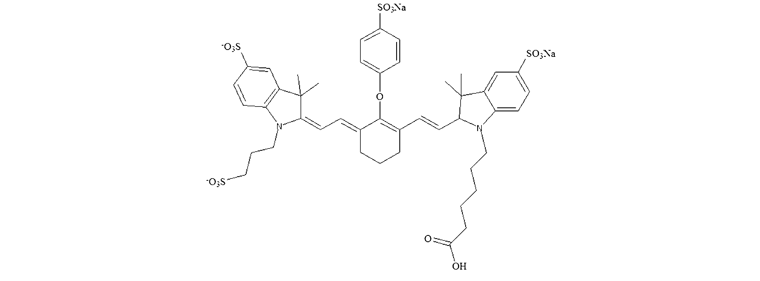
最も好ましくは、蛍光剤は、式Vに示されるような、R1がR18Ph(式中、R18はN-[3-(11,12-ジデヒドロジベンゾ[b,f]アゾシン-5(6H)-イル)-3-オキソプロピル]プロパニル部分である)であり、R16はH、R4及びR11はH、R5及びR12はSO3H、R12は(CH2)3SO3H、R6,R7,R13,R14はHである、薬剤である。
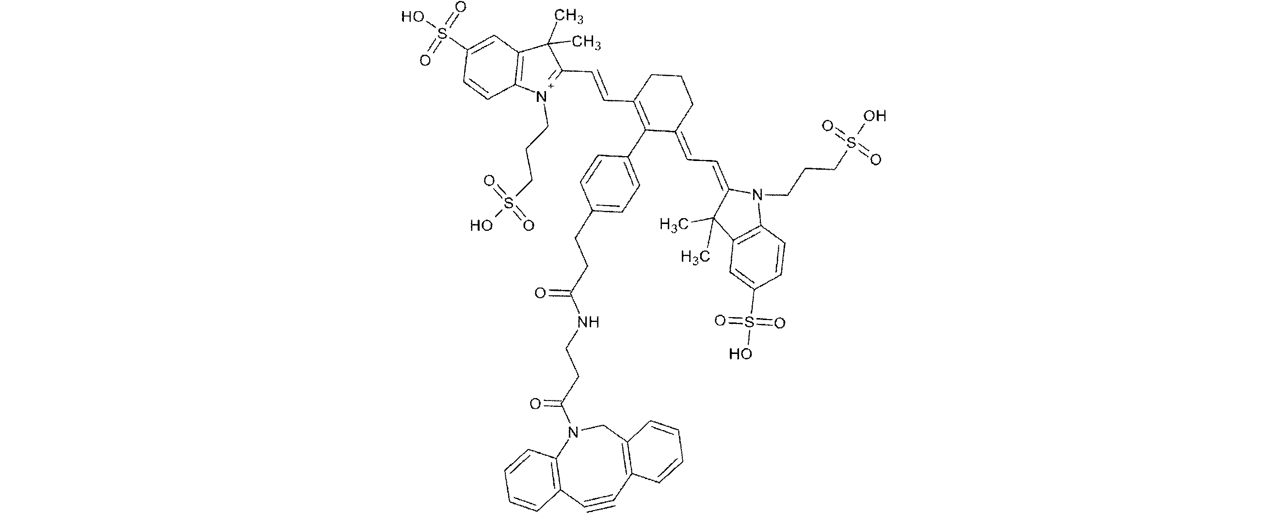
好ましくは、蛍光剤はスルホシアニン部分を含む。 Preferably, the fluorescent agent comprises a sulfocyanine moiety.
650~900nmの吸収波長を有する非蛍光剤は、固有の蛍光をほとんど又は全く有さない化合物であり、バックグラウンドほとんど無しで近接のフルオロフォアからの蛍光を効率的に消光し得る。一実施形態では、非蛍光剤はシアニン分子である。シアニン色素とも呼ばれるシアニン分子は、ポリメチン鎖によって結合された2つの置換又は非置換窒素含有複素環を有する化合物を含む。 Non-fluorescent agents with absorption wavelengths between 650 and 900 nm are compounds that have little or no intrinsic fluorescence and can efficiently quench fluorescence from nearby fluorophores with little background. In one embodiment, the non-fluorescent agent is a cyanine molecule. Cyanine molecules, also called cyanine dyes, include compounds that have two substituted or unsubstituted nitrogen-containing heterocycles linked by a polymethine chain.
好ましくは、非蛍光剤はスルホシアニン部分を含む。 Preferably, the non-fluorescent agent comprises a sulfocyanine moiety.
好ましい実施例では、非蛍光剤は、R1がクロロであり、R2,R3,R9,R10がメチルであり、R4がHであり、R5がN-ヒドロカルビル、好ましくはN[(CH2)3SO3Na]2であり、R11及びR12は、スルファナト基でモノ置換された(又は、一置換された/monosubsituted)芳香環を形成し、R6,R7,R13,R14はHであり、R8は(CH2)qFG(式中、qは3であり、FGはスルファナトである)であり、R15は(CH2)PLN(式中、pは5であり、LNはカルボキシルである)であり、R16はHである、薬剤である。 In a preferred embodiment, the non-fluorescent agent is one in which R1 is chloro, R2 , R3 , R9 , and R10 are methyl, R4 is H, R5 is N- hydrocarbyl, preferably N[(CH2)3SO3Na ] 2 , R11 and R12 form an aromatic ring monosubsituted with a sulfanato group, R6 , R7 , R13 , and R14 are H, R8 is ( CH2 )qFG (where q is 3 and FG is sulfanato), R15 is ( CH2 ) PLN (where p is 5 and LN is carboxyl), and R16 is H.
別の実施形態では、蛍光剤及び非蛍光剤は同じ薬剤であり、好ましくは、R1は

非蛍光剤はまた、消光部分、例えばBHQ3(Biosearch)、QC-1(Li-COR.com)、又はそのような化合物を含む粒子、例えば金ナノ粒子及びフェロナノ粒子であり得る。一実施形態では、試薬は、本明細書に定義されるペプチドを含むナノ粒子である。 The non-fluorescent agent may also be a quenching moiety, e.g., BHQ3 (Biosearch), QC-1 (Li-COR.com), or particles comprising such compounds, e.g., gold nanoparticles and ferronoparticles. In one embodiment, the reagent is a nanoparticle comprising a peptide as defined herein.
本発明で使用できる蛍光剤の例としては、特に、Alexa Fluor(登録商標) 660、Alexa Fluor(登録商標) 700、Alexa 25 Fluor(登録商標) 750 ATTO 680、ATTO 700、DY-647、DY-650、DY-673、DY-675、DY-676、DY-680、DY-681、DY-682、DY-690、DY-700、DY-701、DY-730、DY-731、DY-732、DY-734、DY-750、DY-751、DY-752、DY-776、DY-781、DY-782、DY-831、La Jolla Blue、Cy5、Cy5.5、Cy7、IRDye(登録商標) 800CW、IRDye(登録商標) 38、IRDye(登録商標) 800RS、IRDye(登録商標) 700DX、IRDye(登録商標) 680、TF7WS、TF8WS、TideDye又はTideQuencherが挙げられるが、これらに限定されない。「Alexa Fluor」色素は、Molecular Peptides Inc.,Eugene,OR,U.S.A.(www.peptides.com)から入手できる。ATTO-tec GmbH,Siegen,Germany(www.atto-tec.com)から「ATTO」色素が入手可能である。「DY」色素はDyomics GmbH,Jena,Germany(www.dyomics.com)から入手できる。La Jolla Blueは、Hyperion Inc.から入手可能である。「Cy」色素は、Amersham Biosciences,Piscataway,NJ,U.S.A.(www.amersham.com)から入手可能である。「IRDye(登録商標)赤外線色素」は、LI-COR(登録商標) Bioscience,Inc.Lincoln,N E,U.S.A(www.licor.com)社から入手し得る。 Examples of fluorescent agents that can be used in the present invention include, inter alia, Alexa Fluor® 660, Alexa Fluor® 700, Alexa 25 Fluor® 750, ATTO 680, ATTO 700, DY-647, DY-650, DY-673, DY-675, DY-676, DY-680, DY-681, DY-682, DY-690, DY-700, DY-701, DY-730, DY-731, DY-732, DY-734, DY-750, DY-751, DY-752, DY-776, DY-781, DY-782, DY-831, La Jolla Examples of suitable dyes include, but are not limited to, Blue, Cy5, Cy5.5, Cy7, IRDye® 800CW, IRDye® 38, IRDye® 800RS, IRDye® 700DX, IRDye® 680, TF7WS, TF8WS, TideDye, or TideQuencher. "Alexa Fluor" dyes are available from Molecular Peptides Inc., Eugene, OR, U.S.A. (www.peptides.com). "ATTO" dyes are available from ATTO-tec GmbH, Siegen, Germany (www.atto-tec.com). "DY" dyes are available from Dyomics GmbH, Jena, Germany (www.dyomics.com). La Jolla Blue is available from Hyperion Inc. "Cy" dyes are available from Amersham Biosciences, Piscataway, NJ, U.S.A. (www.amersham.com). "IRDye® Infrared Dyes" are available from LI-COR® Bioscience, Inc. Lincoln, NE, U.S.A. (www.amersham.com). Available from Licor S.A. (www.licor.com).
好ましくは、蛍光剤はCy7、Cy7.5、TF7WS、TF8WS及びIRDye800CWからなる群より選択される。 Preferably, the fluorescent agent is selected from the group consisting of Cy7, Cy7.5, TF7WS, TF8WS and IRDye800CW.
好ましくは、650~900nmの発光波長を有する非蛍光剤は、シアニン部分、好ましくはQC-1である。 Preferably, the non-fluorescent agent having an emission wavelength between 650 and 900 nm is a cyanine moiety, preferably QC-1.
最も好ましくは、試薬は、Cy7、Cy7.5、TQ7WS及びIRDye800CWからなる群より選択される蛍光剤、ならびにQC-1である650~900nmの発光波長を有する非蛍光剤を含む。 Most preferably, the reagent comprises a fluorescent agent selected from the group consisting of Cy7, Cy7.5, TQ7WS and IRDye800CW, and a non-fluorescent agent having an emission wavelength of 650-900 nm that is QC-1.
試薬は、好ましくは、Cy7、Cy7.5、TF7WS及びIRDye800CWからなる群から選択される蛍光剤と、QC-1である650~900nmの発光波長を有する非蛍光剤と、少なくとも3つのアミノ酸を含む切断部位とを含む。 The reagent preferably comprises a fluorescent agent selected from the group consisting of Cy7, Cy7.5, TF7WS and IRDye800CW, a non-fluorescent agent having an emission wavelength of 650-900 nm that is QC-1, and a cleavage site comprising at least three amino acids.
体液試料はヒト又は動物の体液試料であり、より好ましくは体液はヒトの体液である。 The body fluid sample is a human or animal body fluid sample, more preferably the body fluid is a human body fluid.
好ましくは、試料は、10μl~3000μL、より好ましくは50μL~2500μL、さらにより好ましくは100~2000μL、さらにより好ましくは150~1500μL、及び最も好ましくは200~1000μLの範囲の容量を有する。 Preferably, the sample has a volume in the range of 10 μl to 3000 μL, more preferably 50 μL to 2500 μL, even more preferably 100 to 2000 μL, even more preferably 150 to 1500 μL, and most preferably 200 to 1000 μL.
好ましくは、試料、及び任意に希釈剤の添加後の試料容器中の試薬濃度は、0.01~10uM、好ましくは0.02~8uM、より好ましくは0.05~5uM、さらに好ましくは0.1~2.5uMの範囲にある。 Preferably, the concentration of reagent in the sample container after addition of sample and, optionally, diluent, is in the range of 0.01-10 uM, preferably 0.02-8 uM, more preferably 0.05-5 uM, even more preferably 0.1-2.5 uM.
好ましくは、工程i)は、希釈剤を添加する工程を含む。好ましくは、希釈剤は水溶液である。 Preferably, step i) includes adding a diluent. Preferably, the diluent is an aqueous solution.
希釈剤が、MOPS、リン酸塩、クエン酸塩、HEPES TRIS-HCl、リン酸緩衝生理食塩水からなる群から選択される緩衝剤を含むことが好ましい。緩衝剤を含む水性溶液の調製は、当業者に知られている。 The diluent preferably comprises a buffer selected from the group consisting of MOPS, phosphate, citrate, HEPES TRIS-HCl, and phosphate buffered saline. The preparation of aqueous solutions containing buffers is known to those skilled in the art.
好ましくは、希釈剤は5~9の範囲のpHを有し、より好ましくは5.5~8.5の範囲のpHを有し、さらに好ましくは5.75~8.25、さらに好ましくは6~8の範囲のpHを有する。 Preferably, the diluent has a pH in the range of 5 to 9, more preferably in the range of 5.5 to 8.5, even more preferably in the range of 5.75 to 8.25, and even more preferably in the range of 6 to 8.
好ましくは、試料に対する希釈剤の容積比は、0.1~100の範囲、より好ましくは0.5~75の範囲、さらにより好ましくは1~50の範囲である。 Preferably, the volume ratio of diluent to sample is in the range of 0.1 to 100, more preferably in the range of 0.5 to 75, and even more preferably in the range of 1 to 50.
好ましくは、試料をアッセイ緩衝液中で希釈し、好ましくは、試料を1:10~1:10000、より好ましくは1:100~1:1000の範囲のファクタ(又は、因子/factor)で希釈する。 Preferably, the sample is diluted in assay buffer, preferably by a factor ranging from 1:10 to 1:10000, more preferably from 1:100 to 1:1000.
好ましくは、アッセイ緩衝液は、pHを5~9、好ましくは約6~約8、より好ましくは約6.5~約7.5の範囲に維持することができ、好ましくは緩衝液は、HEPES、PIPES、Tris-Hydrochloride(Tris-HCl)、リン酸塩、リン酸塩緩衝生理食塩水、又はMOPSを含む。好ましくは、緩衝液はリン酸緩衝液及びリン酸緩衝生理食塩水の群から選択される。好ましくは、リン酸緩衝液はナトリウム及び/又はカリウムイオンを含む。 Preferably, the assay buffer is capable of maintaining a pH in the range of 5 to 9, preferably about 6 to about 8, more preferably about 6.5 to about 7.5, and preferably the buffer comprises HEPES, PIPES, Tris-Hydrochloride (Tris-HCl), phosphate, phosphate buffered saline, or MOPS. Preferably, the buffer is selected from the group of phosphate buffer and phosphate buffered saline. Preferably, the phosphate buffer comprises sodium and/or potassium ions.
好ましくは、緩衝液は、体液試料中の細胞物質を溶解し得る界面活性剤を含有する。 Preferably, the buffer contains a surfactant capable of dissolving cellular material in the body fluid sample.
好ましくは、緩衝液は、以下からなる群から選択される1つ又は複数の非イオン性界面活性剤を含む:N-オクチル- -D-グルコピラノシド、N-オクチル-D-マルトシド、ZWITTERGENT 3.14、デオキシコール酸塩、n-ドデカノイルスクロース、n-ドデシル- -Dグルコピラノシド、n-ドデシル- -D-マルトシド、n-オクチル- -D-グルコピラノシド、n-オクチル-p-Dマルトピラノシド、n-オクチル-p-D-チオグルコピラノシド、n-デカノイルスクロース、n-デシル-p-D-マルトピラノシド、n-デシル-p-D-チオマルトシド、n-へプチル- -D-グルコピラノシド、n-へプチル- -D-グルコピラノシド、n-へプチル-p-D-チオグルコピラノシド、n-ヘキシル- -D-グルコピラノシド、n-ノニル-p-D-グルコピラノシド、n-オクタノイルスクロース、n-オクチル- -グルコピラノシド、n-ウンデシル- -D-マルトシド、APO-10、APO12、Big CHAP、Big CHAP,Deoxy、BRIJ(登録商標) 35、d2E5、d2E6、Ci2E8、Ci2E9、シクロヘキシル-nエチル-p-D-マルトシド、シクロヘキシル-n-ヘキシル-p-D-マルトシド、シクロヘキシル-n-メチル- -D-マルトシド、ジギトニン、ELUGENT(登録商標)、GENAPOL(登録商標) C-100、GENAPOL(登録商標) X-080、GENAPOL(登録商標) X-100、HECAMEG、MEGA-10、MEGA-8、MEGA-9、NOGA、NP-40、PLURONIC(登録商標) F-127、TRITON(登録商標) X-100、TRITON(登録商標) X-I 14、TWEEN(登録商標) 20、又はTWEEN(登録商標) 80、及びそれらの混合物。 Preferably, the buffer contains one or more non-ionic surfactants selected from the group consisting of N-octyl-D-glucopyranoside, N-octyl-D-maltoside, ZWITTERGENT 3.14, deoxycholate, n-dodecanoyl sucrose, n-dodecyl-D-glucopyranoside, n-dodecyl-D-maltoside, n-octyl-D-glucopyranoside, n-octyl-p-D-maltopyranoside, n-octyl-p-D-thioglucopyranoside, n-decanoyl sucrose, n-decyl-p-D-maltopyranoside, n-decyl-p-D-thiomaltoside, n-heptyl-D-glucopyranoside, n-heptyl- -D-glucopyranoside, n-heptyl-p-D-thioglucopyranoside, n-hexyl- -D-glucopyranoside, n-nonyl-p-D-glucopyranoside, n-octanoylsucrose, n-octyl- -glucopyranoside, n-undecyl- -D-maltoside, APO-10, APO12, Big CHAP, Big CHAP, Deoxy, BRIJ® 35, d2E5, d2E6, Ci2E8, Ci2E9, cyclohexyl-n-ethyl-p-D-maltoside, cyclohexyl-n-hexyl-p-D-maltoside, cyclohexyl-n-methyl- -D-maltoside, digitonin, ELUGENT®, GENAPOL® C-100, GENAPOL® X-080, GENAPOL® X-100, HECAMEG, MEGA-10, MEGA-8, MEGA-9, NOGA, NP-40, PLURONIC® F-127, TRITON® X-100, TRITON® X-I 14, TWEEN® 20, or TWEEN® 80, and mixtures thereof.
緩衝液は、好ましくは、BATC、セチルトリメチルアンモニウムブロミド、ケノデオキシコール酸、コール酸、デオキシコール酸、グリココール酸、グリコデオキシコール酸、グリコリトコール酸、ラウロイルサルコシン、タウロケノデオキシコール酸、タウロコール酸、タウロデヒドロコール酸、タウロリトコール酸、タウロウルソデオキシコール酸、TOPPA及びそれらの混合物からなる群から選択されるイオン性界面活性剤を含む。 The buffer preferably comprises an ionic surfactant selected from the group consisting of BATC, cetyltrimethylammonium bromide, chenodeoxycholic acid, cholic acid, deoxycholic acid, glycocholic acid, glycodeoxycholic acid, glycolithocholic acid, lauroyl sarcosine, taurochenodeoxycholic acid, taurocholic acid, taurodehydrocholic acid, taurolithocholic acid, tauroursodeoxycholic acid, TOPPA, and mixtures thereof.
好ましくは、緩衝液は、アミドスルホベタイン、CHAPS、CHAPSO、カルボキシベタイン、及びメチルベタインからなる群より選択される双性イオン性界面活性剤を含む。 Preferably, the buffer contains a zwitterionic detergent selected from the group consisting of amidosulfobetaine, CHAPS, CHAPSO, carboxybetaine, and methylbetaine.
好ましくは、緩衝液は、SDS、N-ラウリルサルコシン、デオキシコール酸ナトリウム、アルキル-アリールスルホン酸塩、長鎖(脂肪)アルコール硫酸塩、オレフィン硫酸塩及びスルホン酸塩、αオレフィン硫酸塩及びスルホン酸塩、硫酸化モノグリセリド、硫酸化エーテル、スルホコハク酸塩、アルカンスルホン酸塩、リン酸エステル、アルキルイセチオン酸塩、スクロースエステル及びそれらの混合物からなる群から選択されるアニオン性界面活性剤を含む。 Preferably, the buffer contains an anionic surfactant selected from the group consisting of SDS, N-lauryl sarcosine, sodium deoxycholate, alkyl-aryl sulfonates, long chain (fatty) alcohol sulfates, olefin sulfates and sulfonates, alpha-olefin sulfates and sulfonates, sulfated monoglycerides, sulfated ethers, sulfosuccinates, alkanesulfonates, phosphate esters, alkyl isethionates, sucrose esters, and mixtures thereof.
本明細書において、緩衝剤は、緩衝液及び/又は試料の組合せのpHを調節し得る化学種である。 As used herein, a buffering agent is a chemical species that can adjust the pH of a buffer solution and/or sample combination.
好ましくは、体液の試料を、直接試薬と接触させる。本明細書中で使用される用語「直接」とは、試料が試薬と接触する前に遠心分離工程又は他の試料クリーンアップ工程を施さないことを意味する。 Preferably, the sample of bodily fluid is contacted directly with the reagent. As used herein, the term "directly" means that the sample is not subjected to a centrifugation step or other sample cleanup step before contacting the reagent.
好ましくは、体液の試料を、直接試薬と接触させる。 Preferably, the body fluid sample is directly contacted with the reagent.
任意的に、本方法は、試料を遠心分離して、凝集した細菌物質及び/又は沈殿した生体物質を上清から分離し得る遠心分離工程を含む。好ましくは、遠心分離は1,000g~25,000gの範囲、好ましくは3,000g~20,000gの範囲、より好ましくは3,700g~18,000gの範囲の力で行うことができる。遠心分離時間は、好ましくは、1分~30分の範囲、2~20分の範囲、より好ましくは3~10分の範囲である。遠心分離から得られる上清又は凝集した細菌細胞を試薬と接触させるのが好ましい。好ましい実施形態では、遠心分離からの上清は、試薬と接触させることが好ましい。別の好ましい実施形態において、遠心分離から得られる凝集した細菌物質細胞は、好ましくは試薬と接触される。 Optionally, the method includes a centrifugation step in which the sample may be centrifuged to separate the aggregated bacterial material and/or precipitated biological material from the supernatant. Preferably, the centrifugation may be performed at a force in the range of 1,000 g to 25,000 g, preferably in the range of 3,000 g to 20,000 g, more preferably in the range of 3,700 g to 18,000 g. The centrifugation time is preferably in the range of 1 min to 30 min, in the range of 2 to 20 min, more preferably in the range of 3 to 10 min. The supernatant or aggregated bacterial cells obtained from the centrifugation are preferably contacted with a reagent. In a preferred embodiment, the supernatant from the centrifugation is preferably contacted with a reagent. In another preferred embodiment, the aggregated bacterial material cells obtained from the centrifugation are preferably contacted with a reagent.
好ましくは、本方法は、試料を試薬と接触させる前に試料を遠心分離する工程を含む。例えば、血液によるサンプルの多少の汚染がある場合には、アッセイで処理する前に、サンプルを遠心分離することが望ましい。 Preferably, the method includes centrifuging the sample before contacting it with the reagent. For example, if there is some contamination of the sample with blood, it may be desirable to centrifuge the sample before processing it in the assay.
任意的に、試料に試薬を加えた後、混合物を攪拌する。混合物は、ピペットで吸引、渦流又は振盪することにより攪拌することが望ましい。 Optionally, after adding the reagent to the sample, the mixture is agitated. The mixture is preferably agitated by pipetting, vortexing, or shaking.
好ましくは、試薬はペプチド、タンパク質又はナノ粒子である。好ましくは、試薬はペプチドである。ペプチドは、リンカーを介して、タンパク質、ナノ粒子又は磁気ビーズに連結され得る。ペプチドは、好ましくは3~10アミノ酸、より好ましくは4~9アミノ酸、さらにより好ましくは5~8アミノ酸を含む。 Preferably, the reagent is a peptide, a protein or a nanoparticle. Preferably, the reagent is a peptide. The peptide may be linked to the protein, nanoparticle or magnetic bead via a linker. The peptide preferably comprises 3-10 amino acids, more preferably 4-9 amino acids, even more preferably 5-8 amino acids.
好ましくは、[b]は、3~8アミノ酸、より好ましくは3~7アミノ酸、さらにより好ましくは3~6アミノ酸を含む。 Preferably, [b] contains 3 to 8 amino acids, more preferably 3 to 7 amino acids, and even more preferably 3 to 6 amino acids.
切断部位は、好ましくは限られた数のアミノ酸、例えば少なくとも2個、好ましくは少なくとも3個のアミノ酸、より好ましくは少なくとも4個のアミノ酸、さらにより好ましくは少なくとも5個のアミノ酸を含む。 The cleavage site preferably contains a limited number of amino acids, for example at least 2, preferably at least 3 amino acids, more preferably at least 4 amino acids, even more preferably at least 5 amino acids.
好ましくは、切断部位は、2~5個のアミノ酸、より好ましくは3~5個のアミノ酸からなる。 Preferably, the cleavage site consists of 2 to 5 amino acids, more preferably 3 to 5 amino acids.
[b]は一般式Xaa1,Xaa2,Xaa3を有することが好ましい。換言すれば、切断部位は、好ましくは、3個のアミノ酸Xaa1,Xaa2,Xaa3を含み、
-Xaa1は、好ましくは、小さな疎水性アミノ酸であり、より好ましくは、Xaa1は、アラニン、グリシン、ロイシン、バリン、ノルロイシン、ノルバリン、イソロイシン、イソバリン、アロイソロイシンからなる群より選択され、
-Xaa2は、好ましくは、小さな又は芳香族の疎水性アミノ酸であり、好ましくは、Xaa2は、アラニン、グリシン、ロイシン、バリン、ノルロイシン、ノルバリン、イソロイシン、イソバリン、アロイソロイシン及びフェニルアラニンからなる群より選択され、
-Xaa3は、好ましくは、アラニン、グリシン、ロイシン、バリン、ノルロイシン、ノルバリン、イソロイシン、イソバリン、アロイソロイシンからなる群より選択され、より好ましくは、Xaa3は、アラニン、グリシン、ロイシン、バリンからなる群より選択される。
[b] preferably has the general formula Xaa1 , Xaa2 , Xaa3 . In other words, the cleavage site preferably comprises the three amino acids Xaa1 , Xaa2 , Xaa3 ,
-X aa1 is preferably a small hydrophobic amino acid, more preferably X aa1 is selected from the group consisting of alanine, glycine, leucine, valine, norleucine, norvaline, isoleucine, isovaline, alloisoleucine;
- X aa2 is preferably a small or aromatic hydrophobic amino acid, preferably X aa2 is selected from the group consisting of alanine, glycine, leucine, valine, norleucine, norvaline, isoleucine, isovaline, alloisoleucine and phenylalanine;
-X aa3 is preferably selected from the group consisting of alanine, glycine, leucine, valine, norleucine, norvaline, isoleucine, isovaline, alloisoleucine, more preferably X aa3 is selected from the group consisting of alanine, glycine, leucine, valine.
特定の好ましい態様において、Xaa1は、アラニン、バリン、ノルロイシン、ノルバリン、イソロイシン、イソバリン、アロイソロイシンからなる群から選択され、Xaa2は、アラニン、ロイシン、バリン、ノルロイシン、ノルバリン、イソロイシン、イソバリン、アロイソロイシン及びフェニルアラニンからなる群から選択され、Xaa3は、アラニン、ロイシン、バリンからなる群から選択される。 In certain preferred embodiments, Xaa1 is selected from the group consisting of alanine, valine, norleucine, norvaline, isoleucine, isovaline, alloisoleucine; Xaa2 is selected from the group consisting of alanine, leucine, valine, norleucine, norvaline, isoleucine, isovaline, alloisoleucine, and phenylalanine; and Xaa3 is selected from the group consisting of alanine, leucine, and valine.
好ましくは、切断部位は、3つのアミノ酸Xaa1,Xaa2,Xaa3を含み、
-Xaa1は、好ましくは、小さな疎水性アミノ酸であり、より好ましくは、Xaa1は、アラニン、グリシン、ロイシン、バリン、ノルロイシン、ノルバリン、イソロイシン、イソバリン、アロイソロイシンからなる群から選択され、より好ましくは、アラニン、ロイシン、バリン、ノルロイシン、ノルバリン、イソロイシン、イソバリン、アロイソロイシンからなる群より選択され、
-Xaa2は、好ましくは、小さな又は芳香族の疎水性アミノ酸であり、好ましくは、Xaa2は、アラニン、グリシン、ロイシン、バリン、ノルロイシン、ノルバリン、イソロイシン、イソバリン、アロイソロイシン及びフェニルアラニンからなる群より選択され、より好ましくは、Xaa2は、アラニン、ロイシン、バリン、ノルロイシン、イソロイシン、イソバリン、アロイソロイシン及びフェニルアラニンからなる群より選択され、
-Xaa3は、好ましくは、アラニン、ロイシン、バリン、ノルロイシン、ノルバリン、イソロイシン、イソバリン、アロイソロイシンからなる群より選択され、より好ましくは、Xaa3は、アラニン、グリシン、ロイシン、バリンからなる群より選択され、
切断部位は、細菌バイオマーカーによって切断され、細菌バイオマーカーは、エンテロコッカス・フェカーリス(Enterococcus faecalis)、エンテロコッカス・フェシウム(Enterococcus faecium)、緑膿菌(Pseudomonas aeruginosa)、黄色ブドウ球菌(Staphylococcus aureus)、表皮ブドウ球菌(Staphylococcus epidermidis)、スタフィロコッカス・ルグドゥネンシス(Staphylococcus lugdunensis)、スタフィロコッカス・ワーネリ(Staphylococcus warneri)、スタフィロコッカス・カピティス(Staphylococcus capitis)、スタフィロコッカス・カプラエ(Staphylococcus caprae)、ストレプトコッカス・ミティ(Streptococcus mitis)、ストレプトコッカス・オラリス(Streptococcus oralis)、ストレプトコッカス・アガラクティエ(Streptococcus agalactiae)、ストレプトコッカス・アンギノサス(Streptococcus anginosus)、ストレプトコッカス・ゴルドニ(Streptococcus gordonii)、大腸菌(Escherichia coli)、プロピオニバクテリウム・アクネス(Propionibacterium acnes)、プロテウス・ミラビリス(Proteus mirabilis)、グラヌリカテラ・アディアセンス(Granulicatella adjacens)、アシネトバクター・バウマニ(Acinetobacter baumannii)、アビオトロフィア・ディフェクティヴァ(Abiotrophia defective)、コリネバクテリウム・ストリアトゥム(Corynebacterium striatum)、コリネバクテリウム・ミヌティシマム(Corynebacterium minutissimum)、パルビモナス マイクラ(Parvimonas micra)、カンジダ・パラプシローシス(Candida parapsilosis)、カンジダ・グラブラタ(Candida glabrata)、カンジダ・トロピカリス(Candida tropicalis)、及びカンジダ・アルビカンス(Candida albicans)からなる群より選択される細菌によって製造され、より好ましくは、原因物質(細菌)は、バチルス属、スタフィロコッカス属、ストレプトコッカス属、シュードモナス属、大腸菌及びそれらの組み合わせからなる群から選択され、最も好ましくはバチルス属及びシュードモナス属の群から選択される。
Preferably, the cleavage site comprises the three amino acids Xaa1 , Xaa2 , Xaa3 ,
-X aa1 is preferably a small hydrophobic amino acid, more preferably X aa1 is selected from the group consisting of alanine, glycine, leucine, valine, norleucine, norvaline, isoleucine, isovaline, alloisoleucine, more preferably alanine, leucine, valine, norleucine, norvaline, isoleucine, isovaline, alloisoleucine;
-X aa2 is preferably a small or aromatic hydrophobic amino acid, preferably X aa2 is selected from the group consisting of alanine, glycine, leucine, valine, norleucine, norvaline, isoleucine, isovaline, alloisoleucine and phenylalanine, more preferably X aa2 is selected from the group consisting of alanine, leucine, valine, norleucine, isoleucine, isovaline, alloisoleucine and phenylalanine;
-X aa3 is preferably selected from the group consisting of alanine, leucine, valine, norleucine, norvaline, isoleucine, isovaline, alloisoleucine, more preferably X aa3 is selected from the group consisting of alanine, glycine, leucine, valine;
The cleavage site is cleaved by a bacterial biomarker, the bacterial biomarker being Enterococcus faecalis, Enterococcus faecium, Pseudomonas aeruginosa, Staphylococcus aureus, Staphylococcus epidermidis, Staphylococcus lugdunensis, Staphylococcus warneri, warneri, Staphylococcus capitis, Staphylococcus caprae, Streptococcus mitis, Streptococcus oralis, Streptococcus agalactiae, Streptococcus anginosus, Streptococcus gordonii, Escherichia coli coli), Propionibacterium acnes, Proteus mirabilis, Granulicatella adjacens, Acinetobacter baumannii, Abiotrophia defective, Corynebacterium striatum, Corynebacterium minutissimum, Parvimonas micra micra), Candida parapsilosis, Candida glabrata, Candida tropicalis, and Candida albicans, more preferably the causative agent (bacteria) is selected from the group consisting of Bacillus, Staphylococcus, Streptococcus, Pseudomonas, Escherichia coli, and combinations thereof, and most preferably selected from the group of Bacillus and Pseudomonas.
好ましくは、切断部位は、3つのアミノ酸Xaa1,Xaa2,Xaa3を含み、
-Xaa1は、好ましくは、小さな疎水性アミノ酸であり、より好ましくは、Xaa1は、アラニン、グリシン、ロイシン、バリン、ノルロイシン、ノルバリン、イソロイシン、イソバリン、アロイソロイシンからなる群から選択され、より好ましくは、アラニン、ロイシン、バリン、ノルロイシン、ノルバリン、イソロイシン、イソバリン、アロイソロイシンからなる群より選択され、
-Xaa2は、好ましくは荷電アミノ酸であり、好ましくは、Xaa2はアスパラギン酸、グルタミン酸、リシン、アルギニンからなる群から選択され、
-Xaa3は、好ましくは、アラニン、ロイシン、バリン、ノルロイシン、ノルバリン、イソロイシン、イソバリン、アロイソロイシンからなる群から選択され、より好ましくは、Xaa3は、アラニン、グリシン、ロイシン、バリンからなる群から選択され、
切断部位は、細菌バイオマーカーによって切断され、細菌バイオマーカーは、エンテロコッカス・フェカーリス(Enterococcus faecalis)、エンテロコッカス・フェシウム(Enterococcus faecium)、黄色ブドウ球菌(Staphylococcus aureus)、表皮ブドウ球菌(Staphylococcus epidermidis)、スタフィロコッカス・ルグドゥネンシス(Staphylococcus lugdunensis)、スタフィロコッカス・ワーネリ(Staphylococcus warneri)、スタフィロコッカス・カピティス(Staphylococcus capitis)、スタフィロコッカス・カプラエ(Staphylococcus caprae)、ストレプトコッカス・ミティ(Streptococcus mitis)、ストレプトコッカス・オラリス(Streptococcus oralis)、ストレプトコッカス・アガラクティエ(Streptococcus agalactiae)、ストレプトコッカス・アンギノサス(Streptococcus anginosus)、ストレプトコッカス・ゴルドニ(Streptococcus gordonii)、大腸菌(Escherichia coli)、プロピオニバクテリウム・アクネス(Propionibacterium acnes)、プロテウス・ミラビリス(Proteus mirabilis)、グラヌリカテラ・アディアセンス(Granulicatella adjacens)、アシネトバクター・バウマニ(Acinetobacter baumannii)、アビオトロフィア・ディフェクティヴァ(Abiotrophia defective)、コリネバクテリウム・ストリアトゥム(Corynebacterium striatum)、コリネバクテリウム・ミヌティシマム(Corynebacterium minutissimum)、パルビモナス・マイクラ(Parvimonas micra)、カンジダ・パラプシローシス(Candida parapsilosis)、カンジダ・グラブラタ(Candida glabrata)、カンジダ・トロピカリス(Candida tropicalis)、及びカンジダ・アルビカンス(Candida albicans)からなる群より選択される細菌によって製造され、好ましくは、原因物質(細菌)は、スタフィロコッカス属、ストレプトコッカス属(Streptococcus)、大腸菌(Escherichia coli)及びそれらの組み合わせからなる群から選択される。
Preferably, the cleavage site comprises the three amino acids Xaa1 , Xaa2 , Xaa3 ,
-X aa1 is preferably a small hydrophobic amino acid, more preferably X aa1 is selected from the group consisting of alanine, glycine, leucine, valine, norleucine, norvaline, isoleucine, isovaline, alloisoleucine, more preferably alanine, leucine, valine, norleucine, norvaline, isoleucine, isovaline, alloisoleucine;
-X aa2 is preferably a charged amino acid, preferably X aa2 is selected from the group consisting of aspartic acid, glutamic acid, lysine, arginine;
-X aa3 is preferably selected from the group consisting of alanine, leucine, valine, norleucine, norvaline, isoleucine, isovaline, alloisoleucine, more preferably X aa3 is selected from the group consisting of alanine, glycine, leucine, valine;
The cleavage site is cleaved by a bacterial biomarker, the bacterial biomarker being Enterococcus faecalis, Enterococcus faecium, Staphylococcus aureus, Staphylococcus epidermidis, Staphylococcus lugdunensis, Staphylococcus warneri, Staphylococcus capitis, capitis, Staphylococcus caprae, Streptococcus mitis, Streptococcus oralis, Streptococcus agalactiae, Streptococcus anginosus, Streptococcus gordonii, Escherichia coli, Propionibacterium acnes, acnes), Proteus mirabilis, Granulicatella adjacens, Acinetobacter baumannii, Abiotrophia defective, Corynebacterium striatum, Corynebacterium minutissimum, Parvimonas micra, Candida parapsilosis, parapsilosis, Candida glabrata, Candida tropicalis, and Candida albicans, and preferably the causative agent (bacteria) is selected from the group consisting of Staphylococcus, Streptococcus, Escherichia coli, and combinations thereof.
好ましくは、[b]は、AAA、ALA、AAL、LAA、FAA、AFA、AAF、FGG、GFG、GGF、GGG、LGG、GLG、GGL及びGGAからなる群より選択されるアミノ酸配列を含む。より好ましくは、[b]はFGG、GFG、GGF、GGG、LGG、GLG、GGL及びGGAからなる群より選択されるアミノ酸配列を含む。さらに好ましくは、[b]は、AAA、ALA、AAL、LAA、FAA、AFA、AAFからなる群より選択されるアミノ酸配列を含む。さらに好ましくは、[b]は、FAA、AFA及びAAFからなる群から選択されるアミノ酸配列を含む。 Preferably, [b] comprises an amino acid sequence selected from the group consisting of AAA, ALA, AAL, LAA, FAA, AFA, AAF, FGG, GFG, GGF, GGG, LGG, GLG, GGL, and GGA. More preferably, [b] comprises an amino acid sequence selected from the group consisting of FGG, GFG, GGF, GGG, LGG, GLG, GGL, and GGA. Even more preferably, [b] comprises an amino acid sequence selected from the group consisting of AAA, ALA, AAL, LAA, FAA, AFA, and AAF. Even more preferably, [b] comprises an amino acid sequence selected from the group consisting of FAA, AFA, and AAF.
好ましくは、[b]は、AVA、AAV、VAA、YAA、AYA、AAY、YGG、GFG、GGY、GGV、VGG、GVG、GAG及びAGGからなる群より選択されるアミノ酸配列を含む。より好ましくは、[b]は、YGG、GFG、GGY、GGV、VGG、GVG、GAG及びAGGからなる群より選択されるアミノ酸配列を含む。さらにより好ましくは、[b]は、AVA、AAV、VAA、YAA、AYA及びAAYからなる群より選択されるアミノ酸配列を含む。 Preferably, [b] comprises an amino acid sequence selected from the group consisting of AVA, AAV, VAA, YAA, AYA, AAY, YGG, GFG, GGY, GGV, VGG, GVG, GAG, and AGG. More preferably, [b] comprises an amino acid sequence selected from the group consisting of YGG, GFG, GGY, GGV, VGG, GVG, GAG, and AGG. Even more preferably, [b] comprises an amino acid sequence selected from the group consisting of AVA, AAV, VAA, YAA, AYA, and AAY.
好ましくは、[b]は、AEA、AAE、EAA、DAA、ADA、AAD、EGG、GEG、GGE、GGD、DGG、GDG、GRG及びRGGからなる群より選択されるアミノ酸配列を含む。より好ましくは、[b]は、KGG、GKG、GGK、AKA、AAK、KAA、KAA、AKA及びAAKからなる群より選択されるアミノ酸配列を含む。 Preferably, [b] comprises an amino acid sequence selected from the group consisting of AEA, AAE, EAA, DAA, ADA, AAD, EGG, GEG, GGE, GGD, DGG, GDG, GRG, and RGG. More preferably, [b] comprises an amino acid sequence selected from the group consisting of KGG, GKG, GGK, AKA, AAK, KAA, KAA, AKA, and AAK.
好ましくは、[b]:([a]+[c])の質量比は少なくとも1:2であり、好ましくは[b]:([a]+[c])の質量比は少なくとも1:3であり、より好ましくは[b]:([a]+[c])の質量比は少なくとも1:4であり、[b]:([a]+[c])の質量比は少なくとも1:5であることがさらに好ましい。 Preferably, the mass ratio of [b]:([a]+[c]) is at least 1:2, preferably the mass ratio of [b]:([a]+[c]) is at least 1:3, more preferably the mass ratio of [b]:([a]+[c]) is at least 1:4, and even more preferably the mass ratio of [b]:([a]+[c]) is at least 1:5.
好ましくは、試薬は一般式[a]-[リンカー1]n-[b]-[b]-[-リンカー2]m-[c]を有し、ここで、nは0、1又は2であり、mは0、1又は2であり、[リンカー1]及び[リンカー2]は、任意に置換されたヒドロカルビル基及び非タンパク質分解性ヒドロカルビル基の群から独立して選択され、好ましくは、nは0又は1であり、mは1であり、リンカー2は非タンパク質分解性ヒドロカルビル基であり、より好ましくは、nは0であり、mは1であり、リンカー2は非タンパク質分解性ヒドロカルビル基である。 Preferably, the reagent has the general formula [a]-[linker1]n-[b]-[b]-[-linker2]m-[c], where n is 0, 1 or 2, m is 0, 1 or 2, and [linker1] and [linker2] are independently selected from the group of optionally substituted hydrocarbyl groups and non-proteolytic hydrocarbyl groups, preferably n is 0 or 1, m is 1, and linker2 is a non-proteolytic hydrocarbyl group, more preferably n is 0, m is 1, and linker2 is a non-proteolytic hydrocarbyl group.
好ましくは、前記リンカーは、非タンパク質分解性ヒドロカルビル基である。 Preferably, the linker is a non-proteolytic hydrocarbyl group.
好ましくは、リンカーは、β-アラニニル、4-アミノブチロール、2-(アミノエトキシ)アセチル、3-(2-アミノエトキシ)プロパニル、5-アミノバレリル、6-アミノヘキシル、8-アミノ-3,6-ジオキサオクタニル及び12-アミノ-4,7,10-トリオキサドデカニルからなる群から選択され、好ましくは6-アミノヘキシルである。 Preferably, the linker is selected from the group consisting of β-alaninyl, 4-aminobutyrol, 2-(aminoethoxy)acetyl, 3-(2-aminoethoxy)propanyl, 5-aminovaleryl, 6-aminohexyl, 8-amino-3,6-dioxaoctanyl and 12-amino-4,7,10-trioxadodecanyl, preferably 6-aminohexyl.
好ましくは、試薬は、一般公式[a]-[リンカー1]-[b]-[-リンカー2]-[c](II)を有し、[リンカー1]及び[リンカー2]は、任意に置換されたハイドカルバイル基及び非タンパク質分解性ヒドロカルビル基の群から独立に選択される。本明細書で使用されるヒドロカルビルは、任意に置換されたC1-C6アルキルを意味する。好ましくは、任意に置換されたC1-C6アルキルは、O、N及びSで置換される。 Preferably, the reagent has the general formula [a]-[ Linker1 ]-[b]-[- Linker2 ]-[c] (II), where [ Linker1 ] and [ Linker2 ] are independently selected from the group of optionally substituted hydrocarbyl groups and non-proteolytic hydrocarbyl groups. Hydrocarbyl, as used herein, means optionally substituted C1-C6 alkyl. Preferably, the optionally substituted C1-C6 alkyl is substituted with O, N and S.
より好ましくは、試薬は、一般式[a]-[リンカー1]n-[b]-[リンカー2]m-[c]であり、式中、[b]は、AAA、ALA、AAL、LAA、FAA、AFA、AAF、FGG、GFG、GGF、GGG、LGG、GLG、GGL及びGGAからなる群から選択されるアミノ酸配列であり、nは1であり、mは1であり、[リンカー1]及び[リンカー2]は、β-アラニニル、4-アミノブチル、2-(アミノエトキシ)アセチル、3-(2-アミノエトキシ)プロパニル、5-アミノバレリル、6-アミノヘキシルからなる群から独立して選択される。 More preferably, the reagent has the general formula [a]-[Linker1] n- [b]-[Linker2] m- [c], where [b] is an amino acid sequence selected from the group consisting of AAA, ALA, AAL, LAA, FAA, AFA, AAF, FGG, GFG, GGF, GGG, LGG, GLG, GGL and GGA, n is 1, m is 1 and [Linker1] and [Linker2] are independently selected from the group consisting of β-alaninyl, 4-aminobutyl, 2-(aminoethoxy)acetyl, 3-(2-aminoethoxy)propanyl, 5-aminovaleryl, 6-aminohexyl.
より好ましくは、試薬は一般式[a]-[b]-[リンカー]-[c]であり、式中、bはAAA、ALA、AAL、LAA、FAA、AFA、AAF、FGG、GFG、GGF、GGG、LGG、GLG、GGL及びGGAからなる群から選択されるアミノ酸配列であり、nは0であり、mは1であり、[リンカー]はβ-アラニニル(beta-alaninyl)、4-アミノブチリル(aminobutyrl)、2-(アミノエトキシ)アセチル、3-(2-アミノエトキシ)プロパニル、5-アミノバレリル、6-アミノヘキシルからなる群から選択される。 More preferably, the reagent has the general formula [a]-[b]-[linker]-[c], where b is an amino acid sequence selected from the group consisting of AAA, ALA, AAL, LAA, FAA, AFA, AAF, FGG, GFG, GGF, GGG, LGG, GLG, GGL and GGA, n is 0, m is 1 and [linker] is selected from the group consisting of beta-alaninyl, 4-aminobutyrl, 2-(aminoethoxy)acetyl, 3-(2-aminoethoxy)propanyl, 5-aminovaleryl and 6-aminohexyl.
より好ましくは、試薬は、一般式[a]-[リンカー]-[b]-[c]であり、式中、bは、AAA、ALA、AAL,LAA、FAA、AFA、AAF、FGG、GFG、GGF、GGG、LGG、GLG、GGL及びGGAからなる群より選択されるアミノ酸配列であり、nは1であり、mは0であり、[リンカー]は、β-アラニニル、4-アミノブチリル、2-(アミノエトキシ)アセチル、3-(2-アミノエトキシ)プロパニル、5-アミノバレリル、6-アミノヘキシルからなる群より選択される。 More preferably, the reagent has the general formula [a]-[linker]-[b]-[c], where b is an amino acid sequence selected from the group consisting of AAA, ALA, AAL, LAA, FAA, AFA, AAF, FGG, GFG, GGF, GGG, LGG, GLG, GGL and GGA, n is 1, m is 0, and [linker] is selected from the group consisting of β-alaninyl, 4-aminobutyryl, 2-(aminoethoxy)acetyl, 3-(2-aminoethoxy)propanyl, 5-aminovaleryl, and 6-aminohexyl.
好ましくは、本方法は、試料を第一試薬及び第二試薬と接触させる工程を含み、第一試薬が一般式[a]-[b]-[c](I)を有し、[b]が切断部位(b’)を含むペプチドであり、第二試薬が式[a]-[d]-[c](III)を有し、[d]が切断部位(d’)を含むペプチドであり、切断部位d’は切断部位b’とは異なる。 Preferably, the method includes contacting the sample with a first reagent and a second reagent, the first reagent having the general formula [a]-[b]-[c] (I), where [b] is a peptide containing a cleavage site (b'), and the second reagent having the formula [a]-[d]-[c] (III), where [d] is a peptide containing a cleavage site (d'), where cleavage site d' is different from cleavage site b'.
本明細書中に開示される方法の利点は、それが多重化アプローチを可能にし、それによって、異なる切断部位を含む異なる試薬が、単一の試料容器内で組み合わされて、異なる細菌バイオマーカーの検出を可能にし得ることである。 An advantage of the method disclosed herein is that it allows for a multiplexed approach, whereby different reagents containing different cleavage sites can be combined in a single sample vessel to allow for the detection of different bacterial biomarkers.
好ましくは、細菌バイオマーカーが細菌膜結合プロテアーゼ、細菌膜結合トランスペプチダーゼ、細胞内細菌プロテアーゼ又は細胞外細菌プロテアーゼであり、より好ましくは細菌バイオマーカーが細菌膜結合トランスペプチダーゼ又は細胞外細菌プロテアーゼである。 Preferably, the bacterial biomarker is a bacterial membrane-bound protease, a bacterial membrane-bound transpeptidase, an intracellular bacterial protease or an extracellular bacterial protease, more preferably, the bacterial biomarker is a bacterial membrane-bound transpeptidase or an extracellular bacterial protease.
好ましくは、本方法は、宿主の炎症反応よりも細菌感染に対する特異性を有する。 Preferably, the method has specificity for bacterial infection rather than for the host inflammatory response.
本出願の文脈において、「宿主炎症反応」とは、宿主炎症バイオマーカー、例えば宿主炎症プロテアーゼを意味する。本出願の文脈において、宿主の炎症反応にまさる感染に対する「特異性」とは、宿主の炎症反応の存在下での650~900nmの範囲の蛍光発光に対する細菌バイオマーカーの存在下での650~900nmの範囲の蛍光発光の比が、少なくとも1.05:1、好ましくは少なくとも1.5~1、より好ましくは少なくとも2:1、さらに好ましくは少なくとも5:1、さらに好ましくは少なくとも10:1、さらにより好ましくは少なくとも25:1、最も好ましくは少なくとも50:1であることを意味する。 In the context of this application, "host inflammatory response" refers to host inflammatory biomarkers, such as host inflammatory proteases. In the context of this application, "specificity" for infection over host inflammatory response means that the ratio of fluorescence emission in the range of 650-900 nm in the presence of bacterial biomarker to fluorescence emission in the range of 650-900 nm in the presence of host inflammatory response is at least 1.05:1, preferably at least 1.5-1, more preferably at least 2:1, even more preferably at least 5:1, even more preferably at least 10:1, even more preferably at least 25:1, and most preferably at least 50:1.
好ましくは、宿主炎症反応の存在下での650~900nmの範囲における蛍光発光に対する細菌バイオマーカーの存在下での650~900nmの範囲における蛍光発光の比は、1.05:1~500:1の範囲、好ましくは2:1~400:1の範囲、より好ましくは5:1~300:1の範囲である。 Preferably, the ratio of the fluorescence emission in the range of 650-900 nm in the presence of the bacterial biomarker to the fluorescence emission in the range of 650-900 nm in the presence of a host inflammatory response is in the range of 1.05:1 to 500:1, preferably in the range of 2:1 to 400:1, more preferably in the range of 5:1 to 300:1.
好ましくは、細菌バイオマーカーを含まない体液の試料における650~900nmの範囲の蛍光発光に対する細菌バイオマーカーを含む体液の試料における650~900nmの範囲の蛍光発光の比は、1.05:1~500:1の範囲、好ましくは2:1~400:1の範囲、より好ましくは5:1~300:1の範囲である。 Preferably, the ratio of the fluorescence emission in the range of 650-900 nm in a sample of body fluid containing a bacterial biomarker to the fluorescence emission in the range of 650-900 nm in a sample of body fluid not containing a bacterial biomarker is in the range of 1.05:1 to 500:1, preferably in the range of 2:1 to 400:1, more preferably in the range of 5:1 to 300:1.
好ましくは、細菌バイオマーカーを含まない体液の試料中の650~900nmの範囲における蛍光発光に対する細菌バイオマーカーを含む体液の試料中の650~900nmの範囲における蛍光発光の比率は、1.05:1~500:1の範囲、好ましくは2:1~400:1の範囲、より好ましくは5:1~300:1の範囲であり、体液試料は、歯肉液、羊水、尿、漿液、滑液、腹膜透析液、精液、皮脂及び脳脊髄液からなる群から選択され、好ましくは、体液は、滑液、腹膜透析液及び脳脊髄液からなる群から選択され、より好ましくは、体液は、滑液及び腹膜透析液からなる群から選択される。 Preferably, the ratio of the fluorescence emission in the range of 650-900 nm in a sample of body fluid containing a bacterial biomarker to the fluorescence emission in the range of 650-900 nm in a sample of body fluid not containing a bacterial biomarker is in the range of 1.05:1 to 500:1, preferably in the range of 2:1 to 400:1, more preferably in the range of 5:1 to 300:1, and the body fluid sample is selected from the group consisting of gingival fluid, amniotic fluid, urine, serous fluid, synovial fluid, peritoneal dialysis fluid, semen, sebum and cerebrospinal fluid, preferably the body fluid is selected from the group consisting of synovial fluid, peritoneal dialysis fluid and cerebrospinal fluid, more preferably the body fluid is selected from the group consisting of synovial fluid and peritoneal dialysis fluid.
好ましくは、本方法は、少なくとも70%、好ましくは少なくとも75%、より好ましくは少なくとも80%、さらにより好ましくは少なくとも85%、さらにより好ましくは少なくとも90%、最も好ましくは少なくとも95%の細菌感染に対する特異性を有する。 Preferably, the method has a specificity for bacterial infection of at least 70%, preferably at least 75%, more preferably at least 80%, even more preferably at least 85%, even more preferably at least 90%, and most preferably at least 95%.
本出願の文脈において、用語「特異性パーセンテージ」は、以下の式に従って決定される:
特異性%=a/b x 100
式中、aは、650~900nmの範囲の蛍光発光>650~900nmの範囲の閾蛍光発光(threshold fluorescence emission)を有する、本明細書に記載された方法に従って測定された試料の数である;
bは、標的条件について臨床的に陽性の試料の数であり、ここで、臨床的に陽性の試料は、関連する既知の参照方法によって、条件についての陽性であると判定される。典型的には、体液試料中の感染の決定のための参照方法は、当業者に公知の関連する臨床ガイドラインである。
In the context of the present application, the term "specificity percentage" is determined according to the following formula:
Specificity % = a/b x 100
where a is the number of samples measured according to the methods described herein that have a fluorescence emission in the range of 650-900 nm > a threshold fluorescence emission in the range of 650-900 nm;
b is the number of clinically positive samples for the target condition, where the clinically positive samples are determined to be positive for the condition by a relevant known reference method. Typically, the reference method for the determination of infection in a body fluid sample is a relevant clinical guideline known to one of skill in the art.
閾蛍光発光は、陰性試料からの陽性試料の識別のためのカットオフ値であると決定されている蛍光発光の値である。典型的には、閾蛍光は以下の式に従って決定される:
閾値=y・x・LOD
式中、yは1以上10以下の範囲の整数であり、好ましくは2以上9以下の範囲の整数であり、より好ましくは3以上8以下の範囲の整数であり、さらに好ましくは4以上7以下の範囲の整数である;及び
LODは検出の限界であり、ここで、LODは標的分析物の最低検出可能量として決定される。LODの決定は、David A Armbruster and Terry Pry, Clin Biochem Rev. 2008 Aug 29Suppl 1/ S49-S52に記載されているように、当業者の能力に十分収まっている。
The threshold fluorescence is the value of fluorescence that has been determined to be the cutoff value for distinguishing positive from negative samples. Typically, the threshold fluorescence is determined according to the following formula:
Threshold = y x LOD
where y is an integer ranging from 1 to 10, preferably from 2 to 9, more preferably from 3 to 8, even more preferably from 4 to 7; and LOD is the limit of detection, where the LOD is determined as the lowest detectable amount of the target analyte. Determining the LOD is well within the capabilities of one of skill in the art, as described in David A Armbruster and Terry Pry, Clin Biochem Rev. 2008 Aug 29Suppl 1/ S49-S52.
特定の実施形態では、閾値は以下の式に従って決定される:
閾値=LOB+LOD
式中、LOBは平均ブランク蛍光発光であり、
LODは検出限界であり、LODは標的分析物の最低検出可能量として決定される。当業者は、閾値が特定の体液について決定されることを理解する。言い換えると、当業者は、試薬が、例えば、腹膜透析液及び滑液において異なる閾値を有するであろうことを理解する。当業者は既知の方法を用いて閾値を決定し得る。LODの決定は、David A Armbruster and Terry Pry, Clin Biochem Rev. 2008 Aug 29Suppl 1/ S49-S52に記載されているように、当業者の能力に十分収まっている。
In certain embodiments, the threshold value is determined according to the following formula:
Threshold = LOB + LOD
where LOB is the mean blank fluorescence emission;
LOD is the limit of detection, and LOD is determined as the lowest detectable amount of target analyte. Those skilled in the art will understand that the threshold is determined for a particular body fluid. In other words, those skilled in the art will understand that a reagent will have a different threshold in, for example, peritoneal dialysis fluid and synovial fluid. Those skilled in the art can determine the threshold using known methods. Determining the LOD is well within the capabilities of those skilled in the art, as described in David A Armbruster and Terry Pry, Clin Biochem Rev. 2008 Aug 29Suppl 1/ S49-S52.
特定の実施形態では、閾値は以下の式に従って決定される:
閾値=LOB+1.645(標準偏差低濃度試料)、
式中、LOBは平均ブランク蛍光発光であり、LODは検出限界であり、LODは標的分析物の最低検出可能量として決定される。LODの決定は、David A Armbruster and Terry Pry, Clin Biochem Rev. 2008 Aug 29Suppl 1/ S49-S52に記載されているように、とう者の能力に十分収まっている
In certain embodiments, the threshold value is determined according to the following formula:
Threshold = LOB + 1.645 (standard deviation low concentration sample),
where LOB is the mean blank fluorescence emission and LOD is the limit of detection, which is determined as the lowest detectable amount of the target analyte. The determination of LOD is well within the capabilities of the present inventors, as described in David A Armbruster and Terry Pry, Clin Biochem Rev. 2008 Aug 29Suppl 1/ S49-S52.
好ましくは、本方法はグラム陽性菌に対して選択性を有する。本出願の文脈において、グラム陽性細菌に対する「選択性」とは、グラム陽性細菌からの細菌バイオマーカーの存在下における650~900nmの範囲の蛍光発光と、非グラム陽性炎症バイオマーカーの存在下における650~900nmの範囲の蛍光発光との比が、少なくとも1.05:1、好ましくは少なくとも1.5:1、より好ましくは少なくとも2:1、さらに好ましくは少なくとも5:1、さらに好ましくは少なくとも10:1、さらにより好ましくは少なくとも25:1、最も好ましくは少なくとも50:1であることを意味する。 Preferably, the method is selective for Gram-positive bacteria. In the context of this application, "selectivity" for Gram-positive bacteria means that the ratio of fluorescence emission in the range of 650-900 nm in the presence of bacterial biomarkers from Gram-positive bacteria to fluorescence emission in the range of 650-900 nm in the presence of non-Gram-positive inflammatory biomarkers is at least 1.05:1, preferably at least 1.5:1, more preferably at least 2:1, even more preferably at least 5:1, even more preferably at least 10:1, even more preferably at least 25:1, and most preferably at least 50:1.
好ましくは、本方法はグラム陰性菌に対して選択性を有する。本出願の文脈において、グラム陰性細菌に対する「選択性」とは、グラム陰性細菌からの細菌バイオマーカーの存在下における650~900nmの範囲の蛍光発光の、非グラム陰性炎症バイオマーカーの存在下における650~900nmの範囲の蛍光発光に対する比が、少なくとも1.05:1、好ましくは少なくとも1.5:1、より好ましくは少なくとも2:1、さらに好ましくは少なくとも5:1、さらに好ましくは少なくとも10:1、さらにより好ましくは少なくとも25:1、最も好ましくは少なくとも50:1であることを意味する。 Preferably, the method is selective for Gram-negative bacteria. In the context of this application, "selectivity" for Gram-negative bacteria means that the ratio of fluorescence emission in the range of 650-900 nm in the presence of bacterial biomarkers from Gram-negative bacteria to fluorescence emission in the range of 650-900 nm in the presence of non-Gram-negative inflammatory biomarkers is at least 1.05:1, preferably at least 1.5:1, more preferably at least 2:1, even more preferably at least 5:1, even more preferably at least 10:1, even more preferably at least 25:1, and most preferably at least 50:1.
本出願の文脈において、「特異性」及び「選択性」は互換的に使用される。 In the context of this application, "specificity" and "selectivity" are used interchangeably.
好ましくは、本方法は、少なくとも70%、好ましくは少なくとも75%、より好ましくは少なくとも80%、さらにより好ましくは少なくとも85%、さらにより好ましくは少なくとも90%、最も好ましくは少なくとも95%のグラム陽性細菌に対する特異性を有する。 Preferably, the method has a specificity for Gram-positive bacteria of at least 70%, preferably at least 75%, more preferably at least 80%, even more preferably at least 85%, even more preferably at least 90%, and most preferably at least 95%.
好ましくは、本方法は、少なくとも70%、好ましくは少なくとも75%、より好ましくは少なくとも80%、さらにより好ましくは少なくとも85%、さらにより好ましくは少なくとも90%、最も好ましくは少なくとも95%のグラム陰性細菌に対する特異性を有する。 Preferably, the method has a specificity for Gram-negative bacteria of at least 70%, preferably at least 75%, more preferably at least 80%, even more preferably at least 85%, even more preferably at least 90%, and most preferably at least 95%.
好ましくは、本方法は、細菌(原因物質)が、エンテロコッカス・フェカーリス(Enterococcus faecalis)、エンテロコッカス・フェシウム(Enterococcus faecium)、緑膿菌(Pseudomonas aeruginosa)、黄色ブドウ球菌(Staphylococcus aureus)、表皮ブドウ球菌(Staphylococcus epidermidis)、スタフィロコッカス・ルグドゥネンシス(Staphylococcus lugdunensis)、スタフィロコッカス・ワーネリ(Staphylococcus warneri)、スタフィロコッカス・カピティス(Staphylococcus capitis)、スタフィロコッカス・カプラエ(Staphylococcus caprae)、ストレプトコッカス・ミティ(Streptococcus mitis)、ストレプトコッカス・オラリス(Streptococcus oralis)、ストレプトコッカス・アガラクティエ(Streptococcus agalactiae)、ストレプトコッカス・アンギノサス(Streptococcus anginosus)、ストレプトコッカス・ゴルドニ(Streptococcus gordonii)、大腸菌(Escherichia coli)、プロピオニバクテリウム・アクネス(Propionibacterium acnes)、プロテウス・ミラビリス(Proteus mirabilis)、グラヌリカテラ・アディアセンス(Granulicatella adjacens)、アシネトバクター・バウマニ(Acinetobacter baumannii)、アビオトロフィア・ディフェクティヴァ(Abiotrophia defective)、コリネバクテリウム・ストリアトゥム(Corynebacterium striatum)、コリネバクテリウム・ミヌティシマム(Corynebacterium minutissimum)、パルビモナス マイクラ(Parvimonas micra)、カンジダ・パラプシローシス(Candida parapsilosis)、カンジダ・グラブラタ(Candida glabrata)、カンジダ・トロピカリス(Candida tropicalis)、及びカンジダ・アルビカンス(Candida albicans)からなる群より選択され、より好ましくは、原因物質(細菌)は、バチルス属、スタフィロコッカス属、ストレプトコッカス属、シュードモナス属、大腸菌及びそれらの組み合わせからなる群から選択され、最も好ましくは、バチルス属、スタフィロコッカス属、及びシュードモナス属の群から選択される。切断部位は、好ましくは、エンテロコッカス・フェカーリス(Enterococcus faecalis)、エンテロコッカス・フェシウム(Enterococcus faecium)、緑膿菌(Pseudomonas aeruginosa)、黄色ブドウ球菌(Staphylococcus aureus)、表皮ブドウ球菌(Staphylococcus epidermidis)、スタフィロコッカス・ルグドゥネンシス(Staphylococcus lugdunensis)、スタフィロコッカス・ワーネリ(Staphylococcus warneri)、スタフィロコッカス・カピティス(Staphylococcus capitis)、スタフィロコッカス・カプラエ(Staphylococcus caprae)、ストレプトコッカス・ミティ(Streptococcus mitis)、ストレプトコッカス・オラリス(Streptococcus oralis)、ストレプトコッカス・アガラクティエ(Streptococcus agalactiae)、ストレプトコッカス・アンギノサス(Streptococcus anginosus)、ストレプトコッカス・ゴルドニ(Streptococcus gordonii)、大腸菌(Escherichia coli)、プロピオニバクテリウム・アクネス(Propionibacterium acnes)、プロテウス・ミラビリス(Proteus mirabilis)、グラヌリカテラ・アディアセンス(Granulicatella adjacens)、アシネトバクター・バウマニ(Acinetobacter baumannii)、アビオトロフィア・ディフェクティヴァ(Abiotrophia defective)、コリネバクテリウム・ストリアトゥム(Corynebacterium striatum)、コリネバクテリウム・ミヌティシマム(Corynebacterium minutissimum)、パルビモナス マイクラ(Parvimonas micra)、カンジダ・パラプシローシス(Candida parapsilosis)、カンジダ・グラブラタ(Candida glabrata)、カンジダ・トロピカリス(Candida tropicalis)、及びカンジダ・アルビカンス(Candida albicans)からなる群より選択される細菌バイオマーカーに対し特異的であり、より好ましくは、原因物質(細菌)は、バチルス属、スタフィロコッカス属、ストレプトコッカス属、シュードモナス属、大腸菌及びそれらの組み合わせからなる群から選択され、最も好ましくは、バチルス属、スタフィロコッカス属、及びシュードモナス属の群から選択される。 Preferably, the method is carried out in a manner such that the bacterium (causative agent) is selected from the group consisting of Enterococcus faecalis, Enterococcus faecium, Pseudomonas aeruginosa, Staphylococcus aureus, Staphylococcus epidermidis, Staphylococcus lugdunensis, Staphylococcus warneri, and the like. warneri, Staphylococcus capitis, Staphylococcus caprae, Streptococcus mitis, Streptococcus oralis, Streptococcus agalactiae, Streptococcus anginosus, Streptococcus gordonii, Escherichia coli coli), Propionibacterium acnes, Proteus mirabilis, Granulicatella adjacens, Acinetobacter baumannii, Abiotrophia defective, Corynebacterium striatum, Corynebacterium minutissimum, Parvimonas micra micra), Candida parapsilosis, Candida glabrata, Candida tropicalis, and Candida albicans, more preferably the causative agent (bacteria) is selected from the group consisting of Bacillus, Staphylococcus, Streptococcus, Pseudomonas, Escherichia coli, and combinations thereof, and most preferably selected from the group consisting of Bacillus, Staphylococcus, and Pseudomonas. The cleavage site is preferably selected from Enterococcus faecalis, Enterococcus faecium, Pseudomonas aeruginosa, Staphylococcus aureus, Staphylococcus epidermidis, Staphylococcus lugdunensis, Staphylococcus warneri, Staphylococcus capitis, capitis, Staphylococcus caprae, Streptococcus mitis, Streptococcus oralis, Streptococcus agalactiae, Streptococcus anginosus, Streptococcus gordonii, Escherichia coli, Propionibacterium acnes, acnes), Proteus mirabilis, Granulicatella adjacens, Acinetobacter baumannii, Abiotrophia defective, Corynebacterium striatum, Corynebacterium minutissimum, Parvimonas micra, Candida parapsilosis, parapsilosis), Candida glabrata, Candida tropicalis, and Candida albicans, more preferably the causative agent (bacteria) is selected from the group consisting of Bacillus, Staphylococcus, Streptococcus, Pseudomonas, Escherichia coli, and combinations thereof, most preferably selected from the group consisting of Bacillus, Staphylococcus, and Pseudomonas.
切断部位は、好ましくは、バチルス属菌(Bacillus spp)、スタフィロコッカス属菌(Staphylococcus spp)、ストレプトコッカス属菌(Streptococcus spp)、シュードモナス属菌(Pseudomonas spp)、大腸菌(Escherichia coli)及びそれらの組み合わせからなる群から選択される細菌のための細菌バイオマーカーに対して特異的である。切断部位は、好ましくは、セレウス菌(Bacillus cereus)、黄色ブドウ球菌(Staphylococcus aureus)、化膿性レンサ球菌(Streptococcus pyogenes)、緑膿菌(Pseudomonas aeruginosa)、大腸菌(Escherichia coli)及びそれらの組み合わせからなる群からの細菌のための細菌バイオマーカーに対して特異的であり、最も好ましくは、バチルス属菌(Bacillus spp)、スタフィロコッカス属菌(Staphylococcus spp)、及びシュードモナス属菌(Pseudomonas spp)の群からなる。 The cleavage site is preferably specific for a bacterial biomarker for a bacterium selected from the group consisting of Bacillus spp, Staphylococcus spp, Streptococcus spp, Pseudomonas spp, Escherichia coli, and combinations thereof. The cleavage site is preferably specific for a bacterial biomarker for bacteria from the group consisting of Bacillus cereus, Staphylococcus aureus, Streptococcus pyogenes, Pseudomonas aeruginosa, Escherichia coli, and combinations thereof, and most preferably from the group consisting of Bacillus spp, Staphylococcus spp, and Pseudomonas spp.
好ましくは、切断部位は、セレウス菌(Bacillus cereus)、黄色ブドウ球菌(Staphylococcus aureus)、化膿性レンサ球菌(Streptococcus pyogenes)、緑膿菌(Pseudomonas aeruginosa)、大腸菌(Escherichia coli)及びそれらの組合せからなる群から選択される細菌種のための細菌バイオマーカーに対して特異的であり、好ましくは、切断部位が緑膿菌(Pseudomonas aeruginosa)及びセレウス菌(Bacillus cereus)に対して特異的であり、より好ましくは、切断部位が緑膿菌(Pseudomonas aeruginosa)に対して特異的である。 Preferably, the cleavage site is specific for a bacterial biomarker for a bacterial species selected from the group consisting of Bacillus cereus, Staphylococcus aureus, Streptococcus pyogenes, Pseudomonas aeruginosa, Escherichia coli, and combinations thereof, preferably, the cleavage site is specific for Pseudomonas aeruginosa and Bacillus cereus, more preferably, the cleavage site is specific for Pseudomonas aeruginosa.
好ましくは、切断部位は、黄色ブドウ球菌(Staphylococcus aureus)、緑膿菌(Pseudomonas aeruginosa)、大腸菌(Escherichia coli)及びそれらの組合せからなる群から選択される細菌種のための細菌バイオマーカーに対して特異的であり、好ましくは、切断部位が緑膿菌(Pseudomonas aeruginosa)及び黄色ブドウ球菌(Staphylococcus aureus)に対して特異的であり、より好ましくは、切断部位が黄色ブドウ球菌(Staphylococcus aureus)に対して特異的である。 Preferably, the cleavage site is specific for a bacterial biomarker for a bacterial species selected from the group consisting of Staphylococcus aureus, Pseudomonas aeruginosa, Escherichia coli, and combinations thereof, preferably, the cleavage site is specific for Pseudomonas aeruginosa and Staphylococcus aureus, more preferably, the cleavage site is specific for Staphylococcus aureus.
特に好ましい態様において、試料を試薬と接触させる前に、試料に細菌細胞濃縮工程を施さない。「細菌細胞濃縮工程を施さない」とは、試料の光学濃度(濁度、600nmで測定した吸光度)を増加させるために、ある期間、例えば、数時間の間、10~37℃の範囲の温度で試料を培養する(エキソビボ/ex vivo)工程がないことを意味する。 In a particularly preferred embodiment, the sample is not subjected to a bacterial cell enrichment step before contacting the sample with the reagent. By "not subjected to a bacterial cell enrichment step" we mean that there is no step of incubating the sample (ex vivo) at a temperature in the range of 10-37°C for a period of time, e.g., several hours, to increase the optical density (turbidity, absorbance measured at 600 nm) of the sample.
好ましくは、試料は、試薬と接触させる前に、又は接触させた後に、20~40℃の温度範囲で細菌濃縮のエキソビボ工程を施さない。 Preferably, the sample is not subjected to an ex vivo step of bacterial concentration at a temperature range of 20-40°C before or after contact with the reagent.
工程iii)における蛍光の増加をモニターする工程は、好ましくは、試料及び試薬を含む容器を受け入れるように構成された検出器で行われる。好ましくは、試料を容器内の試薬と接触させ、試薬によって発せられる蛍光を、容器を受け入れるように適合させたハンドヘルド又はベンチトップ蛍光分光計によってモニターする。検出器は、好ましくは、相対蛍光単位(RFU)での読み取り値を提供する。ユーザは、読み取り値を対照と比較することができ、対照と試料ルとの間でのRFUの相対的増加は、細菌感染を指示する。 The step of monitoring the increase in fluorescence in step iii) is preferably performed with a detector configured to receive a container containing the sample and the reagent. Preferably, the sample is contacted with the reagent in the container and the fluorescence emitted by the reagent is monitored by a handheld or benchtop fluorescence spectrometer adapted to receive the container. The detector preferably provides a reading in relative fluorescence units (RFU). A user can compare the reading to a control and a relative increase in RFU between the control and the sample is indicative of bacterial infection.
好ましくは、工程iii)における蛍光の増加をモニターする工程は、試料を試薬と接触させることによって実施されることが好ましく、試薬によって発せられる蛍光は、ベンチトップ蛍光分光計、好ましくはプレートリーダーによってモニターされる。好ましくは、試料は、好ましくは試料チューブ内で試薬と接触され、試料及び試薬を含む混合物は、マルチウェルプレート、好ましくは96ウェルプレートに移される。 Preferably, the step of monitoring the increase in fluorescence in step iii) is preferably carried out by contacting the sample with a reagent, and the fluorescence emitted by the reagent is monitored by a benchtop fluorescence spectrometer, preferably a plate reader. Preferably, the sample is contacted with the reagent, preferably in a sample tube, and the mixture comprising the sample and the reagent is transferred to a multi-well plate, preferably a 96-well plate.
蛍光(発光)の増加は、CCDカメラ、ビデオカメラ、写真フィルム、レーザー走査装置、蛍光光度計、フォトダイオード、量子計、エピ蛍光顕微鏡、走査顕微鏡、フローサイトメーター、蛍光マイクロプレートリーダーのいずれかの使用により、又は光増幅管のような信号を増幅する手段により、任意に検出される。試料がフローサイトメーターを用いて検査される場合、試料の検査は、任意的に、それらの蛍光応答に従って試料の仕分け部分(又は、試料を部分に仕分ける工程/sorting portions of the sample)を含む。 The increase in fluorescence (luminescence) is optionally detected by use of a CCD camera, video camera, photographic film, laser scanning device, fluorometer, photodiode, quantum dot meter, epifluorescence microscope, scanning microscope, flow cytometer, fluorescence microplate reader, or by means of amplifying the signal such as a photomultiplier tube. If the sample is examined using a flow cytometer, examination of the sample optionally includes sorting portions of the sample according to their fluorescence response.
好ましくは、試薬は表面に付着していない。表面に共有結合していない形態で試薬を提供することにより、非特異的相互作用が予想外に減少することが見出されている。 Preferably, the reagent is not attached to the surface. It has been found that by providing the reagent in a form that is not covalently bound to the surface, non-specific interactions are unexpectedly reduced.
好ましくは、工程i)における試薬は、炭水化物を含む凍結乾燥ビーズの形態である。 Preferably, the reagent in step i) is in the form of lyophilized beads comprising the carbohydrate.
好ましくは、ビーズは、15~95重量%のビーズ炭水化物、好ましくは20~90重量%、より好ましくは25~85重量%、さらに好ましくは30~80重量%のビーズ炭水化物を含む。 Preferably, the beads comprise 15-95% by weight of bead carbohydrate, preferably 20-90% by weight, more preferably 25-85% by weight, even more preferably 30-80% by weight of bead carbohydrate.
好ましくは、炭水化物は、ポリオール、単糖類、二糖類、多糖類及びそれらの組合せからなる群より選択される。 Preferably, the carbohydrate is selected from the group consisting of polyols, monosaccharides, disaccharides, polysaccharides, and combinations thereof.
第二態様において、炭水化物及び構造[a]-[b]-[c](I)を有する試薬を含む凍結乾燥ビーズが提供され、
式中、
[a]は発光波長650~900nmの蛍光剤であり、
[c]は、前記蛍光剤の前記発光を消光するための、650~900nmの吸収波長を有する非蛍光剤であり、
[b]は、切断部位を含むペプチドであり、切断部位は、細菌バイオマーカーに対して特異的である。
In a second aspect, lyophilized beads are provided comprising a carbohydrate and a reagent having the structure [a]-[b]-[c] (I),
In the formula,
[a] is a fluorescent agent having an emission wavelength of 650 to 900 nm,
[c] is a non-fluorescent agent having an absorption wavelength of 650 to 900 nm for quenching the emission of the fluorescent agent;
[b] is a peptide containing a cleavage site, which cleavage site is specific for a bacterial biomarker.
試薬を炭水化物を備えた凍結乾燥ビーズとして配合することは、試薬の臨床性能を増加させるという予想外の利点を有することが見出されている。驚くべきことに、細菌バイオマーカーの存在下での650~900nmの範囲の波長での蛍光発光と、細菌バイオマーカーの非存在下での650~900nmの範囲の波長での蛍光発光との比は、試薬が凍結乾燥ビーズを含む炭水化物の形態である場合に増加する。 Formulating the reagent as lyophilized beads with carbohydrate has been found to have the unexpected advantage of increasing the clinical performance of the reagent. Surprisingly, the ratio of the fluorescence emission at wavelengths in the range of 650-900 nm in the presence of the bacterial biomarker to the fluorescence emission at wavelengths in the range of 650-900 nm in the absence of the bacterial biomarker is increased when the reagent is in the form of carbohydrate containing lyophilized beads.
理論に拘束されることを望まないが、凍結乾燥ビーズ中に炭水化物を含有させることにより、ビーズ中の試薬分子間の疎水性相互作用を最小とし、その結果、改善されたデクェンチング(dequenching)が観察され、細菌バイオマーカーの存在下及び非存在下での比が改善され、ひいては臨床性能が改善されるようになると仮定される。 Without wishing to be bound by theory, it is hypothesized that the inclusion of carbohydrates in the lyophilized beads minimizes hydrophobic interactions between reagent molecules in the beads, resulting in the observed improved dequenching, improving the ratio in the presence and absence of bacterial biomarkers, and thus improving clinical performance.
凍結乾燥されたビーズは、典型的には、1~5mm、好ましくは2~4mmの範囲のサイズを有する。 The freeze-dried beads typically have a size range of 1-5 mm, preferably 2-4 mm.
好ましくは、ビーズは、5~95重量%のビーズ炭水化物、好ましくは10~90重量%、より好ましくは15~85重量%、さらにより好ましくは20~80重量%のビーズ炭水化物を含む。 Preferably, the beads comprise 5-95% by weight of bead carbohydrate, preferably 10-90% by weight, more preferably 15-85% by weight, even more preferably 20-80% by weight of bead carbohydrate.
好ましくは、炭水化物は、ポリオール、単糖類、二糖類、多糖類及びそれらの組合せからなる群より選択される。炭水化物は、好ましくは、ポリオール、単糖類、二糖類又は多糖類である。炭水化物の同一性(identity)は、本明細書中に開示される方法における凍結乾燥ビーズの相対活性を観察することによって、当業者によって選択され得る。ある態様において、好ましくは、炭水化物の組み合わせが使用され得る。 Preferably, the carbohydrate is selected from the group consisting of polyols, monosaccharides, disaccharides, polysaccharides, and combinations thereof. The carbohydrate is preferably a polyol, monosaccharide, disaccharide, or polysaccharide. The identity of the carbohydrate can be selected by one of skill in the art by observing the relative activity of the lyophilized beads in the methods disclosed herein. In some embodiments, preferably, a combination of carbohydrates can be used.
好ましくは、ポリオールは、(2R,3S)-ブタン-1,2,3,4-テトロール(エスリトール/ethyritol)、4-O-β-D-ガラクトピラノシル-D-グルシトール(ラクチトール)、(2S,3S,4S,5S)-ヘキサン-1,2,3,4,5,6-ヘキソール(L-マンニトール)、(2R,3R,4R,5R)-ヘキサン-1,2,3,4,5,6-ヘキソール(D-マンニトール)、4-O-α-D-グルコピラノシド-D-グルシトール(マルチトール)、(2R,3R,4R,5S)-ヘキサン-1,2,3,4,5-ヘキソール(ソルビトール)、(2R,3r,4S)-ペンタン-1,2,3,4,5-ペントール(キシリトール)、ポリエチレングリコール及びそれらの組み合わせからなる群より選択され、好ましくは、ポリオールは、(2S,3S,4S,5S)-ヘキサン-1,2,3,4,5,6-ヘキソール(L-マンニトール)、(2R,3R,4R,5R)-ヘキサン-1,2,3,4,5,6-ヘキソール(D-マンニトール)及びそれらの組み合わせからなる群より選択される。 Preferably, the polyol is (2R,3S)-butane-1,2,3,4-tetrol (ethyritol), 4-O-β-D-galactopyranosyl-D-glucitol (lactitol), (2S,3S,4S,5S)-hexane-1,2,3,4,5,6-hexol (L-mannitol), (2R,3R,4R,5R)-hexane-1,2,3,4,5,6-hexol (D-mannitol), 4-O-α-D-glucopyranoside-D-glucitol (maltitol), (2R,3R,4R,5S)- The polyol is selected from the group consisting of hexane-1,2,3,4,5-hexol (sorbitol), (2R,3r,4S)-pentane-1,2,3,4,5-pentol (xylitol), polyethylene glycol, and combinations thereof, and preferably, the polyol is selected from the group consisting of (2S,3S,4S,5S)-hexane-1,2,3,4,5,6-hexol (L-mannitol), (2R,3R,4R,5R)-hexane-1,2,3,4,5,6-hexol (D-mannitol), and combinations thereof.
好ましくは、ポリエチレングリコールは、50~5000、より好ましくは100~2500、さらにより好ましくは250~1000の範囲の分子量を有する。 Preferably, the polyethylene glycol has a molecular weight in the range of 50 to 5000, more preferably 100 to 2500, and even more preferably 250 to 1000.
好ましくは、単糖類は、グルコース、フルクトース、ガラクトース及びそれらの組合せからなる群より選択される。単糖類は一般式(CH2O)nをもつ。 Preferably, the monosaccharide is selected from the group consisting of glucose, fructose, galactose and combinations thereof. The monosaccharide has the general formula ( CH2O ) n .
単糖類は、カルボニル基の位置、含有する炭素原子の数、キラルな性質の3つの異なる特徴によって分類される。カルボニル基がアルデヒドであれば、単糖類はアルドースである。カルボニル基がケトンであれば、単糖類はケトースである。三つの炭素原子をもつ単糖類はトリオースとよばれ、これらはジヒドロキシアセトンやd-及びl-グリセルアルデヒドのような最小の単糖類である。四つの炭素原子からなるものはテトロース、五つの炭素をもつものはペントース、六つの炭素のものはヘキソースなどとよばれる。その他の少量の単糖類には、マンノース、ガラクトース、キシロース、アラビノースなどがある。最も一般的に検出されるペントースは、アラビノース及びキシロースである。 Monosaccharides are classified according to three different characteristics: the position of the carbonyl group, the number of carbon atoms they contain, and their chiral nature. If the carbonyl group is an aldehyde, the monosaccharide is an aldose. If the carbonyl group is a ketone, the monosaccharide is a ketose. Monosaccharides with three carbon atoms are called trioses, and these are the smallest monosaccharides such as dihydroxyacetone and d- and l-glyceraldehyde. Those with four carbon atoms are called tetroses, those with five carbon atoms are called pentoses, and those with six carbon atoms are called hexoses. Other minor monosaccharides include mannose, galactose, xylose, and arabinose. The most commonly found pentoses are arabinose and xylose.
好ましくは、二糖類は、スクロース、マルトース、ラクトース、トレハロース及びそれらの組合せからなる群より選択される。二糖類は2つの単糖単位からなり、α又はβ配向のグリコシド結合と結合している。好ましくは、二糖類はショ糖である。ショ糖はα-グルコースとβ-フルクトースが結合した分子からなる。好ましくは、二糖類はラクトースである。ラクトースはβ-1,4-グリコシド結合で連結されたガラクトースとグルコースからなる。好ましくは、二糖類はマルトースである。マルトースは典型的には澱粉の部分的加水分解によって生成され、α-1,4-グリコシド結合によって連結された2つのグルコース単位からなる。 Preferably, the disaccharide is selected from the group consisting of sucrose, maltose, lactose, trehalose and combinations thereof. A disaccharide consists of two monosaccharide units linked by a glycosidic bond in either an α or β orientation. Preferably, the disaccharide is sucrose. Sucrose consists of an α-glucose and a β-fructose molecule. Preferably, the disaccharide is lactose. Lactose consists of galactose and glucose linked by a β-1,4-glycosidic bond. Preferably, the disaccharide is maltose. Maltose is typically produced by partial hydrolysis of starch and consists of two glucose units linked by an α-1,4-glycosidic bond.
好ましくは、多糖類は、アルギン酸、キトサン、ヒアルロン酸、セルロース誘導体、デキストラン及びそれらの組合せからなる群より選択される。理論に束縛されることを望まないが、多糖類炭水化物の存在は、最適な消光が凍結乾燥ビーズにおいて得られるように、凍結乾燥ビーズ中の試薬分子間の分子内相互作用を安定化すると考えられる。 Preferably, the polysaccharide is selected from the group consisting of alginic acid, chitosan, hyaluronic acid, cellulose derivatives, dextran, and combinations thereof. Without wishing to be bound by theory, it is believed that the presence of the polysaccharide carbohydrate stabilizes the intramolecular interactions between the reagent molecules in the lyophilized beads such that optimal quenching is obtained in the lyophilized beads.
好ましくは、多糖類はデキストランである。デキストランはアンヒドログルコースの重合体である。典型的には、デキストランは約95%のα-D-(1-6)結合を含み、残りのa(1-3)結合がデキストランの分岐を占める(又は、説明する/account for)。典型的には、平均分枝長は1~50、より好ましくは5~40、さらに好ましくは10~30の範囲である。好ましくは、平均分枝長(average branch length/average branch less”)が3グルコース単位未満である。 Preferably, the polysaccharide is dextran. Dextran is a polymer of anhydroglucose. Typically, dextran contains about 95% α-D-(1-6) linkages, with the remaining a(1-3) linkages accounting for (or accounting for) the branches of the dextran. Typically, the average branch length ranges from 1 to 50, more preferably from 5 to 40, and even more preferably from 10 to 30. Preferably, the average branch length (average branch less) is less than 3 glucose units.
好ましくは、デキストランの分子量(MW)は2000~5億の範囲である。典型的には、より低いMWデキストランは、わずかに少ない分枝を示し、より狭い範囲のMW分布を有する。10,000を超えるMWを有するデキストランは、典型的には、高分岐であるかのように振る舞う。MWが増加すると、デキストラン分子はより大きな対称性を獲得する。2,000~10,000個のデキストラン分子のMWをもつデキストランは、拡張可能なコイルの特性を示す。2,000デキストラン未満のMWではより棒状である。好ましくは、デキストランの分子量は2000~20000の範囲にあり、より好ましくは3000~15000の範囲にあり、さらに好ましくは4000~10000の範囲にある。 Preferably, the molecular weight (MW) of the dextran is in the range of 2000 to 500 million. Typically, lower MW dextrans show slightly less branching and have a narrower range of MW distribution. Dextrans with MW above 10,000 typically behave as if they are highly branched. As the MW increases, the dextran molecules acquire greater symmetry. Dextrans with MW between 2,000 and 10,000 dextran molecules exhibit properties of an expandable coil. MW below 2,000 dextran are more rod-like. Preferably, the molecular weight of the dextran is in the range of 2,000 to 20,000, more preferably in the range of 3,000 to 15,000, and even more preferably in the range of 4,000 to 10,000.
デキストランのMWは、当業者に周知の方法によって測定され、その多くは、低角レーザー光散乱、サイズ排除クロマトグラフィー、銅錯体形成、及びアントロン試薬比色還元末端糖定量及び粘度(anthrone reagent colorimetric reducing-end sugar determination and viscosity)を含むが、これらに限定されない。 The MW of dextran is measured by methods well known to those of skill in the art, many of which include, but are not limited to, low angle laser light scattering, size exclusion chromatography, copper complexation, and anthrone reagent colorimetric reducing-end sugar determination and viscosity.
好ましくは、本明細書に記載の方法の工程i)は、体液、好ましくはヒト体液の試料を、試料容器内の試薬と接触させる工程を含み、前記試薬が本明細書に記載の凍結乾燥ビーズである。 Preferably, step i) of the method described herein comprises contacting a sample of a body fluid, preferably a human body fluid, with a reagent in a sample container, said reagent being a lyophilized bead as described herein.
好ましくは、工程i)において、試薬は、チューブ内の凍結乾燥ビーズとして提供される。いくつかの実施形態において、凍結乾燥ビーズは希釈剤中に溶解されて、適切な容器内の希釈剤の溶液中の試薬を提供し、次いで、希釈剤の溶液中の試薬を、試料と接触させる。好ましくは、希釈剤は水溶液である。好ましくは、水溶液は、水及び任意に1以上の緩衝剤及び/又はpH調節剤を含む。 Preferably, in step i), the reagent is provided as lyophilized beads in a tube. In some embodiments, the lyophilized beads are dissolved in a diluent to provide the reagent in solution in the diluent in a suitable container, and the reagent in solution in the diluent is then contacted with the sample. Preferably, the diluent is an aqueous solution. Preferably, the aqueous solution comprises water and optionally one or more buffers and/or pH adjusters.
本方法に関して本明細書に記載される実施態様は、凍結乾燥ビーズに関して本明細書に記載される実施態様に同様に適用される。 The embodiments described herein with respect to the method apply equally to the embodiments described herein with respect to the lyophilized beads.
第三態様において、炭水化物及び[a]-[b]-[c](I)構造を有する試薬を含む凍結乾燥ビーズの製造方法が提供され、
式中:
[a]は発光波長650~900nmの蛍光剤であり、
[c]は、蛍光剤の発光を消光するための、吸収波長が650~900nmの非蛍光剤であり、
[b]は切断部位を含むペプチドであり、切断部位は細菌バイオマーカーに特異的であり、
前記方法は、以下の工程を含む:
i)前記試薬の水溶液を炭水化物と混合して、炭水化物を含む試薬の水溶液を提供する工程、
ii)炭水化物を含む試薬の水溶液を凍結乾燥して、試薬及び炭水化物を含む乾燥ビーズを提供する工程。
In a third aspect, there is provided a method for producing lyophilized beads comprising a carbohydrate and a reagent having the structure [a]-[b]-[c](I),
In the formula:
[a] is a fluorescent agent having an emission wavelength of 650 to 900 nm,
[c] is a non-fluorescent agent having an absorption wavelength of 650 to 900 nm for quenching the emission of the fluorescent agent;
[b] is a peptide containing a cleavage site, the cleavage site being specific for a bacterial biomarker;
The method comprises the steps of:
i) mixing said aqueous reagent solution with a carbohydrate to provide an aqueous reagent solution comprising a carbohydrate;
ii) freeze-drying an aqueous solution of the reagent comprising the carbohydrate to provide dry beads comprising the reagent and the carbohydrate.
凍結乾燥ビーズに関して本明細書に記載される実施態様は、凍結乾燥ビーズの製造方法に関して本明細書に記載される実施態様に同様に適用される。 The embodiments described herein with respect to lyophilized beads apply equally to the embodiments described herein with respect to the methods for producing lyophilized beads.
好ましくは、炭水化物を含む試薬の水溶液は、凍結乾燥の前にフラッシュ凍結(flash-freezing)の工程を施される。 Preferably, the aqueous solution of the carbohydrate-containing reagent is subjected to a step of flash-freezing prior to lyophilization.
好ましくは、凍結乾燥工程ii)は、24~96時間、好ましくは30~90時間、より好ましくは36~84時間の範囲の持続時間を有する。 Preferably, the freeze-drying step ii) has a duration in the range of 24 to 96 hours, preferably 30 to 90 hours, more preferably 36 to 84 hours.
第四態様において、炭水化物及び構造[a]-[b]-[c](I)を有する試薬を含む凍結乾燥ビーズの酵素アッセイにおける使用が提供され、
式中:
[a]は発光波長650~900nmの蛍光剤であり、
[c]は、前記蛍光剤の前記発光を消光させるための、吸収波長が650~900nmの非蛍光剤であり、
[b]は切断部位を含むペプチドであり、切断部位はバイオマーカーに特異的である。
In a fourth aspect, there is provided the use of lyophilized beads comprising a carbohydrate and a reagent having the structure [a]-[b]-[c] (I) in an enzyme assay,
In the formula:
[a] is a fluorescent agent having an emission wavelength of 650 to 900 nm,
[c] is a non-fluorescent agent having an absorption wavelength of 650 to 900 nm for quenching the emission of the fluorescent agent;
[b] is a peptide containing a cleavage site, the cleavage site being specific for the biomarker.
炭水化物を、発光波長650~900nmの蛍光剤と、吸収波長650~900nmの非蛍光剤とを含む試薬とともに含有することが、酵素アッセイに予想外の利点を提供することが見出されている。 It has been found that including carbohydrates with a reagent that includes a fluorescent agent with an emission wavelength of 650-900 nm and a non-fluorescent agent with an absorption wavelength of 650-900 nm provides unexpected advantages to enzyme assays.
酵素アッセイの分析性能の予想外の改善は、試薬が凍結乾燥ビーズ中に炭水化物と共に配合される場合に観察される。 Unexpected improvements in the analytical performance of enzyme assays are observed when the reagents are formulated with carbohydrates in lyophilized beads.
さらに、酵素アッセイの臨床性能の予想外の改善が、試薬が凍結乾燥ビーズ中に炭水化物と共に配合される場合に観察される。 Furthermore, unexpected improvements in the clinical performance of enzyme assays are observed when the reagents are formulated with carbohydrates in lyophilized beads.
方法に関して本明細書に記載される実施形態は、酵素アッセイにおける凍結乾燥ビーズの使用に関して本明細書に記載される実施形態に準用される。 The embodiments described herein with respect to the method apply mutatis mutandis to the embodiments described herein with respect to the use of lyophilized beads in enzyme assays.
好ましくは、酵素アッセイは、細胞アッセイ、臨床アッセイ、阻害アッセイ、免疫アッセイ及び微生物学的アッセイからなる群から選択される酵素活性の検出のためのアッセイである。 Preferably, the enzyme assay is an assay for the detection of enzyme activity selected from the group consisting of a cellular assay, a clinical assay, an inhibition assay, an immunoassay and a microbiological assay.
第五態様において、以下を含む、体液試料中の感染のインビトロ診断用キットが提供される:
a) 構造[a]-[b]-[c](I)を有する試薬を含む容器であって、
式中、[a]は発光波長650~900nmの蛍光剤であり、
[c]は、前記蛍光剤の前記発光を消光するための、吸収波長が650~900nmの非蛍光剤であり、
[b]は切断部位を含むペプチドであり、切断部位は細菌バイオマーカーに特異的である、該容器、及び
b) 本明細書に定義される方法を実施するための一連の説明書。
In a fifth aspect, there is provided a kit for the in vitro diagnosis of infection in a body fluid sample comprising:
a) a container containing a reagent having the structure [a]-[b]-[c](I),
In the formula, [a] is a fluorescent agent having an emission wavelength of 650 to 900 nm,
[c] is a non-fluorescent agent having an absorption wavelength of 650 to 900 nm for quenching the emission of the fluorescent agent;
[b] is a peptide comprising a cleavage site, the cleavage site being specific for a bacterial biomarker; and b) a set of instructions for carrying out the methods defined herein.
好ましくは、試薬及び希釈剤は、容器の2つの区画に存在し、好ましくは、区画は、圧力活性化可能な膜によって分離される。 Preferably, the reagent and diluent are present in two compartments of the container, and preferably the compartments are separated by a pressure activatable membrane.
本明細書に記載される方法の好ましい実施態様は、本明細書に記載されるキットにも同様に適用される。 The preferred embodiments of the methods described herein apply equally to the kits described herein.
本明細書に記載された凍結乾燥ビーズの好ましい実施形態は、本明細書に記載されたキットに同様に適用される。 The preferred embodiments of the lyophilized beads described herein apply equally to the kits described herein.
第6の態様において、以下を含む、人工関節周囲感染、腹膜透析関連腹膜炎及び/又は脳脊髄関連感染のインビトロ診断用キットが提供される:
a) 構造[a]-[b]-[c](I)を有する試薬を含む容器、式中
[a]は発光波長650~900nmの蛍光剤であり、
[c]は、前記蛍光剤の前記発光を消光するための、吸収波長が650~900nmの非蛍光剤であり、
[b]は切断部位を含むペプチドであり、切断部位は細菌バイオマーカーに特異的である、該容器、及び
b) 本明細書に定義される方法を実施するための一連の説明書。
In a sixth aspect, there is provided a kit for the in vitro diagnosis of periprosthetic joint infection, peritoneal dialysis associated peritonitis and/or cerebrospinal associated infection comprising:
a) a container containing a reagent having the structure [a]-[b]-[c] (I), where [a] is a fluorescent agent having an emission wavelength of 650-900 nm;
[c] is a non-fluorescent agent having an absorption wavelength of 650 to 900 nm for quenching the emission of the fluorescent agent;
[b] is a peptide comprising a cleavage site, the cleavage site being specific for a bacterial biomarker; and b) a set of instructions for carrying out the methods defined herein.
本発明者らは、滑液、腹膜液又は脳脊髄液の試料を受け入れ、試料を本明細書の他の箇所で定義される試薬と接触させるための容器を受け入れるように適合された装置を備えることによって、前記試料中の感染を同定する迅速な手段が提供されることを見出した。 The inventors have found that by providing a device adapted to receive a sample of synovial, peritoneal or cerebrospinal fluid and a container for contacting the sample with a reagent as defined elsewhere herein, a rapid means of identifying infection in said sample is provided.
好ましくは、システムは、ポイントオブケアシステム(point-of-care system)又は患者ビサイドシステム(patient beside system)である。 Preferably, the system is a point-of-care system or a patient beside system.
好ましくは、システムは
a) 構造[a]-[b]-[c](I)を有する試薬を含む容器であって、
式中、[a]は発光波長650~900nmの蛍光剤であり、
[c]は、前記蛍光剤の前記発光を消光するための、吸収波長が650~900nmの非蛍光剤であり、
[b]は切断部位を含むペプチドであり、切断部位は細菌バイオマーカーに特異的である、該容器と、
b) 前記容器を受け入れ、試薬を用いて滑液、腹腔液又は脳脊髄液の試料の存在下で、試薬から発せられる蛍光シグナルをモニターするように構成された装置と、を含み、本明細書に記載された方法を実施するように構成されているシステム。
Preferably, the system comprises: a) a container containing a reagent having the structure [a]-[b]-[c](I),
In the formula, [a] is a fluorescent agent having an emission wavelength of 650 to 900 nm,
[c] is a non-fluorescent agent having an absorption wavelength of 650 to 900 nm for quenching the emission of the fluorescent agent;
[b] is a peptide containing a cleavage site, the cleavage site being specific for a bacterial biomarker;
b) a device configured to receive the container and use the reagent to monitor a fluorescent signal emitted by the reagent in the presence of a sample of synovial fluid, peritoneal fluid or cerebrospinal fluid, wherein the system is configured to perform the methods described herein.
本明細書に記載される方法の好ましい実施態様は、本明細書に記載されるシステムにも同様に適用される。 The preferred embodiments of the methods described herein apply equally to the systems described herein.
本発明は、多数の例示的な実施形態を参照して上述した。いくつかの部分又は要素の修正及び代替実施が可能であり、添付の特許請求の範囲に定義されている保護の範囲に含まれる。
実施例
<実施例1>
The invention has been described above with reference to a number of exemplary embodiments. Modifications and alternative implementations of some parts or elements are possible, which fall within the scope of protection defined in the appended claims.
Example <Example 1>
ペプチド1: ペプチド切断部位:AFA; 蛍光剤:IRDye 800CW(米国Licor Bioscience社); 非蛍光剤:QC-1(米国Licor Bioscience社)。 Peptide 1: Peptide cleavage site: AFA; Fluorescent agent: IRDye 800CW (Licor Bioscience, USA); Non-fluorescent agent: QC-1 (Licor Bioscience, USA).
(1)と同じ蛍光剤と非蛍光剤を有する対照ペプチド(A)を用いた; ペプチド切断部位:ADA。
(生体試料)
A control peptide (A) was used with the same fluorescent and non-fluorescent moieties as (1); peptide cleavage site: ADA.
(Biological samples)
実施例1.1
モデル滑液の組成:
2mg/mLヒアルロン酸
25mg/mLウシ血清アルブミン
4mg/mLニュートリエントブロスパウダー(Difco no. 234000)
pH7.1のリン酸緩衝生理食塩液で調製したもの
Example 1.1
Composition of model synovial fluid:
2mg/mL hyaluronic acid 25mg/mL bovine serum albumin 4mg/mL nutrient broth powder (Difco no. 234000)
Prepared in phosphate buffered saline at pH 7.1
実施例1.2
モデル脳脊髄液の組成: 150mM Na、3.0mM K、1.4mM Ca、0.8mM Mg、1.0mM P、155mM Cl(BioIVTから得た)。
Example 1.2
Model cerebrospinal fluid composition: 150 mM Na, 3.0 mM K, 1.4 mM Ca, 0.8 mM Mg, 1.0 mM P, 155 mM Cl (obtained from BioIVT).
実施例1.3
腹水の組成: DIANEAL(2.27%) CAPD液2.5L(バクスター)。
(細菌)
Example 1.3
Composition of ascites: DIANEAL (2.27%) CAPD fluid 2.5L (Baxter).
(Bacteria)
以下の菌株を、増殖用汎用培地を添加したモデル滑液、CSF及び腹腔液に接種した:
-緑膿菌(Ex 1)(グラム陰性)
-セレウス菌 (Ex 2) (グラム陽性)
The following strains were inoculated into model synovial fluid, CSF and peritoneal fluid supplemented with general purpose medium for growth:
- Pseudomonas aeruginosa (Ex 1) (Gram negative)
- Bacillus cereus (Ex 2) (Gram positive)
5mLボトルで、低(1x101-1x10³)、中(1x10⁴-1x10⁶)、高(1x10⁷-1x10⁹)の3段階(CFU/mL)で接種を行った。 Inoculation was performed in 5 mL bottles at three levels (CFU/mL): low (1x10 1 -1x10 3 ), medium (1x10 4 -1x10 6 ), and high (1x10 7 -1x10 9 ).
接種24時間後(術後24時間の臨床状況のモデルとして)、各培養の250uL試料をエッペンドルフ(Eppendorf)チューブに入れた。ペプチド1又はペプチドAを加えて5uMの最終濃度にした。エッペンドルフチューブを、術後24時間の臨床状況のモデルとして、接種24時間後にDeNiro NIR Fluorimeter(Detact Diagnostics BV、オランダ)に留置した。 24 hours after inoculation (as a model of the clinical situation 24 hours after surgery), a 250 uL sample of each culture was placed in an Eppendorf tube. Peptide 1 or peptide A was added to a final concentration of 5 uM. The Eppendorf tubes were placed in a DeNiro NIR Fluorimeter (Detect Diagnostics BV, The Netherlands) 24 hours after inoculation as a model of the clinical situation 24 hours after surgery.
対照試料(ペプチドのみ)のRFUを各測定値から差し引いた。ベースラインRFU測定値を超えるものを(+)で示す。ベースラインに対する変化のないものは(-)で示す。
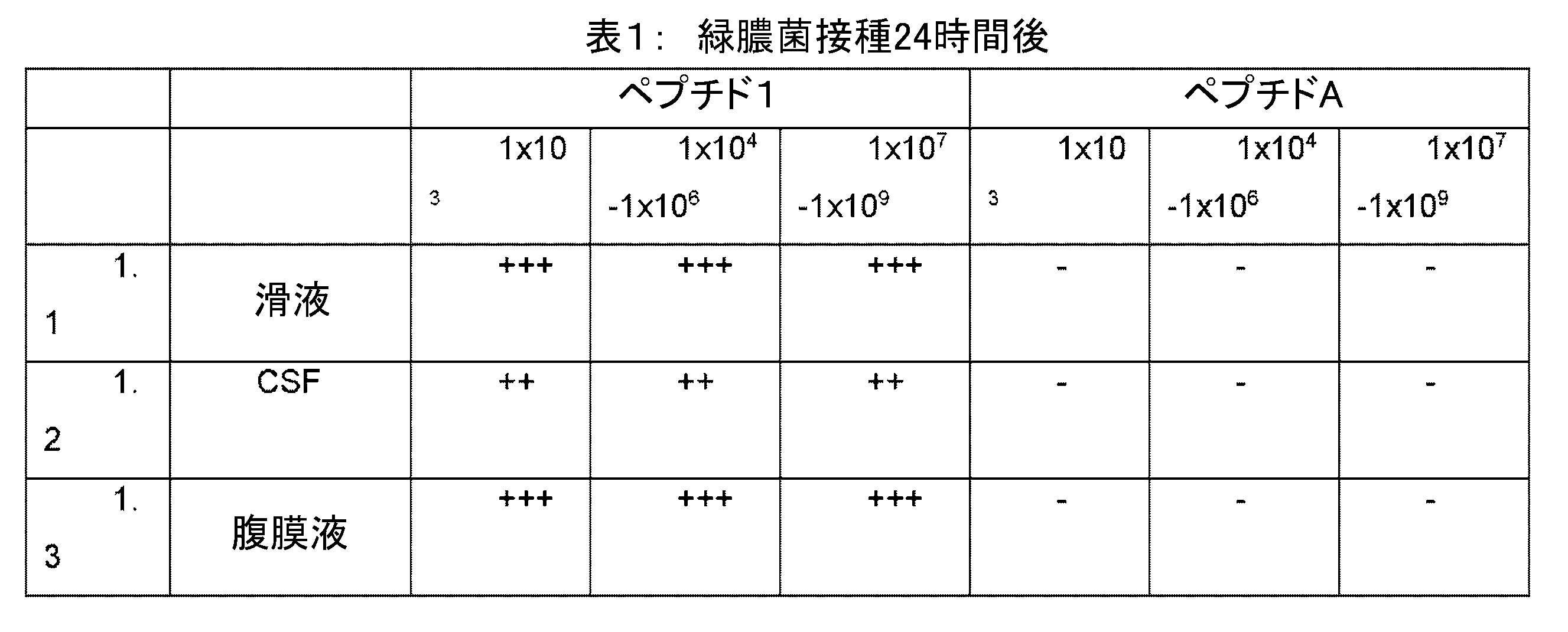

細菌バイオマーカーの位置
The RFU of the control sample (peptide only) was subtracted from each measurement. Values above baseline RFU measurements are indicated with a (+). No change relative to baseline is indicated with a (-).
Location of bacterial biomarkers
Ex. 1.1及びEx.2.1における細菌バイオマーカーの位置は、細菌培養、濃縮細胞(遠心後)及び上清(遠心後)の蛍光活性(RFU)を測定することにより決定した。
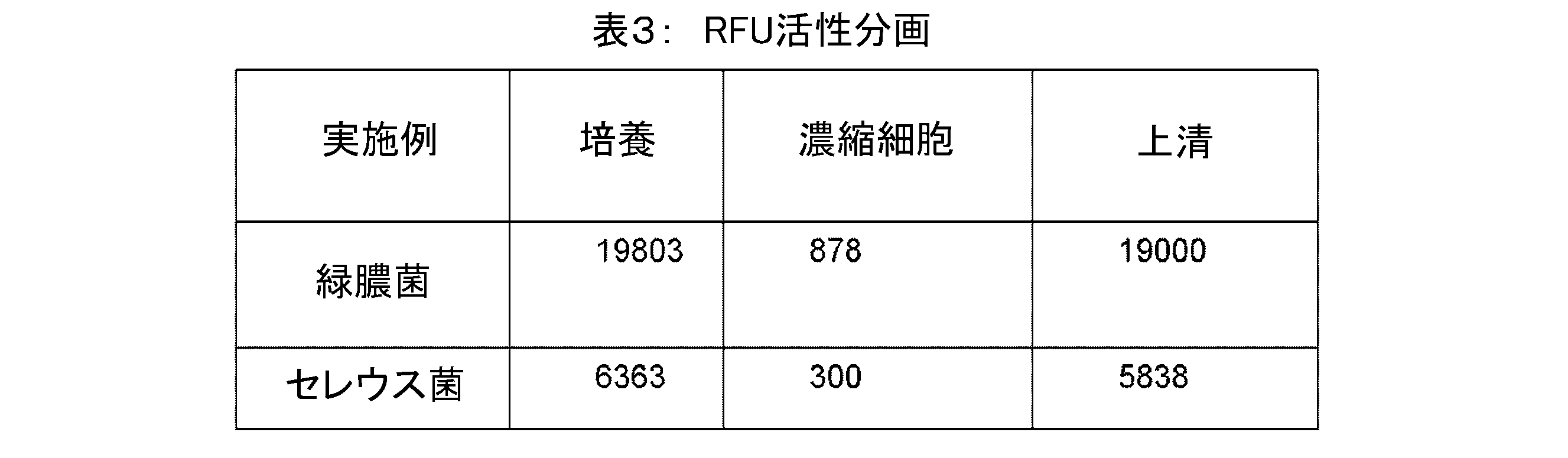
ペプチド2: ペプチド切断部位:ALA; 蛍光剤:IRDye 800CW(Licor Bioscience社、米国); 非蛍光剤:QC-1(Licor Bioscience社、米国)
ペプチドB: ペプチド切断部位:AEA; 蛍光剤:EDANS; 非蛍光剤:DABCYL。
Peptide 2: Peptide cleavage site: ALA; Fluorescent agent: IRDye 800CW (Licor Bioscience, USA); Non-fluorescent agent: QC-1 (Licor Bioscience, USA)
Peptide B: Peptide cleavage site: AEA; Fluorescent agent: EDANS; Non-fluorescent agent: DABCYL.
ペプチド2の検出効率をペプチドBと比較した。 The detection efficiency of peptide 2 was compared with that of peptide B.
ペプチド2及びBを用いて実施例1.1及び2.1を繰り返した。ペプチドB及びCについては、UV分光光度計を用いて検出を行った。

一晩自動腹膜透析を行っている患者の腹膜透析液を一晩透析後に採取した。透析液の滞留時間は約3時間である。廃液の試料を採取し、緑膿菌の臨床分離株の培養でスパイクした(spiked)。臨床分離株は臨床的(細菌性)腹膜炎患者から採取されていた。 Peritoneal dialysis fluid was collected after overnight dialysis from a patient undergoing overnight automated peritoneal dialysis. The residence time of the dialysate is approximately 3 hours. A sample of the effluent was collected and spiked with a culture of a clinical isolate of Pseudomonas aeruginosa. The clinical isolate had been collected from a patient with clinical (bacterial) peritonitis.
2段階で行われたスパイクのレベル: 濁った廃液袋と一致する高レベル(×108CFU/mL)、及び、治療しなければ感染につながる臨床的に問題のある菌量と一致する低レベル(×105CFU/mL)。

固相ペプチド合成により試薬(ペプチド3)を合成した。TQ7WS及びTF7WSはAATBIO(Sunnyvale,CA,USA)amideから購入し、PEG1はBroadPharm(San Diego,CA,USA)から購入したAmino-PEG1-acidである。ペプチド3は、アッセイ管中の最終濃度が1uMであった時に使用した。
ペプチド3:
N末端:TQ7WS
C末端:TF7WS
切断部位:AFA
リンカー:PEG1
The reagent (peptide 3) was synthesized by solid-phase peptide synthesis. TQ7WS and TF7WS were purchased from AATBIO (Sunnyvale, Calif., USA) amide, and PEG1 was Amino-PEG1-acid purchased from BroadPharm (San Diego, Calif., USA). Peptide 3 was used at a final concentration of 1 uM in the assay tube.
Peptide 3:
N-terminus: TQ7WS
C-terminus: TF7WS
Cleavage site: AFA
Linker: PEG1
試料は、Greiner 96平底黒壁プレート(96 Flat Black plate)を用いてTECAN SPARKプレートリーダーで分析し、以下の設定を行った:
-オートマチックミラー
-Z位置=20 000
-ゲイン=175
-振盪10秒、オービタル
-キネティックループ:
-持続時間:30分
-インターバル型:5分間固定
-蛍光強度:
-モード:トップ
-励起波長: 756nm 帯域幅: 5
-発光波長: 780nm 帯域幅: 5
Samples were analyzed on a TECAN SPARK plate reader using Greiner 96 Flat Black plates with the following settings:
- Automatic mirror - Z position = 20 000
-Gain = 175
- Shake 10 seconds, Orbital-Kinetic Loop:
- Duration: 30 minutes - Interval type: fixed for 5 minutes - Fluorescence intensity:
- Mode: Top - Excitation wavelength: 756 nm Bandwidth: 5
-Emission wavelength: 780 nm Bandwidth: 5
陽性結果(positive outcome)に対して、15%の相対的活性の閾値を設定した。閾値はブランクの限界値とした。ブランクの限界は、pH 8のリン酸緩衝液中のペプチド3の蛍光発光を測定することによって決定した。 A threshold of 15% relative activity was set for a positive outcome. The threshold was taken as the blank limit. The blank limit was determined by measuring the fluorescence emission of peptide 3 in phosphate buffer at pH 8.
陰性試料に対する陽性試料の780nmにおける蛍光発光強度(FI)の比は下表のとおりである。
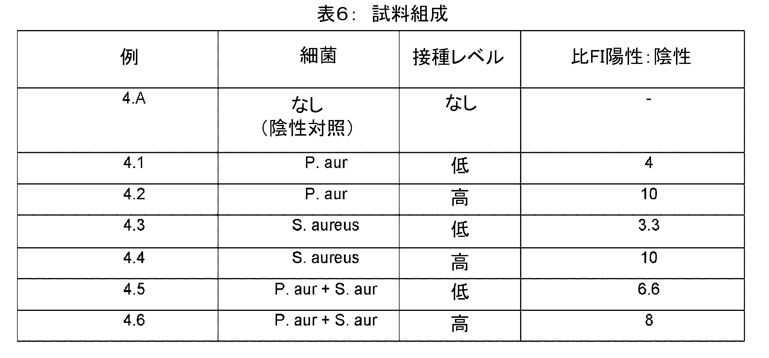
簡単に述べると、黄色ブドウ球菌(S.aureus)及び緑膿菌(P.aeruginosa)の培養物を調製し、ヒト滑液をスパイクするために使用した(Innovate Research、Novi、MI、米国)。試料を1x10⁸-1x10¹⁰の範囲のCFU/mLにスパイクした。この範囲は、人工関節周囲感染における感染滑液中の菌量を表したものである。 Briefly, cultures of S. aureus and P. aeruginosa were prepared and used to spike human synovial fluid (Innovate Research, Novi, MI, USA). Samples were spiked to a range of 1x10-1x10 CFU/ mL , a range representative of the bacterial burden in infected synovial fluid in periprosthetic joint infections.
試料を細菌培養でスパイクした5分後に測定した。試料はDeNIRO蛍光光度計を用いて測定した。 Samples were measured 5 minutes after spiking with bacterial culture. Samples were measured using a DeNIRO fluorometer.
使用した試料の組成を表2に示す
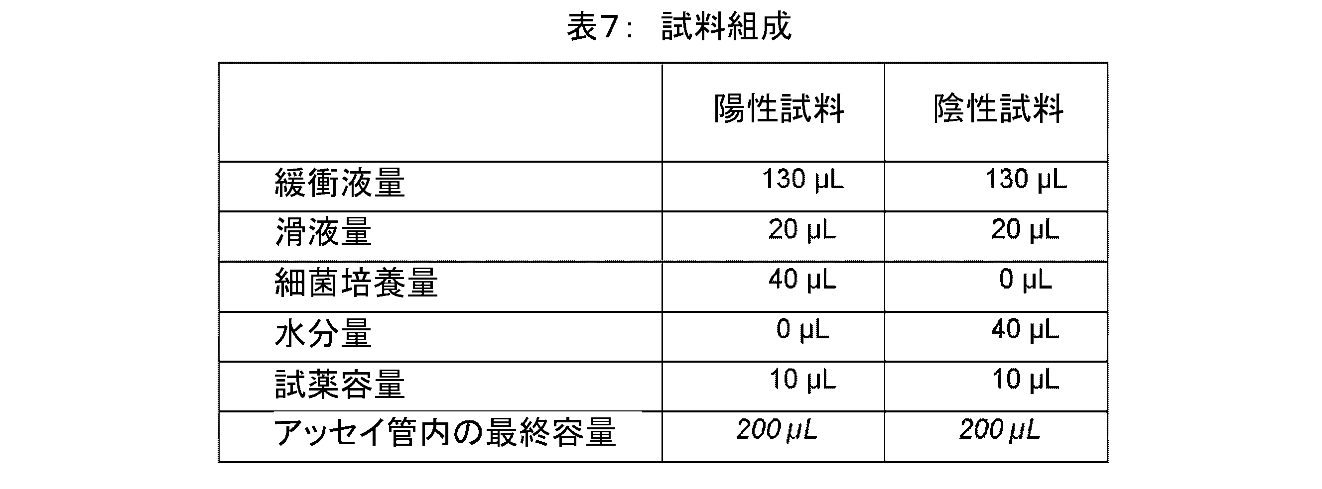
陽性試料は1.2を超える、陰性試料に対する陽性試料の780nmにおける蛍光発光強度(FI)の比を与えたが、陰性試料は5分で1以下の相対活性を与えた。
<実施例6>
Positive samples gave a ratio of fluorescence intensity (FI) at 780 nm of positive to negative samples greater than 1.2, while negative samples gave a relative activity of 1 or less at 5 minutes.
Example 6
AFAモチーフを有する試薬の活性に対するリンカーの影響を腹膜透析液中で測定した。試薬の組成を表8に示す。感染モデルとして広域スペクトル細菌プロテアーゼ(P5380、Sigma Aldrich、4nM最終濃度)に対して試薬6.1~6.5をスクリーニングした。腹膜透析液の試料をリン酸ナトリウム緩衝液、pH8で50%希釈し、Eppendorfチューブ中の1uMのペプチドの最終濃度まで試薬と混合し、マイクロウェルプレート(Greiner、黒壁、チムニー平底のウェルプレート)に200uL移し、TECAN SPARKプレートリーダーを用いて蛍光発光をモニターした。 The effect of the linker on the activity of reagents bearing the AFA motif was measured in peritoneal dialysis fluid. The composition of the reagents is shown in Table 8. Reagents 6.1-6.5 were screened against a broad-spectrum bacterial protease (P5380, Sigma Aldrich, 4 nM final concentration) as an infection model. Peritoneal dialysis fluid samples were diluted 50% in sodium phosphate buffer, pH 8, mixed with reagent to a final concentration of 1 uM peptide in Eppendorf tubes, transferred 200 uL to a microwell plate (Greiner, black-walled, chimney flat-bottom well plate) and fluorescence was monitored using a TECAN SPARK plate reader.
デクェンチング効率(Dequenching efficiency)は、プロテアーゼ非存在下(FI1)及び存在下(FI2)で780nm(TF7WS)又は796nm(IRDye800CW)のいずれかで蛍光発光強度を測定し、式:(((FI2-FI1)/FI1)*100を用いて強度の変化率を測定することにより測定した。
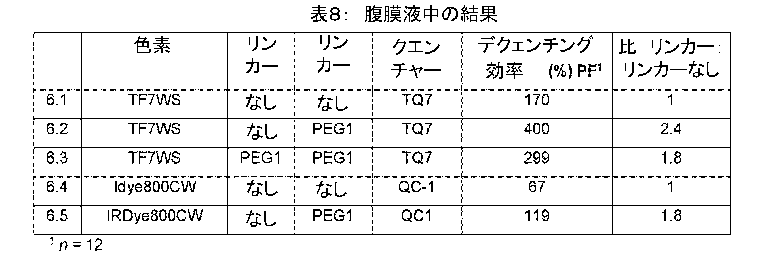
1つ又は2つのPEG1リンカーの導入は、試薬の臨床性能の予想外の改善につながる。
<実施例7>
The introduction of one or two PEG1 linkers leads to an unexpected improvement in the clinical performance of the reagent.
Example 7
AFAモチーフを有する試薬の活性に対するリンカーの影響を滑液中で測定した。試薬の組成を表8に示す。7.1~7.5の試薬を、滑液中の感染モデルとして広域スペクトル細菌プロテアーゼ(P5380、Sigma Aldrich 4nM最終濃度)に対してスクリーニングした。滑液の試料をリン酸ナトリウム緩衝液、pH8で25%希釈し、Eppendorfチューブ中の1uMのペプチドの最終濃度まで試薬と混合し、マイクロウェルプレート(Greiner、黒壁、チムニーの平底のウェルプレート)に200uL移し、TECAN SPARKプレートリーダーを用いて蛍光発光をモニターした。ペプチドの濃度は1uMであった。プロテアーゼの濃度は4nMであった。 The effect of the linker on the activity of reagents with an AFA motif was measured in synovial fluid. The composition of the reagents is shown in Table 8. Reagents 7.1-7.5 were screened against a broad-spectrum bacterial protease (P5380, Sigma Aldrich 4 nM final concentration) as a model of infection in synovial fluid. Synovial fluid samples were diluted 25% in sodium phosphate buffer, pH 8, mixed with reagent to a final concentration of 1 uM peptide in Eppendorf tubes, transferred 200 uL to a microwell plate (Greiner, black-walled, chimney flat-bottom well plate) and fluorescence was monitored using a TECAN SPARK plate reader. The peptide concentration was 1 uM. The protease concentration was 4 nM.
デクェンチング効率は、プロテアーゼ非存在下(FI1)及び存在下(FI2)で780nm(TF7WS)又は796nm(IRDye800CW)のいずれかで蛍光発光強度を測定し、式:(((FI2-FI1)/FI1)*100を用いて強度の変化率を測定することにより測定した。

1つ又は2つのPEG1リンカーの導入は、試薬の臨床性能の予想外の改善につながる。
<実施例8>
The introduction of one or two PEG1 linkers leads to an unexpected improvement in the clinical performance of the reagent.
Example 8
ペプチド1を、炭水化物の存在下でペプチドを凍結乾燥することによって凍結乾燥ビーズとして処方した。簡単に言うと、25ugペプチド1をpH7.4の300uL PBSに溶解し、20%炭水化物を含むpH7.4の300uL PBSに加えた。24個のビーズを分注し、凍結乾燥した(74時間、一次温度-45℃)。 Peptide 1 was formulated as lyophilized beads by lyophilizing the peptide in the presence of carbohydrate. Briefly, 25ug peptide 1 was dissolved in 300uL PBS pH 7.4 and added to 300uL PBS pH 7.4 containing 20% carbohydrate. 24 beads were dispensed and lyophilized (74h, primary temperature -45°C).
賦形剤ごとに、2つの同一ビーズを用いて2つの測定を行った。まず、0.5mLの微量遠心管で、凍結乾燥したビーズを300uLの滅菌水に室温で溶解し、陰性試料を調製し、DeNIRO(登録商標)蛍光光度計(Detact Diagnostics、フローニンゲン、オランダ)に移し、780nmでの蛍光発光強度(TF7WS)を記録した。次に、0.5mLの微量遠心管に凍結乾燥したビーズを、スブチリシン(4nM)の滅菌水溶液300 uLに溶解し、陽性試料を調製した。混合物を短時間渦巻きにし(vortexed)、21℃で15分間培養した後、DeNIRO(登録商標)蛍光光度計を用いて780nmにおける蛍光発光強度(TF7WS)を測定した。 For each excipient, two measurements were performed with two identical beads. Negative samples were prepared by dissolving the lyophilized beads in 300 uL of sterile water at room temperature in a 0.5 mL microcentrifuge tube, transferring to a DeNIRO® fluorometer (Detect Diagnostics, Groningen, The Netherlands) and recording the fluorescence emission intensity at 780 nm (TF7WS). Positive samples were prepared by dissolving the lyophilized beads in 300 uL of a sterile aqueous solution of subtilisin (4 nM) in a 0.5 mL microcentrifuge tube. The mixture was vortexed briefly and incubated at 21°C for 15 min before measuring the fluorescence emission intensity at 780 nm (TF7WS) using a DeNIRO® fluorometer.
陰性検体に対する陽性検体の780nmにおける蛍光発光強度(FI)の比は下表のとおりである。

表9は、炭水化物を含む全ての製剤が、相対活性において予想外の改善を与えたことを示している。驚くべきことに、多糖類、デキストランを含む凍結乾燥ビーズは、バックグラウンドの予想外の安定化(最低の陰性FI)及び780nmでの陽性試料の陰性試料に対する最高比率を示した。
<実施例10>
Table 9 shows that all formulations containing carbohydrates provided unexpected improvements in relative activity. Surprisingly, lyophilized beads containing the polysaccharide, dextran, showed unexpected stabilization of background (lowest negative FI) and the highest ratio of positive to negative samples at 780 nm.
Example 10
実施例9に従うデキストランを含む凍結乾燥ビーズを、200uLの体液(表11に示す腹膜透析液及び滑液)に溶解した。細菌培養物を実施例4に従って調製した。
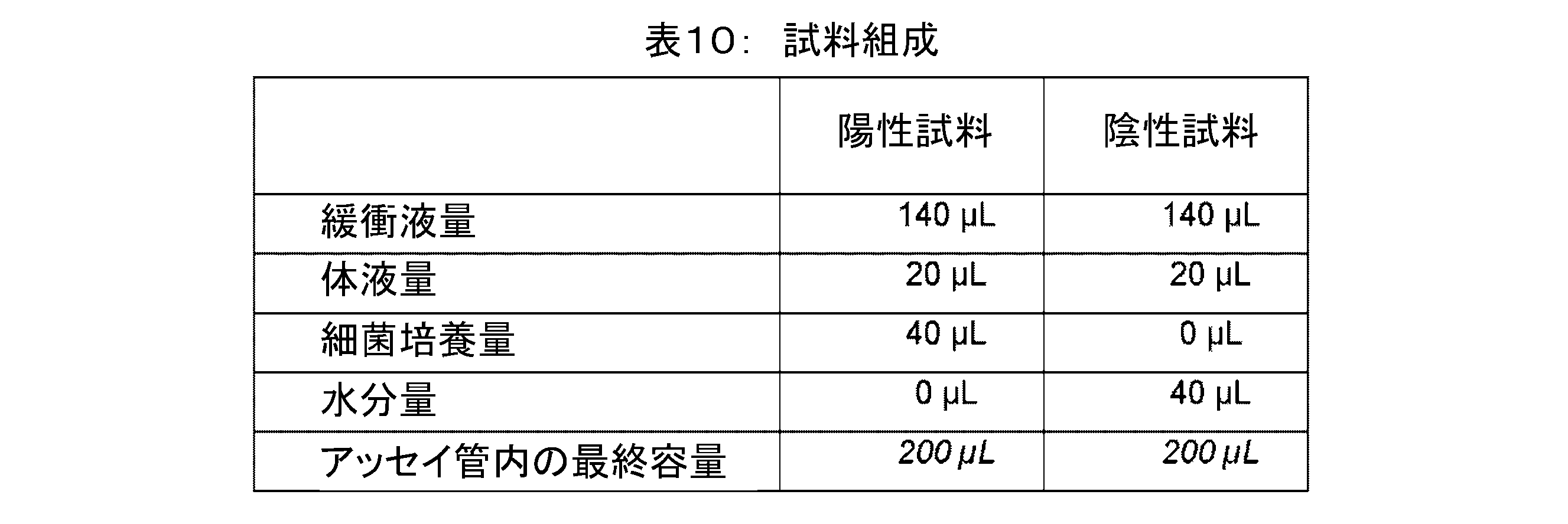
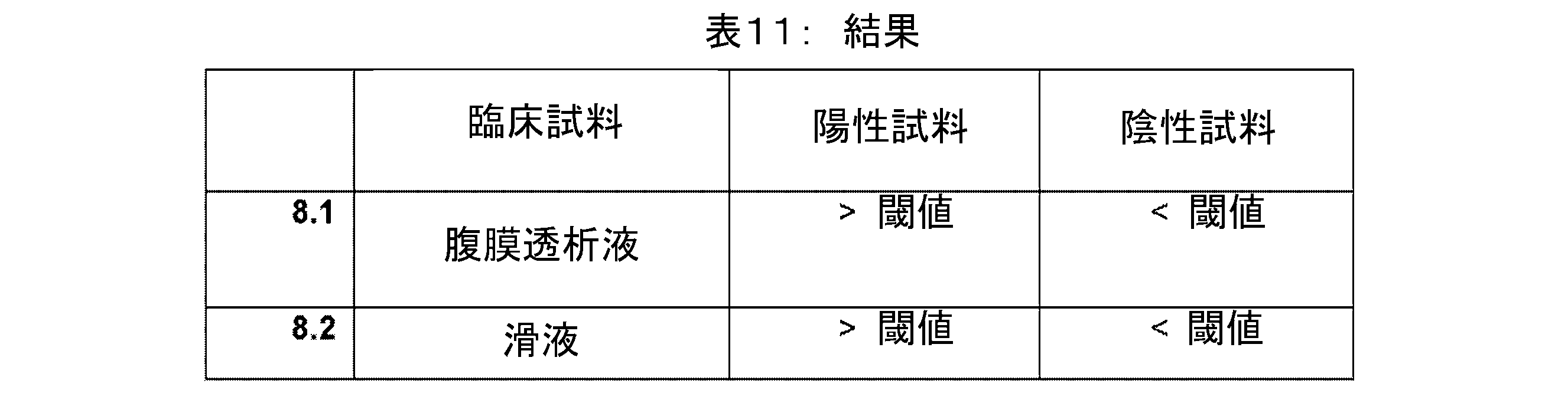
腹腔液及び滑液の臨床試料を、実施例4及び5に従って黄色ブドウ球菌(S. aureus)の培養液でスパイクし、デクェンチング効率を決定する前にWO2018/224561の実施例1により記載される酸性化工程を施した。臨床試料1mLを、10%酢酸の添加によりpH4.6に調整した。溶液は室温で15分間保持した。0.1mLの1M酢酸ナトリウム緩衝液を加え、試料を4500gで30分間遠心分離した。試料を実施例10に従って分析したとき、閾値を超える測定可能な活性は認められなかった。
下記は、本願の出願当初に記載の発明である。
<請求項1>
一般式[a]-[b]-[c](I)を有する試薬を用いて体液試料中の感染をインビトロ診断する方法であって、式中、
[a]は、発光波長650~900nmの蛍光剤であり、
[c]は、前記蛍光剤の前記発光を消光するための、吸収波長650~900nmの非蛍光剤であり、
[b]は、切断部位を含むペプチドであり、前記切断部位は細菌バイオマーカーに対して特異的であり、
前記方法が、
i) 体液、好ましくはヒト体液の試料と試料容器内の前記試薬とを接触させる工程と、
ii) 工程i)の前記容器内の前記試料からの650~900nmの範囲の蛍光発光をモニターする工程であって、650~900nmの範囲の蛍光発光の増加が、前記体液試料中の感染の存在を示す、該工程と、を含む方法。
<請求項2>
前記体液試料は、羊水、尿、漿液、滑液、腹膜透析液、精液、皮脂及び脳脊髄液からなる群から選択され、好ましくは、前記体液は、滑液、腹膜透析液及び脳脊髄液からなる群から選択され、より好ましくは、前記体液は、滑液及び腹膜透析液からなる群から選択される、請求項1に記載の方法。
<請求項3>
[b]:([a]+[c])の質量比が少なくとも1:2、好ましくは[b]:([a]+[c])の質量比が少なくとも1:3、より好ましくは[b]:([a]+[c])の質量比が少なくとも1:4、さらにより好ましくは[b]:([a]+[c])の質量比が少なくとも1:5である、請求項1又は2に記載の方法。
<請求項4>
前記試薬が、一般式[a]-[リンカー1]n-[b]-[-リンカー2]m-[c]を有し、式中、nが0、1又は2であり、mが0、1又は2であり、[リンカー1]及び[リンカー2]が、任意に置換されたヒドロカルビル基及び非タンパク質分解性ヒドロカルビル基の群から独立して選択され、好ましくはnが0又は1であり、mが1であり、リンカー2が非タンパク質分解性ヒドロカルビル基である、請求項1~3のいずれかに記載の方法。
<請求項5>
前記方法が、前記試料を第一試薬及び第二試薬と接触させる工程を含み、前記第一試薬が一般式[a]-[b]-[c](I)を有し、及び[b]が切断部位(b’)を含むペプチドであり、前記第二試薬が式[a]-[d]-[c](III)を有し、式中[d]が切断部位(d’)を含むペプチドであり、前記切断部位d’は前記切断部位b’とは異なる、請求項1~4のいずれかに記載の方法。
<請求項6>
工程i)が、希釈剤を添加する工程を含み、好ましくは、前記希釈剤が、MOPS、リン酸塩、クエン酸塩、HEPES TRIS-HCl、リン酸緩衝生理食塩水からなる群から選択される緩衝剤を含む、請求項1~5のいずれかに記載の方法。
<請求項7>
試料に対する希釈剤の容積比が0.1~100の範囲、より好ましくは0.5~75の範囲、さらにより好ましくは1~50の範囲である、請求項6に記載の方法。
<請求項8>
前記試料、及び任意で希釈剤の添加後の前記試料容器内の試薬の濃度が、0.01~10uM、好ましくは0.02~8uM、より好ましくは0.05~5uM、さらにより好ましくは0.1~2.5uMの範囲にある、請求項1~7のいずれかに記載の方法。
<請求項9>
発光波長650~900nmの前記蛍光剤が、シアニン部分、好ましくはスルホ-シアニン部分を含み、吸収波長650~900nmの前記非蛍光剤が、シアニン部分、好ましくはスルホ-シアニン部分を含む、請求項1~8のいずれかに記載の方法。
<請求項10>
700~850nmの範囲、より好ましくは750~850nmの範囲の蛍光発光の増加が感染の指標である、請求項1~9のいずれかに記載の方法。
<請求項11>
前記細菌バイオマーカーが、細菌膜結合プロテアーゼ、細菌膜結合トランスペプチダーゼ、細胞内細菌プロテアーゼ又は細胞外細菌プロテアーゼであり、より好ましくは前記細菌バイオマーカーが細菌膜結合トランスペプチダーゼ又は細胞外細菌プロテアーゼである、請求項1~10のいずれかに記載の方法。
<請求項12>
前記切断部位が、セレウス菌(Bacillus cereus)、黄色ブドウ球菌(Staphylococcus aureus)、化膿性レンサ球菌(Streptococcus pyogenes)、緑膿菌(Pseudomonas aeruginosa)、大腸菌(Escherichia coli)及びそれらの組み合わせからなる群から選択される細菌種に対する細菌バイオマーカーに対して特異的であり、好ましくは、前記切断部位が緑膿菌(Pseudomonas aeruginosa)及びセレウス菌(Bacillus cereus)に対して特異的であり、より好ましくは、前記切断部位が緑膿菌(Pseudomonas aeruginosa)に対して特異的である、請求項1~11のいずれかに記載の方法。
<請求項13>
[b]が3~8個のアミノ酸を含み、より好ましくは3~7個のアミノ酸を含み、さらにより好ましくは3~6個のアミノ酸を含む、請求項1~12のいずれかに記載の方法。
<請求項14>
[b]は一般式Xaa1,Xaa2,Xaa3を有し、式中、
Xaa1は、小さな疎水性のアミノ酸であり、好ましくはXaa1は、アラニン、グリシン、ロイシン、バリン、ノルロイシン、ノルバリン、イソロイシン、イソバリン、アロイソロイシンからなる群より選択され、
Xaa2は、好ましくは、小さな又は芳香族の疎水性アミノ酸であり、好ましくはXaa2は、アラニン、グリシン、ロイシン、バリン、ノルロイシン、ノルバリン、イソロイシン、イソバリン、アロイソロイシン及びフェニルアラニンからなる群より選択され、
Xaa3は、アラニン、グリシン、ロイシン、バリン、ノルロイシン、ノルバリン、イソロイシン、イソバリン、アロイソロイシンからなる群より選択され、より好ましくは、Xaa3は、アラニン、グリシン、ロイシン、バリンからなる群より選択される、請求項1~13のいずれかに記載の方法。
<請求項15>
前記試料を容器内の前記試薬と接触させ、前記試薬によって発せられる蛍光を、前記容器を受け入れるように適合させたハンドヘルド又はベンチトップ蛍光分光計によってモニターする、請求項1~14のいずれかに記載の方法。
<請求項16>
前記試薬が表面に付着していない、請求項1~15のいずれかに記載の方法。
<請求項17>
前記試料に、前記試薬と接触させる前又は接触させた後のいずれかにおいて、20~40℃の温度範囲で細菌培養のエキソビボ工程を施さない、請求項1~16のいずれかに記載の方法。
<請求項18>
工程i)の前記試薬が炭水化物を含む凍結乾燥ビーズの形態で提供される、請求項1~17のいずれかに記載の方法。
<請求項19>
前記ビーズが、ビーズ炭水化物の重量に対して5~95重量%含み、ビーズの総重量に対して好ましくは10~90重量%、より好ましくは15~85重量%、さらにより好ましくは20~80重量%の炭水化物を含む、請求項18に記載の方法。
<請求項20>
前記炭水化物が、ポリオール、単糖類、二糖類、多糖類及びそれらの組合せからなる群より選択される、請求項18又は19に記載の方法。
<請求項21>
炭水化物及び構造[a]-[b]-[c](I)を有する試薬を含む凍結乾燥ビーズであって、式中、
[a]は、発光波長650~900nmの蛍光剤であり、
[c]は、前記蛍光剤の前記発光を消光するための、吸収波長650~900nmの非蛍光剤であり、
[b]は、切断部位を含むペプチドであり、前記切断部位はバイオマーカー、好ましくは細菌バイオマーカーに対して特異的である、凍結乾燥ビーズ。
<請求項22>
前記ビーズが、ビーズ炭水化物の重量に対して5~95重量%含み、ビーズの総重量に対して好ましくは10~90重量%、より好ましくは15~85重量%、さらにより好ましくは20~80重量%の炭水化物を含む、請求項21に記載の凍結乾燥ビーズ。
<請求項23>
前記炭水化物が、ポリオール、単糖類、二糖類、多糖類及びそれらの組合せからなる群より選択される、請求項21又は22に記載の凍結乾燥ビーズ。
<請求項24>
前記ポリオールが、(2R,3S)-ブタン-1,2,3,4-テトロール(エスリトール)、4-O-β-D-ガラクトピラノシル-D-グルシトール(ラクチトール)、(2S,3S,4S,5S)-ヘキサン-1,2,3,4,5,6-ヘキソール(L-マンニトール)、(2R,3R,4R,5R)-ヘキサン-1,2,3,4,5,6-ヘキソール(D-マンニトール)、4-O-α-D-グルコピラノシル-D-グルシトール(マルチトール)、(2R,3R,4R,5S)-ヘキサン-1,2,3,4,5,6-ヘキソール(ソルビトール)、(2R,3r,4S)-ペンタン-1,2,3,4,5-ペントール(キシリトール)、ポリエチレングリコール及びそれらの組合せからなる群より選択され、好ましくは、前記ポリオールは、(2S,3S,4S,5S)-ヘキサン-1,2,3,4,5,6-ヘキソール(L-マンニトール)、(2R,3R,4R,5R)-ヘキサン-1,2,3,4,5,6-ヘキソール(D-マンニトール)及びそれらの組み合わせからなる群より選択される、請求項21~23のいずれかに記載の凍結乾燥ビーズ。
<請求項25>
前記単糖類がグルコース、フルクトース、ガラクトース及びそれらの組合せからなる群より選択される、請求項21~24のいずれかに記載の凍結乾燥ビーズ。
<請求項26>
前記二糖類が、スクロース、マルトース、ラクトース、トレハロース及びそれらの組合せからなる群より選択される、請求項21~25のいずれかに記載の凍結乾燥ビーズ。
<請求項27>
前記多糖類が、アルギン酸塩、キトサン、ヒアルロン酸、セルロース誘導体、デキストラン及びそれらの組み合わせからなる群より選択される、請求項21~26のいずれかに記載の凍結乾燥ビーズ。
<請求項28>
前記試薬が、請求項21~27のいずれか1以上に記載の試薬である、請求項1~20のいずれかに記載の方法。
<請求項29>
炭水化物及び構造[a]-[b]-[c](I)を有する試薬を含む凍結乾燥ビーズの酵素アッセイにおける使用であって、式中、
[a]は発光波長650~900nmの蛍光剤であり、
[b]は切断部位を含むペプチドであって、前記切断部位はバイオマーカー、好ましくは細菌バイオマーカーに対して特異的であり、
[c]は、前記蛍光剤の前記発光を消光するための、吸収波長650~900nmの非蛍光剤である、使用。
<請求項30>
体液試料中の感染をインビトロ診断するためのキットであって、
a) 構造[a]-[b]-[c](I)を有する試薬を含む容器であって、式中、
[a]は発光波長650~900nmの蛍光剤であり、
[b]は切断部位を含むペプチドであり、前記切断部位は細菌バイオマーカーに対して特異的であり、
[c]は、前記蛍光剤の前記発光を消光するための、吸収波長650~900nmの非蛍光剤である、該容器と、
b) 請求項1~20の方法を実施するための一連の説明書と、
c) 任意に、希釈剤を含有する容器と、を含むキット。
<請求項31>
前記試薬及び前記希釈剤が、容器の2つの区画に存在し、好ましくは、前記区画が圧力活性化可能な膜によって分離されている、請求項30に記載のキット。
<請求項32>
前記試薬が、請求項21~27のいずれか1以上に記載のものである、請求項30又は31に記載のキット。
<請求項33>
a)構造[a]-[b]-[c](I)であって、
[a]は発光波長650~900nmの蛍光剤であり、
[c]は蛍光剤の発光を消光するための吸収波長が650~900nmの非蛍光剤であり、
[b]は切断部位を含むペプチドであり、切断部位は細菌のバイオマーカーに特異的である、前記構造[a]-[b]-[c](I)を有する試薬を含む容器と、
b)容器を受け入れて、試薬を用いて滑液、腹水又は脳脊髄液の試料の存在下で、試薬から発せられる蛍光シグナルを監視するように構成された装置を有するシステムであって、請求項1~20に記載の方法を実施するように構成されたシステム。
<請求項34>
前記システムが、ポイントオブケアシステム又は患者ビサイドシステムである、請求項33に記載のシステム。
Clinical samples of peritoneal and synovial fluid were spiked with cultures of S. aureus according to Examples 4 and 5 and subjected to an acidification step as described by Example 1 of WO2018/224561 before determining the dequenching efficiency. 1 mL of clinical sample was adjusted to pH 4.6 by addition of 10% acetic acid. The solution was kept at room temperature for 15 minutes. 0.1 mL of 1 M sodium acetate buffer was added and the samples were centrifuged at 4500 g for 30 minutes. When the samples were analyzed according to Example 10, no measurable activity above the threshold was observed.
The following is an invention described at the beginning of the present application.
<Claim 1>
1. A method for in vitro diagnosis of infection in a body fluid sample using a reagent having the general formula [a]-[b]-[c] (I), wherein:
[a] is a fluorescent agent having an emission wavelength of 650 to 900 nm;
[c] is a non-fluorescent agent having an absorption wavelength of 650 to 900 nm for quenching the emission of the fluorescent agent;
[b] is a peptide comprising a cleavage site, the cleavage site being specific for a bacterial biomarker;
The method further comprising:
i) contacting a sample of a body fluid, preferably a human body fluid, with said reagent in a sample container;
ii) monitoring fluorescence emission in the range of 650-900 nm from said sample in said container of step i), wherein an increase in fluorescence emission in the range of 650-900 nm indicates the presence of infection in said bodily fluid sample.
<Claim 2>
2. The method of claim 1, wherein the body fluid sample is selected from the group consisting of amniotic fluid, urine, serous fluid, synovial fluid, peritoneal dialysis fluid, semen, sebum and cerebrospinal fluid, preferably the body fluid is selected from the group consisting of synovial fluid, peritoneal dialysis fluid and cerebrospinal fluid, more preferably the body fluid is selected from the group consisting of synovial fluid and peritoneal dialysis fluid.
<Claim 3>
3. The method according to claim 1 or 2, wherein the mass ratio of [b]:([a]+[c]) is at least 1:2, preferably the mass ratio of [b]:([a]+[c]) is at least 1:3, more preferably the mass ratio of [b]:([a]+[c]) is at least 1:4, even more preferably the mass ratio of [b]:([a]+[c]) is at least 1:5.
<Claim 4>
4. The method of any of claims 1 to 3, wherein the reagent has the general formula [a]-[ Linker1 ] n- [b]-[- Linker2 ] m- [c], where n is 0, 1 or 2, m is 0, 1 or 2, and [ Linker1 ] and [ Linker2 ] are independently selected from the group of optionally substituted hydrocarbyl groups and non-proteolytic hydrocarbyl groups, preferably n is 0 or 1, m is 1 and Linker2 is a non-proteolytic hydrocarbyl group.
<Claim 5>
5. The method of any of claims 1 to 4, wherein the method comprises contacting the sample with a first reagent and a second reagent, wherein the first reagent has the general formula [a]-[b]-[c] (I), where [b] is a peptide comprising a cleavage site (b'), and the second reagent has the formula [a]-[d]-[c] (III), where [d] is a peptide comprising a cleavage site (d'), and wherein the cleavage site d' is different from the cleavage site b'.
<Claim 6>
6. The method according to any of claims 1 to 5, wherein step i) comprises adding a diluent, preferably said diluent comprises a buffer selected from the group consisting of MOPS, phosphate, citrate, HEPES TRIS-HCl, phosphate buffered saline.
<Claim 7>
The method of claim 6, wherein the volume ratio of diluent to sample is in the range of 0.1-100, more preferably in the range of 0.5-75, even more preferably in the range of 1-50.
<Claim 8>
8. The method of any of claims 1 to 7, wherein the concentration of the sample and optionally reagent in the sample vessel after addition of a diluent is in the range of 0.01 to 10 uM, preferably 0.02 to 8 uM, more preferably 0.05 to 5 uM, even more preferably 0.1 to 2.5 uM.
<Claim 9>
9. The method of any one of claims 1 to 8, wherein the fluorescent agent with an emission wavelength of 650-900 nm comprises a cyanine moiety, preferably a sulfo-cyanine moiety, and the non-fluorescent agent with an absorption wavelength of 650-900 nm comprises a cyanine moiety, preferably a sulfo-cyanine moiety.
<Claim 10>
The method of any of claims 1 to 9, wherein an increase in fluorescence emission in the range of 700 to 850 nm, more preferably in the range of 750 to 850 nm, is indicative of infection.
<Claim 11>
The method according to any of claims 1 to 10, wherein the bacterial biomarker is a bacterial membrane-bound protease, a bacterial membrane-bound transpeptidase, an intracellular bacterial protease or an extracellular bacterial protease, more preferably the bacterial biomarker is a bacterial membrane-bound transpeptidase or an extracellular bacterial protease.
<Claim 12>
12. The method of any of claims 1 to 11, wherein the cleavage site is specific for a bacterial biomarker for a bacterial species selected from the group consisting of Bacillus cereus, Staphylococcus aureus, Streptococcus pyogenes, Pseudomonas aeruginosa, Escherichia coli and combinations thereof, preferably the cleavage site is specific for Pseudomonas aeruginosa and Bacillus cereus, more preferably the cleavage site is specific for Pseudomonas aeruginosa.
<Claim 13>
The method according to any one of claims 1 to 12, wherein [b] comprises 3 to 8 amino acids, more preferably 3 to 7 amino acids, and even more preferably 3 to 6 amino acids.
<Claim 14>
[b] has the general formula X aa1 , X aa2 , and X aa3 , wherein
Xa1 is a small hydrophobic amino acid, preferably Xa1 is selected from the group consisting of alanine, glycine, leucine, valine, norleucine, norvaline, isoleucine, isovaline, alloisoleucine;
X aa2 is preferably a small or aromatic hydrophobic amino acid, preferably X aa2 is selected from the group consisting of alanine, glycine, leucine, valine, norleucine, norvaline, isoleucine, isovaline, alloisoleucine and phenylalanine;
The method according to any one of claims 1 to 13, wherein X aa3 is selected from the group consisting of alanine, glycine, leucine, valine, norleucine, norvaline, isoleucine, isovaline, and alloisoleucine, more preferably X aa3 is selected from the group consisting of alanine, glycine, leucine, and valine.
<Claim 15>
15. The method of any of claims 1 to 14, wherein the sample is contacted with the reagent in a container and the fluorescence emitted by the reagent is monitored by a handheld or benchtop fluorescence spectrometer adapted to receive the container.
<Claim 16>
The method of any preceding claim, wherein the reagent is not attached to a surface.
<Claim 17>
The method according to any of claims 1 to 16, wherein the sample is not subjected to an ex vivo step of bacterial culture in the temperature range of 20-40°C either before or after contacting with the reagent.
<Claim 18>
The method according to any of claims 1 to 17, wherein the reagent of step i) is provided in the form of lyophilized beads comprising a carbohydrate.
<Claim 19>
19. The method of claim 18, wherein the beads comprise 5-95% by weight of the bead carbohydrate, preferably 10-90% by weight, more preferably 15-85% by weight, even more preferably 20-80% by weight of carbohydrate based on the total weight of the bead.
<Claim 20>
20. The method of claim 18 or 19, wherein the carbohydrate is selected from the group consisting of polyols, monosaccharides, disaccharides, polysaccharides, and combinations thereof.
<Claim 21>
Lyophilized beads comprising a carbohydrate and a reagent having the structure [a]-[b]-[c] (I), wherein:
[a] is a fluorescent agent having an emission wavelength of 650 to 900 nm;
[c] is a non-fluorescent agent having an absorption wavelength of 650 to 900 nm for quenching the emission of the fluorescent agent;
[b] is a peptide comprising a cleavage site, said cleavage site being specific for a biomarker, preferably a bacterial biomarker, said lyophilized bead.
<Claim 22>
22. The freeze-dried beads of claim 21, wherein the beads comprise 5-95% by weight of the bead carbohydrate, preferably 10-90% by weight, more preferably 15-85% by weight, even more preferably 20-80% by weight of carbohydrate, based on the total weight of the bead.
<Claim 23>
23. The freeze-dried bead of claim 21 or 22, wherein the carbohydrate is selected from the group consisting of polyols, monosaccharides, disaccharides, polysaccharides, and combinations thereof.
<Claim 24>
The polyol is selected from the group consisting of (2R,3S)-butane-1,2,3,4-tetrol (esritol), 4-O-β-D-galactopyranosyl-D-glucitol (lactitol), (2S,3S,4S,5S)-hexane-1,2,3,4,5,6-hexol (L-mannitol), (2R,3R,4R,5R)-hexane-1,2,3,4,5,6-hexol (D-mannitol), 4-O-α-D-glucopyranosyl-D-glucitol (maltitol), (2R,3R,4R,5S)-hexane-1,2,3,4,5,6-hexol (L-mannitol), 24. The freeze-dried beads according to any one of claims 21 to 23, wherein the polyol is selected from the group consisting of (2S,3S,4S,5S)-hexane-1,2,3,4,5,6-hexol (L-mannitol), (2R,3R,4R,5R)-hexane-1,2,3,4,5,6-hexol (D-mannitol) and combinations thereof.
<Claim 25>
The freeze-dried beads of any one of claims 21 to 24, wherein the monosaccharide is selected from the group consisting of glucose, fructose, galactose, and combinations thereof.
<Claim 26>
The freeze-dried beads of any of claims 21 to 25, wherein the disaccharide is selected from the group consisting of sucrose, maltose, lactose, trehalose, and combinations thereof.
<Claim 27>
The freeze-dried beads of any one of claims 21 to 26, wherein the polysaccharide is selected from the group consisting of alginates, chitosan, hyaluronic acid, cellulose derivatives, dextran and combinations thereof.
<Claim 28>
The method according to any one of claims 1 to 20, wherein the reagent is a reagent according to any one or more of claims 21 to 27.
<Claim 29>
1. Use of lyophilized beads comprising a carbohydrate and a reagent having the structure [a]-[b]-[c] (I) in an enzyme assay, wherein
[a] is a fluorescent agent having an emission wavelength of 650 to 900 nm,
[b] is a peptide comprising a cleavage site, said cleavage site being specific for a biomarker, preferably a bacterial biomarker;
[c] is a non-fluorescent agent having an absorption wavelength of 650 to 900 nm for quenching the emission of the fluorescent agent.
<Claim 30>
1. A kit for in vitro diagnosis of infection in a body fluid sample, comprising:
a) a vessel containing a reagent having the structure [a]-[b]-[c](I), wherein:
[a] is a fluorescent agent having an emission wavelength of 650 to 900 nm,
[b] is a peptide comprising a cleavage site, the cleavage site being specific for a bacterial biomarker;
[c] is a non-fluorescent agent having an absorption wavelength of 650 to 900 nm for quenching the emission of the fluorescent agent;
b) a set of instructions for carrying out the method of claims 1 to 20; and
c) optionally, a container containing a diluent.
<Claim 31>
31. The kit of claim 30, wherein the reagent and the diluent are present in two compartments of a container, preferably the compartments being separated by a pressure activatable membrane.
<Claim 32>
The kit according to claim 30 or 31, wherein the reagent is according to any one or more of claims 21 to 27.
<Claim 33>
a) the structure [a]-[b]-[c](I),
[a] is a fluorescent agent having an emission wavelength of 650 to 900 nm,
[c] is a non-fluorescent agent having an absorption wavelength of 650 to 900 nm for quenching the emission of the fluorescent agent,
a container containing a reagent having the structure [a]-[b]-[c] (I), wherein [b] is a peptide containing a cleavage site, the cleavage site being specific for a bacterial biomarker;
b) A system comprising a device configured to receive the container and to monitor a fluorescent signal emitted by the reagent in the presence of a sample of synovial fluid, peritoneal fluid or cerebrospinal fluid, said system being configured to carry out the method according to claims 1 to 20.
<Claim 34>
34. The system of claim 33, wherein the system is a point-of-care system or a patient-besides system.
Claims (15)
[a]は、発光波長650~900nmの蛍光剤であり、
[c]は、前記蛍光剤の前記発光を消光するための、吸収波長650~900nmの非蛍光剤であり、
[b]は、切断部位を含むペプチドであり、前記切断部位は細菌バイオマーカーに対して特異的であり、
前記細菌バイオマーカーが、細菌膜結合プロテアーゼ、細菌膜結合トランスペプチダーゼ、細胞内細菌プロテアーゼ又は細胞外細菌プロテアーゼであり、前記試薬が、単糖類、二糖類、多糖類及びそれらの組合せからなる群より選択される炭水化物を含む凍結乾燥ビーズの形態で提供され、
前記方法が、
i) 体液、好ましくはヒト体液の試料と試料容器内の前記試薬とを接触させる工程と、
ii) 工程i)の前記容器内の前記試料からの650~900nmの範囲の蛍光発光をモニターする工程であって、650~900nmの範囲の蛍光発光の増加が、前記体液試料中の感染の存在を示す、該工程と、を含む方法。 1. A method for in vitro testing for infection in a body fluid sample isolated from a subject using a reagent having the general formula [a]-[b]-[c] (I), wherein:
[a] is a fluorescent agent having an emission wavelength of 650 to 900 nm;
[c] is a non-fluorescent agent having an absorption wavelength of 650 to 900 nm for quenching the emission of the fluorescent agent;
[b] is a peptide comprising a cleavage site, the cleavage site being specific for a bacterial biomarker;
the bacterial biomarker is a bacterial membrane-bound protease, a bacterial membrane-bound transpeptidase, an intracellular bacterial protease or an extracellular bacterial protease, and the reagent is provided in the form of lyophilized beads comprising a carbohydrate selected from the group consisting of monosaccharides, disaccharides, polysaccharides and combinations thereof;
The method further comprising:
i) contacting a sample of a body fluid, preferably a human body fluid, with said reagent in a sample container;
ii) monitoring fluorescence emission in the range of 650-900 nm from said sample in said container of step i), wherein an increase in fluorescence emission in the range of 650-900 nm indicates the presence of infection in said bodily fluid sample.
700~850nmの範囲の蛍光発光の増加が感染の指標である、請求項1~7のいずれか一項に記載の方法。 8. The method of any one of claims 1 to 7, wherein the fluorescent agent with an emission wavelength of 650-900 nm comprises a cyanine moiety, the non-fluorescent agent with an absorption wavelength of 650-900 nm comprises a cyanine moiety, and/or an increase in fluorescence emission in the range of 700-850 nm is indicative of infection.
前記切断部位が、セレウス菌(Bacillus cereus)、黄色ブドウ球菌(Staphylococcus aureus)、化膿性レンサ球菌(Streptococcus pyogenes)、緑膿菌(Pseudomonas aeruginosa)、大腸菌(Escherichia coli)及びそれらの組み合わせからなる群から選択される細菌種に由来する細菌バイオマーカーに対して特異的ある、請求項1~8のいずれか一項に記載の方法。
9. The method of any one of claims 1 to 8, wherein the bacterial biomarker is a bacterial membrane-bound transpeptidase or an extracellular bacterial protease, and/or the cleavage site is specific for a bacterial biomarker derived from a bacterial species selected from the group consisting of Bacillus cereus, Staphylococcus aureus, Streptococcus pyogenes, Pseudomonas aeruginosa, Escherichia coli, and combinations thereof.
[b]は一般式Xaa1,Xaa2,Xaa3を有し、式中、
Xaa1は、小さな疎水性のアミノ酸であり、
Xaa2は、好ましくは、小さな又は芳香族の疎水性アミノ酸であり、
Xaa3は、アラニン、グリシン、ロイシン、バリン、ノルロイシン、ノルバリン、イソロイシン、イソバリン、アロイソロイシンからなる群より選択される、請求項1~9のいずれか一項に記載の方法。 [b] comprises 3 to 8 amino acids, and/or [b] has the general formula X aa1 , X aa2 , X aa3 , where
Xaa1 is a small hydrophobic amino acid,
Xaa2 is preferably a small or aromatic hydrophobic amino acid;
The method of any one of claims 1 to 9, wherein X aa3 is selected from the group consisting of alanine, glycine, leucine, valine, norleucine, norvaline, isoleucine, isovaline, alloisoleucine.
前記試薬が表面に付着しておらず、及び/又は
前記試料に、前記試薬と接触させる前又は接触させた後のいずれかにおいて、20~40℃の温度範囲で細菌培養のエキソビボ工程を施さない、請求項1~10のいずれか一項に記載の方法。 The method of any one of claims 1 to 10, wherein the sample is contacted with the reagent in a container and the fluorescence emitted by the reagent is monitored by a handheld or benchtop fluorescence spectrometer adapted to receive the container, and/or the reagent is not attached to a surface, and/or the sample is not subjected to an ex vivo step of bacterial cultivation in the temperature range of 20-40°C either before or after contacting with the reagent.
前記炭水化物が、
-単糖類のグルコース、フルクトース、ガラクトース及びそれらの組合せ、
-二糖類のスクロース、マルトース、ラクトース、トレハロース及びそれらの組合せ、及び
-多糖類のアルギン酸塩、キトサン、ヒアルロン酸、セルロース誘導体、デキストラン及びそれらの組み合わせ
からなる群より選択される、請求項1~11のいずれか一項に記載の方法。 The beads comprise 5-95% by weight of the bead carbohydrate, and/or the carbohydrate comprises
the monosaccharides glucose, fructose, galactose and combinations thereof;
The method according to any one of claims 1 to 11, wherein the sugar is selected from the group consisting of: disaccharides sucrose, maltose, lactose, trehalose and combinations thereof, and polysaccharides alginates, chitosan, hyaluronic acid, cellulose derivatives, dextran and combinations thereof.
単糖類、二糖類、多糖類及びそれらの組み合わせからなる群から選択される炭水化物と、
一般式[a]-[b]-[c](I)を有する試薬であって、式中、
[a]は、発光波長650~900nmの蛍光剤であり、
[c]は、前記蛍光剤の前記発光を消光するための、吸収波長650~900nmの非蛍光剤であり、
[b]は、切断部位を含むペプチドであり、前記切断部位は細菌バイオマーカーに対して特異的であり、前記細菌バイオマーカーが、細菌膜結合プロテアーゼ、細菌膜結合トランスペプチダーゼ、細胞内細菌プロテアーゼ又は細胞外細菌プロテアーゼである、該試薬と、を含む、凍結乾燥ビーズ。 Lyophilized beads provided by the method of any one of claims 1 to 12,
a carbohydrate selected from the group consisting of monosaccharides, disaccharides, polysaccharides, and combinations thereof;
A reagent having the general formula [a]-[b]-[c] (I), wherein
[a] is a fluorescent agent having an emission wavelength of 650 to 900 nm;
[c] is a non-fluorescent agent having an absorption wavelength of 650 to 900 nm for quenching the emission of the fluorescent agent;
[b] is a peptide comprising a cleavage site, the cleavage site being specific for a bacterial biomarker, and the bacterial biomarker is a bacterial membrane-bound protease, a bacterial membrane-bound transpeptidase, an intracellular bacterial protease or an extracellular bacterial protease.
a) 請求項13の凍結乾燥ビーズを含む容器と、
b) 請求項1~12のいずれか一項に記載の方法を実施するための一連の説明書と、
c) 希釈剤を含有する場合には、希釈剤を含有する容器と、を含む、キット。 1. A kit for in vitro diagnosis of infection in a body fluid sample, comprising:
a) a container containing the lyophilized beads of claim 13;
b) a set of instructions for carrying out the method according to any one of claims 1 to 12; and
c) when a diluent is contained , a container containing the diluent.
b) 前記容器を受け入れて、前記試薬を用いて滑液、腹水又は脳脊髄液の試料の存在下で、前記試薬から発せられる前記蛍光シグナルを監視するように構成された装置と、を含むシステムであって、前記システムは、請求項1-12のいずれか一項に記載の方法を実施するように適合されている、システム。 a) a container containing the lyophilized beads of claim 13;
b) a device configured to receive said container and to monitor said fluorescent signal emitted from said reagent in the presence of a sample of synovial fluid, peritoneal fluid or cerebrospinal fluid using said reagent, said system being adapted to perform a method according to any one of claims 1 to 12.
Applications Claiming Priority (3)
| Application Number | Priority Date | Filing Date | Title |
|---|---|---|---|
| NL2027785 | 2021-03-19 | ||
| NL2027785A NL2027785B1 (en) | 2021-03-19 | 2021-03-19 | Method for the in vitro diagnosis of infection |
| PCT/NL2022/050149 WO2022197186A1 (en) | 2021-03-19 | 2022-03-21 | Method for the in vitro diagnosis of infection |
Publications (2)
| Publication Number | Publication Date |
|---|---|
| JP2024511762A JP2024511762A (en) | 2024-03-15 |
| JP7621689B2 true JP7621689B2 (en) | 2025-01-27 |
Family
ID=81328265
Family Applications (1)
| Application Number | Title | Priority Date | Filing Date |
|---|---|---|---|
| JP2023557697A Active JP7621689B2 (en) | 2021-03-19 | 2022-03-21 | In vitro diagnosis of infection |
Country Status (11)
| Country | Link |
|---|---|
| US (1) | US20240168014A1 (en) |
| EP (1) | EP4308929B1 (en) |
| JP (1) | JP7621689B2 (en) |
| KR (1) | KR102725628B1 (en) |
| CN (1) | CN116940841A (en) |
| AU (1) | AU2022238677B2 (en) |
| CA (1) | CA3212118C (en) |
| NL (1) | NL2027785B1 (en) |
| PL (1) | PL4308929T3 (en) |
| WO (1) | WO2022197186A1 (en) |
| ZA (1) | ZA202308087B (en) |
Citations (5)
| Publication number | Priority date | Publication date | Assignee | Title |
|---|---|---|---|---|
| WO2013050204A1 (en) | 2011-10-05 | 2013-04-11 | Albert-Ludwigs-Universität Freiburg | Assay for clostridium botulinum neurotoxin |
| JP2014125473A (en) | 2012-12-27 | 2014-07-07 | Tosoh Corp | Method for producing biological material immobilized particle |
| WO2016076707A2 (en) | 2014-11-13 | 2016-05-19 | Original G B.V. | Quenched coating |
| JP2018040797A (en) | 2016-09-05 | 2018-03-15 | 株式会社ファンケル | Test kit for detecting ferritin and method for detecting ferritin |
| WO2018224561A1 (en) | 2017-06-07 | 2018-12-13 | Original G B.V. | Method for detecting food spoilage microbes |
Family Cites Families (8)
| Publication number | Priority date | Publication date | Assignee | Title |
|---|---|---|---|---|
| EP0752097A4 (en) * | 1994-03-07 | 1997-12-10 | Coulter Corp | Covalent immobilized antibodies on polymeric beads |
| US20050069898A1 (en) * | 2003-09-25 | 2005-03-31 | Cepheid | Lyophilized beads containing mannitol |
| US8609358B2 (en) | 2003-11-03 | 2013-12-17 | Systagenix Wound Management (Us), Inc. | Methods, peptides and biosensors useful for detecting a broad spectrum of bacteria |
| WO2010002976A2 (en) * | 2008-07-03 | 2010-01-07 | Bracco Imaging S.P.A. | Enzyme-cleavable dye-containing fluor-quencher constructs |
| KR101103548B1 (en) * | 2008-08-29 | 2012-01-09 | 한국과학기술연구원 | Nanoparticle sensor for measuring protease activity and its manufacturing method |
| AU2009295623A1 (en) * | 2008-09-24 | 2010-04-01 | Stabilitech Ltd | Method for preserving polypeptides using a sugar and polyethyleneimine |
| GB0823315D0 (en) * | 2008-12-22 | 2009-01-28 | Ge Healthcare As | Fluorescent Probes |
| PL3538665T3 (en) | 2016-11-14 | 2024-05-06 | PPB Technology Pty Ltd | Protease sensor molecules |
-
2021
- 2021-03-19 NL NL2027785A patent/NL2027785B1/en active
-
2022
- 2022-03-21 CA CA3212118A patent/CA3212118C/en active Active
- 2022-03-21 JP JP2023557697A patent/JP7621689B2/en active Active
- 2022-03-21 WO PCT/NL2022/050149 patent/WO2022197186A1/en not_active Ceased
- 2022-03-21 US US18/550,626 patent/US20240168014A1/en active Pending
- 2022-03-21 AU AU2022238677A patent/AU2022238677B2/en active Active
- 2022-03-21 KR KR1020237031491A patent/KR102725628B1/en active Active
- 2022-03-21 CN CN202280020018.0A patent/CN116940841A/en active Pending
- 2022-03-21 PL PL22717454.7T patent/PL4308929T3/en unknown
- 2022-03-21 EP EP22717454.7A patent/EP4308929B1/en active Active
-
2023
- 2023-08-21 ZA ZA2023/08087A patent/ZA202308087B/en unknown
Patent Citations (5)
| Publication number | Priority date | Publication date | Assignee | Title |
|---|---|---|---|---|
| WO2013050204A1 (en) | 2011-10-05 | 2013-04-11 | Albert-Ludwigs-Universität Freiburg | Assay for clostridium botulinum neurotoxin |
| JP2014125473A (en) | 2012-12-27 | 2014-07-07 | Tosoh Corp | Method for producing biological material immobilized particle |
| WO2016076707A2 (en) | 2014-11-13 | 2016-05-19 | Original G B.V. | Quenched coating |
| JP2018040797A (en) | 2016-09-05 | 2018-03-15 | 株式会社ファンケル | Test kit for detecting ferritin and method for detecting ferritin |
| WO2018224561A1 (en) | 2017-06-07 | 2018-12-13 | Original G B.V. | Method for detecting food spoilage microbes |
Also Published As
| Publication number | Publication date |
|---|---|
| CA3212118A1 (en) | 2022-09-22 |
| JP2024511762A (en) | 2024-03-15 |
| EP4308929A1 (en) | 2024-01-24 |
| KR20230149829A (en) | 2023-10-27 |
| AU2022238677B2 (en) | 2023-12-14 |
| EP4308929B1 (en) | 2025-11-12 |
| ZA202308087B (en) | 2024-03-27 |
| BR112023018203A2 (en) | 2023-10-03 |
| WO2022197186A1 (en) | 2022-09-22 |
| KR102725628B1 (en) | 2024-11-04 |
| CA3212118C (en) | 2024-03-19 |
| PL4308929T3 (en) | 2026-03-09 |
| EP4308929C0 (en) | 2025-11-12 |
| NL2027785B1 (en) | 2022-09-29 |
| US20240168014A1 (en) | 2024-05-23 |
| CN116940841A (en) | 2023-10-24 |
| AU2022238677A1 (en) | 2023-09-21 |
Similar Documents
| Publication | Publication Date | Title |
|---|---|---|
| US11865192B2 (en) | Modified wound dressings | |
| CN109564213B (en) | Detection of microbial infections in wounds | |
| CN108289928A (en) | Improved microbe-binding molecules and uses thereof | |
| JP6359559B2 (en) | Cutable coating material with microbial functionality | |
| CN110441515A (en) | A kind of stool occult blood detection kit | |
| US20200237966A1 (en) | Quenched coating | |
| AU2022238677B2 (en) | Method for the in vitro diagnosis of infection | |
| Rafiq et al. | Serological detection of Gram-positive bacterial infection around prostheses | |
| JP5778692B2 (en) | Disease inhibitor | |
| BR112023018203B1 (en) | METHOD FOR IN VITRO DIAGNOSIS OF INFECTION | |
| JP2007064895A (en) | Pretreatment method for endotoxin measurement of biomaterials and endotoxin measurement method | |
| EP1053474B1 (en) | Method for the determination of prosthetic infections | |
| ES2886852T3 (en) | Method for detecting food spoilage microbes | |
| ES2926796T3 (en) | Method for the detection of synovial joint infections | |
| CA3019445C (en) | Detecting microbial infection in wounds | |
| TW201741331A (en) | Detecting microbial infection in wounds | |
| Tarpley et al. | Digestive system | |
| Sarsam | Investigations into the Proteolytic Susceptibility of Non-Enzymically Glycated Connective Tissue Components of Periodontal and Endodontic Importance | |
| CA1166985A (en) | Stabilizing king crab amebocyte lysate |
Legal Events
| Date | Code | Title | Description |
|---|---|---|---|
| A521 | Request for written amendment filed |
Free format text: JAPANESE INTERMEDIATE CODE: A523 Effective date: 20231114 |
|
| A621 | Written request for application examination |
Free format text: JAPANESE INTERMEDIATE CODE: A621 Effective date: 20231114 |
|
| A871 | Explanation of circumstances concerning accelerated examination |
Free format text: JAPANESE INTERMEDIATE CODE: A871 Effective date: 20231114 |
|
| A131 | Notification of reasons for refusal |
Free format text: JAPANESE INTERMEDIATE CODE: A131 Effective date: 20240326 |
|
| A521 | Request for written amendment filed |
Free format text: JAPANESE INTERMEDIATE CODE: A523 Effective date: 20240613 |
|
| A131 | Notification of reasons for refusal |
Free format text: JAPANESE INTERMEDIATE CODE: A131 Effective date: 20240917 |
|
| A521 | Request for written amendment filed |
Free format text: JAPANESE INTERMEDIATE CODE: A523 Effective date: 20240925 |
|
| TRDD | Decision of grant or rejection written | ||
| A01 | Written decision to grant a patent or to grant a registration (utility model) |
Free format text: JAPANESE INTERMEDIATE CODE: A01 Effective date: 20241217 |
|
| A61 | First payment of annual fees (during grant procedure) |
Free format text: JAPANESE INTERMEDIATE CODE: A61 Effective date: 20250107 |
|
| R150 | Certificate of patent or registration of utility model |
Ref document number: 7621689 Country of ref document: JP Free format text: JAPANESE INTERMEDIATE CODE: R150 |