JP7588904B2 - Medical imaging system and computer program - Google Patents
Medical imaging system and computer program Download PDFInfo
- Publication number
- JP7588904B2 JP7588904B2 JP2023519320A JP2023519320A JP7588904B2 JP 7588904 B2 JP7588904 B2 JP 7588904B2 JP 2023519320 A JP2023519320 A JP 2023519320A JP 2023519320 A JP2023519320 A JP 2023519320A JP 7588904 B2 JP7588904 B2 JP 7588904B2
- Authority
- JP
- Japan
- Prior art keywords
- patient
- image quality
- interventional instrument
- image
- interventional
- Prior art date
- Legal status (The legal status is an assumption and is not a legal conclusion. Google has not performed a legal analysis and makes no representation as to the accuracy of the status listed.)
- Active
Links
Images


Classifications
-
- A—HUMAN NECESSITIES
- A61—MEDICAL OR VETERINARY SCIENCE; HYGIENE
- A61B—DIAGNOSIS; SURGERY; IDENTIFICATION
- A61B6/00—Apparatus or devices for radiation diagnosis; Apparatus or devices for radiation diagnosis combined with radiation therapy equipment
- A61B6/02—Arrangements for diagnosis sequentially in different planes; Stereoscopic radiation diagnosis
- A61B6/03—Computed tomography [CT]
- A61B6/032—Transmission computed tomography [CT]
-
- G—PHYSICS
- G16—INFORMATION AND COMMUNICATION TECHNOLOGY [ICT] SPECIALLY ADAPTED FOR SPECIFIC APPLICATION FIELDS
- G16H—HEALTHCARE INFORMATICS, i.e. INFORMATION AND COMMUNICATION TECHNOLOGY [ICT] SPECIALLY ADAPTED FOR THE HANDLING OR PROCESSING OF MEDICAL OR HEALTHCARE DATA
- G16H30/00—ICT specially adapted for the handling or processing of medical images
- G16H30/20—ICT specially adapted for the handling or processing of medical images for handling medical images, e.g. DICOM, HL7 or PACS
-
- A—HUMAN NECESSITIES
- A61—MEDICAL OR VETERINARY SCIENCE; HYGIENE
- A61B—DIAGNOSIS; SURGERY; IDENTIFICATION
- A61B6/00—Apparatus or devices for radiation diagnosis; Apparatus or devices for radiation diagnosis combined with radiation therapy equipment
- A61B6/12—Arrangements for detecting or locating foreign bodies
-
- A—HUMAN NECESSITIES
- A61—MEDICAL OR VETERINARY SCIENCE; HYGIENE
- A61B—DIAGNOSIS; SURGERY; IDENTIFICATION
- A61B6/00—Apparatus or devices for radiation diagnosis; Apparatus or devices for radiation diagnosis combined with radiation therapy equipment
- A61B6/48—Diagnostic techniques
- A61B6/486—Diagnostic techniques involving generating temporal series of image data
-
- A—HUMAN NECESSITIES
- A61—MEDICAL OR VETERINARY SCIENCE; HYGIENE
- A61B—DIAGNOSIS; SURGERY; IDENTIFICATION
- A61B6/00—Apparatus or devices for radiation diagnosis; Apparatus or devices for radiation diagnosis combined with radiation therapy equipment
- A61B6/54—Control of apparatus or devices for radiation diagnosis
- A61B6/542—Control of apparatus or devices for radiation diagnosis involving control of exposure
-
- A—HUMAN NECESSITIES
- A61—MEDICAL OR VETERINARY SCIENCE; HYGIENE
- A61B—DIAGNOSIS; SURGERY; IDENTIFICATION
- A61B6/00—Apparatus or devices for radiation diagnosis; Apparatus or devices for radiation diagnosis combined with radiation therapy equipment
- A61B6/54—Control of apparatus or devices for radiation diagnosis
- A61B6/545—Control of apparatus or devices for radiation diagnosis involving automatic set-up of acquisition parameters
-
- G—PHYSICS
- G16—INFORMATION AND COMMUNICATION TECHNOLOGY [ICT] SPECIALLY ADAPTED FOR SPECIFIC APPLICATION FIELDS
- G16H—HEALTHCARE INFORMATICS, i.e. INFORMATION AND COMMUNICATION TECHNOLOGY [ICT] SPECIALLY ADAPTED FOR THE HANDLING OR PROCESSING OF MEDICAL OR HEALTHCARE DATA
- G16H30/00—ICT specially adapted for the handling or processing of medical images
- G16H30/40—ICT specially adapted for the handling or processing of medical images for processing medical images, e.g. editing
Landscapes
- Health & Medical Sciences (AREA)
- Life Sciences & Earth Sciences (AREA)
- Engineering & Computer Science (AREA)
- Medical Informatics (AREA)
- Public Health (AREA)
- General Health & Medical Sciences (AREA)
- Radiology & Medical Imaging (AREA)
- Nuclear Medicine, Radiotherapy & Molecular Imaging (AREA)
- Molecular Biology (AREA)
- Heart & Thoracic Surgery (AREA)
- Veterinary Medicine (AREA)
- Physics & Mathematics (AREA)
- Animal Behavior & Ethology (AREA)
- Biophysics (AREA)
- High Energy & Nuclear Physics (AREA)
- Optics & Photonics (AREA)
- Pathology (AREA)
- Biomedical Technology (AREA)
- Surgery (AREA)
- Epidemiology (AREA)
- Primary Health Care (AREA)
- Pulmonology (AREA)
- Theoretical Computer Science (AREA)
- Apparatus For Radiation Diagnosis (AREA)
Description
本発明は、患者の撮影領域に放射線を繰り返し照射し、患者の照射された領域からデータを取得し、その取得したデータから画像を生成する医用イメージングシステムに関する。本発明は、請求項8に記載のコンピュータプログラムにさらに関する。 The present invention relates to a medical imaging system for repeatedly irradiating a region of a patient with radiation, acquiring data from the irradiated region of the patient, and generating an image from the acquired data. The present invention further relates to a computer program according to claim 8.
より一般には、本発明は、医用イメージングシステムおよび患者の領域、特に患者の体内に位置する領域の画像を収集するための手順の分野に関する。医用イメージングシステムは、例えば、コンピュータ断層撮影(CT)システムまたは磁気共鳴イメージング(MRI)システムであり得る。本発明は、特に、患者における手術中、いわゆる介入中の医用イメージングの分野、つまり、アブレーションデバイスまたは生検針のような介入器具を用いたイメージング誘導介入の分野に関する。 More generally, the invention relates to the field of medical imaging systems and procedures for acquiring images of areas of a patient, in particular areas located inside the patient's body. The medical imaging system may for example be a computed tomography (CT) system or a magnetic resonance imaging (MRI) system. The invention particularly relates to the field of intraoperative, so-called intra-interventional, medical imaging in a patient, i.e. imaging-guided interventions using interventional instruments such as ablation devices or biopsy needles.
例えば、米国特許第9,545,232号では、移動針検出および針アーチファクトを改善するためのCT移動を備えた介入コンピュータ断層撮影アプリケーションが知られている。 For example, in U.S. Pat. No. 9,545,232, an interventional computed tomography application with CT motion to improve moving needle detection and needle artifacts is known.
本発明の目的は、患者とオペレータの両方のために介入手順のための医用イメージングシステムを改善することである。さらに、このような医用イメージングシステムを制御するためのコンピュータプログラムが要求されている。 The object of the present invention is to improve a medical imaging system for interventional procedures for both the patient and the operator. Furthermore, there is a need for a computer program for controlling such a medical imaging system.
本発明の目的は、前述の種類の医用イメージングシステムによって達成され、システムは、患者の中に導入された介入器具の位置、空間的向き、および/またはタイプに依存した、生成される画像の画質に影響を与えるシステムの少なくとも1つのパラメータの自動適応的調整のために構成される。本発明は、患者の局所構造情報に基づいた治療画像シーケンスのための適応的画質調整に関する。現在の技術水準では、介入低侵襲治療手順では、画質は、CTのX線管の管電流を変更することなどによって、オペレータ自身によって調整される。十分な画質のための客観的な基準がない。 The object of the invention is achieved by a medical imaging system of the aforementioned kind, the system being configured for automatic adaptive adjustment of at least one parameter of the system affecting the image quality of the images produced, depending on the position, spatial orientation and/or type of interventional instrument introduced into the patient. The invention relates to adaptive image quality adjustment for therapeutic image sequences based on local structure information of the patient. In the current state of the art, in interventional minimally invasive treatment procedures, the image quality is adjusted by the operator himself, for example by changing the tube current of the CT x-ray tube. There are no objective criteria for sufficient image quality.
標準的な診断CTイメージングでは、画像データ取得内で線量を調節するためのさまざまな方法がある。最も一般的な方法は、検出器の画像生データの画像ノイズ比の計算に基づくオンライン電流調節である。ノイズが高すぎる場合、180°投影の線量が増加される。診断画像は、診断のために常に高い品質を必要とするため、線量は比較的高くする必要がある。 In standard diagnostic CT imaging, there are various methods to adjust the dose within the image data acquisition. The most common method is online current adjustment based on the calculation of the image noise ratio of the detector's image raw data. If the noise is too high, the dose of the 180° projection is increased. Diagnostic images always require high quality for diagnosis, so the dose needs to be relatively high.
介入CTイメージングでは、画質に関する要件が異なる。CTイメージングは、患者の完全な診断スキャンを行うためには使用されず、腫瘍などのターゲット区域に医療機器(針や治療アプリケータなど)を位置決めするために使用される。 Interventional CT imaging has different requirements regarding image quality. CT imaging is not used to perform a complete diagnostic scan of the patient, but rather to position medical instruments (such as needles or treatment applicators) in a target area, such as a tumor.
診断イメージング方法とは異なり、介入CTでは、針の先端が位置しているスライスが数枚だけ必要とされる。ただし、これらのスライスは、ライブで、つまり、5フレーム毎秒以上で更新されなければならない。 Unlike diagnostic imaging methods, interventional CT only requires a few slices where the needle tip is located. However, these slices must be updated live, i.e., at 5 frames per second or faster.
患者に放射線を使用するほとんどの医用イメージングシステムでは、最適な画質のためには、高放射線量が要求されることが認識されている。これは、CT診断手順などにおける標準的な診断イメージングに必要かもしれないが、介入手順全体では要求されないことがわかっている。介入手順のかなりの部分では、介入器具を患者の体内で開始位置からターゲット位置に誘導するには、大幅に低下された画質で十分である。例えば、開始位置に近いまたはターゲット位置に到達したときは、非常に低い画質で十分である。介入器具が患者の体内のリスクを伴う領域を通過するときに、より高い画質が要求される。 It is recognized that in most medical imaging systems that use radiation on the patient, a high radiation dose is required for optimal image quality. This may be necessary for standard diagnostic imaging, such as in a CT diagnostic procedure, but is not required throughout the entire interventional procedure. For a significant portion of the interventional procedure, a significantly reduced image quality is sufficient to guide the interventional instrument from a start position to a target position within the patient. For example, very low image quality is sufficient near the start position or when the target position is reached. Higher image quality is required as the interventional instrument passes through areas at risk within the patient.
本発明によれば、画質は、介入器具の位置、空間的向き、および/またはタイプに従って、介入手順中にリアルタイムで制御され得る。例えば、鋭いエッジのないタイプの介入器具が使用されるときは、介入手順の大部分では低画質で十分であり得る。針のような鋭いエッジがある介入器具が使用される場合は、リスクを伴う領域を通過するためになど、より高い画質が要求されることがある。 According to the invention, image quality can be controlled in real time during an interventional procedure according to the position, spatial orientation, and/or type of interventional instrument. For example, when a type of interventional instrument without sharp edges is used, a low image quality may be sufficient for most of the interventional procedure. When an interventional instrument with sharp edges, such as a needle, is used, a higher image quality may be required, for example, to pass through areas at risk.
同様に、画質は、介入器具の位置および/または空間的向きに依存して制御され得る。位置は、ターゲット位置まで誘導される介入器具の遠位端に対して定義され得る。 Similarly, image quality can be controlled depending on the position and/or spatial orientation of the interventional instrument. The position can be defined relative to the distal end of the interventional instrument that is guided to the target location.
本発明は、介入手順のかなりの部分で画質を低下させることを可能にするため、患者に放射線負荷を引き起こす放射線量も減少され得る。結果として、システムは、イメージング誘導介入中の線量調節手順による適応的画質調整によって線量減少を可能にする。 The invention allows for a reduction in image quality during a significant portion of the interventional procedure, so that the radiation dose causing radiation burden to the patient can also be reduced. As a result, the system allows for dose reduction by adaptive image quality adjustment through dose adjustment procedures during imaging-guided interventions.
これは、介入器具の位置、空間的向き、および/またはタイプを識別するデータに基づいた画質調整のための自律的なフィードバックループシステムを可能にする。 This enables an autonomous feedback loop system for image quality adjustment based on data identifying the position, spatial orientation, and/or type of interventional instrument.
本発明の有利な実施形態によれば、システムは、患者にシステムによって照射される放射線量に影響を与えることによる、画質の適応的調整のために構成されることが提案されている。放射線量が、いくつかの異なるパラメータによって影響を受けることができることが有利である。これは、画質のこのような適応的調整を実施するための大きな自由度を与える。 According to an advantageous embodiment of the invention, it is proposed that the system is configured for adaptive adjustment of the image quality by influencing the radiation dose delivered by the system to the patient. Advantageously, the radiation dose can be influenced by several different parameters. This gives a great degree of freedom to perform such adaptive adjustment of the image quality.
本発明の有利な実施形態によれば、システムは、システムの次のパラメータ、
a)管電流、
b)管電圧、
c)患者の照射される領域のサイズ、
d)患者の照射される領域の位置、
e)(回転あたりの)投影の数、
f)露光時間、
のうちの1つ、いくつか、またはすべてに影響を与えることによる、画質の適応的調整のために構成されることが提案されている。
According to an advantageous embodiment of the invention, the system is adapted to determine the following parameters of the system:
a) tube current;
b) tube voltage;
c) the size of the irradiated area of the patient;
d) the location of the irradiated area on the patient;
e) number of projections (per rotation);
f) exposure time;
It is proposed that the present invention be configured for adaptive adjustment of image quality by affecting one, some or all of the following:
照射角度は、X線管の実際の回転角度によってCTシステム内で定義され得る。 The exposure angle can be defined within the CT system by the actual rotation angle of the x-ray tube.
本発明の有利な実施形態によれば、高画質の領域および低画質の領域が、所定のデータベースによって、または(例えばAIベースの)フィードバック決定モデルによってオンラインで、定義されることが提案されている。したがって、高画質の領域および低画質の領域は、患者での介入手順が開始される前に計画され得る。例えば、患者、ならびに高画質の領域および低画質の領域における介入器具の経路の計画は、介入手順が開始される前に、CTイメージングなど、標準的な診断イメージングを使用して計画され得る。 According to an advantageous embodiment of the invention, it is proposed that the areas of high and low image quality are defined online by a predefined database or by a (e.g. AI-based) feedback decision model. Thus, the areas of high and low image quality can be planned before an interventional procedure is started on the patient. For example, the planning of the patient and the path of the interventional instrument in the areas of high and low image quality can be planned using standard diagnostic imaging, such as CT imaging, before the interventional procedure is started.
本発明の有利な実施形態によれば、システムは、患者の中への介入器具の導入中に、開始位置からターゲット位置まで、画質を数回変化させるように構成されることが提案されている。結果として、介入手順のいくつかの段階において、要求される画質のみが適用される。例えば、これらの目的を果たすために、放射線量は画質ニーズに従って調節され得る。 According to an advantageous embodiment of the invention, it is proposed that the system is configured to change the image quality several times during the introduction of the interventional instrument into the patient, from the starting position to the target position. As a result, only the required image quality is applied at some stages of the interventional procedure. For example, to achieve these objectives, the radiation dose can be adjusted according to the image quality needs.
本発明の有利な実施形態によれば、システムは、介入器具がターゲット位置に到達したときに、画質を低下させるように構成されることが提案されている。これには、介入器具を導入している間に、患者だけでなくオペレータも短い時間間隔の間にのみ高線量放射線に暴露され得るが、ターゲット位置で介入作業(治療目的のためのエネルギーの局所的な適用など)が行われるより長い時間期間には、放射線量は減少され得るという利点がある。 According to an advantageous embodiment of the invention, it is proposed that the system is configured to reduce the image quality when the interventional instrument reaches the target location. This has the advantage that while introducing the interventional instrument, the patient as well as the operator can be exposed to a high dose of radiation only for a short time interval, whereas for a longer time period when an interventional operation (such as a local application of energy for therapeutic purposes) is performed at the target location, the radiation dose can be reduced.
本発明の有利な実施形態によれば、システムは、介入器具が患者内を開始位置からターゲット位置まで動かされる、事前に計算された経路の関数としての、画質の自動適応的調整のために構成されることが提案されている。これには、介入器具の事前に計算された経路のデータが、放射線の線量調節にも使用され得るという利点がある。介入手順を行うには、いずれにしても経路の計算が要求される。 According to an advantageous embodiment of the invention, it is proposed that the system is configured for automatic adaptive adjustment of the image quality as a function of a pre-calculated path along which the interventional instrument is moved through the patient from a start position to a target position. This has the advantage that the data of the pre-calculated path of the interventional instrument can also be used for radiation dose adjustment. A path calculation is required in any case to perform the interventional procedure.
本発明の有利な実施形態によれば、システムは、開始位置からターゲット位置までの介入器具の経路上にある事前に定義されたリスク位置で、画質を向上させるように構成されることが提案されている。例えば、そのようなリスク位置またはリスク構造は、動脈または神経であり得る。 According to an advantageous embodiment of the invention, it is proposed that the system is configured to enhance image quality at predefined risk locations on the path of the interventional instrument from the start location to the target location. For example, such risk locations or risk structures may be arteries or nerves.
介入器具位置決めプロセスのいくつかの段階では、リスク構造を侵害するリスクがない軟組織構造のみが存在するため、画質は低くてもよい。低画質とは、例えば、高ノイズ、高アーチファクト(例えば、介入器具による金属アーチファクトなど)を意味する。一方で、介入器具の経路のいくつかの段階は、リスク構造に非常に近い場合がある。このため、より高い画質が必要である。画質は、画像解像度、画像ノイズ、アーチファクト、画像シャープネス、および1秒あたりの画像数(フレームレート)など、影響を与えるいくつかのパラメータのうちの1つまたは複数で表され得る。画質が低下される場合は、解像度が低下され得る、および/または、ノイズが増加され得る、および/または、アーチファクトが増加され得る、および/または、画像シャープネスが低下され得る、および/または、フレームレートが低下され得る。 At some stages of the interventional instrument positioning process, the image quality may be low, since only soft tissue structures are present that are not at risk of violating risk structures. Low image quality means, for example, high noise, high artifacts (e.g., metal artifacts from the interventional instrument, etc.). On the other hand, some stages of the path of the interventional instrument may be very close to risk structures. For this reason, a higher image quality is required. Image quality may be expressed in one or more of several influencing parameters, such as image resolution, image noise, artifacts, image sharpness, and the number of images per second (frame rate). If the image quality is reduced, the resolution may be reduced, and/or the noise may be increased, and/or the artifacts may be increased, and/or the image sharpness may be reduced, and/or the frame rate may be reduced.
本発明の目的は、患者の撮影領域に放射線を繰り返し照射し、患者の照射された領域からデータを取得し、その取得したデータから画像を生成する医用イメージングシステムにおいて、画質を適応的に調整するためのコンピュータプログラムによってさらに達成される。コンピュータプログラムは、システムのコンピュータでコンピュータプログラムが実行されると、次のステップ、
a)患者の中に導入された介入器具の位置、空間的向き、および/またはタイプを識別する少なくとも1つの値を入力するステップと、
b)入力された値の関数として、生成される画像の画質に影響を与えるシステムの少なくとも1つのパラメータの適応的調整が行われ得る調整値を計算するステップと、
c)生成される画像の画質に影響を与えるシステムの少なくとも1つのパラメータの調整を可能にする、システムの少なくとも1つの制御コンポーネントに調整値を出力するステップと
を実行するように構成される。
The objects of the present invention are further achieved by a computer program for adaptively adjusting image quality in a medical imaging system that repeatedly irradiates a region of a patient with radiation, acquires data from the irradiated region of the patient, and generates an image from the acquired data, the computer program, when executed on a computer of the system, comprises the steps of:
a) inputting at least one value identifying a location, spatial orientation, and/or type of an interventional instrument introduced into a patient;
b) calculating, as a function of the input values, an adjustment value by which an adaptive adjustment of at least one parameter of the system affecting the image quality of the image to be produced can be performed;
and c) outputting an adjustment value to at least one control component of the system, enabling adjustment of at least one parameter of the system affecting the image quality of the generated image.
そのようなコンピュータプログラムを用いて、前述したものと同じ利点が達成され得る。また、前述のシステムのいくつかの機能も、さらなるプログラムステップとしてコンピュータプログラムにおいて実装され得る。 Using such a computer program, the same advantages as described above can be achieved. Also, some functions of the system described above can be implemented in the computer program as further program steps.
例えば、コンピュータプログラムは、介入器具が患者内を開始位置からターゲット位置までたどる、事前に計算された経路の特性値を入力し、特性値に依存して調整値を計算するように構成され得る。 For example, the computer program may be configured to input characteristic values of a pre-calculated path that the interventional tool will follow through the patient from a start position to a target position, and to calculate an adjustment value in dependence on the characteristic values.
コンピュータは、PC、ラップトップ、ノートブック、タブレットもしくはスマートフォンのような任意の市販のコンピュータ、または、マイクロプロセッサ、マイクロコントローラもしくはFPGA、もしくはシステムオンチップ(SoC)、またはこれらの要素の組み合わせであり得る。 The computer can be any commercially available computer such as a PC, laptop, notebook, tablet or smartphone, or a microprocessor, microcontroller or FPGA, or a system on a chip (SoC), or a combination of these elements.
加えて、システムは、「関心ボリュームイメージング」(VOI)と呼ばれる線量節約アプローチと連動することができる。このアプローチでは、患者の全身に関する画像情報を有している必要がないという問題が対処されている。挿入からターゲットまでの介入器具の経路を表す領域のみが必要である。このVOIイメージングは、VOIの外側の領域の放射線を吸収するX方向およびZ方向のアクティブコリメータリーフを使用して実現され得る。投影データの欠落した情報(切り捨てられた画像データと呼ばれる)は、モデル仮定または患者の身体の事前情報によって部分的に補償され得る。このVOI方法では画質は劣るが、低侵襲のイメージング誘導介入中の線量を減少させる可能性が高い。 In addition, the system can work with a dose-saving approach called "volume of interest imaging" (VOI). This approach addresses the problem of not needing to have image information about the patient's entire body. Only a region representing the path of the interventional instrument from insertion to the target is required. This VOI imaging can be achieved using active collimator leaves in the X and Z directions that absorb radiation in regions outside the VOI. The missing information in the projection data (called truncated image data) can be partially compensated for by model assumptions or prior information of the patient's body. Although this VOI method results in inferior image quality, it has a high potential to reduce the dose during minimally invasive imaging-guided interventions.
他の特徴:
- 挿入点から終点までの患者内の、針などのデバイスの経路をイメージングするシステム
- システムは、前に計算された経路点に依存して、その画質を自動的に調整する
- 高画質の領域および低画質の領域は、自動または手動によるリスク構造検出とともに術前の計画スキャンによって定義される
- システムは、アーチファクトを回避するために、挿入されたデバイスのトポロジ、場所、および向きに依存して、その画質を自動的に調整する
- システムは、X線線量を増減することによって、画質を自動的に調整する。線量は次の方法で調節され得る:
- 管電流を変更する
- 管電圧を変更する
- 露光時間を変更する
- 投影の数を変更する
- 照射されるボリュームを変更する
- オペレータ(医師)が、平均画質、最低画質、および最高画質(境界条件)を調整できるが、システムも自動的に調整することができる。
Other features:
- A system for imaging the path of a device, such as a needle, in a patient from the insertion point to the end point - The system automatically adjusts its image quality depending on previously calculated path points - Areas of high and low image quality are defined by a pre-operative planning scan with automatic or manual risk structure detection - The system automatically adjusts its image quality depending on the topology, location and orientation of the inserted device to avoid artifacts - The system automatically adjusts the image quality by increasing or decreasing the x-ray dose. The dose can be adjusted in the following ways:
- change the tube current - change the tube voltage - change the exposure time - change the number of projections - change the irradiated volume - the average, minimum and maximum image quality (boundary conditions) can be adjusted by the operator (doctor) or the system can adjust automatically.
本発明は、模範的な実施形態および図面を使用してさらに説明される。 The invention is further illustrated using exemplary embodiments and drawings.
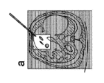
図1は、持続的なCTイメージングを用いた従来の経皮的(面内)介入の3つの段階を示している。針などの介入器具1が患者の中に導入される。介入器具1の遠位端は、開始位置4から、腫瘍などのターゲット構造が位置しているターゲット位置3まで、事前に計算された経路をたどる。この経路では、神経や動脈など、いくつかのリスク位置2が考慮される必要がある。これらのリスク構造に損傷を与えないために、介入器具1は非常に慎重に誘導される必要がある。これは、少なくともリスク構造2の領域において高い画質を要求する。
Figure 1 shows the three stages of a conventional percutaneous (in-plane) intervention with continuous CT imaging. An interventional instrument 1, such as a needle, is introduced into the patient. The distal end of the interventional instrument 1 follows a pre-calculated path from a starting position 4 to a target position 3, where a target structure, such as a tumor, is located. On this path,
図1aでは、介入器具1の遠位端は開始位置4にある。図1bでは、介入器具1の遠位端はリスク位置2に到達している。図1cでは、介入器具1の遠位端はターゲット位置3に到達している。
In FIG. 1a, the distal end of the interventional instrument 1 is at a start position 4. In FIG. 1b, the distal end of the interventional instrument 1 has reached a
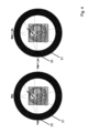
図2は、図1に示されているものと同じ介入手順と同じ3つの段階a、b、cとを示している。図2では、生成される画像の画質に影響を与えるシステムの少なくとも1つのパラメータの自動適応的調整が適用されている。この場合、画像のサイズは関心ボリュームに縮小される。図2aは、リスクが低く、画質に対する要求が低いため、使用される放射線量が少ない区域に挿入した後の介入器具を示している。図2bは、リスク位置2にあるリスク構造のために、精度に対する要求が高い区域における介入器具を示している。画質は、放射線量を増加することによって自動的に向上される。図2cは、介入器具がターゲット位置3にあるターゲット構造に達したことを示している。したがって、画質は、例えば、放射線量を減少することによって再び低下させることができる。
Figure 2 shows the same interventional procedure as shown in Figure 1 with the same three stages a, b, c. In Figure 2, an automatic adaptive adjustment of at least one parameter of the system affecting the image quality of the generated image is applied. In this case, the size of the image is reduced to the volume of interest. Figure 2a shows the interventional instrument after insertion in an area of low risk and therefore low demands on image quality, and therefore low radiation dose used. Figure 2b shows the interventional instrument in an area of high demands on precision due to risk structures in
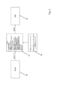
図3は、例えば、FPGAにコンピュータプログラムを実装することによって、医用イメージングシステムに、画質を適応的に調整するためのコンピュータプログラムを埋め込むための実施例を示している。医用イメージングシステムは、検出器30と、例えばX線管である放射線源33とを備える。検出器は、CTシステムで使用されているように、X線検出器であり得る。システムはさらに、固定コンピューティングユニット32と、FPGAの形の追加のコンピューティングユニット31とを備える。固定コンピューティングユニット32は、患者の事前に取得したデータセット内のリスク構造のセグメント化を定義するデータを提供する。検出器30は、患者からの投影データをコンピューティングユニット31に提供する。コンピューティングユニット31は、次のステップを実行することができる。
Figure 3 shows an example for embedding a computer program for adaptively adjusting image quality in a medical imaging system, for example by implementing the computer program in an FPGA. The medical imaging system comprises a
- 反復的再構成
- 投影データおよび再構成されたデータに基づくアーチファクト分析
- 事前に定義されたリスク構造を統合し、介入器具の位置に基づいた高画質である可能性のある領域および低画質である可能性のある領域の特定
- (前述の方法による)線量調節のための決定フィードバックシステムアルゴリズム
コンピューティングユニット31は、放射線源33の電流の電流値を計算し、それらを、放射線源33を制御する制御ユニットに送る。放射線源33の電流を調節することによって、放射線量は、本発明に従って調整され得る。結果として、画質は、患者内の介入器具のさまざまな位置の品質ニーズに従って調整される。
- iterative reconstruction - artifact analysis based on the projection data and the reconstructed data - identification of areas of potential high and low image quality based on the position of the interventional instrument, integrating predefined risk structures - decision feedback system algorithm for dose adjustment (according to the method described above) The
図4は、2つの異なる位置における放射線源33を有する医用イメージングシステムを示している。放射線源33は、患者の周りの回転移動を実行するガントリ51に取り付けられ得る。放射線源33の電流調節は、管の回転角度に依存する患者の不規則な形態のため、基本的に常に可能である。右の図は、関心区域における高画質のための追加の品質調節された電流を示している。
Figure 4 shows a medical imaging system with a
図5は、例えば、X線管である放射線源33と、コリメータ50と、ガントリ51と、放射線検出器30と、コンピューティングユニット31とを有する医用イメージングシステムを示している。さらに、介入手順のためにターゲット構造を有する患者52を配置するためのいくらかのスペースがある。放射線源33は、コリメータ50を介して患者に放射線を提供する。コリメータ50を用いて、患者52の照射される領域のサイズおよび位置が制御され得る。ガントリ51は、例えば、角度αで患者52の周りを回転され得る。放射線源33およびコリメータ50は、ガントリ51に取り付けられているため、同じように回転され得る。さらに、検出器30もガントリ51に取り付けられているため、放射線源33およびコリメータ50と同じように回転される。
Figure 5 shows a medical imaging system with a
例えばFPGAであるコンピューティングユニット31は、いくつかのアルゴリズムを有するコンピュータプログラム53を備える。さらに、コンピューティングユニット31は、コリメータ50を制御する関心ボリューム制御セクション54を備える。さらに、コンピューティングユニット31は、放射線源33の実際の電流の制御を提供する電流調節制御セクション55を備える。加えて、コンピューティングユニット31は、放射線源33および検出器30によって行われる投影の数および投影角度を制御する投影制御セクション56を備える。
The
図3と同様に、検出器30は、コンピューティングユニット31に投影データを提供する。さらに、介入器具1に関する追加情報が、追加のインターフェースを介してコンピューティングユニット31に提供され得る。
As in FIG. 3, the
コンピュータプログラム53では、アーチファクト評価、ノイズ評価、および例えば放射線源33の放射線量の線量調節による画質の自動適応的調整など、いくつかのアルゴリズムが実行される。コンピュータプログラム53は、制御セクション54、55、56に入力データを提供する。
In the
関心ボリューム制御セクション54は、例えば回転角度αに依存する異なるシャッタ位置を提供するなどして、コリメータ50を制御する。電流調節制御セクション55は、放射線源33の電流を制御するための電流値を提供する。投影制御セクション56は、ガントリ制御、放射線源33、および検出器30に、取得パラメータおよびアクティブな角度位置を提供する。
以下に、本願出願の当初の特許請求の範囲に記載された発明を付記する。
[1] 患者(52)の撮影領域に放射線を繰り返し照射し、前記患者(52)の前記照射された領域からデータを取得し、前記取得したデータから画像を生成する医用イメージングシステムにおいて、前記システムは、前記患者(52)の中に導入された介入器具(1)の位置、空間的向き、および/またはタイプに依存した、前記生成される画像の画質に影響を与える前記システムの少なくとも1つのパラメータの自動適応的調整のために構成されることを特徴とする、医用イメージングシステム。
[2] 前記システムは、前記患者(52)に前記システムによって照射される放射線量に影響を与えることによる、前記画質の適応的調整のために構成されることを特徴とする、[1]に記載のシステム。
[3] 前記システムは、前記システムの次のパラメータ、
a)管電流、
b)管電圧、
c)前記患者(52)の前記照射される領域のサイズ、
d)前記患者(52)の前記照射される領域の位置、
e)(回転あたりの)投影の数、
f)露光時間
のうちの1つ、いくつか、またはすべてに影響を与えることによる、前記画質の適応的調整のために構成されることを特徴とする、[1]または[2]に記載のシステム。
[4] 高画質の領域および低画質の領域が、所定のデータベースによって定義されることを特徴とする、[1]から[3]のいずれか一項に記載のシステム。
[5] 前記システムは、前記患者(52)の中への前記介入器具(1)の前記導入中に、開始位置(4)からターゲット位置(3)まで、前記画質を数回変化させるように構成されることを特徴とする、[1]から[4]のいずれか一項に記載のシステム。
[6] 前記システムは、前記介入器具(1)が前記ターゲット位置(3)に到達したときに、前記画質を低下させるように構成されることを特徴とする、[5]に記載のシステム。
[7] 前記システムは、前記介入器具(1)が前記患者(52)内を前記開始位置(4)から前記ターゲット位置(3)まで移動する、事前に計算された経路の関数としての、前記画質の自動適応的調整のために構成されることを特徴とする、[5]または[6]に記載のシステム。
[8] 前記システムは、前記開始位置(4)から前記ターゲット位置(3)までの前記介入器具(1)の前記経路上にある事前に定義されたリスク位置(2)で、前記画質を向上させるように構成されることを特徴とする、[7]に記載のシステム。
[9] 患者(52)の撮影領域に放射線を繰り返し照射し、前記患者(52)の前記照射された領域からデータを取得し、前記取得したデータから画像を生成する医用イメージングシステムにおいて、画質を適応的に調整するためのコンピュータプログラム(53)であって、前記コンピュータプログラム(53)は、前記システムのコンピュータ(31)で前記コンピュータプログラム(53)が実行されると、次のステップ、
a)前記患者(52)の中に導入された介入器具(1)の位置、空間的向き、および/またはタイプを識別する少なくとも1つの値を入力するステップと、
b)前記入力された値の関数として、それによって前記生成される画像の前記画質に影響を与える前記システムの少なくとも1つのパラメータの適応的調整が行われ得る調整値を計算するステップと
c)前記生成される画像の前記画質に影響を与える前記システムの少なくとも1つのパラメータの前記調整を可能にする、前記システムの少なくとも1つの制御コンポーネント(54、55、56)に前記調整値を出力するステップと
を実行するように構成される、コンピュータプログラム(53)。
[10] 前記コンピュータプログラム(53)は、前記介入器具(1)が前記患者(52)内を開始位置(4)からターゲット位置(3)までたどる、事前に計算された経路の特性値を入力し、前記特性値に依存して前記調整値を計算するように構成されていることを特徴とする、[9]に記載のコンピュータプログラム。
A volume of
The invention as originally claimed in the present application is set forth below.
[1] A medical imaging system for repeatedly irradiating an imaging area of a patient (52) with radiation, acquiring data from the irradiated area of the patient (52), and generating an image from the acquired data, characterized in that the system is configured for automatic adaptive adjustment of at least one parameter of the system affecting the image quality of the generated image depending on the position, spatial orientation, and/or type of interventional instrument (1) introduced into the patient (52).
[2] The system of [1], characterized in that the system is configured for adaptive adjustment of the image quality by affecting the amount of radiation delivered by the system to the patient (52).
[3] The system comprises the following parameters of the system:
a) tube current;
b) tube voltage;
c) the size of the irradiated area of the patient (52);
d) the location of the irradiated area of the patient (52);
e) number of projections (per rotation);
f) Exposure Time
3. The system of
[4] The system according to any one of [1] to [3], characterized in that the high image quality areas and the low image quality areas are defined by a predetermined database.
[5] The system according to any one of [1] to [4], characterized in that the system is configured to change the image quality several times during the introduction of the interventional instrument (1) into the patient (52) from a start position (4) to a target position (3).
[6] The system according to [5], characterized in that the system is configured to reduce the image quality when the interventional tool (1) reaches the target position (3).
[7] The system according to [5] or [6], characterized in that the system is configured for automatic adaptive adjustment of the image quality as a function of a pre-calculated path along which the interventional instrument (1) moves within the patient (52) from the start position (4) to the target position (3).
[8] The system according to [7], characterized in that the system is configured to enhance the image quality at predefined risk locations (2) on the path of the interventional instrument (1) from the start location (4) to the target location (3).
[9] A computer program (53) for adaptively adjusting image quality in a medical imaging system for repeatedly irradiating an imaging area of a patient (52) with radiation, acquiring data from the irradiated area of the patient (52), and generating an image from the acquired data, the computer program (53), when executed by a computer (31) of the system, comprises the steps of:
a) inputting at least one value identifying the position, spatial orientation and/or type of an interventional instrument (1) introduced into said patient (52);
b) calculating, as a function of the input values, an adjustment value by which an adaptive adjustment of at least one parameter of the system affecting the image quality of the generated image may be performed;
c) outputting said adjustment value to at least one control component (54, 55, 56) of said system enabling said adjustment of at least one parameter of said system affecting said image quality of said generated image;
A computer program (53) configured to execute the following:
[10] The computer program according to [9], characterized in that the computer program (53) is configured to input characteristic values of a pre-calculated path that the interventional tool (1) will follow in the patient (52) from a start position (4) to a target position (3) and to calculate the adjustment value in dependence on the characteristic values.
Claims (10)
a)管電流、
b)管電圧、
c)前記患者(52)の前記照射される領域のサイズ、
d)前記患者(52)の前記照射される領域の位置、
e)(回転あたりの)投影の数、
f)露光時間
のうちの1つ、いくつか、またはすべてに影響を与えることによる、前記画質の適応的調整のために構成されることを特徴とする、請求項1または2に記載のシステム。 The system is configured to determine the following parameters of the system:
a) tube current;
b) tube voltage;
c) the size of the irradiated area of the patient (52);
d) the location of the irradiated area of the patient (52);
e) number of projections (per rotation);
3. A system according to claim 1 or 2, characterized in that it is arranged for adaptive adjustment of the image quality by affecting one, some or all of the exposure times.
a)前記患者(52)の中に導入された介入器具(1)の位置、空間的向き、および/またはタイプを識別する少なくとも1つの値を入力するステップと、
b)前記入力された値の関数として、それによって前記生成される画像の前記画質に影響を与える前記システムの少なくとも1つのパラメータの適応的調整が行われ得る調整値を計算するステップと
c)前記生成される画像の前記画質に影響を与える前記システムの少なくとも1つのパラメータの前記調整を可能にする、前記システムの少なくとも1つの制御コンポーネント(54、55、56)に前記調整値を出力するステップと
を実行するように構成される、コンピュータプログラム(53)。 9. A medical imaging system according to claim 1, comprising: a computer program (53) for adaptively adjusting image quality, the computer program (53) being adapted to repeatedly irradiate an imaging area of a patient (52) with radiation, acquire data from the irradiated area of the patient (52), and generate an image from the acquired data, the computer program (53) comprising, when executed by a computer (31) of the system, the following steps:
a) inputting at least one value identifying the position, spatial orientation and/or type of an interventional instrument (1) introduced into said patient (52);
b) calculating, as a function of the input values, an adjustment value by which an adaptive adjustment of at least one parameter of the system affecting the image quality of the generated image can be made; and c) outputting said adjustment value to at least one control component (54, 55, 56) of the system enabling the adjustment of at least one parameter of the system affecting the image quality of the generated image.
Applications Claiming Priority (3)
| Application Number | Priority Date | Filing Date | Title |
|---|---|---|---|
| EP20200500.5A EP3981334A1 (en) | 2020-10-07 | 2020-10-07 | Medical imaging system and computer program |
| EP20200500.5 | 2020-10-07 | ||
| PCT/EP2021/077351 WO2022073958A1 (en) | 2020-10-07 | 2021-10-05 | Medical imaging system and computer program |
Publications (2)
| Publication Number | Publication Date |
|---|---|
| JP2023544700A JP2023544700A (en) | 2023-10-25 |
| JP7588904B2 true JP7588904B2 (en) | 2024-11-25 |
Family
ID=72801343
Family Applications (1)
| Application Number | Title | Priority Date | Filing Date |
|---|---|---|---|
| JP2023519320A Active JP7588904B2 (en) | 2020-10-07 | 2021-10-05 | Medical imaging system and computer program |
Country Status (4)
| Country | Link |
|---|---|
| US (1) | US12494280B2 (en) |
| EP (2) | EP3981334A1 (en) |
| JP (1) | JP7588904B2 (en) |
| WO (1) | WO2022073958A1 (en) |
Citations (6)
| Publication number | Priority date | Publication date | Assignee | Title |
|---|---|---|---|---|
| JP2005525891A (en) | 2002-05-17 | 2005-09-02 | ケース ウェスターン リザーヴ ユニヴァーシティ | System and method for adjusting image parameters based on device tracking |
| JP2013542019A (en) | 2010-10-27 | 2013-11-21 | コーニンクレッカ フィリップス エヌ ヴェ | Adaptive imaging and frame rate optimization based on real-time shape sensing of medical devices |
| US20150279031A1 (en) | 2014-04-01 | 2015-10-01 | Case Western Reserve University | Imaging control to facilitate tracking objects and/or perform real-time intervention |
| JP2015530207A (en) | 2012-10-05 | 2015-10-15 | コーニンクレッカ フィリップス エヌ ヴェ | ROI paint |
| JP2020081858A (en) | 2018-11-30 | 2020-06-04 | シーメンス ヘルスケア ゲゼルシヤフト ミツト ベシユレンクテル ハフツング | Apparatus and method for controlling x-ray device |
| US20200275982A1 (en) | 2017-11-13 | 2020-09-03 | Koninklijke Philips N.V. | Autonomous x-ray control for robotic navigation |
Family Cites Families (3)
| Publication number | Priority date | Publication date | Assignee | Title |
|---|---|---|---|---|
| WO2008130380A2 (en) * | 2006-10-25 | 2008-10-30 | Bruce Reiner | Method and apparatus of providing a radiation scorecard |
| EP2083690B1 (en) | 2006-11-10 | 2010-03-31 | Koninklijke Philips Electronics N.V. | Metal artefact prevention during needle guidance under (xper) ct |
| JP6752088B2 (en) * | 2016-09-02 | 2020-09-09 | 株式会社日立製作所 | X-ray fluoroscopy equipment |
-
2020
- 2020-10-07 EP EP20200500.5A patent/EP3981334A1/en not_active Withdrawn
-
2021
- 2021-10-05 US US18/030,568 patent/US12494280B2/en active Active
- 2021-10-05 WO PCT/EP2021/077351 patent/WO2022073958A1/en not_active Ceased
- 2021-10-05 JP JP2023519320A patent/JP7588904B2/en active Active
- 2021-10-05 EP EP21789647.1A patent/EP4225148A1/en active Pending
Patent Citations (6)
| Publication number | Priority date | Publication date | Assignee | Title |
|---|---|---|---|---|
| JP2005525891A (en) | 2002-05-17 | 2005-09-02 | ケース ウェスターン リザーヴ ユニヴァーシティ | System and method for adjusting image parameters based on device tracking |
| JP2013542019A (en) | 2010-10-27 | 2013-11-21 | コーニンクレッカ フィリップス エヌ ヴェ | Adaptive imaging and frame rate optimization based on real-time shape sensing of medical devices |
| JP2015530207A (en) | 2012-10-05 | 2015-10-15 | コーニンクレッカ フィリップス エヌ ヴェ | ROI paint |
| US20150279031A1 (en) | 2014-04-01 | 2015-10-01 | Case Western Reserve University | Imaging control to facilitate tracking objects and/or perform real-time intervention |
| US20200275982A1 (en) | 2017-11-13 | 2020-09-03 | Koninklijke Philips N.V. | Autonomous x-ray control for robotic navigation |
| JP2020081858A (en) | 2018-11-30 | 2020-06-04 | シーメンス ヘルスケア ゲゼルシヤフト ミツト ベシユレンクテル ハフツング | Apparatus and method for controlling x-ray device |
Also Published As
| Publication number | Publication date |
|---|---|
| EP4225148A1 (en) | 2023-08-16 |
| EP3981334A1 (en) | 2022-04-13 |
| US12494280B2 (en) | 2025-12-09 |
| JP2023544700A (en) | 2023-10-25 |
| WO2022073958A1 (en) | 2022-04-14 |
| US20230377720A1 (en) | 2023-11-23 |
Similar Documents
| Publication | Publication Date | Title |
|---|---|---|
| JP6108474B2 (en) | Medical imaging device for providing an image representation to assist in positioning an interventional device | |
| JP4528781B2 (en) | Apparatus and method for adjusting imaging parameters of X-ray apparatus | |
| US10687774B2 (en) | Method and apparatus for visualizing a blood vessel | |
| US10575809B2 (en) | X-ray diagnosis apparatus | |
| JP2017512523A (en) | Dynamic planning method for needle insertion | |
| CN101061520A (en) | Improved representation of real-time location data | |
| US20070183569A1 (en) | Method for graphically following a movement of a medical instrument introduced into an object under examination | |
| JP6334869B2 (en) | X-ray CT system | |
| CN108471997A (en) | Apparatus and method for maintaining image quality while minimizing x-ray dose to a patient | |
| JP7133108B2 (en) | Systems and methods for providing magnified images of patients | |
| CN101277574B (en) | Imaging method and device | |
| JP7588904B2 (en) | Medical imaging system and computer program | |
| JP5279637B2 (en) | Bed positioning system and bed positioning method | |
| EP4633473B1 (en) | Controlled collimation and filtering for dose reduction and image quality in x-ray imaging | |
| JP7408078B2 (en) | Method for detecting structural changes in a patient's body, device for detecting structural changes in a patient's body, and computer program | |
| JP6956554B2 (en) | X-ray diagnostic equipment and image processing program | |
| CN108348768B (en) | Treatment planning devices and radiation therapy systems | |
| US7460642B2 (en) | Method for generating an x-ray image sequence | |
| JP4643544B2 (en) | Bed positioning system, radiation therapy system, and particle beam therapy system | |
| WO2019159273A1 (en) | Radiation therapy device | |
| JP2025081112A (en) | Positioning device, radiation therapy system having the positioning device, and positioning method | |
| WO2023176257A1 (en) | Medical image processing device, treatment system, medical image processing method, and program | |
| JP2022028188A (en) | Medical image processing device, x-ray diagnostic apparatus, and program |
Legal Events
| Date | Code | Title | Description |
|---|---|---|---|
| A621 | Written request for application examination |
Free format text: JAPANESE INTERMEDIATE CODE: A621 Effective date: 20230613 |
|
| A977 | Report on retrieval |
Free format text: JAPANESE INTERMEDIATE CODE: A971007 Effective date: 20240216 |
|
| A131 | Notification of reasons for refusal |
Free format text: JAPANESE INTERMEDIATE CODE: A131 Effective date: 20240402 |
|
| A521 | Request for written amendment filed |
Free format text: JAPANESE INTERMEDIATE CODE: A523 Effective date: 20240620 |
|
| TRDD | Decision of grant or rejection written | ||
| A01 | Written decision to grant a patent or to grant a registration (utility model) |
Free format text: JAPANESE INTERMEDIATE CODE: A01 Effective date: 20241008 |
|
| A61 | First payment of annual fees (during grant procedure) |
Free format text: JAPANESE INTERMEDIATE CODE: A61 Effective date: 20241106 |
|
| R150 | Certificate of patent or registration of utility model |
Ref document number: 7588904 Country of ref document: JP Free format text: JAPANESE INTERMEDIATE CODE: R150 |