JP7581337B2 - Method, system and computer readable storage medium for registering intraoral measurements - Patents.com - Google Patents
Method, system and computer readable storage medium for registering intraoral measurements - Patents.com Download PDFInfo
- Publication number
- JP7581337B2 JP7581337B2 JP2022518298A JP2022518298A JP7581337B2 JP 7581337 B2 JP7581337 B2 JP 7581337B2 JP 2022518298 A JP2022518298 A JP 2022518298A JP 2022518298 A JP2022518298 A JP 2022518298A JP 7581337 B2 JP7581337 B2 JP 7581337B2
- Authority
- JP
- Japan
- Prior art keywords
- images
- neural network
- image
- registration
- individual
- Prior art date
- Legal status (The legal status is an assumption and is not a legal conclusion. Google has not performed a legal analysis and makes no representation as to the accuracy of the status listed.)
- Active
Links
Images


Classifications
-
- G—PHYSICS
- G06—COMPUTING OR CALCULATING; COUNTING
- G06T—IMAGE DATA PROCESSING OR GENERATION, IN GENERAL
- G06T7/00—Image analysis
- G06T7/30—Determination of transform parameters for the alignment of images, i.e. image registration
- G06T7/33—Determination of transform parameters for the alignment of images, i.e. image registration using feature-based methods
- G06T7/344—Determination of transform parameters for the alignment of images, i.e. image registration using feature-based methods involving models
-
- A—HUMAN NECESSITIES
- A61—MEDICAL OR VETERINARY SCIENCE; HYGIENE
- A61B—DIAGNOSIS; SURGERY; IDENTIFICATION
- A61B5/00—Measuring for diagnostic purposes; Identification of persons
- A61B5/0059—Measuring for diagnostic purposes; Identification of persons using light, e.g. diagnosis by transillumination, diascopy, fluorescence
- A61B5/0082—Measuring for diagnostic purposes; Identification of persons using light, e.g. diagnosis by transillumination, diascopy, fluorescence adapted for particular medical purposes
- A61B5/0088—Measuring for diagnostic purposes; Identification of persons using light, e.g. diagnosis by transillumination, diascopy, fluorescence adapted for particular medical purposes for oral or dental tissue
-
- A—HUMAN NECESSITIES
- A61—MEDICAL OR VETERINARY SCIENCE; HYGIENE
- A61B—DIAGNOSIS; SURGERY; IDENTIFICATION
- A61B5/00—Measuring for diagnostic purposes; Identification of persons
- A61B5/72—Signal processing specially adapted for physiological signals or for diagnostic purposes
- A61B5/7235—Details of waveform analysis
- A61B5/7264—Classification of physiological signals or data, e.g. using neural networks, statistical classifiers, expert systems or fuzzy systems
- A61B5/7267—Classification of physiological signals or data, e.g. using neural networks, statistical classifiers, expert systems or fuzzy systems involving training the classification device
-
- A—HUMAN NECESSITIES
- A61—MEDICAL OR VETERINARY SCIENCE; HYGIENE
- A61C—DENTISTRY; APPARATUS OR METHODS FOR ORAL OR DENTAL HYGIENE
- A61C9/00—Impression cups, i.e. impression trays; Impression methods
- A61C9/004—Means or methods for taking digitized impressions
-
- A—HUMAN NECESSITIES
- A61—MEDICAL OR VETERINARY SCIENCE; HYGIENE
- A61C—DENTISTRY; APPARATUS OR METHODS FOR ORAL OR DENTAL HYGIENE
- A61C9/00—Impression cups, i.e. impression trays; Impression methods
- A61C9/004—Means or methods for taking digitized impressions
- A61C9/0046—Data acquisition means or methods
- A61C9/0053—Optical means or methods, e.g. scanning the teeth by a laser or light beam
-
- G—PHYSICS
- G06—COMPUTING OR CALCULATING; COUNTING
- G06N—COMPUTING ARRANGEMENTS BASED ON SPECIFIC COMPUTATIONAL MODELS
- G06N20/00—Machine learning
-
- G—PHYSICS
- G06—COMPUTING OR CALCULATING; COUNTING
- G06N—COMPUTING ARRANGEMENTS BASED ON SPECIFIC COMPUTATIONAL MODELS
- G06N3/00—Computing arrangements based on biological models
- G06N3/02—Neural networks
- G06N3/04—Architecture, e.g. interconnection topology
- G06N3/044—Recurrent networks, e.g. Hopfield networks
- G06N3/0442—Recurrent networks, e.g. Hopfield networks characterised by memory or gating, e.g. long short-term memory [LSTM] or gated recurrent units [GRU]
-
- G—PHYSICS
- G06—COMPUTING OR CALCULATING; COUNTING
- G06N—COMPUTING ARRANGEMENTS BASED ON SPECIFIC COMPUTATIONAL MODELS
- G06N3/00—Computing arrangements based on biological models
- G06N3/02—Neural networks
- G06N3/04—Architecture, e.g. interconnection topology
- G06N3/045—Combinations of networks
-
- G—PHYSICS
- G06—COMPUTING OR CALCULATING; COUNTING
- G06N—COMPUTING ARRANGEMENTS BASED ON SPECIFIC COMPUTATIONAL MODELS
- G06N3/00—Computing arrangements based on biological models
- G06N3/02—Neural networks
- G06N3/04—Architecture, e.g. interconnection topology
- G06N3/0464—Convolutional networks [CNN, ConvNet]
-
- G—PHYSICS
- G06—COMPUTING OR CALCULATING; COUNTING
- G06N—COMPUTING ARRANGEMENTS BASED ON SPECIFIC COMPUTATIONAL MODELS
- G06N3/00—Computing arrangements based on biological models
- G06N3/02—Neural networks
- G06N3/08—Learning methods
-
- G—PHYSICS
- G06—COMPUTING OR CALCULATING; COUNTING
- G06N—COMPUTING ARRANGEMENTS BASED ON SPECIFIC COMPUTATIONAL MODELS
- G06N3/00—Computing arrangements based on biological models
- G06N3/02—Neural networks
- G06N3/08—Learning methods
- G06N3/0895—Weakly supervised learning, e.g. semi-supervised or self-supervised learning
-
- G—PHYSICS
- G06—COMPUTING OR CALCULATING; COUNTING
- G06N—COMPUTING ARRANGEMENTS BASED ON SPECIFIC COMPUTATIONAL MODELS
- G06N3/00—Computing arrangements based on biological models
- G06N3/02—Neural networks
- G06N3/08—Learning methods
- G06N3/09—Supervised learning
-
- G—PHYSICS
- G06—COMPUTING OR CALCULATING; COUNTING
- G06T—IMAGE DATA PROCESSING OR GENERATION, IN GENERAL
- G06T17/00—Three-dimensional [3D] modelling for computer graphics
- G06T17/20—Finite element generation, e.g. wire-frame surface description, tesselation
- G06T17/205—Re-meshing
-
- G—PHYSICS
- G06—COMPUTING OR CALCULATING; COUNTING
- G06T—IMAGE DATA PROCESSING OR GENERATION, IN GENERAL
- G06T3/00—Geometric image transformations in the plane of the image
- G06T3/08—Projecting images onto non-planar surfaces, e.g. geodetic screens
-
- G—PHYSICS
- G06—COMPUTING OR CALCULATING; COUNTING
- G06T—IMAGE DATA PROCESSING OR GENERATION, IN GENERAL
- G06T7/00—Image analysis
- G06T7/0002—Inspection of images, e.g. flaw detection
- G06T7/0012—Biomedical image inspection
-
- G—PHYSICS
- G06—COMPUTING OR CALCULATING; COUNTING
- G06T—IMAGE DATA PROCESSING OR GENERATION, IN GENERAL
- G06T7/00—Image analysis
- G06T7/10—Segmentation; Edge detection
- G06T7/11—Region-based segmentation
-
- G—PHYSICS
- G06—COMPUTING OR CALCULATING; COUNTING
- G06T—IMAGE DATA PROCESSING OR GENERATION, IN GENERAL
- G06T7/00—Image analysis
- G06T7/30—Determination of transform parameters for the alignment of images, i.e. image registration
- G06T7/33—Determination of transform parameters for the alignment of images, i.e. image registration using feature-based methods
-
- G—PHYSICS
- G06—COMPUTING OR CALCULATING; COUNTING
- G06T—IMAGE DATA PROCESSING OR GENERATION, IN GENERAL
- G06T7/00—Image analysis
- G06T7/50—Depth or shape recovery
-
- G—PHYSICS
- G06—COMPUTING OR CALCULATING; COUNTING
- G06T—IMAGE DATA PROCESSING OR GENERATION, IN GENERAL
- G06T2207/00—Indexing scheme for image analysis or image enhancement
- G06T2207/10—Image acquisition modality
- G06T2207/10024—Color image
-
- G—PHYSICS
- G06—COMPUTING OR CALCULATING; COUNTING
- G06T—IMAGE DATA PROCESSING OR GENERATION, IN GENERAL
- G06T2207/00—Indexing scheme for image analysis or image enhancement
- G06T2207/10—Image acquisition modality
- G06T2207/10028—Range image; Depth image; 3D point clouds
-
- G—PHYSICS
- G06—COMPUTING OR CALCULATING; COUNTING
- G06T—IMAGE DATA PROCESSING OR GENERATION, IN GENERAL
- G06T2207/00—Indexing scheme for image analysis or image enhancement
- G06T2207/20—Special algorithmic details
- G06T2207/20081—Training; Learning
-
- G—PHYSICS
- G06—COMPUTING OR CALCULATING; COUNTING
- G06T—IMAGE DATA PROCESSING OR GENERATION, IN GENERAL
- G06T2207/00—Indexing scheme for image analysis or image enhancement
- G06T2207/20—Special algorithmic details
- G06T2207/20084—Artificial neural networks [ANN]
-
- G—PHYSICS
- G06—COMPUTING OR CALCULATING; COUNTING
- G06T—IMAGE DATA PROCESSING OR GENERATION, IN GENERAL
- G06T2207/00—Indexing scheme for image analysis or image enhancement
- G06T2207/30—Subject of image; Context of image processing
- G06T2207/30004—Biomedical image processing
- G06T2207/30036—Dental; Teeth
-
- G—PHYSICS
- G06—COMPUTING OR CALCULATING; COUNTING
- G06T—IMAGE DATA PROCESSING OR GENERATION, IN GENERAL
- G06T2210/00—Indexing scheme for image generation or computer graphics
- G06T2210/41—Medical
Landscapes
- Engineering & Computer Science (AREA)
- Physics & Mathematics (AREA)
- Theoretical Computer Science (AREA)
- Health & Medical Sciences (AREA)
- General Physics & Mathematics (AREA)
- Life Sciences & Earth Sciences (AREA)
- General Health & Medical Sciences (AREA)
- Software Systems (AREA)
- Computer Vision & Pattern Recognition (AREA)
- Artificial Intelligence (AREA)
- Mathematical Physics (AREA)
- Evolutionary Computation (AREA)
- Computing Systems (AREA)
- General Engineering & Computer Science (AREA)
- Data Mining & Analysis (AREA)
- Molecular Biology (AREA)
- Biophysics (AREA)
- Biomedical Technology (AREA)
- Computational Linguistics (AREA)
- Medical Informatics (AREA)
- Veterinary Medicine (AREA)
- Animal Behavior & Ethology (AREA)
- Public Health (AREA)
- Oral & Maxillofacial Surgery (AREA)
- Dentistry (AREA)
- Epidemiology (AREA)
- Radiology & Medical Imaging (AREA)
- Quality & Reliability (AREA)
- Computer Graphics (AREA)
- Geometry (AREA)
- Nuclear Medicine, Radiotherapy & Molecular Imaging (AREA)
- Pathology (AREA)
- Heart & Thoracic Surgery (AREA)
- Surgery (AREA)
- Fuzzy Systems (AREA)
- Physiology (AREA)
- Psychiatry (AREA)
- Signal Processing (AREA)
- Optics & Photonics (AREA)
- Audiology, Speech & Language Pathology (AREA)
Description
関連出願の相互参照
本特許出願は、2019年9月24日に出願された米国特許出願第16/580,084号の利益および優先権を主張するものであり、この米国特許出願は全ての目的のために参照により本明細書に組み込まれる。
CROSS-REFERENCE TO RELATED APPLICATIONS This patent application claims the benefit of and priority to U.S. patent application Ser. No. 16/580,084, filed Sep. 24, 2019, which is incorporated herein by reference for all purposes.
本出願は一般に口腔内測定値をレジストレーションするための方法、システムおよびコンピュータ可読記憶媒体に関し、より詳細にはディープラーニング方法を利用して口腔内測定値をセマンティックにレジストレーションするための方法、システムおよびコンピュータ可読記憶媒体に関する。 This application relates generally to methods, systems, and computer-readable storage media for registering intraoral measurements, and more particularly to methods, systems, and computer-readable storage media for semantically registering intraoral measurements using deep learning methods.
歯科開業医は、軟組織を歯科用カメラの視野外に維持するなどの適当なスキャン技術を使用することによりスキャン中に満足な取得結果を生成するように訓練されている場合がある。軟組織はスキャン中に変形して同じ領域が複数の形状をとり、それによりレジストレーション中に誤差および/または妨害が生じることがある。 Dental practitioners may be trained to produce satisfactory acquisition results during scanning by using appropriate scanning techniques, such as keeping soft tissues out of the field of view of the dental camera. Soft tissues may deform during scanning, causing the same area to take on multiple shapes, which may cause errors and/or interference during registration.
現在では高速点特徴ヒストグラム(FPFH)などの特徴ベースの技術を使用して変換値を計算することができ、それによりスキャンの相対的向きの事前知識がなくてもスキャン/3次元(3D)測定値をレジストレーションすることができる。但しこれらの技術が上手く行くためには、変形可能な領域のスキャン/3D測定を回避することが必要となる場合がある。 Feature-based techniques such as Fast Point Feature Histograms (FPFH) can now be used to compute transformations, allowing scans/three-dimensional (3D) measurements to be registered without prior knowledge of the relative orientation of the scans. However, for these techniques to be successful it may be necessary to avoid scanning/3D measurements of deformable regions.
米国特許第9456754B2号は、歯科用物体の複数の3次元画像を記録する方法であって、3次元画像のそれぞれが当該物体の3D測定データおよび測定された表面の色データを含んでいてもよく、コンピュータ支援記録アルゴリズムを用いて個々の画像を組み合わせて全体的画像にする方法を開示している。それはあたかも本明細書に完全に記載されているかのように、全ての目的のために参照により本明細書に組み込まれる。 U.S. Patent No. 9,456,754 B2 discloses a method for recording multiple three-dimensional images of a dental object, each of which may include 3D measurement data of the object and color data of the measured surfaces, and combining the individual images into an overall image using a computer-aided recording algorithm. It is incorporated herein by reference for all purposes as if fully set forth herein.
米国特許第7698068B2号は、口腔内の少なくとも一部の3次元表面ジオメトリおよび色を表す少なくとも1つの数値実体を提供し、かつその実体を操作してそこから所望のデータを提供することにより口腔に関連する手順に有用なデータを提供する方法を開示している。典型的には数値実体は口腔内の前記一部に関連づけられた表面ジオメトリおよび色データを含み、色データは色相、彩度、値、半透明性および反射率を含む実際の視覚的特性または知覚された視覚的特性を含む。 US Patent No. 7,698,068 B2 discloses a method for providing data useful for a procedure relating to the oral cavity by providing at least one numerical entity representing the three-dimensional surface geometry and color of at least a portion of the intraoral cavity and manipulating the entity to provide desired data therefrom. Typically the numerical entity includes surface geometry and color data associated with said portion of the intraoral cavity, the color data including actual or perceived visual characteristics including hue, saturation, value, translucency and reflectance.
国際公開第2018219800A1号は、点群座標空間において口腔内シーンの一部を表している3D点群データを決定することを含む、口腔内シーンの一部の3D表現を生成および表示するための方法および装置を開示している。口腔内シーンの同じ部分のカラー画像をカメラ座標空間において取得する。前記口腔内シーンの表面を表している画像の領域内にあるカラー画像要素をラベル付けする。 WO2018219800A1 discloses a method and apparatus for generating and displaying a 3D representation of a portion of an intraoral scene, comprising determining 3D point cloud data representative of the portion of the intraoral scene in a point cloud coordinate space. Acquiring a color image of the same portion of the intraoral scene in a camera coordinate space. Labeling color image elements that are within regions of the image representative of a surface of the intraoral scene.
米国特許第9436868B2号は、測定された3次元(3D)物体シーンの高速自動化物体分類を可能にする方法を開示している。物体シーンを光パターンで照明し、異なる空間位相においてパターンによって照明された物体シーンの一連の画像を取得する。 US Patent No. 9,436,868 B2 discloses a method for enabling fast automated object classification of measured three-dimensional (3D) object scenes. The object scene is illuminated with a light pattern and a series of images of the object scene illuminated by the pattern at different spatial phases are acquired.
米国特許第9788917B2号は、自動化歯科矯正診断および治療計画に人工知能を用いるために方法を開示している。この方法は、患者によって動作されるように構成された口腔内イメージャを提供する工程と、歯科矯正状態に関する患者データを受信する工程と、歯科矯正治療から得られた情報を含むかそれへのアクセスを有するデータベースにアクセスする工程と、歯科矯正状態の電子モデルを生成する工程と、少なくとも1つのコンピュータプログラムに、患者データを分析して歯科矯正治療から得られた情報に基づいて歯科矯正状態の少なくとも1つの診断および治療計画を特定するように命令する工程とを含んでもよい。 U.S. Patent No. 9,788,917 B2 discloses a method for using artificial intelligence in automated orthodontic diagnosis and treatment planning. The method may include providing an intraoral imager configured to be operated by a patient, receiving patient data relating to an orthodontic condition, accessing a database containing or having access to information obtained from the orthodontic treatment, generating an electronic model of the orthodontic condition, and instructing at least one computer program to analyze the patient data and identify at least one diagnosis and treatment plan for the orthodontic condition based on the information obtained from the orthodontic treatment.
米国特許出願公開第20190026893A1号は、歯科矯正アライナーの形状を評価するための方法であって、分析画像上に表されている歯に関する歯の属性の値および/または分析画像に関する画像属性の少なくとも1つの値を決定するために、分析画像をディープラーニング装置にサブミットする方法を開示している。 US Patent Publication No. 20190026893A1 discloses a method for evaluating the shape of an orthodontic aligner, where an analysis image is submitted to a deep learning device to determine values of tooth attributes for teeth represented on the analysis image and/or values of at least one image attribute for the analysis image.
PCT出願のPCT/EP2018/055145号は、修復物を構築するための方法であって、歯の状態を歯科用カメラによって測定し、かつ歯の状態の3次元(3D)モデルを生成する方法を開示している。次いでコンピュータ支援検出アルゴリズムを歯の状態の3Dモデルに適用してもよく、修復物の種類、歯の本数または修復物の位置が自動的に決定される。 PCT application PCT/EP2018/055145 discloses a method for constructing restorations, where the condition of the teeth is measured by a dental camera and a three-dimensional (3D) model of the tooth condition is generated. A computer-aided detection algorithm may then be applied to the 3D model of the tooth condition, and the type of restoration, the number of teeth or the position of the restoration is automatically determined.
米国特許出願公開第20180028294A1号は、ディープラーニングを用いた歯科用CAD自動化のための方法を開示している。この方法は、患者の歯列データセットの少なくとも1つの部分を表している患者のスキャンデータを受信する工程と、訓練されたディープニューラルネットワークを用いて患者のスキャンにおいて1つ以上の歯の特徴を特定する工程とを含んでもよい。ここでは設計自動化は、完全なスキャンが生成された後に行ってもよい。しかし、この方法により実際のスキャンプロセスは向上しない。 US Patent Publication No. 20180028294A1 discloses a method for dental CAD automation using deep learning. The method may include receiving patient scan data representing at least a portion of the patient's dentition data set, and identifying one or more dental features in the patient scan using a trained deep neural network, where design automation may occur after the complete scan is generated. However, the method does not improve the actual scanning process.
国際公開第2018158411A1号は、修復物を構築するための方法であって、歯の状態を歯科用カメラによって測定し、かつ歯の状態の3Dモデルを生成する方法を開示している。この場合、コンピュータ支援検出アルゴリズムを歯の状態の3Dモデルに適用し、ここでは修復物の種類および/または少なくとも歯の本数および/または挿入される修復物の位置が自動的に決定される。 WO2018158411A1 discloses a method for constructing restorations, in which the tooth condition is measured by a dental camera and a 3D model of the tooth condition is generated, where a computer-aided detection algorithm is applied to the 3D model of the tooth condition, in which the type of restoration and/or at least the number of teeth and/or the position of the restoration to be inserted is automatically determined.
上記に関連する既存の限界ならびに他の限界は、ディープラーニング方法を利用して口腔内測定値をセマンティックにレジストレーションするための方法、システムおよびコンピュータ可読記憶媒体によって克服することができる。 The existing limitations related to the above, as well as other limitations, can be overcome by a method, system, and computer-readable storage medium for semantically registering intraoral measurements utilizing deep learning methods.
本明細書中の一態様では、本発明は、1つ以上の計算装置によって患者の歯列の個々の画像を受信する工程と、訓練されたディープニューラルネットワークの出力確率値などの1つ以上の出力ラベルを用いて個々の画像におけるレジストレーション誤差の原因を自動的に特定する工程とを含み、出力ラベル/確率値は個々の画像を1つ以上の物体カテゴリに対応する領域にセグメンテーションすることによって得られ、個々の画像は深度画像および/または対応するカラー画像であり、確率値などの1つ以上の出力ラベルに基づいて個々の画像を一緒にレジストレーションして、レジストレーション誤差を全く有しないかレジストレーション誤差を実質的に有しないレジストレーションされた3D画像を形成する工程さらに含む、3次元(3D)レジストレーションのためのコンピュータ実装方法を提供することができる。 In one aspect herein, the invention may provide a computer-implemented method for three-dimensional (3D) registration, comprising receiving, by one or more computing devices, individual images of a patient's dentition, and automatically identifying sources of registration errors in the individual images using one or more output labels, such as output probability values, of a trained deep neural network, the output labels/probability values being obtained by segmenting the individual images into regions corresponding to one or more object categories, the individual images being depth images and/or corresponding color images, and further comprising registering the individual images together based on the one or more output labels, such as the probability values, to form a registered 3D image having no or substantially no registration errors.
本明細書中の別の態様では、本コンピュータ実装方法は以下のうちの1つ以上の組み合わせをさらに含んでもよい。(i)レジストレーションは、深度画像の画素を空間に射影することにより深度画像から点群を生成すること、対応するカラー画像および訓練されたディープニューラルネットワークの出力ラベル/確率値を用いて色値およびラベル/確率値をそれぞれ点群中の各点に割り当てること、および割り当てられたラベル/確率値に基づいて、捨てられたか部分的に含められた点のレジストレーションへの寄与を無くすか減らすように所定の重みを用いて点を捨てるか点群に部分的に含めることにより達成される、(ii)個々の画像は個々の3次元光学画像である、(iii)個々の画像は時間的な一連の画像として受信される、(iv)個々の画像は一対のカラー画像および深度画像として受信される、(v)1つ以上の物体カテゴリは硬い歯肉、軟組織歯肉、歯および歯様物体を含む、(vi)レジストレーション誤差の特定された原因の関連性の指標はその周囲のジオメトリに基づいている、(vii)ディープニューラルネットワークは、畳み込みニューラルネットワーク(CNN)、完全畳み込みニューラルネットワーク(FCN)、回帰型ニューラルネットワーク(RNN)および回帰型畳み込みニューラルネットワーク(回帰型CNN)からなる群から選択されるネットワークである、(vii)1つ以上の計算装置および複数の個々の訓練用画像を用いて各訓練用画像の少なくとも1つの部分における1つ以上の組織を1つ以上のラベル/確率値に対応付けするように、ディープニューラルネットワークを訓練する工程であって、個々の訓練用画像、個々の訓練用画像の画素または個々の訓練用画像のスーパーピクセルをセマンティックデータの種類および/または誤差データの種類に対応する1つ以上のクラスに分類することにより画素レベルで行う訓練する工程をさらに含む、(viii)訓練用画像は3Dメッシュと深度画像およびカラー画像のレジストレーションされた対とを含む、(ix)3Dメッシュはラベル付けされており、当該ラベルは変換関数を用いて3D画像およびカラー画像のレジストレーションされた対に移される。 In another aspect herein, the computer-implemented method may further include a combination of one or more of the following: (i) the registration is achieved by generating a point cloud from the depth image by projecting pixels of the depth image into space, assigning a color value and a label/probability value to each point in the point cloud using the corresponding color image and the output label/probability value of the trained deep neural network, respectively, and discarding or partially including points in the point cloud based on the assigned label/probability value using a predefined weight to eliminate or reduce the contribution of the discarded or partially included points to the registration; (ii) the individual images are individual three-dimensional optical images; (iii) the individual images are received as a temporal sequence of images; (iv) the individual images are received as a pair of color and depth images; (v) the one or more object categories include hard gingiva, soft tissue gingiva, teeth and tooth-like objects; (vi) the indication of relevance of the identified causes of registration error is based on the geometry of its surroundings; (vii) the deep neural network is a convolutional neural network ( CNN), fully convolutional neural network (FCN), recurrent neural network (RNN) and recurrent convolutional neural network (recurrent CNN); (vii) using one or more computing devices and a plurality of individual training images, training the deep neural network to map one or more textures in at least one portion of each training image to one or more labels/probability values, further comprising training at the pixel level by classifying the individual training images, pixels of the individual training images or superpixels of the individual training images into one or more classes corresponding to types of semantic data and/or types of error data; (viii) the training images include a 3D mesh and a registered pair of depth and color images; (ix) the 3D mesh is labeled, and the labels are transferred to the registered pair of 3D and color images using a transformation function.
本発明のさらに別の態様では、コンピュータシステムによって実行された場合にコンピュータシステムに、1つ以上の計算装置によって患者の歯列の個々の画像を受信する工程と、訓練されたディープニューラルネットワークの1つ以上の出力確率値を用いて個々の画像におけるレジストレーション誤差の原因を自動的に特定する工程であって、出力確率値は個々の画像を1つ以上の物体カテゴリに対応する領域にセグメンテーションすることによって得られる工程と含む手順を実行させるプログラムを記憶している非一時的コンピュータ可読記憶媒体であって、個々の画像は深度画像および/または対応するカラー画像であり、1つ以上の出力確率値に基づいて個々の画像を一緒にレジストレーションして、レジストレーション誤差を全く有しないかレジストレーション誤差を実質的に有しないレジストレーションされた3D画像を形成する工程をさらに含む、非一時的コンピュータ可読記憶媒体を提供することができる。 In yet another aspect of the invention, a non-transitory computer-readable storage medium may be provided that stores a program that, when executed by a computer system, causes the computer system to perform a procedure that includes receiving, by one or more computing devices, individual images of a patient's dentition, and automatically identifying sources of registration errors in the individual images using one or more output probability values of a trained deep neural network, the output probability values being obtained by segmenting the individual images into regions corresponding to one or more object categories, the individual images being depth images and/or corresponding color images, and further including registering the individual images together based on the one or more output probability values to form a registered 3D image that has no or substantially no registration errors.
さらに、1つ以上の計算装置によって患者の歯列の個々の画像を受信し、訓練されたディープニューラルネットワークの1つ以上の出力確率値を用いて個々の画像におけるレジストレーション誤差の原因を自動的に特定し、出力確率値は個々の画像を1つ以上の物体カテゴリに対応する領域にセグメンテーションすることによって得られるように構成されたプロセッサを備え、個々の画像は深度画像および/または対応するカラー画像であり、プロセッサは1つ以上の出力確率値に基づいて個々の画像を一緒にレジストレーションしてレジストレーション誤差を全く有しないかレジストレーション誤差を実質的に有しないレジストレーションされた3D画像を形成するように構成されている、3次元(3D)レジストレーションのためのシステムを提供することができる。 Furthermore, a system for three-dimensional (3D) registration can be provided, comprising a processor configured to receive, by one or more computing devices, individual images of a patient's dentition and automatically identify sources of registration errors in the individual images using one or more output probability values of a trained deep neural network, the output probability values being obtained by segmenting the individual images into regions corresponding to one or more object categories, the individual images being depth images and/or corresponding color images, and the processor configured to register the individual images together based on the one or more output probability values to form a registered 3D image having no or substantially no registration errors.
本発明のさらなる態様では、本システムは、畳み込みニューラルネットワーク(CNN)、完全畳み込みニューラルネットワーク(FCN)、回帰型ニューラルネットワーク(RNN)および回帰型畳み込みニューラルネットワーク(回帰型CNN)からなる群から選択されるディープニューラルネットワークを備える。 In a further aspect of the invention, the system comprises a deep neural network selected from the group consisting of a convolutional neural network (CNN), a fully convolutional neural network (FCN), a recurrent neural network (RNN) and a recurrent convolutional neural network (recurrent CNN).
例示的な実施形態は、本明細書において以下に与えられている詳細な説明および添付の図面からより十分に理解されるであろう。図面において同様の要素は同様の符号によって表されており、これらは単に例示として与えられており、従って本明細書中の例示的な実施形態を限定するものではない。 The exemplary embodiments will be more fully understood from the detailed description provided herein below and the accompanying drawings, in which like elements are represented by like reference numerals and are provided merely as examples and therefore not as limitations of the exemplary embodiments herein.
図面の中の異なる図は少なくともいくつかの符号を有する場合があるが、それらは同じ構成要素を特定するために同じである場合があり、そのような構成要素のそれぞれの詳細な説明は各図に関して以下に提供されていない場合がある。 Different figures within the drawings may have at least some reference numbers that may be the same to identify the same components, and detailed descriptions of each of such components may not be provided below with respect to each figure.
本明細書に記載されている例示的な態様に従って、ディープラーニング方法を利用して個々の口腔内測定値をセマンティックにセグメンテーションし、かつ前記個々の口腔内測定値をレジストレーションするための方法、システムおよびコンピュータ可読記憶媒体を提供することができる。 In accordance with exemplary aspects described herein, methods, systems, and computer-readable storage media can be provided for semantically segmenting individual intraoral measurements using deep learning methods and for registering the individual intraoral measurements.
口腔内測定値をレジストレーションするためのシステム A system for registering intraoral measurements
患者の口腔の正確な3D測定は誤ったレジストレーションによって妨げられる場合がある。顎の口腔内測定ではカメラが用いられ、これは顎全体のサブセットのみをキャプチャする単一スキャンを生成し、これらを一緒にレジストレーションして完全なモデルを形成することができる。カメラは手持ち式であってもよく、単一スキャンが得られる正確な位置は一般に分かっていない。これらの単一スキャン(3Dデータ、色データなど)からの情報に基づいて、単一スキャンを共通の基準系(共通の3D座標系)にするための変換を決定する。しかし大部分のレジストレーションプロセスは剛体であるという前提で動作するので、カメラが多くの単一スキャンを高頻度で取得している間に変形する/形状を変える口腔の部分はレジストレーションを歪める場合がある。従って剛体部分のみがレジストレーション向けであるとみなされる。 Accurate 3D measurements of a patient's oral cavity can be hindered by incorrect registration. Intraoral measurements of the jaws use a camera that produces single scans that capture only a subset of the entire jaw, which can be registered together to form a complete model. The camera may be handheld and the exact positions where the single scans are obtained are generally not known. Based on the information from these single scans (3D data, color data, etc.), a transformation is determined to bring the single scans into a common frame of reference (common 3D coordinate system). However, most registration processes work under the assumption that the bodies are rigid, so parts of the oral cavity that deform/change shape while the camera is frequently acquiring many single scans can distort the registration. Hence, only rigid parts are considered for registration.
スキャンは異なる時点で行われるので、特定の組織(特に口腔の軟組織)のジオメトリは、軟組織の変形または移動する異物の存在により、異なるスキャンとスキャンとの間に変化する場合がある。これは3Dデータのマッチングに依存するレジストレーションを妨げる場合がある(例えば、平方和誤差を最小限に抑えることに基づく技術によって生じる典型的な誤差を示す図1を参照)。 Because the scans are taken at different time points, the geometry of certain tissues (especially the soft tissues of the oral cavity) may change between different scans due to soft tissue deformation or the presence of moving foreign bodies. This may hinder registration, which relies on matching 3D data (see, for example, Figure 1, which shows typical errors introduced by techniques based on minimizing the sum-of-squares error).
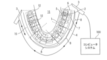
これらの技術の向上は、レジストレーション向けの剛体部分のみを考慮し、かつ無関係の(すなわち非剛体)部分を捨てることにより、あるいはそれらのレジストレーションへの寄与を小さく重み付けすることにより達成してもよく、すなわち歯などの剛体部分/硬組織12がレジストレーション向けであるとみなされる場合、図1に示すようにレジストレーションはロバストであり(iii)、剛体部分/硬組織12の周囲のジオメトリ13a、13bは位置合わせされた状態にあり、逆もまた同様である(iv)。従って「剛体」という用語は本明細書の後で、スキャン手順が行われている期間中に変形する可能性が低い解剖学的構造または解剖学的構造の一部を記述するために使用される場合がある。歯の近くの歯肉はそこに十分に高い力が加えられた場合に変形する場合があるが、これは口腔内スキャナーによるスキャン中に、通常はそのようことにはなり得ない。それを剛体であるとみなすことは妥当な前提であろう。他方、頬の内側はスキャンとスキャンとの間に変形する可能性があり、従って軟組織/軟部15(図2A)とみなしてもよい。
These improvements may be achieved by considering only the rigid parts for registration and discarding irrelevant (i.e. non-rigid) parts or by weighting their contribution to the registration less, i.e. if the rigid parts/
本明細書に記載されているシステムは好ましくは個々の3次元光学画像2などの画像(図2A)を得ることができ、ここでは各3次元光学画像2は好ましくは歯の測定された表面の3D測定データおよび色データを含み、かつ好ましくは直接的な口腔内スキャンにより口腔内において連続的に記録される。これは例えば歯科医院もしくは診療所において行われてもよく、歯科医師または歯科技工士によって行われてもよい。またこれらの画像は一連の記憶された画像から間接的に得てもよい。
The system described herein is preferably capable of obtaining images such as individual three-dimensional optical images 2 (FIG. 2A), where each three-dimensional
好ましくは時間的な一連の画像として得られるこれらの画像を用いて、コンピュータ実装システムはレジストレーション向けであるとみなすことができる画像中の領域を自動的に特定してもよい。これはリアルタイムで行ってもよい。また当然ながら画像は個々の2次元(2D)画像、RGB画像、距離画像(2.5次元、2.5D)、4チャネル画像(RGB-D)であってもよく、ここでは深度および色は完全に位置合わせされていなくてもよく、すなわち深度画像およびカラー画像は異なる期間で取得されてもよい。 Using these images, preferably acquired as a temporal sequence of images, the computer-implemented system may automatically identify regions in the images that can be considered suitable for registration. This may be done in real time. It is also understood that the images may be individual two-dimensional (2D) images, RGB images, distance images (2.5 dimensional, 2.5D), four-channel images (RGB-D), where the depth and color may not be perfectly aligned, i.e. the depth and color images may be acquired at different time periods.
スキャンプロセスでは複数の個々の画像を生成してもよく、次いで少なくとも2枚の個々の画像の一連の画像8または複数の一連の画像8を組み合わせて全体的/大域的3D画像10を形成してもよい(図4)。より具体的には、図2Aに示すように矩形の形態で示されている個々の3次元光学画像2は、測定中に測定経路4に沿って物体1に対して移動させることができるスキャナー/歯科用カメラ3によって得てもよい。いくつかの実施形態では、測定経路はどんな任意の経路であってもよく、すなわち異なる方向から測定を行ってもよい。歯科用カメラ3は、例えば縞投影法を用いて物体1を測定する手持ち式カメラであってもよい。3D測定の他の方法は当業者によって理解され得る。破線で示されている第1の画像6と第2の画像7との間の第1の重なり合っている領域5は、コンピュータを用いて記録条件が満たされているか否かを決定するために確認され、満たされている場合に3次元光学画像2を組み合わせて/一緒にレジストレーションして、大域的3D画像10を形成してもよい。
The scanning process may generate multiple individual images, and then a series of
記録条件としては、特徴的ジオメトリの適切なサイズ、適切なうねり、適切な粗さおよび/または適切な数および配置が挙げられる。しかし従来のコンピュータを、レジストレーション誤差の原因およびそれらを防ぐ方法を決定するようにプログラムするのは難しい場合がある。全ての生じ得るシナリオを網羅するようなレジストレーションまたはセグメンテーション方法のために使用される手動プログラミング特徴は、特に高頻度の測定を考えると行うのに飽き飽きする場合がある。これは特に画像全体のコンテキストを考慮しなければならない場合に特に当てはまる。機械学習法、特にニューラルネットワークおよび正しい訓練用データを使用することで、この問題をより有効に解決することができる。他方ニューラルネットワークは、レジストレーション誤差の原因を認識し、かつ単一スキャン/単一3D測定値からのデータをセマンティックにセグメンテーションし、かつこれらの口腔領域がレジストレーション向けであるとみなすことができるか否かを決定するように学習してもよい。この目的のために、セグメンテーションの異なる物体/物体カテゴリのためのラベルを、限定されるものではないが(i)硬組織(歯、クラウン、ブリッジ、歯の近くの硬い歯肉および他の歯様物体など)、(ii)軟組織(舌、頬、軟らかい歯肉など)および(iii)器具/口腔内に施用される使い捨て器具(鏡、スキャンボディ、コットンロール、ブラケットなど)を含むように定めてもよい。当然ながらグレア部分21(図2B、明るい光によって引き起こされる)などの他の定義を適宜追加してもよい。セグメンテーションは色に基づいていてもよく、この色はコンテキストアウェアで解釈されてもよく、すなわちレジストレーション誤差の潜在的な原因の関連性の指標は、その周囲のジオメトリ13a、13bに基づいていてもよい。さらにセグメンテーションは、色データの単一の画素または個々の3次元光学画像2のより大きい領域に基づいていてもよい。
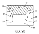
The recording conditions include the appropriate size, waviness, roughness and/or number and arrangement of characteristic geometries. However, it can be difficult to program conventional computers to determine the causes of registration errors and how to prevent them. Manual programming features used for registration or segmentation methods that cover all possible scenarios can be tedious to do, especially considering the high frequency of measurements. This is especially true when the context of the entire image must be considered. Machine learning methods, especially neural networks and the right training data, can solve this problem more effectively. On the other hand, neural networks may learn to recognize the causes of registration errors and to semantically segment data from a single scan/single 3D measurement and determine whether these oral cavity regions can be considered for registration. For this purpose, labels for different objects/object categories of the segmentation may be defined to include, but are not limited to, (i) hard tissues (such as teeth, crowns, bridges, hard gums near teeth and other tooth-like objects), (ii) soft tissues (such as tongue, cheeks, soft gums) and (iii) appliances/disposable appliances applied in the oral cavity (such as mirrors, scanbodies, cotton rolls, brackets). Of course, other definitions such as glare areas 21 (FIG. 2B, caused by bright light) may be added as appropriate. The segmentation may be based on color, which may be interpreted in a context-aware manner, i.e., an indication of the relevance of a potential source of registration error may be based on its surrounding
クラウン、歯または歯の近くの硬い歯肉は剛体であるため、正しいセグメンテーションを考慮するレジストレーションアルゴリズムによってレジストレーション誤差を無くすか実質的に減らしてもよい。さらにコットンロールのような付属品によって導入されるクラッターを除去することにより、クリーンアップされた3Dモデルを生成してもよく、前記クリーンアップされた3Dモデルは歯の治療のために関連するデータのみを含む。 Since the crown, tooth or hard gums near the tooth are rigid bodies, registration errors may be eliminated or substantially reduced by a registration algorithm that takes into account correct segmentation. Furthermore, clutter introduced by accessories such as cotton rolls may be removed to generate a cleaned 3D model, which contains only data relevant for dental treatment.
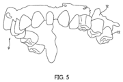
従って本システムは、3次元光学画像2におけるレジストレーション誤差の原因を自動的に認識し、かつ好ましくはリアルタイムでそれらの原因がレジストレーションに寄与するのを防止するように、複数の訓練用データセットを用いてディープニューラルネットワークなどのニューラルネットワークを訓練してもよい。従って、大域的3D画像10(図4)に伝播された誤ったレジストレーション(図1、iv)を、図5の補正された大域的3D画像9に示されているように減らすか無くしてもよく、かつ/またはスキャンフローを、誤ったレジストレーションによって引き起こされる妨害を少なくするか無くすことにより向上させてもよい。
The system may therefore train a neural network, such as a deep neural network, with multiple training data sets to automatically recognise sources of registration errors in the 3D
また本システムはデータをセマンティックに(コンテキストアウェアで、すなわちコンテキストは適当な補正方法を選択するために重要であり得る。例えば歯に近い歯肉を硬組織12とみなしてもよく、歯から遠い歯肉は軟組織15とみなしてもよい)、特定およびラベル付けしてもよい。
The system may also identify and label data semantically (context-aware, i.e., context may be important for selecting the appropriate correction method; for example, gums close to the teeth may be considered as
さらに本システムは補正措置を決定し、かつ/またはレジストレーション誤差の原因を検出した際に前記決定した補正措置を適用してもよい。患者の頬または唇の変形または移動により軟組織の変形を生じさせ、従って図1(iv)に示されているような欠陥のある記録を生じさせる場合があるため、例えば個々の3次元光学画像2において軟組織15に対して高い割合の硬組織12が存在する場合、軟組織15よりもさらに大きく硬組織12に重み付けすることが有利であり得る。しかし画像中の硬組織15の割合が低い場合、記録の品質を高めるために軟組織15にさらに大きく重み付けしてもよい。一例として、図2Bは、2本の真ん中の歯を失っているため、硬組織12を有する第1の領域が個々の3次元光学画像2の全領域の約10%であり、かつ軟組織15を有する第2の領域が画像の全領域の約50%である画像を示す。組織のいずれにも割り当てることができない残りの第3の領域が約40%となり、硬組織12を有する第1の領域の割合は30%の所定の閾値未満に低下する。従って第1の領域の減少と共に第2の重み係数が大きくなるように、硬組織12を有する第1の領域を第1の重み係数により重み付けし、かつ軟組織を有する第2の領域を第2の重み係数により重み付けする。例えば硬組織12を有する第1の領域のための第1の重み係数は1.0であってもよく、軟組織15を有する第2の領域のための第2の可変重み係数は所定の閾値を超えた場合に0.5であってもよく、第1の領域の減少に伴って1.0まで大きくしてもよい。第2の重み係数の第1の領域への依存は指数関数または線形関数などの任意の関数に従って定めてもよく、硬組織および軟組織は本明細書に記載されているニューラルネットワークを用いてセグメンテーションしてもよい。
Furthermore, the system may determine corrective measures and/or apply said corrective measures upon detecting a cause of registration error. For example, if there is a high proportion of
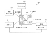
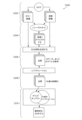
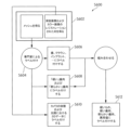
図3Aは、一実施形態に係る患者の歯列の個々の3次元光学画像2から歯科用情報を認識するためのシステム200のブロック図を示す。システム200は歯科用カメラ3、訓練モジュール204、画像レジストレーションモジュール206、コンピュータシステム100およびデータベース202を備えていてもよい。別の実施形態では、データベース202、画像レジストレーションモジュール206および/または訓練モジュール204はコンピュータシステム100の一部であってもよく、かつ/または補正措置に基づいて歯科用カメラ3のパラメータを直接および/または間接的に調整できるものであってもよい。コンピュータシステム100は、少なくとも1つのコンピュータプロセッサ122、ユーザインタフェース126および入力装置130も備えていてもよい。コンピュータプロセッサは様々な要求を受信してもよく、かつ記憶装置に記憶されている適当な命令をメモリにロードし、次いでロードした命令を実行してもよい。コンピュータシステム100は、ソフトウェアおよびデータをコンピュータシステム100と外部装置との間で転送するのを可能にする通信インタフェース146も備えていてもよい。
3A shows a block diagram of a
コンピュータシステム100は、歯科用カメラ3などの外部装置またはユーザ(図示せず)からレジストレーション要求を受信してもよく、かつセマンティックレジストレーションのために適当な命令をロードしてもよい。好ましくはコンピュータシステムは独立して、要求を待つことなく個々の3次元光学画像2を受信した際に画像をレジストレーションしてもよい。
The
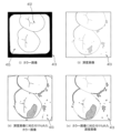
一実施形態では、コンピュータシステム100はデータベース202(これは例えば複数の個々の3次元光学画像2を含んでいてもよい)からの複数の訓練用データセットを使用して、訓練モジュール204の一部であってもよい1つ以上のディープニューラルネットワークを訓練してもよい。図3B(i~iv)は、カラー画像、深度画像、深度画像に対応付けされたカラー画像およびカラー画像に対応付けされた深度画像をそれぞれ含む訓練のために使用される例示的画像を示す。畳み込みニューラルネットワーク(CNN)は局所近傍で動作するので、これらの画像を対応付けすることはCNNにとって良好なことであり得る。故にRGB画像および深度画像の領域は同じ2D画素座標で表されていてもよい。例えば、深度画像のRGB画像への対応付けまたはその逆は、RGB画像および生成された画像において同じ2D画素座標を有する画素が空間中の同じ点に対応するような画像を生成することを意味する。通常これは、ピンホールカメラモデルの適用および移動(レジストレーションアルゴリズムによって決定される)に順応する変換を含む。動きを補償するための工程は省略してもよく、そのネットは、小さいことが期待される生じるオフセットに対処できるものと期待することができる。いくつかの実施形態ではシステム200は、畳み込みニューラルネットワーク(CNN)、完全畳み込みニューラルネットワーク(FCN)、回帰型ニューラルネットワーク(RNN)および回帰型畳み込みニューラルネットワーク(回帰型CNN)などの各種ディープラーニングニューラルネットワークを含むニューラルネットワークモジュール(図示せず)を備えていてもよい。例示的な完全畳み込みニューラルネットワークについては、Jonathan Longら,「セマンティックセグメンテーションのための完全畳み込みネットワーク(Fully Convolutional Networks for Semantic Segmentation)」(2015年3月8日)という表題の刊行物に記載されており、これはあたかも本明細書に完全に記載されているかのように、その全体が参照により本明細書に組み込まれる。従って完全畳み込みニューラルネットワーク(画素ごとのセグメンテーションのために使用される効率的な畳み込みネットワークアーキテクチャ)は、回帰型モデルを用いることによりRGB(D)画像または一連のRGB(D)画像をセグメンテーションするように訓練してもよい。単純な順伝播型ネットワークとは対照的に、回帰型モデルを使用してもよい。従って当該ネットワークは、その現在の活性化を全ての前の入力に応じた状態とみなすことができるように、次の前向き計算のための入力として層の出力を受信してもよく、このようにしてシーケンスの処理を可能にする。さらに例示的な回帰型CNNモデルは、Courtney J.Spoererら,「回帰型畳み込みニューラルネットワーク:生物学的物体認識のより良好なモデル(Recurrent Convolutional Neural Networks:A Better Model of Biological Object Recognition)」(Front.Psychol,2017年9月12日)という表題の刊行物に記載されており、これはあたかも本明細書に完全に記載されているかのように、その全体が参照により本明細書に組み込まれる。
In one embodiment, the
ニューラルネットワークへの訓練用データセットおよび/または入力は前処理されていてもよい。例えば、3D測定値と共に色データを処理するために、較正(カメラモデルのパラメータの決定など)を適用してカラー画像を3D表面と位置合わせしてもよい。さらに合成回転、拡大縮小などの標準的なデータ拡張手順を訓練用データセットおよび/または入力に適用してもよい。 The training dataset and/or inputs to the neural network may be pre-processed. For example, calibration (such as determining camera model parameters) may be applied to align color images with 3D surfaces in order to process color data together with 3D measurements. Furthermore, standard data augmentation procedures such as compound rotation, scaling, etc. may be applied to the training dataset and/or inputs.
訓練モジュール204は、ラベルを有する訓練用データセットを使用してディープニューラルネットワークの学習プロセスを監視してもよい。ラベルを使用してデータ点に重み付けしてもよい。訓練モジュール204は逆に、ラベル付けされていない訓練用データセットを使用して生成ディープニューラルネットワークを訓練してもよい。
The
例示的な一実施形態では、ディープニューラルネットワークを訓練してレジストレーション誤差の原因を検出するために、上に記載されている組織の種類および物体のカテゴリを有する複数の現実の個々の3次元光学画像データセットを使用してもよい。別の例では、ディープニューラルネットワークを訓練してセマンティックデータ(例えば歯の近くの硬い歯肉)を認識するために、1本以上の歯の近くの1つ以上の硬い歯肉領域および1本以上の歯から離れている1つ以上の軟らかい歯肉領域を有する現実の歯科用患者からの別の複数の訓練用データセットを選択して訓練用データセット群を形成する。従ってデータベース202は、例えば各物体カテゴリのための1つの群および/または各セマンティックデータ型のための1つの群である異なる訓練用データセット群を含んでいてもよい。
In an exemplary embodiment, multiple real individual 3D optical image data sets having the tissue types and object categories described above may be used to train the deep neural network to detect sources of registration errors. In another example, to train the deep neural network to recognize semantic data (e.g., hard gingiva near teeth), another multiple training data sets from real dental patients having one or more hard gingiva areas near one or more teeth and one or more soft gingiva areas away from one or more teeth are selected to form a training data set group. Thus, the
いくつかの実施形態では、コンピュータシステム100が1つ以上の予め訓練されたディープニューラルネットワークを容易に使用してレジストレーション誤差の原因を検出することができるように、訓練モジュール204は、データベース204からの訓練用データセットを用いて1つ以上のディープニューラルネットワークを予め訓練してもよい。次いでそれは、検出された原因に関する情報および/または個々の3次元光学画像2を好ましくは自動的に、かつリアルタイムで画像レジストレーションモジュール206に送信してもよく、そこではレジストレーションの前にレジストレーション誤差の原因が考慮される。
In some embodiments, the
データベース204は、対応する個々の3次元光学画像2と共にディープニューラルネットワークおよび特定された原因に関連するデータも記憶していてもよい。さらにコンピュータシステム100は表示装置126および入力装置130を有していてもよく、これらを用いてユーザは要求をサブミットすること、および訓練中に特定されたレジストレーション誤差の原因を受信して再考することなどの機能を行ってもよい。
The
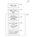
図8に示すように訓練プロセス(S600)の例示的な一実施形態では、現場における現実の事例を表している画像を収集すること(工程S602)によってラベルを生成してもよい。これらの事例には、メッシュ(3D三角メッシュなど)および単一画像(深度画像およびカラー画像のレジストレーションされた対)が含まれていてもよい。これらのメッシュにおいて歯を切り取ることができる専門家によって、当該メッシュをセグメンテーションしてもよい。次いで切り取られたメッシュにラベル付けしてもよい(工程S604)。それらを歯、クラウン、インプラントまたは他の歯様物体(工程S606)あるいは硬い歯肉および軟らかい歯肉(工程S608)としてラベル付けしてもよい。さらに口腔の一部ではないカメラ3の光路における3D点などの異常値をラベル付けしてもよい(工程S610)。当該メッシュのラベル付けを単一画像の画素に移し、このようにして訓練プロセスで行われる作業の量を減らす。工程S612において、工程S606、工程S608および工程S610からの情報を組み合わせることにより全ての最終的なラベルを決定してもよい(工程S612)。さらに単一画像を一緒に位置合わせした変換が分かっているので(それらはレジストレーションされているため)、これらの最終的なラベルを切り取られたメッシュから単一画像に移してもよい。このようにして、メッシュのカット/スライスにより多くの画像にすぐにラベル付けしてもよい。
In an exemplary embodiment of the training process (S600) as shown in FIG. 8, labels may be generated by collecting images (step S602) that represent real cases in the field. These cases may include meshes (such as 3D triangular meshes) and single images (registered pairs of depth and color images). These meshes may be segmented by an expert who can crop teeth in them. The cropped meshes may then be labeled (step S604). They may be labeled as teeth, crowns, implants or other tooth-like objects (step S606) or hard and soft gingiva (step S608). Additionally, outliers such as 3D points in the optical path of
システム200の他の実施形態は、異なる構成要素および/またはさらなる構成要素を備えていてもよい。さらにこれらの機能は、本明細書に記載されているものとは異なるように当該構成要素に分散されていてもよい。
Other embodiments of
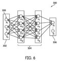
図6は本発明の一実施形態に係るディープニューラルネットワーク300の構造を示すブロック図を示す。それは入力層302、1つ以上の隠れ層304および出力層306を含むいくつかの層を有していてもよい。各層は小さい円によって示されている1つ以上のノード308からなっていてもよい。情報は入力層302から出力層306へ、すなわち左から右方向に流れてもよいが、他の実施形態ではそれは右から左であったりそれらの両方であったりしてもよい。例えば回帰型ネットワークは、一連の画像8において新しいデータを処理する場合に前に観察されたデータを考慮してもよく(例えば前の画像を考慮して現在の画像をセグメンテーションしてもよく)、非回帰型ネットワークは個々に新しいデータを処理してもよい。
Figure 6 shows a block diagram illustrating the structure of a deep
ノード308は入力および出力を有してもよく、入力層308のノードはパッシブであってもよく、これはそれらがデータを修正しなくてもよいことを意味する。例えば入力層302のノード308はそれぞれ、それらの入力に対して単一の値(例えば画素値)を受信し、かつそれらの複数の出力への値を複製してもよい。逆に隠れ層304および出力層306のノードはアクティブであってもよく、従ってデータを修正することができる。例示的な構造では、入力層302からの各値を複製して隠れノードの全てに送信してもよい。隠れノードに到着した値に重みを掛けてもよく、これは隠れノードのそれぞれに関連づけられた所定の数のセットであってもよい。次いで重み付けされた入力を合計して、単一の数を生成してもよい。
本発明に係る一実施形態では、ディープニューラルネットワーク300は物体カテゴリを検出する際に入力として個々の3次元光学画像2の画素を使用してもよい。個々の3次元光学画像2はカラー画像および/または深度画像であってもよい。本明細書では、入力層302中のノードの数は個々の3次元光学画像2中の画素の数に等しくてもよい。
In one embodiment according to the present invention, the deep
例示的な一実施形態では、1つのニューラルネットワークを全ての物体カテゴリのために使用してもよく、別の実施形態では異なるネットワークを異なる物体カテゴリのために使用してもよい。別の例ではディープニューラルネットワーク300は、環境光によって引き起こされるものなどの物体カテゴリを検出した際に、個々の画素の代わりに個々の3次元光学画像2を分類/ラベル付けしてもよい。さらなる実施形態では、当該画像は4番目ごとの画素などのサブサンプリングされた入力であってもよい。
In one exemplary embodiment, one neural network may be used for all object categories, while in another embodiment, different networks may be used for different object categories. In another example, the deep
さらに別の実施形態では、ディープニューラルネットワーク300は入力として、カラー画像、深度測定値、加速度などの歯科用カメラ3によって取得された複数のデータならびに露光時間、アパーチャなどの装置パラメータを有していてもよい。ディープニューラルネットワークはラベルを出力してもよく、このラベルは例えば特定の物体カテゴリに属している各画素入力の1つ以上の確率値を含む確率ベクトルであってもよい。例えば出力は確率値を含む確率ベクトルを含んでもよく、ここでは最も高い確率値は硬組織12の位置を定めてもよい。またディープニューラルネットワークは、どんな確率も含まないラベル値のマップを出力してもよい。各分類のためにディープニューラルネットワークを作成することができるが、これは必要でない場合がある。
In yet another embodiment, the deep
口腔内測定においてレジストレーションするための方法 Method for registration in intraoral measurements
ここまで図3Aのシステム200について説明してきたが、次に本明細書中の例示的な実施形態の少なくともいくつかに係るプロセスS400を示す図7Aを参照する。
Having now described the
プロセスS400は、訓練用データセット中の目的の領域を得て所定のラベルでラベル付けすることにより開始してもよい(工程S402)。例えば図3B(i)に示されているサンプル画像413上のサンプル軟組織415は軟組織としてラベル付けしてもよい。図3B(i)に示されているサンプル画像413上のサンプル硬組織412は硬組織としてラベル付けしてもよい。訓練用画像のラベル付けは、例えば目的の点に対応する画像上にドットを設定することによりデジタルで行ってもよい。
Process S400 may begin by obtaining and labeling regions of interest in the training data set with predefined labels (step S402). For example, sample
セマンティクスを個々の3次元光学画像2に割り当てるために、訓練用データにラベル付けしてもよい。これは、色もしくは深度情報のために画素ごとのレベルで行ってもよい。あるいは単一画像のために、完全な3Dモデルのメッシュをカットして対応する画素ごとのラベルを計算してもよい。さらにラベル付けプロセスを自動化することができるように、前記メッシュをセグメンテーションしてもよい。それ以外のものにはどれにもラベルを割り当てずに、これらのラベルにより歯、頬、唇、舌、歯肉、充填物、セラミックを区別してもよい。レジストレーションに無関係なものは、頬、唇、舌、グレアおよびラベル付けされていないデータであってもよい。
The training data may be labelled in order to assign semantics to the individual 3D
レジストレーション誤差原因ラベルを個々の3次元光学画像2に割り当てるために、訓練用データにもラベル付けしてもよい。これも例えば画像もしくは深度情報のために画素ごとのレベルで行ってもよい。例えば硬組織12および軟組織15および/または器具/口腔内に施用される使い捨て器具などのために、訓練用データに画素レベルでラベル付けしてもよい。
The training data may also be labeled in order to assign registration error cause labels to the individual 3D
セマンティックラベルはレジストレーション誤差の原因のためのマーカー、例えば「硬組織+グレア」、「硬組織に近い軟組織」、「舌+硬組織」などのラベルと重なり合っていてもよく、これらのラベルを「頬+グレア」などの他のラベルと区別してもよい。 Semantic labels may overlap with markers for sources of registration error, such as labels like "hard tissue + glare", "soft tissue close to hard tissue", and "tongue + hard tissue", and these labels may be distinguished from other labels like "cheek + glare".
このラベル付けまたは分類された画像のセットを用いて、ネットワークが新しい画像を自らセグメンテーションすることができるネットワーク配線を行うことができるように、ディープニューラルネットワーク300を構築してラベル付けされた画像を供給し、それによりネットワークがそれから「学習する」のを可能にしてもよい。
With this set of labeled or classified images, a deep
画素ごとのベースでの分類を含むセグメンテーションの別のオプションとして、セグメンテーションは画素ごとのレベルよりも僅かに高いレベルで(すなわち「スーパーピクセルごとの」レベルで、すなわち「スーパーピクセルごと」は画像の通常の画素よりも大きい画像の部分である)、分類および訓練することを含んでもよい。 As an alternative option to segmentation involving classification on a pixel-by-pixel basis, segmentation may involve classifying and training at a level slightly higher than the pixel-by-pixel level (i.e., at the "superpixel-by-superpixel" level, where a "superpixel" is a portion of an image that is larger than a regular pixel in the image).
プロセスS400の命令およびアルゴリズムは、1つ以上の出力ラベル/確率値に基づいて1つ以上の欠陥15を検出するために訓練用データセットを用いて1つ以上のディープニューラルネットワークを訓練する(工程S404)ためにコンピュータシステム100のメモリに記憶されていてもよく、かつプロセッサ122によってロードされて実行されてもよい。例えばグレアに対応する確率ベクトルの確率値の1つが90%である場合、ニューラルネットワークは、個々の3次元光学画像2におけるレジストレーション誤差の原因の1つとしてグレア部分21を検出してもよい。
The instructions and algorithms of process S400 may be stored in the memory of the
訓練する工程は1回、複数回または断続的に行ってもよい。また訓練する工程は「半教師あり」または「自己教師あり」であってもよい。例えば第1の訓練する工程後に、ディープニューラルネットワークは前に見たことのない画像および出力を受信または取得してもよく、かつネットワークが好ましくは自ら動作して最終的に人間の助けなしに画像を分類することができるように、対応するフィードバックを与えてもよい。従って個々の3次元光学画像2の一連の画像8がディープニューラルネットワーク300に入力された場合に、ディープニューラルネットワークがそれらの画像の部分が属するカテゴリを示す各画像のために得られるラベル/確率ベクトルを返すことができるように、ディープニューラルネットワーク300を訓練してもよい。
The training step may be performed once, multiple times or intermittently. The training step may also be "semi-supervised" or "self-supervised". For example, after a first training step, the deep neural network may receive or acquire images and outputs that it has not seen before, and may provide corresponding feedback so that the network can preferably operate on its own and ultimately classify the images without human help. Thus, the deep
訓練する工程後に、ディープニューラルネットワークは歯科用カメラ3から個々の3次元光学画像の一連の画像8を取得または受信してリアルタイムでセグメンテーションしてもよく(工程S406)、それらの画像におけるレジストレーション誤差の原因を検出してもよい(工程408)。前記原因の検出時に画像レジストレーションモジュール206は、レジストレーション誤差の検出された原因がレジストレーションプロセスに寄与しないことを保証する(工程S410)ことにより、セグメントのための所定の重みに基づいてそれらの画像を一緒にレジストレーションしてもよい。図7Aの工程S406~S410は本明細書の後で考察されている図7Bのフローチャートにも含められている。
After the training step, the deep neural network may acquire or receive a sequence of
図7BはプロセスS400のサブセットであってもよいプロセスS500を示す。プロセスS500は工程S502で開始してもよく、ここではカラー画像および/または深度画像を歯科用カメラ3から得る。工程S504では、カラー画像および深度画像を訓練されたディープニューラルネットワーク300への入力として使用し、物体カテゴリに属しているセグメントの確率を示すラベル付けされた画像の対応する出力を得る。両方の画像を使用することにより、異なるラベル間で識別するのをさらに容易にしてもよい。例えば深度および色を用いる場合よりも色に基づいて軟質の歯肉と硬い歯肉とを区別するようにネットを訓練することは難しいものと予想するであろう。これらの画像は「対応付け」されて、その画像がラベル付けされている場合があるため、大きな違いが生じない場合がある。一実施形態では、1つはラベル付け/セグメンテーションされており、他はセグメンテーションを決定するために単にさらなる特徴を提供してもよい。当該実施形態に応じて、得られるラベル付けされた画像と深度画像との間または得られるラベル付けされた画像とカラー画像との間のいずれかにおいて1対1の対応が存在してもよい。ラベル付けされた画像は、深度/カラー画像および当該ラベルのためのチャネルと同じ横方向解像度を有していてもよい。工程S506では、深度画像の各画素を空間に射影することにより深度画像から点群を生成してもよい。これらの点群中の各点に、カラー画像からの色値およびラベル付けされた画像からの確率ベクトルを割り当ててもよい。一実施形態では、ラベル付けされた画像、点群および得られるメッシュは全てそれらに割り当てられたラベル、ラベルの確率または確率ベクトルを有していてもよい。工程S508では、各入力された点群が既にレジストレーションされた点群にレジストレーションされるように、より多くの画像および対応する出力ラベルを得る。本明細書では、軟組織であるという高い確率を有する点(例えば所定の閾値を超えるか所定の方法で確率の関数として重み付けされている)を捨てるか、硬組織であるという高い確率を有する他の点よりも小さく重み付けする。工程S510では、平均的な位置、色および確率になるまで、入力された点群中の各点を対応するグリッドセルに追加する。次いで軟組織15、硬組織12および/またはあらゆる他の物体カテゴリのために所定の重みを使用することにより、単一画像を互いに位置合わせする変換を最適化してもよい。変換が変わった場合にグリッドサンプラーにおける入力をそれに応じて更新してもよい。当然ながら本明細書を考慮に入れて、図7Bとは異なる他の実施形態を達成してもよい。
7B shows process S500, which may be a subset of process S400. Process S500 may begin with step S502, where a color image and/or depth image is obtained from
口腔内測定値をレジストレーションするためのコンピュータシステム A computer system for registering intraoral measurements
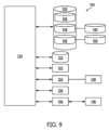
ここまで図7Aおよび図7Bのプロセスについて説明してきたが、次に本明細書中の例示的な実施形態の少なくともいくつかに従って用いることができるコンピュータシステム100のブロック図を示す図9を参照する。この例示的なコンピュータシステム100に関して様々な実施形態が本明細書に記載されている場合があるが、本明細書を読んだ後に、他のコンピュータシステムおよび/またはアーキテクチャを用いて本発明を実行する方法が当業者には明らかになるであろう。
Having now described the processes of Figures 7A and 7B, reference is now made to Figure 9, which illustrates a block diagram of a
コンピュータシステム100は訓練モジュール204、データベース202および/または画像レジストレーションモジュール206を備えていてもそれらとは分離されていてもよい。これらのモジュールはハードウェア、ファームウェアおよび/またはソフトウェアに実装されていてもよい。当該コンピュータシステムは少なくとも1つのコンピュータプロセッサ122、ユーザインタフェース126および入力装置130も備えていてもよい。入力装置130は例示的な一実施形態において、訓練プロセス中に命令または要求を送信するためにモニターなどの表示装置128と共に歯科医師によって使用されてもよい。本明細書中の別の例示的な実施形態では、入力装置130はタッチスクリーンインタフェース(図示せず)上で使用される指またはスタイラスである。入力装置130は代わりとしてジェスチャ/音声認識装置、トラックボール、マウスあるいはキーボードまたはスタイラスなどの他の入力装置であってもよい。一例では、表示装置128、入力装置130およびコンピュータプロセッサ122はまとめてユーザインタフェース126を形成してもよい。
The
コンピュータプロセッサ122としては、例えば中央処理装置、多重処理装置、特定用途向けIC(「ASIC」)またはフィールドプログラマブルゲートアレイ(「FPGA」)などが挙げられる。プロセッサ122は通信インフラ124(例えば、通信バスまたはネットワーク)に接続されていてもよい。本明細書中の一実施形態では、プロセッサ122は3D測定の要求を受信してもよく、画像中のレジストレーション誤差の原因を自動的に検出し、かつこれらの画像レジストレーションモジュール206を用いて、検出されたレジストレーション誤差の原因に基づいて画像を自動的にレジストレーションしてもよい。プロセッサ122は、コンピュータ可読プログラム命令の形態で非一時的記憶装置に記憶されている対応する命令をロードし、かつロードした命令を実行することによりこれを達成してもよい。
The
コンピュータシステム100は、ランダムアクセスメモリ(「RAM」)であってもよい主記憶装置132をさらに備えていてもよく、かつ補助記憶装置134も備えていてもよい。補助記憶装置134は、例えばハードディスクドライブ136および/または取外し可能なストレージドライブ138を含んでいてもよい。取外し可能なストレージドライブ138は周知の方法で、取外し可能な記憶装置140から読み出し、かつ/またはそこに書き込んでもよい。取外し可能な記憶装置140は例えば、取外し可能なストレージドライブ138によって書き込みおよび読み出し可能なフロッピーディスク、磁気テープ、光ディスクおよびフラッシュメモリ装置などであってもよい。取外し可能な記憶装置140は、コンピュータ実行可能ソフトウェア命令および/またはデータを記憶している非一時的コンピュータ可読記憶媒体を含んでもよい。
The
さらなる他の実施形態では、補助記憶装置134は、コンピュータ実行可能プログラムまたはコンピュータシステム100の中にロードされる他の命令を記憶している他のコンピュータ可読媒体を含んでいてもよい。そのような装置は、取外し可能な記憶装置144およびインタフェース142(例えば、プログラムカートリッジおよびカートリッジインタフェース)、取外し可能なメモリチップ(例えば、消去可能プログラム可能リードオンリーメモリ(「EPROM」)またはプログラム可能リードオンリーメモリ(「PROM」))および関連するメモリソケット、他の取外し可能な記憶装置144ならびにソフトウェアおよびデータを取外し可能な記憶装置144からコンピュータシステム100の他の部分に転送するのを可能にするインタフェース142を備えていてもよい。
In yet other embodiments, the
コンピュータシステム100は、ソフトウェアおよびデータをコンピュータシステム100と外部装置との間で転送するのを可能にする通信インタフェース146も備えていてもよい。そのようなインタフェースとしては、モデム、ネットワークインタフェース(例えば、イーサネットカード、無線インタフェース、インターネットを通じたクラウドホスティングサービスなど)、通信ポート(例えば、ユニバーサル・シリアル・バス(「USB」)ポートまたはファイアワイヤ(登録商標)ポート)、パーソナルコンピュータメモリカード国際協会(「PCMCIA」:Personal Computer Memory Card International Association)インタフェースおよびブルートゥース(登録商標)などが挙げられる。通信インタフェース146を介して転送されるソフトウェアおよびデータは信号の形態であってもよく、この信号は、通信インタフェース146によって送信および/または受信することができるようにしてもよい電子信号、電磁信号、光信号または別の種類の信号であってもよい。信号は、通信路148(例えばチャネル)を介して通信インタフェース146に提供されてもよい。通信路148は信号を運んでもよく、かつワイヤまたはケーブル、ファイバーオプティクス、電話線、セルラーリンクまたは無線周波数(「RF」)リンクなどを用いて実装されていてもよい。コンピュータシステム100とリモートサーバまたはクラウドベースのストレージとの間でソフトウェアまたはデータあるいは他の情報を転送するために、通信インタフェース146を使用してもよい。
The
1つ以上のコンピュータプログラムまたはコンピュータ制御ロジックは、主記憶装置132および/または補助記憶装置134に記憶されていてもよい。またコンピュータプログラムは通信インタフェース146を介して受信されてもよい。コンピュータプログラムは、コンピュータプロセッサ122によって実行された場合にコンピュータシステム100に本明細書に記載されている方法を実行させるコンピュータ実行可能命令を含んでいてもよい。
One or more computer programs or computer control logic may be stored in
別の実施形態では、ソフトウェアは非一時的コンピュータ可読記憶媒体に記憶され、かつ取外し可能なストレージドライブ138、ハードディスクドライブ136および/または通信インタフェース146を用いてコンピュータシステム100の主記憶装置132および/または補助記憶装置134の中にロードされてもよい。制御ロジック(ソフトウェア)はプロセッサ122によって実行された場合に、コンピュータシステム100およびより一般にスキャン干渉を検出するための本システムに、本明細書に記載されている方法の全てまたはいくつかを実行させる。
In another embodiment, the software may be stored on a non-transitory computer readable storage medium and loaded into the
本明細書に記載されている機能を実行させるための他のハードウェアおよびソフトウェア構成の実装は、本明細書を考慮すれば当業者には明らかであろう。
Other hardware and software configurations for carrying out the functions described herein will be apparent to those of ordinary skill in the art in view of this specification.
Claims (11)
訓練されたディープニューラルネットワークに入力として前記個々の画像の画素を提供する工程と、
前記訓練されたディープニューラルネットワークの1つ以上の出力ラベル値を用いて前記個々の画像におけるレジストレーション誤差の原因をレジストレーション前に自動的に特定する工程であって、前記出力ラベル値は前記個々の画像を1つ以上の物体カテゴリに対応する領域にセグメンテーションすることによって得られる工程と、
前記1つ以上の出力ラベル値に基づいて前記個々の画像を一緒にレジストレーションして、レジストレーション誤差を全く有しないかレジストレーション誤差を実質的に有しないレジストレーションされた3D画像を形成する工程と
を含み、
前記個々の画像は前記対応するカラー画像に前記深度画像を対応付けすることまたは前記深度画像に前記対応するカラー画像を対応付けすることによって、ともに対応付けされた深度画像および対応するカラー画像であり、
前記深度画像の画素を空間に射影することにより前記深度画像から点群を生成する工程と、
前記対応するカラー画像および前記訓練されたディープニューラルネットワークの前記出力ラベル値をそれぞれ用いて、色値およびラベル値を前記点群中の各点に割り当てる工程と、
前記割り当てられたラベル値に基づいて、捨てられたか部分的に含められた1つ以上の点のレジストレーションへの寄与を無くすか減らすように所定の重みを用いて前記点群中の1つ以上の点を捨てることまたは部分的に含める工程と
をさらに含む、
3次元(3D)レジストレーションのためのコンピュータ実装方法。 receiving, by one or more computing devices, respective images configured as a depth image and a corresponding color image of the patient's dentition, the depth image and the corresponding color image being associated together;
providing pixels of said individual images as input to a trained deep neural network;
automatically identifying sources of registration error in the individual images prior to registration using one or more output label values of the trained deep neural network, the output label values being obtained by segmenting the individual images into regions corresponding to one or more object categories;
and registering the individual images together based on the one or more output label values to form a registered 3D image having no or substantially no registration error;
the individual images are depth images and corresponding color images that are matched together by matching the depth image to the corresponding color image or by matching the corresponding color image to the depth image;
generating a point cloud from the depth image by projecting pixels of the depth image into space;
assigning color and label values to each point in the point cloud using the corresponding color image and the output label values of the trained deep neural network, respectively;
discarding or partially including one or more points in the point cloud based on the assigned label values, using a predetermined weight to eliminate or reduce the contribution of the discarded or partially included one or more points to the registration;
Further comprising:
A computer-implemented method for three-dimensional (3D) registration.
をさらに含み、
前記訓練する工程は前記個々の訓練用画像、前記個々の訓練用画像の画素または前記個々の訓練用画像のスーパーピクセルをセマンティックデータの種類および/またはレジストレーション誤差の種類に対応する1つ以上のクラスに分類することにより画素レベルで行われる、
請求項1に記載の方法。 training the deep neural network using the one or more computing devices and a plurality of individual training images to map one or more textures in at least one portion of each training image to one or more label values;
said training step being performed at pixel level by classifying said individual training images, pixels of said individual training images or superpixels of said individual training images into one or more classes corresponding to types of semantic data and/or types of registration errors ;
The method of claim 1.
1つ以上の計算装置によって患者の歯列の深度画像および対応するカラー画像として構成された個々の画像を受信する工程であって、前記深度画像と対応するカラー画像がともに対応付けされる工程と、
訓練されたディープニューラルネットワークに入力として前記個々の画像の画素を提供する工程と、
前記訓練されたディープニューラルネットワークの1つ以上の出力ラベル値を用いて前記個々の画像におけるレジストレーション誤差の原因をレジストレーション前に自動的に特定する工程であって、前記出力ラベル値は前記個々の画像を1つ以上の物体カテゴリに対応する領域にセグメンテーションすることによって得られる工程と
を含む手順を実行させ、
前記個々の画像は前記対応するカラー画像に前記深度画像を対応付けすることまたは前記深度画像に前記対応するカラー画像を対応付けすることによって、ともに対応付けされた深度画像および対応するカラー画像であり、
前記手順は前記1つ以上の出力ラベル値に基づいて前記個々の画像を一緒にレジストレーションして、レジストレーション誤差を全く有しないかレジストレーション誤差を実質的に有しないレジストレーションされた3D画像を形成する工程を含み、
前記手順は、さらに
前記深度画像の画素を空間に射影することにより前記深度画像から点群を生成する工程と、
前記対応するカラー画像および前記訓練されたディープニューラルネットワークの前記出力ラベル値をそれぞれ用いて、色値およびラベル値を前記点群中の各点に割り当てる工程と、
前記割り当てられたラベル値に基づいて、捨てられたか部分的に含められた1つ以上の点のレジストレーションへの寄与を無くすか減らすように所定の重みを用いて前記点群中の1つ以上の点を捨てることまたは部分的に含める工程と
を含む、
プログラムを記憶している非一時的コンピュータ可読記憶媒体。 receiving, when executed by a computer system, respective images configured by one or more computing devices as a depth image and a corresponding color image of the patient's dentition, wherein the depth image and the corresponding color image are associated together;
providing pixels of said individual images as input to a trained deep neural network;
automatically identifying sources of registration error in the individual images prior to registration using one or more output label values of the trained deep neural network, the output label values being obtained by segmenting the individual images into regions corresponding to one or more object categories;
the individual images are depth images and corresponding color images that are matched together by matching the depth image to the corresponding color image or by matching the corresponding color image to the depth image;
the procedure includes registering the individual images together based on the one or more output label values to form a registered 3D image having no or substantially no registration error ;
The method further comprises:
generating a point cloud from the depth image by projecting pixels of the depth image into space;
assigning color and label values to each point in the point cloud using the corresponding color image and the output label values of the trained deep neural network, respectively;
discarding or partially including one or more points in the point cloud based on the assigned label values, using a predetermined weight to eliminate or reduce the contribution of the discarded or partially included one or more points to the registration;
Including,
A non-transitory computer-readable storage medium storing a program.
訓練されたディープニューラルネットワークに入力として前記個々の画像の画素を提供し、
前記訓練されたディープニューラルネットワークの1つ以上の出力ラベル値を用いて前記個々の画像におけるレジストレーション誤差の原因をレジストレーション前に自動的に特定し、前記出力ラベル値は前記個々の画像を1つ以上の物体カテゴリに対応する領域にセグメンテーションすることによって得られる
ように構成されているプロセッサを備え、
前記個々の画像は前記対応するカラー画像に前記深度画像を対応付けすることまたは前記深度画像に前記対応するカラー画像を対応付けすることによって、ともに対応付けされた深度画像および対応するカラー画像であり、
前記プロセッサは、前記1つ以上の出力ラベル値に基づいて前記個々の画像を一緒にレジストレーションしてレジストレーション誤差を全く有しないかレジストレーション誤差を実質的に有しないレジストレーションされた3D画像を形成するように構成され、
前記プロセッサは、さらに
前記深度画像の画素を空間に射影することにより前記深度画像から点群を生成し、
前記対応するカラー画像および前記訓練されたディープニューラルネットワークの前記出力ラベル値をそれぞれ用いて、色値およびラベル値を前記点群中の各点に割り当て、
前記割り当てられたラベル値に基づいて、捨てられたか部分的に含められた1つ以上の点のレジストレーションへの寄与を無くすか減らすように所定の重みを用いて前記点群中の1つ以上の点を捨てることまたは部分的に含めるように構成されている、
3次元(3D)レジストレーションのためのシステム。 receiving, by one or more computing devices, respective images configured as a depth image and a corresponding color image of the patient's dentition, the depth image and the corresponding color image being associated together;
providing pixels of said respective images as input to a trained deep neural network;
a processor configured to automatically identify sources of registration error in the individual images prior to registration using one or more output label values of the trained deep neural network, the output label values being obtained by segmenting the individual images into regions corresponding to one or more object categories;
the individual images are depth images and corresponding color images that are matched together by matching the depth image to the corresponding color image or by matching the corresponding color image to the depth image;
the processor is configured to register the individual images together based on the one or more output label values to form a registered 3D image having no or substantially no registration error ;
The processor further comprises:
generating a point cloud from the depth image by projecting pixels of the depth image into space;
assigning color and label values to each point in the point cloud using the corresponding color image and the output label values of the trained deep neural network, respectively;
configured to discard or partially include one or more points in the point cloud using a predetermined weight to eliminate or reduce a contribution to registration of the discarded or partially included one or more points based on the assigned label value.
A system for three-dimensional (3D) registration.
Applications Claiming Priority (3)
| Application Number | Priority Date | Filing Date | Title |
|---|---|---|---|
| US16/580,084 US11250580B2 (en) | 2019-09-24 | 2019-09-24 | Method, system and computer readable storage media for registering intraoral measurements |
| US16/580,084 | 2019-09-24 | ||
| PCT/US2020/051942 WO2021061611A1 (en) | 2019-09-24 | 2020-09-22 | Method, system and computer readable storage media for registering intraoral measurements |
Publications (2)
| Publication Number | Publication Date |
|---|---|
| JP2022549281A JP2022549281A (en) | 2022-11-24 |
| JP7581337B2 true JP7581337B2 (en) | 2024-11-12 |
Family
ID=72752524
Family Applications (1)
| Application Number | Title | Priority Date | Filing Date |
|---|---|---|---|
| JP2022518298A Active JP7581337B2 (en) | 2019-09-24 | 2020-09-22 | Method, system and computer readable storage medium for registering intraoral measurements - Patents.com |
Country Status (7)
| Country | Link |
|---|---|
| US (1) | US11250580B2 (en) |
| EP (1) | EP4042371B1 (en) |
| JP (1) | JP7581337B2 (en) |
| KR (1) | KR20220068230A (en) |
| CN (1) | CN114424246B (en) |
| CA (1) | CA3149843A1 (en) |
| WO (1) | WO2021061611A1 (en) |
Families Citing this family (20)
| Publication number | Priority date | Publication date | Assignee | Title |
|---|---|---|---|---|
| US11734825B2 (en) * | 2019-10-07 | 2023-08-22 | J. Morita Mfg. Corp. | Segmentation device and method of generating learning model |
| US11842484B2 (en) | 2021-01-04 | 2023-12-12 | James R. Glidewell Dental Ceramics, Inc. | Teeth segmentation using neural networks |
| WO2022150821A1 (en) * | 2021-01-06 | 2022-07-14 | Pearl Inc. | Computer vision-based analysis of provider data |
| US12462074B2 (en) * | 2021-01-22 | 2025-11-04 | Nvidia Corporation | Object simulation using real-world environments |
| EP4046594A1 (en) | 2021-02-18 | 2022-08-24 | Ivoclar Vivadent AG | Method for determining a tooth colour |
| US12136208B2 (en) | 2021-03-31 | 2024-11-05 | James R. Glidewell Dental Ceramics, Inc. | Automatic clean up of jaw scans |
| US12210802B2 (en) | 2021-04-30 | 2025-01-28 | James R. Glidewell Dental Ceramics, Inc. | Neural network margin proposal |
| EP4113373A1 (en) * | 2021-07-01 | 2023-01-04 | DENTSPLY SIRONA Inc. | Dental procedures |
| US12370025B2 (en) | 2021-08-06 | 2025-07-29 | Align Technology, Inc. | Intuitive intraoral scanning |
| WO2023014995A1 (en) * | 2021-08-06 | 2023-02-09 | Align Technology, Inc. | Intuitive intraoral scanning |
| CN114092797B (en) * | 2021-10-26 | 2024-11-26 | 上海大学 | A method for detecting deformation of tunnel cable supports based on laser radar |
| CN114140504B (en) * | 2021-12-06 | 2024-03-01 | 安徽大学 | A three-dimensional interactive biomedical image registration method |
| CN114187410B (en) * | 2021-12-17 | 2025-06-24 | 桂林市啄木鸟医疗器械有限公司 | Method, device and electronic equipment based on three-dimensional reconstruction of teeth |
| US12295806B2 (en) | 2022-01-10 | 2025-05-13 | James R. Glidewell Dental Ceramics, Inc. | Automatic determination of trim-line for aligners |
| JPWO2023219155A1 (en) * | 2022-05-12 | 2023-11-16 | ||
| JP7748027B2 (en) * | 2022-05-23 | 2025-10-02 | 株式会社デンソー | Information estimation device and information estimation method |
| CN115670710B (en) * | 2022-08-10 | 2026-03-17 | 杭州键嘉医疗科技股份有限公司 | A device and method for calibrating a camera with a CT coordinate system during dental implant surgery. |
| KR102657737B1 (en) * | 2022-10-24 | 2024-04-16 | 주식회사 십일리터 | Method and apparatus for analysis of oral in pets |
| US20240202921A1 (en) * | 2022-12-20 | 2024-06-20 | Align Technology, Inc. | Viewfinder image selection for intraoral scanning |
| WO2024139588A1 (en) * | 2022-12-29 | 2024-07-04 | 香港浸会大学 | Method and apparatus for collecting and quantifying tongue manifestation features |
Citations (4)
| Publication number | Priority date | Publication date | Assignee | Title |
|---|---|---|---|---|
| JP2010515502A (en) | 2007-01-11 | 2010-05-13 | ジカット ゲゼルシャフト ミット ベシュレンクテル ハフツング ウント コンパニー コマンディトゲゼルシャフト | Image registration |
| JP2014527861A (en) | 2011-08-01 | 2014-10-23 | シロナ・デンタル・システムズ・ゲゼルシャフト・ミット・ベシュレンクテル・ハフツング | Method for recording a plurality of three-dimensional images of a dental object |
| JP2017526495A (en) | 2014-07-01 | 2017-09-14 | スリーエム イノベイティブ プロパティズ カンパニー | Detection of tooth wear using intra-oral 3D scanning |
| WO2019158442A1 (en) | 2018-02-16 | 2019-08-22 | 3Shape A/S | Intraoral scanning with surface differentiation |
Family Cites Families (22)
| Publication number | Priority date | Publication date | Assignee | Title |
|---|---|---|---|---|
| US7084868B2 (en) * | 2000-04-26 | 2006-08-01 | University Of Louisville Research Foundation, Inc. | System and method for 3-D digital reconstruction of an oral cavity from a sequence of 2-D images |
| US6402707B1 (en) * | 2000-06-28 | 2002-06-11 | Denupp Corporation Bvi | Method and system for real time intra-orally acquiring and registering three-dimensional measurements and images of intra-oral objects and features |
| DE602005004332T2 (en) | 2004-06-17 | 2009-01-08 | Cadent Ltd. | Method for providing data related to the oral cavity |
| US8194936B2 (en) * | 2008-04-25 | 2012-06-05 | University Of Iowa Research Foundation | Optimal registration of multiple deformed images using a physical model of the imaging distortion |
| US9788917B2 (en) | 2010-03-17 | 2017-10-17 | ClearCorrect Holdings, Inc. | Methods and systems for employing artificial intelligence in automated orthodontic diagnosis and treatment planning |
| US9436868B2 (en) | 2010-09-10 | 2016-09-06 | Dimensional Photonics International, Inc. | Object classification for measured three-dimensional object scenes |
| US9704068B2 (en) * | 2012-06-22 | 2017-07-11 | Google Inc. | System and method for labelling aerial images |
| US10600072B2 (en) * | 2012-08-27 | 2020-03-24 | Trivver, Inc. | System and method for qualifying events based on behavioral patterns and traits in digital environments |
| US10111714B2 (en) * | 2014-01-27 | 2018-10-30 | Align Technology, Inc. | Adhesive objects for improving image registration of intraoral images |
| EP3673809B1 (en) * | 2014-09-16 | 2024-07-17 | Sirona Dental, Inc. | Method and computer program for processing tomographic images |
| US20170337682A1 (en) * | 2016-05-18 | 2017-11-23 | Siemens Healthcare Gmbh | Method and System for Image Registration Using an Intelligent Artificial Agent |
| WO2018022752A1 (en) * | 2016-07-27 | 2018-02-01 | James R. Glidewell Dental Ceramics, Inc. | Dental cad automation using deep learning |
| WO2018111920A1 (en) * | 2016-12-12 | 2018-06-21 | The Charles Stark Draper Laboratory, Inc. | System and method for semantic simultaneous localization and mapping of static and dynamic objects |
| DE102017203475A1 (en) | 2017-03-03 | 2018-09-06 | Sirona Dental Systems Gmbh | Method of constructing a restoration |
| GB201708520D0 (en) | 2017-05-27 | 2017-07-12 | Dawood Andrew | A method for reducing artefact in intra oral scans |
| FR3069361B1 (en) | 2017-07-21 | 2019-08-23 | Dental Monitoring | METHOD FOR ANALYZING AN IMAGE OF A DENTAL ARCADE |
| US10818019B2 (en) * | 2017-08-14 | 2020-10-27 | Siemens Healthcare Gmbh | Dilated fully convolutional network for multi-agent 2D/3D medical image registration |
| US10997727B2 (en) * | 2017-11-07 | 2021-05-04 | Align Technology, Inc. | Deep learning for tooth detection and evaluation |
| US11007040B2 (en) * | 2018-03-19 | 2021-05-18 | James R. Glidewell Dental Ceramics, Inc. | Dental CAD automation using deep learning |
| US11222415B2 (en) * | 2018-04-26 | 2022-01-11 | The Regents Of The University Of California | Systems and methods for deep learning microscopy |
| US10859508B2 (en) * | 2018-05-29 | 2020-12-08 | Board Of Regents, The University Of Texas System | Devices and methods for evaluation of deformable image registration (DIR) systems |
| GB2576548B (en) * | 2018-08-23 | 2021-11-03 | Sony Interactive Entertainment Inc | Method and system for reconstructing colour and depth information of a scene |
-
2019
- 2019-09-24 US US16/580,084 patent/US11250580B2/en active Active
-
2020
- 2020-09-22 WO PCT/US2020/051942 patent/WO2021061611A1/en not_active Ceased
- 2020-09-22 CA CA3149843A patent/CA3149843A1/en active Pending
- 2020-09-22 CN CN202080064381.3A patent/CN114424246B/en active Active
- 2020-09-22 JP JP2022518298A patent/JP7581337B2/en active Active
- 2020-09-22 KR KR1020227011852A patent/KR20220068230A/en active Pending
- 2020-09-22 EP EP20786385.3A patent/EP4042371B1/en active Active
Patent Citations (4)
| Publication number | Priority date | Publication date | Assignee | Title |
|---|---|---|---|---|
| JP2010515502A (en) | 2007-01-11 | 2010-05-13 | ジカット ゲゼルシャフト ミット ベシュレンクテル ハフツング ウント コンパニー コマンディトゲゼルシャフト | Image registration |
| JP2014527861A (en) | 2011-08-01 | 2014-10-23 | シロナ・デンタル・システムズ・ゲゼルシャフト・ミット・ベシュレンクテル・ハフツング | Method for recording a plurality of three-dimensional images of a dental object |
| JP2017526495A (en) | 2014-07-01 | 2017-09-14 | スリーエム イノベイティブ プロパティズ カンパニー | Detection of tooth wear using intra-oral 3D scanning |
| WO2019158442A1 (en) | 2018-02-16 | 2019-08-22 | 3Shape A/S | Intraoral scanning with surface differentiation |
Also Published As
| Publication number | Publication date |
|---|---|
| WO2021061611A1 (en) | 2021-04-01 |
| CA3149843A1 (en) | 2021-04-01 |
| CN114424246B (en) | 2025-12-05 |
| EP4042371A1 (en) | 2022-08-17 |
| US11250580B2 (en) | 2022-02-15 |
| EP4042371B1 (en) | 2026-03-11 |
| KR20220068230A (en) | 2022-05-25 |
| US20210090272A1 (en) | 2021-03-25 |
| JP2022549281A (en) | 2022-11-24 |
| CN114424246A (en) | 2022-04-29 |
Similar Documents
| Publication | Publication Date | Title |
|---|---|---|
| JP7581337B2 (en) | Method, system and computer readable storage medium for registering intraoral measurements - Patents.com | |
| JP7650869B2 (en) | Method, system and computer readable storage medium for detecting errors in three-dimensional measurements - Patents.com | |
| US11735306B2 (en) | Method, system and computer readable storage media for creating three-dimensional dental restorations from two dimensional sketches | |
| US20240366340A1 (en) | Method for generating a model of a dental arch | |
| JP7667738B2 (en) | Tooth segmentation using tooth alignment | |
| US11017535B2 (en) | Method and system for hybrid mesh segmentation | |
| US11058514B2 (en) | Method and system for dentition mesh braces removal | |
| CN111937038B (en) | Method for 3D scanning at least a portion of a surface of an object and optical 3D scanner | |
| CN112515787B (en) | Three-dimensional dental data analysis method | |
| US10810738B1 (en) | Marker-less alignment of digital 3D face and jaw models | |
| US20240058105A1 (en) | Augmentation of 3d surface of dental site using 2d images | |
| WO2024039547A1 (en) | Augmentation of 3d surface of dental site using 2d images | |
| JP2024041065A (en) | Image data processing method and image data processing system | |
| CN118799857B (en) | Residual crown identification method, model training method, device, equipment and storage medium | |
| WO2026046851A1 (en) | Method for determining an epicenter of dental conditions | |
| KR20260034546A (en) | Method for acquiring intraoral model and electronic apparatus for performing the same |
Legal Events
| Date | Code | Title | Description |
|---|---|---|---|
| A621 | Written request for application examination |
Free format text: JAPANESE INTERMEDIATE CODE: A621 Effective date: 20230911 |
|
| A977 | Report on retrieval |
Free format text: JAPANESE INTERMEDIATE CODE: A971007 Effective date: 20240625 |
|
| A131 | Notification of reasons for refusal |
Free format text: JAPANESE INTERMEDIATE CODE: A131 Effective date: 20240702 |
|
| A521 | Request for written amendment filed |
Free format text: JAPANESE INTERMEDIATE CODE: A523 Effective date: 20240927 |
|
| TRDD | Decision of grant or rejection written | ||
| A01 | Written decision to grant a patent or to grant a registration (utility model) |
Free format text: JAPANESE INTERMEDIATE CODE: A01 Effective date: 20241022 |
|
| A61 | First payment of annual fees (during grant procedure) |
Free format text: JAPANESE INTERMEDIATE CODE: A61 Effective date: 20241030 |
|
| R150 | Certificate of patent or registration of utility model |
Ref document number: 7581337 Country of ref document: JP Free format text: JAPANESE INTERMEDIATE CODE: R150 |