JP7429192B2 - KIR3DL3 as HHLA2 receptor, anti-HHLA2 antibody and its use - Google Patents
KIR3DL3 as HHLA2 receptor, anti-HHLA2 antibody and its use Download PDFInfo
- Publication number
- JP7429192B2 JP7429192B2 JP2020554272A JP2020554272A JP7429192B2 JP 7429192 B2 JP7429192 B2 JP 7429192B2 JP 2020554272 A JP2020554272 A JP 2020554272A JP 2020554272 A JP2020554272 A JP 2020554272A JP 7429192 B2 JP7429192 B2 JP 7429192B2
- Authority
- JP
- Japan
- Prior art keywords
- hhla2
- cancer
- antigen
- subject
- cells
- Prior art date
- Legal status (The legal status is an assumption and is not a legal conclusion. Google has not performed a legal analysis and makes no representation as to the accuracy of the status listed.)
- Active
Links
- 101001021491 Homo sapiens HERV-H LTR-associating protein 2 Proteins 0.000 title claims description 367
- 102100035943 HERV-H LTR-associating protein 2 Human genes 0.000 title claims description 345
- 101000945493 Homo sapiens Killer cell immunoglobulin-like receptor 3DL3 Proteins 0.000 title claims description 13
- 102100034834 Killer cell immunoglobulin-like receptor 3DL3 Human genes 0.000 title claims description 8
- 210000004027 cell Anatomy 0.000 claims description 244
- 230000027455 binding Effects 0.000 claims description 212
- 206010028980 Neoplasm Diseases 0.000 claims description 183
- 239000000427 antigen Substances 0.000 claims description 152
- 108091007433 antigens Proteins 0.000 claims description 151
- 102000036639 antigens Human genes 0.000 claims description 151
- 230000014509 gene expression Effects 0.000 claims description 142
- 239000012634 fragment Substances 0.000 claims description 138
- 108090000765 processed proteins & peptides Proteins 0.000 claims description 130
- 238000000034 method Methods 0.000 claims description 126
- 201000011510 cancer Diseases 0.000 claims description 121
- 102000004196 processed proteins & peptides Human genes 0.000 claims description 117
- 241000282414 Homo sapiens Species 0.000 claims description 109
- 150000007523 nucleic acids Chemical class 0.000 claims description 107
- 229920001184 polypeptide Polymers 0.000 claims description 103
- 101000764622 Homo sapiens Transmembrane and immunoglobulin domain-containing protein 2 Proteins 0.000 claims description 96
- 102000039446 nucleic acids Human genes 0.000 claims description 96
- 108020004707 nucleic acids Proteins 0.000 claims description 96
- 102100026224 Transmembrane and immunoglobulin domain-containing protein 2 Human genes 0.000 claims description 88
- 208000037265 diseases, disorders, signs and symptoms Diseases 0.000 claims description 68
- 125000003275 alpha amino acid group Chemical group 0.000 claims description 67
- 230000003993 interaction Effects 0.000 claims description 64
- 230000002401 inhibitory effect Effects 0.000 claims description 54
- 108020003175 receptors Proteins 0.000 claims description 52
- 102000005962 receptors Human genes 0.000 claims description 51
- 239000013598 vector Substances 0.000 claims description 48
- 210000002865 immune cell Anatomy 0.000 claims description 47
- 238000002560 therapeutic procedure Methods 0.000 claims description 46
- 238000003556 assay Methods 0.000 claims description 35
- 208000035475 disorder Diseases 0.000 claims description 35
- 210000001519 tissue Anatomy 0.000 claims description 35
- 238000011282 treatment Methods 0.000 claims description 33
- 238000012360 testing method Methods 0.000 claims description 27
- 230000028993 immune response Effects 0.000 claims description 26
- 239000003795 chemical substances by application Substances 0.000 claims description 25
- 239000000203 mixture Substances 0.000 claims description 23
- 241000124008 Mammalia Species 0.000 claims description 21
- 150000001875 compounds Chemical class 0.000 claims description 18
- 238000009169 immunotherapy Methods 0.000 claims description 18
- 208000020816 lung neoplasm Diseases 0.000 claims description 17
- 206010058467 Lung neoplasm malignant Diseases 0.000 claims description 16
- 206010061535 Ovarian neoplasm Diseases 0.000 claims description 16
- 239000003814 drug Substances 0.000 claims description 16
- 201000005202 lung cancer Diseases 0.000 claims description 16
- 206010006187 Breast cancer Diseases 0.000 claims description 15
- 208000026310 Breast neoplasm Diseases 0.000 claims description 15
- 206010009944 Colon cancer Diseases 0.000 claims description 15
- 206010033128 Ovarian cancer Diseases 0.000 claims description 15
- 208000000236 Prostatic Neoplasms Diseases 0.000 claims description 15
- 208000008839 Kidney Neoplasms Diseases 0.000 claims description 14
- 206010061902 Pancreatic neoplasm Diseases 0.000 claims description 14
- 206010060862 Prostate cancer Diseases 0.000 claims description 14
- 239000012636 effector Substances 0.000 claims description 14
- 230000005746 immune checkpoint blockade Effects 0.000 claims description 14
- 208000014018 liver neoplasm Diseases 0.000 claims description 14
- 208000001333 Colorectal Neoplasms Diseases 0.000 claims description 13
- 230000000295 complement effect Effects 0.000 claims description 13
- 230000001965 increasing effect Effects 0.000 claims description 13
- 208000032839 leukemia Diseases 0.000 claims description 13
- 208000015486 malignant pancreatic neoplasm Diseases 0.000 claims description 13
- 201000002528 pancreatic cancer Diseases 0.000 claims description 13
- 208000008443 pancreatic carcinoma Diseases 0.000 claims description 13
- 238000002965 ELISA Methods 0.000 claims description 12
- 206010038389 Renal cancer Diseases 0.000 claims description 12
- 230000001594 aberrant effect Effects 0.000 claims description 12
- 208000014829 head and neck neoplasm Diseases 0.000 claims description 12
- 201000010982 kidney cancer Diseases 0.000 claims description 12
- 201000007270 liver cancer Diseases 0.000 claims description 12
- 208000031261 Acute myeloid leukaemia Diseases 0.000 claims description 11
- 241001529936 Murinae Species 0.000 claims description 11
- 208000002495 Uterine Neoplasms Diseases 0.000 claims description 11
- 230000000903 blocking effect Effects 0.000 claims description 11
- 238000002512 chemotherapy Methods 0.000 claims description 11
- 201000010536 head and neck cancer Diseases 0.000 claims description 11
- 229940076838 Immune checkpoint inhibitor Drugs 0.000 claims description 10
- 230000006044 T cell activation Effects 0.000 claims description 10
- 229940127089 cytotoxic agent Drugs 0.000 claims description 10
- 239000012274 immune-checkpoint protein inhibitor Substances 0.000 claims description 10
- 238000003127 radioimmunoassay Methods 0.000 claims description 10
- 210000002966 serum Anatomy 0.000 claims description 10
- 206010046766 uterine cancer Diseases 0.000 claims description 10
- 208000032612 Glial tumor Diseases 0.000 claims description 9
- 206010018338 Glioma Diseases 0.000 claims description 9
- 238000011577 humanized mouse model Methods 0.000 claims description 9
- 108010019670 Chimeric Antigen Receptors Proteins 0.000 claims description 8
- 208000033776 Myeloid Acute Leukemia Diseases 0.000 claims description 8
- 238000010171 animal model Methods 0.000 claims description 8
- 238000002619 cancer immunotherapy Methods 0.000 claims description 8
- 238000004519 manufacturing process Methods 0.000 claims description 8
- 208000010839 B-cell chronic lymphocytic leukemia Diseases 0.000 claims description 7
- 206010029260 Neuroblastoma Diseases 0.000 claims description 7
- 230000007423 decrease Effects 0.000 claims description 7
- 238000001356 surgical procedure Methods 0.000 claims description 7
- 238000001262 western blot Methods 0.000 claims description 7
- 208000003950 B-cell lymphoma Diseases 0.000 claims description 6
- 239000002246 antineoplastic agent Substances 0.000 claims description 6
- 239000002254 cytotoxic agent Substances 0.000 claims description 6
- 231100000599 cytotoxic agent Toxicity 0.000 claims description 6
- 238000010172 mouse model Methods 0.000 claims description 6
- 208000008732 thymoma Diseases 0.000 claims description 6
- 238000002626 targeted therapy Methods 0.000 claims description 5
- 208000008900 Pancreatic Ductal Carcinoma Diseases 0.000 claims description 4
- 238000009566 cancer vaccine Methods 0.000 claims description 4
- 229940022399 cancer vaccine Drugs 0.000 claims description 4
- 238000012258 culturing Methods 0.000 claims description 4
- 208000005017 glioblastoma Diseases 0.000 claims description 4
- 201000008129 pancreatic ductal adenocarcinoma Diseases 0.000 claims description 4
- 229940124597 therapeutic agent Drugs 0.000 claims description 4
- 231100000765 toxin Toxicity 0.000 claims description 4
- 238000011374 additional therapy Methods 0.000 claims description 3
- 239000003124 biologic agent Substances 0.000 claims description 3
- 238000009472 formulation Methods 0.000 claims description 3
- 230000003834 intracellular effect Effects 0.000 claims description 3
- 230000002601 intratumoral effect Effects 0.000 claims description 3
- 230000002062 proliferating effect Effects 0.000 claims description 3
- 230000005855 radiation Effects 0.000 claims description 3
- 239000003053 toxin Substances 0.000 claims description 2
- 239000008194 pharmaceutical composition Substances 0.000 claims 6
- 239000000546 pharmaceutical excipient Substances 0.000 claims 1
- 230000002285 radioactive effect Effects 0.000 claims 1
- 239000002585 base Substances 0.000 description 149
- 230000014616 translation Effects 0.000 description 147
- 238000013519 translation Methods 0.000 description 146
- 108090000623 proteins and genes Proteins 0.000 description 101
- 239000000523 sample Substances 0.000 description 100
- 102000004169 proteins and genes Human genes 0.000 description 77
- 235000018102 proteins Nutrition 0.000 description 71
- 210000001744 T-lymphocyte Anatomy 0.000 description 68
- 239000003550 marker Substances 0.000 description 68
- 108010047041 Complementarity Determining Regions Proteins 0.000 description 66
- 125000003729 nucleotide group Chemical group 0.000 description 64
- 239000000090 biomarker Substances 0.000 description 62
- 108020004414 DNA Proteins 0.000 description 55
- 239000002773 nucleotide Substances 0.000 description 50
- 239000000047 product Substances 0.000 description 49
- 230000000694 effects Effects 0.000 description 44
- 230000004044 response Effects 0.000 description 43
- 235000001014 amino acid Nutrition 0.000 description 34
- 201000010099 disease Diseases 0.000 description 33
- 229940024606 amino acid Drugs 0.000 description 32
- 230000006870 function Effects 0.000 description 32
- 230000004083 survival effect Effects 0.000 description 31
- 108091032973 (ribonucleotides)n+m Proteins 0.000 description 30
- 150000001413 amino acids Chemical class 0.000 description 30
- 239000003446 ligand Substances 0.000 description 27
- 102000016266 T-Cell Antigen Receptors Human genes 0.000 description 26
- 108020004459 Small interfering RNA Proteins 0.000 description 25
- 108091008874 T cell receptors Proteins 0.000 description 24
- 238000004458 analytical method Methods 0.000 description 24
- 239000002299 complementary DNA Substances 0.000 description 24
- 239000000126 substance Substances 0.000 description 24
- 210000004602 germ cell Anatomy 0.000 description 22
- 102000050245 human HHLA2 Human genes 0.000 description 22
- 108060003951 Immunoglobulin Proteins 0.000 description 21
- 108010029485 Protein Isoforms Proteins 0.000 description 21
- 102000001708 Protein Isoforms Human genes 0.000 description 21
- 102000018358 immunoglobulin Human genes 0.000 description 21
- 230000005764 inhibitory process Effects 0.000 description 20
- -1 ICOS Proteins 0.000 description 19
- 108010076504 Protein Sorting Signals Proteins 0.000 description 19
- 230000009368 gene silencing by RNA Effects 0.000 description 18
- 108091008042 inhibitory receptors Proteins 0.000 description 18
- 230000037361 pathway Effects 0.000 description 18
- 108091028043 Nucleic acid sequence Proteins 0.000 description 17
- 238000012228 RNA interference-mediated gene silencing Methods 0.000 description 17
- 238000005259 measurement Methods 0.000 description 17
- 230000001404 mediated effect Effects 0.000 description 17
- 108091033409 CRISPR Proteins 0.000 description 16
- 230000000139 costimulatory effect Effects 0.000 description 16
- 239000013604 expression vector Substances 0.000 description 16
- 102000040430 polynucleotide Human genes 0.000 description 16
- 108091033319 polynucleotide Proteins 0.000 description 16
- 238000010354 CRISPR gene editing Methods 0.000 description 15
- 108010087819 Fc receptors Proteins 0.000 description 15
- 102000009109 Fc receptors Human genes 0.000 description 15
- 102000037865 fusion proteins Human genes 0.000 description 15
- 108020001507 fusion proteins Proteins 0.000 description 15
- 239000002609 medium Substances 0.000 description 15
- 239000002157 polynucleotide Substances 0.000 description 15
- 108010002350 Interleukin-2 Proteins 0.000 description 14
- 102000002698 KIR Receptors Human genes 0.000 description 14
- 108010043610 KIR Receptors Proteins 0.000 description 14
- 241001465754 Metazoa Species 0.000 description 14
- 101710089372 Programmed cell death protein 1 Proteins 0.000 description 14
- 239000012707 chemical precursor Substances 0.000 description 14
- 239000013612 plasmid Substances 0.000 description 14
- 230000001225 therapeutic effect Effects 0.000 description 14
- 102000000588 Interleukin-2 Human genes 0.000 description 13
- 210000003719 b-lymphocyte Anatomy 0.000 description 12
- 239000013068 control sample Substances 0.000 description 12
- 102100039498 Cytotoxic T-lymphocyte protein 4 Human genes 0.000 description 11
- 239000005089 Luciferase Substances 0.000 description 11
- 230000001413 cellular effect Effects 0.000 description 11
- 230000034994 death Effects 0.000 description 11
- 238000002360 preparation method Methods 0.000 description 11
- CURLTUGMZLYLDI-UHFFFAOYSA-N Carbon dioxide Chemical compound O=C=O CURLTUGMZLYLDI-UHFFFAOYSA-N 0.000 description 10
- 108060001084 Luciferase Proteins 0.000 description 10
- 108091007412 Piwi-interacting RNA Proteins 0.000 description 10
- 210000000612 antigen-presenting cell Anatomy 0.000 description 10
- 238000000684 flow cytometry Methods 0.000 description 10
- 108020004999 messenger RNA Proteins 0.000 description 10
- 230000035772 mutation Effects 0.000 description 10
- 102000004127 Cytokines Human genes 0.000 description 9
- 108090000695 Cytokines Proteins 0.000 description 9
- 102000037982 Immune checkpoint proteins Human genes 0.000 description 9
- 108091008036 Immune checkpoint proteins Proteins 0.000 description 9
- 102000006496 Immunoglobulin Heavy Chains Human genes 0.000 description 9
- 108010019476 Immunoglobulin Heavy Chains Proteins 0.000 description 9
- 206010027476 Metastases Diseases 0.000 description 9
- 241000699666 Mus <mouse, genus> Species 0.000 description 9
- 230000003213 activating effect Effects 0.000 description 9
- 230000004913 activation Effects 0.000 description 9
- 229940079593 drug Drugs 0.000 description 9
- 102000047302 human TMIGD2 Human genes 0.000 description 9
- 229910052739 hydrogen Inorganic materials 0.000 description 9
- 239000000463 material Substances 0.000 description 9
- 210000004881 tumor cell Anatomy 0.000 description 9
- 108010074708 B7-H1 Antigen Proteins 0.000 description 8
- 108010021064 CTLA-4 Antigen Proteins 0.000 description 8
- 229940045513 CTLA4 antagonist Drugs 0.000 description 8
- 102000004190 Enzymes Human genes 0.000 description 8
- 108090000790 Enzymes Proteins 0.000 description 8
- 241000282412 Homo Species 0.000 description 8
- 241000282567 Macaca fascicularis Species 0.000 description 8
- 241000699670 Mus sp. Species 0.000 description 8
- 102100024216 Programmed cell death 1 ligand 1 Human genes 0.000 description 8
- 230000000875 corresponding effect Effects 0.000 description 8
- 230000003247 decreasing effect Effects 0.000 description 8
- 239000003623 enhancer Substances 0.000 description 8
- 229940088598 enzyme Drugs 0.000 description 8
- 230000002068 genetic effect Effects 0.000 description 8
- 239000001963 growth medium Substances 0.000 description 8
- 230000002519 immonomodulatory effect Effects 0.000 description 8
- 238000003018 immunoassay Methods 0.000 description 8
- 238000003364 immunohistochemistry Methods 0.000 description 8
- 238000000338 in vitro Methods 0.000 description 8
- 238000003670 luciferase enzyme activity assay Methods 0.000 description 8
- 210000000822 natural killer cell Anatomy 0.000 description 8
- 150000003384 small molecules Chemical class 0.000 description 8
- 238000013518 transcription Methods 0.000 description 8
- 230000035897 transcription Effects 0.000 description 8
- 230000009261 transgenic effect Effects 0.000 description 8
- 229910052720 vanadium Inorganic materials 0.000 description 8
- 239000003153 chemical reaction reagent Substances 0.000 description 7
- 230000001086 cytosolic effect Effects 0.000 description 7
- 230000003463 hyperproliferative effect Effects 0.000 description 7
- 238000001727 in vivo Methods 0.000 description 7
- 210000001616 monocyte Anatomy 0.000 description 7
- 238000002703 mutagenesis Methods 0.000 description 7
- 231100000350 mutagenesis Toxicity 0.000 description 7
- 230000009467 reduction Effects 0.000 description 7
- 239000004055 small Interfering RNA Substances 0.000 description 7
- 241000894007 species Species 0.000 description 7
- 102000040650 (ribonucleotides)n+m Human genes 0.000 description 6
- 101100366614 Caenorhabditis elegans srg-15 gene Proteins 0.000 description 6
- 241000283707 Capra Species 0.000 description 6
- 206010011968 Decreased immune responsiveness Diseases 0.000 description 6
- 101000914514 Homo sapiens T-cell-specific surface glycoprotein CD28 Proteins 0.000 description 6
- 102000013463 Immunoglobulin Light Chains Human genes 0.000 description 6
- 108010065825 Immunoglobulin Light Chains Proteins 0.000 description 6
- 108010004729 Phycoerythrin Proteins 0.000 description 6
- 102100027213 T-cell-specific surface glycoprotein CD28 Human genes 0.000 description 6
- 125000000539 amino acid group Chemical group 0.000 description 6
- 230000008901 benefit Effects 0.000 description 6
- 230000016396 cytokine production Effects 0.000 description 6
- 238000005516 engineering process Methods 0.000 description 6
- 102000058142 human KIR3DL3 Human genes 0.000 description 6
- 210000004408 hybridoma Anatomy 0.000 description 6
- 239000003112 inhibitor Substances 0.000 description 6
- 230000000977 initiatory effect Effects 0.000 description 6
- 210000003734 kidney Anatomy 0.000 description 6
- 210000002540 macrophage Anatomy 0.000 description 6
- 239000012528 membrane Substances 0.000 description 6
- 230000009401 metastasis Effects 0.000 description 6
- 239000002679 microRNA Substances 0.000 description 6
- 230000036961 partial effect Effects 0.000 description 6
- 230000001575 pathological effect Effects 0.000 description 6
- 230000011664 signaling Effects 0.000 description 6
- 230000004936 stimulating effect Effects 0.000 description 6
- 230000000638 stimulation Effects 0.000 description 6
- 239000013603 viral vector Substances 0.000 description 6
- 108091008875 B cell receptors Proteins 0.000 description 5
- 102000014914 Carrier Proteins Human genes 0.000 description 5
- 108091026890 Coding region Proteins 0.000 description 5
- OUYCCCASQSFEME-QMMMGPOBSA-N L-tyrosine Chemical compound OC(=O)[C@@H](N)CC1=CC=C(O)C=C1 OUYCCCASQSFEME-QMMMGPOBSA-N 0.000 description 5
- 101100407308 Mus musculus Pdcd1lg2 gene Proteins 0.000 description 5
- 108091007491 NSP3 Papain-like protease domains Proteins 0.000 description 5
- 108700030875 Programmed Cell Death 1 Ligand 2 Proteins 0.000 description 5
- 102100024213 Programmed cell death 1 ligand 2 Human genes 0.000 description 5
- 108020004511 Recombinant DNA Proteins 0.000 description 5
- 208000006265 Renal cell carcinoma Diseases 0.000 description 5
- 208000007097 Urinary Bladder Neoplasms Diseases 0.000 description 5
- 241000700605 Viruses Species 0.000 description 5
- 230000000692 anti-sense effect Effects 0.000 description 5
- 238000001574 biopsy Methods 0.000 description 5
- 229910002092 carbon dioxide Inorganic materials 0.000 description 5
- 210000004978 chinese hamster ovary cell Anatomy 0.000 description 5
- 108091008034 costimulatory receptors Proteins 0.000 description 5
- 230000003013 cytotoxicity Effects 0.000 description 5
- 231100000135 cytotoxicity Toxicity 0.000 description 5
- 238000001514 detection method Methods 0.000 description 5
- 238000011161 development Methods 0.000 description 5
- 230000018109 developmental process Effects 0.000 description 5
- 210000003608 fece Anatomy 0.000 description 5
- 208000025750 heavy chain disease Diseases 0.000 description 5
- 229940072221 immunoglobulins Drugs 0.000 description 5
- 238000011534 incubation Methods 0.000 description 5
- 238000004020 luminiscence type Methods 0.000 description 5
- 201000001441 melanoma Diseases 0.000 description 5
- 210000002381 plasma Anatomy 0.000 description 5
- 230000032361 posttranscriptional gene silencing Effects 0.000 description 5
- 230000008569 process Effects 0.000 description 5
- 230000035755 proliferation Effects 0.000 description 5
- 230000002829 reductive effect Effects 0.000 description 5
- 230000010076 replication Effects 0.000 description 5
- 239000000758 substrate Substances 0.000 description 5
- 230000009885 systemic effect Effects 0.000 description 5
- 238000011269 treatment regimen Methods 0.000 description 5
- OUYCCCASQSFEME-UHFFFAOYSA-N tyrosine Natural products OC(=O)C(N)CC1=CC=C(O)C=C1 OUYCCCASQSFEME-UHFFFAOYSA-N 0.000 description 5
- 210000002700 urine Anatomy 0.000 description 5
- YBJHBAHKTGYVGT-ZKWXMUAHSA-N (+)-Biotin Chemical compound N1C(=O)N[C@@H]2[C@H](CCCCC(=O)O)SC[C@@H]21 YBJHBAHKTGYVGT-ZKWXMUAHSA-N 0.000 description 4
- 108091023037 Aptamer Proteins 0.000 description 4
- 206010005003 Bladder cancer Diseases 0.000 description 4
- 201000009030 Carcinoma Diseases 0.000 description 4
- 108010078791 Carrier Proteins Proteins 0.000 description 4
- 206010008342 Cervix carcinoma Diseases 0.000 description 4
- 108700039887 Essential Genes Proteins 0.000 description 4
- 208000017604 Hodgkin disease Diseases 0.000 description 4
- 208000010747 Hodgkins lymphoma Diseases 0.000 description 4
- 108700005091 Immunoglobulin Genes Proteins 0.000 description 4
- 102100025584 Leukocyte immunoglobulin-like receptor subfamily B member 1 Human genes 0.000 description 4
- 108700018351 Major Histocompatibility Complex Proteins 0.000 description 4
- 108091005461 Nucleic proteins Proteins 0.000 description 4
- 241000283984 Rodentia Species 0.000 description 4
- 230000006052 T cell proliferation Effects 0.000 description 4
- 208000006105 Uterine Cervical Neoplasms Diseases 0.000 description 4
- 238000007792 addition Methods 0.000 description 4
- 230000001580 bacterial effect Effects 0.000 description 4
- 239000012472 biological sample Substances 0.000 description 4
- 210000004369 blood Anatomy 0.000 description 4
- 239000008280 blood Substances 0.000 description 4
- 210000001124 body fluid Anatomy 0.000 description 4
- 239000010839 body fluid Substances 0.000 description 4
- 238000004422 calculation algorithm Methods 0.000 description 4
- 201000010881 cervical cancer Diseases 0.000 description 4
- 208000029742 colonic neoplasm Diseases 0.000 description 4
- 230000001472 cytotoxic effect Effects 0.000 description 4
- 238000012217 deletion Methods 0.000 description 4
- 230000037430 deletion Effects 0.000 description 4
- 230000001419 dependent effect Effects 0.000 description 4
- 238000003745 diagnosis Methods 0.000 description 4
- 210000003979 eosinophil Anatomy 0.000 description 4
- 239000012530 fluid Substances 0.000 description 4
- 230000005714 functional activity Effects 0.000 description 4
- 230000030279 gene silencing Effects 0.000 description 4
- 238000009396 hybridization Methods 0.000 description 4
- 230000036737 immune function Effects 0.000 description 4
- 210000000987 immune system Anatomy 0.000 description 4
- 210000000265 leukocyte Anatomy 0.000 description 4
- 210000004698 lymphocyte Anatomy 0.000 description 4
- 210000004962 mammalian cell Anatomy 0.000 description 4
- 230000007246 mechanism Effects 0.000 description 4
- 230000004048 modification Effects 0.000 description 4
- 238000012986 modification Methods 0.000 description 4
- 238000012544 monitoring process Methods 0.000 description 4
- 210000000440 neutrophil Anatomy 0.000 description 4
- 208000002154 non-small cell lung carcinoma Diseases 0.000 description 4
- 210000001948 pro-b lymphocyte Anatomy 0.000 description 4
- 238000004393 prognosis Methods 0.000 description 4
- 238000003259 recombinant expression Methods 0.000 description 4
- 230000001105 regulatory effect Effects 0.000 description 4
- 210000003296 saliva Anatomy 0.000 description 4
- 230000028327 secretion Effects 0.000 description 4
- 230000019491 signal transduction Effects 0.000 description 4
- 238000006467 substitution reaction Methods 0.000 description 4
- 230000020382 suppression by virus of host antigen processing and presentation of peptide antigen via MHC class I Effects 0.000 description 4
- 238000002198 surface plasmon resonance spectroscopy Methods 0.000 description 4
- RWQNBRDOKXIBIV-UHFFFAOYSA-N thymine Chemical group CC1=CNC(=O)NC1=O RWQNBRDOKXIBIV-UHFFFAOYSA-N 0.000 description 4
- 230000004614 tumor growth Effects 0.000 description 4
- 208000029729 tumor suppressor gene on chromosome 11 Diseases 0.000 description 4
- 241001430294 unidentified retrovirus Species 0.000 description 4
- 201000005112 urinary bladder cancer Diseases 0.000 description 4
- 108020003589 5' Untranslated Regions Proteins 0.000 description 3
- 102100023990 60S ribosomal protein L17 Human genes 0.000 description 3
- REAQAWSENITKJL-DDWPSWQVSA-N Ala-Met-Asp-Tyr Chemical compound [H]N[C@@H](C)C(=O)N[C@@H](CCSC)C(=O)N[C@@H](CC(O)=O)C(=O)N[C@@H](CC1=CC=C(O)C=C1)C(O)=O REAQAWSENITKJL-DDWPSWQVSA-N 0.000 description 3
- 108050005493 CD3 protein, epsilon/gamma/delta subunit Proteins 0.000 description 3
- 208000030808 Clear cell renal carcinoma Diseases 0.000 description 3
- 102000053602 DNA Human genes 0.000 description 3
- 241000588724 Escherichia coli Species 0.000 description 3
- 101000889276 Homo sapiens Cytotoxic T-lymphocyte protein 4 Proteins 0.000 description 3
- 101000666896 Homo sapiens V-type immunoglobulin domain-containing suppressor of T-cell activation Proteins 0.000 description 3
- 102100034980 ICOS ligand Human genes 0.000 description 3
- 108010054477 Immunoglobulin Fab Fragments Proteins 0.000 description 3
- 102000001706 Immunoglobulin Fab Fragments Human genes 0.000 description 3
- 102000037984 Inhibitory immune checkpoint proteins Human genes 0.000 description 3
- 108091008026 Inhibitory immune checkpoint proteins Proteins 0.000 description 3
- 108010052285 Membrane Proteins Proteins 0.000 description 3
- 108700008625 Reporter Genes Proteins 0.000 description 3
- 101100378956 Saccharomyces cerevisiae (strain ATCC 204508 / S288c) AMD2 gene Proteins 0.000 description 3
- 108091027967 Small hairpin RNA Proteins 0.000 description 3
- 230000020385 T cell costimulation Effects 0.000 description 3
- DRTQHJPVMGBUCF-XVFCMESISA-N Uridine Chemical group O[C@@H]1[C@H](O)[C@@H](CO)O[C@H]1N1C(=O)NC(=O)C=C1 DRTQHJPVMGBUCF-XVFCMESISA-N 0.000 description 3
- 239000002253 acid Substances 0.000 description 3
- 230000009471 action Effects 0.000 description 3
- 230000006907 apoptotic process Effects 0.000 description 3
- 230000005540 biological transmission Effects 0.000 description 3
- 201000000053 blastoma Diseases 0.000 description 3
- 210000001185 bone marrow Anatomy 0.000 description 3
- 125000003178 carboxy group Chemical group [H]OC(*)=O 0.000 description 3
- 230000015556 catabolic process Effects 0.000 description 3
- 230000003915 cell function Effects 0.000 description 3
- 230000010261 cell growth Effects 0.000 description 3
- 210000001175 cerebrospinal fluid Anatomy 0.000 description 3
- 239000000356 contaminant Substances 0.000 description 3
- 230000004940 costimulation Effects 0.000 description 3
- 230000009260 cross reactivity Effects 0.000 description 3
- 210000005220 cytoplasmic tail Anatomy 0.000 description 3
- 231100000433 cytotoxic Toxicity 0.000 description 3
- 238000006731 degradation reaction Methods 0.000 description 3
- 238000010586 diagram Methods 0.000 description 3
- 201000008184 embryoma Diseases 0.000 description 3
- 238000011156 evaluation Methods 0.000 description 3
- 102000054766 genetic haplotypes Human genes 0.000 description 3
- 230000012010 growth Effects 0.000 description 3
- 230000036541 health Effects 0.000 description 3
- 210000005260 human cell Anatomy 0.000 description 3
- 230000002055 immunohistochemical effect Effects 0.000 description 3
- 230000001939 inductive effect Effects 0.000 description 3
- 230000000670 limiting effect Effects 0.000 description 3
- 150000002632 lipids Chemical class 0.000 description 3
- 239000011159 matrix material Substances 0.000 description 3
- 210000003071 memory t lymphocyte Anatomy 0.000 description 3
- 108091070501 miRNA Proteins 0.000 description 3
- 238000009099 neoadjuvant therapy Methods 0.000 description 3
- 238000011275 oncology therapy Methods 0.000 description 3
- 210000000056 organ Anatomy 0.000 description 3
- 230000002018 overexpression Effects 0.000 description 3
- 230000001737 promoting effect Effects 0.000 description 3
- 238000001959 radiotherapy Methods 0.000 description 3
- 230000000392 somatic effect Effects 0.000 description 3
- 230000009870 specific binding Effects 0.000 description 3
- 238000004448 titration Methods 0.000 description 3
- 108700012359 toxins Proteins 0.000 description 3
- 241000701161 unidentified adenovirus Species 0.000 description 3
- JPSHPWJJSVEEAX-OWPBQMJCSA-N (2s)-2-amino-4-fluoranylpentanedioic acid Chemical compound OC(=O)[C@@H](N)CC([18F])C(O)=O JPSHPWJJSVEEAX-OWPBQMJCSA-N 0.000 description 2
- NFGXHKASABOEEW-UHFFFAOYSA-N 1-methylethyl 11-methoxy-3,7,11-trimethyl-2,4-dodecadienoate Chemical compound COC(C)(C)CCCC(C)CC=CC(C)=CC(=O)OC(C)C NFGXHKASABOEEW-UHFFFAOYSA-N 0.000 description 2
- GOJUJUVQIVIZAV-UHFFFAOYSA-N 2-amino-4,6-dichloropyrimidine-5-carbaldehyde Chemical group NC1=NC(Cl)=C(C=O)C(Cl)=N1 GOJUJUVQIVIZAV-UHFFFAOYSA-N 0.000 description 2
- KDCGOANMDULRCW-UHFFFAOYSA-N 7H-purine Chemical compound N1=CNC2=NC=NC2=C1 KDCGOANMDULRCW-UHFFFAOYSA-N 0.000 description 2
- 101150051188 Adora2a gene Proteins 0.000 description 2
- 108010088751 Albumins Proteins 0.000 description 2
- 102000009027 Albumins Human genes 0.000 description 2
- 102100029822 B- and T-lymphocyte attenuator Human genes 0.000 description 2
- 101150089247 B7 gene Proteins 0.000 description 2
- 208000003174 Brain Neoplasms Diseases 0.000 description 2
- 108010017533 Butyrophilins Proteins 0.000 description 2
- 102000004555 Butyrophilins Human genes 0.000 description 2
- 102100024263 CD160 antigen Human genes 0.000 description 2
- 102100038078 CD276 antigen Human genes 0.000 description 2
- 102100036008 CD48 antigen Human genes 0.000 description 2
- 210000001266 CD8-positive T-lymphocyte Anatomy 0.000 description 2
- 102000000844 Cell Surface Receptors Human genes 0.000 description 2
- 108010001857 Cell Surface Receptors Proteins 0.000 description 2
- 206010050337 Cerumen impaction Diseases 0.000 description 2
- 208000005243 Chondrosarcoma Diseases 0.000 description 2
- 108020004705 Codon Proteins 0.000 description 2
- 241000702421 Dependoparvovirus Species 0.000 description 2
- 241000196324 Embryophyta Species 0.000 description 2
- 208000000461 Esophageal Neoplasms Diseases 0.000 description 2
- 108010021468 Fc gamma receptor IIA Proteins 0.000 description 2
- 108010021472 Fc gamma receptor IIB Proteins 0.000 description 2
- 108020005004 Guide RNA Proteins 0.000 description 2
- 108010007712 Hepatitis A Virus Cellular Receptor 1 Proteins 0.000 description 2
- 102100034459 Hepatitis A virus cellular receptor 1 Human genes 0.000 description 2
- 102100034458 Hepatitis A virus cellular receptor 2 Human genes 0.000 description 2
- 101710083479 Hepatitis A virus cellular receptor 2 homolog Proteins 0.000 description 2
- 241000238631 Hexapoda Species 0.000 description 2
- 208000021519 Hodgkin lymphoma Diseases 0.000 description 2
- 101000864344 Homo sapiens B- and T-lymphocyte attenuator Proteins 0.000 description 2
- 101000761938 Homo sapiens CD160 antigen Proteins 0.000 description 2
- 101000716130 Homo sapiens CD48 antigen Proteins 0.000 description 2
- 101000945339 Homo sapiens Killer cell immunoglobulin-like receptor 2DS2 Proteins 0.000 description 2
- 101000984190 Homo sapiens Leukocyte immunoglobulin-like receptor subfamily B member 1 Proteins 0.000 description 2
- 101000984189 Homo sapiens Leukocyte immunoglobulin-like receptor subfamily B member 2 Proteins 0.000 description 2
- 101000868279 Homo sapiens Leukocyte surface antigen CD47 Proteins 0.000 description 2
- 101000831007 Homo sapiens T-cell immunoreceptor with Ig and ITIM domains Proteins 0.000 description 2
- 101000914484 Homo sapiens T-lymphocyte activation antigen CD80 Proteins 0.000 description 2
- 101000801234 Homo sapiens Tumor necrosis factor receptor superfamily member 18 Proteins 0.000 description 2
- 241000578472 Human endogenous retrovirus H Species 0.000 description 2
- 102000003812 Interleukin-15 Human genes 0.000 description 2
- 108090000172 Interleukin-15 Proteins 0.000 description 2
- 108010002586 Interleukin-7 Proteins 0.000 description 2
- 102000000704 Interleukin-7 Human genes 0.000 description 2
- 102100033630 Killer cell immunoglobulin-like receptor 2DS2 Human genes 0.000 description 2
- 102100033467 L-selectin Human genes 0.000 description 2
- 102000017578 LAG3 Human genes 0.000 description 2
- 101150030213 Lag3 gene Proteins 0.000 description 2
- 102100025583 Leukocyte immunoglobulin-like receptor subfamily B member 2 Human genes 0.000 description 2
- 101710145805 Leukocyte immunoglobulin-like receptor subfamily B member 3 Proteins 0.000 description 2
- 102100032913 Leukocyte surface antigen CD47 Human genes 0.000 description 2
- 102100029205 Low affinity immunoglobulin gamma Fc region receptor II-b Human genes 0.000 description 2
- 108091027974 Mature messenger RNA Proteins 0.000 description 2
- 108010061593 Member 14 Tumor Necrosis Factor Receptors Proteins 0.000 description 2
- 102000018697 Membrane Proteins Human genes 0.000 description 2
- 108700011259 MicroRNAs Proteins 0.000 description 2
- 208000034578 Multiple myelomas Diseases 0.000 description 2
- 102100029527 Natural cytotoxicity triggering receptor 3 ligand 1 Human genes 0.000 description 2
- 108700026244 Open Reading Frames Proteins 0.000 description 2
- 241000282577 Pan troglodytes Species 0.000 description 2
- 108010079855 Peptide Aptamers Proteins 0.000 description 2
- 206010035226 Plasma cell myeloma Diseases 0.000 description 2
- 239000004698 Polyethylene Substances 0.000 description 2
- 208000008691 Precursor B-Cell Lymphoblastic Leukemia-Lymphoma Diseases 0.000 description 2
- 238000011529 RT qPCR Methods 0.000 description 2
- 208000007660 Residual Neoplasm Diseases 0.000 description 2
- 101150036449 SIRPA gene Proteins 0.000 description 2
- 206010039491 Sarcoma Diseases 0.000 description 2
- 108091081021 Sense strand Proteins 0.000 description 2
- 206010041067 Small cell lung cancer Diseases 0.000 description 2
- 208000005718 Stomach Neoplasms Diseases 0.000 description 2
- 108010090804 Streptavidin Proteins 0.000 description 2
- 230000017274 T cell anergy Effects 0.000 description 2
- 108010092262 T-Cell Antigen Receptors Proteins 0.000 description 2
- 102100039367 T-cell immunoglobulin and mucin domain-containing protein 4 Human genes 0.000 description 2
- 101710174757 T-cell immunoglobulin and mucin domain-containing protein 4 Proteins 0.000 description 2
- 229940126547 T-cell immunoglobulin mucin-3 Drugs 0.000 description 2
- 102100024834 T-cell immunoreceptor with Ig and ITIM domains Human genes 0.000 description 2
- 102100027222 T-lymphocyte activation antigen CD80 Human genes 0.000 description 2
- 208000024313 Testicular Neoplasms Diseases 0.000 description 2
- 206010057644 Testis cancer Diseases 0.000 description 2
- 108010073062 Transcription Activator-Like Effectors Proteins 0.000 description 2
- 108700019146 Transgenes Proteins 0.000 description 2
- 102100028785 Tumor necrosis factor receptor superfamily member 14 Human genes 0.000 description 2
- 102100033728 Tumor necrosis factor receptor superfamily member 18 Human genes 0.000 description 2
- ISAKRJDGNUQOIC-UHFFFAOYSA-N Uracil Chemical compound O=C1C=CNC(=O)N1 ISAKRJDGNUQOIC-UHFFFAOYSA-N 0.000 description 2
- 102100038929 V-set domain-containing T-cell activation inhibitor 1 Human genes 0.000 description 2
- 102100038282 V-type immunoglobulin domain-containing suppressor of T-cell activation Human genes 0.000 description 2
- 241000251539 Vertebrata <Metazoa> Species 0.000 description 2
- 208000033559 Waldenström macroglobulinemia Diseases 0.000 description 2
- 230000002159 abnormal effect Effects 0.000 description 2
- 239000013543 active substance Substances 0.000 description 2
- 208000009956 adenocarcinoma Diseases 0.000 description 2
- 239000000556 agonist Substances 0.000 description 2
- 239000012491 analyte Substances 0.000 description 2
- 210000004102 animal cell Anatomy 0.000 description 2
- 230000001093 anti-cancer Effects 0.000 description 2
- 229940041181 antineoplastic drug Drugs 0.000 description 2
- 238000003782 apoptosis assay Methods 0.000 description 2
- 238000013459 approach Methods 0.000 description 2
- 230000033228 biological regulation Effects 0.000 description 2
- 239000003131 biological toxin Substances 0.000 description 2
- 229960000074 biopharmaceutical Drugs 0.000 description 2
- 230000015572 biosynthetic process Effects 0.000 description 2
- 229960002685 biotin Drugs 0.000 description 2
- 235000020958 biotin Nutrition 0.000 description 2
- 239000011616 biotin Substances 0.000 description 2
- 210000000481 breast Anatomy 0.000 description 2
- 210000004899 c-terminal region Anatomy 0.000 description 2
- AIYUHDOJVYHVIT-UHFFFAOYSA-M caesium chloride Chemical compound [Cl-].[Cs+] AIYUHDOJVYHVIT-UHFFFAOYSA-M 0.000 description 2
- 230000004611 cancer cell death Effects 0.000 description 2
- 239000005018 casein Substances 0.000 description 2
- BECPQYXYKAMYBN-UHFFFAOYSA-N casein, tech. Chemical compound NCCCCC(C(O)=O)N=C(O)C(CC(O)=O)N=C(O)C(CCC(O)=N)N=C(O)C(CC(C)C)N=C(O)C(CCC(O)=O)N=C(O)C(CC(O)=O)N=C(O)C(CCC(O)=O)N=C(O)C(C(C)O)N=C(O)C(CCC(O)=N)N=C(O)C(CCC(O)=N)N=C(O)C(CCC(O)=N)N=C(O)C(CCC(O)=O)N=C(O)C(CCC(O)=O)N=C(O)C(COP(O)(O)=O)N=C(O)C(CCC(O)=N)N=C(O)C(N)CC1=CC=CC=C1 BECPQYXYKAMYBN-UHFFFAOYSA-N 0.000 description 2
- 235000021240 caseins Nutrition 0.000 description 2
- 238000004113 cell culture Methods 0.000 description 2
- 230000030833 cell death Effects 0.000 description 2
- 210000000170 cell membrane Anatomy 0.000 description 2
- 238000001516 cell proliferation assay Methods 0.000 description 2
- 238000002659 cell therapy Methods 0.000 description 2
- 210000002939 cerumen Anatomy 0.000 description 2
- 230000008859 change Effects 0.000 description 2
- 208000037976 chronic inflammation Diseases 0.000 description 2
- 208000037893 chronic inflammatory disorder Diseases 0.000 description 2
- 206010073251 clear cell renal cell carcinoma Diseases 0.000 description 2
- 210000001072 colon Anatomy 0.000 description 2
- 239000013065 commercial product Substances 0.000 description 2
- 210000001151 cytotoxic T lymphocyte Anatomy 0.000 description 2
- 230000006378 damage Effects 0.000 description 2
- 230000002222 downregulating effect Effects 0.000 description 2
- 210000002889 endothelial cell Anatomy 0.000 description 2
- 230000002708 enhancing effect Effects 0.000 description 2
- 230000007613 environmental effect Effects 0.000 description 2
- 210000002919 epithelial cell Anatomy 0.000 description 2
- 210000000981 epithelium Anatomy 0.000 description 2
- 230000002349 favourable effect Effects 0.000 description 2
- GNBHRKFJIUUOQI-UHFFFAOYSA-N fluorescein Chemical compound O1C(=O)C2=CC=CC=C2C21C1=CC=C(O)C=C1OC1=CC(O)=CC=C21 GNBHRKFJIUUOQI-UHFFFAOYSA-N 0.000 description 2
- IJJVMEJXYNJXOJ-UHFFFAOYSA-N fluquinconazole Chemical compound C=1C=C(Cl)C=C(Cl)C=1N1C(=O)C2=CC(F)=CC=C2N=C1N1C=NC=N1 IJJVMEJXYNJXOJ-UHFFFAOYSA-N 0.000 description 2
- 238000005194 fractionation Methods 0.000 description 2
- 206010017758 gastric cancer Diseases 0.000 description 2
- 210000003714 granulocyte Anatomy 0.000 description 2
- UYTPUPDQBNUYGX-UHFFFAOYSA-N guanine Chemical group O=C1NC(N)=NC2=C1N=CN2 UYTPUPDQBNUYGX-UHFFFAOYSA-N 0.000 description 2
- 230000002489 hematologic effect Effects 0.000 description 2
- 210000003630 histaminocyte Anatomy 0.000 description 2
- 239000001257 hydrogen Substances 0.000 description 2
- 208000026278 immune system disease Diseases 0.000 description 2
- 230000003053 immunization Effects 0.000 description 2
- 238000002649 immunization Methods 0.000 description 2
- 230000016784 immunoglobulin production Effects 0.000 description 2
- 230000001976 improved effect Effects 0.000 description 2
- 230000002452 interceptive effect Effects 0.000 description 2
- 229940100994 interleukin-7 Drugs 0.000 description 2
- 210000004185 liver Anatomy 0.000 description 2
- 210000004072 lung Anatomy 0.000 description 2
- 230000011987 methylation Effects 0.000 description 2
- 238000007069 methylation reaction Methods 0.000 description 2
- 238000002493 microarray Methods 0.000 description 2
- 244000005700 microbiome Species 0.000 description 2
- 210000003097 mucus Anatomy 0.000 description 2
- 230000036457 multidrug resistance Effects 0.000 description 2
- 210000000066 myeloid cell Anatomy 0.000 description 2
- 238000011227 neoadjuvant chemotherapy Methods 0.000 description 2
- 201000011330 nonpapillary renal cell carcinoma Diseases 0.000 description 2
- 108091008104 nucleic acid aptamers Proteins 0.000 description 2
- 244000309459 oncolytic virus Species 0.000 description 2
- 201000008968 osteosarcoma Diseases 0.000 description 2
- 230000002611 ovarian Effects 0.000 description 2
- 238000004806 packaging method and process Methods 0.000 description 2
- 238000002559 palpation Methods 0.000 description 2
- 230000007170 pathology Effects 0.000 description 2
- 239000000816 peptidomimetic Substances 0.000 description 2
- 230000026731 phosphorylation Effects 0.000 description 2
- 238000006366 phosphorylation reaction Methods 0.000 description 2
- 230000001124 posttranscriptional effect Effects 0.000 description 2
- 125000002924 primary amino group Chemical group [H]N([H])* 0.000 description 2
- 238000012545 processing Methods 0.000 description 2
- 230000005522 programmed cell death Effects 0.000 description 2
- 210000002307 prostate Anatomy 0.000 description 2
- 230000004224 protection Effects 0.000 description 2
- 238000010384 proximity ligation assay Methods 0.000 description 2
- 238000011084 recovery Methods 0.000 description 2
- 210000003289 regulatory T cell Anatomy 0.000 description 2
- 238000002271 resection Methods 0.000 description 2
- 238000010839 reverse transcription Methods 0.000 description 2
- 238000007790 scraping Methods 0.000 description 2
- 238000012216 screening Methods 0.000 description 2
- 238000012163 sequencing technique Methods 0.000 description 2
- 230000007781 signaling event Effects 0.000 description 2
- 208000000587 small cell lung carcinoma Diseases 0.000 description 2
- 230000021595 spermatogenesis Effects 0.000 description 2
- 238000010186 staining Methods 0.000 description 2
- 201000011549 stomach cancer Diseases 0.000 description 2
- 208000024891 symptom Diseases 0.000 description 2
- 201000003120 testicular cancer Diseases 0.000 description 2
- 229940113082 thymine Drugs 0.000 description 2
- 238000012549 training Methods 0.000 description 2
- 230000009466 transformation Effects 0.000 description 2
- 238000002604 ultrasonography Methods 0.000 description 2
- 230000003612 virological effect Effects 0.000 description 2
- FWMNVWWHGCHHJJ-SKKKGAJSSA-N 4-amino-1-[(2r)-6-amino-2-[[(2r)-2-[[(2r)-2-[[(2r)-2-amino-3-phenylpropanoyl]amino]-3-phenylpropanoyl]amino]-4-methylpentanoyl]amino]hexanoyl]piperidine-4-carboxylic acid Chemical compound C([C@H](C(=O)N[C@H](CC(C)C)C(=O)N[C@H](CCCCN)C(=O)N1CCC(N)(CC1)C(O)=O)NC(=O)[C@H](N)CC=1C=CC=CC=1)C1=CC=CC=C1 FWMNVWWHGCHHJJ-SKKKGAJSSA-N 0.000 description 1
- CJIJXIFQYOPWTF-UHFFFAOYSA-N 7-hydroxycoumarin Natural products O1C(=O)C=CC2=CC(O)=CC=C21 CJIJXIFQYOPWTF-UHFFFAOYSA-N 0.000 description 1
- 239000013607 AAV vector Substances 0.000 description 1
- 102100033350 ATP-dependent translocase ABCB1 Human genes 0.000 description 1
- 102000012440 Acetylcholinesterase Human genes 0.000 description 1
- 108010022752 Acetylcholinesterase Proteins 0.000 description 1
- 206010052747 Adenocarcinoma pancreas Diseases 0.000 description 1
- 108010000239 Aequorin Proteins 0.000 description 1
- 102000002260 Alkaline Phosphatase Human genes 0.000 description 1
- 108020004774 Alkaline Phosphatase Proteins 0.000 description 1
- 208000012791 Alpha-heavy chain disease Diseases 0.000 description 1
- 102000015404 Amino Acid Receptors Human genes 0.000 description 1
- 108010025177 Amino Acid Receptors Proteins 0.000 description 1
- 201000003076 Angiosarcoma Diseases 0.000 description 1
- 241001156002 Anthonomus pomorum Species 0.000 description 1
- 206010003267 Arthritis reactive Diseases 0.000 description 1
- 206010003571 Astrocytoma Diseases 0.000 description 1
- 206010003645 Atopy Diseases 0.000 description 1
- 208000023275 Autoimmune disease Diseases 0.000 description 1
- 108090001008 Avidin Proteins 0.000 description 1
- 102000019260 B-Cell Antigen Receptors Human genes 0.000 description 1
- 108010012919 B-Cell Antigen Receptors Proteins 0.000 description 1
- 241000894006 Bacteria Species 0.000 description 1
- 208000035143 Bacterial infection Diseases 0.000 description 1
- 108091032955 Bacterial small RNA Proteins 0.000 description 1
- 206010004146 Basal cell carcinoma Diseases 0.000 description 1
- 208000023328 Basedow disease Diseases 0.000 description 1
- 102100026189 Beta-galactosidase Human genes 0.000 description 1
- 102000004506 Blood Proteins Human genes 0.000 description 1
- 108010017384 Blood Proteins Proteins 0.000 description 1
- 206010005949 Bone cancer Diseases 0.000 description 1
- 208000018084 Bone neoplasm Diseases 0.000 description 1
- 241000283690 Bos taurus Species 0.000 description 1
- 206010006417 Bronchial carcinoma Diseases 0.000 description 1
- 102100032912 CD44 antigen Human genes 0.000 description 1
- 241000282472 Canis lupus familiaris Species 0.000 description 1
- 102000053642 Catalytic RNA Human genes 0.000 description 1
- 108090000994 Catalytic RNA Proteins 0.000 description 1
- 201000009047 Chordoma Diseases 0.000 description 1
- KRKNYBCHXYNGOX-UHFFFAOYSA-K Citrate Chemical compound [O-]C(=O)CC(O)(CC([O-])=O)C([O-])=O KRKNYBCHXYNGOX-UHFFFAOYSA-K 0.000 description 1
- 101000749287 Clitocybe nebularis Clitocypin Proteins 0.000 description 1
- 101000767029 Clitocybe nebularis Clitocypin-1 Proteins 0.000 description 1
- 108020004635 Complementary DNA Proteins 0.000 description 1
- 206010010741 Conjunctivitis Diseases 0.000 description 1
- 208000009798 Craniopharyngioma Diseases 0.000 description 1
- 208000011231 Crohn disease Diseases 0.000 description 1
- PMATZTZNYRCHOR-CGLBZJNRSA-N Cyclosporin A Chemical compound CC[C@@H]1NC(=O)[C@H]([C@H](O)[C@H](C)C\C=C\C)N(C)C(=O)[C@H](C(C)C)N(C)C(=O)[C@H](CC(C)C)N(C)C(=O)[C@H](CC(C)C)N(C)C(=O)[C@@H](C)NC(=O)[C@H](C)NC(=O)[C@H](CC(C)C)N(C)C(=O)[C@H](C(C)C)NC(=O)[C@H](CC(C)C)N(C)C(=O)CN(C)C1=O PMATZTZNYRCHOR-CGLBZJNRSA-N 0.000 description 1
- 229930105110 Cyclosporin A Natural products 0.000 description 1
- 108010036949 Cyclosporine Proteins 0.000 description 1
- 102000015833 Cystatin Human genes 0.000 description 1
- 229940094664 Cysteine protease inhibitor Drugs 0.000 description 1
- IGXWBGJHJZYPQS-SSDOTTSWSA-N D-Luciferin Chemical compound OC(=O)[C@H]1CSC(C=2SC3=CC=C(O)C=C3N=2)=N1 IGXWBGJHJZYPQS-SSDOTTSWSA-N 0.000 description 1
- 101150074155 DHFR gene Proteins 0.000 description 1
- XPDXVDYUQZHFPV-UHFFFAOYSA-N Dansyl Chloride Chemical compound C1=CC=C2C(N(C)C)=CC=CC2=C1S(Cl)(=O)=O XPDXVDYUQZHFPV-UHFFFAOYSA-N 0.000 description 1
- CYCGRDQQIOGCKX-UHFFFAOYSA-N Dehydro-luciferin Natural products OC(=O)C1=CSC(C=2SC3=CC(O)=CC=C3N=2)=N1 CYCGRDQQIOGCKX-UHFFFAOYSA-N 0.000 description 1
- 206010012442 Dermatitis contact Diseases 0.000 description 1
- 206010061818 Disease progression Diseases 0.000 description 1
- 101100408379 Drosophila melanogaster piwi gene Proteins 0.000 description 1
- 206010059866 Drug resistance Diseases 0.000 description 1
- 238000012286 ELISA Assay Methods 0.000 description 1
- 102100025137 Early activation antigen CD69 Human genes 0.000 description 1
- 206010014733 Endometrial cancer Diseases 0.000 description 1
- 206010014759 Endometrial neoplasm Diseases 0.000 description 1
- 206010014950 Eosinophilia Diseases 0.000 description 1
- 206010014967 Ependymoma Diseases 0.000 description 1
- 206010015150 Erythema Diseases 0.000 description 1
- 208000031637 Erythroblastic Acute Leukemia Diseases 0.000 description 1
- 208000036566 Erythroleukaemia Diseases 0.000 description 1
- 102100038595 Estrogen receptor Human genes 0.000 description 1
- 208000006168 Ewing Sarcoma Diseases 0.000 description 1
- 201000008808 Fibrosarcoma Diseases 0.000 description 1
- BJGNCJDXODQBOB-UHFFFAOYSA-N Fivefly Luciferin Natural products OC(=O)C1CSC(C=2SC3=CC(O)=CC=C3N=2)=N1 BJGNCJDXODQBOB-UHFFFAOYSA-N 0.000 description 1
- 208000004262 Food Hypersensitivity Diseases 0.000 description 1
- 241000287828 Gallus gallus Species 0.000 description 1
- 201000003741 Gastrointestinal carcinoma Diseases 0.000 description 1
- 206010018364 Glomerulonephritis Diseases 0.000 description 1
- 102000003886 Glycoproteins Human genes 0.000 description 1
- 108090000288 Glycoproteins Proteins 0.000 description 1
- 208000009329 Graft vs Host Disease Diseases 0.000 description 1
- 208000015023 Graves' disease Diseases 0.000 description 1
- 208000030836 Hashimoto thyroiditis Diseases 0.000 description 1
- 208000001258 Hemangiosarcoma Diseases 0.000 description 1
- 101000868273 Homo sapiens CD44 antigen Proteins 0.000 description 1
- 101000934374 Homo sapiens Early activation antigen CD69 Proteins 0.000 description 1
- 101001019455 Homo sapiens ICOS ligand Proteins 0.000 description 1
- 101001057504 Homo sapiens Interferon-stimulated gene 20 kDa protein Proteins 0.000 description 1
- 101001055144 Homo sapiens Interleukin-2 receptor subunit alpha Proteins 0.000 description 1
- 101001027081 Homo sapiens Killer cell immunoglobulin-like receptor 2DL1 Proteins 0.000 description 1
- 101000945371 Homo sapiens Killer cell immunoglobulin-like receptor 2DL2 Proteins 0.000 description 1
- 101000945333 Homo sapiens Killer cell immunoglobulin-like receptor 2DL3 Proteins 0.000 description 1
- 101000945331 Homo sapiens Killer cell immunoglobulin-like receptor 2DL4 Proteins 0.000 description 1
- 101000945337 Homo sapiens Killer cell immunoglobulin-like receptor 2DL5A Proteins 0.000 description 1
- 101000945335 Homo sapiens Killer cell immunoglobulin-like receptor 2DL5B Proteins 0.000 description 1
- 101000945340 Homo sapiens Killer cell immunoglobulin-like receptor 2DS1 Proteins 0.000 description 1
- 101000945343 Homo sapiens Killer cell immunoglobulin-like receptor 2DS3 Proteins 0.000 description 1
- 101000945346 Homo sapiens Killer cell immunoglobulin-like receptor 2DS5 Proteins 0.000 description 1
- 101000945351 Homo sapiens Killer cell immunoglobulin-like receptor 3DL1 Proteins 0.000 description 1
- 101000945490 Homo sapiens Killer cell immunoglobulin-like receptor 3DL2 Proteins 0.000 description 1
- 101000945492 Homo sapiens Killer cell immunoglobulin-like receptor 3DS1 Proteins 0.000 description 1
- 101001049181 Homo sapiens Killer cell lectin-like receptor subfamily B member 1 Proteins 0.000 description 1
- 101100481699 Homo sapiens TMIGD2 gene Proteins 0.000 description 1
- 108010001336 Horseradish Peroxidase Proteins 0.000 description 1
- 241000725303 Human immunodeficiency virus Species 0.000 description 1
- 206010020751 Hypersensitivity Diseases 0.000 description 1
- 101150083678 IL2 gene Proteins 0.000 description 1
- 108010073807 IgG Receptors Proteins 0.000 description 1
- 108091008028 Immune checkpoint receptors Proteins 0.000 description 1
- 102000037978 Immune checkpoint receptors Human genes 0.000 description 1
- 102000009786 Immunoglobulin Constant Regions Human genes 0.000 description 1
- 108010009817 Immunoglobulin Constant Regions Proteins 0.000 description 1
- 102000008394 Immunoglobulin Fragments Human genes 0.000 description 1
- 108010021625 Immunoglobulin Fragments Proteins 0.000 description 1
- 108010067060 Immunoglobulin Variable Region Proteins 0.000 description 1
- 102000017727 Immunoglobulin Variable Region Human genes 0.000 description 1
- 208000007866 Immunoproliferative Small Intestinal Disease Diseases 0.000 description 1
- 208000022559 Inflammatory bowel disease Diseases 0.000 description 1
- 102100026878 Interleukin-2 receptor subunit alpha Human genes 0.000 description 1
- 102100037363 Killer cell immunoglobulin-like receptor 2DL1 Human genes 0.000 description 1
- 102100033599 Killer cell immunoglobulin-like receptor 2DL2 Human genes 0.000 description 1
- 102100033634 Killer cell immunoglobulin-like receptor 2DL3 Human genes 0.000 description 1
- 102100033633 Killer cell immunoglobulin-like receptor 2DL4 Human genes 0.000 description 1
- 102100033629 Killer cell immunoglobulin-like receptor 2DL5A Human genes 0.000 description 1
- 102100033628 Killer cell immunoglobulin-like receptor 2DL5B Human genes 0.000 description 1
- 102100033631 Killer cell immunoglobulin-like receptor 2DS1 Human genes 0.000 description 1
- 102100033625 Killer cell immunoglobulin-like receptor 2DS3 Human genes 0.000 description 1
- 102100033626 Killer cell immunoglobulin-like receptor 2DS5 Human genes 0.000 description 1
- 102100033627 Killer cell immunoglobulin-like receptor 3DL1 Human genes 0.000 description 1
- 102100034840 Killer cell immunoglobulin-like receptor 3DL2 Human genes 0.000 description 1
- 101710083490 Killer cell immunoglobulin-like receptor 3DL3 Proteins 0.000 description 1
- 102100034833 Killer cell immunoglobulin-like receptor 3DS1 Human genes 0.000 description 1
- 102100023678 Killer cell lectin-like receptor subfamily B member 1 Human genes 0.000 description 1
- 241000235649 Kluyveromyces Species 0.000 description 1
- ONIBWKKTOPOVIA-BYPYZUCNSA-N L-Proline Chemical compound OC(=O)[C@@H]1CCCN1 ONIBWKKTOPOVIA-BYPYZUCNSA-N 0.000 description 1
- 108010092694 L-Selectin Proteins 0.000 description 1
- 206010023825 Laryngeal cancer Diseases 0.000 description 1
- 108091026898 Leader sequence (mRNA) Proteins 0.000 description 1
- 208000018142 Leiomyosarcoma Diseases 0.000 description 1
- 208000004554 Leishmaniasis Diseases 0.000 description 1
- 241000713666 Lentivirus Species 0.000 description 1
- 241000270322 Lepidosauria Species 0.000 description 1
- 206010024229 Leprosy Diseases 0.000 description 1
- 206010024305 Leukaemia monocytic Diseases 0.000 description 1
- 108010031801 Lipopolysaccharide Receptors Proteins 0.000 description 1
- 102000005482 Lipopolysaccharide Receptors Human genes 0.000 description 1
- 102100029204 Low affinity immunoglobulin gamma Fc region receptor II-a Human genes 0.000 description 1
- 102100029185 Low affinity immunoglobulin gamma Fc region receptor III-B Human genes 0.000 description 1
- DDWFXDSYGUXRAY-UHFFFAOYSA-N Luciferin Natural products CCc1c(C)c(CC2NC(=O)C(=C2C=C)C)[nH]c1Cc3[nH]c4C(=C5/NC(CC(=O)O)C(C)C5CC(=O)O)CC(=O)c4c3C DDWFXDSYGUXRAY-UHFFFAOYSA-N 0.000 description 1
- 208000016604 Lyme disease Diseases 0.000 description 1
- 208000031422 Lymphocytic Chronic B-Cell Leukemia Diseases 0.000 description 1
- 208000028018 Lymphocytic leukaemia Diseases 0.000 description 1
- 206010025323 Lymphomas Diseases 0.000 description 1
- 241000829100 Macaca mulatta polyomavirus 1 Species 0.000 description 1
- 208000007054 Medullary Carcinoma Diseases 0.000 description 1
- 108010047230 Member 1 Subfamily B ATP Binding Cassette Transporter Proteins 0.000 description 1
- 206010027406 Mesothelioma Diseases 0.000 description 1
- 206010027480 Metastatic malignant melanoma Diseases 0.000 description 1
- 241000713869 Moloney murine leukemia virus Species 0.000 description 1
- 208000003445 Mouth Neoplasms Diseases 0.000 description 1
- 208000012799 Mu-heavy chain disease Diseases 0.000 description 1
- 241000699660 Mus musculus Species 0.000 description 1
- 238000005481 NMR spectroscopy Methods 0.000 description 1
- 108091092724 Noncoding DNA Proteins 0.000 description 1
- 238000000636 Northern blotting Methods 0.000 description 1
- 206010030155 Oesophageal carcinoma Diseases 0.000 description 1
- 201000010133 Oligodendroglioma Diseases 0.000 description 1
- 108091034117 Oligonucleotide Proteins 0.000 description 1
- 101100154895 Oryzias latipes tyr gene Proteins 0.000 description 1
- 108090000526 Papain Proteins 0.000 description 1
- 241001494479 Pecora Species 0.000 description 1
- 102000057297 Pepsin A Human genes 0.000 description 1
- 108090000284 Pepsin A Proteins 0.000 description 1
- 208000037581 Persistent Infection Diseases 0.000 description 1
- 206010057249 Phagocytosis Diseases 0.000 description 1
- 208000009565 Pharyngeal Neoplasms Diseases 0.000 description 1
- 208000007641 Pinealoma Diseases 0.000 description 1
- ONIBWKKTOPOVIA-UHFFFAOYSA-N Proline Natural products OC(=O)C1CCCN1 ONIBWKKTOPOVIA-UHFFFAOYSA-N 0.000 description 1
- 239000004365 Protease Substances 0.000 description 1
- 108090000412 Protein-Tyrosine Kinases Proteins 0.000 description 1
- 102000004022 Protein-Tyrosine Kinases Human genes 0.000 description 1
- 201000004681 Psoriasis Diseases 0.000 description 1
- CZPWVGJYEJSRLH-UHFFFAOYSA-N Pyrimidine Chemical compound C1=CN=CN=C1 CZPWVGJYEJSRLH-UHFFFAOYSA-N 0.000 description 1
- 241000700159 Rattus Species 0.000 description 1
- 101100431670 Rattus norvegicus Ybx3 gene Proteins 0.000 description 1
- 201000000582 Retinoblastoma Diseases 0.000 description 1
- 206010039085 Rhinitis allergic Diseases 0.000 description 1
- 240000004808 Saccharomyces cerevisiae Species 0.000 description 1
- 208000004337 Salivary Gland Neoplasms Diseases 0.000 description 1
- 206010061934 Salivary gland cancer Diseases 0.000 description 1
- 206010039710 Scleroderma Diseases 0.000 description 1
- 208000003837 Second Primary Neoplasms Diseases 0.000 description 1
- 201000010208 Seminoma Diseases 0.000 description 1
- 206010070834 Sensitisation Diseases 0.000 description 1
- 201000004283 Shwachman-Diamond syndrome Diseases 0.000 description 1
- VYPSYNLAJGMNEJ-UHFFFAOYSA-N Silicium dioxide Chemical compound O=[Si]=O VYPSYNLAJGMNEJ-UHFFFAOYSA-N 0.000 description 1
- 208000000453 Skin Neoplasms Diseases 0.000 description 1
- FAPWRFPIFSIZLT-UHFFFAOYSA-M Sodium chloride Chemical compound [Na+].[Cl-] FAPWRFPIFSIZLT-UHFFFAOYSA-M 0.000 description 1
- DBMJMQXJHONAFJ-UHFFFAOYSA-M Sodium laurylsulphate Chemical compound [Na+].CCCCCCCCCCCCOS([O-])(=O)=O DBMJMQXJHONAFJ-UHFFFAOYSA-M 0.000 description 1
- 108091081024 Start codon Proteins 0.000 description 1
- 238000012896 Statistical algorithm Methods 0.000 description 1
- 230000005867 T cell response Effects 0.000 description 1
- 108091036066 Three prime untranslated region Proteins 0.000 description 1
- 108010022394 Threonine synthase Proteins 0.000 description 1
- IQFYYKKMVGJFEH-XLPZGREQSA-N Thymidine Chemical group O=C1NC(=O)C(C)=CN1[C@@H]1O[C@H](CO)[C@@H](O)C1 IQFYYKKMVGJFEH-XLPZGREQSA-N 0.000 description 1
- 208000024770 Thyroid neoplasm Diseases 0.000 description 1
- 102000004338 Transferrin Human genes 0.000 description 1
- 108090000901 Transferrin Proteins 0.000 description 1
- 102100023935 Transmembrane glycoprotein NMB Human genes 0.000 description 1
- 206010052779 Transplant rejections Diseases 0.000 description 1
- 208000003721 Triple Negative Breast Neoplasms Diseases 0.000 description 1
- 206010067584 Type 1 diabetes mellitus Diseases 0.000 description 1
- 208000014070 Vestibular schwannoma Diseases 0.000 description 1
- 208000036142 Viral infection Diseases 0.000 description 1
- 208000008383 Wilms tumor Diseases 0.000 description 1
- HCHKCACWOHOZIP-UHFFFAOYSA-N Zinc Chemical compound [Zn] HCHKCACWOHOZIP-UHFFFAOYSA-N 0.000 description 1
- 229940022698 acetylcholinesterase Drugs 0.000 description 1
- 150000007513 acids Chemical class 0.000 description 1
- 208000004064 acoustic neuroma Diseases 0.000 description 1
- 208000017733 acquired polycythemia vera Diseases 0.000 description 1
- 239000007825 activation reagent Substances 0.000 description 1
- 208000021841 acute erythroid leukemia Diseases 0.000 description 1
- GFFGJBXGBJISGV-UHFFFAOYSA-N adenyl group Chemical group N1=CN=C2N=CNC2=C1N GFFGJBXGBJISGV-UHFFFAOYSA-N 0.000 description 1
- 210000001789 adipocyte Anatomy 0.000 description 1
- 239000002671 adjuvant Substances 0.000 description 1
- 238000011226 adjuvant chemotherapy Methods 0.000 description 1
- 238000009098 adjuvant therapy Methods 0.000 description 1
- 201000005188 adrenal gland cancer Diseases 0.000 description 1
- 208000024447 adrenal gland neoplasm Diseases 0.000 description 1
- 238000005377 adsorption chromatography Methods 0.000 description 1
- 108091008108 affimer Proteins 0.000 description 1
- 238000001042 affinity chromatography Methods 0.000 description 1
- 238000000246 agarose gel electrophoresis Methods 0.000 description 1
- 208000026935 allergic disease Diseases 0.000 description 1
- 201000010105 allergic rhinitis Diseases 0.000 description 1
- 230000007815 allergy Effects 0.000 description 1
- 230000000735 allogeneic effect Effects 0.000 description 1
- 208000025751 alpha chain disease Diseases 0.000 description 1
- 210000004381 amniotic fluid Anatomy 0.000 description 1
- 230000003321 amplification Effects 0.000 description 1
- 206010002022 amyloidosis Diseases 0.000 description 1
- 230000033115 angiogenesis Effects 0.000 description 1
- 230000009830 antibody antigen interaction Effects 0.000 description 1
- 230000009227 antibody-mediated cytotoxicity Effects 0.000 description 1
- 230000000890 antigenic effect Effects 0.000 description 1
- 230000005975 antitumor immune response Effects 0.000 description 1
- 208000021780 appendiceal neoplasm Diseases 0.000 description 1
- 210000001742 aqueous humor Anatomy 0.000 description 1
- 210000004507 artificial chromosome Anatomy 0.000 description 1
- 208000006673 asthma Diseases 0.000 description 1
- 210000001130 astrocyte Anatomy 0.000 description 1
- 201000004982 autoimmune uveitis Diseases 0.000 description 1
- 230000005784 autoimmunity Effects 0.000 description 1
- 208000022362 bacterial infectious disease Diseases 0.000 description 1
- 210000003651 basophil Anatomy 0.000 description 1
- 230000009286 beneficial effect Effects 0.000 description 1
- 108010005774 beta-Galactosidase Proteins 0.000 description 1
- DRTQHJPVMGBUCF-PSQAKQOGSA-N beta-L-uridine Natural products O[C@H]1[C@@H](O)[C@H](CO)O[C@@H]1N1C(=O)NC(=O)C=C1 DRTQHJPVMGBUCF-PSQAKQOGSA-N 0.000 description 1
- 210000000941 bile Anatomy 0.000 description 1
- 201000009036 biliary tract cancer Diseases 0.000 description 1
- 208000020790 biliary tract neoplasm Diseases 0.000 description 1
- 108091008324 binding proteins Proteins 0.000 description 1
- 238000004166 bioassay Methods 0.000 description 1
- 238000010256 biochemical assay Methods 0.000 description 1
- 230000004071 biological effect Effects 0.000 description 1
- 230000001851 biosynthetic effect Effects 0.000 description 1
- 210000003969 blast cell Anatomy 0.000 description 1
- 210000001772 blood platelet Anatomy 0.000 description 1
- 208000003362 bronchogenic carcinoma Diseases 0.000 description 1
- 239000012830 cancer therapeutic Substances 0.000 description 1
- 150000001720 carbohydrates Chemical class 0.000 description 1
- 235000014633 carbohydrates Nutrition 0.000 description 1
- 231100000504 carcinogenesis Toxicity 0.000 description 1
- 238000000423 cell based assay Methods 0.000 description 1
- 238000010822 cell death assay Methods 0.000 description 1
- 239000013592 cell lysate Substances 0.000 description 1
- 230000012292 cell migration Effects 0.000 description 1
- 230000004663 cell proliferation Effects 0.000 description 1
- 201000007455 central nervous system cancer Diseases 0.000 description 1
- 235000013330 chicken meat Nutrition 0.000 description 1
- 108700010039 chimeric receptor Proteins 0.000 description 1
- 208000006990 cholangiocarcinoma Diseases 0.000 description 1
- 230000010428 chromatin condensation Effects 0.000 description 1
- 238000004587 chromatography analysis Methods 0.000 description 1
- 230000002759 chromosomal effect Effects 0.000 description 1
- 210000000349 chromosome Anatomy 0.000 description 1
- 230000001684 chronic effect Effects 0.000 description 1
- 208000024207 chronic leukemia Diseases 0.000 description 1
- 208000032852 chronic lymphocytic leukemia Diseases 0.000 description 1
- 210000001268 chyle Anatomy 0.000 description 1
- 210000004913 chyme Anatomy 0.000 description 1
- 229960001265 ciclosporin Drugs 0.000 description 1
- 238000003776 cleavage reaction Methods 0.000 description 1
- 238000010367 cloning Methods 0.000 description 1
- 239000013599 cloning vector Substances 0.000 description 1
- 108091008033 coinhibitory receptors Proteins 0.000 description 1
- 238000004440 column chromatography Methods 0.000 description 1
- 238000009096 combination chemotherapy Methods 0.000 description 1
- 230000002301 combined effect Effects 0.000 description 1
- 238000012875 competitive assay Methods 0.000 description 1
- 230000002860 competitive effect Effects 0.000 description 1
- 108010047295 complement receptors Proteins 0.000 description 1
- 102000006834 complement receptors Human genes 0.000 description 1
- 230000004154 complement system Effects 0.000 description 1
- 239000002131 composite material Substances 0.000 description 1
- 238000012790 confirmation Methods 0.000 description 1
- 208000010247 contact dermatitis Diseases 0.000 description 1
- 238000007796 conventional method Methods 0.000 description 1
- 230000002596 correlated effect Effects 0.000 description 1
- 230000008878 coupling Effects 0.000 description 1
- 238000010168 coupling process Methods 0.000 description 1
- 238000005859 coupling reaction Methods 0.000 description 1
- 238000004132 cross linking Methods 0.000 description 1
- 125000004122 cyclic group Chemical group 0.000 description 1
- 208000002445 cystadenocarcinoma Diseases 0.000 description 1
- 108050004038 cystatin Proteins 0.000 description 1
- 125000000151 cysteine group Chemical group N[C@@H](CS)C(=O)* 0.000 description 1
- 102000003675 cytokine receptors Human genes 0.000 description 1
- 108010057085 cytokine receptors Proteins 0.000 description 1
- OPTASPLRGRRNAP-UHFFFAOYSA-N cytosine Chemical group NC=1C=CNC(=O)N=1 OPTASPLRGRRNAP-UHFFFAOYSA-N 0.000 description 1
- 230000001085 cytostatic effect Effects 0.000 description 1
- 230000002939 deleterious effect Effects 0.000 description 1
- 238000004925 denaturation Methods 0.000 description 1
- 230000036425 denaturation Effects 0.000 description 1
- 210000004443 dendritic cell Anatomy 0.000 description 1
- 238000011033 desalting Methods 0.000 description 1
- 238000013461 design Methods 0.000 description 1
- 230000029087 digestion Effects 0.000 description 1
- 238000010790 dilution Methods 0.000 description 1
- 239000012895 dilution Substances 0.000 description 1
- 229940042399 direct acting antivirals protease inhibitors Drugs 0.000 description 1
- 230000006806 disease prevention Effects 0.000 description 1
- 230000005750 disease progression Effects 0.000 description 1
- 238000010494 dissociation reaction Methods 0.000 description 1
- 230000005593 dissociations Effects 0.000 description 1
- 230000003828 downregulation Effects 0.000 description 1
- 230000000857 drug effect Effects 0.000 description 1
- 230000004064 dysfunction Effects 0.000 description 1
- 210000003162 effector t lymphocyte Anatomy 0.000 description 1
- 238000000909 electrodialysis Methods 0.000 description 1
- 230000013020 embryo development Effects 0.000 description 1
- 210000002308 embryonic cell Anatomy 0.000 description 1
- 230000003511 endothelial effect Effects 0.000 description 1
- 230000001973 epigenetic effect Effects 0.000 description 1
- 231100000321 erythema Toxicity 0.000 description 1
- 201000004101 esophageal cancer Diseases 0.000 description 1
- 108010038795 estrogen receptors Proteins 0.000 description 1
- 210000003527 eukaryotic cell Anatomy 0.000 description 1
- 230000017188 evasion or tolerance of host immune response Effects 0.000 description 1
- 210000003722 extracellular fluid Anatomy 0.000 description 1
- 239000000284 extract Substances 0.000 description 1
- 238000000605 extraction Methods 0.000 description 1
- 210000002950 fibroblast Anatomy 0.000 description 1
- MHMNJMPURVTYEJ-UHFFFAOYSA-N fluorescein-5-isothiocyanate Chemical compound O1C(=O)C2=CC(N=C=S)=CC=C2C21C1=CC=C(O)C=C1OC1=CC(O)=CC=C21 MHMNJMPURVTYEJ-UHFFFAOYSA-N 0.000 description 1
- 238000002875 fluorescence polarization Methods 0.000 description 1
- 235000020932 food allergy Nutrition 0.000 description 1
- 238000013467 fragmentation Methods 0.000 description 1
- 238000006062 fragmentation reaction Methods 0.000 description 1
- 210000000232 gallbladder Anatomy 0.000 description 1
- 108010074605 gamma-Globulins Proteins 0.000 description 1
- 201000005298 gastrointestinal allergy Diseases 0.000 description 1
- 238000002523 gelfiltration Methods 0.000 description 1
- 108091008053 gene clusters Proteins 0.000 description 1
- 238000012239 gene modification Methods 0.000 description 1
- 238000012226 gene silencing method Methods 0.000 description 1
- 230000005017 genetic modification Effects 0.000 description 1
- 235000013617 genetically modified food Nutrition 0.000 description 1
- 238000010362 genome editing Methods 0.000 description 1
- 230000000762 glandular Effects 0.000 description 1
- 208000024908 graft versus host disease Diseases 0.000 description 1
- 210000000224 granular leucocyte Anatomy 0.000 description 1
- 230000003394 haemopoietic effect Effects 0.000 description 1
- 210000003128 head Anatomy 0.000 description 1
- 244000000013 helminth Species 0.000 description 1
- 210000002443 helper t lymphocyte Anatomy 0.000 description 1
- 201000002222 hemangioblastoma Diseases 0.000 description 1
- 210000003494 hepatocyte Anatomy 0.000 description 1
- 230000003284 homeostatic effect Effects 0.000 description 1
- 238000001794 hormone therapy Methods 0.000 description 1
- 235000020256 human milk Nutrition 0.000 description 1
- 210000004251 human milk Anatomy 0.000 description 1
- 230000008348 humoral response Effects 0.000 description 1
- 230000003301 hydrolyzing effect Effects 0.000 description 1
- 230000002209 hydrophobic effect Effects 0.000 description 1
- 230000006450 immune cell response Effects 0.000 description 1
- 230000007124 immune defense Effects 0.000 description 1
- 230000001900 immune effect Effects 0.000 description 1
- 230000036039 immunity Effects 0.000 description 1
- 238000010166 immunofluorescence Methods 0.000 description 1
- 230000005847 immunogenicity Effects 0.000 description 1
- 230000003259 immunoinhibitory effect Effects 0.000 description 1
- 239000002955 immunomodulating agent Substances 0.000 description 1
- 238000001114 immunoprecipitation Methods 0.000 description 1
- 238000012744 immunostaining Methods 0.000 description 1
- 229960003444 immunosuppressant agent Drugs 0.000 description 1
- 239000003018 immunosuppressive agent Substances 0.000 description 1
- 230000001024 immunotherapeutic effect Effects 0.000 description 1
- 230000006872 improvement Effects 0.000 description 1
- 238000007901 in situ hybridization Methods 0.000 description 1
- 238000011065 in-situ storage Methods 0.000 description 1
- 230000006698 induction Effects 0.000 description 1
- 230000002757 inflammatory effect Effects 0.000 description 1
- 239000004615 ingredient Substances 0.000 description 1
- 238000011221 initial treatment Methods 0.000 description 1
- 238000003780 insertion Methods 0.000 description 1
- 230000037431 insertion Effects 0.000 description 1
- 230000004073 interleukin-2 production Effects 0.000 description 1
- 201000002313 intestinal cancer Diseases 0.000 description 1
- 210000000936 intestine Anatomy 0.000 description 1
- 210000002977 intracellular fluid Anatomy 0.000 description 1
- 238000004255 ion exchange chromatography Methods 0.000 description 1
- 150000002500 ions Chemical class 0.000 description 1
- 230000000622 irritating effect Effects 0.000 description 1
- 238000002955 isolation Methods 0.000 description 1
- 210000002510 keratinocyte Anatomy 0.000 description 1
- 210000001821 langerhans cell Anatomy 0.000 description 1
- 206010023841 laryngeal neoplasm Diseases 0.000 description 1
- 231100000636 lethal dose Toxicity 0.000 description 1
- 230000021633 leukocyte mediated immunity Effects 0.000 description 1
- 108020001756 ligand binding domains Proteins 0.000 description 1
- 125000005647 linker group Chemical group 0.000 description 1
- 208000012987 lip and oral cavity carcinoma Diseases 0.000 description 1
- 206010024627 liposarcoma Diseases 0.000 description 1
- 239000002502 liposome Substances 0.000 description 1
- 238000001294 liquid chromatography-tandem mass spectrometry Methods 0.000 description 1
- 230000007774 longterm Effects 0.000 description 1
- HWYHZTIRURJOHG-UHFFFAOYSA-N luminol Chemical compound O=C1NNC(=O)C2=C1C(N)=CC=C2 HWYHZTIRURJOHG-UHFFFAOYSA-N 0.000 description 1
- 206010025135 lupus erythematosus Diseases 0.000 description 1
- 210000002751 lymph Anatomy 0.000 description 1
- 208000012804 lymphangiosarcoma Diseases 0.000 description 1
- 208000003747 lymphoid leukemia Diseases 0.000 description 1
- 201000004792 malaria Diseases 0.000 description 1
- 230000036210 malignancy Effects 0.000 description 1
- 238000001840 matrix-assisted laser desorption--ionisation time-of-flight mass spectrometry Methods 0.000 description 1
- 208000023356 medullary thyroid gland carcinoma Diseases 0.000 description 1
- 206010027191 meningioma Diseases 0.000 description 1
- 230000005906 menstruation Effects 0.000 description 1
- 230000036630 mental development Effects 0.000 description 1
- 229910021645 metal ion Inorganic materials 0.000 description 1
- 230000001394 metastastic effect Effects 0.000 description 1
- 208000021039 metastatic melanoma Diseases 0.000 description 1
- 206010061289 metastatic neoplasm Diseases 0.000 description 1
- 125000002496 methyl group Chemical group [H]C([H])([H])* 0.000 description 1
- 238000001471 micro-filtration Methods 0.000 description 1
- 230000000116 mitigating effect Effects 0.000 description 1
- 201000006894 monocytic leukemia Diseases 0.000 description 1
- 230000000877 morphologic effect Effects 0.000 description 1
- 210000000214 mouth Anatomy 0.000 description 1
- 208000026114 mu chain disease Diseases 0.000 description 1
- 201000006417 multiple sclerosis Diseases 0.000 description 1
- 208000001611 myxosarcoma Diseases 0.000 description 1
- ZTLGJPIZUOVDMT-UHFFFAOYSA-N n,n-dichlorotriazin-4-amine Chemical compound ClN(Cl)C1=CC=NN=N1 ZTLGJPIZUOVDMT-UHFFFAOYSA-N 0.000 description 1
- 238000001728 nano-filtration Methods 0.000 description 1
- 229930014626 natural product Natural products 0.000 description 1
- 210000003739 neck Anatomy 0.000 description 1
- 239000013642 negative control Substances 0.000 description 1
- 230000007896 negative regulation of T cell activation Effects 0.000 description 1
- 230000010807 negative regulation of binding Effects 0.000 description 1
- 201000008026 nephroblastoma Diseases 0.000 description 1
- 210000002569 neuron Anatomy 0.000 description 1
- 210000002445 nipple Anatomy 0.000 description 1
- 108091027963 non-coding RNA Proteins 0.000 description 1
- 102000042567 non-coding RNA Human genes 0.000 description 1
- 230000004987 nonapoptotic effect Effects 0.000 description 1
- 231100001221 nontumorigenic Toxicity 0.000 description 1
- 238000003199 nucleic acid amplification method Methods 0.000 description 1
- 238000011580 nude mouse model Methods 0.000 description 1
- 210000004248 oligodendroglia Anatomy 0.000 description 1
- 201000005443 oral cavity cancer Diseases 0.000 description 1
- 201000002094 pancreatic adenocarcinoma Diseases 0.000 description 1
- 229940055729 papain Drugs 0.000 description 1
- 235000019834 papain Nutrition 0.000 description 1
- 208000004019 papillary adenocarcinoma Diseases 0.000 description 1
- 201000010198 papillary carcinoma Diseases 0.000 description 1
- 201000010279 papillary renal cell carcinoma Diseases 0.000 description 1
- 239000012188 paraffin wax Substances 0.000 description 1
- 244000052769 pathogen Species 0.000 description 1
- 230000008506 pathogenesis Effects 0.000 description 1
- 239000013610 patient sample Substances 0.000 description 1
- 229940111202 pepsin Drugs 0.000 description 1
- 239000000137 peptide hydrolase inhibitor Substances 0.000 description 1
- 210000005259 peripheral blood Anatomy 0.000 description 1
- 239000011886 peripheral blood Substances 0.000 description 1
- 210000004976 peripheral blood cell Anatomy 0.000 description 1
- 208000029255 peripheral nervous system cancer Diseases 0.000 description 1
- 230000008782 phagocytosis Effects 0.000 description 1
- 201000008006 pharynx cancer Diseases 0.000 description 1
- 230000035479 physiological effects, processes and functions Effects 0.000 description 1
- 208000024724 pineal body neoplasm Diseases 0.000 description 1
- 201000004123 pineal gland cancer Diseases 0.000 description 1
- 230000003169 placental effect Effects 0.000 description 1
- 239000013600 plasmid vector Substances 0.000 description 1
- 210000004910 pleural fluid Anatomy 0.000 description 1
- 208000037244 polycythemia vera Diseases 0.000 description 1
- 229920002704 polyhistidine Polymers 0.000 description 1
- 229930001119 polyketide Natural products 0.000 description 1
- 125000000830 polyketide group Chemical group 0.000 description 1
- 239000013641 positive control Substances 0.000 description 1
- 210000004909 pre-ejaculatory fluid Anatomy 0.000 description 1
- 239000003755 preservative agent Substances 0.000 description 1
- 230000002265 prevention Effects 0.000 description 1
- 108091007428 primary miRNA Proteins 0.000 description 1
- 208000037821 progressive disease Diseases 0.000 description 1
- 230000000750 progressive effect Effects 0.000 description 1
- 210000001236 prokaryotic cell Anatomy 0.000 description 1
- 230000002035 prolonged effect Effects 0.000 description 1
- 238000011321 prophylaxis Methods 0.000 description 1
- 230000004952 protein activity Effects 0.000 description 1
- 230000009822 protein phosphorylation Effects 0.000 description 1
- 238000001243 protein synthesis Methods 0.000 description 1
- 238000000746 purification Methods 0.000 description 1
- 210000004915 pus Anatomy 0.000 description 1
- 239000012857 radioactive material Substances 0.000 description 1
- 239000000941 radioactive substance Substances 0.000 description 1
- 238000002708 random mutagenesis Methods 0.000 description 1
- 208000002574 reactive arthritis Diseases 0.000 description 1
- 238000010188 recombinant method Methods 0.000 description 1
- 230000006798 recombination Effects 0.000 description 1
- 238000005215 recombination Methods 0.000 description 1
- 230000022532 regulation of transcription, DNA-dependent Effects 0.000 description 1
- 230000003362 replicative effect Effects 0.000 description 1
- 238000011160 research Methods 0.000 description 1
- 230000004043 responsiveness Effects 0.000 description 1
- 230000000284 resting effect Effects 0.000 description 1
- 230000000717 retained effect Effects 0.000 description 1
- 238000001223 reverse osmosis Methods 0.000 description 1
- 201000009410 rhabdomyosarcoma Diseases 0.000 description 1
- PYWVYCXTNDRMGF-UHFFFAOYSA-N rhodamine B Chemical compound [Cl-].C=12C=CC(=[N+](CC)CC)C=C2OC2=CC(N(CC)CC)=CC=C2C=1C1=CC=CC=C1C(O)=O PYWVYCXTNDRMGF-UHFFFAOYSA-N 0.000 description 1
- 108091092562 ribozyme Proteins 0.000 description 1
- 229920002477 rna polymer Polymers 0.000 description 1
- 239000004576 sand Substances 0.000 description 1
- 201000000306 sarcoidosis Diseases 0.000 description 1
- 230000007017 scission Effects 0.000 description 1
- 238000007423 screening assay Methods 0.000 description 1
- 201000008407 sebaceous adenocarcinoma Diseases 0.000 description 1
- 210000002374 sebum Anatomy 0.000 description 1
- 230000003248 secreting effect Effects 0.000 description 1
- 210000000582 semen Anatomy 0.000 description 1
- 230000035945 sensitivity Effects 0.000 description 1
- 230000008313 sensitization Effects 0.000 description 1
- 230000001235 sensitizing effect Effects 0.000 description 1
- 238000000926 separation method Methods 0.000 description 1
- 230000009131 signaling function Effects 0.000 description 1
- 238000011125 single therapy Methods 0.000 description 1
- 238000002741 site-directed mutagenesis Methods 0.000 description 1
- 238000005549 size reduction Methods 0.000 description 1
- 201000000849 skin cancer Diseases 0.000 description 1
- 210000000813 small intestine Anatomy 0.000 description 1
- 201000002314 small intestine cancer Diseases 0.000 description 1
- 239000011780 sodium chloride Substances 0.000 description 1
- 239000007787 solid Substances 0.000 description 1
- 206010041823 squamous cell carcinoma Diseases 0.000 description 1
- 238000010561 standard procedure Methods 0.000 description 1
- 238000011272 standard treatment Methods 0.000 description 1
- 238000003860 storage Methods 0.000 description 1
- 238000013517 stratification Methods 0.000 description 1
- 230000001629 suppression Effects 0.000 description 1
- 230000002459 sustained effect Effects 0.000 description 1
- 210000004243 sweat Anatomy 0.000 description 1
- 201000010965 sweat gland carcinoma Diseases 0.000 description 1
- 230000002195 synergetic effect Effects 0.000 description 1
- 210000001179 synovial fluid Anatomy 0.000 description 1
- 201000008753 synovium neoplasm Diseases 0.000 description 1
- 238000003786 synthesis reaction Methods 0.000 description 1
- 230000009897 systematic effect Effects 0.000 description 1
- 230000008685 targeting Effects 0.000 description 1
- 210000001138 tear Anatomy 0.000 description 1
- 208000011317 telomere syndrome Diseases 0.000 description 1
- 229940124598 therapeutic candidate Drugs 0.000 description 1
- 231100001274 therapeutic index Toxicity 0.000 description 1
- 210000001541 thymus gland Anatomy 0.000 description 1
- 201000002510 thyroid cancer Diseases 0.000 description 1
- 210000001685 thyroid gland Anatomy 0.000 description 1
- 230000001988 toxicity Effects 0.000 description 1
- 231100000419 toxicity Toxicity 0.000 description 1
- 108091006106 transcriptional activators Proteins 0.000 description 1
- 230000002103 transcriptional effect Effects 0.000 description 1
- 238000001890 transfection Methods 0.000 description 1
- 239000012581 transferrin Substances 0.000 description 1
- 238000011830 transgenic mouse model Methods 0.000 description 1
- 238000003146 transient transfection Methods 0.000 description 1
- 108091007466 transmembrane glycoproteins Proteins 0.000 description 1
- 208000022679 triple-negative breast carcinoma Diseases 0.000 description 1
- 210000002993 trophoblast Anatomy 0.000 description 1
- 201000008827 tuberculosis Diseases 0.000 description 1
- 238000000108 ultra-filtration Methods 0.000 description 1
- 238000005199 ultracentrifugation Methods 0.000 description 1
- ORHBXUUXSCNDEV-UHFFFAOYSA-N umbelliferone Chemical compound C1=CC(=O)OC2=CC(O)=CC=C21 ORHBXUUXSCNDEV-UHFFFAOYSA-N 0.000 description 1
- HFTAFOQKODTIJY-UHFFFAOYSA-N umbelliferone Natural products Cc1cc2C=CC(=O)Oc2cc1OCC=CC(C)(C)O HFTAFOQKODTIJY-UHFFFAOYSA-N 0.000 description 1
- 230000009452 underexpressoin Effects 0.000 description 1
- 241000701447 unidentified baculovirus Species 0.000 description 1
- 241001529453 unidentified herpesvirus Species 0.000 description 1
- 230000003827 upregulation Effects 0.000 description 1
- 229940035893 uracil Drugs 0.000 description 1
- DRTQHJPVMGBUCF-UHFFFAOYSA-N uracil arabinoside Natural products OC1C(O)C(CO)OC1N1C(=O)NC(=O)C=C1 DRTQHJPVMGBUCF-UHFFFAOYSA-N 0.000 description 1
- 229940045145 uridine Drugs 0.000 description 1
- 210000003932 urinary bladder Anatomy 0.000 description 1
- 210000003501 vero cell Anatomy 0.000 description 1
- 230000029812 viral genome replication Effects 0.000 description 1
- 230000009385 viral infection Effects 0.000 description 1
- 210000004127 vitreous body Anatomy 0.000 description 1
- 210000004916 vomit Anatomy 0.000 description 1
- 230000008673 vomiting Effects 0.000 description 1
- 210000005253 yeast cell Anatomy 0.000 description 1
- 229910052725 zinc Inorganic materials 0.000 description 1
- 239000011701 zinc Substances 0.000 description 1
Classifications
-
- A—HUMAN NECESSITIES
- A61—MEDICAL OR VETERINARY SCIENCE; HYGIENE
- A61K—PREPARATIONS FOR MEDICAL, DENTAL OR TOILETRY PURPOSES
- A61K45/00—Medicinal preparations containing active ingredients not provided for in groups A61K31/00 - A61K41/00
- A61K45/06—Mixtures of active ingredients without chemical characterisation, e.g. antiphlogistics and cardiaca
-
- C—CHEMISTRY; METALLURGY
- C07—ORGANIC CHEMISTRY
- C07K—PEPTIDES
- C07K16/00—Immunoglobulins [IGs], e.g. monoclonal or polyclonal antibodies
- C07K16/18—Immunoglobulins [IGs], e.g. monoclonal or polyclonal antibodies against material from animals or humans
- C07K16/28—Immunoglobulins [IGs], e.g. monoclonal or polyclonal antibodies against material from animals or humans against receptors, cell surface antigens or cell surface determinants
- C07K16/2803—Immunoglobulins [IGs], e.g. monoclonal or polyclonal antibodies against material from animals or humans against receptors, cell surface antigens or cell surface determinants against the immunoglobulin superfamily
- C07K16/2827—Immunoglobulins [IGs], e.g. monoclonal or polyclonal antibodies against material from animals or humans against receptors, cell surface antigens or cell surface determinants against the immunoglobulin superfamily against B7 molecules, e.g. CD80, CD86
-
- A—HUMAN NECESSITIES
- A61—MEDICAL OR VETERINARY SCIENCE; HYGIENE
- A61K—PREPARATIONS FOR MEDICAL, DENTAL OR TOILETRY PURPOSES
- A61K47/00—Medicinal preparations characterised by the non-active ingredients used, e.g. carriers or inert additives; Targeting or modifying agents chemically bound to the active ingredient
- A61K47/50—Medicinal preparations characterised by the non-active ingredients used, e.g. carriers or inert additives; Targeting or modifying agents chemically bound to the active ingredient the non-active ingredient being chemically bound to the active ingredient, e.g. polymer-drug conjugates
- A61K47/51—Medicinal preparations characterised by the non-active ingredients used, e.g. carriers or inert additives; Targeting or modifying agents chemically bound to the active ingredient the non-active ingredient being chemically bound to the active ingredient, e.g. polymer-drug conjugates the non-active ingredient being a modifying agent
- A61K47/68—Medicinal preparations characterised by the non-active ingredients used, e.g. carriers or inert additives; Targeting or modifying agents chemically bound to the active ingredient the non-active ingredient being chemically bound to the active ingredient, e.g. polymer-drug conjugates the non-active ingredient being a modifying agent the modifying agent being an antibody, an immunoglobulin or a fragment thereof, e.g. an Fc-fragment
- A61K47/6801—Drug-antibody or immunoglobulin conjugates defined by the pharmacologically or therapeutically active agent
- A61K47/6803—Drugs conjugated to an antibody or immunoglobulin, e.g. cisplatin-antibody conjugates
-
- A—HUMAN NECESSITIES
- A61—MEDICAL OR VETERINARY SCIENCE; HYGIENE
- A61K—PREPARATIONS FOR MEDICAL, DENTAL OR TOILETRY PURPOSES
- A61K47/00—Medicinal preparations characterised by the non-active ingredients used, e.g. carriers or inert additives; Targeting or modifying agents chemically bound to the active ingredient
- A61K47/50—Medicinal preparations characterised by the non-active ingredients used, e.g. carriers or inert additives; Targeting or modifying agents chemically bound to the active ingredient the non-active ingredient being chemically bound to the active ingredient, e.g. polymer-drug conjugates
- A61K47/51—Medicinal preparations characterised by the non-active ingredients used, e.g. carriers or inert additives; Targeting or modifying agents chemically bound to the active ingredient the non-active ingredient being chemically bound to the active ingredient, e.g. polymer-drug conjugates the non-active ingredient being a modifying agent
- A61K47/68—Medicinal preparations characterised by the non-active ingredients used, e.g. carriers or inert additives; Targeting or modifying agents chemically bound to the active ingredient the non-active ingredient being chemically bound to the active ingredient, e.g. polymer-drug conjugates the non-active ingredient being a modifying agent the modifying agent being an antibody, an immunoglobulin or a fragment thereof, e.g. an Fc-fragment
- A61K47/6835—Medicinal preparations characterised by the non-active ingredients used, e.g. carriers or inert additives; Targeting or modifying agents chemically bound to the active ingredient the non-active ingredient being chemically bound to the active ingredient, e.g. polymer-drug conjugates the non-active ingredient being a modifying agent the modifying agent being an antibody, an immunoglobulin or a fragment thereof, e.g. an Fc-fragment the modifying agent being an antibody or an immunoglobulin bearing at least one antigen-binding site
- A61K47/6849—Medicinal preparations characterised by the non-active ingredients used, e.g. carriers or inert additives; Targeting or modifying agents chemically bound to the active ingredient the non-active ingredient being chemically bound to the active ingredient, e.g. polymer-drug conjugates the non-active ingredient being a modifying agent the modifying agent being an antibody, an immunoglobulin or a fragment thereof, e.g. an Fc-fragment the modifying agent being an antibody or an immunoglobulin bearing at least one antigen-binding site the antibody targeting a receptor, a cell surface antigen or a cell surface determinant
-
- A—HUMAN NECESSITIES
- A61—MEDICAL OR VETERINARY SCIENCE; HYGIENE
- A61P—SPECIFIC THERAPEUTIC ACTIVITY OF CHEMICAL COMPOUNDS OR MEDICINAL PREPARATIONS
- A61P35/00—Antineoplastic agents
-
- C—CHEMISTRY; METALLURGY
- C07—ORGANIC CHEMISTRY
- C07K—PEPTIDES
- C07K14/00—Peptides having more than 20 amino acids; Gastrins; Somatostatins; Melanotropins; Derivatives thereof
- C07K14/435—Peptides having more than 20 amino acids; Gastrins; Somatostatins; Melanotropins; Derivatives thereof from animals; from humans
- C07K14/705—Receptors; Cell surface antigens; Cell surface determinants
-
- G—PHYSICS
- G01—MEASURING; TESTING
- G01N—INVESTIGATING OR ANALYSING MATERIALS BY DETERMINING THEIR CHEMICAL OR PHYSICAL PROPERTIES
- G01N33/00—Investigating or analysing materials by specific methods not covered by groups G01N1/00 - G01N31/00
- G01N33/48—Biological material, e.g. blood, urine; Haemocytometers
- G01N33/50—Chemical analysis of biological material, e.g. blood, urine; Testing involving biospecific ligand binding methods; Immunological testing
- G01N33/5005—Chemical analysis of biological material, e.g. blood, urine; Testing involving biospecific ligand binding methods; Immunological testing involving human or animal cells
- G01N33/5008—Chemical analysis of biological material, e.g. blood, urine; Testing involving biospecific ligand binding methods; Immunological testing involving human or animal cells for testing or evaluating the effect of chemical or biological compounds, e.g. drugs, cosmetics
- G01N33/5011—Chemical analysis of biological material, e.g. blood, urine; Testing involving biospecific ligand binding methods; Immunological testing involving human or animal cells for testing or evaluating the effect of chemical or biological compounds, e.g. drugs, cosmetics for testing antineoplastic activity
-
- G—PHYSICS
- G01—MEASURING; TESTING
- G01N—INVESTIGATING OR ANALYSING MATERIALS BY DETERMINING THEIR CHEMICAL OR PHYSICAL PROPERTIES
- G01N33/00—Investigating or analysing materials by specific methods not covered by groups G01N1/00 - G01N31/00
- G01N33/48—Biological material, e.g. blood, urine; Haemocytometers
- G01N33/50—Chemical analysis of biological material, e.g. blood, urine; Testing involving biospecific ligand binding methods; Immunological testing
- G01N33/53—Immunoassay; Biospecific binding assay; Materials therefor
- G01N33/577—Immunoassay; Biospecific binding assay; Materials therefor involving monoclonal antibodies binding reaction mechanisms characterised by the use of monoclonal antibodies; monoclonal antibodies per se are classified with their corresponding antigens
-
- G—PHYSICS
- G01—MEASURING; TESTING
- G01N—INVESTIGATING OR ANALYSING MATERIALS BY DETERMINING THEIR CHEMICAL OR PHYSICAL PROPERTIES
- G01N33/00—Investigating or analysing materials by specific methods not covered by groups G01N1/00 - G01N31/00
- G01N33/48—Biological material, e.g. blood, urine; Haemocytometers
- G01N33/50—Chemical analysis of biological material, e.g. blood, urine; Testing involving biospecific ligand binding methods; Immunological testing
- G01N33/68—Chemical analysis of biological material, e.g. blood, urine; Testing involving biospecific ligand binding methods; Immunological testing involving proteins, peptides or amino acids
- G01N33/6854—Immunoglobulins
-
- A—HUMAN NECESSITIES
- A61—MEDICAL OR VETERINARY SCIENCE; HYGIENE
- A61K—PREPARATIONS FOR MEDICAL, DENTAL OR TOILETRY PURPOSES
- A61K39/00—Medicinal preparations containing antigens or antibodies
- A61K2039/505—Medicinal preparations containing antigens or antibodies comprising antibodies
-
- C—CHEMISTRY; METALLURGY
- C07—ORGANIC CHEMISTRY
- C07K—PEPTIDES
- C07K16/00—Immunoglobulins [IGs], e.g. monoclonal or polyclonal antibodies
- C07K16/18—Immunoglobulins [IGs], e.g. monoclonal or polyclonal antibodies against material from animals or humans
- C07K16/28—Immunoglobulins [IGs], e.g. monoclonal or polyclonal antibodies against material from animals or humans against receptors, cell surface antigens or cell surface determinants
- C07K16/2803—Immunoglobulins [IGs], e.g. monoclonal or polyclonal antibodies against material from animals or humans against receptors, cell surface antigens or cell surface determinants against the immunoglobulin superfamily
- C07K16/2809—Immunoglobulins [IGs], e.g. monoclonal or polyclonal antibodies against material from animals or humans against receptors, cell surface antigens or cell surface determinants against the immunoglobulin superfamily against the T-cell receptor (TcR)-CD3 complex
-
- C—CHEMISTRY; METALLURGY
- C07—ORGANIC CHEMISTRY
- C07K—PEPTIDES
- C07K2317/00—Immunoglobulins specific features
- C07K2317/30—Immunoglobulins specific features characterized by aspects of specificity or valency
- C07K2317/33—Crossreactivity, e.g. for species or epitope, or lack of said crossreactivity
-
- C—CHEMISTRY; METALLURGY
- C07—ORGANIC CHEMISTRY
- C07K—PEPTIDES
- C07K2317/00—Immunoglobulins specific features
- C07K2317/50—Immunoglobulins specific features characterized by immunoglobulin fragments
- C07K2317/56—Immunoglobulins specific features characterized by immunoglobulin fragments variable (Fv) region, i.e. VH and/or VL
- C07K2317/565—Complementarity determining region [CDR]
-
- C—CHEMISTRY; METALLURGY
- C07—ORGANIC CHEMISTRY
- C07K—PEPTIDES
- C07K2317/00—Immunoglobulins specific features
- C07K2317/70—Immunoglobulins specific features characterized by effect upon binding to a cell or to an antigen
- C07K2317/76—Antagonist effect on antigen, e.g. neutralization or inhibition of binding
-
- C—CHEMISTRY; METALLURGY
- C07—ORGANIC CHEMISTRY
- C07K—PEPTIDES
- C07K2317/00—Immunoglobulins specific features
- C07K2317/70—Immunoglobulins specific features characterized by effect upon binding to a cell or to an antigen
- C07K2317/77—Internalization into the cell
-
- C—CHEMISTRY; METALLURGY
- C07—ORGANIC CHEMISTRY
- C07K—PEPTIDES
- C07K2317/00—Immunoglobulins specific features
- C07K2317/90—Immunoglobulins specific features characterized by (pharmaco)kinetic aspects or by stability of the immunoglobulin
- C07K2317/92—Affinity (KD), association rate (Ka), dissociation rate (Kd) or EC50 value
-
- G—PHYSICS
- G01—MEASURING; TESTING
- G01N—INVESTIGATING OR ANALYSING MATERIALS BY DETERMINING THEIR CHEMICAL OR PHYSICAL PROPERTIES
- G01N2800/00—Detection or diagnosis of diseases
- G01N2800/52—Predicting or monitoring the response to treatment, e.g. for selection of therapy based on assay results in personalised medicine; Prognosis
Description
政府権利の陳述
本発明は、米国国立衛生研究所により授与された助成金番号P01 AI056299に基づく政府支援を受けて行われた。米国政府は本発明に一定の権利を有する。
STATEMENT OF GOVERNMENT RIGHTS This invention was made with government support under Grant No. P01 AI056299 awarded by the National Institutes of Health. The United States Government has certain rights in this invention.
CTLA-4、PD-1、VISTA、B7-H2、B7-H3、PD-L1、B7-H4、B7-H6、ICOS、HVEM、PD-L2、CD160、gp49B、PIR-B、KIRファミリー受容体、TIM-1、TIM-3、TIM-4、LAG-3、GITR、4-IBB、OX-40、BTLA、SIRPアルファ(CD47)、CD48、2B4(CD244)、B7.1、B7.2、ILT-2、ILT-4、TIGIT、ブチロフィリン、およびA2aR、ならびにさらに多くのもの等の免疫チェックポイントは、多数の入力間の複雑なコンビナトリアル相互作用に基づいて免疫応答の進行を負に制御する。免疫チェックポイント阻害剤は、いくつかの対象における免疫応答を調節することができるが、免疫チェックポイント発現および天然結合パートナーとの相互作用は、対象間および対象の組織内で異なる。したがって、介入治療で使用するための新たな免疫チェックポイントを特定する必要性が当該技術分野で高まっている。HHLA2は、T細胞機能を調節する新たに特定されたB7ファミリーメンバーである。HHLA2は、TMIGD2の特異的リガンドとして特定されており、HHLA2/TMIGD2相互作用は、AKT依存性シグナル伝達カスケードを介してヒトT細胞増殖およびサイトカイン産生を選択的に共刺激する(Zhu et al.(2013)Nat.Comm.4:2043、Janakiram et al.(2015)Clin.Cancer Res.21:2359-2366)。共阻害機能を発揮する活性化T細胞上のHHLA2に対する第2の特徴付けられていない受容体がいくつかの研究によって示唆された(Zhao et al.(2013)Proc.Natl.Acad.Sci.USA 110:9879-9884、Xiao and Freeman et al.(2015)Clin.Cancer Res.21:2201-2203、Wang et al.(2014)J.Immunol.192:126.11)。HHLA2は様々なヒトがんで発現され、その共阻害機能により、がん免疫療法の候補となる。 CTLA-4, PD-1, VISTA, B7-H2, B7-H3, PD-L1, B7-H4, B7-H6, ICOS, HVEM, PD-L2, CD160, gp49B, PIR-B, KIR family receptor , TIM-1, TIM-3, TIM-4, LAG-3, GITR, 4-IBB, OX-40, BTLA, SIRP Alpha (CD47), CD48, 2B4 (CD244), B7.1, B7.2, Immune checkpoints such as ILT-2, ILT-4, TIGIT, butyrophilin, and A2aR, and many more, negatively regulate the progression of immune responses based on complex combinatorial interactions between multiple inputs. Although immune checkpoint inhibitors can modulate immune responses in some subjects, immune checkpoint expression and interactions with natural binding partners vary between subjects and within a subject's tissues. Therefore, there is a growing need in the art to identify new immune checkpoints for use in interventional treatments. HHLA2 is a newly identified B7 family member that regulates T cell function. HHLA2 has been identified as a specific ligand for TMIGD2, and HHLA2/TMIGD2 interaction selectively co-stimulates human T cell proliferation and cytokine production via an AKT-dependent signaling cascade (Zhu et al. 2013) Nat. Comm. 4:2043, Janakiram et al. (2015) Clin. Cancer Res. 21: 2359-2366). Several studies have suggested a second, uncharacterized receptor for HHLA2 on activated T cells that exerts a co-inhibitory function (Zhao et al. (2013) Proc. Natl. Acad. Sci. USA 110:9879-9884, Xiao and Freeman et al. (2015) Clin. Cancer Res. 21:2201-2203, Wang et al. (2014) J. Immunol. 192:126.11). HHLA2 is expressed in a variety of human cancers, and its co-inhibitory function makes it a candidate for cancer immunotherapy.
本発明は、B7遺伝子ファミリーメンバーHHLA2が様々な腫瘍および抗原提示細胞で広範に発現され、T細胞の活性化リガンドおよび阻害リガンドの両方として関与しているという発見に少なくとも部分的に基づいている。ナイーブT細胞で発現されるTMIGD2は、HHLA2に対する活性化受容体であり、T細胞抗原受容体(TCR)連結後に共刺激シグナルを伝達する。TMIGD2は、反復TCR刺激後に下方制御される。HHLA2に対する推定阻害性受容体が活性化T細胞上で上方制御されて、T細胞活性化を調節することが可能である。本発明は、HHLA2がT細胞およびNK細胞上の受容体KIR3DL3に結合し、HHLA2:KIR3DL3相互作用の結果がT細胞活性化の阻害であるという発見に少なくとも部分的に基づいている。HHLA2が腫瘍で高度に発現され、かつチェックポイントリガンドとしての機能を果たし得るという所見に基づいて、抗HHLA2ヒトモノクローナル抗体(mAb)のパネルが候補免疫チェックポイント阻害剤薬剤として生成された。抗HHLA2 mAbの遮断および非遮断が、TMIGD2トランスフェクト300.19マウスプレB白血病細胞またはKIR3DL3トランスフェクト300.19マウスプレB白血病細胞に結合する可溶性ヒトHHLA2-mIgG2aを評価することによって特定された。TMIGD2およびKIR3DL3の両方に結合するHHLA2を遮断するか、またはKIR3DL3をより選択的に遮断するが、TMIGD2を遮断しない抗HHLA2 mAbが、T細胞アッセイにおけるチェックポイント阻害剤抗体であることが示された。
一態様では、モノクローナル抗体またはその抗原結合断片であって、モノクローナル抗体は、a)表2に列記される配列からなる群から選択される重鎖配列と少なくとも約95%の同一性を有する重鎖配列、および/またはb)表2に列記される配列からなる群から選択される軽鎖配列と少なくとも約95%の同一性を有する軽鎖配列を含む、モノクローナル抗体またはその抗原結合断片が提供される。
The present invention is based, at least in part, on the discovery that B7 gene family member HHLA2 is widely expressed in a variety of tumors and antigen presenting cells and is implicated as both an activating and inhibitory ligand for T cells. TMIGD2, expressed on naive T cells, is an activating receptor for HHLA2 and transmits costimulatory signals after T cell antigen receptor (TCR) ligation. TMIGD2 is downregulated after repeated TCR stimulation. It is possible that putative inhibitory receptors for HHLA2 are upregulated on activated T cells to regulate T cell activation. The invention is based, at least in part, on the discovery that HHLA2 binds to the receptor KIR3DL3 on T cells and NK cells, and that the result of the HHLA2:KIR3DL3 interaction is inhibition of T cell activation. Based on the observation that HHLA2 is highly expressed in tumors and can function as a checkpoint ligand, a panel of anti-HHLA2 human monoclonal antibodies (mAbs) were generated as candidate immune checkpoint inhibitor drugs. Blocking and non-blocking anti-HHLA2 mAbs were identified by evaluating the binding of soluble human HHLA2-mIgG2a to TMIGD2-transfected 300.19 murine pre-B leukemia cells or KIR3DL3-transfected 300.19 murine pre-B leukemia cells. Anti-HHLA2 mAbs that block HHLA2 binding to both TMIGD2 and KIR3DL3, or more selectively block KIR3DL3 but not TMIGD2, were shown to be checkpoint inhibitor antibodies in T cell assays. .
In one aspect, a monoclonal antibody or antigen-binding fragment thereof, wherein the monoclonal antibody has a heavy chain having at least about 95% identity with a heavy chain sequence selected from the group consisting of: a) the sequences listed in Table 2; and/or b) a light chain sequence having at least about 95% identity to a light chain sequence selected from the group consisting of the sequences listed in Table 2. Ru.
別の態様では、モノクローナル抗体またはその抗原結合断片であって、モノクローナル抗体は、a)表2に列記される配列からなる群から選択される重鎖CDR配列と少なくとも約95%の同一性を有する重鎖CDR配列、および/またはb)表2に列記される配列からなる群から選択される軽鎖CDR配列と少なくとも約95%の同一性を有する軽鎖CDR配列を含む、モノクローナル抗体またはその抗原結合断片が提供される。 In another aspect, a monoclonal antibody or antigen-binding fragment thereof, wherein the monoclonal antibody has a) at least about 95% identity with a heavy chain CDR sequence selected from the group consisting of the sequences listed in Table 2. A monoclonal antibody or an antigen thereof comprising a light chain CDR sequence having at least about 95% identity with a heavy chain CDR sequence, and/or b) a light chain CDR sequence selected from the group consisting of the sequences listed in Table 2. A binding fragment is provided.
なお別の態様では、モノクローナル抗体またはその抗原結合断片であって、モノクローナル抗体は、a)表2に列記される配列からなる群から選択される重鎖配列、および/またはb)表2に列記される配列からなる群から選択される軽鎖配列を含む、モノクローナル抗体またはその抗原結合断片が提供される。 In yet another aspect, a monoclonal antibody or antigen-binding fragment thereof, wherein the monoclonal antibody has a) a heavy chain sequence selected from the group consisting of the sequences listed in Table 2, and/or b) a heavy chain sequence listed in Table 2. A monoclonal antibody or antigen-binding fragment thereof is provided, comprising a light chain sequence selected from the group consisting of the following sequences.
さらに別の態様では、モノクローナル抗体またはその抗原結合断片であって、モノクローナル抗体は、a)表2に列記される配列からなる群から選択される重鎖CDR配列、および/またはb)表2に列記される配列からなる群から選択される軽鎖CDR配列を含む、モノクローナル抗体またはその抗原結合断片が提供される。 In yet another aspect, a monoclonal antibody or antigen-binding fragment thereof, wherein the monoclonal antibody has a) heavy chain CDR sequences selected from the group consisting of the sequences listed in Table 2, and/or b) A monoclonal antibody or antigen-binding fragment thereof is provided that comprises a light chain CDR sequence selected from the group consisting of the listed sequences.
本明細書に記載の本発明のいずれかの態様に適用され得る多数の実施形態がさらに提供される。例えば、一実施形態では、本モノクローナル抗体またはその抗原結合断片は、キメラ、ヒト化、複合、マウス、またはヒトである。別の実施形態では、本モノクローナル抗体またはその抗原結合断片は、検出可能に標識され、エフェクタードメインを含み、Fcドメインを含み、かつ/またはFv、Fav、F(ab’)2)、Fab’、dsFv、scFv、sc(Fv)2、およびダイアボディ断片からなる群から選択される。なお別の実施形態では、本モノクローナル抗体またはその抗原結合断片は、寄託受入番号______で寄託されたハイブリドーマ______から得ることができる。さらに別の実施形態では、本モノクローナル抗体またはその抗原結合断片は、a)HHLA2のTMIGD2への結合、b)HHLA2のKIR3DL3の結合、またはc)HHLA2のTMIGD2の結合およびHHLA2のKIR3DL3の結合を阻害する。T細胞活性化アッセイにおいてHHLA2のKIR3DL3への結合を遮断するHHLA2 mAbがチェックポイント遮断剤であることが示されている。別の実施形態では、本モノクローナル抗体またはその抗原結合断片は、HHLA2に特異的に結合する。 Numerous embodiments are further provided that may be applied to any aspect of the invention described herein. For example, in one embodiment, the monoclonal antibody or antigen-binding fragment thereof is chimeric, humanized, conjugated, murine, or human. In another embodiment, the monoclonal antibody or antigen-binding fragment thereof is detectably labeled, comprises an effector domain, comprises an Fc domain, and/or Fv, Fav, F(ab')2), Fab', selected from the group consisting of dsFv, scFv, sc(Fv)2, and diabody fragments. In yet another embodiment, the monoclonal antibody or antigen-binding fragment thereof can be obtained from the hybridoma deposited under Deposit Accession No. _____. In yet another embodiment, the monoclonal antibody or antigen-binding fragment thereof inhibits a) binding of HHLA2 to TMIGD2, b) binding of HHLA2 to KIR3DL3, or c) binding of HHLA2 to TMIGD2 and binding of HHLA2 to KIR3DL3. do. HHLA2 mAbs that block HHLA2 binding to KIR3DL3 in T cell activation assays have been shown to be checkpoint blockers. In another embodiment, the monoclonal antibody or antigen-binding fragment thereof specifically binds HHLA2.
別の態様では、表2に列記される免疫グロブリン重鎖および軽鎖配列からなる群から選択される免疫グロブリン重鎖および/または軽鎖が提供される。 In another aspect, immunoglobulin heavy and/or light chains selected from the group consisting of the immunoglobulin heavy and light chain sequences listed in Table 2 are provided.
なお別の態様では、ストリンジェントな条件下で、表2に列記されるポリペプチド配列からなる群から選択されるポリペプチドをコードする核酸の相補体、または表2に列記されるポリペプチド配列からなる群から選択されるポリペプチドをコードする核酸と少なくとも約95%の相同性を有する配列とハイブリダイズする単離された核酸分子が提供される。 In yet another embodiment, the complement of a nucleic acid encoding a polypeptide selected from the group consisting of the polypeptide sequences listed in Table 2, or from the polypeptide sequences listed in Table 2, under stringent conditions. An isolated nucleic acid molecule is provided that hybridizes to a sequence that has at least about 95% homology to a nucleic acid encoding a polypeptide selected from the group consisting of:
さらに別の態様では、本明細書に記載の単離された核酸を含むベクターが提供される。 In yet another aspect, vectors comprising the isolated nucleic acids described herein are provided.
別の態様では、本明細書に記載の単離された核酸を含むか、本明細書に記載のベクターを含むか、本明細書に記載の抗体またはその抗原結合断片を発現するか、または寄託受入番号______で入手可能である、宿主細胞が提供される。 In another aspect, the method comprises an isolated nucleic acid described herein, comprises a vector described herein, expresses an antibody described herein or an antigen-binding fragment thereof, or deposits Host cells are provided, available under accession no.
なお別の態様では、本明細書に記載の少なくとも1つのモノクローナル抗体またはその抗原結合断片を含むデバイスまたはキットであって、少なくとも1つのモノクローナル抗体またはその抗原結合断片を検出するための標識、またはモノクローナル抗体またはその抗原結合断片を含む複合体を任意に含む、デバイスまたはキットが提供される。 In yet another aspect, a device or kit comprising at least one monoclonal antibody or antigen-binding fragment thereof as described herein, comprising: a label for detecting the at least one monoclonal antibody or antigen-binding fragment thereof; A device or kit is provided, optionally comprising a conjugate comprising an antibody or antigen-binding fragment thereof.
さらに別の態様では、本明細書に記載の少なくとも1つのモノクローナル抗体またはその抗原結合断片を産生する方法であって、(i)当該モノクローナル抗体またはその抗原結合断片の発現を可能にするのに好適な条件下で請求項1~9のいずれか一項に記載の少なくとも1つのモノクローナル抗体をコードする配列を含む核酸によって形質転換された形質転換宿主細胞を培養するステップと、(ii)発現されたモノクローナル抗体またはその抗原結合断片を回収するステップと、を含む、方法が提供される。 In yet another aspect, a method of producing at least one monoclonal antibody or antigen-binding fragment thereof as described herein, comprising: (i) a method suitable for allowing expression of the monoclonal antibody or antigen-binding fragment thereof; (ii) culturing a transformed host cell transformed with a nucleic acid comprising a sequence encoding at least one monoclonal antibody according to any one of claims 1 to 9 under conditions of recovering the monoclonal antibody or antigen-binding fragment thereof.
別の態様では、HHLA2ポリペプチドの存在またはレベルを検出する方法であって、試料を得ることと、本明細書に記載の少なくとも1つのモノクローナル抗体またはその抗原結合断片を使用して試料中の当該ポリペプチドを検出することと、を含む、方法。 In another aspect, a method of detecting the presence or level of an HHLA2 polypeptide comprising: obtaining a sample and using at least one monoclonal antibody or antigen-binding fragment thereof described herein Detecting a polypeptide.
上述のように、ある特定の実施形態が本明細書に記載のいずれかの方法に適用可能である。例えば、一実施形態では、少なくとも1つのモノクローナル抗体またはその抗原結合断片は、HHLA2ポリペプチドと複合体を形成し、この複合体は、酵素結合免疫吸着アッセイ(ELISA)の形態で、放射免疫アッセイ(RIA)の形態で、免疫化学的に、ウエスタンブロットの形態で、または細胞内フローアッセイを使用して検出される。 As mentioned above, certain embodiments are applicable to any of the methods described herein. For example, in one embodiment, at least one monoclonal antibody or antigen-binding fragment thereof forms a complex with a HHLA2 polypeptide, and the complex is conjugated to a radioimmunoassay (in the form of an enzyme-linked immunosorbent assay (ELISA)). RIA), immunochemically, in the form of a Western blot, or using an intracellular flow assay.
別の態様では、対象における異常なHHLA2発現に関連する障害の進行を監視するための方法であって、a)本明細書に記載の少なくとも1つのモノクローナル抗体またはその抗原結合断片を使用して、第1の時点で対象試料中のHHLA2レベルを検出することと、b)その後の時点でステップa)を繰り返すことと、c)ステップa)で検出されたHHLA2レベルとステップb)で検出されたHHLA2レベルとを比較して、対象における障害の進行を監視することと、を含む、方法が提供される。 In another aspect, a method for monitoring the progression of a disorder associated with aberrant HHLA2 expression in a subject, comprising: a) using at least one monoclonal antibody or antigen-binding fragment thereof as described herein; detecting the HHLA2 level in the subject sample at a first time point; b) repeating step a) at a subsequent time point; and c) detecting the HHLA2 level detected in step a) and step b). and comparing HHLA2 levels to monitor progression of the disorder in the subject.
上述のように、ある特定の実施形態が本明細書に記載のいずれかの方法に適用可能である。例えば、一実施形態では、第1の時点とその後の時点との間で、対象は、障害を改善するための治療を受けている。 As mentioned above, certain embodiments are applicable to any of the methods described herein. For example, in one embodiment, between the first time point and the subsequent time point, the subject is receiving treatment to ameliorate the disorder.
別の態様では、異常なHHLA2発現に関連する障害に罹患している対象の臨床転帰を予測するための方法であって、a)本明細書に記載の少なくとも1つのモノクローナル抗体またはその抗原結合断片を使用して、対象試料中のHHLA2レベルを決定することと、b)少なくとも1つのモノクローナル抗体またはその抗原結合断片を使用して、良好な臨床転帰を有する対照対象由来の試料中のHHLA2レベルを決定することと、c)対象試料中のHHLA2レベルと対照対象由来の試料中のHHLA2レベルとを比較することと、を含み、対照対象由来の試料中のレベルと比較して対象試料中の有意により高いHHLA2レベルが、対象が不良な臨床転帰を有することを示す、方法が提供される。 In another aspect, a method for predicting clinical outcome of a subject suffering from a disorder associated with aberrant HHLA2 expression, comprising: a) at least one monoclonal antibody or antigen-binding fragment thereof as described herein; b) using at least one monoclonal antibody or antigen-binding fragment thereof to determine HHLA2 levels in a sample from a control subject with a good clinical outcome. c) comparing the level of HHLA2 in the subject sample with the level of HHLA2 in the sample from the control subject, the level in the subject sample as compared to the level in the sample from the control subject. A method is provided in which a higher HHLA2 level indicates that a subject will have a poor clinical outcome.
なお別の態様では、対象における異常なHHLA2発現に関連する障害の療法の有効性を評価する方法であって、a)本明細書に記載の少なくとも1つのモノクローナル抗体またはその抗原結合断片を使用して、療法の少なくとも一部を対象に施す前に対象から得られた第1の試料中のHHLA2レベルを決定することと、b)療法の一部を施した後に対象から得られた第2の試料中のHHLA2レベルを決定することと、を含み、第1の試料と比較して第2の試料中の有意により低いHHLA2レベルが、療法が対象における障害を抑制するのに有効であることを示す、方法が提供される。 In yet another aspect, a method of evaluating the effectiveness of therapy for a disorder associated with aberrant HHLA2 expression in a subject, comprising: a) using at least one monoclonal antibody or antigen-binding fragment thereof as described herein. b) determining the level of HHLA2 in a first sample obtained from the subject before administering at least a portion of the therapy to the subject; determining the level of HHLA2 in the sample, wherein the significantly lower level of HHLA2 in the second sample compared to the first sample indicates that the therapy is effective in suppressing the disorder in the subject. A method is provided.
さらに別の態様では、対象における異常なHHLA2発現に関連する障害を抑制するための試験化合物の有効性を評価する方法であって、a)本明細書に記載の少なくとも1つのモノクローナル抗体またはその抗原結合断片を使用して、対象から得られ、かつ試験化合物に曝露された第1の試料中のHHLA2レベルを決定することと、b)対象から得られた第2の試料中のHHLA2レベルを決定することと、を含み、第2の試料が試験化合物に曝露されず、第2の試料と比較して有意により低いHHLA2レベルが、試験化合物が対象における障害を抑制するのに有効であることを示す、方法が提供される。 In yet another aspect, a method for evaluating the effectiveness of a test compound for inhibiting a disorder associated with aberrant HHLA2 expression in a subject, comprising: a) at least one monoclonal antibody described herein or an antigen thereof; using the binding fragment to determine the level of HHLA2 in a first sample obtained from the subject and exposed to the test compound; and b) determining the level of HHLA2 in a second sample obtained from the subject. and wherein the second sample is not exposed to the test compound and the significantly lower HHLA2 level compared to the second sample indicates that the test compound is effective in inhibiting the disorder in the subject. A method is provided.
上述のように、ある特定の実施形態が本明細書に記載のいずれかの方法に適用可能である。例えば、一実施形態では、第1の試料および第2の試料は、対象から得られた単一試料の一部であるか、または対象から得られたプールされた試料の一部である。別の実施形態では、障害は、がんである。さらに別の実施形態では、がんは、肺癌、腎臓癌、膵臓癌、結腸直腸癌、急性骨髄性白血病、頭頸部癌、肝臓癌、卵巣癌、前立腺癌、子宮癌、神経膠腫、神経膠芽腫、神経芽細胞腫、乳癌、膵管癌、胸腺腫、B-CLL、白血病、B細胞リンパ腫、およびHHLA2に対する受容体を発現する免疫細胞が浸潤したがんからなる群から選択される。別の実施形態では、試料は、対象から得られた細胞、血清、腫瘍周辺組織、および/または腫瘍内組織を含む。なお別の実施形態では、有意により高いHHLA2レベルは、対照対象由来の試料中の正常なHHLA2レベルと比較して対象試料中のHHLA2レベルの少なくとも20パーセントの増加を含む。別の実施形態では、有意により低いHHLA2レベルは、HHLA2レベルの少なくとも20パーセントの減少を含む。さらに別の実施形態では、対象は、ヒトである。 As mentioned above, certain embodiments are applicable to any of the methods described herein. For example, in one embodiment, the first sample and the second sample are part of a single sample obtained from the subject or are part of a pooled sample obtained from the subject. In another embodiment, the disorder is cancer. In yet another embodiment, the cancer is lung cancer, kidney cancer, pancreatic cancer, colorectal cancer, acute myeloid leukemia, head and neck cancer, liver cancer, ovarian cancer, prostate cancer, uterine cancer, glioma, glioma. selected from the group consisting of blastoma, neuroblastoma, breast cancer, pancreatic ductal carcinoma, thymoma, B-CLL, leukemia, B cell lymphoma, and cancer infiltrated by immune cells expressing receptors for HHLA2. In another embodiment, the sample comprises cells, serum, peritumoral tissue, and/or intratumoral tissue obtained from the subject. In yet another embodiment, a significantly higher HHLA2 level comprises an increase of at least 20 percent in the HHLA2 level in the subject sample compared to the normal HHLA2 level in the sample from the control subject. In another embodiment, the significantly lower HHLA2 level comprises at least a 20 percent decrease in HHLA2 level. In yet another embodiment, the subject is a human.
さらに別の態様では、がんに罹患している対象を治療する方法であって、対象に、本明細書に記載の少なくとも1つのモノクローナル抗体またはその抗原結合断片を投与することを含む、方法が提供される。 In yet another aspect, a method of treating a subject suffering from cancer, the method comprising administering to the subject at least one monoclonal antibody or antigen-binding fragment thereof as described herein. provided.
上述のように、ある特定の実施形態が本明細書に記載のいずれかの方法に適用可能である。例えば、一実施形態では、少なくとも1つのモノクローナル抗体またはその抗原結合断片は、細胞毒性薬剤にコンジュゲートされている。別の実施形態では、細胞毒性薬剤は、化学療法剤、生物学的薬剤、毒素、および放射性同位体からなる群から選択される。さらに別の実施形態では、少なくとも1つのモノクローナル抗体またはその抗原結合断片は、がんにおける増殖細胞を減少させ、かつ/またはがんの腫瘍の体積またはサイズを減少させる。別の実施形態では、少なくとも1つのモノクローナル抗体またはその抗原結合断片は、薬学的に許容される製剤で投与される。なお別の実施形態では、対象に、がんを治療するための治療薬またはレジメンを投与することをさらに含む本明細書に記載の方法。さらに別の実施形態では、対象に、免疫療法、チェックポイント遮断、がんワクチン、キメラ抗原受容体、化学療法、放射線、標的療法、および手術からなる群から選択される追加の療法を投与することをさらに含む、本明細書に記載の方法。別の実施形態では、対象におけるがん細胞および/または腫瘍免疫浸潤細胞は、HHLA2を発現する。さらに別の実施形態では、がんは、肺癌、腎臓癌、膵臓癌、結腸直腸癌、急性骨髄性白血病、頭頸部癌、肝臓癌、卵巣癌、前立腺癌、子宮癌、神経膠腫、神経膠芽腫、神経芽細胞腫、乳癌、膵管癌、胸腺腫、B-CLL、白血病、B細胞リンパ腫、およびHHLA2に対する受容体を発現する免疫細胞が浸潤したがんからなる群から選択される。別の実施形態では、がんは、肺癌、腎臓癌、膵臓癌、結腸直腸癌、急性骨髄性白血病(AML)、頭頸部癌、肝臓癌、卵巣癌、前立腺癌、および子宮癌からなる群から選択される。なお別の実施形態では、対象は、がんの動物モデルである。さらに別の実施形態では、動物モデルは、マウスモデルであり、任意に、マウスモデルは、ヒト化マウスモデルである。別の実施形態では、対象は、哺乳動物である。さらに別の実施形態では、哺乳動物は、ヒト化マウスまたはヒトである。なお別の実施形態では、哺乳動物は、ヒトである。 As mentioned above, certain embodiments are applicable to any of the methods described herein. For example, in one embodiment, at least one monoclonal antibody or antigen-binding fragment thereof is conjugated to a cytotoxic agent. In another embodiment, the cytotoxic agent is selected from the group consisting of chemotherapeutic agents, biological agents, toxins, and radioisotopes. In yet another embodiment, the at least one monoclonal antibody or antigen-binding fragment thereof reduces proliferating cells in a cancer and/or reduces the volume or size of a cancer tumor. In another embodiment, at least one monoclonal antibody or antigen-binding fragment thereof is administered in a pharmaceutically acceptable formulation. In yet another embodiment, a method as described herein further comprising administering to the subject a therapeutic agent or regimen for treating cancer. In yet another embodiment, administering to the subject an additional therapy selected from the group consisting of immunotherapy, checkpoint blockade, cancer vaccines, chimeric antigen receptors, chemotherapy, radiation, targeted therapy, and surgery. A method as described herein, further comprising: In another embodiment, cancer cells and/or tumor immune infiltrating cells in the subject express HHLA2. In yet another embodiment, the cancer is lung cancer, kidney cancer, pancreatic cancer, colorectal cancer, acute myeloid leukemia, head and neck cancer, liver cancer, ovarian cancer, prostate cancer, uterine cancer, glioma, glioma. selected from the group consisting of blastoma, neuroblastoma, breast cancer, pancreatic ductal carcinoma, thymoma, B-CLL, leukemia, B cell lymphoma, and cancer infiltrated by immune cells expressing receptors for HHLA2. In another embodiment, the cancer is from the group consisting of lung cancer, kidney cancer, pancreatic cancer, colorectal cancer, acute myeloid leukemia (AML), head and neck cancer, liver cancer, ovarian cancer, prostate cancer, and uterine cancer. selected. In yet another embodiment, the subject is an animal model of cancer. In yet another embodiment, the animal model is a mouse model, optionally the mouse model is a humanized mouse model. In another embodiment, the subject is a mammal. In yet another embodiment, the mammal is a humanized mouse or human. In yet another embodiment, the mammal is a human.
別の態様では、HHLA2とその結合阻害剤受容体KIRDL3との間の相互作用を阻害することによって免疫応答を調節する方法が提供される。 In another aspect, a method of modulating an immune response by inhibiting the interaction between HHLA2 and its binding inhibitor receptor KIRDL3 is provided.
なお別の態様では、HHLA2とその結合刺激受容体TMIGD2との間の相互作用を遮断または著しく阻害することなく、HHLA2とその結合阻害剤受容体KIR3DL3との間の相互作用を選択的に阻害することによって免疫応答を調節する方法が提供される。 In yet another aspect, selectively inhibiting the interaction between HHLA2 and its binding inhibitor receptor KIR3DL3 without blocking or significantly inhibiting the interaction between HHLA2 and its binding stimulation receptor TMIGD2. Provided are methods for modulating immune responses.
上述のように、ある特定の実施形態が本明細書に記載のいずれかの方法に適用可能である。例えば、一実施形態では、HHLA2とKIRDL3との間の相互作用が、チェックポイント遮断がん免疫療法における使用のために遮断される。別の実施形態では、HHLA2とKIRDL3との間の相互作用は、抗HHLA2抗体を使用して阻害または遮断される。なお別の実施形態では、抗HHLA2抗体は、がん免疫療法のためのT細胞活性化のチェックポイント阻害剤である。 As mentioned above, certain embodiments are applicable to any of the methods described herein. For example, in one embodiment, the interaction between HHLA2 and KIRDL3 is blocked for use in checkpoint blockade cancer immunotherapy. In another embodiment, the interaction between HHLA2 and KIRDL3 is inhibited or blocked using an anti-HHLA2 antibody. In yet another embodiment, the anti-HHLA2 antibody is a checkpoint inhibitor of T cell activation for cancer immunotherapy.
棒ヒストグラム、曲線、または説明文に関連する他のデータを示すいずれの図についても、各指標について左から右に提示される棒、曲線、または他のデータは、説明文のボックスの上から下に、または左から右に直接かつ順に対応する。
特定の実施形態では、例えば、以下が提供される:
(項目1)
モノクローナル抗体またはその抗原結合断片であって、前記モノクローナル抗体は、
a)表2に列記される配列からなる群から選択される重鎖配列と少なくとも約95%の同一性を有する重鎖配列、および/または
b)表2に列記される配列からなる群から選択される軽鎖配列と少なくとも約95%の同一性を有する軽鎖配列を含む、モノクローナル抗体またはその抗原結合断片。
(項目2)
モノクローナル抗体またはその抗原結合断片であって、前記モノクローナル抗体は、
a)表2に列記される配列からなる群から選択される重鎖CDR配列と少なくとも約95%の同一性を有する重鎖CDR配列、および/または
b)表2に列記される配列からなる群から選択される軽鎖CDR配列と少なくとも約95%の同一性を有する軽鎖CDR配列を含む、モノクローナル抗体またはその抗原結合断片。
(項目3)
モノクローナル抗体またはその抗原結合断片であって、前記モノクローナル抗体は、
a)表2に列記される配列からなる群から選択される重鎖配列、および/または
b)表2に列記される配列からなる群から選択される軽鎖配列を含む、モノクローナル抗体またはその抗原結合断片。
(項目4)
モノクローナル抗体、またはその抗原結合断片であって、前記モノクローナル抗体は、
a)表2に列記される配列からなる群から選択される重鎖CDR配列、および/または
b)表2に列記される配列からなる群から選択される軽鎖CDR配列を含む、モノクローナル抗体またはその抗原結合断片。
(項目5)
前記モノクローナル抗体またはその抗原結合断片が、キメラ、ヒト化、複合、マウス、またはヒトである、項目1~4のいずれか一項に記載のモノクローナル抗体またはその抗原結合断片。
(項目6)
前記モノクローナル抗体またはその抗原結合断片が、検出可能に標識され、エフェクタードメインを含み、Fcドメインを含み、かつ/またはFv、Fav、F(ab’)2)、Fab’、dsFv、scFv、sc(Fv)2、およびダイアボディ断片からなる群から選択される、項目1~5のいずれか一項に記載のモノクローナル抗体またはその抗原結合断片。
(項目7)
前記モノクローナル抗体またはその抗原結合断片が、寄託受入番号______で寄託されたハイブリドーマ______から得ることができる、項目1~6のいずれか一項に記載のモノクローナル抗体またはその抗原結合断片。
(項目8)
前記モノクローナル抗体またはその抗原結合断片が、a)HHLA2のTMIGD2への結合、b)HHLA2のKIR3DL3への結合、またはc)HHLA2のTMIGD2への結合およびHHLA2のKIR3DL3への結合を阻害する、項目1~7のいずれか一項に記載のモノクローナル抗体またはその抗原結合断片。
(項目9)
前記モノクローナル抗体またはその抗原結合断片が、HHLA2に特異的に結合する、項目1~8のいずれか一項に記載のモノクローナル抗体またはその抗原結合断片。
(項目10)
表2に列記される免疫グロブリン重鎖および軽鎖配列からなる群から選択される、免疫グロブリン重鎖および/または軽鎖。
(項目11)
ストリンジェントな条件下で、表2に列記されるポリペプチド配列からなる群から選択されるポリペプチドをコードする核酸の相補体、または表2に列記されるポリペプチド配列からなる群から選択されるポリペプチドをコードする核酸と少なくとも約95%の相同性を有する配列とハイブリダイズする、単離された核酸分子。
(項目12)
項目11に記載の単離された核酸を含む、ベクター。
(項目13)
項目11に記載の単離された核酸を含むか、項目12に記載のベクターを含むか、項目1~9のいずれか一項に記載の抗体またはその抗原結合断片を発現するか、または寄託受入番号______で入手可能である、宿主細胞。
(項目14)
項目1~9のいずれか一項に記載の少なくとも1つのモノクローナル抗体またはその抗原結合断片を含むデバイスまたはキットであって、前記少なくとも1つのモノクローナル抗体またはその抗原結合断片を検出するための標識、または前記モノクローナル抗体またはその抗原結合断片を含む複合体を任意に含む、デバイスまたはキット。
(項目15)
項目1~9のいずれか一項に記載の少なくとも1つのモノクローナル抗体またはその抗原結合断片を産生する方法であって、(i)前記モノクローナル抗体またはその抗原結合断片の発現を可能にするのに好適な条件下で、項目1~9のいずれか一項に記載の少なくとも1つのモノクローナル抗体をコードする配列を含む核酸によって形質転換された形質転換宿主細胞を培養するステップと、(ii)前記発現されたモノクローナル抗体またはその抗原結合断片を回収するステップと、を含む、方法。
(項目16)
HHLA2ポリペプチドの存在またはレベルを検出する方法であって、試料を得ることと、項目1~9のいずれか一項に記載の少なくとも1つのモノクローナル抗体またはその抗原結合断片を使用して前記試料中の前記ポリペプチドを検出することと、を含む、方法。
(項目17)
前記少なくとも1つのモノクローナル抗体またはその抗原結合断片が、HHLA2ポリペプチドと複合体を形成し、前記複合体が、酵素結合免疫吸着アッセイ(ELISA)の形態で、放射免疫アッセイ(RIA)の形態で、免疫化学的に、ウエスタンブロットの形態で、または細胞内フローアッセイを使用して検出される、項目16に記載の方法。
(項目18)
対象における異常なHHLA2発現に関連する障害の進行を監視するための方法であって、
a)項目1~9のいずれか一項に記載の少なくとも1つのモノクローナル抗体またはその抗原結合断片を使用して、第1の時点で対象試料中のHHLA2レベルを検出することと、
b)その後の時点でステップa)を繰り返すことと、
c)ステップa)で検出されたHHLA2レベルとステップb)で検出されたHHLA2レベルとを比較して、前記対象における前記障害の進行を監視することと、を含む、方法。
(項目19)
前記第1の時点と前記その後の時点との間で、前記対象が前記障害を改善するための治療を受けている、項目18に記載の方法。
(項目20)
異常なHHLA2発現に関連する障害に罹患している対象の臨床転帰を予測するための方法であって、
a)項目1~9のいずれか一項に記載の少なくとも1つのモノクローナル抗体またはその抗原結合断片を使用して、対象試料中のHHLA2レベルを決定することと、
b)前記少なくとも1つのモノクローナル抗体またはその抗原結合断片を使用して、良好な臨床転帰を有する対照対象由来の試料中のHHLA2レベルを決定することと、
c)前記対象試料中のHHLA2レベルと前記対照対象由来の前記試料中のHHLA2レベルとを比較することと、を含み、
前記対照対象由来の前記試料中のレベルと比較して前記対象試料中の有意により高いHHLA2レベルが、前記対象が不良な臨床転帰を有することを示す、方法。
(項目21)
対象における異常なHHLA2発現に関連する障害のための療法の有効性を評価する方法であって、
a)項目1~9のいずれか一項に記載の少なくとも1つのモノクローナル抗体またはその抗原結合断片を使用して、前記療法の少なくとも一部を前記対象に施す前に前記対象から得られた第1の試料中のHHLA2レベルを決定することと、
b)前記療法の前記一部を施した後に前記対象から得られた第2の試料中のHHLA2レベルを決定することと、を含み、
前記第1の試料と比較して前記第2の試料中の有意により低いHHLA2レベルが、前記療法が前記対象における前記障害を抑制するのに有効であることを示す、方法。
(項目22)
対象における異常なHHLA2発現に関連する障害を抑制するための試験化合物の有効性を評価する方法であって、
a)項目1~9のいずれか一項に記載の少なくとも1つのモノクローナル抗体またはその抗原結合断片を使用して、前記対象から得られ、かつ前記試験化合物に曝露された第1の試料中のHHLA2レベルを決定することと、
b)前記対象から得られた第2の試料中のHHLA2レベルを決定することと、を含み、
前記第2の試料が前記試験化合物に曝露されず、前記第2の試料と比較して有意により低いHHLA2レベルが、前記試験化合物が前記対象における前記障害を抑制するのに有効であることを示す、方法。
(項目23)
前記第1の試料および前記第2の試料が、前記対象から得られた単一試料の一部であるか、または前記対象から得られたプールされた試料の一部である、項目22に記載の方法。
(項目24)
前記障害ががんである、項目18~23のいずれか一項に記載の方法。
(項目25)
前記がんが、肺癌、腎臓癌、膵臓癌、結腸直腸癌、急性骨髄性白血病、頭頸部癌、肝臓癌、卵巣癌、前立腺癌、子宮癌、神経膠腫、神経膠芽腫、神経芽細胞腫、乳癌、膵管癌、胸腺腫、B-CLL、白血病、B細胞リンパ腫、およびHHLA2に対する受容体を発現する免疫細胞が浸潤したがんからなる群から選択される、項目24に記載の方法。
(項目26)
前記試料が、前記対象から得られた細胞、血清、腫瘍周辺組織、および/または腫瘍内組織を含む、項目16~25のいずれか一項に記載の方法。
(項目27)
前記有意により高いHHLA2レベルが、前記対照対象由来の前記試料の正常なHHLA2レベルと比較した前記対象試料中のHHLA2レベルの少なくとも20パーセントの増加を含む、項目20に記載の方法。
(項目28)
前記有意により低いHHLA2レベルが、HHLA2レベルの少なくとも20パーセントの減少を含む、項目21~26のいずれか一項に記載の方法。
(項目29)
前記対象がヒトである、項目18~28のいずれか一項に記載の方法。
(項目30)
がんに罹患している対象を治療する方法であって、前記対象に、項目1~9のいずれか一項に記載の少なくとも1つのモノクローナル抗体またはその抗原結合断片を投与することを含む、方法。
(項目31)
前記少なくとも1つのモノクローナル抗体またはその抗原結合断片が、細胞毒性薬剤にコンジュゲートされている、項目30に記載の方法。
(項目32)
前記細胞毒性薬剤が、化学療法剤、生物学的薬剤、毒素、および放射性同位体からなる群から選択される、項目31に記載の方法。
(項目33)
前記少なくとも1つのモノクローナル抗体またはその抗原結合断片が、前記がんにおける増殖細胞の数を減少させ、かつ/または前記がんの腫瘍の体積もしくはサイズを減少させる、項目30~32のいずれか一項に記載の方法。
(項目34)
前記少なくとも1つのモノクローナル抗体またはその抗原結合断片が、薬学的に許容される製剤で投与される、項目30~33のいずれか一項に記載の方法。
(項目35)
前記対象に、がんを治療するための治療薬またはレジメンを投与することをさらに含む、項目30~34のいずれか一項に記載の方法。
(項目36)
前記対象に、免疫療法、チェックポイント遮断、がんワクチン、キメラ抗原受容体、化学療法、放射線、標的療法、および手術からなる群から選択される追加の療法を投与することをさらに含む、項目30~35のいずれか一項に記載の方法。
(項目37)
前記対象におけるがん細胞および/または腫瘍免疫浸潤細胞がHHLA2を発現する、項目30~36のいずれか一項に記載の方法。
(項目38)
前記がんが、肺癌、腎臓癌、膵臓癌、結腸直腸癌、急性骨髄性白血病、頭頸部癌、肝臓癌、卵巣癌、前立腺癌、子宮癌、神経膠腫、神経膠芽腫、神経芽細胞腫、乳癌、膵管癌、胸腺腫、B-CLL、白血病、B細胞リンパ腫、およびHHLA2に対する受容体を発現する免疫細胞が浸潤したがんからなる群から選択される、項目30~37のいずれか一項に記載の方法。
(項目39)
前記がんが、肺癌、腎臓癌、膵臓癌、結腸直腸癌、急性骨髄性白血病(AML)、頭頸部癌、肝臓癌、卵巣癌、前立腺癌、および子宮癌からなる群から選択される、項目38に記載の方法。
(項目40)
前記対象が、がんの動物モデルである、項目30~39のいずれか一項に記載の方法。
(項目41)
前記動物モデルがマウスモデルであり、任意に、前記マウスモデルがヒト化マウスモデルである、項目40に記載の方法。
(項目42)
前記対象が哺乳動物である、項目30~41のいずれか一項に記載の方法。
(項目43)
前記哺乳動物がヒト化マウスまたはヒトである、項目42に記載の方法。
(項目44)
前記哺乳動物がヒトである、項目43に記載の方法。
(項目45)
HHLA2とその結合阻害剤受容体KIRDL3との間の相互作用を阻害することによって免疫応答を調節する方法。
(項目46)
HHLA2とKIRDL3との間の相互作用が、チェックポイント遮断がん免疫療法における使用のために遮断される、項目45に記載の方法。
(項目47)
HHLA2とKIRDL3との間の相互作用が、抗HHLA2抗体を使用して阻害または遮断される、項目45または46に記載の方法。
(項目48)
前記抗HHLA2抗体が、がん免疫療法のためのT細胞活性化のチェックポイント阻害剤である、項目47に記載の方法。
(項目49)
HHLA2とその結合刺激受容体TMIGD2との間の相互作用を遮断または著しく阻害することなく、HHLA2とその結合阻害剤受容体KIR3DL3との間の相互作用を選択的に阻害することによって免疫応答を調節する方法。
For any figure that shows a bar histogram, curve, or other data related to the legend, the bars, curves, or other data presented from left to right for each metric are displayed from top to bottom of the legend box. or correspond directly and sequentially from left to right.
In certain embodiments, for example, the following is provided:
(Item 1)
A monoclonal antibody or an antigen-binding fragment thereof, the monoclonal antibody comprising:
a) a heavy chain sequence having at least about 95% identity with a heavy chain sequence selected from the group consisting of the sequences listed in Table 2, and/or
b) A monoclonal antibody or antigen-binding fragment thereof comprising a light chain sequence having at least about 95% identity to a light chain sequence selected from the group consisting of the sequences listed in Table 2.
(Item 2)
A monoclonal antibody or an antigen-binding fragment thereof, the monoclonal antibody comprising:
a) a heavy chain CDR sequence having at least about 95% identity with a heavy chain CDR sequence selected from the group consisting of the sequences listed in Table 2, and/or
b) A monoclonal antibody or antigen-binding fragment thereof comprising a light chain CDR sequence having at least about 95% identity with a light chain CDR sequence selected from the group consisting of the sequences listed in Table 2.
(Item 3)
A monoclonal antibody or an antigen-binding fragment thereof, the monoclonal antibody comprising:
a) a heavy chain sequence selected from the group consisting of the sequences listed in Table 2, and/or
b) A monoclonal antibody or antigen-binding fragment thereof comprising a light chain sequence selected from the group consisting of the sequences listed in Table 2.
(Item 4)
A monoclonal antibody or an antigen-binding fragment thereof, the monoclonal antibody comprising:
a) a heavy chain CDR sequence selected from the group consisting of the sequences listed in Table 2, and/or
b) A monoclonal antibody or antigen-binding fragment thereof comprising a light chain CDR sequence selected from the group consisting of the sequences listed in Table 2.
(Item 5)
The monoclonal antibody or antigen-binding fragment thereof according to any one of items 1 to 4, wherein the monoclonal antibody or antigen-binding fragment thereof is chimeric, humanized, conjugated, murine, or human.
(Item 6)
Said monoclonal antibody or antigen-binding fragment thereof is detectably labeled, comprises an effector domain, comprises an Fc domain, and/or has an Fv, Fav, F(ab')2), Fab', dsFv, scFv, sc( Fv)2, and the monoclonal antibody or antigen-binding fragment thereof according to any one of items 1 to 5, selected from the group consisting of diabody fragments.
(Item 7)
7. The monoclonal antibody or antigen-binding fragment thereof according to any one of items 1 to 6, wherein the monoclonal antibody or antigen-binding fragment thereof can be obtained from a hybridoma deposited with deposit accession number ___________.
(Item 8)
Item 1, wherein the monoclonal antibody or antigen-binding fragment thereof inhibits a) binding of HHLA2 to TMIGD2, b) binding of HHLA2 to KIR3DL3, or c) binding of HHLA2 to TMIGD2 and binding of HHLA2 to KIR3DL3. 8. The monoclonal antibody or antigen-binding fragment thereof according to any one of 7 to 7.
(Item 9)
The monoclonal antibody or antigen-binding fragment thereof according to any one of items 1 to 8, wherein the monoclonal antibody or antigen-binding fragment thereof specifically binds to HHLA2.
(Item 10)
An immunoglobulin heavy chain and/or light chain selected from the group consisting of the immunoglobulin heavy chain and light chain sequences listed in Table 2.
(Item 11)
Under stringent conditions, the complement of a nucleic acid encoding a polypeptide selected from the group consisting of the polypeptide sequences listed in Table 2, or selected from the group consisting of the polypeptide sequences listed in Table 2. An isolated nucleic acid molecule that hybridizes to a sequence that has at least about 95% homology to a nucleic acid encoding a polypeptide.
(Item 12)
A vector comprising the isolated nucleic acid according to item 11.
(Item 13)
comprising an isolated nucleic acid according to item 11; comprising a vector according to item 12; expressing an antibody or antigen-binding fragment thereof according to any one of items 1 to 9; Host cells, available under no.
(Item 14)
A device or kit comprising at least one monoclonal antibody or antigen-binding fragment thereof according to any one of items 1 to 9, wherein a label for detecting the at least one monoclonal antibody or antigen-binding fragment thereof; or A device or kit optionally comprising a conjugate comprising said monoclonal antibody or antigen-binding fragment thereof.
(Item 15)
A method for producing at least one monoclonal antibody or antigen-binding fragment thereof according to any one of items 1 to 9, comprising: (i) suitable for allowing expression of said monoclonal antibody or antigen-binding fragment thereof; (ii) culturing a transformed host cell transformed with a nucleic acid comprising a sequence encoding at least one monoclonal antibody according to any one of items 1 to 9 under conditions of recovering the monoclonal antibody or antigen-binding fragment thereof.
(Item 16)
10. A method of detecting the presence or level of an HHLA2 polypeptide comprising: obtaining a sample; detecting said polypeptide of.
(Item 17)
the at least one monoclonal antibody or antigen-binding fragment thereof forms a complex with an HHLA2 polypeptide, the complex is in the form of an enzyme-linked immunosorbent assay (ELISA), in the form of a radioimmunoassay (RIA); 17. The method according to item 16, wherein the method is detected immunochemically, in the form of a Western blot or using an intracellular flow assay.
(Item 18)
A method for monitoring the progression of a disorder associated with aberrant HHLA2 expression in a subject, the method comprising:
a) detecting the level of HHLA2 in the subject sample at a first time point using at least one monoclonal antibody or antigen-binding fragment thereof according to any one of items 1 to 9;
b) repeating step a) at a subsequent time; and
c) comparing the HHLA2 level detected in step a) and the HHLA2 level detected in step b) to monitor the progression of the disorder in the subject.
(Item 19)
19. The method of item 18, wherein between the first time point and the subsequent time point, the subject is receiving treatment to ameliorate the disorder.
(Item 20)
A method for predicting clinical outcome of a subject suffering from a disorder associated with aberrant HHLA2 expression, the method comprising:
a) determining the level of HHLA2 in a subject sample using at least one monoclonal antibody or antigen-binding fragment thereof according to any one of items 1 to 9;
b) using said at least one monoclonal antibody or antigen-binding fragment thereof to determine HHLA2 levels in samples from control subjects with good clinical outcome;
c) comparing the HHLA2 level in the subject sample to the HHLA2 level in the sample from the control subject;
A method, wherein a significantly higher HHLA2 level in the subject sample compared to a level in the sample from the control subject indicates that the subject has a poor clinical outcome.
(Item 21)
A method of evaluating the effectiveness of a therapy for a disorder associated with aberrant HHLA2 expression in a subject, the method comprising:
a) a first antibody obtained from said subject prior to administering at least a portion of said therapy to said subject using at least one monoclonal antibody or antigen-binding fragment thereof according to any one of items 1 to 9; determining the level of HHLA2 in a sample of
b) determining the level of HHLA2 in a second sample obtained from the subject after administering the portion of the therapy;
The method, wherein a significantly lower HHLA2 level in the second sample compared to the first sample indicates that the therapy is effective in inhibiting the disorder in the subject.
(Item 22)
1. A method of evaluating the effectiveness of a test compound for inhibiting a disorder associated with aberrant HHLA2 expression in a subject, the method comprising:
a) HHLA2 in a first sample obtained from said subject and exposed to said test compound using at least one monoclonal antibody or antigen-binding fragment thereof according to any one of items 1 to 9; determining the level;
b) determining the level of HHLA2 in a second sample obtained from the subject;
the second sample is not exposed to the test compound and a significantly lower HHLA2 level compared to the second sample indicates that the test compound is effective in inhibiting the disorder in the subject ,Method.
(Item 23)
Item 22, wherein the first sample and the second sample are part of a single sample obtained from the subject or are part of a pooled sample obtained from the subject. the method of.
(Item 24)
24. The method according to any one of items 18-23, wherein the disorder is cancer.
(Item 25)
The cancer is lung cancer, kidney cancer, pancreatic cancer, colorectal cancer, acute myeloid leukemia, head and neck cancer, liver cancer, ovarian cancer, prostate cancer, uterine cancer, glioma, glioblastoma, neuroblastoma. 25. The method of item 24, wherein the method is selected from the group consisting of cancer, breast cancer, pancreatic ductal cancer, thymoma, B-CLL, leukemia, B-cell lymphoma, and cancer infiltrated by immune cells expressing receptors for HHLA2.
(Item 26)
26. The method according to any one of items 16 to 25, wherein the sample comprises cells, serum, peritumoral tissue, and/or intratumoral tissue obtained from the subject.
(Item 27)
21. The method of item 20, wherein the significantly higher HHLA2 level comprises an increase of at least 20 percent in the HHLA2 level in the subject sample compared to a normal HHLA2 level in the sample from the control subject.
(Item 28)
27. The method of any one of items 21-26, wherein the significantly lower HHLA2 level comprises a decrease in HHLA2 level of at least 20 percent.
(Item 29)
29. The method according to any one of items 18 to 28, wherein the subject is a human.
(Item 30)
A method of treating a subject suffering from cancer, the method comprising administering to said subject at least one monoclonal antibody or antigen-binding fragment thereof according to any one of items 1 to 9. .
(Item 31)
31. The method of item 30, wherein said at least one monoclonal antibody or antigen-binding fragment thereof is conjugated to a cytotoxic agent.
(Item 32)
32. The method of item 31, wherein the cytotoxic agent is selected from the group consisting of chemotherapeutic agents, biological agents, toxins, and radioisotopes.
(Item 33)
Any one of items 30-32, wherein the at least one monoclonal antibody or antigen-binding fragment thereof reduces the number of proliferating cells in the cancer and/or reduces the volume or size of a tumor in the cancer. The method described in.
(Item 34)
34. The method of any one of items 30-33, wherein said at least one monoclonal antibody or antigen-binding fragment thereof is administered in a pharmaceutically acceptable formulation.
(Item 35)
35. The method of any one of items 30-34, further comprising administering to the subject a therapeutic agent or regimen for treating cancer.
(Item 36)
Item 30, further comprising administering to said subject an additional therapy selected from the group consisting of immunotherapy, checkpoint blockade, cancer vaccines, chimeric antigen receptors, chemotherapy, radiation, targeted therapy, and surgery. 36. The method according to any one of items 35 to 35.
(Item 37)
37. The method according to any one of items 30 to 36, wherein cancer cells and/or tumor immune infiltrating cells in the subject express HHLA2.
(Item 38)
The cancer is lung cancer, kidney cancer, pancreatic cancer, colorectal cancer, acute myeloid leukemia, head and neck cancer, liver cancer, ovarian cancer, prostate cancer, uterine cancer, glioma, glioblastoma, neuroblastoma. Any of items 30 to 37 selected from the group consisting of cancer, breast cancer, pancreatic ductal cancer, thymoma, B-CLL, leukemia, B cell lymphoma, and cancer infiltrated by immune cells expressing receptors for HHLA2. The method described in paragraph 1.
(Item 39)
The item wherein the cancer is selected from the group consisting of lung cancer, kidney cancer, pancreatic cancer, colorectal cancer, acute myeloid leukemia (AML), head and neck cancer, liver cancer, ovarian cancer, prostate cancer, and uterine cancer. 38.
(Item 40)
The method according to any one of items 30 to 39, wherein the subject is an animal model of cancer.
(Item 41)
41. The method of item 40, wherein the animal model is a mouse model, and optionally, the mouse model is a humanized mouse model.
(Item 42)
42. The method according to any one of items 30 to 41, wherein the subject is a mammal.
(Item 43)
43. The method of item 42, wherein the mammal is a humanized mouse or human.
(Item 44)
44. The method according to item 43, wherein the mammal is a human.
(Item 45)
A method of modulating an immune response by inhibiting the interaction between HHLA2 and its binding inhibitor receptor KIRDL3.
(Item 46)
46. The method of item 45, wherein the interaction between HHLA2 and KIRDL3 is blocked for use in checkpoint blockade cancer immunotherapy.
(Item 47)
47. The method of item 45 or 46, wherein the interaction between HHLA2 and KIRDL3 is inhibited or blocked using an anti-HHLA2 antibody.
(Item 48)
48. The method of item 47, wherein the anti-HHLA2 antibody is a checkpoint inhibitor of T cell activation for cancer immunotherapy.
(Item 49)
Modulating the immune response by selectively inhibiting the interaction between HHLA2 and its binding inhibitor receptor KIR3DL3 without blocking or significantly inhibiting the interaction between HHLA2 and its binding stimulation receptor TMIGD2 how to.
本発明は、B7遺伝子ファミリーメンバーHHLA2が様々な腫瘍および抗原提示細胞で広範に発現され、T細胞の活性化リガンドおよび阻害リガンドの両方として関与しているという発見に少なくとも部分的に基づいている。ナイーブT細胞で発現されるTMIGD2は、HHLA2に対する活性化受容体であり、T細胞抗原受容体(TCR)連結後に共刺激シグナルを伝達する。TMIGD2は、反復TCR刺激後に下方制御される。HHLA2が腫瘍で高度に発現され、かつチェックポイントリガンドとしての機能を果たし得るという所見に基づいて、抗HHLA2ヒトモノクローナル抗体(mAb)のパネルが候補免疫チェックポイント阻害剤治療薬として生成された。B7ファミリーメンバーB7-1およびB7-2の同じリガンド結合ドメインが活性化受容体および阻害性受容体(例えば、CD28およびCTLA-4)の両方に結合することが知られていることを考慮して、TMIGD2結合を遮断する抗HHLA2モノクローナル抗体がその推定阻害性受容体への結合を遮断する良好な候補としての機能を果たすと考えられている。TMIGD2トランスフェクト300.19マウスプレB白血病細胞への可溶性hHHLA2-mIgG2a結合を評価することにより、遮断抗HHLA2 mAbおよび非遮断抗HHLA2 mAbの両方が特定された。TMIGD2結合を遮断し、それぞれ、0.25ug/mL、0.44ug/mL、および0.21ug/mL(ナノモル範囲)の相対EC50結合親和性でHHLA2トランスフェクト300.19細胞に結合した抗HHLA2 mAb(例えば、6F10、4D1、4E5、および2G2)が表2に列記されている。それぞれ、0.63ug/mLおよび22.49ug/mLの相対結合親和性でHHLA2に結合した非遮断抗体(例えば、1C8および6D10)が表2に列記されている。これらの候補治療的抗HHLA2抗体の可変領域重鎖および軽鎖遺伝子配列が本明細書に記載されている。 The present invention is based, at least in part, on the discovery that B7 gene family member HHLA2 is widely expressed in a variety of tumors and antigen presenting cells and is implicated as both an activating and inhibitory ligand for T cells. TMIGD2, expressed on naive T cells, is an activating receptor for HHLA2 and transmits costimulatory signals after T cell antigen receptor (TCR) ligation. TMIGD2 is downregulated after repeated TCR stimulation. Based on the observation that HHLA2 is highly expressed in tumors and can function as a checkpoint ligand, a panel of anti-HHLA2 human monoclonal antibodies (mAbs) was generated as candidate immune checkpoint inhibitor therapeutics. Considering that the same ligand binding domain of B7 family members B7-1 and B7-2 is known to bind both activating and inhibitory receptors (e.g. CD28 and CTLA-4) , it is believed that anti-HHLA2 monoclonal antibodies that block TMIGD2 binding serve as good candidates to block binding to its putative inhibitory receptor. Both blocking and non-blocking anti-HHLA2 mAbs were identified by assessing soluble hHHLA2-mIgG2a binding to TMIGD2-transfected 300.19 mouse pre-B leukemia cells. Anti-HHLA2 mAb blocked TMIGD2 binding and bound to HHLA2-transfected 300.19 cells with relative EC50 binding affinities of 0.25 ug/mL, 0.44 ug/mL, and 0.21 ug/mL (nanomolar range), respectively. (eg, 6F10, 4D1, 4E5, and 2G2) are listed in Table 2. Non-blocking antibodies (eg, 1C8 and 6D10) that bound HHLA2 with relative binding affinities of 0.63 ug/mL and 22.49 ug/mL, respectively, are listed in Table 2. The variable region heavy chain and light chain gene sequences of these candidate therapeutic anti-HHLA2 antibodies are described herein.
抗HHLA2 mAb 1C8および6D10が、良好なホルマリン固定パラフィン包埋免疫組織化学またはウエスタンブロッティング試薬として特定された。TCGAデータベースからの原発腫瘍におけるHHLA2は、肺癌、腎臓癌、膵臓癌、および結腸直腸癌、ならびにAMLにおいて高い発現を示し、頭頸部癌、肝臓癌、卵巣癌、前立腺癌、および子宮癌において中レベルの発現を示す。がんにおけるHHLA2 mRNA発現は、対応する正常組織よりも高い。 Anti-HHLA2 mAbs 1C8 and 6D10 were identified as good formalin-fixed paraffin-embedded immunohistochemistry or Western blotting reagents. HHLA2 in primary tumors from the TCGA database shows high expression in lung, kidney, pancreatic, and colorectal cancers, as well as AML, and moderate levels in head and neck, liver, ovarian, prostate, and uterine cancers. shows the expression of HHLA2 mRNA expression in cancer is higher than in corresponding normal tissues.
可溶性ヒトHHLA2-mIgG2aを有する3,500個を超える異なる原形質膜タンパク質を網羅する4500個を超える全長クローンの細胞表面発現ヒト血漿タンパク質ライブラリのスクリーニングにより、KIR3DL3(キラー細胞免疫グロブリン様受容体、3つのドメイン、長い細胞質尾部、3)がHHLA2に対する新たな受容体として特定された。KIR3DL3の細胞質尾部は、がん免疫療法を標的とするチェックポイント受容体としての機能を果たし得るHHLA2に対する阻害性受容体を示す配列「VTYAQL」から成るITIMモチーフを含む。14個のKIR受容体(すなわち、KIR3DL3、KIR2DL1、KIR2DL2、KIR2DL3、KIR2DL4、KIR2DL5A、KIR2DL5B、KIR2DS1、KIR2DS2、KIR2DS3、KIR2DS5、KIR3DL1、KIR3DS1、およびKIR3DS1)のパネルを使用した際に他のKIR受容体への結合が観察されなかったため、HHLA2のKIR3DL3への結合の選択性が実証された。 Screening of a cell surface expressed human plasma protein library of over 4500 full-length clones covering over 3500 different plasma membrane proteins with soluble human HHLA2-mIgG2a revealed that KIR3DL3 (killer cell immunoglobulin-like receptor, 3 A long cytoplasmic tail, 3), was identified as a new receptor for HHLA2. The cytoplasmic tail of KIR3DL3 contains an ITIM motif consisting of the sequence "VTYAQL" representing an inhibitory receptor for HHLA2 that can serve as a checkpoint receptor targeting cancer immunotherapy. 14 KIR receptors (i.e., KIR3DL3, KIR2DL1, KIR2DL2, KIR2DL3, KIR2DL4, KIR2DL5A, KIR2DL5B, KIR2DS1, KIR2DS2, KIR2DS3, KIR2DS5, KIR3DL1, KIR3DS1, and KIR3 Other KIR receptors when using the DS1) panel The selectivity of binding of HHLA2 to KIR3DL3 was demonstrated as no binding was observed.
HHLA2が複数のタイプの腫瘍において高レベルで発現されるため、抗HHLA2 mAbチェックポイント阻害剤治療薬は、チェックポイント阻害剤治療に応答する患者のプールを増加させ得る。さらに、PD-1療法に対する耐性を発症する患者は、代替の免疫回避戦略としてHHLA2を発現する場合があり、HHLA2遮断がPD-1免疫療法に対する耐性を打開するための手段を提供し得る。 Because HHLA2 is expressed at high levels in multiple tumor types, anti-HHLA2 mAb checkpoint inhibitor therapeutics may increase the pool of patients who will respond to checkpoint inhibitor therapy. Furthermore, patients who develop resistance to PD-1 therapy may express HHLA2 as an alternative immune evasion strategy, and HHLA2 blockade may provide a means to overcome resistance to PD-1 immunotherapy.
したがって、本発明は、HHLA2に特異的に結合するモノクローナル抗体およびその抗原結合断片、ならびにその免疫グロブリン、ポリペプチド、核酸、ならびに診断、予後、および治療目的のためにかかる抗体を使用する方法を提供する。 Accordingly, the present invention provides monoclonal antibodies and antigen-binding fragments thereof that specifically bind HHLA2, as well as immunoglobulins, polypeptides, nucleic acids thereof, and methods of using such antibodies for diagnostic, prognostic, and therapeutic purposes. do.
I.定義
「a」および「an」という冠詞は、冠詞の文法的目的語の1つまたは2つ以上(すなわち、少なくとも1つ)を指すために本明細書で使用される。一例として、「要素(an element)」は、1つの要素または2つ以上の要素を意味する。
I. DEFINITIONS The articles "a" and "an" are used herein to refer to one or more (ie, at least one) of the grammatical object of the article. As an example, "an element" means one element or more than one element.
マーカーの「変化した量」という用語は、対照試料中のマーカーと比較して、ある試料中のマーカーの増加もしくは減少したコピー数および/または特定のマーカー遺伝子(複数可)の増加もしくは減少した核酸レベルを指す。マーカーの「変化した量」という用語は、正常対照試料中のマーカーのタンパク質レベルと比較して、ある試料中のマーカーの増加または減少したタンパク質レベルも含む。 The term "altered amount" of a marker refers to an increased or decreased copy number of a marker and/or an increased or decreased nucleic acid of a particular marker gene(s) in a sample compared to the marker in a control sample. refers to the level. The term "altered amount" of a marker also includes an increased or decreased protein level of the marker in a sample compared to the protein level of the marker in a normal control sample.
マーカーの「変化した活性」という用語は、正常対照試料中のマーカーの活性と比較して、例えば生体試料中のある病状で増加または減少するマーカーの活性を指す。マーカーの変化した活性は、例えば、マーカーの変化した発現、マーカーの変化したタンパク質レベル、マーカーの変化した構造、または、例えば、マーカーと同じもしくは異なる経路に関与する他のタンパク質との変化した相互作用、または転写活性化因子または阻害剤との変化した相互作用の結果であり得る。 The term "altered activity" of a marker refers to the activity of a marker that is increased or decreased in a certain disease state, eg, in a biological sample, as compared to the activity of the marker in a normal control sample. The altered activity of the marker may be due to, for example, altered expression of the marker, altered protein levels of the marker, altered structure of the marker, or altered interaction of the marker with other proteins involved in the same or different pathways, e.g. , or may be the result of altered interactions with transcriptional activators or inhibitors.
マーカーの「変化した構造」という用語は、正常または野生型遺伝子またはタンパク質と比較して、マーカー遺伝子またはマーカータンパク質中での突然変異または対立遺伝子バリアント、例えば、マーカーの発現または活性に影響を及ぼす突然変異の存在を指す。例えば、突然変異には、置換、欠失、または付加突然変異が含まれるが、これらに限定されない。突然変異は、マーカーのコーディング領域または非コーディング領域に存在し得る。 The term "altered structure" of a marker refers to mutations or allelic variants in the marker gene or protein, e.g., sudden changes that affect the expression or activity of the marker, as compared to the normal or wild-type gene or protein. Indicates the presence of mutations. For example, mutations include, but are not limited to, substitution, deletion, or addition mutations. Mutations may be in the coding or non-coding regions of the marker.
「活性化受容体」という用語は、抗原、複合抗原(例えば、MHCポリペプチドとの関連で)、または抗体に結合する免疫細胞受容体を含む。かかる活性化受容体は、T細胞受容体(TCR)、B細胞受容体(BCR)、サイトカイン受容体、LPS受容体、補体受容体、およびFc受容体を含む。 The term "activated receptor" includes an immune cell receptor that binds an antigen, a complex antigen (eg, in the context of an MHC polypeptide), or an antibody. Such activating receptors include T cell receptors (TCR), B cell receptors (BCR), cytokine receptors, LPS receptors, complement receptors, and Fc receptors.
T細胞受容体はT細胞上に存在し、CD3ポリペプチドと関連している。T細胞受容体は、MHCポリペプチドとの関連で抗原によって(かつポリクローナルT細胞活性化試薬によって)刺激される。TCRを介したT細胞活性化により、多数の変化、例えば、タンパク質リン酸化、膜脂質変化、イオン流出、環状ヌクレオチド改変、RNA転写変化、タンパク質合成変化、および細胞体積変化がもたらされる。 The T cell receptor is present on T cells and is associated with the CD3 polypeptide. T cell receptors are stimulated by antigens (and by polyclonal T cell activation reagents) in the context of MHC polypeptides. TCR-mediated T cell activation results in numerous changes, including protein phosphorylation, membrane lipid changes, ion efflux, cyclic nucleotide modifications, RNA transcription changes, protein synthesis changes, and cell volume changes.
「キメラ抗原受容体」または「CAR」という用語は、所望の抗原特異性を有する操作されたT細胞受容体(TCR)を指す。Tリンパ球は、T細胞受容体(TCR)と主要組織適合性複合体(MHC)クラスIまたはII分子によって提示される短ペプチドとの相互作用により特異的抗原を認識する。初期活性化およびクローン増殖について、ナイーブT細胞は、追加の共刺激シグナルを提供する専門的な抗原提示細胞(APC)に依存している。共刺激の不在下でのTCR活性化により、無応答性およびクローンアネルギーがもたらされ得る。免疫化をバイパスするために、認識特異性がグラフトされた細胞毒性エフェクター細胞を誘導するための異なるアプローチが開発されている。TCR関連CD3複合体の細胞表面成分に特異的な天然リガンドまたは抗体に由来する結合ドメインからなるCARが構築されている。抗原結合時、かかるキメラ抗原受容体は、エフェクター細胞における内因性シグナル伝達経路に連結し、TCR複合体によって開始されるシグナルと同様の活性化シグナルを生成する。キメラ抗原受容体についての最初の報告以来、この概念は着実に改良されており、キメラ受容体の分子設計は最適化されており、いくつもの周知の結合ドメイン、例えば、scFV、Fav、および本明細書に記載の別のタンパク質結合断片が日常的に使用されている。 The term "chimeric antigen receptor" or "CAR" refers to an engineered T cell receptor (TCR) with the desired antigen specificity. T lymphocytes recognize specific antigens through the interaction of the T cell receptor (TCR) with short peptides presented by major histocompatibility complex (MHC) class I or II molecules. For initial activation and clonal expansion, naive T cells are dependent on professional antigen presenting cells (APCs), which provide additional costimulatory signals. TCR activation in the absence of costimulation can lead to unresponsiveness and clonal anergy. Different approaches have been developed to induce cytotoxic effector cells grafted with recognition specificity to bypass immunization. CARs have been constructed that consist of binding domains derived from natural ligands or antibodies specific for cell surface components of the TCR-associated CD3 complex. Upon antigen binding, such chimeric antigen receptors connect to endogenous signaling pathways in effector cells and generate activation signals similar to those initiated by TCR complexes. Since the first reports of chimeric antigen receptors, the concept has been steadily refined, the molecular design of chimeric receptors has been optimized, and a number of well-known binding domains, e.g., scFV, Fav, and Other protein-binding fragments described in the literature are in routine use.
一般に、CARは、本発明による使用のために企図された一種の「細胞療法」(例えば、T細胞療法)である。HHLA2経路を調節することによって、例えば、HHLA2とTMIGD2および/またはKIR3DL3等のHHLA2天然結合パートナーとの間の相互作用を調節することによって免疫細胞活性を調節するための薬剤および方法の多数の代表的な実施形態が包含されるが、免疫細胞に基づく療法および方法も包含される。例えば、TMIGD2および/またはKIR3DL3のノックアウト、ノックダウン、または増加した発現を有するように操作されたT細胞が企図される。同様に、TMIGD2および/またはKIR3DL3等のHHLA2リガンドのノックアウト、ノックダウン、または増加した発現を有するように操作された免疫細胞または他の細胞も企図される。 Generally, CARs are a type of "cell therapy" (eg, T cell therapy) contemplated for use with the present invention. Representative of a large number of agents and methods for modulating immune cell activity by modulating the HHLA2 pathway, e.g., by modulating the interaction between HHLA2 and HHLA2 natural binding partners such as TMIGD2 and/or KIR3DL3 embodiments, but also include immune cell-based therapies and methods. For example, T cells engineered to have knockout, knockdown, or increased expression of TMIGD2 and/or KIR3DL3 are contemplated. Similarly, immune cells or other cells engineered to have knockout, knockdown, or increased expression of HHLA2 ligands such as TMIGD2 and/or KIR3DL3 are also contemplated.
B細胞受容体は、B細胞上に存在する。B細胞抗原受容体は、膜Ig(mIg)と他の膜貫通ポリペプチド(例えば、IgαおよびIgβ)との間の複合体である。mIgのシグナル伝達機能は、オリゴマー抗原または多量体抗原による受容体ポリペプチドの架橋によって誘発される。B細胞は、抗免疫グロブリン抗体によって活性化される場合もある。BCR活性化時、チロシンリン酸化を含む多数の変化がB細胞で生じる。 B cell receptors are present on B cells. B cell antigen receptors are complexes between membrane Ig (mIg) and other transmembrane polypeptides (eg, Igα and Igβ). The signaling function of mIg is induced by cross-linking of receptor polypeptides with oligomeric or multimeric antigens. B cells may also be activated by anti-immunoglobulin antibodies. Upon BCR activation, numerous changes occur in B cells, including tyrosine phosphorylation.
Fc受容体は、免疫応答に関与する多くの細胞上に見られる。Fc受容体(FcR)は、免疫グロブリンポリペプチド(Ig)のFc部分に対する細胞表面受容体である。これまでに特定されているヒトFcRのうち、IgG(Fcγ Rに指定)、IgE(Fcε R1)、IgA(Fcα)、および重合IgM/A(Fcμα R)を認識するものがある。FcRは、以下の細胞型:Fcε R I(マスト細胞)、Fcε R.II(多くの白血球)、Fcα R(好中球)、およびFcμα R(腺上皮、肝細胞)に見られる(Hogg,N.(1988)Immunol.Today 9:185-86)。広く研究されたFcγRは、細胞性免疫防御の中心となり、自己免疫疾患の発病に関与する炎症メディエーターおよび加水分解酵素の放出の刺激に関与する(Unkeless,J.C.et al.(1988)Annu.Rev.Immunol.6:251-81)。マクロファージ/単球、多形核白血球、およびナチュラルキラー(NK)細胞FcγRがIgGによって媒介される特異的認識要素を付与するため、FcγRは、エフェクター細胞とIgを分泌するリンパ球との間に重要な関連性を提供する。ヒト白血球は、IgGに対する少なくとも3つの異なる受容体:h Fcγ RI(単球/マクロファージに見られる)、hFcγ RII(単球、好中球、好酸球、血小板、恐らくB細胞、およびK562細胞株に見られる)、およびFcγ III(NK細胞、好中球、好酸球、およびマクロファージに見られる)を有する。 Fc receptors are found on many cells involved in immune responses. Fc receptors (FcRs) are cell surface receptors for the Fc portion of immunoglobulin polypeptides (Ig). Among the human FcRs that have been identified so far, there are those that recognize IgG (designated Fcγ R), IgE (Fcε R1), IgA (Fcα), and polymerized IgM/A (Fcμα R). FcR is expressed in the following cell types: Fcε R I (mast cells), Fcε R. II (many leukocytes), Fcα R (neutrophils), and Fcμα R (glandular epithelium, hepatocytes) (Hogg, N. (1988) Immunol. Today 9:185-86). The widely studied FcγRs are central to cell-mediated immune defense and are involved in stimulating the release of inflammatory mediators and hydrolytic enzymes involved in the pathogenesis of autoimmune diseases (Unkeless, JC et al. (1988) Annu Rev. Immunol. 6:251-81). Macrophages/monocytes, polymorphonuclear leukocytes, and natural killer (NK) cells FcγRs provide an important link between effector cells and Ig-secreting lymphocytes because they confer specific recognition elements mediated by IgG. provide relevant relevance. Human leukocytes have at least three different receptors for IgG: hFcγRI (found on monocytes/macrophages), hFcγRII (found on monocytes, neutrophils, eosinophils, platelets, possibly B cells, and the K562 cell line). ), and Fcγ III (found in NK cells, neutrophils, eosinophils, and macrophages).
T細胞に関して、T細胞への共刺激シグナルの伝達は、シクロスポリンAによって阻害されないシグナル伝達経路を伴う。加えて、共刺激シグナルは、T細胞におけるサイトカイン分泌(例えば、IL-2および/もしくはIL-10)を誘導することができ、かつ/または抗原に対する無応答性の誘導、アネルギーの誘導、もしくはT細胞における細胞死(欠失)の誘導を阻止することができる。 Regarding T cells, the transmission of costimulatory signals to T cells involves a signaling pathway that is not inhibited by cyclosporin A. In addition, costimulatory signals can induce cytokine secretion (e.g., IL-2 and/or IL-10) in T cells and/or induce unresponsiveness to antigen, induce anergy, or induce T cells. The induction of cell death (deletion) in cells can be prevented.
「活性」という用語は、ポリペプチド、例えば、HHLA2および/またはHHLA2天然結合パートナー、例えば、TMIGD2および/またはKIR3DL3に関して使用される場合、タンパク質の構造に固有の活性を含む。例えば、HHLA2リガンドに関して、「活性」という用語は、免疫細胞における阻害性シグナルを調節することによって(例えば、免疫細胞上の天然受容体を連結することによって)免疫細胞阻害を調節する能力を含む。当業者であれば、HHLA2リガンドポリペプチドの活性化形態が阻害性受容体に結合すると、阻害性シグナルが免疫細胞で生成されることを認識するであろう。 The term "activity" when used with respect to a polypeptide, eg, HHLA2 and/or a HHLA2 natural binding partner, eg, TMIGD2 and/or KIR3DL3, includes activity inherent in the structure of the protein. For example, with respect to HHLA2 ligands, the term "activity" includes the ability to modulate immune cell inhibition by modulating inhibitory signals on immune cells (eg, by ligating natural receptors on immune cells). Those skilled in the art will recognize that when the activated form of the HHLA2 ligand polypeptide binds to an inhibitory receptor, an inhibitory signal is generated in an immune cell.
「阻害性シグナル」という用語は、免疫細胞上のポリペプチドに対する阻害性受容体(例えば、HHLA2、KLRB1、CTLA4、PD-1等)を介して伝達されるシグナルを指す。かかるシグナルは、活性化受容体を介して(例えば、TCR、CD3、BCR、TMIGD2、またはFcポリペプチドを介して)シグナルをアンタゴナイズし、例えば、第2のメッセンジャー生成の阻害、増殖の阻害、免疫細胞におけるエフェクター機能の阻害、例えば、食作用の低減、抗体産生の低減、細胞毒性の低減、免疫細胞がメディエーター(サイトカイン(例えば、IL-2)および/またはアレルギー応答メディエーター等)を産生することができないこと、またはアネルギーの発生をもたらし得る。 The term "inhibitory signal" refers to signals transmitted through inhibitory receptors (eg, HHLA2, KLRB1, CTLA4, PD-1, etc.) for polypeptides on immune cells. Such signals antagonize signals via activated receptors (e.g., via TCR, CD3, BCR, TMIGD2, or Fc polypeptides), e.g., inhibition of second messenger production, inhibition of proliferation, Inhibition of effector functions in immune cells, such as reducing phagocytosis, reducing antibody production, reducing cytotoxicity, causing immune cells to produce mediators, such as cytokines (e.g., IL-2) and/or allergic response mediators. This can lead to an inability to act or to the development of anergy.
対象におけるバイオマーカーの量は、バイオマーカーの量が、それぞれ、正常レベルまたは対照レベルよりも、量を評価するために用いられるアッセイの標準誤差を超える量だけ、好ましくはその量よりも少なくとも20%、30%、40%、50%、60%、70%、80%、90%、100%、150%、200%、300%、350%、400%、500%、600%、700%、800%、900%、1000%高いまたは低い場合、バイオマーカーの正常量よりも「有意に」高いまたは低い。あるいは、対象にけるバイオマーカーの量は、バイオマーカーの量が、それぞれ、バイオマーカーの正常量および/または対照量よりも少なくとも約2倍、好ましくは少なくとも約5%、10%、15%、20%、25%、30%、35%、40%、45%、50%、55%、60%、65%、70%、75%、80%、85%、90%、95%、100%、105%、110%、115%、120%、125%、130%、135%、140%、145%、150%、155%、160%、165%、170%、175%、180%、185%、190%、195%、2倍、3倍、4倍、5倍、もしくはそれ以上、または5%~100%等のそれらの間の任意の範囲高いまたは低い場合、正常量および/または対照量よりも「有意に」高いまたは低いとみなされ得る。かかる有意な調節値は、変化した発現レベル、変化した活性、がん細胞過剰増殖性成長の変化、がん細胞死の変化、バイオマーカー阻害の変化、試験薬剤結合の変化等の本明細書に記載のいずれの測定基準にも適用され得る。 The amount of the biomarker in the subject is such that the amount of the biomarker exceeds the normal or control level, respectively, by an amount that exceeds the standard error of the assay used to assess the amount, preferably at least 20% above the amount. , 30%, 40%, 50%, 60%, 70%, 80%, 90%, 100%, 150%, 200%, 300%, 350%, 400%, 500%, 600%, 700%, 800 %, 900%, 1000% higher or lower than the normal amount of the biomarker. Alternatively, the amount of the biomarker in the subject is such that the amount of the biomarker is at least about twice the normal and/or control amount of the biomarker, preferably at least about 5%, 10%, 15%, 20%, respectively. %, 25%, 30%, 35%, 40%, 45%, 50%, 55%, 60%, 65%, 70%, 75%, 80%, 85%, 90%, 95%, 100%, 105%, 110%, 115%, 120%, 125%, 130%, 135%, 140%, 145%, 150%, 155%, 160%, 165%, 170%, 175%, 180%, 185% , 190%, 195%, 2x, 3x, 4x, 5x, or more, or any range therebetween, such as from 5% to 100%, if higher or lower, normal and/or control amount may be considered "significantly" higher or lower than. Such significant modulatory values are defined herein as altered expression levels, altered activity, altered cancer cell hyperproliferative growth, altered cancer cell death, altered biomarker inhibition, altered test agent binding, etc. It may be applied to any of the metrics described.
対象におけるマーカーの「量」、例えば、マーカーまたはMCRの発現量もしくはコピー数、またはマーカーのタンパク質レベルは、マーカーの量が、それぞれ、正常レベルよりも、量を評価するために用いられるアッセイの標準誤差を超える量だけ、好ましくはその量の少なくとも2倍、より好ましくは3倍、4倍、5倍、10倍、またはそれ以上高いまたは低い場合、マーカーの正常量よりも「有意に」高いまたは低い。あるいは、対象におけるマーカーの量は、マーカーの量が、それぞれ、マーカーの正常量よりも少なくとも約2倍、好ましくは少なくとも約3倍、4倍、または5倍高いまたは低い場合、正常量よりも「有意に」高いまたは低いとみなされ得る。 The "amount" of a marker in a subject, e.g., the expression level or copy number of the marker or MCR, or the protein level of the marker, is defined as the amount of the marker, respectively, relative to the normal level of the standard of the assay used to assess the amount. “significantly” higher or lower than the normal amount of a marker if it is higher or lower by an amount that exceeds the error, preferably at least two times that amount, more preferably three times, four times, five times, ten times, or more. low. Alternatively, the amount of the marker in the subject is greater than the normal amount, where the amount of the marker is at least about 2 times higher or lower, respectively, and preferably at least about 3 times, 4 times, or 5 times higher or lower than the normal amount of the marker. may be considered "significantly" higher or lower.
マーカーの「変化した発現レベル」という用語は、発現またはコピー数を評価するために用いられるアッセイの標準誤差よりも高いまたは低い、好ましくは対照試料(例えば、関連疾患を有しない健常対象由来の試料)中のマーカーまたは染色体領域の発現レベルまたはコピー数、好ましくはいくつかの対照試料中のマーカーまたは染色体領域の平均発現レベルまたはコピー数の少なくとも2倍、より好ましくは3倍、4倍、5倍、もしくは10倍、またはそれ以上である、試験試料、例えば、がんに罹患している対象に由来する試料中のマーカーの発現レベルまたはコピー数を指す。変化した発現レベルは、発現量またはコピー数を評価するために用いられるアッセイの標準誤差よりも高くまたは低く、対照試料(例えば、関連疾患を有しない健常対象由来の試料)中のマーカーの発現レベルまたはコピー数、好ましくはいくつかの対照試料中のマーカーの平均発現レベルまたはコピー数の好ましくは少なくとも2倍、より好ましくは3倍、4倍、5倍、もしくは10倍、またはそれ以上である。 The term "altered expression level" of a marker refers to an expression level that is higher or lower than the standard error of the assay used to assess expression or copy number, preferably in a control sample (e.g., a sample from a healthy subject without associated disease). ), preferably at least 2 times, more preferably 3 times, 4 times, 5 times the average expression level or copy number of the marker or chromosomal region in several control samples. , or 10-fold or more, refers to the expression level or copy number of a marker in a test sample, eg, a sample from a subject suffering from cancer. The altered expression level is higher or lower than the standard error of the assay used to assess expression level or copy number, and the expression level of the marker in a control sample (e.g., a sample from a healthy subject without associated disease). or copy number, preferably at least 2 times, more preferably 3 times, 4 times, 5 times, or 10 times, or more than the average expression level or copy number of the marker in several control samples.
「免疫療法」という用語は、例えば、がんワクチンおよび/または感作抗原提示細胞の使用を含み得る標的療法の形態を指す。例えば、腫瘍溶解性ウイルスは、正常細胞を無傷のまま残しながらがん細胞に感染して溶解させることができるウイルスであり、それらを免疫調節療法に潜在的に有用なものにする。腫瘍溶解性ウイルスの複製は、腫瘍細胞破壊を促進し、腫瘍部位での用量増幅ももたらす。それらは、抗がん遺伝子のベクターとして作用することもでき、それらを腫瘍部位に特異的に送達させる。この免疫療法は、がん抗原または疾患抗原に対して産生された予め形成された抗体の投与(例えば、任意に化学療法剤または毒素に結合しているモノクローナル抗体の腫瘍抗原への投与)によって達成される宿主の短期間保護のための受動免疫を伴い得る。この免疫療法は、がん細胞株の細胞毒性リンパ球によって認識されるエピトープの使用にも焦点を合わせることができる。あるいは、アンチセンスポリヌクレオチド、リボザイム、RNA干渉分子、三重らせんポリヌクレオチド等を使用して、腫瘍またはがんの発症、進行、および/または病理に関連した生体分子を選択的に調節することができる。上述のように、免疫チェックポイント標的、例えば、HHLA2、TMIGD2、KIR3DL3等に対する免疫療法が有用である。 The term "immunotherapy" refers to a form of targeted therapy that may include, for example, the use of cancer vaccines and/or sensitized antigen-presenting cells. For example, oncolytic viruses are viruses that can infect and lyse cancer cells while leaving normal cells intact, making them potentially useful for immunomodulatory therapy. Oncolytic virus replication promotes tumor cell destruction and also results in dose amplification at the tumor site. They can also act as vectors for anti-cancer genes, allowing them to be delivered specifically to tumor sites. This immunotherapy is accomplished by the administration of preformed antibodies raised against cancer or disease antigens (e.g., the administration of monoclonal antibodies, optionally conjugated to chemotherapeutic agents or toxins, to tumor antigens). may involve passive immunization for short-term protection of the host. This immunotherapy can also focus on the use of epitopes recognized by cytotoxic lymphocytes of cancer cell lines. Alternatively, antisense polynucleotides, ribozymes, RNA interference molecules, triple helix polynucleotides, etc. can be used to selectively modulate biomolecules associated with tumor or cancer initiation, progression, and/or pathology. . As mentioned above, immunotherapy directed against immune checkpoint targets such as HHLA2, TMIGD2, KIR3DL3, etc. is useful.
本明細書で別途特定されない限り、「抗体(antibody)」および「抗体(antibodies)」という用語は、抗体の天然に存在する形態(例えばIgG、IgA、IgM、IgE)および組換え抗体、例えば、一本鎖抗体、キメラおよびヒト化抗体、ならびに多重特異性抗体、ならびに前述の全ての断片および誘導体(これらの断片および誘導体は少なくとも抗原結合部位を有する)を広範に包含する。抗体誘導体は、抗体にコンジュゲートされたタンパク質または化学的部分を含み得る。「抗体」とは、ジスルフィド結合によって相互接続された少なくとも2つの重(H)鎖および2つの軽(L)鎖、またはそれらの抗原結合部分を含む糖タンパク質を指す。各重鎖は、重鎖可変領域(本明細書でVHと略される)および重鎖定常領域から成る。重鎖定常領域は、3つのドメイン、CH1、CH2、およびCH3から成る。各軽鎖は、軽鎖可変領域(本明細書でVLと略される)および軽鎖定常領域から成る。軽鎖定常領域は、1つのドメイン、CLから成る。VH領域およびVL領域は、フレームワーク領域(FR)と称されるより保存された領域が散在する、相補性決定領域(CDR)と称される超可変領域にさらに細分され得る。VHおよびVLは各々、FR1、CDR1、FR2、CDR2、FR3、CDR3、FR4の順でアミノ末端からカルボキシル末端に配置される3つのCDRおよび4つのFRから成る。重鎖および軽鎖の可変領域は、抗原と相互作用する結合ドメインを含む。「不活性化抗体」という用語は、補体系を誘導しない抗体を指す。 Unless otherwise specified herein, the terms "antibody" and "antibodies" refer to naturally occurring forms of antibodies (e.g., IgG, IgA, IgM, IgE) and recombinant antibodies, e.g. It broadly encompasses single chain antibodies, chimeric and humanized antibodies, and multispecific antibodies, as well as fragments and derivatives of all the foregoing, which fragments and derivatives have at least an antigen binding site. Antibody derivatives can include proteins or chemical moieties conjugated to the antibody. "Antibody" refers to a glycoprotein that includes at least two heavy (H) chains and two light (L) chains, or antigen-binding portions thereof, interconnected by disulfide bonds. Each heavy chain consists of a heavy chain variable region (abbreviated herein as VH ) and a heavy chain constant region. The heavy chain constant region consists of three domains, CH1, CH2, and CH3. Each light chain consists of a light chain variable region (abbreviated herein as VL ) and a light chain constant region. The light chain constant region consists of one domain, CL. The V H and V L regions can be further subdivided into regions of hypervariability, termed complementarity determining regions (CDR), interspersed with more conserved regions, termed framework regions (FR). V H and V L each consist of three CDRs and four FRs arranged from the amino terminus to the carboxyl terminus in the following order: FR1, CDR1, FR2, CDR2, FR3, CDR3, FR4. The variable regions of heavy and light chains contain binding domains that interact with antigen. The term "inactivated antibody" refers to an antibody that does not induce the complement system.
本明細書で使用される「抗体」という用語は、抗体の「抗原結合部分」(または単に「抗体部分」)も含む。本明細書で使用される「抗原結合部分」という用語は、抗原(例えば、HHLA2ポリペプチドまたはその断片)に特異的に結合する能力を保持する抗体の1つ以上の断片を指す。抗体の抗原結合機能が全長抗体の断片によって果たされ得ることが示されている。抗体の「抗原結合部分」という用語の範囲内に包含される結合断片の例には、(i)Fab断片、VLドメイン、VHドメイン、CLドメイン、およびCH1ドメインからなる一価断片、(ii)F(ab’)2断片、ヒンジ領域でジスルフィド架橋によって連結された2つのFab断片を含む二価断片、(iii)VHドメインおよびCH1ドメインからなるFd断片、(iv)抗体の単一アームのVLドメインおよびVHドメインからなるFv断片、(v)VHドメインからなるdAb断片(Ward et al.,(1989)Nature 341:544-546)、および(vi)単離された相補性決定領域(CDR)が挙げられる。さらに、Fv断片の2つのドメインVLおよびVHが別個の遺伝子によってコードされるが、それらは、組換え法を使用して、それらがVL領域およびVH領域が対合して一価ポリペプチドを形成する単一タンパク質鎖として作製されることを可能にする合成リンカーによって連結され得る(一本鎖Fv(scFv)として知られている、例えば、Bird et al.(1988)Science 242:423-426、およびHuston et al.(1988)Proc.Natl.Acad.Sci.USA 85:5879-5883、およびOsbourn et al.1998,Nature Biotechnology 16:778を参照のこと)。かかる一本鎖抗体は、抗体の「抗原結合部分」という用語の範囲内に包含されるようにも意図されている。特異的scFvの任意のVH配列およびVL配列は、完全IgGポリペプチドまたは他のアイソタイプをコードする発現ベクターを生成するために、ヒト免疫グロブリン定常領域cDNAまたはゲノム配列に連結され得る。VHおよびVLは、タンパク質化学または組換えDNA技術のいずれかを使用した免疫グロブリンのFab断片、Fv断片、または他の断片の生成にも使用され得る。ダイアボディ等の一本鎖抗体他の形態も包含される。ダイアボディは、VHドメインおよびVLドメインが単一のポリペプチド鎖上に発現されるが、同じ鎖上の2つのドメイン間の対合を可能にするには短すぎるリンカーを使用して発現され、それにより、これらのドメインを別の鎖の相補性ドメインと対合させ、2つの抗原結合部位を作製する二価の二重特異性抗体である(例えば、Holliger,P.,et al.(1993)Proc.Natl.Acad.Sci.USA 90:6444-6448、Poljak,R.J.,et al.(1994)Structure 2:1121-1123を参照のこと)。 The term "antibody" as used herein also includes "antigen-binding portions" of antibodies (or simply "antibody portions"). The term "antigen-binding portion" as used herein refers to one or more fragments of an antibody that retain the ability to specifically bind an antigen (eg, an HHLA2 polypeptide or a fragment thereof). It has been shown that the antigen binding function of antibodies can be performed by fragments of full-length antibodies. Examples of binding fragments encompassed within the term "antigen-binding portion" of an antibody include (i) monovalent fragments consisting of Fab fragments, VL domains, VH domains, CL domains, and CH1 domains; (ii) F(ab') 2 fragment, a bivalent fragment comprising two Fab fragments connected by a disulfide bridge in the hinge region, (iii) an Fd fragment consisting of a VH domain and a CH1 domain, (iv) a single-armed VL of the antibody. (v) a dAb fragment consisting of a VH domain (Ward et al., (1989) Nature 341:544-546), and (vi) isolated complementarity determining regions (CDRs). can be mentioned. Furthermore, although the two domains of the Fv fragment, VL and VH, are encoded by separate genes, they can be determined using recombinant methods when the VL and VH regions pair to form a monovalent polypeptide. (known as single-chain Fvs (scFvs), e.g. Bird et al. (1988) Science 242:423-426; and Huston et al. (1988) Proc. Natl. Acad. Sci. USA 85:5879-5883, and Osbourn et al. 1998, Nature Biotechnology 16:778). Such single chain antibodies are also intended to be encompassed within the term "antigen-binding portion" of an antibody. Any VH and VL sequences of a specific scFv can be linked to human immunoglobulin constant region cDNA or genomic sequences to generate an expression vector encoding a complete IgG polypeptide or other isotype. VH and VL can also be used to produce Fab, Fv, or other fragments of immunoglobulins using either protein chemistry or recombinant DNA technology. Other forms of single chain antibodies such as diabodies are also included. Diabodies are those in which the VH and VL domains are expressed on a single polypeptide chain, but with a linker that is too short to allow pairing between the two domains on the same chain; Bivalent bispecific antibodies thereby pair these domains with complementary domains of another chain, creating two antigen-binding sites (e.g., Holliger, P., et al. (1993). ) Proc. Natl. Acad. Sci. USA 90:6444-6448; see Poljak, R. J., et al. (1994) Structure 2:1121-1123).
なおさらに、抗体またはその抗原結合部分は、抗体または抗体部分と1つ以上の他のタンパク質またはペプチドとの共有結合的または非共有結合的会合によって形成されるより大きい免疫接着ポリペプチドの一部であり得る。かかる免疫接着ポリペプチドの例には、四量体scFvポリペプチドを作製するためのストレプトアビジンコア領域の使用(Kipriyanov,S.M.,et al.(1995)Human Antibodies and Hybridomas 6:93-101)、および二価のビオチン化scFvポリペプチドを作製するためのシステイン残基、マーカーペプチド、およびC末端ポリヒスチジンタグの使用(Kipriyanov,S.M.,et al.(1994)Mol.Immunol.31:1047-1058)が挙げられる。Fab断片およびF(ab’)2断片等の抗体部分は、それぞれ、全抗体のパパイン消化またはペプシン消化等の従来の技法を使用して、全抗体から調製され得る。さらに、抗体、抗体部分、および免疫接着ポリペプチドは、本明細書に記載の標準の組換えDNA技法を使用して得られ得る。 Still further, the antibody or antigen-binding portion thereof is part of a larger immunoadhesion polypeptide formed by covalent or non-covalent association of the antibody or antibody portion with one or more other proteins or peptides. could be. Examples of such immunoadhesive polypeptides include the use of streptavidin core regions to create tetrameric scFv polypeptides (Kipriyanov, S.M., et al. (1995) Human Antibodies and Hybridomas 6:93-101). ), and the use of cysteine residues, marker peptides, and C-terminal polyhistidine tags to generate divalent biotinylated scFv polypeptides (Kipriyanov, S.M., et al. (1994) Mol. Immunol. 31 :1047-1058). Antibody portions such as Fab fragments and F(ab') 2 fragments can be prepared from whole antibodies using conventional techniques such as papain or pepsin digestion of whole antibodies, respectively. Additionally, antibodies, antibody portions, and immunoadhesion polypeptides can be obtained using standard recombinant DNA techniques as described herein.
抗体は、ポリクローナルもしくはモノクローナル、異種、同種、もしくは同系、またはそれらの修飾形態(例えば、ヒト化、キメラ等)であり得る。抗体は、完全にヒトである場合もある。一実施形態では、本発明の抗体は、HHLA2ポリペプチドまたはその断片に特異的にまたは実質的に特異的に結合する。本明細書で使用される「モノクローナル抗体」および「モノクローナル抗体組成物」という用語は、抗原の特定のエピトープと免疫反応することができる1種のみの抗原結合部位を含む抗体ポリペプチドの集団を指し、「ポリクローナル抗体」および「ポリクローナル抗体組成物」という用語は、特定の抗原と相互作用することができる複数の種の抗原結合部位を含む抗体ポリペプチドの集団を指す。モノクローナル抗体組成物は、典型的には、それが免疫反応する特定の抗原に対する単一結合親和性を呈する。 Antibodies can be polyclonal or monoclonal, xenogeneic, allogeneic, or syngeneic, or modified forms thereof (eg, humanized, chimeric, etc.). Antibodies may also be fully human. In one embodiment, an antibody of the invention specifically or substantially specifically binds to an HHLA2 polypeptide or a fragment thereof. As used herein, the terms "monoclonal antibody" and "monoclonal antibody composition" refer to a population of antibody polypeptides that contain only one antigen-binding site that is capable of immunoreacting with a particular epitope of an antigen. The terms "polyclonal antibody" and "polyclonal antibody composition" refer to a population of antibody polypeptides that contain antigen-binding sites from multiple species that are capable of interacting with a particular antigen. A monoclonal antibody composition typically exhibits a single binding affinity for the particular antigen with which it immunoreacts.
「体液」という用語は、身体から排泄または分泌される流体、ならびに通常は身体から排泄または分泌されない流体(例えば、羊水、房水、胆汁、血液および血漿、脳脊髄液、耳垢および耳くそ、カウパー液または前射精液、乳糜、糜粥、糞便、雌性射出液、間質液、細胞内液、リンパ液、月経、母乳、粘液、胸膜液、膿汁、唾液、皮脂、精液、血清、汗、滑液、涙、尿、腟粘滑液、硝子体液、嘔吐物)を指す。 The term "body fluid" refers to fluids that are excreted or secreted by the body, as well as fluids that are not normally excreted or secreted by the body (e.g., amniotic fluid, aqueous humor, bile, blood and plasma, cerebrospinal fluid, earwax and earwax, Cowper's fluid or pre-ejaculate, chyle, chyme, feces, female ejaculate, interstitial fluid, intracellular fluid, lymph, menstruation, breast milk, mucus, pleural fluid, pus, saliva, sebum, semen, serum, sweat, synovial fluid , tears, urine, vaginal mucus, vitreous humor, and vomit).
「がん」または「腫瘍」または「過剰増殖性障害」という用語は、制御不能な増殖、不死性、転移能、速い成長および増殖速度、ならびにある特定の特有の形態学的特徴等のがんを引き起こす細胞に特有の特性を有する細胞の存在を指す。がん細胞は、多くの場合、腫瘍の形態であるが、かかる細胞は、動物に単独で存在し得るか、または白血病細胞等の非腫瘍形成性がん細胞であり得る。がんには、B細胞癌、例えば、多発性骨髄腫、ヴァルデンストレームマクログロブリン血症、重鎖病、例えば、アルファ鎖病、ガンマ鎖病、およびミュー鎖病等、良性単クローン性ガンマグロブリン血症、および免疫球性アミロイドーシス、黒色腫、乳癌、肺癌、気管支癌、結腸直腸癌、前立腺癌、膵臓癌、胃癌、卵巣癌、膀胱癌、脳癌または中枢神経系癌、末梢神経系癌、食道癌、子宮頸癌、子宮または子宮内膜癌、口腔癌または咽頭癌、肝臓癌、腎臓癌、睾丸癌、胆道癌、小腸癌または虫垂癌、唾液腺癌、甲状腺癌、副腎癌、骨肉腫、軟骨肉腫、血液学的組織癌等が含まれるが、これらに限定されない。本発明によって包含される方法に適用可能ながんのタイプの他の非限定的な例には、ヒト肉腫および癌腫、例えば、線維肉腫、粘液肉腫、脂肪肉腫、軟骨肉腫、骨肉腫、脊索腫、血管肉腫、内皮肉腫、リンパ管肉腫、リンパ管内皮肉腫、滑液腫瘍、中皮腫、ユーイング腫瘍、平滑筋肉腫、横紋筋肉腫、結腸癌、結腸直腸癌、膵臓癌、乳癌、卵巣癌、前立腺癌、扁平上皮癌、基底細胞癌、腺癌、汗腺癌、脂腺癌、乳頭癌、乳頭腺癌、嚢胞腺癌、髄様癌、気管支癌、腎細胞癌、肝癌、胆管癌、肝臓癌、絨毛腫、精上皮腫、胎生期癌、ウィルムス腫瘍、子宮頸癌、骨癌、脳腫瘍、睾丸癌、肺癌、小細胞肺癌、膀胱癌、上皮癌、神経膠腫、星状細胞腫、髄芽腫、頭蓋咽頭腫、上衣腫、松果体腫、血管芽細胞腫、聴神経腫、乏突起膠腫、髄膜腫、黒色腫、神経芽細胞腫、網膜芽細胞腫、白血病、例えば、急性リンパ性白血病および急性骨髄性白血病(骨髄芽球性、前骨髄球性、骨髄単球性、単球性、および赤白血病)、慢性白血病(慢性骨髄性(顆粒球性)白血病および慢性リンパ球性白血病)、および真性赤血球増加症、リンパ腫(ホジキン病および非ホジキン病)、多発性骨髄腫、ヴァルデンストレームマクログロブリン血症、および重鎖病が挙げられる。いくつかの実施形態では、がんは、本来、上皮癌であり、がんには、膀胱癌、乳癌、子宮頸癌、結腸癌、婦人科癌、腎臓癌、喉頭癌、肺癌、口腔癌、頭頸部癌、卵巣癌、膵臓癌、前立腺癌、または皮膚癌が含まれるが、これらに限定されない。他の実施形態では、がんは、乳癌、前立腺癌、肺癌、または結腸癌である。なお他の実施形態では、上皮癌は、非小細胞肺癌、非乳頭状腎細胞癌、子宮頸癌、卵巣癌(例えば、漿液性卵巣癌)、または乳癌である。上皮癌は、漿液性、類内膜、粘液性、明細胞、ブレンナー、または未分化を含むが、これらに限定されない様々な他の方法で特徴付けられ得る。 The term "cancer" or "tumor" or "hyperproliferative disorder" refers to cancer characterized by uncontrolled proliferation, immortality, metastatic potential, rapid growth and proliferation rate, and certain distinctive morphological features. refers to the presence of cells that have characteristics specific to cells that cause Cancer cells are often in the form of tumors, but such cells may exist alone in an animal or may be non-tumorigenic cancer cells such as leukemia cells. Cancers include B-cell cancers such as multiple myeloma, Waldenström's macroglobulinemia, heavy chain diseases such as alpha chain disease, gamma chain disease, and mu chain disease, and benign monoclonal gamma chain disease. Globulinemia and immunocytic amyloidosis, melanoma, breast cancer, lung cancer, bronchial cancer, colorectal cancer, prostate cancer, pancreatic cancer, stomach cancer, ovarian cancer, bladder cancer, brain cancer or central nervous system cancer, peripheral nervous system cancer , esophageal cancer, cervical cancer, uterine or endometrial cancer, oral cavity or pharynx cancer, liver cancer, kidney cancer, testicular cancer, biliary tract cancer, small intestine cancer or appendiceal cancer, salivary gland cancer, thyroid cancer, adrenal cancer, osteosarcoma , chondrosarcoma, hematological tissue cancer, etc., but are not limited to these. Other non-limiting examples of cancer types applicable to the methods encompassed by the present invention include human sarcomas and carcinomas, such as fibrosarcoma, myxosarcoma, liposarcoma, chondrosarcoma, osteosarcoma, chordoma , angiosarcoma, endosarcoma, lymphangiosarcoma, intralymphatic sarcoma, synovial tumor, mesothelioma, Ewing tumor, leiomyosarcoma, rhabdomyosarcoma, colon cancer, colorectal cancer, pancreatic cancer, breast cancer, ovarian cancer , prostate cancer, squamous cell carcinoma, basal cell carcinoma, adenocarcinoma, sweat gland carcinoma, sebaceous gland carcinoma, papillary carcinoma, papillary adenocarcinoma, cystadenocarcinoma, medullary carcinoma, bronchial carcinoma, renal cell carcinoma, liver cancer, cholangiocarcinoma, liver Cancer, choriomas, seminoma, embryonic cancer, Wilms tumor, cervical cancer, bone cancer, brain cancer, testicular cancer, lung cancer, small cell lung cancer, bladder cancer, epithelial cancer, glioma, astrocytoma, pulp Blastoma, craniopharyngioma, ependymoma, pinealoma, hemangioblastoma, acoustic neuroma, oligodendroglioma, meningioma, melanoma, neuroblastoma, retinoblastoma, leukemia, e.g. lymphocytic leukemia and acute myeloid leukemia (myeloblastic, promyelocytic, myelomonocytic, monocytic, and erythroleukemia), chronic leukemia (chronic myeloid (granulocytic) leukemia and chronic lymphocytic leukemia), and polycythemia vera, lymphoma (Hodgkin's disease and non-Hodgkin's disease), multiple myeloma, Waldenström macroglobulinemia, and heavy chain disease. In some embodiments, the cancer is epithelial in nature and includes bladder cancer, breast cancer, cervical cancer, colon cancer, gynecological cancer, kidney cancer, laryngeal cancer, lung cancer, oral cancer, Includes, but is not limited to, head and neck cancer, ovarian cancer, pancreatic cancer, prostate cancer, or skin cancer. In other embodiments, the cancer is breast cancer, prostate cancer, lung cancer, or colon cancer. In still other embodiments, the epithelial cancer is non-small cell lung cancer, non-papillary renal cell cancer, cervical cancer, ovarian cancer (eg, serous ovarian cancer), or breast cancer. Epithelial cancers can be characterized in a variety of other ways, including, but not limited to, serous, endometrioid, mucinous, clear cell, Brenner, or undifferentiated.
「CDR」という用語およびその複数形「CDRs」は、3つが軽鎖可変領域の結合特性(CDR-L1、CDR-L2、およびCDR-L3)を構成し、3つが重鎖可変領域の結合特性(CDR-H1、CDR-H2、およびCDR-H3)を構成する、相補性決定領域(CDR)を指す。CDRは、抗体分子の機能的活性に寄与し、足場またはフレームワーク領域を含むアミノ酸配列によって分離される。正確な定義CDR境界および長さは、異なる分類および番号付けシステムに付される。したがって、CDRは、Kabat、Chothia、接触、または任意の他の境界定義によって参照され得る。異なる境界にもかかわらず、これらのシステムは各々、可変配列内のいわゆる「超可変領域」を構成するものにある程度の重複を有する。したがって、これらのシステムに従うCDR定義は、隣接するフレームワーク領域に関して長さおよび境界領域の点で異なり得る。例えば、Kabatら、Chothia、および/またはMacCallum et al.(各々参照により全体が組み込まれる、Kabat et al.,“Sequences of Proteins of Immunological Interest,”5th Edition,U.S.Department of Health and Human Services,1992、Chothia et al.(1987)J.Mol.Biol.196,901、およびMacCallum et al.,J.Mol.Biol.(1996)262,732)を参照されたい。 The term "CDR" and its plural form "CDRs" refers to the three binding characteristics of the light chain variable region (CDR-L1, CDR-L2, and CDR-L3) and the three binding characteristics of the heavy chain variable region. Refers to the complementarity determining regions (CDRs) comprising (CDR-H1, CDR-H2, and CDR-H3). CDRs are separated by amino acid sequences that contribute to the functional activity of an antibody molecule and include scaffold or framework regions. Precise definitions of CDR boundaries and lengths are subject to different classification and numbering systems. Thus, a CDR may be referenced by Kabat, Chothia, contact, or any other boundary definition. Despite their different boundaries, each of these systems has some degree of overlap in what constitutes the so-called "hypervariable regions" within the variable sequence. Therefore, CDR definitions according to these systems may differ in length and border area with respect to adjacent framework regions. For example, Kabat et al., Chothia, and/or MacCallum et al. (Kabat et al., “Sequences of Proteins of Immunological Interest,” 5 th Edition, U.S. Department of Health and Human Science, each incorporated by reference in its entirety. services, 1992, Chothia et al. (1987) J. Mol. Biol. 196, 901, and MacCallum et al., J. Mol. Biol. (1996) 262, 732).
本明細書で使用される場合、「分類すること」という用語は、試料を病状と「関連付けること」または試料を病状で「カテゴリー化すること」を含む。ある特定の例では、「分類すること」は、統計的証拠、実験的証拠、またはそれらの両方に基づいている。ある特定の実施形態では、分類する方法およびシステムは、いわゆる既知の病状を有する試料の訓練セットを使用する。確立された時点で、訓練データセットは、試料の未知の病状を分類するために、未知の試料の特徴が比較される基礎、モデル、またはテンプレートとしての機能を果たす。ある特定の例では、試料の分類は、試料の病状の診断に似ている。ある特定の他の例では、試料の分類は、試料の病状の別の病状との区別に似ている。 As used herein, the term "classifying" includes "associating" a sample with a medical condition or "categorizing" a sample with a medical condition. In certain examples, "classifying" is based on statistical evidence, experimental evidence, or both. In certain embodiments, the classification method and system uses a training set of samples with so-called known medical conditions. Once established, the training dataset serves as a basis, model, or template against which features of the unknown sample are compared in order to classify the unknown pathology of the sample. In certain instances, classifying a sample is akin to diagnosing a medical condition of the sample. In certain other examples, classifying a sample is akin to distinguishing a disease state of the sample from another disease state.
本明細書で使用される場合、「コーディング領域」という用語は、アミノ酸残基に翻訳されるコドンを含むヌクレオチド配列の領域を指し、「非コーディング領域」という用語は、アミノ酸に翻訳されないヌクレオチド配列の領域(例えば、5’および3’非翻訳領域)を指す。 As used herein, the term "coding region" refers to the region of a nucleotide sequence that contains codons that are translated into amino acid residues, and the term "non-coding region" refers to the region of the nucleotide sequence that does not translate into amino acid residues. region (eg, 5' and 3' untranslated regions).
「[に対する]相補体」または「相補的」とは、2つの核酸鎖の領域間または同じ核酸鎖の2つの領域間の配列相補性の広範な概念を指す。第1の核酸領域のアデニン残基が、残基がチミンまたはウラシルである場合に第1の領域に逆平行の第2の核酸領域の残基と特異的水素結合(「塩基対合」)を形成することができることが知られている。同様に、第1の核酸鎖のシトシン残基が、残基がグアニンである場合に第1の鎖に逆平行の第2の核酸鎖の残基と塩基対合することができることが知られている。核酸の第1の領域は、第1の領域と第2の領域が逆平行様式で配置されたときに、第1の領域の少なくとも1つのヌクレオチド残基が第2の領域の残基と塩基対合することができる場合、同じまたは異なる核酸の第2の領域に相補的である。一実施形態では、第1の領域が第1の部分を含み、第2の領域が第2の部分を含み、それにより、第1の部分と第2の部分が平行様式で配置されたときに、第1の部分のヌクレオチド残基の少なくとも約50%、好ましくは少なくとも約75%、少なくとも約90%、または少なくとも約95%が、第2の部分のヌクレオチド残基と塩基対合することができる。別の実施形態では、第1の部分の全てのヌクレオチド残基が、第2の部分のヌクレオチド残基と塩基対合することができる。 "Complement [to]" or "complementary" refers to the broad concept of sequence complementarity between regions of two nucleic acid strands or between two regions of the same nucleic acid strand. An adenine residue in a first nucleic acid region forms a specific hydrogen bond (“base pairing”) with a residue in a second nucleic acid region antiparallel to the first region when the residue is thymine or uracil. It is known that it can be formed. Similarly, it is known that a cytosine residue of a first nucleic acid strand can base pair with a residue of a second nucleic acid strand antiparallel to the first strand when the residue is guanine. There is. The first region of the nucleic acid is such that at least one nucleotide residue of the first region base pairs with a residue of the second region when the first region and the second region are arranged in an antiparallel manner. are complementary to a second region of the same or different nucleic acid if they can be combined. In one embodiment, the first region includes a first portion and the second region includes a second portion, such that when the first portion and the second portion are arranged in a parallel manner; , at least about 50%, preferably at least about 75%, at least about 90%, or at least about 95% of the nucleotide residues of the first portion are capable of base pairing with the nucleotide residues of the second portion. . In another embodiment, all nucleotide residues of the first portion are capable of base pairing with nucleotide residues of the second portion.
本明細書で使用される場合、「複合抗体」という用語は、2つ以上の非連関可変領域由来の生殖系列または非生殖系列免疫グロブリン配列を含む可変領域を有する抗体を指す。加えて、「複合ヒト抗体」という用語は、ヒト生殖系列または非生殖系列免疫グロブリン配列に由来する定常領域および2つ以上の非関連ヒト可変領域由来のヒト生殖系列または非生殖系列配列を含む可変領域を有する抗体を指す。複合ヒト抗体は、ヒト体内の複合ヒト抗体の抗原性が低下するため、本発明による治療薬における有効な成分として有用である。 As used herein, the term "conjugated antibody" refers to an antibody that has a variable region that includes germline or non-germline immunoglobulin sequences from two or more unrelated variable regions. In addition, the term "composite human antibody" refers to a variable antibody comprising a constant region derived from human germline or non-germline immunoglobulin sequences and human germline or non-germline sequences derived from two or more unrelated human variable regions. Refers to an antibody that has a region. The conjugated human antibody is useful as an effective ingredient in therapeutic agents according to the present invention because the antigenicity of the conjugated human antibody in the human body is reduced.
「対照」という用語は、試験試料中の発現産物との比較を提供するのに好適な任意の参照基準を指す。一実施形態では、対照は、発現産物レベルが検出され、試験試料由来の発現産物レベルと比較される「対照試料」を得ることを含む。かかる対照試料は、既知の転帰を有する対照がん患者由来の試料(保管された試料もしくは先行試料測定であり得る)、健常患者もしくはがん患者等の対象から単離された正常組織もしくは細胞、がん患者の同じ臓器もしくは身体位置から得られた正常細胞/組織に隣接する、健常対象もしくはがん患者等の対象から単離された培養初代細胞/組織、健常対象から単離された組織もしくは細胞試料、または寄託所から得られた初代細胞/組織を含むが、これらに限定されない、任意の好適な試料を含み得る。別の好ましい実施形態では、対照は、ハウスキーピング遺伝子、正常組織(または他の以前に分析された対照試料)由来の発現産物レベル範囲、ある特定の転帰(例えば、1、2、3、4年等の生存期間)を有するか、またはある特定の治療(例えば、標準治療がん療法)を受けている患者群または一組の患者由来の試験試料中の以前に決定された発現産物レベル範囲を含むが、これらに限定されない、任意の好適な源由来の参照基準発現産物レベルを含み得る。当業者であれば、かかる対照試料および参照基準発現産物レベルが対照として本発明の方法と組み合わせて使用され得ることを理解するであろう。一実施形態では、対照は、正常または非がん性細胞/組織試料を含み得る。別の好ましい実施形態では、対照は、一組の患者、例えば、一組のがん患者の発現レベル、またはある特定の治療を受けている一組のがん患者発現レベル、またはある転帰に対して別の転帰を有する一組の患者の発現レベルを含み得る。前者の場合、各患者の特異的発現産物レベルは、発現のパーセンタイルレベルに割り当てられ得るか、または参照基準発現レベルの平均値もしくは平均よりも高いまたは低いのいずれかとして表され得る。別の好ましい実施形態では、対照は、正常細胞、併用化学療法で治療された患者由来の細胞、および良性がんを有する患者由来の細胞を含み得る。別の実施形態では、対照は、測定値、例えば、ある集団におけるハウスキーピング遺伝子の発現レベルと比較した同じ集団における特定の遺伝子の平均発現レベルも含み得る。かかる集団は、健常対象、いずれの治療も受けていない(すなわち、未治療)がん患者、標準治療療法を受けているがん患者、または良性がんを有する患者を含み得る。別の好ましい実施形態では、対照は、試験試料中の2つの遺伝子の発現産物レベルの比率を決定し、それを参照基準で同じ2つの遺伝子の任意の好適な比率と比較すること、試験試料中の2つ以上の遺伝子の発現産物レベルを決定し、任意の好適な対照中の発現産物レベルの差を決定すること、および試験試料中の2つ以上の遺伝子の発現産物レベルを決定し、それらの発現を試験試料中のハウスキーピング遺伝子の発現に正規化し、任意の好適な対照と比較することを含むが、これらに限定されない、発現産物レベルの比率変換を含む。特に好ましい実施形態では、対照は、試験試料と同じ系統および/またはタイプのものである対照試料を含む。別の実施形態では、対照は、がんを有する全ての患者等の一組の患者試料中のパーセンタイルとしてまたはそれに基づいて群分けされる発現産物レベルを含み得る。一実施形態では、例えば特定のパーセンタイルと比較してより高いまたはより低い発現産物レベルが転帰を予測するための基礎として使用される対照発現産物レベルが確立される。別の好ましい実施形態では、既知の転帰を有するがん対照患者由来の発現産物レベルを使用して対照発現産物レベルが確立され、試験試料由来の発現産物レベルが転帰を予測する基礎として対照発現産物レベルと比較される。以下のデータによって実証されるように、本発明の方法は、試験試料中の発現産物レベルを対照と比較する際の特定のカットポイントの使用に限定されない。 The term "control" refers to any reference standard suitable to provide a comparison to the expression product in the test sample. In one embodiment, the control includes obtaining a "control sample" in which expression product levels are detected and compared to expression product levels from the test sample. Such control samples may include samples from control cancer patients with known outcome (which may be archived samples or prior sample measurements), normal tissues or cells isolated from subjects such as healthy patients or cancer patients; cultured primary cells/tissues isolated from a healthy subject or a subject such as a cancer patient, tissue isolated from a healthy subject or adjacent to normal cells/tissues obtained from the same organ or body location of a cancer patient; Any suitable sample may be included, including, but not limited to, a cell sample, or primary cells/tissue obtained from a depository. In another preferred embodiment, the control includes a housekeeping gene, a range of expression product levels from normal tissue (or other previously analyzed control samples), a specific outcome (e.g., 1, 2, 3, 4 years). previously determined range of expression product levels in test samples from a group or set of patients who have a survival period of Reference standard expression product levels from any suitable source may be included, including but not limited to. Those skilled in the art will appreciate that such control samples and reference standard expression product levels can be used in conjunction with the methods of the invention as controls. In one embodiment, the control may include a normal or non-cancerous cell/tissue sample. In another preferred embodiment, the control is the expression level of a set of patients, e.g., a set of cancer patients, or a set of cancer patients receiving a certain treatment, or for a certain outcome. may include expression levels for a set of patients with different outcomes. In the former case, each patient's specific expression product level may be assigned a percentile level of expression or expressed as either a mean value or above or below the mean of reference standard expression levels. In another preferred embodiment, controls may include normal cells, cells from patients treated with combination chemotherapy, and cells from patients with benign cancer. In another embodiment, a control may also include a measurement, eg, the average expression level of a particular gene in a population compared to the expression level of a housekeeping gene in the same population. Such a population can include healthy subjects, cancer patients not receiving any treatment (ie, untreated), cancer patients receiving standard treatment regimens, or patients with benign cancer. In another preferred embodiment, the control comprises determining the ratio of expression product levels of two genes in a test sample and comparing it to any suitable ratio of the same two genes in a reference standard. determining the expression product levels of two or more genes in a test sample and determining the difference in expression product levels in any suitable control; ratio transformation of expression product levels, including, but not limited to, normalizing the expression of the expression of a housekeeping gene to the expression of a housekeeping gene in the test sample and comparing to any suitable control. In particularly preferred embodiments, the control comprises a control sample that is of the same strain and/or type as the test sample. In another embodiment, the control may include expression product levels grouped as or based on percentiles in a set of patient samples, such as all patients with cancer. In one embodiment, a control expression product level is established, eg, a higher or lower expression product level compared to a particular percentile is used as a basis for predicting outcome. In another preferred embodiment, the expression product level from a cancer control patient with a known outcome is used to establish the control expression product level, and the expression product level from the test sample is used as the basis for predicting the outcome of the control expression product. compared to the level. As demonstrated by the data below, the methods of the invention are not limited to the use of particular cut points in comparing expression product levels in test samples to controls.
本明細書で使用される場合、「Fc領域」という用語は、天然配列Fc領域および可変Fc領域を含む免疫グロブリン重鎖のC末端領域を定義するために使用される。免疫グロブリン重鎖のFc領域の境界は異なり得るが、ヒトIgG重鎖Fc領域は、通常、Cys226位のアミノ酸残基から、またはPro230から、そのカルボキシル末端まで及ぶと定義される。本発明の抗体における使用に好適な天然配列Fc領域には、ヒトIgG1、IgG2(IgG2A、IgG2B)、IgG3、およびIgG4が含まれる。 As used herein, the term "Fc region" is used to define the C-terminal region of an immunoglobulin heavy chain, including the native sequence Fc region and the variable Fc region. Although the boundaries of the Fc region of an immunoglobulin heavy chain can vary, the human IgG heavy chain Fc region is typically defined as extending from amino acid residue Cys226, or from Pro230, to its carboxyl terminus. Native sequence Fc regions suitable for use in antibodies of the invention include human IgG1, IgG2 (IgG2A, IgG2B), IgG3, and IgG4.
本明細書で使用される場合、「Fc受容体」または「FcR」は、抗体のFc領域に結合する受容体を説明する。好ましいFcRは、天然配列ヒトFcRである。さらに、好ましいFcRは、IgG抗体に結合するもの(ガンマ受容体)であり、好ましいFcRには、FcγRI、FcγRII、およびFcγRIIIサブクラスの受容体(これらの受容体の対立遺伝子バリアントおよび選択的にスプライスされた形態を含む)が含まれ、FcγRII受容体には、FcγRIIA(「活性化受容体」)およびFcγRIIB(「阻害受容体」)が含まれ、これらは、主にその細胞質ドメインが異なる同様のアミノ酸配列を有する。活性化受容体FcγRIIAは、その細胞質ドメインに免疫受容体チロシンベースの活性化モチーフ(ITAM)を含む。阻害受容体FcγRIIBは、その細胞質ドメインに免疫受容体チロシンベースの阻害モチーフ(ITIM)を含む(M.Daeron,Annu.Rev.Immunol.15:203-234(1997)を参照のこと)。FcRは、Ravetch and Kinet,Annu.Rev.Immunol.9:457-92(1991)、Capel et al.,Immunomethods 4:25-34(1994)、およびde Haas et al.,J.Lab.Clin.Med.126:330-41(1995)で概説されている。今後特定されるものを含む他のFcRも、本明細書における「FcR」という用語によって包含される。 As used herein, "Fc receptor" or "FcR" describes a receptor that binds to the Fc region of an antibody. Preferred FcRs are native sequence human FcRs. Additionally, preferred FcRs are those that bind IgG antibodies (gamma receptors), and preferred FcRs include receptors of the FcγRI, FcγRII, and FcγRIII subclasses (allelic variants and alternatively spliced forms of these receptors). FcγRII receptors include FcγRIIA (“activating receptor”) and FcγRIIB (“inhibiting receptor”), which are similar amino acid receptors that differ primarily in their cytoplasmic domains. It has an array. Activating receptor FcγRIIA contains an immunoreceptor tyrosine-based activation motif (ITAM) in its cytoplasmic domain. The inhibitory receptor FcγRIIB contains an immunoreceptor tyrosine-based inhibitory motif (ITIM) in its cytoplasmic domain (see M. Daeron, Annu. Rev. Immunol. 15:203-234 (1997)). FcR has been described by Ravetch and Kinet, Annu. Rev. Immunol. 9:457-92 (1991), Capel et al. , Immunomethods 4:25-34 (1994), and de Haas et al. , J. Lab. Clin. Med. 126:330-41 (1995). Other FcRs, including those to be identified in the future, are also encompassed by the term "FcR" herein.
分子は、かなりの割合の分子が基質から解離することなく基質が流体(例えば標準のクエン酸生理食塩水、pH7.4)ですすがれ得るように、分子が基質に共有結合的または非共有結合的に会合している場合、基質に「固定」または「付着(affix)」されている。 The molecule may be covalently or non-covalently attached to the substrate such that the substrate can be rinsed with a fluid (e.g. standard citrate saline, pH 7.4) without a significant proportion of the molecule dissociating from the substrate. It is "fixed" or "affixed" to a substrate when it is associated with a substrate.
本明細書で使用される場合、「フレームワーク」または「FR」残基は、本明細書で定義されるHVR残基以外の可変ドメイン残基である。 As used herein, "framework" or "FR" residues are variable domain residues other than HVR residues as defined herein.
「機能保存的バリアント」とは、タンパク質または酵素中の所与のアミノ酸残基が、あるアミノ酸と同様の特性(例えば、極性、水素結合能、酸性、塩基性、疎水性、芳香族等)を有するアミノ酸との置換を含むが、これに限定されない、ポリペプチドの全体的な立体配座および機能を改変することなく変化したものである。保存されたものとして示されるもの以外のアミノ酸は、同様の機能を有する任意の2つのタンパク質間のタンパク質またはアミノ酸配列類似性パーセントが変化し得るように、タンパク質中で異なり得、例えば、類似性がMEGALIGNアルゴリズムに基づくクラスター法等のアライメントスキームに従って決定される、70%~99%であり得る。「機能保存的バリアント」は、BLASTまたはFASTAアルゴリズムによって決定される、少なくとも60%、好ましくは少なくとも75%、より好ましくは少なくとも85%、なお好ましくは少なくとも90%、さらにより好ましくは少なくとも95%のアミノ酸同一性を有し、かつそれが比較される天然または親タンパク質と同じまたは実質的に同様の特性または機能を有するポリペプチドも含む。 A "functionally conservative variant" means that a given amino acid residue in a protein or enzyme has similar properties (e.g., polarity, hydrogen bonding ability, acidity, basicity, hydrophobicity, aromaticity, etc.) as another amino acid. changes without altering the overall conformation and function of the polypeptide, including, but not limited to, substitutions with amino acids that have Amino acids other than those designated as conserved may vary in proteins, such that the percent protein or amino acid sequence similarity between any two proteins with similar functions may vary, e.g. It can be between 70% and 99%, determined according to an alignment scheme such as the cluster method based on the MEGALIGN algorithm. "Function-conservative variant" means at least 60%, preferably at least 75%, more preferably at least 85%, even more preferably at least 90%, even more preferably at least 95% of the amino acids determined by the BLAST or FASTA algorithm. Also included are polypeptides that have identity and the same or substantially similar properties or functions as the native or parent protein to which it is compared.
本明細書で使用される場合、「異種抗体」という用語は、かかる抗体を産生するトランスジェニック非ヒト生物との関連で定義される。この用語は、トランスジェニック非ヒト動物からなるものではなく、一般にトランスジェニック非ヒト動物種以外の種由来の生物に見られるものに対応するアミノ酸配列またはコード核酸配列を有する抗体を指す。 As used herein, the term "xenoantibody" is defined in the context of a transgenic non-human organism that produces such antibodies. This term refers to antibodies that are not derived from transgenic non-human animals and that have amino acid sequences or encoding nucleic acid sequences that correspond to those found in organisms that are generally derived from species other than the transgenic non-human animal species.
本明細書で使用される「相同」とは、同じ核酸鎖の2つの領域間または2つの異なる核酸鎖の領域間のヌクレオチド配列類似性を指す。両領域におけるヌクレオチド残基位置が同じヌクレオチド残基によって占有されている場合、それらの領域は、その位置で相同である。第1の領域は、各領域の少なくとも1つのヌクレオチド残基位置が同じ残基によって占有されている場合、第2の領域と相同である。2つの領域間の相同性は、同じヌクレオチド残基によって占有されている2つの領域のヌクレオチド残基位置の割合で表される。一例として、ヌクレオチド配列5’-ATTGCC-3’を有する領域およびヌクレオチド配列5’-TATGGC-3’を有する領域は、50%の相同性を共有する。好ましくは、第1の領域が第1の部分を含み、第2の領域が第2の部分を含み、それにより、それらの部分の各々のヌクレオチド残基位置の少なくとも約50%、好ましくは少なくとも約75%、少なくとも約90%、または少なくとも約95%が同じヌクレオチド残基によって占有される。より好ましくは、それらの部分の各々の全てのヌクレオチド残基位置が同じヌクレオチド残基によって占有される。 As used herein, "homologous" refers to nucleotide sequence similarity between two regions of the same nucleic acid strand or between regions of two different nucleic acid strands. If a nucleotide residue position in both regions is occupied by the same nucleotide residue, then those regions are homologous at that position. A first region is homologous to a second region if at least one nucleotide residue position in each region is occupied by the same residue. Homology between two regions is expressed as the percentage of nucleotide residue positions in the two regions that are occupied by the same nucleotide residue. As an example, a region with the nucleotide sequence 5'-ATTGCC-3' and a region with the nucleotide sequence 5'-TATGGC-3' share 50% homology. Preferably, the first region comprises a first portion and the second region comprises a second portion, such that at least about 50% of the nucleotide residue positions in each of those portions, preferably at least about 75%, at least about 90%, or at least about 95% occupied by the same nucleotide residues. More preferably, all nucleotide residue positions of each of the moieties are occupied by the same nucleotide residue.
本明細書で使用される場合、「宿主細胞」という用語は、本発明の組換え発現ベクター等の本発明の核酸が導入される細胞を指すよう意図されている。「宿主細胞」および「組換え宿主細胞」という用語は、本明細書で同義に使用される。かかる用語が特定の対象細胞のみならず、かかる細胞の子孫または潜在的子孫も指すことを理解されたい。突然変異または環境影響のいずれかによりある特定の修飾が後世で生じ得るため、かかる子孫は実際には親細胞と同一ではない場合があるが、依然として本明細書で使用される本用語の範囲内に含まれる。 As used herein, the term "host cell" is intended to refer to a cell into which a nucleic acid of the invention, such as a recombinant expression vector of the invention, is introduced. The terms "host cell" and "recombinant host cell" are used interchangeably herein. It is to be understood that such terms refer not only to a particular cell of interest, but also to the progeny or potential progeny of such a cell. Such progeny may not actually be identical to the parent cell, as certain modifications may occur later in life, either due to mutations or environmental influences, but still within the scope of the term as used herein. include.
本明細書で使用される「ヒト化抗体」という用語は、ヒト細胞によって作製されるであろう抗体により厳密に似るように改変された可変領域および定常領域を有する非ヒト細胞によって作製された抗体を含むよう意図されている。例えば、非ヒト抗体アミノ酸配列を改変することにより、ヒト生殖系列免疫グロブリン配列に見られるアミノ酸を組み込む。ヒト化抗体は、例えば、CDRにおける、ヒト生殖系列免疫グロブリン配列(例えば、インビトロでのランダムもしくは部位特異的突然変異誘発またはインビボでの体細胞突然変異によって導入される突然変異)によってコードされていないアミノ酸残基を含み得る。本明細書で使用される「ヒト化抗体」という用語は、マウス等の別の哺乳類種の生殖系列に由来するCDR配列がヒトフレームワーク配列にグラフトされた抗体も含む。 As used herein, the term "humanized antibody" refers to an antibody produced by non-human cells that has variable and constant regions modified to more closely resemble antibodies that would be produced by human cells. is intended to include. For example, non-human antibody amino acid sequences are modified to incorporate amino acids found in human germline immunoglobulin sequences. A humanized antibody is one that is not encoded by human germline immunoglobulin sequences (e.g., mutations introduced by random or site-directed mutagenesis in vitro or somatic mutagenesis in vivo), e.g., in the CDRs. may contain amino acid residues. As used herein, the term "humanized antibody" also includes antibodies in which CDR sequences derived from the germline of another mammalian species, such as a mouse, have been grafted onto human framework sequences.
本明細書で使用されるヒト化マウスとは、機能するヒト遺伝子(例えば、HHLA2、TMIGD2、および/またはKIR3DL3)、細胞、組織、および/または臓器を持つマウスである。ヒト化マウスは、ヒト治療薬のための生物学的および医学的研究で小動物モデルとして一般に使用されている。ヌードマウスおよび重症複合免疫不全(SCID)マウスがこの目的のために使用され得る。NCGマウス、NOGマウス、およびNSGマウスが、ヒト細胞および組織を他のモデルよりも効率的に生着させるために使用され得る。かかるヒト化マウスモデルは、健康および病理学のシナリオでヒト免疫系をモデル化するために使用され得、ヒト生理学に関連するインビボ状況での治療候補の評価を可能にし得る。 As used herein, a humanized mouse is a mouse that has functional human genes (eg, HHLA2, TMIGD2, and/or KIR3DL3), cells, tissues, and/or organs. Humanized mice are commonly used as small animal models in biological and medical research for human therapeutics. Nude mice and severe combined immunodeficient (SCID) mice can be used for this purpose. NCG mice, NOG mice, and NSG mice can be used to engraft human cells and tissues more efficiently than other models. Such humanized mouse models can be used to model the human immune system in health and pathological scenarios, and can allow evaluation of therapeutic candidates in in vivo situations relevant to human physiology.
本明細書で使用される場合、「超可変領域」、「HVR」、または「HV」という用語は、配列で超可変であり、かつ/または構造的に定義されたループを形成する抗体可変ドメインの領域を指す。一般に、抗体は、6つのHVRを含み、3つがVHにあり(H1、H2、H3)、3つがVLにある(L1、L2、L3)。天然抗体では、H3およびL3が6つのHVRのうちで最も多様性を呈し、特にH3が抗体への微細特異性の付与に特有の役割を果たすと考えられている。例えば、Xu et al.(2000)Immunity 13,37-45、Johnson and Wu in Methods in Molecular Biology 248,1-25(Lo,ed.,Human Press,Totowa,NJ,2003))を参照されたい。実際には、重鎖のみからなる天然に存在するラクダ類抗体は、軽鎖の不在下で機能的であり、安定している(例えば、Hamers-Casterman et al.(1993)Nature 363:446-448(1993)およびSheriff et al.(1996)Nature Struct.Biol.3,733-736を参照のこと)。 As used herein, the term "hypervariable region," "HVR," or "HV" refers to antibody variable domains that are hypervariable in sequence and/or form structurally defined loops. refers to the area of Generally, antibodies contain six HVRs, three in the VH (H1, H2, H3) and three in the VL (L1, L2, L3). In natural antibodies, H3 and L3 exhibit the most diversity of the six HVRs, with H3 in particular believed to play a unique role in conferring fine specificity to antibodies. For example, Xu et al. (2000) Immunity 13, 37-45, Johnson and Wu in Methods in Molecular Biology 248, 1-25 (Lo, ed., Human Press, Totowa, NJ, 2003)). In fact, naturally occurring camelid antibodies consisting only of heavy chains are functional and stable in the absence of light chains (e.g. Hamers-Casterman et al. (1993) Nature 363:446- 448 (1993) and Sheriff et al. (1996) Nature Struct. Biol. 3, 733-736).
本明細書で使用される場合、「免疫細胞」という用語は、免疫応答において役割を果たす細胞を指す。免疫細胞は造血起源のものであり、免疫細胞には、リンパ球、例えば、B細胞およびT細胞、ナチュラルキラー細胞、骨髄細胞、例えば、単球、マクロファージ、好酸球、マスト細胞、好塩基球、および顆粒球が含まれる。 As used herein, the term "immune cell" refers to a cell that plays a role in the immune response. Immune cells are of hematopoietic origin and include lymphocytes such as B and T cells, natural killer cells, myeloid cells such as monocytes, macrophages, eosinophils, mast cells, basophils. , and granulocytes.
本明細書で使用される場合、「免疫障害」という用語には、がん、慢性炎症性疾患および障害(例えば、クローン病、炎症性腸疾患、反応性関節炎、およびライム病等)、インスリン依存性糖尿病、臓器特異的自己免疫(例えば、多発性硬化症、橋本甲状腺炎、自己免疫性ブドウ膜炎、およびグレーブス病等)、接触性皮膚炎、乾癬、移植片拒絶、移植片対宿主病、サルコイドーシス、アトピー性状態(例えば、喘息およびアレルギー(アレルギー性鼻炎および食品アレルギー等の消化管アレルギーを含むが、これらに限定されない)等)、好酸球増加症、結膜炎、糸球体腎炎、全身性紅斑性狼瘡、強皮症、蠕虫等のある特定の病原体易感染性(例えば、リーシュマニア症等)およびある特定のウイルス感染(例えば、HIVおよび細菌感染(結核および癩腫癩等)等)、ならびにマラリアを含むが、これらに限定されない免疫疾患、状態、およびそれらの素因が含まれる。 As used herein, the term "immune disorder" includes cancer, chronic inflammatory diseases and disorders (e.g., Crohn's disease, inflammatory bowel disease, reactive arthritis, and Lyme disease, etc.), insulin-dependent diabetes, organ-specific autoimmunity (e.g., multiple sclerosis, Hashimoto's thyroiditis, autoimmune uveitis, and Graves' disease), contact dermatitis, psoriasis, graft rejection, graft-versus-host disease, sarcoidosis, atopic conditions such as asthma and allergies (including but not limited to allergic rhinitis and gastrointestinal allergies such as food allergies), eosinophilia, conjunctivitis, glomerulonephritis, systemic erythema susceptibility to certain pathogens such as lupus, scleroderma, helminths (e.g., leishmaniasis, etc.) and certain viral infections (e.g., HIV and bacterial infections (e.g., tuberculosis and leprosy); Includes immune diseases, conditions, and predispositions thereof, including, but not limited to, malaria.
本明細書で使用される場合、「免疫応答」という用語は、T細胞媒介性および/またはB細胞媒介性免疫応答を含む。例示の免疫応答には、T細胞応答、例えば、サイトカイン産生、および細胞毒性が含まれる。加えて、免疫応答という用語には、T細胞活性化、例えば、抗体産生(体液性応答)およびサイトカイン応答性細胞、例えば、マクロファージの活性化によって間接的にもたらされる免疫応答が含まれる。 As used herein, the term "immune response" includes T cell-mediated and/or B cell-mediated immune responses. Exemplary immune responses include T cell responses, such as cytokine production, and cytotoxicity. In addition, the term immune response includes immune responses brought about indirectly by T cell activation, eg, antibody production (humoral response) and activation of cytokine-responsive cells, eg, macrophages.
「免疫治療薬」という用語は、対象における腫瘍またはがんに対する免疫応答をもたらすように宿主免疫系を刺激することができる任意の分子、ペプチド、抗体、または他の薬剤を含み得る。様々な免疫治療薬が本明細書に記載の組成物および方法で有用である。 The term "immunotherapeutic" can include any molecule, peptide, antibody, or other agent that can stimulate the host immune system to produce an immune response against a tumor or cancer in a subject. A variety of immunotherapeutic agents are useful in the compositions and methods described herein.
「免疫チェックポイント」という用語は、抗腫瘍免疫応答を下方調節または阻害することによって免疫応答を微調整するCD4+および/またはCD8+T細胞の細胞表面上の分子群を指す。免疫チェックポイントタンパク質は当該技術分野で周知であり、CTLA-4、PD-1、VISTA、B7-H2、B7-H3、PD-L1、B7-H4、B7-H6、ICOS、HVEM、PD-L2、CD160、gp49B、PIR-B、KIRファミリー受容体、TIM-1、TIM-3、TIM-4、LAG-3、GITR、4-IBB、OX-40、BTLA、SIRPアルファ(CD47)、CD48、2B4(CD244)、B7.1、B7.2、ILT-2、ILT-4、TIGIT、HHLA2、ブチロフィリン、およびA2aRが含まれるが、これらに限定されない(例えば、WO2012/177624を参照のこと)。この用語は、生物学的に活性なタンパク質断片、ならびに全長免疫チェックポイントタンパク質およびその生物学的に活性なタンパク質断片をコードする核酸をさらに包含する。いくつかの実施形態では、この用語は、本明細書に提供される相同性の説明に従う任意の断片をさらに包含する。 The term "immune checkpoint" refers to a group of molecules on the cell surface of CD4+ and/or CD8+ T cells that fine-tune the immune response by downregulating or inhibiting the anti-tumor immune response. Immune checkpoint proteins are well known in the art and include CTLA-4, PD-1, VISTA, B7-H2, B7-H3, PD-L1, B7-H4, B7-H6, ICOS, HVEM, PD-L2. , CD160, gp49B, PIR-B, KIR family receptor, TIM-1, TIM-3, TIM-4, LAG-3, GITR, 4-IBB, OX-40, BTLA, SIRP alpha (CD47), CD48, 2B4 (CD244), B7.1, B7.2, ILT-2, ILT-4, TIGIT, HHLA2, butyrophilin, and A2aR (see, eg, WO2012/177624). The term further encompasses biologically active protein fragments, as well as nucleic acids encoding full-length immune checkpoint proteins and biologically active protein fragments thereof. In some embodiments, the term further encompasses any fragment following the homology description provided herein.
免疫チェックポイントおよびそれらの配列は当該技術分野で周知であり、代表的な実施形態が以下に記載される。例えば、「PD-1」という用語は、既知のリガンドとしてPD-L1およびPD-L2を有する共阻害受容体として機能する免疫グロブリン遺伝子スーパーファミリーのメンバーを指す。PD-1は、TCR誘導性活性化T細胞死中に上方制御された遺伝子を選択するためのクローニングベースの減算アプローチを使用して以前に特定された。PD-1は、PD-L1に結合するその能力に基づいた分子のCD28/CTLA-4ファミリーのメンバーである。CTLA-4と同様に、PD-1は、抗CD3に応答してT細胞の表面上で迅速に誘導される(Agata et al.25(1996)Int.Immunol.8:765)。しかしながら、CTLA-4とは対照的に、PD-1はB細胞の表面上でも誘導される(抗IgMに応答して)。PD-1は、胸腺細胞および骨髄細胞のサブセット上でも発現される(Agata et al.(1996)(上記参照)、Nishimura et al.(1996)Int.Immunol.8:773)。 Immune checkpoints and their sequences are well known in the art, and representative embodiments are described below. For example, the term "PD-1" refers to a member of the immunoglobulin gene superfamily that functions as a coinhibitory receptor with PD-L1 and PD-L2 as known ligands. PD-1 was previously identified using a cloning-based subtraction approach to select genes upregulated during TCR-induced activated T cell death. PD-1 is a member of the CD28/CTLA-4 family of molecules based on its ability to bind PD-L1. Similar to CTLA-4, PD-1 is rapidly induced on the surface of T cells in response to anti-CD3 (Agata et al. (1996) Int. Immunol. 8:765). However, in contrast to CTLA-4, PD-1 is also induced on the surface of B cells (in response to anti-IgM). PD-1 is also expressed on a subset of thymocytes and myeloid cells (Agata et al. (1996) (see above), Nishimura et al. (1996) Int. Immunol. 8:773).
本明細書で使用される場合、「阻害すること」という用語およびその文法的等価物は、特定の作用、機能、または相互作用の低減、制限、および/または遮断を指す。一実施形態では、この用語は、所与の出力またはパラメータのレベルを、対応する対照における量よりも少なくとも10%、15%、20%、25%、30%、35%、40%、45%、50%、55%、60%、65%、70%、75%、80%、85%、90%、95%、99%、またはそれ未満の量(例えば、背景染色、HHLA2シグナル伝達、HHLA2免疫阻害性機能等)に減少させることを指す。所与の出力またはパラメータのレベルの減少は、出力またはパラメータの絶対不在を意味し得るが、意味する必要はない。本発明は、出力またはパラメータを完全に排除する方法を必要とせず、それに限定されない。所与の出力またはパラメータは、本明細書および実施例で論じられる免疫組織化学的アッセイ、分子生物学的アッセイ、細胞生物学的アッセイ、臨床的アッセイ、および生化学的アッセイを含むが、これらに限定されない当該技術分野で周知の方法を使用して決定され得る。「促進すること」、「増加させること」という反意用語、およびそれらの文法的等価物は、阻害または低減について記載されたものと反対である所与の出力またはパラメータのレベルの増加を指す。 As used herein, the term "inhibiting" and its grammatical equivalents refers to reducing, limiting, and/or blocking a particular effect, function, or interaction. In one embodiment, the term refers to a level of a given output or parameter that is at least 10%, 15%, 20%, 25%, 30%, 35%, 40%, 45% less than the amount in the corresponding control. , 50%, 55%, 60%, 65%, 70%, 75%, 80%, 85%, 90%, 95%, 99%, or less amount (e.g., background staining, HHLA2 signaling, HHLA2 This refers to the reduction of immune-inhibitory functions (e.g., immune-inhibitory functions). A decrease in the level of a given output or parameter may, but need not imply, the absolute absence of the output or parameter. The invention does not require or be limited to methods that completely eliminate outputs or parameters. A given output or parameter includes, but is not limited to, immunohistochemical assays, molecular biological assays, cell biological assays, clinical assays, and biochemical assays as discussed herein and in the Examples. It can be determined using methods well known in the art, without limitation. The antonyms "promoting", "increasing", and their grammatical equivalents refer to an increase in the level of a given output or parameter as opposed to that described for inhibition or reduction.
本明細書で使用される場合、「相互作用」という用語は、2つの分子間の相互作用を指すとき、分子の互いとの物理的接触(例えば、結合)(例えば、HHLA2のTMIGD2への結合またはHHLA2のKIR3DL3への結合)を指す。一般に、かかる相互作用は、当該分子の一方または両方の活性をもたらす(それにより、生物学的効果がもたらされる)。活性は、それらの分子の一方または両方の直接活性(例えば、シグナル伝達)であり得る。あるいは、相互作用における一方または両方の分子は、それらのリガンドへの結合を阻止される場合があり、それ故に、リガンド結合活性(例えば、そのリガンドに結合し、免疫応答を誘発または阻害する)に関して不活性で保持される。かかる相互作用を阻害することにより、相互作用に関与する1つ以上の分子の活性の破壊がもたらされる。かかる相互作用を増強することは、当該物理的接触を延長するか、またはその可能性を高めることであり、当該活性の可能性を延長するか、またはその可能性を高めることである。 As used herein, the term "interaction" when referring to an interaction between two molecules, such as physical contact (e.g., binding) of the molecules with each other (e.g., binding of HHLA2 to TMIGD2) or HHLA2 binding to KIR3DL3). Generally, such interaction results in the activity of one or both of the molecules (thereby resulting in a biological effect). The activity can be a direct activity (eg, signal transduction) of one or both of those molecules. Alternatively, one or both molecules in the interaction may be prevented from binding to their ligand and therefore with respect to ligand-binding activity (e.g., binding to its ligand and eliciting or inhibiting an immune response). Retained inert. Inhibiting such interactions results in the destruction of the activity of one or more molecules involved in the interaction. To enhance such interaction is to prolong or increase the likelihood of such physical contact, and to prolong or increase the likelihood of such activity.
「ネオアジュバント療法」という用語は、一次治療の前に施される治療を指す。ネオアジュバント療法の例には、化学療法、放射線療法、およびホルモン療法が挙げられ得る。 The term "neoadjuvant therapy" refers to treatment given before primary treatment. Examples of neoadjuvant therapy may include chemotherapy, radiation therapy, and hormonal therapy.
本明細書で使用される場合、「単離された抗体」という用語は、異なる抗原特異性を有する他の抗体を実質的に含まない抗体(例えば、HHLA2に特異的に結合し、HHLA2に結合しない抗体を実質的に含まない単離された抗体)を指すよう意図されている。しかしながら、HHLA2に特異的に結合する単離された抗体は、それぞれ、異なる種由来の他のB7ファミリータンパク質に対する交差反応性を有し得る。例えば、いくつかの実施形態では、この抗体は、少なくとも2つの種、例えば、ヒトおよび非齧歯類動物等の他の動物、または他の哺乳動物もしくは非哺乳動物種に対して特異的結合親和性を維持する。しかしながら、いくつかの実施形態では、この抗体は、ヒトHHLA2に対してより高いまたは実際に特異的な親和性および選択性を維持する。加えて、単離された抗体は、典型的には、他の細胞物質および/または化学物質を実質的に含まない。本発明の一実施形態では、ヒトHHLA2に対する異なる特異性を有する「単離された」モノクローナル抗体の組み合わせが、明確に定義された組成物中で組み合わせられる。 As used herein, the term "isolated antibody" refers to an antibody that is substantially free of other antibodies with different antigenic specificities (e.g., that specifically binds HHLA2 and that binds HHLA2). is intended to refer to isolated antibodies (substantially free of antibodies that do not contain antibodies). However, each isolated antibody that specifically binds HHLA2 may have cross-reactivity to other B7 family proteins from different species. For example, in some embodiments, the antibody has specific binding affinity for at least two species, e.g., a human and another animal, such as a non-rodent, or another mammal or non-mammalian species. maintain sexuality. However, in some embodiments, the antibody maintains higher or indeed specific affinity and selectivity for human HHLA2. Additionally, isolated antibodies are typically substantially free of other cellular materials and/or chemicals. In one embodiment of the invention, a combination of "isolated" monoclonal antibodies with different specificities for human HHLA2 are combined in a well-defined composition.
本明細書で使用される場合、「単離されたタンパク質」とは、他のタンパク質、細胞物質、分離培地、および培養培地(細胞から単離される場合または組換えDNA技法によって産生される場合)、または化学的前駆体もしくは他の化学物質(化学的に合成される場合)を実質的に含まないタンパク質を指す。「単離された」または「精製された」タンパク質またはその生物学的に活性な部分は、抗体、ポリペプチド、ペプチド、もしくは融合タンパク質が由来する細胞もしくは組織源由来の細胞物質もしくは他の夾雑タンパク質を実質的に含まず、または化学的前駆体もしくは他の化学物質(化学的に合成される場合)を実質的に含まない。「細胞物質を実質的に含まない」という言語は、タンパク質が、それが単離されるか、または組換え的に産生される細胞の細胞成分から分離される標的ポリペプチド(例えば、免疫グロブリン)またはその断片の調製物を含む。一実施形態では、「細胞物質を実質的に含まない」という言語は、約30%未満(乾燥重量で)の非標的タンパク質(本明細書で「夾雑タンパク質」とも称される)、より好ましくは約20%未満の非標的タンパク質、なおより好ましくは約10%未満の非標的タンパク質、最も好ましくは約5%未満の非標的タンパク質を有する標的タンパク質またはその断片の調製物を含む。抗体、ポリペプチド、ペプチド、もしくは融合タンパク質、またはそれらの断片、例えば、それらの生物学的に活性な断片が組換え的に産生される場合、これは、好ましくは、培養培地、すなわち、タンパク質調製物の体積の約20%未満、より好ましくは約10%未満、最も好ましくは約5%未満に相当する培養培地を実質的に含まない。 As used herein, "isolated protein" refers to other proteins, cellular material, isolation media, and culture media (when isolated from cells or produced by recombinant DNA techniques). , or substantially free of chemical precursors or other chemicals (if chemically synthesized). An "isolated" or "purified" protein or biologically active portion thereof refers to cellular material or other contaminant proteins derived from the cell or tissue source from which the antibody, polypeptide, peptide, or fusion protein is derived. or substantially free of chemical precursors or other chemicals (if chemically synthesized). The language "substantially free of cellular material" means that the protein is separated from the cellular components of the cell from which it is isolated or recombinantly produced (e.g., an immunoglobulin) or Contains preparations of its fragments. In one embodiment, the phrase "substantially free of cellular material" refers to less than about 30% (by dry weight) of non-target protein (also referred to herein as "contaminant protein"), more preferably Includes preparations of target proteins or fragments thereof having less than about 20% non-target protein, even more preferably less than about 10% non-target protein, and most preferably less than about 5% non-target protein. When antibodies, polypeptides, peptides, or fusion proteins, or fragments thereof, e.g., biologically active fragments thereof, are produced recombinantly, this is preferably carried out in the culture medium, i.e., in the protein preparation. Substantially free of culture medium representing less than about 20%, more preferably less than about 10%, and most preferably less than about 5% of the volume of the product.
本明細書で使用される場合、「アイソタイプ」という用語は、重鎖定常領域遺伝子によってコードされる抗体クラス(例えば、IgMまたはIgG1)を指す。 As used herein, the term "isotype" refers to the antibody class (eg, IgM or IgG1) encoded by the heavy chain constant region genes.
本明細書で使用される場合、「KD」という用語は、特定の抗体-抗原相互作用の解離平衡定数を指すよう意図されている。開示される本発明の抗体の結合親和性は、標準の抗体-抗原アッセイ、例えば、競合的アッセイ、飽和アッセイ、またはELISAもしくはRIA等の標準の免疫アッセイによって測定または決定され得る。 As used herein, the term "K D " is intended to refer to the dissociation equilibrium constant of a particular antibody-antigen interaction. The binding affinity of the disclosed antibodies of the invention can be measured or determined by standard antibody-antigen assays, eg, competitive assays, saturation assays, or standard immunoassays such as ELISA or RIA.
本明細書で使用される場合、「キット」とは、本発明のマーカーの発現を特異的に検出または調節するための少なくとも1つの試薬、例えば、プローブを含む任意の製品(例えば、パッケージまたは容器)である。本キットは、本発明の方法を行うための装置として販売促進、流通、または販売され得る。 As used herein, "kit" refers to any article of manufacture (e.g., a package or container) that includes at least one reagent, e.g., a probe, for specifically detecting or modulating the expression of a marker of the invention. ). The kit may be promoted, distributed, or sold as a device for performing the methods of the invention.
本明細書で使用される場合、「モノクローナル抗体」という用語は、特定のエピトープに対して単一結合特異性および親和性を呈する抗体を指す。したがって、「ヒトモノクローナル抗体」という用語は、単一結合特異性を呈し、かつヒト生殖系列または非生殖系列免疫グロブリン配列に由来する可変領域および定常領域を有する抗体を指す。一実施形態では、ヒトモノクローナル抗体は、不死化細胞に融合したヒト重鎖トランス遺伝子および軽鎖トランス遺伝子を含むゲノムを有するトランスジェニック非ヒト動物、例えば、トランスジェニックマウスから得られたB細胞を含むハイブリドーマによって産生される。 As used herein, the term "monoclonal antibody" refers to an antibody that exhibits a single binding specificity and affinity for a particular epitope. Accordingly, the term "human monoclonal antibody" refers to antibodies that exhibit a single binding specificity and have variable and constant regions derived from human germline or non-germline immunoglobulin sequences. In one embodiment, the human monoclonal antibody comprises a B cell obtained from a transgenic non-human animal, e.g., a transgenic mouse, having a genome comprising a human heavy chain transgene and a light chain transgene fused to an immortalized cell. Produced by hybridomas.
「マーカー」は、ある組織または細胞での発現レベルの正常または健常組織または細胞におけるその発現レベルからの変化ががん等の病状に関連する遺伝子である。「マーカー核酸」は、本発明のマーカーによってコードされるか、またはそれに対応する核酸(例えば、mRNA、cDNA)である。かかるマーカー核酸には、配列表に記載の核酸配列のうちのいずれかの全配列もしくは部分配列、またはかかる配列の相補体を含むDNA(例えば、cDNA)が含まれる。マーカー核酸は、配列表に記載の核酸配列のうちのいずれかの全配列もしくは部分配列、またはかかる配列の相補体を含むRNAも含み、全てのチミジン残基がウリジン残基で置換されている。「マーカータンパク質」は、本発明のマーカーによってコードされるか、またはそれに対応するタンパク質である。マーカータンパク質は、配列表に記載の配列のうちのいずれかの全配列または部分配列を含む。いくつかの実施形態では、HHLA2の全体がマーカーとして使用される。他の実施形態では、HHLA2の断片がマーカーとして使用される。「タンパク質」および「ポリペプチド」という用語は同義に使用される。 A "marker" is a gene whose expression level in a tissue or cell changes from its expression level in a normal or healthy tissue or cell that is associated with a disease state such as cancer. A "marker nucleic acid" is a nucleic acid (eg, mRNA, cDNA) encoded by or corresponding to a marker of the invention. Such marker nucleic acids include DNAs (eg, cDNAs) containing the entire or partial sequence of any of the nucleic acid sequences set forth in the sequence listing, or the complement of such sequences. Marker nucleic acids also include RNA containing the entire or partial sequence of any of the nucleic acid sequences set forth in the Sequence Listing, or the complement of such sequences, in which all thymidine residues are replaced with uridine residues. A "marker protein" is a protein encoded by or corresponding to a marker of the invention. Marker proteins include the entire sequence or a partial sequence of any of the sequences listed in the sequence listing. In some embodiments, the entire HHLA2 is used as a marker. In other embodiments, fragments of HHLA2 are used as markers. The terms "protein" and "polypeptide" are used interchangeably.
本明細書で使用される場合、「調節する」という用語は、上方制御および下方制御、例えば、応答の増強または阻害を含む。 As used herein, the term "modulate" includes up-regulation and down-regulation, eg, enhancing or inhibiting a response.
マーカーの「正常な」発現レベルは、異常なマーカーレベルに関連する疾患または障害に罹患していない対象、例えば、ヒト患者の細胞中のマーカーの発現レベルである。マーカーの「過剰発現」または「有意により高い発現レベル」とは、発現を評価するために用いられるアッセイの標準誤差を超え、かつ対照試料(例えば、マーカー関連疾患を有しない健常対象由来の試料)中のマーカーの発現レベル、好ましくは、いくつかの対照試料中のマーカーの平均発現レベルの好ましくは少なくとも2倍、より好ましくは3、4、5、または10倍である、試験試料における発現レベルを指す。マーカーの「有意により低い発現レベル」とは、対照試料(例えば、マーカー関連疾患を有しない健常対象由来の試料)中のマーカーの発現レベル、好ましくは、いくつかの対照試料中のマーカーの平均発現レベルよりも少なくとも2倍、より好ましくは3、4、5、または10倍低い、試験試料における発現レベルを指す。 A "normal" expression level of a marker is the expression level of the marker in cells of a subject, eg, a human patient, who does not suffer from a disease or disorder associated with abnormal marker levels. "Overexpression" or "significantly higher expression level" of a marker is defined as a marker that exceeds the standard error of the assay used to assess expression and in a control sample (e.g., a sample from a healthy subject without marker-associated disease). Preferably, the expression level of the marker in the test sample is preferably at least 2 times, more preferably 3, 4, 5, or 10 times the average expression level of the marker in several control samples. Point. A "significantly lower expression level" of a marker is the expression level of the marker in a control sample (e.g., a sample from a healthy subject without marker-associated disease), preferably the average expression of the marker in several control samples. refers to the expression level in a test sample that is at least 2 times, more preferably 3, 4, 5, or 10 times lower than the level.
かかる「有意」レベルは、例えば、発現、阻害、細胞毒性、細胞成長等についての本明細書に記載の任意の他の測定されたパラメータにも適用され得る。 Such "significant" levels may also apply to any other measured parameters described herein, eg, for expression, inhibition, cytotoxicity, cell growth, etc.
「所定の」バイオマーカー量および/または活性測定値(複数可)という用語は、ほんの一例として、特定の治療のために選択され得る対象を評価するために、HHLA2モジュレーター等のHHLA2経路の1つ以上のモジュレーター、ならびにTMIGD2および/またはKIR3DL3等の1つ以上の天然結合パートナー等の治療への応答を単独でまたは1つ以上の免疫療法と組み合わせてのいずれかで評価するために、かつ/または病状を評価するために使用されるバイオマーカー量および/または活性測定値(複数可)であり得る。所定のバイオマーカー量および/または活性測定値(複数可)は、がんを有する患者集団またはがんを有しない患者集団において決定され得る。所定のバイオマーカー量および/または活性測定値(複数可)は、あらゆる患者に平等に適用可能な単数であり得るか、または所定のバイオマーカー量および/または活性測定値(複数可)は、特定の患者亜集団により異なり得る。対象の年齢、体重、身長、および他の因子は、その個体の所定のバイオマーカー量および/または活性測定値(複数可)に影響を及ぼし得る。さらに、所定のバイオマーカー量および/または活性は、対象毎に個別に決定され得る。一実施形態では、本明細書に記載の方法で決定および/または比較される量は、絶対測定値に基づいている。別の実施形態では、本明細書に記載の方法で決定および/または比較される量は、比率(例えば、細胞比、またはハウスキーピングもしくはさもなければ概ね一定のバイオマーカーの発現に正規化された血清バイオマーカー)等の相対測定値に基づいている。所定のバイオマーカー量および/または活性測定値(複数可)は、任意の好適な基準であり得る。例えば、所定のバイオマーカー量および/または活性測定値(複数可)は、患者選択が評価される同じまたは異なるヒトから得られ得る。一実施形態では、所定のバイオマーカー量および/または活性測定値(複数可)は、同じ患者の以前の評価から得られ得る。かかる様式で、患者の選択の経過が経時的に監視され得る。加えて、対照は、別のヒトまたは複数のヒト、例えば、対象がヒトである場合、選択されたヒト群の評価から得られ得る。かかる様式で、選択が評価されるヒトの選択の程度が、好適な他のヒト、例えば、同様もしくは同じ状態(複数可)に罹患しているヒトおよび/または同じ民族集団のヒト等の目的とするヒトと同様の状況下にある他のヒトと比較され得る。 The term "predetermined" biomarker amount and/or activity measure(s) refers to, by way of example only, one of the HHLA2 pathway, such as an HHLA2 modulator, to evaluate a subject that may be selected for a particular treatment. to assess response to treatment, either alone or in combination with one or more immunotherapies, and/or It can be a biomarker amount and/or activity measurement(s) used to assess a medical condition. A given biomarker amount and/or activity measurement(s) can be determined in a patient population with cancer or in a patient population without cancer. The predetermined biomarker amount and/or activity measure(s) may be singular, equally applicable to all patients, or the predetermined biomarker amount and/or activity measure(s) may be specific. may vary by patient subpopulation. A subject's age, weight, height, and other factors can influence a given biomarker amount and/or activity measurement(s) for that individual. Furthermore, the amount and/or activity of a given biomarker can be determined individually for each subject. In one embodiment, the quantities determined and/or compared in the methods described herein are based on absolute measurements. In another embodiment, the amounts determined and/or compared in the methods described herein are normalized to a ratio (e.g., cell ratio, or housekeeping or otherwise generally constant biomarker expression). It is based on relative measurements such as serum biomarkers). The predetermined biomarker amount and/or activity measurement(s) may be any suitable criteria. For example, the predetermined biomarker amount and/or activity measurement(s) may be obtained from the same or different humans whose patient selection is being evaluated. In one embodiment, the predetermined biomarker amount and/or activity measurement(s) may be obtained from a previous evaluation of the same patient. In such a manner, the progress of patient selection can be monitored over time. In addition, a control can be obtained from the evaluation of another human or multiple humans, eg, if the subject is a human, a selected group of humans. In such a manner, the degree of selection of the person whose selection is being evaluated is compared to that of other suitable persons, such as persons suffering from similar or identical condition(s) and/or persons of the same ethnic group. can be compared to other humans under similar circumstances.
「予測的」という用語は、HHLA2経路モジュレーター療法(例えば、HHLA2とTMIGD2および/またはKIR3DL3等の1つ以上の天然結合パートナーとの間の相互作用のモジュレーターを単独でまたは免疫チェックポイント阻害療法等の免疫療法と組み合わせてのいずれかで)等の免疫調節療法に対するがんの応答の可能性を決定するための、療法前、療法中、または療法後のバイオマーカー核酸および/またはタンパク質状態、例えば、過剰または過小活性、出現、発現、成長、寛解、再発、または腫瘍の抵抗性の使用を含む。バイオマーカーのかかる予測的使用は、例えば、(1)増加または減少したコピー数(例えば、FISH、FISH+SKY、単一分子配列決定(例えば、当該技術分野において少なくともJ.Biotechnol.,86:289-301に記載されるもの)、もしくはqPCRによる)、バイオマーカー核酸の過剰発現もしくは過小発現(例えば、ISH、ノーザンブロット、もしくはqPCRによる)、増加もしくは減少したバイオマーカータンパク質(例えば、IHCによる)および/もしくはバイオマーカー標的、または増加もしくは減少した活性(例えば、アッセイされたヒトがんタイプまたはがん試料の約5%、6%、7%、8%、9%、10%、11%、12%、13%、14%、15%、20%、25%、30%、40%、50%、60%、70%、80%、90%、95%、100%超、またはそれ以上)、(2)生体試料、例えば、がんに罹患している対象、例えば、ヒト由来の組織、全血、血清、血漿、頬掻爬物、唾液、脳脊髄液、尿、糞便、または骨髄を含有する試料におけるその絶対または比較的調節された存在または不在、(3)がんを有する患者(例えば、特定の免疫調節療法(例えば、HHLA2経路モジュレーター療法(例えば、HHLA2とTMIGD2および/またはKIR3DL3等の1つ以上の天然結合パートナーとの間の相互作用のモジュレーターを単独でまたは免疫療法と組み合わせてのいずれかで)に応答する患者、またはそれに対する抵抗性を発症する患者)の臨床サブセットにおけるその絶対または比較的調節された存在または不在によって確認され得る。 The term "predictive" refers to the use of HHLA2 pathway modulator therapies (e.g., modulators of the interaction between HHLA2 and one or more natural binding partners, such as TMIGD2 and/or KIR3DL3 alone or such as immune checkpoint inhibition therapies). Biomarker nucleic acid and/or protein status before, during, or after therapy to determine the likelihood of a cancer's response to immunomodulatory therapy, such as (either in combination with immunotherapy), e.g. Including the use of over- or under-activity, emergence, expression, growth, remission, relapse, or resistance of tumors. Such predictive uses of biomarkers include, for example, (1) increased or decreased copy number (e.g., FISH, FISH+SKY, single molecule sequencing (e.g., at least in the art, J. Biotechnol., 86:289-301 or by qPCR), overexpression or underexpression of a biomarker nucleic acid (e.g., by ISH, Northern blot, or qPCR), increased or decreased biomarker protein (e.g., by IHC), and/or biomarker target, or increased or decreased activity (e.g., about 5%, 6%, 7%, 8%, 9%, 10%, 11%, 12%, 13%, 14%, 15%, 20%, 25%, 30%, 40%, 50%, 60%, 70%, 80%, 90%, 95%, over 100% or more), (2 ) in a biological sample, e.g., a sample containing tissue, whole blood, serum, plasma, buccal scrapings, saliva, cerebrospinal fluid, urine, feces, or bone marrow from a subject suffering from cancer, e.g. (3) patients with cancer (e.g., specific immunomodulatory therapies (e.g., HHLA2 pathway modulator therapy (e.g., one or more of HHLA2 and TMIGD2 and/or KIR3DL3, etc.); Modulators of the interaction between the natural binding partners of (either alone or in combination with immunotherapy) or develop resistance to it (absolutely or relatively) in clinical subsets of patients who respond to it (either alone or in combination with immunotherapy) May be confirmed by modulated presence or absence.
「予防する」、「予防すること」、「予防」、「予防的治療」等の用語は、疾患、障害、または状態を有しないが、それを発症する危険性があるか、またはそれに罹患し易い対象における疾患、障害、または状態を発症する可能性を低下させることを指す。 Terms such as "prevent," "preventing," "prophylaxis," and "preventative treatment" refer to those who do not have a disease, disorder, or condition but are at risk of developing or suffering from it. Refers to reducing the likelihood of developing a disease, disorder, or condition in a susceptible subject.
「予後」という用語は、がんの起こり得る経過および転帰または疾患からの回復の可能性の予測を含む。いくつかの実施形態では、統計的アルゴリズムの使用により、個体におけるがんの予後が提供される。例えば、予後は、手術、がんの臨床亜型(例えば、肺癌、黒色腫、および腎細胞癌等の固形腫瘍)の発症、1つ以上の臨床的因子の発症、腸癌の発症、または疾患からの回復であり得る。 The term "prognosis" includes prediction of the likely course and outcome of cancer or the likelihood of recovery from the disease. In some embodiments, the use of statistical algorithms provides a prognosis of cancer in an individual. For example, prognosis may include surgery, development of a clinical subtype of cancer (e.g., solid tumors such as lung cancer, melanoma, and renal cell carcinoma), development of one or more clinical factors, development of bowel cancer, or disease. It can be a recovery from.
「療法に対する応答」(例えば、HHLA2経路モジュレーター療法(例えば、HHLA2とTMIGD2および/またはKIR3DL3等の1つ以上の天然結合パートナーとの間の相互作用のモジュレーターを単独でまたは免疫チェックポイント阻害療法等の免疫療法と組み合わせてのいずれかで)という用語は、療法に対する任意の応答(例えば、HHLA2経路モジュレーター療法(例えば、HHLA2とTMIGD2および/またはKIR3DL3等の1つ以上の天然結合パートナーとの間の相互作用のモジュレーターを単独でまたは免疫チェックポイント阻害療法等の免疫療法と組み合わせてのいずれかで)に関し、がんの場合、好ましくは、ネオアジュバントまたはアジュバント化学療法の開始後のがん細胞数、腫瘍質量、および/または体積の変化に関する。過剰増殖性障害応答が、例えば、有効性についてまたはネオアジュバントもしくはアジュバント状況で評価され得、全身介入後の腫瘍サイズが、CT、PET、マンモグラム、超音波、または触診によって測定される初期サイズおよび寸法と比較され得る。応答は、生検または外科的切除後の腫瘍のキャリパー測定または病理学検査によっても評価され得る。応答は、腫瘍体積のパーセンテージ変化等の量的様式で、または「病理学的完全寛解」(pCR)、「臨床的完全寛解」(cCR)、「臨床的部分寛解」(cPR)、「臨床的安定疾患」(cSD)、「臨床的進行性疾患」(cPD)、もしくは他の質的基準等の質的様式で記録され得る。過剰増殖性障害応答の評価は、ネオアジュバントまたはアジュバント療法開始後早期に、例えば、数時間後、数日後、数週間後、または好ましくは数ヶ月後に行われ得る。応答評価の典型的なエンドポイントは、ネオアジュバント化学療法の終了時または残留腫瘍細胞および/もしくは腫瘍床の外科的除去時である。これは、典型的には、ネオアジュバント療法開始の3ヶ月後である。いくつかの実施形態では、本明細書に記載の治療的治療の臨床的有効性は、臨床的有用率(CBR)を測定することによって決定され得る。臨床的有用率は、療法終了の少なくとも6ヶ月後の時点での完全寛解(CR)にある患者のパーセンテージ、部分寛解(PR)にある患者の数、および安定疾患(SD)を有する患者の数の合計を決定することによって測定される。この公式の省略表現は、6ヶ月にわたるCBR=CR+PR+SDである。いくつかの実施形態では、特定のがん治療レジメンのCBRは、少なくとも25%、30%、35%、40%、45%、50%、55%、60%、65%、70%、75%、80%、85%、またはそれ以上である。がん療法に対する応答を評価するための追加の基準は、「生存期間」に関連し、これには、死亡までの生存期間、別名、全生存期間(当該死亡は、原因を問わないか、または腫瘍関連のいずれかであり得る)、「無再発生存期間」(再発という用語は、局所再発および遠隔再発の両方を含むものとする)、無転移生存期間、無病生存期間(病という用語は、がんおよびそれに関連する疾患を含むものとする)の全てが含まれる。当該生存期間の長さは、定義された開始点(例えば、診断時または治療開始時)および終了点(例えば、死、再発、または転移)を参照することによって計算され得る。加えて、治療有効性についての基準は、化学療法に対する応答、生存確率、所与の期間以内の転移確率、および腫瘍再発確率を含むように拡大され得る。例えば、適切な閾値を決定するために、特定のがん治療レジメンが対象集団に投与され得、転帰が任意の免疫調節療法の投与前に決定されたバイオマーカー測定値と相関付けられ得る。転帰測定値は、ネオアジュバント状況で施される療法に対する病理学的応答であり得る。あるいは、バイオマーカー測定値が既知である免疫調節療法後の対象について、全生存期間および無病生存期間等の転帰測定値がある期間にわたって監視され得る。ある特定の実施形態では、投与される用量は、当該技術分野で既知のがん治療薬の標準用量である。対象が監視される期間は異なり得る。例えば、対象は、少なくとも2、4、6、8、10、12、14、16、18、20、25、30、35、40、45、50、55、または60ヶ月間監視され得る。 "Response to therapy" (e.g., HHLA2 pathway modulator therapy (e.g., modulator of the interaction between HHLA2 and one or more natural binding partners such as TMIGD2 and/or KIR3DL3 alone or immune checkpoint inhibition therapy, etc.) The term (either in combination with immunotherapy) refers to any response to therapy (e.g., HHLA2 pathway modulator therapy (e.g., interaction between HHLA2 and one or more natural binding partners such as TMIGD2 and/or KIR3DL3). Modulators of action, either alone or in combination with immunotherapies such as immune checkpoint inhibition therapy), in the case of cancer, preferably the number of cancer cells after the initiation of neoadjuvant or adjuvant chemotherapy, the tumor Regarding changes in mass, and/or volume. Hyperproliferative disorder responses can be evaluated, for example, for efficacy or in neoadjuvant or adjuvant settings, and tumor size after systemic intervention can be determined by CT, PET, mammogram, ultrasound, or compared to the initial size and dimensions measured by palpation. The response may also be assessed by caliper measurements or pathological examination of the tumor after biopsy or surgical resection. The response may be evaluated as a percentage change in tumor volume, etc. in a quantitative manner or in “pathological complete remission” (pCR), “clinical complete remission” (cCR), “clinical partial remission” (cPR), “clinical stable disease” (cSD), “clinical assessment of hyperproliferative disorder response may be recorded in a qualitative manner, such as ``progressive disease'' (cPD), or other quality criteria. It can be done after days, weeks, or preferably months. Typical endpoints for response assessment are at the end of neoadjuvant chemotherapy or at surgical removal of residual tumor cells and/or tumor bed. This is typically 3 months after initiation of neoadjuvant therapy. In some embodiments, the clinical effectiveness of the therapeutic treatments described herein is measured by the clinical benefit rate (CBR). The clinical benefit rate can be determined by measuring the percentage of patients in complete response (CR), the number of patients in partial response (PR), and stable disease at least 6 months after the end of therapy. (SD). The shorthand for this formula is CBR=CR+PR+SD over 6 months. In some embodiments, the CBR of a particular cancer treatment regimen is at least 25%, 30%, 35%, 40%, 45%, 50%, 55%, 60%, 65%, 70%, 75%. , 80%, 85%, or more. An additional criterion for assessing response to cancer therapy relates to “survival,” which includes survival to death, also known as overall survival (whether death occurs from any cause or tumor-related), “recurrence-free survival” (the term recurrence shall include both local and distant recurrence), metastasis-free survival, disease-free survival (the term disease shall include cancer-related and diseases related thereto). The length of survival can be calculated by reference to defined starting points (eg, at diagnosis or initiation of treatment) and end points (eg, death, recurrence, or metastasis). In addition, criteria for treatment efficacy can be expanded to include response to chemotherapy, probability of survival, probability of metastasis within a given time period, and probability of tumor recurrence. For example, to determine appropriate thresholds, a particular cancer treatment regimen may be administered to a subject population, and outcome may be correlated with biomarker measurements determined prior to administration of any immunomodulatory therapy. The outcome measure can be a pathological response to therapy administered in a neoadjuvant setting. Alternatively, outcome measures such as overall survival and disease-free survival can be monitored over a period of time for subjects following immunomodulatory therapy for which biomarker measurements are known. In certain embodiments, the dose administered is a standard dose of cancer therapeutics known in the art. The period for which subjects are monitored may vary. For example, a subject can be monitored for at least 2, 4, 6, 8, 10, 12, 14, 16, 18, 20, 25, 30, 35, 40, 45, 50, 55, or 60 months.
「抵抗性」という用語は、免疫調節療法に対するがん試料または哺乳動物の獲得抵抗性または自然抵抗性(すなわち、治療的治療に対して無応答性であるか、または治療的治療に対する低下したまたは限定された応答を有すること)を指し、例えば、治療的治療に対して5%またはそれ以上、例えば、10%、15%、20%、25%、30%、35%、40%、45%、50%、55%、60%、65%、70%、75%、80%、85%、90%、95%、100%、またはそれ以上、2倍、3倍、4倍、5倍、10倍、15倍、20倍、またはそれ以上低下した応答を有する。応答の低下は、抵抗性が獲得される前に同じがん試料または哺乳動物と比較することによって、または治療的治療に対する抵抗性を有しないことが知られている異なるがん試料または哺乳動物と比較することによって測定され得る。化学療法に対する典型的な獲得抵抗性は、「多剤抵抗性」と呼ばれる。多剤抵抗性は、P糖タンパク質によって媒介され得るか、または他の機構によって媒介され得るか、または哺乳動物が多剤抵抗性微生物または微生物の組み合わせに感染したときに生じ得る。治療的治療に対する抵抗性の決定は、当該技術分野で通例であり、当業臨床医の技術の範囲内であり、例えば、「感作」として本明細書に記載の細胞増殖性アッセイおよび細胞死アッセイによって測定され得る。いくつかの実施形態では、「抵抗性を逆転させる」という用語は、一次がん療法(例えば、化学療法または放射線療法)単独では治療されていない腫瘍の腫瘍体積と比較して腫瘍体積の統計的に有意な減少をもたらすことができない状況下で、一次がん療法(例えば、化学療法または放射線療法)と組み合わせた第2の薬剤の使用が、治療されていない腫瘍の腫瘍体積と比較して統計的に有意なレベル(例えば、p<0.05)で腫瘍体積の有意な減少をもたらすことができることを意味する。これは、一般に、治療されていない腫瘍が対数律動的に成長しているときに行われる腫瘍体積測定に適用される。 The term "resistance" refers to acquired or natural resistance of a cancer sample or mammal to immunomodulatory therapy (i.e., unresponsive to therapeutic treatment or reduced or 5% or more, such as 10%, 15%, 20%, 25%, 30%, 35%, 40%, 45% , 50%, 55%, 60%, 65%, 70%, 75%, 80%, 85%, 90%, 95%, 100% or more, 2x, 3x, 4x, 5x, have a 10-fold, 15-fold, 20-fold, or more reduced response. The reduced response can be determined by comparison with the same cancer sample or mammal before resistance is acquired or with a different cancer sample or mammal known not to have resistance to therapeutic treatment. It can be measured by comparing. Typical acquired resistance to chemotherapy is called "multidrug resistance." Multidrug resistance may be mediated by P-glycoprotein, or may be mediated by other mechanisms, or may occur when a mammal is infected with a multidrug-resistant microorganism or combination of microorganisms. Determination of resistance to therapeutic treatment is routine in the art and within the skill of the skilled clinician, and includes, for example, the cell proliferation assays and cell death assays described herein as "sensitization." Can be measured by assay. In some embodiments, the term "reversing resistance" refers to the statistical improvement of tumor volume compared to the tumor volume of tumors not treated with primary cancer therapy (e.g., chemotherapy or radiation therapy) alone. In situations where the use of a second agent in combination with the primary cancer therapy (e.g., chemotherapy or radiotherapy) fails to result in a significant reduction in tumor volume compared to untreated tumors, means capable of producing a significant reduction in tumor volume at a statistically significant level (eg, p<0.05). This generally applies to tumor volume measurements performed when an untreated tumor is growing logarithmically.
「応答」または「応答性」という用語は、療法に対する応答を指す。例えば、抗がん応答には、腫瘍サイズの減少または腫瘍成長の阻害が含まれる。これらの用語は、例えば、再発までの時間(第1のイベントとしての第2の原発性がんもしくは再発の証拠のない死の時点で打ち切る第1の再発までの期間)の延長、または全生存期間(治療から任意の原因による死までの期間)の延長によって反映される、改善された予後も指し得る。応答するまたは応答を有するとは、刺激に曝露されたときに有益なエンドポイントが得られることを意味する。あるいは、陰性症状または有害な症状は、刺激への曝露時に最小限に抑えられるか、緩和されるか、または軽減される。腫瘍または対象が好ましい応答を呈する可能性を評価することが、腫瘍または対象が好ましい応答を呈しない(すなわち、応答の欠如を呈するか、または非応答性である)可能性を評価することと同等であることが理解される。 The term "response" or "responsiveness" refers to response to therapy. For example, anti-cancer responses include reducing tumor size or inhibiting tumor growth. These terms include, for example, prolongation of time to recurrence (time to first recurrence, censored at the time of second primary cancer as the first event or death without evidence of recurrence), or overall survival. It can also refer to an improved prognosis, reflected by a prolonged period (time from treatment to death from any cause). Responding or having a response means that a beneficial endpoint is obtained upon exposure to a stimulus. Alternatively, negative or deleterious symptoms are minimized, alleviated, or alleviated upon exposure to the stimulus. Assessing the likelihood that a tumor or subject will exhibit a favorable response is equivalent to evaluating the likelihood that a tumor or subject will not exhibit a favorable response (i.e., exhibit a lack of response or be non-responsive) It is understood that
「耐性」または「無応答性」という用語は、刺激、例えば、活性化受容体またはサイトカインによる刺激に対する免疫細胞等の細胞の屈折性を含む。無応答性は、例えば、免疫抑制剤への曝露または高用量の抗原への曝露により生じ得る。いくつかの独立した方法が耐性を引き起こし得る。1つの機構は、「アネルギー」と称され、細胞がエフェクター機能を有する細胞に分化するのではなく、非応答性細胞としてインビボで存続する状態として定義される。かかる屈折性は、一般に、抗原特異的であり、耐性化抗原への曝露が終了した後に持続する。例えば、T細胞におけるアネルギーは、IL-2等のサイトカイン産生の欠如を特徴する。T細胞アネルギーは、T細胞が抗原に曝露され、第1のシグナル(T細胞受容体またはCD-3媒介シグナル)を第2のシグナル(共刺激シグナル)の不在下で受けたときに生じる。これらの条件下で、細胞の同じ抗原への再曝露(再曝露が共刺激ポリペプチドの存在下で生じる場合でさえも)により、サイトカインの産生に失敗し、それ故に、増殖に失敗する。しかしながら、アネルギーT細胞は、サイトカイン(例えば、IL-2)で培養された場合、増殖することができる。例えば、T細胞アネルギーは、ELISAまたは指標細胞株を使用した増殖アッセイによって測定されるTリンパ球によるIL-2産生の欠如によっても観察され得る。あるいは、レポーター遺伝子構築物が使用され得る。例えば、アネルギーT細胞は、5’IL-2遺伝子エンハンサーの制御下で異種プロモーターによってまたはエンハンサー中に見られ得るAP1配列の多量体によって誘導されるIl-2遺伝子転写を開始することができない(Kang et al.(1992)Science 257:1134)。別の機構は、「疲弊」と称される。T細胞疲弊は、多くの慢性感染症およびがん発症中に生じるT細胞機能障害の状態である。これは、不良なエフェクター機能、阻害性受容体の持続発現、および機能的エフェクターまたはメモリーT細胞とは別個の転写状態によって定義される。 The term "tolerance" or "unresponsiveness" includes the refractoriness of a cell, such as an immune cell, to a stimulus, eg, stimulation by an activated receptor or a cytokine. Unresponsiveness can occur, for example, due to exposure to immunosuppressants or exposure to high doses of antigen. Several independent methods can cause resistance. One mechanism is termed "anergy" and is defined as a condition in which cells persist in vivo as non-responsive cells rather than differentiate into cells with effector functions. Such refractive properties are generally antigen-specific and persist after exposure to the tolerizing antigen has ceased. For example, anergy in T cells is characterized by a lack of cytokine production such as IL-2. T cell anergy occurs when a T cell is exposed to an antigen and receives a first signal (T cell receptor or CD-3 mediated signal) in the absence of a second signal (co-stimulatory signal). Under these conditions, re-exposure of cells to the same antigen (even when re-exposure occurs in the presence of costimulatory polypeptides) results in failure to produce cytokines and therefore failure to proliferate. However, anergic T cells can proliferate when cultured with cytokines (eg, IL-2). For example, T cell anergy can also be observed by lack of IL-2 production by T lymphocytes as measured by ELISA or proliferation assays using indicator cell lines. Alternatively, reporter gene constructs can be used. For example, anergic T cells are unable to initiate Il-2 gene transcription induced by a heterologous promoter under the control of the 5'IL-2 gene enhancer or by multimers of AP1 sequences that may be found in the enhancer (Kang et al. et al. (1992) Science 257:1134). Another mechanism is termed "exhaustion." T cell exhaustion is a condition of T cell dysfunction that occurs during many chronic infections and cancer development. It is defined by poor effector function, sustained expression of inhibitory receptors, and a transcriptional state distinct from functional effector or memory T cells.
本明細書で使用される場合、「核酸分子」という用語は、DNA分子およびRNA分子を含むよう意図されている。核酸分子は、一本鎖または二本鎖であり得るが、好ましくは二本鎖DNAである。本明細書で使用される場合、HHLA2(例えば、mAb 2G2、4D1、8A12、8D2、1C8、2C4、6D10、4E5、および6F10、ならびにポリクローナル抗体)に結合する抗体または抗体部分(例えば、VH、VL、CDR3)をコードする核酸に関して「単離された核酸分子」という用語は、それらの抗体または抗体部分をコードするヌクレオチド配列が、HHLA2以外の抗原に結合する抗体または抗体部分をコードする他のヌクレオチド配列を含まず、他の配列ヒトゲノムDNA中の核酸に天然に隣接し得る核酸分子を指すよう意図されている。 As used herein, the term "nucleic acid molecule" is intended to include DNA molecules and RNA molecules. Nucleic acid molecules can be single-stranded or double-stranded, but are preferably double-stranded DNA. As used herein, antibodies or antibody portions (e.g., V H, The term "isolated nucleic acid molecule" with respect to nucleic acids encoding antibodies ( VL , CDR3) in which those antibody or antibody portion-encoding nucleotide sequences encode antibodies or antibody portions that bind to antigens other than HHLA2 is intended to refer to a nucleic acid molecule that does not contain the nucleotide sequence of, and may naturally flank, nucleic acids of other sequences in human genomic DNA.
核酸は、別の核酸配列と機能的関係に置かれた場合、「作動可能に連結される」。例えば、プロモーターまたはエンハンサーは、コーディング配列の転写に影響を及ぼした場合、その配列に作動可能に連結される。転写制御配列に関して、作動可能に連結されるとは、連結されているDNA配列が隣接しており、必要に応じて、2つのタンパク質コーディング領域を連結するために、隣接してリーディングフレーム内にあることを意味する。スイッチ配列の場合、作動可能に連結されるとは、それらの配列がスイッチ組換えをもたらすことができることを示す。 Nucleic acid is "operably linked" when it is placed into a functional relationship with another nucleic acid sequence. For example, a promoter or enhancer is operably linked to a coding sequence if it affects the transcription of that sequence. With respect to transcriptional control sequences, operably linked means that the DNA sequences being linked are contiguous and, optionally, contiguous and in reading frame to join the two protein coding regions. It means that. In the case of switch sequences, operably linked indicates that the sequences are capable of effecting switch recombination.
マーカーの「過剰発現」または「有意により高い発現レベル」とは、発現を評価するために用いられるアッセイの標準誤差を超え、好ましくは対照試料(例えば、マーカー関連疾患を有しない健常対象由来の試料)中のマーカーの発現活性またはレベル、好ましくは、いくつかの対照試料中のマーカーの平均発現レベルよりも少なくとも2倍、より好ましくは2.1、2.2、2.3、2.4、2.5、2.6、2.7、2.8、2.9、3、3.5、4、4.5、5、5.5、6、6.5、7、7.5、8、8.5、9、9.5、10、10.5、11、12、13、14、15、16、17、18、19、20倍、またはそれ以上高い、試験試料中の発現レベルを指す。マーカーの「有意により低い発現レベル」とは、対照試料(例えば、マーカー関連疾患を有しない健常対象由来の試料)中のマーカーの発現レベル、好ましくは、いくつかの対照試料中のマーカーの平均発現レベルよりも少なくとも2倍、より好ましくは2.1、2.2、2.3、2.4、2.5、2.6、2.7、2.8、2.9、3、3.5、4、4.5、5、5.5、6、6.5、7、7.5、8、8.5、9、9.5、10、10.5、11、12、13、14、15、16、17、18、19、20倍、またはそれ以上低い、試験試料中の発現レベルを指す。 "Overexpression" or "significantly higher expression level" of a marker means that it exceeds the standard error of the assay used to assess expression, preferably in a control sample (e.g., a sample from a healthy subject without marker-associated disease). ), preferably at least twice the average expression level of the marker in several control samples, more preferably 2.1, 2.2, 2.3, 2.4, 2.5, 2.6, 2.7, 2.8, 2.9, 3, 3.5, 4, 4.5, 5, 5.5, 6, 6.5, 7, 7.5, 8, 8.5, 9, 9.5, 10, 10.5, 11, 12, 13, 14, 15, 16, 17, 18, 19, 20 times or more expression level in the test sample refers to A "significantly lower expression level" of a marker is the expression level of the marker in a control sample (e.g., a sample from a healthy subject without marker-associated disease), preferably the average expression of the marker in several control samples. at least twice the level, more preferably 2.1, 2.2, 2.3, 2.4, 2.5, 2.6, 2.7, 2.8, 2.9, 3, 3. 5, 4, 4.5, 5, 5.5, 6, 6.5, 7, 7.5, 8, 8.5, 9, 9.5, 10, 10.5, 11, 12, 13, Refers to an expression level in a test sample that is 14, 15, 16, 17, 18, 19, 20 or more times lower.
本明細書に記載のかかる抗体は、放射性免疫アッセイ、ウエスタンブロットアッセイ、免疫蛍光アッセイ、酵素免疫アッセイ、免疫沈降アッセイ、化学発光アッセイ、免疫組織化学的アッセイ、ドットブロットアッセイ、またはスロットブロットアッセイを含むが、これらに限定されない周知の免疫アッセイ形態のうちのいずれか1つで使用され得る。上述の様々な免疫アッセイおよびそれらの技法の他の変形、例えば、インサイチュ近接ライゲーションアッセイ(PLA)、蛍光偏光免疫アッセイ(FPIA)、蛍光免疫アッセイ(FIA)、酵素免疫アッセイ(EIA)、比濁阻害免疫アッセイ(NIA)、酵素結合免疫吸着アッセイ(ELISA)、および放射性免疫アッセイ(RIA)、ELISA等を、単独でまたはNMR、MALDI-TOF、LC-MS/MSと組み合わせてまたはその代わりに行う際に使用される一般的な技法が、当業者に既知である。 Such antibodies described herein include radioimmunoassays, western blot assays, immunofluorescence assays, enzyme immunoassays, immunoprecipitation assays, chemiluminescence assays, immunohistochemical assays, dot blot assays, or slot blot assays. can be used in any one of the well-known immunoassay formats, including but not limited to. Various immunoassays described above and other variations of those techniques, such as in situ proximity ligation assay (PLA), fluorescence polarization immunoassay (FPIA), fluorescence immunoassay (FIA), enzyme immunoassay (EIA), turbidimetric inhibition. When performing immunoassays (NIA), enzyme-linked immunosorbent assays (ELISA), and radioimmunoassays (RIA), ELISA, etc. alone or in combination with or instead of NMR, MALDI-TOF, LC-MS/MS. The general techniques used are known to those skilled in the art.
かかる試薬は、臨床試験手順の一環として、例えば、阻害剤の最適投薬量を監視するために、細胞または組織、例えば、白血球またはリンパ球中のタンパク質レベルを監視するためにも使用され得る。検出は、抗体を検出可能な物質にカップリング(例えば、物理的に連結する)ことによって促進され得る。検出可能な物質の例には、様々な酵素、補欠分子族、蛍光物質、発光物質、生物発光物質、および放射性物質が挙げられる。好適な酵素の例には、西洋ワサビペルオキシダーゼ、アルカリ性ホスファターゼ、β-ガラクトシダーゼ、またはアセチルコリンエステラーゼが挙げられ、好適な補欠分子族複合体の例には、ストレプトアビジン/ビオチンおよびアビジン/ビオチンが挙げられ、好適な蛍光物質の例には、ウンベリフェロン、フルオレセイン、フルオレセインイソチオシアネート、ローダミン、ジクロロトリアジニルアミンフルオレセイン、塩化ダンシル、またはフィコエリトリンが挙げられ、発光物質の例には、ルミノールが挙げられ、生物発光物質の例には、ルシフェラーゼ、ルシフェリン、およびエクオリンが挙げられ、好適な放射性物質の例には、125I、131I、35S、または3Hが挙げられる。 Such reagents may also be used to monitor protein levels in cells or tissues, eg, leukocytes or lymphocytes, as part of clinical trial procedures, eg, to monitor optimal dosages of inhibitors. Detection can be facilitated by coupling (eg, physically linking) the antibody to a detectable substance. Examples of detectable substances include various enzymes, prosthetic groups, fluorescent materials, luminescent materials, bioluminescent materials, and radioactive materials. Examples of suitable enzymes include horseradish peroxidase, alkaline phosphatase, β-galactosidase, or acetylcholinesterase; examples of suitable prosthetic group complexes include streptavidin/biotin and avidin/biotin; Examples of suitable fluorescent substances include umbelliferone, fluorescein, fluorescein isothiocyanate, rhodamine, dichlorotriazinylamine fluorescein, dansyl chloride, or phycoerythrin, and examples of luminescent substances include luminol, bioluminescent Examples of substances include luciferase, luciferin, and aequorin, and examples of suitable radioactive substances include 125 I, 131 I, 35 S, or 3 H.
かかる試薬はまた、任意の数の生体試料とともに使用されてもよい。生体試料は、核酸および/またはタンパク質を含む体液試料、細胞試料、または組織試料を含む患者由来の様々な源から収集され得る。好ましい実施形態では、対象および/または対照試料は、細胞、細胞株、組織学的スライド、パラフィン包埋組織、生検、全血、乳頭吸引液、血清、血漿、頬掻爬物、唾液、脳脊髄液、尿、糞便、および骨髄からなる群から選択される。一実施形態では、試料は、血清、血漿、または尿である。別の実施形態では、試料は、血清である。 Such reagents may also be used with any number of biological samples. Biological samples can be collected from a variety of sources from patients, including body fluid samples, cell samples, or tissue samples containing nucleic acids and/or proteins. In preferred embodiments, the subject and/or control sample is a cell, cell line, histological slide, paraffin-embedded tissue, biopsy, whole blood, nipple aspirate, serum, plasma, buccal scraping, saliva, cerebrospinal fluid. selected from the group consisting of fluid, urine, feces, and bone marrow. In one embodiment, the sample is serum, plasma, or urine. In another embodiment, the sample is serum.
試料は、長期間にわたって個体から繰り返し収集され得る(例えば、約数日間、数週間、数ヶ月に1回以上、年1回、年2回等)。ある期間にわたって個体から多数の試料を得ることは、早期検出からの結果を検証し、かつ/または生物学的パターンの変化を、例えば、疾患進行、薬物治療等の結果として特定するために使用され得る。例えば、対象試料は、1ヶ月に1回、2ヶ月に1回、または本発明による1、2、もしくは3ヶ月間隔の組み合わせで採取および監視され得る。加えて、経時的に得られた対象のバイオマーカー量および/または活性測定値は、監視期間中に、互いに、かつ正常対照のものと好都合に比較され、それにより、対象自身の値を長期監視のための内部対照または個人的対照として提供することができる。 Samples may be collected repeatedly from an individual over an extended period of time (eg, about once every few days, weeks, months, or more, once a year, twice a year, etc.). Obtaining large numbers of samples from individuals over a period of time can be used to validate results from early detection and/or to identify changes in biological patterns, e.g. as a result of disease progression, drug treatment, etc. obtain. For example, subject samples can be taken and monitored once a month, once every two months, or any combination of one, two, or three month intervals according to the present invention. In addition, a subject's biomarker abundance and/or activity measurements obtained over time are conveniently compared to each other and to those of normal controls during the monitoring period, thereby allowing for long-term monitoring of the subject's own values. can serve as an internal control or personal control for patients.
試料は、生細胞/組織、新鮮凍結細胞、新鮮組織、生検、固定細胞/組織、パラフィン等の培地中に包埋された細胞/組織、組織学的スライド、またはそれらの任意の組み合わせを含有し得る。 The sample contains live cells/tissues, fresh frozen cells, fresh tissues, biopsies, fixed cells/tissues, cells/tissues embedded in media such as paraffin, histological slides, or any combination thereof. It is possible.
試料調製および分離は、収集された試料のタイプおよび/またはバイオマーカー測定値(複数可)の分析に応じて、手順のうちのいずれかを含み得る。かかる手順には、ほんの一例として、濃縮、希釈、pHの調整、高濃度ポリペプチド(例えば、アルブミン、ガンマグロブリン、およびトランスフェリン等)の除去、保存料および検量体の添加、プロテアーゼ阻害剤の添加、変性剤の添加、試料の脱塩、試料タンパク質の濃縮、脂質の抽出および精製が含まれる。 Sample preparation and separation may include any of the following procedures depending on the type of sample collected and/or analysis of the biomarker measurement(s). Such procedures include, by way of example only, concentration, dilution, adjustment of pH, removal of highly concentrated polypeptides (e.g., albumin, gamma globulin, and transferrin, etc.), addition of preservatives and calibrators, addition of protease inhibitors, Includes addition of denaturing agents, desalting of samples, concentration of sample proteins, extraction and purification of lipids.
試料調製は、非共有結合複合体中で他のタンパク質(例えば、担体タンパク質)に結合した分子を単離することもできる。このプロセスは、特定の担体タンパク質(例えば、アルブミン)に結合した分子を単離し得るか、またはタンパク質変性による、例えば、酸を使用した、全ての担体タンパク質からの結合した分子の放出、その後の担体タンパク質の除去等のより一般的なプロセスを使用し得る。 Sample preparation can also isolate molecules bound to other proteins (eg, carrier proteins) in non-covalent complexes. This process may isolate molecules bound to a particular carrier protein (e.g. albumin) or release bound molecules from all carrier proteins by protein denaturation, e.g. using an acid, followed by release of the bound molecules from all carrier proteins. More conventional processes such as protein removal may be used.
望ましくないタンパク質(例えば、高濃度の、無益な、または検出不能なタンパク質)の試料からの除去は、高親和性試薬、高分子量フィルター、超遠心分離および/または電気透析を使用して達成され得る。高親和性試薬には、高濃度タンパク質に選択的に結合する抗体または他の試薬(例えば、アプタマー)が含まれる。試料調製には、イオン交換クロマトグラフィー、金属イオン親和性クロマトグラフィー、ゲル濾過、疎水性クロマトグラフィー、クロマト分画、吸着クロマトグラフィー、等電点分画、および関連技法も含まれ得る。分子量フィルターには、サイズおよび分子量に基づいて分子を分離する膜が含まれる。かかるフィルターは、逆浸透、ナノ濾過、限外濾過、および精密濾過をさらに用い得る。 Removal of unwanted proteins (e.g., highly concentrated, useless, or undetectable proteins) from a sample can be accomplished using high affinity reagents, high molecular weight filters, ultracentrifugation, and/or electrodialysis. . High affinity reagents include antibodies or other reagents (eg, aptamers) that selectively bind to highly concentrated proteins. Sample preparation may also include ion exchange chromatography, metal ion affinity chromatography, gel filtration, hydrophobic chromatography, chromatographic fractionation, adsorption chromatography, isoelectric fractionation, and related techniques. Molecular weight filters include membranes that separate molecules based on size and molecular weight. Such filters may further employ reverse osmosis, nanofiltration, ultrafiltration, and microfiltration.
「ポリペプチド断片」または「断片」という用語は、参照ポリペプチドに関して使用される場合、アミノ酸残基が参照ポリペプチド自体と比較して欠失しているが、残りのアミノ酸配列が、通常、参照ポリペプチドにおける対応する位置と同一であるポリペプチドを指す。かかる欠失は、参照ポリペプチドのアミノ末端で内部的に、またはそのカルボキシル末端で、またはあるいはそれらの両方で生じ得る。断片は、典型的には、少なくとも5、6、8、または10アミノ酸長、少なくとも14アミノ酸長、少なくとも20、30、40、または50アミノ酸長、少なくとも75アミノ酸長、または少なくとも100、150、200、300、500、またはそれ以上のアミノ酸長である。それらは、例えば、それらが全長ポリペプチドの長さ未満である限り、少なくとも10、15、20、25、30、35、40、45、50、55、60、65、70、75、80、85、90、95、100、120、140、160、180、200、220、240、260、280、300、320、340、360、380、400、420、440、460、480、500、520、540、560、580、600、620、640、660、680、700、720、740、760、780、800、820、840、860、880、900、920、940、960、980、1000、1020、1040、1060、1080、1100、1120、1140、1160、1180、1200、1220、1240、1260、1280、1300、1320、1340、またはそれ以上の長さであり得、および/またはそれを含み得る。あるいは、それらは、それらが全長ポリペプチドの長さ未満である限り、かかる範囲を超えず、かつ/またはかかる範囲を除外し得る。 The term "polypeptide fragment" or "fragment" when used in reference to a reference polypeptide has amino acid residues deleted compared to the reference polypeptide itself, but the remaining amino acid sequence is typically Refers to a polypeptide that is identical to the corresponding position in the polypeptide. Such deletions may occur internally at the amino terminus of the reference polypeptide, or at its carboxyl terminus, or both. Fragments are typically at least 5, 6, 8, or 10 amino acids long, at least 14 amino acids long, at least 20, 30, 40, or 50 amino acids long, at least 75 amino acids long, or at least 100, 150, 200, 300, 500, or more amino acids in length. They are, for example, at least 10, 15, 20, 25, 30, 35, 40, 45, 50, 55, 60, 65, 70, 75, 80, 85, as long as they are less than the length of the full-length polypeptide. , 90, 95, 100, 120, 140, 160, 180, 200, 220, 240, 260, 280, 300, 320, 340, 360, 380, 400, 420, 440, 460, 480, 500, 520, 540 , 560, 580, 600, 620, 640, 660, 680, 700, 720, 740, 760, 780, 800, 820, 840, 860, 880, 900, 920, 940, 960, 980, 1000, 1020, 1040 , 1060, 1080, 1100, 1120, 1140, 1160, 1180, 1200, 1220, 1240, 1260, 1280, 1300, 1320, 1340, or more. Alternatively, they may not exceed and/or exclude such ranges so long as they are less than the length of the full-length polypeptide.
「プローブ」という用語は、特異的に意図される標的分子、例えば、マーカーによってコードされるか、またはそれに対応するヌクレオチド転写物またはタンパク質に選択的に結合することができる任意の分子を指す。プローブは、当業者によって合成されるか、または適切な生物学的調製物に由来するかのいずれかであり得る。標的分子の検出のために、プローブは、本明細書に記載されるように、標識されるように特異的に設計され得る。プローブとして利用され得る分子の例には、RNA、DNA、タンパク質、抗体、および有機分子が挙げられるが、これらに限定されない。 The term "probe" refers to any molecule that is capable of selectively binding to a specifically intended target molecule, such as a nucleotide transcript or protein encoded by or corresponding to a marker. Probes can be either synthesized by those skilled in the art or derived from appropriate biological preparations. For detection of target molecules, probes can be specifically designed to be labeled, as described herein. Examples of molecules that can be utilized as probes include, but are not limited to, RNA, DNA, proteins, antibodies, and organic molecules.
本明細書で使用される場合、「再編成された」という用語は、Vセグメントが、それぞれ、完全VHおよびVLドメインを本質的にコードする立体配座におけるD-JまたはJセグメントに直接隣接して位置付けられている、重鎖または軽鎖免疫グロブリン遺伝子座の立体配置を指す。再編成された免疫グロブリン遺伝子座は、生殖系列DNAとの比較によって特定され得、再編成された遺伝子座は、少なくとも1つの組換えられた七量体/九量体相同性要素を有する。 As used herein, the term "rearranged" means that the V segment directly joins the DJ or J segment in a conformation that essentially encodes the complete V H and V L domains, respectively. Refers to the configuration of adjacently located heavy or light chain immunoglobulin loci. Rearranged immunoglobulin loci can be identified by comparison with germline DNA, where the rearranged locus has at least one recombined heptameric/nonamer homology element.
本明細書で使用される場合、「組換え宿主細胞」(または単に「宿主細胞」)という用語は、組換え発現ベクターが導入された細胞を指すよう意図されている。かかる用語が、特定の対象細胞のみならず、かかる細胞の子孫も指すよう意図されていることを理解されたい。突然変異または環境影響のいずれかによりある特定の修飾が後世で生じ得るため、かかる子孫は実際には親細胞と同一ではない場合があるが、依然として本明細書で使用される「宿主細胞」という用語の範囲内に含まれる。 As used herein, the term "recombinant host cell" (or simply "host cell") is intended to refer to a cell into which a recombinant expression vector has been introduced. It is to be understood that such terms are intended to refer not only to the particular cell of interest, but also to the progeny of such cells. Such progeny may not actually be identical to the parent cell, as certain modifications may occur in later generations, either due to mutations or environmental influences, but are still referred to as "host cells" as used herein. included within the scope of the term.
本明細書で使用される場合、「組換えヒト抗体」という用語は、組換え手段によって調製、発現、作製、または単離された全てのヒト抗体、例えば、(a)ヒト免疫グロブリン遺伝子に対してトランスジェニックまたはトランス染色体である動物(例えば、マウス)またはそれから調製されたハイブリドーマから単離された抗体(以下にさらに記載される)、(b)形質転換されて抗体を発現する宿主細胞から、例えば、トランスフェクトーマから単離された抗体、(c)組換えコンビナトリアルヒト抗体ライブラリから単離された抗体、および(d)ヒト免疫グロブリン遺伝子配列の他のDNA配列へのスプライシングを伴う任意の他の手段によって調製、発現、作製、または単離された抗体を含む。かかる組換えヒト抗体は、ヒト生殖系列および/または非生殖系列免疫グロブリン配列に由来する可変領域および定常領域を有する。しかしながら、ある特定の実施形態では、かかる組換えヒト抗体は、インビトロ突然変異誘発(または、ヒトIg配列に対してトランスジェニックな動物が使用される場合、インビボ体細胞突然変異誘発)に供され得、それ故に、組換え抗体のVH領域およびVL領域のアミノ酸配列は、ヒト生殖系列VH配列およびVL配列に由来し、かつそれに関連している一方で、インビボでヒト抗体生殖系列レパートリー中に天然に存在しない場合がある配列である。 As used herein, the term "recombinant human antibody" refers to any human antibody that is prepared, expressed, produced, or isolated by recombinant means, e.g. (b) from a host cell that has been transformed to express the antibody; For example, (c) antibodies isolated from transfectomas, (c) antibodies isolated from recombinant combinatorial human antibody libraries, and (d) any antibody that involves splicing of human immunoglobulin gene sequences into other DNA sequences. Includes antibodies prepared, expressed, produced, or isolated by other means. Such recombinant human antibodies have variable and constant regions derived from human germline and/or non-germline immunoglobulin sequences. However, in certain embodiments, such recombinant human antibodies may be subjected to in vitro mutagenesis (or in vivo somatic mutagenesis if animals transgenic for human Ig sequences are used). , therefore, while the amino acid sequences of the V H and V L regions of recombinant antibodies are derived from and related to human germline V H and V L sequences, in vivo the human antibody germline repertoire is This is a sequence that may not occur naturally in
「共刺激する」という用語は、活性化免疫細胞に関して使用される場合、増殖またはエフェクター機能を誘導する第2の非活性化受容体媒介シグナル(「共刺激シグナル」)を提供する共刺激ポリペプチドの能力を含む。例えば、共刺激シグナルは、例えば、T細胞-受容体媒介性シグナルを受けたT細胞におけるサイトカイン分泌をもたらし得る。例えば、活性化受容体を介して細胞-受容体媒介シグナルを受けた免疫細胞は、本明細書で「活性化免疫細胞」と称される。 The term "co-stimulate" when used in reference to activated immune cells refers to costimulatory polypeptides that provide a second, non-activating receptor-mediated signal (a "co-stimulatory signal") that induces proliferation or effector function. including the ability to For example, costimulatory signals can result in cytokine secretion in T cells that receive, eg, T cell-receptor-mediated signals. For example, an immune cell that receives a cell-receptor-mediated signal via an activated receptor is referred to herein as an "activated immune cell."
「共刺激受容体」という用語は、共刺激シグナルを免疫細胞、例えば、CD28に伝達する受容体を含む。本明細書で使用される場合、「阻害性受容体」という用語は、負のシグナルを免疫細胞(例えば、CTLA4、KIR3DL3、またはPD-1)に伝達する受容体を含む。阻害性受容体によって伝達された阻害性シグナルは、共刺激受容体(CD28等)が免疫細胞上に存在せず、それ故に、単に共刺激ポリペプチドの結合についての阻害性受容体と共刺激受容体との間の競合の機能ではない場合さえも生じ得る(Fallarino et al.(1998)J.Exp.Med.188:205)。阻害性シグナルの免疫細胞への伝達により、免疫細胞に無応答性またはアネルギーまたはプログラム細胞死がもたらされ得る。好ましくは、阻害性シグナルの伝達は、アポトーシスを伴わない機構により動作する。本明細書で使用される場合、「アポトーシス」という用語は、当該技術分野で既知の技法を使用して特徴付けられ得るプログラム細胞死を含む。アポトーシス細胞死は、例えば、結果的に細胞断片化に至る細胞収縮、膜ブレブ形成、およびクロマチン凝縮によって特徴付けられ得る。アポトーシスを受けた細胞は、ヌクレオソーム間DNA切断の特徴的パターンも呈する。受容体に結合するポリペプチドの形態に応じて、例えば、1つ以上の天然結合パートナーへの結合についてHHLA2および/またはKIR3DL3の活性化形態と競合することによって、シグナルが伝達され得る(例えば、HHLA2および/またはKIR3DL3ポリペプチドの多価形態によって)か、またはシグナルが阻害され得る(例えば、HHLA2および/またはKIR3DL3の可溶性一価形態によって)かのいずれかである。しかしながら、可溶性ポリペプチドが刺激性であり得る例も存在する。調節剤の効果は、本明細書に記載の通例のスクリーニングアッセイを使用して容易に実証され得る。 The term "costimulatory receptor" includes receptors that transmit costimulatory signals to immune cells, such as CD28. As used herein, the term "inhibitory receptor" includes receptors that transmit negative signals to immune cells (eg, CTLA4, KIR3DL3, or PD-1). Inhibitory signals transmitted by inhibitory receptors are caused by the fact that costimulatory receptors (such as CD28) are not present on immune cells and, therefore, are simply linked to inhibitory receptors and costimulatory receptors for the binding of costimulatory polypeptides. (Fallarino et al. (1998) J. Exp. Med. 188:205). Transmission of inhibitory signals to immune cells can cause the immune cells to become unresponsive or anergic or programmed cell death. Preferably, the transmission of the inhibitory signal operates by a non-apoptotic mechanism. As used herein, the term "apoptosis" includes programmed cell death that can be characterized using techniques known in the art. Apoptotic cell death can be characterized, for example, by cell shrinkage, membrane blebbing, and chromatin condensation resulting in cell fragmentation. Cells undergoing apoptosis also exhibit a characteristic pattern of internucleosomal DNA breaks. Depending on the form of the polypeptide that binds to the receptor, signals may be transduced, for example, by competing with activated forms of HHLA2 and/or KIR3DL3 for binding to one or more natural binding partners (e.g., HHLA2 and/or by a multivalent form of the KIR3DL3 polypeptide), or the signal may be inhibited (eg, by a soluble monovalent form of HHLA2 and/or KIR3DL3). However, there are also instances where soluble polypeptides can be irritating. The efficacy of modulators can be readily demonstrated using the routine screening assays described herein.
細胞バイオマーカー発現に関連した「高」、「低」、「中間」、および「陰性」という用語は、1つ以上の参照細胞によるバイオマーカーの細胞発現と比較した発現されたバイオマーカーの量を指す。バイオマーカー発現は、1つ以上のバイオマーカーゲノム核酸、リボ核酸、および/またはポリペプチドの細胞レベル、活性、構造等の分析を含むが、これらに限定されない、本明細書に記載の任意の方法に従って決定され得る。一実施形態では、これらの用語は、バイオマーカーを、それぞれ、最高レベル、中間レベル、または最低レベルで発現する細胞集団の定義されたパーセンテージを指す。かかるパーセンテージは、バイオマーカーを高度にまたは弱くのいずれかで発現する細胞集団の上位0.1%、0.5%、1.0%、1.5%、2.0%、2.5%、3.0%、3.5%、4.0%、4.5%、5.0%、5.5%、6.0%、6.5%、7.0%、7.5%、8.0%、8.5%、9.0%、9.5%、10%、11%、12%、13%、14%、15%、またはそれ以上、またはそれらの間の任意の範囲(境界値も含む)として定義され得る。「低」という用語は、バイオマーカーを検出可能に発現しない細胞を除外し、これは、かかる細胞がバイオマーカー発現に対して「陰性」であるためである。「中間」という用語は、バイオマーカーを発現するが、それを「高」レベルで発現する集団よりも低いレベルで発現する細胞を含む。別の実施形態では、これらの用語は、質的または統計的プロット領域によって特定されたバイオマーカー発現の細胞集団も指し得るか、またはそれを代わりに指し得る。例えば、フローサイトメトリーを使用して選別された細胞集団は、当該技術分野で周知の方法に従って、検出可能な部分の分析に基づいて、例えば、平均蛍光強度等に基づいて別個のプロットを特定することによって、バイオマーカー発現レベルに基づいて区別され得る。かかるプロット領域は、目的とするバイオマーカーについて当該技術分野で周知の方法に基づいて、数、形状、重複等に従って精製され得る。なお別の実施形態では、これらの用語は、追加のバイオマーカーの発現の存在または不在に従って決定される場合もある。 The terms "high," "low," "intermediate," and "negative" in the context of cellular biomarker expression refer to the amount of the biomarker expressed compared to the cellular expression of the biomarker by one or more reference cells. Point. Biomarker expression may include any method described herein, including, but not limited to, analysis of cellular levels, activity, structure, etc. of one or more biomarker genomic nucleic acids, ribonucleic acids, and/or polypeptides. can be determined according to In one embodiment, these terms refer to a defined percentage of a cell population that expresses the biomarker at the highest, intermediate, or lowest level, respectively. Such percentages are the top 0.1%, 0.5%, 1.0%, 1.5%, 2.0%, 2.5% of the cell population that expresses the biomarker either highly or weakly. , 3.0%, 3.5%, 4.0%, 4.5%, 5.0%, 5.5%, 6.0%, 6.5%, 7.0%, 7.5% , 8.0%, 8.5%, 9.0%, 9.5%, 10%, 11%, 12%, 13%, 14%, 15%, or more, or any in between. It can be defined as a range (including boundary values). The term "low" excludes cells that do not detectably express the biomarker, as such cells are "negative" for biomarker expression. The term "intermediate" includes cells that express the biomarker, but at a lower level than a population that expresses it at a "high" level. In another embodiment, these terms may also or alternatively refer to a cell population of biomarker expression identified by a qualitative or statistical plot area. For example, cell populations sorted using flow cytometry identify distinct plots based on analysis of detectable fractions, e.g., mean fluorescence intensity, etc., according to methods well known in the art. can be differentiated based on biomarker expression levels. Such plot areas can be purified according to number, shape, overlap, etc. based on methods well known in the art for the biomarker of interest. In yet another embodiment, these terms may be determined according to the presence or absence of expression of additional biomarkers.
上述のように、「応答」という用語は、概して、例えば、臨床的介入の経過、有効性、または転帰への影響を決定することに関連している。いくつかの実施形態では、応答は、臨床的介入(例えば、抗HHLA2モノクローナル抗体、例えば、2G2、4D1、8A12、8D2、1C8、2C4、6D10、4E5、または6F10、およびポリクローナル抗体の投与)開始後の腫瘍質量および/または体積の変化に直接関連している。例えば、過剰増殖性障害応答は、CT、PET、マンモグラム、超音波、または触診によって測定される初期サイズおよび寸法と比較した、全身介入後の腫瘍のサイズに従って評価され得る。応答は、生検または外科的切除後の腫瘍のキャリパー測定または病理学的試験によっても評価され得る。応答は、腫瘍体積のパーセンテージ変化等の量的様式で、または「病理学的完全寛解」(pCR)、「臨床的完全寛解」(cCR)、「臨床的部分寛解」(cPR)、「臨床的安定疾患」(cSD)、「臨床的進行性疾患」(cPD)、もしくは他の質的基準等の質的様式で記録され得る。評価は、臨床的介入の開始後早期に、例えば、数時間後、数日後、数週間後、または好ましくは数ヶ月後に行われ得る。応答評価の典型的なエンドポイントは、臨床的介入の終了時または残留腫瘍細胞および/もしくは腫瘍床の外科的除去時である。 As mentioned above, the term "response" generally relates to, for example, determining the course, effectiveness, or impact on outcome of a clinical intervention. In some embodiments, the response occurs after initiation of clinical intervention (e.g., administration of anti-HHLA2 monoclonal antibodies, e.g., 2G2, 4D1, 8A12, 8D2, 1C8, 2C4, 6D10, 4E5, or 6F10, and polyclonal antibodies). directly associated with changes in tumor mass and/or volume. For example, hyperproliferative disorder responses can be assessed according to the size of the tumor after systemic intervention compared to the initial size and dimensions as measured by CT, PET, mammogram, ultrasound, or palpation. Response can also be assessed by caliper measurements or pathological examination of the tumor after biopsy or surgical resection. Responses can be expressed in a quantitative manner, such as percentage change in tumor volume, or in terms of “pathological complete response” (pCR), “clinical complete response” (cCR), “clinical partial response” (cPR), “clinical It may be recorded in a qualitative manner, such as "stable disease" (cSD), "clinically progressive disease" (cPD), or other qualitative criteria. The assessment may be performed early after the start of the clinical intervention, eg, hours, days, weeks, or preferably months. Typical endpoints for response assessment are the end of clinical intervention or surgical removal of residual tumor cells and/or tumor bed.
本明細書で使用される場合、「特異的結合」という用語は、抗体の所定の抗原への結合を指す。典型的には、抗体は、ヒトHHLA2を分析物として、かつ抗体をリガンドとして使用したBIACORE(登録商標)アッセイ器具における表面プラズモン共鳴(SPR)技術によって決定される場合、およそ10-7M未満、例えば、およそ10-8M、10-9M、もしくは10-10M未満、またはそれよりもさらに低い親和性(KD)で結合し、所定の抗原以外の非特異的抗原(例えば、BSA、カゼイン)または密接に関連した抗原への結合に対するその親和性よりも少なくとも1.1、1.2、1.3、1.4、1.5、1.6、1.7、1.8、1.9、2.0、2.5、3.0、3.5、4.0、4.5、5.0、6.0、7.0、8.0、9.0、もしくは10.0倍、またはそれ以上高い親和性で所定の抗原に結合する。「抗原を認識する抗体」および「抗原に特異的な抗体」という語句は、本明細書で「抗原に特異的に結合する抗体」という用語と同義に使用され得る。 As used herein, the term "specific binding" refers to the binding of an antibody to a given antigen. Typically, the antibody is less than approximately 10 −7 M, as determined by surface plasmon resonance (SPR) technology in a BIACORE® assay instrument using human HHLA2 as the analyte and the antibody as the ligand; For example, non - specific antigens other than the antigen of interest ( e.g. , BSA , its affinity for binding to casein) or a closely related antigen is at least 1.1, 1.2, 1.3, 1.4, 1.5, 1.6, 1.7, 1.8, 1.9, 2.0, 2.5, 3.0, 3.5, 4.0, 4.5, 5.0, 6.0, 7.0, 8.0, 9.0, or 10 Binds to a given antigen with .0 times higher affinity or more. The terms "antibody that recognizes an antigen" and "antibody specific for an antigen" may be used interchangeably herein with the term "antibody that specifically binds an antigen."
本明細書で使用される場合、「対象」は、任意の健常動物、哺乳動物、もしくはヒト、または異常なマーカーレベルに関連した疾患または障害に罹患している任意の動物、哺乳動物、もしくはヒトを指す。「対象」という用語は、「患者」と同義である。「非ヒト動物」という用語には、全ての脊椎動物、例えば、非ヒト霊長類、ヒツジ、イヌ、ウシ、ニワトリ、両生類、爬虫類等の哺乳動物および非哺乳動物が含まれる。 As used herein, a "subject" is any healthy animal, mammal, or human, or any animal, mammal, or human suffering from a disease or disorder associated with abnormal marker levels. refers to The term "subject" is synonymous with "patient." The term "non-human animal" includes all vertebrates, including mammals and non-mammals such as non-human primates, sheep, dogs, cows, chickens, amphibians, and reptiles.
「化学的前駆体または他の化学物質を実質的に含まない」という言語は、タンパク質がタンパク質の合成に関与する化学的前駆体または他の化学物質から分離された抗体、ポリペプチド、ペプチド、または融合タンパク質の調製物を含む。一実施形態では、「化学的前駆体または他の化学物質を実質的に含まない」という言語は、約30%未満(乾燥重量で)の化学的前駆体または非抗体、ポリペプチド、ペプチド、または融合タンパク質化学物質、より好ましくは約20%未満の化学的前駆体または非抗体、ポリペプチド、ペプチド、または融合タンパク質化学物質、なおより好ましくは約10%未満の化学的前駆体または非抗体、ポリペプチド、ペプチド、または融合タンパク質化学物質、最も好ましくは約5%未満の化学的前駆体または非抗体、ポリペプチド、ペプチド、または融合タンパク質化学物質を有する抗体、ポリペプチド、ペプチド、または融合タンパク質の調製物を含む。 The language "substantially free of chemical precursors or other chemicals" means that the protein is an antibody, polypeptide, peptide, or Contains preparations of fusion proteins. In one embodiment, the phrase "substantially free of chemical precursors or other chemicals" refers to less than about 30% (by dry weight) of chemical precursors or non-antibodies, polypeptides, peptides, or fusion protein chemicals, more preferably less than about 20% chemical precursors or non-antibodies, polypeptides, peptides, or fusion protein chemicals, even more preferably less than about 10% chemical precursors or non-antibodies, polypeptides, Preparation of antibodies, polypeptides, peptides, or fusion proteins with peptide, peptide, or fusion protein chemicals, most preferably less than about 5% chemical precursors or non-antibody, polypeptide, peptide, or fusion protein chemicals. Including things.
本明細書で使用される場合、「生存期間」という用語には、死亡までの生存期間、別名、全生存期間(当該死亡は、原因を問わないか、または腫瘍関連のいずれかであり得る)、「無再発生存期間」(再発という用語は、局所再発および遠隔再発の両方を含むものとする)、無転移生存期間、無病生存期間(病という用語は、がんおよびそれに関連する疾患を含むものとする)の全てが含まれる。当該生存期間の長さは、定義された開始点(例えば、診断時または治療開始時)および終点(例えば、死、再発、または転移)を参照することによって計算され得る。加えて、治療有効性についての基準は、化学療法に対する応答、生存確率、所与の期間以内の転移確率、および腫瘍再発確率を含むように拡大され得る。 As used herein, the term "survival" includes survival to death, also known as overall survival (which death may be either from any cause or tumor-related). , "recurrence-free survival" (the term recurrence shall include both local and distant recurrence), metastasis-free survival, disease-free survival (the term disease shall include cancer and its associated diseases) All of the following are included. The length of survival can be calculated by reference to defined starting points (eg, at diagnosis or initiation of treatment) and end points (eg, death, recurrence, or metastasis). In addition, criteria for treatment efficacy can be expanded to include response to chemotherapy, probability of survival, probability of metastasis within a given time period, and probability of tumor recurrence.
「転写ポリヌクレオチド」または「ヌクレオチド転写物」とは、本発明のマーカーの転写、RNA転写物(存在する場合)の正常転写後プロセシング(例えば、スプライシング)、およびRNA転写物の逆転写によって作製される成熟mRNAの全てまたは一部に相補的なまたはそれと相同のポリヌクレオチド(例えば、mRNA、hnRNA、cDNA、またはかかるRNAもしくはcDNAの類似体)である。 A "transcribed polynucleotide" or "nucleotide transcript" means a polynucleotide produced by transcription of a marker of the invention, normal post-transcriptional processing (e.g., splicing) of an RNA transcript (if present), and reverse transcription of the RNA transcript. A polynucleotide that is complementary to or homologous to all or a portion of mature mRNA (eg, mRNA, hnRNA, cDNA, or an analog of such RNA or cDNA).
本明細書で使用される場合、「T細胞」という用語は、CD4+T細胞およびCD8+T細胞を含む。T細胞という用語は、Tヘルパー1型T細胞およびTヘルパー2型T細胞の両方も含む。「抗原提示細胞」という用語は、専門的抗原提示細胞(例えば、Bリンパ球、単球、樹状細胞、ランゲルハンス細胞)、ならびに他の抗原提示細胞(例えば、ケラチノサイト、内皮細胞、アストロサイト、線維芽細胞、オリゴデンドロサイト)を含む。 As used herein, the term "T cell" includes CD4+ T cells and CD8+ T cells. The term T cell also includes both T helper type 1 T cells and T helper type 2 T cells. The term "antigen-presenting cell" refers to professional antigen-presenting cells (e.g., B lymphocytes, monocytes, dendritic cells, Langerhans cells), as well as other antigen-presenting cells (e.g., keratinocytes, endothelial cells, astrocytes, fibrillar cells). including blast cells, oligodendrocytes).
従来のT細胞、別名、TconvまたはTeffは、1つ以上のT細胞受容体の発現により免疫応答を増強するためのエフェクター機能(例えば、サイトカイン分泌、細胞毒性活性、抗自己認識等)を有する。TconまたはTeffは、一般に、Tregではない任意のT細胞集団として定義され、それらには、例えば、ナイーブT細胞、活性化T細胞、メモリーT細胞、静止Tcon、または例えばTh1もしくはTh2系統に分化したTconが含まれる。いくつかの実施形態では、Teffは、非Treg T細胞のサブセットである。いくつかの実施形態では、Teffは、CD4+TeffまたはCD8+Teff、例えば、CD4+ヘルパーTリンパ球(例えば、Th0、Th1、Tfh、またはTh17)およびCD8+細胞毒性Tリンパ球である。本明細書にさらに記載されるように、細胞毒性T細胞は、CD8+Tリンパ球である。「ナイーブTcon」とは、骨髄で分化しており、胸腺での正および負の中枢選択プロセスを成功させたが、抗原への曝露によって活性化されていないCD4+T細胞である。ナイーブTconは、L-セレクチン(CD62L)の表面発現、CD25、CD44、またはCD69等の活性化マーカーの不在、およびCD45RO等のメモリーマーカーの不在を一般に特徴としている。したがって、ナイーブTconは、静止状態にあり、非分裂性であり、恒常的生存にインターロイキン-7(IL-7)およびインターロイキン-15(IL-15)を必要とすると考えられている(少なくとも、WO2010/101870を参照のこと)。かかる細胞の存在および活性は、免疫応答の抑制との関連で望ましくない。Tregとは異なり、Tconは、アネルギー性ではなく、抗原ベースのT細胞受容体活性化に応答して増殖することができる(Lechler et al.(2001)Philos.Trans.R.Soc.Lond.Biol.Sci.356:625-637)。腫瘍では、疲弊した細胞がアネルギーの特徴を提示し得る。 Conventional T cells, also known as Tconv or Teff, have effector functions (eg, cytokine secretion, cytotoxic activity, anti-self recognition, etc.) to enhance immune responses through expression of one or more T cell receptors. Tcon or Teff is generally defined as any T cell population that is not a Treg and includes, for example, naïve T cells, activated T cells, memory T cells, resting Tcon, or those that have differentiated into the Th1 or Th2 lineage, for example. Contains Tcon. In some embodiments, Teff is a subset of non-Treg T cells. In some embodiments, the Teff is a CD4+ Teff or a CD8+ Teff, eg, a CD4+ helper T lymphocyte (eg, Th0, Th1, Tfh, or Th17) and a CD8+ cytotoxic T lymphocyte. As further described herein, cytotoxic T cells are CD8+ T lymphocytes. "Naive Tcon" are CD4 + T cells that have differentiated in the bone marrow and have successfully undergone positive and negative central selection processes in the thymus, but have not been activated by exposure to antigen. Naive Tcon is generally characterized by surface expression of L-selectin (CD62L), the absence of activation markers such as CD25, CD44, or CD69, and the absence of memory markers such as CD45RO. Therefore, naïve Tcon is thought to be quiescent, non-dividing, and to require interleukin-7 (IL-7) and interleukin-15 (IL-15) for homeostatic survival (at least , see WO2010/101870). The presence and activity of such cells is undesirable in connection with suppression of the immune response. Unlike Tregs, Tcon is not anergic and can proliferate in response to antigen-based T cell receptor activation (Lechler et al. (2001) Philos. Trans. R. Soc. Lond. Biol. Sci. 356:625-637). In tumors, exhausted cells can exhibit characteristics of anergy.
本明細書で使用される場合、Vセグメントに関して「再編成されていない」または「生殖系列立体配置」という用語は、VセグメントがDセグメントまたはJセグメントに直接隣接するように組換えられていない立体配置を指す。 As used herein, the term "unrearranged" or "germline configuration" with respect to a V segment refers to a configuration in which the V segment has not been recombined to be directly adjacent to a D or J segment. Refers to placement.
本明細書で使用される場合、「ベクター」という用語は、連結されている別の核酸を輸送することができる核酸を指す。1つの種類のベクターは、追加のDNAセグメントがライゲートされ得る環状二本鎖DNAループを指す「プラスミド」である。別の種類のベクターは、追加のDNAセグメントがウイルスゲノムにライゲートされ得るウイルスベクターである。ある特定のベクターは、それらが導入される宿主細胞において自己複製することができる(例えば、細菌性複製起源を有する細菌ベクターおよびエピソーム哺乳類ベクター)。他のベクター(例えば、非エピソーム哺乳類ベクター)は、宿主細胞への導入時に宿主細胞のゲノムに組み込まれ、それにより、宿主ゲノムとともに複製される。さらに、ある特定のベクターは、それらが作動可能に連結される遺伝子の発現を指示することができる。かかるベクターは、本明細書で「組換え発現ベクター」または単に「発現ベクター」と称される。一般に、組換えDNA技法で有用な発現ベクターは、多くの場合、プラスミドの形態である。本明細書では、「プラスミド」および「ベクター」は、プラスミドがベクターの最も一般に使用されている形態であるため、同義に使用され得る。しかしながら、本発明は、ウイルスベクター(例えば、複製欠損レトロウイルス、アデノウイルス、およびアデノ随伴ウイルス)等の同等の機能を果たす発現ベクターの他の形態を含むよう意図されている。 As used herein, the term "vector" refers to a nucleic acid capable of transporting another nucleic acid to which it has been linked. One type of vector is a "plasmid," which refers to a circular double-stranded DNA loop into which additional DNA segments can be ligated. Another type of vector is a viral vector in which additional DNA segments can be ligated to the viral genome. Certain vectors are capable of autonomous replication in a host cell into which they are introduced (eg, bacterial vectors having a bacterial origin of replication and episomal mammalian vectors). Other vectors (eg, non-episomal mammalian vectors) integrate into the host cell's genome upon introduction into the host cell, and are thereby replicated along with the host genome. Additionally, certain vectors are capable of directing the expression of genes to which they are operably linked. Such vectors are referred to herein as "recombinant expression vectors" or simply "expression vectors." In general, expression vectors useful in recombinant DNA techniques are often in the form of plasmids. As used herein, "plasmid" and "vector" may be used interchangeably as the plasmid is the most commonly used form of vector. However, the invention is intended to include other forms of expression vectors that serve equivalent functions, such as viral vectors (eg, replication-defective retroviruses, adenoviruses, and adeno-associated viruses).
核酸について、「実質的相同性」という用語は、2つの核酸またはそれらの指定された配列が、最適にアラインおよび比較されたときに、適切なヌクレオチド挿入または欠失を伴って、ヌクレオチドの少なくとも約80%、通常、ヌクレオチドの少なくとも約81%、82%、83%、84%、85%、86%、87%、88%、89%、90%、91%、92%、93%、94%、95%、96%、またはそれ以上、より好ましくは、ヌクレオチドの少なくとも約97%、98%、99%、またはそれ以上同一であることを示す。あるいは、実質的相同性は、セグメントが選択的ハイブリダイゼーション条件下でその鎖の相補体にハイブリダイズする場合に存在する。 With respect to nucleic acids, the term "substantial homology" means that two nucleic acids or their designated sequences, when optimally aligned and compared, have at least about a nucleotide sequence, with appropriate nucleotide insertions or deletions. 80%, typically at least about 81%, 82%, 83%, 84%, 85%, 86%, 87%, 88%, 89%, 90%, 91%, 92%, 93%, 94% of the nucleotides , 95%, 96% or more, more preferably at least about 97%, 98%, 99% or more of the nucleotides are identical. Alternatively, substantial homology exists when the segments hybridize to their strand complements under selective hybridization conditions.
2つの配列間の同一性パーセントは、それらの2つの配列の最適アライメントのために導入される必要のあるギャップの数および各ギャップの長さを考慮して、それらの配列によって共有された同一の位置の数の関数(すなわち、同一性%=同一の位置の数/位置の総数×100)である。2つの配列間の配列の比較および同一性パーセントの決定は、以下の非限定的な例に記載されるように、数学的アルゴリズムを使用して達成され得る。 The percent identity between two sequences is the number of identical gaps shared by those two sequences, taking into account the number of gaps that need to be introduced for optimal alignment of those two sequences and the length of each gap. It is a function of the number of locations (ie, % identity = number of identical locations/total number of locations x 100). Sequence comparison and determination of percent identity between two sequences can be accomplished using mathematical algorithms, as described in the non-limiting examples below.
2つのヌクレオチド配列間の同一性パーセントは、NWSgapdnaを使用したGCGソフトウェアパッケージ(GCG社ウェブサイトのワールドワイドウェブ上で利用可能なもの)内のGAPプログラムを使用して決定され得る。CMP行列、およびギャップ重み40、50、60、70、または80、および長さ重み1、2、3、4、5、または6である。2つのヌクレオチドまたはアミノ酸配列間の同一性パーセントは、PAM120重み残基表、ギャップ長ペナルティ12、およびギャップペナルティ4を使用した、ALIGNプログラム(バージョン2.0)に組み込まれたE.Meyers and W.Miller(CABIOS、4:11 17(1989))のアルゴリズムを使用して決定される場合もある。加えて、2つのアミノ酸配列間の同一性パーセントは、Blosum62行列またはPAM250行列のいずれか、およびギャップ重み16、14、12、10、8、6、または4、および長さ重み1、2、3、4、5、または6を使用した、GCGソフトウェアパッケージ(GCG社ウェブサイトのワールドワイドウェブ上で利用可能なもの)内のGAPプログラムに組み込まれたNeedleman and Wunsch(J.Mol.Biol.(48):444 453(1970))のアルゴリズムを使用して決定され得る。 Percent identity between two nucleotide sequences can be determined using the GAP program within the GCG software package (available on the World Wide Web at the GCG Inc. website) using NWSgapdna. CMP matrix, and gap weights of 40, 50, 60, 70, or 80, and length weights of 1, 2, 3, 4, 5, or 6. Percent identity between two nucleotide or amino acid sequences was calculated using the E. coli as implemented in the ALIGN program (version 2.0) using a PAM120 weight residue table, a gap length penalty of 12, and a gap penalty of 4. Meyers and W. It may also be determined using the algorithm of Miller (CABIOS, 4:11 17 (1989)). In addition, the percent identity between two amino acid sequences can be calculated using either the Blosum62 matrix or the PAM250 matrix, and gap weights 16, 14, 12, 10, 8, 6, or 4, and length weights 1, 2, 3. Needleman and Wunsch (J. Mol. Biol. (48 ): 444 453 (1970)).
本発明の核酸およびタンパク質配列は、例えば、関連配列を特定するために、公開データベースに対する検索を行うための「問い合わせ配列」としてさらに使用され得る。かかる検索は、Altschul,et al.(1990)J.Mol.Biol.215:403 10のNBLASTプログラムおよびXBLASTプログラム(バージョン2.0)を使用して行われ得る。BLASTヌクレオチド検索は、本発明の核酸分子と相同のヌクレオチド配列を得るために、NBLASTプログラム、スコア=100、ワード長=12を用いて行われ得る。BLASTタンパク質検索は、本発明のタンパク質分子と相同のアミノ酸配列を得るために、XBLASTプログラム、スコア=50、ワード長=3を用いて行われ得る。比較のためのギャップ付きアライメントを得るために、Altschul et al.,(1997)Nucleic Acids Res.25(17):3389 3402に記載のGapped BLASTが利用され得る。BLASTプログラムおよびギャップ付きBLASTプログラムを利用する際に、それぞれのプログラム(例えば、XBLASTおよびNBLAST)のデフォルトパラメータが使用され得る(NCBIウェブサイトのワールドワイドウェブ上で利用可能である)。 The nucleic acid and protein sequences of the invention can further be used as "query sequences" to perform searches against public databases, eg, to identify related sequences. Such a search is performed by Altschul, et al. (1990) J. Mol. Biol. 215:403 10 NBLAST and XBLAST programs (version 2.0). BLAST nucleotide searches can be performed using the NBLAST program, score=100, word length=12, to obtain nucleotide sequences homologous to the nucleic acid molecules of the invention. BLAST protein searches can be performed using the XBLAST program, score=50, wordlength=3 to obtain amino acid sequences homologous to protein molecules of the invention. To obtain gapped alignments for comparison, Altschul et al. , (1997) Nucleic Acids Res. 25(17):3389 3402 may be used. When utilizing the BLAST and Gapped BLAST programs, default parameters for the respective programs (eg, XBLAST and NBLAST) may be used (available on the World Wide Web at the NCBI website).
核酸は、全細胞中に、細胞溶解物中に、または部分的に精製された形態もしくは実質的に純粋な形態で存在し得る。核酸は、アルカリ/SDS処理、CsClバンディング、カラムクロマトグラフィー、アガロースゲル電気泳動、および当該技術分野で周知の他の技法(F.Ausubel,et al.,ed.Current Protocols in Molecular Biology,Greene Publishing and Wiley Interscience,New York(1987)を参照のこと)を含む標準の技法によって、他の細胞成分または他の夾雑物、例えば、他の細胞核酸またはタンパク質から精製されたときに、「単離される」または「実質的に純粋にされる」。 Nucleic acids may be present in whole cells, in cell lysates, or in partially purified or substantially pure form. Nucleic acids can be purified by alkaline/SDS treatment, CsCl banding, column chromatography, agarose gel electrophoresis, and other techniques well known in the art (F. Ausubel, et al., ed. Current Protocols in Molecular Biology, Green Publishing and d "Isolated" when purified from other cellular components or other contaminants, e.g., other cellular nucleic acids or proteins, by standard techniques, including Wiley Interscience, New York (1987)). or "made substantially pure."
「対象に好適な治療レジメンを決定すること」という用語は、本発明による分析の結果に基づいてまたは本質的に基づいてまたは少なくとも部分的に基づいて開始、変更、および/または終了される、対象の治療レジメン(すなわち、対象におけるがんの予防および/または治療のために使用される単回療法または異なる療法の組み合わせ)の決定を意味するとみなされる。一例は、免疫調節療法(例えば、HHLA2経路モジュレーター療法(例えば、HHLA2とTMIGD2および/またはKIR3DL3等の1つ以上の天然結合パートナーとの間の相互作用のモジュレーター))を提供するためにがんに対する標的療法を提供するかを決定することである。別の例は、再発の危険性を低下させることを目的とする手術後にアジュバント療法を開始することであり、別の例は、特定の化学療法の投薬量を変更することであろう。決定は、本発明による分析の結果に加えて、治療される対象の個人的特徴に基づき得る。多くの場合、対象に好適な治療レジメンの実際の決定は、主治医または医師によって行われるであろう。 The term "determining a treatment regimen suitable for a subject" refers to determining a treatment regimen suitable for a subject that is initiated, modified, and/or terminated based on or essentially based on or at least in part based on the results of an analysis according to the present invention. (i.e., a single therapy or a combination of different therapies used for the prevention and/or treatment of cancer in a subject). One example is to provide immunomodulatory therapy (e.g., HHLA2 pathway modulator therapy (e.g., modulator of the interaction between HHLA2 and one or more natural binding partners such as TMIGD2 and/or KIR3DL3)) against cancer. The next step is to decide whether to offer targeted therapy. Another example would be starting adjuvant therapy after surgery with the aim of reducing the risk of recurrence; another would be changing the dosage of a particular chemotherapy. The decision may be based on the results of the analysis according to the invention as well as on the personal characteristics of the subject being treated. In many cases, the actual determination of the appropriate treatment regimen for a subject will be made by the attending physician or physician.
II.モノクローナル抗体、免疫グロブリン、およびポリペプチド
本発明は、部分的に、HHLA2に対して産生される単離されたモノクローナル抗体またはその断片(本明細書に列記されるモノクローナル抗体およびポリクローナル抗体等)に関する。かかる分子は、部分的に、それらが、診断アッセイ、例えば、免疫組織化学的(IHC)、ウエスタンブロット、細胞間フロー、ELISA等においてHHLA2タンパク質を認識する能力を呈することを特徴とする。かかる分子は、部分的に、それらが、T細胞上で発現される受容体(例えば、TMIGD2およびKIR3DL3)等の受容体へのHHLA2の結合を阻害する能力を呈することを特徴とする。
II. Monoclonal Antibodies, Immunoglobulins, and Polypeptides The present invention relates, in part, to isolated monoclonal antibodies or fragments thereof raised against HHLA2, such as the monoclonal and polyclonal antibodies listed herein. Such molecules are characterized, in part, in that they exhibit the ability to recognize HHLA2 proteins in diagnostic assays, such as immunohistochemical (IHC), Western blots, intercellular flow, ELISA, etc. Such molecules are characterized, in part, in that they exhibit the ability to inhibit the binding of HHLA2 to receptors such as receptors expressed on T cells (eg, TMIGD2 and KIR3DL3).
「HHLA2」という用語、別名、ヒト内因性レトロウイルスH長末端反復関連タンパク質2、HERV-H LTR関連2、B7y、B7H7、B7-H5、B7-H7は、B7ファミリーのメンバーを指す。HHLA2タンパク質は、正常ヒト組織では限定された発現を有するが、ヒトがんでは広範に発現される。HHLA2タンパク質が3つのIg様ドメイン(IgV-IgC-IgV)を有する膜タンパク質である一方で、B7ファミリーの他のメンバーは、一般に、2つのみのIgドメイン(IgV-IgC)を有する。正常ヒト組織中のHHLA2タンパク質は、腎臓、腸、胆嚢、および乳房の上皮、ならびに胎盤栄養膜細胞で発現される。免疫系では、HHLA2タンパク質は、ヒト単球/マクロファージ上で構成的に発現される。HHLA2は、ヒトT細胞機能を制御し、例えば、HHLA2は、T細胞増殖およびサイトカイン産生を阻害し、T細胞産生およびサイトカイン産生を増加させる。HHLA2は、結腸直腸、腎臓、肺、膵臓、卵巣、および前立腺由来の広範なヒトがんにおいてより高いレベルで発現される。HHLA2は、甲状腺癌、黒色腫、肝臓癌、膀胱癌、結腸癌、腎臓癌、乳癌、および食道癌のヒトがんでも発現される。 The term "HHLA2", also known as human endogenous retrovirus H long terminal repeat-associated protein 2, HERV-H LTR-associated 2, B7y, B7H7, B7-H5, B7-H7, refers to a member of the B7 family. HHLA2 protein has limited expression in normal human tissues but is widely expressed in human cancers. While the HHLA2 protein is a membrane protein with three Ig-like domains (IgV-IgC-IgV), other members of the B7 family generally have only two Ig domains (IgV-IgC). HHLA2 protein in normal human tissues is expressed in the epithelium of the kidney, intestine, gallbladder, and breast, and in placental trophoblast cells. In the immune system, HHLA2 protein is constitutively expressed on human monocytes/macrophages. HHLA2 controls human T cell function, for example, HHLA2 inhibits T cell proliferation and cytokine production and increases T cell production and cytokine production. HHLA2 is expressed at higher levels in a wide range of human cancers of colorectal, kidney, lung, pancreatic, ovarian, and prostate origin. HHLA2 is also expressed in human cancers: thyroid, melanoma, liver, bladder, colon, kidney, breast, and esophageal cancers.
上述のHHLA2の構造および機能は、当該技術分野で周知である(例えば、Xiao et al.(2015)Clin.Cancer Res.21:2201-2203、Janakiram et al.(2015)Clin.Cancer Res.21:2359-2366、Mager et al.(1999)Genomics 21:2359-2366、Flajnik et al.(2012)Immunogenet.64:571-590、Zhao et al.(2013)Proc.Natl.Acad.Sci.U.S.A.110:9879-9884、およびZhu et al.(2013)Nat.Commun.4:2043を参照のこと)。 The structure and function of HHLA2 described above are well known in the art (e.g., Xiao et al. (2015) Clin. Cancer Res. 21:2201-2203, Janakiram et al. (2015) Clin. Cancer Res. 21 :2359-2366, Mager et al. (1999) Genomics 21:2359-2366, Flajnik et al. (2012) Immunogenet.64:571-590, Zhao et al. (2013) Proc. Nat. l.Acad.Sci.U S.A. 110:9879-9884, and Zhu et al. (2013) Nat. Commun. 4:2043).
「HHLA2」という用語は、その断片、バリアント(例えば、対立遺伝子バリアント)、および誘導体を含むよう意図されている。代表的なヒトHHLA2 cDNAおよびヒトHHLA2タンパク質配列は、当該技術分野で周知であり、National Center for Biotechnology Information(NCBI)から公的に入手可能である。ヒトHHLA2バリアントには、バリアント1(NM_007072.3およびNP_009003.1、最長転写物を表し、最長アイソフォームをコードする)、バリアント2(NM_001282556.1およびNP_001269485.1、代替プロモーターの使用を表し、バリアント1と比較して5’UTRが異なる)、バリアント3(NM_001282557.1およびNP_001269486.1、代替プロモーターの使用を表し、バリアント1と比較して5’UTRが異なる)、バリアント4(NM_001282558.1およびNP_001269487.1、アイソフォームbをコードし、代替プロモーターの使用を表し、バリアント1と比較して5’UTRが異なり、かつ3’コーディング領域内に代替インフレームエクソンを欠き、アイソフォームaよりも短いアイソフォームをもたらす)、およびバリアント5(NM_001282559.1およびNP_001269488.1、アイソフォームcをコードし、代替プロモーターの使用を表し、バリアント2と比較して複数の相違点を有し、別個の5’UTRをもたらし、バリアント1と比較して代替開始コドンで翻訳を開始させ、別個のN末端およびアイソフォームaよりも短いアイソフォームをもたらす)が含まれる。ヒト以外の生物中のHHLA2オルソログの核酸およびポリペプチド配列が周知であり、例えば、カエルHHLA2(NM_001128644.1およびNP_001122116.1)が含まれる。HHLA2オルソログの代表的な配列が以下の表1に提示される。 The term "HHLA2" is intended to include fragments, variants (eg, allelic variants), and derivatives thereof. Representative human HHLA2 cDNA and human HHLA2 protein sequences are well known in the art and are publicly available from the National Center for Biotechnology Information (NCBI). Human HHLA2 variants include variant 1 (NM_007072.3 and NP_009003.1, representing the longest transcript and encoding the longest isoform), variant 2 (NM_001282556.1 and NP_001269485.1, representing the use of an alternative promoter and variant 1), variant 3 (NM_001282557.1 and NP_001269486.1, representing the use of alternative promoters and differing in 5'UTR compared to variant 1), variant 4 (NM_001282558.1 and NP_001269487.1, encodes isoform b, represents the use of an alternative promoter, has a different 5'UTR compared to variant 1 and lacks an alternative in-frame exon within the 3' coding region, and is shorter than isoform a isoform), and variant 5 (NM_001282559.1 and NP_001269488.1, which encode isoform c, represent the use of alternative promoters, have multiple differences compared to variant 2, and have distinct 5' UTR, initiates translation at an alternative start codon compared to variant 1, results in a distinct N-terminus and a shorter isoform than isoform a). Nucleic acid and polypeptide sequences of HHLA2 orthologs in non-human organisms are well known, including, for example, frog HHLA2 (NM_001128644.1 and NP_001122116.1). Representative sequences of HHLA2 orthologs are presented in Table 1 below.
HHLA2タンパク質の検出に好適な抗HHLA2抗体が当該技術分野で周知であり、例えば、抗体カタログ番号ab107119およびab214327(abcam)、抗体PA5-24146およびPA5-6313(ThermoFisher Scientific)、抗体MAB80841、AF8084、FAB80841R、FAB80841T、およびMAB8084(R&D systems)、抗体AP52042PU-N(Origene)、抗体NBP2-49187、MAB80842、H00011148-B01P、およびNBP2-32420(Novus Biologicals)、抗体GTX51981(GeneTex)、抗体HPA055478(Atlas Antibodies)、抗体LS-C321945、LS-C308228、LS-C246742、LS-C246743、LS-C246744、LS-C236210、およびLS-C249186(LifeSpan Biosiences)等が含まれる。さらに、HHLA2発現を減少させるための複数のsiRNA、shRNA、CRISPR構築物、例えば、shRNA製品番号TL312462、TF312462、TR312462、TG312462、およびTL312462V、siRNA製品番号SR323358(Origene Technologies)、SiRNA製品番号i009616、i009616a、i009616b、i009616c、i009616d、iV009616、iV009616a、iV009616b、iV009616c、iV009616d、iAAV00961600、iAAV00961601、iAAV00961602、iAAV00961603、iAAV00961604、iAAV00961605、iAAV00961606、iAAV00961607、iAAV00961608、およびiAAV00961609、CRISPR製品番号K0950321、K0950301、K0950302、K0950303、K0950304、K0950305、K0950306、K0950307、K0950308、およびK0950311(abm)、siRNA製品番号sc-78498、shRNA製品番号sc-78498-Vおよびsc-78498-SH、CRISPR製品番号sc-411576、sc-411576-HDR、sc-411576-NIC、sand c-411576-NIC-2(Santa Cruz Biotechnology)等は、上記の会社の市販製品リストで見つけることができる。この用語がHHLA2分子に関して本明細書に記載される特徴の任意の組み合わせを指すためにさらに使用され得ることに留意されたい。例えば、配列組成、同一性パーセンテージ、配列長さ、ドメイン構造、機能的活性等の任意の組み合わせを使用して、本発明のHHLA2分子を説明することができる。 Anti-HHLA2 antibodies suitable for the detection of HHLA2 protein are well known in the art, for example antibodies catalog numbers ab107119 and ab214327 (abcam), antibodies PA5-24146 and PA5-6313 (ThermoFisher Scientific), antibodies MAB80841, AF8084, FA B80841R , FAB80841T, and MAB8084 (R&D systems), antibody AP52042PU-N (Origene), antibody NBP2-49187, MAB80842, H00011148-B01P, and NBP2-32420 (Novus Biologicals), antibody GTX51981 (GeneTex), antibody HPA055478 (Atlas Antibodies) , antibodies LS-C321945, LS-C308228, LS-C246742, LS-C246743, LS-C246744, LS-C236210, and LS-C249186 (LifeSpan Biosciences). In addition, multiple siRNA, shRNA, CRISPR constructs for reducing HHLA2 expression, such as shRNA Product No. TL312462, TF312462, TR312462, TG312462, and TL312462V, siRNA Product No. SR323358 (Origene Technolo gies), SiRNA product number i009616, i009616a, i009616b, i009616c, i009616d, iV009616, iV009616a, iV009616b, iV009616c, iV009616d, iAAV00961600, iAAV00961601, iAAV00961602, iAAV0096 1603, iAAV00961604, iAAV00961605, iAAV00961606, iAAV00961607, iAAV00961608, and iAAV00961609, CRISPR Product Numbers K0950321, K0950301, K0950302, K09503 03, K0950304, K0950305, K0950306, K0950307, K0950308, and K0950311 (abm), siRNA product number sc-78498, shRNA product number sc-78498-V and sc-78498-SH, CRISPR product number sc-411576, sc-41157 6-HDR, sc -411576-NIC, sand c-411576-NIC-2 (Santa Cruz Biotechnology), etc. can be found in the commercial product list of the above-mentioned companies. Note that this term can be further used to refer to any combination of the features described herein with respect to the HHLA2 molecule. For example, any combination of sequence composition, percentage identity, sequence length, domain structure, functional activity, etc. can be used to describe the HHLA2 molecules of the invention.
「HHLA2経路」という用語は、HHLA2およびHHLA2とTMIGD2およびKIR3DL3等のその天然結合パートナーのうちの1つ以上との相互作用を含む。 The term "HHLA2 pathway" includes HHLA2 and the interaction of HHLA2 with one or more of its natural binding partners, such as TMIGD2 and KIR3DL3.
「TMIGD2」という用語は、2、CD28H、IGPR1、およびIGPR-1を含む膜貫通および免疫グロブリンドメインを指し、これは、CD28、CTLA-4、ICOS、およびPD-1と約10%のアミノ酸同一性を有する膜タンパク質である。TMIGD2は、1つの細胞外IgV様ドメイン、膜貫通領域、および2つのチロシンシグナル伝達モチーフを有するプロリンリッチ細胞質ドメインを有する。TMIGD2タンパク質は、全てのナイーブT細胞およびナチュラルキラー(NK)細胞の大半上で構成的に発現されるが、T制御性細胞またはB細胞上では構成的に発現されない。TMIGD2発現は、T細胞の反復刺激により緩徐に失われる。これと一致して、TMIGD2は、メモリーT細胞の約半数のみで発現され、TMIGD2陰性T細胞は、末端が分化した老化表現型を有する。TMIGD2が内皮および上皮細胞で発現され、細胞遊走を減少させる機能を果たし、血管新生中に毛細管形成を促進することも示されている。 The term "TMIGD2" refers to the transmembrane and immunoglobulin domain containing 2, CD28H, IGPR1, and IGPR-1, which has approximately 10% amino acid identity with CD28, CTLA-4, ICOS, and PD-1. It is a membrane protein with properties. TMIGD2 has an extracellular IgV-like domain, a transmembrane region, and a proline-rich cytoplasmic domain with two tyrosine signaling motifs. TMIGD2 protein is constitutively expressed on all naive T cells and the majority of natural killer (NK) cells, but not on T regulatory cells or B cells. TMIGD2 expression is slowly lost upon repeated stimulation of T cells. Consistent with this, TMIGD2 is expressed in only about half of memory T cells, and TMIGD2-negative T cells have a terminally differentiated senescent phenotype. It has also been shown that TMIGD2 is expressed in endothelial and epithelial cells, functions to reduce cell migration, and promotes capillary tube formation during angiogenesis.
上述のTMIGD2の構造および機能は、当該技術分野で周知である(例えば、Xiao et al.(2015)Clin.Cancer Res.21:2201-2203、Janakiram et al.(2015)Clin.Cancer Res.21:2359-2366、Zhu et al.(2013)Nat.Commun.4:2043、およびRahimi(2012)Cell 23:1646-1656)。 The structure and function of TMIGD2 described above are well known in the art (e.g., Xiao et al. (2015) Clin. Cancer Res. 21:2201-2203, Janakiram et al. (2015) Clin. Cancer Res. 21 :2359-2366, Zhu et al. (2013) Nat. Commun. 4:2043, and Rahimi (2012) Cell 23:1646-1656).
「TMIGD2」という用語は、その断片、バリアント(例えば、対立遺伝子バリアント)、および誘導体を含むよう意図されている。代表的なヒトTMIGD2 cDNAおよびヒトTMIGD2タンパク質配列は、当該技術分野で周知であり、National Center for Biotechnology Information(NCBI)から公的に入手可能である。ヒトTMIGD2アイソフォームには、アイソフォーム1(NM_144615.2およびNP_653216.2)、アイソフォーム2(NM_001169126.1およびNP_001162597.1、バリアント1と比較して3’コーディング領域内で代替インフレームスプライス部位を使用し、アイソフォーム1と比較してより短いアイソフォームをもたらす)、およびアイソフォーム3(NM_001308232.1およびNP_001295161.1、バリアント1と比較して5’コーディング領域内に代替インフレームエクソンを欠き、アイソフォーム1と比較してより短いアイソフォームをもたらす)が含まれる。ヒト以外の生物中のTMIGD2オルソログの核酸およびポリペプチド配列が周知であり、例えば、チンパンジーTMIGD2(XM_009434393.2およびXP_009432668.2、ならびにXM_001138228.4およびXP_001138228.3)、およびウシTMIGD2(XM_005208980.3およびXP_005209037.1、XM_005208979.3およびXP_005209036.1、ならびにXM_002688933.5およびXP_002688979.1)が含まれる。TMIGD2オルソログの代表的な配列が以下の表1に提示される。 The term "TMIGD2" is intended to include fragments, variants (eg, allelic variants), and derivatives thereof. Representative human TMIGD2 cDNA and human TMIGD2 protein sequences are well known in the art and are publicly available from the National Center for Biotechnology Information (NCBI). Human TMIGD2 isoforms include isoform 1 (NM_144615.2 and NP_653216.2), isoform 2 (NM_001169126.1 and NP_001162597.1), which has an alternative in-frame splice site within the 3' coding region compared to variant 1. isoform 1), and isoform 3 (NM_001308232.1 and NP_001295161.1, lacking an alternative in-frame exon within the 5' coding region compared to variant 1), resulting in a shorter isoform compared to isoform 1). The nucleic acid and polypeptide sequences of TMIGD2 orthologs in non-human organisms are well known, such as chimpanzee TMIGD2 (XM_009434393.2 and XP_009432668.2, and XM_001138228.4 and 0.3 and XP_005209037.1, XM_005208979.3 and XP_005209036.1, and XM_002688933.5 and XP_002688979.1). Representative sequences of TMIGD2 orthologs are presented in Table 1 below.
TMIGD2タンパク質の検出に好適な抗TMIGD2抗体が当該技術分野で周知であり、例えば、抗体カタログ番号MAB8316、MAB83162、FAB8316R、FAB83162R、FAB83162G、FAB83162N、FAB83162S、FAB83162T、FAB83162U、およびFAB83162V(R&D systems)、抗体TA326695(Origene)、抗体PA5-52787、およびPA5-38055(ThermoFisher Scientific)、抗体MAB83161およびNBP1-81164(Novus Biologicals)等が含まれる。さらに、TMIGD2発現を減少させるための複数のsiRNA、shRNA、CRISPR構築物、例えば、shRNA製品番号TF317829、TG317829、TL317829、TR317829、およびTL317829V、siRNA製品番号SR314913、ならびにCRISPR製品番号KN204938、KN204938LP、KN204938RB、およびKN204938BN(Origene Technologies)、siRNA製品番号i024914、i024914a、i024914b、i024914c、i024914d、iV024914、iV024914a、iV024914b、iV024914c、iV024914d、iAAV02491400、iAAV02491401、iAAV02491402、iAAV02491403、iAAV02491404、iAAV02491405、iAAV02491406、iAAV02491407、iAAV02491408、およびiAAV02491409、ならびにCRISPR製品番号K2409321、K2409301、K2409302、K2409303、K2409304、K2409305、K2409306、K2409307、K2409308、およびK2409311(Abm)、siRNA製品番号sc-97757、shRNA製品番号sc-97757-SHおよびsc-97757-V、ならびにCRISPR製品番号sc-414261、sc-414261-HDR、sc-414261-NIC、およびsc-414261-NIC-2(Santa Cruz Biotechnology)、shRNA製品番号SH888208およびSH874720(Vigene Biosciences)等は、上記の会社の市販製品リストで見つけることができる。さらに、TMIGD2発現を増加させるための複数のCRISPR構築物、例えば、CRISPR製品番号K2409378、K2409377、K2409376、K2409375、K2409374、K2409373、K2409372、およびK2409371(Abm)、CRISPR製品番号sc-414261-ACT、sc-414261-ACT-2、sc-414261-LAC、およびsc-414261-LAC-2(Santa Cruz Biotechnology)等は、上記の会社の市販製品リストで見つけることができる。この用語がTMIGD2分子に関して本明細書に記載される特徴の任意の組み合わせを指すためにさらに使用され得ることに留意されたい。例えば、配列組成、同一性パーセンテージ、配列長さ、ドメイン構造、機能的活性等の任意の組み合わせを使用して、本発明のTMIGD2分子を説明することができる。 Anti-TMIGD2 antibodies suitable for detecting TMIGD2 protein are well known in the art, such as antibody catalog numbers MAB8316, MAB83162, FAB8316R, FAB83162R, FAB83162G, FAB83162N, FAB83162S, FAB83162T, FAB83162U, and FAB83162V (R&D systems), antibody These include TA326695 (Origene), antibodies PA5-52787, and PA5-38055 (ThermoFisher Scientific), antibodies MAB83161 and NBP1-81164 (Novus Biologicals), and the like. Additionally, multiple siRNA, shRNA, CRISPR constructs for decreasing TMIGD2 expression, such as shRNA Product Number TF317829, TG317829, TL317829, TR317829, and TL317829V, siRNA Product Number SR314913, and CRISPR Product Number KN2049 38, KN204938LP, KN204938RB, and KN204938BN (Origene Technologies), siRNA product number i024914, i024914a, i024914b, i024914c, i024914d, iV024914, iV024914a, iV024914b, iV024914c, iV024914d, iAAV02491400, iAAV02491401, iAAV02491402, iAAV02491403, iAAV02491404, iAAV02491405, iAAV02491406, iAAV02491407, iAAV0249140 8, and iAAV02491409, and CRISPR product numbers K2409321, K2409301, K2409302, K2409303, K2409304, K2409305, K2409306, K2409307, K2409308, and K2409311 (Abm), siRNA product number sc-97757, sh RNA product numbers sc-97757-SH and sc-97757-V, and CRISPR product numbers sc-414261, sc-414261-HDR, sc-414261-NIC, and sc-414261-NIC-2 (Santa Cruz Biotechnology), shRNA product numbers SH888208 and SH874720 (Vigene Biosciences), etc. are the companies mentioned above. can be found in the list of commercially available products. Additionally, multiple CRISPR constructs for increasing TMIGD2 expression, such as CRISPR Product No. K2409378, K2409377, K2409376, K2409375, K2409374, K2409373, K2409372, and K2409371 (Abm), CRISPR Product No. sc- 414261-ACT, sc- 414261-ACT-2, sc-414261-LAC, and sc-414261-LAC-2 (Santa Cruz Biotechnology), etc., can be found in the commercial product list of the above companies. Note that this term can be further used to refer to any combination of the features described herein with respect to the TMIGD2 molecule. For example, any combination of sequence composition, percentage identity, sequence length, domain structure, functional activity, etc. can be used to describe the TMIGD2 molecules of the invention.
上述のTMIGD2とHHLA2との間の相互作用、ならびにそれらの機能は、当該技術分野で周知である(例えば、Xiao et al.(2015)Clin.Cancer Res.21:2201-2203、およびJanakiram et al.(2015)Clin.Cancer Res.21:2359-2366を参照のこと)。 The interactions between TMIGD2 and HHLA2 mentioned above, as well as their functions, are well known in the art (e.g., Xiao et al. (2015) Clin. Cancer Res. 21:2201-2203, and Janakiram et al. (2015) Clin. Cancer Res. 21:2359-2366).
「KIR3DL3」という用語、別名、キラー細胞免疫グロブリン様受容体3DL3、CD158Z、KIR3DL7、KIR44、KIRC1、KIR2DS2、キラー細胞免疫グロブリン様受容体、3つのIgドメインおよび長い細胞質尾部3とは、ナチュラルキラー細胞およびT細胞のサブセットによって発現される膜貫通糖タンパク質ファミリーのメンバーを指す。キラー細胞免疫グロブリン様受容体(KIR)遺伝子は、多型であり、高度に相同であり、それらは、1Mb白血球受容体複合体(LRC)内の染色体19q13.4上のクラスターに見られる。KIR遺伝子クラスターの遺伝子含有量はハプロタイプ間で異なるが、いくつかの「フレームワーク」遺伝子が全てのハプロタイプに見られる(KIR3DL3、KIR3DP1、KIR3DL4、KIR3DL2)。KIRタンパク質は、細胞外免疫グロブリンドメイン(2Dまたは3D)の数およびそれらが長い(L)細胞質ドメインを有するか短い(S)細胞質ドメインを有するかによって分類される。長い細胞質ドメインを有するKIRタンパク質が免疫チロシンベースの阻害性モチーフ(ITIM)を介してリガンド結合時に阻害性シグナルを伝達する一方で、短い細胞質ドメインを有するKIRタンパク質はITIMモチーフを欠き、その代わりに、TYROタンパク質チロシンキナーゼ結合タンパク質に結合して、活性化シグナルを伝達する。いくつかのKIRタンパク質のリガンドはHLAクラスI分子のサブセットであり、それ故に、KIRタンパク質が免疫応答の制御に重要な役割を果たすと考えられている。この遺伝子は、全てのハプロタイプに存在する「フレームワーク」遺伝子座のうちの1つである。KIR3DL3タンパク質は、N末端シグナル配列、3つのIgドメイン、正電荷残基を欠く膜貫通領域、および免疫受容体チロシンベースの阻害性モチーフ(ITIM)を含む長い細胞質尾部を有する。KIR3DL3は、他のKIRに見られるストーク領域を欠く。 The term "KIR3DL3", also known as killer cell immunoglobulin-like receptor 3DL3, CD158Z, KIR3DL7, KIR44, KIRC1, KIR2DS2, killer cell immunoglobulin-like receptor, three Ig domains and long cytoplasmic tail 3, refers to natural killer cells and a member of the transmembrane glycoprotein family expressed by a subset of T cells. Killer cell immunoglobulin-like receptor (KIR) genes are polymorphic and highly homologous; they are found in a cluster on chromosome 19q13.4 within the 1 Mb leukocyte receptor complex (LRC). Although the gene content of the KIR gene cluster differs between haplotypes, several "framework" genes are found in all haplotypes (KIR3DL3, KIR3DP1, KIR3DL4, KIR3DL2). KIR proteins are classified according to the number of extracellular immunoglobulin domains (2D or 3D) and whether they have a long (L) or short (S) cytoplasmic domain. KIR proteins with long cytoplasmic domains transmit inhibitory signals upon ligand binding via immunotyrosine-based inhibitory motifs (ITIMs), whereas KIR proteins with short cytoplasmic domains lack ITIM motifs and instead TYRO protein binds to tyrosine kinase binding protein and transmits activation signals. The ligands of some KIR proteins are a subset of HLA class I molecules, and it is therefore believed that KIR proteins play an important role in regulating immune responses. This gene is one of the "framework" loci present in all haplotypes. The KIR3DL3 protein has a long cytoplasmic tail that contains an N-terminal signal sequence, three Ig domains, a transmembrane region lacking positively charged residues, and an immunoreceptor tyrosine-based inhibitory motif (ITIM). KIR3DL3 lacks the stalk region found in other KIRs.
上述のKIR3DL3の構造および機能は、当該技術分野で周知である(例えば、Hsu et al.(2002)Immunol Rev.190:40-52、Trompeter et al.(2005)J.Immunol.174:4135-4143、Trundley et al.(2006)Immunogenet.57:904-916、およびJones et al.(2006)Immunogenet.58:614-627を参照のこと)。 The structure and function of KIR3DL3 described above are well known in the art (e.g., Hsu et al. (2002) Immunol Rev. 190:40-52, Trompeter et al. (2005) J. Immunol. 174:4135- 4143, Trundley et al. (2006) Immunogenet. 57:904-916, and Jones et al. (2006) Immunogenet. 58:614-627).
「KIR3DL3」という用語は、その断片、バリアント(例えば、対立遺伝子バリアント)、および誘導体を含むよう意図されている。代表的なヒトKIR3DL3 cDNAおよびヒトKIR3DL3タンパク質配列は、当該技術分野で周知であり、National Center for Biotechnology Information(NCBI)から公的に入手可能である。例えば、少なくとも1つのヒトKIR3DL3アイソフォームが既知であり、ヒトKIR3DL3(NM_153443.4)は、転写物(NP_703144.3)によってコード可能である。ヒト以外の生物中のKIR3DL3オルソログの核酸およびポリペプチド配列が周知であり、例えば、チンパンジーKIR3DL3(XM_003316679.3およびXP_003316727.3)、アカゲザルKIR3DL3(NM_001104552.2およびNP_001098022.1)、マウスKIR3DL3(NM_001310690.1およびNP_001297619.1、NM_177749.4およびNP_808417.2、NM_177748.2およびNP_808416.1)、ならびにラットKIR3DL3(NM_181479.2およびNP_852144.1)が含まれる。KIR3DL3オルソログの代表的な配列が以下の表1に提示される。 The term "KIR3DL3" is intended to include fragments, variants (eg, allelic variants), and derivatives thereof. Representative human KIR3DL3 cDNA and human KIR3DL3 protein sequences are well known in the art and are publicly available from the National Center for Biotechnology Information (NCBI). For example, at least one human KIR3DL3 isoform is known, and human KIR3DL3 (NM_153443.4) can be encoded by transcript (NP_703144.3). Nucleic acid and polypeptide sequences of KIR3DL3 orthologs in non-human organisms are well known, such as chimpanzee KIR3DL3 (XM_003316679.3 and XP_003316727.3), rhesus KIR3DL3 (NM_001104552.2 and NP_001098022.1), mouse KIR3DL3 (NM_001310690. NM_177749.4 and NP_808417.2, NM_177748.2 and NP_808416.1), and rat KIR3DL3 (NM_181479.2 and NP_852144.1). Representative sequences of KIR3DL3 orthologs are presented in Table 1 below.
KIR3DL3タンパク質の検出に好適な抗KIR3DL3抗体が当該技術分野で周知であり、例えば、抗体カタログ番号FAB8919R、MAB8919、FAB8919G、FAB8919N、FAB8919S、FAB8919T、FAB8919U、およびFAB8919V(R&D systems)、抗体AP52374PU-N(Origene)、抗体PA5-26178(ThermoFisher Scientific)、抗体OAAB05761、OAAF08125、OAAN04122、OACA09134、OACA09135、OACD04988、およびOASG01190(Aviva Systems Biology)等が含まれる。さらに、KIR3DL3発現を減少させるための複数のsiRNA、shRNA、CRISPR構築物、例えば、shRNA製品番号TF303684、TR303684、TG303684、TL303684、TL303684V、siRNA製品番号SR314516、ならびにCRISPR製品番号KN224383、KN224383BN、KN224383RB、およびKN224383LP(Origene Technologies)、siRNA製品番号i011627、i011627a、i011627b、i011627c、i011627d、iV011627、iV011627a、iV011627b、iV011627c、iV011627d、iAAV01162700、iAAV01162701、iAAV01162702、iAAV01162703、iAAV01162704、iAAV01162705、iAAV01162706、iAAV01162707、iAAV01162708、およびiAAV01162709、ならびにCRISPR製品番号K1151421、K1151401、K1151402、K1151403、K1151404、K1151405、K1151406、K1151407、K1151408、およびK1151411(Abm)、siRNA製品番号sc-60892、shRNA製品番号sc-60892-SHおよびsc-60892-V、ならびにCRISPR製品番号sc-406227、sc-406227-KO-2、sc-406227-HDR-2、sc-406227-NIC、およびsc-406227-NIC-2(Santa Cruz Biotechnology)等は、上記の会社の市販製品リストで見つけることができる。この用語がKIR3DL3分子に関して本明細書に記載される特徴の任意の組み合わせを指すためにさらに使用され得ることに留意されたい。例えば、配列組成、同一性パーセンテージ、配列長さ、ドメイン構造、機能的活性等の任意の組み合わせを使用して、本発明のKIR3DL3分子を説明することができる。 Anti-KIR3DL3 antibodies suitable for the detection of KIR3DL3 protein are well known in the art, such as antibody catalog numbers FAB8919R, MAB8919, FAB8919G, FAB8919N, FAB8919S, FAB8919T, FAB8919U, and FAB8919V (R&D systems), Antibody AP52374PU-N ( Antibodies PA5-26178 (ThermoFisher Scientific), antibodies OAAB05761, OAAF08125, OAAN04122, OACA09134, OACA09135, OACD04988, and OASG01190 (Aviva Systems Biology), etc. Additionally, multiple siRNA, shRNA, CRISPR constructs for reducing KIR3DL3 expression, such as shRNA Product Number TF303684, TR303684, TG303684, TL303684, TL303684V, siRNA Product Number SR314516, and CRISPR Product Number KN2243 83, KN224383BN, KN224383RB, and KN224383LP (Origene Technologies), siRNA product numbers i011627, i011627a, i011627b, i011627c, i011627d, iV011627, iV011627a, iV011627b, iV011627c, iV011627d, i AAV01162700, iAAV01162701, iAAV01162702, iAAV01162703, iAAV01162704, iAAV01162705, iAAV01162706, iAAV01162707, iAAV01162708, and iAAV0116 2709, and CRISPR product numbers K1151421, K1151401, K1151402, K1151403, K1151404, K1151405, K1151406, K1151407, K1151408, and K1151411 (Abm), siRNA product number sc-60892, shR NA product numbers sc-60892-SH and sc-60892-V, and CRISPR product numbers sc-406227, sc-406227-KO-2, sc-406227-HDR-2, sc-406227-NIC, and sc-406227-NIC-2 (Santa Cruz Biotechnology), etc. are commercially available from the above companies. can be found in the product list. Note that this term can be further used to refer to any combination of the features described herein with respect to the KIR3DL3 molecule. For example, any combination of sequence composition, percentage identity, sequence length, domain structure, functional activity, etc. can be used to describe the KIR3DL3 molecules of the invention.
「末梢血球亜型」という用語は、好酸球、好中球、T細胞、単球、NK細胞、顆粒球、およびB細胞を含むが、これらに限定されない末梢血中に通常見られる細胞型を指す。 The term "peripheral blood cell subtype" refers to cell types normally found in peripheral blood, including but not limited to eosinophils, neutrophils, T cells, monocytes, NK cells, granulocytes, and B cells. refers to
「組換えヒト抗体」という用語は、組換え手段によって調製、発現、作製、または単離された全てのヒト抗体、例えば、(a)ヒト免疫グロブリン遺伝子に対してトランスジェニックまたはトランス染色体である動物(例えば、マウス)またはそれから調製されたハイブリドーマから単離された抗体(以下にさらに記載される)、(b)形質転換されて抗体を発現する宿主細胞から、例えば、トランスフェクトーマから単離された抗体、(c)組換えコンビナトリアルヒト抗体ライブラリから単離された抗体、および(d)ヒト免疫グロブリン遺伝子配列の他のDNA配列へのスプライシングを伴う任意の他の手段によって調製、発現、作製、または単離された抗体を含む。かかる組換えヒト抗体は、ヒト生殖系列および/または非生殖系列免疫グロブリン配列に由来する可変領域および定常領域を有する。しかしながら、ある特定の実施形態では、かかる組換えヒト抗体は、インビトロ突然変異誘発(または、ヒトIg配列に対してトランスジェニックな動物が使用される場合、インビボ体細胞突然変異誘発)に供され得、それ故に、組換え抗体のVH領域およびVL領域のアミノ酸配列は、ヒト生殖系列VH配列およびVL配列に由来し、かつそれに関連している一方で、インビボでヒト抗体生殖系列レパートリー中に天然に存在しない場合がある配列である。 The term "recombinant human antibody" refers to any human antibody that is prepared, expressed, produced, or isolated by recombinant means, such as an animal that is (a) transgenic or transchromosomal for human immunoglobulin genes; (b) isolated from a host cell that has been transformed to express the antibody, e.g. from a transfectoma (as further described below); (c) antibodies isolated from recombinant combinatorial human antibody libraries, and (d) prepared, expressed, produced by any other means involving splicing of human immunoglobulin gene sequences into other DNA sequences. , or isolated antibodies. Such recombinant human antibodies have variable and constant regions derived from human germline and/or non-germline immunoglobulin sequences. However, in certain embodiments, such recombinant human antibodies may be subjected to in vitro mutagenesis (or in vivo somatic mutagenesis if animals transgenic for human Ig sequences are used). , therefore, while the amino acid sequences of the V H and V L regions of recombinant antibodies are derived from and related to human germline V H and V L sequences, in vivo the human antibody germline repertoire is This is a sequence that may not occur naturally in
少なくとも1つのバイオマーカーの存在またはレベルを検出または決定するために使用される「試料」という用語は、典型的には、全血、血漿、血清、唾液、尿、糞便(例えば、糞)、涙、および任意の他の体液(例えば、上記の「体液」の定義下で記載されたもの)、または組織試料(例えば、生検)、例えば、小腸、結腸試料、または外科的切除組織である。ある特定の例では、本発明の方法は、試料中の少なくとも1つのマーカーの存在またはレベルの検出または決定前に個体から試料を得ることをさらに含む。 The term "sample" used to detect or determine the presence or level of at least one biomarker typically includes whole blood, plasma, serum, saliva, urine, feces (e.g., feces), lachrymal , and any other body fluid (e.g., those described under the definition of "body fluid" above) or tissue sample (e.g., biopsy), such as a small intestine, colon sample, or surgically resected tissue. In certain instances, the methods of the invention further include obtaining a sample from the individual prior to detecting or determining the presence or level of at least one marker in the sample.
本明細書で使用される「RNA干渉剤」は、RNA干渉(RNAi)によって標的バイオマーカー遺伝子の発現に干渉するか、またはそれを阻害する任意の薬剤として定義される。かかるRNA干渉剤には、本発明の標的バイオマーカー遺伝子と相同のRNA分子を含む核酸分子またはその断片、短干渉RNA(siRNA)、およびRNA干渉(RNAi)によって標的バイオマーカー核酸の発現に干渉するか、またはそれを阻害する小分子が含まれるが、これらに限定されない。 As used herein, an "RNA interference agent" is defined as any agent that interferes with or inhibits the expression of a target biomarker gene by RNA interference (RNAi). Such RNA interference agents include nucleic acid molecules or fragments thereof that include RNA molecules homologous to the target biomarker genes of the invention, short interfering RNAs (siRNAs), and those that interfere with the expression of target biomarker nucleic acids by RNA interference (RNAi). or small molecules that inhibit it.
「RNA干渉(RNAi)」とは、標的バイオマーカー核酸と同一または極めて同様の配列のRNAの発現または導入により、標的とされた遺伝子から転写されたメッセンジャーRNA(mRNA)の配列特異的分解または特異的転写後遺伝子サイレンシング(PTGS)がもたらされ(Coburn,G.and Cullen,B.(2002)J.of Virology 76(18):9225を参照のこと)、それにより、標的バイオマーカー核酸の発現を阻害する、進化的に保存されたプロセスである。一実施形態では、RNAは、二本鎖RNA(dsRNA)である。このプロセスは、植物、脊椎動物、および哺乳類細胞で説明されている。自然界では、RNAiは、dsRNA特異的エンドヌクレアーゼDicerによって開始され、それにより、長いdsRNAのsiRNAと呼ばれる二本鎖断片への前進的な切断が促進される。siRNAは、標的mRNAを認識して切断するタンパク質複合体に組み込まれる。RNAiは、核酸分子、例えば、合成siRNA、shRNA、または他のRNA干渉剤の導入によって開始されて、標的バイオマーカー核酸の発現を阻害またはサイレンシングすることもできる。本明細書で使用される場合、「標的バイオマーカー核酸発現の阻害」または「マーカー遺伝子発現の阻害」は、標的バイオマーカー核酸または標的バイオマーカー核酸によってコードされるタンパク質の発現またはタンパク質活性もしくはレベルの任意の減少を含む。この減少は、RNA干渉剤によって標的とされていない標的バイオマーカー核酸の発現または標的バイオマーカー核酸によってコードされるタンパク質の活性もしくはレベルと比較して、少なくとも30%、40%、50%、60%、70%、80%、90%、95%、もしくは99%、またはそれ以上であり得る。 "RNA interference (RNAi)" refers to the sequence-specific degradation or specific degradation of messenger RNA (mRNA) transcribed from a targeted gene by the expression or introduction of RNA with the same or very similar sequence as the target biomarker nucleic acid. inducing post-transcriptional gene silencing (PTGS) (see Coburn, G. and Cullen, B. (2002) J. of Virology 76(18):9225), thereby leading to It is an evolutionarily conserved process that inhibits expression. In one embodiment, the RNA is double-stranded RNA (dsRNA). This process has been described in plant, vertebrate, and mammalian cells. In nature, RNAi is initiated by the dsRNA-specific endonuclease Dicer, which facilitates the progressive cleavage of long dsRNAs into double-stranded fragments called siRNAs. siRNA is incorporated into a protein complex that recognizes and cleaves the target mRNA. RNAi can also be initiated by the introduction of nucleic acid molecules, such as synthetic siRNAs, shRNAs, or other RNA interfering agents, to inhibit or silence the expression of target biomarker nucleic acids. As used herein, "inhibition of target biomarker nucleic acid expression" or "inhibition of marker gene expression" refers to the expression or protein activity or level of a target biomarker nucleic acid or a protein encoded by a target biomarker nucleic acid. Including any reduction. This reduction is at least 30%, 40%, 50%, 60% compared to the expression of the target biomarker nucleic acid or the activity or level of the protein encoded by the target biomarker nucleic acid that is not targeted by the RNA interference agent. , 70%, 80%, 90%, 95%, or 99%, or more.
RNAiに加えて、ゲノム編集を使用して、目的とするバイオマーカーのコピー数または遺伝子配列、例えば、HHLA2、TMIGD2、および/またはKIR3DL3等のHHLA2経路成分等の目的とするバイオマーカーの構成的または誘導性ノックアウトまたは突然変異を調節することができる。例えば、CRISPR-Cas系が、ゲノム核酸の正確な編集のために(例えば、非機能的またはヌル突然変異を引き起こすために)使用され得る。かかる実施形態では、CRISPRガイドRNAおよび/またはCas酵素が発現され得る。例えば、ガイドRNAのみを含むベクターが、Cas9酵素に対してトランスジェニックな動物または細胞に投与され得る。同様の戦略が使用され得る(例えば、デザイナー亜鉛フィンガー、転写活性化因子様エフェクター(TALE)、またはホーミングメガヌクレアーゼ)。かかる系は、当該技術分野で周知である(例えば、米国特許第8,697,359号、Sander and Joung(2014)Nat.Biotech.32:347-355、Hale et al.(2009)Cell 139:945-956、Karginov and Hannon(2010)Mol.Cell 37:7、米国特許公開第2014/0087426号および同第2012/0178169号、Boch et al.(2011)Nat.Biotech.29:135-136、Boch et al.(2009)Science 326:1509-1512、Moscou and Bogdanove(2009)Science 326:1501、Weber et al.(2011)PLoS One 6:e19722、Li et al.(2011)Nucl.Acids Res.39:6315-6325、Zhang et al.(2011)Nat.Biotech.29:149-153、Miller et al.(2011)Nat.Biotech.29:143-148、Lin et al.(2014)Nucl.Acids Res.42:e47を参照のこと)。かかる遺伝子戦略は、当該技術分野で周知の方法に従って構成的発現系または誘導性発現系を使用することができる。 In addition to RNAi, genome editing can be used to modify the copy number or genetic sequence of a biomarker of interest, e.g., the constitutive or Inducible knockouts or mutations can be regulated. For example, the CRISPR-Cas system can be used for precise editing of genomic nucleic acids (eg, to create non-functional or null mutations). In such embodiments, a CRISPR guide RNA and/or a Cas enzyme may be expressed. For example, a vector containing only guide RNA can be administered to an animal or cell transgenic for the Cas9 enzyme. Similar strategies can be used (eg, designer zinc fingers, transcription activator-like effectors (TALEs), or homing meganucleases). Such systems are well known in the art (e.g., US Pat. No. 8,697,359, Sander and Jung (2014) Nat. Biotech. 32:347-355, Hale et al. (2009) Cell 139: 945-956, Karginov and Hannon (2010) Mol. Cell 37:7, US Patent Publication Nos. 2014/0087426 and 2012/0178169, Boch et al. (2011) Nat. Biotech. 29:135-136, Boch et al. (2009) Science 326:1509-1512, Moscou and Bogdanove (2009) Science 326:1501, Weber et al. (2011) PLoS One 6:e19722, Li et al. al. (2011) Nucl. Acids Res. 39:6315-6325, Zhang et al. (2011) Nat.Biotech.29:149-153, Miller et al. (2011) Nat.Biotech.29:143-148, Lin et al. (2014) Nucl.Acid s (See Res. 42:e47). Such genetic strategies can use constitutive or inducible expression systems according to methods well known in the art.
「Piwi相互作用RNA(piRNA)」とは、低分子非コーディングRNAの最大クラスである。piRNAは、Piwiタンパク質との相互作用によりRNA-タンパク質複合体を形成する。これらのpiRNA複合体は、生殖系列細胞、特に精子形成時の生殖系列細胞におけるレトロトランスポゾンおよび他の遺伝要素の後成的遺伝子サイレンシングおよび転写後遺伝子サイレンシングの両方に関連している。これらは、サイズ(21~24ヌクレオチドではなく26~31ヌクレオチド)、配列保存の欠如、および複雑さの増大の点でマイクロRNA(miRNA)と異なる。しかしながら、他の低分子RNAと同様に、piRNAは、遺伝子サイレンシング、特にトランスポゾンサイレンシングに関与すると考えられている。piRNAの大半がトランスポゾン配列に対してアンチセンスであり、これは、トランスポゾンがpiRNA標的であることを示唆する。哺乳動物において、トランスポゾンサイレンシング時のpiRNAの活性が胚発生中に最も重要であると思われ、C.elegansおよびヒトの両方において、piRNAが精子形成に必要である。piRNAは、RNA誘導性サイレンシング複合体(RISC)の形成によるRNAサイレンシングにおいて役割を果たす。 "Piwi-interacting RNAs (piRNAs)" are the largest class of small non-coding RNAs. piRNA forms an RNA-protein complex through interaction with Piwi proteins. These piRNA complexes are involved in both epigenetic and post-transcriptional gene silencing of retrotransposons and other genetic elements in germline cells, particularly during spermatogenesis. They differ from microRNAs (miRNAs) in size (26-31 instead of 21-24 nucleotides), lack of sequence conservation, and increased complexity. However, like other small RNAs, piRNAs are thought to be involved in gene silencing, particularly transposon silencing. The majority of piRNAs are antisense to transposon sequences, suggesting that transposons are piRNA targets. In mammals, piRNA activity during transposon silencing appears to be most important during embryonic development, and C. In both C. elegans and humans, piRNA is required for spermatogenesis. piRNAs play a role in RNA silencing through the formation of RNA-induced silencing complexes (RISCs).
「アプタマー」とは、特定の標的分子に結合するオリゴヌクレオチドまたはペプチド分子である。「核酸アプタマー」とは、様々な分子標的、例えば、小分子、タンパク質、核酸、さらには細胞、組織、および生物に結合するようにインビトロ選択ラウンドの繰り返しまたは同等にSELEX(指数関数的濃縮によるリガンドの系統的進化)により操作された核酸種である。「ペプチドアプタマー」とは、特定の標的分子に結合するように選択または操作された人工タンパク質である。これらのタンパク質は、タンパク質骨格によって呈される可変配列の1つ以上のペプチドループからなる。これらは、典型的には、コンビナトリアルライブラリから単離され、その後、多くの場合、定方向突然変異または可変領域突然変異誘発および選択ラウンドによって改善される。ペプチドアプタマーの進化である「アフィマータンパク質」とは、特定の標的タンパク質に高親和性結合表面を提供するペプチドループを呈するように操作された高度に安定した小さいタンパク質である。これは、シスタチンのシステインプロテアーゼ阻害剤ファミリーに由来する低分子量(12~14kDa)のタンパク質である。アプタマーは、一般に使用される生体分子である抗体と競合する分子認識特性を提供するため、生物工学的用途および治療的用途に有用である。それらの区別認識に加えて、アプタマーは、試験管内で完全に操作され得、化学合成によって容易に産生され、望ましい保管特性を有し、かつ治療的用途において免疫原性をほとんどまたは全く誘発しないため、抗体に優る利点を提供する。 An "aptamer" is an oligonucleotide or peptide molecule that binds to a specific target molecule. "Nucleic acid aptamers" refer to repeated in vitro selection rounds or equivalently SELEX (Ligand Exponential Enrichment It is a nucleic acid species that has been engineered by systematic evolution of A "peptide aptamer" is an engineered protein selected or engineered to bind to a specific target molecule. These proteins consist of one or more peptide loops of variable sequence exhibited by a protein backbone. These are typically isolated from combinatorial libraries and then improved, often by directed mutagenesis or rounds of variable region mutagenesis and selection. An evolution of peptide aptamers, "affimer proteins" are highly stable, small proteins engineered to exhibit peptide loops that provide high-affinity binding surfaces for specific target proteins. It is a low molecular weight (12-14 kDa) protein from the cysteine protease inhibitor family of cystatins. Aptamers are useful in biotechnological and therapeutic applications because they provide molecular recognition properties that compete with antibodies, a commonly used biomolecule. In addition to their differential recognition, aptamers can be completely manipulated in vitro, are easily produced by chemical synthesis, have desirable storage properties, and induce little or no immunogenicity in therapeutic applications. , offering advantages over antibodies.
本明細書で「低分子干渉RNA」とも称される「短干渉RNA」(siRNA)は、例えば、RNAiによって標的バイオマーカー核酸の発現を阻害する機能を果たす薬剤として定義される。siRNAは、化学的に合成され得るか、インビトロ転写によって産生され得るか、または宿主細胞中に産生され得る。一実施形態では、siRNAは、約15~約40ヌクレオチド長、好ましくは約15~約28ヌクレオチド長、より好ましくは約19~約25ヌクレオチド長、より好ましくは約19、20、21、または22ヌクレオチド長の二本鎖RNA(dsRNA)分子であり、約0、1、2、3、4、または5ヌクレオチド長を有する3’および/または5’オーバーハングを各鎖に有し得る。オーバーハングの長さは、これらの2つの鎖間で独立しており、すなわち、一方の鎖上のオーバーハングの長さは、第2の鎖上のオーバーハングの長さに依存しない。好ましくは、siRNAは、標的メッセンジャーRNA(mRNA)の分解または特異的転写後遺伝子サイレンシング(PTGS)によりRNA干渉を促進することができる。 "Short interfering RNA" (siRNA), also referred to herein as "small interfering RNA", is defined as an agent that functions to inhibit the expression of a target biomarker nucleic acid, eg, by RNAi. siRNA can be chemically synthesized, produced by in vitro transcription, or produced in a host cell. In one embodiment, the siRNA is about 15 to about 40 nucleotides in length, preferably about 15 to about 28 nucleotides in length, more preferably about 19 to about 25 nucleotides in length, more preferably about 19, 20, 21, or 22 nucleotides in length. A long double-stranded RNA (dsRNA) molecule that can have 3' and/or 5' overhangs on each strand having a length of about 0, 1, 2, 3, 4, or 5 nucleotides. The length of the overhang is independent between these two strands, ie the length of the overhang on one strand does not depend on the length of the overhang on the second strand. Preferably, the siRNA is capable of promoting RNA interference by target messenger RNA (mRNA) degradation or specific post-transcriptional gene silencing (PTGS).
別の実施形態では、siRNAは、小ヘアピン(ステムループとも呼ばれる)RNA(shRNA)である。一実施形態では、これらのshRNAは、短い(例えば、19~25ヌクレオチドの)アンチセンス鎖、続いて、5~9ヌクレオチドのループ、および類似センス鎖から成る。あるいは、センス鎖がヌクレオチドループ構造に先行する場合があり、アンチセンス鎖が続き得る。これらのshRNAは、プラスミド、レトロウイルス、およびレンチウイルス中に含まれ得、例えば、ポリメラーゼIII U6プロモーター、または別のプロモーターから発現され得る(例えば、参照により本明細書に組み込まれるStewart,et al.(2003)RNA Apr;9(4):493-501を参照のこと)。 In another embodiment, the siRNA is a small hairpin (also called stem-loop) RNA (shRNA). In one embodiment, these shRNAs consist of a short (eg, 19-25 nucleotide) antisense strand, followed by a 5-9 nucleotide loop, and a similar sense strand. Alternatively, the nucleotide loop structure may be preceded by a sense strand, followed by an antisense strand. These shRNAs can be contained in plasmids, retroviruses, and lentiviruses, and can be expressed, for example, from the Polymerase III U6 promoter, or another promoter (see, eg, Stewart, et al., incorporated herein by reference). (2003) RNA Apr;9(4):493-501).
RNA干渉剤、例えば、siRNA分子は、がんで過剰発現されるバイオマーカー遺伝子の発現を阻害し、それにより、対象におけるがんを治療、予防、または抑制するために、がんを有するか、またはがんを有する危険性のある患者に投与され得る。 RNA interference agents, e.g., siRNA molecules, are used to inhibit the expression of biomarker genes that are overexpressed in cancer, thereby treating, preventing, or suppressing cancer in a subject who has cancer or Can be administered to patients at risk of having cancer.
「小分子」という用語は、当該技術分野の用語であり、約1000分子量未満または約500分子量未満の分子を含む。一実施形態では、小分子は、ペプチド結合を排他的に含まない。別の実施形態では、小分子は、オリゴマーではない。活性についてスクリーニングされ得る例示の小分子化合物には、ペプチド、ペプチド模倣薬、核酸、炭水化物、有機小分子(例えば、ポリケチド)(Cane et al.1998.Science 282:63)、および天然産物抽出物ライブラリが含まれるが、これらに限定されない。別の実施形態では、これらの化合物は、非ペプチド性有機小分子化合物である。さらなる実施形態では、小分子は、生合成のものではない。 The term "small molecule" is a term of art and includes molecules less than about 1000 molecular weight or less than about 500 molecular weight. In one embodiment, the small molecule does not exclusively contain peptide bonds. In another embodiment, the small molecule is not an oligomer. Exemplary small molecule compounds that can be screened for activity include peptides, peptidomimetics, nucleic acids, carbohydrates, small organic molecules (e.g., polyketides) (Cane et al. 1998. Science 282:63), and natural product extract libraries. including, but not limited to. In another embodiment, these compounds are non-peptidic small organic molecules. In further embodiments, the small molecule is not biosynthetic.
生物学的に活性な薬剤に適用される「選択的モジュレーター」または「選択的に調節する」という用語は、標的との直接または相互作用相互作用により、オフターゲット細胞集団、シグナル伝達活性等と比較して、細胞集団、シグナル伝達活性等の標的を調節する薬剤の能力を指す。例えば、HHLA2とTMIGD2およびKIR3DL3等の1つ以上の天然結合パートナーとの間の相互作用をHHLA2と別の結合パートナーとの間の別の相互作用よりも選択的に阻害する薬剤、および/または目的とする細胞集団におけるかかる相互作用(複数可)は、HHLA2経路モジュレーター療法(例えば、HHLA2とTMIGD2およびKIR3DL3等の1つ以上の天然結合パートナーとの間の相互作用のモジュレーター、少なくとも1つの他の結合パートナーに対する薬剤の活性の少なくとも5%、10%、15%、20%、25%、30%、35%、40%、45%、50%、55%、60%、65%、70%、75%、80%、85%、90%、95%、100%、110%、120%、130%、140%、150%、160%、170%、180%、190%、2倍、またはそれ以上である(例えば、少なくとも約3倍、4倍、5倍、6倍、7倍、8倍、9倍、10倍、15倍、20倍、25倍、30倍、35倍、40倍、45倍、50倍、55倍、60倍、65倍、70倍、75倍、80倍、85倍、90倍、95倍、100倍、105倍、110倍、120倍、125倍、150倍、200倍、250倍、300倍、350倍、400倍、450倍、500倍、600倍、700倍、800倍、900倍、1000倍、1500倍、2000倍、2500倍、3000倍、3500倍、4000倍、4500倍、5000倍、5500倍、6000倍、6500倍、7000倍、7500倍、8000倍、8500倍、9000倍、9500倍、10000倍、もしくはそれ以上、またはそれらの間の任意の範囲(境界値も含む)である相互作用)に対する活性を有し得る。かかる測定基準は、典型的には、相互作用/活性を半減させるのに必要な薬剤の相対量の観点で表現される。 The term "selective modulator" or "selectively modulates" as applied to biologically active agents refers to direct or interactive interactions with the target, relative to off-target cell populations, signaling activities, etc. refers to the ability of an agent to modulate targets such as cell populations, signaling activity, etc. For example, agents and/or purposes that selectively inhibit an interaction between HHLA2 and one or more natural binding partners such as TMIGD2 and KIR3DL3 over another interaction between HHLA2 and another binding partner. Such interaction(s) in the desired cell population may be associated with HHLA2 pathway modulator therapy (e.g., modulators of the interaction between HHLA2 and one or more natural binding partners such as TMIGD2 and KIR3DL3, at least one other binding At least 5%, 10%, 15%, 20%, 25%, 30%, 35%, 40%, 45%, 50%, 55%, 60%, 65%, 70%, 75% of the drug's activity against the partner %, 80%, 85%, 90%, 95%, 100%, 110%, 120%, 130%, 140%, 150%, 160%, 170%, 180%, 190%, twice or more (e.g., at least about 3x, 4x, 5x, 6x, 7x, 8x, 9x, 10x, 15x, 20x, 25x, 30x, 35x, 40x, 45x) times, 50 times, 55 times, 60 times, 65 times, 70 times, 75 times, 80 times, 85 times, 90 times, 95 times, 100 times, 105 times, 110 times, 120 times, 125 times, 150 times, 200x, 250x, 300x, 350x, 400x, 450x, 500x, 600x, 700x, 800x, 900x, 1000x, 1500x, 2000x, 2500x, 3000x, 3500x , 4000x, 4500x, 5000x, 5500x, 6000x, 6500x, 7000x, 7500x, 8000x, 8500x, 9000x, 9500x, 10000x, or more, or any number in between. (including boundary values). Such metrics are typically expressed in terms of the relative amount of drug required to halve the interaction/activity. Ru.
より一般には、「選択的」という用語は、優先的な作用または機能を指す。「選択的」という用語は、他の標的と比較した目的とする特定の標的における優先的効果の観点で定量化され得る。例えば、測定変数(例えば、他の細胞(Tcon等の他の免疫細胞等)に対するTreg/Bregの調節)は、10%、20%、25%、30%、35%、40%、45%、50%、55%、60%、65%、70%、75%、80%、85%、90%、95%、1倍、1.5倍、2倍、2.5倍、3倍、3.5倍、4倍、4.5倍、5倍、5.5倍、6倍、6.5倍、7倍、7.5倍、8倍、8.5倍、9倍、9.5倍、10倍、11倍、12倍、13倍、14倍、15倍、16倍、17倍、18倍、19倍、20倍、25倍、30倍、35倍、40倍、45倍、50倍、55倍、60倍、70倍、80倍、90倍、100倍、もしくはそれ以上、またはそれらの間の任意の範囲(境界値も含む)(例えば、50%~16倍)であり得、意図されないまたは望ましくない標的に対する目的とする標的では異なる。同じ倍率分析を使用して、所与の組織、細胞集団、測定変数、測定効果等、例えば、Treg:Tcon比、Breg:Tcon比、過剰増殖性細胞成長速度または体積、Treg/Breg増殖速度または数等における効果の規模を確認することができる。 More generally, the term "selective" refers to preferential action or function. The term "selective" can be quantified in terms of preferential effect on a particular target of interest compared to other targets. For example, the measured variable (e.g., regulation of Treg/Breg to other cells (such as other immune cells such as Tcon)) may be 10%, 20%, 25%, 30%, 35%, 40%, 45%, 50%, 55%, 60%, 65%, 70%, 75%, 80%, 85%, 90%, 95%, 1x, 1.5x, 2x, 2.5x, 3x, 3 .5x, 4x, 4.5x, 5x, 5.5x, 6x, 6.5x, 7x, 7.5x, 8x, 8.5x, 9x, 9.5 times, 10 times, 11 times, 12 times, 13 times, 14 times, 15 times, 16 times, 17 times, 18 times, 19 times, 20 times, 25 times, 30 times, 35 times, 40 times, 45 times, 50 times, 55 times, 60 times, 70 times, 80 times, 90 times, 100 times, or more, or any range therebetween (including boundary values) (for example, 50% to 16 times) The intended target differs from the target target as opposed to the unintended or undesired target. The same magnification analysis can be used to measure a given tissue, cell population, measured variable, measured effect, etc., e.g., Treg:Tcon ratio, Breg:Tcon ratio, hyperproliferative cell growth rate or volume, Treg/Breg proliferation rate or It is possible to confirm the scale of the effect in terms of numbers, etc.
対照的に、「特異的」という用語は、排他的作用または機能を指す。例えば、HHLA2-TMIGD2相互作用およびHHLA2-KIR3DL3相互作用の特異的調節は、それぞれ、HHLA2/TMIDG2相互作用およびHHLA2/KIR3DL3相互作用の排他的調節を指し、別のリガンドでのHHLA2の調節ではない。別の例では、抗体の所定の抗原への特異的結合とは、他の抗原に結合することなく目的とする抗原に結合する抗体の能力を指す。典型的には、抗体は、目的とする抗原を分析物として、かつ抗体をリガンドとして使用したBIACORE(登録商標)アッセイ器具における表面プラズモン共鳴(SPR)技術によって決定される場合、およそ1×10-7M未満、例えば、およそ10-8M、10-9M、10-10M、10-11M未満、またはそれよりもさらに低い親和性(KD)で結合し、所定の抗原以外の非特異的抗原(例えば、BSA、カゼイン)または密接に関連した抗原への結合に対するその親和性よりも少なくとも1.1、1.2、1.3、1.4、1.5、1.6、1.7、1.8、1.9、2.0、2.5、3.0、3.5、4.0、4.5、5.0、6.0、7.0、8.0、9.0、もしくは10.0倍、またはそれ以上高い親和性で所定の抗原に結合する。加えて、KDは、KAの逆数である。「抗原を認識する抗体」および「抗原に特異的な抗体」という語句は、本明細書で「抗原に特異的に結合する抗体」という用語と同義に使用され得る。 In contrast, the term "specific" refers to exclusive action or function. For example, specific modulation of HHLA2-TMIDG2 interaction and HHLA2-KIR3DL3 interaction refers to exclusive modulation of HHLA2/TMIDG2 interaction and HHLA2/KIR3DL3 interaction, respectively, and not modulation of HHLA2 with another ligand. In another example, specific binding of an antibody to a given antigen refers to the ability of the antibody to bind to the antigen of interest without binding to other antigens. Typically, the antibody is approximately 1 × 10 − as determined by surface plasmon resonance (SPR) technology in a BIACORE® assay instrument using the antigen of interest as the analyte and the antibody as the ligand . binds with an affinity (K D ) of less than 7 M, e.g., less than approximately 10 −8 M, 10 −9 M, 10 −10 M, 10 −11 M, or even lower, and binds to non-antigens other than the given antigen. at least 1.1, 1.2, 1.3, 1.4, 1.5, 1.6, than its affinity for binding to a specific antigen (e.g., BSA, casein) or a closely related antigen; 1.7, 1.8, 1.9, 2.0, 2.5, 3.0, 3.5, 4.0, 4.5, 5.0, 6.0, 7.0, 8. Binds to a given antigen with 0, 9.0, or 10.0 times higher affinity. Additionally, K D is the reciprocal of K A. The terms "antibody that recognizes an antigen" and "antibody specific for an antigen" may be used interchangeably herein with the term "antibody that specifically binds an antigen."
「感作させる」という用語は、療法(例えば、HHLA2経路モジュレーター療法(例えば、HHLA2とTMIGD2およびKIR3DL3等の1つ以上の天然結合パートナーとの間の相互作用のモジュレーター)を単独でまたは免疫チェックポイント阻害療法等の免疫療法と組み合わせてのいずれかで)を用いてより有効な治療を可能にする方法で、がん細胞または腫瘍細胞等の細胞を改変することを意味する。いくつかの実施形態では、正常細胞は、療法(例えば、HHLA2経路モジュレーター療法(例えば、HHLA2とTMIGD2およびKIR3DL3等の1つ以上の天然結合パートナーとの間の相互作用のモジュレーター)を単独でまたは免疫チェックポイント阻害療法等の免疫療法と組み合わせてのいずれかで)によって正常細胞を過度に損傷させる程度に影響を及ぼされない。治療的治療に対する感受性の増加または感受性の低下は、細胞増殖性アッセイ(Tanigawa N,Kern D H,Kikasa Y,Morton D L,Cancer Res 1982;42:2159-2164)、細胞死アッセイ(Weisenthal L M,Shoemaker R H,Marsden J A,Dill P L,Baker J A,Moran E M,Cancer Res 1984;94:161-173、Weisenthal L M,Lippman M E,Cancer Treat Rep 1985;69:615-632、Weisenthal L M,In:Kaspers G J L,Pieters R,Twentyman P R,Weisenthal L M,Veerman A J P,eds.Drug Resistance in Leukemia and Lymphoma.Langhorne,P A:Harwood Academic Publishers,1993:415-432、Weisenthal L M,Contrib Gynecol Obstet 1994;19:82-90)を含むが、これらに限定されない、以下の本明細書に記載の特定の治療および方法について当該技術分野で既知の方法に従って測定される。感受性または抵抗性は、腫瘍サイズ減少をある期間、例えば、ヒトの場合6ヶ月およびマウスの場合4~6週間にわたって測定することによって動物においても測定され得る。組成物または方法は、治療感受性の増加または抵抗性の減少が、かかる組成物または方法の不在下での治療感受性または抵抗性と比較して、5%またはそれ以上、例えば、10%、15%、20%、25%、30%、35%、40%、45%、50%、55%、60%、65%、70%、75%、80%、85%、90%、95%、100%、またはそれ以上~2倍、3倍、4倍、5倍、10倍、15倍、20倍、またはそれ以上である場合、治療的治療に対する応答を感作させる。治療的治療に対する感受性または抵抗性の決定は、当該技術分野で通例であり、当業臨床医の技術の範囲内である。免疫調節の有効性を増強するための本明細書に記載のいずれの方法も、過剰増殖性またはさもなければがん性細胞(例えば、耐性細胞)を療法に対して感作させる方法に同等に適用され得ることを理解されたい。 The term "sensitize" refers to therapy (e.g., HHLA2 pathway modulator therapy (e.g., modulators of the interaction between HHLA2 and one or more natural binding partners such as TMIGD2 and KIR3DL3) alone or at an immune checkpoint. It refers to modifying cells, such as cancer cells or tumor cells, in a way that allows for more effective treatment (either in combination with immunotherapy, such as inhibitory therapy). In some embodiments, normal cells are treated with a therapy (e.g., a HHLA2 pathway modulator therapy (e.g., a modulator of the interaction between HHLA2 and one or more natural binding partners, such as TMIGD2 and KIR3DL3) alone or immunized. (either in combination with immunotherapies such as checkpoint inhibition therapy) are not affected to the extent that normal cells are unduly damaged. Increased or decreased sensitivity to therapeutic treatment can be determined by cell proliferation assays (Tanigawa N, Kern D H, Kikasa Y, Morton D L, Cancer Res 1982;42:2159-2164), cell death assays (Weisenthal L M , Shoemaker R H, Marsden J A, Dill P L, Baker J A, Moran E M, Cancer Res 1984;94:161-173, Weisenthal L M, Lippman M E, Cancer Treat Rep 1985;69:615-632, Weisenthal L M, In: Kaspers G J L, Pieters R, Twentyman P R, Weisenthal L M, Veerman A J P, eds. Drug Resistance in Leukemia and Ly. Langhorne, P. A.: Harwood Academic Publishers, 1993: 415-432. , Weisenthal L M, Contrib Gynecol Obstet 1994;19:82-90). . Sensitivity or resistance can also be determined in animals by measuring tumor size reduction over a period of time, eg, 6 months in humans and 4-6 weeks in mice. The composition or method provides an increase in treatment susceptibility or a decrease in resistance of 5% or more, such as 10%, 15%, as compared to treatment susceptibility or resistance in the absence of such composition or method. , 20%, 25%, 30%, 35%, 40%, 45%, 50%, 55%, 60%, 65%, 70%, 75%, 80%, 85%, 90%, 95%, 100 %, or more to 2-fold, 3-fold, 4-fold, 5-fold, 10-fold, 15-fold, 20-fold, or more, sensitizes response to therapeutic treatment. Determination of susceptibility or resistance to therapeutic treatment is routine in the art and within the skill of the skilled clinician. Any method described herein for enhancing the effectiveness of immunomodulation is equivalent to a method of sensitizing hyperproliferative or otherwise cancerous cells (e.g., resistant cells) to therapy. It should be understood that this may apply.
「相乗効果」という用語は、2つ以上のHHLA2経路モジュレーター、HHLA2経路モジュレーターおよび免疫療法、HHLA2経路モジュレーターを単独でまたは免疫チェックポイント阻害療法等の免疫療法と組み合わせてのいずれか等の2つ以上の治療薬の併用効果が、抗がん剤単独の個別の効果の合計を上回り得ることを指す。 The term "synergistic effect" refers to two or more HHLA2 pathway modulators, an HHLA2 pathway modulator and an immunotherapy, two or more HHLA2 pathway modulators either alone or in combination with an immunotherapy such as immune checkpoint inhibition therapy. This refers to the fact that the combined effects of anticancer drugs can exceed the sum of the individual effects of anticancer drugs alone.
「対象」という用語は、任意の健常動物、哺乳動物、もしくはヒト、または目的とする状態(例えば、がん)に罹患している任意の動物、哺乳動物、もしくはヒトを指す。「対象」という用語は、「患者」と同義である。 The term "subject" refers to any healthy animal, mammal, or human, or any animal, mammal, or human suffering from a condition of interest (eg, cancer). The term "subject" is synonymous with "patient."
「生存期間」という用語には、死亡までの生存期間、別名、全生存期間(当該死亡は、原因を問わないか、または腫瘍関連のいずれかであり得る)、「無再発生存期間」(再発という用語は、局所再発および遠隔再発の両方を含むものとする)、無転移生存期間、無病生存期間(病という用語は、がんおよびそれに関連する疾患を含むものとする)の全てが含まれる。当該生存期間の長さは、定義された開始点(例えば、診断時または治療開始時)および終点(例えば、死、再発、または転移)を参照することによって計算され得る。加えて、治療有効性についての基準は、化学療法に対する応答、生存確率、所与の期間以内の転移確率、および腫瘍再発確率を含むように拡大され得る。 The term "survival" includes survival time until death, also known as overall survival (the death can be either from any cause or tumor-related), "recurrence-free survival" (recurrence-free survival), disease-free survival (the term disease shall include both local and distant recurrence), metastasis-free survival, and disease-free survival (the term disease shall include cancer and its associated diseases). The length of survival can be calculated by reference to defined starting points (eg, at diagnosis or initiation of treatment) and end points (eg, death, recurrence, or metastasis). In addition, criteria for treatment efficacy can be expanded to include response to chemotherapy, probability of survival, probability of metastasis within a given time period, and probability of tumor recurrence.
「治療効果」という用語は、薬理学的に活性な物質によって引き起こされる動物、具体的には哺乳動物、より具体的にはヒトにおける局所または全身効果を指す。したがって、この用語は、動物もしくはヒトにおける疾患の診断、治癒、緩和、治療、もしくは予防、または望ましい身体もしくは精神発達および状態の増強における使用を目的とした任意の物質を意味する。「治療有効量」という語句は、いずれの治療にも適用可能な合理的なベネフィット/リスク比でいくらかの所望の局所または全身効果をもたらすかかる物質の量を意味する。ある特定の実施形態では、化合物の治療有効量は、その治療指数、溶解度等に依存するであろう。例えば、本発明の方法によって見出されたある特定の化合物が、かかる治療に適用可能な合理的なベネフィット/リスク比をもたらすのに十分な量で投与され得る。 The term "therapeutic effect" refers to a local or systemic effect in animals, particularly mammals, and more specifically humans, caused by a pharmacologically active substance. Accordingly, this term refers to any substance intended for use in the diagnosis, cure, mitigation, treatment, or prevention of disease, or the enhancement of desired physical or mental development and conditions in animals or humans. The phrase "therapeutically effective amount" means an amount of such a substance that produces some desired local or systemic effect at a reasonable benefit/risk ratio applicable to any treatment. In certain embodiments, the therapeutically effective amount of a compound will depend on its therapeutic index, solubility, etc. For example, certain compounds discovered by the methods of the invention may be administered in amounts sufficient to provide a reasonable benefit/risk ratio applicable to such treatment.
本明細書で使用される「治療有効量」および「有効量」という用語は、いずれの医学的治療にも適用可能な合理的なベネフィット/リスク比で動物における細胞の少なくとも亜集団にいくらかの所望の治療効果をもたらすのに有効な、化合物、本発明の化合物を含む材料、または組成物の量を意味する。対象化合物の毒性および治療有効性は、例えば、LD50およびED50を決定するための、細胞培養または実験動物における標準の医薬品手順によって決定され得る。高い治療指数を呈する組成物が好ましい。いくつかの実施形態では、LD50(致死投薬量)が測定され得、薬剤の投与なしと比較して、薬剤に対して、例えば、少なくとも10%、20%、30%、40%、50%、60%、70%、80%、90%、100%、200%、300%、400%、500%、600%、700%、800%、900%、1000%、またはそれ以上減少し得る。同様に、ED50(すなわち、症状の半最大阻害を達成する濃度)が測定され得、薬剤の投与なしと比較して、薬剤に対して、例えば、少なくとも10%、20%、30%、40%、50%、60%、70%、80%、90%、100%、200%、300%、400%、500%、600%、700%、800%、900%、1000%、またはそれ以上増加し得る。また、同様に、IC50(すなわち、がん細胞への半最大細胞毒性または細胞増殖抑制効果を達成する濃度)が測定され得、薬剤の投与なしと比較して、薬剤に対して、例えば、少なくとも10%、20%、30%、40%、50%、60%、70%、80%、90%、100%、200%、300%、400%、500%、600%、700%、800%、900%、1000%、またはそれ以上増加し得る。いくつかの実施形態では、アッセイにおけるがん細胞成長が、少なくとも約10%、15%、20%、25%、30%、35%、40%、45%、50%、55%、60%、65%、70%、75%、80%、85%、90%、95%、またはさらには100%阻害され得る。がん細胞死が、少なくとも約10%、15%、20%、25%、30%、35%、40%、45%、50%、55%、60%、65%、70%、75%、80%、85%、90%、95%、またはさらには100%促進され得る。別の実施形態では、がん細胞数および/または固形悪性腫瘍の少なくとも約10%、15%、20%、25%、30%、35%、40%、45%、50%、55%、60%、65%、70%、75%、80%、85%、90%、95%、またはさらには100%の減少が達成され得る。 As used herein, the terms "therapeutically effective amount" and "effective amount" mean any desired medical treatment that affects at least a subpopulation of cells in an animal with a reasonable benefit/risk ratio. means the amount of a compound, material, or composition comprising a compound of the invention that is effective to produce the therapeutic effect of. Toxicity and therapeutic efficacy of a subject compound can be determined by standard pharmaceutical procedures in cell culture or experimental animals, for example, to determine the LD 50 and ED 50 . Compositions that exhibit high therapeutic indices are preferred. In some embodiments, the LD 50 (lethal dose) may be determined, e.g., at least 10%, 20%, 30%, 40%, 50% for the drug compared to no administration of the drug. , 60%, 70%, 80%, 90%, 100%, 200%, 300%, 400%, 500%, 600%, 700%, 800%, 900%, 1000%, or more. Similarly, the ED 50 (i.e., the concentration that achieves half-maximal inhibition of symptoms) can be determined, e.g., at least 10%, 20%, 30%, 40% for the drug compared to no administration of the drug. %, 50%, 60%, 70%, 80%, 90%, 100%, 200%, 300%, 400%, 500%, 600%, 700%, 800%, 900%, 1000%, or more May increase. Similarly, the IC 50 (i.e., the concentration that achieves a half-maximal cytotoxic or cytostatic effect on cancer cells) can be determined, e.g., for a drug compared to no administration of the drug. At least 10%, 20%, 30%, 40%, 50%, 60%, 70%, 80%, 90%, 100%, 200%, 300%, 400%, 500%, 600%, 700%, 800 %, 900%, 1000%, or more. In some embodiments, the cancer cell growth in the assay is at least about 10%, 15%, 20%, 25%, 30%, 35%, 40%, 45%, 50%, 55%, 60%, It may be inhibited by 65%, 70%, 75%, 80%, 85%, 90%, 95%, or even 100%. Cancer cell death is at least about 10%, 15%, 20%, 25%, 30%, 35%, 40%, 45%, 50%, 55%, 60%, 65%, 70%, 75%, It can be accelerated by 80%, 85%, 90%, 95%, or even 100%. In another embodiment, at least about 10%, 15%, 20%, 25%, 30%, 35%, 40%, 45%, 50%, 55%, 60% of the cancer cell count and/or solid malignancy. %, 65%, 70%, 75%, 80%, 85%, 90%, 95%, or even 100% reductions may be achieved.
「化学的前駆体または他の化学物質を実質的に含まない」という用語は、タンパク質がタンパク質の合成に関与する化学的前駆体または他の化学物質から分離される、抗体、ポリペプチド、ペプチド、または融合タンパク質の調製を含む。一実施形態では、「化学的前駆体または他の化学物質を実質的に含まない」という言語は、約30%未満(乾燥重量で)の化学的前駆体または非抗体、ポリペプチド、ペプチド、または融合タンパク質化学物質、より好ましくは約20%未満の化学的前駆体または非抗体、ポリペプチド、ペプチド、または融合タンパク質化学物質、なおより好ましくは約10%未満の化学的前駆体または非抗体、ポリペプチド、ペプチド、または融合タンパク質化学物質、最も好ましくは約5%未満の化学的前駆体または非抗体、ポリペプチド、ペプチド、または融合タンパク質化学物質を有する抗体、ポリペプチド、ペプチド、または融合タンパク質の調製物を含む。 The term "substantially free of chemical precursors or other chemicals" refers to antibodies, polypeptides, peptides, or involving the preparation of fusion proteins. In one embodiment, the phrase "substantially free of chemical precursors or other chemicals" refers to less than about 30% (by dry weight) of chemical precursors or non-antibodies, polypeptides, peptides, or fusion protein chemicals, more preferably less than about 20% chemical precursors or non-antibodies, polypeptides, peptides, or fusion protein chemicals, even more preferably less than about 10% chemical precursors or non-antibodies, polypeptides, Preparation of antibodies, polypeptides, peptides, or fusion proteins with peptide, peptide, or fusion protein chemicals, most preferably less than about 5% chemical precursors or non-antibody, polypeptide, peptide, or fusion protein chemicals. Including things.
「転写ポリヌクレオチド」または「ヌクレオチド転写物」とは、本発明のマーカーの転写およびRNA転写物(存在する場合)の正常転写後プロセシング(例えば、スプライシング)、およびRNA転写物の逆転写によって作製される成熟mRNAの全てまたは一部に相補的なまたはそれと相同のポリヌクレオチド(例えば、mRNA、hnRNA、cDNA、成熟miRNA、プレmiRNA、プリmiRNA、miRNA*、抗miRNA、もしくはmiRNA結合部位、またはそれらのバリアント、またはかかるRNAもしくはcDNAの類似体)である。 A "transcribed polynucleotide" or "nucleotide transcript" is a polynucleotide produced by transcription of a marker of the invention and normal post-transcriptional processing (e.g., splicing) of an RNA transcript (if present), and reverse transcription of an RNA transcript. A polynucleotide that is complementary to or homologous to all or a portion of mature mRNA (e.g., mRNA, hnRNA, cDNA, mature miRNA, pre-miRNA, pri-miRNA, miRNA*, anti-miRNA, or miRNA binding site); variants or analogs of such RNA or cDNA).
「ベクター」という用語は、それが連結されている別の核酸を輸送することができる核酸を指す。1つの種類のベクターは、追加のDNAセグメントがライゲートされ得る環状二本鎖DNAループを指す「プラスミド」である。別の種類のベクターは、追加のDNAセグメントがウイルスゲノムにライゲートされ得るウイルスベクターである。ある特定のベクターは、それらが導入される宿主細胞において自己複製することができる(例えば、細菌性複製起源を有する細菌ベクターおよびエピソーム哺乳類ベクター)。他のベクター(例えば、非エピソーム哺乳類ベクター)は、宿主細胞への導入時に宿主細胞のゲノムに組み込まれ、それにより、宿主ゲノムとともに複製される。さらに、ある特定のベクターは、それらが作動可能に連結される遺伝子の発現を指示することができる。かかるベクターは、本明細書で「組換え発現ベクター」または単に「発現ベクター」と称される。一般に、組換えDNA技法で有用な発現ベクターは、多くの場合、プラスミドの形態である。本明細書では、「プラスミド」および「ベクター」は、プラスミドがベクターの最も一般に使用されている形態であるため、同義に使用され得る。しかしながら、本発明は、ウイルスベクター(例えば、複製欠損レトロウイルス、アデノウイルス、およびアデノ随伴ウイルス)等の同等の機能を果たす発現ベクターの他の形態を含むよう意図されている。 The term "vector" refers to a nucleic acid capable of transporting another nucleic acid to which it is linked. One type of vector is a "plasmid," which refers to a circular double-stranded DNA loop into which additional DNA segments can be ligated. Another type of vector is a viral vector in which additional DNA segments can be ligated to the viral genome. Certain vectors are capable of autonomous replication in a host cell into which they are introduced (eg, bacterial vectors having a bacterial origin of replication and episomal mammalian vectors). Other vectors (eg, non-episomal mammalian vectors) integrate into the host cell's genome upon introduction into the host cell, and are thereby replicated along with the host genome. Additionally, certain vectors are capable of directing the expression of genes to which they are operably linked. Such vectors are referred to herein as "recombinant expression vectors" or simply "expression vectors." In general, expression vectors useful in recombinant DNA techniques are often in the form of plasmids. As used herein, "plasmid" and "vector" may be used interchangeably as the plasmid is the most commonly used form of vector. However, the invention is intended to include other forms of expression vectors that serve equivalent functions, such as viral vectors (eg, replication-defective retroviruses, adenoviruses, and adeno-associated viruses).
特定のタンパク質のアミノ酸配列と、遺伝子コード(以下に示される)によって定義されるタンパク質をコードし得るヌクレオチド配列との間に既知の明確な対応関係が存在する。同様に、特定の核酸のヌクレオチド配列と、遺伝子コードによって定義される核酸によってコードされるアミノ酸配列との間に既知の明確な対応関係が存在する。

遺伝子コードの重要な周知の特徴は、その冗長性であり、それにより、タンパク質を作製するために使用されるアミノ酸の大半には、1つより多くのコーディングヌクレオチドトリプレットが用いられ得る(上で例証される)。したがって、いくつかの異なるヌクレオチド配列が所与のアミノ酸配列をコードし得る。かかるヌクレオチド配列は、全ての生物における同じアミノ酸配列の産生をもたらすため(ある特定の生物はいくつかの配列を他の配列よりも効率的に翻訳し得るが)、機能的に同等であるとみなされる。さらに、時折、プリンまたはピリミジンのメチル化バリアントが所与のヌクレオチド配列に見られ得る。かかるメチル化は、トリヌクレオチドコドンと対応するアミノ酸との間のコーディング関係に影響を及ぼさない。 An important well-known feature of the genetic code is its redundancy, whereby more than one coding nucleotide triplet can be used for most of the amino acids used to make proteins (as exemplified above). ). Thus, several different nucleotide sequences may encode a given amino acid sequence. Such nucleotide sequences are considered functionally equivalent because they result in the production of the same amino acid sequence in all organisms (although certain organisms may translate some sequences more efficiently than others). It will be done. Additionally, occasionally purine or pyrimidine methylation variants can be found in a given nucleotide sequence. Such methylation does not affect the coding relationship between the trinucleotide codon and the corresponding amino acid.
前述を考慮して、バイオマーカー核酸(またはその任意の部分)をコードするDNAまたはRNAのヌクレオチド配列は、DNAまたはRNAをアミノ酸配列に翻訳するための遺伝子コードを使用してポリペプチドアミノ酸配列を得るために使用され得る。同様に、ポリペプチドアミノ酸配列について、ポリペプチドをコードすることができる対応するヌクレオチド配列が遺伝子コードから推定され得る(その冗長性のため、任意の所与のアミノ酸配列に対して複数の核酸配列がもたらされる)。したがって、ポリペプチドをコードするヌクレオチド配列の本明細書における説明および/または開示は、ヌクレオチド配列によってコードされるアミノ酸配列の説明および/または開示も含むとみなされるべきである。同様に、本明細書におけるポリペプチドアミノ酸配列の説明および/または開示は、アミノ酸配列をコードすることができる全ての可能なヌクレオチド配列の説明および/または開示も含むとみなされるべきである。 In view of the foregoing, a nucleotide sequence of DNA or RNA encoding a biomarker nucleic acid (or any portion thereof) can be used to derive a polypeptide amino acid sequence using the genetic code to translate the DNA or RNA into an amino acid sequence. can be used for Similarly, for a polypeptide amino acid sequence, the corresponding nucleotide sequence capable of encoding the polypeptide can be deduced from the genetic code (due to its redundancy, multiple nucleic acid sequences for any given amino acid sequence can be deduced from the genetic code). ). Therefore, any description and/or disclosure herein of a nucleotide sequence encoding a polypeptide should also be considered to include the description and/or disclosure of the amino acid sequence encoded by the nucleotide sequence. Similarly, any description and/or disclosure of a polypeptide amino acid sequence herein should also be considered to include the description and/or disclosure of all possible nucleotide sequences that can encode the amino acid sequence.
最後に、本発明に有用な核酸およびポリペプチド分子の核酸およびアミノ酸配列情報は、当該技術分野で周知であり、National Center for Biotechnology Information(NCBI)等の公的に利用可能なデータベースで容易に入手可能である。例えば、公的に利用可能な配列データベースに由来する例示の核酸およびアミノ酸配列が、以下の表1に提供される。
表1
配列番号1:ヒトHHLA2バリアント1 cDNA配列(NM_007072.3、415~1659位由来のCDS領域)
*タンパク質のオルソログ、ならびに表1に列記されるいずれかの配列番号のアミノ酸配列またはその一部と全長にわたって少なくとも80%、81%、82%、83%、84%、85%、86%、87%、88%、89%、90%、91%、92%、93%、94%、95%、96%、97%、98%、99%、99.5%、またはそれ以上の同一性を有するアミノ酸配列を含むポリペプチド分子が表1に含まれる。かかるポリペプチドは、本明細書にさらに記載される全長ポリペプチドの機能を有し得る。
*他の既知のHHLA2、TMIGD2、およびKIR3DL3核酸およびアミノ酸配列が表1に含まれる。
Finally, nucleic acid and amino acid sequence information for nucleic acid and polypeptide molecules useful in the present invention is well known in the art and readily available in publicly available databases such as the National Center for Biotechnology Information (NCBI). It is possible. For example, exemplary nucleic acid and amino acid sequences derived from publicly available sequence databases are provided in Table 1 below.
Table 1
SEQ ID NO: 1: Human HHLA2 variant 1 cDNA sequence (NM_007072.3, CDS region derived from positions 415 to 1659)
*Orthologues of the protein, as well as at least 80%, 81%, 82%, 83%, 84%, 85%, 86%, 87 over the entire length of the amino acid sequence or part thereof of any SEQ ID NO listed in Table 1. %, 88%, 89%, 90%, 91%, 92%, 93%, 94%, 95%, 96%, 97%, 98%, 99%, 99.5% or more identity Included in Table 1 are polypeptide molecules comprising amino acid sequences having the following: Such polypeptides may have the functions of full-length polypeptides as further described herein.
*Other known HHLA2, TMIGD2, and KIR3DL3 nucleic acid and amino acid sequences are included in Table 1.
刺激性受容体である(すなわち、共刺激シグナルを免疫細胞に伝達する)ことに加えて、HHLA2ポリペプチドは、阻害性シグナルを免疫細胞に伝達し、それにより、免疫細胞エフェクター機能を阻害することができる阻害性受容体であるか、または、例えば、阻害性受容体または刺激性受容体に結合しているときに、免疫細胞の共刺激を促進することができる。HHLA2は、1つ以上の受容体、例えば、T細胞上のTMIGD2、KIR3DL3、および/または他のポリペプチドに結合する。 In addition to being a stimulatory receptor (i.e., transmitting costimulatory signals to immune cells), HHLA2 polypeptides can transmit inhibitory signals to immune cells, thereby inhibiting immune cell effector function. or, for example, when bound to an inhibitory or stimulatory receptor, can promote co-stimulation of immune cells. HHLA2 binds to one or more receptors, such as TMIGD2, KIR3DL3, and/or other polypeptides on T cells.
「HHLA2活性」という用語は、例えば、T細胞上の天然HHLA2リガンドを連結することによって、活性化免疫細胞における阻害性シグナルを調節するHHLA2ポリペプチドの能力を含む。免疫細胞における阻害性シグナルの調節により、免疫細胞の増殖および/または免疫細胞によるサイトカイン分泌の調節がもたらされる。したがって、「HHLA2活性」という用語には、HHLA2ポリペプチドのその天然リガンド(複数可)に結合する能力、免疫細胞における共刺激または阻害性シグナルを調節する能力、および免疫応答を調節する能力が含まれる。 The term "HHLA2 activity" includes the ability of HHLA2 polypeptides to modulate inhibitory signals on activated immune cells, for example, by ligating the natural HHLA2 ligand on T cells. Modulation of inhibitory signals in immune cells results in regulation of immune cell proliferation and/or cytokine secretion by the immune cells. Thus, the term "HHLA2 activity" includes the ability of an HHLA2 polypeptide to bind to its natural ligand(s), modulate costimulatory or inhibitory signals in immune cells, and modulate immune responses. It will be done.
いくつかの実施形態では、がん等の状態は、HHLA2遮断単独に対して応答性である。他の実施形態では、がん等の状態は、HHLA2遮断単独に対して応答性であるが、HHLA2遮断と別の療法との組み合わせで治療された場合、著しくまたは相乗的により応答性である。HHLA2遮断に対して単独でまたは組み合わせで応答性である多くの状態には、黒色腫(例えば、進行性または転移性黒色腫)、肺癌(例えば、非小細胞肺癌および小細胞肺癌)、乳癌(例えば、HER-2陰性乳癌、エストロゲン受容体陽性/HER-2陰性乳癌、およびトリプルネガティブ乳癌)、膵臓癌(例えば、膵臓腺癌)、およびホジキンリンパ腫、ならびに膀胱癌、胃癌、頭頸部癌、腎臓癌、前立腺癌、婦人科癌、結腸直腸癌、卵巣癌、および血液癌が含まれるが、これらに限定されない。 In some embodiments, a condition such as cancer is responsive to HHLA2 blockade alone. In other embodiments, a condition, such as a cancer, is responsive to HHLA2 blockade alone, but is significantly or synergistically more responsive when treated with HHLA2 blockade in combination with another therapy. Many conditions that are responsive to HHLA2 blockade, alone or in combination, include melanoma (e.g., advanced or metastatic melanoma), lung cancer (e.g., non-small cell lung cancer and small cell lung cancer), breast cancer ( For example, HER-2-negative breast cancer, estrogen receptor-positive/HER-2-negative breast cancer, and triple-negative breast cancer), pancreatic cancer (e.g., pancreatic adenocarcinoma), and Hodgkin's lymphoma, as well as bladder cancer, stomach cancer, head and neck cancer, kidney cancer. cancer, including, but not limited to, prostate cancer, gynecological cancer, colorectal cancer, ovarian cancer, and hematological cancer.
好ましいB7ポリペプチドは、共刺激または阻害性シグナルを免疫細胞に提供し、それにより、免疫細胞応答を促進または阻害することができる。例えば、共刺激受容体に結合するB7ファミリーメンバーがT細胞活性化および増殖を増加させる一方で、阻害性受容体に結合するB7ファミリーメンバーは共刺激を減少させる。さらに、同じB7ファミリーメンバーがT細胞共刺激を増加させる場合も減少させる場合もある。例えば、共刺激受容体に結合すると、HHLA2は免疫細胞の共刺激を誘導することができ、あるいは、阻害性受容体に結合すると、HHLA2は免疫細胞を阻害することができる。阻害性受容体に結合すると、HHLA2は、阻害性シグナルを免疫細胞に伝達することができる。好ましいB7ファミリーメンバーには、HHLA2、B7-1、B7-2、B7h、PD-L1、またはPD-L2、およびそれらの可溶性断片または誘導体が含まれる。一実施形態では、免疫細胞上の1つ以上の受容体、例えば、TMIGD2、KIR3DL3、CTLA4、CD28、ICOS、PD-1、および/または他の受容体に結合するB7ファミリーメンバーは、受容体に応じて、阻害性シグナルまたは共刺激シグナルを免疫細胞、好ましくは、T細胞に伝達する能力を有する。 Preferred B7 polypeptides can provide costimulatory or inhibitory signals to immune cells, thereby promoting or inhibiting immune cell responses. For example, B7 family members that bind costimulatory receptors increase T cell activation and proliferation, while B7 family members that bind inhibitory receptors decrease costimulation. Furthermore, the same B7 family member can increase or decrease T cell co-stimulation. For example, when bound to costimulatory receptors, HHLA2 can induce costimulation of immune cells, or when bound to inhibitory receptors, HHLA2 can inhibit immune cells. Upon binding to inhibitory receptors, HHLA2 can transmit inhibitory signals to immune cells. Preferred B7 family members include HHLA2, B7-1, B7-2, B7h, PD-L1, or PD-L2, and soluble fragments or derivatives thereof. In one embodiment, the B7 family member that binds to one or more receptors on an immune cell, such as TMIGD2, KIR3DL3, CTLA4, CD28, ICOS, PD-1, and/or other receptors, Accordingly, it has the ability to transmit inhibitory or costimulatory signals to immune cells, preferably T cells.
共刺激シグナルの調節により、免疫細胞のエフェクター機能の調節がもたらされる。したがって、「HHLA2リガンド活性」という用語には、HHLA2リガンドポリペプチドのその天然受容体(複数可)(例えばHHLA2)に結合する能力、免疫細胞における共刺激または阻害性シグナルを調節する能力、および免疫応答を調節する能力が含まれる。 Modulation of costimulatory signals results in modulation of effector functions of immune cells. Thus, the term "HHLA2 ligand activity" includes the ability of an HHLA2 ligand polypeptide to bind to its natural receptor(s) (e.g., HHLA2), to modulate costimulatory or inhibitory signals in immune cells, and to Includes the ability to modulate responses.
HHLA2とTMIGD2および/またはKIR3DL3等の1つ以上の天然結合パートナーとの間の相互作用の調節により免疫機能が調節され得るように、HHLA2経路が免疫機能の負の制御因子または正の制御因子であることが本明細書で実証される。HHLA2のTMIGD2への結合は、免疫機能の正の制御因子であり得る。HHLA2の阻害性受容体への結合は、免疫機能の負の制御因子である。HHLA2のTMIGD2への結合は、HHLA2のへ阻害性受容体の結合と同様であると考えられる。したがって、HHLA2のTMIGD2への結合を阻害することが、HHLA2の阻害性受容体への結合を阻害すると考えられる。したがって、HHLA2と1つ以上の天然結合パートナーとの間の相互作用を調節するHHLA2経路モジュレーター(例えば、HHLA2とTMIGD2および/またはKIR3DL3等の1つ以上の天然結合パートナーとの間の相互作用のモジュレーター)である本明細書に記載の本発明の薬剤は、直接的か間接的かにかかわらず、免疫系を上方制御または下方制御し、それにより、免疫応答を上方制御または下方制御することができる。かかる相互作用を調節する薬剤は、直接的または間接的のいずれかでそうすることができる。 The HHLA2 pathway may be a negative or positive regulator of immune function, such that immune function can be modulated by modulating the interaction between HHLA2 and one or more natural binding partners such as TMIGD2 and/or KIR3DL3. This is demonstrated herein. Binding of HHLA2 to TMIGD2 may be a positive regulator of immune function. Binding of HHLA2 to inhibitory receptors is a negative regulator of immune function. The binding of HHLA2 to TMIGD2 is thought to be similar to the binding of HHLA2 to inhibitory receptors. Therefore, inhibiting the binding of HHLA2 to TMIGD2 is thought to inhibit the binding of HHLA2 to inhibitory receptors. Thus, HHLA2 pathway modulators that modulate the interaction between HHLA2 and one or more natural binding partners (e.g., modulators of the interaction between HHLA2 and one or more natural binding partners such as TMIGD2 and/or KIR3DL3) ) can directly or indirectly upregulate or downregulate the immune system and thereby upregulate or downregulate the immune response. . Agents that modulate such interactions can do so either directly or indirectly.
HHLA2とTMIGD2および/またはKIR3DL3等の1つ以上のHHLA2天然結合パートナーとの間の相互作用により、共刺激または共阻害免疫シグナルの送達がもたらされる。したがって、一実施形態では、かかる相互作用(複数可)を直接遮断する薬剤(例えば、抗HHLA2、抗TMIGD2、および/または抗KIR3DL3遮断抗体)は、阻害性または刺激性シグナル伝達を阻止し、免疫応答を上方制御または下方制御することができる。あるいは、相互作用(複数可)を間接的に遮断する薬剤は、阻害性シグナル伝達を阻止し、免疫応答を上方制御することができる。免疫応答を上方制御するための例示の薬剤には、HHLA2とKIR3DL3との間の相互作用を遮断するHHLA2またはKIR3DL3に対する抗体、HHLA2またはKIR3DL3の非活性化形態(例えば、優性ネガティブポリペプチド)、HHLA2とKIR3DL3との間の相互作用を遮断する小分子またはペプチド、それぞれ、HHLA2またはKIR3DL3に結合し、HHLA2とKIR3DL3との間の相互作用を阻害する融合タンパク質(例えば、抗体または免疫グロブリンのFc部分に融合したHHLA2またはKIR3DL3の細胞外部分)、HHLA2および/またはKIR3DL3転写または翻訳を遮断する核酸分子および/または遺伝的修飾、天然HHLA2リガンドの非活性化形態、ならびに天然KIR3DL3リガンドの可溶性形態が挙げられる。 Interaction between HHLA2 and one or more HHLA2 natural binding partners, such as TMIGD2 and/or KIR3DL3, results in the delivery of co-stimulatory or co-inhibitory immune signals. Accordingly, in one embodiment, agents that directly block such interaction(s) (e.g., anti-HHLA2, anti-TMIGD2, and/or anti-KIR3DL3 blocking antibodies) block inhibitory or stimulatory signaling and provide immune protection. The response can be up-regulated or down-regulated. Alternatively, agents that indirectly block the interaction(s) can block inhibitory signaling and upregulate the immune response. Exemplary agents for upregulating immune responses include antibodies to HHLA2 or KIR3DL3 that block the interaction between HHLA2 and KIR3DL3, non-activated forms of HHLA2 or KIR3DL3 (e.g., dominant negative polypeptides), HHLA2 A small molecule or peptide that binds to HHLA2 or KIR3DL3 and blocks the interaction between HHLA2 and KIR3DL3, respectively, and a fusion protein (e.g., to the Fc portion of an antibody or immunoglobulin that fused extracellular portions of HHLA2 or KIR3DL3), nucleic acid molecules and/or genetic modifications that block HHLA2 and/or KIR3DL3 transcription or translation, non-activated forms of natural HHLA2 ligands, and soluble forms of natural KIR3DL3 ligands. .
他の例示の実施形態では、HHLA2ポリペプチドのKIR3DL3ポリペプチド等の1つ以上の天然結合パートナーへの結合を促進する薬剤は、免疫細胞への阻害性シグナルを促進する。かかる相互作用を調節する薬剤は、直接的または間接的のいずれかでそうすることができる。したがって、一実施形態では、HHLA2とKIR3DL3との間の相互作用を直接増強する薬剤(HHLA2アゴニストおよび/またはKIR3DL3アゴニスト)は、阻害性シグナル伝達を促進し、免疫応答を下方制御することができる。あるいは、KIR3DL3の他の標的への結合を遮断する薬剤は、HHLA2への結合に利用可能なKIR3DL3の有効濃度を増加させる。免疫応答を下方制御するための例示の薬剤には、HHLA2とKIR3DL3との間の相互作用を活性化または促進するHHLA2またはKIR3DL3に対する抗体、HHLA2とKIR3DL3との間の相互作用を活性化または促進する小分子またはペプチド、ならびにそれぞれ、HHLA2およびKIR3DL3以外のHHLA2およびKIR3DL3の天然結合パートナーに結合する遮断抗体が挙げられる。これらの関係は、HHLA2とTMIGD2との相互作用にも適用される。 In other exemplary embodiments, agents that promote binding of HHLA2 polypeptide to one or more natural binding partners, such as KIR3DL3 polypeptide, promote inhibitory signals to immune cells. Agents that modulate such interactions can do so either directly or indirectly. Thus, in one embodiment, agents that directly enhance the interaction between HHLA2 and KIR3DL3 (HHLA2 agonists and/or KIR3DL3 agonists) can promote inhibitory signaling and downregulate the immune response. Alternatively, agents that block the binding of KIR3DL3 to other targets will increase the effective concentration of KIR3DL3 available for binding to HHLA2. Exemplary agents for downregulating immune responses include antibodies to HHLA2 or KIR3DL3 that activate or promote the interaction between HHLA2 and KIR3DL3; Included are small molecules or peptides and blocking antibodies that bind to natural binding partners of HHLA2 and KIR3DL3 other than HHLA2 and KIR3DL3, respectively. These relationships also apply to the interaction between HHLA2 and TMIGD2.
本発明の方法で有用な追加の薬剤には、表1に列記されるバイオマーカーを含む本発明のタンパク質バイオマーカーまたはその断片に結合および/またはそれを活性化するか、またはそれを阻害するかのいずれかを行うことができる抗体、小分子、ペプチド、ペプチド模倣薬、天然リガンド、および天然リガンドの誘導体;表1に列記されるバイオマーカーを含む本発明のバイオマーカーまたはその断片の発現および/または活性を下方制御することができるRNA干渉、アンチセンス、核酸アプタマー等が含まれる。 Additional agents useful in the methods of the invention include those that bind to and/or activate or inhibit the protein biomarkers of the invention or fragments thereof, including the biomarkers listed in Table 1. antibodies, small molecules, peptides, peptidomimetics, natural ligands, and derivatives of natural ligands capable of carrying out any of the following: expression of the biomarkers of the invention or fragments thereof, including the biomarkers listed in Table 1; or RNA interference, antisense, nucleic acid aptamers, etc. that can downregulate activity.
HHLA2に対して産生される単離されたモノクローナル抗体またはその断片が提供される。いくつかの実施形態では、ハイブリドーマによって産生されたmAbは、ブダペスト条約の条項に従って、______に寄託番号______でAmerican Type Culture Collection(ATCC)に寄託されている。 Isolated monoclonal antibodies or fragments thereof raised against HHLA2 are provided. In some embodiments, the mAb produced by the hybridoma has been deposited with the American Type Culture Collection (ATCC) under the terms of the Budapest Treaty under deposit number _____.
抗体重鎖および軽鎖CDR3ドメインが抗体の抗原に対する結合特異性/親和性に特に重要な役割を果たすことが当該技術分野で周知であるため、上述のように調製された本発明の組換えモノクローナル抗体は、好ましくは、本発明の可変領域の重鎖および軽鎖CDR3(例えば、表2の配列またはその一部を含む)を含む。本抗体は、本発明の可変領域のCDR2(例えば、表2の配列またはその一部を含む)をさらに含み得る。本抗体は、本発明の可変領域のCDR1(例えば、表2の配列またはその一部を含む)をさらに含み得る。他の実施形態では、本抗体は、これらのCDRの任意の組み合わせを含み得る。 Since it is well known in the art that antibody heavy and light chain CDR3 domains play a particularly important role in the binding specificity/affinity of antibodies for antigens, the recombinant monoclonal of the invention prepared as described above The antibody preferably comprises the heavy and light chain CDR3 of the variable region of the invention (eg, comprising the sequences of Table 2 or a portion thereof). The antibody may further comprise CDR2 of a variable region of the invention (eg, comprising the sequences of Table 2 or a portion thereof). The antibody may further comprise the CDR1 of a variable region of the invention (eg, comprising the sequence of Table 2 or a portion thereof). In other embodiments, the antibody may contain any combination of these CDRs.
上述の操作された抗体のCDR1、2、および/または3領域は、本明細書に開示される本発明の可変領域(例えば、表2の配列またはその一部を含む)として正確なアミノ酸配列(複数可)を含み得る。しかしながら、当業者であれば、HHLA2に有効に結合する抗体の能力(例えば、保存的配列修飾)を依然として保持しながら、正確なCDR配列からのいくらかの逸脱が可能であり得ることを理解するであろう。したがって、別の実施形態では、操作された抗体は、例えば、本発明の1つ以上のCDR(例えば、表2の配列またはその一部を含む)と50%、60%、70%、80%、85%、90%、91%、92%、93%、94%、95%、96%、97%、98%、99%、または99.5%同一の1つ以上のCDRから成り得る。 The CDR1, 2, and/or 3 regions of the engineered antibodies described above have the exact amino acid sequences ( plural). However, those skilled in the art will appreciate that some deviations from the exact CDR sequences may be possible while still retaining the ability of the antibody to effectively bind HHLA2 (e.g., conservative sequence modifications). Probably. Thus, in another embodiment, engineered antibodies include, for example, one or more CDRs of the invention (e.g., comprising the sequences of Table 2 or a portion thereof) and 50%, 60%, 70%, 80% , 85%, 90%, 91%, 92%, 93%, 94%, 95%, 96%, 97%, 98%, 99%, or 99.5% identical.
既知の非ヒトまたはヒト抗体(例えば、マウスまたは非齧歯類抗ヒトHHLA2抗体)の構造的特徴を使用して、HHLA2の結合等の本発明の抗体の少なくとも1つの機能的特性を保持する構造的に関連したヒト抗ヒトHHLA2抗体(例えば、mAb 8A12、ならびにポリクローナル抗体1.2および2.2)を作製することができる。別の機能的特性には、競合ELISAアッセイにおける本来の既知の非ヒトまたはヒト抗体の結合の阻害が含まれる。 Structures that retain at least one functional property of the antibodies of the invention, such as binding of HHLA2, using structural features of known non-human or human antibodies (e.g., murine or non-rodent anti-human HHLA2 antibodies) Related human anti-human HHLA2 antibodies (eg, mAb 8A12, and polyclonal antibodies 1.2 and 2.2) can be generated. Another functional property includes inhibition of binding of native known non-human or human antibodies in competitive ELISA assays.
いくつかの実施形態では、可変ドメインが表2に提示される重鎖可変ドメインCDR群と少なくとも80%、85%、90%、91%、92%、93%、94%、95%、96%、97%、98%、99%、99.5%、または100%同一の配列を有する少なくとも1つのCDRを含む重鎖を含む、ヒトHHLA2に結合することができるモノクローナル抗体(例えば、mAb 8A12、ならびにポリクローナル抗体1.2および2.2)が提供される。 In some embodiments, the variable domain is at least 80%, 85%, 90%, 91%, 92%, 93%, 94%, 95%, 96% of the heavy chain variable domain CDRs presented in Table 2. A monoclonal antibody capable of binding human HHLA2 (e.g., mAb 8A12, and polyclonal antibodies 1.2 and 2.2).
同様に、可変ドメインが表2に提示される軽鎖可変ドメインCDR群と少なくとも80%、85%、90%、91%、92%、93%、94%、95%、96%、97%、98%、99%、99.5%または100%同一の配列を有する少なくとも1つのCDRを含む軽鎖を含む、ヒトHHLA2に結合することができるモノクローナル抗体(例えば、mAb 8A12、ならびにポリクローナル抗体1.2および2.2)も提供される。 Similarly, the variable domains are at least 80%, 85%, 90%, 91%, 92%, 93%, 94%, 95%, 96%, 97%, Monoclonal antibodies capable of binding human HHLA2 (e.g., mAb 8A12, as well as polyclonal antibodies 1. 2 and 2.2) are also provided.
可変ドメインが表2に提示される重鎖可変ドメインCDR群と少なくとも80%、85%、90%、91%、92%、93%、94%、95%、96%、97%、98%、99%、99.5%、または100%同一の配列を有する少なくとも1つのCDRを含む重鎖を含み、かつ可変ドメインが表2に提示される軽鎖可変ドメインCDR群と少なくとも80%、85%、90%、91%、92%、93%、94%、95%、96%、97%、98%、99%、99.5%、または100%同一の配列を有する少なくとも1つのCDRを含む軽鎖を含む、ヒトHHLA2に結合することができるモノクローナル抗体(例えば、mAb 8A12、ならびにポリクローナル抗体1.2および2.2)も提供される。 The variable domains are at least 80%, 85%, 90%, 91%, 92%, 93%, 94%, 95%, 96%, 97%, 98%, with the heavy chain variable domain CDR groups presented in Table 2. a heavy chain comprising at least one CDR having a sequence that is 99%, 99.5%, or 100% identical, and the variable domain is at least 80%, 85% identical to the light chain variable domain CDR groups presented in Table 2; , 90%, 91%, 92%, 93%, 94%, 95%, 96%, 97%, 98%, 99%, 99.5%, or 100% sequence identity. Also provided are monoclonal antibodies (eg, mAb 8A12, and polyclonal antibodies 1.2 and 2.2) that are capable of binding human HHLA2, including light chains.
当業者であれば、かかる相同性パーセンテージが、所与のCDR内での1、2、3、4、5、6、7、8、9、10個、またはそれ以上の保存的アミノ酸置換と同等であり、その保存的アミノ酸置換を導入することによって達成され得ることに留意するであろう。 Those skilled in the art will appreciate that such homology percentages are equivalent to 1, 2, 3, 4, 5, 6, 7, 8, 9, 10, or more conservative amino acid substitutions within a given CDR. It will be noted that this can be achieved by introducing conservative amino acid substitutions.
本発明のモノクローナル抗体は、可変ドメインが表2に提示される重鎖可変ドメインCDRからなる群から選択される配列を有する少なくとも1つのCDRを含む重鎖、および可変ドメインが表2に提示される軽鎖可変ドメインCDRからなる群から選択される配列を有する少なくとも1つのCDRを含む軽鎖を含み得る。 A monoclonal antibody of the invention has a heavy chain comprising at least one CDR having a sequence selected from the group consisting of the heavy chain variable domain CDRs, the variable domain of which is presented in Table 2, and the variable domain of which is presented in Table 2. The light chain may include a light chain comprising at least one CDR having a sequence selected from the group consisting of light chain variable domain CDRs.
かかるモノクローナル抗体は、可変ドメインが本明細書に記載のCDR-L1、CDR-L2、およびCDR-L3からなる群から選択される配列を有する少なくとも1つのCDRを含む軽鎖、および/または可変ドメインが本明細書に記載のCDR-H1、CDR-H2、およびCDR-H3からなる群から選択される配列を有する少なくとも1つのCDRを含む重鎖を含み得る。いくつかの実施形態では、ヒトHHLA2に結合することができるモノクローナル抗体は、本明細書に記載のCDR-L1、CDR-L2、CDR-L3、CDR-H1、CDR-H2、およびCDR-H3を含むか、またはそれからなる。 Such monoclonal antibodies have a light chain in which the variable domain comprises at least one CDR having a sequence selected from the group consisting of CDR-L1, CDR-L2, and CDR-L3 as described herein, and/or a variable domain. may include a heavy chain comprising at least one CDR having a sequence selected from the group consisting of CDR-H1, CDR-H2, and CDR-H3 as described herein. In some embodiments, monoclonal antibodies capable of binding human HHLA2 bind CDR-L1, CDR-L2, CDR-L3, CDR-H1, CDR-H2, and CDR-H3 described herein. containing or consisting of.
本発明のモノクローナル抗体の重鎖可変ドメインは、表2に記載のvHアミノ酸配列を含み得るか、またはそれからなり得、かつ/または本発明のモノクローナル抗体の軽鎖可変ドメインは、表2に記載のvκアミノ酸配列を含み得るか、またはそれからなり得る。 The heavy chain variable domain of the monoclonal antibody of the invention may comprise or consist of the vH amino acid sequences set forth in Table 2, and/or the light chain variable domain of the monoclonal antibody of the invention may comprise the vH amino acid sequences set forth in Table 2. may comprise or consist of a vkappa amino acid sequence.
本発明のモノクローナル抗体は、当該技術分野で周知の任意の技法によって産生および修飾され得る。例えば、かかるモノクローナル抗体は、マウスまたは非齧歯類抗体、例えば、______に寄託______でATCCに寄託されたハイブリドーマから入手可能なものであり得る。同様に、かかるモノクローナル抗体は、キメラ抗体、好ましくはキメラマウス/ヒト抗体であり得る。いくつかの実施形態では、モノクローナル抗体は、可変ドメインが、ヒトアクセプターフレームワーク領域、および任意にヒト定常ドメイン(存在する場合)、ならびに上で定義されるマウスまたは非齧歯類CDR等の非ヒトドナーCDRを含むようなヒト化抗体である。 Monoclonal antibodies of the invention may be produced and modified by any technique known in the art. For example, such monoclonal antibodies may be murine or non-rodent antibodies, such as those available from hybridomas deposited with the ATCC at ___________. Similarly, such monoclonal antibodies may be chimeric antibodies, preferably chimeric murine/human antibodies. In some embodiments, the monoclonal antibody has variable domains that include human acceptor framework regions, and optionally human constant domains (if present), and non-human components such as murine or non-rodent CDRs as defined above. A humanized antibody that contains human donor CDRs.
本発明は、Fv、Fab、F(ab’)2、Fab’、dsFv、scFv、sc(Fv)2、およびダイアボディが含まれるが、これらに限定されない当該モノクローナル抗体の断片、ならびに抗体断片から形成される多重特異性抗体をさらに提供する。例えば、HHLA2、PD-L2、PD-L1、CTLA-4、KIR3DL3等のいくつかの免疫阻害性分子は、かかる分子の発現を効率的に特徴付けるために、二重特異的または多重特異的様式で検出され得る。 The invention provides fragments of such monoclonal antibodies, including but not limited to Fv, Fab, F(ab')2, Fab', dsFv, scFv, sc(Fv)2, and diabodies, as well as antibody fragments. Further provided are multispecific antibodies formed. For example, several immunoinhibitory molecules, such as HHLA2, PD-L2, PD-L1, CTLA-4, KIR3DL3, etc., have been developed in a bispecific or multispecific manner to efficiently characterize the expression of such molecules. can be detected.
本発明のモノクローナル抗体の他の断片も企図される。例えば、個別の免疫グロブリン重鎖および/または軽鎖が提供され、その可変ドメインは、表2に提示される少なくとも1つのCDRを含む。一実施形態では、免疫グロブリン重鎖は、表2に提示される重鎖または軽鎖可変ドメインCDR群と少なくとも80%、85%、90%、91%、92%、93%、94%、95%、96%、97%、98%、99%、99.5%または100%同一の配列を有する少なくとも1つのCDRを含む。別の実施形態では、免疫グロブリン軽鎖は、本明細書に記載の(例えば、表2に提示される)軽鎖または重鎖可変ドメインCDR群と少なくとも80%、85%、90%、91%、92%、93%、94%、95%、96%、97%、98%、99%、99.5%または100%同一の配列を有する少なくとも1つのCDRを含む。 Other fragments of the monoclonal antibodies of the invention are also contemplated. For example, separate immunoglobulin heavy and/or light chains are provided, the variable domains of which include at least one CDR presented in Table 2. In one embodiment, the immunoglobulin heavy chain has at least 80%, 85%, 90%, 91%, 92%, 93%, 94%, 95% of the heavy chain or light chain variable domain CDRs presented in Table 2. %, 96%, 97%, 98%, 99%, 99.5% or 100% sequence identity. In another embodiment, the immunoglobulin light chain has at least 80%, 85%, 90%, 91% of the light chain or heavy chain variable domain CDRs described herein (e.g., presented in Table 2). , 92%, 93%, 94%, 95%, 96%, 97%, 98%, 99%, 99.5% or 100% sequence identity.
いくつかの実施形態では、免疫グロブリン重鎖および/または軽鎖は、本明細書に記載のCDR-L1、CDR-L2、CDR-L3、CDR-H1、CDR-H2、またはCDR-H3のうちの少なくとも1つを含む可変ドメインを含む。かかる免疫グロブリン重鎖は、CDR-H1、CDR-H2、およびCDR-H3のうちの少なくとも1つを含み得るか、またはそれからなり得る。かかる免疫グロブリン軽鎖は、CDR-L1、CDR-L2、およびCDR-L3のうちの少なくとも1つを含み得るか、またはそれからなり得る。 In some embodiments, the immunoglobulin heavy chain and/or light chain is one of the CDR-L1, CDR-L2, CDR-L3, CDR-H1, CDR-H2, or CDR-H3 described herein. a variable domain comprising at least one of the following: Such immunoglobulin heavy chains may comprise or consist of at least one of CDR-H1, CDR-H2, and CDR-H3. Such immunoglobulin light chains may comprise or consist of at least one of CDR-L1, CDR-L2, and CDR-L3.
他の実施形態では、本発明による免疫グロブリン重鎖および/または軽鎖は、それぞれ、表2に提供されるvH可変ドメイン配列またはvκ可変ドメイン配列を含むか、またはそれからなる。 In other embodiments, an immunoglobulin heavy chain and/or light chain according to the invention comprises or consists of a vH variable domain sequence or a vκ variable domain sequence provided in Table 2, respectively.
本発明は、本明細書に記載のvH可変ドメイン、vκ可変ドメイン、CDR-L1、CDR-L2、CDR-L3、CDR-H1、CDR-H2、およびCDR-H3配列からなる群から選択される配列を有するポリペプチドをさらに提供する。
本発明の抗体、免疫グロブリン、およびポリペプチドは、単離された(例えば、精製された)形態で使用され得るか、または膜もしくは脂質小胞(例えば、リポソーム)等のベクター中に含まれ得る。
表2:mAb 8A12、6D10、6F10、8D2、2G2、2C4、4D1、1C8、および4E5を含む抗HHLA2モノクローナル抗体のリーダー領域および可変領域の特定および配列決定
8A12重鎖可変(vH)DNAおよびアミノ酸配列*
MHC2554HC.1;M13 499.5.8A12.11.10.5
CDR分析
GFTFNTNV....___IRTKTNNYAT___VGAMDY
FASTA形式でのアミノ酸配列(MHC2554HC.1\;M13F)
> MHC2554HC.1\;M13F
遺伝子座8A12_vH 402bpの直鎖状DNA


> MHC2554HC.1\;M13F.499.5.8A12.11.10.5
起源
シグナルペプチド(塩基対1~57):

/翻訳=「MLLGLKWIFFVVFYQGVHC」(配列番号22)
フレームワーク1(塩基対58~132):

/翻訳=「GFTFNTN」(配列番号26)
フレームワーク2(塩基対154~210):
/翻訳=「VMNWVRQAPGKGLEWVGRI」(配列番号28)
CDR-H2(塩基対211~234):
/翻訳=「RTKTNNYA」(配列番号30)
フレームワーク3(塩基対235~357):

CDR-H3(塩基対358~369):
/翻訳=「AMDY」(配列番号34)
フレームワーク4(塩基対370~402):
/翻訳=「WGQGTSVTVSS」(配列番号36)

MHC2554LC.2;M13.499.5.8A12.11.10.5
CDR分析
ESVDNSGINF..___RAS.......___QQSYKDPPT
FASTA形式でのアミノ酸配列(MHC2554LC.2\;M13F)
> MHC2554LC.2\;M13F
遺伝子座8A12_vL 393bpの直鎖状DNA


> MHC2554LC.2\;M13F.499.5.8A12.11.10.5
起源

/翻訳=「METDTLLLWVLLLWVPGSTG」(配列番号40)
フレームワーク1(塩基対61~129):
![]()
CDR-L1(塩基対130~174):
/翻訳=「RASESVDNSGINFIH」(配列番号44)
フレームワーク2(塩基対175~219):
/翻訳=「WYQQKPGQSPKLLLY」(配列番号46)
CDR-L2(塩基対220~240):
/翻訳=「RASNLKS」(配列番号48)
フレームワーク3(塩基対241~336):

CDR-L3(塩基対337~363):
/翻訳=「QQSYKDPPT」(配列番号52)
フレームワーク4(塩基対364~393):
/翻訳=「FGTGTKLELK」(配列番号54)

MHC2555HC.1;M13.499.5.6D10.11.11.7
CDR分析
GFTFNTNV....___IRTKTNNYAT___VGAMDY
FASTA形式でのアミノ酸配列(MHC2555HC.1\;M13F)
> MHC2555HC.1\;M13F 499.5.6D10.11.11.7
遺伝子座6D10_vH 402bpの直鎖状DNA


> MHC2555HC.1\;M13F.499.5.6D10.11.11.7
起源

/翻訳=「MLLGLKWIFFVVFYQGVHC」(配列番号58)
フレームワーク1(塩基対58~132):
![]()
CDR-H1(塩基対133~153):
/翻訳=「GFTFNTN」(配列番号62)
フレームワーク2(塩基対154~210):
/翻訳=「VMNWVRQAPGKGLEWVARI」(配列番号64)
CDR-H2(塩基対211~234):
/翻訳=「RTKTNNYA」(配列番号66)
フレームワーク3(塩基対235~357):

CDR-H3(塩基対358~369):
/翻訳=「AMDY」(配列番号70)
フレームワーク4(塩基対370~402):
/翻訳=「WGQGTSVTVSS」(配列番号72)

MHC2555LC.1;M13.499.5.6D10.11.11.7
CDR分析
ESVDNYGISF..___RAS.......___QQSSKDPPT
FASTA形式でのアミノ酸配列(MHC2555LC.1\;M13F)
> MHC2555LC.1\;M13F.499.5.6D10.11.11.7
遺伝子座6D10_vL 393bpの直鎖状DNA


> MHC2555LC.1\;M13F.499.5.6D10.11.11.7
起源

/翻訳=「METDTLLLWVLLLWVPGSTG」(配列番号76)
フレームワーク1(塩基対61~129):

CDR-L1(塩基対130~174):
/翻訳=「RASESVDNYGISFMH」(配列番号80)
フレームワーク2(塩基対175~219):
/翻訳=「WYQQKPGQPPKLLIY」(配列番号82)
CDR-L2(塩基対220~240):
/翻訳=「RASNLKS」(配列番号84)
フレームワーク3(塩基対241~336):
![]()
CDR-L3(塩基対337~363):
/翻訳=「QQSSKDPPT」(配列番号88)
フレームワーク4(塩基対364~393):
/翻訳=「FGTGTKLELK」(配列番号90)

MHC2557HCO.1;M13.499.5.6F10.A4.10
CDR分析
GYTFTTYT....___INPSSGYT..___ARHPWDSDY
FASTA形式でのアミノ酸配列(MHC2557HCO.1\;M13F)
> MHC2557HCO.1\;M13F.499.5.6F10.A4.10
遺伝子座6F10_vH 405bpの直鎖状DNA


> MHC2557HCO.1\;M13F.499.5.6F10.A4.10
起源

/翻訳=「MERHWIFLFLLSVTAGVHS」(配列番号94)
フレームワーク1(塩基対58~132):
![]()
CDR-H1(塩基対133~153):
/翻訳=「GYTFTTY」(配列番号98)
フレームワーク2(塩基対154~210):
/翻訳=「TMHWVKQRPGQGLEWIGHI」(配列番号100)
CDR-H2(塩基対211-228):
/翻訳=「NPSSGY」(配列番号102)
フレームワーク3(塩基対229~351):

CDR-H3(塩基対352~372):
/翻訳=「HPWDSDY」(配列番号106)
フレームワーク4(塩基対373~405):
/翻訳=「WGQGTTLTVSS」(配列番号108)

MHC2557LC.1;M13.
CDR分析
ENIDSY......___AAT.......___QHYYITPFT
FASTA形式でのアミノ酸配列(MHC2557LC.1\;M13F)
> MHC2557LC.1\;M13F.499.5.6F10.A4.10
遺伝子座6F10_vL 381bpの直鎖状DNA


> MHC2557LC.1\;M13F.499.5.6F10.A4.10
起源

/翻訳=「MSVPTQLLGLLLLWLTDARC」(配列番号112)
フレームワーク1(塩基対61~129):
![]()
CDR-L1(塩基対130~162):
/翻訳=「RASENIDSYLA」(配列番号116)
フレームワーク2(塩基対163~207):
/翻訳=「WYQQKQGRSPQLLVY」(配列番号118)
CDR-L2(塩基対208~228):
/翻訳=「AATNLAD」(配列番号120)
フレームワーク3(塩基対229~324):
![]()
CDR-L3(塩基対325~351):
/翻訳=「QHYYITPFT」(配列番号124)
フレームワーク4(塩基対352~381):
/翻訳=「FGSGTKLEIA」(配列番号126)

MHC2558HC.1;M13.499.8D2.6.8
CDR分析
GFTFNTNV....___IRTKTNNYAT___VGAMDY
FASTA形式でのアミノ酸配列(MHC2558HC.1\;M13F)
> MHC2558HC.1\;M13F.499.8D2.6.8
遺伝子座8D2_vH 402bpの直鎖状DNA


> MHC2558HC.1\;M13F.499.8D2.6.8
起源

/翻訳=「MLLGLKWIFFVVFYQGVHC」(配列番号130)
フレームワーク1(塩基対58~132):
![]()
CDR-H1(塩基対133~153):
/翻訳=「GFTFNTN」(配列番号134)
フレームワーク2(塩基対154~210):
![]()
CDR-H2(塩基対211~234):
/翻訳=「RTKTNNYA」(配列番号138)
フレームワーク3(塩基対235~357):

CDR-H3(塩基対358~369):
/翻訳=「AMDY」(配列番号142)
フレームワーク4(塩基対370~402):
/翻訳=「WGQGTSVTVSS」(配列番号144)

MHC2558LC.2;M13.499.8D2.6.8
CDR分析
ESVDNSGINF..___RAS.......___QQSYKDPPT
FASTA形式でのアミノ酸配列(MHC2558LC.2\;M13F)
> MHC2558LC.2\;M13F.499.8D2.6.8
遺伝子座8D2_vL 393bpの直鎖状DNA


> MHC2558LC.2\;M13F.499.8D2.6.8
起源

/翻訳=「METDTLLLWVLLLWVPGSTG」(配列番号148)
フレームワーク1(塩基対61~129):
![]()
CDR-L1(塩基対130~174):
/翻訳=「RASESVDNSGINFIH」(配列番号152)
フレームワーク2(塩基対175~219):
/翻訳=「WYQQKPGQSPKLLLY」(配列番号154)
CDR-L2(塩基対220~240):
/翻訳=「RASNLKS」(配列番号156)
フレームワーク3(塩基対241~336):
![]()
CDR-L3(塩基対337~363):
/翻訳=「QQSYKDPPT」(配列番号160)
フレームワーク4(塩基対364~393):
/翻訳=「FGTGTKLELK」(配列番号162)

MHC2561HC.1;M13.499.5.2G2.13.3.10
CDR分析
GLTFSSYA....___ISSGGSHT..___TRLGRAFDY
FASTA形式でのアミノ酸配列(MHC2561HC.1\;M13F)
> MHC2561HC.1\;M13F.499.5.2G2.13.3.10
遺伝子座2G2_vH 405bpの直鎖状DNA


> MHC2561HC.1\;M13F.499.5.2G2.13.3.10
起源

/翻訳=「MNFGLSLIFLVLVLKGVQC」(配列番号166)
フレームワーク1(塩基対58~132):
![]()
CDR-H1(塩基対133~153):
/翻訳=「GLTFSSY」(配列番号170)
フレームワーク2(塩基対154~210):
/翻訳=「AMSWVRQTPEKRLEWVATI」(配列番号172)
CDR-H2(塩基対211~228):
/翻訳=「SSGGSH」(配列番号174)
フレームワーク3(塩基対229~351):

CDR-H3(塩基対352~372):
/翻訳=「LGRAFDY」(配列番号178)
フレームワーク4(塩基対373~405):
/翻訳=「WGQGTTLTVSS」(配列番号180)

MHC2561LC.1;M13.499.5.2G2.13.3.10
CDR分析
QDVSTA......___WAS.......___QQDYSTPWT
FASTA形式でのアミノ酸配列(MHC2561LC.1\;M13F)
> MHC2561LC.1\;M13F.499.5.2G2.13.3.10
遺伝子座2G2_vL 381bpの直鎖状DNA


> MHC2561LC.1\;M13F.499.5.2G2.13.3.10
起源

/翻訳=「MESQIQVFVFVFLWLSGVDG」(配列番号184)
フレームワーク1(塩基対61~129):
![]()
CDR-L1(塩基対130~162):
/翻訳=「KASQDVSTAVA」(配列番号188)
フレームワーク2(塩基対163~207):
/翻訳=「WYQQKPGQSPKVLIY」(配列番号190)
CDR-L2(塩基対208~228):
/翻訳=「WASTRHT」(配列番号192)
フレームワーク3(塩基対229~324):
![]()
CDR-L3(塩基対325~351):
/翻訳=「QQDYSTPWT」(配列番号196)
フレームワーク4(塩基対352~381):
/翻訳=「FGGGTKLEIK」(配列番号198)

MHC2562HC.1;M13.499.5.2C4.E6.6.8.1
CDR分析
GFSFTDYN....___IDPYYGRI..___ATGAYTSGYSWFAY
FASTA形式でのアミノ酸配列(MHC2562HC.1\;M13F)
> MHC2562HC.1\;M13F.499.5.2C4.E6.6.8.1
遺伝子座2C4_vH 420bpの直鎖状DNA


> MHC2562HC.1\;M13F.499.5.2C4.E6.6.8.1
起源
シグナルペプチド(塩基対1~57):
/翻訳=「MGWTWIFILILSVTTGVHS」(配列番号202)
フレームワーク1(塩基対58~132):
![]()
CDR-H1(塩基対133~153):
/翻訳=「GFSFTDY」(配列番号206)
フレームワーク2(塩基対154~210):
/翻訳=「NMNWVKQSSGKSLEWIGNI」(配列番号208)
CDR-H2(塩基対211~228):
/翻訳=「DPYYGR」(配列番号210)
フレームワーク3(塩基対229~351):

CDR-H3(塩基対352~387):
/翻訳=「GAYTSGYSWFAY」(配列番号214)
フレームワーク4(塩基対388~420):
/翻訳=「WGQGTLVTVSA」(配列番号216)
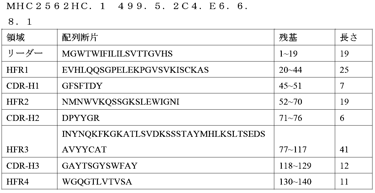
MHC2562LC.2;M13.499.5.2C4.E6.6.8.1
CDR分析
SSVSSSY.....___RTS.......___QQWSGYPFT
FASTA形式でのアミノ酸配列(MHC2562LC.2\;M13F)
> MHC2562LC.2\;M13F.499.5.2C4.E6.6.8.1
遺伝子座2C4_vL 390bpの直鎖状DNA


> MHC2562LC.2\;M13F.499.5.2C4.E6.6.8.1
起源

![]()
フレームワーク1(塩基対67~135):
![]()
CDR-L1(塩基対136~171):
/翻訳=「SASSSVSSSYLH」(配列番号224)
フレームワーク2(塩基対172~216):
/翻訳=「WYQQRSGASPKPLIH」(配列番号226)
CDR-L2(塩基対217~237):
/翻訳=「RTSNLAS」(配列番号228)
フレームワーク3(塩基対238~333):
![]()
CDR-L3(塩基対334~360):
/翻訳=「QQWSGYPFT」(配列番号232)
フレームワーク4(塩基対361~390):
/翻訳=「FGSGTKLEIK」(配列番号234)

MHC2563HC.1;M13.499.5.4D1.5.16
CDR分析
GYTFTDYA....___ISTYYGDT..___ARGNYDDWYFNV
FASTA形式でのアミノ酸配列(MHC2563HC.1\;M13F)
> MHC2563HC.1\;M13F.499.5.4D1.5.16
遺伝子座4D1_vH 414bpの直鎖状DNA


> MHC2563HC.1\;M13F.499.5.4D1.5.16
起源

シグナルペプチド(塩基対1~57):
/翻訳=「MDWSCIIFFLVATATGVHS」(配列番号238)
フレームワーク1(塩基対58~132):
![]()
CDR-H1(塩基対133~153):
/翻訳=「GYTFTDY」(配列番号242)
フレームワーク2(塩基対154~210):
/翻訳=「AMHWVKQSHAKSLEWIGSI」(配列番号244)
CDR-H2(塩基対211~228):
/翻訳=「STYYGD」(配列番号246)
フレームワーク3(塩基対229~351):

CDR-H3(塩基対352~381):
/翻訳=「GNYDDWYFNV」(配列番号250)
フレームワーク4(塩基対382~414):
/翻訳=「WGAGTTVTVSS」(配列番号252)

MHC2563LC.1;M13.499.5.4D1.5.16
CDR分析
QDISGY......___STS.......___LQYASSPYT
FASTA形式でのアミノ酸配列(MHC2563LC.1\;M13F)
> MHC2563LC.1\;M13F.499.5.4D1.5.16
遺伝子座4D1_vL 381bpの直鎖状DNA


> MHC2563LC.1\;M13F.499.5.4D1.5.16
起源

/翻訳=「MRIPAHVFGFLLLWFPGARC」(配列番号256)
フレームワーク1(塩基対61~129):
![]()
CDR-L1(塩基対130~162):
/翻訳=「RASQDISGYLS」(配列番号260)
フレームワーク2(塩基対163~207):
/翻訳=「WLQQKPDGTIKRLIY」(配列番号262)
CDR-L2(塩基対208~228):
/翻訳=「STSTLDS」(配列番号264)
フレームワーク3(塩基対229~324):
![]()
CDR-L3(塩基対325~351):
/翻訳=「LQYASSPYT」(配列番号268)
フレームワーク4(塩基対352~381):
/翻訳=「FGGGAKLEIK」(配列番号270)

MHC2564HC.1;M13.499.5.1C8.9.12.2
CDR分析
GFNIKDTY....___IDPANGKT..___AWLLPYYFDY
FASTA形式でのアミノ酸配列(MHC2564HC.1\;M13F)
> MHC2564HC.1\;M13F.499.5.1C8.9.12.2
遺伝子座1C8_vH 408bpの直鎖状DNA


> MHC2564HC.1\;M13F.499.5.1C8.9.12.2
起源

/翻訳=「MKCSWIIFFLMAVVTGVNS」(配列番号274)
フレームワーク1(塩基対58~132):
![]()
CDR-H1(塩基対133~153):
/翻訳=「GFNIKDT」(配列番号278)
フレームワーク2(塩基対154~210):
/翻訳=「YMHWVKQRPEQGLEWIGRI」(配列番号280)
CDR-H2(塩基対211~228):
/翻訳=「DPANGK」(配列番号282)
フレームワーク3(塩基対229~351):

CDR-H3(塩基対352~375):
/翻訳=「LLPYYFDY」(配列番号286)
フレームワーク4(塩基対376~408):
/翻訳=「WGQGTTLTVSS」(配列番号288)

MHC2564LCB.4;M13.499.5.1C8.9.12.2
CDR分析
SSISSSN.....___GTS.......___QKWSHYPLT
FASTA形式でのアミノ酸配列(MHC2564LCB.4\;M13F)
> MHC2564LCB.4\;M13F.499.5.1C8.9.12.2
遺伝子座1C8_vL 390bpの直鎖状DNA
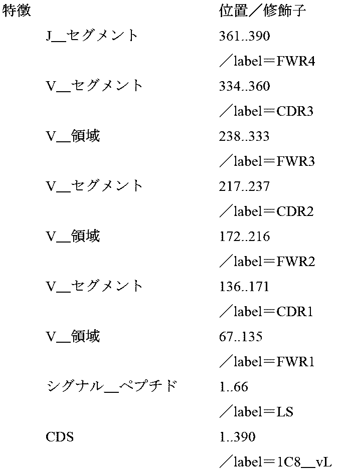

> MHC2564LCB.4\;M13F.499.5.1C8.9.12.2
起源

![]()
フレームワーク1(塩基対67~135):
![]()
CDR-L1(塩基対136~171):
/翻訳=「SVSSSISSSNLH」(配列番号296)
フレームワーク2(塩基対172~216):
/翻訳=「WYQQKSGTSPKLWIY」(配列番号298)
CDR-L2(塩基対217~237):
/翻訳=「GTSNLAS」(配列番号300)
フレームワーク3(塩基対238~333):
![]()
CDR-L3(塩基対334~360):
/翻訳=「QKWSHYPLT」(配列番号304)
フレームワーク4(塩基対361~390):
/翻訳=「FGAGTKLELK」(配列番号306)
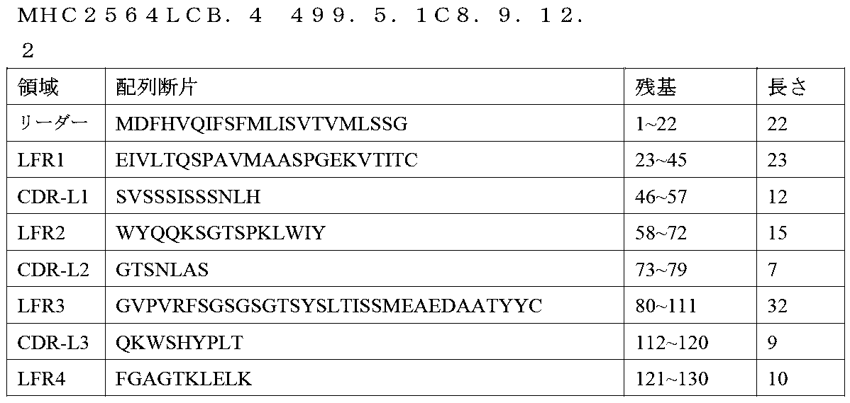
MHC2556HCN.1;M13.499.5.4E5.20.22
CDR分析
GYTFTTYT....___INPSSGYT..___ARHPWDSNY
FASTA形式でのアミノ酸配列(MHC2556HCN.1\;M13F)
>MHC2556HCN.1\;M13F
遺伝子座4E5_vH 405bpの直鎖状DNA


>MHC2556HCN.1\;M13F
起源

/翻訳=「MERHWIFLFLLSVTAGVHS」(配列番号310)
フレームワーク1(塩基対58~132):
![]()
CDR-H1(塩基対133~153):
/翻訳=「GYTFTTY」(配列番号314)
フレームワーク2(塩基対154~210):
/翻訳=「TMHWVKQRPGQGLEWIGHI」(配列番号316)
CDR-H2(塩基対211~228):
/翻訳=「NPSSGY」(配列番号318)
フレームワーク3(塩基対229~351):

CDR-H3(塩基対352~372):
/翻訳=「HPWDSNY」(配列番号322)
フレームワーク4(塩基対373~405):
/翻訳=「WGQGTTLTVSS」(配列番号324)

MHC2556LCN.2;M13.499.5.4E5.20.22
CDR分析
ENIDSY......___AAT.......___QHYYITPFT
FASTA形式でのアミノ酸配列(MHC2556LCN.2\;M13F)
> MHC2556LCN.2\;M13F
遺伝子座4E5_vL 381bpの直鎖状DNA


> MHC2556LCN.2\;M13F
起源

/翻訳=「MSVPTQLLGLLLLWLTDARC」(配列番号328)
フレームワーク1(塩基対61~129):
![]()
CDR-L1(塩基対130~162):
/翻訳=「RASENIDSYLA」(配列番号332)
フレームワーク2(塩基対163~207):
/翻訳=「WYQQKQGRSPQLLVY」(配列番号334)
CDR-L2(塩基対208~228):
/翻訳=「AATNLAD」(配列番号336)
フレームワーク3(塩基対229~324):
![]()
CDR-L3(塩基対325~351):
/翻訳=「QHYYITPFT」(配列番号340)
フレームワーク4(塩基対352~381):
/翻訳=「FGSGTKLEIK」(配列番号342)


Antibodies, immunoglobulins, and polypeptides of the invention can be used in isolated (e.g., purified) form or contained in vectors such as membranes or lipid vesicles (e.g., liposomes). .
Table 2: Identification and Sequencing of Leader and Variable Regions of Anti-HHLA2 Monoclonal Antibodies Including mAbs 8A12, 6D10, 6F10, 8D2, 2G2, 2C4, 4D1, 1C8, and 4E5 8A12 Heavy Chain Variable (vH) DNA and Amino Acid Sequences *
MHC2554HC. 1;M13 499.5.8A12.11.10.5
CDR analysis GFTFNTNV. .. .. .. ___IRTKTNNYAT___VGAMDY
Amino acid sequence in FASTA format (MHC2554HC.1\;M13F)
> MHC2554HC. 1\;M13F
Locus 8A12_vH 402bp linear DNA
> MHC2554HC. 1\;M13F. 499.5.8A12.11.10.5
Origin signal peptide (base pairs 1-57):
/Translation = “MLLGLKWIFFVVFYQGVHC” (SEQ ID NO: 22)
Framework 1 (base pairs 58-132):
/Translation = “GFTFNTN” (SEQ ID NO: 26)
Framework 2 (base pairs 154-210):
/Translation = “VMNWVRQAPGKGLEWVGRI” (SEQ ID NO: 28)
CDR-H2 (base pairs 211-234):
/Translation = “RTKTNNYA” (SEQ ID NO: 30)
Framework 3 (base pairs 235-357):
CDR-H3 (base pairs 358-369):
/Translation = “AMDY” (SEQ ID NO: 34)
Framework 4 (base pairs 370-402):
/Translation = “WGQGTSVTVSS” (SEQ ID NO: 36)
MHC2554LC. 2; M13.499.5.8A12.11.10.5
CDR analysis ESV DNSGINF. .. ___RAS. .. .. .. .. .. .. ___QQSYKDPPT
Amino acid sequence in FASTA format (MHC2554LC.2\;M13F)
> MHC2554LC. 2\;M13F
Locus 8A12_vL 393bp linear DNA
> MHC2554LC. 2\;M13F. 499.5.8A12.11.10.5
origin
/Translation = “METDTLLLWVLLLWVPGSTG” (SEQ ID NO: 40)
Framework 1 (base pairs 61-129):
![]()
CDR-L1 (base pairs 130-174):
/Translation = “RASESVDNSGINFIH” (SEQ ID NO: 44)
Framework 2 (base pairs 175-219):
/Translation = “WYQQKPGQSPKLLLY” (SEQ ID NO: 46)
CDR-L2 (base pairs 220-240):
/Translation = “RASNLKS” (SEQ ID NO: 48)
Framework 3 (base pairs 241-336):
CDR-L3 (base pairs 337-363):
/Translation = “QQSYKDPPT” (SEQ ID NO: 52)
Framework 4 (base pairs 364-393):
/Translation = “FGTGTKLELK” (SEQ ID NO: 54)
MHC2555HC. 1; M13.499.5.6D10.11.11.7
CDR analysis GFTFNTNV. .. .. .. ___IRTKTNNYAT___VGAMDY
Amino acid sequence in FASTA format (MHC2555HC.1\;M13F)
> MHC2555HC. 1\;M13F 499.5.6D10.11.11.7
Locus 6D10_vH 402bp linear DNA
> MHC2555HC. 1\;M13F. 499.5.6D10.11.11.7
origin
/Translation = “MLLGLKWIFFVVFYQGVHC” (SEQ ID NO: 58)
Framework 1 (base pairs 58-132):
![]()
CDR-H1 (base pairs 133-153):
/Translation = “GFTFNTN” (SEQ ID NO: 62)
Framework 2 (base pairs 154-210):
/Translation = “VMNWVRQAPGKGLEWVARI” (SEQ ID NO: 64)
CDR-H2 (base pairs 211-234):
/Translation = “RTKTNNYA” (SEQ ID NO: 66)
Framework 3 (base pairs 235-357):
CDR-H3 (base pairs 358-369):
/Translation = “AMDY” (SEQ ID NO: 70)
Framework 4 (base pairs 370-402):
/Translation = “WGQGTSVTVSS” (SEQ ID NO: 72)
MHC2555LC. 1; M13.499.5.6D10.11.11.7
CDR analysis ESVDNYGISF. .. ___RAS. .. .. .. .. .. .. ___QQSSKDPPT
Amino acid sequence in FASTA format (MHC2555LC.1\;M13F)
> MHC2555LC. 1\;M13F. 499.5.6D10.11.11.7
Locus 6D10_vL 393bp linear DNA
> MHC2555LC. 1\;M13F. 499.5.6D10.11.11.7
origin
/Translation = “METDTLLLWVLLLWVPGSTG” (SEQ ID NO: 76)
Framework 1 (base pairs 61-129):
CDR-L1 (base pairs 130-174):
/Translation = “RASESVDNYGISFMH” (SEQ ID NO: 80)
Framework 2 (base pairs 175-219):
/Translation = “WYQQKPGQPPKLLIY” (SEQ ID NO: 82)
CDR-L2 (base pairs 220-240):
/Translation = “RASNLKS” (SEQ ID NO: 84)
Framework 3 (base pairs 241-336):
![]()
CDR-L3 (base pairs 337-363):
/Translation = “QQSSKDPPT” (SEQ ID NO: 88)
Framework 4 (base pairs 364-393):
/Translation = “FGTGTKLELK” (SEQ ID NO: 90)
MHC2557HCO. 1; M13.499.5.6F10. A4.10
CDR analysis GYTFTTYT. .. .. .. ___INPSSGYT. .. ___ARHPWDSDY
Amino acid sequence in FASTA format (MHC2557HCO.1\;M13F)
> MHC2557HCO. 1\;M13F. 499.5.6F10. A4.10
Locus 6F10_vH 405bp linear DNA
> MHC2557HCO. 1\;M13F. 499.5.6F10. A4.10
origin
/Translation = “MERHWIFLFLLSVTAGVHS” (SEQ ID NO: 94)
Framework 1 (base pairs 58-132):
![]()
CDR-H1 (base pairs 133-153):
/Translation = “GYTFTTY” (SEQ ID NO: 98)
Framework 2 (base pairs 154-210):
/Translation = "TMHWVKQRPGQGLEWIGHI" (SEQ ID NO: 100)
CDR-H2 (base pairs 211-228):
/Translation = “NPSSGY” (SEQ ID NO: 102)
Framework 3 (base pairs 229-351):
CDR-H3 (base pairs 352-372):
/Translation = "HPWDSDY" (SEQ ID NO: 106)
Framework 4 (base pairs 373-405):
/Translation = “WGQGTTLTVSS” (SEQ ID NO: 108)
MHC2557LC. 1;M13.
CDR analysis ENIDSY. .. .. .. .. .. ___AAT. .. .. .. .. .. .. ___QHYYITPFT
Amino acid sequence in FASTA format (MHC2557LC.1\;M13F)
> MHC2557LC. 1\;M13F. 499.5.6F10. A4.10
Locus 6F10_vL 381bp linear DNA
> MHC2557LC. 1\;M13F. 499.5.6F10. A4.10
origin
/Translation = "MSVPTQLLGLLLLWLTDARC" (SEQ ID NO: 112)
Framework 1 (base pairs 61-129):
![]()
CDR-L1 (base pairs 130-162):
/Translation = “RASENIDSYLA” (SEQ ID NO: 116)
Framework 2 (base pairs 163-207):
/Translation = “WYQQKQGRSPQLLVY” (SEQ ID NO: 118)
CDR-L2 (base pairs 208-228):
/Translation = “AATNLAD” (SEQ ID NO: 120)
Framework 3 (base pairs 229-324):
![]()
CDR-L3 (base pairs 325-351):
/Translation = “QHYYITPFT” (SEQ ID NO: 124)
Framework 4 (base pairs 352-381):
/Translation = “FGSGTKLEIA” (SEQ ID NO: 126)
MHC2558HC. 1; M13.499.8D2.6.8
CDR analysis GFTFNTNV. .. .. .. ___IRTKTNNYAT___VGAMDY
Amino acid sequence in FASTA format (MHC2558HC.1\;M13F)
> MHC2558HC. 1\;M13F. 499.8D2.6.8
Locus 8D2_vH 402bp linear DNA
> MHC2558HC. 1\;M13F. 499.8D2.6.8
origin
/Translation = “MLLGLKWIFFVVFYQGVHC” (SEQ ID NO: 130)
Framework 1 (base pairs 58-132):
![]()
CDR-H1 (base pairs 133-153):
/Translation = “GFTFNTN” (SEQ ID NO: 134)
Framework 2 (base pairs 154-210):
![]()
CDR-H2 (base pairs 211-234):
/Translation = "RTKTNNYA" (SEQ ID NO: 138)
Framework 3 (base pairs 235-357):
CDR-H3 (base pairs 358-369):
/Translation = "AMDY" (SEQ ID NO: 142)
Framework 4 (base pairs 370-402):
/Translation = “WGQGTSVTVSS” (SEQ ID NO: 144)
MHC2558LC. 2; M13.499.8D2.6.8
CDR analysis ESV DNSGINF. .. ___RAS. .. .. .. .. .. .. ___QQSYKDPPT
Amino acid sequence in FASTA format (MHC2558LC.2\;M13F)
> MHC2558LC. 2\;M13F. 499.8D2.6.8
Locus 8D2_vL 393bp linear DNA
> MHC2558LC. 2\;M13F. 499.8D2.6.8
origin
/Translation = “METDTLLLWVLLLWVPGSTG” (SEQ ID NO: 148)
Framework 1 (base pairs 61-129):
![]()
CDR-L1 (base pairs 130-174):
/Translation = “RASESVDNSGINFIH” (SEQ ID NO: 152)
Framework 2 (base pairs 175-219):
/Translation = “WYQQKPGQSPKLLLY” (SEQ ID NO: 154)
CDR-L2 (base pairs 220-240):
/Translation = “RASNLKS” (SEQ ID NO: 156)
Framework 3 (base pairs 241-336):
![]()
CDR-L3 (base pairs 337-363):
/Translation = “QQSYKDPPT” (SEQ ID NO: 160)
Framework 4 (base pairs 364-393):
/Translation = “FGTGTKLELK” (SEQ ID NO: 162)
MHC2561HC. 1; M13.499.5.2G2.13.3.10
CDR analysis GLTFSSYA. .. .. .. ___ISSGGSHT. .. ___TRLGRAFDY
Amino acid sequence in FASTA format (MHC2561HC.1\;M13F)
> MHC2561HC. 1\;M13F. 499.5.2G2.13.3.10
Locus 2G2_vH 405bp linear DNA
> MHC2561HC. 1\;M13F. 499.5.2G2.13.3.10
origin
/Translation = “MNFGLSLIFLVLVLKGVQC” (SEQ ID NO: 166)
Framework 1 (base pairs 58-132):
![]()
CDR-H1 (base pairs 133-153):
/Translation = “GLTFSSY” (SEQ ID NO: 170)
Framework 2 (base pairs 154-210):
/Translation = “AMSWVRQTPEKRLEWVATI” (SEQ ID NO: 172)
CDR-H2 (base pairs 211-228):
/Translation = “SSGGSH” (SEQ ID NO: 174)
Framework 3 (base pairs 229-351):
CDR-H3 (base pairs 352-372):
/Translation = “LGRAFDY” (SEQ ID NO: 178)
Framework 4 (base pairs 373-405):
/Translation = “WGQGTTLTVSS” (SEQ ID NO: 180)
MHC2561LC. 1; M13.499.5.2G2.13.3.10
CDR analysis QDVSTA. .. .. .. .. .. ___WAS. .. .. .. .. .. .. ___QQDYSTPWT
Amino acid sequence in FASTA format (MHC2561LC.1\;M13F)
> MHC2561LC. 1\;M13F. 499.5.2G2.13.3.10
Locus 2G2_vL 381bp linear DNA
> MHC2561LC. 1\;M13F. 499.5.2G2.13.3.10
origin
/Translation = "MESQIQVFVFVFLWLSGVDG" (SEQ ID NO: 184)
Framework 1 (base pairs 61-129):
![]()
CDR-L1 (base pairs 130-162):
/Translation = “KASQDVSTAVA” (SEQ ID NO: 188)
Framework 2 (base pairs 163-207):
/Translation = “WYQQKPGQSPKVLIY” (SEQ ID NO: 190)
CDR-L2 (base pairs 208-228):
/Translation = “WASTRHT” (SEQ ID NO: 192)
Framework 3 (base pairs 229-324):
![]()
CDR-L3 (base pairs 325-351):
/Translation = “QQDYSTPWT” (SEQ ID NO: 196)
Framework 4 (base pairs 352-381):
/Translation = “FGGGTKLEIK” (SEQ ID NO: 198)
MHC2562HC. 1; M13.499.5.2C4. E6.6.8.1
CDR analysis GFSFTDYN. .. .. .. ___IDPYYGRI. .. ___ATGAYTSGYSWFAY
Amino acid sequence in FASTA format (MHC2562HC.1\;M13F)
> MHC2562HC. 1\;M13F. 499.5.2C4. E6.6.8.1
Locus 2C4_vH 420bp linear DNA
> MHC2562HC. 1\;M13F. 499.5.2C4. E6.6.8.1
origin
Signal peptide (base pairs 1-57):
/Translation = “MGWTWIFILILSVTTGVHS” (SEQ ID NO: 202)
Framework 1 (base pairs 58-132):
![]()
CDR-H1 (base pairs 133-153):
/Translation = “GFSFTDY” (SEQ ID NO: 206)
Framework 2 (base pairs 154-210):
/Translation = “NMNWVKQSSGKSLEWIGNI” (SEQ ID NO: 208)
CDR-H2 (base pairs 211-228):
/Translation = “DPYYGR” (SEQ ID NO: 210)
Framework 3 (base pairs 229-351):
CDR-H3 (base pairs 352-387):
/Translation = “GAYTSGYSWFAY” (SEQ ID NO: 214)
Framework 4 (base pairs 388-420):
/Translation = “WGQGTLVTVSA” (SEQ ID NO: 216)
MHC2562LC. 2; M13.499.5.2C4. E6.6.8.1
CDR analysis SSVSSSY. .. .. .. .. ___RTS. .. .. .. .. .. .. ___QQWSGYPFT
Amino acid sequence in FASTA format (MHC2562LC.2\;M13F)
> MHC2562LC. 2\;M13F. 499.5.2C4. E6.6.8.1
Locus 2C4_vL 390bp linear DNA
> MHC2562LC. 2\;M13F. 499.5.2C4. E6.6.8.1
origin
![]()
Framework 1 (base pairs 67-135):
![]()
CDR-L1 (base pairs 136-171):
/Translation = “SASSSVSSSYLH” (SEQ ID NO: 224)
Framework 2 (base pairs 172-216):
/Translation = “WYQQRSGASPKPLIH” (SEQ ID NO: 226)
CDR-L2 (base pairs 217-237):
/Translation = “RTSNLAS” (SEQ ID NO: 228)
Framework 3 (base pairs 238-333):
![]()
CDR-L3 (base pairs 334-360):
/Translation = “QQWSGYPFT” (SEQ ID NO: 232)
Framework 4 (base pairs 361-390):
/Translation = “FGSGTKLEIK” (SEQ ID NO: 234)
MHC2563HC. 1; M13.499.5.4D1.5.16
CDR analysis GYTFTDYA. .. .. .. ___ISTYYGDT. .. ___ARGNYDDWYFNV
Amino acid sequence in FASTA format (MHC2563HC.1\;M13F)
> MHC2563HC. 1\;M13F. 499.5.4D1.5.16
Locus 4D1_vH 414bp linear DNA
> MHC2563HC. 1\;M13F. 499.5.4D1.5.16
origin
Signal peptide (base pairs 1-57):
/Translation = "MDWSCIIFLVATATGVHS" (SEQ ID NO: 238)
Framework 1 (base pairs 58-132):
![]()
CDR-H1 (base pairs 133-153):
/Translation = “GYTFTDY” (SEQ ID NO: 242)
Framework 2 (base pairs 154-210):
/Translation = “AMHWVKQSHAKSLEWIGSI” (SEQ ID NO: 244)
CDR-H2 (base pairs 211-228):
/Translation = “STYYGD” (SEQ ID NO: 246)
Framework 3 (base pairs 229-351):
CDR-H3 (base pairs 352-381):
/Translation = “GNYDDWYFNV” (SEQ ID NO: 250)
Framework 4 (base pairs 382-414):
/Translation = “WGAGTTVTVSS” (SEQ ID NO: 252)
MHC2563LC. 1; M13.499.5.4D1.5.16
CDR analysis QDISGY. .. .. .. .. .. ___STS. .. .. .. .. .. .. ___LQYASSPYT
Amino acid sequence in FASTA format (MHC2563LC.1\;M13F)
> MHC2563LC. 1\;M13F. 499.5.4D1.5.16
Locus 4D1_vL 381bp linear DNA
> MHC2563LC. 1\;M13F. 499.5.4D1.5.16
origin
/Translation = “MRIPAHVFGFLLLWFPGARC” (SEQ ID NO: 256)
Framework 1 (base pairs 61-129):
![]()
CDR-L1 (base pairs 130-162):
/Translation = "RASQDISGYLS" (SEQ ID NO: 260)
Framework 2 (base pairs 163-207):
/Translation = “WLQQKPDGTIKRLIY” (SEQ ID NO: 262)
CDR-L2 (base pairs 208-228):
/Translation = “STSTLDS” (SEQ ID NO: 264)
Framework 3 (base pairs 229-324):
![]()
CDR-L3 (base pairs 325-351):
/Translation = “LQYASSPYT” (SEQ ID NO: 268)
Framework 4 (base pairs 352-381):
/Translation = “FGGGAKLEIK” (SEQ ID NO: 270)
MHC2564HC. 1; M13.499.5.1C8.9.12.2
CDR analysis GFNIKDTY. .. .. .. ___IDPANGKT. .. ___AWLLPYYFDY
Amino acid sequence in FASTA format (MHC2564HC.1\;M13F)
> MHC2564HC. 1\;M13F. 499.5.1C8.9.12.2
Locus 1C8_vH 408bp linear DNA
> MHC2564HC. 1\;M13F. 499.5.1C8.9.12.2
origin
/Translation = “MKCSWIIFFLMAVVTGVNS” (SEQ ID NO: 274)
Framework 1 (base pairs 58-132):
![]()
CDR-H1 (base pairs 133-153):
/Translation = “GFNIKDT” (SEQ ID NO: 278)
Framework 2 (base pairs 154-210):
/Translation = “YMHWVKQRPEQGLEWIGRI” (SEQ ID NO: 280)
CDR-H2 (base pairs 211-228):
/Translation = “DPANGK” (SEQ ID NO: 282)
Framework 3 (base pairs 229-351):
CDR-H3 (base pairs 352-375):
/Translation = “LLPYYFDY” (SEQ ID NO: 286)
Framework 4 (base pairs 376-408):
/Translation = “WGQGTTLTVSS” (SEQ ID NO: 288)
MHC2564LCB. 4; M13.499.5.1C8.9.12.2
CDR analysis SSISSSN. .. .. .. .. ___GTS. .. .. .. .. .. .. ___QKWSHYPLT
Amino acid sequence in FASTA format (MHC2564LCB.4\;M13F)
> MHC2564LCB. 4\;M13F. 499.5.1C8.9.12.2
Locus 1C8_vL 390bp linear DNA
> MHC2564LCB. 4\;M13F. 499.5.1C8.9.12.2
origin
![]()
Framework 1 (base pairs 67-135):
![]()
CDR-L1 (base pairs 136-171):
/Translation = “SVSSSISSSNLH” (SEQ ID NO: 296)
Framework 2 (base pairs 172-216):
/Translation = “WYQQKSGTSPKLWIY” (SEQ ID NO: 298)
CDR-L2 (base pairs 217-237):
/Translation = “GTSNLAS” (SEQ ID NO: 300)
Framework 3 (base pairs 238-333):
![]()
CDR-L3 (base pairs 334-360):
/Translation = “QKWSHYPLT” (SEQ ID NO: 304)
Framework 4 (base pairs 361-390):
/Translation = “FGAGTKLELK” (SEQ ID NO: 306)
MHC2556HCN. 1; M13.499.5.4E5.20.22
CDR analysis GYTFTTYT. .. .. .. ___INPSSGYT. .. ___ARHPWDSNY
Amino acid sequence in FASTA format (MHC2556HCN.1\;M13F)
>MHC2556HCN. 1\;M13F
Locus 4E5_vH 405bp linear DNA
>MHC2556HCN. 1\;M13F
origin
/Translation = "MERHWIFLFLLSVTAGVHS" (SEQ ID NO: 310)
Framework 1 (base pairs 58-132):
![]()
CDR-H1 (base pairs 133-153):
/Translation = “GYTFTTY” (SEQ ID NO: 314)
Framework 2 (base pairs 154-210):
/Translation = "TMHWVKQRPGQGLEWIGHI" (SEQ ID NO: 316)
CDR-H2 (base pairs 211-228):
/Translation = “NPSSGY” (SEQ ID NO: 318)
Framework 3 (base pairs 229-351):
CDR-H3 (base pairs 352-372):
/Translation = “HPWDSNY” (SEQ ID NO: 322)
Framework 4 (base pairs 373-405):
/Translation = “WGQGTTLTVSS” (SEQ ID NO: 324)
MHC2556LCN. 2; M13.499.5.4E5.20.22
CDR analysis ENIDSY. .. .. .. .. .. ___AAT. .. .. .. .. .. .. ___QHYYITPFT
Amino acid sequence in FASTA format (MHC2556LCN.2\;M13F)
> MHC2556LCN. 2\;M13F
Locus 4E5_vL 381bp linear DNA
> MHC2556LCN. 2\;M13F
origin
/Translation = “MSVPTQLLGLLLLWLTDARC” (SEQ ID NO: 328)
Framework 1 (base pairs 61-129):
![]()
CDR-L1 (base pairs 130-162):
/Translation = “RASENIDSYLA” (SEQ ID NO: 332)
Framework 2 (base pairs 163-207):
/Translation = “WYQQKQGRSPQLLVY” (SEQ ID NO: 334)
CDR-L2 (base pairs 208-228):
/Translation = “AATNLAD” (SEQ ID NO: 336)
Framework 3 (base pairs 229-324):
![]()
CDR-L3 (base pairs 325-351):
/Translation = “QHYYITPFT” (SEQ ID NO: 340)
Framework 4 (base pairs 352-381):
/Translation = “FGSGTKLEIK” (SEQ ID NO: 342)
III.核酸、ベクター、および組換え宿主細胞
本発明のさらなる目的は、本発明のモノクローナル抗体およびその断片、免疫グロブリン、ならびにポリペプチドをコードする核酸配列に関する。
III. Nucleic Acids, Vectors, and Recombinant Host Cells A further object of the invention relates to nucleic acid sequences encoding the monoclonal antibodies and fragments thereof, immunoglobulins, and polypeptides of the invention.
例えば、特定の実施形態では、本発明は、mAb 8A12のvHドメインまたはmAb 8A12のvLドメインをコードする核酸配列に部分的に関する。別の特定の実施形態では、本発明は、ポリクローナル抗体1.2および/または2.2から単離された少なくとも1つの有効な抗HHLA2 mAbのvHドメインまたはvLドメインをコードする核酸配列に部分的に関する。 For example, in certain embodiments, the invention relates in part to nucleic acid sequences encoding the vH domain of mAb 8A12 or the vL domain of mAb 8A12. In another specific embodiment, the present invention provides for a nucleic acid sequence encoding a vH domain or a vL domain of at least one effective anti-HHLA2 mAb isolated from polyclonal antibodies 1.2 and/or 2.2. Regarding.
典型的には、当該核酸は、プラスミド、コスミド、エピソーム、人工染色体、ファージ、またはウイルスベクター等の任意の好適なベクター中に含まれ得るDNAまたはRNA分子である。 Typically, the nucleic acid is a DNA or RNA molecule that can be contained in any suitable vector, such as a plasmid, cosmid, episome, artificial chromosome, phage, or viral vector.
「ベクター」、「クローニングベクター」、および「発現ベクター」という用語は、DNAまたはRNA配列(例えば、外来遺伝子)が宿主細胞に導入されて、宿主を形質転換し、導入された配列の発現(例えば、転写および翻訳)を促進し得るビヒクルを意味する。したがって、本発明のさらなる目的は、本発明の核酸を含むベクターに関する。 The terms "vector," "cloning vector," and "expression vector" mean that a DNA or RNA sequence (e.g., a foreign gene) is introduced into a host cell to transform the host and express the introduced sequence (e.g., , transcription and translation). A further object of the invention therefore relates to vectors comprising the nucleic acids of the invention.
かかるベクターは、対象への投与時に当該ポリペプチドの発現を引き起こすか、または指示するための制御要素、例えば、プロモーター、エンハンサー、ターミネーター等を含み得る。動物細胞の発現ベクターに使用され得るプロモーターおよびエンハンサーの例には、SV40の初期プロモーターおよびエンハンサー(Mizukami T.et al.1987)、モロニーマウス白血病ウイルスのLTRプロモーターおよびエンハンサー(Kuwana Y et al.1987)、免疫グロブリンH鎖のプロモーター(Mason J O et al.1985)およびエンハンサー(Gillies S D et al.1983)等が挙げられる。 Such vectors may contain control elements, such as promoters, enhancers, terminators, etc., to cause or direct expression of the polypeptide upon administration to a subject. Examples of promoters and enhancers that can be used in animal cell expression vectors include the SV40 early promoter and enhancer (Mizukami T. et al. 1987), the Moloney murine leukemia virus LTR promoter and enhancer (Kuwana Y et al. 1987) , immunoglobulin heavy chain promoter (Mason JO et al. 1985) and enhancer (Gillies S D et al. 1983).
動物細胞のいずれの発現ベクターも使用され得る。好適なベクターの例には、pAGE107(Miyaji H et al.1990)、pAGE103(Mizukami T et al.1987)、pHSG274(Brady G et al.1984)、pKCR(O’Hare K et al.1981)、pSG1ベータd2-4-(Miyaji H et al.1990)等が挙げられる。プラスミドの他の代表的な例には、複製起点を含む複製プラスミド、または組み込み型プラスミド、例えば、pUC、pcDNA、pBR等が挙げられる。ウイルスベクターの代表的な例には、アデノウイルスベクター、レトロウイルスベクター、ヘルペスウイルスベクター、およびAAVベクターが挙げられる。かかる組換えウイルスは、パッケージング細胞のトランスフェクションまたはヘルパープラスミドもしくはウイルスでの過渡的トランスフェクション等の当該技術分野で既知の技法によって産生され得る。ウイルスパッケージング細胞の典型的な例には、PA317細胞、PsiCRIP細胞、GPenv陽性細胞、293細胞等が挙げられる。かかる複製欠損組換えウイルスを産生するための詳細なプロトコルは、例えば、WO95/14785、WO96/22378、米国特許第5,882,877号、米国特許第6,013,516号、米国特許第4,861,719号、米国特許第5,278,056号、およびWO94/19478で見つけることができる。 Any animal cell expression vector may be used. Examples of suitable vectors include pAGE107 (Miyaji H et al. 1990), pAGE103 (Mizukami T et al. 1987), pHSG274 (Brady G et al. 1984), pKCR (O'Hare K et al. 198 1), Examples include pSG1beta d2-4- (Miyaji H et al. 1990). Other representative examples of plasmids include replicative plasmids containing origins of replication, or integrating plasmids, such as pUC, pcDNA, pBR, and the like. Representative examples of viral vectors include adenovirus vectors, retrovirus vectors, herpesvirus vectors, and AAV vectors. Such recombinant viruses can be produced by techniques known in the art, such as transfection of packaging cells or transient transfection with helper plasmids or viruses. Typical examples of virus packaging cells include PA317 cells, PsiCRIP cells, GPenv positive cells, 293 cells, and the like. Detailed protocols for producing such replication-defective recombinant viruses can be found, for example, in WO 95/14785, WO 96/22378, US Pat. No. 5,882,877, US Pat. No. 6,013,516, US Pat. , 861,719, US Pat. No. 5,278,056, and WO 94/19478.
本発明のさらなる目的は、本発明による核酸および/またはベクターによってトランスフェクト、感染、または形質転換された細胞に関する。「形質転換」という用語は、「外来」(すなわち、外因性または細胞外)遺伝子またはDNAもしくはRNA配列の宿主細胞への導入を意味し、これにより、宿主細胞が導入された遺伝子または配列を発現して、導入された遺伝子または配列によってコードされる所望の物質、典型的には、タンパク質または酵素を産生するようになる。導入されたDNAまたはRNAを受容および発現する宿主細胞が「形質転換されている」。 A further object of the invention relates to cells transfected, infected or transformed with the nucleic acids and/or vectors according to the invention. The term "transformation" refers to the introduction of a "foreign" (i.e., exogenous or extracellular) gene or DNA or RNA sequence into a host cell, such that the host cell expresses the introduced gene or sequence. This results in the production of the desired substance, typically a protein or enzyme, encoded by the introduced gene or sequence. A host cell that accepts and expresses introduced DNA or RNA has been "transformed."
本発明の核酸を使用して、好適な発現系において本発明の組換えポリペプチドを産生することができる。「発現系」という用語は、例えば、ベクターによって運搬されて宿主細胞に導入された外来DNAによってコードされたタンパク質の発現に好適な条件下での宿主細胞および適合性ベクターを意味する。 Nucleic acids of the invention can be used to produce recombinant polypeptides of the invention in suitable expression systems. The term "expression system" refers to a host cell and a compatible vector, eg, under conditions suitable for the expression of a protein encoded by foreign DNA carried by the vector and introduced into the host cell.
一般的な発現系には、E.coli宿主細胞およびプラスミドベクター、昆虫宿主細胞およびバキュロウイルスベクター、ならびに哺乳類宿主細胞およびベクターが含まれる。宿主細胞の他の例には、原核細胞(細菌等)および真核細胞(酵母細胞、哺乳類細胞、昆虫細胞、植物細胞等)が挙げられるが、これらに限定されない。特定の例としては、E.coli、Kluyveromyces、またはSaccharomyces酵母、哺乳類細胞株(例えば、Vero細胞、CHO細胞、3T3細胞、COS細胞等)、ならびに初代または樹立哺乳類細胞培養物(例えば、リンパ芽球、線維芽細胞、胚細胞、上皮細胞、神経細胞、脂肪細胞等から産生される)が挙げられる。例としては、マウスSP2/0-Ag14細胞(ATCC CRL1581)、マウスP3X63-Ag8.653細胞(ATCC CRL1580)、ジヒドロ葉酸還元酵素遺伝子(以下、「DHFR遺伝子」と称される)が欠損しているCHO細胞(Urlaub G et al;1980)、ラットYB2/3HL.P2.G11.16Ag.20細胞(ATCC CRL 1662、以下、「YB2/0細胞」と称される)等も挙げられる。キメラまたはヒト化抗体のADCC活性がYB2/0細胞で発現されたときに増強されるため、YB2/0細胞が好ましい。 Common expression systems include E. E. coli host cells and plasmid vectors, insect host cells and baculovirus vectors, and mammalian host cells and vectors. Other examples of host cells include, but are not limited to, prokaryotic cells (such as bacteria) and eukaryotic cells (such as yeast cells, mammalian cells, insect cells, plant cells, etc.). A specific example is E. coli, Kluyveromyces, or Saccharomyces yeasts, mammalian cell lines (e.g., Vero cells, CHO cells, 3T3 cells, COS cells, etc.), and primary or established mammalian cell cultures (e.g., lymphoblasts, fibroblasts, embryonic cells, (produced from epithelial cells, nerve cells, adipocytes, etc.). Examples include mouse SP2/0-Ag14 cells (ATCC CRL1581), mouse P3X63-Ag8.653 cells (ATCC CRL1580), and cells lacking the dihydrofolate reductase gene (hereinafter referred to as "DHFR gene"). CHO cells (Urlaub G et al; 1980), rat YB2/3HL. P2. G11.16Ag. 20 cells (ATCC CRL 1662, hereinafter referred to as "YB2/0 cells") and the like. YB2/0 cells are preferred because the ADCC activity of chimeric or humanized antibodies is enhanced when expressed in YB2/0 cells.
本発明は、本発明による本発明の抗体またはポリペプチドを発現する組換え宿主細胞を産生する方法であって、(i)上述の組換え核酸またはベクターをコンピテント宿主細胞にインビトロまたはエクスビボで導入するステップ、(ii)得られた組換え宿主細胞をインビトロまたはエクスビボで培養するステップ、および(iii)任意に、当該抗体またはポリペプチドを発現および/または分泌する細胞を選択するステップからなるステップを含む、方法にも関する。かかる組換え宿主細胞は、本発明の抗体およびポリペプチドの産生に使用され得る。 The present invention provides a method for producing a recombinant host cell expressing an antibody or polypeptide of the invention according to the invention, comprising: (i) introducing a recombinant nucleic acid or vector as described above into a competent host cell in vitro or ex vivo; (ii) culturing the resulting recombinant host cells in vitro or ex vivo; and (iii) optionally selecting cells that express and/or secrete the antibody or polypeptide. It also relates to a method, including. Such recombinant host cells can be used to produce antibodies and polypeptides of the invention.
別の態様では、本発明は、選択的ハイブリダイゼーション条件下で本明細書に開示されるポリヌクレオチドにハイブリダイズする単離された核酸を提供する。したがって、この実施形態のポリヌクレオチドは、かかるポリヌクレオチドを含む核酸の単離、検出、および/または定量化に使用され得る。例えば、本発明のポリヌクレオチドを使用して、寄託されたライブラリにおける部分長または全長クローンを特定、単離、または増幅することができる。いくつかの実施形態では、ポリヌクレオチドは、ヒトもしくは哺乳類核酸ライブラリから単離されるか、またはさもなければヒトもしくは哺乳類核酸ライブラリ由来のcDNAと相補的なゲノムもしくはcDNA配列である。好ましくは、cDNAライブラリは、少なくとも80%の全長配列、好ましくは、少なくとも85%または90%の全長配列、より好ましくは、少なくとも95%の全長配列を含む。cDNAライブラリは、珍しい配列の提示を増加させるために正規化され得る。低または中程度ストリンジェンシーハイブリダイゼーション条件は、排他的ではなく典型的に、相補的配列と比較して配列同一性が減少した配列に用いられる。中程度および高ストリンジェンシー条件は、任意に、より高い同一性の配列に用いられ得る。低ストリンジェンシー条件は、約70%の配列同一性を有する配列の選択的ハイブリダイゼーションを可能にし、オルソロガスまたはパラロガス配列を特定するために用いられ得る。任意に、本発明のポリヌクレオチドは、本明細書に記載のポリヌクレオチドによってコードされる抗体の少なくとも一部をコードする。本発明のポリヌクレオチドは、本発明の抗体をコードするポリヌクレオチドへの選択的ハイブリダイゼーションのために用いられ得る核酸配列を包含する。例えば、各々参照により本明細書に全体的に組み込まれる、Ausubel(上記参照)、Colligan(上記参照)を参照されたい。 In another aspect, the invention provides isolated nucleic acids that hybridize to polynucleotides disclosed herein under selective hybridization conditions. Accordingly, polynucleotides of this embodiment can be used to isolate, detect, and/or quantify nucleic acids comprising such polynucleotides. For example, polynucleotides of the invention can be used to identify, isolate, or amplify partial-length or full-length clones in deposited libraries. In some embodiments, the polynucleotide is a genomic or cDNA sequence isolated from or otherwise complementary to cDNA from a human or mammalian nucleic acid library. Preferably, the cDNA library contains at least 80% full-length sequences, preferably at least 85% or 90% full-length sequences, more preferably at least 95% full-length sequences. cDNA libraries can be normalized to increase the representation of rare sequences. Low or moderate stringency hybridization conditions are typically, but not exclusively, used for sequences with reduced sequence identity as compared to complementary sequences. Moderate and high stringency conditions may optionally be used for sequences of higher identity. Low stringency conditions allow selective hybridization of sequences with about 70% sequence identity and can be used to identify orthologous or paralogous sequences. Optionally, a polynucleotide of the invention encodes at least a portion of an antibody encoded by a polynucleotide described herein. Polynucleotides of the invention include nucleic acid sequences that can be used for selective hybridization to polynucleotides encoding antibodies of the invention. See, eg, Ausubel (supra), Colligan (supra), each of which is herein incorporated by reference in its entirety.
IV.抗体を産生する方法
本発明の抗体およびその断片、免疫グロブリン、ならびにポリペプチドは、単独でまたは組み合わせでのいずれかでの任意の化学的、生物学的、遺伝的、または酵素的技法等であるが、これらに限定されない当該技術分野で既知の任意の技法によって産生され得る。
IV. Methods of Producing Antibodies The antibodies and fragments thereof, immunoglobulins, and polypeptides of the present invention can be produced by any chemical, biological, genetic, or enzymatic technique, either alone or in combination. can be produced by any technique known in the art, including but not limited to.
所望の配列のアミノ酸配列を知ることにより、当業者であれば、ポリペプチドを産生するための標準技法によって当該抗体またはポリペプチドを容易に産生することができる。例えば、それらは、周知の固相法を使用して、好ましくは、製造業者の指示に従って市販のペプチド合成装置(例えば、Applied Biosystems(Foster City,Calif.)製のもの)を使用して合成され得る。あるいは、本発明の抗体および他のポリペプチドは、当該技術分野で周知の組換えDNA技法によって合成され得る。例えば、これらの断片は、所望の(ポリ)ペプチドをコードするDNA配列の発現ベクターへの組み込みおよびかかるベクターの所望のポリペプチドを発現する好適な真核または原核宿主への導入後にDNA発現産物として得られ得、所望のポリペプチドは、周知の技法を使用して後に単離され得る。 Knowing the amino acid sequence of a desired sequence, one skilled in the art can readily produce the antibody or polypeptide by standard techniques for producing polypeptides. For example, they are synthesized using well-known solid-phase methods, preferably using commercially available peptide synthesizers (e.g., from Applied Biosystems, Foster City, Calif.) according to the manufacturer's instructions. obtain. Alternatively, antibodies and other polypeptides of the invention can be synthesized by recombinant DNA techniques well known in the art. For example, these fragments can be used as DNA expression products after incorporation of a DNA sequence encoding the desired (poly)peptide into an expression vector and introduction of such vector into a suitable eukaryotic or prokaryotic host expressing the desired polypeptide. The desired polypeptide can be subsequently isolated using well-known techniques.
具体的には、本発明はさらに、本発明の抗体またはポリペプチドを産生する方法であって、(i)当該抗体またはポリペプチドの発現を可能にするのに好適な条件下で本発明による形質転換宿主細胞を培養するステップ、および(ii)発現された抗体またはポリペプチドを回収するステップからなるステップを含む、方法に関する。 In particular, the invention further provides a method of producing an antibody or polypeptide of the invention, comprising: (i) producing a trait according to the invention under suitable conditions to allow expression of the antibody or polypeptide; A method comprising the steps of: culturing a transformed host cell; and (ii) recovering the expressed antibody or polypeptide.
本発明の抗体および他のポリペプチドは、例えば、タンパク質A-セファロース、ヒドロキシルアパタイトクロマトグラフィー、ゲル電気泳動、透析、親和性クロマトグラフィー、硫酸アンモニウムまたはエタノール沈殿、酸抽出、アニオンまたはカチオン交換クロマトグラフィー、ホスホセルロースクロマトグラフィー、疎水性相互作用クロマトグラフィー、ヒドロキシルアパタイトクロマトグラフィー、およびレクチンクロマトグラフィー等の従来の免疫グロブリン精製手順によって培養培地から好適に分離される。高速液体クロマトグラフィー(「HPLC」)も精製に用いられ得る。例えば、各々参照により本明細書に全体的に組み込まれる、Colligan,Current Protocols in Immunology、またはCurrent Protocols in Protein Science,John Wiley&Sons,NY,N.Y.,(1997-2001)、例えば、第1、4、6、8、9、10章を参照されたい。 Antibodies and other polypeptides of the invention can be used, for example, on protein A-Sepharose, hydroxyl apatite chromatography, gel electrophoresis, dialysis, affinity chromatography, ammonium sulfate or ethanol precipitation, acid extraction, anion or cation exchange chromatography, phosphorescence They are suitably separated from the culture medium by conventional immunoglobulin purification procedures such as cellulose chromatography, hydrophobic interaction chromatography, hydroxylapatite chromatography, and lectin chromatography. High performance liquid chromatography ("HPLC") may also be used for purification. See, for example, Colligan, Current Protocols in Immunology, or Current Protocols in Protein Science, John Wiley & Sons, NY, N.C., each of which is herein incorporated by reference in its entirety. Y. , (1997-2001), see, for example, Chapters 1, 4, 6, 8, 9, and 10.
本発明のキメラ抗体(例えば、マウス-ヒトキメラまたは非齧歯類-ヒトキメラ)は、前述のようにVLおよびVHドメインをコードする核配列を得て、それらを、ヒト抗体CHおよびヒト抗体CLをコードする遺伝子を有する動物細胞の発現ベクターに挿入することによってヒトキメラ抗体発現ベクターを構築し、発現ベクターを動物細胞に導入することによってコーディング配列を発現することによって産生され得る。ヒトキメラ抗体のCHドメインは、IgGクラスまたはそのサブクラス、例えば、IgG1、IgG2、IgG3、およびIgG4等のヒト免疫グロブリンに属する任意の領域であり得る。同様に、ヒトキメラ抗体のCLは、カッパクラスまたはラムダクラス等のIgに属する任意の領域であり得る。標準の組換えDNA技法を使用して作製され得るヒト部分および非ヒト部分の両方を含むキメラおよびヒト化モノクローナル抗体は、本発明の範囲内である。かかるキメラおよびヒト化モノクローナル抗体は、当該技術分野で既知の組換えDNA技法によって、例えば、国際特許公開第PCT/US86/02269号(Robinson et al.)、欧州特許出願第184,187号(Akira et al.)、欧州特許出願第171,496号(Taniguchi,M.)、欧州特許出願第173,494号(Morrison et al.)、PCT出願第WO86/01533号(Neuberger et al.)、米国特許第4,816,567号(Cabilly et al.)、欧州特許出願第125,023号(Cabilly et al.)、Better et al.(1988)Science 240:1041-1043、Liu et al.(1987)Proc.Natl.Acad.Sci.USA 84:3439-3443、Liu et al.(1987)J.Immunol.139:3521-3526、Sun et al.(1987)Proc.Natl.Acad.Sci.84:214-218、Nishimura et al.(1987)Cancer Res.47:999-1005、Wood et al.(1985)Nature 314:446-449、Shaw et al.(1988)J.Natl.Cancer Inst.80:1553-1559)、Morrison,S.L.(1985)Science 229:1202-1207、Oi et al.(1986)Biotechniques 4:214、米国特許第5,225,539号(Winter)、Jones et al.(1986)Nature 321:552-525、Verhoeyan et al.(1988)Science 239:1534、およびBeidler et al.(1988)J.Immunol.141:4053-4060に記載の方法を使用して産生され得る。 Chimeric antibodies of the invention (e.g., mouse-human chimeras or non-rodent-human chimeras) obtain nuclear sequences encoding the VL and VH domains as described above and combine them to encode human antibodies CH and human antibodies CL. A human chimeric antibody expression vector can be constructed by inserting the gene into an expression vector in an animal cell, and the coding sequence can be expressed by introducing the expression vector into the animal cell. The CH domain of a human chimeric antibody can be any region belonging to the IgG class or a subclass thereof, eg, human immunoglobulins, such as IgG1, IgG2, IgG3, and IgG4. Similarly, the CL of a human chimeric antibody can be any region belonging to the Ig class, such as the kappa or lambda classes. Chimeric and humanized monoclonal antibodies containing both human and non-human portions that can be made using standard recombinant DNA techniques are within the scope of the invention. Such chimeric and humanized monoclonal antibodies can be produced by recombinant DNA techniques known in the art, e.g. et al.), European Patent Application No. 171,496 (Taniguchi, M.), European Patent Application No. 173,494 (Morrison et al.), PCT Application No. WO86/01533 (Neuberger et al.), United States Patent No. 4,816,567 (Cabilly et al.), European Patent Application No. 125,023 (Cabilly et al.), Better et al. (1988) Science 240:1041-1043, Liu et al. (1987) Proc. Natl. Acad. Sci. USA 84:3439-3443, Liu et al. (1987) J. Immunol. 139:3521-3526, Sun et al. (1987) Proc. Natl. Acad. Sci. 84:214-218, Nishimura et al. (1987) Cancer Res. 47:999-1005, Wood et al. (1985) Nature 314:446-449, Shaw et al. (1988) J. Natl. Cancer Inst. 80:1553-1559), Morrison, S. L. (1985) Science 229:1202-1207, Oi et al. (1986) Biotechniques 4:214, US Pat. No. 5,225,539 (Winter), Jones et al. (1986) Nature 321:552-525, Verhoeyan et al. (1988) Science 239:1534, and Beidler et al. (1988) J. Immunol. 141:4053-4060.
加えて、ヒト化抗体は、米国特許第5,565,332号に開示されるプロトコル等の標準のプロトコルに従って作製され得る。別の実施形態では、抗体鎖または特異的結合対メンバーは、当該技術分野で既知の技法、例えば、米国特許第5,565,332号、同第5,871,907号、または同第5,733,743号に記載の技法を使用して、特異的結合対メンバーのポリペプチド鎖と複製可能なジェネリックディスプレイパッケージの構成成分との融合物をコードする核酸分子を含むベクターと、単一の結合対メンバーの第2のポリペプチド鎖をコードする核酸分子を含むベクターとの間の組換えによって産生され得る。本発明のヒト化抗体は、前述のようにCDRドメインをコードする核酸配列を得て、それらを、(i)ヒト抗体と同一の重鎖定常領域および(ii)ヒト抗体と同一の軽鎖定常領域をコードする遺伝子を有する動物細胞の発現ベクターに挿入することによってヒト化抗体発現ベクターを構築し、発現ベクターを動物細胞に導入することによって遺伝子を発現することによって産生され得る。ヒト化抗体発現ベクターは、抗体重鎖をコードする遺伝子および抗体軽鎖をコードする遺伝子が別個のベクター上に存在するタイプのもの、またはこれらの両方の遺伝子が同じベクター上に存在するタイプ(タンデムタイプ)のもののいずれかであり得る。 Additionally, humanized antibodies can be made according to standard protocols such as those disclosed in US Pat. No. 5,565,332. In another embodiment, the antibody chains or specific binding pair members are prepared using techniques known in the art, e.g., U.S. Pat. 733,743 to a vector containing a nucleic acid molecule encoding a fusion of a polypeptide chain of a specific binding pair member and a component of a replicable generic display package. It can be produced by recombination with a vector containing a nucleic acid molecule encoding a second polypeptide chain of a paired member. The humanized antibodies of the present invention obtain nucleic acid sequences encoding CDR domains as described above, and combine them with (i) a heavy chain constant region identical to a human antibody and (ii) a light chain constant region identical to a human antibody. A humanized antibody expression vector can be constructed by inserting the gene encoding the region into an expression vector of an animal cell, and can be produced by expressing the gene by introducing the expression vector into the animal cell. Humanized antibody expression vectors can be of the type in which the gene encoding the antibody heavy chain and the gene encoding the antibody light chain are present on separate vectors, or the type in which both of these genes are present on the same vector (tandem). type).
従来の組換えDNA技法および遺伝子トランスフェクション技法に基づいてヒト化抗体を産生するための方法は、当該技術分野で周知である(例えば、Riechmann L.et al.1988、Neuberger M S.et al.1985を参照のこと)。抗体は、例えば、CDRグラフティング(EP239,400、PCT公開第WO91/09967号、米国特許第5,225,539号、同第5,530,101号、および同第5,585,089号)、ベニヤリングまたはリサーフェシング(EP592,106、EP519,596、Padlan EA(1991)、Studnicka G M et al.(1994)、Roguska M A.et al.(1994))、および鎖シャッフリング(米国特許第5,565,332号)を含む、当該技術分野で既知の様々な技法を使用してヒト化され得る。かかる抗体を調製するための一般的な組換えDNA技術も既知である(欧州特許出願第EP125023号および国際特許出願第WO96/02576号を参照のこと)。 Methods for producing humanized antibodies based on conventional recombinant DNA and gene transfection techniques are well known in the art (eg, Riechmann L. et al. 1988, Neuberger M S. et al. 1985). Antibodies can be produced by, for example, CDR grafting (EP 239,400, PCT Publication No. WO 91/09967, U.S. Patent Nos. 5,225,539, 5,530,101, and 5,585,089). , veneering or resurfacing (EP 592,106, EP 519,596, Padlan EA (1991), Studnicka G M et al. (1994), Roguska M A. et al. (1994)), and chain shuffling (U.S. Pat. , 565, 332), can be humanized using various techniques known in the art. General recombinant DNA techniques for preparing such antibodies are also known (see European Patent Application No. EP 125023 and International Patent Application No. WO 96/02576).
同様に、本明細書に記載の二重特異性または多重特異性抗体は、標準の手順に従って作製され得る。例えば、トリオーマおよびハイブリッドハイブリドーマは、二重特異性または多重特異性抗体を分泌することができる細胞株の2つの例である。ハイブリッドハイブリドーマまたはトリオーマによって産生される二重特異性および多重特異性抗体の例は、米国特許第4,474,893号に開示されている。かかる抗体は、化学的手段(Staerz et al.(1985)Nature 314:628、およびPerez et al.(1985)Nature 316:354)、およびハイブリドーマ技術(Staerz and Bevan(1986)Proc.Natl.Acad.Sci.USA,83:1453、およびStaerz and Bevan(1986)Immunol.Today 7:241)によっても構築され得る。あるいは、かかる抗体は、異なる抗体を作製するハイブリドーマまたは他の細胞を融合させることによってヘテロハイブリドーマを作製し、その後、所望の抗体を産生して共集合させるクローンを特定することによっても生成され得る。それらは、完全免疫グロブリン鎖またはFabおよびFv配列等のその一部の化学的または遺伝的コンジュゲーションによっても生成され得る。抗体成分は、本明細書に記載の1つ以上の免疫阻害性バイオマーカーを含む、本発明の1つ以上のバイオマーカーのポリペプチドまたはその断片に結合することができる。 Similarly, bispecific or multispecific antibodies described herein can be made according to standard procedures. For example, triomas and hybrid hybridomas are two examples of cell lines that can secrete bispecific or multispecific antibodies. Examples of bispecific and multispecific antibodies produced by hybrid hybridomas or triomas are disclosed in US Pat. No. 4,474,893. Such antibodies can be produced by chemical means (Staerz et al. (1985) Nature 314:628, and Perez et al. (1985) Nature 316:354), and by hybridoma technology (Staerz and Bevan (1986) Proc. Natl.A). cad. Sci. USA, 83:1453, and Staerz and Bevan (1986) Immunol. Today 7:241). Alternatively, such antibodies may be produced by creating heterohybridomas by fusing hybridomas or other cells that produce different antibodies, and then identifying clones that produce and coassemble the desired antibodies. They can also be produced by chemical or genetic conjugation of complete immunoglobulin chains or parts thereof, such as Fab and Fv sequences. The antibody component can bind to one or more biomarker polypeptides of the invention or fragments thereof, including one or more immunoinhibitory biomarkers described herein.
加えて、抗体断片を産生するための方法が周知である。例えば、本発明のFab断片は、ヒトHHLA2と特異的に反応する抗体(例えば、mAb 8A12、ならびにポリクローナル抗体1.2および2.2)をパパイン等のプロテアーゼで処理することによって得られ得る。また、Fabは、抗体のFabをコードするDNAを原核発現系または真核発現系のベクターに挿入し、そのベクターを原核生物または真核生物(適切な場合)に導入してFabを発現させることによって産生され得る。 Additionally, methods for producing antibody fragments are well known. For example, Fab fragments of the invention can be obtained by treating antibodies that specifically react with human HHLA2 (eg, mAb 8A12 and polyclonal antibodies 1.2 and 2.2) with a protease such as papain. Fab can also be produced by inserting DNA encoding the Fab of an antibody into a vector of a prokaryotic or eukaryotic expression system, and introducing the vector into a prokaryotic or eukaryotic organism (as appropriate) to express the Fab. can be produced by.
同様に、本発明のF(ab’)2断片は、HHLA2と特異的に反応する抗体をプロテアーゼ(ペプシン)で処理することによって得られ得る。また、F(ab’)2断片は、チオエーテル結合またはジスルフィド結合により以下に記載のFab’に結合することによって産生され得る。 Similarly, the F(ab')2 fragments of the invention can be obtained by treating antibodies that specifically react with HHLA2 with protease (pepsin). F(ab')2 fragments can also be produced by linking to Fab's described below via thioether bonds or disulfide bonds.
本発明のFab’断片は、ヒトHHLA2と特異的に反応するF(ab’)2を還元剤ジチオスレイトールで処理することによって得られ得る。また、Fab’断片は、抗体のFab’断片をコードするDNAを原核生物の発現ベクターまたは真核生物の発現ベクターに挿入し、そのベクターを原核生物または真核生物(適切な場合)に導入してそれを発現させることによって産生され得る。 The Fab' fragment of the present invention can be obtained by treating F(ab')2, which specifically reacts with human HHLA2, with the reducing agent dithiothreitol. Fab' fragments can also be prepared by inserting DNA encoding the Fab' fragment of an antibody into a prokaryotic or eukaryotic expression vector and introducing the vector into a prokaryotic or eukaryotic organism (as appropriate). can be produced by expressing it using
加えて、本発明のscFvは、前述のようにVHおよびVLドメインをコードするcDNAを得て、scFvをコードするDNAを構築し、そのDNAを原核生物の発現ベクターまたは真核生物の発現ベクターに挿入し、その後、その発現ベクターを原核生物または真核生物(適切な場合)に導入してscFvを発現させることによって産生され得る。ヒト化scFv断片を生成するために、ドナーscFv断片から相補性決定領域(CDR)を選択することと、それらを、既知の三次元構造を有するヒトscFv断片フレームワークにグラフトすることとを含む、CDRグラフティングと呼ばれる周知の技術が使用され得る(例えば、WO98/45322、WO87/02671、米国特許第5,859,205号、米国特許第5,585,089号、米国特許第4,816,567号、EP0173494を参照のこと)。 In addition, the scFv of the present invention can be obtained by obtaining cDNA encoding the VH and VL domains as described above, constructing the DNA encoding the scFv, and converting the DNA into a prokaryotic expression vector or a eukaryotic expression vector. and then introducing the expression vector into prokaryotes or eukaryotes (as appropriate) to express the scFv. selecting complementarity determining regions (CDRs) from a donor scFv fragment and grafting them onto a human scFv fragment framework with a known three-dimensional structure to generate a humanized scFv fragment; A well-known technique called CDR grafting may be used (e.g., WO 98/45322, WO 87/02671, U.S. Pat. No. 5,859,205, U.S. Pat. No. 5,585,089, U.S. Pat. No. 4,816, No. 567, EP 0173494).
V.抗体、免疫グロブリン、およびポリペプチドの修飾
本明細書に記載の抗体のアミノ酸配列修飾(複数可)が企図される。例えば、抗体の結合親和性および/または他の生物学的特性を改善することが望ましくあり得る。ヒト化抗体が非ヒト動物に由来する抗体のVHおよびVL内のCDRのみをヒト抗体のVHおよびVLのFRに単にグラフトすることによって産生された場合、抗原結合活性が非ヒト動物に由来する元の抗体と比較して減少することが知られている。CDR内のみならずFR内の非ヒト抗体のVHおよびVLのいくつかのアミノ酸残基が抗原結合活性に直接または間接的に関連するとみなされる。したがって、これらのアミノ酸残基の、ヒト抗体のVHおよびVLのFRに由来する異なるアミノ酸残基での置換により、結合活性が減少し、アミノ酸を、非ヒト動物に由来する元の抗体のアミノ酸残基で置き換えることによって補正され得る。
V. Modifications of Antibodies, Immunoglobulins, and Polypeptides Amino acid sequence modification(s) of the antibodies described herein are contemplated. For example, it may be desirable to improve the binding affinity and/or other biological properties of antibodies. If a humanized antibody is produced by simply grafting only the CDRs in the VH and VL of an antibody derived from a non-human animal to the FRs of the VH and VL of a human antibody, the antigen-binding activity will be greater than that of the non-human animal-derived antibody. It is known that this decreases compared to that of antibodies. Several amino acid residues of the VH and VL of non-human antibodies, not only within the CDRs but also within the FRs, are considered to be directly or indirectly related to antigen binding activity. Therefore, substitution of these amino acid residues with different amino acid residues derived from the VH and VL FRs of human antibodies reduces binding activity and replaces the amino acid residues with amino acid residues of the original antibody derived from non-human animals. It can be corrected by replacing with a group.
修飾および変化が本発明の抗体の構造およびそれらをコードするDNA配列になされ得、望ましい特徴を有する抗体およびポリペプチドをコードする機能的分子を依然として得ることができる。例えば、ある特定のアミノ酸は、活性の認識可能な損失なしにタンパク質構造における他のアミノ酸によって置換され得る。タンパク質の相互作用的能力および性質がタンパク質の生物学的機能的活性を定義するため、ある特定のアミノ酸置換が、タンパク質配列に、ひいては言うまでもなくそのDNAコード配列になされ得、それにもかかわらず、同様の特性を有するタンパク質を得ることもできる。したがって、様々な変化が、それらの生物学的活性の認識可能な損失なしに、本発明の抗体配列または当該ポリペプチドをコードする対応するDNA配列になされ得ることが企図される。 Modifications and changes can be made to the structure of the antibodies of the invention and the DNA sequences encoding them and still obtain functional molecules encoding antibodies and polypeptides with desirable characteristics. For example, certain amino acids can be substituted by other amino acids in the protein structure without appreciable loss of activity. Because the interactive capabilities and properties of a protein define its biological and functional activity, certain amino acid substitutions can be made to the protein sequence, and by extension, to its DNA coding sequence, and yet remain similar. It is also possible to obtain proteins having the following properties. It is therefore contemplated that various changes may be made to the antibody sequences of the invention or the corresponding DNA sequences encoding the polypeptides of the invention without appreciable loss of their biological activity.
一実施形態では、アミノ酸変化は、DNA配列内のコドンを変化させて、遺伝子コードの保存に基づいて保存的置換をコードすることによって達成され得る。具体的には、特定のタンパク質のアミノ酸配列と、遺伝子コード(以下に示される)によって定義されるタンパク質をコードし得るヌクレオチド配列との間に既知の明確な対応関係が存在する。同様に、特定の核酸のヌクレオチド配列と、遺伝子コード(上記の遺伝子コード図表を参照のこと)によって定義される核酸によってコードされるアミノ酸配列との間に既知の明確な対応関係が存在する。 In one embodiment, amino acid changes may be accomplished by changing codons within the DNA sequence to encode conservative substitutions based on conservation of the genetic code. Specifically, there is a known and well-defined correspondence between the amino acid sequence of a particular protein and the nucleotide sequence that can encode the protein as defined by the genetic code (shown below). Similarly, there is a known and well-defined correspondence between the nucleotide sequence of a particular nucleic acid and the amino acid sequence encoded by the nucleic acid as defined by the genetic code (see Genetic Code Diagram above).
上述のように、遺伝子コードの重要な周知の特徴は、その冗長性であり、それにより、タンパク質を作製するために使用されるアミノ酸の大半には、1つより多くのコーディングヌクレオチドトリプレットが用いられ得る(上で例証される)。したがって、いくつかの異なるヌクレオチド配列が所与のアミノ酸配列をコードし得る。かかるヌクレオチド配列は、全ての生物における同じアミノ酸配列の産生をもたらすため(ある特定の生物はいくつかの配列を他の配列よりも効率的に翻訳し得るが)、機能的に同等であるとみなされる。さらに、時折、プリンまたはピリミジンのメチル化バリアントが所与のヌクレオチド配列に見られ得る。かかるメチル化は、トリヌクレオチドコドンと対応するアミノ酸との間のコーディング関係に影響を及ぼさない。 As mentioned above, an important well-known feature of the genetic code is its redundancy, whereby more than one coding nucleotide triplet is used for the majority of amino acids used to make proteins. obtain (exemplified above). Thus, several different nucleotide sequences may encode a given amino acid sequence. Such nucleotide sequences are considered functionally equivalent because they result in the production of the same amino acid sequence in all organisms (although certain organisms may translate some sequences more efficiently than others). It will be done. Additionally, purine or pyrimidine methylation variants can occasionally be found in a given nucleotide sequence. Such methylation does not affect the coding relationship between the trinucleotide codon and the corresponding amino acid.
ポリペプチドのアミノ配列を変化させる際に、アミノ酸の疎水性親水性指標が考慮され得る。相互作用的生物学的機能をタンパク質に付与する際の疎水性親水性アミノ酸指数の重要性は、当該技術分野で一般に理解されている。アミノ酸の相対的疎水性親水性特性が、結果として生じたタンパク質の二次構造に寄与し、次いで、タンパク質と、他の分子、例えば、酵素、基質、受容体、DNA、抗体、抗原等との相互作用を定義することが認められている。各アミノ酸には、それらの疎水性および電荷特性に基づいて疎水性親水性指標が割り当てられており、それらは、イソロイシン(+4.5)、バリン(+4.2)、ロイシン(+3.8)、フェニルアラニン(+2.8)、システイン/シスチン(+2.5)、メチオニン(+1.9)、アラニン(+1.8)、グリシン(-0.4)、トレオニン(-0.7)、セリン(-0.8)、トリプトファン(-0.9)、チロシン(-1.3)、プロリン(-1.6)、ヒスチジン(-3.2)、グルタミン酸(-3.5)、グルタミン(-3.5)、アスパラギン酸(<RTI 3.5)、アスパラギン(-3.5)、リジン(-3.9)、およびアルギニン(-4.5)である。 In altering the amino sequence of a polypeptide, the hydrophilicity index of the amino acids can be considered. The importance of hydrophobic to hydrophilic amino acid indices in conferring interactive biological functions to proteins is generally understood in the art. The relative hydrophobic-hydrophilic properties of the amino acids contribute to the secondary structure of the resulting protein and, in turn, facilitate the interaction of the protein with other molecules such as enzymes, substrates, receptors, DNA, antibodies, antigens, etc. It is allowed to define interactions. Each amino acid is assigned a hydrophilicity index based on their hydrophobicity and charge characteristics: isoleucine (+4.5), valine (+4.2), leucine (+3.8), Phenylalanine (+2.8), cysteine/cystine (+2.5), methionine (+1.9), alanine (+1.8), glycine (-0.4), threonine (-0.7), serine (-0 .8), tryptophan (-0.9), tyrosine (-1.3), proline (-1.6), histidine (-3.2), glutamic acid (-3.5), glutamine (-3.5) ), aspartic acid (<RTI 3.5), asparagine (-3.5), lysine (-3.9), and arginine (-4.5).
ある特定のアミノ酸が同様の疎水性親水性指標またはスコアを有する他のアミノ酸によって置換されてもよく、同様の生物学的活性を有するタンパク質が依然としてもたらされる、すなわち、機能的に同等の生物学的タンパク質が依然として得られることが当該技術分野で既知である。 Certain amino acids may be substituted by other amino acids with a similar hydrophilicity index or score and still result in a protein with similar biological activity, i.e., a functionally equivalent biological It is known in the art that proteins can still be obtained.
したがって、上で概説されるように、アミノ酸置換は、一般に、置換基のアミノ酸側鎖の相対的類似性、例えば、それらの疎水性、親水性、電荷、サイズ等に基づいている。前述の様々な特性を考慮した例示の置換が当業者に周知であり、これらには、アルギニンおよびリジン、グルタミン酸およびアスパラギン酸、セリンおよびトレオニン、グルタミンおよびアスパラギン、ならびにバリン、ロイシン、およびイソロイシンが含まれる。 Thus, as outlined above, amino acid substitutions are generally based on the relative similarity of the amino acid side chains of the substituents, eg, their hydrophobicity, hydrophilicity, charge, size, etc. Exemplary substitutions taking into account the various properties described above are well known to those skilled in the art and include arginine and lysine, glutamic acid and aspartic acid, serine and threonine, glutamine and asparagine, and valine, leucine, and isoleucine. .
本発明の抗体のアミノ酸修飾の別のタイプが、例えば、安定性を増加させるための抗体の元のグリコシル化パターンを改変させる際に有用であり得る。「改変させる」とは、抗体に見られる1つ以上の炭水化物部分を欠失させること、および/または抗体に存在しない1つ以上のグリコシル化部位を付加することを意味する。抗体のグリコシル化は、典型的には、N結合であり得る。「N結合」とは、炭水化物部分のアスパラギン残基の側鎖への結合を指す。トリペプチド配列アスパラギン-X-セリンおよびアスパラギン-X-トレオニン(ここで、Xは、プロリンを除く任意のアミノ酸である)は、炭水化物部分のアスパラギン側鎖への酵素結合の認識配列である。したがって、ポリペプチドにおけるこれらのトリペプチド配列のいずれかの存在により、潜在的なグリコシル化部位が作製される。抗体へのグリコシル化部位の付加は、(N結合グリコシル化部位について)上に記載されるトリペプチド配列のうちの1つ以上を含むようにアミノ酸配列を改変することによって好都合に達成される。共有結合的修飾の別のタイプは、グリコシドを抗体に化学的または酵素的にカップリングすることを含む。これらの手順は、N結合またはO結合グリコシル化のためのグリコシル化能力を有する宿主細胞における抗体の産生を必要としないという点で有利である。使用されるカップリング様式に応じて、糖(複数可)が、(a)アルギニンおよびヒスチジン、(b)遊離カルボキシル基、(c)遊離スルフヒドリル基、例えば、システインの遊離スルフヒドリル基、(d)遊離ヒドロキシル基、例えば、セリン、トレオニン、もしくはヒドロキシプロリンの遊離ヒドロキシル基、(e)芳香族残基、例えば、フェニルアラニン、チロシン、もしくはトリプトファンの芳香族残基、または(f)グルタミンのアミド基に結合していてもよい。例えば、かかる方法は、WO87/05330に記載されている。 Other types of amino acid modifications to antibodies of the invention may be useful, for example, in altering the native glycosylation pattern of the antibody to increase stability. By "altered" is meant the deletion of one or more carbohydrate moieties found in the antibody and/or the addition of one or more glycosylation sites not present in the antibody. Glycosylation of antibodies typically can be N-linked. "N-linked" refers to the attachment of a carbohydrate moiety to the side chain of an asparagine residue. The tripeptide sequences asparagine-X-serine and asparagine-X-threonine, where X is any amino acid except proline, are the recognition sequences for enzyme attachment to the asparagine side chain of a carbohydrate moiety. Thus, the presence of any of these tripeptide sequences in a polypeptide creates a potential glycosylation site. Addition of glycosylation sites to antibodies is conveniently accomplished by modifying the amino acid sequence to include one or more of the tripeptide sequences described above (for N-linked glycosylation sites). Another type of covalent modification involves chemically or enzymatically coupling a glycoside to an antibody. These procedures are advantageous in that they do not require production of antibodies in host cells with glycosylation capacity for N-linked or O-linked glycosylation. Depending on the coupling mode used, the sugar(s) may be (a) arginine and histidine, (b) free carboxyl groups, (c) free sulfhydryl groups, such as cysteine, (d) free attached to a hydroxyl group, such as a free hydroxyl group of serine, threonine, or hydroxyproline; (e) an aromatic residue, such as an aromatic residue of phenylalanine, tyrosine, or tryptophan; or (f) an amide group of glutamine. You can leave it there. For example, such a method is described in WO 87/05330.
同様に、抗体上に存在するいずれの炭水化物部分の除去は、化学的または酵素的に達成され得る。化学的脱グリコシル化は、化合物トリフルオロメタンスルホン酸または同等の化合物への抗体の曝露を必要とする。この処理により、抗体をインタクトに保った状態で、結合糖(N-アセチルグルコサミンまたはN-アセチルガラクトサミン)を除く大半または全ての糖の切断がもたらされる。化学的脱グリコシル化は、Sojahr H.et al.(1987)およびEdge,A S.et al.(1981)によって説明されている。抗体上の炭水化物部分の酵素的切断は、Thotakura,N R.et al.(1987)によって説明される様々なエンドグリコシダーゼおよびエキソグリコシダーゼの使用によって達成され得る。 Similarly, removal of any carbohydrate moieties present on the antibody can be accomplished chemically or enzymatically. Chemical deglycosylation requires exposure of the antibody to the compound trifluoromethanesulfonic acid or an equivalent compound. This treatment results in cleavage of most or all sugars except the linking sugar (N-acetylglucosamine or N-acetylgalactosamine) while keeping the antibody intact. Chemical deglycosylation is described by Sojahr H. et al. (1987) and Edge, A.S. et al. (1981). Enzymatic cleavage of carbohydrate moieties on antibodies is described by Thotakura, N R. et al. (1987).
他の修飾は、イムノコンジュゲートの形成を含み得る。例えば、1つのタイプの共有結合的修飾では、抗体またはタンパク質が、米国特許第4,640,835号、同第4,496,689号、同第4,301,144号、同第4,670,417号、同第4,791,192号、または同第4,179,337号に記載の様式で、ポリエチレングリコール、ポリプロピレングリコール、またはポリオキシアルキレン等の様々な非タンパク質性ポリマーのうちの1つに共有結合的に連結される。 Other modifications may include the formation of immunoconjugates. For example, in one type of covalent modification, antibodies or proteins are , 417, 4,791,192, or 4,179,337, such as polyethylene glycol, polypropylene glycol, or polyoxyalkylene. covalently linked to.
本発明の抗体または他のタンパク質の異種薬剤とのコンジュゲーションは、N-スクシンイミジル(2-ピリジルジチオ)プロピオネート(SPDP)、スクシンイミジル(N-マレイミドメチル)シクロヘキサン-1-カルボキシレート、イミノチオラン(IT)、イミドエステルの二官能性誘導体(アジプイミド酸ジメチルHCL等)、活性エステル(スベリン酸ジスクシンイミジル等)、アルデヒド(グルタルアルデヒド等)、ビス-アジド化合物(ビス(p-アジドベンゾイル)ヘキサンジアミン等)、ビス-ジアゾニウム誘導体(ビス-(p-ジアゾニウムベンゾイル)-エチレンジアミン等)、ジイソシアネート(トルエン2,6ジイソシアネート等)、およびビス-活性フッ素化合物(1,5-ジフルオロ-2,4-ジニトロベンゼン等)を含むが、これらに限定されない、様々な二官能性タンパク質カップリング剤を使用して行われ得る。例えば、炭素標識1-イソチオシアナトベンジルメチルジエチレントリアミン五酢酸(MX-DTPA)は、放射性ヌクレオチドの抗体へのコンジュゲーションのための例示のキレート剤である(WO94/11026)。 Conjugation of antibodies or other proteins of the invention with heterologous agents can include N-succinimidyl (2-pyridyldithio) propionate (SPDP), succinimidyl (N-maleimidomethyl) cyclohexane-1-carboxylate, iminothiolane (IT), Bifunctional derivatives of imide esters (dimethyl adipimidate HCL, etc.), active esters (disuccinimidyl suberate, etc.), aldehydes (glutaraldehyde, etc.), bis-azide compounds (bis(p-azidobenzoyl)hexanediamine, etc.) , bis-diazonium derivatives (such as bis-(p-diazoniumbenzoyl)-ethylenediamine), diisocyanates (such as toluene 2,6 diisocyanate), and bis-active fluorine compounds (such as 1,5-difluoro-2,4-dinitrobenzene). can be performed using a variety of bifunctional protein coupling agents, including but not limited to. For example, carbon-labeled 1-isothiocyanatobenzylmethyldiethylenetriaminepentaacetic acid (MX-DTPA) is an exemplary chelating agent for the conjugation of radionucleotides to antibodies (WO 94/11026).
別の態様では、本発明は、細胞毒素、薬物、および/または放射性同位体等の治療的部分にコンジュゲートされたHHLA2に特異的に結合する抗体を特色とする。細胞毒素にコンジュゲートされると、これらの抗体コンジュゲートは、「免疫毒素」と称される。細胞毒素または細胞毒性薬剤には、細胞に有害な(例えば、細胞を死滅させる)任意の薬剤が含まれる。例としては、タキソール、サイトカラシンB、グラミシジンD、臭化エチジウム、エメチン、マイトマイシン、エトポシド、テノポシド、ビンクリスチン、ビンブラスチン、コルヒチン、ドキソルビシン、ダウノルビシン、ジヒドロキシアントラシンジオン、ミトキサントロン、ミトラマイシン、アクチノマイシンD、1-デヒドロテストステロン、グルココルチコイド、プロカイン、テトラカイン、リドカイン、プロプラノロール、およびピューロマイシン、ならびにそれらの類似体または相同体が挙げられる。治療薬には、代謝拮抗剤(例えば、メトトレキサート、6-メルカプトプリン、6-チオグアニン、シタラビン、5-フルオロウラシルデカルバジン)、アルキル化剤(例えば、メクロレタミン、チオエパクロラムブシル、メルファラン、カルムスチン(BSNU)およびロムスチン(CCNU)、シクロホスファミド、ブスルファン、ジブロモマンニトール、ストレプトゾトシン、マイトマイシンC、ならびにシス-ジクロロジアミン白金(II)(DDP)シスプラチン)、アントラサイクリン(例えば、ダウノルビシン(以前のダウノマイシン)およびドキソルビシン)、抗生物質(例えば、ダクチノマイシン(以前のアクチノマイシン)、ブレオマイシン、ミトラマイシン、およびアントラマイシン(AMC))、および抗分裂剤(例えば、ビンクリスチンおよびビンブラスチン)が挙げられるが、これらに限定されない。本発明の抗体は、放射性同位体、例えば、放射性ヨウ素にコンジュゲートされて、がん等の関連障害を治療するための細胞毒性放射性医薬品を生成することができる。 In another aspect, the invention features antibodies that specifically bind HHLA2 conjugated to therapeutic moieties such as cytotoxins, drugs, and/or radioisotopes. When conjugated to a cytotoxin, these antibody conjugates are termed "immunotoxins." A cytotoxin or cytotoxic agent includes any agent that is harmful to (eg, kills) cells. Examples include taxol, cytochalasin B, gramicidin D, ethidium bromide, emetine, mitomycin, etoposide, tenoposide, vincristine, vinblastine, colchicine, doxorubicin, daunorubicin, dihydroxyanthracindione, mitoxantrone, mitramycin, actinomycin D , 1-dehydrotestosterone, glucocorticoids, procaine, tetracaine, lidocaine, propranolol, and puromycin, and analogs or homologs thereof. Therapeutic agents include antimetabolites (e.g., methotrexate, 6-mercaptopurine, 6-thioguanine, cytarabine, 5-fluorouracil decarbazine), alkylating agents (e.g., mechlorethamine, thioepachlorambucil, melphalan, carmustine), BSNU) and lomustine (CCNU), cyclophosphamide, busulfan, dibromomannitol, streptozotocin, mitomycin C, and cis-dichlorodiamineplatinum(II) (DDP) cisplatin), anthracyclines (e.g. daunorubicin (formerly daunomycin) and doxorubicin), antibiotics (e.g., dactinomycin (formerly actinomycin), bleomycin, mitramycin, and anthramycin (AMC)), and antimitotic agents (e.g., vincristine and vinblastine) Not done. The antibodies of the invention can be conjugated to radioactive isotopes, such as radioactive iodine, to produce cytotoxic radiopharmaceuticals for treating related disorders such as cancer.
コンジュゲートされた抗HHLA2抗体を使用して、臨床試験手順の一環として組織中のポリペプチドレベルを診断的または予後的に監視して、例えば、所与の治療レジメンの有効性を決定するか、または免疫療法に応答する可能性が最も高い患者を選択することができる。例えば、細胞がフローサイトメトリーアッセイで透過処理されて、HHLA2に結合する抗体(mAb 8A12およびポリクローナル抗体1.2および2.2等)にその認識された細胞内エピトープを標的とさせ、コンジュゲートされた分子から発せられるシグナルを分析することによって結合の検出を可能にすることができる。検出は、抗体を検出可能な物質にカップリングする(すなわち、物理的に連結する)ことによって促進され得る。検出可能な物質の例には、様々な酵素、補欠分子族、蛍光物質、発光物質、生物発光物質、および放射性物質が挙げられる。好適な酵素の例には、西洋ワサビペルオキシダーゼ、アルカリ性ホスファターゼ、β-ガラクトシダーゼ、またはアセチルコリンエステラーゼが挙げられ、好適な補欠分子族複合体の例には、ストレプトアビジン/ビオチンおよびアビジン/ビオチンが挙げられ、好適な蛍光物質の例には、ウンベリフェロン、フルオレセイン、フルオレセインイソチオシアネート(FITC)、ローダミン、ジクロロトリアジニルアミンフルオレセイン、塩化ダンシル、またはフィコエリトリン(PE)が挙げられ、発光物質の例には、ルミノールが挙げられ、生物発光物質の例には、ルシフェラーゼ、ルシフェリン、およびエクオリンが挙げられ、好適な放射性物質の例には、125I、131I、35S、または3Hが挙げられる。本明細書で使用される場合、抗体に関して「標識される」という用語は、検出可能な物質、例えば、放射性薬剤またはフルオロフォア(例えば、フルオレセインイソチオシアネート(FITC)またはフィコエリトリン(PE)またはインドシアニン(Cy5))を抗体にカップリングする(すなわち、物理的に連結する)ことによる抗体の直接標識、ならびに検出可能な物質との反応による抗体の間接的標識を包含するよう意図されている。 Conjugated anti-HHLA2 antibodies may be used to monitor polypeptide levels in tissues diagnostically or prognostically as part of a clinical trial procedure, e.g., to determine the effectiveness of a given treatment regimen; or can select patients most likely to respond to immunotherapy. For example, cells are permeabilized in a flow cytometry assay and antibodies that bind HHLA2 (such as mAb 8A12 and polyclonal antibodies 1.2 and 2.2) are targeted and conjugated to their recognized intracellular epitopes. Detection of binding can be made possible by analyzing the signals emitted by the bound molecules. Detection can be facilitated by coupling (ie, physically linking) the antibody to a detectable substance. Examples of detectable substances include various enzymes, prosthetic groups, fluorescent materials, luminescent materials, bioluminescent materials, and radioactive materials. Examples of suitable enzymes include horseradish peroxidase, alkaline phosphatase, β-galactosidase, or acetylcholinesterase; examples of suitable prosthetic group complexes include streptavidin/biotin and avidin/biotin; Examples of suitable fluorescent materials include umbelliferone, fluorescein, fluorescein isothiocyanate (FITC), rhodamine, dichlorotriazinylamine fluorescein, dansyl chloride, or phycoerythrin (PE), and examples of luminescent materials include luminol. Examples of bioluminescent substances include luciferase, luciferin, and aequorin, and examples of suitable radioactive substances include 125 I, 131 I, 35 S, or 3 H. As used herein, the term "labeled" with respect to an antibody refers to a detectable substance, such as a radioactive agent or a fluorophore (e.g., fluorescein isothiocyanate (FITC) or phycoerythrin (PE) or indocyanine ( It is intended to encompass direct labeling of antibodies by coupling (ie, physically linking) Cy5)) to the antibody, as well as indirect labeling of antibodies by reaction with a detectable substance.
本発明の抗体コンジュゲートを使用して、所与の生物学的応答を修飾することができる。治療的部分が古典的な化学的治療薬に限定されると解釈されるべきではない。例えば、薬物部分は、所望の生物学的活性を有するタンパク質またはポリペプチドであり得る。かかるタンパク質には、例えば、酵素的に活性な毒素またはその活性断片(アブリン、リシンA、シュードモナス外毒素、またはジフテリア毒素等)、タンパク質(腫瘍壊死因子またはインターフェロン-ガンマ等)、または生物学的応答修飾因子(例えば、リンフォカイン、インターロイキン-1(「IL-1」)、インターロイキン-2(「IL-2」)、インターロイキン-6(「IL-6」)、顆粒球マクロファージコロニー刺激因子(「GM-CSF」)、顆粒球コロニー刺激因子(「G-CSF」)、または他のサイトカインもしくは成長因子等)が含まれ得る。 Antibody conjugates of the invention can be used to modify a given biological response. The therapeutic moiety should not be construed as limited to classical chemical therapeutics. For example, a drug moiety can be a protein or polypeptide that has a desired biological activity. Such proteins include, for example, enzymatically active toxins or active fragments thereof (such as abrin, ricin A, pseudomonas exotoxin, or diphtheria toxin), proteins (such as tumor necrosis factor or interferon-gamma), or biological responses. Modifiers (e.g., lymphokines, interleukin-1 (“IL-1”), interleukin-2 (“IL-2”), interleukin-6 (“IL-6”), granulocyte-macrophage colony-stimulating factor ( "GM-CSF"), granulocyte colony stimulating factor ("G-CSF"), or other cytokines or growth factors).
かかる治療的部分を抗体にコンジュゲートするための技法が周知であり、例えば、Arnon et al.,“Monoclonal Antibodies For Immunotargeting Of Drugs In Cancer Therapy”,in Monoclonal Antibodies And Cancer Therapy,Reisfeld et al.(eds.),pp.243 56(Alan R.Liss,Inc.1985)、Hellstrom et al.,“Antibodies For Drug Delivery”,in Controlled Drug Delivery(2nd Ed.),Robinson et al.(eds.),pp.623 53(Marcel Dekker,Inc.1987)、Thorpe,“Antibody Carriers Of Cytotoxic Agents In Cancer Therapy:A Review”,in Monoclonal Antibodies’84:Biological And Clinical Applications,Pinchera et al.(eds.),pp.475 506(1985)、“Analysis,Results,And Future Prospective Of The Therapeutic Use Of Radiolabeled Antibody In Cancer Therapy”,in Monoclonal Antibodies For Cancer Detection And Therapy,Baldwin et al.(eds.),pp.303 16(Academic Press 1985)、およびThorpe et al.,“The Preparation And Cytotoxic Properties Of Antibody-Toxin Conjugates”,Immunol.Rev.,62:119 58(1982)を参照されたい。 Techniques for conjugating such therapeutic moieties to antibodies are well known and are described, for example, in Arnon et al. , “Monoclonal Antibodies For Immunotargeting Of Drugs In Cancer Therapy”, in Monoclonal Antibodies And Cancer Therapy, Reisfel d et al. (eds.), pp. 243 56 (Alan R. Liss, Inc. 1985), Hellstrom et al. , “Antibodies For Drug Delivery”, in Controlled Drug Delivery (2nd Ed.), Robinson et al. (eds.), pp. 623 53 (Marcel Dekker, Inc. 1987), Thorpe, “Antibody Carriers Of Cytotoxic Agents In Cancer Therapy: A Review”, in Monoclonal Anti bodies'84: Biological and Clinical Applications, Pinchera et al. (eds.), pp. 475 506 (1985), “Analysis, Results, And Future Prospective Of The Therapeutic Use Of Radiolabeled Antibody In Cancer Therapy”, i. n Monoclonal Antibodies For Cancer Detection And Therapy, Baldwin et al. (eds.), pp. 303 16 (Academic Press 1985), and Thorpe et al. , “The Preparation And Cytotoxic Properties Of Antibody-Toxin Conjugates”, Immunol. Rev. , 62:119 58 (1982).
いくつかの実施形態では、コンジュゲーションは、細胞における細胞毒性薬剤または成長阻害剤の放出を促進する「切断可能なリンカー」を使用して行われ得る。例えば、酸不安定性リンカー、ペプチダーゼ感受性リンカー、感光性リンカー、ジメチルリンカー、またはジスルフィド含有リンカー(例えば、米国特許第5,208,020号を参照のこと)が使用され得る。あるいは、抗体および細胞毒性薬剤または成長阻害剤を含む融合タンパク質が組換え技法またはペプチド合成によって作製され得る。DNAの長さは、互いに隣接しているか、またはコンジュゲートの所望の特性を破壊しないリンカーペプチドをコードする領域によって分離されているかのいずれかのコンジュゲートの2つの部分をコードするそれぞれの領域を含み得る。 In some embodiments, conjugation may be performed using a "cleavable linker" that facilitates release of the cytotoxic or growth inhibitory agent in the cell. For example, acid-labile linkers, peptidase-sensitive linkers, photosensitive linkers, dimethyl linkers, or disulfide-containing linkers (see, eg, US Pat. No. 5,208,020) may be used. Alternatively, fusion proteins comprising an antibody and a cytotoxic or growth inhibitory agent can be made by recombinant techniques or peptide synthesis. The length of DNA has each region encoding the two parts of the conjugate either adjacent to each other or separated by a region encoding a linker peptide that does not disrupt the desired properties of the conjugate. may be included.
VI.本発明の使用および方法
本明細書に記載の本発明の抗HHLA2抗体、免疫グロブリン、ポリペプチド、および核酸は、HHLA2レベルの検出に基づく多数の予測的医薬アッセイ(例えば、診断アッセイ、予後アッセイ、および臨床試験の監視)で使用され得、いくつかの実施形態では、単独で、または毒性化合物もしくは他の治療薬にコンジュゲートされたときに、治療目的(例えば、治療的および予防的)に有用であり得る。本明細書で使用される「検出」という用語は、対照の参照を伴うまたは伴わない質的および/または量的検出(レベルの測定)を含む。本明細書に記載されるように、本発明のHHLA2ポリペプチドまたはその断片は、以下の活性:1)TMIGD2および/またはKIR3DL3等のその天然結合パートナー(複数可)に結合し、かつまたはその活性を調節すること、2)共免疫阻害性シグナル伝達等の細胞内または細胞間シグナル伝達を調節すること、3)リンパ球の活性化および/または増殖を調節すること、4)生物、例えば、マウス、非齧歯類動物、またはヒト等の哺乳類生物の免疫応答を調節すること、および5)免疫細胞アネルギーを調節することのうちの1つ以上を有する。
VI. Uses and Methods of the Invention The anti-HHLA2 antibodies, immunoglobulins, polypeptides, and nucleic acids of the invention described herein are useful in a number of predictive pharmaceutical assays based on the detection of HHLA2 levels (e.g., diagnostic assays, prognostic assays, and clinical trial monitoring) and, in some embodiments, useful for therapeutic purposes (e.g., therapeutic and prophylactic), alone or when conjugated to toxic compounds or other therapeutic agents. It can be. The term "detection" as used herein includes qualitative and/or quantitative detection (measurement of levels) with or without reference to a control. As described herein, the HHLA2 polypeptides of the invention or fragments thereof have the following activities: 1) binding to TMIGD2 and/or its natural binding partner(s), such as KIR3DL3; 2) modulating intracellular or intercellular signaling, such as co-immune inhibitory signaling; 3) modulating lymphocyte activation and/or proliferation; 4) an organism, e.g., a mouse. 5) modulating the immune response of a mammalian organism, such as a non-rodent, or human; and 5) modulating immune cell anergy.
したがって、本発明の一態様は、生体試料(例えば、血液、血清、細胞、または組織)との関連でHHLA2ポリペプチドレベルを決定し、それにより、試料中のHHLA2ポリペプチドレベルを決定し、個体が障害に罹患しているかを決定し、かつ/またはかかるHHLA2レベルによって示されるかかる障害の状態を決定するための診断アッセイに関する。例えば、本発明の抗体は、HHLA2に関連するがん疾患の病期分類に有用である。 Accordingly, one aspect of the present invention is to determine HHLA2 polypeptide levels in the context of a biological sample (e.g., blood, serum, cells, or tissue), thereby determining HHLA2 polypeptide levels in the sample and in an individual. The present invention relates to diagnostic assays for determining whether a person is suffering from a disorder and/or determining the status of such disorder as indicated by such HHLA2 levels. For example, antibodies of the invention are useful in staging cancer diseases associated with HHLA2.
本発明は、個体がかかる障害を発症する危険性があるかを決定するための予後(または予測的)アッセイも提供する。本発明の別の態様は、臨床試験におけるHHLA2の発現または活性への薬剤(例えば、薬物、化合物)の影響の監視に関する。 The invention also provides prognostic (or predictive) assays for determining whether an individual is at risk of developing such a disorder. Another aspect of the invention relates to monitoring the effect of agents (eg, drugs, compounds) on HHLA2 expression or activity in clinical trials.
本発明は、HHLA2シグナルを伝達する薬剤を特定するための手段としてHHLA2の検出も提供する。HHLA2シグナルを伝達する薬剤は、免疫応答を減弱させ、自己免疫疾患、喘息、および耐性の確立に有用であり得る。 The present invention also provides for the detection of HHLA2 as a means to identify agents that transduce HHLA2 signals. Agents that transduce HHLA2 signals attenuate immune responses and may be useful in autoimmune diseases, asthma, and the establishment of tolerance.
本明細書に記載のいずれの方法においても、HHLA2は、単独で、または他の免疫チェックポイントおよび/もしくは共刺激分子等の他の分子の発現と組み合わせて検出され得る。いくつかの分子のコンビナトリアル検出(例えば、順次または同時)は、治療的介入の相乗作用および/または障害亜型の個別化された高分解能診断に関する有用な情報を提供し得る。いくつかの実施形態では、HHLA2は、もう1つのマーカーでコンビナトリアル的に検出される。 In any of the methods described herein, HHLA2 can be detected alone or in combination with the expression of other molecules such as other immune checkpoints and/or costimulatory molecules. Combinatorial detection of several molecules (eg, sequentially or simultaneously) may provide useful information for synergy of therapeutic interventions and/or personalized high-resolution diagnosis of disorder subtypes. In some embodiments, HHLA2 is detected combinatorially with another marker.
1.診断アッセイ
本発明は、生体試料が細胞制限HHLA2を発現するか、かつ/または細胞制限HHLA2レベルが調節される(例えば、上方制御または下方制御される)かを正確に分類し、それにより、がん等の目的とする障害の状態を示すための方法、システム、およびコードを部分的に提供する。いくつかの実施形態では、本発明は、統計的アルゴリズムおよび/または経験的データ(例えば、HHLA2の存在、不在、またはレベル)を使用してHHLA2によって媒介されるがんまたはその亜型に関連するか、またはその危険性があるものとして試料(例えば、対象由来の)を分類する際に有用である。
1. Diagnostic Assays The present invention accurately classifies whether a biological sample expresses cell-restricted HHLA2 and/or whether cell-restricted HHLA2 levels are regulated (e.g., up-regulated or down-regulated), thereby Methods, systems, and code are provided, in part, for indicating desired failure conditions, such as, for example. In some embodiments, the invention uses statistical algorithms and/or empirical data (e.g., presence, absence, or levels of HHLA2) to identify cancers mediated by HHLA2 or subtypes thereof. It is useful in classifying samples (e.g., from a subject) as having or at risk of becoming.
HHLA2またはその断片のレベルを検出するための例示の方法であって、それ故に、試料がHHLA2またはその臨床亜型の異常な発現(例えば、上方制御または下方制御)によって媒介される疾患または障害に関連するかの分類に有用である、方法は、生体試料を試験対象から得ることと、HHLA2レベルが生体試料中で検出されるように、生体試料を、HHLA2を検出することができる本発明の抗体またはその抗原結合断片と接触させることとを含む。いくつかの実施形態では、少なくとも1つの抗体またはその抗原結合断片が使用され、2、3、4、5、6、7、8、9、10、またはそれ以上のかかる抗体または抗体断片が組み合わせで(例えば、サンドイッチELISAで)または順次に使用され得る。ある特定の例では、統計的アルゴリズムは、単一の学習統計的分類子システムである。例えば、単一の学習統計的分類子システムは、予測値または確率値およびHHLA2の存在またはレベルに基づいて試料をHHLA2試料として分類するために使用され得る。単一の学習統計的分類子システムの使用により、典型的には、試料が、少なくとも約75%、76%、77%、78%、79%、80%、81%、82%、83%、84%、85%、86%、87%、88%、89%、90%、91%、92%、93%、94%、95%、96%、97%、98%、または99%の感受性、特異性、陽性予測値、陰性予測値、および/または全体精度を有するHHLA2試料として分類される。 An exemplary method for detecting levels of HHLA2 or a fragment thereof, wherein the sample is associated with a disease or disorder mediated by aberrant expression (e.g., upregulation or downregulation) of HHLA2 or a clinical subtype thereof. The method includes obtaining a biological sample from a test subject and detecting HHLA2 in the biological sample, such that HHLA2 levels are detected in the biological sample. contacting the antibody or antigen-binding fragment thereof. In some embodiments, at least one antibody or antigen-binding fragment thereof is used, and 2, 3, 4, 5, 6, 7, 8, 9, 10, or more such antibodies or antibody fragments are used in combination. (eg, in a sandwich ELISA) or sequentially. In one particular example, the statistical algorithm is a single learning statistical classifier system. For example, a single learning statistical classifier system can be used to classify samples as HHLA2 samples based on predicted or probability values and the presence or level of HHLA2. The use of a single learning statistical classifier system typically results in a sample having at least about 75%, 76%, 77%, 78%, 79%, 80%, 81%, 82%, 83%, 84%, 85%, 86%, 87%, 88%, 89%, 90%, 91%, 92%, 93%, 94%, 95%, 96%, 97%, 98%, or 99% susceptibility , specificity, positive predictive value, negative predictive value, and/or overall accuracy.
他の好適な統計的アルゴリズムが当業者に周知である。例えば、学習統計的分類子システムは、複雑なデータセット(例えば、目的とするマーカーのパネル)に適合し、かかるデータセットに基づいて決定することができる機械学習アルゴリズム技法を含む。いくつかの実施形態では、分類ツリー(例えば、ランダムフォレスト)等の単一の学習統計的分類子システムが使用される。他の実施形態では、2、3、4、5、6、7、8、9、10個、またはそれ以上の学習統計的分類子システムの組み合わせが、好ましくは直列で使用される。学習統計的分類子システムの例には、帰納的学習(例えば、ランダムフォレスト、分類および回帰ツリー(C&RT)、ブーストツリー等の決定/分類ツリー)、確率的に近似的に正しい(PAC)学習、コネクショニスト学習(例えば、ニューラルネットワーク(NN)、人工ニューラルネットワーク(ANN)、ニューロファジーネットワーク(NFN)、ネットワーク構造、多層パーセプトロン等のパーセプトロン、多層フィードフォワードネットワーク、ニューラルネットワークの適用、信念ネットワークにおけるベイズ学習等)、強化学習(例えば、ナイーブ学習、適応動的学習、および時間差学習等の既知の環境における受動的学習、未知の環境における受動的学習、未知の環境における能動的学習、学習行動値関数、強化学習の適用等)、ならびに遺伝的アルゴリズムおよび進化的プログラミングを使用したものが挙げられるが、これらに限定されない。他の学習統計的分類子システムは、サポートベクトルマシン(例えば、カーネル法)、多変量適応型回帰スプライン(MARS)、レーベンバーグ・マーカートアルゴリズム、ガウス・ニュートンアルゴリズム、ガウスの混合(mixtures of Gaussians)、勾配降下アルゴリズム、および学習ベクトル量子化(LVQ)を含む。ある特定の実施形態では、本発明の方法は、HLA2試料分類結果を臨床医、例えば、組織病理学者または腫瘍学者に送信することをさらに含む。 Other suitable statistical algorithms are well known to those skilled in the art. For example, learning statistical classifier systems include machine learning algorithmic techniques that can be adapted to and make decisions based on complex datasets (eg, panels of markers of interest). In some embodiments, a single learning statistical classifier system, such as a classification tree (eg, random forest), is used. In other embodiments, combinations of 2, 3, 4, 5, 6, 7, 8, 9, 10, or more learning statistical classifier systems are used, preferably in series. Examples of learning statistical classifier systems include inductive learning (e.g., decision/classification trees such as random forests, classification and regression trees (C&RT), boosted trees, etc.), probabilistically approximately correct (PAC) learning, Connectionist learning (e.g. neural networks (NN), artificial neural networks (ANN), neurofuzzy networks (NFN), network structures, perceptrons such as multilayer perceptrons, multilayer feedforward networks, applications of neural networks, Bayesian learning in belief networks, etc.) ), reinforcement learning (e.g., naive learning, adaptive dynamic learning, and time difference learning), passive learning in a known environment, passive learning in an unknown environment, active learning in an unknown environment, learning action value functions, reinforcement applications of learning, etc.), and those using genetic algorithms and evolutionary programming. Other learning statistical classifier systems include support vector machines (e.g. kernel methods), multivariate adaptive regression splines (MARS), Levenberg-Marquardt algorithm, Gauss-Newton algorithm, mixtures of Gaussians. , gradient descent algorithms, and learning vector quantization (LVQ). In certain embodiments, the methods of the invention further include transmitting the HLA2 sample classification results to a clinician, eg, a histopathologist or an oncologist.
別の実施形態では、本発明の方法は、個体がHHLA2に関連する状態または障害を有する確率の形態で診断をさらに提供する。例えば、個体は、約0%、5%、10%、15%、20%、25%、30%、35%、40%、45%、50%、55%、60%、65%、70%、75%、80%、85%、90%、95%、またはそれ以上のその状態または障害を有する確率を有し得る。さらに別の実施形態では、本発明の方法は、個体における状態または障害の予後をさらに提供する。いくつかの事例では、試料をHHLA2試料として分類する方法は、試料が得られる個体の症状(例えば、臨床的因子)にさらに基づいている。症状または症状の群は、例えば、リンパ球数、白血球数、赤血球沈降速度、下痢、腹痛、痙攣、発熱、貧血、体重減少、不安、うつ、およびそれらの組み合わせであり得る。いくつかの実施形態では、HHLA2に関連する状態または障害を有する個体の診断に続いて、個体に、その状態または障害に関連する1つ以上の症状の治療に有用な薬物(例えば、化学療法剤)の治療有効量が投与される。 In another embodiment, the methods of the invention further provide a diagnosis in the form of a probability that an individual has a condition or disorder associated with HHLA2. For example, an individual may have approximately 0%, 5%, 10%, 15%, 20%, 25%, 30%, 35%, 40%, 45%, 50%, 55%, 60%, 65%, 70% , 75%, 80%, 85%, 90%, 95%, or more probability of having the condition or disorder. In yet another embodiment, the methods of the invention further provide a prognosis of a condition or disorder in an individual. In some cases, the method of classifying a sample as an HHLA2 sample is further based on the symptoms (eg, clinical factors) of the individual from whom the sample is obtained. The symptom or group of symptoms can be, for example, lymphocyte count, white blood cell count, erythrocyte sedimentation rate, diarrhea, abdominal pain, cramps, fever, anemia, weight loss, anxiety, depression, and combinations thereof. In some embodiments, following diagnosis of an individual with a condition or disorder associated with HHLA2, the individual is administered a drug (e.g., a chemotherapeutic agent) useful for treating one or more symptoms associated with the condition or disorder. ) is administered in a therapeutically effective amount.
一実施形態では、本方法は、対照生体試料(例えば、HHLA2によって媒介される状態または障害を有しない対象由来の生体試料)、HHLA2によって媒介される状態または障害の寛解中またはそれを発症した後の対象由来の生体試料、またはHHLA2によって媒介される状態または障害を発症する治療中の対象由来の生体試料を得ることをさらに含む。 In one embodiment, the method comprises a control biological sample (e.g., a biological sample from a subject who does not have the HHLA2-mediated condition or disorder), during remission of the HHLA2-mediated condition or disorder, or after developing the HHLA2-mediated condition or disorder. or from a subject undergoing treatment that develops a condition or disorder mediated by HHLA2.
HHLA2ポリペプチドまたはその断片の存在または不在を検出するための例示の方法は、HHLA2ポリペプチドに結合することができる本発明の抗体またはその断片、好ましくは検出可能な標識を有する抗体である。抗体は、ポリクローナルであり得るか、またはより好ましくは、モノクローナルであり得る。かかる薬剤が標識され得る。抗体に関して「標識される」という用語は、検出可能な物質をプローブまたは抗体にカップリングする(すなわち、物理的に連結する)ことによるプローブまたは抗体の直接標識、ならびに直接標識される別の試薬との反応によるプローブまたは抗体の間接的標識を包含するよう意図されている。間接標識の例には、蛍光標識された二次抗体を使用した一次抗体の検出が挙げられる。「生体試料」という用語は、対象から単離された組織、細胞、および血清等の生体液、ならびに対象に存在する組織、細胞、および体液を含むよう意図されている。すなわち、本発明の検出方法を使用して、生体試料中のHHLA2またはその断片をインビトロで、かつインビボで検出することができる。HHLA2ポリペプチドの検出のためのインビトロ技法には、酵素結合免疫吸着アッセイ(ELISA)、ウエスタンブロット、免疫沈降、免疫組織化学(IHC)、細胞内フローサイトメトリーおよび関連技法、ならびに免疫蛍光が含まれる。さらに、HHLA2ポリペプチドまたはその断片の検出のためのインビボ技法には、対象に標識された抗HHLA2抗体を導入することが含まれる。例えば、本抗体は、放射性、発光、蛍光、または他の同様のマーカーで標識され得、対象におけるその存在および位置が、標準の画像技法によって単独で、または細胞型マーカー(例えば、CD8+T細胞マーカー)等の他の分子の画像法と組み合わせてのいずれかで検出され得る。 An exemplary method for detecting the presence or absence of an HHLA2 polypeptide or a fragment thereof is an antibody of the invention or a fragment thereof, preferably an antibody having a detectable label, that is capable of binding an HHLA2 polypeptide. Antibodies may be polyclonal or, more preferably, monoclonal. Such agents can be labeled. The term "labeled" with respect to antibodies includes direct labeling of a probe or antibody by coupling (i.e., physically linking) a detectable substance to the probe or antibody, as well as the direct labeling of a probe or antibody with another reagent that is directly labeled. is intended to encompass indirect labeling of probes or antibodies by reaction with An example of indirect labeling includes detection of a primary antibody using a fluorescently labeled secondary antibody. The term "biological sample" is intended to include tissues, cells, and biological fluids such as serum isolated from a subject, as well as tissues, cells, and fluids present in the subject. That is, using the detection method of the present invention, HHLA2 or a fragment thereof in a biological sample can be detected in vitro and in vivo. In vitro techniques for detection of HHLA2 polypeptides include enzyme-linked immunosorbent assay (ELISA), Western blot, immunoprecipitation, immunohistochemistry (IHC), intracellular flow cytometry and related techniques, and immunofluorescence. . Additionally, in vivo techniques for the detection of HHLA2 polypeptides or fragments thereof include introducing a labeled anti-HHLA2 antibody into the subject. For example, the antibody can be labeled with a radioactive, luminescent, fluorescent, or other similar marker whose presence and location in a subject can be determined by standard imaging techniques alone or with a cell type marker (e.g., a CD8+ T cell marker). can be detected either in combination with other molecular imaging methods such as.
一実施形態では、生体試料は、試験対象由来のポリペプチド分子を含有する。好ましい生体試料は、従来の手段によって対象から単離される血清、腫瘍微小環境、腫瘍周辺、または腫瘍内である。 In one embodiment, the biological sample contains polypeptide molecules from the test subject. Preferred biological samples are serum, tumor microenvironment, peritumoral, or intratumoral isolated from the subject by conventional means.
別の実施形態では、本方法は、対照対象から対照生体試料を得ることと、HHLA2ポリペプチドもしくはその断片の存在が生体試料中で検出されるように、対照試料を、化合物またはHHLA2ポリペプチドもしくはその断片を検出することができる薬剤と接触させることと、対照試料中のHHLA2ポリペプチドもしくはその断片の存在を、試験試料中のHHLA2ポリペプチドもしくはその断片の存在と比較することとをさらに含む。 In another embodiment, the method comprises obtaining a control biological sample from a control subject and injecting the control sample with a compound or a HHLA2 polypeptide or a fragment thereof such that the presence of the HHLA2 polypeptide or a fragment thereof is detected in the biological sample. The method further comprises contacting the fragment with an agent capable of detecting the fragment and comparing the presence of the HHLA2 polypeptide or fragment thereof in the control sample to the presence of the HHLA2 polypeptide or fragment thereof in the test sample.
なお他の実施形態では、本抗体は、ELISAまたはRIAにおける抗体の使用のための構成要素またはデバイスと関連付けられる。非限定的な例には、これらのアッセイで使用するために固体表面上に固定化された(例えば、上述のように光または放射線放出に基づいて検出可能な標識に連結および/またはコンジュゲートされた)抗体が挙げられる。他の実施形態では、本抗体は、免疫クロマトグラフィーアッセイまたは免疫化学的アッセイ、例えば、「サンドイッチ」または競合的アッセイ、免疫組織化学、免疫蛍光顕微鏡法等の使用によるHHLA2の検出のためのデバイスまたは条片と関連付けられる。かかるデバイスまたは条片の追加の例は、自宅での検査または迅速な臨床現場即時検査用に設計されたものである。さらなる例には、単一試料中の複数の分析物の同時分析用に設計されたものが挙げられる。例えば、本発明の非標識抗体は、生体試料中の「捕捉」HHLA2ポリペプチドに適用され得、捕捉された(または固定化された)HHLA2ポリペプチドは、検出のために本発明の抗HHLA2抗体の標識形態に結合し得る。例えば、免疫拡散、免疫電気泳動、免疫組織病理学、免疫組織化学、および組織病理学に基づくアッセイを含む免疫アッセイの他の標準の実施形態が当業者に周知である。 In still other embodiments, the antibody is associated with a component or device for use of the antibody in an ELISA or RIA. Non-limiting examples include immobilized on a solid surface (e.g., linked and/or conjugated to a label detectable based on light or radiation emission as described above) for use in these assays. and) antibodies. In other embodiments, the antibody is a device for the detection of HHLA2 by the use of immunochromatographic or immunochemical assays, e.g., "sandwich" or competitive assays, immunohistochemistry, immunofluorescence microscopy, etc. Associated with strips. Additional examples of such devices or strips are those designed for home testing or rapid point-of-care testing. Further examples include those designed for simultaneous analysis of multiple analytes in a single sample. For example, an unlabeled antibody of the invention can be applied to a "captured" HHLA2 polypeptide in a biological sample, and the captured (or immobilized) HHLA2 polypeptide is used for detection by an anti-HHLA2 antibody of the invention. can bind to labeled forms of. Other standard embodiments of immunoassays are well known to those skilled in the art, including, for example, immunodiffusion, immunoelectrophoresis, immunohistopathology, immunohistochemistry, and histopathology-based assays.
2.予後アッセイ
さらに、本明細書に記載の診断法は、HHLA2に関連する障害を有するか、またはそれを発症する危険性のある対象を特定するために利用され得る。本明細書で使用される場合、「異常な」という用語は、正常なHHLA2レベルから逸脱するHHLA2上方制御または下方制御を含む。異常な発現または活性には、増加または減少した発現または活性、ならびに正常な発現発生パターンまたは細胞内発現パターンに従わない発現または活性が含まれる。例えば、異常なHHLA2レベルは、HHLA2遺伝子もしくは制御配列の突然変異またはその染色体HHLA2遺伝子の増幅によりHHLA2の上方制御または下方制御が引き起こされる場合を含むよう意図されている。本明細書で使用される場合、「望ましくない」という用語は、免疫細胞活性化等の生物学的応答に関与する望ましくない現象を含む。例えば、望ましくないという用語は、対象において望ましくないHHLA2を含む。
2. Prognostic Assays Additionally, the diagnostic methods described herein can be utilized to identify subjects who have or are at risk of developing a HHLA2-related disorder. As used herein, the term "abnormal" includes HHLA2 upregulation or downregulation that deviates from normal HHLA2 levels. Aberrant expression or activity includes increased or decreased expression or activity, as well as expression or activity that does not follow normal developmental or intracellular patterns of expression. For example, abnormal HHLA2 levels are intended to include cases where mutations in the HHLA2 gene or regulatory sequences or amplification of the chromosomal HHLA2 gene cause upregulation or downregulation of HHLA2. As used herein, the term "undesirable" includes undesirable phenomena that involve biological responses such as immune cell activation. For example, the term undesirable includes HHLA2 that is undesirable in the subject.
実施例でさらに説明されるように、HHLA2に関連する多くの障害が当業者に既知である。HHLA2は、選択リンパ性悪性疾患、ウイルス誘導性がん、および多くの固形腫瘍を含む複数の腫瘍タイプによって発現される。一般に、HHLA2は、それを必要とする状態に対する強力な免疫応答を阻害する免疫チェックポイント制御因子を活性化するため、予後不良マーカーである。しかしながら、いくつかの障害、例えば、自己免疫疾患、炎症性疾患等を治療する際に免疫応答を下方制御するために免疫阻害が所望される。 Many disorders related to HHLA2 are known to those skilled in the art, as further explained in the Examples. HHLA2 is expressed by multiple tumor types including selected lymphoid malignancies, virus-induced cancers, and many solid tumors. In general, HHLA2 is a poor prognostic marker because it activates immune checkpoint regulators that inhibit strong immune responses to conditions that require it. However, immune inhibition is desired to downregulate the immune response in treating some disorders, such as autoimmune diseases, inflammatory diseases, and the like.
前述の診断アッセイまたは以下のアッセイ等の本明細書に記載のアッセイを利用して、HHLA2活性化の誤制御に関連する障害を有するか、またはそれを発症する危険性のある対象を特定することができる。したがって、本発明は、異常なまたは望ましくないHHLA2活性化に関連する障害を特定するための方法を提供し、本方法において、試験試料が対象から得られ、HHLA2が検出され、HHLA2ポリペプチドの存在が、異常なまたは望ましくないHHLA2活性に関連する障害を有するか、またはそれを発症する危険性のある対象の診断に用いられる。本明細書で使用される場合、「試験試料」は、目的とする対象から得られた生体試料を指す。例えば、試験試料は、生体液(例えば、脳脊髄液または血清)、細胞試料、または組織、例えば、腫瘍微小環境、腫瘍周辺領域、および/または腫瘍内領域の組織病理学的スライドであり得る。好ましい実施形態では、試料は、成熟膜結合型HHLA2および/またはHHLA2断片を発現する細胞を含む。 Utilizing the assays described herein, such as the diagnostic assays described above or the assays below, to identify subjects who have or are at risk of developing disorders associated with misregulated HHLA2 activation. Can be done. Accordingly, the present invention provides methods for identifying disorders associated with aberrant or undesired HHLA2 activation, in which a test sample is obtained from a subject, HHLA2 is detected, and the presence of HHLA2 polypeptide is detected. is used in the diagnosis of subjects who have or are at risk of developing a disorder associated with aberrant or undesirable HHLA2 activity. As used herein, "test sample" refers to a biological sample obtained from a subject of interest. For example, the test sample can be a biological fluid (eg, cerebrospinal fluid or serum), a cell sample, or a histopathological slide of a tissue, such as the tumor microenvironment, peritumoral area, and/or intratumoral area. In a preferred embodiment, the sample comprises cells expressing mature membrane-bound HHLA2 and/or HHLA2 fragments.
さらに、本明細書に記載の予後アッセイを使用して、薬剤(例えば、アゴニスト、アンタゴニスト、ペプチド模倣薬、ポリペプチド、ペプチド、核酸、小分子、または他の薬物候補)が対象に投与されて、異常なまたは望ましくないHHLA2活性に関連するかかる障害を治療することができるかを決定することができる。例えば、かかる方法を使用して、対象が1つの薬剤または薬剤の組み合わせで有効に治療され得るかを決定することができる。したがって、本発明は、対象が、異常なまたは望ましくないHHLA2活性化に関連する障害を治療するための1つ以上の薬剤で有効に治療され得るかを決定するための方法を提供し、本方法において、試験試料が得られ、HHLA2が検出される(例えば、HHLA2ポリペプチドの存在量が、異常なまたは望ましくないHHLA2活性化に関連する障害を治療するための薬剤を投与される対象の診断に用いられる)。 Additionally, using the prognostic assays described herein, an agent (e.g., an agonist, antagonist, peptidomimetic, polypeptide, peptide, nucleic acid, small molecule, or other drug candidate) is administered to the subject; It can be determined whether such disorders associated with aberrant or undesirable HHLA2 activity can be treated. For example, such methods can be used to determine whether a subject can be effectively treated with one drug or combination of drugs. Accordingly, the present invention provides a method for determining whether a subject can be effectively treated with one or more agents for treating a disorder associated with aberrant or undesired HHLA2 activation, and the method , a test sample is obtained and HHLA2 is detected (e.g., the abundance of HHLA2 polypeptide is diagnostic for a subject being administered an agent to treat a disorder associated with aberrant or unwanted HHLA2 activation). used).
本明細書に記載の方法は、例えば、HHLA2を伴う疾患または疾病の症状または家族歴を呈する患者を診断するための臨床状況において好都合に使用され得る、例えば、本明細書に記載の少なくとも1つの抗体試薬を含む予めパッケージングされた診断キットを利用することによって行われ得る。 The methods described herein may be advantageously used in a clinical setting, e.g., to diagnose a patient presenting with a disease or disease symptoms or family history involving HHLA2, e.g. This can be done by utilizing prepackaged diagnostic kits containing antibody reagents.
さらに、HHLA2が発現される任意の細胞型または組織が本明細書に記載の予後アッセイで利用され得る。 Additionally, any cell type or tissue in which HHLA2 is expressed can be utilized in the prognostic assays described herein.
本発明の別の態様は、異常なHHLA2活性化に関連する障害を有する個体由来の生体試料中のHHLA2が分析され、その情報が好ましくは同様の年齢および人種の対照(例えば、その障害を有しない個体であり、対照は、「健常」もしくは「正常」個体とも称され得るか、または所与の経時的研究における初期の時点のもの)と比較される、関連および/または層別化分析のための本明細書に記載の組成物および方法の使用を含む。患者および対照の適切な選択は、関連および/または層別化研究の成功に重要である。したがって、十分に特徴付けられた表現型を有する個体のプールが非常に望ましい。疾患診断、疾患素因スクリーニング、疾患予後、薬物応答性決定(薬理ゲノミクス)、薬物毒性スクリーニング等の基準が本明細書に記載される。 Another aspect of the invention provides that HHLA2 in biological samples from individuals with a disorder associated with aberrant HHLA2 activation is analyzed and that information is preferably passed to controls of similar age and ethnicity (e.g., Association and/or stratification analyzes in which controls are compared to individuals who do not have the disease (and controls may also be referred to as “healthy” or “normal” individuals, or those at earlier time points in a given longitudinal study) including the use of the compositions and methods described herein for. Appropriate selection of patients and controls is critical to the success of association and/or stratification studies. Therefore, a pool of individuals with well-characterized phenotypes is highly desirable. Criteria for disease diagnosis, disease predisposition screening, disease prognosis, drug responsiveness determination (pharmacogenomics), drug toxicity screening, etc. are described herein.
遺伝的関連および/または層別化研究のための異なる研究設計が使用され得る(Modern Epidemiology,Lippincott Williams&Wilkins(1998),609-622)。患者の応答が干渉されない観察的研究が最も頻繁に行われている。観察的研究の第1のタイプは、疾患の疑わしい原因が存在するヒトの試料および疑わしい原因が存在しないヒトの別の試料を特定し、その後、2つの試料における疾患の発症頻度が比較される。これらの試料採取された集団はコホートと呼ばれ、この研究は前向き研究である。観察的研究の他のタイプは、症例対照研究または後向き研究である。典型的な症例対照研究では、試料は、疾患のある特定の兆候等の目的とする表現型を有する個体(症例)および結論が導き出される集団(標的集団)におけるその表現型を有しない個体(対照)から収集される。その後、疾患の考えられる原因が後向きに調査される。症例対照研究における試料収集時間および費用が前向き研究よりも大幅に下回るため、症例対照研究は、少なくとも探査および発見段階中の遺伝的関連研究におけるより一般に使用されている研究設計である。 Different study designs for genetic association and/or stratification studies may be used (Modern Epidemiology, Lippincott Williams & Wilkins (1998), 609-622). Observational studies, in which patient responses are not interfered with, are most frequently performed. The first type of observational study identifies a human sample in which a suspected cause of the disease is present and another human sample in which the suspected cause is not present, and then the frequency of disease occurrence in the two samples is compared. These sampled populations are called cohorts, and this study is prospective. Other types of observational studies are case-control studies or retrospective studies. In a typical case-control study, samples are collected from individuals with a phenotype of interest (cases), such as a particular manifestation of a disease, and individuals without that phenotype (controls) in the population from which conclusions are drawn (target population). ) is collected from. Possible causes of the disease are then investigated retrospectively. Case-control studies are the more commonly used study design in genetic association studies, at least during the exploration and discovery stages, because sample collection time and costs in case-control studies are significantly less than in prospective studies.
全ての関連表現型および/または遺伝子型情報が得られた後、個体の表現型特性を有する対立遺伝子または遺伝子型の存在間に有意な相関関係が存在するかを決定するための統計的分析が行われる。好ましくは、データの検査および整理が最初に行われ、その後、遺伝的関連のための統計的検定が行われる。試料の疫学的および臨床データが当該技術分野で周知の表およびグラフを用いて記述統計学によって要約され得る。好ましくは、データ完成、一貫性のないエントリ、および外れ値を確認するためのデータ検証が行われる。その後、それぞれ、離散変数および連続変数について症例と対照との間の有意差を確認するためのカイ二乗検定およびt-検定(分布が正規分布ではない場合、ウィルコクソンの順位和検定)が使用され得る。 After all relevant phenotypic and/or genotypic information is obtained, statistical analysis is performed to determine whether a significant correlation exists between the presence of alleles or genotypes with the phenotypic characteristics of the individual. It will be done. Preferably, examination and reduction of the data is performed first, followed by statistical testing for genetic association. Epidemiological and clinical data of the sample can be summarized by descriptive statistics using tables and graphs well known in the art. Preferably, data validation is performed to check for data completion, inconsistent entries, and outliers. Chi-square tests and t-tests (Wilcoxon rank sum test if the distribution is not normal) can then be used to check for significant differences between cases and controls for discrete and continuous variables, respectively. .
遺伝的関連検定を行う際の重要な決定は、有意レベルの決定であり、それらの検定のp値がそのレベルに達したときに有意な関連が断言され得る。陽性ヒットがその後の確証検定で追跡される予備的分析では、例えば、0.2(寛大な有意レベル)未満の未調整p値が、HHLA2レベルの疾患のある特定の表現型特性との有意な関連のための仮説を生み出すために使用され得る。0.05(当該技術分野で伝統的に使用されている有意レベル)未満のp値が疾患との関連を有するとみなされるようにそのレベルが達成されることが好ましい。ヒットが同じ源由来のより多くの試料または異なる源由来の異なる試料における確証的分析で追跡されるとき、複数の検定の調整が、実験ワイズエラー率を0.05で維持しながら過剰なヒット数を回避するように行われる。複数の検定を調整して異なる種類のエラー率を制御するための異なる方法が存在する一方で、一般に使用されているが、むしろ保守的な方法は、実験ワイズまたはファミリーワイズエラー率を制御するためのボンフェローニ補正である(Multiple comparisons and multiple tests,Westfall et al,SAS Institute(1999))。偽発見率(FDR)を制御するための並べ替え検定がより有力であり得る(Benjamini and Hochberg,Journal of the Royal Statistical Society,Series B 57,1289-1300,1995、Resampling-based Multiple Testing,Westfall and Young,Wiley(1993))。これらの検定が依存的であり、実験ワイズエラー率の制御とは対照的に偽発見率の制御が十分である場合に、多重度を制御するためのかかる方法が好ましいであろう。 An important decision when performing genetic association tests is the determination of the level of significance; a significant association can be asserted when the p-value of those tests reaches that level. In preliminary analyzes where positive hits are followed up with subsequent confirmatory tests, e.g., an unadjusted p-value of less than 0.2 (a generous level of significance) indicates that HHLA2 levels are significantly associated with a certain phenotypic trait of the disease. Can be used to generate hypotheses for associations. Preferably, that level is achieved such that a p-value of less than 0.05 (the level of significance traditionally used in the art) is considered to have an association with the disease. When hits are followed up with confirmatory analysis in more samples from the same source or different samples from different sources, multiple testing adjustments reduce the number of excess hits while maintaining an experiment-wise error rate of 0.05. This is done to avoid. While different methods exist for adjusting multiple tests to control different types of error rates, a commonly used, but rather conservative, method is to control experiment-wise or family-wise error rates. (Multiple comparisons and multiple tests, Westfall et al, SAS Institute (1999)). Permutation tests to control false discovery rate (FDR) may be more powerful (Benjamini and Hochberg, Journal of the Royal Statistical Society, Series B 57, 1289-1300, 1995, Resample ing-based Multiple Testing,Westfall and Young, Wiley (1993)). Such a method to control multiplicity may be preferred if these tests are dependent and control of false discovery rate as opposed to experiment-wise error rate is sufficient.
個々の遺伝的または非遺伝的危険因子が疾患の素因になることが見出されると、分類/予測スキームが設定されて、個体の表現型および/または遺伝子型ならびに他の非遺伝的危険因子に応じて個体が属するカテゴリー(例えば、疾患または非疾患)を予測することができる。離散形質のロジスティック回帰および連続形質の線形回帰が、かかる課題のための標準の技法である(Applied Regression Analysis,Draper and Smith,Wiley(1998))。さらに、他の技法も分類を設定するために使用され得る。かかる技法には、MART、CART、ニューラルネットワーク、および異なる方法の性能の比較における使用に好適な判別分析が含まれるが、これらに限定されない(The Elements of Statistical Learning,Hastie,Tibshirani&Friedman,Springer(2002))。 Once an individual genetic or non-genetic risk factor is found to predispose to a disease, a classification/prediction scheme is set up to classify the individual according to the individual's phenotype and/or genotype and other non-genetic risk factors. can predict the category to which an individual belongs (eg, disease or non-disease). Logistic regression for discrete traits and linear regression for continuous traits are standard techniques for such tasks (Applied Regression Analysis, Draper and Smith, Wiley (1998)). Additionally, other techniques may also be used to establish the classification. Such techniques include, but are not limited to, MART, CART, neural networks, and discriminant analysis suitable for use in comparing the performance of different methods (The Elements of Statistical Learning, Hastie, Tibshirani & Friedman, Springer (2002) ).
3.臨床試験中の効果の監視
薬剤(例えば、化合物、薬物、または小分子)のHHLA2ポリペプチドまたはその断片への影響(例えば、細胞増殖および/または遊走の調節)の監視は、基本的な薬物スクリーニングのみならず、臨床試験でも適用され得る。例えば、HHLA2遺伝子発現を減少させるか、ポリペプチドレベルを減少させるか、またはHHLA2活性を下方制御するための本明細書に記載のスクリーニングアッセイによって決定される薬剤の有効性は、減少したHHLA2遺伝子発現、減少したポリペプチドレベル、または下方制御されたHHLA2活性を呈する対象の臨床試験で監視され得るか、または本明細書に記載の抗HHLA2抗体または断片によって検出可能な減少したHHLA2発現を呈する対象の臨床試験で監視され得る。かかる臨床試験では、HHLA2遺伝子の発現もしくは活性および/または目的とする障害の症状もしくはマーカーが、特定の細胞、組織、または系の表現型の「読み出し」またはマーカーとして使用され得る。同様に、HHLA2遺伝子発現を増加させるか、ポリペプチドレベルを増加させるか、またはHHLA2活性を増加させるための本明細書に記載のスクリーニングアッセイによって決定される薬剤の有効性は、増加したHHLA2遺伝子発現、増加したポリペプチドレベル、または増加したHHLA2活性を呈する対象の臨床試験で監視され得るか、または本明細書に記載の抗HHLA2抗体または断片によって検出可能な増加したHHLA2を呈する対象の臨床試験で監視され得る。かかる臨床試験では、HHLA2遺伝子の発現もしくは活性および/または目的とする障害の症状もしくはマーカーが、自己免疫障害等の特定の細胞、組織、または系の表現型の「読み出し」またはマーカーとして使用され得る。
3. Monitoring Effects During Clinical Trials Monitoring the effects of agents (e.g., compounds, drugs, or small molecules) on HHLA2 polypeptides or fragments thereof (e.g., modulating cell proliferation and/or migration) is a fundamental drug screening tool. It can also be applied not only to clinical trials. For example, the effectiveness of an agent as determined by the screening assays described herein to decrease HHLA2 gene expression, decrease polypeptide levels, or downregulate HHLA2 activity is determined by the effectiveness of an agent that decreases HHLA2 gene expression, decreases polypeptide levels, or downregulates HHLA2 activity. , of subjects exhibiting decreased polypeptide levels, or downregulated HHLA2 activity that may be monitored in a clinical trial, or that exhibit decreased HHLA2 expression detectable by an anti-HHLA2 antibody or fragment described herein. Can be monitored in clinical trials. In such clinical trials, HHLA2 gene expression or activity and/or symptoms or markers of the disorder of interest may be used as a phenotypic "readout" or marker of a particular cell, tissue, or system. Similarly, the effectiveness of an agent as determined by the screening assays described herein for increasing HHLA2 gene expression, increasing polypeptide levels, or increasing HHLA2 activity is determined by the effectiveness of an agent that increases HHLA2 gene expression, increases polypeptide levels, or increases HHLA2 activity. , may be monitored in a clinical trial of subjects exhibiting increased polypeptide levels, or increased HHLA2 activity, or in a clinical trial of subjects exhibiting increased HHLA2 detectable by an anti-HHLA2 antibody or fragment described herein. Can be monitored. In such clinical trials, HHLA2 gene expression or activity and/or symptoms or markers of the disorder of interest may be used as a phenotypic "readout" or marker of a particular cell, tissue, or system, such as an autoimmune disorder. .
例であって、限定するものではなく、HHLA2活性(例えば、本明細書に記載のスクリーニングアッセイで特定された)を調節する薬剤(例えば、化合物、薬物、または小分子)での処理によって細胞内で調節されるHHLA2を含む遺伝子が特定され得る。したがって、例えば、臨床試験において、異常なHHLA2活性化に関連する障害への薬剤の効果を研究するために、細胞が単離され、核酸および/またはタンパク質が調製され、異常なHHLA2活性化に関連する障害に関与するHHLA2および/または他の遺伝子のレベルについて分析され得る。遺伝子発現レベル(例えば、遺伝子発現パターン)は、産生されるポリペプチドの量を測定することによって、本明細書に記載の方法のうちの1つによって、またはHHLA2または他の遺伝子のレベルを測定することによって分析される。この方法で、遺伝子発現パターンは、薬剤に対する細胞の生理学的応答を示すマーカーとしての機能を果たし得る。したがって、この応答状態は、薬剤での個体の治療前およびその治療中の様々な時点で決定され得る。 By way of example, and not limitation, treatment with an agent (e.g., compound, drug, or small molecule) that modulates HHLA2 activity (e.g., identified in a screening assay described herein) Genes containing HHLA2 that are regulated by can be identified. Thus, for example, in clinical trials, to study the effects of drugs on disorders associated with aberrant HHLA2 activation, cells are isolated and nucleic acids and/or proteins are prepared and associated with aberrant HHLA2 activation. Levels of HHLA2 and/or other genes involved in the disorder can be analyzed. Gene expression levels (e.g., gene expression patterns) are determined by measuring the amount of polypeptide produced, by one of the methods described herein, or by measuring the level of HHLA2 or other genes. It is analyzed by In this way, gene expression patterns can serve as markers indicative of a cell's physiological response to a drug. Accordingly, this response state can be determined at various times before and during treatment of an individual with the drug.
好ましい実施形態では、本発明は、薬剤(例えば、アゴニスト、アンタゴニスト、ペプチド模倣薬、ポリペプチド、ペプチド、核酸、小分子、または本明細書に記載のスクリーニングアッセイによって特定される他の薬物候補)での対象の治療の有効性を監視するための方法であって、(i)薬剤の投与前に対象から投与前試料を得るステップと、(ii)投与前試料中のHHLA2ポリペプチドまたはその断片のレベルを検出するステップと、(iii)対象から1つ以上の投与後試料を得るステップと、(iv)投与後試料中のHHLA2ポリペプチドまたはその断片のレベルを検出するステップと、(v)投与前試料中のHHLA2ポリペプチドまたはその断片のレベルを、投与後試料(複数可)中のHHLA2ポリペプチドと比較するステップと、(vi)対象への薬剤の投与を適宜に変更するステップとを含む、方法を提供する。例えば、薬剤の増加した投与は、HHLA2を検出されたレベルよりも低いレベルに低下させる、すなわち、薬剤の有効性を高めるのに望ましくあり得る。かかる実施形態に従って、HHLA2は、観察可能な表現型応答の不在下でさえも薬剤の有効性の指標として使用され得る。同様に、免疫組織化学(IHC)等によるHHLA2分析を使用して、1つ以上の免疫チェックポイントを阻害するためのHHLA2免疫療法を受ける患者を選択することもできる。本明細書に記載されるように、腫瘍がHHLA2活性化を有する患者は、HHLA2 mAb免疫療法に応答する可能性が高い。この免疫療法により、免疫活性化が最初にもたらされ、活性化T細胞がIFN-ガンマを発現し、次いで、HHLA2活性化を上方制御する。通常、これにより、HHLA2の会合および免疫応答の下方制御がもたらされるが、HHLA2が本明細書に記載の抗HHLA2 mAbによって遮断され得るため、免疫応答は腫瘍等の所望の状態が排除されるまで継続する。対照的に、HHLA2を介して活発にシグナル伝達するmAbは、免疫応答を直接下方制御する。 In a preferred embodiment, the present invention provides a method for treating drugs (e.g., agonists, antagonists, peptidomimetics, polypeptides, peptides, nucleic acids, small molecules, or other drug candidates identified by the screening assays described herein). A method for monitoring the effectiveness of treatment of a subject, comprising: (i) obtaining a pre-dose sample from the subject prior to administration of a drug; and (ii) detecting an HHLA2 polypeptide or a fragment thereof in the pre-dose sample. (iii) obtaining one or more post-dose samples from the subject; (iv) detecting the levels of the HHLA2 polypeptide or fragment thereof in the post-dose samples; and (v) administering. comparing the level of the HHLA2 polypeptide or fragment thereof in the pre-sample to the HHLA2 polypeptide in the post-administration sample(s); and (vi) modifying administration of the agent to the subject accordingly. , provides a method. For example, increased administration of the drug may be desirable to reduce HHLA2 to a lower level than the detected level, ie, increase the effectiveness of the drug. According to such embodiments, HHLA2 can be used as an indicator of drug efficacy even in the absence of an observable phenotypic response. Similarly, HHLA2 analysis, such as by immunohistochemistry (IHC), can also be used to select patients for HHLA2 immunotherapy to inhibit one or more immune checkpoints. As described herein, patients whose tumors have HHLA2 activation are more likely to respond to HHLA2 mAb immunotherapy. This immunotherapy initially results in immune activation, with activated T cells expressing IFN-gamma and then upregulating HHLA2 activation. Normally this results in HHLA2 association and downregulation of the immune response, but as HHLA2 can be blocked by the anti-HHLA2 mAbs described herein, the immune response is inhibited until the desired condition, such as a tumor, is eliminated. continue. In contrast, mAbs that actively signal through HHLA2 directly downregulate the immune response.
4.治療的方法および使用
いくつかの実施形態では、本発明の抗体、断片、またはイムノコンジュゲート(例えば、抗HHLA2抗体のみ、または治療的部分にコンジュゲートされたもの)は、異常なまたは望ましくないHHLA2活性化に関連する任意の障害(例えば、がん)の治療に有用である。ある特定の実施形態では、治療は、ヒト等の哺乳動物のものである。本発明のかかる抗体は、目的とする障害を治療するために、単独で、または任意の好適な薬剤もしくは適切な療法と組み合わせて使用され得る。例えば、HHLA2および別の免疫チェックポイントまたは共刺激分子を含む細胞を治療したときに治療的相乗作用が現れるようになると考えられる。
4. Therapeutic Methods and Uses In some embodiments, antibodies, fragments, or immunoconjugates of the invention (e.g., anti-HHLA2 antibodies alone or conjugated to therapeutic moieties) are used to treat abnormal or unwanted HHLA2 Useful in the treatment of any disorder associated with activation, such as cancer. In certain embodiments, the treatment is of a mammal, such as a human. Such antibodies of the invention may be used alone or in combination with any suitable agent or appropriate therapy to treat the disorder of interest. For example, it is believed that therapeutic synergy will emerge when cells containing HHLA2 and another immune checkpoint or costimulatory molecule are treated.
治療用モノクローナル抗体が、その抗体によって特異的に認識される抗原を細胞外に有する細胞の枯渇をもたらし得ることが周知である。この枯渇は、少なくとも3つの機構:抗体媒介性細胞毒性(ADCC)、補体依存性溶解、および抗体によって標的とされる抗原を介して伝達されるシグナルによる腫瘍成長の直接抗腫瘍阻害によって媒介され得る。 It is well known that therapeutic monoclonal antibodies can result in the depletion of cells that extracellularly carry the antigen specifically recognized by the antibody. This depletion is mediated by at least three mechanisms: antibody-mediated cytotoxicity (ADCC), complement-dependent lysis, and direct anti-tumor inhibition of tumor growth by signals transmitted through the antigen targeted by the antibody. obtain.
「補体依存性細胞毒性」または「CDC」とは、補体の存在下での標的細胞の溶解を指す。古典的な補体経路の活性化は、補体系の第1の成分の、それらの同族抗原に結合している抗体への結合によって開始される。補体活性化を評価するために、CDCアッセイ、例えば、Gazzano-Santoro et al.(1997)に記載のCDCアッセイが行われ得る。 "Complement-dependent cytotoxicity" or "CDC" refers to the lysis of target cells in the presence of complement. Activation of the classical complement pathway is initiated by the binding of the first component of the complement system to antibodies that are bound to their cognate antigen. To assess complement activation, CDC assays, eg, Gazzano-Santoro et al. (1997) may be performed.
「抗体依存性細胞媒介性細胞毒性」または「ADCC」とは、ある特定の細胞毒性細胞(例えばナチュラルキラー(NK)細胞、好中球、およびマクロファージ)上に存在するFc受容体(FcR)に結合した分泌抗体により、これらの細胞毒性エフェクター細胞が抗原を有する標的細胞に特異的に結合し、その後、標的細胞を死滅させることが可能になる細胞毒性の形態を指す。目的とする分子のADCC活性を評価するために、インビトロADCCアッセイ、例えば、米国特許第5,500,362号または同第5,821,337号に記載のインビトロADCCアッセイが行われ得る。当該技術分野で周知のように、Fc部分は、Fc受容体との所望の相互作用を達成するか、またはそれを欠くように操作され得る。 “Antibody-dependent cell-mediated cytotoxicity” or “ADCC” refers to the use of Fc receptors (FcR) present on certain cytotoxic cells (e.g. natural killer (NK) cells, neutrophils, and macrophages). Refers to a form of cytotoxicity in which the bound secreted antibody allows these cytotoxic effector cells to specifically bind to antigen-bearing target cells and subsequently kill the target cells. To assess the ADCC activity of a molecule of interest, in vitro ADCC assays can be performed, such as those described in US Pat. No. 5,500,362 or US Pat. No. 5,821,337. As is well known in the art, the Fc portion can be engineered to achieve the desired interaction with the Fc receptor or to lack it.
細胞内標的の抗体媒介性結合、中和、および/または調節のために、ある特定の修飾がなされ得る。本明細書に記載されるように、sFvおよびFab等のある特定の抗体型式が、抗体様分子の細胞内発現に適している。特定の経路で抗原または分子に対抗する、核、ER、細胞質、ゴルジ、原形質膜、ミトコンドリアを含む細胞の異なるコンパートメントにおける発現を標的とするためのかかる適合抗体様分子を作製および使用するための方法が周知である(少なくとも、米国特許公開第2008/0233110号および同第2003/0104402号、Marasco et al.(1993)Proc.Natl.Acad.Sci.U.S.A.90:7889-7893、Chen et al.(1994)Human Gene Therapy 5:595-601、Chen et al.(1994)Proc.Natl.Acad.Sci.U.S.A.91:5932-5936、Mhashilkar et al.(1995)EMBO J.14:1542-1551、Marasco et al.(1997)Gene Therapy 4:11-15、Richardson et al.(1995)Proc.Natl.Acad.Sci.U.S.A.92:3137-3141、およびDuan et al.(1994)Human Gene Therapy 5:1315-1324を参照のこと)。 Certain modifications can be made for antibody-mediated binding, neutralization, and/or modulation of intracellular targets. As described herein, certain antibody types, such as sFv and Fab, are suitable for intracellular expression of antibody-like molecules. To generate and use such adapted antibody-like molecules to target expression in different compartments of the cell, including the nucleus, ER, cytoplasm, Golgi, plasma membrane, and mitochondria, to counter antigens or molecules in specific pathways. Methods are well known (at least U.S. Patent Publications No. 2008/0233110 and 2003/0104402, Marasco et al. (1993) Proc. , Chen et al. (1994) Human Gene Therapy 5:595-601, Chen et al. (1994) Proc. Natl. Acad. Sci. U.S.A. 91:5932-5936, Mhashikar et al. 5 ) EMBO J. 14: 1542-1551, Marasco et al. (1997) Gene Therapy 4: 11-15, Richardson et al. (1995) Proc. Natl. Acad. Sci. U.S.A. 92: 3137- 3141, and Duan et al. (1994) Human Gene Therapy 5:1315-1324).
本明細書で使用される場合、「細胞内免疫グロブリン分子」という用語は、天然に存在する分泌免疫グロブリンと同じであるが、合成後に細胞内に残存する完全免疫グロブリンである。「細胞内免疫グロブリン断片」とは、細胞内免疫グロブリン分子の一本鎖断片を含む任意の断片を指す。したがって、細胞内免疫グロブリン分子またはその断片は、細胞の外面で分泌または発現されない。一本鎖細胞内免疫グロブリン断片は、本明細書で「一本鎖免疫グロブリン」と称される。本明細書で使用される場合、「細胞内免疫グロブリン分子またはその断片」という用語は、「細胞内免疫グロブリン」、「一本鎖細胞内免疫グロブリン」(またはその断片)、「細胞内免疫グロブリン断片」、「細胞内抗体」(またはその断片)、および「イントラボディ」(またはその断片)を包含すると理解される。したがって、「細胞内免疫グロブリン」、「細胞内Ig」、「細胞内抗体」、および「イントラボディ」という用語は、本明細書で同義に使用され得、全て「細胞内免疫グロブリン分子またはその断片」の一般的定義によって包含される。本発明の細胞内免疫グロブリン分子またはその断片は、いくつかの実施形態では、2つ以上のサブユニットポリペプチド、例えば、「第1の細胞内免疫グロブリンサブユニットポリペプチド」および「第2の細胞内免疫グロブリンサブユニットポリペプチド」を含み得る。しかしながら、他の実施形態では、細胞内免疫グロブリンは、単一のポリペプチドのみを含む「一本鎖細胞内免疫グロブリン」であり得る。本明細書で使用される場合、「一本鎖細胞内免疫グロブリン」は、所望の活性、例えば、抗原への細胞内結合を有する任意の単位断片として定義される。したがって、一本鎖細胞内免疫グロブリンは、重鎖可変領域および軽鎖可変領域の両方を含むもの、ならびに抗原に結合する単一の可変領域、例えば、本明細書に記載の「ラクダ化」重鎖可変領域のみを有する一本鎖細胞内免疫グロブリンを包含する。細胞内免疫グロブリンまたはIg断片は、細胞内の実質的に任意の場所で、例えば、細胞質内、細胞膜の内面上、または核、ゴルジ、小胞体、エンドソーム、ミトコンドリア等の細胞内コンパートメント(細胞サブコンパートメントまたは細胞コンパートメントとも称される)内で発現され得る。さらなる細胞サブコンパートメントには、本明細書に記載され、かつ当該技術分野で周知のものが含まれる。 As used herein, the term "intracellular immunoglobulin molecule" is an intact immunoglobulin that is the same as a naturally occurring secreted immunoglobulin, but that remains within a cell after synthesis. "Intracellular immunoglobulin fragment" refers to any fragment that includes a single chain fragment of an intracellular immunoglobulin molecule. Therefore, intracellular immunoglobulin molecules or fragments thereof are not secreted or expressed on the external surface of the cell. Single chain intracellular immunoglobulin fragments are referred to herein as "single chain immunoglobulin." As used herein, the term "intracellular immunoglobulin molecule or fragment thereof" refers to "intracellular immunoglobulin", "single chain intracellular immunoglobulin" (or fragment thereof), "intracellular immunoglobulin" "fragments", "intrabodies" (or fragments thereof), and "intrabodies" (or fragments thereof). Accordingly, the terms "intracellular immunoglobulin," "intracellular Ig," "intrabody," and "intrabody" may be used interchangeably herein and are all "intracellular immunoglobulin molecules or fragments thereof." ” is encompassed by the general definition of ``. In some embodiments, the intracellular immunoglobulin molecule or fragment thereof of the invention comprises two or more subunit polypeptides, e.g., a "first intracellular immunoglobulin subunit polypeptide" and a "second intracellular immunoglobulin subunit polypeptide." "internal immunoglobulin subunit polypeptides". However, in other embodiments, the intracellular immunoglobulin may be a "single chain intracellular immunoglobulin" comprising only a single polypeptide. As used herein, a "single chain intracellular immunoglobulin" is defined as any unitary fragment that has the desired activity, such as intracellular binding to an antigen. Thus, single-chain intracellular immunoglobulins include those that contain both heavy and light chain variable regions, as well as those that contain a single variable region that binds an antigen, such as the "camelized" heavy chain described herein. Includes single chain intracellular immunoglobulins having only chain variable regions. Intracellular immunoglobulins or Ig fragments can be present virtually anywhere within a cell, for example, in the cytoplasm, on the inner surface of the cell membrane, or in intracellular compartments (cell subcompartments) such as the nucleus, Golgi, endoplasmic reticulum, endosomes, mitochondria, etc. or a cellular compartment). Additional cellular subcompartments include those described herein and well known in the art.
かかる細胞内免疫グロブリンは、標的とされる抗原を含むレシピエント細胞または宿主細胞で発現される。本発明の宿主細胞は、好ましくは、真核細胞もしくは細胞株、好ましくは、植物、動物、脊椎動物、哺乳類、齧歯類、マウス、霊長類、またはヒト細胞もしくは細胞株である。 Such intracellular immunoglobulins are expressed in recipient or host cells containing the targeted antigen. The host cell of the invention is preferably a eukaryotic cell or cell line, preferably a plant, animal, vertebrate, mammalian, rodent, murine, primate, or human cell or cell line.
理論によって束縛されることなく、本明細書に記載の免疫グロブリンポリペプチドの細胞内発現により、HHLA2の細胞内標的およびそれへの結合が可能になり、それにより、例えば、阻害性受容体への結合による活性化時にシグナル伝達を伝播するその能力を調節すること等によってシグナル伝達する分子の能力を立体的に調節し、かつ/または複数のHHLA2分子の局所有効濃度の増加時にシグナル伝達を調節すると考えられる。 Without being bound by theory, intracellular expression of the immunoglobulin polypeptides described herein allows for intracellular targeting and binding of HHLA2 to, for example, inhibitory receptors. sterically modulating the ability of a molecule to signal, such as by modulating its ability to propagate signal upon activation by binding, and/or modulating signal transduction upon increasing local effective concentration of multiple HHLA2 molecules. Conceivable.
いくつかの実施形態では、本発明の抗体は、前述の成長阻害剤、細胞毒性薬剤、またはプロドラッグ活性化酵素等の治療的部分にコンジュゲートされ得る。本発明の抗体は、当該成長阻害剤、細胞毒性薬剤、またはプロドラッグの、所望のHHLA2量を過小または過剰発現する細胞への標的に有用であり得る。 In some embodiments, antibodies of the invention may be conjugated to therapeutic moieties, such as growth inhibitory agents, cytotoxic agents, or prodrug activating enzymes, as described above. Antibodies of the invention may be useful in targeting such growth inhibitory agents, cytotoxic agents, or prodrugs to cells that under- or overexpress desired amounts of HHLA2.
したがって、本発明の目的は、異常なHHLA2活性化に関連する障害を治療するための方法であって、それを必要とする対象に、本発明の抗体、断片、またはイムノコンジュゲートの治療有効量を投与することを含む、方法に関する。 Accordingly, an object of the present invention is a method for treating disorders associated with aberrant HHLA2 activation, comprising administering to a subject in need thereof a therapeutically effective amount of an antibody, fragment, or immunoconjugate of the invention. A method comprising administering.
あるいは、いくつかの実施形態では、本発明の抗体または抗原結合断片は、免疫応答の上方制御に関する診断、予後、および予防用途に加えて、治療的用途に有用である。免疫応答の上方制御は、既存の免疫応答を増強する形態または初期免疫応答を誘発する形態であり得る。例えば、主題の組成物および方法を使用した免疫応答の増強は、がんおよび微生物(例えば、細菌、ウイルス、または寄生虫)感染に対する免疫学的防御を改善する場合に有用である。例えば、本明細書に記載の免疫応答機能の上方制御または増強は、腫瘍免疫の誘導に有用である。 Alternatively, in some embodiments, antibodies or antigen-binding fragments of the invention are useful for diagnostic, prognostic, and prophylactic applications for upregulating immune responses, as well as therapeutic applications. Upregulation of an immune response can be in the form of enhancing an existing immune response or inducing an initial immune response. For example, enhancing immune responses using the subject compositions and methods is useful in improving immunological defenses against cancer and microbial (eg, bacterial, viral, or parasitic) infections. For example, upregulating or enhancing immune response function as described herein is useful for inducing tumor immunity.
別の実施形態では、免疫応答は、既存の耐性、クローン欠失、および/または疲弊(例えば、T細胞疲弊)が克服されるように、本明細書に記載の方法によって刺激され得る。例えば、対象が有意な免疫応答を開始することができない抗原、例えば、腫瘍特異的抗原等の自己抗原に対する免疫応答は、免疫応答を上方制御する本明細書に記載の適切な薬剤を投与することによって誘導され得る。一実施形態では、腫瘍特異的抗原等の自己抗原が同時投与され得る。別の実施形態では、免疫応答は、神経学的障害を治療するための抗原(例えば、自己抗原)に対して刺激され得る。別の実施形態では、主題の薬剤は、能動免疫化の過程で外来抗原に対する応答を高めるためのアジュバントとして使用され得る。 In another embodiment, an immune response can be stimulated by the methods described herein such that pre-existing tolerance, clonal deletion, and/or exhaustion (eg, T cell exhaustion) is overcome. For example, an immune response against an antigen in which the subject is unable to mount a significant immune response, e.g. a self-antigen such as a tumor-specific antigen, can be achieved by administering a suitable agent as described herein that upregulates the immune response. can be induced by In one embodiment, self-antigens such as tumor-specific antigens may be co-administered. In another embodiment, an immune response can be stimulated against an antigen (eg, an autoantigen) to treat a neurological disorder. In another embodiment, the subject agents can be used as an adjuvant to enhance responses to foreign antigens during active immunization.
ある特定の例では、免疫応答をさらに増強するために、免疫応答を上方制御する他の薬剤、例えば、共刺激受容体を介してシグナルを伝達する他のB7ファミリーメンバーの形態をさらに投与することが望ましくあり得る。また、免疫応答を上方制御する薬剤が様々なポリペプチド(例えば、病原体に由来するポリペプチド)に対するワクチンにおいて予防的に使用され得る。病原体(例えば、ウイルス)に対する免疫は、適切なアジュバントにおいて免疫応答を上方制御する薬剤とともにウイルスタンパク質をワクチン接種することによって誘導され得る。 In certain instances, other agents that upregulate the immune response, such as forms of other B7 family members that signal through co-stimulatory receptors, are further administered to further enhance the immune response. may be desirable. Also, agents that upregulate immune responses can be used prophylactically in vaccines against various polypeptides (eg, polypeptides derived from pathogens). Immunity against pathogens (eg, viruses) can be induced by vaccination with viral proteins in appropriate adjuvants with agents that upregulate the immune response.
あるいは、いくつかの実施形態では、本発明の抗体および抗原結合断片は、診断、予後、および予防用途に加えて、免疫反応を上方制御する疾患、例えば、喘息、自己免疫疾患(糸球体腎炎、関節炎、拡張型心筋症様疾患、潰瘍性大腸炎、シェーグレン症候群、クローン病、全身性紅斑性狼瘡、慢性リウマチ性関節炎、多発性硬化症、乾癬、アレルギー性接触皮膚炎、多発性筋炎、強皮症、結節性動脈周囲炎、リウマチ熱、尋常性白斑、インスリン依存性糖尿病、ベーチェット病、橋本病、アジソン病、皮膚筋炎、重症筋無力症、ライター症候群、グレーブス病、悪性貧血、グッドパスチャー症候群、不妊症、慢性活動性肝炎、天疱瘡、自己免疫性血小板減少性紫斑病、および自己免疫性溶血性貧血、活動性慢性肝炎、アジソン病、抗リン脂質症候群、アトピー性アレルギー、自己免疫性萎縮性胃炎、自己免疫無塩酸症(achlorhydra autoimmune)、セリアック病、クッシング症候群、皮膚筋炎、円板状紅斑性狼瘡、グッドパスチャー症候群、橋本甲状腺炎、特発性副腎萎縮、特発性血小板減少症、インスリン依存性糖尿病、ランバート・イートン症候群、ルポイド肝炎、いくつかのリンパ球減少症例、混合結合組織病、類天疱瘡、尋常性天疱瘡、悪性貧血、水晶体起因性ブドウ膜炎、結節性多発性動脈炎、多腺性自己免疫症候群、原発性胆汁性肝硬変、原発性硬化性胆管炎、レイノー症候群、再発性多発性軟骨炎、シュミット症候群、限局性強皮症(またはクレスト症候群)、交感性眼炎、全身性紅斑性狼瘡、高安動脈炎、側頭動脈炎、甲状腺機能亢進症、B型インスリン抵抗性、潰瘍性大腸炎、およびヴェグナー肉芽腫症)を抑制するための治療的用途(疾患の治療およびその発症または進行の遅延等)に有用である。 Alternatively, in some embodiments, antibodies and antigen-binding fragments of the invention are useful for diagnostic, prognostic, and prophylactic uses, as well as for diseases that upregulate immune responses, such as asthma, autoimmune diseases (glomerulonephritis, Arthritis, dilated cardiomyopathy-like disease, ulcerative colitis, Sjogren's syndrome, Crohn's disease, systemic lupus erythematosus, chronic rheumatoid arthritis, multiple sclerosis, psoriasis, allergic contact dermatitis, polymyositis, scleroderma periarteritis nodosa, rheumatic fever, vitiligo, insulin-dependent diabetes, Behcet's disease, Hashimoto's disease, Addison's disease, dermatomyositis, myasthenia gravis, Reiter's syndrome, Graves' disease, pernicious anemia, Goodpasture's syndrome, Infertility, chronic active hepatitis, pemphigus, autoimmune thrombocytopenic purpura, and autoimmune hemolytic anemia, active chronic hepatitis, Addison's disease, antiphospholipid syndrome, atopic allergy, autoimmune atrophic Gastritis, autoimmune achlorhydria (achlorhydra autoimmune), celiac disease, Cushing's syndrome, dermatomyositis, discoid lupus erythematosus, Goodpasture syndrome, Hashimoto's thyroiditis, idiopathic adrenal atrophy, idiopathic thrombocytopenia, insulin dependence Diabetes, Lambert-Eaton syndrome, lupoid hepatitis, some cases of lymphopenia, mixed connective tissue disease, pemphigoid, pemphigus vulgaris, pernicious anemia, lentogenic uveitis, polyarteritis nodosa, Glandular autoimmune syndrome, primary biliary cirrhosis, primary sclerosing cholangitis, Raynaud's syndrome, relapsing polychondritis, Schmidt's syndrome, localized scleroderma (or Crest syndrome), sympathetic ophthalmia, systemic Therapeutic use for inhibiting lupus erythematosus, Takayasu arteritis, temporal arteritis, hyperthyroidism, type B insulin resistance, ulcerative colitis, and Wegner's granulomatosis (treatment of the disease and its development) or delay progression, etc.).
同様に、本発明の抗体および抗原結合断片は、診断、予後、および予防用途に加えて、持続する感染性疾患(例えば、HPV、HBV、C型肝炎ウイルス(HCV)、レトロウイルス、例えば、ヒト免疫不全ウイルス(HIV-1およびHIV-2)、ヘルペスウイルス、例えば、エプスタインバーウイルス(EBV)、サイトメガロウイルス(CMV)、HSV-1およびHSV-2、ならびにインフルエンザウイルスを含むウイルス感染性疾患のための治療的用途(疾患の治療およびその発症または進行の遅延等)に有用である。本明細書に記載されるように使用され得る病原体に関連する他の抗原は、マラリア、好ましくはNANPの反復に基づくマラリアペプチドを含む、様々な寄生虫の抗原である。加えて、Aspergillus、Brugia、Candida、Chlamydia、Coccidia、Cryptococcus、Dirofilaria、Gonococcus、Histoplasma、Leishmania、Mycobacterium、Mycoplasma、Paramecium、Pertussis、Plasmodium、Pneumococcus、Pneumocystis、Rickettsia、Salmonella、Shigella、Staphylococcus、Streptococcus、Toxoplasma、およびVibriocholerae等の細菌性、真菌性、および他の病原性疾患が含まれる。例示の種には、Neisseria gonorrhea、Mycobacterium tuberculosis、Candida albicans、Candida tropicalis、Trichomonas vaginalis、Haemophilus vaginalis、B群Streptococcus菌種、Microplasma hominis、Hemophilus ducreyi、Granuloma inguinale、Lymphopathia venereum、Treponema pallidum、Brucella abortus、Brucella melitensis、Brucella suis、Brucella canis、Campylobacter fetus、Campylobacter fetus intestinalis、Leptospira pomona、Listeria monocytogenes、Brucella ovis、Chlamydia psittaci、Trichomonas foetus、Toxoplasma gondii、Escherichia coli、Actinobacillus equuli、Salmonella abortus ovis、Salmonella abortus equi、Pseudomonas aeruginosa、Corynebacterium equi、Corynebacterium pyogenes、Actinobaccilus seminis、Mycoplasma bovigenitalium、Aspergillus fumigatus、Absidia ramosa、Trypanosoma equiperdum、Babesia caballi、Clostridium tetani、Clostridium botulinum、または真菌、例えば、Paracoccidioides brasiliensis等、または他の病原体、例えば、Plasmodium falciparumが含まれる。国立アレルギー感染病研究所(NIAID)優先病原体も含まれる。これらには、カテゴリーA薬剤、例えば、大痘瘡(天然痘)、Bacillus anthracis(炭疽)、Yersinia pestis(疫病)、Clostridium botulinum毒素(ボツリヌス中毒症)、Francisella tularensis(野兎病)、フィロウイルス(エボラ出血熱、マールブルグ出血熱)、アレナウイルス(ラッサ(ラッサ熱)、フニンウイルス(アルゼンチン出血熱)、および関連ウイルス)、カテゴリーB薬剤、例えば、Coxiella burnetti(Q熱)、Brucella種(ブルセラ症)、Burkholderia mallei(鼻疽)、アルファウイルス(ベネズエラ脳脊髄炎、東部および西部ウマ脳脊髄炎)、Ricinus communis(ヒマシ実)由来のリシン毒素、Clostridium perfringensのイプシロン毒素、StaphylococcusエンテロトキシンB、Salmonella種、Shigella dysenteriae、Escherichia coli菌株O157:H7、Vibrio cholerae、Cryptosporidium parvum、カテゴリーC薬剤、例えば、ニパウイルス、ハンタウイルス、ダニ媒介性出血熱ウイルス、ダニ媒介性脳炎ウイルス、黄熱、および多剤耐性結核菌、蠕虫、例えば、SchistosomaおよびTaenia、ならびに原虫、例えば、Leishmania(例えば、L.mexicana)およびPlasmodiumが含まれる。 Similarly, the antibodies and antigen-binding fragments of the invention are useful for persistent infectious diseases, such as HPV, HBV, hepatitis C virus (HCV), retroviruses, e.g. of viral infectious diseases including immunodeficiency viruses (HIV-1 and HIV-2), herpesviruses such as Epstein-Barr virus (EBV), cytomegalovirus (CMV), HSV-1 and HSV-2, and influenza virus. Other antigens related to pathogens that may be used as described herein include malaria, preferably NANP. Antigens of various parasites, including repeat-based malaria peptides. In addition, Aspergillus, Brugia, Candida, Chlamydia, Coccidia, Cryptococcus, Dirofilaria, Gonococcus, Histoplasma, Leishmania , Mycobacterium, Mycoplasma, Paramecium, Pertussis, Plasmodium, Bacterial, fungal, and other species such as Pneumococcus, Pneumocystis, Rickettsia, Salmonella, Shigella, Staphylococcus, Streptococcus, Toxoplasma, and Vibriocholerae. Pathogenic diseases include. Exemplary species include Neisseria gonorrhea, Mycobacterium tuberculosis, Candida albicans. , Candida tropicalis, Trichomonas vaginalis, Haemophilus vaginalis, Group B Streptococcus species, Microplasma hominis, Hemophilus ducreyi, Gr. anuloma inguinale, Lymphopathia venereum, Treponema pallidum, Brucella abortus, Brucella melitensis, Brucella suis, Brucella canis, Campylobacter fetus, Campylobacter fetus intestinalis, Leptospira pomona, Listeria monocytogenes, Brucella ovis, Chlamydia psittaci, Trichomonas foetus, Toxoplasma gondii, Escherichia coli, Actinobacillus equuli, Salmonella abortus ovis, Salmonella abortus equi, Pseudomonas aeruginosa, Corynebacterium equi, Corynebacterium pyogenes, Actinobacillus seminis, Mycoplasma bovigenitalium, Aspergillus fumigatus, Absidia ramosa, Trypanosoma equiperdum, Babesia caballi, Clostridium tetani, Clostridium botulinum, or fungi such as Paracoccidioides bras or other pathogens such as Plasmodium falciparum. Also included are National Institute of Allergy and Infectious Diseases (NIAID) priority pathogens. These include Category A drugs, such as variola major (smallpox), Bacillus anthracis (anthrax), Yersinia pestis (plague), Clostridium botulinum toxin (botulism), Francisella tularensis (wild disease), rabbit disease), filovirus (Ebola hemorrhagic fever, Coxiella burnetti (Q fever), Brucella species (brucellosis), Burkholderia mallei ( Glandus), alphavirus (Venezuelan encephalomyelitis, eastern and western equine encephalomyelitis), ricin toxin from Ricinus communis (castor bean), epsilon toxin of Clostridium perfringens, Staphylococcus enterotoxin B, Salmonella species, Shigella dysenteriae, Escherichia coli strain O157:H7, Vibrio cholerae, Cryptosporidium parvum, category C drugs such as Nipah virus, hantavirus, tick-borne hemorrhagic fever virus, tick-borne encephalitis virus, yellow fever, and multidrug-resistant Mycobacterium tuberculosis, helminths, such as Schistosoma and Taenia, and protozoa such as Leishmania (eg, L. mexicana) and Plasmodium.
なお別の実施形態では、本発明の抗体または抗原結合断片は、診断、予後、および予防用途に加えて、免疫学的耐性、臓器移植片拒絶、移植片対宿主病(GVHD)、アレルギー性疾患、およびHHLA2によって媒介された免疫反応の減弱によって引き起こされる疾患の誘導に関して治療的用途に有用である。 In yet another embodiment, the antibodies or antigen-binding fragments of the invention are useful for immunological tolerance, organ graft rejection, graft-versus-host disease (GVHD), allergic diseases, in addition to diagnostic, prognostic, and prophylactic uses. , and for the induction of diseases caused by attenuation of the immune response mediated by HHLA2.
本発明との関連で、本明細書で使用される「治療すること」または「治療」という用語は、かかる用語が適用される障害もしくは状態、またはかかる障害もしくは状態の1つ以上の症状を逆転させるか、軽減するか、その進行を抑制するか、または予防することを意味する。本明細書で使用される「がんを治療する」という用語は、がんの細胞の成長および/または増殖の抑制を意味する。好ましくは、かかる治療は、腫瘍成長の退縮(すなわち、測定可能な腫瘍のサイズの減少)ももたらす。最も好ましくは、かかる治療は、腫瘍の完全退縮をもたらす。 In the context of the present invention, the term "treating" or "therapy" as used herein refers to the disorder or condition to which such term applies, or reversing one or more symptoms of such disorder or condition. It means to cause, reduce, suppress the progression of, or prevent. As used herein, the term "treating cancer" means inhibiting the growth and/or proliferation of cancerous cells. Preferably, such treatment also results in regression of tumor growth (ie, a measurable reduction in tumor size). Most preferably, such treatment results in complete regression of the tumor.
いくつかの実施形態では、「患者」または「それを必要とする患者」という用語は、異常なHHLA2活性化に関連するがんに罹患しているか、または罹患する可能性の高いヒトまたは非ヒト哺乳動物を対象とする。 In some embodiments, the term "patient" or "patient in need thereof" refers to a human or non-human suffering from or likely to suffer from a cancer associated with aberrant HHLA2 activation. Intended for mammals.
本発明のポリペプチドの「治療有効量」とは、いずれの医学的治療にも適用可能な合理的なベネフィット/リスク比でがん等の目的とする障害を治療するのに十分な抗体の量を意味する。しかしながら、本発明の抗体および組成物の総1日使用量は、正しい医学的判断の範囲内で主治医によって決定されることが理解されるであろう。任意の特定の患者の特定の治療有効用量レベルは、治療される障害および障害の重症度;用いられる特定の抗体の活性;用いられる特定の組成物、患者の年齢、体重、一般的な健康状態、性別、および食生活;用いられる特定の抗体の投与時間、投与経路、および排泄速度;治療期間;用いられる特定のポリペプチドと組み合わせてまたはそれと同時に使用される薬物;ならびに医学技術分野で周知の同様の因子を含む様々な因子に依存するであろう。例えば、所望の治療効果を達成するのに必要なレベル未満のレベルで化合物の投与を開始し、所望の効果が達成されるまで投薬量を徐々に増加させることが当業者に周知である。 A "therapeutically effective amount" of a polypeptide of the invention is an amount of antibody sufficient to treat the desired disorder, such as cancer, at a reasonable benefit/risk ratio applicable to any medical treatment. means. It will be understood, however, that the total daily usage of the antibodies and compositions of the present invention will be decided by the attending physician within the scope of sound medical judgment. The particular therapeutically effective dose level for any particular patient will depend on the disorder being treated and the severity of the disorder; the activity of the particular antibody employed; the particular composition employed, the age, weight, and general health status of the patient. , gender, and diet; time of administration, route of administration, and rate of excretion of the particular antibody used; duration of treatment; drugs used in combination with or concomitantly with the particular polypeptide used; It will depend on a variety of factors, including similar factors. For example, it is well known to those skilled in the art to begin administering a compound at a level below that required to achieve the desired therapeutic effect and gradually increase the dosage until the desired effect is achieved.
本発明の抗体を1つ以上含む治療用製剤は、凍結乾燥製剤または水溶液の形態で、所望の純度を有する抗体を、任意の生理学的に許容される担体、賦形剤、または安定剤(Remington’s Pharmaceutical Sciences 16th edition,Osol,A.Ed.(1980))と混合することによって保管用に調製される。抗体組成物は、良好な医療行為と一致する様式で製剤化、投薬、および投与される。この関連での考慮すべき因子には、治療される特定の障害、治療される特定の哺乳動物、個々の患者の臨床状態、障害の原因、薬剤送達部位、投与方法、投与のスケジュール、および医師に既知の他の因子が含まれる。 Therapeutic formulations containing one or more antibodies of the invention can be prepared by adding the antibodies of the desired purity, in the form of a lyophilized formulation or an aqueous solution, to any physiologically acceptable carrier, excipient, or stabilizer (Remington et al. 's Pharmaceutical Sciences 16th edition, Osol, A. Ed. (1980)). Antibody compositions are formulated, dosed, and administered in a manner consistent with good medical practice. Factors to be considered in this connection include the particular disorder being treated, the particular mammal being treated, the clinical condition of the individual patient, the cause of the disorder, the site of drug delivery, the method of administration, the schedule of administration, and the physician. includes other known factors.
治療用量は、少なくとも約0.01μg/kg体重、少なくとも約0.05μg/kg体重、少なくとも約0.1μg/kg体重、少なくとも約0.5μg/kg体重、少なくとも約1μg/kg体重、少なくとも約2.5μg/kg体重、少なくとも約5μg/kg体重、および約100μg/kg体重以下であり得る。当業者であれば、かかるガイドラインが、例えば、抗体断片の使用時または抗体コンジュゲートの使用時に、活性薬剤の分子量に応じて調整されることを理解するであろう。投薬量は、局所投与、例えば、経鼻、吸入等、または全身投与、例えば、筋肉内、腹腔内、静脈内等によっても変化し得る。 The therapeutic dose is at least about 0.01 μg/kg body weight, at least about 0.05 μg/kg body weight, at least about 0.1 μg/kg body weight, at least about 0.5 μg/kg body weight, at least about 1 μg/kg body weight, at least about 2 μg/kg body weight. .5 μg/kg body weight, at least about 5 μg/kg body weight, and up to about 100 μg/kg body weight. Those skilled in the art will appreciate that such guidelines are adjusted depending on the molecular weight of the active agent, for example, when using antibody fragments or antibody conjugates. Dosages may also be varied by local administration, eg, nasally, inhalation, etc., or by systemic administration, eg, intramuscularly, intraperitoneally, intravenously, etc.
本組成物は、活性を増強するか、または別様に治療効果を高める1つ以上の薬剤を必要としないが、任意にそれと製剤化される。これらは、一般に、本明細書で使用される同じ投薬量および投与経路で、またはこれまでに用いられた投薬量の約1~99%で使用される。 The present compositions do not require, but are optionally formulated with, one or more agents that enhance activity or otherwise enhance therapeutic efficacy. These are generally used at the same dosages and routes of administration used herein, or at about 1-99% of the dosages previously used.
許容される担体、賦形剤、または安定剤は、用いられる投薬量および濃度でレシピエントに非毒性であり、これらには、リン酸、クエン酸、および他の有機酸等の緩衝液;アスコルビン酸およびメチオニンを含む抗酸化剤;保存料(塩化オクタデシルジメチルベンジルアンモニウム;塩化ヘキサメトニウム;塩化ベンザルコニウム、塩化ベンゼトニウム;フェノール、ブチル、またはベンジルアルコール;メチルパラベンまたはプロピルパラベン等のアルキルパラベン;カテコール;レゾルシノール;シクロヘキサノール;3-ペンタノール;およびm-クレゾール等);低分子量(約10残基未満)ポリペプチド;血清アルブミン、ゼラチン、または免疫グロブリン等のタンパク質;ポリビニルピロリドン等の親水性ポリマー;グリシン、グルタミン、アスパラギン、ヒスチジン、アルギニン、またはリジン等のアミノ酸;グルコース、マンノース、またはデキストリンを含む単糖、二糖、および他の炭水化物;EDTA等のキレート剤;スクロース、マンニトール、トレハロース、またはソルビトール等の糖;ナトリウム等の塩形成対イオン;金属錯体(例えば、亜鉛-タンパク質錯体)、および/またはTWEEN(登録商標)、PLURONICS(登録商標)、またはポリエチレングリコール(PEG)等の非イオン性界面活性剤が含まれる。インビボ投与に使用される製剤は、滅菌でなければならない。これは、滅菌濾過膜を通す濾過によって容易に達成される。 Acceptable carriers, excipients, or stabilizers are non-toxic to recipients at the dosages and concentrations employed and include buffers such as phosphoric acid, citric acid, and other organic acids; ascorbic acid; Antioxidants including acids and methionine; preservatives (octadecyldimethylbenzylammonium chloride; hexamethonium chloride; benzalkonium chloride, benzethonium chloride; phenol, butyl, or benzyl alcohol; alkylparabens such as methylparaben or propylparaben; catechol; low molecular weight (less than about 10 residues) polypeptides; proteins such as serum albumin, gelatin, or immunoglobulin; hydrophilic polymers such as polyvinylpyrrolidone; glycine , amino acids such as glutamine, asparagine, histidine, arginine, or lysine; monosaccharides, disaccharides, and other carbohydrates, including glucose, mannose, or dextrin; chelating agents such as EDTA; such as sucrose, mannitol, trehalose, or sorbitol. sugars; salt-forming counterions such as sodium; metal complexes (e.g. zinc-protein complexes); and/or nonionic surfactants such as TWEEN®, PLURONICS®, or polyethylene glycol (PEG). is included. Formulations used for in vivo administration must be sterile. This is easily accomplished by filtration through a sterile filter membrane.
これらの活性成分は、例えば、液滴形成技法または界面重合によって調製されたマイクロカプセルに、例えば、それぞれ、ヒドロキシメチルセルロースまたはゼラチンマイクロカプセルおよびポリ-(メチルメタクリレート)マイクロカプセルに、コロイド薬物送達系(例えば、リポソーム、アルブミンマイクロスフェア、マイクロエマルジョン、ナノ粒子、およびナノカプセル)に、またはマクロエマルジョンに封入される場合もある。かかる技法は、Remington’s Pharmaceutical Sciences 16th edition,Osol,A.Ed.(1980)に開示されている。 These active ingredients can be incorporated, for example, into microcapsules prepared by droplet formation techniques or interfacial polymerization, e.g. , liposomes, albumin microspheres, microemulsions, nanoparticles, and nanocapsules) or in macroemulsions. Such techniques are described in Remington's Pharmaceutical Sciences 16th edition, Osol, A. Ed. (1980).
本明細書に記載の組成物は、非経口、皮下、腹腔内、肺内、および鼻内を含む任意の好適な手段によって投与され得る。非経口注入には、筋肉内、静脈内、動脈内、腹腔内、または皮下投与が含まれる。加えて、本組成物は、特に本抗体の用量の減少に伴うパルス注入によって好適に投与され得る。 The compositions described herein may be administered by any suitable means, including parenterally, subcutaneously, intraperitoneally, intrapulmonally, and intranasally. Parenteral injection includes intramuscular, intravenous, intraarterial, intraperitoneal, or subcutaneous administration. Additionally, the compositions may be suitably administered by pulse infusion, particularly with decreasing doses of the antibody.
疾患の予防または治療のために、抗体の適切な投薬量は、上で定義される治療される疾患のタイプ、疾患の重症度および経過、抗体が予防のために投与されるか、以前の療法、患者の臨床歴および抗体に対する応答、および主治医の判断に依存するであろう。抗体は、1回または一連の治療にわたって患者に好適に投与される。 For the prophylaxis or treatment of a disease, the appropriate dosage of the antibody will depend on the type of disease being treated as defined above, the severity and course of the disease, whether the antibody is being administered prophylactically, and prior therapy. will depend on the patient's clinical history and response to antibodies, and the judgment of the attending physician. The antibody is suitably administered to the patient at one time or over a series of treatments.
本発明の治療薬は、単独で使用され得るか、または例えば、化学療法剤、ホルモン、抗血管新生剤、放射性標識化合物、または手術、凍結療法、および/もしくは放射線療法との併用療法で投与され得る。前述の治療法は、従来の療法と連続して、従来の療法の前に、または従来の療法の後のいずれかで、従来の療法の他の形態(例えば、当業者に周知の標準治療がん治療)と併せて施され得る。例えば、本発明の薬剤は、化学療法剤の治療有効用量と投与され得る。別の実施形態では、本発明の薬剤は、化学療法剤の活性および有効性を増強するために化学療法と併せて投与される。医師用添付文書集(PDR)は、様々ながんの治療に使用されている化学療法剤の投薬量を開示している。治療的に有効なこれらの前述の化学療法薬の投薬レジメンおよび投薬量は、治療される特定のがん、疾患の程度、および当該技術分野の医師が精通している他の因子に依存し、医師によって決定され得る。 The therapeutic agents of the invention can be used alone or administered in combination therapy with, for example, chemotherapeutic agents, hormones, anti-angiogenic agents, radiolabeled compounds, or surgery, cryotherapy, and/or radiotherapy. obtain. The foregoing treatments may be performed either sequentially with, before, or after conventional therapy, in addition to other forms of conventional therapy (e.g., standard treatments well known to those skilled in the art). It may be given in conjunction with other treatments. For example, an agent of the invention may be administered with a therapeutically effective dose of a chemotherapeutic agent. In another embodiment, the agents of the invention are administered in conjunction with chemotherapy to enhance the activity and effectiveness of the chemotherapeutic agent. The Physician's Guidebook (PDR) discloses dosages of chemotherapeutic agents used in the treatment of various cancers. Dosage regimens and dosages of these aforementioned chemotherapeutic agents that are therapeutically effective will depend on the particular cancer being treated, the extent of the disease, and other factors with which the physician of skill in the art is familiar; It can be determined by your doctor.
がんワクチンも、標的療法、例えば、免疫療法と組み合わせて投与され得る。「標的療法」という用語は、選択された生体分子と選択的に相互作用し、それにより、がんを治療する薬剤の投与を指す。例えば、本発明の方法と組み合わせた免疫チェックポイント阻害剤の阻害に関する標的療法が有用である。「免疫チェックポイント阻害剤」という用語は、抗腫瘍免疫応答を下方調節または阻害することによって免疫応答を微調整するCD4+および/またはCD8+T細胞の細胞表面上の分子群を意味する。免疫チェックポイントタンパク質は、当該技術分野で周知であり、CTLA-4、PD-1、VISTA、B7-H2、B7-H3、PD-L1、B7-H4、B7-H6、2B4、ICOS、HVEM、PD-L2、CD160、gp49B、PIR-B、KIRファミリー受容体、TIM-1、TIM-3、TIM-4、LAG-3、BTLA、SIRPアルファ(CD47)、CD48、2B4(CD244)、B7.1、B7.2、ILT-2、ILT-4、TIGIT、HHLA2、TMIDG2、KIR3DL3、およびA2aR(例えば、WO2012/177624を参照のこと)が含まれるが、これらに限定されない。1つ以上の免疫チェックポイント阻害剤の阻害により、阻害性シグナル伝達が遮断またはさもなければ中和され、それにより、免疫応答が上方制御されて、がんがより有効に治療され得る。いくつかの実施形態では、がんワクチンは、PD1、PD-L1、および/またはCD47阻害剤等の1つ以上の免疫チェックポイント阻害剤と組み合わせて投与される。 Cancer vaccines can also be administered in combination with targeted therapies, such as immunotherapy. The term "targeted therapy" refers to the administration of agents that selectively interact with selected biomolecules, thereby treating cancer. For example, targeted therapies involving inhibition of immune checkpoint inhibitors in combination with the methods of the invention are useful. The term "immune checkpoint inhibitor" refers to a group of molecules on the cell surface of CD4+ and/or CD8+ T cells that fine-tune the immune response by downregulating or inhibiting the anti-tumor immune response. Immune checkpoint proteins are well known in the art and include CTLA-4, PD-1, VISTA, B7-H2, B7-H3, PD-L1, B7-H4, B7-H6, 2B4, ICOS, HVEM, PD-L2, CD160, gp49B, PIR-B, KIR family receptor, TIM-1, TIM-3, TIM-4, LAG-3, BTLA, SIRP alpha (CD47), CD48, 2B4 (CD244), B7. 1, B7.2, ILT-2, ILT-4, TIGIT, HHLA2, TMIDG2, KIR3DL3, and A2aR (see, eg, WO2012/177624). Inhibition of one or more immune checkpoint inhibitors blocks or otherwise neutralizes inhibitory signaling, thereby upregulating the immune response and allowing cancer to be more effectively treated. In some embodiments, cancer vaccines are administered in combination with one or more immune checkpoint inhibitors, such as PD1, PD-L1, and/or CD47 inhibitors.
免疫療法は、例えば、追加のがんワクチンおよび/または感作抗原提示細胞の使用を含み得る、標的療法の一形態である。例えば、腫瘍溶解性ウイルスは、正常細胞を無傷のまま残しながらがん細胞に感染して溶解させることができるウイルスであり、それらをがん療法に潜在的に有用なものにする。腫瘍溶解性ウイルスの複製は、腫瘍細胞破壊を促進し、腫瘍部位での用量増幅ももたらす。それらは、抗がん遺伝子のベクターとして作用することもでき、それらを腫瘍部位に特異的に送達させる。この免疫療法は、がん抗原または疾患抗原に対して産生された予め形成された抗体の投与(例えば、任意に化学療法剤または毒素に結合しているモノクローナル抗体の腫瘍抗原への投与)によって達成される宿主の短期間保護のための受動免疫を伴い得る。例えば、抗VEGFおよびmTOR阻害剤は、腎細胞癌の治療に有効であることが既知である。この免疫療法は、がん細胞株の細胞毒性リンパ球によって認識されるエピトープの使用にも焦点を合わせることができる。あるいは、アンチセンスポリヌクレオチド、リボザイム、RNA干渉分子、三重らせんポリヌクレオチド等を使用して、腫瘍またはがんの発症、進行、および/または病理に関連した生体分子を選択的に調節することができる。 Immunotherapy is a form of targeted therapy that may include, for example, the use of additional cancer vaccines and/or sensitized antigen-presenting cells. For example, oncolytic viruses are viruses that can infect and lyse cancer cells while leaving normal cells intact, making them potentially useful in cancer therapy. Oncolytic virus replication promotes tumor cell destruction and also results in dose amplification at the tumor site. They can also act as vectors for anti-cancer genes, allowing them to be delivered specifically to tumor sites. This immunotherapy is accomplished by the administration of preformed antibodies raised against cancer or disease antigens (e.g., the administration of monoclonal antibodies, optionally conjugated to chemotherapeutic agents or toxins, to tumor antigens). may involve passive immunization for short-term protection of the host. For example, anti-VEGF and mTOR inhibitors are known to be effective in treating renal cell carcinoma. This immunotherapy can also focus on the use of epitopes recognized by cytotoxic lymphocytes of cancer cell lines. Alternatively, antisense polynucleotides, ribozymes, RNA interference molecules, triple helix polynucleotides, etc. can be used to selectively modulate biomolecules associated with tumor or cancer initiation, progression, and/or pathology. .
「非標的療法」という用語は、選択された生体分子と選択的に相互作用しないが、がんを治療する薬剤の投与を指す。非標的療法の代表的な例には、化学療法、遺伝子療法、および放射線療法が挙げられるが、これらに限定されない。 The term "non-targeted therapy" refers to the administration of agents that do not selectively interact with selected biomolecules, yet treat cancer. Representative examples of non-targeted therapies include, but are not limited to, chemotherapy, gene therapy, and radiation therapy.
一実施形態では、化学療法が使用される。化学療法は、化学療法剤の投与を含む。かかる化学療法剤は、以下の化合物の群:白金化合物、細胞毒性抗生物質、代謝拮抗剤、抗分裂剤、アルキル化剤、ヒ素化合物、DNAトポイソメラーゼ阻害剤、タキサン、ヌクレオシド類似体、植物アルカロイド、および毒素、ならびにそれらの合成誘導体から選択されるものであり得るが、これらに限定されない。例示の化合物には、アルキル化剤(シスプラチン、トレオスルファン、およびトロホスファミド)、植物アルカロイド(ビンブラスチン、パクリタキセル、ドセタキセル)、DNAトポイソメラーゼ阻害剤(テニポシド、クリスナトール、およびマイトマイシン)、抗葉酸剤(メトトレキサート、ミコフェノール酸、およびヒドロキシウレア)、ピリミジン類似体(5-フルオロウラシル、ドキシフルリジン、およびシトシンアラビノシド)、プリン類似体(メルカプトプリンおよびチオグアニン)、DNA代謝拮抗剤(2’-デオキシ-5-フルオロウリジン、グリシン酸アフィジコリン、およびピラゾロイミダゾール)、ならびに有糸分裂阻害剤(ハリコンドリン、コルヒチン、およびリゾキシン)が挙げられるが、これらに限定されない。1つ以上の化学療法剤(例えば、FLAG、CHOP)を含む組成物も使用され得る。FLAGには、フルダラビン、シトシンアラビノシド(Ara-C)、およびG-CSFが含まれる。CHOPには、シクロホスファミド、ビンクリスチン、ドキソルビシン、およびプレドニゾンが含まれる。化学療法剤の前述の例は例示的なものであり、限定するようには意図されていない。 In one embodiment, chemotherapy is used. Chemotherapy involves the administration of chemotherapeutic agents. Such chemotherapeutic agents include the following groups of compounds: platinum compounds, cytotoxic antibiotics, antimetabolites, antimitotic agents, alkylating agents, arsenic compounds, DNA topoisomerase inhibitors, taxanes, nucleoside analogs, plant alkaloids, and It may be selected from, but is not limited to, toxins, as well as synthetic derivatives thereof. Exemplary compounds include alkylating agents (cisplatin, treosulfan, and trophosfamide), plant alkaloids (vinblastine, paclitaxel, docetaxel), DNA topoisomerase inhibitors (teniposide, cristunatol, and mitomycin), antifolates (methotrexate, mycophenolic acid, and hydroxyurea), pyrimidine analogs (5-fluorouracil, doxyfluridine, and cytosine arabinoside), purine analogs (mercaptopurine and thioguanine), DNA antimetabolites (2'-deoxy-5-fluorouridine) , aphidicolin glycinate, and pyrazoloimidazole), and antimitotic agents (halichondrin, colchicine, and rhizoxin). Compositions containing one or more chemotherapeutic agents (eg, FLAG, CHOP) may also be used. FLAG includes fludarabine, cytosine arabinoside (Ara-C), and G-CSF. CHOP includes cyclophosphamide, vincristine, doxorubicin, and prednisone. The foregoing examples of chemotherapeutic agents are illustrative and not intended to be limiting.
別の実施形態では、放射線療法が使用される。放射線療法で使用される放射線は、電離放射線であり得る。放射線療法は、ガンマ線、X線、または陽子ビームでもあり得る。放射線療法の例には、外部ビーム放射線療法、放射性同位体(I-125、パラジウム、イリジウム)、ストロンチウム-89等の放射性同位体の間質埋め込み、胸部放射線療法、腹腔内P-32放射線療法、ならびに/または全腹部および骨盤放射線療法が挙げられるが、これらに限定されない。放射線療法の一般概要については、Hellman,Chapter 16:Principles of Cancer Management:Radiation Therapy,6th edition,2001,DeVita et al.,eds.,J.B.Lippencott Company,Philadelphiaを参照されたい。放射線療法は、放射線が遠隔源から向けられる外部ビーム放射線または遠隔療法として施され得る。放射線治療は、放射性源ががん細胞または腫瘍塊に近接して体内に設置される内部療法または小線源療法として施される場合もある。ヘマトポルフィリンおよびその誘導体、ベルトポルフィン(BPD-MA)、フタロシアニン、感光剤Pc4、デメトキシ-ヒポクレリンA、ならびに2BA-2-DMHA等の感光剤の投与を含む光線力学的療法の使用も包含される。 In another embodiment, radiation therapy is used. The radiation used in radiation therapy can be ionizing radiation. Radiation therapy can also be gamma rays, x-rays, or proton beams. Examples of radiotherapy include external beam radiotherapy, interstitial implantation of radioisotopes (I-125, palladium, iridium), radioisotopes such as strontium-89, thoracic radiotherapy, intraperitoneal P-32 radiotherapy, and/or whole abdominal and pelvic radiotherapy. For a general overview of radiation therapy, see Hellman, Chapter 16: Principles of Cancer Management: Radiation Therapy, 6th edition, 2001, DeVita et al. , eds. , J. B. See Lippencott Company, Philadelphia. Radiation therapy can be administered as external beam radiation or teletherapy, where the radiation is directed from a remote source. Radiation therapy may also be given as internal therapy or brachytherapy, where a radioactive source is placed within the body in close proximity to cancer cells or tumor masses. Also included is the use of photodynamic therapy, including the administration of photosensitizers such as hematoporphyrin and its derivatives, beltoporfin (BPD-MA), phthalocyanines, photosensitizer Pc4, demethoxy-hypocrellin A, and 2BA-2-DMHA.
別の実施形態では、ホルモン療法が使用される。ホルモン治療的治療は、例えば、ホルモンアゴニスト、ホルモンアンタゴニスト(例えば、フルタミド、ビカルタミド、タモキシフェン、ラロキシフェン、酢酸ロイプロリド(LUPRON)、LH-RHアンタゴニスト)、ホルモン生合成および処理阻害剤、ならびにステロイド(例えば、デキサメタゾン、レチノイド、デルトイド、ベータメタゾン、コルチゾール、コルチゾン、プレドニゾン、ジヒドロテストステロン、グルココルチコイド、ミネラルコルチコイド、エストロゲン、テストステロン、プロゲスチン)、ビタミンA誘導体(例えば、オールトランス型レチノイン酸(ATRA))、ビタミンD3類似体、抗ゲスターゲン(例えば、ミフェプリストン、オナプリストン)、または抗アンドロゲン(例えば、酢酸シプロテロン)を含み得る。 In another embodiment, hormonal therapy is used. Hormonal therapeutic treatments include, for example, hormone agonists, hormone antagonists (e.g., flutamide, bicalutamide, tamoxifen, raloxifene, leuprolide acetate (LUPRON), LH-RH antagonists), hormone biosynthesis and processing inhibitors, and steroids (e.g., dexamethasone). , retinoids, deltoids, betamethasone, cortisol, cortisone, prednisone, dihydrotestosterone, glucocorticoids, mineralocorticoids, estrogens, testosterone, progestins), vitamin A derivatives (e.g., all-trans retinoic acid (ATRA)), vitamin D3 analogs, It may include an antigestagen (eg, mifepristone, onapristone), or an antiandrogen (eg, cyproterone acetate).
5.アッセイおよびスクリーニング法
本発明の別の態様は、非細胞ベースのアッセイおよび異種移植片動物モデルアッセイを含むスクリーニングアッセイに関する。一実施形態では、これらのアッセイは、HHLA2シグナル伝達を減少させ、それにより、免疫応答を増加させる薬剤を特定し、かつ/またはHHLA2シグナル伝達を増加させ、それにより、免疫応答を減少させる薬剤を特定するために、ヒトまたは動物モデルアッセイ等においてHHLA2シグナル伝達を調節する薬剤を特定するための方法を提供する。
5. Assays and Screening Methods Another aspect of the invention relates to screening assays, including non-cell-based assays and xenograft animal model assays. In one embodiment, these assays identify agents that decrease HHLA2 signaling, thereby increasing the immune response, and/or identify agents that increase HHLA2 signaling, thereby decreasing the immune response. Methods are provided for identifying agents that modulate HHLA2 signaling, such as in human or animal model assays.
一実施形態では、本発明は、HHLA2、TMIGD2、およびKIR3DL3等の本明細書(例えば、表、図、実施例、またはさもなければ本明細書)に記載の少なくとも1つのバイオマーカーに結合するか、またはその生物学的活性を調節する試験薬剤をスクリーニングするためのアッセイに関する。一実施形態では、かかる薬剤を特定するための方法は、本明細書に記載の少なくとも1つのバイオマーカーを調節する、例えば、阻害する薬剤の能力の決定を伴う。 In one embodiment, the invention binds to at least one biomarker described herein (e.g., in the Tables, Figures, Examples, or otherwise herein), such as HHLA2, TMIGD2, and KIR3DL3. , or assays for screening test agents that modulate its biological activity. In one embodiment, a method for identifying such an agent involves determining the ability of the agent to modulate, eg, inhibit, at least one biomarker described herein.
一実施形態では、アッセイは、本明細書に記載の少なくとも1つのバイオマーカーを試験薬剤と接触させることと、以下に記載されるように基質の直接結合を測定することまたは間接的パラメータを測定すること等によって、バイオマーカーの酵素活性を調節する(例えば、阻害する)試験薬剤の能力を決定することとを含む、無細胞アッセイまたは細胞ベースのアッセイである。 In one embodiment, the assay comprises contacting at least one biomarker described herein with a test agent and measuring direct binding of the substrate or measuring an indirect parameter as described below. and determining the ability of a test agent to modulate (eg, inhibit) the enzymatic activity of a biomarker.
例えば、直接結合アッセイでは、結合が複合体中の標識タンパク質または分子を検出することによって決定され得るように、バイオマーカータンパク質(またはそれらのそれぞれの標的ポリペプチドまたは分子)が放射性同位体または酵素標識にカップリングされ得る。例えば、標的は、直接的または間接的のいずれかで、125I、35S、14C、または3Hで標識され得、放射性同位体が放射線放出の直接計数またはシンチレーション計数によって検出され得る。あるいは、標的は、例えば、西洋ワサビペルオキシダーゼ、アルカリ性ホスファターゼ、またはルシフェラーゼで酵素的に標識され得、酵素標識が適切な基質の産物への変換の決定によって検出され得る。バイオマーカーと基質との間の相互作用の決定が標準の結合または酵素分析アッセイを使用して達成される場合もある。上述のアッセイ方法の1つ以上の実施形態では、ポリペプチドまたは分子を固定化して、タンパク質または分子の一方または両方の複合体形成されていない形態からの複合体形成された形態の分離を促進し、かつアッセイの自動化を適応させることが望ましくあり得る。 For example, in a direct binding assay, the biomarker proteins (or their respective target polypeptides or molecules) are labeled with a radioisotope or an enzyme label such that binding can be determined by detecting the labeled protein or molecule in the complex. can be coupled to For example, the target can be labeled with 125 I, 35 S, 14 C, or 3 H, either directly or indirectly, and the radioisotope detected by direct counting or scintillation counting of radiation emission. Alternatively, the target can be enzymatically labeled with, for example, horseradish peroxidase, alkaline phosphatase, or luciferase, and the enzymatic label detected by determining the conversion of the appropriate substrate to product. Determination of interactions between biomarkers and substrates may also be accomplished using standard binding or enzymatic analysis assays. In one or more embodiments of the assay methods described above, the polypeptide or molecule is immobilized to facilitate separation of the complexed from the uncomplexed form of one or both of the proteins or molecules. , and it may be desirable to adapt automation of the assay.
試験薬剤の標的への結合は、反応物を収容するのに好適な任意の容器内で達成され得る。かかる容器の非限定的な例には、マイクロタイタープレート、試験管、および微小遠心管が挙げられる。本明細書に記載の抗体の固定化された形態は、多孔性、微孔性(約1ミクロン未満の平均細孔径を有する)またはマクロ多孔性(約10ミクロン超の平均細孔径を有する)材料、例えば、膜、セルロース、ニトロセルロース、またはガラス繊維;ビーズ、例えば、アガロースもしくはポリアクリルアミドもしくはラテックス製のもの;または皿、プレート、もしくはウェルの表面、例えば、ポリスチレン製のもの等の固相に結合した抗体も含み得る。 Binding of the test agent to the target can be accomplished in any container suitable for containing the reactants. Non-limiting examples of such containers include microtiter plates, test tubes, and microcentrifuge tubes. The immobilized forms of the antibodies described herein can be made from porous, microporous (having an average pore size of less than about 1 micron) or macroporous (having an average pore size of greater than about 10 microns) materials. , e.g. membranes, cellulose, nitrocellulose or glass fibers; beads, e.g. made of agarose or polyacrylamide or latex; or bound to a solid phase, such as the surface of a dish, plate or well, e.g. made of polystyrene. It may also include antibodies that have been tested.
代替の実施形態では、バイオマーカーと基質またはバイオマーカーとその天然結合パートナーとの間の相互作用を調節する薬剤の能力決定は、シグナル伝達経路(例えば、フィードバックループ)内のその位置の下流または上流で機能するポリペプチドまたは他の産物の活性を調節する試験薬剤の能力を決定することによって達成され得る。かかるフィードバックループは当該技術分野で周知である(例えば、Chen and Guillemin(2009)Int.J.Tryptophan Res.2:1-19を参照のこと)。 In alternative embodiments, determining the ability of an agent to modulate the interaction between a biomarker and a substrate or a biomarker and its natural binding partner is determined by determining the ability of an agent to modulate the interaction between a biomarker and a substrate or a biomarker and its natural binding partner, either downstream or upstream of its location in a signal transduction pathway (e.g., a feedback loop). This can be accomplished by determining the ability of a test agent to modulate the activity of a polypeptide or other product that functions in a. Such feedback loops are well known in the art (see, eg, Chen and Guillemin (2009) Int. J. Tryptophan Res. 2:1-19).
HHLA2の状態は、本明細書に記載の抗HHLA2抗体を使用して測定され得る。HHLA2発現の減少は、この薬剤がHHLA2活性/シグナル伝達を阻害することを示し、HHLA2活性/シグナル伝達の阻害および免疫応答の増加に有用なものとして薬剤を特定する。HHLA2のTMIGD2、KIR3DL3、および/または阻害性受容体への結合の減少は、その薬剤がHHLA2活性/シグナル伝達を阻害することを示し、HHLA2活性/シグナル伝達の阻害および免疫応答の増加に有用なものとして薬剤を特定する。対照的に、HHLA2発現の増加は、その薬剤がHHLA2活性/シグナル伝達を促進することを示し、HHLA2活性/シグナル伝達の促進および免疫応答の減少に有用なものとして薬剤を特定する。HHLA2のTMIGD2、KIR3DL3、および/または阻害性受容体への結合の増加は、その薬剤がHHLA2活性/シグナル伝達を促進することを示し、HHLA2活性/シグナル伝達の促進および免疫応答の減少に有用なものとして薬剤を特定する。 HHLA2 status can be measured using anti-HHLA2 antibodies described herein. A decrease in HHLA2 expression indicates that the agent inhibits HHLA2 activity/signaling and identifies the agent as useful for inhibiting HHLA2 activity/signaling and increasing immune responses. Decreased binding of HHLA2 to TMIGD2, KIR3DL3, and/or inhibitory receptors indicates that the drug inhibits HHLA2 activity/signaling and is useful for inhibiting HHLA2 activity/signaling and increasing immune responses. Identify the drug as a drug. In contrast, increased HHLA2 expression indicates that the agent promotes HHLA2 activity/signaling and identifies the agent as useful for promoting HHLA2 activity/signaling and reducing immune responses. Increased binding of HHLA2 to TMIGD2, KIR3DL3, and/or inhibitory receptors indicates that the agent promotes HHLA2 activity/signaling and is useful in promoting HHLA2 activity/signaling and reducing immune responses. Identify the drug as a drug.
本発明はさらに、上述のスクリーニングアッセイによって特定された新規の薬剤に関する。したがって、本明細書に記載されるように特定された薬剤を適切な動物モデル等にさらに使用することは本発明の範囲内である。例えば、本明細書に記載されるように特定された薬剤が動物モデルに使用されて、かかる薬剤での治療の有効性、毒性、または副作用を決定することができる。あるいは、本明細書に記載されるように特定された抗体が動物モデルに使用されて、かかる薬剤の作用機構を決定することができる。 The present invention further relates to novel agents identified by the above-described screening assays. Accordingly, it is within the scope of the present invention to further use the agents identified as described herein, such as in appropriate animal models. For example, agents identified as described herein can be used in animal models to determine the efficacy, toxicity, or side effects of treatment with such agents. Alternatively, antibodies identified as described herein can be used in animal models to determine the mechanism of action of such agents.
本発明の一態様は、非細胞ベースのアッセイおよび異種移植片動物モデルアッセイを含むスクリーニングアッセイに関する。一実施形態では、これらのアッセイは、異種移植片動物モデルアッセイを使用することによってヒト等におけるがんが抗HHLA2抗体療法に応答する可能性があるかを特定し、かつ/または薬剤が抗HHLA2抗体療法に応答する可能性の低いがん細胞の成長を阻害するか、またはそれを死滅させることができるかを特定するための方法を提供する。 One aspect of the invention relates to screening assays, including non-cell-based assays and xenograft animal model assays. In one embodiment, these assays identify whether a cancer, such as in humans, is likely to respond to anti-HHLA2 antibody therapy by using xenograft animal model assays, and/or if the agent is anti-HHLA2 antibody therapy. Methods are provided to identify cancer cells that are unlikely to respond to antibody therapy, which can be inhibited from growing or killed.
6.予防的方法
一態様では、本発明は、対象における、望ましくないまたは決して望ましいとはいえない免疫応答に関連する疾患または状態を予防するための方法を提供する。特許請求される薬剤または方法での治療から恩恵を受けるであろう疾患を発症する危険性のある対象が、例えば、当該技術分野で既知の診断アッセイまたは予後アッセイのうちのいずれかまたはそれらの組み合わせによって特定され得る。予防薬の投与は、望ましくないまたは決して望ましいとはいえない免疫応答に関連する症状の兆候の前に起こり得る。治療に使用される適切な薬剤(例えば、抗体、ペプチド、融合タンパク質、または小分子)は、臨床的適応に基づいて決定され得、例えば、本明細書に記載のスクリーニングアッセイを使用して特定され得る。
6. Prophylactic Methods In one aspect, the invention provides methods for preventing a disease or condition associated with an undesirable or less-than-desirable immune response in a subject. A subject at risk of developing a disease that would benefit from treatment with a claimed agent or method, for example, may be tested using any of the art-known diagnostic or prognostic assays or combinations thereof. can be specified by Administration of prophylactic agents may occur prior to the onset of symptoms associated with an undesirable or less than desirable immune response. Appropriate agents (e.g., antibodies, peptides, fusion proteins, or small molecules) used in therapy may be determined based on clinical indications and may be identified using, e.g., the screening assays described herein. obtain.
VII.薬学的組成物
例えば、抗体、ペプチド、融合タンパク質、または小分子を遮断することを含む、HHLA2とTMIGD2および/またはKIR3DL3等の1つ以上の天然結合パートナーとの間の相互作用を調節する(例えば、阻害または促進する)薬剤が、対象への投与に好適な薬学的組成物に組み込まれ得る。かかる組成物は、典型的には、抗体、ペプチド、融合タンパク質、または小分子、および薬学的に許容される担体を含む。本明細書で使用される場合、「薬学的に許容される担体」という言語は、薬学的投与と適合性のあるありとあらゆる溶媒、分散媒、コーティング剤、抗細菌剤および抗真菌剤、等張剤、ならびに吸収遅延剤等を含むよう意図されている。薬学的に活性な物質のためのかかる媒体および薬剤の使用は、当該技術分野で周知である。いずれの従来の媒体または薬剤も活性化合物と適合性のない場合を除いて、本組成物におけるそれらの使用が企図される。補足的活性化合物も本組成物に組み込まれ得る。
VII. Pharmaceutical compositions that modulate the interaction between HHLA2 and one or more natural binding partners such as TMIGD2 and/or KIR3DL3, including, for example, blocking antibodies, peptides, fusion proteins, or small molecules (e.g. , inhibiting or promoting) can be incorporated into pharmaceutical compositions suitable for administration to a subject. Such compositions typically include an antibody, peptide, fusion protein, or small molecule and a pharmaceutically acceptable carrier. As used herein, the term "pharmaceutically acceptable carrier" refers to any and all solvents, dispersion media, coatings, antibacterial and antifungal agents, isotonic agents, and the like that are compatible with the pharmaceutical administration. , as well as absorption delaying agents and the like. The use of such media and agents for pharmaceutically active substances is well known in the art. Unless any conventional media or agents are incompatible with the active compound, their use in the present compositions is contemplated. Supplementary active compounds can also be incorporated into the compositions.
本発明の薬学的組成物は、その意図される投与経路と適合性であるように製剤化される。投与経路の例には、非経口、例えば、静脈内、皮内、皮下、経口(例えば、吸入)、経皮(局所)、経粘膜、および直腸投与が挙げられる。非経口、皮内、または皮下適用に使用される溶液または懸濁液は、以下の成分:滅菌希釈剤、例えば、注射用水、生理食塩水溶液、固定油、ポリエチレングリコール、グリセリン、プロピレングリコール、または他の合成溶媒;抗細菌剤、例えば、ベンジルアルコールまたはメチルパラベン;抗酸化剤、例えば、アスコルビン酸または重亜硫酸ナトリウム;キレート剤、例えば、エチレンジアミン四酢酸;緩衝液、例えば、酢酸、クエン酸、またはリン酸;および張度を調整するための薬剤、例えば、塩化ナトリウムまたはデキストロースを含み得る。pHは、塩酸または水酸化ナトリウム等の酸または塩基で調整され得る。これらの非経口調製物は、ガラスまたはプラスチック製のアンプル、使い捨てシリンジ、または複数回投与バイアル内に封入され得る。 A pharmaceutical composition of the invention is formulated to be compatible with its intended route of administration. Examples of routes of administration include parenteral, eg, intravenous, intradermal, subcutaneous, oral (eg, inhalation), transdermal (topical), transmucosal, and rectal administration. Solutions or suspensions used for parenteral, intradermal, or subcutaneous application may contain the following ingredients: sterile diluents such as water for injection, saline solution, fixed oils, polyethylene glycols, glycerin, propylene glycol, or others. synthesis solvents; antibacterial agents, such as benzyl alcohol or methylparaben; antioxidants, such as ascorbic acid or sodium bisulfite; chelating agents, such as ethylenediaminetetraacetic acid; buffers, such as acetic acid, citric acid, or phosphoric acid. and agents to adjust tonicity, such as sodium chloride or dextrose. pH can be adjusted with acids or bases such as hydrochloric acid or sodium hydroxide. These parenteral preparations can be enclosed in ampoules, disposable syringes or multiple dose vials made of glass or plastic.
注射用に好適な薬学的組成物には、滅菌注射用溶液または分散剤の即時調製のための滅菌水溶液(水溶性の場合)または分散剤および滅菌粉末が含まれる。静脈内投与の場合、好適な担体には、生理学的生理食塩水、静菌水、Cremophor EL(商標)(BASF,Parsippany,NJ)、またはリン酸緩衝生理食塩水(PBS)が含まれる。全ての場合において、本組成物は滅菌でなければならず、容易なシリンジ通過性(syringeability)が存在する程度に流体であるべきである。本組成物は、製造および保管条件下で安定しているべきであり、細菌および真菌等の微生物の混入作用から保護されるべきである。担体は、例えば、水、エタノール、ポリオール(例えば、グリセロール、プロピレングリコール、および液体ポリエチレングリコール等)、およびそれらの好適な混合物を含有する溶媒または分散媒であり得る。例えば、レシチン等のコーティング剤の使用、必要な粒径の維持(分散剤の場合)、および界面活性剤の使用により、適切な流動性が維持され得る。微生物作用の阻止は、様々な抗細菌剤および抗真菌剤、例えば、パラベン、クロロブタノール、フェノール、アスコルビン酸、チメロサール等によって達成され得る。多くの場合、本組成物中に、等張剤、例えば、糖、多価アルコール、例えば、マンニトール、ソルビトール、塩化ナトリウムを含むことが好ましい。注射用組成物の持続的吸収は、本組成物中に吸収を遅延させる薬剤、例えば、モノステアリン酸アルミニウムおよびゼラチンを含むことによってもたらされ得る。 Pharmaceutical compositions suitable for injectable use include sterile aqueous solutions (where water soluble) or dispersions and sterile powders for the extemporaneous preparation of sterile injectable solutions or dispersion. For intravenous administration, suitable carriers include physiological saline, bacteriostatic water, Cremophor EL™ (BASF, Parsippany, NJ), or phosphate buffered saline (PBS). In all cases, the composition must be sterile and should be fluid to the extent that easy syringability exists. The composition should be stable under the conditions of manufacture and storage and should be protected against the contaminating action of microorganisms such as bacteria and fungi. The carrier can be a solvent or dispersion medium containing, for example, water, ethanol, polyols such as glycerol, propylene glycol, and liquid polyethylene glycol, and suitable mixtures thereof. Proper fluidity may be maintained, for example, by the use of coatings such as lecithin, the maintenance of the required particle size (in the case of dispersants), and the use of surfactants. Inhibition of microbial action can be achieved by various antibacterial and antifungal agents, such as parabens, chlorobutanol, phenol, ascorbic acid, thimerosal, and the like. In many cases, it will be preferable to include isotonic agents, for example, sugars, polyhydric alcohols, such as mannitol, sorbitol, sodium chloride, in the composition. Prolonged absorption of the injectable compositions can be brought about by including in the composition an agent that delays absorption, for example, aluminum monostearate and gelatin.
滅菌注射用溶液は、必要に応じて、活性化合物を必要な量で上に列挙される成分のうちの1つまたは組み合わせを有する適切な溶媒に組み込み、その後、濾過滅菌することによって調製され得る。一般に、分散剤は、活性化合物を、塩基性分散媒および上に列挙されるもの由来の必要な他の成分を含有する滅菌ビヒクルに組み込むことによって調製される。滅菌注射用溶液の調製のための滅菌粉末の場合、好ましい調製方法は、活性成分と事前に滅菌濾過された溶液由来の任意の追加の所望の成分との粉末をもたらす真空乾燥および凍結乾燥である。 Sterile injectable solutions can be prepared by incorporating the active compound in the required amount in an appropriate solvent with one or a combination of ingredients enumerated above, as required, followed by filtered sterilization. Generally, dispersions are prepared by incorporating the active compound into a sterile vehicle that contains a basic dispersion medium and the required other ingredients from those enumerated above. In the case of sterile powders for the preparation of sterile injectable solutions, the preferred methods of preparation are vacuum drying and lyophilization, resulting in a powder of the active ingredient and any additional desired ingredients from a previously sterile-filtered solution. .
経口組成物には、一般に、不活性希釈剤または食用担体が含まれる。それらは、ゼラチンカプセル内に封入され得るか、または錠剤に圧縮され得る。治療的経口投与の目的のために、活性化合物が賦形剤と組み込まれ、錠剤、トローチ、またはカプセルの形態で使用され得る。経口組成物は、洗口液としての使用のための流体担体を使用して調製される場合もあり、流体担体中の化合物は、経口適用され、口内でグチュグチュされ、吐き出されるか、または飲み込まれる。薬学的に適合性のある結合剤、および/またはアジュバント材料が、本組成物の一部として含まれ得る。錠剤、丸剤、カプセル、トローチ等は、以下の成分:結合剤、例えば、微結晶性セルロース、トラガカントガム、もしくはゼラチン;賦形剤、例えば、デンプンもしくはラクトース;崩壊剤、例えば、アルギン酸、Primogel、もしくはトウモロコシデンプン;滑沢剤、例えば、ステアリン酸マグネシウムもしくはSterotes;流動促進剤、例えば、コロイド状二酸化ケイ素;甘味剤、例えば、スクロースもしくはサッカリン;または香味剤、例えば、ペパーミント、サリチル酸メチル、もしくはオレンジ香味料のうちのいずれか、または同様の性質を有する化合物を含有し得る。 Oral compositions generally include an inert diluent or an edible carrier. They may be enclosed within gelatin capsules or compressed into tablets. For the purpose of therapeutic oral administration, the active compound can be incorporated with excipients and used in the form of tablets, troches, or capsules. Oral compositions may also be prepared using a fluid carrier for use as a mouthwash, where the compound in the fluid carrier is applied orally, swigged in the mouth, and spitted out or swallowed. . Pharmaceutically compatible binding agents and/or adjuvant materials may be included as part of the composition. Tablets, pills, capsules, troches, etc. may contain the following ingredients: binders, such as microcrystalline cellulose, gum tragacanth, or gelatin; excipients, such as starch or lactose; disintegrants, such as alginic acid, Primogel, or corn starch; lubricants, such as magnesium stearate or Sterotes; glidants, such as colloidal silicon dioxide; sweeteners, such as sucrose or saccharin; or flavoring agents, such as peppermint, methyl salicylate, or orange flavor. or compounds with similar properties.
吸入による投与の場合、化合物は、好適な推進剤、例えば、二酸化炭素等のガスを含有する加圧容器もしくはディスペンサー、または噴霧器からのエアゾールスプレーの形態で送達される。 For administration by inhalation, the compounds are delivered in the form of an aerosol spray from a pressurized container or dispenser containing a suitable propellant, eg, a gas such as carbon dioxide, or a nebulizer.
全身投与は、経粘膜または経皮手段による場合もある。経粘膜または経皮投与の場合、浸透される障壁に適切な浸透剤が製剤中に使用される。かかる浸透剤は、当該技術分野で一般的に知られており、これらには、例えば、経粘膜投与の場合、洗剤、胆汁塩、およびフシジン酸誘導体が含まれる。経粘膜投与は、鼻腔スプレーまたは坐薬の使用によって達成され得る。経皮投与の場合、活性化合物は、当該技術分野で一般的に知られているように、軟膏、膏薬、ゲル、またはクリームに製剤化され得る。 Systemic administration may also be by transmucosal or transdermal means. For transmucosal or transdermal administration, penetrants appropriate to the barrier to be penetrated are used in the formulation. Such penetrants are generally known in the art and include, for example, for transmucosal administration, detergents, bile salts, and fusidic acid derivatives. Transmucosal administration can be accomplished through the use of nasal sprays or suppositories. For transdermal administration, the active compounds may be formulated into ointments, salves, gels, or creams, as generally known in the art.
化合物は、坐薬(例えば、ココアバターおよび他のグリセリド等の従来の坐薬基剤を用いて)または直腸送達の場合には保持浣腸剤の形態で調製される場合もある。 The compounds may also be prepared in the form of suppositories (eg, with conventional suppository bases such as cocoa butter and other glycerides) or retention enemas for rectal delivery.
一実施形態では、調節剤は、インプラントおよびマイクロカプセル化送達系を含む、放出制御製剤等の体内からの迅速な排除から化合物を保護する担体を用いて調製される。エチレン酢酸ビニル、ポリ無水物、ポリグリコール酸、コラーゲン、ポリオルトエステル、およびポリ乳酸等の生分解性生体適合性ポリマーが使用され得る。かかる製剤を調製するための方法は、当業者に明らかであるはずである。これらの材料は、Alza CorporationおよびNova Pharmaceuticals,Inc.から商業的に入手することもできる。リポソーム懸濁液(ウイルス抗原に対するモノクローナル抗体に感染した細胞を標的としたリポソームを含む)も薬学的に許容される担体として使用され得る。これらは、当業者に既知の方法、例えば、米国特許第4,522,811号に記載の方法に従って調製され得る。 In one embodiment, modulators are prepared with carriers that protect the compound from rapid elimination from the body, such as controlled release formulations, including implants and microencapsulated delivery systems. Biodegradable, biocompatible polymers such as ethylene vinyl acetate, polyanhydrides, polyglycolic acid, collagen, polyorthoesters, and polylactic acid may be used. Methods for preparing such formulations should be apparent to those skilled in the art. These materials are available from Alza Corporation and Nova Pharmaceuticals, Inc. It can also be obtained commercially from. Liposomal suspensions (including liposomes targeted to infected cells with monoclonal antibodies directed against viral antigens) may also be used as pharmaceutically acceptable carriers. These may be prepared according to methods known to those skilled in the art, such as those described in US Pat. No. 4,522,811.
投与の簡便性および投薬量の均一性のために経口または非経口組成物を単位剤形で製剤化することが特に有利である。本明細書で使用される単位剤形とは、治療される対象の単位投薬量に適した物理的に別個の単位を指し、各単位が、必要な薬学的担体と関連して所望の治療効果をもたらすように計算された活性化合物の所定の量を含有する。本発明の単位剤形の仕様は、活性化合物の特有の特性、達成される特定の治療効果、および個体の治療のためのかかる活性化合物を化合する当該技術分野に固有の制限によって決定され、かつそれらに直接依存する。 It is especially advantageous to formulate oral or parenteral compositions in dosage unit form for ease of administration and uniformity of dosage. Unit dosage form, as used herein, refers to physically discrete units suitable for unitary dosage of the subject being treated, each unit containing the desired therapeutic effect in association with the required pharmaceutical carrier. containing a predetermined amount of active compound calculated to provide . The specification of the unit dosage form of the present invention is determined by the unique properties of the active compound, the particular therapeutic effect achieved, and the limitations inherent in the art in compounding such active compounds for the treatment of individuals; directly dependent on them.
かかる化合物の毒性および治療有効性は、細胞培養物または実験動物における標準の薬学的手順によって決定され得、例えば、LD50(集団の50%に致死的な用量)およびED50(集団の50%に治療的に有効な用量)を決定するためのものである。毒性作用と治療効果との間の用量比は治療指数であり、LD50/ED50比として表現され得る。高い治療指数を呈する化合物が好ましい。毒性副作用を呈する化合物が使用され得るが、非感染細胞への潜在的な損傷を最小限に抑え、それにより、副作用を軽減するために、かかる化合物を罹患組織部位に標的させる送達系を設計する際に注意を払われなければならない。 Toxicity and therapeutic efficacy of such compounds can be determined by standard pharmaceutical procedures in cell cultures or experimental animals, such as LD50 (dose lethal to 50% of the population) and ED50 (dose lethal to 50% of the population). The purpose of this study is to determine a clinically effective dose. The dose ratio between toxic and therapeutic effects is the therapeutic index and it can be expressed as the ratio LD50/ED50. Compounds that exhibit high therapeutic indices are preferred. Although compounds that exhibit toxic side effects may be used, delivery systems are designed to target such compounds to affected tissue sites in order to minimize potential damage to uninfected cells, thereby reducing side effects. Care must be taken when
細胞培養アッセイおよび動物研究から得られたデータが、ヒトにおける使用のためのある範囲の投薬量の製剤化に使用され得る。かかる化合物の投薬量は、好ましくは、毒性をほとんどまたは全く有しないED50を含む循環濃度の範囲内である。投薬量は、用いられる剤形および利用される投与経路に応じてこの範囲内で変動し得る。本発明の方法で使用されるいずれの化合物についても、治療有効用量は、細胞培養アッセイから最初に推定され得る。用量が動物モデルで製剤化されて、細胞培養において決定されるIC50(すなわち、症状の半最大抑制を達成する試験化合物の濃度)を含む循環血漿濃度範囲を達成することができる。かかる情報を使用して、ヒトにおける有用な用量をより正確に決定することができる。血漿中のレベルは、例えば、高速液体クロマトグラフィーによって測定され得る。 The data obtained from cell culture assays and animal studies can be used in formulating a range of dosage for use in humans. The dosage of such compounds lies preferably within a range of circulating concentrations that include the ED50 with little or no toxicity. Dosages may vary within this range depending on the dosage form employed and the route of administration utilized. For any compound used in the methods of the invention, the therapeutically effective dose can be estimated initially from cell culture assays. Doses can be formulated in animal models to achieve a circulating plasma concentration range that includes the IC50 (ie, the concentration of the test compound that achieves half-maximal inhibition of symptoms) as determined in cell culture. Such information can be used to more accurately determine useful doses in humans. Levels in plasma can be measured, for example, by high performance liquid chromatography.
上述の調節剤は、当該薬剤をコードする発現可能な核酸の形態で投与され得る。これらが含まれるかかる核酸および組成物も本発明によって包含される。例えば、本発明の核酸分子は、ベクターに挿入され、遺伝子療法ベクターとして使用され得る。遺伝子療法ベクターは、例えば、静脈内注射、局所投与(米国特許第5,328,470号を参照のこと)、または定位注射(例えば、Chen et al.(1994)Proc.Natl.Acad.Sci.USA 91:3054-3057を参照のこと)によって対象に送達され得る。遺伝子療法ベクターの薬学的調製物は、許容される希釈剤中の遺伝子療法ベクターを含み得るか、または遺伝子送達ビヒクルが埋め込まれる徐放性マトリックスを含み得る。あるいは、完全な遺伝子送達ベクターが組換え細胞からインタクトで産生され得る場合、例えば、レトロウイルスベクターの場合、薬学的調製物は、遺伝子送達系を産生する1つ以上の細胞を含み得る。 The modulating agents described above may be administered in the form of an expressible nucleic acid encoding the agent. Such nucleic acids and compositions containing them are also encompassed by the present invention. For example, a nucleic acid molecule of the invention can be inserted into a vector and used as a gene therapy vector. Gene therapy vectors can be administered, for example, by intravenous injection, local administration (see US Pat. No. 5,328,470), or stereotactic injection (see, for example, Chen et al. (1994) Proc. Natl. Acad. Sci. USA 91:3054-3057). Pharmaceutical preparations of gene therapy vectors can include the gene therapy vector in an acceptable diluent or can include a sustained release matrix in which the gene delivery vehicle is embedded. Alternatively, if the complete gene delivery vector can be produced intact from recombinant cells, eg, in the case of retroviral vectors, the pharmaceutical preparation can include one or more cells producing the gene delivery system.
薬学的組成物は、投与についての指示と一緒に容器、パック、またはディスペンサー内に含まれ得る。 The pharmaceutical composition may be included in a container, pack, or dispenser together with instructions for administration.
VIII.薬剤の投与
本発明の免疫調節薬剤は、免疫細胞媒介性免疫応答を増強するか、または抑止するかのいずれかのために、インビボでの医薬品投与に好適な生物学的に適合性の形態で対象に投与される。「インビボ投与に好適な生物学的に適合性の形態」とは、いずれの毒性作用もタンパク質の治療効果を上回る投与されるタンパク質の形態を意味する。「対象」という用語は、免疫応答が誘発され得る生きている生物、例えば、哺乳動物を含むよう意図されている。対象の例には、ヒト、イヌ、ネコ、マウス、ラット、およびそれらのトランスジェニック種が含まれる。本明細書に記載の薬剤の投与は、薬剤の治療的に活性な量を単独でまたは薬学的に許容される担体と組み合わせて含む任意の薬理学的形態であり得る。
VIII. Administration of Agents The immunomodulatory agents of the present invention are in a biologically compatible form suitable for pharmaceutical administration in vivo, either to enhance or suppress immune cell-mediated immune responses. administered to the subject. By "biologically compatible form suitable for in vivo administration" is meant a form of the protein in which any toxic effects outweigh the therapeutic effects of the protein. The term "subject" is intended to include living organisms, such as mammals, against which an immune response can be elicited. Examples of subjects include humans, dogs, cats, mice, rats, and transgenic species thereof. Administration of the agents described herein can be in any pharmacological form containing a therapeutically active amount of the agent alone or in combination with a pharmaceutically acceptable carrier.
本発明の治療的組成物の治療的に活性な量の投与は、必要な投薬量で必要な期間にわたって所望の結果を達成するのに有効な量として定義される。例えば、薬剤の治療的に活性な量は、個体の病状、年齢、性別、および体重等の因子、ならびに個体に所望の応答を誘発するペプチドの能力により異なり得る。投薬量レジメンは、最適治療応答を提供するように調整され得る。例えば、いくつかの分割用量が1日1回投与され得るか、または用量は、治療状況の緊急事態によって示されるように比例的に減少し得る。 Administration of a therapeutically active amount of a therapeutic composition of the invention is defined as an amount effective at the necessary dosage and for the necessary period of time to achieve the desired result. For example, a therapeutically active amount of an agent may vary depending on factors such as the individual's medical condition, age, sex, and weight, as well as the ability of the peptide to elicit a desired response in the individual. Dosage regimens may be adjusted to provide the optimal therapeutic response. For example, several divided doses may be administered once daily, or the dose may be proportionally reduced as indicated by the exigencies of the therapeutic situation.
本明細書に記載の薬剤または本発明は、注射(皮下、静脈内等)、経口投与、吸入、経皮適用、または直腸投与等の簡便な様式で投与され得る。投与経路に応じて、活性化合物は、化合物を不活性化し得る酵素、酸、および他の自然条件の作用から化合物を保護するための材料でコーティングされ得る。例えば、薬剤の投与について、非経口投与以外の方法により、薬剤をその不活性化を阻止するための材料でコーティングするか、または薬剤をその材料と同時投与することが望ましくあり得る。 The agents or the invention described herein can be administered in any convenient manner, such as injection (subcutaneous, intravenous, etc.), oral administration, inhalation, transdermal application, or rectal administration. Depending on the route of administration, the active compound may be coated with materials to protect the compound from the action of enzymes, acids, and other natural conditions that may inactivate the compound. For example, for administration of a drug, it may be desirable to coat the drug with, or co-administer the drug with, a material to prevent its inactivation by methods other than parenteral administration.
薬剤は、個体に、適切な担体、希釈剤、もしくはアジュバント中で投与されるか、酵素阻害剤と同時投与されるか、またはリポソーム等の適切な担体中で投与され得る。薬学的に許容される希釈剤には、生理食塩水および水性緩衝溶液が含まれる。アジュバントは、その最も広い意味で使用され、インターフェロン等の任意の免疫刺激化合物を含む。本明細書で企図されるアジュバントには、レゾルシノール、非イオン性界面活性剤、例えば、ポリオキシエチレンオレイルエーテルおよびn-ヘキサデシルポリエチレンエーテルが含まれる。酵素阻害剤には、膵臓トリプシン阻害剤、ジイソプロピルフルオロリン酸(DEEP)、およびトラジロールが含まれる。リポソームには、水中油中水乳剤、ならびに従来のリポソーム(Sterna et al.(1984)J.Neuroimmunol.7:27)が含まれる。 The agent may be administered to an individual in a suitable carrier, diluent, or adjuvant, co-administered with an enzyme inhibitor, or administered in a suitable carrier such as a liposome. Pharmaceutically acceptable diluents include saline and aqueous buffer solutions. Adjuvant is used in its broadest sense and includes any immunostimulatory compound such as interferon. Adjuvants contemplated herein include resorcinol, nonionic surfactants such as polyoxyethylene oleyl ether and n-hexadecyl polyethylene ether. Enzyme inhibitors include pancreatic trypsin inhibitor, diisopropylfluorophosphate (DEEP), and trasylol. Liposomes include water-in-oil-in-water emulsions as well as conventional liposomes (Sterna et al. (1984) J. Neuroimmunol. 7:27).
薬剤は、非経口または腹腔内投与される場合もある。分散剤が、グリセロール、液体ポリエチレングリコール、およびそれらの混合物、ならびに油中で調製される場合もある。通常の保管および使用条件下で、これらの調製物は、微生物増殖を阻止するための保存料を含有し得る。 Drugs may also be administered parenterally or intraperitoneally. Dispersions can also be prepared in glycerol, liquid polyethylene glycols, and mixtures thereof and in oils. Under normal conditions of storage and use, these preparations may contain preservatives to inhibit microbial growth.
注射用に好適な薬剤の薬学的組成物には、滅菌注射用溶液または分散剤の即時調製のための滅菌水溶液(水溶性の場合)または分散剤および滅菌粉末が含まれる。全ての場合において、本組成物は、好ましくは滅菌であり、容易なシリンジ通過性が存在する程度に流体であるべきである。本組成物は、好ましくは製造および保管条件下で安定しており、細菌および真菌等の微生物の混入作用から保護されるであろう。担体は、例えば、水、エタノール、ポリオール(例えば、グリセロール、プロピレングリコール、および液体ポリエチレングリコール等)、およびそれらの好適な混合物を含有する溶媒または分散媒であり得る。例えば、レシチン等のコーティング剤の使用、必要な粒径の維持(分散剤の場合)、および界面活性剤の使用により、適切な流動性が維持され得る。微生物作用の阻止は、様々な抗細菌剤および抗真菌剤、例えば、パラベン、クロロブタノール、フェノール、アスコルビン酸、チメロサール等によって達成され得る。多くの場合、本組成物中に、等張剤、例えば、糖、多価アルコール、例えば、マンニトール、ソルビトール、塩化ナトリウムを含むことが好ましい。注射用組成物の持続的吸収は、本組成物中に吸収を遅延させる薬剤、例えば、モノステアリン酸アルミニウムおよびゼラチンを含むことによってもたらされ得る。 Pharmaceutical compositions of drugs suitable for injectable use include sterile aqueous solutions (where water soluble) or dispersions and sterile powders for the extemporaneous preparation of sterile injectable solutions or dispersion. In all cases, the composition is preferably sterile and should be fluid to the extent that easy syringability exists. The composition will preferably be stable under the conditions of manufacture and storage and will be protected from the contaminating action of microorganisms such as bacteria and fungi. The carrier can be a solvent or dispersion medium containing, for example, water, ethanol, polyols such as glycerol, propylene glycol, and liquid polyethylene glycol, and suitable mixtures thereof. Proper fluidity may be maintained, for example, by the use of coatings such as lecithin, maintenance of the required particle size (in the case of dispersants), and the use of surfactants. Inhibition of microbial action can be achieved by various antibacterial and antifungal agents, such as parabens, chlorobutanol, phenol, ascorbic acid, thimerosal, and the like. In many cases, it will be preferable to include isotonic agents, for example, sugars, polyalcohols, such as mannitol, sorbitol, sodium chloride, in the composition. Prolonged absorption of the injectable compositions can be brought about by including in the composition an agent that delays absorption, for example, aluminum monostearate and gelatin.
滅菌注射用溶液は、必要に応じて、本発明の薬剤(例えば、抗体、ペプチド、融合タンパク質、または小分子)を必要な量で上に列挙される成分のうちの1つまたは組み合わせを有する適切な溶媒に組み込み、その後、濾過滅菌することによって調製され得る。一般に、分散剤は、活性化合物を、塩基性分散媒および上に列挙されるもの由来の必要な他の成分を含有する滅菌ビヒクルに組み込むことによって調製される。滅菌注射用溶液の調製のための滅菌粉末の場合、好ましい調製方法は、薬剤と事前に滅菌濾過された溶液由来の任意の追加の所望の成分との粉末をもたらす真空乾燥および凍結乾燥である。 Sterile injectable solutions can be prepared as appropriate, containing an agent of the invention (e.g., an antibody, peptide, fusion protein, or small molecule) in the required amount with one or a combination of ingredients enumerated above. It can be prepared by incorporating in a suitable solvent followed by filter sterilization. Generally, dispersions are prepared by incorporating the active compound into a sterile vehicle that contains a basic dispersion medium and the required other ingredients from those enumerated above. In the case of sterile powders for the preparation of sterile injectable solutions, the preferred methods of preparation are vacuum drying and lyophilization to yield a powder of the drug and any additional desired ingredients from a previously sterile-filtered solution.
上述のように薬剤が好適に保護される場合、タンパク質は、例えば、不活性希釈剤または同化可能な食用担体と経口投与され得る。本明細書で使用される場合、「薬学的に許容される担体」は、ありとあらゆる溶媒、分散媒、コーティング剤、抗細菌剤および抗真菌剤、等張剤、ならびに吸収遅延剤等を含む。薬学的に活性な物質のためのかかる媒体および薬剤の使用は、当該技術分野で周知である。いずれの従来の媒体または薬剤も活性化合物と適合性のない場合を除いて、治療的組成物におけるそれらの使用が企図される。補足的活性化合物も本組成物に組み込まれ得る。 If the drug is suitably protected as described above, the protein can be administered orally, eg, with an inert diluent or an assimilable edible carrier. As used herein, "pharmaceutically acceptable carrier" includes any and all solvents, dispersion media, coatings, antibacterial and antifungal agents, isotonic agents, absorption delaying agents, and the like. The use of such media and agents for pharmaceutically active substances is well known in the art. Their use in therapeutic compositions is contemplated except insofar as any conventional media or agents are incompatible with the active compound. Supplementary active compounds can also be incorporated into the compositions.
投与の簡便性および投薬量の均一性のために非経口組成物を単位剤形で製剤化することが特に有利である。本明細書で使用される「単位剤形」とは、治療される哺乳類対象の単位投薬量に適した物理的に別個の単位を指し、各単位が、必要な薬学的担体と関連して所望の治療効果をもたらすように計算された活性化合物の所定の量を含有する。本発明の単位剤形の仕様は、(a)活性化合物の特有の特性および達成される特定の治療効果、ならびに(b)個体における感受性の治療のためのかかる活性化合物を化合する当該技術分野に固有の制限によって決定され、かつそれらに直接依存する。 It is especially advantageous to formulate parenteral compositions in dosage unit form for ease of administration and uniformity of dosage. As used herein, "unit dosage form" refers to physically discrete units suitable for unitary dosage in the mammalian subject being treated, each unit containing the desired dosage form in association with the required pharmaceutical carrier. containing a predetermined amount of active compound calculated to produce a therapeutic effect. The specification of the unit dosage forms of the present invention depends on (a) the unique properties of the active compounds and the particular therapeutic effect achieved, and (b) those skilled in the art of compounding such active compounds for the treatment of susceptibilities in individuals. Determined by and directly dependent on inherent limitations.
一実施形態では、本発明の薬剤は、抗体である。本明細書で定義されるように、抗体の治療有効量(すなわち、有効投薬量)は、約0.001~30mg/kg体重、好ましくは約0.01~25mg/kg体重、より好ましくは約0.1~20mg/kg体重、さらにより好ましくは約1~10mg/kg、2~9mg/kg、3~8mg/kg、4~7mg/kg、または5~6mg/kg体重の範囲である。当業者であれば、疾患または障害の重症度、以前の治療、対象の一般的な健康状態および/または年齢、ならびに存在する他の疾患を含むが、これらに限定されないある特定の因子が、対象を有効に治療するのに必要な投薬量に影響を及ぼし得ることを理解する。さらに、抗体の治療有効量での対象の治療は、単回治療を含み得るか、または好ましくは一連の治療を含み得る。好ましい例では、対象は、約0.1~20mg/kg体重の範囲の抗体で、約1~10週間、好ましくは2~8週間、より好ましくは約3~7週間、さらにより好ましくは約4、5、または6週間にわたって週1回治療される。治療に使用される抗体の有効投薬量が特定の治療の過程にわたって増加または減少し得ることも理解されるであろう。投薬量の変化により、診断アッセイの結果がもたらされ得る。 In one embodiment, the agent of the invention is an antibody. As defined herein, a therapeutically effective amount (i.e., effective dosage) of an antibody is about 0.001 to 30 mg/kg body weight, preferably about 0.01 to 25 mg/kg body weight, more preferably about It ranges from 0.1 to 20 mg/kg body weight, even more preferably about 1 to 10 mg/kg, 2 to 9 mg/kg, 3 to 8 mg/kg, 4 to 7 mg/kg, or 5 to 6 mg/kg body weight. One of ordinary skill in the art will appreciate that certain factors, including but not limited to the severity of the disease or disorder, previous treatments, the subject's general health and/or age, and other diseases present, Understand that this may affect the dosage required to effectively treat your condition. Furthermore, treatment of a subject with a therapeutically effective amount of an antibody can include a single treatment or, preferably, can include a series of treatments. In a preferred example, the subject receives antibody in the range of about 0.1-20 mg/kg body weight for about 1-10 weeks, preferably 2-8 weeks, more preferably about 3-7 weeks, even more preferably about 4 weeks. treated once a week for , 5, or 6 weeks. It will also be understood that the effective dosage of antibody used in therapy may increase or decrease over the course of a particular treatment. Changes in dosage can yield results in diagnostic assays.
上述のように、いくつかの実施形態では、投与のための薬剤は、細胞ベースである。細胞ベースの薬剤は、対象宿主と免疫適合性関係にあり、任意のかかる関係が本発明による使用のために企図される。例えば、養子T細胞等の細胞は同系であり得る。「同系」という用語は、特に抗原または免疫学的反応に関して遺伝学的に同一である同じ種に由来するか、それに起源するか、またはそのメンバーである状態を指し得る。これらには、一致するMHC型を有する同一の双子が含まれる。したがって、「同系移植」とは、ドナーからドナーと遺伝学的に同一であるか、または望ましくない有害な免疫原性応答(例えば、本明細書に記載の免疫学的スクリーニング結果の解釈に反するであろうもの等)なしに移植を可能にするのに十分に免疫学的に適合性であるレシピエントへの細胞の移入を指す。 As mentioned above, in some embodiments, the agent for administration is cell-based. The cell-based agent is in an immunocompatible relationship with the subject host, and any such relationship is contemplated for use with the present invention. For example, cells such as adoptive T cells can be syngeneic. The term "syngeneic" may refer to the condition of being derived from, originating from, or being a member of the same species that is genetically identical, particularly with respect to antigens or immunological responses. These include identical twins with matching MHC types. Therefore, a "syngeneic transplant" refers to a transplant from a donor that is genetically identical to the donor or that produces an undesirable adverse immunogenic response (e.g., that would contradict the interpretation of the immunological screening results described herein). Refers to the transfer of cells into a recipient that is sufficiently immunologically compatible to allow transplantation without the need for transplantation.
移入された細胞が同じ対象から得られ、同じ対象に移植される場合、同系移植は、「自己」のものであり得る。「自己移植」は、対象自身の細胞または臓器の採取および再注入または移植を指す。自己細胞の排他的または補足的使用により、細胞の宿主への戻し投与、特定の移植片対宿主反応の多くの有害作用が排除または軽減され得る。 A syngeneic transplant can be "autologous" if the transferred cells are obtained from and transplanted into the same subject. "Autologous transplant" refers to the harvesting and reinfusion or transplantation of a subject's own cells or organs. The exclusive or supplementary use of autologous cells may eliminate or reduce many of the deleterious effects of administering cells back to the host, certain graft-versus-host reactions.
移入された細胞が同じ種から得られ、かつその同じ種の異なるメンバーに移植されるが、有害な免疫原性応答を回避するのに十分に一致した主要組織適合性複合体(MHC)抗原を有する場合、同系移植は、「一致した同種」のものであり得る。MHC不一致の程度の決定は、当該技術分野で既知の使用されている標準の試験に従って達成され得る。例えば、ヒトにおいて、移植生物学で重要であると特定されているMHC遺伝子の少なくとも6つの主要なカテゴリーが存在する。HLA-A、HLA-B、HLA-Cは、HLAクラスIタンパク質をコードし、HLA-DR、HLA-DQ、およびHLA-DPは、HLAクラスIIタンパク質をコードする。これらの群の各々の群内の遺伝子は、ヒト集団に見られる多数のHLA対立遺伝子またはバリアントに反映されるように高度に多型であり、個体間のこれらの群の差は、移植細胞の免疫応答の強度に関連する。MHC一致の程度を決定するための標準方法は、HLA-B群およびHLA-DR群、またはHLA-A群、HLA-B群、およびHLA-DR群内の対立遺伝子を試験する。したがって、試験は、それぞれ、2つまたは3つのHLA群内の少なくとも4つ、さらには5つまたは6つのMHC抗原からなり得る。血清学的MHC試験では、各HLA抗原型に対して産生された抗体が1体の対象(例えば、ドナー)由来の細胞と反応して、その抗体と反応するある特定のMHC抗原の存在または不在を決定する。これは、他の対象(例えば、レシピエント)の反応性プロファイルと比較される。抗体のMHC抗原との反応は、典型的には、抗体を細胞とインキュベートし、その後、補体を添加して細胞溶解を誘導すること(すなわち、リンパ球毒性試験)によって決定される。反応は、この反応において溶解した細胞の量に従って試験および等級分けされる(例えば、Mickelson and Petersdorf(1999)Hematopoietic Cell Transplantation,Thomas,E.D.et al.eds.,pg 28-37,Blackwell Scientific,Malden,Mass.を参照のこと)。他の細胞ベースのアッセイには、標識抗体を使用したフローサイトメトリーまたは酵素結合免疫アッセイ(ELISA)が含まれる。MHC型を決定するための分子法が周知であり、HLAタンパク質をコードする特定の遺伝子配列を検出するための合成プローブおよび/またはプライマーを一般に用いる。合成オリゴヌクレオチドは、特定のHLA型に関連する制限断片長多型を検出するためのハイブリダイゼーションプローブとして使用され得る(Vaughn(2002)Method.Mol.Biol.MHC Protocol.210:45-60)。あるいは、(例えば、ポリメラーゼ連鎖反応またはライゲーション連鎖反応により)HLA配列を増幅するためのプライマーが使用され得、その産物は、直接DNA配列決定、制限断片多型分析(RFLP)、または一連の配列特異的オリゴヌクレオチドプライマー(SSOP)とのハイブリダイゼーションによってさらに試験され得る(Petersdorf et al.(1998)Blood 92:3515-3520、Morishima et al.(2002)Blood 99:4200-4206、およびMiddleton and Williams(2002)Method.Mol.Biol.MHC Protocol.210:67-112)。 Although the transferred cells are obtained from the same species and are transplanted into different members of that same species, they contain sufficiently matched major histocompatibility complex (MHC) antigens to avoid deleterious immunogenic responses. If so, a syngeneic transplant can be "matched allogeneic." Determination of the degree of MHC mismatch may be accomplished according to standard tests known and used in the art. For example, in humans there are at least six major categories of MHC genes that have been identified as important in transplant biology. HLA-A, HLA-B, HLA-C encode HLA class I proteins, and HLA-DR, HLA-DQ, and HLA-DP encode HLA class II proteins. Genes within each of these groups are highly polymorphic, as reflected in the large number of HLA alleles or variants found in the human population, and differences in these groups between individuals Related to the strength of the immune response. Standard methods for determining the degree of MHC match test for alleles within the HLA-B and HLA-DR groups, or within the HLA-A, HLA-B, and HLA-DR groups. Thus, the test may consist of at least four, even five or six MHC antigens within two or three HLA groups, respectively. In a serological MHC test, antibodies produced against each HLA antigen type react with cells from one subject (e.g., a donor) to determine the presence or absence of certain MHC antigens that react with the antibodies. Determine. This is compared to the reactivity profile of other subjects (eg recipients). The reaction of antibodies with MHC antigens is typically determined by incubating the antibodies with cells and then adding complement to induce cell lysis (ie, lymphocytotoxicity testing). Reactions are tested and graded according to the amount of cells lysed in the reaction (e.g., Mickelson and Petersdorf (1999) Hematopoietic Cell Transplantation, Thomas, E.D. et al. eds., pg 28-37, Blackweed). ll Scientific , Malden, Mass.). Other cell-based assays include flow cytometry or enzyme-linked immunoassay (ELISA) using labeled antibodies. Molecular methods for determining MHC typing are well known and generally employ synthetic probes and/or primers to detect specific gene sequences encoding HLA proteins. Synthetic oligonucleotides can be used as hybridization probes to detect restriction fragment length polymorphisms associated with particular HLA types (Vaughn (2002) Method. Mol. Biol. MHC Protocol. 210:45-60). Alternatively, primers can be used to amplify HLA sequences (e.g., by polymerase chain reaction or ligation chain reaction), and the products can be analyzed by direct DNA sequencing, restriction fragment polymorphism analysis (RFLP), or a series of sequence-specific can be further tested by hybridization with standard oligonucleotide primers (SSOP) (Petersdorf et al. (1998) Blood 92:3515-3520, Morishima et al. (2002) Blood 99:4200-4206, and Middleton and Wis. lliams( 2002) Method. Mol. Biol. MHC Protocol. 210:67-112).
移入された細胞と対象の細胞が、典型的には同系交配により、単一遺伝子座等の定義された遺伝子座で異なる場合、同系移植は、「類遺伝子性」であり得る。「類遺伝子性」という用語は、同じ種に由来するか、それに起源するか、またはそのメンバーであることを指し、これらのメンバーは、小さい遺伝領域、典型的には単一遺伝子座(すなわち、単一遺伝子)を除いて遺伝学的に同一である。「類遺伝子移植」とは、レシピエントが単一遺伝子座を除いてドナーと遺伝学的に同一である、細胞または臓器のドナーからレシピエントへの移入を指す。例えば、CD45は、いくつかの対立遺伝子形態で存在し、マウス株がCD45.1またはCD45.2対立遺伝子バージョンが発現されるかに関して異なる類遺伝子マウス株が存在する。 An isogenic transplant can be "genic" when the transferred cell and the subject cell differ at a defined genetic locus, such as a single genetic locus, typically by inbreeding. The term "genioid" refers to being derived from, originating from, or being members of the same species, where these members are located within a small genetic region, typically a single locus (i.e. Genetically identical except for a single gene. "Genetic transplant" refers to the transfer of cells or organs from a donor to a recipient, where the recipient is genetically identical to the donor except for a single genetic locus. For example, CD45 exists in several allelic forms, and there are congenic mouse strains that differ with respect to whether the CD45.1 or CD45.2 allelic version is expressed.
対照的に、「不一致同種」とは、有害な免疫原性応答を誘発するのに十分なMHC抗原の定義されたメンバーの血清学的分析または分子分析等の当該技術分野で使用されている標準のアッセイによって典型的に決定される、非同一の主要組織適合性複合体(MHC)抗原(すなわち、タンパク質)を有する同じ種に由来するか、それに起源するか、またはそのメンバーであることを指す。「部分的不一致」とは、メンバー間、典型的にはドナーとレシピエントとの間で試験されたMHC抗原の部分的一致を指す。例えば、「半不一致」とは、試験されたMHC抗原の50%が2つのメンバー間で異なるMHC抗原型を示すことを指す。「完全(full)」または「完全(complete)」不一致とは、試験された全てのMHC抗原が2つのメンバー間で異なることを指す。 In contrast, "mismatched allogeneic" refers to standards used in the art, such as serological or molecular analysis of defined members of the MHC antigen that are sufficient to elicit an adverse immunogenic response. refers to being derived from, originating from, or being a member of the same species having non-identical major histocompatibility complex (MHC) antigens (i.e., proteins), typically determined by assays for . "Partial mismatch" refers to a partial match of MHC antigens tested between members, typically donor and recipient. For example, "semi-discordant" refers to 50% of the MHC antigens tested exhibiting different MHC serotypes between the two members. A "full" or "complete" mismatch refers to all MHC antigens tested differing between the two members.
同様に、対照的には、「異種」とは、異なる種、例えば、ヒトおよび齧歯類、ヒトおよびブタ、ヒトおよびチンパンジー等に由来するか、それに起源するか、またはそのメンバーであることを指す。「異種移植」とは、レシピエントがドナーの種とは異なる種である、細胞または臓器のドナーからレシピエントへの移入を指す。 Similarly, by contrast, "xenospecies" refers to being derived from, originating from, or being members of different species, such as humans and rodents, humans and pigs, humans and chimpanzees, etc. Point. "Xenotransplantation" refers to the transfer of cells or organs from a donor to a recipient, where the recipient is of a different species than the donor.
加えて、細胞は、単一の源または複数の源(例えば、単一の対象または複数の対象)から得られ得る。複数とは、少なくとも2つ(例えば、1つより多く)を指す。なお別の実施形態では、非ヒト哺乳動物は、マウスである。目的とする細胞型が得られる動物は、成体、新生児(例えば、48時間齢未満)、未熟、または子宮内であり得る。目的とする細胞型は、初代がん細胞、がん幹細胞、樹立がん細胞株、不死化初代がん細胞等であり得る。ある特定の実施形態では、宿主対象の免疫系は、移植がん細胞と免疫学的に適合性のあるように操作されるか、または別様に選出され得る。例えば、一実施形態では、対象は、ヒトがん細胞と適合性になるように「ヒト化」され得る。「免疫系ヒト化」という用語は、ヒトHSC系統細胞ならびにヒト後天性および先天性免疫細胞を含むマウス等の動物が、宿主動物から拒絶されることなく生存し、それにより、ヒト造血ならびに後天性免疫および先天性免疫の両方の宿主動物における再構成を可能にすることを指す。後天性免疫細胞には、T細胞およびB細胞が含まれる。先天性免疫細胞には、マクロファージ、顆粒球(好塩基球、好酸球、好中球)、DC、NK細胞、およびマスト細胞が含まれる。代表的な非限定的な例には、SCID-hu、Hu-PBL-SCID、Hu-SRC-SCID、NSG(先天性免疫系、B細胞、T細胞、およびサイトカインシグナル伝達を欠くNOD-SCID IL2r-ガンマ(ヌル))、NOG(NOD-SCID IL2r-ガンマ(切断))、BRG(BALB/c-Rag2(ヌル)IL2r-ガンマ(ヌル))、およびH2dRG(Stock-H2d-Rag2(ヌル)IL2r-ガンマ(ヌル))マウス(例えば、Shultz et al.(2007)Nat.Rev.Immunol.7:118、Pearson et al.(2008)Curr.Protocol.Immunol.15:21、Brehm et al.(2010)Clin.Immunol.135:84-98、McCune et al.(1988)Science 241:1632-1639、米国特許第7,960,175号、および米国特許公開第2006/0161996号を参照のこと)、ならびに免疫関連遺伝子の関連ヌル突然変異体、例えば、Rag1(B細胞およびT細胞を欠く)、Rag2(B細胞およびT細胞を欠く)、TCRアルファ(T細胞を欠く)、パーフォリン(cD8+T細胞が細胞毒性機能を欠く)、FoxP3(機能的CD4+T制御性細胞を欠く)、IL2rg、またはPrfl、ならびにHHLA2、KIR3DL3、TMIGD2、PD-1、PD-L1、Tim3、および/または2B4の突然変異体またはノックアウトが挙げられ、マウス等の免疫不全動物のコンパートメント特異的モデルにおけるヒト免疫細胞の効率的な生着を可能にし、かつ/またはそのモデルを提供する(例えば、PCT公開第WO2013/062134号を参照のこと)。加えて、NSG-CD34+(NOD-SCID IL2r-ガンマ(ヌル)CD34+)ヒト化マウスは、マウス等の動物モデルにおけるヒト遺伝子および腫瘍活性の研究に有用である。 Additionally, cells can be obtained from a single source or multiple sources (eg, a single subject or multiple subjects). A plurality refers to at least two (eg, more than one). In yet another embodiment, the non-human mammal is a mouse. The animal from which the cell type of interest is obtained can be adult, neonatal (eg, less than 48 hours old), immature, or in utero. The cell type of interest may be primary cancer cells, cancer stem cells, established cancer cell lines, immortalized primary cancer cells, and the like. In certain embodiments, the host subject's immune system can be engineered or otherwise selected to be immunologically compatible with the transplanted cancer cells. For example, in one embodiment, a subject can be "humanized" to be compatible with human cancer cells. The term "immune system humanization" means that animals such as mice containing human HSC lineage cells and human acquired and innate immune cells survive without being rejected by the host animal, thereby allowing human hematopoiesis and acquired Refers to allowing reconstitution of both immune and innate immunity in the host animal. Acquired immune cells include T cells and B cells. Innate immune cells include macrophages, granulocytes (basophils, eosinophils, neutrophils), DCs, NK cells, and mast cells. Representative non-limiting examples include SCID-hu, Hu-PBL-SCID, Hu-SRC-SCID, NSG (NOD-SCID IL2r lacking innate immune system, B cell, T cell, and cytokine signaling). -gamma (null)), NOG (NOD-SCID IL2r-gamma (cleavage)), BRG (BALB/c-Rag2 (null) IL2r-gamma (null)), and H2dRG (Stock-H2d-Rag2 (null) IL2r - gamma (null)) mice (e.g., Shultz et al. (2007) Nat. Rev. Immunol. 7:118, Pearson et al. (2008) Curr. Protocol. Immunol. 15:21, Brehm et al. (2010) ) Clin. Immunol. 135:84-98, McCune et al. (1988) Science 241:1632-1639, U.S. Patent No. 7,960,175, and U.S. Patent Publication No. 2006/0161996), and related null mutants of immune-related genes, such as Rag1 (lacking B cells and T cells), Rag2 (lacking B cells and T cells), TCR alpha (lacking T cells), perforin (cD8+ T cells mutants or knockouts of HHLA2, KIR3DL3, TMIGD2, PD-1, PD-L1, Tim3, and/or 2B4; which enable and/or provide efficient engraftment of human immune cells in compartment-specific models of immunodeficient animals such as mice (see, e.g., PCT Publication No. WO 2013/062134). thing). In addition, NSG-CD34+ (NOD-SCID IL2r-gamma (null) CD34+) humanized mice are useful for studying human genes and tumor activity in animal models such as mice.
本明細書で使用される場合、生物学的材料源から「得られる」とは、ドナー由来の生物学的材料を採取するか、またはその源を分配する任意の従来の方法を意味する。例えば、生物学的材料は、固形腫瘍、末梢血もしくは臍帯血試料等の血液試料から得られ得るか、または骨髄もしくは羊水等の別の体液から採取され得る。かかる試料を得るための方法は、当業者に周知である。本発明では、試料は、新鮮なもの(すなわち、凍結することなくドナーから得られたもの)であり得る。さらに、試料は、増殖前に外来性のまたは望ましくない成分を除去するようにさらに操作され得る。試料は、保存ストックから得られる場合もある。例えば、細胞株または末梢血もしくは臍帯血等の体液の場合、試料は、かかる細胞株または体液の低温貯蔵的にまたは別様に保存されたバンクから採取され得る。かかる試料は、任意の好適なドナーから得られ得る。 As used herein, "obtained" from a source of biological material means any conventional method of harvesting or distributing donor-derived biological material. For example, biological material can be obtained from a solid tumor, a blood sample such as a peripheral blood or umbilical cord blood sample, or taken from another body fluid such as bone marrow or amniotic fluid. Methods for obtaining such samples are well known to those skilled in the art. In the present invention, the sample can be fresh (ie, obtained from the donor without freezing). Additionally, the sample can be further manipulated to remove extraneous or undesirable components prior to amplification. Samples may also be obtained from archival stocks. For example, in the case of cell lines or body fluids such as peripheral blood or umbilical cord blood, the sample may be taken from a cryopreserved or otherwise preserved bank of such cell lines or body fluids. Such samples may be obtained from any suitable donor.
得られた細胞集団は、直接使用され得るか、または後日使用するために凍結され得る。凍結保存のための様々な媒体およびプロトコルが当該技術分野で既知である。一般に、凍結媒体は、約5~10%のDMSO、10~90%の血清アルブミン、および50~90%の培養培地を含む。細胞の保存に有用な他の添加物には、例として、トレハロース等の二糖(Scheinkoniget al.(2004)Bone Marrow Transplant.34:531-536)、またはヘタスターチ(すなわち、ヒドロキシエチルデンプン)等の血漿増量剤が挙げられるが、これらに限定されない。いくつかの実施形態では、リン酸緩衝生理食塩水等の等張緩衝溶液が使用され得る。例示の凍結保存組成物は、4%のHSA、7.5%のジメチルスルホキシド(DMSO)、および2%のヘタスターチを有する細胞培養培地を有する。凍結保存のための他の組成物および方法は、当該技術分野で周知であり、説明されている(例えば、Broxmeyer et al.(2003)Proc.Natl.Acad.Sci.U.S.A.100:645-650を参照のこと)。細胞は、約-135℃未満の最終温度で保存される。 The resulting cell population can be used directly or frozen for use at a later date. Various media and protocols for cryopreservation are known in the art. Generally, the freezing medium contains about 5-10% DMSO, 10-90% serum albumin, and 50-90% culture medium. Other additives useful in preserving cells include, for example, disaccharides such as trehalose (Scheinkoniget et al. (2004) Bone Marrow Transplant. 34:531-536), or hetastarch (i.e., hydroxyethyl starch). These include, but are not limited to, plasma expanders. In some embodiments, an isotonic buffer solution such as phosphate buffered saline may be used. An exemplary cryopreservation composition has a cell culture medium with 4% HSA, 7.5% dimethyl sulfoxide (DMSO), and 2% hetastarch. Other compositions and methods for cryopreservation are well known in the art and have been described (e.g., Broxmeyer et al. (2003) Proc. Natl. Acad. Sci. U.S.A. 100). :645-650). Cells are stored at a final temperature of less than about -135°C.
細胞は、対象の体重1キログラムあたり0.1×10⁶、0.2×10⁶、0.3×10⁶、0.4×10⁶、0.5×10⁶、0.6×10⁶、0.7×10⁶、0.8×10⁶、0.9×10⁶、1.0×10⁶、5.0×10⁶、1.0×10⁷、5.0×10⁷、1.0×10⁸、5.0×10⁸個の細胞、もしくはそれ以上、またはそれらの間の任意の範囲もしくはそれらの間の任意の値で投与され得る。移植される細胞の数は、所与の時間量での所望の生着レベルに基づいて調整され得る。一般に、1×10⁵~約1×10⁹細胞/kg体重、約1×106~約1×108細胞/kg体重、もしくは約1×107細胞/kg体重、またはそれ以上の細胞が必要に応じて移植され得る。いくつかの実施形態では、平均サイズのマウスに対して少なくとも約0.1x106、0.5x106、1.0×106、2.0×106、3.0×106、4.0×106、または5.0×106個の総細胞の移植が有効である。 Cells are 0.1 x 10 6 , 0.2 x 10 6 , 0.3 x 10 6 , 0.4 x 10 6 , 0.5 x 10 6 , 0.6 x 10 6 per kilogram of subject's body weight. , 0.7×10 6 , 0.8×10 6 , 0.9×10 6 , 1.0×10 6 , 5.0×10 6 , 1.0×10 7 , 5.0×10 7 , It may be administered at 1.0 x 10 8 , 5.0 x 10 8 cells, or more, or any range therebetween or any value therebetween. The number of cells transplanted can be adjusted based on the desired level of engraftment at a given amount of time. Generally, 1 x 10 5 to about 1 x 10 9 cells/kg body weight, about 1 x 10 6 to about 1 x 10 8 cells/kg body weight, or about 1 x 10 7 cells /kg body weight, or more cells are present. Can be transplanted if necessary. In some embodiments, at least about 0.1 x 10 6 , 0.5 x 10 6 , 1.0 x 10 6 , 2.0 x 10 6 , 3.0 x 10 6 , 4.0 Transplantation of ×10 6 or 5.0 × 10 6 total cells is effective.
細胞は、注入等の本明細書に記載の任意の好適な経路で投与され得る。細胞は、他の抗がん剤の前に、それと同時に、またはその後に投与される場合もある。 Cells may be administered by any suitable route described herein, such as by injection. Cells may also be administered before, simultaneously with, or after other anti-cancer agents.
投与は、当該技術分野で一般に既知の方法を使用して達成され得る。細胞を含む薬剤は、直接注射によって、または血管内、脳内、非経口、腹腔内、静脈内、硬膜外、髄腔内、胸骨内、関節内、滑液嚢内、鞘内、動脈内、心臓内、もしくは筋肉内投与を含むが、これらに限定されない当該技術分野で使用されている任意の他の手段によって所望の部位に導入され得る。例えば、目的とする対象は、様々な経路によって移植細胞と生着し得る。かかる経路には、静脈内投与、皮下投与、特定の組織への投与(例えば、焦点移植)、大腿骨髄腔への注射、脾臓への注射、胎児肝臓の腎被膜下への投与等が含まれるが、これらに限定されない。ある特定の実施形態では、本発明のがんワクチンは、対象の腫瘍内にまたは皮下に注射される。細胞は、1回の注入で、または所望の効果をもたらすのに十分な定義された期間にわたる順次注入により投与され得る。移植細胞の移植、生着評価、およびマーカー表現型決定分析のための例示の方法は、当該技術分野で周知である(例えば、Pearson et al.(2008)Curr.Protoc.Immunol.81:15.21.1-15.21.21、Ito et al.(2002)Blood 100:3175-3182、Traggiai et al.(2004)Science 304:104-107、Ishikawa et al.Blood(2005)106:1565-1573、Shultz et al.(2005)J.Immunol.174:6477-6489、およびHolyoake et al.(1999)Exp.Hematol.27:1418-1427を参照のこと)。 Administration can be accomplished using methods generally known in the art. Agents containing cells can be administered by direct injection or by intravascular, intracerebral, parenteral, intraperitoneal, intravenous, epidural, intrathecal, intrasternal, intraarticular, intrasynovial, intrathecal, intraarterial, etc. It may be introduced to the desired site by any other means used in the art, including, but not limited to, intracardiac or intramuscular administration. For example, the intended subject may engraft with transplanted cells by various routes. Such routes include intravenous administration, subcutaneous administration, administration to specific tissues (e.g., focal implantation), injection into the femoral medullary cavity, injection into the spleen, administration under the renal capsule of the fetal liver, etc. However, it is not limited to these. In certain embodiments, the cancer vaccines of the invention are injected into a subject intratumorally or subcutaneously. The cells can be administered in a single injection or by sequential injections over a defined period of time sufficient to produce the desired effect. Exemplary methods for engraftment, engraftment assessment, and marker phenotyping analysis of transplanted cells are well known in the art (eg, Pearson et al. (2008) Curr. Protoc. Immunol. 81:15. 21.1-15.21.21, Ito et al. (2002) Blood 100:3175-3182, Traggiai et al. (2004) Science 304:104-107, Ishikawa et al. Blood (2005) 106:15 65- 1573, Shultz et al. (2005) J. Immunol. 174:6477-6489, and Holyoake et al. (1999) Exp. Hematol. 27:1418-1427).
2つ以上の細胞型、例えば、細胞ベースの療法と幹細胞の養子細胞移入、がんワクチンと細胞ベースの療法等が組み合わせられ、投与され得る。例えば、養子細胞ベースの免疫療法が本発明の細胞ベースの療法と組み合わせられ得る。照射自己または同種腫瘍細胞、腫瘍溶解物またはアポトーシス腫瘍細胞、抗原提示細胞ベースの免疫療法、樹状細胞ベースの免疫療法、養子T細胞移入、養子CAR T細胞療法、自己免疫増強療法(AIET)、がんワクチン、および/または抗原提示細胞を含むが、これらに限定されない、周知の養子細胞ベースの免疫療法様式。かかる細胞ベースの免疫療法は、1つ以上の遺伝子産物を発現して免疫応答をさらに調節する、例えば、GM-CSF等のサイトカインを発現するように、かつ/またはMage-1、gp-100等の腫瘍関連抗原(TAA)抗原を発現するようにさらに修正され得る。本明細書に記載のがんワクチン中のがん細胞の他の細胞型に対する比率は、1:1であり得るが、所望される任意の量で調節され得る(例えば、1:1、1.1:1、1.2:1、1.3:1、1.4:1、1.5:1、2:1、2.5:1、3:1、3.5:1、4:1、4.5:1、5:1、5.5:1、6:1、6.5:1、7:1、7.5:1、8:1、8.5:1、9:1、9.5:1、10:1、またはそれ以上)。 Two or more cell types may be administered in combination, such as adoptive cell transfer of cell-based therapies and stem cells, cancer vaccines and cell-based therapies, etc. For example, adoptive cell-based immunotherapy can be combined with the cell-based therapy of the invention. irradiated autologous or allogeneic tumor cells, tumor lysates or apoptotic tumor cells, antigen-presenting cell-based immunotherapy, dendritic cell-based immunotherapy, adoptive T cell transfer, adoptive CAR T cell therapy, autoimmune enhancement therapy (AIET), Well-known adoptive cell-based immunotherapy modalities including, but not limited to, cancer vaccines, and/or antigen-presenting cells. Such cell-based immunotherapy expresses one or more gene products to further modulate the immune response, e.g. to express cytokines such as GM-CSF and/or Mage-1, gp-100, etc. can be further modified to express tumor-associated antigen (TAA) antigens. The ratio of cancer cells to other cell types in the cancer vaccines described herein can be 1:1, but can be adjusted in any amount desired (eg, 1:1, 1. 1:1, 1.2:1, 1.3:1, 1.4:1, 1.5:1, 2:1, 2.5:1, 3:1, 3.5:1, 4: 1, 4.5:1, 5:1, 5.5:1, 6:1, 6.5:1, 7:1, 7.5:1, 8:1, 8.5:1, 9: 1, 9.5:1, 10:1, or higher).
移植細胞の生着は、腫瘍体積、サイトカインレベル、投与時間、移植後の1つ以上の時点で対象から得られた目的とする細胞のフローサイトメトリー分析等であるが、これらに限定されない様々な方法のうちのいずれかによって評価され得る。例えば、1、2、3、4、5、6、7、8、9、10、11、12、13、14、15、16、17、18、19、20、21、22、23、24、25、26、27、28日待ちの時間ベースの分析、または腫瘍採取時間を示し得る。任意のかかる測定基準は、抗がん免疫療法に対する応答への変数の影響を決定するために周知のパラメータに従って調整され得る変数である。加えて、移植細胞は、他の薬剤、例えば、サイトカイン、細胞外マトリックス、細胞培養支持体等と共移植され得る。 Engraftment of transplanted cells is determined by various factors including, but not limited to, tumor volume, cytokine levels, time of administration, and flow cytometry analysis of cells of interest obtained from the subject at one or more time points post-transplant. can be evaluated by any of the following methods: For example, 1, 2, 3, 4, 5, 6, 7, 8, 9, 10, 11, 12, 13, 14, 15, 16, 17, 18, 19, 20, 21, 22, 23, 24, Time-based analysis of waiting 25, 26, 27, 28 days, or tumor harvest time can be shown. Any such metric is a variable that can be adjusted according to known parameters to determine the variable's impact on response to anti-cancer immunotherapy. Additionally, transplanted cells can be co-transplanted with other agents, such as cytokines, extracellular matrices, cell culture supports, and the like.
本発明は、以下の実施例によってさらに例証され、これらの実施例は、限定するものとして解釈されるべきではない。本明細書を通じて引用される全ての参考文献、特許、および公開された特許出願の内容、ならびに図面は、参照により本明細書に組み込まれる。 The invention is further illustrated by the following examples, which are not to be construed as limiting. The contents of all references, patents, and published patent applications cited throughout this specification, as well as the drawings, are incorporated herein by reference.
IX.対象
一実施形態では、HHLA2抗体療法の有効性の予測可能性が決定される対象は、哺乳動物(例えば、マウス、ヒト化マウス、ラット、霊長類、非ヒト哺乳動物、家畜、例えば、イヌ、ネコ、ウシ、ウマ等)であり、好ましくはヒトである。別の実施形態では、対象は、がんの動物モデルである。例えば、動物モデルは、ヒト由来のがんの同所性異種移植片動物モデルであり得る。
IX. Subjects In one embodiment, the subject for which the predictability of the efficacy of HHLA2 antibody therapy is determined is a mammal (e.g., mouse, humanized mouse, rat, primate, non-human mammal, domestic animal, e.g., dog, (cat, cow, horse, etc.), preferably a human. In another embodiment, the subject is an animal model of cancer. For example, the animal model can be an orthotopic xenograft animal model of cancer of human origin.
本発明の方法の別の実施形態では、対象は、化学療法、放射線療法、標的療法、および/または抗免疫チェックポイント療法等の治療を受けていない。なお別の実施形態では、対象は、化学療法、放射線療法、標的療法、および/または抗免疫チェックポイント療法等の治療を受けている。 In another embodiment of the method of the invention, the subject has not received any treatment, such as chemotherapy, radiation therapy, targeted therapy, and/or anti-immune checkpoint therapy. In yet another embodiment, the subject is undergoing treatment such as chemotherapy, radiation therapy, targeted therapy, and/or anti-immune checkpoint therapy.
ある特定の実施形態では、対象は、がん性または前がん性組織を取り除くための手術を受けている。他の実施形態では、がん性組織は取り除かれておらず、例えば、がん性組織は、生命に不可欠な組織または外科的手技が患者に危害を及ぼすかなりの危険性を引き起こすであろう領域等の身体の手術不能な領域に位置し得る。 In certain embodiments, the subject has undergone surgery to remove cancerous or precancerous tissue. In other embodiments, cancerous tissue is not removed, e.g., cancerous tissue is removed from vital tissue or areas where the surgical procedure would pose a substantial risk of harm to the patient. may be located in inoperable areas of the body.
本発明の方法を使用して、本明細書に記載のもの等の対象における多くの異なるがんのHHLA2阻害剤療法に対する応答性を決定することができる。 The methods of the invention can be used to determine the responsiveness of many different cancers to HHLA2 inhibitor therapy in subjects such as those described herein.
X.試料の収集、調製、および分離
いくつかの実施形態では、対象由来の試料中のバイオマーカー量および/または活性測定値(複数可)が所定の対照(基準)試料と比較される。対象由来の試料は、典型的には、がん細胞または組織等の罹患組織由来である。対照試料は、同じ対象または異なる対象由来であり得る。対照試料は、典型的には、罹患していない正常試料である。しかしながら、いくつかの実施形態では、疾患の病期分類または治療の有効性の評価等のために、対照試料は罹患組織由来であり得る。対照試料は、いくつかの異なる対象由来の試料の組み合わせであり得る。いくつかの実施形態では、対象由来のバイオマーカー量および/または活性測定値(複数可)が所定のレベルと比較される。この所定のレベルは、典型的には、正常試料から得られる。本明細書に記載される場合、「所定の」バイオマーカー量および/または活性測定値(複数可)は、ほんの一例として、治療に選択され得る対象を評価する(例えば、ゲノム突然変異の数および/またはDNA修復遺伝子の非機能性タンパク質を引き起こすゲノム突然変異の数に基づいて)、抗HHLA2抗体療法に対する応答を評価する、および/または1つ以上の追加の抗がん療法を伴う抗HHLA2抗体療法に対する応答を評価するために使用されるバイオマーカー量および/または活性測定値(複数可)であり得る。所定のバイオマーカー量および/または活性測定値(複数可)は、がんを有する患者集団またはがんを有しない患者集団において決定され得る。所定のバイオマーカー量および/または活性測定値(複数可)は、あらゆる患者に平等に適用可能な単数であり得るか、または所定のバイオマーカー量および/または活性測定値(複数可)は、特定の患者亜集団により異なり得る。対象の年齢、体重、身長、および他の因子は、その個体の所定のバイオマーカー量および/または活性測定値(複数可)に影響を及ぼし得る。さらに、所定のバイオマーカー量および/または活性は、対象毎に個別に決定され得る。一実施形態では、本明細書に記載の方法で決定および/または比較される量は、絶対測定値に基づいている。
X. Sample Collection, Preparation, and Separation In some embodiments, the amount of biomarker and/or activity measurement(s) in a sample from a subject is compared to a predetermined control (reference) sample. A sample from a subject is typically from a diseased tissue, such as cancer cells or tissue. Control samples can be from the same subject or from different subjects. A control sample is typically a normal, undiseased sample. However, in some embodiments, the control sample may be from diseased tissue, such as for staging a disease or evaluating the effectiveness of a treatment. A control sample can be a combination of samples from several different subjects. In some embodiments, the subject-derived biomarker amount and/or activity measurement(s) are compared to a predetermined level. This predetermined level is typically obtained from a normal sample. As described herein, "predetermined" biomarker abundance and/or activity measurement(s) evaluate a subject that may be selected for treatment (e.g., number of genomic mutations and or based on the number of genomic mutations causing non-functional proteins in DNA repair genes), assessing response to anti-HHLA2 antibody therapy, and/or with one or more additional anti-cancer therapies. It can be a biomarker amount and/or activity measurement(s) used to assess response to therapy. A given biomarker amount and/or activity measurement(s) can be determined in a patient population with cancer or in a patient population without cancer. The predetermined biomarker amount and/or activity measure(s) may be singular, equally applicable to all patients, or the predetermined biomarker amount and/or activity measure(s) may be specific. may vary by patient subpopulation. A subject's age, weight, height, and other factors can influence a given biomarker amount and/or activity measurement(s) for that individual. Furthermore, the amount and/or activity of a given biomarker can be determined individually for each subject. In one embodiment, the quantities determined and/or compared in the methods described herein are based on absolute measurements.
別の実施形態では、本明細書に記載の方法で決定および/または比較される量は、比率等の相対的測定(例えば、治療前対治療後のバイオマーカーコピー数、レベル、および/または活性、スパイクまたは人工対照に対するかかるバイオマーカー測定値、ハウスキーピング遺伝子の発現に対するかかるバイオマーカー測定値等)に基づいている。例えば、相対的分析は、治療後バイオマーカー測定と比較した治療前バイオマーカー測定の比率に基づき得る。治療前バイオマーカー測定は、抗がん療法開始前の任意の時点で行われ得る。治療後バイオマーカー測定は、抗がん療法開始後の任意の時点で行われ得る。いくつかの実施形態では、治療後バイオマーカー測定は、抗がん療法開始の1、2、3、4、5、6、7、8、9、10、11、12、13、14、15、16、17、18、19、20週間、またはそれ以上後に、継続的監視のために無期限に近いさらに長い期間後に行われる。治療は、1つ以上の抗HHLA2抗体を単独でまたは免疫チェックポイント阻害剤等の他の抗がん剤と組み合わせて含む治療レジメン等の抗がん療法を含み得る。 In another embodiment, the amounts determined and/or compared in the methods described herein are relative measures such as ratios (e.g., pre- versus post-treatment biomarker copy number, level, and/or activity). , such biomarker measurements relative to spikes or artificial controls, such biomarker measurements relative to the expression of housekeeping genes, etc.). For example, a relative analysis can be based on the ratio of pre-treatment biomarker measurements compared to post-therapy biomarker measurements. Pre-treatment biomarker measurements can be performed at any time before anti-cancer therapy begins. Post-therapy biomarker measurements can be performed at any time after initiation of anti-cancer therapy. In some embodiments, the post-treatment biomarker measurement is 1, 2, 3, 4, 5, 6, 7, 8, 9, 10, 11, 12, 13, 14, 15, After 16, 17, 18, 19, 20, or more weeks, and even longer periods of time, nearly indefinitely, for continued monitoring. Treatment may include anti-cancer therapies, such as treatment regimens comprising one or more anti-HHLA2 antibodies alone or in combination with other anti-cancer agents such as immune checkpoint inhibitors.
所定のバイオマーカー量および/または活性測定値(複数可)は、任意の好適な基準であり得る。例えば、所定のバイオマーカー量および/または活性測定値(複数可)は、患者選択が評価される同じまたは異なるヒトから得られ得る。一実施形態では、所定のバイオマーカー量および/または活性測定値(複数可)は、同じ患者の以前の評価から得られ得る。かかる様式で、患者の選択の経過が経時的に監視され得る。加えて、対照は、別のヒトまたは複数のヒト、例えば、対象がヒトである場合、選択されたヒト群の評価から得られ得る。かかる様式で、選択が評価されるヒトの選択の程度が、好適な他のヒト、例えば、同様もしくは同じ状態(複数可)に罹患しているヒトおよび/または同じ民族集団のヒト等の目的とするヒトと同様の状況下にある他のヒトと比較され得る。 The predetermined biomarker amount and/or activity measurement(s) may be any suitable criteria. For example, the predetermined biomarker amount and/or activity measurement(s) may be obtained from the same or different humans whose patient selection is being evaluated. In one embodiment, the predetermined biomarker amount and/or activity measurement(s) may be obtained from a previous evaluation of the same patient. In such a manner, the progress of patient selection can be monitored over time. In addition, a control may be obtained from the evaluation of another human or multiple humans, eg, if the subject is a human, a selected group of humans. In such a manner, the degree of selection of the person whose selection is being evaluated is compared to that of other suitable persons, such as persons suffering from similar or identical condition(s) and/or persons of the same ethnic group. can be compared to other humans under similar circumstances.
本発明のいくつかの実施形態では、所定のレベルからのバイオマーカー量および/または活性測定値(複数可)の変化は、約0.1、0.2、0.3、0.4、0.5、0.6、0.7、0.8、0.9、1.0、1.5、2.0、2.5、3.0、3.5、4.0、4.5、もしくは5.0倍、またはそれ以上、またはそれらの間の任意の範囲(境界値も含む)である。測定値が相対的変化に基づいている、例えば、治療前バイオマーカー測定を治療後バイオマーカー測定と比較した比率に基づいている場合に、かかるカットオフ値が平等に適用される。 In some embodiments of the invention, the change in biomarker amount and/or activity measure(s) from a predetermined level is about 0.1, 0.2, 0.3, 0.4, 0 .5, 0.6, 0.7, 0.8, 0.9, 1.0, 1.5, 2.0, 2.5, 3.0, 3.5, 4.0, 4.5 , or 5.0 times or more, or any range therebetween (including boundary values). Such cutoff values apply equally if the measurement is based on a relative change, eg, a ratio comparing a pre-treatment biomarker measurement to a post-treatment biomarker measurement.
生体試料は、核酸および/またはタンパク質を含む体液試料、細胞試料、または組織試料を含む患者由来の様々な源から収集され得る。「体液」とは、身体から排泄または分泌される流体、ならびに通常は身体から排泄または分泌されない流体(例えば、羊水、房水、胆汁、血液および血漿、脳脊髄液、耳垢および耳くそ、カウパー液または前射精液、乳糜、糜粥、糞便、雌性射出液、間質液、細胞内液、リンパ液、月経、母乳、粘液、胸膜液、膿汁、唾液、皮脂、精液、血清、汗、滑液、涙、尿、腟粘滑液、硝子体液、嘔吐物)を指す。好ましい実施形態では、対象試料および/または対照試料は、細胞、細胞株、組織学的スライド、パラフィン包埋組織、生検、全血、乳頭吸引液、血清、血漿、頬掻爬物、唾液、脳脊髄液、尿、糞便、および骨髄からなる群から選択される。一実施形態では、試料は、血清、血漿、または尿である。別の実施形態では、試料は、血清である。 Biological samples can be collected from a variety of sources from patients, including body fluid samples, cell samples, or tissue samples containing nucleic acids and/or proteins. "Body Fluids" refers to fluids excreted or secreted by the body, as well as fluids not normally excreted or secreted by the body (e.g., amniotic fluid, aqueous humor, bile, blood and plasma, cerebrospinal fluid, earwax and earwax, Cowper's fluid). or pre-ejaculate, chyle, chyme, feces, female ejaculate, interstitial fluid, intracellular fluid, lymph, menstruation, breast milk, mucus, pleural fluid, pus, saliva, sebum, semen, serum, sweat, synovial fluid, (tears, urine, vaginal mucus, vitreous humor, vomit). In preferred embodiments, the subject sample and/or control sample is a cell, cell line, histological slide, paraffin-embedded tissue, biopsy, whole blood, nipple aspirate, serum, plasma, buccal scraping, saliva, brain. selected from the group consisting of spinal fluid, urine, feces, and bone marrow. In one embodiment, the sample is serum, plasma, or urine. In another embodiment, the sample is serum.
試料は、長期間にわたって個体から繰り返し収集され得る(例えば、約数日間、数週間、数ヶ月に1回以上、年1回、年2回等)。ある期間にわたって個体から多数の試料を得ることは、早期検出からの結果を検証し、かつ/または生物学的パターンの変化を、例えば、疾患進行、薬物治療等の結果として特定するために使用され得る。例えば、対象試料は、1ヶ月に1回、2ヶ月に1回、または本発明による1、2、もしくは3ヶ月間隔の組み合わせで採取および監視され得る。加えて、経時的に得られた対象のバイオマーカー量および/または活性測定値は、監視期間中に、互いに、かつ正常対照のものと好都合に比較され、それにより、対象自身の値を長期監視のための内部対照または個人的対照として提供することができる。 Samples may be collected repeatedly from an individual over an extended period of time (eg, about once every few days, weeks, months, or more, once a year, twice a year, etc.). Obtaining large numbers of samples from individuals over a period of time can be used to validate results from early detection and/or to identify changes in biological patterns, e.g. as a result of disease progression, drug treatment, etc. obtain. For example, subject samples can be taken and monitored once a month, once every two months, or any combination of one, two, or three month intervals according to the present invention. In addition, a subject's biomarker abundance and/or activity measurements obtained over time are conveniently compared to each other and to those of normal controls during the monitoring period, thereby allowing for long-term monitoring of the subject's own values. can serve as an internal control or personal control for patients.
試料調製および分離は、収集された試料のタイプおよび/またはバイオマーカー測定値(複数可)の分析に応じて、手順のうちのいずれかを含み得る。かかる手順には、ほんの一例として、濃縮、希釈、pHの調整、高濃度ポリペプチド(例えば、アルブミン、ガンマグロブリン、およびトランスフェリン等)の除去、保存料および検量体の添加、プロテアーゼ阻害剤の添加、変性剤の添加、試料の脱塩、ならびに試料タンパク質の濃縮、脂質の抽出および精製が含まれる。 Sample preparation and separation may include any of the following procedures depending on the type of sample collected and/or analysis of the biomarker measurement(s). Such procedures include, by way of example only, concentration, dilution, adjustment of pH, removal of highly concentrated polypeptides (e.g., albumin, gamma globulin, and transferrin, etc.), addition of preservatives and calibrators, addition of protease inhibitors, Includes addition of denaturants, desalting of the sample, and concentration of sample proteins, extraction and purification of lipids.
試料調製は、非共有結合複合体中で他のタンパク質(例えば、担体タンパク質)に結合した分子を単離することもできる。このプロセスは、特定の担体タンパク質(例えば、アルブミン)に結合した分子を単離し得るか、またはタンパク質変性による、例えば、酸を使用した、全ての担体タンパク質からの結合した分子の放出、その後の担体タンパク質の除去等のより一般的なプロセスを使用し得る。 Sample preparation can also isolate molecules bound to other proteins (eg, carrier proteins) in non-covalent complexes. This process may isolate molecules bound to a particular carrier protein (e.g. albumin) or release bound molecules from all carrier proteins by protein denaturation, e.g. using an acid, followed by release of the bound molecules from all carrier proteins. More conventional processes such as protein removal may be used.
望ましくないタンパク質(例えば、高濃度の、無益な、または検出不能なタンパク質)の試料からの除去は、高親和性試薬、高分子量フィルター、超遠心分離および/または電気透析を使用して達成され得る。高親和性試薬には、高濃度タンパク質に選択的に結合する抗体または他の試薬(例えば、アプタマー)が含まれる。試料調製には、イオン交換クロマトグラフィー、金属イオン親和性クロマトグラフィー、ゲル濾過、疎水性クロマトグラフィー、クロマト分画、吸着クロマトグラフィー、等電点分画、および関連技法も含まれ得る。分子量フィルターには、サイズおよび分子量に基づいて分子を分離する膜が含まれる。かかるフィルターは、逆浸透、ナノ濾過、限外濾過、および精密濾過をさらに用い得る。 Removal of unwanted proteins (e.g., highly concentrated, useless, or undetectable proteins) from a sample can be accomplished using high affinity reagents, high molecular weight filters, ultracentrifugation, and/or electrodialysis. . High affinity reagents include antibodies or other reagents (eg, aptamers) that selectively bind to highly concentrated proteins. Sample preparation may also include ion exchange chromatography, metal ion affinity chromatography, gel filtration, hydrophobic chromatography, chromatographic fractionation, adsorption chromatography, isoelectric fractionation, and related techniques. Molecular weight filters include membranes that separate molecules based on size and molecular weight. Such filters may further employ reverse osmosis, nanofiltration, ultrafiltration, and microfiltration.
超遠心分離とは、望ましくないポリペプチドを試料から除去するための方法である。超遠心分離とは、光学系で粒子の沈降(またはその欠如)を監視しながら、約15,000~60,000rpmで試料を遠心分離することである。電気透析とは、イオンが電位勾配の影響下で半透性膜を通して1つの溶液から別の溶液に輸送されるプロセスにおいて電気膜または半透性膜を使用する手技である。電気透析に使用される膜は、正電荷または負電荷を有するイオンを選択的に輸送するか、反対電荷のイオンを拒絶するか、または種がサイズおよび電荷に基づいて半透性膜を通って移動することを可能にする能力を有し得るため、電気透析を電解質の濃縮、除去、または分離に有用なものにする。 Ultracentrifugation is a method for removing unwanted polypeptides from a sample. Ultracentrifugation involves centrifuging a sample at approximately 15,000-60,000 rpm while monitoring particle sedimentation (or lack thereof) with an optical system. Electrodialysis is a procedure that uses electrical or semipermeable membranes in a process in which ions are transported from one solution to another through the membrane under the influence of an electrical potential gradient. Membranes used for electrodialysis selectively transport ions with a positive or negative charge, reject ions of opposite charge, or allow species to pass through a semipermeable membrane based on size and charge. This makes electrodialysis useful for concentrating, removing, or separating electrolytes.
本発明における分離および精製は、当該技術分野で既知の任意の手技、例えば、キャピラリー電気泳動(例えば、キャピラリー内もしくはチップ上)またはクロマトグラフィー(例えば、キャピラリー内、カラム、もしくはチップ上)を含み得る。電気泳動とは、電場の影響下でイオン性分子を分離するために使用され得る方法である。電気泳動は、ゲル中、キャピラリー内、またはチップ上のマイクロチャネル内で行われ得る。電気泳動に使用されるゲルの例には、デンプン、アクリルアミド、ポリエチレンオキシド、アガロース、またはそれらの組み合わせが挙げられる。ゲルは、その架橋結合、洗剤または変性剤の添加、酵素または抗体(アフィニティー電気泳動)または基質(ザイモグラフィー)の固定化、およびpH勾配の組み込みによって修飾され得る。電気泳動に使用されるキャピラリーの例には、エレクトロスプレーと連動するキャピラリーが挙げられる。 Separation and purification in the present invention may include any technique known in the art, such as capillary electrophoresis (e.g., in a capillary or on a chip) or chromatography (e.g., in a capillary, in a column, or on a chip). . Electrophoresis is a method that can be used to separate ionic molecules under the influence of an electric field. Electrophoresis can be performed in a gel, in a capillary, or in a microchannel on a chip. Examples of gels used for electrophoresis include starch, acrylamide, polyethylene oxide, agarose, or combinations thereof. Gels can be modified by their cross-linking, addition of detergents or denaturants, immobilization of enzymes or antibodies (affinity electrophoresis) or substrates (zymography), and incorporation of pH gradients. Examples of capillaries used for electrophoresis include capillaries associated with electrospray.
キャピラリー電気泳動(CE)は、複合親水性分子と高度に荷電された溶質の分離に好ましい。CE技術は、マイクロ流体チップ上に実装される場合もある。使用されるキャピラリーおよび緩衝液のタイプに応じて、CEは、キャピラリーゾーン電気泳動(CZE)、キャピラリー等電点電気泳動(CIEF)、キャピラリー等速電気泳動(cITP)、およびキャピラリー電気クロマトグラフィー(CEC)等の分離技法にさらに分けられ得る。CE技法をエレクトロスプレーイオン化に連結するための実施形態は、揮発性溶液、例えば、揮発性酸および/または塩基を含有する水性混合物ならびにアルコールまたはアセトニトリル等の有機物の使用を含む。 Capillary electrophoresis (CE) is preferred for the separation of complex hydrophilic molecules and highly charged solutes. CE technology may also be implemented on microfluidic chips. Depending on the type of capillary and buffer used, CE can be performed using capillary zone electrophoresis (CZE), capillary isoelectric focusing (CIEF), capillary isotachophoresis (cITP), and capillary electrochromatography (CEC). ) and other separation techniques. Embodiments for coupling CE techniques to electrospray ionization include the use of volatile solutions, such as aqueous mixtures containing volatile acids and/or bases and organics such as alcohols or acetonitrile.
キャピラリー等速電気泳動(cITP)とは、分析物がキャピラリーを通って一定速度で移動するが、それにもかかわらずそれらのそれぞれの移動度によって分離される技法である。キャピラリーゾーン電気泳動(CZE)、別名、自由溶液CE(FSCE)とは、分子上の電荷によって決定される種の電気泳動移動度の差および分子が移動中に遭遇する摩擦抵抗(多くの場合、分子のサイズに正比例する)に基づいている。キャピラリー等電点電気泳動(CIEF)は、弱イオン性両性分子がpH勾配で電気泳動によって分離されることを可能にする。CECは、伝統的な高速液体クロマトグラフィー(HPLC)とCEとの間のハイブリッド技法である。 Capillary isotachophoresis (cITP) is a technique in which analytes move at a constant velocity through a capillary, but are nevertheless separated by their respective mobilities. Capillary zone electrophoresis (CZE), also known as free solution CE (FSCE), refers to the difference in electrophoretic mobility of species determined by the charge on the molecule and the frictional resistance (often directly proportional to the size of the molecule). Capillary isoelectric focusing (CIEF) allows weakly ionic amphoteric molecules to be separated electrophoretically over a pH gradient. CEC is a hybrid technique between traditional high performance liquid chromatography (HPLC) and CE.
本発明で使用される分離および精製技法は、当該技術分野で既知の任意のクロマトグラフィー手順を含む。クロマトグラフィーは、ある特定の分析物の示差的吸着および溶出または移動相と固定相との間での分析物の分配に基づき得る。クロマトグラフィーの異なる例としては、液体クロマトグラフィー(LC)、ガスクロマトグラフィー(GC)、高速液体クロマトグラフィー(HPLC)等が挙げられるが、これらに限定されない。 Separation and purification techniques used in the present invention include any chromatographic procedures known in the art. Chromatography may be based on differential adsorption and elution of certain analytes or partitioning of the analytes between a mobile phase and a stationary phase. Different examples of chromatography include, but are not limited to, liquid chromatography (LC), gas chromatography (GC), high performance liquid chromatography (HPLC), and the like.
XI.バイオマーカーポリペプチド
本発明の別の態様は、バイオマーカータンパク質およびその生物学的に活性な部分の使用に関する。一実施形態では、マーカーに対応する天然ポリペプチドは、標準のタンパク質精製技法を使用して適切な精製スキームによって細胞源または組織源から単離され得る。別の実施形態では、本発明のマーカーに対応するポリペプチドは、組換えDNA技法によって産生される。組換え発現の代替案として、本発明のマーカーに対応するポリペプチドは、標準のペプチド合成技法を使用して化学的に合成され得る。
XI. Biomarker Polypeptides Another aspect of the invention relates to the use of biomarker proteins and biologically active portions thereof. In one embodiment, the naturally occurring polypeptide corresponding to the marker can be isolated from a cell or tissue source by an appropriate purification scheme using standard protein purification techniques. In another embodiment, polypeptides corresponding to markers of the invention are produced by recombinant DNA techniques. As an alternative to recombinant expression, polypeptides corresponding to markers of the invention can be chemically synthesized using standard peptide synthesis techniques.
バイオマーカーポリペプチドの生物学的に活性な部分は、本明細書に記載のバイオマーカータンパク質アミノ酸配列と十分に同一であるか、またはそれに由来するアミノ酸配列を含むが、全長タンパク質よりも少ないアミノ酸を含み、対応する全長タンパク質の少なくとも1つの活性を呈するポリペプチドを含む。典型的には、生物学的に活性な部分は、対応するタンパク質の少なくとも1つの活性を有するドメインまたはモチーフを含む。本発明のタンパク質の生物学的に活性な部分は、例えば、10、25、50、100、またはそれ以上のアミノ酸長のポリペプチドであり得る。さらに、タンパク質の他の領域が欠失している他の生物学的に活性な部分は、組換え技法によって調製され、本発明のポリペプチドの天然形態の機能的活性のうちの1つ以上について評価され得る。 A biologically active portion of a biomarker polypeptide comprises an amino acid sequence substantially identical to, or derived from, a biomarker protein amino acid sequence described herein, but containing fewer amino acids than the full-length protein. and exhibiting at least one activity of the corresponding full-length protein. Typically, a biologically active portion comprises a domain or motif that has at least one activity of the corresponding protein. A biologically active portion of a protein of the invention can be a polypeptide of, for example, 10, 25, 50, 100, or more amino acids in length. Additionally, other biologically active portions in which other regions of the protein are deleted may be prepared by recombinant techniques and possess one or more of the functional activities of the native form of the polypeptides of the invention. can be evaluated.
好ましいポリペプチドは、本明細書に記載の核酸分子によってコードされるバイオマーカータンパク質のアミノ酸配列を有する。他の有用なタンパク質は、これらの配列のうちの1つと実質的に同一であり(例えば、少なくとも約40%、好ましくは50%、60%、70%、75%、80%、83%、85%、88%、90%、91%、92%、93%、94%、95%、96%、97%、98%、または99%)、対応する天然に存在するタンパク質のタンパク質の機能的活性を保持するが、天然対立遺伝子変異または突然変異誘発によりアミノ酸配列が異なる。 Preferred polypeptides have the amino acid sequence of a biomarker protein encoded by a nucleic acid molecule described herein. Other useful proteins are substantially identical to one of these sequences (e.g., at least about 40%, preferably 50%, 60%, 70%, 75%, 80%, 83%, 85%). %, 88%, 90%, 91%, 92%, 93%, 94%, 95%, 96%, 97%, 98%, or 99%) of the corresponding naturally occurring protein. but differ in amino acid sequence due to natural allelic variation or mutagenesis.
2つのアミノ酸配列または2つの核酸の同一性パーセントを決定するために、配列が最適比較目的のためにアラインされる(例えば、第2のアミノ酸または核酸配列との最適アライメントのために第1のアミノ酸または核酸配列の配列にギャップが導入され得る)。その後、対応するアミノ酸位置またはヌクレオチド位置のアミノ酸残基またはヌクレオチドが比較される。第1の配列における位置が第2の配列における対応する位置と同じアミノ酸残基またはヌクレオチドによって占有されている場合、それらの分子はその位置で同一である。2つの配列間の同一性パーセントは、それらの配列によって共有される同一の位置の数の関数(すなわち、同一性%=同一の位置の数/総位置数(例えば、重複している位置)×100)である。一実施形態では、2つの配列は、同じ長さである。 To determine the percent identity of two amino acid sequences or two nucleic acids, the sequences are aligned for optimal comparison purposes (e.g., a first amino acid for optimal alignment with a second amino acid or nucleic acid sequence). or gaps may be introduced in the sequence of the nucleic acid sequence). The amino acid residues or nucleotides at corresponding amino acid or nucleotide positions are then compared. Molecules are identical at a position in a first sequence if that position is occupied by the same amino acid residue or nucleotide as the corresponding position in the second sequence. The percent identity between two sequences is a function of the number of identical positions shared by those sequences (i.e., % identity = number of identical positions / total number of positions (e.g., overlapping positions) x 100). In one embodiment, the two sequences are of the same length.
2つの配列間の同一性パーセントの決定は、数学的アルゴリズムを使用して達成され得る。2つの配列の比較に利用される数学的アルゴリズムの好ましい非限定的な例は、Karlin and Altschul(1993)Proc.Natl.Acad.Sci.USA 90:5873-5877にあるように修正されたKarlin and Altschul(1990)Proc.Natl.Acad.Sci.USA 87:2264-2268のアルゴリズムである。かかるアルゴリズムは、Altschul,et al.(1990)J.Mol.Biol.215:403-410のNBLASTおよびXBLASTプログラムに組み込まれる。本発明の核酸分子と相同のヌクレオチド配列を得るために、BLASTヌクレオチド検索が、NBLASTプログラム、スコア=100、ワード長=12で行われ得る。本発明のタンパク質分子と相同のアミノ酸配列を得るために、BLASTタンパク質検索が、XBLASTプログラム、スコア=50、ワード長=3で行われ得る。比較目的のためにギャップドアライメントを得るために、ギャップドBLASTがAltschul et al.(1997)Nucleic Acids Res.25:3389-3402に記載されるように利用され得る。あるいは、PSI-Blastを使用して、分子間の距離関係を検出する反復検索を行うことができる。BLAST、ギャップドBLAST、およびPSI-Blastプログラムを利用する場合、それぞれのプログラム(例えば、XBLASTおよびNBLAST)のデフォルトパラメータが使用され得る。http://www.ncbi.nlm.nih.govを参照されたい。配列の比較に利用される数学的アルゴリズムの別の好ましい非限定的な例は、Myers and Miller,(1988)Comput Appl Biosci,4:11-7のアルゴリズムである。かかるアルゴリズムは、GCG配列アライメントソフトウェアパッケージの一部であるALIGNプログラム(バージョン2.0)に組み込まれる。アミノ酸配列を比較するためにALIGNプログラムを利用する場合、PAM120重み残基表、ギャップ長ペナルティ12、およびギャップペナルティ4が使用され得る。局所配列類似性およびアライメントの領域の特定に有用なさらに別のアルゴリズムは、Pearson and Lipman(1988)Proc.Natl.Acad.Sci.USA 85:2444-2448に記載のFASTAアルゴリズムである。ヌクレオチドまたはアミノ酸配列を比較するためにFASTAアルゴリズムを使用する場合、例えば、PAM120重み残基表がk-タプル値2で使用され得る。 Determination of percent identity between two sequences can be accomplished using mathematical algorithms. A preferred, non-limiting example of a mathematical algorithm utilized to compare two sequences is described by Karlin and Altschul (1993) Proc. Natl. Acad. Sci. USA 90:5873-5877, as modified from Karlin and Altschul (1990) Proc. Natl. Acad. Sci. USA 87:2264-2268. Such an algorithm is described by Altschul, et al. (1990) J. Mol. Biol. 215:403-410 into the NBLAST and XBLAST programs. To obtain nucleotide sequences homologous to the nucleic acid molecules of the invention, BLAST nucleotide searches can be performed with the NBLAST program, score=100, wordlength=12. To obtain amino acid sequences homologous to protein molecules of the invention, BLAST protein searches can be performed with the XBLAST program, score=50, wordlength=3. To obtain gapped alignments for comparison purposes, gapped BLAST was used as described by Altschul et al. (1997) Nucleic Acids Res. 25:3389-3402. Alternatively, PSI-Blast can be used to perform iterative searches that detect distance relationships between molecules. When utilizing the BLAST, Gapped BLAST, and PSI-Blast programs, default parameters for each program (eg, XBLAST and NBLAST) may be used. http://www. ncbi. nlm. nih. Please refer to gov. Another preferred, non-limiting example of a mathematical algorithm utilized for sequence comparisons is the algorithm of Myers and Miller, (1988) Comput Appl Biosci, 4:11-7. Such an algorithm is incorporated into the ALIGN program (version 2.0), which is part of the GCG sequence alignment software package. When utilizing the ALIGN program to compare amino acid sequences, a PAM120 weight residue table, a gap length penalty of 12, and a gap penalty of 4 may be used. Yet another algorithm useful for identifying regions of local sequence similarity and alignment is described by Pearson and Lipman (1988) Proc. Natl. Acad. Sci. USA 85:2444-2448. When using the FASTA algorithm to compare nucleotide or amino acid sequences, for example, a PAM120 weighted residue table may be used with a k-tuple value of two.
2つの配列間の同一性パーセントは、ギャップを許容してまたはギャップを許容することなく、上述の技法と同様の技法を使用して決定され得る。同一性パーセントを計算する際、正確な一致のみが計数される。 Percent identity between two sequences can be determined using techniques similar to those described above, with or without allowing gaps. Only exact matches are counted when calculating percent identity.
本発明は、バイオマーカータンパク質に対応するキメラまたは融合タンパク質も提供する。本明細書で使用される場合、「キメラタンパク質」または「融合タンパク質」は、異種ポリペプチド(すなわち、マーカーに対応するポリペプチド以外のポリペプチド)に作動可能に連結された本発明のマーカーに対応するポリペプチドの全てまたは一部(好ましくは、生物学的に活性な部分)を含む。融合タンパク質において、「作動可能に連結される」という用語は、本発明のポリペプチドと異種ポリペプチドが互いにインフレームで融合していることを示すよう意図されている。異種ポリペプチドは、本発明のポリペプチドのアミノ末端またはカルボキシル末端に融合し得る。 The invention also provides chimeric or fusion proteins corresponding to biomarker proteins. As used herein, a "chimeric protein" or "fusion protein" corresponds to a marker of the invention operably linked to a heterologous polypeptide (i.e., a polypeptide other than the polypeptide corresponding to the marker). all or a portion (preferably the biologically active portion) of the polypeptide. In fusion proteins, the term "operably linked" is intended to indicate that the polypeptide of the invention and the heterologous polypeptide are fused in frame with each other. A heterologous polypeptide may be fused to the amino or carboxyl terminus of a polypeptide of the invention.
1つの有用な融合タンパク質は、本発明のマーカーに対応するポリペプチドがGST配列のカルボキシル末端に融合しているGST融合タンパク質である。かかる融合タンパク質は、本発明の組換えポリペプチドの精製を容易にし得る。 One useful fusion protein is a GST fusion protein in which a polypeptide corresponding to a marker of the invention is fused to the carboxyl terminus of a GST sequence. Such fusion proteins can facilitate purification of recombinant polypeptides of the invention.
別の実施形態では、融合タンパク質は、異種シグナル配列、免疫グロブリン融合タンパク質、毒素、または他の有用なタンパク質配列を含む。本発明のキメラおよび融合タンパク質は、標準の組換えDNA技法によって産生され得る。別の実施形態では、融合遺伝子は、自動DNA合成機を含む従来の技法によって合成され得る。あるいは、遺伝子断片のPCR増幅は、2つの連続した遺伝子断片間の相補的オーバーハングをもたらすアンカープライマーを使用して行われ得、その後、これがアニールおよび再増幅されて、キメラ遺伝子配列を生成することができる(例えば、Ausubel et al.(上記参照)を参照のこと)。さらに、融合部分(例えば、GSTポリペプチド)をすでにコードする多くの発現ベクターが市販されている。本発明のポリペプチドをコードする核酸は、融合部分が本発明のポリペプチドにインフレームで連結されるように、かかる発現ベクターにクローニングされ得る。 In another embodiment, the fusion protein includes a heterologous signal sequence, immunoglobulin fusion protein, toxin, or other useful protein sequence. Chimeric and fusion proteins of the invention can be produced by standard recombinant DNA techniques. In another embodiment, the fusion gene can be synthesized by conventional techniques including automated DNA synthesizers. Alternatively, PCR amplification of gene fragments can be performed using anchor primers that result in complementary overhangs between two consecutive gene fragments, which are then annealed and reamplified to generate chimeric gene sequences. (see, eg, Ausubel et al., supra). Additionally, many expression vectors are commercially available that already encode fusion moieties (eg, GST polypeptides). A nucleic acid encoding a polypeptide of the invention can be cloned into such an expression vector such that the fusion moiety is linked in frame to the polypeptide of the invention.
シグナル配列を使用して、分泌タンパク質または目的とする他のタンパク質の分泌および単離を容易にすることができる。シグナル配列は、典型的には、一般に1つ以上の切断事象において分泌中に成熟タンパク質から切断される疎水性アミノ酸のコアを特徴とする。かかるシグナルペプチドは、分泌経路を通過する際に成熟タンパク質からのシグナル配列の切断を可能にする処理部位を含む。したがって、本発明は、シグナル配列を有する記載のポリペプチド、ならびにシグナル配列がタンパク質分解的に切断されたポリペプチド(すなわち、切断産物)に関する。一実施形態では、シグナル配列をコードする核酸配列は、通常は分泌されないか、またはさもなければ単離が困難なタンパク質等の目的とするタンパク質に発現ベクター内で作動可能に連結され得る。シグナル配列は、発現ベクターが形質転換される真核宿主等からのタンパク質の分泌を指示し、シグナル配列がその後にまたは同時に切断される。その後、タンパク質は、当該技術分野によって認識された方法によって細胞外培地から容易に精製され得る。あるいは、シグナル配列は、GSTドメイン等の精製を容易にする配列を使用して目的とするタンパク質に連結され得る。 Signal sequences can be used to facilitate secretion and isolation of secreted proteins or other proteins of interest. Signal sequences typically feature a core of hydrophobic amino acids that are cleaved from the mature protein during secretion, generally in one or more cleavage events. Such signal peptides contain processing sites that allow cleavage of the signal sequence from the mature protein upon passage through the secretory pathway. The invention therefore relates to the described polypeptides having a signal sequence, as well as polypeptides in which the signal sequence has been proteolytically cleaved (ie, cleavage products). In one embodiment, a nucleic acid sequence encoding a signal sequence can be operably linked within an expression vector to a protein of interest, such as a protein that is not normally secreted or otherwise difficult to isolate. The signal sequence directs secretion of the protein from a eukaryotic host or the like into which the expression vector is transformed, and the signal sequence is subsequently or simultaneously cleaved. The protein can then be easily purified from the extracellular medium by art-recognized methods. Alternatively, the signal sequence can be linked to the protein of interest using a sequence that facilitates purification, such as a GST domain.
本発明は、本明細書に記載のバイオマーカーポリペプチドのバリアントにも関する。かかるバリアントは、アゴニスト(模倣物)またはアンタゴニストのいずれかとして機能し得る改変されたアミノ酸配列を有し得る。バリアントは、突然変異誘発、例えば、離散点突然変異または切断によって生成され得る。アゴニストは、タンパク質の天然に存在する形態の生物学的活性と実質的に同じものまたはそのサブセットを保持し得る。タンパク質のアンタゴニストは、例えば、目的とするタンパク質を含む細胞シグナル伝達カスケードの下流または上流メンバーに競合的に結合することによって、タンパク質の天然に存在する形態の活性のうちの1つ以上を阻害し得る。したがって、特定の生物学的効果が限定された機能を有するバリアントでの処理によって誘発され得る。タンパク質の天然に存在する形態の生物学的活性のサブセットを有するバリアントでの対象の治療は、タンパク質の天然に存在する形態での治療と比較して対象におけるより少ない副作用を有し得る。 The invention also relates to variants of the biomarker polypeptides described herein. Such variants may have altered amino acid sequences that can function as either agonists (mimetics) or antagonists. Variants can be generated by mutagenesis, eg, discrete point mutations or truncation. An agonist may retain substantially the same biological activity, or a subset thereof, of the naturally occurring form of the protein. An antagonist of a protein may inhibit one or more of the activities of a naturally occurring form of the protein, e.g., by competitively binding to a downstream or upstream member of a cell signaling cascade that includes the protein of interest. . Therefore, specific biological effects may be induced by treatment with variants with limited function. Treatment of a subject with a variant that has a subset of the biological activities of the naturally occurring form of the protein may have fewer side effects in the subject compared to treatment with the naturally occurring form of the protein.
アゴニスト(模倣物)またはアンタゴニストのいずれかとして機能するバイオマーカータンパク質のバリアントは、本発明のタンパク質の突然変異体、例えば、切断突然変異体のコンビナトリアルライブラリをアゴニストまたはアンタゴニスト活性についてスクリーニングすることによって特定され得る。一実施形態では、バリアントの多様化ライブラリが核酸レベルでのコンビナトリアル突然変異誘発によって生成され、多様化遺伝子ライブラリによってコードされる。バリアントの多様化ライブラリは、潜在的なタンパク質配列の縮重セットが個々のポリペプチドとしてまたはあるいはより大きい融合タンパク質のセットとして(例えば、ファージディスプレイ用に)発現可能であるように、例えば、合成オリゴヌクレオチドの混合物を遺伝子配列に酵素的にライゲートすることによって産生され得る。縮重オリゴヌクレオチド配列から本発明のポリペプチドの潜在的なバリアントのライブラリを産生するために使用され得る様々な方法が存在する。縮重オリゴヌクレオチドを合成するための方法は当該技術分野で既知である(例えば、Narang,1983,Tetrahedron 39:3、Itakura et al.,1984,Annu.Rev.Biochem.53:323、Itakura et al.,1984,Science 198:1056、Ike et al.,1983 Nucleic Acid Res.11:477を参照のこと)。 Variants of biomarker proteins that function as either agonists (mimetics) or antagonists are identified by screening combinatorial libraries of mutants, e.g., truncated mutants, of the proteins of the invention for agonist or antagonist activity. obtain. In one embodiment, a diversified library of variants is generated by combinatorial mutagenesis at the nucleic acid level and encoded by a diversified gene library. A diversified library of variants can be generated using, for example, synthetic oligonucleotides such that a degenerate set of potential protein sequences can be expressed as individual polypeptides or as a set of larger fusion proteins (e.g., for phage display). It can be produced by enzymatically ligating a mixture of nucleotides to a gene sequence. There are a variety of methods that can be used to generate libraries of potential variants of polypeptides of the invention from degenerate oligonucleotide sequences. Methods for synthesizing degenerate oligonucleotides are known in the art (e.g., Narang, 1983, Tetrahedron 39:3, Itakura et al., 1984, Annu. Rev. Biochem. 53:323, Itakura et al. ., 1984, Science 198:1056, Ike et al., 1983 Nucleic Acid Res. 11:477).
加えて、本発明のマーカーに対応するポリペプチドのコーディング配列の断片のライブラリを使用して、バリアントのスクリーニングおよびその後の選択のためのポリペプチドの多様化集団を生成することができる。例えば、コーディング配列断片のライブラリは、ニッキングが1分子あたり約1回のみ生じる条件下で目的とするコーディング配列の二本鎖PCR断片をヌクレアーゼで処理し、二本鎖DNAを変性させ、DNAを復元して異なるニッキング産物由来のセンス/アンチセンス対を含み得る二本鎖DNAを形成し、S1ヌクレアーゼでの処理により再形成された二本鎖から一本鎖部分を除去し、結果として生じた断片ライブラリを発現ベクターにライゲートすることによって生成され得る。この方法により、目的とするタンパク質の様々なサイズのアミノ末端および内部断片をコードする発現ライブラリが得られ得る。 In addition, libraries of fragments of polypeptide coding sequences corresponding to markers of the invention can be used to generate diverse populations of polypeptides for variant screening and subsequent selection. For example, a library of coding sequence fragments can be prepared by treating a double-stranded PCR fragment of the desired coding sequence with a nuclease under conditions in which nicking occurs only about once per molecule, denaturing the double-stranded DNA, and restoring the DNA. to form double-stranded DNA, which may contain sense/antisense pairs from different nicked products, and remove the single-stranded portion from the re-formed duplex by treatment with S1 nuclease, and remove the single-stranded portion from the resulting fragment. It can be generated by ligating the library into an expression vector. By this method, expression libraries encoding amino-terminal and internal fragments of various sizes of the protein of interest can be obtained.
点突然変異または切断によって作製されたコンビナトリアルライブラリの遺伝子産物をスクリーニングするための、かつ選択された特性を有する遺伝子産物についてcDNAライブラリをスクリーニングするためのいくつかの技法が当該技術分野で既知である。大きい遺伝子ライブラリをスクリーニングするためのハイスループット分析に従う最も広く使用されている技法は、典型的には、遺伝子ライブラリを複製可能な発現ベクターにクローニングすることと、適切な細胞をベクターの結果として生じたライブラリで形質転換することと、所望の活性の検出が、産物が検出された遺伝子をコードするベクターの単離を容易にする条件下で、コンビナトリアル遺伝子を発現させることとを含む。ライブラリにおける機能的突然変異体の頻度を高める技法である再帰的アンサンブル突然変異誘発(REM)が、本発明のタンパク質のバリアントを特定するためのスクリーニングアッセイと組み合わせて使用され得る(Arkin and Yourvan,1992,Proc.Natl.Acad.Sci.USA 89:7811-7815、Delgrave et al.,1993,Protein Engineering 6(3):327-331)。 Several techniques are known in the art for screening gene products in combinatorial libraries created by point mutations or truncations, and for screening cDNA libraries for gene products with selected properties. The most widely used technique to follow high-throughput analysis for screening large gene libraries typically involves cloning the gene library into a replicable expression vector and converting appropriate cells into the resulting vector. Transformation with the library and detection of the desired activity involves expressing the combinatorial genes under conditions that facilitate isolation of vectors encoding the genes whose products are detected. Recursive ensemble mutagenesis (REM), a technique that increases the frequency of functional mutants in libraries, can be used in conjunction with screening assays to identify variants of the proteins of the invention (Arkin and Yourvan, 1992 , Proc. Natl. Acad. Sci. USA 89:7811-7815, Delgrave et al., 1993, Protein Engineering 6(3):327-331).
表1に列記されるバイオマーカーまたはその断片を含む本発明の1つ以上のバイオマーカーに対応する単離されたポリペプチドまたはその断片(またはかかるポリペプチドをコードする核酸)は、当該技術分野で周知の方法に従ってポリクローナル抗体およびモノクローナル抗体を調製するための標準の技法を使用して、当該免疫原に結合する抗体を生成するために、免疫原として使用され得る。抗原ペプチドは、少なくとも8個のアミノ酸残基を含み、ペプチドに対して産生された抗体がそれぞれの全長分子と特異的免疫複合体を形成するように、それぞれの全長分子中に存在するエピトープを包含する。好ましくは、抗原ペプチドは、少なくとも10個のアミノ酸残基を含む。一実施形態では、かかるエピトープは、マウスまたはヒト等の1つの種由来の所与のポリペプチド分子に対して特異的であり得る(すなわち、種にわたって保存されていないポリペプチド分子の領域にわたる抗原ペプチドが免疫原として使用され、かかる保存されていない残基が本明細書に提供されるアライメント等のアライメントを使用して決定され得る)。 Isolated polypeptides or fragments thereof (or nucleic acids encoding such polypeptides) corresponding to one or more biomarkers of the invention, including the biomarkers or fragments thereof listed in Table 1, are known in the art. It can be used as an immunogen to generate antibodies that bind to the immunogen using standard techniques for preparing polyclonal and monoclonal antibodies according to well-known methods. Antigenic peptides contain at least eight amino acid residues and include epitopes present in each full-length molecule such that antibodies raised against the peptide form specific immune complexes with the respective full-length molecule. do. Preferably, the antigenic peptide contains at least 10 amino acid residues. In one embodiment, such epitopes may be specific for a given polypeptide molecule from one species, such as mouse or human (i.e., antigenic peptides that span regions of the polypeptide molecule that are not conserved across species). are used as immunogens and such non-conserved residues can be determined using alignments such as those provided herein).
一実施形態では、抗体は、HHLA2に実質的に特異的に結合し、例えば、それとHHLA2受容体(例えば、TMIGD2および/またはKIR3DL3)との相互作用を妨害することによってその機能を阻害または遮断する。別の実施形態では、抗体は、1つ以上のHHLA2受容体に実質的に特異的に結合し、例えば、それとHHLA2およびその受容体のうちの少なくとも1つとの相互作用を妨害することによってその機能を阻害または遮断する。 In one embodiment, the antibody substantially specifically binds to HHLA2 and inhibits or blocks its function, e.g., by interfering with its interaction with an HHLA2 receptor (e.g., TMIGD2 and/or KIR3DL3). . In another embodiment, the antibody substantially specifically binds to one or more HHLA2 receptors, e.g., by interfering with its interaction with HHLA2 and at least one of its receptors. inhibit or block.
例えば、ポリペプチド免疫原は、典型的には、好適な対象(例えば、ウサギ、ヤギ、マウス、ヒト化マウス、または他の哺乳動物)を免疫原で免疫化することによって抗体を調製するために使用される。適切な免疫原性調製物は、例えば、免疫応答が生成される組換え的に発現されたまたは化学的に合成された分子またはその断片を含み得る。この調製物は、フロインド完全もしくは不完全アジュバント等のアジュバント、または同様の免疫刺激剤をさらに含み得る。好適な対象の免疫原性調製物での免疫化により、内部に含まれる抗原ペプチドに対するポリクローナル抗体の応答が誘導される。 For example, a polypeptide immunogen is typically used to prepare antibodies by immunizing a suitable subject (e.g., rabbit, goat, mouse, humanized mouse, or other mammal) with the immunogen. used. A suitable immunogenic preparation may contain, for example, a recombinantly expressed or chemically synthesized molecule or fragment thereof to which an immune response is generated. The preparation may further include an adjuvant, such as Freund's complete or incomplete adjuvant, or similar immunostimulating agent. Immunization of a suitable subject with an immunogenic preparation induces a polyclonal antibody response against the antigenic peptide contained therein.
ポリクローナル抗体は、好適な対象をポリペプチド免疫原で免疫化することによって上述のように調製され得る。免疫化された対象におけるポリペプチド抗体力価が、固定化ポリペプチドを使用した酵素結合免疫吸着アッセイ(ELISA)等の標準の技法によって経時的に監視され得る。所望の場合、抗原に対して産生された抗体が哺乳動物から(例えば、血液から)単離され、タンパク質Aクロマトグラフィー等の周知の技法によってさらに精製されて、IgG画分を得ることができる。免疫化後の適切な時点で、例えば、抗体力価が最も高い時点で、抗体を産生する細胞を対象から得ることができ、それを使用して、ハイブリドーマ技法(Kohler and Milstein(1975)Nature 256:495-497によって最初に説明された)(Brown et al.(1981)J.Immunol.127:539-46、Brown et al.(1980)J.Biol.Chem.255:4980-83、Yeh et al.(1976)Proc.Natl.Acad.Sci.76:2927-31、Yeh et al.(1982)Int.J.Cancer 29:269-75も参照のこと)、より最近のヒトB細胞ハイブリドーマ技法(Kozbor et al.(1983)Immunol.Today 4:72)、EBV-ハイブリドーマ技法(Cole et al.(1985)Monoclonal Antibodies and Cancer Therapy,Alan R.Liss,Inc.,pp.77-96)、またはトリオーマ技法等の標準の技法によってモノクローナル抗体を調製する。モノクローナル抗体ハイブリドーマを産生するための技術が周知である(概して、Kenneth,R.H.in Monoclonal Antibodies:A New Dimension In Biological Analyses,Plenum Publishing Corp.,New York,New York(1980)、Lerner,E.A.(1981)Yale J.Biol.Med.54:387-402、Gefter,M.L.et al.(1977)Somatic Cell Genet.3:231-36を参照のこと)。簡潔には、不死細胞株(典型的には、骨髄腫)が上述のように免疫原で免疫化された哺乳動物由来のリンパ球(典型的には、脾細胞)に融合し、結果として生じたハイブリドーマ細胞の培養上清がスクリーニングされて、ポリペプチド抗原に好ましくは特異的に結合するモノクローナル抗体を産生するハイブリドーマが特定される。いくつかの実施形態では、免疫化は、目的とする標的抗原のノックアウトを有する(例えば、免疫化前に抗原を産生しない)細胞または動物宿主で行われる。 Polyclonal antibodies can be prepared as described above by immunizing a suitable subject with a polypeptide immunogen. Polypeptide antibody titers in immunized subjects can be monitored over time by standard techniques such as enzyme-linked immunosorbent assay (ELISA) using immobilized polypeptide. If desired, antibodies raised against the antigen can be isolated from the mammal (eg, from blood) and further purified by well-known techniques such as protein A chromatography to obtain an IgG fraction. At an appropriate time after immunization, for example, when antibody titers are highest, antibody-producing cells can be obtained from the subject and used to perform hybridoma techniques (Kohler and Milstein (1975) Nature 256 :495-497) (Brown et al. (1981) J. Immunol. 127:539-46, Brown et al. (1980) J. Biol. Chem. 255:4980-83, Yeh et al. (1976) Proc. Natl. Acad. Sci. 76:2927-31; see also Yeh et al. (1982) Int. J. Cancer 29:269-75), and more recent human B-cell hybridoma techniques. (Kozbor et al. (1983) Immunol. Today 4:72), EBV-hybridoma technique (Cole et al. (1985) Monoclonal Antibodies and Cancer Therapy, Alan R. Liss, In c., pp.77-96), or Monoclonal antibodies are prepared by standard techniques such as the trioma technique. Techniques for producing monoclonal antibody hybridomas are well known (see generally Kenneth, R.H. in Monoclonal Antibodies: A New Dimension In Biological Analyses, Plenum Publishing Corp., New York, New York (1980), Lerner, E. (1981) Yale J. Biol. Med. 54:387-402, Gefter, M. L. et al. (1977) Somatic Cell Genet. 3:231-36). Briefly, an immortal cell line (typically myeloma) is fused to lymphocytes (typically splenocytes) from a mammal immunized with an immunogen as described above, and the resulting Culture supernatants of hybridoma cells obtained are screened to identify hybridomas that produce monoclonal antibodies that preferably specifically bind to the polypeptide antigen. In some embodiments, immunization is performed in a cell or animal host that has a knockout of the desired target antigen (eg, does not produce the antigen prior to immunization).
リンパ球を不死化細胞株に融合させるために使用される多くの周知のプロトコルのうちのいずれか、表1に列記されるバイオマーカーを含む本発明の1つ以上のバイオマーカーまたはその断片に対するモノクローナル抗体を生成する目的のために適用され得る(例えば、Galfre,G.et al.(1977)Nature 266:55052、Gefter et al.(1977)(上記参照)、Lerner(1981)(上記参照)、Kenneth(1980)(上記参照)を参照のこと)。さらに、当業者であれば、同様に有用であろうかかる方法の多くの変形が存在することを理解する。典型的には、不死細胞株(例えば、骨髄腫細胞株)は、リンパ球と同じ哺乳類種に由来する。例えば、マウスハイブリドーマは、本発明の免疫原性調製物で免疫化されたマウス由来のリンパ球を不死化マウス細胞株に融合させることによって作製され得る。好ましい不死細胞株は、ヒポキサンチン、アミノプテリン、およびチミジンを含有する培養培地(「HAT培地」)に感受性のマウス骨髄腫細胞株である。例えば、P3-NS1/1-Ag4-1、P3-x63-Ag8.653、またはSp2/O-Ag14骨髄腫株等のいくつかの骨髄腫細胞株のうちのいずれかが、標準の技法に従って融合パートナーとして使用され得る。これらの骨髄腫株は、American Type Culture Collection(ATCC)(Rockville,MD)から入手可能である。典型的には、HAT感受性マウス骨髄腫細胞は、ポリエチレングリコール(「PEG」)を使用してマウス脾細胞に融合する。その後、この融合から生じたハイブリドーマ細胞が、融合していない骨髄腫細胞および非生産的に融合した骨髄腫細胞を死滅させるHAT培地を使用して選択される(融合していない脾細胞は形質転換されていないため、数日後に死ぬ)。本発明のモノクローナル抗体を産生するハイブリドーマ細胞は、例えば、標準のELISAアッセイを使用して、ハイブリドーマ培養上清を所与のポリペプチドに結合する抗体についてスクリーニングすることによって検出される。 Any of the many well-known protocols used to fuse lymphocytes into immortalized cell lines, monoclonal to one or more biomarkers of the invention or fragments thereof, including the biomarkers listed in Table 1. (1977) Nature 266:55052, Gefter et al. (1977) (see above), Lerner (1981) (see above), See Kenneth (1980) (see above). Additionally, those skilled in the art will appreciate that there are many variations of such methods that may be useful as well. Typically, immortal cell lines (eg, myeloma cell lines) are derived from the same mammalian species as the lymphocytes. For example, mouse hybridomas can be generated by fusing lymphocytes from mice immunized with an immunogenic preparation of the invention to an immortalized mouse cell line. A preferred immortal cell line is a mouse myeloma cell line that is sensitive to a culture medium containing hypoxanthine, aminopterin, and thymidine ("HAT medium"). For example, any of several myeloma cell lines such as P3-NS1/1-Ag4-1, P3-x63-Ag8.653, or Sp2/O-Ag14 myeloma lines are fused according to standard techniques. Can be used as a partner. These myeloma lines are available from the American Type Culture Collection (ATCC) (Rockville, MD). Typically, HAT-sensitive mouse myeloma cells are fused to mouse splenocytes using polyethylene glycol (“PEG”). Hybridoma cells resulting from this fusion are then selected using HAT medium, which kills unfused and unproductively fused myeloma cells (unfused splenocytes are transformed dies after a few days). Hybridoma cells producing monoclonal antibodies of the invention are detected by screening hybridoma culture supernatants for antibodies that bind to a given polypeptide using, for example, standard ELISA assays.
モノクローナル抗体を分泌するハイブリドーマを調製する代替案として、上述のポリペプチドのうちの1つに特異的なモノクローナルは、組換えコンビナトリアル免疫グロブリンライブラリ(例えば、抗体ファージディスプレイライブラリ)を適切なポリペプチドでスクリーニングし、それにより、ポリペプチドに結合する免疫グロブリンライブラリメンバーを単離することによって特定および単離され得る。ファージディスプレイライブラリを生成およびスクリーニングするためのキットが市販されている(例えば、Pharmacia Recombinant Phage Antibody System(カタログ番号27-9400-01)、およびStratagene SurfZAP(商標)Phage Display Kit(カタログ番号240612))。加えて、抗体ディスプレイライブラリの生成およびスクリーニングにおける使用に特に従う方法および試薬の例は、例えば、Ladner et al.(米国特許第5,223,409号)、Kang et al.(国際公開第WO92/18619号)、Dower et al.(国際公開第WO91/17271号)、Winter et al.(国際公開第WO92/20791号)、Markland et al.(国際公開第WO92/15679号)、Breitling et al.(国際公開第WO93/01288号)、McCafferty et al.(国際公開第WO92/01047号)、Garrard et al.(国際公開第WO92/09690号)、Ladner et al.(国際公開第WO90/02809号)、Fuchs et al.(1991)Biotechnology(NY)9:1369-1372、Hay et al.(1992)Hum.Antibod.Hybridomas 3:81-85、Huse et al.(1989)Science 246:1275-1281、Griffiths et al.(1993)EMBO J.12:725-734、Hawkins et al.(1992)J.Mol.Biol.226:889-896、Clarkson et al.(1991)Nature 352:624-628、Gram et al.(1992)Proc.Natl.Acad.Sci.USA 89:3576-3580、Garrard et al.(1991)Biotechnology(NY)9:1373-1377、Hoogenboom et al.(1991)Nucleic Acids Res.19:4133-4137、Barbas et al.(1991)Proc.Natl.Acad.Sci.USA 88:7978-7982、およびMcCafferty et al.(1990)Nature 348:552-554で見つけることができる。 As an alternative to preparing hybridomas secreting monoclonal antibodies, monoclonals specific for one of the polypeptides mentioned above can be obtained by screening a recombinant combinatorial immunoglobulin library (e.g., an antibody phage display library) with the appropriate polypeptide. and thereby can be identified and isolated by isolating immunoglobulin library members that bind to the polypeptide. Kits for generating and screening phage display libraries are commercially available (e.g., Pharmacia Recombinant Phage Antibody System (Cat. No. 27-9400-01), and Stratagene SurfZAP™ Phage Display Kit (Cat. No. 24). 0612)). In addition, examples of methods and reagents specifically amenable to use in generating and screening antibody display libraries are described, for example, by Ladner et al. (U.S. Pat. No. 5,223,409), Kang et al. (International Publication No. WO92/18619), Dower et al. (International Publication No. WO 91/17271), Winter et al. (International Publication No. WO92/20791), Markland et al. (International Publication No. WO92/15679), Breitling et al. (International Publication No. WO93/01288), McCafferty et al. (International Publication No. WO92/01047), Garrard et al. (International Publication No. WO92/09690), Ladner et al. (International Publication No. WO90/02809), Fuchs et al. (1991) Biotechnology (NY) 9:1369-1372, Hay et al. (1992) Hum. Antibod. Hybridomas 3:81-85, Huse et al. (1989) Science 246:1275-1281, Griffiths et al. (1993) EMBO J. 12:725-734, Hawkins et al. (1992) J. Mol. Biol. 226:889-896, Clarkson et al. (1991) Nature 352:624-628, Gram et al. (1992) Proc. Natl. Acad. Sci. USA 89:3576-3580, Garrard et al. (1991) Biotechnology (NY) 9:1373-1377, Hoogenboom et al. (1991) Nucleic Acids Res. 19:4133-4137, Barbas et al. (1991) Proc. Natl. Acad. Sci. USA 88:7978-7982, and McCafferty et al. (1990) Nature 348:552-554.
非ヒト抗体またはヒト抗体(例えば、ラット抗マウス/抗ヒト抗体)の構造的特徴を使用して、HHLA2、HHLA2受容体、TMIGD2、および/またはKIR3DL3のうちの1つ以上への結合等の本発明の抗体の少なくとも1つの機能特性を保持する構造的に関連したヒト抗体を作製することができる。別の機能的特性には、競合ELISAアッセイにおける本来の既知の非ヒトまたはヒト抗体の結合の阻害が含まれる。 Structural features of non-human or human antibodies (e.g., rat anti-mouse/anti-human antibodies) can be used to detect antibodies such as binding to one or more of HHLA2, HHLA2 receptor, TMIGD2, and/or KIR3DL3. Structurally related human antibodies can be generated that retain at least one functional property of the antibodies of the invention. Another functional property includes inhibition of binding of native known non-human or human antibodies in competitive ELISA assays.
いくつかの実施形態では、可変ドメインが、本明細書に提示されるか、またはさもなければ公的に入手可能な重鎖可変ドメインCDRの群と少なくとも80%、85%、90%、91%、92%、93%、94%、95%、96%、97%、98%、99%、99.5%、または100%同一の配列を有する少なくとも1つのCDRを含む重鎖を含む、HHLA2、HHLA2受容体、TMIGD2、および/またはKIR3DL3のうちの1つ以上に結合し、かつそれを阻害/遮断することができるモノクローナル抗体が提供される。 In some embodiments, the variable domain is at least 80%, 85%, 90%, 91% of the group of heavy chain variable domain CDRs presented herein or otherwise publicly available. , 92%, 93%, 94%, 95%, 96%, 97%, 98%, 99%, 99.5%, or 100% sequence identity. , HHLA2 receptor, TMIGD2, and/or KIR3DL3, and is capable of inhibiting/blocking the same.
同様に、可変ドメインが、本明細書に提示されるか、またはさもなければ公的に入手可能な軽鎖可変ドメインCDRの群と少なくとも80%、85%、90%、91%、92%、93%、94%、95%、96%、97%、98%、99%、99.5%、または100%同一の配列を有する少なくとも1つのCDRを含む軽鎖を含む、HHLA2、HHLA2受容体、TMIGD2、および/またはKIR3DL3のうちの1つ以上に結合し、かつそれを阻害/遮断するモノクローナル抗体も提供される。 Similarly, the variable domain is at least 80%, 85%, 90%, 91%, 92%, HHLA2, HHLA2 receptor, comprising a light chain comprising at least one CDR having a sequence of 93%, 94%, 95%, 96%, 97%, 98%, 99%, 99.5%, or 100% identity Also provided are monoclonal antibodies that bind to and inhibit/block one or more of KIR3DL3, TMIGD2, and/or KIR3DL3.
可変ドメインが、本明細書に提示されるか、またはさもなければ公的に入手可能な重鎖可変ドメインCDRの群と少なくとも80%、85%、90%、91%、92%、93%、94%、95%、96%、97%、98%、99%、99.5%、または100%同一の配列を有する少なくとも1つのCDRを含む重鎖を含み、かつ可変ドメインが、本明細書に提示されるか、またはさもなければ公的に入手可能な軽鎖可変ドメインCDRの群と少なくとも80%、85%、90%、91%、92%、93%、94%、95%、96%、97%、98%、99%、99.5%、または100%同一の配列を有する少なくとも1つのCDRを含む軽鎖を含む、HHLA2、HHLA2受容体、TMIGD2、および/またはKIR3DL3のうちの1つ以上に結合し、かつそれを阻害/遮断することができるモノクローナル抗体も提供される。 the variable domain is at least 80%, 85%, 90%, 91%, 92%, 93%, A heavy chain comprising at least one CDR having a sequence of 94%, 95%, 96%, 97%, 98%, 99%, 99.5%, or 100%, and the variable domain is defined herein as or otherwise publicly available light chain variable domain CDRs and at least 80%, 85%, 90%, 91%, 92%, 93%, 94%, 95%, 96 %, 97%, 98%, 99%, 99.5%, or 100% of HHLA2, HHLA2 receptor, TMIGD2, and/or KIR3DL3 comprising a light chain comprising at least one CDR with sequence identity Monoclonal antibodies capable of binding to and inhibiting/blocking one or more are also provided.
当業者であれば、かかる相同性パーセンテージが、所与のCDR内での1、2、3、4、5、6、7、8、9、10個、またはそれ以上の保存的アミノ酸置換と同等であり、その保存的アミノ酸置換を導入することによって達成され得ることに留意するであろう。 Those skilled in the art will appreciate that such homology percentages are equivalent to 1, 2, 3, 4, 5, 6, 7, 8, 9, 10, or more conservative amino acid substitutions within a given CDR. It will be noted that this can be achieved by introducing conservative amino acid substitutions.
加えて、完全ヒト抗体が、表1に列記されるバイオマーカーを含む本発明のバイオマーカーまたはその断片に対して作製され得る。完全ヒト抗体は、例えば、Hogan,et al.,“Manipulating the Mouse Embryo:A Laboratory Manuel,”Cold Spring Harbor Laboratoryに従って、ヒト免疫グロブリン遺伝子に対してトランスジェニックであるマウスに作製され得る。簡潔には、トランスジェニックマウスは、精製された免疫原で免疫化される。脾臓細胞が採取され、骨髄腫細胞に融合して、ハイブリドーマが産生される。ハイブリドーマは、免疫原に結合する抗体を産生するそれらの能力に基づいて選択される。完全ヒト抗体は、ヒトにおけるかかる抗体の免疫原性を低下させるであろう。 Additionally, fully human antibodies can be generated against the biomarkers of the invention or fragments thereof, including the biomarkers listed in Table 1. Fully human antibodies are described, for example, in Hogan, et al. , "Manipulating the Mouse Embryo: A Laboratory Manuel," Cold Spring Harbor Laboratory, mice can be made to be transgenic for human immunoglobulin genes. Briefly, transgenic mice are immunized with purified immunogen. Spleen cells are harvested and fused to myeloma cells to produce hybridomas. Hybridomas are selected based on their ability to produce antibodies that bind to the immunogen. Fully human antibodies will reduce the immunogenicity of such antibodies in humans.
一実施形態では、本発明で使用するための抗体は、二重特異性または多重特異性抗体である。二重特異性抗体は、単一の抗体ポリペプチド内に2つの異なる抗原に対する結合部位を有する。抗原結合は、同時または順次であり得る。トリオーマおよびハイブリッドハイブリドーマは、二重特異性抗体を分泌することができる細胞株の2つの例である。ハイブリッドハイブリドーマまたはトリオーマによって産生された二重特異性抗体の例は、米国特許第4,474,893号に開示されている。二重特異性抗体は、化学的手段(Staerz et al.(1985)Nature 314:628およびPerez et al.(1985)Nature 316:354)、およびハイブリドーマ技術(Staerz and Bevan(1986)Proc.Natl.Acad.Sci.USA,83:1453およびStaerz and Bevan(1986)Immunol.Today 7:241)によって構築されている。二重特異性抗体は、米国特許第5,959,084号にも記載されている。二重特異性抗体の断片は、米国特許第5,798,229号に記載されている。 In one embodiment, antibodies for use in the invention are bispecific or multispecific antibodies. Bispecific antibodies have binding sites for two different antigens within a single antibody polypeptide. Antigen binding can be simultaneous or sequential. Triomas and hybrid hybridomas are two examples of cell lines that can secrete bispecific antibodies. Examples of bispecific antibodies produced by hybrid hybridomas or triomas are disclosed in US Pat. No. 4,474,893. Double -specific antibodies are chemical means (STAERZ ET Al. ) PROC.NATL. Acad. Sci. USA, 83:1453 and Staerz and Bevan (1986) Immunol. Today 7:241). Bispecific antibodies are also described in US Pat. No. 5,959,084. Bispecific antibody fragments are described in US Pat. No. 5,798,229.
二重特異性薬剤は、異なる抗体を作製するハイブリドーマまたは他の細胞を融合させることによりヘテロハイブリドーマを作製し、その後、これらの両方の抗体を産生して共集合させるクローンを特定することによっても生成され得る。それらは、完全免疫グロブリン鎖またはFabおよびFv配列等のその一部の化学的または遺伝的コンジュゲーションによっても生成され得る。抗体成分は、表1に列記される1つ以上のバイオマーカーを含む本発明の1つ以上のバイオマーカーまたはその断片のポリペプチドまたはその断片に結合することができる。一実施形態では、二重特異性抗体は、ポリペプチドまたはその断片およびその天然結合パートナー(複数可)またはその断片(複数可)の両方に特異的に結合し得る。 Bispecific drugs can also be produced by creating heterohybridomas by fusing hybridomas or other cells that make different antibodies, and then identifying clones that produce and coassemble both of these antibodies. can be done. They can also be produced by chemical or genetic conjugation of complete immunoglobulin chains or parts thereof, such as Fab and Fv sequences. The antibody component can bind to a polypeptide or fragment thereof of one or more biomarkers of the invention or fragments thereof, including one or more of the biomarkers listed in Table 1. In one embodiment, a bispecific antibody is capable of specifically binding both a polypeptide or a fragment thereof and its natural binding partner(s) or fragment(s) thereof.
本発明の別の態様では、ペプチドまたはペプチド模倣物を使用して、表1に列記される1つ以上のバイオマーカーを含む本発明の1つ以上のバイオマーカーまたはその断片(複数可)の活性をアンタゴナイズすることができる。一実施形態では、それぞれの全長タンパク質の調節剤として機能する表1に列記される1つ以上のバイオマーカーのバリアントは、突然変異体、例えば、切断突然変異体のコンビナトリアルライブラリをアンタゴニスト活性についてスクリーニングすることによって特定され得る。一実施形態では、バリアントの多様化ライブラリが核酸レベルでのコンビナトリアル突然変異誘発によって生成され、多様化遺伝子ライブラリによってコードされる。バリアントの多様化ライブラリは、潜在的なポリペプチド配列の縮重セットがポリペプチド配列のセットを内部に含む個々のポリペプチドとして発現可能であるように、例えば、合成オリゴヌクレオチドの混合物を遺伝子配列に酵素的にライゲートすることによって産生され得る。縮重オリゴヌクレオチド配列からポリペプチドバリアントのライブラリを産生するために使用され得る様々な方法が存在する。縮重遺伝子配列の化学的合成が自動DNA合成機で行われ、その後、合成遺伝子が適切な発現ベクターにライゲートされ得る。遺伝子の縮重セットの使用により、1つの混合物における、潜在的なポリペプチド配列の所望のセットをコードする配列の全ての提供が可能になる。縮重オリゴヌクレオチドを合成するための方法は当該技術分野で既知である(例えば、Narang,S.A.(1983)Tetrahedron 39:3、Itakura et al.(1984)Annu.Rev.Biochem.53:323、Itakura et al.(1984)Science 198:1056、Ike et al.(1983)Nucleic Acid Res.11:477を参照のこと。 In another aspect of the invention, peptides or peptidomimetics are used to determine the activity of one or more biomarkers of the invention or fragment(s) thereof, including one or more biomarkers listed in Table 1. can be antagonized. In one embodiment, variants of one or more of the biomarkers listed in Table 1 that function as modulators of the respective full-length protein are used to screen a combinatorial library of mutants, e.g., truncated mutants, for antagonist activity. can be identified by In one embodiment, a diversified library of variants is generated by combinatorial mutagenesis at the nucleic acid level and encoded by a diversified gene library. A diversified library of variants is created by combining, for example, a mixture of synthetic oligonucleotides into a gene sequence such that a degenerate set of potential polypeptide sequences can be expressed as individual polypeptides containing a set of polypeptide sequences within them. It can be produced by enzymatic ligation. There are a variety of methods that can be used to produce libraries of polypeptide variants from degenerate oligonucleotide sequences. Chemical synthesis of the degenerate gene sequence can be performed in an automatic DNA synthesizer, after which the synthetic gene can be ligated into an appropriate expression vector. The use of a degenerate set of genes allows the provision of all the sequences encoding a desired set of potential polypeptide sequences in one mixture. Methods for synthesizing degenerate oligonucleotides are known in the art (e.g., Narang, S.A. (1983) Tetrahedron 39:3, Itakura et al. (1984) Annu. Rev. Biochem. 53: 323, Itakura et al. (1984) Science 198:1056, Ike et al. (1983) Nucleic Acid Res. 11:477.
加えて、ポリペプチドコーディング配列の断片のライブラリを使用して、所与のポリペプチドのバリアントのスクリーニングおよびその後の選択のためのポリペプチド断片の多様化集団を生成することができる。一実施形態では、コーディング配列断片のライブラリは、ニッキングが1ポリペプチドあたり約1回のみ生じる条件下でポリペプチドコーディング配列の二本鎖PCR断片をヌクレアーゼで処理し、二本鎖DNAを変性させ、DNAを復元して異なるニッキング産物由来のセンス/アンチセンス対を含み得る二本鎖DNAを形成し、S1ヌクレアーゼでの処理により再形成された二本鎖から一本鎖部分を除去し、結果として生じた断片ライブラリを発現ベクターにライゲートすることによって生成され得る。この方法により、ポリペプチドの様々なサイズのN末端、C末端、および内部断片をコードする発現ライブラリが得られ得る。 In addition, libraries of fragments of polypeptide coding sequences can be used to generate diverse populations of polypeptide fragments for screening and subsequent selection of variants of a given polypeptide. In one embodiment, the library of coding sequence fragments is prepared by treating double-stranded PCR fragments of polypeptide coding sequences with a nuclease to denature the double-stranded DNA under conditions where nicking occurs only about once per polypeptide; The DNA is renatured to form double-stranded DNA, which may contain sense/antisense pairs from different nicking products, and the single-stranded portion is removed from the re-formed duplex by treatment with S1 nuclease, resulting in It can be generated by ligating the resulting fragment library into an expression vector. By this method, expression libraries encoding N-terminal, C-terminal, and internal fragments of various sizes of polypeptides can be obtained.
点突然変異または切断によって作製されたコンビナトリアルライブラリの遺伝子産物をスクリーニングするための、かつ選択された特性を有する遺伝子産物についてcDNAライブラリをスクリーニングするためのいくつかの技法が当該技術分野で既知である。かかる技法は、ポリペプチドのコンビナトリアル突然変異誘発によって生成された遺伝子ライブラリの迅速なスクリーニングに適応可能である。大きい遺伝子ライブラリをスクリーニングするためのハイスループット分析に従う最も広く使用されている技法は、典型的には、遺伝子ライブラリを複製可能な発現ベクターにクローニングすることと、適切な細胞をベクターの結果として生じたライブラリで形質転換することと、所望の活性の検出が、産物が検出された遺伝子をコードするベクターの単離を容易にする条件下で、コンビナトリアル遺伝子を発現させることとを含む。ライブラリにおける機能的突然変異体の頻度を高める技法である再帰的アンサンブル突然変異誘発(REM)が、目的とするバリアントを特定するためのスクリーニングアッセイと組み合わせて使用され得る(Arkin and Youvan(1992)Proc.Natl.Acad.Sci.USA 89:7811-7815、Delagrave et al.(1993)Protein Eng.6(3):327-331)。一実施形態では、細胞ベースのアッセイが、多様化ポリペプチドライブラリを分析するために利用され得る。例えば、発現ベクターのライブラリは、通常は表1に列記される1つ以上のバイオマーカーを含む本発明の1つ以上のバイオマーカーまたはその断片を合成する細胞株にトランスフェクトされ得る。その後、トランスフェクト細胞は、全長ポリペプチドおよび特定の突然変異体ポリペプチドが産生されるように培養され、細胞上清における全長ポリペプチド活性への突然変異体の発現の影響が、例えば、いくつかの機能的アッセイのうちのいずれかによって検出され得る。その後、プラスミドDNAが、全長ポリペプチド活性の阻害、またはあるいは増強をスコア化する細胞から回収され、個々のクローンがさらに特徴付けられ得る。 Several techniques are known in the art for screening gene products in combinatorial libraries created by point mutations or truncations, and for screening cDNA libraries for gene products with selected properties. Such techniques are amenable to rapid screening of gene libraries generated by combinatorial mutagenesis of polypeptides. The most widely used technique to follow high-throughput analysis for screening large gene libraries typically involves cloning the gene library into a replicable expression vector and converting appropriate cells into the resulting vector. Transformation with the library and detection of the desired activity involves expressing the combinatorial genes under conditions that facilitate isolation of vectors encoding the genes whose products are detected. Recursive ensemble mutagenesis (REM), a technique that increases the frequency of functional mutants in libraries, can be used in conjunction with screening assays to identify variants of interest (Arkin and Youvan (1992) Proc. Natl. Acad. Sci. USA 89:7811-7815, Delagrave et al. (1993) Protein Eng. 6(3):327-331). In one embodiment, cell-based assays may be utilized to analyze diverse polypeptide libraries. For example, a library of expression vectors can be transfected into a cell line that synthesizes one or more biomarkers of the invention or fragments thereof, typically including one or more of the biomarkers listed in Table 1. The transfected cells are then cultured such that the full-length polypeptide and the specific mutant polypeptide are produced, and the effect of the expression of the mutant on the full-length polypeptide activity in the cell supernatant is determined by e.g. can be detected by any of the following functional assays. Plasmid DNA can then be recovered from cells scored for inhibition or, alternatively, enhancement of full-length polypeptide activity, and individual clones can be further characterized.
ポリペプチドアミノ酸配列の1つ以上のアミノ酸の同じタイプのD-アミノ酸での系統的置換(例えば、L-リジンの代わりにD-リジン)を使用して、より安定したペプチドを生成することができる。加えて、目的とするポリペプチドアミノ酸配列または実質的に同一の配列変形を含む制限ペプチドが、当該技術分野で既知の方法(参照により本明細書に組み込まれる、Rizo and Gierasch(1992)Annu.Rev.Biochem.61:387)によって、例えば、ペプチドを環化する分子内ジスルフィド架橋を形成することができる内部システイン残基を付加することによって生成され得る。 Systematic substitution of one or more amino acids of a polypeptide amino acid sequence with a D-amino acid of the same type (e.g., D-lysine for L-lysine) can be used to generate more stable peptides. . In addition, restriction peptides containing the polypeptide amino acid sequence of interest or substantially identical sequence variations can be prepared using methods known in the art (Rizo and Gierasch (1992) Annu.Rev, incorporated herein by reference). Biochem. 61:387), for example, by adding an internal cysteine residue that can form an intramolecular disulfide bridge that cyclizes the peptide.
本明細書に開示されるアミノ酸配列は、当業者がペプチド配列およびその配列バリアントに対応するポリペプチドを産生することを可能にするであろう。かかるポリペプチドは、多くの場合、より大きいポリペプチドの一部として、ペプチド配列をコードするポリヌクレオチドの発現によって原核または真核宿主細胞中に産生され得る。あるいは、かかるペプチドは、化学的方法によって合成され得る。組換え宿主で異種タンパク質を発現させるための方法、ポリペプチドの化学的合成、およびインビトロ翻訳は、当該技術分野で周知であり、参照により本明細書に組み込まれる、Maniatis et al.Molecular Cloning:A Laboratory Manual(1989),2nd Ed.,Cold Spring Harbor,N.Y.、Berger and Kimmel,Methods in Enzymology,Volume 152,Guide to Molecular Cloning Techniques(1987),Academic Press,Inc.,San Diego,Calif.、Merrifield,J.(1969)J.Am.Chem.Soc.91:501、Chaiken I.M.(1981)CRC Crit.Rev.Biochem.11:255、Kaiser et al.(1989)Science 243:187、Merrifield,B.(1986)Science 232:342、Kent,S.B.H.(1988)Annu.Rev.Biochem.57:957、およびOfford,R.E.(1980)Semisynthetic Proteins,Wiley Publishing)にさらに記載されている。 The amino acid sequences disclosed herein will enable those skilled in the art to produce polypeptides corresponding to the peptide sequences and sequence variants thereof. Such polypeptides can be produced in prokaryotic or eukaryotic host cells by expression of a polynucleotide encoding the peptide sequence, often as part of a larger polypeptide. Alternatively, such peptides can be synthesized by chemical methods. Methods for expressing heterologous proteins in recombinant hosts, chemical synthesis of polypeptides, and in vitro translation are well known in the art and are described by Maniatis et al., herein incorporated by reference. Molecular Cloning: A Laboratory Manual (1989), 2nd Ed. , Cold Spring Harbor, N.C. Y. , Berger and Kimmel, Methods in Enzymology, Volume 152, Guide to Molecular Cloning Techniques (1987), Academic Press, Inc. , San Diego, Calif. , Merrifield, J. (1969) J. Am. Chem. Soc. 91:501, Chaiken I. M. (1981) CRC Crit. Rev. Biochem. 11:255, Kaiser et al. (1989) Science 243:187, Merrifield, B. (1986) Science 232:342, Kent, S. B. H. (1988) Annu. Rev. Biochem. 57:957, and Offord, R. E. (1980) Semisynthetic Proteins, Wiley Publishing).
ペプチドは、典型的には、直接化学合成によって産生され得る。ペプチドは、非ペプチド部分がN末端および/またはC末端への共有結合的結合によって結合している修飾ペプチドとして産生され得る。ある特定の好ましい実施形態では、カルボキシ末端もしくはアミノ末端のいずれか、またはそれらの両方が、化学的に修飾されている。末端アミノ基および末端カルボキシル基の最も一般的な修飾は、それぞれ、アセチル化およびアミド化である。アシル化(例えば、アセチル化)またはアルキル化(例えば、メチル化)等のアミノ末端修飾およびアミド化等のカルボキシ末端修飾、ならびに環化を含む他の末端修飾が、本発明の様々な実施形態に組み込まれ得る。コア配列に対するある特定のアミノ末端修飾および/もしくはカルボキシ末端修飾ならびに/またはペプチド伸長により、増強された安定性、増加した効力および/または有効性、血清プロテアーゼに対する抵抗性、望ましい薬物動態学的特性等の有利な物理的、化学的、生化学的、および薬理学的特性が提供され得る。本明細書に開示されるペプチドは、例えば、患者における共刺激を改変することによって、疾患を治療するために治療的に使用され得る。 Peptides typically can be produced by direct chemical synthesis. Peptides can be produced as modified peptides in which non-peptide moieties are attached by covalent bonds to the N-terminus and/or C-terminus. In certain preferred embodiments, either the carboxy or amino termini, or both, are chemically modified. The most common modifications of terminal amino and carboxyl groups are acetylation and amidation, respectively. Other terminal modifications, including amino-terminal modifications such as acylation (e.g., acetylation) or alkylation (e.g., methylation) and carboxy-terminal modifications such as amidation, as well as cyclization, are contemplated in various embodiments of the invention. can be incorporated. Certain amino-terminal and/or carboxy-terminal modifications and/or peptide extensions to the core sequence result in enhanced stability, increased potency and/or efficacy, resistance to serum proteases, desirable pharmacokinetic properties, etc. Advantageous physical, chemical, biochemical, and pharmacological properties may be provided. The peptides disclosed herein can be used therapeutically to treat diseases, eg, by altering co-stimulation in a patient.
ペプチド模倣薬(参照により本明細書に組み込まれる、Fauchere(1986)Adv.Drug Res.15:29、Veber and Freidinger(1985)TINS p.392、およびEvans et al.(1987)J.Med.Chem.30:1229)は、通常、コンピュータ化された分子モデリングを用いて開発される。治療的に有用なペプチドと構造的に同様のペプチド模倣物を使用して、同等の治療または予防効果をもたらすことができる。一般に、ペプチド模倣薬は、パラダイムポリペプチド(すなわち、生物学的または薬理学的活性を有するポリペプチド)と構造的に同様であるが、当該技術分野で既知であり、かつ以下の参考文献:各々参照により本明細書に組み込まれる、Spatola,A.F.in“Chemistry and Biochemistry of Amino Acids,Peptides,and Proteins”Weinstein,B.,ed.,Marcel Dekker,New York,p.267(1983)、Spatola,A.F.,Vega Data(March 1983),Vol.1,Issue 3,“Peptide Backbone Modifications”(一般概説)、Morley,J.S.(1980)Trends Pharm.Sci.pp.463-468(一般概説)、Hudson,D.et al.(1979)Int.J.Pept.Prot.Res.14:177-185(-CH2NH-、CH2CH2-)、Spatola,A.F.et al.(1986)Life Sci.38:1243-1249(-CH2-S)、Hann,M.M.(1982)J.Chem.Soc.Perkin Trans.I.307-314(-CH-CH-、シスおよびトランス)、Almquist,R.G.et al.(190)J.Med.Chem.23:1392-1398(-COCH2-)、Jennings-White,C.et al.(1982)Tetrahedron Lett.23:2533(-COCH2-)、Szelke,M.et al.European Appln.EP 45665(1982)CA:97:39405(1982)(-CH(OH)CH2-)、Holladay,M.W.et al.(1983)Tetrahedron Lett.(1983)24:4401-4404(-C(OH)CH2-)、およびHruby,V.J.(1982)Life Sci.(1982)31:189-199(-CH2-S-)にさらに記載される方法により、-CH2NH-、-CH2S-、-CH2-CH2-、-CH=CH-(シスおよびトランス)、-COCH2-、-CH(OH)CH2-、および-CH2SO-からなる群から選択される結合によって任意に置き換えられる1つ以上のペプチド結合を有する。特に好ましい非ペプチド結合は、-CH2NH-である。かかるペプチド模倣物は、例えば、より経済的な産生、より高い化学的安定性、増強された薬理学的特性(半減期、吸収、効力、有効性等)、改変された特異性(例えば、生物学的活性の広域スペクトル)、低下した抗原性等を含む、ポリペプチド実施形態に優る重大な利点を有し得る。ペプチド模倣薬の標識は、通常、定量的構造活性データおよび/または分子モデリングによって予測されるペプチド模倣薬上の非干渉位置(複数可)への1つ以上の標識の直接またはスペーサー(例えば、アミド基)を介した共有結合的結合を伴う。かかる非干渉位置は、概して、ペプチド模倣薬が結合して治療効果をもたらすマクロポリペプチド(複数可)との直接接触を形成しない位置である。ペプチド模倣薬の誘導体化(例えば、標識)は、ペプチド模倣薬の所望の生物学的または薬理学的活性に実質的に干渉してはならない。 Peptidomimetics (incorporated herein by reference, Fauchere (1986) Adv. Drug Res. 15:29, Veber and Freidinger (1985) TINS p. 392, and Evans et al. (1987) J. Med. Chem .30:1229) are typically developed using computerized molecular modeling. Peptidomimetics that are structurally similar to therapeutically useful peptides can be used to provide comparable therapeutic or prophylactic effects. In general, peptidomimetics are structurally similar to paradigm polypeptides (i.e., polypeptides that have biological or pharmacological activity), but are known in the art and listed in the following references: Spatola, A., incorporated herein by reference. F. in “Chemistry and Biochemistry of Amino Acids, Peptides, and Proteins” Weinstein, B. , ed. , Marcel Dekker, New York, p. 267 (1983), Spatola, A. F. , Vega Data (March 1983), Vol. 1, Issue 3, “Peptide Backbone Modifications” (General Overview), Morley, J. S. (1980) Trends Pharm. Sci. pp. 463-468 (general review), Hudson, D. et al. (1979) Int. J. Pept. Prot. Res. 14:177-185 (-CH2NH-, CH2CH2-), Spatola, A. F. et al. (1986) Life Sci. 38:1243-1249 (-CH2-S), Hann, M. M. (1982) J. Chem. Soc. Perkin Trans. I. 307-314 (-CH-CH-, cis and trans), Almquist, R. G. et al. (190) J. Med. Chem. 23:1392-1398 (-COCH2-), Jennings-White, C. et al. (1982) Tetrahedron Lett. 23:2533 (-COCH2-), Szelke, M. et al. European Appln. EP 45665 (1982) CA:97:39405 (1982) (-CH(OH)CH2-), Holladay, M. W. et al. (1983) Tetrahedron Lett. (1983) 24:4401-4404 (-C(OH)CH2-), and Hruby, V. J. (1982) Life Sci. (1982) 31:189-199 (-CH2-S-), -CH2NH-, -CH2S-, -CH2-CH2-, -CH=CH- (cis and trans), -COCH2 having one or more peptide bonds optionally replaced by a bond selected from the group consisting of -, -CH(OH)CH2-, and -CH2SO-. A particularly preferred non-peptide bond is -CH2NH-. Such peptidomimetics may, for example, be more economical to produce, have higher chemical stability, enhanced pharmacological properties (half-life, absorption, potency, efficacy, etc.), altered specificity (e.g. biological may have significant advantages over polypeptide embodiments, including broad spectrum of biological activity), reduced antigenicity, and the like. Labels for peptidomimetics are typically attached directly or via spacers (e.g., amide) to non-interfering position(s) on the peptidomimetic as predicted by quantitative structure-activity data and/or molecular modeling. with covalent bonding through (groups). Such non-interfering positions are generally positions where the peptidomimetic does not form direct contact with the macropolypeptide(s) to which it binds and produces a therapeutic effect. Derivatization (eg, labeling) of the peptidomimetic should not substantially interfere with the desired biological or pharmacological activity of the peptidomimetic.
例えば、本明細書に記載のまたは表1に列記されるバイオマーカーとそれらの天然結合パートナーとの間の相互作用を調節する(増強または阻害のいずれか)ことができる小分子も本発明によって包含される。本発明の小分子は、空間的にアドレス可能な並列固相または溶液相ライブラリ、デコンボリューションを必要とする合成ライブラリ法、「1ビーズ1化合物」ライブラリ法、および親和性クロマトグラフィー選択を使用した合成ライブラリ法を含む当該技術分野で既知のコンビナトリアルライブラリ方法における多数のアプローチのうちのいずれかを使用して得られ得る。(Lam,K.S.(1997)Anticancer Drug Des.12:145)。 For example, small molecules capable of modulating (either enhancing or inhibiting) the interaction between the biomarkers described herein or listed in Table 1 and their natural binding partners are also encompassed by the invention. be done. The small molecules of the invention can be synthesized using spatially addressable parallel solid-phase or solution-phase libraries, synthetic library methods requiring deconvolution, "one-bead-one-compound" library methods, and affinity chromatography selection. It may be obtained using any of a number of approaches in combinatorial library methods known in the art, including library methods. (Lam, K. S. (1997) Anticancer Drug Des. 12:145).
分子ライブラリを合成するための方法の例は、当該技術分野で、例えば、DeWitt et al.(1993)Proc.Natl.Acad.Sci.USA 90:6909、Erb et al.(1994)Proc.Natl.Acad.Sci.USA 91:11422、Zuckermann et al.(1994)J.Med.Chem.37:2678、Cho et al.(1993)Science 261:1303、Carrell et al.(1994)Angew.Chem.Int.Ed.Engl.33:2059、Carell et al.(1994)Angew.Chem.Int.Ed.Engl.33:2061、およびGallop et al.(1994)J.Med.Chem.37:1233で見つけることができる。 Examples of methods for synthesizing molecular libraries are described in the art, eg, by DeWitt et al. (1993) Proc. Natl. Acad. Sci. USA 90:6909, Erb et al. (1994) Proc. Natl. Acad. Sci. USA 91:11422, Zuckermann et al. (1994) J. Med. Chem. 37:2678, Cho et al. (1993) Science 261:1303, Carrell et al. (1994) Angew. Chem. Int. Ed. Engl. 33:2059, Carell et al. (1994) Angew. Chem. Int. Ed. Engl. 33:2061, and Gallop et al. (1994) J. Med. Chem. It can be found at 37:1233.
化合物のライブラリは、溶液中に(例えば、Houghten(1992)Biotechniques 13:412-421)、またはビーズ上に(Lam(1991)Nature 354:82-84)、チップ上に(Fodor(1993)Nature 364:555-556)、細菌上に(Ladner USP5,223,409)、胞子上に(Ladner USP5,223,409)、プラスミド上に(Cull et al.(1992)Proc.Natl.Acad.Sci.USA 89:1865-1869)、またはファージ上に(Scott and Smith(1990)Science 249:386-390)、(Devlin(1990)Science 249:404-406)、(Cwirla et al.(1990)Proc.Natl.Acad.Sci.USA 87:6378-6382)、(Felici(1991)J.Mol.Biol.222:301-310)、(Ladner(上記参照))提示され得る。化合物は、細胞ベースのアッセイまたは非細胞ベースのアッセイでスクリーニングされ得る。化合物は、プール中で(例えば、各試験試料中の複数の化合物)または個別の化合物としてスクリーニングされ得る。 Libraries of compounds can be prepared in solution (e.g. Houghten (1992) Biotechniques 13:412-421) or on beads (Lam (1991) Nature 354:82-84) or on chips (Fodor (1993) Nature 364). :555-556), on bacteria (Ladner USP5,223,409), on spores (Ladner USP5,223,409), on plasmids (Cull et al. (1992) Proc. Natl. Acad. Sci. USA 89:1865-1869), or on phage (Scott and Smith (1990) Science 249:386-390), (Devlin (1990) Science 249:404-406), (Cwirla et al. (1990) Proc. Na tl .Acad.Sci.USA 87:6378-6382), (Felici (1991) J. Mol. Biol. 222:301-310), (Ladner (see above)). Compounds can be screened in cell-based or non-cell-based assays. Compounds can be screened in pools (eg, multiple compounds in each test sample) or as individual compounds.
本発明は、表1に列記されるバイオマーカーを含む本発明のバイオマーカーまたはその断片のキメラまたは融合タンパク質にも関する。本明細書で使用される場合、「キメラタンパク質」または「融合タンパク質」は、それぞれのバイオマーカーと実質的に相同ではないタンパク質に対応するアミノ酸配列を有する別のポリペプチドに作動可能に連結された、表1に列記される1つ以上のバイオマーカーを含む本発明の1つ以上のバイオマーカーまたはその断片を含む。好ましい実施形態では、融合タンパク質は、表1に列記される1つ以上のバイオマーカーを含む本発明の1つ以上のバイオマーカーまたはその断片の少なくとも1つの生物学的に活性な部分を含む。融合タンパク質において、「作動可能に連結される」という用語は、融合から独立して発現されたときに呈される機能を保存する方法で、バイオマーカー配列および非バイオマーカー配列が互いにインフレームで融合していることを指すよう意図されている。「別の」配列が、それぞれ、バイオマーカー配列のN末端またはC末端に融合し得る。 The invention also relates to chimeric or fusion proteins of the biomarkers of the invention or fragments thereof comprising the biomarkers listed in Table 1. As used herein, a "chimeric protein" or "fusion protein" is operably linked to another polypeptide that has an amino acid sequence that corresponds to a protein that is not substantially homologous to the respective biomarker. , including one or more biomarkers of the invention or fragments thereof, including one or more biomarkers listed in Table 1. In a preferred embodiment, the fusion protein comprises at least one biologically active portion of one or more biomarkers of the invention or fragments thereof, including one or more of the biomarkers listed in Table 1. In fusion proteins, the term "operably linked" means that the biomarker and non-biomarker sequences are fused in frame with each other in a manner that preserves the functionality exhibited when expressed independently of the fusion. It is intended to refer to something that is done. "Another" sequence may be fused to the N-terminus or C-terminus of the biomarker sequence, respectively.
かかる融合タンパク質は、第1のペプチドをコードするヌクレオチド配列および第2のペプチドをコードするヌクレオチド配列の組換え発現によって産生され得る。第2のペプチドは、任意に、第1のペプチドの溶解度、親和性、安定性、または原子価を改変する部分、例えば、免疫グロブリン定常領域に対応し得る。別の好ましい実施形態では、第1のペプチドは、生物学的に活性な分子の一部(例えば、ポリペプチドの細胞外部分またはリガンド結合部分)からなる。第2のペプチドは、免疫グロブリン定常領域、例えば、ヒトCγ1ドメインまたはCγ4ドメイン(例えば、ヒトIgCγ1またはヒトIgCγ4のヒンジ、CH2、およびCH3領域、例えば、参照により本明細書に組み込まれる、Capon et al.米国特許第5,116,964号、同第5,580,756号、同第5,844,095号等を参照のこと)を含み得る。かかる定常領域は、エフェクター機能(例えばFc受容体結合)を媒介する領域を保持し得るか、またはエフェクター機能を低下させるように改変され得る。結果として生じた融合タンパク質は、独立して発現された第1のペプチドと比較して改変された溶解度、結合親和性、安定性、および/または原子価(すなわち、1ポリペプチドあたりの利用可能な結合部位の数)を有し得、タンパク質精製の効率を高め得る。組換え技法によって産生された融合タンパク質およびペプチドが分泌され、細胞とそのタンパク質またはペプチドを含有する培地との混合物から単離され得る。あるいは、タンパク質またはペプチドは、細胞質的に保持され、細胞が採取され、溶解し、タンパク質が単離され得る。細胞培養物は、典型的には、宿主細胞、培地、および他の副産物を含む。細胞培養に好適な培地は、当該技術分野で周知である。タンパク質およびペプチドは、タンパク質およびペプチドを精製するための当該技術分野で既知の技法を使用して、細胞培養培地、宿主細胞、またはそれらの両方から単離され得る。宿主細胞をトランスフェクトするための技法およびタンパク質およびペプチドを精製するための技法は、当該技術分野で既知である。 Such fusion proteins can be produced by recombinant expression of a nucleotide sequence encoding a first peptide and a nucleotide sequence encoding a second peptide. The second peptide may optionally correspond to a moiety that alters the solubility, affinity, stability, or valency of the first peptide, such as an immunoglobulin constant region. In another preferred embodiment, the first peptide consists of a portion of a biologically active molecule (eg, an extracellular portion or a ligand binding portion of a polypeptide). The second peptide is an immunoglobulin constant region, such as a human Cγ1 domain or a Cγ4 domain (e.g., the hinge, CH2, and CH3 regions of human IgCγ1 or human IgCγ4, e.g., as described by Capon et al., herein incorporated by reference). (See U.S. Pat. No. 5,116,964, U.S. Pat. No. 5,580,756, U.S. Pat. No. 5,844,095, etc.). Such constant regions may retain regions that mediate effector function (eg, Fc receptor binding) or may be modified to reduce effector function. The resulting fusion protein has an altered solubility, binding affinity, stability, and/or valence (i.e., less available per polypeptide) compared to the independently expressed first peptide. (number of binding sites) and can increase the efficiency of protein purification. Fusion proteins and peptides produced by recombinant techniques can be secreted and isolated from a mixture of cells and medium containing the protein or peptide. Alternatively, the protein or peptide can be retained cytoplasmically, the cells harvested, lysed, and the protein isolated. A cell culture typically includes host cells, media, and other byproducts. Media suitable for cell culture are well known in the art. Proteins and peptides can be isolated from cell culture media, host cells, or both using techniques known in the art for purifying proteins and peptides. Techniques for transfecting host cells and purifying proteins and peptides are known in the art.
好ましくは、本発明の融合タンパク質は、標準の組換えDNA技法によって産生される。例えば、異なるポリペプチド配列をコードするDNA断片が、従来の技法に従って、例えば、ライゲーションのための平滑末端化またはスタッガー末端化末端、適切な末端を提供するための制限酵素消化、適切な場合、付着末端の充填、望ましくない連結を回避するためのアルカリ性ホスファターゼ処理、および酵素的ライゲーションを用いて、一緒にインフレームでライゲートされる。別の実施形態では、融合遺伝子は、自動DNA合成機を含む従来の技法によって合成され得る。あるいは、遺伝子断片のPCR増幅は、2つの連続した遺伝子断片間の相補的オーバーハングをもたらすアンカープライマーを使用して行われ得、その後、これがアニールおよび再増幅されて、キメラ遺伝子配列を生成することができる(例えば、Current Protocols in Molecular Biology,eds.Ausubel et al.John Wiley&Sons:1992を参照のこと)。 Preferably, fusion proteins of the invention are produced by standard recombinant DNA techniques. For example, DNA fragments encoding different polypeptide sequences can be processed according to conventional techniques, e.g., blunt-ended or staggered ends for ligation, restriction enzyme digestion to provide appropriate ends, attachment, if appropriate. They are ligated together in frame using end-filling, alkaline phosphatase treatment to avoid unwanted ligations, and enzymatic ligation. In another embodiment, the fusion gene can be synthesized by conventional techniques including automated DNA synthesizers. Alternatively, PCR amplification of gene fragments can be performed using anchor primers that result in complementary overhangs between two consecutive gene fragments, which are then annealed and reamplified to generate a chimeric gene sequence. (See, eg, Current Protocols in Molecular Biology, eds. Ausubel et al. John Wiley & Sons: 1992).
特に好ましいIg融合タンパク質は、免疫グロブリン定常領域(例えば、Fc領域)にカップリングされた表1に列記される1つ以上のバイオマーカーの細胞外ドメイン部分または可変領域様ドメインを含む。免疫グロブリン定常領域は、免疫グロブリン構造に固有のエフェクター活性を低減または排除する遺伝子修飾を含み得る。例えば、目的とするポリペプチドの細胞外部分をコードするDNAは、例えば、WO97/28267に教示される部位特異的突然変異誘発によって修飾されたヒトIgGγ1および/またはIgGγ4のヒンジ、CH2、およびCH3領域をコードするDNAに連結され得る。 Particularly preferred Ig fusion proteins comprise an extracellular domain portion or variable region-like domain of one or more of the biomarkers listed in Table 1 coupled to an immunoglobulin constant region (eg, an Fc region). Immunoglobulin constant regions may contain genetic modifications that reduce or eliminate the effector activities inherent in the immunoglobulin structure. For example, the DNA encoding the extracellular portion of the polypeptide of interest may include the hinge, CH2, and CH3 regions of human IgGγ1 and/or IgGγ4 modified by, for example, site-directed mutagenesis as taught in WO97/28267. can be linked to DNA encoding.
別の実施形態では、融合タンパク質は、そのN末端に異種シグナル配列を含む。ある特定の宿主細胞(例えば、哺乳類宿主細胞)において、ポリペプチドの発現および/または分泌が異種シグナル配列の使用により増加し得る。 In another embodiment, the fusion protein includes a heterologous signal sequence at its N-terminus. In certain host cells (eg, mammalian host cells), expression and/or secretion of a polypeptide can be increased through the use of a heterologous signal sequence.
本発明の融合タンパク質を免疫原として使用して、対象に抗体を産生することができる。かかる抗体は、融合タンパク質が生成されたそれぞれの天然ポリペプチドを精製するために使用され得るか、またはスクリーニングアッセイにおいて1つ以上のバイオマーカーポリペプチドまたはその断片とその天然結合パートナー(複数可)またはその断片(複数可)との間の相互作用を阻害するポリペプチドを特定するために使用され得る。 The fusion proteins of the invention can be used as immunogens to produce antibodies in a subject. Such antibodies can be used to purify the respective native polypeptide from which the fusion protein has been produced, or to combine one or more biomarker polypeptides or fragments thereof with its natural binding partner(s) or in screening assays. It can be used to identify polypeptides that inhibit interaction between the fragment(s).
本明細書に記載の調節剤(例えば、抗体、小分子、ペプチド、融合タンパク質、または小核酸)が薬学的組成物に組み込まれ、インビボで対象に投与され得る。本組成物は、単一のかかる分子もしくは薬剤または本明細書に記載の薬剤の任意の組み合わせを含み得る。本明細書に記載の「単一の活性薬剤」は、本明細書に提供される方法および組成物による当該技術分野で既知の他の薬理学的に活性な化合物(「第2の活性薬剤」)と組み合わせられ得る。 A modulating agent described herein (eg, an antibody, small molecule, peptide, fusion protein, or small nucleic acid) can be incorporated into a pharmaceutical composition and administered to a subject in vivo. The composition may contain a single such molecule or agent or any combination of agents described herein. A "single active agent" as described herein refers to other pharmacologically active compounds known in the art (a "second active agent") according to the methods and compositions provided herein. ) may be combined with
本明細書に記載のバイオマーカー核酸および/またはバイオマーカーポリペプチド分子の産生および使用は、標準の組換え技法を使用することによって容易になり得る。いくつかの実施形態では、かかる技法は、バイオマーカーポリペプチドまたはかかるポリペプチドの一部をコードする核酸を含むベクター、好ましくは発現ベクターを使用する。本明細書で使用される場合、「ベクター」という用語は、連結されている別の核酸を輸送することができる核酸分子を指す。1つの種類のベクターは、追加のDNAセグメントがライゲートされ得る環状二本鎖DNAループを指す「プラスミド」である。別の種類のベクターは、追加のDNAセグメントがウイルスゲノムにライゲートされ得るウイルスベクターである。ある特定のベクターは、それらが導入される宿主細胞において自己複製することができる(例えば、細菌性複製起源を有する細菌ベクターおよびエピソーム哺乳類ベクター)。他のベクター(例えば、非エピソーム哺乳類ベクター)は、宿主細胞への導入時に宿主細胞のゲノムに組み込まれ、それにより、宿主ゲノムとともに複製される。さらに、ある特定のベクター、すなわち、発現ベクターは、それらが作動可能に連結される遺伝子の発現を指示することができる。一般に、組換えDNA技法で有用な発現ベクターは、多くの場合、プラスミド(ベクター)の形態である。しかしながら、本発明は、ウイルスベクター(例えば、複製欠損レトロウイルス、アデノウイルス、およびアデノ随伴ウイルス)等の同等の機能を果たす発現ベクターの他の形態を含むよう意図されている。 Production and use of the biomarker nucleic acids and/or biomarker polypeptide molecules described herein can be facilitated by using standard recombinant techniques. In some embodiments, such techniques employ vectors, preferably expression vectors, that contain a nucleic acid encoding a biomarker polypeptide or a portion of such a polypeptide. As used herein, the term "vector" refers to a nucleic acid molecule capable of transporting another nucleic acid to which it has been linked. One type of vector is a "plasmid," which refers to a circular double-stranded DNA loop into which additional DNA segments can be ligated. Another type of vector is a viral vector in which additional DNA segments can be ligated to the viral genome. Certain vectors are capable of autonomous replication in a host cell into which they are introduced (eg, bacterial vectors having a bacterial origin of replication and episomal mammalian vectors). Other vectors (eg, non-episomal mammalian vectors) integrate into the host cell's genome upon introduction into the host cell, and are thereby replicated along with the host genome. Additionally, certain vectors, ie, expression vectors, are capable of directing the expression of genes to which they are operably linked. In general, expression vectors useful in recombinant DNA techniques are often in the form of plasmids (vectors). However, the invention is intended to include other forms of expression vectors that serve equivalent functions, such as viral vectors (eg, replication-defective retroviruses, adenoviruses, and adeno-associated viruses).
本発明の組換え発現ベクターは、宿主細胞での核酸の発現に好適な形態で本発明の核酸を含む。これは、組換え発現ベクターが、発現される核酸配列に作動可能に連結される、発現に使用される宿主細胞に基づいて選択される1つ以上の制御配列を含むことを意味する。組換え発現ベクターにおいて、「作動可能に連結される」とは、目的とするヌクレオチド配列が、(例えば、インビトロ転写/翻訳系での、またはベクターが宿主細胞に導入される場合は宿主細胞での)ヌクレオチド配列の発現を可能にする様式で制御配列(複数可)に連結されることを意味するよう意図されている。「制御配列」という用語は、プロモーター、エンハンサー、および他の発現対照要素(例えば、ポリアデニル化シグナル)を含むよう意図されている。かかる制御配列は、例えば、Goeddel,Methods in Enzymology:Gene Expression Technology vol.185,Academic Press,San Diego,CA(1991)に記載されている。制御配列には、多くの種類の宿主細胞でのヌクレオチド配列の構成的発現を指示するものおよびある特定の宿主細胞のみでのヌクレオチド配列の発現を指示するもの(例えば、組織特異的制御配列)が含まれる。当業者であれば、発現ベクターの設計が、形質転換される宿主細胞の選択、所望のタンパク質の発現レベル等の因子に依存し得ることを理解するであろう。本発明の発現ベクターが宿主細胞に導入され、それにより、本明細書に記載の核酸によってコードされる融合タンパク質またはペプチドを含むタンパク質またはペプチドを産生することができる。 A recombinant expression vector of the invention comprises a nucleic acid of the invention in a form suitable for expression of the nucleic acid in a host cell. This means that the recombinant expression vector contains one or more control sequences, selected based on the host cell used for expression, operably linked to the nucleic acid sequence to be expressed. In the context of recombinant expression vectors, "operably linked" means that the nucleotide sequence of interest is present (e.g., in an in vitro transcription/translation system, or in a host cell if the vector is introduced into the host cell). ) is intended to mean linked to control sequence(s) in a manner that allows expression of the nucleotide sequence. The term "control sequences" is intended to include promoters, enhancers, and other expression control elements (eg, polyadenylation signals). Such control sequences are described, for example, in Goeddel, Methods in Enzymology: Gene Expression Technology vol. 185, Academic Press, San Diego, CA (1991). Control sequences include those that direct constitutive expression of a nucleotide sequence in many types of host cells and those that direct expression of a nucleotide sequence only in certain host cells (e.g., tissue-specific control sequences). included. Those skilled in the art will appreciate that the design of the expression vector may depend on factors such as the choice of host cell to be transformed, the level of expression of the desired protein, and the like. Expression vectors of the invention can be introduced into host cells to produce proteins or peptides, including fusion proteins or peptides encoded by the nucleic acids described herein.
本発明における使用のための組換え発現ベクターは、原核(例えば、E.coli)または真核細胞(例えば、昆虫細胞{バキュロウイルス発現ベクターを使用}、酵母細胞、または哺乳類細胞)での本発明のマーカーに対応するポリペプチドの発現のために設計され得る。好適な宿主細胞は、Goeddel(上記参照)に詳述されている。あるいは、組換え発現ベクターは、例えば、T7プロモーター制御配列およびT7ポリメラーゼを使用して、インビトロで転写および翻訳され得る。 Recombinant expression vectors for use in the present invention may be used in the present invention in prokaryotic (e.g., E. coli) or eukaryotic cells (e.g., insect cells {using baculovirus expression vectors}, yeast cells, or mammalian cells). can be designed for the expression of polypeptides corresponding to markers of. Suitable host cells are detailed in Goeddel (see above). Alternatively, recombinant expression vectors can be transcribed and translated in vitro using, for example, a T7 promoter control sequence and T7 polymerase.
原核生物でのタンパク質の発現は、多くの場合、融合タンパク質または非融合タンパク質のいずれかの発現を指示する構成的または誘導性プロモーターを含むベクターを用いてE.coliで行われる。融合ベクターは、いくつかのアミノ酸をそれにコードされるタンパク質に、通常、組換えタンパク質のアミノ末端に付加する。かかる融合ベクターは、典型的には、1)組換えタンパク質の発現を増加させる、2)組換えタンパク質の溶解度を増加させる、および3)親和性精製においてリガンドとして作用することによって組換えタンパク質の精製を助けるという3つの目的を果たす。多くの場合、融合発現ベクターにおいて、タンパク質分解的切断部位が融合部分と組換えタンパク質との接合部に導入されて、融合タンパク質の精製後に融合部分からの組換えタンパク質の分離を可能にする。かかる酵素、およびそれらの同族認識配列には、第Xa因子、トロンビン、およびエンテロキナーゼが含まれる。典型的な融合発現ベクターには、それぞれ、グルタチオンS-トランスフェラーゼ(GST)、マルトースE結合タンパク質、またはタンパク質Aを標的組換えタンパク質に融合させる、pGEX(Pharmacia Biotech Inc、Smith and Johnson,1988,Gene 67:31-40)、pMAL(New England Biolabs,Beverly,MA)、およびpRIT5(Pharmacia,Piscataway,NJ)が含まれる。 Expression of proteins in prokaryotes is often carried out in E. coli using vectors containing constitutive or inducible promoters that direct the expression of either fusion or non-fusion proteins. It is done in coli. Fusion vectors add several amino acids to the protein they encode, usually at the amino terminus of the recombinant protein. Such fusion vectors typically facilitate the purification of recombinant proteins by 1) increasing expression of the recombinant protein, 2) increasing the solubility of the recombinant protein, and 3) acting as a ligand in affinity purification. It serves three purposes: to help people. Often in fusion expression vectors, a proteolytic cleavage site is introduced at the junction of the fusion moiety and the recombinant protein to allow separation of the recombinant protein from the fusion moiety after purification of the fusion protein. Such enzymes, and their cognate recognition sequences, include factor Xa, thrombin, and enterokinase. Typical fusion expression vectors include pGEX (Pharmacia Biotech Inc, Smith and Johnson, 1988, Gene 67), which fuses glutathione S-transferase (GST), maltose E binding protein, or protein A, respectively, to the target recombinant protein. pMAL (New England Biolabs, Beverly, MA), and pRIT5 (Pharmacia, Piscataway, NJ).
好適な誘導性非融合E.coli発現ベクターの例には、pTrc(Amann et al.,1988,Gene 69:301-315)およびpET 11d(Studier et al.,p.60-89,In Gene Expression Technology:Methods in Enzymology vol.185,Academic Press,San Diego,CA,1991)が挙げられる。pTrcベクターからの標的バイオマーカー核酸発現は、ハイブリッドtrp-lac融合プロモーターからの宿主RNAポリメラーゼ転写に依存する。pET 11dベクターからの標的バイオマーカー核酸発現は、共発現されたウイルスRNAポリメラーゼ(T7 gn1)によって媒介されたT7 gn10-lac融合プロモーターからの転写に依存する。このウイルスポリメラーゼは、lacUV 5プロモーターの転写制御下でT7 gn1遺伝子を保有する常在性プロファージ由来の宿主株BL21(DE3)またはHMS174(DE3)によって供給される。 Preferred inducible non-fusion E. Examples of E. coli expression vectors include pTrc (Amann et al., 1988, Gene 69:301-315) and pET 11d (Studier et al., p. 60-89, In Gene Expression Technology: Methods in E. nzymology vol.185 , Academic Press, San Diego, CA, 1991). Target biomarker nucleic acid expression from the pTrc vector is dependent on host RNA polymerase transcription from a hybrid trp-lac fusion promoter. Target biomarker nucleic acid expression from the pET 11d vector relies on transcription from a T7 gn10-lac fusion promoter mediated by a coexpressed viral RNA polymerase (T7 gn1). This viral polymerase is supplied by the resident prophage-derived host strain BL21 (DE3) or HMS174 (DE3) carrying the T7 gn1 gene under the transcriptional control of the lacUV 5 promoter.
E.coliでの組換えタンパク質発現を最大化するための1つの戦略は、組換えタンパク質をタンパク質分解的に切断する能力が損なわれた宿主細菌でタンパク質を発現することである(Gottesman,p.119-128,In Gene Expression Technology:Methods in Enzymology vol.185,Academic Press,San Diego,CA,1990。別の戦略は、各アミノ酸の個々のコドンがE.coliで優先的に利用されるものになるように発現ベクターに挿入される核酸の核酸配列を改変することである(Wada et al.,1992,Nucleic Acids Res.20:2111-2118)。本発明の核酸配列のかかる改変は、標準のDNA合成技法によって行われ得る。 E. One strategy to maximize recombinant protein expression in E. coli is to express the protein in a host bacterium that is impaired in its ability to proteolytically cleave the recombinant protein (Gottesman, p. 119- 128, In Gene Expression Technology: Methods in Enzymology vol. 185, Academic Press, San Diego, CA, 1990. Another strategy is that individual codons for each amino acid are preferentially utilized in E. coli. to become something (Wada et al., 1992, Nucleic Acids Res. 20:2111-2118). Such modifications of the nucleic acid sequences of the present invention can be performed using standard DNA synthesis methods. It can be done by technique.
別の実施形態では、発現ベクターは、酵母発現ベクターである。酵母S.cerevisiaeでの発現のためのベクターの例には、pYepSec1(Baldari et al.,1987,EMBO J.6:229-234)、pMFa(Kurjan and Herskowitz,1982,Cell 30:933-943)、pJRY88(Schultz et al.,1987,Gene 54:113-123)、pYES2Invitrogen Corporation,San Diego,CA)、およびpPicZ(Invitrogen Corp,San Diego,CA)が挙げられる。 In another embodiment, the expression vector is a yeast expression vector. Yeast S. Examples of vectors for expression in S. cerevisiae include pYepSec1 (Baldari et al., 1987, EMBO J. 6:229-234), pMFa (Kurjan and Herskowitz, 1982, Cell 30:933-943), pJR Y88( Schurtz et al., 1987, Gene 54: 113-123), Pyes2invitRogen Corporation, San Diego, Ca), and PPICZ (InvitRogen Corp, SAN DIEGO, C. A) is listed.
あるいは、発現ベクターは、バキュロウイルス発現ベクターである。昆虫培養細胞(例えば、Sf 9細胞)でのタンパク質の発現に利用可能なバキュロウイルスベクターには、pAc系列(Smith et al.,1983,Mol.Cell Biol.3:2156-2165)およびpVL系列(Lucklow and Summers,1989,Virology 170:31-39)が含まれる。 Alternatively, the expression vector is a baculovirus expression vector. Baculovirus vectors available for expression of proteins in cultured insect cells (e.g., Sf 9 cells) include the pAc series (Smith et al., 1983, Mol. Cell Biol. 3:2156-2165) and the pVL series ( Lucklow and Summers, 1989, Virology 170:31-39).
さらに別の実施形態では、本発明の核酸は、哺乳類発現ベクターを使用して哺乳類細胞で発現される。哺乳類発現ベクターの例には、pCDM8(Seed,1987,Nature 329:840)およびpMT2PC(Kaufman et al.,1987,EMBO J.6:187-195)が挙げられる。哺乳類細胞で使用される場合、発現ベクターの制御機能は、多くの場合、ウイルス制御要素によって提供される。例えば、一般に使用されているプロモーターは、ポリオーマ、アデノウイルス2、サイトメガロウイルス、およびシミアンウイルス40に由来する。原核細胞および真核細胞の両方に好適な他の発現系については、Sambrook et al.(上記参照)の第16章および第17章を参照されたい。 In yet another embodiment, the nucleic acids of the invention are expressed in mammalian cells using mammalian expression vectors. Examples of mammalian expression vectors include pCDM8 (Seed, 1987, Nature 329:840) and pMT2PC (Kaufman et al., 1987, EMBO J. 6:187-195). When used in mammalian cells, the expression vector's control functions are often provided by viral control elements. For example, commonly used promoters are derived from polyoma, adenovirus 2, cytomegalovirus, and simian virus 40. For other expression systems suitable for both prokaryotic and eukaryotic cells, see Sambrook et al. See Chapters 16 and 17 (see above).
別の実施形態では、組換え哺乳類発現ベクターは、特定の細胞型で優先的に核酸の発現を指示することができる(例えば、組織特異的制御要素を使用して核酸を発現させる)。組織特異的制御要素は、当該技術分野で既知である。好適な組織特異的プロモーターの非限定的な例には、アルブミンプロモーター(肝臓特異的、Pinkert et al.,1987,Genes Dev.1:268-277)、リンパ系特異的プロモーター(Calame and Eaton,1988,Adv.Immunol.43:235-275)、具体的にはT細胞受容体プロモーター(Winoto and Baltimore,1989,EMBO J.8:729-733)および免疫グロブリンプロモーター(Banerji et al.,1983,Cell 33:729-740、Queen and Baltimore,1983,Cell 33:741-748)、ニューロン特異的プロモーター(例えば、ニューロフィラメントプロモーター、Byrne and Ruddle,1989,Proc.Natl.Acad.Sci.USA 86:5473-5477)、膵臓特異的プロモーター(Edlund et al.,1985,Science 230:912-916)、ならびに乳腺特異的プロモーター(例えば、乳清プロモーター、米国特許第4,873,316号および欧州出願公開第264,166号)が挙げられる。発生的に制御されたプロモーター、例えば、マウスhoxプロモーター(Kessel and Gruss,1990,Science 249:374-379)およびα-フェトプロテインプロモーター(Camper and Tilghman,1989,Genes Dev.3:537-546)も包含される。 In another embodiment, a recombinant mammalian expression vector can direct the expression of a nucleic acid preferentially in a particular cell type (eg, express the nucleic acid using tissue-specific control elements). Tissue-specific control elements are known in the art. Non-limiting examples of suitable tissue-specific promoters include the albumin promoter (liver-specific, Pinkert et al., 1987, Genes Dev. 1:268-277), the lymphoid-specific promoter (Calame and Eaton, 1988). , Adv. Immunol. 43:235-275), specifically the T cell receptor promoter (Winoto and Baltimore, 1989, EMBO J. 8:729-733) and the immunoglobulin promoter (Banerji et al., 1983, Cell 33:729-740, Queen and Baltimore, 1983, Cell 33:741-748), neuron-specific promoters (e.g., neurofilament promoter, Byrne and Ruddle, 1989, Proc. Natl. Acad. Sci. USA 86:5473- 5477), pancreas-specific promoters (Edlund et al., 1985, Science 230:912-916), and mammary gland-specific promoters (e.g., whey promoter, U.S. Pat. No. 4,873,316 and European Application Publication No. 264) , No. 166). Also included are developmentally regulated promoters, such as the mouse hox promoter (Kessel and Gruss, 1990, Science 249:374-379) and the alpha-fetoprotein promoter (Camper and Tilghman, 1989, Genes Dev. 3:537-546). be done.
本発明は、アンチセンス配向で発現ベクターにクローニングされたDNA分子を含む組換え発現ベクターをさらに提供する。すなわち、DNA分子は、本発明のポリペプチドをコードするmRNAに対してアンチセンスであるRNA分子の(DNA分子の転写による)発現を可能にする様式で制御配列に作動可能に連結される。様々な細胞型でのアンチセンスRNA分子の連続発現を指示する、アンチセンス配向でクローニングされた核酸に作動可能に連結された制御配列、例えば、ウイルスプロモーターおよび/またはエンハンサーが選択され得るか、またはアンチセンスRNAの構成的、組織特異的、もしくは細胞型特異的発現を指示する制御配列が選択され得る。アンチセンス発現ベクターは、組換えプラスミド、ファージミド、または弱毒化ウイルスの形態であり得、そこで、アンチセンス核酸が高効率制御領域の制御下で産生され、その活性が、ベクターが導入される細胞型によって決定され得る。アンチセンス遺伝子を使用した遺伝子発現の制御の考察については、(Weintraub et al.,1986,Trends in Genetics,Vol.1(1)を参照されたい)。 The invention further provides recombinant expression vectors comprising DNA molecules cloned into the expression vector in an antisense orientation. That is, the DNA molecule is operably linked to control sequences in a manner that allows expression (by transcription of the DNA molecule) of an RNA molecule that is antisense to the mRNA encoding the polypeptide of the invention. Control sequences, such as viral promoters and/or enhancers, can be selected operably linked to the cloned nucleic acid in the antisense orientation, directing continuous expression of the antisense RNA molecule in a variety of cell types, or Control sequences can be selected that direct constitutive, tissue-specific, or cell type-specific expression of antisense RNA. Antisense expression vectors can be in the form of recombinant plasmids, phagemids, or attenuated viruses in which the antisense nucleic acid is produced under the control of high efficiency control regions and whose activity is unique to the cell type into which the vector is introduced. can be determined by For a discussion of controlling gene expression using antisense genes (see Weintraub et al., 1986, Trends in Genetics, Vol. 1(1)).
本発明の別の態様は、本発明の組換え発現ベクターが導入された宿主細胞に関する。「宿主細胞」および「組換え宿主細胞」という用語は、本明細書で同義に使用される。かかる用語が特定の対象細胞のみならず、かかる細胞の子孫または潜在的な子孫も指すことが理解される。突然変異または環境影響のいずれかによりある特定の修飾が後世で生じ得るため、かかる子孫は実際には親細胞と同一ではない場合があるが、依然として本明細書で使用される本用語の範囲内に含まれる。 Another aspect of the invention relates to host cells into which the recombinant expression vectors of the invention have been introduced. The terms "host cell" and "recombinant host cell" are used interchangeably herein. It is understood that such terms refer not only to a particular cell of interest, but also to the progeny or potential progeny of such a cell. Such progeny may not actually be identical to the parent cell, as certain modifications may occur later in life, either due to mutations or environmental influences, but still within the scope of the term as used herein. include.
宿主細胞は、任意の原核(例えば、E.coli)細胞または真核細胞(例えば、昆虫細胞、酵母、もしくは哺乳類細胞)であり得る。 The host cell can be any prokaryotic (eg, E. coli) or eukaryotic (eg, insect cell, yeast, or mammalian cell).
ベクターDNAは、従来の形質転換またはトランスフェクション技法によって原核または真核細胞に導入され得る。本明細書で使用される場合、「形質転換」および「トランスフェクション」という用語は、リン酸カルシウムもしくは塩化カルシウム共沈、DEAE-デキストラン媒介性トランスフェクション、リポフェクション、または電気穿孔を含む、外来核酸を宿主細胞に導入するための様々な当該技術分野で認識されている技法を指すよう意図されている。宿主細胞の形質転換またはトランスフェクションに好適な方法は、Sambrook,et al.((上記参照))および他の研究室マニュアルで見つけることができる。 Vector DNA can be introduced into prokaryotic or eukaryotic cells by conventional transformation or transfection techniques. As used herein, the terms "transformation" and "transfection" refer to the transformation of foreign nucleic acids into host cells, including calcium phosphate or calcium chloride coprecipitation, DEAE-dextran-mediated transfection, lipofection, or electroporation. is intended to refer to a variety of art-recognized techniques for implementing Suitable methods for transformation or transfection of host cells are described by Sambrook, et al. ((see above)) and can be found in other laboratory manuals.
哺乳類細胞の安定したトランスフェクションのために、使用される発現ベクターおよびトランスフェクション技法に応じて、わずかな細胞のみが外来DNAをそれらのゲノムに組み込むことができることが既知である。これらの組み込み体を特定および選択するために、一般に、(例えば、抗生物質に対する抵抗性のための)選択可能なマーカーをコードする遺伝子が目的とする遺伝子とともに宿主細胞に導入される。好ましい選択可能なマーカーには、G418、ハイグロマイシン、およびメトトレキサート等の薬物に対する抵抗性を付与するものが含まれる。導入された核酸で安定してトランスフェクトされた細胞は、薬物選択によって特定され得る(例えば、選択可能なマーカー遺伝子を組み込んだ細胞が生存する一方で、他の細胞は死ぬ)。 It is known that for stable transfection of mammalian cells, only a few cells are able to integrate foreign DNA into their genome, depending on the expression vector and transfection technique used. To identify and select these integrants, a gene encoding a selectable marker (eg, for resistance to antibiotics) is generally introduced into the host cell along with the gene of interest. Preferred selectable markers include those that confer resistance to drugs such as G418, hygromycin, and methotrexate. Cells stably transfected with the introduced nucleic acid can be identified by drug selection (eg, cells that have integrated a selectable marker gene survive while other cells die).
XII.バイオマーカー核酸およびポリペプチドの分析
バイオマーカー核酸および/またはバイオマーカーポリペプチドは、本明細書に記載の方法および当業者に既知の技法に従って分析されて、1)バイオマーカー転写物またはポリペプチドのレベルの改変、2)バイオマーカー遺伝子の1つ以上のヌクレオチドの欠失または付加、4)バイオマーカー遺伝子の1つ以上のヌクレオチドの置換、5)発現制御領域等のバイオマーカー遺伝子の異常な修飾を含むが、これらに限定されない、本発明に有用な遺伝子改変または発現改変を特定することができる。
XII. Analysis of Biomarker Nucleic Acids and Polypeptides Biomarker nucleic acids and/or biomarker polypeptides are analyzed according to the methods described herein and techniques known to those skilled in the art to determine 1) the levels of biomarker transcripts or polypeptides; 2) deletion or addition of one or more nucleotides in the biomarker gene, 4) substitution of one or more nucleotides in the biomarker gene, 5) abnormal modification of the biomarker gene such as an expression control region. Genetic or expression modifications useful in the present invention can be identified, including but not limited to.
a.コピー数を検出するための方法
バイオマーカー核酸のコピー数を評価する方法は、当業者に周知である。染色体獲得または喪失の存在または不在は、単に本明細書で特定された領域またはマーカーのコピー数を決定することによって評価され得る。
a. Methods for Detecting Copy Number Methods for assessing copy number of biomarker nucleic acids are well known to those skilled in the art. The presence or absence of chromosomal gain or loss can be assessed simply by determining the copy number of the regions or markers identified herein.
一実施形態では、生体試料は、ゲノムマーカーを含むゲノム遺伝子座のコピー数変化の存在について試験される。少なくとも3、4、5、6、7、8、9、または10のコピー数が抗HHLA2抗体療法のより不良な転帰を予測する。 In one embodiment, the biological sample is tested for the presence of copy number changes at genomic loci that include genomic markers. A copy number of at least 3, 4, 5, 6, 7, 8, 9, or 10 predicts worse outcome with anti-HHLA2 antibody therapy.
バイオマーカー遺伝子座のコピー数を評価する方法には、ハイブリダイゼーションベースのアッセイが含まれるが、これに限定されない。ハイブリダイゼーションベースのアッセイには、伝統的な「直接プローブ」法、例えば、サザンブロット、インサイツハイブリダイゼーション(例えば、FISHおよびFISHプラスSKY)法、および「比較プローブ」法、例えば、比較ゲノムハイブリダイゼーション(CGH)、例えば、cDNAベースまたはオリゴヌクレオチドベースのCGHが含まれるが、これらに限定されない。これらの方法は、基質(例えば、膜もしくはガラス)結合方法またはアレイベースのアプローチを含むが、これらに限定されない多種多様の形式で使用され得る。 Methods of assessing copy number of a biomarker locus include, but are not limited to, hybridization-based assays. Hybridization-based assays include traditional "direct probe" methods, e.g., Southern blots, in situ hybridization (e.g., FISH and FISH plus SKY) methods, and "comparative probe" methods, e.g., comparative genomic hybridization ( CGH), including, but not limited to, cDNA-based or oligonucleotide-based CGH. These methods can be used in a wide variety of formats including, but not limited to, substrate (eg, membrane or glass) bonded methods or array-based approaches.
一実施形態では、試料中のバイオマーカー遺伝子のコピー数の評価は、サザンブロットを伴う。サザンブロットでは、ゲノムDNA(典型的には、電気泳動ゲル上で断片および分離されたもの)が標的領域に特異的なプローブにハイブリダイズされる。標的領域に対するプローブからのハイブリダイゼーションシグナルと、正常ゲノムDNA(例えば、同じまたは関連細胞、組織、臓器等の非増幅部分)の分析からの対照プローブシグナルとの強度の比較により、標的核酸の相対コピー数の推定値が提供される。あるいは、試料中のコード核酸のコピー数を評価するためにノーザンブロットが利用され得る。ノーザンブロットでは、mRNAが標的領域に特異的なプローブにハイブリダイズされる。標的領域に対するプローブからのハイブリダイゼーションシグナルと、正常RNA(例えば、同じまたは関連細胞、組織、臓器等の非増幅部分)の分析からの対照プローブシグナルとの強度の比較により、標的核酸の相対コピー数の推定値が提供される。あるいは、適切な対照(例えば、同じまたは関連細胞組織、臓器等の非増幅部分)と比較してより高いまたはより低い発現により、標的核酸の相対コピー数の推定値が提供されるように、RNAを検出するための当該技術分野で周知の他の方法が使用され得る。 In one embodiment, assessing the copy number of a biomarker gene in a sample involves a Southern blot. In a Southern blot, genomic DNA (typically fragmented and separated on an electrophoretic gel) is hybridized to a probe specific for the target region. Comparison of the intensity of hybridization signals from probes to the target region and control probe signals from analysis of normal genomic DNA (e.g., unamplified portions of the same or related cells, tissues, organs, etc.) determines relative copies of the target nucleic acid. An estimate of the number is provided. Alternatively, Northern blots can be utilized to assess the copy number of encoding nucleic acids in a sample. In Northern blots, mRNA is hybridized to probes specific for the target region. Comparison of the intensity of hybridization signals from probes to the target region and control probe signals from analysis of normal RNA (e.g., unamplified portions of the same or related cells, tissues, organs, etc.) determines the relative copy number of the target nucleic acid. An estimate of is provided. Alternatively, the RNA may be isolated such that higher or lower expression compared to an appropriate control (e.g., a non-amplified portion of the same or related cell tissue, organ, etc.) provides an estimate of the relative copy number of the target nucleic acid. Other methods well known in the art for detecting can be used.
ゲノムコピー数を決定するための代替手段は、インサイチュハイブリダイゼーション(例えば、Angerer(1987)Meth.Enzymol 152:649)である。一般に、インサイチュハイブリダイゼーションは、以下のステップ:(1)分析される組織または生物学的構造の固定、(2)標的DNAのアクセス可能性を高め、かつ非特異的結合を減少させるための生物学的構造のプレハイブリダイゼーション処理、(3)生物学的構造または組織における核酸混合物の核酸へのハイブリダイゼーション、(4)ハイブリダイゼーションで結合しなかった核酸断片を除去するためのポストハイブリダイゼーション洗浄、および(5)ハイブリダイズされた核酸断片の検出を含む。これらのステップの各々で使用される試薬および使用条件は、特定の用途に応じて異なる。典型的なインサイチュハイブリダイゼーションアッセイでは、細胞が、固体支持体、典型的には、スライドガラスに固定される。核酸がプローブされる場合、細胞は、典型的には、熱またはアルカリで変性される。その後、これらの細胞は適度な温度でハイブリダイゼーション溶液と接触して、タンパク質をコードする核酸配列に特異的な標識プローブのアニーリングを可能にする。その後、標的(例えば、細胞)は、典型的には、適切なシグナル対ノイズ比が得られるまで所定のストリンジェンシーでまたは増加するストリンジェンシーで洗浄される。プローブは、典型的には、例えば、放射性同位体または蛍光レポーターで標識される。一実施形態では、プローブは、ストリンジェントな条件下で標的核酸(複数可)と特異的にハイブリダイズするのに十分に長い。プローブは、一般に、約200塩基長~約1000塩基長の範囲である。いくつかの用途では、反復配列のハイブリダイゼーション能力を阻止することが必要である。したがって、いくつかの実施形態では、非特異的ハイブリダイゼーションを阻止するために、tRNA、ヒトゲノムDNA、またはCot-I DNAが使用される。 An alternative means for determining genome copy number is in situ hybridization (eg Angerer (1987) Meth. Enzymol 152:649). In general, in situ hybridization involves the following steps: (1) immobilization of the tissue or biological structure to be analyzed; (2) biological attachment to increase the accessibility of target DNA and reduce nonspecific binding. (3) hybridization of the nucleic acid mixture to the nucleic acids in the biological structure or tissue; (4) posthybridization washing to remove nucleic acid fragments that are not bound by hybridization; and (5) Includes detection of hybridized nucleic acid fragments. The reagents and conditions used in each of these steps will vary depending on the particular application. In a typical in situ hybridization assay, cells are fixed to a solid support, typically a glass slide. When nucleic acids are probed, cells are typically denatured with heat or alkali. These cells are then contacted with a hybridization solution at a moderate temperature to allow annealing of a labeled probe specific to the protein-encoding nucleic acid sequence. The target (eg, cell) is then typically washed at a predetermined or increasing stringency until an appropriate signal-to-noise ratio is obtained. Probes are typically labeled with, for example, radioactive isotopes or fluorescent reporters. In one embodiment, the probe is long enough to specifically hybridize to the target nucleic acid(s) under stringent conditions. Probes generally range in length from about 200 bases to about 1000 bases in length. In some applications, it is necessary to block the hybridization ability of repetitive sequences. Thus, in some embodiments, tRNA, human genomic DNA, or Cot-I DNA is used to prevent non-specific hybridization.
ゲノムコピー数を決定するための代替手段は、比較ゲノムハイブリダイゼーションである。一般に、ゲノムDNAは、正常参照細胞および試験細胞(例えば、腫瘍細胞)から単離され、必要に応じて増幅される。これらの2つの核酸は、差次的に標識され、その後、参照細胞の分裂中期の染色体にインサイチュでハイブリダイズされる。参照DNAおよび試験DNAの両方の反復配列は、ある手段によって、例えば、適切な遮断核酸とのプレハイブリダイゼーションによって、かつ/または当該ハイブリダイゼーション中に当該反復配列に対するかかる遮断核酸配列を含むことによって除去されるか、またはそれらのハイブリダイゼーション能力が低下するかのいずれかである。その後、結合した標識DNA配列は、必要に応じて、視覚可能な形態にされる。コピー数が増加または減少した試験細胞における染色体領域は、2つのDNAからのシグナル比が変化する領域を検出することによって特定され得る。例えば、試験細胞におけるコピー数が減少した領域は、ゲノムの他の領域と比較して参照よりも試験DNAから比較的より低いシグナルを示すであろう。試験細胞におけるコピー数が増加した領域は、試験DNAから比較的より高いシグナルを示すであろう。染色体の欠失または増倍が存在する場合、2つの標識からのシグナルの比率の差が検出され、この比率により、コピー数の尺度が提供される。CGHの別の実施形態であるアレイCGH(aCGH)では、固定化された染色体要素がアレイ上の一群の固体支持体に結合した標的核酸で置き換えられ、ゲノムの大部分または全てが一群の固体支持体に結合した標的に表されることを可能にする。標的核酸は、cDNA、ゲノムDNA、オリゴヌクレオチド(例えば、単一ヌクレオチド多型を検出するため)等を含み得る。アレイベースのCGHは、(対照および可能な腫瘍試料を2つの異なる色素で標識し、ハイブリダイゼーション前にそれらを混合し、それにより、アレイ上のプローブの競合的ハイブリダイゼーションに起因する比率がもたらされることとは対照的に)単色標識で行われる場合もある。単色CGHでは、対照が標識され、1つのアレイにハイブリダイズされ、絶対シグナルが読み取られ、可能な腫瘍試料が標識され、第2のアレイ(同一の含有量)にハイブリダイズされ、絶対シグナルが読み取られる。コピー数の差が2つのアレイからの絶対シグナルに基づいて計算される。固定化された染色体またはアレイを調製する方法および比較ゲノムハイブリダイゼーションを行う方法は、当該技術分野で周知である(例えば、米国特許第6,335,167号、同第6,197,501号、同第5,830,645号、および同第5,665,549号、ならびにAlbertson(1984)EMBO J.3:1227-1234、Pinkel(1988)Proc.Natl.Acad.Sci.USA 85:9138-9142、欧州特許庁公開第430,402号、Methods in Molecular Biology,Vol.33:In situ Hybridization Protocols,Choo,ed.,Humana Press,Totowa,N.J.(1994)等を参照のこと)。別の実施形態では、Pinkel,et al.(1998)Nature Genetics 20:207-211またはKallioniemi(1992)Proc.Natl Acad Sci USA 89:5321-5325(1992)のハイブリダイゼーションプロトコルが使用される。 An alternative means for determining genome copy number is comparative genomic hybridization. Generally, genomic DNA is isolated from normal reference cells and test cells (eg, tumor cells) and optionally amplified. These two nucleic acids are differentially labeled and then hybridized in situ to metaphase chromosomes of a reference cell. The repetitive sequences of both the reference and test DNA are removed by some means, e.g., by prehybridization with a suitable blocking nucleic acid and/or by including such a blocking nucleic acid sequence for the repetitive sequences during said hybridization. either their hybridization capacity is reduced. The bound labeled DNA sequence is then optionally rendered into a visible form. Chromosomal regions in a test cell that have increased or decreased copy number can be identified by detecting regions where the ratio of signals from the two DNAs changes. For example, a region that has reduced copy number in a test cell will exhibit a relatively lower signal from the test DNA than the reference compared to other regions of the genome. Regions with increased copy number in the test cell will exhibit a relatively higher signal from the test DNA. If a chromosomal deletion or gain is present, the difference in the ratio of the signals from the two labels is detected, and this ratio provides a measure of copy number. In another embodiment of CGH, array CGH (aCGH), immobilized chromosomal elements are replaced with target nucleic acids bound to a set of solid supports on an array, such that most or all of the genome is allowing it to be expressed to targets bound to the body. Target nucleic acids can include cDNA, genomic DNA, oligonucleotides (eg, for detecting single nucleotide polymorphisms), and the like. Array-based CGH (control and potential tumor samples are labeled with two different dyes and mixed before hybridization, resulting in a ratio resulting from competitive hybridization of the probes on the array) In contrast, this may be done with a single color label. For monochromatic CGH, a control is labeled and hybridized to one array and the absolute signal is read; a possible tumor sample is labeled and hybridized to a second array (same content) and the absolute signal is read. It will be done. Copy number differences are calculated based on the absolute signals from the two arrays. Methods of preparing immobilized chromosomes or arrays and performing comparative genomic hybridization are well known in the art (e.g., U.S. Pat. No. 6,335,167; U.S. Pat. No. 6,197,501; Nos. 5,830,645 and 5,665,549, and Albertson (1984) EMBO J.3:1227-1234, Pinkel (1988) Proc. Natl. Acad. Sci. USA 85:9138- 9142, European Patent Office Publication No. 430,402, Methods in Molecular Biology, Vol. 33: In Situ Hybridization Protocols, Choo, ed., Humana Press, Totowa, N.J. ( (1994), etc.). In another embodiment, Pinkel, et al. (1998) Nature Genetics 20:207-211 or Kallioniemi (1992) Proc. The hybridization protocol of Natl Acad Sci USA 89:5321-5325 (1992) is used.
なお別の実施形態では、増幅ベースのアッセイを使用して、コピー数を測定することができる。かかる増幅ベースのアッセイでは、核酸配列は、増幅反応(例えば、ポリメラーゼ連鎖反応(PCR)において鋳型としての機能を果たす。定量的増幅では、増幅産物の量は、元の試料中の鋳型の量に比例するであろう。適切な対照、例えば、健常組織との比較により、コピー数の尺度が提供される。 In yet another embodiment, copy number can be determined using an amplification-based assay. In such amplification-based assays, a nucleic acid sequence serves as a template in an amplification reaction (e.g., polymerase chain reaction (PCR)). In quantitative amplification, the amount of amplification product is proportional to the amount of template in the original sample. Comparison with an appropriate control, eg, healthy tissue, will provide a measure of copy number.
「定量的」増幅法は、当業者に周知である。例えば、定量的PCRは、同じプライマーを使用して既知の量の対照配列を同時に共増幅することを含む。これにより、PCR反応を較正するために使用され得る内部標準が提供される。定量的PCRの詳細なプロトコルは、Innis,et al.(1990)PCR Protocols,A Guide to Methods and Applications,Academic Press,Inc.N.Y.)に提供されている。定量的PCR分析を使用したマイクロサテライト遺伝子座でのDNAコピー数の測定は、Ginzonger,et al.(2000)Cancer Research 60:5405-5409に記載されている。遺伝子の既知の核酸配列は、当業者が遺伝子の任意の部分を増幅するためにプライマーを通例的に選択することを可能にするのに十分である。蛍光発生的定量的PCRも本発明の方法で使用され得る。蛍光発生的定量的PCRでは、定量は、蛍光シグナル、例えば、TaqManおよびSYBRグリーンの量に基づいている。 "Quantitative" amplification methods are well known to those skilled in the art. For example, quantitative PCR involves co-amplifying known amounts of control sequences simultaneously using the same primers. This provides an internal standard that can be used to calibrate the PCR reaction. A detailed protocol for quantitative PCR can be found in Innis, et al. (1990) PCR Protocols, A Guide to Methods and Applications, Academic Press, Inc. N. Y. ) is provided. Measuring DNA copy number at microsatellite loci using quantitative PCR analysis was described by Ginzonger, et al. (2000) Cancer Research 60:5405-5409. The known nucleic acid sequence of the gene is sufficient to enable one skilled in the art to routinely select primers to amplify any portion of the gene. Fluorogenic quantitative PCR can also be used in the methods of the invention. In fluorogenic quantitative PCR, quantification is based on the amount of fluorescent signal, such as TaqMan and SYBR Green.
他の好適な増幅法には、リガーゼ連鎖反応(LCR)(Wu and Wallace(1989)Genomics 4:560、Landegren,et al.(1988)Science 241:1077、およびBarringer et al.(1990)Gene 89:117を参照のこと)、転写増幅(Kwoh,et al.(1989)Proc.Natl.Acad.Sci.USA 86:1173)、自己持続配列複製(Guatelli,et al.(1990)Proc.Nat.Acad.Sci.USA 87:1874)、ドットPCR、およびリンカーアダプターPCR等が含まれるが、これらに限定されない。 For other preferred amplification methods, the Rigase Chain Response (LCR) (WU And Wallace (1989) GENOMICS 4:560, LANDEGREN, ET AL. (1988) Science 241:1077, and Barringer et al. (1990) GENE 89 :117), transcriptional amplification (Kwoh, et al. (1989) Proc. Natl. Acad. Sci. USA 86:1173), self-sustaining sequence replication (Guatelli, et al. (1990) Proc. Nat. Acad. Sci. USA 87:1874), dot PCR, linker adapter PCR, and the like.
ヘテロ接合性の喪失(LOH)および主要コピー比率(MCP)マッピング(Wang,Z.C.,et al.(2004)Cancer Res 64(1):64-71、Seymour,A.B.,et al.(1994)Cancer Res 54,2761-4、Hahn,S.A.,et al.(1995)Cancer Res 55,4670-5、Kimura,M.,et al.(1996)Genes Chromosomes Cancer 17,88-93、Li et al.,(2008)MBC Bioinform.9,204-219)も、増幅または欠失の領域を特定するために使用され得る。 Loss of heterozygosity (LOH) and major copy ratio (MCP) mapping (Wang, Z.C., et al. (2004) Cancer Res 64(1):64-71, Seymour, A.B., et al. (1994) Cancer RES 54, 2761-4, Hahn, S.A., ET AL. (1995) Cancer RES 55, 4670-5, Kimura, M., et al. NCER 17,88 -93, Li et al., (2008) MBC Bioinform. 9, 204-219) can also be used to identify regions of amplification or deletion.
b.バイオマーカー核酸発現を検出するための方法
バイオマーカー発現は、転写分子またはタンパク質の発現を検出するための多種多様の周知の方法のうちのいずれかによって評価され得る。かかる方法の非限定的な例には、分泌タンパク質、細胞表面タンパク質、細胞質タンパク質、または核タンパク質を検出するための免疫学的方法、タンパク質精製法、タンパク質機能または活性アッセイ、核酸ハイブリダイゼーション法、核酸逆転写法、および核酸増幅法が挙げられる。
b. Methods for Detecting Biomarker Nucleic Acid Expression Biomarker expression can be assessed by any of a wide variety of well-known methods for detecting expression of transcriptional molecules or proteins. Non-limiting examples of such methods include immunological methods for detecting secreted, cell surface, cytoplasmic, or nuclear proteins; protein purification methods; protein function or activity assays; nucleic acid hybridization methods; Examples include reverse transcription and nucleic acid amplification.
好ましい実施形態では、特定の遺伝子の活性は、遺伝子転写物(例えば、mRNA)の尺度、翻訳されたタンパク質の量の尺度、または遺伝子産物活性の尺度によって特徴付けられる。マーカー発現は、mRNAレベル、タンパク質レベル、またはタンパク質活性(これらはいずれも標準の技法を使用して測定され得る)を検出することを含む様々な方法で監視され得る。検出は、遺伝子発現(例えば、ゲノムDNA、cDNA、mRNA、タンパク質、または酵素活性)レベルの定量化を含むか、またはあるいは、具体的には対照レベルと比較した遺伝子発現レベルの質的評価であり得る。検出されるレベルのタイプは、文脈から明らかであろう。 In preferred embodiments, the activity of a particular gene is characterized by a measure of gene transcript (eg, mRNA), a measure of the amount of translated protein, or a measure of gene product activity. Marker expression can be monitored in a variety of ways, including detecting mRNA levels, protein levels, or protein activity, any of which can be measured using standard techniques. Detection may include quantification of gene expression (e.g., genomic DNA, cDNA, mRNA, protein, or enzymatic activity) levels, or alternatively, specifically a qualitative assessment of gene expression levels compared to control levels. obtain. The type of level detected will be clear from the context.
別の実施形態では、バイオマーカーおよびその機能的に同様の相同体(その断片または遺伝子改変を含む)の(例えば、その制御領域またはプロモーター領域における)発現レベルの検出または決定は、目的とするマーカーのRNAレベルを検出または決定することを含む。一実施形態では、1つ以上の細胞が試験される対象から得られ、その細胞からRNAが単離される。好ましい実施形態では、乳房組織細胞の試料が対象から得られる。 In another embodiment, detecting or determining the expression level (e.g., in its regulatory or promoter region) of a biomarker and its functionally similar homologues (including fragments or genetic modifications thereof) is determined by the marker of interest. detecting or determining the RNA level of. In one embodiment, one or more cells are obtained from the subject to be tested and RNA is isolated from the cells. In a preferred embodiment, a sample of breast tissue cells is obtained from the subject.
一実施形態では、RNAは、単一細胞から得られる。例えば、細胞は、レーザーキャプチャーマイクロダイセクション(LCM)によって組織試料から単離され得る。この技法を使用して、細胞が、染色された組織切片を含む組織切片から単離され、それにより、所望の細胞が単離されることを確実にすることができる(例えば、Bonner et al.(1997)Science 278:1481、Emmert-Buck et al.(1996)Science 274:998、Fend et al.(1999)Am.J.Path.154:61、およびMurakami et al.(2000)Kidney Int.58:1346を参照のこと)。例えば、Murakami et al.(上記参照)は、事前に免疫染色された組織切片からの細胞の単離について説明している。 In one embodiment, RNA is obtained from a single cell. For example, cells can be isolated from tissue samples by laser capture microdissection (LCM). Using this technique, cells can be isolated from tissue sections, including stained tissue sections, thereby ensuring that the desired cells are isolated (e.g., Bonner et al. 1997) Science 278:1481, Emmert-Buck et al. (1996) Science 274:998, Fend et al. (1999) Am.J.Path.154:61, and Murakami et al. (2000) Kidne y Int.58 :1346). For example, Murakami et al. (see above) describe the isolation of cells from previously immunostained tissue sections.
対象から細胞を得て、その細胞をインビトロで培養すること、例えば、RNAが抽出され得るより大きい細胞集団を得ることも可能である。非形質転換細胞の培養物、すなわち、初代細胞培養物を確立するための方法が当該技術分野で既知である。 It is also possible to obtain cells from a subject and culture the cells in vitro, eg, to obtain a larger population of cells from which RNA can be extracted. Methods are known in the art for establishing cultures of non-transformed cells, ie, primary cell cultures.
個体由来の組織試料または細胞からRNAを単離する際に、組織または細胞が対象から除去された後に遺伝子発現のいかなるさらなる変化も阻止することが重要であり得る。発現レベルの変化は、撹乱後に、例えば、熱ショックまたはリポ多糖(LPS)もしくは他の試薬での活性化後に急速に変化することが知られている。加えて、組織および細胞中のRNAは、急速に分解し得る。したがって、好ましい実施形態では、対象から得られた組織または細胞は、可能な限り迅速に瞬間凍結される。 When isolating RNA from tissue samples or cells from an individual, it may be important to prevent any further changes in gene expression after the tissue or cells are removed from the subject. Changes in expression levels are known to occur rapidly after perturbation, eg, heat shock or activation with lipopolysaccharide (LPS) or other reagents. Additionally, RNA in tissues and cells can be rapidly degraded. Therefore, in a preferred embodiment, tissue or cells obtained from a subject are flash frozen as quickly as possible.
RNAは、様々な方法、例えば、チオシアン酸グアニジウム溶解に続くCsCl遠心分離によって組織試料から抽出され得る(Chirgwin et al.,1979,Biochemistry 18:5294-5299)。単一細胞由来のRNAは、例えば、Dulac,C.(1998)Curr.Top.Dev.Biol.36、245およびJena et al.(1996)J.Immunol.Methods 190:199に記載の単一細胞からcDNAライブラリを調製するための方法に記載されるように得られ得る。例えば、RNAsinの包含により、RNA分解を回避するために注意を払われなければならない。 RNA can be extracted from tissue samples by various methods, such as guanidium thiocyanate lysis followed by CsCl centrifugation (Chirgwin et al., 1979, Biochemistry 18:5294-5299). RNA from single cells can be obtained, for example, by Dulac, C. et al. (1998) Curr. Top. Dev. Biol. 36, 245 and Jena et al. (1996) J. Immunol. Methods 190:199 for preparing cDNA libraries from single cells. For example, with the inclusion of RNAsins, care must be taken to avoid RNA degradation.
その後、RNA試料は、特定の種で濃縮され得る。一実施形態では、ポリ(A)+RNAがRNA試料から単離される。一般に、かかる精製は、mRNA上のポリ-A尾部を利用する。具体的には、上述のように、ポリ-Tオリゴヌクレオチドは、固体支持体内に固定化されて、mRNAに対する親和性リガンドとしての機能を果たし得る。この目的のためのキット、例えば、MessageMakerキット(Life Technologies,Grand Island,NY)が市販されている。 The RNA sample can then be enriched for specific species. In one embodiment, poly(A)+ RNA is isolated from an RNA sample. Generally, such purification utilizes poly-A tails on the mRNA. Specifically, as discussed above, poly-T oligonucleotides can be immobilized within a solid support to serve as affinity ligands for mRNA. Kits for this purpose are commercially available, such as the MessageMaker kit (Life Technologies, Grand Island, NY).
好ましい実施形態では、RNA集団は、マーカー配列で濃縮される。濃縮は、例えば、プライマー特異的cDNA合成によって、またはcDNA合成および鋳型特異的インビトロ転写に基づく複数回の線形増幅によって行われ得る(例えば、Wang et al.(1989)PNAS 86、9717、Dulac et al.(上記参照)、およびJena et al.(上記参照)を参照のこと)。 In a preferred embodiment, the RNA population is enriched with marker sequences. Enrichment can be performed, for example, by primer-specific cDNA synthesis or by multiple rounds of linear amplification based on cDNA synthesis and template-specific in vitro transcription (e.g., Wang et al. (1989) PNAS 86, 9717, Dulac et al. (see above), and Jena et al. (see above).
特定の種または配列で濃縮されたRNA集団または濃縮されていないRNA集団がさらに増幅され得る。本明細書で定義される場合、「増幅プロセス」は、RNA中の分子を強化するか、増加させるか、または増大させるように設計されている。例えば、RNAがmRNAである場合、シグナルが検出可能であるか、または検出が増強されるように、RT-PCR等の増幅プロセスが利用されて、mRNAを増幅することができる。かかる増幅プロセスは、特に生体試料、組織試料、または腫瘍試料のサイズまたは体積が小さい場合に有益である。 RNA populations enriched or unenriched in a particular species or sequence can be further amplified. As defined herein, an "amplification process" is designed to enhance, increase, or increase molecules in RNA. For example, if the RNA is mRNA, an amplification process such as RT-PCR can be utilized to amplify the mRNA so that the signal is detectable or detection is enhanced. Such amplification processes are particularly beneficial when the biological, tissue, or tumor sample is small in size or volume.
様々な増幅法および検出法が使用され得る。例えば、mRNAのcDNAへの逆転写に続くポリメラーゼ連鎖反応(RT-PCR)、または米国特許第5,322,770号に記載される両ステップでの単一の酵素の使用、またはR.L.Marshall,et al.,PCR Methods and Applications 4:80-84(1994)に記載されるmRNAのcDNAへの逆転写に続く対称ギャップリガーゼ連鎖反応(RT-AGLCR)は本発明の範囲内である。リアルタイムPCRも使用され得る。 Various amplification and detection methods can be used. For example, reverse transcription of mRNA into cDNA followed by polymerase chain reaction (RT-PCR), or the use of a single enzyme in both steps as described in US Pat. No. 5,322,770, or R. L. Marshall, et al. , PCR Methods and Applications 4:80-84 (1994), followed by reverse transcription of mRNA into cDNA, is within the scope of the present invention. Real-time PCR may also be used.
本明細書で利用され得る他の既知の増幅法には、PNAS USA 87:1874-1878(1990)に記載されており、かつNature 350(No.6313):91-92(1991)にも記載されているいわゆる「NASBA」または「3SR」技法、公開された欧州特許出願(EPA)第4544610号に記載のQ-ベータ増幅、G.T.Walker et al.,Clin.Chem.42:9-13(1996)および欧州特許出願第684315号に記載の鎖置換増幅、PCT公開第WO9322461号に記載の標的媒介増幅、PCR、リガーゼ連鎖反応(LCR)(例えば、Wu and Wallace,Genomics 4,560(1989)、Landegren et al.,Science 241,1077(1988)を参照のこと)、自己持続配列複製(SSR)(例えば、Guatelli et al.,Proc.Nat.Acad.Sci.USA,87,1874(1990)を参照のこと)、および転写増幅(例えば、Kwoh et al.,Proc.Natl.Acad.Sci.USA 86,1173(1989)を参照のこと)が含まれるが、これらに限定されない。 Other known amplification methods that may be utilized herein include those described in PNAS USA 87:1874-1878 (1990) and also described in Nature 350 (No. 6313): 91-92 (1991). The so-called "NASBA" or "3SR" technique, which has been described in published European Patent Application (EPA) No. 4544610, Q-beta amplification, G. T. Walker et al. , Clin. Chem. 42:9-13 (1996) and European Patent Application No. 684315, target-mediated amplification as described in PCT Publication No. WO 9322461, PCR, ligase chain reaction (LCR) (e.g., Wu and Wallace, Genomics 4,560 (1989); Landegren et al., Science 241, 1077 (1988)); 87, 1874 (1990)), and transcriptional amplification (see, e.g., Kwoh et al., Proc. Natl. Acad. Sci. USA 86, 1173 (1989)); Not limited.
遺伝子発現の絶対レベルおよび相対レベルを決定するための多くの技法が当該技術分野で既知であり、本発明での使用に好適な一般に使用されている技法には、ノーザン分析、RNase保護アッセイ(RPA)、マイクロアレイ、およびPCRベースの技法、例えば、定量的PCRおよび差次的発現PCRが含まれる。例えば、ノーザンブロッティングは、RNA調製物を変性アガロースゲル上に泳動させ、それを、活性化セルロース、ニトロセルロース、またはガラスもしくはナイロン膜等の好適な支持体に移送することを含む。その後、放射性標識cDNAまたはRNAが調製物にハイブリダイズされ、洗浄され、オートラジオグラフィーによって分析される。 Many techniques are known in the art for determining absolute and relative levels of gene expression, and commonly used techniques suitable for use in the present invention include Northern analysis, RNase protection assay (RPA ), microarrays, and PCR-based techniques, such as quantitative PCR and differential expression PCR. For example, Northern blotting involves running an RNA preparation on a denaturing agarose gel and transferring it to a suitable support such as activated cellulose, nitrocellulose, or a glass or nylon membrane. Radiolabeled cDNA or RNA is then hybridized to the preparation, washed, and analyzed by autoradiography.
放射活性的に標識されたアンチセンスRNAプローブが生検試料の薄切片とハイブリダイズされ、洗浄され、RNaseで切断され、オートラジオグラフィーのために感受性乳剤に曝露されるインサイチュハイブリダイゼーション視覚化も用いられ得る。試料がヘマトキシリンで染色されると、試料の組織学的組成を示すことができ、好適な光フィルターを用いた暗視野撮像により、現像された乳剤が示される。ジゴキシゲニン等の非放射性標識も使用され得る。 In situ hybridization visualization is also used in which radioactively labeled antisense RNA probes are hybridized to thin sections of biopsy samples, washed, cleaved with RNase, and exposed to sensitive emulsion for autoradiography. can be When the sample is stained with hematoxylin, the histological composition of the sample can be shown, and dark field imaging with a suitable light filter shows the developed emulsion. Non-radioactive labels such as digoxigenin may also be used.
あるいは、mRNA発現は、DNAアレイ、チップ、またはマイクロアレイ上で検出され得る。対象から得られた試験試料の標識核酸が、バイオマーカーDNAを含む固体表面にハイブリダイズされ得る。正のハイブリダイゼーションシグナルがバイオマーカー転写物を含有する試料で得られる。DNAアレイを調製する方法およびそれらを使用する方法は、当該技術分野で周知である(例えば、参照により全体が本明細書に組み込まれる、米国特許第6,618,6796号、同第6,379,897号、同第6,664,377号、同第6,451,536号、同第548,257号、米国2003/0157485号、ならびにSchena et al.(1995)Science 20,467-470、Gerhold et al.(1999)Trends In Biochem.Sci.24,168-173、およびLennon et al.(2000)Drug Discovery Today 5,59-65を参照のこと)。遺伝子発現の連続分析(SAGE)も行われ得る(例えば、米国特許出願第2003/0215858号を参照のこと)。 Alternatively, mRNA expression can be detected on a DNA array, chip, or microarray. Labeled nucleic acids of a test sample obtained from a subject can be hybridized to a solid surface containing biomarker DNA. Positive hybridization signals are obtained with samples containing biomarker transcripts. Methods of preparing DNA arrays and methods of using them are well known in the art (e.g., U.S. Pat. No. 6,618,6796, U.S. Pat. , 897, 6,664,377, 6,451,536, 548,257, US 2003/0157485, and Schena et al. (1995) Science 20, 467-470, Gerhold et al. (1999) Trends In Biochem. Sci. 24, 168-173, and Lennon et al. (2000) Drug Discovery Today 5, 59-65). Serial analysis of gene expression (SAGE) may also be performed (see, eg, US Patent Application No. 2003/0215858).
mRNAレベルを監視するために、例えば、mRNAが試験される生体試料から抽出され、逆転写され、蛍光標識cDNAプローブが生成される。その後、マーカーcDNAにハイブリダイズすることができるマイクロアレイが標識cDNAプローブでプローブされ、スライドがスキャンされ、蛍光強度が測定される。この強度は、ハイブリダイゼーション強度および発現レベルと相関する。 To monitor mRNA levels, for example, mRNA is extracted from the biological sample being tested, reverse transcribed, and fluorescently labeled cDNA probes are generated. The microarray that can hybridize to the marker cDNA is then probed with labeled cDNA probes, the slide is scanned, and the fluorescence intensity is measured. This intensity correlates with hybridization intensity and expression level.
本明細書に記載の方法で使用され得るプローブのタイプには、cDNA、リボプローブ、合成オリゴヌクレオチド、およびゲノムプローブが含まれる。使用されるプローブのタイプは、一般に、例えば、インサイチュハイブリダイゼーションの場合にはリボプローブおよびノーザンブロッティングの場合にはcDNA等の特定の状況によって決定される。一実施形態では、プローブは、RNAに特有のヌクレオチド領域に向けられる。プローブは、マーカーmRNA転写物を差次的に認識するのに必要な程度に短くてもよく、例えば、15塩基程度に短くてもよいが、少なくとも17、18、19、もしくは20塩基、またはそれ以上の塩基のプローブが使用されてもよい。一実施形態では、プライマーおよびプローブは、ストリンジェントな条件下で、マーカーに対応するヌクレオチド配列を有するDNA断片に特異的にハイブリダイズする。本明細書で使用される場合、「ストリンジェントな条件」という用語は、ヌクレオチド配列に少なくとも95%の同一性が存在する場合にのみハイブリダイゼーションが生じることを意味する。別の実施形態では、「ストリンジェントな条件」下でのハイブリダイゼーションは、配列間に少なくとも97%の同一性が存在する場合に生じる。 Types of probes that can be used in the methods described herein include cDNA, riboprobes, synthetic oligonucleotides, and genomic probes. The type of probe used is generally determined by the particular situation, eg, riboprobes for in situ hybridization and cDNA for Northern blotting. In one embodiment, the probe is directed to a unique nucleotide region of the RNA. The probe may be as short as necessary to differentially recognize the marker mRNA transcript, for example, as short as 15 bases, but at least 17, 18, 19, or 20 bases, or more. Probes of the above bases may be used. In one embodiment, the primers and probes specifically hybridize under stringent conditions to a DNA fragment having a nucleotide sequence that corresponds to the marker. As used herein, the term "stringent conditions" means that hybridization will occur only if there is at least 95% identity in the nucleotide sequences. In another embodiment, hybridization under "stringent conditions" occurs when there is at least 97% identity between the sequences.
プローブの標識の形態は、放射性同位体、例えば、32Pおよび35Sの使用等の任意の適切なものであり得る。好適に標識された塩基の使用により、プローブが化学的に合成されるか生物学的に合成されるかにかかわらず、放射性同位体での標識が達成され得る。 The form of labeling of the probe can be any suitable, such as the use of radioactive isotopes, such as 32 P and 35 S. By the use of suitably labeled bases, labeling with radioisotopes can be accomplished whether the probe is chemically or biologically synthesized.
一実施形態では、生体試料は、試験対象由来のポリペプチド分子を含有する。あるいは、生体試料は、試験対象由来のmRNA分子または試験対象由来のゲノムDNA分子を含有し得る。 In one embodiment, the biological sample contains polypeptide molecules from the test subject. Alternatively, the biological sample may contain mRNA molecules from the test subject or genomic DNA molecules from the test subject.
別の実施形態では、本方法は、対照対象から対照生体試料を得ることと、マーカーポリペプチド、mRNA、ゲノムDNA、またはそれらの断片の存在が生体試料中で検出されるように、対照試料を、マーカーポリペプチド、mRNA、ゲノムDNA、またはそれらの断片を検出することができる化合物または薬剤と接触させることと、対照試料中のマーカーポリペプチド、mRNA、ゲノムDNA、またはそれらの断片の存在を、試験試料中のマーカーポリペプチド、mRNA、ゲノムDNA、またはそれらの断片の存在と比較することとをさらに含む。 In another embodiment, the method includes obtaining a control biological sample from a control subject and determining the control sample such that the presence of a marker polypeptide, mRNA, genomic DNA, or fragment thereof is detected in the biological sample. , contacting the marker polypeptide, mRNA, genomic DNA, or fragment thereof with a compound or agent capable of detecting the presence of the marker polypeptide, mRNA, genomic DNA, or fragment thereof in a control sample; and comparing the presence of a marker polypeptide, mRNA, genomic DNA, or fragment thereof in the test sample.
c.バイオマーカータンパク質発現を検出するための方法
バイオマーカータンパク質の活性またはレベルは、発現されたポリペプチドを検出または定量化することによって検出および/または定量化され得る。ポリペプチドは、当業者に周知のいくつかの手段のうちのいずれかによって検出および定量化され得る。バイオマーカー核酸およびその機能的に同様の相同体(その断片または遺伝子改変を含む)によってコードされるポリペプチドの(例えば、その制御領域またはプロモーター領域における)異常なポリペプチド発現レベルは、抗HHLA2抗体療法に対するがんの応答の可能性と関連付けられる。ポリペプチドを検出するための当該技術分野で既知の任意の方法が使用され得る。かかる方法には、免疫拡散、免疫電気泳動、放射性免疫アッセイ(RIA)、酵素結合免疫吸着アッセイ(ELISA)、免疫蛍光アッセイ、ウエスタンブロッティング、結合剤リガンドアッセイ、免疫組織化学的技法、凝集、補体アッセイ、高速液体クロマトグラフィー(HPLC)、薄層クロマトグラフィー(TLC)、超拡散クロマトグラフィー等が含まれるが、これらに限定されない(例えば、参照により組み込まれる、Basic and Clinical Immunology,Sites and Terr,eds.,Appleton and Lange,Norwalk,Conn.pp 217-262,1991)。抗体をエピトープ(複数可)と反応させることと、標識ポリペプチドまたはその誘導体を競合的に置き換えることとを含む、結合剤リガンド免疫アッセイ法が好ましい。ある特定の実施形態では、表2に列記される抗体を使用して、表1に列記されるバイオマーカーを検出および/または定量化する。
c. Methods for Detecting Biomarker Protein Expression The activity or level of a biomarker protein can be detected and/or quantified by detecting or quantifying the expressed polypeptide. Polypeptides can be detected and quantified by any of several means well known to those skilled in the art. Aberrant polypeptide expression levels (e.g., in its regulatory or promoter region) of the polypeptide encoded by the biomarker nucleic acid and its functionally similar homologs (including fragments or genetic modifications thereof) may be detected by anti-HHLA2 antibodies. Associated with the likelihood of a cancer's response to therapy. Any method known in the art for detecting polypeptides can be used. Such methods include immunodiffusion, immunoelectrophoresis, radioimmunoassay (RIA), enzyme-linked immunosorbent assay (ELISA), immunofluorescence assay, Western blotting, binder-ligand assay, immunohistochemical techniques, agglutination, complement assays, high performance liquid chromatography (HPLC), thin layer chromatography (TLC), superdiffusion chromatography, etc. (e.g., Basic and Clinical Immunology, Sites and Terr, eds, incorporated by reference) ., Appleton and Lange, Norwalk, Conn. pp 217-262, 1991). Binder-ligand immunoassays that involve reacting an antibody with epitope(s) and competitively displacing a labeled polypeptide or derivative thereof are preferred. In certain embodiments, the antibodies listed in Table 2 are used to detect and/or quantify the biomarkers listed in Table 1.
例えば、ELISAおよびRIA手順は、所望のバイオマーカータンパク質標準が(125Iもしくは35S等の放射性同位体、または西洋ワサビペルオキシダーゼもしくはアルカリ性ホスファターゼ等のアッセイ可能な酵素で)標識され、非標識試料と一緒に、対応する抗体と接触させられるように行われ得、そこで、第2の抗体が第1の抗体に結合するために使用され、放射活性または固定化酵素がアッセイされる(競合的アッセイ)。あるいは、試料中のバイオマーカータンパク質が対応する固定化抗体と反応させられ、放射性同位体標識または酵素標識抗バイオマーカータンパク質抗体がこの系と反応させられ、放射活性または酵素がアッセイされる(ELISA-サンドイッチアッセイ)。好適な場合、他の従来の方法も用いられ得る。 For example, ELISA and RIA procedures require that the desired biomarker protein standard be labeled (with a radioisotope such as 125 I or 35 S, or an assayable enzyme such as horseradish peroxidase or alkaline phosphatase) and combined with an unlabeled sample. The second antibody can then be brought into contact with the corresponding antibody, whereupon the second antibody is used to bind the first antibody and the radioactivity or immobilized enzyme is assayed (competitive assay). Alternatively, a biomarker protein in a sample is reacted with the corresponding immobilized antibody, a radioisotope-labeled or enzyme-labeled anti-biomarker protein antibody is reacted with this system, and the radioactivity or enzyme is assayed (ELISA- sandwich assay). Other conventional methods may also be used, if appropriate.
上述の技法は、「一段階」アッセイまたは「二段階」アッセイとして本質的に行われ得る。「一段階」アッセイは、抗原を固定化抗体と接触させ、洗浄することなく、混合物を標識抗体と接触させることを含む。「二段階」アッセイは、混合物を標識抗体と接触させる前に洗浄することを含む。好適な場合、他の従来の方法も用いられ得る。 The techniques described above may be performed essentially as a "one-step" or "two-step" assay. A "one-step" assay involves contacting the antigen with immobilized antibody and, without washing, contacting the mixture with labeled antibody. A "two-step" assay involves washing the mixture before contacting it with the labeled antibody. Other conventional methods may also be used, if appropriate.
一実施形態では、バイオマーカータンパク質レベルを測定するための方法は、生物学的検体を、バイオマーカータンパク質に選択的に結合する抗体またはそのバリアント(例えば、断片)と接触させるステップと、当該抗体またはそのバリアントが当該試料に結合しているかを検出するステップと、それにより、バイオマーカータンパク質レベルを測定するステップとを含む。 In one embodiment, a method for measuring biomarker protein levels comprises the steps of: contacting a biological specimen with an antibody or variant (e.g., fragment) thereof that selectively binds to a biomarker protein; Detecting whether the variant binds to the sample and thereby measuring biomarker protein levels.
バイオマーカータンパク質および/または抗体の酵素標識および放射標識は、従来の手段によって達成され得る。かかる手段は、一般に、特に酵素の活性に悪影響を及ぼさないように、例えば、グルタルアルデヒドによる、問題となっている抗原または抗体への酵素の共有結合的連結を含み、これは、酵素がその基質と依然として相互作用することができなければならないことを意味するが、酵素の全てが活性である必要はないが、但し、アッセイの達成を可能とするのに十分な酵素が活性のままであることを条件とする。実際には、酵素に結合するためのいくつかの技法は非特異的であり(例えば、ホルムアルデヒドを使用して)、ある割合の活性酵素しかもたらさない。 Enzymatic and radiolabeling of biomarker proteins and/or antibodies can be accomplished by conventional means. Such means generally involve covalent linkage of the enzyme to the antigen or antibody in question, for example by glutaraldehyde, in particular so as not to adversely affect the activity of the enzyme, which allows the enzyme to bind to its substrate. Not all of the enzymes need to be active, meaning that they must still be able to interact with the enzyme, but enough of the enzyme remains active to allow the assay to be accomplished. subject to the following conditions. In fact, some techniques for coupling enzymes are non-specific (eg, using formaldehyde) and yield only a certain percentage of active enzyme.
アッセイ系の1つの成分を支持体上に固定化し、それにより、系の他の成分がその1つの成分と接触し、多大な労力と時間を必要とすることなく容易に除去されることを可能にすることが通常望ましい。第2の相が第1の相から離れて固定化されることが可能であるが、通常、1つの相で十分である。 One component of the assay system is immobilized on a support, allowing other components of the system to come into contact with that one component and be easily removed without requiring significant effort and time. It is usually desirable to It is possible for the second phase to be immobilized separately from the first phase, but one phase is usually sufficient.
酵素自体を支持体上に固定化することが可能であるが、固相酵素が必要とされる場合、これは、通常、抗体に結合し、その抗体を当該技術分野で周知の支持体、モデル、および系に付着することによって最も良好に達成される。単純なポリエチレンが好適な支持体を提供し得る。 It is possible to immobilize the enzyme itself on a support, but if a solid-phase enzyme is required, this is usually coupled to an antibody and attached to a support, a model well known in the art. , and is best achieved by adhering to the system. Simple polyethylene may provide a suitable support.
標識に用いられ得る酵素は特に限定されていないが、例えばオキシダーゼ群のメンバーから選択され得る。これらは、それらの基質との反応により過酸化水素の産生を触媒し、グルコースオキシダーゼは、その良好な安定性、入手の容易さおよび安価、ならびにその基質(グルコース)の入手し易さのため、使用されることが多い。オキシダーゼの活性は、当該技術分野で周知の制御された条件下で酵素標識抗体と基質との反応後に形成された過酸化水素の濃度を測定することによってアッセイされ得る。 Enzymes that can be used for labeling are not particularly limited, but can be selected, for example, from members of the oxidase group. They catalyze the production of hydrogen peroxide by reaction with their substrates, and glucose oxidases are used because of their good stability, availability and low cost, as well as the availability of their substrate (glucose). Often used. Oxidase activity can be assayed by measuring the concentration of hydrogen peroxide formed after reaction of an enzyme-labeled antibody with a substrate under controlled conditions well known in the art.
他の技法を使用して、本開示に基づいて専門家の選好に従ってバイオマーカータンパク質を検出することができる。かかる技法の1つは、好適に処理された試料をSDS-PAGEゲル上で泳動させた後にニトロセルロースフィルター等の固体支持体に移すウエスタンブロッティング(Towbin et at.,Proc.Nat.Acad.Sci.76:4350(1979))である。その後、抗バイオマーカータンパク質抗体(非標識)を支持体と接触させ、標識タンパク質Aまたは抗免疫グロブリン(125I、西洋ワサビペルオキシダーゼ、およびアルカリ性ホスファターゼを含む好適な標識)等の二次免疫学的試薬によってアッセイする。クロマトグラフ検出も使用され得る。 Other techniques can be used to detect biomarker proteins according to expert preference based on this disclosure. One such technique is Western blotting (Towbin et at., Proc. Nat. Acad. Sci.) in which a suitably treated sample is run on an SDS-PAGE gel and then transferred to a solid support such as a nitrocellulose filter. 76:4350 (1979)). Anti-biomarker protein antibodies (unlabeled) are then contacted with the support and a secondary immunological reagent such as labeled protein A or anti-immunoglobulin (suitable labels including 125 I, horseradish peroxidase, and alkaline phosphatase) Assay by. Chromatographic detection may also be used.
免疫組織化学を使用して、例えば、生検試料中のバイオマーカータンパク質の発現を検出することができる。好適な抗体が、例えば、細胞の薄層と接触させられ、洗浄され、その後、第2の標識抗体と接触させられる。標識は、蛍光マーカー、酵素、例えば、ペルオキシダーゼ、アビジン、または放射標識により得る。このアッセイは、顕微鏡を使用して視覚的にスコア化される。 Immunohistochemistry can be used, for example, to detect the expression of biomarker proteins in biopsy samples. A suitable antibody, for example, is contacted with a thin layer of cells, washed, and then contacted with a second labeled antibody. Labeling may be obtained by fluorescent markers, enzymes such as peroxidases, avidin, or radiolabels. This assay is scored visually using a microscope.
イントラボディ等の抗バイオマーカータンパク質抗体(例えば、表2に列記される)も撮像目的のために使用されて、例えば、対象の細胞および組織中のバイオマーカータンパク質の存在を検出することができる。好適な標識には、放射性同位体、ヨウ素(125I、121I)、炭素(14C)、硫黄(35S)、トリチウム(3H)、インジウム(112In)、およびテクネチウム(99mTc)、フルオレセインおよびローダミン等の蛍光標識、ならびにビオチンが含まれる。 Anti-biomarker protein antibodies such as intrabodies (e.g., listed in Table 2) can also be used for imaging purposes, e.g., to detect the presence of biomarker proteins in cells and tissues of a subject. Suitable labels include the radioisotopes iodine ( 125 I, 121 I), carbon ( 14 C), sulfur ( 35 S), tritium ( 3 H), indium ( 112 In), and technetium ( 99 mTc), Included are fluorescent labels such as fluorescein and rhodamine, and biotin.
インビボ撮像の目的のために、抗体は、それ自体が体外から検出不能であるため、検出を可能にするために標識されるか、または別様に修飾されなければならない。この目的のためのマーカーは、抗体結合を実質的に妨害しないが、外部検出を可能にする任意のマーカーであり得る。好適なマーカーには、X線ラジオグラフィー、NMR、またはMRIによって検出され得るものが含まれ得る。X線ラジオグラフィー技法の場合、好適なマーカーには、検出可能な放射線を放出するが、対象に明らかに有害ではない任意の放射性同位体(例えば、バリウムまたはセシウム等)が含まれる。NMRおよびMRIに好適なマーカーには、一般に、例えば、関連ハイブリドーマの栄養素の好適な標識によって抗体に組み込まれ得る重水素等の検出可能な特徴的スピンを有するものが含まれる。 For in vivo imaging purposes, antibodies are themselves undetectable from outside the body and must be labeled or otherwise modified to enable detection. A marker for this purpose can be any marker that does not substantially interfere with antibody binding, but allows external detection. Suitable markers may include those detectable by X-ray radiography, NMR, or MRI. For X-ray radiography techniques, suitable markers include any radioactive isotope that emits detectable radiation but is not obviously harmful to the subject, such as barium or cesium. Markers suitable for NMR and MRI generally include those that have a detectable signature spin, such as deuterium, which can be incorporated into the antibody by suitable labeling of the relevant hybridoma nutrient.
対象のサイズおよび使用される撮像システムにより、診断的画像を生成するのに必要な撮像部分の量が決定される。放射性同位体部分の場合、ヒト対象について、注射される放射活性の量は、通常、テクネチウム-99約5~20ミリキュリーの範囲である。その後、標識された抗体または抗体断片は、バイオマーカータンパク質を含有する細胞の場所に優先的に蓄積する。その後、標識された抗体または抗体断片が既知の技法を使用して検出され得る。 The size of the object and the imaging system used determines the amount of imaging moiety needed to generate diagnostic images. In the case of radioisotope moieties, for human subjects, the amount of radioactivity injected typically ranges from about 5 to 20 millicuries of technetium-99. The labeled antibody or antibody fragment then preferentially accumulates at cellular locations containing the biomarker protein. The labeled antibody or antibody fragment can then be detected using known techniques.
バイオマーカータンパク質を検出するために使用され得る抗体には、天然であるか合成であるか、全長であるかその断片であるか、モノクローナルであるかポリクローナルであるかにかかわらず、検出されるバイオマーカータンパク質に十分に強くかつ特異的に結合する任意の抗体(例えば、表2に列記される)が含まれる。抗体は、最大約10-6M、10-7M、10-8M、10-9M、10-10M、10-11M、10-12MのKdを有し得る。「特異的に結合する」という語句は、例えば、結合が、同一または同様のエピトープ、抗原、または抗原決定基の第2の調製物で置き換えられ得るか、またはそれと競合され得るような様式での、抗体のエピトープまたは抗原または抗原決定基への結合を指す。抗体は、関連タンパク質等の他のタンパク質と比較してバイオマーカータンパク質に優先的に結合し得る。 Antibodies that can be used to detect biomarker proteins include antibodies, whether natural or synthetic, full-length or fragments thereof, monoclonal or polyclonal, that Any antibody that binds sufficiently strongly and specifically to the marker protein (eg, listed in Table 2) is included. The antibody can have a K d up to about 10 −6 M, 10 −7 M, 10 −8 M, 10 −9 M, 10 −10 M, 10 −11 M, 10 −12 M. The phrase "specifically binds" means, for example, in such a manner that binding can be displaced or competed with a second preparation of the same or similar epitope, antigen, or antigenic determinant. , refers to the binding of an antibody to an epitope or antigen or antigenic determinant. Antibodies may bind preferentially to biomarker proteins compared to other proteins such as related proteins.
抗体は、市販されているか、または当該技術分野で既知の方法に従って調製され得る。 Antibodies are commercially available or can be prepared according to methods known in the art.
使用され得る抗体およびその誘導体は、ポリクローナルまたはモノクローナル抗体、キメラ、ヒト、ヒト化、霊長類化(CDR-グラフト化)、ベニヤ化、または一本鎖抗体、ならびに抗体の機能的断片、すなわち、バイオマーカータンパク質結合断片を包含する。例えば、バイオマーカータンパク質またはその一部(Fv、Fab、Fab’、およびF(ab’)2断片を含むが、これらに限定されない)に結合することができる抗体断片が使用され得る。かかる断片は、酵素的切断または組換え技法によって産生され得る。例えば、パパインまたはペプシン切断により、それぞれ、Fab断片またはF(ab’)2断片が生成され得る。必要な基質特異性を有する他のプロテアーゼもFab断片またはF(ab’)2断片を生成するために使用され得る。抗体は、1つ以上の終止コドンが天然終止部位の上流に導入された抗体遺伝子を使用して様々な切断形態で産生される場合もある。例えば、F(ab’)2重鎖部分をコードするキメラ遺伝子は、重鎖のCH、ドメイン、およびヒンジ領域をコードするDNA配列を含むように設計され得る。 Antibodies and derivatives thereof that may be used include polyclonal or monoclonal antibodies, chimeric, human, humanized, primatized (CDR-grafted), veneered, or single chain antibodies, as well as functional fragments of antibodies, i.e. Includes marker protein binding fragment. For example, antibody fragments capable of binding to a biomarker protein or a portion thereof (including, but not limited to, Fv, Fab, Fab', and F(ab')2 fragments) can be used. Such fragments may be produced by enzymatic cleavage or recombinant techniques. For example, papain or pepsin cleavage can generate Fab or F(ab')2 fragments, respectively. Other proteases with the necessary substrate specificity can also be used to generate Fab or F(ab')2 fragments. Antibodies may also be produced in various truncated forms using an antibody gene in which one or more stop codons are introduced upstream of the natural termination site. For example, a chimeric gene encoding an F(ab') double chain portion can be designed to include DNA sequences encoding the CH, domain, and hinge region of the heavy chain.
合成抗体および操作された抗体は、例えば、Cabilly et al.(米国特許第4,816,567号)、Cabilly et al.(欧州特許第0,125,023号B1)、Boss et al.(米国特許第4,816,397号)、Boss et al.(欧州特許第0,120,694号B1)、Neuberger,M.S.et al.(WO86/01533)、Neuberger,M.S.et al.(欧州特許第0,194,276号B1)、Winter(米国特許第5,225,539号)、Winter(欧州特許第0,239,400号B1)、Queen et al.(欧州特許第0451216号B1)、およびPadlan,E.A.et al.(欧州特許第0519596号A1)に記載されている。霊長類化抗体についてはNewman,R.et al.,BioTechnology,10:1455-1460(1992)、一本鎖抗体についてはLadner et al.(米国特許第4,946,778号)およびBird,R.E.et al.,Science,242:423-426(1988))も参照されたい。ライブラリ、例えば、ファージディスプレイライブラリから産生された抗体も使用され得る。 Synthetic and engineered antibodies are described, for example, in Cabilly et al. (U.S. Pat. No. 4,816,567), Cabilly et al. (European Patent No. 0,125,023 B1), Boss et al. (U.S. Pat. No. 4,816,397), Boss et al. (European Patent No. 0,120,694 B1), Neuberger, M. S. et al. (WO86/01533), Neuberger, M. S. et al. (European Patent No. 0,194,276 B1), Winter (US Pat. No. 5,225,539), Winter (European Patent No. 0,239,400 B1), Queen et al. (European Patent No. 0451216 B1), and Padlan, E. A. et al. (European Patent No. 0519596 A1). For primatized antibodies, see Newman, R.; et al. , BioTechnology, 10:1455-1460 (1992); for single chain antibodies, see Ladner et al. (U.S. Pat. No. 4,946,778) and Bird, R. E. et al. , Science, 242:423-426 (1988)). Antibodies produced from libraries, such as phage display libraries, may also be used.
いくつかの実施形態では、抗体以外のバイオマーカータンパク質に特異的に結合する薬剤、例えば、ペプチドが使用される。バイオマーカータンパク質に特異的に結合するペプチドは、当該技術分野で既知の任意の手段によって特定され得る。例えば、バイオマーカータンパク質の特異的ペプチド結合剤は、ペプチドファージディスプレイライブラリの使用についてスクリーニングされ得る。 In some embodiments, agents that specifically bind to biomarker proteins other than antibodies, such as peptides, are used. Peptides that specifically bind to a biomarker protein can be identified by any means known in the art. For example, specific peptide binders of biomarker proteins can be screened for use in peptide phage display libraries.
d.バイオマーカーの構造的改変を検出するための方法
以下の例証的な方法は、例えば、過剰発現される、過剰機能的である等のHHLA2、TMIGD2、KIR3DL3を特定するために、バイオマーカー核酸および/またはバイオマーカーポリペプチド分子の構造的改変の存在を特定するために使用され得る。
d. Methods for Detecting Structural Alterations in Biomarkers The following illustrative methods describe biomarker nucleic acids and/or or can be used to identify the presence of structural alterations in a biomarker polypeptide molecule.
ある特定の実施形態では、改変の検出は、アンカーPCRもしくはRACE PCR等のポリメラーゼ連鎖反応(PCR)(例えば、米国特許第4,683,195号および同第4,683,202号を参照のこと)、またはあるいはライゲーション連鎖反応(LCR)(例えば、Landegran et al.(1988)Science 241:1077-1080、およびNakazawa et al.(1994)Proc.Natl.Acad.Sci.USA 91:360-364を参照のこと)におけるプローブ/プライマーの使用を含み、後者は、バイオマーカー遺伝子等のバイオマーカー核酸の点突然変異の検出に特に有用であり得る(Abravaya et al.(1995)Nucleic Acids Res.23:675-682を参照のこと)。この方法は、対象から細胞試料を収集するステップと、試料の細胞から核酸(例えば、ゲノム、mRNA、またはそれらの両方)を単離するステップと、バイオマーカー遺伝子(存在する場合)のハイブリダイゼーションおよび増幅が生じるような条件下で、核酸試料を、バイオマーカー遺伝子に特異的にハイブリダイズする1つ以上のプライマーと接触させるステップと、増幅産物の存在または不在を検出するステップ、または増幅産物のサイズを検出して、対照試料と長さを比較するステップとを含み得る。PCRおよび/またはLCRを予備的増幅ステップとして本明細書に記載の突然変異を検出するために使用される技法のうちのいずれかと併せて使用することが望ましくあり得ることが理解される。 In certain embodiments, detection of the modification is performed using polymerase chain reaction (PCR), such as anchored PCR or RACE PCR (see, e.g., U.S. Pat. Nos. 4,683,195 and 4,683,202). ), or alternatively ligation chain reaction (LCR) (e.g., Landegran et al. (1988) Science 241:1077-1080, and Nakazawa et al. (1994) Proc. Natl. Acad. Sci. USA 91:360-364). The latter may be particularly useful for the detection of point mutations in biomarker nucleic acids, such as biomarker genes (see Abravaya et al. (1995) Nucleic Acids Res. 23: 675-682). The method includes the steps of collecting a cell sample from a subject, isolating nucleic acids (e.g., genomic, mRNA, or both) from the cells of the sample, hybridizing biomarker genes (if present), and contacting the nucleic acid sample with one or more primers that specifically hybridize to the biomarker gene under conditions such that amplification occurs; and detecting the presence or absence of the amplification product, or the size of the amplification product. and comparing the length with a control sample. It is understood that it may be desirable to use PCR and/or LCR as a preliminary amplification step in conjunction with any of the techniques used to detect mutations described herein.
代替の増幅法には、自己持続配列複製(Guatelli,J.C.et al.(1990)Proc.Natl.Acad.Sci.USA 87:1874-1878)、転写増幅系(Kwoh,D.Y.et al.(1989)Proc.Natl.Acad.Sci.USA 86:1173-1177)、Q-ベータレプリカーゼ(Lizardi,P.M.et al.(1988)Bio-Technology 6:1197)、または任意の他の核酸増幅法が含まれ、その後に、当業者に周知の技法を使用した増幅された分子の検出が続く。これらの検出スキームは、核酸分子が非常に少ない数で存在する場合の核酸分子の検出に特に有用である。 Alternative amplification methods include self-sustaining sequence replication (Guatelli, JC et al. (1990) Proc. Natl. Acad. Sci. USA 87:1874-1878), transcription amplification systems (Kwoh, DY. et al. (1989) Proc. Natl. Acad. Sci. USA 86:1173-1177), Q-beta replicase (Lizardi, PM et al. (1988) Bio-Technology 6:1197), or any Other nucleic acid amplification methods are included, followed by detection of the amplified molecules using techniques well known to those skilled in the art. These detection schemes are particularly useful for detecting nucleic acid molecules when they are present in very low numbers.
代替の実施形態では、試料細胞由来のバイオマーカー核酸の突然変異は、制限酵素切断パターンの改変によって特定され得る。例えば、試料DNAおよび対照DNAが単離され、増幅され(任意に)、1つ以上の制限エンドヌクレアーゼと消化され、断片長サイズがゲル電気泳動によって決定され、比較される。試料DNAと対照DNAとの間の断片長サイズの差が、試料DNAの突然変異を示す。さらに、配列特異的リボザイムの使用(例えば、米国特許第5,498,531号を参照のこと)を使用して、リボザイム切断部位の発生または喪失による特異的突然変異の存在についてスコア化することができる。 In an alternative embodiment, mutations in a biomarker nucleic acid from a sample cell can be identified by altering the restriction enzyme cleavage pattern. For example, sample DNA and control DNA are isolated, amplified (optionally), digested with one or more restriction endonucleases, and fragment length sizes determined by gel electrophoresis and compared. A difference in fragment length size between the sample DNA and the control DNA indicates a mutation in the sample DNA. Additionally, the use of sequence-specific ribozymes (see, e.g., U.S. Pat. No. 5,498,531) can be used to score for the presence of specific mutations due to the occurrence or loss of ribozyme cleavage sites. can.
他の実施形態では、バイオマーカー核酸の遺伝子突然変異は、試料核酸および対照核酸、例えば、DNAまたはRNAを、何百または何千ものオリゴヌクレオチドプローブを含有する高密度アレイにハイブリダイズすることによって特定され得る(Cronin,M.T.et al.(1996)Hum.Mutat.7:244-255、Kozal,M.J.et al.(1996)Nat.Med.2:753-759)。例えば、バイオマーカーの遺伝子突然変異は、Cronin et al.(1996)(上記参照)に記載されるように、光生成DNAプローブを含有する二次元アレイで特定され得る。簡潔には、プローブの第1のハイブリダイゼーションアレイを使用して、試料および対照中のDNAの長いストレッチを走査して、連続的な重複プローブの線形アレイを作製することによって配列間の塩基の変化を特定することができる。このステップにより、点突然変異の特定が可能になる。このステップの後に、検出される全てのバリアントまたは突然変異に相補的なより小さい特殊化されたプローブアレイを使用することによって特異的突然変異の特徴付けを可能にする第2のハイブリダイゼーションアレイが続く。突然変異アレイは各々、並列プローブセットから成り、一方は野生型遺伝子に相補的であり、他方は突然変異体遺伝子に相補的である。かかるバイオマーカーの遺伝子突然変異は、例えば、生殖系列および体細胞突然変異を含む様々な状況で特定され得る。 In other embodiments, genetic mutations in biomarker nucleic acids are identified by hybridizing sample and control nucleic acids, e.g., DNA or RNA, to high-density arrays containing hundreds or thousands of oligonucleotide probes. (Cronin, M.T. et al. (1996) Hum. Mutat. 7:244-255, Kozal, M.J. et al. (1996) Nat. Med. 2:753-759). For example, genetic mutations of biomarkers are described by Cronin et al. (1996) (see above), two-dimensional arrays containing photogenerated DNA probes can be identified. Briefly, a first hybridization array of probes is used to scan long stretches of DNA in samples and controls to detect base changes between sequences by creating a linear array of consecutive overlapping probes. can be identified. This step allows the identification of point mutations. This step is followed by a second hybridization array that allows characterization of specific mutations by using smaller specialized probe arrays that are complementary to all variants or mutations detected. . Each mutant array consists of parallel probe sets, one complementary to the wild-type gene and the other complementary to the mutant gene. Genetic mutations in such biomarkers can be identified in a variety of situations, including, for example, germline and somatic mutations.
さらに別の実施形態では、当該技術分野で既知の様々な配列決定反応のうちのいずれかを使用して、バイオマーカー遺伝子を直接配列決定し、試料バイオマーカーの配列を対応する野生型(対照)配列と比較することによって突然変異を検出することができる。配列決定反応の例には、Maxam and Gilbert(1977)Proc.Natl.Acad.Sci.USA 74:560またはSanger(1977)Proc.Natl.Acad Sci.USA 74:5463によって開発された技法に基づくものが挙げられる。質量分析による配列決定(例えば、PCT国際公開第WO94/16101号、Cohen et al.(1996)Adv.Chromatogr.36:127-162、およびGriffin et al.(1993)Appl.Biochem.Biotechnol.38:147-159を参照のこと)を含む、様々な自動配列決定手順のうちのいずれかが、診断アッセイを行う際に利用され得ることも企図される(Naeve(1995)Biotechniques 19:448-53)。 In yet another embodiment, the biomarker gene is directly sequenced using any of a variety of sequencing reactions known in the art, and the sequence of the sample biomarker is compared to the corresponding wild type (control). Mutations can be detected by comparison with sequences. Examples of sequencing reactions include Maxam and Gilbert (1977) Proc. Natl. Acad. Sci. USA 74:560 or Sanger (1977) Proc. Natl. Acad Sci. USA 74:5463. Sequencing by mass spectrometry (e.g., PCT International Publication No. WO 94/16101, Cohen et al. (1996) Adv. Chromatogr. 36:127-162, and Griffin et al. (1993) Appl. Biochem. Biotechnol. 38: It is also contemplated that any of a variety of automated sequencing procedures may be utilized in performing diagnostic assays, including (see, 147-159) (Naeve (1995) Biotechniques 19:448-53). .
バイオマーカー遺伝子の突然変異を検出するための他の方法には、切断薬剤からの保護を使用してRNA/RNAまたはRNA/DNAヘテロ二重鎖中のミスマッチ塩基を検出する方法が含まれる(Myers et al.(1985)Science 230:1242)。一般に、当該技術分野の技法である「ミスマッチ切断」は、野生型バイオマーカー配列を含有する(標識された)RNAまたはDNAを組織試料から得られた潜在的に突然変異のRNAまたはDNAとハイブリダイズすることによって形成されたヘテロ二重鎖を提供することから開始する。二本鎖二重鎖は、対照鎖と試料鎖との間の塩基対ミスマッチにより存在するもの等の二重鎖の一本鎖領域を切断する薬剤で処理される。例えば、RNA/DNA二重鎖がRNaseで処理され、DNA/DNAハイブリッドがSIヌクレアーゼで処理されて、ミスマッチ領域を酵素的に消化することができる。他の実施形態では、DNA/DNA二重鎖またはRNA/DNA二重鎖のいずれかがヒドロキシルアミンまたは四酸化オスミウムおよびピペリジンで処理されて、ミスマッチ領域を消化することができる。ミスマッチ領域を消化した後、結果として生じた物質が変性ポリアクリルアミドゲル上でサイズにより分離されて、突然変異の部位を決定する。例えば、Cotton et al.(1988)Proc.Natl.Acad.Sci.USA 85:4397およびSaleeba et al.(1992)Methods Enzymol.217:286-295を参照されたい。好ましい実施形態では、対照DNAまたはRNAが検出のために標識され得る。 Other methods for detecting mutations in biomarker genes include detecting mismatched bases in RNA/RNA or RNA/DNA heteroduplexes using protection from cleaving agents (Myers et al. et al. (1985) Science 230:1242). Generally, "mismatch cleavage," a technique in the art, involves hybridizing (labeled) RNA or DNA containing a wild-type biomarker sequence with potentially mutant RNA or DNA obtained from a tissue sample. We begin by providing a heteroduplex formed by The double-stranded duplex is treated with an agent that cleaves single-stranded regions of the duplex such as those present due to base pair mismatches between the control and sample strands. For example, RNA/DNA duplexes can be treated with RNase and DNA/DNA hybrids can be treated with SI nuclease to enzymatically digest mismatched regions. In other embodiments, either the DNA/DNA duplex or the RNA/DNA duplex can be treated with hydroxylamine or osmium tetroxide and piperidine to digest mismatched regions. After digesting the mismatch region, the resulting material is separated by size on a denaturing polyacrylamide gel to determine the site of mutation. For example, Cotton et al. (1988) Proc. Natl. Acad. Sci. USA 85:4397 and Saleeba et al. (1992) Methods Enzymol. 217:286-295. In preferred embodiments, control DNA or RNA may be labeled for detection.
なお別の実施形態では、ミスマッチ切断反応は、細胞試料から得られたバイオマーカーcDNAの点突然変異を検出およびマッピングするための定義された系において二本鎖DNA中のミスマッチ塩基対を認識する1つ以上のタンパク質(いわゆる「DNAミスマッチ修復」酵素)を用いる。例えば、E.coliのmutY酵素は、G/AミスマッチでAを切断し、HeLa細胞由来のチミジンDNAグリコシラーゼは、G/TミスマッチでTを切断する(Hsu et al.(1994)Carcinogenesis 15:1657-1662)。例示の実施形態に従って、バイオマーカー配列、例えば、DNAミスマッチ修復酵素で処理された野生型バイオマーカーおよび切断産物(存在する場合)に基づくプローブが、電気泳動プロトコル等から検出され得る(例えば、米国特許第5,459,039号)。 In yet another embodiment, the mismatch cleavage reaction is a 1-molecule that recognizes mismatched base pairs in double-stranded DNA in a defined system for detecting and mapping point mutations in biomarker cDNA obtained from cell samples. More than one protein (so-called "DNA mismatch repair" enzyme) is used. For example, E. E. coli mutY enzyme cleaves A at G/A mismatches, and thymidine DNA glycosylase from HeLa cells cleaves T at G/T mismatches (Hsu et al. (1994) Carcinogenesis 15:1657-1662). In accordance with exemplary embodiments, probes based on biomarker sequences, e.g., wild-type biomarkers and cleavage products (if present) treated with DNA mismatch repair enzymes, can be detected from electrophoresis protocols, etc. (e.g., U.S. Pat. No. 5,459,039).
他の実施形態では、電気泳動移動度の改変を使用して、バイオマーカー遺伝子の突然変異を特定することができる。例えば、一本鎖立体配座多型(SSCP)を使用して、突然変異核酸と野生型核酸との間の電気泳動移動度の差を検出することができる(Orita et al.(1989)Proc Natl.Acad.Sci USA 86:2766、Cotton(1993)Mutat.Res.285:125-144およびHayashi(1992)Genet.Anal.Tech.Appl.9:73-79も参照のこと)。試料および対照バイオマーカー核酸の一本鎖DNA断片は変性され、復元され得る。一本鎖核酸の二次構造は配列によって異なり、電気泳動移動度の結果として生じた改変は、単一塩基の変化の検出さえも可能にする。DNA断片は、標識プローブで標識または検出され得る。アッセイの感度は、二次構造が配列の変化により敏感である(DNAではなく)RNAを使用することによって増強され得る。好ましい実施形態では、主題の方法は、電気泳動移動度の変化に基づいて二本鎖ヘテロ二重鎖分子を分離するためのヘテロ二重鎖分析を利用する(Keen et al.(1991)Trends Genet.7:5)。 In other embodiments, alterations in electrophoretic mobility can be used to identify mutations in biomarker genes. For example, single-stranded conformational polymorphism (SSCP) can be used to detect differences in electrophoretic mobility between mutant and wild-type nucleic acids (Orita et al. (1989) Proc. Natl. Acad. Sci USA 86:2766, see also Cotton (1993) Mutat. Res. 285:125-144 and Hayashi (1992) Genet. Anal. Tech. Appl. 9:73-79). Single-stranded DNA fragments of sample and control biomarker nucleic acids can be denatured and renatured. The secondary structure of single-stranded nucleic acids varies by sequence, and the resulting alterations in electrophoretic mobility allow the detection of even single base changes. DNA fragments can be labeled or detected with labeled probes. The sensitivity of the assay can be enhanced by using RNA (rather than DNA) whose secondary structure is more sensitive to changes in sequence. In a preferred embodiment, the subject method utilizes heteroduplex analysis to separate double-stranded heteroduplex molecules based on changes in electrophoretic mobility (Keen et al. (1991) Trends Genet .7:5).
さらに別の実施形態では、変性剤勾配を有するポリアクリルアミドゲル中での突然変異断片または野生型断片の移動が変性勾配ゲル電気泳動(DGGE)を使用してアッセイされる(Myers et al.(1985)Nature 313:495)。DGGEが分析法として使用される場合、DNAは、例えば、PCRによっておよそ40bpの高融点GCリッチDNAのGCクランプを付加することによって、完全に変性しないことを確実にするように修飾される。さらなる実施形態では、温度勾配が変性勾配の代わりに使用されて、対照DNAと試料DNAとの移動度の差を特定する(Rosenbaum and Reissner(1987)Biophys.Chem.265:12753)。 In yet another embodiment, the migration of mutant or wild-type fragments in a polyacrylamide gel with a denaturing agent gradient is assayed using denaturing gradient gel electrophoresis (DGGE) (Myers et al. (1985) ) Nature 313:495). When DGGE is used as an analytical method, the DNA is modified to ensure that it is not completely denatured, for example by adding a GC clamp of approximately 40 bp of high-melting GC-rich DNA by PCR. In a further embodiment, a temperature gradient is used in place of a denaturing gradient to determine differences in mobility between control and sample DNA (Rosenbaum and Reissner (1987) Biophys. Chem. 265:12753).
点突然変異を検出するための他の技法の例には、選択的オリゴヌクレオチドハイブリダイゼーション、選択的増幅、または選択的プライマー伸長が挙げられるが、これらに限定されない。例えば、オリゴヌクレオチドプライマーは、既知の突然変異が中心に配置されるように調製され得、その後、完全マッチが見つけられた場合にのみハイブリダイゼーションを許容する条件下で標的DNAにハイブリダイズされ得る(Saiki et al.(1986)Nature 324:163、Saiki et al.(1989)Proc.Natl.Acad.Sci.USA 86:6230)。かかる対立遺伝子特異的オリゴヌクレオチドは、オリゴヌクレオチドがハイブリダイズ膜に付着され、標識された標的DNAとハイブリダイズされる場合に、PCR増幅された標的DNAまたはいくつかの異なる突然変異にハイブリダイズされる。 Examples of other techniques for detecting point mutations include, but are not limited to, selective oligonucleotide hybridization, selective amplification, or selective primer extension. For example, oligonucleotide primers can be prepared to center a known mutation and then hybridized to target DNA under conditions that allow hybridization only if a perfect match is found ( Saiki et al. (1986) Nature 324:163, Saiki et al. (1989) Proc. Natl. Acad. Sci. USA 86:6230). Such allele-specific oligonucleotides are hybridized to PCR-amplified target DNA or to several different mutations when the oligonucleotide is attached to a hybridization membrane and hybridized to labeled target DNA. .
あるいは、選択的PCR増幅に依存する対立遺伝子特異的増幅技術が、本発明と併せて使用され得る。特異的増幅のためのプライマーとして使用されるオリゴヌクレオチドは、目的とする突然変異を、分子の中心に(これにより、増幅が差次的ハイブリダイゼーションに依存するようになる)(Gibbs et al.(1989)Nucleic Acids Res.17:2437-2448)、または適切な条件下でミスマッチがポリメラーゼ伸長を阻止または低減する1つのプライマーの最も3’側に(Prossner(1993)Tibtech 11:238)有し得る。加えて、切断ベースの検出を行うために突然変異の領域内に新規制限部位を導入することが望ましくあり得る(Gasparini et al.(1992)Mol.Cell Probes 6:1)。ある特定の実施形態では、増幅が増幅用のTaqリガーゼを使用して行われる場合もあることが理解される(Barany(1991)Proc.Natl.Acad.Sci USA 88:189)。そのような場合、5’配列の3’末端に完全マッチが存在する場合にのみライゲーションが生じ、増幅の存在または不在を探すことによって特定の部位での既知の突然変異の存在を検出することを可能にする。 Alternatively, allele-specific amplification techniques that rely on selective PCR amplification may be used in conjunction with the present invention. The oligonucleotides used as primers for specific amplification place the mutation of interest at the center of the molecule (which makes amplification dependent on differential hybridization) (Gibbs et al. 1989) Nucleic Acids Res. 17:2437-2448), or at the most 3' of one primer (Prossner (1993) Tibtech 11:238) where the mismatch prevents or reduces polymerase extension under appropriate conditions. . In addition, it may be desirable to introduce new restriction sites within the region of the mutation to perform cleavage-based detection (Gasparini et al. (1992) Mol. Cell Probes 6:1). It is understood that in certain embodiments, amplification may be performed using Taq ligase for amplification (Barany (1991) Proc. Natl. Acad. Sci USA 88:189). In such cases, ligation will only occur if there is a perfect match at the 3' end of the 5' sequence, making it easy to detect the presence of known mutations at specific sites by looking for the presence or absence of amplification. enable.
抗がん療法
抗HHLA2抗体療法の有効性は、本明細書に記載の方法による対象におけるがんに関連するバイオマーカー量および/または活性に従って予測される。一実施形態では、かかる抗HHLA2抗体療法または療法の組み合わせ(例えば、別の免疫チェックポイント阻害剤等の1つ以上の追加の抗がん療法と組み合わせた1つ以上の抗HHLA2抗体療法)は、特に対象が最初に抗HHLA2抗体療法に応答する可能性の高い者として示された場合に投与され得る。別の実施形態では、かかる抗HHLA2抗体療法は、対象が抗HHLA2抗体療法に応答する可能性の低い者として示されたときに避けられ得、標的および/または非標的抗がん療法等の代替の治療レジメンが投与され得る。併用療法も企図され、これは、例えば、1つ以上の化学療法剤および放射線、1つ以上の化学療法剤および免疫療法、または1つ以上の化学療法剤、放射線、および化学療法を含み得、これらの組み合わせは各々、抗免疫チェックポイント療法との併用であり得る。加えて、特定の標的を調節するための薬剤の任意の代表的な実施形態は、当業者によって本明細書および以下に記載の任意の他の標的に適応され得る(例えば、本明細書に記載の直接および間接的HHLA2阻害剤は、他の免疫チェックポイント阻害剤および/またはHHLA2、例えば、単一特異性抗体、二重特異性抗体、非活性化形態、小分子、ペプチド、干渉核酸等に適用され得る)。
Anti-Cancer Therapy The efficacy of anti-HHLA2 antibody therapy is predicted according to the amount and/or activity of cancer-associated biomarkers in a subject according to the methods described herein. In one embodiment, such anti-HHLA2 antibody therapy or combination of therapies (e.g., one or more anti-HHLA2 antibody therapies in combination with one or more additional anti-cancer therapies, such as another immune checkpoint inhibitor) comprises: In particular, it may be administered if the subject is initially shown to be a likely responder to anti-HHLA2 antibody therapy. In another embodiment, such anti-HHLA2 antibody therapy may be avoided when a subject is indicated as being unlikely to respond to anti-HHLA2 antibody therapy, and alternatives such as targeted and/or non-targeted anti-cancer therapy may be used. A therapeutic regimen may be administered. Combination therapies are also contemplated, which may include, for example, one or more chemotherapeutic agents and radiation, one or more chemotherapeutic agents and immunotherapy, or one or more chemotherapeutic agents, radiation, and chemotherapy; Each of these combinations can be used in conjunction with anti-immune checkpoint therapy. In addition, any representative embodiment of an agent for modulating a particular target can be adapted by those skilled in the art to any other target described herein and below (e.g., as described herein). Direct and indirect HHLA2 inhibitors include other immune checkpoint inhibitors and/or HHLA2, such as monospecific antibodies, bispecific antibodies, non-activated forms, small molecules, peptides, interfering nucleic acids, etc. applicable).
「標的療法」という用語は、選択された生体分子と選択的に相互作用し、それにより、がんを治療する薬剤の投与を指す。一例には、当該技術分野で周知の免疫チェックポイント阻害剤が挙げられる。例えば、当該技術分野で周知であり、かつ上に記載されている治療用モノクローナル遮断抗体等の抗CTLA-4経路剤を使用して、CTLA-4リガンド(例えば、CD80およびCD86)等のCTLA-4経路の望ましくない成分を発現する腫瘍微小環境および細胞を標的とすることができる。 The term "targeted therapy" refers to the administration of agents that selectively interact with selected biomolecules, thereby treating cancer. One example includes immune checkpoint inhibitors that are well known in the art. For example, using anti-CTLA-4 pathway agents such as therapeutic monoclonal blocking antibodies well known in the art and described above, CTLA-4 ligands (e.g., CD80 and CD86) The tumor microenvironment and cells that express undesirable components of the 4 pathway can be targeted.
例えば、「CTLA-4経路」という用語は、CTLA-4受容体およびそのリガンド、例えば、CD80およびCD86を指す。「CTLA-4経路阻害剤」は、相互作用によって別様に生成された免疫阻害性シグナル伝達が遮断されるか、またはさもなければ低下するように、CTLA-4とそのリガンドの一方または両方との間の相互作用を遮断するか、またはさもなければ低下させる。抗免疫チェックポイント阻害剤は、直接または間接的であり得る。直接抗免疫チェックポイント阻害剤は、免疫チェックポイントとそのリガンドの少なくとも一方との間の相互作用を遮断するか、またはさもなければ低下させる。例えば、CTLA-4阻害剤は、CTLA-4のそのリガンドの一方または両方との結合を遮断することができる。直接CTLA-4組み合わせ阻害剤は、特にCTLA-4の天然結合パートナー(例えば、CD80およびCD86)が既知であるため、当該技術分野で周知である。 For example, the term "CTLA-4 pathway" refers to the CTLA-4 receptor and its ligands, such as CD80 and CD86. A "CTLA-4 pathway inhibitor" refers to an agent that interacts with CTLA-4 and one or both of its ligands such that immunoinhibitory signaling otherwise produced by the interaction is blocked or otherwise reduced. block or otherwise reduce the interaction between Anti-immune checkpoint inhibitors can be direct or indirect. Direct anti-immune checkpoint inhibitors block or otherwise reduce the interaction between an immune checkpoint and at least one of its ligands. For example, a CTLA-4 inhibitor can block the binding of CTLA-4 to one or both of its ligands. Direct CTLA-4 combination inhibitors are well known in the art, particularly as the natural binding partners of CTLA-4 (eg, CD80 and CD86) are known.
例えば、二重特異性抗体等のCTLA-4と1つ以上のCTLA-4リガンドおよび/または結合パートナーとの間の相互作用を直接遮断する薬剤は、阻害性シグナル伝達を阻止し、免疫応答を上方制御することができる(すなわち、CTLA-4経路阻害剤として)。あるいは、CTLA-4とそのリガンドの一方または両方との間の相互作用を間接的に遮断する薬剤は、阻害性シグナル伝達を阻止し、免疫応答を上方制御することができる。例えば、B7-1またはその可溶性形態は、CTLA-4ポリペプチドへの結合により、CTLA-4への結合に利用可能なPD-L1ポリペプチドの有効濃度を間接的に減少させる。例示の薬剤には、受容体とリガンド(複数可)との間の相互作用を遮断するCTLA-4および1つ以上のCTLA-4リガンドおよび/または結合パートナーに対する単一特異性または二重特異性遮断抗体、CTLA-4および1つ以上のCTLA-4リガンドおよび/または結合パートナーの非活性化形態(例えば、優性ネガティブまたは可溶性ポリペプチド)、CTLA-4と1つ以上のCTLA-4リガンドおよび/または結合パートナーとの間の相互作用を遮断する小分子またはペプチド、CTLA-4および1つ以上のCTLA-4リガンドおよび/または結合パートナーに結合し、その受容体とリガンド(複数可)との間の相互作用を阻害する融合タンパク質(例えば、抗体または免疫グロブリンのFc部分に融合したCTLA-4および1つ以上のCTLA-4リガンドおよび/または結合パートナーの細胞外部分)、天然CTLA-4および1つ以上のCTLA-4リガンドおよび/または結合パートナーの非活性化形態、ならびに天然CTLA-4および1つ以上のCTLA-4リガンドおよび/または結合パートナーの可溶性形態が挙げられる。 For example, agents that directly block the interaction between CTLA-4 and one or more CTLA-4 ligands and/or binding partners, such as bispecific antibodies, block inhibitory signaling and suppress immune responses. (ie, as a CTLA-4 pathway inhibitor). Alternatively, agents that indirectly block the interaction between CTLA-4 and one or both of its ligands can block inhibitory signaling and upregulate the immune response. For example, B7-1 or its soluble form, by binding to CTLA-4 polypeptide, indirectly reduces the effective concentration of PD-L1 polypeptide available for binding to CTLA-4. Exemplary agents include monospecific or bispecific agents for CTLA-4 and one or more CTLA-4 ligands and/or binding partners that block the interaction between receptor and ligand(s). blocking antibodies, non-activated forms (e.g., dominant negative or soluble polypeptides) of CTLA-4 and one or more CTLA-4 ligands and/or binding partners, CTLA-4 and one or more CTLA-4 ligands and/or or a small molecule or peptide that binds to CTLA-4 and one or more CTLA-4 ligands and/or a binding partner that blocks the interaction between its receptor and the ligand(s); fusion proteins (e.g., CTLA-4 fused to the Fc portion of an antibody or immunoglobulin and the extracellular portion of one or more CTLA-4 ligands and/or binding partners) that inhibit the interaction of native CTLA-4 and 1 Included are non-activated forms of one or more CTLA-4 ligands and/or binding partners, as well as soluble forms of native CTLA-4 and one or more CTLA-4 ligands and/or binding partners.
間接的抗免疫チェックポイント阻害剤は、免疫チェックポイントとそのリガンドの少なくとも一方との間の相互作用によって生成された免疫阻害性シグナル伝達を遮断するか、またはさもなければ低下させる。例えば、阻害剤は、CTLA-4とそのリガンドの一方または両方との間の相互作用を必ずしも直接遮断することなく、CTLA-4とそのリガンドの一方または両方との間の相互作用を遮断することができる。例えば、間接的阻害剤には、免疫阻害性シグナル伝達を遮断するか、またはさもなければ低下させるために、シグナル伝達することを必要とされるCTLA-4および/または1つ以上のCTLA-4リガンドおよび/または結合パートナーの細胞内部分に結合するイントラボディが含まれる。同様に、CTLA-4および/または1つ以上のCTLA-4リガンドおよび/または結合パートナーの発現を減少させる核酸は、相互作用のための成分の利用可能性を除去することによって、CTLA-4とそのリガンドの一方または両方との間の相互作用を間接的に阻害することができる。かかる核酸分子は、転写または翻訳のためにCTLA-4および/または1つ以上のCTLA-4リガンドおよび/または結合パートナーを遮断することができる。 Indirect anti-immune checkpoint inhibitors block or otherwise reduce immune inhibitory signaling generated by the interaction between an immune checkpoint and at least one of its ligands. For example, an inhibitor can block the interaction between CTLA-4 and one or both of its ligands without necessarily directly blocking the interaction between CTLA-4 and one or both of its ligands. Can be done. For example, indirect inhibitors include CTLA-4 and/or one or more CTLA-4 molecules required to signal to block or otherwise reduce immunoinhibitory signaling. Included are intrabodies that bind to intracellular portions of the ligand and/or binding partner. Similarly, nucleic acids that reduce the expression of CTLA-4 and/or one or more CTLA-4 ligands and/or binding partners may interact with CTLA-4 by removing the availability of components for interaction. The interaction between one or both of the ligands can be indirectly inhibited. Such nucleic acid molecules are capable of blocking CTLA-4 and/or one or more CTLA-4 ligands and/or binding partners for transcription or translation.
同様に、HHLA2とHHLA2受容体(複数可)/共受容体(複数可)との間の相互作用を直接的に遮断する薬剤、例えば、抗HHLA2抗体、1つ以上のHHLA2受容体(複数可)/共受容体(複数可)を認識する抗体、抗HHLA2/抗免疫チェックポイント二重特異性抗体等は、HHLA2および/またはその受容体(複数可)/共受容体(複数可)のシグナル伝達およびその下流免疫応答を阻止することができる。あるいは、HHLA2とその受容体(複数可)/共受容体(複数可)との間の相互作用を間接的に遮断する薬剤は、HHLA2および/またはその受容体(複数可)/共受容体(複数可)のシグナル伝達およびその下流免疫応答を阻止することができる。例えば、HHLA2の細胞外ドメイン等のHHLA2の可溶性形態は、その受容体(複数可)/共受容体(複数可)への結合により、細胞表面上のHHLA2への結合に利用可能なその受容体(複数可)/共受容体(複数可)の有効濃度を間接的に減少させる。例示の薬剤には、受容体とリガンド(複数可)との間の相互作用を遮断するHHLA2および/またはその受容体(複数可)/共受容体(複数可)に対する単一特異性または二重特異性遮断抗体、HHLA2および/またはその受容体(複数可)/共受容体(複数可)の非活性化形態(例えば、優性ネガティブまたは可溶性ポリペプチド)、HHLA2とその受容体(複数可)/共受容体(複数可)との間の相互作用を遮断する小分子またはペプチド、HHLA2および/またはその受容体(複数可)/共受容体(複数可)に結合し、その受容体とリガンド(複数可)との間の相互作用を阻害する融合タンパク質(例えば、抗体または免疫グロブリンのFc部分に融合したHHLA2および/またはその受容体(複数可)/共受容体(複数可)の細胞外部分)、天然HHLA2および/またはその受容体(複数可)/共受容体(複数可)の非活性化形態、ならびに天然HHLA2および/またはその受容体(複数可)/共受容体(複数可)の可溶性形態が挙げられる。 Similarly, agents that directly block the interaction between HHLA2 and HHLA2 receptor(s)/coreceptor(s), such as anti-HHLA2 antibodies, one or more HHLA2 receptor(s) )/coreceptor(s), anti-HHLA2/anti-immune checkpoint bispecific antibodies, etc. that recognize HHLA2 and/or its receptor(s)/coreceptor(s) signals. transmission and its downstream immune response can be blocked. Alternatively, an agent that indirectly blocks the interaction between HHLA2 and its receptor(s)/coreceptor(s) may be signal transduction and its downstream immune response. A soluble form of HHLA2, such as the extracellular domain of HHLA2, is made available for binding to HHLA2 on the cell surface by binding to its receptor(s)/co-receptor(s). Indirectly decreases the effective concentration of co-receptor(s)/coreceptor(s). Exemplary agents include monospecific or dual specificity for HHLA2 and/or its receptor(s)/co-receptor(s) that block the interaction between receptor and ligand(s). Specificity blocking antibodies, non-activated forms (e.g., dominant negative or soluble polypeptides) of HHLA2 and/or its receptor(s)/co-receptor(s), HHLA2 and/or its receptor(s)/ A small molecule or peptide that binds to HHLA2 and/or its receptor(s)/blocks the interaction between coreceptor(s), and blocks the interaction between its receptor(s) and the ligand(s). a fusion protein (e.g., the extracellular portion of HHLA2 and/or its receptor(s)/co-receptor(s) fused to the Fc portion of an antibody or immunoglobulin) ), the inactivated form of native HHLA2 and/or its receptor(s)/coreceptor(s), and the non-activated form of native HHLA2 and/or its receptor(s)/coreceptor(s). Includes soluble forms.
免疫応答を誘発または増幅するように設計された免疫療法は、「活性化免疫療法」と称される。免疫応答を低下または抑制するように設計された免疫療法は、「抑制免疫療法」と称される。遺伝学的に修飾された移植がん細胞に免疫系効果をもたらすと考えられる任意の薬剤をアッセイして、薬剤が免疫療法であるかを決定し、所与の遺伝子修飾が免疫応答の調節にもたらす効果を決定することができる。いくつかの実施形態では、免疫療法は、がん細胞特異的である。いくつかの実施形態では、免疫療法は、「非標的」であり得、これは、免疫系細胞と選択的に相互作用しないが、免疫系機能を調節する薬剤の投与を指す。非標的療法の代表的な例には、化学療法、遺伝子療法、および放射線療法が挙げられるが、これらに限定されない。 Immunotherapy designed to induce or amplify an immune response is referred to as "activation immunotherapy." Immunotherapy designed to reduce or suppress the immune response is referred to as "suppressive immunotherapy." Any drug that appears to have an immune system effect on genetically modified transplanted cancer cells can be assayed to determine if the drug is an immunotherapy and to determine whether a given genetic modification contributes to the modulation of the immune response. The effect it brings can be determined. In some embodiments, the immunotherapy is cancer cell specific. In some embodiments, immunotherapy can be "non-targeted," which refers to the administration of agents that do not selectively interact with immune system cells, but modulate immune system function. Representative examples of non-targeted therapies include, but are not limited to, chemotherapy, gene therapy, and radiation therapy.
免疫療法は、がん抗原または疾患抗原に対して産生された予め形成された抗体の投与(例えば、任意に化学療法剤または毒素に結合しているモノクローナル抗体の腫瘍抗原への投与)によって達成される宿主の短期間保護のための受動免疫を伴い得る。この免疫療法は、がん細胞株の細胞毒性リンパ球によって認識されるエピトープの使用にも焦点を合わせることができる。あるいは、アンチセンスポリヌクレオチド、リボザイム、RNA干渉分子、三重らせんポリヌクレオチド等を使用して、腫瘍またはがんの発症、進行、および/または病理に関連した生体分子を選択的に調節することができる。 Immunotherapy is accomplished by the administration of preformed antibodies raised against cancer or disease antigens (e.g., the administration of monoclonal antibodies to tumor antigens, optionally conjugated to chemotherapeutic agents or toxins). may involve passive immunization for short-term protection of the host. This immunotherapy can also focus on the use of epitopes recognized by cytotoxic lymphocytes of cancer cell lines. Alternatively, antisense polynucleotides, ribozymes, RNA interference molecules, triple helix polynucleotides, etc. can be used to selectively modulate biomolecules associated with tumor or cancer initiation, progression, and/or pathology. .
一実施形態では、免疫療法は、養子細胞ベースの免疫療法を含む。照射自己または同種腫瘍細胞、腫瘍溶解物またはアポトーシス腫瘍細胞、抗原提示細胞ベースの免疫療法、樹状細胞ベースの免疫療法、養子T細胞移入、養子CAR T細胞療法、自己免疫増強療法(AIET)、がんワクチン、および/または抗原提示細胞を含むが、これらに限定されない、周知の養子細胞ベースの免疫療法様式。かかる細胞ベースの免疫療法は、1つ以上の遺伝子産物を発現して免疫応答をさらに調節する、例えば、GM-CSF等のサイトカインを発現するように、かつ/またはMage-1、gp-100、患者特異的ネオ抗原ワクチン等の腫瘍関連抗原(TAA)抗原を発現するようにさらに修正され得る。 In one embodiment, the immunotherapy comprises adoptive cell-based immunotherapy. irradiated autologous or allogeneic tumor cells, tumor lysates or apoptotic tumor cells, antigen-presenting cell-based immunotherapy, dendritic cell-based immunotherapy, adoptive T cell transfer, adoptive CAR T cell therapy, autoimmune enhancement therapy (AIET), Well-known adoptive cell-based immunotherapy modalities including, but not limited to, cancer vaccines, and/or antigen-presenting cells. Such cell-based immunotherapy expresses one or more gene products to further modulate the immune response, e.g. to express cytokines such as GM-CSF and/or Mage-1, gp-100, It can be further modified to express tumor-associated antigen (TAA) antigens, such as patient-specific neoantigen vaccines.
別の実施形態では、免疫療法は、非細胞ベースの免疫療法を含む。一実施形態では、ワクチン増強アジュバントを有するまたは有しない抗原を含む組成物が使用される。かかる組成物は、ペプチド組成物、腫瘍溶解性ウイルス、融合タンパク質を含む組換え抗原等の多くの周知の形態で存在する。なお別の実施形態では、免疫調節インターロイキン、例えば、IL-2、IL-6、IL-7、IL-12、IL-17、IL-23等、ならびにそのモジュレーター(例えば、遮断抗体またはより強力な形態もしくはより長い時間持続する形態)が使用される。さらに別の実施形態では、免疫調節サイトカイン、例えば、インターフェロン、G-CSF、イミキモド、TNFアルファ等、ならびにそのモジュレーター(例えば、遮断抗体またはより強力な形態もしくはより長い時間持続する形態)が使用される。別の実施形態では、免疫調節ケモカイン、例えば、CCL3、CCL26、およびCXCL7等、ならびにそのモジュレーター(例えば、遮断抗体またはより強力な形態もしくはより長い時間持続する形態)が使用される。別の実施形態では、免疫抑制を標的とする免疫調節分子、例えば、STAT3シグナル伝達モジュレーター、NFカッパBシグナル伝達モジュレーター、および免疫チェックポイントモジュレーターが使用される。「免疫チェックポイント」および「抗免疫チェックポイント療法」という用語は、上で説明されている。 In another embodiment, the immunotherapy includes non-cell-based immunotherapy. In one embodiment, a composition comprising an antigen with or without a vaccine-enhancing adjuvant is used. Such compositions exist in many well-known forms, such as peptide compositions, oncolytic viruses, recombinant antigens, including fusion proteins. In yet another embodiment, immunomodulatory interleukins, e.g., IL-2, IL-6, IL-7, IL-12, IL-17, IL-23, etc., as well as modulators thereof (e.g., blocking antibodies or more potent long-lasting forms or longer-lasting forms). In yet another embodiment, immunomodulatory cytokines, such as interferon, G-CSF, imiquimod, TNF alpha, etc., and modulators thereof (e.g., blocking antibodies or more potent or longer lasting forms) are used. . In another embodiment, immunomodulatory chemokines, such as CCL3, CCL26, and CXCL7, and modulators thereof (eg, blocking antibodies or more potent or longer lasting forms) are used. In another embodiment, immunomodulatory molecules that target immunosuppression are used, such as STAT3 signaling modulators, NFkappaB signaling modulators, and immune checkpoint modulators. The terms "immune checkpoint" and "anti-immune checkpoint therapy" are explained above.
なお別の実施形態では、免疫調節薬、例えば、免疫細胞増殖抑制薬、グルココルチコイド、細胞増殖抑制剤、イムノフィリン、およびそのモジュレーター(例えば、ラパマイシン、カルシニューリン阻害剤、タクロリムス、シクロスポリン(ciclosporin)(シクロスポリン(cyclosporin))、ピメクロリムス、アベチムス、グスペリムス、リダフォロリムス、エベロリムス
、テムシロリムス、ゾタロリムス等)、ヒドロコルチゾン(コルチゾール)、酢酸コルチゾン、プレドニゾン、プレドニゾロン、メチルプレドニゾロン、デキサメタゾン、ベータメタゾン、トリアムシノロン、ベクロメタゾン、酢酸フルドロコルチゾン、酢酸デオキシコルチコステロン(doca)アルドステロン、非グルココルチコイドステロイド、ピリミジン合成阻害剤、レフルノミド、テリフルノミド、葉酸類似体、メトトレキサート、抗胸腺細胞グロブリン、抗リンパ球グロブリン、サリドマイド、レナリドミド、ペントキシフィリン、ブプロピオン、クルクミン、カテキン、オピオイド、IMPDH阻害剤、ミコフェノール酸、ミリオシン、フィンゴリモド、NF-xB阻害剤、ラロキシフェン、ドロトレコギンアルファ、デノスマブ、NF-xBシグナル伝達カスケード阻害剤、ジスルフィラム、オルメサルタン、ジチオカルバメート、プロテアソーム阻害剤、ボルテゾミブ、MG132、Prol、NPI-0052、クルクミン、ゲニステイン、レスベラトロル、パルテノリド、サリドマイド、レナリドミド、フラボピリドール、非ステロイド性抗炎症薬(NSAID)、三酸化ヒ素、デヒドロキシメチルエポキシキノマイシン(DHMEQ)、I3C(インドール-3-カルビノール)/DIM(ジインドールメタン)(13C/DIM)、Bay 11-7082、ルテオリン、細胞透過性ペプチドSN-50、IKBα-スーパーリプレッサー過剰発現、NFKBデコイオリゴデオキシヌクレオチド(ODN)、またはそれらのいずれかの誘導体もしくは類似体が使用される。さらに別の実施形態では、免疫調節抗体またはタンパク質が使用される。例えば、CD40、Toll様受容体(TLR)、OX40、GITR、CD27、または4-1BB、T細胞二重特異性抗体、抗IL-2受容体抗体、抗CD3抗体、OKT3(ムロモナブ)、オテリキシズマブ、テプリズマブ、ビジリズマブ、抗CD4抗体、クレノリキシマブ、ケリキシマブ、ザノリムマブ、抗CD11a抗体、エファリズマブ、抗CD18抗体、エルリズマブ、ロベリズマブ、抗CD20抗体、アフツズマブ、オクレリズマブ、オファツムマブ、パスコリズマブ、リツキシマブ、抗CD23抗体、ルミリキシマブ、抗CD40抗体、テネリキシマブ、トラリズマブ、抗CD40L抗体、ルプリズマブ、抗CD62L抗体、アセリズマブ、抗CD80抗体、ガリキシマブ、抗CD147抗体、ガビリモマブ、Bリンパ球刺激因子(BLyS)阻害抗体、ベリムマブ、CTLA4-Ig融合タンパク質、アバタセプト、ベラタセプト、抗CTLA4抗体、イピリムマブ、トレメリムマブ、抗エオタキシン1抗体、ベルチリムマブ、抗a4-インテグリン抗体、ナタリズマブ、抗IL-6R抗体、トシリズマブ、抗LFA-1抗体、オデュリモマブ、抗CD25抗体、バシリキシマブ、ダクリズマブ、イノリモマブ、抗CD5抗体、ゾリモマブ、抗CD2抗体、シプリズマブ、ネレリモマブ、ファラリモマブ、アトリズマブ、アトロリムマブ、セデリズマブ、ドルリモマブアリトクス、ドルリキシズマブ、フォントリズマブ、ガンテネルマブ、ゴミリキシマブ、レブリリズマブ、マスリモマブ、モロリムマブ、パキセリズマブ、レスリズマブ、ロベリズマブ、タリズマブ、テリモマブアリトクス、バパリキシマブ、ベパリモマブ、アフリベルセプト、アレファセプト、リロナセプト、IL-1受容体アンタゴニスト、アナキンラ、抗IL-5抗体、メポリズマブ、IgE阻害剤、オマリズマブ、タリズマブ、IL12阻害剤、IL23阻害剤、ウステキヌマブ等に結合する抗体。
In yet another embodiment, immunomodulatory agents, such as immunocytostatics, glucocorticoids, cytostatics, immunophilins, and modulators thereof (e.g., rapamycin, calcineurin inhibitors, tacrolimus, ciclosporin) (cyclosporin)), pimecrolimus, avetimus, gusperimus, ridaforolimus, everolimus, temsirolimus, zotarolimus, etc.), hydrocortisone (cortisol), cortisone acetate, prednisone, prednisolone, methylprednisolone, dexamethasone, betamethasone, triamcinolone, beclomethasone, fludrocortisone acetate , deoxycorticosterone acetate (DOCA) aldosterone, non-glucocorticoid steroids, pyrimidine synthesis inhibitors, leflunomide, teriflunomide, folic acid analogs, methotrexate, antithymocyte globulin, antilymphocyte globulin, thalidomide, lenalidomide, pentoxifylline, bupropion , curcumin, catechin, opioid, IMPDH inhibitor, mycophenolic acid, myriocin, fingolimod, NF-xB inhibitor, raloxifene, drotrecogin alpha, denosumab, NF-xB signaling cascade inhibitor, disulfiram, olmesartan, dithiocarbamate, Proteasome inhibitors, bortezomib, MG132, Prol, NPI-0052, curcumin, genistein, resveratrol, parthenolide, thalidomide, lenalidomide, flavopiridol, nonsteroidal anti-inflammatory drugs (NSAIDs), arsenic trioxide, dehydroxymethylepoxyquinomycin (DHMEQ), I3C (indole-3-carbinol)/DIM (diindolemethane) (13C/DIM), Bay 11-7082, luteolin, cell-penetrating peptide SN-50, IKBα-superrepressor overexpression, NFKB Decoy oligodeoxynucleotides (ODNs), or any derivatives or analogs thereof, are used. In yet another embodiment, immunomodulatory antibodies or proteins are used. For example, CD40, Toll-like receptor (TLR), OX40, GITR, CD27, or 4-1BB, T cell bispecific antibody, anti-IL-2 receptor antibody, anti-CD3 antibody, OKT3 (muromonab), otelixizumab, Teplizumab, vigilizumab, anti-CD4 antibody, crenoliximab, keliximab, zanolimumab, anti-CD11a antibody, efalizumab, anti-CD18 antibody, erulizumab, lobelizumab, anti-CD20 antibody, aftuzumab, ocrelizumab, ofatumumab, pascolizumab, rituximab, anti-CD23 antibody, lumiliximab, anti-CD40 Antibodies, teneliximab, tralizumab, anti-CD40L antibody, luplizumab, anti-CD62L antibody, acerizumab, anti-CD80 antibody, galiximab, anti-CD147 antibody, gavilimomab, B lymphocyte stimulating factor (BLyS) inhibitory antibody, belimumab, CTLA4-Ig fusion protein, abatacept , belatacept, anti-CTLA4 antibody, ipilimumab, tremelimumab, anti-eotaxin 1 antibody, veltilimumab, anti-A4-integrin antibody, natalizumab, anti-IL-6R antibody, tocilizumab, anti-LFA-1 antibody, odulimomab, anti-CD25 antibody, basiliximab, daclizumab, inolimomab, anti-CD5 antibody, zolimomab, anti-CD2 antibody, ciplizumab, nerelimomab, faralimomab, atlizumab, atrolimumab, cedelizumab, dorlimomab aritox, dorlixizumab, fontolizumab, gantenelumab, gomiliximab, lebrilizumab, maslimomab, morolimumab, paxelizumab, reslizumab, lobelizumab, talizumab, telimomab alitox, vapariximab, bepalimomab, aflibercept, alefacept, rilonacept, IL-1 receptor antagonist, anakinra, anti-IL-5 antibody, mepolizumab, IgE inhibitor, omalizumab, talizumab, IL12 inhibitor, Antibodies that bind to IL23 inhibitors, ustekinumab, etc.
ビタミンA、ビタミンE、ビタミンC等の免疫応答を増強する栄養補助剤が当該技術分野で周知であり(例えば、米国特許第4,981,844号および同第5,230,902号ならびにPCT公開第WO2004/004483号を参照のこと)、本明細書に記載の方法で使用され得る。 Nutraceuticals that enhance immune responses, such as vitamin A, vitamin E, and vitamin C, are well known in the art (e.g., U.S. Pat. Nos. 4,981,844 and 5,230,902 and PCT Publication No. WO 2004/004483) may be used in the methods described herein.
同様に、薬剤および免疫療法以外の療法またはそれらの組み合わせを抗HHLA2抗体と組み合わせて使用して、免疫応答を刺激し、それにより、それから恩恵を受けるであろう状態を治療することができる。例えば、化学療法、放射線、後成的修飾因子(例えば、ヒストンデアセチラーゼ(HDAC)修飾因子、メチル化修飾因子、リン酸化修飾因子等)、標的療法等が当該技術分野で周知である。 Similarly, therapies other than drugs and immunotherapy, or combinations thereof, can be used in combination with anti-HHLA2 antibodies to stimulate an immune response and thereby treat conditions that would benefit therefrom. For example, chemotherapy, radiation, epigenetic modifiers (eg, histone deacetylase (HDAC) modifiers, methylation modifiers, phosphorylation modifiers, etc.), targeted therapies, and the like are well known in the art.
「非標的療法」という用語は、選択された生体分子と選択的に相互作用しないが、がんを治療する薬剤の投与を指す。非標的療法の代表的な例には、化学療法、遺伝子療法、および放射線療法が挙げられるが、これらに限定されない。 The term "non-targeted therapy" refers to the administration of agents that do not selectively interact with selected biomolecules, but which treat cancer. Representative examples of non-targeted therapies include, but are not limited to, chemotherapy, gene therapy, and radiation therapy.
一実施形態では、化学療法が使用される。化学療法は、化学療法剤の投与を含む。かかる化学療法剤は、以下の化合物の群:白金化合物、細胞毒性抗生物質、代謝拮抗剤、抗分裂剤、アルキル化剤、ヒ素化合物、DNAトポイソメラーゼ阻害剤、タキサン、ヌクレオシド類似体、植物アルカロイド、および毒素、ならびにそれらの合成誘導体から選択されるものであり得るが、これらに限定されない。例示の化合物には、アルキル化剤(シスプラチン、トレオスルファン、およびトロホスファミド)、植物アルカロイド(ビンブラスチン、パクリタキセル、ドセタキセル)、DNAトポイソメラーゼ阻害剤(テニポシド、クリスナトール、およびマイトマイシン)、抗葉酸剤(メトトレキサート、ミコフェノール酸、およびヒドロキシウレア)、ピリミジン類似体(5-フルオロウラシル、ドキシフルリジン、およびシトシンアラビノシド)、プリン類似体(メルカプトプリンおよびチオグアニン)、DNA代謝拮抗剤(2’-デオキシ-5-フルオロウリジン、グリシン酸アフィジコリン、およびピラゾロイミダゾール)、ならびに有糸分裂阻害剤(ハリコンドリン、コルヒチン、およびリゾキシン)が挙げられるが、これらに限定されない。1つ以上の化学療法剤(例えば、FLAG、CHOP)を含む組成物も使用され得る。FLAGには、フルダラビン、シトシンアラビノシド(Ara-C)、およびG-CSFが含まれる。CHOPには、シクロホスファミド、ビンクリスチン、ドキソルビシン、およびプレドニゾンが含まれる。別の実施形態では、PARP(例えば、PARP-1および/またはPARP-2)阻害剤が使用され、かかる阻害剤は当該技術分野で周知である(例えば、Olaparib、ABT-888、BSI-201、BGP-15(N-Gene Research Laboratories,Inc.)、INO-1001(Inotek Pharmaceuticals Inc.)、PJ34(Soriano et al.,2001、Pacher et al.,2002b)、3-アミノベンズアミド(Trevigen)、4-アミノ-1,8-ナフタルイミド(Trevigen)、6(5H)-フェナントリジノン(Trevigen)、ベンズアミド(米国特許Re第36,397号)、およびNU1025(Bowman et al.)。作用機構は、一般に、PARPに結合し、その活性を低下させるPARP阻害剤の能力に関連する。PARPは、ベータ-ニコチンアミドアデニンジヌクレオチド(NAD+)のニコチンアミドおよびポリ-ADP-リボース(PAR)への変換を触媒する。ポリ(ADP-リボース)およびPARPはいずれも、転写制御、細胞増殖、ゲノム安定性、および発がんに関連付けられている(Bouchard V.J.et.al.Experimental Hematology,Volume 31,Number 6,June 2003,pp.446-454(9)、Herceg Z.;Wang Z.-Q.Mutation Research/Fundamental and Molecular Mechanisms of Mutagenesis,Volume 477,Number 1,2 Jun.2001,pp.97-110(14))。ポリ(ADP-リボース)ポリメラーゼ1(PARP1)は、DNA一本鎖破壊(SSB)の修復における重要な分子である(de Murcia J.et al.1997.Proc Natl Acad Sci USA 94:7303-7307、Schreiber V,Dantzer F,Ame J C,de Murcia G(2006)Nat Rev Mol Cell Biol 7:517-528、Wang Z Q,et al.(1997)Genes Dev 11:2347-2358)。PARP1機能の阻害によるSSB修復のノックアウトにより、相同性指向DSB修復に欠陥のあるがん細胞における合成致死を誘発し得るDNA二本鎖破壊(DSB)が誘導される(Bryant H E,et al.(2005)Nature 434:913-917、Farmer H,et al.(2005)Nature 434:917-921)。化学療法剤の前述の例は例示的なものであり、限定するようには意図されていない。 In one embodiment, chemotherapy is used. Chemotherapy involves the administration of chemotherapeutic agents. Such chemotherapeutic agents include the following groups of compounds: platinum compounds, cytotoxic antibiotics, antimetabolites, antimitotic agents, alkylating agents, arsenic compounds, DNA topoisomerase inhibitors, taxanes, nucleoside analogs, plant alkaloids, and It may be selected from, but is not limited to, toxins, as well as synthetic derivatives thereof. Exemplary compounds include alkylating agents (cisplatin, treosulfan, and trophosfamide), plant alkaloids (vinblastine, paclitaxel, docetaxel), DNA topoisomerase inhibitors (teniposide, cristunatol, and mitomycin), antifolates (methotrexate, mycophenolic acid, and hydroxyurea), pyrimidine analogs (5-fluorouracil, doxyfluridine, and cytosine arabinoside), purine analogs (mercaptopurine and thioguanine), DNA antimetabolites (2'-deoxy-5-fluorouridine) , aphidicolin glycinate, and pyrazoloimidazole), and antimitotic agents (halichondrin, colchicine, and rhizoxin). Compositions containing one or more chemotherapeutic agents (eg, FLAG, CHOP) may also be used. FLAG includes fludarabine, cytosine arabinoside (Ara-C), and G-CSF. CHOP includes cyclophosphamide, vincristine, doxorubicin, and prednisone. In another embodiment, PARP (e.g., PARP-1 and/or PARP-2) inhibitors are used, and such inhibitors are well known in the art (e.g., Olaparib, ABT-888, BSI-201, BGP-15 (N-Gene Research Laboratories, Inc.), INO-1001 (Inotek Pharmaceuticals Inc.), PJ34 (Soriano et al., 2001, Pacher et al., 2002 b), 3-aminobenzamide (Trevigen), 4 -amino-1,8-naphthalimide (Trevigen), 6(5H)-phenanthridinone (Trevigen), benzamide (US Patent Re 36,397), and NU1025 (Bowman et al.).The mechanism of action is , generally related to the ability of PARP inhibitors to bind to and reduce the activity of PARP. Poly(ADP-ribose) and PARP are both associated with transcriptional regulation, cell proliferation, genome stability, and carcinogenesis (Bouchard V.J. et.al. Experimental Hematology, Volume 31, Number 6, June 2003, pp.446-454(9), Herceg Z.; Wang Z.-Q. Mutation Research/Fundamental and Molecular Mechanisms of Mutagenesis, Vol. ume 477, Number 1, 2 Jun. 2001, pp. 97-110 (14)) Poly(ADP-ribose) polymerase 1 (PARP1) is a key molecule in the repair of DNA single-strand breaks (SSBs) (de Murcia J. et al. 1997. Proc Natl Acad Sci USA 94 :7303-7307, Schreiber V, Dantzer F, Ame J C, de Murcia G (2006) Nat Rev Mol Cell Biol 7:517-528, Wang Z Q, et al. (1997) Genes Dev 11:2347-2358). Knockout of SSB repair by inhibition of PARP1 function induces DNA double-strand breaks (DSBs) that can induce synthetic lethality in cancer cells defective in homology-directed DSB repair (Bryant H E, et al. (2005) Nature 434:913-917, Farmer H, et al. (2005) Nature 434:917-921). The foregoing examples of chemotherapeutic agents are illustrative and not intended to be limiting.
別の実施形態では、放射線療法が使用される。放射線療法で使用される放射線は、電離放射線であり得る。放射線療法は、ガンマ線、X線、または陽子ビームでもあり得る。放射線療法の例には、外部ビーム放射線療法、放射性同位体(I-125、パラジウム、イリジウム)、ストロンチウム-89等の放射性同位体の間質埋め込み、胸部放射線療法、腹腔内P-32放射線療法、ならびに/または全腹部および骨盤放射線療法が挙げられるが、これらに限定されない。放射線療法の一般概要については、Hellman,Chapter 16:Principles of Cancer Management:Radiation Therapy,6th edition,2001,DeVita et al.,eds.,J.B.Lippencott Company,Philadelphiaを参照されたい。放射線療法は、放射線が遠隔源から向けられる外部ビーム放射線または遠隔療法として施され得る。放射線治療は、放射性源ががん細胞または腫瘍塊に近接して体内に設置される内部療法または小線源療法として施される場合もある。ヘマトポルフィリンおよびその誘導体、ベルトポルフィン(BPD-MA)、フタロシアニン、感光剤Pc4、デメトキシ-ヒポクレリンA、ならびに2BA-2-DMHA等の感光剤の投与を含む光線力学的療法の使用も包含される。 In another embodiment, radiation therapy is used. The radiation used in radiation therapy can be ionizing radiation. Radiation therapy can also be gamma rays, x-rays, or proton beams. Examples of radiotherapy include external beam radiotherapy, interstitial implantation of radioisotopes (I-125, palladium, iridium), radioisotopes such as strontium-89, thoracic radiotherapy, intraperitoneal P-32 radiotherapy, and/or whole abdominal and pelvic radiotherapy. For a general overview of radiation therapy, see Hellman, Chapter 16: Principles of Cancer Management: Radiation Therapy, 6th edition, 2001, DeVita et al. , eds. , J. B. See Lippencott Company, Philadelphia. Radiation therapy can be administered as external beam radiation or teletherapy, where the radiation is directed from a remote source. Radiation therapy may also be given as internal therapy or brachytherapy, where a radioactive source is placed within the body in close proximity to cancer cells or tumor masses. Also included is the use of photodynamic therapy, including the administration of photosensitizers such as hematoporphyrin and its derivatives, beltoporfin (BPD-MA), phthalocyanines, photosensitizer Pc4, demethoxy-hypocrellin A, and 2BA-2-DMHA.
別の実施形態では、がん性細胞および/または組織を物理的に取り除くために外科的介入が行われ得る。 In another embodiment, surgical intervention may be performed to physically remove cancerous cells and/or tissue.
なお別の実施形態では、ホルモン療法が使用される。ホルモン治療的治療は、例えば、ホルモンアゴニスト、ホルモンアンタゴニスト(例えば、フルタミド、ビカルタミド、タモキシフェン、ラロキシフェン、酢酸ロイプロリド(LUPRON)、LH-RHアンタゴニスト)、ホルモン生合成および処理阻害剤、ならびにステロイド(例えば、デキサメタゾン、レチノイド、デルトイド、ベータメタゾン、コルチゾール、コルチゾン、プレドニゾン、ジヒドロテストステロン、グルココルチコイド、ミネラルコルチコイド、エストロゲン、テストステロン、プロゲスチン)、ビタミンA誘導体(例えば、オールトランス型レチノイン酸(ATRA))、ビタミンD3類似体、抗ゲスターゲン(例えば、ミフェプリストン、オナプリストン)、または抗アンドロゲン(例えば、酢酸シプロテロン)を含み得る。 In yet another embodiment, hormonal therapy is used. Hormonal therapeutic treatments include, for example, hormone agonists, hormone antagonists (e.g., flutamide, bicalutamide, tamoxifen, raloxifene, leuprolide acetate (LUPRON), LH-RH antagonists), hormone biosynthesis and processing inhibitors, and steroids (e.g., dexamethasone). , retinoids, deltoids, betamethasone, cortisol, cortisone, prednisone, dihydrotestosterone, glucocorticoids, mineralocorticoids, estrogens, testosterone, progestins), vitamin A derivatives (e.g., all-trans retinoic acid (ATRA)), vitamin D3 analogs, It may include an antigestagen (eg, mifepristone, onapristone), or an antiandrogen (eg, cyproterone acetate).
さらに別の実施形態では、身体組織が高温(最大106°F)に曝露される手技である温熱療法が使用される。熱は、細胞を損傷するか、またはそれらが生きるのに必要な物質をそれらから取り除くことによって腫瘍を縮小させる助けとなり得る。温熱療法は、外部および内部加熱デバイスを使用した局部、局所、および全身温熱療法であり得る。温熱療法は、ほとんどの場合、他の療法形態(例えば、放射線療法、化学療法、および生物学的療法)とともに使用されて、それらの有効性を向上させるよう試みる。局部温熱とは、腫瘍等の非常に小さい領域に適用される熱を指す。その領域は、体外のデバイスから腫瘍に向けられた高周波で外部から加熱され得る。内部加熱を達成するために、細い加熱ワイヤまたは温水が充填された中空管、埋め込み型マイクロ波アンテナ、および高周波電極を含む滅菌プローブのいくつかのタイプのうちの1つが使用され得る。局所温熱療法では、臓器または肢が加熱される。高エネルギーを生み出す磁石およびデバイスが加熱される領域上に配置される。灌流と呼ばれる別のアプローチでは、患者の血液のいくらかが取り除かれ、加熱され、その後、内部加熱される領域にポンプ注入(灌流)される。全身加熱は、全身に広がった転移がんを治療するために使用される。これは、温水毛布、温ワックス、誘導コイル(電気毛布中のもの等)、または熱チャンバ(大型インキュベーターと同様のもの)を使用して達成され得る。温熱療法は、放射線副作用または合併症の著しい増加を引き起こさない。しかしながら、皮膚に直接適用される熱は、治療される患者の約半数に不快感またはさらには著しい局所疼痛をもたらし得る。これは、通常は急速に治癒する水疱も引き起こし得る。 In yet another embodiment, hyperthermia is used, a procedure in which body tissue is exposed to high temperatures (up to 106° F.). Heat can help shrink tumors by damaging cells or stripping them of the substances they need to live. Hyperthermia can be local, regional, and whole body hyperthermia using external and internal heating devices. Hyperthermia therapy is most often used in conjunction with other forms of therapy (eg, radiation therapy, chemotherapy, and biological therapy) in an attempt to improve their effectiveness. Localized heat refers to heat applied to a very small area, such as a tumor. The area can be externally heated with radiofrequency waves directed at the tumor from a device outside the body. To achieve internal heating, one of several types of sterile probes may be used, including thin heating wires or hollow tubes filled with hot water, implantable microwave antennas, and radiofrequency electrodes. In local hyperthermia, an organ or limb is heated. Magnets and devices that produce high energy are placed over the area to be heated. In another approach, called perfusion, some of the patient's blood is removed, heated, and then pumped (perfused) into the internally heated area. Whole body heating is used to treat metastatic cancer that has spread throughout the body. This can be accomplished using a hot water blanket, hot wax, induction coils (such as in an electric blanket), or a thermal chamber (similar to a large incubator). Hyperthermia does not cause a significant increase in radiation side effects or complications. However, heat applied directly to the skin can cause discomfort or even significant local pain in about half of the patients treated. This can also cause blisters, which usually heal quickly.
なお別の実施形態では、光線力学的療法(別名、PDT、光線照射療法、光線療法、または光化学療法)が、いくつかのがんタイプの治療に使用される。これは、単細胞生物が特定のタイプの光に曝露されたときに感光剤として既知のある特定の化学物質がその生物を死滅させることができるという発見に基づいている。PDTは、感光剤と組み合わせて固定周波数レーザー光を使用することによってがん細胞を破壊する。PDTでは、感光剤は、血流に注射され、全身の細胞によって吸収される。この薬剤は、正常細胞で残存するよりも長い時間にわたってがん細胞で残存する。処理されたがん細胞がレーザー光に曝露されると、感光剤は光を吸収し、処理されたがん細胞を破壊する酸素の活性形態を産生する。露光は、感光剤の大半が健常細胞を去っているが、依然としてがん細胞中に存在しているときに生じるように、慎重に時間調節されなければならない。PDTで使用されるレーザー光は、光ファイバー(極細ガラスストランド)を通して方向付けられ得る。光ファイバーは、適切な量の光を送達するためにがんに近接して設置される。光ファイバーは、肺癌の治療の場合には気管支鏡を通して肺内に方向付けられ得、食道癌の治療の場合には内視鏡を通して食道内に方向付けられ得る。PDTの利点は、健常組織に最小限の損傷しか引き起こさないことである。しかしながら、現在使用されているレーザー光が約3センチメートル(1と1/8インチをわずかに超える)を超える組織を通過することができないため、PDTは、皮膚上もしくは皮膚真下または内臓器官の内膜上の腫瘍の治療に主に使用されている。光線力学的療法により、治療後6週間以上にわたって皮膚および目が光に敏感になる。患者は、直射日光および明るい室内照明を少なくとも6週間避けるよう忠告される。患者が屋外に出なければならない場合、サングラスを含む防護衣類を着用しなければならない。PDTの他の一時的な副作用は、特定の領域の治療に関連しており、咳嗽、嚥下困難、腹痛、および痛みを伴う呼吸または息切れが含まれ得る。1995年12月、米国食品医薬品局(FDA)は、閉塞を引き起こしている食道癌およびレーザーのみでは満足に治療することができない食道癌の症状を緩和するためのポルフィマーナトリウムと呼ばれる感光剤またはPhotofrin(登録商標)を承認した。1998年1月、FDAは、通常の肺癌治療が適切ではない患者における早期非小細胞肺癌の治療のためにポルフィマーナトリウムを承認した。国立がん研究所および他の機関は、膀胱癌、脳癌、喉頭癌、および口腔癌を含むいくつかのがんタイプに対する光線力学的療法の使用を評価するための臨床試験(調査研究)を支援している。 In yet another embodiment, photodynamic therapy (also known as PDT, photoirradiation therapy, phototherapy, or photochemotherapy) is used to treat some cancer types. It is based on the discovery that certain chemicals known as photosensitizers can kill single-celled organisms when they are exposed to certain types of light. PDT destroys cancer cells by using fixed frequency laser light in combination with a photosensitizing agent. In PDT, a photosensitizing agent is injected into the bloodstream and absorbed by cells throughout the body. The drug persists in cancer cells for a longer period of time than it does in normal cells. When treated cancer cells are exposed to laser light, the photosensitizer absorbs the light and produces active forms of oxygen that destroy the treated cancer cells. Light exposure must be carefully timed to occur when most of the photosensitizer has left the healthy cells but is still present in the cancer cells. The laser light used in PDT can be directed through optical fibers (microscopic glass strands). Optical fibers are placed close to the cancer to deliver the appropriate amount of light. Optical fibers may be directed into the lungs through a bronchoscope for the treatment of lung cancer, and into the esophagus through an endoscope for the treatment of esophageal cancer. The advantage of PDT is that it causes minimal damage to healthy tissue. However, because currently used laser beams cannot penetrate more than about 3 centimeters (slightly more than 1 1/8 inches) of tissue, PDT can be used on or just below the skin or inside internal organs. It is mainly used to treat tumors on membranes. Photodynamic therapy makes the skin and eyes sensitive to light for six weeks or more after treatment. Patients are advised to avoid direct sunlight and bright indoor lighting for at least 6 weeks. If patients must go outdoors, they must wear protective clothing, including sunglasses. Other temporary side effects of PDT are related to treatment of specific areas and can include coughing, difficulty swallowing, abdominal pain, and painful breathing or shortness of breath. In December 1995, the U.S. Food and Drug Administration (FDA) approved the use of a photosensitizing agent called porfimer sodium, or Photofrin, to alleviate the symptoms of esophageal cancer that causes obstruction and that cannot be satisfactorily treated with laser alone. (registered trademark) approved. In January 1998, the FDA approved porfimer sodium for the treatment of early stage non-small cell lung cancer in patients for whom conventional lung cancer treatments are inadequate. The National Cancer Institute and other institutions are conducting clinical trials (research studies) to evaluate the use of photodynamic therapy for several cancer types, including bladder cancer, brain cancer, laryngeal cancer, and oral cavity cancer. We are supporting.
さらに別の実施形態では、レーザー療法は、高強度光を利用してがん細胞を破壊するために使用される。この技法は、多くの場合、特にがんが他の治療によって治癒されない場合に、出血または閉塞等のがんの症状を緩和するために使用される。この技法は、腫瘍を縮小または破壊することによってがんを治療するためにも使用され得る。「レーザー」という用語は、励起誘導放射による光増幅を表す。電球からの光等の常光は、多くの波長を有し、全ての方向に広がる。その一方で、レーザー光は、特定波長を有し、狭ビームに集中する。このタイプの高強度光は、多くのエネルギーを含んでいる。レーザーは、非常に強力であり、鋼鉄の切断またはダイヤモンドの成形に使用され得る。レーザーは、眼内の損傷網膜の修復または組織の切断(外科用メスの代わりに)等の非常に精密な外科的作業にも使用され得る。いくつかの異なる種類のレーザーが存在するが、医学分野で3種類のみが広く使用されている。二酸化炭素(CO2)レーザー:このタイプのレーザーは、深層を貫通することなく皮膚表面から薄層を取り除くことができる。この技法は、皮膚の深部に広がっていない腫瘍およびある特定の前がん性状態を治療する際に特に有用である。伝統的な外科用メス手術の代替案として、CO2レーザーも皮膚を切断することができる。レーザーをこの方法で使用して、皮膚癌を取り除く。ネオジム:イットリウム-アルミニウム-ガーネット(Nd:YAG)レーザー:このレーザーからの光は、他のタイプのレーザーよりも深く組織に透過することができ、血液を素早く凝固させることができる。これは、光ファイバーを通じて身体の到達しにくい部分に運ばれ得る。このタイプのレーザーは、咽喉癌の治療に使用されることもある。アルゴンレーザー:このレーザーは、組織の表層のみを通過することができ、したがって、皮膚科学および眼の手術に有用である。これは、光線力学的療法(PDT)として既知の手技において腫瘍を治療するために感光性色素とともに使用される。レーザーは、以下を含む標準の手術道具に優るいくつかの利点を有する。レーザーは、外科用メスよりも精密である。周辺皮膚または他の組織とほとんど接触しないため、切開付近の組織が保護される。レーザーによって生成された熱が手術部位を滅菌するため、感染の危険性を低下させる。レーザーの精度がより小さい切開を可能にするため、より短い手術時間しか要しない場合がある。多くの場合、治癒時間が短縮され、レーザー熱が血管を閉鎖するため、出血、腫脹、または瘢痕が軽減される。レーザー手術はあまり複雑ではない場合がある。例えば、光ファイバーを用いて、レーザー光は、大きく切開することなく、身体部分に方向付けられ得る。より多くの手技が外来で行われ得る。レーザーを2つの方法で使用して、すなわち、熱で腫瘍を縮小もしくは破壊することによって、またはがん細胞を破壊する感光剤として既知の化学物質を活性化することによって、がんを治療することができる。PDTでは、感光剤はがん細胞で保持され、がん細胞を死滅させる反応を引き起こす光によって刺激され得る。CO2およびNd:YAGレーザーは、腫瘍を縮小または破壊するために使用される。これらは、医師が膀胱等の身体のある特定の領域を覗き込むことを可能にする管である内視鏡とともに使用され得る。いくつかのレーザーからの光は、光ファイバーを装着したフレキシブル内視鏡を通して伝達され得る。これにより、医師が手術を除いては別様に到達することのできない身体部分を見て作業することが可能になり、それ故に、レーザービームの非常に精密な照準が可能になる。レーザーは、低出力顕微鏡とともに使用される場合もあり、医師は治療をされている部位をはっきりと見ることができる。他の器具とともに使用して、レーザーシステムは、極細糸の幅未満である直径最小200ミクロンの切断領域を生成することができる。レーザーは、多くのがんタイプを治療するために使用される。レーザー手術は、声門(声帯)癌、子宮頸癌、皮膚癌、肺癌、膣癌、外陰癌、および陰茎癌のある特定の病期に対する標準の治療である。がんを破壊するための使用に加えて、レーザー手術は、がんによって引き起こされる症状の緩和(緩和ケア)を助けるためにも使用される。例えば、レーザーを使用して、患者の気管(trachea)(気管(windpipe))を封鎖している腫瘍を縮小または破壊し、呼吸を容易にすることができる。これは、結腸直腸癌および肛門癌の緩和に使用されることもある。レーザー誘起間質温熱療法(LITT)は、レーザー療法における最新の開発のうちの1つである。LITTは、熱が、細胞を損傷するか、またはそれらが生きるのに必要な物質をそれらから取り除くことによって腫瘍を縮小させる助けとなり得る、温熱療法と呼ばれるがん治療と同じ発想を使用する。この治療では、レーザーは、体内の間質領域(臓器間の領域)に方向付けられる。その後、レーザー光が腫瘍の温度を上昇させ、これにより、がん細胞が損傷または破壊される。 In yet another embodiment, laser therapy is used to destroy cancer cells using high intensity light. This technique is often used to alleviate symptoms of cancer, such as bleeding or blockage, especially when the cancer is not cured by other treatments. This technique can also be used to treat cancer by shrinking or destroying tumors. The term "laser" refers to light amplification by excitation-stimulated emission. Ordinary light, such as light from a light bulb, has many wavelengths and spreads in all directions. On the other hand, laser light has a specific wavelength and is concentrated into a narrow beam. This type of high intensity light contains a lot of energy. Lasers are very powerful and can be used to cut steel or shape diamonds. Lasers can also be used for very precise surgical tasks, such as repairing damaged retinas or cutting tissue (instead of a scalpel) within the eye. Although several different types of lasers exist, only three are widely used in the medical field. Carbon dioxide (CO 2 ) laser: This type of laser can remove thin layers from the skin surface without penetrating deeper layers. This technique is particularly useful in treating tumors that have not spread deep into the skin and certain precancerous conditions. As an alternative to traditional scalpel surgery, CO2 lasers can also cut the skin. Lasers are used in this way to remove skin cancer. Neodymium:Yttrium-Aluminum-Garnet (Nd:YAG) laser: The light from this laser can penetrate deeper into tissue than other types of lasers and can clot blood quickly. It can be delivered to hard-to-reach parts of the body through optical fibers. This type of laser is also sometimes used to treat throat cancer. Argon laser: This laser can only pass through the superficial layers of tissue and is therefore useful in dermatology and eye surgery. It is used with photosensitive dyes to treat tumors in a procedure known as photodynamic therapy (PDT). Lasers have several advantages over standard surgical tools, including: Lasers are more precise than scalpels. There is little contact with surrounding skin or other tissue, thus protecting the tissue near the incision. The heat generated by the laser sterilizes the surgical site, reducing the risk of infection. Because the precision of the laser allows for smaller incisions, shorter surgical times may be required. Healing time is often faster and the laser heat closes blood vessels, reducing bleeding, swelling, or scarring. Laser surgery may be less complicated. For example, using fiber optics, laser light can be directed to a body part without making large incisions. More procedures can be performed on an outpatient basis. Lasers are used to treat cancer in two ways: by shrinking or destroying tumors with heat, or by activating chemicals known as photosensitizers that destroy cancer cells. Can be done. In PDT, a photosensitizer is retained in cancer cells and can be stimulated by light to trigger a reaction that kills the cancer cells. CO2 and Nd:YAG lasers are used to shrink or destroy tumors. These may be used with endoscopes, which are tubes that allow doctors to look into certain areas of the body, such as the bladder. Light from some lasers can be transmitted through a flexible endoscope fitted with optical fibers. This allows doctors to see and work on parts of the body that would otherwise be inaccessible outside of surgery, and therefore allows very precise targeting of the laser beam. The laser may also be used with a low-power microscope, allowing the doctor to clearly see the area being treated. When used in conjunction with other instruments, the laser system can produce cutting areas down to 200 microns in diameter, which is less than the width of a microthin thread. Lasers are used to treat many cancer types. Laser surgery is the standard treatment for certain stages of glottic (vocal cord) cancer, cervical cancer, skin cancer, lung cancer, vaginal cancer, vulvar cancer, and penile cancer. In addition to its use to destroy cancer, laser surgery is also used to help relieve symptoms caused by cancer (palliative care). For example, a laser can be used to shrink or destroy a tumor blocking a patient's trachea (windpipe), making breathing easier. It may also be used to relieve colorectal and anal cancer. Laser-induced interstitial thermal therapy (LITT) is one of the latest developments in laser therapy. LITT uses the same idea as a cancer treatment called hyperthermia, in which heat can help shrink tumors by damaging cells or stripping them of the substances they need to live. In this treatment, a laser is directed to interstitial areas (areas between organs) of the body. The laser light then raises the temperature of the tumor, which damages or destroys cancer cells.
療法を用いた治療の持続期間および/または用量は、特定の治療薬またはそれらの組み合わせによって異なり得る。当業者であれば、特定のがん治療薬の適切な治療時間を理解するであろう。本発明は、各がん治療薬の最適治療スケジュールの継続的な評価を企図し、ここで、本発明の方法によって決定される対象のがんの表現型が、最適治療用量およびスケジュールを決定する要因である。 The duration and/or dosage of treatment with a therapy may vary depending on the particular therapeutic agent or combination thereof. One of ordinary skill in the art will understand appropriate treatment times for particular cancer therapeutics. The present invention contemplates continuous evaluation of the optimal treatment schedule for each cancer treatment, where the subject cancer phenotype determined by the methods of the present invention determines the optimal treatment dose and schedule. It is a factor.
ポリヌクレオチドを哺乳動物、ヒト、もしくは非ヒト、またはそれらの細胞に導入するための任意の手段は、本発明の様々な構築物を対象とするレシピエントに送達するための本発明の実施に適応され得る。本発明の一実施形態では、DNA構築物は、トランスフェクションによって、すなわち、「ネイキッド」DNAの送達によって、またはでコロイド分散系との複合体で細胞に送達される。コロイド系には、巨大分子複合体、ナノカプセル、ミクロスフェア、ビーズ、および脂質ベースの系、例えば、水中油乳剤、ミセル、混合ミセル、およびリポソームが含まれる。本発明の好ましいコロイド系は、脂質複合体化DNAまたはリポソーム製剤化DNAである。前者のアプローチでは、例えば、脂質でのDNAの製剤化の前に、所望のDNA構築物を有するトランス遺伝子を含むプラスミドが発現(例えば、イントロンの5’非翻訳領域への包含および不要な配列の排除(Felgner,et al.,Ann NY Acad Sci 126-139,1995)について最初に実験的に最適化され得る。その後、例えば、様々な脂質またはリポソーム材料でのDNAの製剤化が、既知の方法および材料を使用して達成され得、レシピエント哺乳動物に送達され得る。例えば、Canonico et al,Am J Respir Cell Mol Biol 10:24-29,1994、Tsan et al,Am J Physiol 268、Alton et al.,Nat Genet.5:135-142,1993、および米国特許第5,679,647号(Carson et al.)を参照されたい。 Any means for introducing polynucleotides into mammals, humans, or non-humans, or cells thereof, are adapted to the practice of this invention for delivering the various constructs of this invention to intended recipients. obtain. In one embodiment of the invention, the DNA construct is delivered to the cell by transfection, ie, by delivery of "naked" DNA, or in a complex with a colloidal dispersion system. Colloidal systems include macromolecular complexes, nanocapsules, microspheres, beads, and lipid-based systems such as oil-in-water emulsions, micelles, mixed micelles, and liposomes. A preferred colloidal system of the invention is lipid-complexed DNA or liposome-formulated DNA. In the former approach, for example, a plasmid containing a transgene with the desired DNA construct is expressed (e.g., inclusion of introns in the 5' untranslated region and exclusion of unnecessary sequences) prior to formulation of the DNA with lipids. (Felgner, et al., Ann NY Acad Sci 126-139, 1995) can be first optimized experimentally. The formulation of the DNA with, for example, various lipids or liposomal materials can then be performed using known methods and methods. For example, Canonico et al, Am J Respir Cell Mol Biol 10:24-29, 1994, Tsan et al, Am J Physiol 268, Alton et al. ., Nat Genet. 5:135-142, 1993, and US Pat. No. 5,679,647 (Carson et al.).
リポソームの標的は、解剖学的要因および機構的要因に基づいて分類され得る。解剖学的分類は、選択性レベル、例えば、臓器特異的、細胞特異的、および細胞小器官特異的に基づいている。機構的標的は、それが受動的であるか能動的であるかに基づいて区別され得る。受動的標的は、洞様毛細血管を含む臓器内の細網内皮系(RES)の細胞に分布するリポソームの生来の傾向を利用する。その一方で、能動的標的は、天然に存在する局在化部位以外の臓器および細胞型への標的を達成するために、リポソームをモノクローナル抗体、糖、糖脂質、もしくはタンパク質等の特異的リガンドに結合することによる、またはリポソームの組成またはサイズを変化させることによるリポソームの改変を伴う。 Liposome targets can be classified based on anatomical and mechanistic factors. Anatomical classifications are based on levels of selectivity, such as organ-specific, cell-specific, and organelle-specific. Mechanistic targets can be distinguished on the basis of whether they are passive or active. Passive targeting takes advantage of the natural tendency of liposomes to distribute to cells of the reticuloendothelial system (RES) within organs, including sinusoids. Active targeting, on the other hand, targets liposomes with specific ligands such as monoclonal antibodies, sugars, glycolipids, or proteins to achieve targeting to organs and cell types other than naturally occurring localization sites. involves modification of liposomes by binding or by changing the composition or size of the liposomes.
標的とされた送達系の表面は、様々な方法で修飾され得る。リポソーム標的送達系の場合、脂質基は、標的リガンドをリポソーム二重層と安定した会合で維持するために、リポソームの脂質二重層に組み込まれ得る。脂質鎖を標的リガンドに連結するために様々な連結基が使用され得る。送達ビヒクル、例えば、リポソームと会合したネイキッドDNAまたはDNAは、対象におけるいくつかの部位に投与され得る(以下を参照のこと)。 The surface of a targeted delivery system can be modified in a variety of ways. In the case of liposome targeted delivery systems, lipid groups can be incorporated into the lipid bilayer of the liposome to maintain the targeting ligand in stable association with the liposome bilayer. A variety of linking groups can be used to link the lipid chain to the target ligand. Naked DNA or DNA associated with a delivery vehicle, such as a liposome, can be administered to several sites in a subject (see below).
核酸は、任意の所望のベクターで送達され得る。これらには、アデノウイルスベクター、アデノ随伴ウイルスベクター、レトロウイルスベクター、レンチウイルスベクター、およびプラスミドベクターを含むウイルスまたは非ウイルスベクターが含まれる。例示のウイルスタイプには、HSV(単純ヘルペスウイルス)、AAV(アデノ随伴ウイルス)、HIV(ヒト免疫不全ウイルス)、BIV(ウシ免疫不全ウイルス)、およびMLV(マウス白血病ウイルス)が挙げられる。核酸は、十分に効率的な送達レベルを提供する任意の所望の形式で、例えば、ウイルス粒子、リポソーム、ナノ粒子で投与され、ポリマーに複合体化され得る。 Nucleic acids can be delivered in any desired vector. These include viral or non-viral vectors, including adenoviral vectors, adeno-associated viral vectors, retroviral vectors, lentiviral vectors, and plasmid vectors. Exemplary virus types include HSV (herpes simplex virus), AAV (adeno-associated virus), HIV (human immunodeficiency virus), BIV (bovine immunodeficiency virus), and MLV (murine leukemia virus). Nucleic acids can be administered in any desired format that provides sufficiently efficient levels of delivery, eg, in viral particles, liposomes, nanoparticles, and conjugated to polymers.
目的とするタンパク質または核酸をコードする核酸は、プラスミドもしくはウイルスベクター、または当該技術分野で既知の他のベクターに存在し得る。かかるベクターは周知であり、任意のベクターが特定の用途のために選択され得る。本発明の一実施形態では、遺伝子送達ビヒクルは、プロモーターおよびデメチラーゼコーディング配列を含む。好ましいプロモーターは、組織特異的プロモーターおよび細胞増殖によって活性化されるプロモーター、例えば、チミジンキナーゼおよびチミジル酸シンターゼプロモーターである。他の好ましいプロモーターには、ウイルス感染によって活性化可能なプロモーター、例えば、α-およびβ-インターフェロンプロモーター、ならびにエストロゲン等のホルモンによって活性化可能なプロモーターが含まれる。使用され得る他のプロモーターには、モロニーウイルスLTR、CMVプロモーター、およびマウスアルブミンプロモーターが含まれる。プロモーターは、構成的または誘導性であり得る。 Nucleic acids encoding proteins or nucleic acids of interest may be present in plasmid or viral vectors, or other vectors known in the art. Such vectors are well known, and any vector may be selected for a particular use. In one embodiment of the invention, the gene delivery vehicle includes a promoter and a demethylase coding sequence. Preferred promoters are tissue-specific promoters and promoters activated by cell proliferation, such as the thymidine kinase and thymidylate synthase promoters. Other preferred promoters include promoters activatable by viral infection, such as the α- and β-interferon promoters, and promoters activatable by hormones such as estrogen. Other promoters that may be used include the Moloney virus LTR, the CMV promoter, and the mouse albumin promoter. Promoters can be constitutive or inducible.
別の実施形態では、ネイキッドポリヌクレオチド分子が、WO90/11092および米国特許第5,580,859号に記載されるように遺伝子送達ビヒクルとして使用される。かかる遺伝子送達ビヒクルは、成長因子DNAまたはRNAのいずれかであり得、ある特定の実施形態では、死滅アデノウイルスに連結される。Curiel et al.,Hum.Gene.Ther.3:147-154,1992。任意に使用され得る他のビヒクルには、DNA-リガンド(Wu et al.,J.Biol.Chem.264:16985-16987,1989)、脂質-DNA組み合わせ(Felgner et al.,Proc.Natl.Acad.Sci.USA 84:7413 7417,1989)、リポソーム(Wang et al.,Proc.Natl.Acad.Sci.84:7851-7855,1987)、およびマイクロプロジェクタイル(Williams et al.,Proc.Natl.Acad.Sci.88:2726-2730,1991)が含まれる。 In another embodiment, naked polynucleotide molecules are used as gene delivery vehicles as described in WO 90/11092 and US Pat. No. 5,580,859. Such gene delivery vehicles can be either growth factor DNA or RNA, and in certain embodiments are linked to killed adenovirus. Curiel et al. , Hum. Gene. Ther. 3:147-154, 1992. Other vehicles that may optionally be used include DNA-ligands (Wu et al., J. Biol. Chem. 264:16985-16987, 1989), lipid-DNA combinations (Felgner et al., Proc. Natl. Acad. Sci. USA 84:7413 7417, 1989), liposomes (Wang et al., Proc. Natl. Acad. Sci. 84:7851-7855, 1987), and microprojectiles (Williams et al., Proc. Natl. Acad. Sci. 88:2726-2730, 1991).
遺伝子送達ビヒクルは、ウイルス複製起源またはパッケージングシグナル等のウイルス配列を任意に含み得る。これらのウイルス配列は、アストロウイルス、コロナウイルス、オルトミクソウイルス、パポバウイルス、パラミクソウイルス、パルボウイルス、ピコルナウイルス、ポックスウイルス、レトロウイルス、トガウイルス、またはアデノウイルス等のウイルスから選択され得る。好ましい実施形態では、成長因子遺伝子送達ビヒクルは、組換えレトロウイルスベクターである。組換えレトロウイルスおよびその様々な使用が、多数の参考文献、例えば、Mann et al.,Cell 33:153,1983,Cane and Mulligan,Proc.Nat’l.Acad.Sci.USA 81:6349,1984、Miller et al.,Human Gene Therapy 1:5-14,1990、米国特許第4,405,712号、同第4,861,719号、および同第4,980,289号、ならびにPCT出願第WO89/02,468号、同第WO89/05,349号、および同第WO90/02,806号に記載されている。例えば、EP0,415,731、WO90/07936、WO94/03622、WO93/25698、WO93/25234、米国特許第5,219,740号、WO9311230、WO9310218、Vile and Hart,Cancer Res.53:3860-3864,1993、Vile and Hart,Cancer Res.53:962-967,1993、Ram et al.,Cancer Res.53:83-88,1993、Takamiya et al.,J.Neurosci.Res.33:493-503,1992、Baba et al.,J.Neurosurg.79:729-735,1993(米国特許第4,777,127号、GB2,200,651、EP0,345,242、およびWO91/02805)に記載のものを含む、多数のレトロウイルス遺伝子送達ビヒクルが本発明で利用され得る。 Gene delivery vehicles may optionally contain viral sequences such as viral origins of replication or packaging signals. These viral sequences may be selected from viruses such as astroviruses, coronaviruses, orthomyxoviruses, papovaviruses, paramyxoviruses, parvoviruses, picornaviruses, poxviruses, retroviruses, togaviruses, or adenoviruses. In a preferred embodiment, the growth factor gene delivery vehicle is a recombinant retroviral vector. Recombinant retroviruses and their various uses are described in numerous references, eg Mann et al. , Cell 33:153, 1983, Cane and Mulligan, Proc. Nat’l. Acad. Sci. USA 81:6349, 1984, Miller et al. , Human Gene Therapy 1:5-14, 1990, U.S. Pat. No. 4,405,712, U.S. Pat. No. 4,861,719, and U.S. Pat. No., WO89/05,349, and WO90/02,806. For example, EP 0,415,731, WO90/07936, WO94/03622, WO93/25698, WO93/25234, U.S. Patent No. 5,219,740, WO9311230, WO9310218, Vile and Hart, Cancer Res. 53:3860-3864, 1993, Vile and Hart, Cancer Res. 53:962-967, 1993, Ram et al. , Cancer Res. 53:83-88, 1993, Takamiya et al. , J. Neurosci. Res. 33:493-503, 1992, Baba et al. , J. Neurosurg. 79:729-735, 1993 (U.S. Patent No. 4,777,127, GB 2,200,651, EP 0,345,242, and WO 91/02805). It can be used in the present invention.
本発明のポリヌクレオチドを送達するために使用され得る他のウイルスベクター系は、ヘルペスウイルス、例えば、単純ヘルペスウイルス(米国特許第5,631,236号(Woo et al.、1997年5月20日発行)およびWO00/08191(Neurovex))、ワクチニアウイルス(Ridgeway(1988)Ridgeway,“Mammalian expression vectors,”In: Rodriguez R L,Denhardt D T,ed.Vectors:A survey of molecular cloning vectors and their uses.Stoneham:Butterworth、Baichwal and Sugden(1986)“Vectors for gene transfer derived from animal DNA viruses:Transient and stable expression of transferred genes,”In:Kucherlapati R,ed.Gene transfer.New York:Plenum Press、Coupar et al.(1988)Gene,68:1-10)、およびいくつかのRNAウイルスに由来している。好ましいウイルスには、アルファウイルス、ポキシウイルス、アレナウイルス、ワクチニアウイルス、ポリオウイルス等が含まれる。それらは、様々な哺乳類細胞にいくつかの魅力的な特徴を提供する(Friedmann(1989)Science,244:1275-1281、Ridgeway,1988(上記参照)、Baichwal and Sugden,1986(上記参照)、Coupar et al.1988、Horwich et al.(1990)J.Virol.,64:642-650)。 Other viral vector systems that can be used to deliver polynucleotides of the invention include herpesviruses, such as herpes simplex virus (U.S. Pat. No. 5,631,236 (Woo et al., May 20, 1997)). ) and WO 00/08191 (Neurovex)), vaccinia virus (Ridgeway (1988) Ridgeway, “Mammalian expression vectors,” In: Rodriguez R L, Denhardt D T, ed. Ve ctors: A survey of molecular cloning vectors and their uses .Stoneham: Butterworth, Baichwal and Sugden (1986) “Vectors for gene transfer derived from animal DNA viruses: Transient and sta. ble expression of transferred genes,” In: Kucherlapati R, ed. Gene transfer. New York: Plenum Press, Coupar et al. (1988) Gene, 68:1-10), and several RNA viruses. Preferred viruses include alphaviruses, poxyviruses, arenaviruses, vaccinia viruses, polioviruses, and the like. They offer several attractive features to a variety of mammalian cells (Friedmann (1989) Science, 244:1275-1281, Ridgeway, 1988 (see above), Baichwal and Sugden, 1986 (see above), Coupar et al. 1988, Horwich et al. (1990) J. Virol., 64:642-650).
他の実施形態では、ゲノム中の標的DNAは、当該技術分野で周知の方法を使用して操作され得る。例えば、ゲノム中の標的DNAは、欠失、挿入、および/または突然変異によって操作され得、これらは、レトロウイルス挿入、人工染色体技法、遺伝子挿入、組織特異的プロモーターによるランダム挿入、遺伝子標的、転移性因子、および/または外来DNAを導入するためのまたは修飾DNA/修飾核DNAを産生するための任意の他の方法である。他の修飾技法は、DNA配列をゲノムから欠失させることおよび/または核DNA配列を改変することを含む。例えば、核DNA配列は、部位特異的突然変異誘発によって改変され得る。 In other embodiments, target DNA in the genome can be manipulated using methods well known in the art. For example, target DNA in the genome can be manipulated by deletions, insertions, and/or mutations, including retroviral insertions, artificial chromosome techniques, gene insertions, random insertions with tissue-specific promoters, gene targeting, translocation. sexual factors, and/or any other method for introducing foreign DNA or for producing modified DNA/modified nuclear DNA. Other modification techniques include deleting DNA sequences from the genome and/or altering nuclear DNA sequences. For example, nuclear DNA sequences can be modified by site-directed mutagenesis.
他の実施形態では、組換えバイオマーカーポリペプチドおよびその断片が対象に投与され得る。いくつかの実施形態では、増強された生物学的特性を有する融合タンパク質が構築および投与され得る。加えて、バイオマーカーポリペプチドおよびその断片は、増加したバイオアベイラビリティおよび減少したタンパク質分解等の望ましい生物学的活性をさらに増強するために、当該技術分野で周知の薬理学的方法(例えば、ペグ化、グリコシル化、オリゴマー化等)に従って修飾され得る。 In other embodiments, recombinant biomarker polypeptides and fragments thereof may be administered to a subject. In some embodiments, fusion proteins with enhanced biological properties can be constructed and administered. In addition, biomarker polypeptides and fragments thereof can be processed by pharmacological methods well known in the art (e.g., pegylation) to further enhance desirable biological activities such as increased bioavailability and decreased proteolysis. , glycosylation, oligomerization, etc.).
臨床的有効性
臨床的有効性は、当該技術分野で既知の任意の方法によって測定され得る。例えば、抗HHLA2抗体療法等の療法に対する応答は、がん、例えば、腫瘍のその療法に対する任意の応答、好ましくは、ネオアジュバントまたはアジュバント化学療法の開始後の腫瘍質量および/または体積の変化に関連する。腫瘍応答は、CT、PET、マンモグラム、超音波、または触診によって測定される全身介入後の腫瘍サイズが初期サイズおよび寸法と比較され得るネオアジュバントまたはアジュバント状況下で評価され得、腫瘍の細胞充実度が組織学的に推定され得、治療開始前に採取された腫瘍生検の細胞充実度と比較され得る。応答は、生検または外科的切除後の腫瘍のキャリパー測定または病理学的試験によっても評価され得る。応答は、腫瘍体積もしくは細胞充実度のパーセンテージ変化等の定量様式で記録され得るか、または残留がん組織量等の半定量スコア化システム(Symmans et al.,J.Clin.Oncol.(2007)25:4414-4422)もしくはミラー・ペインスコア(Ogston et al.,(2003)Breast(Edinburgh,Scotland)12:320-327)を使用して、「病理学的完全応答」(pCR)、「臨床的完全寛解」(cCR)、「臨床的部分寛解」(cPR)、「臨床的安定疾患」(cSD)、「臨床的進行性疾患」(cPD)、または他の質的基準等の質的様式で記録され得る。腫瘍応答の評価は、ネオアジュバントまたはアジュバント療法の開始後早期に、例えば、数時間後、数日後、数週間後、または好ましくは数ヶ月後に行われ得る。応答評価の典型的なエンドポイントは、ネオアジュバント化学療法の終了時または残留腫瘍細胞および/もしくは腫瘍床の外科的除去時である。
Clinical Efficacy Clinical efficacy can be measured by any method known in the art. Response to a therapy, such as anti-HHLA2 antibody therapy, is associated with any response of a cancer, e.g. tumor, to that therapy, preferably a change in tumor mass and/or volume after initiation of neoadjuvant or adjuvant chemotherapy. do. Tumor response can be assessed in a neoadjuvant or adjuvant setting, where tumor size after systemic intervention as measured by CT, PET, mammogram, ultrasound, or palpation can be compared to the initial size and dimensions, and the cellularity of the tumor can be estimated histologically and compared to the cellularity of tumor biopsies taken before treatment initiation. Response can also be assessed by caliper measurements or pathological examination of the tumor after biopsy or surgical resection. Responses can be recorded in a quantitative manner, such as percentage change in tumor volume or cellularity, or semi-quantitative scoring systems such as residual cancer burden (Symmans et al., J. Clin. Oncol. (2007)). 25:4414-4422) or the Miller Pain Score (Ogston et al., (2003) Breast (Edinburgh, Scotland) 12:320-327). qualitative modalities such as "clinical complete remission" (cCR), "clinical partial remission" (cPR), "clinically stable disease" (cSD), "clinically progressive disease" (cPD), or other quality criteria. can be recorded in Assessment of tumor response may be performed early after initiation of neoadjuvant or adjuvant therapy, eg, hours, days, weeks, or preferably months. Typical endpoints for response assessment are the end of neoadjuvant chemotherapy or surgical removal of residual tumor cells and/or tumor bed.
いくつかの実施形態では、本明細書に記載の治療的治療の臨床的有効性は、臨床的有用率(CBR)を測定することによって決定され得る。臨床的有用率は、療法終了の少なくとも6ヶ月後の時点での完全寛解(CR)にある患者のパーセンテージ、部分寛解(PR)にある患者の数、および安定疾患(SD)を有する患者の数の合計を決定することによって測定される。この公式の省略表現は、6ヶ月にわたるCBR=CR+PR+SDである。いくつかの実施形態では、特定の抗免疫チェックポイント治療レジメンのCBRは、少なくとも25%、30%、35%、40%、45%、50%、55%、60%、65%、70%、75%、80%、85%、またはそれ以上である。 In some embodiments, the clinical effectiveness of the therapeutic treatments described herein can be determined by measuring the clinical benefit rate (CBR). Clinical benefit rates are determined by the percentage of patients in complete response (CR), the number of patients in partial response (PR), and the number of patients with stable disease (SD) at least 6 months after the end of therapy. is measured by determining the sum of The shorthand for this formula is CBR=CR+PR+SD over 6 months. In some embodiments, the CBR of a particular anti-immune checkpoint treatment regimen is at least 25%, 30%, 35%, 40%, 45%, 50%, 55%, 60%, 65%, 70%, 75%, 80%, 85% or more.
抗免疫チェックポイント療法に対する応答を評価するための追加の基準は、「生存期間」に関連し、これには、死亡までの生存期間、別名、全生存期間(当該死亡は、原因を問わないか、または腫瘍関連のいずれかであり得る)、「無再発生存期間」(再発という用語は、局所再発および遠隔再発の両方を含むものとする)、無転移生存期間、無病生存期間(病という用語は、がんおよびそれに関連する疾患を含むものとする)の全てが含まれる。当該生存期間の長さは、定義された開始点(例えば、診断時または治療開始時)および終了点(例えば、死、再発、または転移)を参照することによって計算され得る。加えて、治療有効性についての基準は、化学療法に対する応答、生存確率、所与の期間以内の転移確率、および腫瘍再発確率を含むように拡大され得る。 An additional criterion for assessing response to anti-immune checkpoint therapy relates to "survival", which includes survival to death, also known as overall survival (whether death occurs from any cause or not). , or tumor-related), “recurrence-free survival” (the term recurrence shall include both local and distant recurrence), metastasis-free survival, disease-free survival (the term disease shall include both local and distant recurrence), (including cancer and related diseases). The length of survival can be calculated by reference to defined starting points (eg, at diagnosis or initiation of treatment) and end points (eg, death, recurrence, or metastasis). In addition, criteria for treatment efficacy can be expanded to include response to chemotherapy, probability of survival, probability of metastasis within a given time period, and probability of tumor recurrence.
例えば、適切な閾値を決定するために、特定の抗がん治療レジメンが対象集団に投与され得、転帰が任意の抗免疫チェックポイント療法の投与前に決定されたバイオマーカー測定値と相関付けられ得る。転帰測定値は、ネオアジュバント状況で施される療法に対する病理学的応答であり得る。あるいは、バイオマーカー測定値が既知である抗免疫チェックポイント療法後の対象について、全生存期間および無病生存期間等の転帰測定値がある期間にわたって監視され得る。ある特定の実施形態では、同じ用量の抗免疫チェックポイント薬剤が各対象に投与される。関連実施形態では、投与される用量は、抗免疫チェックポイント薬剤の当該技術分野で既知の標準用量である。対象が監視される期間は異なり得る。例えば、対象は、少なくとも2、4、6、8、10、12、14、16、18、20、25、30、35、40、45、50、55、または60ヶ月間監視され得る。抗免疫チェックポイント療法の転帰と相関するバイオマーカー測定閾値は、実施例の節に記載の方法等の方法を使用して決定され得る。 For example, to determine appropriate thresholds, a particular anti-cancer treatment regimen may be administered to a target population, and outcomes may be correlated with biomarker measurements determined prior to administration of any anti-immune checkpoint therapy. obtain. The outcome measure can be a pathological response to therapy administered in a neoadjuvant setting. Alternatively, outcome measures such as overall survival and disease-free survival can be monitored over a period of time for subjects following anti-immune checkpoint therapy for which biomarker measurements are known. In certain embodiments, the same dose of anti-immune checkpoint agent is administered to each subject. In a related embodiment, the dose administered is a standard dose known in the art for anti-immune checkpoint agents. The period for which subjects are monitored may vary. For example, a subject can be monitored for at least 2, 4, 6, 8, 10, 12, 14, 16, 18, 20, 25, 30, 35, 40, 45, 50, 55, or 60 months. Biomarker measurement thresholds that correlate with the outcome of anti-immune checkpoint therapy can be determined using methods such as those described in the Examples section.
XIII.キット
加えて、本発明は、生体試料中のHHLA2ポリペプチドまたはその断片の存在を検出するためのキットも包含する。例えば、キットは、生体試料中のHHLA2ポリペプチドまたはその断片を検出することができる標識化合物または薬剤、試料中のHHLA2ポリペプチドまたはその断片の量を決定するための手段、および試料中のHHLA2ポリペプチドまたはその断片の量を標準と比較するための手段を含み得る。化合物または薬剤は、好適な容器内にパッケージングされ得る。例えば、本発明は、本明細書に記載の少なくとも1つの抗体を含むキットを提供する。本発明の抗体を含むキットは、HHLA2の検出または治療もしくは診断アッセイで使用され得る。本発明のキットは、固体支持体、例えば、組織培養プレートまたはビーズ(例えば、セファロースビーズ)に結合した抗体を含み得る。
XIII. Kits Additionally, the present invention encompasses kits for detecting the presence of HHLA2 polypeptides or fragments thereof in biological samples. For example, the kit includes a labeled compound or agent capable of detecting an HHLA2 polypeptide or a fragment thereof in a biological sample, a means for determining the amount of an HHLA2 polypeptide or a fragment thereof in a sample, and a It may include means for comparing the amount of the peptide or fragment thereof to a standard. The compound or agent can be packaged in a suitable container. For example, the invention provides kits comprising at least one antibody described herein. Kits containing antibodies of the invention can be used in HHLA2 detection or therapeutic or diagnostic assays. Kits of the invention can include antibodies bound to a solid support, such as a tissue culture plate or beads (eg, Sepharose beads).
キットは、キットが設計される特定の用途を容易にするために追加の成分を含み得る。例えば、インビトロでの、例えば、ELISAまたはウエスタンブロットでのHHLA2の検出および定量化のための抗体を含むキットが提供され得る。キットが含み得る追加の例示の薬剤には、標識を検出する手段(例えば、酵素標識のための酵素基質、蛍光標識を検出するためのフィルターセット、ヒツジ抗マウス-HRP等の適切な二次標識等)および対照に必要な試薬(例えば、対照生体試料またはHHLA2タンパク質標準)が挙げられる。キットは、開示される本発明の方法での使用が認められている緩衝液および他の試薬をさらに含み得る。非限定的な例には、担体タンパク質または洗剤等の非特異的結合を減少させる薬剤が挙げられる。本発明のキットは、本明細書に提供される開示される本発明の方法における開示される本発明のキットまたは抗体の使用を開示または記載する教材も含み得る。 Kits may contain additional components to facilitate the particular use for which the kit is designed. For example, kits containing antibodies for detection and quantification of HHLA2 in vitro, eg, by ELISA or Western blot, can be provided. Additional exemplary agents that the kit may include include a means for detecting the label (e.g., an enzyme substrate for an enzyme label, a filter set for detecting a fluorescent label, a suitable secondary label such as sheep anti-mouse-HRP) etc.) and reagents required for controls (eg, control biological samples or HHLA2 protein standards). Kits may further include buffers and other reagents approved for use in the disclosed methods. Non-limiting examples include agents that reduce non-specific binding, such as carrier proteins or detergents. Kits of the invention may also include educational materials disclosing or describing the use of the disclosed kits of the invention or antibodies in the methods of the disclosed invention provided herein.
本発明は、以下の実施例によってさらに例証され、これらは、限定するものと解釈されるべきではない。本明細書を通じて引用される全ての参考文献、特許、および公開された特許出願の内容、ならびに図面は、参照により本明細書に組み込まれる。 The invention is further illustrated by the following examples, which should not be construed as limiting. The contents of all references, patents, and published patent applications cited throughout this specification, as well as the drawings, are incorporated herein by reference.
実施例1:抗ヒトHHLA2モノクローナル抗体の誘導
いくつかの抗HHLA2モノクローナル抗体を生成および分析した。簡潔には、マウスモノクローナル抗体の免疫化および生成前にヒトHHLA2-mIgG2a融合タンパク質を産生した。ヒトHHLA2のコーディング領域を、プライマー5’-TGTTCTGCACAAGACA-3’(ATG開始のすぐ5’側に位置するセンスプライマー)および5’-GTAAGGATGCAGGTCATGAGT-3’(終止コドンのすぐ3’側に位置する抗センスプライマー)を使用してPCR増幅し、TAクローニングによりpEF6ベクターに導入した。HHLA2-mIgG2a融合タンパク質を、cDNAのHHLA2細胞外ドメインをマウスIgG2aヒンジおよびFcドメイン(Fc受容体への結合を減少させる突然変異を有する)に融合させ、pEF6ベクターにクローニングすることによって作製した。構築物をCHO細胞に導入し、HHLA2-mIgG2a融合タンパク質をタンパク質Gカラム上で親和性クロマトグラフィーにより精製した。
Example 1: Induction of anti-human HHLA2 monoclonal antibodies Several anti-HHLA2 monoclonal antibodies were generated and analyzed. Briefly, human HHLA2-mIgG2a fusion protein was produced prior to immunization and generation of mouse monoclonal antibodies. The coding region of human HHLA2 was isolated with primers 5'-TGTTCTGCACAAGACA-3' (sense primer located immediately 5' of the ATG start) and 5'-GTAAGGATGCAGGTCATGAGT-3' (antisense primer located immediately 3' of the stop codon). PCR amplification was performed using the following primers) and introduced into the pEF6 vector by TA cloning. The HHLA2-mIgG2a fusion protein was generated by fusing the HHLA2 extracellular domain of the cDNA to the mouse IgG2a hinge and Fc domain (with mutations that reduce binding to Fc receptors) and cloning into the pEF6 vector. The construct was introduced into CHO cells and the HHLA2-mIgG2a fusion protein was purified by affinity chromatography on a protein G column.
4~6週間齢の5匹のマウス(Balb/c、C57Bl/6、Swiss-Webster)をCharles River Laboratories(Wilmington,MA)から入手した。全ての動物を、Institutional Animal Care and Use Committee of Harvard Standing Committee on Animalsのガイドラインに従って入手および維持した。 Five mice (Balb/c, C57Bl/6, Swiss-Webster), 4-6 weeks old, were obtained from Charles River Laboratories (Wilmington, MA). All animals were obtained and maintained according to the guidelines of the Institutional Animal Care and Use Committee of Harvard Standing Committee on Animals.
初回免疫化のために、50マイクログラムの組換えHHLA2-mIgG2a融合タンパク質をダルベッコリン酸緩衝生理食塩水(PBS、GIBCO,Grand Island,NY)中に懸濁し、等体積の完全フロインドアジュバント(Sigma Chemical Co.,St.Louis,MO)で乳化した。マウスを、リンパ節が見られる5つの皮下部位(鼠径、腕、腋窩、および浅頸)への乳剤の注射によって、かつ腹腔内で免疫した。初回免疫化の14日後に、マウスを、等体積の不完全フロイントアジュバントで乳化した50マイクログラムの組換えHHLA2-mIgG2a融合タンパク質で腹腔内追加免疫した。14日後、マウスを、等体積の不完全フロイントアジュバントで乳化した50マイクログラムの組換えHHLA2-mIgG2a融合タンパク質で腹腔内追加免疫した。42日後、マウスを、等体積の不完全フロイントアジュバントで乳化した50マイクログラムの組換えHHLA2-mIgG2a融合タンパク質および50マイクログラムの変性組換えHHLA2-mIgG2a融合タンパク質で腹腔内追加免疫した。タンパク質を、0.1%ドデシル硫酸ナトリウムの存在下で5分間沸騰させることによって変性させた。13日後、マウスを、等体積の不完全フロイントアジュバントで乳化した50マイクログラムの組換えHHLA2-mIgG2a融合タンパク質および50マイクログラムの変性組換えHHLA2-mIgG2a融合タンパク質で腹腔内追加免疫した。追加免疫の10~12日後に少量の血液を収集した。HHLA2に対する血清活性を、HHLA2 cDNAでトランスフェクトされた300.19細胞およびトランスフェクトされていない300.19細胞上でフローサイトメトリーにより滴定した。36日目に、マウスを最後に50マイクログラムの変性組換えHHLA2-mIgG2a融合タンパク質で腹腔内および静脈内追加免疫した。 For the initial immunization, 50 micrograms of recombinant HHLA2-mIgG2a fusion protein was suspended in Dulbecco's phosphate-buffered saline (PBS, GIBCO, Grand Island, NY) and an equal volume of complete Freund's adjuvant (Sigma Chemical Co., St. Louis, MO). Mice were immunized by injection of the emulsion into five subcutaneous sites where lymph nodes were found (inguinal, arm, axillary, and superficial neck) and intraperitoneally. Fourteen days after the initial immunization, mice were boosted intraperitoneally with 50 micrograms of recombinant HHLA2-mIgG2a fusion protein emulsified in an equal volume of incomplete Freund's adjuvant. Fourteen days later, mice were boosted intraperitoneally with 50 micrograms of recombinant HHLA2-mIgG2a fusion protein emulsified in an equal volume of incomplete Freund's adjuvant. Forty-two days later, mice were boosted intraperitoneally with 50 micrograms of recombinant HHLA2-mIgG2a fusion protein and 50 micrograms of modified recombinant HHLA2-mIgG2a fusion protein emulsified in equal volumes of incomplete Freund's adjuvant. Proteins were denatured by boiling for 5 minutes in the presence of 0.1% sodium dodecyl sulfate. Thirteen days later, mice were boosted intraperitoneally with 50 micrograms of recombinant HHLA2-mIgG2a fusion protein and 50 micrograms of modified recombinant HHLA2-mIgG2a fusion protein emulsified in equal volumes of incomplete Freund's adjuvant. A small amount of blood was collected 10-12 days after the boost. Serum activity against HHLA2 was titrated by flow cytometry on 300.19 cells transfected with HHLA2 cDNA and on untransfected 300.19 cells. On day 36, mice were finally boosted intraperitoneally and intravenously with 50 micrograms of modified recombinant HHLA2-mIgG2a fusion protein.
最も高い滴定量を有するマウス番号5を、4日後の融合のために選択した。採取した脾臓およびリンパ節を細胞懸濁液にし、その後、DMEMで洗浄した。脾臓/リンパ節細胞を計数し、2:1の脾臓:骨髄腫比を使用して、重鎖免疫グロブリン鎖も軽鎖免疫グロブリン鎖もいずれも分泌することができないSP 2/0骨髄腫細胞と混合した(Kearney et al.(1979)J.Immunol.123:1548-1550およびKilpatrick et al.(1997)Hybridoma 16:381-389)。細胞を、標準手順(Kohler and Milstein(1975)Nature 256:495-497)に従ってHAT選択培地中の8個の96ウェル組織培養プレート内でポリエチレングリコール1450と融合させた。 Mouse number 5 with the highest titer was selected for fusion after 4 days. The collected spleen and lymph nodes were made into a cell suspension and then washed with DMEM. Spleen/lymph node cells were counted and SP 2/0 myeloma cells, which are unable to secrete either heavy or light immunoglobulin chains, were separated using a 2:1 spleen:myeloma ratio. (Kearney et al. (1979) J. Immunol. 123:1548-1550 and Kilpatrick et al. (1997) Hybridoma 16:381-389). Cells were fused with polyethylene glycol 1450 in eight 96-well tissue culture plates in HAT selection medium according to standard procedures (Kohler and Milstein (1975) Nature 256:495-497).
融合の10~21日後、ハイブリドーマコロニーが可視になり、培養上清を収集し、その後、HHLA2 cDNAでトランスフェクトされた300.19細胞上でフローサイトメトリーによってHHLA2に対して、かつトランスフェクトされていない300.19細胞上で反応性の欠如に対してスクリーニングした。 After 10-21 days of fusion, hybridoma colonies became visible and culture supernatants were collected and then assayed for HHLA2 and non-transfected cells by flow cytometry on 300.19 cells transfected with HHLA2 cDNA. No. 300.19 cells were screened for lack of reactivity.
実施例2:抗HHLA2抗体
いくつかの抗HHLA2 mAbを配列決定して、それらのCDR領域を分析した。簡潔には、RACE(cDNA末端迅速増幅)を行い、vHおよびvLのDNAを増幅した(mRNA変性、cDNA合成、5’RACE反応、およびPCR結果の分析)。陽性クローンを特定するために、PCR反応試料をアガロースゲル上で分析して、増幅DNA断片を可視化した。正しい抗体可変領域DNA断片は、500~700塩基対のサイズを有するはずである。PCR陽性バンドをTOPOクローニングし、その後、PCR増幅した後、ゲル電気泳動、アガロースゲルからの回収、および配列決定を行った。配列決定データを使用してCDR分析を行った(VBASE2を使用してCDR領域を定義、vbase2.org/のワールドワイドウェブを参照のこと)。
Example 2: Anti-HHLA2 Antibodies Several anti-HHLA2 mAbs were sequenced to analyze their CDR regions. Briefly, RACE (rapid amplification of cDNA ends) was performed to amplify vH and vL DNA (mRNA denaturation, cDNA synthesis, 5'RACE reaction, and analysis of PCR results). To identify positive clones, PCR reaction samples were analyzed on an agarose gel to visualize amplified DNA fragments. Correct antibody variable region DNA fragments should have a size of 500-700 base pairs. PCR positive bands were TOPO cloned, followed by PCR amplification, followed by gel electrophoresis, recovery from agarose gel, and sequencing. CDR analysis was performed using the sequencing data (CDR regions defined using VBASE2, see World Wide Web at vbase2.org/).
表2は、mAb配列決定の結果を列記する。抗体の軽鎖は、カッパ型である。マウス抗体8A12、8D2、および1C8は、IgG2aアイソタイプであり、2C4、5H4、6G8、6D10、2G2、4D1、4E5、および6F10は、IgG1アイソタイプである。 Table 2 lists the results of mAb sequencing. The light chain of antibodies is of the kappa type. Mouse antibodies 8A12, 8D2, and 1C8 are of the IgG2a isotype, and 2C4, 5H4, 6G8, 6D10, 2G2, 4D1, 4E5, and 6F10 are of the IgG1 isotype.
新たに特定された抗HHLA2抗体を、インビトロ結合およびTMIGD2遮断について特徴付けた。 The newly identified anti-HHLA2 antibodies were characterized for in vitro binding and TMIGD2 blocking.
具体的には、各抗HHLA2抗体の結合親和性をアッセイした。HHLA2 cDNAおよびピューロマイシン耐性をコードするベクターを、電気穿孔(300ボルト、1600マイクロファラッド)によりマウス300.19細胞に同時トランスフェクトし、5μg/mLのピューロマイシンを含有する培地で選択した。HHLA2を発現する細胞を、TMIGD2-ヒトFc融合タンパク質での染色により特定し、フィコエリトリンにコンジュゲートされたヤギ抗ヒトFc抗体および単一細胞をフローサイトメトリーにより選別した。個々のクローンでのHHLA2の発現を、上述のようにフローサイトメトリーによって確認した。HHLA2トランスフェクト300.19細胞を抗HHLA2 mAbで滴定した(図1A)。 Specifically, the binding affinity of each anti-HHLA2 antibody was assayed. HHLA2 cDNA and a vector encoding puromycin resistance were co-transfected into mouse 300.19 cells by electroporation (300 volts, 1600 microfarads) and selected in medium containing 5 μg/mL puromycin. Cells expressing HHLA2 were identified by staining with TMIGD2-human Fc fusion protein, goat anti-human Fc antibody conjugated to phycoerythrin and single cells were sorted by flow cytometry. Expression of HHLA2 in individual clones was confirmed by flow cytometry as described above. HHLA2-transfected 300.19 cells were titrated with anti-HHLA2 mAb (Fig. 1A).
図1Bは、フローサイトメトリーによるTMIGD2遮断の評価結果を示す。簡潔には、ヒトHHLA2細胞でトランスフェクトされた300.19細胞へのTMIGD2-ヒトIgGの結合の抗HHLA2 mAb遮断についてのアッセイを行った。例えば、96ウェル丸底プレートの1ウェルあたり20μL(25,000個の300.19細胞と同等)のヒトHHLA2で安定してトランスフェクトされた300.19細胞を添加した。1ウェルあたり50μLの抗体であった。未希釈物、1~10倍希釈物、および1~100倍希釈物をアッセイした。細胞を上下にピペットして細胞をmAb中に再懸濁し、回転台上で混合しながら4℃で30分間インキュベートした。FACS緩衝液(PBS+2%FBS、0.02%アジド)中20μLの2μg/mLヒトTMIGD2-ヒトIgG1融合タンパク質(R&D Systemsカタログ番号8316-TR-050)を各ウェルに添加した。TMIGD2-hIgGを有しない細胞の陰性対照ウェルおよびマウスIgGアイソタイプ対照を有するTMIGD2の陽性対照を含めた。プレートを混合しながら4℃で30分間インキュベートした。細胞をFACS緩衝液中で2回洗浄し、上清を除去した。50μLの2.5μg/mL第2の抗体(マウスIgに対して交差吸収されたFab2ヤギ抗ヒト-IgG-PE、Southern Biotech 2043-09)を各ウェルに添加した。細胞を上下に3回ピペットして細胞をAb中に再懸濁し、混合しながら4℃で30分間インキュベートした。細胞をFACS緩衝液中で2回洗浄し、上清を除去した。細胞を80μLのPBS+2%ホルムアルデヒド中に再懸濁し、結合をFACS機で分析した。FACS緩衝液(PBS+2%FBS、0.02%アジド)を使用した。 FIG. 1B shows the results of evaluation of TMIGD2 blockade by flow cytometry. Briefly, anti-HHLA2 mAb blocking of TMIGD2-human IgG binding to 300.19 cells transfected with human HHLA2 cells was assayed. For example, 20 μL (equivalent to 25,000 300.19 cells) of 300.19 cells stably transfected with human HHLA2 were added per well of a 96-well round bottom plate. There was 50 μL of antibody per well. Undiluted, 1-10 fold dilutions, and 1-100 fold dilutions were assayed. Cells were resuspended in mAb by pipetting up and down and incubated for 30 minutes at 4°C with mixing on a rotating platform. 20 μL of 2 μg/mL human TMIGD2-human IgG1 fusion protein (R&D Systems catalog number 8316-TR-050) in FACS buffer (PBS + 2% FBS, 0.02% azide) was added to each well. Negative control wells of cells without TMIGD2-hIgG and positive controls of TMIGD2 with mouse IgG isotype control were included. The plate was incubated for 30 minutes at 4°C with mixing. Cells were washed twice in FACS buffer and the supernatant was removed. 50 μL of 2.5 μg/mL second antibody (Fab 2 goat anti-human-IgG-PE cross-absorbed to mouse Ig, Southern Biotech 2043-09) was added to each well. Cells were resuspended in Ab by pipetting up and down three times and incubated for 30 min at 4°C with mixing. Cells were washed twice in FACS buffer and the supernatant was removed. Cells were resuspended in 80 μL of PBS+2% formaldehyde and binding was analyzed on a FACS machine. FACS buffer (PBS+2% FBS, 0.02% azide) was used.
図2は、抗HHLA2モノクローナル抗体8D2を使用して分析したヒト腫瘍細胞株のウエスタンブロッティング結果を示す。簡潔には、タンパク質溶解物を製造業者(Thermo Scientific)の指示に従ってRIPA緩衝液で調製し、溶解物の調製前にプロテアーゼ阻害剤カクテルを緩衝液(complete Ultra tablets、小型、EDTAフリー、Roche)に添加した。タンパク質溶解物を、ヒトHHLA2で安定してトランスフェクトされた300.19細胞および示されるヒト腫瘍細胞株から作製した。L428およびHDLM2は、ホジキンリンパ腫細胞株であり、OC1-Ly1は、B細胞非ホジキンリンパ腫(びまん性大細胞)細胞株であり、Rajiは、バーキットリンパ腫細胞株であり、Jurkat E6は、T細胞白血病細胞株であり、THP-1は、急性単球性白血病細胞株であり、MDA231およびSKBRは、乳房腫瘍細胞株であり、CAKI2は、ヒト明細胞腎細胞癌(ccRCC)細胞株である。80μgの溶解物を4~15%勾配ミニプロティアンTGXゲル(BioRad)に装填し、半乾燥法によって移動させた。膜を、Tween20(TBST)、12%無脂肪乳、および1%正常ヤギ血清を有するTris緩衝生理食塩水で、室温で1時間遮断した。膜をTBSTで洗浄し、一次抗体(TBSTおよび1%BSA中最終濃度1μg/mLの抗HHLA2 mAb 499.5.8D2)と4℃で一晩インキュベートした。膜を室温のTBSTで3回洗浄し、二次抗体(1:4000、西洋ワサビコンジュゲートヤギ抗マウスIgG HRP抗体、Southern Biotechカタログ番号1030-05)と、TBST、6%無脂肪乳、および0.5%正常ヤギ血清中で30分間インキュベートした。TBSTでさらに3回洗浄した後、1:1比のECL基質:エンハンサーを膜に添加し(SuperSignal West Pico Stable Peroxide Solution、Supersignal West Pico Luminol/Enhancer Solution、ThermoScientific)、Hyblot CLオートラジオグラフィーフィルム(Denville Scientific)上で画像化した。 Figure 2 shows Western blotting results of human tumor cell lines analyzed using anti-HHLA2 monoclonal antibody 8D2. Briefly, protein lysates were prepared in RIPA buffer according to the manufacturer's instructions (Thermo Scientific), and protease inhibitor cocktail was added to the buffer (complete Ultra tablets, small size, EDTA-free, Roche) before lysate preparation. Added. Protein lysates were made from 300.19 cells stably transfected with human HHLA2 and the indicated human tumor cell lines. L428 and HDLM2 are Hodgkin's lymphoma cell lines, OC1-Ly1 is a B-cell non-Hodgkin's lymphoma (diffuse large cell) cell line, Raji is a Burkitt's lymphoma cell line, and Jurkat E6 is a T-cell Leukemia cell lines: THP-1 is an acute monocytic leukemia cell line, MDA231 and SKBR are breast tumor cell lines, and CAKI2 is a human clear cell renal cell carcinoma (ccRCC) cell line. 80 μg of lysate was loaded onto a 4-15% gradient Mini-Protean TGX gel (BioRad) and transferred by a semi-dry method. Membranes were blocked with Tris-buffered saline with Tween 20 (TBST), 12% non-fat milk, and 1% normal goat serum for 1 hour at room temperature. Membranes were washed with TBST and incubated with primary antibody (anti-HHLA2 mAb 499.5.8D2 at a final concentration of 1 μg/mL in TBST and 1% BSA) overnight at 4°C. Membranes were washed three times with TBST at room temperature and incubated with secondary antibody (1:4000, horseradish-conjugated goat anti-mouse IgG HRP antibody, Southern Biotech Cat. No. 1030-05) in TBST, 6% nonfat milk, and 0 .5% normal goat serum for 30 minutes. After three additional washes with TBST, a 1:1 ratio of ECL substrate:enhancer was added to the membrane (SuperSignal West Pico Stable Peroxide Solution, Supersignal West Pico Luminol/Enhancer Solution). on, ThermoScientific), Hyblot CL autoradiography film (Denville Imaged on a Scientific).
実施例3:正常組織およびがん組織におけるHHLA2 mRNA発現
図3は、正常腎臓に対する腎臓癌における他のチェックポイント阻害剤と比較したHHLA2 mRNA発現の結果を示す。正常腎臓および腎臓腫瘍における示されるB7ファミリーメンバー免疫受容体および腫瘍浸潤白血球の発現を、TCGAデータベースからのRNAseqデータを正規化することによって評価した。正常組織と比較して腫瘍において増加した受容体発現が、PD-L1、PD-L2、B7-H3、VISTA、およびHHLA2で観察された。正常組織と比較して腫瘍において増加したレベルもCD8+およびCD14+腫瘍浸潤細胞で観察された。
Example 3: HHLA2 mRNA expression in normal and cancer tissues Figure 3 shows the results of HHLA2 mRNA expression compared to other checkpoint inhibitors in kidney cancer versus normal kidney. Expression of the indicated B7 family member immunoreceptors and tumor-infiltrating leukocytes in normal kidneys and kidney tumors was assessed by normalizing RNAseq data from the TCGA database. Increased receptor expression in tumors compared to normal tissue was observed for PD-L1, PD-L2, B7-H3, VISTA, and HHLA2. Increased levels were also observed in CD8+ and CD14+ tumor-infiltrating cells in tumors compared to normal tissue.
同様に、図4は、TCGAデータベースからの様々ながんにおけるHHLA2発現の結果を示す(図4)。簡潔には、様々ながんタイプにおけるHHLA2の発現プロファイルを、RNAseq分析によるTCGAデータベース(National Cancer Institute’s Center for Cancer Genomics and the National Human Genome Research Instituteによる監督)を使用して調査した。図4に示されるように、HHLA2は、明細胞および乳頭状腎細胞癌、肺腺癌、結腸直腸癌、および膵臓癌で高レベル(太字イタリック体)で発現し、AML、子宮頸癌、頭頸部癌、肝臓癌、卵巣癌、前立腺癌、睾丸癌、および肺扁平上皮癌で中程度に発現する。 Similarly, Figure 4 shows the results of HHLA2 expression in various cancers from the TCGA database (Figure 4). Briefly, the expression profile of HHLA2 in various cancer types was analyzed using RNAseq analysis in the TCGA database (National Cancer Institute's Center for Cancer Genomics and the National Human Genome Research). supervised by the Arch Institute). As shown in Figure 4, HHLA2 is expressed at high levels (bold italics) in clear cell and papillary renal cell carcinoma, lung adenocarcinoma, colorectal cancer, and pancreatic cancer, and in AML, cervical cancer, head and neck cancer. It is moderately expressed in genital cancer, liver cancer, ovarian cancer, prostate cancer, testicular cancer, and squamous cell lung cancer.
図5は、HHLA2陽性および陰性細胞株腎臓癌腫瘍アレイ等における免疫組織化学によるHHLA2発現の結果を示す。腎臓癌腫瘍アレイおよび細胞株におけるHHLA2発現を免疫組織化学によって検出した。再水和パラフィン包埋組織切片および細胞株切片を、圧力鍋(Biocare Medical)を用いてEDTA緩衝液(pH8)(Life Technologies)中で、125℃で30秒間煮沸した。室温に冷却した後、組織切片をペルオキシダーゼ遮断溶液(二重内因性酵素遮断溶液、Dako)およびタンパク質遮断溶液(無血清遮断溶液、Dako)と室温で各々5分間連続してインキュベートした。次に、切片を抗体希釈剤中バックグラウンド低減成分(Dako)で希釈したマウス抗HHLA-2抗体(1/100、クローン8D2)と室温で1時間インキュベートした。その後、組織切片をEnVision(登録商標)抗マウス西洋ワサビ(HRP)コンジュゲート抗体(Dako)と室温で30分間インキュベートした。3,3-ジアミノベンジジン基質(DAB+、Dako)を適用することによってHRP視覚化を行った。各ステップ間で、タンパク質遮断ステップおよび一次抗体インキュベーションステップの免除時に、組織切片を洗浄緩衝液(0.1mM Tris(pH7.4)+0.05%Tween 20)中で5分間洗浄した。核をヘマトキシリンで対比染色した(図5A~5D)。 FIG. 5 shows the results of HHLA2 expression by immunohistochemistry in HHLA2-positive and -negative cell lines such as kidney cancer tumor arrays. HHLA2 expression in kidney cancer tumor arrays and cell lines was detected by immunohistochemistry. Rehydrated paraffin-embedded tissue sections and cell line sections were boiled for 30 seconds at 125°C in EDTA buffer (pH 8) (Life Technologies) using a pressure cooker (Biocare Medical). After cooling to room temperature, tissue sections were sequentially incubated with peroxidase blocking solution (dual endogenous enzyme blocking solution, Dako) and protein blocking solution (serum-free blocking solution, Dako) for 5 min each at room temperature. Sections were then incubated with mouse anti-HHLA-2 antibody (1/100, clone 8D2) diluted in background reduction component (Dako) in antibody diluent for 1 hour at room temperature. Tissue sections were then incubated with EnVision® anti-mouse horseradish (HRP) conjugated antibody (Dako) for 30 minutes at room temperature. HRP visualization was performed by applying 3,3-diaminobenzidine substrate (DAB+, Dako). Between each step, exempting the protein blocking step and primary antibody incubation step, tissue sections were washed for 5 minutes in wash buffer (0.1 mM Tris (pH 7.4) + 0.05% Tween 20). Nuclei were counterstained with hematoxylin (Figures 5A-5D).
図11は、HHLA2およびPD-L1免疫染色研究(Cheng et al.(2018)Clin.Cancer Res.24:1954-1964)に基づいて計算したPD-L1陽性および陰性非小細胞肺癌におけるHHLA2発現のパーセンテージを示す。全NSCLC集団の63%がHHLA2で標的可能であることが示された。 Figure 11 shows the relationship between HHLA2 expression in PD-L1 positive and negative non-small cell lung cancers calculated based on HHLA2 and PD-L1 immunostaining studies (Cheng et al. (2018) Clin. Cancer Res. 24:1954-1964). Show percentage. It was shown that 63% of the total NSCLC population is targetable with HHLA2.
実施例4:HHLA2受容体の特定
HHLA2は、TMIGD2に対する特異的リガンドとして特定されており、HHLA2/TMIGD2相互作用は、AKT依存性シグナル伝達カスケードを介してヒトT細胞成長およびサイトカイン産生を選択的に共刺激する(Zhu et al.(2013)Nat.Comm.4:2043、Janakiram et al.(2015)Clin.Cancer Res.21:2359-2366)。共阻害機能を果たす活性化T細胞におけるHHLA2に対する第2の受容体がいくつかの研究によって示唆されている(Zhao et al.(2013)Proc.Natl.Acad.Sci.USA 110:9879-9884、Xiao and Freeman et al.(2015)Clin.Cancer Res.21:2201-2203、Wang et al.(2014)J.Immunol.192:126.11)。本研究では、TMIGD2のHHLA2への結合を確認し、新たなHHLA2受容体であるKIR3DL3が特定された。簡潔には、HEK293細胞上で発現され、かつ細胞マイクロアレイ技術(Retrogenix,Whaley Bridge,UK)を使用してスライド上にインプリントされた3,500個超の異なる原形質膜タンパク質を包含する4500個超の全長クローンのライブラリを、可溶性ヒトHHLA2-mIg融合タンパク質への結合について評価した。
Example 4: Identification of the HHLA2 receptor HHLA2 has been identified as a specific ligand for TMIGD2, and the HHLA2/TMIGD2 interaction selectively regulates human T cell growth and cytokine production via an AKT-dependent signaling cascade. co-stimulate (Zhu et al. (2013) Nat. Comm. 4:2043, Janakiram et al. (2015) Clin. Cancer Res. 21: 2359-2366). Several studies have suggested a second receptor for HHLA2 on activated T cells that serves a co-inhibitory function (Zhao et al. (2013) Proc. Natl. Acad. Sci. USA 110:9879-9884, Xiao and Freeman et al. (2015) Clin. Cancer Res. 21:2201-2203, Wang et al. (2014) J. Immunol. 192:126.11). In this study, we confirmed the binding of TMIGD2 to HHLA2 and identified a new HHLA2 receptor, KIR3DL3. Briefly, 4500 cells containing over 3,500 different plasma membrane proteins were expressed on HEK293 cells and imprinted onto slides using cell microarray technology (Retrogenix, Whaley Bridge, UK). A library of ultra full-length clones was evaluated for binding to soluble human HHLA2-mIg fusion protein.
フルスクリーンの開始前に結合のアッセイ条件を最初に発生させた。陽性対照受容体TMIGD2または陰性対照EGFRトランスフェクト細胞または非トランスフェクト細胞でインプリントされたスライドを、2:1のモル比でHHLA2-mIgG2aと予め複合体化されたか、または連続して添加されたかのいずれかのAF647標識抗マウスIgG検出抗体で検出された、2、10、および20ug/mLの濃度で可溶性HHLA2-mIgG2aへの特異的結合について評価した。 Binding assay conditions were first generated before starting the full screen. Slides imprinted with positive control receptor TMIGD2 or negative control EGFR transfected or non-transfected cells were pre-complexed or sequentially added with HHLA2-mIgG2a at a molar ratio of 2:1. Specific binding to soluble HHLA2-mIgG2a detected with either AF647-labeled anti-mouse IgG detection antibody was evaluated at concentrations of 2, 10, and 20 ug/mL.
一次スクリーニングについて、各々全長ヒト原形質膜タンパク質をコードする4,500個超の発現ベクターを、それぞれ、13個のマイクロアレイスライド(「スライドセット」)にわたって二連でアレイした。 For the primary screen, over 4,500 expression vectors, each encoding a full-length human plasma membrane protein, were each arrayed in duplicate across 13 microarray slides ("slide sets").
発現ベクター(pIRES-hEGFR-IRES-ZsGreen1)を全てのスライド上で四連でスポッティングし、これを使用して、全てのスライド上でトランスフェクション効率の最小閾値が達成または超過されたことを確実にした。この最小閾値(1.5のバックグラウンドにわたるpIRES-EGFR-ZsGreen1ベクターからの平均ZsGreen1シグナル)は以前に定義されている。 The expression vector (pIRES-hEGFR-IRES-ZsGreen1) was spotted on all slides in quadruplicate and used to ensure that the minimum threshold of transfection efficiency was achieved or exceeded on all slides. did. This minimum threshold (average ZsGreen1 signal from the pIRES-EGFR-ZsGreen1 vector over a background of 1.5) was previously defined.
ヒトHHLA2-mIgG2a融合タンパク質を固定トランスフェクトHEK293細胞スライドのスライドに20ug/mLの濃度で添加し、タンパク質をAF647標識抗マウスIgG検出抗体で検出した。2つの複製スライドを13個のスライドセット毎にスクリーニングした。蛍光画像を、ImageQuantソフトウェア(GE)を使用して分析および定量した(トランスフェクション効率について)。タンパク質「ヒット」を、バックグラウンドレベルと比較して上昇したシグナルを示す重複スポットとして定義した。これを、ImageQuantソフトウェア上にグリッドされた画像を使用して目視検査によって達成した。 Human HHLA2-mIgG2a fusion protein was added to fixed transfected HEK293 cell slides at a concentration of 20 ug/mL, and the protein was detected with AF647-labeled anti-mouse IgG detection antibody. Two replicate slides were screened for each set of 13 slides. Fluorescent images were analyzed and quantified (for transfection efficiency) using ImageQuant software (GE). Protein "hits" were defined as overlapping spots showing elevated signal compared to background levels. This was accomplished by visual inspection using gridded images on ImageQuant software.
重複スポットの強度に応じて、ヒットを、「強い、中間、弱い、または非常に弱い」に分類した。2つの一次スクリーンの一方または両方で特定されたヒットをコードする全てのベクターを配列決定して、それらの同一性を二重確認した。 Depending on the intensity of the overlapping spots, hits were classified as "strong, intermediate, weak, or very weak." All vectors encoding hits identified in one or both of the two primary screens were sequenced to double confirm their identity.
どのヒット(複数可)(存在する場合)が再現可能であり、かつヒトHHLA2に特異的であるかを決定するために、一次ヒットをコードする全てのベクター、および適切な対照KIR受容体を新たなスライド上にアレイした。同一のスライドを、一次スクリーンで使用した用量およびインキュベーション条件を使用して、各試験リガンドでスクリーニングし、適切な陽性および陰性対照(n=1処理あたり2つのスライド)でスクリーニングした。 To determine which hit(s) (if any) are reproducible and specific for human HHLA2, all vectors encoding the primary hits and appropriate control KIR receptors were Arrayed on a slide. Identical slides were screened with each test ligand using the doses and incubation conditions used in the primary screen, and with appropriate positive and negative controls (n=2 slides per treatment).
可溶性HHLA2-mIgG2aを使用した4500個超の全長細胞表面受容体クローンのスクリーニングにより、KIR3DL3をHHLA2結合のヒットとして特定した。二連での約300個の原形質膜クローンの代表的なセットのスクリーニング結果を図6Aに示す。HHLA2-mIgG2aの不在下でのAF647標識抗マウスIgG検出抗体の添加はシグナルを生成しなかった。 Screening of >4500 full-length cell surface receptor clones using soluble HHLA2-mIgG2a identified KIR3DL3 as a hit for HHLA2 binding. The results of screening a representative set of approximately 300 plasma membrane clones in duplicate are shown in Figure 6A. Addition of AF647-labeled anti-mouse IgG detection antibody in the absence of HHLA2-mIgG2a produced no signal.
新たにアレイされたスライド上の確認スクリーンは、HHLA2のKIR3DL3への結合を示した(図6A)。HHLA2-mIgG2aの不在下でのAF647標識抗マウスIgG検出抗体のみの添加はシグナルを生成しなかった。HHLA2のKIR3DL3への結合の選択性を、スライド上にアレイされた14個のKIR受容体を使用して評価した。HHLA2のKIR3DL3への選択的結合が実証された(表4および図6B~6C)。

実施例5:抗ヒトHHLA2 mAbの免疫チェックポイント阻害を確認するためのT細胞活性化アッセイ
A.プラスミドの構築
膜アンカーキメラ抗体であるTCR活性化因子を、抗ヒトCD3 mAb OKT3(Kipriyanov et al.(1997)PEDS 10:445-453)の一本鎖可変断片(scFv)を、ヒンジドメイン、膜貫通ドメイン、および細胞質ドメインを含むマウスCD8α(受入番号:NP_001074579.1)のC末端ドメイン(残基113~220)に融合させることによって構築した。これを行うことにより、抗CD3 scFvがT細胞活性化因子としてCHO-K1細胞表面にアンカーされた。TCR活性化因子をコードするDNA配列を合成し、pIRES-hyg3ベクター(ClonTech)に挿入して、結果として得られる構築物TCRα pIREShyg3を作製した。
Example 5: T cell activation assay to confirm immune checkpoint inhibition of anti-human HHLA2 mAb A. Construction of plasmid The TCR activator, which is a membrane-anchored chimeric antibody, the single chain variable fragment (scFv) of the anti-human CD3 mAb OKT3 (Kipriyanov et al. (1997) PEDS 10:445-453), the hinge domain, and the membrane It was constructed by fusion to the C-terminal domain (residues 113-220) of mouse CD8α (accession number: NP_001074579.1), which contains the transmembrane domain, and the cytoplasmic domain. By doing this, anti-CD3 scFv was anchored to the CHO-K1 cell surface as a T cell activator. The DNA sequence encoding the TCR activator was synthesized and inserted into the pIRES-hyg3 vector (ClonTech) to generate the resulting construct TCRα pIREShyg3.
ヒトHHLA2(受入番号:NP_009003.1およびTMIGD2(受入番号:NM_144615.2)のDNA配列をpIRES-Hyg3またはpIRES-Neo3に個別にクローニングして、それぞれ、HHLA2_pIRESneo3およびTMIGD2_pIREShyg3を作製した。KIR3DL3(受入番号:BC143802.1、Genecopoeia社製)のDNA配列を合成し、puroEIF2ベクターに挿入した。 The DNA sequences of human HHLA2 (accession number: NP_009003.1 and TMIGD2 (accession number: NM_144615.2) were individually cloned into pIRES-Hyg3 or pIRES-Neo3 to generate HHLA2_pIRESneo3 and TMIGD2_pIREShyg3, respectively.KIR3 DL3 (Accession number : BC143802.1, manufactured by Genecopoeia) was synthesized and inserted into the puroEIF2 vector.
NFATレポーターは、NFAT応答要素の4つのコピーの制御下で蛍ルシフェラーゼ遺伝子を含み、その後に最小プロモーターが続く。IL2レポーターは、内因性IL2プロモーターの制御下で蛍ルシフェラーゼ遺伝子を含む。これらのレポーターをコードするDNA配列をpcDNA 3.1に挿入して、NFAT-Luc-pcDNAおよびIL2-Luc-pcDNAを生成した。 The NFAT reporter contains the firefly luciferase gene under the control of four copies of the NFAT response element followed by a minimal promoter. The IL2 reporter contains the firefly luciferase gene under the control of the endogenous IL2 promoter. DNA sequences encoding these reporters were inserted into pcDNA 3.1 to generate NFAT-Luc-pcDNA and IL2-Luc-pcDNA.
B.細胞株および細胞培養
親Jurkat(クローンE6-1)(ATCC番号TIB-152)細胞およびCHO-K1(ATCC番号CCL-61)細胞をATCCから入手した。Jurkat細胞を、10%FBS(Life Technologies番号26140-079)および1%ペニシリン/ストレプトマイシン(Hyclone番号SV30010.01)を補充したRPMI1640培地(Life Technologies番号A10491-01)を使用して、37℃/5%CO2で培養した。
B. Cell Lines and Cell Culture Parental Jurkat (clone E6-1) (ATCC number TIB-152) and CHO-K1 (ATCC number CCL-61) cells were obtained from ATCC. Jurkat cells were cultured using RPMI 1640 medium (Life Technologies number A10491-01) supplemented with 10% FBS (Life Technologies number 26140-079) and 1% penicillin/streptomycin (Hyclone number SV30010.01). °C/5 % CO2.
TMIGD2-NFAT-Jurkat安定細胞株培養物に1000μg/mLのジェネティシン(Life Technologies番号11811031)および200ug/mLのハイグロマイシン(Invitrogen番号10687010)を補充して、TMIGD2およびNFATレポーターの組換え発現が維持されることを確実にした。 TMIGD2-NFAT-Jurkat stable cell line cultures were supplemented with 1000 μg/mL Geneticin (Life Technologies No. 11811031) and 200 μg/mL Hygromycin (Invitrogen No. 10687010) to maintain recombinant expression of TMIGD2 and NFAT reporters. I made sure that
KIR3DL3-IL2-Jurkat安定細胞株培養物に1000μg/mLのジェネティシンおよび250ng/mLのピューロマイシン(InvivoGen番号ant-pr-1)を補充して、KIR3DL3およびIL-2レポーターの組換え発現が維持されることを確実にした。 KIR3DL3-IL2-Jurkat stable cell line cultures were supplemented with 1000 μg/mL geneticin and 250 ng/mL puromycin (InvivoGen number ant-pr-1) to maintain recombinant expression of KIR3DL3 and IL-2 reporters. I made sure that
CHO細胞を、10%FBSおよび1%ペニシリン/ストレプトマイシンを補充したF12-K(Hyclone番号SH30526.01)培地中で維持した。HHLA2-TCR-CHO安定細胞株培養物に1000μg/mLのジェネティシンおよび500μg/mのハイグロマイシンを補充して、HHLA2およびTCR活性化因子の組換え発現が維持されることを確実にした。 CHO cells were maintained in F12-K (Hyclone number SH30526.01) medium supplemented with 10% FBS and 1% penicillin/streptomycin. HHLA2-TCR-CHO stable cell line cultures were supplemented with 1000 μg/mL geneticin and 500 μg/m hygromycin to ensure that recombinant expression of HHLA2 and TCR activators was maintained.
C.JurkatレポーターおよびCHO安定細胞株の生成
Jurkatクローン:TMIGD2-NFAT-Jurkat安定細胞株を作製するために、Jurkat細胞(クローンE6-1)を電気穿孔によってNFAT_Luc_pcDNAおよびTMIGD2_pIREShyg3で順次に同時トランスフェクトした。安定したクローンをハイグロマイシンならびにG418二重選択および限界希釈によって生成した。選択された安定した細胞クローンを、ハイグロマイシンおよびG418を補充した完全細胞培養培地で維持した。細胞をRPMI+10%FBS+1%P/S+1000μg/mLのG418+200μg/mLのハイグロマイシン中で維持した。
C. Jurkat reporter and CHO stable cell line generation Jurkat clone: TMIGD2-NFAT-To generate the Jurkat stable cell line, Jurkat cells (clone E6-1) were sequentially co-transfected with NFAT_Luc_pcDNA and TMIGD2_pIREShyg3 by electroporation. Stable clones were generated by hygromycin and G418 double selection and limiting dilution. Selected stable cell clones were maintained in complete cell culture medium supplemented with hygromycin and G418. Cells were maintained in RPMI + 10% FBS + 1% P/S + 1000 μg/mL G418 + 200 μg/mL hygromycin.
KIR3DL3-IL2-Jurkat安定細胞株を作製するために、Jurkat細胞(クローンE6-1)を電気穿孔によってIL2_Luc_pcDNAおよびKIR3DL3_puroで順次に同時トランスフェクトした。安定したクローンをピューロマイシンならびにG418二重選択および限界希釈によって生成した。選択された安定した細胞クローンを、ピューロマイシンおよびG418を補充した完全細胞培養培地で維持した。細胞をRPMI+10%FBS+1%P/S+1000μg/mLのG418+250ng/mLのピューロマイシン中で維持した。 To generate the KIR3DL3-IL2-Jurkat stable cell line, Jurkat cells (clone E6-1) were sequentially co-transfected with IL2_Luc_pcDNA and KIR3DL3_puro by electroporation. Stable clones were generated by puromycin and G418 double selection and limiting dilution. Selected stable cell clones were maintained in complete cell culture medium supplemented with puromycin and G418. Cells were maintained in RPMI + 10% FBS + 1% P/S + 1000 μg/mL G418 + 250 ng/mL puromycin.
NFAT(Jurkat-ルシフェラーゼ)またはIL-2プロモーター(Jurkat-IL-2)のみを発現する親JurkatクローンをRPMI+10%FBS+1%P/S+1000μg/mLのG418(BPS Biosciences)中で維持した。 Parental Jurkat clones expressing only NFAT (Jurkat-luciferase) or IL-2 promoter (Jurkat-IL-2) were maintained in RPMI + 10% FBS + 1% P/S + 1000 μg/mL G418 (BPS Biosciences).
CHO細胞クローン:HHLA2-TCR-CHO安定細胞株を作製するために、CHO-K1細胞をLipofectamine(登録商標)2000(Invitrogen)によってTCRa_pIREShyg3およびHHLA2 pIRESneo3で順次に同時トランスフェクトした。安定したクローンをハイグロマイシンならびにG418二重選択および限界希釈によって生成した。選択された安定した細胞クローンを、ハイグロマイシンおよびG418を補充した完全細胞培養培地で維持した。細胞をF12-K+10%FBS+1%P/S+1000μg/mLのG418+500μg/mLのハイグロマイシン中で維持した。抗CD3 scFvを発現する親CHOクローンをF12-K+10%FBS+1%P/S+500μg/mLのハイグロマイシン(BPS Biosciences)中で維持した。 CHO cell clone: To generate the HHLA2-TCR-CHO stable cell line, CHO-K1 cells were sequentially co-transfected with TCRa_pIREShyg3 and HHLA2 pIRESneo3 by Lipofectamine® 2000 (Invitrogen). Stable clones were generated by hygromycin and G418 double selection and limiting dilution. Selected stable cell clones were maintained in complete cell culture medium supplemented with hygromycin and G418. Cells were maintained in F12-K + 10% FBS + 1% P/S + 1000 μg/mL G418 + 500 μg/mL hygromycin. Parental CHO clones expressing anti-CD3 scFv were maintained in F12-K + 10% FBS + 1% P/S + 500 μg/mL hygromycin (BPS Biosciences).
D.Jurkatレポーター遺伝子アッセイ
TMIGD2 NFAT Jurkatレポーター遺伝子共刺激アッセイおよびHHLA2抗体による阻害:HHLA2-TCR-CHO細胞を、白色の底の不透明な96ウェルプレート中のCHOK1成長培地中に2×10⁴細胞/ウェルの密度で播種した。37℃/5%CO2で一晩インキュベートした後、細胞がプレートに付着した。翌日、培地を各ウェルから慎重に取り除き、抗HHLA2抗体を50μLのJurkat細胞培地中に添加し、HHLA2-TCR-CHO細胞を1時間インキュベートした後、TMIGD2_NFAT_Jurkatレポーター細胞株を50μLのJurkat細胞培地中に4~5×104細胞/ウェルで添加した。プレートウェルを混合し、およそ3~6時間インキュベートした。ルシフェラーゼシグナルを発生させるために、ONE-Step(商標)ルシフェラーゼアッセイ系(BPS Bioscience、カタログ番号60690)を推奨プロトコルに従って各ウェルに100μL添加した。照度計を使用して発光を読み取った。
D. Jurkat Reporter Gene Assay TMIGD2 NFAT Jurkat Reporter Gene Costimulation Assay and Inhibition with HHLA2 Antibodies: HHLA2-TCR-CHO cells were grown at 2 x 10 cells/well in CHOK1 growth medium in white bottom opaque 96-well plates. Sowed at density. Cells attached to the plates after overnight incubation at 37°C/5% CO2 . The next day, the medium was carefully removed from each well, anti-HHLA2 antibody was added in 50 μL of Jurkat cell medium, HHLA2-TCR-CHO cells were incubated for 1 hour, and then the TMIGD2_NFAT_Jurkat reporter cell line was added in 50 μL of Jurkat cell medium. 4-5×10 4 cells/well were added. Plate wells were mixed and incubated for approximately 3-6 hours. To generate a luciferase signal, 100 μL of ONE-Step™ Luciferase Assay System (BPS Bioscience, Cat. No. 60690) was added to each well according to the recommended protocol. Luminescence was read using a luminometer.
KIR3DL3 IL2 Jurkatレポーター遺伝子阻害アッセイおよびHHLA2抗体による逆転:HHLA2-TCR-CHO細胞を、白色の底の不透明な96ウェルプレート中のCHOK1成長培地中に2×10⁴細胞/ウェルの密度で播種した。37℃/5%CO2で一晩インキュベートした後、細胞がプレートに付着した。翌日、培地を各ウェルから慎重に取り除き、抗HHLA2抗体を50μLのJurkat細胞培地中に添加し、HHLA2-TCR-CHO細胞を1時間インキュベートした後、KIR3DL3_IL2_Jurkatレポーター細胞株を50μLのJurkat細胞培地中に4~5×104細胞/ウェルで添加し、加えて、2μg/mLの抗CD28抗体(BPS Bioscience番号100186)も添加した(1ウェルあたり100μLのアッセイ混合物中1μg/mLの最終濃度)。プレートウェルを混合し、およそ5時間インキュベートした。ルシフェラーゼシグナルを発生させるために、ONE-Step(商標)ルシフェラーゼアッセイ系(BPS Bioscience、カタログ番号60690)を推奨プロトコルに従って各ウェルに100μL添加した。照度計を使用して発光を読み取った。 KIR3DL3 IL2 Jurkat reporter gene inhibition assay and reversal with HHLA2 antibody: HHLA2-TCR-CHO cells were seeded at a density of 2×10 4 cells/well in CHOK1 growth medium in white-bottomed opaque 96-well plates. Cells attached to the plates after overnight incubation at 37°C/5% CO2 . The next day, the medium was carefully removed from each well, anti-HHLA2 antibody was added in 50 μL of Jurkat cell medium, HHLA2-TCR-CHO cells were incubated for 1 hour, and then the KIR3DL3_IL2_Jurkat reporter cell line was added in 50 μL of Jurkat cell medium. 4-5×10 4 cells/well were added, in addition, 2 μg/mL anti-CD28 antibody (BPS Bioscience No. 100186) was also added (1 μg/mL final concentration in 100 μL assay mixture per well). Plate wells were mixed and incubated for approximately 5 hours. To generate a luciferase signal, 100 μL of ONE-Step™ Luciferase Assay System (BPS Bioscience, Cat. No. 60690) was added to each well according to the recommended protocol. Luminescence was read using a luminometer.
これらの材料および方法を使用して、HHLA2 mAbまたはアイソタイプ対照抗体を、図14に示され、かつ以下の表9に概説されるようにTMIGD2/HHLA2 T細胞共刺激アッセイにおいて共刺激遮断について評価した。

抗CD3 scFVおよびHHLA2でトランスフェクトされたCHO細胞でのHHLA2の発現を検出した(図15)。TMIGD2でトランスフェクトされたJurkat NFATレポーター細胞でのTMIGD2の発現も検出された(図16)。抗CD3 scFVおよびHHLA2を発現するCHO細胞ならびにTMIGD2を発現するJurkat NFATレポーター細胞をT細胞共刺激アッセイで使用した。HHLA2 mAb(6F10)がHHLA2によって誘導された増強された活性化を逆転させた一方で、アイソタイプ対照抗体がかかる作用を及ぼさなかったことが見出された(図17)。 Expression of HHLA2 in CHO cells transfected with anti-CD3 scFV and HHLA2 was detected (Figure 15). Expression of TMIGD2 in Jurkat NFAT reporter cells transfected with TMIGD2 was also detected (Figure 16). CHO cells expressing anti-CD3 scFV and HHLA2 and Jurkat NFAT reporter cells expressing TMIGD2 were used in T cell costimulation assays. It was found that HHLA2 mAb (6F10) reversed the enhanced activation induced by HHLA2, while the isotype control antibody had no such effect (Figure 17).
HHLA2 mAbまたはアイソタイプ対照抗体を、図18に示され、かつ以下の表10に概説されるようにKIR3DL3/HHLA2 T細胞阻害アッセイにおいてチェックポイント遮断についても評価した。

KIR3DL3でトランスフェクトされたJurkat-IL-2レポーター細胞でのKIR3DL3の発現を検出した(図19Aおよび図19B)。抗CD3 scFVおよびHHLA2を発現するCHO細胞ならびにKIR3DL3を発現するJurkat-IL-2レポーター細胞をT細胞阻害アッセイで使用した。HHLA2 mAb(クローン6F10およびクローン2C4)がHHLA2による阻害を逆転させた一方で、アイソタイプ対照抗体がかかる作用を及ぼさなかったことが見出された(図20A~20Cおよび図21A~21C)。KIR3DL3選択的HHLA2 mAbクローン2C4は、TMIGD2によって媒介された共刺激遮断しなかった(図22)。 Expression of KIR3DL3 in Jurkat-IL-2 reporter cells transfected with KIR3DL3 was detected (Figures 19A and 19B). CHO cells expressing anti-CD3 scFV and HHLA2 and Jurkat-IL-2 reporter cells expressing KIR3DL3 were used in T cell inhibition assays. It was found that HHLA2 mAbs (clone 6F10 and clone 2C4) reversed inhibition by HHLA2, while isotype control antibodies had no such effect (Figures 20A-20C and Figures 21A-21C). KIR3DL3-selective HHLA2 mAb clone 2C4 did not block costimulation mediated by TMIGD2 (Figure 22).
本明細書に記載のデータは、TMIGD2をHHLA2に対するT細胞共刺激受容体として実証し、TMIGD2共刺激を遮断するHHLA2 mAbが特定された。これらのデータは、KIR3DL3もHHLA2に対するT細胞阻害性受容体として実証し、HHLA2 mAbチェックポイント遮断剤が特定された。 The data described herein demonstrate TMIGD2 as a T cell costimulatory receptor for HHLA2, and HHLA2 mAbs that block TMIGD2 costimulation have been identified. These data also demonstrated KIR3DL3 as a T cell inhibitory receptor for HHLA2 and identified an HHLA2 mAb checkpoint blocker.
追加のアッセイを使用して、HHLA2 mAbを評価することができる。例えば、アロ刺激T細胞アッセイ、例えば、CD4 T細胞および同種単球由来のDCまたはマクロファージを使用したアッセイを使用して、抗ヒトHHLA2 mAbの免疫チェックポイント阻害を確認することができる。Wang et al.(2014)Cancer Immunol.Res.2:846およびBrown et al.(2003)J.Immunol.170:1257等に記載のかかる方法は、当該技術分野で周知である。 Additional assays can be used to evaluate HHLA2 mAbs. For example, allostimulated T cell assays, eg, using CD4 T cells and allogeneic monocyte-derived DCs or macrophages, can be used to confirm immune checkpoint inhibition of anti-human HHLA2 mAbs. Wang et al. (2014) Cancer Immunol. Res. 2:846 and Brown et al. (2003) J. Immunol. 170:1257 are well known in the art.
いくつかの実施形態では、HHLA2は、樹状細胞およびマクロファージで発現される。例えば、Zhao et al.(2013)Proc.Natl.Acad.Sci.U.S.A.110:9879およびZhu et al.(2013)Nat.Comm.4:2043等の樹状細胞培養物を誘導する方法は、当該技術分野で周知である。 In some embodiments, HHLA2 is expressed on dendritic cells and macrophages. For example, Zhao et al. (2013) Proc. Natl. Acad. Sci. U. S. A. 110:9879 and Zhu et al. (2013) Nat. Comm. Methods of inducing dendritic cell cultures such as 4:2043 are well known in the art.
いくつかの実施形態では、単球由来の未熟DC(iDC)を、単球精製キット(Miltenyi Biotec)を使用してPBMCから単離された単球を培養し、50ng/mLのIL-4および100ng/mLのGM-CSF(R&D Systems)で7日間培養することによって生成する。単球由来の活性化DCを、単球精製キット(Miltenyi Biotec)を使用してPBMCから単離された単球を培養し、1ug/mLのLPSおよび100ng/mLのIFN-ガンマまたはポリICで7日間培養することによって生成することができる。 In some embodiments, monocyte-derived immature DCs (iDCs) are cultured with monocytes isolated from PBMC using a monocyte purification kit (Miltenyi Biotec) and treated with 50 ng/mL IL-4 and Generated by culturing in 100 ng/mL GM-CSF (R&D Systems) for 7 days. Monocyte-derived activated DCs were cultured with monocytes isolated from PBMC using a monocyte purification kit (Miltenyi Biotec) and treated with 1 ug/mL LPS and 100 ng/mL IFN-gamma or poly IC. It can be produced by culturing for 7 days.
いくつかの実施形態では、マクロファージを、単球精製キット(Miltenyi Biotec)を使用してPBMCから単離された単球を培養することによって生成し、100ng/mLのLPSおよび100ng/mLのIFN-gまたはポリICの存在下で3日間培養する。 In some embodiments, macrophages are generated by culturing monocytes isolated from PBMC using a monocyte purification kit (Miltenyi Biotec) and treated with 100 ng/mL LPS and 100 ng/mL IFN- Culture for 3 days in the presence of g or poly IC.
樹状細胞およびマクロファージ培養物を、HHLA2、TMIGD2(CD28H)、およびPD-L1の発現について、これらの抗原に対する抗体を使用したフローサイトメトリーによって評価することができる。 Dendritic cell and macrophage cultures can be assessed for expression of HHLA2, TMIGD2 (CD28H), and PD-L1 by flow cytometry using antibodies directed against these antigens.
アロ刺激T細胞活性化アッセイを使用して、T細胞増殖を測定することができる。簡潔には、いくつかの実施形態では、CD4+T細胞(1×10⁵)および上述のように誘導された同種DC(1×10⁴)またはマクロファージを、10:1(T/DC)比で、抗ヒトHHLA-2、PD-1(ニボルマブ)、およびアイソタイプ対照抗体ありおよびなしで6日間共培養する。5日目の培養上清中のIFN-ガンマ分泌をELISA(BD biosciencesまたはR&D systemsのキット)によって測定する。6日目のT細胞増殖を、フローサイトメトリーを使用してCFSE希釈によって測定する。 Allostimulated T cell activation assays can be used to measure T cell proliferation. Briefly, in some embodiments, CD4+ T cells (1×10 5 ) and allogeneic DCs (1×10 4 ) or macrophages induced as described above are combined in a 10:1 (T/DC) ratio. Co-culture for 6 days with and without anti-human HHLA-2, PD-1 (nivolumab), and isotype control antibodies. IFN-gamma secretion in the culture supernatant on day 5 is measured by ELISA (kit from BD biosciences or R&D systems). T cell proliferation on day 6 is measured by CFSE dilution using flow cytometry.
いくつかの実施形態では、IL-2およびIFN-ガンマレベルを使用して、T細胞活性化を、例えば、SEB刺激PBMC活性化アッセイを使用して、確認することができる(例えば、Wang et al.(2014)Cancer Immunol.Res.2:846を参照のこと)。例えば、健常ドナー由来のPBMC(105細胞)を、1、0.1、および0.01ug/mLの濃度のブドウ球菌エンテロトキシンB(SEB、Toxin Technology)の存在下で、飽和濃度の抗ヒトHHLA-2、PD-1(ニボルマブ)、およびアイソタイプ対照抗体ありまたはなしで3日間共培養することができる。培養上清中のIL-2およびIFN-ガンマレベルをELISAまたは多重分析(BD Biosciences)によって測定することができる。 In some embodiments, IL-2 and IFN-gamma levels can be used to confirm T cell activation, e.g., using an SEB-stimulated PBMC activation assay (e.g., Wang et al. (2014) Cancer Immunol. Res. 2:846). For example, PBMCs (10 5 cells) from healthy donors were treated with saturating concentrations of anti-human HHLA in the presence of staphylococcal enterotoxin B (SEB, Toxin Technology) at concentrations of 1, 0.1, and 0.01 ug/mL. -2, PD-1 (nivolumab), and with or without isotype control antibody can be co-cultured for 3 days. IL-2 and IFN-gamma levels in culture supernatants can be measured by ELISA or multiplex assay (BD Biosciences).
他の実施形態では、CMV溶解物刺激PBMC活性化リコールアッセイを使用して、T細胞活性化を確認することができる(例えば、Sinclair et al.(2004)Viral Immunol.17:445およびWang et al.(2014)Cancer Immunol.Res.2:846を参照のこと)。例えば、CMV陽性ドナー由来のPBMC(2×10⁵細胞)を、抗ヒトHHLA-2、PD-1(ニボルマブ)、およびアイソタイプ対照抗体ありおよびなしで共培養することができ、CMV感染細胞(3ug/mL、Advanced Biotechnologies)由来の溶解物で4日間刺激する。培養上清中でのIFN-ガンマ分泌をELISA(BD biosciencesまたはR&D systemsのキット)によって測定することができる。T細胞増殖を、フローサイトメトリーを使用してCFSE希釈によって測定することができる。 In other embodiments, a CMV lysate-stimulated PBMC activation recall assay can be used to confirm T cell activation (e.g., Sinclair et al. (2004) Viral Immunol. 17:445 and Wang et al. (2014) Cancer Immunol. Res. 2:846). For example, PBMC (2 x 10 cells) from a CMV-positive donor can be co-cultured with and without anti-human HHLA-2, PD-1 (nivolumab), and isotype control antibodies, and CMV-infected cells (3 ug /mL, Advanced Biotechnologies) for 4 days. IFN-gamma secretion in the culture supernatant can be measured by ELISA (kit from BD biosciences or R&D systems). T cell proliferation can be measured by CFSE dilution using flow cytometry.
いくつかの実施形態では、腫瘍ベースの混合リンパ球反応(MLR)アッセイを使用して、T細胞活性化を確認することができる(例えば、McWhirter et al.(2006)Proc.Natl.Acad.Sci.U.S.A.103:1041を参照のこと)。例えば、樹状細胞(DC)を、単球精製キット(Miltenyi Biotec)を使用してPBMCから単離された単球を培養することによって生成することができ、500U/mLのIL-4、800U/mLのGM-CSF、および100ug/mLのIFN-ガンマで5日間培養する。混合リンパ球培養物を、抗HHLA2 mAbまたはアイソタイプ対照(10ug/mL)の存在下で、樹状細胞(2×105)、同種CD3+T細胞(1×106)、およびHHLA2を発現する照射腫瘍細胞(2×105)を使用して24ウェルプレート内に48時間配置することができる。上清を収集し、多重サイトカインアッセイキットを使用してサイトカイン産生について評価する。 In some embodiments, tumor-based mixed lymphocyte reaction (MLR) assays can be used to confirm T cell activation (e.g., McWhirter et al. (2006) Proc. Natl. Acad. Sci. .U.S.A. 103:1041). For example, dendritic cells (DCs) can be generated by culturing monocytes isolated from PBMC using a monocyte purification kit (Miltenyi Biotec), 500 U/mL IL-4, 800 U /mL GM-CSF and 100ug/mL IFN-gamma for 5 days. Mixed lymphocyte cultures were incubated with dendritic cells (2 x 10 ), allogeneic CD3 + T cells (1 x 10 ), and expressing HHLA2 in the presence of anti-HHLA2 mAb or isotype control (10 ug/mL). Irradiated tumor cells (2×10 5 ) can be used to place in 24-well plates for 48 hours. Supernatants are collected and assessed for cytokine production using a multiplex cytokine assay kit.
いくつかの実施形態では、Jurkat T細胞NFAT-Lucレポーター遺伝子アッセイを使用して、T細胞活性化を確認することができる(例えば、BPS bioscienceのウェブサイトおよびWang et al.(2017)J.Pharm.Biomed.Anal.145:247を参照のこと)。例えば、NFAT-ルシフェラーゼレポーター遺伝子カセット(BPS bioscience)およびKIR3DL受容体(以下のアッセイ番号1を参照のこと)またはTMIGD2受容体(以下のアッセイ番号2を参照のこと)でトランスフェクトされたJurkat T細胞を、抗HHLA2 mAbの存在下または不在下で、膜貫通抗CD3 scFV(BPS bioscience)およびHHLA2でトランスフェクトされたCHO細胞で刺激する(図7)。例示のJurkat NFAT受容体遺伝子アッセイ構成を以下の表5に列記する。5×10⁴個のCHO細胞を96ウェルアッセイプレート中に播種し、12~14時間培養する。付着CHO細胞を、10ug/mLで抗HHLA2 mAbまたはアイソタイプ対照と37℃で1時間プレインキュベートする。1×10⁵個のJurkat細胞をCHO細胞と6時間共培養し、発光基質を添加し、照度計を使用してルシフェラーゼ単位を定量する。

実施例6:HHLA2 mAb活性のインビボ確認および動物モデルの開発
加えて、Wang et al.(2014)Cancer Immunol.Res.2:846、Brown et al.(2003)J.Immunol.170:1257、Zhao et al.(2013)Proc.Natl.Acad.Sci.U.S.A.110:9879、Zhu et al.(2013)Nat.Comm.4:2043、およびSinclair et al.(2004)Viral Immunol.17:445)等に記載の抗体をインビボで特徴付けする方法が当該技術分野で周知である。腫瘍モデル用の齧歯類、イヌ、および他の従来の種におけるHHLA2経路の不在により、新たな動物腫瘍または薬理学モデルが必要とされる。
Example 6: In Vivo Confirmation of HHLA2 mAb Activity and Development of Animal Model In addition, Wang et al. (2014) Cancer Immunol. Res. 2:846, Brown et al. (2003) J. Immunol. 170:1257, Zhao et al. (2013) Proc. Natl. Acad. Sci. U. S. A. 110:9879, Zhu et al. (2013) Nat. Comm. 4:2043, and Sinclair et al. (2004) Viral Immunol. 17:445) and others are well known in the art. The absence of the HHLA2 pathway in rodents, dogs, and other conventional species for tumor models requires new animal tumor or pharmacological models.
いくつかの実施形態では、キメラ抗原受容体-T細胞(CAR-T)モデルを使用する(図8)。例えば、NSGマウスに、炭酸脱水酵素IX(CAIX)、HHLA2、およびPD-L1を発現するヒト腎臓癌細胞株細胞を移植する。抗炭酸脱水酵素IX(CAIX)標的キメラ抗原受容体(抗CAIX CAR T細胞)ならびにPD-1およびKIR3DL受容体を発現するヒトT細胞(CAR-T)を、抗HHLA2またはKIR3DL mAbとともに単独でまたは抗PD-1/L-1 mAbと組み合わせてのいずれかで静脈内注射する。腫瘍成長を、(例えば、Suarez et al.(2016)Oncotarget 7:34341に記載されるように)ルシフェリンIP注射後に生物発光画像法によって定量化する。 In some embodiments, a chimeric antigen receptor-T cell (CAR-T) model is used (Figure 8). For example, NSG mice are implanted with human kidney cancer cell line cells expressing carbonic anhydrase IX (CAIX), HHLA2, and PD-L1. Human T cells expressing anti-carbonic anhydrase IX (CAIX)-targeted chimeric antigen receptors (anti-CAIX CAR T cells) and PD-1 and KIR3DL receptors (CAR-T) alone or with anti-HHLA2 or KIR3DL mAbs. Injected intravenously either in combination with anti-PD-1/L-1 mAb. Tumor growth is quantified by bioluminescence imaging after luciferin IP injection (eg, as described in Suarez et al. (2016) Oncotarget 7:34341).
いくつかの実施形態では、SCIDマウスにおけるCAR-T細胞腫瘍モデルを開発する。HeLa-CD19-HHLA2腫瘍を有するSCIDマウスにHHLA2 mabおよびCD19/KIR3DL3 CAR-T細胞を投与し、腫瘍成長阻害を評価する。6週齢の雌SCIDマウス(Jackson Laboratories,Bar Harbor,ME)を利用する。0日目に、各マウスに、2×10⁶個のHeLa-CD19細胞またはHeLa-CD19+HHLA2細胞を含有する滅菌PBSを100μL皮下(SC)注射する。PBS中のCAR-T細胞を19日目(5×10⁶細胞)および33日目(9×10⁶細胞)に腫瘍内注射し、腫瘍成長を毎日追跡する。抗体を腹腔内(IP)投与により週に3回投与する。体重を週に2回監視する。腫瘍サイズをキャリパーで毎日測定し、式W2L/2(式中、Wが腫瘍の幅であり、Lが腫瘍の長さである)を使用して腫瘍体積(mm3単位)を決定する。研究期間はおよそ45日間であり、1アームあたり10匹のマウスを使用する。マウスに、HHLA2 mAbをこれらの抗体のPKプロファイルに基づいて投与する。 In some embodiments, a CAR-T cell tumor model in SCID mice is developed. SCID mice bearing HeLa-CD19-HHLA2 tumors will be administered HHLA2 mab and CD19/KIR3DL3 CAR-T cells and tumor growth inhibition will be assessed. Six week old female SCID mice (Jackson Laboratories, Bar Harbor, ME) are utilized. On day 0, each mouse is injected subcutaneously (SC) with 100 μL of sterile PBS containing 2×10 6 HeLa-CD19 cells or HeLa-CD19+HHLA2 cells. CAR-T cells in PBS are injected intratumorally on day 19 (5 x 10 6 cells) and day 33 (9 x 10 6 cells) and tumor growth is followed daily. Antibodies are administered by intraperitoneal (IP) administration three times a week. Monitor weight twice a week. Tumor size is measured daily with calipers and tumor volume (in mm ) is determined using the formula W2L/2, where W is the tumor width and L is the tumor length. The study period is approximately 45 days and uses 10 mice per arm. Mice are administered HHLA2 mAbs based on the PK profile of these antibodies.
この実験の最後に、腫瘍を切除し、末端出血を行い、血清をELISAおよびFACS分析のために保管する。腫瘍を採取し、秤量し、腫瘍の半分を新たに凍結し、半分を4%パラホルムアルデヒド中に固定し、その後、H&E染色および免疫組織化学(IHC)のためにパラフィンに包埋する。例示のCD19/KIR3DL3 CAR-T細胞腫瘍モデル構成を以下の表12に列記する。

いくつかの実施形態では、カニクイザルにおけるT細胞増殖、サイトカイン、および/またはT細胞依存性抗体応答(TDAR)を確認する抗原刺激および追加免疫アッセイを使用してHHLA2 mAbがインビボ活性を有することが確認される。いくつかの実施形態では、カニクイザルKLH抗原チャレンジT細胞モデルを生成および使用する(図24Aおよび図24B)。例えば、抗HHLA2 mAbを静脈内投与したカニクイザルを2日目にフロイント不完全アジュバント中のKLH(0.1mg)で免疫化し、14日後にフロイント不完全アジュバント中の低用量KLHで追加免疫する。免疫前および免疫後の出血を、免疫アッセイを使用して抗KLH IgG抗体の増強された産生について評価する。カニクイザルを1日目にHHLA2 mAbで処理し、2~50mg/kgでHHLA2 mAbを毎週IV投与する。抗HHLA2 mAb処理前および処理後のカニクイザルから単離されたPBMCを、T細胞依存性抗KLH抗体応答、Ki67染色およびフローサイトメトリーによるT細胞およびNK細胞増殖、ならびにインビボでのCD107a FACS染色によるNK細胞毒性またはエクスビボでのK562細胞に対する細胞毒性について評価する。いくつかの実施形態では、カニクイザルSIVモデルを開発し、これを使用して、HHLA2 mAbをインビボで評価する。 In some embodiments, the HHLA2 mAb is confirmed to have in vivo activity using antigen stimulation and boost assays that confirm T cell proliferation, cytokines, and/or T cell dependent antibody responses (TDAR) in cynomolgus monkeys. be done. In some embodiments, a cynomolgus monkey KLH antigen-challenged T cell model is generated and used (Figures 24A and 24B). For example, cynomolgus monkeys administered intravenously with anti-HHLA2 mAb are immunized with KLH (0.1 mg) in Freund's incomplete adjuvant on day 2 and boosted 14 days later with low-dose KLH in Freund's incomplete adjuvant. Pre- and post-immune bleeding is assessed for enhanced production of anti-KLH IgG antibodies using an immunoassay. Cynomolgus monkeys are treated with HHLA2 mAb on day 1 and HHLA2 mAb is administered weekly IV at 2-50 mg/kg. PBMCs isolated from cynomolgus monkeys before and after anti-HHLA2 mAb treatment were analyzed for T cell-dependent anti-KLH antibody responses, T cell and NK cell proliferation by Ki67 staining and flow cytometry, and NK by in vivo CD107a FACS staining. Assess for cytotoxicity or cytotoxicity to K562 cells ex vivo. In some embodiments, a cynomolgus monkey SIV model is developed and used to evaluate HHLA2 mAbs in vivo.
いくつかの実施形態では、ヒト化SRG-15マウス腫瘍モデルを使用し、HHLA2 mAbのヒト化SRG-15(Flavell)マウスまたはNSG-15(Greiner)マウスにおけるヒト腫瘍への影響を試験する(図23A~23C、Herndler-Brandstetter D et al.(2017)114:E9626-E9634)。 In some embodiments, the humanized SRG-15 mouse tumor model is used to test the effect of HHLA2 mAbs on human tumors in humanized SRG-15 (Flavell) or NSG-15 (Greiner) mice (Figure 23A-23C, Herndler-Brandstetter D et al. (2017) 114:E9626-E9634).
実施例7:HHLA2 mAbの細胞結合および受容体遮断特性
HHLA2 mAbの結合および遮断特性を分析した。例えば、HHLA2 mAbのHHLA2への結合のEC50、およびHHLA2のTMIGD2またはKIR3DL3への結合のHHLA2 mAb遮断のIC50を検出した。トランスフェクト293T細胞でのTMIGD2およびKIR3DL3の発現をフローサイトメトリーによって検出した(図12Aおよび図12B)。HHLA2-FcのTMIGD2およびKIR3DL3トランスフェクト293T細胞への結合を示した(図12Cおよび図12D)。HHLA2、TMIGD2、またはKIR3DL3を発現する安定してトランスフェクトされた300.19プレB細胞も生成した。
Example 7: Cell binding and receptor blocking properties of HHLA2 mAb The binding and blocking properties of HHLA2 mAb were analyzed. For example, the EC50 of HHLA2 mAb binding to HHLA2 and the IC50 of HHLA2 mAb blocking HHLA2 binding to TMIGD2 or KIR3DL3 were detected. The expression of TMIGD2 and KIR3DL3 in transfected 293T cells was detected by flow cytometry (Figures 12A and 12B). Binding of HHLA2-Fc to TMIGD2 and KIR3DL3 transfected 293T cells was demonstrated (FIGS. 12C and 12D). Stably transfected 300.19 pre-B cells expressing HHLA2, TMIGD2, or KIR3DL3 were also generated.
その後、HHLA2 mAbの細胞結合および受容体遮断実験を行った。ある実験では、異なる濃度のHHLA2 mAbをHHLA2トランスフェクト300.19プレB細胞と4℃で30分間インキュベートした。HHLA2 mAbのトランスフェクト300.19細胞への結合をPE標識ヤギ抗マウスIgGで検出した(H+L)。HHLA2 mAb遮断実験の場合、異なる濃度のHHLA2 mAbを4ug/mLのHHLA2-mIgG2aと1:1比で30分間プレインキュベートし、TMIGD2またはKIR3DL3トランスフェクト293T細胞のいずれかに添加し、4℃で30分間インキュベートした。HHLA2-mIgG2aのTMIGD2またはKIR3DL3トランスフェクト293T細胞への結合をPE標識Fab2ヤギ抗mIgG2a抗体で検出した。EC50およびIC50分析を、Graph Pad Prismを使用して行った。データを表6に示す。HHLA2 mAbクローン2C4が完全KIR3DL3遮断および部分的TMIGD2遮断の所望の特性を有することが示された(表6)。

別の実験では、異なる濃度のHHLA2 mAbをHHLA2トランスフェクト300.19プレB細胞と4℃で30分間インキュベートした。HHLA2 mAbのトランスフェクト300.19細胞への結合をPE標識ヤギ抗マウスIgGで検出した(H+L)。HHLA2 mAb遮断実験の場合、異なる濃度のHHLA2 mAbを4ug/mLのHHLA2-mIgG2aと1:1比で30分間プレインキュベートし、TMIGD2またはKIR3DL3トランスフェクト300.19プレB細胞のいずれかに添加し、4℃で30分間インキュベートした。HHLA2-mIgG2aのTMIGD2またはKIR3DL3トランスフェクト300.19プレB細胞への結合をPE標識Fab2ヤギ抗mIgG2a抗体で検出した。EC50およびIC50分析を、Graph Pad Prismを使用して行った。データを表7に示す。HHLA2 mAbクローン2C4が完全KIR3DL3遮断および部分的TMIGD2遮断の所望の特性を有することが示された(表7)。

異なるHHLA2 mAbのヒトおよびカニクイザルHHLA2への結合も試験し、EC50結合分析を行った(図13Aおよび図13B)。異なる濃度のHHLA2 mAbをヒトまたはカニクイザルHHLA2トランスフェクト300.19プレB細胞のいずれかと4℃で30分間インキュベートした。HHLA2 mAbのトランスフェクト300.19細胞への結合をPE標識ヤギ抗マウスIgGで検出した(H+L)。EC50分析を、Graph Pad Prismを使用して行った。データを表8に示す。

実施例8:HHLA2 mAb活性のインビトロ確認
いくつかの実施形態では、HeLa-CD19およびHHLA2を発現する標的腫瘍細胞に対するCD19およびKIRDL3を発現するCAR-T細胞(CAR-T/CD19/KIRDL3)の細胞毒性をHHLA2 mAbの存在下または不在下のいずれかで評価する。CAR-T/CD19/KIRDL3細胞を生成する。抗CD19 scFvおよびKIR3DL3を発現するレンチウイルスプラスミドを構築し、GM-CSF由来の分泌シグナルペプチド、ヒンジ領域、膜貫通ドメイン、およびCD28由来の共刺激ドメイン、およびCD3ζ活性化ドメインを含む第二世代CARカセットにサブクローニングする。1000万個の成長停止HEK293FT細胞をT75フラスコに播種し、一晩培養し、その後、CalPhos(商標)トランスフェクションキット(Takara,Mountain View,CA)を使用してpPACKH1 Lentivector Packaging mix(System Biosciences,Palo Alto,CA)ならびに抗CD19 scFvおよびKIR3DL3を含有する各レンチウイルスベクター10μgでトランスフェクトする。1日後、培地を新鮮な培地と交換し、48時間後にレンチウイルス含有培地を収集する。2100gで30分間の遠心分離によって培地から細胞残屑を除去する。112,000gで100分間の遠心分離によってウイルス粒子を収集し、アリコートし、-80℃で凍結させる。ウイルス調製物の力価を、Lenti-X(商標)qRT-PCRキットを使用して定量的RT-PCRによって決定する。
Example 8: In vitro confirmation of HHLA2 mAb activity In some embodiments, CAR-T cells expressing CD19 and KIRDL3 (CAR-T/CD19/KIRDL3) against target tumor cells expressing HeLa-CD19 and HHLA2. Toxicity is assessed either in the presence or absence of HHLA2 mAb. Generate CAR-T/CD19/KIRDL3 cells. A lentiviral plasmid expressing anti-CD19 scFv and KIR3DL3 was constructed to generate a second generation CAR containing a GM-CSF-derived secretory signal peptide, hinge region, transmembrane domain, and CD28-derived costimulatory domain, and CD3ζ activation domain. Subcloning into cassette. Ten million growth-arrested HEK293FT cells were seeded in T75 flasks and cultured overnight, then transfected with pPACKH1 Lentivector Packaging mix (System Bios sciences, Palo Alto, CA) and 10 μg of each lentiviral vector containing anti-CD19 scFv and KIR3DL3. After one day, the medium is replaced with fresh medium and the lentivirus-containing medium is collected after 48 hours. Cell debris is removed from the medium by centrifugation at 2100 g for 30 minutes. Viral particles are collected by centrifugation at 112,000 g for 100 minutes, aliquoted and frozen at -80°C. The titer of the virus preparation is determined by quantitative RT-PCR using the Lenti-X™ qRT-PCR kit.
PBMCを、10%FBSおよび300U/mLのIL-2を含有するAIM V培地中に1×10⁶細胞/mLで懸濁されたヒト末梢血単核細胞(Dana Farber Cancer Instituteによってその承認IRBプロトコルに従って提供されたもの)から単離し、等数(1:1比)のCD3/CD28 Dynabeads(Thermo Fisher)と混合し、未処理24ウェルプレート中で培養する(0.5mL/ウェル)。24時間および48時間時点で、レンチウイルスを感染多重度(MOI)5で1μLのTransPlus(商標)形質導入エンハンサー(AlStem)とともに培養物に添加する。T細胞が次の2週間にわたって増殖するにつれて、細胞を2~3日毎に計数し、300U/mLのIL-2を有する新鮮な培地を培養物に添加して、細胞密度を1~3×106細胞/mLで維持する。CAR発現を、抗CD4抗体、抗CD8抗体、および抗CD19抗体を使用してフローサイトメトリーによって評価する。したがって、形質導入の前に、抗CD28抗体共刺激と組み合わせて抗CD3抗体を使用してT細胞を活性化する。培養下でのCAR-T細胞の急速な増殖は、一般に、2週間以内に達成され、外因性IL-2の存在によって駆動される。このCARを、以下でCAR-T/CD19/KIRDL3と呼ぶ。 PBMC were incubated with human peripheral blood mononuclear cells suspended at 1 x 10 cells/mL in AIM V medium containing 10% FBS and 300 U/mL of IL-2 (according to its approved IRB protocol by the Dana Farber Cancer Institute). CD3/CD28 Dynabeads (Thermo Fisher) in equal numbers (1:1 ratio) and cultured in untreated 24-well plates (0.5 mL/well). At the 24 and 48 hour time points, lentivirus is added to the cultures at a multiplicity of infection (MOI) of 5 along with 1 μL of TransPlus™ Transduction Enhancer (AlStem). As the T cells proliferate over the next 2 weeks, cells are counted every 2-3 days and fresh medium with 300 U/mL IL-2 is added to the culture to increase the cell density to 1-3 x 10 Maintain at 6 cells/mL. CAR expression is assessed by flow cytometry using anti-CD4, anti-CD8, and anti-CD19 antibodies. Therefore, prior to transduction, anti-CD3 antibodies are used in combination with anti-CD28 antibody costimulation to activate T cells. Rapid proliferation of CAR-T cells in culture is generally achieved within two weeks and is driven by the presence of exogenous IL-2. This CAR is hereinafter referred to as CAR-T/CD19/KIRDL3.
HHLA2トランスフェクトHeLa-CD19細胞を生成する。HeLa-CD19発現細胞をLipofectamine(登録商標)2000(Invitrogen)によってHHLA2_pIRESneo3でトランスフェクトする。安定したクローンをG418選択および限界希釈によって生成し、フローサイトメトリーによってHHLA2発現について評価する。 Generate HHLA2 transfected HeLa-CD19 cells. HeLa-CD19 expressing cells are transfected with HHLA2_pIRESneo3 by Lipofectamine® 2000 (Invitrogen). Stable clones are generated by G418 selection and limiting dilution and assessed for HHLA2 expression by flow cytometry.
リアルタイム細胞毒性アッセイ(RTCA)におけるインビトロでの初代T細胞のKIR3DL3阻害を行う。付着標的細胞(HeLaまたはHeLa-CD19)を96ウェルE-プレート(Acea Biosciences,San Diego,CA)に1×10⁴細胞/ウェルで播種し、インピーダンスベースのリアルタイム細胞分析(RTCA)xCELLigenceシステム(Acea Biosciences)を用いて培養下で一晩監視する。翌日、三連で、培地を取り除き、10%FBS±1×10⁵個のエフェクター細胞(CAR-T/CD19/KIRDL3細胞または非形質導入T細胞)を含有するAIM V-AlbuMAX(登録商標)培地と交換する。E-プレート中の細胞をRTCAシステムでさらに2~3日間監視し、インピーダンスを経時的にプロットする。細胞溶解を、(エフェクター細胞なしの標的細胞のインピーダンス-エフェクター細胞ありの標的細胞のインピーダンス)×100/エフェクター細胞なしの標的細胞のインピーダンスとして計算する。 KIR3DL3 inhibition of primary T cells in vitro in real-time cytotoxicity assay (RTCA) is performed. Adherent target cells (HeLa or HeLa-CD19) were seeded at 1 × 10 cells/well in 96- well E-plates (Acea Biosciences, San Diego, CA) and analyzed using an impedance-based real-time cell analysis (RTCA) x CELLigence system (Acea Biosciences, San Diego, CA). Monitor overnight in culture using Biosciences). The next day, in triplicate, remove the medium and replace with AIM V-AlbuMAX® medium containing 10% FBS ± 1 x 10 effector cells (CAR-T/CD19/KIRDL3 cells or non-transduced T cells). exchange with. Cells in the E-plate are monitored on the RTCA system for an additional 2-3 days and impedance is plotted over time. Cell lysis is calculated as (impedance of target cells without effector cells - impedance of target cells with effector cells) x 100/impedance of target cells without effector cells.
サイトカイン分泌も評価する。いくつかの実施形態では、サイトカイン誘導アッセイを行う。例えば、HeLa-CD19/HHLA2標的細胞を、10%FBSを含有するAIM V-AlbuMAX(登録商標)培地200mLを有するU底96ウェルプレート中に、エフェクターCAR-T/CD19/KIRDL3細胞または非形質導入T細胞と1:1比(各々1×104細胞)で三連で培養する。16時間後、培地の上部150mLをV底96ウェルプレートに移し、300gで5分間遠心分離して、いずれの残留細胞もペレットにする。上清の上部120mLを新たな96ウェルプレートに移し、Thermo Fisher製のキットを製造業者のプロトコルに従って使用してELISAによってヒトIFN-γおよびIL-2レベルについて分析する。 Cytokine secretion will also be assessed. In some embodiments, cytokine induction assays are performed. For example, HeLa-CD19/HHLA2 target cells were placed in U-bottom 96-well plates with 200 mL of AIM V-AlbuMAX® medium containing 10% FBS, as well as effector CAR-T/CD19/KIRDL3 cells or non-transduced cells. T cells are cultured in triplicate at a 1:1 ratio (1×10 4 cells each). After 16 hours, transfer the top 150 mL of medium to a V-bottom 96-well plate and centrifuge at 300 g for 5 minutes to pellet any remaining cells. The top 120 mL of supernatant is transferred to a new 96-well plate and analyzed for human IFN-γ and IL-2 levels by ELISA using a kit from Thermo Fisher according to the manufacturer's protocol.
例示のCAR-T細胞毒性モデルインビトロ立体配置を以下の表11に列記する。
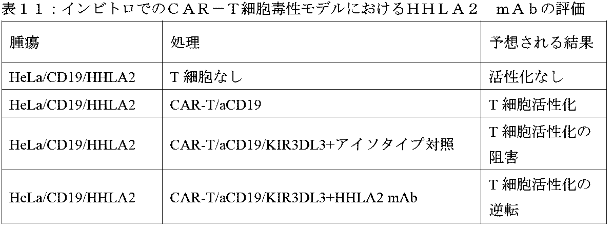
いくつかの実施形態では、NK細胞毒性のKIR3DL3阻害アッセイを行う。KIR3DL3トランスフェクトNK92細胞を生成する。例えば、NK92細胞をKIR3DL3 puroEIF2ベクターでトランスフェクトし、ピューロマイシン耐性のために選択し、KIR3DL3発現をフローサイトメトリーによって検出する。 In some embodiments, a KIR3DL3 inhibition assay of NK cytotoxicity is performed. Generate KIR3DL3 transfected NK92 cells. For example, NK92 cells are transfected with the KIR3DL3 puroEIF2 vector, selected for puromycin resistance, and KIR3DL3 expression is detected by flow cytometry.
HHLA2トランスフェクトK562標的細胞を生成する。例えば、標的K562細胞をHHLA2 pIRESneo3でトランスフェクトし、ネオマイシン耐性のために選択し、HHLA2発現をフローサイトメトリーによって検出した。 Generate HHLA2 transfected K562 target cells. For example, target K562 cells were transfected with HHLA2 pIRESneo3, selected for neomycin resistance, and HHLA2 expression was detected by flow cytometry.
KIR3DL3でトランスフェクトされたNK92細胞を、4時間細胞毒性アッセイにおいて、HHLA2 mAbの存在下または不在下で、K562細胞に対するNK細胞毒性(+または-HHLA2)について異なるE/T比で評価する。例示のCAR-T細胞毒性モデルインビトロ立体配置を以下の表13に列記する。
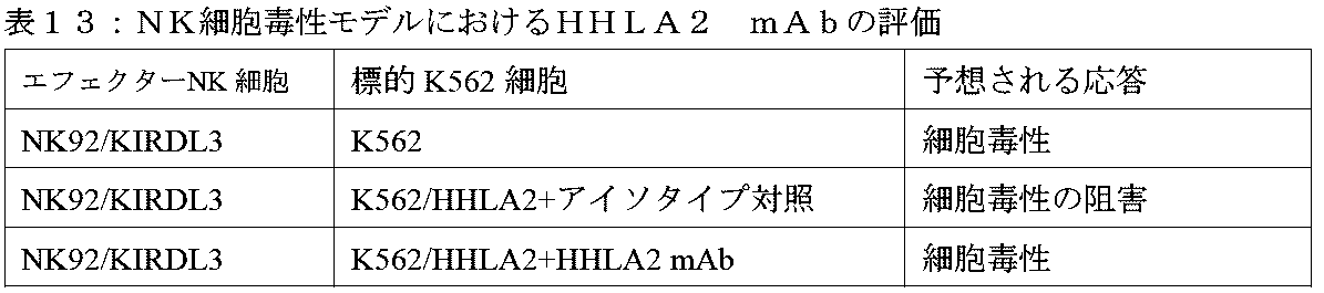
いくつかの実施形態では、Jurkat T細胞におけるKIR3DL3シグナル伝達経路を研究する。 In some embodiments, the KIR3DL3 signaling pathway in Jurkat T cells is studied.
参照による組み込み
本明細書で言及される全ての刊行物、特許、および特許出願は、個々の刊行物、特許、または特許出願が各々参照により組み込まれると具体的かつ個別に示されるかのように、参照によりそれらの全体が本明細書に組み込まれる。矛盾する場合には、本明細書のいずれの定義も含む本出願が支配する。
INCORPORATION BY REFERENCE All publications, patents, and patent applications mentioned herein are incorporated by reference as if each individual publication, patent, or patent application was specifically and individually indicated to be incorporated by reference. , incorporated herein by reference in their entirety. In case of conflict, the present application, including any definitions herein, will control.
The Institute for Genomic Research(TIGR)のワールドワイドウェブtigr.orgおよび/またはNational Center for Biotechnology Information(NCBI)のワールドワイドウェブncbi.nlm.nih.govによって管理されているもの等の公開データベースのエントリと相関する受入番号を参照する任意のポリヌクレオチドおよびポリペプチド配列も参照によりそれらの全体が組み込まれる。 The Institute for Genomic Research (TIGR) World Wide Web tigr. org and/or the National Center for Biotechnology Information (NCBI) World Wide Web ncbi. nlm. nih. Any polynucleotide and polypeptide sequences that reference accession numbers that correlate with entries in public databases, such as those maintained by .gov, are also incorporated by reference in their entirety.
等価物
当業者であれば、通例の実験のみを使用して、本明細書に記載の本発明の特定の実施形態に対する多くの等価物を認識するか、または確定することができるであろう。かかる等価物は、以下の特許請求の範囲によって包含されるよう意図されている。
Equivalents Those skilled in the art will recognize, or be able to ascertain using no more than routine experimentation, many equivalents to the specific embodiments of the invention described herein. Such equivalents are intended to be covered by the following claims.
Claims (20)
a)配列番号206のVH CDR1アミノ酸配列、配列番号210のVH CDR2アミノ酸配列、および配列番号214のVH CDR3アミノ酸配列を含むVH、および
b)配列番号224のVL CDR1アミノ酸配列、配列番号228のVL CDR2アミノ酸配列、および配列番号232のVL CDR3アミノ酸配列を含むVL
を含む、抗HHLA2モノクローナル抗体またはその抗原結合断片。 An anti-HHLA2 monoclonal antibody or antigen-binding fragment thereof, wherein the monoclonal antibody or antigen-binding fragment thereof is
a) a VH comprising a VH CDR1 amino acid sequence of SEQ ID NO: 206, a VH CDR2 amino acid sequence of SEQ ID NO: 210, and a VH CDR3 amino acid sequence of SEQ ID NO: 214; and b) a VL CDR1 amino acid sequence of SEQ ID NO: 224, a VL of SEQ ID NO: 228. A VL comprising a CDR2 amino acid sequence and a VL CDR3 amino acid sequence of SEQ ID NO: 232.
An anti-HHLA2 monoclonal antibody or antigen-binding fragment thereof.
a)配列番号199のアミノ酸配列を含むVH、および
b)配列番号217のアミノ酸配列を含むVL
を含む、請求項1に記載の抗HHLA2モノクローナル抗体またはその抗原結合断片。 The monoclonal antibody or antigen-binding fragment thereof is
a) VH comprising the amino acid sequence of SEQ ID NO: 199, and b) VL comprising the amino acid sequence of SEQ ID NO: 217.
The anti-HHLA2 monoclonal antibody or antigen-binding fragment thereof according to claim 1, comprising:
i)キメラ抗体、ヒト化抗体、複合抗体、またはマウス抗体である;かつ/または
ii)検出可能に標識され、エフェクタードメインを含み、Fcドメインを含む、かつ/または
iii)Fv、Fav、F(ab’)2、Fab’、dsFv、scFv、sc(Fv)2、およびダイアボディ断片からなる群から選択される、
請求項1または2に記載の抗HHLA2モノクローナル抗体またはその抗原結合断片。 The monoclonal antibody or antigen-binding fragment thereof is
i) is a chimeric , humanized, conjugated, or murine antibody ; and/or ii) is detectably labeled and includes an effector domain and includes an Fc domain ; and/or iii) is an Fv, Fav, F ( ab') 2, selected from the group consisting of Fab', dsFv, scFv, sc(Fv)2, and diabody fragments;
The anti-HHLA2 monoclonal antibody or antigen-binding fragment thereof according to claim 1 or 2.
(i)前記抗HHLA2モノクローナル抗体またはその抗原結合断片の発現を可能にするのに好適な条件下で、前記少なくとも1つの抗HHLA2モノクローナル抗体またはその抗原結合断片をコードする配列を含む核酸によって形質転換された形質転換宿主細胞を培養するステップと、
(ii)前記発現された抗HHLA2モノクローナル抗体またはその抗原結合断片を回収するステップと、
を含む、方法。 A method for producing at least one anti-HHLA2 monoclonal antibody or antigen-binding fragment thereof according to any one of claims 1 to 3, comprising:
(i) transformed with a nucleic acid comprising a sequence encoding said at least one anti-HHLA2 monoclonal antibody or antigen-binding fragment thereof under conditions suitable to allow expression of said anti-HHLA2 monoclonal antibody or antigen-binding fragment thereof; culturing the transformed host cells;
(ii) collecting the expressed anti-HHLA2 monoclonal antibody or antigen-binding fragment thereof;
including methods.
前記少なくとも1つの抗HHLA2モノクローナル抗体またはその抗原結合断片が、HHLA2ポリペプチドと複合体を形成し、前記複合体が、酵素結合免疫吸着アッセイ(ELISA)の形態で、放射免疫アッセイ(RIA)の形態で、免疫化学的に、ウエスタンブロットの形態で、または細胞内フローアッセイを使用して検出される、方法。 4. A method of detecting the presence or level of an HHLA2 polypeptide in a sample obtained using at least one anti-HHLA2 monoclonal antibody or antigen-binding fragment thereof according to any one of claims 1 to 3. and, optionally, detecting said polypeptide of
The at least one anti-HHLA2 monoclonal antibody or antigen-binding fragment thereof forms a complex with an HHLA2 polypeptide, and the complex is in the form of an enzyme-linked immunosorbent assay (ELISA), in the form of a radioimmunoassay (RIA). , detected immunochemically, in the form of a Western blot, or using an intracellular flow assay.
a)請求項1~3のいずれか一項に記載の少なくとも1つの抗HHLA2モノクローナル抗体またはその抗原結合断片を使用して、第1の時点で対象試料中のHHLA2レベルを検出することと、
b)その後の時点でステップa)を繰り返すことと、
c)ステップa)で検出されたHHLA2レベルとステップb)で検出されたHHLA2レベルとを比較することであって、ステップa)で検出されたHHLA2レベルとステップb)で検出されたHHLA2レベルとの前記比較は、前記対象における前記障害の進行の指標であることと、を含み、任意に、前記第1の時点と前記その後の時点との間で、前記対象が前記障害を改善するための治療を受けている、方法。 A method for determining the level of HHLA2 as an indicator of the progression of a disorder associated with aberrant HHLA2 expression in a subject, the method comprising:
a) detecting the level of HHLA2 in a subject sample at a first time point using at least one anti-HHLA2 monoclonal antibody or antigen-binding fragment thereof according to any one of claims 1 to 3;
b) repeating step a) at a subsequent time; and
c) comparing the HHLA2 level detected in step a) and the HHLA2 level detected in step b), the HHLA2 level detected in step a) and the HHLA2 level detected in step b); the comparison of is indicative of the progression of the disorder in the subject, optionally between the first time point and the subsequent time point, the comparison is indicative of the progression of the disorder in the subject; How you are being treated.
a)請求項1~3のいずれか一項に記載の少なくとも1つの抗HHLA2モノクローナル抗体またはその抗原結合断片を使用して、対象試料中のHHLA2レベルを決定することと、
b)前記少なくとも1つの抗HHLA2モノクローナル抗体またはその抗原結合断片を使用して、良好な臨床転帰を有する対照対象由来の試料中のHHLA2レベルを決定することと、
c)前記対象試料中のHHLA2レベルと前記対照対象由来の前記試料中のHHLA2レベルとを比較することと、を含み、
前記対照対象由来の前記試料中のレベルと比較して前記対象試料中の有意により高いHHLA2レベルが、前記対象が不良な臨床転帰を有することを示し、任意に、前記有意により高いHHLA2レベルは、前記対照対象由来の前記試料の正常なHHLA2レベルと比較した前記対象試料中のHHLA2レベルの少なくとも20パーセントの増加を含む、方法。 A method for determining HHLA2 levels as an indicator of clinical outcome in a subject suffering from a disorder associated with aberrant HHLA2 expression, the method comprising:
a) determining the level of HHLA2 in a subject sample using at least one anti-HHLA2 monoclonal antibody or antigen-binding fragment thereof according to any one of claims 1 to 3;
b) using said at least one anti-HHLA2 monoclonal antibody or antigen-binding fragment thereof to determine HHLA2 levels in a sample from a control subject with a good clinical outcome;
c) comparing the HHLA2 level in the subject sample to the HHLA2 level in the sample from the control subject;
A significantly higher HHLA2 level in the subject sample compared to a level in the sample from the control subject indicates that the subject has a poor clinical outcome; optionally, the significantly higher HHLA2 level comprises: The method comprises increasing the level of HHLA2 in the subject sample by at least 20 percent compared to the normal HHLA2 level of the sample from the control subject.
a)請求項1~3のいずれか一項に記載の少なくとも1つの抗HHLA2モノクローナル抗体またはその抗原結合断片を使用して、前記療法の少なくとも一部を前記対象に施す前に前記対象から得られた第1の試料中のHHLA2レベルを決定することと、
b)前記療法の前記一部を施した後に前記対象から得られた第2の試料中のHHLA2レベルを決定することと、を含み、
前記第1の試料と比較して前記第2の試料中の有意により低いHHLA2レベルが、前記療法が前記対象における前記障害を抑制するのに有効であることを示し、任意に、前記有意により低いHHLA2レベルが、HHLA2レベルの少なくとも20パーセントの減少を含む、方法;あるいは
対象における異常なHHLA2発現に関連する障害を抑制するための試験化合物の有効性の指標としてのHHLA2レベルを決定する方法であって、
a)請求項1~3のいずれか一項に記載の少なくとも1つの抗HHLA2モノクローナル抗体またはその抗原結合断片を使用して、前記対象から得られ、かつ前記試験化合物に曝露された第1の試料中のHHLA2レベルを決定することと、
b)前記対象から得られた第2の試料中のHHLA2レベルを決定することと、を含み、
前記第2の試料が前記試験化合物に曝露されず、前記第2の試料と比較して有意により低いHHLA2レベルが、前記試験化合物が前記対象における前記障害を抑制するのに有効であることを示し、任意に、前記第1の試料および前記第2の試料が、前記対象から得られた単一試料の一部であるか、または前記対象から得られたプールされた試料の一部である、方法。 A method for determining HHLA2 levels as an indicator of effectiveness of therapy for a disorder associated with aberrant HHLA2 expression in a subject, the method comprising:
a) at least one anti-HHLA2 monoclonal antibody or antigen-binding fragment thereof according to any one of claims 1 to 3 obtained from the subject before administering at least a part of the therapy to the subject; determining the level of HHLA2 in the first sample;
b) determining the level of HHLA2 in a second sample obtained from the subject after administering the portion of the therapy;
a significantly lower HHLA2 level in the second sample compared to the first sample indicates that the therapy is effective in inhibiting the disorder in the subject; a method, wherein the low HHLA2 level comprises a decrease of at least 20 percent in HHLA2 levels; or a method of determining HHLA2 levels as an indicator of the effectiveness of a test compound to inhibit a disorder associated with aberrant HHLA2 expression in a subject. There it is,
a) a first sample obtained from said subject and exposed to said test compound using at least one anti-HHLA2 monoclonal antibody or antigen-binding fragment thereof according to any one of claims 1 to 3; determining the HHLA2 level in the
b) determining the level of HHLA2 in a second sample obtained from the subject;
wherein the second sample is not exposed to the test compound and a significantly lower HHLA2 level compared to the second sample indicates that the test compound is effective in inhibiting the disorder in the subject. , optionally, the first sample and the second sample are part of a single sample obtained from the subject or are part of a pooled sample obtained from the subject; Method.
(b)前記試料が、前記対象から得られた細胞、血清、腫瘍周辺組織、および/または腫瘍内組織を含む;ならびに/あるいは
(c)前記対象がヒトである、請求項11~13のいずれか一項に記載の方法。 (a) said disorder is cancer, and optionally said cancer is lung cancer, kidney cancer, pancreatic cancer, colorectal cancer, acute myeloid leukemia, head and neck cancer, liver cancer, ovarian cancer, prostate cancer, uterine cancer. , glioma, glioblastoma, neuroblastoma, breast cancer, pancreatic ductal carcinoma, thymoma, B-CLL, leukemia, B-cell lymphoma, and cancers infiltrated by immune cells expressing receptors for HHLA2. selected from the group;
(b) the sample comprises cells, serum, peritumoral tissue, and/or intratumoral tissue obtained from the subject; and/or (c) the subject is a human. The method described in paragraph (1).
(b)前記少なくとも1つの抗HHLA2モノクローナル抗体またはその抗原結合断片が、前記がんにおける増殖細胞の数を減少させ、かつ/または前記がんの腫瘍の体積もしくはサイズを減少させる;
(c)前記少なくとも1つの抗HHLA2モノクローナル抗体またはその抗原結合断片が、薬学的に許容される製剤で投与され得る;
(d)がんを治療するためのさらなる治療薬またはレジメンが前記対象に投与され得る;ならびに/あるいは
(e)免疫療法、チェックポイント遮断、がんワクチン、キメラ抗原受容体、化学療法、放射線、標的療法、および手術からなる群から選択される追加の療法が前記対象に投与され得る、請求項15に記載の組成物。 (a) said at least one anti-HHLA2 monoclonal antibody or antigen-binding fragment thereof is conjugated to a cytotoxic agent, optionally said cytotoxic agent being a chemotherapeutic agent, a biological agent, a toxin, and a radioactive agent; selected from the group consisting of isotopes;
(b) said at least one anti-HHLA2 monoclonal antibody or antigen-binding fragment thereof reduces the number of proliferating cells in said cancer and/or reduces the volume or size of a tumor in said cancer;
(c) said at least one anti-HHLA2 monoclonal antibody or antigen-binding fragment thereof may be administered in a pharmaceutically acceptable formulation;
(d) additional therapeutic agents or regimens for treating cancer may be administered to said subject; and/or (e) immunotherapy, checkpoint blockade, cancer vaccines, chimeric antigen receptors, chemotherapy, radiation, 16. The composition of claim 15, wherein an additional therapy selected from the group consisting of targeted therapy, and surgery can be administered to the subject.
(b)前記がんが、肺癌、腎臓癌、膵臓癌、結腸直腸癌、急性骨髄性白血病、頭頸部癌、肝臓癌、卵巣癌、前立腺癌、子宮癌、神経膠腫、神経膠芽腫、神経芽細胞腫、乳癌、膵管癌、胸腺腫、B-CLL、白血病、B細胞リンパ腫、およびHHLA2に対する受容体を発現する免疫細胞が浸潤したがんからなる群から選択される;
(c)前記がんが、肺癌、腎臓癌、膵臓癌、結腸直腸癌、急性骨髄性白血病(AML)、頭頸部癌、肝臓癌、卵巣癌、前立腺癌、および子宮癌からなる群から選択される;
(d)前記対象が、がんの動物モデルであり;任意に、前記動物モデルがマウスモデルであり、任意に、前記マウスモデルがヒト化マウスモデルである;ならびに/あるいは
(e)前記対象が哺乳動物であり、任意に、前記哺乳動物がヒト化マウスまたはヒトである、請求項15または16に記載の組成物。 (a) cancer cells and/or tumor immune infiltrating cells in said subject express HHLA2;
(b) the cancer is lung cancer, kidney cancer, pancreatic cancer, colorectal cancer, acute myeloid leukemia, head and neck cancer, liver cancer, ovarian cancer, prostate cancer, uterine cancer, glioma, glioblastoma, selected from the group consisting of neuroblastoma, breast cancer, pancreatic ductal carcinoma, thymoma, B-CLL, leukemia, B-cell lymphoma, and cancer infiltrated by immune cells expressing receptors for HHLA2;
(c) the cancer is selected from the group consisting of lung cancer, kidney cancer, pancreatic cancer, colorectal cancer, acute myeloid leukemia (AML), head and neck cancer, liver cancer, ovarian cancer, prostate cancer, and uterine cancer. Ru;
(d) said subject is an animal model of cancer; optionally said animal model is a mouse model; optionally said mouse model is a humanized mouse model; and/or (e) said subject is 17. The composition of claim 15 or 16, which is a mammal, optionally said mammal being a humanized mouse or human.
薬学的組成物。 5. The pharmaceutical composition of claim 4 for use in modulating an immune response, wherein upon administration of the pharmaceutical composition to a subject, the anti-HHLA2 monoclonal antibody or antigen-binding fragment thereof inhibits HHLA2 and KIR3DL3. inhibiting or blocking the interaction between HHLA2 and TMIGD2, and partially blocking or inhibiting the interaction between HHLA2 and TMIGD2 ;
Pharmaceutical composition.
Priority Applications (1)
| Application Number | Priority Date | Filing Date | Title |
|---|---|---|---|
| JP2024009992A JP2024032865A (en) | 2018-04-06 | 2024-01-26 | KIR3DL3 as HHLA2 receptor, anti-HHLA2 antibody and its use |
Applications Claiming Priority (3)
| Application Number | Priority Date | Filing Date | Title |
|---|---|---|---|
| US201862654068P | 2018-04-06 | 2018-04-06 | |
| US62,654,068 | 2018-04-06 | ||
| PCT/US2019/026034 WO2019204057A1 (en) | 2018-04-06 | 2019-04-05 | Kir3dl3 as an hhla2 receptor, anti-hhla2 antibodies, and uses thereof |
Related Child Applications (1)
| Application Number | Title | Priority Date | Filing Date |
|---|---|---|---|
| JP2024009992A Division JP2024032865A (en) | 2018-04-06 | 2024-01-26 | KIR3DL3 as HHLA2 receptor, anti-HHLA2 antibody and its use |
Publications (3)
| Publication Number | Publication Date |
|---|---|
| JP2021520208A JP2021520208A (en) | 2021-08-19 |
| JPWO2019204057A5 JPWO2019204057A5 (en) | 2022-04-12 |
| JP7429192B2 true JP7429192B2 (en) | 2024-02-07 |
Family
ID=68239919
Family Applications (2)
| Application Number | Title | Priority Date | Filing Date |
|---|---|---|---|
| JP2020554272A Active JP7429192B2 (en) | 2018-04-06 | 2019-04-05 | KIR3DL3 as HHLA2 receptor, anti-HHLA2 antibody and its use |
| JP2024009992A Pending JP2024032865A (en) | 2018-04-06 | 2024-01-26 | KIR3DL3 as HHLA2 receptor, anti-HHLA2 antibody and its use |
Family Applications After (1)
| Application Number | Title | Priority Date | Filing Date |
|---|---|---|---|
| JP2024009992A Pending JP2024032865A (en) | 2018-04-06 | 2024-01-26 | KIR3DL3 as HHLA2 receptor, anti-HHLA2 antibody and its use |
Country Status (13)
| Country | Link |
|---|---|
| US (1) | US20210115144A1 (en) |
| EP (1) | EP3773658A4 (en) |
| JP (2) | JP7429192B2 (en) |
| KR (1) | KR20210007959A (en) |
| CN (1) | CN112153977A (en) |
| AU (1) | AU2019257249A1 (en) |
| BR (1) | BR112020015999A2 (en) |
| CA (1) | CA3091859A1 (en) |
| IL (1) | IL276746A (en) |
| MX (1) | MX2020010094A (en) |
| RU (1) | RU2020136055A (en) |
| SG (1) | SG11202007898XA (en) |
| WO (1) | WO2019204057A1 (en) |
Families Citing this family (7)
| Publication number | Priority date | Publication date | Assignee | Title |
|---|---|---|---|---|
| WO2021067633A2 (en) * | 2019-10-04 | 2021-04-08 | Albert Einstein College Of Medicine | Kir3dl3 is an inhibitory receptor of the immune system and uses thereof |
| US20230178239A1 (en) * | 2020-05-13 | 2023-06-08 | Juno Therapeutics, Inc. | Methods of identifying features associated with clinical response and uses thereof |
| CN112946268B (en) * | 2020-12-29 | 2022-12-27 | 中山大学肿瘤防治中心(中山大学附属肿瘤医院、中山大学肿瘤研究所) | Application of HHLA2 as prognostic marker in preparation of liver cancer prognosis prediction kit |
| EP4284431A1 (en) * | 2021-01-28 | 2023-12-06 | Nextpoint Therapeutics, Inc. | Hhla2 binding agents with novel activity |
| WO2023279107A1 (en) * | 2021-07-01 | 2023-01-05 | Albert Einstein College Of Medicine | Compositions and methods for inhibiting the expression of tmigd2 |
| CN113621068B (en) * | 2021-10-11 | 2022-01-07 | 上海恒润达生生物科技股份有限公司 | Antibody or antigen binding fragment thereof specifically binding to CD276, and preparation method and application thereof |
| WO2023138579A1 (en) * | 2022-01-21 | 2023-07-27 | 和铂医药美国股份有限公司 | Anti-b7-h7 antibody or antigen-binding fragment thereof, and preparation method therefor and use thereof |
Citations (1)
| Publication number | Priority date | Publication date | Assignee | Title |
|---|---|---|---|---|
| US20170081410A1 (en) | 2014-06-04 | 2017-03-23 | Ngm Biopharmaceuticals, Inc. | Compositions and methods for targeting a pathway |
Family Cites Families (6)
| Publication number | Priority date | Publication date | Assignee | Title |
|---|---|---|---|---|
| EP1562968B1 (en) * | 2001-11-14 | 2013-08-21 | Janssen Biotech, Inc. | Anti-il-6 antibodies, compositions, methods and uses |
| EP3381937A3 (en) * | 2009-08-13 | 2018-10-31 | The Johns Hopkins University | Methods of modulating immune function |
| CA2845259A1 (en) * | 2011-08-15 | 2013-02-21 | The University Of Chicago | Compositions and methods related to antibodies to staphylococcal protein a |
| US10093737B2 (en) * | 2013-03-01 | 2018-10-09 | Albert Einstein College Of Medicine, Inc. | HHLA2 as a novel inhibitor of human immune system and uses thereof |
| CN106414489A (en) * | 2013-05-24 | 2017-02-15 | 米迪缪尼有限公司 | Anti-b7-h5 antibodies and their uses |
| WO2018026969A2 (en) * | 2016-08-03 | 2018-02-08 | Achaogen, Inc. | Plazomicin antibodies and methods of use |
-
2019
- 2019-04-05 CA CA3091859A patent/CA3091859A1/en active Pending
- 2019-04-05 US US17/044,493 patent/US20210115144A1/en active Pending
- 2019-04-05 CN CN201980031870.6A patent/CN112153977A/en active Pending
- 2019-04-05 JP JP2020554272A patent/JP7429192B2/en active Active
- 2019-04-05 RU RU2020136055A patent/RU2020136055A/en unknown
- 2019-04-05 MX MX2020010094A patent/MX2020010094A/en unknown
- 2019-04-05 AU AU2019257249A patent/AU2019257249A1/en active Pending
- 2019-04-05 SG SG11202007898XA patent/SG11202007898XA/en unknown
- 2019-04-05 EP EP19789172.4A patent/EP3773658A4/en active Pending
- 2019-04-05 BR BR112020015999-9A patent/BR112020015999A2/en unknown
- 2019-04-05 KR KR1020207030319A patent/KR20210007959A/en active Search and Examination
- 2019-04-05 WO PCT/US2019/026034 patent/WO2019204057A1/en unknown
-
2020
- 2020-08-17 IL IL276746A patent/IL276746A/en unknown
-
2024
- 2024-01-26 JP JP2024009992A patent/JP2024032865A/en active Pending
Patent Citations (1)
| Publication number | Priority date | Publication date | Assignee | Title |
|---|---|---|---|---|
| US20170081410A1 (en) | 2014-06-04 | 2017-03-23 | Ngm Biopharmaceuticals, Inc. | Compositions and methods for targeting a pathway |
Also Published As
| Publication number | Publication date |
|---|---|
| IL276746A (en) | 2020-10-29 |
| AU2019257249A1 (en) | 2020-10-15 |
| WO2019204057A1 (en) | 2019-10-24 |
| KR20210007959A (en) | 2021-01-20 |
| MX2020010094A (en) | 2021-01-15 |
| JP2021520208A (en) | 2021-08-19 |
| RU2020136055A3 (en) | 2022-05-06 |
| EP3773658A1 (en) | 2021-02-17 |
| US20210115144A1 (en) | 2021-04-22 |
| JP2024032865A (en) | 2024-03-12 |
| BR112020015999A2 (en) | 2020-12-15 |
| RU2020136055A (en) | 2022-05-06 |
| CA3091859A1 (en) | 2019-10-24 |
| CN112153977A (en) | 2020-12-29 |
| SG11202007898XA (en) | 2020-10-29 |
| EP3773658A4 (en) | 2022-04-27 |
Similar Documents
| Publication | Publication Date | Title |
|---|---|---|
| JP7429192B2 (en) | KIR3DL3 as HHLA2 receptor, anti-HHLA2 antibody and its use | |
| US20220033502A1 (en) | METHODS FOR UPREGULATING IMMUNE RESPONSES USING COMBINATIONS OF ANTI-RGMb AND ANTI-PD-1 AGENTS | |
| EP3362074A2 (en) | Regulatory t cell pd-1 modulation for regulating t cell effector immune responses | |
| KR20220042056A (en) | Anti-SIGLEC-9 compositions and methods for modulating myeloid cell inflammatory phenotype, and uses thereof | |
| WO2019014665A1 (en) | Modulating biomarkers to increase tumor immunity and improve the efficacy of cancer immunotherapy | |
| WO2016179194A1 (en) | Lilra3 and method of using the same | |
| US20210032334A1 (en) | Methods for treating cancer using combinations of anti-btnl2 and immune checkpoint blockade agents | |
| JP2022552153A (en) | Anti-KIR3DL3 Antibodies and Their Use | |
| KR20230017778A (en) | Anti-VSIG4 compositions and methods for modulating myeloid cell inflammatory phenotype, and uses thereof | |
| WO2018231339A2 (en) | Anti-phosphotyrosinylated pd-1 antibodies and uses thereof | |
| KR20220042055A (en) | Anti-PSGL-1 compositions and methods for modulating myeloid cell inflammatory phenotype, and uses thereof | |
| JP2023553247A (en) | Compositions and methods for cancer diagnosis | |
| WO2021150925A1 (en) | Uses of biomarkers for improving immunotherapy | |
| KR20220042131A (en) | Anti-LRRC25 compositions and methods for modulating myeloid cell inflammatory phenotype, and uses thereof | |
| US20220289854A1 (en) | Methods for treating cancer using combinations of anti-cx3cr1 and immune checkpoint blockade agents | |
| US20220211848A1 (en) | Modulating gabarap to modulate immunogenic cell death | |
| WO2019014664A1 (en) | Modulating biomarkers to increase tumor immunity and improve the efficacy of cancer immunotherapy |
Legal Events
| Date | Code | Title | Description |
|---|---|---|---|
| A521 | Request for written amendment filed |
Free format text: JAPANESE INTERMEDIATE CODE: A523 Effective date: 20220404 |
|
| A621 | Written request for application examination |
Free format text: JAPANESE INTERMEDIATE CODE: A621 Effective date: 20220404 |
|
| A977 | Report on retrieval |
Free format text: JAPANESE INTERMEDIATE CODE: A971007 Effective date: 20230308 |
|
| A131 | Notification of reasons for refusal |
Free format text: JAPANESE INTERMEDIATE CODE: A131 Effective date: 20230317 |
|
| A521 | Request for written amendment filed |
Free format text: JAPANESE INTERMEDIATE CODE: A523 Effective date: 20230616 |
|
| A131 | Notification of reasons for refusal |
Free format text: JAPANESE INTERMEDIATE CODE: A131 Effective date: 20230824 |
|
| A521 | Request for written amendment filed |
Free format text: JAPANESE INTERMEDIATE CODE: A523 Effective date: 20231124 |
|
| TRDD | Decision of grant or rejection written | ||
| A01 | Written decision to grant a patent or to grant a registration (utility model) |
Free format text: JAPANESE INTERMEDIATE CODE: A01 Effective date: 20231212 |
|
| A601 | Written request for extension of time |
Free format text: JAPANESE INTERMEDIATE CODE: A601 Effective date: 20240109 |
|
| A61 | First payment of annual fees (during grant procedure) |
Free format text: JAPANESE INTERMEDIATE CODE: A61 Effective date: 20240126 |
|
| R150 | Certificate of patent or registration of utility model |
Ref document number: 7429192 Country of ref document: JP Free format text: JAPANESE INTERMEDIATE CODE: R150 |