JP7386852B2 - Peptide activator - Google Patents
Peptide activator Download PDFInfo
- Publication number
- JP7386852B2 JP7386852B2 JP2021514457A JP2021514457A JP7386852B2 JP 7386852 B2 JP7386852 B2 JP 7386852B2 JP 2021514457 A JP2021514457 A JP 2021514457A JP 2021514457 A JP2021514457 A JP 2021514457A JP 7386852 B2 JP7386852 B2 JP 7386852B2
- Authority
- JP
- Japan
- Prior art keywords
- cells
- cell
- lck
- peptide
- expression
- Prior art date
- Legal status (The legal status is an assumption and is not a legal conclusion. Google has not performed a legal analysis and makes no representation as to the accuracy of the status listed.)
- Active
Links
- 108090000765 processed proteins & peptides Proteins 0.000 title claims description 412
- 239000012190 activator Substances 0.000 title description 87
- 230000003213 activating effect Effects 0.000 claims description 195
- 239000000203 mixture Substances 0.000 claims description 149
- 150000001413 amino acids Chemical group 0.000 claims description 98
- 239000008194 pharmaceutical composition Substances 0.000 claims description 41
- 230000028993 immune response Effects 0.000 claims description 24
- 230000001939 inductive effect Effects 0.000 claims description 12
- 102100024036 Tyrosine-protein kinase Lck Human genes 0.000 claims description 9
- 101001047681 Homo sapiens Tyrosine-protein kinase Lck Proteins 0.000 claims description 8
- 239000004480 active ingredient Substances 0.000 claims description 3
- 239000003085 diluting agent Substances 0.000 claims description 2
- 210000004027 cell Anatomy 0.000 description 445
- 101150028321 Lck gene Proteins 0.000 description 425
- 210000001744 T-lymphocyte Anatomy 0.000 description 269
- 238000000034 method Methods 0.000 description 260
- 230000014509 gene expression Effects 0.000 description 147
- 230000001965 increasing effect Effects 0.000 description 135
- 102000004196 processed proteins & peptides Human genes 0.000 description 113
- 150000004665 fatty acids Chemical class 0.000 description 104
- 235000001014 amino acid Nutrition 0.000 description 100
- 229940024606 amino acid Drugs 0.000 description 99
- 235000014113 dietary fatty acids Nutrition 0.000 description 97
- 229930195729 fatty acid Natural products 0.000 description 97
- 239000000194 fatty acid Substances 0.000 description 97
- 108010002350 Interleukin-2 Proteins 0.000 description 95
- 102000000588 Interleukin-2 Human genes 0.000 description 95
- 206010028980 Neoplasm Diseases 0.000 description 84
- 210000001266 CD8-positive T-lymphocyte Anatomy 0.000 description 83
- 230000000694 effects Effects 0.000 description 78
- 210000000822 natural killer cell Anatomy 0.000 description 72
- 238000011282 treatment Methods 0.000 description 61
- 201000011510 cancer Diseases 0.000 description 48
- 230000004913 activation Effects 0.000 description 44
- 230000027455 binding Effects 0.000 description 44
- 230000028327 secretion Effects 0.000 description 43
- 150000001875 compounds Chemical class 0.000 description 42
- 230000004044 response Effects 0.000 description 41
- 208000037265 diseases, disorders, signs and symptoms Diseases 0.000 description 40
- 230000026731 phosphorylation Effects 0.000 description 39
- 238000006366 phosphorylation reaction Methods 0.000 description 39
- 241000699670 Mus sp. Species 0.000 description 38
- 230000000638 stimulation Effects 0.000 description 37
- 101000946843 Homo sapiens T-cell surface glycoprotein CD8 alpha chain Proteins 0.000 description 36
- 102100034922 T-cell surface glycoprotein CD8 alpha chain Human genes 0.000 description 36
- 102000016266 T-Cell Antigen Receptors Human genes 0.000 description 35
- 229920001184 polypeptide Polymers 0.000 description 35
- 108091008874 T cell receptors Proteins 0.000 description 34
- 125000005647 linker group Chemical group 0.000 description 34
- 230000035755 proliferation Effects 0.000 description 34
- 239000003981 vehicle Substances 0.000 description 34
- 230000003915 cell function Effects 0.000 description 33
- 230000008685 targeting Effects 0.000 description 33
- 208000015181 infectious disease Diseases 0.000 description 32
- 210000003289 regulatory T cell Anatomy 0.000 description 31
- 241000282414 Homo sapiens Species 0.000 description 30
- QNAYBMKLOCPYGJ-REOHCLBHSA-N L-alanine Chemical compound C[C@H](N)C(O)=O QNAYBMKLOCPYGJ-REOHCLBHSA-N 0.000 description 29
- 235000004279 alanine Nutrition 0.000 description 29
- 102000004127 Cytokines Human genes 0.000 description 28
- 108090000695 Cytokines Proteins 0.000 description 28
- 108091000080 Phosphotransferase Proteins 0.000 description 28
- 102000020233 phosphotransferase Human genes 0.000 description 28
- 101000914514 Homo sapiens T-cell-specific surface glycoprotein CD28 Proteins 0.000 description 27
- 102100027213 T-cell-specific surface glycoprotein CD28 Human genes 0.000 description 27
- 210000003819 peripheral blood mononuclear cell Anatomy 0.000 description 27
- 230000002829 reductive effect Effects 0.000 description 27
- 230000004043 responsiveness Effects 0.000 description 27
- KZSNJWFQEVHDMF-BYPYZUCNSA-N L-valine Chemical compound CC(C)[C@H](N)C(O)=O KZSNJWFQEVHDMF-BYPYZUCNSA-N 0.000 description 25
- 101100519207 Mus musculus Pdcd1 gene Proteins 0.000 description 25
- KZSNJWFQEVHDMF-UHFFFAOYSA-N Valine Natural products CC(C)C(N)C(O)=O KZSNJWFQEVHDMF-UHFFFAOYSA-N 0.000 description 25
- 201000010099 disease Diseases 0.000 description 25
- 238000000338 in vitro Methods 0.000 description 25
- 102000005962 receptors Human genes 0.000 description 25
- 108020003175 receptors Proteins 0.000 description 25
- 235000014393 valine Nutrition 0.000 description 25
- 239000004474 valine Substances 0.000 description 25
- 239000004952 Polyamide Substances 0.000 description 23
- 102000036639 antigens Human genes 0.000 description 23
- 238000001727 in vivo Methods 0.000 description 23
- 229920002647 polyamide Polymers 0.000 description 23
- 125000003275 alpha amino acid group Chemical group 0.000 description 22
- 239000000427 antigen Substances 0.000 description 22
- 108091007433 antigens Proteins 0.000 description 22
- 238000007492 two-way ANOVA Methods 0.000 description 22
- 241000700605 Viruses Species 0.000 description 21
- 108010002481 Lymphocyte Specific Protein Tyrosine Kinase p56(lck) Proteins 0.000 description 20
- 102000036243 Lymphocyte Specific Protein Tyrosine Kinase p56(lck) Human genes 0.000 description 20
- 230000032683 aging Effects 0.000 description 20
- 238000003776 cleavage reaction Methods 0.000 description 20
- 230000006870 function Effects 0.000 description 20
- 238000004519 manufacturing process Methods 0.000 description 20
- 108090000623 proteins and genes Proteins 0.000 description 20
- 230000007017 scission Effects 0.000 description 20
- 125000004432 carbon atom Chemical group C* 0.000 description 19
- 238000000684 flow cytometry Methods 0.000 description 19
- 230000007423 decrease Effects 0.000 description 18
- 102100020792 Interleukin-12 receptor subunit beta-2 Human genes 0.000 description 17
- 101710103840 Interleukin-12 receptor subunit beta-2 Proteins 0.000 description 17
- 102000003812 Interleukin-15 Human genes 0.000 description 17
- 206010058467 Lung neoplasm malignant Diseases 0.000 description 17
- 206010040047 Sepsis Diseases 0.000 description 17
- 201000005202 lung cancer Diseases 0.000 description 17
- 208000020816 lung neoplasm Diseases 0.000 description 17
- 230000011664 signaling Effects 0.000 description 17
- 102000017420 CD3 protein, epsilon/gamma/delta subunit Human genes 0.000 description 16
- 108050005493 CD3 protein, epsilon/gamma/delta subunit Proteins 0.000 description 16
- OKTJSMMVPCPJKN-UHFFFAOYSA-N Carbon Chemical compound [C] OKTJSMMVPCPJKN-UHFFFAOYSA-N 0.000 description 16
- 230000006044 T cell activation Effects 0.000 description 16
- 210000004443 dendritic cell Anatomy 0.000 description 16
- 108040002099 interleukin-21 receptor activity proteins Proteins 0.000 description 16
- 102000008640 interleukin-21 receptor activity proteins Human genes 0.000 description 16
- 238000010149 post-hoc-test Methods 0.000 description 16
- 230000001629 suppression Effects 0.000 description 16
- 241000725303 Human immunodeficiency virus Species 0.000 description 15
- ROHFNLRQFUQHCH-YFKPBYRVSA-N L-leucine Chemical compound CC(C)C[C@H](N)C(O)=O ROHFNLRQFUQHCH-YFKPBYRVSA-N 0.000 description 15
- ROHFNLRQFUQHCH-UHFFFAOYSA-N Leucine Natural products CC(C)CC(N)C(O)=O ROHFNLRQFUQHCH-UHFFFAOYSA-N 0.000 description 15
- KDXKERNSBIXSRK-UHFFFAOYSA-N Lysine Natural products NCCCCC(N)C(O)=O KDXKERNSBIXSRK-UHFFFAOYSA-N 0.000 description 15
- 125000004429 atom Chemical group 0.000 description 15
- 208000035475 disorder Diseases 0.000 description 15
- 239000012636 effector Substances 0.000 description 15
- 230000002209 hydrophobic effect Effects 0.000 description 15
- 235000005772 leucine Nutrition 0.000 description 15
- 239000000178 monomer Substances 0.000 description 15
- 230000001717 pathogenic effect Effects 0.000 description 15
- 230000037361 pathway Effects 0.000 description 15
- 210000004988 splenocyte Anatomy 0.000 description 15
- QUBNFZFTFXTLKH-UHFFFAOYSA-N 2-aminododecanoic acid Chemical compound CCCCCCCCCCC(N)C(O)=O QUBNFZFTFXTLKH-UHFFFAOYSA-N 0.000 description 14
- 239000004475 Arginine Substances 0.000 description 14
- 239000004472 Lysine Substances 0.000 description 14
- ODKSFYDXXFIFQN-UHFFFAOYSA-N arginine Natural products OC(=O)C(N)CCCNC(N)=N ODKSFYDXXFIFQN-UHFFFAOYSA-N 0.000 description 14
- 235000009697 arginine Nutrition 0.000 description 14
- 230000004073 interleukin-2 production Effects 0.000 description 14
- 102000004169 proteins and genes Human genes 0.000 description 14
- 102000004190 Enzymes Human genes 0.000 description 13
- 108090000790 Enzymes Proteins 0.000 description 13
- 102000001253 Protein Kinase Human genes 0.000 description 13
- 108060008682 Tumor Necrosis Factor Proteins 0.000 description 13
- 102000000852 Tumor Necrosis Factor-alpha Human genes 0.000 description 13
- 238000003556 assay Methods 0.000 description 13
- 229940088598 enzyme Drugs 0.000 description 13
- 230000001506 immunosuppresive effect Effects 0.000 description 13
- 108060006633 protein kinase Proteins 0.000 description 13
- 235000018102 proteins Nutrition 0.000 description 13
- -1 Yes Proteins 0.000 description 12
- 125000003178 carboxy group Chemical group [H]OC(*)=O 0.000 description 12
- 230000001684 chronic effect Effects 0.000 description 12
- 238000011161 development Methods 0.000 description 12
- 230000018109 developmental process Effects 0.000 description 12
- 230000002708 enhancing effect Effects 0.000 description 12
- 238000002866 fluorescence resonance energy transfer Methods 0.000 description 12
- 230000001404 mediated effect Effects 0.000 description 12
- PBLZLIFKVPJDCO-UHFFFAOYSA-N 12-aminododecanoic acid Chemical compound NCCCCCCCCCCCC(O)=O PBLZLIFKVPJDCO-UHFFFAOYSA-N 0.000 description 11
- 241000894006 Bacteria Species 0.000 description 11
- 238000002965 ELISA Methods 0.000 description 11
- 208000031886 HIV Infections Diseases 0.000 description 11
- 230000001472 cytotoxic effect Effects 0.000 description 11
- 239000003814 drug Substances 0.000 description 11
- 239000012634 fragment Substances 0.000 description 11
- WWZKQHOCKIZLMA-UHFFFAOYSA-N octanoic acid Chemical compound CCCCCCCC(O)=O WWZKQHOCKIZLMA-UHFFFAOYSA-N 0.000 description 11
- 230000002265 prevention Effects 0.000 description 11
- 230000001105 regulatory effect Effects 0.000 description 11
- 239000000243 solution Substances 0.000 description 11
- MTCFGRXMJLQNBG-REOHCLBHSA-N (2S)-2-Amino-3-hydroxypropansäure Chemical compound OC[C@H](N)C(O)=O MTCFGRXMJLQNBG-REOHCLBHSA-N 0.000 description 10
- 108010029697 CD40 Ligand Proteins 0.000 description 10
- 102100032937 CD40 ligand Human genes 0.000 description 10
- 101001023379 Homo sapiens Lysosome-associated membrane glycoprotein 1 Proteins 0.000 description 10
- 102100035133 Lysosome-associated membrane glycoprotein 1 Human genes 0.000 description 10
- MTCFGRXMJLQNBG-UHFFFAOYSA-N Serine Natural products OCC(N)C(O)=O MTCFGRXMJLQNBG-UHFFFAOYSA-N 0.000 description 10
- 230000006052 T cell proliferation Effects 0.000 description 10
- 229910052799 carbon Inorganic materials 0.000 description 10
- 231100000433 cytotoxic Toxicity 0.000 description 10
- 230000001419 dependent effect Effects 0.000 description 10
- 230000002255 enzymatic effect Effects 0.000 description 10
- FUZZWVXGSFPDMH-UHFFFAOYSA-N hexanoic acid Chemical compound CCCCCC(O)=O FUZZWVXGSFPDMH-UHFFFAOYSA-N 0.000 description 10
- 244000052769 pathogen Species 0.000 description 10
- 229920006395 saturated elastomer Polymers 0.000 description 10
- 235000004400 serine Nutrition 0.000 description 10
- 239000000758 substrate Substances 0.000 description 10
- 208000024891 symptom Diseases 0.000 description 10
- 108010074708 B7-H1 Antigen Proteins 0.000 description 9
- 238000011199 Dunnett post hoc test Methods 0.000 description 9
- 206010062016 Immunosuppression Diseases 0.000 description 9
- 102100020790 Interleukin-12 receptor subunit beta-1 Human genes 0.000 description 9
- ONIBWKKTOPOVIA-BYPYZUCNSA-N L-Proline Chemical compound OC(=O)[C@@H]1CCCN1 ONIBWKKTOPOVIA-BYPYZUCNSA-N 0.000 description 9
- OUYCCCASQSFEME-QMMMGPOBSA-N L-tyrosine Chemical compound OC(=O)[C@@H](N)CC1=CC=C(O)C=C1 OUYCCCASQSFEME-QMMMGPOBSA-N 0.000 description 9
- 206010035226 Plasma cell myeloma Diseases 0.000 description 9
- 102100024216 Programmed cell death 1 ligand 1 Human genes 0.000 description 9
- ONIBWKKTOPOVIA-UHFFFAOYSA-N Proline Natural products OC(=O)C1CCCN1 ONIBWKKTOPOVIA-UHFFFAOYSA-N 0.000 description 9
- 230000009435 amidation Effects 0.000 description 9
- 238000007112 amidation reaction Methods 0.000 description 9
- 125000000539 amino acid group Chemical group 0.000 description 9
- 230000015572 biosynthetic process Effects 0.000 description 9
- 210000004369 blood Anatomy 0.000 description 9
- 239000008280 blood Substances 0.000 description 9
- 210000001671 embryonic stem cell Anatomy 0.000 description 9
- 238000002875 fluorescence polarization Methods 0.000 description 9
- 230000005764 inhibitory process Effects 0.000 description 9
- 230000004048 modification Effects 0.000 description 9
- 238000012986 modification Methods 0.000 description 9
- 235000013930 proline Nutrition 0.000 description 9
- 230000009467 reduction Effects 0.000 description 9
- OUYCCCASQSFEME-UHFFFAOYSA-N tyrosine Natural products OC(=O)C(N)CC1=CC=C(O)C=C1 OUYCCCASQSFEME-UHFFFAOYSA-N 0.000 description 9
- 235000002374 tyrosine Nutrition 0.000 description 9
- 229960005486 vaccine Drugs 0.000 description 9
- DHMQDGOQFOQNFH-UHFFFAOYSA-N Glycine Chemical compound NCC(O)=O DHMQDGOQFOQNFH-UHFFFAOYSA-N 0.000 description 8
- 208000037357 HIV infectious disease Diseases 0.000 description 8
- 101000738771 Homo sapiens Receptor-type tyrosine-protein phosphatase C Proteins 0.000 description 8
- 229940076838 Immune checkpoint inhibitor Drugs 0.000 description 8
- 206010027476 Metastases Diseases 0.000 description 8
- 241000699666 Mus <mouse, genus> Species 0.000 description 8
- 102000004160 Phosphoric Monoester Hydrolases Human genes 0.000 description 8
- 102100037422 Receptor-type tyrosine-protein phosphatase C Human genes 0.000 description 8
- 239000003795 chemical substances by application Substances 0.000 description 8
- 230000002950 deficient Effects 0.000 description 8
- 208000033519 human immunodeficiency virus infectious disease Diseases 0.000 description 8
- 239000012274 immune-checkpoint protein inhibitor Substances 0.000 description 8
- 108040003610 interleukin-12 receptor activity proteins Proteins 0.000 description 8
- 239000003446 ligand Substances 0.000 description 8
- 230000015654 memory Effects 0.000 description 8
- 230000009401 metastasis Effects 0.000 description 8
- 125000006239 protecting group Chemical group 0.000 description 8
- 239000006228 supernatant Substances 0.000 description 8
- 101150114748 unc-119 gene Proteins 0.000 description 8
- 208000024827 Alzheimer disease Diseases 0.000 description 7
- DCXYFEDJOCDNAF-UHFFFAOYSA-N Asparagine Natural products OC(=O)C(N)CC(N)=O DCXYFEDJOCDNAF-UHFFFAOYSA-N 0.000 description 7
- 102000009728 CDC2 Protein Kinase Human genes 0.000 description 7
- 108010034798 CDC2 Protein Kinase Proteins 0.000 description 7
- 102100034458 Hepatitis A virus cellular receptor 2 Human genes 0.000 description 7
- 101710103841 Interleukin-12 receptor subunit beta-1 Proteins 0.000 description 7
- DCXYFEDJOCDNAF-REOHCLBHSA-N L-asparagine Chemical compound OC(=O)[C@@H](N)CC(N)=O DCXYFEDJOCDNAF-REOHCLBHSA-N 0.000 description 7
- 108090000608 Phosphoric Monoester Hydrolases Proteins 0.000 description 7
- 235000009582 asparagine Nutrition 0.000 description 7
- 229960001230 asparagine Drugs 0.000 description 7
- 239000013592 cell lysate Substances 0.000 description 7
- 230000003828 downregulation Effects 0.000 description 7
- 229940079593 drug Drugs 0.000 description 7
- 210000000987 immune system Anatomy 0.000 description 7
- 230000006698 induction Effects 0.000 description 7
- 230000002401 inhibitory effect Effects 0.000 description 7
- 210000004072 lung Anatomy 0.000 description 7
- 239000003550 marker Substances 0.000 description 7
- 239000002609 medium Substances 0.000 description 7
- 230000004083 survival effect Effects 0.000 description 7
- 210000001519 tissue Anatomy 0.000 description 7
- YBJHBAHKTGYVGT-ZKWXMUAHSA-N (+)-Biotin Chemical compound N1C(=O)N[C@@H]2[C@H](CCCCC(=O)O)SC[C@@H]21 YBJHBAHKTGYVGT-ZKWXMUAHSA-N 0.000 description 6
- 101001068133 Homo sapiens Hepatitis A virus cellular receptor 2 Proteins 0.000 description 6
- 101001109501 Homo sapiens NKG2-D type II integral membrane protein Proteins 0.000 description 6
- 101000581981 Homo sapiens Neural cell adhesion molecule 1 Proteins 0.000 description 6
- 241000713772 Human immunodeficiency virus 1 Species 0.000 description 6
- 102100022680 NKG2-D type II integral membrane protein Human genes 0.000 description 6
- 102100032851 Natural cytotoxicity triggering receptor 2 Human genes 0.000 description 6
- 102100027347 Neural cell adhesion molecule 1 Human genes 0.000 description 6
- 208000037581 Persistent Infection Diseases 0.000 description 6
- AYFVYJQAPQTCCC-UHFFFAOYSA-N Threonine Natural products CC(O)C(N)C(O)=O AYFVYJQAPQTCCC-UHFFFAOYSA-N 0.000 description 6
- 239000004473 Threonine Substances 0.000 description 6
- OIRDTQYFTABQOQ-KQYNXXCUSA-N adenosine Chemical compound C1=NC=2C(N)=NC=NC=2N1[C@@H]1O[C@H](CO)[C@@H](O)[C@H]1O OIRDTQYFTABQOQ-KQYNXXCUSA-N 0.000 description 6
- 125000003342 alkenyl group Chemical group 0.000 description 6
- 125000000217 alkyl group Chemical group 0.000 description 6
- 239000002246 antineoplastic agent Substances 0.000 description 6
- 230000008901 benefit Effects 0.000 description 6
- 210000004899 c-terminal region Anatomy 0.000 description 6
- 239000006285 cell suspension Substances 0.000 description 6
- 238000002659 cell therapy Methods 0.000 description 6
- 230000003247 decreasing effect Effects 0.000 description 6
- XEYBRNLFEZDVAW-ARSRFYASSA-N dinoprostone Chemical compound CCCCC[C@H](O)\C=C\[C@H]1[C@H](O)CC(=O)[C@@H]1C\C=C/CCCC(O)=O XEYBRNLFEZDVAW-ARSRFYASSA-N 0.000 description 6
- POULHZVOKOAJMA-UHFFFAOYSA-N dodecanoic acid Chemical compound CCCCCCCCCCCC(O)=O POULHZVOKOAJMA-UHFFFAOYSA-N 0.000 description 6
- KTUFNOKKBVMGRW-UHFFFAOYSA-N imatinib Chemical compound C1CN(C)CCN1CC1=CC=C(C(=O)NC=2C=C(NC=3N=C(C=CN=3)C=3C=NC=CC=3)C(C)=CC=2)C=C1 KTUFNOKKBVMGRW-UHFFFAOYSA-N 0.000 description 6
- 210000002865 immune cell Anatomy 0.000 description 6
- 210000004698 lymphocyte Anatomy 0.000 description 6
- 238000012423 maintenance Methods 0.000 description 6
- 201000000050 myeloid neoplasm Diseases 0.000 description 6
- 230000003389 potentiating effect Effects 0.000 description 6
- 230000008569 process Effects 0.000 description 6
- 230000004224 protection Effects 0.000 description 6
- 238000012360 testing method Methods 0.000 description 6
- 238000002560 therapeutic procedure Methods 0.000 description 6
- 238000012546 transfer Methods 0.000 description 6
- 208000032791 BCR-ABL1 positive chronic myelogenous leukemia Diseases 0.000 description 5
- 102000000844 Cell Surface Receptors Human genes 0.000 description 5
- 108010001857 Cell Surface Receptors Proteins 0.000 description 5
- 208000010833 Chronic myeloid leukaemia Diseases 0.000 description 5
- 150000008574 D-amino acids Chemical class 0.000 description 5
- WHUUTDBJXJRKMK-UHFFFAOYSA-N Glutamic acid Natural products OC(=O)C(N)CCC(O)=O WHUUTDBJXJRKMK-UHFFFAOYSA-N 0.000 description 5
- 208000017604 Hodgkin disease Diseases 0.000 description 5
- 208000010747 Hodgkins lymphoma Diseases 0.000 description 5
- 101000589305 Homo sapiens Natural cytotoxicity triggering receptor 2 Proteins 0.000 description 5
- 108010038453 Interleukin-2 Receptors Proteins 0.000 description 5
- 102000010789 Interleukin-2 Receptors Human genes 0.000 description 5
- WHUUTDBJXJRKMK-VKHMYHEASA-N L-glutamic acid Chemical compound OC(=O)[C@@H](N)CCC(O)=O WHUUTDBJXJRKMK-VKHMYHEASA-N 0.000 description 5
- 239000005517 L01XE01 - Imatinib Substances 0.000 description 5
- 241000187479 Mycobacterium tuberculosis Species 0.000 description 5
- 102000035195 Peptidases Human genes 0.000 description 5
- 108091005804 Peptidases Proteins 0.000 description 5
- 108010001441 Phosphopeptides Proteins 0.000 description 5
- 108010072866 Prostate-Specific Antigen Proteins 0.000 description 5
- 102000007066 Prostate-Specific Antigen Human genes 0.000 description 5
- 239000004365 Protease Substances 0.000 description 5
- 230000005867 T cell response Effects 0.000 description 5
- 208000036142 Viral infection Diseases 0.000 description 5
- 238000009825 accumulation Methods 0.000 description 5
- 125000003277 amino group Chemical group 0.000 description 5
- 210000003719 b-lymphocyte Anatomy 0.000 description 5
- 230000001580 bacterial effect Effects 0.000 description 5
- 230000033228 biological regulation Effects 0.000 description 5
- 230000000903 blocking effect Effects 0.000 description 5
- 230000008859 change Effects 0.000 description 5
- 238000012258 culturing Methods 0.000 description 5
- 210000001151 cytotoxic T lymphocyte Anatomy 0.000 description 5
- 230000004069 differentiation Effects 0.000 description 5
- 230000004064 dysfunction Effects 0.000 description 5
- 235000013922 glutamic acid Nutrition 0.000 description 5
- 239000004220 glutamic acid Substances 0.000 description 5
- 230000001771 impaired effect Effects 0.000 description 5
- 230000004054 inflammatory process Effects 0.000 description 5
- 239000003112 inhibitor Substances 0.000 description 5
- 108091008042 inhibitory receptors Proteins 0.000 description 5
- 230000003993 interaction Effects 0.000 description 5
- 230000003834 intracellular effect Effects 0.000 description 5
- 208000032839 leukemia Diseases 0.000 description 5
- 238000001543 one-way ANOVA Methods 0.000 description 5
- 244000045947 parasite Species 0.000 description 5
- 210000005259 peripheral blood Anatomy 0.000 description 5
- 239000011886 peripheral blood Substances 0.000 description 5
- 230000002085 persistent effect Effects 0.000 description 5
- 235000019419 proteases Nutrition 0.000 description 5
- 238000011160 research Methods 0.000 description 5
- 239000000523 sample Substances 0.000 description 5
- 238000012216 screening Methods 0.000 description 5
- 238000003786 synthesis reaction Methods 0.000 description 5
- LMBFAGIMSUYTBN-MPZNNTNKSA-N teixobactin Chemical compound C([C@H](C(=O)N[C@@H]([C@@H](C)CC)C(=O)N[C@@H](CO)C(=O)N[C@H](CCC(N)=O)C(=O)N[C@H]([C@@H](C)CC)C(=O)N[C@@H]([C@@H](C)CC)C(=O)N[C@@H](CO)C(=O)N[C@H]1C(N[C@@H](C)C(=O)N[C@@H](C[C@@H]2NC(=N)NC2)C(=O)N[C@H](C(=O)O[C@H]1C)[C@@H](C)CC)=O)NC)C1=CC=CC=C1 LMBFAGIMSUYTBN-MPZNNTNKSA-N 0.000 description 5
- 201000008827 tuberculosis Diseases 0.000 description 5
- 238000002255 vaccination Methods 0.000 description 5
- 230000009385 viral infection Effects 0.000 description 5
- 102100032912 CD44 antigen Human genes 0.000 description 4
- 229910052693 Europium Inorganic materials 0.000 description 4
- 239000004471 Glycine Substances 0.000 description 4
- 102000005744 Glycoside Hydrolases Human genes 0.000 description 4
- 108010031186 Glycoside Hydrolases Proteins 0.000 description 4
- 241000711549 Hepacivirus C Species 0.000 description 4
- 208000021519 Hodgkin lymphoma Diseases 0.000 description 4
- 206010061218 Inflammation Diseases 0.000 description 4
- 150000008575 L-amino acids Chemical class 0.000 description 4
- 102000002274 Matrix Metalloproteinases Human genes 0.000 description 4
- 108010000684 Matrix Metalloproteinases Proteins 0.000 description 4
- 241001465754 Metazoa Species 0.000 description 4
- 102000004022 Protein-Tyrosine Kinases Human genes 0.000 description 4
- 108090000412 Protein-Tyrosine Kinases Proteins 0.000 description 4
- 102000038012 SFKs Human genes 0.000 description 4
- 108091008118 SFKs Proteins 0.000 description 4
- 241000713311 Simian immunodeficiency virus Species 0.000 description 4
- 230000002378 acidificating effect Effects 0.000 description 4
- 230000003197 catalytic effect Effects 0.000 description 4
- 230000004663 cell proliferation Effects 0.000 description 4
- 230000007969 cellular immunity Effects 0.000 description 4
- 230000007547 defect Effects 0.000 description 4
- 229960002986 dinoprostone Drugs 0.000 description 4
- VYFYYTLLBUKUHU-UHFFFAOYSA-N dopamine Chemical group NCCC1=CC=C(O)C(O)=C1 VYFYYTLLBUKUHU-UHFFFAOYSA-N 0.000 description 4
- OGPBJKLSAFTDLK-UHFFFAOYSA-N europium atom Chemical compound [Eu] OGPBJKLSAFTDLK-UHFFFAOYSA-N 0.000 description 4
- 230000002068 genetic effect Effects 0.000 description 4
- 210000002443 helper t lymphocyte Anatomy 0.000 description 4
- 229960002411 imatinib Drugs 0.000 description 4
- 230000005746 immune checkpoint blockade Effects 0.000 description 4
- 230000036737 immune function Effects 0.000 description 4
- 230000036039 immunity Effects 0.000 description 4
- 201000004792 malaria Diseases 0.000 description 4
- 230000036210 malignancy Effects 0.000 description 4
- 230000007246 mechanism Effects 0.000 description 4
- 238000010172 mouse model Methods 0.000 description 4
- 229960002446 octanoic acid Drugs 0.000 description 4
- 230000001766 physiological effect Effects 0.000 description 4
- 239000000047 product Substances 0.000 description 4
- XEYBRNLFEZDVAW-UHFFFAOYSA-N prostaglandin E2 Natural products CCCCCC(O)C=CC1C(O)CC(=O)C1CC=CCCCC(O)=O XEYBRNLFEZDVAW-UHFFFAOYSA-N 0.000 description 4
- 230000010076 replication Effects 0.000 description 4
- 238000012552 review Methods 0.000 description 4
- 102000009076 src-Family Kinases Human genes 0.000 description 4
- 108010087686 src-Family Kinases Proteins 0.000 description 4
- 210000000130 stem cell Anatomy 0.000 description 4
- 230000002459 sustained effect Effects 0.000 description 4
- 230000002195 synergetic effect Effects 0.000 description 4
- 210000003171 tumor-infiltrating lymphocyte Anatomy 0.000 description 4
- 229940121358 tyrosine kinase inhibitor Drugs 0.000 description 4
- 208000031261 Acute myeloid leukaemia Diseases 0.000 description 3
- 238000000035 BCA protein assay Methods 0.000 description 3
- 208000004429 Bacillary Dysentery Diseases 0.000 description 3
- 102100031650 C-X-C chemokine receptor type 4 Human genes 0.000 description 3
- 239000002126 C01EB10 - Adenosine Substances 0.000 description 3
- 239000005635 Caprylic acid (CAS 124-07-2) Substances 0.000 description 3
- 108010022366 Carcinoembryonic Antigen Proteins 0.000 description 3
- 102100025475 Carcinoembryonic antigen-related cell adhesion molecule 5 Human genes 0.000 description 3
- 102000005600 Cathepsins Human genes 0.000 description 3
- 108010084457 Cathepsins Proteins 0.000 description 3
- 108010067225 Cell Adhesion Molecules Proteins 0.000 description 3
- 102000016289 Cell Adhesion Molecules Human genes 0.000 description 3
- 208000035473 Communicable disease Diseases 0.000 description 3
- 241000282412 Homo Species 0.000 description 3
- 101000922348 Homo sapiens C-X-C chemokine receptor type 4 Proteins 0.000 description 3
- 101000761938 Homo sapiens CD160 antigen Proteins 0.000 description 3
- 101000868273 Homo sapiens CD44 antigen Proteins 0.000 description 3
- 101000934346 Homo sapiens T-cell surface antigen CD2 Proteins 0.000 description 3
- 208000029462 Immunodeficiency disease Diseases 0.000 description 3
- 108010017515 Interleukin-12 Receptors Proteins 0.000 description 3
- 102000004560 Interleukin-12 Receptors Human genes 0.000 description 3
- XUJNEKJLAYXESH-REOHCLBHSA-N L-Cysteine Chemical compound SC[C@H](N)C(O)=O XUJNEKJLAYXESH-REOHCLBHSA-N 0.000 description 3
- COLNVLDHVKWLRT-QMMMGPOBSA-N L-phenylalanine Chemical compound OC(=O)[C@@H](N)CC1=CC=CC=C1 COLNVLDHVKWLRT-QMMMGPOBSA-N 0.000 description 3
- 208000006552 Lewis Lung Carcinoma Diseases 0.000 description 3
- 206010027458 Metastases to lung Diseases 0.000 description 3
- 208000034578 Multiple myelomas Diseases 0.000 description 3
- 208000033761 Myelogenous Chronic BCR-ABL Positive Leukemia Diseases 0.000 description 3
- 102000007399 Nuclear hormone receptor Human genes 0.000 description 3
- 108020005497 Nuclear hormone receptor Proteins 0.000 description 3
- 206010040550 Shigella infections Diseases 0.000 description 3
- 108010090804 Streptavidin Proteins 0.000 description 3
- 238000000692 Student's t-test Methods 0.000 description 3
- 210000003592 T-IEL Anatomy 0.000 description 3
- 102100025237 T-cell surface antigen CD2 Human genes 0.000 description 3
- 101710087299 Tyrosine-protein kinase Lck Proteins 0.000 description 3
- 108010046882 ZAP-70 Protein-Tyrosine Kinase Proteins 0.000 description 3
- 102000007624 ZAP-70 Protein-Tyrosine Kinase Human genes 0.000 description 3
- 230000005856 abnormality Effects 0.000 description 3
- 230000001154 acute effect Effects 0.000 description 3
- 230000033289 adaptive immune response Effects 0.000 description 3
- 229960005305 adenosine Drugs 0.000 description 3
- 239000002671 adjuvant Substances 0.000 description 3
- 108010004469 allophycocyanin Proteins 0.000 description 3
- 239000000611 antibody drug conjugate Substances 0.000 description 3
- 229940049595 antibody-drug conjugate Drugs 0.000 description 3
- 230000007503 antigenic stimulation Effects 0.000 description 3
- 229940041181 antineoplastic drug Drugs 0.000 description 3
- 239000011324 bead Substances 0.000 description 3
- 235000020958 biotin Nutrition 0.000 description 3
- 239000011616 biotin Substances 0.000 description 3
- 229960002685 biotin Drugs 0.000 description 3
- 230000011712 cell development Effects 0.000 description 3
- 230000006037 cell lysis Effects 0.000 description 3
- 210000000170 cell membrane Anatomy 0.000 description 3
- 238000006243 chemical reaction Methods 0.000 description 3
- 238000002648 combination therapy Methods 0.000 description 3
- 235000018417 cysteine Nutrition 0.000 description 3
- XUJNEKJLAYXESH-UHFFFAOYSA-N cysteine Natural products SCC(N)C(O)=O XUJNEKJLAYXESH-UHFFFAOYSA-N 0.000 description 3
- 230000001086 cytosolic effect Effects 0.000 description 3
- GHVNFZFCNZKVNT-UHFFFAOYSA-N decanoic acid Chemical compound CCCCCCCCCC(O)=O GHVNFZFCNZKVNT-UHFFFAOYSA-N 0.000 description 3
- HABLENUWIZGESP-UHFFFAOYSA-N decanoic acid Chemical compound CCCCCCCCCC(O)=O.CCCCCCCCCC(O)=O HABLENUWIZGESP-UHFFFAOYSA-N 0.000 description 3
- 230000007812 deficiency Effects 0.000 description 3
- 230000030609 dephosphorylation Effects 0.000 description 3
- 238000006209 dephosphorylation reaction Methods 0.000 description 3
- WLGSIWNFEGRXDF-UHFFFAOYSA-N dodecanoic acid Chemical compound CCCCCCCCCCCC(O)=O.CCCCCCCCCCCC(O)=O WLGSIWNFEGRXDF-UHFFFAOYSA-N 0.000 description 3
- 238000005516 engineering process Methods 0.000 description 3
- 238000012188 high-throughput screening assay Methods 0.000 description 3
- 230000013632 homeostatic process Effects 0.000 description 3
- 230000002519 immonomodulatory effect Effects 0.000 description 3
- 208000026278 immune system disease Diseases 0.000 description 3
- 230000002779 inactivation Effects 0.000 description 3
- 230000002757 inflammatory effect Effects 0.000 description 3
- 230000005703 interleukin-21 production Effects 0.000 description 3
- 238000007912 intraperitoneal administration Methods 0.000 description 3
- 230000009545 invasion Effects 0.000 description 3
- PGHMRUGBZOYCAA-ADZNBVRBSA-N ionomycin Chemical compound O1[C@H](C[C@H](O)[C@H](C)[C@H](O)[C@H](C)/C=C/C[C@@H](C)C[C@@H](C)C(/O)=C/C(=O)[C@@H](C)C[C@@H](C)C[C@@H](CCC(O)=O)C)CC[C@@]1(C)[C@@H]1O[C@](C)([C@@H](C)O)CC1 PGHMRUGBZOYCAA-ADZNBVRBSA-N 0.000 description 3
- PGHMRUGBZOYCAA-UHFFFAOYSA-N ionomycin Natural products O1C(CC(O)C(C)C(O)C(C)C=CCC(C)CC(C)C(O)=CC(=O)C(C)CC(C)CC(CCC(O)=O)C)CCC1(C)C1OC(C)(C(C)O)CC1 PGHMRUGBZOYCAA-UHFFFAOYSA-N 0.000 description 3
- 238000011813 knockout mouse model Methods 0.000 description 3
- 150000002632 lipids Chemical class 0.000 description 3
- 210000001165 lymph node Anatomy 0.000 description 3
- 238000002794 lymphocyte assay Methods 0.000 description 3
- 210000003071 memory t lymphocyte Anatomy 0.000 description 3
- 230000000813 microbial effect Effects 0.000 description 3
- 210000001616 monocyte Anatomy 0.000 description 3
- 238000010606 normalization Methods 0.000 description 3
- 229910052760 oxygen Inorganic materials 0.000 description 3
- 229960002621 pembrolizumab Drugs 0.000 description 3
- 230000002093 peripheral effect Effects 0.000 description 3
- 239000012071 phase Substances 0.000 description 3
- COLNVLDHVKWLRT-UHFFFAOYSA-N phenylalanine Natural products OC(=O)C(N)CC1=CC=CC=C1 COLNVLDHVKWLRT-UHFFFAOYSA-N 0.000 description 3
- 235000008729 phenylalanine Nutrition 0.000 description 3
- 230000017854 proteolysis Effects 0.000 description 3
- 230000007420 reactivation Effects 0.000 description 3
- 230000035945 sensitivity Effects 0.000 description 3
- 239000000126 substance Substances 0.000 description 3
- 125000001424 substituent group Chemical group 0.000 description 3
- 238000012353 t test Methods 0.000 description 3
- 229940124597 therapeutic agent Drugs 0.000 description 3
- 230000001225 therapeutic effect Effects 0.000 description 3
- 210000001541 thymus gland Anatomy 0.000 description 3
- 230000010304 tumor cell viability Effects 0.000 description 3
- 238000011870 unpaired t-test Methods 0.000 description 3
- 230000003827 upregulation Effects 0.000 description 3
- 230000003612 virological effect Effects 0.000 description 3
- QUBNFZFTFXTLKH-NSHDSACASA-N (2s)-2-aminododecanoic acid Chemical compound CCCCCCCCCC[C@H](N)C(O)=O QUBNFZFTFXTLKH-NSHDSACASA-N 0.000 description 2
- 102000010400 1-phosphatidylinositol-3-kinase activity proteins Human genes 0.000 description 2
- WCKQPPQRFNHPRJ-UHFFFAOYSA-N 4-[[4-(dimethylamino)phenyl]diazenyl]benzoic acid Chemical compound C1=CC(N(C)C)=CC=C1N=NC1=CC=C(C(O)=O)C=C1 WCKQPPQRFNHPRJ-UHFFFAOYSA-N 0.000 description 2
- HBAQYPYDRFILMT-UHFFFAOYSA-N 8-[3-(1-cyclopropylpyrazol-4-yl)-1H-pyrazolo[4,3-d]pyrimidin-5-yl]-3-methyl-3,8-diazabicyclo[3.2.1]octan-2-one Chemical class C1(CC1)N1N=CC(=C1)C1=NNC2=C1N=C(N=C2)N1C2C(N(CC1CC2)C)=O HBAQYPYDRFILMT-UHFFFAOYSA-N 0.000 description 2
- 208000030507 AIDS Diseases 0.000 description 2
- 206010000830 Acute leukaemia Diseases 0.000 description 2
- QGZKDVFQNNGYKY-UHFFFAOYSA-N Ammonia Chemical compound N QGZKDVFQNNGYKY-UHFFFAOYSA-N 0.000 description 2
- 108090001008 Avidin Proteins 0.000 description 2
- 208000035143 Bacterial infection Diseases 0.000 description 2
- 102100036597 Basement membrane-specific heparan sulfate proteoglycan core protein Human genes 0.000 description 2
- 206010006187 Breast cancer Diseases 0.000 description 2
- 208000026310 Breast neoplasm Diseases 0.000 description 2
- 102100035875 C-C chemokine receptor type 5 Human genes 0.000 description 2
- 101710149870 C-C chemokine receptor type 5 Proteins 0.000 description 2
- ZUHQCDZJPTXVCU-UHFFFAOYSA-N C1#CCCC2=CC=CC=C2C2=CC=CC=C21 Chemical compound C1#CCCC2=CC=CC=C2C2=CC=CC=C21 ZUHQCDZJPTXVCU-UHFFFAOYSA-N 0.000 description 2
- 108010062802 CD66 antigens Proteins 0.000 description 2
- 108010020650 COUP Transcription Factor II Proteins 0.000 description 2
- 102100028226 COUP transcription factor 2 Human genes 0.000 description 2
- 102100024533 Carcinoembryonic antigen-related cell adhesion molecule 1 Human genes 0.000 description 2
- 108010019670 Chimeric Antigen Receptors Proteins 0.000 description 2
- 206010010099 Combined immunodeficiency Diseases 0.000 description 2
- 108010047041 Complementarity Determining Regions Proteins 0.000 description 2
- 102000004654 Cyclic GMP-Dependent Protein Kinases Human genes 0.000 description 2
- 108010003591 Cyclic GMP-Dependent Protein Kinases Proteins 0.000 description 2
- 108010025454 Cyclin-Dependent Kinase 5 Proteins 0.000 description 2
- 102000013717 Cyclin-Dependent Kinase 5 Human genes 0.000 description 2
- MNQZXJOMYWMBOU-VKHMYHEASA-N D-glyceraldehyde Chemical class OC[C@@H](O)C=O MNQZXJOMYWMBOU-VKHMYHEASA-N 0.000 description 2
- 108020004414 DNA Proteins 0.000 description 2
- 206010011968 Decreased immune responsiveness Diseases 0.000 description 2
- 102100031113 Disintegrin and metalloproteinase domain-containing protein 15 Human genes 0.000 description 2
- 102100024099 Disks large homolog 1 Human genes 0.000 description 2
- 206010059866 Drug resistance Diseases 0.000 description 2
- 241000709661 Enterovirus Species 0.000 description 2
- 241000588724 Escherichia coli Species 0.000 description 2
- 208000001382 Experimental Melanoma Diseases 0.000 description 2
- 241000233866 Fungi Species 0.000 description 2
- 102100031351 Galectin-9 Human genes 0.000 description 2
- 101710121810 Galectin-9 Proteins 0.000 description 2
- 229930186217 Glycolipid Natural products 0.000 description 2
- 241000700721 Hepatitis B virus Species 0.000 description 2
- 208000005176 Hepatitis C Diseases 0.000 description 2
- 108010027412 Histocompatibility Antigens Class II Proteins 0.000 description 2
- 102000018713 Histocompatibility Antigens Class II Human genes 0.000 description 2
- 101100005713 Homo sapiens CD4 gene Proteins 0.000 description 2
- 101000777455 Homo sapiens Disintegrin and metalloproteinase domain-containing protein 15 Proteins 0.000 description 2
- 101001053984 Homo sapiens Disks large homolog 1 Proteins 0.000 description 2
- 101000951365 Homo sapiens Disks large-associated protein 5 Proteins 0.000 description 2
- 101000861452 Homo sapiens Forkhead box protein P3 Proteins 0.000 description 2
- 101001003142 Homo sapiens Interleukin-12 receptor subunit beta-1 Proteins 0.000 description 2
- 101100342754 Homo sapiens LCK gene Proteins 0.000 description 2
- 101001090688 Homo sapiens Lymphocyte cytosolic protein 2 Proteins 0.000 description 2
- 101000605639 Homo sapiens Phosphatidylinositol 4,5-bisphosphate 3-kinase catalytic subunit alpha isoform Proteins 0.000 description 2
- 101000659522 Homo sapiens Protein unc-119 homolog A Proteins 0.000 description 2
- 101000604583 Homo sapiens Tyrosine-protein kinase SYK Proteins 0.000 description 2
- 101000617285 Homo sapiens Tyrosine-protein phosphatase non-receptor type 6 Proteins 0.000 description 2
- 101000772888 Homo sapiens Ubiquitin-protein ligase E3A Proteins 0.000 description 2
- 241000701085 Human alphaherpesvirus 3 Species 0.000 description 2
- 241000701806 Human papillomavirus Species 0.000 description 2
- 108010017537 Interleukin-18 Receptors Proteins 0.000 description 2
- 102000004557 Interleukin-18 Receptors Human genes 0.000 description 2
- 208000008839 Kidney Neoplasms Diseases 0.000 description 2
- CKLJMWTZIZZHCS-REOHCLBHSA-N L-aspartic acid Chemical compound OC(=O)[C@@H](N)CC(O)=O CKLJMWTZIZZHCS-REOHCLBHSA-N 0.000 description 2
- AGPKZVBTJJNPAG-WHFBIAKZSA-N L-isoleucine Chemical compound CC[C@H](C)[C@H](N)C(O)=O AGPKZVBTJJNPAG-WHFBIAKZSA-N 0.000 description 2
- 102000004058 Leukemia inhibitory factor Human genes 0.000 description 2
- 108090000581 Leukemia inhibitory factor Proteins 0.000 description 2
- 108010028921 Lipopeptides Proteins 0.000 description 2
- 102100034709 Lymphocyte cytosolic protein 2 Human genes 0.000 description 2
- 241000712899 Lymphocytic choriomeningitis mammarenavirus Species 0.000 description 2
- 206010025323 Lymphomas Diseases 0.000 description 2
- 208000033776 Myeloid Acute Leukemia Diseases 0.000 description 2
- 102000001759 Notch1 Receptor Human genes 0.000 description 2
- 108010029755 Notch1 Receptor Proteins 0.000 description 2
- 108091007960 PI3Ks Proteins 0.000 description 2
- 102100038332 Phosphatidylinositol 4,5-bisphosphate 3-kinase catalytic subunit alpha isoform Human genes 0.000 description 2
- 108700019535 Phosphoprotein Phosphatases Proteins 0.000 description 2
- 102000007982 Phosphoproteins Human genes 0.000 description 2
- 108010089430 Phosphoproteins Proteins 0.000 description 2
- 102000004861 Phosphoric Diester Hydrolases Human genes 0.000 description 2
- 108090001050 Phosphoric Diester Hydrolases Proteins 0.000 description 2
- 102100040678 Programmed cell death protein 1 Human genes 0.000 description 2
- 101710089372 Programmed cell death protein 1 Proteins 0.000 description 2
- 206010060862 Prostate cancer Diseases 0.000 description 2
- 208000000236 Prostatic Neoplasms Diseases 0.000 description 2
- 102100036228 Protein unc-119 homolog A Human genes 0.000 description 2
- 101710125856 Proto-oncogene tyrosine-protein kinase LCK Proteins 0.000 description 2
- 102100031426 Ras GTPase-activating protein 1 Human genes 0.000 description 2
- 108050004017 Ras GTPase-activating protein 1 Proteins 0.000 description 2
- 108020004511 Recombinant DNA Proteins 0.000 description 2
- 206010038389 Renal cancer Diseases 0.000 description 2
- 102100022340 SHC-transforming protein 1 Human genes 0.000 description 2
- 241000607768 Shigella Species 0.000 description 2
- 230000024932 T cell mediated immunity Effects 0.000 description 2
- 102100023345 Tyrosine-protein kinase ITK/TSK Human genes 0.000 description 2
- 102100038183 Tyrosine-protein kinase SYK Human genes 0.000 description 2
- 102100021657 Tyrosine-protein phosphatase non-receptor type 6 Human genes 0.000 description 2
- 102100030434 Ubiquitin-protein ligase E3A Human genes 0.000 description 2
- 101150042678 VAV1 gene Proteins 0.000 description 2
- 241000607479 Yersinia pestis Species 0.000 description 2
- 230000002159 abnormal effect Effects 0.000 description 2
- 239000002253 acid Substances 0.000 description 2
- 230000009471 action Effects 0.000 description 2
- 150000001371 alpha-amino acids Chemical class 0.000 description 2
- 235000008206 alpha-amino acids Nutrition 0.000 description 2
- 150000001408 amides Chemical class 0.000 description 2
- 230000000259 anti-tumor effect Effects 0.000 description 2
- 230000005809 anti-tumor immunity Effects 0.000 description 2
- 238000011225 antiretroviral therapy Methods 0.000 description 2
- 235000003704 aspartic acid Nutrition 0.000 description 2
- 230000035578 autophosphorylation Effects 0.000 description 2
- 208000022362 bacterial infectious disease Diseases 0.000 description 2
- 244000052616 bacterial pathogen Species 0.000 description 2
- GONOPSZTUGRENK-UHFFFAOYSA-N benzyl(trichloro)silane Chemical compound Cl[Si](Cl)(Cl)CC1=CC=CC=C1 GONOPSZTUGRENK-UHFFFAOYSA-N 0.000 description 2
- 150000001576 beta-amino acids Chemical class 0.000 description 2
- OQFSQFPPLPISGP-UHFFFAOYSA-N beta-carboxyaspartic acid Natural products OC(=O)C(N)C(C(O)=O)C(O)=O OQFSQFPPLPISGP-UHFFFAOYSA-N 0.000 description 2
- 230000003115 biocidal effect Effects 0.000 description 2
- 230000008827 biological function Effects 0.000 description 2
- 210000004556 brain Anatomy 0.000 description 2
- BQRGNLJZBFXNCZ-UHFFFAOYSA-N calcein am Chemical compound O1C(=O)C2=CC=CC=C2C21C1=CC(CN(CC(=O)OCOC(C)=O)CC(=O)OCOC(C)=O)=C(OC(C)=O)C=C1OC1=C2C=C(CN(CC(=O)OCOC(C)=O)CC(=O)OCOC(=O)C)C(OC(C)=O)=C1 BQRGNLJZBFXNCZ-UHFFFAOYSA-N 0.000 description 2
- 238000002619 cancer immunotherapy Methods 0.000 description 2
- 150000001721 carbon Chemical group 0.000 description 2
- 230000020411 cell activation Effects 0.000 description 2
- 230000024245 cell differentiation Effects 0.000 description 2
- 230000032823 cell division Effects 0.000 description 2
- 230000005754 cellular signaling Effects 0.000 description 2
- 239000013522 chelant Substances 0.000 description 2
- 238000002512 chemotherapy Methods 0.000 description 2
- 230000000295 complement effect Effects 0.000 description 2
- 230000009918 complex formation Effects 0.000 description 2
- 230000003750 conditioning effect Effects 0.000 description 2
- ZYGHJZDHTFUPRJ-UHFFFAOYSA-N coumarin Chemical compound C1=CC=C2OC(=O)C=CC2=C1 ZYGHJZDHTFUPRJ-UHFFFAOYSA-N 0.000 description 2
- 230000016396 cytokine production Effects 0.000 description 2
- 238000004163 cytometry Methods 0.000 description 2
- 229940127089 cytotoxic agent Drugs 0.000 description 2
- 230000007402 cytotoxic response Effects 0.000 description 2
- 210000000940 dendritic epidermal T lymphocyte Anatomy 0.000 description 2
- 238000013461 design Methods 0.000 description 2
- 239000000539 dimer Substances 0.000 description 2
- 231100000676 disease causative agent Toxicity 0.000 description 2
- 229960003638 dopamine Drugs 0.000 description 2
- 231100000673 dose–response relationship Toxicity 0.000 description 2
- 210000003162 effector t lymphocyte Anatomy 0.000 description 2
- 210000000981 epithelium Anatomy 0.000 description 2
- 210000003743 erythrocyte Anatomy 0.000 description 2
- VYXSBFYARXAAKO-UHFFFAOYSA-N ethyl 2-[3-(ethylamino)-6-ethylimino-2,7-dimethylxanthen-9-yl]benzoate;hydron;chloride Chemical compound [Cl-].C1=2C=C(C)C(NCC)=CC=2OC2=CC(=[NH+]CC)C(C)=CC2=C1C1=CC=CC=C1C(=O)OCC VYXSBFYARXAAKO-UHFFFAOYSA-N 0.000 description 2
- 238000011156 evaluation Methods 0.000 description 2
- 230000005284 excitation Effects 0.000 description 2
- 238000002474 experimental method Methods 0.000 description 2
- 239000003269 fluorescent indicator Substances 0.000 description 2
- 102000034287 fluorescent proteins Human genes 0.000 description 2
- 108091006047 fluorescent proteins Proteins 0.000 description 2
- OVBPIULPVIDEAO-LBPRGKRZSA-N folic acid Chemical compound C=1N=C2NC(N)=NC(=O)C2=NC=1CNC1=CC=C(C(=O)N[C@@H](CCC(O)=O)C(O)=O)C=C1 OVBPIULPVIDEAO-LBPRGKRZSA-N 0.000 description 2
- 108020001507 fusion proteins Proteins 0.000 description 2
- 102000037865 fusion proteins Human genes 0.000 description 2
- 210000001035 gastrointestinal tract Anatomy 0.000 description 2
- 201000006592 giardiasis Diseases 0.000 description 2
- 125000000291 glutamic acid group Chemical group N[C@@H](CCC(O)=O)C(=O)* 0.000 description 2
- 125000005843 halogen group Chemical group 0.000 description 2
- 208000006454 hepatitis Diseases 0.000 description 2
- 238000013537 high throughput screening Methods 0.000 description 2
- 150000002430 hydrocarbons Chemical group 0.000 description 2
- 229950010245 ibalizumab Drugs 0.000 description 2
- 230000002458 infectious effect Effects 0.000 description 2
- 230000000977 initiatory effect Effects 0.000 description 2
- 108010044426 integrins Proteins 0.000 description 2
- 102000006495 integrins Human genes 0.000 description 2
- 230000004068 intracellular signaling Effects 0.000 description 2
- 229960000310 isoleucine Drugs 0.000 description 2
- AGPKZVBTJJNPAG-UHFFFAOYSA-N isoleucine Natural products CCC(C)C(N)C(O)=O AGPKZVBTJJNPAG-UHFFFAOYSA-N 0.000 description 2
- 201000010982 kidney cancer Diseases 0.000 description 2
- 238000000021 kinase assay Methods 0.000 description 2
- 210000000265 leukocyte Anatomy 0.000 description 2
- 239000006166 lysate Substances 0.000 description 2
- 230000003211 malignant effect Effects 0.000 description 2
- 239000000463 material Substances 0.000 description 2
- 201000001441 melanoma Diseases 0.000 description 2
- 125000002496 methyl group Chemical group [H]C([H])([H])* 0.000 description 2
- 230000004770 neurodegeneration Effects 0.000 description 2
- 238000011275 oncology therapy Methods 0.000 description 2
- 230000003287 optical effect Effects 0.000 description 2
- 230000002018 overexpression Effects 0.000 description 2
- 238000010837 poor prognosis Methods 0.000 description 2
- 238000002360 preparation method Methods 0.000 description 2
- 230000003449 preventive effect Effects 0.000 description 2
- 230000002062 proliferating effect Effects 0.000 description 2
- 230000001737 promoting effect Effects 0.000 description 2
- 230000001681 protective effect Effects 0.000 description 2
- 230000009822 protein phosphorylation Effects 0.000 description 2
- 230000002285 radioactive effect Effects 0.000 description 2
- 238000011084 recovery Methods 0.000 description 2
- 230000007115 recruitment Effects 0.000 description 2
- 230000002441 reversible effect Effects 0.000 description 2
- 150000003839 salts Chemical class 0.000 description 2
- 238000002821 scintillation proximity assay Methods 0.000 description 2
- 238000002864 sequence alignment Methods 0.000 description 2
- 208000002491 severe combined immunodeficiency Diseases 0.000 description 2
- 230000019491 signal transduction Effects 0.000 description 2
- 210000003491 skin Anatomy 0.000 description 2
- 239000007790 solid phase Substances 0.000 description 2
- 230000009870 specific binding Effects 0.000 description 2
- 238000007619 statistical method Methods 0.000 description 2
- 238000009168 stem cell therapy Methods 0.000 description 2
- 238000009580 stem-cell therapy Methods 0.000 description 2
- 230000004936 stimulating effect Effects 0.000 description 2
- 229910052717 sulfur Inorganic materials 0.000 description 2
- 231100000617 superantigen Toxicity 0.000 description 2
- 230000009885 systemic effect Effects 0.000 description 2
- ABZLKHKQJHEPAX-UHFFFAOYSA-N tetramethylrhodamine Chemical compound C=12C=CC(N(C)C)=CC2=[O+]C2=CC(N(C)C)=CC=C2C=1C1=CC=CC=C1C([O-])=O ABZLKHKQJHEPAX-UHFFFAOYSA-N 0.000 description 2
- 230000002992 thymic effect Effects 0.000 description 2
- 238000002877 time resolved fluorescence resonance energy transfer Methods 0.000 description 2
- 230000002103 transcriptional effect Effects 0.000 description 2
- 210000004881 tumor cell Anatomy 0.000 description 2
- 239000005483 tyrosine kinase inhibitor Substances 0.000 description 2
- 241001529453 unidentified herpesvirus Species 0.000 description 2
- 241000712461 unidentified influenza virus Species 0.000 description 2
- 230000029812 viral genome replication Effects 0.000 description 2
- 230000003442 weekly effect Effects 0.000 description 2
- KIUKXJAPPMFGSW-DNGZLQJQSA-N (2S,3S,4S,5R,6R)-6-[(2S,3R,4R,5S,6R)-3-Acetamido-2-[(2S,3S,4R,5R,6R)-6-[(2R,3R,4R,5S,6R)-3-acetamido-2,5-dihydroxy-6-(hydroxymethyl)oxan-4-yl]oxy-2-carboxy-4,5-dihydroxyoxan-3-yl]oxy-5-hydroxy-6-(hydroxymethyl)oxan-4-yl]oxy-3,4,5-trihydroxyoxane-2-carboxylic acid Chemical compound CC(=O)N[C@H]1[C@H](O)O[C@H](CO)[C@@H](O)[C@@H]1O[C@H]1[C@H](O)[C@@H](O)[C@H](O[C@H]2[C@@H]([C@@H](O[C@H]3[C@@H]([C@@H](O)[C@H](O)[C@H](O3)C(O)=O)O)[C@H](O)[C@@H](CO)O2)NC(C)=O)[C@@H](C(O)=O)O1 KIUKXJAPPMFGSW-DNGZLQJQSA-N 0.000 description 1
- GYSCBCSGKXNZRH-UHFFFAOYSA-N 1-benzothiophene-2-carboxamide Chemical compound C1=CC=C2SC(C(=O)N)=CC2=C1 GYSCBCSGKXNZRH-UHFFFAOYSA-N 0.000 description 1
- GOJUJUVQIVIZAV-UHFFFAOYSA-N 2-amino-4,6-dichloropyrimidine-5-carbaldehyde Chemical group NC1=NC(Cl)=C(C=O)C(Cl)=N1 GOJUJUVQIVIZAV-UHFFFAOYSA-N 0.000 description 1
- LAXVMANLDGWYJP-UHFFFAOYSA-N 2-amino-5-(2-aminoethyl)naphthalene-1-sulfonic acid Chemical compound NC1=CC=C2C(CCN)=CC=CC2=C1S(O)(=O)=O LAXVMANLDGWYJP-UHFFFAOYSA-N 0.000 description 1
- JINGUCXQUOKWKH-UHFFFAOYSA-N 2-aminodecanoic acid Chemical compound CCCCCCCCC(N)C(O)=O JINGUCXQUOKWKH-UHFFFAOYSA-N 0.000 description 1
- AKVBCGQVQXPRLD-UHFFFAOYSA-N 2-aminooctanoic acid Chemical compound CCCCCCC(N)C(O)=O AKVBCGQVQXPRLD-UHFFFAOYSA-N 0.000 description 1
- HVCOBJNICQPDBP-UHFFFAOYSA-N 3-[3-[3,5-dihydroxy-6-methyl-4-(3,4,5-trihydroxy-6-methyloxan-2-yl)oxyoxan-2-yl]oxydecanoyloxy]decanoic acid;hydrate Chemical compound O.OC1C(OC(CC(=O)OC(CCCCCCC)CC(O)=O)CCCCCCC)OC(C)C(O)C1OC1C(O)C(O)C(O)C(C)O1 HVCOBJNICQPDBP-UHFFFAOYSA-N 0.000 description 1
- WEVYNIUIFUYDGI-UHFFFAOYSA-N 3-[6-[4-(trifluoromethoxy)anilino]-4-pyrimidinyl]benzamide Chemical compound NC(=O)C1=CC=CC(C=2N=CN=C(NC=3C=CC(OC(F)(F)F)=CC=3)C=2)=C1 WEVYNIUIFUYDGI-UHFFFAOYSA-N 0.000 description 1
- 102100022464 5'-nucleotidase Human genes 0.000 description 1
- SJQRQOKXQKVJGJ-UHFFFAOYSA-N 5-(2-aminoethylamino)naphthalene-1-sulfonic acid Chemical compound C1=CC=C2C(NCCN)=CC=CC2=C1S(O)(=O)=O SJQRQOKXQKVJGJ-UHFFFAOYSA-N 0.000 description 1
- NJYVEMPWNAYQQN-UHFFFAOYSA-N 5-carboxyfluorescein Chemical compound C12=CC=C(O)C=C2OC2=CC(O)=CC=C2C21OC(=O)C1=CC(C(=O)O)=CC=C21 NJYVEMPWNAYQQN-UHFFFAOYSA-N 0.000 description 1
- SLXKOJJOQWFEFD-UHFFFAOYSA-N 6-aminohexanoic acid Chemical compound NCCCCCC(O)=O SLXKOJJOQWFEFD-UHFFFAOYSA-N 0.000 description 1
- WQZIDRAQTRIQDX-UHFFFAOYSA-N 6-carboxy-x-rhodamine Chemical compound OC(=O)C1=CC=C(C([O-])=O)C=C1C(C1=CC=2CCCN3CCCC(C=23)=C1O1)=C2C1=C(CCC1)C3=[N+]1CCCC3=C2 WQZIDRAQTRIQDX-UHFFFAOYSA-N 0.000 description 1
- 101710151806 72 kDa type IV collagenase Proteins 0.000 description 1
- 102100026802 72 kDa type IV collagenase Human genes 0.000 description 1
- 208000004881 Amebiasis Diseases 0.000 description 1
- 108700023418 Amidases Proteins 0.000 description 1
- 102000004400 Aminopeptidases Human genes 0.000 description 1
- 108090000915 Aminopeptidases Proteins 0.000 description 1
- 206010001980 Amoebiasis Diseases 0.000 description 1
- 208000037259 Amyloid Plaque Diseases 0.000 description 1
- 102100024222 B-lymphocyte antigen CD19 Human genes 0.000 description 1
- MLDQJTXFUGDVEO-UHFFFAOYSA-N BAY-43-9006 Chemical compound C1=NC(C(=O)NC)=CC(OC=2C=CC(NC(=O)NC=3C=C(C(Cl)=CC=3)C(F)(F)F)=CC=2)=C1 MLDQJTXFUGDVEO-UHFFFAOYSA-N 0.000 description 1
- 108010077805 Bacterial Proteins Proteins 0.000 description 1
- 206010006049 Bovine Tuberculosis Diseases 0.000 description 1
- 125000000882 C2-C6 alkenyl group Chemical group 0.000 description 1
- 238000011357 CAR T-cell therapy Methods 0.000 description 1
- 108091016585 CD44 antigen Proteins 0.000 description 1
- 210000005236 CD8+ effector T cell Anatomy 0.000 description 1
- 108010069682 CSK Tyrosine-Protein Kinase Proteins 0.000 description 1
- 102100032216 Calcium and integrin-binding protein 1 Human genes 0.000 description 1
- 101710103933 Calcium and integrin-binding protein 1 Proteins 0.000 description 1
- 241000222122 Candida albicans Species 0.000 description 1
- 241000283707 Capra Species 0.000 description 1
- 239000005632 Capric acid (CAS 334-48-5) Substances 0.000 description 1
- 101710132601 Capsid protein Proteins 0.000 description 1
- OKTJSMMVPCPJKN-IGMARMGPSA-N Carbon-12 Chemical compound [12C] OKTJSMMVPCPJKN-IGMARMGPSA-N 0.000 description 1
- 108010006303 Carboxypeptidases Proteins 0.000 description 1
- 102000005367 Carboxypeptidases Human genes 0.000 description 1
- 102000014914 Carrier Proteins Human genes 0.000 description 1
- 102000008122 Casein Kinase I Human genes 0.000 description 1
- 108010049812 Casein Kinase I Proteins 0.000 description 1
- 101150066912 Cbl gene Proteins 0.000 description 1
- 241000242722 Cestoda Species 0.000 description 1
- 208000017667 Chronic Disease Diseases 0.000 description 1
- 241000193403 Clostridium Species 0.000 description 1
- 208000028698 Cognitive impairment Diseases 0.000 description 1
- 208000003322 Coinfection Diseases 0.000 description 1
- 108010035532 Collagen Proteins 0.000 description 1
- 102000008186 Collagen Human genes 0.000 description 1
- 206010009944 Colon cancer Diseases 0.000 description 1
- 208000001333 Colorectal Neoplasms Diseases 0.000 description 1
- 102100025680 Complement decay-accelerating factor Human genes 0.000 description 1
- 241000711573 Coronaviridae Species 0.000 description 1
- 241000709687 Coxsackievirus Species 0.000 description 1
- 206010011409 Cross infection Diseases 0.000 description 1
- 108010068192 Cyclin A Proteins 0.000 description 1
- 102000002427 Cyclin B Human genes 0.000 description 1
- 108010068150 Cyclin B Proteins 0.000 description 1
- 102100025191 Cyclin-A2 Human genes 0.000 description 1
- UHDGCWIWMRVCDJ-CCXZUQQUSA-N Cytarabine Chemical compound O=C1N=C(N)C=CN1[C@H]1[C@@H](O)[C@H](O)[C@@H](CO)O1 UHDGCWIWMRVCDJ-CCXZUQQUSA-N 0.000 description 1
- 102100039498 Cytotoxic T-lymphocyte protein 4 Human genes 0.000 description 1
- 125000002038 D-arginyl group Chemical group N[C@@H](C(=O)*)CCCNC(=N)N 0.000 description 1
- 230000005778 DNA damage Effects 0.000 description 1
- 231100000277 DNA damage Toxicity 0.000 description 1
- 230000004543 DNA replication Effects 0.000 description 1
- 230000006820 DNA synthesis Effects 0.000 description 1
- 229940021995 DNA vaccine Drugs 0.000 description 1
- ZBNZXTGUTAYRHI-UHFFFAOYSA-N Dasatinib Chemical compound C=1C(N2CCN(CCO)CC2)=NC(C)=NC=1NC(S1)=NC=C1C(=O)NC1=C(C)C=CC=C1Cl ZBNZXTGUTAYRHI-UHFFFAOYSA-N 0.000 description 1
- 206010012289 Dementia Diseases 0.000 description 1
- 208000001490 Dengue Diseases 0.000 description 1
- 206010012310 Dengue fever Diseases 0.000 description 1
- 206010012735 Diarrhoea Diseases 0.000 description 1
- 206010061818 Disease progression Diseases 0.000 description 1
- 201000011001 Ebola Hemorrhagic Fever Diseases 0.000 description 1
- 241001115402 Ebolavirus Species 0.000 description 1
- 241001466953 Echovirus Species 0.000 description 1
- 108010091443 Exopeptidases Proteins 0.000 description 1
- 102000018389 Exopeptidases Human genes 0.000 description 1
- 102100037362 Fibronectin Human genes 0.000 description 1
- 108010067306 Fibronectins Proteins 0.000 description 1
- 241000711950 Filoviridae Species 0.000 description 1
- 241000710198 Foot-and-mouth disease virus Species 0.000 description 1
- 102100027581 Forkhead box protein P3 Human genes 0.000 description 1
- 102100041003 Glutamate carboxypeptidase 2 Human genes 0.000 description 1
- 108010070675 Glutathione transferase Proteins 0.000 description 1
- 102000003886 Glycoproteins Human genes 0.000 description 1
- 108090000288 Glycoproteins Proteins 0.000 description 1
- 108060003393 Granulin Proteins 0.000 description 1
- 229940033330 HIV vaccine Drugs 0.000 description 1
- 102100031547 HLA class II histocompatibility antigen, DO alpha chain Human genes 0.000 description 1
- 206010061192 Haemorrhagic fever Diseases 0.000 description 1
- 208000006968 Helminthiasis Diseases 0.000 description 1
- 102100029100 Hematopoietic prostaglandin D synthase Human genes 0.000 description 1
- 101710083479 Hepatitis A virus cellular receptor 2 homolog Proteins 0.000 description 1
- 108700039791 Hepatitis C virus nucleocapsid Proteins 0.000 description 1
- 241000709721 Hepatovirus A Species 0.000 description 1
- 206010019973 Herpes virus infection Diseases 0.000 description 1
- 108010088652 Histocompatibility Antigens Class I Proteins 0.000 description 1
- 102000008949 Histocompatibility Antigens Class I Human genes 0.000 description 1
- 101000980825 Homo sapiens B-lymphocyte antigen CD19 Proteins 0.000 description 1
- 101000856022 Homo sapiens Complement decay-accelerating factor Proteins 0.000 description 1
- 101000889276 Homo sapiens Cytotoxic T-lymphocyte protein 4 Proteins 0.000 description 1
- 101000892862 Homo sapiens Glutamate carboxypeptidase 2 Proteins 0.000 description 1
- 101000866278 Homo sapiens HLA class II histocompatibility antigen, DO alpha chain Proteins 0.000 description 1
- 101001045846 Homo sapiens Histone-lysine N-methyltransferase 2A Proteins 0.000 description 1
- 101001137987 Homo sapiens Lymphocyte activation gene 3 protein Proteins 0.000 description 1
- 101000891579 Homo sapiens Microtubule-associated protein tau Proteins 0.000 description 1
- 101000946889 Homo sapiens Monocyte differentiation antigen CD14 Proteins 0.000 description 1
- 101001012157 Homo sapiens Receptor tyrosine-protein kinase erbB-2 Proteins 0.000 description 1
- 101000825399 Homo sapiens SHC-transforming protein 1 Proteins 0.000 description 1
- 101001059454 Homo sapiens Serine/threonine-protein kinase MARK2 Proteins 0.000 description 1
- 101001050476 Homo sapiens Tyrosine-protein kinase ITK/TSK Proteins 0.000 description 1
- 241000701024 Human betaherpesvirus 5 Species 0.000 description 1
- 108700020134 Human immunodeficiency virus 1 nef Proteins 0.000 description 1
- 241000713340 Human immunodeficiency virus 2 Species 0.000 description 1
- 206010020751 Hypersensitivity Diseases 0.000 description 1
- 102000037982 Immune checkpoint proteins Human genes 0.000 description 1
- 108091008036 Immune checkpoint proteins Proteins 0.000 description 1
- 206010061598 Immunodeficiency Diseases 0.000 description 1
- 108010054477 Immunoglobulin Fab Fragments Proteins 0.000 description 1
- 102000001706 Immunoglobulin Fab Fragments Human genes 0.000 description 1
- 108010067060 Immunoglobulin Variable Region Proteins 0.000 description 1
- 102000017727 Immunoglobulin Variable Region Human genes 0.000 description 1
- 108010074328 Interferon-gamma Proteins 0.000 description 1
- 102000008070 Interferon-gamma Human genes 0.000 description 1
- 108010050904 Interferons Proteins 0.000 description 1
- 102000014150 Interferons Human genes 0.000 description 1
- 108010017550 Interleukin-10 Receptors Proteins 0.000 description 1
- 102000015696 Interleukins Human genes 0.000 description 1
- 108010063738 Interleukins Proteins 0.000 description 1
- MNQZXJOMYWMBOU-GSVOUGTGSA-N L-(-)-glyceraldehyde Chemical compound OC[C@H](O)C=O MNQZXJOMYWMBOU-GSVOUGTGSA-N 0.000 description 1
- AHLPHDHHMVZTML-BYPYZUCNSA-N L-Ornithine Chemical compound NCCC[C@H](N)C(O)=O AHLPHDHHMVZTML-BYPYZUCNSA-N 0.000 description 1
- 125000002059 L-arginyl group Chemical group O=C([*])[C@](N([H])[H])([H])C([H])([H])C([H])([H])C([H])([H])N([H])C(=N[H])N([H])[H] 0.000 description 1
- FFEARJCKVFRZRR-BYPYZUCNSA-N L-methionine Chemical compound CSCC[C@H](N)C(O)=O FFEARJCKVFRZRR-BYPYZUCNSA-N 0.000 description 1
- QIVBCDIJIAJPQS-VIFPVBQESA-N L-tryptophane Chemical compound C1=CC=C2C(C[C@H](N)C(O)=O)=CNC2=C1 QIVBCDIJIAJPQS-VIFPVBQESA-N 0.000 description 1
- 239000005511 L01XE05 - Sorafenib Substances 0.000 description 1
- 239000002067 L01XE06 - Dasatinib Substances 0.000 description 1
- 239000005536 L01XE08 - Nilotinib Substances 0.000 description 1
- 102000017578 LAG3 Human genes 0.000 description 1
- 239000005639 Lauric acid Substances 0.000 description 1
- 208000004554 Leishmaniasis Diseases 0.000 description 1
- 241000713666 Lentivirus Species 0.000 description 1
- 206010024229 Leprosy Diseases 0.000 description 1
- 102000003960 Ligases Human genes 0.000 description 1
- 108090000364 Ligases Proteins 0.000 description 1
- 206010067125 Liver injury Diseases 0.000 description 1
- 108091054437 MHC class I family Proteins 0.000 description 1
- 102000043129 MHC class I family Human genes 0.000 description 1
- 241000124008 Mammalia Species 0.000 description 1
- 241001115401 Marburgvirus Species 0.000 description 1
- 108010015302 Matrix metalloproteinase-9 Proteins 0.000 description 1
- 102100030412 Matrix metalloproteinase-9 Human genes 0.000 description 1
- 241000002163 Mesapamea fractilinea Species 0.000 description 1
- 102000016193 Metabotropic glutamate receptors Human genes 0.000 description 1
- 108010010914 Metabotropic glutamate receptors Proteins 0.000 description 1
- 102100035877 Monocyte differentiation antigen CD14 Human genes 0.000 description 1
- 208000034486 Multi-organ failure Diseases 0.000 description 1
- 208000010718 Multiple Organ Failure Diseases 0.000 description 1
- 241001529936 Murinae Species 0.000 description 1
- 101001022864 Mus musculus Proto-oncogene tyrosine-protein kinase LCK Proteins 0.000 description 1
- 206010062207 Mycobacterial infection Diseases 0.000 description 1
- 241001467552 Mycobacterium bovis BCG Species 0.000 description 1
- KWYHDKDOAIKMQN-UHFFFAOYSA-N N,N,N',N'-tetramethylethylenediamine Chemical compound CN(C)CCN(C)C KWYHDKDOAIKMQN-UHFFFAOYSA-N 0.000 description 1
- OVBPIULPVIDEAO-UHFFFAOYSA-N N-Pteroyl-L-glutaminsaeure Natural products C=1N=C2NC(N)=NC(=O)C2=NC=1CNC1=CC=C(C(=O)NC(CCC(O)=O)C(O)=O)C=C1 OVBPIULPVIDEAO-UHFFFAOYSA-N 0.000 description 1
- 230000006051 NK cell activation Effects 0.000 description 1
- 241000244206 Nematoda Species 0.000 description 1
- 102000048850 Neoplasm Genes Human genes 0.000 description 1
- 108700019961 Neoplasm Genes Proteins 0.000 description 1
- 102000004108 Neurotransmitter Receptors Human genes 0.000 description 1
- 208000015914 Non-Hodgkin lymphomas Diseases 0.000 description 1
- 206010029803 Nosocomial infection Diseases 0.000 description 1
- BZQFBWGGLXLEPQ-UHFFFAOYSA-N O-phosphoryl-L-serine Natural products OC(=O)C(N)COP(O)(O)=O BZQFBWGGLXLEPQ-UHFFFAOYSA-N 0.000 description 1
- 108700020796 Oncogene Proteins 0.000 description 1
- 208000001388 Opportunistic Infections Diseases 0.000 description 1
- AHLPHDHHMVZTML-UHFFFAOYSA-N Orn-delta-NH2 Natural products NCCCC(N)C(O)=O AHLPHDHHMVZTML-UHFFFAOYSA-N 0.000 description 1
- UTJLXEIPEHZYQJ-UHFFFAOYSA-N Ornithine Natural products OC(=O)C(C)CCCN UTJLXEIPEHZYQJ-UHFFFAOYSA-N 0.000 description 1
- 241000702244 Orthoreovirus Species 0.000 description 1
- 229910019142 PO4 Inorganic materials 0.000 description 1
- 241001494479 Pecora Species 0.000 description 1
- 101710161551 Pectate lyase 3 Proteins 0.000 description 1
- 101710124951 Phospholipase C Proteins 0.000 description 1
- 102000045595 Phosphoprotein Phosphatases Human genes 0.000 description 1
- 206010035148 Plague Diseases 0.000 description 1
- 241000224016 Plasmodium Species 0.000 description 1
- 241000223960 Plasmodium falciparum Species 0.000 description 1
- 206010035664 Pneumonia Diseases 0.000 description 1
- 239000002202 Polyethylene glycol Substances 0.000 description 1
- 206010067268 Post procedural infection Diseases 0.000 description 1
- 241000288906 Primates Species 0.000 description 1
- 101710179609 Probable pectin lyase C Proteins 0.000 description 1
- 101800004937 Protein C Proteins 0.000 description 1
- 102000017975 Protein C Human genes 0.000 description 1
- 108010029485 Protein Isoforms Proteins 0.000 description 1
- 102000001708 Protein Isoforms Human genes 0.000 description 1
- 108090000315 Protein Kinase C Proteins 0.000 description 1
- 102000003923 Protein Kinase C Human genes 0.000 description 1
- 108010067787 Proteoglycans Proteins 0.000 description 1
- 102000016611 Proteoglycans Human genes 0.000 description 1
- 208000010362 Protozoan Infections Diseases 0.000 description 1
- 206010037742 Rabies Diseases 0.000 description 1
- 241000700159 Rattus Species 0.000 description 1
- 102100030086 Receptor tyrosine-protein kinase erbB-2 Human genes 0.000 description 1
- 206010070308 Refractory cancer Diseases 0.000 description 1
- 102000000395 SH3 domains Human genes 0.000 description 1
- 108050008861 SH3 domains Proteins 0.000 description 1
- 241000701062 Saimiriine gammaherpesvirus 2 Species 0.000 description 1
- 101800001700 Saposin-D Proteins 0.000 description 1
- 206010040070 Septic Shock Diseases 0.000 description 1
- 102100028904 Serine/threonine-protein kinase MARK2 Human genes 0.000 description 1
- 241000607762 Shigella flexneri Species 0.000 description 1
- 235000021355 Stearic acid Nutrition 0.000 description 1
- 208000007271 Substance Withdrawal Syndrome Diseases 0.000 description 1
- 201000001322 T cell deficiency Diseases 0.000 description 1
- 108010092262 T-Cell Antigen Receptors Proteins 0.000 description 1
- 208000029052 T-cell acute lymphoblastic leukemia Diseases 0.000 description 1
- 208000027912 T-cell immunodeficiency Diseases 0.000 description 1
- 229940126547 T-cell immunoglobulin mucin-3 Drugs 0.000 description 1
- 230000029662 T-helper 1 type immune response Effects 0.000 description 1
- 210000000662 T-lymphocyte subset Anatomy 0.000 description 1
- 206010043376 Tetanus Diseases 0.000 description 1
- 210000000447 Th1 cell Anatomy 0.000 description 1
- 238000012338 Therapeutic targeting Methods 0.000 description 1
- 102100036407 Thioredoxin Human genes 0.000 description 1
- 206010044248 Toxic shock syndrome Diseases 0.000 description 1
- 231100000650 Toxic shock syndrome Toxicity 0.000 description 1
- 241000223996 Toxoplasma Species 0.000 description 1
- 206010057179 Toxoplasma infections Diseases 0.000 description 1
- 108010048999 Transcription Factor 3 Proteins 0.000 description 1
- 102100038313 Transcription factor E2-alpha Human genes 0.000 description 1
- 102000004338 Transferrin Human genes 0.000 description 1
- 108090000901 Transferrin Proteins 0.000 description 1
- 102000007238 Transferrin Receptors Human genes 0.000 description 1
- 108010033576 Transferrin Receptors Proteins 0.000 description 1
- QIVBCDIJIAJPQS-UHFFFAOYSA-N Tryptophan Natural products C1=CC=C2C(CC(N)C(O)=O)=CNC2=C1 QIVBCDIJIAJPQS-UHFFFAOYSA-N 0.000 description 1
- 206010064390 Tumour invasion Diseases 0.000 description 1
- 102100031167 Tyrosine-protein kinase CSK Human genes 0.000 description 1
- 241000711975 Vesicular stomatitis virus Species 0.000 description 1
- 108010003533 Viral Envelope Proteins Proteins 0.000 description 1
- 108010031318 Vitronectin Proteins 0.000 description 1
- 102100035140 Vitronectin Human genes 0.000 description 1
- 241000710886 West Nile virus Species 0.000 description 1
- 241000710772 Yellow fever virus Species 0.000 description 1
- INAPMGSXUVUWAF-GCVPSNMTSA-N [(2r,3s,5r,6r)-2,3,4,5,6-pentahydroxycyclohexyl] dihydrogen phosphate Chemical compound OC1[C@H](O)[C@@H](O)C(OP(O)(O)=O)[C@H](O)[C@@H]1O INAPMGSXUVUWAF-GCVPSNMTSA-N 0.000 description 1
- 239000000556 agonist Substances 0.000 description 1
- 125000003295 alanine group Chemical group N[C@@H](C)C(=O)* 0.000 description 1
- 150000007824 aliphatic compounds Chemical class 0.000 description 1
- 125000001931 aliphatic group Chemical group 0.000 description 1
- OBETXYAYXDNJHR-UHFFFAOYSA-N alpha-ethylcaproic acid Natural products CCCCC(CC)C(O)=O OBETXYAYXDNJHR-UHFFFAOYSA-N 0.000 description 1
- 230000001668 ameliorated effect Effects 0.000 description 1
- 102000005922 amidase Human genes 0.000 description 1
- 229910021529 ammonia Inorganic materials 0.000 description 1
- 238000004458 analytical method Methods 0.000 description 1
- 238000010171 animal model Methods 0.000 description 1
- 125000000129 anionic group Chemical group 0.000 description 1
- 230000000845 anti-microbial effect Effects 0.000 description 1
- 230000000840 anti-viral effect Effects 0.000 description 1
- 230000000890 antigenic effect Effects 0.000 description 1
- 238000003782 apoptosis assay Methods 0.000 description 1
- 230000006907 apoptotic process Effects 0.000 description 1
- 238000013459 approach Methods 0.000 description 1
- 125000000637 arginyl group Chemical group N[C@@H](CCCNC(N)=N)C(=O)* 0.000 description 1
- 238000003491 array Methods 0.000 description 1
- QVGXLLKOCUKJST-UHFFFAOYSA-N atomic oxygen Chemical compound [O] QVGXLLKOCUKJST-UHFFFAOYSA-N 0.000 description 1
- 230000002238 attenuated effect Effects 0.000 description 1
- IVRMZWNICZWHMI-UHFFFAOYSA-N azide group Chemical group [N-]=[N+]=[N-] IVRMZWNICZWHMI-UHFFFAOYSA-N 0.000 description 1
- 125000000852 azido group Chemical group *N=[N+]=[N-] 0.000 description 1
- 230000008952 bacterial invasion Effects 0.000 description 1
- 230000009286 beneficial effect Effects 0.000 description 1
- 230000001588 bifunctional effect Effects 0.000 description 1
- 108091008324 binding proteins Proteins 0.000 description 1
- 230000004071 biological effect Effects 0.000 description 1
- 239000012472 biological sample Substances 0.000 description 1
- 238000005460 biophysical method Methods 0.000 description 1
- 210000002459 blastocyst Anatomy 0.000 description 1
- 210000001185 bone marrow Anatomy 0.000 description 1
- 210000000481 breast Anatomy 0.000 description 1
- 230000009400 cancer invasion Effects 0.000 description 1
- 238000009566 cancer vaccine Methods 0.000 description 1
- 229940022399 cancer vaccine Drugs 0.000 description 1
- 229940095731 candida albicans Drugs 0.000 description 1
- 210000004970 cd4 cell Anatomy 0.000 description 1
- 238000000423 cell based assay Methods 0.000 description 1
- 230000006369 cell cycle progression Effects 0.000 description 1
- 230000030833 cell death Effects 0.000 description 1
- 230000007910 cell fusion Effects 0.000 description 1
- 230000012292 cell migration Effects 0.000 description 1
- 230000003833 cell viability Effects 0.000 description 1
- 230000001413 cellular effect Effects 0.000 description 1
- 230000008668 cellular reprogramming Effects 0.000 description 1
- 230000036755 cellular response Effects 0.000 description 1
- 238000012512 characterization method Methods 0.000 description 1
- 238000007385 chemical modification Methods 0.000 description 1
- 239000003153 chemical reaction reagent Substances 0.000 description 1
- 239000002975 chemoattractant Substances 0.000 description 1
- 208000024207 chronic leukemia Diseases 0.000 description 1
- 208000010877 cognitive disease Diseases 0.000 description 1
- 229920001436 collagen Polymers 0.000 description 1
- 230000002860 competitive effect Effects 0.000 description 1
- 230000001010 compromised effect Effects 0.000 description 1
- 238000006482 condensation reaction Methods 0.000 description 1
- 238000010276 construction Methods 0.000 description 1
- 239000013256 coordination polymer Substances 0.000 description 1
- 229960000956 coumarin Drugs 0.000 description 1
- 235000001671 coumarin Nutrition 0.000 description 1
- 230000008878 coupling Effects 0.000 description 1
- 238000010168 coupling process Methods 0.000 description 1
- 238000005859 coupling reaction Methods 0.000 description 1
- 229940111134 coxibs Drugs 0.000 description 1
- 239000012228 culture supernatant Substances 0.000 description 1
- 239000003255 cyclooxygenase 2 inhibitor Substances 0.000 description 1
- 125000000151 cysteine group Chemical group N[C@@H](CS)C(=O)* 0.000 description 1
- 229960000684 cytarabine Drugs 0.000 description 1
- 102000003675 cytokine receptors Human genes 0.000 description 1
- 108010057085 cytokine receptors Proteins 0.000 description 1
- 230000009089 cytolysis Effects 0.000 description 1
- 230000001461 cytolytic effect Effects 0.000 description 1
- 230000003013 cytotoxicity Effects 0.000 description 1
- 231100000135 cytotoxicity Toxicity 0.000 description 1
- 230000006378 damage Effects 0.000 description 1
- 229960002448 dasatinib Drugs 0.000 description 1
- 230000002498 deadly effect Effects 0.000 description 1
- 230000007123 defense Effects 0.000 description 1
- 230000006735 deficit Effects 0.000 description 1
- 239000000412 dendrimer Substances 0.000 description 1
- 229920000736 dendritic polymer Polymers 0.000 description 1
- 208000025729 dengue disease Diseases 0.000 description 1
- 230000000779 depleting effect Effects 0.000 description 1
- 238000001514 detection method Methods 0.000 description 1
- 230000006866 deterioration Effects 0.000 description 1
- UREBDLICKHMUKA-CXSFZGCWSA-N dexamethasone Chemical compound C1CC2=CC(=O)C=C[C@]2(C)[C@]2(F)[C@@H]1[C@@H]1C[C@@H](C)[C@@](C(=O)CO)(O)[C@@]1(C)C[C@@H]2O UREBDLICKHMUKA-CXSFZGCWSA-N 0.000 description 1
- 229960003957 dexamethasone Drugs 0.000 description 1
- 229950006137 dexfosfoserine Drugs 0.000 description 1
- 238000003745 diagnosis Methods 0.000 description 1
- 230000003292 diminished effect Effects 0.000 description 1
- 206010013023 diphtheria Diseases 0.000 description 1
- 230000005750 disease progression Effects 0.000 description 1
- 238000006073 displacement reaction Methods 0.000 description 1
- VXUXKOBYNVVANW-UHFFFAOYSA-N dodecan-2-amine Chemical compound CCCCCCCCCCC(C)N VXUXKOBYNVVANW-UHFFFAOYSA-N 0.000 description 1
- 239000002552 dosage form Substances 0.000 description 1
- 230000008482 dysregulation Effects 0.000 description 1
- 238000000295 emission spectrum Methods 0.000 description 1
- 206010014599 encephalitis Diseases 0.000 description 1
- 231100000655 enterotoxin Toxicity 0.000 description 1
- 230000007515 enzymatic degradation Effects 0.000 description 1
- 210000002919 epithelial cell Anatomy 0.000 description 1
- 230000008029 eradication Effects 0.000 description 1
- 125000001495 ethyl group Chemical group [H]C([H])([H])C([H])([H])* 0.000 description 1
- 238000000695 excitation spectrum Methods 0.000 description 1
- GNBHRKFJIUUOQI-UHFFFAOYSA-N fluorescein Chemical compound O1C(=O)C2=CC=CC=C2C21C1=CC=C(O)C=C1OC1=CC(O)=CC=C21 GNBHRKFJIUUOQI-UHFFFAOYSA-N 0.000 description 1
- MHMNJMPURVTYEJ-UHFFFAOYSA-N fluorescein-5-isothiocyanate Chemical compound O1C(=O)C2=CC(N=C=S)=CC=C2C21C1=CC=C(O)C=C1OC1=CC(O)=CC=C21 MHMNJMPURVTYEJ-UHFFFAOYSA-N 0.000 description 1
- 238000002795 fluorescence method Methods 0.000 description 1
- 102000006815 folate receptor Human genes 0.000 description 1
- 108020005243 folate receptor Proteins 0.000 description 1
- 235000019152 folic acid Nutrition 0.000 description 1
- 239000011724 folic acid Substances 0.000 description 1
- 229960000304 folic acid Drugs 0.000 description 1
- 239000012737 fresh medium Substances 0.000 description 1
- 230000005714 functional activity Effects 0.000 description 1
- 230000005861 gene abnormality Effects 0.000 description 1
- 210000004602 germ cell Anatomy 0.000 description 1
- 229940080856 gleevec Drugs 0.000 description 1
- 239000003862 glucocorticoid Substances 0.000 description 1
- ZDXPYRJPNDTMRX-UHFFFAOYSA-N glutamine Natural products OC(=O)C(N)CCC(N)=O ZDXPYRJPNDTMRX-UHFFFAOYSA-N 0.000 description 1
- 230000012010 growth Effects 0.000 description 1
- 239000001963 growth medium Substances 0.000 description 1
- 244000005709 gut microbiome Species 0.000 description 1
- 230000036541 health Effects 0.000 description 1
- 201000005787 hematologic cancer Diseases 0.000 description 1
- 208000024200 hematopoietic and lymphoid system neoplasm Diseases 0.000 description 1
- 230000002949 hemolytic effect Effects 0.000 description 1
- 231100000234 hepatic damage Toxicity 0.000 description 1
- 231100000283 hepatitis Toxicity 0.000 description 1
- 208000002672 hepatitis B Diseases 0.000 description 1
- 125000005842 heteroatom Chemical group 0.000 description 1
- 210000001320 hippocampus Anatomy 0.000 description 1
- HNDVDQJCIGZPNO-UHFFFAOYSA-N histidine Natural products OC(=O)C(N)CC1=CN=CN1 HNDVDQJCIGZPNO-UHFFFAOYSA-N 0.000 description 1
- 108091008039 hormone receptors Proteins 0.000 description 1
- 102000057063 human MAPT Human genes 0.000 description 1
- 230000004727 humoral immunity Effects 0.000 description 1
- 229920002674 hyaluronan Polymers 0.000 description 1
- 229960003160 hyaluronic acid Drugs 0.000 description 1
- 229910052739 hydrogen Inorganic materials 0.000 description 1
- 125000004435 hydrogen atom Chemical group [H]* 0.000 description 1
- 238000003384 imaging method Methods 0.000 description 1
- 230000007124 immune defense Effects 0.000 description 1
- 239000012642 immune effector Substances 0.000 description 1
- 229940124644 immune regulator Drugs 0.000 description 1
- 230000037451 immune surveillance Effects 0.000 description 1
- 230000006058 immune tolerance Effects 0.000 description 1
- 238000003018 immunoassay Methods 0.000 description 1
- 230000007813 immunodeficiency Effects 0.000 description 1
- 230000016784 immunoglobulin production Effects 0.000 description 1
- 230000006054 immunological memory Effects 0.000 description 1
- 229940121354 immunomodulator Drugs 0.000 description 1
- 229960003444 immunosuppressant agent Drugs 0.000 description 1
- 239000003018 immunosuppressive agent Substances 0.000 description 1
- 238000009169 immunotherapy Methods 0.000 description 1
- 230000003116 impacting effect Effects 0.000 description 1
- 230000006872 improvement Effects 0.000 description 1
- 238000011503 in vivo imaging Methods 0.000 description 1
- 238000011534 incubation Methods 0.000 description 1
- 210000004263 induced pluripotent stem cell Anatomy 0.000 description 1
- 206010022000 influenza Diseases 0.000 description 1
- 238000002347 injection Methods 0.000 description 1
- 239000007924 injection Substances 0.000 description 1
- 238000007689 inspection Methods 0.000 description 1
- 108010021309 integrin beta6 Proteins 0.000 description 1
- 229940079322 interferon Drugs 0.000 description 1
- 229960003130 interferon gamma Drugs 0.000 description 1
- 108040006849 interleukin-2 receptor activity proteins Proteins 0.000 description 1
- 229940047122 interleukins Drugs 0.000 description 1
- 230000000968 intestinal effect Effects 0.000 description 1
- 210000004347 intestinal mucosa Anatomy 0.000 description 1
- 239000007928 intraperitoneal injection Substances 0.000 description 1
- 230000002601 intratumoral effect Effects 0.000 description 1
- 229960005386 ipilimumab Drugs 0.000 description 1
- 230000002427 irreversible effect Effects 0.000 description 1
- 230000007794 irritation Effects 0.000 description 1
- 238000002955 isolation Methods 0.000 description 1
- 229940043355 kinase inhibitor Drugs 0.000 description 1
- 230000003902 lesion Effects 0.000 description 1
- 230000000670 limiting effect Effects 0.000 description 1
- 210000005229 liver cell Anatomy 0.000 description 1
- 230000008818 liver damage Effects 0.000 description 1
- 208000018191 liver inflammation Diseases 0.000 description 1
- 230000004807 localization Effects 0.000 description 1
- 230000033001 locomotion Effects 0.000 description 1
- 150000004668 long chain fatty acids Chemical class 0.000 description 1
- 230000005923 long-lasting effect Effects 0.000 description 1
- 230000007774 longterm Effects 0.000 description 1
- 210000004324 lymphatic system Anatomy 0.000 description 1
- 210000003810 lymphokine-activated killer cell Anatomy 0.000 description 1
- 210000003712 lysosome Anatomy 0.000 description 1
- 230000001868 lysosomic effect Effects 0.000 description 1
- 229920002521 macromolecule Polymers 0.000 description 1
- 210000002540 macrophage Anatomy 0.000 description 1
- 229940124735 malaria vaccine Drugs 0.000 description 1
- 210000005171 mammalian brain Anatomy 0.000 description 1
- 210000004962 mammalian cell Anatomy 0.000 description 1
- 238000007726 management method Methods 0.000 description 1
- 239000011159 matrix material Substances 0.000 description 1
- 230000035800 maturation Effects 0.000 description 1
- 238000005259 measurement Methods 0.000 description 1
- 150000004667 medium chain fatty acids Chemical class 0.000 description 1
- 239000012528 membrane Substances 0.000 description 1
- 210000004379 membrane Anatomy 0.000 description 1
- 108020004999 messenger RNA Proteins 0.000 description 1
- 238000010197 meta-analysis Methods 0.000 description 1
- 230000002503 metabolic effect Effects 0.000 description 1
- 230000004060 metabolic process Effects 0.000 description 1
- 230000001394 metastastic effect Effects 0.000 description 1
- 206010061289 metastatic neoplasm Diseases 0.000 description 1
- 229930182817 methionine Natural products 0.000 description 1
- 150000004702 methyl esters Chemical class 0.000 description 1
- 230000011987 methylation Effects 0.000 description 1
- 238000007069 methylation reaction Methods 0.000 description 1
- 230000005486 microgravity Effects 0.000 description 1
- 230000017205 mitotic cell cycle checkpoint Effects 0.000 description 1
- 230000009456 molecular mechanism Effects 0.000 description 1
- 210000004980 monocyte derived macrophage Anatomy 0.000 description 1
- 210000005087 mononuclear cell Anatomy 0.000 description 1
- 210000004400 mucous membrane Anatomy 0.000 description 1
- 208000029744 multiple organ dysfunction syndrome Diseases 0.000 description 1
- 208000027531 mycobacterial infectious disease Diseases 0.000 description 1
- 108700004028 nef Genes Proteins 0.000 description 1
- 101150023385 nef gene Proteins 0.000 description 1
- 230000020402 negative regulation of interleukin-2 secretion Effects 0.000 description 1
- 210000002682 neurofibrillary tangle Anatomy 0.000 description 1
- 230000014511 neuron projection development Effects 0.000 description 1
- 210000000440 neutrophil Anatomy 0.000 description 1
- HHZIURLSWUIHRB-UHFFFAOYSA-N nilotinib Chemical compound C1=NC(C)=CN1C1=CC(NC(=O)C=2C=C(NC=3N=C(C=CN=3)C=3C=NC=CC=3)C(C)=CC=2)=CC(C(F)(F)F)=C1 HHZIURLSWUIHRB-UHFFFAOYSA-N 0.000 description 1
- 229960001346 nilotinib Drugs 0.000 description 1
- 229910052757 nitrogen Inorganic materials 0.000 description 1
- 108020004707 nucleic acids Proteins 0.000 description 1
- 102000039446 nucleic acids Human genes 0.000 description 1
- 150000007523 nucleic acids Chemical class 0.000 description 1
- 108010028584 nucleotidase Proteins 0.000 description 1
- QIQXTHQIDYTFRH-UHFFFAOYSA-N octadecanoic acid Chemical compound CCCCCCCCCCCCCCCCCC(O)=O QIQXTHQIDYTFRH-UHFFFAOYSA-N 0.000 description 1
- OQCDKBAXFALNLD-UHFFFAOYSA-N octadecanoic acid Natural products CCCCCCCC(C)CCCCCCCCC(O)=O OQCDKBAXFALNLD-UHFFFAOYSA-N 0.000 description 1
- 238000003305 oral gavage Methods 0.000 description 1
- 229960003104 ornithine Drugs 0.000 description 1
- 239000007800 oxidant agent Substances 0.000 description 1
- 238000007248 oxidative elimination reaction Methods 0.000 description 1
- 239000001301 oxygen Substances 0.000 description 1
- 238000007427 paired t-test Methods 0.000 description 1
- 230000003071 parasitic effect Effects 0.000 description 1
- 208000014837 parasitic helminthiasis infectious disease Diseases 0.000 description 1
- 230000036961 partial effect Effects 0.000 description 1
- 239000002245 particle Substances 0.000 description 1
- 230000008506 pathogenesis Effects 0.000 description 1
- 230000001575 pathological effect Effects 0.000 description 1
- 230000000144 pharmacologic effect Effects 0.000 description 1
- 238000000819 phase cycle Methods 0.000 description 1
- NBIIXXVUZAFLBC-UHFFFAOYSA-K phosphate Chemical compound [O-]P([O-])([O-])=O NBIIXXVUZAFLBC-UHFFFAOYSA-K 0.000 description 1
- 239000010452 phosphate Substances 0.000 description 1
- 150000003904 phospholipids Chemical group 0.000 description 1
- 229910052698 phosphorus Inorganic materials 0.000 description 1
- BZQFBWGGLXLEPQ-REOHCLBHSA-N phosphoserine Chemical compound OC(=O)[C@@H](N)COP(O)(O)=O BZQFBWGGLXLEPQ-REOHCLBHSA-N 0.000 description 1
- USRGIUJOYOXOQJ-GBXIJSLDSA-N phosphothreonine Chemical compound OP(=O)(O)O[C@H](C)[C@H](N)C(O)=O USRGIUJOYOXOQJ-GBXIJSLDSA-N 0.000 description 1
- 239000003757 phosphotransferase inhibitor Substances 0.000 description 1
- DCWXELXMIBXGTH-UHFFFAOYSA-N phosphotyrosine Chemical compound OC(=O)C(N)CC1=CC=C(OP(O)(O)=O)C=C1 DCWXELXMIBXGTH-UHFFFAOYSA-N 0.000 description 1
- 210000004180 plasmocyte Anatomy 0.000 description 1
- 229920001223 polyethylene glycol Polymers 0.000 description 1
- 229920000642 polymer Polymers 0.000 description 1
- 230000027317 positive regulation of immune response Effects 0.000 description 1
- 229940002612 prodrug Drugs 0.000 description 1
- 239000000651 prodrug Substances 0.000 description 1
- 230000005522 programmed cell death Effects 0.000 description 1
- 230000000750 progressive effect Effects 0.000 description 1
- 230000009696 proliferative response Effects 0.000 description 1
- 230000000069 prophylactic effect Effects 0.000 description 1
- 238000011321 prophylaxis Methods 0.000 description 1
- 125000001436 propyl group Chemical group [H]C([*])([H])C([H])([H])C([H])([H])[H] 0.000 description 1
- 108010043671 prostatic acid phosphatase Proteins 0.000 description 1
- 229960000856 protein c Drugs 0.000 description 1
- 238000000164 protein isolation Methods 0.000 description 1
- 238000001742 protein purification Methods 0.000 description 1
- 229940023143 protein vaccine Drugs 0.000 description 1
- 230000005180 public health Effects 0.000 description 1
- 230000005855 radiation Effects 0.000 description 1
- 238000009790 rate-determining step (RDS) Methods 0.000 description 1
- 102000027426 receptor tyrosine kinases Human genes 0.000 description 1
- 108091008598 receptor tyrosine kinases Proteins 0.000 description 1
- 208000016691 refractory malignant neoplasm Diseases 0.000 description 1
- 230000036279 refractory period Effects 0.000 description 1
- 230000001172 regenerating effect Effects 0.000 description 1
- 230000003716 rejuvenation Effects 0.000 description 1
- 230000008672 reprogramming Effects 0.000 description 1
- 239000011347 resin Substances 0.000 description 1
- 229920005989 resin Polymers 0.000 description 1
- 230000000241 respiratory effect Effects 0.000 description 1
- 230000000284 resting effect Effects 0.000 description 1
- 230000001177 retroviral effect Effects 0.000 description 1
- PYWVYCXTNDRMGF-UHFFFAOYSA-N rhodamine B Chemical compound [Cl-].C=12C=CC(=[N+](CC)CC)C=C2OC2=CC(N(CC)CC)=CC=C2C=1C1=CC=CC=C1C(O)=O PYWVYCXTNDRMGF-UHFFFAOYSA-N 0.000 description 1
- 229960004641 rituximab Drugs 0.000 description 1
- 230000009919 sequestration Effects 0.000 description 1
- 201000005113 shigellosis Diseases 0.000 description 1
- 150000004666 short chain fatty acids Chemical class 0.000 description 1
- 230000007781 signaling event Effects 0.000 description 1
- 230000005487 simulated microgravity Effects 0.000 description 1
- 239000007787 solid Substances 0.000 description 1
- 229960003787 sorafenib Drugs 0.000 description 1
- 108090000250 sortase A Proteins 0.000 description 1
- 241000894007 species Species 0.000 description 1
- 238000001228 spectrum Methods 0.000 description 1
- 210000000952 spleen Anatomy 0.000 description 1
- 238000012453 sprague-dawley rat model Methods 0.000 description 1
- 238000010186 staining Methods 0.000 description 1
- 238000010561 standard procedure Methods 0.000 description 1
- 239000007858 starting material Substances 0.000 description 1
- 239000008117 stearic acid Substances 0.000 description 1
- 230000023895 stem cell maintenance Effects 0.000 description 1
- 238000011476 stem cell transplantation Methods 0.000 description 1
- 125000005480 straight-chain fatty acid group Chemical group 0.000 description 1
- 238000006467 substitution reaction Methods 0.000 description 1
- 230000008093 supporting effect Effects 0.000 description 1
- 208000037369 susceptibility to malaria Diseases 0.000 description 1
- 210000000225 synapse Anatomy 0.000 description 1
- 230000002194 synthesizing effect Effects 0.000 description 1
- 229940037128 systemic glucocorticoids Drugs 0.000 description 1
- 238000002626 targeted therapy Methods 0.000 description 1
- 238000011191 terminal modification Methods 0.000 description 1
- 230000017423 tissue regeneration Effects 0.000 description 1
- 231100000331 toxic Toxicity 0.000 description 1
- 230000002588 toxic effect Effects 0.000 description 1
- 239000003053 toxin Substances 0.000 description 1
- 231100000765 toxin Toxicity 0.000 description 1
- 108700012359 toxins Proteins 0.000 description 1
- 238000013518 transcription Methods 0.000 description 1
- 230000035897 transcription Effects 0.000 description 1
- 230000002463 transducing effect Effects 0.000 description 1
- 238000010361 transduction Methods 0.000 description 1
- 230000026683 transduction Effects 0.000 description 1
- 239000012581 transferrin Substances 0.000 description 1
- 238000011830 transgenic mouse model Methods 0.000 description 1
- 230000001052 transient effect Effects 0.000 description 1
- 230000032258 transport Effects 0.000 description 1
- 229960000575 trastuzumab Drugs 0.000 description 1
- 229950007217 tremelimumab Drugs 0.000 description 1
- 230000001960 triggered effect Effects 0.000 description 1
- 201000002311 trypanosomiasis Diseases 0.000 description 1
- 230000005748 tumor development Effects 0.000 description 1
- 230000006433 tumor necrosis factor production Effects 0.000 description 1
- 150000004917 tyrosine kinase inhibitor derivatives Chemical class 0.000 description 1
- 125000001493 tyrosinyl group Chemical group [H]OC1=C([H])C([H])=C(C([H])=C1[H])C([H])([H])C([H])(N([H])[H])C(*)=O 0.000 description 1
- 231100000402 unacceptable toxicity Toxicity 0.000 description 1
- 241001430294 unidentified retrovirus Species 0.000 description 1
- 210000001215 vagina Anatomy 0.000 description 1
- 210000005167 vascular cell Anatomy 0.000 description 1
- 230000035899 viability Effects 0.000 description 1
- 210000002845 virion Anatomy 0.000 description 1
- 230000007923 virulence factor Effects 0.000 description 1
- 239000000304 virulence factor Substances 0.000 description 1
- 230000003313 weakening effect Effects 0.000 description 1
- 238000001262 western blot Methods 0.000 description 1
- 229940051021 yellow-fever virus Drugs 0.000 description 1
- 229940055760 yervoy Drugs 0.000 description 1
- XRASPMIURGNCCH-UHFFFAOYSA-N zoledronic acid Chemical compound OP(=O)(O)C(P(O)(O)=O)(O)CN1C=CN=C1 XRASPMIURGNCCH-UHFFFAOYSA-N 0.000 description 1
- 229960004276 zoledronic acid Drugs 0.000 description 1
Images


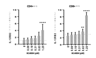
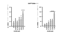


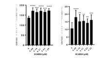


Classifications
-
- C—CHEMISTRY; METALLURGY
- C07—ORGANIC CHEMISTRY
- C07K—PEPTIDES
- C07K7/00—Peptides having 5 to 20 amino acids in a fully defined sequence; Derivatives thereof
- C07K7/04—Linear peptides containing only normal peptide links
- C07K7/08—Linear peptides containing only normal peptide links having 12 to 20 amino acids
-
- C—CHEMISTRY; METALLURGY
- C07—ORGANIC CHEMISTRY
- C07K—PEPTIDES
- C07K7/00—Peptides having 5 to 20 amino acids in a fully defined sequence; Derivatives thereof
- C07K7/04—Linear peptides containing only normal peptide links
- C07K7/06—Linear peptides containing only normal peptide links having 5 to 11 amino acids
-
- A—HUMAN NECESSITIES
- A61—MEDICAL OR VETERINARY SCIENCE; HYGIENE
- A61K—PREPARATIONS FOR MEDICAL, DENTAL OR TOILETRY PURPOSES
- A61K38/00—Medicinal preparations containing peptides
- A61K38/04—Peptides having up to 20 amino acids in a fully defined sequence; Derivatives thereof
- A61K38/08—Peptides having 5 to 11 amino acids
-
- A—HUMAN NECESSITIES
- A61—MEDICAL OR VETERINARY SCIENCE; HYGIENE
- A61K—PREPARATIONS FOR MEDICAL, DENTAL OR TOILETRY PURPOSES
- A61K38/00—Medicinal preparations containing peptides
- A61K38/04—Peptides having up to 20 amino acids in a fully defined sequence; Derivatives thereof
- A61K38/10—Peptides having 12 to 20 amino acids
-
- A—HUMAN NECESSITIES
- A61—MEDICAL OR VETERINARY SCIENCE; HYGIENE
- A61K—PREPARATIONS FOR MEDICAL, DENTAL OR TOILETRY PURPOSES
- A61K47/00—Medicinal preparations characterised by the non-active ingredients used, e.g. carriers or inert additives; Targeting or modifying agents chemically bound to the active ingredient
- A61K47/50—Medicinal preparations characterised by the non-active ingredients used, e.g. carriers or inert additives; Targeting or modifying agents chemically bound to the active ingredient the non-active ingredient being chemically bound to the active ingredient, e.g. polymer-drug conjugates
- A61K47/51—Medicinal preparations characterised by the non-active ingredients used, e.g. carriers or inert additives; Targeting or modifying agents chemically bound to the active ingredient the non-active ingredient being chemically bound to the active ingredient, e.g. polymer-drug conjugates the non-active ingredient being a modifying agent
- A61K47/54—Medicinal preparations characterised by the non-active ingredients used, e.g. carriers or inert additives; Targeting or modifying agents chemically bound to the active ingredient the non-active ingredient being chemically bound to the active ingredient, e.g. polymer-drug conjugates the non-active ingredient being a modifying agent the modifying agent being an organic compound
- A61K47/542—Carboxylic acids, e.g. a fatty acid or an amino acid
-
- A—HUMAN NECESSITIES
- A61—MEDICAL OR VETERINARY SCIENCE; HYGIENE
- A61P—SPECIFIC THERAPEUTIC ACTIVITY OF CHEMICAL COMPOUNDS OR MEDICINAL PREPARATIONS
- A61P35/00—Antineoplastic agents
-
- A—HUMAN NECESSITIES
- A61—MEDICAL OR VETERINARY SCIENCE; HYGIENE
- A61P—SPECIFIC THERAPEUTIC ACTIVITY OF CHEMICAL COMPOUNDS OR MEDICINAL PREPARATIONS
- A61P37/00—Drugs for immunological or allergic disorders
- A61P37/02—Immunomodulators
- A61P37/04—Immunostimulants
-
- C—CHEMISTRY; METALLURGY
- C12—BIOCHEMISTRY; BEER; SPIRITS; WINE; VINEGAR; MICROBIOLOGY; ENZYMOLOGY; MUTATION OR GENETIC ENGINEERING
- C12N—MICROORGANISMS OR ENZYMES; COMPOSITIONS THEREOF; PROPAGATING, PRESERVING, OR MAINTAINING MICROORGANISMS; MUTATION OR GENETIC ENGINEERING; CULTURE MEDIA
- C12N9/00—Enzymes; Proenzymes; Compositions thereof; Processes for preparing, activating, inhibiting, separating or purifying enzymes
- C12N9/10—Transferases (2.)
- C12N9/12—Transferases (2.) transferring phosphorus containing groups, e.g. kinases (2.7)
-
- A—HUMAN NECESSITIES
- A61—MEDICAL OR VETERINARY SCIENCE; HYGIENE
- A61K—PREPARATIONS FOR MEDICAL, DENTAL OR TOILETRY PURPOSES
- A61K38/00—Medicinal preparations containing peptides
-
- C—CHEMISTRY; METALLURGY
- C12—BIOCHEMISTRY; BEER; SPIRITS; WINE; VINEGAR; MICROBIOLOGY; ENZYMOLOGY; MUTATION OR GENETIC ENGINEERING
- C12N—MICROORGANISMS OR ENZYMES; COMPOSITIONS THEREOF; PROPAGATING, PRESERVING, OR MAINTAINING MICROORGANISMS; MUTATION OR GENETIC ENGINEERING; CULTURE MEDIA
- C12N5/00—Undifferentiated human, animal or plant cells, e.g. cell lines; Tissues; Cultivation or maintenance thereof; Culture media therefor
- C12N5/06—Animal cells or tissues; Human cells or tissues
- C12N5/0602—Vertebrate cells
- C12N5/0634—Cells from the blood or the immune system
- C12N5/0636—T lymphocytes
- C12N5/0637—Immunosuppressive T lymphocytes, e.g. regulatory T cells or Treg
-
- G—PHYSICS
- G01—MEASURING; TESTING
- G01N—INVESTIGATING OR ANALYSING MATERIALS BY DETERMINING THEIR CHEMICAL OR PHYSICAL PROPERTIES
- G01N33/00—Investigating or analysing materials by specific methods not covered by groups G01N1/00 - G01N31/00
- G01N33/48—Biological material, e.g. blood, urine; Haemocytometers
- G01N33/50—Chemical analysis of biological material, e.g. blood, urine; Testing involving biospecific ligand binding methods; Immunological testing
- G01N33/5005—Chemical analysis of biological material, e.g. blood, urine; Testing involving biospecific ligand binding methods; Immunological testing involving human or animal cells
- G01N33/5008—Chemical analysis of biological material, e.g. blood, urine; Testing involving biospecific ligand binding methods; Immunological testing involving human or animal cells for testing or evaluating the effect of chemical or biological compounds, e.g. drugs, cosmetics
- G01N33/5044—Chemical analysis of biological material, e.g. blood, urine; Testing involving biospecific ligand binding methods; Immunological testing involving human or animal cells for testing or evaluating the effect of chemical or biological compounds, e.g. drugs, cosmetics involving specific cell types
- G01N33/5047—Cells of the immune system
- G01N33/505—Cells of the immune system involving T-cells
-
- G—PHYSICS
- G01—MEASURING; TESTING
- G01N—INVESTIGATING OR ANALYSING MATERIALS BY DETERMINING THEIR CHEMICAL OR PHYSICAL PROPERTIES
- G01N33/00—Investigating or analysing materials by specific methods not covered by groups G01N1/00 - G01N31/00
- G01N33/48—Biological material, e.g. blood, urine; Haemocytometers
- G01N33/50—Chemical analysis of biological material, e.g. blood, urine; Testing involving biospecific ligand binding methods; Immunological testing
- G01N33/68—Chemical analysis of biological material, e.g. blood, urine; Testing involving biospecific ligand binding methods; Immunological testing involving proteins, peptides or amino acids
- G01N33/6863—Cytokines, i.e. immune system proteins modifying a biological response such as cell growth proliferation or differentiation, e.g. TNF, CNF, GM-CSF, lymphotoxin, MIF or their receptors
- G01N33/6869—Interleukin
Landscapes
- Health & Medical Sciences (AREA)
- Chemical & Material Sciences (AREA)
- Life Sciences & Earth Sciences (AREA)
- Organic Chemistry (AREA)
- General Health & Medical Sciences (AREA)
- Medicinal Chemistry (AREA)
- Engineering & Computer Science (AREA)
- Bioinformatics & Cheminformatics (AREA)
- Immunology (AREA)
- Animal Behavior & Ethology (AREA)
- Public Health (AREA)
- Pharmacology & Pharmacy (AREA)
- Veterinary Medicine (AREA)
- Genetics & Genomics (AREA)
- Proteomics, Peptides & Aminoacids (AREA)
- Biochemistry (AREA)
- Molecular Biology (AREA)
- Epidemiology (AREA)
- Biophysics (AREA)
- Nuclear Medicine, Radiotherapy & Molecular Imaging (AREA)
- Chemical Kinetics & Catalysis (AREA)
- General Chemical & Material Sciences (AREA)
- Wood Science & Technology (AREA)
- Zoology (AREA)
- Gastroenterology & Hepatology (AREA)
- Biomedical Technology (AREA)
- Biotechnology (AREA)
- Microbiology (AREA)
- General Engineering & Computer Science (AREA)
- Medicines That Contain Protein Lipid Enzymes And Other Medicines (AREA)
- Peptides Or Proteins (AREA)
- Medicines Containing Material From Animals Or Micro-Organisms (AREA)
- Hematology (AREA)
- Cell Biology (AREA)
- Micro-Organisms Or Cultivation Processes Thereof (AREA)
Description
発明の分野
本発明は、Lck(リンパ球特異的タンパク質チロシンキナーゼ)活性の刺激および種々の疾患または状態の予防または治療に使用するためのLck活性化剤に関する。
FIELD OF THE INVENTION The present invention relates to stimulation of Lck (lymphocyte-specific protein tyrosine kinase) activity and Lck activators for use in the prevention or treatment of various diseases or conditions.
分子生物学では、CD4(表面抗原分類4)は、ヘルパーT細胞、単球、マクロファージおよび樹状細胞などの免疫細胞の表面上で認められる糖タンパク質である。CD4+ヘルパーT細胞は、免疫系の不可欠の部分である白血球であり、これは、CD8キラー細胞などの他の免疫細胞にシグナルを送り、感染性粒子を破壊する。T細胞は、T細胞受容体(TCR)および共受容体が抗原ペプチド:主要組織適合抗原(MHC)に結合すると、TCR複合体をクラスター化させて、細胞内カスケードを開始させる。T細胞活性化では、共受容体CD4およびCD8の、それぞれ、クラスIIまたはクラスI MHC分子への結合が抗原に対するT細胞の感受性を高める。 In molecular biology, CD4 (surface antigen class 4) is a glycoprotein found on the surface of immune cells such as helper T cells, monocytes, macrophages and dendritic cells. CD4+ helper T cells are white blood cells that are an integral part of the immune system, which send signals to other immune cells, such as CD8 killer cells, to destroy infectious particles. T cells cluster TCR complexes and initiate intracellular cascades when the T cell receptor (TCR) and co-receptor bind to an antigenic peptide: major histocompatibility antigen (MHC). In T cell activation, the binding of co-receptors CD4 and CD8 to class II or class I MHC molecules, respectively, increases the sensitivity of T cells to antigens.
Lckは、Srcファミリーキナーゼのメンバーであり、T細胞のエフェクター能力の発現、すなわち、効果的サイトカインの転写と共に、発生中の胸腺細胞および成熟T細胞の両方の増殖と分化に不可欠である(Lovatt M et al,Mol & Cell Biology,2006,26(22):8655-8665)。 Lck is a member of the Src family of kinases and is essential for the expression of T cell effector capacity, i.e., the proliferation and differentiation of both developing thymocytes and mature T cells, along with the transcription of effective cytokines (Lovatt M et al, Mol & Cell Biology, 2006, 26(22):8655-8665).
LckによるTCRのリン酸化は、細胞質ゾルキナーゼZAP70(ゼータ鎖関連プロテインキナーゼ70)を細胞膜に動員する結合部位を作り出すことにより、下流のシグナル伝達を開始させる(Iwashima M.et al,Science,1994,263:1136-1139)。さらに具体的には、Lckは、Lckの不活性化をもたらすTyr505をリン酸化するC末端Srcキナーゼ(Csk)、活性部位の再配置を介してLckのキナーゼ活性を活性化するLck活性化ループのTyr394トランス自己リン酸化、およびホスファターゼによる脱リン酸化、により調節される(Fulop T et al,Longevity & Healthspan,2012,1:6)。 Phosphorylation of the TCR by Lck initiates downstream signal transduction by creating a binding site that recruits the cytosolic kinase ZAP70 (zeta chain-associated protein kinase 70) to the cell membrane (Iwashima M. et al, Science, 1994, 263 :1136-1139). More specifically, Lck is linked to the C-terminal Src kinase (Csk), which phosphorylates Tyr505 leading to Lck inactivation, and the Lck activation loop, which activates the kinase activity of Lck through active site relocation. It is regulated by Tyr394 trans-autophosphorylation and dephosphorylation by phosphatases (Fulop T et al, Longevity & Healthspan, 2012, 1:6).
TCR刺激時には、CD4/CD8結合Lckは、TCR/CD3複合体と近接し、CD3分子の免疫受容活性化チロシンモチーフ(ITAM)をリン酸化する。ZAP70キナーゼがその後、リン酸化ITAMへ動員され、続けて、Lckにより活性化される。これは、最終的にT細胞活性化およびIL-2産生に繋がる複数の経路の活性化をもたらす(Wang G et al,BioMed Research International,2014,doi.org/10.1155/2014/682010)。 Upon TCR stimulation, CD4/CD8-bound Lck comes into close proximity to the TCR/CD3 complex and phosphorylates the immunoreceptor activation tyrosine motif (ITAM) of the CD3 molecule. ZAP70 kinase is then recruited to phosphorylated ITAM and subsequently activated by Lck. This results in activation of multiple pathways that ultimately lead to T cell activation and IL-2 production (Wang G et al, BioMed Research International, 2014, doi.org/10.1155/2014/682010).
加えて、Lckの樹状細胞(DC)媒介活性化は、「TCRライセンシング」、同族ペプチド-MHCクラスII抗原複合体に対するTCR応答の感度と大きさを劇的に高めるプロセス、に繋がる(Meraner P et al,2007,J Immunol,178(4):2262-2271)。このような上皮内DETC細胞(樹枝状表皮T細胞)またはガンマ/デルタT細胞は、恒常性、組織修復、炎症および悪性腫瘍からの保護で有用な役割を果たす(Witherden DA & Havran WL,J Leukoc Biol,2013,94(1):69-76)。 In addition, dendritic cell (DC)-mediated activation of Lck leads to "TCR licensing", a process that dramatically increases the sensitivity and magnitude of TCR responses to cognate peptide-MHC class II antigen complexes (Meraner P. et al., 2007, J Immunol, 178(4):2262-2271). Such intraepithelial DETC cells (dendritic epidermal T cells) or gamma/delta T cells play useful roles in homeostasis, tissue repair, inflammation and protection from malignancy (Witherden DA & Havran WL, J Leukoc Biol, 2013, 94(1):69-76).
一態様では、本発明は、Lck活性化のためのペプチドを提供し、ペプチドは、式I:
R/K’-x1-R/K-x2-R/K-x3-x4-x5-x6-R/K” 式I
のLck活性化ポリペプチド部分またはその反転配列を含み、式中、
各R/Kは、独立に、アルギニンまたはリシンアミノ酸残基であり;
R/K’は、アルギニンまたはリシンアミノ酸残基で、存在または非存在であり;R/K”は、アルギニンまたはリシンアミノ酸残基で、存在または非存在であり;x1~x6はそれぞれ独立に、1個のアミノ酸であり;アミノ酸x3~x6は、ひとまとめにして存在または非存在であり、アミノ酸x3~x6が非存在である場合、R/K”は非存在である。
In one aspect, the invention provides peptides for Lck activation, the peptides having the formula I:
R/K'-x 1 -R/K-x 2 -R/K-x 3 -x 4 -x 5 -x 6 -R/K" Formula I
or an inverted sequence thereof, wherein:
each R/K is independently an arginine or lysine amino acid residue;
R/K' is an arginine or lysine amino acid residue, present or absent; R/K'' is an arginine or lysine amino acid residue, present or absent; x 1 to x 6 are each independently is one amino acid; amino acids x 3 to x 6 are collectively present or absent; if amino acids x 3 to x 6 are absent, then R/K'' is absent.
別の実施形態では、x1~x6の内の1個または複数のアミノ酸は、疎水性アミノ酸である。別の実施形態では、Lck活性化ポリペプチド部分は、Lアミノ酸残基を含む。別の実施形態では、Lck活性化ポリペプチド部分は、Lアミノ酸残基からなる。別の実施形態では、Lck活性化ポリペプチド部分は、Dアミノ酸残基を含む。別の実施形態では、Lck活性化ポリペプチド部分は、Dアミノ酸残基からなる。 In another embodiment, one or more amino acids of x 1 -x 6 are hydrophobic amino acids. In another embodiment, the Lck activating polypeptide portion comprises L amino acid residues. In another embodiment, the Lck activating polypeptide portion consists of L amino acid residues. In another embodiment, the Lck activating polypeptide portion includes D amino acid residues. In another embodiment, the Lck activating polypeptide portion consists of D amino acid residues.
別の実施形態では、Lck活性化ポリペプチド部分は、RSKAKNPLY、rskaknply、RVKVKVVVV、およびrvkvkvvvvからなる群より選択される。 In another embodiment, the Lck activating polypeptide moiety is selected from the group consisting of RSKAKNPLY, rskaknply, RVKVKVVVV, and rvkvkvvvv.
別の実施形態では、Lckを活性化するためのペプチドは、Lck活性化ポリペプチド部分のN末端またはC末端に結合した式II:

の化合物をさらに含み、式中、
nは、化合物のモノマー単位の数で、1~5の整数であり、
各mは、独立に、0~18の整数であり、および
各R基は、独立にHまたはC1~C18の側鎖である。
In another embodiment, the peptide for activating Lck is of formula II attached to the N-terminus or C-terminus of the Lck activating polypeptide moiety:
further comprising a compound of the formula:
n is the number of monomer units of the compound, and is an integer from 1 to 5;
Each m is independently an integer from 0 to 18, and each R group is independently H or a C 1 to C 18 side chain.
別の実施形態では、各モノマー単位のR基は、独立にC1~C18側鎖である。 In another embodiment, the R group of each monomer unit is independently a C 1 -C 18 side chain.
別の実施形態では、各R基は、独立に18-m炭素原子長さであり、式中、mは、0~17の値を有する。 In another embodiment, each R group is independently 18-m carbon atoms in length, where m has a value from 0 to 17.
別の実施形態では、Lckを活性化するためのペプチドは、Lck活性化ポリペプチド部分のN末端またはC末端に結合した化合物をさらに含み、化合物は、少なくとも1個の脂肪酸である。 In another embodiment, the peptide for activating Lck further comprises a compound attached to the N-terminus or C-terminus of the Lck activating polypeptide moiety, and the compound is at least one fatty acid.
別の実施形態では、少なくとも4個の脂肪酸が、Lck活性化ポリペプチド部分のC末端に結合される。 In another embodiment, at least 4 fatty acids are attached to the C-terminus of the Lck activating polypeptide moiety.
別の実施形態では、2個以上の脂肪酸の結合は直鎖状である。 In another embodiment, the linkage of two or more fatty acids is linear.
別の実施形態では、Lckを活性化するためのペプチドは、2個以上の脂肪酸の結合が分岐している、本明細書に記載のさらなるペプチドである。 In another embodiment, the peptide for activating Lck is a further peptide described herein in which the bonds of two or more fatty acids are branched.
別の実施形態では、脂肪酸は飽和している。 In another embodiment, the fatty acids are saturated.
別の実施形態では、脂肪酸は、カプロン酸、カプリル酸、カプリン酸(デカン酸)、およびラウリン酸(ドデカン酸)からなる群より選択される。 In another embodiment, the fatty acid is selected from the group consisting of caproic acid, caprylic acid, capric acid (decanoic acid), and lauric acid (dodecanoic acid).
別の実施形態では、最遠位脂肪酸は、アミド化される。 In another embodiment, the most distal fatty acid is amidated.
別の実施形態では、アミノ酸配列は、Lck活性化ポリペプチド部分のC末端でアミド化される。 In another embodiment, the amino acid sequence is amidated at the C-terminus of the Lck activating polypeptide portion.
別の態様では、本発明は、RSKAKNPLYR-(2Adod)4、rskaknplyr-(2Adod)4、RVKVKVVVVR-(2Adod)4、およびrvkvkvvvvr-(2Adod)4からなる群より選択されるアミノ酸配列を含むペプチドを提供する。 In another aspect, the invention provides a peptide comprising an amino acid sequence selected from the group consisting of RSKAKNPLYR-(2Adod) 4 , rskaknplyr-(2Adod) 4 , RVKVKVVVVR-(2Adod) 4 , and rvkvkvvvvr-(2Adod) 4 provide.
別の態様では、本発明は、RSKAKNPLYR-(2Adod)4、rskaknplyr-(2Adod)4、RVKVKVVVVR-(2Adod)4、およびrvkvkvvvvr-(2Adod)4からなる群より選択されるアミノ酸配列からなるペプチドを提供する。 In another aspect , the invention provides a peptide consisting of an amino acid sequence selected from the group consisting of : provide.
別の態様では、本発明は、本明細書に記載の有効成分としてのペプチド、および薬学的に許容可能な希釈剤または担体を含む医薬組成物を提供する。 In another aspect, the invention provides a pharmaceutical composition comprising a peptide as an active ingredient described herein and a pharmaceutically acceptable diluent or carrier.
別の態様では、本発明は、Lckキナーゼの活性を高める方法を提供し、方法は、Lckキナーゼを、本明細書に記載のペプチドを含む組成物または本明細書に記載の医薬組成物と接触させることを含む。 In another aspect, the invention provides a method of increasing the activity of Lck kinase, comprising contacting Lck kinase with a composition comprising a peptide described herein or a pharmaceutical composition described herein. Including causing.
別の態様では、本発明は、LckキナーゼのY394リン酸化を増大させる方法を提供し、方法は、Lckキナーゼを、本明細書に記載のペプチドを含む組成物または本明細書に記載の医薬組成物と接触させることを含む。 In another aspect, the invention provides a method of increasing Y394 phosphorylation of Lck kinase, which method comprises increasing Y394 phosphorylation of Lck kinase in a composition comprising a peptide described herein or a pharmaceutical composition described herein. Including contact with objects.
別の態様では、本発明は、細胞または細胞集団からのIL-2分泌を増大させる方法を提供し、方法は、細胞または細胞集団を、本明細書に記載のペプチドを含む組成物または本明細書に記載の医薬組成物と接触させることを含む。 In another aspect, the invention provides a method of increasing IL-2 secretion from a cell or cell population, the method comprising: increasing IL-2 secretion from a cell or cell population with a composition comprising a peptide described herein or a composition comprising a peptide described herein. the pharmaceutical composition described in the book.
別の態様では、本発明は、細胞上または細胞集団中のIL-2Rα(CD25)の発現を増大させる方法を提供し、方法は、細胞または細胞集団を、本明細書に記載のペプチドを含む組成物または本明細書に記載の医薬組成物と接触させることを含む。 In another aspect, the invention provides a method of increasing the expression of IL-2Rα (CD25) on a cell or population of cells, the method comprising increasing the expression of IL-2Rα (CD25) on a cell or population of cells comprising a peptide as described herein. composition or a pharmaceutical composition described herein.
別の態様では、本発明は、細胞上または細胞集団中のIL-2Rβ(CD122)の発現を増大させる方法を提供し、方法は、細胞または細胞集団を、本明細書に記載のペプチドを含む組成物または本明細書に記載の医薬組成物と接触させることを含む。 In another aspect, the invention provides a method of increasing expression of IL-2Rβ (CD122) on a cell or in a population of cells, the method comprising increasing the expression of IL-2Rβ (CD122) on a cell or population of cells comprising a peptide as described herein. composition or a pharmaceutical composition described herein.
別の態様では、本発明は、細胞のまたは細胞集団中のIL-2応答性を高める方法を提供し、方法は、細胞または細胞集団を、本明細書に記載のペプチドを含む組成物または本明細書に記載の医薬組成物と接触させることを含む。 In another aspect, the invention provides a method of increasing IL-2 responsiveness of a cell or in a population of cells, which method comprises injecting the cell or population of cells with a composition comprising a peptide or a peptide as described herein. contacting with a pharmaceutical composition as described herein.
別の態様では、本発明は、細胞上のまたは細胞集団中のIL-15Rの発現を増大させる方法を提供し、方法は、細胞または細胞集団を、本明細書に記載のペプチドを含む組成物または本明細書に記載の医薬組成物と接触させることを含む。 In another aspect, the invention provides a method of increasing the expression of IL-15R on a cell or in a population of cells, the method comprising: treating the cell or population of cells with a composition comprising a peptide as described herein. or contacting with a pharmaceutical composition described herein.
別の態様では、本発明は、細胞のまたは細胞集団中のIL-15の応答性を高める方法を提供し、方法は、細胞または細胞集団を、本明細書に記載のペプチドを含む組成物または本明細書に記載の医薬組成物と接触させることを含む。 In another aspect, the invention provides a method of increasing the responsiveness of IL-15 in a cell or population of cells, the method comprising: treating a cell or population of cells with a composition comprising a peptide described herein or contacting with a pharmaceutical composition described herein.
別の態様では、本発明は、細胞上または細胞集団中のCD28の発現を増大させる方法を提供し、方法は、細胞または細胞集団を、本明細書に記載のペプチドを含む組成物または本明細書に記載の医薬組成物と接触させることを含む。 In another aspect, the invention provides a method of increasing the expression of CD28 on a cell or in a population of cells, the method comprises increasing the expression of CD28 on a cell or population of cells, comprising a composition comprising a peptide described herein or a population of cells described herein. the pharmaceutical composition described in the book.
別の態様では、本発明は、細胞または細胞集団からのIL-21の分泌を増大させる方法を提供し、方法は、細胞または細胞集団を、本明細書に記載のペプチドを含む組成物または本明細書に記載の医薬組成物と接触させることを含む。 In another aspect, the invention provides a method of increasing the secretion of IL-21 from a cell or cell population, the method comprising injecting the cell or cell population with a composition comprising a peptide or a peptide described herein. contacting with a pharmaceutical composition as described herein.
別の態様では、本発明は、細胞上のまたは細胞集団中のIL-21R(CD360)IL-2Rβの発現を増大させる方法を提供し、方法は、細胞または細胞集団を、本明細書に記載のペプチドを含む組成物または本明細書に記載の医薬組成物と接触させることを含む。 In another aspect, the invention provides a method of increasing the expression of IL-21R(CD360)IL-2Rβ on a cell or in a population of cells, the method comprising: or a pharmaceutical composition described herein.
別の態様では、本発明は、細胞のまたは細胞集団中のIL-21の応答性を高める方法を提供し、方法は、細胞または細胞集団を、本明細書に記載のペプチドを含む組成物または本明細書に記載の医薬組成物と接触させることを含む。 In another aspect, the invention provides a method of increasing IL-21 responsiveness of a cell or population of cells, the method comprising: treating a cell or population of cells with a composition comprising a peptide described herein or contacting with a pharmaceutical composition described herein.
別の態様では、本発明は、細胞上のまたは細胞集団中のIL-12Rの発現を増大させる方法を提供し、方法は、細胞または細胞集団を、本明細書に記載のペプチドを含む組成物または本明細書に記載の医薬組成物と接触させることを含む。 In another aspect, the invention provides a method of increasing the expression of IL-12R on a cell or in a population of cells, the method comprising: treating the cell or population of cells with a composition comprising a peptide described herein. or contacting with a pharmaceutical composition described herein.
一実施形態では、IL-12Rは、IL-12Rβ1および/またはIL-12Rβ2である。 In one embodiment, the IL-12R is IL-12Rβ1 and/or IL-12Rβ2.
別の態様では、本発明は、細胞のまたは細胞集団中のIL-12の応答性を高める方法を提供し、方法は、細胞または細胞集団を、本明細書に記載のペプチドを含む組成物または本明細書に記載の医薬組成物と接触させることを含む。 In another aspect, the invention provides a method of increasing the responsiveness of IL-12 in a cell or population of cells, the method comprising increasing the responsiveness of IL-12 in a cell or population of cells with a composition comprising a peptide or a peptide described herein. contacting with a pharmaceutical composition described herein.
別の態様では、本発明は、細胞または細胞集団からのサイトカインの分泌を増大させる方法を提供し、方法は、細胞または細胞集団を、本明細書に記載のペプチドを含む組成物または本明細書に記載の医薬組成物と接触させることを含む。 In another aspect, the present invention provides a method of increasing the secretion of a cytokine from a cell or cell population, the method comprising increasing the secretion of a cytokine from a cell or cell population using a composition comprising a peptide described herein or a peptide described herein. contacting with the pharmaceutical composition described in .
一実施形態では、サイトカインはIFNγまたはTNFαである。 In one embodiment, the cytokine is IFNγ or TNFα.
別の態様では、本発明は、細胞または細胞集団の増殖を誘導する方法を提供し、方法は、細胞または細胞集団を、本明細書に記載のペプチドを含む組成物または本明細書に記載の医薬組成物と接触させることを含む。 In another aspect, the invention provides a method of inducing proliferation of a cell or cell population, the method comprising inducing the proliferation of a cell or cell population with a composition comprising a peptide described herein or a composition described herein. contacting with a pharmaceutical composition.
別の態様では、本発明は、細胞集団の増殖を増大させる方法を提供し、方法は、細胞集団を、本明細書に記載のペプチドを含む組成物または本明細書に記載の医薬組成物と接触させることを含む。 In another aspect, the invention provides a method of increasing proliferation of a cell population, the method comprising increasing the proliferation of a cell population with a composition comprising a peptide described herein or a pharmaceutical composition described herein. Including bringing into contact.
別の実施形態では、本発明は、細胞は、CD4+T細胞、CD8+T細胞、NK細胞、または樹状細胞からなる群より選択される細胞である、または細胞集団はこれらからなる群より選択される細胞を含む。 In another embodiment, the invention provides that the cell is a cell selected from the group consisting of CD4+ T cells, CD8+ T cells, NK cells, or dendritic cells, or the cell population is selected from the group consisting of including.
別の態様では、本発明は、CD8+T細胞またはNK細胞中のCD107aの発現を増大させる方法を提供し、方法は、CD8+T細胞またはNK細胞を、本明細書に記載のペプチドを含む組成物または本明細書に記載の医薬組成物と接触させることを含む。 In another aspect, the invention provides a method of increasing the expression of CD107a in a CD8+ T cell or NK cell, which method comprises increasing the expression of CD107a in a CD8+ T cell or NK cell, the method comprising: contacting with a pharmaceutical composition as described herein.
別の態様では、本発明は、T細胞機能を高める方法を提供し、方法は、T細胞またはT細胞集団を、本明細書に記載のペプチドを含む組成物または本明細書に記載の医薬組成物と接触させることを含む。 In another aspect, the invention provides a method of enhancing T cell function, the method comprising increasing a T cell or T cell population to a composition comprising a peptide described herein or a pharmaceutical composition described herein. Including contact with objects.
一実施形態では、細胞は未感作細胞である、または細胞集団は未感作細胞を含む。 In one embodiment, the cells are or the cell population includes naive cells.
別の態様では、本発明は、細胞傷害性細胞機能を高める方法を提供し、方法は、細胞傷害性細胞または細胞傷害性細胞集団を、本明細書に記載のペプチドを含む組成物または本明細書に記載の医薬組成物と接触させることを含む。 In another aspect, the invention provides a method of increasing cytotoxic cell function, which method comprises increasing cytotoxic cells or populations of cytotoxic cells with a composition comprising a peptide described herein or a composition comprising a peptide described herein. the pharmaceutical composition described in the book.
別の態様では、本発明は、T細胞またはT細胞集団の疲弊を低減する方法を提供し、方法は、T細胞またはT細胞集団を、本明細書に記載のペプチドを含む組成物または本明細書に記載の医薬組成物と接触させることを含む。 In another aspect, the invention provides a method of reducing exhaustion of a T cell or T cell population, the method comprising reducing the exhaustion of a T cell or T cell population using a composition comprising a peptide described herein or a composition described herein. the pharmaceutical composition described in the book.
別の態様では、本発明は、T細胞上のCD40L発現を増大させる方法を提供し、方法は、T細胞を、本明細書に記載のペプチドを含む組成物または本明細書に記載の医薬組成物と接触させることを含む。 In another aspect, the invention provides a method of increasing CD40L expression on T cells, which method comprises increasing T cells with a composition comprising a peptide described herein or a pharmaceutical composition described herein. Including contact with objects.
別の態様では、本発明は、CD8+T細胞応答を高める方法を提供し、方法は、CD4+T細胞を、本明細書に記載のペプチドを含む組成物または本明細書に記載の医薬組成物と接触させることを含む。 In another aspect, the invention provides a method of increasing a CD8+ T cell response, the method comprising contacting a CD4+ T cell with a composition comprising a peptide described herein or a pharmaceutical composition described herein. Including.
別の態様では、本発明は、細胞集団中のTreg細胞の比率を減らす方法を提供し、方法は、Treg含有細胞集団を、本明細書に記載のペプチドを含む組成物または本明細書に記載の医薬組成物と接触させることを含む。 In another aspect, the invention provides a method of reducing the proportion of Treg cells in a cell population, the method comprising reducing the Treg-containing cell population with a composition comprising a peptide described herein or a composition described herein. the pharmaceutical composition.
一実施形態では、Treg細胞はFoxp3+Treg細胞である。 In one embodiment, the Treg cells are Foxp3+ Treg cells.
別の態様では、本発明は、チェックポイント阻害剤の存在下でIL-2分泌を増大させる方法を提供し、方法は、細胞または細胞集団を、本明細書に記載のペプチドを含む組成物または本明細書に記載の医薬組成物と接触させることを含む。 In another aspect, the invention provides a method of increasing IL-2 secretion in the presence of a checkpoint inhibitor, the method comprising injecting a cell or cell population with a composition comprising a peptide or a peptide described herein. contacting with a pharmaceutical composition described herein.
別の態様では、本発明は、樹状細胞生存率を増大させる方法を提供し、方法は、樹状細胞を、本明細書に記載のペプチドまたは本明細書に記載の医薬組成物と接触させることを含む。 In another aspect, the invention provides a method of increasing dendritic cell survival, the method comprising contacting a dendritic cell with a peptide described herein or a pharmaceutical composition described herein. Including.
一実施形態では、接触は、インビボまたはインビトロで実施される。 In one embodiment, contacting is performed in vivo or in vitro.
一実施形態では、方法は、エクスビボで実施される。 In one embodiment, the method is performed ex vivo.
別の態様では、本発明は、本明細書に記載の方法により得られる細胞または細胞集団を提供する。 In another aspect, the invention provides cells or cell populations obtained by the methods described herein.
別の態様では、本発明は、細胞または細胞集団を、本明細書に記載のペプチドを含む組成物または本明細書に記載の医薬組成物と接触させることにより得られる細胞または細胞集団に由来する細胞または細胞集団を提供する。 In another aspect, the invention provides a cell or cell population derived from a cell or cell population obtained by contacting the cell or cell population with a composition comprising a peptide as described herein or a pharmaceutical composition as described herein. Provide a cell or cell population.
別の態様では、本発明は、インビボでLckの活性を高めるために、ペプチドまたは医薬組成物が対象または細胞に投与される本明細書に記載の方法を提供する。 In another aspect, the invention provides a method as described herein, wherein a peptide or pharmaceutical composition is administered to a subject or cell to increase Lck activity in vivo.
別の態様では、本発明は、対象のLckの活性を高める方法を提供し、方法は、本明細書に記載のペプチドを含む組成物または本明細書に記載の医薬組成物を対象に投与することを含む。 In another aspect, the invention provides a method of increasing Lck activity in a subject, the method comprising administering to a subject a composition comprising a peptide described herein or a pharmaceutical composition described herein. Including.
別の態様では、本発明は、対象の免疫応答を誘導する方法を提供し、方法は、本明細書に記載のペプチドまたは本明細書に記載の医薬組成物を対象に投与することを含む。 In another aspect, the invention provides a method of inducing an immune response in a subject, the method comprising administering to a subject a peptide described herein or a pharmaceutical composition described herein.
別の態様では、本発明は、対象中のLckまたはLck活性の抑制または下方制御を特徴とする疾患または状態に関連する少なくとも1つの症状を治療および/または予防する方法を提供し、方法は、本明細書に記載のペプチドを含む組成物または本明細書に記載の医薬組成物を対象に投与することを含む。 In another aspect, the invention provides a method of treating and/or preventing at least one symptom associated with a disease or condition characterized by suppression or downregulation of Lck or Lck activity in a subject, the method comprising: comprising administering to a subject a composition comprising a peptide described herein or a pharmaceutical composition described herein.
別の態様では、本発明は、LckまたはLck活性の抑制または下方制御を特徴とする疾患または状態が、病原性感染症、病原性感染症に由来する敗血症(例えば、慢性敗血症)、免疫不全障害、低下した免疫応答、減少したT細胞数、T細胞異常、T細胞疲弊、およびT細胞チェックポイント阻害からなる群より選択される、本明細書で記載の方法を提供する。 In another aspect, the invention provides that the disease or condition characterized by suppression or downregulation of Lck or Lck activity is a pathogenic infection, sepsis derived from a pathogenic infection (e.g., chronic sepsis), an immunodeficiency disorder, , reduced immune response, decreased T cell numbers, T cell abnormalities, T cell exhaustion, and T cell checkpoint inhibition.
別の態様では、本発明は、疾患または状態が癌である、本明細書に記載の方法を提供する。 In another aspect, the invention provides a method as described herein, wherein the disease or condition is cancer.
別の実施形態では、本発明は、ワクチン接種を必要としている対象にワクチンを接種する方法を提供し、方法は、本明細書に記載のペプチドを含む組成物または本明細書に記載の医薬組成物をワクチンと同時にまたは順次に対象に投与することを含む。 In another embodiment, the invention provides a method of vaccinating a subject in need of vaccination, the method comprising a composition comprising a peptide described herein or a pharmaceutical composition described herein. This includes administering the product to the subject simultaneously or sequentially with the vaccine.
別の態様では、本発明は、癌の治療および/または予防を必要としている対象にそれを実施する方法を提供し、方法は、本明細書に記載のペプチドを含む組成物または本明細書に記載の医薬組成物をチェックポイント阻害剤と同時にまたは順次に対象に投与することを含む。 In another aspect, the invention provides a method for administering cancer treatment and/or prevention to a subject in need thereof, the method comprising a composition comprising a peptide described herein or a peptide described herein. comprising administering the described pharmaceutical compositions to a subject simultaneously or sequentially with a checkpoint inhibitor.
別の態様では、本発明は、対象の免疫抑制を低減する方法を提供し、方法は、本明細書に記載のペプチドを含む組成物または本明細書に記載の医薬組成物を対象に投与することを含む。 In another aspect, the invention provides a method of reducing immunosuppression in a subject, the method comprising administering to a subject a composition comprising a peptide described herein or a pharmaceutical composition described herein. Including.
別の態様では、本発明は、対象の加齢関連免疫機能不全を治療および/または予防する方法を提供し、方法は、本明細書に記載のペプチドを含む組成物または本明細書に記載の医薬組成物を対象に投与することを含む。 In another aspect, the invention provides a method of treating and/or preventing age-related immune dysfunction in a subject, the method comprising a composition comprising a peptide described herein or a peptide described herein. comprising administering a pharmaceutical composition to a subject.
別の態様では、本発明は、対象のTh1応答を誘導する方法を提供し、方法は、本明細書に記載のペプチドまたは本明細書に記載の医薬組成物を対象に投与することを含む。 In another aspect, the invention provides a method of inducing a Th1 response in a subject, the method comprising administering to a subject a peptide described herein or a pharmaceutical composition described herein.
別の態様では、本発明は、対象のHIV感染症を治療する方法を提供し、方法は、本明細書に記載のペプチドを含む組成物または本明細書に記載の医薬組成物を対象に投与することを含む。 In another aspect, the invention provides a method of treating an HIV infection in a subject, the method comprising administering to a subject a composition comprising a peptide described herein or a pharmaceutical composition described herein. including doing.
別の態様では、本発明は、対象の結核菌感染症を治療する方法を提供し、方法は、本明細書に記載のペプチドを含む組成物または本明細書に記載の医薬組成物を対象に投与することを含む。 In another aspect, the invention provides a method of treating Mycobacterium tuberculosis infection in a subject, the method comprising using a composition comprising a peptide described herein or a pharmaceutical composition described herein. including administering.
別の態様では、本発明は、LckまたはLck活性の抑制または下方制御を特徴とする疾患または状態に関連する少なくとも1つの症状を治療および/または予防するための投与単位剤形を提供し、投与単位剤形は、本明細書に記載のペプチドを含む。 In another aspect, the present invention provides a dosage unit form for treating and/or preventing at least one symptom associated with a disease or condition characterized by inhibition or downregulation of Lck or Lck activity; Unit dosage forms include the peptides described herein.
別の態様では、本発明は、癌治療および/または予防のための投与単位剤形を提供し、投与単位剤形は、本明細書に記載のペプチドを含む。 In another aspect, the invention provides a dosage unit form for cancer treatment and/or prevention, the dosage unit form comprising a peptide as described herein.
本明細書の全体を通して、用語の「含む(comprise)」、または「含む(comprises)」および「含むこと(comprising)」などの変化形は、記述した要素、整数もしくはステップまたは複数の要素、整数もしくはステップの群を含むが、いかなる他の要素、整数もしくはステップまたは複数の要素、整数もしくはステップの群も排除しないことを意味すると理解されよう。 Throughout this specification, the term "comprise" or variations such as "comprises" and "comprising" refer to the described element, integer or step or elements, integer or groups of steps, but not excluding any other elements, integers or steps, or groups of elements, integers or steps.
本明細書に収載されている文書、法令、物質、装置、論文などの全ての考察は、本発明の背景を提供することのみを目的とするものである。これらの資料のいずれかまたはその全てが、本出願の優先日以前にオーストラリアまたは他のどこかで存在したとして、先行技術の根拠の一部を形成する、または本発明に関連する分野での共通の一般常識であったことを認めるものであると解釈されるべきではない。 Any discussion of documents, statutes, materials, devices, articles, etc. contained herein is for the sole purpose of providing context for the present invention. Any or all of these materials form part of the basis of prior art as they existed in Australia or elsewhere before the priority date of this application, or are common in the field relevant to the present invention. This should not be interpreted as an admission that this was common knowledge.
本発明の特徴および利点は、添付図面と共に本発明の例示的実施形態の以下の詳細な説明からさらに明らかになるであろう。 The features and advantages of the invention will become more apparent from the following detailed description of exemplary embodiments of the invention, taken in conjunction with the accompanying drawings.
哺乳動物の非受容体Srcキナーゼファミリー(SKF)のメンバーには、次の8種のキナーゼ:Src、Fyn、Yes、Fgr、Lyn、Hck、Lck、およびBlkが含まれ、選択的Lck活性化剤は、特定されてこなかった(Bae O-N et al,Journal of Neuroscience,2012,32(21):7278-7286)。 Members of the mammalian non-receptor Src kinase family (SKF) include eight kinases: Src, Fyn, Yes, Fgr, Lyn, Hck, Lck, and Blk, and are selective Lck activators. has not been identified (Bae O-N et al, Journal of Neuroscience, 2012, 32(21): 7278-7286).
本発明は、1つまたは複数の実施形態では、本明細書に記載の式IのペプチドがLckの活性を刺激し得るという発見に関する。少なくともいくつかの実施形態では、本発明によるLck活性化剤は、SKFメンバー中でLckのみの活性化を刺激し得る。Lck活性化剤、特に特異的Lck活性化剤に対する必要性は、加齢または非加齢関連の多様な状態に直接的に関連する。 The present invention relates, in one or more embodiments, to the discovery that the peptides of Formula I described herein are capable of stimulating the activity of Lck. In at least some embodiments, Lck activators according to the invention may stimulate activation of only Lck in SKF members. The need for Lck activators, particularly specific Lck activators, is directly related to a variety of aging and non-ageing related conditions.
従って、一態様では、本発明は、Lckを活性化するためのペプチドを提供し、ペプチドは、式I:
R/K’-x1-R/K-x2-R/K-x3-x4-x5-x6-R/K” 式I
のLck活性化ポリペプチド部分またはその反転配列を含み、式中、
各R/Kは、独立に、アルギニンまたはリシンアミノ酸残基であり;
R/K’は、アルギニンまたはリシンアミノ酸残基で、存在するまたは非存在であり;R/K”は、アルギニンまたはリシンアミノ酸残基で、存在するまたは非存在であり;x1~x6はそれぞれ独立に、1個のアミノ酸であり;および
アミノ酸x3~x6は、ひとまとめにして存在するまたは非存在であり、アミノ酸x3~x6が非存在である場合、R/K”は非存在である。
Accordingly, in one aspect, the invention provides peptides for activating Lck, the peptides having the formula I:
R/K'-x 1 -R/K-x 2 -R/K-x 3 -x 4 -x 5 -x 6 -R/K" Formula I
or an inverted sequence thereof, wherein:
each R/K is independently an arginine or lysine amino acid residue;
R/K' is an arginine or lysine amino acid residue, present or absent; R/K'' is an arginine or lysine amino acid residue, present or absent; x 1 to x 6 are each independently is one amino acid; and amino acids x 3 to x 6 are present or absent collectively, and if amino acids x 3 to x 6 are absent, then It is existence.
本明細書で使用される場合、「活性化」という用語は、一般に、少なくとも1つの標的タンパク質の活性を高めることを意味する。キナーゼの特定の文脈では、この活性化は、少なくとも1つの標的基質または部位の増大したリン酸化に繋がる。この活性化は、限定されないが、プロテインキナーゼと、プロテインキナーゼの結合相手との間に複合体が形成される確率を増大させること、またはその標的に以前に結合したキナーゼの活性を増大させることを含む任意の手段により引き起こされ得る。このような活性化は、インビボあるいはインビトロで起こり得る。 As used herein, the term "activation" generally means increasing the activity of at least one target protein. In the specific context of kinases, this activation leads to increased phosphorylation of at least one target substrate or site. This activation can include, but is not limited to, increasing the probability of complex formation between the protein kinase and the protein kinase's binding partner, or increasing the activity of a kinase previously bound to its target. can be caused by any means including. Such activation can occur in vivo or in vitro.
本明細書で使用される場合、「Lckを活性化するためのペプチド」は、「Lck活性化ポリペプチド」または「Lck活性化剤」と同義に使用される。 As used herein, "peptide for activating Lck" is used synonymously with "Lck activating polypeptide" or "Lck activating agent."
Lckは、LCK癌原遺伝子、白血球C末端Srcキナーゼ;リンパ球細胞特異的タンパク質チロシンキナーゼ;タンパク質YT16;癌原遺伝子Lck;T細胞特異的タンパク質チロシンキナーゼ;およびp56-LCKとも呼ばれる。 Lck is also called LCK proto-oncogene, leukocyte C-terminal Src kinase; lymphocyte cell-specific protein tyrosine kinase; protein YT16; proto-oncogene Lck; T cell-specific protein tyrosine kinase; and p56-LCK.
ヒトのLckの遺伝子およびタンパク質配列は、HUGO Gene Nomenclature Committee(HGNC)(http://www.genenames.org/)から得ることができる。ヒトのSrc(およびスプライスバリアント)のHGNC番号は、ジェンバンク受入番号および遺伝子IDと一緒に下表1に記載されている。

Lckは、いくつかの活性を有する。Lckは、その正の調節部位Tyr394で自己リン酸化し、さらにCD3受容体、CEACAM1、ZAP-70、SLP-76、IL-2受容体、レセプタータンパク質C、ITK、PLC、SHC、RasGAP、Cbl、Vav1、およびPI3Kを含む多数のタンパク質をリン酸化する。Lckは、ADAM15、CD2、CD44、CD4、COUP-TFII、DLG1、NOTCH1、PIK3CA、PTPN6、PTPRC、UNC119、SYK、UBE3A、およびZAP70と相互作用することが示されている。
Lck has several activities. Lck is autophosphorylated at its positive regulatory site Tyr394 and further stimulates the CD3 receptor, CEACAM1, ZAP-70, SLP-76, IL-2 receptor, receptor protein C, ITK, PLC, SHC, RasGAP, Cbl, Phosphorylates numerous proteins including Vav1, and PI3K. Lck has been shown to interact with ADAM15, CD2, CD44, CD4, COUP-TFII, DLG1, NOTCH1, PIK3CA, PTPN6, PTPRC, UNC119, SYK, UBE3A, and ZAP70.
本発明者らは、本明細書で記載の活性化剤は、Y394でのLckのリン酸化、および増大したIL-2産生を含むLckの活性を高めることができることを実証した。 The inventors demonstrated that the activators described herein are capable of increasing the activity of Lck, including phosphorylation of Lck at Y394, and increased IL-2 production.
従って、本明細書で使用される場合、「Lckの活性」という用語は、Y394でのLckのリン酸化、IL-2の産生、CD3受容体、CEACAM1、ZAP- 70、SLP-76、the IL-2受容体、プロテインキナーゼC、ITK、PLC、SHC、RasGAP、Cbl、Vav1、および/またはPI3Kのリン酸化;ADAM15、CD2、CD44、CD4、COUP-TFII、DLG1、NOTCH1、PIK3CA、PTPN6、PTPRC、UNC119、SYK、UBE3A、ZAP70との相互作用、および/またはTCRのリン酸化を含む。 Thus, as used herein, the term "activity of Lck" refers to phosphorylation of Lck at Y394, production of IL-2, CD3 receptor, CEACAM1, ZAP-70, SLP-76, the IL -2 receptor, protein kinase C, ITK, PLC, SHC, RasGAP, Cbl, Vav1, and/or PI3K phosphorylation; ADAM15, CD2, CD44, CD4, COUP-TFII, DLG1, NOTCH1, PIK3CA, PTPN6, PTPRC , UNC119, SYK, UBE3A, ZAP70, and/or TCR phosphorylation.
本発明により実施されるLck活性化剤は、式Iのペプチド(P)単独からなり、または他の実施形態では、ペプチドと組み合わせてLck活性を刺激するために、さらなる部分に結合したペプチドを含み得る。さらなる部分は、例えば、式IのペプチドのLck刺激剤活性を強化し得る。他の実施形態では、ペプチド単独は、Lck活性を刺激せず、Lckの活性は、ペプチドがさらなる部分に結合した場合に刺激されるのみである。通常、さらなる部分は、少なくとも1個の脂肪酸である。 Lck activators practiced in accordance with the invention consist of the peptide (P) of Formula I alone, or in other embodiments, include a peptide conjugated to a further moiety to stimulate Lck activity in combination with the peptide. obtain. Additional moieties may, for example, enhance the Lck stimulator activity of the peptide of formula I. In other embodiments, the peptide alone does not stimulate Lck activity, and Lck activity is only stimulated when the peptide is bound to an additional moiety. Usually the further moiety is at least one fatty acid.
少なくともいくつかの実施形態では、ペプチド(P)は、以下のアミノ酸配列:
R/K’-x1-R/K-x2-R/K-x3-x4-x5-x6-R/K” 式I
またはその反転配列を含み、式中、
各R/Kは、独立に、アルギニンまたはリシンアミノ酸残基であり;
R/K’は、アルギニンまたはリシンアミノ酸残基で、存在または非存在であり;R/K”は、アルギニンまたはリシンアミノ酸残基で、存在または非存在であり;x1~x6はそれぞれ独立に、1個のアミノ酸であり;および
アミノ酸x3~x6は、ひとまとめにして存在または非存在であり、アミノ酸x3~x6が存在する場合、R/K”は非存在である。
In at least some embodiments, the peptide (P) has the following amino acid sequence:
R/K'-x 1 -R/K-x 2 -R/K-x 3 -x 4 -x 5 -x 6 -R/K" Formula I
or its inverted array, where:
each R/K is independently an arginine or lysine amino acid residue;
R/K' is an arginine or lysine amino acid residue, present or absent; R/K'' is an arginine or lysine amino acid residue, present or absent; x 1 to x 6 are each independently and amino acids x 3 to x 6 are collectively present or absent, and if amino acids x 3 to x 6 are present, then R/K'' is absent.
通常、式Iのペプチド(P)では、
x1、x3およびx6は、独立に、アミノ酸から選択され、および
x2、x4、およびx5は、それぞれ独立に疎水性アミノ酸である。
Typically, for the peptide (P) of formula I:
x 1 , x 3 and x 6 are independently selected from amino acids, and x 2 , x 4 and x 5 are each independently hydrophobic amino acids.
少なくともいくつかの実施形態では、x1、x3およびx6は、親水性アミノ酸であり、x2、x4およびx5は、疎水性アミノ酸である。 In at least some embodiments, x 1 , x 3 and x 6 are hydrophilic amino acids and x 2 , x 4 and x 5 are hydrophobic amino acids.
他の実施形態では、x3およびx6は、親水性アミノ酸であり、x1、x2、x4およびx5は、疎水性アミノ酸である。 In other embodiments, x 3 and x 6 are hydrophilic amino acids and x 1 , x 2 , x 4 and x 5 are hydrophobic amino acids.
他の実施形態では、x1およびx3は、親水性アミノ酸であり、x2、x4、x5およびx6は、疎水性アミノ酸である。 In other embodiments, x 1 and x 3 are hydrophilic amino acids and x 2 , x 4 , x 5 and x 6 are hydrophobic amino acids.
さらにその他の実施形態では、アミノ酸x1~x6は全て疎水性アミノ酸または少なくともアミノ酸x1~x6の大部分は、疎水性アミノ酸である。 In yet other embodiments, all or at least a majority of the amino acids x 1 -x 6 are hydrophobic amino acids .
本明細書に記載のLck活性化剤のポリカチオン性ペプチドの親水性および疎水性アミノ酸は、遺伝子コードによりコードされ得るか、および/または合成アミノ酸を含み得る。通常、アミノ酸は、遺伝子コードによりコードされる。ポリカチオン性ペプチドの親水性アミノ酸は、極性、塩基性、および酸性アミノ酸から独立して選択できる。 The hydrophilic and hydrophobic amino acids of the polycationic peptides of the Lck activators described herein may be encoded by the genetic code and/or may include synthetic amino acids. Amino acids are usually encoded by the genetic code. The hydrophilic amino acids of the polycationic peptide can be independently selected from polar, basic, and acidic amino acids.
極性アミノ酸は、例えば、セリン(S)、トレオニン(T)、チロシン(Y)、アスパラギン(N)、およびグルタミン(Q)アミノ酸残基からなる群より選択され得る。 Polar amino acids may be selected from the group consisting of, for example, serine (S), threonine (T), tyrosine (Y), asparagine (N), and glutamine (Q) amino acid residues.
塩基性アミノ酸は、例えば、リシン(K)、アルギニン(R)、ヒスチジン(H)およびオルニチンからなる群より選択され得る。 The basic amino acid may be selected from the group consisting of lysine (K), arginine (R), histidine (H) and ornithine, for example.
酸性アミノ酸は、例えば、グルタミン酸(E)およびアスパラギン酸(D)から選択され得る。 Acidic amino acids may be selected from, for example, glutamic acid (E) and aspartic acid (D).
非極性疎水性アミノ酸は、例えば、アラニン(A)、バリン(V)、ロイシン(L)、イソロイシン(I)、プロリン(P)、システイン(C)、メチオニン(M)、フェニルアラニン(F)、トリプトファン(W)およびグリシン(G)からなる群より選択され得る。通常、疎水性アミノ酸は、アラニン(A)、バリン(V)およびフェニルアラニン(F)から選択される。 Non-polar hydrophobic amino acids include, for example, alanine (A), valine (V), leucine (L), isoleucine (I), proline (P), cysteine (C), methionine (M), phenylalanine (F), tryptophan. (W) and glycine (G). Typically, hydrophobic amino acids are selected from alanine (A), valine (V) and phenylalanine (F).
通常、式Iのペプチド(P)中に、少なくとも1つのR/K’およびR/K”が存在する。 Usually, in the peptide (P) of formula I, at least one R/K' and R/K'' is present.
最も典型的には、式Iのアミノ酸残基R/K’が存在する。 Most typically, the amino acid residues R/K' of formula I are present.
少なくともいくつかの実施形態では、式Iのペプチド(P)は、下記からなる群より選択されるアミノ酸配列を含む:
(a)R-x1-K-x2-K-x3-x4-x5-x6-R/K”;
(b)R-x1-R-x2-K-x3-x4-x5-x6-R/K”;および
(c)K-x1-K-x2-K-x3-x4-x5-x6-R/K”。
In at least some embodiments, the peptide (P) of Formula I comprises an amino acid sequence selected from the group consisting of:
(a) R-x 1 -K-x 2 -K-x 3 -x 4 -x 5 -x 6 -R/K";
(b) R-x 1 -R-x 2 -K-x 3 -x 4 -x 5 -x 6 -R/K"; and (c) K-x 1 -K-x 2 -K-x 3 -x 4 -x 5 -x 6 -R/K".
通常、式Iaのペプチド(P)では、
x1、x3およびx6は、親水性アミノ酸であり、および
x2、x4、およびx5は、疎水性アミノ酸である。
Typically, for the peptide (P) of formula Ia,
x 1 , x 3 and x 6 are hydrophilic amino acids and x 2 , x 4 and x 5 are hydrophobic amino acids.
式Iaのペプチド(P)の少なくともいくつかの実施形態では、
x1、x3およびx6は、極性アミノ酸であり、および
x2、x4、およびx5は、非極性アミノ酸である。
In at least some embodiments of the peptide (P) of Formula Ia,
x 1 , x 3 and x 6 are polar amino acids and x 2 , x 4 and x 5 are non-polar amino acids.
通常、式Iaのいくつかの実施形態では、
x1はセリンであり;
x2はアラニン、ロイシンまたはバリンであり;
x3はアスパラギンであり;
x4は、プロリン、バリンまたはアラニンであり;
x5はロイシン、バリンまたはアラニンであり、および
x6はチロシンである。
Typically, in some embodiments of Formula Ia,
x 1 is serine;
x 2 is alanine, leucine or valine;
x 3 is asparagine;
x 4 is proline, valine or alanine;
x 5 is leucine, valine or alanine and x 6 is tyrosine.
最も典型的には、式Iaのいくつかの実施形態では、x2はアラニン、x4はプロリン、およびx5はロイシンである。 Most typically, in some embodiments of Formula Ia, x 2 is alanine, x 4 is proline, and x 5 is leucine.
通常、式Ibのペプチド(P)では、
x3およびx6は、親水性アミノ酸であり、および
x1、x2、x4、およびx5は、疎水性アミノ酸である。
Typically, for peptides (P) of formula Ib,
x 3 and x 6 are hydrophilic amino acids, and x 1 , x 2 , x 4 , and x 5 are hydrophobic amino acids.
式Ibのペプチド(P)の少なくともいくつかの実施形態では、
x3およびx6は、極性アミノ酸であり、および
x1、x2、x4、およびx5は、非極性アミノ酸である。
In at least some embodiments of the peptide (P) of Formula Ib,
x 3 and x 6 are polar amino acids, and x 1 , x 2 , x 4 , and x 5 are nonpolar amino acids.
通常、式Ibのいくつかの実施形態では、
x1はアラニンまたはグルタミン酸であり;
x2はアラニン、ロイシンまたはバリンであり;
x3はアスパラギンであり;
x4は、プロリン、バリンまたはアラニンであり;
x5はロイシン、バリンまたはアラニンであり、および
x6はチロシンである。
Typically, in some embodiments of Formula Ib,
x 1 is alanine or glutamic acid;
x 2 is alanine, leucine or valine;
x 3 is asparagine;
x 4 is proline, valine or alanine;
x 5 is leucine, valine or alanine and x 6 is tyrosine.
最も典型的には、式Ibのいくつかの実施形態では、x2はアラニン、x4はプロリン、およびx5はロイシンである。 Most typically, in some embodiments of Formula Ib, x 2 is alanine, x 4 is proline, and x 5 is leucine.
あるいは、式Ibのペプチド(P)の少なくともいくつかの実施形態では、
x1、x3およびx6は、親水性アミノ酸であり、および
x2、x4、およびx5は、疎水性アミノ酸である。
Alternatively, in at least some embodiments of the peptide (P) of Formula Ib,
x 1 , x 3 and x 6 are hydrophilic amino acids and x 2 , x 4 and x 5 are hydrophobic amino acids.
通常、式Ibのこのような実施形態では、
x1はセリンであり;
x2はアラニン、ロイシン、またはバリンであり;
x3はアスパラギンであり;
x4は、プロリン、バリンまたはアラニンであり;
x5はロイシン、バリンまたはアラニンであり、および
x6はチロシンである。
Typically, in such embodiments of Formula Ib,
x 1 is serine;
x 2 is alanine, leucine, or valine;
x 3 is asparagine;
x 4 is proline, valine or alanine;
x 5 is leucine, valine or alanine and x 6 is tyrosine.
最も典型的には、式Ibのこのような実施形態では、x2はアラニン、x4はプロリン、およびx5はロイシンである。 Most typically, in such embodiments of Formula Ib, x 2 is alanine, x 4 is proline, and x 5 is leucine.
式Icのペプチド(P)の少なくともいくつかの実施形態では、
x1およびx3は、親水性アミノ酸であり、および
x2、x4、x5およびx6は、疎水性アミノ酸である。
In at least some embodiments of the peptide (P) of Formula Ic,
x 1 and x 3 are hydrophilic amino acids, and x 2 , x 4 , x 5 and x 6 are hydrophobic amino acids.
通常、式Icのいくつかの実施形態では、
x1はグルタミン酸、バリンまたはアラニンであり;
x2はアラニン、ロイシンまたはバリンであり;
x3はアスパラギンであり;
x4は、プロリン、バリンまたはアラニンであり;
x5はロイシン、バリンまたはアラニンであり、および
x6はチロシンである。
Typically, in some embodiments of Formula Ic,
x 1 is glutamic acid, valine or alanine;
x 2 is alanine, leucine or valine;
x 3 is asparagine;
x 4 is proline, valine or alanine;
x 5 is leucine, valine or alanine and x 6 is tyrosine.
さらなる実施形態では、式Iのペプチド(P)は、次記を含み得る:
x1はバリン、アラニン、グルタミン酸、およびセリンから独立に選択され;
x2はバリン、アラニン、およびロイシンから独立に選択され;
x3はバリン、アラニン、およびアスパラギンから独立に選択され;
x4はバリン、アラニン、およびプロリンから独立に選択され;
x5はバリン、アラニン、およびロイシンから独立に選択され;
x6はバリン、アラニン、フェニルアラニンおよびチロシンから独立に選択される。
In further embodiments, the peptide (P) of formula I may include:
x 1 is independently selected from valine, alanine, glutamic acid, and serine;
x 2 is independently selected from valine, alanine, and leucine;
x 3 is independently selected from valine, alanine, and asparagine;
x 4 is independently selected from valine, alanine, and proline;
x 5 is independently selected from valine, alanine, and leucine;
x6 is independently selected from valine, alanine, phenylalanine and tyrosine.
少なくともいくつかの実施形態では、式Iのペプチド(P)は、アミノ酸配列R/K’-S-R/K-A-R/K-N-P-L-Y-R/K”(例えば、R/K-SKAKNPLY-R/K”)、またはその反転配列を含む。 In at least some embodiments, the peptide (P) of Formula I has the amino acid sequence R/K'-S-R/K-AR/K-N-P-L-Y-R/K" (e.g. , R/K-SKAKNPLY-R/K"), or its inverse sequence.
他の実施形態では、ペプチド(P)は、改変またはバリアント配列R/K’-x1-R/K-x2-R/K-x3-x4-x5-x6-R/K”を含む、またはそれからなり、式中、アミノ酸x1~x6は、ペプチドR/K’-S-R/K-A-R/K-N-P-L-Y-R/K”の対応するアミノ酸と、30%を超える、より一般的には50%以上、より一般的には65%を超える、および最も一般的には80%を超える全体アミノ酸配列同一性を有する、または改変またはバリアント配列の反転配列を有する。このようなペプチドの例としては、KEKLKNPLFKおよびRAKAKNPLFが挙げられる。 In other embodiments, the peptide (P) has the modified or variant sequence R/K'-x1-R/K-x 2 -R/K-x 3 -x 4 -x 5 -x 6 -R/K" in which the amino acids x 1 to x 6 correspond to the peptide R/K′-SR/KAR/KNPLYR/K” have an overall amino acid sequence identity of greater than 30%, more commonly greater than 50%, more commonly greater than 65%, and most commonly greater than 80%, or a modification or variant with an amino acid that It has an inverted array of arrays. Examples of such peptides include KEKLKNPLFK and RAKAKNPLF.
さらなる実施形態では、式R/K’-x1-R/K-x2-R/K-x3-x4-x5-x6-R/K”のペプチド(P)のアミノ酸x1~x6は、アラニン(A)、バリン(V)、セリン(S)、トレオニン(T)、ロイシン(L)、イソロイシン(I)、およびグリシン(G)残基からなる群より独立に選択され得る。少なくともいくつかのこのような実施形態では、ペプチドは、位置x1に、セリン(S)またはトレオニン(T)(これらの両方は、極性アミノ酸である)を有さない。通常、アミノ酸x1~x6のそれぞれは、独立に非極性アミノ酸であり、最も典型的には、アラニン、バリンおよびセリンから選択される。通常、この実施形態では、アミノ酸x1~x6のそれぞれは、独立に、アラニン(A)およびバリン(V)から選択される。このようなペプチドの例としては、R/K’-AKAKAAAA-R/K”およびR/K’-VKVKVVVV-R/K”が挙げられる。 In a further embodiment, the amino acid x 1 of the peptide (P) of the formula R/K'-x 1 -R/K-x 2 -R/K-x 3 -x 4 -x 5 -x 6 -R/K" ~x 6 is independently selected from the group consisting of alanine (A), valine (V), serine (S), threonine (T), leucine (L), isoleucine (I), and glycine (G) residues. In at least some such embodiments, the peptide does not have serine (S) or threonine (T) (both of which are polar amino acids) at position x 1. Typically, the amino acid x Each of amino acids x 1 to x 6 is independently a nonpolar amino acid, most typically selected from alanine, valine, and serine. Typically , in this embodiment, each of amino acids selected from alanine (A) and valine (V). Examples of such peptides include R/K'-AKAKAAAA-R/K" and R/K'-VKVKVVVV-R/K". It will be done.
通常、式Iのペプチドのアミノ酸R/K”は、存在しない。このようなペプチドの例としては、RSKAKNPLYが挙げられる。 Typically, the amino acids R/K'' of peptides of formula I are absent. An example of such a peptide is RSKAKNPLY.
少なくともいくつかの実施形態では、アミノ酸x3~x6およびR/K”は存在しない。 In at least some embodiments, amino acids x 3 -x 6 and R/K'' are absent.
少なくともいくつかの実施形態では、アミノ酸x3~x6は存在し、R/K”は存在しない。 In at least some embodiments, amino acids x 3 -x 6 are present and R/K'' is absent.
通常、R/K’は存在する。最も典型的には、R/K’はアルギニンである。 Usually R/K' is present. Most typically R/K' is arginine.
通常、各R/Kは、それぞれリシンアミノ酸である。 Typically, each R/K is each a lysine amino acid.
少なくともいくつかの実施形態では、アミノ酸x1~x6は疎水性アミノ酸である。 In at least some embodiments, amino acids x 1 -x 6 are hydrophobic amino acids.
特に好ましい実施形態では、アミノ酸x1~x6は同じである。 In particularly preferred embodiments, amino acids x 1 -x 6 are the same.
さらに別の実施形態では、本明細書に記載のLck活性化剤の式Iのペプチドは以下のペプチド:
R/K’-x1-R/K-x2-R/K 式I’
またはその反転配列を含み、式中、
R/K’は、アルギニンまたはリシンアミノ酸残基であり;
各R/Kは、独立に、アルギニンまたはリシンアミノ酸残基であり;および
x1およびx2は、上述の式Iおよびその実施形態と同様に、それぞれ独立に1個のアミノ酸である。
In yet another embodiment, the peptides of Formula I of the Lck activators described herein are the following peptides:
R/K'-x 1 -R/K-x 2 -R/K Formula I'
or its inverted array, where:
R/K' is an arginine or lysine amino acid residue;
Each R/K is independently an arginine or lysine amino acid residue; and x 1 and x 2 are each independently one amino acid, as in Formula I and embodiments thereof above.
少なくともいくつかの実施形態では、式Iまたは式I’のペプチドは、R-x1-K-x2-Kを含む。 In at least some embodiments, the peptide of Formula I or Formula I' comprises R-x 1 -Kx 2 -K.
特に好ましい実施形態では、式IまたはIaのペプチドのアミノ酸x1およびx2はそれぞれ独立に、疎水性アミノ酸(例えば、バリン(V)またはアラニン(A))であり、通常、同じである。 In particularly preferred embodiments, amino acids x 1 and x 2 of the peptide of formula I or Ia are each independently a hydrophobic amino acid, such as valine (V) or alanine (A), and are usually the same.
従って、上記から、ペプチド(P)は、式Iまたは式I’のペプチドを含む、またはそれからなり得る。従って、式IまたはI’のペプチドを含むペプチドは、式IまたはI’のペプチドと近接している1個または複数の追加のアミノ酸も同様に含むことが理解されよう。例えば、ペプチドは、ペプチドの片末端または両末端(例えば、N末端および/またはC末端)にそれぞれ結合した1個または複数の独立に選択されたアミノ酸を含み得る。 Thus, from the above, peptide (P) may comprise or consist of a peptide of formula I or formula I'. It will therefore be appreciated that a peptide comprising a peptide of Formula I or I' will also include one or more additional amino acids in close proximity to the peptide of Formula I or I'. For example, a peptide can include one or more independently selected amino acids each attached to one or both termini (eg, the N-terminus and/or the C-terminus) of the peptide.
本発明による式Iのペプチド(P)は、通常、最大約40アミノ酸長さを有する。少なくともいくつかの実施形態では、ペプチド(P)は、少なくとも5アミノ酸長さであり得る(例えば、5、6、7、8、9、10、11、12、13、14、15、16、17、18、19、20、21、22、23、24、25、26、27、28、29、30、31、32、33、34、35、36、37、38、39、または40アミノ酸長さ)。少なくともいくつかの実施形態では、ペプチドは、約25アミノ酸以下、またはより一般的には約20、15、14、13、12、11、または10アミノ酸以下の長さを有する。同様に理解されるように、5~最大で、例えば、40アミノ酸またはそれを超えるアミノ酸の全ての長さの範囲も明示的に本明細書のために提供される。特に好ましい実施形態では、ペプチド(P)は、少なくとも9アミノ酸長さを有する。 Peptides of formula I according to the invention (P) typically have a length of up to about 40 amino acids. In at least some embodiments, the peptide (P) can be at least 5 amino acids long (e.g., 5, 6, 7, 8, 9, 10, 11, 12, 13, 14, 15, 16, 17 , 18, 19, 20, 21, 22, 23, 24, 25, 26, 27, 28, 29, 30, 31, 32, 33, 34, 35, 36, 37, 38, 39, or 40 amino acids long ). In at least some embodiments, the peptide has a length of about 25 amino acids or less, or more typically about 20, 15, 14, 13, 12, 11, or 10 amino acids or less. As is also understood, all length ranges from 5 up to, eg, 40 or more amino acids are also expressly provided herein. In particularly preferred embodiments, the peptide (P) has a length of at least 9 amino acids.
本明細書で使用される場合、用語の「反転配列」は、アミノ酸配列が逆転していることを意味する。例えば、RSKAKNPLYの反転配列は、YLPNKAKSRである。従って、反転配列のアミノ酸配列は、逆順であり、それにより、例えば、元の配列のN末端は、反転配列のC末端になり、元の配列のC末端は、反転配列のN末端になる。通常、本発明の実施形態により利用されるペプチドは、少なくともそれらのカルボキシ末端でアミド化などを行って、タンパク分解に対して保護する。しかし、タンパク分解に対し保護するための任意の好適なNまたはC末端修飾も採用できる(例えば、メチル化)。 As used herein, the term "inverted sequence" means that the amino acid sequence is reversed. For example, the inverted array of RSKAKNPLY is YLPNKAKSR. Thus, the amino acid sequence of the inverted sequence is in reverse order, such that, for example, the N-terminus of the original sequence becomes the C-terminus of the inverted sequence, and the C-terminus of the original sequence becomes the N-terminus of the inverted sequence. Typically, peptides utilized in accordance with embodiments of the invention are protected against proteolytic degradation, such as by amidation, at least at their carboxy terminus. However, any suitable N- or C-terminal modification to protect against proteolytic degradation can be employed (eg, methylation).
本明細書で使用される場合、用語の「アミノ酸配列」は、アミノ酸からなり、通常、アミド結合で連結された分子を意味する。この用語は、配列の「プロドラッグ」、配列の荷電および非荷電型、配列の薬学的に許容可能な塩、および本開示の方法および使用で機能活性を保持している、配列の骨格および/または末端に対する改変を含む配列に対する任意の他のバリアント、誘導体または改変体を含む。 As used herein, the term "amino acid sequence" refers to a molecule consisting of amino acids, usually linked by amide bonds. The term refers to "prodrugs" of the sequences, charged and uncharged forms of the sequences, pharmaceutically acceptable salts of the sequences, and backbones and/or backbones of the sequences that retain functional activity in the methods and uses of this disclosure. or any other variants, derivatives or modifications to the sequence that include modifications to the termini.
本明細書で使用される場合、用語の「配列」は、分子を形成できるアミノ酸の数の最大長さを暗黙で指定するものと解釈されるべきではない。いくつかの実施形態では、最大長さは10アミノ酸である。いくつかの実施形態では、最大長さは9アミノ酸である。 As used herein, the term "sequence" is not to be construed as implying a maximum length of number of amino acids that can form a molecule. In some embodiments, the maximum length is 10 amino acids. In some embodiments, the maximum length is 9 amino acids.
いくつかの実施形態では、配列は、単離または精製配列である。 In some embodiments, the sequence is an isolated or purified sequence.
天然または組換え技術により生成された配列の「単離」および「精製」の方法は、当技術分野において、例えば、C-H Lee,A Simple Outline of Methods for Protein Isolation and Purification,Endocrinology and Metabolism;2017,March;32(1):18、により既知である。さらに、用語の「単離」または「精製」は、合成およびその他の人為的に作製された配列を含む。配列の合成方法は、当技術分野において既知である。通常、配列は、1つのアミノ酸のカルボキシル基の別のアミノ基への縮合反応により化学的に合成される。配列の化学合成は、溶液相または固相技術を用いて実施できる。合成技術は、非天然アミノ酸配列、骨格修飾およびD-異性体を組み込んだ配列の作製を可能にする。 Methods for "isolating" and "purifying" naturally occurring or recombinantly produced sequences are well known in the art, for example, in C-H Lee, A Simple Outline of Methods for Protein Isolation and Purification, Endocrinology and d Metabolism; 2017, March; 32(1):18. Additionally, the term "isolated" or "purified" includes synthetic and other artificially created sequences. Methods for synthesizing sequences are known in the art. Typically, the sequences are chemically synthesized by a condensation reaction of the carboxyl group of one amino acid to the amino group of another. Chemical synthesis of sequences can be performed using solution phase or solid phase techniques. Synthetic techniques allow for the creation of sequences incorporating non-natural amino acid sequences, backbone modifications and D-isomers.
いくつかの実施形態では、本発明の配列は改変される。いくつかの実施形態では、改変は、配列の薬理学的特性を変える改変であり得る。いくつかの実施形態では、改変は、本発明の組成物または配列の半減期を延長する。いくつかの実施形態では、改変は、配列(および/または本発明の組成物)の生物活性を高め得る。いくつかの実施形態では、改変は、本発明の配列または組成物の選択性を高める改変であり得る。 In some embodiments, the sequences of the invention are modified. In some embodiments, a modification can be one that changes the pharmacological properties of the sequence. In some embodiments, the modification increases the half-life of the compositions or sequences of the invention. In some embodiments, the modifications may enhance the biological activity of the sequences (and/or compositions of the invention). In some embodiments, a modification may be one that increases the selectivity of the sequences or compositions of the invention.
一実施形態では、改質は保護基の付加である。保護基は、N末端保護基、C末端保護基または側鎖保護基であり得る。本発明の配列は、これらの保護基の内の1個または複数を有し得る。当業者は、アミノ酸をこれらの保護基と反応させるための好適な技術を知っている。これらの基は、当該技術分野において既知の作製方法により付加できる。基は配列上に残してもよく、または使用もしくは投与前に除去してもよい。保護基は、合成中に付加してもよい。 In one embodiment, the modification is the addition of a protecting group. The protecting group can be an N-terminal protecting group, a C-terminal protecting group or a side chain protecting group. Sequences of the invention may have one or more of these protecting groups. Those skilled in the art will know suitable techniques for reacting amino acids with these protecting groups. These groups can be added by methods of construction known in the art. Groups may remain on the array or may be removed prior to use or administration. Protecting groups may be added during synthesis.
本発明者らは、Lck活性化ペプチドのアミド化が、意外にも、Lck活性レベルを高めることを実証した。従って、一実施形態では、本発明は、最遠位の脂肪酸がアミド化されている、本明細書に記載のペプチドを提供する。 The inventors have demonstrated that amidation of Lck-activating peptides unexpectedly increases Lck activity levels. Accordingly, in one embodiment, the invention provides a peptide as described herein, wherein the most distal fatty acid is amidated.
本明細書でポリペプチド配列の文脈で使用される場合、「NH2」は、ポリペプチドがアミド化されていることを示す。 As used herein in the context of a polypeptide sequence, " NH2 " indicates that the polypeptide is amidated.
いくつかの実施形態では、配列は、そのC末端でアミド化されている。アミド化は、逐次的内部および端部タンパク質分解によるグリシン延長基質のN-酸化切断のプロセスを意味する。アミド化配列をインビトロで作製する方法は、例えば、酵素的アミド化;組換えで作製された配列およびタンパク質のC末端の化学修飾;固相配列合成でのアミド樹脂の使用;アンモニアの存在下でカルボキシペプチダーゼの使用;および配列のC末端のメチルエステルへの変換、および低温でのアンモニアの付加などのように、当技術分野において既知である。好適な技術の開示の例としては、DJ Merkler,C-terminal amidated sequences:production by the in vitro enzymatic amidation of glycine-extended sequences and the importance of the amide to bioactivity;Enzyme Microbial technology,1994,June;16(6):450-6およびV Cerovsky and M-R Kula C-Terminal sequences Amidation Catalyzed by Orange Flavedo sequences Amidase;Angewandte Chemie,1998,August;37(13-14):1885、が挙げられる。 In some embodiments, the sequence is amidated at its C-terminus. Amidation refers to the process of N-oxidative cleavage of glycine-extended substrates by sequential internal and terminal proteolysis. Methods for producing amidated sequences in vitro include, for example, enzymatic amidation; chemical modification of the C-terminus of recombinantly produced sequences and proteins; use of amide resins in solid-phase sequence synthesis; The use of carboxypeptidases; and conversion of the C-terminus of the sequence to a methyl ester, and addition of ammonia at low temperatures, etc. are known in the art. Examples of suitable technology disclosures include DJ Merkler, C-terminal amidated sequences: production by the in vitro enzymatic amidation of glycine-extend. d sequences and the importance of the amide to bioactivity; Enzyme Microbial technology, 1994, June; 16 ( 6):450-6 and V Cerovsky and M-R Kula C-Terminal sequences Amidation Catalyzed by Orange Flavedo sequences Amidase; Angew andte Chemie, 1998, August; 37(13-14): 1885.
C末端のアミド化により、C末端が非電荷になり、そのため、改変配列は、より厳密に未変性タンパク質を模倣する。これは、配列の細胞中に入るための高められた能力;配列のインビボでの代謝安定性の改善;アミノペプチダーゼ、エキソペプチダーゼ、およびシンテターゼによる配列のインビボ酵素分解の低減;および配列の貯蔵寿命の改善、を含む一連の利点をもたらす。 C-terminal amidation renders the C-terminus uncharged, so the modified sequence more closely mimics the native protein. This increases the ability of the sequence to enter cells; improves the in vivo metabolic stability of the sequence; reduces in vivo enzymatic degradation of the sequence by aminopeptidases, exopeptidases, and synthetases; and reduces the shelf life of the sequence. It brings about a series of benefits, including improvements.
本明細書に記載のように、本発明者らは、本明細書で記載のLck活性化ポリペプチドがLおよびDアミノ酸を含むことができ、生物活性を有することを明らかにした。従って、一実施形態では、本発明は、Lck活性化剤を本明細書に記載のアミノ酸配列がL-アルギニン残基を含むとして記述する。別の実施形態では、本発明は、Lck活性化剤を本明細書に記載のアミノ酸配列がD-アルギニン残基を含むとして記述する。 As described herein, the inventors have shown that the Lck activating polypeptides described herein can include L and D amino acids and are biologically active. Accordingly, in one embodiment, the present invention describes Lck activators as having an amino acid sequence described herein comprising an L-arginine residue. In another embodiment, the invention describes Lck activators as having an amino acid sequence described herein that includes a D-arginine residue.
従来技術で公知なように、アルファアミノ酸は、α位置にキラル炭素を含む。従って、全てのアルファアミノ酸は、グリシンを除いて、2つの鏡像異性体のL-またはD-異性体のいずれかとして存在できる。一般に、L-アミノ酸のみが、哺乳動物細胞中で製造され、タンパク質中に組み込まれる。D-アミノ酸は、人為的に合成され得るか、または細菌タンパク質中で見つけられることがある。LおよびD規則は、アミノ酸の立体化学に直接言及するためには使用されず、むしろ、それはアミノ酸配置の参照に使用され、およびアミノ酸それ自体の光学活性に言及しないで、むしろ、そのアミノ酸を合成し得る元の原料のグリセルアルデヒドの異性体の光学活性に言及する(D-グリセルアルデヒドは右旋性である;L-グリセルアルデヒドは左旋性である)。 As known in the art, alpha amino acids contain a chiral carbon in the alpha position. Thus, all alpha amino acids, with the exception of glycine, can exist as either the two enantiomeric L- or D-isomers. Generally, only L-amino acids are manufactured in mammalian cells and incorporated into proteins. D-amino acids can be artificially synthesized or found in bacterial proteins. The L and D rules are not used to refer directly to the stereochemistry of an amino acid; rather, it is used to refer to the amino acid configuration and does not refer to the optical activity of the amino acid itself, but rather to the synthesis of that amino acid. Reference is made to the optical activity of the isomers of glyceraldehyde of the possible original starting material (D-glyceraldehyde is dextrorotatory; L-glyceraldehyde is levorotatory).
以下でさらに記載のように、ペプチドのアミノ酸は、L-アミノ酸および/またはD-アミノ酸であってよい。従って、反転配列のアミノ酸は、全てL-アミノ酸または全てD-アミノ酸であってよく、用語の「反転配列」は、レトロインベルソペプチドに拡張され、全てのアミノ酸は、D-アミノ酸であるが、これに限定されない。 As described further below, the amino acids of the peptide may be L-amino acids and/or D-amino acids. Thus, the amino acids of the inverted sequence may be all L-amino acids or all D-amino acids, and the term "inverted sequence" extends to retro-inverso peptides in which all amino acids are D-amino acids, but It is not limited to this.
本明細書で言及される配列は、配列識別番号(配列番号)により表される。配列識別子の一覧を表2に示す。


*;小文字は、右旋性(dextrorotatory)(「右旋性(dextro)」)アミノ酸を示す;2Adodは、2-アミノドデカン酸(例えば、(S)-2-アミノドデカン酸)を示す;12Adodは、12-アミノドデカン酸(例えば、(S)-12-アミノドデカン酸)を示す;NH2はアミド化を示す;OHは、非アミド化を示す。
Sequences referred to herein are represented by sequence identification numbers (SEQ ID NOs). Table 2 shows a list of array identifiers.
*; lowercase letters indicate dextrorotatory (“dextro”) amino acids; 2Adod indicates 2-aminododecanoic acid (e.g., (S)-2-aminododecanoic acid); 12Adod indicates 12-aminododecanoic acid (eg (S)-12-aminododecanoic acid); NH 2 indicates amidation; OH indicates non-amidation.
少なくともいくつかの実施形態では、本明細書に記載のLck活性化剤のペプチド(P)は、RSKAKNPLY、RSKAKNPLYR、RSRARNPLY、RARAKNPLY、KEKLKNPLF、KEKLKNPLFK、RVKVKVVVV、RAKAKAAAA、RAKAKNPLF、RSKAK、RAKAKおよびRVKVKからなる群より選択されるペプチド活性成分を含み得る、またはそれからなり得る。 In at least some embodiments, the Lck activator peptide (P) described herein is RSKAKNPLY, RSKAKNPLYR, RSRARNPLY, RARAKNPLY, KEKLKNPLF, KEKLKNPLFK, RVKVKVVVV, RAKAKAAAA, RAKAKNPLF, RSKA Consisting of K, RAKAK and RVKVK may comprise or consist of a peptide active ingredient selected from the group.
他の実施形態では、ペプチド(P)は、反転配列YLPNKAKSR、RYLPNKAKSR、YLPNRARSR、YLPNKARAR、FLPNKLKEK、KFLPNKLKEK、VVVVKVKVR、AAAAKAKAR、FLPNKAKAR、KAKSR、KAKARおよびKVKVRからなる群から選択され得る。 In other embodiments, the peptide (P) has the inverted sequences YLPNKAKSR, RYLPNKAKSR, YLPNRARSR, YLPNKARAR, FLPNKLKEK, KFLPNKLKEK, VVVVKVKVR, AAAAKAKAR, FLPNKAKAR, KAKSR, KAKAR and KVKV may be selected from the group consisting of R.
最も典型的には、ペプチドは、RSKAKNPLY、YLPNKAKSR、RSKAKまたはKAKSRを含む、またはそれらからなる。 Most typically the peptide comprises or consists of RSKAKNPLY, YLPNKAKSR, RSKAK or KAKSR.
本発明者らは、意外にも、少なくとも1個の脂肪酸の、本明細書に記載のペプチドへの結合が、Lckを活性化するための増大した能力をペプチドに付与することを実証した。 The inventors have surprisingly demonstrated that the attachment of at least one fatty acid to the peptides described herein confers on the peptides an increased ability to activate Lck.
従って、いくつかの実施形態では、Lckを活性化するためのペプチドは、1個または複数の結合脂肪酸部分を含む。いくつかの実施形態では、Lckの活性化剤は、4つの結合脂肪酸部分を含む。この結果は意外であり、その理由は、それら単独では、1~4個の結合脂肪酸を含む脂肪酸部分はLckを活性化しないためである。いくつかの実施形態では、結合脂肪酸部分は、少なくとも1つのアミノドデカン酸部分を含む。いくつかの実施形態では、全ての脂肪酸部分は、アミノドデカン酸である。 Thus, in some embodiments, peptides for activating Lck include one or more attached fatty acid moieties. In some embodiments, the activator of Lck comprises four linked fatty acid moieties. This result is surprising because on their own, fatty acid moieties containing 1-4 bound fatty acids do not activate Lck. In some embodiments, the attached fatty acid moiety includes at least one aminododecanoic acid moiety. In some embodiments, all fatty acid moieties are aminododecanoic acid.
好ましい実施形態では、本発明は、RSKAKNPLYR-(2Adod)4、rskaknplyr-(2Adod)4、RVKVKVVVVR-(2Adod)4、およびrvkvkvvvvr-(2Adod)4からなる群より選択されるアミノ酸配列を含むペプチドを提供する。 In a preferred embodiment, the invention provides a peptide comprising an amino acid sequence selected from the group consisting of RSKAKNPLYR-(2Adod) 4 , rskaknplyr-(2Adod) 4 , RVKVKVVVVR-(2Adod) 4 , and rvkvkvvvvr-(2Adod) 4 provide.
別の好ましい実施形態では、本発明は、RSKAKNPLYR-(2Adod)4、rskaknplyr-(2Adod)4、RVKVKVVVVR-(2Adod)4、およびrvkvkvvvvr-(2Adod)4からなる群より選択されるアミノ酸配列からなるペプチドを提供する。 In another preferred embodiment, the invention consists of an amino acid sequence selected from the group consisting of RSKAKNPLYR-(2Adod) 4 , rskaknplyr-(2Adod) 4 , RVKVKVVVVR-(2Adod) 4 , and rvkvkvvvvr-(2Adod) 4 Provide peptides.
本明細書に記載の式Iのペプチドに結合され得る化合物の例としては、脂質、直鎖または分岐鎖脂肪酸(例えば、ステアリン酸などの8~18個の炭素原子の脂肪酸鎖長を有する)およびポリアミドが挙げられる。 Examples of compounds that can be attached to the peptides of formula I described herein include lipids, straight or branched chain fatty acids (e.g. with a fatty acid chain length of 8 to 18 carbon atoms, such as stearic acid) and Examples include polyamide.
別の好ましい実施形態では、2個以上の脂肪酸が式Iのポリペプチド部分のC末端に結合される。例えば、2、3、4、5、6、7、8、9、または10個またはそれを超える脂肪酸が式Iのポリペプチド部分に結合される。 In another preferred embodiment, two or more fatty acids are attached to the C-terminus of the polypeptide moiety of Formula I. For example, 2, 3, 4, 5, 6, 7, 8, 9, or 10 or more fatty acids are attached to the polypeptide moiety of Formula I.
少なくとも1個の脂肪酸は、1個の脂肪酸鎖のアミノ基置換基と、次の脂肪酸の末端カルボキシル基との間のそれぞれのアミド結合を連続的に形成することにより、一緒に脂肪酸を結合して、それにより結合脂肪酸を得ることによりもたらされ得る。 The at least one fatty acid is bonded together by sequentially forming a respective amide bond between the amino group substituent of one fatty acid chain and the terminal carboxyl group of the next fatty acid. , thereby obtaining bound fatty acids.
従って、脂肪酸のαまたはβ炭素上のアミノ基(NH2)置換基を有する脂肪酸は、特に結合に好適する。 Therefore, fatty acids having an amino group (NH 2 ) substituent on the alpha or beta carbon of the fatty acid are particularly suitable for attachment.
好ましい一実施形態では、本発明は、少なくとも4個の脂肪酸が、活性化剤のC末端に結合される、本明細書に記載のLckを活性化するためのペプチドを提供する。 In a preferred embodiment, the invention provides a peptide for activating Lck as described herein, wherein at least 4 fatty acids are attached to the C-terminus of the activator.
別の好ましい実施形態では、本発明は、最遠位の脂肪酸がアミド化されている、本明細書に記載のLckを活性化するためのペプチドを提供する。 In another preferred embodiment, the invention provides peptides for activating Lck as described herein, wherein the most distal fatty acid is amidated.
脂肪酸の結合は、直鎖および/または分岐であり得る。一実施形態では、本発明は、少なくとも4個の脂肪酸の結合が直鎖である、本明細書に記載のLckを活性化するためのペプチドを提供する。別の実施形態では、本発明は、少なくとも4個の脂肪酸の結合が分岐である、本明細書に記載のLckを活性化するためのペプチドを提供する。 Fatty acid linkages can be straight chain and/or branched. In one embodiment, the invention provides a peptide for activating Lck as described herein, wherein the at least four fatty acid bonds are linear. In another embodiment, the invention provides a peptide for activating Lck as described herein, wherein the at least four fatty acid linkages are branched.
上記で考察したように、脂肪酸部分は、任意の位置でLckを活性化するためのペプチドに結合して、リポペプチドを形成できる。しかし、好ましい実施形態では、脂肪酸部分は、Lckを活性化するためのペプチドのC末端に結合される。アミノ酸配列に結合した脂肪酸部分は、分岐脂肪酸または直鎖脂肪酸であり得る。 As discussed above, the fatty acid moiety can be attached to a peptide to activate Lck at any position to form a lipopeptide. However, in a preferred embodiment, a fatty acid moiety is attached to the C-terminus of the peptide for activating Lck. The fatty acid moiety attached to the amino acid sequence can be a branched fatty acid or a straight chain fatty acid.
いくつかの実施形態では、少なくとも1、2、3または4つの脂肪酸部分は直鎖である。例えば、少なくとも1個の脂肪酸の結合が直鎖である場合、一実施形態では、少なくとも1個の脂肪酸は、Lckを活性化するためのペプチドのC末端にアミド結合を介して結合する。 In some embodiments, at least 1, 2, 3 or 4 fatty acid moieties are linear. For example, when the at least one fatty acid bond is linear, in one embodiment the at least one fatty acid is attached via an amide bond to the C-terminus of the peptide for activating Lck.
本明細書で使用される場合、「直鎖」という用語は、脂肪酸の炭化水素鎖がLck活性化するためのペプチドの骨格中に組み込まれていることを意味する。例えば、脂肪酸は、オメガ炭素の位置にアミノ基置換基を含むことができる。一実施形態では、12アミノラウリン脂肪酸アミド(例えば、12-アミノドデカン酸;「Adod」)が使用され、従って、アミド結合は、炭素12の位置のアミノ置換基と別の基(例えば、カルボキシル基)との間で形成できる。
As used herein, the term "linear" means that a fatty acid hydrocarbon chain is incorporated into the backbone of the peptide for Lck activation. For example, the fatty acid can include an amino group substituent at the omega carbon. In one embodiment, a 12-aminolauric fatty acid amide (e.g., 12-aminododecanoic acid; "Adod") is used, such that the amide bond is an amino substituent at
従って、別の実施形態では、少なくとも1個の脂肪酸の結合が直鎖である場合、第1の脂肪酸は、アミド結合を介して、Lckを活性化するためのペプチドのC末端に結合し、さらなる脂肪酸は、脂肪酸の末端カルボキシル基とさらなる脂肪酸のアミノ基置換基(例えば、アミノドデカン酸)との間でアミド結合を形成することにより脂肪酸に結合する。別の実施形態では、第3の脂肪酸は、さらなる脂肪酸の末端カルボキシル基と第3の脂肪酸のアミノ基置換基との間でアミド結合を形成することによりさらなる酸に結合する。別の実施形態では、第4の脂肪酸は、第3の脂肪酸の末端カルボキシル基と第4の脂肪酸のアミノ基置換基との間でアミド結合を形成することにより第3の酸に結合する。 Thus, in another embodiment, when the bond of at least one fatty acid is linear, the first fatty acid is attached via an amide bond to the C-terminus of the peptide for activating Lck and the additional Fatty acids are attached to fatty acids by forming an amide bond between the terminal carboxyl group of the fatty acid and an amino group substituent of a further fatty acid (eg, aminododecanoic acid). In another embodiment, the third fatty acid is attached to the additional acid by forming an amide bond between the terminal carboxyl group of the additional fatty acid and the amino group substituent of the third fatty acid. In another embodiment, the fourth fatty acid is attached to the third acid by forming an amide bond between the terminal carboxyl group of the third fatty acid and the amino group substituent of the fourth fatty acid.
任意の数の脂肪酸を、脂肪酸モノマーから、1個の脂肪酸の末端カルボキシル基と、Lckを活性化するためのペプチドに結合されるべき別の脂肪酸のアミノ基置換基との間でアミド結合の形成により脂肪酸の直鎖ポリマー鎖を形成することにより、Lckを活性化するためのペプチドの末端に結合できる。 Formation of an amide bond between the terminal carboxyl group of one fatty acid and the amino group substituent of another fatty acid to be coupled to a peptide for activating Lck from fatty acid monomers. can be attached to the terminus of a peptide to activate Lck by forming a linear polymer chain of fatty acids.
他の実施形態では、脂肪酸の炭化水素鎖は、「直鎖」ではなく、例えば、脂肪酸の炭化水素鎖の全てまたは一部は、Lckを活性化するためのペプチドの骨格の一部ではない(例えば、Lckを活性酸素ペプチドの骨格に「垂直」である)。例えば、脂肪酸は、脂肪酸のαまたはβ炭素の位置にアミノ基置換基を含むことができる。例えば、一実施形態では、2-アミノラウリン脂肪酸アミド(例えば、2-アミノドデカン酸;「Adod」)が使用され、従って、アミド結合は、炭素2の位置のアミノ置換基と別の基(例えば、カルボキシル基)との間で形成できる。
In other embodiments, the hydrocarbon chain of the fatty acid is not "linear", e.g., all or part of the hydrocarbon chain of the fatty acid is not part of the backbone of the peptide for activating Lck ( For example, Lck is "perpendicular" to the backbone of the active oxygen peptide). For example, the fatty acid can include an amino group substituent at the alpha or beta carbon position of the fatty acid. For example, in one embodiment, a 2-aminolauric fatty acid amide (e.g., 2-aminododecanoic acid; "Adod") is used, such that the amide bond is an amino substituent at
説明を容易にするために、ポリアミド部分は「nCy」と呼ばれ、nはポリアミド部分の反復単位の数であり、Cyは各反復単位中のR基の炭素原子数であり、各R基は、飽和、直鎖炭素鎖である。従って、本実施例では、「4C10」は、4反復単位(n=4)を有し、各反復単位のR基は10炭素長さの飽和炭素側鎖であるスキーム1に示すポリアミド部分(例えば、4個の2-アミノドデカン酸)を意味すると解釈されるべきである。 For ease of explanation, the polyamide moiety is referred to as "nCy," where n is the number of repeating units of the polyamide moiety, Cy is the number of carbon atoms of the R group in each repeating unit, and each R group is , a saturated, straight carbon chain. Thus, in this example, "4C10" is a polyamide moiety shown in Scheme 1 (e.g. , 4 2-aminododecanoic acids).
本明細書で使用される場合、Adodという用語は、アミノドデカン酸を意味し、および「2Adod」は、2-アミノドデカンを意味し、;「12Adod」は、12-アミノドデカン酸を意味し、等々。2個以上の脂肪酸が結合される場合、結合した脂肪酸の数は、下付き文字で表記される。例えば、「(2Adod)2」は、2個の2-アミノドデカン酸を意味する。従って、本明細書に記載の単一単位のポリアミド部分は、1個の2-アミノドデカン酸残基に対応する(本明細書では、「(2Adod)1」とも表現される)。本明細書に記載の2単位のポリアミド部分は、2個の2-アミノドデカン酸残基に対応する(本明細書では、「(2Adod)2」とも表現される)。本明細書に記載の3単位のポリアミド部分は、3個の2-アミノドデカン酸残基に対応する(本明細書では、「(2Adod)3」とも表現される)。本明細書に記載の4単位のポリアミド部分は、4個の2-アミノドデカン酸残基に対応する(本明細書では、「(2Adod)4」とも表現される)。 As used herein, the term Adod means aminododecanoic acid, and "2Adod" means 2-aminododecane; "12Adod" means 12-aminododecanoic acid; and so on. When more than one fatty acid is combined, the number of fatty acids combined is indicated by a subscript. For example, "(2Adod) 2 " means two 2-aminododecanoic acids. Thus, a single unit polyamide moiety as described herein corresponds to one 2-aminododecanoic acid residue (also referred to herein as "(2Adod) 1 "). The two-unit polyamide moiety described herein corresponds to two 2-aminododecanoic acid residues (also expressed herein as "(2Adod) 2 "). The three-unit polyamide moiety described herein corresponds to three 2-aminododecanoic acid residues (also expressed herein as "(2Adod) 3 "). The four-unit polyamide moiety described herein corresponds to four 2-aminododecanoic acid residues (also expressed herein as "(2Adod) 4 ").
他の実施形態では、脂肪酸は、脂肪酸の別の炭素の位置にアミノ基置換基を含むことができる。 In other embodiments, the fatty acid can include an amino group substituent at another carbon position of the fatty acid.
アミノ脂肪酸が1つのキラル中心を有するキラルである他の実施形態では、2つの可能な鏡像異性体(例えば、RおよびS)が存在する。従って、一実施形態では、本発明のLckを活性化するためのペプチドは、アミノ脂肪酸の1つの鏡像異性体またはもう一方の鏡像異性体、または1個または複数のアミノ脂肪酸のもう一つの鏡像異性体を含むことができる。 In other embodiments where the amino fatty acid is chiral with one chiral center, there are two possible enantiomers (eg, R and S). Thus, in one embodiment, a peptide for activating Lck of the invention comprises one enantiomer or the other enantiomer of an amino fatty acid or one or more enantiomers of an amino fatty acid. It can include the body.
従って、本発明は、直鎖および/または非直鎖(例えば、分岐方式)で結合した脂肪酸を含むLckを活性化するためのペプチドを提供する。 Accordingly, the present invention provides peptides for activating Lck that include fatty acids linked in a linear and/or non-linear (eg, branched manner).
本明細書で使用される場合、「分岐」という用語は、Lckを活性化するためのペプチドの骨格に「直鎖」または「垂直」にならないように、アミノ酸残基に結合した、または最初の脂肪酸に結合した2個以上の脂肪酸を含む。 As used herein, the term "branched" refers to amino acid residues attached or initial Contains two or more fatty acids linked together.
一実施形態では、少なくとも1個の脂肪酸は、13~21個の炭素原子を含む長鎖脂肪酸、6~12個の炭素原子を含む中鎖脂肪酸、または6個未満の炭素原子を有する短鎖脂肪酸である。好ましい実施形態では、脂肪酸は、約6~16炭素原子長さを含む。例えば、脂肪酸は、6~16個の炭素、8~14個の炭素または10~12個の炭素を含む。 In one embodiment, the at least one fatty acid is a long chain fatty acid containing 13 to 21 carbon atoms, a medium chain fatty acid containing 6 to 12 carbon atoms, or a short chain fatty acid containing less than 6 carbon atoms. It is. In a preferred embodiment, the fatty acid comprises about 6-16 carbon atoms in length. For example, fatty acids contain 6-16 carbons, 8-14 carbons or 10-12 carbons.
一実施形態では、脂肪酸は、6個の炭素を含み、カプロン酸またはヘキサン酸である。例えば、一実施形態では、脂肪酸はアミノヘキサン酸である。 In one embodiment, the fatty acid contains 6 carbons and is caproic acid or hexanoic acid. For example, in one embodiment, the fatty acid is aminohexanoic acid.
別の実施形態では、脂肪酸は、8個の炭素を含み、カプリル酸またはオクタン酸である。例えば、一実施形態では、脂肪酸はアミノオクタン酸である。 In another embodiment, the fatty acid contains 8 carbons and is caprylic acid or octanoic acid. For example, in one embodiment, the fatty acid is aminooctanoic acid.
別の実施形態では、脂肪酸は、10個の炭素を含み、カプリン酸またはデカン酸である。例えば、一実施形態では、脂肪酸はアミノデカン酸である。 In another embodiment, the fatty acid contains 10 carbons and is capric acid or decanoic acid. For example, in one embodiment, the fatty acid is aminodecanoic acid.
別の実施形態では、脂肪酸は、12個の炭素を含み、ラウリン酸またはドデカン酸である。例えば、一実施形態では、脂肪酸はアミノドデカン酸(例えば、(S)-2-アミノドデカン酸)である。脂肪酸は、飽和であっても、または1個または複数の二重結合を有する不飽和であってもよい。好ましくは、脂肪酸は飽和している。 In another embodiment, the fatty acid contains 12 carbons and is lauric acid or dodecanoic acid. For example, in one embodiment, the fatty acid is aminododecanoic acid (eg, (S)-2-aminododecanoic acid). Fatty acids may be saturated or unsaturated with one or more double bonds. Preferably the fatty acids are saturated.
一実施形態では、本発明は、Lckを活性化するためのペプチドを提供し、このペプチドはさらにLck活性化ポリペプチド部分のN末端またはC末端に結合した化合物を含み、この化合物は、少なくとも1個の脂肪酸である。 In one embodiment, the invention provides a peptide for activating Lck, the peptide further comprising a compound attached to the N-terminus or C-terminus of the Lck activating polypeptide moiety, the compound comprising at least one fatty acids.
一実施形態では、本発明は、少なくとも4個の脂肪酸が、Lck活性化ポリペプチド部分のC末端に結合される、本明細書に記載のLckを活性化するためのペプチドを提供する。 In one embodiment, the invention provides a peptide for activating Lck as described herein, wherein at least four fatty acids are attached to the C-terminus of the Lck activating polypeptide moiety.
一実施形態では、本発明は、2個以上の脂肪酸の結合が直鎖である、本明細書に記載のLckを活性化するためのペプチドを提供する。 In one embodiment, the invention provides a peptide for activating Lck as described herein, wherein the linkage of two or more fatty acids is linear.
一実施形態では、本発明は、2個以上の脂肪酸の結合が分岐である、本明細書に記載のLckを活性化するためのペプチドを提供する。 In one embodiment, the invention provides peptides for activating Lck as described herein, wherein the linkage of two or more fatty acids is branched.
一実施形態では、本発明は、脂肪酸が飽和している、本明細書に記載のLckを活性化するためのペプチドを提供する。 In one embodiment, the invention provides peptides for activating Lck as described herein that are saturated with fatty acids.
一実施形態では、本発明は、脂肪酸が、カプロン酸、カプリル酸、カプリン酸(デカン酸)、およびラウリン酸(ドデカン酸)からなる群より選択される、本明細書に記載のLckを活性化するためのペプチドを提供する。 In one embodiment, the present invention provides for activating Lck as described herein, wherein the fatty acid is selected from the group consisting of caproic acid, caprylic acid, capric acid (decanoic acid), and lauric acid (dodecanoic acid). provides peptides for
一実施形態では、本発明は、最遠位の脂肪酸がアミド化されている、本明細書に記載のLckを活性化するためのペプチドを提供する。 In one embodiment, the invention provides peptides for activating Lck as described herein, wherein the most distal fatty acid is amidated.
一実施形態では、本発明は、アミノ酸配列が、Lck活性化ポリペプチド部分のC末端でアミド化されている、本明細書に記載のLckを活性化するためのペプチドを提供する。 In one embodiment, the invention provides a peptide for activating Lck as described herein, wherein the amino acid sequence is amidated at the C-terminus of the Lck activating polypeptide moiety.
特に好ましい実施形態では、化合物は、飽和または不飽和脂肪族化合物、脂肪酸、ポリアミドまたは最大20原子またはそれ以上の長さの、1個または複数の側鎖を有してもよい他の骨格を含み得る。通常、骨格鎖は、約6~20原子(すなわち、6、7、8、9、10、11、12、13、14、15、16、17、18、19または20原子)の範囲の長さを有し、1個または複数のヘテロ原子(例えば、N、O、SおよびPから独立に選択される)を含み得る。
分岐の場合、骨格鎖は通常、1~5側鎖(例えば、1、2、3、4または5側鎖)を有し、これらのそれぞれは、独立に最大18原子長さ(すなわち、1、2、3、4、5、6、7、8、9、10、11、12、13、14、15、16、17または18原子長さ)である。
通常、各側鎖は、独立に、飽和または不飽和脂肪族炭素鎖(例えば、C1~C18鎖)である。最も典型的には、促進剤部分は、脂肪族またはポリアミド骨格を有する。
In particularly preferred embodiments, the compound comprises saturated or unsaturated aliphatic compounds, fatty acids, polyamides or other backbones that may have one or more side chains up to 20 atoms or more in length. obtain. Typically, the backbone chain has a length in the range of about 6 to 20 atoms (i.e., 6, 7, 8, 9, 10, 11, 12, 13, 14, 15, 16, 17, 18, 19 or 20 atoms). and may contain one or more heteroatoms (eg, independently selected from N, O, S and P).
In the case of branching, the backbone chain typically has 1 to 5 side chains (e.g., 1, 2, 3, 4 or 5 side chains), each of which independently can be up to 18 atoms long (i.e., 1, 3, 4 or 5 side chains). 2, 3, 4, 5, 6, 7, 8, 9, 10, 11, 12, 13, 14, 15, 16, 17 or 18 atoms in length).
Typically, each side chain is independently a saturated or unsaturated aliphatic carbon chain (eg, a C 1 -C 18 chain). Most typically the promoter moiety has an aliphatic or polyamide backbone.
少なくともいくつかの実施形態では、化合物は、3~5反復単位を有するポリアミドであり、各反復単位は独立に、3~9原子長さ(すなわち、3、4、5、6、7、8、9原子の長さ)であり、側鎖がないか、または単一側鎖を有する。通常、それぞれの反復単位は3~6原子長さであり、より典型的には、3~6、3~5、または3~4原子長さであり、最も典型的には、3原子長さである。各反復単位の側鎖の長さは通常、独立に4~18炭素原子長さ(すなわち、C4~C18)、例えば、4、5、6、7、8、9、10、11、12、13、14、15、16、17または18炭素原子であり、より一般的には、8~18、8~16、8~14、8~12または8~10炭素原子長さである。各反復単位の側鎖は、通常、独立に、反復単位のαまたはβ炭素原子から伸びる。 In at least some embodiments, the compound is a polyamide having 3 to 5 repeat units, each repeat unit being independently 3 to 9 atoms in length (i.e., 3, 4, 5, 6, 7, 8, 9 atoms long) and have no side chains or a single side chain. Typically, each repeat unit is 3 to 6 atoms long, more typically 3 to 6, 3 to 5, or 3 to 4 atoms long, and most typically 3 atoms long. It is. The side chain length of each repeating unit is typically independently 4 to 18 carbon atoms long (i.e., C 4 -C 18 ), e.g., 4, 5, 6, 7, 8, 9, 10, 11, 12 , 13, 14, 15, 16, 17 or 18 carbon atoms, more typically 8-18, 8-16, 8-14, 8-12 or 8-10 carbon atoms in length. The side chains of each repeating unit usually extend independently from the alpha or beta carbon atom of the repeating unit.
通常、本発明の実施形態により結合されるべき化合物は、以下の式II:

の化合物を含み、式中、
nは、化合物のモノマー単位の数で、1~5の整数であり、
各mは、独立に、0~18の整数であり、および
各R基は、独立にHまたはC1~C18の側鎖である。
Typically, the compound to be coupled according to embodiments of the invention has the following formula II:
including compounds of the formula,
n is the number of monomer units of the compound, and is an integer from 1 to 5;
Each m is independently an integer from 0 to 18, and each R group is independently H or a C 1 to C 18 side chain.
通常、モノマー単位のR側鎖の長さは、そのモノマー単位のmの値にほぼ反比例する。通常、各Rは、18-m炭素原子長さであり、式中、mは、0~17の値を有する。例えば、mが0の場合、RはC18側鎖であり、mが2の場合、RはC16側鎖であり、mが4の場合、RはC14側鎖であり、等々。 Typically, the length of the R side chain of a monomer unit is approximately inversely proportional to the value of m for that monomer unit. Typically, each R is 18-m carbon atoms long, where m has a value from 0 to 17. For example, when m is 0, R is a C 18 side chain, when m is 2, R is a C 16 side chain, when m is 4, R is a C 14 side chain, and so on.
通常、式IIの化合物では、nは2~5であり、各mは独立に0、1、2または3であり;各Rは独立に脂肪族炭素鎖である。 Typically, in compounds of Formula II, n is from 2 to 5, each m is independently 0, 1, 2 or 3; each R is independently an aliphatic carbon chain.
通常、各Rは独立に、4~18炭素原子長さの炭素鎖である。従って、少なくともいくつかの実施形態では、本発明によるLck活性化剤は、以下の式IIa:

のポリアミド部分(PM)であり、式中、
nは、化合物のモノマー単位の数で、3~5の整数であり、
各mは独立に、0、1、2または3であり、および
各R基は独立に、4~18炭素原子長さの炭素鎖である。
Typically, each R is independently a carbon chain from 4 to 18 carbon atoms in length. Thus, in at least some embodiments, Lck activators according to the invention have the following formula IIa:
is a polyamide moiety (PM) of the formula,
n is the number of monomer units of the compound, and is an integer from 3 to 5;
Each m is independently 0, 1, 2, or 3, and each R group is independently a carbon chain from 4 to 18 carbon atoms in length.
通常、式IIまたは式IIaの化合物では、各mは独立に、0、1または2である。より典型的には、mは0または1である。最も典型的には、mは0である。 Typically, in compounds of Formula II or Formula IIa, each m is independently 0, 1 or 2. More typically m is 0 or 1. Most typically m is 0.
当然のことながら、本明細書に記載の式IIの化合物のモノマー単位は、化合物の1個または複数の他のモノマー単位とは異なり得る。 It will be appreciated that the monomer units of the compounds of Formula II described herein may be different from one or more other monomer units of the compound.
式IIまたは式IIaの化合物/ポリアミド部分は、例えば、式から分かるようにアミノ基置換基を有し、Rとmは上記で記載の通りである、式R-CHNH2-(CH2)m-COOHの脂肪酸を、1個の脂肪酸鎖のアミノ基置換基と、次の脂肪酸の末端カルボキシル基との間のそれぞれのアミド結合を連続的に形成することにより、一緒に脂肪酸を結合して、それにより、ポリアミド骨格を得て、そこから各脂肪酸のR基が側鎖として伸びることによりもたらされ得る。即ち、得られたポリアミド部分の各モノマー単位のR基側鎖は、それぞれの脂肪酸からポリアミド部分の骨格が形成される際のその脂肪酸の残部である。 The compound/polyamide moiety of formula II or formula IIa may, for example, have an amino group substituent as can be seen from the formula R-CHNH 2 -(CH 2 ) m , where R and m are as described above. -COOH fatty acids are joined together by successive formation of respective amide bonds between the amino group substituent of one fatty acid chain and the terminal carboxyl group of the next fatty acid, Thereby, a polyamide skeleton is obtained, from which the R groups of each fatty acid can extend as side chains. That is, the R group side chain of each monomer unit of the obtained polyamide portion is the remainder of the fatty acid when the skeleton of the polyamide portion is formed from the respective fatty acid.
脂肪酸のαまたはβ炭素上のアミノ基(NH2)置換基を有する脂肪酸は、ポリアミド部分(PM)の合成での使用に特に好適し、それにより、得られたポリアミド部分は、αまたはβポリアミド骨格を有する。最も典型的には、式IIの化合物では、各モノマー単位のmは0である。従って、ポリアミド部分は、それぞれの脂肪酸の炭素原子がアミノ基で置換されている脂肪酸から形成できる。 Fatty acids having an amino group (NH 2 ) substituent on the alpha or beta carbon of the fatty acid are particularly suitable for use in the synthesis of polyamide moieties (PM), whereby the resulting polyamide moieties can be made into alpha or beta polyamides. It has a skeleton. Most typically, in compounds of Formula II, m in each monomer unit is 0. Thus, the polyamide moiety can be formed from fatty acids in which each fatty acid carbon atom is replaced with an amino group.
本発明のいくつかの実施形態では、各脂肪酸部分は、6~16個の炭素、8~14個の炭素または10~12個の炭素を含む。いくつかの実施形態では、少なくとも1つの脂肪酸部分は、飽和、不飽和または多価不飽和である。好ましい実施形態では、1、2、3または4個の脂肪酸は飽和している。いくつかの実施形態では、脂肪酸は、カプロン酸(ヘキサン酸)、カプリル酸(オクタン酸)、カプリン酸(デカン酸)、およびラウリン酸(ドデカン酸)からなる群より選択される。好ましい実施形態では、脂肪酸は、ドデカン酸(アミノドデカン酸/adod)である。 In some embodiments of the invention, each fatty acid moiety includes 6 to 16 carbons, 8 to 14 carbons, or 10 to 12 carbons. In some embodiments, at least one fatty acid moiety is saturated, unsaturated or polyunsaturated. In preferred embodiments, 1, 2, 3 or 4 fatty acids are saturated. In some embodiments, the fatty acid is selected from the group consisting of caproic acid (hexanoic acid), caprylic acid (octanoic acid), capric acid (decanoic acid), and lauric acid (dodecanoic acid). In a preferred embodiment, the fatty acid is dodecanoic acid (aminododecanoic acid/adod).
本発明による方法で利用されるLck活性化剤のいくつかの実施形態では、式IIの化合物の各R基の炭素鎖は、独立に直鎖または分岐鎖であり得、および飽和または1個または複数の二重結合を有する不飽和であり得る。通常、各R基は、飽和炭素鎖である。 In some embodiments of Lck activators utilized in methods according to the invention, the carbon chains of each R group of the compound of formula II can be independently linear or branched, and saturated or mono- or Can be unsaturated with multiple double bonds. Typically each R group is a saturated carbon chain.
通常、式IIの化合物の各R基は独立に、8~18、8~16、より好ましくは8~14、および最も好ましくは8~12炭素原子長さの炭素鎖である。最も典型的には、各R基は独立に、8~10炭素原子長さの炭素鎖である。 Typically, each R group of a compound of Formula II is independently a carbon chain of 8 to 18, 8 to 16, more preferably 8 to 14, and most preferably 8 to 12 carbon atoms in length. Most typically, each R group is independently a carbon chain from 8 to 10 carbon atoms in length.
従って、例えば、2-アミノカプリンおよびラウリン脂肪酸(それぞれ、10および12炭素原子長さである)は、本明細書に記載の式IIの化合物の提供での使用のために特に好適する。当然のことながら、カプリン酸脂肪酸の場合には、得られた式IIの化合物のモノマー単位のR基は、カプリン脂肪酸鎖の炭素1および2(α炭素)が化合物の骨格中に組み込まれることを考慮すると、8炭素原子長さである。同様に、ラウリン脂肪酸の場合には、得られた化合物のモノマー単位(n=1)のR基は、10炭素原子長さである。 Thus, for example, 2-aminocapric and lauric fatty acids (10 and 12 carbon atoms long, respectively) are particularly suitable for use in providing compounds of formula II as described herein. Naturally, in the case of capric fatty acids, the R group of the monomer unit of the resulting compound of formula II indicates that carbons 1 and 2 (α carbons) of the capric fatty acid chain are incorporated into the backbone of the compound. Considering this, it is 8 carbon atoms long. Similarly, in the case of lauric fatty acids, the R group of the monomer unit (n=1) of the resulting compound is 10 carbon atoms long.
式IIの化合物のR基の炭素鎖の長さは、独立に、化合物の1つのモノマー単位から次へと次々変わり得る。通常、式IIの化合物のR基の炭素鎖は、相互に同じ長さである。式IIの化合物の提供で2-アミノラウリン脂肪酸アミドの使用は、特に好ましい。従って、この例での化合物の各モノマー単位のR基は、10炭素原子長さである。 The length of the carbon chain of the R group of the compound of formula II may independently vary from one monomer unit of the compound to the next. Typically, the carbon chains of the R groups of compounds of formula II are of the same length as each other. Particularly preferred is the use of 2-aminolauric fatty acid amides in providing compounds of formula II. Thus, the R group of each monomer unit of the compound in this example is 10 carbon atoms long.
通常、式IIの化合物では、nは3、4または5であり、より典型的には、nは3または4であり、最も典型的には、nは4である。 Typically, for compounds of Formula II, n is 3, 4 or 5, more typically n is 3 or 4, and most typically n is 4.
しかし、本明細書に記載のLck活性化剤の式IIの化合物の全ての変化は、明示的に包含される。通常、本明細書に記載のLck活性化剤の式IIaの化合物は、下記スキーム1に示すようにアミノ基(NH2)で終端する。

他の実施形態では、水素原子またはNH2以外の末端基で終端する式IIの化合物(スキーム1のように)も採用し得、本発明は、明示的に、全ての好適な生理学的に許容可能なこのような化合物(生理学的に許容可能なその塩を含む)を含む治療薬の使用にまで及ぶ。 In other embodiments, compounds of formula II (as in Scheme 1) terminating with a hydrogen atom or with a terminal group other than NH2 may also be employed, and the invention expressly contemplates that all suitable physiologically acceptable This extends to the possible use of therapeutic agents containing such compounds, including physiologically acceptable salts thereof.
本発明により利用される式IIの化合物は、例えば、NH2、H、COOH、OH、ハロ(例えば、F、Cl、Br、I)、SH、アルキル、低級アルキル、アルケニル、低級アルケニル、OR、NHR’およびNR’R”から選択される末端基を有し得、低アルキル基、低級アルケニル基、OR、NHR’およびNR’R”は、任意に置換されてもよく、およびR、R’およびR”は、それぞれ独立に任意に置換されてもよい低級アルキルまたは低級アルケニルである。 Compounds of formula II utilized according to the invention include, for example, NH 2 , H, COOH, OH, halo (e.g. F, Cl, Br, I), SH, alkyl, lower alkyl, alkenyl, lower alkenyl, OR, may have terminal groups selected from NHR' and NR'R'', lower alkyl groups, lower alkenyl groups, OR, NHR' and NR'R'' may be optionally substituted, and R, R' and R'' are each independently optionally substituted lower alkyl or lower alkenyl.
低級アルキル、低級アルケニル、OR、NHR’およびNR’R”基の任意の置換基は、例えば、OH、ハロ(例えば、F、Cl、Br、I)、C1-C3アルキル(例えば、メチル)、COOHなどから選択され得る。 Optional substituents of lower alkyl, lower alkenyl, OR, NHR' and NR'R'' groups include, for example, OH, halo (e.g. F, Cl, Br, I), C 1 -C 3 alkyl (e.g. methyl ), COOH, etc.
「低級アルキル」は、C1-C6アルキル(例えば、直鎖または分岐であってよい、メチル、エチル、またはプロピル基など)を意味する。 "Lower alkyl" means a C 1 -C 6 alkyl, such as a methyl, ethyl, or propyl group, which may be straight chain or branched.
同様に、「低級アルケニル」は、その最長鎖中に1個または複数の二重結合を含むC2-C6アルケニル基を意味する。 Similarly, "lower alkenyl" refers to a C 2 -C 6 alkenyl group containing one or more double bonds in its longest chain.
しかし、通常、式IIの化合物は、NH2またはCOOH基で終端する。 However, compounds of formula II usually terminate with NH2 or COOH groups.
式Iのペプチド(P)の改変またはバリアント型の間のアミノ酸差異は通常、保存的アミノ酸変化である。保存的アミノ酸置換は、アミノ酸残基の、類似の立体化学的特性(例えば、構造体、電荷、酸性または塩基性特性)を有する別のアミノ酸による置換を意味し、これは、構造または特徴的生物学的機能の所望の状況を実質的にもたらさない。例えば、アスパラギン酸(D)などの酸性アミノ酸は、グルタミン酸残基(E)により置換され得、セリン(S)などの極性アミノ酸は、トレオニン(T)またはアスパラギン(N)等々の別の極性アミノ酸により置換され得る。 Amino acid differences between modified or variant forms of peptide (P) of Formula I are usually conservative amino acid changes. Conservative amino acid substitution refers to the replacement of an amino acid residue by another amino acid with similar stereochemical properties (e.g., structure, charge, acidic or basic properties), which may result in structural or characteristic biological does not substantially bring about the desired situation of scientific function. For example, an acidic amino acid such as aspartic acid (D) can be replaced by a glutamic acid residue (E), and a polar amino acid such as serine (S) can be replaced by another polar amino acid such as threonine (T) or asparagine (N). May be replaced.
本明細書に記載のアミノ酸配列間の配列同一性は、配列が比較のために最適に整列されている場合に、配列中の各位置でのアミノ酸を比較することにより決定できる。配列の整列は、任意の好適なプログラムまたはアルゴリズム、例えば、NeedlemanおよびWunschアルゴリズム(Needleman and Wunsch,1970)を用いて実施できる。コンピュータ支援配列整列は、好都合にも、Wisconsin Package Version 10.1(Genetics Computer Group,Madison,Wisconsin,United States)の一部であるGAPなどの標準的ソフトウェアプログラムを使用し、50のギャップ生成ペナルティ(gap creation penalty)および3のギャップ伸張ペナルティ(gap extension penalty)のデフォルトスコアマトリックスを用いて実施できる。比較のためのアミノ酸配列の他の整列方法はまた、例えば、限定されないが、Smith and Waterman,(1981)およびPearson and Lipman(1988)のアルゴリズム、このようなアルゴリズムのコンピュータ-実装(例えば、BESTFIT、FASTAおよびBLAST)、および配列のマニュアル整列および検査によりよく知られている。 Sequence identity between amino acid sequences described herein can be determined by comparing the amino acids at each position in the sequences when the sequences are optimally aligned for comparison. Sequence alignment can be performed using any suitable program or algorithm, such as the Needleman and Wunsch algorithm (Needleman and Wunsch, 1970). Computer-assisted sequence alignment conveniently uses standard software programs such as GAP, part of the Wisconsin Package Version 10.1 (Genetics Computer Group, Madison, Wisconsin, United States), with a gap creation penalty of 50. ( A gap creation penalty of 3 and a gap extension penalty of 3 can be implemented using a default score matrix. Other alignment methods of amino acid sequences for comparison are also available, such as, but not limited to, the algorithms of Smith and Waterman, (1981) and Pearson and Lipman (1988), computer-implementations of such algorithms (e.g., BESTFIT, FASTA and BLAST), and manual alignment and inspection of sequences.
本明細書に記載のいくつかの実施形態では、式IIの化合物(C)は、下記式IIIおよび式IVで例示されるように、本明細書に記載の式Iのペプチド(P)のN末端またはC末端に結合できる。
C-P 式III
P-C 式IV
In some embodiments described herein, the compound of formula II (C) is the N Can be attached at the terminal or C-terminus.
CP Formula III
P-C Formula IV
本明細書に記載のLck活性化剤のペプチド(P)および化合物(C)は、場合に応じて、それぞれのペプチド結合または他の好適な(例えば、共有またはイオン)結合により、正常に相互に直接結合される。他の実施形態では、ペプチドは、リンカー基(LG)を介して化合物に結合し得る。リンカーは、例えば、1~10原子またはそれ超(すなわち、1、2、3、4、5、6、7、8、9または10原子)の長さを有し、結合原子またはリンカー基(例えば、直鎖)の1個もしくは複数の原子は、例えば、N、SおよびOから独立に選択される非炭素原子であってよい。しかし、任意の好適な結合系を用い得る。 The peptide (P) and compound (C) of the Lck activators described herein normally interact with each other, as the case may be, through their respective peptide bonds or other suitable (e.g., covalent or ionic) bonds. Directly combined. In other embodiments, the peptide may be attached to the compound via a linker group (LG). The linker, for example, has a length of 1 to 10 atoms or more (i.e., 1, 2, 3, 4, 5, 6, 7, 8, 9 or 10 atoms) and has a length of from 1 to 10 atoms or more (i.e., 1, 2, 3, 4, 5, 6, 7, 8, 9 or 10 atoms) and includes a bonding atom or linker group (e.g. , straight chain) may be, for example, a non-carbon atom independently selected from N, S and O. However, any suitable bonding system may be used.
上記から、本明細書に記載のLck活性化剤の少なくともいくつかの実施形態は、リポペプチドであるか、またはそれを含む。 From the above, at least some embodiments of the Lck activators described herein are or include lipopeptides.
また、式IIIまたは式IVのペプチド(P)は、式Iのペプチドの反転配列であり得ることも理解されよう。 It will also be appreciated that the peptide (P) of Formula III or Formula IV can be the inverted sequence of the peptide of Formula I.
またさらなる実施形態では、本発明によるLck活性化剤は、Lck活性化剤をLck活性化剤の標的細胞へのおよび/または1つまたは複数の本明細書に記載の他のLck活性化剤への標的化送達のためのターゲティング部分に結合するためのリンカー部分(LM)に結合し得る。リンカー基は、例えば、下記式V~VIIで例示されているペプチド(P)のどちらかの末端に結合できる。
LM-P-C 式V
C-P-LM 式VI
P-C-LM 式VII
In yet further embodiments, an Lck activator according to the present invention is capable of directing an Lck activator to a target cell of the Lck activator and/or to one or more other Lck activators as described herein. may be attached to a linker moiety (LM) for attachment to a targeting moiety for targeted delivery of. The linker group can be attached to either end of the peptide (P) exemplified by formulas V to VII below, for example.
LM-P-C Formula V
CP-LM Formula VI
P-C-LM Formula VII
いくつかの実施形態では、リンカー部分は、Lck活性化剤をターゲティング部分に直接結合できる。 In some embodiments, the linker moiety can directly attach the Lck activator to the targeting moiety.
他の実施形態では、リンカー部分は、1対のLck活性化剤を一緒に結合するための架橋部分を含むことができ、この場合、Lck活性化剤は相互に同じまたは異なる。 In other embodiments, the linker moiety can include a bridging moiety to join together a pair of Lck activators, where the Lck activators are the same or different from each other.
リンカー部分(LM)は、任意選択で、下記に例示するように、標的細胞の表面でまたは標的細胞の細胞内で酵素切断してLck活性化剤を放出するために、1つまたは複数の酵素切断部位をコードするアミノ酸配列を含むか、またはそれからなる。 The linker moiety (LM) optionally includes one or more enzymes for enzymatic cleavage to release the Lck activator at the surface of the target cell or intracellularly in the target cell, as exemplified below. Contains or consists of an amino acid sequence encoding a cleavage site.
通常、リンカー部分は、リンカー部分のターゲティング部分への結合のために、結合部分を含む。 Typically, the linker moiety includes a binding moiety for attachment of the linker moiety to the targeting moiety.
結合部分は、ターゲティング部分に結合するために任意の好適なアミノ酸またはアミノ酸配列、例えば、システイン(C)アミノ酸残基(ターゲティング部分により提供される末端システイン残基とジスルフィド架橋を形成するために)、リシン残基(K)、または例えば、リシン(K)またはシステイン(C)残基に対してリンカー部分の酵素切断部位(存在する場合)から間隔をあけるために、KAA、CAAからなる群より選択されるスペーサーアミノ酸配列を含み得る。スペーサーアミノ酸配列は、選択されたターゲティング部分に対する結合の判定のために、さらにマーカーとしての機能を果たすことができる。特に好ましい実施形態では、結合部分は、1個または複数のβアミノ酸(例えば、KAAおよびCAAの場合には、アラニン残基がβアミノ酸であり得る)を含む。 The binding moiety can include any suitable amino acid or amino acid sequence for binding to the targeting moiety, such as a cysteine (C) amino acid residue (to form a disulfide bridge with a terminal cysteine residue provided by the targeting moiety); selected from the group consisting of KAA, CAA, for spacing from the enzyme cleavage site (if present) of the linker moiety for lysine (K) or cysteine (C) residues, e.g. may include a spacer amino acid sequence. The spacer amino acid sequence can further serve as a marker for determining binding to a selected targeting moiety. In particularly preferred embodiments, the binding moiety comprises one or more β-amino acids (eg, in the case of KAA and CAA, the alanine residue can be a β-amino acid).
一実施形態では、結合部分はアジド部分である。 In one embodiment, the linking moiety is an azide moiety.
別の実施形態では、結合部分(例えば、アジド部分)は、ジベンゾシクロオクチン(DBCO)に結合する。 In another embodiment, the binding moiety (eg, azido moiety) binds dibenzocyclooctyne (DBCO).
リンカー部分(LM)の1つまたは複数の酵素切断部位は、カテプシン切断部位、例えば、GFLGFK(例えば、Orban et al.,Amino Acids,2011,41(2):469-483を参照)、マトリックスメタロプロテアーゼ(MMP)切断部位で、この例としてはMMP-9およびMMP-2、例えば、GPLGIAGQ、PAGLLGCおよびGPLGLWAQ(例えば、Kratz F.et al.,Bioorg Med Chem Letters,2001,11:2001-2006を参照)、前立腺特異抗原(PSA)切断部位、例えば、KGISSQYおよびSSKYQL(Kumar SK et al.,Bioorg Med Chem,2008,16(6):2764-2768;Niemela P et al.,Clin Chem,2002,48(8):1257-1264)、およびグルタチオン-s-トランスフェラーゼなどの細胞内酵素により切断可能なジスルフィド架橋(-S-S-)からなる群から選択され得る。これらの全ての使用は明示的に本明細書に包含される。 The one or more enzyme cleavage sites of the linker moiety (LM) may include cathepsin cleavage sites, such as GFLGFK (see, e.g., Orban et al., Amino Acids, 2011, 41(2):469-483), protease (MMP) cleavage sites, examples of which include MMP-9 and MMP-2, such as GPLGIAGQ, PAGLLGC and GPLGLWAQ (e.g. Kratz F. et al., Bioorg Med Chem Letters, 2001, 11:2001-2006). ), prostate specific antigen (PSA) cleavage sites, such as KGISSQY and SSKYQL (Kumar SK et al., Bioorg Med Chem, 2008, 16(6): 2764-2768; Niemela P et al., Clin Chem, 2002, 48(8):1257-1264), and disulfide bridges (-SS-) cleavable by intracellular enzymes such as glutathione-s-transferase. All such uses are expressly included herein.
従って、リンカー部分(LM)は、特に、GFLGFK、KAAGFLGFK、CAAGFLGFK、GPLGIAGQ、KAAGPLGIAGQ、CAAGPLGGIAGQ、PAGLLGC、KAAPAGLLGC、CAAPAGLLGC、GPLGLWAQ、KAAGPLGLWAQおよびCAAGPLGLWAQからなる群から選択されるように、例えば、本明細書に記載の結合部分および/または酵素切断部位の種々の組み合わせを含むか、またはそれからなる。少なくともいくつかの実施形態では、本明細書に記載のリンカー部分(LM)は、2つ以上の酵素切断部位を含み得る。 Accordingly, the linker moiety (LM) may include, among others, GFLGFK, KAAGFLGFK, CAAGFLGFK, GPLGIAGQ, KAAGPLGIAGQ, CAAGPLGGIAGQ, PAGLLGC, KAAPAGLLGC, CAAPAGLLGC, GPLGLWAQ, KAAGPLGLW For example, as selected from the group consisting of AQ and CAAGPLGLWAQ. It comprises or consists of various combinations of the described binding moieties and/or enzyme cleavage sites. In at least some embodiments, a linker moiety (LM) described herein can include more than one enzymatic cleavage site.
本明細書に記載の実施形態に従って、リンカー部分(LM)により一緒に結合された1対のLck活性化剤の例は、下記のスキームIIに示される。

スキームIIで示されるダイマーLck活性化剤は、架橋部分により一緒に結合された本明細書に記載のLck活性化剤Lck1およびLck2を含む。この例では、架橋部分は、グルタミン酸(Glu)部分を介して、以下でさらに記述されるように、ソルターゼA媒介ライゲーションによりLck活性化剤をターゲティング部分に結合するための結合部分を形成する3個のグリシン(G)アミノ酸残基(Gly3)の配列に結合した酵素切断部位X1およびX2(例えば、PSAおよび/またはMMPアミノ酸切断配列)を含む。酵素切断部位X1およびX2は、相互に異なっても同じでもよい。さらに、架橋部分は、例えば、Lck活性化剤の式Iのそれぞれのペプチド(P)の末端に、または式IIのダイマーのそれぞれのLck活性化剤の化合物に、結合できる。 The dimeric Lck activator shown in Scheme II comprises the Lck activators Lck1 and Lck2 described herein linked together by a bridging moiety. In this example, the bridging moiety is a trivalent moiety that forms a binding moiety for coupling the Lck activator to the targeting moiety by sortase A-mediated ligation, as further described below, via the glutamic acid (Glu) moiety. (e.g., PSA and/ or MMP amino acid cleavage sequences). Enzyme cleavage sites X 1 and X 2 may be different from each other or the same. Furthermore, a bridging moiety can be attached, for example, to the terminus of each peptide (P) of formula I of the Lck activator or to the respective Lck activator compound of the dimer of formula II.
Gly3以外の結合部分は、本発明の実施形態でも採用できる。例えば、結合部分は、3~5グリシンアミノ酸残基、別のアミノ酸配列(例えば、3~5アミノ酸長さ)、または他の好適なリンカー鎖の配列を含み得る。 Binding moieties other than Gly 3 can also be employed in embodiments of the present invention. For example, a linking moiety can include 3-5 glycine amino acid residues, another amino acid sequence (eg, 3-5 amino acids in length), or other suitable linker chain sequence.
さらにその他の実施形態では、本明細書に記載のLck活性化剤、ダイマーまたはLck活性化剤の他のマルチマーは、デンドリマーまたは他の生理学的に許容可能な足場/フレームワークにより、1つまたは複数の他の個別のLck活性化剤またはLck活性化剤マルチマーに結合し得る。 In yet other embodiments, the Lck activators, dimers or other multimers of Lck activators described herein are provided by one or more dendrimer or other physiologically acceptable scaffolds/frameworks. may be combined with other individual Lck activators or Lck activator multimers.
本明細書に記載のLck活性化剤の細胞への侵入は、外側細胞膜を横切る核酸を介することを含む、いくつかの機序を介して起こり得、または、例えば、カテプシン酵素に富むリソソームを介することを含む、受容体媒介輸送または内部移行により起こり得る。 Entry of the Lck activators described herein into cells can occur through several mechanisms, including through nucleic acids that cross the outer cell membrane, or through, for example, lysosomes rich in cathepsin enzymes. It may occur by receptor-mediated transport or internalization, including.
本明細書に記載のLck活性化剤の標的化のためのターゲティング部分は、標的細胞の細胞表面または組織環境中で発現した受容体または他の分子に結合する、リガンド、結合ペプチド、抗体またはその結合フラグメント(FabおよびF(ab)2フラグメントなど)、または単鎖可変フラグメント(scFv)などのターゲティング部分の使用により実現され得る。抗体ターゲティング部分の例としては、抗CD3および抗CD4抗体(例えば、TRX1,Ng CH et al,Pharmaceutical Research,2006,doi:10.1007/s11095-005-8814-3)、およびBT-061(Humblet-Baron S & Baron F,Immunology & Cell Biology,2015,doi:10.1038/icb.2014.120)が挙げられる。利用され得るターゲティング部分はまた、特に、トランスフェリン、ビオチン、葉酸、およびヒアルロン酸を含み得る。例えば、Ojima I.et al.,Future Med Chem,2012,4(1):33-50を参照。この文献の全内容は、相互参照により本明細書に組み込まれる。 Targeting moieties for targeting of Lck activators described herein include ligands, binding peptides, antibodies or the like that bind to receptors or other molecules expressed on the cell surface of target cells or in the tissue environment. This can be achieved through the use of targeting moieties such as binding fragments (such as Fab and F(ab) 2 fragments) or single chain variable fragments (scFv). Examples of antibody targeting moieties include anti-CD3 and anti-CD4 antibodies (e.g., TRX1, Ng CH et al, Pharmaceutical Research, 2006, doi:10.1007/s11095-005-8814-3), and BT-061 (Humblet - Baron S & Baron F, Immunology & Cell Biology, 2015, doi:10.1038/icb.2014.120). Targeting moieties that may be utilized may also include transferrin, biotin, folic acid, and hyaluronic acid, among others. For example, Ojima I. et al. , Future Med Chem, 2012, 4(1): 33-50. The entire contents of this document are incorporated herein by cross-reference.
HIV(例えば、HIV-1)は、例えば、T細胞中に侵入するために、ウィルスエンベロープタンパク質gp120を介してTリンパ球上に発現したCD4に結合する。CD4への結合後にgp120タンパク質で生じた立体構造変化は、ウィルスを、リンパ球の共受容体CCR5および/またはCXCR4に結合させ、ウィルスと細胞を融合可能にする。CD4、CCR5およびCXCR4は、本発明により治療薬をTリンパ球に送達するために、本発明の実施形態により標的化され得る細胞表面受容体の例である。本発明により標的化され得る標的細胞上の細胞表面受容体または表面発現分子の他の例としては、CD2、CD3、CD8、CD28およびCD45(例えば、CD45RA、CD45RBおよびCD45ROアイソフォーム)が挙げられる。 HIV (eg, HIV-1), for example, binds to CD4 expressed on T lymphocytes via the viral envelope protein gp120 in order to enter T cells. The conformational change that occurs in the gp120 protein after binding to CD4 allows the virus to bind to the co-receptors CCR5 and/or CXCR4 on lymphocytes, allowing virus and cell fusion. CD4, CCR5 and CXCR4 are examples of cell surface receptors that can be targeted according to embodiments of the invention to deliver therapeutic agents to T lymphocytes according to the invention. Other examples of cell surface receptors or surface expressed molecules on target cells that can be targeted according to the invention include CD2, CD3, CD8, CD28 and CD45 (eg, CD45RA, CD45RB and CD45RO isoforms).
本発明の実施形態により治療薬の標的細胞への送達のために標的化され得るさらなる分子の例としては、インテグリンファミリーおよびそのサブユニットのメンバー、細胞間接着分子(ICAM)ホルモン受容体、神経伝達物質受容体、受容体チロシンキナーゼ受容体、G-タンパク質結合受容体、Gタンパク質結合受容体、成長因子受容体、膜貫通型プロテアーゼ受容体、細胞表面プロテオグリカン、CD44、CD55、Fcy受容体、癌胎児抗原(CEA)、ヒアルロネート受容体、トランスフェリン受容体、葉酸受容体、前立腺特異的膜抗原、血管細胞接着分子、フィブロネクチン、コラーゲンビトロネクチンおよびラミニンなどのマトリックスタンパク質が挙げられる。 Examples of additional molecules that may be targeted for delivery of therapeutic agents to target cells according to embodiments of the present invention include members of the integrin family and its subunits, intercellular adhesion molecule (ICAM) hormone receptors, neurotransmitter Substance receptor, receptor tyrosine kinase receptor, G-protein coupled receptor, G protein coupled receptor, growth factor receptor, transmembrane protease receptor, cell surface proteoglycan, CD44, CD55, Fcy receptor, carcinofetal Matrix proteins include antigen (CEA), hyaluronate receptor, transferrin receptor, folate receptor, prostate-specific membrane antigen, vascular cell adhesion molecules, fibronectin, collagen vitronectin and laminin.
化学療法剤イバリズマブ(以前はTNX-355)(Bruno CJ and Jacobson JM,J Antimicrobial Chemotherapy,2010,doi:10.1093/jac/dkq261)は、本発明による癌または他の疾患または状態の治療のための他の薬物のように、CD4を標的とし、本発明によるLck活性化剤の標的細胞への送達のためのターゲティング部分として用い得る。同様に、CD4(例えば、OKT4)またはCXCR4に結合する抗体またはその抗体結合フラグメントも利用し得る。 The chemotherapeutic agent ibalizumab (formerly TNX-355) (Bruno CJ and Jacobson JM, J Antimicrobial Chemotherapy, 2010, doi:10.1093/jac/dkq261) for the treatment of cancer or other diseases or conditions according to the present invention Like other drugs, CD4 can be targeted and used as a targeting moiety for the delivery of Lck activators according to the invention to target cells. Similarly, antibodies or antibody binding fragments thereof that bind to CD4 (eg, OKT4) or CXCR4 may also be utilized.
いくつかの実施形態では、Lck活性化剤は、治療標的抗体と一緒に別々に、または抗体-薬物複合体(ADC)のように標的化抗体に結合させて投与し得る。例えば、腫瘍特異的T細胞上に発現した、これらの受容体のT細胞活性化およびIL-2産生の抑制効果を阻害するプログラム死リガンド1(PD-1)受容体または細胞傷害性Tリンパ球結合タンパク質4(CTLA4)受容体に対する抗体、例えば、トレメリムマブ/イピリムマブ(ヤーボイ)またはMSB0010718Cは、それぞれ、このような抗体により標的化された黒色腫、腎臓癌および肺癌などの癌の治療における使用のために、本明細書で記載のLck活性化剤に結合してADCを形成し得る(Ott PA,OncLive,published online February 21,2014)。CTLA-4受容体の、T細胞の抑制剤から活性化剤への変換はまた、これまで、Lck活性化を介してその効果を媒介する二重特異的タンデム型scFvリガンドを用いて説明されてきた(Madrenas J et al,J Immunol,2004,172:5948-5956およびTeft WA et al,BMC Immunology,2009;doi:10.1186/1471-2172-10-23,Teft WA et al,BMC Immunology,2009;doi:10.1186/1471-2172-10-23)。従って、scFvの本明細書に記載のLck活性化剤への結合は、本発明の実施形態によるT細胞活性化をさらに促進し得る。さらなる例として、当業者に既知の二重特異的抗体を用いて、例えば、本明細書に記載のPEG結合Lck活性化剤を、例えば、CD28などの細胞表面受容体に対する抗体に結合させ得る。 In some embodiments, an Lck activator may be administered separately with a therapeutic targeting antibody or conjugated to a targeting antibody, such as an antibody-drug conjugate (ADC). For example, programmed death ligand 1 (PD-1) receptors expressed on tumor-specific T cells or cytotoxic T lymphocytes that inhibit the suppressive effects of these receptors on T cell activation and IL-2 production. Antibodies against binding protein 4 (CTLA4) receptors, such as tremelimumab/ipilimumab (Yervoy) or MSB0010718C, for use in the treatment of cancers such as melanoma, kidney cancer and lung cancer, respectively, targeted by such antibodies. can be combined with the Lck activators described herein to form an ADC (Ott PA, OncLive, published online February 21, 2014). The conversion of the CTLA-4 receptor from a suppressor to an activator of T cells has also been previously described using bispecific tandem scFv ligands that mediate their effects via Lck activation. (Madrenas J et al, J Immunol, 2004, 172:5948-5956 and Teft WA et al, BMC Immunology, 2009; doi:10.1186/1471-2172-10-23, Teft WA et al. al, BMC Immunology, 2009; doi:10.1186/1471-2172-10-23). Therefore, binding of scFv to Lck activators described herein may further promote T cell activation according to embodiments of the invention. As a further example, bispecific antibodies known to those skilled in the art can be used to couple, for example, a PEG-conjugated Lck activator described herein to an antibody directed against a cell surface receptor such as, for example, CD28.
本発明の実施形態でターゲティング部分として使用される抗体またはその結合フラグメント(Fab、F(ab)2およびFvフラグメントなど)は、望ましくは、選択標的分子に対し特異的であり、そのため、通常、モノクローナル抗体またはその結合フラグメントを含む。モノクローナル抗体およびその結合フラグメントの産生は、よく知られている。キメラおよびヒト化モノクローナル抗体、およびその結合フラグメントは、特に好ましい。キメラ抗体は、例えば、標的分子に特異的な非ヒト抗体のFc領域を、ヒト抗体のFc領域で置換することによりもたらされ得る。ヒト化抗体は、非ヒト(例えば、マウス、ラット、ヒツジまたはヤギ)モノクローナル抗体のFabフラグメントの可変領域中の相補性決定領域(CDR)を、同様に当該技術分野で既知の組換えDNA技術を用いてヒト抗体骨格中に継ぎ合わせることにより得ることができる。 Antibodies or binding fragments thereof (such as Fab, F(ab) 2 and Fv fragments) used as targeting moieties in embodiments of the invention are desirably specific for the target molecule of choice and are therefore typically monoclonal. Including antibodies or binding fragments thereof. The production of monoclonal antibodies and binding fragments thereof is well known. Chimeric and humanized monoclonal antibodies and binding fragments thereof are particularly preferred. Chimeric antibodies can be generated, for example, by replacing the Fc region of a non-human antibody specific for a target molecule with the Fc region of a human antibody. Humanized antibodies are created by translating the complementarity determining regions (CDRs) in the variable regions of Fab fragments of non-human (e.g., mouse, rat, sheep, or goat) monoclonal antibodies using recombinant DNA techniques also known in the art. can be obtained by splicing it into a human antibody backbone using
上記のように、単鎖可変フラグメント(scFv)およびそのマルチマー型、例えば、二価scFv(例えば、タンデム型scFvおよびディアボディ)、三価scFv(トリアボディ)および四価scFv(テトラボディ)もまた、本発明の実施形態のターゲティング部分として利用し得、ディアボディの使用が特に好ましい。本発明の実施形態で有用なscFvは、2つの可能な配向Vl-AAL-VhまたはVh-AAL-Vlのいずれかで、アミノ酸リンカー配列(AAL)により一緒に結合されたヒト化または天然抗体重鎖(VH)および軽鎖(VL)を含み得る。リンカー配列の長さは、モノマーscFv、ディアボディ、トリアボディまたはテトラボディのいずれが形成されるべきかに応じて、変わり得る。scFvのリンカー配列(AAL)は、通常、約5~約30アミノ酸の範囲の長さ、より一般的には、5~25アミノ酸の範囲の長さである。ディアボディの形成のために、リンカー配列は通常、約5アミノ酸長であり、それにより、scFvを二量体化させる。トリアボディに対しては、リンカー配列は、ただの1または2アミノ酸長さであり得る。インビボ画像処理および治療法で使用するための、ディアボディを含むscFvの設計は、例えば、Todorovska A.et al.,J Immunol Methods,2001,Feb 1;248(1-2):47-66,Worn A.and Pluckthun A.,J.Mol.Biol.,2001,305,989-1010、およびAhmed Z.A.et al.,Clinical and Developmental Immunology,Vol.2012,Article ID 980250,Hindawi Pub.Corp.に記載されている。これらの全ての内容は、その全体が相互参照により本明細書に組み込まれる。
As mentioned above, single chain variable fragments (scFvs) and their multimeric forms, such as bivalent scFvs (e.g. tandem scFvs and diabodies), trivalent scFvs (triabodies) and tetravalent scFvs (tetrabodies) are also used. , may be utilized as targeting moieties in embodiments of the present invention, with the use of diabodies being particularly preferred. The scFv useful in embodiments of the invention are humanized humanized molecules linked together by an amino acid linker sequence (AAL) in either of two possible orientations, V l -AAL-V h or V h -AAL-V l . or may include natural antibody heavy chains (V H ) and light chains (V L ). The length of the linker sequence can vary depending on whether a monomeric scFv, diabody, triabody or tetrabody is to be formed. The linker sequence (AAL) of an scFv typically ranges in length from about 5 to about 30 amino acids, more typically from 5 to 25 amino acids. For diabody formation, the linker sequence is typically about 5 amino acids long, thereby allowing the scFv to dimerize. For triabodies, the linker sequence can be only 1 or 2 amino acids long. The design of scFv containing diabodies for use in in vivo imaging and therapeutics is described, for example, by Todorovska A. et al. , J Immunol Methods, 2001,
scFvがターゲティング部分として採用される場合、本明細書に記載のそれぞれのLck活性化剤は、本明細書に記載の1つまたは複数の酵素切断部位を含むリンカー部分を介してそのVHおよび/またはVL鎖の遊離末端に結合できる。即ち、本明細書に記載の2つのLck活性化剤が、scFvに結合し得、Lck活性化剤の1つは、VH鎖の遊離末端に結合し、もう一方は、VL鎖の遊離末端に結合し、この場合、Lck活性化剤は、同じであっても、または異なっていてもよい。 When an scFv is employed as a targeting moiety, each Lck activator described herein is linked to its V H and/or via a linker moiety containing one or more enzyme cleavage sites described herein. or can be attached to the free end of the V L chain. That is, two Lck activators described herein can bind to the scFv, one of the Lck activators binds to the free end of the V H chain, and the other binds to the free end of the V L chain. The terminally attached Lck activators may be the same or different.
同様に、本明細書に記載のLck活性化剤は、通常、上述の1つまたは複数の酵素切断部位を含むリンカー部分を介して、任意の好適な方式で、抗体、その結合フラグメント、または他のターゲティング部分に結合できる。 Similarly, the Lck activators described herein are typically linked to antibodies, binding fragments thereof, or other molecules in any suitable manner via a linker moiety that includes one or more enzymatic cleavage sites as described above. can be combined with the targeting part of
さらに他の実施形態では、本明細書に記載のLck活性化剤は、組換えDNA技術を利用して、ターゲティング部分それ自体(例えば、抗体、抗体結合フラグメント、またはscFv)中に組み込んで提供できる。このような実施形態では、Lck活性化剤は、Lck活性化剤のそれぞれの末端で、酵素切断部位(例えば、カテプシン切断部位)を含むそれぞれの結合部分によりターゲティング部分に結合でき、これは、各末端のLck活性化剤をターゲティング部分に連結する。このような実施形態では、Lck活性化剤は、ターゲティング部分の結合または標的化機能を損なわない、ターゲティング部分中の任意の好適な位置で組み込まれ得、使用中の酵素切断部位の切断を可能とする。例えば、本明細書に記載のそれぞれのLck活性化剤は、scFvのVHおよびVL鎖の片方または両方に、scFvの結合または標的化機能を保持しながら上記の方式で挿入され得、例えば、Lck活性化剤は、MMPおよびPSA切断配列から選択される酵素切断配列に挟まれている。β6インテグリンサブユニットを特異的に標的とし、他のαV結合βインテグリンサブユニットを標的にしない17マーペプチド(すなわち、PNLRGDLQVLAQKVA)が、例えば、癌胎児抗原(CEA)と反応性があるマウスscFvである、MFE-23のCDR H3ループ中に挿入された。 In yet other embodiments, the Lck activators described herein can be provided incorporated into the targeting moiety itself (e.g., an antibody, antibody binding fragment, or scFv) using recombinant DNA technology. . In such embodiments, the Lck activator can be attached to the targeting moiety by a respective binding moiety that includes an enzyme cleavage site (e.g., a cathepsin cleavage site) at each end of the Lck activator, which The terminal Lck activator is linked to the targeting moiety. In such embodiments, the Lck activator can be incorporated at any suitable position in the targeting moiety that does not impair the binding or targeting function of the targeting moiety and allows for cleavage of the enzymatic cleavage site in use. do. For example, each Lck activator described herein can be inserted into one or both of the V H and V L chains of the scFv in the manner described above while retaining the binding or targeting function of the scFv, e.g. , the Lck activator is flanked by enzyme cleavage sequences selected from MMP and PSA cleavage sequences. A 17-mer peptide (i.e., PNLRGDLQVLAQKVA) that specifically targets the β6 integrin subunit and not other αV-linked β integrin subunits is, for example, a mouse scFv that is reactive with carcinoembryonic antigen (CEA). , was inserted into the CDR H3 loop of MFE-23.
オンターゲット組織または標的細胞上の異なる部位を標的化するために、または標的細胞に免疫エフェクター細胞を動員するための手段として、2つ以上のターゲティング部分を用いる二重特異的標的化プロトコルは、本発明により明示的に包含される(例えば、二重特異的抗体標的化は、Weidle UH.et al.,Cancer Genomics & Proteomics,2013,10:1-18により最近概説された)。即ち、本明細書に記載の単一Lck活性化剤は、異なる標的分子を相互に標的とする2つの異なるターゲティング部分を組み込み得る。一例として、HER2およびCD4などの2個の異なる標的分子を標的にするための二重特異的タンデム型bi-scFvを、例えば、それぞれの対のVHおよびVL鎖の間の1つまたは複数の酵素(例えば、MMPおよび/またはPSA)切断配列を用いて得ることが可能である。 Bispecific targeting protocols that use two or more targeting moieties to target different sites on a target tissue or target cell, or as a means to recruit immune effector cells to a target cell, are described herein. (eg, bispecific antibody targeting was recently reviewed by Weidle UH. et al., Cancer Genomics & Proteomics, 2013, 10:1-18). That is, a single Lck activator described herein can incorporate two different targeting moieties that mutually target different target molecules. As an example, a bispecific tandem bi-scFv for targeting two different target molecules such as HER2 and CD4 , e.g. can be obtained using enzyme (eg MMP and/or PSA) cleavage sequences.
本明細書に記載の式Iのペプチド(P)を含み、本明細書に記載のターゲティング部分および/または結合部分を含有または非含有である、キメラタンパク質(すなわち、融合タンパク質)の形のLck活性化剤は、本発明の方法でこれらを使用する場合、明示的に包含される。 Lck activity in the form of a chimeric protein (i.e., a fusion protein) comprising a peptide (P) of formula I as described herein, with or without a targeting moiety and/or a binding moiety as described herein. oxidizing agents are expressly included when using them in the methods of the invention.
例えば、細胞免疫反応が加齢と共に低下することはよく知られている。特に、リンパ球増殖は、高齢患者由来のTリンパ球中で顕著に低下し、これは、Lck活性の大きな低下に関連していることが知られている(Fulop T Jr et al,Experimental Gerontology,1999,34(2):197-216)。従って、特異的Lck活性化剤の予防薬投与は、年配者の消耗性感染症の発生または重症度の予防または低減に特に有益であり得る。さらに、適切なT細胞免疫応答の減少は、複数のチロシンキナーゼを標的にする癌療法に付随する免疫抑制を克服するために、Lck活性化剤を組み合わせて使用する必要性を強調する癌化学療法の副作用である。抗癌剤に対する薬剤耐性ならびに急性白血病および非ホジキンリンパ腫での細胞周期進行の抑制の克服におけるLck活性化のための潜在的役割に加えて、Lck活性化は、多様な病原性感染(ウィルス、細菌、真菌、原生動物および寄生虫)ならびにLck欠損に関連する種々の障害の管理にも重要である。 For example, it is well known that cellular immune responses decline with age. In particular, lymphocyte proliferation is markedly reduced in T lymphocytes from elderly patients, which is known to be associated with a large reduction in Lck activity (Fulop T Jr et al, Experimental Gerontology, 1999, 34(2): 197-216). Therefore, prophylactic administration of specific Lck activators may be particularly beneficial in preventing or reducing the occurrence or severity of debilitating infections in the elderly. Furthermore, the reduction in appropriate T-cell immune responses emphasizes the need to use Lck activators in combination in cancer chemotherapy to overcome the immunosuppression associated with cancer therapies that target multiple tyrosine kinases. It is a side effect of In addition to a potential role for Lck activation in overcoming drug resistance to anticancer drugs and inhibition of cell cycle progression in acute leukemia and non-Hodgkin's lymphoma, Lck activation has been linked to a variety of pathogenic infections (viruses, bacteria, fungi). , protozoa and parasites) and is also important in the management of various disorders associated with Lck deficiency.
免疫系のTリンパ球は、胸腺で産生され、皮膚、リンパ節などの特定の組織、ならびに口内、肺気道、消化管、および膣などの粘膜組織中で循環および存在できる。上皮内T細胞は、恒常性および悪性病変からの保護で重要な役割を果たす。皮膚、消化管、肺などの上皮組織は、常時の環境暴露下にあり、微生物の侵入に対する第一線の防御を形成する。マウスでの上皮内T細胞(Lckを発現することが知られている)の非存在は、腫瘍拒絶における欠陥を生じる。特に、上皮内のこのようなT細胞の存在は、黒色腫を含む上皮悪性腫瘍の下方制御に必要である(Girardi M et al,Science,2001,294(5542):605-9;Schon MP et al,J Invest Dermatology,2003,121:951-962)。胸腺内でのこのような組織特異的T細胞の成熟は、Lck依存性であり、この細胞は、病原性感染およびそれらの病理学的影響(例えば、マラリア)からの保護に寄与する(Inoue S-I et al,PNAS USA,2012,109:12129-12134)。さらに、ガンマ-デルタ(γ/δ)T細胞は、原生動物、寄生生物、細菌およびウィルスによる感染に対する免疫応答で重要な役割を果たす。このようなT細胞の組織特異的局在化は、皮膚での免疫監視のための要件である(Jamieson JM et al,Frontiers in Bioscience,2004,9:2640-2651;Schon MP et al,J Invest Dermatology,2003,121:951-962)。 T lymphocytes of the immune system are produced in the thymus gland and can circulate and reside in certain tissues such as the skin, lymph nodes, and mucosal tissues such as the mouth, lung airways, gastrointestinal tract, and vagina. Intraepithelial T cells play an important role in homeostasis and protection from malignant lesions. Epithelial tissues such as the skin, gastrointestinal tract, and lungs are under constant environmental exposure and form the first line of defense against microbial invasion. The absence of intraepithelial T cells (known to express Lck) in mice results in a defect in tumor rejection. In particular, the presence of such T cells within the epithelium is required for the downregulation of epithelial malignancies, including melanoma (Girardi M et al, Science, 2001, 294(5542):605-9; Schon MP et al. al, J Invest Dermatology, 2003, 121:951-962). Maturation of such tissue-specific T cells within the thymus is Lck-dependent and these cells contribute to protection from pathogenic infections and their pathological effects (e.g. malaria) (Inoue S. -I et al, PNAS USA, 2012, 109:12129-12134). Additionally, gamma-delta (γ/δ) T cells play an important role in the immune response to infections by protozoa, parasites, bacteria, and viruses. Such tissue-specific localization of T cells is a requirement for immune surveillance in the skin (Jamieson JM et al, Frontiers in Bioscience, 2004, 9:2640-2651; Schon MP et al, J Invest Dermatology, 2003, 121:951-962).
いくつかのT細胞株では、活性化Lckタンパク質は、抗原刺激の非存在下で、T細胞活性化の証明である、インターロイキン2(IL-2)産生を刺激することが示された(Luo K,and Sefton BM,Mol Cell Biol,1992,12(10):4724-4732)。 In some T cell lines, activated Lck proteins have been shown to stimulate interleukin 2 (IL-2) production in the absence of antigenic stimulation, indicative of T cell activation (Luo et al. K, and Sefton BM, Mol Cell Biol, 1992, 12(10): 4724-4732).
制御性T細胞(Treg)は、γ/δT細胞とは別のT細胞のサブセットであり、癌に対する免疫応答の抑制で重要な役割を果たす。Tregは、末梢性CD4+T細胞プールの5~10%を構成し、IL-2Rαおよび転写因子FOXP3の構成的発現により認識される。 Regulatory T cells (Tregs) are a subset of T cells distinct from γ/δ T cells and play an important role in suppressing the immune response to cancer. Tregs constitute 5-10% of the peripheral CD4+ T cell pool and are recognized by constitutive expression of IL-2Rα and the transcription factor FOXP3.
腫瘍浸潤リンパ球内のFOXP3 Treg細胞の存在は、様々な種類のヒト癌での予後不良と相関し、IL-21はT細胞応答を促進するその能力により、抗腫瘍免疫を促進して、免疫系に対するTreg媒介抑制性効果の影響を弱めることが知られている(Kannappan V et al,Cancer Immunol Immunother,2017,66(5):637-645)。 The presence of FOXP3 Treg cells within tumor-infiltrating lymphocytes correlates with poor prognosis in various types of human cancers, and IL-21 promotes anti-tumor immunity through its ability to promote T-cell responses, leading to immune It is known to attenuate the influence of Treg-mediated inhibitory effects on the system (Kannappan V et al, Cancer Immunol Immunother, 2017, 66(5):637-645).
本発明者らは、本発明のLck活性化ポリペプチドは、細胞からのIL-21分泌を増大させることを示した(例えば、図7)。従って、本明細書で記載のLck活性化ポリペプチドは、エクスビボまたはインビボでTregの数を低減させる潜在力を有する。実際に、本発明者らは、本明細書で記載のペプチドを活性化するLckがインビトロでTregを減らすことを示した。 We have shown that Lck activating polypeptides of the invention increase IL-21 secretion from cells (eg, Figure 7). Therefore, the Lck activating polypeptides described herein have the potential to reduce the number of Tregs ex vivo or in vivo. Indeed, we have shown that Lck activating peptides described herein reduce Tregs in vitro.
重要なのは、本発明者らが、意外にも、本明細書で記載のLckを活性化するためのペプチドは、選択的にLckを活性化し、他のSrcファミリーキナーゼを活性化しないことを実証したことである。例えば、図2は、Blk、cSrc、Fgr、Fyn、Hck、LynおよびYesは、本明細書で記載のLckを活性するためのペプチドにより活性化されないことを示している。 Importantly, the inventors unexpectedly demonstrated that the peptides for activating Lck described herein selectively activate Lck and not other Src family kinases. That's true. For example, Figure 2 shows that Blk, cSrc, Fgr, Fyn, Hck, Lyn and Yes are not activated by the peptides described herein to activate Lck.
一実施形態では、本発明は、Lckキナーゼの活性を高める方法を提供し、方法は、Lckキナーゼを、本明細書に記載のLck活性化ペプチドを含む組成物と接触させることを含む。本発明はまた、LckキナーゼのY394リン酸化を増大させる方法を提供し、方法は、Lckキナーゼを、本明細書に記載のLck活性化ペプチドを含む組成物と接触させることを含む。 In one embodiment, the invention provides a method of increasing the activity of Lck kinase, the method comprising contacting Lck kinase with a composition comprising an Lck activating peptide described herein. The invention also provides a method of increasing Y394 phosphorylation of Lck kinase, the method comprising contacting Lck kinase with a composition comprising an Lck activating peptide as described herein.
一実施形態では、本明細書で記載のLck活性化ペプチドを含む組成物と接触させたLck活性レベルを、本明細書に記載のLck活性化ペプチドと接触させていないLck(例えば、対照)の活性と比較する場合、または本明細書で記載のLck活性化ペプチドを含む組成物と接触の前後のレベルを比較する場合、本明細書に記載のLck活性化ペプチドと接触させたLckの活性は増大している。同様に、本明細書で記載のLck活性化ペプチドと接触させたLckキナーゼのY394リン酸化レベルを、本明細書に記載のLck活性化ペプチドと接触させていないLckキナーゼ(例えば、対照)のY394リン酸化と比較する場合、または本明細書で記載のLck活性化ペプチドを含む組成物と接触の前後のレベルを比較する場合、LckキナーゼのY394リン酸化のレベルは増大している。 In one embodiment, the level of Lck activity contacted with a composition comprising an Lck activating peptide described herein is compared to that of an Lck not contacted with a Lck activating peptide described herein (e.g., a control). When comparing the activity or levels before and after contacting with a composition comprising an Lck activating peptide described herein, the activity of Lck contacted with a Lck activating peptide described herein is It is increasing. Similarly, Y394 phosphorylation levels of Lck kinases contacted with Lck activating peptides described herein are compared with Y394 phosphorylation levels of Lck kinases not contacted with Lck activating peptides described herein (e.g., control). The level of Y394 phosphorylation of Lck kinase is increased when comparing phosphorylation or levels before and after contacting with a composition comprising an Lck activating peptide as described herein.
一実施形態では、インビボでのLckの活性を高めるために、ペプチドまたはペプチドを含む医薬組成物が対象に投与される。 In one embodiment, a peptide or a pharmaceutical composition comprising a peptide is administered to a subject to enhance Lck activity in vivo.
本明細書で使用される場合、「活性を高めること」および「を活性化する」という用語は通常、ホスホトランスフェラーゼ活性などのキナーゼの活性を高めること(例えば、LckキナーゼのY394リン酸化を増やすこと)を意味する。例えば、キナーゼの酵素活性の活性化は、活性化剤の非存在下でのキナーゼ活性に比べて、活性化剤の存在下での同じキナーゼ活性の任意の増加を意味する。用語のプロテインキナーゼを活性化するは、自己リン酸化の増加、活性化時に、1つの細胞内位置からベルの位置へ移動の増加、キナーゼを所定の位置に固定している1個または複数のタンパク質への結合またはそれからの解放の増加、またはプロテインキナーゼの他の活性もしくは機能の増加も含む。この増加は、限定されないが、プロテインキナーゼと、プロテインキナーゼの結合相手との間に複合体が形成される確率を増大させること、またはその標的に以前に結合したキナーゼ活性の増大を含む任意の手段により引き起こされ得る。このような活性化は、インビボあるいはインビトロで起こり得る。 As used herein, the terms "enhancing the activity" and "activating" generally refer to increasing the activity of a kinase, such as phosphotransferase activity (e.g., increasing Y394 phosphorylation of Lck kinase). ) means. For example, activation of the enzymatic activity of a kinase refers to any increase in the same kinase activity in the presence of an activator compared to the kinase activity in the absence of the activator. The term activating protein kinase refers to increased autophosphorylation, increased movement of one or more proteins from one intracellular location to the bell location, upon activation, that locks the kinase in place. Also included is an increase in binding to or release from a protein kinase, or an increase in other activity or function of a protein kinase. This increase can be achieved by any means including, but not limited to, increasing the probability of complex formation between the protein kinase and the binding partner of the protein kinase, or increasing the activity of a kinase previously bound to its target. can be caused by. Such activation can occur in vivo or in vitro.
キナーゼの酵素活性は、当業者に既知の種々の方法、例えば、Parker,Law,et al.,(2000),Development of high throughput screening assays using fluorescence polarization:nuclear receptor-ligand-binding and kinase/phosphatase assays,J.Biomolec.Screening 5(2):77-88;Bader et al.(2001),Journal of Biomolecular Screening 6(4):255-64);Liu,F.,X.H.Ma,et al.(2001).“Regulation of cyclin-dependent kinase 5 and casein kinase 1 by metabotropic glutamate receptors.” Proceedings of the National Academy of Sciences of the United States of America 98(20):11062-8;Evans,D.B.,K.B.Rank,et al.(2002).“A scintillation proximity assay for studying inhibitors of human Tau protein kinase II/Cdk5 using a 96-well format.” Journal of Biochemical & Biophysical Methods 50(2-3):151-61で開示の方法により監視できる。
Enzymatic activity of kinases can be determined by various methods known to those skilled in the art, eg, Parker, Law, et al. , (2000), Development of high throughput screening assays using fluorescence polarization: nuclear receptor-ligand-binding and kinase/phosphotase assays, J. Biomolec. Screening 5(2):77-88; Bader et al. (2001), Journal of Biomolecular Screening 6(4):255-64); Liu, F. ,X. H. Ma, et al. (2001). “Regulation of cyclin-
このような標準的方法を用いて、目的のキナーゼを含む試料を適切な条件下で放射性ATPおよびリン酸化の位置を与えるための適切な組成の合成ペプチド基質に暴露する。次に、ペプチドに新しく結合した放射性リン酸を測定する。基質ペプチドに共有結合したビオチンなどの化学的部分を付加して、ストレプトアビジンコートビーズを基質ペプチドに結合させる。ビーズ結合ペプチドを単離して結合した放射能を測定できる、または基質ペプチドと結合した放射能をシンチレーション近接アッセイに好適するビーズを用いて直接測定できるのが好ましい。 Using such standard methods, a sample containing the kinase of interest is exposed under appropriate conditions to radioactive ATP and a synthetic peptide substrate of appropriate composition to provide the site of phosphorylation. Next, the radioactive phosphate newly bound to the peptide is measured. The streptavidin-coated beads are attached to the substrate peptide by adding a chemical moiety such as biotin that is covalently attached to the substrate peptide. Preferably, the bead-bound peptide can be isolated and the bound radioactivity measured, or the radioactivity bound to the substrate peptide can be measured directly using beads suitable for scintillation proximity assays.
ペプチド基質のリン酸化はまた、リン酸化部位特異的抗体の直接結合を介して、リン酸化部位特異的抗体の競合リンペプチドからの移動を測定することにより、検出できる(例えば、Parker,Law et al.,2000,Development of high throughput screening assays using fluorescence polarization:nuclear receptor-ligand binding and kinase/phosphatase assays,J.Biomolec.Screening 5(2):77-88を参照)。蛍光共鳴エネルギー転移(FRET)または蛍光偏光法(FP)などの蛍光法を使用して、特定のリンペプチド-抗体複合体を検出できる。これらの方法は、結合種の単離に依存しないで、むしろ、溶液中の特異的結合のために起こる蛍光の変化に依存する「ホモジニアス」検出法を採用するという利点を有する。 Phosphorylation of peptide substrates can also be detected through direct binding of phosphorylation site-specific antibodies and by measuring the displacement of phosphorylation site-specific antibodies from competing phosphopeptides (e.g., Parker, Law et al. ., 2000, Development of high throughput screening assays using fluorescence polarization: nuclear receptor-ligand binding and kinase/phosphotase assays, J. Biomolec. Screening 5(2):77-88). Fluorescence methods such as fluorescence resonance energy transfer (FRET) or fluorescence polarization (FP) can be used to detect specific phosphopeptide-antibody complexes. These methods have the advantage of employing a "homogeneous" detection method that does not rely on the isolation of the bound species, but rather on the change in fluorescence that occurs due to specific binding in solution.
リン酸化部位特異的抗体の作製方法は、当該技術分野において周知である。一実施形態では、Bibbらにより1999年10月15日に出願された「Methods of Identifying Agents That Regulate Phosphorylation/Dephosphorylation in Dopamine Signalling」という名称の米国特許出願第09/419,379号、およびBibbらにより2000年10月13日に出願された「Methods of Identifying Agents That Regulate Phosphorylation/Dephosphorylation in Dopamine Signalling」という名称の米国特許出願第09/687,959号(これらのそれぞれは、参照によりその全体が本明細書に組み込まれる)で開示される方法を使用して、リン酸化標的に対し特異性を有するリン酸化状態特異的抗体が作製される。 Methods for making phosphorylation site-specific antibodies are well known in the art. In one embodiment, Methods of Identifying Agents That Regulate Phosphorylation/Dephosphorylation in Dopamine Signal, filed October 15, 1999 by Bibb et al. U.S. patent application Ser. A U.S. patent entitled "Methods of Identifying Agents That Regulate Phosphorylation/Dephosphorylation in Dopamine Signaling" filed on October 13, 2000 Application No. 09/687,959, each of which is incorporated herein by reference in its entirety. Phosphorylation state-specific antibodies are generated that have specificity for phosphorylated targets using the methods disclosed in (incorporated herein).
ホスホセリン、ホスホトレオニン、またはフォスフォチロシンに対するリン酸化状態特異的抗体は、市販品として入手できる。これらの抗体は、タンパク質が全体としてリン酸化されているかどうか、およびどの残基でリン酸化されているかを決定するのに有用である。このような抗体は、市販品入手源から入手できる(例えば、Santa Cruz Biotechnology Inc.,Sigma RBI,Stratagene,Upstate BiotechnologyおよびZymedを含む、市販品入手源のリストについては、The Scientist 15[4]:24,Feb.19,2001を参照)。 Phosphorylation state-specific antibodies for phosphoserine, phosphothreonine, or phosphotyrosine are commercially available. These antibodies are useful for determining whether a protein is phosphorylated as a whole and on which residues. Such antibodies are available from commercial sources (for a list of commercial sources including Santa Cruz Biotechnology Inc., Sigma RBI, Stratagene, Upstate Biotechnology and Zymed, see The Scientis t 15 [4]: 24, Feb. 19, 2001).
蛍光共鳴エネルギー転移、またはFRETは、高分子の特異的結合を検出できるホモジニアスアッセイに広く使用されている。FRETは、放射光ではなく、励起「ドナー」蛍光の分子(フルオロフォア)のそれらの近くの「アクセプター」フルオロフォアへエネルギーを伝達する転移に依存する。従って、2つのフルオロフォアが基質標的に結合することにより空間中で1つにされる場合、通常のドナー波長で放出される蛍光は減少し、アクセプターフルオロフォアにより放出される蛍光は増加する。ドナー蛍光の減少またはアクセプター蛍光の増加を用いて、結合イベントを測定できる。 Fluorescence resonance energy transfer, or FRET, is widely used in homogeneous assays that can detect specific binding of macromolecules. FRET relies on the transfer of excited "donor" fluorescent molecules (fluorophores) to transfer energy to their nearby "acceptor" fluorophores, rather than on emitted light. Thus, when two fluorophores are brought together in space by binding to a substrate target, the fluorescence emitted at the normal donor wavelength decreases and the fluorescence emitted by the acceptor fluorophore increases. Binding events can be measured using a decrease in donor fluorescence or an increase in acceptor fluorescence.
キナーゼ活性を評価するための好適な方法である、Baderら(2001,A cGMP-dependent protein kinase assay for high throughput screening based on time-resolved fluorescence resonance energy transfer,Journal of Biomolecular Screening 6(4):255-64)で開示の方法を用いて、例えば、ホスホジエステラーゼ、キナーゼまたはタンパク質ホスファターゼの活性が決定される。Baderらは、時間分解蛍光共鳴エネルギー転移(「FRET」)をベースにした高スループットスクリーニングのためのcGMP依存性プロテインキナーゼアッセイを開示し、これは、当業者なら分かるように、ホスホジエステラーゼまたはタンパク質ホスファターゼのアッセイに適応させ得る。目的のキナーゼを含む試料がキナーゼ特異的リン酸化部位およびアミノ末端ビオチン部分を有するATPおよび合成ペプチド基質に暴露される。リン酸化されたペプチドは、アロフィコシアニン標識ストレプトアビジン、リンペプチド特異的抗体およびユーロピウムキレート標識二次抗体を用いて検出される。ストレプトアビジンおよびリン酸化部位特異的抗体のリン酸化基質分子への同時結合は、蛍光共鳴エネルギー転移が起こり、615nm波長でのユーロピウム放射の減少として、および665nm波長でのアロフィコシアニン放射の増加として測定できるように、二次抗体上のユーロピウムキレート「ドナー」をアロフィコシアニンフルオロフォア「アクセプター」の十分近くに運ぶ。ユーロピウム-アロフィコシアニンのドナー-アクセプター対は、励起ユーロピウムの長い蛍光寿命を利用するためによく使われ、従って、シグナルは、時間分解される。 A preferred method for assessing kinase activity is the method described by Bader et al. (2001, A cGMP-dependent protein kinase assay for high throughput screening based on time-resolved fluo Rescence resonance energy transfer, Journal of Biomolecular Screening 6(4):255- For example, the activity of phosphodiesterases, kinases or protein phosphatases is determined using the method disclosed in 64). Bader et al. disclose a cGMP-dependent protein kinase assay for high-throughput screening based on time-resolved fluorescence resonance energy transfer (“FRET”), which, as one of ordinary skill in the art would appreciate, The assay can be adapted. A sample containing the kinase of interest is exposed to ATP and a synthetic peptide substrate that has a kinase-specific phosphorylation site and an amino-terminal biotin moiety. Phosphorylated peptides are detected using allophycocyanin-labeled streptavidin, a phosphopeptide-specific antibody, and a europium chelate-labeled secondary antibody. Simultaneous binding of streptavidin and phosphorylated site-specific antibodies to phosphorylated substrate molecules results in fluorescence resonance energy transfer that can be measured as a decrease in europium emission at a 615 nm wavelength and as an increase in allophycocyanin emission at a 665 nm wavelength. As such, the europium chelate "donor" on the secondary antibody is brought close enough to the allophycocyanin fluorophore "acceptor". The europium-allophycocyanin donor-acceptor pair is often used to take advantage of the long fluorescence lifetime of excited europium, so the signal is time-resolved.
クマリンとフルオレセインイソチオシアネートなどの他のフルオロフォア対も使用できる。蛍光共鳴エネルギー転移(FRET)に関与できるこのような分子対は、FRET対と呼ばれる。エネルギー転移が起こるためには、ドナーおよびアクセプター分子は通常、70~100Åまでの近接状態でなければならない(Clegg,1992,Methods Enzymol.211:353-388;Selvin,1995,Methods Enzymol.246:300-334)。エネルギー転移の効率は、ドナーとアクセプター分子との間の距離と共に急速に低下する。FRETでよく使用される分子としては、フルオレセイン、5-カルボキシフルオレセイン(FAM)、2’7’-ジメトキシ-4’5’-ジクロロ-6-カルボキシフルオレセイン(JOE)、ローダミン、6-カルボキシローダミン(R6G)、N,N,N’,N’-テトラメチル-6-カルボキシローダミン(TAMRA)、6-カルボキシ-X-ローダミン(ROX)、4-(4’-ジメチルアミノフェニルアゾ)安息香酸(DABCYL)、および5-(2’-アミノエチル)アミノナフタレン-1-スルホン酸(EDANS)が挙げられる。フルオロフォアがドナーであるかアクセプターであるかは、その励起および発光スペクトル、およびそれと対形成するフルオロフォアにより決まる。例えば、FAMは、488nmの波長の光により最も効率的に励起され、500~650nmのスペクトルと、525nmの最大発光を有する光を放出する。FAMは、JOE、TAMRA、およびROX(これらの全ては、514nmで最大励起を有する)と共に使用するのに好適なドナーフルオロフォアである。 Other fluorophore pairs can also be used, such as coumarin and fluorescein isothiocyanate. Such molecular pairs that can participate in fluorescence resonance energy transfer (FRET) are called FRET pairs. For energy transfer to occur, the donor and acceptor molecules must be in close proximity, typically up to 70-100 Å (Clegg, 1992, Methods Enzymol. 211:353-388; Selvin, 1995, Methods Enzymol. 246:300 -334). The efficiency of energy transfer decreases rapidly with the distance between donor and acceptor molecules. Molecules commonly used in FRET include fluorescein, 5-carboxyfluorescein (FAM), 2'7'-dimethoxy-4'5'-dichloro-6-carboxyfluorescein (JOE), rhodamine, and 6-carboxyrhodamine (R6G). ), N,N,N',N'-tetramethyl-6-carboxyrhodamine (TAMRA), 6-carboxy-X-rhodamine (ROX), 4-(4'-dimethylaminophenylazo)benzoic acid (DABCYL) , and 5-(2'-aminoethyl)aminonaphthalene-1-sulfonic acid (EDANS). Whether a fluorophore is a donor or acceptor depends on its excitation and emission spectra and the fluorophore with which it is paired. For example, FAM is most efficiently excited by light at a wavelength of 488 nm and emits light with a spectrum of 500-650 nm and an emission maximum at 525 nm. FAM is a suitable donor fluorophore for use with JOE, TAMRA, and ROX, all of which have an excitation maximum at 514 nm.
また、蛍光偏光測定もホスホジエステラーゼ、プロテインキナーゼまたはホスファターゼの活性の測定に使用できる(例えば、Parker,Law et al.,2000,Development of high throughput screening assays using fluorescence polarization:nuclear receptor-ligand-binding and kinase/phosphatase assays,J.Biomolec.Screening 5(2):77-88;Turek et al.,2001,Anal.Biochem.299:45-53を参照)。大きな特異的抗体の小さい蛍光リンペプチドへの結合は、そのタンブリング速度を遅らせ、蛍光偏光シグナルを高める。従って、蛍光偏光は、結合した蛍光のリンペプチドの量に比例する。このアッセイは、競合モードで使用でき、このモードでは、固定濃度の蛍光のペプチドおよび抗体が生体試料に加えられ、非蛍光リン酸化タンパク質またはリンペプチドの存在が、シグナルの減少として記録される。それは、直接結合モードでも使用でき、このモードでは、リン酸付加(例えば、キナーゼによる)または除去(例えば、ホスファターゼによる)が抗体結合を、従って、偏光シグナルを調節する。特定の実施形態では、蛍光偏光アッセイは、Turekら(2001,Anal.Biochem.299:45-53)の方法を用いて実施され、この場合、生成物特異的抗リン酸化ペプチド特異的(例えば、抗リン酸化セリン)抗体が用いられる。 Fluorescence polarization measurements can also be used to measure the activity of phosphodiesterases, protein kinases, or phosphatases (e.g., Parker, Law et al., 2000, Development of high throughput screening assays using fluorescence polarization: nuclear receptor-ligand-binding and kinase/ phosphotase assays, J. Biomolec. Screening 5(2):77-88; see Turek et al., 2001, Anal. Biochem. 299:45-53). Binding of a large specific antibody to a small fluorescent phosphopeptide slows its tumbling rate and increases the fluorescence polarization signal. Fluorescence polarization is therefore proportional to the amount of fluorescent phosphopeptide bound. This assay can be used in competitive mode, in which fixed concentrations of fluorescent peptides and antibodies are added to the biological sample and the presence of non-fluorescent phosphoproteins or phosphopeptides is recorded as a decrease in signal. It can also be used in direct binding mode, in which phosphorylation (eg, by a kinase) or removal (eg, by a phosphatase) modulates antibody binding and thus the polarized signal. In certain embodiments, fluorescence polarization assays are performed using the method of Turek et al. (2001, Anal. Biochem. 299:45-53), in which product-specific antiphosphopeptide Anti-phosphorylated serine) antibodies are used.
キナーゼ活性を評価するための他の好適な方法としては、リン酸化のための細胞ベースアッセイが挙げられる。例えば、タンパク質リン酸化に基づくシグナル伝達は、例えば、単一生細胞中で、蛍光指示薬を使って、Satoら(2002,Fluorescent indicators for imaging protein phosphorylation in single living cells,Nature Biotechnology 20(3):287-94)で開示されたものなどの方法を用いて、インビボで可視化される。このようなセンサーは、フレキシブルリンカーで分離された2個の蛍光タンパク質分子からなる。リンカーペプチドは、リン酸化部位およびリン酸化タンパク質認識要素を含む。リンカーのリン酸化は、立体構造変化を生じ、これが、2個の蛍光タンパク質を近接距離に動かし、FRETを生じさせ、系の蛍光出力を変える。 Other suitable methods for assessing kinase activity include cell-based assays for phosphorylation. For example, signal transduction based on protein phosphorylation can be achieved using fluorescent indicators in single living cells, for example, as described by Sato et al. (2002, Fluorescent indicators for imaging protein phosphorylation in single living cells, re Biotechnology 20(3):287- Visualized in vivo using methods such as those disclosed in 94). Such sensors consist of two fluorescent protein molecules separated by a flexible linker. The linker peptide contains a phosphorylation site and a phosphoprotein recognition element. Phosphorylation of the linker causes a conformational change that moves the two fluorescent proteins into close proximity, causing FRET and changing the fluorescence output of the system.
加齢
T細胞は、細胞免疫で中心的な役割を果たし、加齢随伴病の多くは、免疫系の調節不全により影響を受け、その発生率は増加している。免疫応答、主にT細胞応答は、高齢化により調節不全になっていることを示唆する数多くの証拠が存在する(Fulop T et al,Longevity & Healthspan,2012,1:6)。老化過程に関連するT細胞のシグナル伝達経路でのいくつかの変化が記載され、細胞膜(脂質ラフト)内のリン脂質構造体がTCRカスケードの成分を集める働きをすることが提唱されている(Fulop et al(前出参照)で概説されている)。さらにLckおよびその活性化型の脂質ラフトへの動員が高齢者由来の活性化T細胞中で低減していることが示された(Larbi A et al,J Leukoc Biol,2004,75(2):373-381)。
Aging T cells play a central role in cellular immunity, and many age-related diseases are affected by dysregulation of the immune system and their incidence is increasing. There is a large body of evidence suggesting that immune responses, primarily T cell responses, are dysregulated with aging (Fulop T et al, Longevity & Healthspan, 2012, 1:6). Several changes in T cell signaling pathways associated with the aging process have been described, and it has been proposed that phospholipid structures within the cell membrane (lipid rafts) serve to recruit components of the TCR cascade (Fulop et al (reviewed in supra). Furthermore, the recruitment of Lck and its activated form to lipid rafts was shown to be reduced in activated T cells from the elderly (Larbi A et al, J Leukoc Biol, 2004, 75(2): 373-381).
T細胞活性化経路の加齢に伴う変化が、実験動物モデルおよびヒトで観察されており、最も重要な変化がCD4+T細胞中で起こり、サイトカインインターロイキン2(IL-2)の産生およびクローン増殖の低減をもたらす(Fulop et al(前出参照)で概説されている)。 Age-related changes in T cell activation pathways have been observed in experimental animal models and humans, with the most important changes occurring in CD4+ T cells, leading to production of the cytokine interleukin 2 (IL-2) and clonal expansion. (reviewed in Fulop et al, supra).
IL-2系では、細胞内シグナル伝達がIL-2受容体のベータ鎖により誘発され、Lckの触媒ドメインの特異的部位と、IL-2Rβ受容体の細胞質ドメインとの間の結合の結果として、IL-2Rβ受容体がリン酸化Lckによりリン酸化される(Hatakeyama M et al,Science,1991,252:152308)。Lckは、T細胞のIL-2刺激時に活性化されることが示された唯一のSrcファミリーキナーゼであり(Brockdorff J et al,Eur Cytokine Netw,2000,11(2):225-31)、活性化Lckタンパク質は、抗原刺激の非存在下でIL-2産生を刺激することが示された(Luo K & Sefton BM,Mol Cell Biol,1992,12(10):4724-4732)。 In the IL-2 system, intracellular signaling is triggered by the beta chain of the IL-2 receptor, as a result of binding between specific sites in the catalytic domain of Lck and the cytoplasmic domain of the IL-2Rβ receptor. The IL-2Rβ receptor is phosphorylated by phosphorylated Lck (Hatakeyama M et al, Science, 1991, 252:152308). Lck is the only Src family kinase shown to be activated upon IL-2 stimulation of T cells (Brockdorff J et al, Eur Cytokine Netw, 2000, 11(2):225-31), and its activity Lck proteins have been shown to stimulate IL-2 production in the absence of antigenic stimulation (Luo K & Sefton BM, Mol Cell Biol, 1992, 12(10):4724-4732).
機能不全T細胞活性化および増殖は、加齢中の免疫機能低下における重要な変化であり(NASA-T-cell activation in aging,Report 2016;http://www.nasa.gov/mission pagres/station/research/wexperiments/857.html)、宇宙飛行と地上操作との比較から得られた情報は、これに関与する特定の因子の理解および特定に対する洞察を提供する。宇宙飛行および模擬微小重力は、早期T細胞活性化における重要な遺伝子発現の顕著な低下をもたらすことが知られており、多数の宇宙飛行任務は、インターロイキン2(IL-2)、インターロイキン2受容体アルファ(IL-2RαまたはCD25)、インターフェロンガンマ(IFNγ)および腫瘍壊死因子アルファ(TNFα)の顕著な低減を示した(Martinez EM et al,Am J Physiol Regul Integr Comp Physiol,2015,308:R480-R488で概説されている)。 Dysfunctional T cell activation and proliferation are important changes in immune function decline during aging (NASA-T-cell activation in aging, Report 2016; http://www.nasa.gov/mission pageres/station Information obtained from comparisons of spaceflight and ground operations provides insight into understanding and identifying the specific factors involved. Spaceflight and simulated microgravity are known to result in significant reductions in the expression of genes important in early T-cell activation, and numerous spaceflight missions have been shown to induce a significant reduction in the expression of genes important in early T-cell activation, including interleukin-2 (IL-2), interleukin-2. showed a significant reduction in receptor alpha (IL-2Rα or CD25), interferon gamma (IFNγ) and tumor necrosis factor alpha (TNFα) (Martinez EM et al, Am J Physiol Regul Integr Comp Physiol, 2015, 308: R480 - outlined in R488).
宇宙飛行中の免疫抑制に関する懸念は、宇宙飛行関連免疫系弱体化が地球の軌道を超えた人の存在の拡大を妨げる可能性があるかどうかが疑問視されていることである(Gueguinou N et al,J Leukocyte Biology,2009,doi:10.1189/jib.0309167)。本明細書に記載のように、少なくともいくつかの実施形態では、上述のサイトカインの発現ならびにCD4+およびCD8+T細胞の増殖は、通常および疲弊したT細胞の両方で、本明細書で記載の方法で利用される化合物により誘導され得る。従って、本発明の方法は、低および微小重力環境での使用にまで明示的に拡大される。 Concerns about immunosuppression during spaceflight have raised questions about whether spaceflight-related immune system weakening could impede the expansion of human presence beyond Earth's orbit (Gueguino et al. al, J Leukocyte Biology, 2009, doi:10.1189/jib.0309167). As described herein, in at least some embodiments, expression of the cytokines described above and proliferation of CD4+ and CD8+ T cells, both normal and exhausted T cells, are utilized in the methods described herein. can be induced by compounds that Therefore, the method of the present invention is explicitly extended to use in low and microgravity environments.
本発明者らは、本明細書で記載のペプチドは、表3~9および図1に示すように、Lckを活性化し、図2に示すようにY394でのLckリン酸化を増大させることを示した。 We have shown that the peptides described herein activate Lck, as shown in Tables 3-9 and Figure 1, and increase Lck phosphorylation at Y394, as shown in Figure 2. Ta.
従って、一実施形態では、本発明は、Lckキナーゼの活性を高める方法を提供し、方法は、Lckキナーゼを、本明細書に記載のLck活性化ペプチドを含む組成物と接触させることを含む。 Accordingly, in one embodiment, the invention provides a method of increasing the activity of Lck kinase, the method comprising contacting Lck kinase with a composition comprising an Lck activating peptide as described herein.
Lck活性化の下流効果を含むLckの活性は、本明細書で記載される。 Lck activity, including downstream effects of Lck activation, is described herein.
本明細書で使用される場合、「接触させること」という用語は、インビトロ系またはインビボ系中で示した部分(例えば、本明細書に記載のLck活性化ペプチドとLckキナーゼ)を一緒にすることを意味する。例えば、プロテインキナーゼを本明細書に記載の活性化剤と「接触させること」は、本発明の化合物の、キナーゼを有するヒトなどの個体または患者への投与、ならびに、例えば、本明細書に記載の活性化剤を、キナーゼを含む細胞調製物または精製調製物を含む試料中に導入することを含む。 As used herein, the term "contacting" refers to bringing together the indicated moieties (e.g., Lck activating peptides described herein and Lck kinase) in an in vitro or in vivo system. means. For example, "contacting" a protein kinase with an activating agent as described herein includes administering a compound of the invention to an individual or patient, such as a human, having the kinase, as well as, e.g. into a sample containing a cell preparation or a purified preparation containing the kinase.
別の実施形態では、本発明は、Lckリン酸化を増大させる方法を提供し、方法は、Lckキナーゼを、本明細書に記載のLck活性化ペプチドを含む組成物と接触させることを含む。 In another embodiment, the invention provides a method of increasing Lck phosphorylation, the method comprising contacting an Lck kinase with a composition comprising an Lck activating peptide as described herein.
本発明者らはまた、本明細書で記載のペプチドは、ヒトT細胞からのIL-2分泌を増大させることも示した(図14、図15、図37、図38、図40)。重要なのは、本明細書で記載のLck活性化ペプチドによる細胞の前処理が、抗原刺激の前に、ヒトT細胞からのIL-2分泌を増大させることである(例えば、図34参照)。 The inventors also showed that the peptides described herein increase IL-2 secretion from human T cells (Figures 14, 15, 37, 38, 40). Importantly, pretreatment of cells with Lck activating peptides described herein increases IL-2 secretion from human T cells prior to antigen stimulation (see, eg, Figure 34).
一実施形態では、本発明は、細胞または細胞集団からのIL-2分泌を増大させる方法を提供し、方法は、細胞を、本明細書に記載のLck活性化ペプチドを含む組成物と接触させることを含む。 In one embodiment, the invention provides a method of increasing IL-2 secretion from a cell or population of cells, the method comprising contacting the cell with a composition comprising an Lck activating peptide as described herein. Including.
一実施形態では、本明細書で記載のLck活性化ペプチドを含む組成物と接触させた細胞または細胞集団からのIL-2分泌レベルを、本明細書に記載のLck活性化ペプチドと接触させていない細胞または細胞集団(例えば、対照)からのIL-2分泌のレベルと比較する場合、または本明細書で記載のLck活性化ペプチドを含む組成物と接触の前後のレベルを比較する場合、細胞または細胞集団からのIL-2分泌のレベルは増大している。 In one embodiment, the level of IL-2 secretion from a cell or population of cells contacted with a composition comprising an Lck activating peptide described herein is reduced by contacting with an Lck activating peptide described herein. When comparing the level of IL-2 secretion from a cell or cell population (e.g., a control), or when comparing the levels before and after contact with a composition comprising an Lck-activating peptide described herein, the cell or the level of IL-2 secretion from the cell population is increased.
一実施形態では、細胞はT細胞である。 In one embodiment, the cell is a T cell.
本発明は、細胞の増殖を誘発する方法を提供し、方法は、細胞を、本明細書に記載のLck活性化ペプチドを含む組成物と接触させることを含む。 The present invention provides a method of inducing proliferation of a cell, the method comprising contacting the cell with a composition comprising an Lck activating peptide as described herein.
本明細書で使用される場合、増殖という用語は、細胞数の増大、または細胞または細胞集団の細胞分裂数の増大を含み、増殖(expansion)と同義に使用される。一実施形態では、本明細書で記載のLck活性化ペプチドと接触させた細胞または細胞集団の細胞増殖レベルを、本明細書に記載のLck活性化ペプチドと接触させていない細胞または細胞集団(例えば、対照)の細胞増殖レベルと比較する場合、または本明細書で記載のLck活性化ペプチドを含む組成物と接触の前後のレベルを比較する場合、細胞または細胞集団の増殖レベルは増大している。 As used herein, the term proliferation includes an increase in the number of cells or an increase in the number of cell divisions of a cell or population of cells, and is used synonymously with expansion. In one embodiment, the cell proliferation level of a cell or cell population contacted with an Lck activating peptide described herein is lower than that of a cell or cell population not contacted with an Lck activating peptide described herein, e.g. , a control) or when comparing the levels before and after contacting with a composition comprising an Lck activating peptide as described herein, the level of proliferation of the cell or cell population is increased. .
別の実施形態では、本発明は、細胞集団の増殖を増大させる方法を提供し、方法は、細胞集団を、本明細書に記載のLck活性化ペプチドを含む組成物と接触させることを含む。 In another embodiment, the invention provides a method of increasing proliferation of a cell population, the method comprising contacting the cell population with a composition comprising an Lck activating peptide described herein.
一実施形態では、本発明は、細胞は、CD4+T細胞、CD8+T細胞、NK細胞、または樹状細胞からなる群より選択される細胞である、または細胞集団はこれらからなる群より選択される細胞を含む。 In one embodiment, the invention provides that the cells are cells selected from the group consisting of CD4+ T cells, CD8+ T cells, NK cells, or dendritic cells, or the cell population includes cells selected from the group consisting of include.
別の実施形態では、細胞は未感作細胞である、または細胞集団は未感作細胞を含む。 In another embodiment, the cells are or the cell population includes naive cells.
好ましい実施形態では、本方法は、細胞または細胞集団に対し、インビボでまたはインビトロで実施される。別の好ましい実施形態では、本方法は、細胞または細胞集団に対し、エクスビボで実施される。 In preferred embodiments, the method is performed on a cell or cell population in vivo or in vitro. In another preferred embodiment, the method is performed ex vivo on a cell or population of cells.
別の実施形態では、本発明は、動物の細胞または細胞集団の増殖を増大させる方法を提供し、方法は、動物に、本明細書に記載のLck活性化ペプチドを含む組成物を投与することを含む。 In another embodiment, the invention provides a method of increasing proliferation of a cell or cell population in an animal, the method comprising administering to the animal a composition comprising an Lck activating peptide as described herein. including.
本明細書に記載のLck活性化ペプチドを含む組成物は、通常、有効量で投与される。本明細書で使用される場合、用語の「有効量」(例えば、「治療有効量」または「薬学的有効量」)は、本明細書に記載のLck活性化ペプチドの、Lckキナーゼ活性の活性化を可能とする量、または分子または細胞応答を高めるまたは低減させる量を意味する。前記「有効量」は、個々の年齢および全身状態応じて、および治療または予防される特定の状態、治療期間、以前の治療、および状態の性質および既存期間などの因子により対象毎に変化する。 Compositions comprising Lck activating peptides described herein are typically administered in an effective amount. As used herein, the term "effective amount" (e.g., "therapeutically effective amount" or "pharmaceutically effective amount") refers to the activity of Lck kinase activity of an Lck activating peptide described herein. or an amount that enhances or reduces a molecular or cellular response. The "effective amount" will vary from subject to subject depending on the age and general condition of the individual and on factors such as the particular condition being treated or prevented, the duration of treatment, previous treatments, and the nature and pre-existing duration of the condition.
いくつかの実施形態では、本明細書に記載のLck活性化ペプチドは、通常、Lckを活性化するために、有効量で疾患または状態の症状のない対象に、例えば、任意の年齢の健常者に投与される。 In some embodiments, the Lck activating peptides described herein are generally administered to a subject without symptoms of a disease or condition in an effective amount to activate Lck, e.g., a healthy individual of any age. administered to
具体的には、活性化剤の有効量は、過剰のまたは許容できない毒性、刺激、アレルギー反応、または他の問題または合併症なく、適度なベネフィットリスク比に見合っているが、本明細書全体を通して開示されるものなどの適切な技術で評価して、所望の効果を与えるのに十分な量で対象に投与できるLck活性化ペプチドの量を規定する。従って、正確な有効量を指定するのは可能ではないが、当業者なら、定型実験および背景的一般知識を用いて、任意の個々の症例に適切な「有効」量を決定できる。これに関する治療結果には、症状の根絶または軽減が含まれる。治療結果は状態の完全な回復(すなわち、治癒)である必要はない。 In particular, an effective amount of the activator is commensurate with a reasonable benefit-risk ratio without excessive or unacceptable toxicity, irritation, allergic reactions, or other problems or complications, but throughout this specification The amount of Lck activating peptide that can be administered to a subject is determined in an amount sufficient to provide the desired effect, as assessed by appropriate techniques such as those disclosed. Thus, although it is not possible to specify a precise effective amount, one of ordinary skill in the art can, using routine experimentation and common background knowledge, determine the appropriate "effective" amount for any individual case. Treatment outcomes in this regard include eradication or reduction of symptoms. The outcome of treatment need not be complete recovery (ie, cure) of the condition.
別の実施形態では、本発明は、本明細書に記載のLck活性化ペプチドを含む組成物と接触させた細胞または細胞集団に由来する細胞または細胞集団を提供する。例えば、本明細書に記載の方法よる(例えば、本明細書に記載のLck活性化ペプチドを含む組成物と細胞または細胞集団とをインビボまたはインビトロ接触後)細胞または細胞集団に由来する、T細胞、NK細胞または樹状細胞、またはそれらの集団。別の態様では、本発明は、本明細書で記載の細胞または細胞集団の方法および使用を提供する。 In another embodiment, the invention provides a cell or cell population derived from a cell or cell population contacted with a composition comprising an Lck activating peptide described herein. For example, T cells derived from a cell or cell population by the methods described herein (e.g., after in vivo or in vitro contacting the cell or cell population with a composition comprising an Lck activating peptide described herein) , NK cells or dendritic cells, or populations thereof. In another aspect, the invention provides methods and uses of the cells or cell populations described herein.
本発明者らはまた、本明細書で記載のLck活性化ペプチドは、CD4+およびCD8+T細胞(図17、図19)を含む、T細胞上のIL-2Rα(CD25)発現を増大させることも示した(図3)。 We also show that the Lck activating peptides described herein increase IL-2Rα (CD25) expression on T cells, including CD4+ and CD8+ T cells (Figure 17, Figure 19). (Figure 3).
従って、一実施形態では、本発明は、細胞上のIL-2Rα(CD25)発現を増大させる方法を提供し、方法は、細胞を、本明細書に記載のLck活性化ペプチドを含む組成物と接触させることを含む。一実施形態では、細胞はT細胞である。好ましい実施形態では、T細胞はCD4+T細胞またはCD8+T細胞である。 Accordingly, in one embodiment, the invention provides a method of increasing IL-2Rα (CD25) expression on a cell, the method comprising administering the cell to a composition comprising an Lck activating peptide as described herein. Including bringing into contact. In one embodiment, the cell is a T cell. In preferred embodiments, the T cells are CD4+ T cells or CD8+ T cells.
上記で考察したように、本明細書記載の方法は、細胞または細胞集団に対し、インビボで(例えば、対象中で)またはインビトロで(例えば、エクスビボで)実施できる。同様に上述したように、本発明は、本明細書で記載の方法による細胞および細胞集団、および本明細書で記載の細胞または細胞集団の方法および使用を提供する。 As discussed above, the methods described herein can be performed on cells or populations of cells in vivo (eg, in a subject) or in vitro (eg, ex vivo). Also as noted above, the invention provides cells and cell populations according to the methods described herein, and methods and uses of cells or cell populations described herein.
本明細書で記載のペプチドは、CD4+T細胞の増殖(図17)、およびCD8+T細胞の増殖(図35)を増大させる。従って、本明細書で記載のLck活性化ペプチドを用いて、例えば、養子免疫細胞療法のために、細胞および集団の増殖をインビボおよびインビトロで誘導できる。 The peptides described herein increase proliferation of CD4+ T cells (Figure 17) and CD8+ T cells (Figure 35). Accordingly, the Lck activating peptides described herein can be used to induce proliferation of cells and populations in vivo and in vitro, for example, for adoptive immune cell therapy.
従って、一実施形態では、本発明は、T細胞の増殖を増大させる方法を提供し、方法は、T細胞を、本明細書に記載のLck活性化ペプチドを含む組成物と接触させることを含む。好ましい実施形態では、T細胞はCD4+T細胞またはCD8+T細胞である。 Accordingly, in one embodiment, the invention provides a method of increasing T cell proliferation, the method comprising contacting a T cell with a composition comprising an Lck activating peptide as described herein. . In preferred embodiments, the T cells are CD4+ T cells or CD8+ T cells.
人口統計学的進化は、公衆衛生に対する課題を示している。世界の住民、特に先進国の住民は、高齢化しており、60歳を超える人口の比率は、1950年の8%から2050年の予測値21%に増える見込みである(Compte N et al,PLOS One,2013,doi:10.1371/journal.pone.0065325で概説されている)。免疫機能の低下は、加齢の特徴であり、高齢者は、細菌およびウィルス感染症、癌ならびに低いワクチン応答の比率および重症度が次第に増加する。 Demographic evolution presents challenges to public health. The world's population, especially those in developed countries, is aging, and the proportion of people over 60 is expected to increase from 8% in 1950 to a projected 21% in 2050 (Compte N et al, PLOS One, 2013, doi:10.1371/journal.pone.0065325). Declining immune function is a hallmark of aging, and older adults experience progressively increased rates and severity of bacterial and viral infections, cancer, and poor vaccine responses.
ヒトの免疫系は年齢と共に次第に劣化し、この欠陥は、ワクチンおよび感染症に対する免疫記憶の誘導能力の低下として顕在化する。病原体および腫瘍発生からの保護は、多様なTCRレパートリーの生成と維持に依存し、加齢でTCR多様性が顕著に低下することにより、新規病原体との戦いおよび反復感染に対する活発なリコール応答の開始が困難になる(Moro-Garcia MA et al,Front Immunol,2013,doi.org/10.3389/fimmu.2013.00107で概説されている)。 The human immune system progressively deteriorates with age, and this defect manifests as a reduced ability to induce immune memory against vaccines and infections. Protection from pathogens and tumor development depends on the generation and maintenance of a diverse TCR repertoire, and a marked decline in TCR diversity with aging facilitates the fight against novel pathogens and the initiation of a vigorous recall response to repeated infections. (reviewed in Moro-Garcia MA et al, Front Immunol, 2013, doi.org/10.3389/fimmu.2013.00107).
病原体に対して戦う能力およびワクチン接種に応答する能力の低下は、CD4+およびCD8+T細胞での観察される変化に反映され、加齢における獲得免疫反応の劣化の防止は、CD4+T細胞集団の「若返り」により実現され得ることが提案された(Moro-Garcia MA et al,Front Immunol,2013,doi.org/10.3389/fimmu.2013.00107)。 The reduced ability to fight against pathogens and respond to vaccination is reflected in the observed changes in CD4+ and CD8+ T cells, and prevention of the deterioration of the acquired immune response in aging may be due to the "rejuvenation" of the CD4+ T cell population. (Moro-Garcia MA et al, Front Immunol, 2013, doi.org/10.3389/fimmu.2013.00107).
CD28発現の減少は、加齢に伴うCD4+T細胞機能の低下の証拠である。CD28は、T細胞活性化中に、サイトカイン産生(IL-2)の誘導および細胞増殖の促進などの重要な役割を果たすので、活性化中のこの共刺激シグナルの欠如は、部分的な活性化のみ、あるいはT細胞のアネルギー性状態を生ずる(Godlove J et al,Exp Gerontol,2007,42(5):412-5)。このように、CD28陰性T細胞の蓄積は、高齢者における病原体およびワクチン剤に対する低減した全体免疫応答に関連し(Sauerwein-Teissl M et al,J Immunol,2002,168:5893-5899)、65歳を超える一部の個人では、CD4+/CD28-T細胞は、総CD4+T細胞コンパートメントの最大50%を含む場合がある(Vallejo An et al,J Immunol,2000,165:6301-6307)。 Decreased CD28 expression is evidence of age-related decline in CD4+ T cell function. Since CD28 plays important roles during T cell activation, such as inducing cytokine production (IL-2) and promoting cell proliferation, the lack of this co-stimulatory signal during activation may result in partial activation. (Godlove J et al, Exp Gerontol, 2007, 42(5): 412-5). Thus, accumulation of CD28-negative T cells is associated with reduced overall immune responses to pathogens and vaccine agents in the elderly (Sauerwein-Teissl M et al, J Immunol, 2002, 168:5893-5899), and at age 65. In some individuals, CD4+/CD28- T cells may comprise up to 50% of the total CD4+ T cell compartment (Vallejo An et al, J Immunol, 2000, 165:6301-6307).
本発明者らは、本明細書で記載のLck活性化ペプチドが、CD8+およびCD4+T細胞上のCD28発現を高めることを示した(図20)。 We showed that the Lck activating peptides described herein enhance CD28 expression on CD8+ and CD4+ T cells (Figure 20).
一実施形態では、本発明は、細胞または細胞集団上のCD28発現を増大させる方法を提供し、方法は、細胞または細胞集団を、本明細書に記載のLck活性化ペプチドを含む組成物と接触させることを含む。 In one embodiment, the invention provides a method of increasing CD28 expression on a cell or cell population, the method comprising contacting the cell or cell population with a composition comprising an Lck activating peptide as described herein. Including causing.
好ましい実施形態では、T細胞はCD4+T細胞またはCD8+T細胞である。 In preferred embodiments, the T cells are CD4+ T cells or CD8+ T cells.
一実施形態では、細胞または細胞集団のCD28発現のレベルは、本明細書で記載のLck活性化ペプチドと接触させた細胞または細胞集団上のCD28発現のレベルが本明細書に記載のLck活性化ペプチドと接触させていない細胞または細胞集団(例えば、対照)上のCD28発現のレベルと比較される場合または本明細書で記載のLck活性化ペプチドを含む組成物との接触の前後のレベルを比較する場合、増大される。 In one embodiment, the level of CD28 expression on the cell or cell population is such that the level of CD28 expression on the cell or cell population contacted with the Lck activating peptide described herein is the level of CD28 expression on the cell or cell population described herein. When compared to the level of CD28 expression on a cell or cell population (e.g., a control) that has not been contacted with the peptide or before and after contact with a composition comprising an Lck activating peptide as described herein. If so, it will be increased.
哺乳動物の加齢中に、胸腺の健全性および機能が徐々に低下し、胸腺退縮は、免疫老化の重要な因子であると考えられる(Li L et al,J Immunol.2004,172(5):2909-2916)。マウス研究により、IL-12ノックアウトマウスでの加速胸腺退縮が示され、高齢の野生型およびIL-12ノックアウトの両方のマウスで、IL-12がIL-2誘導胸腺細胞増殖を増強する強力な相乗的効果をもたらすことを示した(Li et al、前出参照)。 During the aging of mammals, the health and function of the thymus gland gradually declines, and thymic involution is thought to be an important factor in immunosenescence (Li L et al, J Immunol. 2004, 172(5) :2909-2916). Mouse studies show accelerated thymic involution in IL-12 knockout mice and a strong synergy in which IL-12 enhances IL-2-induced thymocyte proliferation in both aged wild-type and IL-12 knockout mice. (Li et al, supra).
別の実施形態では、本発明は、対象の加齢関連免疫機能不全の治療および/または予防方法を提供し、方法は、本明細書に記載のLck活性化ペプチドを含む組成物を対象に投与することを含む。 In another embodiment, the invention provides a method for treating and/or preventing age-related immune dysfunction in a subject, the method comprising administering to a subject a composition comprising an Lck activating peptide as described herein. including doing.
一実施形態では、LckまたはLck活性の抑制または下方制御を特徴とする疾患または状態は、病原性感染症、病原性感染症に由来する敗血症(例えば、慢性敗血症)、免疫不全障害、低下した免疫応答、減少したT細胞数、T細胞異常、T細胞疲弊、およびT細胞チェックポイント阻害からなる群より選択される。 In one embodiment, the disease or condition characterized by suppression or downregulation of Lck or Lck activity is a pathogenic infection, sepsis derived from a pathogenic infection (e.g., chronic sepsis), an immunodeficiency disorder, a compromised immune system. response, decreased T cell number, T cell abnormality, T cell exhaustion, and T cell checkpoint inhibition.
好ましい実施形態では、疾患または障害は、癌である。 In a preferred embodiment, the disease or disorder is cancer.
本明細書で使用される場合、「加齢関連免疫機能不全」という用語は、上述した加齢関連免疫変化を含む。 As used herein, the term "age-related immune dysfunction" includes the age-related immune changes described above.
別の実施形態では、本発明は、LckまたはLck活性の抑制または下方制御を特徴とする疾患または状態に関連する少なくとも1つの症状を治療および/または予防する方法を提供し、方法は、本明細書に記載のペプチドを含む組成物を対象に投与することを含む。 In another embodiment, the invention provides a method of treating and/or preventing at least one symptom associated with a disease or condition characterized by inhibition or downregulation of Lck or Lck activity, the method described herein and administering to the subject a composition comprising the peptide described in the book.
本明細書で使用される場合、「治療」という用語は、治療処置ならびに予防処置(障害または障害の症状の発症(加齢関連変化を含む)全体の予防、または障害の症状の発生(加齢関連変化を含む)、もしくは前臨床で明らかな個別の障害の段階の遅延化)を含む。 As used herein, the term "treatment" refers to therapeutic treatment as well as preventive treatment (prevention of the disorder or the onset of symptoms of the disorder, including age-related changes), or the development of symptoms of the disorder (including age-related changes). related changes) or delaying the stage of individual disorders evident in preclinical studies).
「予防」という用語は、障害または障害の症状の発症の全体的予防、または障害もしくは障害の症状の発生、または前臨床で明らかな個別の障害の段階の遅延化を含む。これには、例えば、癌などの疾患を発症する危険のある人の予防処置が含まれる。「予防(prophylaxis)」は予防(prevention)のもう一つの用語である。 The term "prevention" includes the overall prevention of the onset of a disorder or symptoms of a disorder, or the delay of the development of a disorder or symptoms of a disorder, or the preclinically apparent stage of an individual disorder. This includes, for example, preventive treatment of people at risk of developing diseases such as cancer. "Prophylaxis" is another term for prevention.
加えて、高齢者における損傷IFNガンマ産生も、免疫リスク表現型の一因となり、高齢者の細菌生成物またはウィルス抗原による刺激に対して、このサイトカインを産生する能力の低下もまた、加齢に伴う疾患感受性の一因である(Ouyang Q et al,Eur Cytokine Netw,2002,13(4):392-4)。特に、加齢マウスが、インフルエンザウィルスに対する漸減したCD8+細胞傷害性Tリンパ球(CTL)応答を示すのは、一部は、低減したIL-18受容体(IL-18R)mRNA発現が原因であると考えられ、IL-12およびIL-18の両方は、加齢マウスのIFNガンマ産生を顕著に増大させることができ、それにより、加齢に伴うCD8+CTL欠損を元に戻すことができる(Zhang Y et al,J Interferon & Cytokine Res,2004,doi.org/10.1089/107999001753238097)。 In addition, impaired IFN gamma production in the elderly also contributes to the immune risk phenotype, and the reduced ability of the elderly to produce this cytokine in response to stimulation by bacterial products or viral antigens may also be associated with aging. (Ouyang Q et al, Eur Cytokine Netw, 2002, 13(4): 392-4). Notably, aging mice exhibit a diminished CD8+ cytotoxic T lymphocyte (CTL) response to influenza virus, in part due to reduced IL-18 receptor (IL-18R) mRNA expression. It is believed that both IL-12 and IL-18 can significantly increase IFN gamma production in aged mice, thereby reversing the age-related CD8+ CTL deficiency (Zhang Y et al, J Interferon & Cytokine Res, 2004, doi.org/10.1089/107999001753238097).
本発明者らは、本明細書で記載のLck活性化ペプチドが、CD4+T細胞によるIFNγ産生を増大することを示した(図18)。 We showed that the Lck activating peptides described herein increase IFNγ production by CD4+ T cells (Figure 18).
従って、本明細書で記載のLck活性化ペプチドは、本明細書で記載のIFNγの生理学的効果を高めるために使用できる。 Accordingly, the Lck activating peptides described herein can be used to enhance the physiological effects of IFNγ as described herein.
一実施形態では、本発明は、細胞または細胞集団からのサイトカイン分泌を増大させる方法を提供し、方法は、細胞または細胞集団を、本明細書に記載のLck活性化ペプチドを含む組成物と接触させることを含む。 In one embodiment, the invention provides a method of increasing cytokine secretion from a cell or cell population, the method comprising contacting a cell or cell population with a composition comprising an Lck activating peptide as described herein. Including causing.
好ましい実施形態では、サイトカインはIFNγである。 In a preferred embodiment, the cytokine is IFNγ.
一実施形態では、本明細書で記載のLck活性化ペプチドを含む組成物と接触させた細胞または細胞集団からのサイトカイン(例えば、IFNγ)分泌レベルを、本明細書に記載のLck活性化ペプチドと接触させていない細胞または細胞集団(例えば、対照)から分泌されたサイトカイン(例えば、IFNγ)のレベルと比較する場合、または本明細書で記載のLck活性化ペプチドを含む組成物と接触の前後のレベルを比較する場合、細胞または細胞集団からのサイトカイン(例えば、IFNγ)分泌のレベルは増大している。 In one embodiment, cytokine (e.g., IFNγ) secretion levels from a cell or population of cells contacted with a composition comprising an Lck activating peptide described herein are when compared to levels of cytokines (e.g., IFNγ) secreted from uncontacted cells or cell populations (e.g., controls), or before and after contact with a composition comprising an Lck-activating peptide as described herein. When comparing levels, the level of cytokine (eg, IFNγ) secretion from a cell or cell population is increased.
好ましい実施形態では、T細胞はCD4+T細胞である。 In a preferred embodiment, the T cells are CD4+ T cells.
ナチュラルキラー(NK)細胞はまた、感染症および癌に対する免疫で重要な役割を果たす。加齢関連機能的NK細胞欠損は、ヒトおよびマウスで文書による十分な裏づけがあり、可溶性IL-15/IL-15Rα複合体の加齢マウス中への注射は、加齢関連NK細胞欠損を完全に回復することが示された(Chiu B-C et al,J Immunol,2013,doi:10.4049/jimmunol.1301625)。さらに、IL-15は、早期感染症に対する免疫応答で重要な役割を果たし、CD4+CD28-T細胞の細胞溶解性特性を高め、それらの抗原特異的応答を強化することが示された(Alonso-Arias R et al,Aging Cell,2011,10:844-852)。 Natural killer (NK) cells also play an important role in immunity against infectious diseases and cancer. Age-related functional NK cell defects are well documented in humans and mice, and injection of soluble IL-15/IL-15Rα complexes into aging mice completely abolishes age-related NK cell defects. (Chiu B-C et al, J Immunol, 2013, doi:10.4049/jimmunol.1301625). Furthermore, IL-15 was shown to play an important role in the immune response to early infections, increasing the cytolytic properties of CD4+CD28- T cells and enhancing their antigen-specific responses (Alonso-Arias R et al, Aging Cell, 2011, 10:844-852).
IL-15は、NKおよびCD8+T細胞の発生、機能および生存を促進し、IL-15によるシグナル伝達は、CD215(IL-15R)受容体を介して起こる。本発明者らは、本明細書で記載のLck活性化ペプチドが、NK細胞上のCD215発現を高めることを示した(図28)。 IL-15 promotes the development, function and survival of NK and CD8+ T cells, and signaling by IL-15 occurs through the CD215 (IL-15R) receptor. We showed that the Lck activating peptides described herein enhance CD215 expression on NK cells (Figure 28).
本明細書で記載のLck活性化ペプチドは、本明細書で記載のIL-15RおよびIL-15の生理学的効果を強化するために使用できる。 Lck activating peptides described herein can be used to potentiate the physiological effects of IL-15R and IL-15 described herein.
従って、一実施形態では、本発明は、細胞または細胞集団からのIL-15分泌を増大させる方法を提供し、方法は、細胞または細胞集団を、本明細書に記載のLck活性化ペプチドを含む組成物と接触させることを含む。 Accordingly, in one embodiment, the invention provides a method of increasing IL-15 secretion from a cell or cell population, the method comprising increasing IL-15 secretion from a cell or cell population comprising an Lck activating peptide as described herein. contacting the composition.
一実施形態では、本明細書で記載のLck活性化ペプチドを含む組成物と接触させた細胞または細胞集団からのIL-15分泌レベルを、本明細書に記載のLck活性化ペプチドと接触させていない細胞または細胞集団(例えば、対照)からのIL-15分泌のレベルと比較する場合、または本明細書で記載のLck活性化ペプチドを含む組成物と接触の前後のレベルを比較する場合、細胞または細胞集団からのIL-15分泌のレベルは増大している。 In one embodiment, IL-15 secretion levels from a cell or population of cells contacted with a composition comprising an Lck activating peptide described herein are reduced by contacting with an Lck activating peptide described herein. When comparing the level of IL-15 secretion from a cell or cell population (e.g., a control), or when comparing the levels before and after contact with a composition comprising an Lck-activating peptide described herein, the cell or the level of IL-15 secretion from the cell population is increased.
一実施形態では、本発明は、細胞上のIL-15R(CD215)発現を増大させる方法を提供し、方法は、細胞を、本明細書に記載のLck活性化ペプチドを含む組成物と接触させることを含む。 In one embodiment, the invention provides a method of increasing IL-15R (CD215) expression on a cell, the method comprising contacting the cell with a composition comprising an Lck activating peptide described herein. Including.
一実施形態では、本明細書に記載のLck活性化ペプチドを含む組成物と接触させた細胞または細胞集団上のIL-15R発現のレベルを、本明細書に記載のLck活性化ペプチドと接触させていない細胞または細胞集団(例えば、対照)上のIL-15Rのレベルと比較する場合、または本明細書で記載のLck活性化ペプチドを含む組成物と接触の前後のレベルを比較する場合、細胞または細胞集団上のIL-15R発現レベルは増大している。 In one embodiment, the level of IL-15R expression on a cell or population of cells contacted with a composition comprising an Lck activating peptide described herein is reduced by contacting with a Lck activating peptide described herein. When comparing the levels of IL-15R on a cell or cell population (e.g., a control) that has not been tested, or when comparing the levels before and after contact with a composition comprising an Lck-activating peptide described herein, the cell or the level of IL-15R expression on the cell population is increased.
別の実施形態では、本発明は、NK細胞のIL-15応答性を高める方法を提供し、方法は、NK細胞を、本明細書に記載のLck活性化ペプチドを含む組成物と接触させることを含む。 In another embodiment, the invention provides a method of increasing IL-15 responsiveness of a NK cell, the method comprising contacting a NK cell with a composition comprising an Lck activating peptide described herein. including.
一実施形態では、本明細書で記載のLck活性化ペプチドを含む組成物と接触させた細胞または細胞集団のIL-15に対する応答性のレベルを、本明細書に記載のLck活性化ペプチドと接触させていない細胞または細胞集団(例えば、対照)のIL-15に対する応答性のレベルと比較する場合、または本明細書で記載のLck活性化ペプチドを含む組成物と接触の前後のレベルを比較する場合、細胞または細胞集団のIL-15に対する応答性のレベルは増大している。 In one embodiment, the level of responsiveness to IL-15 of a cell or population of cells contacted with a composition comprising an Lck activating peptide described herein is determined by contacting a composition comprising an Lck activating peptide described herein. when comparing the level of responsiveness to IL-15 of a cell or cell population (e.g., a control) that has not been exposed to the Lck-activating peptides, or before and after contacting with a composition comprising an Lck-activating peptide as described herein. In this case, the level of responsiveness of the cell or cell population to IL-15 is increased.
好ましい実施形態では、細胞はCD4+T細胞またはCD8+T細胞である。 In preferred embodiments, the cells are CD4+ T cells or CD8+ T cells.
別の実施形態では、本発明は、NK細胞のIL-15応答性を高める方法を提供し、方法は、NK細胞を、本明細書に記載のLck活性化ペプチドを含む組成物と接触させることを含む。 In another embodiment, the invention provides a method of increasing IL-15 responsiveness of a NK cell, the method comprising contacting a NK cell with a composition comprising an Lck activating peptide described herein. including.
理論に束縛されることを意図するものではないが、IL-15に対する増大した応答性は、NKおよびCD8+T細胞の発生、機能および生存を調節することが予測される。 Without intending to be bound by theory, increased responsiveness to IL-15 is predicted to modulate NK and CD8+ T cell development, function and survival.
本発明者らはまた、本明細書で記載のLck活性化ペプチドが、マウスにおいてNK細胞の増殖を増大させることを示した(図28)。 We also showed that the Lck activating peptides described herein increased NK cell proliferation in mice (Figure 28).
従って、一実施形態では、本発明は、動物においてNK細胞の増殖を増大させる方法を提供し、方法は、動物に、本明細書に記載のLck活性化ペプチドを含む組成物を投与することを含む。 Accordingly, in one embodiment, the invention provides a method of increasing NK cell proliferation in an animal, the method comprising administering to the animal a composition comprising an Lck activating peptide described herein. include.
別の実施形態では、本発明は、NK細胞集団の増殖を増大させる方法を提供し、方法は、NK細胞集団を、本明細書に記載のLck活性化ペプチドを含む組成物と接触させることを含む。好ましい実施形態では、本方法は、NK細胞またはNK細胞集団に対し、インビボでまたはインビトロで実施される。別の好ましい実施形態では、本方法は、NK細胞またはNK細胞集団に対し、エクスビボで実施される。別の実施形態では、本発明は、NK細胞の増殖を誘導する方法を提供し、方法は、NK細胞を、本明細書に記載のLck活性化ペプチドを含む組成物と接触させることを含む。 In another embodiment, the invention provides a method of increasing proliferation of a population of NK cells, the method comprising contacting the population of NK cells with a composition comprising an Lck activating peptide described herein. include. In preferred embodiments, the method is performed on NK cells or NK cell populations in vivo or in vitro. In another preferred embodiment, the method is performed ex vivo on NK cells or populations of NK cells. In another embodiment, the invention provides a method of inducing proliferation of NK cells, the method comprising contacting NK cells with a composition comprising an Lck activating peptide described herein.
本発明者らはまた、本明細書で記載のLck活性化ペプチドが、CD8+T細胞およびNK細胞(図33)のCD107a(脱顆粒マーカー)発現、ならびにNK細胞(図21)上のNKp44およびNKG2Dの発現を高めることを示した。NKp44およびNKG2Dは、細胞溶解に関与する活性化受容体である。 We also found that the Lck-activating peptides described herein can reduce CD107a (degranulation marker) expression on CD8+ T cells and NK cells (Figure 33), and NKp44 and NKG2D expression on NK cells (Figure 21). It was shown to increase expression. NKp44 and NKG2D are activating receptors involved in cell lysis.
従って、一実施形態では、本発明は、CD8+T細胞またはNK細胞中のCD107aの発現を増大させる方法を提供し、方法は、CD8+T細胞またはNK細胞を、本明細書に記載のLck活性化ペプチドを含む組成物と接触させることを含む。 Accordingly, in one embodiment, the present invention provides a method of increasing the expression of CD107a in CD8+ T cells or NK cells, the method comprising: contacting the composition with the composition.
理論に束縛されることを意図するものではないが、CD107aは脱顆粒マーカーであるため、CD8+T細胞またはNK細胞中のCD107aを増大させる方法は、細胞の細胞傷害性応答を増大させることが予測される。従って、一実施形態では、本発明は、対象の細胞傷害性応答を増大させる方法を提供し、方法は、本明細書に記載のLck活性化ペプチドを含む組成物を対象に投与することを含む。 Without intending to be bound by theory, since CD107a is a degranulation marker, methods of increasing CD107a in CD8+ T cells or NK cells are predicted to increase the cytotoxic response of the cells. Ru. Accordingly, in one embodiment, the invention provides a method of increasing a cytotoxic response in a subject, the method comprising administering to a subject a composition comprising an Lck activating peptide as described herein. .
インターロイキン-2(IL-2)は、主にヘルパーT細胞により産生され、細胞免疫および体液性免疫に関与する種々の細胞の成長および機能を調節する成長促進サイトカインである。IL-2の発現は、年齢と共に減少し、この減少は、免疫機能の加齢関連低下に対応することが示された(Pahlavani MA & Richardson A,Mech Ageing Dev,1996,89(3):125-54)。さらに、加齢マウスへのIL-21投与は、新たに胸腺リンパ球形成を誘発することにより、それらの末梢T細胞プールを若返らせることが示された(Al-Chami E et al,Aging Cell,2016,15(2):349-60)。例えば、rIL-21治療加齢マウス由来のT細胞の刺激は、Lckキナーゼの活性化を強化したことを示し、これは、最終的に、対照加齢マウス由来のT細胞と比較して、それらのIL-2産生、CD25発現、および増殖能力を高めた(Al-Chami et al、前出参照)。 Interleukin-2 (IL-2) is a growth-promoting cytokine that is produced primarily by helper T cells and regulates the growth and function of various cells involved in cellular and humoral immunity. IL-2 expression was shown to decrease with age, and this decrease corresponds to age-related decline in immune function (Pahlavani MA & Richardson A, Mech Aging Dev, 1996, 89(3): 125 -54). Furthermore, IL-21 administration to aging mice was shown to rejuvenate their peripheral T cell pool by inducing new thymopoiesis (Al-Chami E et al, Aging Cell, 2016, 15(2): 349-60). For example, stimulation of T cells from rIL-21-treated aged mice showed enhanced activation of Lck kinase, which ultimately led to their stimulation of T cells from control aged mice. (Al-Chami et al, supra).
本発明者らは、本明細書で記載のLck活性化ペプチドが、CD4+およびCD8+T細胞(図17、図19)を含む、T細胞株上のIL-2Rα(CD25)発現を増大させ(図3)、CD4+T細胞の増殖(図17)および、CD8+T細胞の増殖(図35)を増大させさせることを示した。 We demonstrated that the Lck-activating peptides described herein increase IL-2Rα (CD25) expression on T cell lines (Figure 3), including CD4+ and CD8+ T cells (Figure 17, Figure 19). ) was shown to increase proliferation of CD4+ T cells (Figure 17) and proliferation of CD8+ T cells (Figure 35).
従って、本明細書で記載のLck活性化ペプチドは、本明細書で記載のIL-2Rの生理学的効果(例えば、IL-2に対する応答性)を高めるために使用できる。 Accordingly, the Lck activating peptides described herein can be used to enhance the physiological effects (eg, responsiveness to IL-2) of the IL-2R described herein.
一実施形態では、本発明は、細胞または細胞集団のIL-2応答性を増大させる方法を提供し、方法は、細胞または細胞集団を、本明細書に記載のLck活性化ペプチドを含む組成物と接触させることを含む。 In one embodiment, the invention provides a method of increasing IL-2 responsiveness of a cell or cell population, the method comprising: increasing the IL-2 responsiveness of a cell or cell population with a composition comprising an Lck activating peptide as described herein. including contact with.
一実施形態では、本明細書で記載のLck活性化ペプチドを含む組成物と接触させた細胞または細胞集団のIL-2に対する応答性のレベルを、本明細書に記載のLck活性化ペプチドと接触させていない細胞または細胞集団(例えば、対照)のIL-2に対する応答性のレベルと比較する場合、または本明細書で記載のLck活性化ペプチドを含む組成物と接触の前後のレベルを比較する場合、細胞または細胞集団のIL-2に対する応答性のレベルは増大している。 In one embodiment, the level of responsiveness to IL-2 of a cell or population of cells contacted with a composition comprising an Lck activating peptide described herein is determined by contacting a composition comprising an Lck activating peptide described herein. when comparing the level of responsiveness to IL-2 of a cell or cell population (e.g., a control) that has not been exposed to Lck or after contact with a composition comprising an Lck activating peptide as described herein. In this case, the level of responsiveness of the cell or cell population to IL-2 is increased.
一実施形態では、細胞はT細胞である。好ましい実施形態では、T細胞はCD4+T細胞またはCD8+T細胞である。 In one embodiment, the cell is a T cell. In preferred embodiments, the T cells are CD4+ T cells or CD8+ T cells.
一実施形態では、細胞は未感作細胞である、または細胞集団は未感作細胞を含む。 In one embodiment, the cells are or the cell population includes naive cells.
一実施形態では、本発明は、細胞上または細胞集団中のIL-2Rα(CD25)発現を増大させる方法を提供し、方法は、細胞または細胞集団を、本明細書に記載のLck活性化ペプチドを含む組成物と接触させることを含む。 In one embodiment, the present invention provides a method of increasing IL-2Rα (CD25) expression on a cell or in a population of cells, the method comprises increasing the expression of IL-2Rα (CD25) on a cell or population of cells with a Lck activating peptide as described herein. contacting with a composition comprising.
一実施形態では、本発明は、チェックポイント阻害剤の存在下で、CD25発現を増大させる方法を提供し、方法は、細胞または細胞集団を、本明細書に記載のLck活性化ペプチドを含む組成物と接触させることを含む。 In one embodiment, the invention provides a method of increasing CD25 expression in the presence of a checkpoint inhibitor, the method comprising: treating a cell or cell population with a composition comprising an Lck activating peptide as described herein. Including contact with objects.
別の実施形態では、本発明は、細胞の増殖を誘発する方法を提供し、方法は、細胞を、本明細書に記載のLck活性化ペプチドを含む組成物と接触させることを含む。好ましい実施形態では、細胞はCD4+T細胞、CD8+T細胞またはNK細胞である。 In another embodiment, the invention provides a method of inducing proliferation of a cell, the method comprising contacting the cell with a composition comprising an Lck activating peptide described herein. In preferred embodiments, the cells are CD4+ T cells, CD8+ T cells or NK cells.
別の実施形態では、本発明は、細胞集団の増殖を増大させる方法を提供し、方法は、細胞集団を、本明細書に記載のLck活性化ペプチドを含む組成物と接触させることを含む。好ましい実施形態では、細胞集団はCD4+T細胞、CD8+T細胞またはNK細胞の集団である。上記で示したように、本方法は、細胞または細胞集団に対し、インビボでまたはインビトロで実施できる。別の好ましい実施形態では、本方法は、細胞または細胞集団に対し、エクスビボで実施される。 In another embodiment, the invention provides a method of increasing proliferation of a cell population, the method comprising contacting the cell population with a composition comprising an Lck activating peptide described herein. In preferred embodiments, the cell population is a population of CD4+ T cells, CD8+ T cells or NK cells. As indicated above, the method can be performed on cells or cell populations in vivo or in vitro. In another preferred embodiment, the method is performed ex vivo on a cell or population of cells.
別の実施形態では、本発明は、本明細書に記載のLck活性化ペプチドを含む組成物と接触させた細胞または細胞集団に由来する細胞集団を提供する。例えば、本明細書に記載のLck活性化ペプチドを含む組成物と接触させた細胞または細胞集団に由来する、T細胞、NK細胞または樹状細胞、またはそれらの集団。 In another embodiment, the invention provides a cell population derived from a cell or cell population contacted with a composition comprising an Lck activating peptide described herein. For example, T cells, NK cells or dendritic cells, or populations thereof, derived from a cell or population of cells contacted with a composition comprising an Lck activating peptide as described herein.
一態様では、本発明は、本明細書で記載の細胞または細胞集団の方法および使用を提供する。例えば、本明細書で記載の細胞または細胞集団は、CAR T細胞療法を含む養子免疫細胞療法のために使用できる。 In one aspect, the invention provides methods and uses of the cells or cell populations described herein. For example, the cells or cell populations described herein can be used for adoptive immune cell therapy, including CAR T cell therapy.
サイトカインIL-21は、狂犬病感染症に対する最適ワクチン誘導1次応答の発生に重要であり、 全細胞癌ワクチンでの強力な補助剤効力を有することが示された(Dorfmeier CL et al,PLoS Negl Trop Di,2013,doi:10.1371/journal.pntd.0002129;Gu Y-Z et al,Scientific Reports,2016,doi:10.1038/srep32351)。 The cytokine IL-21 is important for the generation of optimal vaccine-induced primary responses to rabies infection and was shown to have potent adjuvant efficacy in whole-cell cancer vaccines (Dorfmeier CL et al, PLoS Negl Trop. Di, 2013, doi:10.1371/journal.pntd.0002129; Gu Y-Z et al, Scientific Reports, 2016, doi:10.1038/srep32351).
本発明者らは、本明細書で記載のLck活性化ペプチドが、CD4+およびCD8+T細胞(図4)、NK細胞(図5、図6)上のIL-21R(CD360)発現を増大させ、加えて、T細胞によるIL-21産生を増大させる(図7)ことを示した。 We demonstrated that the Lck-activating peptides described herein increase IL-21R (CD360) expression on CD4+ and CD8+ T cells (Figure 4), NK cells (Figures 5, 6), and showed that it increased IL-21 production by T cells (Figure 7).
一実施形態では、本発明は、細胞または細胞集団からのIL-21分泌を増大させる方法を提供し、方法は、細胞または細胞集団を、本明細書に記載のLck活性化ペプチドを含む組成物と接触させることを含む。 In one embodiment, the invention provides a method of increasing IL-21 secretion from a cell or cell population, the method comprising: treating the cell or cell population with a composition comprising an Lck activating peptide as described herein. including contact with.
一実施形態では、本明細書で記載のLck活性化ペプチドを含む組成物と接触させた細胞または細胞集団からのIL-21分泌レベルを、本明細書に記載のLck活性化ペプチドと接触させていない細胞または細胞集団(例えば、対照)からのIL-21分泌のレベルと比較する場合、または本明細書で記載のLck活性化ペプチドを含む組成物と接触の前後のレベルを比較する場合、細胞または細胞集団からのIL-21分泌のレベルは増大している。 In one embodiment, the level of IL-21 secretion from a cell or population of cells contacted with a composition comprising an Lck activating peptide described herein is reduced by contacting with an Lck activating peptide described herein. When comparing the level of IL-21 secretion from a cell or cell population (e.g., a control) in which the cell or the level of IL-21 secretion from the cell population is increased.
一実施形態では、本発明は、対象の免疫応答を誘導する方法を提供し、方法は、本明細書に記載のLck活性化ペプチドを含む組成物を対象に投与することを含む。 In one embodiment, the invention provides a method of inducing an immune response in a subject, the method comprising administering to the subject a composition comprising an Lck activating peptide described herein.
一実施形態では、本発明は、細胞または細胞集団上のIL-21R(CD360)発現を増大させる方法を提供し、方法は、細胞または細胞集団を、本明細書に記載のLck活性化ペプチドを含む組成物と接触させることを含む。 In one embodiment, the present invention provides a method of increasing IL-21R (CD360) expression on a cell or cell population, the method comprising: increasing IL-21R (CD360) expression on a cell or cell population, the method comprising: contacting the composition with the composition.
一実施形態では、細胞または細胞集団のIL-21R発現レベルは、本明細書に記載のLck活性化ペプチドを含む組成物と接触させた細胞または細胞集団のIL-21R発現のレベルが本明細書に記載のLck活性化ペプチドと接触させていない細胞または細胞集団(例えば、対照)のIL-21Rのレベルと比較される場合、または本明細書で記載のLck活性化ペプチドを含む組成物との接触の前後のレベルを比較する場合、増大される。 In one embodiment, the level of IL-21R expression of the cell or cell population is defined as the level of IL-21R expression of the cell or cell population contacted with a composition comprising an Lck activating peptide described herein. when compared to the level of IL-21R in a cell or population of cells (e.g., a control) that has not been contacted with the Lck activating peptides described herein, or with a composition comprising an Lck activating peptide described herein. Increased when comparing levels before and after contact.
別の実施形態では、本発明は、細胞のIL-21応答性を高める方法を提供し、方法は、細胞を、本明細書に記載のLck活性化ペプチドを含む組成物と接触させることを含む。好ましい実施形態では、細胞はCD4+T細胞、CD8+T細胞またはNK細胞である。 In another embodiment, the invention provides a method of increasing IL-21 responsiveness of a cell, the method comprising contacting the cell with a composition comprising an Lck activating peptide described herein. . In preferred embodiments, the cells are CD4+ T cells, CD8+ T cells or NK cells.
一実施形態では、本発明は、細胞上のIL-21R(CD360)発現を増大させる方法を提供し、方法は、細胞を、本明細書に記載のLck活性化ペプチドを含む組成物と接触させることを含む。別の実施形態では、本発明は、細胞のIL-21応答性を高める方法を提供し、方法は、細胞を、本明細書に記載のLck活性化ペプチドを含む組成物と接触させることを含む。 In one embodiment, the invention provides a method of increasing IL-21R (CD360) expression on a cell, the method comprising contacting the cell with a composition comprising an Lck activating peptide described herein. Including. In another embodiment, the invention provides a method of increasing IL-21 responsiveness of a cell, the method comprising contacting the cell with a composition comprising an Lck activating peptide described herein. .
一実施形態では、本発明は、ワクチン接種を必要としている対象にそれを実施する方法を提供し、方法は、本明細書に記載のLck活性化ペプチドを含む組成物を、ワクチンと同時にまたは順次に対象に投与することを含む。本発明はまた、ワクチン(例えば、ワクチン抗原)と共に処方された、本明細書に記載のLck活性化ペプチドを含む組成物を提供する。 In one embodiment, the invention provides a method of administering vaccination to a subject in need thereof, the method comprising administering a composition comprising an Lck activating peptide described herein to a vaccine, either simultaneously or sequentially. including administering to a subject. The invention also provides compositions comprising the Lck activating peptides described herein formulated with vaccines (eg, vaccine antigens).
免疫抑制
多くの新規抗癌剤の望ましくない重要な副作用は、壊滅的な敗血症に繋がる免疫抑制である。例えば、転移性腎臓癌用の薬剤であるソラフェニブは、10μM超の投与量で、休薬後でも、回復できないT細胞増殖性抑制を引き起こすことが報告された(Zhao W et al,Leukemia,2008,22:1226-1233)。Lck発現上昇の増大は、例えば、チロシンキナーゼ阻害剤治療を受けている慢性骨髄性白血病患者の免疫応答を再刺激するための実施可能な選択肢であると提案された(Wang G et al,BioMed Research International,2014,doi.org/10.1155/2014/682010)。さらに、例えば、BCR-ABLキナーゼ抑制剤のイマチニブ(Gleevec,ST1571)は、慢性骨髄性白血病の治療に極めて効果的である。しかし、この薬物による慢性治療は、免疫抑制を誘導し、これは、主に、LckおよびZAP70などのチロシンキナーゼの抑制に伴うT細胞機能不全に起因する(Wang G et al、前出参照)。加えて、グルココルチコイドは、TCR活性化に必要なシグナル伝達イベントを遮断する強力な免疫抑制剤として作用し、デキサメタゾンによるLckの抑制は、イノシトールホスフェート3下方制御し、それにより、TCRシグナル強度の低下により免疫応答を抑制することが示された(Harr MW et al,JBC,2009,284:31860-31871)。急速に拡大している癌におけるチロシンキナーゼ阻害剤の開発と使用に伴い、これらの薬物の生じ得る副作用は、臨床的にかなり重要になっている。イマチニブに加えて、ニロチニブおよびダサチニブも、例えば、Lck抑制の原因とされる、T細胞機能の抑制が報告された。
Immunosuppression An important undesirable side effect of many new anticancer drugs is immunosuppression, which can lead to devastating sepsis. For example, sorafenib, a drug for metastatic kidney cancer, was reported to cause irreversible T cell proliferation suppression even after drug withdrawal at doses above 10 μM (Zhao W et al, Leukemia, 2008, 22:1226-1233). Increased Lck expression has been proposed to be a viable option to restimulate the immune response in chronic myeloid leukemia patients undergoing tyrosine kinase inhibitor therapy, for example (Wang G et al, BioMed Research International, 2014, doi.org/10.1155/2014/682010). Additionally, for example, the BCR-ABL kinase inhibitor imatinib (Gleevec, ST1571) is highly effective in treating chronic myeloid leukemia. However, chronic treatment with this drug induces immunosuppression, which is mainly due to T cell dysfunction with suppression of tyrosine kinases such as Lck and ZAP70 (Wang G et al, supra). In addition, glucocorticoids act as potent immunosuppressants that block signaling events required for TCR activation, and inhibition of Lck by dexamethasone downregulates
上記で考察したように、本発明者らは、本明細書で記載のペプチドは、表3~10および図1に示すように、Lck活性を高め、および図2に示すようにY394でのLckリン酸化を増大させ、ならびにCD4+T細胞の増殖(図17)およびCD8+T細胞の増殖(図35)を増大させることを示した。 As discussed above, we have shown that the peptides described herein enhance Lck activity, as shown in Tables 3-10 and FIG. was shown to increase phosphorylation, as well as CD4+ T cell proliferation (Figure 17) and CD8+ T cell proliferation (Figure 35).
従って、癌療法は、細菌スーパー抗原などの免疫強化薬物との組み合わせ治療から恩恵を受ける可能性がある。しかし、細菌スーパー抗原は、T細胞の非特異的活性化および大量のサイトカイン放出をもたらし、毒性ショックおよび多臓器不全などの重度の命を脅かす症状に繋がる抗原のクラスである。ブドウ球菌エンテロトキシンA(SEA)は、抗生物質関連下痢のよくある原因であるので、臨床現場での懸念材料である。SEAが原因の潜在的疾病率にもかかわらず、LckがSEAにより活性化されること(Wang G et al,BioMed Research International,2014,2014:Article ID 682010)および選択的Lck活性化剤の事前の欠乏を考慮して、慢性骨髄性白血病のイマチニブ媒介T細胞免疫抑制防止のためにSEAを使用すべきであることが提案されてきた。 Therefore, cancer therapy may benefit from combination therapy with immune-enhancing drugs such as bacterial superantigens. However, bacterial superantigens are a class of antigens that result in nonspecific activation of T cells and massive cytokine release, leading to severe life-threatening conditions such as toxic shock and multiple organ failure. Staphylococcal enterotoxin A (SEA) is of concern in clinical practice because it is a common cause of antibiotic-associated diarrhea. Despite the potential morbidity caused by SEA, it is important to note that Lck is activated by SEA (Wang G et al, BioMed Research International, 2014, 2014: Article ID 682010) and that the prior development of selective Lck activators In view of the deficiency, it has been proposed that SEA should be used to prevent imatinib-mediated T-cell immunosuppression in chronic myeloid leukemia.
これらの報告は、T細胞発生および活性化および従って獲得免疫応答のためのLckの重要性を強調している(Stirnweiss A et al,2013,Sci Signal,6(263):ra13.Doi:10.1126/scisignal.2003607)。Lckの樹状細胞(DC)媒介活性化は、「TCRライセンシング」、同族ペプチド-MHCクラスII抗原複合体に対するTCR応答の感度と大きさを劇的に高めるプロセス、に繋がる(Meraner P et al,2007,J Immunol,178(4):2262-2271)。しかし、例えば、イマチニブは、Lck活性の抑制によりTCR誘導増殖および活性化を低減させる(Seggewiss R et al,2005,Blood,105:2473-2479)。これは、幹細胞移植後の、日和見感染および移植片対宿主または移植片対白血病反応の誘導という含意を有する(Seggewiss R et al,2005,Blood,105:2473-2479)。 These reports highlight the importance of Lck for T cell development and activation and thus the acquired immune response (Stirnweiss A et al, 2013, Sci Signal, 6(263):ra13.Doi:10. 1126/scisignal.2003607). Dendritic cell (DC)-mediated activation of Lck leads to "TCR licensing", a process that dramatically increases the sensitivity and magnitude of TCR responses to cognate peptide-MHC class II antigen complexes (Meraner P et al. 2007, J Immunol, 178(4):2262-2271). However, imatinib, for example, reduces TCR-induced proliferation and activation by suppressing Lck activity (Seggewiss R et al, 2005, Blood, 105:2473-2479). This has implications for opportunistic infections and induction of graft-versus-host or graft-versus-leukemia reactions after stem cell transplantation (Seggewiss R et al, 2005, Blood, 105:2473-2479).
Lck遺伝子異常および疾患
多数のヒト疾患ヒト疾患が、Lck座位の異常により引き起こされ、一例は、T細胞急性リンパ性白血病である(Converse PJ,2003 Sept 24.Lymphocyte-specific protein-tyrosine kinase;Lck.Online Mendelian Inheritance in Man.,http://www.ncbi.nlm.nih.gov/entrez/dispomim.cgi?id=153390.in)。 加えて、AML関連のほとんどのDNA変化は、誕生前に遺伝により受け継がれるのではなく、人の寿命の間に発生する。いくつかのこれらの後天性変化は、放射線または発癌化学薬品などの外的原因を有し得るが、ほとんどの事例では、それらが起こる理由は分かっていない。
Lck gene abnormalities and diseases A number of human diseases are caused by abnormalities at the Lck locus, one example being T-cell acute lymphoblastic leukemia (Converse PJ, 2003 Sept 24. Lymphocyte-specific protein-tyrosine kinase; Lck. Online Mendelian Inheritance in Man., http://www.ncbi.nlm.nih.gov/entrez/dispomim.cgi?id=153390.in). Additionally, most AML-related DNA changes occur during a person's lifespan rather than being genetically inherited before birth. Although some of these acquired changes may have an external cause, such as radiation or cancer-causing chemicals, in most cases it is not known why they occur.
免疫反応応答におけるIL-2/IL-2Rα(CD25)軸
慢性感染症および癌では、T細胞は持続性抗原および/または炎症性シグナルに暴露される。このシナリオは、多くの場合、細胞機能の低下:「疲弊」と呼ばれる状態に関連する(Wherry EJ & Kurachi M,Nature Reviews Immunology,2015,15:486-499)。疲弊T細胞は、強固なエフェクター機能を失い、PD-1、LAG3、CD160、または2B4などのシグナル伝達チェックポイントで複数の抑制性受容体を発現し、変化した転写プログラムにより規定される。しかし、疲弊T細胞の再活性化は免疫を再活性化でき、HIV-1、C型肝炎ウィルス、およびなどの持続性感染症、および癌に対し、新しい治療標的を提供する(Wherry & Kurachi、前出参照)。
IL-2/IL-2Rα (CD25) Axis in the Immune Response In chronic infections and cancers, T cells are exposed to persistent antigens and/or inflammatory signals. This scenario is often associated with a decline in cellular function: a condition called "exhaustion" (Wherry EJ & Kurachi M, Nature Reviews Immunology, 2015, 15:486-499). Exhausted T cells lose robust effector functions, express multiple inhibitory receptors at signaling checkpoints such as PD-1, LAG3, CD160, or 2B4, and are defined by an altered transcriptional program. However, reactivation of exhausted T cells can reactivate immunity and provide new therapeutic targets for persistent infections such as HIV-1, hepatitis C virus, and cancer (Wherry & Kurachi, (see above).
本発明者らは、本発明のポリペプチドによる疲弊CD4+細胞の治療が、CD4+T細胞増殖、CD25発現、およびTNFαおよびIFNγ産生を誘導することを示した(図17および図18)。 We have shown that treatment of exhausted CD4+ cells with polypeptides of the invention induces CD4+ T cell proliferation, CD25 expression, and TNFα and IFNγ production (Figures 17 and 18).
従って、一実施形態では、本発明は、T細胞の疲弊を低減する方法を提供し、方法は、T細胞を、本明細書に記載のLck活性化ペプチドを含む組成物と接触させることを含む。 Accordingly, in one embodiment, the invention provides a method of reducing T cell exhaustion, the method comprising contacting a T cell with a composition comprising an Lck activating peptide as described herein. .
用語の「疲弊」は、多くの慢性感染症および癌の間に発生する持続的TCRシグナル伝達から生じるT細胞機能不全の状態としてのT細胞疲弊を意味する。これは、アネルギーが不完全なまたは欠陥シグナル伝達により生じないで、持続的シグナル伝達から生ずるという点で、アネルギーとは区別される。これは、不十分なエフェクター機能、抑制性受容体の持続性発現および機能的エフェクターまたは記憶T細胞とは異なる転写性状態の持続性発現により定義される。疲弊は、感染症および腫瘍の最適制御を妨げる。疲弊は、外因性負の調節経路(例えば、免疫調節性サイトカイン)ならびに細胞内因性負の調節(共刺激)経路(PD-1、B7-H3、B7-H4、など)の両方から生じ得る。一実施形態では、疲弊の低減のレベルは、少なくとも10%、あるいは、20%、30%、40%、50%、60%、70%、80%、90%である。疲弊の測定方法は、当業者に既知である。 The term "exhaustion" refers to T cell exhaustion as a state of T cell dysfunction resulting from persistent TCR signaling that occurs during many chronic infections and cancers. This is distinguished from anergy in that anergy does not result from incomplete or defective signalling, but from sustained signaling. It is defined by insufficient effector function, persistent expression of inhibitory receptors and persistent expression of a transcriptional state distinct from functional effector or memory T cells. Exhaustion prevents optimal control of infections and tumors. Exhaustion can result from both extrinsic negative regulatory pathways (eg, immunomodulatory cytokines) as well as cell-intrinsic negative regulatory (co-stimulatory) pathways (PD-1, B7-H3, B7-H4, etc.). In one embodiment, the level of reduction in fatigue is at least 10%, alternatively 20%, 30%, 40%, 50%, 60%, 70%, 80%, 90%. Methods of measuring exhaustion are known to those skilled in the art.
一実施形態では、本明細書で記載のLck活性化ペプチドを含む組成物と接触させたT細胞またはT細胞集団のT細胞機能のレベルを、本明細書に記載のLck活性化ペプチドと接触させていないT細胞またはT細胞集団(例えば、対照のT細胞機能のレベルと比較する場合、または本明細書で記載のLck活性化ペプチドを含む組成物と接触の前後のレベルを比較する場合、T細胞またはT細胞集団の疲弊のレベルは減少している。 In one embodiment, the level of T cell function of a T cell or population of T cells contacted with a composition comprising an Lck activating peptide described herein is reduced by contacting with a Lck activating peptide described herein. T cells or T cell populations (e.g., when comparing levels of T cell function in a control or when comparing levels before and after contact with a composition comprising an Lck activating peptide described herein) The level of exhaustion of the cell or T cell population is reduced.
別の実施形態では、本発明は、T細胞機能を高める方法を提供し、方法は、T細胞を、本明細書に記載のLck活性化ペプチドを含む組成物と接触させることを含む。 In another embodiment, the invention provides a method of enhancing T cell function, the method comprising contacting a T cell with a composition comprising an Lck activating peptide described herein.
本明細書で使用される場合、「T細胞機能を高めること」は、持続的なまたは増幅された生物学的機能を有するように、または疲弊したまたは不活性T細胞を再生もしくは再活性化するために、T細胞を誘導、誘発または刺激することを意味する。T細胞機能の強化の例としては、介入前のこのようなレベルと比較して、CD8+T細胞からのIFNγの分泌の増大、増殖の増大、抗原応答性(例えば、ウィルス、病原体、または腫瘍の除去)の増大、が挙げられる。一実施形態では、強化のレベルは、少なくとも50%、あるいは、60%、70%、80%、90%、100%、120%、150%、200%である。この強化を測定する方法は、当業者に既知である。 As used herein, "enhancing T cell function" refers to "enhancing T cell function" or regenerating or reactivating exhausted or inactive T cells to have sustained or amplified biological function. To induce, induce or stimulate T cells. Examples of enhanced T cell function include increased secretion of IFNγ from CD8+ T cells, increased proliferation, and antigen responsiveness (e.g., clearance of viruses, pathogens, or tumors) compared to such levels before intervention. ). In one embodiment, the level of enhancement is at least 50%, alternatively 60%, 70%, 80%, 90%, 100%, 120%, 150%, 200%. Methods to measure this enhancement are known to those skilled in the art.
上記で考察したように、本明細書記載の方法は、細胞または細胞集団に対し、インビボで(例えば、対象中で)またはインビトロで(例えば、エクスビボで)実施できる。 As discussed above, the methods described herein can be performed on cells or populations of cells in vivo (eg, in a subject) or in vitro (eg, ex vivo).
別の実施形態では、本発明は、T細胞からのIFNγ分泌を増大させる方法を提供し、方法は、T細胞を、本明細書に記載のLck活性化ペプチドを含む組成物と接触させることを含む。好ましい実施形態では、T細胞はCD4+T細胞である。 In another embodiment, the invention provides a method of increasing IFNγ secretion from a T cell, the method comprising contacting a T cell with a composition comprising an Lck activating peptide described herein. include. In a preferred embodiment, the T cells are CD4+ T cells.
初期の活性化時に、CD8+T細胞は、爆発的にIL-2を産生するが、その後、一過性の不応期に入り、その間、いくつかのエフェクター機能を維持するがIL-2を産生する能力を失う。この期間中、CD8+T細胞は、それらの増殖の継続および完全な機能的能力の回復のために、CD4+T細胞により供給される外因性IL-2に依存する(Cox MA et al,Trends Immunol,2001,32(4):180-186で概説されている)。IL-2は、CD25(IL-2Rα)、CD122(IL-2Rβ)およびガンマサブユニットからなるトリマー受容体を介してシグナル伝達を行う。CD25は、IL-12およびIL-2それ自体などの炎症性サイトカインに暴露後に発現上昇される。この炎症性サイトカインは、CD25発現の量と期間を調節し、次に、応答細胞の能力を制御してIL-2依存性シグナルを受信し、それにより、T細胞のエフェクターおよび記憶集団(memory pool)の形成に影響を与える(Cox et al,前出参照)。 Upon initial activation, CD8+ T cells produce IL-2 explosively, but then enter a transient refractory period during which they maintain some effector functions but are unable to produce IL-2. Lose. During this period, CD8+ T cells are dependent on exogenous IL-2 provided by CD4+ T cells for continued proliferation and recovery of full functional capacity (Cox MA et al, Trends Immunol, 2001, 32(4):180-186). IL-2 signals through a trimeric receptor consisting of CD25 (IL-2Rα), CD122 (IL-2Rβ) and gamma subunits. CD25 is upregulated after exposure to inflammatory cytokines such as IL-12 and IL-2 itself. This inflammatory cytokine regulates the amount and duration of CD25 expression, which in turn controls the ability of responsive cells to receive IL-2-dependent signals, thereby contributing to T cell effector and memory pool ) (see Cox et al, supra).
CD4+T細胞は、IL-2の生来の産生細胞であり、CD8+T細胞と一体になって応答の初期拡大ならびに耐久性のある防御二次反応を誘発できる記憶細胞形成を促進する。重要なのは、CD4+T細胞によるIL-2の分泌が、CD8+T細胞に応答することによりCD25発現を上方制御し、また、CD25+CD8+T細胞の増殖を増やすIL-2源も提供する(Cox et al,vide supra,Obar JJ et al,PNAS USA,2010,107(1):193-198)。さらに、IL-2は、外来性の刺激の非存在下で、CD25陰性Tリンパ球上のIL-2受容体のアルファ鎖の用量依存性発現をもたらすことが示された(Sereti I et al,Clin Immunol,2000,97(3):266-76)。従って、CD4+T細胞またはCD8+T細胞上のCD25発現の非存在は、病原性特異的応答の拡大期を制限する。従って、CD25などのサイトカイン受容体発現の調節が、CD25のより高い発現により細胞がより強力なIL-2シグナルを受けることを可能にするエフェクター細胞の生成における律速段階になる可能性が高い(Cox et al、前出参照)。 CD4+ T cells are the innate producers of IL-2 and together with CD8+ T cells promote the initial expansion of the response as well as the formation of memory cells that can elicit durable protective secondary responses. Importantly, secretion of IL-2 by CD4+ T cells upregulates CD25 expression in response to CD8+ T cells and also provides a source of IL-2 that increases proliferation of CD25+CD8+ T cells (Cox et al, video supra, Obar JJ et al, PNAS USA, 2010, 107(1): 193-198). Furthermore, IL-2 was shown to lead to dose-dependent expression of the alpha chain of the IL-2 receptor on CD25-negative T lymphocytes in the absence of exogenous stimuli (Sereti I et al. Clin Immunol, 2000, 97(3):266-76). Therefore, the absence of CD25 expression on CD4+ or CD8+ T cells limits the expansion phase of the pathogenic-specific response. Therefore, modulation of cytokine receptor expression, such as CD25, is likely to be a rate-limiting step in the generation of effector cells, with higher expression of CD25 allowing cells to receive stronger IL-2 signals (Cox et al., supra).
慢性敗血症および癌に対して応答したエフェクターおよび記憶細胞の調節におけるCD25発現の重要性に加えて、CD25発現CD8+T細胞はまた、老齢期の効力の高い記憶細胞であることが明らかになり、高齢者でのこれらの細胞の蓄積は、老齢期の未感作T細胞の非存在下で、インタクト免疫応答性の前提条件であるように見える(Herndler-Brandstetter D et al,J Immunology,2005,175:1566-1574)。この知見は、高齢者集団における免疫コンピテントCD8+CD25+記憶T細胞の長期生存の支援を目的とする新規ワクチン接種手法の提案をもたらした(Herndler-Brandstetter et al、前出参照)。 In addition to the importance of CD25 expression in the regulation of effector and memory cells in response to chronic sepsis and cancer, CD25-expressing CD8+ T cells have also been shown to be potent memory cells in old age, and are highly effective in the elderly. The accumulation of these cells in the absence of old naive T cells appears to be a prerequisite for intact immune responsiveness (Herndler-Brandstetter D et al, J Immunology, 2005, 175: 1566-1574). This finding has led to the proposal of a novel vaccination approach aimed at supporting the long-term survival of immunocompetent CD8+CD25+ memory T cells in the elderly population (see Herndler-Brandstetter et al, supra).
本発明者らはまた、本明細書で記載のペプチドが、ヒトCD4+およびCD8+T細胞上の(図10)および治療されたマウスにおけるCD4+およびCD8+T細胞上の(図26、図27および図29)IL-12Rβ2発現を高めることを示した。本明細書で記載のペプチドはまた、NK細胞上の(図11)、および治療されたマウスにおけるNK細胞上の(図27)IL-12Rβ1/IL-12Rβ2発現を増大させる。 The inventors also demonstrated that the peptides described herein can be used in IL-12 cells on human CD4+ and CD8+ T cells (FIG. 10) and on CD4+ and CD8+ T cells in treated mice (FIGS. 26, 27 and 29). -12Rβ2 expression was shown to increase. The peptides described herein also increase IL-12Rβ1/IL-12Rβ2 expression on NK cells (Figure 11) and on NK cells in treated mice (Figure 27).
別の実施形態では、本発明は、細胞または細胞集団のIL-12R発現を増大させる方法を提供し、方法は、細胞または細胞集団を、本明細書に記載のLck活性化ペプチドを含む組成物と接触させることを含む。 In another embodiment, the invention provides a method of increasing IL-12R expression in a cell or population of cells, the method comprising increasing the expression of IL-12R in a cell or population of cells with a composition comprising an Lck activating peptide as described herein. including contact with.
一実施形態では、本明細書に記載のLck活性化ペプチドを含む組成物と接触させた細胞または細胞集団のIL-12R発現のレベルを、本明細書に記載のLck活性化ペプチドと接触させていない細胞または細胞集団(例えば、対照)のIL-12Rのレベルと比較する場合、または本明細書で記載のLck活性化ペプチドを含む組成物と接触の前後のレベルを比較する場合、細胞または細胞集団のIL-12R発現レベルは増大している。 In one embodiment, the level of IL-12R expression in a cell or population of cells contacted with a composition comprising an Lck activating peptide described herein is reduced by contacting with an Lck activating peptide described herein. When comparing levels of IL-12R in a cell or population of cells (e.g., a control), or when comparing levels before and after contact with a composition comprising an Lck activating peptide described herein, the cell or cell population IL-12R expression levels in the population are increasing.
別の実施形態では、本発明は、細胞のIL-12応答性を高める方法を提供し、方法は、細胞または細胞集団を、本明細書に記載のLck活性化ペプチドを含む組成物と接触させることを含む。 In another embodiment, the invention provides a method of increasing IL-12 responsiveness of a cell, the method comprising contacting a cell or population of cells with a composition comprising an Lck activating peptide as described herein. Including.
一実施形態では、本明細書で記載のLck活性化ペプチドを含む組成物と接触させた細胞または細胞集団のIL-12に対する応答性のレベルを、本明細書に記載のLck活性化ペプチドと接触させていない細胞または細胞集団(例えば、対照)のIL-12に対する応答性のレベルと比較する場合、または本明細書で記載のLck活性化ペプチドを含む組成物と接触の前後のレベルを比較する場合、細胞または細胞集団のIL-12に対する応答性のレベルは増大している。 In one embodiment, the level of responsiveness to IL-12 of a cell or population of cells contacted with a composition comprising an Lck activating peptide described herein is determined by contacting a composition comprising an Lck activating peptide described herein. When comparing the level of responsiveness to IL-12 of a cell or cell population (e.g., a control) that has not been exposed to Lck or after contact with a composition comprising an Lck activating peptide as described herein. In this case, the level of responsiveness of the cell or cell population to IL-12 is increased.
好ましい実施形態では、細胞はCD4+T細胞、CD8+T細胞またはNK細胞である。 In preferred embodiments, the cells are CD4+ T cells, CD8+ T cells or NK cells.
上記で考察したように、本明細書記載の方法は、細胞または細胞集団に対し、インビボで(例えば、対象中で)またはインビトロで(例えば、エクスビボで)実施できる。 As discussed above, the methods described herein can be performed on cells or populations of cells in vivo (eg, in a subject) or in vitro (eg, ex vivo).
慢性ウィルス感染症または担癌状態中に、T細胞記憶形成および機能は、最終的に宿主保護を付与する能力に影響を及ぼす、応答抗原特異的T細胞のエフェクター機能および増殖能力の段階的障害に起因してかなり変化する(Jin H-T et al,BMB Reports,2011,44(4):217-231)。腫瘍環境では、CD8+腫瘍浸潤性リンパ球(TIL)は、低レベルのCD25(IL-2Rα)発現を示し、従って、IL-2シグナル伝達に対し不応性であり、それらは、増殖、エフェクターサイトカインの産生および機能的記憶細胞への分化ができない(Mumprecht S et al,Blood,2009,114:1528-1536)。例えば、CML特異的CD8+T細胞は、レトロウィルス誘導マウスCMLモデル化するにおけるIFNγ、TNFαおよびIL-2などのエフェクターサイトカインの産生低下を示す(Mumprecht et al、前出参照)。 During chronic viral infections or tumor-bearing conditions, T-cell memory formation and function result in gradual impairment of the effector functions and proliferative capacity of responsive antigen-specific T cells, ultimately impacting their ability to confer host protection. (Jin HT et al, BMB Reports, 2011, 44(4): 217-231). In the tumor environment, CD8+ tumor-infiltrating lymphocytes (TILs) exhibit low levels of CD25 (IL-2Rα) expression and are therefore refractory to IL-2 signaling; production and differentiation into functional memory cells (Mumprecht S et al, Blood, 2009, 114:1528-1536). For example, CML-specific CD8+ T cells show reduced production of effector cytokines such as IFNγ, TNFα and IL-2 in a retrovirus-induced murine model of CML (see Mumprecht et al, supra).
本発明者らは、本発明のポリペプチドによる疲弊CD4+細胞の治療が、CD4+T細胞増殖、CD25発現、およびTNFαおよびIFNγ産生(図17および図18)、ならびに治療マウス由来の脾細胞によるIFNγ産生(図27)を誘導することを示した。 The inventors have demonstrated that treatment of exhausted CD4+ cells with polypeptides of the invention significantly increases CD4+ T cell proliferation, CD25 expression, and TNFα and IFNγ production (Figures 17 and 18), as well as IFNγ production by splenocytes from treated mice (Figures 17 and 18). Figure 27) was shown to be induced.
CD25発現は、NK細胞の細胞傷害活性と正に相関する。本発明者らは、Lck活性化ポリペプチドIK14004は、ルイス肺癌マウスの脾細胞中のNK細胞上のCD25の相対発現を高めることを示した(図28)。 CD25 expression positively correlates with the cytotoxic activity of NK cells. We showed that the Lck activating polypeptide IK14004 increased the relative expression of CD25 on NK cells in splenocytes of Lewis lung cancer mice (Figure 28).
従って、一態様では、本発明は、細胞傷害性細胞機能を高める方法を提供し、方法は、細胞傷害性細胞または細胞傷害性細胞集団を、本明細書に記載のLck活性化ペプチドと接触させることを含む。 Accordingly, in one aspect, the invention provides a method of enhancing cytotoxic cell function, the method comprising contacting a cytotoxic cell or population of cytotoxic cells with an Lck activating peptide as described herein. Including.
一実施形態では、本明細書で記載のLck活性化ペプチドと接触させた細胞または細胞集団の細胞傷害性細胞機能のレベルを、本明細書に記載のLck活性化ペプチドと接触させていない細胞または細胞集団(例えば、対照)の細胞傷害性細胞機能のレベルと比較する場合、または本明細書で記載のLck活性化ペプチドを含む組成物と接触の前後のレベルを比較する場合、細胞または細胞集団の細胞傷害性細胞機能のレベルは高められている。 In one embodiment, the level of cytotoxic cell function in a cell or cell population contacted with an Lck activating peptide described herein is lower than that in a cell or cell population not contacted with an Lck activating peptide described herein. When comparing the level of cytotoxic cell function in a cell population (e.g., a control) or when comparing the level before and after contacting with a composition comprising an Lck activating peptide described herein, the cell or cell population The level of cytotoxic cell function is increased.
別の実施形態では、本発明は、T細胞機能を高める方法を提供し、方法は、T細胞またはT細胞集団を、本明細書に記載のLck活性化ペプチドを含む組成物と接触させることを含む。 In another embodiment, the invention provides a method of enhancing T cell function, the method comprising contacting a T cell or T cell population with a composition comprising an Lck activating peptide described herein. include.
本明細書で使用される場合、「T細胞機能を高めること」は、持続的または増幅された生物学的機能を有するように、または疲弊したまたは不活性T細胞を再生もしくは再活性化するために、T細胞を誘導、誘発または刺激することを意味する。T細胞機能の強化の例はとしては、介入前のレベルと比較して、CD8+T細胞からのIFNγの分泌の増大、増殖の増大、抗原応答性(例えば、ウィルス、病原体、または腫瘍の除去)の増大、が挙げられる。一実施形態では、強化のレベルは、少なくとも50%、あるいは、60%、70%、80%、90%、100%、120%、150%、200%である。この強化の測定方法は、当業者に既知である。 As used herein, "enhancing T cell function" refers to "enhancing T cell function" to have sustained or amplified biological function or to regenerate or reactivate exhausted or inactive T cells. It means to induce, induce or stimulate T cells. Examples of enhanced T-cell function include increased secretion of IFNγ from CD8+ T cells, increased proliferation, and increased antigen responsiveness (e.g., clearance of viruses, pathogens, or tumors) compared to pre-intervention levels. An example is increase. In one embodiment, the level of enhancement is at least 50%, alternatively 60%, 70%, 80%, 90%, 100%, 120%, 150%, 200%. Methods for measuring this enhancement are known to those skilled in the art.
一実施形態では、本明細書で記載のLck活性化ペプチドと接触させた細胞または細胞集団のT細胞機能のレベルを、本明細書に記載のLck活性化ペプチドと接触させていない細胞または細胞集団(例えば、対照)のT細胞機能のレベルと比較する場合、または本明細書で記載のLck活性化ペプチドを含む組成物と接触の前後のレベルを比較する場合、細胞または細胞集団のT細胞機能のレベルは高められている。 In one embodiment, the level of T cell function in a cell or cell population that has been contacted with an Lck activating peptide described herein is reduced by the level of T cell function of a cell or cell population that has not been contacted with an Lck activating peptide described herein. T cell function of a cell or cell population when compared to the level of T cell function of a cell or cell population (e.g., a control) or when comparing the level of T cell function of a cell or cell population before and after contacting with a composition comprising an Lck activating peptide as described herein. level has been increased.
代表的細胞傷害性細胞機能およびT細胞機能端本明細書で考察される。 Representative cytotoxic cell functions and T cell functions are discussed herein.
本明細書で記載のペプチドは、CD4+T細胞の増殖(図17)、およびCD8+T細胞の増殖(図35)、および治療マウスのNK細胞の増殖(図28)を増大させる。 The peptides described herein increase proliferation of CD4+ T cells (Figure 17) and CD8+ T cells (Figure 35) and NK cells (Figure 28) in treated mice.
CD4+T細胞による樹状細胞のCD40L媒介ライセンシングは、強いCD8+応答の生成のために必要である。本発明者らは、本明細書で記載のペプチドが、CD4+T細胞上のCD40L発現を高めることを示した(図12および図13)。 CD40L-mediated licensing of dendritic cells by CD4+ T cells is required for the generation of strong CD8+ responses. We showed that the peptides described herein enhance CD40L expression on CD4+ T cells (Figures 12 and 13).
従って、一実施形態では、本発明は、T細胞上のCD40L発現を高める方法を提供し、方法は、T細胞を、本明細書に記載のLck活性化ペプチドを含む組成物と接触させることを含む。 Accordingly, in one embodiment, the invention provides a method of increasing CD40L expression on a T cell, the method comprising contacting the T cell with a composition comprising an Lck activating peptide described herein. include.
別の実施形態では、本発明は、CD8+T細胞応答を高める方法を提供し、方法は、CD4+T細胞を、本明細書に記載のLck活性化ペプチドを含む組成物と接触させることを含む。 In another embodiment, the invention provides a method of increasing a CD8+ T cell response, the method comprising contacting a CD4+ T cell with a composition comprising an Lck activating peptide described herein.
従って、一実施形態では、本発明は、T細胞の疲弊を低減する方法を提供し、方法は、T細胞を、本明細書に記載のLck活性化ペプチドを含む組成物と接触させることを含む。 Accordingly, in one embodiment, the invention provides a method of reducing T cell exhaustion, the method comprising contacting a T cell with a composition comprising an Lck activating peptide as described herein. .
別の実施形態では、本発明は、T細胞からのTNFα分泌を増大させる方法を提供し、方法は、T細胞を、本明細書に記載のLck活性化ペプチドを含む組成物と接触させることを含む。 In another embodiment, the invention provides a method of increasing TNFα secretion from a T cell, the method comprising contacting a T cell with a composition comprising an Lck activating peptide described herein. include.
上記で考察したように、本明細書記載の方法は、細胞または細胞集団に対し、インビボで(例えば、対象中で)またはインビトロで(例えば、エクスビボで)実施できる。 As discussed above, the methods described herein can be performed on cells or populations of cells in vivo (eg, in a subject) or in vitro (eg, ex vivo).
抗原および/または炎症が除去されている急性感染症の設定では、エフェクターCD8+T細胞は、二次感染症時に複数のサイトカイン(IFNγ、TNFαおよびIL-2)を産生でき、強いリコール応答を開始できる機能的記憶CD8+T細胞にさらに分化する。対照的に、慢性感染症中は、抗原および炎症はエフェクター作動相後にも持続する。感染症が継続し、T細胞刺激が継続されるので、T細胞は、階層的にエフェクター機能を失い、疲弊状態になる。通常、IL-2産生およびサイトカイン多機能性、ならびに高増殖能力などの機能は早期に失われる。この後に、IFNγおよびTNFα産生の障害が続く。T細胞疲弊はまた、上記に示した抑制性受容体の量と多様性の漸進的増大に付随して起こり、これは、それらそれぞれのリガンドに結合時に、機能障害性T細胞を生ずる。 In the setting of acute infection, where antigen and/or inflammation is ablated, effector CD8+ T cells are capable of producing multiple cytokines (IFNγ, TNFα and IL-2) during secondary infections, and have the ability to mount a strong recall response. further differentiate into memory CD8+ T cells. In contrast, during chronic infection, antigen and inflammation persist beyond the effector agonist phase. As the infection continues and T cell stimulation continues, T cells hierarchically lose effector function and become exhausted. Functions such as IL-2 production and cytokine multifunctionality, as well as high proliferative capacity, are usually lost early. This is followed by impaired IFNγ and TNFα production. T cell exhaustion also occurs with a progressive increase in the amount and diversity of the inhibitory receptors shown above, which upon binding to their respective ligands results in dysfunctional T cells.
複数の抑制性受容体の同時発現がT細胞疲弊の主要な特徴であることを考慮して、これらの受容体の同時標的化は、T細胞疲弊の相乗的逆転をもたらすことが示された(Wherry & Kurachi、前出参照)。例えば、慢性LCMV感染症では、マウスにおけるIL-10およびプログラム死受容体1(PD-1)経路の同時遮断は、CD8+T細胞疲弊の相乗的逆転に繋がり、IL-2治療とPD-1経路の遮断の組み合わせになるので、ウィルス制御を強化する。加えて、LCMVマウスモデルでは、CD25コンピテントCD8+T細胞は、LMCVで感染後、CD25欠損T細胞よりも5倍大きい数にまで増殖することが示された(Bachmann MF et al,Eur J Immunol,2007,37:1502-1512)。さらに、これらの研究者は、CD8+T細胞の誘導および維持がCD25シグナル伝達とほとんど無関係である急性/既往感染と対照的に、CD25シグナル伝達は、持続性ウィルス感染症中のウィルス特異的CD8+T細胞の維持に重要であることを示した。 Given that the simultaneous expression of multiple inhibitory receptors is a key feature of T cell exhaustion, simultaneous targeting of these receptors was shown to result in a synergistic reversal of T cell exhaustion ( Wherry & Kurachi, supra). For example, in chronic LCMV infection, simultaneous blockade of IL-10 and programmed death receptor 1 (PD-1) pathways in mice led to synergistic reversal of CD8+ T cell exhaustion, and IL-2 treatment and PD-1 pathway Since it is a combination of blocking, it strengthens virus control. Additionally, in a LCMV mouse model, CD25-competent CD8+ T cells were shown to proliferate to a 5-fold greater number than CD25-deficient T cells after infection with LMCV (Bachmann MF et al, Eur J Immunol, 2007 , 37:1502-1512). Furthermore, these researchers concluded that CD25 signaling is a key contributor to virus-specific CD8+ T cells during persistent viral infections, in contrast to acute/previous infections where the induction and maintenance of CD8+ T cells is largely independent of CD25 signaling. showed that it is important for maintenance.
別の抑制性受容体の、CTLA-4は、慢性HIV感染症において、CD4+T細胞中で選択的に発現上昇されているが、CD8+T細胞中では発現上昇されておらず、これは、ウィルス抗原に反応したT細胞のIL-2産生能力の低下と相関し、CD4+T細胞機能不全と選択的に関連する可逆的調節経路を示している(Kaufmann DE et al,Nature Immunology,2007,8:1246-1254)。T細胞機能の階層的損失は、抗原暴露、炎症および抑制分子(PD-1、CD160、2B4)の発現増大の継続時間と関連している(Wherry J et al,Nature Immunology,2011)。さらには、CD4+/CD8+T細胞によるサイトカインの分泌が減少する(例えば、IL-2、TNFαおよびIFNγ)。同時に、慢性的感染HIV-1患者などの慢性敗血症の状態では、HIV特異的CD8+T細胞の表現型は、チェックポイント分子(例えば、PD-1、CD160)の蓄積を明らかにする。 Another inhibitory receptor, CTLA-4, is selectively upregulated in CD4+ T cells, but not in CD8+ T cells, in chronic HIV infection; It correlates with a decrease in the IL-2 production capacity of responding T cells, indicating a reversible regulatory pathway that is selectively associated with CD4+ T cell dysfunction (Kaufmann DE et al, Nature Immunology, 2007, 8: 1246-1254 ). Hierarchical loss of T cell function is associated with the duration of antigen exposure, inflammation and increased expression of suppressive molecules (PD-1, CD160, 2B4) (Wherry J et al, Nature Immunology, 2011). Furthermore, secretion of cytokines by CD4+/CD8+ T cells is reduced (eg, IL-2, TNFα and IFNγ). At the same time, in conditions of chronic sepsis, such as in chronically infected HIV-1 patients, the phenotype of HIV-specific CD8+ T cells reveals the accumulation of checkpoint molecules (eg, PD-1, CD160).
T細胞疲弊は、慢性HIV感染症の間の病原体除去失敗の重要な因子である可能性が高く、一部は、負の調節経路により調節される(Porichis F & Kaufamm DE,Curr Opin HIV AIDS,2011,6(3):174-180で概説されている)。例えば、HIV感染対象では、CTLA-4抑制性免疫調節受容体ならびにプログラム細胞死受容体-1(PD-1)は、CD4+T細胞中で発現上昇されているが、CD8+T細胞中では、発現上昇されていない(Kaufmann DE et al,Nature Immunol,2007,8:1264-1254)。これは、CD4+T細胞がウィルス抗原に反応してIL-2を産生する能力がないことと相関する。HIV患者由来のT細胞は、IL-2を産生する能力が障害され、リコール抗原に対する増殖性応答がHIV感染症では早期に妨害されており、IL-2によりインビトロで回復できる(Fauer AS & Panteleo G(Eds),Immunopathogenesis of HIV infection,Springer,doi:10.1007/978-3-642-60867-4,pp 41-42)。 T-cell exhaustion is likely an important factor in pathogen clearance failure during chronic HIV infection and is regulated, in part, by negative regulatory pathways (Porichis F & Kaufamm DE, Curr Opin HIV AIDS, 2011, 6(3): 174-180). For example, in HIV-infected subjects, the CTLA-4 inhibitory immunomodulatory receptor and programmed cell death receptor-1 (PD-1) are upregulated in CD4+ T cells, but not in CD8+ T cells. (Kaufmann DE et al, Nature Immunol, 2007, 8:1264-1254). This correlates with the inability of CD4+ T cells to produce IL-2 in response to viral antigens. T cells from HIV patients have an impaired ability to produce IL-2, and their proliferative responses to recall antigens are disrupted early in HIV infection and can be restored in vitro by IL-2 (Fauer AS & Panteleo G (Eds), Immunopathogenesis of HIV infection, Springer, doi: 10.1007/978-3-642-60867-4, pp 41-42).
従って、高レベルの抗原の場合、HIV特異的CD4+T細胞の増殖の抑制は、ウィルス特異的CD4+T細胞の前駆細胞頻度が制限される機序によるものであり得る(McNeil AC et al,PNAS USA,2001,98:13878-3883)。さらに、PD-L1遮断抗体によるPD-1経路の遮断は、HIV特異的CD4+T細胞増殖を増大させる(Porichis & Kaufmann、前出参照)。 Therefore, in the case of high levels of antigen, inhibition of the proliferation of HIV-specific CD4+ T cells may be due to a mechanism in which the progenitor frequency of virus-specific CD4+ T cells is limited (McNeil AC et al, PNAS USA, 2001 , 98:13878-3883). Furthermore, blocking the PD-1 pathway by PD-L1 blocking antibodies increases HIV-specific CD4+ T cell proliferation (see Porichis & Kaufmann, supra).
しかし、CD4+T細胞のみが疲弊し、その後の抗原刺激に対し無効力になるだけでなく、CD8+T細胞も同様である。HIV感染症では、CD4+T細胞の減少は、CD8+T細胞の疲弊の増加および疾患進行と関連し、CD8+T細胞疲弊に対するCD4+T細胞応答の変化の影響は、大いに関連性がある(Wherry EJ & Kurachi M,Nature Reviews,2015,15:486-499)。疲弊したCD8+T細胞はまた、高発現のPD-1を有するが、一方、老化細胞は、それを有さない(Wherry & Kurachiで概説、前出参照)。さらに、IL-2治療とPD-1媒介阻害経路の遮断の組み合わせは、疲弊したCD8+T細胞を再活性化し、ウィルス量を低減するための顕著な相乗効果を有することが示された(Wherry & Kurachi、前出参照)。HIV-1特異的CD8+T細胞のエクスビボ増殖は、IL-2に大きく依存する(Lichterfield M et al,JEM,2004,200:701-712)。まとめると、慢性感染症におけるHIV特異的CD8+T細胞機能の低下は、HIV特異的CD4+T細胞ヘルパー機能の増強により、回復できる。 However, not only CD4+ T cells become exhausted and ineffective against subsequent antigen stimulation, but so do CD8+ T cells. In HIV infection, CD4+ T cell depletion is associated with increased CD8+ T cell exhaustion and disease progression, and the impact of altered CD4+ T cell responses on CD8+ T cell exhaustion is highly relevant (Wherry EJ & Kurachi M, Nature Reviews, 2015, 15:486-499). Exhausted CD8+ T cells also have high expression of PD-1, whereas senescent cells do not (reviewed in Wherry & Kurachi, supra). Furthermore, the combination of IL-2 treatment and blockade of the PD-1-mediated inhibitory pathway was shown to have a significant synergistic effect to reactivate exhausted CD8+ T cells and reduce viral load (Wherry & Kurachi , supra). Ex vivo expansion of HIV-1-specific CD8+ T cells is highly dependent on IL-2 (Lichterfield M et al, JEM, 2004, 200:701-712). In summary, the decline in HIV-specific CD8+ T cell function in chronic infection can be reversed by enhancing HIV-specific CD4+ T cell helper function.
T細胞疲弊の別の表現型マーカーは、Tim-3およびPD-1が慢性ウィルス感染症におけるCD8 T細胞機能不全を回復する最も効果的な手段である可能性があるという提案に繋がったCD8+T細胞上のTim-3発現である(Jin HT et al,PNAS USA,2010,107(33):14733-8)。さらに、最近の文献は、Tim-3との関連で、CD8+T細胞疲弊におけるLckの潜在的重要性を強調する。例えば、Tim-3の生理学的リガンドであるガレクチン-9は、受容体ホスファターゼCD45に結合し、これは、高レベルで存在する場合には、Lck上のY394を脱リン酸化し、それにより、Lck活性を低減させる(Clayton KL et al,J Immunol,2014,192(2):782-791)。 Another phenotypic marker of T cell exhaustion is CD8+ T cells, which has led to the proposal that Tim-3 and PD-1 may be the most effective means of restoring CD8 T cell dysfunction in chronic viral infections. (Jin HT et al, PNAS USA, 2010, 107(33): 14733-8). Furthermore, recent literature highlights the potential importance of Lck in CD8+ T cell exhaustion in the context of Tim-3. For example, galectin-9, a physiological ligand for Tim-3, binds to the receptor phosphatase CD45, which, when present at high levels, dephosphorylates Y394 on Lck, thereby activity (Clayton KL et al, J Immunol, 2014, 192(2):782-791).
さらに、ガレクチン-9は、Tim-3ならびにCD3シグナル伝達ラフト内のCD45のレベルを高めることができ、シナプスでの高濃度のこのホスファターゼは、Lckが負に制御され、TCRシグナル伝達の抑制に至る手段であるとして提案された(Clayton KL et al,J Immunol,2014,192(2):782-791)。加えて、TCRの抗CD3/CD28とのライゲーションにより活性化されたT細胞由来の細胞ライセートの分析は、LckのTim-3への動員をもたらし、Tim-3によるLckの隔離は細胞内Lckプールを枯渇させ、TCR鎖上のITAMモチーフのリン酸化を妨害することにより、TCRの不完全な活性化を生ずることも提唱された(Tomkowicz B et al,Plos One,2015,10(10):e0140694(doi:10.1371/journal.pone.0140694)。 Furthermore, galectin-9 can increase the levels of Tim-3 as well as CD45 within CD3 signaling rafts, and high concentrations of this phosphatase at synapses are negatively regulated by Lck, leading to suppression of TCR signaling. (Clayton KL et al, J Immunol, 2014, 192(2): 782-791). In addition, analysis of cell lysates from T cells activated by ligation of TCR with anti-CD3/CD28 results in the recruitment of Lck to Tim-3, and sequestration of Lck by Tim-3 increases the intracellular Lck pool. It has also been proposed that depleting TCR and disrupting phosphorylation of the ITAM motif on the TCR chain results in incomplete activation of the TCR (Tomkowicz B et al, Plos One, 2015, 10(10): e0140694 (doi:10.1371/journal.pone.0140694).
癌の場合、今日では、腫瘍は、特定の免疫チェックポイント経路を、特に、腫瘍抗原に特異的なT細胞に対する免疫耐性の主要機序として利用することが明らかになっており、細胞傷害性Tリンパ球抗原4(CTLA-4)およびプログラム細胞死タンパク質1(PD-1)の抗体遮断薬を用いる臨床試験は、持続性のある臨床的応答を生成する潜在力を有する強化された抗腫瘍免疫を示した(Pardoll DM,Nature Reviews Cancer,2012,12:252-264)。 In the case of cancer, it is now clear that tumors utilize specific immune checkpoint pathways as a major mechanism of immune tolerance, particularly against T cells specific for tumor antigens, including cytotoxic T cells. Clinical trials using antibody blockers of lymphocyte antigen 4 (CTLA-4) and programmed cell death protein 1 (PD-1) have demonstrated enhanced antitumor immunity with the potential to generate long-lasting clinical responses. (Pardoll DM, Nature Reviews Cancer, 2012, 12:252-264).
CTLA-4は活性化CD8+エフェクターT細胞により発現されるものではあるが、CTLA-4の主要な生理学的役割は、CD4+T細胞の2つの主要なサブセットに対する別々の効果:ヘルパーT細胞活性の抑制的調節および調節性T(Treg)細胞免疫抑制活性の強化、を介するものであるように思われる。しかし、癌免疫療法でのPD-1経路の遮断はまた、腫瘍内Treg細胞の数および/または抑制活性を漸減させることにより、免疫応答を高め得る(Pardoll、前出参照)。さらに、PD-1遮断は、組織中および腫瘍微小環境中のエフェクターT細胞の活性を強化するのみでなく、おそらく、腫瘍中のナチュラルキラー細胞活性およびPD-1 B細胞に対する直接的作用による抗体産生も強化する(Pardoll、前出参照)。癌では、主要な発現PD-1リガンドは、PD-L1であり、PD-L1の高発現は、ほぼ全ての癌型で認められている(Pardoll、前出参照)。従って、1つまたは複数の抑制性チェックポイントを単独でまたは組み合わせて標的とする治療は、癌の全身制御に対する大きな可能性を有すると考えられる(Allison JP,JAMA,2015,314(11):1113-1114;Creelan BC,cancer Control,2014,21(1):80-9)。 Although CTLA-4 is expressed by activated CD8+ effector T cells, the primary physiological role of CTLA-4 is to have separate effects on two major subsets of CD4+ T cells: suppression of helper T cell activity; It appears to be through regulation and enhancement of regulatory T (Treg) cell immunosuppressive activity. However, blocking the PD-1 pathway in cancer immunotherapy may also enhance the immune response by progressively decreasing the number and/or suppressive activity of intratumoral Treg cells (see Pardoll, supra). Furthermore, PD-1 blockade not only enhances the activity of effector T cells in tissues and the tumor microenvironment, but also likely enhances natural killer cell activity in the tumor and antibody production by direct action on PD-1 B cells. (See Pardoll, supra). In cancer, the major expressed PD-1 ligand is PD-L1, and high expression of PD-L1 has been observed in almost all cancer types (see Pardoll, supra). Therefore, treatments that target one or more inhibitory checkpoints alone or in combination are considered to have great potential for systemic control of cancer (Allison JP, JAMA, 2015, 314(11): 1113 -1114;Creelan BC, Cancer Control, 2014, 21(1):80-9).
本発明者らは、本明細書で記載のLck活性化ポリペプチドは、チェックポイント阻害剤PD-L1の存在下で、IL-2分泌をレスキューできることを示した(図16)。 We showed that the Lck activating polypeptides described herein are able to rescue IL-2 secretion in the presence of the checkpoint inhibitor PD-L1 (Figure 16).
従って、一実施形態では、本発明は、対象の癌を治療または予防する方法を提供し、方法は、本明細書に記載のLck活性化ペプチドを含む組成物を対象に投与することを含む。別の実施形態では、本発明は、対象の癌を治療または予防する方法を提供し、方法は、本明細書に記載のLck活性化ペプチドを含む組成物をチェックポイント阻害剤と順次にまたは同時に対象に投与することを含む。 Accordingly, in one embodiment, the invention provides a method of treating or preventing cancer in a subject, the method comprising administering to the subject a composition comprising an Lck activating peptide as described herein. In another embodiment, the invention provides a method of treating or preventing cancer in a subject, the method comprising administering a composition comprising an Lck activating peptide described herein sequentially or simultaneously with a checkpoint inhibitor. Including administering to a subject.
代表的癌は、本明細書で言及される。 Representative cancers are mentioned herein.
別の実施形態では、本発明は、対象の免疫応答を活性化する方法を提供し、方法は、本明細書に記載のLck活性化ペプチドを含む組成物を対象に投与することを含む。 In another embodiment, the invention provides a method of activating an immune response in a subject, the method comprising administering to the subject a composition comprising an Lck activating peptide described herein.
本発明者らは、本明細書で記載のLck活性化ポリペプチドが、Treg細胞の比率を減らし、抗PD1抗体と相乗的に作用することを示した(図9)。 We showed that the Lck activating polypeptides described herein reduce the proportion of Treg cells and act synergistically with anti-PD1 antibodies (Figure 9).
従って、一実施形態では、本発明は、細胞集団中のTreg細胞の比率を減らす方法を提供し、方法は、Treg含有細胞集団を、本明細書に記載のLck活性化ペプチドを含む組成物と接触させることを含む。 Accordingly, in one embodiment, the present invention provides a method of reducing the proportion of Treg cells in a cell population, the method comprising reducing the Treg-containing cell population with a composition comprising an Lck activating peptide as described herein. Including bringing into contact.
一実施形態では、本発明は、対象のTreg細胞の比率を減らす方法を提供し、方法は、本明細書に記載のLck活性化ペプチドを含む組成物を対象に投与することを含む。 In one embodiment, the invention provides a method of reducing the proportion of Treg cells in a subject, the method comprising administering to the subject a composition comprising an Lck activating peptide described herein.
別の実施形態では、本発明は、対象の免疫抑制を低減させる方法を提供し、方法は、本明細書に記載のLck活性化ペプチドを含む組成物を対象に投与することを含む。 In another embodiment, the invention provides a method of reducing immunosuppression in a subject, the method comprising administering to the subject a composition comprising an Lck activating peptide described herein.
PD-1標的療法による疲弊したCD8+T細胞のレスキューは、CD28依存性である。本発明者らは、本明細書で記載のLck活性化ポリペプチドが、CD4+およびCD8+T細胞中のCD28の発現を高めることを示した。 Rescue of exhausted CD8+ T cells by PD-1 targeted therapy is CD28 dependent. The inventors have shown that the Lck activating polypeptides described herein increase the expression of CD28 in CD4+ and CD8+ T cells.
従って、一実施形態では、本発明は、対象のT細胞の疲弊を減らす方法を提供し、方法は、本明細書に記載のLck活性化ペプチドを含む組成物を対象に投与することを含む。別の実施形態では、本明細書に記載のLck活性化ペプチドを含む組成物が、チェックポイント阻害剤と順次にまたは同時に投与される。 Accordingly, in one embodiment, the invention provides a method of reducing T cell exhaustion in a subject, the method comprising administering to the subject a composition comprising an Lck activating peptide as described herein. In another embodiment, a composition comprising an Lck activating peptide described herein is administered sequentially or simultaneously with a checkpoint inhibitor.
癌
低減した細胞免疫は、多くの癌および高レベルのプロスタグランジンE2(PGE2)に関連する。ホジキンリンパ腫におけるCD4+細胞は、インビトロでT細胞中のPGE2による健常者とは変化した類似の遺伝子調節を示し、これには、Lckの不活性化およびZAP70の低減したリン酸化を含むことが示された(Chemnitz JM et al,Cancer Research,2006,66:1114)。
Cancer Reduced cellular immunity is associated with many cancers and high levels of prostaglandin E2 (PGE2). CD4+ cells in Hodgkin's lymphoma have been shown to exhibit similar altered gene regulation from healthy controls by PGE2 in T cells in vitro, including inactivation of Lck and reduced phosphorylation of ZAP70. (Chemnitz JM et al, Cancer Research, 2006, 66:1114).
ホジキンリンパ腫および他の腫瘍では、機能不全CD4+T細胞活性化のために細胞免疫が低下し、ホジキンリンパ腫に関連する高レベルのPGE2がLckを抑制することによりCD4+T細胞機能を抑制することが示唆された(Chemnitz JM et al,2006,Cancer Res,66(2),<http://www.ncbi.nlm.nih.gov/entrez/query.fcgi?cmd=Retrieve&db=pubmed&dopt=Abs tract&list_uids=16424048&query_hl=5&itool=pubmed_docsum>)。Lckはまた、薬剤耐性でも重要な役割を果たし、Lck欠損のT細胞は抗癌剤に対し耐性があることが示された(Samraj AK et al,2006,Oncogene,25:186-197,<http://www.ncbi.nlm.nih.gov/entrez/query.fcgi?cmd=Retrieve&db=pubmed&dopt=Abs tract&list_uids=16116473&query_hl=18&itool=pubmed_docsum>)。 In Hodgkin lymphoma and other tumors, cellular immunity is reduced due to dysfunctional CD4+ T cell activation, suggesting that the high levels of PGE2 associated with Hodgkin lymphoma suppress CD4+ T cell function by suppressing Lck. (Chemnitz JM et al, 2006, Cancer Res, 66(2), <http://www.ncbi.nlm.nih.gov/entrez/query.fcgi?cmd=Retrieve&db=pubmed&dopt= Abs tract&list_uids=16424048&query_hl=5&itool= pubmed_docsum>). Lck also plays an important role in drug resistance, and Lck-deficient T cells were shown to be resistant to anticancer drugs (Samraj AK et al, 2006, Oncogene, 25:186-197, <http:// /www.ncbi.nlm.nih.gov/entrez/query.fcgi?cmd=Retrieve&db=pubmed&dopt=Abs tract&list_uids=16116473&query_hl=18&itool= pubmed_docsum>).
シタラビンは、急性骨髄性白血病の治療で使用される最も効果的な化学療法剤の1つである(Frei E et al,Cancer Res,1969,29:1325-1332)。セリン/トレオニンキナーゼのp34cdc2キナーゼは、サイクリンAおよびBと複合体形成し、p34cdc2活性は、細胞分裂の開始のために必要である。従って、p34cdc2は、DNA合成の完了およびDNA損傷の存在を監視する有糸分裂チェックポイントとなる(Nurse P,Nature,1990,344:503-507)。しかし、DNA複製の状態は、p34cdc2のセリン/トレオニン活性に依存する一方で、これは、Tyr15上のp34cdc2のインビボでのリン酸化により調節され(Atherton-Fessler S et al,Mol Cell Biol,1993,13:1675-1685)、p34cdc2のセリン/トレオニン活性は、p34cdc2の触媒サブユニット中のTyr15のリン酸化により抑制されることが示された(Gould K & Nurse P,Nature,1989,342:39-44)。Lckキナーゼは、p34cdc2のTyr15をリン酸化することが示され(Draetta G et al,Nature,1988,336:738-744)、白血病および他の固形癌の治療におけるLck活性化剤の潜在的役割を示唆している。 Cytarabine is one of the most effective chemotherapeutic agents used in the treatment of acute myeloid leukemia (Frei E et al, Cancer Res, 1969, 29:1325-1332). The serine/threonine kinase p34cdc2 kinase forms a complex with cyclin A and B, and p34cdc2 activity is required for the initiation of cell division. Thus, p34cdc2 represents a mitotic checkpoint that monitors the completion of DNA synthesis and the presence of DNA damage (Nurse P, Nature, 1990, 344:503-507). However, the state of DNA replication depends on the serine/threonine activity of p34cdc2, which is regulated by in vivo phosphorylation of p34cdc2 on Tyr15 (Atherton-Fessler et al, Mol Cell Biol, 1993, 13:1675-1685), the serine/threonine activity of p34cdc2 was shown to be suppressed by phosphorylation of Tyr15 in the catalytic subunit of p34cdc2 (Gould K & Nurse P, Nature, 1989, 342:39- 44). Lck kinase has been shown to phosphorylate Tyr15 of p34cdc2 (Draetta G et al, Nature, 1988, 336:738-744), raising the potential role of Lck activators in the treatment of leukemia and other solid cancers. Suggests.
ゾレドロン酸およびIL-2の組み合わせによる末梢血血中のリンパ球のエクスビボまたはインビボ刺激による癌免疫療法のためのγ/δ Tリンパ球の標的化は、以前に記載され(Gomes AQ et al,Cancer Res,2010,doi:10.1158/0008-5472.CAN-10-3236;Dieli F et al,Cancer Res,2007,67(15):7450-7;Wilhelm M et al,Blood,2003,102(1):200-6)、本明細書に記載のLck活性化剤は、γ/δT細胞をエクスビボで生成するためのIL-2の使用の代替物として機能し、乳癌、前立腺癌からリンパ系悪性腫瘍までの範囲の悪性腫瘍の患者のための治療投与への用途を有し得る(Dieli F et al,vide supra;Wilhem M et al vide supra;Capietto AH et al,J Immunol,2011,187(2):1031-8)。加えて、腫瘍細胞に対するγ/δ T細胞傷害性は、リツキシマブおよびトラスツズマブを含む組み合わせ方式で強化されることが示され(Tokuyama H et al,Int J Cancer,2008,122(11):2526-343;Capietto AH、前出参照)、本発明は、上述の抗癌剤、および/または本発明による癌または他の疾患もしくは状態のための他の薬物との併用療法による本明細書に記載のLck活性化剤の使用にまで及ぶ。 Targeting of γ/δ T lymphocytes for cancer immunotherapy by ex vivo or in vivo stimulation of lymphocytes in peripheral blood with a combination of zoledronic acid and IL-2 has been previously described (Gomes AQ et al, Cancer Res, 2010, doi:10.1158/0008-5472.CAN-10-3236; Dieli F et al, Cancer Res, 2007, 67(15): 7450-7; Wilhelm M et al, Blood, 2003, 102 ( 1):200-6), the Lck activators described herein serve as an alternative to the use of IL-2 to generate γ/δ T cells ex vivo, and can be used to generate γ/δ T cells from breast cancer, prostate cancer to the lymphatic system. May have application in therapeutic administration for patients with a range of malignant tumors (Dieli F et al, video supra; Wilhem M et al vide supra; Capietto AH et al, J Immunol, 2011, 187 ( 2):1031-8). In addition, γ/δ T cytotoxicity against tumor cells was shown to be enhanced in a combination regimen involving rituximab and trastuzumab (Tokuyama H et al, Int J Cancer, 2008, 122(11): 2526-343 ; Capietto AH, supra), the present invention relates to the activation of Lck as described herein by combination therapy with the above-mentioned anti-cancer agents and/or other drugs for cancer or other diseases or conditions according to the present invention. This extends to the use of drugs.
本発明者らは、本明細書で記載のLck活性化ペプチドが、
・腹腔内または経口投与される場合、肺転移を低減する(図22);
・ルイス肺癌(LCC)モデルにおいて腫瘍面積を減らす(図23);
・ルイス肺癌モデルにおいて、異種移植片腫瘍体積および腫瘍細胞生存率を低下させ、腫瘍中のCD45+細胞の比率を増大させる(図24);
TCR刺激後に、ペプチド治療されたルイス肺癌マウスから取り出した脾細胞からのIFNγおよびIL-2放出を増大させる(図25);
・ペプチド治療されたルイス肺癌マウスから取り出した脾細胞のTCR刺激後に、IL-12Rβ1およびIL-12Rβ2発現CD4+T細胞の比率を増大させる(図26);
・ペプチド治療されたルイス肺癌マウス由来の脾細胞単細胞懸濁液(活性化されない)中のIL-12Rβ2の発現を増大させる(図27);
・ペプチド治療されたルイス肺癌マウス由来の脾細胞中のNK細胞上のCD25(IL-2Rα)、CD215(IL-15R)、CD28およびNK細胞の発現を高める(図28);および
・ペプチド治療されたルイス肺癌マウス由来のCD4+T細胞のTCR刺激の存在下でIL-12Rβ2の発現を増大させる(図29)
ことを示した。
The present inventors have demonstrated that the Lck activating peptides described herein are
Reduces lung metastasis when administered intraperitoneally or orally (Figure 22);
- Reduce tumor area in Lewis lung cancer (LCC) model (Figure 23);
- Reduce xenograft tumor volume and tumor cell viability and increase the proportion of CD45+ cells in the tumor in the Lewis lung cancer model (Figure 24);
Increases IFNγ and IL-2 release from splenocytes from peptide-treated Lewis lung cancer mice after TCR stimulation (FIG. 25);
- Increase the proportion of IL-12Rβ1 and IL-12Rβ2-expressing CD4+ T cells after TCR stimulation of splenocytes from peptide-treated Lewis lung cancer mice (Figure 26);
- Increases the expression of IL-12Rβ2 in splenocyte single cell suspensions (not activated) from peptide-treated Lewis lung cancer mice (Figure 27);
- Increased expression of CD25 (IL-2Rα), CD215 (IL-15R), CD28 and NK cells on NK cells in splenocytes from peptide-treated Lewis lung cancer mice (Figure 28); and - Peptide-treated increased expression of IL-12Rβ2 in the presence of TCR stimulation of CD4+ T cells from Lewis lung cancer mice (Figure 29)
It was shown that
従って、一実施形態では、本発明は、対象の肺癌を治療する方法を提供し、方法は、本明細書に記載のLck活性化ペプチドを含む組成物を対象に投与することを含む。 Accordingly, in one embodiment, the invention provides a method of treating lung cancer in a subject, the method comprising administering to the subject a composition comprising an Lck activating peptide as described herein.
従って、一実施形態では、本発明は、対象の肺癌を予防する方法を提供し、方法は、本明細書に記載のLck活性化ペプチドを含む組成物を対象に投与することを含む。 Accordingly, in one embodiment, the invention provides a method of preventing lung cancer in a subject, the method comprising administering to the subject a composition comprising an Lck activating peptide as described herein.
多発性骨髄腫/白血病/リンパ腫
ホジキンリンパ腫に対するIL-2およびIL-12の同時標的化は、休止NK細胞および腫瘍細胞溶解の活性化を強化する(Hombach A et al,Int J Cancer,2005,doi.org/10.1002/ijc.20829)。本発明者らは、本明細書で記載のLck活性化ペプチドが、CD8+T細胞およびNK細胞中のCD107a(脱顆粒マーカー)発現(図33)、ならびにNK細胞上のNKp44およびNKG2Dの発現(図21)を高めることを示した。NKp44およびNKG2Dは、細胞溶解に関与する活性化受容体である。
Multiple Myeloma/Leukemia/Lymphoma Co-targeting of IL-2 and IL-12 against Hodgkin lymphoma enhances activation of resting NK cells and tumor cell lysis (Hombach A et al, Int J Cancer, 2005, doi .org/10.1002/ijc.20829). We have demonstrated that the Lck-activating peptides described herein reduce CD107a (a degranulation marker) expression in CD8+ T cells and NK cells (Figure 33), and the expression of NKp44 and NKG2D on NK cells (Figure 21). ) was shown to increase NKp44 and NKG2D are activating receptors involved in cell lysis.
本発明者らはまた、本明細書で記載のペプチドが、ヒトCD4+およびCD8+T細胞上の(図10)および治療マウスのCD4+およびCD8+T細胞上の(図26、図27および図29)IL-12Rβ2発現を高めることを示した。本明細書で記載のペプチドはまた、NK細胞上の(図11)、および治療マウスのNK細胞上の(図27)IL-12Rβ1/IL-12Rβ2発現を増大させる。本発明者らはまた、本明細書で記載のLck活性化ペプチドは、CD4+およびCD8+T細胞(図17、図19)を含む、T細胞上のIL-2Rα(CD25)発現を増大させる(図3)ことも示した。 The inventors also demonstrated that the peptides described herein inhibit IL-12Rβ2 on human CD4+ and CD8+ T cells (Figure 10) and on CD4+ and CD8+ T cells of treated mice (Figures 26, 27, and 29). It was shown to increase expression. The peptides described herein also increase IL-12Rβ1/IL-12Rβ2 expression on NK cells (Figure 11) and on NK cells of treated mice (Figure 27). We also found that the Lck activating peptides described herein increase IL-2Rα (CD25) expression on T cells (Figure 3), including CD4+ and CD8+ T cells (Figure 17, Figure 19). ) was also shown.
急性骨髄性白血病では、乱れたT細胞機能の複数の側面が診断時に作用し、疲弊および老化が主要な過程である(Knaus HA et al,JCI Insight,2018,doi:10.1172/jci.insight.120974)。実際に、急性および慢性白血病の患者のT細胞は、PD-1またはTIM-3などの疲弊マーカーを発現し、不十分な応答、例えば、機能不全増殖およびIL-2およびIFNγの低減した産生であり得る(Siska PJ et al,Blood,2014,124:4121)。本発明者らは、本発明のポリペプチドによる疲弊CD4+細胞の治療が、CD4+T細胞増殖、CD25、およびTNFαおよびIFNγ産生発現を誘導することを示した(図17および図18)。 In acute myeloid leukemia, multiple aspects of perturbed T-cell function are at play at the time of diagnosis, with exhaustion and aging being key processes (Knaus HA et al, JCI Insight, 2018, doi: 10.1172/jci.insight .120974). Indeed, T cells from patients with acute and chronic leukemia express exhausted markers such as PD-1 or TIM-3 and exhibit inadequate responses, such as dysfunctional proliferation and reduced production of IL-2 and IFNγ. It is possible (Siska PJ et al, Blood, 2014, 124:4121). We have shown that treatment of exhausted CD4+ cells with polypeptides of the invention induces CD4+ T cell proliferation, CD25, and expression of TNFα and IFNγ production (Figures 17 and 18).
本発明者らはまた、本明細書で記載のペプチドが、ヒトT細胞からのIL-2分泌を増大させることも示した(図14、図15、図37、図38、図40)。 The inventors also showed that the peptides described herein increase IL-2 secretion from human T cells (Figures 14, 15, 37, 38, 40).
多発性骨髄腫(MM)は、悪性のプラズマ細胞のクローン増殖を特徴とする進行性B系統腫瘍である。多発性骨髄腫中のT細胞はまた、腫瘍部位で疲弊および老化の特徴を示し、T細胞サブセットの数と機能は、MM患者では異常であり;例えば、CD4:CD8比率は逆転し、CD4細胞中のヘルパーT細胞1型:2型(Th1:Th2)比率は異常であり、T細胞活性化に必要なCD28発現レベルは、T細胞中で下方制御されており、MM患者から循環する樹状細胞は機能障害性である(Sharabi A & Haran-Ghera N,Bone Marrow Res,2011,Article ID 269519で概説されている)。本発明者らは、本明細書で記載のLck活性化ペプチドが、CD8+およびCD4+T細胞上のCD28発現を高めることを示した(図20)。 Multiple myeloma (MM) is an aggressive B-lineage tumor characterized by clonal proliferation of malignant plasma cells. T cells in multiple myeloma also exhibit exhausted and senescent features at the tumor site, and the number and function of T cell subsets are abnormal in MM patients; for example, the CD4:CD8 ratio is reversed and CD4 cells The helper T cell type 1:type 2 (Th1:Th2) ratio in T cells is abnormal, and the level of CD28 expression required for T cell activation is downregulated in T cells, and the dendritic cells circulating from MM patients. The cells are dysfunctional (reviewed in Sharabi A & Haran-Ghera N, Bone Marrow Res, 2011, Article ID 269519). We showed that the Lck activating peptides described herein enhance CD28 expression on CD8+ and CD4+ T cells (Figure 20).
重要なのは、本明細書で記載のLck活性化ペプチドによる細胞の前処理が、抗原刺激の前に、ヒトT細胞からのIL-2分泌を増大させることである(例えば、図34参照)。 Importantly, pretreatment of cells with Lck activating peptides described herein increases IL-2 secretion from human T cells prior to antigen stimulation (see, eg, Figure 34).
従って、本発明は、本明細書に記載のLck活性化ペプチドが予防的に、例えば、疾患の症状の発生前に、抗原刺激の前に、病原体への暴露の前に、等々に、投与される、方法および使用を提供する。 Accordingly, the present invention provides that the Lck activating peptides described herein are administered prophylactically, e.g., prior to the onset of disease symptoms, prior to antigenic stimulation, prior to exposure to a pathogen, etc. Provides methods and uses for
別の態様では、本発明は、疾患または障害のない個体における免疫機能を強化する方法を提供し、方法は、本明細書に記載のLck活性化ペプチドの有効量を対象に投与することを含む。 In another aspect, the invention provides a method of enhancing immune function in an individual without a disease or disorder, the method comprising administering to a subject an effective amount of an Lck activating peptide described herein. .
加えて、骨髄腫腫瘍部位由来のCD8+T細胞は、CD3/CD28インビトロ刺激後にIFNγを産生できず、T細胞刺激物質に応答して脱顆粒する能力の低下を示す(Zelle-Rieser C et al,J Haematology & Oncology,2016,doi.org/10.1186/s13045-016-0345-30)。MMにおける機能不全免疫応答は、MM患者の末梢血中の機能的に活性な免疫抑制Tregの増大によりさらに強調される(Dosani T et al,Blood Cancer Journal,2015,5:e306で概説されている)。 In addition, CD8+ T cells derived from myeloma tumor sites fail to produce IFNγ after CD3/CD28 in vitro stimulation and exhibit a reduced ability to degranulate in response to T cell stimulators (Zelle-Rieser C et al, J Haematology & Oncology, 2016, doi.org/10.1186/s13045-016-0345-30). The dysfunctional immune response in MM is further accentuated by the increase in functionally active immunosuppressive Tregs in the peripheral blood of MM patients (reviewed in Dosani T et al, Blood Cancer Journal, 2015, 5:e306 ).
本発明者らはまた、本明細書で記載のLck活性化ポリペプチドが、免疫抑制Treg細胞の比率を減らすことを示した(図8および図9)。 We also showed that the Lck activating polypeptides described herein reduce the proportion of immunosuppressive Treg cells (Figures 8 and 9).
腫瘍浸潤リンパ球中のFoxp3+Treg細胞の存在は、様々なタイプのヒト癌での予後不良と相関する。本発明者らは、本明細書で記載のLck活性化ポリペプチドが、Foxp3+Treg細胞を減らすことを示した(図8および図9)。 The presence of Foxp3+ Treg cells among tumor-infiltrating lymphocytes correlates with poor prognosis in various types of human cancer. We showed that the Lck activating polypeptides described herein reduce Foxp3+ Treg cells (Figures 8 and 9).
従って、本発明は、細胞集団中のFoxp3+Treg細胞の比率を減らす方法を提供し、方法は、細胞集団を、本明細書に記載のLck活性化ペプチドを含む組成物と接触させることを含む。 Accordingly, the present invention provides a method of reducing the proportion of Foxp3+Treg cells in a cell population, the method comprising contacting the cell population with a composition comprising an Lck activating peptide as described herein.
IL-21は、免疫抑制Tregを抑制する。本発明者らは、本明細書で記載のLck活性化ポリペプチドが、T細胞によるIL-21産生を高めることを示した(図7)。 IL-21 suppresses immunosuppressive Tregs. We have shown that the Lck activating polypeptides described herein enhance IL-21 production by T cells (Figure 7).
重要なことに、Lck活性化ポリペプチドRSKAKNPLYR-(2Adod)4-NH2が抗PD-1抗体(ペムブロリズマブ)と組み合わされた場合、PBMC内のTreg(Foxp3+)の比率は意外にも有意にさらに低減され、抗PD-1抗体およびRSKAKNPLYR-(2Adod)4-NH2が相乗的に作用することを示す(図9)。 Importantly, when the Lck-activating polypeptide RSKAKNPLYR-(2Adod) 4 - NH2 was combined with an anti-PD-1 antibody (pembrolizumab), the proportion of Tregs (Foxp3+) within PBMCs was unexpectedly significantly increased. 9, indicating that anti-PD-1 antibody and RSKAKNPLYR-(2Adod) 4 -NH 2 act synergistically (FIG. 9).
従って、本明細書で記載のLck活性化ペプチドは、本明細書で記載のTreg細胞の比率を減らす生理学的効果を強化するために使用できる。 Accordingly, the Lck activating peptides described herein can be used to enhance the physiological effects of reducing the proportion of Treg cells described herein.
一実施形態では、本発明は、細胞集団中のTreg細胞の比率を減らす方法を提供し、方法は、Treg含有細胞集団を、本明細書に記載のLck活性化ペプチドを含む組成物と接触させることを含む。 In one embodiment, the invention provides a method of reducing the proportion of Treg cells in a cell population, the method comprising contacting a Treg-containing cell population with a composition comprising an Lck activating peptide as described herein. Including.
一実施形態では、Treg細胞はFoxp3+Treg細胞である。 In one embodiment, the Treg cells are Foxp3+ Treg cells.
一実施形態では、本明細書で記載のLck活性化ペプチドを含む組成物と接触させた細胞集団中のTregの比率を、本明細書に記載のLck活性化ペプチドと接触させていない細胞集団(例えば、対照)中のTregの比率と比較する場合、または本明細書で記載のLck活性化ペプチドを含む組成物と接触の前後で比較する場合、細胞集団中のTregの比率は減少している。 In one embodiment, the proportion of Tregs in a population of cells that have been contacted with a composition comprising an Lck activating peptide described herein is lower than the proportion of Tregs in a population of cells that have not been contacted with a Lck activating peptide described herein. For example, the proportion of Tregs in a cell population is decreased when compared to the proportion of Tregs in a control) or before and after contact with a composition comprising an Lck activating peptide described herein. .
アルツハイマー病(AD)
アルツハイマー病は、認知症の主要な原因であり、神経の変性および消失ならびに老人斑および神経原線維濃縮体の生成を特徴とする。
Alzheimer's disease (AD)
Alzheimer's disease is a major cause of dementia and is characterized by neuronal degeneration and loss and the production of senile plaques and neurofibrillary tangles.
PD-1/PD-L1経路チェックポイント阻害剤は、近年、遺伝子導入マウスモデルで、脳中の単球由来マクロファージ集団のIFNγ依存性増大を誘発し、脳アミロイドの除去および認知障害の改善を行うことにより、ADを回復させることが報告された(Baruch K et al,Nature Med,2016,22:135-37)。本明細書に記載のように、少なくともいくつかの実施形態では、本発明で利用される化合物は、PD-1/PD-L1相互作用により抑制された免疫応答をレスキューし得る。 PD-1/PD-L1 pathway checkpoint inhibitors have recently been shown to induce an IFNγ-dependent increase in monocyte-derived macrophage populations in the brain, clear brain amyloid, and improve cognitive impairment in transgenic mouse models. It has been reported that AD can be cured by this method (Baruch K et al, Nature Med, 2016, 22:135-37). As described herein, in at least some embodiments, compounds utilized in the invention can rescue immune responses suppressed by PD-1/PD-L1 interaction.
さらに、Lckは、神経突起成長の調節に関与し、臨床的報告は、AD患者の海馬中のLckの下方調節レベルについて記載した(Hata R et al,BBRC,2001,284:310-316)。実際に、ヒトLck遺伝子は、アルツハイマー病関連遺伝的結合領域1p34~36に位置している(Blacker D et al,Hum Mol Genet,2003,12:23-32)。最近の哺乳動物脳中のLckのインビトロおよびインビボでの機能的特性評価は、Lckが、記憶の取得と維持の重要なメディエーターであり、これは、ADで最も顕著に障害されたプロセスであることをさらに示唆している(Kim E-J et al,Cell Mol Life Sci,2012,doi:10.1007/s00018-102-1168-1)。 Furthermore, Lck is involved in the regulation of neurite outgrowth, and clinical reports have described downregulated levels of Lck in the hippocampus of AD patients (Hata R et al, BBRC, 2001, 284:310-316). In fact, the human Lck gene is located in the Alzheimer's disease-related genetic binding region 1p34-36 (Blacker D et al, Hum Mol Genet, 2003, 12:23-32). Recent in vitro and in vivo functional characterization of Lck in the mammalian brain indicates that Lck is an important mediator of memory acquisition and maintenance, which is the most prominently impaired process in AD. (Kim E-J et al, Cell Mol Life Sci, 2012, doi:10.1007/s00018-102-1168-1).
本発明者らは、本明細書で記載のLck活性化ポリペプチドは、チェックポイント阻害剤PD-L1の存在下で、IL-2分泌をレスキューできることを示した(図16)。 We showed that the Lck activating polypeptides described herein are able to rescue IL-2 secretion in the presence of the checkpoint inhibitor PD-L1 (Figure 16).
HIV感染症
Lckは、CD4とCD8の両方に結合しているが、Lck活性は、CD4に結合した場合の方がより高い(Delves PJ and Roitt IM,editors.1998.Encyclopaedia of Immunology,Second Edition.San Diego:Academic Press)。HIVは、CD4+細胞の枯渇を特徴とし、Nefを欠くHIV株はAIDSにまで進行しない(Olszewski A et al,2004,PNAS.USA,101(39):http://www.pnas.org/cgi/content/full/101/39/14079)。HIV感染症におけるLckの重要な役割は十分に認識されている。例えば、不活性Lck発現細胞は、加速されたウィルス複製を示すが、一方、通常のまたは高い酵素活性を有するLck発現細胞は、初期内在性Lck酵素活性に比例して、ウィルス複製の遅延を示す(Yousefi S et al,2003,Clinical & Experimental Immunology,133(1):78-90)。
HIV infection Lck is bound to both CD4 and CD8, but Lck activity is higher when bound to CD4 (Delves PJ and Roitt IM, editors. 1998. Encyclopaedia of Immunology, Second Edition on. San Diego: Academic Press). HIV is characterized by depletion of CD4+ cells, and HIV strains lacking Nef do not progress to AIDS (Olszewski A et al, 2004, PNAS.USA, 101(39): http://www.pnas.org/cgi /content/full/101/39/14079). The important role of Lck in HIV infection is well recognized. For example, inactive Lck-expressing cells exhibit accelerated viral replication, whereas Lck-expressing cells with normal or high enzymatic activity exhibit retarded viral replication in proportion to the initial endogenous Lck enzymatic activity. (Yousefi S et al, 2003, Clinical & Experimental Immunology, 133(1):78-90).
Nef遺伝子は、霊長類レンチウィルス(ヒト免疫不全1型(HIV-1)、2型(HIV-2)、およびサル免疫不全ウィルス(SIV))に特有であり、約25kdのミリストイル化膜結合タンパク質をコードする(Greenway AL et al,1999,J Virol.,73(7):6152-6158)。細胞レベルでは、Nefは、CD4、インターロイキン2受容体、MHCクラスIを含む細胞表面受容体のレベルを低減させ、細胞内シグナル伝達を妨害し、特定のサイトカイン産生を低下させる(Greenway AL et al,1999(前出参照)で概説されている)。
The Nef gene is unique to primate lentiviruses (human immunodeficiency type 1 (HIV-1), type 2 (HIV-2), and simian immunodeficiency virus (SIV)) and is a myristoylated membrane-bound protein of approximately 25 kd. (Greenway AL et al, 1999, J Virol., 73(7):6152-6158). At the cellular level, Nef reduces levels of cell surface receptors including CD4,
Nefは、T細胞拘束性Lckチロシンキナーゼ(リンパ球タンパク質チロシンキナーゼ)と直接に相互作用し、インビトロおよびインタクト細胞中の両方でLckキナーゼ活性を低減させ、それにより、近位および遠位Lck媒介シグナル伝達イベントの両方の障害を生ずる(Collette Y et al,1996,JBC,271:6333-6341)。LckのNefへの結合は、他のビリオンまたは細胞質タンパク質を必要としない。その理由は、Lckの触媒活性の抑制は、精製LckとHIV-1 NefまたはSIVタンパク質との間で起こることが示されているためであり、Nef媒介病態形成の複雑さを示している。 Nef directly interacts with T cell-restricted Lck tyrosine kinase (lymphocyte protein tyrosine kinase) and reduces Lck kinase activity both in vitro and in intact cells, thereby inhibiting proximal and distal Lck-mediated signals. (Colette Y et al, 1996, JBC, 271:6333-6341). Binding of Lck to Nef does not require other virion or cytoplasmic proteins. This is because suppression of Lck's catalytic activity has been shown to occur between purified Lck and HIV-1 Nef or SIV proteins, indicating the complexity of Nef-mediated pathogenesis.
具体的には、NefはLckのSH3ドメインに結合し、Lck触媒活性の抑制をもたらし(Collette Y et al,1996,JBC,271:6333-6341;Greenway A et al,1996,J Virol,70(10):6701-6708;Greenway AL et al,1999,J Virol,73(7):6152-6158)、Lckの活性化剤の開発は、抗レトロウィルス治療法の開発を補完し得る。 Specifically, Nef binds to the SH3 domain of Lck, resulting in suppression of Lck catalytic activity (Colette Y et al, 1996, JBC, 271:6333-6341; Greenway A et al, 1996, J Virol, 70 ( 10):6701-6708; Greenway AL et al, 1999, J Virol, 73(7):6152-6158), the development of activators of Lck may complement the development of antiretroviral therapies.
Lckは、T細胞中でインターロイキン2(IL-2)刺激時に活性化される唯一のファミリーキナーゼである(Brockdorff J et al,2000,Eur Cytokine Netw.,11(2):225-231)にもかかわらず、以前の研究は、HIV感染症の患者のIL-2療法は抗レトロウィルス療法単独の使用を超える臨床的便益を付加しないことを示唆し、CD4+細胞数のかなりの持続的増大が観察された(The INSIGHT-ESPRIT Study Group and SILCAAT Scientific Committee,N Eng J Med,2009,361:1548-1559)。しかし、さらに最近のデータは、HIV DNAまたはタンパク質ワクチン剤と共に、IL-2のアジュバントとしての投与が、将来のHIVワクチン試験デザインで考慮されるべきであることを示唆し(Baden LR et al,2011,J Infect Dis,204(10):1541-1549)、いくつかの感染細胞株においてIL-2は実際に、HIV-1複製を抑制する(Raphael MO et al,2013,JBC,doi:10.1074/jbc.M113.468975)。 Lck is the only family of kinases activated upon interleukin-2 (IL-2) stimulation in T cells (Brockdorff J et al, 2000, Eur Cytokine Netw., 11(2):225-231). Nevertheless, previous studies suggest that IL-2 therapy in patients with HIV infection does not add clinical benefit over the use of antiretroviral therapy alone, with significant sustained increases in CD4+ cell numbers. (The INSIGHT-ESPRIT Study Group and SILCAAT Scientific Committee, N Eng J Med, 2009, 361:1548-1559). However, more recent data suggest that administration of IL-2 as an adjuvant, along with HIV DNA or protein vaccine agents, should be considered in future HIV vaccine trial designs (Baden LR et al, 2011 , J Infect Dis, 204(10):1541-1549), IL-2 indeed suppresses HIV-1 replication in some infected cell lines (Raphael MO et al, 2013, JBC, doi: 10. 1074/jbc.M113.468975).
これらの報告は、T細胞発生および活性化および従って獲得免疫応答のためのLckの重要性を強調している(Stirnweiss A et al,2013,Sci Signal,6(263):ra13.Doi:10.1126/scisignal.2003607)。 These reports highlight the importance of Lck for T cell development and activation and thus the acquired immune response (Stirnweiss A et al, 2013, Sci Signal, 6(263):ra13.Doi:10. 1126/scisignal.2003607).
重要なことに、本発明者らは、本明細書で記載のLck活性化ペプチド(RSKAKNPLYR-(2Adod)4-NH2)が、末梢血T細胞中でHIV複製を抑制することを示した(例えば、図42)。 Importantly, we showed that the Lck activating peptide described herein (RSKAKNPLYR-(2Adod) 4 -NH 2 ) suppresses HIV replication in peripheral blood T cells ( For example, FIG. 42).
本発明者らはまた、本明細書で記載のLck活性化ペプチド(RSKAKNPLYR-(2Adod)4-NH2)が、CD4+T細胞増殖を誘導することを示した(例えば、図17)。 The inventors also showed that the Lck activating peptide described herein (RSKAKNPLYR-(2Adod) 4 -NH 2 ) induces CD4+ T cell proliferation (eg, FIG. 17).
従って、一実施形態では、本発明は、対象のHIV感染症を治療する方法を提供し、方法は、本明細書に記載のLck活性化ペプチドを含む組成物を対象に投与することを含む。 Accordingly, in one embodiment, the invention provides a method of treating an HIV infection in a subject, the method comprising administering to the subject a composition comprising an Lck activating peptide as described herein.
その他の病原性感染症
ウィルス、細菌、原虫および寄生生物に起因する感染症に抵抗するための適切な免疫応答は、特定のキナーゼ活性の欠如によりそこなわれ、微生物の複製および細胞への侵入を容易にし、および/または効果的な免疫応答を抑制する。
Other Pathogenic Infections Appropriate immune responses to resist infections caused by viruses, bacteria, protozoa, and parasites are impaired by the lack of specific kinase activities, which inhibit microbial replication and invasion of cells. facilitating and/or suppressing an effective immune response.
例えば、多くの細菌性病原菌が、宿主細胞と完全に結合する。一部は、細胞表面に結合したままで残るが、他のものは、内部移行する。腸内粘膜中のγ/δ上皮内T細胞は、隣接上皮細胞とのクロストークを介して侵入細菌の存在を検出でき、腸内微生物叢と共に恒常性を維持する免疫防御の階層の不可欠な要素である(Ismail AS et al,PNAS,2011,108:8743-8748)。 For example, many bacterial pathogens fully associate with host cells. Some remain bound to the cell surface, while others are internalized. γ/δ intraepithelial T cells in the intestinal mucosa can detect the presence of invading bacteria through crosstalk with neighboring epithelial cells and are an integral component of the immune defense hierarchy that maintains homeostasis with the gut microbiota. (Ismail AS et al, PNAS, 2011, 108:8743-8748).
細菌が浸潤後細胞内でそれらの生活環を確立できる高度に保存された進化手段は、細菌により産生されたUDP糖加水分解酵素を介する。これらは、UDP糖加水分解酵素および5’-ヌクレオチダーゼ活性を有する二官能性の酵素である。例えば、大腸菌は、UDP糖加水分解酵素を産生でき、この生成物は、Lck活性を著しく抑制する(Berger SA et al,JBC,1996,271:23431-23437)。具体的には、HeLa細胞の大腸菌による感染後、この比較的非特異的ヌクレオチダーゼは、アデノシンの蓄積を生じ、これは、Lck抑制に直接関与する。さらに、UDP糖加水分解酵素の過剰発現は、一旦HeLa細胞内に入った細菌の生存を強化することを示した。細胞内のATPの減少は、Lck抑制の原因ではなく、むしろ、原因は、ADP、AMPおよびアデノシンの蓄積であり(Berger et al、前出参照)、これらの研究者により、他の病原体も、感染状況中にこの酵素を用い得ることが提唱された。実際に、カンジダ・アルビカンス培養物は、好中球機能を抑制する原因であることが示されたアデノシンを含む(Smail EH et al,J Immunol,1992,148:3588-3595)。 A highly conserved evolutionary means by which bacteria can establish their life cycle within cells after invasion is through UDP glycohydrolase produced by bacteria. These are bifunctional enzymes with UDP glycohydrolase and 5'-nucleotidase activities. For example, E. coli can produce UDP glycohydrolase, and this product significantly inhibits Lck activity (Berger SA et al, JBC, 1996, 271:23431-23437). Specifically, after infection of HeLa cells with E. coli, this relatively nonspecific nucleotidase results in the accumulation of adenosine, which is directly involved in Lck repression. Furthermore, overexpression of UDP glycohydrolase was shown to enhance bacterial survival once inside HeLa cells. A decrease in intracellular ATP is not the cause of Lck suppression; rather, the cause is the accumulation of ADP, AMP, and adenosine (see Berger et al., supra), and these researchers have also shown that other pathogens It was proposed that this enzyme could be used during infectious situations. Indeed, Candida albicans cultures contain adenosine, which has been shown to be responsible for suppressing neutrophil function (Smail EH et al, J Immunol, 1992, 148:3588-3595).
細菌性赤痢の原因物質であるフレクスナー赤痢菌はまた、活性化CD4+T細胞にインビトロで侵入し、化学誘引物質刺激に向かうT細胞遊走を抑制し、また、獲得免疫刺激部位内(すなわち、リンパ節)のインビトロT細胞動力学を低下させ、それにより、細菌に対する効率的免疫応答の誘導を妨げる(Nothelfer K et al,J Exp Med,2014,211(6):1215;Salgado-Pabon W et al,PNAS,2013,110:4458-4463、で概説されている)。さらに、細菌性赤痢マウスモデルでは、Unc119による治療は、赤痢菌感染症を抑制する(Vepachedu R et al,PLOS one,2009,doi:10.1371/journal.pone.0005211)。逆に、Unc119ノックダウンは、細菌侵入および死亡率を高めることが示された。同様に、Unc119ノックダウン後のTHP-1のウシ結核菌BCG感染症の高められた感染力が報告され、Unc119の作用は赤痢菌に特異的ではないことを示唆している(Vepachedu et al、前出参照)。Unc119の抑制効果は、そのAblファミリーキナーゼとの相互作用が原因であり、Srcファミリーキナーゼによる媒介によるものではなかった。これについては研究されなかった。さらに、Unc119ノックダウンは、用いた細胞株では、二重赤痢菌感染(double Shigella infection)であることが明らかになった。本発明まで、選択的Lck活性化剤は、以前に報告されたことがなかったと考えられている。(Bae O-N et al,J.Neuroscience,2012,32(21):7278-7286)。最近報告されたデータは、Unc119はT細胞中のLckを活性化できることを示唆するが、Unc119およびそのSH3ペプチドモチーフの両方は、Lckだけでなく、Srcキナーゼファミリー中のHck、LynおよびFynキナーゼメンバーも同様に活性化することが示された(Cen O et al,JBC,2003,278:8837-8845;Gorska MM et al,J Exp Med,2004,199(3):369-379)。従って、本明細書に記載のLck活性化剤は、代用抗生物質として作用し、または従来の抗生物質療法に対する補完療法として機能し得、本発明は、全てのこのような使用にまで拡張される。 Shigella flexneri, the causative agent of shigellosis, also invades activated CD4+ T cells in vitro, inhibits T cell migration toward chemoattractant stimulation, and also within sites of acquired immune stimulation (i.e., lymph nodes). in vitro T cell dynamics, thereby preventing the induction of efficient immune responses against bacteria (Nothelfer K et al, J Exp Med, 2014, 211(6): 1215; Salgado-Pabon W et al, PNAS , 2013, 110:4458-4463). Furthermore, in a Shigella mouse model, treatment with Unc119 suppresses Shigella infection (Vepachedu R et al, PLOS one, 2009, doi:10.1371/journal.pone.0005211). Conversely, Unc119 knockdown was shown to increase bacterial invasion and mortality. Similarly, enhanced infectivity of M. bovis BCG infection of THP-1 after Unc119 knockdown was reported, suggesting that the action of Unc119 is not specific to Shigella (Vepachedu et al. (see above). The inhibitory effect of Unc119 was due to its interaction with Abl family kinases and was not mediated by Src family kinases. This was not studied. Furthermore, Unc119 knockdown revealed double Shigella infection in the cell line used. It is believed that until the present invention, selective Lck activators had not been previously reported. (Bae O-N et al, J. Neuroscience, 2012, 32(21): 7278-7286). Recently reported data suggest that Unc119 can activate Lck in T cells, but both Unc119 and its SH3 peptide motif activate not only Lck but also Hck, Lyn and Fyn kinase members in the Src kinase family. (Cen O et al, JBC, 2003, 278:8837-8845; Gorska MM et al, J Exp Med, 2004, 199(3): 369-379). Thus, the Lck activators described herein may act as a substitute antibiotic or as a complementary therapy to conventional antibiotic therapy, and the invention extends to all such uses. .
マラリアは、肝細胞および赤血球に感染するマラリア原虫種により引き起こされる非常に蔓延している疾患である。CD4+T細胞は、慢性血液期マラリアに対し保護し、CD8+T細胞の枯渇は、寄生生物の除去を遅らせ、これらの細胞を慢性疾患に対する保護に関係づける(Wykes MN et al,Cell Reports,2013,5:1204-1213で概説されている)。さらに、プログラム細胞死1受容体(PD-1)を介したシグナル伝達がHIV特異的CD4+およびCD8+T細胞を枯渇させると考えられているのと全く同じように、PD-1は、マラリア寄生生物特異的CD8+T細胞の減少および枯渇、およびより少ない程度でCD4+T細胞の機能の低下を媒介することが示された(Wykes et al、前出参照)。重要なのは、最近、PD-1ノックアウトマウスでは、CD8+T細胞により高められたIFNγ分泌は、マラリア感染に対する保護に関連付けられることが示され、将来のマラリアワクチン剤はCD8+T細胞の応答性を高めることを考慮すべきであるという提案に至ったことである(Wykes MN et al,Scientific Reports,2016,6:26210)。本明細書に記載のように、少なくともいくつかの実施形態では、本発明の方法により利用される化合物は、例えば、疲弊したマウスCD4+T細胞、ヒト末梢血単核球および例えば、CD8+発現ジャーカット細胞中のIFNγ分泌を強化でき、それにより、マラリアの治療における一定の役割を示し、このための本明細書に記載の化合物の使用は、明示的に包含される。
Malaria is a highly prevalent disease caused by Plasmodium species that infect liver cells and red blood cells. CD4+ T cells protect against chronic blood stage malaria, and depletion of CD8+ T cells slows parasite clearance, implicating these cells in protection against chronic disease (Wykes MN et al, Cell Reports, 2013, 5: 1204-1213). Furthermore, just as signaling through the programmed
例えば、ヘルペスウィルスサイミリチロシンキナーゼ相互作用タンパク質(Tip)がLckと物理的に相互作用し、安定発現している細胞株中でLck活性を抑制することを考慮すると、Tリンパ球向性ウィルス、例えば、ヘルペスウィルスは、Lck活性化剤のための、および本発明により実施される方法のための特定の標的である(Isakov N and Biesinger B,Eur J Biochem,2000,267(12):3413-21)。 For example, considering that the herpesvirus saimiri tyrosine kinase-interacting protein (Tip) physically interacts with Lck and suppresses Lck activity in cell lines in which it is stably expressed, T lymphotropic viruses, For example, herpesviruses are a particular target for Lck activators and for the methods practiced by the present invention (Isakov N and Biesinger B, Eur J Biochem, 2000, 267(12): 3413- 21).
さらに、エボラウィルス属およびマールブルグウィルス属で代表されるフィロウィルスは、ヒトおよび非ヒト霊長類の致死性出血熱の原因である。このウィルスは、Bリンパ球およびTリンパ球の数を減らすことにより免疫系を攻撃する。CD4およびCD8リンパ球などのB細胞およびT細胞は、免疫制御因子としてサイトカインを産生するのに必要であり、2,3日内に死亡する患者は、アポトーシスによりB細胞およびT細胞数が減少していることが示された。T細胞は、ウィルス感染細胞の破壊のためには、活性化される必要があり(Wauquier N et al,Public Library of Science,2010,4(10):837-847)、ヒト血液末梢単核球の不活化エボラザイールウィルスへの暴露は、低減したIL-2産生をもたらすことが示された(Yaddanapudi K et al,The FASEB journal,2006,20:2519-2530)。従って、本明細書に記載のLck活性化剤の使用は、これらの疾患に対抗して、T細胞活性化の刺激およびIL-2産生への用途がある。 Furthermore, filoviruses, represented by the genera Ebolavirus and Marburgvirus, are responsible for fatal hemorrhagic fevers in humans and non-human primates. This virus attacks the immune system by reducing the number of B and T lymphocytes. B cells and T cells, such as CD4 and CD8 lymphocytes, are necessary to produce cytokines as immune regulators, and patients who die within a few days are likely due to decreased B and T cell numbers due to apoptosis. It was shown that there is. T cells need to be activated for the destruction of virus-infected cells (Wauquier N et al, Public Library of Science, 2010, 4(10): 837-847), and human blood peripheral mononuclear cells Exposure to inactivated Ebola Zaire virus was shown to result in reduced IL-2 production (Yaddanapudi K et al, The FASEB journal, 2006, 20:2519-2530). Therefore, the use of Lck activators described herein has utility in stimulating T cell activation and IL-2 production against these diseases.
さらに、本明細書に記載のLck活性化剤の使用はまた、結核などの他の感染症でも一定の役割がある。例えば、ほとんどの個体は、MTBを根絶できないにしても、結核菌(MTB)を制御するために、CD4+およびCD8+T細胞を必要とする。CD4+T細胞による認識を回避するために、MTB壁に最も豊富な糖脂質の1つであるMTBにより使用される分子機序は、糖脂質ManLAMによるシグナル伝達であり、これは、Lckリン酸化を抑制することによりTCRシグナル伝達を妨害するが、Tyr505のリン酸化は行わない(Mahon RN et al,2012,Cell Immunol.275(1-2):98-105;Mahon RN III,PhD学位論文,2010,http://rave.ohiolink.edu/etdc/view?acc num=case1275668686)。 Additionally, the use of Lck activators described herein also has a role in other infectious diseases such as tuberculosis. For example, most individuals require CD4+ and CD8+ T cells to control Mycobacterium tuberculosis (MTB), if not eradicate MTB. The molecular mechanism used by MTB, one of the most abundant glycolipids in the MTB wall, to evade recognition by CD4+ T cells is signaling by the glycolipid ManLAM, which suppresses Lck phosphorylation. but does not phosphorylate Tyr505 (Mahon RN et al, 2012, Cell Immunol. 275(1-2):98-105; Mahon RN III, PhD dissertation, 2010, http://rave.ohiolink.edu/etdc/view?acc num=case1275668686).
IL-12RB1は、結核菌感染症に対するヒトの抵抗力にとって不可欠である。本発明者らは、本明細書で記載のLck活性化ペプチドが、NK細胞のIL-12RB1発現を誘導することを示した。 IL-12RB1 is essential for human resistance to Mycobacterium tuberculosis infection. We have shown that the Lck activating peptides described herein induce IL-12RB1 expression in NK cells.
従って、一実施形態では、本発明は、対象の結核菌感染症を治療する方法を提供し、方法は、本明細書に記載のLck活性化ペプチドを含む組成物を対象に投与することを含む。 Accordingly, in one embodiment, the invention provides a method of treating a Mycobacterium tuberculosis infection in a subject, the method comprising administering to a subject a composition comprising an Lck activating peptide as described herein. .
Lck活性の上方制御により本発明に従って抑制または治療され得る他の感染症には、ペストおよび肝炎ウィルスが挙げられる(例えば、B型肝炎ウィルスおよびC型肝炎ウィルス)。B型肝炎は、肝損傷および炎症などに繋がる最も蔓延しているウィルスであり、C型肝炎ウィルスは、T細胞疲弊に関連している(Ye B et al,Cell Death & Disease,2015,6,e1694)。ペストのペスト菌病の原学的薬剤に対する重要な病原性因子は、チロシンホスファターゼYopHである。細菌は、YopHを宿主細胞に注入し、Lckは、活性YopHを含む細胞中のその正の調節部位Tyr394で脱リン酸化されることが示された。Lckを遮断することにより、そのごく最初のステップでYopHはT細胞抗原受容体シグナル伝達を遮断し、この致死性疾患に対する防御免疫応答の発生を効果的に防止する(Alonso A et al,2003,JBC,279:4922-4928)。C型肝炎では、C型肝炎ウィルスコアタンパク質がT細胞に結合し、Lck活性化を抑制し、コアタンパク質がT細胞活性化の極初期イベントを抑制することを示唆する(Yao SQ et al,2004,J Virol,78(12):6409-6419)。 Other infectious diseases that can be suppressed or treated according to the present invention by upregulation of Lck activity include plague and hepatitis viruses (eg, hepatitis B virus and hepatitis C virus). Hepatitis B is the most prevalent virus that leads to liver damage and inflammation, and hepatitis C virus is associated with T cell exhaustion (Ye B et al, Cell Death & Disease, 2015, 6). e1694). An important virulence factor for the etiologic agent of Yersinia pestis is the tyrosine phosphatase YopH. Bacteria inject YopH into host cells and Lck was shown to be dephosphorylated at its positive regulatory site Tyr394 in cells containing active YopH. By blocking Lck, in its very first step YopH blocks T cell antigen receptor signaling, effectively preventing the development of a protective immune response against this deadly disease (Alonso A et al, 2003, JBC, 279:4922-4928). In hepatitis C, hepatitis C virus core protein binds to T cells and suppresses Lck activation, suggesting that core protein suppresses very early events of T cell activation (Yao SQ et al, 2004 , J Virol, 78(12):6409-6419).
全身感染、術後感染あるいは癌再発に対するかかりやすさの観点からの輸血の危険性は、1973年以来知られており、最近のメタアナリシスは、赤血球輸血後のこの健康管理関連感染のリスクを再確認している(Rhode JM et al,JAMA,2014,311(13):1317-1326;Blumberg N et al,Transfusion,2007,47(4):573-81;Fergusson D et al,Can J Anaesth,2004,51(5):417-24)。輸血誘導免疫調節の提案された機序は不明確なままであるが、一定の役割を果たすと見なされる実証された変化には、低減したCD4/CD8比率および低減したIL-2分泌が挙げられる(Kirkley SA,Clinical and Diagnostic Laboratory Immunology,1999,6(5):652-657)。従って、輸血に組み込まれたLck活性化剤の投与は、これらの状態の1つまたは複数のリスクを改善し得る。 The risks of blood transfusions in terms of susceptibility to systemic infection, postoperative infection, or cancer recurrence have been known since 1973, and a recent meta-analysis has reiterated the risk of this health care-associated infection following red blood cell transfusions. confirmed (Rhode JM et al, JAMA, 2014, 311(13): 1317-1326; Blumberg N et al, Transfusion, 2007, 47(4): 573-81; Ferguson D et al, Can J Ana esth, 2004, 51(5):417-24). Although the proposed mechanism of transfusion-induced immunomodulation remains unclear, demonstrated changes that appear to play a role include reduced CD4/CD8 ratio and reduced IL-2 secretion. (Kirkley SA, Clinical and Diagnostic Laboratory Immunology, 1999, 6(5): 652-657). Therefore, administration of Lck activators incorporated into blood transfusions may ameliorate the risk of one or more of these conditions.
さらに、リンホカイン活性化キラー細胞を生成するための同系脾臓細胞のインビトロIL-2治療は、熱傷マウスモデルの敗血症関連する死亡のIL-2防止を強化することが示され、活性化Lckは、抗原刺激の非存在下でIL-2産生を刺激することが示された(Mendez MV et al,J Surg Res,1993,54(6):565-70;Luo K and Sefton BM,Mol Cell Biol,1992,12(10):4724-4732)。さらに、プロスタグランジンE2(PGE2)は、敗血症中のT細胞抑制で重要な役割を果たすことが知られており、新生児スプラーグドーリーラットの敗血症中のT細胞抑制モデルでは、IL-2産生の低減が付随して起こることが示された。このような抑制はCOX-2阻害剤で改善され、それにより、PGE2をこのプロセスに関連付けている。これを考慮して、およびT細胞のPGE2への暴露がLckの不活化およびZAP70の低減したリン酸化に繋がるので(Dallal O et al,Biol Neponate,2003,83(3):201-7;Chemnitz JM et al,Cancer Res,2006,66:1114)、本発明によるLck活性化剤による治療は、T細胞活性の刺激を介した敗血症中の免疫応答の刺激、およびIL-2産生刺激にも適用し得る。 Furthermore, in vitro IL-2 treatment of syngeneic splenocytes to generate lymphokine-activated killer cells was shown to enhance IL-2 prevention of sepsis-related mortality in a burn mouse model, and activated Lck It has been shown to stimulate IL-2 production in the absence of stimulation (Mendez MV et al, J Surg Res, 1993, 54(6):565-70; Luo K and Sefton BM, Mol Cell Biol, 1992 , 12(10):4724-4732). Furthermore, prostaglandin E2 (PGE2) is known to play an important role in T cell suppression during sepsis, and in a model of T cell suppression during sepsis in neonatal Sprague-Dawley rats, IL-2 production was A concomitant reduction was shown to occur. Such suppression is ameliorated with COX-2 inhibitors, thereby implicating PGE2 in this process. In view of this, and since exposure of T cells to PGE2 leads to inactivation of Lck and reduced phosphorylation of ZAP70 (Dallal O et al, Biol Neponate, 2003, 83(3): 201-7; Chemnitz JM et al, Cancer Res, 2006, 66:1114), treatment with Lck activators according to the invention also applies to stimulation of immune responses during sepsis through stimulation of T cell activity, and stimulation of IL-2 production. It is possible.
従って、本明細書に記載のLck活性化剤は、一連の用途、特に、Lckの活性化またはLck活性の刺激を必要とするいずれかの用途で広く使用される。本明細書に記載のLck活性化剤によりおよび/または本発明により実施される方法により治療され得る疾患および状態としては、Lckの最適未満の発現またはLckの抑制またはLckもしくはLck活性の下方制御を特徴とする疾患および状態、上皮内および粘膜内常在性T細胞機能不全および/または病原性感染症(例えば、慢性感染症を含む)、病原性感染症由来の敗血症(例えば、慢性敗血症)および輸血関連敗血症、癌および皮膚性および上皮悪性腫瘍に反応したT細胞活性化に対する要求に関連する障害;リンパ腫、ホジキン病および白血病を含む癌一般(例えば、乳癌、結腸直腸癌、および前立腺癌)の予防または治療;T細胞機能を抑制する治療法(例えば、癌および非癌状態の治療法)に起因する免疫抑制;限定されないが、重症複合型免疫不全症候群(SCID)、CD4/CD8 T細胞の生存を必要とする状態および細胞数を低下させる状態または障害(例えば、肺炎、インフルエンザ、ヘルペス感染症などの感染症)を含む免疫不全障害;ウィルス、細菌、真菌および寄生生物による病原性感染症;T細胞疲弊(例えば、癌または非癌状態(病原性感染症由来の敗血症(例えば、慢性敗血症)および/または癌または非癌状態の治療に関連する))、およびT細胞中のチェックポイント阻害(例えば、癌または慢性敗血症に関連する)、およびT細胞活性化経路の加齢に伴う変化が挙げられるが、これらに限定されない。 Accordingly, the Lck activators described herein find wide use in a range of applications, particularly any application requiring activation of Lck or stimulation of Lck activity. Diseases and conditions that may be treated by the Lck activators described herein and/or by the methods practiced in accordance with the invention include suboptimal expression of Lck or inhibition of Lck or downregulation of Lck or Lck activity. Diseases and conditions characterized by intraepithelial and intramucosal resident T cell dysfunction and/or pathogenic infections (including, e.g., chronic infections), sepsis derived from pathogenic infections (e.g., chronic sepsis); Transfusion-associated sepsis, a disorder associated with the requirement for T cell activation in response to cancer and cutaneous and epithelial malignancies; cancer in general, including lymphoma, Hodgkin's disease and leukemia (e.g., breast, colorectal, and prostate cancer); Prevention or treatment; immunosuppression resulting from therapies that suppress T cell function (e.g., treatments for cancer and non-cancerous conditions); including, but not limited to, severe combined immunodeficiency syndrome (SCID), CD4/CD8 T cell Immunodeficiency disorders, including conditions requiring survival and conditions or disorders that reduce cell numbers (e.g., infections such as pneumonia, influenza, herpes infections, etc.); pathogenic infections due to viruses, bacteria, fungi, and parasites; T-cell exhaustion (e.g., related to cancer or non-cancerous conditions (e.g., sepsis from pathogenic infections (e.g., chronic sepsis) and/or treatment of cancer or non-cancerous conditions), and checkpoint inhibition in T cells (e.g., associated with treatment of cancer or non-cancerous conditions) for example, associated with cancer or chronic sepsis), and age-related changes in T cell activation pathways.
種々のリガンド-受容体相互作用は、チェックポイントシグナル伝達阻害の一因となることが知られており(例えば、Pardoll DM,Nature Reviews Cancer,2012,12:252-264参照)、本発明の少なくともいくつかの実施形態では、T細胞機能は、これらの相互作用の1つまたは複数を抑制することにより回復され得る(例えば、PD-1/PD-L1相互作用)。従って、少なくともいくつかの実施形態では、本発明は、疲弊T細胞におけるT細胞シグナル伝達のチェックポイント阻害/抑制を克服するための、本明細書に記載のLck活性化剤の投与にまで明示的に拡張される。 Various ligand-receptor interactions are known to contribute to checkpoint signaling inhibition (see, eg, Pardoll DM, Nature Reviews Cancer, 2012, 12:252-264), and at least one of the present invention In some embodiments, T cell function can be restored by inhibiting one or more of these interactions (eg, the PD-1/PD-L1 interaction). Accordingly, in at least some embodiments, the present invention extends explicitly to the administration of Lck activators described herein to overcome checkpoint inhibition/suppression of T cell signaling in exhausted T cells. will be expanded to.
病原体および病原性感染のさらなる例としては、ヘルペスウィルス、ミクソウィルス、レオウィルス、エンテロウィルス、コクサッキーウィルス、エコーウィルス、口蹄疫ウィルス、C型肝炎を引き起こすウィルス(例えば、A型肝炎ウィルス、B型肝炎ウィルスおよびC型肝炎ウィルス)、脳炎および脈絡髄膜炎、サル免疫不全ウィルス、SARS、コロナウィルス、デング熱ウィルス、インフルエンザウィルスインフルエンザウィルス、黄熱病ウィルス、ウエストナイルウィルス、アレノウィルス、水泡性口内炎ウィルス、ライノウィルス、ヒトパピローマウィルス(HPV)、呼吸器多核体ウィルス、ヒトサイトメガロウィルス、および水痘帯状疱疹ウィルス(VZV)に加えて、ポックスウィルス、その他のTリンパ球向性ウィルスなどによるウィルス感染症;結核(例えば、ハンセン病)、グラム陽性球菌およびグラム陰性球菌、およびグラム陰性桿菌および球桿菌などのグラム陽性およびグラム陰性菌により引き起こされる疾患、溶血性連鎖球菌、腸球菌、および破傷風、ジフテリア、ならびにクロストリジウムおよびボツリヌス中毒感染症などの毒素産生細菌に加えて、他のマイコバクテリア感染症などによる細菌感染症;アメーバ症、マラリア(この原因物質は、熱帯熱マラリア原虫である)、リーシュマニア症、トリパノソーマ症、トキソプラズマ症およびランブル鞭毛虫症などの原生動物感染症;および腸管寄生線虫、糸状虫、条虫(サナダムシ)および包虫などによる蠕虫感染症が挙げられる。 Further examples of pathogens and pathogenic infections include herpesviruses, myxoviruses, reoviruses, enteroviruses, coxsackieviruses, echoviruses, foot-and-mouth disease virus, viruses that cause hepatitis C (e.g., hepatitis A virus, hepatitis B virus). virus and hepatitis C virus), encephalitis and choriomeningitis, simian immunodeficiency virus, SARS, coronavirus, dengue fever virus, influenza virus influenza virus, yellow fever virus, West Nile virus, arenovirus, vesicular stomatitis virus, rhinovirus Viral infections caused by viruses such as human papillomavirus (HPV), respiratory polyhedrovirus, human cytomegalovirus, and varicella-zoster virus (VZV), as well as poxviruses and other T-lymphotropic viruses; tuberculosis ( diseases caused by Gram-positive and Gram-negative bacteria such as Leprosy), Gram-positive and Gram-negative cocci, and Gram-negative bacilli and cocci, hemolytic streptococci, enterococci, and tetanus, diphtheria, and Clostridium and botulinum In addition to toxin-producing bacteria such as toxic infections, bacterial infections such as other mycobacterial infections; amebiasis, malaria (the causative agent of which is Plasmodium falciparum), leishmaniasis, trypanosomiasis, toxoplasma and protozoan infections such as Giardiasis and Giardiasis; and helminth infections caused by intestinal parasitic nematodes, heartworms, tapeworms, and hydatid worms.
上記の疾患または状態の予防または治療における本明細書に記載のLck活性化剤の使用に加えて、本発明の他の実施形態では、本発明によるLck活性化剤は、幹細胞治療(例えば、心臓幹細胞治療)、胚性幹細胞自己複製、および胚性幹細胞(例えば、胚盤胞胚芽の)の多能性の維持に対してさらなる用途があり得るが、それらに限定されず、全てのこのような使用は、本発明により明示的に包含される。 In addition to the use of the Lck activators described herein in the prevention or treatment of the diseases or conditions described above, in other embodiments of the invention, Lck activators according to the invention may be used in stem cell therapy, e.g. Further applications may include, but are not limited to, for embryonic stem cell self-renewal (stem cell therapy), embryonic stem cell self-renewal, and maintenance of pluripotency of embryonic stem cells (e.g., of blastocyst embryos), but all such Uses are expressly encompassed by the present invention.
養子免疫細胞療法(ACT)
難治性または進行性癌に対するワクチン接種の代用物は、養子T細胞療法(ACT)、すなわち、エクスビボ処理T細胞の投与である(Kaartinen T et al,Cytotherapy,2017,19(6):689-702)。さらに、マウス試験から、ACTの前の、エクスビボ増殖中のT細胞のコンディショニングもまた、インビボ効力に影響を及ぼす重要なパラメーターであることも明になった(Rubinstein MP et al,Cancer Immunol Immunother,2015,64(5):539-549)。T細胞の養子移入は、患者中で強力な抗腫瘍および抗ウィルス免疫を媒介でき、このような療法は、T細胞受容体、キメラ抗原受容体(CAR)または他のエフェクター分子を含む遺伝子情報の移入に依存し得る(Andrijauskaite K et al,Cancer Gene Ther,2015,22(7):360-367で概説されている)。
Adoptive immune cell therapy (ACT)
An alternative to vaccination for refractory or advanced cancers is adoptive T cell therapy (ACT), i.e., the administration of ex vivo treated T cells (Kaartinen T et al, Cytotherapy, 2017, 19(6): 689-702 ). Furthermore, mouse studies have revealed that conditioning of ex vivo expanding T cells prior to ACT is also an important parameter influencing in vivo efficacy (Rubinstein MP et al, Cancer Immunol Immunother, 2015 , 64(5):539-549). Adoptive transfer of T cells can mediate potent anti-tumor and anti-viral immunity in patients, and such therapies rely on genetic information, including T-cell receptors, chimeric antigen receptors (CARs), or other effector molecules. (reviewed in Andrijauskaite K et al, Cancer Gene Ther, 2015, 22(7): 360-367).
インターロイキンは、T細胞のエクスビボ増殖中に培地中に取り込むと、T細胞機能を強化するためにうまく用いられた。例えば、IL-12コンディショニングは、CD8+T細胞のレトロウィルス媒介形質導入効率を改善し(Andrijauskaite K et al、前出参照);IL-21の添加は培養物中のリンパ球のより大きな増殖、およびCD8+中枢記憶T細胞の増大した収率を誘導することが示され(Zoon CK et al,Int J Mol Sci,2015,16:8744-8760);IL-21と共に培養したCAR+T細胞の養子移入はマウスのCD19+B細胞悪性腫瘍の制御を改善することが示された(Singh H et al,Cancer Res,2011,71(10:3516-3527)。 Interleukins have been successfully used to enhance T cell function when incorporated into the culture medium during ex vivo expansion of T cells. For example, IL-12 conditioning improves retroviral-mediated transduction efficiency of CD8+ T cells (see Andrijauskaite K et al, supra); addition of IL-21 results in greater proliferation of lymphocytes in culture, and CD8+ Adoptive transfer of CAR+ T cells cultured with IL-21 was shown to induce increased yields of central memory T cells (Zoon CK et al, Int J Mol Sci, 2015, 16:8744-8760); It has been shown to improve the control of CD19+ B cell malignancies (Singh H et al, Cancer Res, 2011, 71 (10:3516-3527).
養子免疫療法では、IL-12およびIL-18培養腫瘍灌流リンパ節(tumour-draining lymph node)細胞(TDLN)は、IL-12またはIL-18単独生成T細胞より効率的に、肺転移を根絶することを示した(Li Q et al,Cancer Res,2005,65(3):1063-70)。これは、IL-12およびIL-18を使って、抗体活性化TDLN細胞をTh1表現型に対して相乗的に極性化させることにより、強力なCD4+およびCD8+抗腫瘍エフェクター細胞を生成できることを示す(Li et al、前出参照)。 In adoptive immunotherapy, IL-12 and IL-18 cultured tumor-draining lymph node cells (TDLN) eradicate lung metastases more efficiently than T cells produced by IL-12 or IL-18 alone. (Li Q et al, Cancer Res, 2005, 65(3): 1063-70). This indicates that IL-12 and IL-18 can be used to synergistically polarize antibody-activated TDLN cells toward a Th1 phenotype to generate potent CD4+ and CD8+ antitumor effector cells ( (see Li et al, supra).
IL-12受容体発現は、IL-12結合およびTh1免疫応答の誘導と相関する。本発明者らは、本明細書で記載のLck活性化ペプチドが、CD4+およびCD8+T細胞上の(図10)、およびNK細胞上の(図11)IL-12Rの発現を促進することを示した。 IL-12 receptor expression correlates with IL-12 binding and induction of Th1 immune responses. We showed that the Lck-activating peptides described herein promote the expression of IL-12R on CD4+ and CD8+ T cells (Figure 10) and on NK cells (Figure 11). .
従って、一実施形態では、本発明は、対象のTh1応答を誘導する方法を提供し、方法は、本明細書に記載のLck活性化ペプチドを含む組成物を対象に投与することを含む。 Accordingly, in one embodiment, the invention provides a method of inducing a Th1 response in a subject, the method comprising administering to a subject a composition comprising an Lck activating peptide as described herein.
T細胞は、IL-21を使って、養子免疫細胞療法のためにインビトロ/エクスビボで増殖できる。本発明者らは、本明細書で記載のLck活性化ペプチドが、CD4+およびCD8+T細胞上のIL-21Rの発現を促進することを示した(図4)。本発明者らはまた、本明細書で記載のLck活性化ペプチドが、CD8+T細胞の増殖を促進し(図35)、およびIL-2の存在下または非存在下でNK細胞のIL-21Rの発現を高める(図5および図6)ことを示した。 T cells can be expanded in vitro/ex vivo for adoptive immune cell therapy using IL-21. We showed that the Lck activating peptides described herein promote the expression of IL-21R on CD4+ and CD8+ T cells (Figure 4). We also found that the Lck-activating peptides described herein promoted the proliferation of CD8+ T cells (Figure 35) and the activation of IL-21R in NK cells in the presence or absence of IL-2. It was shown that the expression was increased (Fig. 5 and Fig. 6).
特に、誘導多能性細胞は、胚性幹細胞の定義特性の維持のために重要な遺伝子および因子を発現させることにより、胚性幹細胞様状態に遺伝的にリプログラミングされた成熟細胞である。成熟細胞が幹細胞になるように一旦プログラムされてしまうだけで、それらはその後、所望生殖細胞層に分化するように誘導できる。Srcファミリーチロシンキナーゼのメンバーは、ヒト胚性幹細胞の分化に不可欠であるが、Lck発現レベルは、胚性幹細胞分化の関数として劇的に低下する(Zhang X et al,Stem Cell Res,2014,13(3 Pt A):379-389)。 In particular, induced pluripotent cells are mature cells that have been genetically reprogrammed to an embryonic stem cell-like state by expressing genes and factors important for the maintenance of the defining properties of embryonic stem cells. Once mature cells have been programmed to become stem cells, they can then be induced to differentiate into the desired germ cell layer. Members of the Src family tyrosine kinases are essential for human embryonic stem cell differentiation, but Lck expression levels decrease dramatically as a function of embryonic stem cell differentiation (Zhang X et al, Stem Cell Res, 2014, 13 (3PtA):379-389).
白血病抑制因子(LIF)は、マウス胚性および誘導多能性幹細胞の培養と誘導のために使用される重要な外因性因子の1つであり、マウス胚性幹細胞自己複製の重要な制御因子である「シグナル伝達性転写因子3」(STAT3)を活性化する(Dang-Nguyen TQ et al,Molecular Reproduction and Development,2014,81:230)。さらには、STAT3は、系統特異的差別化プログラムの活性化を妨げることにより、中胚葉および内胚葉系統の両方への分化を抑制することが知られている(Graf U et al,Genes,2011,2(1):280-297)。従って、STAT3は、未分化胚性幹細胞表現型の維持のために不可欠である(Raz R et al,PNAS USA,1999,Cell Biology,96:2846-2851)。
Leukemia inhibitory factor (LIF) is one of the important exogenous factors used for the culture and induction of mouse embryonic and induced pluripotent stem cells, and is an important regulator of mouse embryonic stem cell self-renewal. It activates a certain “signal
重要なのは、STAT3の活性化は、多能性の誘導のための制約要因であり、その過剰発現は、多能性を達成するための追加の因子に対する要求を排除する(Yang J et al,Cell Stem Cell,2010,7(3):319-328)。従って、STAT3シグナル伝達は、STAT3が自己複製因子の発現を推進することから、主にナイーブ多能性を指示するマスターリプログラミング因子の1つと見なされる(Li Y-Q,Cellular Reprogramming,2010,12(1):3-13)。Lckは、STAT3を直接活性化することが示され、外来性LckによるSTAT3の活性化は、Lck特異的抑制剤PP1により減弱される(Lund TC et al,Cell Signal,1999,11(11):789-796)。従って、本発明によるLck活性化剤は、Lck活性の刺激を介してSTAT3活性を上方制御する働きをし得るので、幹細胞自己複製の促進と維持への用途がある。 Importantly, activation of STAT3 is a limiting factor for the induction of pluripotency, and its overexpression eliminates the requirement for additional factors to achieve pluripotency (Yang J et al, Cell Stem Cell, 2010, 7(3): 319-328). Therefore, STAT3 signaling is considered as one of the master reprogramming factors that mainly directs naive pluripotency, since STAT3 promotes the expression of self-renewal factors (Li Y-Q, Cellular Reprogramming, 2010, 12 (1):3-13). Lck has been shown to directly activate STAT3, and activation of STAT3 by exogenous Lck is attenuated by the Lck-specific inhibitor PP1 (Lund TC et al, Cell Signal, 1999, 11(11): 789-796). Therefore, the Lck activator according to the present invention can serve to upregulate STAT3 activity through stimulation of Lck activity, and thus has application in promoting and maintaining stem cell self-renewal.
さらに、本明細書に記載のLck活性化剤は、T細胞、細胞媒介免疫を含む免疫および免疫応答の増強、細胞の免疫レシュキュー、T細胞受容体シグナル伝達の上方制御、1つまたは複数のサイトカイン(例えば、IL-2、IFNγおよびTNFαから選択される)産生の上方制御、T細胞の再活性化、T細胞免疫応答の再活性化への、および補助剤としての(例えば、ワクチン組成物に入れて、または個体に別々に投与して抗原に対する免疫応答を刺激するために)特定の用途がある。 Additionally, the Lck activators described herein may enhance immunity and immune responses, including T cells, cell-mediated immunity, immune rescue of cells, upregulation of T cell receptor signaling, activation of one or more cytokines, for the upregulation of production (e.g. selected from IL-2, IFNγ and TNFα), reactivation of T cells, reactivation of T cell immune responses, and as an adjuvant (e.g. in vaccine compositions). They have particular uses (either in combination or administered separately to an individual to stimulate an immune response against an antigen).
従って、個体のワクチン接種用のワクチン組成物がさらに本明細書で提供され、組成物は、薬学的に許容可能な担体と一緒に、本発明により実施される1種または複数のLck活性化剤を含む。ワクチンは、ワクチンの投与により免疫応答が生成される任意の好適な抗原、および任意選択で、抗原に対する免疫応答を刺激するための任意の追加の補助剤を含む。 Accordingly, there is further provided herein a vaccine composition for vaccination of an individual, the composition comprising, together with a pharmaceutically acceptable carrier, one or more Lck activators as practiced according to the invention. including. The vaccine comprises any suitable antigen to which an immune response is generated upon administration of the vaccine, and optionally any additional adjuvants to stimulate an immune response against the antigen.
少なくともいくつかの実施形態では、T細胞受容体(TCR)の刺激は、本明細書に記載のLck活性化剤による治療に由来するT細胞の生理学的結果(例えば、高められたIL-2サイトカイン産生、発現上昇したT細胞シグナル伝達、など)を達成するために必要であり得る。即ち、Lck活性化剤は、根本的にT細胞を活性化するのではなく、T細胞受容体刺激の生理学的結果を高めるように作用し得る。従って、本明細書で記載の方法の少なくともいくつかの実施形態では、Lck活性化剤の投与により治療されるT細胞は刺激T細胞である。 In at least some embodiments, stimulation of T cell receptors (TCRs) results from T cell physiological consequences (e.g., increased IL-2 cytokines) resulting from treatment with Lck activators described herein. production, increased T cell signaling, etc.). That is, Lck activators may act to enhance the physiological consequences of T cell receptor stimulation rather than fundamentally activating T cells. Thus, in at least some embodiments of the methods described herein, the T cells treated by administration of an Lck activator are stimulated T cells.
本発明の1つまたは複数の実施形態によるLck活性化剤に応答するT細胞集団は、例えば、上皮内T細胞、粘膜内T細胞、γ/δT細胞、樹枝状表皮T細胞、CD4+T細胞、CD8+T細胞、細胞傷害性T細胞、制御性T細胞(Treg)、NK細胞、樹状細胞、および前述の細胞集団の組み合わせからなる群より選択され得る。 T cell populations responsive to Lck activators according to one or more embodiments of the invention include, for example, intraepithelial T cells, intramucosal T cells, γ/δ T cells, dendritic epidermal T cells, CD4+ T cells, CD8+ T cells, cells, cytotoxic T cells, regulatory T cells (Tregs), NK cells, dendritic cells, and combinations of the foregoing cell populations.
本発明者らは、本明細書で記載のLck活性化ペプチドが、樹状細胞の生存率を高めることを示した(図36)。従って、一実施形態では、本発明は、樹状細胞生存率を増大させる方法を提供し、方法は、樹状細胞を、本明細書に記載のLck活性化ペプチドを含む組成物と接触させることを含む。 We showed that the Lck activating peptides described herein enhance dendritic cell survival (Figure 36). Accordingly, in one embodiment, the invention provides a method of increasing dendritic cell survival, the method comprising contacting a dendritic cell with a composition comprising an Lck activating peptide as described herein. including.
別の実施形態では、本発明は、樹状細胞または樹状細胞集団の増殖を誘導する方法を提供し、方法は、細胞または細胞集団を、本明細書に記載のペプチドを含む組成物と接触させることを含む。別の実施形態では、本発明は、樹状細胞集団の増殖を増大させる方法を提供し、方法は、細胞集団を、本明細書に記載のペプチドを含む組成物と接触させることを含む。 In another embodiment, the invention provides a method of inducing proliferation of a dendritic cell or population of dendritic cells, the method comprising contacting a cell or population of cells with a composition comprising a peptide described herein. Including causing. In another embodiment, the invention provides a method of increasing proliferation of a dendritic cell population, the method comprising contacting the cell population with a composition comprising a peptide described herein.
本明細書に記載のLck活性化剤またはその成分(例えば、式Iのペプチド(P)および/または式IIの化合物)は、当業者に周知の合成または組換え技術により得ることができる。さらに、本発明によるLck活性化剤を含むペプチド(P)は、アミノ酸または遺伝子コードによりコードされていないアミノ酸、またはアミノ酸類似体を組み込むことができる。 The Lck activators or components thereof described herein (eg, peptides (P) of Formula I and/or compounds of Formula II) can be obtained by synthetic or recombinant techniques well known to those skilled in the art. Furthermore, the peptide (P) comprising the Lck activator according to the invention can incorporate amino acids or amino acids not encoded by the genetic code, or amino acid analogs.
例えば、1個または複数のD-アミノ酸、ベータアミノ酸、および/またはホモアミノ酸は、L-アミノ酸の代わりに利用し得る。実際に、ペプチド(P)は、部分的にまたは全てDアミノ酸、または例えば、D-アミノ酸、ベータアミノ酸、ホモアミノ酸、ベータホモアミノ酸、L-アミノ酸、およびL-またはD-ホモアミノ酸の内の1個または複数の組み合わせからなり得る。ベータアミノ酸の例としては、カチオン性アミノ酸のベータバリアント型を含む、ベータアラニン(NH2-CH2-CH2-COOH)、ベータフェニルアラニン、ベータトリプトファン、ベータチロシン、ベータロイシンなどが挙げられる。アミノ酸のホモバリアント型の例としては、L-システインと比較して追加のCH2基を有するホモシステインが挙げられる。従って、いくつかの実施形態では、本発明により実施されるLck活性化剤のペプチド(P)は、例えば、L-アミノ酸、D-アミノ酸またはL-、D-アミノ酸の混合物および/または上述の他のアミノ酸型を含み得る。D-アミノ酸を含むペプチドの使用は、例えば、ペプチダーゼ活性(例えば、エンドペプチダーゼ)を抑制し、それにより、インビボでのペプチドおよびその結果としてのLck活性化剤の安定性を高め、半減期を延長できる。 For example, one or more D-amino acids, beta amino acids, and/or homoamino acids can be utilized in place of L-amino acids. Indeed, the peptide (P) may be partially or entirely a D-amino acid, or one of, for example, a D-amino acid, a beta-amino acid, a homoamino acid, a beta-homoamino acid, an L-amino acid, and an L- or a D-homoamino acid. It may consist of one or more combinations. Examples of beta amino acids include beta alanine (NH 2 -CH 2 -CH 2 -COOH), beta phenylalanine, beta tryptophan, beta tyrosine, beta leucine, etc., including beta variant forms of cationic amino acids. An example of a homovariant form of an amino acid includes homocysteine, which has an additional CH 2 group compared to L-cysteine. Accordingly, in some embodiments, the peptide (P) of the Lck activator practiced according to the invention is, for example, an L-amino acid, a D-amino acid or a mixture of L-, D-amino acids and/or others mentioned above. amino acid types. The use of peptides containing D-amino acids, for example, inhibits peptidase activity (e.g., endopeptidases), thereby increasing the stability and extending the half-life of the peptide and thus the Lck activator in vivo. can.
本明細書に記載のペプチドまたは融合タンパク質は、本明細書に記載の方法で使用するために、三次元構造中で拘束され得る。例えば、それは、側鎖構造で合成され得る、あるいは既知のインビボで安定な構造を有する分子中に組み込まれ得る、または環化されてインビボで高められた剛性およびそれによる安定性を得る。環化ペプチド、融合タンパク質などのための種々の方法が既知である。ペプチドは、4種の異なる経路、すなわち、頭-尾(C末端からN末端)、頭-側鎖、側鎖-尾、または側鎖-側鎖により環化し得る。例えば、融合タンパク質のペプチドは、ペプチドまたは融合タンパク質に沿って相互に離した2個のシステイン残基を用意し、残基のチオール基チオール基を酸化してそれらの間にジスルフィド架橋を形成することにより環化して得てもよい。環化はまた、ペプチドのN末端とC末端アミノ酸または、例えば、リシン残基の側鎖上の正に帯電しているアミノ基と、グルタミン酸残基の側鎖上の負に帯電しているカルボキシル基との間で結合を形成することにより、実現し得る。環化を達成するために、アミノ酸間の直接化学結合の形成、または任意の好適なリンカーの使用もまた、十分に当業者の範囲内にある。本発明による環化を達成するために特に好ましい方法は、ラクタム基の形成およびラクタム化を使用した環化型のペプチドおよび/または本明細書に記載のLck活性化剤の形成は、明示的に包含される。好適なラクタム化方法を含む、環化を達成するための方法は、例えば、White CJ and Yudin AK.,Contemporary strategies for peptide macrocyclization.Nature Chemistry,June 2011,pp.509、に記載されている。この文献の全内容は、相互参照により本明細書に組み込まれる。 The peptides or fusion proteins described herein can be constrained in a three-dimensional structure for use in the methods described herein. For example, it can be synthesized in a side chain structure, or incorporated into a molecule with a known in vivo stable structure, or cyclized to obtain increased rigidity and thereby stability in vivo. Various methods are known for cyclizing peptides, fusion proteins, etc. Peptides can be cyclized by four different routes: head-to-tail (C-terminus to N-terminus), head-to-side chain, side chain-to-tail, or side chain-to-side chain. For example, the peptide of the fusion protein can be prepared by providing two cysteine residues spaced apart along the peptide or fusion protein and oxidizing the thiol groups of the residues to form a disulfide bridge between them. It may also be obtained by cyclization. Cyclization also involves the N-terminal and C-terminal amino acids of the peptide or, for example, the positively charged amino group on the side chain of a lysine residue and the negatively charged carboxyl group on the side chain of a glutamic acid residue. This can be achieved by forming a bond with a group. The formation of direct chemical bonds between amino acids or the use of any suitable linker to achieve cyclization is also well within the skill in the art. A particularly preferred method for achieving cyclization according to the invention is that the formation of a lactam group and the formation of a cyclized form of the peptide and/or Lck activator described herein using lactamization explicitly Included. Methods for achieving cyclization, including suitable lactamization methods, are described, for example, in White CJ and Yudin AK. , Contemporary strategies for peptide macrocyclization. Nature Chemistry, June 2011, pp. 509. The entire contents of this document are incorporated herein by cross-reference.
本明細書に記載のLck活性化剤を含む、ペプチドまたは融合タンパク質はまた、炭水化物部分の結合またはアミノ酸残基のアルキル化またはアセチル化を生じる化学反応または化学結合の形成を伴う他の変化などの翻訳後または合成後修飾も含み得る。 Peptides or fusion proteins that include the Lck activators described herein also include chemical reactions or other changes that involve the formation of chemical bonds, such as the attachment of carbohydrate moieties or the alkylation or acetylation of amino acid residues. Post-translational or post-synthetic modifications may also be included.
本発明によるLck活性化剤を含むペプチドのペプチド模倣体の使用も意図され、本明細書に明示的に包含される。ペプチド模倣体は、例えば、ペプチドの1個または複数のアミノ酸のアミノ酸類似体による置換を含み、アミノ酸類似体は、従来の活性、細胞毒性および/または他の好適なアッセイにより評価して、親ペプチドの活性を基本的に低減させない。 The use of peptidomimetics of peptides comprising Lck activators according to the invention is also contemplated and expressly included herein. A peptidomimetic includes, for example, the substitution of one or more amino acids of a peptide by an amino acid analog, which amino acid analog is compared to the parent peptide, as assessed by conventional activity, cytotoxicity, and/or other suitable assays. basically does not reduce the activity of
本明細書に記載のLck活性化剤およびその成分は、化学的に合成、または従来の組換え技術を用いて製造できる。融合タンパク質をコードする核酸は、例えば、平滑末端化末端およびオリゴヌクレオチドリンカーを用い、必要に応じて消化して付着末端を得て、付着末端のライゲーションにより、所望のアミノ酸配列を有するペプチドをコードする別々のcDNAフラグメントを連結することにより得られる。あるいは、後で一緒に連結できる相補的末端を有するアンプリコンを発生させるプライマーを用いて、DNAフラグメントのPCR増幅を利用することができる。 The Lck activators and components thereof described herein can be chemically synthesized or produced using conventional recombinant techniques. Nucleic acids encoding fusion proteins can be encoded, for example, by using blunt ends and oligonucleotide linkers, optionally digested to obtain cohesive ends, and ligation of the cohesive ends to encode a peptide having the desired amino acid sequence. obtained by ligating separate cDNA fragments. Alternatively, PCR amplification of DNA fragments can be utilized using primers that generate amplicons with complementary ends that can later be ligated together.
本明細書に記載のペプチドおよび融合タンパク質は、任意の好適な技術を利用して、インビトロで発現させ、哺乳動物対象への投与のために、または式IIの化合物に結合させてLck活性化剤を得て、本発明により実施される方法で使用するために、細胞培養液から精製され得る。 The peptides and fusion proteins described herein can be expressed in vitro using any suitable technique, for administration to a mammalian subject, or coupled to a compound of Formula II to act as an Lck activator. can be obtained and purified from cell culture fluid for use in the methods practiced according to the invention.
ペプチド成分を一緒に結合する、および/またはターゲティング部分、例えば、scFvなどに結合するなどのために、固相ペプチド合成(SPPS)、クリックケミストリーおよびブドウ球菌ソルターゼA媒介ペプチド-ペプチド融合プロトコル、または前述の組み合わせを利用して、本明細書に記載のLck活性化剤を提供し得る。このような合成法のための種々のプロトコルがよく知られており、任意の好適なこのような方法を用い得る。 Solid-phase peptide synthesis (SPPS), click chemistry and staphylococcal sortase A-mediated peptide-peptide fusion protocols, or as previously described, to link the peptide components together and/or to link targeting moieties, e.g., scFv, etc. may be utilized to provide the Lck activators described herein. A variety of protocols for such synthetic methods are well known, and any suitable such method may be used.
治療薬の合成のためのFmocまたはT-bocまたは保護基を用いたSPPS法は、特に好ましい。このような合成法は、よく知られ、脱保護ステップの前後で洗浄ステップを有する、結合および脱保護反復サイクルを含む。本明細書で記載の少なくともいくつかの実施形態では、治療薬の全体が、SPSSにより固体担体上で合成できる。例えば、式IIのポリアミド部分の合成では、例えば、Fmocを有する疑似脂肪酸構成単位、保護2-アミノ基は、順次一緒に結合され、それぞれが上述のR基側鎖を有する3~5反復単位を有するポリアミド骨格部分を形成する。同様に、Lck活性化剤のペプチド成分はその後、ポリアミド部分(PM)に順次結合されて活性化剤がC末端でN末端方向に伸張された後、合成されたLck活性化剤は固体担体から切り離され、収集され得る。 SPPS methods using Fmoc or T-boc or protecting groups for the synthesis of therapeutic agents are particularly preferred. Such synthetic methods are well known and involve repeated cycles of coupling and deprotection, with washing steps before and after the deprotection step. In at least some embodiments described herein, the entire therapeutic agent can be synthesized on a solid support by SPSS. For example, in the synthesis of polyamide moieties of formula II, pseudofatty acid building blocks with, for example, Fmoc, protected 2-amino groups, are sequentially linked together, each containing 3 to 5 repeating units with the R group side chain described above. form a polyamide skeleton portion having Similarly, the peptide component of the Lck activator is then sequentially attached to a polyamide moiety (PM) to extend the activator at the C-terminus in the N-terminal direction, after which the synthesized Lck activator is released from the solid support. Can be separated and collected.
ソルターゼA(Srt A)は、黄色ブドウ球菌で最初に記載された細菌性酵素であり、トレオニンとグリシンとの間の切断配列LPXTGで切断し、アシル酵素中間体を生成し、これは、その後、N末端グリシン残基と反応して、酵素を放出し、グリシンとLPXTGタグ付き成分をペプチド結合により一緒に融合する。Levary,D.A et al.,“Protein-protein fusion catalysed by sortase A”.PLoS ONE,April 2011,Vol.6(4):1-6,e18342参照。また、例えば、Witte,M.D.,“Production of unnaturally linked chimeric proteins using a combination of sortase-catalysed transpeptidation and click chemistry”.Nat.Protoc.Sep 2013,8(9):1808-1819、およびBently ML.et al.,J.Biol Chem,2008,283:14762-14771、およびMazmanian SK.et al.,1999,Science,285:760-763も参照されたい。ソルターゼA媒介タンパク質ライゲーションを介して蛍光タグまたは毒素に結合した組換えHER1およびHER2標的化抗体が記載された。例えば、Madej MP et al.,Biotechnology and Bioengineering,2012,109:1461-1470,and Kornberger P.and Skeria A.,2014,mAbs 6(2):354-366を参照されたい。前出の全ての内容は、相互参照により、その全体が本明細書に組み込まれる。 Sortase A (Srt A) is a bacterial enzyme first described in Staphylococcus aureus that cleaves at the cleavage sequence LPXTG between threonine and glycine, producing an acyl enzyme intermediate, which is then It reacts with the N-terminal glycine residue, releasing the enzyme and fusing the glycine and LPXTG-tagged components together via a peptide bond. Levary, D. A et al. , “Protein-protein fusion catalyzed by sortase A”. PLoS ONE, April 2011, Vol. 6(4):1-6, e18342. Also, see, for example, Witte, M. D. , “Production of naturally linked chimeric proteins using a combination of sortase-catalysed transpeptidation and click "chemistry". Nat. Protoc. Sep 2013, 8(9): 1808-1819, and Bently ML. et al. , J. Biol Chem, 2008, 283:14762-14771, and Mazmanian SK. et al. , 1999, Science, 285:760-763. Recombinant HER1 and HER2 targeting antibodies coupled to fluorescent tags or toxins via sortase A-mediated protein ligation have been described. For example, Madej MP et al. , Biotechnology and Bioengineering, 2012, 109:1461-1470, and Kornberger P. and Skeria A. , 2014, mAbs 6(2):354-366. All of the foregoing is incorporated herein by cross-reference in its entirety.
クリックケミストリーは、1つの成分上の末端アジド基ともう一方の成分上のアジド基との間の金属触媒(例えば、Cu(I))アジド-アルキン付加環化反応により、成分がペプチド結合ではなく、1、2、3トリアゾール結合により一緒に結合されることによるなどの本明細書に記載のLck活性化剤の提供において、一緒に成分を結合するのに好適するもう一つの高収率法である。1、2、3トリアゾール結合は、従来のペプチド結合に対し生物学的等価体として機能し、それらは加水分解に耐性があるという優位性を有する。例えば、Li et al.,Click chemistry in peptide-based drug design,Molecules,2013,18,pp:9797-9817;doi:10.3390/molecules18089797参照。ジベンゾビシクロオクチン(DBCO)などのシクロオクチンも同様に、アジドと高反応性であり、上述のアジド-アルキン環化付加に対する代替形態のクリックケミストリー反応を提供する、またはアジド-アルキン環化付加と組み合わせて使用して、本発明により実施されるLck活性化剤を提供し得る。シクロオクチンベースクリック合成反応は、それらが銅またはその他の金属触媒なしで実施できるという点で有意性を有する。 Click chemistry involves a metal-catalyzed (e.g., Cu(I)) azide-alkyne cycloaddition reaction between a terminal azide group on one component and an azide group on the other component, resulting in a component that is not a peptide bond. , another high yield method suitable for coupling the components together in providing the Lck activators described herein, such as by being coupled together by a 1,2,3 triazole bond. be. 1,2,3 triazole bonds function as bioisosteres to conventional peptide bonds, and they have the advantage of being resistant to hydrolysis. For example, Li et al. , Click chemistry in peptide-based drug design, Molecules, 2013, 18, pp: 9797-9817; doi: 10.3390/molecules18089797. Cyclooctynes, such as dibenzobicyclooctyne (DBCO), are also highly reactive with azides, providing an alternative form of click chemistry reaction to the azide-alkyne cycloadditions described above, or in combination with azide-alkyne cycloadditions. may be used to provide Lck activators practiced in accordance with the present invention. Cyclooctyne-based click synthesis reactions have significance in that they can be performed without copper or other metal catalysts.
scFv、抗体または抗体フラグメントなどのターゲティング部分は、例えば、最初に、2つの成分のシステイン誘導体を作製し、これを切断し、HCl塩として精製後、マレイミド結合を採用し、溶液相中でシステインの遊離スルフヒドリルを利用して、それぞれのクリック試薬に結合させることにより、上述のLck活性化剤のリンカー部分(LM)に結合させることができる。ターゲティング部分は、その後、アジドまたはアルキン(または、例えば、DBCO)試薬で誘導体化し、リンカー部分(LM)に結合した相補性試薬を介してクリックコンジュゲーションが起こる。 Targeting moieties such as scFvs, antibodies or antibody fragments, for example, first generate a two-component cysteine derivative, which, after cleavage and purification as an HCl salt, employs a maleimide linkage and converts the cysteine in solution phase. Free sulfhydryls can be utilized to link to the linker moiety (LM) of the Lck activator described above by linking to the respective click reagent. The targeting moiety is then derivatized with an azide or alkyne (or, for example, DBCO) reagent, and click conjugation occurs via a complementary reagent attached to the linker moiety (LM).
他の実施形態では、本発明による治療処置(例えば、病原性感染の予防または治療)を実施することを目的として、宿主細胞の細胞転写エレメントおよび翻訳リボソーム複合体を利用して核酸をインビボ発現させるために、哺乳動物対象の標的細胞に、本明細書に記載の融合タンパク質(すなわち、キメラタンパク質)Lck活性化剤(例えば、本明細書に記載のペプチド(P)を含む)をコードする核酸を遺伝子導入し得る。 In other embodiments, the nucleic acids are expressed in vivo utilizing cellular transcriptional elements and translational ribosomal complexes of host cells for the purpose of carrying out therapeutic treatments according to the invention (e.g., prevention or treatment of pathogenic infections). to a target cell of a mammalian subject with a nucleic acid encoding a fusion protein (i.e., a chimeric protein) described herein, an Lck activator (e.g., comprising a peptide (P) described herein). Gene transfer is possible.
本明細書に記載のLck活性化剤をコードする核酸の発現のために、核酸は通常、最初にクローニングベクターに導入され、宿主細胞中で増幅され、その後、核酸は摘出され、細胞の遺伝子導入のために好適な発現ベクター中に組み込まれる。発現ベクターは、宿主細胞のゲノムDNAとは無関係に、核酸挿入断片の発現のために、または宿主細胞のゲノムDNA中への部位特異的相同または異種の組換えにより宿主細胞中での核酸挿入断片のその後の発現のために、設計され得る。 For expression of a nucleic acid encoding an Lck activator described herein, the nucleic acid is typically first introduced into a cloning vector and amplified in a host cell, after which the nucleic acid is excised and transduced into the cell. into a suitable expression vector. Expression vectors are used for the expression of nucleic acid inserts independently of the host cell's genomic DNA or by site-specific homologous or heterologous recombination into the host cell's genomic DNA. can be designed for subsequent expression.
通常、クローニングベクター(例えば、コスミド)は、ベクターの効率的複製を可能とするための複製起点(ori)、ベクターで形質転換された宿主細胞の選択を可能とするためのレポーターまたはマーカー遺伝子、および目的の核酸配列の挿入およびその後の切除を容易にするための制限酵素切断部位を組み込む。好ましくはクローニングベクターは、一連の制限酵素部位を組み込んだポリリンカー配列を有する。マーカー遺伝子は、薬剤耐性遺伝子(例えば、アンピシリン耐性のAmpr)、クロラムフェニコール・アセチルトランスフェラーゼ(CAT)、β-ラクタマーゼ、アデノシンデアミナーゼ(ADA)、アミノグリコシドホスホトランスフェラーゼ(APH)、ジヒドロ葉酸還元酵素(DHFR)、ヒグロマイシン-B-ホスホトランスフェラーゼ(HPH)、チミジンキナーゼ(TK)、または、例えば、大腸菌LacZ遺伝子(LacZ’)によりコードされるβ-ガラクトシダーゼなどの酵素をコードする遺伝子であり得る。酵母レポーター遺伝子としては、イミダゾールグリセリンリン酸デヒドラターゼ(HIS3)、N-(5’-ホスホリボシル)アントラニレート異性化酵素(TRP1)およびβ-イソプロピルマレート脱水素酵素(LEU2)が挙げられる。発現ベクターはまた、このようなマーカー遺伝子を組み込んでもよい。使用可能なクローニングベクターとしては、哺乳動物用クローニングベクター、酵母および昆虫細胞が挙げられる。適用可能な特定のベクターとしては、pBR322ベースベクターおよびpUC118およびpUC119などのpUCベクターが挙げられる。 Typically, a cloning vector (e.g., a cosmid) includes an origin of replication (ori) to enable efficient replication of the vector, a reporter or marker gene to enable selection of host cells transformed with the vector, and Restriction enzyme cleavage sites are incorporated to facilitate insertion and subsequent excision of the nucleic acid sequence of interest. Preferably, the cloning vector has a polylinker sequence incorporating a series of restriction enzyme sites. Marker genes include drug resistance genes (e.g., Ampr for ampicillin resistance), chloramphenicol acetyltransferase (CAT), β-lactamase, adenosine deaminase (ADA), aminoglycoside phosphotransferase (APH), dihydrofolate reductase ( DHFR), hygromycin-B-phosphotransferase (HPH), thymidine kinase (TK), or, for example, a gene encoding an enzyme such as β-galactosidase encoded by the E. coli LacZ gene (LacZ'). Yeast reporter genes include imidazole glycerol phosphate dehydratase (HIS3), N-(5'-phosphoribosyl) anthranilate isomerase (TRP1) and β-isopropyl malate dehydrogenase (LEU2). Expression vectors may also incorporate such marker genes. Cloning vectors that can be used include mammalian cloning vectors, yeast and insect cells. Particular vectors that are applicable include pBR322-based vectors and pUC vectors such as pUC118 and pUC119.
好適な発現ベクターとしては、DNA(例えば、ゲノムDNAまたはcDNA)挿入断片の発現が可能なプラスミドが挙げられる。発現ベクターは通常、挿入された核酸配列が作動可能に連結される転写調節制御配列を含む。「作動可能に連結される」は、挿入断片の読み枠のずれなしに挿入配列の転写を可能とするために、核酸挿入断片が転写調節制御配列に連結されることを意味する。このような転写調節制御配列は、転写を開始するためのRNAポリメラーゼの結合を容易にするためのプロモーター、リボソームの転写mRNAへの結合を可能とする発現制御配列、およびプロモーター活性を調節するためのエンハンサーを含む。プロモーターは、特異的細胞系統でのみ核酸挿入断片の転写を容易にし、他の細胞型では容易にしない、またはこのような他の細胞型中では相対的に低レベルでのみ容易にする組織特異性プロモーターであり得る。発現ベクターの設計は、遺伝子導入されるべき宿主細胞、遺伝子導入方式、および核酸挿入断片の転写の所望のレベルに依存する。 Suitable expression vectors include plasmids capable of expressing DNA (eg, genomic or cDNA) inserts. Expression vectors typically include transcriptional regulatory control sequences to which the inserted nucleic acid sequences are operably linked. "Operably linked" means that a nucleic acid insert is linked to transcriptional regulatory control sequences to permit transcription of the insert without shifting the insert into reading frame. Such transcriptional regulatory control sequences include a promoter to facilitate the binding of RNA polymerase to initiate transcription, an expression control sequence to allow binding of ribosomes to the transcribed mRNA, and a promoter to regulate promoter activity. Contains enhancers. A promoter has tissue specificity, facilitating transcription of a nucleic acid insert only in specific cell lineages and not in other cell types, or only at relatively low levels in such other cell types. Can be a promoter. The design of the expression vector depends on the host cell to be transfected, the mode of gene transfer, and the desired level of transcription of the nucleic acid insert.
原核生物(例えば、細菌細胞)または真核生物(例えば、酵母、昆虫または哺乳動物細胞)の遺伝子導入に好適する多数の発現ベクターが当技術分野において既知である。 A large number of expression vectors suitable for prokaryotic (eg, bacterial cells) or eukaryotic (eg, yeast, insect, or mammalian cells) gene transfer are known in the art.
真核細胞の遺伝子導入に好適する発現ベクターとしては、pSV2neo、pEF.PGK.puro、pTk2、pRc/CNV、pcDNAI/neo、ポリアデニル化部位および伸長因子1-aプロモーターを組み込んだ非複製アデノウィルスシャトルベクターおよびサイトメガロウィルス(CMV)プロモーターを組み込んだものが最も好ましいpAdEasyベース発現ベクターが挙げられる。昆虫細胞中での発現のために、バキュロウィルス発現ベクターを利用し得、この例としては、pVL1392およびpVL941などのpVLベースベクター、およびpAcUW1などのpAcUWベースベクターが挙げられる。本発明の実施形態による哺乳動物細胞中での核酸挿入断片の発現のために好ましい発現ベクターとしては、pEF.PGK.puroなどのCMVまたは伸長因子1aプロモーターを有するプラスミドが挙げられる(Huang,David C.S.et al.,Oncogene(1997)14:405-414)。pEF.PGK.puroプラスミドは、SV40起点、EF-1aプロモーター、ポリクローニング部位およびポリA領域を含み、本明細書に記載のペプチドまたはキメラタンパク質をコードする核酸挿入断片の発現に特に好ましい。 Expression vectors suitable for gene introduction into eukaryotic cells include pSV2neo, pEF. P.G.K. The most preferred pAdEasy-based expression vectors are puro, pTk2, pRc/CNV, pcDNAI/neo, non-replicating adenovirus shuttle vectors that incorporate a polyadenylation site and elongation factor 1-a promoter, and those that incorporate a cytomegalovirus (CMV) promoter. Can be mentioned. For expression in insect cells, baculovirus expression vectors may be utilized, including pVL-based vectors such as pVL1392 and pVL941, and pAcUW-based vectors such as pAcUW1. Preferred expression vectors for expression of nucleic acid inserts in mammalian cells according to embodiments of the invention include pEF. P.G.K. Examples include plasmids having CMV or elongation factor 1a promoters such as puro (Huang, David C.S. et al., Oncogene (1997) 14:405-414). pEF. P.G.K. The puro plasmid contains an SV40 origin, an EF-1a promoter, a polycloning site and a polyA region, and is particularly preferred for the expression of nucleic acid inserts encoding the peptides or chimeric proteins described herein.
種々の形態の発現ベクターが当技術分野において既知であり、このような任意の好適な発現構築物を意図する目的のために使用し得る。ウィルス移入方法も本明細書に記載のLck活性化剤をコードする核酸の標的細胞中へのインビトロまたはインビボでの導入を行うために使用できる。標的細胞への送達のために発現ベクターをパッケージ可能な好適なウィルスとしては、アデノウィルス、ワクシニアウィルス、トリのレトロウィルス、単純ヘルペスウィルス(HSV)およびEBVを含むマウスおよびヒト起源ヘルペスウィルス、SV40などのパポバウィルス、ならびにアデノ随伴ウィルスが挙げられる。本明細書で記載の方法で有用な特に好ましいウィルスとしては、複製能欠損型組換えアデノウィルスまたはその他のウィルスが挙げられる。組換えウィルスを、局所または全身投与して、ペプチドまたは融合タンパク質をコードする核酸の標的細胞への送達を行い得る。本明細書に記載のペプチドまたは融合タンパク質をコードする核酸はまた、当技術分野において既知の、従来の低温ショックまたはヒートショックまたは例えば、リン酸カルシウム共沈法電気穿孔プロトコルを用いて、細胞内にインビトロ送達し得る。 Various forms of expression vectors are known in the art, and any suitable such expression construct may be used for the intended purpose. Viral transfer methods can also be used to effect the in vitro or in vivo introduction of nucleic acids encoding Lck activators described herein into target cells. Suitable viruses by which expression vectors can be packaged for delivery to target cells include adenovirus, vaccinia virus, avian retrovirus, herpesviruses of murine and human origin, including herpes simplex virus (HSV) and EBV, such as SV40. Includes papovavirus, as well as adeno-associated virus. Particularly preferred viruses useful in the methods described herein include replication-defective recombinant adenoviruses or other viruses. Recombinant viruses can be administered locally or systemically to effect delivery of nucleic acids encoding peptides or fusion proteins to target cells. Nucleic acids encoding peptides or fusion proteins described herein can also be delivered in vitro into cells using conventional cold shock or heat shock or e.g. calcium phosphate coprecipitation electroporation protocols known in the art. It is possible.
遺伝子導入された細胞は、スクリーニングして、核酸挿入断片の安定で再現可能な発現、および付随するコードされたペプチドまたは融合タンパク質の産生を示す培養物または細胞株を特定できる。種々の宿主細胞内の核酸の安定な統合および発現は当該技術分野において周知である。本明細書に記載のペプチドまたは融合タンパク質の発現に使用できる宿主細胞としては、大腸菌、枯草菌、ラクチス乳酸菌、ストレプトマイセスおよびシュードモナス、ブレビバクテリウムおよび特にB.リネンス菌株などの細菌およびプロバイオティクス細菌、サッカロマイセスおよびピキアなどの酵母、昆虫細胞、鳥類細胞およびチャイニーズハムスター卵巣(CHO)、COS、HeLa、HaRas、WI38、SW480、およびNIH3T3細胞などの哺乳動物細胞が挙げられる。組換えタンパク質産生のための大腸菌の使用はよく知られており、このような発現系で使用するための好適なプロモーターとしては、T7、trcおよびlacUV5(Tegel H et al,FEBS Journal,2011,278:729-739)が挙げられる。宿主細胞は、好適な培地中で、標準的精製技術を使用し得る場合は、宿主細胞、および/または上清由来の発現された産物の精製の前に、導入された核酸の発現を容易にするための条件下で培養される。さらに、発現産物は、下記の標的化ミニ細胞調製物が作製される宿主細胞(例えば、大腸菌などの細菌細胞)内でELISAによる定量化のためにヒスチジンタグを含み得る(MacDiarmid J et al,Cancer Cell,2007,11:431-445)。 Transfected cells can be screened to identify cultures or cell lines that exhibit stable and reproducible expression of the nucleic acid insert and concomitant production of the encoded peptide or fusion protein. Stable integration and expression of nucleic acids in a variety of host cells is well known in the art. Host cells that can be used to express the peptides or fusion proteins described herein include E. coli, Bacillus subtilis, Lactobacillus lactis, Streptomyces and Pseudomonas, Brevibacterium and especially B. lactis. Bacteria and probiotic bacteria such as B. linens strains, yeasts such as Saccharomyces and Pichia, insect cells, avian cells and mammalian cells such as Chinese hamster ovary (CHO), COS, HeLa, HaRas, WI38, SW480, and NIH3T3 cells. Can be mentioned. The use of E. coli for recombinant protein production is well known, and suitable promoters for use in such expression systems include T7, trc and lacUV5 (Tegel H et al, FEBS Journal, 2011, 278 :729-739). The host cell can be used in a suitable medium to facilitate expression of the introduced nucleic acid prior to purification of the expressed product from the host cell and/or supernatant, if standard purification techniques can be used. cultured under conditions for Additionally, the expression product may include a histidine tag for quantification by ELISA in the host cells (e.g., bacterial cells such as E. coli) in which targeted minicell preparations are made (MacDiarmid J et al, Cancer Cell, 2007, 11:431-445).
特に、ミニ細胞(例えば、De Boer PA,et al.,A division inhibitor and topological specific factor coded for by the minicell locus determine proper placement of the division septum in E.coli.Cell,56;641-649,1989)、リポソーム、ゴースト細菌細胞、caveosphere、合成ポリマー薬剤、超遠心分離ナノ粒子および他の無核ナノ粒子を、標的細胞へのカーゴの標的化送達のために、本明細書に記載のLck活性化剤、核酸または発現ベクター(例えば、プラスミド)と共に添加し得る。このようなシャトルは、注射のために、または胃の酸環境を通過して、小腸を介するカーゴの放出と取り込みのための経口摂取のために、処方され得る。 In particular, minicells (e.g. De Boer PA, et al., A division inhibitor and topological specific factor coded for by the minicell locus determine pr oper placement of the division septum in E. coli. Cell, 56; 641-649, 1989) , liposomes, ghost bacterial cells, caveospheres, synthetic polymer drugs, ultracentrifuged nanoparticles and other nucleated nanoparticles with the Lck activators described herein for targeted delivery of cargo to target cells. , a nucleic acid or an expression vector (eg, a plasmid). Such shuttles may be formulated for injection or for oral ingestion for passage through the acid environment of the stomach and release and uptake of cargo through the small intestine.
ミニ細胞は、正常細胞分裂を制御する遺伝子の変異により産生され得るナノサイズの細胞であり、細胞質を含み、従って、親細胞のタンパク質発現のための細胞質成分を含むが、これは無染色体であり、自己複製はできない。細胞分裂を制御する抑制(または上方制御)遺伝子によるミニ細胞の生成は、静脈内注射で通常使用されるよりも遙かに少ない投与量での腫瘍への薬物送達に対する解決策を提供することが示された(MacDiarmid,J.A.et al.,JC(2007),Cancer Cell;11;431-445)。本発明との関連でのミニ細胞は、遺伝子変異および/または関与する細胞成分の抑制によるなどの細胞分裂工程(例えば、二分裂)の乱れまたは障害から生ずる場合があるように、親細胞の異常細胞分裂により産生される任意の無染色体細胞であり得る。本明細書に記載の方法で使用するためのミニ細胞は、従来から既知の方法、例えば、国際公開第03/033519号、米国特許第7,183,105号、およびMacDiarmid,J.A.,et al.,2007に記載のものにより作製できる。これらの全ての内容は、相互参照によりその全体が明示的に本明細書に組み込まれる。細胞分裂を制御し、細菌性ミニ細胞を生成する細菌遺伝子の不活性化は、例えば、De Boer,P.A.,et al.,“A division inhibitor and a topological specificity factor coded for by the minicell locus determine placement of the division septum in E.coli”.Cell 56,1989,pp.641-649でさらに記載されている。密度勾配遠心分離(例えば、オプティプレップ(登録商標)、Axis-Shield PLC,Dundee,Scotland)およびクロスフロー濾過を利用するインタクトミニ細胞の精製方法は、米国特許第7,611,885号および同第8,003,091号に記載されている。両特許の内容も、相互参照によりその全体が本明細書に明示的に組み込まれる。 Minicells are nanosized cells that can be produced by mutations in genes that control normal cell division and contain a cytoplasm and therefore a cytoplasmic component for protein expression of the parent cell, but are chromosomeless. , cannot self-replicate. Generation of minicells with suppressed (or upregulated) genes that control cell division may provide a solution for drug delivery to tumors at much lower doses than typically used by intravenous injection. (MacDiarmid, J.A. et al., JC (2007), Cancer Cell; 11; 431-445). Minicells in the context of the present invention are defined as abnormalities in the parent cell, such as those that may result from a disturbance or failure of the cell division process (e.g. binary fission), such as by genetic mutation and/or suppression of the cellular components involved. It can be any chromosomeless cell produced by cell division. Minicells for use in the methods described herein can be prepared using methods known in the art, such as those described in WO 03/033519, US Pat. No. 7,183,105, and MacDiarmid, J. et al. A. , et al. , 2007. The contents of all of these are expressly incorporated herein by cross-reference in their entirety. Inactivation of bacterial genes that control cell division and produce bacterial minicells is described, for example, in De Boer, P. et al. A. , et al. , “A division inhibitor and a topological specificity factor coded for by the minicell locus determine placement of the di vision septum in E. coli”. Cell 56, 1989, pp. 641-649. Methods for the purification of intact minicells utilizing density gradient centrifugation (e.g., Optiprep®, Axis-Shield PLC, Dundee, Scotland) and cross-flow filtration are described in U.S. Pat. No. 8,003,091. The contents of both patents are also expressly incorporated herein by cross-reference in their entirety.
本明細書で有用なミニ細胞が誘導される細菌細胞の例としては、大腸菌(E.coli)(例えば、MinA、MinB、cya、crp、MukA1、またはMukeEに変異を有する、またはminB、minE、flsZ、sdiを過剰発現するもの)、枯草菌属菌種(例えば、minC、minD、ripXに変異を有する、またはsmc変異またはOriC欠失を有する)、ラクトバチルス属菌種、およびシュードモナス属種種などの細菌が挙げられる。細菌は、グラム陽性(例えば、リステリア・モノサイトゲネス)またはグラム陰性(例えば、緑膿菌)であり得る。外膜中にポリンを有する細菌(すなわち、通常グラム陰性菌であるが、一部のグラム陽性菌もポリンを有する)から分離されたミニ細胞は、標的細胞に送達されるべき核酸、発現ベクターまたは本明細書に記載のLck活性化剤を含むミニ細胞の添加を容易にするために特に好ましい。ミニ細胞はまた、始原細菌または真核細胞からも誘導され得る。例えば、米国特許第7,183,105号を参照されたい。しかし、通常、細菌由来ミニ細胞、すなわち、細菌性親細胞から誘導されたミニ細胞が利用される。 Examples of bacterial cells from which minicells useful herein are derived include E. coli (e.g., with mutations in MinA, MinB, cya, crp, MukA1, or MukeE, or minB, minE, flsZ, sdi), Bacillus sp. (e.g., having mutations in minC, minD, ripX, or having smc mutations or OriC deletions), Lactobacillus sp., and Pseudomonas sp. These include bacteria. The bacteria can be Gram-positive (eg, Listeria monocytogenes) or Gram-negative (eg, Pseudomonas aeruginosa). Minicells isolated from bacteria that have porins in their outer membrane (i.e., usually Gram-negative bacteria, but some Gram-positive bacteria also have porins) contain the nucleic acid, expression vector or Particularly preferred for facilitating the addition of minicells containing Lck activators as described herein. Minicells can also be derived from archaeal bacteria or eukaryotic cells. See, eg, US Pat. No. 7,183,105. However, usually bacterially derived minicells, ie, minicells derived from bacterial parent cells, are utilized.
本発明による治療を実施するための細胞に対するミニ細胞の標的化は、任意の好適なターゲティング部分の使用により(例えば、ミニ細胞またはリポソームなど上の二重特異性抗体、scFv標的化ペプチドなどを介して)達成され得る。ターゲティング部分は、ミニ細胞の表面上に発現できる、または、例えば、ミニ細胞は、1個または複数の選択ターゲティング部分でタグ付けまたは標識できる。特に好ましい実施形態では、ミニ細胞の腫瘍細胞への標的化は、ミニ細胞表面リポ多糖類のO抗原成分および標的化される哺乳動物細胞に特異的な細胞表面受容体(例えば、EFGR)を認識する二重特異的抗体複合体の形のターゲティング部分を用いて達成され得、複合体の2個の抗体はプロテインA/Gを使用して、Fc領域を介して一緒に結合されている(MacDiarmid,J.A.,et al.,JC(2007),Cancer Cell;11;431-445、および国際公開第03/033519号を参照)。しかし、本発明はこれに限定されず、上述の他のターゲティング部分も採用し得る。 Targeting of minicells to cells for carrying out therapy according to the invention can be achieved by the use of any suitable targeting moiety (e.g., via bispecific antibodies, scFv targeting peptides, etc. on minicells or liposomes, etc.). ) can be achieved. The targeting moiety can be expressed on the surface of the minicell, or, for example, the minicell can be tagged or labeled with one or more selective targeting moieties. In particularly preferred embodiments, targeting of minicells to tumor cells recognizes the O antigen component of the minicell surface lipopolysaccharide and a cell surface receptor (e.g., EFGR) specific for the targeted mammalian cell. This can be achieved using a targeting moiety in the form of a bispecific antibody conjugate, where the two antibodies of the conjugate are linked together via the Fc region using Protein A/G (MacDiarmid , J.A., et al., JC (2007), Cancer Cell; 11; 431-445, and WO 03/033519). However, the invention is not limited thereto, and other targeting moieties described above may also be employed.
本明細書に記載のLck活性化剤は、ミニ細胞内で、またはミニ細胞上で標的細胞または組織に輸送される。例えば、Lck活性化剤は、ミニ細胞中に添加され、ミニ細胞の膜中で発現され、またはミニ細胞の膜上で、例えば、電荷会合により運搬され得る。 The Lck activators described herein are transported within or on minicells to target cells or tissues. For example, an Lck activator can be added into the minicell, expressed in the membrane of the minicell, or transported on the membrane of the minicell, eg, by charge association.
ミニ細胞は、Lck活性化剤または核酸を含むインキュベーション培地中でのミニ細胞のインキュベーションを介した受動的拡散により、本明細書に記載のLck活性化剤または核酸(例えば、発現ベクター)と共に添加され得る。添加を支援するために、Lck活性化剤または核酸をミニ細胞に対し透過性に(例えば、ミニ細胞に穴を開ける)し得る、または薬剤のミニ細胞透過性を従来から既知の技術によるなどの他の方法で増大または強化し得る。 Minicells are added with an Lck activator or nucleic acid (e.g., an expression vector) described herein by passive diffusion via incubation of the minicells in an incubation medium containing the Lck activator or nucleic acid. obtain. To aid in the addition, the Lck activator or nucleic acid can be made permeable to the minicells (e.g., by puncturing the minicells), or the minicell permeability of the agent can be increased, such as by conventionally known techniques. May be augmented or enhanced in other ways.
特に、本明細書に記載のLck活性化剤および核酸の細菌性ミニ細胞の膜を介した侵入を、種々の既知の可逆的および不可逆的方法を使用して容易にし得る。これらには、電気穿孔(Miller L.et al,Technology in Cancer Research & Treatment,2005,4:1-7)、ジギトニン(Melo RF.et al,Cell Biochemistry and Function,1998,16:99-105)、NSAID(Mizushima T,Inflammation and Regeneration,2008,28:100-105)、トリトン-X100(van de Ven AL.et al,J Biomedical Optics,2009,14(2):1-10)、植物サポニン(Bachran C.et al,Mini-Reviews in Medicinal Chemistry,2008,8:575-584)、乳酸(Alakomi HL et al.,Appl Environ Microbiol,2000,66(5):2001-5)などへの暴露が挙げられる。 In particular, entry of the Lck activators and nucleic acids described herein through the membrane of bacterial minicells can be facilitated using a variety of known reversible and irreversible methods. These include electroporation (Miller L. et al, Technology in Cancer Research & Treatment, 2005, 4:1-7), digitonin (Melo RF. et al, Cell Biochemistry and Funct. ion, 1998, 16:99-105) , NSAID (Mizushima T, Inflammation and Regeneration, 2008, 28:100-105), Triton-X100 (van de Ven AL. et al, J Biomedical Optics, 2009, 14(2):1-10), plant saponins ( Bachran C. et al., Mini-Reviews in Medicinal Chemistry, 2008, 8:575-584), lactic acid (Alakomi HL et al., Appl Environ Microbiol, 2000, 66 (5) ):2001-5), etc. Can be mentioned.
あるいは、ミニ細胞が産生され得る細菌または他の細胞は、本明細書に記載のLck活性化剤の発現の発現ベクターで遺伝子導入され得、産生されるとミニ細胞は、発現したLck活性化剤と共に添加される。 Alternatively, bacteria or other cells from which minicells can be produced can be transfected with expression vectors for expression of Lck activators as described herein, and once produced, minicells can contain the expressed Lck activator. It is added with.
ミニ細胞の含有物またはカーゴの標的細胞への侵入は、ミニ細胞と、NK細胞、DC、T細胞などの標的細胞上に発現した細胞表面受容体との相互作用から生ずる食作用(例えば、好中球およびマクロファージによる)またはエンドサイトーシス(クラスリン媒介またはクラスリン非依存性エンドサイトーシス)によるミニ細胞の標的細胞中への移動、およびその後のミニ細胞の分解とミニ細胞の内容物の標的細胞の細胞質中への放出(例えば、細胞内区画、例えば、エンドソームおよび/またはリソソームから)により行われる。 Entry of minicell contents or cargo into target cells is due to phagocytosis (e.g., favorable Migration of minicells into target cells by neutrophils and macrophages) or endocytosis (clathrin-mediated or clathrin-independent endocytosis) and subsequent degradation of minicells and targeting of minicell contents Release into the cytoplasm of the cell (eg, from intracellular compartments such as endosomes and/or lysosomes) takes place.
ミニ細胞の添加を支援するために、本明細書に記載のLck活性化剤の実施形態のペプチド成分は、細菌由来ミニ細胞上に存在するLamBポリンを介した輸送を容易にするために、炭水化物部分、例えば、グルコース(DまたはL異性体)に結合できる。ポリンスーパーファミリーは、グラム陰性菌の外側細胞膜を横切る水充填細孔を形成する多数のホモトリマー膜貫通型タンパク質を含む。ほとんどのポリンは、環境変化により調節される通常の非特異的チャネルを形成する。マルトポリン(LamBポリン)は、マルトースおよびマルトデキストリンの大腸菌細胞中への誘導拡散に関与する。特に、LamBタンパク質はまた、グルコースの拡散を容易にでき(von Meyerburg K and Nikaido H,Biochem Biophys Res.Vol.78:pp1100-1107,(1977))、グルコースは広範囲の試験糖類由来のLamBタンパク質をインビトロで横切る最速の拡散速度を有することが明らかになった(Luckey M and Nikaido H,Proc.Natl.Acad.Sci.USA Vol.77:pp167- 171,(1980))。本明細書に記載のLck活性化剤、融合タンパク質およびそのペプチド成分は、超音波処理または界面活性剤を用いた細胞膜の破壊、遠心分離による膜および固体断片の除去、および適用可能な場合には、親和性または免疫親和性クロマトグラフィーにより、当該技術分野において既知の方法で溶液または上清からの精製により細胞培養物から精製できる。使用可能なこのような好適な固体基材および支持体としては、限定されないが、アガロース、セファロースおよび他の商業的に入手できる支持体(例えば、ラテックス、ポリスチレン、またはデキストランなどのビーズ)が挙げられる。その後の溶出およびそれからの濃縮のために、本発明のペプチドまたは融合タンパク質を固体支持体上に固定するための抗体、その結合フラグメントまたは他の好適な結合分子を、一般に用いられるアミドまたはエステルリンカーを利用して、または吸着により、固体基材に共有結合させ得る。 To aid in the addition of minicells, the peptide component of the Lck activator embodiments described herein may contain carbohydrates to facilitate transport through the LamB porin present on bacterially derived minicells. moieties, such as glucose (D or L isomer). The porin superfamily includes a number of homotrimeric transmembrane proteins that form water-filled pores across the outer cell membrane of Gram-negative bacteria. Most porins form regular non-specific channels that are regulated by environmental changes. Maltoporin (LamB porin) is involved in the guided diffusion of maltose and maltodextrin into E. coli cells. In particular, the LamB protein can also facilitate the diffusion of glucose (von Meyerburg K and Nikaido H, Biochem Biophys Res. Vol. 78: pp 1100-1107, (1977)), and glucose can bind the LamB protein from a wide range of tested sugars. It was found to have the fastest diffusion rate across in vitro (Luckey M and Nikaido H, Proc. Natl. Acad. Sci. USA Vol. 77: pp 167-171, (1980)). Lck activators, fusion proteins and their peptide components described herein can be prepared by disrupting cell membranes using sonication or detergents, removing membrane and solid fragments by centrifugation, and, if applicable, It can be purified from cell culture by purification from solution or supernatant by methods known in the art, by affinity or immunoaffinity chromatography. Such suitable solid substrates and supports that can be used include, but are not limited to, agarose, sepharose, and other commercially available supports (e.g., beads such as latex, polystyrene, or dextran). . Antibodies, binding fragments thereof or other suitable binding molecules for immobilizing the peptides or fusion proteins of the invention on a solid support for subsequent elution and concentration therefrom, are combined with commonly used amide or ester linkers. It can be covalently bonded to a solid substrate by conjugation or by adsorption.
さらに、リポソーム、高分子、固体金属含有ナノ粒子などとしての使用に好適するアルブミン、ゼラチン、リン脂質などのナノ粒子を、本明細書に記載のLck活性化剤の送達のために利用し得る(例えば、De Jong WH & Borm PJA,Int J Nanomedicine,2008,3(2):133-149を参照)。標的細胞の種々のリガンドまたは受容体に対する抗体を用いたナノ粒子の外側コーティングの技術は、十分に認められており、本発明の実施形態でも用いることができる。 Additionally, nanoparticles such as albumin, gelatin, phospholipids, etc., suitable for use as liposomes, polymers, solid metal-containing nanoparticles, etc., may be utilized for the delivery of the Lck activators described herein ( See, eg, De Jong WH & Borm PJA, Int J Nanomedicine, 2008, 3(2): 133-149). The technique of outer coating of nanoparticles with antibodies directed against various ligands or receptors of target cells is well accepted and can be used in embodiments of the present invention.
特に、本発明によるLck活性化剤の脂質送達は、リポソーム、固体脂質ナノ粒子、逆脂質ミセル、脂質微小管および脂質マイクロシリンダーによるものを含む(Swaminatham J & Ehrhardt C,Expert Opin Drug Deliv,2012,9(12):1489-1503)。ペプチドカーゴ含有リポソームは、例えば、噴霧器として経皮送達用に、鼻腔内用に、眼および頬側経路用に、および経口用に、非経口および肺性経路用に提案されてきた(Swaminatham J & Ehrhardt C,Expert Opin Drug Deliv,2012,9(12):1489-1503で概説されている)。リポソームは、薬物および遺伝子送達担体として、さらに最近は、ペプチド送達担体として広範に研究され(Pharmagap Incより2012年8月21日出願の国際公開第2013/033838A1号、発明者:Sokoli K & Chabot JM)、ペグ化リポソーム製剤は、大抵の場合、中性脂質とアニオン性脂質の混合物を含む。 In particular, lipid delivery of Lck activators according to the present invention includes by liposomes, solid lipid nanoparticles, reverse lipid micelles, lipid microtubules and lipid microcylinders (Swaminatham J & Ehrhardt C, Expert Opin Drug Deliv, 2012, 9(12):1489-1503). Peptide cargo-containing liposomes have been proposed for transdermal delivery, e.g. as a nebulizer, for intranasal, ocular and buccal routes, and for oral, parenteral and pulmonary routes (Swaminatham J & (reviewed in Ehrhardt C, Expert Opin Drug Deliv, 2012, 9(12): 1489-1503). Liposomes have been extensively studied as drug and gene delivery vehicles, and more recently as peptide delivery vehicles (WO 2013/033838A1 filed August 21, 2012 by Pharmagap Inc., inventors: Sokoli K & Chabot JM). ), pegylated liposome formulations often contain a mixture of neutral and anionic lipids.
多数の臨床的に実証されたリポソームベース薬物療法が利用でき、非標的化リポソームと比較して、標的化リポソームは、腫瘍組織中で高められた細胞内薬物送達を達成する(Kirpotin DB et al,Cancer Res,2006,66:6732)。さらに、神経膠腫の治療のために、血液脳関門を横切るリポソーム媒介遺伝子送達の成功が最近報告された(Yue P-J et al,Molecular Cancer,2014,13:191)。リポソームベース細胞標的化と、細胞内部移行手法との組み合わせは、本発明の方法によるLck活性化剤の細胞への送達に利用できる(例えば、免疫系を増強するためにT細胞へ/免疫応答を高めるために、例えば、病原体または癌細胞などへ) A number of clinically proven liposome-based drug therapies are available, and compared to non-targeted liposomes, targeted liposomes achieve enhanced intracellular drug delivery in tumor tissues (Kirpotin DB et al. Cancer Res, 2006, 66:6732). Additionally, successful liposome-mediated gene delivery across the blood-brain barrier for the treatment of glioma was recently reported (Yue P-J et al, Molecular Cancer, 2014, 13:191). A combination of liposome-based cell targeting and cellular internalization techniques can be used to deliver Lck activators to cells according to the methods of the invention (e.g., to T cells/immune responses to enhance the immune system). (e.g. to pathogens or cancer cells, etc.)
本明細書に記載のLck活性化剤の標的細胞(例えば、免疫細胞)への送達を改善する方法は、CD3またはヒト化抗CD4抗体(TNX-355、現在ではイバリズマブ(TMB-355)として知られる。Zhang X-Q et al,Antimicrobial Agents and Chemotherapy,2006,50(6):pp 2231-2233、により記載された)などの免疫細胞により選択的に発現された受容体を認識する標的化リガンドでのリポソームの表面の官能化を含む。イバリズマブは、HIVの一次受容体のCD4に結合する非免疫抑制モノクローナル抗体であり、ウィルス侵入プロセスを抑制する(イバリズマブ(TMB-355):TaiMed Biologics.2009-09-09)。リポソームの外表面上のペグ化はまた、免疫細胞または癌細胞上のPEGユニットおよび受容体を同時に標的化する二重特異的抗体またはその誘導体の使用を容易にする。本明細書で記載のLck活性化ポリペプチドを切断するMMP9/2切断配列に結合したLck活性化剤で癌細胞を標的化することにより、腫瘍浸潤リンパ球抗癌活性を強化できる。発明者らは、MMP9/2は、本明細書で記載のLck活性化剤ポリペプチドを切断しないことを示した。従って、本明細書に記載のLck活性化剤は、標的細胞(例えば、癌細胞または免疫細胞)上に発現されたHER1、HER2、PSMA(前立腺特異的膜抗原)または別の抗原などの表面抗原を標的にするリポソーム内に封入され得る。 The methods described herein to improve the delivery of Lck activators to target cells (e.g., immune cells) utilize a humanized anti-CD3 or humanized anti-CD4 antibody (TNX-355, now known as ibalizumab (TMB-355)). Targeting ligands that recognize receptors selectively expressed by immune cells, such as those described by Zhang X-Q et al, Antimicrobial Agents and Chemotherapy, 2006, 50(6): pp 2231-2233. functionalization of the surface of the liposomes. Ibalizumab is a non-immunosuppressive monoclonal antibody that binds to the primary receptor of HIV, CD4, and inhibits the viral entry process (Ibalizumab (TMB-355): TaiMed Biologics. 2009-09-09). PEGylation on the outer surface of the liposomes also facilitates the use of bispecific antibodies or derivatives thereof that simultaneously target PEG units and receptors on immune or cancer cells. By targeting cancer cells with Lck activating agents coupled to MMP9/2 cleavage sequences that cleave Lck activating polypeptides as described herein, tumor-infiltrating lymphocyte anti-cancer activity can be enhanced. The inventors have shown that MMP9/2 does not cleave the Lck activator polypeptides described herein. Accordingly, the Lck activators described herein are directed against surface antigens such as HER1, HER2, PSMA (prostate-specific membrane antigen) or another antigen expressed on target cells (e.g., cancer cells or immune cells). can be encapsulated within liposomes that target
本明細書に記載のLck活性化剤、ペプチド、融合タンパク質、遺伝子改変抗体および他の結合部分(例えば、scFv)は、例えば、それらの精製および/またはそれらの細胞株に対する結合能力の評価のために、当該技術分野において公知のタグ(例えば、c-mycおよびポリヒスチジンタグ)と共に宿主細胞中で発現させ得る。このようなタグが利用される場合、コードされたLck活性化剤、ペプチドなどは、エンドペプチダーゼを用いてタグの除去を容易にする好適なアミノ酸配列をさらに含み得る。同様に、本明細書に記載のLck活性化剤、ペプチド、融合タンパク質などをコードする核酸は、上述の親和性クロマトグラフィーによる精製のために宿主細胞からの翻訳産物の分泌を容易にするために、シグナルペプチド配列をさらに含み得る。免疫親和性クロマトグラフィーおよび親和性クロマトグラフィープロトコル用の固体基材の作製のためのプロトコルは、例えば、Current Protocols in Molecular Biology-Ausubel FM.et al,Wiley-Interscience,1988およびその後の最新情報に記載されている。 The Lck activators, peptides, fusion proteins, genetically engineered antibodies and other binding moieties (e.g., scFv) described herein can be used, e.g., for their purification and/or evaluation of their ability to bind to cell lines. can be expressed in host cells with tags known in the art, such as c-myc and polyhistidine tags. If such a tag is utilized, the encoded Lck activator, peptide, etc. may further include a suitable amino acid sequence to facilitate removal of the tag using an endopeptidase. Similarly, nucleic acids encoding Lck activators, peptides, fusion proteins, etc., described herein may be used to facilitate secretion of translation products from host cells for purification by affinity chromatography as described above. , may further include a signal peptide sequence. Protocols for the production of solid substrates for immunoaffinity chromatography and affinity chromatography protocols are described, for example, in Current Protocols in Molecular Biology-Ausubel FM. et al, Wiley-Interscience, 1988 and subsequent updates.
本明細書に記載のLck活性化剤、そのペプチド成分、融合タンパク質、および核酸は、単離または精製された形態で提供できる。本明細書で使用される場合、用語の「精製された」は、例えば、電気泳動および/または他の技術で評価して、80%純度超のレベル、または少なくとも85%、または少なくとも90%、または少なくとも95%、または少なくとも96%、または少なくとも97%、または少なくとも98%、またはそれ超(例えば、99%以上)のレベルまでの部分精製を包含する。 The Lck activators, their peptide components, fusion proteins, and nucleic acids described herein can be provided in isolated or purified form. As used herein, the term "purified" refers to a level of purity greater than 80%, or at least 85%, or at least 90%, as assessed, for example, by electrophoresis and/or other techniques; or partial purification to a level of at least 95%, or at least 96%, or at least 97%, or at least 98%, or greater (eg, 99% or more).
またさらなる実施形態では、本明細書に記載のLck活性化剤のペプチド成分の遊離末端は、例えば、メチル化、アセチル化、または複数のエチレングリコールモノマー単位でペグ化され、そのインビボでのプロテアーゼによる分解に対する耐性を小さくする、またはLck活性化剤の腎臓を介した循環からの除去を抑制し得る。ポリペプチド/ペプチドのペグ化方法は、当該技術分野において周知であり、全てのこのような方法は明示的に包含される。通常、本発明により実施される方法で使用されるペグ化ポリペプチドは、2個以上のモノマー単位のポリエチレングリコール(PEG)、通常、約2~約11PEGモノマーに結合される(すなわち、(PEG)nのnは2~11に等しい)。通常、nは2である。 In yet further embodiments, the free end of the peptide component of the Lck activators described herein is, for example, methylated, acetylated, or pegylated with multiple ethylene glycol monomer units, so that its in vivo It may be less resistant to degradation or inhibit removal of Lck activators from circulation via the kidneys. Methods of pegylating polypeptides/peptides are well known in the art, and all such methods are expressly included. Typically, pegylated polypeptides used in methods practiced in accordance with the present invention are attached to two or more monomer units of polyethylene glycol (PEG), typically from about 2 to about 11 PEG monomers (i.e., (PEG) n is equal to 2 to 11). Usually n is 2.
さらに、本発明により実施されるLck活性化剤を含むペプチドは、環化されて剛性、およびその結果としてのインビボ安定性が高められ、種々のこのような方法は、当技術分野において既知である。 Furthermore, peptides containing Lck activators practiced according to the invention can be cyclized to increase rigidity and, as a result, in vivo stability; various such methods are known in the art. .
本明細書に記載のLck活性化剤は、本発明により適用可能な疾患または状態の予防または治療のために、単一の薬物として対象に投与され得るが、少なくともいくつかの実施形態では、治療薬は、疾患または状態の治療のための1種または複数の他の薬物(すなわち、予防薬または治療薬)と組み合わせて投与し得る。特定の疾患または状態の治療に従来使用されたチェックポイント遮断薬などの任意の好適な薬物を本明細書に記載の治療薬と共に併用療法として利用し得る。 Although the Lck activators described herein can be administered to a subject as a single drug for the prevention or treatment of diseases or conditions applicable according to the present invention, in at least some embodiments, A drug may be administered in combination with one or more other drugs (ie, prophylactic or therapeutic agents) for the treatment of a disease or condition. Any suitable drug, such as checkpoint blockers, conventionally used to treat a particular disease or condition may be utilized as a combination therapy with the therapeutics described herein.
本発明による併用療法で使用され得る従来の抗ウィルス薬は、例えば、レトロウィルス薬物、プロテアーゼ阻害剤、インテグラーゼ阻害剤、細胞進入阻害剤、およびノイラミニダーゼ阻害剤から選択され得る。例としては、ジドブジン(AZT)、アバカビル、ラミブジン、エムトリシタビンおよびアシクロビルなどのヌクレオシド逆転写酵素阻害剤;テノホビルなどのヌクレオチド逆転写酵素阻害剤;ネビラピンおよびエファビレンツなどの非ヌクレオシド逆転写酵素阻害剤;マラビロクおよびエンフビルチドなどの細胞進入阻害剤;ラルテグラビル、エルビテグラビル、およびドルテグラビルなどのインテグラーゼ阻害剤;ダルナビル、アタザナビル、インジナビル、ロピナビル、ネルフィナビル、アンプレナビル、およびリトナビルなどのプロテアーゼ阻害剤;ならびにザナミビルおよびオセルタミビルなどのノイラミニダーゼ阻害剤が挙げられる。 Conventional antiviral drugs that can be used in combination therapy according to the invention can be selected from, for example, retroviral drugs, protease inhibitors, integrase inhibitors, cell entry inhibitors, and neuraminidase inhibitors. Examples include nucleoside reverse transcriptase inhibitors such as zidovudine (AZT), abacavir, lamivudine, emtricitabine and acyclovir; nucleotide reverse transcriptase inhibitors such as tenofovir; non-nucleoside reverse transcriptase inhibitors such as nevirapine and efavirenz; maraviroc and Cell entry inhibitors such as enfuvirtide; integrase inhibitors such as raltegravir, elvitegravir, and dolutegravir; protease inhibitors such as darunavir, atazanavir, indinavir, lopinavir, nelfinavir, amprenavir, and ritonavir; and neuraminidase such as zanamivir and oseltamivir. Inhibitors may be mentioned.
少なくともいくつかの実施形態では、本明細書に記載のLck活性化剤は、コンビビル(ジドブジンおよびラミブジン)、トリジビル(アバカビル、ジドブジンおよびラミブジン)、カレトラ(ロピナビルおよびリトナビル)、エプジコム(アバカビルおよびラミブジン)、ツルバダ(テノホビルおよびエムトリシタビン)、アトリプラ(エファビレンツ、テノホビルおよびエムトリシタビン)、スタリビルド(エルビテグラビル、コビシスタット、テノホビルおよびエムトリシタビン)およびトリーメク(ドルテグラビル、アバカビルおよびラミブジン)からなる群から選択され得るものなどのHIVまたは他のレトロウィルス感染症の治療のための従来の併用療法剤に含まれ得る。 In at least some embodiments, the Lck activators described herein include Combivir (zidovudine and lamivudine), Tridivir (abacavir, zidovudine and lamivudine), Kaletra (lopinavir and ritonavir), Epzicom (abacavir and lamivudine), HIV or others, such as those which may be selected from the group consisting of Truvada (tenofovir and emtricitabine), Atripla (efavirenz, tenofovir and emtricitabine), Staribid (elvitegravir, cobicistat, tenofovir and emtricitabine) and Trimeq (dolutegravir, abacavir and lamivudine) can be included in conventional combination therapy for the treatment of retroviral infections.
本発明による併用療法で使用され得る従来の抗菌薬は、例えば、ペニシリン、セファロスポリン、カルバペネム、アミノグリコシド、スルホンアミド、キノロン、およびオキサゾリジノンなどの抗生物質からなる群より選択され得る。 Conventional antimicrobials that may be used in combination therapy according to the invention may be selected from the group consisting of antibiotics such as, for example, penicillins, cephalosporins, carbapenems, aminoglycosides, sulfonamides, quinolones, and oxazolidinones.
本発明の方法による併用療法で使用され得る従来の抗原虫薬は、例えば、メトロニダゾール、オルニダゾール、エフロルニチン、フラゾリドン、メラルソプロール、チニダゾールおよびピリメタミンからなる群より選択され得る。 Conventional antiprotozoal drugs that may be used in combination therapy according to the methods of the invention may be selected from the group consisting of metronidazole, ornidazole, eflornithine, furazolidone, melarsoprol, tinidazole and pyrimethamine, for example.
本明細書に記載の併用療法で使用され得る従来の蠕虫駆虫薬としては、アルベンダゾール、メベンダゾール、トリクラベンダゾール、フルベンダゾール、およびフェンベンダゾールなどのベンゾイミダゾールが挙げられる。 Conventional helminthic anthelmintics that may be used in the combination therapy described herein include benzimidazoles such as albendazole, mebendazole, triclabendazole, flubendazole, and fenbendazole.
本明細書に記載のLck活性化剤はまた、別の疾患または状態のための従来の薬物による治療から生ずる日和見感染の治療にも使用され得る。例えば、慢性骨髄性白血病の治療によく使用される薬物グリベック(イマチニブ、ST1571)は、強力なチロシンキナーゼの阻害剤であり、Lck活性の抑制を介してTCR誘導増殖および活性化を低減させ(Seggewiss R et al,2005,Blood,105:2473-2479)、これは、日和見感染の誘導の意味を有する。これはまた、イマチニブ、ニロチニブまたはダサチニブなどの他のチロシンキナーゼ阻害剤からの副作用の改善に同様に当てはまる。例えば、細菌スーパー抗原(例えば、ブドウ球菌エンテロトキシンA(SEA))などの免疫強化薬物が病的状態の原因である可能性があるにもかかわらず、LckがSEAにより活性化されること、および選択的Lck活性化剤の欠乏を考慮して、慢性骨髄性白血病におけるイマチニブ媒介T細胞免疫抑制の予防にSEAを使用すべきとの提案がなされてきた(Wang G et al,BioMed Research International,2014,Article ID 682010)。 The Lck activators described herein can also be used to treat opportunistic infections resulting from treatment with conventional drugs for another disease or condition. For example, Gleevec (imatinib, ST1571), a drug commonly used to treat chronic myeloid leukemia, is a potent tyrosine kinase inhibitor and reduces TCR-induced proliferation and activation through suppression of Lck activity (Seggewiss R et al, 2005, Blood, 105:2473-2479), which has implications for the induction of opportunistic infections. This applies as well to ameliorating side effects from other tyrosine kinase inhibitors such as imatinib, nilotinib or dasatinib. For example, although immune-enhancing drugs such as bacterial superantigens (e.g., staphylococcal enterotoxin A (SEA)) may be responsible for pathological conditions, Lck is activated by SEA, and selection In view of the lack of functional Lck activators, it has been proposed that SEA should be used to prevent imatinib-mediated T-cell immunosuppression in chronic myeloid leukemia (Wang G et al, BioMed Research International, 2014, Article ID 682010).
いくつかの実施形態では、本明細書に記載の病原性感染または他の疾患または状態の予防または治療のための従来の薬物は、例えば、共有結合により、電荷引力により、または好適なリンカーの使用により、本発明によるLck活性化剤と複合体を形成し得る。薬物をLck活性化剤に結合するための好適なリンカーとしては、例えば、1~10原子長さのリンカー、スルフヒドリルリンカーおよび/または好ましくは、薬物の放出のための、標的細胞または組織で、またはそれら内で、1つまたは複数の酵素切断部位(例えば、MMP切断部位)を設定したアミノ酸またはアミノ酸配列が挙げられる。 In some embodiments, conventional drugs for the prevention or treatment of pathogenic infections or other diseases or conditions described herein are linked, e.g., by covalent bonding, by charge attraction, or by the use of a suitable linker. can form a complex with the Lck activator according to the present invention. Suitable linkers for coupling the drug to the Lck activator include, for example, linkers of 1 to 10 atoms in length, sulfhydryl linkers and/or preferably for release of the drug, at the target cell or tissue, or Among these are amino acids or amino acid sequences in which one or more enzyme cleavage sites (eg, MMP cleavage sites) are established.
本明細書に記載のLck活性化剤の細胞に対する活性および/または細胞毒性プロファイルは、細胞形態の評価、トリパンブルー色素排除法、アポトーシスの評価、細胞増殖試験(例えば、細胞数、3H-チミジン取り込みおよびMTTアッセイ)、キナーゼ活性アッセイ、ウェスタンブロットおよび免疫蛍光法試験の1種または複数などの種々の従来から既知のアッセイにより測定し得る。 The activity and/or cytotoxicity profile of the Lck activators described herein on cells can be determined by evaluation of cell morphology, trypan blue dye exclusion, evaluation of apoptosis, cell proliferation tests (e.g., cell number, 3 H-thymidine It may be determined by a variety of conventionally known assays, such as one or more of the following: uptake and MTT assays), kinase activity assays, Western blots, and immunofluorescence tests.
本明細書に記載のLck活性化剤は、本発明の方法により哺乳動物に投与でき、または細胞をLck活性化剤とインビトロで接触させることができる。同様に、本発明は、エクスビボ治療を提供し、この場合、細胞は、Lck活性化剤で哺乳動物の外部で治療され、その後、哺乳動物に戻す、哺乳動物に投与する、または細胞を哺乳動物中に移植する。 The Lck activators described herein can be administered to a mammal by the methods of the invention, or cells can be contacted with the Lck activators in vitro. Similarly, the present invention provides ex vivo treatments in which cells are treated with an Lck activator outside a mammal and then returned to, administered to, or transferred to a mammal. transplant inside.
本明細書に記載のLck活性化剤、ベクター(例えば、発現ベクター)または核酸は、目的の対象への投与のために、薬学的に許容可能な担体および/または賦形剤を含む医薬組成物として提供できる。Lck活性化剤または核酸は、経口、鼻腔内、吸入により(例えば、エアロゾル噴霧により)、静脈内、非経口、直腸内、皮下、注入により、局所、筋肉内、腹腔内、脊髄内、眼内、または任意の他の適切と見なされる経路を介して、投与できる。 The Lck activators, vectors (e.g., expression vectors) or nucleic acids described herein can be prepared in pharmaceutical compositions containing pharmaceutically acceptable carriers and/or excipients for administration to a subject of interest. It can be provided as Lck activators or nucleic acids can be administered orally, intranasally, by inhalation (e.g., by aerosol spray), intravenously, parenterally, intrarectally, subcutaneously, by injection, topically, intramuscularly, intraperitoneally, intraspinally, intraocularly. , or via any other route deemed appropriate.
直腸および/または結腸は、エナミン(フェニルアラニンおよびフェニルグリシン)、5-アミノまたは-メトキシサリチレート、キレート化剤、中鎖脂肪酸、シクロデキストリン、pH感受性高分子コート薬物、アゾポリマープロドラッグなどの、ペプチドに結合した種々の吸収促進薬を用いてペプチドの薬物吸収を高める経路を提供し(例えば、Tiwari G et al,International J Drug Delivery,2010,2:01-11;Kolte BP et al,Asian J Biomedical & Pharmaceutical Sciences,2012,2(14):21-28;Philip AK et al,OMJ,2010,25:70-78;およびLakshmi PJ et al.2012,Asian J Res Pharm Sci,2(4):143-149を参照)、このような投与方法および本明細書に記載のLck活性化剤の形態も同様に本明細書に明示的に包含される。 The rectum and/or colon are treated with drugs such as enamines (phenylalanine and phenylglycine), 5-amino or -methoxysalicylates, chelating agents, medium-chain fatty acids, cyclodextrins, pH-sensitive polymer-coated drugs, azo polymer prodrugs, etc. The use of various absorption enhancers conjugated to peptides provides a route to enhance drug absorption of peptides (e.g., Tiwari G et al, International J Drug Delivery, 2010, 2:01-11; Kolte BP et al, Asian J Biomedical & Pharmaceutical Sciences, 2012, 2(14): 21-28; Philip AK et al, OMJ, 2010, 25: 70-78; and Lakshmi PJ et al. 2012, Asian J Re s Pharm Sci, 2(4): 143-149), such methods of administration and forms of Lck activators described herein are also expressly encompassed herein.
医薬組成物は、例えば、液体、懸濁剤、乳剤、シロップ、クリーム、摂取可能錠剤、カプセル、丸薬、坐剤、粉剤、トローチ剤、エリキシル剤、または選択投与経路に適切な他の形態であり得る。 Pharmaceutical compositions can be, for example, liquids, suspensions, emulsions, syrups, creams, ingestible tablets, capsules, pills, suppositories, powders, troches, elixirs, or other forms appropriate to the chosen route of administration. obtain.
本発明による方法に有用な医薬組成物には、医薬水溶液が含まれる。注射可能組成物は、通針性が存在する程度に流体であり、通常、製造後の貯蔵を可能とするために、所定の期間の間、常態で安定である。さらに、薬学的に許容可能な担体は、任意の好適な従来から既知の溶媒、分散媒、水、生理食塩水および等張性配合物または溶液、界面活性剤を含み得、任意の好適な薬学的に許容可能な担体(例えば、経口または局所的に許容可能な担体)を利用し得る。好適な分散媒は、例えば、エタノール、ポリオール(例えば、グリセロール、プロピレングリコール、液体ポリエチレングリコール、など)、植物油およびこれらの混合物の内の1種または複数を含み得る。特に、Lck活性化剤または核酸は、例えば、不活性希釈剤、同化可能な食用担体と共に処方でき、および/またはそれは、硬または軟シェルゼラチンゲル中に封入し得る。 Pharmaceutical compositions useful in the methods according to the invention include aqueous pharmaceutical solutions. Injectable compositions are fluid to the extent that they are needleable and are normally stable for a period of time to permit storage after manufacture. Additionally, the pharmaceutically acceptable carrier may include any suitable conventionally known solvents, dispersion media, water, saline and isotonic formulations or solutions, surfactants, and any suitable pharmaceutically acceptable carrier. Any commercially acceptable carrier (eg, an orally or topically acceptable carrier) may be utilized. Suitable dispersion media may include, for example, one or more of ethanol, polyols (eg, glycerol, propylene glycol, liquid polyethylene glycol, etc.), vegetable oils, and mixtures thereof. In particular, the Lck activator or nucleic acid can be formulated with, for example, an inert diluent, an assimilable edible carrier, and/or it can be encapsulated in a hard or soft shell gelatin gel.
本明細書に記載の医薬組成物浜田、インビボおよび/または局所投与に好適する1種または複数の、パラベン、クロロブタノール、ソルビン酸、およびチメロサールなどの保存剤を組み込むことができる。加えて、組成物の持続的吸収は、アルミニウムモノステアレートおよびゼラチンなどの吸収を遅らせる薬剤組成物中で使用することにより生じさせ得る。本明細書に記載のLck活性化剤または核酸を含む錠剤、トローチ剤、丸薬、カプセル剤などはまた、1種または複数の次記を含むことができる:トラガカントゴム、アラビアゴム、コーンスターチまたはゼラチンなどの結合剤;コーンスターチ、ジャガイモデンプンまたはアルギン酸などの崩壊剤;ステアリン酸マグネシウムなどの潤滑剤;スクロース、ラクトースまたはサッカリンなどの甘味剤;および香味料。 The pharmaceutical compositions described herein may incorporate one or more preservatives suitable for in vivo and/or topical administration, such as parabens, chlorobutanol, sorbic acid, and thimerosal. Additionally, sustained absorption of the composition can be brought about through the use in drug compositions that delay absorption, such as aluminum monostearate and gelatin. Tablets, troches, pills, capsules, etc. containing Lck activators or nucleic acids described herein can also include one or more of the following: gum tragacanth, gum arabic, corn starch or gelatin, etc. Binders; disintegrants such as corn starch, potato starch or alginic acid; lubricants such as magnesium stearate; sweeteners such as sucrose, lactose or saccharin; and flavorants.
医薬組成物中での上述の成分および媒体の使用は、よく知られている。任意の従来の媒体または成分が本明細書に記載のLck活性化剤と適合しない場合を除いて、本明細書に記載の治療および予防医薬組成物中でのそれらの使用が含まれる。 The use of the above-mentioned ingredients and vehicles in pharmaceutical compositions is well known. Except to the extent that any conventional media or ingredients are incompatible with the Lck activators described herein, their use in the therapeutic and prophylactic pharmaceutical compositions described herein is included.
本明細書で使用される場合、「併用療法」は、同じまたは異なる経路による他の薬物と同じまたは異なる配合物中の本発明によるLck活性化剤または核酸の事前、同時または逐次投与を意味し、それによって、Lck活性化剤および/または核酸は、重なり合う治療濃度域中でそれらの効果を働かせる。 As used herein, "combination therapy" means the prior, simultaneous or sequential administration of an Lck activator or nucleic acid according to the invention in the same or different formulation with other drugs by the same or different route. , whereby the Lck activators and/or nucleic acids exert their effects in overlapping therapeutic concentration windows.
投与および投与量の均一性の達成を容易にするために、非経口組成物を投薬単位形態で処方することが特に好ましい。本明細書で使用される場合、投与単位剤形は、治療すべき対象にとって単位投与量として適している物理的に別々の単位を意味しており、各単位が、使われた適切な担体および/または賦形剤と共同して所望の治療または予防効果を生じるように計算された、所定量の少なくとも1種の本発明によるLck活性化剤または核酸を含む。投与単位剤形が、例えば、カプセル剤、錠剤または丸薬である場合、種々の成分は、投与単位の物理的形態を別の状態に変えるための、または対象への投与を容易にするためのコーティング(例えば、シェラック、糖類またはその両方)として使用し得る。 It is especially preferable to formulate parenteral compositions in dosage unit form for ease of administration and achieving uniformity of dosage. As used herein, dosage unit form refers to physically discrete units suitable as a unit dose for the subject to be treated, each unit containing the appropriate carrier and Contains a predetermined amount of at least one Lck activator or nucleic acid according to the invention, calculated to produce the desired therapeutic or prophylactic effect in conjunction with/or excipients. When the dosage unit form is, for example, a capsule, tablet, or pill, various ingredients may include coatings to convert the physical form of the dosage unit into another state or to facilitate administration to a subject. (e.g. shellac, sugars or both).
医薬組成物は通常、少なくとも約1重量%の本明細書に記載のLck活性化剤を含む。パーセンテージは変わってもよく、通常は、組成物または製剤の約5重量%~約80重量%であり得る。この場合も、本発明によるLck活性化剤または核酸の量は、提案された投与経路を考慮して、好適な有効投与量が対象に送達されるような量である。好ましい経口医薬組成物は、約0.1μg~15gのLck活性化剤を含む。 Pharmaceutical compositions typically include at least about 1% by weight of an Lck activator as described herein. The percentage may vary and typically be from about 5% to about 80% by weight of the composition or formulation. Again, the amount of Lck activator or nucleic acid according to the invention is such that a suitable effective dose is delivered to the subject, taking into account the proposed route of administration. Preferred oral pharmaceutical compositions contain about 0.1 μg to 15 g of Lck activator.
本発明によるLck活性化剤または核酸の投与量は、Lck活性化剤または核酸が予防的用途で投与さるのかまたは治療的用途で投与されるのか、意図される薬剤投与の疾患、状態または目的、疾患または状態の重症度、対象の年齢、ならびに対象の体重および認められた原則に従って医師または看護助手により決定され得る総体的な健康などの関連因子を含む多くの因子に依存する。例えば、初期には、低投与量で投与され、これは、その後、対象の応答の評価後に投与毎に増大される。同様に、投与頻度は、同様の方法、すなわち、各投与間で対象の応答を継続的に監視し、さらに必要に応じて、投与頻度を増やす、あるいは投与頻度を減らすことにより、決定され得る。 The dosage of the Lck activating agent or nucleic acid according to the invention depends on whether the Lck activating agent or nucleic acid is being administered for prophylactic or therapeutic use, the disease, condition or purpose for which the drug is administered; It depends on many factors, including the severity of the disease or condition, the age of the subject, and related factors such as the subject's weight and general health, which can be determined by a physician or nursing assistant according to accepted principles. For example, initially a low dose is administered, which is then increased with each subsequent dose after evaluation of the subject's response. Similarly, dosing frequency can be determined in a similar manner, ie, by continuously monitoring the subject's response between each dose and increasing or decreasing the frequency of dosing as appropriate.
通常、本明細書に記載のLck活性化剤は、本発明により実施される方法に従って投与して、最大約100mg/kgの個々の体重、より一般的には、最大約50mg/kg体重、および最も一般的には、約5mg/kg~40mg/kg体重の範囲のLck活性化剤の投与量を与えるように投与される。少なくともいくつかの実施形態では、Lck活性化剤は、約5~25mg/kg体重の範囲、通常約5mg/kg~約20mg/kgの範囲、および最も一般的には、約10mg/kg~20mg/kgの範囲のLck活性化剤の投与量を与えるように投与される。経口投与される場合には、1日当たり最大約20gのLck活性化剤が投与され得る(例えば、1日当たり4経口用量で、各用量は5gのLck活性化剤を含む)。 Typically, the Lck activators described herein will be administered according to the methods practiced according to the invention to deliver up to about 100 mg/kg of an individual's body weight, more typically up to about 50 mg/kg of body weight, and Most commonly, it is administered to provide a dosage of Lck activator ranging from about 5 mg/kg to 40 mg/kg body weight. In at least some embodiments, the Lck activator is in a range of about 5-25 mg/kg body weight, usually in a range of about 5 mg/kg to about 20 mg/kg, and most commonly in a range of about 10 mg/kg to 20 mg. The Lck activator is administered to provide a dosage of Lck activator in the range of /kg. When administered orally, up to about 20 g of Lck activator per day may be administered (eg, 4 oral doses per day, each dose containing 5 g of Lck activator).
静脈内経路については、特に好適な経路は、Lck活性化剤または核酸の血管中への全身分布のための注射を介したもので、これは、治療されるべき組織または特定の臓器に供給する。さらに、Lck活性化剤は、任意の好適な注入または灌流技術によっても送達できる。Lck活性化剤または核酸(例えば、細菌由来ミニ細胞中に添加された発現ベクター)はまた、例えば、胸膜腔または腹膜腔などの腔中に送達されるか、または治療される組織中に直接注射される。 For the intravenous route, a particularly preferred route is via injection for systemic distribution of the Lck activator or nucleic acid into the blood vessels, which supplies the tissue or specific organ to be treated. . Additionally, Lck activators can be delivered by any suitable injection or perfusion technique. Lck activators or nucleic acids (e.g., expression vectors added into bacterially derived minicells) can also be delivered into cavities such as the pleural or peritoneal cavities, or directly injected into the tissue to be treated. be done.
本明細書で記載の方法で有用な好適なクローニングおよび発現ベクターおよびそれらの作製および送達方法は、当業者によく知られたマニュアルおよびハンドブックに記載されており、例えば、Ausubel et al.(1994)Current Protocols in Molecular Biology,USA,Vol.1 and 2,John Wiley & Sons,1992;Sambrook et al(1998)Molecular cloning:A Laboratory Manual,Second Ed.,Cold Spring Harbour Laboratory Press 1989,New York,ならびにリプリントおよびその最新情報を参照されたい。これらの内容は、相互参照によりその全体が本明細書に組み込まれる。同様に、好適な薬学的に許容可能な担体および本明細書に記載の組成物に有用な配合物は、例えば、当業者に周知のハンドブックおよびテキスト、例えば、”Remington:The Science and Practice of Pharmacy(Mack Publishing Co.,1995)”、ならびにその任意のリプリントおよび最新情報で見つけることができる。細胞の遺伝子導入および核酸挿入断片のインビボ発現の方法およびプロトコルは、例えば、国際公開第2006/31996号、同第2006/31689号、同第2006/29981号、同第2006/29005号、米国特許出願公開第2006/0063731号、同第2006/0063924号に記載されている。全ての前述の刊行物、上記マニュアルおよびハンドブックは、相互参照によりその全体が本明細書に組み込まれる。 Suitable cloning and expression vectors useful in the methods described herein and methods for their production and delivery are described in manuals and handbooks familiar to those skilled in the art, eg, Ausubel et al. (1994) Current Protocols in Molecular Biology, USA, Vol. 1 and 2, John Wiley & Sons, 1992; Sambrook et al (1998) Molecular cloning: A Laboratory Manual, Second Ed. , Cold Spring Harbor Laboratory Press 1989, New York, and reprints and updates thereof. These contents are incorporated herein by cross-reference in their entirety. Similarly, suitable pharmaceutically acceptable carriers and formulations useful in the compositions described herein are described, for example, in handbooks and texts well known to those skilled in the art, e.g., "Remington: The Science and Practice of Pharmacy. (Mack Publishing Co., 1995)” and any reprints and updates thereof. Methods and protocols for cell gene transfer and in vivo expression of nucleic acid inserts are described, for example, in WO 2006/31996, WO 2006/31689, WO 2006/29981, WO 2006/29005, and U.S. Pat. It is described in Application Publication Nos. 2006/0063731 and 2006/0063924. All aforementioned publications, manuals, and handbooks are incorporated herein by cross-reference in their entirety.
本明細書に記載のように治療される哺乳動物は、本発明により治療可能な任意の哺乳動物であり得る。例えば、哺乳動物は、ウシ、ブタ、ヒツジまたはウマファミリーのメンバー、マウス、ウサギ、モルモット、ネコまたはイヌなどの臨床試験動物、または霊長類もしくはヒトであり得る。通常、哺乳動物はヒトである。 The mammal treated as described herein can be any mammal treatable according to the present invention. For example, the mammal can be a member of the bovine, porcine, ovine or equine family, a clinical test animal such as a mouse, rabbit, guinea pig, cat or dog, or a primate or human. Usually the mammal is a human.
本発明の別の態様は、本明細書で言及される疾患および/または状態(加齢関連変化を含む)を治療および/または予防に使用するための薬物の製造における本明細書で記載のLck活性化ポリペプチドの使用を提供する。 Another aspect of the invention provides that Lck as described herein in the manufacture of a medicament for use in the treatment and/or prophylaxis of the diseases and/or conditions mentioned herein (including age-related changes). Uses of activated polypeptides are provided.
本発明の別の態様は、本明細書で言及される疾患および/または状態(加齢関連変化を含む)の治療および/または予防に使用するための、本明細書で記載のLck活性化ポリペプチドの使用を提供する。 Another aspect of the invention provides Lck activating polypeptides as described herein for use in the treatment and/or prevention of the diseases and/or conditions mentioned herein, including age-related changes. Provides uses of peptides.
本発明は、多くの非限定的実施例により以降でさらに説明される。 The invention is further explained below by means of a number of non-limiting examples.
実施例
実施例1:Lck活性化ポリペプチドによるリンパ球特異的タンパク質チロシンキナーゼ(Lck)のインビトロ活性化。
本発明のポリペプチドのLckを活性化する能力をインビトロで調査した。Lckは、チロシンキナーゼのSrcファミリーのメンバーである。インビトロLck活性化キナーゼ試験は、ペプチド基質(KVEKIGEGTYGVVYK)および片対数用量範囲の試験ポリペプチドの存在下で、15μMの見かけのKm内のATP濃度を用いて、Eurofins Pharma Discovery Services UK Limited(Dundee Technology Park,Dundee,United Kingdom)で実施した。実施例Xの全ての結果は、対照(100%)に対するパーセンテージLck活性である。
Examples Example 1: In vitro activation of lymphocyte-specific protein tyrosine kinase (Lck) by Lck-activating polypeptides.
The ability of the polypeptides of the invention to activate Lck was investigated in vitro. Lck is a member of the Src family of tyrosine kinases. The in vitro Lck-activated kinase assay was performed by Eurofins Pharma Discovery Services UK Limited (Dundee Tec) using an ATP concentration within an apparent K m of 15 μM in the presence of a peptide substrate (KVEKIGEGTYGVVYK) and a semi-log dose range of the test polypeptide. hnology Park, Dundee, United Kingdom). All results for Example X are percentage Lck activity relative to control (100%).
表3は、ポリペプチドRSKAKNPLYRが、1μM、10μMまたは30μMのペプチド濃度でLckを活性化しないことを示す。

意外にも、ポリペプチドRSKAKNPLYが、RSKAKNPLYRのC末端アルギニンなしで、10μMまたは30μMのペプチド濃度でLckを活性化するが、1μMの濃度では活性化しないことが示された。従って、RSKAKNPLYのカルボキシ末端での単一アルギニン残基の存在は、Lckの刺激/活性化を消滅させる。 Surprisingly, it was shown that the polypeptide RSKAKNPLY, without the C-terminal arginine of RSKAKNPLYR, activates Lck at peptide concentrations of 10 μM or 30 μM, but not at a concentration of 1 μM. Therefore, the presence of a single arginine residue at the carboxy terminus of RSKAKNPLY abolishes Lck stimulation/activation.
本発明のポリペプチドのSrcファミリーキナーゼを活性化する能力をインビトロで調査した。Lck活性化試験を上記のように実施し、全ての活性値を、対照に対するパーセンテージとして示す。簡単に説明すると、表4および表5は、10μMの濃度のペプチドRSKAKNPLYの存在下での、Srcファミリーキナーゼ中のLckの選択的活性化を示す。

表4は、Lckが、ポリペプチドRSKAKNPLYにより選択的に活性化され、Lck以外のSrcファミリーキナーゼ(SFK)は、10μMのペプチド濃度のペプチドにより活性化されないことを示す。表3と一致して、RSKAKNPLYのカルボキシ末端での単一アルギニン残基の存在は、Lckの刺激/活性化を消滅させる。 Table 4 shows that Lck is selectively activated by the polypeptide RSKAKNPLY, and Src family kinases (SFKs) other than Lck are not activated by the peptide at a peptide concentration of 10 μM. Consistent with Table 3, the presence of a single arginine residue at the carboxy terminus of RSKAKNPLY abolishes Lck stimulation/activation.
表5は、ポリペプチドRVKVKVVVVが、1μMおよび10μMのペプチドの濃度でLckを活性化し、LckはポリペプチドRVKVKVVVVにより活性化され、Lck以外のSrcファミリーキナーゼ(SFK)は、1μMまたは10μMの濃度のペプチドにより活性化されないことを示す。

このデータは、Lck活性化ペプチドが選択的にLckを活性化し、Blk、cSrc、Fgr、Fyn、Hck、LynまたはYesを活性化しないことを示す。 This data shows that Lck activating peptides selectively activate Lck and not Blk, cSrc, Fgr, Fyn, Hck, Lyn or Yes.
10ペプチドRSKAKNPLYのカチオン性アミノ酸位置の改変および置換の効果を調査した。簡単に説明すると、ペプチドPep9(RSKAKNPLY)と比較してアミノ酸配列変化を有するペプチドを、Lck活性を刺激する能力について評価した。Lck活性化調査を、実施例1に記載のように実施し、結果を表6に示す。全ての結果を、対照(100%)に対するパーセンテージとして示す。 The effects of modifications and substitutions at cationic amino acid positions in the 10 peptide RSKAKNPLY were investigated. Briefly, peptides with amino acid sequence changes compared to peptide Pep9 (RSKAKNPLY) were evaluated for their ability to stimulate Lck activity. Lck activation studies were performed as described in Example 1 and the results are shown in Table 6. All results are expressed as a percentage of control (100%).
表6は、アミノ酸配列NPLYの存在は、ポリペプチドRSKAKNPLYの位置1、3および5(太字)のカチオン性アミノ酸残基のいずれか1つがアラニン残基で置換される場合、Lckの刺激に十分ではないことを示す。さらに、N末端アルギニンが非存在の場合((SKAKNPLYR)で示されるように)、C末端アルギニンの存在もLck活性化に十分ではない。
加えて、RSKAKNPLYペプチドの改変9マーアミノ酸配列、すなわち、位置3と5がカチオン性アミノ酸ではないRYLPNKAKSは、Lckの刺激に対する効力がない。

In addition, the modified 9-mer amino acid sequence of the RSKAKNPLY peptide, RYLPNKAKS, in which positions 3 and 5 are not cationic amino acids, is ineffective in stimulating Lck.
実施例2:脂肪酸に結合されたLck活性化ポリペプチドによるリンパ球特異的タンパク質チロシンキナーゼ(Lck)の選択的活性化。
15ペプチドRSKAKNPLYRに結合された式IIによるポリアミド部分(PM)「4C10」(m=0で、上記スキーム1による末端アミノ基を有する)(例えば、「RSKAKNPLYR-4C10」または「RSKAKNPLYR-(2Adod)4」;これは4個の2-アミノドデカン酸残基に結合されたRSKAKNPLYRである)がLck活性に与える効果を図1に示す。
Example 2: Selective activation of lymphocyte-specific protein tyrosine kinase (Lck) by Lck-activating polypeptides coupled to fatty acids.
15 A polyamide moiety (PM) according to formula II attached to the peptide RSKAKNPLYR "4C10" (with m=0 and a terminal amino group according to
Lck活性を実施例1に記載のように評価し、全てのキナーゼ活性値を、対照(100%)に対するパーセンテージとして示す。説明を容易にするために、ポリアミド部分は「nCy」と呼ばれ、nはポリアミド部分の反復単位の数であり、Cyは各反復単位中のR基の炭素原子数であり、各R基は、飽和、直鎖炭素鎖である。従って、本実施例では、「4C10」は、4反復単位(n=4)を有し、各反復単位のR基は10炭素長さの飽和炭素側鎖である、上記スキーム1に示すポリアミド部分(例えば、4個の2-アミノドデカン酸)を指すと解釈されるべきである。
Lck activity was assessed as described in Example 1 and all kinase activity values are expressed as a percentage of control (100%). For ease of explanation, the polyamide moiety is referred to as "nCy," where n is the number of repeating units of the polyamide moiety, Cy is the number of carbon atoms of the R group in each repeating unit, and each R group is , a saturated, straight carbon chain. Thus, in this example, "4C10" refers to the polyamide moiety shown in
驚くべきことに、図1に示すように、非Lck活性化ペプチド、RSKAKNPLYRのC末端に結合されたポリアミドアミド部分の存在は、約30nMの濃度の化合物(対照のレベルより18%高いレベル)で始まり30μMの濃度で対照レベルを超える914%の活性化に達する強力なLck活性化を誘導した。さらに、4C10部分をRSKAKNPYLRペプチドに結合した場合も、C末端結合で観察された値より低い程度ではあるが(1μMの濃度で435%の活性化、10μMの濃度で410%の活性化)(データは示さず)、Lck活性化が誘導された。 Surprisingly, as shown in Figure 1, the presence of a polyamide amide moiety attached to the C-terminus of the non-Lck activating peptide, RSKAKNPLYR, at a concentration of approximately 30 nM of the compound (a level 18% higher than that of the control) It induced strong Lck activation starting at a concentration of 30 μM reaching 914% activation over control levels. Additionally, conjugation of the 4C10 moiety to the RSKAKNPYLR peptide also resulted in a lower activation than that observed for C-terminal conjugation (435% activation at a concentration of 1 μM and 410% activation at a concentration of 10 μM) (data (not shown), Lck activation was induced.
このデータは、4個の脂肪酸残基(4個の2-アミノドデカン酸残基)がLckを活性化しないペプチド(RSKAKNPLYR)に結合される場合、この4個の脂肪酸残基はポリペプチドに対しLckを活性化する能力を付与することを示す。 This data shows that when four fatty acid residues (four 2-aminododecanoic acid residues) are attached to a peptide that does not activate Lck (RSKAKNPLYR), the four fatty acid residues It shows that it confers the ability to activate Lck.
下表5に列挙されたポリアミド部分(PM)それ自体の効果も評価した。再度述べると、ポリアミド部分は上記スキーム1で示される化合物であり(式中m=0)、式「nCy」と呼ばれ、nはポリアミド部分の反復単位の数であり、Cyは各反復単位中のR基の炭素原子数であり、各R基は、飽和、直鎖炭素鎖であるが、ただし、この例では、ポリアミド部分は、NH2基ではなく、ヒドロキシル基を有し、ペプチドのC末端への結合ではなく、先頭末端水素を有する。
The effectiveness of the polyamide moiety (PM) itself listed in Table 5 below was also evaluated. To restate, the polyamide moiety is the compound shown in
従って、H-1C10-OHは、単一単位の本明細書に記載のポリアミド部分のみを含み、1個の2-アミノドデカン酸残基に相当する(本明細書では、「H-(2Adod)1-OH」または「(2Adod)1」とも表現される)。H-2C10-OHは、2単位の本明細書に記載のポリアミド部分を含み、2個の2-アミノドデカン酸残基に相当する(本明細書では、「H-(2Adod)2-OH」または「(2Adod)2」とも表現される)。H-3C10-OHは、3単位の本明細書に記載のポリアミド部分を含み、3個の2-アミノドデカン酸残基に相当する(本明細書では、「H-(2Adod)3-OH」または「(2Adod)3」とも表現される)。H-4C10-OHは、4単位の本明細書に記載のポリアミド部分を含み、4個の2-アミノドデカン酸残基に相当する(本明細書では、「H-(2Adod)4-OH」または「(2Adod)4」とも表現される)。

表7に示すように、化合物H-2C10-OHのみが、10μMの化合物の濃度でLckのわずかな低レベル活性化を示した。 As shown in Table 7, only compound H-2C10-OH showed a slight low level activation of Lck at a compound concentration of 10 μM.
ペプチドRSKAKNPLYR(表3参照)ならびに4C10およびH-1C10-OHポリアミド部分単独は、無細胞Lckキナーゼ活性アッセイでLckに対し何らの効果もなかったことを考慮すると、上記の結果は、非Lck活性化ペプチドRSKAKNPLYRの非Lck活性化ポリアミド部分4C10-OHへの結合により、予想外の相乗的効果が得られることを示す。 Considering that the peptide RSKAKNPLYR (see Table 3) and the 4C10 and H-1C10-OH polyamide moieties alone had no effect on Lck in the cell-free Lck kinase activity assay, the above results suggest that non-Lck activating We show that conjugation of the peptide RSKAKNPLYR to the non-Lck activating polyamide moiety 4C10-OH results in an unexpected synergistic effect.
ポリアミド部分4C10(すなわち、4個の2-アミノドデカン酸残基)のペプチドRSKAKNPLYRのC末端への結合がLck活性に与える効果を、上記のポリアミド部分-1C10-OH(1個の2-アミノドデカン酸残基)の結合の効果と比較して、表8に示す。

この調査で、および表8に示すように、ペプチドRSKAKNPLYR-4C10によるLckの大幅な刺激が観察され、1μMの濃度での対照を超える519%から、30μMの濃度での816%に増大した。Lck活性の刺激は1μMのペプチドRSKAKNPLYR-1C10-OHでは観察されず、10μMおよび30μMのこの化合物の濃度でかなりのLck活性化が観察されたが、ペプチドRSKAKNPLYR-4C10で得られた値より低レベルであった。これらの結果は、ポリアミド部分4C10または-1C10-OHへのペプチドRSKAKNPLYRの結合により、相乗的効果が得られることを示す。 In this study, and as shown in Table 8, significant stimulation of Lck by peptide RSKAKNPLYR-4C10 was observed, increasing from 519% over control at a concentration of 1 μM to 816% at a concentration of 30 μM. No stimulation of Lck activity was observed with 1 μM of peptide RSKAKNPLYR-1C10-OH, and significant Lck activation was observed at concentrations of this compound of 10 μM and 30 μM, but at lower levels than that obtained with peptide RSKAKNPLYR-4C10. Met. These results show that binding of the peptide RSKAKNPLYR to the polyamide moiety 4C10 or -1C10-OH results in a synergistic effect.
RSKAKNPLYRに結合されたポリアミド部分4C10(4個の2-アミノドデカン酸残基)に対するアミド化がLck活性に与える効果を表9に示す。

このデータは、非Lck活性化ポリアミド部分4C10-OHへの非Lck活性化ペプチドRSKAKNPLYRの結合により得られた予想外の相乗的効果が、さらに驚くべきことに、ペプチドに結合された最遠位脂肪酸のアミド化により強化できることを示す。このデータはまた、最遠位脂肪酸がアミド化されない場合、Lck活性化のレベルは、最遠位脂肪酸がアミド化された場合に観察される活性のレベルの約50%であることを示す。 This data shows that the unexpected synergistic effect obtained by conjugation of the non-Lck activating peptide RSKAKNPLYR to the non-Lck activating polyamide moiety 4C10-OH is even more surprising due to the fact that the most distal fatty acid attached to the peptide This shows that it can be strengthened by amidation of This data also shows that when the most distal fatty acid is not amidated, the level of Lck activation is approximately 50% of the level of activity observed when the most distal fatty acid is amidated.
Lck活性化に与えるポリアミド部分の線形結合および非線形(例えば、垂直)結合の効果を調査した。Lck活性に与えるRSKAKNPLYRへの結合の効果を表9および表10に示す。簡単に説明すると、4個の脂肪酸残基(4個の12アミノドデカン酸残基)を、線形方式でLck(RSKAKNPLYR)を活性化しないペプチドに結合した場合、この4個の脂肪酸残基は、ポリペプチドに対しLckを活性化する能力を付与する。 The effect of linear and nonlinear (eg, perpendicular) binding of polyamide moieties on Lck activation was investigated. The effect of binding to RSKAKNPLYR on Lck activity is shown in Tables 9 and 10. Briefly, when four fatty acid residues (four 12-aminododecanoic acid residues) are attached to a peptide that does not activate Lck (RSKAKNPLYR) in a linear manner, the four fatty acid residues Endows the polypeptide with the ability to activate Lck.

表10は、表9および表8と一致して、4個の脂肪酸残基(4個の2-アミノドデカン酸残基)がLckを活性化しないペプチド(RSKAKNPLYR)に非線形の垂直に結合される場合、この4個の脂肪酸残基はポリペプチドに対しLckを活性化する能力を付与することを示す。 Table 10 shows that four fatty acid residues (four 2-aminododecanoic acid residues) are nonlinearly vertically bound to a peptide that does not activate Lck (RSKAKNPLYR), consistent with Tables 9 and 8. indicates that these four fatty acid residues confer on the polypeptide the ability to activate Lck.
表10はまた、4個の2-アミノドデカン酸(「Adec」)残基がLckを活性化しないペプチド(RSKAKNPLYR)に垂直に結合される場合、この4個の脂肪酸残基はポリペプチドに対しLckを活性化する能力を付与することを示す。 Table 10 also shows that when four 2-aminododecanoic acid (“Adec”) residues are attached perpendicularly to a peptide that does not activate Lck (RSKAKNPLYR), the four fatty acid residues It shows that it confers the ability to activate Lck.
実施例3:脂肪酸に結合されたLck活性化ポリペプチドは、Lckリン酸化を増大させ、Lck活性化ポリペプチドは、Y394でのLckリン酸化を増大させる。
LckのTyr394のリン酸化は、Lckキナーゼを活性化する高次構造開口部(conformational opening)を生じる。
Example 3: Lck activating polypeptide coupled to fatty acid increases Lck phosphorylation Lck activating polypeptide increases Lck phosphorylation at Y394.
Phosphorylation of Tyr394 of Lck results in a conformational opening that activates Lck kinase.
ウェスタンブロット調査をEurofins Pharma Discovery Services UK Limited(Dundee,United Kingdom)により実施した。簡単に説明すると、Lck(h)を試験ペプチドの存在下で標準的KinaseProfiler(登録商標)反応条件(Eurofins Pharma Discovery Services UK Limited)を用いて、15μMのKm内のATP濃度でインキュベートした。反応を室温で40分間進行させた後、SDSサンプルバッファーの添加により停止させた。それぞれの場合で、Lck酵素を25μlの反応当たり20ngが得られるように希釈した。化合物およびペプチドの非存在下で適切な対照、ならびにLck(h)およびLck(h)活性化キナーゼ両方の未処理試料も同様に実験した。その後、得られた試料を、抗体4G10(抗リン酸化チロシン抗体)(図3参照)および抗pY394 Lck(R&D Systems MAB7500)(図2参照)を用いて、SDS-PAGEおよびウェスタンブロット分析に供した。二次検出抗体(抗マウスHRP)を用いて、追加のブロットも実施した。膜をPonceau Sにより染色し、試料装填および効果的タンパク質転写を確認した。4G10抗体は、一般的な抗リン酸化チロシン抗体であり、Lck中にはいくつかのチロシン残基が存在するが、自己リン酸化チロシン残基がただ1個、すなわち、Y394の位置に存在する。従って、抗py394抗体は、Lckの自己リン酸化を特異的に検出する。試料は以下の通り(数字は、ゲルのレーンに対応する):
1.分子量マーカー
2.標準Lck(酵素のみ)
3.標準Lck-ペプチド基質+ATP(試験ペプチドなし)
4.標準Lck+ペプチド基質+ATP(試験ペプチドなし)
5.標準Lck+ATP+RSKAKNPLYR 10μM(ペプチド基質なし)
6.標準Lck+ATP+RSKAKNPLYR 30μM(ペプチド基質なし)
7.標準Lck+ATP+RSKAKNPLY 10μM(ペプチド基質なし)
8.標準Lck+ATP+RSKAKNPLY 30μM(ペプチド基質なし)
9.標準Lck+ATP+RSKAKNPLYR-4C10 10μM(ペプチド基質なし)
10.標準Lck+ATP+RSKAKNPLYR-4C10 30μM(ペプチド基質なし)
Western blot studies were performed by Eurofins Pharma Discovery Services UK Limited (Dundee, United Kingdom). Briefly, Lck(h) was incubated in the presence of test peptide using standard KinaseProfiler® reaction conditions (Eurofins Pharma Discovery Services UK Limited) at ATP concentrations within a K m of 15 μM. The reaction was allowed to proceed for 40 minutes at room temperature before being stopped by the addition of SDS sample buffer. In each case, the Lck enzyme was diluted to give 20 ng per 25 μl reaction. Appropriate controls in the absence of compound and peptide, as well as untreated samples of both Lck(h) and Lck(h)-activated kinase, were also run. Thereafter, the obtained samples were subjected to SDS-PAGE and Western blot analysis using antibody 4G10 (anti-phosphotyrosine antibody) (see Figure 3) and anti-pY394 Lck (R&D Systems MAB7500) (see Figure 2). . Additional blots were also performed using a secondary detection antibody (anti-mouse HRP). Membranes were stained with Ponceau S to confirm sample loading and effective protein transfer. The 4G10 antibody is a general anti-phosphotyrosine antibody, and although there are several tyrosine residues in Lck, there is only one autophosphorylated tyrosine residue, namely at position Y394. Therefore, anti-py394 antibodies specifically detect autophosphorylation of Lck. Samples are as follows (numbers correspond to gel lanes):
1.
3. Standard Lck-peptide substrate + ATP (no test peptide)
4. Standard Lck + peptide substrate + ATP (no test peptide)
5. Standard Lck+ATP+
6. Standard Lck+ATP+
7. Standard Lck+ATP+
8. Standard Lck+ATP+RSKAKNPLY 30μM (no peptide substrate)
9. Standard Lck+ATP+RSKAKNPLYR-4C10 10μM (no peptide substrate)
10. Standard Lck+ATP+RSKAKNPLYR-
ウェスタンブロットプロトコルのステップは以下の通り:
SDS-PAGEゲル:200Vで50分
ウェスタンブロット転写:25Vで90分
膜:ニトロセルロースプレカットブロッティング膜:0.2μm細孔径(LC2000(Life Tech))
ポンソー染色:5%酢酸中の0.1%ポンソーS(w/v)
The steps of the Western blot protocol are as follows:
SDS-PAGE gel: 50 minutes at 200V Western blot transfer: 90 minutes at 25V Membrane: Nitrocellulose pre-cut blotting membrane: 0.2 μm pore size (LC2000 (Life Tech))
Ponceau staining: 0.1% Ponceau S in 5% acetic acid (w/v)
膜を10mLのPBSTおよび10%BSAで1時間ブロックし、その後、一次および二次抗体を適用した。 Membranes were blocked with 10 mL of PBST and 10% BSA for 1 hour before primary and secondary antibodies were applied.
一次抗体:
抗-pY394 Lck(R&D Systems MAB7500、ブロッキング緩衝液中で希釈1/2500)を膜と共に一晩4℃でインキュベートした後、4x20mlのTBSTで洗浄した。
Primary antibody:
Anti-pY394 Lck (R&D Systems MAB7500, diluted 1/2500 in blocking buffer) was incubated with the membrane overnight at 4°C, followed by washing with 4x20ml TBST.
二次抗体:
抗マウス-HRP(Cell Signalling Technology、7076、ブロッキング緩衝液中で希釈1/5000)を膜と共に室温で1時間インキュベートした後、4x20mlのTBSTで洗浄した。
Secondary antibody:
Anti-mouse-HRP (Cell Signaling Technology, 7076, diluted 1/5000 in blocking buffer) was incubated with the membrane for 1 hour at room temperature and then washed with 4 x 20 ml TBST.
図2は、10μMおよび30μMのペプチドRSKAKNPLYRへのLckの暴露で増大しない、ATPの存在下におけるY394でのLckの弱い自己リン酸化を示す(それぞれ、レーン3および5/6)。対照的に、10μMおよび30μMのペプチドRSKAKNPLYへのLckの暴露は、Lckの用量依存性自己リン酸化を誘導し(それぞれ、レーン7および8)、RSKAKNPLYRのC末端アルギニンがLckの自己リン酸化を抑制することを示す。しかし、この抑制は、ペプチドRSKAKNPLYR(RSKAKNPLYR-4C10)のC末端への4C10ポリアミド部分の結合により克服される。さらに、10μMのRSKAKNPLYR-4C10の存在下で観察されたLckの自己リン酸化は、同じ濃度のRSKAKNPLY単独で観察された自己リン酸化を超えた。
Figure 2 shows weak autophosphorylation of Lck at Y394 in the presence of ATP, which is not increased upon exposure of Lck to 10 μM and 30 μM peptide RSKAKNPLYR (
これらのデータはまた、Lckを選択的に活性化するポリペプチドがLckの自己リン酸化を高め、Lck活性化ポリペプチドがLckの自己リン酸化を用量依存的に高めることを示す。 These data also indicate that polypeptides that selectively activate Lck increase Lck autophosphorylation and that Lck activating polypeptides increase Lck autophosphorylation in a dose-dependent manner.
実施例4:Lck活性化ポリペプチドは、ヒトTリンパ球(ジャーカット)細胞株中のCD25(IL-2Rα)発現を増大させる。
T細胞株上のIL-2Rα(CD25)発現に対するLck活性化ポリペプチドの効果を調査した。
Example 4: Lck activating polypeptides increase CD25 (IL-2Rα) expression in human T lymphocyte (Jurkat) cell lines.
The effect of Lck activating polypeptides on IL-2Rα (CD25) expression on T cell lines was investigated.
簡単に説明すると、野性型ジャーカット細胞(1x10⁶細胞)を標準的RPMI媒体および上記の10%胎児の仔ウシ血清中で、非刺激またはPMA(10ng/ml)およびイオノマイシンで刺激した場合について、96時間にわたり培養した。細胞を試験ペプチドRSKAKNPLYR-(2Adod)4-NH2(2.5μMまたは5μM濃度のいずれかで)に、全体で96時間の培養期間にわたり暴露し、細胞ライセート中のIL-2Rαレベルを上記の標準的な市販のELISAフォーマットを用いて測定した。 Briefly, wild-type Jurkat cells ( 1x10 cells) were unstimulated or stimulated with PMA (10 ng/ml) and ionomycin in standard RPMI media and 10% fetal calf serum as described above. Cultured for 96 hours. Cells were exposed to the test peptide RSKAKNPLYR-(2Adod) 4 -NH 2 (at either 2.5 μM or 5 μM concentrations) for a total culture period of 96 hours, and IL-2Rα levels in cell lysates were measured using the standard described above. Measurements were made using a commercially available ELISA format.
図3は、IL-2Rα(CD25)発現が、刺激細胞をRSKAKNPLYR-(2Adod)4-NH2(「IK14004」)に暴露した場合に増大されたことを示す。これらのデータは、Lck活性化ポリペプチドが、T細胞刺激(例えば、イオノマイシンおよびPMA)の非存在下では基本的にT細胞を活性化しないが、T細胞刺激の存在下ではIL-2Rα発現を高めることができることを示す。 FIG. 3 shows that IL-2Rα (CD25) expression was increased when stimulated cells were exposed to RSKAKNPLYR-(2Adod) 4 -NH 2 (“IK14004”). These data demonstrate that Lck-activating polypeptides essentially do not activate T cells in the absence of T cell stimulation (e.g., ionomycin and PMA), but do suppress IL-2Rα expression in the presence of T cell stimulation. Show that it can be improved.
実施例5:Lck活性化ポリペプチドは単離ヒトCD4+およびCD8+T細胞上のIL-21Rの発現を増大させる。
単離ヒトCD4+およびCD8+T細胞上のIL-21R発現に与えるLck活性化ポリペプチドの効果を調査した。
Example 5: Lck activating polypeptides increase expression of IL-21R on isolated human CD4+ and CD8+ T cells.
The effect of Lck activating polypeptides on IL-21R expression on isolated human CD4+ and CD8+ T cells was investigated.
簡単に説明すると、PBMCを、抗CD3(1μg/ml)刺激および5種の試験濃度範囲(0~1.25μM)の試験ペプチドと共に、72時間培養した後、細胞をCD360(IL-21R)の発現についてフローサイトメトリーにより評価した。提示データは、ペプチド処理に応答したCD4+またはCD8+T細胞集団中のそれぞれの発現の平均、±SEM、n=4を示す。データを、ダネット事後検定を伴う2元配置分散分析により解析した。*p<0.05、***p<0.001、****P<0.0001。点線は、非刺激細胞を示す。 Briefly, PBMCs were cultured for 72 hours with anti-CD3 (1 μg/ml) stimulation and test peptides at five tested concentration ranges (0-1.25 μM), after which the cells were stimulated with CD360 (IL-21R). Expression was assessed by flow cytometry. Data presented represent mean, ± SEM, n=4 of respective expression in CD4+ or CD8+ T cell populations in response to peptide treatment. Data were analyzed by two-way analysis of variance with Dunnett post hoc test. *p<0.05, ****p<0.001, ****p<0.0001. Dotted lines indicate unstimulated cells.
図4は、Lck活性化融合ポリペプチドRSKAKNPLYR-(2Adod)4-NH2(「IK14004」)が、対照に比較して、ヒトCD4+およびCD8+T細胞上のIL-21R発現を誘導することを示す。 FIG. 4 shows that the Lck activating fusion polypeptide RSKAKNPLYR-(2Adod) 4 -NH 2 (“IK14004”) induces IL-21R expression on human CD4+ and CD8+ T cells compared to controls.
実施例6:Lck活性化ポリペプチドは、CD360(IL-21R)陽性CD3negCD56+NK細胞集団を増大させる。
CD360(IL-21R)陽性CD3negCD56+NK細胞の比率を増大させるLck活性化ポリペプチドの能力を調査した。簡単に説明すると、単離NK細胞を、組換えIL-2(100U/ml)および5種の濃度範囲(0~1.25μM)のRSKAKNPLYR-(2Adod)4-NH2(「IK14004」)と共に、72時間培養した後、CD3negCD56+NK細胞をIL-21Rの発現についてフローサイトメトリーにより評価した。提示データは、ペプチド処理に反応したNK細胞集団での発現の平均、±SEM、n=4を示す。データを、ダネット事後検定を伴う2元配置分散分析により解析した。*p<0.05、***p<0.001、****p<0.0001。
Example 6: Lck activating polypeptides expand CD360 (IL-21R) positive CD3 neg CD56 + NK cell populations.
The ability of Lck activating polypeptides to increase the proportion of CD360 (IL-21R) positive CD3 neg CD56 + NK cells was investigated. Briefly, isolated NK cells were incubated with recombinant IL-2 (100 U/ml) and five concentration ranges (0-1.25 μM) of RSKAKNPLYR-(2Adod) 4 -NH 2 (“IK14004”). After 72 hours of culture, CD3 neg CD56 + NK cells were evaluated for IL-21R expression by flow cytometry. Data presented represent mean, ± SEM, n=4 expression in NK cell populations in response to peptide treatment. Data were analyzed by two-way analysis of variance with Dunnett post hoc test. *p<0.05, ***p<0.001, ***p<0.0001.
図5は、Lck活性化ポリペプチドRSKAKNPLYR-(2Adod)4-NH2(「IK14004」)が、CD360陽性ヒトCD3negCD56+NK細胞集団を増大させ、CD360(IL-21R)の発現がRSKAKNPLYR-(2Adod)4-NH2により増大されたことを示す。 Figure 5 shows that the Lck activating polypeptide RSKAKNPLYR-(2Adod) 4 -NH 2 (“IK14004”) increases the CD360-positive human CD3 neg CD56 + NK cell population, and the expression of CD360 (IL-21R) (2Adod) 4 -NH 2 .
IL-2の非存在下で、単離ヒトCD3negCD56+NK細胞のCD360の発現を増大させるLck活性化ポリペプチドの能力も調査した。簡単に説明すると、単離NK細胞を、組換えIL-2不含であるが、5種の濃度範囲(0~1.25μM)のRSKAKNPLYR-(2Adod)4-NH2と共に、72時間培養した後、CD3negCD56+NK細胞をIL-21Rの発現についてフローサイトメトリーにより評価した。提示データは、ペプチド処理に反応したNK細胞集団での発現の平均、±SEM、n=4を示す。データを、ダネット事後検定を伴う2元配置分散分析により解析した。*p<0.05、***p<0.001、****p<0.0001。 The ability of Lck activating polypeptides to increase expression of CD360 on isolated human CD3 neg CD56 + NK cells in the absence of IL-2 was also investigated. Briefly, isolated NK cells were cultured for 72 hours without recombinant IL-2 but with five concentration ranges (0-1.25 μM) of RSKAKNPLYR-(2Adod) 4 -NH 2 . Afterwards, CD3 neg CD56 + NK cells were evaluated for IL-21R expression by flow cytometry. Data presented represent mean, ± SEM, n=4 expression in NK cell populations in response to peptide treatment. Data were analyzed by two-way analysis of variance with Dunnett post hoc test. *p<0.05, ***p<0.001, ***p<0.0001.
図6は、Lck活性化ポリペプチドRSKAKNPLYR-(2Adod)4-NH2(「IK14004」)が、IL-2の非存在下で、IL-21R陽性ヒトCD3negCD56+NK細胞集団を増大させ、IL-2はRSKAKNPLYR-(2Adod)4-NH2によりCD360の発現を増大させるために必ずしも必要でないことを示す。 FIG. 6 shows that the Lck activating polypeptide RSKAKNPLYR-(2Adod) 4 -NH 2 (“IK14004”) expands the IL-21R-positive human CD3 neg CD56 + NK cell population in the absence of IL-2; We show that IL-2 is not necessary to increase CD360 expression by RSKAKNPLYR-(2Adod) 4 -NH 2 .
実施例7:Lck活性化ポリペプチドRSKAKNPLYR-(2Adod)4-NH2は、ヒトT細胞からのIL-21分泌を増大させる。
T細胞からのIL-21分泌に与えるLck活性化ポリペプチドRSKAKNPLYR-(2Adod)4-NH2の効果を調査した。
Example 7: Lck activating polypeptide RSKAKNPLYR-(2Adod) 4 -NH 2 increases IL-21 secretion from human T cells.
The effect of the Lck activating polypeptide RSKAKNPLYR-(2Adod) 4 -NH 2 on IL-21 secretion from T cells was investigated.
簡単に説明すると、抗CD3/抗CD28刺激T細胞(CD3+単離)を、5種の濃度範囲のRSKAKNPLYR-(2Adod)4-NH2(「IK14004」)(0~1.25μM)+ビークル対照と共に、72時間培養した後、上清をIL-21についてELISAにより分析した。提示データは、ペプチド処理に反応したpg/ml値の平均、±SEM、n=4を示す。データを、それぞれのペプチド濃度をビークルと比較するダネット事後検定を伴うRM1元配置分散分析により解析した。*p<0.05。 Briefly, anti-CD3/anti-CD28 stimulated T cells (CD3+ isolated) were incubated with five concentration ranges of RSKAKNPLYR-(2Adod) 4 -NH 2 (“IK14004”) (0-1.25 μM) plus vehicle control. After 72 hours of culture, supernatants were analyzed for IL-21 by ELISA. Data presented represent mean pg/ml values in response to peptide treatment, ±SEM, n=4. Data were analyzed by RM one-way analysis of variance with Dunnett's post hoc test comparing each peptide concentration to vehicle. *p<0.05.
図7は、IL-21分泌がRSKAKNPLYR-(2Adod)4-NH2(「IK14004」)により増大されることを示す。 FIG. 7 shows that IL-21 secretion is increased by RSKAKNPLYR-(2Adod) 4 -NH 2 (“IK14004”).
実施例8:Lck活性化ポリペプチドRSKAKNPLYR-(2Adod)4-NH2は、ヒトPBMC内の制御性T細胞のパーセンテージを低減させる。
ヒトPBMC内の制御性T細胞数に与えるLck活性化ポリペプチドRSKAKNPLYR-(2Adod)4-NH2の効果を調査した。
Example 8: Lck activating polypeptide RSKAKNPLYR-(2Adod) 4 -NH 2 reduces the percentage of regulatory T cells in human PBMC.
The effect of the Lck activating polypeptide RSKAKNPLYR-(2Adod) 4 -NH 2 on the number of regulatory T cells in human PBMC was investigated.
簡単に説明すると、抗CD3刺激PBMCを、Lck活性化ポリペプチドRSKAKNPLYR-(2Adod)4-NH2(「IK14004」)と一緒に、48時間培養した後、Treg集団を、CD4+CD25+CD127low集団内の細胞のFoxp3+比率を測定することにより決定した。 Briefly, after culturing anti-CD3 stimulated PBMCs with the Lck activating polypeptide RSKAKNPLYR-(2Adod) 4 - NH2 ("IK14004") for 48 hours, the Treg population was divided into cells within the CD4+CD25+CD127 low population. was determined by measuring the Foxp3+ ratio of .
図8は、Lck活性化ポリペプチドRSKAKNPLYR-(2Adod)4-NH2(「IK14004」)が、Treg(Foxp3+)の比率を低減させることを示す。 FIG. 8 shows that the Lck activating polypeptide RSKAKNPLYR-(2Adod) 4 -NH 2 (“IK14004”) reduces the proportion of Tregs (Foxp3+).
実施例9:Lck活性化ポリペプチドRSKAKNPLYR-(2Adod)4-NH2はPBMC内のTreg細胞の比率を低減させる。
抗PD-1抗体(ペムブロリズマブ)と組み合わせた場合の、ヒトPBMC内の制御性T細胞数に対するLck活性化ポリペプチドの効果を調査した。
Example 9: Lck activating polypeptide RSKAKNPLYR-(2Adod) 4 -NH 2 reduces the proportion of Treg cells in PBMCs.
The effect of Lck activating polypeptides on regulatory T cell numbers in human PBMCs when combined with an anti-PD-1 antibody (pembrolizumab) was investigated.
簡単に説明すると、抗CD3刺激PBMCを、Lck活性化ポリペプチドRSKAKNPLYR-(2Adod)4-NH2(「IK14004」)と、抗PD-1(ペムブロリズマブ;「抗PD1」)またはアイソタイプ対照(「hIgG4」)との組み合わせと一緒に、48時間培養した後、Treg集団を、CD4+CD25+CD127low集団中の細胞のFoxp3+比率を測定することにより決定した。データをドナーの変動のため正規化した。提示データは、抗PD1またはアイソタイプに応答した発現倍率変化(%陽性およびMFI)およびFoxp3+Treg比率、±SEMを示し、抗PD1とhIgG4を比較する対応のあるt検定により分析された。 Briefly, anti-CD3 stimulated PBMCs were treated with the Lck activating polypeptide RSKAKNPLYR-(2Adod) 4 - NH2 ("IK14004") and anti-PD-1 (pembrolizumab; "anti-PD1") or isotype control ("hIgG4"). After 48 hours of culture with combinations with ``CD4+CD25+CD127''), the Treg population was determined by measuring the Foxp3+ proportion of cells in the CD4+CD25+CD127 low population. Data were normalized for donor variation. Data presented shows fold change in expression (% positivity and MFI) and Foxp3+Treg proportions, ±SEM in response to anti-PD1 or isotype and was analyzed by paired t-test comparing anti-PD1 and hIgG4.
図9は、抗PD-1抗体およびアイソタイプ対照(hIgG4)が、RSKAKNPLYR-(2Adod)4-NH2の非存在下で、PBMC内の同じ比率のTreg細胞を生じることを示す。 Figure 9 shows that anti-PD-1 antibody and isotype control (hIgG4) generate the same proportion of Treg cells within PBMC in the absence of RSKAKNPLYR-(2Adod) 4 -NH 2 .
Lck活性化ポリペプチドRSKAKNPLYR-(2Adod)4-NH2は、アイソタイプ対照と組み合わせた場合、PBMC内のTreg(Foxp3+)の比率を低減させる。 Lck activating polypeptide RSKAKNPLYR-(2Adod) 4 -NH 2 reduces the proportion of Tregs (Foxp3+) in PBMCs when combined with isotype control.
重要なことに、Lck活性化ポリペプチドRSKAKNPLYR-(2Adod)4-NH2(「IK14004」)を抗PD-1抗体(ペムブロリズマブ)と組み合わせた場合、PBMC内のTreg(Foxp3+)の比率が意外にも有意にさらに低減され、抗PD-1抗体およびRSKAKNPLYR-(2Adod)4-NH2が相乗的に作用することを示す。 Importantly, when the Lck-activating polypeptide RSKAKNPLYR-(2Adod) 4 -NH 2 (“IK14004”) was combined with an anti-PD-1 antibody (pembrolizumab), the proportion of Tregs (Foxp3+) in PBMCs was unexpectedly increased. was significantly further reduced, indicating that anti-PD-1 antibody and RSKAKNPLYR-(2Adod) 4 -NH 2 act synergistically.
実施例10:Lck活性化ポリペプチドRSKAKNPLYR-(2Adod)4-NH2は、刺激CD4+T細胞およびCD8+T細胞、ならびにIL-12Rβ1およびIL-12Rβ2 NK細胞でのIL-12Rβ分泌を増大させる。
IL-12受容体サブユニットβ2(IL-12Rβ2)発現を変えるLck活性化ポリペプチドRSKAKNPLYR-(2Adod)4-NH2の能力を調査した。
Example 10: Lck activating polypeptide RSKAKNPLYR-(2Adod) 4 -NH 2 increases IL-12Rβ secretion in stimulated CD4+ and CD8+ T cells and IL-12Rβ1 and IL-12Rβ2 NK cells.
The ability of the Lck activating polypeptide RSKAKNPLYR-(2Adod) 4 -NH 2 to alter IL-12 receptor subunit β2 (IL-12Rβ2) expression was investigated.
簡単に説明すると、抗CD3刺激PBMCを、Lck活性化ポリペプチドRSKAKNPLYR-(2Adod)4-NH2+ビークル対照と共に72時間培養した後、CD4+T細胞、CD8+T細胞およびNK細胞(CD3negCD56+細胞)をIL-12Rβ2およびIL-12Rβ1発現についてフローサイトメトリーにより評価した。 Briefly, anti-CD3 stimulated PBMCs were cultured with Lck activating polypeptide RSKAKNPLYR-(2Adod) 4 - NH2+vehicle control for 72 hours followed by CD4+ T cells, CD8+ T cells and NK cells (CD3 neg CD56 + cells). were evaluated by flow cytometry for IL-12Rβ2 and IL-12Rβ1 expression.
図10は、Lck活性化ポリペプチドRSKAKNPLYR-(2Adod)4-NH2(「IK14004」)が、CD4+T細胞およびCD8+T細胞中のIL-12Rβ2発現を誘導することを示す。 FIG. 10 shows that the Lck activating polypeptide RSKAKNPLYR-(2Adod) 4 -NH 2 (“IK14004”) induces IL-12Rβ2 expression in CD4+ and CD8+ T cells.
図11は、Lck活性化ポリペプチドRSKAKNPLYR-(2Adod)4-NH2(「IK14004」)が、NK細胞中のIL-12Rβ1およびIL-12Rβ2発現を誘導することを示す。 FIG. 11 shows that the Lck activating polypeptide RSKAKNPLYR-(2Adod) 4 -NH 2 (“IK14004”) induces IL-12Rβ1 and IL-12Rβ2 expression in NK cells.
実施例11:Lck活性化ポリペプチドRSKAKNPLYR-(2Adod)4-NH2は、非刺激および刺激T細胞中の両アッセイにおいて、CD4+T細胞上のCD40L発現を増大させる。
LCD4+T細胞上のCD40L発現を増大させるck活性化ポリペプチドRSKAKNPLYR-(2Adod)4-NH2の能力を、非刺激および刺激T細胞の両アッセイにおいて調査した。
Example 11: Lck activating polypeptide RSKAKNPLYR-(2Adod) 4 -NH 2 increases CD40L expression on CD4+ T cells in both assays in unstimulated and stimulated T cells.
The ability of the ck activating polypeptide RSKAKNPLYR-(2Adod) 4 -NH 2 to increase CD40L expression on LCD4+ T cells was investigated in both unstimulated and stimulated T cell assays.
簡単に説明すると、非刺激PBMCを、5種の濃度範囲のRSKAKNPLYR-(2Adod)4-NH2(0~1.25μM)+ビークル対照と共に、72時間培養した後、CD4+T細胞をCD40L発現についてフローサイトメトリーにより評価した。図12は、Lck活性化ポリペプチドRSKAKNPLYR-(2Adod)4-NH2(「IK14004」)が、非刺激PBMC中のCD4+T細胞上のCD40L発現を増大させたことを示す。 Briefly, unstimulated PBMC were cultured for 72 hours with five concentration ranges of RSKAKNPLYR-(2Adod) 4 - NH2 (0-1.25 μM) plus vehicle control, and then CD4+ T cells were flown for CD40L expression. Evaluation was performed by cytometry. FIG. 12 shows that the Lck activating polypeptide RSKAKNPLYR-(2Adod) 4 -NH 2 (“IK14004”) increased CD40L expression on CD4+ T cells in unstimulated PBMC.
刺激T細胞を調査するために、単離T細胞(CD3+)を、抗CD3/抗CD28Dynabeads(登録商標)で刺激し、5種の濃度範囲のRSKAKNPLYR-(2Adod)4-NH2(0~1.25μM)+ビークル対照と共に、72時間培養した後、CD4+T細胞をCD40L発現についてフローサイトメトリーにより評価した。図13は、Lck活性化ポリペプチドRSKAKNPLYR-(2Adod)4-NH2(「IK14004」)が、刺激T細胞中のCD4+T細胞上のCD40L発現を増大させたことを示す。 To investigate stimulated T cells, isolated T cells (CD3+) were stimulated with anti-CD3/anti-CD28 Dynabeads® and treated with five concentration ranges of RSKAKNPLYR-(2Adod) 4 -NH 2 (0-1 After 72 hours of culture with .25 μM) + vehicle control, CD4+ T cells were assessed for CD40L expression by flow cytometry. FIG. 13 shows that the Lck activating polypeptide RSKAKNPLYR-(2Adod) 4 -NH 2 (“IK14004”) increased CD40L expression on CD4+ T cells among stimulated T cells.
実施例12:Lck活性化ポリペプチドRSKAKNPLYR-(2Adod)4-NH2は、ヒトTリンパ球(ジャーカット)細胞株でのIL-2分泌を増大させる。
IL-2分泌を変えるLck活性化ポリペプチドRSKAKNPLYR-(2Adod)4-NH2の能力を調査した。
Example 12: Lck activating polypeptide RSKAKNPLYR-(2Adod) 4 -NH 2 increases IL-2 secretion in the human T lymphocyte (Jurkat) cell line.
The ability of the Lck activating polypeptide RSKAKNPLYR-(2Adod) 4 -NH 2 to alter IL-2 secretion was investigated.
簡単に説明すると、野性型ジャーカット細胞(ヒトTリンパ球細胞の不死化系)を、抗CD28抗体およびアビジンの存在下で、ビオチン化抗CD3抗体への暴露により刺激した。培養期間(48時間)中、細胞を試験ポリペプチドRSKAKNPLYR-(2Adod)4-NH2に暴露した。細胞上清を、上述の標準的な市販ELISAキットを用いて48時間時間の終わりにIL-2についてアッセイした。 Briefly, wild type Jurkat cells (an immortalized line of human T lymphocyte cells) were stimulated by exposure to biotinylated anti-CD3 antibodies in the presence of anti-CD28 antibodies and avidin. During the culture period (48 hours), cells were exposed to the test polypeptide RSKAKNPLYR-(2Adod) 4 -NH 2 . Cell supernatants were assayed for IL-2 at the end of the 48 hour period using a standard commercial ELISA kit as described above.
図14は、Lck活性化ポリペプチドRSKAKNPLYR-(2Adod)4-NH2(「IK14004」)が、48時間の暴露後に、野性型ジャーカット細胞で対照に比較してIL-2分泌を誘導することを示す。 FIG. 14 shows that the Lck activating polypeptide RSKAKNPLYR-(2Adod) 4 -NH 2 (“IK14004”) induces IL-2 secretion in wild-type Jurkat cells compared to controls after 48 hours of exposure. shows.
以下で考察するように、図42Aは、ポリペプチドRSKAKNPLYRおよびRSKAKNPLYは、ヒトTリンパ球(ジャーカット)細胞株でのIL-2分泌を増大させることを示し、および図42Bは、rskaknplyはヒトTリンパ球(ジャーカット)細胞株でのIL-12分泌を高めるが、RSKAKNPLYRはIL-12分泌を高めないことを示す。 As discussed below, Figure 42A shows that the polypeptides RSKAKNPLYR and RSKAKNPLY increase IL-2 secretion in the human T lymphocyte (Jurkat) cell line, and Figure 42B shows that rskaknply increases IL-2 secretion in human T lymphocyte (Jurkat) cell lines. We show that RSKAKNPLYR does not enhance IL-12 secretion in a lymphocyte (Jurkat) cell line.
実施例13:Lck活性化ポリペプチドRSKAKNPLYR-(2Adod)4-NH2は、ヒトTリンパ球(ジャーカット)細胞株でのIL-2分泌、およびIL-2Rα発現を増大させる。
IL-2分泌を変えるLck活性化ポリペプチドの能力を調査した。簡単に説明すると、野性型ジャーカット細胞(ヒトTリンパ球細胞の不死化系)を、抗CD28抗体およびアビジンの存在下で、ビオチン化抗CD3抗体への暴露により刺激した。培養期間(72時間)中、細胞をRSKAKNPLYR-(2Adod)4-NH2に暴露した。細胞上清を、上述の標準的な市販ELISAキットを用いて72時間時間の終わりにIL-2についてアッセイした。
Example 13: Lck activating polypeptide RSKAKNPLYR-(2Adod) 4 -NH 2 increases IL-2 secretion and IL-2Rα expression in a human T lymphocyte (Jurkat) cell line.
The ability of Lck activating polypeptides to alter IL-2 secretion was investigated. Briefly, wild type Jurkat cells (an immortalized line of human T lymphocyte cells) were stimulated by exposure to biotinylated anti-CD3 antibodies in the presence of anti-CD28 antibodies and avidin. Cells were exposed to RSKAKNPLYR-(2Adod) 4 -NH 2 during the culture period (72 hours). Cell supernatants were assayed for IL-2 at the end of the 72 hour period using the standard commercial ELISA kit described above.
図15Aは、Lck活性化ポリペプチドRSKAKNPLYR-(2Adod)4-NH2(「IK14004」)が、72時間の暴露後、野性型ジャーカット細胞で対照に比較してIL-2分泌を誘導することを示す。図15Bは、Lck活性化ポリペプチドRSKAKNPLYR-(2Adod)4-NH2(「IK14004」)が、72時間の暴露後、野性型ジャーカット細胞中で対照に比較してIL-2Rα発現を誘導することを示す。 FIG. 15A shows that the Lck-activating polypeptide RSKAKNPLYR-(2Adod) 4 -NH 2 (“IK14004”) induces IL-2 secretion in wild-type Jurkat cells compared to controls after 72 hours of exposure. shows. FIG. 15B shows that the Lck-activating polypeptide RSKAKNPLYR-(2Adod) 4 -NH 2 (“IK14004”) induces IL-2Rα expression in wild-type Jurkat cells compared to controls after 72 hours of exposure. Show that.
IL-2分泌およびIL-2Rα発現を変えるLck活性化ポリペプチドの能力におけるLckの役割も調査した。簡単に説明すると、Lck欠損ジャーカット細胞(J.CaM1.6)を、抗CD28抗体およびアビジンの存在下で、ビオチン化抗CD3抗体への暴露により刺激した。細胞上清を、培養期間(72時間)中、細胞をLck活性化ポリペプチドRSKAKNPLYR-(2Adod)4-NH2(「IK14004」)に暴露した。上述の標準的な市販ELISAキットを用いて72時間時間の終わりにIL-2およびIL-2Rαについてアッセイした。 The role of Lck in the ability of Lck-activating polypeptides to alter IL-2 secretion and IL-2Rα expression was also investigated. Briefly, Lck-deficient Jurkat cells (J.CaM1.6) were stimulated by exposure to biotinylated anti-CD3 antibody in the presence of anti-CD28 antibody and avidin. Cell supernatants were used to expose cells to the Lck activating polypeptide RSKAKNPLYR-(2Adod) 4 -NH 2 (“IK14004”) during the culture period (72 hours). IL-2 and IL-2Rα were assayed at the end of the 72 hour period using standard commercial ELISA kits as described above.
図15Aは、72時間の暴露後の野性型ジャーカット細胞中の対照に比較したIL-2分泌の誘導は、Lckに依存することを示す。図15Bは、72時間の暴露後の野性型ジャーカット細胞での対照に比較したIL-2Rα発現の誘導は、Lckに依存することを示す。両方の場合で、構成的Lck発現の非存在下では、応答は観察されなかった。 Figure 15A shows that induction of IL-2 secretion compared to controls in wild type Jurkat cells after 72 hours of exposure is dependent on Lck. Figure 15B shows that induction of IL-2Rα expression compared to controls in wild-type Jurkat cells after 72 hours of exposure is dependent on Lck. In both cases, no response was observed in the absence of constitutive Lck expression.
実施例14:Lck活性化ポリペプチドRSKAKNPLYR-(2Adod)4-NH2は、チェックポイント阻害からヒトT細胞株をレスキューする。
免疫系の過剰活性化を防ぐために、プログラム死1(PD-1)経路はT細胞活性化を制御するためのチェックポイントとして機能する。リガンドPD-L1およびPD-L2がPD-1受容体に結合すると、経路が活性化され、T細胞活性化および増殖の可逆的抑制であるT細胞の「疲弊」が生じる。Lck活性化ポリペプチドRSKAKNPLYR-(2Adod)4-NH2を、PD-L1により誘導されたチェックポイント阻害からヒトT細胞をレスキューする能力について調査した。
Example 14: Lck activating polypeptide RSKAKNPLYR-(2Adod) 4 -NH 2 rescues human T cell lines from checkpoint inhibition.
To prevent overactivation of the immune system, the programmed death 1 (PD-1) pathway functions as a checkpoint to control T cell activation. Binding of the ligands PD-L1 and PD-L2 to the PD-1 receptor activates the pathway and results in T cell "exhaustion", a reversible suppression of T cell activation and proliferation. The Lck activating polypeptide RSKAKNPLYR-(2Adod) 4 -NH 2 was investigated for its ability to rescue human T cells from PD-L1-induced checkpoint inhibition.
簡単に説明すると、ジャーカット細胞を10ng/mLのPMAおよびイオノマイシン(1μg/mL)で刺激しすることにより活性化して、プログラム細胞死受容体1(PD-1)の表面発現を誘導し、添加物の非存在下で、または細胞中のチェックポイント阻害を誘導するために2.5μMのペプチドRSKAKNPLYR-(2Adod)4-NH2単独、5μg/mlの組換えPD-L1(PD-1のリガンド)単独と共に、またはRSKAKNPLYR-(2Adod)4-NH2+組換えPD-L1と共に、48時間培養した。IL-2を、上述の市販の標準的ELISAキットにより細胞ライセート中で測定した。
Briefly, Jurkat cells were activated by stimulation with 10 ng/mL PMA and ionomycin (1 μg/mL) to induce surface expression of programmed cell death receptor 1 (PD-1); 2.5 μM of peptide RSKAKNPLYR-(2Adod) 4 -NH 2 alone, 5 μg/ml of recombinant PD-L1 (ligand for PD-1) in the absence of or with 2.5 μM of peptide RSKAKNPLYR-(2Adod) 4 -
重要なことに、図16は、RSKAKNPLYR-(2Adod)4-NH2(「IK14004」)の添加が、PD-1/PD-L1相互作用により誘導されたIL-2の抑制をレスキューし、本発明のLck活性化融合ポリペプチドが、PD-L1により誘導されるチェックポイント阻害からヒトT細胞をレスキューできることを示す。 Importantly, Figure 16 shows that the addition of RSKAKNPLYR-(2Adod) 4 -NH 2 (“IK14004”) rescued the suppression of IL-2 induced by PD-1/PD-L1 interaction and We show that Lck-activating fusion polypeptides of the invention are able to rescue human T cells from checkpoint inhibition induced by PD-L1.
実施例15:Lck活性化ポリペプチドRSKAKNPLYR-(2Adod)4-NH2およびrskaknplyr-(2Adod)4-NH2は、再刺激時に、疲弊したCD4+細胞上のIL-2Rα(CD25)発現を増大させ、かつCD4+T細胞の増殖を増大させる。
T細胞疲弊は、T細胞機能の段階的および進行性消失を特徴とし、結果的に応答細胞の物理的欠損に至り得る。インターロイキン2(IL-2)産生は、失われる最初のエフェクター作用の1つで、その後に、腫瘍壊死因子α(TNFα)産生の消失が続き、一方、インターフェロンγ(IFN-γ)を産生する能力は、不活性化に対してより高い抵抗性がある。
Example 15: Lck activating polypeptides RSKAKNPLYR-(2Adod) 4 - NH2 and rskaknplyr-(2Adod) 4 - NH2 increase IL-2Rα (CD25) expression on exhausted CD4+ cells upon restimulation. , and increase proliferation of CD4+ T cells.
T cell exhaustion is characterized by a gradual and progressive loss of T cell function and can result in the physical loss of responding cells. Interleukin 2 (IL-2) production is one of the first effector effects to be lost, followed by loss of tumor necrosis factor alpha (TNFα) production, while interferon-γ (IFN-γ) production Capabilities are more resistant to inactivation.
Lck活性化ポリペプチドRSKAKNPLYR-(2Adod)4-NH2(「IK14004」)およびrskaknplyr-(2Adod)4-NH2(「IKD14004」;これは、Dアミノ酸を含むRSKAKNPLYR-(2Adod)4-NH2である)のIL-2Rα発現に与える効果を調査した。 Lck activating polypeptides RSKAKNPLYR-( 2Adod ) 4 -NH 2 (“IK14004”) and rskaknplyr-(2Adod) 4 -NH 2 (“IKD14004 ” ); We investigated the effect of 1) on IL-2Rα expression.
簡単に説明すると、細胞を、72時間培養後、Ki67およびCD25の出現頻度を評価し発現レベルを示すために、フローサイトメトリー用に染色した。Ki67は、所与の細胞集団、この場合CD4+細胞、の成長画分を決定するために使用される増殖マーカーである。生存可能集団内から、細胞を、CD4+細胞に注目するためにゲートにかけ、その後、Ki67集団のCD25+についてゲートにかけた。 Briefly, cells were stained for flow cytometry to assess Ki67 and CD25 frequency and indicate expression levels after 72 hours of culture. Ki67 is a proliferation marker used to determine the growth fraction of a given cell population, in this case CD4+ cells. From within the viable population, cells were gated to focus on CD4+ cells and then on CD25+ of the Ki67 population.
マウスモデル(Tg4 Ly5.1(MBPトラッカーマウス)、B10PlxC57BL/6)を用いたCD4+T細胞疲弊アッセイを使用して、CD4+T細胞受容体遺伝子導入T細胞を野性型ミエリン塩基性タンパク質ペプチド(WT-MBP)で刺激し、完全応答性エフェクターT細胞を産生する、または改変ペプチドリガンド-ペプチドMBP(APL-MBP)で刺激し、疲弊T細胞を産生する。これらは、IL-2中で静置させた後、調査下のいずれかの処理(例えば、ペプチド)と共にAPC、単一用量のAPL-MBPペプチドで二次刺激する。 Using a CD4+ T cell exhaustion assay using a mouse model (Tg4 Ly5.1 (MBP tracker mouse), B10PlxC57BL/6), CD4+ T cell receptor transgenic T cells were isolated from wild-type myelin basic protein peptide (WT-MBP). or stimulated with modified peptide ligand-peptide MBP (APL-MBP) to produce exhausted T cells. These are left in IL-2 and then secondary stimulated with APC, a single dose of APL-MBP peptide, along with any treatment under investigation (eg, peptide).
マウスから脾臓を取り出し、処理して脾細胞の単細胞懸濁液を生成した。MBPトラッカー脾細胞を3x10⁶/mLで再懸濁し、WT-MBP(対照、非疲弊細胞)またはAPL-MBP(疲弊細胞を生成するために)で刺激した。細胞を72時間刺激した。刺激後、T細胞をフィコール密度勾配により精製し、その後、20U/mLのIL-2中で2x106/mLで4日間再播種した。この静置期間の終わりに、細胞を再懸濁し(4x105/mL、最終2x104/ウェル)、照射APC(B10PLxC57BL/6マウス、4x106/mL、最終濃度2x105細胞/ウェル)、単一用量のAPL-MBPペプチドおよび一連の濃度の試験ペプチドを用いて再刺激した。培養の72時間後、フローサイトメトリーにより細胞のIL-2Rαおよび増殖マーカー(それぞれ、CD25およびKi-67)を評価し、ELISAまたは多重化イムノアッセイによるサイトカイン産生(IFN-γおよびTNF-α)の評価のために上清を集めた。 Spleens were removed from mice and processed to generate a single cell suspension of splenocytes. MBP tracker splenocytes were resuspended at 3x10 6 /mL and stimulated with WT-MBP (control, non-exhausted cells) or APL-MBP (to generate exhausted cells). Cells were stimulated for 72 hours. After stimulation, T cells were purified by Ficoll density gradient and then replated at 2×10 6 /mL in 20 U/mL IL-2 for 4 days. At the end of this incubation period, cells were resuspended (4x10 5 /mL, final 2x10 4 /well), irradiated APC (B10PLxC57BL/6 mice, 4x10 6 /mL, final concentration 2x10 5 cells/well), single Re-stimulation was performed with a dose of APL-MBP peptide and a range of concentrations of test peptide. After 72 hours of culture, cells were assessed for IL-2Rα and proliferation markers (CD25 and Ki-67, respectively) by flow cytometry, and cytokine production (IFN-γ and TNF-α) by ELISA or multiplexed immunoassay. The supernatant was collected for.
図17は、Lck活性化融合ポリペプチドRSKAKNPLYR-(2Adod)4-NH2(「IK14004」)およびrskaknplyr-(2Adod)4-NH2(「IKD14004」、これはDアミノ酸を含む)で再刺激時に、疲弊CD4+T細胞中で、CD25の発現が増大することを示す。これらのデータは、Lck活性化ペプチドが、再刺激された疲弊T細胞における生理学的制御の結果(例えば、IL-2Rα発現)を強化できることを示している。これらのデータはまた、Lck活性化ポリペプチドは、ヒトCD4+T細胞の増殖集団を増大させることを示す。 FIG. 17 shows that upon restimulation with the Lck-activating fusion polypeptides RSKAKNPLYR-(2Adod) 4 -NH 2 (“IK14004”) and rskaknplyr-(2Adod) 4 -NH 2 (“IKD14004,” which contains D-amino acids). , showing increased expression of CD25 in exhausted CD4+ T cells. These data indicate that Lck-activating peptides can enhance physiological regulatory outcomes (eg, IL-2Rα expression) in restimulated exhausted T cells. These data also indicate that Lck activating polypeptides increase proliferating populations of human CD4+ T cells.
実施例16:Lck活性化ポリペプチドは、TNFαおよびIFNγ分泌を誘導する。
T細胞疲弊は、T細胞機能の段階的および進行性消失を特徴とし、結果的に応答細胞の物理的欠損に至り得る。インターロイキン2(IL-2)産生は、失われる最初のエフェクター作用の1つであり、その後に、腫瘍壊死因子α(TNFα)産生の消失が続き、一方、インターフェロンγ(IFNγ)を産生する能力は、不活性化に対してより高い抵抗性がある。
Example 16: Lck activating polypeptide induces TNFα and IFNγ secretion.
T cell exhaustion is characterized by a gradual and progressive loss of T cell function and can result in the physical loss of responding cells. Interleukin 2 (IL-2) production is one of the first effector effects to be lost, followed by loss of tumor necrosis factor alpha (TNFα) production, while the ability to produce interferon gamma (IFNγ) are more resistant to inactivation.
Lck活性化ポリペプチドRSKAKNPLYR-(2Adod)4-NH2(「IK14004」)およびrskaknplyr-(2Adod)4-NH2(「IKD14004」;これは、Dアミノ酸を含むRSKAKNPLYR-(2Adod)4-NH2である)のTNFαおよびIFNγ産生に与える効果を調査した。 Lck activating polypeptides RSKAKNPLYR-( 2Adod ) 4 -NH 2 (“IK14004”) and rskaknplyr-(2Adod) 4 -NH 2 (“IKD14004 ” ); We investigated the effects of the following:
細胞を、TNFαおよびIFNγ産生を評価するために、培養の72時間後にフローサイトメトリー用に染色した。 Cells were stained for flow cytometry after 72 hours of culture to assess TNFα and IFNγ production.
図18は、IFNγおよびTNFαの発現が、Lck活性化融合ポリペプチドRSKAKNPLYR-(2Adod)4-NH2(「IK14004」)およびrskaknplyr-(2Adod)4-NH2(「IKD14004」)で再刺激時に、疲弊CD4+T細胞中で増大されることを示す。 Figure 18 shows that the expression of IFNγ and TNFα increases upon restimulation with the Lck-activating fusion polypeptides RSKAKNPLYR-(2Adod) 4 -NH 2 (“IK14004”) and rskaknplyr-(2Adod) 4 -NH 2 (“IKD14004”). , shown to be expanded in exhausted CD4+ T cells.
実施例17:Lck活性化ポリペプチドRSKAKNPLYR-(2Adod)4-NH2はヒトCD4+およびCD8+T細胞上のIL-2Rαの発現を増大させる。
単離T細胞(CD4+およびCD8+T細胞)上のIL-2Rα発現に与えるLck活性化ポリペプチドの効果を調査した。
Example 17: Lck activating polypeptide RSKAKNPLYR-(2Adod) 4 -NH 2 increases expression of IL-2Rα on human CD4+ and CD8+ T cells.
The effect of Lck activating polypeptides on IL-2Rα expression on isolated T cells (CD4+ and CD8+ T cells) was investigated.
簡単に説明すると、新たに単離したPBMCを、示した濃度(μM)の試験ペプチドRSKAKNPLYR-(2Adod)4-NH2(「IK14004」)の存在下、抗CD3(1μg/mL)含有または非含有(-aCD3)の場合について、24時間刺激した。 Briefly, freshly isolated PBMC were treated with or without anti-CD3 (1 μg/mL) in the presence of the test peptide RSKAKNPLYR-(2Adod) 4 -NH 2 (“IK14004”) at the indicated concentrations (μM). (-aCD3) was stimulated for 24 hours.
図19は、Lck活性化ポリペプチドRSKAKNPLYR-(2Adod)4-NH2(「IK14004」)が、CD4+およびCD8+T細胞上のIL-2Rαの発現を増大させることを示す。 FIG. 19 shows that the Lck activating polypeptide RSKAKNPLYR-(2Adod) 4 -NH 2 (“IK14004”) increases the expression of IL-2Rα on CD4+ and CD8+ T cells.
実施例18:Lck活性化ポリペプチドRSKAKNPLYR-(2Adod)4-NH2は、CD4+およびCD8+T細胞中のCD28発現を増大させる。
Lck活性化ポリペプチドのCD28発現を変える能力を調査した。簡単に説明すると、抗CD3刺激PBMCを、Lck活性化ポリペプチドRSKAKNPLYR-(2Adod)4-NH2と一緒に、72時間培養した後、フローサイトメトリーにより細胞のCD28発現を評価した。
Example 18: Lck activating polypeptide RSKAKNPLYR-(2Adod) 4 -NH 2 increases CD28 expression in CD4+ and CD8+ T cells.
The ability of Lck activating polypeptides to alter CD28 expression was investigated. Briefly, anti-CD3 stimulated PBMCs were cultured with the Lck activating polypeptide RSKAKNPLYR-(2Adod) 4 -NH 2 for 72 hours, and then cellular CD28 expression was assessed by flow cytometry.
図20Aは、Lck活性化ポリペプチドRSKAKNPLYR-(2Adod)4-NH2(「IK14004」)が、非刺激細胞に比べて、CD4+T細胞のCD28発現を誘導することを示す。 FIG. 20A shows that the Lck activating polypeptide RSKAKNPLYR-(2Adod) 4 -NH 2 (“IK14004”) induces CD28 expression in CD4+ T cells compared to unstimulated cells.
図20Bは、Lck活性化ポリペプチドRSKAKNPLYR-(2Adod)4-NH2(「IK14004」)が、非刺激細胞に比べて、CD8+T細胞のCD28発現を誘導することを示す。 FIG. 20B shows that the Lck activating polypeptide RSKAKNPLYR-(2Adod) 4 -NH 2 (“IK14004”) induces CD28 expression in CD8+ T cells compared to unstimulated cells.
実施例19:Lck活性化ポリペプチドRSKAKNPLYR-(2Adod)4-NH2は、NK細胞上のNKp44およびNKG2Dの発現を増大させる。
Lck活性化ポリペプチドRSKAKNPLYR-(2Adod)4-NH2を、NK細胞により発現され腫瘍細胞溶解に関与するNKp44およびNKG2D活性化受容体の発現を増大させる能力について調査した。
Example 19: Lck activating polypeptide RSKAKNPLYR-(2Adod) 4 -NH 2 increases the expression of NKp44 and NKG2D on NK cells.
The Lck activating polypeptide RSKAKNPLYR-(2Adod) 4 -NH 2 was investigated for its ability to increase the expression of NKp44 and NKG2D activating receptors expressed by NK cells and involved in tumor cell lysis.
簡単に説明すると、NK細胞をヒトPBMCから単離し、5種の濃度範囲のRSKAKNPLYR-(2Adod)4-NH2(0~1.25μM)+ビークル対照(左)または組換えIL-2(10ng/mL)+ビークル対照(右)に暴露し、NKp44およびNKG2Dの表面発現についてフローサイトメトリーにより評価した。 Briefly, NK cells were isolated from human PBMCs and treated with five concentration ranges of RSKAKNPLYR-(2Adod) 4 -NH 2 (0-1.25 μM) plus vehicle control (left) or recombinant IL-2 (10 ng /mL) + vehicle control (right) and assessed by flow cytometry for surface expression of NKp44 and NKG2D.
図21は、RSKAKNPLYR-(2Adod)4-NH2(「IK14004」)が、末梢血NK細胞上のNKp44およびNKG2Dの発現を増大させることを示す。重要なことに、発現の増大は、組換えIL-2で観察された増大のレベルと同等である。 FIG. 21 shows that RSKAKNPLYR-(2Adod) 4 -NH 2 (“IK14004”) increases the expression of NKp44 and NKG2D on peripheral blood NK cells. Importantly, the increase in expression is comparable to the level of increase observed with recombinant IL-2.
実施例20:Lck活性化ポリペプチドRSKAKNPLYR-(2Adod)4-NH2は、マウスに投与された場合、肺のB16黒色腫腫瘍結節の樹立を抑制する。
癌の樹立を抑制するLck活性化ポリペプチドの能力を調査した。
Example 20: Lck activating polypeptide RSKAKNPLYR-(2Adod)4-NH2 inhibits the establishment of B16 melanoma tumor nodules in the lungs when administered to mice.
The ability of Lck activating polypeptides to suppress cancer establishment was investigated.
8匹の8週齢雌C57BL/6マウスにPBS中の2x10(x5)B16F10細胞を尾静脈経由で接種し、マウスの腹腔内にRSKAKNPLYR-(2Adod)4-NH2(水中に溶解;400μg)を2週間にわたり、1、4、8および11日目に投与し、続いてマウスを15日目に安楽死させた。肺を取り出し、PBSですすぎ、フェケテ溶液で固定し、肺腫瘍結節を計数した(対照マウスは、同時に同じ経路により200μlの水の投与を受けた)。別の試験では、800μgの200μlの水中に溶解したrskaknplyr-(2Adod)4-NH2を、上記時点に従って、経管栄養により投与した。
Eight 8-week-old female C57BL/6 mice were inoculated via the tail vein with 2x10 (x5) B16F10 cells in PBS, and the mice were intraperitoneally injected with RSKAKNPLYR-(2Adod) 4 -NH 2 (dissolved in water; 400 μg). was administered on
図22Aは、腹腔内に投与される場合、RSKAKNPLYR-(2Adod)4-NH2(「IK14004」)が、マウスの肺中のB16F10腫瘍結節数(腫瘍結節)を抑制することを示す。 FIG. 22A shows that RSKAKNPLYR-(2Adod) 4 -NH 2 (“IK14004”) suppresses B16F10 tumor nodule numbers (tumor nodules) in the lungs of mice when administered intraperitoneally.
図22Bは、経口投与される場合、RSKAKNPLYR-(2Adod)4-NH2(「IK14004」)が、マウスの肺中のB16F10腫瘍結節数(腫瘍結節)を抑制することを示す。 FIG. 22B shows that RSKAKNPLYR-(2Adod) 4 -NH 2 (“IK14004”) suppresses B16F10 tumor nodule numbers (tumor nodules) in the lungs of mice when administered orally.
実施例21:Lck活性化ポリペプチドRSKAKNPLYR-(2Adod)4-NH2は、マウスに投与された場合、ルイス肺癌マウス(転移モデル)の腫瘍量を低減させる。
ルイス肺癌(LLC)(転移モデル)マウスの腫瘍量を低減させるLck活性化ポリペプチドの能力を調査した。
Example 21: Lck activating polypeptide RSKAKNPLYR-(2Adod) 4 -NH 2 reduces tumor burden in Lewis lung cancer mice (metastasis model) when administered to mice.
The ability of Lck activating polypeptides to reduce tumor burden in Lewis Lung Carcinoma (LLC) (metastasis model) mice was investigated.
ATCCから入手したLLC細胞をDMEM中で70%集密度に増殖し、剥離させた後、PBSで洗浄した。0日目に、各マウスは、0.5x10⁶個のLLC細胞を尾静脈内注射により投与を受けた。治療薬(RSKAKNPLYR-(2Adod)4-NH2の200μlの水中の400μg)を週2回腹腔内投与した(細胞導入後、1、4、8、および11日目)。細胞導入の15日後に、マウスを安楽死させ、肺を取り出しブアン固定液中に入れて肺組織を固定し、腫瘍を区別する。表面腫瘍転移をマニュアルで計数し、画像を、組織学的検査の前にサイズを推定するために取得した。
LLC cells obtained from ATCC were grown to 70% confluence in DMEM, detached, and washed with PBS. On
図23は、腹腔内に投与され場合、RSKAKNPLYR-(2Adod)4-NH2(「IK14004」)が、対照と比較して、腫瘍塊の平均面積を低減させることを示す。 FIG. 23 shows that when administered intraperitoneally, RSKAKNPLYR-(2Adod) 4 -NH 2 (“IK14004”) reduces the average area of tumor masses compared to control.
実施例22:RSKAKNPLYR-(2Adod)4-NH2は、ルイス肺癌(LLC)(異種移植片)モデルにおいて、異種移植片腫瘍体積および腫瘍細胞生存率を低下させ、腫瘍中のCD45+細胞の比率を増大させる。
ルイス肺癌(LLC)マウスの異種移植片腫瘍を調節するLck活性化ポリペプチドの能力を調査した。
Example 22: RSKAKNPLYR-(2Adod) 4 -NH 2 reduces xenograft tumor volume and tumor cell viability and increases the proportion of CD45+ cells in the tumor in a Lewis lung cancer (LLC) (xenograft) model. increase
The ability of Lck-activating polypeptides to modulate xenograft tumors in Lewis lung carcinoma (LLC) mice was investigated.
ルイス肺癌細胞株を約70%集密度まで培養した後、細胞を集め、計数して、5x10⁶/mLで無菌HBSS中に再懸濁させた。C57BL/6の右脇腹に5x105細胞(100μl)を皮下注射した。腫瘍細胞移植の5日後に、2つの群間で平均腫瘍サイズがほぼ等しくなるようにマウスをランダムに治療群に割り付けた。試験物質IK14004(200μlの水中400μg)またはビークル(水)を、腫瘍細胞移植後5日目から、ビークル治療群で腫瘍の直径が平均10mmに達するまで、週2回(月曜と木曜)腹腔内(i.p)注射により投与した。腫瘍をデジタルノギスを用いて週3回(月曜、水曜、および金曜)、治療群に関して知らされていない技術者が測定した。ビークル群の平均腫瘍サイズが直径10mmに達すると、マウスを頸椎脱臼により屠殺し、腫瘍および脾臓を収集した。
After culturing the Lewis lung cancer cell line to approximately 70% confluency, cells were collected, counted, and resuspended in sterile HBSS at 5x10 6 /mL. C57BL/6 were injected subcutaneously into the right flank with 5x10 5 cells (100 μl). Five days after tumor cell implantation, mice were randomly assigned to treatment groups such that the average tumor size was approximately equal between the two groups. Test substance IK14004 (400 μg in 200 μl of water) or vehicle (water) was administered intraperitoneally twice a week (Monday and Thursday) from
腫瘍を、2mLのコラゲナーゼD(2mg/mL、Roche Lot# 28960126)およびDNアーゼI(100μg/mL、Sigma Lot# SLBV1446)を含むRPMI-1640中に入れ、機械的に消化した後、80rpmの振盪インキュベーター中、37℃で30分間インキュベートした。消化腫瘍試料を70μmのストレーナーを通し、氷冷RPMI-10で2回洗浄した。試料を遠心分離し、赤血球溶解を実施した後、洗浄し、RPMI-10に再懸濁させた。腫瘍細胞をトリパンブルー色素排除により計数し、免疫細胞マーカーCD45(AF700)用として染色した。 Tumors were mechanically digested in RPMI-1640 containing 2 mL of collagenase D (2 mg/mL, Roche Lot # 28960126) and DNase I (100 μg/mL, Sigma Lot # SLBV1446) followed by shaking at 80 rpm. Incubate for 30 minutes at 37°C in an incubator. Digested tumor samples were passed through a 70 μm strainer and washed twice with ice-cold RPMI-10. Samples were centrifuged, red blood cell lysis was performed, then washed and resuspended in RPMI-10. Tumor cells were counted by trypan blue dye exclusion and stained for the immune cell marker CD45 (AF700).
図24は、RSKAKNPLYR-(2Adod)4-NH2(「IK14004」)が、ルイス肺癌(LLC)異種移植片モデルにおいて、異種移植片腫瘍体積および腫瘍細胞生存率を低下させ、腫瘍中のCD45+細胞の比率を増大させることを示す。 FIG. 24 shows that RSKAKNPLYR-(2Adod) 4 -NH 2 (“IK14004”) reduces xenograft tumor volume and tumor cell survival in a Lewis lung cancer (LLC) xenograft model, and CD45+ cells in the tumor. This indicates that the ratio of
実施例23:Lck活性化ポリペプチドを投与されたルイス肺癌(転移モデル)マウス由来の脾細胞は、TCR刺激時に、IFNγ産生を増大させる。
Lck活性化ポリペプチドによる治療の、治療ルイス肺癌マウスの脾細胞からのサイトカイン放出に与える効果を調査した。
Example 23: Splenocytes from Lewis lung cancer (metastasis model) mice administered Lck activating polypeptide increase IFNγ production upon TCR stimulation.
The effect of treatment with Lck activating polypeptides on cytokine release from splenocytes of treated Lewis lung cancer mice was investigated.
簡単に説明すると、RSKAKNPLYR-(2Adod)4-NH2で治療したルイス肺癌マウス由来の脾細胞を抗CD3ならびに抗CD3および抗CD28抗体の存在下で培養し、細胞培養上清中のIFNγおよびIL-2のレベルを調べた。脾臓を採取し、単細胞懸濁液中で処理した。新しい細胞懸濁液を、脾臓採取の直後にまたはウェル当たり200,000個の細胞を播種しTCR刺激の有る場合と無い場合(抗CD3単独、または抗CD28と組み合わせて、両方とも2μg/mLの濃度で)で一晩培養の直後に、フローサイトメトリーで評価した。一晩の培養後、上澄みを集め、ELISAによりIFNγおよびIL-2について評価した。新しい非刺激細胞懸濁液または一晩刺激後の細胞を、次のマーカーについて染色した:生存率、CD4、CD8、NK1.1、CD25、Ki67、CD28、CD215、IL-21R、IL-12Rβ1およびIL-12Rβ2。 Briefly, splenocytes from Lewis lung cancer mice treated with RSKAKNPLYR-(2Adod) 4 - NH2 were cultured in the presence of anti-CD3 and anti-CD3 and anti-CD28 antibodies to detect IFNγ and IL in the cell culture supernatant. I checked the level of -2. Spleens were harvested and processed in single cell suspension. Fresh cell suspensions were seeded immediately after spleen harvest or at 200,000 cells per well with and without TCR stimulation (anti-CD3 alone or in combination with anti-CD28, both at 2 μg/mL). Immediately after overnight incubation at (concentration), it was evaluated by flow cytometry. After overnight culture, supernatants were collected and assessed for IFNγ and IL-2 by ELISA. Fresh unstimulated cell suspensions or cells after overnight stimulation were stained for the following markers: viability, CD4, CD8, NK1.1, CD25, Ki67, CD28, CD215, IL-21R, IL-12Rβ1 and IL-12Rβ2.
図25は、RSKAKNPLYR-(2Adod)4-NH2(「IK14004」)で治療したマウスは、抗CD3刺激時にIFNγ産生の有意な増大を示し、抗CD3および抗CD28刺激時に、IL-2産生を有意に増大させることを示す。重要なことに、RSKAKNPLYR-(2Adod)4-NH2治療マウス由来のCD4+脾細胞は、抗CD28で共刺激されたビークル治療マウス由来の脾細胞よりも、抗CD3活性化のみの後に、より多くのIL-12Rβ2を発現した。 Figure 25 shows that mice treated with RSKAKNPLYR-(2Adod)4-NH2 (“IK14004”) showed a significant increase in IFNγ production upon anti-CD3 stimulation and a significant increase in IL-2 production upon anti-CD3 and anti-CD28 stimulation. Indicates that it increases. Importantly, CD4+ splenocytes from RSKAKNPLYR-(2Adod) 4 - NH2- treated mice were more abundant after anti-CD3 activation alone than splenocytes from vehicle-treated mice co-stimulated with anti-CD28. IL-12Rβ2 was expressed.
実施例24:Lck活性化ポリペプチドを投与されたルイス肺癌(転移モデル)マウスのCD4+T細胞は、増大されたIL-12Rβ2の発現レベルを有する。
RSKAKNPLYR-(2Adod)4-NH2による治療の、治療されたルイス肺癌マウス(転移モデル)の活性化脾細胞中のIL-12Rβ1およびIL-12Rβ2を発現するCD4+T細胞の比率に与える効果を調査した。
Example 24: CD4+ T cells of Lewis lung cancer (metastasis model) mice administered Lck activating polypeptide have increased expression levels of IL-12Rβ2.
We investigated the effect of treatment with RSKAKNPLYR-(2Adod) 4 - NH2 on the proportion of CD4+ T cells expressing IL-12Rβ1 and IL-12Rβ2 in activated splenocytes of treated Lewis lung cancer mice (metastasis model). .
簡単に説明すると、RSKAKNPLYR-(2Adod)4-NH2で治療したルイス肺癌マウス由来の脾細胞を、抗CD3ならびに抗CD3および抗CD28抗体の存在下で培養し、IL-12Rβ1およびIL-12Rβ2を発現するCD4+T細胞のレベルを調査した。 Briefly, splenocytes from Lewis lung cancer mice treated with RSKAKNPLYR-(2Adod) 4 -NH 2 were cultured in the presence of anti-CD3 and anti-CD3 and anti-CD28 antibodies to induce IL-12Rβ1 and IL-12Rβ2. The level of expressed CD4+ T cells was investigated.
図26は、RSKAKNPLYR-(2Adod)4-NH2(「IK14004」)で治療したマウスは、抗CD3刺激時にCD4+T細胞でのIL-12Rβ2の発現のレベルの有意な増大、および抗CD3および抗CD28刺激時にCD4+T細胞でのIL-12Rβ1およびIL-12Rβ2発現レベルの有意な増大を示すことを示す。 FIG. 26 shows that mice treated with RSKAKNPLYR-(2Adod) 4 -NH 2 (“IK14004”) showed a significant increase in the level of expression of IL-12Rβ2 on CD4+ T cells upon anti-CD3 stimulation, and anti-CD3 and anti-CD28 Figure 3 shows a significant increase in IL-12Rβ1 and IL-12Rβ2 expression levels in CD4+ T cells upon stimulation.
実施例25:Lck活性化ポリペプチドを投与されたルイス肺癌(転移モデル)マウスは、IL-12Rβ2を発現するCD8+T細胞およびNK細胞の増大した比率を有する。
RSKAKNPLYR-(2Adod)4-NH2による治療の、治療されたルイス肺癌マウスの非活性化脾細胞中のIL-12Rβ2を発現するCD8+T細胞およびNK細胞の比率に与える効果を調査した。
Example 25: Lewis lung cancer (metastasis model) mice administered Lck activating polypeptides have increased proportions of CD8+ T cells and NK cells expressing IL-12Rβ2.
The effect of treatment with RSKAKNPLYR-(2Adod) 4 -NH 2 on the proportion of CD8+ T cells and NK cells expressing IL-12Rβ2 in non-activated splenocytes of treated Lewis lung cancer mice was investigated.
簡単に説明すると、RSKAKNPLYR-(2Adod)4-NH2で治療したルイス肺癌マウスから脾細胞を単離し、IL-12Rβ2を発現するCD8+T細胞およびNK細胞のレベルを調査した。 Briefly, splenocytes were isolated from Lewis lung cancer mice treated with RSKAKNPLYR-(2Adod) 4 -NH 2 and the levels of CD8+ T cells and NK cells expressing IL-12Rβ2 were investigated.
図27は、RSKAKNPLYR-(2Adod)4-NH2(「IK14004」)で治療したマウスは、IL-12Rβ2を発現するCD8+T細胞およびNK細胞のレベルの有意な増大を示すことを示す。 FIG. 27 shows that mice treated with RSKAKNPLYR-(2Adod) 4 -NH 2 (“IK14004”) exhibit a significant increase in the levels of CD8+ T cells and NK cells expressing IL-12Rβ2.
実施例26:Lck活性化ポリペプチドを投与されたルイス肺癌(転移モデル)マウスは、増大したレベルのIL-2Rα、IL-15R、およびCD28を発現するNK細胞、および増大したNK細胞増殖を有する。
Lck活性化ポリペプチドによる治療の、非刺激治療ルイス肺癌マウス中のCD25+、CD215、CD28+NK細胞の比率、およびNK細胞の増殖に与える効果を調べた。
Example 26: Lewis Lung Carcinoma (Metastasis Model) Mice Administered with Lck Activating Polypeptides Have NK Cells Expressing Increased Levels of IL-2Rα, IL-15R, and CD28, and Increased NK Cell Proliferation .
The effect of treatment with Lck activating polypeptides on the proportion of CD25+, CD215, CD28+ NK cells and NK cell proliferation in non-stimulated treated Lewis lung cancer mice was investigated.
簡単に説明すると、RSKAKNPLYR-(2Adod)4-NH2で治療したルイス肺癌マウスから非刺激脾細胞を単離し、非刺激治療ルイス肺癌マウスのCD25+、CD215+、CD28+NK細胞の比率、およびNK細胞の増殖を調査した。 Briefly, unstimulated splenocytes were isolated from Lewis lung cancer mice treated with RSKAKNPLYR-(2Adod) 4 - NH2 , and the ratio of CD25+, CD215+, CD28+ NK cells, and NK cell proliferation in unstimulated treated Lewis lung cancer mice was determined. investigated.
図28は、Lck活性化ポリペプチドで治療したマウスは、CD25+、CD215+、およびCD28+NK細胞のレベルの有意な増大、および刺激の非存在下での増殖NK細胞の比率の増大を示すことを示す。 Figure 28 shows that mice treated with Lck activating polypeptides exhibit significantly increased levels of CD25+, CD215+, and CD28+ NK cells and an increased proportion of proliferating NK cells in the absence of stimulation.
実施例27:Lck活性化ポリペプチドを投与されたルイス肺癌(転移モデル)マウスは、IL-12Rβ2を発現するCD4+T細胞の増大した比率を有する。
RSKAKNPLYR-(2Adod)4-NH2による治療の、治療されたルイス肺癌マウスの活性化脾細胞中のIL-12Rβ2を発現するCD4+T細胞の比率に与える効果を調査した。
Example 27: Lewis lung cancer (metastasis model) mice administered Lck activating polypeptides have an increased proportion of CD4+ T cells expressing IL-12Rβ2.
The effect of treatment with RSKAKNPLYR-(2Adod) 4 -NH 2 on the proportion of CD4+ T cells expressing IL-12Rβ2 in activated splenocytes of treated Lewis lung cancer mice was investigated.
簡単に説明すると、RSKAKNPLYR-(2Adod)4-NH2の腹腔内投与で治療したルイス肺癌マウスから脾細胞を単離し、CD4+T細胞上のIL-12Rβ2発現のレベルを調査した。 Briefly, splenocytes were isolated from Lewis lung carcinoma mice treated with intraperitoneal administration of RSKAKNPLYR-(2Adod) 4 -NH 2 and the level of IL-12Rβ2 expression on CD4+ T cells was investigated.
図29は、Lck活性化ポリペプチドで治療したマウスは、抗CD3刺激時、ならびに抗CD3および抗CD28刺激時に、CD4+T細胞のIL-12Rβ2の発現のレベルの有意な増大を示すことを示す。 Figure 29 shows that mice treated with Lck activating polypeptides exhibit significantly increased levels of IL-12Rβ2 expression on CD4+ T cells upon anti-CD3 stimulation, as well as upon anti-CD3 and anti-CD28 stimulation.
重要なことに、IK14004治療マウス由来のCD4+脾細胞は、抗CD28で同様に共刺激されたビークル治療マウス由来の脾細胞よりも、抗CD3活性化のみの後に、より多くのIL-12Rβ2を発現した。 Importantly, CD4+ splenocytes from IK14004-treated mice expressed more IL-12Rβ2 after anti-CD3 activation alone than splenocytes from vehicle-treated mice that were also costimulated with anti-CD28. did.
実施例28:アニオン性残基を含むペプチドは、Lckを活性化しない。
Lckを活性化するアニオン性残基を含むペプチドの能力を調査した。
Example 28: Peptides containing anionic residues do not activate Lck.
The ability of peptides containing anionic residues to activate Lck was investigated.
図30は、ポリペプチドRSKAKNPLYR-(2Adod)4-NH2はLckを活性化するが、アニオン性残基を含むDSEAENPLYD-(2Adod)4-NH2(「IKDE004」)はLckの活性を高めないことを示す。 Figure 30 shows that the polypeptide RSKAKNPLYR-(2Adod) 4 -NH 2 activates Lck, but the anionic residue-containing DSEAENPLYD-(2Adod) 4 -NH 2 (“IKDE004”) does not enhance Lck activity. Show that.
実施例29:Lck活性化ポリペプチドは、刺激PBMCアッセイにおいて、NK細胞上のIL-2Rβ(CD122)発現を増大させる。
Lck活性化ポリペプチドRSKAKNPLYR-(2Adod)4-NH2の、NK細胞上のIL-2受容体サブユニットベータ2(IL-2Rβ)発現を変える能力を調査した。
Example 29: Lck activating polypeptide increases IL-2Rβ (CD122) expression on NK cells in stimulated PBMC assays.
The ability of the Lck activating polypeptide RSKAKNPLYR-(2Adod) 4 -NH 2 to alter IL-2 receptor subunit beta 2 (IL-2Rβ) expression on NK cells was investigated.
簡単に説明すると、健康なドナー由来のPBMCを、RSKAKNPLYR-(2Adod)4-NH2(0.08~1.25μM)またはビークル対照の存在下で、抗CD3(1μg/mL)で72時間刺激した。培養期間の最後に、細胞を集め、CD3negCD56+/dimNK細胞内のIL-2Rβ(CD122)発現についてフローサイトメトリーにより評価した。 Briefly, PBMCs from healthy donors were stimulated with anti-CD3 (1 μg/mL) for 72 hours in the presence of RSKAKNPLYR-(2Adod) 4 -NH 2 (0.08-1.25 μM) or vehicle control. did. At the end of the culture period, cells were collected and assessed for IL-2Rβ (CD122) expression in CD3 neg CD56 +/dim NK cells by flow cytometry.
図31は、Lck活性化ポリペプチドRSKAKNPLYR-(2Adod)4-NH2(「IK14004」)が、IL-2Rβ発現(MFI、および%陽性細胞)CD3negCD56+/dimNK細胞を誘導することを示す。 Figure 31 shows that the Lck-activating polypeptide RSKAKNPLYR-(2Adod) 4 -NH 2 (“IK14004”) induces IL-2Rβ expression (MFI, and % positive cells) in CD3 neg CD56 +/dim NK cells. show.
実施例30:Lck活性化ポリペプチドは、刺激PBMCアッセイにおいて、T細胞上のIL-2Rβ(CD122)発現を増大させる。
Lck活性化ポリペプチドRSKAKNPLYR-(2Adod)4-NH2の、T細胞上のIL-2受容体サブユニットベータ2(IL-2Rβ)発現を変える能力を調査した。
Example 30: Lck activating polypeptides increase IL-2Rβ (CD122) expression on T cells in stimulated PBMC assays.
The ability of the Lck activating polypeptide RSKAKNPLYR-(2Adod) 4 -NH 2 to alter IL-2 receptor subunit beta 2 (IL-2Rβ) expression on T cells was investigated.
簡単に説明すると、健康なドナー由来のPBMCを、RSKAKNPLYR-(2Adod)4-NH2(0.08~1.25μM)またはビークル対照の存在下で、抗CD3(1μg/mL)で72時間刺激した。培養期間の最後に、細胞を集め、CD4+およびCD8+T細胞内のIL-2Rβ(CD122)発現についてフローサイトメトリーにより評価した。 Briefly, PBMCs from healthy donors were stimulated with anti-CD3 (1 μg/mL) for 72 hours in the presence of RSKAKNPLYR-(2Adod) 4 -NH 2 (0.08-1.25 μM) or vehicle control. did. At the end of the culture period, cells were collected and assessed for IL-2Rβ (CD122) expression within CD4+ and CD8+ T cells by flow cytometry.
図32は、Lck活性化ポリペプチドRSKAKNPLYR-(2Adod)4-NH2(「IK14004」)が、CD4+およびCD8+T細胞中のIL-2Rβ発現(%陽性細胞)を誘導することを示す。 FIG. 32 shows that the Lck activating polypeptide RSKAKNPLYR-(2Adod) 4 -NH 2 (“IK14004”) induces IL-2Rβ expression in CD4+ and CD8+ T cells (% positive cells).
実施例31:RSKAKNPLYR-(2Adod)4-NH2およびrskaknplyr-(2Adod)4-NH2は、CD8+T細胞およびNK細胞中の脱顆粒マーカーCD107aの発現を増大させる。
Lck活性化ポリペプチドRSKAKNPLYR-(2Adod)4-NH2およびrskaknplyr-(2Adod)4-NH2(Dアミノ酸を含むRSKAKNPLYR-(2Adod)4-NH2)の、CD8+T細胞およびNK細胞のCD107a発現に与える効果を調査した。
Example 31: RSKAKNPLYR-(2Adod) 4 -NH 2 and rskaknplyr-(2Adod) 4 -NH 2 increase expression of the degranulation marker CD107a in CD8+ T cells and NK cells.
Lck activating polypeptides RSKAKNPLYR-(2Adod) 4 -NH 2 and rskaknplyr-(2Adod) 4 -NH 2 (RSKAKNPLYR-(2Adod) 4 -NH 2 containing D amino acids) inhibit CD107a expression in CD8+ T cells and NK cells. We investigated the effects of
図33は、RSKAKNPLYR-(2Adod)4-NH2(「IK14004」)およびrskaknplyr-(2Adod)4-NH2(「IKD14004」)が、IFNαと類似のレベルにCD8+T細胞およびNK細胞中の脱顆粒マーカーCD107aの発現を増大させることを示す。 Figure 33 shows that RSKAKNPLYR-(2Adod) 4 -NH 2 (“IK14004”) and rskaknplyr-(2Adod) 4 -NH 2 (“IKD14004”) induce degranulation in CD8+ T cells and NK cells to levels similar to IFNα. Figure 3 shows increased expression of marker CD107a.
実施例32:抗原刺激の前に提供されるLck活性化ポリペプチドは、ヒトTリンパ球(ジャーカット)細胞株中のIL-2分泌を誘導するが、Lck欠損細胞中では誘導しない。
Lck活性化ポリペプチドの、ヒトTリンパ球(ジャーカット)細胞株、およびLck欠損(JCam1.6)細胞株によるIL-2産生を誘導する能力を調査した。
Example 32: Lck activating polypeptide provided prior to antigen stimulation induces IL-2 secretion in the human T lymphocyte (Jurkat) cell line, but not in Lck-deficient cells.
The ability of Lck-activating polypeptides to induce IL-2 production by human T lymphocyte (Jurkat) and Lck-deficient (JCam1.6) cell lines was investigated.
簡単に説明すると、500万個の細胞をT25フラスコ中に播種し、上記表に示すように種々の濃度のRSKAKNPLYR-(2Adod)4-NH2で1時間処理した後、細胞懸濁液を遠心分離し、新鮮培地で2回洗浄した。500μLの培地中のビオチン-抗CD3およびアビジン(5:1.25μg)の混合物を12ウェルプレートのウェルに加えた。10分後、細胞をウェル当たり100万個の濃度で播種し、細胞懸濁液を培地を用いて2mLの体積にした。その後、試料を抗CD28(5μg/mL)でさらに刺激して、37℃で48時間インキュベートした後、上清(100μL、n=3)のIL-2含量をELISAで分析した。 Briefly, 5 million cells were seeded in T25 flasks and treated with various concentrations of RSKAKNPLYR-(2Adod) 4 - NH2 for 1 hour as shown in the table above, followed by centrifugation of the cell suspension. Separated and washed twice with fresh medium. A mixture of biotin-anti-CD3 and avidin (5:1.25 μg) in 500 μL of medium was added to the wells of a 12-well plate. After 10 minutes, cells were seeded at a concentration of 1 million cells per well and the cell suspension was made up to a volume of 2 mL with medium. Samples were then further stimulated with anti-CD28 (5 μg/mL) and incubated at 37° C. for 48 hours, after which the IL-2 content of the supernatant (100 μL, n=3) was analyzed by ELISA.
図34は、Lck活性化ポリペプチドRSKAKNPLYR-(2Adod)4-NH2(「IK14004」)が、抗原刺激の前に投与される場合、ヒトT細胞株によるIL-2産生を誘導するが、Lck欠損細胞では誘導しないことを示す。 FIG. 34 shows that the Lck activating polypeptide RSKAKNPLYR-(2Adod) 4 -NH 2 (“IK14004”) induces IL-2 production by a human T cell line when administered before antigen stimulation, but Lck This shows that it is not induced in defective cells.
実施例33:Lck活性化ポリペプチドは、CD8+細胞集団の増殖を誘発する。
Lck活性化ポリペプチドRSKAKNPLYR-(2Adod)4-NH2の、PBMC内のCD8+細胞集団の増殖を誘発する能力を調査した。
Example 33: Lck activating polypeptides induce proliferation of CD8+ cell populations.
The ability of the Lck activating polypeptide RSKAKNPLYR-(2Adod) 4 -NH 2 to induce proliferation of CD8+ cell populations within PBMC was investigated.
簡単に説明すると、健康なドナー由来のPBMCを、RSKAKNPLYR-(2Adod)4-NH2(0.08~1.25μM)またはビークル対照の存在下でTCR+IL-2を介して10日間刺激した。培養期間の最後に、細胞を集め、CD8+細胞比率についてフローサイトメトリーにより評価した。 Briefly, PBMCs from healthy donors were stimulated via TCR+IL-2 in the presence of RSKAKNPLYR-(2Adod) 4 -NH 2 (0.08-1.25 μM) or vehicle control for 10 days. At the end of the culture period, cells were collected and evaluated for CD8+ cell percentage by flow cytometry.
図35は、Lck活性化ポリペプチドRSKAKNPLYR-(2Adod)4-NH2(「IK14004」)が、対照と比較してPBMC内のCD8+細胞集団の増殖を誘発することを示す。 FIG. 35 shows that the Lck activating polypeptide RSKAKNPLYR-(2Adod) 4 -NH 2 (“IK14004”) induces proliferation of the CD8+ cell population in PBMCs compared to controls.
実施例34:Lck活性化ポリペプチドは、樹状細胞(DC)の生存率を高める。
樹状細胞の生存率を高めるLck活性化ポリペプチドRSKAKNPLYR-(2Adod)4-NH2の能力を調査した。
Example 34: Lck activating polypeptides increase dendritic cell (DC) survival.
The ability of the Lck activating polypeptide RSKAKNPLYR-(2Adod) 4 -NH 2 to increase dendritic cell survival was investigated.
簡単に説明すると、未成熟単球由来DC(iMoDC)を、Mo-DC分化培地中で7日間培養した単離CD14+単球から誘導した。iMoDCを、5点濃度曲線全体にわたる試験ペプチド(0~1.25μM)+ビークルおよび抗CD3(1μg/mL)の存在下で、72時間培養した。72時間後、細胞を、生存率についてフローサイトメトリーにより評価した。提示データは、ペプチド処理後の平均生存細胞パーセンテージを示す。 Briefly, immature monocyte-derived DCs (iMoDCs) were derived from isolated CD14+ monocytes cultured for 7 days in Mo-DC differentiation medium. iMoDCs were cultured for 72 hours in the presence of test peptide (0-1.25 μM) plus vehicle and anti-CD3 (1 μg/mL) across a 5-point concentration curve. After 72 hours, cells were assessed for viability by flow cytometry. The data presented shows the average percentage of viable cells after peptide treatment.
図36は、Lck活性化ポリペプチドRSKAKNPLYR-(2Adod)4-NH2(「IK14004」)が、単球由来DCの生存率を高めることを示す。 FIG. 36 shows that the Lck activating polypeptide RSKAKNPLYR-(2Adod) 4 -NH 2 (“IK14004”) increases the survival rate of monocyte-derived DCs.
実施例35:Lck活性化ポリペプチドRSKAKNPLYR-(2Adod)4-NH2は単離ヒトCD3+T細胞からのIL-2分泌を増大させる。
単離T細胞(CD3+T細胞)からのIL-2分泌に与えるLck活性化ポリペプチドRSKAKNPLYR-(2Adod)4-NH2の効果を調査した。
Example 35: Lck activating polypeptide RSKAKNPLYR-(2Adod) 4 -NH 2 increases IL-2 secretion from isolated human CD3+ T cells.
The effect of the Lck activating polypeptide RSKAKNPLYR-(2Adod) 4 -NH 2 on IL-2 secretion from isolated T cells (CD3+ T cells) was investigated.
簡単に説明すると、単離T細胞(CD3+)を、抗CD3/抗CD28Dynabeads(登録商標)で刺激し、5種の濃度範囲のLck活性化ポリペプチドRSKAKNPLYR-(2Adod)4-NH2(0~1.25μM)+ビークル対照と一緒に、72時間培養した後、上清を集め、ELISAによりIL-2についてアッセイした。 Briefly, isolated T cells (CD3+) were stimulated with anti-CD3/anti-CD28 Dynabeads® and treated with the Lck activating polypeptide RSKAKNPLYR-(2Adod) 4 -NH 2 (0 to After 72 hours of culture with 1.25 μM) + vehicle control, supernatants were collected and assayed for IL-2 by ELISA.
図37は、Lck活性化ポリペプチドRSKAKNPLYR-(2Adod)4-NH2(「IK14004」)が、単離ヒトT細胞中で対照に比較してIL-2分泌を誘導することを示す。 FIG. 37 shows that the Lck activating polypeptide RSKAKNPLYR-(2Adod) 4 -NH 2 (“IK14004”) induces IL-2 secretion in isolated human T cells compared to controls.
実施例36:Lck活性化ポリペプチドRSKAKNPLYR-(2Adod)4-NH2は、ヒトTリンパ球(ジャーカット)細胞株でのIL-2分泌を増大させる。
IL-2分泌を変えるLck活性化ポリペプチドRSKAKNPLYR-(2Adod)4-NH2の能力におけるLckの役割を調査した。
Example 36: Lck activating polypeptide RSKAKNPLYR-(2Adod) 4 -NH 2 increases IL-2 secretion in the human T lymphocyte (Jurkat) cell line.
The role of Lck in the ability of the Lck-activating polypeptide RSKAKNPLYR-(2Adod) 4 -NH 2 to alter IL-2 secretion was investigated.
簡単に説明すると、野生型ジャーカット細胞およびLck欠損ジャーカット細胞(J.Cam1.6)を、抗CD28抗体およびアビジンの存在下で、ビオチン化抗CD3抗体への暴露により刺激した。培養期間(24時間)中、細胞をLck活性化ポリペプチドRSKAKNPLYR-(2Adod)4-NH2に暴露した。細胞上清を、上述の標準的な市販ELISAキットを用いて24時間の終わりにIL-2についてアッセイした。 Briefly, wild-type Jurkat cells and Lck-deficient Jurkat cells (J.Cam1.6) were stimulated by exposure to biotinylated anti-CD3 antibody in the presence of anti-CD28 antibody and avidin. During the culture period (24 hours), cells were exposed to the Lck activating polypeptide RSKAKNPLYR-(2Adod) 4 -NH 2 . Cell supernatants were assayed for IL-2 at the end of 24 hours using a standard commercial ELISA kit as described above.
図38は、対照と比較してジャーカット細胞でのIL-2分泌を誘導するLck活性化ポリペプチドRSKAKNPLYR-(2Adod)4-NH2(「IK14004」)の能力が、Lckに依存することを示す。このデータは、Lck活性化融合ポリペプチドRSKAKNPLYR-(2Adod)4-NH2によるヒトT細胞株中のIL-2分泌が、Lckに依存することを示す。 FIG. 38 shows that the ability of the Lck-activating polypeptide RSKAKNPLYR-(2Adod) 4 -NH 2 (“IK14004”) to induce IL-2 secretion in Jurkat cells compared to controls is dependent on Lck. show. This data shows that IL-2 secretion in human T cell lines by the Lck-activated fusion polypeptide RSKAKNPLYR-(2Adod) 4 -NH 2 is dependent on Lck.
実施例37:Lck活性化ポリペプチドRSKAKNPLYR-(2Adod)4-NH2は、ヒトTリンパ球(ジャーカット)細胞株のIL-2Rα発現を増大させる。
ジャーカット細胞上のIL-2Rα発現を変えるLck活性化ポリペプチドRSKAKNPLYR-(2Adod)4-NH2の能力におけるLckの役割を調査した。
Example 37: Lck activating polypeptide RSKAKNPLYR-(2Adod) 4 -NH 2 increases IL-2Rα expression in the human T lymphocyte (Jurkat) cell line.
The role of Lck in the ability of the Lck-activating polypeptide RSKAKNPLYR-(2Adod) 4 -NH 2 to alter IL-2Rα expression on Jurkat cells was investigated.
簡単に説明すると、野生型ジャーカット細胞およびLck欠損ジャーカット細胞(J.Cam1.6)を、抗CD28抗体およびアビジンの存在下で、ビオチン化抗CD3抗体への暴露により刺激した。培養期間(24時間)中、細胞をLck活性化ポリペプチドRSKAKNPLYR-(2Adod)4-NH2に暴露した。細胞ライセートを、上述の標準的な市販ELISAキットを用いて24時間の終わりにIL-2Rαについてアッセイした。 Briefly, wild-type Jurkat cells and Lck-deficient Jurkat cells (J.Cam1.6) were stimulated by exposure to biotinylated anti-CD3 antibody in the presence of anti-CD28 antibody and avidin. During the culture period (24 hours), cells were exposed to the Lck activating polypeptide RSKAKNPLYR-(2Adod) 4 -NH 2 . Cell lysates were assayed for IL-2Rα at the end of 24 hours using the standard commercial ELISA kit described above.
図39は、対照と比較してジャーカット細胞上でIL-2Rα発現を誘導するLck活性化ポリペプチドRSKAKNPLYR-(2Adod)4-NH2(「IK14004」)の能力が、Lckに依存することを示す。このデータは、Lck活性化融合ポリペプチドRSKAKNPLYR-(2Adod)4-NH2によるヒトT細胞株上のIL-2Rα発現が、Lckにより強化されることを示す。 FIG. 39 shows that the ability of the Lck-activating polypeptide RSKAKNPLYR-(2Adod) 4 -NH 2 (“IK14004”) to induce IL-2Rα expression on Jurkat cells compared to controls is dependent on Lck. show. This data shows that IL-2Rα expression on human T cell lines by the Lck-activated fusion polypeptide RSKAKNPLYR-(2Adod) 4 -NH 2 is enhanced by Lck.
実施例38:Lck活性化ポリペプチドRSKAKNPLYR-(2Adod)4-NH2による、ヒトTリンパ球(ジャーカット)細胞株でのIL-2分泌の増大は、TCR刺激に依存する。
IL-2分泌を変えるLck活性化ポリペプチドRSKAKNPLYR-(2Adod)4-NH2の能力におけるLckの役割を調査した。
Example 38: Increased IL-2 secretion in human T lymphocyte (Jurkat) cell line by Lck activating polypeptide RSKAKNPLYR-(2Adod) 4 -NH 2 is dependent on TCR stimulation.
The role of Lck in the ability of the Lck-activating polypeptide RSKAKNPLYR-(2Adod) 4 -NH 2 to alter IL-2 secretion was investigated.
簡単に説明すると、ジャーカット細胞を、抗CD28抗体およびアビジンの存在下でビオチン化抗CD3抗体への暴露により刺激した、または刺激しなかった。培養期間(24時間)中、細胞をLck活性化ポリペプチドRSKAKNPLYR-(2Adod)4-NH2に暴露した。細胞培養上清を、上述の標準的な市販ELISAキットを用いて24時間の終わりにIL-2についてアッセイした。 Briefly, Jurkat cells were stimulated or not by exposure to biotinylated anti-CD3 antibodies in the presence of anti-CD28 antibodies and avidin. During the culture period (24 hours), cells were exposed to the Lck activating polypeptide RSKAKNPLYR-(2Adod) 4 -NH 2 . Cell culture supernatants were assayed for IL-2 at the end of 24 hours using the standard commercial ELISA kit described above.
図40は、対照と比較してジャーカット細胞でのIL-2分泌を誘導するLck活性化ポリペプチドRSKAKNPLYR-(2Adod)4-NH2(「IK14004」)の能力が、TCR刺激に依存することを示す。 FIG. 40 shows that the ability of the Lck-activating polypeptide RSKAKNPLYR-(2Adod) 4 -NH 2 (“IK14004”) to induce IL-2 secretion in Jurkat cells compared to control is dependent on TCR stimulation. shows.
実施例39:末梢血CD4+細胞中のHIV複製の抑制。
末梢血CD4+細胞のHIV複製を抑制するLck活性化ポリペプチドRSKAKNPLYR-(2Adod)4-NH2の能力を調査した。
Example 39: Suppression of HIV replication in peripheral blood CD4+ cells.
The ability of the Lck activating polypeptide RSKAKNPLYR-(2Adod) 4 -NH 2 to suppress HIV replication in peripheral blood CD4+ cells was investigated.
末梢血単核球(PBMC)を、Australian Red Crossにより提供された「バフィーコート」から単離した。細胞を1μg/mlの抗CD3および抗CD28抗体で刺激した。細胞を37℃で2日間インキュベートし、CD4+細胞をCD4+MACSにより単離した。一部のCD4+細胞をHIV-1(株AD8、CCR5指向性)に感染させた。 Peripheral blood mononuclear cells (PBMC) were isolated from "buffy coat" provided by Australian Red Cross. Cells were stimulated with 1 μg/ml anti-CD3 and anti-CD28 antibodies. Cells were incubated for 2 days at 37°C and CD4+ cells were isolated by CD4+MACS. Some CD4+ cells were infected with HIV-1 (strain AD8, CCR5 tropic).
試験ペプチドRSKAKNPLYR-(2Adod)4-NH2のHIV-1複製に与える効果を、急性的に感染させた初代ヒトCD4+T細胞(感染後3日)を用いて試験した。処理培養液を48時間インキュベートし、培養上清中に存在するHIV-1カプシドの量をELISAにより測定した。ウィルス粒子は細胞膜から出芽するので、培養上清中に存在するカプシドは、HIV-1複製の指標である。この結果を、図41に示す。ネビラピン(NVP;Boehringer Ingelheim Pharmaceuticals,Inc)は、商業的に入手できる非ヌクレオシド逆転写酵素抑制剤であり、陽性対照として使用された。試験ペプチドRSKAKNPLYR-(2Adod)4-NH2は、図41でIK5として特定される。 The effect of the test peptide RSKAKNPLYR-(2Adod) 4 -NH 2 on HIV-1 replication was tested using acutely infected primary human CD4+ T cells (3 days post infection). The treated cultures were incubated for 48 hours and the amount of HIV-1 capsids present in the culture supernatant was determined by ELISA. Since viral particles bud from the cell membrane, capsids present in the culture supernatant are an indicator of HIV-1 replication. The results are shown in FIG. 41. Nevirapine (NVP; Boehringer Ingelheim Pharmaceuticals, Inc) is a commercially available non-nucleoside reverse transcriptase inhibitor and was used as a positive control. The test peptide RSKAKNPLYR-(2Adod) 4 -NH 2 is identified in FIG. 41 as IK5.
CD4+細胞モデルは、培養上清中で測定可能な量のウィルスを産生するために、HIV-1感染細胞から非感染細胞へのウィルス拡散を必要とする。このモデルは、従って、ウィルス複製の全ステップでの試験薬剤の効果を検出できる。図41に示すように、10μMのRSKAKNPLYR-(2Adod)4-NH2(「IK14004」;「IK5」)ペプチドは、陰性対照(薬物なし)に比較して、約5倍HIV-1複製を抑制した。 The CD4+ cell model requires virus spread from HIV-1 infected cells to uninfected cells to produce measurable amounts of virus in the culture supernatant. This model is therefore able to detect the effects of test agents on all steps of viral replication. As shown in Figure 41, 10 μM RSKAKNPLYR-(2Adod) 4 -NH 2 (“IK14004”; “IK5”) peptide inhibited HIV-1 replication approximately 5-fold compared to the negative control (no drug). did.
実施例40:ポリペプチドRSKAKNPLYRおよびRSKAKNPLYは、ヒトTリンパ球(ジャーカット)細胞株中のIL-2分泌を増大させ、およびrskaknplyは、ヒトTリンパ球(ジャーカット)細胞株中のIL-12分泌を高めるが、RSKAKNPLYRは、IL-12分泌を高めない。
ポリペプチドRSKAKNPLYR(「14000」)、RSKAKNPLY(「IK94000」)およびrskaknply(「IKD94000」)のIL-2およびIL-12分泌を変える能力を調査した。
Example 40: Polypeptides RSKAKNPLYR and RSKAKNPLY increase IL-2 secretion in the human T lymphocyte (Jurkat) cell line, and rskaknply increases IL-12 secretion in the human T lymphocyte (Jurkat) cell line. RSKAKNPLYR does not increase IL-12 secretion.
The ability of the polypeptides RSKAKNPLYR ("14000"), RSKAKNPLY ("IK94000") and rskaknply ("IKD94000") to alter IL-2 and IL-12 secretion was investigated.
簡単に説明すると、野性型ジャーカット細胞(ヒトTリンパ球細胞の不死化系)を、抗CD28抗体およびアビジンの存在下で、ビオチン化抗CD3抗体への暴露により刺激した。ウェル(12ウェルプレート、2mL体積)当たり100万個で細胞を播種し、ビオチン-CD3、抗CD28、およびアビジン(5:5:1.25μg)で刺激した。次に、細胞を試験ペプチドで処理した後、37℃で48時間インキュベートした。その後、上清(100μl、n=3)のIL-2およびIL-12含量を分析した。培養期間(48時間)中、細胞を試験ポリペプチドRSKAKNPLYR-(2Adod)4-NH2に暴露した。48時間の終わりに、上述の標準的な市販ELISAキットを用いて細胞上清のIL-2およびIL-12をアッセイした。 Briefly, wild type Jurkat cells (an immortalized line of human T lymphocyte cells) were stimulated by exposure to biotinylated anti-CD3 antibodies in the presence of anti-CD28 antibodies and avidin. Cells were seeded at 1 million per well (12-well plate, 2 mL volume) and stimulated with biotin-CD3, anti-CD28, and avidin (5:5:1.25 μg). Cells were then treated with test peptides and then incubated at 37°C for 48 hours. The supernatant (100 μl, n=3) was then analyzed for IL-2 and IL-12 content. During the culture period (48 hours), cells were exposed to the test polypeptide RSKAKNPLYR-(2Adod) 4 -NH 2 . At the end of 48 hours, cell supernatants were assayed for IL-2 and IL-12 using standard commercially available ELISA kits as described above.
図42Aは、ポリペプチドRSKAKNPLYRおよびRSKAKNPLYが、ヒトTリンパ球(ジャーカット)細胞株中のIL-2分泌を増大させることを示す。 Figure 42A shows that polypeptides RSKAKNPLYR and RSKAKNPLY increase IL-2 secretion in a human T lymphocyte (Jurkat) cell line.
図42Bは、rskaknplyは、ヒトTリンパ球(ジャーカット)細胞株のIL-12分泌を増大させるが、RSKAKNPLYRは分泌を増大させないことを示す。 Figure 42B shows that rskaknply, but not RSKAKNPLYR, increases IL-12 secretion of the human T lymphocyte (Jurkat) cell line.
多くの本発明の実施形態が上記で説明されてきたが、本発明の範囲から逸脱することなく、さまざまな修正および変更がなされ得ることは理解されよう。上記の実施形態は、従って、例示に過ぎず、限定するものと解釈されるべきではない。
Although a number of embodiments of the invention have been described above, it will be appreciated that various modifications and changes can be made without departing from the scope of the invention. The embodiments described above are therefore illustrative only and should not be construed as limiting.
Claims (3)
Applications Claiming Priority (3)
| Application Number | Priority Date | Filing Date | Title |
|---|---|---|---|
| AU2018901673 | 2018-05-15 | ||
| AU2018901673A AU2018901673A0 (en) | 2018-05-15 | Peptide Activating Agent | |
| PCT/AU2019/050463 WO2019218016A1 (en) | 2018-05-15 | 2019-05-15 | Peptide activating agent |
Publications (3)
| Publication Number | Publication Date |
|---|---|
| JP2021523946A JP2021523946A (en) | 2021-09-09 |
| JPWO2019218016A5 JPWO2019218016A5 (en) | 2022-05-24 |
| JP7386852B2 true JP7386852B2 (en) | 2023-11-27 |
Family
ID=68539135
Family Applications (1)
| Application Number | Title | Priority Date | Filing Date |
|---|---|---|---|
| JP2021514457A Active JP7386852B2 (en) | 2018-05-15 | 2019-05-15 | Peptide activator |
Country Status (8)
| Country | Link |
|---|---|
| US (1) | US11912789B2 (en) |
| EP (1) | EP3794014A4 (en) |
| JP (1) | JP7386852B2 (en) |
| KR (1) | KR20210021984A (en) |
| CN (1) | CN112399970A (en) |
| AU (1) | AU2019268412A1 (en) |
| CA (1) | CA3100074A1 (en) |
| WO (1) | WO2019218016A1 (en) |
Families Citing this family (2)
| Publication number | Priority date | Publication date | Assignee | Title |
|---|---|---|---|---|
| CN111518187B (en) * | 2020-04-16 | 2022-05-10 | 中国农业科学院饲料研究所 | Antibacterial peptide DN6NH2 and application thereof |
| CN117979985A (en) * | 2021-08-20 | 2024-05-03 | 英特肽治疗有限公司 | Compositions and methods for treating autoimmunity including autoimmunity associated with cancer and cancer therapies |
Citations (9)
| Publication number | Priority date | Publication date | Assignee | Title |
|---|---|---|---|---|
| JP2007522094A (en) | 2003-09-22 | 2007-08-09 | アシドフィル エルエルシー | Small molecule composition and method for increasing drug efficiency using the composition |
| WO2009038026A1 (en) | 2007-09-18 | 2009-03-26 | Kurume University | Ctl inducer composition |
| JP2010535508A (en) | 2007-08-07 | 2010-11-25 | パデュー リサーチ ファンデイション | Kinase inhibitors and uses thereof |
| JP2012511583A (en) | 2008-12-10 | 2012-05-24 | パーデュー・リサーチ・ファウンデーション | Kinase inhibitors using cell penetrating peptides |
| JP2012518602A (en) | 2009-02-23 | 2012-08-16 | インター−ケイ ピーティーワイ リミテッド | Inhibition of multiple cell activation pathways |
| WO2012174412A2 (en) | 2011-06-16 | 2012-12-20 | La Jolla Institute For Allergy And Immunology | Compositions targeting pkc-theta and uses and methods of treating pkc-theta pathologies, adverse immune responses and diseases |
| JP2011520436A5 (en) | 2009-05-14 | 2013-02-07 | ||
| JP2015510393A (en) | 2012-01-24 | 2015-04-09 | インター・ケー・ピーティーワイ・リミテッド | Peptide agent for cancer treatment |
| US20170327543A1 (en) | 2010-09-08 | 2017-11-16 | The Johns Hopkins University | Polyionic papilloma virus-like particle (vlp) vaccines |
Family Cites Families (6)
| Publication number | Priority date | Publication date | Assignee | Title |
|---|---|---|---|---|
| IT1226552B (en) * | 1988-07-29 | 1991-01-24 | Ellem Ind Farmaceutica | IMMUNOSTIMULANT PEPTIDES. |
| WO2005035003A2 (en) | 2003-09-22 | 2005-04-21 | Dihedron Corporation | Compositions and methods for increasing drug efficiency |
| HUE024541T2 (en) | 2008-05-14 | 2016-01-28 | Immatics Biotechnologies Gmbh | Novel and powerful MHC-class II peptides derived from survivin and neurocan |
| US20120277161A1 (en) * | 2009-02-23 | 2012-11-01 | Inter-K Pty Limited | Inhibition of multiple cell activation pathways |
| WO2017181061A1 (en) * | 2016-04-15 | 2017-10-19 | Ra Pharmaceuticals, Inc. | Ras binding peptides and methods of use |
| KR20210021311A (en) * | 2018-05-15 | 2021-02-25 | 인터크 펩타이드 테라퓨틱스 리미티드 | Active agent |
-
2019
- 2019-05-15 WO PCT/AU2019/050463 patent/WO2019218016A1/en unknown
- 2019-05-15 EP EP19803268.2A patent/EP3794014A4/en active Pending
- 2019-05-15 CN CN201980046742.9A patent/CN112399970A/en active Pending
- 2019-05-15 KR KR1020207036029A patent/KR20210021984A/en unknown
- 2019-05-15 CA CA3100074A patent/CA3100074A1/en active Pending
- 2019-05-15 JP JP2021514457A patent/JP7386852B2/en active Active
- 2019-05-15 AU AU2019268412A patent/AU2019268412A1/en active Pending
- 2019-05-15 US US17/055,274 patent/US11912789B2/en active Active
Patent Citations (9)
| Publication number | Priority date | Publication date | Assignee | Title |
|---|---|---|---|---|
| JP2007522094A (en) | 2003-09-22 | 2007-08-09 | アシドフィル エルエルシー | Small molecule composition and method for increasing drug efficiency using the composition |
| JP2010535508A (en) | 2007-08-07 | 2010-11-25 | パデュー リサーチ ファンデイション | Kinase inhibitors and uses thereof |
| WO2009038026A1 (en) | 2007-09-18 | 2009-03-26 | Kurume University | Ctl inducer composition |
| JP2012511583A (en) | 2008-12-10 | 2012-05-24 | パーデュー・リサーチ・ファウンデーション | Kinase inhibitors using cell penetrating peptides |
| JP2012518602A (en) | 2009-02-23 | 2012-08-16 | インター−ケイ ピーティーワイ リミテッド | Inhibition of multiple cell activation pathways |
| JP2011520436A5 (en) | 2009-05-14 | 2013-02-07 | ||
| US20170327543A1 (en) | 2010-09-08 | 2017-11-16 | The Johns Hopkins University | Polyionic papilloma virus-like particle (vlp) vaccines |
| WO2012174412A2 (en) | 2011-06-16 | 2012-12-20 | La Jolla Institute For Allergy And Immunology | Compositions targeting pkc-theta and uses and methods of treating pkc-theta pathologies, adverse immune responses and diseases |
| JP2015510393A (en) | 2012-01-24 | 2015-04-09 | インター・ケー・ピーティーワイ・リミテッド | Peptide agent for cancer treatment |
Non-Patent Citations (1)
| Title |
|---|
| British Journal of Dermatology,Vol.162, No.1,2009年,p.29-41 |
Also Published As
| Publication number | Publication date |
|---|---|
| CA3100074A1 (en) | 2019-11-21 |
| JP2021523946A (en) | 2021-09-09 |
| US11912789B2 (en) | 2024-02-27 |
| EP3794014A4 (en) | 2022-03-16 |
| AU2019268412A1 (en) | 2020-12-03 |
| WO2019218016A1 (en) | 2019-11-21 |
| CN112399970A (en) | 2021-02-23 |
| US20210214391A1 (en) | 2021-07-15 |
| KR20210021984A (en) | 2021-03-02 |
| EP3794014A1 (en) | 2021-03-24 |
Similar Documents
| Publication | Publication Date | Title |
|---|---|---|
| KR20210014638A (en) | Nanoparticles for gene expression and their use | |
| JP7187486B2 (en) | PD-1 and CTLA-4 Dual Inhibitor Peptides | |
| AU2019268411B2 (en) | Activating agents | |
| KR102383710B1 (en) | Novel immunotherapy against several tumors, such as lung cancer, including nsclc | |
| TW201529850A (en) | Human mesothelin chimeric antigen receptors and uses thereof | |
| US20200399392A1 (en) | Inhibitors of pla2-g1b cofactors for treating cancer | |
| CN110944658A (en) | Compositions and methods for cell-targeted therapy | |
| JP7386852B2 (en) | Peptide activator | |
| US20190262431A1 (en) | Cbl-family inhibitor comprising a cbl-family tkb domain binding peptide for the prevention or treatment of diseases | |
| US20220233595A1 (en) | Compositions and methods for treating t cell exhaustion | |
| CN115916961A (en) | T cells | |
| KR102530161B1 (en) | Exosomes derived from umbilical cord blood plasma or their mimics and imunosuppression use of thereof | |
| US20200163994A1 (en) | Ex vivo method of generating super regulatory t cells for the prevention of autoimmune disease | |
| US20220378890A1 (en) | Immunogenic egfr peptide compositions and their use in the treatment of cancer | |
| JP2022535045A (en) | Chemoenzymatic methods for detection of cell-cell proximity interactions and isolation of tumor-specific antigen-reactive T cells for immunotherapy | |
| US20240218019A1 (en) | Methods and compositions comprising mhc class i peptides | |
| Mohan | The role of LKB1 spliceoforms in iTreg-Th17 plasticity and their interactions with PKCθ and SIRT1 downstream of IL-6 signaling | |
| WO2023196135A1 (en) | Method for preparing t cells for adoptive t cell therapy | |
| Zhang et al. | CD74 is a functional MIF receptor on activated CD4+ T cells | |
| Yang et al. | PDIA3 orchestrates effector T cell program by serving as a chaperone to facilitate the non-canonical nuclear import of STAT1 and PKM2 | |
| Merino Wong | Investigation of calcium signatures in human CD4+ T cells with a focus on modulation of differentiation by plasma membrane ATPases | |
| CN115992097A (en) | Mitochondrial fusion protein 2 (MFN 2) and use of variants thereof in immunotherapy | |
| CN115768885A (en) | Combinations of ATP hydrolase and immune checkpoint modulators and uses thereof | |
| CN115151274A (en) | HLA-restricted HORMAD 1T cell receptor and uses thereof | |
| TAKAYAMA et al. | Cytotoxic T Lymphocytes: The Newly Identified Fas |
Legal Events
| Date | Code | Title | Description |
|---|---|---|---|
| A521 | Request for written amendment filed |
Free format text: JAPANESE INTERMEDIATE CODE: A523 Effective date: 20220516 |
|
| A621 | Written request for application examination |
Free format text: JAPANESE INTERMEDIATE CODE: A621 Effective date: 20220516 |
|
| A131 | Notification of reasons for refusal |
Free format text: JAPANESE INTERMEDIATE CODE: A131 Effective date: 20230523 |
|
| A521 | Request for written amendment filed |
Free format text: JAPANESE INTERMEDIATE CODE: A523 Effective date: 20230822 |
|
| TRDD | Decision of grant or rejection written | ||
| A01 | Written decision to grant a patent or to grant a registration (utility model) |
Free format text: JAPANESE INTERMEDIATE CODE: A01 Effective date: 20231017 |
|
| A61 | First payment of annual fees (during grant procedure) |
Free format text: JAPANESE INTERMEDIATE CODE: A61 Effective date: 20231114 |
|
| R150 | Certificate of patent or registration of utility model |
Ref document number: 7386852 Country of ref document: JP Free format text: JAPANESE INTERMEDIATE CODE: R150 |