JP7352007B2 - Humanized anti-VEGF monoclonal antibody - Google Patents
Humanized anti-VEGF monoclonal antibody Download PDFInfo
- Publication number
- JP7352007B2 JP7352007B2 JP2022503865A JP2022503865A JP7352007B2 JP 7352007 B2 JP7352007 B2 JP 7352007B2 JP 2022503865 A JP2022503865 A JP 2022503865A JP 2022503865 A JP2022503865 A JP 2022503865A JP 7352007 B2 JP7352007 B2 JP 7352007B2
- Authority
- JP
- Japan
- Prior art keywords
- antibody
- antigen
- seq
- binding fragment
- vegf
- Prior art date
- Legal status (The legal status is an assumption and is not a legal conclusion. Google has not performed a legal analysis and makes no representation as to the accuracy of the status listed.)
- Active
Links
Images


Classifications
-
- C—CHEMISTRY; METALLURGY
- C07—ORGANIC CHEMISTRY
- C07K—PEPTIDES
- C07K16/00—Immunoglobulins [IGs], e.g. monoclonal or polyclonal antibodies
- C07K16/18—Immunoglobulins [IGs], e.g. monoclonal or polyclonal antibodies against material from animals or humans
- C07K16/22—Immunoglobulins [IGs], e.g. monoclonal or polyclonal antibodies against material from animals or humans against growth factors ; against growth regulators
-
- A—HUMAN NECESSITIES
- A61—MEDICAL OR VETERINARY SCIENCE; HYGIENE
- A61P—SPECIFIC THERAPEUTIC ACTIVITY OF CHEMICAL COMPOUNDS OR MEDICINAL PREPARATIONS
- A61P35/00—Antineoplastic agents
-
- A—HUMAN NECESSITIES
- A61—MEDICAL OR VETERINARY SCIENCE; HYGIENE
- A61K—PREPARATIONS FOR MEDICAL, DENTAL OR TOILETRY PURPOSES
- A61K47/00—Medicinal preparations characterised by the non-active ingredients used, e.g. carriers or inert additives; Targeting or modifying agents chemically bound to the active ingredient
- A61K47/50—Medicinal preparations characterised by the non-active ingredients used, e.g. carriers or inert additives; Targeting or modifying agents chemically bound to the active ingredient the non-active ingredient being chemically bound to the active ingredient, e.g. polymer-drug conjugates
- A61K47/51—Medicinal preparations characterised by the non-active ingredients used, e.g. carriers or inert additives; Targeting or modifying agents chemically bound to the active ingredient the non-active ingredient being chemically bound to the active ingredient, e.g. polymer-drug conjugates the non-active ingredient being a modifying agent
- A61K47/68—Medicinal preparations characterised by the non-active ingredients used, e.g. carriers or inert additives; Targeting or modifying agents chemically bound to the active ingredient the non-active ingredient being chemically bound to the active ingredient, e.g. polymer-drug conjugates the non-active ingredient being a modifying agent the modifying agent being an antibody, an immunoglobulin or a fragment thereof, e.g. an Fc-fragment
- A61K47/6835—Medicinal preparations characterised by the non-active ingredients used, e.g. carriers or inert additives; Targeting or modifying agents chemically bound to the active ingredient the non-active ingredient being chemically bound to the active ingredient, e.g. polymer-drug conjugates the non-active ingredient being a modifying agent the modifying agent being an antibody, an immunoglobulin or a fragment thereof, e.g. an Fc-fragment the modifying agent being an antibody or an immunoglobulin bearing at least one antigen-binding site
- A61K47/6845—Medicinal preparations characterised by the non-active ingredients used, e.g. carriers or inert additives; Targeting or modifying agents chemically bound to the active ingredient the non-active ingredient being chemically bound to the active ingredient, e.g. polymer-drug conjugates the non-active ingredient being a modifying agent the modifying agent being an antibody, an immunoglobulin or a fragment thereof, e.g. an Fc-fragment the modifying agent being an antibody or an immunoglobulin bearing at least one antigen-binding site the antibody targeting a cytokine, e.g. growth factors, VEGF, TNF, a lymphokine or an interferon
-
- A—HUMAN NECESSITIES
- A61—MEDICAL OR VETERINARY SCIENCE; HYGIENE
- A61K—PREPARATIONS FOR MEDICAL, DENTAL OR TOILETRY PURPOSES
- A61K39/00—Medicinal preparations containing antigens or antibodies
- A61K2039/505—Medicinal preparations containing antigens or antibodies comprising antibodies
-
- C—CHEMISTRY; METALLURGY
- C07—ORGANIC CHEMISTRY
- C07K—PEPTIDES
- C07K2317/00—Immunoglobulins specific features
- C07K2317/20—Immunoglobulins specific features characterized by taxonomic origin
- C07K2317/24—Immunoglobulins specific features characterized by taxonomic origin containing regions, domains or residues from different species, e.g. chimeric, humanized or veneered
-
- C—CHEMISTRY; METALLURGY
- C07—ORGANIC CHEMISTRY
- C07K—PEPTIDES
- C07K2317/00—Immunoglobulins specific features
- C07K2317/50—Immunoglobulins specific features characterized by immunoglobulin fragments
- C07K2317/56—Immunoglobulins specific features characterized by immunoglobulin fragments variable (Fv) region, i.e. VH and/or VL
- C07K2317/565—Complementarity determining region [CDR]
-
- C—CHEMISTRY; METALLURGY
- C07—ORGANIC CHEMISTRY
- C07K—PEPTIDES
- C07K2317/00—Immunoglobulins specific features
- C07K2317/50—Immunoglobulins specific features characterized by immunoglobulin fragments
- C07K2317/56—Immunoglobulins specific features characterized by immunoglobulin fragments variable (Fv) region, i.e. VH and/or VL
- C07K2317/567—Framework region [FR]
-
- C—CHEMISTRY; METALLURGY
- C07—ORGANIC CHEMISTRY
- C07K—PEPTIDES
- C07K2317/00—Immunoglobulins specific features
- C07K2317/70—Immunoglobulins specific features characterized by effect upon binding to a cell or to an antigen
- C07K2317/73—Inducing cell death, e.g. apoptosis, necrosis or inhibition of cell proliferation
-
- C—CHEMISTRY; METALLURGY
- C07—ORGANIC CHEMISTRY
- C07K—PEPTIDES
- C07K2317/00—Immunoglobulins specific features
- C07K2317/70—Immunoglobulins specific features characterized by effect upon binding to a cell or to an antigen
- C07K2317/76—Antagonist effect on antigen, e.g. neutralization or inhibition of binding
-
- C—CHEMISTRY; METALLURGY
- C07—ORGANIC CHEMISTRY
- C07K—PEPTIDES
- C07K2317/00—Immunoglobulins specific features
- C07K2317/90—Immunoglobulins specific features characterized by (pharmaco)kinetic aspects or by stability of the immunoglobulin
- C07K2317/92—Affinity (KD), association rate (Ka), dissociation rate (Kd) or EC50 value
Description
本発明は、腫瘍免疫療法の分野に関するものであり、具体的には、VEGFに結合するヒト化モノクローナル抗体に関する。 The present invention relates to the field of tumor immunotherapy, and specifically to humanized monoclonal antibodies that bind to VEGF.
血管系の発生は、多くの生理学的及び病理学的プロセスの基礎である。血管内皮増殖因子(VEGF)は、内皮細胞の有糸分裂及び抗アポトーシス促進し、血管の浸透性を増大させ、且つ細胞の移動を促進する、重要な血管新生促進活性を有する増殖因子の群である。ヒトVEGF遺伝子は、染色体6p21.3上に位置し、ジスルフィド結合によって連結して二量体を形成するVEGFをコードするVEGF/PDGFスーパー遺伝子ファミリーに属する。ヒトにおいて、VEGFファミリーには、機能が異なる複数のメンバー:VEGFA(いくつかの異なるスプライシング変異体を伴うVEGF)、VEGFB、VEGFC、VEGFD、VEGFE、VEGFF、及び胎盤増殖因子(PIGF)が含まれる。最近は、内分泌腺由来血管内皮増殖因子(EG-VEGF)もまた、このファミリーに含まれている(Samson Mら、J Clin Endocrinol Metab. 2004; 89(8):4078~4088)。VEGFは、ヒトの組織及び器官に広く分布しており、とりわけ、眼の網膜色素上皮細胞、血管内皮細胞、神経細胞等に発現している(Goel H Lら、Nat Rev Cancer. 2013; 13(12): 871)。3つのタイプのVEGF受容体:VEGFR1、VEGFR2、及びVEGFR3が存在する。受容体細胞外ドメインへのVEGFの結合は、受容体の二量化を引き起こし、細胞内ドメインにおけるチロシン残基の自己リン酸化を促進し、これによって、下流シグナルが活性化され、これが細胞の増殖、移動、抗アポトーシス、及び血管の浸透性の増大を促進する。VEGFR1及びVEGFR2は血管内皮細胞において主に発現し、一方、VEGFR3はリンパ管内皮細胞において主に発現する。 The development of the vasculature is the basis of many physiological and pathological processes. Vascular endothelial growth factor (VEGF) is a group of growth factors with important proangiogenic activities that promote endothelial cell mitosis and anti-apoptosis, increase vascular permeability, and promote cell migration. be. The human VEGF gene is located on chromosome 6p21.3 and belongs to the VEGF/PDGF supergene family, which encodes VEGF that is linked by disulfide bonds to form dimers. In humans, the VEGF family includes several members with different functions: VEGFA (VEGF with several different splicing variants), VEGFB, VEGFC, VEGFD, VEGFE, VEGFF, and placental growth factor (PIGF). Recently, endocrine-derived vascular endothelial growth factor (EG-VEGF) has also been included in this family (Samson M et al., J Clin Endocrinol Metab. 2004; 89(8):4078-4088). VEGF is widely distributed in human tissues and organs, and is particularly expressed in retinal pigment epithelial cells, vascular endothelial cells, nerve cells, etc. of the eye (Goel H L et al., Nat Rev Cancer. 2013; 13(12 ): 871). There are three types of VEGF receptors: VEGFR1, VEGFR2, and VEGFR3. Binding of VEGF to the receptor extracellular domain causes receptor dimerization and promotes autophosphorylation of tyrosine residues in the intracellular domain, which activates downstream signals that lead to cell proliferation, Promotes migration, anti-apoptosis, and increased vascular permeability. VEGFR1 and VEGFR2 are mainly expressed in vascular endothelial cells, while VEGFR3 is mainly expressed in lymphatic endothelial cells.
VEGFは、正常な及び病理学的な血管新生の調節において重要な役割を有することが確認されている(Melincovici C Sら、Rom J Morphol Embryol. 2018; 59(2):455~467)。VEGFは、悪性腹水を生じさせ得る様々な腫瘍において過剰発現しており、腫瘍におけるVEGFの発現は、腫瘍細胞の移動能力と相関している。胃腸がん、卵巣がん、乳がん、及び肺がん等の生存率が低い充実性腫瘍を有する患者におけるVEGFの濃度は、疾患の病期と正の相関をしている(Sebastian、Kら、Oncologist. 2009; 14(12):1242~1251)。腫瘍微小環境における低酸素条件は、核内への腫瘍細胞転写因子HIF-1aの侵入を誘導し、連続的に、HIF-1aはVEGFAのHREエレメントに結合し、これによって、VEGFAの転写レベルが上方調節され、腫瘍細胞による腫瘍微小環境への大量のVEGFの分泌が促進される。一方、高濃度のVEGFは血管内皮細胞のVEGFRに作用し、多数の新血管形成を誘導し、血液供給を増強させ、また腫瘍細胞の増殖のための十分な栄養を提供する。成長の早い腫瘍細胞からは、より多くのVEGFが分泌され、このVEGFは血管内皮細胞の増殖及び移動を更に促進し、腫瘍の転移を誘導する。更に、VEGFはまた、腫瘍組織内の単球を刺激してM2サプレッサーマクロファージに変換させ、これは、よりネガティブな免疫因子を産生し、また同時にTreg細胞を上方調節し、これによって、T細胞の殺傷能力が相乗的に低減する。 VEGF has been confirmed to have an important role in the regulation of normal and pathological angiogenesis (Melincovici C S et al., Rom J Morphol Embryol. 2018; 59(2):455-467). VEGF is overexpressed in various tumors that can give rise to malignant ascites, and VEGF expression in tumors correlates with the migratory ability of tumor cells. Concentrations of VEGF in patients with solid tumors with poor survival rates, such as gastrointestinal, ovarian, breast, and lung cancers, are positively correlated with disease stage (Sebastian, K et al., Oncologist. 2009; 14(12):1242-1251). Hypoxic conditions in the tumor microenvironment induce the entry of the tumor cell transcription factor HIF-1a into the nucleus, and sequentially, HIF-1a binds to the HRE element of VEGFA, thereby reducing the transcriptional level of VEGFA. It is upregulated and promotes the secretion of large amounts of VEGF by tumor cells into the tumor microenvironment. On the other hand, high concentration of VEGF acts on VEGFR of vascular endothelial cells, induces the formation of numerous new blood vessels, enhances blood supply, and also provides sufficient nutrients for tumor cell proliferation. Fast-growing tumor cells secrete more VEGF, which further promotes the proliferation and migration of vascular endothelial cells and induces tumor metastasis. Furthermore, VEGF also stimulates monocytes in tumor tissue to convert into M2 suppressor macrophages, which produce more negative immune factors and simultaneously upregulate Treg cells, thereby increasing T cell Killing ability is reduced synergistically.
VEGFと内皮細胞表面受容体VEGFR2及びVEGFR1との相互作用を阻害することによって、VEGFモノクローナル抗体薬は、下流のシグナル伝達経路を遮断し、内皮細胞の増殖及び新血管形成を阻害し、腫瘍組織への血液供給を奪い、そして腫瘍の内部栄養供給を制御し、こうして、腫瘍の増殖を制限し、最終的には抗がん有効性を達成する。アバスチン(ベバシズマブ、2009に承認された)は、腫瘍血管新生を阻害することが承認された最初の抗体薬であり、乳がん、子宮頸がん、結腸直腸がん、膠芽腫、グリオーマ、非小細胞肺がん、卵巣がん、及び腎細胞癌の治療に主に使用されている。アバスチンは様々ながんを治療するために使用されているが、より優れたVEGF阻害及びより高い有効性を有する、より強力な抗体が、当技術分野において依然として必要とされている。 By inhibiting the interaction of VEGF with the endothelial cell surface receptors VEGFR2 and VEGFR1, VEGF monoclonal antibody drugs block downstream signaling pathways, inhibit endothelial cell proliferation and neovascularization, and inhibit tumor tissue transfer. deprive the tumor of its blood supply and control the tumor's internal nutritional supply, thus limiting tumor growth and ultimately achieving anticancer efficacy. Avastin (bevacizumab, approved in 2009) was the first antibody drug approved to inhibit tumor angiogenesis and is used in breast cancer, cervical cancer, colorectal cancer, glioblastoma, glioma, non-small It is primarily used to treat cellular lung cancer, ovarian cancer, and renal cell carcinoma. Although Avastin has been used to treat various cancers, there is still a need in the art for more potent antibodies with better VEGF inhibition and higher efficacy.
本発明は、結腸直腸がんを治療するための新規なヒトVEGF抗体を提供する。 The present invention provides novel human VEGF antibodies for treating colorectal cancer.
一態様において、本発明は、配列番号30で示されるアミノ酸配列を有する重鎖CDR1領域、配列番号31で示されるアミノ酸配列を有する重鎖CDR2領域、及び配列番号32で示されるアミノ酸配列を有する重鎖CDR3領域を有する重鎖可変領域、並びに配列番号27で示されるアミノ酸配列を有する軽鎖CDR1領域、配列番号28で示されるアミノ酸配列を有する軽鎖CDR2領域、及び配列番号29で示されるアミノ酸配列を有する軽鎖CDR3領域を有する軽鎖可変領域を含む、単離された抗VEGF抗体又はその抗原結合断片を提供する。 In one embodiment, the present invention provides a heavy chain CDR1 region having the amino acid sequence shown in SEQ ID NO: 30, a heavy chain CDR2 region having the amino acid sequence shown in SEQ ID NO: 31, and a heavy chain CDR2 region having the amino acid sequence shown in SEQ ID NO: 32. A heavy chain variable region having a chain CDR3 region, a light chain CDR1 region having an amino acid sequence shown in SEQ ID NO: 27, a light chain CDR2 region having an amino acid sequence shown in SEQ ID NO: 28, and an amino acid sequence shown in SEQ ID NO: 29. Provided is an isolated anti-VEGF antibody or antigen-binding fragment thereof, comprising a light chain variable region having a light chain CDR3 region having a light chain CDR3 region.
一実施形態において、前記抗VEGF抗体又はその抗原結合断片は、配列番号39で示されるアミノ酸配列又は配列番号39に対して少なくとも90%、92%、95%、98%、若しくは99%の配列同一性を有するアミノ酸配列を有する重鎖可変領域、及び配列番号40で示されるアミノ酸配列又は配列番号40に対して少なくとも90%、92%、95%、98%、若しくは99%の配列同一性を有するアミノ酸配列を有する軽鎖可変領域を有する。 In one embodiment, the anti-VEGF antibody or antigen-binding fragment thereof is at least 90%, 92%, 95%, 98%, or 99% sequence identical to the amino acid sequence set forth in SEQ ID NO: 39 or SEQ ID NO: 39. and a heavy chain variable region having an amino acid sequence having a specific character, and the amino acid sequence shown in SEQ ID NO: 40 or having at least 90%, 92%, 95%, 98%, or 99% sequence identity to SEQ ID NO: 40. It has a light chain variable region having an amino acid sequence.
一実施形態において、前記抗体は、軽鎖定常領域及び重鎖定常領域を更に含み、好ましくは、軽鎖定常領域は、配列番号42で示されるアミノ酸配列又は配列番号42に対して少なくとも90%、92%、95%、98%、若しくは99%の配列同一性を有するアミノ酸配列を有する軽鎖定常領域であり、且つ/或いは、重鎖定常領域は、配列番号41で示されるアミノ酸配列又は配列番号41に対して少なくとも90%、92%、95%、98%、若しくは99%の配列同一性を有するアミノ酸配列を有するIgG1重鎖定常領域である。 In one embodiment, the antibody further comprises a light chain constant region and a heavy chain constant region, preferably the light chain constant region has an amino acid sequence of SEQ ID NO: 42 or at least 90% relative to SEQ ID NO: 42; A light chain constant region having an amino acid sequence with 92%, 95%, 98%, or 99% sequence identity, and/or the heavy chain constant region has the amino acid sequence shown in SEQ ID NO: 41 or SEQ ID NO: An IgG1 heavy chain constant region having an amino acid sequence having at least 90%, 92%, 95%, 98%, or 99% sequence identity to 41.
一実施形態において、前記抗VEGF抗体又はその抗原結合断片は、IgG抗体、好ましくはIgG1抗体である。 In one embodiment, the anti-VEGF antibody or antigen-binding fragment thereof is an IgG antibody, preferably an IgG1 antibody.
一実施形態において、前記抗VEGF抗体又はその抗原結合断片は、モノクローナル抗体である。 In one embodiment, the anti-VEGF antibody or antigen-binding fragment thereof is a monoclonal antibody.
一実施形態において、組換えヒトVEGF165タンパク質に対する前記抗VEGF抗体又はその抗原結合断片の結合親和性KDは、1~100pM、好ましくは5~50pM、及び更に好ましくは19.5pMである。 In one embodiment, the binding affinity K D of said anti-VEGF antibody or antigen-binding fragment thereof to recombinant human VEGF165 protein is 1-100 pM, preferably 5-50 pM, and more preferably 19.5 pM.
一実施形態において、前記抗原結合断片は、Fv、Fab、Fab'、Fab'-SH、F(ab')2、Fd断片、Fd'断片、単鎖抗体分子、又は単一ドメイン抗体であり、単鎖抗体分子は、好ましくはscFv、ジ-scFv、トリ-scFv、ダイアボディ、又はscFabである。 In one embodiment, the antigen-binding fragment is an Fv, Fab, Fab', Fab'-SH, F(ab')2, Fd fragment, Fd' fragment, single chain antibody molecule, or single domain antibody; Single chain antibody molecules are preferably scFvs, di-scFvs, tri-scFvs, diabodies, or scFabs.
別の態様において、本発明は、本明細書において記載される抗VEGF抗体又はその抗原結合断片及びさらなる治療剤を含む、抗体-薬物コンジュゲートを提供し、好ましくは、前記抗VEGF抗体又はその抗原結合断片は、コネクターを介してさらなる治療剤と結合している。 In another aspect, the invention provides an antibody-drug conjugate comprising an anti-VEGF antibody or antigen-binding fragment thereof as described herein and an additional therapeutic agent, preferably said anti-VEGF antibody or antigen-binding fragment thereof. The binding fragment is attached to an additional therapeutic agent via a connector.
一態様において、本発明は、配列番号16で示されるアミノ酸配列を有する重鎖CDR1領域、及び配列番号17で示されるアミノ酸配列を有する重鎖CDR2領域、及び配列番号18で示されるアミノ酸配列を有する重鎖CDR3領域を有する重鎖可変領域、並びに、配列番号13で示されるアミノ酸配列を有する軽鎖CDR1領域、配列番号14で示されるアミノ酸配列を有する軽鎖CDR2領域、及び配列番号15で示されるアミノ酸配列を有する軽鎖CDR3領域を有する軽鎖可変領域を含む、単離された抗VEGF抗体又はその抗原結合断片を提供する。 In one embodiment, the present invention provides a heavy chain CDR1 region having the amino acid sequence shown in SEQ ID NO: 16, a heavy chain CDR2 region having the amino acid sequence shown in SEQ ID NO: 17, and an amino acid sequence having the amino acid sequence shown in SEQ ID NO: 18. A heavy chain variable region having a heavy chain CDR3 region, a light chain CDR1 region having an amino acid sequence shown in SEQ ID NO: 13, a light chain CDR2 region having an amino acid sequence shown in SEQ ID NO: 14, and a light chain CDR2 region having an amino acid sequence shown in SEQ ID NO: 15. An isolated anti-VEGF antibody or antigen-binding fragment thereof is provided, comprising a light chain variable region having a light chain CDR3 region having an amino acid sequence.
一実施形態において、前記抗VEGF抗体又はその抗原結合断片は、配列番号25で示されるアミノ酸配列又は配列番号25に対して少なくとも90%、92%、95%、98%、若しくは99%の配列同一性を有するアミノ酸配列を有する重鎖定常領域、及び配列番号26で示されるアミノ酸配列又は配列番号26に対して少なくとも90%、92%、95%、98%、若しくは99%の配列同一性を有するアミノ酸配列を有する軽鎖可変領域を含む。 In one embodiment, the anti-VEGF antibody or antigen-binding fragment thereof is at least 90%, 92%, 95%, 98%, or 99% sequence identical to the amino acid sequence set forth in SEQ ID NO: 25 or SEQ ID NO: 25. and a heavy chain constant region having an amino acid sequence having a specific character, and the amino acid sequence shown in SEQ ID NO: 26 or having at least 90%, 92%, 95%, 98%, or 99% sequence identity to SEQ ID NO: 26. It comprises a light chain variable region having an amino acid sequence.
一実施形態において、前記抗VEGF抗体又はその抗原結合断片は、ヒト化抗体又はキメラ抗体である。 In one embodiment, the anti-VEGF antibody or antigen-binding fragment thereof is a humanized or chimeric antibody.
別の態様において、本発明は、本明細書において記載される抗VEGF抗体又はその抗原結合断片をコードする核酸を提供する。 In another aspect, the invention provides a nucleic acid encoding an anti-VEGF antibody or antigen-binding fragment thereof as described herein.
一実施形態において、前記核酸は、配列番号7で示されるヌクレオチド配列及び/若しくは配列番号8で示されるヌクレオチド配列を含むか、又は配列番号23で示されるヌクレオチド配列及び/若しくは配列番号24で示されるヌクレオチド配列を含むか、又は配列番号47で示されるヌクレオチド配列及び/若しくは配列番号48で示されるヌクレオチド配列を含む。 In one embodiment, the nucleic acid comprises the nucleotide sequence shown in SEQ ID NO: 7 and/or the nucleotide sequence shown in SEQ ID NO: 8, or the nucleotide sequence shown in SEQ ID NO: 23 and/or the nucleotide sequence shown in SEQ ID NO: 24. or comprises the nucleotide sequence shown in SEQ ID NO: 47 and/or the nucleotide sequence shown in SEQ ID NO: 48.
別の態様において、本発明は、本明細書において記載される核酸を含む発現ベクターを提供する。 In another aspect, the invention provides expression vectors comprising the nucleic acids described herein.
別の態様において、本発明は、本明細書において記載される核酸又は本明細書において記載される発現ベクターを含む宿主細胞を提供する。 In another aspect, the invention provides a host cell comprising a nucleic acid described herein or an expression vector described herein.
別の態様において、本発明は、本明細書において記載される宿主細胞を抗体発現に適した条件下で培養する工程、及び発現した抗体を培養培地から採取する工程を含む、本明細書において記載される抗VEGF抗体又はその抗原結合断片を生産するための方法を提供する。 In another aspect, the invention provides the methods described herein comprising culturing a host cell described herein under conditions suitable for antibody expression, and harvesting the expressed antibody from the culture medium. Provided are methods for producing anti-VEGF antibodies or antigen-binding fragments thereof.
別の態様において、本発明は、本明細書において記載される抗VEGF抗体若しくはその抗原結合断片、又は本明細書において記載される抗体-薬物コンジュゲート、又は本明細書において記載される核酸、又は本明細書において記載される発現ベクター、及び薬学的に許容可能な担体を含む、医薬組成物を提供する。 In another aspect, the invention provides an anti-VEGF antibody or antigen-binding fragment thereof as described herein, or an antibody-drug conjugate as described herein, or a nucleic acid as described herein, or A pharmaceutical composition is provided comprising an expression vector described herein and a pharmaceutically acceptable carrier.
一実施形態において、本発明は、結腸直腸がんの治療において使用するための、本明細書において記載される抗VEGF抗体若しくはその抗原結合断片、又は本明細書において記載される抗体-薬物コンジュゲート、又は本明細書において記載される医薬組成物を提供する。 In one embodiment, the invention provides an anti-VEGF antibody or antigen-binding fragment thereof, or an antibody-drug conjugate described herein, for use in the treatment of colorectal cancer. or a pharmaceutical composition as described herein.
別の態様において、本発明は、必要とする対象に、治療有効量の本明細書において記載される抗VEGF抗体若しくはその抗原結合断片、又は本明細書において記載される抗体-薬物コンジュゲート、又は本明細書において記載される医薬組成物を投与する工程を含む、結腸直腸がんを治療するための方法を提供する。 In another aspect, the invention provides for administering to a subject in need thereof a therapeutically effective amount of an anti-VEGF antibody or antigen-binding fragment thereof, or an antibody-drug conjugate described herein, or A method for treating colorectal cancer is provided comprising administering a pharmaceutical composition as described herein.
別の態様において、本発明は、結腸直腸がんを治療するための医薬の調製における、本明細書において記載される抗VEGF抗体若しくはその抗原結合断片、又は本明細書において記載される抗体-薬物コンジュゲート、又は本明細書において記載される医薬組成物の使用を提供する。 In another aspect, the invention provides an anti-VEGF antibody or antigen-binding fragment thereof, or an antibody-drug as described herein, in the preparation of a medicament for treating colorectal cancer. Uses of the conjugates or pharmaceutical compositions described herein are provided.
別の態様において、本発明は、本明細書において記載される抗VEGF抗体若しくはその抗原結合断片、又は本明細書において記載される抗体-薬物コンジュゲート、又は本明細書において記載される医薬組成物、及び1つ又は複数のさらなる治療剤を含む、薬学的組み合わせ物を提供する。 In another aspect, the invention provides an anti-VEGF antibody or antigen-binding fragment thereof as described herein, or an antibody-drug conjugate as described herein, or a pharmaceutical composition as described herein. , and one or more additional therapeutic agents.
別の態様において、本発明は、本明細書において記載される抗VEGF抗体若しくは抗原結合断片、又は本明細書において記載される抗体-薬物コンジュゲート、又は本明細書において記載される医薬組成物を含み、好ましくは、投与のためのデバイスを更に含む、キットを提供する。 In another aspect, the invention provides an anti-VEGF antibody or antigen-binding fragment as described herein, or an antibody-drug conjugate as described herein, or a pharmaceutical composition as described herein. and preferably further comprising a device for administration.
本発明は、以下の添付の図面と組み合わせて説明される。 The invention will be described in conjunction with the accompanying drawings below.
本発明の様々な態様は、単離された抗VEGF抗体又はその抗原結合断片、前記抗体又はその抗原結合断片を含む抗体-薬物コンジュゲート、前記抗体又はその抗原結合断片をコードする核酸及び発現ベクター、並びに前記核酸又は発現ベクターを有する宿主細胞、前記抗VEGF抗体又はその抗原結合断片を生産するための方法、前記抗VEGF抗体又はその抗原結合断片を含む医薬組成物、並びに結腸直腸がんを治療するための前記抗VEGF抗体又はその抗原結合断片の使用方法に関する。 Various aspects of the invention provide isolated anti-VEGF antibodies or antigen-binding fragments thereof, antibody-drug conjugates comprising said antibodies or antigen-binding fragments thereof, nucleic acids encoding said antibodies or antigen-binding fragments thereof, and expression vectors. , as well as host cells carrying said nucleic acids or expression vectors, methods for producing said anti-VEGF antibodies or antigen-binding fragments thereof, pharmaceutical compositions comprising said anti-VEGF antibodies or antigen-binding fragments thereof, and treatment of colorectal cancer. The present invention relates to a method of using the anti-VEGF antibody or antigen-binding fragment thereof to
定義
別段の記載がない限り、本明細書において使用される全ての技術用語及び科学用語は、本発明が属する技術分野の当業者によって通常理解されている意味を有する。本発明の目的では、以下の用語は、当技術分野において一般に理解される意味と一貫するように定義されている。
DEFINITIONS Unless otherwise defined, all technical and scientific terms used herein have the meaning commonly understood by one of ordinary skill in the art to which this invention belongs. For purposes of this invention, the following terms are defined consistent with their commonly understood meanings in the art.
本明細書及び添付の特許請求の範囲において使用される場合、単数形「1つの」、「a/an」、「別の」、及び「前記」は、文脈から別段のことが明らかに示されない限り、対象物の複数形の指定を含む。 As used in this specification and the appended claims, the singular forms "a," "a/an," "another," and "said" do not clearly dictate otherwise from the context. including the plural designation of the object.
用語「抗体」は、免疫グロブリン分子を指し、また、所望の生物学的活性を示すあらゆる形態の抗体を指す。これらとしては、限定はしないが、モノクローナル抗体(完全長モノクローナル抗体を含む)、ポリクローナル抗体及び多重特異的抗体(例えば二重特異的抗体)、並びに更には抗体断片が含まれる。典型的には、完全長抗体の構造は、好ましくは、ジスルフィド結合によって典型的に相互接続されている、4つのポリペプチド鎖、2つの重(H)鎖、及び2つの軽(L)鎖を含む。各重鎖は、重鎖可変領域及び重鎖定常領域を有する。各軽鎖は、軽鎖可変領域及び軽鎖定常領域を有する。この典型的な完全長抗体構造に加えて、構造はまた、他の誘導体形態も含む。 The term "antibody" refers to an immunoglobulin molecule and also refers to any form of antibody that exhibits the desired biological activity. These include, but are not limited to, monoclonal antibodies (including full-length monoclonal antibodies), polyclonal antibodies and multispecific antibodies (eg, bispecific antibodies), as well as antibody fragments. Typically, the structure of a full-length antibody preferably comprises four polypeptide chains, two heavy (H) chains, and two light (L) chains, typically interconnected by disulfide bonds. include. Each heavy chain has a heavy chain variable region and a heavy chain constant region. Each light chain has a light chain variable region and a light chain constant region. In addition to this typical full-length antibody structure, the structure also includes other derivative forms.
前記重鎖可変領域及び軽鎖可変領域は、これらに点在する、より保存的な領域(フレームワーク領域(FR)と呼ばれる)及び超可変領域(相補性決定領域(CDR)と呼ばれる)に更に分けることができる。 The heavy and light chain variable regions are further divided into more conserved regions (termed framework regions (FR)) and hypervariable regions (termed complementarity determining regions (CDR)) interspersed therewith. Can be divided.
用語「相補性決定領域」(CDR、例えば、CDR1、CDR2、及びCDR3)は、その存在が抗原の結合に必要な、抗体の可変領域内のアミノ酸残基を指す。各可変領域は、CDR1、CDR2、及びCDR3と同定されている3つのCDR領域を典型的に有する。各相補性決定領域は、Kabatによって定義されている「相補性決定領域」のアミノ酸残基(Kabatら、Sequences of Proteins of Immunological Interest、第5版、Public Health Service、National Institutes of Health、Bethesda、MD. 1991)及び/又は「高可変ループ」のアミノ酸残基(Chothia及びLesk、J MolBiol 196:901~917(1987))を有し得る。 The term "complementarity determining region" (CDR, eg, CDR1, CDR2, and CDR3) refers to the amino acid residues within the variable region of an antibody whose presence is necessary for antigen binding. Each variable region typically has three CDR regions identified as CDR1, CDR2, and CDR3. Each complementarity-determining region is a "complementarity-determining region" amino acid residue as defined by Kabat (Kabat et al., Sequences of Proteins of Immunological Interest, 5th edition, Public Health Service, National Institutes of Health, Bethesda, MD). . 1991) and/or "highly variable loop" amino acid residues (Chothia and Lesk, J MolBiol 196:901-917 (1987)).
用語「フレームワーク」又は「FR」残基は、本明細書において定義されるCDR残基以外の可変領域内の残基である。 The term "framework" or "FR" residues are residues within the variable region other than the CDR residues as defined herein.
各重鎖可変領域及び軽鎖可変領域は、3つのCDR及び最大4つのFRを典型的に有し、前記CDR及びFRは、以下の順序で、例えば、FR1、CDR1、FR2、CDR2、FR3、CDR3、及びFR4の順序で、アミノ末端からカルボキシル末端まで配列している。 Each heavy chain variable region and light chain variable region typically has three CDRs and up to four FRs, the CDRs and FRs being in the following order, e.g., FR1, CDR1, FR2, CDR2, FR3, They are arranged in the order of CDR3 and FR4 from the amino terminus to the carboxyl terminus.
所与の抗体の相補性決定領域(CDR)及びフレームワーク領域(FR)は、Kabatシステム(Kabatら: Sequences of Proteins of Immunological Interest、第5版、US Department of Health and Human Services、PHS、NIH、NIH Publication 第91~3242、1991)を使用して同定することができる。 Complementarity determining regions (CDRs) and framework regions (FRs) of a given antibody are determined using the Kabat system (Kabat et al.: Sequences of Proteins of Immunological Interest, 5th edition, US Department of Health and Human Services, PHS, NIH, NIH Publication No. 91-3242, 1991).
用語「定常領域」は、抗原への抗体の結合に直接的には関与しないが抗体依存性の細胞傷害性等の様々なエフェクター機能を示す、抗体の軽鎖及び重鎖内のアミノ酸配列を指す。 The term "constant region" refers to amino acid sequences within the light and heavy chains of antibodies that are not directly involved in antibody binding to antigen but exhibit various effector functions such as antibody-dependent cytotoxicity. .
抗体の重鎖は、その定常領域のアミノ酸配列の抗原性の違いに従って、α、δ、ε、γ、及びμという5つのクラスに分類することができる。重鎖が軽鎖と完全な抗体を形成する場合、これはIgA、IgD、IgE、IgG、及びIgMという5つのクラスに分類することができ、これらは更にIgG1、IgG2、IgG3、IgG4、IgA、及びIgA2等のサブクラス(アイソタイプ)に分類することができる。抗体の軽鎖は、その定常ドメインのアミノ酸配列に基づいて、κ及びλに分類することができる。 Antibody heavy chains can be classified into five classes: α, δ, ε, γ, and μ, according to antigenic differences in the amino acid sequences of their constant regions. When the heavy chain forms a complete antibody with the light chain, it can be divided into five classes: IgA, IgD, IgE, IgG, and IgM, which are further divided into IgG1, IgG2, IgG3, IgG4, IgA, It can be classified into subclasses (isotypes) such as IgA2 and IgA2. Antibody light chains can be classified into κ and λ based on the amino acid sequences of their constant domains.
「抗体の抗原結合断片」は、親抗体の結合特異性の少なくとも一部を保持し、親抗体の抗原結合領域又は可変領域(例えば1つ又は複数のCDR)の少なくとも一部を典型的に含む、無傷抗体分子の一部を含む。抗原結合断片の例としては、限定はしないが、Fv、Fab、Fab'、Fab'-SH、F(ab')2、Fd断片、Fd'断片、単鎖抗体分子(例えば、scFv、ジ-scFv、若しくはトリ-scFv、ダイアボディ、又はscFab)、単一ドメイン抗体が含まれる。 An "antigen-binding fragment of an antibody" retains at least some of the binding specificity of the parent antibody and typically includes at least a portion of the antigen-binding region or variable region (e.g., one or more CDRs) of the parent antibody. , containing part of an intact antibody molecule. Examples of antigen-binding fragments include, but are not limited to, Fv, Fab, Fab', Fab'-SH, F(ab') 2 , Fd fragments, Fd' fragments, single chain antibody molecules (e.g., scFv, di- scFv, or tri-scFv, diabody, or scFab), single domain antibodies.
用語「抗体断片」は、「抗原結合断片」として上記で記載したものに加えて、限定はしないがFc断片を含む、親抗体の生物学的特性の少なくとも一部を保持する、無傷ではない抗体分子を指す。 The term "antibody fragment" refers to a non-intact antibody that retains at least some of the biological properties of the parent antibody, including those described above as "antigen-binding fragment" plus, but not limited to, an Fc fragment. Refers to molecules.
用語「抗体-薬物コンジュゲート」又は「ADC」は、場合によって治療剤又は細胞傷害剤であり得る化学的薬物(本明細書において作用剤とも呼ばれる)の1つ又は複数に化学的に連結している、抗体又はその抗原結合断片等の結合タンパク質を指す。好ましい実施形態において、ADCは、抗体、細胞傷害薬又は治療薬、及び薬物を抗体に連結又はコンジュゲートさせ得るリンカーを含む。ADCは、2、4、6、又は8つの装薬物質を含む、抗体にコンジュゲートした1~8のいずれかの値の薬物を通常有する。ADCに含まれ得る薬物の非限定的な例は、有糸分裂阻害剤、抗腫瘍抗生物質、免疫調節剤、遺伝子療法のためのベクター、アルキル化剤、抗血管形成剤、代謝拮抗剤、ホウ素を含有する作用剤、化学療法保護剤、ホルモン、抗ホルモン剤、コルチコステロイド、光活動性治療剤、オリゴヌクレオチド、放射性核種剤、トポイソメラーゼ阻害剤、チロシンキナーゼ阻害剤、及び放射線増感剤である。 The term "antibody-drug conjugate" or "ADC" refers to a compound chemically linked to one or more chemical drugs (also referred to herein as agents) that may optionally be therapeutic or cytotoxic agents. refers to binding proteins such as antibodies or antigen-binding fragments thereof. In a preferred embodiment, the ADC comprises an antibody, a cytotoxic or therapeutic agent, and a linker that can link or conjugate the drug to the antibody. ADCs typically have anywhere from 1 to 8 values of drug conjugated to the antibody, including 2, 4, 6, or 8 charge materials. Non-limiting examples of drugs that may be included in an ADC include antimitotic agents, antitumor antibiotics, immunomodulators, vectors for gene therapy, alkylating agents, antiangiogenic agents, antimetabolites, boron agents, chemotherapeutic protectants, hormones, antihormones, corticosteroids, photoactive therapeutic agents, oligonucleotides, radionuclide agents, topoisomerase inhibitors, tyrosine kinase inhibitors, and radiosensitizers. .
用語「キメラ抗体」は、重鎖及び/又は軽鎖の一部が特定の由来源又は種に由来し、残りの部分が異なる由来源又は種に由来する、抗体を指す。「キメラ抗体」はまた、上記に定義した機能的断片でもあり得る。「ヒト化抗体」は、「キメラ抗体」のサブセットである。 The term "chimeric antibody" refers to an antibody in which a portion of the heavy and/or light chain is derived from a particular source or species, and the remaining portion is derived from a different source or species. A "chimeric antibody" may also be a functional fragment as defined above. "Humanized antibodies" are a subset of "chimeric antibodies."
用語「ヒト化抗体」又は「ヒト化抗原結合断片」は、本明細書において、(i)非ヒト由来源(例えば、異種免疫系を有するトランスジェニックマウス)に由来し、ヒト生殖系配列に基づく、抗体若しくは抗体断片、又は(ii)可変領域が非ヒト由来のものであり定常領域がヒト由来のものであるキメラ抗体である、抗体若しくは抗体断片、又は(iii)可変領域のCDRが非ヒト由来のものであり、可変領域の1つ若しくは複数のフレームワーク領域がヒト由来のものであり、且つ、定常領域が存在する場合にはヒト由来のものである、CDR移植体である、抗体若しくは抗体断片、として定義される。「ヒト化」の目的は、考えられる最大の親和性を保持しながら、ヒト体内において、非ヒト由来抗体の免疫原性を排除することである。非ヒト由来源抗体のフレームワーク配列に最も類似しているヒトフレームワーク配列をヒト化のための鋳型として選択することが有利である。一部のケースにおいて、親和性の低下を避けるために、ヒトフレームワーク配列内の1つ又は複数のアミノ酸を非ヒト構築物内の対応する残基で置き換えることが必要である場合がある。 The term "humanized antibody" or "humanized antigen-binding fragment" as used herein refers to antibodies that (i) are derived from a non-human source (e.g., a transgenic mouse with a xenologous immune system) and are based on human germline sequences; , an antibody or antibody fragment, or (ii) an antibody or antibody fragment that is a chimeric antibody in which the variable region is of non-human origin and the constant region is of human origin; or (iii) the CDR of the variable region is of non-human origin. an antibody or antibody that is a CDR graft, one or more framework regions of the variable region is of human origin, and the constant region, if present, is of human origin; Defined as an antibody fragment. The purpose of "humanization" is to eliminate the immunogenicity of non-human-derived antibodies in the human body while retaining the highest possible affinity. It is advantageous to select as the template for humanization the human framework sequences that are most similar to the framework sequences of the non-human source antibody. In some cases, it may be necessary to replace one or more amino acids in the human framework sequence with the corresponding residue in the non-human construct to avoid loss of affinity.
用語「モノクローナル抗体」は、実質的に均質な抗体集団に由来する抗体を指し、すなわち、集団に含まれる全ての単一の抗体は、非常にわずかな量で存在し得る考えられる突然変異(例えば天然の突然変異)を除いて同一である。用語「モノクローナル」は、したがって、問題となっている抗体の性質を指し、すなわち、関連のない抗体の混合物ではない。異なるエピトープに対する異なる抗体を通常含むポリクローナル抗体調製物とは対照的に、モノクローナル抗体調製物中の各モノクローナル抗体は、抗原上の単一のエピトープに対して向けられている。モノクローナル抗体調製物は、その特異性に加えて、他の抗体によって通常は汚染されていないという利点を有する。用語「モノクローナル」は、任意の特定の方法による前記抗体の産生を要するとは理解されない。 The term "monoclonal antibody" refers to an antibody that is derived from a substantially homogeneous population of antibodies, i.e., every single antibody contained in the population is free from possible mutations (e.g. identical except for natural mutations). The term "monoclonal" therefore refers to the nature of the antibody in question, ie, not a mixture of unrelated antibodies. In contrast to polyclonal antibody preparations, which usually include different antibodies directed against different epitopes, each monoclonal antibody in a monoclonal antibody preparation is directed against a single epitope on the antigen. In addition to their specificity, monoclonal antibody preparations have the advantage that they are usually uncontaminated by other antibodies. The term "monoclonal" is not understood to require production of said antibody by any particular method.
抗体は、腫瘍関連ペプチド抗原標的(このケースではPD-1)等の標的抗原に「特異的に結合」し、すなわち、前記抗原を発現する細胞又は組織を標的化する治療剤として前記抗体を使用することを可能にするために十分な親和性で前記抗原を結合し、また、他のタンパク質と顕著には交差反応しないか、又は、上記の標的タンパク質のホモログ及びバリアント(例えば、突然変異形態、スプライスバリアント、若しくはタンパク質加水分解トランケート形態)以外のタンパク質と顕著には交差反応しない。 The antibody "specifically binds" to a target antigen, such as a tumor-associated peptide antigen target (in this case PD-1), i.e., the antibody is used as a therapeutic agent to target cells or tissues that express said antigen. Homologs and variants of the target protein (e.g., mutant forms, It does not significantly cross-react with proteins other than splice variants or proteolytically truncated forms.
用語「結合親和性」は、分子の個々の結合部位とその結合パートナーとの間の非共有結合相互作用を合わせたものの強度を指す。別段の記載がない限り、「結合親和性」は、本明細書において使用される場合、結合対のメンバー(例えば抗体及び抗原)間の1:1の相互作用を反映する、内因性の結合親和性を指す。本明細書において使用される場合、用語「KD」は、抗体-抗原相互作用の平衡解離定数を指す。本明細書において使用される場合、用語「kon」は、抗体が抗原に結合する速度定数を指す。本明細書において使用される場合、用語「koff」は、抗体が抗体/抗原複合体から解離する速度定数を指す。「KD」、「結合速度定数kon」、及び「解離速度定数koff」は、分子(例えば抗体)とその結合パートナー(例えば抗原)との間の親和性を説明するために一般に使用される。親和性は、すなわち、リガンドが特定のタンパク質を結合する緊密性の程度である。結合親和性は、2つの分子の間の水素結合、静電気的相互作用、疎水性力、及びファンデルワールス力等の非共有結合性の分子間相互作用の影響を受ける。更に、リガンドとその標的分子との間の結合親和性は、他の分子の存在の影響を受け得る。親和性は、本明細書において記載されるELISAを含む、当技術分野において公知の従来の方法によって解析することができる。 The term "binding affinity" refers to the strength of the combined non-covalent interactions between the individual binding sites of a molecule and its binding partners. Unless otherwise specified, "binding affinity" as used herein refers to the intrinsic binding affinity, which reflects a 1:1 interaction between members of a binding pair (e.g., an antibody and an antigen). Refers to gender. As used herein, the term "KD" refers to the equilibrium dissociation constant of antibody-antigen interaction. As used herein, the term " kon " refers to the rate constant by which an antibody binds to an antigen. As used herein, the term " koff " refers to the rate constant at which an antibody dissociates from an antibody/antigen complex. “KD,” “association rate constant k on ,” and “dissociation rate constant k off ” are commonly used to describe the affinity between a molecule (e.g., an antibody) and its binding partner (e.g., an antigen). . Affinity is the degree of tightness with which a ligand binds a particular protein. Binding affinity is influenced by non-covalent intermolecular interactions such as hydrogen bonds, electrostatic interactions, hydrophobic forces, and van der Waals forces between two molecules. Furthermore, the binding affinity between a ligand and its target molecule can be influenced by the presence of other molecules. Affinity can be analyzed by conventional methods known in the art, including ELISA as described herein.
用語「エピトープ」には、抗体又はT細胞受容体に特異的に結合するあらゆるタンパク質決定基クラスターが含まれる。エピトープ決定基クラスターは、典型的には、分子の化学的に活性な表面基(例えば、アミノ酸又は糖側鎖又はこれらの組み合わせ)からなり、特異的な三次元構造の特徴及び特異的な電荷の特徴を有することが多い。 The term "epitope" includes any protein determinant cluster that specifically binds to an antibody or T cell receptor. Epitopic determinant clusters typically consist of chemically active surface groupings of molecules (e.g., amino acids or sugar side chains, or combinations thereof) with specific three-dimensional structural characteristics and specific charge characteristics. It often has characteristics.
用語「単離された」抗体は、抗体が発現している細胞の成分から同定及び単離されている抗体である。単離された抗体には、前記抗体の天然環境における少なくとも1つの成分が不在の組換え細胞内部のin situ抗体が含まれる。しかし、通常は、単離された抗体は、少なくとも1つの精製工程を介して調製される。 The term "isolated" antibody is one that has been identified and isolated from a component of the cell in which it is expressed. Isolated antibody includes the antibody in situ within recombinant cells in the absence of at least one component of the antibody's natural environment. However, isolated antibodies are usually prepared through at least one purification step.
2つのポリペプチド又は核酸配列の間の「配列同一性」は、前記配列間で同一の残基の数を、残基の総数に対するパーセンテージとして示し、小さい方の比較対象分子のサイズに基づいて計算される。同一性パーセンテージを計算する場合、アラインされる配列は、配列間のマッチが最大となるような方法でマッチされ、マッチ内のギャップ(存在する場合)は、特定のアルゴリズムによって解決される。2つの配列の間の同一性を決定するための好ましいコンピュータプログラム方法としては、限定はしないが、GAP、BLASTP、BLASTN、及びFASTAを含む、GCGプログラムパッケージが含まれる(Altschulら、1990、J. Mol. Biol. 215: 403~410)。上記の手順は、International Center for Biotechnology Information(NCBI)及び他のソースから公開されている。周知のスミス・ウォーターマンアルゴリズムもまた、同一性を決定するために使用することができる。 "Sequence identity" between two polypeptide or nucleic acid sequences is expressed as the number of identical residues between said sequences as a percentage of the total number of residues, calculated based on the size of the smaller comparison molecule. be done. When calculating percentage identity, the sequences to be aligned are matched in such a way that the match between the sequences is maximized, and gaps within the matches (if any) are resolved by a specific algorithm. Preferred computer program methods for determining identity between two sequences include the GCG program packages, including but not limited to GAP, BLASTP, BLASTN, and FASTA (Altschul et al., 1990, J. Mol. Biol. 215: 403-410). The above procedures are published by the International Center for Biotechnology Information (NCBI) and other sources. The well-known Smith-Waterman algorithm can also be used to determine identity.
用語「Fc受容体」又は「FcR」は、抗体のFc領域に結合する受容体を指す。天然配列のヒトFcR、好ましくは、FcγRI、FcγRII、及びFcγRIIIアイソフォームを含む、IgG抗体に結合する受容体(ガンマ受容体)、並びにこれらの受容体のバリアントが好ましい。全ての他のFcRが、用語「FcR」に含まれる。この用語にはまた、胎児への母親のIgGの輸送に関与する新生児受容体(FcRn)も含まれる(Guyerら、Journal of Immunology 117: 587 (1976)、及びKimら、Journal of Immunology 24: 249 (1994))。 The term "Fc receptor" or "FcR" refers to a receptor that binds to the Fc region of an antibody. Natural sequence human FcRs, preferably receptors that bind IgG antibodies (gamma receptors), including the FcγRI, FcγRII, and FcγRIII isoforms, and variants of these receptors are preferred. All other FcRs are included in the term "FcR". The term also includes the neonatal receptor (FcRn), which is involved in the transport of maternal IgG to the fetus (Guyer et al., Journal of Immunology 117: 587 (1976), and Kim et al., Journal of Immunology 24: 249). (1994)).
用語「新生児Fc受容体」は、「FcRn」と省略されるが、IgG抗体のFc領域に結合する。新生児Fc受容体(FcRn)は、インビボでのIgG様抗体の代謝運命において重要な役割を有する。FcRnは、IgGをリソソーム分解経路から守るように機能し、これによって、血清中でのそのクリアランスを低減させ、その半減期を延ばす。したがって、IgGのインビトロでのFcRn結合特性/特徴は、循環におけるそのインビボでの薬物動態学的特性の指標である。 The term "neonatal Fc receptor", abbreviated as "FcRn", binds to the Fc region of IgG antibodies. Neonatal Fc receptors (FcRn) have an important role in the metabolic fate of IgG-like antibodies in vivo. FcRn functions to protect IgG from the lysosomal degradation pathway, thereby reducing its clearance in the serum and increasing its half-life. Therefore, the in vitro FcRn binding properties/characteristics of IgG are indicative of its in vivo pharmacokinetic properties in the circulation.
用語「エフェクター機能」は、抗体のFc領域に起因し得る生物学的活性を指し、このFc領域はアイソタイプごとに変化する。抗体のエフェクター機能の例としては、C1q結合及び補体依存性細胞傷害性(CDC)、Fc受容体結合、抗体依存性細胞介在性細胞傷害性(ADCC)、抗体依存性細胞食作用(ADCP)、サイトカイン分泌、免疫複合体介在性の抗原提示細胞による抗原の取り込み、細胞表面受容体の下方調節(例えばB細胞受容体)、並びにB細胞活性化が含まれる。 The term "effector function" refers to the biological activity that can be attributed to the Fc region of an antibody, which varies from one isotype to another. Examples of antibody effector functions include C1q binding and complement-dependent cytotoxicity (CDC), Fc receptor binding, antibody-dependent cell-mediated cytotoxicity (ADCC), and antibody-dependent cellular phagocytosis (ADCP). , cytokine secretion, immune complex-mediated uptake of antigen by antigen-presenting cells, down-regulation of cell surface receptors (eg, B-cell receptors), and B-cell activation.
用語「エフェクター細胞」は、1つ又は複数のFcRを発現し、エフェクター機能を実行する、細胞を指す。一態様において、前記エフェクター細胞は、少なくともFcγRIIIを発現し、ADCCエフェクター機能を実行する。ADCCを仲介するヒト細胞の例としては、末梢血単核球(PBMC)、ナチュラルキラー(NK)細胞、単球、細胞傷害性T細胞、及び好中球が含まれる。エフェクター細胞は、天然由来源、例えば血液から単離することができる。エフェクター細胞は通常、エフェクター相に関連するリンパ球であり、サイトカインを産生するために機能するか(ヘルパーT細胞)、病原体に感染した細胞を殺傷するか(細胞傷害性T細胞)、又は抗体を分泌する(分化型B細胞)。 The term "effector cell" refers to a cell that expresses one or more FcRs and performs effector functions. In one embodiment, the effector cell expresses at least FcγRIII and performs ADCC effector function. Examples of human cells that mediate ADCC include peripheral blood mononuclear cells (PBMCs), natural killer (NK) cells, monocytes, cytotoxic T cells, and neutrophils. Effector cells can be isolated from natural sources, such as blood. Effector cells are usually lymphocytes associated with the effector phase and function to produce cytokines (helper T cells), kill pathogen-infected cells (cytotoxic T cells), or produce antibodies. secrete (differentiated B cells).
「免疫細胞」には、造血系に由来し、免疫応答において役割を有する、細胞が含まれる。免疫細胞としては、B細胞及びT細胞等のリンパ球、ナチュラルキラー細胞、並びに、単球、マクロファージ、好酸球、マスト細胞、好塩基球、及び顆粒球等の骨髄細胞が含まれる。 "Immune cells" include cells that are derived from the hematopoietic system and have a role in the immune response. Immune cells include lymphocytes such as B cells and T cells, natural killer cells, and bone marrow cells such as monocytes, macrophages, eosinophils, mast cells, basophils, and granulocytes.
「抗体依存性細胞介在性細胞傷害性」又は「ADCC」は、分泌されたIgが、ある特定の細胞傷害性細胞(例えば、NK細胞、好中球、及びマクロファージ)に存在するFcγ受容体に結合して、これらの細胞傷害性エフェクター細胞が抗原を有する標的細胞に特異的に結合し、次いで例えば細胞毒を使用して前記標的細胞を殺傷することを可能にする、細胞傷害性の形態を指す。標的抗体のADCC活性を評価するために、米国特許第5,500,362号又は米国特許第5,821,337号又は米国特許第6,737,056号(Presta)において記載されているインビトロADCCアッセイ等の、インビトロADCCアッセイを行うことができる。このようなアッセイにおいて使用するための有用なエフェクター細胞としては、PBMC及びNK細胞が含まれる。 “Antibody-dependent cell-mediated cytotoxicity” or “ADCC” refers to the ability of secreted Ig to target Fcγ receptors present on certain cytotoxic cells (e.g., NK cells, neutrophils, and macrophages). in combination, these cytotoxic effector cells are able to specifically bind to target cells bearing the antigen and then kill said target cells using, for example, a cytotoxin. Point. To assess ADCC activity of a target antibody, an in vitro ADCC assay can be performed, such as the in vitro ADCC assay described in U.S. Patent No. 5,500,362 or U.S. Patent No. 5,821,337 or U.S. Patent No. 6,737,056 (Presta). . Useful effector cells for use in such assays include PBMCs and NK cells.
「補体依存性細胞傷害性」又は「CDC」は、補体の存在下での標的細胞の溶解を指す。補体活性化の従来の経路は、補体系の第1の成分(C1q)を、その対応する抗原に結合する抗体(適切なサブクラスの)に結合させることによって開始される。補体活性化を評価するために、Gazzano-Santoroら、J. Immunol Methods 202: 163 (1996)において記載されているCDCアッセイ等の、CDCアッセイを行うことができる。例えば、米国特許第6,194,551号及びWO1999/51642において、Fc領域のアミノ酸配列が改変されたポリペプチドバリアント(バリアントFc領域を有するポリペプチド)、及びC1q結合が増強又は低減しているポリペプチドバリアントが記載されている。 "Complement-dependent cytotoxicity" or "CDC" refers to the lysis of target cells in the presence of complement. The conventional pathway of complement activation is initiated by binding the first component of the complement system (C1q) to an antibody (of the appropriate subclass) that binds its corresponding antigen. To assess complement activation, a CDC assay can be performed, such as the CDC assay described in Gazzano-Santoro et al., J. Immunol Methods 202: 163 (1996). For example, U.S. Patent No. 6,194,551 and WO1999/51642 describe polypeptide variants in which the amino acid sequence of the Fc region has been modified (polypeptides with variant Fc regions) and polypeptide variants in which C1q binding is enhanced or reduced. has been done.
本発明の抗体のアミノ酸配列及びヌクレオチド配列
本発明は、組換えヒトVEGF165タンパク質を使用してウサギを免疫化し、次いで、ファージディスプレイライブラリースクリーニングによって、組換えヒトVEGF165タンパク質に特異的に結合する抗体クローンVEGF-R859、VEGF-R988、VEGF-R613、及びVEGF-R812を得た。VEGFR2-MK19 scFv抗体の重鎖及び軽鎖可変領域をコードするヌクレオチド配列を次いで、ウサギIgG1定常領域又はウサギカッパ定常領域をコードするヌクレオチド配列を有するpSTEP2ベクターに、PCRによって挿入し、そして、培養して発現させた。高純度の抗体が、タンパク質A精製カラムを使用して精製された。ELISAは、前記ウサギ抗体がVEGFR2タンパク質へのVEGF165タンパク質の結合を遮断し得ること、並びにVEGF-R988及びVEGF-R613がVEGF165のHUVEC増殖促進能力を効果的に低減させ得ること、並びにVEGF-R988がより高い最大阻害率を示すことを示した。
Amino acid and nucleotide sequences of antibodies of the present invention The present invention uses recombinant human VEGF165 protein to immunize rabbits, and then clones antibodies that specifically bind to recombinant human VEGF165 protein by phage display library screening. VEGF-R859, VEGF-R988, VEGF-R613, and VEGF-R812 were obtained. The nucleotide sequences encoding the heavy and light chain variable regions of the VEGFR2-MK19 scFv antibody were then inserted by PCR into the pSTEP2 vector carrying the nucleotide sequences encoding the rabbit IgG1 constant region or the rabbit kappa constant region, and cultured. It was expressed by High purity antibodies were purified using a Protein A purification column. ELISA showed that the rabbit antibody could block the binding of VEGF165 protein to VEGFR2 protein, and that VEGF-R988 and VEGF-R613 could effectively reduce the ability of VEGF165 to promote HUVEC proliferation, and that VEGF-R988 showed a higher maximum inhibition rate.
次いで、ヒト化CDRの移植のための従来の方法を使用して、その配列がウサギ軽鎖又は重鎖可変領域の配列に近いヒト抗体の軽鎖又は重鎖可変領域を鋳型として選択し、ウサギ抗体軽鎖又は重鎖の3つのCDRの各々(Table 1(表3))を前記ヒト抗体の可変領域に挿入することによって、ヒト化軽鎖可変領域(VL)配列及び重鎖可変領域(VH)配列を得た。ウサギフレームワーク領域の重要な部位はCDR活性の安定性の維持に必須であるため、この重要な部位を、ウサギ抗体の対応する配列に復帰突然変異させた。VEGF-H988-10軽鎖/重鎖発現ベクターを全遺伝子合成によって得、HEK-293細胞にトランスフェクトし、培養して発現させ、そして、タンパク質A精製カラムを使用して培養上清を精製して、高純度の抗体を得た。VEGF-H988-10の親和性を向上させるために、重鎖及び軽鎖可変領域のCDR領域のSDMライブラリー(LCDR1、LCDR3、HCDR2、及びHCDR3を含む)を構築し、そして、4つの突然変異体ライブラリーをscFv形態で構築し、svFv-gIII融合タンパク質としてファージベクターにクローニングした。各CDRについて、可溶性抗原VEGFへの最適な結合能力を有するCDRクローンをスクリーニングし、最後に、CDR親和性及び安定性が最適化された抗体VEGF-H988を得た。 Then, using conventional methods for grafting humanized CDRs, a human antibody light chain or heavy chain variable region whose sequence is close to that of a rabbit light chain or heavy chain variable region is selected as a template, and rabbit A humanized light chain variable region (VL) sequence and a heavy chain variable region (VH ) array was obtained. Because this critical site in the rabbit framework region is essential for maintaining the stability of CDR activity, this critical site was backmutated to the corresponding sequence in the rabbit antibody. The VEGF-H988-10 light chain/heavy chain expression vector was obtained by total gene synthesis, transfected into HEK-293 cells, cultured for expression, and the culture supernatant was purified using a protein A purification column. Highly purified antibodies were obtained. To improve the affinity of VEGF-H988-10, we constructed an SDM library of CDR regions of heavy and light chain variable regions (including LCDR1, LCDR3, HCDR2, and HCDR3) and created four mutations. The body library was constructed in scFv form and cloned into a phage vector as a svFv-gIII fusion protein. For each CDR, CDR clones with optimal binding ability to the soluble antigen VEGF were screened, and finally, antibody VEGF-H988 with optimized CDR affinity and stability was obtained.
本発明の核酸
本発明はまた、本発明の抗体又はその一部分をコードする核酸分子にも関する。これらの核酸分子の配列には、限定はしないが、配列番号11、19~20、23~24、43~51、及び53~54が含まれる。
Nucleic Acids of the Invention The invention also relates to nucleic acid molecules encoding the antibodies of the invention or portions thereof. Sequences of these nucleic acid molecules include, but are not limited to, SEQ ID NOs: 11, 19-20, 23-24, 43-51, and 53-54.
本発明の核酸分子は、本明細書において開示されている配列に限定されず、これらのバリアントも含む。本発明におけるバリアントは、ハイブリダイゼーションにおけるそれらの物理的特性を参照して記載され得る。当業者には、核酸ハイブリダイゼーション技術を使用して、核酸が、それらの補体及びそれらの同等物又はホモログを同定するために使用され得ることが認識されよう。また、ハイブリダイゼーションが100%未満の相補性で行われ得ることも認識されよう。しかし、条件が適切に選択されれば、ハイブリダイゼーション技術を使用して、特定のプローブに対するDNA配列の構造的関連性に基づいて前記DNA配列を区別することができる。このような条件についてのガイダンスについては、Sambrookら、Molecular Cloning: A Laboratory Manual、第2版、Cold Spring Harbor Press、Cold Spring Harbor、N. Y.、1989、並びにAusubel, F. M.、Brent, R.、Kingston, R. E.、Moore, D. D.、Sedman, J. G.、Smith, J. A.、及びStruhl, K.編(1995). Current Protocols in Molecular Biology. New York: John Wiley and Sonsを参照されたい。 Nucleic acid molecules of the invention are not limited to the sequences disclosed herein, but also include variants thereof. Variants in the present invention may be described with reference to their physical properties upon hybridization. Those skilled in the art will recognize that nucleic acids can be used to identify their complements and their equivalents or homologues using nucleic acid hybridization techniques. It will also be appreciated that hybridization can be performed with less than 100% complementarity. However, if conditions are appropriately chosen, hybridization techniques can be used to differentiate DNA sequences based on their structural relatedness to a particular probe. For guidance on such conditions, see Sambrook et al., Molecular Cloning: A Laboratory Manual, 2nd edition, Cold Spring Harbor Press, Cold Spring Harbor, N. Y., 1989, and Ausubel, F. M., Brent, R., Kingston, R. E. , Moore, D. D., Sedman, J. G., Smith, J. A., and Struhl, K., eds. (1995). Current Protocols in Molecular Biology. New York: John Wiley and Sons.
組換えベクター及び発現
本発明はまた、本発明の1つ又は複数のヌクレオチド配列を含む組換え構築物も提供する。本発明の組換え構築物は、本発明の抗体をコードする核酸分子をプラスミド、ファージミド、ファージ、又はウイルスベクター等のベクターに挿入することによって構築される。
Recombinant Vectors and Expression The invention also provides recombinant constructs containing one or more nucleotide sequences of the invention. Recombinant constructs of the invention are constructed by inserting a nucleic acid molecule encoding an antibody of the invention into a vector such as a plasmid, phagemid, phage, or viral vector.
本明細書において提供される抗体は、軽鎖及び重鎖又はこれらの一部分をコードするヌクレオチド配列を宿主細胞において組換え発現させることによって調製することができる。抗体を組換え発現させるために、宿主細胞に軽鎖及び/又は重鎖又はこれらの一部分をコードするヌクレオチド配列を有する1つ又は複数の組換え発現ベクターをトランスフェクトして、前記軽鎖及び重鎖を前記宿主細胞において発現させることができる。標準的な組換えDNA法が、重鎖及び軽鎖をコードする核酸を調製する及び/又は得るため、これらの核酸を組換え発現ベクターに組み込むため、並びに前記ベクターを宿主細胞に導入するために使用される。例えば、Sambrook、Fritsch、及びManiatis (編), Molecular Cloning; A Laboratory Manual、第2版、Cold Spring Harbor, N.Y.、(1989)、Ausubel, F. M.ら(編) Current Protocols in Molecular Biology, Greene Publishing Associates, (1989)、並びに、Bossらによる米国特許第4,816,397号において記載されているもの。 Antibodies provided herein can be prepared by recombinant expression of nucleotide sequences encoding light and heavy chains, or portions thereof, in host cells. To recombinantly express antibodies, host cells are transfected with one or more recombinant expression vectors having nucleotide sequences encoding light and/or heavy chains, or portions thereof, to express said light and heavy chains. The chain can be expressed in the host cell. Standard recombinant DNA methods are available for preparing and/or obtaining nucleic acids encoding heavy and light chains, for incorporating these nucleic acids into recombinant expression vectors, and for introducing said vectors into host cells. used. See, e.g., Sambrook, Fritsch, and Maniatis (eds.), Molecular Cloning; A Laboratory Manual, 2nd ed., Cold Spring Harbor, N.Y., (1989); Ausubel, F. M. et al. (eds.) Current Protocols in Molecular Biology, Greene Publishing Associates, (1989), as well as in US Pat. No. 4,816,397 by Boss et al.
適切な宿主細胞は、原核細胞及び真核細胞である。原核宿主細胞の例は細菌であり、真核宿主細胞の例は、酵母細胞、昆虫細胞、又は哺乳動物細胞である。調節配列の選択を含む、発現ベクターの設計が、宿主細胞の選択、所望のタンパク質の発現レベル、及び発現が構成的であるか誘導性であるか等の多くの因子によって決定されることが理解されるべきである。 Suitable host cells are prokaryotic and eukaryotic cells. Examples of prokaryotic host cells are bacteria, and examples of eukaryotic host cells are yeast cells, insect cells, or mammalian cells. It is understood that the design of the expression vector, including the choice of regulatory sequences, is determined by many factors such as the choice of host cell, the level of expression of the desired protein, and whether expression is constitutive or inducible. It should be.
細菌発現
所望の抗体をコードする構造的DNA配列を、適切な翻訳開始シグナル及び翻訳終結シグナル及び機能的プロモーターと共に、作動可能なリーディングフレームに挿入することによって、細菌において使用するための発現ベクターが構築される。ベクターは、1つ又は複数の表現型選択マーカー、及び、ベクターの維持を確実にし、必要に応じて宿主内で増殖させるための、複製起点を有する。形質転換のための適切な原核生物宿主としては、大腸菌(E. coli)、枯草菌(Bacillus subtilis)、ネズミチフス菌(Salmonella typhimurium)、並びにシュードモナス属(Pseudomonas)、ストレプトマイセス属(Streptomyces)、及びスタフィロコッカス属(Staphylococcus)の複数の種が含まれる。
Bacterial Expression Expression vectors are constructed for use in bacteria by inserting the structural DNA sequence encoding the desired antibody into an operable reading frame, along with appropriate translation initiation and termination signals and a functional promoter. be done. The vector has one or more phenotypic selection markers and an origin of replication to ensure maintenance of the vector and optionally propagation within the host. Suitable prokaryotic hosts for transformation include E. coli, Bacillus subtilis, Salmonella typhimurium, and Pseudomonas, Streptomyces, and Includes several species of the genus Staphylococcus.
細菌ベクターは、例えば、ファージベース、プラスミドベース、又はファージミドベースであり得る。これらのベクターは、選択マーカー、及び周知のクローニングベクターpBR322(ATCC 37017)のエレメントを通常有する市販されているプラスミドに由来する細菌複製起点を有し得る。適切な宿主株を形質転換し、宿主株を適切な細胞密度まで増殖させた後、選択されたプロモーターを適切な方法(例えば、温度変化又は化学的誘導)によって抑制解除/誘導し、そして細胞をさらなる時間にわたり培養する。細胞は通常、遠心分離によって採取され、物理的又は化学的方法によって破壊され、そして、得られた租抽出物はさらなる精製のために保持される。 Bacterial vectors can be, for example, phage-based, plasmid-based, or phagemid-based. These vectors may have a selectable marker and a bacterial origin of replication derived from a commercially available plasmid, usually containing elements of the well-known cloning vector pBR322 (ATCC 37017). After transforming a suitable host strain and growing the host strain to a suitable cell density, the selected promoter is derepressed/induced by a suitable method (e.g. temperature change or chemical induction) and the cells are Incubate for additional time. Cells are usually harvested by centrifugation, disrupted by physical or chemical methods, and the resulting crude extract is retained for further purification.
細菌系において、様々な発現ベクターを、発現させるタンパク質の使用目的に従って、有利に選択することができる。例えば、多くのこのようなタンパク質が抗体産生又はペプチドライブラリースクリーニングのために産生される場合には、例えば、精製が容易な融合タンパク質産物の高レベルの発現を指示するベクターが望ましい場合がある。 In bacterial systems, different expression vectors can be advantageously selected according to the intended use of the protein to be expressed. For example, if many such proteins are produced for antibody production or peptide library screening, vectors that direct high level expression of fusion protein products that are easy to purify may be desirable.
哺乳動物での発現及び精製
哺乳動物宿主細胞における発現のための好ましい調節配列としては、哺乳動物細胞における高レベルのタンパク質発現を指示するウイルスエレメント、例えば、サイトメガロウイルス(CMV)に由来するプロモーター及び/又はエンハンサー(例えば、CMVプロモーター/エンハンサー)、シミアンウイルス40(SV40)のプロモーター及び/又はエンハンサー(例えば、SV40プロモーター/エンハンサー)、アデノウイルスのプロモーター及び/又はエンハンサー(例えば、アデノウイルス主要後期プロモーター(AdMLP))、並びにポリオーマウイルスのプロモーター及び/又はエンハンサーが含まれる。ウイルス調節エレメント及びこれらの配列のさらなる記載については、例えば、Stinskiによる米国特許第5,168,062号、Bellらによる米国特許第4,510,245号、及びSchaffnerらによる米国特許第4,968,615号を参照されたい。組換え発現ベクターはまた、複製起点及び選択マーカー(例えば、Axelらによる米国特許第4,399,216号、米国特許第4,634,665号、及び米国特許第5,179,017号を参照されたい)も含む。適切な選択マーカーは、G418、ハイグロマイシン、又はメトトレキサート等の薬物に対する耐性をベクターが導入されている宿主細胞に付与する遺伝子を含む。例えば、ジヒドロ葉酸レダクターゼ(DHFR)遺伝子はメトトレキサートに対する耐性を付与し、一方、neo遺伝子はG418に対する耐性を付与する。
Mammalian Expression and Purification Preferred regulatory sequences for expression in mammalian host cells include viral elements that direct high-level protein expression in mammalian cells, such as promoters and promoters derived from cytomegalovirus (CMV). / or enhancers (e.g., CMV promoter/enhancer), promoters and/or enhancers of simian virus 40 (SV40) (e.g., SV40 promoter/enhancer), promoters and/or enhancers of adenovirus (e.g., adenovirus major late promoter ( AdMLP)), and polyomavirus promoters and/or enhancers. For further description of viral regulatory elements and their sequences, see, for example, US Pat. No. 5,168,062 to Stinski, US Pat. No. 4,510,245 to Bell et al., and US Pat. Recombinant expression vectors also include an origin of replication and a selectable marker (see, eg, Axel et al., US Pat. No. 4,399,216, US Pat. No. 4,634,665, and US Pat. No. 5,179,017). Suitable selectable markers include genes that confer resistance to drugs such as G418, hygromycin, or methotrexate to host cells into which the vector has been introduced. For example, the dihydrofolate reductase (DHFR) gene confers resistance to methotrexate, while the neo gene confers resistance to G418.
宿主細胞への発現ベクターのトランスフェクションは、エレクトロポレーション、リン酸カルシウム沈殿、及びDEAE-デキストラントランスフェクション等の標準的な技術を使用して行うことができる。 Transfection of expression vectors into host cells can be performed using standard techniques such as electroporation, calcium phosphate precipitation, and DEAE-dextran transfection.
本明細書において提供される抗体を発現させるための適切な哺乳動物宿主細胞としては、チャイニーズハムスター卵巣(CHO細胞)[Urlaub及びChasin、(1980) Proc. Natl. Acad. Sci. USA 77:4216~4220において記載されているdhfr-CHO細胞を含み、例えばR.J. Kaufman及びP.A. Sharp (1982) Mol. Biol. 159:601-621において記載されているDHFR選択マーカーが利用される]、NSO骨髄腫細胞、COS細胞、並びにSP2細胞が含まれる。 Suitable mammalian host cells for expressing the antibodies provided herein include Chinese hamster ovary (CHO cells) [Urlaub and Chasin, (1980) Proc. Natl. Acad. Sci. USA 77:4216~ dhfr-CHO cells as described in R.J. Kaufman and P.A. Sharp (1982) Mol. Biol. 159:601-621], NSO myeloma cells; Includes COS cells as well as SP2 cells.
本発明の抗体は、限定はしないが、硫酸アンモニウム又はエタノール沈殿、酸抽出、タンパク質Aアフィニティークロマトグラフィー、タンパク質Gアフィニティークロマトグラフィー、陰イオン又は陽イオン交換クロマトグラフィー、リン酸セルロースクロマトグラフィー、疎水性相互作用クロマトグラフィー、アフィニティークロマトグラフィー、ヒドロキシアパタイトクロマトグラフィー、及びレクチンクロマトグラフィーを含む公知の方法によって、組換え細胞培養物から回収及び精製することができる。高性能液体クロマトグラフィー(「HPLC」)も、精製に使用することができる。例えば第1、4、6、8、9、及び10章の各々が参照によってその全体が本明細書に組み込まれる、Colligan, Current Protocols in Immunology, or Current Protocols in Protein Science、John Wiley & Sons, NY、N.Y.、(1997~2001)を例えば参照されたい。
Antibodies of the invention can be produced by, but not limited to, ammonium sulfate or ethanol precipitation, acid extraction, protein A affinity chromatography, protein G affinity chromatography, anion or cation exchange chromatography, phosphocellulose chromatography, hydrophobic interaction. It can be recovered and purified from recombinant cell culture by known methods including chromatography, affinity chromatography, hydroxyapatite chromatography, and lectin chromatography. High performance liquid chromatography ("HPLC") can also be used for purification. For example, Colligan, Current Protocols in Immunology, or Current Protocols in Protein Science, John Wiley & Sons, NY, each of
本発明の抗体の特徴及び機能
本発明のヒト化抗体VEGF-H988の特徴解析及び機能解析を行った。この解析は、本発明の抗体が以下の利点を有することを示した:(1)VEGF-H988のVEGF165タンパク質結合能力は、アバスチンのそれよりもわずかに良好である、(2)VEGF165-H988の、組換えヒトVEGF165タンパク質への結合親和性は、アバスチンのそれよりもわずかに高く、アバスチンの約1.5倍である、(3)VEGF165-H988は、組換えヒトVEGF165タンパク質に特異的に結合し、組換えマウスmVEGF164タンパク質に交差結合する、(4)抗体VEGF-H988は、VEGF165タンパク質へのVEGFR2タンパク質の結合を効果的に阻害し得、その阻害能力はEYLEAよりも弱いが、アバスチンよりも良好である、(5)抗体VEGF-H988は、VEGF165のHUVEC増殖促進能力を効果的に低減させ得る、(6)抗体VEGF-H988は、異なるVEGFアイソフォーム(VEGF165、VEGFC、VEGFD)がHUVEC細胞に対して同時に作用しているケースでは、アバスチンよりも強力な中和効果を有する、(7)マウスにおける反復薬物投与毒性試験は、抗体VEGF-H988で顕著な薬物関連毒性反応が見られないことを示した、及び(8)HCT-116異種移植片腫瘍モデルの腫瘍抑制試験は、VEGF-H988がアバスチンよりも良好な腫瘍阻害効果を有することを示した。
Characteristics and functions of the antibody of the present invention Characteristic and functional analyzes of the humanized antibody VEGF-H988 of the present invention were performed. This analysis showed that the antibody of the present invention has the following advantages: (1) the VEGF165 protein binding ability of VEGF-H988 is slightly better than that of Avastin; , the binding affinity to recombinant human VEGF165 protein is slightly higher than that of Avastin, about 1.5 times that of Avastin, (3) VEGF165-H988 specifically binds to recombinant human VEGF165 protein, (4) Antibody VEGF-H988, which cross-links to recombinant mouse mVEGF164 protein, can effectively inhibit the binding of VEGFR2 protein to VEGF165 protein, and its inhibition ability is weaker than EYLEA but better than Avastin. (5) Antibody VEGF-H988 can effectively reduce the ability of VEGF165 to promote HUVEC proliferation. (6) Antibody VEGF-H988 can effectively reduce the ability of VEGF165 to promote HUVEC cells. (7) Repeated drug-dose toxicity studies in mice showed that antibody VEGF-H988 did not exhibit significant drug-related toxic reactions. and (8) tumor inhibition test of HCT-116 xenograft tumor model showed that VEGF-H988 had better tumor inhibition effect than Avastin.
使用
本発明の抗体は、結腸直腸がんを治療するために使用することができる。本発明の抗体はまた、前記障害を治療するための薬剤を調製するためにも使用することができる。
Uses Antibodies of the invention can be used to treat colorectal cancer. Antibodies of the invention can also be used to prepare medicaments for treating the disorders mentioned above.
医薬組成物
本発明の抗体は、本発明の抗体及び1つ又は複数の薬学的に許容可能な担体、希釈剤、又は賦形剤を含む医薬組成物を形成するために、少なくとも1つの他の作用剤(例えば、安定な化合物)と共に調製することができる。場合によって、医薬組成物は、さらなる治療剤を含有し得る。
Pharmaceutical Compositions Antibodies of the invention may be combined with at least one other carrier to form a pharmaceutical composition comprising an antibody of the invention and one or more pharmaceutically acceptable carriers, diluents, or excipients. It can be prepared with agents (eg, stable compounds). Optionally, the pharmaceutical composition may contain additional therapeutic agents.
キット
本発明はまた、前述の本発明の医薬組成物を含有する1つ又は複数の容器を含む薬学的パッケージ及びキットにも関する。このような容器には、医薬品又は生物製剤の製造、使用、又は販売を管理している政府機関によって規定された形態の仕様書が添付されていてよく、これは、前記医薬品又は生物製剤を製造、使用、又は販売している上記機関によるヒト投与についての承認を反映している。
Kits The invention also relates to pharmaceutical packages and kits comprising one or more containers containing the pharmaceutical compositions of the invention as described above. Such containers may be accompanied by specifications in the form prescribed by the government agency that controls the manufacture, use, or sale of drugs or biological products, which reflects the approval for human administration by the agencies listed above for use, sale, or marketing.
調製及び保存
本発明の医薬組成物は、当技術分野において公知の様式で、例えば、従来の混合方法、溶解方法、造粒方法、トローチ調製方法、製粉方法、乳化方法、カプセル化方法、包埋方法、又は凍結乾燥方法によって調製することができる。
Preparation and Storage The pharmaceutical compositions of the invention can be prepared in a manner known in the art, such as by conventional mixing, dissolving, granulating, lozenge-making, milling, emulsifying, encapsulating, embedding. method or freeze-drying method.
許容可能な担体に配合された本発明の化合物を含む医薬組成物が既に調製されていたら、適応状態の治療のために、これらを適切な容器に入れ、ラベル付けしてよい。このようなラベルは、薬物の量、頻度、及び投与経路を含む。 Once pharmaceutical compositions containing a compound of the invention formulated in an acceptable carrier have been prepared, they may be placed in a suitable container and labeled for treatment of the indicated condition. Such labels include the amount, frequency, and route of administration of the drug.
組み合わせ物
上記の本発明の抗体を含む医薬組成物はまた、抗新生物剤等の1つ又は複数の他の治療剤と組み合わされ、この場合、得られた組み合わせ物は、許容できない副作用を生じさせない。
Combinations Pharmaceutical compositions comprising the antibodies of the invention described above may also be combined with one or more other therapeutic agents, such as antineoplastic agents, in which case the resulting combination may produce unacceptable side effects. I won't let you.
以下の実施例は、本発明のより良好な理解を助けるが、本発明を限定することを意図したものではない。以下の実施例における実験方法は、別段の特定がない限り、全て従来の方法である。以下の実施例で使用される実験材料は、別段の特定がない限り、従来の生化学的試薬の販売者から購入した。 The following examples help to better understand the invention, but are not intended to limit it. All experimental methods in the following examples are conventional methods unless otherwise specified. Experimental materials used in the following examples were purchased from conventional biochemical reagent vendors unless otherwise specified.
(実施例1)
抗体ファージディスプレイライブラリーを使用する、VEGFR1/VEGFR2へのVEGF165の結合を遮断するウサギ抗体のスクリーニング
1.1 ウサギの免疫化
組換えヒトVEGF165タンパク質(Sino Biological, Inc社から入手、カタログ番号11066-HNAH)を使用して、ウサギを免疫化した。ヒトVEGF165タンパク質の細胞外領域Met1~Arg191のアミノ酸配列(UniProt P15692-4)は、配列番号1である。
(Example 1)
Screening for rabbit antibodies that block VEGF165 binding to VEGFR1/VEGFR2 using an antibody phage display library
1.1 Immunization of Rabbits Recombinant human VEGF165 protein (obtained from Sino Biological, Inc., catalog number 11066-HNAH) was used to immunize rabbits. The amino acid sequence of the extracellular region Met1 to Arg191 of human VEGF165 protein (UniProt P15692-4) is SEQ ID NO: 1.
詳細な方法は以下の通りであった:組換えヒトVEGF165タンパク質をフロイントアジュバントと混合し、ウサギを当該混合物で、それぞれ3週間、2週間、及び2週間の間隔で4回、各回500μgの用量で、皮下で免疫化した。4回目の免疫化から、免疫化の4日後に、眼の内眼角網状組織を介して血液を回収した。ウサギ抗VEGF165の血清力価を、被覆された組換えヒトVEGF165タンパク質を使用して、ELISAによって測定した。5回目の免疫化による血清の力価は1:250000に達し、ウサギを、5回目の免疫化の9週間後に、25μgの組換えヒトVEGF165タンパク質で、静脈内でブーストした。7日後、マウスを屠殺し、脾臓組織を取り出し、液体窒素中で凍結した。
The detailed method was as follows: recombinant human VEGF165 protein was mixed with Freund's adjuvant, and rabbits were treated with the mixture for 4 times at 3-week, 2-week, and 2-week intervals, respectively, at a dose of 500 μg each time. , immunized subcutaneously. From the fourth immunization, blood was collected through the canthal reticular tissue of the
1.2 抗体ファージディスプレイライブラリーのスクリーニング
TriPure単離試薬(Roche社から入手、カタログ番号11 667 165 001)を使用してRNAをウサギ脾臓組織から抽出し、逆転写キット(Invitrogen社から入手、カタログ番号18080-051)を使用してRNAを逆転写することによってcDNAを得た。ウサギ抗体の軽鎖可変領域の配列を増幅するために10対のプライマーを設計し、重鎖可変領域の配列を増幅するために4対のプライマーを設計した(Barbas C Fら、CSHL Press. 2004)。伸長PCRをオーバーラップさせることによって、ウサギ抗体の軽鎖及び重鎖可変領域をコードする配列を、scFvをコードするヌクレオチド配列にアセンブルし、以下のリンカーによって軽鎖及び重鎖可変領域を連結させた(Jones S Tら、Bio/technology. 1991、9(1): 88)。
TCTAGTGGTGGCGGTGGTTCGGGCGGTGGTGGAGGTGGTAGTTCTAGATCTTCC(SSGGGGSGGGGGGSSRSS)(配列番号2)、
1.2 Screening of antibody phage display libraries
RNA was extracted from rabbit spleen tissue using TriPure isolation reagent (obtained from Roche,
TCTAGTGGTGGCGGTGGTTCGGGCGGTGGTGGAGGTGGTAGTTCTAGATCTTCC(SSGGGGSGGGGGGSSRSS) (SEQ ID NO: 2),
次いで、制限エンドヌクレアーゼSfi I(Fermentas社から入手)によってファージベクターpComb3x(Sino Biological, Inc.社から入手)に酵素的にライゲーションし、コンピテントX-Blueに電気的形質転換して、ウサギファージディスプレイscFv抗体ライブラリーを構築した。組換えヒトVEGF165タンパク質をELISAプレートに被覆し、抗VEGF165陽性抗体に富むファージライブラリーを、ファージ抗体パンニングプロセス(O'Brien, PM及びAitken, R. (編)、Springer Science & Business Media. 2002、ISBN: 9780896037113)のプロセスに従ってスクリーニングした。単一コロニーファージを、発現のために、富んだライブラリーから選択し、組換えヒトVEGF165タンパク質へのこれらの結合をELISAによって検出した。組換えヒトVEGF165に特異的に結合するscFv抗体クローンを選択し、シーケンシングのためにシーケンシングサービス会社に送って、抗体のヌクレオチド配列を得、このうち、いくつかのscFv抗体クローンを、実施例1.3で記載する方法によってVEGF-R859、VEGF-R988、VEGF-R613、VEGF-R812に誘導した。これらのscFV抗体クローンのヌクレオチド配列は、配列番号3、配列番号4、配列番号5、及び配列番号6である。 It was then enzymatically ligated into the phage vector pComb3x (obtained from Sino Biological, Inc.) by restriction endonuclease Sfi I (obtained from Fermentas) and electrotransformed into competent X-Blue for rabbit phage display. An scFv antibody library was constructed. Recombinant human VEGF165 protein was coated on ELISA plates and a phage library enriched with anti-VEGF165 positive antibodies was subjected to a phage antibody panning process (O'Brien, PM and Aitken, R. (eds.), Springer Science & Business Media. 2002, ISBN: 9780896037113). Single colony phages were selected from the enriched library for expression and their binding to recombinant human VEGF165 protein was detected by ELISA. The scFv antibody clones that specifically bind to recombinant human VEGF165 were selected and sent to a sequencing service company for sequencing to obtain the nucleotide sequences of the antibodies, and among these, some scFv antibody clones were selected as described in the Example. VEGF-R859, VEGF-R988, VEGF-R613, and VEGF-R812 were induced by the method described in 1.3. The nucleotide sequences of these scFV antibody clones are SEQ ID NO: 3, SEQ ID NO: 4, SEQ ID NO: 5, and SEQ ID NO: 6.
1.3 VEGF165を標的化するウサギ抗体の産生
VEGF-R988を例に取ると、VEGF-R988のscFv抗体の重鎖可変領域をコードするヌクレオチド配列をPCR増幅し、Sca I+Kpn I(Fermentas社)で消化した、重鎖シグナルペプチド(配列番号45)及びウサギIgG1定常領域(配列番号9)をコードするヌクレオチド配列を有するpSTEP2ベクターに、in-fusion方法によって挿入し、こうして、重鎖(配列番号53)を発現するベクターを得た。VEGF-R988のscFv抗体の軽鎖可変領域をコードするヌクレオチド配列をPCR増幅し、Sca I+BamH I(Fermentas社)で消化した、軽鎖シグナルペプチド(配列番号46)及びウサギカッパ定常領域(配列番号10)をコードするヌクレオチド配列を有するpSTEP2ベクターに、in-fusion方法によって挿入し、こうして、軽鎖(配列番号54)を発現するベクターを得た。組換えプラスミドを抽出し、HEK-293細胞にトランスフェクトし、発現のために7日間培養し、そして、培養上清をタンパク質A精製カラムによって精製して、高純度の抗体を得た。
1.3 Production of rabbit antibodies targeting VEGF165
Taking VEGF-R988 as an example, the nucleotide sequence encoding the heavy chain variable region of the scFv antibody of VEGF-R988 was PCR amplified, and the heavy chain signal peptide (SEQ ID NO: 45) and into a pSTEP2 vector having a nucleotide sequence encoding the rabbit IgG1 constant region (SEQ ID NO: 9) by an in-fusion method, thus obtaining a vector expressing the heavy chain (SEQ ID NO: 53). The nucleotide sequence encoding the light chain variable region of the scFv antibody of VEGF-R988 was PCR amplified and digested with Sca I + BamH I (Fermentas) to obtain the light chain signal peptide (SEQ ID NO: 46) and rabbit kappa constant region (SEQ ID NO: 46). No. 10) was inserted into the pSTEP2 vector having the nucleotide sequence encoding the light chain (SEQ ID No. 54) by the in-fusion method, thus obtaining a vector expressing the light chain (SEQ ID No. 54). The recombinant plasmid was extracted and transfected into HEK-293 cells, cultured for 7 days for expression, and the culture supernatant was purified by protein A purification column to obtain highly pure antibodies.


1.4 VEGF165を標的化するウサギ抗体の機能解析
1.4.1 ウサギ抗体は、VEGFR2-hisへのVEGF165の結合を遮断する
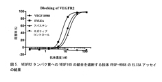
1μg/mLの濃度のVEGF165タンパク質(SinoBiological, Inc.社から入手した)を、100μL/ウェルで一晩、4℃で、96ウェルプレートに被覆した。翌日、プレートを洗浄し、室温で1時間遮断させた。5μg/mLのVEGFR2-ビオチンタンパク質(SinoBiological, Inc.社から入手した)100μL、及びVEGF165を標的化する異なる濃度の前記ウサギ抗体を添加し、共インキュベートした。プレートを洗浄して未結合の抗体を除去し、ストレプトアビジン/HRP(Beijing ZSGB-Bio Co., Ltd.社から入手した)とインキュベートし、次いで、繰り返し洗浄し、そして、色素生成性基質溶液を添加して発色させた。発色が止まった後、OD450を測定した。VEGF165を標的化するウサギ抗体の濃度を水平座標として取り、阻害率PI%を垂直座標として取って、graphPad Prism 6.0ソフトウェアをデータ解析及び曲線チャートの作成に使用した。阻害率(%)=(ODブランク-ODサンプル)/ODブランク×100%であり、式中、ODブランクは、VEGFR2-ビオチンのみが添加され、ウサギ抗体は添加されていないウェルのOD値を指し、ODサンプルは、VEGFR2-ビオチン及びウサギ抗体の両方が添加されたウェルのOD値を指す。
1.4 Functional analysis of rabbit antibodies targeting VEGF165
1.4.1 Rabbit antibody blocks VEGF165 binding to VEGFR2-his
VEGF165 protein (obtained from SinoBiological, Inc.) at a concentration of 1 μg/mL was coated in 96-well plates at 100 μL/well overnight at 4°C. The next day, plates were washed and blocked for 1 hour at room temperature. 100 μL of 5 μg/mL VEGFR2-biotin protein (obtained from SinoBiological, Inc.) and different concentrations of the rabbit antibody targeting VEGF165 were added and co-incubated. The plate was washed to remove unbound antibody, incubated with streptavidin/HRP (obtained from Beijing ZSGB-Bio Co., Ltd.), then washed repeatedly and treated with chromogenic substrate solution. was added to develop color. After color development stopped, OD 450 was measured. graphPad Prism 6.0 software was used for data analysis and generation of curve charts, with the concentration of rabbit antibody targeting VEGF165 taken as the horizontal coordinate and the inhibition rate PI% as the vertical coordinate. Inhibition rate (%) = (OD blank - OD sample ) / OD blank x 100%, where OD blank refers to the OD value of the well where only VEGFR2-biotin was added and no rabbit antibody was added. , OD sample refers to the OD value of the well where both VEGFR2-biotin and rabbit antibody were added.
図1に示すように、VEGFR2タンパク質は、被覆されたVEGF165タンパク質に効果的に結合し得、ウサギ抗体VEGF-R859、VEGF-R988、VEGF-R613、VEGF-R812は、VEGFR2タンパク質へのVEGFR165タンパク質の結合を効果的に阻害し得る。 As shown in Figure 1, VEGFR2 protein could effectively bind to coated VEGF165 protein, and rabbit antibodies VEGF-R859, VEGF-R988, VEGF-R613, VEGF-R812 were able to bind VEGFR165 protein to VEGFR2 protein. binding can be effectively inhibited.
抗体は、臍帯静脈内皮細胞に対するVEGF165の増殖効果を阻害する。 The antibody inhibits the proliferative effects of VEGF165 on umbilical vein endothelial cells.
1.4.2 ウサギ抗体は、HUVECの増殖を阻害する
前記ウサギ抗体の、VEGF165によって誘導される臍帯静脈内皮細胞増殖の中和効果を、WST-8法を使用して検出した。ヒト臍帯静脈内皮細胞HUVECを、4×103個細胞/ウェルで96ウェルプレートに接種し、10%FBS及び5%L-Glnを含有するM199培地中で4時間培養し、次いで、異なる濃度のウサギ抗体を50μL/ウェルで添加し、次いで、最終濃度10ng/mLのVEGF-165を10μL/ウェルで添加し、96ウェルプレートを、37℃、5%CO2の細胞インキュベーターで3日間インキュベートし、そしてブランクウェルB(細胞なし)、ネガティブコントロールM(細胞接種あり、抗体サンプルなし、VEGF-165の添加あり)、並びにM'(細胞接種あり、抗体サンプルなし、及びVEGF-165なし)を使用した。インキュベーション後、10μL/ウェルのWST-8色素生成溶液を添加し、96ウェルプレートをCO2インキュベーターでインキュベートして発色させ、発色が安定化した後、OD450及びOD630をマイクロプレートリーダーで測定した。各ウェルで、読み取り値は(OD450-OD630)であり、各群のOD値をその群の読み取り値マイナスブランクウェルBの読み取り値として定義して抗体の中和率を計算し、中和率%=(ネガティブコントロールMのOD値-サンプルのOD値)/(ネガティブコントロールMのOD値-M'のOD値)×100%であった。標準曲線を、抗体サンプル濃度を水平座標として取り、中和率を垂直座標として取って、統計ソフトウェアGraphPad Prismの自動解析機能を使用して計算し、4パラメータロジスティック回帰方程式を使用して、標準的な「S」曲線を当てはめて、抗体サンプルの半数最大効果濃度(EC50)を計算した。
1.4.2 Rabbit Antibodies Inhibit HUVEC Proliferation The neutralizing effect of the rabbit antibodies on the umbilical vein endothelial cell proliferation induced by VEGF165 was detected using the WST-8 method. Human umbilical vein endothelial cells HUVEC were seeded in a 96-well plate at 4 × 10 3 cells/well, cultured for 4 hours in M199 medium containing 10% FBS and 5% L-Gln, and then treated with different concentrations of Rabbit antibody was added at 50 μL/well, then VEGF-165 at a final concentration of 10 ng/mL was added at 10 μL/well, and the 96-well plate was incubated for 3 days in a cell incubator at 37 °C and 5% CO2 . Blank well B (no cells), negative control M (with cell inoculation, no antibody sample, and addition of VEGF-165), and M' (with cell inoculation, no antibody sample, and no VEGF-165) were used. . After incubation, 10 μL/well of WST-8 chromogenic solution was added, and the 96-well plate was incubated in a CO 2 incubator to develop color. After the color development was stabilized, OD 450 and OD 630 were measured with a microplate reader. . For each well, the reading is (OD 450 - OD 630 ), and the neutralization rate of the antibody is calculated by defining the OD value for each group as the reading for that group minus the reading for blank well B, and Rate %=(OD value of negative control M−OD value of sample)/(OD value of negative control M−OD value of M′)×100%. A standard curve was calculated using the automatic analysis function of the statistical software GraphPad Prism, taking the antibody sample concentration as the horizontal coordinate and the neutralization rate as the vertical coordinate, and a four-parameter logistic regression equation was used to calculate the standard curve. The half-maximal effective concentration (EC 50 ) of the antibody samples was calculated by fitting a typical "S" curve.
図2に示す結果は、VEGF-R988及びVEGF-R613がVEGF165のHUVEC増殖促進能力を効果的に低減させ、VEGF-R988がより高い最大阻害率を示し、VEGF-R812は阻害能力を基本的に有さず、一方、VEGF-R859は阻害能力を全く有さないことを示している。 The results shown in Figure 2 show that VEGF-R988 and VEGF-R613 effectively reduced the ability of VEGF165 to promote HUVEC proliferation, VEGF-R988 showed a higher maximum inhibition rate, and VEGF-R812 basically reduced the inhibitory ability. On the other hand, VEGF-R859 shows no inhibitory ability at all.
(実施例2)
ウサギ抗体VEGF-R988のヒト化、修飾、及び産生
実施例1におけるウサギ抗体の機能的解析の結果に基づいて、VEGF-R988をヒト化及び産生に選択した。
(Example 2)
Humanization, Modification, and Production of Rabbit Antibody VEGF-R988 Based on the results of the functional analysis of the rabbit antibody in Example 1, VEGF-R988 was selected for humanization and production.
2.1 ウサギ抗体VEGF-R988の軽鎖及び重鎖のCDRの決定
実施例1.2で決定されたVEGF-R988 scFv抗体のヌクレオチド配列に基づいて、VEGF-R988 scFvの重鎖及び軽鎖可変領域のアミノ酸配列を推定した。配列番号11/12を参照されたい。
2.1 Determination of CDRs of light chain and heavy chain of rabbit antibody VEGF-R988 Based on the nucleotide sequence of VEGF-R988 scFv antibody determined in Example 1.2, the amino acid sequence of the heavy chain and light chain variable region of VEGF-R988 scFv was determined. estimated. Please refer to SEQ ID NO: 11/12.
Kabatインデックス及びIMGTナンバリングシステムを参照して、ウサギ抗体VEGF-R988-scFvの軽鎖及び重鎖の3つのCDRの各々のアミノ酸配列を決定した。Table 1(表3)を参照されたい。次の工程で、軽鎖及び重鎖の前述のそれぞれの3つのCDRを、ヒト化抗体VEGF-R988-scFvに移植した。実施例2.2を参照されたい。 The amino acid sequences of each of the three CDRs of the light chain and heavy chain of the rabbit antibody VEGF-R988-scFv were determined with reference to the Kabat index and IMGT numbering system. Please refer to Table 1 (Table 3). In the next step, the aforementioned three CDRs of each of the light and heavy chains were grafted onto the humanized antibody VEGF-R988-scFv. See Example 2.2.

2.2 ウサギ抗体VEGF-R988のCDR移植
ウサギ抗体のヒト化を、CDR移植の従来のヒト化方法を使用して行った。その配列がウサギ軽鎖又は重鎖可変領域の配列に近い、ヒト抗体の軽鎖又は重鎖可変領域を、鋳型として選択し、ウサギ軽鎖又は重鎖の3つのCDR(Table 1(表3))の各々をヒト抗体の可変領域に挿入して、ヒト化軽鎖可変領域(VL)又はヒト化重鎖可変領域(VH)配列をそれぞれ得た。VEGF-R988の軽鎖可変領域のヒト鋳型は、VEGF-R988の軽鎖に65.30%相同なIGKV1-27*01であり、重鎖可変領域のヒト鋳型は、VEGF-R988の重鎖に53.20%相同なIGHV4-4*08である。
2.2 CDR Grafting of Rabbit Antibody VEGF-R988 Humanization of the rabbit antibody was performed using the conventional humanization method of CDR grafting. A human antibody light chain or heavy chain variable region whose sequence is close to that of a rabbit light chain or heavy chain variable region was selected as a template, and three CDRs of a rabbit light chain or heavy chain (Table 1) ) were inserted into the variable region of a human antibody to obtain a humanized light chain variable region (VL) or humanized heavy chain variable region (VH) sequence, respectively. The human template for the light chain variable region of VEGF-R988 is IGKV1-27 * 01, which is 65.30% homologous to the light chain of VEGF-R988, and the human template for the heavy chain variable region is 53.20% homologous to the heavy chain of VEGF-R988. It is homologous IGHV4-4 * 08.
2.3 ヒト化可変領域のフレームワーク領域での復帰突然変異
ウサギ由来のフレームワーク領域内の一部の重要なアミノ酸はCDR活性の維持に必須であるため、これら重要なアミノ酸を、対応するウサギ抗体アミノ酸配列に復帰突然変異させ、以下の部位を復帰突然変異させた: 軽鎖では、1位をEに復帰突然変異させ、2位をLに復帰突然変異させ、4位をLに復帰突然変異させ、そして63位をKに復帰突然変異させた。一方、重鎖では、3位をVに復帰突然変異させ、37位をVに復帰突然変異させ、47位をYに復帰突然変異させ、78位をVに復帰突然変異させ、79位をDに復帰突然変異させ、そして91位をFに復帰突然変異させた。全ての上記の部位は、Kabatのナンバリングスキームを参照することによってナンバリングした。ヒト化抗体VEGF-H988-10を、CDRヒト化移植及びフレームワーク領域の復帰突然変異によって得た。
2.3 Backmutation in the framework region of the humanized variable region Some key amino acids in the rabbit-derived framework region are essential for maintaining CDR activity, so these key amino acids were replaced with the corresponding rabbit antibody amino acids. The following sites were backmutated: in the light chain,
2.4 ヒト化モノクローナル抗体VEGF-H988-10の産生及びCDR親和性の修飾
VEGF-H988-10の重鎖可変領域(配列番号23)を全遺伝子合成方法によって得、次いで、Sca I+Nhe I(Fermentas社)で消化した、重鎖シグナルペプチドをコードするヌクレオチド配列(配列番号45)及びヒトIgG1定常領域をコードするヌクレオチド配列(配列番号49)を有するpSTEP2ベクターに、in-fusion方法によって挿入して、VEGF-H988-10重鎖(配列番号19)を発現するベクターを得た。VEGF-H988-10の軽鎖可変領域(配列番号24)を全遺伝子合成方法によって得、次いで、Sca I+BsiW I(Fermentas社)で消化した、軽鎖シグナルペプチドをコードするヌクレオチド配列(配列番号46)及びヒトカッパ定常領域をコードするヌクレオチド配列(配列番号50)を有するpSTEP2ベクターに、in-fusion方法によって挿入して、VEGF-H988-10軽鎖(配列番号20)を発現するベクターを得た。プラスミドを抽出し、HEK-293細胞にコトランスフェクトし、細胞を7日間培養した。培養上清をタンパク質A精製カラムで精製して、高純度の抗体を得た。
2.4 Production of humanized monoclonal antibody VEGF-H988-10 and modification of CDR affinity
The heavy chain variable region of VEGF-H988-10 (SEQ ID NO: 23) was obtained by a total gene synthesis method, and then digested with Sca I+Nhe I (Fermentas) to obtain the nucleotide sequence encoding the heavy chain signal peptide (SEQ ID NO: 23). 45) and into a pSTEP2 vector having a nucleotide sequence encoding the human IgG1 constant region (SEQ ID NO: 49) by an in-fusion method to obtain a vector expressing the VEGF-H988-10 heavy chain (SEQ ID NO: 19). Ta. The light chain variable region of VEGF-H988-10 (SEQ ID NO: 24) was obtained by a total gene synthesis method, and then digested with Sca I+BsiW I (Fermentas) to obtain the nucleotide sequence encoding the light chain signal peptide (SEQ ID NO: 24). 46) and into a pSTEP2 vector having a nucleotide sequence encoding the human kappa constant region (SEQ ID NO: 50) by an in-fusion method to obtain a vector expressing the VEGF-H988-10 light chain (SEQ ID NO: 20). . The plasmid was extracted and co-transfected into HEK-293 cells, and the cells were cultured for 7 days. The culture supernatant was purified using a protein A purification column to obtain highly pure antibodies.


VEGF-H988-10の親和性を向上させるために、重鎖及び軽鎖可変領域のCDR領域のSDMライブラリー(3つの飽和突然変異ライブラリーLCDR1、LCDR3、及びHCDR2を含む)を構築した。一方、抗体の化学的安定性を向上させるためには、脱アミド化又は異性化を受け得るアミノ酸残基を別のアミノ酸残基に修飾するべきである。アスパラギンの脱アミド化は、NG、NS、NA、NT等で生じ得、抗体の安定性又は生物学的機能に影響するイソアスパラギン酸残基を生成させる。VEGF-H988の可変領域HCDR3は脱アミド化感受性部位を有しており、したがって、SDMライブラリーを構築して、抗体の化学的安定性及び生物学的機能を向上させた。上記の4つの突然変異体ライブラリーはscFv形態で構築し、scFv- gIII融合タンパク質としてファージベクターにクローニングした。各CDRについて、可溶性抗原VEGFに対して最適な結合能力を有するCDRクローンをスクリーニングし、最後に、CDRの親和性及び安定性が最適化された抗体VEGF-H988を得た。VEGF-H988軽鎖及び重鎖CDRの配列をTable 2(表6)に示す。 To improve the affinity of VEGF-H988-10, an SDM library of heavy and light chain variable region CDR regions was constructed, including three saturation mutation libraries LCDR1, LCDR3, and HCDR2. On the other hand, in order to improve the chemical stability of antibodies, amino acid residues that can undergo deamidation or isomerization should be modified to other amino acid residues. Deamidation of asparagine can occur with NG, NS, NA, NT, etc., producing isoaspartic acid residues that affect antibody stability or biological function. The variable region HCDR3 of VEGF-H988 has a deamidation sensitive site, therefore, an SDM library was constructed to improve the chemical stability and biological function of the antibody. The above four mutant libraries were constructed in scFv form and cloned into phage vectors as scFv-gIII fusion proteins. For each CDR, CDR clones with optimal binding ability to the soluble antigen VEGF were screened, and finally, the antibody VEGF-H988 with optimized CDR affinity and stability was obtained. The sequences of VEGF-H988 light chain and heavy chain CDRs are shown in Table 2 (Table 6).

2.5 ヒト化モノクローナル抗体VEGF-H988の産生
順に結合された、軽鎖シグナルペプチドをコードするヌクレオチド配列(配列番号46)、ヒト化抗体軽鎖可変領域をコードするヌクレオチド配列(配列番号48)、及びヒト抗体カッパ軽鎖定常領域をコードするヌクレオチド配列(配列番号50)を有する、前述の抗体VEGF-H988軽鎖及びシグナルペプチドをコードするヌクレオチド配列(配列番号44)をPCR増幅し、自社開発のpGSベクター(Kpn I+Xba I)にin-fusion方法によって挿入し、正確なプラスミドをシーケンシングによって確認した。順に結合された、重鎖シグナルペプチドをコードするヌクレオチド配列(配列番号45)、ヒト化抗体重鎖可変領域をコードするヌクレオチド配列(配列番号47)、及びヒト抗体カッパ重鎖定常領域をコードするヌクレオチド配列(配列番号49)を有する、前述の抗体VEGF-H988重鎖及びシグナルペプチドをコードするヌクレオチド配列(配列番号43)をPCR増幅し、軽鎖を正確に有することが確認されているpGSベクター(Nhe I+Not I)に、in-fusion方法によって挿入し、VEGF-R988の軽鎖及び重鎖の両方を発現する正確なベクターを、シーケンシングによって確認した。これら発現ベクターは、選択マーカーとしてのGS遺伝子、並びに抗体軽鎖及び重鎖の発現エレメントを有する、真核性の発現ベクターである。これら発現ベクターをCHO-K1-GS-欠損細胞にトランスフェクトし、VEGF-H988高発現細胞系をMSXスクリーニングによって得た。抗体発現が高いクローンをELISAアッセイによって選択し、高発現細胞系を、細胞の増殖状態及び抗体薬のための重要な質的特徴の両方を考慮することによって選択した。無血清懸濁液培養を使用して、VEGF-H988を産生するCHO細胞系を培養して、純度及び質の高いVEGF-H988抗体を得た。
2.5 Production of humanized monoclonal antibody VEGF-H988 The nucleotide sequence encoding the light chain signal peptide (SEQ ID NO: 46), the nucleotide sequence encoding the humanized antibody light chain variable region (SEQ ID NO: 48), and the human The aforementioned antibody VEGF-H988 light chain having the nucleotide sequence (SEQ ID NO: 50) encoding the antibody kappa light chain constant region and the nucleotide sequence (SEQ ID NO: 44) encoding the signal peptide were amplified by PCR, and a pGS vector developed in-house was created. (Kpn I+Xba I) by in-fusion method, and the correct plasmid was confirmed by sequencing. A nucleotide sequence encoding a heavy chain signal peptide (SEQ ID NO: 45), a nucleotide sequence encoding a humanized antibody heavy chain variable region (SEQ ID NO: 47), and a nucleotide sequence encoding a human antibody kappa heavy chain constant region linked in sequence. The aforementioned antibody VEGF-H988 heavy chain having the sequence (SEQ ID NO: 49) and the nucleotide sequence (SEQ ID NO: 43) encoding the signal peptide were amplified by PCR, and the pGS vector (SEQ ID NO: 43), which was confirmed to have exactly the light chain ( Nhe I+Not I) by in-fusion method and the correct vector expressing both the light and heavy chains of VEGF-R988 was confirmed by sequencing. These expression vectors are eukaryotic expression vectors having the GS gene as a selection marker and expression elements for antibody light and heavy chains. These expression vectors were transfected into CHO-K1-GS-deficient cells, and a cell line with high VEGF-H988 expression was obtained by MSX screening. Clones with high antibody expression were selected by ELISA assay, and high expressing cell lines were selected by considering both the proliferation status of the cells and important qualitative characteristics for antibody drugs. The CHO cell line producing VEGF-H988 was cultured using serum-free suspension culture to obtain VEGF-H988 antibodies of high purity and quality.
(実施例3)
ヒト化抗体VEGF-H988の特徴解析
3.1 VEGF165へのヒト化抗体VEGF-H988の結合の特徴解析
3.1.1 ヒト化抗体VEGF-H988はVEGF165に特異的に結合する
異なる濃度の(0.15ng/mL、0.46ng/mL、1.37ng/mL、4.12ng/mL、12.35ng/mL、37.04ng/mL、111.11ng/mL、333.33ng/mL、1000ng/mL、及び3000ng/mL)組換えヒトVEGF165タンパク質(SinoBiological, Inc.社から入手した)を、100μL/ウェルで一晩、4℃で、96ウェルプレートに被覆した。翌日、プレートを洗浄し、室温で1時間遮断させた。100μLの、1μg/mLのVEGF165-H988、アバスチン(Roche社から入手した)、又はネガティブコントロール抗体H7N9-R1とそれぞれインキュベートした後、プレートを洗浄して未結合の抗体を除去し、次いで、ヤギF(ab')2抗ヒトIgG F(ab')2/HRP(Jackson ImmunoResearch Laboratories, Inc.社から入手した)とインキュベートし、繰り返し洗浄し、そして、色素生成性基質溶液を添加して発色させた。発色が止まった後、OD450を測定した。組換えヒトVEGF165タンパク質の濃度を水平座標として取り、OD450値を垂直座標として取って、graphPad Prism 6.0ソフトウェアを使用して「S」曲線チャートを当てはめ、組換えヒトVEGF165タンパク質への抗体の結合を解析した。
(Example 3)
Characteristic analysis of humanized antibody VEGF-H988
3.1 Characteristic analysis of the binding of humanized antibody VEGF-H988 to VEGF165
3.1.1 Humanized antibody VEGF-H988 specifically binds to VEGF165 at different concentrations (0.15ng/mL, 0.46ng/mL, 1.37ng/mL, 4.12ng/mL, 12.35ng/mL, 37.04ng/mL , 111.11ng/mL, 333.33ng/mL, 1000ng/mL, and 3000ng/mL) recombinant human VEGF165 protein (obtained from SinoBiological, Inc.) was added to 96 wells at 100 μL/well overnight at 4°C. plate was coated. The next day, plates were washed and blocked for 1 hour at room temperature. After incubation with 100 μL of 1 μg/mL of VEGF165-H988, Avastin (obtained from Roche), or negative control antibody H7N9-R1, respectively, the plate was washed to remove unbound antibody and then goat F (ab')2 anti-human IgG F(ab')2/HRP (obtained from Jackson ImmunoResearch Laboratories, Inc.), washed repeatedly, and color developed by adding chromogenic substrate solution. . After color development stopped, OD 450 was measured. Taking the concentration of recombinant human VEGF165 protein as the horizontal coordinate and the OD450 value as the vertical coordinate, fit an “S” curve chart using graphPad Prism 6.0 software to determine the binding of the antibody to the recombinant human VEGF165 protein. Analyzed.
図3に示す結果は、組換えヒトVEGF165に特異的に結合するヒト化分子VEGF165-H988のEC50値が2.42ng/mLであり、R2=0.999であること、組換えヒトVEGF165に結合するアバスチンのEC50値が2.77ng/mLであり、R2=1.000であることを示している。これは、VEGF165-H988の組換えヒトVEGF165タンパク質への結合能力がアバスチンのそれよりもわずかに良好であることを示している。ネガティブコントロール抗体H7N9-R1は、組換えヒトVEGF165タンパク質への結合能力を有さない。 The results shown in Figure 3 show that the EC 50 value of the humanized molecule VEGF165-H988, which specifically binds to recombinant human VEGF165, is 2.42 ng/mL and R 2 =0.999. The EC 50 value of Avastin is 2.77 ng/mL, indicating that R 2 =1.000. This indicates that the binding ability of VEGF165-H988 to recombinant human VEGF165 protein is slightly better than that of Avastin. Negative control antibody H7N9-R1 has no ability to bind to recombinant human VEGF165 protein.
3.1.2 組換えVEGF165タンパク質に対するヒト化抗体VEGF-H988の結合親和性のアッセイ
VEGF165-H988及びアバスチン(Roche社から入手した)の親和性を、ストレプトアビジンで被覆されたセンサー及び固定されたビオチン標識VEGF165タンパク質を使用して、複数の濃度で測定した。
3.1.2 Assay of binding affinity of humanized antibody VEGF-H988 to recombinant VEGF165 protein
The affinity of VEGF165-H988 and Avastin (obtained from Roche) was measured at multiple concentrations using a streptavidin-coated sensor and immobilized biotin-labeled VEGF165 protein.
組換えヒトVEGF165タンパク質をまず、以下のプロセスの通りに、1:2のモル比で、ビオチンで標識した:組換えVEGFタンパク質バッファー(20mMのTris、150mMのNaCl、pH8.0)を、5000MWの限外濾過遠沈管での限外濾過を介してPBSと置き換え、UV定量によって測定すると567.57μgのタンパク質が得られ、そして、得られたタンパク質を20mMのビオチン溶液と1:2のモル比で混合し、30分間、室温で、暗所でインキュベートし、次いで、5000MWの限外濾過遠沈管で再び濾過して、未標識のビオチンを除去した。UV定量した後、ビオチン標識タンパク質を、等容積のグリセロール及び最終濃度0.1%のBSAを添加することによって得た。UVによって検出したところ、VEGF165タンパク質の濃度は2.08mg/mLであった。 Recombinant human VEGF165 protein was first labeled with biotin at a molar ratio of 1:2 as per the following process: Recombinant VEGF protein buffer (20 mM Tris, 150 mM NaCl, pH 8.0) was added to 5000 MW of Ultrafiltration was replaced with PBS via ultrafiltration in centrifuge tubes, yielding 567.57 μg of protein as determined by UV quantification, and the resulting protein was mixed with 20 mM biotin solution in a 1:2 molar ratio. and incubated for 30 minutes at room temperature in the dark, then filtered again through a 5000 MW ultrafiltration centrifuge tube to remove unlabeled biotin. After UV quantification, biotin-labeled proteins were obtained by adding equal volumes of glycerol and BSA to a final concentration of 0.1%. The concentration of VEGF165 protein was 2.08 mg/mL as detected by UV.
次いで、異なる濃度のVEGF165-H988及びアバスチンの、ビオチン化された組換えヒトVEGFタンパク質との親和性を測定し、得られたKD値が最終親和性であった。 The affinities of different concentrations of VEGF165-H988 and Avastin with biotinylated recombinant human VEGF protein were then measured, and the resulting KD value was the final affinity.
Table 3(表7)に示す結果は、組換えヒトVEGF165タンパク質に対するVEGF165-H988の結合親和性KDの値が19.5pMであったこと、結合定数konの値が3.44E+05M-1s-1であったこと、及び解離定数kdisの値が6.70E-06s-1であったことを示している。Table 3(表7)に示すように、VEGFタンパク質へのアバスチンの結合親和性KDの値は29.2E-11pMであり、結合定数konの値は1.87E+05M-1s-1であり、解離定数kdisの値は5.46E-06s-1であった。この結果から、VEGF165-H988の親和性がアバスチンのそれよりわずかに高い、すなわちアバスチンの親和性の約1.5倍高いと結論付けることができる。 The results shown in Table 3 (Table 7) show that the value of the binding affinity KD of VEGF165-H988 to recombinant human VEGF165 protein was 19.5 pM, and the value of the binding constant k on was 3.44E+05M -1 s - 1 , and the value of the dissociation constant k dis was 6.70E-06s -1 . As shown in Table 3 (Table 7), the value of the binding affinity KD of Avastin to the VEGF protein is 29.2E-11pM, the value of the binding constant k on is 1.87E+05M -1 s -1 , The value of the dissociation constant kdis was 5.46E-06s -1 . From this result, it can be concluded that the affinity of VEGF165-H988 is slightly higher than that of Avastin, i.e. about 1.5 times higher than that of Avastin.

3.1.3 ヒト化抗体VEGF165-H988の種間交差反応性の決定
組換えヒトVEGF165タンパク質又は組換えマウスmVEGF164タンパク質を0.1μg/mL、1μg/mL、及び10μg/mLまでそれぞれ希釈し、100μL/ウェルで一晩、4℃で、96ウェルプレートに被覆した。翌日、プレートを洗浄し、室温で1時間遮断させた。100μLのVEGF165-H988、アバスチン(Roche社から入手した)又はネガティブコントロール抗体H7N9-R1を1μg/mLの濃度でそれぞれ添加し、そして1時間インキュベートした。プレートを洗浄して未結合の抗体を除去した。プレートをヤギF(ab')2抗ヒトIgG F(ab')2/HRP(Jackson ImmunoResearch Laboratories, Inc.社)とインキュベートし、次いで、繰り返し洗浄し、そして色素生成性基質溶液を添加して発色させた。発色が止まった後、OD450を測定した。タンパク質濃度を水平座標として取り、OD450値を垂直座標として取って、graphPad Prism 6.0ソフトウェアを使用して棒チャートを作成した。
3.1.3 Determination of interspecies cross-reactivity of humanized antibody VEGF165-H988 Dilute recombinant human VEGF165 protein or recombinant mouse mVEGF164 protein to 0.1 μg/mL, 1 μg/mL, and 10 μg/mL, respectively, and add 100 μL/well. plated in 96-well plates overnight at 4°C. The next day, plates were washed and blocked for 1 hour at room temperature. 100 μL of VEGF165-H988, Avastin (obtained from Roche) or negative control antibody H7N9-R1 were added at a concentration of 1 μg/mL, respectively, and incubated for 1 hour. Plates were washed to remove unbound antibody. Plates were incubated with goat F(ab')2 anti-human IgG F(ab')2/HRP (Jackson ImmunoResearch Laboratories, Inc.), then washed repeatedly and developed by adding chromogenic substrate solution. I let it happen. After color development stopped, OD 450 was measured. A bar chart was created using graphPad Prism 6.0 software, taking the protein concentration as the horizontal coordinate and the OD450 value as the vertical coordinate.
図4に示す結果は、VEGF165-H988が組換えヒトVEGF165タンパク質に特異的に結合し、組換えマウスmVEGF164タンパク質との交差結合を示すことを示している。 The results shown in Figure 4 show that VEGF165-H988 specifically binds to recombinant human VEGF165 protein and exhibits cross-linking with recombinant mouse mVEGF164 protein.
3.2 ヒト化抗体VEGF-H988の受容体遮断特性
1μg/mLの濃度のVEGF165タンパク質(SinoBiological, Inc.社から入手した)を、100μL/ウェルで一晩、4℃で、96ウェルプレートに被覆した。翌日、プレートを洗浄し、室温で1時間遮断させた。2μg/mLのVEGFR2-hisタンパク質(SinoBiological, Inc.社から入手した)100μLを各ウェルに添加し、異なる濃度の抗体VEGF-H988、EYLEA、アバスチン(Roche社から入手した)、又はネガティブコントロール抗体H7N9-R1をそれぞれ添加し、そして共インキュベートした。プレートを洗浄して未結合の抗体を除去した。プレートをC-his-R023/HRPとインキュベートし、次いで繰り返し洗浄し、そして、色素生成性基質溶液を添加して発色させた。発色が安定化した後、OD450を測定し、各群について2回ずつ試験した。抗体の濃度を水平座標として取り、阻害率PI%を垂直座標として取って、graphPad Prism 6.0ソフトウェアをデータ解析及び曲線チャートの作成に使用して、IC50値を計算した。阻害率(%)=(ODブランク-ODサンプル)/ODブランク×100%であり、式中、ODブランクは、VEGFR2-hisのみが添加され、ヒト抗体は添加されていないウェルのOD値を指し、ODサンプルは、VEGFR2-his及びヒト化抗体の両方が添加されたウェルのOD値を指す。
3.2 Receptor blocking properties of humanized antibody VEGF-H988
VEGF165 protein (obtained from SinoBiological, Inc.) at a concentration of 1 μg/mL was coated in 96-well plates at 100 μL/well overnight at 4°C. The next day, plates were washed and blocked for 1 hour at room temperature. 100 μL of 2 μg/mL VEGFR2-his protein (obtained from SinoBiological, Inc.) was added to each well, and different concentrations of antibodies VEGF-H988, EYLEA, Avastin (obtained from Roche), or negative control antibody H7N9 were added to each well. -R1 were added and co-incubated. Plates were washed to remove unbound antibody. Plates were incubated with C-his-R023/HRP, then washed repeatedly, and chromogenic substrate solution was added to develop color. After the color development stabilized, the OD 450 was measured and each group was tested in duplicate. IC50 values were calculated using graphPad Prism 6.0 software for data analysis and curve chart generation, taking the concentration of antibody as the horizontal coordinate and the inhibition rate PI% as the vertical coordinate. Inhibition rate (%) = (OD blank - OD sample ) / OD blank x 100%, where OD blank refers to the OD value of the well where only VEGFR2-his was added and no human antibody was added. , OD sample refers to the OD value of the well where both VEGFR2-his and humanized antibody were added.
図5に示すように、VEGFR2タンパク質は、被覆されたVEGF165タンパク質に効果的に結合し得、抗体VEGF-H988は、VEGFR165タンパク質へのVEGFR2タンパク質の結合を、EYLEAよりも比較的弱いがアバスチンよりも優れた阻害能力で、効果的に阻害し得、ネガティブコントロール抗体は、阻害効果を有さない。 As shown in Figure 5, VEGFR2 protein could effectively bind to coated VEGF165 protein, and antibody VEGF-H988 inhibited the binding of VEGFR2 protein to VEGFR165 protein relatively weaker than EYLEA but more than Avastin. With good inhibitory ability, it can be inhibited effectively, and the negative control antibody has no inhibitory effect.
3.3 ヒト化抗体によるHUVEC細胞増殖の阻害
3.3.1 ヒト化抗体VEGF-H988による異なる濃度のVEGF165の中和効果
VEGF-H988の、VEGF165によって誘導されるHUVEC細胞増殖の中和効果を、WST-8法を使用することによって検出した。HUVEC細胞を、4×103個細胞/ウェルで96ウェルプレートに接種し、10%FBS及び5%L-Glnを含有するM199培地中で4時間培養し、次いで、異なる濃度のVEGF-H988、EYLEA、又はアバスチン(Roche社から入手した)を50μL/ウェルで添加し、次いで、最終濃度1000ng/mL、100ng/mL、又は10ng/mLのVEGF-165を10μL/ウェルで添加し、96ウェルプレートを、37℃、5%CO2の細胞インキュベーターで3日間インキュベートし、ブランクウェルB(細胞なし)、ネガティブコントロールM(細胞接種あり、抗体サンプルの添加なし、VEGF-165の添加あり)、及びM'(細胞接種あり、抗体サンプルの添加なし、及びVEGF-165の添加なし)を使用した。インキュベーション後、10μL/ウェルのWST-8色素生成溶液を添加し、96ウェルプレートをCO2インキュベーターでインキュベートして発色させ、発色が安定化した後、OD450及びOD630をマイクロプレートリーダーで測定した。各ウェルで、読み取り値は(OD450-OD630)であり、各群のOD値をその群の読み取り値マイナスブランクウェルBの読み取り値として定義して抗体の中和率を計算し、中和率%=(ネガティブコントロールMのOD値-サンプルのOD値)/(ネガティブコントロールMのOD値-M'のOD値)×100%であった。標準曲線を、抗体サンプル濃度を水平座標として取り、中和率を垂直座標として取って、統計ソフトウェアGraphPad Prismの自動解析機能を使用して計算し、4-パラメータロジスティック回帰方程式を使用して、標準的な「S」曲線を当てはめて、抗体サンプルの半数最大効果濃度(EC50)を計算した。
3.3 Inhibition of HUVEC cell proliferation by humanized antibodies
3.3.1 Neutralizing effect of humanized antibody VEGF-H988 on different concentrations of VEGF165
The neutralizing effect of VEGF-H988 on HUVEC cell proliferation induced by VEGF165 was detected by using WST-8 method. HUVEC cells were seeded in 96-well plates at 4 × 10 3 cells/well and cultured for 4 h in M199 medium containing 10% FBS and 5% L-Gln, followed by different concentrations of VEGF-H988, Add EYLEA or Avastin (obtained from Roche) at 50 μL/well, then add VEGF-165 at a final concentration of 1000 ng/mL, 100 ng/mL, or 10 ng/mL at 10 μL/well to a 96-well plate. were incubated in a cell incubator at 37°C and 5% CO2 for 3 days to prepare blank wells B (no cells), negative control M (with cell inoculation, no antibody sample addition, and VEGF-165 addition), and M'. (with cell inoculation, without addition of antibody sample, and without addition of VEGF-165) was used. After incubation, 10 μL/well of WST-8 chromogenic solution was added, and the 96-well plate was incubated in a CO2 incubator to develop color. After the color development was stabilized, OD 450 and OD 630 were measured using a microplate reader. For each well, the reading is (OD 450 - OD 630 ), and the neutralization rate of the antibody is calculated by defining the OD value for each group as the reading for that group minus the reading for blank well B, and Rate %=(OD value of negative control M−OD value of sample)/(OD value of negative control M−OD value of M′)×100%. A standard curve was calculated using the automatic analysis function of the statistical software GraphPad Prism, taking the antibody sample concentration as the horizontal coordinate and the neutralization rate as the vertical coordinate, and a 4-parameter logistic regression equation was used to calculate the standard curve. A standard "S" curve was fitted to calculate the half-maximal effective concentration (EC 50 ) of the antibody samples.
図6及び図7に示すように、抗体VEGF-H988は、VEGF165のHUVEC増殖促進能力を効果的に低減させ得る。VEGF-H988の中和能力は、異なる濃度の組換えヒトVEGF165で、EYLEA及びアバスチンのそれよりも強力であった。また、中和能力の差は、VEGF165濃度が高いほど大きかった。VEGF165が高濃度である条件下では、VEGF-H988は低い中和EC50を依然として有し、最大中和率を依然として維持し得るが、EYLEA及びアバスチンの中和EC50は徐々に増大し、それに伴って、最大中和率が低下した。異なる濃度のVEGF165中和抗体の半数最大効果濃度(EC50)及び最大中和率をTable 4(表8)にまとめる。 As shown in FIGS. 6 and 7, antibody VEGF-H988 can effectively reduce the ability of VEGF165 to promote HUVEC proliferation. The neutralizing ability of VEGF-H988 was more potent than that of EYLEA and Avastin at different concentrations of recombinant human VEGF165. Furthermore, the difference in neutralization ability was greater as the VEGF165 concentration was higher. Under conditions where VEGF165 is at high concentration, VEGF-H988 still has a low neutralization EC 50 and can still maintain the maximum neutralization rate, while the neutralization EC 50 of EYLEA and Avastin increases gradually and Accordingly, the maximum neutralization rate decreased. The half-maximal effective concentration (EC 50 ) and maximum neutralization rate of VEGF165-neutralizing antibodies at different concentrations are summarized in Table 4.

3.3.2 ヒト化抗体VEGF-H988による、様々なサブタイプのVEGFの中和効果
VEGF-H988の、様々なVEGFサブタイプ(VEGF165、VEGFC、及びVEGFD)によって誘導されるHUVEC細胞増殖の中和効果を、WST-8法を使用して検出した。HUVEC細胞を4×103個細胞/ウェルで96ウェルプレートに接種し、10%FBS及び5%L-Glnを含有するM199培地中で4時間培養し、次いで、異なる濃度のVEGF-H988又はアバスチン(Roche社から入手した)を50μL/ウェルで添加し、次いで、VEGF-165、VEGFC、及びVEGFD混合物(最終濃度はそれぞれ25ng/mL、1000ng/mL、6000ng/mLであった)を10μL/ウェルで添加し、96ウェルプレートを、37℃、5%CO2の細胞インキュベーターで3日間インキュベートし、そして、ブランクウェルB(細胞なし)、ネガティブコントロールM(細胞接種あり、抗体サンプルの添加なし、VEGFの添加あり)、及びM'(細胞接種あり、抗体サンプルなし、及びVEGFなし)を使用した。インキュベーション後、10μL/ウェルのWST-8色素生成溶液を添加し、96ウェルプレートをCO2インキュベーターでインキュベートして発色させ、発色が安定化した後、OD450及びOD630をマイクロプレートリーダーで測定した。各ウェルで、読み取り値は(OD450-OD630)であり、各群のOD値をその群の読み取り値マイナスブランクウェルBの読み取り値として定義して抗体の中和率を計算し、中和率%=(ネガティブコントロールMのOD値-サンプルのOD値)/(ネガティブコントロールMのOD値-M'のOD値)×100%であった。標準曲線を、抗体サンプル濃度を水平座標として取り、中和率を垂直座標として取って、統計ソフトウェアGraphPad Prismの自動解析機能を使用して計算し、4-パラメータロジスティック回帰方程式を使用して、標準的な「S」曲線を当てはめて、抗体サンプルの半数最大効果濃度(EC50)を計算した。
3.3.2 Neutralization effect of various subtypes of VEGF by humanized antibody VEGF-H988
The neutralizing effect of VEGF-H988 on HUVEC cell proliferation induced by various VEGF subtypes (VEGF165, VEGFC, and VEGFD) was detected using WST-8 method. HUVEC cells were seeded at 4 × 10 3 cells/well in a 96-well plate and cultured for 4 hours in M199 medium containing 10% FBS and 5% L-Gln, followed by different concentrations of VEGF-H988 or Avastin. (obtained from Roche) at 50 μL/well, then VEGF-165, VEGFC, and VEGFD mixture (final concentrations were 25 ng/mL, 1000 ng/mL, and 6000 ng/mL, respectively) at 10 μL/well. The 96-well plate was incubated for 3 days in a cell incubator at 37°C and 5% CO2 , and the blank well B (no cells), negative control M (with cell seeding, no antibody sample addition, VEGF (with the addition of ), and M' (with cell inoculation, no antibody sample, and no VEGF) were used. After incubation, 10 μL/well of WST-8 chromogenic solution was added, and the 96-well plate was incubated in a CO 2 incubator to develop color. After the color development was stabilized, OD 450 and OD 630 were measured with a microplate reader. . For each well, the reading is (OD 450 - OD 630 ), and the neutralization rate of the antibody is calculated by defining the OD value for each group as the reading for that group minus the reading for blank well B, and Rate %=(OD value of negative control M−OD value of sample)/(OD value of negative control M−OD value of M′)×100%. A standard curve was calculated using the automatic analysis function of the statistical software GraphPad Prism, taking the antibody sample concentration as the horizontal coordinate and the neutralization rate as the vertical coordinate, and a 4-parameter logistic regression equation was used to calculate the standard curve. A standard "S" curve was fitted to calculate the half-maximal effective concentration (EC 50 ) of the antibody samples.
図8に示すように、HUVEC細胞に同時に作用する様々なVEGFサブタイプ(VEGF165、VEGFC、VEGFD)のケースでは、抗体VEGF-H988は、アバスチンよりも強力な中和効果を有する。0.016~4.000nMの濃度範囲では、VEGF-H988はアバスチンよりも小さいEC50を有し、それぞれ0.26nM及び0.77nMであった。そして、VEGF-H988はアバスチンよりも高い中和率を有し、それぞれ86.1%及び58.5%であった。VEGF-H988及びアバスチンの、様々なVEGFサブタイプを中和する半数最大効果濃度(EC50)及び最大中和率をTable 5(表9)にまとめる。 As shown in Figure 8, in the case of various VEGF subtypes (VEGF165, VEGFC, VEGFD) acting simultaneously on HUVEC cells, the antibody VEGF-H988 has a stronger neutralizing effect than Avastin. In the concentration range of 0.016 to 4.000 nM, VEGF-H988 had a lower EC 50 than Avastin, 0.26 nM and 0.77 nM, respectively. And VEGF-H988 had a higher neutralization rate than Avastin, 86.1% and 58.5%, respectively. The half-maximal effective concentrations (EC 50 ) and maximum neutralization rates of VEGF-H988 and Avastin to neutralize various VEGF subtypes are summarized in Table 5.

(実施例4)
マウスにおける抗体VEGF-H988の反復投与毒性研究
高齢群(35週齢)及び正常群(9週齢)のそれぞれにおいて8頭の、半分がオスで半分がメスの、CD-1マウスを使用した。マウスを完全ランダム化法に従って4つの群(G1~G4)に分け、各群、半分がオスで半分がメスの4頭のマウスであった。具体的なグループ化は以下の通りであった:G1:高齢マウス溶媒コントロール群、G2:高齢マウス研究群、G3:正常マウス溶媒コントロール群、G4:正常マウス研究群。VEGF-H988抗体を研究群に反復投与し、コントロール群には等容積の溶媒を投与した。用量は50mg/kgであり、各投与の容積はそれぞれ5mL/kg(1回目から8回目の投与)及び10mL/kg(9回目から16回目の投与)であった。投与態様は腹腔内注射であり、週に2回の頻度であった。薬物動態学的アッセイ及び免疫原性アッセイのために、全てのマウスで、初回投与の前、及び0.5時間後、2時間後、4時間後、6時間後、24時間後、48時間後、72時間後、並びに全ての他の投与の前及び1時間後に、眼窩血液採取を行った。マウスの体重を測定し、各投与の前に記録した。詳細な投与レジメンをTable 6(表10)に示す。
(Example 4)
Repeated dose toxicity study of antibody VEGF-H988 in mice Eight CD-1 mice, half male and half female, were used in each of the aged group (35 weeks old) and normal group (9 weeks old). The mice were divided into four groups (G1-G4) according to a complete randomization method, and each group had four mice, half male and half female. The specific grouping was as follows: G1: elderly mouse vehicle control group, G2: elderly mouse research group, G3: normal mouse vehicle control group, G4: normal mouse research group. The VEGF-H988 antibody was administered repeatedly to the study group, and the control group received an equal volume of vehicle. The dose was 50 mg/kg, and the volume of each dose was 5 mL/kg (1st to 8th doses) and 10 mL/kg (9th to 16th doses), respectively. The mode of administration was intraperitoneal injection, twice weekly. For pharmacokinetic and immunogenicity assays, all mice were tested at 72 h before and 0.5 h, 2 h, 4 h, 6 h, 24 h, 48 h, and 72 h after the first dose. Orbital blood sampling was performed after 1 hour, and before and 1 hour after all other administrations. Mice weights were measured and recorded before each administration. The detailed dosing regimen is shown in Table 6 (Table 10).

抗体VEGF-H988群におけるマウスの体重は、対応する溶媒コントロール群の体重に類似しており、このことは、抗体VEGF-H988がマウスの体重に対する影響を有さなかったことを示している。研究の間、抗体VEGF-H988群において臨床的異常が観察された。全てのマウスで肉眼的な剖検を行い、組織及び器官において可視的な異常はなかった。組織病理学的検査は、全てのマウスが肝臓、肺、及び腎臓において静脈性うっ血を有していたが、コントロール群と研究群との間で有意差はなかったことを示した。わずかな個々のマウスが、脾臓におけるピンク色の漿液又は赤血球の停滞、及び腎臓における炎症性細胞浸潤を示していたが、これは、個体差であり薬物投与には関連しないとみなされた。初回投与の後のマウスの毒物動態学的パラメータを、Table 7(表11)にまとめる。 The body weight of mice in the antibody VEGF-H988 group was similar to that of the corresponding vehicle control group, indicating that antibody VEGF-H988 had no effect on the body weight of the mice. During the study, clinical abnormalities were observed in the antibody VEGF-H988 group. Gross necropsy was performed on all mice and there were no visible abnormalities in tissues and organs. Histopathological examination showed that all mice had venous congestion in the liver, lungs, and kidneys, but there were no significant differences between the control and study groups. A few individual mice exhibited pink serous fluid or red blood cell retention in the spleen and inflammatory cell infiltration in the kidneys, but this was considered individual variation and not related to drug administration. The toxicokinetic parameters of mice after the first dose are summarized in Table 7.

図9~図11の血中薬物濃度-時間曲線で示されるように、各群のオス動物とメス動物との間で血中濃度変化の傾向に有意差はなかった。両群において、複数の投与の後に有意な蓄積はなく、このことは、より高い用量を投与された群が飽和レベルに達していることに関連し得る。G2群(高齢)及びG4群(正常)におけるピーク血中濃度は基本的に同一であり、高齢群におけるトラフ濃度はわずかに低く、これは、群内での大きな個体差に関連し得る。 As shown in the blood drug concentration-time curves in FIGS. 9 to 11, there was no significant difference in the trend of blood concentration changes between male and female animals in each group. There was no significant accumulation after multiple doses in both groups, which may be related to the higher dosed group reaching saturation levels. The peak blood concentrations in the G2 (older) and G4 (normal) groups are essentially the same, and the trough concentrations in the older group are slightly lower, which may be related to large individual differences within the group.
各群のマウスは、投与の1時間後に血中薬物濃度の有意な増大を示さず、又は更にはある程度の低下を示したが、これは、免疫原性とあまり相関していなかった(免疫原性試験データについてのTable 8(表12)を参照されたい)。更に、投与の1時間後の異常な血中薬物濃度もまた、インビボでの薬物分布速度及び個体差に関連し得る。
Mice in each group showed no significant increase or even some decrease in
免疫原性試験の結果は対象の一部で陽性を示したが、SNRと閾値SCPとの間の差はわずかであり、実際のところこれらは非常に近く、したがって強力な陽性は示されておらず、前述の陽性の結果はおそらく偽陽性であったと考えられる。Table 8(表12)は、マウスにおける抗体VEGF-H988の反復投与の毒性研究の免疫原性の結果をまとめている。 Although the results of the immunogenicity test were positive in some subjects, the difference between the SNR and the threshold SCP was small; in fact, they were very close, so a strong positive was not indicated. First, the positive result mentioned above was probably a false positive. Table 8 summarizes the immunogenicity results of repeated dose toxicity studies of antibody VEGF-H988 in mice.

マウスにおける反復投与毒性試験は、抗体VEGF-H988を50mg/kgの用量で週に2回、腹腔内注射を介して高齢CD-1マウス及び正常CD-1マウスに16回反復投与した場合に有意な薬物関連毒性効果は見られなかったことを示した。 Repeated dose toxicity studies in mice showed significant results when the antibody VEGF-H988 was administered twice weekly via intraperitoneal injection to aged CD-1 mice and normal CD-1 mice for 16 times at a dose of 50 mg/kg. The results showed that no significant drug-related toxic effects were observed.
(実施例5)
ヒト結腸直腸がん細胞系HCT-116異種移植片腫瘍モデルにおける有効性研究
メスBalb/c-nuヌードマウスは、Beijing Vital River Laboratory Animal Technology Co., Ltd.から入手した(Animal productionライセンス番号第SCXK(北京)2016-0006号、品質証明番号第11400700373019号)。HCT116腫瘍塊を、自社開発のHCT116細胞系を接種したマウスから得た。6週齢のメスのBalb/c-nuマウスにおよそ2×2×2(mm3)のサイズのHCT116腫瘍塊を皮下接種し、腫瘍容積がおよそ300mm3に達したら、群内のマウスに、1mg/kgのVEGF-H988又はアバスチン(SinoCelltech Co., Ltd.社から入手した)又は対応する溶媒をそれぞれ、週に2回、腹腔内注射で投与した。腫瘍容積を週に2回測定して、アバスチンと比較したVEGF-H988の抗腫瘍有効性を評価した。
(Example 5)
Efficacy study in human colorectal cancer cell line HCT-116 xenograft tumor model Female Balb/c-nu nude mice were obtained from Beijing Vital River Laboratory Animal Technology Co., Ltd. (Animal production license no. SCXK (Beijing) No. 2016-0006, Quality Certification No. 11400700373019). HCT116 tumor masses were obtained from mice inoculated with an in-house developed HCT116 cell line. Six-week-old female Balb/c-nu mice were subcutaneously inoculated with HCT116 tumor masses approximately 2 × 2 × 2 (mm 3 ) in size, and once the tumor volume reached approximately 300 mm 3 , mice within the group were 1 mg/kg of VEGF-H988 or Avastin (obtained from SinoCelltech Co., Ltd.) or the corresponding vehicle were each administered twice weekly by intraperitoneal injection. Tumor volumes were measured twice weekly to assess the antitumor efficacy of VEGF-H988 compared to Avastin.
図12に示すように、約300mm3の、より大きな開始腫瘍容積及びより低い用量(1mg/kg)のケースでは、腫瘍増殖速度は、投与後7日目から、非投与コントロール群と比較して、VEGF-H988処置群において徐々に低減した。腫瘍増殖に対するアバスチンの阻害効果は、投与後11日目に始まり、これは、等用量のVEGF-H988で処置した群におけるよりも遅かった。HCT-116異種移植片腫瘍モデルにおける腫瘍阻害アッセイは、腫瘍増殖に対するVEGF-H988の阻害効果がアバスチンのそれよりも優れていることを示した。
As shown in Figure 12, in the case of a larger starting tumor volume of approximately 300 mm3 and a lower dose (1 mg/kg), the tumor growth rate increased from


Claims (32)
配列番号27で示されるアミノ酸配列を有する軽鎖CDR1領域、配列番号28で示されるアミノ酸配列を有する軽鎖CDR2領域、及び配列番号29で示されるアミノ酸配列を有する軽鎖CDR3領域を有する軽鎖可変領域
を含む、単離された抗VEGF抗体又はその抗原結合断片。 A heavy chain variable having a heavy chain CDR1 region having the amino acid sequence shown in SEQ ID NO: 30, a heavy chain CDR2 region having the amino acid sequence shown in SEQ ID NO: 31, and a heavy chain CDR3 region having the amino acid sequence shown in SEQ ID NO: 32. and a light chain CDR1 region having the amino acid sequence shown in SEQ ID NO: 27, a light chain CDR2 region having the amino acid sequence shown in SEQ ID NO: 28, and a light chain CDR3 region having the amino acid sequence shown in SEQ ID NO: 29. An isolated anti-VEGF antibody or antigen-binding fragment thereof comprising a light chain variable region.
配列番号40で示されるアミノ酸配列又は配列番号40に対して少なくとも90%の配列同一性を有するアミノ酸配列を有する軽鎖可変領域
を含む、請求項1に記載の抗VEGF抗体又はその抗原結合断片。 A heavy chain variable region having an amino acid sequence shown in SEQ ID NO: 39 or an amino acid sequence having at least 90% sequence identity to SEQ ID NO: 39, and an amino acid sequence shown in SEQ ID NO: 40 or at least to SEQ ID NO: 40. 2. The anti-VEGF antibody or antigen-binding fragment thereof according to claim 1, comprising a light chain variable region having an amino acid sequence with 90% sequence identity.
前記重鎖定常領域が、配列番号41で示されるアミノ酸配列又は配列番号41に対して少なくとも90%の配列同一性を有するアミノ酸配列を有するIgG1重鎖定常領域である、
請求項3に記載の抗VEGF抗体又はその抗原結合断片。 The light chain constant region is a light chain constant region having the amino acid sequence shown in SEQ ID NO: 42 or an amino acid sequence having at least 90% sequence identity to SEQ ID NO: 42, and/or the heavy chain constant region is an IgG1 heavy chain constant region having the amino acid sequence shown in SEQ ID NO: 41 or an amino acid sequence having at least 90% sequence identity to SEQ ID NO: 41,
The anti-VEGF antibody or antigen-binding fragment thereof according to claim 3.
配列番号13で示されるアミノ酸配列を有する軽鎖CDR1領域、配列番号14で示されるアミノ酸配列を有する軽鎖CDR2領域、及び配列番号15で示されるアミノ酸配列を有する軽鎖CDR3領域を有する軽鎖可変領域
を含む、単離された抗VEGF抗体又はその抗原結合断片。 A heavy chain having a heavy chain CDR1 region having the amino acid sequence shown in SEQ ID NO: 16, a heavy chain CDR2 region having the amino acid sequence shown in SEQ ID NO: 17, and a heavy chain CDR3 region having the amino acid sequence shown in SEQ ID NO: 18. variable region, and
A light chain variable having a light chain CDR1 region having the amino acid sequence shown in SEQ ID NO: 13, a light chain CDR2 region having the amino acid sequence shown in SEQ ID NO: 14, and a light chain CDR3 region having the amino acid sequence shown in SEQ ID NO: 15. An isolated anti-VEGF antibody or antigen-binding fragment thereof comprising a region.
配列番号26で示されるアミノ酸配列又は配列番号26に対して少なくとも90%の配列同一性を有するアミノ酸配列を有する軽鎖可変領域
を含む、請求項16に記載の抗VEGF抗体又はその抗原結合断片。 A heavy chain variable region having an amino acid sequence shown in SEQ ID NO: 25 or an amino acid sequence having at least 90% sequence identity to SEQ ID NO: 25, and an amino acid sequence shown in SEQ ID NO: 26 or at least to SEQ ID NO: 26. 17. The anti-VEGF antibody or antigen-binding fragment thereof of claim 16, comprising a light chain variable region having an amino acid sequence with 90% sequence identity.
配列番号23で示されるヌクレオチド配列及び/若しくは配列番号24で示されるヌクレオチド配列、又は
配列番号47で示されるヌクレオチド配列及び/若しくは配列番号48で示されるヌクレオチド配列
を含む、請求項22に記載の核酸。 The nucleotide sequence shown by SEQ ID NO: 7 and/or the nucleotide sequence shown by SEQ ID NO: 8, or the nucleotide sequence shown by SEQ ID NO: 23 and/or the nucleotide sequence shown by SEQ ID NO: 24, or the nucleotide sequence shown by SEQ ID NO: 47. and/or the nucleotide sequence shown in SEQ ID NO: 48.
Applications Claiming Priority (3)
| Application Number | Priority Date | Filing Date | Title |
|---|---|---|---|
| CN201910657497.7 | 2019-07-19 | ||
| CN201910657497 | 2019-07-19 | ||
| PCT/CN2020/102622 WO2021013080A1 (en) | 2019-07-19 | 2020-07-17 | Humanized anti-vegf monoclonal antibody |
Publications (2)
| Publication Number | Publication Date |
|---|---|
| JP2022542037A JP2022542037A (en) | 2022-09-29 |
| JP7352007B2 true JP7352007B2 (en) | 2023-09-27 |
Family
ID=74193255
Family Applications (1)
| Application Number | Title | Priority Date | Filing Date |
|---|---|---|---|
| JP2022503865A Active JP7352007B2 (en) | 2019-07-19 | 2020-07-17 | Humanized anti-VEGF monoclonal antibody |
Country Status (10)
| Country | Link |
|---|---|
| US (1) | US20220144930A1 (en) |
| EP (1) | EP4001306A4 (en) |
| JP (1) | JP7352007B2 (en) |
| KR (1) | KR20220071177A (en) |
| CN (1) | CN114008075A (en) |
| AU (1) | AU2020316498B2 (en) |
| BR (1) | BR112022001017A2 (en) |
| CA (1) | CA3147921C (en) |
| MX (1) | MX2022000779A (en) |
| WO (1) | WO2021013080A1 (en) |
Families Citing this family (2)
| Publication number | Priority date | Publication date | Assignee | Title |
|---|---|---|---|---|
| CN116897164A (en) * | 2021-02-10 | 2023-10-17 | 上海济煜医药科技有限公司 | anti-VEGF antibodies and uses thereof |
| CN115850470B (en) * | 2022-12-12 | 2023-07-07 | 三门峡市眼科医院 | VEGF antibodies and uses thereof |
Citations (5)
| Publication number | Priority date | Publication date | Assignee | Title |
|---|---|---|---|---|
| JP2007526756A (en) | 2003-08-01 | 2007-09-20 | ジェネンテック・インコーポレーテッド | Anti-VEGF antibody |
| JP2013502445A (en) | 2009-08-28 | 2013-01-24 | チャンスー シムサー ファーマシューティカル アールアンドディー カンパニー リミテッド | Anti-VEGF monoclonal antibody and pharmaceutical composition containing the antibody |
| CN105820245A (en) | 2015-01-06 | 2016-08-03 | 珠海亿胜生物制药有限公司 | Anti-VEGF antibody |
| WO2017119435A1 (en) | 2016-01-06 | 2017-07-13 | 株式会社オーダーメードメディカルリサーチ | High-affinity anti-vegf antibody |
| JP2022537072A (en) | 2019-07-19 | 2022-08-23 | 神州細胞工程有限公司 | Humanized anti-VEGF FAB antibody fragment and uses thereof |
Family Cites Families (15)
| Publication number | Priority date | Publication date | Assignee | Title |
|---|---|---|---|---|
| US4634665A (en) | 1980-02-25 | 1987-01-06 | The Trustees Of Columbia University In The City Of New York | Processes for inserting DNA into eucaryotic cells and for producing proteinaceous materials |
| US5179017A (en) | 1980-02-25 | 1993-01-12 | The Trustees Of Columbia University In The City Of New York | Processes for inserting DNA into eucaryotic cells and for producing proteinaceous materials |
| US4399216A (en) | 1980-02-25 | 1983-08-16 | The Trustees Of Columbia University | Processes for inserting DNA into eucaryotic cells and for producing proteinaceous materials |
| US4510245A (en) | 1982-11-18 | 1985-04-09 | Chiron Corporation | Adenovirus promoter system |
| GB8308235D0 (en) | 1983-03-25 | 1983-05-05 | Celltech Ltd | Polypeptides |
| US5168062A (en) | 1985-01-30 | 1992-12-01 | University Of Iowa Research Foundation | Transfer vectors and microorganisms containing human cytomegalovirus immediate-early promoter-regulatory DNA sequence |
| US4968615A (en) | 1985-12-18 | 1990-11-06 | Ciba-Geigy Corporation | Deoxyribonucleic acid segment from a virus |
| IL85035A0 (en) | 1987-01-08 | 1988-06-30 | Int Genetic Eng | Polynucleotide molecule,a chimeric antibody with specificity for human b cell surface antigen,a process for the preparation and methods utilizing the same |
| ES2206447T3 (en) | 1991-06-14 | 2004-05-16 | Genentech, Inc. | HUMANIZED ANTIBODY FOR HEREGULINE. |
| US6194551B1 (en) | 1998-04-02 | 2001-02-27 | Genentech, Inc. | Polypeptide variants |
| ATE375365T1 (en) | 1998-04-02 | 2007-10-15 | Genentech Inc | ANTIBODIES VARIANTS AND FRAGMENTS THEREOF |
| US6737056B1 (en) | 1999-01-15 | 2004-05-18 | Genentech, Inc. | Polypeptide variants with altered effector function |
| AR079217A1 (en) * | 2009-11-30 | 2012-01-04 | Genentech Inc | COMPOSITIONS AND METHODS FOR DIAGNOSIS AND TUMOR TREATMENT |
| JP6706578B2 (en) * | 2013-12-30 | 2020-06-10 | エピムアブ バイオセラピューティクス インコーポレイテッド | Tandem Fab immunoglobulins and uses thereof |
| CN108101985B (en) * | 2015-01-06 | 2020-04-10 | 珠海亿胜生物制药有限公司 | anti-VEGF antibodies |
-
2020
- 2020-07-17 KR KR1020227004900A patent/KR20220071177A/en unknown
- 2020-07-17 BR BR112022001017A patent/BR112022001017A2/en unknown
- 2020-07-17 JP JP2022503865A patent/JP7352007B2/en active Active
- 2020-07-17 EP EP20844414.1A patent/EP4001306A4/en active Pending
- 2020-07-17 CN CN202080046296.4A patent/CN114008075A/en active Pending
- 2020-07-17 WO PCT/CN2020/102622 patent/WO2021013080A1/en unknown
- 2020-07-17 MX MX2022000779A patent/MX2022000779A/en unknown
- 2020-07-17 AU AU2020316498A patent/AU2020316498B2/en active Active
- 2020-07-17 CA CA3147921A patent/CA3147921C/en active Active
-
2022
- 2022-01-19 US US17/579,376 patent/US20220144930A1/en active Pending
Patent Citations (5)
| Publication number | Priority date | Publication date | Assignee | Title |
|---|---|---|---|---|
| JP2007526756A (en) | 2003-08-01 | 2007-09-20 | ジェネンテック・インコーポレーテッド | Anti-VEGF antibody |
| JP2013502445A (en) | 2009-08-28 | 2013-01-24 | チャンスー シムサー ファーマシューティカル アールアンドディー カンパニー リミテッド | Anti-VEGF monoclonal antibody and pharmaceutical composition containing the antibody |
| CN105820245A (en) | 2015-01-06 | 2016-08-03 | 珠海亿胜生物制药有限公司 | Anti-VEGF antibody |
| WO2017119435A1 (en) | 2016-01-06 | 2017-07-13 | 株式会社オーダーメードメディカルリサーチ | High-affinity anti-vegf antibody |
| JP2022537072A (en) | 2019-07-19 | 2022-08-23 | 神州細胞工程有限公司 | Humanized anti-VEGF FAB antibody fragment and uses thereof |
Also Published As
| Publication number | Publication date |
|---|---|
| EP4001306A1 (en) | 2022-05-25 |
| JP2022542037A (en) | 2022-09-29 |
| CA3147921C (en) | 2023-02-21 |
| AU2020316498B2 (en) | 2024-02-29 |
| BR112022001017A2 (en) | 2022-04-12 |
| CN114008075A (en) | 2022-02-01 |
| US20220144930A1 (en) | 2022-05-12 |
| KR20220071177A (en) | 2022-05-31 |
| CA3147921A1 (en) | 2021-01-28 |
| AU2020316498A1 (en) | 2022-02-03 |
| MX2022000779A (en) | 2022-02-14 |
| EP4001306A4 (en) | 2023-11-15 |
| WO2021013080A1 (en) | 2021-01-28 |
Similar Documents
| Publication | Publication Date | Title |
|---|---|---|
| KR102587442B1 (en) | Anti-CD47 antibody or its application | |
| CA3080120C (en) | Anti-galectin-9 antibodies and uses thereof | |
| JP6986965B2 (en) | Anti-PD-1 antibody | |
| JP7393337B2 (en) | Anti-B7-H4 antibody, antigen-binding fragment thereof and its medical use | |
| EP3936526A1 (en) | Bifunctional fusion protein and pharmaceutical use thereof | |
| IL247715B (en) | Immunoactivating antigen-binding molecule | |
| JP7350740B2 (en) | ILDR2 antagonists and combinations thereof | |
| US20220135665A1 (en) | Humanized anti-VEGF antibody Fab fragment and use thereof | |
| US20220002412A1 (en) | Humanized Anti-PD-1 Antibody and The Use Thereof | |
| WO2019242619A1 (en) | Fully humanized anti-lag-3 antibody and application thereof | |
| TWI815306B (en) | PD-1/TGF-beta quadrivalent bispecific antibody, preparation method and use thereof | |
| US20220144930A1 (en) | Humanized Anti-VEGF Monoclonal Antibody | |
| WO2021013064A1 (en) | Humanized vegfr2 antibody and application thereof | |
| JP2023516941A (en) | Anti-CD137 constructs, multispecific antibodies and uses thereof | |
| AU2023200779A1 (en) | TGF-beta-RII binding proteins | |
| CN112424231A (en) | anti-PD-1 antibodies and dosages and uses thereof | |
| RU2809746C2 (en) | Humanized anti-vegf monoclonal antibody | |
| TW202208438A (en) | Anti-lilrb1 antibody or antigen-binding fragment thereof, method of preparing the same, pharmaceutical composition, nucleic acid molecule, recombinant vector and recombinant cell | |
| TWI833227B (en) | Specific binding protein targeting pd-l1 and cd73 and application thereof | |
| WO2021013061A1 (en) | Humanized anti-vegfr2 antibody and application thereof | |
| WO2022247826A1 (en) | Specific binding protein targeting pd-l1 and cd73 | |
| WO2024061223A1 (en) | Antibody and use thereof in resisting tumor | |
| TW202305007A (en) | Specific binding protein targeting pd-l1 and cd73 | |
| CN116925222A (en) | anti-PVRIG antibody, pharmaceutical composition and application thereof |
Legal Events
| Date | Code | Title | Description |
|---|---|---|---|
| A521 | Request for written amendment filed |
Free format text: JAPANESE INTERMEDIATE CODE: A523 Effective date: 20220318 |
|
| A621 | Written request for application examination |
Free format text: JAPANESE INTERMEDIATE CODE: A621 Effective date: 20220318 |
|
| A977 | Report on retrieval |
Free format text: JAPANESE INTERMEDIATE CODE: A971007 Effective date: 20230203 |
|
| A131 | Notification of reasons for refusal |
Free format text: JAPANESE INTERMEDIATE CODE: A131 Effective date: 20230213 |
|
| A521 | Request for written amendment filed |
Free format text: JAPANESE INTERMEDIATE CODE: A523 Effective date: 20230515 |
|
| A131 | Notification of reasons for refusal |
Free format text: JAPANESE INTERMEDIATE CODE: A131 Effective date: 20230718 |
|
| A521 | Request for written amendment filed |
Free format text: JAPANESE INTERMEDIATE CODE: A523 Effective date: 20230804 |
|
| TRDD | Decision of grant or rejection written | ||
| A01 | Written decision to grant a patent or to grant a registration (utility model) |
Free format text: JAPANESE INTERMEDIATE CODE: A01 Effective date: 20230821 |
|
| A61 | First payment of annual fees (during grant procedure) |
Free format text: JAPANESE INTERMEDIATE CODE: A61 Effective date: 20230914 |
|
| R150 | Certificate of patent or registration of utility model |
Ref document number: 7352007 Country of ref document: JP Free format text: JAPANESE INTERMEDIATE CODE: R150 |