JP7325411B2 - Method and apparatus for analyzing echocardiogram - Google Patents
Method and apparatus for analyzing echocardiogram Download PDFInfo
- Publication number
- JP7325411B2 JP7325411B2 JP2020524199A JP2020524199A JP7325411B2 JP 7325411 B2 JP7325411 B2 JP 7325411B2 JP 2020524199 A JP2020524199 A JP 2020524199A JP 2020524199 A JP2020524199 A JP 2020524199A JP 7325411 B2 JP7325411 B2 JP 7325411B2
- Authority
- JP
- Japan
- Prior art keywords
- echocardiograms
- echocardiogram
- pair
- consecutive
- computer
- Prior art date
- Legal status (The legal status is an assumption and is not a legal conclusion. Google has not performed a legal analysis and makes no representation as to the accuracy of the status listed.)
- Active
Links
Images


Classifications
-
- A—HUMAN NECESSITIES
- A61—MEDICAL OR VETERINARY SCIENCE; HYGIENE
- A61B—DIAGNOSIS; SURGERY; IDENTIFICATION
- A61B5/00—Measuring for diagnostic purposes; Identification of persons
- A61B5/72—Signal processing specially adapted for physiological signals or for diagnostic purposes
- A61B5/7235—Details of waveform analysis
- A61B5/7264—Classification of physiological signals or data, e.g. using neural networks, statistical classifiers, expert systems or fuzzy systems
- A61B5/7267—Classification of physiological signals or data, e.g. using neural networks, statistical classifiers, expert systems or fuzzy systems involving training the classification device
-
- G—PHYSICS
- G16—INFORMATION AND COMMUNICATION TECHNOLOGY [ICT] SPECIALLY ADAPTED FOR SPECIFIC APPLICATION FIELDS
- G16H—HEALTHCARE INFORMATICS, i.e. INFORMATION AND COMMUNICATION TECHNOLOGY [ICT] SPECIALLY ADAPTED FOR THE HANDLING OR PROCESSING OF MEDICAL OR HEALTHCARE DATA
- G16H50/00—ICT specially adapted for medical diagnosis, medical simulation or medical data mining; ICT specially adapted for detecting, monitoring or modelling epidemics or pandemics
- G16H50/20—ICT specially adapted for medical diagnosis, medical simulation or medical data mining; ICT specially adapted for detecting, monitoring or modelling epidemics or pandemics for computer-aided diagnosis, e.g. based on medical expert systems
-
- G—PHYSICS
- G16—INFORMATION AND COMMUNICATION TECHNOLOGY [ICT] SPECIALLY ADAPTED FOR SPECIFIC APPLICATION FIELDS
- G16H—HEALTHCARE INFORMATICS, i.e. INFORMATION AND COMMUNICATION TECHNOLOGY [ICT] SPECIALLY ADAPTED FOR THE HANDLING OR PROCESSING OF MEDICAL OR HEALTHCARE DATA
- G16H10/00—ICT specially adapted for the handling or processing of patient-related medical or healthcare data
- G16H10/60—ICT specially adapted for the handling or processing of patient-related medical or healthcare data for patient-specific data, e.g. for electronic patient records
-
- G—PHYSICS
- G16—INFORMATION AND COMMUNICATION TECHNOLOGY [ICT] SPECIALLY ADAPTED FOR SPECIFIC APPLICATION FIELDS
- G16H—HEALTHCARE INFORMATICS, i.e. INFORMATION AND COMMUNICATION TECHNOLOGY [ICT] SPECIALLY ADAPTED FOR THE HANDLING OR PROCESSING OF MEDICAL OR HEALTHCARE DATA
- G16H30/00—ICT specially adapted for the handling or processing of medical images
- G16H30/40—ICT specially adapted for the handling or processing of medical images for processing medical images, e.g. editing
-
- G—PHYSICS
- G16—INFORMATION AND COMMUNICATION TECHNOLOGY [ICT] SPECIALLY ADAPTED FOR SPECIFIC APPLICATION FIELDS
- G16H—HEALTHCARE INFORMATICS, i.e. INFORMATION AND COMMUNICATION TECHNOLOGY [ICT] SPECIALLY ADAPTED FOR THE HANDLING OR PROCESSING OF MEDICAL OR HEALTHCARE DATA
- G16H50/00—ICT specially adapted for medical diagnosis, medical simulation or medical data mining; ICT specially adapted for detecting, monitoring or modelling epidemics or pandemics
- G16H50/70—ICT specially adapted for medical diagnosis, medical simulation or medical data mining; ICT specially adapted for detecting, monitoring or modelling epidemics or pandemics for mining of medical data, e.g. analysing previous cases of other patients
-
- A—HUMAN NECESSITIES
- A61—MEDICAL OR VETERINARY SCIENCE; HYGIENE
- A61B—DIAGNOSIS; SURGERY; IDENTIFICATION
- A61B2560/00—Constructional details of operational features of apparatus; Accessories for medical measuring apparatus
- A61B2560/02—Operational features
Description
本発明は、心エコー図分析の分野に関し、特に、心エコー図を分析し、予測モデルをトレーニングして、1対の連続する心エコー図のクラスを決定する方法及び装置に関する。 The present invention relates to the field of echocardiographic analysis, and more particularly to a method and apparatus for analyzing echocardiograms and training predictive models to determine the class of a pair of consecutive echocardiograms.
心エコー検査(「エコー」)は、患者の心機能を調べてモニタリングする主要なモダリティである一般的な非侵襲的イメージング技術の1つである。心エコー検査を行うには、医療技術者がトランスデューサを患者の胸に当てる。トランスデューサは、心臓の様々な構造によって反射されてトランスデューサに返ってくる高周波音波を生成する。音波のこの反射、即ち、エコーを使用して、画像が形成される。1つのエコーは1つ以上のシーケンスを含み、それぞれが異なる角度から且つ異なるズームレベルで心臓の解剖学的構造を調べる。 Echocardiography (“echo”) is one of the common non-invasive imaging techniques that is the primary modality for examining and monitoring cardiac function in patients. To perform an echocardiogram, a medical technician applies a transducer to the patient's chest. The transducer produces high frequency sound waves that are reflected back to the transducer by various structures of the heart. This reflection, or echo, of sound waves is used to form an image. An echo contains one or more sequences, each examining the heart anatomy from different angles and at different zoom levels.
心エコー図を使用して、心臓のサイズ及び/又は構造、例えば心筋の厚さや血液が心腔をどのように流れるか(ドップラー技術と組み合わせた場合)を測定することができる。心エコー検査により、心臓内の腫瘍や塞栓を特定することができる。更に、心エコー図を使用して、心臓壁、弁及び血管の構造異常を視覚化することができる。したがって、この技術は、先天性心疾患(例えば心室中隔欠損)、心筋症及び動脈瘤の診断に役立つ。 An echocardiogram can be used to measure the size and/or structure of the heart, such as the thickness of the myocardium and how blood flows through the heart chambers (when combined with Doppler technology). Echocardiography can identify tumors and emboli within the heart. In addition, echocardiograms can be used to visualize structural abnormalities in heart walls, valves and blood vessels. Thus, this technique is useful in diagnosing congenital heart disease (eg ventricular septal defect), cardiomyopathy and aneurysms.
心エコー図は、患者の心臓の状態に関する大量の情報を医師に提供するが、医師が膨大な量の情報を調べ、これに基づいて診断を行うことは、特に患者の数が多い場合、困難な場合がある。 Echocardiograms provide physicians with a wealth of information about a patient's heart condition, but it is difficult for physicians to examine and base a diagnosis on this vast amount of information, especially in large numbers of patients. There are cases.
心エコー検査は、超音波検査技師による収集と心臓学の副専門医による解釈が必要であるため、費用がかかる。毎年行われる心エコー検査の数は増え続けている。現在、患者のかなりの部分がフォローアップ心エコー検査を受け、これにより、医師(又は他の医療関係者)は、最初の検査からの画像変化を観察することができる。例えばシカゴ大学医療センターでは、患者の10%以上が1週間以内にフォローアップ心エコー検査を受け、患者の約40%が3年以内にフォローアップ心エコー検査を受ける。図1に、これらの及び他のフォローアップ統計値を示す。 Echocardiography is expensive because it requires acquisition by a sonographer and interpretation by an associate cardiologist. The number of echocardiographic examinations performed each year continues to increase. A significant portion of patients now have a follow-up echocardiogram, which allows the physician (or other medical personnel) to observe image changes from the initial examination. For example, at the University of Chicago Medical Center, more than 10% of patients have a follow-up echocardiogram within one week and about 40% of patients have a follow-up echocardiogram within three years. These and other follow-up statistics are shown in FIG.
特定の測定値及び評価を作成する機能の改良等、心臓専門医による心エコー図の解釈を向上及び支援するために画像処理技術を改良する努力が継続的に行われているが、心エコー図データの自動解釈の開発にはまだ課題がある。根本的な理由の1つは、人間の心臓が非常に様々に病的な状態にあることが可能である点である。したがって、一連の鑑別診断は広大な検索空間にひろがり、これは、自動手段には潜在的に大きすぎる。 Efforts to improve imaging techniques to improve and assist the interpretation of echocardiograms by cardiologists, including improvements in the ability to make specific measurements and assessments, are ongoing; There are still challenges in developing an automatic interpretation of One underlying reason is that the human heart can be in a great variety of pathological conditions. The range of differential diagnoses thus spans a vast search space, which is potentially too large for automatic means.
上述したように、幾つかの制約によって心エコー図の自動解釈の開発には困難がある。この解釈を簡略化する方法の1つは、単一の心エコー図を解釈するのではなく、連続する心エコー図を自動比較することである。これは、比較が(例えば直近の過去の心エコー図と比較した場合の有意な変化を特定する)バイナリ比較に依存していることによる。しかし、心エコー図の自動比較は一般に困難である。これは、3次元ビューが心臓の異なる平面に対応している場合があるからである。つまり、過去の心エコー図と現在の心エコー図とは異なる(シフトした)平面に対応している場合があるので、可視の心臓の構造が異なる。 As noted above, several limitations make the development of automated interpretation of echocardiograms difficult. One way to simplify this interpretation is to automatically compare consecutive echocardiograms rather than interpreting a single echocardiogram. This is because the comparison relies on binary comparisons (eg, identifying significant changes when compared to the most recent past echocardiogram). However, automatic comparison of echocardiograms is generally difficult. This is because the 3D views may correspond to different planes of the heart. That is, the past and current echocardiograms may correspond to different (shifted) planes, and thus the visible cardiac structures are different.
最近では、人工畳み込みニューラルネットワーク(例えば畳み込み深層学習(CDL))の画像認識/分類への適用に関して大きな進展があった。具体的には、ニューラルネットワークを画像の「生」のピクセルデータに、「特徴エンジニアリング」、つまり、ベースとなる画像データに関して下位情報を上位情報に変換するプロセスを必要とせずに適用することができることが示された。CDLの人気の高まりによって、様々な分野、特に心エコー図分析における適用への一般的な機械学習法への関心が高まっている。 Recently, there has been great progress in applying artificial convolutional neural networks (eg convolutional deep learning (CDL)) to image recognition/classification. Specifically, neural networks can be applied to the "raw" pixel data of an image without the need for "feature engineering," the process of transforming low-level information into high-level information about the underlying image data. It has been shown. The increasing popularity of CDLs has led to increased interest in general machine learning methods for application in various fields, particularly echocardiographic analysis.
連続する心エコー図の自動比較を行って連続する心エコー図間に変化があるかどうかを特定することは有利である。また、人工畳み込みニューラルネットワークを使用してトレーニングされた予測モデルに基づいて心エコー図分析を行ってより正確な分析を実現することも望ましい。 It is advantageous to perform an automatic comparison of consecutive echocardiograms to identify if there are changes between consecutive echocardiograms. It is also desirable to base echocardiographic analysis on predictive models trained using artificial convolutional neural networks to achieve more accurate analysis.
これらの懸念の1つ以上に適切に対処するために、第1の態様では、心エコー図を分析するコンピュータ実装方法が提供される。この方法は、データベースから、複数の被験者についての複数対の連続する心エコー図を取得するステップと、関連付けられるクラスを決定するために連続する心エコー図の各対を分析するステップと、連続する心エコー図の各対について、各対における心エコー図に1回以上の畳み込み及び/又はリダクションを行うことによって、各心エコー図の抽象的表現を決定するステップと、複数対の連続する心エコー図の抽象的表現に基づいて、新しい1対の心エコー図のクラスを決定するために予測モデルをトレーニングするステップとを含み、各心エコー図は、心エコー図の内容に関連付けられる指示を有し、クラスは、各対における連続する心エコー図間に変化があるかないかを示し、抽象的表現は、各対のクラスを示す1つ以上の特徴を含む。 To adequately address one or more of these concerns, in a first aspect a computer-implemented method of analyzing an echocardiogram is provided. The method comprises the steps of obtaining from a database a plurality of pairs of consecutive echocardiograms for a plurality of subjects; analyzing each pair of consecutive echocardiograms to determine an associated class; determining, for each pair of echocardiograms, an abstract representation of each echocardiogram by performing one or more convolutions and/or reductions on the echocardiograms in each pair; training a predictive model to determine a new pair of echocardiogram classes based on the abstract representation of the diagram, each echocardiogram having instructions associated with the echocardiogram content. where the class indicates whether there is a change between consecutive echocardiograms in each pair, and the abstract representation includes one or more features indicating the class of each pair.
幾つかの実施形態では、方法は更に、各対の連続する心エコー図を、心周期に関して、時間的に整列させるステップを含む。 In some embodiments, the method further includes temporally aligning each pair of consecutive echocardiograms with respect to the cardiac cycle.
幾つかの実施形態では、各心エコー図は、複数の画像フレームを含み、方法は更に、各心エコー図が同数の画像フレームを含むように、各対の心エコー図の一方又は両方について1つ以上の画像フレームを補間するステップを含む。 In some embodiments, each echocardiogram includes a plurality of image frames, and the method further includes adding 1 for one or both of each pair of echocardiograms such that each echocardiogram includes the same number of image frames. Interpolating one or more image frames.
幾つかの実施形態では、心エコー図の内容は、ユーザによって入力される1つ以上の観察ステートメント又は診断ステートメントを含む。 In some embodiments, the echocardiogram content includes one or more observational or diagnostic statements entered by the user.
幾つかの実施形態では、方法は更に、被験者の新しい心エコー図を受信するステップと、被験者の過去の心エコー図を取得するステップと、新しい心エコー図及び過去の心エコー図のクラスを決定するために予測モデルを使用するステップとを含む。これらの実施形態の幾つかでは、方法は更に、新しい1対の心エコー図の決定されたクラスが、変化があることを示す場合、ユーザに通知を提供するようにインターフェースを制御するステップを含む。更に、これらの実施形態の幾つかでは、過去の心エコー図を取得するステップは、過去の心エコー図の内容に関連付けられる指示を取得するステップを含む。方法は更に、新しい1対の心エコー図の決定されたクラスが、変化がないことを示す場合、ユーザに、関連付けられる指示及び/又は内容を提供するようにインターフェースを制御するステップを含む。 In some embodiments, the method further comprises receiving a new echocardiogram of the subject, obtaining a previous echocardiogram of the subject, and determining the class of the new and previous echocardiograms. and using the predictive model to In some of these embodiments, the method further includes controlling the interface to provide a notification to the user if the determined class of the new pair of echocardiograms indicates that there is a change. . Further, in some of these embodiments, obtaining a past echocardiogram includes obtaining an indication associated with content of the past echocardiogram. The method further includes controlling the interface to provide the user with associated instructions and/or content if the determined class of the new pair of echocardiograms indicates no change.
幾つかの実施形態では、各心エコー図は、複数のシーケンスを含み、各シーケンスは、被験者の異なるビューを表す。連続する心エコー図の各対を分析するステップは更に、単一の画像を形成するために各心エコー図の複数のシーケンスを結合するステップを含む。 In some embodiments, each echocardiogram includes multiple sequences, each sequence representing a different view of the subject. Analyzing each pair of consecutive echocardiograms further includes combining multiple sequences of each echocardiogram to form a single image.
幾つかの実施形態では、各心エコー図は、複数のシーケンスを含み、各シーケンスは、被験者の異なるビューを表す。連続する心エコー図の各対を分析するステップは更に、各対の一方の心エコー図における複数のシーケンスのそれぞれを、各対の他方の心エコー図における複数のシーケンスのそれぞれと比較するステップを含む。 In some embodiments, each echocardiogram includes multiple sequences, each sequence representing a different view of the subject. Analyzing each pair of consecutive echocardiograms further comprises comparing each of the plurality of sequences in one echocardiogram of each pair with each of the plurality of sequences in the other echocardiogram of each pair. include.
幾つかの実施形態では、各心エコー図は、複数のシーケンスを含み、各シーケンスは、被験者の異なるビューを表す。方法は更に、複数のシーケンスのそれぞれにビュータグを関連付けるステップを含み、ビュータグは、シーケンスによって表される被験者のビューを示す。ビュータグは、傍胸骨長軸、傍胸骨短軸、心尖部4腔、心尖部5腔、心尖部2腔、心尖部3腔、肋骨下及び胸骨上切痕のうちの1つである。 In some embodiments, each echocardiogram includes multiple sequences, each sequence representing a different view of the subject. The method further includes associating a view tag with each of the plurality of sequences, the view tag indicating the subject's view represented by the sequence. The view tag is one of parasternal long axis, parasternal short axis, apical 4-chamber, apical 5-chamber, apical 2-chamber, apical 3-chamber, subcostal and suprasternal notch.
第2の態様によれば、適切なコンピュータ又はプロセッサによる実行時に、当該コンピュータ又はプロセッサに第1の態様又はその任意の実施形態による方法を行わせるコンピュータ可読コードが具現化されたコンピュータ可読媒体を含むコンピュータプログラムプロダクトが提供される。 According to a second aspect, it comprises a computer readable medium embodying computer readable code which, when executed by a suitable computer or processor, causes said computer or processor to perform the method according to the first aspect or any embodiment thereof. A computer program product is provided.
第3の態様では、心エコー図を分析する装置が提供される。装置は、データベースから、複数の被験者についての複数対の連続する心エコー図を取得し、関連付けられるクラスを決定するために連続する心エコー図の各対を分析し、連続する心エコー図の各対について、各対における心エコー図に1回以上の畳み込み及び/又はリダクションを行うことによって、各心エコー図の抽象的表現を決定し、複数対の連続する心エコー図の抽象的表現に基づいて、新しい1対の心エコー図のクラスを決定するために予測モデルをトレーニングするプロセッサを含み、各心エコー図は、心エコー図の内容に関連付けられる指示を有し、クラスは、各対における連続する心エコー図間に変化があるかないかを示し、抽象的表現は、各対のクラスを示す1つ以上の特徴を含む。 In a third aspect, an apparatus for analyzing an echocardiogram is provided. The apparatus acquires multiple pairs of consecutive echocardiograms for a plurality of subjects from a database, analyzes each pair of consecutive echocardiograms to determine an associated class, and analyzes each pair of consecutive echocardiograms. for each pair, determining an abstract representation of each echocardiogram by performing one or more convolutions and/or reductions on the echocardiograms in each pair; a processor for training a predictive model to determine the class of a new pair of echocardiograms, each echocardiogram having an indication associated with the content of the echocardiogram, the class being The abstract representation includes one or more features that indicate the class of each pair, indicating whether or not there is a change between consecutive echocardiograms.
幾つかの実施形態では、プロセッサは更に、各対の連続する心エコー図を、心周期に関して、時間的に整列させる。 In some embodiments, the processor further temporally aligns each pair of consecutive echocardiograms with respect to the cardiac cycle.
幾つかの実施形態では、各心エコー図は、複数の画像フレームを含み、プロセッサは更に、各心エコー図が同数の画像フレームを含むように、各対の心エコー図の一方又は両方について1つ以上の画像フレームを補間する。 In some embodiments, each echocardiogram includes multiple image frames, and the processor further adds 1 for one or both of each pair of echocardiograms such that each echocardiogram includes the same number of image frames. Interpolate one or more image frames.
幾つかの実施形態では、心エコー図の内容は、ユーザによって入力される1つ以上の観察ステートメント又は診断ステートメントを含む。 In some embodiments, the echocardiogram content includes one or more observational or diagnostic statements entered by the user.
幾つかの実施形態では、プロセッサは更に、被験者の新しい心エコー図を受信し、被験者の過去の心エコー図を取得し、新しい心エコー図及び過去の心エコー図のクラスを決定するために予測モデルを使用する。これらの実施形態の幾つかでは、プロセッサは更に、新しい1対の心エコー図の決定されたクラスが、変化があることを示す場合、ユーザに通知を提供するようにインターフェースを制御する。更に、これらの実施形態の幾つかでは、プロセッサは更に、過去の心エコー図の内容に関連付けられる指示を取得する。プロセッサは更に、新しい1対の心エコー図の決定されたクラスが、変化がないことを示す場合、ユーザに、関連付けられる指示及び/又は内容を提供するようにインターフェースを制御する。 In some embodiments, the processor further receives new echocardiograms of the subject, obtains previous echocardiograms of the subject, and performs prediction to determine classes of the new and previous echocardiograms. use the model. In some of these embodiments, the processor further controls the interface to provide a notification to the user if the determined class of the new pair of echocardiograms indicates that there is a change. Additionally, in some of these embodiments, the processor further obtains an indication associated with past echocardiogram content. The processor further controls the interface to provide the user with associated instructions and/or content if the determined class of the new pair of echocardiograms indicates no change.
幾つかの実施形態では、各心エコー図は、複数のシーケンスを含み、各シーケンスは、被験者の異なるビューを表す。プロセッサは、単一の画像を形成するために各心エコー図の複数のシーケンスを結合することによって、連続する心エコー図の各対を分析する。 In some embodiments, each echocardiogram includes multiple sequences, each sequence representing a different view of the subject. A processor analyzes each pair of successive echocardiograms by combining multiple sequences of each echocardiogram to form a single image.
幾つかの実施形態では、各心エコー図は、複数のシーケンスを含み、各シーケンスは、被験者の異なるビューを表す。プロセッサは、各対の一方の心エコー図における複数のシーケンスのそれぞれを、各対の他方の心エコー図における複数のシーケンスのそれぞれと比較することによって、連続する心エコー図の各対を分析する。 In some embodiments, each echocardiogram includes multiple sequences, each sequence representing a different view of the subject. A processor analyzes each pair of consecutive echocardiograms by comparing each of the plurality of sequences in one echocardiogram of each pair to each of the plurality of sequences in the other echocardiogram of each pair. .
幾つかの実施形態では、各心エコー図は、複数のシーケンスを含んでよく、各シーケンスは、被験者の異なるビューを表す。プロセッサは更に、複数のシーケンスのそれぞれにビュータグを関連付ける。ビュータグは、シーケンスによって表される被験者のビューを示す。ビュータグは、傍胸骨長軸、傍胸骨短軸、心尖部4腔、心尖部5腔、心尖部2腔、心尖部3腔、肋骨下及び胸骨上切痕のうちの1つである。 In some embodiments, each echocardiogram may include multiple sequences, each representing a different view of the subject. The processor further associates a viewtag with each of the plurality of sequences. A view tag indicates the subject's view represented by the sequence. The viewtag is one of parasternal long axis, parasternal short axis, apical 4-chamber, apical 5-chamber, apical 2-chamber, apical 3-chamber, subcostal and suprasternal notch.
これらの及び他の態様は、以下に説明する実施形態から明らかとなり、また、当該実施形態を参照して説明する。 These and other aspects will be apparent from and will be explained with reference to the embodiments described below.
本明細書に説明する実施形態をより深く理解するために、また、当該実施形態をどのように実施することができるかをより明確に示すために、ここで、単なる例として、添付図面を参照する。 For a better understanding of the embodiments described herein, and to more clearly show how the embodiments may be practiced, reference is now made, by way of example only, to the accompanying drawings. do.
前述したように、既存の問題を克服する心エコー図を分析する改良方法及び装置を提供する。 As noted above, an improved method and apparatus for analyzing echocardiograms is provided that overcomes existing problems.
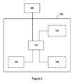
図2は、一実施形態による心エコー図を分析する装置100のブロック図を示す。幾つかの実施形態では、装置は、コンピュータ、サーバ又はラップトップ等である。
FIG. 2 shows a block diagram of an
図2に示すように、装置100は、装置100の動作を制御し、本明細書に説明する方法を実施することができるプロセッサ102を含む。プロセッサ102は、本明細書に説明するやり方で装置100を制御するようにプログラムされた1つ以上のプロセッサ、処理ユニット、マルチコアプロセッサ又はモジュールを含むことができる。特定の実施態様では、プロセッサ102は、本明細書に説明する実施形態による方法の個々の又は複数のステップをそれぞれ行う複数のソフトウェア及び/又はハードウェアモジュールを含むことができる。
As shown in FIG. 2,
簡潔に説明すると、プロセッサ102は、データベース200から、複数の被験者について、複数対の連続する心エコー図を取得する。各心エコー図は、心エコー図の内容に関連付けられる指示を有する。次に、プロセッサ102は、連続する心エコー図の各対を分析して関連するクラスを決定する。クラスは、各対における連続する心エコー図間に変化があるかないかを示す。プロセッサ102は、連続する心エコー図の各対について、各対における心エコー図に対して1回以上の畳み込み及び/又はリダクションを行うことによって各心エコー図の抽象的表現を決定する。プロセッサ102は更に、予測モデルをトレーニングして、複数対の連続する心エコー図の抽象的表現に基づいて、新しい対の心エコー図のクラスを決定する。
Briefly,
プロセッサ102は、以下に説明する様々な機能を行うために、ソフトウェア及び/又はハードウェアを用いて、多くのやり方で実装することができる。プロセッサ102は、ソフトウェア又はコンピュータプログラムコードを使用してプログラムされて、必要な機能を行うか及び/又はプロセッサ102のコンポーネントを制御して必要な機能を実行する1つ以上のマイクロプロセッサ又はデジタルシグナルプロセッサ(DSP)を含む。プロセッサ102は、幾つかの機能を行う専用ハードウェア(例えば増幅器、前置増幅器、アナログ-デジタル変換器(ADC)及び/又はデジタル-アナログ変換器(DAC))と他の機能を行うプロセッサ(例えば1つ以上のプログラムされたマイクロプロセッサ、コントローラ、DSP及び関連回路)との組み合わせとして実施される。本開示の様々な実施形態で使用され得るコンポーネントの例には、従来のマイクロプロセッサ、DSP、特定用途向け集積回路(ASIC)及びフィールドプログラマブルゲートアレイ(FPGA)が含まれるが、これらに限定されない。
幾つかの実施形態では、装置は更に、少なくとも1つのユーザインターフェース104を含む。或いは又は更に、少なくとも1つのユーザインターフェース104は、装置100のユーザがユーザ入力を提供し、装置100とインタラクトする及び/又は装置100を制御することを可能にする任意のユーザインターフェースである。例えばユーザインターフェース104は、1つ以上のスイッチ、1つ以上のボタン、キーパッド、キーボード、(例えばタブレット又はスマートフォン上の)タッチスクリーン若しくはアプリケーション、ディスプレイ画面、グラフィカルユーザーインターフェース(GUI)若しくは他の視覚的レンダリングコンポーネント、1つ以上のスピーカ、1つ以上のマイク若しくは他のオーディオコンポーネント、1つ以上のライト、触覚フィードバックを提供するコンポーネント(例えば振動機能)又は他のユーザインターフェース若しくはユーザインターフェースの組み合わせを含む。
In some embodiments, the device also includes at least one
幾つかの実施形態では、装置100はまた、プロセッサ102によって実行されて本明細書に説明する方法を実行するプログラムコードを格納するメモリ106を含む。メモリ106は、ランダムアクセスメモリ(RAM)、スタティックRAM(SRAM)、ダイナミックRAM(DRAM)、読み取り専用メモリ(ROM)、プログラム可能ROM(PROM)、消去可能なPROM(EPROM)及び電気的に消去可能なPROM(EEPROM)といった揮発性及び不揮発性コンピュータメモリを含むキャッシュ又はシステムメモリを含む。
In some embodiments,
或いは又は更に、1つ以上のメモリ106が、装置100の外部にあってもよい(即ち、装置100とは別個である又は装置100からリモートであってよい)。例えば1つ以上のメモリ106が、別のデバイスの一部である。メモリ106を使用して、装置100のプロセッサ102によって取得若しくは作成された、又は、装置100の外部にある任意のインターフェース、メモリ又はデバイスからの画像(心エコー図を含む)、情報、データ、信号及び測定値を格納することができる。例えばメモリ106を使用して、分析に使用する1つ以上の過去の又は現在の心エコー図を(例えばローカルファイルに)格納することができる。プロセッサ102は、メモリ106を制御して、分析に使用する1つ以上の過去の又は現在の心エコー図を格納することができる。
Alternatively or additionally, one or
幾つかの実施形態では、複数の被験者についての複数対の連続する心エコー図のデータベース200はメモリ106に格納されていてよいが、他の実施形態では、データベース200は、装置100から別個である。
In some embodiments, a
幾つかの実施形態では、装置100はまた、装置100が装置100内外の任意のインターフェース、メモリ及びデバイスと通信することを可能にする通信インターフェース(回路)108を含む。通信インターフェース108は、装置100内外の任意のインターフェース、メモリ及びデバイスと通信することができる。通信インターフェース108は、任意のインターフェース、メモリ及びデバイスと無線で又は有線接続を介して通信することができる。例えば1つ以上のユーザインターフェース104が装置100の外部にある実施形態では、通信インターフェース108は、当該1つ以上の外部ユーザインターフェース104と無線で又は有線接続を介して通信することができる。同様に、1つ以上のメモリ106が装置100の外部にある実施形態では、通信インターフェース108は、当該1つ以上の外部メモリ106と無線で又は有線接続を介して通信することができる。
In some embodiments,
当然ながら、図2は、本発明の上記態様の説明に必要なコンポーネントを示すに過ぎず、実際の実装では、装置100は、示したものに追加のコンポーネントを含んでよい。例えば装置100は、装置100に給電するバッテリ若しくは他の電源や、装置100を主電源に接続する手段を含む。
Of course, FIG. 2 only shows the components necessary for the description of the above aspects of the invention, and in actual implementations the
図3は、一実施形態による心エコー図を分析するコンピュータ実施方法300を示す。図示する方法300は、一般に、装置100のプロセッサ102によって又はその制御下で行うことができる。
FIG. 3 illustrates a computer-implemented method 300 of analyzing an echocardiogram according to one embodiment. The illustrated method 300 may generally be performed by or under the control of the
図3を参照すると、ステップ302において、複数の被験者についての複数対の連続する心エコー図がデータベース200から取得される。プロセッサ102は、データベース200から複数対を取り出すことによって当該複数対を取得することができる。複数対の連続する心エコー図における各心エコー図は、心エコー図の内容に関連付けられた指示を有する。幾つかの実施形態では、心エコー図の内容は、ユーザによって入力される1つ以上の観察ステートメント又は診断ステートメントを含む。より詳細には、これらのステートメントは2つの異なるカテゴリ、即ち、個別のステートメント(例えば「左心室は正常」)と測定ステートメント(例えば「左心室駆出率は45.0%」)とに分けることができる。幾つかの実施形態では、1つ以上の観察ステートメント又は診断ステートメントのそれぞれに内部識別子が関連付けられる。
Referring to FIG. 3, at
幾つかの実施形態では、装置100のプロセッサ102は、1対の連続する心エコー図の観察ステートメント又は診断ステートメントに基づいて変化を検出することができる。例えばプロセッサ102は、1対の一方の心エコー図に関連付けられる「左心室駆出率は45.0%」というステートメントと、当該1対の他方の心エコー図に関連付けられる「左心室駆出率は60.0%」というステートメントとの間に変化があることを検出する。したがって、これらの実施形態の幾つかでは、ステップ304の前に、方法は更に、1対における心エコー図のそれぞれに関連付けられるステートメント間の変化が検出されたときに、当該1対の心エコー図の分析をトリガするステップを含む。より詳細には、これらの実施形態の幾つかでは、小さい変化(所定の差の閾値よりも低い)を無視できるように、所定の差の閾値を使用することができる。例えば「10.0%未満の駆出率」という所定の差の閾値を設定して、1対の連続する心エコー図における一方の心エコー図が「左心室駆出率は45.0%」という関連付けられる指示ステートメントを含み、同じ対の連続する心エコー図の他方の心エコー図が「左心室駆出率は50.0%」という関連付けられる指示ステートメントを含んでいるならば、プロセッサ102は、ステートメント間のこの差(つまり、5.0%。これは10.0%未満である)を無視することができる。
In some embodiments, the
更に、プロセッサ102が1対の心エコー図の観察ステートメント又は診断ステートメントに基づいて変化を検出するこれらの実施形態の幾つかでは、方法は更に、ステップ304の前に、1対の心エコー図のそれぞれに関連付けられるステートメント間に変化はないと決定される場合に、当該対の心エコー図の分析をトリガするステップを含む。したがって、当該対の心エコー図を分析することによって、当該対の心エコー図間に変化はないことを更に確認することができる。
Further, in some of those embodiments in which the
幾つかの実施形態では、各心エコー図は、複数の画像フレームを含む。具体的には、各心エコー図について、複数の画像フレームはそれぞれ、1つの心周期中の心臓の時間インスタンスに対応する。例えば画像フレームの一部は、心周期の収縮期部分に対応し、画像フレームの別の部分は、心周期の拡張期部分に対応する。 In some embodiments, each echocardiogram includes multiple image frames. Specifically, for each echocardiogram, multiple image frames each correspond to a time instance of the heart during one cardiac cycle. For example, one portion of the image frame corresponds to the systolic portion of the cardiac cycle and another portion of the image frame corresponds to the diastolic portion of the cardiac cycle.
これらの実施形態では、方法は更に、心周期に関して1対の連続する心エコー図を時間的に整列させるステップを含むことができる。連続する心エコー図のこの時間的整列を行うことにより、1対の心エコー図における画像フレームが同期されて、当該1対の心エコー図の一方における画像フレームの心周期の一部が、当該1対の他方の心エコー図における画像フレームの心周期の一部と時間的に対応するので、心エコー図の分析の精度を向上させることができる。更に、これらの実施形態では、方法は更に、心エコー図の一方又は両方について、1つ以上の画像フレームを補間するステップを含んでよく、これにより、各心エコー図が同数の画像フレームを含む。 In these embodiments, the method may further include temporally aligning the pair of consecutive echocardiograms with respect to the cardiac cycle. By performing this temporal alignment of successive echocardiograms, the image frames in a pair of echocardiograms are synchronized such that a portion of the cardiac cycle of the image frames in one of the pair of echocardiograms is aligned with the The accuracy of echocardiographic analysis can be improved by temporally corresponding to a portion of the cardiac cycle of the image frame in the other echocardiogram of the pair. Additionally, in these embodiments, the method may further include interpolating one or more image frames for one or both of the echocardiograms, such that each echocardiogram includes the same number of image frames. .
図3のステップ304において、連続する心エコー図の各対が分析されて、関連付けられるクラスが決定される。ステップ304における決定は、各対の連続する心エコー図のそれぞれに関連付けられる観察ステートメント又は診断ステートメント間の自動比較によって行われるか、又は、手動で行われてよい(つまり、ユーザが心エコー図の各対を観察し、ラベルを提供することにより、当該対の心エコー図間に変化が観察されるかどうかを示す)。クラスは、1対における連続する心エコー図間に変化があるかないかを示す。連続する心エコー図間の変化とは、連続する心エコー図間の視覚的表現(例えばノイズレベル、テクスチャ、ビューの位置)の変化ではなく、連続する心エコー図で表される心機能及び/又は心臓の構造の変化を指す。連続する心エコー図の各対について決定された関連付けられるクラスと、連続する心エコー図のそれぞれの対とは、グラウンドトゥルースを形成する。連続する心エコー図の各対に関連付けられるグラウンドトゥルースは、当該対の連続する心エコー図間で心機能及び/又は心臓の構造に変化があるかどうかを示し、ステップ308においてトレーニングされる予測モデルの基礎を形成する。
At
幾つかの実施形態では、各心エコー図は、複数のシーケンス(即ち、複数の画像シーケンス)を含み、各シーケンスは、被験者の異なるビューを表す。これらの実施形態では、連続する心エコー図の各対を分析するステップは、各心エコー図の複数のシーケンスを(例えば並べて)結合するステップを含むことにより、単一の画像が形成される。つまり、各心エコー図の複数のシーケンスは、複数のシーケンスの画像からなる単一の画像によって表される。この単一の画像は、単一のビューを有する1つの心エコー図と同じように分析することができる。或いは、これらの実施形態では、連続する心エコー図の各対を分析するステップは、複数のシーケンスに対して個別に畳み込み演算を行い、畳み込み深層学習ネットワークの全結合層において情報を混合するステップを含む。 In some embodiments, each echocardiogram includes multiple sequences (ie, multiple image sequences), each representing a different view of the subject. In these embodiments, analyzing each pair of consecutive echocardiograms includes combining (eg, side-by-side) multiple sequences of each echocardiogram to form a single image. That is, the multiple sequences of each echocardiogram are represented by a single image composed of the images of the multiple sequences. This single image can be analyzed like an echocardiogram with a single view. Alternatively, in these embodiments, analyzing each pair of consecutive echocardiograms comprises performing a separate convolution operation on the multiple sequences to mix the information in a fully connected layer of a convolutional deep learning network. include.
また、或いは、各心エコー図が複数のシーケンスを含み、各シーケンスが被験者の異なるビューを表す幾つかの実施形態では、対毎の比較を行って、関連付けられるクラスを決定することができる。より詳細には、1対の心エコー図の一方がシーケンスS1、S2、…、Smを含み、心エコー図の他方がシーケンスT1、T2、…、Tnを含む場合、連続する心エコー図の対の分析は、最初にS1をT1、T2、…、Tnのそれぞれと比較し、次に、S2をT1、T2、…、Tnのそれぞれと比較し、以降同様にすることを含み、これにより、1対の心エコー図のうちの第1の心エコー図の各シーケンスSiが、当該対の心エコー図のうちの第2の心エコー図の各シーケンスTjと比較される。方法が畳み込み深層学習ネットワーク(例えば図4に示すようなネットワーク)で実装される幾つかの実施形態では、シーケンスS1、S2、…、Sm及びT1、T2、…、Tnのそれぞれは、畳み込み深層学習ネットワークの複数の入力ノードのそれぞれに関連付けられる。 Also, or alternatively, in some embodiments where each echocardiogram includes multiple sequences and each sequence represents a different view of the subject, pairwise comparisons can be made to determine the associated classes. More specifically , if one of a pair of echocardiograms contains the sequences S 1 , S 2 , . A paired analysis of echocardiograms to be performed first compares S 1 with each of T 1 , T 2 , . comparing, and so on, whereby each sequence S i of the first echocardiogram of a pair of echocardiograms is compared with the second echocardiogram of the pair of echocardiograms. Each sequence Tj in the figure is compared. In some embodiments where the method is implemented in a convolutional deep learning network (eg, a network such as that shown in FIG. 4), the sequences S 1 , S 2 , . . . , S m and T 1 , T 2 , . Each associated with each of a plurality of input nodes of the convolutional deep learning network.
各心エコー図が複数のシーケンスを含み、各シーケンスが被験者の異なるビューを表す実施形態において、方法は更に、複数のシーケンスのそれぞれにビュータグを関連付けるステップを含む。ビュータグは、シーケンスによって表される被験者のビューを示す。詳細には、心エコー図の複数のシーケンスのそれぞれは、現在の画像処理技術を使用して分析されて、被験者のビューが決定され、これにより、それぞれのシーケンスを、次のビュータグのうちの1つに関連付けることができる。傍胸骨長軸(PLAX)、傍胸骨短軸(PSAX)、心尖部4腔(AP4)、心尖部5腔(AP5)、心尖部2腔(AP2)、心尖部3腔(AP3)、肋骨下(SC)及び胸骨上切痕(SSN)。更に、複数のシーケンスのそれぞれが対応するビュータグに関連付けられているこれらの実施形態において、方法は更に、(複数対の連続する心エコー図のうちの)1対の連続する心エコー図のビュータグが同じである(例えば当該対の心エコー図の両方のビュータグがAP2である)場合にのみ、当該1対の心エコー図を分析するステップを含んでよい。一致する/同じビュータグを有するシーケンスのみを比較することによって、方法の効率を向上させることができる。 In embodiments in which each echocardiogram includes multiple sequences, each representing a different view of the subject, the method further includes associating a view tag with each of the multiple sequences. A view tag indicates the subject's view represented by the sequence. Specifically, each of the multiple sequences of echocardiograms is analyzed using current image processing techniques to determine the view of the subject, thereby assigning each sequence to one of the following view tags: can be associated with Parasternal long axis (PLAX), parasternal short axis (PSAX), apical 4 chambers (AP4), apical 5 chambers (AP5), apical 2 chambers (AP2), apical 3 chambers (AP3), subcostal (SC) and suprasternal notch (SSN). Further, in those embodiments in which each of the multiple sequences is associated with a corresponding view-tag, the method further comprises determining that the pair of consecutive echocardiogram view-tags (of the plurality of pairs of consecutive echocardiograms) Analyzing the pair of echocardiograms only if they are the same (eg, both viewtags of the pair of echocardiograms are AP2) may be included. The efficiency of the method can be improved by comparing only sequences with matching/same view-tags.
図3のステップ306において、連続する心エコー図の各対について、当該対における心エコー図に対して1回以上の畳み込み及び/又はリダクションを行うことによって、各心エコー図の抽象的表現が決定される。畳み込み(又はフィルタリング)演算は、画像内の各ピクセルの畳み込み値を計算できるようにする画像処理アルゴリズムに基づいて行われる。ピクセルの畳み込み値を計算するには、畳み込みカーネルの複数のパラメータで定義される線形係数を用いてすべての隣接ピクセルの線形結合が計算される。畳み込みカーネルのパラメータは、予測モデルのネットワークトレーニングプロセスで決定することができる。リダクション演算は、ピクセルを拡大し、ピクセルの平均値又はピクセルの最大値を取得することにより、画像のピクセル数を削減する。畳み込み及び/又はリダクションは、畳み込み深層学習ネットワークの層で行われてよく、畳み込み深層学習ネットワークでの畳み込み及び/又はリダクションに対応する層の数は様々であってよい。畳み込み及び/又はリダクションが行われた後、元の画像は変換され、複数の数によって特徴付けられる特徴のセットによって表される。この特徴のセットは更に、畳み込み深層学習ネットワークの意思決定ノードに関連付けられ、これにより、結果が決定される。
In
幾つかの実施形態では、抽象的表現の決定は、1対の心エコー図のそれぞれについて別々に行われる。抽象的表現は、当該1対のクラスを示す1つ以上の特徴を含む。例えば抽象的表現は、予測モデルによって生成される特徴ベクトルに対応するベクトルを含む。特徴ベクトルは、被験者の心臓の構造的及び/又は機能的特性の変化を示す。別の例として、抽象的表現は、予測モデルによって生成されるデータのセットに対応するデータを含む。データのセットは、被験者の心臓の構造的及び/又は機能的特性の変化を示す。この予測モデルは、ステップ308においてトレーニングされる予測モデルと同じである。
In some embodiments, abstract representation determination is performed separately for each of a pair of echocardiograms. An abstract representation includes one or more features that describe the pair of classes. For example, the abstract representation includes vectors corresponding to feature vectors generated by the predictive model. The feature vector indicates changes in structural and/or functional properties of the subject's heart. As another example, the abstract representation includes data corresponding to the set of data generated by the predictive model. The data set is indicative of changes in structural and/or functional properties of the subject's heart. This prediction model is the same prediction model trained in
図3のステップ308において、複数対の連続する心エコー図の抽象的表現に基づいて予測モデルがトレーニングされて、これにより、新しい1対の心エコー図のクラスが決定される。幾つかの実施形態では、予測モデルは、畳み込み深層学習モデルである。トレーニングされた予測モデルを使用して、新しい対の心エコー図間の時間間隔が所定の時間閾値(例えば7日)を超えた場合に、類似した被験者の過去の心エコー図の対の研究に基づいて、当該新しい対の心エコー図のクラスを決定することもできる。
In
図3には示していないが、幾つかの実施形態による方法は更に、被験者の新しい心エコー図を受信するステップと、当該被験者の過去の心エコー図を取得するステップと、予測モデルを使用して新しい心エコー図及び過去の心エコー図のクラスを決定するステップを含む。場合によっては、過去の心エコー図は「以前の心エコー図」と呼ばれ、新しい心エコー図は「現在の心エコー図」と呼ばれることがある。これらの実施形態では、方法はまた、新しい1対の心エコー図の決定されたクラスが変化があることを示す場合、インターフェースを制御して、ユーザに通知を提供するステップを更に含む。また、これらの実施形態では、方法はまた、新しい1対の心エコー図の決定されたクラスの変化があることを示す場合、インターフェース上で当該対の心エコー図を強調表示するステップを更に含む。過去の心エコー図は、メモリ106から取り出すことによって又はデータベース200から取り出すことによって取得することができる。新しい心エコー図は、被験者の心エコー図を作成するデバイスから直接受信されても、又は、装置100による処理の前に新しい心エコー図を格納する別のデバイスから受信される。或いは、新しい心エコー図は、プロセッサ102によってメモリ106から受信される。
Although not shown in FIG. 3, the method according to some embodiments further includes receiving a new echocardiogram for the subject, obtaining a previous echocardiogram for the subject, and using a predictive model. determining the class of the new echocardiogram and the previous echocardiogram. In some cases, past echocardiograms are referred to as "previous echocardiograms" and new echocardiograms are referred to as "current echocardiograms." In these embodiments, the method also includes controlling the interface to provide a notification to the user if the determined class of the new pair of echocardiograms indicates a change. Also, in these embodiments, the method also further includes highlighting the new pair of echocardiograms on the interface if indicating that there is a change in the determined class of the pair of echocardiograms. . Past echocardiograms can be obtained by retrieving from
更に、新しい心エコー図が受信され、被験者の過去の心エコー図が取得されるこれらの実施形態において、過去の心エコー図を取得するステップは、過去の心エコー図の内容に関連付けられる指示を取得するステップを含む。これらの実施形態では、方法は更に、新しい1対の心エコー図の決定されたクラスが変化がないことを示す場合、インターフェースを制御して、過去の心エコー図の内容に関連付けられる指示をユーザに提供するステップを含む。したがって、過去の心エコー図と新しい心エコー図との間に変化がないと判断されると、過去の心エコー図の内容に関連付けられる指示は、(変更がないことにより)新しい心エコー図にも適用され、また、新しい心エコー図の詳細な分析や解釈を行う必要なく、ユーザ(例えば医療技術者)が簡単に確認することができるように利用可能にされる。 Further, in those embodiments in which a new echocardiogram is received and a past echocardiogram of the subject is acquired, the step of acquiring the past echocardiogram includes: Including the step of obtaining. In these embodiments, if the determined class of the new pair of echocardiograms indicates no change, the method further controls the interface to provide the user with instructions associated with the content of the previous echocardiogram. including the step of providing to Therefore, if it is determined that there is no change between the previous echocardiogram and the new echocardiogram, the instructions associated with the contents of the previous echocardiogram will be transferred (by lack of change) to the new echocardiogram. is also applied and made available for easy review by a user (eg, a medical technician) without having to perform detailed analysis and interpretation of the new echocardiogram.
幾つかの実施形態では、図3に示す方法は、心臓全体の変化を観察するのではなく、特定の解剖学的変化を観察するように適用される。例えば幾つかの実施形態では、方法は、1対の連続する心エコー図間で、心臓の特定の部分、例えば左心室に変化があるかないかを示すクラスを決定するために適用される。更に、幾つかの実施形態では、図3に示す方法は、肝臓、胆嚢、腹部、直腸等といった被験者の他の臓器の構造的及び/又は機能的変化を観察するために適用される。同じ原理を胎児超音波検査にも適用することができる。 In some embodiments, the method illustrated in FIG. 3 is applied to look at specific anatomical changes rather than looking at changes throughout the heart. For example, in some embodiments, the method is applied between a pair of consecutive echocardiograms to determine classes that indicate whether a particular portion of the heart, eg, the left ventricle, is altered or not. Additionally, in some embodiments, the method shown in FIG. 3 is applied to observe structural and/or functional changes in other organs of a subject, such as the liver, gallbladder, abdomen, rectum, and the like. The same principle can be applied to fetal ultrasound.
図4は、例示的な実施形態による心エコー図を分析する畳み込み深層学習ネットワーク400の概略図である。図4に示す畳み込み深層学習ネットワーク400は、図2を参照して先に説明した装置100のより詳細な実施態様である。図4に示す例示的な実施形態では、装置100のプロセッサ102が、ブロック402、404及び406を行う。
FIG. 4 is a schematic diagram of a convolutional
図4に示すように、1対の連続する心エコー図、即ち、過去の心エコー図410と現在の心エコー図412が、システム400のデータベース(例えばデータベース200)から取り出される。図3に関連して上述した連続する心エコー図の対と同様に、過去の心エコー図410及び現在の心エコー図412のそれぞれは、心エコー図の内容に関連付けられる指示を有する。心エコー図の内容は、「左心室は正常」又は「左心室は大幅に縮小している」といったユーザによって入力される1つ以上の観察ステートメント又は診断ステートメントを含む。更に、この実施形態では、過去の心エコー図410及び現在の心エコー図412のそれぞれは、1つの心周期中の心臓の時間インスタンスにそれぞれ対応する複数の画像フレームを含む。
As shown in FIG. 4, a pair of consecutive echocardiograms, a
前述したように、図4に示す例示的な実施形態では、プロセッサ102は、ブロック402、404及び406を行う。簡潔に説明すると、プロセッサ102は、心周期に関して、1対の連続する心エコー図410、412の時間的整列を行い、1回以上の畳み込み及び/又はリダクションを行って、過去の心エコー図410及び現在の心エコー図412のそれぞれの抽象的表現を決定し、畳み込み深層学習ネットワーク400の全結合層において当該1対の連続する心エコー図410、412に関連付けられるクラスを決定する。これらのブロック402、404及び406のそれぞれの詳細は、以下で更に詳細に説明する。
As previously mentioned, in the exemplary embodiment shown in FIG. 4,
図4のブロック402において、プロセッサ102は、心周期に関して、1対の連続する心エコー図410、412を時間的に整列させる。したがって、過去の心エコー図410の画像フレームの内容は、現在の心エコー図412の画像フレームの内容に時間的に対応し、これらは、後続のプロセス、例えば当該1対の心エコー図に関連付けられるクラスが決定されるブロック406において、より簡単に比較することができる。
At
図4のブロック404において、プロセッサ102は、心エコー図に対して1回以上の畳み込み及び/又はリダクションを行うことによって、過去の心エコー図410及び現在の心エコー図412のそれぞれの抽象的表現を決定する。過去の心エコー図の抽象的表現420及び現在の心エコー図の抽象的表現422はそれぞれ、当該1対の心エコー図のクラスを示す1つ以上の特徴を含む。
At
図4のブロック406において、プロセッサ102は、畳み込み深層学習ネットワーク400の全結合層において、当該1対の連続する心エコー図410、412に関連付けられるクラスを決定する。全結合層では、当該1対の連続する心エコー図に関連付けられるクラスを決定するのに最適なやり方で、抽象的表現420、422からの情報を統合する画像減算又は任意の他の演算が行われる。この全結合層は、図4のブロック430として表され、元々は別々のプロセスの結果である抽象的表現420、422からの情報を統合することを可能にする。決定の結果、即ち、連続する心エコー図410、412間に変化があるかないかを示す対の連続する心エコー図410、412に関連付けられるクラスは、ブロック440(「変化あり/変化なし」)によって表される。
At
更に、本実施形態による畳み込み深層学習ネットワーク400では、プロセッサ102はまた、ブロック408において予測モデルをトレーニングして、対の連続する心エコー図410、412の抽象的表現420、422に基づいて新しい1対の心エコー図のクラスを決定する。より詳細には、新しい1対の心エコー図に関連付けられるクラスの決定の精度は、抽象的表現420、422に基づいて情報を混合する演算を最適化することによって向上させることができる。
Further, in the convolutional
したがって、心エコー図を分析する改良方法及び装置が提供される。本明細書に説明する実施形態による方法及び装置は、注釈付き心エコー図(即ち、心エコー図の内容に関連付けられる指示を有する心エコー図)を含むデータベースに基づいて連続する心エコー図を自動的に比較し、重要な変化を検出する一方で、(例えば異なるノイズレベル又はビューの位置による)重要でない変化は無視する。 Accordingly, an improved method and apparatus for analyzing echocardiograms is provided. Methods and apparatus according to embodiments described herein automatically generate sequential echocardiograms based on a database containing annotated echocardiograms (i.e., echocardiograms having instructions associated with the content of the echocardiogram). , and detect significant changes, while ignoring insignificant changes (eg due to different noise levels or view positions).
開示された実施形態に対する変形は、図面、開示及び添付の特許請求の範囲の検討から、請求された発明を実施する際に当業者によって理解及び達成されうる。請求項において、「含む」との用語は、他の要素又はステップを排除するものではなく、また、単数形は複数を排除するものではない。単一のプロセッサ又は他のユニットが、請求項に引用される幾つかのアイテムの機能を果たしてもよい。特定の手段が相互に異なる従属請求項に記載されることだけで、これらの手段の組み合わせを有利に使用することができないことを示すものではない。コンピュータプログラムは、他のハードウェアと共に又はその一部として供給される光学記憶媒体又はソリッドステート媒体といった適切な媒体上に記憶及び/又は分散されてもよいが、インターネット又は他の有線若しくは無線通信システムを介するといった他の形式で分配されてもよい。請求項における任意の参照符号は、範囲を限定するものと解釈されるべきではない。 Variations to the disclosed embodiments can be understood and effected by those skilled in the art in practicing the claimed invention, from a study of the drawings, the disclosure, and the appended claims. In the claims, the word "comprising" does not exclude other elements or steps, and the singular does not exclude a plural. A single processor or other unit may fulfill the functions of several items recited in the claims. The mere fact that certain measures are recited in mutually different dependent claims does not indicate that a combination of these measures cannot be used to advantage. The computer program may be stored and/or distributed on any suitable medium, such as optical storage media or solid state media supplied with or as part of other hardware, Internet or other wired or wireless communication systems. It may also be distributed in other ways, such as via Any reference signs in the claims shall not be construed as limiting the scope.
Claims (12)
各対の連続する心エコー図のそれぞれに関連付けられる前記診断ステートメント間の自動比較によって、関連付けられるクラスを決定するために連続する心エコー図の各対を分析するステップと、
連続する心エコー図の各対について、畳み込み深層学習ネットワークにおいて、前記各対における前記心エコー図に1回以上の畳み込み及び/又はリダクションを行うことによって、特徴ベクトル又はデータのセットのいずれかを含む、各心エコー図の抽象的表現を決定するステップと、
前記複数対の連続する心エコー図の前記抽象的表現に基づいて、新しい1対の心エコー図のクラスを決定するために予測モデルをトレーニングするステップと、
を含み、
前記クラスは、前記各対における前記連続する心エコー図間に変化があるかないかを示し、前記変化は、心機能及び/又は心臓の構造の変化を表し、
前記抽象的表現は、前記各対の前記クラスを示す1つ以上の特徴を含む、心エコー図を分析するコンピュータ実施方法。 obtaining from a database a plurality of pairs of consecutive echocardiograms for a plurality of subjects, each echocardiogram including one or more diagnostic statements entered by a user; obtaining, having an indication associated with
analyzing each pair of consecutive echocardiograms to determine an associated class by automatic comparison between said diagnostic statements associated with each pair of consecutive echocardiograms;
For each pair of successive echocardiograms, a feature vector or data set is generated by performing one or more convolutions and/or reductions on the echocardiograms in each pair in a convolutional deep learning network. determining an abstract representation of each echocardiogram comprising any
training a predictive model to determine a class of new pairs of echocardiograms based on the abstract representation of the pairs of consecutive echocardiograms;
including
said class indicates whether there is a change between said consecutive echocardiograms in said each pair, said change representing a change in cardiac function and/or cardiac structure;
A computer-implemented method of analyzing an echocardiogram, wherein the abstract representation includes one or more features indicative of the class of each pair.
前記コンピュータ実施方法は、各心エコー図が同数の画像フレームを含むように、前記各対の心エコー図の一方又は両方について1つ以上の画像フレームを補間するステップを更に含む、請求項1又は2に記載のコンピュータ実施方法。 each echocardiogram includes a plurality of image frames;
3. The computer-implemented method of claim 1, further comprising interpolating one or more image frames for one or both of each pair of echocardiograms such that each echocardiogram includes the same number of image frames. 3. The computer-implemented method of claim 2.
前記被験者の過去の心エコー図を取得するステップと、
前記新しい心エコー図及び前記過去の心エコー図のクラスを決定するために前記予測モデルを使用するステップと、
を更に含む、請求項1から3のいずれか一項に記載のコンピュータ実施方法。 receiving a new echocardiogram of the subject;
obtaining a past echocardiogram of the subject;
using the predictive model to determine the class of the new echocardiogram and the previous echocardiogram;
4. The computer-implemented method of any one of claims 1-3, further comprising:
前記コンピュータ実施方法は、前記新しい1対の心エコー図の決定された前記クラスが、変化がないことを示す場合、ユーザに前記関連付けられる指示及び/又は前記内容を提供するようにインターフェースを制御するステップを更に含む、請求項4又は5に記載のコンピュータ実施方法。 the step of obtaining a past echocardiogram includes receiving an indication associated with content of the past echocardiogram;
The computer-implemented method controls an interface to provide the associated instructions and/or the content to a user if the determined class of the new pair of echocardiograms indicates no change. 6. The computer-implemented method of claim 4 or 5, further comprising the steps of:
各シーケンスは、前記被験者の異なるビューを表し、
前記連続する心エコー図の各対を分析するステップは、単一の画像を形成するために各心エコー図の前記複数のシーケンスを結合するステップを更に含む、請求項1から6のいずれか一項に記載のコンピュータ実施方法。 Each echocardiogram contains multiple sequences,
each sequence represents a different view of the subject;
7. The step of analyzing each pair of consecutive echocardiograms further comprises combining the multiple sequences of each echocardiogram to form a single image. 13. The computer-implemented method of claim 1.
各シーケンスは、前記被験者の異なるビューを表し、
前記連続する心エコー図の各対を分析するステップは、前記各対の一方の心エコー図における複数のシーケンスのそれぞれを、前記各対の他方の心エコー図における複数のシーケンスのそれぞれと比較するステップを更に含む、請求項1から6のいずれか一項に記載のコンピュータ実施方法。 Each echocardiogram contains multiple sequences,
each sequence represents a different view of the subject;
Analyzing each pair of consecutive echocardiograms comprises comparing each of a plurality of sequences in one echocardiogram of each pair with each of a plurality of sequences in the other echocardiogram of each pair. 7. The computer-implemented method of any one of claims 1-6, further comprising the steps of:
各シーケンスは、前記被験者の異なるビューを表し、
前記コンピュータ実施方法は、前記複数のシーケンスのそれぞれにビュータグを関連付けるステップを更に含み、
前記ビュータグは、前記シーケンスによって表される前記被験者のビューを示す、請求項1から8のいずれか一項に記載のコンピュータ実施方法。 Each echocardiogram contains multiple sequences,
each sequence represents a different view of the subject;
The computer-implemented method further includes associating a viewtag with each of the plurality of sequences;
9. The computer-implemented method of any one of claims 1-8, wherein the view tag indicates the subject's view represented by the sequence.
各対の連続する心エコー図のそれぞれに関連付けられる前記診断ステートメント間の自動比較によって、関連付けられるクラスを決定するために連続する心エコー図の各対を分析し、
連続する心エコー図の各対について、畳み込み深層学習ネットワークにおいて、前記各対における前記心エコー図に1回以上の畳み込み及び/又はリダクションを行うことによって、特徴ベクトル又はデータのセットのいずれかを含む、各心エコー図の抽象的表現を決定し、
前記複数対の連続する心エコー図の前記抽象的表現に基づいて、新しい1対の心エコー図のクラスを決定するために予測モデルをトレーニングする、
プロセッサを含み、
前記クラスは、前記各対における前記連続する心エコー図間に変化があるかないかを示し、前記変化は、心機能及び/又は心臓の構造の変化を表し、
前記抽象的表現は、前記各対の前記クラスを示す1つ以上の特徴を含む、
心エコー図を分析する装置。 obtaining from a database a plurality of pairs of sequential echocardiograms for a plurality of subjects, each echocardiogram including one or more diagnostic statements entered by a user, instructions associated with the content of said echocardiograms; has
analyzing each pair of consecutive echocardiograms to determine an associated class by automatic comparison between said diagnostic statements associated with each pair of consecutive echocardiograms;
For each pair of successive echocardiograms, a feature vector or data set is generated by performing one or more convolutions and/or reductions on the echocardiograms in each pair in a convolutional deep learning network. determining an abstract representation of each echocardiogram containing any
training a predictive model to determine a class of new pairs of echocardiograms based on the abstract representation of the pairs of consecutive echocardiograms;
including a processor;
said class indicates whether there is a change between said consecutive echocardiograms in said each pair, said change representing a change in cardiac function and/or cardiac structure;
the abstract representation includes one or more features indicative of the class of each pair;
Equipment for analyzing echocardiograms.
Applications Claiming Priority (3)
| Application Number | Priority Date | Filing Date | Title |
|---|---|---|---|
| US201762580491P | 2017-11-02 | 2017-11-02 | |
| US62/580,491 | 2017-11-02 | ||
| PCT/EP2018/079963 WO2019086586A1 (en) | 2017-11-02 | 2018-11-02 | A method and apparatus for analysing echocardiograms |
Publications (3)
| Publication Number | Publication Date |
|---|---|
| JP2021501633A JP2021501633A (en) | 2021-01-21 |
| JP2021501633A5 JP2021501633A5 (en) | 2021-12-09 |
| JP7325411B2 true JP7325411B2 (en) | 2023-08-14 |
Family
ID=64270837
Family Applications (1)
| Application Number | Title | Priority Date | Filing Date |
|---|---|---|---|
| JP2020524199A Active JP7325411B2 (en) | 2017-11-02 | 2018-11-02 | Method and apparatus for analyzing echocardiogram |
Country Status (5)
| Country | Link |
|---|---|
| US (1) | US20210219922A1 (en) |
| EP (1) | EP3704707B1 (en) |
| JP (1) | JP7325411B2 (en) |
| CN (1) | CN111448614A (en) |
| WO (1) | WO2019086586A1 (en) |
Families Citing this family (4)
| Publication number | Priority date | Publication date | Assignee | Title |
|---|---|---|---|---|
| US11931207B2 (en) * | 2018-12-11 | 2024-03-19 | Eko.Ai Pte. Ltd. | Artificial intelligence (AI) recognition of echocardiogram images to enhance a mobile ultrasound device |
| US11446009B2 (en) * | 2018-12-11 | 2022-09-20 | Eko.Ai Pte. Ltd. | Clinical workflow to diagnose heart disease based on cardiac biomarker measurements and AI recognition of 2D and doppler modality echocardiogram images |
| US20210264238A1 (en) * | 2018-12-11 | 2021-08-26 | Eko.Ai Pte. Ltd. | Artificial intelligence (ai)-based guidance for an ultrasound device to improve capture of echo image views |
| US11301996B2 (en) * | 2018-12-11 | 2022-04-12 | Eko.Ai Pte. Ltd. | Training neural networks of an automatic clinical workflow that recognizes and analyzes 2D and doppler modality echocardiogram images |
Citations (5)
| Publication number | Priority date | Publication date | Assignee | Title |
|---|---|---|---|---|
| JP2007526016A (en) | 2003-06-25 | 2007-09-13 | シーメンス メディカル ソリューションズ ユーエスエー インコーポレイテッド | System and method for automatic local myocardial assessment of cardiac imaging |
| JP2012139487A (en) | 2010-12-13 | 2012-07-26 | Toshiba Corp | Ultrasonic diagnostic device, image processing device, and image processing method |
| WO2016194161A1 (en) | 2015-06-03 | 2016-12-08 | 株式会社日立製作所 | Ultrasonic diagnostic apparatus and image processing method |
| WO2017056078A1 (en) | 2015-10-02 | 2017-04-06 | Koninklijke Philips N.V. | System for mapping findings to pertinent echocardiogram loops |
| JP2017068838A (en) | 2015-09-29 | 2017-04-06 | パナソニックIpマネジメント株式会社 | Control method and program of information terminal |
Family Cites Families (7)
| Publication number | Priority date | Publication date | Assignee | Title |
|---|---|---|---|---|
| US5911133A (en) * | 1997-10-22 | 1999-06-08 | Rush-Presbyterian -St. Luke's Medical Center | User interface for echocardiographic report generation |
| US6447450B1 (en) * | 1999-11-02 | 2002-09-10 | Ge Medical Systems Global Technology Company, Llc | ECG gated ultrasonic image compounding |
| CN1725978A (en) * | 2002-12-13 | 2006-01-25 | 皇家飞利浦电子股份有限公司 | System and method for processing a series of image frames representing a cardiac cycle |
| EP3191991B1 (en) * | 2014-09-10 | 2021-01-13 | Koninklijke Philips N.V. | Image report annotation identification |
| CN106846306A (en) * | 2017-01-13 | 2017-06-13 | 重庆邮电大学 | A kind of ultrasonoscopy automatic describing method and system |
| WO2018210714A1 (en) * | 2017-05-18 | 2018-11-22 | Koninklijke Philips N.V. | Convolutional deep learning analysis of temporal cardiac images |
| CN107184198A (en) * | 2017-06-01 | 2017-09-22 | 广州城市职业学院 | A kind of electrocardiosignal classifying identification method |
-
2018
- 2018-11-02 US US16/760,678 patent/US20210219922A1/en active Pending
- 2018-11-02 JP JP2020524199A patent/JP7325411B2/en active Active
- 2018-11-02 WO PCT/EP2018/079963 patent/WO2019086586A1/en unknown
- 2018-11-02 CN CN201880078066.9A patent/CN111448614A/en active Pending
- 2018-11-02 EP EP18800551.6A patent/EP3704707B1/en active Active
Patent Citations (6)
| Publication number | Priority date | Publication date | Assignee | Title |
|---|---|---|---|---|
| JP2007526016A (en) | 2003-06-25 | 2007-09-13 | シーメンス メディカル ソリューションズ ユーエスエー インコーポレイテッド | System and method for automatic local myocardial assessment of cardiac imaging |
| JP2012139487A (en) | 2010-12-13 | 2012-07-26 | Toshiba Corp | Ultrasonic diagnostic device, image processing device, and image processing method |
| WO2016194161A1 (en) | 2015-06-03 | 2016-12-08 | 株式会社日立製作所 | Ultrasonic diagnostic apparatus and image processing method |
| JP2017068838A (en) | 2015-09-29 | 2017-04-06 | パナソニックIpマネジメント株式会社 | Control method and program of information terminal |
| WO2017056078A1 (en) | 2015-10-02 | 2017-04-06 | Koninklijke Philips N.V. | System for mapping findings to pertinent echocardiogram loops |
| JP2018534029A (en) | 2015-10-02 | 2018-11-22 | コーニンクレッカ フィリップス エヌ ヴェKoninklijke Philips N.V. | A system for mapping findings to relevant echocardiographic loops |
Also Published As
| Publication number | Publication date |
|---|---|
| EP3704707B1 (en) | 2024-01-10 |
| JP2021501633A (en) | 2021-01-21 |
| EP3704707A1 (en) | 2020-09-09 |
| WO2019086586A1 (en) | 2019-05-09 |
| CN111448614A (en) | 2020-07-24 |
| US20210219922A1 (en) | 2021-07-22 |
Similar Documents
| Publication | Publication Date | Title |
|---|---|---|
| Ghorbani et al. | Deep learning interpretation of echocardiograms | |
| Litjens et al. | State-of-the-art deep learning in cardiovascular image analysis | |
| Mirea et al. | Intervendor differences in the accuracy of detecting regional functional abnormalities: a report from the EACVI-ASE strain standardization task force | |
| JP7325411B2 (en) | Method and apparatus for analyzing echocardiogram | |
| Sermesant et al. | Applications of artificial intelligence in cardiovascular imaging | |
| Slomka et al. | Cardiac imaging: working towards fully-automated machine analysis & interpretation | |
| Tsang et al. | Transthoracic 3D echocardiographic left heart chamber quantification using an automated adaptive analytics algorithm | |
| US11837354B2 (en) | Contrast-agent-free medical diagnostic imaging | |
| Sjøli et al. | Diagnostic capability and reproducibility of strain by Doppler and by speckle tracking in patients with acute myocardial infarction | |
| KR101565311B1 (en) | 3 automated detection of planes from three-dimensional echocardiographic data | |
| Sharifrazi et al. | CNN-KCL: Automatic myocarditis diagnosis using convolutional neural network combined with k-means clustering | |
| Yagel et al. | 3D and 4D ultrasound in fetal cardiac scanning: a new look at the fetal heart | |
| JP2020511190A (en) | System and method for ultrasonic analysis | |
| Benes et al. | Automatically designed machine vision system for the localization of CCA transverse section in ultrasound images | |
| CN109192305B (en) | Heart function automatic analysis method based on deep circulation neural network | |
| JP2007527743A (en) | System and method for automatic diagnosis and decision support for heart related diseases and conditions | |
| Johri et al. | Assessment of image quality in real time three‐dimensional dobutamine stress echocardiography: an integrated 2D/3D approach | |
| Yu et al. | Artificial intelligence-based myocardial texture analysis in etiological differentiation of left ventricular hypertrophy | |
| JP2023155314A (en) | medical image processing device | |
| Kagiyama et al. | Machine learning in cardiovascular imaging | |
| Wehbe et al. | Deep learning for cardiovascular imaging: A review | |
| CN115666401A (en) | System and method for non-invasive pressure measurement | |
| CN115334975A (en) | System and method for imaging and measuring epicardial adipose tissue | |
| US20220082647A1 (en) | Technique for determining a cardiac metric from cmr images | |
| CN110853012B (en) | Method, apparatus and computer storage medium for obtaining cardiac parameters |
Legal Events
| Date | Code | Title | Description |
|---|---|---|---|
| A521 | Request for written amendment filed |
Free format text: JAPANESE INTERMEDIATE CODE: A523 Effective date: 20211029 |
|
| A621 | Written request for application examination |
Free format text: JAPANESE INTERMEDIATE CODE: A621 Effective date: 20211029 |
|
| A977 | Report on retrieval |
Free format text: JAPANESE INTERMEDIATE CODE: A971007 Effective date: 20220921 |
|
| A131 | Notification of reasons for refusal |
Free format text: JAPANESE INTERMEDIATE CODE: A131 Effective date: 20220928 |
|
| A601 | Written request for extension of time |
Free format text: JAPANESE INTERMEDIATE CODE: A601 Effective date: 20221222 |
|
| A521 | Request for written amendment filed |
Free format text: JAPANESE INTERMEDIATE CODE: A523 Effective date: 20230322 |
|
| TRDD | Decision of grant or rejection written | ||
| A01 | Written decision to grant a patent or to grant a registration (utility model) |
Free format text: JAPANESE INTERMEDIATE CODE: A01 Effective date: 20230703 |
|
| A61 | First payment of annual fees (during grant procedure) |
Free format text: JAPANESE INTERMEDIATE CODE: A61 Effective date: 20230801 |
|
| R150 | Certificate of patent or registration of utility model |
Ref document number: 7325411 Country of ref document: JP Free format text: JAPANESE INTERMEDIATE CODE: R150 |