JP7319193B2 - Selection of magnetic resonance fingerprinting dictionaries for anatomical regions - Google Patents
Selection of magnetic resonance fingerprinting dictionaries for anatomical regions Download PDFInfo
- Publication number
- JP7319193B2 JP7319193B2 JP2019553510A JP2019553510A JP7319193B2 JP 7319193 B2 JP7319193 B2 JP 7319193B2 JP 2019553510 A JP2019553510 A JP 2019553510A JP 2019553510 A JP2019553510 A JP 2019553510A JP 7319193 B2 JP7319193 B2 JP 7319193B2
- Authority
- JP
- Japan
- Prior art keywords
- magnetic resonance
- mrf
- mapping
- anatomical
- imaging system
- Prior art date
- Legal status (The legal status is an assumption and is not a legal conclusion. Google has not performed a legal analysis and makes no representation as to the accuracy of the status listed.)
- Active
Links
Images


Classifications
-
- G—PHYSICS
- G01—MEASURING; TESTING
- G01R—MEASURING ELECTRIC VARIABLES; MEASURING MAGNETIC VARIABLES
- G01R33/00—Arrangements or instruments for measuring magnetic variables
- G01R33/20—Arrangements or instruments for measuring magnetic variables involving magnetic resonance
- G01R33/44—Arrangements or instruments for measuring magnetic variables involving magnetic resonance using nuclear magnetic resonance [NMR]
- G01R33/48—NMR imaging systems
- G01R33/54—Signal processing systems, e.g. using pulse sequences ; Generation or control of pulse sequences; Operator console
- G01R33/56—Image enhancement or correction, e.g. subtraction or averaging techniques, e.g. improvement of signal-to-noise ratio and resolution
- G01R33/5608—Data processing and visualization specially adapted for MR, e.g. for feature analysis and pattern recognition on the basis of measured MR data, segmentation of measured MR data, edge contour detection on the basis of measured MR data, for enhancing measured MR data in terms of signal-to-noise ratio by means of noise filtering or apodization, for enhancing measured MR data in terms of resolution by means for deblurring, windowing, zero filling, or generation of gray-scaled images, colour-coded images or images displaying vectors instead of pixels
-
- A—HUMAN NECESSITIES
- A61—MEDICAL OR VETERINARY SCIENCE; HYGIENE
- A61B—DIAGNOSIS; SURGERY; IDENTIFICATION
- A61B5/00—Measuring for diagnostic purposes; Identification of persons
- A61B5/05—Detecting, measuring or recording for diagnosis by means of electric currents or magnetic fields; Measuring using microwaves or radio waves
- A61B5/055—Detecting, measuring or recording for diagnosis by means of electric currents or magnetic fields; Measuring using microwaves or radio waves involving electronic [EMR] or nuclear [NMR] magnetic resonance, e.g. magnetic resonance imaging
-
- G—PHYSICS
- G01—MEASURING; TESTING
- G01R—MEASURING ELECTRIC VARIABLES; MEASURING MAGNETIC VARIABLES
- G01R33/00—Arrangements or instruments for measuring magnetic variables
- G01R33/20—Arrangements or instruments for measuring magnetic variables involving magnetic resonance
- G01R33/44—Arrangements or instruments for measuring magnetic variables involving magnetic resonance using nuclear magnetic resonance [NMR]
- G01R33/48—NMR imaging systems
- G01R33/54—Signal processing systems, e.g. using pulse sequences ; Generation or control of pulse sequences; Operator console
- G01R33/543—Control of the operation of the MR system, e.g. setting of acquisition parameters prior to or during MR data acquisition, dynamic shimming, use of one or more scout images for scan plane prescription
-
- G—PHYSICS
- G01—MEASURING; TESTING
- G01R—MEASURING ELECTRIC VARIABLES; MEASURING MAGNETIC VARIABLES
- G01R33/00—Arrangements or instruments for measuring magnetic variables
- G01R33/20—Arrangements or instruments for measuring magnetic variables involving magnetic resonance
- G01R33/44—Arrangements or instruments for measuring magnetic variables involving magnetic resonance using nuclear magnetic resonance [NMR]
- G01R33/48—NMR imaging systems
- G01R33/54—Signal processing systems, e.g. using pulse sequences ; Generation or control of pulse sequences; Operator console
- G01R33/56—Image enhancement or correction, e.g. subtraction or averaging techniques, e.g. improvement of signal-to-noise ratio and resolution
- G01R33/561—Image enhancement or correction, e.g. subtraction or averaging techniques, e.g. improvement of signal-to-noise ratio and resolution by reduction of the scanning time, i.e. fast acquiring systems, e.g. using echo-planar pulse sequences
-
- G—PHYSICS
- G01—MEASURING; TESTING
- G01R—MEASURING ELECTRIC VARIABLES; MEASURING MAGNETIC VARIABLES
- G01R33/00—Arrangements or instruments for measuring magnetic variables
- G01R33/20—Arrangements or instruments for measuring magnetic variables involving magnetic resonance
- G01R33/24—Arrangements or instruments for measuring magnetic variables involving magnetic resonance for measuring direction or magnitude of magnetic fields or magnetic flux
- G01R33/246—Spatial mapping of the RF magnetic field B1
-
- G—PHYSICS
- G01—MEASURING; TESTING
- G01R—MEASURING ELECTRIC VARIABLES; MEASURING MAGNETIC VARIABLES
- G01R33/00—Arrangements or instruments for measuring magnetic variables
- G01R33/20—Arrangements or instruments for measuring magnetic variables involving magnetic resonance
- G01R33/44—Arrangements or instruments for measuring magnetic variables involving magnetic resonance using nuclear magnetic resonance [NMR]
- G01R33/448—Relaxometry, i.e. quantification of relaxation times or spin density
-
- G—PHYSICS
- G01—MEASURING; TESTING
- G01R—MEASURING ELECTRIC VARIABLES; MEASURING MAGNETIC VARIABLES
- G01R33/00—Arrangements or instruments for measuring magnetic variables
- G01R33/20—Arrangements or instruments for measuring magnetic variables involving magnetic resonance
- G01R33/44—Arrangements or instruments for measuring magnetic variables involving magnetic resonance using nuclear magnetic resonance [NMR]
- G01R33/48—NMR imaging systems
- G01R33/4818—MR characterised by data acquisition along a specific k-space trajectory or by the temporal order of k-space coverage, e.g. centric or segmented coverage of k-space
- G01R33/4824—MR characterised by data acquisition along a specific k-space trajectory or by the temporal order of k-space coverage, e.g. centric or segmented coverage of k-space using a non-Cartesian trajectory
-
- G—PHYSICS
- G01—MEASURING; TESTING
- G01R—MEASURING ELECTRIC VARIABLES; MEASURING MAGNETIC VARIABLES
- G01R33/00—Arrangements or instruments for measuring magnetic variables
- G01R33/20—Arrangements or instruments for measuring magnetic variables involving magnetic resonance
- G01R33/44—Arrangements or instruments for measuring magnetic variables involving magnetic resonance using nuclear magnetic resonance [NMR]
- G01R33/48—NMR imaging systems
- G01R33/50—NMR imaging systems based on the determination of relaxation times, e.g. T1 measurement by IR sequences; T2 measurement by multiple-echo sequences
Description
本発明は、磁気共鳴イメージングに関し、特に、磁気共鳴フィンガープリンティングに関する。 The present invention relates to magnetic resonance imaging, and more particularly to magnetic resonance fingerprinting.
磁気共鳴フィンガープリンティング(MRF)は、時間的に分散された幾つかのRFパルスが印加され、その結果、異なる材料又は組織からの信号が測定磁気共鳴(MR)信号への固有の寄与を有する技法である。1組の又は固定数の物質からの事前計算された信号寄与の限定的なディクショナリが、測定MR信号と比較され、単一ボクセル内における組成が決定される。例えば、ボクセルが、水、脂肪、及び筋組織しか含まないことが分かっている場合、これらの3つの材料からの寄与しか考慮する必要がなく、ボクセルの組成を正確に決定するのに少数のRFパルスしか必要とされない。 Magnetic resonance fingerprinting (MRF) is a technique in which several temporally distributed RF pulses are applied so that signals from different materials or tissues have unique contributions to the measured magnetic resonance (MR) signal. is. A limited dictionary of pre-computed signal contributions from a set or fixed number of materials is compared to the measured MR signal to determine composition within a single voxel. For example, if a voxel is known to contain only water, fat, and muscle tissue, then only contributions from these three materials need be considered, and a small number of RF Only pulses are required.
磁気共鳴フィンガープリンティング技法は、学術誌論文のMa等、「Magnetic Resonance Fingerprinting」、Nature、495巻、187~193頁、doi:10.1038/nature11971において紹介された。磁気フィンガープリンティング技法は、米国特許出願公開第2013/0271132A1号及び第2013/0265047A1号にも説明されている。 The magnetic resonance fingerprinting technique was introduced in the journal article Ma et al., "Magnetic Resonance Fingerprinting", Nature 495:187-193, doi: 10.1038/nature 11971. Magnetic fingerprinting techniques are also described in US Patent Application Publication Nos. 2013/0271132A1 and 2013/0265047A1.
本発明は、独立請求項において、磁気共鳴イメージングシステム、コンピュータプログラムプロダクト、及び方法を提供する。実施形態が従属請求項において与えられる。 The present invention provides a magnetic resonance imaging system, a computer program product and a method in the independent claims. Embodiments are given in the dependent claims.
実施形態は、磁気共鳴フィンガープリンティングにおける整合プロセスの特異度を改善する方法を提供する。解剖学的モデルは、最初に、関心領域の磁気共鳴画像に整合又は重ね合わされる。次いで、解剖学的モデルを使用して、特定の解剖学的領域の局所的磁気共鳴フィンガープリンティングディクショナリを選択する。局所的磁気共鳴フィンガープリンティングディクショナリを選択することには、特定の解剖学的領域に関連しない物質又は組織タイプを分析から除去するという利点がある。 Embodiments provide methods for improving the specificity of the matching process in magnetic resonance fingerprinting. An anatomical model is first registered or superimposed on the magnetic resonance image of the region of interest. The anatomical model is then used to select local magnetic resonance fingerprinting dictionaries for specific anatomical regions. Choosing a local magnetic resonance fingerprinting dictionary has the advantage of eliminating from the analysis material or tissue types that are not relevant to a particular anatomical region.
1つの態様では、本発明は、関心領域内の対象者から磁気共鳴フィンガープリンティング又はMRF磁気共鳴データを取得するように構成されている磁気共鳴イメージングシステムを提供する。磁気共鳴イメージングシステムは、磁気共鳴イメージングシステムを制御するためのプロセッサを備える。磁気共鳴イメージングシステムは、マシン実行可能命令とパルスシーケンスコマンドとを格納するためのメモリを更に備える。MRFパルスシーケンスコマンドは、磁気共鳴フィンガープリンティングプロトコルに従ってMRF磁気共鳴データを取得するように磁気共鳴イメージングシステムを制御するように構成される。 In one aspect, the invention provides a magnetic resonance imaging system configured to acquire magnetic resonance fingerprinting or MRF magnetic resonance data from a subject within a region of interest. A magnetic resonance imaging system includes a processor for controlling the magnetic resonance imaging system. The magnetic resonance imaging system further comprises memory for storing machine executable instructions and pulse sequence commands. The MRF pulse sequence commands are configured to control the magnetic resonance imaging system to acquire MRF magnetic resonance data according to the magnetic resonance fingerprinting protocol.
マシン実行可能命令の実行は、プロセッサに、MRFパルスシーケンスコマンドにより磁気共鳴イメージングシステムを制御することによって関心領域のMRF磁気共鳴データを取得させる。マシン実行可能命令の実行は、更に、プロセッサに、関心領域を描写する磁気共鳴データを受け取らせる。磁気共鳴データは、例えば、少なくとも1つの磁気共鳴画像である。代替として、それは、MRF磁気共鳴データ又は再構成されたMRフィンガープリントである。例えば、MRフィンガープリントはクラスタ化される。そのようなクラスタリングに由来するMRフィンガープリントのクラスタは関心領域を描写する。マシン実行可能命令の実行は、更に、プロセッサに、少なくとも1つの磁気共鳴画像又はクラスタ化MRフィンガープリントを解剖学的モデルに重ね合わせることによって、解剖学的モデルを使用して関心領域内の解剖学的領域を識別させる。マシン実行可能命令の実行は、更に、プロセッサに、解剖学的領域の各々に対して1組の磁気共鳴フィンガープリンティングディクショナリから局所的磁気共鳴フィンガープリンティングディクショナリを選択させる。 Execution of the machine-executable instructions causes the processor to acquire MRF magnetic resonance data of the region of interest by controlling the magnetic resonance imaging system with MRF pulse sequence commands. Execution of the machine-executable instructions further causes the processor to receive magnetic resonance data describing the region of interest. Magnetic resonance data is, for example, at least one magnetic resonance image. Alternatively, it is MRF magnetic resonance data or a reconstructed MR fingerprint. For example, MR fingerprints are clustered. Clusters of MR fingerprints derived from such clustering delineate regions of interest. Execution of the machine-executable instructions further instructs the processor to use the anatomical model to superimpose the at least one magnetic resonance image or clustered MR fingerprint on the anatomical model to determine the anatomy within the region of interest. target area. Execution of the machine-executable instructions further causes the processor to select a local magnetic resonance fingerprinting dictionary from a set of magnetic resonance fingerprinting dictionaries for each of the anatomical regions.
局所的磁気共鳴フィンガープリンティングディクショナリは、解剖学的領域の各々に固有の1組の所定の物質に対する計算されたMRF信号のリストを含む。マシン実行可能命令の実行は、更に、プロセッサに、MRF磁気共鳴データと局所的磁気共鳴フィンガープリンティングディクショナリとを使用して解剖学的領域の各々に対する所定の物質の組成マッピングを計算させる。好ましくは、最初のMRフィンガープリントは、MRF磁気共鳴データから再構成される。組成マッピングは、解剖学的領域の各々内の空間平均である。 A local magnetic resonance fingerprinting dictionary contains a list of computed MRF signals for a set of predetermined substances unique to each anatomical region. Execution of the machine-executable instructions further causes the processor to use the MRF magnetic resonance data and the local magnetic resonance fingerprinting dictionary to compute a compositional mapping of the given material for each of the anatomical regions. Preferably, the initial MR fingerprint is reconstructed from MRF magnetic resonance data. Compositional mapping is a spatial average within each of the anatomical regions.
この実施形態は、関心領域を描写する磁気共鳴画像によって与えられる事前知識により、磁気共鳴フィンガープリンティングが行われる前に局所的磁気共鳴フィンガープリンティングディクショナリを選択することができるので有利である。これは、様々な解剖学的領域の組成のより正確な決定を可能にする。 This embodiment is advantageous because the prior knowledge provided by the magnetic resonance images depicting the region of interest allows the local magnetic resonance fingerprinting dictionary to be selected before magnetic resonance fingerprinting is performed. This allows for more accurate determination of the composition of various anatomical regions.
解剖学的モデルは、異なる例では異なる形態をとる。場合によっては、それは変形可能モデルとすることができ、次いで、それは、変形可能モデルの異なる領域のための局所的磁気共鳴フィンガープリンティングディクショナリの選択にリンクされる。他の例では、解剖学的モデルは、解剖学的アトラスの形態をとることができ、次いで、それは、少なくとも1つの磁気共鳴画像にフィットされるか又は重ね合わされる。 Anatomical models take different forms in different examples. In some cases it can be a deformable model, which is then linked to a selection of local magnetic resonance fingerprinting dictionaries for different regions of the deformable model. Alternatively, the anatomical model may take the form of an anatomical atlas, which is then fitted or overlaid with at least one magnetic resonance image.
幾つかの例では、少なくとも1つの磁気共鳴画像は、MRF磁気共鳴データの前、間、又は直後に取得された画像である。少なくとも1つの磁気共鳴画像は、例えば、従来の磁気共鳴画像であるか、又はそれどころか従来の磁気共鳴フィンガープリンティングプロトコルを使用してMRF磁気共鳴データから作成された画像である。 In some examples, the at least one magnetic resonance image is an image acquired before, during, or immediately after the MRF magnetic resonance data. The at least one magnetic resonance image is, for example, a conventional magnetic resonance image or even an image generated from MRF magnetic resonance data using a conventional magnetic resonance fingerprinting protocol.
別の実施形態では、マシン実行可能命令の実行により、空間平均化が、局所的磁気共鳴フィンガープリンティングディクショナリを使用して組成マッピングを計算する前に、画像空間における解剖学的領域の各々内の磁気共鳴フィンガープリントのボクセルごとの平均化を実行することによって実行される。 In another embodiment, execution of machine-executable instructions causes spatial averaging to generate magnetic fields within each of the anatomical regions in image space prior to computing compositional mapping using local magnetic resonance fingerprinting dictionaries. It is performed by performing voxel-wise averaging of the resonance fingerprints.
マシン実行可能命令の実行は、更に、プロセッサに、局所的磁気共鳴フィンガープリンティングディクショナリを使用して組成マッピングを計算した後に、組成マッピングのボクセルごとの平均化を実行することによる空間平均化を実行させる。マシン実行可能な命令の実行により、更に、空間平均化は、組成マッピングが解剖学的領域の各々についてのボクセルに最良フィットするように局所的磁気共鳴フィンガープリンティングディクショナリを使用して組成マッピングを計算することによって実行される。この実施形態には、組成マッピングを計算するとき、信号対雑音を大幅に向上又は改善するという利点がある。 Execution of the machine-executable instructions further causes the processor to perform spatial averaging by performing voxel-wise averaging of the compositional mapping after computing the compositional mapping using the local magnetic resonance fingerprinting dictionary. . Execution of the machine-executable instructions further computes the compositional mapping using the local magnetic resonance fingerprinting dictionary such that the compositional mapping best fits the voxels for each of the anatomical regions. It is executed by This embodiment has the advantage of significantly enhancing or improving signal-to-noise when computing compositional mappings.
幾つかの例では、空間平均化が実行されるとき、解剖学的領域間の境界域は排除される。これには、解剖学的領域内の空間平均を改善するという利点がある。 In some examples, boundaries between anatomical regions are eliminated when spatial averaging is performed. This has the advantage of improving spatial averaging within the anatomic region.
別の実施形態では、マシン実行可能命令の実行は、更に、プロセッサに、解剖学的領域から選択された解剖学的領域内のボクセルごとに組成分布を決定させる。マシン実行可能命令の実行は、更に、プロセッサに、異常なボクセルの組成分布が所定のしきい値を超えて解剖学的領域の各々内の空間平均と異なる場合、解剖学的領域内の異常なボクセルを異常であるとして識別させる。この実施形態は、解剖学的領域の各々に対する組成マッピングが、その特定の解剖学的領域に関して正常であると考えられるもののベースラインを作り出すのに使用されるので有利である。次いで、これらの解剖学的領域内の個々のボクセルは組成分布と比較され、解剖学的領域内の異常な領域を識別することが非常に容易になる。これは、例えば、異常な組織又は組成を有する領域を自動的に識別する際に有用である。 In another embodiment execution of the machine-executable instructions further causes the processor to determine a composition distribution for each voxel within an anatomical region selected from the anatomical region. Execution of the machine-executable instructions further instructs the processor to identify the aberrant voxels within the anatomical region if the composition distribution of the aberrant voxels differs from the spatial average within each of the anatomical regions by more than a predetermined threshold. Identify voxels as abnormal. This embodiment is advantageous because the compositional mapping for each of the anatomical regions is used to create a baseline of what is considered normal for that particular anatomical region. Individual voxels within these anatomical regions are then compared to the composition distribution, greatly facilitating the identification of abnormal regions within the anatomical region. This is useful, for example, in automatically identifying regions with abnormal tissue or composition.
所定のしきい値は、例えば、設定されている絶対値であってもよく、又はある組成若しくは特定の組成の変化率であってもよい。 The predetermined threshold value may be, for example, a set absolute value, or may be a certain composition or a rate of change of a particular composition.
別の実施形態では、マシン実行可能命令の実行は、更に、プロセッサに、磁気共鳴フィンガープリンティングディクショナリによる異常組織磁気共鳴フィンガープリンティングディクショナリを使用することによって異常なボクセルの各々に対して異常なボクセル組成を決定させる。この実施形態では、異常なボクセルが識別された後、磁気共鳴フィンガープリンティングが、再度、異なる磁気共鳴フィンガープリンティングディクショナリを使用して実行される。例えば、磁気共鳴フィンガープリンティングが実行されると、ディクショナリ内の多数の所定の物質又は組織タイプが分析を複雑にする。この実施形態では、特定の解剖学的領域が、最初に、識別され、次いで、局所的磁気共鳴フィンガープリンティングディクショナリが選択される。次いで、異常なボクセルが識別され、次いで、異常なボクセルは、異なる磁気共鳴フィンガープリンティングディクショナリを更に再度使用して磁気共鳴フィンガープリンティングの別のラウンドを受ける。例えば、特定の解剖学的領域内に、特定のタイプの異常な組織又は腫瘍が形成されている可能性がある。そこで、異常組織磁気共鳴フィンガープリンティングディクショナリはまた、特定の解剖学的領域に固有である。これは、解剖学的領域内の異常な組織の識別を大幅に改善する。 In another embodiment execution of the machine-executable instructions further causes the processor to determine the aberrant voxel composition for each of the aberrant voxels by using an aberrant tissue magnetic resonance fingerprinting dictionary according to the magnetic resonance fingerprinting dictionary. Let me decide. In this embodiment, after abnormal voxels are identified, magnetic resonance fingerprinting is performed again using a different magnetic resonance fingerprinting dictionary. For example, when magnetic resonance fingerprinting is performed, the large number of given substances or tissue types in the dictionary complicates the analysis. In this embodiment, a specific anatomical region is first identified and then a local magnetic resonance fingerprinting dictionary is selected. Abnormal voxels are then identified, and the abnormal voxels are then subjected to another round of magnetic resonance fingerprinting, again using a different magnetic resonance fingerprinting dictionary. For example, a particular type of abnormal tissue or tumor may have formed within a particular anatomical region. So the abnormal tissue magnetic resonance fingerprinting dictionary is also specific to a particular anatomical region. This greatly improves the identification of abnormal tissue within an anatomical region.
幾つかの実施形態では、組成マップの境界域内の異常なボクセルの識別が避けられる。例えば、2つの解剖学的領域間に境界がある場合、境界を検出し、次いで、境界の一定の近傍内のボクセルを異常なボクセル組成の決定から除外することが可能である。 In some embodiments, identification of anomalous voxels within the bounding regions of the composition map is avoided. For example, if there is a boundary between two anatomical regions, it is possible to detect the boundary and then exclude voxels within a certain vicinity of the boundary from the determination of abnormal voxel composition.
別の実施形態では、マシン実行可能命令の実行は、更に、プロセッサに、解剖学的領域の各々の間の境界ボクセルを識別させる。マシン実行可能命令の実行は、更に、プロセッサに、境界ボクセルの各々に隣接する解剖学的領域ごとに局所的磁気共鳴フィンガープリンティングディクショナリを使用して、境界ボクセルの各々に対して部分的ボクセル組成マッピングを計算させる。この実施形態では、ハイブリッドが使用され、境界が、1つを超えるタイプの解剖学的領域に属する組織、混合物、又は成分を含むことがあることを理解されたい。この例では、次いで、境界ボクセルの組成マッピングがより正確に実行されるように、適切な磁気共鳴フィンガープリンティングディクショナリが選択される。 In another embodiment, execution of the machine-executable instructions further causes the processor to identify boundary voxels between each of the anatomical regions. Execution of the machine-executable instructions further causes the processor to perform partial voxel composition mapping for each of the bounding voxels using the local magnetic resonance fingerprinting dictionary for each anatomical region adjacent to each of the bounding voxels. be calculated. It should be understood that in this embodiment hybrids are used and the boundary may include tissue, mixtures or components belonging to more than one type of anatomical region. In this example, an appropriate magnetic resonance fingerprinting dictionary is then selected so that compositional mapping of boundary voxels can be performed more accurately.
別の実施形態では、マシン実行可能命令の実行は、更に、プロセッサに、関心領域のB1+マッピングを受け取らせる。マシン実行可能命令の実行は、更に、プロセッサに、B1+マッピングを使用して組成マッピングを補正させる。この実施形態は、磁気共鳴フィンガープリンティング技法が、解剖学的領域の各々内で空間的に平均化された組成マッピングを提供するので有利である。B1+場、送信RF場は、磁気共鳴イメージングシステムの物理的構成のために空間不均質性を有することがある。これは、B1+場の不均質性を補正する手段を提供する。 In another embodiment, execution of the machine-executable instructions further cause the processor to receive a B1+ mapping of the region of interest. Execution of the machine-executable instructions further causes the processor to correct the composition mapping using the B1+ mapping. This embodiment is advantageous because magnetic resonance fingerprinting techniques provide spatially averaged compositional mapping within each of the anatomical regions. The B1+ field, the transmit RF field, may have spatial inhomogeneities due to the physical configuration of the magnetic resonance imaging system. This provides a means of correcting for B1+ field inhomogeneities.
別の実施形態では、メモリは、B1+マッピング磁気共鳴イメージングプロトコルに従ってB1+マッピング磁気共鳴データを取得するためのB1+マッピングパルスシーケンスコマンドを更に含む。マシン実行可能命令の実行は、更に、プロセッサに、B+マッピングパルスシーケンスコマンドにより磁気共鳴イメージングシステムを制御することによってB1+マッピング磁気共鳴データを取得することと、次いで、B1+マッピング磁気共鳴イメージングプロトコルに従ってB1+マッピング磁気共鳴データを使用してB1+マッピングを再構成することとを行わせることによってB1+マップを受け取らせる。 In another embodiment, the memory further includes B1+mapping pulse sequence commands for acquiring B1+mapping magnetic resonance data according to a B1+mapping magnetic resonance imaging protocol. Execution of the machine-executable instructions further instructs the processor to acquire B1+ mapping magnetic resonance data by controlling a magnetic resonance imaging system with B1+ mapping pulse sequence commands, and then perform B1+ mapping according to a B1+ mapping magnetic resonance imaging protocol. reconstructing the B1+ mapping using the magnetic resonance data; and receiving the B1+ map.
B1+マッピングMRプロトコルは、例えば、B1+マップを作成するために使用される方法である。これは、排他的ではないが、デュアル角度方法及び実フリップ角イメージング方法を含む。これは、幾つかの異なる位相ベース方法、例えば、スピンエコー位相敏感、すなわち、SEPS方法、フリップ角及び位相を符号化するグラディエントエコーシーケンスによる複合励起パルス、又は更にBloch-Siegertシフト方法などを含む。 The B1+ mapping MR protocol is, for example, the method used to create the B1+ map. This includes, but is not exclusive to, dual angle methods and real flip angle imaging methods. This includes several different phase-based methods, such as the spin-echo phase-sensitive or SEPS method, compound excitation pulses with gradient echo sequences encoding flip angle and phase, or even the Bloch-Siegert shift method.
別の実施形態では、マシン実行可能命令の実行は、更に、プロセッサに、MRF磁気共鳴データを使用し、B1+マッピング磁気共鳴フィンガープリンティングディクショナリを使用してB1+マップを再構成することによってB1+マップを受け取らせる。予備の磁気共鳴フィンガープリンティングディクショナリは、B1+マッピング値のエントリを含む。この実施形態は、関心領域の通常の磁気共鳴フィンガープリンティングを実行するプロセスがB1+マッピングを作り出すためにも使用されるので有利である。少なくとも1つの磁気共鳴画像はまた、MRF磁気共鳴データを使用し、一般的な磁気共鳴フィンガープリンティングディクショナリを適用して得られることに留意されたい。この場合、同じデータが少なくとも1つの磁気共鳴画像を生成するために使用され、次いで、それを使用して、局所的磁気共鳴フィンガープリンティングディクショナリを選択し、同時に、B1+マップが組成マッピングの解剖学的領域の各々内の空間平均の精度を更に補正できるのは何かを決定する。 In another embodiment, execution of the machine-executable instructions further instructs the processor to use the MRF magnetic resonance data to reconstruct the B1+ map using the B1+ mapping magnetic resonance fingerprinting dictionary. Let A preliminary magnetic resonance fingerprinting dictionary contains entries for B1+ mapping values. This embodiment is advantageous because the process of performing conventional magnetic resonance fingerprinting of the region of interest is also used to generate the B1+ mapping. Note that the at least one magnetic resonance image is also obtained using MRF magnetic resonance data and applying a common magnetic resonance fingerprinting dictionary. In this case, the same data is used to generate at least one magnetic resonance image, which is then used to select a local magnetic resonance fingerprinting dictionary, while the B1+ map is the anatomical for compositional mapping. Determine what can further correct the accuracy of the spatial average within each of the regions.
別の実施形態では、メモリは、MRイメージングプロトコルによるイメージングパルスシーケンスコマンドを更に含む。MRイメージングプロトコルは、事実上、任意の磁気共鳴イメージングプロトコルとすることができ、それを使用して画像を作り出し、次いで、それは解剖学的モデルにフィットされる。これは、T1マップ、T2マップ、又はプロトン密度などを排除するのではなく含むことができる。 In another embodiment, the memory further includes imaging pulse sequence commands according to an MR imaging protocol. The MR imaging protocol can be virtually any magnetic resonance imaging protocol that is used to create an image that is then fitted to an anatomical model. This can include rather than exclude T1 maps, T2 maps, proton densities, and the like.
マシン実行可能命令の実行は、更に、プロセッサに、イメージングパルスシーケンスコマンドにより磁気共鳴イメージングシステムを制御することによってイメージング磁気共鳴データを取得することと、次いで、MRイメージングプロトコルに従ってイメージング磁気共鳴データから少なくとも1つの磁気共鳴画像を再構成することとを行わせることによって少なくとも1つの磁気共鳴画像を受け取らせる。 Execution of the machine-executable instructions further instructs the processor to acquire imaging magnetic resonance data by controlling the magnetic resonance imaging system with the imaging pulse sequence commands, and then extract at least one image from the imaging magnetic resonance data according to the MR imaging protocol. At least one magnetic resonance image is received by reconstructing one magnetic resonance image.
別の実施形態では、解剖学的モデルは、以下のもの、すなわち、変形可能形状モデルなどの変形可能モデル及び解剖学的アトラスのうちの任意の1つである。 In another embodiment, the anatomical model is any one of the following: a deformable model, such as a deformable shape model, and an anatomical atlas.
別の実施形態では、解剖学的モデルは、識別されたモデル領域と、1組の磁気共鳴フィンガープリンティングディクショナリのセットからの局所的磁気共鳴フィンガープリンティングディクショナリの選択との間のリンクを含む。 In another embodiment, the anatomical model includes links between identified model regions and selection of local magnetic resonance fingerprinting dictionaries from a set of magnetic resonance fingerprinting dictionaries.
別の態様では、本発明は、関心領域内の対象者からMRF磁気共鳴データを取得するための磁気共鳴イメージングシステムを操作する方法を提供する。この方法は、パルスシーケンスコマンドにより磁気共鳴イメージングシステムを制御することによって関心領域のMRF磁気共鳴データを取得するステップを有する。MRFパルスシーケンスコマンドは、磁気共鳴フィンガープリンティングプロトコルに従ってMRF磁気共鳴データを取得するように磁気共鳴イメージングシステムを制御するように構成される。この方法は、関心領域を描写する少なくとも1つの磁気共鳴画像を受け取るステップを更に有する。 In another aspect, the invention provides a method of operating a magnetic resonance imaging system for acquiring MRF magnetic resonance data from a subject within a region of interest. The method comprises acquiring MRF magnetic resonance data of a region of interest by controlling a magnetic resonance imaging system with pulse sequence commands. The MRF pulse sequence commands are configured to control the magnetic resonance imaging system to acquire MRF magnetic resonance data according to the magnetic resonance fingerprinting protocol. The method further comprises receiving at least one magnetic resonance image depicting the region of interest.
この方法は、解剖学的モデルを使用して関心領域内の解剖学的領域を識別するステップを更に有する。この方法は、解剖学的領域の各々に対して1組の磁気共鳴フィンガープリンティングディクショナリから局所的磁気共鳴フィンガープリンティングディクショナリを選択するステップを更に有する。局所的磁気共鳴フィンガープリンティングディクショナリは、解剖学的領域の各々に固有の1組の所定の物質に対する計算されたMRF信号のリストを含む。この方法は、MRF磁気共鳴データと局所的磁気共鳴フィンガープリンティングディクショナリとを使用して解剖学的領域の各々に対する所定の物質の組成マッピングを計算するステップを更に有する。組成マッピングは、解剖学的領域の各々内の空間平均である。 The method further comprises identifying anatomical regions within the region of interest using the anatomical model. The method further comprises selecting a local magnetic resonance fingerprinting dictionary from a set of magnetic resonance fingerprinting dictionaries for each of the anatomical regions. A local magnetic resonance fingerprinting dictionary contains a list of computed MRF signals for a set of predetermined substances unique to each anatomical region. The method further comprises computing a compositional mapping of the given material to each of the anatomical regions using the MRF magnetic resonance data and the local magnetic resonance fingerprinting dictionary. Compositional mapping is a spatial average within each of the anatomical regions.
別の実施形態では、空間平均化は、局所的磁気共鳴フィンガープリンティングディクショナリを使用して組成マッピングを計算する前に、画像空間における解剖学的領域の各々内の磁気共鳴フィンガープリントのボクセルごとの平均化を実行することによって実行される。 In another embodiment, spatial averaging involves voxel-by-voxel averaging of the magnetic resonance fingerprints within each of the anatomical regions in image space prior to computing the compositional mapping using the local magnetic resonance fingerprinting dictionary. This is done by executing
別の実施形態では、空間平均化は、局所的磁気共鳴フィンガープリンティングディクショナリを使用して組成マッピングを計算した後に、組成マッピングのボクセルごとの平均化を行うことによって実行される。 In another embodiment, spatial averaging is performed by calculating a compositional mapping using a local magnetic resonance fingerprinting dictionary, followed by voxel-wise averaging of the compositional mapping.
別の実施形態では、空間平均化は、組成マッピングが解剖学的領域の各々についてのすべてのボクセルに最良フィットするように局所的磁気共鳴フィンガープリンティングディクショナリを使用して組成マッピングを計算することによって実行される。 In another embodiment, spatial averaging is performed by computing compositional mapping using local magnetic resonance fingerprinting dictionaries such that the compositional mapping best fits all voxels for each of the anatomical regions. be done.
別の実施形態では、この方法は、解剖学的領域から選択された解剖学的領域内のボクセルごとに組成分布を決定するステップを更に有する。 In another embodiment, the method further comprises determining a composition distribution for each voxel within an anatomical region selected from the anatomical region.
この方法は、異常なボクセルの組成分布が所定のしきい値を超えて解剖学的領域の各々内の空間平均と異なる場合、解剖学的領域内の異常なボクセルを異常であるとして識別するステップを更に有する。 The method includes identifying an abnormal voxel within an anatomical region as abnormal if the composition distribution of the abnormal voxel differs from the spatial average within each of the anatomical regions by more than a predetermined threshold. further has
別の態様では、本発明は、磁気共鳴イメージングシステムを制御するプロセッサによる実行のためのマシン実行可能命令を含むコンピュータプログラムを提供する。マシン実行可能命令の実行は、プロセッサに、MRFパルスシーケンスコマンドにより磁気共鳴イメージングシステムを制御することによって関心領域のMRF磁気共鳴データを取得させる。MRFパルスシーケンスコマンドは、磁気共鳴フィンガープリンティングプロトコルに従ってMRF磁気共鳴データを取得するように磁気共鳴イメージングシステムを制御するように構成される。マシン実行可能命令の実行は、更に、プロセッサに、関心領域を描写する少なくとも1つの磁気共鳴画像を受け取らせる。マシン実行可能命令の実行は、更に、プロセッサに、解剖学的モデルを使用して関心領域内の解剖学的領域を識別させる。マシン実行可能命令の実行は、更に、プロセッサに、解剖学的領域の各々に対して1組の磁気共鳴フィンガープリンティングディクショナリから局所的磁気共鳴フィンガープリンティングディクショナリを選択させる。 In another aspect, the invention provides a computer program product comprising machine-executable instructions for execution by a processor to control a magnetic resonance imaging system. Execution of the machine-executable instructions causes the processor to acquire MRF magnetic resonance data of the region of interest by controlling the magnetic resonance imaging system with MRF pulse sequence commands. The MRF pulse sequence commands are configured to control the magnetic resonance imaging system to acquire MRF magnetic resonance data according to the magnetic resonance fingerprinting protocol. Execution of the machine-executable instructions further causes the processor to receive at least one magnetic resonance image depicting the region of interest. Execution of the machine-executable instructions further causes the processor to identify anatomical regions within the region of interest using the anatomical model. Execution of the machine-executable instructions further causes the processor to select a local magnetic resonance fingerprinting dictionary from a set of magnetic resonance fingerprinting dictionaries for each of the anatomical regions.
局所的磁気共鳴フィンガープリンティングディクショナリは、解剖学的領域の各々に固有の1組の所定の物質に対する計算されたMRF信号のリストを含む。マシン実行可能命令の実行は、更に、プロセッサに、MRF磁気共鳴データと局所的磁気共鳴フィンガープリンティングディクショナリとを使用して解剖学的領域の各々に対する所定の物質の組成マッピングを計算させる。組成マッピングは、解剖学的領域の各々内の空間平均である。 A local magnetic resonance fingerprinting dictionary contains a list of computed MRF signals for a set of predetermined substances unique to each anatomical region. Execution of the machine-executable instructions further causes the processor to use the MRF magnetic resonance data and the local magnetic resonance fingerprinting dictionary to compute a compositional mapping of the given material for each of the anatomical regions. Compositional mapping is a spatial average within each of the anatomical regions.
本発明の上述の実施形態のうちの1つ又は複数は、組み合わせられた実施形態が相互排他的でない限り、組み合わせられることを理解されたい。 It should be understood that one or more of the above-described embodiments of the invention may be combined unless the combined embodiments are mutually exclusive.
当業者には理解されるように、本発明の態様は、装置、方法又はコンピュータプログラムプロダクトとして具体化され得る。したがって、本発明の態様は、全面的にハードウェア実施形態、全面的にソフトウェア実施形態(ファームウェア、常駐ソフトウェア、マイクロコード等を含む)又は本明細書においてすべて一般的に「回路」、「モジュール」若しくは「システム」と称され得るソフトウェア及びハードウェア態様を組み合わせた実施形態の形態をとり得る。更に、本発明の態様は、コンピュータ可読媒体上で具現化されたコンピュータ実行可能コードを有する1つ又は複数のコンピュータ可読媒体において具体化されたコンピュータプログラムプロダクトの形態をとり得る。 As will be appreciated by those skilled in the art, aspects of the present invention may be embodied as an apparatus, method or computer program product. Accordingly, aspects of the present invention may be described as an entirely hardware embodiment, an entirely software embodiment (including firmware, resident software, microcode, etc.) or referred to herein generally as "circuits," "modules." Alternatively, it may take the form of an embodiment that combines software and hardware aspects, which may be referred to as a "system." Furthermore, aspects of the present invention may take the form of a computer program product embodied on one or more computer-readable media having computer-executable code embodied thereon.
1つ又は複数のコンピュータ可読媒体の任意の組み合わせが利用されてもよい。コンピュータ可読媒体は、コンピュータ可読信号媒体又はコンピュータ可読ストレージ媒体でもよい。本明細書で使用される「コンピュータ可読ストレージ媒体」は、コンピューティングデバイスのプロセッサによって実行可能な命令を保存することができる任意の有形ストレージ媒体を包含する。コンピュータ可読ストレージ媒体は、コンピュータ可読非一時的ストレージ媒体と称される場合もある。コンピュータ可読ストレージ媒体はまた、有形コンピュータ可読媒体と称される場合もある。一部の実施形態では、コンピュータ可読ストレージ媒体はまた、コンピューティングデバイスのプロセッサによってアクセスされることが可能なデータを保存可能であってもよい。コンピュータ可読ストレージ媒体の例は、フロッピー(登録商標)ディスク、磁気ハードディスクドライブ、半導体ハードディスク、フラッシュメモリ、USBサムドライブ、ランダムアクセスメモリ(RAM)、読み取り専用メモリ(ROM)、光ディスク、磁気光学ディスク、及びプロセッサのレジスタファイルを含むが、これらに限定されない。光ディスクの例は、例えば、CD-ROM、CD-RW、CD-R、DVD-ROM、DVD-RW、又はDVD-Rディスクといったコンパクトディスク(CD)及びデジタル多用途ディスク(DVD)を含む。コンピュータ可読ストレージ媒体という用語は、ネットワーク又は通信リンクを介してコンピュータデバイスによってアクセスされることが可能な様々な種類の記録媒体も指す。例えば、データは、モデムによって、インターネットによって、又はローカルエリアネットワークによって読み出されてもよい。コンピュータ可読媒体上で具現化されたコンピュータ実行可能コードは、限定されることはないが、無線、有線、光ファイバケーブル、RF等を含む任意の適切な媒体、又は上記の任意の適切な組み合わせを用いて送信されてもよい。 Any combination of one or more computer readable media may be utilized. A computer-readable medium may be a computer-readable signal medium or a computer-readable storage medium. As used herein, "computer-readable storage medium" encompasses any tangible storage medium capable of storing instructions executable by a processor of a computing device. A computer-readable storage medium may also be referred to as a computer-readable non-transitory storage medium. Computer-readable storage media may also be referred to as tangible computer-readable media. In some embodiments, the computer-readable storage medium may also be capable of storing data that can be accessed by the processor of the computing device. Examples of computer-readable storage media include floppy disks, magnetic hard disk drives, semiconductor hard disks, flash memory, USB thumb drives, random access memory (RAM), read-only memory (ROM), optical disks, magneto-optical disks, and Including, but not limited to, the processor's register file. Examples of optical discs include compact discs (CD) and digital versatile discs (DVD), eg, CD-ROM, CD-RW, CD-R, DVD-ROM, DVD-RW, or DVD-R discs. The term computer-readable storage media also refers to various types of storage media that can be accessed by a computing device over a network or communications link. For example, data may be retrieved by modem, by the Internet, or by a local area network. Computer-executable code embodied on a computer-readable medium may be any suitable medium including, but not limited to, wireless, wired, fiber optic cable, RF, etc., or any suitable combination of the above. may be sent using
コンピュータ可読信号媒体は、例えばベースバンドにおいて又は搬送波の一部として内部で具体化されたコンピュータ実行可能コードを備えた伝搬データ信号を含んでもよい。このような伝搬信号は、限定されることはないが電磁気、光学的、又はそれらの任意の適切な組み合わせを含む様々な形態のいずれかをとり得る。コンピュータ可読信号媒体は、コンピュータ可読ストレージ媒体ではない及び命令実行システム、装置、若しくはデバイスによって又はそれと関連して使用するためのプログラムを通信、伝搬、若しくは輸送できる任意のコンピュータ可読媒体でもよい。 A computer-readable signal medium may include a propagated data signal with computer-executable code embodied therein, for example, in baseband or as part of a carrier wave. Such propagating signals may take any of a variety of forms including, but not limited to, electromagnetic, optical, or any suitable combination thereof. A computer-readable signal medium is not a computer-readable storage medium and may be any computer-readable medium capable of communicating, propagating, or transporting a program for use by or in connection with an instruction execution system, apparatus, or device.
「コンピュータメモリ」又は「メモリ」は、コンピュータ可読ストレージ媒体の一例である。コンピュータメモリは、プロセッサに直接アクセス可能な任意のメモリである。「コンピュータストレージ」又は「ストレージ」は、コンピュータ可読ストレージ媒体の更なる一例である。コンピュータストレージは、任意の揮発性又は不揮発性コンピュータ可読ストレージ媒体である。 "Computer memory" or "memory" is an example of a computer-readable storage medium. Computer memory is any memory directly accessible to the processor. "Computer storage" or "storage" is a further example of a computer-readable storage medium. Computer storage is any volatile or non-volatile computer-readable storage medium.
本明細書で使用される「プロセッサ」は、プログラム、マシン実行可能命令、又はコンピュータ実行可能コードを実行可能な電子コンポーネントを包含する。「プロセッサ」を含むコンピューティングデバイスへの言及は、場合により、2つ以上のプロセッサ又は処理コアを含むと解釈されるべきである。プロセッサは、例えば、マルチコアプロセッサである。プロセッサは、また、単一のコンピュータシステム内の、又は複数のコンピュータシステムの中へ分配されたプロセッサの集合体も指す。コンピュータデバイスとの用語は、各々が1つ又は複数のプロセッサを有するコンピュータデバイスの集合体又はネットワークを指してもよいと理解されるべきである。コンピュータ実行可能コードは、同一のコンピュータデバイス内の、又は複数のコンピュータデバイス間に分配された複数のプロセッサによって実行される。 A "processor," as used herein, encompasses any electronic component capable of executing a program, machine-executable instructions, or computer-executable code. References to a computing device that includes a "processor" should sometimes be construed as including two or more processors or processing cores. A processor is, for example, a multi-core processor. Processor also refers to a collection of processors within a single computer system or distributed among multiple computer systems. It should be understood that the term computing device may refer to a collection or network of computing devices each having one or more processors. The computer-executable code is executed by multiple processors within the same computing device or distributed among multiple computing devices.
コンピュータ実行可能コードは、本発明の態様をプロセッサに行わせるマシン実行可能命令又はプログラムを含んでもよい。本発明の態様に関する動作を実施するためのコンピュータ実行可能コードは、Java(登録商標)、Smalltalk(登録商標)、又はC++等のオブジェクト指向プログラミング言語及びCプログラミング言語又は類似のプログラミング言語等の従来の手続きプログラミング言語を含む1つ又は複数のプログラミング言語の任意の組み合わせで書かれてもよい及びマシン実行可能命令にコンパイルされてもよい。場合によっては、コンピュータ実行可能コードは、高水準言語の形態又は事前コンパイル形態でもよい及び臨機応変にマシン実行可能命令を生成するインタプリタと共に使用されてもよい。 Computer-executable code may include machine-executable instructions or programs that cause a processor to perform aspects of the invention. Computer-executable code for carrying out operations relating to aspects of the present invention may be written in any conventional programming language, such as object-oriented programming languages such as Java®, Smalltalk®, or C++, and the C programming language or similar programming languages. It may be written in any combination of one or more programming languages, including procedural programming languages, and compiled into machine-executable instructions. In some cases, the computer-executable code may be in the form of a high-level language or in pre-compiled form and used with an interpreter to generate machine-executable instructions on the fly.
コンピュータ実行可能コードは、完全にユーザのコンピュータ上で、部分的にユーザのコンピュータ上で、スタンドアローンソフトウェアパッケージとして、部分的にユーザのコンピュータ上で及び部分的にリモートコンピュータ上で、又は完全にリモートコンピュータ若しくはサーバ上で実行することができる。後者の場合、リモートコンピュータは、ローカルエリアネットワーク(LAN)若しくは広域ネットワーク(WAN)を含む任意の種類のネットワークを通してユーザのコンピュータに接続されてもよい、又はこの接続は外部コンピュータに対して(例えば、インターネットサービスプロバイダを使用したインターネットを通して)行われてもよい。 Computer-executable code may reside entirely on the user's computer, partially on the user's computer, as a stand-alone software package, partially on the user's computer and partially on a remote computer, or entirely remote. It can run on a computer or server. In the latter case, the remote computer may be connected to the user's computer through any type of network, including a local area network (LAN) or wide area network (WAN), or this connection may be to an external computer (e.g. over the internet using an internet service provider).
本発明の態様は、本発明の実施形態による方法、装置(システム)及びコンピュータプログラムプロダクトのフローチャート、図及び/又はブロック図を参照して説明される。フローチャート、図、及び/又はブロック図の各ブロック又は複数のブロックの一部は、適用できる場合、コンピュータ実行可能コードの形態のコンピュータプログラム命令によって実施され得ることが理解されよう。相互排他的でなければ、異なるフローチャート、図、及び/又はブロック図におけるブロックの組み合わせが組み合わせられてもよいことが更に理解される。これらのコンピュータプログラム命令は、コンピュータ又は他のプログラム可能データ処理装置のプロセッサを介して実行する命令がフローチャート及び/又はブロック図の1つ又は複数のブロックにおいて指定された機能/行為を実施するための手段を生じさせるようにマシンを作るために、汎用コンピュータ、特定用途コンピュータ、又は他のプログラム可能データ処理装置のプロセッサへと提供されてもよい。 Aspects of the present invention are described with reference to flowchart illustrations, illustrations and/or block diagrams of methods, apparatus (systems) and computer program products according to embodiments of the invention. It will be understood that each block or portions of blocks in the flowcharts, diagrams and/or block diagrams, where applicable, can be implemented by computer program instructions in the form of computer-executable code. It is further understood that combinations of blocks in different flowcharts, figures, and/or block diagrams may be combined, if not mutually exclusive. These computer program instructions are executed via a processor of a computer or other programmable data processing apparatus to perform the functions/acts specified in one or more blocks of the flowchart illustrations and/or block diagrams. It may be provided to the processor of a general purpose computer, a specific use computer, or other programmable data processing apparatus to produce a machine to produce the means.
これらのコンピュータプログラム命令はまた、コンピュータ可読媒体に保存された命令がフローチャート及び/又はブロック図の1つ又は複数のブロックにおいて指定された機能/行為を実施する命令を含む製品を作るように、コンピュータ、他のプログラム可能データ処理装置、又は他のデバイスにある特定の方法で機能するように命令することができるコンピュータ可読媒体に保存されてもよい。 These computer program instructions can also be used by a computer to produce an article of manufacture, the instructions stored on the computer-readable medium including instructions for performing the functions/acts specified in one or more blocks of the flowchart illustrations and/or block diagrams. , other programmable data processing apparatus, or other device to function in a certain manner.
コンピュータプログラム命令はまた、コンピュータ又は他のプログラム可能装置上で実行する命令がフローチャート及び/又はブロック図の1つ又は複数のブロックにおいて指定された機能/行為を実施するためのプロセスを提供するように、一連の動作ステップがコンピュータ、他のプログラム可能装置又は他のデバイス上で行われるようにすることにより、コンピュータ実施プロセスを生じさせるために、コンピュータ、他のプログラム可能データ処理装置、又は他のデバイス上にロードされてもよい。 Computer program instructions may also be used to provide a process for instructions executing on a computer or other programmable device to perform the functions/acts specified in one or more blocks of the flowchart illustrations and/or block diagrams. , a computer, other programmable data processing apparatus, or other device for effecting a computer-implemented process by causing a series of operational steps to be performed on the computer, other programmable apparatus, or other device may be loaded on.
本明細書で使用される「ユーザインタフェース」は、ユーザ又はオペレータがコンピュータ又はコンピュータシステムとインタラクトすることを可能にするインタフェースである。「ユーザインタフェース」は、「ヒューマンインタフェースデバイス」と称される場合もある。ユーザインタフェースは、情報若しくはデータをオペレータに提供することができる及び/又は情報若しくはデータをオペレータから受信することができる。ユーザインタフェースは、オペレータからの入力がコンピュータによって受信されることを可能にしてもよい及びコンピュータからユーザへ出力を提供してもよい。つまり、ユーザインタフェースはオペレータがコンピュータを制御する又は操作することを可能にしてもよい、及びインタフェースはコンピュータがオペレータの制御又は操作の結果を示すことを可能にしてもよい。ディスプレイ又はグラフィカルユーザインタフェース上のデータ又は情報の表示は、情報をオペレータに提供する一例である。キーボード、マウス、トラックボール、タッチパッド、指示棒、グラフィックタブレット、ジョイスティック、ウェブコム、ヘッドセット、ペダル、有線グローブ、リモコン、及び加速度計を介したデータの受信は、オペレータから情報又はデータの受信を可能にするユーザインタフェース要素の全例である。 A "user interface" as used herein is an interface that allows a user or operator to interact with a computer or computer system. A "user interface" may also be referred to as a "human interface device." The user interface can provide information or data to the operator and/or receive information or data from the operator. A user interface may allow input from an operator to be received by the computer and may provide output from the computer to the user. That is, the user interface may allow the operator to control or operate the computer, and the interface may allow the computer to indicate the results of the operator's control or operation. Displaying data or information on a display or graphical user interface is one example of providing information to an operator. Receipt of data via keyboards, mice, trackballs, touchpads, wands, graphics tablets, joysticks, webcoms, headsets, pedals, wired gloves, remote controls, and accelerometers may result in receipt of information or data from the operator. It is a full example of an enabling user interface element.
本明細書で使用される「ハードウェアインタフェース」は、コンピュータシステムのプロセッサが外部コンピューティングデバイス及び/又は装置とインタラクトする及び/又はそれを制御することを可能にするインタフェースを包含する。ハードウェアインタフェースは、プロセッサが外部コンピューティングデバイス及び/又は装置へ制御信号又は命令を送ることを可能にしてもよい。ハードウェアインタフェースはまた、プロセッサが外部コンピューティングデバイス及び/又は装置とデータを交換することを可能にしてもよい。ハードウェアインタフェースの例は、ユニバーサルシリアルバス、IEEE1394ポート、パラレルポート、IEEE1284ポート、シリアルポート、RS-232ポート、IEEE488ポート、ブルートゥース(登録商標)接続、無線LAN接続、TCP/IP接続、イーサネット(登録商標)接続、制御電圧インタフェース、MIDIインタフェース、アナログ入力インタフェース、及びデジタル入力インタフェースを含むが、これらに限定されない。 As used herein, a "hardware interface" encompasses an interface that allows a computer system's processor to interact with and/or control an external computing device and/or apparatus. A hardware interface may allow a processor to send control signals or instructions to an external computing device and/or apparatus. A hardware interface may also allow the processor to exchange data with external computing devices and/or apparatus. Examples of hardware interfaces include universal serial bus, IEEE1394 port, parallel port, IEEE1284 port, serial port, RS-232 port, IEEE488 port, Bluetooth (registered trademark) connection, wireless LAN connection, TCP/IP connection, Ethernet (Registered (trademark) connection, control voltage interface, MIDI interface, analog input interface, and digital input interface.
本明細書で使用される「ディスプレイ」又は「ディスプレイデバイス」は、画像又はデータを表示するために構成された出力デバイス又はユーザインタフェースを包含する。ディスプレイは、視覚、音声、及び/又は触覚データを出力してもよい。ディスプレイの例は、コンピュータモニタ、テレビスクリーン、タッチスクリーン、触覚電子ディスプレイ、点字スクリーン、陰極線管(CRT)、蓄積管、双安定ディスプレイ、電子ペーパー、ベクターディスプレイ、平面パネルディスプレイ、真空蛍光ディスプレイ(VF)、発光ダイオード(LED)ディスプレイ、エレクトロルミネッセントディスプレイ(ELD)、プラズマディスプレイパネル(PDP)、液晶ディスプレイ(LCD)、有機発光ダイオードディスプレイ(OLED)、プロジェクタ、及びヘッドマウントディスプレイを含むが、これらに限定されない。 A "display" or "display device" as used herein encompasses an output device or user interface configured for displaying images or data. The display may output visual, audio, and/or tactile data. Examples of displays are computer monitors, television screens, touch screens, tactile electronic displays, braille screens, cathode ray tubes (CRT), storage tubes, bi-stable displays, electronic paper, vector displays, flat panel displays, vacuum fluorescent displays (VF). , light emitting diode (LED) displays, electroluminescent displays (ELD), plasma display panels (PDP), liquid crystal displays (LCD), organic light emitting diode displays (OLED), projectors, and head mounted displays. Not limited.
磁気共鳴(MR)データは、本明細書においては、磁気共鳴イメージングスキャン中に磁気共鳴装置のアンテナによって原子スピンにより発せられた無線周波数信号の記録された測定結果として定義される。予備磁気共鳴データは、医療イメージングデータの一例である。磁気共鳴(MR)画像は、本明細書においては、磁気共鳴イメージングデータ内に含まれる解剖学的データの再構成された2次元又は3次元視覚化として定義される。 Magnetic resonance (MR) data is defined herein as recorded measurements of radio frequency signals emitted by atomic spins by the antenna of a magnetic resonance apparatus during a magnetic resonance imaging scan. Preliminary magnetic resonance data is an example of medical imaging data. A magnetic resonance (MR) image is defined herein as a reconstructed two- or three-dimensional visualization of anatomical data contained within magnetic resonance imaging data.
以下において、本発明の好適な実施形態が、単なる例として次の図面を参照して説明される。
図において似通った参照番号を付された要素は、等価な要素であるか、同じ機能を実行するかのいずれかである。先に考察された要素は、機能が等価である場合は、後の図においては必ずしも考察されない。 Like-numbered elements in the figures are either equivalent elements or perform the same function. Elements discussed earlier are not necessarily discussed in subsequent figures where they are functionally equivalent.
図1は、磁石104をもつ磁気共鳴イメージングシステム100の一例を示す。磁石104は、ボア106がそれを貫通する超伝導円筒磁石である。異なるタイプの磁石の使用も可能であり、例えば、分割円筒磁石及びいわゆる開放磁石の両方を使用することも可能である。分割円筒磁石は、磁石のアイソ面へのアクセスを可能にするようにクライオスタットが2つのセクションに分割されていることを除いて、標準円筒磁石と同様であり、そのような磁石は、例えば、荷電粒子ビーム治療に関連して使用される。開放磁石は、2つの磁石セクションを有し、一方のセクションが他方のセクションの上方にあり、中間の空間は対象者を受け入れるのに十分な大きさであり、2つのセクションの配置はヘルムホルツコイルの配置と同様である。開放磁石は、対象者があまり閉じ込められないので評判がよい。円筒磁石のクライオスタットの内部には、超伝導コイルの集合がある。円筒磁石104のボア106内には、磁場が磁気共鳴イメージングを実行するのに十分強く均一であるイメージングゾーン108がある。関心領域109が、イメージングゾーン108内に示される。対象者118は、対象者118の少なくとも一部がイメージングゾーン108及び関心領域109内にあるように対象者支持体120によって支持されるものとして示される。
FIG. 1 shows an example magnetic
磁石のボア106内には、磁石104のイメージングゾーン108内で磁気スピンを空間的に符号化するために、予備磁気共鳴データの取得のために使用される磁場勾配コイル110のセットもある。磁場勾配コイル110は、磁場勾配コイル電源112に接続される。磁場勾配コイル110は代表的なものであることが意図される。一般的に、磁場勾配コイル110は、3つの直交空間方向で空間的に符号化するためのコイルの3つの別個のセットを含む。磁場勾配電源は、電流を磁場勾配コイルに供給する。磁場勾配コイル110に供給される電流は、時間の関数として制御され、ランプされるか又はパルス化される。
Also within the
イメージングゾーン108に隣接するのは、イメージングゾーン108内の磁気スピンの配向を操作するため及び同じくイメージングゾーン108内のスピンから無線伝送を受信するための無線周波数コイル114である。無線周波数アンテナは、複数のコイル素子を含む。無線周波数アンテナは、チャネル又はアンテナとも呼ばれる。無線周波数コイル114は、無線周波数トランシーバ116に接続される。無線周波数コイル114及び無線周波数トランシーバ116は、別個の送信及び受信コイル並びに別個の送信機及び受信機と置き換えられる。無線周波数コイル114及び無線周波数トランシーバ116は代表的なものであることを理解されたい。無線周波数コイル114は、専用送信アンテナ及び専用受信アンテナをも表すように意図される。同様に、トランシーバ116は、別個の送信機及び受信機をも表す。無線周波数コイル114は、複数の受信/送信素子をも有し、無線周波数トランシーバ116は、複数の受信/送信チャネルを有し得る。例えば、SENSEなどのパラレルイメージング技法が実行される場合、無線周波数コイル114は、複数のコイル素子を有する。
Adjacent to the
トランシーバ116及び勾配コントローラ112は、コンピュータシステム126のハードウェアインタフェース128に接続されるものとして示される。コンピュータシステムは更に、ハードウェアシステム128と通信しているプロセッサ130と、メモリ134と、ユーザインタフェース132とを備える。メモリ134は、プロセッサ130にとってアクセス可能であるメモリの任意の組み合わせである。これは、フラッシュRAM、ハードドライブ、又は他のストレージデバイスなど、メインメモリ、キャッシュメモリ、更には不揮発性メモリなどのようなものを含む。幾つかの例では、メモリ134は、非一時的コンピュータ可読媒体であると見なされる。
メモリ134は、磁気共鳴イメージングシステム100の動作及び機能を制御するために、プロセッサ130がコマンドを送受信できるようにするマシン実行可能命令140を含むものとして示される。メモリ134は更に、MRFパルスシーケンスコマンド142を含むものとして示される。MRFパルスシーケンスコマンドは、磁気共鳴フィンガープリンティングプロトコルに従ってMRF磁気共鳴データを取得するように構成される。
メモリ134は、MRFパルスシーケンスコマンド142により磁気共鳴イメージングシステム100を制御することによって取得されたMRF磁気共鳴データを含むものとして示される。MRF磁気共鳴データ144は、関心領域109のものである。磁気共鳴データは、MRFパルスシーケンスの各反復パルスの後に繰り返しサンプリングされる。次いで、これらの磁気共鳴データは、一連の画像に変換される。しかしながら、MRFパルスシーケンスで使用されるパラメータはイメージングには有用でないので、また、一般に、フーリエ空間のデータはアンダーサンプリングされる。次いで、これらの一連の画像を使用して、個々のボクセルごとにデータを抽出し、特定のMRFパルスシーケンスコマンドに対する1組の値又はベクトルを作り出す。この値のシリーズはMRF信号と呼ばれる。磁気共鳴フィンガープリンティングディクショナリは、同じMRFパルスシーケンスコマンドでの特定の物質又は組織タイプの信号を含む。次いで、測定MRF信号をディクショナリの信号と比較することによって、個々のボクセルの組成が決定される。
メモリ134は、MRF磁気共鳴データ144を繰り返しサンプリングすることに基づいて反復された幾つか中間画像146を含むものとして示される。コンピュータメモリ134は、中間画像146から構成されたボクセルのMRF信号148を更に含むものとして示される。コンピュータメモリ134はまた、MRF信号148が構成される前に中間画像146を補正するために使用されるB1+マッピング150とまた多分B0マッピングを含む。
コンピュータメモリは更に、少なくとも関心領域109を包含する磁気共鳴画像152を含むものとして示される。コンピュータメモリ134は、磁気共鳴画像152のレジストレーション156を生成するために使用される解剖学的モデル154を含む。レジストレーション156は、様々な解剖学的領域の識別、及び/又はまた特定の解剖学的領域内でどの磁気共鳴フィンガープリンティングディクショナリを使用すべきかの識別である。次いで、コンピュータメモリ134は、レジストレーション156を使用して選択された局所的磁気共鳴フィンガープリンティングディクショナリ158を含むものとして示される。コンピュータメモリ134は更に、局所的磁気共鳴フィンガープリンティングディクショナリ158とMRF信号148とを使用して構成された組成マッピング160を含むものとして示される。
Computer memory is further shown as containing a
図2は、図1の磁気共鳴イメージングシステム100を操作する方法を示す流れ図を示す。最初に、ステップ200において、MRF磁気共鳴データ144が、MRFパルスシーケンスコマンド142により磁気共鳴イメージングシステム100を制御することによって取得される。次に、ステップ202において、プロセッサ130は、磁気共鳴画像152を受け取る。これは、前もって取得されてもよく、又はそれはまた、磁気共鳴フィンガープリンティングの前、後、若しくは間に、関心領域109のイメージングを実行するために磁気共鳴イメージングシステムを制御することによって取得されてもよい。
FIG. 2 shows a flow diagram illustrating a method of operating the magnetic
次に、ステップ204において、磁気共鳴データ152と解剖学的モデル154とを使用してレジストレーション156を実行するために、解剖学的領域が識別される。磁気共鳴データは、磁気共鳴画像である。代替として、解剖学的モデルはMRFデータ自体によって導かれて、同様の解剖学的/機能的構造に属するボクセルがクラスタ化される。次に、ステップ206において、局所的磁気共鳴フィンガープリンティングディクショナリ158が、解剖学的領域156の識別を使用して選択される。最後に、ステップ208において、組成マッピング160が、局所的磁気共鳴フィンガープリンティングディクショナリ158とMRF信号148とを使用して1組の所定の物質について計算される。
Next, at
MRフィンガープリンティング(MRF)は、MR組織パラメータ又は組織固有情報を定量的にマッピングして将来の臨床診断を支援することができる組織分類/特性評価のための新しい有望な手法である。現在のMRF用途では、各ボクセルからの情報は、ディクショナリ整合プロセスの間に個々に分析される。しかしながら、個々のボクセルの信号間には空間的に相関もあり、それは、まだ十分に使用されていない。互いに近いボクセルは、同じ組織種類に属する可能性があるか、又は同じ組織のパーシャルボリュームによって支配される。 MR fingerprinting (MRF) is a promising new technique for tissue classification/characterization that can quantitatively map MR tissue parameters or tissue-specific information to aid future clinical diagnosis. In current MRF applications, information from each voxel is analyzed individually during the dictionary matching process. However, there is also a spatial correlation between the signals of individual voxels, which is still underutilized. Voxels that are close to each other may belong to the same tissue type or be dominated by the same tissue partial volume.
本明細書で説明される例は、この相関から利益を得ており、また、通常のディクショナリ評価を適切な解剖のモデリングと組み合わせて診断プロセスをサポートしている。解剖学的モデルはMRFデータ自体によって導かれて、同様の解剖学的/機能的構造に属するボクセルがクラスタ化される。第2のMRF整合ステップにおいて、次いで、解剖学的構造についての知識を使用して、整合プロセスにおける特異度が改善される。このようにして、構造固有組織組成分析が可能になり(例えば、脳:白質おび灰白質、CSFなど)、偏差及び外れ値の識別を容易にし、それにより、極めて患者固有のやり方で診断が導かれる。 The examples described herein benefit from this correlation and also combine conventional dictionary evaluation with appropriate anatomical modeling to support the diagnostic process. An anatomical model is derived from the MRF data itself to cluster voxels belonging to similar anatomic/functional structures. In a second MRF matching step, knowledge of the anatomy is then used to improve specificity in the matching process. In this way, structure-specific tissue composition analysis becomes possible (e.g., brain: white and gray matter, CSF, etc.), facilitating the identification of deviations and outliers, thereby guiding diagnosis in a highly patient-specific manner. be killed.
解剖学的モデルは、様々な臓器又は構造の幾何学的範囲を含むだけではなく、それぞれの構造で予想される最も可能性の高い組織タイプの内容/組成についての事前情報も含む。 An anatomical model not only contains the geometric extent of various organs or structures, but also a priori information about the content/composition of the most likely tissue types expected in each structure.
MRIは、軟組織コントラストが大きく、最も汎用性のあるイメージングモダリティのうちの1つである。膨大な多種多様のコントラストを低減させるには、及び/又は診断の結論を引き出すために知見をより比較できるようにするには、定量的MR技法が望ましい。数値を与える能力を有する今後のMRフィンガープリンティング(MRF)を含む定量的MRI(qMRI)は、診断、治療モニタリング、事後の決定を容易にする重要なバイオマーカーを同定するのに非常に有望な手法である。 MRI has great soft tissue contrast and is one of the most versatile imaging modalities. Quantitative MR techniques are desirable to reduce the vast variety of contrasts and/or to make findings more comparable to draw diagnostic conclusions. Quantitative MRI (qMRI), including future MR fingerprinting (MRF) with its ability to give numerical values, is a highly promising technique for identifying important biomarkers to facilitate diagnosis, treatment monitoring, and post-mortem decision making. is.
MRフィンガープリンティングは非常に有望であるが、データ評価/分析プロセスは改善され得る。今まで、各ボクセルの信号は、残りものから分離されて、別々に分析されている。しかしながら、ボクセル間には多くの相関がある。これは、特に、同じ組織タイプ、解剖学的又は機能的構造に属するボクセルに当てはまる。この相関は、将来の診断又は病期分類を改善するためにより適切に使用されるべきである。 Although MR fingerprinting holds great promise, the data evaluation/analysis process could be improved. Until now, the signal of each voxel has been separated from the rest and analyzed separately. However, there are many correlations between voxels. This applies in particular to voxels belonging to the same tissue type, anatomical or functional structure. This correlation should be better used to improve future diagnosis or staging.
例は、以下の特徴のうちの1つ又は複数を有する。
MRFデータそして例えば患者自身に適合した適切な解剖学的モデルを使用して、現在のディクショナリに基づくボクセル信号整合プロセスの範囲をより広い解剖学的領域/区画/機能的構造に拡大する。
より正確なMRF整合のためにSNRを向上させて、将来の疾病固有マーカーを得るために、解剖学的/機能的構造に制限している組織組成分析(パーシャルボリューム)の精度を更に改善する。
例はまた、事前のものとして適合されたモデルから導き出された情報を使用して空間アンダーサンプリングアーティファクトを補償するために潜在的な反復再構成を誘導するのを支援するのに使用される。
例はまた、モデル主導解剖学的/機能的クラスタ化手法をサポートし、領域ごとに平均からの組織偏差及び外れ値を識別する。したがって、疑わしいスポットは、識別された患者であり、具体的には、クラスタ化された領域/ベース情報をボクセルごとの空間的に分解されたMRF分析と比較しているものである。
Examples have one or more of the following features.
Extending the scope of current dictionary-based voxel signal matching processes to larger anatomical regions/compartments/functional structures using MRF data and, for example, an appropriate anatomical model fitted to the patient himself.
Increase SNR for more accurate MRF matching to further improve accuracy of tissue composition analysis (partial volume) restricted to anatomical/functional structures to obtain future disease-specific markers.
The example is also used to help guide potential iterative reconstructions to compensate for spatial undersampling artifacts using information derived from a prior fitted model.
The example also supports a model-driven anatomic/functional clustering approach to identify tissue deviations from the mean and outliers for each region. Thus, the suspect spots are the identified patients, specifically comparing the clustered region/base information with the voxel-by-voxel spatially resolved MRF analysis.
例示の方法は、以下のステップのうちの1つ又は複数を含む。
1.MRF画像データを取得する。
2.信号(例えば、T1、T2)に符号化されたすべてのパラメータの定量的パラメータマップを見いだすために、標準ディクショナリ整合を実行する。
3.このマルチパラメータ情報を使用して、解剖学的領域が、解剖学的モデルを画像データと整合させることによって識別される。MRFマップのマルチパラメータ、定量的、完全な相互重ね合わせの性質は、解剖学的モデルの非常に正確な整合を可能にする。
4.解剖学的領域ごとに、新しい専用のMRFディクショナリが、a)この領域で予想される組織成分に関する事前知識、b)測定によりその領域で見いだされたフィンガープリントに関する経験的知識、c)それらの両方に基づいて作り出される。
5.個々の専用MRFディクショナリが、それぞれの周囲の組織に関する情報を用いて拡大され、その結果、組織境界でのパーシャルボリューム効果が正確に考慮される。
6.追加のMRF整合プロセスが解剖学的領域ごとに実行されて、組織タイプ、ボクセル組成、及び予想信号からの偏差が高い特異度で識別される。
7.改良されたMRF分析の結果、特に、予想外の組織信号に関する情報が、好ましくは、初期のMRF整合手順から導き出された解剖学的モデルデータ又はパラメータコントラストデータのいずれかへの(色づけされた)オーバレイ画像としてユーザに表示される。
Example methods include one or more of the following steps.
1. Acquire MRF image data.
2. A standard dictionary match is performed to find a quantitative parameter map of all parameters encoded in the signal (eg, T1, T2).
3. Using this multi-parameter information, anatomical regions are identified by matching the anatomical model with the image data. The multi-parameter, quantitative, perfect reciprocal nature of MRF maps allows for very accurate matching of anatomical models.
4. For each anatomical region, a new dedicated MRF dictionary is based on a) prior knowledge of the expected tissue composition in this region, b) empirical knowledge of the fingerprints found in that region by measurements, and c) both. produced on the basis of
5. Each dedicated MRF dictionary is augmented with information about each surrounding tissue so that partial volume effects at tissue boundaries are accurately taken into account.
6. An additional MRF matching process is performed for each anatomical region to identify tissue type, voxel composition, and deviations from the expected signal with high specificity.
7. The results of the improved MRF analysis, in particular information about unexpected tissue signals, are preferably (coloured) into either the anatomical model data or the parametric contrast data derived from the initial MRF matching procedure. Displayed to the user as an overlay image.
第1の実施例では、通常のMRFシーケンスが脳検査の一部として実行される。この実施例では、T1スポイルドMRF手法が選ばれ、固定された短いTRを使用し、ベースシーケンスのフリップ角のみを変更する。このシーケンスは、T1重み付けを強化するために反転パルスで始まる。アンダーサンプリングされたスパイラルが、TRごとに信号の空間符号化に使用される。フリップ角は、事前定義されたやり方で時間の関数として変更される(対応するディクショナリ生成プロセスに整合されて)。スパイラルサンプリングパターンは、TRごとにわずかに変更されて、空間サンプリングコヒーレンスが損なわれる。 In a first example, a normal MRF sequence is performed as part of a brain exam. In this example, the T1-spoiled MRF approach was chosen, using a fixed short TR and only changing the flip angle of the base sequence. The sequence begins with an inversion pulse to enhance the T1 weighting. An undersampled spiral is used for spatial encoding of the signal per TR. The flip angle is changed as a function of time in a predefined way (aligned with the corresponding dictionary generation process). The spiral sampling pattern changes slightly from TR to TR to compromise spatial sampling coherence.
アンダーサンプリングされた各スパイラル(TR当たり)の取得及び再構成の後、時間内にサンプリングされたこれらの複素数データ(画像)の一部が平均化される。平均化された実際の画像の数は、測定で使用されたアンダーサンプリング係数に少なくとも等しいか又はその倍数である(これは、難しい前提条件ではないが、データ処理を容易にする)。このようにして、同じスライスの完全にサンプリングされた2つの画像が生成され、それは、MRF符号化によって引き起こされた非定常信号挙動に起因する深刻な画像アーティファクトを示す。これらの画像は、解剖学的モデルを実際の患者の解剖学的構造に適合させるのに使用される。解剖学的モデルは、適切なフィッティング及び整合手順を使用して、これらの画像に適合される。解剖学的モデルはMRFデータ(画像)にフィットされる。解剖学的モデルは、異なるコントラストを反映するMRFデータの異なるように平均化されたサブセットにフィットされる。 After acquisition and reconstruction of each undersampled spiral (per TR), a portion of these complex data (images) sampled in time are averaged. The number of actual images averaged is at least equal to or a multiple of the undersampling factor used in the measurement (this is not a hard precondition, but facilitates data processing). In this way two fully sampled images of the same slice are generated, which exhibit severe image artifacts due to non-stationary signal behavior caused by MRF coding. These images are used to fit the anatomical model to the actual patient anatomy. Anatomical models are fitted to these images using appropriate fitting and registration procedures. An anatomical model is fitted to the MRF data (images). An anatomical model is fitted to differently averaged subsets of the MRF data reflecting different contrasts.
この更なる改良では、また、MRFシーケンスは、小さいアーティファクトレベルで異なるコントラストを反映する上記のサポート画像の形成を可能にするように適切に適合される。 In this further refinement, the MRF sequence is also suitably adapted to enable the formation of said support images reflecting different contrasts with a small artifact level.
別の改良では、それは、それらの画像の形成なしに行い、通常のMRFマッピングを行う。パラメータマップ(T1、T2、M0などのような)及び幾つかの簡単な事前知識/規則に基づいて、組織分類が行われる。この分類の結果は、モデル適合に使用される。 In another refinement, it does without the formation of those images and performs normal MRF mapping. Tissue classification is done based on parameter maps (like T1, T2, M0, etc.) and some simple prior knowledge/rules. The results of this classification are used for model fitting.
モデルをフィッティングした後、異なる解剖学的/機能的構造からのボクセルは一緒にグループ化され、対応するMRF信号が空間的に平均され、この領域を表す1つのMRF信号を形成する。この手順はかなり複雑になる。したがって、基本的な平均化は、大きさによって、又はより好ましくは受信コイル感度に関する情報も組み込む適切な複雑な操作によって実行される。このようにして、結果として生じるMRF信号のSNRは改善され、それにより、後続の整合プロセスが容易になる。この手法はかなり低い磁界強度で使用されるが、高い磁界強度では、異なる送信条件下で得られたMRF信号を平均化するのを避けるために、B1+不均等性に関する情報も考慮されなければならない。この情報は、以前に測定されたB1+場マップによってもたらされるか、又はモデルによって良好な推測として与えられる。代替として、B1+情報はまた、解剖学的モデルに整合させるために上述したような最初の完全なボクセルごとのMRF分析から導き出され、RF不均等性はまたディクショナリの一部である。この情報に基づいて、アイソB1+領域からの信号を上述のように平均化して、幾つかの部分的に平均化されたより高いSNRのMRF応答を形成し、それは、個々にディクショナリに整合されなければならない。 After fitting the model, voxels from different anatomical/functional structures are grouped together and the corresponding MRF signals are spatially averaged to form one MRF signal representing this region. This procedure is rather complicated. A basic averaging is therefore performed by magnitude, or more preferably by a suitable complex operation that also incorporates information about the receiver coil sensitivity. In this way the SNR of the resulting MRF signal is improved, thereby facilitating the subsequent matching process. This approach is used at fairly low field strengths, but at high field strengths information on B1+ non-uniformity must also be considered to avoid averaging MRF signals obtained under different transmission conditions. . This information comes from previously measured B1+ field maps or is given as a good guess by the model. Alternatively, the B1+ information is also derived from the initial full voxel-by-voxel MRF analysis as described above to match the anatomical model, and the RF inhomogeneities are also part of the dictionary. Based on this information, the signals from the isoB1+ regions are averaged as described above to form several partially averaged higher SNR MRF responses, which must be individually matched to the dictionary. not.
これの又はこれらの信号に基づいて、MRF区画分析がまた実行されて、個々のボクセルよりも高い精度で構造全体のパーシャルボリューム信号寄与が識別される。このフィッティング/整合プロセスは、注釈付きアトラスで与えられる事前知識によってサポートされ(白質が灰白質、CSFなどの一部を含むとき)、部分的組織分析を手引きするのを支援する。 Based on this or these signals, MRF segmentation analysis is also performed to identify partial volume signal contributions of the entire structure with greater accuracy than individual voxels. This fitting/matching process is supported by prior knowledge provided in an annotated atlas (when white matter includes portions of gray matter, CSF, etc.) to help guide segmental tissue analysis.
このモデルで増強された分析の1つの結果として、様々な臓器又は臓器サブ領域のサイズ/体積、及び組織組成に関する情報を含む詳細報告が考えられる。このようにして、情報は、事前知識に補足され、標準/臨床的勧告と比較される。これは、実際の知見が、臨床グラウンドトゥルースデータと整合されるが、個々の対象者にも整合されることを意味する。 One result of this model-enhanced analysis could be a detailed report containing information on the size/volume and tissue composition of various organs or organ subregions. In this way the information is supplemented with prior knowledge and compared with standards/clinical recommendations. This means that real-world findings are matched to clinical ground truth data, but also to individual subjects.
実施例2:例はまた、脳に適用可能である。ここで、そのような情報は、体積及び組成変化に関連する様々な疾患(例えば、神経変性疾患)を診断するのに非常に有用である。 Example 2: The example is also applicable to the brain. Such information is now very useful in diagnosing various diseases associated with volume and compositional changes, such as neurodegenerative diseases.
主要な組織成分を決定した後、ボクセル固有の通常のMRF整合手順が、まだ行われていない場合、MRFにおいて通常のようにトリガされる。この整合手順においても、最小自乗問題が解かれて、幾つかの未知のボクセル内成分が識別される。このフィッティング手順は、状態を改善するために局所的整合プロセスを誘導する/抑制することによって、このボクセルが属する潜在的な組織組成に関するグローバルなたった今取得された知識から利益を得る。 After determining the major tissue components, the normal voxel-specific MRF matching procedure is triggered as usual in the MRF, if not already done. Also in this matching procedure, a least squares problem is solved to identify some unknown intra-voxel components. This fitting procedure benefits from the global just-acquired knowledge about the potential tissue composition to which this voxel belongs by inducing/inhibiting local alignment processes to improve the situation.
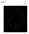
図3~図6は、磁気共鳴フィンガープリンティングの間に異常なボクセルを検出しそれを補正する方法を説明するために使用される。それは、解剖学的モデルで増強された外れ値検出方式と考えられる。最初に、図3において、磁気共鳴フィンガープリンティングからの組成マッピング300、すなわち画像が示される。CSF、白質、及び灰白質が図3に表示されている。図4は、脳の白質内で予想されるT2分布400を示す。ライン402は、図3の実際に測定されたT2分布を示す。曲線402の領域404は、曲線400と比較すると、異常であることが分かる。これは、潜在的に異常であるボクセルを識別するために使用される。図5は、T1値についての同様の分析を示す。曲線500は白質の正常な又は予想されるT1値であり、曲線502はT1の測定値の分布を示す。図4及び図5は若干の異常を示していることが分かる。図6は、補正されたMRF組成マッピング600を示す。異常なボクセル602は、新しい磁気共鳴フィンガープリンティングディクショナリを使用して再計算された組成マッピングを有している。領域602のボクセルは、潜在的に異常であり、例えば、異常組織病理を示している。
FIGS. 3-6 are used to describe methods for detecting and correcting abnormal voxels during magnetic resonance fingerprinting. It can be thought of as an anatomical model augmented outlier detection scheme. First, in FIG. 3, a
第3の実施例では、実施例1におけるような同様のセットアップが選ばれ、解剖学的/機能的モデルを初期データにフィッティングさせた。局部MRF応答、結果として生じた組織組成、及び解剖学的モデル全体からの情報を反復再構成における優先/制約として使用して、個々にサブサンプリングされた時間ドメイン画像におけるエイリアシングの一部が低減される。 In a third example, a similar set-up as in Example 1 was chosen to fit an anatomical/functional model to the initial data. Using information from local MRF responses, resulting tissue composition, and the entire anatomical model as priorities/constraints in iterative reconstruction, some of the aliasing in the individually subsampled time-domain images is reduced. be.
本発明は、図面及び前述の記載において詳細に図示及び説明されたが、このような図示及び記載は、説明的又は例示的であって限定するものではないと見なされるべきである。すなわち本発明は、開示された実施形態に限定されるものではない。 While the invention has been illustrated and described in detail in the drawings and foregoing description, such illustration and description are to be considered illustrative or exemplary and not restrictive. the invention is not limited to the disclosed embodiments.
開示された実施形態のその他の変形が、図面、本開示及び添付の請求項の検討から、請求項に係る発明を実施する当業者によって理解されて実現され得る。請求項において、「comprising(含む、備える)」という単語は、他の要素又はステップを除外するものではなく、不定冠詞「a」又は「an」は、複数を除外するものではない。単一のプロセッサ又は他のユニットが請求項に記載された幾つかのアイテムの機能を果たす。特定の手段が相互に異なる従属請求項に列挙されているという単なる事実は、これらの手段の組み合わせが有利に用いられないことを示すものではない。コンピュータプログラムは、他のハードウェアと共に若しくは他のハードウェアの一部として供給される光記憶媒体又はソリッドステート媒体等の適当な媒体に保存/分配されてもよいが、インターネット又は他の有線若しくは無線の電気通信システムを介して等の他の形式で分配されてもよい。請求項における任意の参照符号は、本発明の範囲を限定するものと解釈されるべきではない。 Other variations of the disclosed embodiments can be understood and effected by those skilled in the art in practicing the claimed invention, from a study of the drawings, this disclosure, and the appended claims. In the claims, the word "comprising" does not exclude other elements or steps, and the indefinite articles "a" or "an" do not exclude a plurality. A single processor or other unit may fulfill the functions of several items recited in the claims. The mere fact that certain measures are recited in mutually different dependent claims does not indicate that a combination of these measures cannot be used to advantage. The computer program may be stored/distributed on any suitable medium, such as optical storage media or solid-state media supplied with or as part of other hardware, but may also be stored/distributed on the Internet or other wired or wireless may be distributed in other forms, such as via a telecommunications system such as Any reference signs in the claims should not be construed as limiting the scope of the invention.
100 磁気共鳴イメージングシステム
104 磁石
106 磁石のボア
108 イメージングゾーン
109 関心領域
110 磁場勾配コイル
112 磁場勾配コイル電源
114 無線周波数コイル
116 トランシーバ
118 対象者
120 対象者支持体
126 コンピュータシステム
128 ハードウェアインタフェース
130 プロセッサ
132 ユーザインタフェース
134 コンピュータメモリ
140 マシン実行可能命令
142 MRFパルスシーケンスコマンド
144 MRF磁気共鳴データ
146 中間画像
148 MRF信号
150 B1+マッピング
152 磁気共鳴画像
154 解剖学的モデル
156 解剖学的領域、又はモデルへのレジストレーション
158 局所的磁気共鳴フィンガープリンティングディクショナリ
160 組成マッピング
200 MRFパルスシーケンスコマンドにより磁気共鳴イメージングシステムを制御することによって関心領域のMRF磁気共鳴データを取得する
202 関心領域を描写する少なくとも1つの磁気共鳴画像を受け取る
204 解剖学的モデルを使用して関心領域内の解剖学的領域を識別する
206 解剖学的領域の各々に対して1組の磁気共鳴フィンガープリンティングディクショナリから局所的磁気共鳴フィンガープリンティングディクショナリを選択する
208 MRF磁気共鳴データと局所的磁気共鳴フィンガープリンティングディクショナリとを使用して解剖学的領域の各々に対する所定の物質の組成マッピングを計算する
300 MRF組成マッピング又は画像
400 正常なT2分布
402 測定されたT2分布
404 異常なボクセルに起因する
500 測定されたT1分布
502 測定されたT1分布
600 補正されたMRF組成マッピング又は画像
602 潜在的な病理構造
100 Magnetic
Claims (20)
前記磁気共鳴イメージングシステムを制御するためのプロセッサと、
マシン実行可能命令及びMRFパルスシーケンスコマンドを格納するためのメモリであって、前記MRFパルスシーケンスコマンドが、磁気共鳴フィンガープリンティングプロトコルに従って前記MRF磁気共鳴データを取得するように前記磁気共鳴イメージングシステムを制御する、メモリと
を備え、
前記マシン実行可能命令の実行が、前記プロセッサに、
前記MRFパルスシーケンスコマンドにより前記磁気共鳴イメージングシステムを制御することによって前記関心領域の前記MRF磁気共鳴データを取得し、前記MRF磁気共鳴データからMRフィンガープリントを再構成することと、
前記関心領域を描写する磁気共鳴データを受け取ることと、
解剖学的モデルを使用して前記磁気共鳴データにおける前記関心領域内の解剖学的領域を識別することと、
前記解剖学的領域の各々に対して1組の磁気共鳴フィンガープリンティングディクショナリから局所的磁気共鳴フィンガープリンティングディクショナリを選択することであって、前記局所的磁気共鳴フィンガープリンティングディクショナリが、前記解剖学的領域の各々に固有の1組の所定の物質に対する計算されたMRF信号のリストを含む、選択することと、
前記MRフィンガープリントと前記局所的磁気共鳴フィンガープリンティングディクショナリとを使用して前記解剖学的領域の各々に対して前記所定の物質の組成マッピングを計算することであって、前記組成マッピングが前記解剖学的領域の各々内のボクセルの空間平均である、計算することと
を行わせる、磁気共鳴イメージングシステム。 A magnetic resonance imaging system for acquiring magnetic resonance fingerprinting ( MRF ) magnetic resonance data from a subject within a region of interest, said magnetic resonance imaging system comprising:
a processor for controlling the magnetic resonance imaging system;
A memory for storing machine executable instructions and MRF pulse sequence commands, said MRF pulse sequence commands controlling said magnetic resonance imaging system to acquire said MRF magnetic resonance data according to a magnetic resonance fingerprinting protocol. , with memory and
Execution of the machine-executable instructions causes the processor to:
acquiring the MRF magnetic resonance data of the region of interest by controlling the magnetic resonance imaging system with the MRF pulse sequence commands, and reconstructing an MR fingerprint from the MRF magnetic resonance data;
receiving magnetic resonance data depicting the region of interest;
identifying an anatomical region within the region of interest in the magnetic resonance data using an anatomical model;
selecting a local magnetic resonance fingerprinting dictionary from a set of magnetic resonance fingerprinting dictionaries for each of said anatomical regions, said local magnetic resonance fingerprinting dictionary selecting a list of calculated MRF signals for each unique set of predetermined substances;
computing a compositional mapping of the given material for each of the anatomical regions using the MR fingerprints and the local magnetic resonance fingerprinting dictionary, wherein the compositional mapping A magnetic resonance imaging system, which is a spatial average of voxels within each of the target regions.
前記局所的磁気共鳴フィンガープリンティングディクショナリを使用して前記組成マッピングを計算する前に、画像空間における前記解剖学的領域の各々内の磁気共鳴フィンガープリントのボクセルごとの平均化を実行すること、
前記局所的磁気共鳴フィンガープリンティングディクショナリを使用して前記組成マッピングを計算した後に、前記組成マッピングのボクセルごとの平均化を実行すること、及び
前記組成マッピングが前記解剖学的領域の各々についての前記ボクセルに最良フィットするように前記局所的磁気共鳴フィンガープリンティングディクショナリを使用して前記組成マッピングを計算すること
のうちの任意の1つを使用して実行される、請求項1に記載の磁気共鳴イメージングシステム。 Execution of said machine-executable instructions causes spatial averaging to:
performing voxel-by-voxel averaging of the magnetic resonance fingerprints within each of the anatomical regions in image space prior to calculating the compositional mapping using the local magnetic resonance fingerprinting dictionary;
performing voxel-by-voxel averaging of the compositional mapping after calculating the compositional mapping using the local magnetic resonance fingerprinting dictionary; and 2. The magnetic resonance imaging system of claim 1, performed using any one of calculating the compositional mapping using the local magnetic resonance fingerprinting dictionary to best fit to .
前記解剖学的領域から選択された解剖学的領域内のボクセルごとに組成分布を決定することと、
異常なボクセルの前記組成分布が所定のしきい値を超えて前記解剖学的領域の各々内の前記空間平均と異なる場合、前記解剖学的領域内の前記異常なボクセルを異常であるとして識別することと
を行わせる、請求項1に記載の磁気共鳴イメージングシステム。 Execution of the machine-executable instructions further causes the processor to:
determining a composition distribution for each voxel within an anatomical region selected from the anatomical region;
identifying the abnormal voxels within the anatomical region as abnormal if the composition distribution of the abnormal voxels differs from the spatial average within each of the anatomical regions by more than a predetermined threshold. 2. The magnetic resonance imaging system of claim 1, wherein the magnetic resonance imaging system causes:
前記解剖学的領域の各々の間の境界ボクセルを識別することと、
前記境界ボクセルの各々に隣接する解剖学的領域ごとに前記局所的磁気共鳴フィンガープリンティングディクショナリを使用して、前記境界ボクセルの各々についての部分的ボクセル組成マッピングを計算することと、
を行わせる、請求項1に記載の磁気共鳴イメージングシステム。 Execution of the machine-executable instructions further causes the processor to:
identifying boundary voxels between each of the anatomical regions;
calculating a partial voxel composition mapping for each of the bounding voxels using the local magnetic resonance fingerprinting dictionary for each anatomical region adjacent to each of the bounding voxels;
2. The magnetic resonance imaging system of claim 1, wherein the magnetic resonance imaging system allows
前記B+マッピングパルスシーケンスコマンドにより前記磁気共鳴イメージングシステムを制御することによって前記B1+マッピング磁気共鳴データを取得することと、
B1+マッピング磁気共鳴イメージングプロトコルに従って前記B1+マッピング磁気共鳴データを使用して前記B1+マッピングを再構成することと
を行わせることによって前記B1+マップを受け取らせる、請求項6に記載の磁気共鳴イメージングシステム。 said memory further comprising B1+mapping pulse sequence commands for acquiring B1+mapping magnetic resonance data according to a B1+mapping magnetic resonance imaging protocol, execution of said machine-executable instructions further causing said processor to:
acquiring the B1+ mapping magnetic resonance data by controlling the magnetic resonance imaging system with the B+ mapping pulse sequence commands;
7. The magnetic resonance imaging system of claim 6, wherein the B1+ map is received by using the B1+ mapping magnetic resonance data to reconstruct the B1+ mapping according to a B1+ mapping magnetic resonance imaging protocol.
前記イメージングパルスシーケンスコマンドにより前記磁気共鳴イメージングシステムを制御することによってイメージング磁気共鳴データを取得することと、
前記MRイメージングプロトコルに従って前記イメージング磁気共鳴データから前記少なくとも1つの磁気共鳴画像を再構成することと
を行わせることによって前記少なくとも1つの磁気共鳴画像を受け取らせる、請求項1に記載の磁気共鳴イメージングシステム。 the memory further comprising imaging pulse sequence commands according to an MR imaging protocol, execution of the machine-executable instructions further causing the processor to:
acquiring imaging magnetic resonance data by controlling the magnetic resonance imaging system with the imaging pulse sequence commands;
2. The magnetic resonance imaging system of claim 1, wherein the at least one magnetic resonance image is received by: reconstructing the at least one magnetic resonance image from the imaging magnetic resonance data according to the MR imaging protocol. .
MRFパルスシーケンスコマンドにより前記磁気共鳴イメージングシステムを制御することによって前記関心領域の前記MRF磁気共鳴データを取得するステップであって、前記MRFパルスシーケンスコマンドが、磁気共鳴フィンガープリンティングプロトコルに従って前記MRF磁気共鳴データを取得するように前記磁気共鳴イメージングシステムを制御し、前記MRF磁気共鳴データからMRフィンガープリントを再構成するように構成される、取得するステップと、
前記関心領域を描写する磁気共鳴データを受け取るステップと、
解剖学的モデルを使用して前記磁気共鳴データにおける前記関心領域内の解剖学的領域を識別するステップと、
前記解剖学的領域の各々に対して1組の磁気共鳴フィンガープリンティングディクショナリから局所的磁気共鳴フィンガープリンティングディクショナリを選択するステップであって、前記局所的磁気共鳴フィンガープリンティングディクショナリが、前記解剖学的領域の各々に固有の1組の所定の物質に対する計算されたMRF信号のリストを含む、選択するステップと、
前記MRフィンガープリントと前記局所的磁気共鳴フィンガープリンティングディクショナリとを使用して前記解剖学的領域の各々に対して前記所定の物質の組成マッピングを計算するステップであって、前記組成マッピングが前記解剖学的領域の各々内のボクセルの空間平均である、計算するステップと
を有する、方法。 A method of operating a magnetic resonance imaging system for acquiring magnetic resonance fingerprinting ( MRF ) magnetic resonance data from a subject within a region of interest, said method comprising:
acquiring the MRF magnetic resonance data of the region of interest by controlling the magnetic resonance imaging system with MRF pulse sequence commands, wherein the MRF pulse sequence commands generate the MRF magnetic resonance data according to a magnetic resonance fingerprinting protocol; and configured to reconstruct an MR fingerprint from the MRF magnetic resonance data;
receiving magnetic resonance data depicting the region of interest;
identifying an anatomical region within the region of interest in the magnetic resonance data using an anatomical model;
selecting a local magnetic resonance fingerprinting dictionary from a set of magnetic resonance fingerprinting dictionaries for each of said anatomical regions, said local magnetic resonance fingerprinting dictionary selecting, each containing a list of calculated MRF signals for a unique set of predetermined substances;
computing a compositional mapping of the given material for each of the anatomical regions using the MR fingerprints and the local magnetic resonance fingerprinting dictionary, wherein the compositional mapping calculating the spatial average of the voxels within each of the target regions.
前記局所的磁気共鳴フィンガープリンティングディクショナリを使用して前記組成マッピングを計算する前に、画像空間における前記解剖学的領域の各々内の磁気共鳴フィンガープリントのボクセルごとの平均化を実行すること、
前記局所的磁気共鳴フィンガープリンティングディクショナリを使用して前記組成マッピングを計算した後に、前記組成マッピングのボクセルごとの平均化を実行すること、及び
前記組成マッピングが前記解剖学的領域の各々についての前記ボクセルに最良フィットするように前記局所的磁気共鳴フィンガープリンティングディクショナリを使用して前記組成マッピングを計算すること
のうちの任意の1つを使用して実行される、請求項12に記載の方法。 Spatial averaging is
performing voxel-by-voxel averaging of the magnetic resonance fingerprints within each of the anatomical regions in image space prior to calculating the compositional mapping using the local magnetic resonance fingerprinting dictionary;
performing voxel-by-voxel averaging of the compositional mapping after calculating the compositional mapping using the local magnetic resonance fingerprinting dictionary; and 13. The method of claim 12, performed using any one of calculating the compositional mapping using the local magnetic resonance fingerprinting dictionary to best fit .
異常なボクセルの前記組成分布が所定のしきい値を超えて前記解剖学的領域の各々内の前記空間平均と異なる場合、前記解剖学的領域内の前記異常なボクセルを異常であるとして識別するステップと
を更に有する、請求項12に記載の方法。 determining a composition distribution for each voxel within an anatomical region selected from the anatomical regions;
identifying the abnormal voxels within the anatomical region as abnormal if the composition distribution of the abnormal voxels differs from the spatial average within each of the anatomical regions by more than a predetermined threshold. 13. The method of claim 12 , further comprising the steps of:
磁気共鳴フィンガープリンティング(MRF)パルスシーケンスコマンドにより前記磁気共鳴イメージングシステムを制御することによって関心領域のMRF磁気共鳴データを取得することであって、前記MRFパルスシーケンスコマンドが、磁気共鳴フィンガープリンティングプロトコルに従って前記MRF磁気共鳴データを取得するように前記磁気共鳴イメージングシステムを制御し、前記MRF磁気共鳴データからMRフィンガープリントを再構成するように構成される、取得することと、
前記関心領域を描写する磁気共鳴データを受け取ることと、
解剖学的モデルを使用して前記磁気共鳴データにおける前記関心領域内の解剖学的領域を識別することと、
前記解剖学的領域の各々に対して1組の磁気共鳴フィンガープリンティングディクショナリから局所的磁気共鳴フィンガープリンティングディクショナリを選択することであって、前記局所的磁気共鳴フィンガープリンティングディクショナリが、前記解剖学的領域の各々に固有の1組の所定の物質に対する計算されたMRF信号のリストを含む、選択することと、
前記MRフィンガープリントと前記局所的磁気共鳴フィンガープリンティングディクショナリとを使用して前記解剖学的領域の各々に対して前記所定の物質の組成マッピングを計算することであって、前記組成マッピングが前記解剖学的領域の各々内のボクセルの空間平均である、計算することと
を行わせる、非一時的コンピュータ可読媒体。 A non-transitory computer-readable medium storing machine-executable instructions for execution by a processor for controlling a magnetic resonance imaging system, execution of the machine-executable instructions causing the processor to:
Acquiring MRF magnetic resonance data of a region of interest by controlling said magnetic resonance imaging system with magnetic resonance fingerprinting ( MRF ) pulse sequence commands, said MRF pulse sequence commands according to said magnetic resonance fingerprinting protocol; acquiring, configured to control the magnetic resonance imaging system to acquire MRF magnetic resonance data and reconstruct an MR fingerprint from the MRF magnetic resonance data;
receiving magnetic resonance data depicting the region of interest;
identifying an anatomical region within the region of interest in the magnetic resonance data using an anatomical model;
selecting a local magnetic resonance fingerprinting dictionary from a set of magnetic resonance fingerprinting dictionaries for each of said anatomical regions, said local magnetic resonance fingerprinting dictionary selecting a list of calculated MRF signals for each unique set of predetermined substances;
computing a compositional mapping of the given material for each of the anatomical regions using the MR fingerprints and the local magnetic resonance fingerprinting dictionary, wherein the compositional mapping a spatial average of voxels within each of the target regions.
を更に有する、請求項14に記載の方法。15. The method of claim 14, further comprising:
前記境界ボクセルの各々に隣接する解剖学的領域ごとに前記局所的磁気共鳴フィンガープリンティングディクショナリを使用して、前記境界ボクセルの各々についての部分的ボクセル組成マッピングを計算するステップと、 computing a partial voxel composition mapping for each of said bounding voxels using said local magnetic resonance fingerprinting dictionary for each anatomical region adjacent to each of said bounding voxels;
を更に有する、請求項12に記載の方法。13. The method of claim 12, further comprising:
前記解剖学的領域から選択された解剖学的領域内のボクセルごとに組成分布を決定することと、 determining a composition distribution for each voxel within an anatomical region selected from the anatomical region;
異常なボクセルの前記組成分布が所定のしきい値を超えて前記解剖学的領域の各々内の前記空間平均と異なる場合、前記解剖学的領域内の前記異常なボクセルを異常であるとして識別することと identifying the abnormal voxels within the anatomical region as abnormal if the composition distribution of the abnormal voxels differs from the spatial average within each of the anatomical regions by more than a predetermined threshold. things and
を行わせる、請求項15に記載の非一時的コンピュータ可読媒体。16. The non-transitory computer-readable medium of claim 15, causing
前記解剖学的領域の各々の間の境界ボクセルを識別することと、 identifying boundary voxels between each of the anatomical regions;
前記境界ボクセルの各々に隣接する解剖学的領域ごとに前記局所的磁気共鳴フィンガープリンティングディクショナリを使用して、前記境界ボクセルの各々についての部分的ボクセル組成マッピングを計算することと、 calculating a partial voxel composition mapping for each of the bounding voxels using the local magnetic resonance fingerprinting dictionary for each anatomical region adjacent to each of the bounding voxels;
を行わせる、請求項15に記載の非一時的コンピュータ可読媒体。16. The non-transitory computer-readable medium of claim 15, causing
Priority Applications (1)
| Application Number | Priority Date | Filing Date | Title |
|---|---|---|---|
| JP2022152124A JP2022188131A (en) | 2017-03-30 | 2022-09-26 | Selection of magnetic resonance fingerprinting dictionary for anatomical region |
Applications Claiming Priority (3)
| Application Number | Priority Date | Filing Date | Title |
|---|---|---|---|
| EP17163899.2 | 2017-03-30 | ||
| EP17163899.2A EP3382416A1 (en) | 2017-03-30 | 2017-03-30 | Selection of magnetic resonance fingerprinting dictionaries for anatomical regions |
| PCT/EP2018/058320 WO2018178347A1 (en) | 2017-03-30 | 2018-03-30 | Selection of magnetic resonance fingerprinting dictionaries for anatomical regions |
Related Child Applications (1)
| Application Number | Title | Priority Date | Filing Date |
|---|---|---|---|
| JP2022152124A Division JP2022188131A (en) | 2017-03-30 | 2022-09-26 | Selection of magnetic resonance fingerprinting dictionary for anatomical region |
Publications (3)
| Publication Number | Publication Date |
|---|---|
| JP2020512143A JP2020512143A (en) | 2020-04-23 |
| JP2020512143A5 JP2020512143A5 (en) | 2021-08-19 |
| JP7319193B2 true JP7319193B2 (en) | 2023-08-01 |
Family
ID=58464312
Family Applications (2)
| Application Number | Title | Priority Date | Filing Date |
|---|---|---|---|
| JP2019553510A Active JP7319193B2 (en) | 2017-03-30 | 2018-03-30 | Selection of magnetic resonance fingerprinting dictionaries for anatomical regions |
| JP2022152124A Pending JP2022188131A (en) | 2017-03-30 | 2022-09-26 | Selection of magnetic resonance fingerprinting dictionary for anatomical region |
Family Applications After (1)
| Application Number | Title | Priority Date | Filing Date |
|---|---|---|---|
| JP2022152124A Pending JP2022188131A (en) | 2017-03-30 | 2022-09-26 | Selection of magnetic resonance fingerprinting dictionary for anatomical region |
Country Status (5)
| Country | Link |
|---|---|
| US (1) | US11112478B2 (en) |
| EP (2) | EP3382416A1 (en) |
| JP (2) | JP7319193B2 (en) |
| CN (1) | CN110476075B (en) |
| WO (1) | WO2018178347A1 (en) |
Families Citing this family (10)
| Publication number | Priority date | Publication date | Assignee | Title |
|---|---|---|---|---|
| EP3382416A1 (en) | 2017-03-30 | 2018-10-03 | Koninklijke Philips N.V. | Selection of magnetic resonance fingerprinting dictionaries for anatomical regions |
| EP3588121B1 (en) * | 2018-06-21 | 2022-09-14 | Siemens Healthcare GmbH | Magnetic resonance fingerprinting method |
| EP3680681A1 (en) | 2019-01-10 | 2020-07-15 | Siemens Healthcare GmbH | Selecting an acquisition scheme for a magnetic resonance fingerprinting acquisition and a corresponding dictionary based on a pre-scan |
| US11353527B2 (en) | 2019-07-19 | 2022-06-07 | Shanghai United Imaging Healthcare Co., Ltd. | Systems and methods for waveform determination in magnetic resonance imaging |
| CN110488210B (en) * | 2019-07-19 | 2021-09-17 | 上海联影医疗科技股份有限公司 | Scanning method for magnetic resonance system, computer device and storage medium |
| EP3786888A1 (en) * | 2019-08-27 | 2021-03-03 | Siemens Healthcare GmbH | Method for quantitative magnetic resonance imaging, magnetic resonance device, computer program and electronically readable data carrier |
| US11521314B2 (en) * | 2019-12-31 | 2022-12-06 | Shanghai United Imaging Healthcare Co., Ltd. | Systems and methods for image processing |
| US11875509B2 (en) * | 2020-04-03 | 2024-01-16 | Case Western Reserve University | System and method for determining undersampling errors for a magnetic resonance fingerprinting pulse sequence |
| US20220061809A1 (en) * | 2020-08-26 | 2022-03-03 | GE Precision Healthcare LLC | Method and system for providing an anatomic orientation indicator with a patient-specific model of an anatomical structure of interest extracted from a three-dimensional ultrasound volume |
| GB2617371A (en) * | 2022-04-06 | 2023-10-11 | Perspectum Ltd | Method and apparatus for characterization of breast tissue using multiparametric MRI |
Citations (4)
| Publication number | Priority date | Publication date | Assignee | Title |
|---|---|---|---|---|
| US20130278255A1 (en) | 2012-04-18 | 2013-10-24 | Mohammad Mehdi Khalighi | System and method of receive sensitivity correction in mr imaging |
| JP2015093192A (en) | 2013-11-14 | 2015-05-18 | 株式会社東芝 | Medical image processing apparatus and medical image processing method |
| US20160061922A1 (en) | 2014-08-29 | 2016-03-03 | Siemens Aktiengesellschaft | Method and apparatus for magnetic resonance examination of an examination object |
| US11112478B2 (en) | 2017-03-30 | 2021-09-07 | Koninklijke Philips N.V. | Selection of magnetic resonance fingerprinting dictionaries for anatomical regions |
Family Cites Families (16)
| Publication number | Priority date | Publication date | Assignee | Title |
|---|---|---|---|---|
| US8643363B2 (en) | 2008-11-27 | 2014-02-04 | Syntheticmr Ab | Method of visualizing segmented MR images with absolute-scale values independent of MR scanner settings |
| US10627468B2 (en) | 2011-03-18 | 2020-04-21 | Case Western Reserve University | Nuclear magnetic resonance (NMR) fingerprinting |
| US9097781B2 (en) | 2012-04-12 | 2015-08-04 | Mark Griswold | Nuclear magnetic resonance (NMR) fingerprinting with parallel transmission |
| WO2015081079A1 (en) * | 2013-11-26 | 2015-06-04 | Henry Ford Innovation Institute | Software for using magnetic resonance images to generate a synthetic computed tomography image |
| US10488480B2 (en) * | 2014-02-11 | 2019-11-26 | The General Hospital Corporation | System and method for adaptive dictionary matching in magnetic resonance fingerprinting |
| US10261154B2 (en) * | 2014-04-21 | 2019-04-16 | Case Western Reserve University | Nuclear magnetic resonance (NMR) fingerprinting tissue classification and image segmentation |
| US10209335B2 (en) * | 2014-04-21 | 2019-02-19 | Case Western Reserve University | Nuclear magnetic resonance (NMR) fingerprinting with singular value decomposition (SVD) compression |
| US9964616B2 (en) | 2014-05-30 | 2018-05-08 | Case Western Reserve University | Fast group matching for magnetic resonance fingerprinting reconstruction |
| CN107106039B (en) * | 2014-11-14 | 2020-08-11 | 皇家飞利浦有限公司 | Magnetic resonance fingerprinting in slices extending along one dimension |
| EP3224642B1 (en) * | 2014-11-27 | 2019-08-28 | Koninklijke Philips N.V. | Magnetic resonance fingerprinting data collection and analysis system |
| US10145917B2 (en) | 2015-03-27 | 2018-12-04 | Case Western Reserve University | Multi-component voxel separation using magnetic resonance fingerprinting with compartment exchange |
| US10209334B2 (en) * | 2015-04-08 | 2019-02-19 | The General Hospital Corporation | System and method for controlling partial volume effects in magnetic resonance fingerprinting |
| DE102015210292B4 (en) | 2015-06-03 | 2017-01-26 | Siemens Healthcare Gmbh | Determination of substance weightings by means of magnetic resonance signals |
| DE102015220077B4 (en) * | 2015-10-15 | 2019-03-07 | Siemens Healthcare Gmbh | Method for planning an irradiation of a patient |
| CN105869192B (en) * | 2016-03-28 | 2019-04-05 | 浙江大学 | A kind of magnetic resonance fingerprint recognition method for reconstructing based on sliding window |
| US20180231626A1 (en) * | 2017-02-10 | 2018-08-16 | Case Western Reserve University | Systems and methods for magnetic resonance fingerprinting for quantitative breast imaging |
-
2017
- 2017-03-30 EP EP17163899.2A patent/EP3382416A1/en not_active Withdrawn
-
2018
- 2018-03-30 US US16/497,480 patent/US11112478B2/en active Active
- 2018-03-30 CN CN201880022981.6A patent/CN110476075B/en active Active
- 2018-03-30 WO PCT/EP2018/058320 patent/WO2018178347A1/en unknown
- 2018-03-30 JP JP2019553510A patent/JP7319193B2/en active Active
- 2018-03-30 EP EP18716570.9A patent/EP3602097B1/en active Active
-
2022
- 2022-09-26 JP JP2022152124A patent/JP2022188131A/en active Pending
Patent Citations (5)
| Publication number | Priority date | Publication date | Assignee | Title |
|---|---|---|---|---|
| US20130278255A1 (en) | 2012-04-18 | 2013-10-24 | Mohammad Mehdi Khalighi | System and method of receive sensitivity correction in mr imaging |
| JP2015093192A (en) | 2013-11-14 | 2015-05-18 | 株式会社東芝 | Medical image processing apparatus and medical image processing method |
| US20160061922A1 (en) | 2014-08-29 | 2016-03-03 | Siemens Aktiengesellschaft | Method and apparatus for magnetic resonance examination of an examination object |
| US11112478B2 (en) | 2017-03-30 | 2021-09-07 | Koninklijke Philips N.V. | Selection of magnetic resonance fingerprinting dictionaries for anatomical regions |
| EP3602097B1 (en) | 2017-03-30 | 2022-05-11 | Koninklijke Philips N.V. | Selection of magnetic resonance fingerprinting dictionaries for anatomical regions |
Also Published As
| Publication number | Publication date |
|---|---|
| JP2020512143A (en) | 2020-04-23 |
| WO2018178347A1 (en) | 2018-10-04 |
| JP2022188131A (en) | 2022-12-20 |
| CN110476075B (en) | 2022-05-13 |
| EP3602097B1 (en) | 2022-05-11 |
| EP3602097A1 (en) | 2020-02-05 |
| CN110476075A (en) | 2019-11-19 |
| US11112478B2 (en) | 2021-09-07 |
| EP3382416A1 (en) | 2018-10-03 |
| US20210109180A1 (en) | 2021-04-15 |
Similar Documents
| Publication | Publication Date | Title |
|---|---|---|
| JP7319193B2 (en) | Selection of magnetic resonance fingerprinting dictionaries for anatomical regions | |
| US10698062B2 (en) | Diffusion MRI method for generating a synthetic diffusion image at a high B-value | |
| US10823798B2 (en) | Virtual CT images from magnetic resonance images | |
| US20180356484A1 (en) | Removal of image artifacts in sense-mri | |
| US20200041594A1 (en) | Sub voxel resolution magnetic resonance fingerprinting imaging | |
| JP2019534748A (en) | Combined arterial spin labeling and magnetic resonance fingerprinting | |
| US20190346526A1 (en) | Intensity corrected magnetic resonance images | |
| US11906608B2 (en) | Automated detection of water-fat swaps in Dixon magnetic resonance imaging | |
| CN114097041A (en) | Uncertainty map for deep learning electrical characteristic tomography | |
| EP4321890A1 (en) | Reconstruction parameter determination for the reconstruction of synthesized magnetic resonance images | |
| US11353532B2 (en) | (3-n) dimensional determination of electric conductivity | |
| JP7418357B2 (en) | Electrical property tomography mapping of conductivity changes | |
| EP4074253A1 (en) | Segmentation using multiple image contrasts | |
| US20220248973A1 (en) | Deep learning of eletrical properties tomography | |
| US20230377112A1 (en) | Magnetic Resonance Imaging of Breast Micro-Calcifications | |
| WO2024012945A1 (en) | Making anatomical measurements using magnetic resonance imaging | |
| WO2023156233A1 (en) | Detection of artifical structures in magentic resonance images due to neural networks | |
| JP2020525157A (en) | Magnetic Resonance Fingerprinting in Fourier Space |
Legal Events
| Date | Code | Title | Description |
|---|---|---|---|
| A521 | Request for written amendment filed |
Free format text: JAPANESE INTERMEDIATE CODE: A523 Effective date: 20210329 |
|
| A621 | Written request for application examination |
Free format text: JAPANESE INTERMEDIATE CODE: A621 Effective date: 20210329 |
|
| A521 | Request for written amendment filed |
Free format text: JAPANESE INTERMEDIATE CODE: A523 Effective date: 20210706 |
|
| A871 | Explanation of circumstances concerning accelerated examination |
Free format text: JAPANESE INTERMEDIATE CODE: A871 Effective date: 20210706 |
|
| A131 | Notification of reasons for refusal |
Free format text: JAPANESE INTERMEDIATE CODE: A131 Effective date: 20210928 |
|
| A601 | Written request for extension of time |
Free format text: JAPANESE INTERMEDIATE CODE: A601 Effective date: 20211221 |
|
| A02 | Decision of refusal |
Free format text: JAPANESE INTERMEDIATE CODE: A02 Effective date: 20220530 |
|
| C60 | Trial request (containing other claim documents, opposition documents) |
Free format text: JAPANESE INTERMEDIATE CODE: C60 Effective date: 20220926 |
|
| C22 | Notice of designation (change) of administrative judge |
Free format text: JAPANESE INTERMEDIATE CODE: C22 Effective date: 20230320 |
|
| A61 | First payment of annual fees (during grant procedure) |
Free format text: JAPANESE INTERMEDIATE CODE: A61 Effective date: 20230720 |
|
| R150 | Certificate of patent or registration of utility model |
Ref document number: 7319193 Country of ref document: JP Free format text: JAPANESE INTERMEDIATE CODE: R150 |