JP7316930B2 - Methods and Materials for Assessing Response to Plasmablast and Plasma Cell Depletion Therapy - Google Patents
Methods and Materials for Assessing Response to Plasmablast and Plasma Cell Depletion Therapy Download PDFInfo
- Publication number
- JP7316930B2 JP7316930B2 JP2019501612A JP2019501612A JP7316930B2 JP 7316930 B2 JP7316930 B2 JP 7316930B2 JP 2019501612 A JP2019501612 A JP 2019501612A JP 2019501612 A JP2019501612 A JP 2019501612A JP 7316930 B2 JP7316930 B2 JP 7316930B2
- Authority
- JP
- Japan
- Prior art keywords
- antibody
- seq
- cells
- agent
- patient
- Prior art date
- Legal status (The legal status is an assumption and is not a legal conclusion. Google has not performed a legal analysis and makes no representation as to the accuracy of the status listed.)
- Active
Links
- 210000004180 plasmocyte Anatomy 0.000 title claims description 52
- 210000003720 plasmablast Anatomy 0.000 title claims description 42
- 238000000034 method Methods 0.000 title description 140
- 230000004044 response Effects 0.000 title description 14
- 238000002560 therapeutic procedure Methods 0.000 title description 11
- 239000000463 material Substances 0.000 title description 6
- 101000777636 Homo sapiens ADP-ribosyl cyclase/cyclic ADP-ribose hydrolase 1 Proteins 0.000 claims description 269
- 210000004027 cell Anatomy 0.000 claims description 220
- 102100031585 ADP-ribosyl cyclase/cyclic ADP-ribose hydrolase 1 Human genes 0.000 claims description 217
- 208000037265 diseases, disorders, signs and symptoms Diseases 0.000 claims description 79
- 201000010099 disease Diseases 0.000 claims description 78
- 238000011282 treatment Methods 0.000 claims description 67
- 108090000623 proteins and genes Proteins 0.000 claims description 64
- 239000012472 biological sample Substances 0.000 claims description 57
- 241000282414 Homo sapiens Species 0.000 claims description 51
- 102000052645 human CD38 Human genes 0.000 claims description 49
- 208000023275 Autoimmune disease Diseases 0.000 claims description 45
- 150000001413 amino acids Chemical class 0.000 claims description 39
- 239000003795 chemical substances by application Substances 0.000 claims description 35
- 238000000684 flow cytometry Methods 0.000 claims description 32
- 201000000596 systemic lupus erythematosus Diseases 0.000 claims description 26
- 206010039073 rheumatoid arthritis Diseases 0.000 claims description 17
- 206010009900 Colitis ulcerative Diseases 0.000 claims description 6
- 208000022559 Inflammatory bowel disease Diseases 0.000 claims description 6
- 201000006704 Ulcerative Colitis Diseases 0.000 claims description 6
- 208000009329 Graft vs Host Disease Diseases 0.000 claims description 5
- 208000030836 Hashimoto thyroiditis Diseases 0.000 claims description 5
- 208000024908 graft versus host disease Diseases 0.000 claims description 5
- 201000006417 multiple sclerosis Diseases 0.000 claims description 5
- 206010021245 Idiopathic thrombocytopenic purpura Diseases 0.000 claims description 4
- 208000021386 Sjogren Syndrome Diseases 0.000 claims description 4
- 208000031981 Thrombocytopenic Idiopathic Purpura Diseases 0.000 claims description 4
- 201000003710 autoimmune thrombocytopenic purpura Diseases 0.000 claims description 4
- 239000004312 hexamethylene tetramine Substances 0.000 claims description 4
- 206010028417 myasthenia gravis Diseases 0.000 claims description 4
- 206010043554 thrombocytopenia Diseases 0.000 claims description 3
- 201000007023 Thrombotic Thrombocytopenic Purpura Diseases 0.000 claims description 2
- BFMYDTVEBKDAKJ-UHFFFAOYSA-L disodium;(2',7'-dibromo-3',6'-dioxido-3-oxospiro[2-benzofuran-1,9'-xanthene]-4'-yl)mercury;hydrate Chemical compound O.[Na+].[Na+].O1C(=O)C2=CC=CC=C2C21C1=CC(Br)=C([O-])C([Hg])=C1OC1=C2C=C(Br)C([O-])=C1 BFMYDTVEBKDAKJ-UHFFFAOYSA-L 0.000 claims description 2
- 231100000673 dose–response relationship Toxicity 0.000 claims description 2
- 230000001404 mediated effect Effects 0.000 claims description 2
- 230000001363 autoimmune Effects 0.000 claims 1
- 230000027455 binding Effects 0.000 description 80
- 239000000523 sample Substances 0.000 description 63
- 230000014509 gene expression Effects 0.000 description 54
- 235000001014 amino acid Nutrition 0.000 description 47
- 108090000765 processed proteins & peptides Proteins 0.000 description 45
- 108010047041 Complementarity Determining Regions Proteins 0.000 description 42
- 210000004369 blood Anatomy 0.000 description 41
- 239000008280 blood Substances 0.000 description 41
- 102000004196 processed proteins & peptides Human genes 0.000 description 39
- 239000000427 antigen Substances 0.000 description 38
- 102000036639 antigens Human genes 0.000 description 38
- 108091007433 antigens Proteins 0.000 description 38
- 230000004048 modification Effects 0.000 description 38
- 238000012986 modification Methods 0.000 description 38
- 229940024606 amino acid Drugs 0.000 description 37
- 229920001184 polypeptide Polymers 0.000 description 37
- 102000004169 proteins and genes Human genes 0.000 description 29
- 235000018102 proteins Nutrition 0.000 description 26
- 101100438926 Macaca fascicularis CD38 gene Proteins 0.000 description 25
- 102100024952 Protein CBFA2T1 Human genes 0.000 description 25
- 239000000203 mixture Substances 0.000 description 25
- 239000002953 phosphate buffered saline Substances 0.000 description 25
- 230000005764 inhibitory process Effects 0.000 description 24
- 210000003719 b-lymphocyte Anatomy 0.000 description 20
- 150000001875 compounds Chemical class 0.000 description 19
- 230000001225 therapeutic effect Effects 0.000 description 19
- 238000003556 assay Methods 0.000 description 18
- 239000012520 frozen sample Substances 0.000 description 18
- 108091003079 Bovine Serum Albumin Proteins 0.000 description 17
- 102100022005 B-lymphocyte antigen CD20 Human genes 0.000 description 16
- 101000897405 Homo sapiens B-lymphocyte antigen CD20 Proteins 0.000 description 16
- 241000699666 Mus <mouse, genus> Species 0.000 description 16
- 206010028980 Neoplasm Diseases 0.000 description 16
- 229940098773 bovine serum albumin Drugs 0.000 description 16
- 108020004999 messenger RNA Proteins 0.000 description 16
- 239000000243 solution Substances 0.000 description 16
- 238000006467 substitution reaction Methods 0.000 description 16
- 210000004698 lymphocyte Anatomy 0.000 description 15
- 241000894007 species Species 0.000 description 15
- 108020004414 DNA Proteins 0.000 description 14
- 108060003951 Immunoglobulin Proteins 0.000 description 14
- 150000001720 carbohydrates Chemical class 0.000 description 14
- 238000001943 fluorescence-activated cell sorting Methods 0.000 description 14
- 102000018358 immunoglobulin Human genes 0.000 description 14
- 230000001965 increasing effect Effects 0.000 description 14
- 102100024222 B-lymphocyte antigen CD19 Human genes 0.000 description 13
- -1 Cd38-rs1 Proteins 0.000 description 13
- 101000980825 Homo sapiens B-lymphocyte antigen CD19 Proteins 0.000 description 13
- 125000003275 alpha amino acid group Chemical group 0.000 description 13
- 239000000872 buffer Substances 0.000 description 13
- 150000007523 nucleic acids Chemical class 0.000 description 13
- WSFSSNUMVMOOMR-UHFFFAOYSA-N Formaldehyde Chemical compound O=C WSFSSNUMVMOOMR-UHFFFAOYSA-N 0.000 description 12
- 125000000539 amino acid group Chemical group 0.000 description 12
- 210000001185 bone marrow Anatomy 0.000 description 12
- 239000012634 fragment Substances 0.000 description 12
- 230000013595 glycosylation Effects 0.000 description 12
- 238000006206 glycosylation reaction Methods 0.000 description 12
- 230000009467 reduction Effects 0.000 description 12
- 238000012360 testing method Methods 0.000 description 12
- 206010035226 Plasma cell myeloma Diseases 0.000 description 11
- 230000000694 effects Effects 0.000 description 11
- 239000013604 expression vector Substances 0.000 description 11
- 102000039446 nucleic acids Human genes 0.000 description 11
- 108020004707 nucleic acids Proteins 0.000 description 11
- 210000003819 peripheral blood mononuclear cell Anatomy 0.000 description 11
- 108020003175 receptors Proteins 0.000 description 11
- 102000005962 receptors Human genes 0.000 description 11
- 239000006228 supernatant Substances 0.000 description 11
- 241000282567 Macaca fascicularis Species 0.000 description 10
- 238000001816 cooling Methods 0.000 description 10
- 239000003814 drug Substances 0.000 description 10
- 210000003743 erythrocyte Anatomy 0.000 description 10
- 239000008188 pellet Substances 0.000 description 10
- 238000003752 polymerase chain reaction Methods 0.000 description 10
- 108091033319 polynucleotide Proteins 0.000 description 10
- 102000040430 polynucleotide Human genes 0.000 description 10
- 239000002157 polynucleotide Substances 0.000 description 10
- 239000012099 Alexa Fluor family Substances 0.000 description 9
- 101000874179 Homo sapiens Syndecan-1 Proteins 0.000 description 9
- 241000699670 Mus sp. Species 0.000 description 9
- 229930040373 Paraformaldehyde Natural products 0.000 description 9
- 102100035721 Syndecan-1 Human genes 0.000 description 9
- 239000003153 chemical reaction reagent Substances 0.000 description 9
- 210000004978 chinese hamster ovary cell Anatomy 0.000 description 9
- 230000004540 complement-dependent cytotoxicity Effects 0.000 description 9
- 230000009089 cytolysis Effects 0.000 description 9
- 230000006870 function Effects 0.000 description 9
- BRZYSWJRSDMWLG-CAXSIQPQSA-N geneticin Natural products O1C[C@@](O)(C)[C@H](NC)[C@@H](O)[C@H]1O[C@@H]1[C@@H](O)[C@H](O[C@@H]2[C@@H]([C@@H](O)[C@H](O)[C@@H](C(C)O)O2)N)[C@@H](N)C[C@H]1N BRZYSWJRSDMWLG-CAXSIQPQSA-N 0.000 description 9
- 238000004020 luminiscence type Methods 0.000 description 9
- 229920002866 paraformaldehyde Polymers 0.000 description 9
- 238000012216 screening Methods 0.000 description 9
- 210000002966 serum Anatomy 0.000 description 9
- 239000013598 vector Substances 0.000 description 9
- FWMNVWWHGCHHJJ-SKKKGAJSSA-N 4-amino-1-[(2r)-6-amino-2-[[(2r)-2-[[(2r)-2-[[(2r)-2-amino-3-phenylpropanoyl]amino]-3-phenylpropanoyl]amino]-4-methylpentanoyl]amino]hexanoyl]piperidine-4-carboxylic acid Chemical compound C([C@H](C(=O)N[C@H](CC(C)C)C(=O)N[C@H](CCCCN)C(=O)N1CCC(N)(CC1)C(O)=O)NC(=O)[C@H](N)CC=1C=CC=CC=1)C1=CC=CC=C1 FWMNVWWHGCHHJJ-SKKKGAJSSA-N 0.000 description 8
- 102100027207 CD27 antigen Human genes 0.000 description 8
- 101000914511 Homo sapiens CD27 antigen Proteins 0.000 description 8
- 210000001744 T-lymphocyte Anatomy 0.000 description 8
- 238000004458 analytical method Methods 0.000 description 8
- 235000014633 carbohydrates Nutrition 0.000 description 8
- 238000006243 chemical reaction Methods 0.000 description 8
- 239000000975 dye Substances 0.000 description 8
- 238000003384 imaging method Methods 0.000 description 8
- 238000011534 incubation Methods 0.000 description 8
- 238000002595 magnetic resonance imaging Methods 0.000 description 8
- 229910052751 metal Inorganic materials 0.000 description 8
- 239000002184 metal Substances 0.000 description 8
- 238000002493 microarray Methods 0.000 description 8
- 238000010186 staining Methods 0.000 description 8
- LFQSCWFLJHTTHZ-UHFFFAOYSA-N Ethanol Chemical compound CCO LFQSCWFLJHTTHZ-UHFFFAOYSA-N 0.000 description 7
- 208000034578 Multiple myelomas Diseases 0.000 description 7
- 230000010056 antibody-dependent cellular cytotoxicity Effects 0.000 description 7
- 230000000295 complement effect Effects 0.000 description 7
- 238000001514 detection method Methods 0.000 description 7
- 238000010494 dissociation reaction Methods 0.000 description 7
- 230000005593 dissociations Effects 0.000 description 7
- 229940079593 drug Drugs 0.000 description 7
- 239000012636 effector Substances 0.000 description 7
- 238000007710 freezing Methods 0.000 description 7
- 230000008014 freezing Effects 0.000 description 7
- 229940127121 immunoconjugate Drugs 0.000 description 7
- 238000000338 in vitro Methods 0.000 description 7
- 238000001802 infusion Methods 0.000 description 7
- 238000002347 injection Methods 0.000 description 7
- 239000007924 injection Substances 0.000 description 7
- 239000002609 medium Substances 0.000 description 7
- 210000000822 natural killer cell Anatomy 0.000 description 7
- 230000002829 reductive effect Effects 0.000 description 7
- NFGXHKASABOEEW-UHFFFAOYSA-N 1-methylethyl 11-methoxy-3,7,11-trimethyl-2,4-dodecadienoate Chemical compound COC(C)(C)CCCC(C)CC=CC(C)=CC(=O)OC(C)C NFGXHKASABOEEW-UHFFFAOYSA-N 0.000 description 6
- 102100029824 ADP-ribosyl cyclase/cyclic ADP-ribose hydrolase 2 Human genes 0.000 description 6
- 241000282693 Cercopithecidae Species 0.000 description 6
- 102100029193 Low affinity immunoglobulin gamma Fc region receptor III-A Human genes 0.000 description 6
- 241001465754 Metazoa Species 0.000 description 6
- OKKJLVBELUTLKV-UHFFFAOYSA-N Methanol Chemical compound OC OKKJLVBELUTLKV-UHFFFAOYSA-N 0.000 description 6
- 239000004698 Polyethylene Substances 0.000 description 6
- ZMANZCXQSJIPKH-UHFFFAOYSA-N Triethylamine Chemical compound CCN(CC)CC ZMANZCXQSJIPKH-UHFFFAOYSA-N 0.000 description 6
- 210000000628 antibody-producing cell Anatomy 0.000 description 6
- 208000032852 chronic lymphocytic leukemia Diseases 0.000 description 6
- 239000010949 copper Substances 0.000 description 6
- 239000012091 fetal bovine serum Substances 0.000 description 6
- 238000001727 in vivo Methods 0.000 description 6
- 230000002757 inflammatory effect Effects 0.000 description 6
- 230000002401 inhibitory effect Effects 0.000 description 6
- 238000004519 manufacturing process Methods 0.000 description 6
- 238000005259 measurement Methods 0.000 description 6
- 238000002823 phage display Methods 0.000 description 6
- 230000009870 specific binding Effects 0.000 description 6
- 235000000346 sugar Nutrition 0.000 description 6
- 230000004083 survival effect Effects 0.000 description 6
- 208000024891 symptom Diseases 0.000 description 6
- 230000004797 therapeutic response Effects 0.000 description 6
- 210000001519 tissue Anatomy 0.000 description 6
- 238000002965 ELISA Methods 0.000 description 5
- 241000282412 Homo Species 0.000 description 5
- 101000738771 Homo sapiens Receptor-type tyrosine-protein phosphatase C Proteins 0.000 description 5
- 108010021625 Immunoglobulin Fragments Proteins 0.000 description 5
- 102000008394 Immunoglobulin Fragments Human genes 0.000 description 5
- 208000031422 Lymphocytic Chronic B-Cell Leukemia Diseases 0.000 description 5
- KDXKERNSBIXSRK-UHFFFAOYSA-N Lysine Natural products NCCCCC(N)C(O)=O KDXKERNSBIXSRK-UHFFFAOYSA-N 0.000 description 5
- 239000004472 Lysine Substances 0.000 description 5
- 239000002202 Polyethylene glycol Substances 0.000 description 5
- 238000003559 RNA-seq method Methods 0.000 description 5
- 102100037422 Receptor-type tyrosine-protein phosphatase C Human genes 0.000 description 5
- 230000009824 affinity maturation Effects 0.000 description 5
- 230000004075 alteration Effects 0.000 description 5
- 239000003146 anticoagulant agent Substances 0.000 description 5
- 229940127219 anticoagulant drug Drugs 0.000 description 5
- 239000011324 bead Substances 0.000 description 5
- 230000000903 blocking effect Effects 0.000 description 5
- 238000005119 centrifugation Methods 0.000 description 5
- 238000003745 diagnosis Methods 0.000 description 5
- 238000002405 diagnostic procedure Methods 0.000 description 5
- 238000005516 engineering process Methods 0.000 description 5
- 230000002255 enzymatic effect Effects 0.000 description 5
- 239000000284 extract Substances 0.000 description 5
- GNBHRKFJIUUOQI-UHFFFAOYSA-N fluorescein Chemical compound O1C(=O)C2=CC=CC=C2C21C1=CC=C(O)C=C1OC1=CC(O)=CC=C21 GNBHRKFJIUUOQI-UHFFFAOYSA-N 0.000 description 5
- MHMNJMPURVTYEJ-UHFFFAOYSA-N fluorescein-5-isothiocyanate Chemical compound O1C(=O)C2=CC(N=C=S)=CC=C2C21C1=CC=C(O)C=C1OC1=CC(O)=CC=C21 MHMNJMPURVTYEJ-UHFFFAOYSA-N 0.000 description 5
- 229940072221 immunoglobulins Drugs 0.000 description 5
- 238000000099 in vitro assay Methods 0.000 description 5
- 238000011503 in vivo imaging Methods 0.000 description 5
- 230000003993 interaction Effects 0.000 description 5
- 150000002500 ions Chemical class 0.000 description 5
- XEEYBQQBJWHFJM-UHFFFAOYSA-N iron Substances [Fe] XEEYBQQBJWHFJM-UHFFFAOYSA-N 0.000 description 5
- 238000002372 labelling Methods 0.000 description 5
- 238000004091 panning Methods 0.000 description 5
- 230000005298 paramagnetic effect Effects 0.000 description 5
- 239000013610 patient sample Substances 0.000 description 5
- 229920001223 polyethylene glycol Polymers 0.000 description 5
- 229920000642 polymer Polymers 0.000 description 5
- 239000012146 running buffer Substances 0.000 description 5
- 210000000952 spleen Anatomy 0.000 description 5
- 239000000126 substance Substances 0.000 description 5
- XLYOFNOQVPJJNP-UHFFFAOYSA-N water Substances O XLYOFNOQVPJJNP-UHFFFAOYSA-N 0.000 description 5
- 230000003442 weekly effect Effects 0.000 description 5
- 239000004475 Arginine Substances 0.000 description 4
- KCXVZYZYPLLWCC-UHFFFAOYSA-N EDTA Chemical compound OC(=O)CN(CC(O)=O)CCN(CC(O)=O)CC(O)=O KCXVZYZYPLLWCC-UHFFFAOYSA-N 0.000 description 4
- 102100031573 Hematopoietic progenitor cell antigen CD34 Human genes 0.000 description 4
- 101000777663 Homo sapiens Hematopoietic progenitor cell antigen CD34 Proteins 0.000 description 4
- 101000581981 Homo sapiens Neural cell adhesion molecule 1 Proteins 0.000 description 4
- 102100027347 Neural cell adhesion molecule 1 Human genes 0.000 description 4
- 102100024616 Platelet endothelial cell adhesion molecule Human genes 0.000 description 4
- 241000288906 Primates Species 0.000 description 4
- MTCFGRXMJLQNBG-UHFFFAOYSA-N Serine Natural products OCC(N)C(O)=O MTCFGRXMJLQNBG-UHFFFAOYSA-N 0.000 description 4
- 230000004913 activation Effects 0.000 description 4
- 150000001412 amines Chemical class 0.000 description 4
- 230000002096 anti-tetanic effect Effects 0.000 description 4
- ODKSFYDXXFIFQN-UHFFFAOYSA-N arginine Natural products OC(=O)C(N)CCCNC(N)=N ODKSFYDXXFIFQN-UHFFFAOYSA-N 0.000 description 4
- 239000000090 biomarker Substances 0.000 description 4
- 210000004899 c-terminal region Anatomy 0.000 description 4
- 238000002591 computed tomography Methods 0.000 description 4
- 239000002872 contrast media Substances 0.000 description 4
- 230000008878 coupling Effects 0.000 description 4
- 238000010168 coupling process Methods 0.000 description 4
- 238000005859 coupling reaction Methods 0.000 description 4
- 230000009260 cross reactivity Effects 0.000 description 4
- 230000003247 decreasing effect Effects 0.000 description 4
- 210000004443 dendritic cell Anatomy 0.000 description 4
- 239000000032 diagnostic agent Substances 0.000 description 4
- 229940039227 diagnostic agent Drugs 0.000 description 4
- 238000002474 experimental method Methods 0.000 description 4
- 238000009472 formulation Methods 0.000 description 4
- 238000010166 immunofluorescence Methods 0.000 description 4
- 230000006872 improvement Effects 0.000 description 4
- 239000003446 ligand Substances 0.000 description 4
- 210000003738 lymphoid progenitor cell Anatomy 0.000 description 4
- 230000002934 lysing effect Effects 0.000 description 4
- 239000003550 marker Substances 0.000 description 4
- 230000007246 mechanism Effects 0.000 description 4
- 125000002496 methyl group Chemical group [H]C([H])([H])* 0.000 description 4
- 239000003094 microcapsule Substances 0.000 description 4
- 210000001616 monocyte Anatomy 0.000 description 4
- 201000000050 myeloid neoplasm Diseases 0.000 description 4
- 210000000581 natural killer T-cell Anatomy 0.000 description 4
- 229940068196 placebo Drugs 0.000 description 4
- 239000000902 placebo Substances 0.000 description 4
- 230000002285 radioactive effect Effects 0.000 description 4
- 238000003753 real-time PCR Methods 0.000 description 4
- 239000007787 solid Substances 0.000 description 4
- 238000003860 storage Methods 0.000 description 4
- 150000008163 sugars Chemical class 0.000 description 4
- 238000002198 surface plasmon resonance spectroscopy Methods 0.000 description 4
- MTCFGRXMJLQNBG-REOHCLBHSA-N (2S)-2-Amino-3-hydroxypropansäure Chemical compound OC[C@H](N)C(O)=O MTCFGRXMJLQNBG-REOHCLBHSA-N 0.000 description 3
- JKMHFZQWWAIEOD-UHFFFAOYSA-N 2-[4-(2-hydroxyethyl)piperazin-1-yl]ethanesulfonic acid Chemical compound OCC[NH+]1CCN(CCS([O-])(=O)=O)CC1 JKMHFZQWWAIEOD-UHFFFAOYSA-N 0.000 description 3
- GOJUJUVQIVIZAV-UHFFFAOYSA-N 2-amino-4,6-dichloropyrimidine-5-carbaldehyde Chemical group NC1=NC(Cl)=C(C=O)C(Cl)=N1 GOJUJUVQIVIZAV-UHFFFAOYSA-N 0.000 description 3
- 102000000074 ADP-ribosyl Cyclase Human genes 0.000 description 3
- 108010080394 ADP-ribosyl Cyclase Proteins 0.000 description 3
- 102000018667 ADP-ribosyl Cyclase 1 Human genes 0.000 description 3
- 108010027122 ADP-ribosyl Cyclase 1 Proteins 0.000 description 3
- CSCPPACGZOOCGX-UHFFFAOYSA-N Acetone Chemical compound CC(C)=O CSCPPACGZOOCGX-UHFFFAOYSA-N 0.000 description 3
- 108010032595 Antibody Binding Sites Proteins 0.000 description 3
- 208000010839 B-cell chronic lymphocytic leukemia Diseases 0.000 description 3
- WVDDGKGOMKODPV-UHFFFAOYSA-N Benzyl alcohol Chemical compound OCC1=CC=CC=C1 WVDDGKGOMKODPV-UHFFFAOYSA-N 0.000 description 3
- 108091026890 Coding region Proteins 0.000 description 3
- BQOHYSXSASDCEA-KEOHHSTQSA-N Cyclic ADP-Ribose Chemical compound C([C@@H]1[C@H]([C@H]([C@@H](O1)N1C=2N=CN3C(C=2N=C1)=N)O)O)OP(O)(=O)OP(O)(=O)OC[C@@H]1[C@@H](O)[C@@H](O)[C@H]3O1 BQOHYSXSASDCEA-KEOHHSTQSA-N 0.000 description 3
- 201000004624 Dermatitis Diseases 0.000 description 3
- IAZDPXIOMUYVGZ-UHFFFAOYSA-N Dimethylsulphoxide Chemical compound CS(C)=O IAZDPXIOMUYVGZ-UHFFFAOYSA-N 0.000 description 3
- 102000004190 Enzymes Human genes 0.000 description 3
- 108090000790 Enzymes Proteins 0.000 description 3
- 241000588724 Escherichia coli Species 0.000 description 3
- 208000009386 Experimental Arthritis Diseases 0.000 description 3
- 108010087819 Fc receptors Proteins 0.000 description 3
- 102000009109 Fc receptors Human genes 0.000 description 3
- 238000012413 Fluorescence activated cell sorting analysis Methods 0.000 description 3
- WQZGKKKJIJFFOK-GASJEMHNSA-N Glucose Natural products OC[C@H]1OC(O)[C@H](O)[C@@H](O)[C@@H]1O WQZGKKKJIJFFOK-GASJEMHNSA-N 0.000 description 3
- 239000007995 HEPES buffer Substances 0.000 description 3
- 101000917858 Homo sapiens Low affinity immunoglobulin gamma Fc region receptor III-A Proteins 0.000 description 3
- 101000917839 Homo sapiens Low affinity immunoglobulin gamma Fc region receptor III-B Proteins 0.000 description 3
- 102100026120 IgG receptor FcRn large subunit p51 Human genes 0.000 description 3
- 101710177940 IgG receptor FcRn large subunit p51 Proteins 0.000 description 3
- 102100029571 Immunoglobulin J chain Human genes 0.000 description 3
- ODKSFYDXXFIFQN-BYPYZUCNSA-P L-argininium(2+) Chemical compound NC(=[NH2+])NCCC[C@H]([NH3+])C(O)=O ODKSFYDXXFIFQN-BYPYZUCNSA-P 0.000 description 3
- KDXKERNSBIXSRK-YFKPBYRVSA-N L-lysine Chemical compound NCCCC[C@H](N)C(O)=O KDXKERNSBIXSRK-YFKPBYRVSA-N 0.000 description 3
- 230000004989 O-glycosylation Effects 0.000 description 3
- 238000012220 PCR site-directed mutagenesis Methods 0.000 description 3
- 239000006146 Roswell Park Memorial Institute medium Substances 0.000 description 3
- 239000004473 Threonine Substances 0.000 description 3
- 230000002159 abnormal effect Effects 0.000 description 3
- 125000000613 asparagine group Chemical group N[C@@H](CC(N)=O)C(=O)* 0.000 description 3
- 230000008901 benefit Effects 0.000 description 3
- 230000001588 bifunctional effect Effects 0.000 description 3
- 230000004071 biological effect Effects 0.000 description 3
- 210000000601 blood cell Anatomy 0.000 description 3
- 238000006664 bond formation reaction Methods 0.000 description 3
- 201000011510 cancer Diseases 0.000 description 3
- 125000003178 carboxy group Chemical group [H]OC(*)=O 0.000 description 3
- 239000002299 complementary DNA Substances 0.000 description 3
- 229920001577 copolymer Polymers 0.000 description 3
- 239000003405 delayed action preparation Substances 0.000 description 3
- 238000012217 deletion Methods 0.000 description 3
- 230000037430 deletion Effects 0.000 description 3
- 230000000779 depleting effect Effects 0.000 description 3
- 238000001212 derivatisation Methods 0.000 description 3
- 238000011161 development Methods 0.000 description 3
- 230000018109 developmental process Effects 0.000 description 3
- 230000002708 enhancing effect Effects 0.000 description 3
- 238000011156 evaluation Methods 0.000 description 3
- 230000004927 fusion Effects 0.000 description 3
- 125000000291 glutamic acid group Chemical group N[C@@H](CCC(O)=O)C(=O)* 0.000 description 3
- ZFGMDIBRIDKWMY-PASTXAENSA-N heparin Chemical compound CC(O)=N[C@@H]1[C@@H](O)[C@H](O)[C@@H](COS(O)(=O)=O)O[C@@H]1O[C@@H]1[C@@H](C(O)=O)O[C@@H](O[C@H]2[C@@H]([C@@H](OS(O)(=O)=O)[C@@H](O[C@@H]3[C@@H](OC(O)[C@H](OS(O)(=O)=O)[C@H]3O)C(O)=O)O[C@@H]2O)CS(O)(=O)=O)[C@H](O)[C@H]1O ZFGMDIBRIDKWMY-PASTXAENSA-N 0.000 description 3
- 229920000669 heparin Polymers 0.000 description 3
- 208000026278 immune system disease Diseases 0.000 description 3
- 208000027866 inflammatory disease Diseases 0.000 description 3
- 238000003780 insertion Methods 0.000 description 3
- 230000037431 insertion Effects 0.000 description 3
- 208000036971 interstitial lung disease 2 Diseases 0.000 description 3
- 230000003834 intracellular effect Effects 0.000 description 3
- 239000002502 liposome Substances 0.000 description 3
- 210000002809 long lived plasma cell Anatomy 0.000 description 3
- 239000011159 matrix material Substances 0.000 description 3
- 239000012528 membrane Substances 0.000 description 3
- 150000002739 metals Chemical class 0.000 description 3
- 235000013336 milk Nutrition 0.000 description 3
- 239000008267 milk Substances 0.000 description 3
- 210000004080 milk Anatomy 0.000 description 3
- 210000005170 neoplastic cell Anatomy 0.000 description 3
- 229920001542 oligosaccharide Polymers 0.000 description 3
- 150000002482 oligosaccharides Chemical class 0.000 description 3
- 210000000056 organ Anatomy 0.000 description 3
- 229960000402 palivizumab Drugs 0.000 description 3
- 230000036961 partial effect Effects 0.000 description 3
- 230000035755 proliferation Effects 0.000 description 3
- 230000000717 retained effect Effects 0.000 description 3
- 238000012163 sequencing technique Methods 0.000 description 3
- 150000003384 small molecules Chemical class 0.000 description 3
- 230000010473 stable expression Effects 0.000 description 3
- 125000001493 tyrosinyl group Chemical group [H]OC1=C([H])C([H])=C(C([H])=C1[H])C([H])([H])C([H])(N([H])[H])C(*)=O 0.000 description 3
- 238000002604 ultrasonography Methods 0.000 description 3
- YBJHBAHKTGYVGT-ZKWXMUAHSA-N (+)-Biotin Chemical compound N1C(=O)N[C@@H]2[C@H](CCCCC(=O)O)SC[C@@H]21 YBJHBAHKTGYVGT-ZKWXMUAHSA-N 0.000 description 2
- VYEWZWBILJHHCU-OMQUDAQFSA-N (e)-n-[(2s,3r,4r,5r,6r)-2-[(2r,3r,4s,5s,6s)-3-acetamido-5-amino-4-hydroxy-6-(hydroxymethyl)oxan-2-yl]oxy-6-[2-[(2r,3s,4r,5r)-5-(2,4-dioxopyrimidin-1-yl)-3,4-dihydroxyoxolan-2-yl]-2-hydroxyethyl]-4,5-dihydroxyoxan-3-yl]-5-methylhex-2-enamide Chemical compound N1([C@@H]2O[C@@H]([C@H]([C@H]2O)O)C(O)C[C@@H]2[C@H](O)[C@H](O)[C@H]([C@@H](O2)O[C@@H]2[C@@H]([C@@H](O)[C@H](N)[C@@H](CO)O2)NC(C)=O)NC(=O)/C=C/CC(C)C)C=CC(=O)NC1=O VYEWZWBILJHHCU-OMQUDAQFSA-N 0.000 description 2
- HZAXFHJVJLSVMW-UHFFFAOYSA-N 2-Aminoethan-1-ol Chemical compound NCCO HZAXFHJVJLSVMW-UHFFFAOYSA-N 0.000 description 2
- GOLORTLGFDVFDW-UHFFFAOYSA-N 3-(1h-benzimidazol-2-yl)-7-(diethylamino)chromen-2-one Chemical compound C1=CC=C2NC(C3=CC4=CC=C(C=C4OC3=O)N(CC)CC)=NC2=C1 GOLORTLGFDVFDW-UHFFFAOYSA-N 0.000 description 2
- 108050008264 ADP-ribosyl cyclase/cyclic ADP-ribose hydrolase 1 Proteins 0.000 description 2
- 108010088751 Albumins Proteins 0.000 description 2
- 102000009027 Albumins Human genes 0.000 description 2
- 208000032791 BCR-ABL1 positive chronic myelogenous leukemia Diseases 0.000 description 2
- 241000894006 Bacteria Species 0.000 description 2
- 208000023328 Basedow disease Diseases 0.000 description 2
- 208000011691 Burkitt lymphomas Diseases 0.000 description 2
- 210000001266 CD8-positive T-lymphocyte Anatomy 0.000 description 2
- 241000283707 Capra Species 0.000 description 2
- 208000010833 Chronic myeloid leukaemia Diseases 0.000 description 2
- 241000699802 Cricetulus griseus Species 0.000 description 2
- 208000011231 Crohn disease Diseases 0.000 description 2
- SHZGCJCMOBCMKK-UHFFFAOYSA-N D-mannomethylose Natural products CC1OC(O)C(O)C(O)C1O SHZGCJCMOBCMKK-UHFFFAOYSA-N 0.000 description 2
- SRBFZHDQGSBBOR-IOVATXLUSA-N D-xylopyranose Chemical compound O[C@@H]1COC(O)[C@H](O)[C@H]1O SRBFZHDQGSBBOR-IOVATXLUSA-N 0.000 description 2
- QSJXEFYPDANLFS-UHFFFAOYSA-N Diacetyl Chemical compound CC(=O)C(C)=O QSJXEFYPDANLFS-UHFFFAOYSA-N 0.000 description 2
- 206010061818 Disease progression Diseases 0.000 description 2
- 201000003542 Factor VIII deficiency Diseases 0.000 description 2
- 208000001640 Fibromyalgia Diseases 0.000 description 2
- PNNNRSAQSRJVSB-SLPGGIOYSA-N Fucose Natural products C[C@H](O)[C@@H](O)[C@H](O)[C@H](O)C=O PNNNRSAQSRJVSB-SLPGGIOYSA-N 0.000 description 2
- SXRSQZLOMIGNAQ-UHFFFAOYSA-N Glutaraldehyde Chemical compound O=CCCCC=O SXRSQZLOMIGNAQ-UHFFFAOYSA-N 0.000 description 2
- DHMQDGOQFOQNFH-UHFFFAOYSA-N Glycine Chemical compound NCC(O)=O DHMQDGOQFOQNFH-UHFFFAOYSA-N 0.000 description 2
- 102000005744 Glycoside Hydrolases Human genes 0.000 description 2
- 108010031186 Glycoside Hydrolases Proteins 0.000 description 2
- 208000015023 Graves' disease Diseases 0.000 description 2
- 208000035186 Hemolytic Autoimmune Anemia Diseases 0.000 description 2
- 208000009292 Hemophilia A Diseases 0.000 description 2
- 101001011441 Homo sapiens Interferon regulatory factor 4 Proteins 0.000 description 2
- 108010054477 Immunoglobulin Fab Fragments Proteins 0.000 description 2
- 102000001706 Immunoglobulin Fab Fragments Human genes 0.000 description 2
- 101710132152 Immunoglobulin J chain Proteins 0.000 description 2
- 102000013463 Immunoglobulin Light Chains Human genes 0.000 description 2
- 108010065825 Immunoglobulin Light Chains Proteins 0.000 description 2
- 102100030126 Interferon regulatory factor 4 Human genes 0.000 description 2
- SHZGCJCMOBCMKK-DHVFOXMCSA-N L-fucopyranose Chemical compound C[C@@H]1OC(O)[C@@H](O)[C@H](O)[C@@H]1O SHZGCJCMOBCMKK-DHVFOXMCSA-N 0.000 description 2
- HNDVDQJCIGZPNO-YFKPBYRVSA-N L-histidine Chemical compound OC(=O)[C@@H](N)CC1=CN=CN1 HNDVDQJCIGZPNO-YFKPBYRVSA-N 0.000 description 2
- AYFVYJQAPQTCCC-GBXIJSLDSA-N L-threonine Chemical compound C[C@@H](O)[C@H](N)C(O)=O AYFVYJQAPQTCCC-GBXIJSLDSA-N 0.000 description 2
- 108010000817 Leuprolide Proteins 0.000 description 2
- 102100029206 Low affinity immunoglobulin gamma Fc region receptor II-c Human genes 0.000 description 2
- 101710099301 Low affinity immunoglobulin gamma Fc region receptor III-A Proteins 0.000 description 2
- 108060001084 Luciferase Proteins 0.000 description 2
- 239000005089 Luciferase Substances 0.000 description 2
- 208000033761 Myelogenous Chronic BCR-ABL Positive Leukemia Diseases 0.000 description 2
- OVRNDRQMDRJTHS-CBQIKETKSA-N N-Acetyl-D-Galactosamine Chemical compound CC(=O)N[C@H]1[C@@H](O)O[C@H](CO)[C@H](O)[C@@H]1O OVRNDRQMDRJTHS-CBQIKETKSA-N 0.000 description 2
- MBLBDJOUHNCFQT-UHFFFAOYSA-N N-acetyl-D-galactosamine Natural products CC(=O)NC(C=O)C(O)C(O)C(O)CO MBLBDJOUHNCFQT-UHFFFAOYSA-N 0.000 description 2
- 230000004988 N-glycosylation Effects 0.000 description 2
- 229930193140 Neomycin Natural products 0.000 description 2
- 102000003729 Neprilysin Human genes 0.000 description 2
- 108090000028 Neprilysin Proteins 0.000 description 2
- 241000283973 Oryctolagus cuniculus Species 0.000 description 2
- 229910019142 PO4 Inorganic materials 0.000 description 2
- 206010034277 Pemphigoid Diseases 0.000 description 2
- ISWSIDIOOBJBQZ-UHFFFAOYSA-N Phenol Natural products OC1=CC=CC=C1 ISWSIDIOOBJBQZ-UHFFFAOYSA-N 0.000 description 2
- NBIIXXVUZAFLBC-UHFFFAOYSA-N Phosphoric acid Chemical compound OP(O)(O)=O NBIIXXVUZAFLBC-UHFFFAOYSA-N 0.000 description 2
- 108010004729 Phycoerythrin Proteins 0.000 description 2
- 108010069381 Platelet Endothelial Cell Adhesion Molecule-1 Proteins 0.000 description 2
- 229920001213 Polysorbate 20 Polymers 0.000 description 2
- ONIBWKKTOPOVIA-UHFFFAOYSA-N Proline Natural products OC(=O)C1CCCN1 ONIBWKKTOPOVIA-UHFFFAOYSA-N 0.000 description 2
- 102000001708 Protein Isoforms Human genes 0.000 description 2
- 108010029485 Protein Isoforms Proteins 0.000 description 2
- RADKZDMFGJYCBB-UHFFFAOYSA-N Pyridoxal Chemical compound CC1=NC=C(CO)C(C=O)=C1O RADKZDMFGJYCBB-UHFFFAOYSA-N 0.000 description 2
- PXIPVTKHYLBLMZ-UHFFFAOYSA-N Sodium azide Chemical compound [Na+].[N-]=[N+]=[N-] PXIPVTKHYLBLMZ-UHFFFAOYSA-N 0.000 description 2
- FAPWRFPIFSIZLT-UHFFFAOYSA-M Sodium chloride Chemical compound [Na+].[Cl-] FAPWRFPIFSIZLT-UHFFFAOYSA-M 0.000 description 2
- 102100038021 Steryl-sulfatase Human genes 0.000 description 2
- 108010090804 Streptavidin Proteins 0.000 description 2
- 229910052771 Terbium Inorganic materials 0.000 description 2
- NYTOUQBROMCLBJ-UHFFFAOYSA-N Tetranitromethane Chemical compound [O-][N+](=O)C([N+]([O-])=O)([N+]([O-])=O)[N+]([O-])=O NYTOUQBROMCLBJ-UHFFFAOYSA-N 0.000 description 2
- AYFVYJQAPQTCCC-UHFFFAOYSA-N Threonine Natural products CC(O)C(N)C(O)=O AYFVYJQAPQTCCC-UHFFFAOYSA-N 0.000 description 2
- 206010052779 Transplant rejections Diseases 0.000 description 2
- YJQCOFNZVFGCAF-UHFFFAOYSA-N Tunicamycin II Natural products O1C(CC(O)C2C(C(O)C(O2)N2C(NC(=O)C=C2)=O)O)C(O)C(O)C(NC(=O)C=CCCCCCCCCC(C)C)C1OC1OC(CO)C(O)C(O)C1NC(C)=O YJQCOFNZVFGCAF-UHFFFAOYSA-N 0.000 description 2
- 206010047115 Vasculitis Diseases 0.000 description 2
- 230000009471 action Effects 0.000 description 2
- 230000002776 aggregation Effects 0.000 description 2
- 238000004220 aggregation Methods 0.000 description 2
- 150000001299 aldehydes Chemical class 0.000 description 2
- 230000009830 antibody antigen interaction Effects 0.000 description 2
- 230000000890 antigenic effect Effects 0.000 description 2
- 125000000637 arginyl group Chemical group N[C@@H](CCCNC(N)=N)C(=O)* 0.000 description 2
- 206010003246 arthritis Diseases 0.000 description 2
- 125000003118 aryl group Chemical group 0.000 description 2
- 230000009286 beneficial effect Effects 0.000 description 2
- WQZGKKKJIJFFOK-VFUOTHLCSA-N beta-D-glucose Chemical compound OC[C@H]1O[C@@H](O)[C@H](O)[C@@H](O)[C@@H]1O WQZGKKKJIJFFOK-VFUOTHLCSA-N 0.000 description 2
- 238000001574 biopsy Methods 0.000 description 2
- 210000002798 bone marrow cell Anatomy 0.000 description 2
- DQXBYHZEEUGOBF-UHFFFAOYSA-N but-3-enoic acid;ethene Chemical compound C=C.OC(=O)CC=C DQXBYHZEEUGOBF-UHFFFAOYSA-N 0.000 description 2
- 150000001718 carbodiimides Chemical class 0.000 description 2
- YCIMNLLNPGFGHC-UHFFFAOYSA-N catechol Chemical compound OC1=CC=CC=C1O YCIMNLLNPGFGHC-UHFFFAOYSA-N 0.000 description 2
- 230000004663 cell proliferation Effects 0.000 description 2
- 230000001413 cellular effect Effects 0.000 description 2
- 210000001175 cerebrospinal fluid Anatomy 0.000 description 2
- 230000008859 change Effects 0.000 description 2
- 239000013522 chelant Substances 0.000 description 2
- 239000002738 chelating agent Substances 0.000 description 2
- 230000001684 chronic effect Effects 0.000 description 2
- 238000012875 competitive assay Methods 0.000 description 2
- 230000021615 conjugation Effects 0.000 description 2
- 239000013068 control sample Substances 0.000 description 2
- 125000000151 cysteine group Chemical group N[C@@H](CS)C(=O)* 0.000 description 2
- 230000001086 cytosolic effect Effects 0.000 description 2
- 230000022811 deglycosylation Effects 0.000 description 2
- 201000001981 dermatomyositis Diseases 0.000 description 2
- UREBDLICKHMUKA-CXSFZGCWSA-N dexamethasone Chemical compound C1CC2=CC(=O)C=C[C@]2(C)[C@]2(F)[C@@H]1[C@@H]1C[C@@H](C)[C@@](C(=O)CO)(O)[C@@]1(C)C[C@@H]2O UREBDLICKHMUKA-CXSFZGCWSA-N 0.000 description 2
- 229960003957 dexamethasone Drugs 0.000 description 2
- 206010012601 diabetes mellitus Diseases 0.000 description 2
- SPCNPOWOBZQWJK-UHFFFAOYSA-N dimethoxy-(2-propan-2-ylsulfanylethylsulfanyl)-sulfanylidene-$l^{5}-phosphane Chemical compound COP(=S)(OC)SCCSC(C)C SPCNPOWOBZQWJK-UHFFFAOYSA-N 0.000 description 2
- XBDQKXXYIPTUBI-UHFFFAOYSA-N dimethylselenoniopropionate Natural products CCC(O)=O XBDQKXXYIPTUBI-UHFFFAOYSA-N 0.000 description 2
- 230000005750 disease progression Effects 0.000 description 2
- 239000003937 drug carrier Substances 0.000 description 2
- 239000002961 echo contrast media Substances 0.000 description 2
- YQGOJNYOYNNSMM-UHFFFAOYSA-N eosin Chemical compound [Na+].OC(=O)C1=CC=CC=C1C1=C2C=C(Br)C(=O)C(Br)=C2OC2=C(Br)C(O)=C(Br)C=C21 YQGOJNYOYNNSMM-UHFFFAOYSA-N 0.000 description 2
- IINNWAYUJNWZRM-UHFFFAOYSA-L erythrosin B Chemical compound [Na+].[Na+].[O-]C(=O)C1=CC=CC=C1C1=C2C=C(I)C(=O)C(I)=C2OC2=C(I)C([O-])=C(I)C=C21 IINNWAYUJNWZRM-UHFFFAOYSA-L 0.000 description 2
- 229940011411 erythrosine Drugs 0.000 description 2
- 235000012732 erythrosine Nutrition 0.000 description 2
- 239000004174 erythrosine Substances 0.000 description 2
- 150000002148 esters Chemical class 0.000 description 2
- 239000005038 ethylene vinyl acetate Substances 0.000 description 2
- 239000000834 fixative Substances 0.000 description 2
- 239000012530 fluid Substances 0.000 description 2
- 125000000524 functional group Chemical group 0.000 description 2
- 239000011521 glass Substances 0.000 description 2
- 239000008103 glucose Substances 0.000 description 2
- ZDXPYRJPNDTMRX-UHFFFAOYSA-N glutamine Natural products OC(=O)C(N)CCC(N)=O ZDXPYRJPNDTMRX-UHFFFAOYSA-N 0.000 description 2
- 125000000404 glutamine group Chemical group N[C@@H](CCC(N)=O)C(=O)* 0.000 description 2
- HHLFWLYXYJOTON-UHFFFAOYSA-N glyoxylic acid Chemical compound OC(=O)C=O HHLFWLYXYJOTON-UHFFFAOYSA-N 0.000 description 2
- 210000003958 hematopoietic stem cell Anatomy 0.000 description 2
- HNDVDQJCIGZPNO-UHFFFAOYSA-N histidine Natural products OC(=O)C(N)CC1=CN=CN1 HNDVDQJCIGZPNO-UHFFFAOYSA-N 0.000 description 2
- 125000000487 histidyl group Chemical group [H]N([H])C(C(=O)O*)C([H])([H])C1=C([H])N([H])C([H])=N1 0.000 description 2
- 210000004408 hybridoma Anatomy 0.000 description 2
- 239000000017 hydrogel Substances 0.000 description 2
- 125000002887 hydroxy group Chemical group [H]O* 0.000 description 2
- 150000002463 imidates Chemical class 0.000 description 2
- 230000003100 immobilizing effect Effects 0.000 description 2
- 210000002865 immune cell Anatomy 0.000 description 2
- 230000001900 immune effect Effects 0.000 description 2
- 230000005847 immunogenicity Effects 0.000 description 2
- 238000005462 in vivo assay Methods 0.000 description 2
- 238000010348 incorporation Methods 0.000 description 2
- 208000015181 infectious disease Diseases 0.000 description 2
- 230000008595 infiltration Effects 0.000 description 2
- 238000001764 infiltration Methods 0.000 description 2
- 238000001990 intravenous administration Methods 0.000 description 2
- 229910052742 iron Inorganic materials 0.000 description 2
- 210000003734 kidney Anatomy 0.000 description 2
- 210000000265 leukocyte Anatomy 0.000 description 2
- RGLRXNKKBLIBQS-XNHQSDQCSA-N leuprolide acetate Chemical compound CC(O)=O.CCNC(=O)[C@@H]1CCCN1C(=O)[C@H](CCCNC(N)=N)NC(=O)[C@H](CC(C)C)NC(=O)[C@@H](CC(C)C)NC(=O)[C@@H](NC(=O)[C@H](CO)NC(=O)[C@H](CC=1C2=CC=CC=C2NC=1)NC(=O)[C@H](CC=1N=CNC=1)NC(=O)[C@H]1NC(=O)CC1)CC1=CC=C(O)C=C1 RGLRXNKKBLIBQS-XNHQSDQCSA-N 0.000 description 2
- 230000000670 limiting effect Effects 0.000 description 2
- 206010025135 lupus erythematosus Diseases 0.000 description 2
- 210000002751 lymph Anatomy 0.000 description 2
- 210000001165 lymph node Anatomy 0.000 description 2
- 125000003588 lysine group Chemical group [H]N([H])C([H])([H])C([H])([H])C([H])([H])C([H])([H])C([H])(N([H])[H])C(*)=O 0.000 description 2
- RLSSMJSEOOYNOY-UHFFFAOYSA-N m-cresol Chemical compound CC1=CC=CC(O)=C1 RLSSMJSEOOYNOY-UHFFFAOYSA-N 0.000 description 2
- 238000007403 mPCR Methods 0.000 description 2
- 239000004005 microsphere Substances 0.000 description 2
- 210000005087 mononuclear cell Anatomy 0.000 description 2
- RTGDFNSFWBGLEC-SYZQJQIISA-N mycophenolate mofetil Chemical compound COC1=C(C)C=2COC(=O)C=2C(O)=C1C\C=C(/C)CCC(=O)OCCN1CCOCC1 RTGDFNSFWBGLEC-SYZQJQIISA-N 0.000 description 2
- 229960004866 mycophenolate mofetil Drugs 0.000 description 2
- 239000002105 nanoparticle Substances 0.000 description 2
- 239000013642 negative control Substances 0.000 description 2
- 229960004927 neomycin Drugs 0.000 description 2
- 239000002773 nucleotide Substances 0.000 description 2
- 125000003729 nucleotide group Chemical group 0.000 description 2
- 230000003287 optical effect Effects 0.000 description 2
- 210000001672 ovary Anatomy 0.000 description 2
- 230000002018 overexpression Effects 0.000 description 2
- 230000037361 pathway Effects 0.000 description 2
- 210000005259 peripheral blood Anatomy 0.000 description 2
- 239000011886 peripheral blood Substances 0.000 description 2
- 239000008194 pharmaceutical composition Substances 0.000 description 2
- 239000000546 pharmaceutical excipient Substances 0.000 description 2
- OJUGVDODNPJEEC-UHFFFAOYSA-N phenylglyoxal Chemical compound O=CC(=O)C1=CC=CC=C1 OJUGVDODNPJEEC-UHFFFAOYSA-N 0.000 description 2
- NBIIXXVUZAFLBC-UHFFFAOYSA-K phosphate Chemical compound [O-]P([O-])([O-])=O NBIIXXVUZAFLBC-UHFFFAOYSA-K 0.000 description 2
- 239000010452 phosphate Substances 0.000 description 2
- 229920001200 poly(ethylene-vinyl acetate) Polymers 0.000 description 2
- 239000000256 polyoxyethylene sorbitan monolaurate Substances 0.000 description 2
- 235000010486 polyoxyethylene sorbitan monolaurate Nutrition 0.000 description 2
- 229940068977 polysorbate 20 Drugs 0.000 description 2
- 239000002243 precursor Substances 0.000 description 2
- 238000002360 preparation method Methods 0.000 description 2
- 210000001948 pro-b lymphocyte Anatomy 0.000 description 2
- 230000008569 process Effects 0.000 description 2
- 230000000069 prophylactic effect Effects 0.000 description 2
- 238000011321 prophylaxis Methods 0.000 description 2
- QELSKZZBTMNZEB-UHFFFAOYSA-N propylparaben Chemical compound CCCOC(=O)C1=CC=C(O)C=C1 QELSKZZBTMNZEB-UHFFFAOYSA-N 0.000 description 2
- 210000002307 prostate Anatomy 0.000 description 2
- 238000000746 purification Methods 0.000 description 2
- BBEAQIROQSPTKN-UHFFFAOYSA-N pyrene Chemical compound C1=CC=C2C=CC3=CC=CC4=CC=C1C2=C43 BBEAQIROQSPTKN-UHFFFAOYSA-N 0.000 description 2
- NGVDGCNFYWLIFO-UHFFFAOYSA-N pyridoxal 5'-phosphate Chemical compound CC1=NC=C(COP(O)(O)=O)C(C=O)=C1O NGVDGCNFYWLIFO-UHFFFAOYSA-N 0.000 description 2
- 230000001105 regulatory effect Effects 0.000 description 2
- GHMLBKRAJCXXBS-UHFFFAOYSA-N resorcinol Chemical compound OC1=CC=CC(O)=C1 GHMLBKRAJCXXBS-UHFFFAOYSA-N 0.000 description 2
- PYWVYCXTNDRMGF-UHFFFAOYSA-N rhodamine B Chemical compound [Cl-].C=12C=CC(=[N+](CC)CC)C=C2OC2=CC(N(CC)CC)=CC=C2C=1C1=CC=CC=C1C(O)=O PYWVYCXTNDRMGF-UHFFFAOYSA-N 0.000 description 2
- 238000005070 sampling Methods 0.000 description 2
- 125000003607 serino group Chemical group [H]N([H])[C@]([H])(C(=O)[*])C(O[H])([H])[H] 0.000 description 2
- 210000002619 short lived plasma cell Anatomy 0.000 description 2
- 239000002356 single layer Substances 0.000 description 2
- 230000006641 stabilisation Effects 0.000 description 2
- 238000011105 stabilization Methods 0.000 description 2
- 239000003381 stabilizer Substances 0.000 description 2
- 238000007447 staining method Methods 0.000 description 2
- 238000010561 standard procedure Methods 0.000 description 2
- 238000007619 statistical method Methods 0.000 description 2
- 238000007920 subcutaneous administration Methods 0.000 description 2
- 229940036185 synagis Drugs 0.000 description 2
- 208000011580 syndromic disease Diseases 0.000 description 2
- 210000005222 synovial tissue Anatomy 0.000 description 2
- GZCRRIHWUXGPOV-UHFFFAOYSA-N terbium atom Chemical compound [Tb] GZCRRIHWUXGPOV-UHFFFAOYSA-N 0.000 description 2
- WGTODYJZXSJIAG-UHFFFAOYSA-N tetramethylrhodamine chloride Chemical compound [Cl-].C=12C=CC(N(C)C)=CC2=[O+]C2=CC(N(C)C)=CC=C2C=1C1=CC=CC=C1C(O)=O WGTODYJZXSJIAG-UHFFFAOYSA-N 0.000 description 2
- 238000010257 thawing Methods 0.000 description 2
- 125000003396 thiol group Chemical group [H]S* 0.000 description 2
- 125000000341 threoninyl group Chemical group [H]OC([H])(C([H])([H])[H])C([H])(N([H])[H])C(*)=O 0.000 description 2
- 238000004448 titration Methods 0.000 description 2
- 230000001988 toxicity Effects 0.000 description 2
- 231100000419 toxicity Toxicity 0.000 description 2
- MEYZYGMYMLNUHJ-UHFFFAOYSA-N tunicamycin Natural products CC(C)CCCCCCCCCC=CC(=O)NC1C(O)C(O)C(CC(O)C2OC(C(O)C2O)N3C=CC(=O)NC3=O)OC1OC4OC(CO)C(O)C(O)C4NC(=O)C MEYZYGMYMLNUHJ-UHFFFAOYSA-N 0.000 description 2
- 210000002700 urine Anatomy 0.000 description 2
- NAWDYIZEMPQZHO-UHFFFAOYSA-N ytterbium Chemical compound [Yb] NAWDYIZEMPQZHO-UHFFFAOYSA-N 0.000 description 2
- HDTRYLNUVZCQOY-UHFFFAOYSA-N α-D-glucopyranosyl-α-D-glucopyranoside Natural products OC1C(O)C(O)C(CO)OC1OC1C(O)C(O)C(O)C(CO)O1 HDTRYLNUVZCQOY-UHFFFAOYSA-N 0.000 description 1
- FXYPGCIGRDZWNR-UHFFFAOYSA-N (2,5-dioxopyrrolidin-1-yl) 3-[[3-(2,5-dioxopyrrolidin-1-yl)oxy-3-oxopropyl]disulfanyl]propanoate Chemical compound O=C1CCC(=O)N1OC(=O)CCSSCCC(=O)ON1C(=O)CCC1=O FXYPGCIGRDZWNR-UHFFFAOYSA-N 0.000 description 1
- XMQUEQJCYRFIQS-YFKPBYRVSA-N (2s)-2-amino-5-ethoxy-5-oxopentanoic acid Chemical compound CCOC(=O)CC[C@H](N)C(O)=O XMQUEQJCYRFIQS-YFKPBYRVSA-N 0.000 description 1
- KYBXNPIASYUWLN-WUCPZUCCSA-N (2s)-5-hydroxypyrrolidine-2-carboxylic acid Chemical compound OC1CC[C@@H](C(O)=O)N1 KYBXNPIASYUWLN-WUCPZUCCSA-N 0.000 description 1
- HBZBAMXERPYTFS-SECBINFHSA-N (4S)-2-(6,7-dihydro-5H-pyrrolo[3,2-f][1,3]benzothiazol-2-yl)-4,5-dihydro-1,3-thiazole-4-carboxylic acid Chemical compound OC(=O)[C@H]1CSC(=N1)c1nc2cc3CCNc3cc2s1 HBZBAMXERPYTFS-SECBINFHSA-N 0.000 description 1
- 108091032973 (ribonucleotides)n+m Proteins 0.000 description 1
- 125000003287 1H-imidazol-4-ylmethyl group Chemical group [H]N1C([H])=NC(C([H])([H])[*])=C1[H] 0.000 description 1
- VILCJCGEZXAXTO-UHFFFAOYSA-N 2,2,2-tetramine Chemical compound NCCNCCNCCN VILCJCGEZXAXTO-UHFFFAOYSA-N 0.000 description 1
- 150000003923 2,5-pyrrolediones Chemical class 0.000 description 1
- BHANCCMWYDZQOR-UHFFFAOYSA-N 2-(methyldisulfanyl)pyridine Chemical compound CSSC1=CC=CC=N1 BHANCCMWYDZQOR-UHFFFAOYSA-N 0.000 description 1
- JHALWMSZGCVVEM-UHFFFAOYSA-N 2-[4,7-bis(carboxymethyl)-1,4,7-triazonan-1-yl]acetic acid Chemical compound OC(=O)CN1CCN(CC(O)=O)CCN(CC(O)=O)CC1 JHALWMSZGCVVEM-UHFFFAOYSA-N 0.000 description 1
- 125000000979 2-amino-2-oxoethyl group Chemical group [H]C([*])([H])C(=O)N([H])[H] 0.000 description 1
- FKJSFKCZZIXQIP-UHFFFAOYSA-N 2-bromo-1-(4-bromophenyl)ethanone Chemical compound BrCC(=O)C1=CC=C(Br)C=C1 FKJSFKCZZIXQIP-UHFFFAOYSA-N 0.000 description 1
- JQPFYXFVUKHERX-UHFFFAOYSA-N 2-hydroxy-2-cyclohexen-1-one Natural products OC1=CCCCC1=O JQPFYXFVUKHERX-UHFFFAOYSA-N 0.000 description 1
- XBBVURRQGJPTHH-UHFFFAOYSA-N 2-hydroxyacetic acid;2-hydroxypropanoic acid Chemical compound OCC(O)=O.CC(O)C(O)=O XBBVURRQGJPTHH-UHFFFAOYSA-N 0.000 description 1
- VJINKBZUJYGZGP-UHFFFAOYSA-N 3-(1-aminopropylideneamino)propyl-trimethylazanium Chemical compound CCC(N)=NCCC[N+](C)(C)C VJINKBZUJYGZGP-UHFFFAOYSA-N 0.000 description 1
- BIGBDMFRWJRLGJ-UHFFFAOYSA-N 3-benzyl-1,5-didiazoniopenta-1,4-diene-2,4-diolate Chemical compound [N-]=[N+]=CC(=O)C(C(=O)C=[N+]=[N-])CC1=CC=CC=C1 BIGBDMFRWJRLGJ-UHFFFAOYSA-N 0.000 description 1
- ONZQYZKCUHFORE-UHFFFAOYSA-N 3-bromo-1,1,1-trifluoropropan-2-one Chemical compound FC(F)(F)C(=O)CBr ONZQYZKCUHFORE-UHFFFAOYSA-N 0.000 description 1
- VIIIJFZJKFXOGG-UHFFFAOYSA-N 3-methylchromen-2-one Chemical compound C1=CC=C2OC(=O)C(C)=CC2=C1 VIIIJFZJKFXOGG-UHFFFAOYSA-N 0.000 description 1
- QHSXWDVVFHXHHB-UHFFFAOYSA-N 3-nitro-2-[(3-nitropyridin-2-yl)disulfanyl]pyridine Chemical compound [O-][N+](=O)C1=CC=CN=C1SSC1=NC=CC=C1[N+]([O-])=O QHSXWDVVFHXHHB-UHFFFAOYSA-N 0.000 description 1
- FWBHETKCLVMNFS-UHFFFAOYSA-N 4',6-Diamino-2-phenylindol Chemical compound C1=CC(C(=N)N)=CC=C1C1=CC2=CC=C(C(N)=N)C=C2N1 FWBHETKCLVMNFS-UHFFFAOYSA-N 0.000 description 1
- NLPWSMKACWGINL-UHFFFAOYSA-N 4-azido-2-hydroxybenzoic acid Chemical compound OC(=O)C1=CC=C(N=[N+]=[N-])C=C1O NLPWSMKACWGINL-UHFFFAOYSA-N 0.000 description 1
- 229940117976 5-hydroxylysine Drugs 0.000 description 1
- PWJFNRJRHXWEPT-UHFFFAOYSA-N ADP ribose Natural products C1=NC=2C(N)=NC=NC=2N1C1OC(COP(O)(=O)OP(O)(=O)OCC(O)C(O)C(O)C=O)C(O)C1O PWJFNRJRHXWEPT-UHFFFAOYSA-N 0.000 description 1
- SRNWOUGRCWSEMX-KEOHHSTQSA-N ADP-beta-D-ribose Chemical compound C([C@H]1O[C@H]([C@@H]([C@@H]1O)O)N1C=2N=CN=C(C=2N=C1)N)OP(O)(=O)OP(O)(=O)OC[C@H]1O[C@@H](O)[C@H](O)[C@@H]1O SRNWOUGRCWSEMX-KEOHHSTQSA-N 0.000 description 1
- 101710148652 ADP-ribosyl cyclase/cyclic ADP-ribose hydrolase 2 Proteins 0.000 description 1
- 102000020236 ADP-ribosylarginine hydrolase Human genes 0.000 description 1
- 108010079768 ADP-ribosylarginine hydrolase Proteins 0.000 description 1
- 208000024893 Acute lymphoblastic leukemia Diseases 0.000 description 1
- 208000014697 Acute lymphocytic leukaemia Diseases 0.000 description 1
- 208000031261 Acute myeloid leukaemia Diseases 0.000 description 1
- 208000008190 Agammaglobulinemia Diseases 0.000 description 1
- 229920001817 Agar Polymers 0.000 description 1
- 239000004229 Alkannin Substances 0.000 description 1
- 208000032671 Allergic granulomatous angiitis Diseases 0.000 description 1
- 102100021266 Alpha-(1,6)-fucosyltransferase Human genes 0.000 description 1
- 206010002556 Ankylosing Spondylitis Diseases 0.000 description 1
- 101710145634 Antigen 1 Proteins 0.000 description 1
- 208000003343 Antiphospholipid Syndrome Diseases 0.000 description 1
- 241000237967 Aplysia Species 0.000 description 1
- DCXYFEDJOCDNAF-UHFFFAOYSA-N Asparagine Natural products OC(=O)C(N)CC(N)=O DCXYFEDJOCDNAF-UHFFFAOYSA-N 0.000 description 1
- 206010003827 Autoimmune hepatitis Diseases 0.000 description 1
- 206010064539 Autoimmune myocarditis Diseases 0.000 description 1
- 206010055128 Autoimmune neutropenia Diseases 0.000 description 1
- 206010050245 Autoimmune thrombocytopenia Diseases 0.000 description 1
- 208000025324 B-cell acute lymphoblastic leukemia Diseases 0.000 description 1
- 208000012526 B-cell neoplasm Diseases 0.000 description 1
- 208000009137 Behcet syndrome Diseases 0.000 description 1
- 208000009299 Benign Mucous Membrane Pemphigoid Diseases 0.000 description 1
- 208000008439 Biliary Liver Cirrhosis Diseases 0.000 description 1
- 208000033222 Biliary cirrhosis primary Diseases 0.000 description 1
- 241000283690 Bos taurus Species 0.000 description 1
- 206010006187 Breast cancer Diseases 0.000 description 1
- 208000026310 Breast neoplasm Diseases 0.000 description 1
- CPELXLSAUQHCOX-UHFFFAOYSA-M Bromide Chemical compound [Br-] CPELXLSAUQHCOX-UHFFFAOYSA-M 0.000 description 1
- 125000001433 C-terminal amino-acid group Chemical group 0.000 description 1
- 201000002829 CREST Syndrome Diseases 0.000 description 1
- OYPRJOBELJOOCE-UHFFFAOYSA-N Calcium Chemical compound [Ca] OYPRJOBELJOOCE-UHFFFAOYSA-N 0.000 description 1
- 101000947501 Camptotheca acuminata (S)-8-oxocitronellyl enol synthase CYC1 Proteins 0.000 description 1
- 208000006344 Churg-Strauss Syndrome Diseases 0.000 description 1
- 208000005443 Circulating Neoplastic Cells Diseases 0.000 description 1
- 102100026735 Coagulation factor VIII Human genes 0.000 description 1
- 108020004705 Codon Proteins 0.000 description 1
- 208000015943 Coeliac disease Diseases 0.000 description 1
- 208000011038 Cold agglutinin disease Diseases 0.000 description 1
- 206010009868 Cold type haemolytic anaemia Diseases 0.000 description 1
- 102000008186 Collagen Human genes 0.000 description 1
- 108010035532 Collagen Proteins 0.000 description 1
- 102000000989 Complement System Proteins Human genes 0.000 description 1
- 108010069112 Complement System Proteins Proteins 0.000 description 1
- RYGMFSIKBFXOCR-UHFFFAOYSA-N Copper Chemical compound [Cu] RYGMFSIKBFXOCR-UHFFFAOYSA-N 0.000 description 1
- 241000699800 Cricetinae Species 0.000 description 1
- 208000019707 Cryoglobulinemic vasculitis Diseases 0.000 description 1
- JPVYNHNXODAKFH-UHFFFAOYSA-N Cu2+ Chemical compound [Cu+2] JPVYNHNXODAKFH-UHFFFAOYSA-N 0.000 description 1
- 101710095468 Cyclase Proteins 0.000 description 1
- 102000004127 Cytokines Human genes 0.000 description 1
- 108090000695 Cytokines Proteins 0.000 description 1
- FBPFZTCFMRRESA-FSIIMWSLSA-N D-Glucitol Natural products OC[C@H](O)[C@H](O)[C@@H](O)[C@H](O)CO FBPFZTCFMRRESA-FSIIMWSLSA-N 0.000 description 1
- FBPFZTCFMRRESA-KVTDHHQDSA-N D-Mannitol Chemical compound OC[C@@H](O)[C@@H](O)[C@H](O)[C@H](O)CO FBPFZTCFMRRESA-KVTDHHQDSA-N 0.000 description 1
- FBPFZTCFMRRESA-JGWLITMVSA-N D-glucitol Chemical compound OC[C@H](O)[C@@H](O)[C@H](O)[C@H](O)CO FBPFZTCFMRRESA-JGWLITMVSA-N 0.000 description 1
- WQZGKKKJIJFFOK-QTVWNMPRSA-N D-mannopyranose Chemical compound OC[C@H]1OC(O)[C@@H](O)[C@@H](O)[C@@H]1O WQZGKKKJIJFFOK-QTVWNMPRSA-N 0.000 description 1
- 101100285408 Danio rerio eng2a gene Proteins 0.000 description 1
- 206010012468 Dermatitis herpetiformis Diseases 0.000 description 1
- 239000004375 Dextrin Substances 0.000 description 1
- 229920001353 Dextrin Polymers 0.000 description 1
- 229910052692 Dysprosium Inorganic materials 0.000 description 1
- 241000196324 Embryophyta Species 0.000 description 1
- 102100038591 Endothelial cell-selective adhesion molecule Human genes 0.000 description 1
- 208000018428 Eosinophilic granulomatosis with polyangiitis Diseases 0.000 description 1
- 241001379910 Ephemera danica Species 0.000 description 1
- 229910052691 Erbium Inorganic materials 0.000 description 1
- 229910052693 Europium Inorganic materials 0.000 description 1
- 208000010201 Exanthema Diseases 0.000 description 1
- 108010021470 Fc gamma receptor IIC Proteins 0.000 description 1
- CWYNVVGOOAEACU-UHFFFAOYSA-N Fe2+ Chemical compound [Fe+2] CWYNVVGOOAEACU-UHFFFAOYSA-N 0.000 description 1
- 229910052688 Gadolinium Inorganic materials 0.000 description 1
- GYHNNYVSQQEPJS-UHFFFAOYSA-N Gallium Chemical compound [Ga] GYHNNYVSQQEPJS-UHFFFAOYSA-N 0.000 description 1
- 241000287828 Gallus gallus Species 0.000 description 1
- 108010010803 Gelatin Proteins 0.000 description 1
- 208000007465 Giant cell arteritis Diseases 0.000 description 1
- 206010018364 Glomerulonephritis Diseases 0.000 description 1
- WHUUTDBJXJRKMK-UHFFFAOYSA-N Glutamic acid Natural products OC(=O)C(N)CCC(O)=O WHUUTDBJXJRKMK-UHFFFAOYSA-N 0.000 description 1
- 239000004471 Glycine Substances 0.000 description 1
- 208000024869 Goodpasture syndrome Diseases 0.000 description 1
- 206010072579 Granulomatosis with polyangiitis Diseases 0.000 description 1
- 241000238631 Hexapoda Species 0.000 description 1
- 102100026122 High affinity immunoglobulin gamma Fc receptor I Human genes 0.000 description 1
- 101000819490 Homo sapiens Alpha-(1,6)-fucosyltransferase Proteins 0.000 description 1
- 101000911390 Homo sapiens Coagulation factor VIII Proteins 0.000 description 1
- 101000882622 Homo sapiens Endothelial cell-selective adhesion molecule Proteins 0.000 description 1
- 101000913074 Homo sapiens High affinity immunoglobulin gamma Fc receptor I Proteins 0.000 description 1
- 101000840258 Homo sapiens Immunoglobulin J chain Proteins 0.000 description 1
- 101000935043 Homo sapiens Integrin beta-1 Proteins 0.000 description 1
- 101001002657 Homo sapiens Interleukin-2 Proteins 0.000 description 1
- 101000917826 Homo sapiens Low affinity immunoglobulin gamma Fc region receptor II-a Proteins 0.000 description 1
- 101000917824 Homo sapiens Low affinity immunoglobulin gamma Fc region receptor II-b Proteins 0.000 description 1
- 101001116302 Homo sapiens Platelet endothelial cell adhesion molecule Proteins 0.000 description 1
- 101000797623 Homo sapiens Protein AMBP Proteins 0.000 description 1
- 102000004157 Hydrolases Human genes 0.000 description 1
- 108090000604 Hydrolases Proteins 0.000 description 1
- PMMYEEVYMWASQN-DMTCNVIQSA-N Hydroxyproline Chemical compound O[C@H]1CN[C@H](C(O)=O)C1 PMMYEEVYMWASQN-DMTCNVIQSA-N 0.000 description 1
- 206010020983 Hypogammaglobulinaemia Diseases 0.000 description 1
- 201000009794 Idiopathic Pulmonary Fibrosis Diseases 0.000 description 1
- 208000010159 IgA glomerulonephritis Diseases 0.000 description 1
- 206010021263 IgA nephropathy Diseases 0.000 description 1
- 108010073807 IgG Receptors Proteins 0.000 description 1
- DGAQECJNVWCQMB-PUAWFVPOSA-M Ilexoside XXIX Chemical compound C[C@@H]1CC[C@@]2(CC[C@@]3(C(=CC[C@H]4[C@]3(CC[C@@H]5[C@@]4(CC[C@@H](C5(C)C)OS(=O)(=O)[O-])C)C)[C@@H]2[C@]1(C)O)C)C(=O)O[C@H]6[C@@H]([C@H]([C@@H]([C@H](O6)CO)O)O)O.[Na+] DGAQECJNVWCQMB-PUAWFVPOSA-M 0.000 description 1
- 102000009786 Immunoglobulin Constant Regions Human genes 0.000 description 1
- 108010009817 Immunoglobulin Constant Regions Proteins 0.000 description 1
- 208000005531 Immunoglobulin Light-chain Amyloidosis Diseases 0.000 description 1
- 102100025304 Integrin beta-1 Human genes 0.000 description 1
- 238000012695 Interfacial polymerization Methods 0.000 description 1
- PIWKPBJCKXDKJR-UHFFFAOYSA-N Isoflurane Chemical compound FC(F)OC(Cl)C(F)(F)F PIWKPBJCKXDKJR-UHFFFAOYSA-N 0.000 description 1
- 208000003456 Juvenile Arthritis Diseases 0.000 description 1
- 206010059176 Juvenile idiopathic arthritis Diseases 0.000 description 1
- 208000011200 Kawasaki disease Diseases 0.000 description 1
- DCXYFEDJOCDNAF-REOHCLBHSA-N L-asparagine Chemical compound OC(=O)[C@@H](N)CC(N)=O DCXYFEDJOCDNAF-REOHCLBHSA-N 0.000 description 1
- WHUUTDBJXJRKMK-VKHMYHEASA-N L-glutamic acid Chemical compound OC(=O)[C@@H](N)CCC(O)=O WHUUTDBJXJRKMK-VKHMYHEASA-N 0.000 description 1
- ZDXPYRJPNDTMRX-VKHMYHEASA-N L-glutamine Chemical compound OC(=O)[C@@H](N)CCC(N)=O ZDXPYRJPNDTMRX-VKHMYHEASA-N 0.000 description 1
- FFEARJCKVFRZRR-BYPYZUCNSA-N L-methionine Chemical compound CSCC[C@H](N)C(O)=O FFEARJCKVFRZRR-BYPYZUCNSA-N 0.000 description 1
- COLNVLDHVKWLRT-QMMMGPOBSA-N L-phenylalanine Chemical compound OC(=O)[C@@H](N)CC1=CC=CC=C1 COLNVLDHVKWLRT-QMMMGPOBSA-N 0.000 description 1
- QIVBCDIJIAJPQS-VIFPVBQESA-N L-tryptophane Chemical compound C1=CC=C2C(C[C@H](N)C(O)=O)=CNC2=C1 QIVBCDIJIAJPQS-VIFPVBQESA-N 0.000 description 1
- OUYCCCASQSFEME-QMMMGPOBSA-N L-tyrosine Chemical compound OC(=O)[C@@H](N)CC1=CC=C(O)C=C1 OUYCCCASQSFEME-QMMMGPOBSA-N 0.000 description 1
- 208000005777 Lupus Nephritis Diseases 0.000 description 1
- 206010025323 Lymphomas Diseases 0.000 description 1
- 238000012307 MRI technique Methods 0.000 description 1
- 239000004907 Macro-emulsion Substances 0.000 description 1
- WAEMQWOKJMHJLA-UHFFFAOYSA-N Manganese(2+) Chemical compound [Mn+2] WAEMQWOKJMHJLA-UHFFFAOYSA-N 0.000 description 1
- 229930195725 Mannitol Natural products 0.000 description 1
- 208000025205 Mantle-Cell Lymphoma Diseases 0.000 description 1
- 102000018697 Membrane Proteins Human genes 0.000 description 1
- 108010052285 Membrane Proteins Proteins 0.000 description 1
- 208000027530 Meniere disease Diseases 0.000 description 1
- DMUAPQTXSSNEDD-QALJCMCCSA-N Midecamycin Chemical compound C1[C@](O)(C)[C@@H](OC(=O)CC)[C@H](C)O[C@H]1O[C@H]1[C@H](N(C)C)[C@@H](O)[C@H](O[C@@H]2[C@H]([C@H](OC(=O)CC)CC(=O)O[C@H](C)C/C=C/C=C/[C@H](O)[C@H](C)C[C@@H]2CC=O)OC)O[C@@H]1C DMUAPQTXSSNEDD-QALJCMCCSA-N 0.000 description 1
- 208000003250 Mixed connective tissue disease Diseases 0.000 description 1
- 208000012192 Mucous membrane pemphigoid Diseases 0.000 description 1
- 101000974353 Mus musculus Nuclear receptor coactivator 5 Proteins 0.000 description 1
- 208000021642 Muscular disease Diseases 0.000 description 1
- 206010028372 Muscular weakness Diseases 0.000 description 1
- 208000000112 Myalgia Diseases 0.000 description 1
- 241000204031 Mycoplasma Species 0.000 description 1
- 201000009623 Myopathy Diseases 0.000 description 1
- QPCDCPDFJACHGM-UHFFFAOYSA-N N,N-bis{2-[bis(carboxymethyl)amino]ethyl}glycine Chemical compound OC(=O)CN(CC(O)=O)CCN(CC(=O)O)CCN(CC(O)=O)CC(O)=O QPCDCPDFJACHGM-UHFFFAOYSA-N 0.000 description 1
- NQTADLQHYWFPDB-UHFFFAOYSA-N N-Hydroxysuccinimide Chemical class ON1C(=O)CCC1=O NQTADLQHYWFPDB-UHFFFAOYSA-N 0.000 description 1
- OVRNDRQMDRJTHS-UHFFFAOYSA-N N-acelyl-D-glucosamine Natural products CC(=O)NC1C(O)OC(CO)C(O)C1O OVRNDRQMDRJTHS-UHFFFAOYSA-N 0.000 description 1
- OVRNDRQMDRJTHS-FMDGEEDCSA-N N-acetyl-beta-D-glucosamine Chemical compound CC(=O)N[C@H]1[C@H](O)O[C@H](CO)[C@@H](O)[C@@H]1O OVRNDRQMDRJTHS-FMDGEEDCSA-N 0.000 description 1
- MBLBDJOUHNCFQT-LXGUWJNJSA-N N-acetylglucosamine Natural products CC(=O)N[C@@H](C=O)[C@@H](O)[C@H](O)[C@H](O)CO MBLBDJOUHNCFQT-LXGUWJNJSA-N 0.000 description 1
- VIHYIVKEECZGOU-UHFFFAOYSA-N N-acetylimidazole Chemical compound CC(=O)N1C=CN=C1 VIHYIVKEECZGOU-UHFFFAOYSA-N 0.000 description 1
- 125000000729 N-terminal amino-acid group Chemical group 0.000 description 1
- 108091007491 NSP3 Papain-like protease domains Proteins 0.000 description 1
- 206010028811 Natural killer-cell leukaemia Diseases 0.000 description 1
- VEQPNABPJHWNSG-UHFFFAOYSA-N Nickel(2+) Chemical compound [Ni+2] VEQPNABPJHWNSG-UHFFFAOYSA-N 0.000 description 1
- 108020004711 Nucleic Acid Probes Proteins 0.000 description 1
- 108091028043 Nucleic acid sequence Proteins 0.000 description 1
- 108091034117 Oligonucleotide Proteins 0.000 description 1
- 201000011152 Pemphigus Diseases 0.000 description 1
- 208000031845 Pernicious anaemia Diseases 0.000 description 1
- 206010065159 Polychondritis Diseases 0.000 description 1
- 206010036105 Polyneuropathy Diseases 0.000 description 1
- 208000006994 Precancerous Conditions Diseases 0.000 description 1
- 208000006664 Precursor Cell Lymphoblastic Leukemia-Lymphoma Diseases 0.000 description 1
- 208000012654 Primary biliary cholangitis Diseases 0.000 description 1
- 208000033766 Prolymphocytic Leukemia Diseases 0.000 description 1
- 102100032859 Protein AMBP Human genes 0.000 description 1
- 201000004681 Psoriasis Diseases 0.000 description 1
- 201000001263 Psoriatic Arthritis Diseases 0.000 description 1
- 208000036824 Psoriatic arthropathy Diseases 0.000 description 1
- 206010037660 Pyrexia Diseases 0.000 description 1
- 239000012980 RPMI-1640 medium Substances 0.000 description 1
- 238000011529 RT qPCR Methods 0.000 description 1
- 208000012322 Raynaud phenomenon Diseases 0.000 description 1
- 208000033464 Reiter syndrome Diseases 0.000 description 1
- 241000219061 Rheum Species 0.000 description 1
- 241000283984 Rodentia Species 0.000 description 1
- 238000011579 SCID mouse model Methods 0.000 description 1
- 240000004808 Saccharomyces cerevisiae Species 0.000 description 1
- 206010039710 Scleroderma Diseases 0.000 description 1
- 108010071390 Serum Albumin Proteins 0.000 description 1
- 102000007562 Serum Albumin Human genes 0.000 description 1
- XUIMIQQOPSSXEZ-UHFFFAOYSA-N Silicon Chemical compound [Si] XUIMIQQOPSSXEZ-UHFFFAOYSA-N 0.000 description 1
- BQCADISMDOOEFD-UHFFFAOYSA-N Silver Chemical compound [Ag] BQCADISMDOOEFD-UHFFFAOYSA-N 0.000 description 1
- 206010072148 Stiff-Person syndrome Diseases 0.000 description 1
- 229930006000 Sucrose Natural products 0.000 description 1
- CZMRCDWAGMRECN-UGDNZRGBSA-N Sucrose Chemical compound O[C@H]1[C@H](O)[C@@H](CO)O[C@@]1(CO)O[C@@H]1[C@H](O)[C@@H](O)[C@H](O)[C@@H](CO)O1 CZMRCDWAGMRECN-UGDNZRGBSA-N 0.000 description 1
- 208000001106 Takayasu Arteritis Diseases 0.000 description 1
- WDLRUFUQRNWCPK-UHFFFAOYSA-N Tetraxetan Chemical compound OC(=O)CN1CCN(CC(O)=O)CCN(CC(O)=O)CCN(CC(O)=O)CC1 WDLRUFUQRNWCPK-UHFFFAOYSA-N 0.000 description 1
- 102100023935 Transmembrane glycoprotein NMB Human genes 0.000 description 1
- HDTRYLNUVZCQOY-WSWWMNSNSA-N Trehalose Natural products O[C@@H]1[C@@H](O)[C@@H](O)[C@@H](CO)O[C@@H]1O[C@@H]1[C@H](O)[C@@H](O)[C@@H](O)[C@@H](CO)O1 HDTRYLNUVZCQOY-WSWWMNSNSA-N 0.000 description 1
- 239000013504 Triton X-100 Substances 0.000 description 1
- 229920004890 Triton X-100 Polymers 0.000 description 1
- QIVBCDIJIAJPQS-UHFFFAOYSA-N Tryptophan Natural products C1=CC=C2C(CC(N)C(O)=O)=CNC2=C1 QIVBCDIJIAJPQS-UHFFFAOYSA-N 0.000 description 1
- 206010067584 Type 1 diabetes mellitus Diseases 0.000 description 1
- 208000024780 Urticaria Diseases 0.000 description 1
- 206010046851 Uveitis Diseases 0.000 description 1
- 241000700605 Viruses Species 0.000 description 1
- 206010047642 Vitiligo Diseases 0.000 description 1
- 208000033559 Waldenström macroglobulinemia Diseases 0.000 description 1
- 241000021375 Xenogenes Species 0.000 description 1
- 229910052769 Ytterbium Inorganic materials 0.000 description 1
- 238000010521 absorption reaction Methods 0.000 description 1
- 239000008351 acetate buffer Substances 0.000 description 1
- 230000021736 acetylation Effects 0.000 description 1
- 238000006640 acetylation reaction Methods 0.000 description 1
- 239000002253 acid Substances 0.000 description 1
- 239000003929 acidic solution Substances 0.000 description 1
- 230000002378 acidificating effect Effects 0.000 description 1
- 239000004480 active ingredient Substances 0.000 description 1
- 239000013543 active substance Substances 0.000 description 1
- 230000006978 adaptation Effects 0.000 description 1
- 239000000654 additive Substances 0.000 description 1
- 229960000643 adenine Drugs 0.000 description 1
- 230000001919 adrenal effect Effects 0.000 description 1
- 230000002411 adverse Effects 0.000 description 1
- 239000008272 agar Substances 0.000 description 1
- 208000015230 aggressive NK-cell leukemia Diseases 0.000 description 1
- 235000004279 alanine Nutrition 0.000 description 1
- 125000003295 alanine group Chemical group N[C@@H](C)C(=O)* 0.000 description 1
- 150000001298 alcohols Chemical class 0.000 description 1
- 125000000217 alkyl group Chemical group 0.000 description 1
- 208000004631 alopecia areata Diseases 0.000 description 1
- HDTRYLNUVZCQOY-LIZSDCNHSA-N alpha,alpha-trehalose Chemical compound O[C@@H]1[C@@H](O)[C@H](O)[C@@H](CO)O[C@@H]1O[C@@H]1[C@H](O)[C@@H](O)[C@H](O)[C@@H](CO)O1 HDTRYLNUVZCQOY-LIZSDCNHSA-N 0.000 description 1
- WQZGKKKJIJFFOK-PHYPRBDBSA-N alpha-D-galactose Chemical compound OC[C@H]1O[C@H](O)[C@H](O)[C@@H](O)[C@H]1O WQZGKKKJIJFFOK-PHYPRBDBSA-N 0.000 description 1
- 229910000147 aluminium phosphate Inorganic materials 0.000 description 1
- 230000009435 amidation Effects 0.000 description 1
- 238000007112 amidation reaction Methods 0.000 description 1
- 125000003368 amide group Chemical group 0.000 description 1
- 125000003277 amino group Chemical group 0.000 description 1
- 230000003321 amplification Effects 0.000 description 1
- 230000003698 anagen phase Effects 0.000 description 1
- 238000010171 animal model Methods 0.000 description 1
- 239000005557 antagonist Substances 0.000 description 1
- 239000003242 anti bacterial agent Substances 0.000 description 1
- 230000003110 anti-inflammatory effect Effects 0.000 description 1
- 230000000692 anti-sense effect Effects 0.000 description 1
- 229940088710 antibiotic agent Drugs 0.000 description 1
- 229940124691 antibody therapeutics Drugs 0.000 description 1
- 239000003963 antioxidant agent Substances 0.000 description 1
- 230000005775 apoptotic pathway Effects 0.000 description 1
- 230000006907 apoptotic process Effects 0.000 description 1
- 238000013459 approach Methods 0.000 description 1
- PYMYPHUHKUWMLA-UHFFFAOYSA-N arabinose Natural products OCC(O)C(O)C(O)C=O PYMYPHUHKUWMLA-UHFFFAOYSA-N 0.000 description 1
- 238000003491 array Methods 0.000 description 1
- 235000009582 asparagine Nutrition 0.000 description 1
- 229960001230 asparagine Drugs 0.000 description 1
- 201000000448 autoimmune hemolytic anemia Diseases 0.000 description 1
- 208000006424 autoimmune oophoritis Diseases 0.000 description 1
- 208000036923 autoimmune primary adrenal insufficiency Diseases 0.000 description 1
- 208000029407 autoimmune urticaria Diseases 0.000 description 1
- 150000001553 barium compounds Chemical class 0.000 description 1
- 229960000686 benzalkonium chloride Drugs 0.000 description 1
- 229960001950 benzethonium chloride Drugs 0.000 description 1
- UREZNYTWGJKWBI-UHFFFAOYSA-M benzethonium chloride Chemical compound [Cl-].C1=CC(C(C)(C)CC(C)(C)C)=CC=C1OCCOCC[N+](C)(C)CC1=CC=CC=C1 UREZNYTWGJKWBI-UHFFFAOYSA-M 0.000 description 1
- 235000019445 benzyl alcohol Nutrition 0.000 description 1
- CADWTSSKOVRVJC-UHFFFAOYSA-N benzyl(dimethyl)azanium;chloride Chemical compound [Cl-].C[NH+](C)CC1=CC=CC=C1 CADWTSSKOVRVJC-UHFFFAOYSA-N 0.000 description 1
- SRBFZHDQGSBBOR-UHFFFAOYSA-N beta-D-Pyranose-Lyxose Natural products OC1COC(O)C(O)C1O SRBFZHDQGSBBOR-UHFFFAOYSA-N 0.000 description 1
- 239000011230 binding agent Substances 0.000 description 1
- 238000005415 bioluminescence Methods 0.000 description 1
- 230000029918 bioluminescence Effects 0.000 description 1
- 238000009301 bioretention Methods 0.000 description 1
- 230000015572 biosynthetic process Effects 0.000 description 1
- 229960002685 biotin Drugs 0.000 description 1
- 235000020958 biotin Nutrition 0.000 description 1
- 239000011616 biotin Substances 0.000 description 1
- HUTDDBSSHVOYJR-UHFFFAOYSA-H bis[(2-oxo-1,3,2$l^{5},4$l^{2}-dioxaphosphaplumbetan-2-yl)oxy]lead Chemical compound [Pb+2].[Pb+2].[Pb+2].[O-]P([O-])([O-])=O.[O-]P([O-])([O-])=O HUTDDBSSHVOYJR-UHFFFAOYSA-H 0.000 description 1
- JDIBGQFKXXXXPN-UHFFFAOYSA-N bismuth(3+) Chemical compound [Bi+3] JDIBGQFKXXXXPN-UHFFFAOYSA-N 0.000 description 1
- 230000036765 blood level Effects 0.000 description 1
- 210000001124 body fluid Anatomy 0.000 description 1
- 239000010839 body fluid Substances 0.000 description 1
- 238000009583 bone marrow aspiration Methods 0.000 description 1
- 238000007469 bone scintigraphy Methods 0.000 description 1
- 239000007975 buffered saline Substances 0.000 description 1
- 208000000594 bullous pemphigoid Diseases 0.000 description 1
- LRHPLDYGYMQRHN-UHFFFAOYSA-N butyl alcohol Substances CCCCO LRHPLDYGYMQRHN-UHFFFAOYSA-N 0.000 description 1
- 125000000484 butyl group Chemical group [H]C([*])([H])C([H])([H])C([H])([H])C([H])([H])[H] 0.000 description 1
- 229910052791 calcium Inorganic materials 0.000 description 1
- 239000011575 calcium Substances 0.000 description 1
- 239000002775 capsule Substances 0.000 description 1
- 150000001244 carboxylic acid anhydrides Chemical class 0.000 description 1
- 125000002057 carboxymethyl group Chemical group [H]OC(=O)C([H])([H])[*] 0.000 description 1
- 239000000969 carrier Substances 0.000 description 1
- 238000006555 catalytic reaction Methods 0.000 description 1
- 238000000423 cell based assay Methods 0.000 description 1
- 238000004113 cell culture Methods 0.000 description 1
- 230000030833 cell death Effects 0.000 description 1
- 230000024245 cell differentiation Effects 0.000 description 1
- 239000002771 cell marker Substances 0.000 description 1
- 239000006285 cell suspension Substances 0.000 description 1
- 230000005754 cellular signaling Effects 0.000 description 1
- 210000003756 cervix mucus Anatomy 0.000 description 1
- 238000012512 characterization method Methods 0.000 description 1
- 235000013330 chicken meat Nutrition 0.000 description 1
- VDQQXEISLMTGAB-UHFFFAOYSA-N chloramine T Chemical compound [Na+].CC1=CC=C(S(=O)(=O)[N-]Cl)C=C1 VDQQXEISLMTGAB-UHFFFAOYSA-N 0.000 description 1
- VXIVSQZSERGHQP-UHFFFAOYSA-N chloroacetamide Chemical compound NC(=O)CCl VXIVSQZSERGHQP-UHFFFAOYSA-N 0.000 description 1
- FOCAUTSVDIKZOP-UHFFFAOYSA-N chloroacetic acid Chemical compound OC(=O)CCl FOCAUTSVDIKZOP-UHFFFAOYSA-N 0.000 description 1
- 229940106681 chloroacetic acid Drugs 0.000 description 1
- BFGKITSFLPAWGI-UHFFFAOYSA-N chromium(3+) Chemical compound [Cr+3] BFGKITSFLPAWGI-UHFFFAOYSA-N 0.000 description 1
- 208000024376 chronic urticaria Diseases 0.000 description 1
- 201000010002 cicatricial pemphigoid Diseases 0.000 description 1
- 238000003776 cleavage reaction Methods 0.000 description 1
- 238000010367 cloning Methods 0.000 description 1
- 238000005354 coacervation Methods 0.000 description 1
- 230000001112 coagulating effect Effects 0.000 description 1
- XLJKHNWPARRRJB-UHFFFAOYSA-N cobalt(2+) Chemical compound [Co+2] XLJKHNWPARRRJB-UHFFFAOYSA-N 0.000 description 1
- 229920001436 collagen Polymers 0.000 description 1
- 230000000112 colonic effect Effects 0.000 description 1
- 230000006957 competitive inhibition Effects 0.000 description 1
- 230000001276 controlling effect Effects 0.000 description 1
- 229910052802 copper Inorganic materials 0.000 description 1
- 238000004132 cross linking Methods 0.000 description 1
- 239000003431 cross linking reagent Substances 0.000 description 1
- 150000003983 crown ethers Chemical class 0.000 description 1
- 238000005138 cryopreservation Methods 0.000 description 1
- 238000012258 culturing Methods 0.000 description 1
- OILAIQUEIWYQPH-UHFFFAOYSA-N cyclohexane-1,2-dione Chemical compound O=C1CCCCC1=O OILAIQUEIWYQPH-UHFFFAOYSA-N 0.000 description 1
- DAKAHRYFTUOCIF-UHFFFAOYSA-N cyclohexanol;pentan-3-ol Chemical compound CCC(O)CC.OC1CCCCC1 DAKAHRYFTUOCIF-UHFFFAOYSA-N 0.000 description 1
- 125000000113 cyclohexyl group Chemical group [H]C1([H])C([H])([H])C([H])([H])C([H])(*)C([H])([H])C1([H])[H] 0.000 description 1
- 235000018417 cysteine Nutrition 0.000 description 1
- XUJNEKJLAYXESH-UHFFFAOYSA-N cysteine Natural products SCC(N)C(O)=O XUJNEKJLAYXESH-UHFFFAOYSA-N 0.000 description 1
- 238000004163 cytometry Methods 0.000 description 1
- 210000005220 cytoplasmic tail Anatomy 0.000 description 1
- 229940127089 cytotoxic agent Drugs 0.000 description 1
- 239000002254 cytotoxic agent Substances 0.000 description 1
- 231100000599 cytotoxic agent Toxicity 0.000 description 1
- 229960002204 daratumumab Drugs 0.000 description 1
- 210000004544 dc2 Anatomy 0.000 description 1
- YSMODUONRAFBET-UHFFFAOYSA-N delta-DL-hydroxylysine Natural products NCC(O)CCC(N)C(O)=O YSMODUONRAFBET-UHFFFAOYSA-N 0.000 description 1
- 230000003210 demyelinating effect Effects 0.000 description 1
- 230000008021 deposition Effects 0.000 description 1
- 238000009795 derivation Methods 0.000 description 1
- 238000013461 design Methods 0.000 description 1
- 230000001627 detrimental effect Effects 0.000 description 1
- 235000019425 dextrin Nutrition 0.000 description 1
- 238000002059 diagnostic imaging Methods 0.000 description 1
- 235000015872 dietary supplement Nutrition 0.000 description 1
- FFYPMLJYZAEMQB-UHFFFAOYSA-N diethyl pyrocarbonate Chemical compound CCOC(=O)OC(=O)OCC FFYPMLJYZAEMQB-UHFFFAOYSA-N 0.000 description 1
- 239000012470 diluted sample Substances 0.000 description 1
- 238000007865 diluting Methods 0.000 description 1
- 239000003085 diluting agent Substances 0.000 description 1
- LOKCTEFSRHRXRJ-UHFFFAOYSA-I dipotassium trisodium dihydrogen phosphate hydrogen phosphate dichloride Chemical compound P(=O)(O)(O)[O-].[K+].P(=O)(O)([O-])[O-].[Na+].[Na+].[Cl-].[K+].[Cl-].[Na+] LOKCTEFSRHRXRJ-UHFFFAOYSA-I 0.000 description 1
- 150000002016 disaccharides Chemical class 0.000 description 1
- 208000035475 disorder Diseases 0.000 description 1
- PMMYEEVYMWASQN-UHFFFAOYSA-N dl-hydroxyproline Natural products OC1C[NH2+]C(C([O-])=O)C1 PMMYEEVYMWASQN-UHFFFAOYSA-N 0.000 description 1
- 238000012377 drug delivery Methods 0.000 description 1
- KBQHZAAAGSGFKK-UHFFFAOYSA-N dysprosium atom Chemical compound [Dy] KBQHZAAAGSGFKK-UHFFFAOYSA-N 0.000 description 1
- 238000004520 electroporation Methods 0.000 description 1
- 238000010828 elution Methods 0.000 description 1
- 238000001839 endoscopy Methods 0.000 description 1
- 239000003623 enhancer Substances 0.000 description 1
- UYAHIZSMUZPPFV-UHFFFAOYSA-N erbium Chemical compound [Er] UYAHIZSMUZPPFV-UHFFFAOYSA-N 0.000 description 1
- YSMODUONRAFBET-UHNVWZDZSA-N erythro-5-hydroxy-L-lysine Chemical compound NC[C@H](O)CC[C@H](N)C(O)=O YSMODUONRAFBET-UHNVWZDZSA-N 0.000 description 1
- OGPBJKLSAFTDLK-UHFFFAOYSA-N europium atom Chemical compound [Eu] OGPBJKLSAFTDLK-UHFFFAOYSA-N 0.000 description 1
- 201000005884 exanthem Diseases 0.000 description 1
- 238000010195 expression analysis Methods 0.000 description 1
- 238000001914 filtration Methods 0.000 description 1
- GVEPBJHOBDJJJI-UHFFFAOYSA-N fluoranthrene Natural products C1=CC(C2=CC=CC=C22)=C3C2=CC=CC3=C1 GVEPBJHOBDJJJI-UHFFFAOYSA-N 0.000 description 1
- 201000003444 follicular lymphoma Diseases 0.000 description 1
- 230000033581 fucosylation Effects 0.000 description 1
- 230000002538 fungal effect Effects 0.000 description 1
- ZZUFCTLCJUWOSV-UHFFFAOYSA-N furosemide Chemical compound C1=C(Cl)C(S(=O)(=O)N)=CC(C(O)=O)=C1NCC1=CC=CO1 ZZUFCTLCJUWOSV-UHFFFAOYSA-N 0.000 description 1
- UIWYJDYFSGRHKR-UHFFFAOYSA-N gadolinium atom Chemical compound [Gd] UIWYJDYFSGRHKR-UHFFFAOYSA-N 0.000 description 1
- 229930182830 galactose Natural products 0.000 description 1
- 229910052733 gallium Inorganic materials 0.000 description 1
- 150000002259 gallium compounds Chemical class 0.000 description 1
- 239000008273 gelatin Substances 0.000 description 1
- 229920000159 gelatin Polymers 0.000 description 1
- 235000019322 gelatine Nutrition 0.000 description 1
- 235000011852 gelatine desserts Nutrition 0.000 description 1
- 238000007429 general method Methods 0.000 description 1
- 230000002068 genetic effect Effects 0.000 description 1
- 235000013922 glutamic acid Nutrition 0.000 description 1
- 239000004220 glutamic acid Substances 0.000 description 1
- 150000004676 glycans Chemical class 0.000 description 1
- 150000002337 glycosamines Chemical group 0.000 description 1
- 229930182470 glycoside Natural products 0.000 description 1
- 150000002338 glycosides Chemical class 0.000 description 1
- CBMIPXHVOVTTTL-UHFFFAOYSA-N gold(3+) Chemical compound [Au+3] CBMIPXHVOVTTTL-UHFFFAOYSA-N 0.000 description 1
- 230000012010 growth Effects 0.000 description 1
- 239000003102 growth factor Substances 0.000 description 1
- 239000003966 growth inhibitor Substances 0.000 description 1
- ZRALSGWEFCBTJO-UHFFFAOYSA-N guanidine group Chemical group NC(=N)N ZRALSGWEFCBTJO-UHFFFAOYSA-N 0.000 description 1
- 230000003394 haemopoietic effect Effects 0.000 description 1
- 230000036541 health Effects 0.000 description 1
- 208000018706 hematopoietic system disease Diseases 0.000 description 1
- 229960001008 heparin sodium Drugs 0.000 description 1
- 206010073071 hepatocellular carcinoma Diseases 0.000 description 1
- 231100000844 hepatocellular carcinoma Toxicity 0.000 description 1
- SCKNFLZJSOHWIV-UHFFFAOYSA-N holmium(3+) Chemical compound [Ho+3] SCKNFLZJSOHWIV-UHFFFAOYSA-N 0.000 description 1
- 102000055277 human IL2 Human genes 0.000 description 1
- 238000009396 hybridization Methods 0.000 description 1
- 229920001477 hydrophilic polymer Polymers 0.000 description 1
- 229920001600 hydrophobic polymer Polymers 0.000 description 1
- 125000002349 hydroxyamino group Chemical group [H]ON([H])[*] 0.000 description 1
- 230000033444 hydroxylation Effects 0.000 description 1
- 238000005805 hydroxylation reaction Methods 0.000 description 1
- 229920003063 hydroxymethyl cellulose Polymers 0.000 description 1
- 229940031574 hydroxymethyl cellulose Drugs 0.000 description 1
- 229960002591 hydroxyproline Drugs 0.000 description 1
- RAXXELZNTBOGNW-UHFFFAOYSA-N imidazole Substances C1=CNC=N1 RAXXELZNTBOGNW-UHFFFAOYSA-N 0.000 description 1
- 210000000987 immune system Anatomy 0.000 description 1
- 230000003053 immunization Effects 0.000 description 1
- 238000002649 immunization Methods 0.000 description 1
- 238000003018 immunoassay Methods 0.000 description 1
- 230000002055 immunohistochemical effect Effects 0.000 description 1
- 239000000411 inducer Substances 0.000 description 1
- 210000004969 inflammatory cell Anatomy 0.000 description 1
- 230000000977 initiatory effect Effects 0.000 description 1
- 229940102223 injectable solution Drugs 0.000 description 1
- 239000000543 intermediate Substances 0.000 description 1
- 238000007918 intramuscular administration Methods 0.000 description 1
- 238000007912 intraperitoneal administration Methods 0.000 description 1
- 238000007919 intrasynovial administration Methods 0.000 description 1
- 238000007913 intrathecal administration Methods 0.000 description 1
- 229960002725 isoflurane Drugs 0.000 description 1
- BPHPUYQFMNQIOC-NXRLNHOXSA-N isopropyl beta-D-thiogalactopyranoside Chemical compound CC(C)S[C@@H]1O[C@H](CO)[C@H](O)[C@H](O)[C@H]1O BPHPUYQFMNQIOC-NXRLNHOXSA-N 0.000 description 1
- 230000000155 isotopic effect Effects 0.000 description 1
- 210000003292 kidney cell Anatomy 0.000 description 1
- 229910052747 lanthanoid Inorganic materials 0.000 description 1
- 150000002602 lanthanoids Chemical class 0.000 description 1
- CZMAIROVPAYCMU-UHFFFAOYSA-N lanthanum(3+) Chemical compound [La+3] CZMAIROVPAYCMU-UHFFFAOYSA-N 0.000 description 1
- 239000010410 layer Substances 0.000 description 1
- 230000003902 lesion Effects 0.000 description 1
- 229960004338 leuprorelin Drugs 0.000 description 1
- 201000011486 lichen planus Diseases 0.000 description 1
- DLBFLQKQABVKGT-UHFFFAOYSA-L lucifer yellow dye Chemical compound [Li+].[Li+].[O-]S(=O)(=O)C1=CC(C(N(C(=O)NN)C2=O)=O)=C3C2=CC(S([O-])(=O)=O)=CC3=C1N DLBFLQKQABVKGT-UHFFFAOYSA-L 0.000 description 1
- 229940087857 lupron Drugs 0.000 description 1
- 230000005291 magnetic effect Effects 0.000 description 1
- 238000009115 maintenance therapy Methods 0.000 description 1
- FDZZZRQASAIRJF-UHFFFAOYSA-M malachite green Chemical compound [Cl-].C1=CC(N(C)C)=CC=C1C(C=1C=CC=CC=1)=C1C=CC(=[N+](C)C)C=C1 FDZZZRQASAIRJF-UHFFFAOYSA-M 0.000 description 1
- 229940107698 malachite green Drugs 0.000 description 1
- 230000036210 malignancy Effects 0.000 description 1
- 230000003211 malignant effect Effects 0.000 description 1
- 210000004962 mammalian cell Anatomy 0.000 description 1
- 239000011572 manganese Substances 0.000 description 1
- WPBNNNQJVZRUHP-UHFFFAOYSA-L manganese(2+);methyl n-[[2-(methoxycarbonylcarbamothioylamino)phenyl]carbamothioyl]carbamate;n-[2-(sulfidocarbothioylamino)ethyl]carbamodithioate Chemical compound [Mn+2].[S-]C(=S)NCCNC([S-])=S.COC(=O)NC(=S)NC1=CC=CC=C1NC(=S)NC(=O)OC WPBNNNQJVZRUHP-UHFFFAOYSA-L 0.000 description 1
- 239000000594 mannitol Substances 0.000 description 1
- 235000010355 mannitol Nutrition 0.000 description 1
- 238000013507 mapping Methods 0.000 description 1
- MYWUZJCMWCOHBA-VIFPVBQESA-N methamphetamine Chemical compound CN[C@@H](C)CC1=CC=CC=C1 MYWUZJCMWCOHBA-VIFPVBQESA-N 0.000 description 1
- 229930182817 methionine Natural products 0.000 description 1
- YCXSYMVGMXQYNT-UHFFFAOYSA-N methyl 3-[(4-azidophenyl)disulfanyl]propanimidate Chemical compound COC(=N)CCSSC1=CC=C(N=[N+]=[N-])C=C1 YCXSYMVGMXQYNT-UHFFFAOYSA-N 0.000 description 1
- 239000004292 methyl p-hydroxybenzoate Substances 0.000 description 1
- 235000010270 methyl p-hydroxybenzoate Nutrition 0.000 description 1
- NEGQCMNHXHSFGU-UHFFFAOYSA-N methyl pyridine-2-carboximidate Chemical compound COC(=N)C1=CC=CC=N1 NEGQCMNHXHSFGU-UHFFFAOYSA-N 0.000 description 1
- 230000011987 methylation Effects 0.000 description 1
- 238000007069 methylation reaction Methods 0.000 description 1
- 229960002216 methylparaben Drugs 0.000 description 1
- 239000004530 micro-emulsion Substances 0.000 description 1
- 238000002156 mixing Methods 0.000 description 1
- 230000001483 mobilizing effect Effects 0.000 description 1
- 239000003068 molecular probe Substances 0.000 description 1
- 238000012544 monitoring process Methods 0.000 description 1
- 150000002772 monosaccharides Chemical class 0.000 description 1
- 238000010172 mouse model Methods 0.000 description 1
- 208000001725 mucocutaneous lymph node syndrome Diseases 0.000 description 1
- 208000029766 myalgic encephalomeyelitis/chronic fatigue syndrome Diseases 0.000 description 1
- 230000036473 myasthenia Effects 0.000 description 1
- 208000025113 myeloid leukemia Diseases 0.000 description 1
- 229950006780 n-acetylglucosamine Drugs 0.000 description 1
- 239000002159 nanocrystal Substances 0.000 description 1
- QEFYFXOXNSNQGX-UHFFFAOYSA-N neodymium atom Chemical compound [Nd] QEFYFXOXNSNQGX-UHFFFAOYSA-N 0.000 description 1
- 201000001119 neuropathy Diseases 0.000 description 1
- 230000007823 neuropathy Effects 0.000 description 1
- 230000003472 neutralizing effect Effects 0.000 description 1
- 210000000440 neutrophil Anatomy 0.000 description 1
- 206010029410 night sweats Diseases 0.000 description 1
- 230000036565 night sweats Effects 0.000 description 1
- FEMOMIGRRWSMCU-UHFFFAOYSA-N ninhydrin Chemical compound C1=CC=C2C(=O)C(O)(O)C(=O)C2=C1 FEMOMIGRRWSMCU-UHFFFAOYSA-N 0.000 description 1
- 239000002736 nonionic surfactant Substances 0.000 description 1
- 231100000252 nontoxic Toxicity 0.000 description 1
- 230000003000 nontoxic effect Effects 0.000 description 1
- 238000001263 normal photoelectron diffraction Methods 0.000 description 1
- 238000010606 normalization Methods 0.000 description 1
- 238000003199 nucleic acid amplification method Methods 0.000 description 1
- 239000002853 nucleic acid probe Substances 0.000 description 1
- 201000005737 orchitis Diseases 0.000 description 1
- 150000007524 organic acids Chemical class 0.000 description 1
- 235000005985 organic acids Nutrition 0.000 description 1
- YFZOUMNUDGGHIW-UHFFFAOYSA-M p-chloromercuribenzoic acid Chemical compound OC(=O)C1=CC=C([Hg]Cl)C=C1 YFZOUMNUDGGHIW-UHFFFAOYSA-M 0.000 description 1
- LXCFILQKKLGQFO-UHFFFAOYSA-N p-hydroxybenzoic acid methyl ester Natural products COC(=O)C1=CC=C(O)C=C1 LXCFILQKKLGQFO-UHFFFAOYSA-N 0.000 description 1
- 239000002245 particle Substances 0.000 description 1
- 230000008506 pathogenesis Effects 0.000 description 1
- 230000001717 pathogenic effect Effects 0.000 description 1
- 201000001976 pemphigus vulgaris Diseases 0.000 description 1
- 229960003330 pentetic acid Drugs 0.000 description 1
- 208000033808 peripheral neuropathy Diseases 0.000 description 1
- COLNVLDHVKWLRT-UHFFFAOYSA-N phenylalanine Natural products OC(=O)C(N)CC1=CC=CC=C1 COLNVLDHVKWLRT-UHFFFAOYSA-N 0.000 description 1
- HMFAQQIORZDPJG-UHFFFAOYSA-N phosphono 2-chloroacetate Chemical compound OP(O)(=O)OC(=O)CCl HMFAQQIORZDPJG-UHFFFAOYSA-N 0.000 description 1
- 230000026731 phosphorylation Effects 0.000 description 1
- 238000006366 phosphorylation reaction Methods 0.000 description 1
- 239000000906 photoactive agent Substances 0.000 description 1
- 210000002381 plasma Anatomy 0.000 description 1
- 208000031223 plasma cell leukemia Diseases 0.000 description 1
- 229920003023 plastic Polymers 0.000 description 1
- 239000004033 plastic Substances 0.000 description 1
- 238000007747 plating Methods 0.000 description 1
- 229920000747 poly(lactic acid) Polymers 0.000 description 1
- 229920003229 poly(methyl methacrylate) Polymers 0.000 description 1
- 229920002401 polyacrylamide Polymers 0.000 description 1
- 229920000768 polyamine Polymers 0.000 description 1
- 201000006292 polyarteritis nodosa Diseases 0.000 description 1
- 229920000728 polyester Polymers 0.000 description 1
- 229920000570 polyether Polymers 0.000 description 1
- 229920002338 polyhydroxyethylmethacrylate Polymers 0.000 description 1
- 229920000656 polylysine Polymers 0.000 description 1
- 239000004926 polymethyl methacrylate Substances 0.000 description 1
- 208000005987 polymyositis Diseases 0.000 description 1
- 230000007824 polyneuropathy Effects 0.000 description 1
- 229920005862 polyol Polymers 0.000 description 1
- 150000003077 polyols Chemical class 0.000 description 1
- 229920001451 polypropylene glycol Polymers 0.000 description 1
- 229920001282 polysaccharide Polymers 0.000 description 1
- 239000005017 polysaccharide Substances 0.000 description 1
- 229920002451 polyvinyl alcohol Polymers 0.000 description 1
- 229920000036 polyvinylpyrrolidone Polymers 0.000 description 1
- 239000001267 polyvinylpyrrolidone Substances 0.000 description 1
- 235000013855 polyvinylpyrrolidone Nutrition 0.000 description 1
- 150000004032 porphyrins Chemical class 0.000 description 1
- 230000001376 precipitating effect Effects 0.000 description 1
- 239000003755 preservative agent Substances 0.000 description 1
- 208000022256 primary systemic amyloidosis Diseases 0.000 description 1
- 238000012545 processing Methods 0.000 description 1
- 239000000047 product Substances 0.000 description 1
- 230000002035 prolonged effect Effects 0.000 description 1
- 235000019260 propionic acid Nutrition 0.000 description 1
- 239000004405 propyl p-hydroxybenzoate Substances 0.000 description 1
- 235000010232 propyl p-hydroxybenzoate Nutrition 0.000 description 1
- 229960003415 propylparaben Drugs 0.000 description 1
- 238000000159 protein binding assay Methods 0.000 description 1
- 230000005180 public health Effects 0.000 description 1
- 229960003581 pyridoxal Drugs 0.000 description 1
- 235000008164 pyridoxal Nutrition 0.000 description 1
- 239000011674 pyridoxal Substances 0.000 description 1
- 235000007682 pyridoxal 5'-phosphate Nutrition 0.000 description 1
- 239000011589 pyridoxal 5'-phosphate Substances 0.000 description 1
- 229960001327 pyridoxal phosphate Drugs 0.000 description 1
- 238000011002 quantification Methods 0.000 description 1
- 239000002096 quantum dot Substances 0.000 description 1
- IUVKMZGDUIUOCP-BTNSXGMBSA-N quinbolone Chemical compound O([C@H]1CC[C@H]2[C@H]3[C@@H]([C@]4(C=CC(=O)C=C4CC3)C)CC[C@@]21C)C1=CCCC1 IUVKMZGDUIUOCP-BTNSXGMBSA-N 0.000 description 1
- 238000003127 radioimmunoassay Methods 0.000 description 1
- 238000002708 random mutagenesis Methods 0.000 description 1
- 206010037844 rash Diseases 0.000 description 1
- 208000002574 reactive arthritis Diseases 0.000 description 1
- 238000011084 recovery Methods 0.000 description 1
- 230000008929 regeneration Effects 0.000 description 1
- 238000011069 regeneration method Methods 0.000 description 1
- 230000010076 replication Effects 0.000 description 1
- 230000000241 respiratory effect Effects 0.000 description 1
- 230000002441 reversible effect Effects 0.000 description 1
- 238000012552 review Methods 0.000 description 1
- 229960004641 rituximab Drugs 0.000 description 1
- XMVJITFPVVRMHC-UHFFFAOYSA-N roxarsone Chemical group OC1=CC=C([As](O)(O)=O)C=C1[N+]([O-])=O XMVJITFPVVRMHC-UHFFFAOYSA-N 0.000 description 1
- 210000003296 saliva Anatomy 0.000 description 1
- 150000003839 salts Chemical class 0.000 description 1
- DOSGOCSVHPUUIA-UHFFFAOYSA-N samarium(3+) Chemical compound [Sm+3] DOSGOCSVHPUUIA-UHFFFAOYSA-N 0.000 description 1
- 201000000306 sarcoidosis Diseases 0.000 description 1
- 238000013341 scale-up Methods 0.000 description 1
- 230000007017 scission Effects 0.000 description 1
- 210000000582 semen Anatomy 0.000 description 1
- 230000035945 sensitivity Effects 0.000 description 1
- 238000000926 separation method Methods 0.000 description 1
- 238000013207 serial dilution Methods 0.000 description 1
- 230000011664 signaling Effects 0.000 description 1
- FZHAPNGMFPVSLP-UHFFFAOYSA-N silanamine Chemical compound [SiH3]N FZHAPNGMFPVSLP-UHFFFAOYSA-N 0.000 description 1
- 229910000077 silane Inorganic materials 0.000 description 1
- 210000002460 smooth muscle Anatomy 0.000 description 1
- 229910052708 sodium Inorganic materials 0.000 description 1
- 239000011734 sodium Substances 0.000 description 1
- 239000011780 sodium chloride Substances 0.000 description 1
- IHQKEDIOMGYHEB-UHFFFAOYSA-M sodium dimethylarsinate Chemical compound [Na+].C[As](C)([O-])=O IHQKEDIOMGYHEB-UHFFFAOYSA-M 0.000 description 1
- 239000002904 solvent Substances 0.000 description 1
- 239000000600 sorbitol Substances 0.000 description 1
- 230000003595 spectral effect Effects 0.000 description 1
- 238000002798 spectrophotometry method Methods 0.000 description 1
- SFVFIFLLYFPGHH-UHFFFAOYSA-M stearalkonium chloride Chemical compound [Cl-].CCCCCCCCCCCCCCCCCC[N+](C)(C)CC1=CC=CC=C1 SFVFIFLLYFPGHH-UHFFFAOYSA-M 0.000 description 1
- 210000000130 stem cell Anatomy 0.000 description 1
- 238000011146 sterile filtration Methods 0.000 description 1
- 150000001629 stilbenes Chemical class 0.000 description 1
- 235000021286 stilbenes Nutrition 0.000 description 1
- 210000002536 stromal cell Anatomy 0.000 description 1
- 239000000758 substrate Substances 0.000 description 1
- 239000005720 sucrose Substances 0.000 description 1
- 239000000725 suspension Substances 0.000 description 1
- 230000002459 sustained effect Effects 0.000 description 1
- 210000004243 sweat Anatomy 0.000 description 1
- 230000008685 targeting Effects 0.000 description 1
- 206010043207 temporal arteritis Diseases 0.000 description 1
- 210000001994 temporal artery Anatomy 0.000 description 1
- MPLHNVLQVRSVEE-UHFFFAOYSA-N texas red Chemical compound [O-]S(=O)(=O)C1=CC(S(Cl)(=O)=O)=CC=C1C(C1=CC=2CCCN3CCCC(C=23)=C1O1)=C2C1=C(CCC1)C3=[N+]1CCCC3=C2 MPLHNVLQVRSVEE-UHFFFAOYSA-N 0.000 description 1
- 150000003476 thallium compounds Chemical class 0.000 description 1
- 229940124597 therapeutic agent Drugs 0.000 description 1
- 239000004308 thiabendazole Substances 0.000 description 1
- 238000000954 titration curve Methods 0.000 description 1
- 230000000699 topical effect Effects 0.000 description 1
- 231100000331 toxic Toxicity 0.000 description 1
- 230000002588 toxic effect Effects 0.000 description 1
- FGMPLJWBKKVCDB-UHFFFAOYSA-N trans-L-hydroxy-proline Natural products ON1CCCC1C(O)=O FGMPLJWBKKVCDB-UHFFFAOYSA-N 0.000 description 1
- 230000002103 transcriptional effect Effects 0.000 description 1
- 238000001890 transfection Methods 0.000 description 1
- 230000009466 transformation Effects 0.000 description 1
- 230000009261 transgenic effect Effects 0.000 description 1
- 230000014616 translation Effects 0.000 description 1
- 108091007466 transmembrane glycoproteins Proteins 0.000 description 1
- 238000011277 treatment modality Methods 0.000 description 1
- ITMCEJHCFYSIIV-UHFFFAOYSA-N triflic acid Chemical compound OS(=O)(=O)C(F)(F)F ITMCEJHCFYSIIV-UHFFFAOYSA-N 0.000 description 1
- 210000004881 tumor cell Anatomy 0.000 description 1
- OUYCCCASQSFEME-UHFFFAOYSA-N tyrosine Natural products OC(=O)C(N)CC1=CC=C(O)C=C1 OUYCCCASQSFEME-UHFFFAOYSA-N 0.000 description 1
- 230000002485 urinary effect Effects 0.000 description 1
- 229910052720 vanadium Inorganic materials 0.000 description 1
- LEONUFNNVUYDNQ-UHFFFAOYSA-N vanadium atom Chemical compound [V] LEONUFNNVUYDNQ-UHFFFAOYSA-N 0.000 description 1
- 239000003981 vehicle Substances 0.000 description 1
- 230000035899 viability Effects 0.000 description 1
- 239000011534 wash buffer Substances 0.000 description 1
- 238000005406 washing Methods 0.000 description 1
- 230000004580 weight loss Effects 0.000 description 1
- 208000016261 weight loss Diseases 0.000 description 1
- 238000002424 x-ray crystallography Methods 0.000 description 1
Images
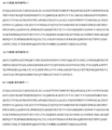
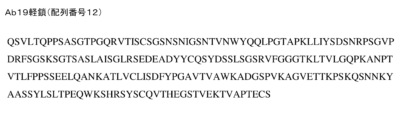
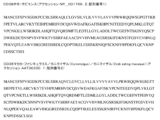


Classifications
-
- C—CHEMISTRY; METALLURGY
- C07—ORGANIC CHEMISTRY
- C07K—PEPTIDES
- C07K16/00—Immunoglobulins [IGs], e.g. monoclonal or polyclonal antibodies
- C07K16/18—Immunoglobulins [IGs], e.g. monoclonal or polyclonal antibodies against material from animals or humans
- C07K16/28—Immunoglobulins [IGs], e.g. monoclonal or polyclonal antibodies against material from animals or humans against receptors, cell surface antigens or cell surface determinants
- C07K16/2896—Immunoglobulins [IGs], e.g. monoclonal or polyclonal antibodies against material from animals or humans against receptors, cell surface antigens or cell surface determinants against molecules with a "CD"-designation, not provided for elsewhere
-
- A—HUMAN NECESSITIES
- A61—MEDICAL OR VETERINARY SCIENCE; HYGIENE
- A61P—SPECIFIC THERAPEUTIC ACTIVITY OF CHEMICAL COMPOUNDS OR MEDICINAL PREPARATIONS
- A61P1/00—Drugs for disorders of the alimentary tract or the digestive system
-
- A—HUMAN NECESSITIES
- A61—MEDICAL OR VETERINARY SCIENCE; HYGIENE
- A61P—SPECIFIC THERAPEUTIC ACTIVITY OF CHEMICAL COMPOUNDS OR MEDICINAL PREPARATIONS
- A61P1/00—Drugs for disorders of the alimentary tract or the digestive system
- A61P1/04—Drugs for disorders of the alimentary tract or the digestive system for ulcers, gastritis or reflux esophagitis, e.g. antacids, inhibitors of acid secretion, mucosal protectants
-
- A—HUMAN NECESSITIES
- A61—MEDICAL OR VETERINARY SCIENCE; HYGIENE
- A61P—SPECIFIC THERAPEUTIC ACTIVITY OF CHEMICAL COMPOUNDS OR MEDICINAL PREPARATIONS
- A61P19/00—Drugs for skeletal disorders
- A61P19/02—Drugs for skeletal disorders for joint disorders, e.g. arthritis, arthrosis
-
- A—HUMAN NECESSITIES
- A61—MEDICAL OR VETERINARY SCIENCE; HYGIENE
- A61P—SPECIFIC THERAPEUTIC ACTIVITY OF CHEMICAL COMPOUNDS OR MEDICINAL PREPARATIONS
- A61P21/00—Drugs for disorders of the muscular or neuromuscular system
- A61P21/04—Drugs for disorders of the muscular or neuromuscular system for myasthenia gravis
-
- A—HUMAN NECESSITIES
- A61—MEDICAL OR VETERINARY SCIENCE; HYGIENE
- A61P—SPECIFIC THERAPEUTIC ACTIVITY OF CHEMICAL COMPOUNDS OR MEDICINAL PREPARATIONS
- A61P25/00—Drugs for disorders of the nervous system
-
- A—HUMAN NECESSITIES
- A61—MEDICAL OR VETERINARY SCIENCE; HYGIENE
- A61P—SPECIFIC THERAPEUTIC ACTIVITY OF CHEMICAL COMPOUNDS OR MEDICINAL PREPARATIONS
- A61P37/00—Drugs for immunological or allergic disorders
- A61P37/02—Immunomodulators
-
- A—HUMAN NECESSITIES
- A61—MEDICAL OR VETERINARY SCIENCE; HYGIENE
- A61P—SPECIFIC THERAPEUTIC ACTIVITY OF CHEMICAL COMPOUNDS OR MEDICINAL PREPARATIONS
- A61P37/00—Drugs for immunological or allergic disorders
- A61P37/02—Immunomodulators
- A61P37/06—Immunosuppressants, e.g. drugs for graft rejection
-
- G—PHYSICS
- G01—MEASURING; TESTING
- G01N—INVESTIGATING OR ANALYSING MATERIALS BY DETERMINING THEIR CHEMICAL OR PHYSICAL PROPERTIES
- G01N33/00—Investigating or analysing materials by specific methods not covered by groups G01N1/00 - G01N31/00
- G01N33/48—Biological material, e.g. blood, urine; Haemocytometers
- G01N33/50—Chemical analysis of biological material, e.g. blood, urine; Testing involving biospecific ligand binding methods; Immunological testing
- G01N33/53—Immunoassay; Biospecific binding assay; Materials therefor
- G01N33/569—Immunoassay; Biospecific binding assay; Materials therefor for microorganisms, e.g. protozoa, bacteria, viruses
- G01N33/56966—Animal cells
-
- A—HUMAN NECESSITIES
- A61—MEDICAL OR VETERINARY SCIENCE; HYGIENE
- A61K—PREPARATIONS FOR MEDICAL, DENTAL OR TOILETRY PURPOSES
- A61K39/00—Medicinal preparations containing antigens or antibodies
- A61K2039/505—Medicinal preparations containing antigens or antibodies comprising antibodies
-
- A—HUMAN NECESSITIES
- A61—MEDICAL OR VETERINARY SCIENCE; HYGIENE
- A61K—PREPARATIONS FOR MEDICAL, DENTAL OR TOILETRY PURPOSES
- A61K39/00—Medicinal preparations containing antigens or antibodies
- A61K2039/505—Medicinal preparations containing antigens or antibodies comprising antibodies
- A61K2039/507—Comprising a combination of two or more separate antibodies
-
- C—CHEMISTRY; METALLURGY
- C07—ORGANIC CHEMISTRY
- C07K—PEPTIDES
- C07K2317/00—Immunoglobulins specific features
- C07K2317/20—Immunoglobulins specific features characterized by taxonomic origin
- C07K2317/21—Immunoglobulins specific features characterized by taxonomic origin from primates, e.g. man
-
- C—CHEMISTRY; METALLURGY
- C07—ORGANIC CHEMISTRY
- C07K—PEPTIDES
- C07K2317/00—Immunoglobulins specific features
- C07K2317/70—Immunoglobulins specific features characterized by effect upon binding to a cell or to an antigen
- C07K2317/73—Inducing cell death, e.g. apoptosis, necrosis or inhibition of cell proliferation
- C07K2317/732—Antibody-dependent cellular cytotoxicity [ADCC]
-
- C—CHEMISTRY; METALLURGY
- C07—ORGANIC CHEMISTRY
- C07K—PEPTIDES
- C07K2317/00—Immunoglobulins specific features
- C07K2317/70—Immunoglobulins specific features characterized by effect upon binding to a cell or to an antigen
- C07K2317/73—Inducing cell death, e.g. apoptosis, necrosis or inhibition of cell proliferation
- C07K2317/734—Complement-dependent cytotoxicity [CDC]
-
- C—CHEMISTRY; METALLURGY
- C07—ORGANIC CHEMISTRY
- C07K—PEPTIDES
- C07K2317/00—Immunoglobulins specific features
- C07K2317/90—Immunoglobulins specific features characterized by (pharmaco)kinetic aspects or by stability of the immunoglobulin
- C07K2317/92—Affinity (KD), association rate (Ka), dissociation rate (Kd) or EC50 value
-
- G—PHYSICS
- G01—MEASURING; TESTING
- G01N—INVESTIGATING OR ANALYSING MATERIALS BY DETERMINING THEIR CHEMICAL OR PHYSICAL PROPERTIES
- G01N2333/00—Assays involving biological materials from specific organisms or of a specific nature
- G01N2333/90—Enzymes; Proenzymes
- G01N2333/91—Transferases (2.)
- G01N2333/91091—Glycosyltransferases (2.4)
- G01N2333/91148—Glycosyltransferases (2.4) transferring other glycosyl groups (2.4.99)
Landscapes
- Health & Medical Sciences (AREA)
- Life Sciences & Earth Sciences (AREA)
- Chemical & Material Sciences (AREA)
- Immunology (AREA)
- Engineering & Computer Science (AREA)
- Organic Chemistry (AREA)
- General Health & Medical Sciences (AREA)
- Medicinal Chemistry (AREA)
- Bioinformatics & Cheminformatics (AREA)
- Pharmacology & Pharmacy (AREA)
- Nuclear Medicine, Radiotherapy & Molecular Imaging (AREA)
- Animal Behavior & Ethology (AREA)
- General Chemical & Material Sciences (AREA)
- Public Health (AREA)
- Veterinary Medicine (AREA)
- Chemical Kinetics & Catalysis (AREA)
- Molecular Biology (AREA)
- Biomedical Technology (AREA)
- Biochemistry (AREA)
- Orthopedic Medicine & Surgery (AREA)
- Physical Education & Sports Medicine (AREA)
- Hematology (AREA)
- Urology & Nephrology (AREA)
- Cell Biology (AREA)
- Neurology (AREA)
- Rheumatology (AREA)
- Biophysics (AREA)
- Genetics & Genomics (AREA)
- Proteomics, Peptides & Aminoacids (AREA)
- Biotechnology (AREA)
- General Physics & Mathematics (AREA)
- Virology (AREA)
- Zoology (AREA)
- Pathology (AREA)
- Microbiology (AREA)
- Tropical Medicine & Parasitology (AREA)
- Food Science & Technology (AREA)
- Physics & Mathematics (AREA)
- Analytical Chemistry (AREA)
- Neurosurgery (AREA)
Description
関連出願の相互参照
本出願は、2016年7月15日に出願された米国仮特許出願第62/362963号に対する米国特許法第119条(e)に基づく優先権を主張し、その開示全体が参照により本明細書に組み込まれる。
CROSS-REFERENCE TO RELATED APPLICATIONS This application claims priority under 35 U.S.C. incorporated herein by reference.
電子的に提出された資料の参照による取り込み
その全体が参照により本明細書に組み入れられるものは、本明細書と同時に提出されるコンピューター可読ヌクレオチド/アミノ酸配列表であり、2017年7月14日に作成された56キロバイトの「266608SeqListing.txt」という名前のACII(テキスト)ファイルとして同定される。
INCORPORATION BY REFERENCE OF MATERIAL SUBMITTED ELECTRONICALLY Incorporated herein by reference in its entirety is the Computer Readable Nucleotide/Amino Acid Sequence Listing filed concurrently herewith and dated July 14, 2017. Identified as an ACII (text) file named "266608SeqListing.txt" of 56 kilobytes created.
分野
本開示は、抗CD38抗体、並びに治療薬及び診断薬としてのそれらの使用に関する。本開示はさらに、全身性エリテマトーデス及び関節リウマチなどの自己免疫疾患を治療する方法に関する。本開示はさらに、治療のために自己免疫疾患を有する患者を同定するための診断アッセイ方法に関する。
FIELD This disclosure relates to anti-CD38 antibodies and their use as therapeutics and diagnostics. The disclosure further relates to methods of treating autoimmune diseases such as systemic lupus erythematosus and rheumatoid arthritis. The disclosure further relates to diagnostic assay methods for identifying patients with autoimmune diseases for treatment.
背景
形質細胞及び形質芽細胞は、全身性エリテマトーデス(SLE)及び関節リウマチ(RA)に関連する病原性過程にとって重要である抗体分泌細胞(ASC)である。それらは、重症筋無力症、シェーグレン症候群、多発性硬化症(MS)及び自己免疫性甲状腺炎を含む多数の抗体駆動自己免疫疾患に関与している。Jacobi AM,Mei H,Hoyer BF,et al.,Ann Rheum Dis(2010)69(1):305-8;Dorner T,Isenberg D,Jayne D,et al.,International Roundtable on B cells as Therapeutic Target for Intervention,Autoimmun Rev (2009)9(2):82-9;Tipton CM,Fucile CF,Darce J,et al.,Nat Immunol(2015)16(7):755-65;及びCepok S,Rosche B,Grummel V,et al.,Brain(2005)128(Pt 7):1667-76を参照。形質芽細胞は急速に産生される最終分化B細胞であり、大多数は短命のエフェクター細胞になり、少数は長命の形質細胞になる。
BACKGROUND Plasma cells and plasmablasts are antibody-secreting cells (ASC) that are important for the pathogenic processes associated with systemic lupus erythematosus (SLE) and rheumatoid arthritis (RA). They are implicated in numerous antibody-driven autoimmune diseases, including myasthenia gravis, Sjögren's syndrome, multiple sclerosis (MS) and autoimmune thyroiditis. Jacobi AM, Mei H, Hoyer BF, et al. , Ann Rheum Dis (2010) 69(1):305-8; Dorner T, Isenberg D, Jayne D, et al. , International Roundtable on B cells as Therapeutic Target for Intervention, Autoimmune Rev (2009) 9(2):82-9; Tipton CM, Facility CF, Darce J, et al. , Nat Immunol (2015) 16(7):755-65; and Cepok S, Rosche B, Grummel V, et al. , Brain (2005) 128(Pt 7):1667-76. Plasmablasts are rapidly produced, terminally differentiated B cells, the majority of which become short-lived effector cells and a minority of which become long-lived plasma cells.
ほとんどのB細胞はCD20を発現し、この細胞表面タンパク質に対する抗体治療薬によって効果的かつ永続的に枯渇される。Silverman GJ,Arthritis Rheum(2006)54(8):2356-67を参照のこと。しかしながら、形質芽細胞及び形質細胞などのいくつかのB系統細胞は、相当量のCD20を発現せず、直接的なCD20枯渇によって効果的には減少しない。Silverman 2006を参照のこと。したがって、この細胞集団に影響を与えるCD20特異的薬物の能力は、既に形成された形質芽細胞及び形質細胞にほとんど影響を与えずに最終的に形質芽細胞に分化する可能性があるCD20+B細胞集団を枯渇させることに限定される。Silverman GJ and Boyle DL,Immunol Rev(2008)223:175-85を参照のこと。 Most B cells express CD20 and are effectively and permanently depleted by antibody therapeutics against this cell surface protein. See Silverman GJ, Arthritis Rheum (2006) 54(8):2356-67. However, some B-lineage cells, such as plasmablasts and plasma cells, do not express significant amounts of CD20 and are not effectively reduced by direct CD20 depletion. See Silverman 2006. Thus, the ability of CD20-specific drugs to affect this cell population is a CD20+ B cell population that may ultimately differentiate into plasmablasts with little effect on already formed plasmablasts and plasma cells. limited to depleting See Silverman GJ and Boyle DL, Immunol Rev (2008) 223:175-85.
SLE患者に関する臨床研究は、抗CD20治療前の高レベルの形質芽細胞が血中の非効率的なB細胞枯渇と相関することを示した。加えて、血中の形質芽細胞の不完全な枯渇は、より速い再発及び生存の減少を含む臨床結果をもたらす可能性がある。Vital EM,Rawstron AC,Dass S,et al.,Arthritis Rheum(2011)63(3):603-8を参照のこと。抗CD20モノクローナル抗体で治療したRA患者についても同様の知見が実証されている。Dass S,Rawstron AC,Vital EM,et al.,Arthritis Rheum(2008)58(10):2993-9;及びOwczarczyk K,Lal P,Abbas AR,et al.,Sci Transl Med(2011)3(101):101ra92を参照のこと。 Clinical studies on SLE patients have shown that high levels of plasmablasts prior to anti-CD20 treatment correlate with inefficient B-cell depletion in the blood. In addition, incomplete depletion of plasmablasts in the blood can have clinical consequences including faster relapse and decreased survival. Vital EM, Rawstron AC, Dass S, et al. , Arthritis Rheum (2011) 63(3):603-8. Similar findings have been demonstrated for RA patients treated with anti-CD20 monoclonal antibodies. Dass S, Rawstron AC, Vital EM, et al. , Arthritis Rheum (2008) 58(10):2993-9; and Owczarczyk K, Lal P, Abbas AR, et al. , Sci Transl Med (2011) 3(101):101ra92.
これらの問題に取り組むために、これらの細胞型において主に発現されるmRNA転写物を評価することによって血中の形質芽細胞及び形質細胞レベルを間接的にモニターするための方法が開発された。Vital EM,Dass S,Buch MH,et al.,Arthritis Rheum(2011)63(10):3038-47;Streicher K,Morehouse CA,Groves CJ,et al.,Arthritis Rheumatol(2014)66(1):173-84;及びOwczarczyk,2011を参照。この方法を用いて、CD20非応答者は、形質芽細胞及び形質細胞において豊富な高レベルのIgJ(免疫グロブリンJ鎖)を有し、CD20+非形質芽細胞で発現される低レベルのFCRL5(Fc受容体様5タンパク質)を有する患者として遡及的に同定された。 To address these issues, methods were developed to indirectly monitor blood plasmablast and plasma cell levels by assessing the mRNA transcripts that are predominantly expressed in these cell types. Vital EM, Dass S, Buch MH, et al. , Arthritis Rheum (2011) 63(10):3038-47; Streicher K, Morehouse CA, Groves CJ, et al. , Arthritis Rheumatol (2014) 66(1):173-84; and Owczarczyk, 2011. Using this method, CD20 non-responders have high levels of IgJ (immunoglobulin J chain) abundant in plasmablasts and plasma cells and low levels of FCRL5 (Fc) expressed in CD20+ non-plasmablasts. was retrospectively identified as a patient with receptor-like 5 protein).
この間接的なアプローチは単純で、ほとんどの臨床状況で利用することができる。血液サンプルは、一般に入手可能なサンプル管(例えば、Paxgene管)を使用して収集され、適切に貯蔵される場合には長い安定性を有する。しかし、全血液中の転写産物解析では、形質芽細胞と形質細胞の数を直接定量することは容易ではなく、この細胞集団で評価された転写産物の排他性によっては、他の細胞型の変化が結果に影響を与える可能性がある。 This indirect approach is simple and can be used in most clinical situations. Blood samples are collected using commonly available sample tubes (eg, Paxgene tubes) and have long stability if properly stored. However, transcript analysis in whole blood does not facilitate direct quantification of the number of plasmablasts and plasma cells, and the exclusivity of transcripts evaluated in this cell population suggests that changes in other cell types may not be possible. May affect results.
B細胞に影響を与える治療の前又はそれに応答した形質芽細胞及び形質細胞のレベルの直接測定は、この問題を回避するのに役立つはずである。しかしながら、形質芽細胞の生存能は、静脈穿刺後に急速に低下する。現在の方法論を使用して、形質芽細胞レベルをモニターすることは同日処理を必要とし、それは異なる臨床現場の間で標準化することは困難である。 Direct measurement of plasmablast and plasma cell levels prior to or in response to treatments that affect B cells should help circumvent this problem. However, plasmablast viability declines rapidly after venipuncture. Using current methodologies, monitoring plasmablast levels requires same-day treatment, which is difficult to standardize between different clinical settings.
形質芽細胞及び形質細胞は相当量のCD20を発現しないが、これらの細胞はNK細胞と共に、活性化時にCD38発現を上方制御するB細胞及びT細胞と同様に高レベルのCD38を恒常的に発現する。Malavasi F,Deaglio S,Funaro A,et al.,Physiol Rev(2008)88(3):841-86を参照のこと。環状ADPリボースヒドロラーゼとしても知られるCD38は、長いC末端細胞外ドメイン及び短いN末端細胞質ドメインを有するII型膜貫通糖タンパク質である。CD38は、CD157とAplysia ADPRシクラーゼを含む、関連する膜結合型又は可溶性酵素のグループのメンバーである。このファミリーの酵素は、NADを環状ADPリボース又はニコチン酸(nictotinic acid)-アデニンジヌクレオチドホスフェートに変換する固有の能力を有する。 Plasmablasts and plasma cells do not express significant amounts of CD20, but these cells, along with NK cells, constitutively express high levels of CD38, as do B and T cells that upregulate CD38 expression upon activation. do. Malavasi F, Deaglio S, Funaro A, et al. , Physiol Rev (2008) 88(3):841-86. CD38, also known as cyclic ADP ribose hydrolase, is a type II transmembrane glycoprotein with a long C-terminal extracellular domain and a short N-terminal cytoplasmic domain. CD38 is a member of a group of related membrane-bound or soluble enzymes that include CD157 and Aplysia ADPR cyclases. This family of enzymes has the inherent ability to convert NAD to cyclic ADP ribose or nictotinic acid-adenine dinucleotide phosphate.
ヒトにおける最近の研究は、抗CD38mAbが形質芽細胞及び形質細胞を含む複数の血液細胞型を枯渇させ得ることを示している。 Recent studies in humans show that anti-CD38 mAb can deplete multiple blood cell types, including plasmablasts and plasma cells.
発明の要旨 gist of the invention
抗CD38抗体は、治療前又は治療中の形質芽細胞及び/又は形質細胞の測定と組み合わせると、CD20に基づく治療法や、形質芽細胞及び/又は形質細胞を持続的に枯渇させない他の治療法では、疾患の寛解に達することができない関節リウマチ、全身性エリテマトーデス又は他の自己免疫疾患を患っている患者を治療するために使用され得る。 Anti-CD38 antibodies may be used in combination with plasmablast and/or plasma cell measurements prior to or during therapy, CD20-based therapies and other therapies that do not persistently deplete plasmablasts and/or plasma cells. can be used to treat patients suffering from rheumatoid arthritis, systemic lupus erythematosus or other autoimmune diseases who are unable to reach remission of the disease.
本発明の一態様は、患者の疾患を治療する方法を提供し、該方法は治療上有効量の抗CD38抗体を患者に投与することを含み、ここで、抗CD38抗体は、ヒトCD38(配列番号1)に特異的に結合する単離された抗体であり、疾患は自己免疫疾患であり、患者は抗CD38抗体による治療の前に、コントロール対象と比較して、CD38発現細胞の上昇したレベルを有することが示された。 One aspect of the invention provides a method of treating a disease in a patient, the method comprising administering to the patient a therapeutically effective amount of an anti-CD38 antibody, wherein the anti-CD38 antibody is human CD38 (sequence No. 1), wherein the disease is an autoimmune disease and the patient has elevated levels of CD38-expressing cells compared to a control subject prior to treatment with the anti-CD38 antibody. was shown to have
本発明の別の態様は、患者の疾患を治療する方法を提供し、該方法は治療上有効量の抗CD38抗体を患者に投与することを含み、ここで、抗CD38抗体は、ヒトCD38(配列番号1)に特異的に結合する単離された抗体であり、疾患は自己免疫疾患であり、患者は抗CD20抗体による治療の後で、コントロール対象と比較して、CD38発現細胞の上昇したレベルを有することが示された。 Another aspect of the invention provides a method of treating a disease in a patient, the method comprising administering to the patient a therapeutically effective amount of an anti-CD38 antibody, wherein the anti-CD38 antibody is human CD38 ( 9. An isolated antibody that specifically binds to SEQ ID NO: 1), wherein the disease is an autoimmune disease and patients have elevated CD38-expressing cells after treatment with an anti-CD20 antibody compared to control subjects. It was shown to have a level
本発明のさらなる態様は、患者の疾患を治療する方法を提供し、該方法は治療上有効量の抗CD38抗体を患者に投与することを含み、ここで、抗CD38抗体は、ヒトCD38(配列番号1)に特異的に結合する単離された抗体であり、疾患は自己免疫疾患であり、抗CD38抗体による治療の前に患者から得られた生物学的サンプルは、コントロール対象と比較して、CD38発現細胞の上昇したレベルを有することが示された。 A further aspect of the invention provides a method of treating a disease in a patient, the method comprising administering to the patient a therapeutically effective amount of an anti-CD38 antibody, wherein the anti-CD38 antibody is human CD38 (sequence number 1), wherein the disease is an autoimmune disease, and wherein a biological sample obtained from the patient prior to treatment with the anti-CD38 antibody compares to a control subject , were shown to have elevated levels of CD38-expressing cells.
本発明のさらなる態様は、患者の疾患を治療する方法を提供し、該方法は治療上有効量の抗CD38抗体を患者に投与することを含み、ここで、抗CD38抗体は、ヒトCD38(配列番号1)に特異的に結合する単離された抗体であり、疾患は自己免疫疾患であり、抗CD20抗体による治療の後で患者から得られた生物学的サンプルは、コントロール対象と比較して、CD38発現細胞の上昇したレベルを有することが示された。 A further aspect of the invention provides a method of treating a disease in a patient, the method comprising administering to the patient a therapeutically effective amount of an anti-CD38 antibody, wherein the anti-CD38 antibody is human CD38 (sequence number 1), wherein the disease is an autoimmune disease, wherein a biological sample obtained from the patient after treatment with the anti-CD20 antibody compares to a control subject , were shown to have elevated levels of CD38-expressing cells.
本発明の別の態様は、患者の疾患を治療する方法を提供し、該方法は治療上有効量の抗CD38抗体を患者に投与することを含み、ここで、抗CD38抗体は、ヒトCD38(配列番号1)に特異的に結合する単離された抗体であり、疾患は自己免疫疾患であり、抗CD38抗体による治療の前に患者から得られた生物学的サンプルは、遊離Ig軽鎖についてアッセイすることによって、コントロール対象と比較して、形質細胞及び形質芽細胞の上昇したレベルを有することが示された。 Another aspect of the invention provides a method of treating a disease in a patient, the method comprising administering to the patient a therapeutically effective amount of an anti-CD38 antibody, wherein the anti-CD38 antibody is human CD38 ( An isolated antibody that specifically binds to SEQ ID NO: 1), wherein the disease is an autoimmune disease, wherein a biological sample obtained from the patient prior to treatment with an anti-CD38 antibody is characterized for free Ig light chain Assays showed elevated levels of plasma cells and plasma blasts compared to control subjects.
本発明のさらなる態様は、患者の疾患を治療する方法を提供し、該方法は治療上有効量の抗CD38抗体を患者に投与することを含み、ここで、抗CD38抗体は、ヒトCD38(配列番号1)に特異的に結合する単離された抗体であり、疾患は自己免疫疾患であり、抗CD20抗体による治療の後で患者から得られた生物学的サンプルは、遊離Ig軽鎖についてアッセイすることによって、コントロール対象と比較して、形質細胞及び形質芽細胞の上昇したレベルを有することが示された。 A further aspect of the invention provides a method of treating a disease in a patient, the method comprising administering to the patient a therapeutically effective amount of an anti-CD38 antibody, wherein the anti-CD38 antibody is human CD38 (sequence No. 1), wherein the disease is an autoimmune disease, wherein a biological sample obtained from the patient after treatment with an anti-CD20 antibody is assayed for free Ig light chain. were shown to have elevated levels of plasma cells and plasmablasts compared to control subjects.
本発明のさらに別の態様は、患者の疾患を治療する方法を提供し、該方法は治療上有効量の抗CD38抗体を患者に投与することを含み、ここで、抗CD38抗体は、ヒトCD38(配列番号1)に特異的に結合する単離された抗体であり、疾患は自己免疫疾患であり、抗CD38抗体による治療の前に患者から得られた生物学的サンプルは、コントロール対象と比較して、特定のmRNA又は(mRNAor)mRNA(複数)のグループのCD38発現細胞における(of in)上昇したレベルを有することが示された。
Yet another aspect of the invention provides a method of treating a disease in a patient, the method comprising administering to the patient a therapeutically effective amount of an anti-CD38 antibody, wherein the anti-CD38 antibody is
本発明のさらなる態様は、患者の疾患を治療する方法を提供し、該方法は治療上有効量の抗CD38抗体を患者に投与することを含み、ここで、抗CD38抗体は、ヒトCD38(配列番号1)に特異的に結合する単離された抗体であり、疾患は自己免疫疾患であり、抗CD20抗体による治療の後で患者から得られた生物学的サンプルは、コントロール対象と比較して、総CD38発現細胞mRNAの上昇したレベルを有することが示された。 A further aspect of the invention provides a method of treating a disease in a patient, the method comprising administering to the patient a therapeutically effective amount of an anti-CD38 antibody, wherein the anti-CD38 antibody is human CD38 (sequence number 1), wherein the disease is an autoimmune disease, wherein a biological sample obtained from the patient after treatment with the anti-CD20 antibody compares to a control subject , were shown to have elevated levels of total CD38-expressing cell mRNA.
本発明のさらなる態様は、患者の疾患を治療する方法を提供し、該方法は治療上有効量の第1の抗CD38抗体を患者に投与することを含み、ここで、第1の抗CD38抗体は、ヒトCD38(配列番号1)に特異的に結合する単離された抗体であり、疾患は自己免疫疾患であり、抗CD38抗体による治療の前に患者から得られた生物学的サンプルは、フローサイトメトリーによって、コントロール対象と比較して、CD38発現細胞の上昇したレベルを有することが示された。 A further aspect of the invention provides a method of treating a disease in a patient, the method comprising administering to the patient a therapeutically effective amount of a first anti-CD38 antibody, wherein the first anti-CD38 antibody is an isolated antibody that specifically binds to human CD38 (SEQ ID NO: 1), the disease is an autoimmune disease, and the biological sample obtained from the patient prior to treatment with the anti-CD38 antibody comprises Flow cytometry showed that they had elevated levels of CD38-expressing cells compared to control subjects.
本発明の別の態様は、患者の疾患を治療する方法を提供し、該方法は治療上有効量の第1の抗CD38抗体を患者に投与することを含み、ここで、第1の抗CD38抗体は、ヒトCD38(配列番号1)に特異的に結合する単離された抗体であり、疾患は自己免疫疾患であり、抗CD20抗体による治療の後で患者から得られた生物学的サンプルは、フローサイトメトリーによって、コントロール対象と比較して、CD38発現細胞の上昇したレベルを有することが示された。 Another aspect of the invention provides a method of treating a disease in a patient, the method comprising administering to the patient a therapeutically effective amount of a first anti-CD38 antibody, wherein the first anti-CD38 The antibody is an isolated antibody that specifically binds to human CD38 (SEQ ID NO: 1), the disease is an autoimmune disease, and the biological sample obtained from the patient after treatment with an anti-CD20 antibody is , were shown by flow cytometry to have elevated levels of CD38-expressing cells compared to control subjects.
本発明のさらなる態様は、全血液中のCD38発現細胞をアッセイするための方法を提供し、該方法は、
a)対象から全血液のサンプルを得ること(ここで、サンプルは、赤血球、白血球、及び抗凝固剤を含む);
b)赤血球を溶解して、処理されたサンプルを形成すること;
c)サンプルを得てから約24時間以内に処理したサンプルを約-20℃以下の温度に凍結して凍結サンプルを形成すること;
d)サンプルを得てから約72時間以内にサンプル中のCD38発現細胞の量を測定すること(ここで、CD38発現細胞の量を測定する前に、凍結サンプルを室温まで温める)
を含む。
A further aspect of the invention provides a method for assaying CD38-expressing cells in whole blood, the method comprising:
a) obtaining a sample of whole blood from a subject (wherein the sample comprises red blood cells, white blood cells, and an anticoagulant);
b) lysing red blood cells to form a processed sample;
c) freezing the treated sample to a temperature of about −20° C. or less within about 24 hours of obtaining the sample to form a frozen sample;
d) measuring the amount of CD38-expressing cells in the sample within about 72 hours of obtaining the sample (wherein the frozen sample is warmed to room temperature before measuring the amount of CD38-expressing cells);
including.
本明細書に提供されるのは、CD38への結合のための試薬及び方法、並びに抗CD38抗体を含むCD38特異的結合剤を用いてCD38関連疾患を治療するため、及びCD38を検出するための方法である。 Provided herein are reagents and methods for binding to CD38, and for treating CD38-related diseases using CD38-specific binding agents, including anti-CD38 antibodies, and for detecting CD38. The method.
したがって、いくつかの実施態様において、ヒトCD38(配列番号1)及びカニクイザルCD38(配列番号2)に特異的な単離された抗体は、本発明の様々な態様に関連した使用のために記載されている。いくつかの実施態様において、本明細書に記載の単離された抗体は、重鎖可変領域及び軽鎖可変領域から構成され得、ここで、重鎖可変領域は、HCDR1、HCDR2、及びHCDR3として本明細書に記載されている3つの相補性決定領域(CDR)を含み、軽鎖可変領域は、LCDR1、LCDR2、及びLCDR3として本明細書に記載される3つのCDRを含む。CDRの配列は、HCDR1(配列番号3)、HCDR2(配列番号4)、HCDR3(配列番号5)、LCDR1(配列番号6)、LCDR2(配列番号7)及びLCDR3(配列番号8)によって表される。 Thus, in some embodiments, isolated antibodies specific for human CD38 (SEQ ID NO: 1) and cynomolgus monkey CD38 (SEQ ID NO: 2) are described for use in connection with various aspects of the present invention. ing. In some embodiments, the isolated antibodies described herein can be composed of a heavy chain variable region and a light chain variable region, wherein the heavy chain variable regions are designated as HCDR1, HCDR2, and HCDR3. Comprising three complementarity determining regions (CDRs) described herein, the light chain variable region comprises three CDRs described herein as LCDR1, LCDR2, and LCDR3. The CDR sequences are represented by HCDR1 (SEQ ID NO:3), HCDR2 (SEQ ID NO:4), HCDR3 (SEQ ID NO:5), LCDR1 (SEQ ID NO:6), LCDR2 (SEQ ID NO:7) and LCDR3 (SEQ ID NO:8). .
他の実施態様において、単離された抗体は重鎖可変領域を含むことができ、ここで、重鎖可変領域の配列は配列番号9を含む。他の実施態様において、単離された抗体は軽鎖可変領域を含むことができ、ここで、軽鎖可変領域の配列は配列番号10を含む。他の実施態様において、重鎖可変領域は配列番号9を含み、軽鎖可変領域は配列番号10を含む。 In other embodiments, the isolated antibody can comprise a heavy chain variable region, wherein the heavy chain variable region sequence comprises SEQ ID NO:9. In other embodiments, the isolated antibody can comprise a light chain variable region, wherein the light chain variable region sequence comprises SEQ ID NO:10. In another embodiment, the heavy chain variable region comprises SEQ ID NO:9 and the light chain variable region comprises SEQ ID NO:10.
いくつかの実施態様において、単離された抗体は重鎖可変領域を含むことができ、ここで重鎖可変領域の配列は配列番号21を含む。他の実施態様において、単離された抗体は軽鎖可変領域を含むことができ、ここで軽鎖可変領域の配列は配列番号22を含む。他の実施態様において、重鎖可変領域は配列番号21を含み、軽鎖可変領域は配列番号22を含む。重鎖可変領域と軽鎖可変領域のこの組合せは、Ab79と呼ばれる。いくつかの実施態様において、単離された抗体はFcドメインを含む。他の実施態様において、FcドメインはヒトFcドメインである。さらに他の実施態様において、Fcドメインは変異型Fcドメインである。 In some embodiments, the isolated antibody can comprise a heavy chain variable region, wherein the heavy chain variable region sequence comprises SEQ ID NO:21. In other embodiments, the isolated antibody can comprise a light chain variable region, wherein the light chain variable region sequence comprises SEQ ID NO:22. In another embodiment, the heavy chain variable region comprises SEQ ID NO:21 and the light chain variable region comprises SEQ ID NO:22. This combination of heavy and light chain variable regions is called Ab79. In some embodiments, the isolated antibody comprises an Fc domain. In other embodiments, the Fc domain is a human Fc domain. In still other embodiments, the Fc domain is a variant Fc domain.
いくつかの実施態様において、ヒトCD38(配列番号1)及びカニクイザルCD38(配列番号2)に特異的な単離された抗体は、本発明の様々な態様に関連した使用のために記載されている。いくつかの実施態様において、本明細書に記載の単離された抗体は6つのCDRから構成され得、ここで、該抗体の各CDRは配列番号3、配列番号4、配列番号5、配列番号6、配列番号7、及び配列番号8と、0、1個、又は2個のアミノ酸置換によって異なり得る。 In some embodiments, isolated antibodies specific for human CD38 (SEQ ID NO: 1) and cynomolgus monkey CD38 (SEQ ID NO: 2) are described for use in connection with various aspects of the present invention. . In some embodiments, the isolated antibodies described herein can be composed of 6 CDRs, wherein each CDR of the antibody is SEQ ID NO: 3, SEQ ID NO: 4, SEQ ID NO: 5, SEQ ID NO: 6, SEQ ID NO:7, and SEQ ID NO:8 by 0, 1, or 2 amino acid substitutions.
他の実施態様において、ヒトCD38(配列番号1)及びカニクイザルCD38(配列番号2)に特異的な単離された抗体が、本発明の様々な態様に関連した使用のために記載されている。本明細書に記載の単離された抗体は、重鎖可変領域及び軽鎖可変領域から構成され得、ここで、重鎖可変領域は、HCDR1、HCDR2、及びHCDR3として本明細書に記載される3つの相補性決定領域(CDR)を含み、軽鎖可変領域は、LCDR1、LCDR2、及びLCDR3として本明細書に記載される3つのCDRを含む。CDRの配列は、HCDR1(配列番号13)、HCDR2(配列番号14)、HCDR3(配列番号15)、LCDR1(配列番号16)、LCDR2(配列番号17)及びLCDR3(配列番号18)によって表される。 In other embodiments, isolated antibodies specific for human CD38 (SEQ ID NO: 1) and cynomolgus monkey CD38 (SEQ ID NO: 2) are described for use in connection with various aspects of the present invention. The isolated antibodies described herein can be composed of a heavy chain variable region and a light chain variable region, where the heavy chain variable regions are described herein as HCDR1, HCDR2, and HCDR3. Containing three complementarity determining regions (CDRs), the light chain variable region contains three CDRs described herein as LCDR1, LCDR2, and LCDR3. The CDR sequences are represented by HCDR1 (SEQ ID NO: 13), HCDR2 (SEQ ID NO: 14), HCDR3 (SEQ ID NO: 15), LCDRl (SEQ ID NO: 16), LCDR2 (SEQ ID NO: 17) and LCDR3 (SEQ ID NO: 18). .
いくつかの実施態様において、単離された抗体は重鎖可変領域を含むことができ、ここで重鎖可変領域の配列は配列番号11を含む。他の実施態様において、単離された抗体は軽鎖可変領域を含むことができ、ここで軽鎖可変領域の配列は配列番号12を含む。いくつかの実施態様において、単離された抗体は重鎖及び軽鎖を含むことができ、ここで、重鎖配列は配列番号11を含み、軽鎖は配列番号12を含む。 In some embodiments, the isolated antibody can comprise a heavy chain variable region, wherein the heavy chain variable region sequence comprises SEQ ID NO:11. In other embodiments, the isolated antibody can comprise a light chain variable region, wherein the light chain variable region sequence comprises SEQ ID NO:12. In some embodiments, an isolated antibody can comprise a heavy chain and a light chain, wherein the heavy chain sequence comprises SEQ ID NO:11 and the light chain comprises SEQ ID NO:12.
他の実施態様において、単離された抗体は重鎖可変領域を含むことができ、ここで重鎖可変領域の配列は配列番号19を含む。他の実施態様において、単離された抗体は軽鎖可変領域を含むことができ、ここで軽鎖可変領域の配列は配列番号20を含む。他の実施態様において、重鎖可変領域は配列番号19を含み、軽鎖可変領域は配列番号20を含む。重鎖可変領域と軽鎖可変領域のこの組合せは、Ab19と呼ばれる。 In other embodiments, the isolated antibody can comprise a heavy chain variable region, wherein the heavy chain variable region sequence comprises SEQ ID NO:19. In other embodiments, the isolated antibody can comprise a light chain variable region, wherein the light chain variable region sequence comprises SEQ ID NO:20. In another embodiment, the heavy chain variable region comprises SEQ ID NO:19 and the light chain variable region comprises SEQ ID NO:20. This combination of heavy and light chain variable regions is called Ab19.
他の実施態様において、ヒトCD38(配列番号1)及びカニクイザルCD38(配列番号2)に特異的な単離された抗体が、本発明の様々な態様に関連した使用のために記載されている。本明細書に記載の単離された抗体は6つのCDRから構成され得、ここで、該抗体の各CDRは配列番号13、配列番号14、配列番号15、配列番号16、配列番号17、及び配列番号18と、0、1個、又は2個のアミノ酸置換によって異なり得る。 In other embodiments, isolated antibodies specific for human CD38 (SEQ ID NO: 1) and cynomolgus monkey CD38 (SEQ ID NO: 2) are described for use in connection with various aspects of the present invention. The isolated antibodies described herein can be composed of six CDRs, wherein each CDR of the antibody is SEQ ID NO: 13, SEQ ID NO: 14, SEQ ID NO: 15, SEQ ID NO: 16, SEQ ID NO: 17, and It may differ from SEQ ID NO: 18 by 0, 1, or 2 amino acid substitutions.
いくつかの実施態様において、ヒトCD38(配列番号1)及びカニクイザルCD38(配列番号2)に特異的に結合する単離された抗CD38抗体が提供され、ここで、該抗体は約10-6、10-7、10-8、10-9又はそれ以上のKDでヒトCD38に結合し、約10-6、10-7、10-8、10-9又はそれ以上のKDでカニクイザルCD38に結合する。 In some embodiments, an isolated anti-CD38 antibody that specifically binds to human CD38 (SEQ ID NO: 1) and cynomolgus CD38 (SEQ ID NO: 2) is provided, wherein the antibody is about 10 −6 , Binds human CD38 with a KD of 10 −7 , 10 −8 , 10 −9 or greater and binds cynomolgus monkey CD38 with a KD of about 10 −6 , 10 −7 , 10 −8 , 10 −9 or greater .
いくつかの実施態様において、ヒトCD38及び/又はカニクイザルCD38への結合についてAb79及び/又はAb19と競合する抗体が提供される。 In some embodiments, antibodies are provided that compete with Ab79 and/or Ab19 for binding to human CD38 and/or cynomolgus monkey CD38.
本開示の方法は、以下の列挙された節のいずれかにおける実施態様として説明され得る。本明細書に記載の任意の実施態様は、組み合わされた実施態様が互いに矛盾しない限り、本明細書に記載の任意の他の実施態様と関連して使用され得ることが理解されよう。 The methods of the disclosure may be described as embodiments in any of the following enumerated sections. It will be appreciated that any embodiment described herein can be used in conjunction with any other embodiment described herein so long as the combined embodiments are not mutually exclusive.
1.治療上有効量の抗CD38抗体を患者に投与することを含む、患者の疾患を治療する方法であって、ここで、抗CD38抗体は、ヒトCD38(配列番号1)に特異的に結合する単離された抗体であり、疾患は自己免疫疾患であり、患者は抗CD38抗体による治療の前に、コントロール対象と比較して、CD38発現細胞の上昇したレベルを有することが示された、方法。 1. A method of treating a disease in a patient comprising administering to the patient a therapeutically effective amount of an anti-CD38 antibody, wherein the anti-CD38 antibody is a single antibody that specifically binds to human CD38 (SEQ ID NO: 1). wherein the antibody is released, the disease is an autoimmune disease, and the patient is shown to have elevated levels of CD38-expressing cells compared to control subjects prior to treatment with an anti-CD38 antibody.
2.治療上有効量の抗CD38抗体を患者に投与することを含む、患者の疾患を治療する方法であって、ここで、抗CD38抗体は、ヒトCD38(配列番号1)に特異的に結合する単離された抗体であり、疾患は自己免疫疾患であり、患者は抗CD20抗体による治療の後で、コントロール対象と比較して、CD38発現細胞の上昇したレベルを有することが示された、方法。 2. A method of treating a disease in a patient comprising administering to the patient a therapeutically effective amount of an anti-CD38 antibody, wherein the anti-CD38 antibody is a single antibody that specifically binds to human CD38 (SEQ ID NO: 1). wherein the antibody is released, the disease is an autoimmune disease, and the patient is shown to have elevated levels of CD38-expressing cells after treatment with an anti-CD20 antibody compared to control subjects.
3.治療上有効量の抗CD38抗体を患者に投与することを含む、患者の疾患を治療する方法であって、ここで、抗CD38抗体は、ヒトCD38(配列番号1)に特異的に結合する単離された抗体であり、疾患は自己免疫疾患であり、抗CD38抗体による治療の前に患者から得られた生物学的サンプルは、コントロール対象と比較して、CD38発現細胞の上昇したレベルを有することが示された、方法。 3. A method of treating a disease in a patient comprising administering to the patient a therapeutically effective amount of an anti-CD38 antibody, wherein the anti-CD38 antibody is a single antibody that specifically binds to human CD38 (SEQ ID NO: 1). the antibody is isolated, the disease is an autoimmune disease, and a biological sample obtained from the patient prior to treatment with an anti-CD38 antibody has elevated levels of CD38-expressing cells compared to control subjects It was shown, how.
4.治療上有効量の抗CD38抗体を患者に投与することを含む、患者の疾患を治療する方法であって、ここで、抗CD38抗体は、ヒトCD38(配列番号1)に特異的に結合する単離された抗体であり、疾患は自己免疫疾患であり、抗CD20抗体による治療の後で患者から得られた生物学的サンプルは、コントロール対象と比較して、CD38発現細胞の上昇したレベルを有することが示された、方法。 4. A method of treating a disease in a patient comprising administering to the patient a therapeutically effective amount of an anti-CD38 antibody, wherein the anti-CD38 antibody is a single antibody that specifically binds to human CD38 (SEQ ID NO: 1). the antibody is released, the disease is an autoimmune disease, and a biological sample obtained from the patient after treatment with an anti-CD20 antibody has elevated levels of CD38-expressing cells compared to control subjects It was shown, how.
5.治療上有効量の抗CD38抗体を患者に投与することを含む、患者の疾患を治療する方法であって、ここで、抗CD38抗体は、ヒトCD38(配列番号1)に特異的に結合する単離された抗体であり、疾患は自己免疫疾患であり、抗CD38抗体による治療の前に患者から得られた生物学的サンプルは、遊離Ig軽鎖についてアッセイすることによって、コントロール対象と比較して、CD38発現形質芽細胞及びCD38発現形質細胞の上昇したレベルを有することが示された、方法。 5. A method of treating a disease in a patient comprising administering to the patient a therapeutically effective amount of an anti-CD38 antibody, wherein the anti-CD38 antibody is a single antibody that specifically binds to human CD38 (SEQ ID NO: 1). the antibody is released, the disease is an autoimmune disease, and the biological sample obtained from the patient prior to treatment with the anti-CD38 antibody is assayed for free Ig light chain, compared to control subjects , shown to have elevated levels of CD38-expressing plasmablasts and CD38-expressing plasma cells.
6.治療上有効量の抗CD38抗体を患者に投与することを含む、患者の疾患を治療する方法であって、ここで、抗CD38抗体は、ヒトCD38(配列番号1)に特異的に結合する単離された抗体であり、疾患は自己免疫疾患であり、抗CD20抗体による治療の後で患者から得られた生物学的サンプルは、遊離Ig軽鎖についてアッセイすることによって、コントロール対象と比較して、CD38発現細胞の上昇したレベルを有することが示された、方法。 6. A method of treating a disease in a patient comprising administering to the patient a therapeutically effective amount of an anti-CD38 antibody, wherein the anti-CD38 antibody is a single antibody that specifically binds to human CD38 (SEQ ID NO: 1). the antibody is released, the disease is an autoimmune disease, and the biological sample obtained from the patient after treatment with the anti-CD20 antibody is compared to control subjects by assaying for free Ig light chain , shown to have elevated levels of CD38-expressing cells.
7.治療上有効量の抗CD38抗体を患者に投与することを含む、患者の疾患を治療する方法であって、ここで、抗CD38抗体は、ヒトCD38(配列番号1)に特異的に結合する単離された抗体であり、疾患は自己免疫疾患であり、抗CD38抗体による治療の前に患者から得られた生物学的サンプルは、コントロール対象と比較して、CD38発現細胞に豊富な少なくとも1つの遺伝子の上昇したレベルを有することが示された、方法。 7. A method of treating a disease in a patient comprising administering to the patient a therapeutically effective amount of an anti-CD38 antibody, wherein the anti-CD38 antibody is a single antibody that specifically binds to human CD38 (SEQ ID NO: 1). The antibody is isolated, the disease is an autoimmune disease, and the biological sample obtained from the patient prior to treatment with the anti-CD38 antibody is enriched in CD38-expressing cells compared to control subjects. A method shown to have elevated levels of a gene.
8.治療上有効量の抗CD38抗体を患者に投与することを含む、患者の疾患を治療する方法であって、ここで、抗CD38抗体は、ヒトCD38(配列番号1)に特異的に結合する単離された抗体であり、疾患は自己免疫疾患であり、抗CD20抗体による治療の後で患者から得られた生物学的サンプルは、コントロール対象と比較して、CD38発現細胞に豊富な少なくとも1つの遺伝子の上昇したレベルを有することが示された、方法。 8. A method of treating a disease in a patient comprising administering to the patient a therapeutically effective amount of an anti-CD38 antibody, wherein the anti-CD38 antibody is a single antibody that specifically binds to human CD38 (SEQ ID NO: 1). The antibody is isolated, the disease is an autoimmune disease, and the biological sample obtained from the patient after treatment with the anti-CD20 antibody is enriched in CD38-expressing cells compared to control subjects. A method shown to have elevated levels of a gene.
9.抗CD38抗体が、
a)以下を含む重鎖可変領域:
i)配列番号3を含む第1のCDR;
ii)配列番号4を含む第2のCDR;
iii)配列番号5を含む第3のCDR、及び
b)以下を含む軽鎖可変領域:
i)配列番号6を含む第1のCDR;
ii)配列番号7を含む第2のCDR;
iii)配列番号8を含む第3のCDR
を含む、前述の節のいずれか1節に記載の方法。
9. anti-CD38 antibody
a) a heavy chain variable region comprising:
i) a first CDR comprising SEQ ID NO:3;
ii) a second CDR comprising SEQ ID NO:4;
iii) a third CDR comprising SEQ ID NO:5, and b) a light chain variable region comprising:
i) a first CDR comprising SEQ ID NO:6;
ii) a second CDR comprising SEQ ID NO:7;
iii) a third CDR comprising SEQ ID NO:8
The method of any one of the preceding clauses comprising
10.重鎖可変領域が配列番号9を含む、9節に記載の方法。 10. The method of Section 9, wherein the heavy chain variable region comprises SEQ ID NO:9.
11.軽鎖可変領域が配列番号10を含む、9節に記載の方法。 11. 9. The method of Section 9, wherein the light chain variable region comprises SEQ ID NO:10.
12.重鎖可変領域が配列番号9を含み、軽鎖可変領域が配列番号10を含む、9節から11節のいずれか1節に記載の方法。 12. The method of any one of clauses 9-11, wherein the heavy chain variable region comprises SEQ ID NO:9 and the light chain variable region comprises SEQ ID NO:10.
13.重鎖が配列番号21を含み、軽鎖が配列番号22を含む、9節から11節のいずれか1節に記載の方法。 13. The method of any one of clauses 9-11, wherein the heavy chain comprises SEQ ID NO:21 and the light chain comprises SEQ ID NO:22.
14.抗CD38抗体が、
a)以下を含む重鎖可変領域:
i)配列番号13を含む第1のCDR;
ii)配列番号14を含む第2のCDR;
iii)配列番号15を含む第3のCDR、及び
b)以下を含む軽鎖可変領域:
i)配列番号16含む第1のCDR;
ii)配列番号17を含む第2のCDR;
iii)配列番号18を含む第3のCDR
を含む、1節から8節のいずれか1節に記載の方法。
14. anti-CD38 antibody
a) a heavy chain variable region comprising:
i) a first CDR comprising SEQ ID NO: 13;
ii) a second CDR comprising SEQ ID NO: 14;
iii) a third CDR comprising SEQ ID NO: 15, and b) a light chain variable region comprising:
i) a first CDR comprising SEQ ID NO: 16;
ii) a second CDR comprising SEQ ID NO: 17;
iii) a third CDR comprising SEQ ID NO: 18
The method of any one of clauses 1-8, comprising:
15.重鎖可変領域が配列番号11を含む、14節に記載の方法。 15. 15. The method of clause 14, wherein the heavy chain variable region comprises SEQ ID NO:11.
16.軽鎖可変領域が配列番号12を含む、14節に記載の方法。 16. 15. The method of clause 14, wherein the light chain variable region comprises SEQ ID NO:12.
17.重鎖可変領域が配列番号19を含み、軽鎖可変領域が配列番号20を含む、14節から16節のいずれか1節に記載の方法。 17. 16. The method of any one of clauses 14-16, wherein the heavy chain variable region comprises SEQ ID NO:19 and the light chain variable region comprises SEQ ID NO:20.
18.重鎖が配列番号34を含み、軽鎖が配列番号35を含む、14節から16節のいずれか1節に記載の方法。 18. The method of any one of clauses 14-16, wherein the heavy chain comprises SEQ ID NO:34 and the light chain comprises SEQ ID NO:35.
19.抗CD38抗体がさらにFcドメインを含む、9節及び14節のいずれか1節に記載の方法。 19. The method of any one of Sections 9 and 14, wherein the anti-CD38 antibody further comprises an Fc domain.
20.Fcドメインがヒトのものである、19節に記載の方法。 20. 19. The method of clause 19, wherein the Fc domain is human.
21.Fcドメインが変異型Fcドメインである、19節に記載の方法。 21. 19. The method of clause 19, wherein the Fc domain is a variant Fc domain.
22.抗CD38抗体が、配列番号1の少なくともK121、F135、Q139、D141、E239、W241、C275、K276、F284、P291及びE292と相互作用する、1節から8節のいずれか1節に記載の方法。 22. The method of any one of clauses 1-8, wherein the anti-CD38 antibody interacts with at least K121, F135, Q139, D141, E239, W241, C275, K276, F284, P291 and E292 of SEQ ID NO: 1 .
23.治療上有効量の第1の抗CD38抗体を患者に投与することを含む、患者の疾患を治療する方法であって、ここで、第1の抗CD38抗体は、ヒトCD38(配列番号1)に特異的に結合する単離された抗体であり、疾患は自己免疫疾患であり、抗CD38抗体による治療の前に患者から得られた生物学的サンプルは、フローサイトメトリーによって、コントロール対象と比較して、CD38発現細胞の上昇したレベルを有することが示された、方法。 23. A method of treating a disease in a patient comprising administering to the patient a therapeutically effective amount of a first anti-CD38 antibody, wherein the first anti-CD38 antibody is directed against human CD38 (SEQ ID NO: 1) An isolated antibody that specifically binds, the disease is an autoimmune disease, and a biological sample obtained from the patient prior to treatment with an anti-CD38 antibody is compared to control subjects by flow cytometry. , shown to have elevated levels of CD38-expressing cells.
24.治療上有効量の第1の抗CD38抗体を患者に投与することを含む、患者の疾患を治療する方法であって、ここで、第1の抗CD38抗体は、ヒトCD38(配列番号1)に特異的に結合する単離された抗体であり、疾患は自己免疫疾患であり、抗CD20抗体による治療の後で患者から得られた生物学的サンプルは、フローサイトメトリーによって、コントロール対象と比較して、CD38発現細胞の上昇したレベルを有することが示された、方法。 24. A method of treating a disease in a patient comprising administering to the patient a therapeutically effective amount of a first anti-CD38 antibody, wherein the first anti-CD38 antibody is directed against human CD38 (SEQ ID NO: 1) An isolated antibody that specifically binds, the disease is an autoimmune disease, and a biological sample obtained from the patient after treatment with an anti-CD20 antibody is compared to a control subject by flow cytometry. , shown to have elevated levels of CD38-expressing cells.
25.CD38発現細胞が、蛍光色素に結合(conjugate)した第2の抗CD38抗体で染色された、23節及び24節のいずれか1節に記載の方法。 25. The method of any one of clauses 23 and 24, wherein the CD38-expressing cells are stained with a second anti-CD38 antibody conjugated to a fluorochrome.
26.第2の抗CD38抗体が14節から第18節のいずれか1節に記載の抗体である、25節に記載の方法。 26. The method of clause 25, wherein the second anti-CD38 antibody is the antibody of any one of clauses 14-18.
27.第1の抗CD38抗体が9節から13節のいずれか1節に記載の抗体である、21節から24節のいずれか1節に記載の方法。 27. The method of any one of clauses 21-24, wherein the first anti-CD38 antibody is the antibody of any one of clauses 9-13.
28.自己免疫疾患が、関節リウマチ、全身性エリテマトーデス、炎症性腸疾患、潰瘍性大腸炎、移植片対宿主病、重症筋無力症、シェーグレン症候群、多発性硬化症、及び自己免疫性甲状腺炎からなる群から選択される、1節から27節のいずれか1節に記載の方法。 28. The autoimmune disease is the group consisting of rheumatoid arthritis, systemic lupus erythematosus, inflammatory bowel disease, ulcerative colitis, graft-versus-host disease, myasthenia gravis, Sjögren's syndrome, multiple sclerosis, and autoimmune thyroiditis 27. The method of any one of clauses 1-27, selected from:
29.自己免疫疾患が関節リウマチである、1節から27節のいずれかに記載の方法。 29. 28. The method of any of paragraphs 1-27, wherein the autoimmune disease is rheumatoid arthritis.
30.自己免疫疾患が全身性エリテマトーデスである、1節から27節のいずれか1節に記載の方法。 30. 27. The method of any one of clauses 1-27, wherein the autoimmune disease is systemic lupus erythematosus.
31.全血液中のCD38発現細胞をアッセイするための方法であって、
a)対象から全血液のサンプルを得ること(ここで、サンプルは、赤血球、白血球、及び抗凝固剤を含む);
b)赤血球を溶解して、処理されたサンプルを形成すること;
c)処理されたサンプルを凍結して、凍結サンプルを形成すること(ここで、凍結は対象からの全血液のサンプルを得てから約24時間以内に起こる);
d)サンプルを得てから約72時間以内にサンプル中のCD38発現細胞の量を測定すること(ここで、CD38発現細胞の量を測定する前に、凍結サンプルを室温まで温める)
を含む、方法。
31. A method for assaying CD38-expressing cells in whole blood, comprising:
a) obtaining a sample of whole blood from a subject (wherein the sample comprises red blood cells, white blood cells, and an anticoagulant);
b) lysing red blood cells to form a processed sample;
c) freezing the treated sample to form a frozen sample, wherein freezing occurs within about 24 hours of obtaining a sample of whole blood from the subject;
d) measuring the amount of CD38-expressing cells in the sample within about 72 hours of obtaining the sample (wherein the frozen sample is warmed to room temperature before measuring the amount of CD38-expressing cells);
A method, including
32.凍結サンプルがCD38発現細胞の量を測定する前に約-20℃以下の温度に維持される、31節に記載の方法。 32. 32. The method of clause 31, wherein the frozen sample is maintained at a temperature of about -20°C or less prior to measuring the amount of CD38-expressing cells.
33.CD38発現細胞が形質細胞及び/又は形質芽細胞である、1節から32節のいずれか1節に記載の方法。 33. 32. The method of any one of clauses 1-32, wherein the CD38-expressing cells are plasma cells and/or plasmablasts.
34.CD38発現細胞が形質細胞である、1節から32節のいずれか1節に記載の方法。 34. 32. The method of any one of clauses 1-32, wherein the CD38-expressing cells are plasma cells.
35.CD38発現血球細胞が形質芽細胞である、1節から32節のいずれか1節に記載の方法。 35. 32. The method of any one of clauses 1-32, wherein the CD38-expressing blood cells are plasmablasts.
これら及び他の実施態様、特徴及び潜在的な利点は、以下の記載及び図面を参照することによって明らかになるであろう。 These and other embodiments, features and potential advantages will become apparent with reference to the following description and drawings.
(本発明の詳細な説明)
概説
CD38の細胞外ドメインは、ADPリボシルシクラーゼ活性とADPリボシルヒドロラーゼ活性の両方を有する、二機能(bifunctional)酵素活性を有することが示されている。従って、CD38は、NAD+のcADPRへの変換を触媒でき(シクラーゼ)、さらにそれをADPリボースに加水分解できる(ヒドロラーゼ)。cADPRは、細胞増殖、分化及びアポトーシスに重要なセカンドメッセンジャーアクティビティーであるカルシウムの細胞内ストアからの動員に機能する。
(Detailed description of the invention)
Overview
The extracellular domain of CD38 has been shown to possess bifunctional enzymatic activity, possessing both ADP-ribosyl cyclase and ADP-ribosyl hydrolase activities. CD38 can therefore catalyze the conversion of NAD + to cADPR (a cyclase) and hydrolyze it to ADP-ribose (a hydrolase). cADPR functions in mobilizing calcium from intracellular stores, a second messenger activity important for cell proliferation, differentiation and apoptosis.
CD38の発現の増加は、造血起源の様々な疾患で報告されており、慢性リンパ芽球性白血病におけるネガティブな予後マーカーとして記載されている。このような疾患としては、多発性骨髄腫(Jackson et al.(1988))、慢性リンパ芽球性白血病(Moribito et al.(2001)、Jelinek et al.(2001)、Chevalier et al.(2002)、Durig et al.(2002))、B細胞慢性リンパ性白血病、B細胞急性リンパ性白血病を含む急性リンパ芽球性白血病(Keyhani et al(2000))、ワルデンシュトレームマクログロブリン血症、原発性全身性アミロイドーシス、マントル細胞リンパ腫、前リンパ球性/骨髄性白血病、急性骨髄性白血病(Keyhani et al.(1993))、慢性骨髄性白血病(Marinov et al.(1993))、濾胞性リンパ腫、NK細胞白血病及び形質細胞白血病が挙げられるが、これらに限定されない。このように、CD38は、造血系疾患の治療において有用な標的を提供する。 Increased expression of CD38 has been reported in various diseases of hematopoietic origin and has been described as a negative prognostic marker in chronic lymphoblastic leukemia. Such diseases include multiple myeloma (Jackson et al. (1988)), chronic lymphoblastic leukemia (Moribito et al. (2001), Jelinek et al. (2001), Chevalier et al. (2002)). ), Durig et al. (2002)), acute lymphoblastic leukemia including B-cell chronic lymphocytic leukemia, B-cell acute lymphocytic leukemia (Keyhani et al (2000)), Waldenström macroglobulinemia, Primary systemic amyloidosis, mantle cell lymphoma, prolymphocytic/myeloid leukemia, acute myeloid leukemia (Keyhani et al. (1993)), chronic myeloid leukemia (Marinov et al. (1993)), follicular lymphoma , NK cell leukemia and plasma cell leukemia. CD38 thus provides a useful target in the treatment of hematopoietic disorders.
複数の抗CD38抗体が、CD38関連ガンの治療について臨床試験されている。従って、治療効果及び/又は診断的適用を有するCD38に対する抗体が有用である。本発明は、異なるCD38エピトープに結合する、2つの異なる抗CD38のCDRセットであって、CD38のヒト及びカクニイザイル形態の両方に結合するもの、並びにこれらのCDRを含む抗体を提供する。 Several anti-CD38 antibodies are in clinical trials for the treatment of CD38-associated cancers. Antibodies to CD38 that have therapeutic efficacy and/or diagnostic applications are therefore useful. The present invention provides two different sets of anti-CD38 CDRs that bind to different CD38 epitopes, which bind both the human and cynomolgus forms of CD38, and antibodies comprising these CDRs.
さらに、本発明は、活性化リンパ球と関連する炎症及び/又は免疫学的障害(具体的には自己免疫疾患を含む)の診断及び/又は治療における、抗CD38抗体の利用を見出すことを示す。本明細書で示すとおり、CD38は未熟な造血細胞で発現し、成熟細胞では下方制御され、活性化リンパ球及び形質細胞において高レベルで再発現する。例えば、CD38の高発現は、活性化B細胞、形質細胞、活性化CD4+T細胞、活性化CD8+T細胞、NK細胞、NKT細胞、成熟樹状細胞(DC)及び活性化単球で見られる。 Furthermore, the present invention shows that anti-CD38 antibodies find use in the diagnosis and/or treatment of inflammatory and/or immunological disorders (specifically including autoimmune diseases) associated with activated lymphocytes. . As shown herein, CD38 is expressed on immature hematopoietic cells, downregulated on mature cells, and re-expressed at high levels on activated lymphocytes and plasma cells. For example, high expression of CD38 is found on activated B cells, plasma cells, activated CD4+ T cells, activated CD8+ T cells, NK cells, NKT cells, mature dendritic cells (DC) and activated monocytes.
本明細書における発見は、CD38に対する自己抗体の存在が、糖尿病、慢性自己免疫性甲状腺炎及びグレーブス病と関連するという点で驚くべきものである(Antonelli et al,Clin.Exp.Immunol.2001 126:426-431;Mallone et al.,Diabetes 50:752(2001)及びAntonelli et al.,J.Endocrinol.Invest.27:695-707(2004)を参照されたい。これらは全て参照により援用される)。 The findings herein are surprising in that the presence of autoantibodies to CD38 is associated with diabetes, chronic autoimmune thyroiditis and Graves' disease (Antonelli et al, Clin. Exp. Immunol. 2001 126 Mallone et al., Diabetes 50:752 (2001) and Antonelli et al., J. Endocrinol.Invest.27:695-707 (2004), all of which are incorporated by reference. ).
従って、本発明の抗体は多数の疾患の診断及び/又は治療における利用が見出されており、当該疾患としては、以下に記載の自己免疫疾患に限定されないが、全身性エリテマトーデス(SLE)、関節リウマチ(RA)、炎症性腸疾患(IBD)及び潰瘍性大腸炎が挙げられるが、これらに限定されない。 Accordingly, the antibodies of the present invention find use in the diagnosis and/or treatment of a number of diseases, including but not limited to autoimmune diseases such as systemic lupus erythematosus (SLE), joint Including, but not limited to, rheumatoid arthritis (RA), inflammatory bowel disease (IBD) and ulcerative colitis.
従って、例えば、高い形質細胞を呈するSLE患者や、CD20に基づく治療に不応答性のRA患者などの、高い形質細胞量を有する患者が選択され得る。 Thus, patients with high plasma cell mass can be selected, for example, SLE patients who present with high plasma cells or RA patients who are refractory to CD20-based therapy.
本発明の治療的抗CD38抗体は、CD38陽性細胞と結合し、その結果、CDC、ADCC及びアポトーシス経路(これらに限定されない)を含む複数の作用メカニズムを介して、活性化リンパ球などのこれらの細胞の枯渇をもたらし、本明細書で概説するように、自己免疫疾患の治療及び/又は改善を導く。 The therapeutic anti-CD38 antibodies of the invention bind CD38-positive cells, resulting in activation of these cells, such as activated lymphocytes, through multiple mechanisms of action, including but not limited to the CDC, ADCC and apoptotic pathways. Resulting in cell depletion, leading to treatment and/or amelioration of autoimmune diseases, as outlined herein.
ガン臨床試験におけるいくつかの抗CD38抗体では見られない利点の一つは、カニクイザルCD38に結合できることである。なぜなら、これらの霊長類は前臨床試験での利用が見出されており、従って、投与、毒性、有効性などの初期の評価につながり得るからである。 One advantage not seen with some anti-CD38 antibodies in cancer clinical trials is their ability to bind to cynomolgus monkey CD38. Because these primates have found utility in preclinical studies, they can therefore lead to early evaluations of dosing, toxicity, efficacy, and the like.
CD38タンパク質
従って、本発明は、ヒトCD38タンパク質に特異的に結合する(及び、以下に記載のとおり、追加として、好ましくは、霊長類CD38タンパク質に特異的に結合する)単離された抗CD38抗体を提供する。当分野で既知のとおり、CD38タンパク質は多数の種で見られる。本発明で特に有用なものは、ヒトCD38タンパク質と霊長類CD38タンパク質、特に、臨床試験で使用される霊長類、例えば、カニクイザル(マカカ・ファシキュラリス(Macaca fascicularis)、カニクイザル(Crab eating macaque)、本明細書では「サイノ」(cyno)と称する場合もある)などのCD38タンパク質の両方に結合する抗体である。「ヒトCD38」又は「ヒトCD38抗原」とは、配列番号1、又はエピトープなどの機能性断片(本明細書で定義するとおり)のタンパク質を意味する。一般に、CD38は、短い細胞質内テイル、膜貫通ドメイン及び細胞外ドメインを有し、特定の実施態様において、本発明の抗体は、CD38タンパク質の細胞外部分と結合する。本明細書において「カニクイザルCD38」とは、ヒトCD38と92%同一である配列番号2を意味する。
CD38 Protein Accordingly, the present invention provides an isolated anti-CD38 antibody that specifically binds to human CD38 protein (and additionally, preferably, to primate CD38 protein, as described below). I will provide a. As is known in the art, the CD38 protein is found in many species. Particularly useful in the present invention are human and primate CD38 proteins, particularly primates used in clinical trials, such as cynomolgus monkeys (Macaca fascicularis, Crab eating macque, (sometimes referred to herein as a "cyno") that binds to both CD38 proteins. By "human CD38" or "human CD38 antigen" is meant the protein of SEQ ID NO: 1, or a functional fragment such as an epitope (as defined herein). Generally, CD38 has a short cytoplasmic tail, a transmembrane domain and an extracellular domain, and in certain embodiments the antibodies of the invention bind to the extracellular portion of the CD38 protein. By "cynomolgus monkey CD38" herein is meant SEQ ID NO: 2, which is 92% identical to human CD38.
CD38の同義語には、ADPリボシルシクラーゼ1、cADPrヒドロラーゼ1、Cd38-rs1、サイクリックADP-リボースヒドロラーゼ1、I-19及びNIM-R5抗原が含まれる。
Synonyms for CD38 include ADP-
いくつかの実施態様において、本発明の抗CD38 Ab79抗体は、多数のアミノ酸残基においてCD38と相互作用し、該アミノ酸残基としては、K121、F135、Q139、D141、M142、D202、V203、H205、Q236、E239、W241、S274、C275、K276、F284、C287、V288、K289、N290、P291、E292、D293が挙げられる。本明細書で概説するように、これらの残基と相互作用する他の抗体も、治療及び診断的適用における使用が見出される。 In some embodiments, the anti-CD38 Ab79 antibodies of the invention interact with CD38 at multiple amino acid residues, including K121, F135, Q139, D141, M142, D202, V203, H205. , Q236, E239, W241, S274, C275, K276, F284, C287, V288, K289, N290, P291, E292, D293. Other antibodies that interact with these residues will also find use in therapeutic and diagnostic applications, as outlined herein.
いくつかの実施態様において、本発明の抗CD38抗体は、任意選択で(いくつかの場合においては好ましくは)、CD157などのCD38ファミリーの他のメンバーと結合しない。例えば、本明細書における好ましい実施態様は、配列番号23(Genbankアクセッション番号NP_004325)のヒトCD157と結合しない。 In some embodiments, the anti-CD38 antibodies of the invention optionally (and in some cases preferably) do not bind other members of the CD38 family, such as CD157. For example, preferred embodiments herein do not bind human CD157 of SEQ ID NO:23 (Genbank Accession No. NP_004325).
抗体
本発明は、抗CD38抗体、通常は本明細書に記載のとおり、治療及び/又は診断抗体を提供する。本発明での使用が発見される抗体は、本明細書に記載のとおり、多数のフォーマットをとることができ、これには、従来の抗体、並びに抗体誘導体、断片及び模倣物(mimetics)(以下に記載)が挙げられる。原則として、本発明は、本明細書で
定義するとおり6個のCDRのセット(以下に記載のとおり、少数のアミノ酸の変化を含む)を含む抗体構造を提供する。
Antibodies The present invention provides anti-CD38 antibodies, generally therapeutic and/or diagnostic antibodies as described herein. Antibodies found for use in the present invention can take a number of formats, as described herein, including conventional antibodies, as well as antibody derivatives, fragments and mimetics (hereinafter described in). In principle, the present invention provides an antibody structure comprising a set of 6 CDRs as defined herein (including a small number of amino acid changes, as described below).
従来の抗体構造ユニットは、典型的には4量体を含む。各4量体は、典型的には、2つの同一のポリペプチチド鎖対からなり、各対は、1つの「軽」鎖(典型的には約25kDaの分子量を有する)と1つの「重」鎖(典型的には、約50~70kDaの分子量を有する)を有する。ヒト軽鎖は、κ軽鎖とλ軽鎖に分類される。重鎖は、μ、δ、γ、α又はεに分類され、それぞれ、IgM、IgD、IgG、IgA及びIgEとして抗体アイソタイプを定義する。IgGは複数のサブクラスを有し、サブクラスとしては、IgG1、IgG2、IgG3、及びIgG4が挙げられるがこれらに限定されない。IgMは、IgM1及びIgM2を含むサブクラスを有するが、これらに限定されない。従って、本明細書で使用される場合、「アイソタイプ」は、その定常領域の化学的及び抗原的特徴により定義される免疫グロブリンの任意のサブクラスを意味する。既知のヒト免疫グロブリンアイソタイプは、IgG1、IgG2、IgG3、IgG4、IgA1、IgA2、IgM1、IgM2、IgD及びIgEである。治療用抗体には、アイソタイプ及び/又はサブクラスのハイブリッドも含まれ得ることが理解されるべきである。 Conventional antibody structural units typically include tetramers. Each tetramer is typically composed of two identical pairs of polypeptide chains, each pair consisting of one "light" chain (typically having a molecular weight of about 25 kDa) and one "heavy" chain. chain (typically with a molecular weight of about 50-70 kDa). Human light chains are classified as kappa and lambda light chains. Heavy chains are classified as mu, delta, gamma, alpha, or epsilon, and define the antibody's isotype as IgM, IgD, IgG, IgA and IgE, respectively. IgG has multiple subclasses, including but not limited to IgG1, IgG2, IgG3, and IgG4. IgM has subclasses including, but not limited to, IgM1 and IgM2. Thus, as used herein, "isotype" refers to any subclass of immunoglobulins defined by the chemical and antigenic characteristics of their constant regions. The known human immunoglobulin isotypes are IgG1, IgG2, IgG3, IgG4, IgA1, IgA2, IgM1, IgM2, IgD and IgE. It should be understood that therapeutic antibodies may also include hybrids of isotypes and/or subclasses.
各鎖のアミノ末端部分には、抗原認識に主に関与する約100~110以上のアミノ酸の可変領域が含まれる。可変領域では3つのループが重鎖及び軽鎖の各Vドメインについて集合しており、抗原結合部位を形成する。各ループは、相補性決定領域とも称され(以下、「CDR」とも称する)、この部分におけるアミノ酸配列の変異(variation)が最も顕著である。「可変」とは、可変領域の特定のセグメントが、抗体の配列において広範囲に異なるという事実を意味する。可変領域内の可変性は、均一に分布していない。代わりに、V領域は、それぞれ9~15アミノ酸長以上の「超可変領域」と称される極度に可変性の短い領域により分離される15~30アミノ酸のフレームワーク領域(FR)と称される比較的不変のストレッチからなる。 The amino-terminal portion of each chain includes a variable region of about 100-110 or more amino acids primarily responsible for antigen recognition. Three loops assemble in the variable region for each V domain of the heavy and light chains to form the antigen binding site. Each loop is also referred to as a complementarity determining region (hereinafter also referred to as "CDR"), and amino acid sequence variation is most prominent in this region. "Variable" refers to the fact that certain segments of the variable regions differ extensively in antibody sequence. The variability within the variable regions is not evenly distributed. Alternatively, the V regions are called framework regions (FR) of 15-30 amino acids separated by short regions of extreme variability called "hypervariable regions", each 9-15 amino acids or more in length. Consists of a relatively constant stretch.
各VH及びVLは、3つの超可変領域(「相補性決定領域」、「CDR」)と4つのFRからなり、以下の順序でアミノ末端からカルボキシ末端へ配置される:FR1-CDR1-FR2-CDR2-FR3-CDR3-FR4。 Each VH and VL is composed of three hypervariable regions (“complementarity determining regions”, “CDRs”) and four FRs, arranged from amino-terminus to carboxy-terminus in the following order: FR1-CDR1-FR2- CDR2-FR3-CDR3-FR4.
超可変領域は、一般に、軽鎖可変領域における、アミノ酸残基約24~34(LCDR1;「L」は軽鎖を意味する)、50~56(LCDR2)及び89~97(LCDR3)と重鎖可変領域における、約31~35B(HCDR1;「H」は重鎖を意味する)、50~65(HCDR2)及び95~102(HCDR3)のアミノ酸残基;Kabat et al.,SEQUENCES OF PROTEINS OF IMMUNOLOGICAL INTEREST,5th Ed.Public Health Service,National Institutes of Health,Bethesda,Md.(1991)、及び/又は超可変性ループを形成するそれらの残基(例えば、軽鎖可変領域における残基26~32(LCDR1)、50~52(LCDR2)及び91~96(LCDR3)と、重鎖可変領域における26~32(HCDR1)、53~55(HCDR2)及び96~101(HCDR3);Chothia and Lesk(1987)J.Mol.Biol.196:901-917を包含する。本発明の具体的なCDRは以下に記載する。 The hypervariable region generally comprises about amino acid residues 24-34 (LCDR1; “L” means light chain), 50-56 (LCDR2) and 89-97 (LCDR3) in the light chain variable region and the heavy chain. Amino acid residues from about 31-35B (HCDR1; “H” means heavy chain), 50-65 (HCDR2) and 95-102 (HCDR3) in the variable region; Kabat et al. , SEQUENCES OF PROTEINS OF IMMUNOLOGICAL INTEREST, 5th Ed. Public Health Service, National Institutes of Health, Bethesda, Md. (1991), and/or those residues that form hypervariable loops, such as residues 26-32 (LCDR1), 50-52 (LCDR2) and 91-96 (LCDR3) in the light chain variable region, 26-32 (HCDR1), 53-55 (HCDR2) and 96-101 (HCDR3) in the heavy chain variable region; Specific CDRs are described below.
本明細書にわたり、可変ドメインの残基(およそ、軽鎖可変領域の残基1~107及び重鎖可変領域の残基1~113)について言及する場合には、Fc領域で使用されるEUナンバーシステムとともに、Kabatナンバリングシステムを通常用いる(例えば、Kabat et al.,上記(1991))。 Throughout this specification, when referring to residues of the variable domains (approximately residues 1-107 of the light chain variable region and residues 1-113 of the heavy chain variable region), the EU numbers used for the Fc region are: The Kabat numbering system is commonly used with the system (eg, Kabat et al., supra (1991)).
CDRは、抗原結合の形成に寄与し、より詳細には、抗体のエピトープ結合部位の形成に寄与する。「エピトープ」とは、パラトープとして知られる、抗体分子の可変領域における特異的な抗原結合部位と相互作用する決定基を意味する。エピトープは、アミノ酸や糖側鎖などの分子にグループ分けされ、通常、特異的な構造特性及び特異的な電荷特性を有する。単一の抗原が、2以上のエピトープを有してもよい。例えば、本明細書で示すとおり、「Ab19」及び「Ab79」と本明細書で称する2つの異なる抗体は、CD38分子上の異なるエピトープに結合する。 CDRs contribute to the formation of antigen binding, and more particularly the epitope binding site of an antibody. By "epitope" is meant a determinant that interacts with a specific antigen-binding site in the variable region of antibody molecules, known as the paratope. Epitopes are groupings of molecules such as amino acids or sugar side chains and usually have specific structural characteristics, as well as specific charge characteristics. A single antigen may have more than one epitope. For example, as shown herein, two different antibodies, referred to herein as "Ab19" and "Ab79," bind to different epitopes on the CD38 molecule.
エピトープは、結合に直接関与するアミノ酸残基(エピトープの免疫優性コンポーネント(immunodominant component)とも称される)、及び結合に直接関与しない他のアミノ酸残基(特異的な抗原結合ペプチドにより有効に阻害されるアミノ酸残基など)(換言すれば、当該アミノ酸残基は、特異的な抗原結合ペプチドのフットプリント(footprint)内である)を含んでよい。 An epitope includes amino acid residues directly involved in binding (also called the immunodominant component of the epitope) and other amino acid residues not directly involved in binding (effectively inhibited by specific antigen-binding peptides). (such as amino acid residues that are within the footprint of the specific antigen-binding peptide) (in other words, the amino acid residues are within the footprint of the specific antigen-binding peptide).
エピトープは、立体構造であるか、直線状であるかのいずれかであってよい。立体構造エピトープは、直線状のポリペプチド鎖の異なるセグメントから空間的に並置されたアミノ酸により製造される。直線状エピトープは、ポリペプチド鎖の隣接アミノ酸残基により作製されたものである。立体構造及び非立体構造エピトープは、変性溶媒の存在下、後者ではなく前者との結合が失われることで区別され得る。 Epitopes can be either conformational or linear. Conformational epitopes are produced by spatially juxtaposed amino acids from different segments of a linear polypeptide chain. A linear epitope is one made by adjacent amino acid residues of a polypeptide chain. Conformational and non-conformational epitopes can be distinguished by loss of binding to the former, but not the latter, in the presence of denaturing solvents.
エピトープは、典型的には、特有の空間配置において、少なくとも3つ、通常は、少なくとも5又は8~10アミノ酸を含む。同一のエピトープを認識する抗体は、1つの抗体が、標的抗原ともう一つの抗体との結合をブロックする能力を示す単純な免疫アッセイで検証することができ、これは、例えば、実施例で概説するような、「ビニング(binning)」などである。実施例に示すようなX線結晶学研究により、図4に示すとおり、本発明の抗体(Ab19及びAb79を含む)と従来技術の抗体(ベンチマーク1及びベンチマーク2)の両方に結合するアミノ酸残基が同定される。
An epitope typically includes at least 3 and usually at least 5 or 8-10 amino acids in a unique spatial arrangement. Antibodies that recognize the same epitope can be tested in simple immunoassays that show the ability of one antibody to block the binding of a target antigen to another antibody, as outlined, for example, in the Examples. such as "binning". X-ray crystallography studies, as shown in the Examples, revealed amino acid residues that bind to both the antibodies of the invention (including Ab19 and Ab79) and the prior art antibodies (
本発明において、実施例で概説するとおりAb79は、CD38の多数のアミノ酸残基と相互作用し、該アミノ酸残基としては、K121、F135、Q139、D141、M142、E239、W241、S274、C275、K276、F284、V288、K289、N290、P291、E292及びD293が挙げられる。これらの残基は、S274がシノ(cyno)で実際にはF274である以外は、ヒト及びカニクイザル(cyno monkeys)の両方で同一であることに留意すべきである。これらの残基は、免疫優性エピトープ及び/又は特異的抗原結合ペプチドのフットプリント内の残基を表してよい。 In the present invention, Ab79 interacts with a number of amino acid residues of CD38, as outlined in the Examples, including K121, F135, Q139, D141, M142, E239, W241, S274, C275, K276, F284, V288, K289, N290, P291, E292 and D293. Note that these residues are identical in both humans and cyno monkeys, except that S274 is cyno and is actually F274. These residues may represent immunodominant epitopes and/or residues within the footprint of a specific antigen-binding peptide.
本発明において、Ab19は、異なるエピトープと結合し、これには、G91、E103、E1034、D105、Q107、M110、K111、T114、Q115、T148、V192、R194、R195、F196、A199、H228、N229、Q231、E233及びK234が挙げられる。これらの残基は、M110がシノ(cyno)ではV110であり、A199がシノ(cyno)ではT199であることを除いて、ヒトとカニクイザル(cyno monkeys)の両方で同一であることに留意すべきである。 In the present invention, Ab19 binds to different epitopes, including G91, E103, E1034, D105, Q107, M110, K111, T114, Q115, T148, V192, R194, R195, F196, A199, H228, N229 , Q231, E233 and K234. Note that these residues are identical in both humans and cyno monkeys, except that M110 is V110 in cyno and A199 is T199 in cyno. is.
従って、いくつかの実施態様において、これらのエピトープのいずれかでの結合によりAb79及びAb19と競合する抗体も自己免疫疾患を治療するために使用され得る。Ab79とベンチマーク1(BM1)は、いくらか重複しており、従って、Ab79と競合するがBM1とは競合しない抗体の本発明での使用も見出される点に留意すべきである。 Thus, in some embodiments, antibodies that compete with Ab79 and Ab19 for binding at either of these epitopes can also be used to treat autoimmune diseases. It should be noted that Ab79 and Benchmark 1 (BM1) have some overlap and therefore antibodies that compete with Ab79 but not BM1 will also find use in the present invention.
従って、本発明は、ヒト及びシノ(cyno)CD38の両方に結合し、これらの残基の少なくとも80%、90%、95%又は98%と相互作用する抗体を提供する。言い換えれば、相互作用ゾーンの表面領域は、これらの残基の領域に過ぎない。 Accordingly, the present invention provides antibodies that bind to both human and cyno CD38 and interact with at least 80%, 90%, 95% or 98% of these residues. In other words, the surface area of the interaction zone is only the area of these residues.
各鎖のカルボキシ末端部分は、エフェクター機能に主に関与する定常領域を定義する。Kabatらは、重鎖及び軽鎖可変領域の多数の一次配列を集めた。配列の保存の程度に基づいて、彼らは、個々の一次配列をCDRとフレームワークとに分類し、そのリストを作成した(SEQUENCES OF IMMUNOLOGICAL INTEREST,5th edition,NIH publication,No.91-3242,E.A.Kabat et al.を参照されたい。参照によりその全体が本明細書に援用される)。 The carboxy-terminal portion of each chain defines a constant region primarily responsible for effector function. Kabat et al. assembled a large number of primary sequences of heavy and light chain variable regions. Based on the degree of sequence conservation, they classified individual primary sequences into CDRs and frameworks and created a list thereof (SEQUENCES OF IMMUNOLOGICAL INTEREST, 5th edition, NIH publication, No. 91-3242, See EA Kabat et al., incorporated herein by reference in its entirety).
免疫グロブリンのIgGサブクラスでは、重鎖に複数の免疫グロブリンドメインが存在する。本明細書において「免疫グロブリン(Ig)ドメイン」とは、異なる三次構造を有する免疫グロブリンの領域を意味する。本発明で関心のあるものは、重鎖定常(CH)ドメインとヒンジドメインを含む、重鎖ドメインである。IgG抗体との関連において、IgGアイソタイプはそれぞれ3つのCH領域を有する。従って、IgGとの関連において「CH」ドメインは以下のとおりである:「CH1」とは、Kabatに示すEUインデックスに従って、118~220位を意味する。「CH2」とは、Kabatに示すEUインデックスに従って、237~340位を意味し、「CH3」とは、Kabatに示すEUインデックスに従って、341~447位を意味する。 In the IgG subclass of immunoglobulins, there are multiple immunoglobulin domains in the heavy chain. As used herein, the term "immunoglobulin (Ig) domain" refers to regions of immunoglobulins that have different tertiary structures. Of interest in the present invention are heavy chain domains, including the heavy chain constant (CH) domain and the hinge domain. In the context of IgG antibodies, each IgG isotype has three CH regions. Thus, the "CH" domains in the context of IgG are as follows: "CH1" means positions 118-220 according to the EU index given in Kabat. "CH2" means positions 237-340 according to the EU index as shown in Kabat and "CH3" means positions 341-447 according to the EU index as shown in Kabat.
重鎖の別のタイプのIgドメインはヒンジ領域である。本明細書において「ヒンジ」又は「ヒンジ領域」又は「抗体ヒンジ領域」又は「免疫グロブリンヒンジ領域」とは、抗体の第一と第二の定常領域の間のアミノ酸を含むフレキシブルなポリペプチドを意味する。構造的には、IgG CH1ドメインは、EU220位で終了し、IgG CH2ドメインは、EU237位の残基で開始する。従って、IgGについての抗体ヒンジは、本明細書では、221位(IgG1のD221)から236位(IgG1ではG236)を含むと定義され、ここで、番号付けはKabatに示すEUインデックスに従うものである。いくつかの実施態様において、例えばFc領域との関連で、下方(lower)のヒンジが含まれ、「下方のヒンジ」とは一般に226位又は230位を意味する。 Another type of Ig domain in heavy chains is the hinge region. By "hinge" or "hinge region" or "antibody hinge region" or "immunoglobulin hinge region" herein is meant a flexible polypeptide comprising amino acids between the first and second constant regions of an antibody. do. Structurally, the IgG CH1 domain ends at EU220 and the IgG CH2 domain starts at residue EU237. Thus, the antibody hinge for IgG is defined herein to include positions 221 (D221 for IgG1) to 236 (G236 for IgG1), where the numbering is according to the EU index shown in Kabat. . In some embodiments, the lower hinge is included, eg, in the context of the Fc region, where "lower hinge" generally means position 226 or 230.
本発明で特に関心があるのはFc領域である。本明細書で使用する場合、「Fc」又は「Fc領域」又は「Fcドメイン」とは、第一の定常領域免疫グロブリンドメインを除く抗体定常領域を含むポリペプチドを意味し、いくつかの場合においては、ヒンジの一部も含まれる。従って、Fcとは、IgA、IgD及びIgGの最後の2つの定常領域免疫グロブリンドメイン、IgE及びIgMの最後の3つの定常領域免疫グロブリンドメイン、並びにこれらのドメインのN末端のフレキシブルなヒンジを意味する。IgAとIgMについては、FcにJ鎖が含まれてもよい。IgGについては、Fcドメインは、免疫グロブリンドメインCγ2及びCγ3(Cγ2及びCγ3)、並びにCγ1(Cγ1)とCγ2(Cγ2)の間の下方のヒンジを含む。Fc領域の境界は変化し得るとはいえ、ヒトIgG重鎖Fc領域は通常、そのカルボキシ末端にC226又はP230残基を含ものと定義され、ここで番号付けはKabatに示すEUインデックスに従う。いくつかの実施態様において、以下に詳述するとおり、アミノ酸修飾がFc領域でなされて、例えば、1つ以上のFcγRレセプター、又はFcRnレセプターとの結合が変更される。 Of particular interest in the present invention is the Fc region. As used herein, "Fc" or "Fc region" or "Fc domain" refers to a polypeptide comprising an antibody constant region, excluding a first constant region immunoglobulin domain, in some cases also includes part of the hinge. Fc therefore means the last two constant region immunoglobulin domains of IgA, IgD and IgG, the last three constant region immunoglobulin domains of IgE and IgM, and the N-terminal flexible hinge of these domains. . For IgA and IgM, the Fc may include the J chain. For IgG, the Fc domain includes the immunoglobulin domains Cγ2 and Cγ3 (Cγ2 and Cγ3) and the lower hinge between Cγ1 (Cγ1) and Cγ2 (Cγ2). Although the boundaries of the Fc region may vary, the human IgG heavy chain Fc region is generally defined as including the C226 or P230 residue at its carboxy terminus, where numbering follows the EU index as given in Kabat. In some embodiments, amino acid modifications are made in the Fc region to alter binding, for example, to one or more FcγR receptors, or FcRn receptors, as detailed below.
いくつかの実施態様において、抗体は完全長である。本明細書において「完全長抗体」とは、抗体の天然の生物学的形態を構成する構造を意味し、これは、可変及び定常領域を含み、本明細書で概説するとおり1つ以上の修飾を含む。 In some embodiments, the antibody is full length. As used herein, the term "full-length antibody" refers to structures that make up the native biological form of an antibody, including variable and constant regions, and containing one or more modifications as outlined herein. including.
あるいは、抗体は、様々な構造、例えば、抗体断片、モノクローナル抗体、二重特異性抗体、ミニボディ、ドメイン抗体、合成抗体(本明細書では「抗体模倣物」と称する場合もある)、キメラ抗体、ヒト化抗体、抗体融合物(「抗体コンジュゲート」とも称される場合がある)、及びそれぞれの断片などであってよいが、これらに限定されない。構造は依然として依存する。 Alternatively, antibodies may be of various structures, e.g., antibody fragments, monoclonal antibodies, bispecific antibodies, minibodies, domain antibodies, synthetic antibodies (sometimes referred to herein as "antibody mimetics"), chimeric antibodies. , humanized antibodies, antibody fusions (sometimes also referred to as "antibody conjugates"), and fragments of each, but are not limited to these. Structure still depends.
一実施態様において、抗体は抗体断片である。具体的な抗体断片としては、以下が挙げられるがこれらに限定されない:(i)VL、VH、CL及びCH1ドメインからなるFab断片;(ii)VH及びCH1ドメインからなるFd断片;(iii)単一抗体のVL及びVHドメインからなるFv断片;(iv)単一の可変領域からなるdAb断片(Ward et al.,1989,Nature 341:544-546、参照により全体を援用する);(v)単離されたCDR領域;(vi)2つの結合したFab断片を含む二価の断片であるF(ab’)2断片;(vii)単鎖Fv分子(scFv)であって、VHドメイン及びVLドメインが、2つのドメインが会合して抗原結合部位を形成することができるペプチドリンカーにより結合されているもの(Bird et al.,1988,Science 242:423-426、Huston et al.,1988,Proc.Natl.Acad.Sci.U.S.A.85:5879-5883、参照によりその全体が援用される);(viii)二重特異性単鎖Fv(WO03/11161、参照により本明細書に援用される);及び(ix)遺伝子融合により構築された多価又は多重特異性(multispecific)断片である「ダイアボディ」又は「トリアボディ」(Tomlinson et. al.,2000,Methods
Enzymol.326:461-479;WO94/13804;Holliger et al.,1993,Proc.Natl.Acad.Sci.U.S.A.90:6444-6448、参照により全て、その全体が援用される)。
In one embodiment, the antibody is an antibody fragment. Exemplary antibody fragments include, but are not limited to: (i) a Fab fragment consisting of the VL, VH, CL and CH1 domains; (ii) an Fd fragment consisting of the VH and CH1 domains; (iv) a dAb fragment consisting of a single variable region (Ward et al., 1989, Nature 341:544-546, incorporated by reference in its entirety); (v) (vi) an F(ab')2 fragment that is a bivalent fragment comprising two linked Fab fragments; (vii) a single-chain Fv molecule (scFv) comprising a VH domain and a VL The domains are joined by a peptide linker that allows the two domains to associate to form an antigen-binding site (Bird et al., 1988, Science 242:423-426; Huston et al., 1988, Proc Sci USA 85:5879-5883, incorporated by reference in its entirety); and (ix) "diabodies" or "triabodies", which are multivalent or multispecific fragments constructed by gene fusion (Tomlinson et. al., 2000, Methods
Enzymol. 326:461-479; WO94/13804; Holliger et al. , 1993, Proc. Natl. Acad. Sci. U.S.A. S. A. 90:6444-6448, all incorporated by reference in their entirety).
キメラ及びヒト化抗体
いくつかの実施態様において、抗体は異なる種由来の混合物であってよく、例えば、キメラ抗体及び/又はヒト化抗体などであってよい。すなわち、本発明において、CDRセットは、本明細書において配列により具体的に記載されるもの以外のフレームワーク及び定常領域とともに使用されてよい。
Chimeric and Humanized Antibodies In some embodiments, the antibodies may be a mixture from different species, such as chimeric and/or humanized antibodies. That is, in the present invention, CDR sets may be used with frameworks and constant regions other than those specifically described by the sequences herein.
一般に、「キメラ抗体」及び「ヒト化抗体」はともに、2以上の種由来の領域を組み合わせた抗体を意味する。例えば、「キメラ抗体」は、従来、マウス(又はいくつかの場合ではラット)由来の可変領域(1つ以上)とヒト由来の定常領域(1つ以上)とを含む。「ヒト化抗体」は、通常、ヒト抗体で見られる配列と交換された可変ドメインフレームワーク領域を有している非ヒト抗体を意味する。一般に、ヒト化抗体においては、CDRを除く抗体全体は、ヒト起源のポリヌクレオチドによりコードされるか、又はそのCDR内を除いてそのような抗体と同一である。CDRは、そのいくつか又は全てが非ヒト生物由来の核酸によりコードされ、ヒト抗体可変領域のβシートフレームワークへとグラフトされて抗体を作っており、その特異性はグラフトされたCDRにより決定される。このような抗体の作製は、例えば、WO92/11018、Jones,1986,Nature 321:522-525、Verhoeyen et al.,1988,Science 239:1534-1536などに記載されており、これらは全て、参照によりその全体が本明細書に援用される。選択されたアクセプターフレームワーク残基の対応するドナー残基への「復帰突然変異(Backmutation)」は、多くの場合、最初のグラフト構築物で失われた親和性を取り戻すために必要である(US5530101;US5585089;US5693761;US5693762;US6180370;US5859205;US5821337;US6054297;US6407213、これらは全て、参照によりその全体が援用される)。ヒト化抗体は、最も有利には、少なくとも免疫グロブリン定常領域の一部、典型的にはヒト免疫グロブリンのものを含み、従っ
て、典型的にはヒトFc領域を含む。ヒト化抗体は、遺伝子組換えされた免疫系を有するマウスを用いて作製されてもよい。Roque et al.,2004,Biotechnol.Prog.20:639-654、参照によりその全体が援用される。非ヒト抗体をヒト化及び再形成のための様々な技術及び方法が当分野で公知である(Tsurushita&Vasquez,2004,Humanization of Monoclonal Antibodies,Molecular Biology of B Cells,533-545,Elsevier Science(USA)及びそこで引用される参考文献を参照されたい。これらは全て、参照によりその全体が援用される)。ヒト化方法としては、以下に記載の方法が挙げられるが、これらに限定されない:Jones et al.,1986,Nature 321:522-525;Riechmann et al.,1988;Nature 332:323-329;Verhoeyen et al.,1988,Science,239:1534-1536;Queen et al.,1989,Proc Natl Acad Sci,USA86:10029-33;He et al.,1998,J.Immunol.160:1029-1035;Carter et al.,1992,Proc Natl Acad Sci USA 89:4285-9,Presta et al.,1997,Cancer Res.57(20):4593-9;Gorman et al.,1991,Proc.Natl.Acad.Sci.USA 88:4181-4185;O’Connor et al.,1998,Protein Eng 11:321-8、これら全て、参照によりその全体が援用される。ヒト化、又は非ヒト抗体可変領域の免疫原性を低下させる他の方法には、例えば、Roguska et al.,1994,Proc.Natl.Acad.Sci.USA91:969-973(参照によりその全体が援用される)などに記載されるような表面再形成(resurfacing)方法が含まれてよい。一実施態様において、親抗体は、当分野で既知であるとおり、親和性成熟されている。構造に基づく方法は、ヒト化及び親和性成熟について実施されてよく、例えばUSSN11/004,590に記載される方法などである。選択に基づく方法は、抗体可変領域をヒト化及び/又は親和性成熟するために実施されてよく、この方法としては、以下に記載の方法が挙げられるが、これらに限定されない:Wu et al.,1999,J.Mol.Biol.294:151-162;Baca et al.,1997,J.Biol.Chem.272(16):10678-10684;Rosok et al.,1996,J.Biol.Chem.271(37):22611-22618;Rader et al.,1998,Proc.Natl.Acad.Sci.USA95:8910-8915;Krauss et al.,2003,Protein Engineering 16(10):753-759(これらは全て、参照によりその全体が援用される)。他のヒト化方法は、CDRの一部のみのグラフトを伴ってよく、これには、以下に記載の方法が挙げられるが、これらに限定されない:USSN09/810,510;Tan et al.,2002,J.Immunol.169:1119-1125;De Pascalis et al.,2002,J.Immunol.169:3076-3084(これらは全て、参照によりその全体が援用される)。
In general, both "chimeric antibody" and "humanized antibody" refer to antibodies that combine regions from two or more species. For example, a "chimeric antibody" conventionally comprises variable region(s) from mouse (or in some cases rat) and constant region(s) from human. "Humanized antibody" generally refers to non-human antibodies that have had the variable domain framework regions replaced with sequences found in human antibodies. Generally, in humanized antibodies, the entire antibody except for the CDRs is encoded by polynucleotides of human origin or is identical to such an antibody except within its CDRs. The CDRs, some or all of which are encoded by nucleic acid derived from a non-human organism, are grafted onto the β-sheet framework of a human antibody variable region to make an antibody, the specificity of which is determined by the grafted CDRs. be. Production of such antibodies is described, for example, in WO92/11018, Jones, 1986, Nature 321:522-525, Verhoeyen et al. , 1988, Science 239:1534-1536, all of which are hereby incorporated by reference in their entireties. "Backmutation" of selected acceptor framework residues to the corresponding donor residues is often necessary to regain lost affinity in the initial graft construct (US5530101). US5693762; US6180370; US5859205; US5821337; US6054297; US6407213, all of which are incorporated by reference in their entireties). The humanized antibody most advantageously will comprise at least a portion of an immunoglobulin constant region, typically that of a human immunoglobulin, and thus typically a human Fc region. Humanized antibodies can also be generated using mice with a genetically engineered immune system. Roque et al. , 2004, Biotechnol. Prog. 20:639-654, incorporated by reference in its entirety. Various techniques and methods for humanizing and reshaping non-human antibodies are known in the art (Tsurushita & Vasquez, 2004, Humanization of Monoclonal Antibodies, Molecular Biology of B Cells, 533-545, Elsevier Science (USA) and See the references cited therein, all of which are incorporated by reference in their entirety). Humanization methods include, but are not limited to, those described in Jones et al. , 1986, Nature 321:522-525; Riechmann et al. , 1988; Nature 332:323-329; Verhoeyen et al. , 1988, Science, 239:1534-1536; Queen et al. , 1989, Proc Natl Acad Sci, USA 86:10029-33; He et al. , 1998,J. Immunol. 160:1029-1035; Carter et al. , 1992, Proc Natl Acad Sci USA 89:4285-9, Presta et al. , 1997, Cancer Res. 57(20):4593-9; Gorman et al. , 1991, Proc. Natl. Acad. Sci. USA 88:4181-4185; O'Connor et al. , 1998, Protein Eng 11:321-8, all of which are incorporated by reference in their entireties. Other methods of humanizing or reducing the immunogenicity of non-human antibody variable regions include, for example, Roguska et al. , 1994, Proc. Natl. Acad. Sci. USA 91:969-973 (incorporated by reference in its entirety) and other resurfacing methods may be included. In one embodiment, the parent antibody has been affinity matured, as is known in the art. Structure-based methods may be performed for humanization and affinity maturation, such as those described in USSN 11/004,590. Selection-based methods may be performed to humanize and/or affinity mature antibody variable regions, including but not limited to those described in Wu et al. , 1999, J.P. Mol. Biol. 294:151-162; Baca et al. , 1997,J. Biol. Chem. 272(16):10678-10684; Rosok et al. , 1996, J.P. Biol. Chem. 271(37):22611-22618; Rader et al. , 1998, Proc. Natl. Acad. Sci. USA 95:8910-8915; Krauss et al. , 2003, Protein Engineering 16(10):753-759, all of which are incorporated by reference in their entirety. Other humanization methods may involve grafting of only a portion of the CDRs, including but not limited to those described in USSN 09/810,510; Tan et al. , 2002, J.P. Immunol. 169:1119-1125; De Pascalis et al. , 2002, J.P. Immunol. 169:3076-3084, all of which are incorporated by reference in their entireties.
一実施態様において、本発明の抗体は、多重特異性抗体、とりわけ二重特異性抗体であってよく、二重特異性抗体は、「ダイアボディ」と称される場合もある。これらは、2つ(以上)の異なる抗原、又は同一抗原上の異なるエピトープに結合する抗体である。ダイアボディは、当分野で既知の様々な方法で作製され得(Holliger and Winter,1993,Current Opinion Biotechnol.4:446-449、参照によりその全体が援用される)、例えば、化学的に又はハイブリッドハイブリドーマ(hybrid hybridoma)から調製され得る。 In one embodiment, an antibody of the invention may be a multispecific antibody, particularly a bispecific antibody, sometimes referred to as a "diabody". These are antibodies that bind two (or more) different antigens or different epitopes on the same antigen. Diabodies may be produced in a variety of ways known in the art (Holliger and Winter, 1993, Current Opinion Biotechnol. 4:446-449, incorporated by reference in its entirety), including chemically or hybrid It can be prepared from a hybrid hybridoma.
一実施態様において、抗体はミニボディである。ミニボディは、CH3ドメインと連結されたscFvを含む最小化抗体様タンパク質である。Hu et al.,1996,Cancer Res.56:3055-3061、参照によりその全体が援用される。いくつかの場合において、scFvはFc領域と連結され得、ヒンジ領域のいくつか又は全体を含んでよい。 In one embodiment, the antibody is a minibody. A minibody is a minimized antibody-like protein comprising a scFv linked to a CH3 domain. Hu et al. , 1996, Cancer Res. 56:3055-3061, incorporated by reference in its entirety. In some cases, the scFv may be linked to an Fc region and may include some or all of the hinge region.
本発明の抗体は、通常、単離されるか、又は組換えられる。「単離される」とは、本明細書で開示する様々なポリペプチドを記載するために使用する場合、ポリペプチドが発現される細胞又は細胞培養物から同定され且つ分離され、及び/又は回収されたポリペプチドを意味する。通常、単離されたポリペプチドは、少なくとも1つの精製工程により調製される。「単離された抗体」とは、異なる抗原特異性を有する他の抗体を実質的に含まない抗体を意味する。例えば、CD38と特異的に結合する単離された抗体は、CD38以外の抗原と特異的に結合する抗体を実質的に含まない。 Antibodies of the invention are typically isolated or recombinant. "Isolated," as used to describe the various polypeptides disclosed herein, is identified and separated and/or recovered from the cell or cell culture in which the polypeptide is expressed. means a polypeptide. Ordinarily, an isolated polypeptide will be prepared by at least one purification step. By "isolated antibody" is meant an antibody that is substantially free of other antibodies with different antigenic specificities. For example, an isolated antibody that specifically binds CD38 is substantially free of antibodies that specifically bind antigens other than CD38.
しかしながら、ヒトCD38又はカニクイザルCD38のエピトープ、アイソフォーム又は変異体と特異的に結合する単離された抗体は、例えば、CD38種ホモログ(species homolog)などの他の種由来の他の関連抗原と交差反応性を有してよい。さらに、単離された抗体は、他の細胞物質及び/又は化学物質を実質的に含まないものであってよい。 However, an isolated antibody that specifically binds to an epitope, isoform or variant of human CD38 or cynomolgus CD38 may cross-react with other related antigens from other species, such as, for example, CD38 species homologs. It may be reactive. Moreover, an isolated antibody may be substantially free of other cellular material and/or chemicals.
異なる特異性を有する単離されたモノクローナル抗体は、良く定義された組成物中で組み合わされ得る。従って、例えば、Ab79及びAb19は、所望する場合、単一の製剤で組み合わされ得る。 Isolated monoclonal antibodies with different specificities can be combined in well-defined compositions. Thus, for example, Ab79 and Ab19 can be combined in a single formulation if desired.
本発明の抗CD38抗体は、CD38リガンド(例えば、配列番号1及び2のヒト及びカニクイザルCD38タンパク質など)と特異的に結合する。「特異的結合」又は「特異的に結合する」、又は特定の抗原若しくはエピトープに「特異的」であるとは、非特異的相互作用と測定可能な程度に異なる結合を意味する。特異的結合は、例えば、コントロール分子の結合と比較して、分子の結合を決定することにより測定でき、コントロール分子は、通常、結合活性を有さない類似構造の分子である。例えば、特異的結合は、標的と類似するコントロール分子との競合により決定され得る。 The anti-CD38 antibodies of the invention specifically bind to CD38 ligands, such as the human and cynomolgus monkey CD38 proteins of SEQ ID NOS: 1 and 2. "Specific binding" or "binds specifically" or being "specific" for a particular antigen or epitope means binding measurably different from non-specific interactions. Specific binding can be measured, for example, by determining the binding of a molecule relative to the binding of a control molecule, which is generally a molecule of similar structure that does not have binding activity. For example, specific binding can be determined by competition of the target with similar control molecules.
特定の抗原又はエピトープに対する特異的結合は、例えば、少なくとも約10-4M、少なくとも約10-5M、少なくとも約10-6M、少なくとも約10-7M、少なくとも約10-8M、少なくとも約10-9M、或いは少なくとも約10-10M、少なくとも約10-11M、少なくとも約10-12M以上の抗原又はエピトープに対するKDを有する抗体により示され得、ここで、KDとは、特定の抗体-抗原相互作用の解離速度を意味する。典型的には、抗原に特異的に結合する抗体は、抗原又はエピトープに対するコントロール分子について20~、50~、100~、500~、1000~、5,000~、10,000~倍以上のKDを有する。 Specific binding to a particular antigen or epitope is, for example, at least about 10 −4 M, at least about 10 −5 M, at least about 10 −6 M, at least about 10 −7 M, at least about 10 −8 M, at least about 10 −9 M, or at least about 10 −11 M, at least about 10 −11 M, at least about 10 −12 M or more for an antigen or epitope, where KD is the specific means the dissociation rate of the antibody-antigen interaction. Typically, an antibody that specifically binds to an antigen has a K D of 20-, 50-, 100-, 500-, 1000-, 5,000-, 10,000-fold or more over a control molecule to the antigen or epitope have
さらに、特定の抗原又はエピトープに対する特異的結合は、例えば、コントロールに対するエピトープについて少なくとも20~、50~、100~、500~、1000~、5,000~、10,000~倍以上の抗原又はエピトープに対するKA又はKaを有する抗体により示され得、ここで、KA又はKaは、特定の抗体-抗原相互作用の結合(association)速度を意味する。 Further, specific binding to a particular antigen or epitope is, for example, at least 20-, 50-, 100-, 500-, 1000-, 5,000-, 10,000-fold or more that of the epitope to the control. , where KA or Ka refers to the association rate of a particular antibody-antigen interaction.
抗体修飾
本発明は、さらに変異抗体も提供する。すなわち、本発明の抗体は多数の修飾がなされてよく、これには、CDRにおけるアミノ酸修飾(親和性成熟)、Fc領域におけるアミノ酸修飾、グリコシル化変異体、他のタイプの共有結合修飾などが挙げられるが、これら
に限定されない。
Antibody Modifications The present invention further provides variant antibodies. Thus, the antibodies of the present invention may have numerous modifications, including amino acid modifications in the CDRs (affinity maturation), amino acid modifications in the Fc region, glycosylation variants, other types of covalent modifications, and the like. include but are not limited to:
本明細書において「変異体」とは、少なくとも1つのアミノ酸修飾によって親ポリペプチドのものと異なるポリペプチド配列を意味する。アミノ酸修飾には、置換、挿入、及び欠失が含まれてよく、多くの場合、前者が好ましい。 By "variant" herein is meant a polypeptide sequence that differs from that of the parent polypeptide by virtue of at least one amino acid modification. Amino acid modifications may include substitutions, insertions and deletions, with the former being preferred in many cases.
一般に、本明細書に記載するとおり、タンパク質の機能が依然として存在する限り、変異体には任意数の修飾が含まれてよい。すなわち、Ab79又はAb19のいずれかのCDRで作製されたアミノ酸変異体の場合、例えば、抗体は、ヒト及びカニクイザルCD38の両方に依然として、特異的に結合すべきである。同様に、アミノ酸変異体が、Fc領域で作製された場合、例えば、変異抗体は、抗体の特定の適用又は適応のため、必要なレセプター結合機能を保持すべきである。 In general, variants may include any number of modifications, so long as the function of the protein is still present, as described herein. That is, for amino acid variants made in either Ab79 or Ab19 CDRs, for example, the antibody should still specifically bind to both human and cynomolgus monkey CD38. Similarly, when amino acid variants are made in the Fc region, for example, the variant antibody should retain the necessary receptor binding function for the particular antibody application or indication.
しかしながら、一般に、多くの場合、そのゴールは最小数の修飾で機能を変更することであるので、1、2、3、4、5、6、7、8、9又は10個のアミノ酸置換が通常利用される。いくつかの場合において、1~5個修飾が存在し、多くの実施態様においては、1~2、1~3、及び1~4個の修飾の使用も認められる。 In general, however, 1, 2, 3, 4, 5, 6, 7, 8, 9 or 10 amino acid substitutions are common, since often the goal is to alter function with a minimal number of modifications. used. In some cases, 1-5 modifications are present, and in many embodiments 1-2, 1-3, and 1-4 modifications are also permissible.
アミノ酸修飾の数は、機能的ドメインの範囲内であってよいことに留意すべきである:例えば、野生型又は改変タンパク質のFc領域では1~5個の修飾、例えばFv領域ではる1~5個の修飾を有することが望ましい場合がある。変異ポリペプチド配列は、親配列(例えば、Ab79及び/又はAb19の可変領域、定常領域、並びに/又は重鎖及び軽鎖配列など)と少なくとも約80%、85%、90%、95%又は最大で98若しくは99%の同一性を有することが好ましい。配列サイズにもよるが、同一性の割合は、アミノ酸の数によって決まることに留意すべきである。 It should be noted that the number of amino acid modifications may be within a functional domain: eg 1-5 modifications in the Fc region of a wild-type or engineered protein, eg 1-5 modifications in the Fv region. It may be desirable to have one modification. A variant polypeptide sequence may be at least about 80%, 85%, 90%, 95%, or up to 80%, 85%, 90%, 95% or more than a parental sequence (e.g., Ab79 and/or Ab19 variable region, constant region, and/or heavy and light chain sequences, etc.). preferably 98 or 99% identity. It should be noted that, depending on sequence size, percent identity is determined by the number of amino acids.
本明細書において「アミノ酸置換」又は「置換」とは、親ポリペプチド配列の特定の位置のアミノ酸を別のアミノ酸と交換することを意味する。例えば、置換S100Aとは、100位のセリンをアラニンと交換した変異ポリペプチドをいう。本明細書で使用する場合、「アミノ酸挿入」又は「挿入」とは、親ポリペプチド配列の特定の位置のアミノ酸の追加を意味する。本明細書で使用する場合に、「アミノ酸欠失」又は「欠失」とは、親ポリペプチド配列における特定の位置のアミノ酸の除去を意味する。
As used herein, "amino acid substitution" or "substitution" refers to the replacement of an amino acid at a particular position in a parent polypeptide sequence with another amino acid. For example, substitution S100A refers to a mutant polypeptide in which serine at
本明細書で使用する場合、「親ポリペプチド」、「親タンパク質」、「前駆体ポリペプチド」又は「前駆体タンパク質」とは、変異体を作製するためにその後修飾される、非修飾のポリペプチドを意味する。通常、本明細書における親ポリペプチドはAb79及びAb19である。親ポリペプチドは、ポリペプチド自体、親ポリペプチドを含む組成物、又はそれをコードするアミノ酸配列をいうこともある。従って、本明細書で使用する場合、「親Fcポリペプチド」とは、変異体を作製するために修飾されるFcポリペプチドを意味し、本明細書で使用する場合、「親抗体」とは、変異抗体を作製するために修飾される抗体を意味する。 As used herein, "parent polypeptide", "parent protein", "precursor polypeptide" or "precursor protein" refers to an unmodified polypeptide that is subsequently modified to create a variant. means peptide. Generally, the parent polypeptides herein are Ab79 and Ab19. A parent polypeptide may also refer to the polypeptide itself, compositions comprising the parent polypeptide, or the amino acid sequence that encodes it. Thus, as used herein, "parent Fc polypeptide" refers to the Fc polypeptide that is modified to create variants, and as used herein, "parent antibody" refers to , refers to an antibody that is modified to produce a variant antibody.
本明細書において「野生型」又は「WT」又は「天然」とは、アレル変異を含む、天然で発見されるアミノ酸配列又はヌクレオチド配列を意味する。WTタンパク質、ポリペプチド、抗体、免疫グロブリン、IgGなどは、意図的に修飾されていないアミノ酸配列又はヌクレオチド配列を有する。 By "wild-type" or "WT" or "natural" herein is meant an amino acid or nucleotide sequence found in nature, including allelic variations. WT proteins, polypeptides, antibodies, immunoglobulins, IgGs, etc. have amino acid or nucleotide sequences that are not intentionally modified.
本明細書において「変異Fc領域」とは、少なくとも1つのアミノ酸修飾によって、野生型Fc配列のものとは異なるFc配列を意味する。Fc変異体は、Fcポリペプチド自体、Fc変異体ポリペプチドを含む組成物、又はアミノ酸配列をいうこともある。 By "variant Fc region" herein is meant an Fc sequence that differs from that of the wild-type Fc sequence by virtue of at least one amino acid modification. Fc variant may refer to the Fc polypeptide itself, compositions comprising Fc variant polypeptides, or amino acid sequences.
いくつかの実施態様において、1つ以上のアミノ酸修飾が、抗体(Ab79又はAb19のいずれか)の1つ以上のCDRにおいてなされる。一般に、1又は2又は3個のアミノ酸のみが、いずれかの単一のCDRにおいて置換され、通常は4から、5、6、7、8、9又は10個以上の変化はCDRのセット内ではなされない。しかしながら、任意のCDRにおいて、置換されないか、1個、2個又は3個の置換のいずれかの組合せが、任意の他の置換と、独立して任意選択で組み合わされ得る。 In some embodiments, one or more amino acid modifications are made in one or more CDRs of the antibody (either Ab79 or Ab19). Generally, only 1 or 2 or 3 amino acids are substituted in any single CDR, usually 4, 5, 6, 7, 8, 9 or 10 or more changes within a set of CDRs. not done. However, in any CDR, either unsubstituted or any combination of 1, 2 or 3 substitutions may be independently optionally combined with any other substitutions.
いくつかの場合において、CDRにおけるアミノ酸修飾は、「親和性成熟」と称される。「親和性成熟された」抗体は、1つ以上のCDRにおいて1つ以上の変更を有するものであり、その結果、これらの変更を有さない親抗体と比較して、抗原に対する抗体の親和性が改善される。いくつかの場合において、稀ではあるが、その抗原に対する抗体の親和性を低下させることが望ましい場合があるが、通常、これは好ましくない。 In some cases, amino acid modifications in CDRs are referred to as "affinity maturation." An "affinity matured" antibody is one that has one or more alterations in one or more CDRs such that the antibody's affinity for antigen is reduced compared to a parent antibody that does not have these alterations. is improved. In some cases, although rare, it may be desirable to reduce the affinity of an antibody for its antigen, but usually this is not preferred.
親和性成熟は、「親」抗体と比較して、少なくとも約10%~50~100~150%以上、又は1~5倍、抗原に対する抗体の結合親和性を増加させるためになされ得る。好ましい親和性成熟抗体は、標的抗原に対して、ナノモル、又はさらにはピコモル親和性を有する。親和性成熟抗体は、既知の手順により製造される。例えば、重鎖可変(VH)ドメイン及び軽鎖可変(VL)ドメインシャッフリングによる親和性成熟を記載したMarks et al.,1992,Biotechnology 10:779-783を参照されたい。CDR及び/又はフレームワーク残基のランダム変異導入法は、以下に記載される:例えば、Barbas,et al.1994,Proc.Nat.Acad.Sci,USA 91:3809-3813;Shier et al.,1995,Gene 169:147-155;Yelton et al.,1995,J.Immunol.155:1994-2004;Jackson et al.,1995,J.Immunol.154(7):3310-9;及びHawkins et al,1992,J.Mol.Biol.226:889-896など。 Affinity maturation may be performed to increase the binding affinity of an antibody for antigen by at least about 10% to 50-100-150% or more, or 1-5 fold, as compared to the "parent" antibody. Preferred affinity matured antibodies will have nanomolar or even picomolar affinities for the target antigen. Affinity matured antibodies are produced by known procedures. For example, Marks et al., which describes affinity maturation by heavy chain variable (VH) domain and light chain variable (VL) domain shuffling. , 1992, Biotechnology 10:779-783. Methods for random mutagenesis of CDR and/or framework residues are described in, eg, Barbas, et al. 1994, Proc. Nat. Acad. Sci, USA 91:3809-3813; Shier et al. , 1995, Gene 169:147-155; Yelton et al. , 1995, J.P. Immunol. 155:1994-2004; Jackson et al. , 1995, J.P. Immunol. 154(7):3310-9; and Hawkins et al, 1992, J. Am. Mol. Biol. 226:889-896, etc.
あるいは、アミノ酸修飾は、「サイレント」である、例えば、抗原に対する抗体の親和性を有意に変更しない、本発明の抗体の1つ以上のCDRでなされ得る。これらは、発現を最適化する(本発明の抗体をコードする核酸についてなし得る)ことを含む様々な理由でなされ得る。 Alternatively, amino acid modifications may be made in one or more CDRs of an antibody of the invention that are "silent," eg, do not significantly alter the affinity of the antibody for antigen. These can be done for a variety of reasons, including optimizing expression (as can be done for nucleic acids encoding the antibodies of the invention).
従って、変異CDR及び抗体は、本発明のCDR及び抗体の定義内に含まれる;すなわち、本発明の抗体は、Ab79及びAb19の1つ以上のCDRにおいて、アミノ酸修飾を含み得る。さらに、以下で概説するとおり、アミノ酸修飾は、CDR外の任意の領域(フレームワーク及び定常領域を含む)において、独立して任意選択でなされ得る。 Thus, mutated CDRs and antibodies are included within the definition of CDRs and antibodies of the invention; that is, antibodies of the invention may contain amino acid modifications in one or more of the CDRs of Ab79 and Ab19. Furthermore, as outlined below, amino acid modifications can optionally be made independently in any region outside the CDRs, including framework and constant regions.
いくつかの実施態様において、ヒトCD38(配列番号1)及びカニクイザルCD38(配列番号2)に対して特異的な、Ab79及びAb19変異抗体を記載する。この抗体は6つのCDRからなり、ここで、この抗体の各CDRは、配列番号3、配列番号4、配列番号5、配列番号6、配列番号7及び配列番号8と0、1又は2個のアミノ酸置換により異なり得る。他の実施態様において、変異抗CD38抗体は、6つのCDRからなり、ここで、この抗体の各CDRは、配列番号13、配列番号14、配列番号15、配列番号16、配列番号17及び配列番号18と0、1又は2個のアミノ酸置換により異なり得る。 In some embodiments, Ab79 and Ab19 variant antibodies specific for human CD38 (SEQ ID NO: 1) and cynomolgus monkey CD38 (SEQ ID NO: 2) are described. This antibody consists of 6 CDRs, wherein each CDR of this antibody consists of SEQ ID NO:3, SEQ ID NO:4, SEQ ID NO:5, SEQ ID NO:6, SEQ ID NO:7 and SEQ ID NO:8 and 0, 1 or 2 It may differ by amino acid substitution. In other embodiments, the mutated anti-CD38 antibody consists of 6 CDRs, wherein each CDR of the antibody is SEQ ID NO: 13, SEQ ID NO: 14, SEQ ID NO: 15, SEQ ID NO: 16, SEQ ID NO: 17 and SEQ ID NO: 17 18 may differ by 0, 1 or 2 amino acid substitutions.
いくつかの実施態様において、本発明の抗CD38抗体は、変異Fcドメインからなる。当分野で既知のとおり、抗体のFc領域は、多数のFcレセプター及びリガンドと相互作用し、エフェクター機能と称される重要な機能的能力の数々を与える。これらのFcレ
セプターとしては、以下が挙げられるが、これらに限定されない:(ヒトにおける)アイソフォームFcγRIa、FcγRIb及びFcγRIcを含むFcγRI(CD64);アイソフォームFcγRIIa(アロタイプH131及びR131を含む)、FcγRIIb(FcγRIIb-1及びFcγRIIb-2を含む)及びFcγRIIcを含むFcγRII(CD32);並びにアイソフォームFcγRIIIa(アロタイプV158及びF158を含み、抗体依存性細胞傷害(ADCC)と関連する))及びFcγRIIIb(アロタイプFcγRIIIb-NA1及びFcγRIIIb-NA2を含む)を含むFcγRIII(CD16);FcRn(新生児型レセプター)、C1q(補体依存性細胞傷害(CDC)に関与する補体タンパク質)並びにFcRn(血清半減期に関与する新生児型レセプター)。適した修飾は、以下で一般に概説されるように、1つ以上の位置でなされ得る:例えば、US特許出願11/841,654及びそこで引用される参考文献、US2004/013210、US2005/0054832、US2006/0024298、US2006/0121032、US2006/0235208、US2007/0148170、USSN12/341,769、US特許No.6,737,056、US特許No.7,670,600、US特許No.6,086,875(これらは全て、参照によりその全体が明確に援用される)(特に、Fcレセプターとの結合を増加させる特定のアミノ酸置換について)。
In some embodiments, the anti-CD38 antibodies of the invention consist of a mutated Fc domain. As is known in the art, the Fc region of an antibody interacts with numerous Fc receptors and ligands, conferring a number of important functional capabilities termed effector functions. These Fc receptors include, but are not limited to: FcγRI (CD64), including isoforms FcγRIa, FcγRIb and FcγRIc (in humans); FcγRII (CD32), which includes FcγRIIb-1 and FcγRIIb-1 and FcγRIIb-2) and FcγRIIc; FcγRIII (CD16), including NA1 and FcγRIIIb-NA2); FcRn (neonatal receptor), C1q (complement protein involved in complement-dependent cytotoxicity (CDC)) and FcRn (neonatal type receptor). Suitable modifications may be made at one or more positions, as generally outlined below: for example, US patent application Ser. /0024298, US2006/0121032, US2006/0235208, US2007/0148170, USSN12/341,769, US Patent No. 6,737,056, US Patent No. 6,737,056; 7,670,600, US Patent No. 6,086,875, all of which are expressly incorporated by reference in their entireties, particularly for certain amino acid substitutions that increase binding to Fc receptors.
上記で概説する修飾に加えて、他の修飾がなされ得る。例えば、分子は、VHドメインとVLドメインを連結するジスルフィド架橋の組込みにより安定化され得る(Reiter et al.,1996,Nature Biotech.14:1239-1245、参照によりその全体が援用される)。さらに、以下で概説するようになされ得る抗体の様々な共有結合修飾が存在する。 In addition to the modifications outlined above, other modifications may be made. For example, the molecule can be stabilized by the incorporation of disulfide bridges linking the VH and VL domains (Reiter et al., 1996, Nature Biotech. 14:1239-1245, incorporated by reference in its entirety). In addition, there are various covalent modifications of antibodies that can be made as outlined below.
抗体の共有結合修飾は、本発明の範囲内に含まれ、常にではないが通常は、翻訳後に行われる。例えば、選択された側鎖又はN末端若しくはC末端残基と反応可能な有機誘導体化剤を抗体の特定のアミノ酸残基と反応させることにより、複数のタイプの抗体共有結合修飾が分子に導入される。 Covalent modifications of antibodies are included within the scope of this invention and are usually, but not always, performed post-translationally. Several types of antibody covalent modifications have been introduced into the molecule, for example, by reacting specific amino acid residues of the antibody with organic derivatizing agents capable of reacting with selected side chains or N- or C-terminal residues. be.
最も一般的には、システイニル残基が、クロロ酢酸又はクロロアセトアミドなどのα-ハロアセテート(及び対応するアミン)と反応して、カルボキシメチル又はカルボキシアミドメチル誘導体を生成する。システイニル残基はまた、ブロモトリフルオロアセトン、α-ブロモ-β-(5-イミドゾイル)プロピオン酸、クロロアセチルホスフェート、N-アルキルマレイミド、3-ニトロ-2-ピリジルジスルフィド、メチル 2-ピリジルジスルフィド、p-クロロメルクリベンゾアート、2-クロロメルクリ-4-ニトロフェノール、又はクロロ-7-ニトロベンゾ-2-オキサ-1,3-ジアゾールなどとの反応により、誘導体化されてよい。 Cysteinyl residues most commonly are reacted with α-haloacetates (and corresponding amines), such as chloroacetic acid or chloroacetamide, to give carboxymethyl or carboxyamidomethyl derivatives. Cysteinyl residues can also be bromotrifluoroacetone, α-bromo-β-(5-imidozoyl)propionic acid, chloroacetylphosphate, N-alkylmaleimide, 3-nitro-2-pyridyldisulfide, methyl 2-pyridyldisulfide, p -chloromercuribenzoate, 2-chloromercury-4-nitrophenol, or chloro-7-nitrobenzo-2-oxa-1,3-diazole, or the like.
ヒスチジル残基は、ジエチルピロカーボネートとpH5.5~7.0で反応させることにより誘導体化される。なぜなら、この薬剤は、ヒスチジル側鎖に対して比較的特異的だからである。パラ-ブロモフェナシルブロミドも有用である;反応は、pH6.0で0.1Mカコジル酸ナトリウム中で実施されることが好ましい。 Histidyl residues are derivatized by reaction with diethylpyrocarbonate at pH 5.5-7.0. This is because the drug is relatively specific for histidyl side chains. Para-bromophenacyl bromide is also useful; the reaction is preferably performed in 0.1 M sodium cacodylate at pH 6.0.
リシニル(lysinyl)及びアミノ末端残基は、コハク酸又は他のカルボン酸無水物と反応する。この薬剤を用いた誘導体化は、リジニル残基の電荷を逆転させる効果を有する。α-アミノ含有残基を誘導体化するために適した他の薬剤は、メチルピコリンイミデートなどのイミドエステル;ピリドキサールホスフェート;ピリドキサール;クロロボロヒドリド;トリニトロベンゼンスルホン酸;O-メチルイソウレア;2,4-ペンタンジオン;及びグリオキシル酸とのアミノ基転移酵素触媒反応が挙げられる。 Lysinyl and amino terminal residues are reacted with succinic or other carboxylic acid anhydrides. Derivatization with this agent has the effect of reversing the charge of the lysinyl residues. Other agents suitable for derivatizing α-amino containing residues are imidoesters such as methylpicolinimidate; pyridoxal phosphate; pyridoxal; chloroborohydride; 4-pentanedione; and aminotransferase-catalyzed reactions with glyoxylic acid.
アルギニル残基は、1つ又は複数の従来の試薬との反応により修飾され、これらのなかには、フェニルグリオキサール、2,3-ブタンジオン、1,2-シクロヘキサンジオン、及びニンヒドリンがある。アルギニン残基の誘導体化は、グアニジン官能基の高いpKaにより、反応がアルカリ条件で実施されることを必要とする。さらに、これらの試薬は、リシンの基、及びアルギニンイプシロンアミノ基と反応してよい。 Arginyl residues are modified by reaction with one or more conventional reagents, among them phenylglyoxal, 2,3-butanedione, 1,2-cyclohexanedione, and ninhydrin. Derivatization of arginine residues requires that the reaction be performed in alkaline conditions due to the high pKa of the guanidine functional group. In addition, these reagents may react with groups of lysine and arginine epsilon amino groups.
チロシル残基の特定の修飾は、チロシル残基にスペクトル標識を導入することに特に関心がある場合には、芳香族ジオゾニウム化合物又はテトラニトロメタンと反応させることによりなされてよい。最も一般的には、N-アセチルイミドゾール及びテトラニトロメタンを用いて、それぞれ、O-アセチルチロシル種及び3-ニトロ誘導体を形成する。チロシル残基は、125I又は131Iを用いてヨウ素化して、ラジオイムノアッセイ、クロラミンT法(適しているとして上記に記載したもの)で使用するための標識されたタンパク質を調製する。 Specific modifications of tyrosyl residues may be made by reaction with aromatic diozonium compounds or tetranitromethane, if there is particular interest in introducing spectral labels into tyrosyl residues. Most commonly, N-acetylimidazole and tetranitromethane are used to form O-acetyltyrosyl species and 3-nitro derivatives, respectively. Tyrosyl residues are iodinated using 125I or 131I to prepare labeled proteins for use in radioimmunoassay, chloramine T methods (described as suitable above).
カルボキシル側鎖基(アスパルチル又はグルタミル)は、カルボジイミド(R’-N=C=N-R’)[式中、R及びR’は、任意選択で異なるアルキル基である]との反応により選択的に修飾され、例えば、1-シクロヘキシル-3-(2-モルホリニル-4-エチル)カルボジイミド、1-エチル-3-(4-アゾニア-4,4-ジメチルペンチル)カルボジイミドなどである。さらに、アスパルチル及びグルタミル残基は、アンモニウムイオンとの反応によりアスパラギニル及びグルタミニル残基に変換される。 Carboxyl side groups (aspartyl or glutamyl) are selectively catalyzed by reaction with carbodiimides (R'-N=C=N-R'), where R and R' are optionally different alkyl groups. Examples include 1-cyclohexyl-3-(2-morpholinyl-4-ethyl)carbodiimide, 1-ethyl-3-(4-azonia-4,4-dimethylpentyl)carbodiimide, and the like. Furthermore, aspartyl and glutamyl residues are converted to asparaginyl and glutaminyl residues by reaction with ammonium ions.
二官能性剤を用いた誘導体化は、以下に記載の方法に加えて様々な方法で使用するため、水不溶性のサポートマトリックス又は表面への抗体の架橋に有用である。一般的に使用される架橋剤としては、例えば、1,1-ビス(ジアゾアセチル)-2-フェニルエタン、グルタルアルデヒド、N-ヒドロキシスクシンイミドエステル、例えば、4-アジドサリチル酸とのエステル、ホモ二官能性イミドエステル(3,3’-ジチオビス(スクシンイミジルプロピオネート)などのジスクシンイミジルエステルを含む)及び二官能性マレイミド(ビス-N-マレイミド-1,8-オクタンなど)が挙げられる。メチル-3-[(p-アジドフェニル)ジチオ]プロピオイミデートなどの誘導体化剤は、光の存在下で架橋を形成できる光活性化可能(photoactivatable)な中間体を生成する。あるいは、サイノモルガソジェン(cynomolgusogen)ブロミド活性化炭水化物などの反応性水不溶性マトリックス及び反応性基質(U.S.特許No.3,969,287;3,691,016;4,195,128;4,247,642;4,229,537;及び4,330,440(全て参照により援用される)に記載のもの)がタンパク質固定化に用いられる。 Derivatization with bifunctional agents is useful for cross-linking antibodies to water-insoluble support matrices or surfaces for use in a variety of ways in addition to those described below. Commonly used cross-linking agents include, for example, 1,1-bis(diazoacetyl)-2-phenylethane, glutaraldehyde, N-hydroxysuccinimide esters, such as esters with 4-azidosalicylic acid, homobifunctional functional imidoesters (including disuccinimidyl esters such as 3,3′-dithiobis(succinimidyl propionate)) and bifunctional maleimides (such as bis-N-maleimido-1,8-octane). . Derivatizing agents such as methyl-3-[(p-azidophenyl)dithio]propioimidate generate photoactivatable intermediates capable of forming crosslinks in the presence of light. Alternatively, reactive water-insoluble matrices and reactive substrates such as cynomolgusogen bromide-activated carbohydrates (U.S. Patent Nos. 3,969,287; 3,691,016; 4,195,128; 4,247,642; 4,229,537; and 4,330,440 (all incorporated by reference)) are used for protein immobilization.
グルタミニル及びアスパラギニル残基は、それぞれ、対応するグルタミル及びアスパルチル残基に頻繁に脱アミノ化される。あるいは、これらの残基は、中程度の酸性条件下で脱アミド化される。これらの残基のいずれの形態も、本発明の範囲内に含まれる。 Glutaminyl and asparaginyl residues are frequently deaminated to the corresponding glutamyl and aspartyl residues, respectively. Alternatively, these residues are deamidated under moderately acidic conditions. Any form of these residues is included within the scope of the present invention.
他の修飾として、プロリン及びリシンのヒドロキシル化、セリル又はスレオニル残基のヒドロキシル基のリン酸化、リシン、アルギニン及びヒスチジン側鎖のα-アミノ基のメチル化(T.E.Creighton、Proteins:Structure and
Molecular Properties,W.H.Freeman&Co.,San Francisco,pp.79-86[1983]、参照によりその全体が援用される)、N-末端アミンのアセチル化、及びC末端カルボキシル基のアミド化が挙げられる。
Other modifications include hydroxylation of proline and lysine, phosphorylation of hydroxyl groups of seryl or threonyl residues, methylation of α-amino groups of lysine, arginine and histidine side chains (TE Creighton, Proteins: Structure and
Molecular Properties, W.; H. Freeman & Co. , San Francisco, pp. 79-86 [1983], incorporated by reference in its entirety), acetylation of the N-terminal amine, and amidation of the C-terminal carboxyl group.
さらに、当業者に理解されるように、標識(蛍光、酵素、磁気、放射性などを含む)は全て抗体(及び本発明の他の組成物)に追加され得る。 Additionally, labels (including fluorescent, enzymatic, magnetic, radioactive, etc.) can all be added to antibodies (and other compositions of the invention), as will be appreciated by those of skill in the art.
グリコシル化
別のタイプの共有結合修飾は、グリコシル化での変更である。別の実施態様において、本明細書で開示する抗体は、1つ以上の改変糖型(engineered glycoform)を含むよう修飾され得る。本明細書で使用される場合、「改変糖型」とは抗体と共有結合した炭水化物組成物を意味し、ここで、前記炭水化物組成物は、化学的に親抗体のものとは異なる。改変糖型は、エフェクター機能の増強又は低下を含む(これらに限定されない)種々の目的のために有用であり得る。改変糖型の好ましい形態は、アフコシル化(afucosylation)であり、これは、おそらくFcγRIIIaレセプターへの強固(tighter)な結合を介して、ADCC機能の増加と関連することを示している。これに関連して、「アフコシル化」とは、宿主細胞で製造される抗体の大多数が、実質的にフコースを欠いていることを意味し、例えば、作製された抗体の90~95~98%が、抗体の炭水化物部分(通常、Fc領域のN297で結合される)の成分として相当量(appreciable)のフコースを有さないことを意味する。機能的に定義すると、アフコシル化抗体は、通常、FcγRIIIaレセプターに対して、少なくとも50%以上の親和性を示す。
Glycosylation Another type of covalent modification is an alteration in glycosylation. In another embodiment, the antibodies disclosed herein may be modified to contain one or more engineered glycoforms. As used herein, "modified glycoform" means a carbohydrate composition covalently attached to an antibody, wherein said carbohydrate composition chemically differs from that of the parent antibody. Modified glycoforms can be useful for a variety of purposes including, but not limited to, enhancing or reducing effector function. A preferred form of modified glycoform is afucosylation, which has been shown to be associated with increased ADCC function, possibly through tighter binding to the FcγRIIIa receptor. In this context, "afucosylated" means that the majority of antibodies produced in host cells are substantially devoid of fucose, e.g. % means not having an appreciable amount of fucose as a component of the carbohydrate portion of the antibody (usually attached at N297 of the Fc region). Defined functionally, afucosylated antibodies typically exhibit an affinity of at least 50% or greater for the FcγRIIIa receptor.
改変糖型は、当分野で既知の様々な方法で作製されてよい(Umana et al.
,1999,Nat Biotechnol 17:176-180;Davies et al.,2001,Biotechnol Bioeng 74:288-294;Shields et al.,2002,J Biol Chem 277:26733-26740;Shinkawa et al.,2003,J Biol Chem 278:3466-3473;US6,602,684;USSN10/277,370;USSN10/113,929;PCT WO00/61739A1;PCT WO01/29246A1;PCT WO02/31140A1;PCT WO02/30954A1(全て参照によりその全体が援用される);(Potelligent(登録商標)技術[Biowa,Inc.,Princeton,NJ];GlycoMAb(登録商標)グリコシル化改変技術[Glycart Biotechnology AG,Zurich,Switzerland])。これらの技術の多くが、Fc領域に共有結合するフコシル化及び/又は二分化(bisecting)オリゴ糖のレベルを制御することに基づき、例えば、種々の生物又は細胞株(改変されたか、そうではないもの)、例えば、Lec-13 CHO細胞又はラットハイブリドーマYB2/0細胞などでIgGを発現させること、グリコシル化経路に関与する酵素(例えば、FUT8[α1,6-フコシルトランスフェラーゼ(α1,6-fucosyltranserase)]及び/又はβ1-4-N-アセチルグルコサミニルトランスフェラーゼIII[GnTIII])を制御すること、又はIgGを発現させた後に1つ以上の炭水化物を修飾することなどによる。例えば、「糖改変抗体(sugar engineered antibody)」又はSeattle Geneticsの「SEA技術」は、製造の間にフコシル化を阻害する修飾されたサッカリドを添加することにより機能する;例えば、20090317869を参照されたい(参照によりその全体が援用される)。改変糖型は、典型的には、異なる炭水化物又はオリゴ糖を意味し、従って、抗体は、改変糖型を含み得る。
Modified glycoforms may be generated by various methods known in the art (Umana et al.
, 1999, Nat Biotechnol 17:176-180; Davies et al. , 2001, Biotechnol Bioeng 74:288-294; Shields et al. , 2002, J Biol Chem 277:26733-26740; Shinkawa et al.
あるいは、改変糖型は、異なる炭水化物又はオリゴ糖を含むIgG変異体を意味してもよい。当分野で既知のとおり、グリコシル化パターンは、タンパク質の配列(例えば、以下に記載する特定のグリコシル化アミノ酸残基の有無)、又はタンパク質が製造される宿主細胞若しくは生物の両方によって決まり得る。特定の発現系を以下に記載する。 Alternatively, modified glycoforms may refer to IgG variants containing different carbohydrates or oligosaccharides. As is known in the art, the glycosylation pattern can depend both on the sequence of the protein (eg, the presence or absence of particular glycosylated amino acid residues as described below) or the host cell or organism in which the protein is produced. Particular expression systems are described below.
ポリペプチドのグリコシル化は、典型的には、N結合型又はO結合型のいずれかである。N結合型とは、炭水化物部分がアスパラギン残基の側鎖に結合することを意味する。アスパラギン-X-セリン及びアスパラギン-X-スレオニンのトリペプチド配列(ここで
、Xはプロリン以外の任意のアミノ酸である)は、炭水化物部分のアスパラギン側鎖への酵素結合に対する認識配列である。従って、ポリペプチドにこれらのトリペプチド配列のいずれかが存在することにより、潜在的グリコシル化部位が形成される。O結合型グリコシル化とは、糖であるN-アセチルガラクトサミン、ガラクトース又はキシロースの1つの、ヒドロキシアミノ酸、最も一般的にはセリン又はトレオニンへの結合を意味するが、5-ヒドロキシプロリン又は5-ヒドロキシリシンを使用してもよい。
Glycosylation of polypeptides is typically either N-linked or O-linked. N-linked means that the carbohydrate moiety is attached to the side chain of an asparagine residue. The asparagine-X-serine and asparagine-X-threonine tripeptide sequences, where X is any amino acid except proline, are the recognition sequences for enzymatic attachment of the carbohydrate moiety to the asparagine side chain. Thus, the presence of either of these tripeptide sequences in a polypeptide creates a potential glycosylation site. O-linked glycosylation refers to the attachment of one of the sugars N-acetylgalactosamine, galactose or xylose to a hydroxyamino acid, most commonly serine or threonine, although 5-hydroxyproline or 5-hydroxy Lysine may be used.
抗体へのグリコシル化部位の付加は、上記トリペプチド配列の1つ以上を含むようにアミノ酸配列を変更(alteration)することにより都合よく達成される(N結合型グリコシル化部位について)。変更は、1つ以上のセリン又はスレオニン残基の出発配列への付加、又はそれによる置換によりなされてもよい(O結合型グリコシル化部位について)。容易にするために、抗体アミノ酸配列は、DNAレベルでの変化を介して好ましくは変更され、特に、所望のアミノ酸に翻訳されるコドンが作製されるように、予め選択した塩基で標的ポリペプチドをコードするDNAを変異させることによる。 Addition of glycosylation sites to the antibody is conveniently accomplished by altering the amino acid sequence so that it contains one or more of the above tripeptide sequences (for N-linked glycosylation sites). Alterations may be made by the addition of, or substitution by, one or more serine or threonine residues to the starting sequence (for O-linked glycosylation sites). To facilitate, the antibody amino acid sequence is preferably altered through alterations at the DNA level, specifically to alter the target polypeptide at preselected bases so as to create codons that are translated into the desired amino acids. By mutating the encoding DNA.
抗体上の炭水化物部分の数を増加させる別の手段は、化学的又は酵素的にグリコシドをタンパク質に結合させることによる。これらの手順は、N結合型及びO結合型グリコシル化に関するグリコシル化能力を有する宿主細胞でのタンパク質製造を必要としないという点において有利である。使用される結合様式によって、1つ以上の糖が以下に結合されてよい:(a)アルギニン及びヒスチジン、(b)遊離カルボキシル基、(c)遊離スルフヒドリル基(システインのものなど)、(d)遊離ヒドロキシル基(セリン、スレオニン、ヒドロキシプロリンのものなど)、(e)芳香族残基(フェニルアラニン、チロシン、トリプトファンのものなど)、又は(f)グルタミンのアミド基。これらの方法は、WO87/05330及びAplin and Wriston,1981,CRC Crit.Rev.Biochem.,pp.259-306に記載されており、これらはともに参照によりその全体が援用される。 Another means of increasing the number of carbohydrate moieties on the antibody is by chemically or enzymatically coupling glycosides to the protein. These procedures are advantageous in that they do not require protein production in host cells that have glycosylation capabilities for N-linked and O-linked glycosylation. Depending on the linkage mode used, one or more sugars may be attached to: (a) arginine and histidine, (b) free carboxyl groups, (c) free sulfhydryl groups (such as those of cysteine), (d) A free hydroxyl group (such as that of serine, threonine, hydroxyproline), (e) an aromatic residue (such as that of phenylalanine, tyrosine, tryptophan), or (f) the amide group of glutamine. These methods are described in WO87/05330 and Aplin and Wriston, 1981, CRC Crit. Rev. Biochem. , pp. 259-306, both of which are incorporated by reference in their entirety.
出発抗体に存在する炭水化物部分の除去(例えば、翻訳後)は、化学的又は酵素的に達成されてよい。化学的な脱グリコシル化は、化合物トリフルオロメタンスルホン酸又は同等の化合物へのタンパク質の暴露を必要とする。この処理により、結合する糖(N-アセチルグルコサミン又はN-アセチルガラクトサミン)を除く、大部分又は全ての糖が切断される一方で、ポリペプチドは無傷のままである。化学的脱グリコシル化は、Hakimuddin et al.,1987,Arch.Biochem.Biophys.259:52及びEdge et al.,1981,Anal.Biochem.118:131に記載されており、これらはともに参照によりその全体が援用される。ポリペプチド上の炭水化物部分の酵素的切断は、Thotakura et al.,1987,Meth.Enzymol.138:350(参照によりその全体が援用される)に記載されるとおり、様々なエンド及びエキソ-グリコシダーゼを使用することにより達成できる。潜在的グリコシル化部位でのグリコシル化は、Duskin et al.,1982,J.Biol.Chem.257:3105(参照によりその全体が援用される)に記載されるとおり、化合物ツニカマイシンの使用により妨げられ得る。ツニカマイシンは、タンパク質-N-グリコシド結合の形成を阻害する。 Removal (eg, post-translationally) of carbohydrate moieties present on the starting antibody may be accomplished chemically or enzymatically. Chemical deglycosylation requires exposure of the protein to the compound trifluoromethanesulfonic acid or an equivalent compound. This treatment cleaves most or all sugars, except for the linking sugar (N-acetylglucosamine or N-acetylgalactosamine), while leaving the polypeptide intact. Chemical deglycosylation is described by Hakimuddin et al. , 1987, Arch. Biochem. Biophys. 259:52 and Edge et al. , 1981, Anal. Biochem. 118:131, both of which are incorporated by reference in their entireties. Enzymatic cleavage of carbohydrate moieties on polypeptides is described by Thotakura et al. , 1987, Meth. Enzymol. 138:350 (incorporated by reference in its entirety) can be achieved by using a variety of endo- and exo-glycosidases. Glycosylation at potential glycosylation sites is described by Duskin et al. , 1982, J.P. Biol. Chem. 257:3105 (incorporated by reference in its entirety), can be prevented by the use of the compound tunicamycin. Tunicamycin inhibits protein-N-glycosidic bond formation.
抗体の共有結合修飾の別のタイプは、様々な非タンパク性ポリマーと抗体との結合を含み、当該ポリマーとしては、例えば、Nektar Therapeuticsの2005-2006PEGカタログ(Nektarウェブサイトから入手可能)、US特許No.4,640,835;4,496,689;4,301,144;4,670,417;4,791,192又は4,179,337(これらは全て参照によりその全体が援用される)に記載されるように、ポリエチレングリコール、ポリプロピレングリコール、ポリオキシアルキレンなどの各種ポリオールが挙げられるが、これらに限定されない。さらに、当分野で既知のとおり、アミノ酸置換は、PEGなどのポリマーの付加を容易にするために、抗体内の様々な位置で行われてよい。例えば、U.S.公報No.2005/0114037A1(参照によりその全体が援用される)を参照されたい。 Another type of covalent modification of antibodies involves conjugation of antibodies to various non-proteinaceous polymers, such as those described in Nektar Therapeutics' 2005-2006 PEG catalog (available from Nektar website), US patents No. 4,640,835; 4,496,689; 4,301,144; 4,670,417; 4,791,192 or 4,179,337, all of which are incorporated by reference in their entirety. Various polyols such as polyethylene glycol, polypropylene glycol, polyoxyalkylene, etc., as described, but are not limited thereto. Additionally, as is known in the art, amino acid substitutions may be made at various positions within the antibody to facilitate attachment of polymers such as PEG. For example, U.S.A. S. Publication No. See 2005/0114037A1 (incorporated by reference in its entirety).
特定のCDR及び可変領域の実施態様
本発明は、それぞれ特定のCDRセット(上記で概説するように、いくつかのアミノ酸置換を含む)を有する多数の抗体を提供する。上記で概説するように、抗体は、6つのCDRのセット、可変領域、又は全長の重鎖及び軽鎖(定常領域を含む)により定義され得る。さらに、上記で概説するように、アミノ酸置換がなされてもよい。一般に、CDR内の変化との関連で、アミノ酸修飾は、CDRの長さが比較的短いことに起因して、なされ得るアミノ酸修飾の数に関して通常記載される。これは、可変、定常又は全長配列に導入され得るアミノ酸修飾の数の議論にも適用できる一方、変化の数に加えて、「%同一性」に関してこれらの変化を定義することも適切である。従って、本明細書に記載されるように、本発明内に含まれる抗体は、本明細書に列挙した配列番号と80、85、90、95、98又は99%同一である。
Specific CDR and Variable Region Embodiments The present invention provides a number of antibodies, each with a specific set of CDRs (including several amino acid substitutions, as outlined above). As outlined above, antibodies can be defined by a set of six CDRs, variable regions, or full length heavy and light chains (including constant regions). Additionally, amino acid substitutions may be made as outlined above. Generally, in the context of changes within CDRs, amino acid modifications are usually described in terms of the number of amino acid modifications that can be made due to the relatively short length of CDRs. While this is applicable to discussion of the number of amino acid modifications that may be introduced into a variable, constant or full-length sequence, in addition to the number of changes it is also appropriate to define these changes in terms of "% identity". Thus, antibodies included within the invention, as described herein, are 80, 85, 90, 95, 98 or 99% identical to the SEQ ID NOs listed herein.
Ab79抗体の関連で、CDRのセットは以下のとおりである:重鎖の3つのCDRは、HCDR1配列番号3(HCDR1)、配列番号4(HCDR2)及び配列番号5(HCDR3)を含み、かつ軽鎖の3つのCDRは、配列番号6(LCDR1)、配列番号7(LCDR2)及び配列番号8(LCDR3)を含む。 In the context of the Ab79 antibody, the set of CDRs is as follows: the three CDRs of the heavy chain include HCDR1 SEQ ID NO:3 (HCDR1), SEQ ID NO:4 (HCDR2) and SEQ ID NO:5 (HCDR3); The three CDRs of the chain include SEQ ID NO:6 (LCDR1), SEQ ID NO:7 (LCDR2) and SEQ ID NO:8 (LCDR3).
Ab19の関連で、CDRのセットは以下のとおりである:HCDR1(配列番号13)、HCDR2(配列番号14)及びHCDR3(配列番号15)、並びにLCDR1(配列番号16)、LCDR2(配列番号17)及びLCDR3(配列番号18)。 In the context of Ab19, the CDR set is as follows: HCDR1 (SEQ ID NO:13), HCDR2 (SEQ ID NO:14) and HCDR3 (SEQ ID NO:15), and LCDR1 (SEQ ID NO:16), LCDR2 (SEQ ID NO:17) and LCDR3 (SEQ ID NO: 18).
本発明から特に排除される抗体は、配列番号24及び25(ベンチマーク1の重鎖及び軽鎖)並びに配列番号26及び27(ベンチマーク2の重鎖及び軽鎖)のものである。これらの抗体は、下記のカニクイザルCD38と交差反応しないことに留意すべきである。
Antibodies specifically excluded from the present invention are those of SEQ ID NOs: 24 and 25 (
本発明の抗体は、ヒト及びカニクイザルCD38と交差反応し、従って種交差反応性抗体(species cross-reactive antibody)である。「種交差反応性抗体」とは、第一の哺乳動物種由来の抗原に対する結合親和性とほぼ同一の、第二の哺乳動物種由来のその抗原のホモログに対する結合親和性を有する抗体である。種交差反応性は、例えば、第一の哺乳動物種の抗原に対する抗体のKDを、第二の哺乳動物種のその抗原ホモログに対する同一抗体のKDで割った比率として表すことができ、当該比率は、1.1、1.2、1.3、1.4、1.5、2、5、10、15、最大で20である。あるいは又はさらに、抗体は、第二の種に投与されて治療又は診断効果を示す場合に、「種交差反応性」である。従って、本ケースでは、本発明の抗体は、カクニイザルCD38と交差反応性であり、カニクイザル霊長類に投与されて前臨床効果を示し、従って、交差反応性と考えられる。 The antibodies of the present invention cross-react with human and cynomolgus monkey CD38 and are therefore species cross-reactive antibodies. A "species cross-reactive antibody" is an antibody that has about the same binding affinity for a homologue of that antigen from a second mammalian species as it has for an antigen from a first mammalian species. Species cross-reactivity can be expressed, for example, as the ratio of the KD of an antibody to an antigen in a first mammalian species divided by the KD of the same antibody to a homologue of that antigen in a second mammalian species, where the ratio is , 1.1, 1.2, 1.3, 1.4, 1.5, 2, 5, 10, 15, up to 20. Alternatively or additionally, an antibody is "species cross-reactive" if it is administered to a second species and exhibits a therapeutic or diagnostic effect. Thus, in the present case, the antibodies of the invention are cross-reactive with cynomolgus monkey CD38 and have shown preclinical efficacy when administered to cynomolgus monkey primates and are therefore considered cross-reactive.
いくつかの実施態様において、ヒトCD38及び/又はカニクイザルCD38との結合に対して、本発明の抗体(例えば、Ab79及び/又はAb19)と競合する抗体を提供するが、これには、BM1又はBM2のいずれも含まれない。2つ以上の抗CD38抗体によるCD38又はCD38の一部との結合の競合は、当分野で既知の任意の適した技術により決定されてよい。 In some embodiments, antibodies are provided that compete with an antibody of the invention (e.g., Ab79 and/or Ab19) for binding to human CD38 and/or cynomolgus monkey CD38, which include BM1 or BM2 are not included. Competition for binding to CD38 or a portion of CD38 by two or more anti-CD38 antibodies may be determined by any suitable technique known in the art.
本発明の関連で競合とは、試験化合物の存在下、本発明の抗体(例えば、Ab79又はAb19)が、例えばCD38などの特定の結合パートナーと結合する性向を、検出可能に有意に減少させることをいう。典型的には、競合とは、ELISAやBiacore(登録商標)アッセイなどの標準的な技術により測定されるように、競合物質の存在下、本発明の抗体がCD38と結合するのを少なくとも約10~100%減少させることを意味する。従って、例えば、競合についての基準を、少なくとも約10%の相対的阻害が検出される;少なくとも約15%の相対的阻害が検出される;又は、少なくとも約20%の相対的阻害が検出された後、抗体は十分に競合すると考えられる、と設定することが可能である。競合抗体の属するエピトープが抗原中に近接して位置する場合、競合は、CD38結合の約40%超の相対的阻害により特徴付けられてよい(例えば、少なくとも約45%阻害、例えば少なくとも約50%阻害、例えば少なくとも約55%阻害、例えば少なくとも約60%阻害、例えば少なくとも約65%阻害、例えば少なくとも約70%阻害、例えば少なくとも約75%阻害、例えば少なくとも約80%阻害、例えば少なくとも約85%阻害、例えば少なくとも約90%阻害、例えば少なくとも約95%阻害、又はそれより高いレベルの相対的阻害)。 Competition in the context of the present invention is a detectably significant reduction in the propensity of an antibody of the invention (e.g. Ab79 or Ab19) to bind a particular binding partner, e.g. CD38, in the presence of a test compound. Say. Typically, competition means that the binding of an antibody of the invention to CD38 in the presence of a competitor is at least about 10%, as measured by standard techniques such as ELISA and Biacore® assays. ~100% reduction. Thus, for example, the criterion for competition is that at least about 10% relative inhibition is detected; at least about 15% relative inhibition is detected; or at least about 20% relative inhibition is detected. Later it can be set that the antibodies are considered to compete well. If the epitopes to which the competing antibodies belong are located closely in the antigen, competition may be characterized by a relative inhibition of CD38 binding of greater than about 40% (e.g., at least about 45% inhibition, e.g., at least about 50% inhibition, such as at least about 55% inhibition, such as at least about 60% inhibition, such as at least about 65% inhibition, such as at least about 70% inhibition, such as at least about 75% inhibition, such as at least about 80% inhibition, such as at least about 85% inhibition , eg, at least about 90% inhibition, eg, at least about 95% inhibition, or higher levels of relative inhibition).
いくつかの場合において、競合的結合アッセイの1つ以上の成分が、診断的適用の関連で、以下に記載するように標識される。 In some cases, one or more components of a competitive binding assay are labeled as described below in the context of diagnostic applications.
競合は、2以上のCD38エピトープについて、及び/又はCD38の一部について、抗CD38抗体間で存在してもよい場合もあり、例えば、CD38の特定の領域の抗体結合特性がその断片中に保持される場合、例えば、様々な試験断片に位置するうまく提示された(well-presented)直線状エピトープ、又は十分に大きなCD38断片及びCD38で提示される立体構造エピトープの場合などである。 Competition may also exist between anti-CD38 antibodies for more than one CD38 epitope and/or for a portion of CD38, e.g., antibody binding properties of a particular region of CD38 are retained in the fragment. For example, well-presented linear epitopes located on different test fragments, or a sufficiently large CD38 fragment and a conformational epitope presented on CD38.
競合の評価は、典型的には、本発明の抗体、CD38(ヒト若しくはカニクイザルのいずれか、又は両方)、及び試験分子を用いて、相対的阻害結合を評価することを伴う。試験分子としては任意の分子が挙げられ、他の抗体、低分子、ペプチドなどが挙げられる。化合物は、他に存在する分子に対する、問題となる分子の選択性及び/又は特異性に関する情報が比較により与えられるのに十分な量で混合される。 Assessment of competition typically involves assessing relative inhibitory binding using an antibody of the invention, CD38 (either human or cynomolgus monkey, or both), and a test molecule. Test molecules include any molecule, including other antibodies, small molecules, peptides, and the like. The compounds are mixed in sufficient amounts such that the comparison provides information regarding the selectivity and/or specificity of the molecule in question over other molecules present.
試験化合物、CD38及び本発明の抗体の量は変更されてよい。例えば、ELISA評価のためには、約5~50μg(例えば、約10~50μg、約20~50μg、約5~20μg、約10~20μgなど)の抗CD38抗体及び/又はCD38標的が、競合が存在するかを評価するために必要である。条件は、結合に適するべきでもある。典型的には、生理学的又はほぼ生理学的条件(例えば、約20~40℃の温度、pH=約7~8など)が、抗CD38:CD38結合に適する。 The amounts of test compound, CD38 and antibodies of the invention may be varied. For example, for ELISA evaluation, about 5-50 μg (eg, about 10-50 μg, about 20-50 μg, about 5-20 μg, about 10-20 μg, etc.) of anti-CD38 antibody and/or CD38 target is Required to evaluate whether it exists. Conditions should also be suitable for binding. Typically, physiological or near-physiological conditions (eg, a temperature of about 20-40° C., pH=about 7-8, etc.) are suitable for anti-CD38:CD38 binding.
多くの場合、競合は、ELISA及び/又はFACS分析により決定されるように、約5%よりもかなり高い相対的阻害により特徴づけられる。特定の状況(例えば、CD38に結合する別のペプチド又は分子(例えば、CD38の天然の結合パートナー、例えば、CD31抗原とも称されるCD31、EndoCAM、GPIIA、PECAM-1、血小板/内皮細胞接着分子、天然に存在する抗CD38抗体など)の結合を阻害するという目的の機能を有するよう設計された新規抗体について、競合アッセイを用いて選択又はスクリーニングする場合)では、競合に適したレベルの基準/決定因子として、より高い相対的阻害の閾値を設定することが望ましい場合がある。 Competition is often characterized by a relative inhibition substantially greater than about 5%, as determined by ELISA and/or FACS analysis. In certain situations (e.g., another peptide or molecule that binds to CD38 (e.g., a natural binding partner of CD38, e.g., CD31, also called CD31 antigen, EndoCAM, GPIIA, PECAM-1, platelet/endothelial cell adhesion molecule, selection or screening using a competitive assay for novel antibodies designed to have the desired function of inhibiting the binding of a naturally occurring anti-CD38 antibody), a criterion/determination of the appropriate level of competition As a factor, it may be desirable to set a higher threshold for relative inhibition.
いくつかの実施態様において、本発明の抗CD38抗体は、CD38の1つ以上の残基又は領域と特異的に結合するが、CD38と相同性を有する他のタンパク質、例えば、BST-1(骨髄間質細胞抗原-1)、Mo5(CD157とも称される)などと交差反応しないものでもある。 In some embodiments, the anti-CD38 antibodies of the invention specifically bind to one or more residues or regions of CD38, but other proteins with homology to CD38, such as BST-1 (bone marrow). It also does not cross-react with stromal cell antigen-1), Mo5 (also called CD157), and the like.
典型的には、交差反応性の欠如は、適切なアッセイ条件下、十分量の分子を用いたEL
ISA及び/又はFACS分析により評価される場合、分子間の相対的競合阻害が約5%未満であることを意味する。
Typically, lack of cross-reactivity is measured by EL using sufficient amounts of the molecule under appropriate assay conditions.
It means less than about 5% relative competitive inhibition between molecules as assessed by ISA and/or FACS analysis.
CD38活性の阻害
開示する抗体は、リガンド-レセプター相互作用のブロック、又はレセプターコンポーネント相互作用の阻害での使用が見出され得る。本発明の抗CD38は、「遮断(blocking)」又は「中和」するものであってよい。「中和抗体」は、CD38への結合により、CD38の生物活性、例えば、リガンドと相互作用する能力、酵素活性、シグナリング能力、及び特に活性化リンパ球を生じさせる能力を阻害する抗体を意味することが意図される。CD38の生物活性の阻害は、当分野で既知の複数の標準的in vitro又はin vivoアッセイの1つ以上により評価され得る(以下の実施例を参照されたい)。
Inhibition of CD38 Activity The disclosed antibodies may find use in blocking ligand-receptor interactions or inhibiting receptor component interactions. The anti-CD38 of the present invention may be "blocking" or "neutralizing.""Neutralizingantibody" means an antibody that, upon binding to CD38, inhibits the biological activity of CD38, such as its ability to interact with ligands, its enzymatic activity, its signaling ability, and in particular its ability to give rise to activated lymphocytes. is intended. Inhibition of CD38 biological activity can be assessed by one or more of several standard in vitro or in vivo assays known in the art (see Examples below).
「結合を阻害する」又は「結合を遮断する」(例えば、CD38結合パートナーとCD38との結合の阻害/遮断について言及する場合)には、部分的及び完全な阻害/遮断の両方が包含される。CD38結合パートナーとCD38との結合の阻害/遮断は、阻害又は遮断されることなくCD38結合パートナーがCD38と結合した場合に生じる細胞シグナリングの正常なレベル又はタイプを低下又は変更してもよい。阻害及び遮断はまた、抗CD38抗体と接触しないリガンドと比較して、抗CD38抗体と接触させた場合のCD38結合パートナーとCD38との結合親和性が、測定可能に減少することを含むことが意図され、例えば、CD38結合パートナーとCD38との結合が、少なくとも約10%、20%、30%、40%、50%、60%、70%、80%、90%、99%又は100%遮断されることなどである。 "Inhibiting binding" or "blocking binding" (e.g., when referring to inhibition/blocking of binding of a CD38 binding partner to CD38) includes both partial and complete inhibition/blockage. . Inhibiting/blocking the binding of a CD38 binding partner to CD38 may reduce or alter the normal level or type of cell signaling that occurs when the CD38 binding partner binds to CD38 without being inhibited or blocked. Inhibition and blockage are also intended to include a measurable reduction in the binding affinity of a CD38 binding partner to CD38 when contacted with an anti-CD38 antibody compared to a ligand not contacted with the anti-CD38 antibody. e.g., binding of a CD38 binding partner to CD38 is blocked by at least about 10%, 20%, 30%, 40%, 50%, 60%, 70%, 80%, 90%, 99% or 100% and so on.
開示する抗CD38抗体はまた、細胞増殖を阻害してもよい。「増殖を阻害する」とは、抗CD38抗体と接触しない細胞の増殖と比較して、抗CD38抗体と接触した場合の同一の細胞の増殖が、測定可能に減少することを含み、例えば、細胞培養物の増殖が少なくとも約10%、20%、30%、40%、50%、60%、70%、80%、90%、99%又は100%阻害されることなどである。 The disclosed anti-CD38 antibodies may also inhibit cell proliferation. "Inhibiting proliferation" includes a measurable reduction in the proliferation of the same cells when contacted with an anti-CD38 antibody compared to the proliferation of cells not contacted with the anti-CD38 antibody, e.g. and growth of the culture is inhibited by at least about 10%, 20%, 30%, 40%, 50%, 60%, 70%, 80%, 90%, 99% or 100%.
いくつかの実施態様において、開示する抗CD38抗体は、活性化リンパ球、形質芽細胞及び形質細胞を枯渇することができる。この文脈での「枯渇」とは、未処理の動物と比較した場合に、活性化リンパ球及び/又は形質細胞の血液レベル(例えば、カニクイザル(cyno monkey)で試験する場合)での測定可能な減少を意味する。一般に、少なくとも約10%、20%、30%、40%、50%、60%、70%、80%、90%、99%又は100%の枯渇が見られる。さらに、下記実施例で示すとおり、本発明の抗体が示す特定の利点の一つは、投与後にこれらの細胞が復元可能なことである;すなわち、いくつかの治療で知られるように(例えば、抗CD20抗体を用いた場合など)、細胞枯渇が長期間継続すると、所望しない副作用が生じ得る。本明細書で示すように、活性化リンパ球及び/又は形質細胞の効果は復元可能である。 In some embodiments, the disclosed anti-CD38 antibodies are capable of depleting activated lymphocytes, plasmablasts and plasma cells. "Depletion" in this context refers to measurable blood levels of activated lymphocytes and/or plasma cells (e.g., when tested in cyno monkeys) when compared to untreated animals. means decrease. Generally, at least about 10%, 20%, 30%, 40%, 50%, 60%, 70%, 80%, 90%, 99% or 100% depletion is observed. Furthermore, as shown in the examples below, one of the particular advantages exhibited by the antibodies of the invention is the ability of these cells to restore after administration; Prolonged cell depletion, such as with anti-CD20 antibodies, can result in unwanted side effects. As shown herein, the effects of activated lymphocytes and/or plasma cells are reversible.
本発明の抗体の製造方法
本発明は、開示する抗CD38抗体の製造方法をさらに提供する。これらの方法は、本発明の抗体をコードする、1つ以上の単離された核酸を含む宿主細胞を培養する工程を含む。当業者に理解されるように、これは、抗体の特性に応じて、様々な方法で実施され得る。いくつかの実施態様において、本発明の抗体が全長の従来の抗体である場合、例えば、重鎖可変領域及び軽鎖可変領域である場合には、例えば、抗体が製造され、単離可能な条件下で実施される。
Methods for Producing Antibodies of the Invention The present invention further provides methods for producing the disclosed anti-CD38 antibodies. These methods involve culturing host cells containing one or more isolated nucleic acids encoding an antibody of the invention. As will be appreciated by those skilled in the art, this can be done in a variety of ways, depending on the properties of the antibody. In some embodiments, where the antibody of the invention is a full-length conventional antibody, e.g., a heavy chain variable region and a light chain variable region, e.g. implemented below.
一般に、本発明の抗体をコードする核酸を提供する。このようなポリヌクレオチドは、
重鎖及び軽鎖それぞれの可変領域及び定常領域の両方をコードするものであるが、本明細書に記載の組成物に従って、他の組合せも本発明により企図される。本発明は、開示するポリヌクレオチド由来のオリゴヌクレオチド断片、及びこれらのポリヌクレオチドと相補的な核酸配列も企図する。
Generally, nucleic acids encoding the antibodies of the invention are provided. Such polynucleotides are
Although encoding both the variable and constant regions of the heavy and light chains respectively, other combinations are also contemplated by the present invention according to the compositions described herein. The present invention also contemplates oligonucleotide fragments derived from the disclosed polynucleotides and nucleic acid sequences complementary to these polynucleotides.
ポリヌクレオチドはRNAの形態であってもDNAの形態であってもよい。DNA、cDNA、ゲノムDNA、核酸アナログ及び合成DNAの形態であるポリヌクレオチドは、本発明の範囲内である。DNAは二本鎖であっても一本鎖であってもよく、一本鎖の場合には、コーディング(センス)鎖又は非コーディング(アンチセンス)鎖であってよい。ポリペプチドをコードするコーディング配列は、本明細書で提供するコーディング配列と同一であってもよく、又は異なるコーディング配列であって、遺伝コードの冗長性又は縮重の結果として、本明細書で提供するDNAと同一のポリペプチドをコードする配列であってもよい。 Polynucleotides may be in the form of RNA or DNA. Polynucleotides in the form of DNA, cDNA, genomic DNA, nucleic acid analogs and synthetic DNA are within the scope of the invention. The DNA may be double-stranded or single-stranded, and if single stranded may be the coding (sense) strand or non-coding (anti-sense) strand. A coding sequence encoding a polypeptide may be the same as the coding sequence provided herein, or may be a different coding sequence than provided herein as a result of redundancy or degeneracy of the genetic code. It may be a sequence that encodes the same polypeptide as the DNA that carries it.
いくつかの実施態様において、本発明の抗体をコードする1つ以上の核酸は、発現ベクターに組み込まれ、当該発現ベクターは、導入される宿主細胞の染色体外であるか、又はそのゲノム中にインテグレートされるよう設計され得る。発現ベクターは、任意数の適切な制御配列(転写及び翻訳調節配列、プロモーター、リボソーム結合部位、エンハンサー、複製起点などが挙げられるが、これらに限定されない)又は他の要素(選択遺伝子など)を含むことができ、これらは全て、当分野でよく知られるように操作可能に連結される。いくつかの場合、2つの核酸を用いて、それぞれを異なる発現ベクターに入れるか(例えば、第一の発現ベクターに重鎖、第二の発現ベクターに軽鎖)、あるいはそれらを同一の発現ベクターにいれることができる。制御配列の選択を含む、1つ以上の発現ベクターの設計は、宿主細胞の選択、所望するタンパク質の発現レベルなどの因子によって決まり得ることが当業者に理解されるだろう。 In some embodiments, one or more nucleic acids encoding an antibody of the invention are incorporated into an expression vector that is extrachromosomal or integrated into the genome of the host cell into which it is introduced. can be designed to be Expression vectors include any number of suitable regulatory sequences (including, but not limited to, transcriptional and translational regulatory sequences, promoters, ribosome binding sites, enhancers, origins of replication, etc.) or other elements (such as selection genes). all can be operably linked as is well known in the art. In some cases, two nucleic acids are used, each in a different expression vector (e.g., a heavy chain in a first expression vector and a light chain in a second expression vector), or they are placed in the same expression vector. can be put in It will be appreciated by those skilled in the art that the design of one or more expression vectors, including the selection of control sequences, may depend on factors such as the choice of host cell, the desired level of protein expression, and the like.
一般に、選択される宿主細胞に適した任意の方法(例えば、トランスフォーメーション、トランスフェクション、エレクトロポレーション、インフェクションなど)を用いて、1つ以上の核酸が1つ以上の発現調節要素に操作可能に連結されるように(例えば、ベクターにおいて、細胞でのプロセスにより作出される構築物において、宿主細胞のゲノムにインテグレートされて)、核酸及び/又は発現が適した宿主細胞に導入され、組換え宿主細胞が作出される。得られた組換え宿主細胞は、発現に適した条件下(例えば、インデューサーの存在下、適した非ヒト動物中、適した塩、増殖因子、抗生物質、栄養補助剤などを添加した適した培地など)で維持でき、それによりコードされた1つ以上のポリペプチドが製造される。いくつかの場合において、重鎖が1つの細胞で製造され、軽鎖が別の細胞で製造される。 In general, any method (e.g., transformation, transfection, electroporation, infection, etc.) appropriate to the host cell of choice is used to manipulate one or more nucleic acids into one or more expression control elements. The nucleic acid and/or expression is introduced into a suitable host cell such that it is ligated (e.g., in a vector, in a construct produced by a process in the cell, integrated into the genome of the host cell), and the recombinant host cell is produced. The resulting recombinant host cells are fermented under conditions suitable for expression (e.g., in the presence of an inducer, in a suitable non-human animal, with the addition of suitable salts, growth factors, antibiotics, nutritional supplements, etc.). medium, etc.), thereby producing one or more encoded polypeptides. In some cases, the heavy chain is manufactured in one cell and the light chain is manufactured in another cell.
発現のための宿主として利用可能な哺乳動物細胞株は当分野で既知であり、American Type Culture Collection(ATCC),Manassas,VAから入手可能な多くの不死化細胞株が含まれ、これには、チャイニーズハムスター卵巣(CHO)細胞、HEK 293細胞、NSO細胞、HeLa細胞、ベビーハムスター腎臓(BHK)細胞、サル腎臓細胞(COS)、ヒト肝細胞ガン細胞(例えば、Hep G2)、及び多数の他の細胞株が挙げられるが、これらに限定されない。細菌、酵母、昆虫及び植物を含む(これらに限定されない)非哺乳動物細胞を用いて、組換え抗体を発現させることもできる。いくつかの実施態様において、抗体は、ウシやニワトリなどのトランスジェニック動物において製造することができる。
Mammalian cell lines available as hosts for expression are known in the art and include many immortalized cell lines available from the American Type Culture Collection (ATCC), Manassas, VA, including: Chinese hamster ovary (CHO) cells,
抗体分子生物学、発現、精製及びスクリーニングの一般的な方法は、例えば、Kontermann & Dubelにより編集されたAntibody Engineering,Springer,Heidelberg,2001 and 2010 Hay
hurst & Georgiou,2001,Curr Opin Chem Biol 5:683-689;Maynard & Georgiou,2000,Annu Rev Biomed Eng 2:339-76;及びMorrison,S.(1985)Science 229:1202に記載される。
General methods of antibody molecular biology, expression, purification and screening can be found, for example, in Antibody Engineering, edited by Kontermann & Dubel, Springer, Heidelberg, 2001 and 2010 Hay
hurst & Georgiou, 2001, Curr Opin Chem Biol 5:683-689; Maynard & Georgiou, 2000, Annu Rev Biomed Eng 2:339-76; (1985) Science 229:1202.
適用及び適応
本発明の抗体が作製された場合には、CD38関連疾患の診断及びその治療を含む様々な適用での使用が見出される。
APPLICATIONS AND INDICATIONS Antibodies of the present invention, when made, find use in a variety of applications, including diagnosis of and treatment of CD38-related diseases.
CD38関連病態
一つの態様において、本発明は、炎症性疾患及び免疫疾患と関連する病態を診断及び治療する方法を提供し、特に、活性化リンパ球と関連する疾患の診断及び治療方法を提供する。本明細書に示すように、CD38は、未成熟な造血細胞で発現し、成熟細胞で下方制御され、活性化リンパ球及び形質細胞において高レベルで再発現する。例えば、CD38の高発現は、活性化B細胞、形質細胞、活性化CD4+T細胞、活性化CD8+T細胞、NK細胞、NKT細胞、成熟樹状細胞(DC)及び活性化単球で見られる。
CD38-Related Conditions In one aspect, the present invention provides methods for diagnosing and treating conditions associated with inflammatory and immune disorders, particularly diseases associated with activated lymphocytes. . As shown herein, CD38 is expressed on immature hematopoietic cells, downregulated on mature cells, and reexpressed at high levels on activated lymphocytes and plasma cells. For example, high expression of CD38 is found on activated B cells, plasma cells, activated CD4+ T cells, activated CD8+ T cells, NK cells, NKT cells, mature dendritic cells (DC) and activated monocytes.
本発明の治療抗CD38抗体は、CD38陽性細胞と結合し、その結果、CDC及びADCC経路の両方を含む複数の作用メカニズムを介して、活性化リンパ球などのこれらの細胞を枯渇させる。 Therapeutic anti-CD38 antibodies of the invention bind CD38-positive cells, thereby depleting these cells, such as activated lymphocytes, through multiple mechanisms of action, including both the CDC and ADCC pathways.
従って、本発明の抗体を用いて、疾患要素としてCD38発現の増加又はCD38発現細胞数の増加を示す任意の自己免疫疾患を治療してよい。これら疾患として、以下の疾患が挙げられるが、これらに限定されない:同種膵島移植片拒絶(allogenic islet graft rejection)、円形脱毛症、強直性脊椎炎、抗リン脂質症候群、自己免疫性アジソン病、抗好中球細胞質抗体(ANCA)、副腎の自己免疫疾患、自己免疫性溶血性貧血、自己免疫性肝炎、自己免疫性心筋炎、自己免疫性好中球減少症、自己免疫性卵巣炎及び精巣炎、自己免疫性血小板減少症、自己免疫性蕁麻疹、ベーチェット病、水疱性類天疱瘡、真菌ミオパチー、キャッスルマン症候群、セリアックスプルー皮膚炎、慢性疲労免疫機能障害症候群、慢性炎症性脱髄性多発性神経障害、チャーグ-ストラウス症候群、瘢痕性類天疱瘡、CREST症候群、寒冷凝集素病、クローン病、皮膚筋炎、円板状狼瘡、本態性混合型寒冷グロブリン血症、第8因子欠乏症、線維筋痛-線維筋炎、糸球体腎炎、グレーブス病、ギランバレー、グッドパスチャー症候群、移植片対宿主病(GVHD)、橋本甲状腺炎、血友病A、特発性肺線維症、特発性血小板減少性紫斑病(ITP)、IgA腎症、IgM多発ニューロパシー、免疫介在性血小板減少症、若年性関節炎、川崎病、扁平苔癬、エリテマトーデス、メニエール病、混合性結合組織病、多発性硬化症、1型糖尿病、重症筋無力症、尋常性天疱瘡、悪性貧血、結節性多発性動脈炎、多発性軟骨炎、多腺性症候群、リウマチ性多発筋痛、多発性筋炎及び皮膚筋炎、原発性無ガンマグロブリン血症、原発性胆汁性肝硬変、乾癬、乾癬性関節炎、レイノー現象、ライター症候群、関節リウマチ、サルコイドーシス、強皮症、シェーグレン症候群、固形臓器移植拒絶、スティッフマン症候群、全身性エリテマトーデス、高安動脈炎、側頭動脈炎/巨細胞性動脈炎、血栓性血小板減少性紫斑病、潰瘍性大腸炎、ブドウ膜炎、血管炎、例えば、疱疹状皮膚炎型血管炎(dermatitis herpetiformis vasculitis)、白斑、及びウェジナー肉芽腫症。 Accordingly, the antibodies of the invention may be used to treat any autoimmune disease that exhibits increased CD38 expression or an increased number of CD38-expressing cells as a disease component. These diseases include, but are not limited to: allogenic islet graft rejection, alopecia areata, ankylosing spondylitis, antiphospholipid syndrome, autoimmune Addison's disease, anti-inflammatory Neutrophil cytoplasmic antibody (ANCA), adrenal autoimmune disease, autoimmune hemolytic anemia, autoimmune hepatitis, autoimmune myocarditis, autoimmune neutropenia, autoimmune oophoritis and orchitis , autoimmune thrombocytopenia, autoimmune urticaria, Behcet's disease, bullous pemphigoid, fungal myopathy, Castleman's syndrome, celiac sprue dermatitis, chronic fatigue immune dysfunction syndrome, chronic inflammatory demyelinating multiplex Neuropathy, Churg-Strauss syndrome, cicatricial pemphigoid, CREST syndrome, cold agglutinin disease, Crohn's disease, dermatomyositis, discoid lupus, mixed essential cryoglobulinemia, factor VIII deficiency, fibromyalgia - fibromyositis, glomerulonephritis, Graves' disease, Guillain-Barré, Goodpasture's syndrome, graft-versus-host disease (GVHD), Hashimoto's thyroiditis, hemophilia A, idiopathic pulmonary fibrosis, idiopathic thrombocytopenic purpura ( ITP), IgA nephropathy, IgM polyneuropathy, immune-mediated thrombocytopenia, juvenile arthritis, Kawasaki disease, lichen planus, lupus erythematosus, Meniere's disease, mixed connective tissue disease, multiple sclerosis, type 1 diabetes, severe myasthenia, pemphigus vulgaris, pernicious anemia, polyarteritis nodosa, polychondritis, polyglandular syndrome, polymyalgia rheumatoid arthritis, polymyositis and dermatomyositis, primary agammaglobulinemia, Primary biliary cirrhosis, psoriasis, psoriatic arthritis, Raynaud's phenomenon, Reiter's syndrome, rheumatoid arthritis, sarcoidosis, scleroderma, Sjögren's syndrome, solid organ transplant rejection, stiff-man syndrome, systemic lupus erythematosus, Takayasu's arteritis, temporal artery inflammatory/giant cell arteritis, thrombotic thrombocytopenic purpura, ulcerative colitis, uveitis, vasculitis such as dermatitis herpetiformis vasculitis, vitiligo, and Wegener's granulomatosis .
特定の使用は、いくつかの実施態様において、多数の疾患の診断及び/又は治療に使用するための本発明の抗体の使用であり、当該疾患としては、限定されないが自己免疫疾患が挙げられ、自己免疫疾患としては、全身性エリテマトーデス(SLE)、関節リウマチ(RA)、炎症性腸疾患(IBD)、潰瘍性大腸炎及び移植片対宿主病が挙げられるが、これらに限定されない。 A particular use, in some embodiments, is the use of the antibodies of the invention for use in diagnosing and/or treating a number of diseases, including but not limited to autoimmune diseases, Autoimmune diseases include, but are not limited to, systemic lupus erythematosus (SLE), rheumatoid arthritis (RA), inflammatory bowel disease (IBD), ulcerative colitis and graft-versus-host disease.
従って、例えば、高い形質細胞を示すSLE患者や、CD20に基づく治療に不応性を示すRA患者などの高い形質細胞量を有する患者を選択できる。 Thus, for example, patients with high plasma cell mass, such as SLE patients with high plasma cells or RA patients who are refractory to CD20-based therapy, can be selected.
特定の病態は、CD38を発現する細胞と関連し、特定の病態は、細胞表面上のCD38の過剰発現、高密度発現又は上方制御された発現と関連することが当分野で既知である。細胞集団がCD38を発現するかどうかは、当分野で既知の方法、例えば、所与の集団において、CD38と特異的に結合する抗体により標識された細胞の割合をフローサイトメトリーにより決定する方法や、免疫組織化学アッセイなどにより決定でき、これは、診断的適用について以下で一般に記載するとおりである。例えば、CD38の発現が細胞の約10~30%で検出される細胞集団は、CD38に対して弱陽性(weak positivity)を有するとみなすことができ;CD38の発現が細胞の約30%超で検出される細胞集団は、CD38に対して明確に(definite)陽性であるとみなすことができるが(Jackson et al.(1988),Clin.Exp.Immunol.72:351-356で示すとおり)、とはいえ他の基準を用いて、細胞集団がCD38を発現するかを決定できる。細胞表面上の発現密度は、当分野で既知の方法、例えば、CD38と特異的に結合する抗体を用いて蛍光標識されている細胞の平均蛍光密度をフローサイトメトリーにより測定することなどにより、決定できる。 Certain disease states are associated with cells that express CD38, and certain disease states are known in the art to be associated with overexpression, high expression, or upregulated expression of CD38 on the cell surface. Whether a cell population expresses CD38 can be determined by methods known in the art, such as determining by flow cytometry the percentage of cells in a given population that are labeled with an antibody that specifically binds CD38. , immunohistochemical assays, etc., as described generally below for diagnostic applications. For example, a cell population in which CD38 expression is detected in about 10-30% of the cells can be considered to have a weak positivity for CD38; Although the cell population detected can be considered definitively positive for CD38 (as shown in Jackson et al. (1988), Clin. Exp. Immunol. 72:351-356), However, other criteria can be used to determine whether a cell population expresses CD38. Expression density on the cell surface is determined by methods known in the art, such as measuring the average fluorescence density of cells fluorescently labeled with an antibody that specifically binds CD38 by flow cytometry. can.
In Vivo投与のための抗体組成物
本発明に従って使用される抗体の製剤は、任意選択で医薬上許容される担体、賦形剤又は安定化剤とともに、所望の純度を有する抗体を混合することにより保存のために凍結乾燥製剤又は水溶性の形態で調製される(Remington’s Pharmaceutical Sciences 16th edition,Osol,A.Ed.[1980])。許容される担体、賦形剤又は安定化剤は、実施される用量及び濃度において、レシピエントに対して非毒性であり、リン酸、クエン酸及び他の有機酸などのバッファー;アスコルビン酸及びメチオニンを含む抗酸化剤;保存料(オクタデシルジメチルベンジルアンモニウムクロリド;ヘキサメトニウムクロリド;ベンザルコニウムクロリド、ベンゼトニウムクロリド;フェノール、ブチル又はベンジルアルコール:アルキルパベン(メチル又はプロピルパラベンなど);カテコール;レゾルシノール;シクロヘキサノール;3-ペンタノール;及びm-クレゾールなど);低分子量(約10残基未満)ポリペプチド;タンパク質(血清アルブミン、ゼラチン又は免疫グロブリンなど);親水性ポリマー(ポリビニルピロリドンなど);アミノ酸(グリシン、グルタミン、アスパラギン、ヒスチジン、アルギニン又はリシンなど);単糖、二糖及び他の炭水化物(グルコース、マンノース又はデキストリンを含む);キレート化剤(EDTAなど);糖類(スクロース、マンニトール、トレハロース又はソルビトールなど);塩形成カウンターイオン(ナトリウムなど);金属錯体(例えば、Zn-タンパク質錯体);及び/又は非イオン性界面活性剤(TWEEN(商標)、PLURONICS(商標)又はポリエチレングリコール(PEG)など)が挙げられる。
Antibody Compositions for In Vivo Administration Antibody formulations for use in accordance with the present invention are prepared by mixing the antibody of the desired purity, optionally with pharmaceutically acceptable carriers, excipients or stabilizers. For storage, they are prepared in lyophilized or water-soluble form (Remington's Pharmaceutical Sciences 16th edition, Osol, A. Ed. [1980]). Acceptable carriers, excipients or stabilizers are nontoxic to recipients at the dosages and concentrations employed and buffers such as phosphate, citric and other organic acids; ascorbic and methionine Antioxidants, including; preservatives (octadecyldimethylbenzylammonium chloride; hexamethonium chloride; benzalkonium chloride, benzethonium chloride; phenol, butyl or benzyl alcohol: alkylpaben (such as methyl or propylparaben); catechol; resorcinol; cyclohexanol 3-pentanol; and m-cresol, etc.); low molecular weight (less than about 10 residues) polypeptides; proteins (such as serum albumin, gelatin or immunoglobulins); hydrophilic polymers (such as polyvinylpyrrolidone); glutamine, asparagine, histidine, arginine or lysine); monosaccharides, disaccharides and other carbohydrates (including glucose, mannose or dextrin); chelating agents (such as EDTA); sugars (such as sucrose, mannitol, trehalose or sorbitol) salt-forming counterions (such as sodium); metal complexes (such as Zn-protein complexes); and/or nonionic surfactants (such as TWEEN™, PLURONICS™ or polyethylene glycol (PEG)). be done.
本明細書における製剤は、治療されるべき特定の適応のために、必要に応じて2以上の活性化合物を含んでよく、好ましくは、互いに悪影響を及ぼさない相補的な活性を有するものである。例えば、他の特異性を有する抗体を提供することが望ましいことがある。あるいは、又はさらに、組成物は、細胞毒性剤、サイトカイン、増殖阻害剤及び/又は低分子アンタゴニストを含んでよい。このような分子は、意図される目的のために有効な量で組み合わせて適切に存在する。 The formulations herein may contain two or more active compounds as desired for the particular indication to be treated, preferably those with complementary activities that do not adversely affect each other. For example, it may be desirable to provide antibodies with other specificities. Alternatively, or additionally, the composition may comprise cytotoxic agents, cytokines, growth inhibitors and/or small molecule antagonists. Such molecules are suitably present in combination in amounts that are effective for the purpose intended.
活性成分は、例えば、コアセルベーション技術又は界面重合により、調製されたマイクロカプセル中に封入されてよく、例えば、コロイド状ドラッグデリバリーシステム(例えば、リポソーム、アルブミンマイクロスフェア、マイクロエマルション、ナノ粒子及びナノカプセル)又はマクロエマルションにおける、それぞれ、ヒドロキシメチルセルロース又はゼラチン-マイクロカプセル及びポリ-(メチルメタクリレート)マイクロカプセルなどである。このような技術は、Remington’s Pharmaceutical Sciences 16th edition,Osol,A.Ed.(1980)に記載されている。 The active ingredient may be encapsulated in microcapsules prepared by, for example, coacervation techniques or interfacial polymerization, and colloidal drug delivery systems such as liposomes, albumin microspheres, microemulsions, nanoparticles and nanoparticles. hydroxymethylcellulose or gelatin-microcapsules and poly-(methyl methacrylate) microcapsules, respectively, in capsules) or macroemulsions. Such techniques are disclosed in Remington's Pharmaceutical Sciences 16th edition, Osol, A.; Ed. (1980).
In vivo投与のために使用されるべき製剤は、無菌であるか、ほぼ無菌に近いものであるべきである。これは、滅菌ろ過膜を介したろ過により容易に達成される。 The formulations to be used for in vivo administration should be sterile or nearly sterile. This is readily accomplished by filtration through sterile filtration membranes.
徐放性調製物を調製してよい。徐放性調製物の適した例としては、抗体を含む固形疎水性ポリマーの半透過性マトリックスが挙げられ、このマトリックスは、例えば、フィルム又はマイクロカプセルなどの成形品の形態である。徐放性調製物の例としては、ポリエステル、ヒドロゲル(例えば、ポリ(2-ヒドロキシエチル-メタクリレート)又はポリ(ビニルアルコール))、ポリラクチド(U.S.特許No.3,773,919)、L-グルタミン酸とガンマエチル-L-グルタメートのコポリマー、非分解性エチレン-ビニルアセテート、分解性乳酸-グリコール酸コポリマー、例えば、LUPRON DEPOT(商標)(乳酸-グリコール酸コポリマーとロイプロリドアセテートからなる注入可能なマイクロスフェア)、及びポリ-D-(-)-3-ヒドロキシ酪酸が挙げられる。エチレン-ビニルアセテート及び乳酸-グリコール酸などのポリマーは、100日間にわたって分子を放出することができるが、ある種のヒドロゲルは、より短い期間、タンパク質を放出する。 Sustained-release preparations may be prepared. Suitable examples of sustained-release preparations include semipermeable matrices of solid hydrophobic polymers containing the antibody, which matrices are in the form of shaped articles, eg films or microcapsules. Examples of sustained-release preparations include polyesters, hydrogels (eg, poly(2-hydroxyethyl-methacrylate) or poly(vinyl alcohol)), polylactides (U.S. Patent No. 3,773,919), L - Copolymers of glutamic acid and gamma ethyl-L-glutamate, non-degradable ethylene-vinyl acetate, degradable lactic acid-glycolic acid copolymers such as LUPRON DEPOT™ (an injectable consisting of lactic acid-glycolic acid copolymer and leuprolide acetate) microspheres), and poly-D-(-)-3-hydroxybutyric acid. While polymers such as ethylene-vinyl acetate and lactic acid-glycolic acid can release molecules for over 100 days, certain hydrogels release proteins for shorter time periods.
カプセル化された抗体が、長期間体内で保持される場合、37℃での湿気の曝露の結果、それらは変性又は凝集し得、結果として生物学的活性を喪失し、免疫原性の潜在的変化を引き起こす。合理的戦略として、関与するメカニズムに応じた安定化が考案され得る。例えば、凝集メカニズムが、チオ-ジスルフィド交換を介した分子内S-S結合形成であることを発見した場合には、スルフヒドリル残基を修飾し、酸性溶液から凍結乾燥し、含水量を調節し、適切な添加物をしようし、特定のポリマーマトリックス組成物を開発することにより、安定化が達成され得る。 When encapsulated antibodies are retained in the body for long periods of time, they may denature or aggregate as a result of exposure to moisture at 37°C, resulting in loss of biological activity and potential for immunogenicity. cause change. Rational strategies can be devised for stabilization depending on the mechanism involved. For example, when we discovered that the aggregation mechanism was intramolecular S—S bond formation via thio-disulfide exchange, we modified the sulfhydryl residues, lyophilized from acidic solutions, adjusted the water content, Stabilization can be achieved by using appropriate additives and developing specific polymer matrix compositions.
投与様式
本発明の抗体は、既知の方法に従って対象に投与され、例えば、ボーラスとして又は一定期間にわたる持続注入による静脈内投与、筋肉内、腹腔内、脳髄腔内(intracerobrospinal)、皮下、関節内、滑液内、クモ膜下腔内、経口、局所又は吸入による経路により患者に投与される。抗体の静脈内又は皮下投与が好ましい。
Modes of Administration Antibodies of the invention are administered to a subject according to known methods, e.g., intravenous administration as a bolus or by continuous infusion over a period of time, intramuscular, intraperitoneal, intracerobrospinal, subcutaneous, intraarticular, It is administered to the patient by intrasynovial, intrathecal, oral, topical or inhalation routes. Intravenous or subcutaneous administration of antibody is preferred.
治療様式
本発明の方法において、治療は、疾患又は病態に対する正の治療応答(positive therapeutic response)を提供するために使用される。「正の治療応答」とは、疾患若しくは病態の改善、及び/又は疾患若しくは病態と関連する症状の改善を意図する。例えば、正の治療応答は、疾患における以下の改善の1つ以上を意味し得る:(1)新生細胞数の減少;(2)新生細胞死の増加;(3)新生細胞生存の阻害;(5)腫瘍増殖の阻害(すなわち、ある程度遅らせる、好ましくは停止);(6)患者生存率の増加;及び(7)疾患又は病態と関連する1つ以上の症状のある程度の緩和。
Treatment Modalities In the methods of the invention, treatment is used to provide a positive therapeutic response to a disease or condition. By "positive therapeutic response" is intended amelioration of a disease or condition and/or amelioration of symptoms associated with a disease or condition. For example, a positive therapeutic response can mean one or more of the following improvements in disease: (1) decreased neoplastic cell number; (2) increased neoplastic cell death; (3) inhibition of neoplastic cell survival; (6) increased patient survival; and (7) alleviation to some extent of one or more symptoms associated with the disease or condition.
任意の所与の疾患又は病態に対する正の治療応答は、その疾患又は病態に特異的な標準化された応答基準により決定され得る。腫瘍応答は、スクリーニング技術、例えば、磁気共鳴画像(MRI)スキャン、X-放射線イメージング、コンピューター断層撮影(CT)スキャン、骨スキャンイメージング、内視鏡検査及び腫瘍生検サンプリング(骨髄穿刺(BMA)を含む)及び循環腫瘍細胞の計測などを用いて、腫瘍形態の変化(すなわち、全腫瘍組織量、腫瘍サイズなど)について評価できる。 A positive therapeutic response to any given disease or condition can be determined by standardized response criteria specific to that disease or condition. Tumor response can be assessed using screening techniques such as magnetic resonance imaging (MRI) scans, X-radiation imaging, computed tomography (CT) scans, bone scan imaging, endoscopy and tumor biopsy sampling (bone marrow aspiration (BMA)). ) and counting of circulating tumor cells can be used to assess changes in tumor morphology (ie, total tumor burden, tumor size, etc.).
これらの正の治療応答に加えて、治療を受ける対象は、疾患と関連する症状において、改善の有益な効果を受け得る。 In addition to these positive therapeutic responses, subjects receiving treatment may experience beneficial effects of improvement in symptoms associated with the disease.
従って、B細胞腫瘍について、対象は、いわゆるB症状、すなわち、盗汗、発熱、体重減少及び/又は蕁麻疹の低下を受け得る。前ガン状態について、抗CD38療法での治療は、例えば、意義不明の単クローン性免疫グロブリン血症(MGUS)に罹患した対象における多発性骨髄腫の発症などの、関連する悪性疾患の発症までの期間を遮断及び/又は引き延ばし得る。 Thus, for B-cell neoplasms, a subject may experience a reduction in so-called B symptoms, ie night sweats, fever, weight loss and/or urticaria. For precancerous conditions, treatment with anti-CD38 therapy may be used until the development of associated malignancies, such as the development of multiple myeloma in subjects with monoclonal immunoglobulinemia of unknown significance (MGUS). The period may be blocked and/or extended.
疾患の改善は、完全寛解として特徴付けられてよい。「完全寛解」とは、骨髄腫の場合には、なんらかの以前は異常であった放射線学的検査、骨髄、及び脳脊髄液(CSF)又は異常な単クローン性タンパク質の正常化とともに、臨床的に検出可能な疾患がなくなることを意図する。 Disease improvement may be characterized as complete remission. "Complete response," in the case of myeloma, is clinically significant, along with normalization of any previously abnormal radiographic, bone marrow, and cerebrospinal fluid (CSF) or abnormal monoclonal proteins. The intention is to eliminate detectable disease.
このような応答は、本発明の方法による治療後、少なくとも4~8週間、場合によっては6~8週間持続してよい。あるいは、疾患における改善は、部分寛解として分類されてよい。「部分寛解」とは、新たな病変の不在下で、全ての測定可能な腫瘍組織量(すなわち、対象に存在する悪性細胞数、又は測定される腫瘍塊の大部分又は異常な単クローン性タンパク質の量)が少なくとも約50%減少し、それが4~8週間又は6~8週間持続し得ることを意図する。 Such responses may persist for at least 4-8 weeks, and in some cases 6-8 weeks, after treatment with the methods of the invention. Alternatively, improvement in disease may be classified as a partial remission. A "partial response" is defined as, in the absence of new lesions, total measurable tumor burden (i.e., the number of malignant cells present in the subject, or the majority of the measured tumor mass or abnormal monoclonal protein amount) is reduced by at least about 50% and may be sustained for 4-8 weeks or 6-8 weeks.
本発明の治療は、「治療上有効量」の使用される医薬を含む。「治療上有効量」とは、必要な用量及び期間で、所望の治療結果を達成するのに有効な量をいう。 Treatment of the present invention includes a "therapeutically effective amount" of the drug used. A "therapeutically effective amount" refers to an amount effective, at dosages and for periods of time necessary, to achieve the desired therapeutic result.
治療上有効量は、個体の疾患状態、年齢、性別及び体重や、個体において所望の反応を導く医薬の能力などの要因によって変動し得る。治療上有効量はまた、抗体又は抗体部分の毒性又は有害作用のいずれかより治療上有益な効果が上回るものでもある。 A therapeutically effective amount may vary according to factors such as the disease state, age, sex and weight of the individual and the ability of the drug to elicit the desired response in the individual. A therapeutically effective amount is also one in which any toxic or detrimental effects of the antibody or antibody portion are outweighed by the therapeutically beneficial effects.
腫瘍療法における「治療上有効量」は、疾患の進行を一定に保つ能力により測定されてよい。B細胞を枯渇させる化合物の能力は、ヒトの自己免疫疾患における有効性を予測する動物モデル系で評価されてよい。 A "therapeutically effective amount" in tumor therapy may be measured by its ability to keep disease progression constant. The ability of a compound to deplete B cells may be evaluated in an animal model system predictive of efficacy in human autoimmune disease.
あるいは、組成物のこの特性は、当業者に既知のin vitroアッセイにより、化合物がB細胞集団を枯渇させる能力を調べることにより評価されてよい。治療化合物の治療上有効量は、B細胞集団を枯渇させるか、あるいは対象における症状を改善し得る。当業者であれば、対象のサイズ、対象の症状の重篤度、及び特定の組成物又は選択される投与経路などの因子に基づいて、このような量を決定できるだろう。 Alternatively, this property of a composition may be evaluated by examining the ability of the compound to deplete the B cell population by in vitro assays known to those skilled in the art. A therapeutically effective amount of a therapeutic compound can deplete the B cell population or otherwise ameliorate symptoms in a subject. One of ordinary skill in the art would be able to determine such amounts based on factors such as the size of the subject, the severity of the subject's symptoms, and the particular composition or route of administration chosen.
用量レジメンは、最適な所望の反応(例えば、治療応答)を提供するために調整される。例えば、単回ボーラスを投与してよく、複数の分割量を時間をかけて投与してよく、又は用量を比例的に減少又は増加(危急的治療状況により示されるとおり)させてもよい。非経口組成物は、投与を容易にするために、また投薬量を一様にするために、投薬単位形態に製剤化してもよい。本明細書で使用する場合、投薬単位形態(dosage unit form)とは、治療されるべき対象のために単一用量として適切な、物理的に別個の単位を意味し、各単位は、必要な医薬担体と関連して所望の治療効果を生じるよう計算された、予め決められた量の活性化合物を含む。 Dosage regimens are adjusted to provide the optimum desired response (eg, a therapeutic response). For example, a single bolus may be administered, multiple divided doses may be administered over time, or the dose may be proportionally reduced or increased (as indicated by the exigencies of the therapeutic situation). Parenteral compositions may be formulated in dosage unit form for ease of administration and uniformity of dosage. Dosage unit form, as used herein, refers to physically discrete units appropriate as single dosages for the subjects to be treated, each unit containing the required dosage of It contains a predetermined amount of active compound calculated to produce the desired therapeutic effect in association with a pharmaceutical carrier.
本発明の投薬単位形態についての規格は、(a)活性化合物に特有の特性及び達成されるべき特定の治療効果、並びに(b)個体における治療感受性(treatment of sensitivity)のために、そのような活性化合物を配合する当分野に固有の限界、により決定され、それらに直接依存する。 Specifications for dosage unit forms of the present invention are based on (a) the inherent properties of the active compound and the particular therapeutic effect to be achieved, and (b) the treatment of sensitivity in an individual, such It is determined by and directly depends on the limitations inherent in the art of formulating active compounds.
本発明で使用される抗CD38抗体についての有効量及び投薬レジメンは、治療されるべき疾患又は病態によって決まり、当業者により決定され得る。 Effective amounts and dosing regimens for anti-CD38 antibodies used in the present invention depend on the disease or condition to be treated and can be determined by one skilled in the art.
本発明で使用される抗CD38抗体の治療上有効量についての非限定的な例示範囲は、約0.1~100mg/kg、例えば約0.1~50mg/kgなど、例えば約0.1~20mg/kg、例えば約0.1~10mg/kg、例えば約0.5、例えば約0.3、約1、又は約3mg/kgなどである。別の実施態様において、抗体は、1mg/kg以上の用量、例えば1~20mg/kgの用量、例えば5~20mg/kgの用量、例えば8mg/kgの用量で投与される。 A non-limiting exemplary range for a therapeutically effective amount of an anti-CD38 antibody for use in the invention is about 0.1-100 mg/kg, such as about 0.1-50 mg/kg, such as about 0.1-50 mg/kg. 20 mg/kg, such as about 0.1-10 mg/kg, such as about 0.5, such as about 0.3, about 1, or about 3 mg/kg. In another embodiment, the antibody is administered at a dose of 1 mg/kg or greater, such as a dose of 1-20 mg/kg, such as a dose of 5-20 mg/kg, such as a dose of 8 mg/kg.
当分野の通常の技術を有する医師であれば、必要な医薬組成物の有効量を容易に決定し、処方し得る。例えば、医師又は獣医は、所望の治療効果を達成するのに必要な量よりも低いレベルで、医薬組成物に用いられる医薬の用量をスタートし、所望の効果が達成されるまで徐々に用量を増加させることができる。 A physician having ordinary skill in the art can readily determine and prescribe the effective amount of the pharmaceutical composition required. For example, a physician or veterinarian may start dosage of the drug used in the pharmaceutical composition at a level lower than that required to achieve the desired therapeutic effect, and gradually increase the dosage until the desired effect is achieved. can be increased.
一実施態様において、抗CD38抗体は、1週間の投与量が10~500mg/kg、例えば200~400mg/kgで注入により投与される。このような投与は、例えば、1~8回(3~5回など)、反復してよい。投与は、2~24時間、例えば2~12時間の期間に亘って持続注入により実施されてよい。 In one embodiment, the anti-CD38 antibody is administered by infusion at a weekly dose of 10-500 mg/kg, such as 200-400 mg/kg. Such administration may be repeated, for example, 1-8 times (such as 3-5 times). Administration may be by continuous infusion over a period of 2-24 hours, eg 2-12 hours.
一実施態様において、毒性を含む副作用を低下させることが必要な場合には、抗CD38抗体は、24時間超などの長期間に亘ったゆっくりした持続注入により投与される。 In one embodiment, the anti-CD38 antibody is administered by slow continuous infusion over an extended period of time, such as greater than 24 hours, when required to reduce side effects, including toxicity.
一実施態様において、抗CD38抗体は、1週間の投与量が250mg~2000mg、例えば300mg、500mg、700mg、1000mg、1500mg又は2000mgなどで、最大8回、例えば4~6回など投与される。投与は、2~24時間、例えば2~12時間などに亘る持続注入により実施されてよい。このようなレジメンは、例えば6ヶ月又は12ヶ月後に、必要に応じて1回以上反復されてよい。用量は、例えば、生物学的サンプルを取り出し、抗CD38抗体の抗原結合領域を標的とする抗イディオタイプ抗体を用いることにより、投与時の血中の本発明の化合物量を測定することにより決定されるか、又は調整されてよい。 In one embodiment, the anti-CD38 antibody is administered in weekly doses of 250 mg to 2000 mg, such as 300 mg, 500 mg, 700 mg, 1000 mg, 1500 mg or 2000 mg, up to 8 times, such as 4-6 times. Administration may be by continuous infusion over 2-24 hours, such as 2-12 hours. Such regimens may be repeated one or more times as necessary, eg, after 6 or 12 months. Doses are determined, for example, by removing a biological sample and measuring the amount of a compound of the invention in the blood at the time of administration by using an anti-idiotypic antibody that targets the antigen-binding region of an anti-CD38 antibody. or may be adjusted.
さらなる実施態様において、抗CD38抗体は、2~12週間、例えば3~10週間、例えば4~8週間、週に一回投与される。 In a further embodiment, the anti-CD38 antibody is administered once a week for 2-12 weeks, such as 3-10 weeks, such as 4-8 weeks.
一実施態様において、抗CD38抗体は、維持療法により投与され、これは、例えば、6ヶ月以上の期間、週に一回投与される。 In one embodiment, the anti-CD38 antibody is administered by maintenance therapy, which is administered, for example, once weekly for a period of 6 months or longer.
一実施態様において、抗CD38抗体は、抗CD38抗体の一回の注入とそれに続く放射性同位体結合抗CD38抗体の注入を含むレジメンにより投与される。レジメンは、例えば7~9日後などに反復されてよい。 In one embodiment, the anti-CD38 antibody is administered by a regimen comprising a single injection of anti-CD38 antibody followed by an injection of radioisotope-conjugated anti-CD38 antibody. The regimen may be repeated, eg, after 7-9 days.
本発明に従った治療は、非限定例として、単回又は分割量を24、12、8、6、4若しくは2時間ごとに又は任意の組合せで使用して、治療の開始後、1、2、3、4、5、6、7、8、9、10、11、12、13、14、15、16、17、18、19、20、21、22、23、24、25、26、27、28、29、30、31、32、33、34、35、36、37、38、39又は40日目に少なくとも1回、あるいはまた1、2、3、4、5、6、7、8、9、10、11、12、13、14、15、16、17、18、19又は20週目に少なくとも1回、又はその任意の組合せで、一日当たり、約0.1~100mg/kg、例えば0.5、0.9、1.0、1.1、1.5、2、3、4、5、6、7、8、9、10、11、12、13、14、15、16、17、18、19、20、21、22、23、24、25、26、27、28、29、30、40、45、50、60、70、80、90又は100mg/kgの抗体量を一日当たりの用量として提供してよい。
Treatment according to the present invention may be administered, as a non-limiting example, using single or divided doses every 24, 12, 8, 6, 4 or 2 hours or in any combination for 1, 2 days after initiation of treatment. , 3, 4, 5, 6, 7, 8, 9, 10, 11, 12, 13, 14, 15, 16, 17, 18, 19, 20, 21, 22, 23, 24, 25, 26, 27 , at least once on
診断用途
提供する抗CD38抗体は、CD38と関連する腫瘍又は自己免疫疾患病態のin vitro又はin vivoイメージングでの使用も見出される。いくつかの実施態様において、本明細書の記載の抗体は、診断及び治療のために使用され、又は診断のみに使用される。抗CD38抗体を診断と治療の両方に使用する場合には、いくつかの実施態様は、診断抗体が治療抗体との結合に競合しないように、2つの異なるエピトープに対する2つの異なる抗CD38抗体に依拠するが、とはいえ、いくつかの場合では同一の抗体を両方の目的のために使用できる。例えば、いくつかの例において、Ab19抗体を診断的(通常、以下に記載のように標識される)に使用し、一方でAb79を治療的に使用するか、逆もまた同様である。従って、本発明に含まれる組成物は、診断抗体と治療抗体を含むものであり、いくつかの実施態様においては、診断抗体は本明細書に記載のように標識される。さらに、治療抗体と診断抗体の組成物は、本明細書で概説するように、他の薬物と同時投与されてもよい。
Diagnostic Uses The provided anti-CD38 antibodies also find use in in vitro or in vivo imaging of tumor or autoimmune disease states associated with CD38. In some embodiments, the antibodies described herein are used for diagnosis and therapy, or for diagnostic use only. When anti-CD38 antibodies are used both diagnostically and therapeutically, some embodiments rely on two different anti-CD38 antibodies directed against two different epitopes so that the diagnostic antibody does not compete for binding with the therapeutic antibody. However, in some cases the same antibody can be used for both purposes. For example, in some instances, Ab19 antibody is used diagnostically (generally labeled as described below), while Ab79 is used therapeutically, or vice versa. Accordingly, compositions encompassed by the invention include diagnostic antibodies and therapeutic antibodies, and in some embodiments, diagnostic antibodies are labeled as described herein. In addition, therapeutic and diagnostic antibody compositions may be co-administered with other drugs, as outlined herein.
多くの実施態様において、診断抗体は標識される。本明細書において「標識される」とは、本明細書に開示する抗体が、スクリーニング又は診断手順で検出できるよう結合される1つ以上の要素、アイソトープ又は化合物を有することを意味する。一般に、標識は複数のクラスに分類される:a)免疫標識(抗体により認識される融合パートナーとして組み込まれるエピトープであってよい)、b)同位体標識(放射性同位体又は重同位体であってよい)、c)低分子標識(蛍光及び比色(colorimetric)色素(例えば、フルオレセインイソチオシアネート(fluorescein isothionate)、FITCとしても知られる)、又は他の標識方法を可能にするビオチンなどの分子を含んでよい)、及びd)粒子などの標識(超音波標識のためのバブルを含む)又は体内イメージングを可能にする常磁性標識。当分野で既知であるように、標識は任意の位置で抗体に組み込まれてよく、タンパク質発現の間にin vitro又はin vivoで組み込まれてよい。 In many embodiments, diagnostic antibodies are labeled. By "labeled" herein is meant that the antibodies disclosed herein have one or more moieties, isotopes or compounds to which they are detectably attached in a screening or diagnostic procedure. In general, labels fall into several classes: a) immunolabels (which may be epitopes incorporated as fusion partners recognized by antibodies), b) isotopic labels (which may be radioactive or heavy isotopes). c) small molecule labels (fluorescent and colorimetric dyes (e.g. fluorescein isothionate, also known as FITC), or molecules such as biotin that enable other labeling methods). and d) labels such as particles (including bubbles for ultrasound labeling) or paramagnetic labels to enable in vivo imaging. The label may be incorporated into the antibody at any location and may be incorporated during protein expression in vitro or in vivo, as is known in the art.
診断は、下記に記載するように体全体のイメージングを可能にする診断抗体の投与によりin vivoで実施されることもでき、又は患者から得られたサンプル又は生物学的サンプル上でin vitroで実施することもできる。この関連において、「サンプル」又は「生物学的サンプル」には、体液(全血液、血漿、尿、血清、リンパ液、唾液、肛門及び膣分泌物、汗及び精液を含むがこれらに限定されない)、及び関連組織の生検から得られるものなどの組織サンプルを含む(これらに限定されない)、任意数の物を含む。いくつかの実施態様において、生物学的サンプルは全血液サンプルである。 Diagnosis can also be performed in vivo by administration of diagnostic antibodies that allow whole-body imaging, as described below, or performed in vitro on a sample obtained from a patient or on a biological sample. You can also In this context, "sample" or "biological sample" includes body fluids (including but not limited to whole blood, plasma, urine, serum, lymph, saliva, anal and vaginal secretions, sweat and semen), and any number of objects, including but not limited to tissue samples such as those obtained from a biopsy of relevant tissue. In some embodiments, the biological sample is a whole blood sample.
いくつかの実施態様において、in vivoイメージングは、例えば、超音波、CTスキャン、X線、MRI及びPETスキャン、並びに光学技術(体の表面に近い腫瘍について光学標識を用いるものなど)を含む(これらに限定されない)ものにより実施される。 In some embodiments, in vivo imaging includes, for example, ultrasound, CT scans, X-rays, MRI and PET scans, and optical techniques such as those using optical labeling for tumors near the surface of the body. but not limited to).
CD38と関連する疾患のin vivoイメージングは、任意の適した技術により実施されてよい。例えば、99Tc-標識、又は別のβ線放射同位体での標識を用いて、抗CD38抗体を標識してよい。この技術の変形例として、ガンマカメラ技術よりも画像を
改善するために磁気共鳴映像法(MRI)の使用が挙げられてよい。同様の免疫シンチグラフィー法及び原理は、例えば、Srivastava(ed.),Radiolabeled Monoclonal Antibodies For Imaging And Therapy(Plenum Press 1988),Chase,“Medical Applications of Radioisotopes”,in Remington’s Pharmaceutical Sciences,18th Edition,Gennaro et al.,(eds.),pp.624-652(Mack Publishing Co.,1990)、及びBrown,“Clinical Use of Monoclonal Antibodies”,in Biotechnology And Pharmacy 227-49,Pezzuto et al.,(eds.)(Chapman & Hall 1993)などに記載されている。
In vivo imaging of diseases associated with CD38 may be performed by any suitable technique. For example, an anti-CD38 antibody may be labeled with a 99 Tc-label, or labeling with another β-emitting isotope. Variations on this technique may include the use of magnetic resonance imaging (MRI) to improve images over gamma camera techniques. Similar immunoscintigraphic methods and principles are described, for example, in Srivastava (ed.), Radiolabeled Monoclonal Antibodies For Imaging And Therapy (Plenum Press 1988), Chase, "Medical Applications of Radio otopes", in Remington's Pharmaceutical Sciences, 18th Edition, Gennaro et al. , (eds.), pp. 624-652 (Mack Publishing Co., 1990), and Brown, "Clinical Use of Monoclonal Antibodies", in Biotechnology And Pharmacy 227-49, Pezzuto et al. , (eds.) (Chapman & Hall 1993).
一実施態様において、本発明は、抗CD38抗体が検出促進剤に結合され、結合抗体を血流に注入することなどにより宿主に投与して、宿主における標識抗体の存在及び位置を分析する、in vivoイメージング方法を提供する。この技術、及び本明細書で提供する任意の他の診断方法を介して、本発明は、ヒト患者、又はヒト患者から得られた生物学的サンプルにおける、疾患関連細胞の存在をスクリーニングする方法を提供する。 In one embodiment, the invention provides that an anti-CD38 antibody is conjugated to a detection facilitating agent and administered to a host, such as by infusing the conjugated antibody into the bloodstream, to analyze the presence and location of the labeled antibody in the host, in An in vivo imaging method is provided. Through this technology, and any other diagnostic methods provided herein, the present invention provides methods of screening for the presence of disease-associated cells in human patients, or biological samples obtained from human patients. offer.
診断イメージングについては、放射性同位体を抗CD38抗体に直接結合させるか、又は媒介となる(intermediary)官能基を用いることにより間接的に結合させるかのいずれかであってよい。有用な媒介となる官能基としては、エチレンジアミンテトラ酢酸、ジエチレントリアミンペンタ酢酸などのキレーターが挙げられる(例えば、U.S.特許No.5,057,313を参照されたい)。放射性同位体に結合させた抗CD38抗体を伴うこのような診断アッセイにおいて、患者にデリバリーされる結合抗CD38抗体の用量は、典型的には、検出及び正確な測定を可能にする、最小限の半減期、最小限の体内保持及び最小限の同位体量の最もよい組み合わせについて同位体を選択することを通じて、可能な限り低いレベルで維持される。 For diagnostic imaging, the radioisotope can be either directly attached to the anti-CD38 antibody or indirectly by using an intermediary functional group. Useful intermediary functional groups include chelators such as ethylenediaminetetraacetic acid, diethylenetriaminepentaacetic acid (see, eg, US Patent No. 5,057,313). In such diagnostic assays involving an anti-CD38 antibody conjugated to a radioisotope, the dose of conjugated anti-CD38 antibody delivered to the patient is typically the lowest possible dose to allow detection and accurate measurement. It is maintained at the lowest possible levels through the selection of isotopes for the best combination of half-life, minimal bioretention and minimal isotope abundance.
放射性同位体及び放射線不透過性剤に加えて、診断方法は、色素(例えば、ビオチン-ストレプトアジビン複合体など)、造影剤、蛍光化合物(例えば、フルオレセインイソチオシアネート(fluorescein isothionate)、FITCとしても知られる)又は分子、及び増強剤(例えば、常磁性イオン)(磁気共鳴イメージング(MRI)(例えば、MRI技術、及びMRI増強剤に結合する抗体の調製を記載したU.S.特許No.6,331,175などを参照されたい)について)に結合させた抗CD38抗体を用いて実施してよい。このような診断/検出剤は、磁気共鳴イメージングで使用するための剤、及び蛍光化合物から選択されてよい。 In addition to radioisotopes and radiopaque agents, diagnostic methods may also include dyes (such as biotin-streptavidin conjugates), contrast agents, fluorescent compounds (such as fluorescein isothiocyanate, FITC). known) or molecules, and enhancing agents (e.g., paramagnetic ions) (magnetic resonance imaging (MRI) (e.g., MRI techniques, and U.S. Patent No. 6 describing the preparation of antibodies that bind to MRI enhancing agents). , 331, 175, etc.). Such diagnostic/detection agents may be selected from agents for use in magnetic resonance imaging, and fluorescent compounds.
抗CD38抗体に放射性金属又は常磁性イオンを負荷するために、イオンを結合するための多様なキレート基が結合されたロングテールを有する試薬と、抗体とを反応させる必要があり得る。このようなテールは、ポリリシンや多糖などのポリマー、又はキレート基との結合が可能なペンダント基を有する他の誘導体化若しくは誘導可能な鎖、例えば、ポルフィリン、ポリアミン、クラウンエーテル、ビスチオセミカルバゾン、ポリオキシム、及びこの目的のために有用であることが既知の基などであってよい。 In order to load an anti-CD38 antibody with a radiometal or paramagnetic ion, it may be necessary to react the antibody with a reagent having a long tail attached with various chelating groups for binding the ion. Such tails may be polymers such as polylysines, polysaccharides, or other derivatizable or derivatizable chains with pendant groups capable of conjugation with chelating groups, such as porphyrins, polyamines, crown ethers, bisthiosemicarbazones. , polyoximes, and groups known to be useful for this purpose.
キレートは、標準化学を用いて抗CD38抗体に結合されてよい。キレートは、免疫反応性の最小限の喪失、及び最小限の凝集及び/又は内部交差反応性を有する分子への結合形成を可能にする基により抗CD3抗体に通常は結合される。 Chelates may be conjugated to anti-CD38 antibodies using standard chemistries. Chelates are usually attached to anti-CD3 antibodies by groups that allow bond formation to molecules with minimal loss of immunoreactivity and minimal aggregation and/or internal cross-reactivity.
潜在的に有用な金属キレート組み合わせの例として、2-ベンジル-DTPA及びそのモノメチル及びシクロヘキシルアナログが挙げられ、通常のエネルギー範囲である60~4,000keVの診断同位体、例えば、125I、123I、124I、62Cu、64Cu、18F、111In、67Ga、99Tc、94Tc、11C、13N、5O、76Brなど(ラジオイメージングについて)とともに使用される。 Examples of potentially useful metal chelate combinations include 2-benzyl-DTPA and its monomethyl and cyclohexyl analogues, diagnostic isotopes in the usual energy range of 60-4,000 keV, eg 125 I, 123 I , 124 I, 62 Cu, 64 Cu, 18 F, 111 In, 67 Ga, 99 Tc, 94 Tc, 11 C, 13 N, 5 O, 76 Br, etc. (for radioimaging).
標識として、放射性核種、放射性物質造影剤、常磁性イオン、金属、蛍光標識、化学発行標識、超音波造影剤、光活性剤が挙げられる。このような診断剤は公知であり、このような公知の診断剤のいずれも使用してよい。診断剤の非限定的な例として、放射性核種、例えば、110In、111In、177Lu、18F、52Fe、62Cu、64Cu、67Cu、67Ga、68Ga、86Y、90Y、89Zr、94mTc、94Tc、99mTc、120I、123I、124I、125I、131I、154-158Gd、32P、11C、13N、15O、186Re、188Re、51Mn、52mMn、55Co、72As、75Br、76Br、82mRb、83Sr、又は他のγ-、β-若しくは陽電子放射体などが挙げられてよい。 Labels include radionuclides, radioactive contrast agents, paramagnetic ions, metals, fluorescent labels, chemically emitted labels, ultrasound contrast agents, photoactive agents. Such diagnostic agents are known and any such known diagnostic agents may be used. Non-limiting examples of diagnostic agents include radionuclides such as 110 In, 111 In, 177 Lu, 18 F, 52 Fe, 62 Cu, 64 Cu, 67 Cu, 67 Ga, 68 Ga, 86 Y, 90 Y , 89 Zr, 94 mTc, 94 Tc, 99 mTc, 120 I, 123 I, 124 I, 125 I, 131 I, 154-158 Gd, 32 P, 11 C, 13 N, 15 O, 186 Re, 188 Re , 51 Mn, 52 mMn, 55 Co, 72 As, 75 Br, 76 Br, 82 mRb, 83 Sr, or other γ-, β- or positron emitters.
使用される常磁性イオンとして、クロム(III)、マンガン(II)、鉄(III)、鉄(II)、コバルト(II)、ニッケル(II)、銅(II)、ネオジム(III)、サマリウム(III)、イッテルビウム(III)、ガドリニウム(III)、バナジウム(II)、テルビウム(III)、ジスプロシウム(III)、ホルミウム(III)又はエルビウム(III)を挙げてよい。金属造影剤として、ランタン(III)、金(III)、鉛(II)又はビスマス(III)を挙げてよい。 Paramagnetic ions used include chromium (III), manganese (II), iron (III), iron (II), cobalt (II), nickel (II), copper (II), neodymium (III), samarium ( III), ytterbium (III), gadolinium (III), vanadium (II), terbium (III), dysprosium (III), holmium (III) or erbium (III). Metal contrast agents may include lanthanum(III), gold(III), lead(II) or bismuth(III).
超音波造影剤は、リポソーム、例えばガス封入(filled)リポソームを含んでよい。放射線不透過性診断剤は、化合物、バリウム化合物、ガリリウム化合物及びタリウム化合物から選択されてよい。 Ultrasound contrast agents may include liposomes, such as gas-filled liposomes. Radiopaque diagnostic agents may be selected from compounds, barium compounds, gallium compounds and thallium compounds.
これらの及び同様のキレートは、マンガン、鉄、カドリニウムなどの非放射線金属と複合化させる場合に、抗CD38抗体と関連して、MRI診断方法に有用であり得る。NOTA、DOTA、TETAなどの大環状キレートは、様々な金属及び放射性金属で有用であり、とりわけ、それぞれ、ガリウム、イッテリウム及び銅の放射性核種で有用である。このような金属キレート複合体は、対象となる金属に対する環のサイズを調節することにより非常に安定化させ得る。223Raなどの核種との安定した結合について関心対象である大環状ポリエーテルなどの他の環系キレートも診断方法に適切であり得る。 These and similar chelates may be useful in MRI diagnostic methods in conjunction with anti-CD38 antibodies when conjugated to non-radioactive metals such as manganese, iron, cadrinium. Macrocyclic chelates such as NOTA, DOTA, TETA are useful with a variety of metals and radiometals, particularly with radionuclides of gallium, yttelium and copper, respectively. Such metal chelate complexes can be highly stabilized by adjusting the ring size to the metal of interest. Other ring-based chelates such as macrocyclic polyethers that are of interest for stable binding to nuclides such as 223 Ra may also be suitable for diagnostic methods.
抗CD38抗体はまた、例えば、特定のCD38発現細胞、例えば形質芽細胞や形質細胞など、組織又は血清における対象抗原の発現を検出するのに有用であり得る。いくつかの実施態様では、本明細書に記載の抗CD38抗体は、CD38発現形質芽細胞及び形質細胞において目的の抗原の発現を検出するのに有用であり得る。診断的適用のために、抗体は典型的には、in vitroアッセイで検出可能な部分で標識される。当業者に理解されるように、in vitro試験での使用に適した標識は広範囲にわたって存在する。本発明のこの態様での使用に適した色素(dye)としては、以下が挙げられるが、これらに限定されない:蛍光ランタニド複合体(ユーロピウム及びテルビウムを含む)、フルオレセイン、ローダミン、テトラムエチルローダミン、エオシン、エリスロシン、クマリン、メチル-クマリン、量子ドット(「ナノクリスタル」とも称される;U.S.Ser.No.09/315,584を参照されたい(参照により援用される))、ピレン、マラカイトグリーン、スチルベン、ルシファーイエロー、カスケードブルー(商標)、テキサスレッド、Cy色素(Cy3、Cy5など)、アレクサを含むアレクサ色素、フィコエリトリン、ボディピー及びMolecular Probes Handbook by Richard P. Hauglandの第6版に記載の他のもの(参照により本明細書に援用される)。 Anti-CD38 antibodies may also be useful, for example, in detecting the expression of the antigen of interest in tissues or serum, such as specific CD38-expressing cells, such as plasmablasts and plasma cells. In some embodiments, the anti-CD38 antibodies described herein can be useful for detecting expression of an antigen of interest on CD38-expressing plasmablasts and plasma cells. For diagnostic applications, antibodies are typically labeled with a moiety detectable in an in vitro assay. As will be appreciated by those of skill in the art, there is a wide variety of labels suitable for use in in vitro studies. Dyes suitable for use in this aspect of the invention include, but are not limited to: fluorescent lanthanide complexes (including europium and terbium), fluorescein, rhodamine, tetramethylrhodamine, eosin, erythrosine, coumarin, methyl-coumarin, quantum dots (also referred to as "nanocrystals"; see US Ser. No. 09/315,584 (incorporated by reference)), pyrene, Malachite Green, Stilbenes, Lucifer Yellow, Cascade Blue™, Texas Red, Cy dyes (Cy3, Cy5, etc.), Alexa dyes including Alexa, Phycoerythrin, Bodipy and Molecular Probes Handbook by Richard P.M. Others as described in Haugland, 6th edition, incorporated herein by reference.
したがって、本発明は診断用抗CD38抗体コンジュゲートを提供し、ここで、抗CD38抗体コンジュゲートは、造影剤(例えば、磁気共鳴画像法、コンピュータ断層撮影法、又は超音波コントラスト増強剤のための)、例えば、γ-、β-、α-、オージェ電子、若しくは陽電子放出同位体であり得る放射性核種、又は検出可能な部分(フルオレセイン、ローダミン、テトラメチルローダミン、エオシン、エリスロシン、クマリンなど)に結合(conjugate)している。いくつかの実施態様において、本発明に関連する診断用抗CD38抗体コンジュゲートは、FITCとしても知られているフルオレセインイソチオネートに結合(conjugate)した抗CD38抗体であり得る。いくつかの実施態様において、結合(conjugated)抗CD38抗体は、抗CD38抗体-フルオレセインコンジュゲートと呼ぶことができる。当業者であれば、フルオレセインを抗CD38抗体に結合(conjugate)させるために当該分野で公知の他の試薬を使用することができ、それぞれそのような場合には、得られた結合体(conjugate)は抗CD38抗体-フルオレセイン結合体(conjugate)と称することができることを理解するであろう。本明細書に記載のそのような抗CD38抗体-フルオレセイン結合体(conjugate)は、フローサイトメトリーなどのin vitroアッセイに使用することができる。 Accordingly, the present invention provides diagnostic anti-CD38 antibody conjugates, wherein the anti-CD38 antibody conjugates are for contrast agents (e.g., magnetic resonance imaging, computed tomography, or ultrasound contrast enhancement). ), a radionuclide which can be, for example, a γ-, β-, α-, Auger electron, or positron emitting isotope, or to a detectable moiety (fluorescein, rhodamine, tetramethylrhodamine, eosin, erythrosine, coumarin, etc.) (conjugate). In some embodiments, a diagnostic anti-CD38 antibody conjugate associated with the present invention can be an anti-CD38 antibody conjugated to fluorescein isothionate, also known as FITC. In some embodiments, the conjugated anti-CD38 antibody can be referred to as an anti-CD38 antibody-fluorescein conjugate. One of ordinary skill in the art can use other reagents known in the art to conjugate fluorescein to anti-CD38 antibodies, each in such cases the resulting conjugate can be referred to as an anti-CD38 antibody-fluorescein conjugate. Such anti-CD38 antibody-fluorescein conjugates described herein can be used in in vitro assays such as flow cytometry.
アッセイ法
本発明と関連して使用するための適用可能なアッセイ方法は、フローサイトメトリー、遊離「Ig」軽鎖についてのアッセイ、mRNA転写物分析などを含むが、これらに限定されない。そのようなアッセイ方法を実施するための当該分野で公知の任意の従来の手段が本発明と関連して使用され得ることが理解される。
Assay Methods Applicable assay methods for use in connection with the present invention include, but are not limited to, flow cytometry, assays for free "Ig" light chain, mRNA transcript analysis, and the like. It will be appreciated that any conventional means known in the art for carrying out such assay methods can be used in connection with the present invention.
いくつかの実施態様において、本明細書に記載の方法は、患者から得た生物学的サンプル中のCD38発現細胞を定量するための手段としてフローサイトメトリーを使用して実施することができる。本明細書で使用される場合、患者から得られた生物学的サンプルは、全血液サンプル、血清サンプル、又は処理された血液のサンプルであり得ることが理解されるであろう。いくつかの実施態様において、患者から得られた生物学的サンプルは全血液サンプルである。患者から得られた生物学的サンプルは、限定されないが、赤血球、及び図1に示す細胞型のいずれかを含む、1つ以上の細胞型を含み得ることが理解されるであろう。本明細書中で使用される場合、「CD38発現細胞」とは、CD38を発現することが示される任意のものを意味し、限定されないが、プロB細胞(CD34+CD19+CD20-)、活性化B細胞(CD19+CD20+)、形質芽細胞(CD19+CD27+)長命形質細胞(CD138+CD19-CD20-)、短命形質細胞(CD138+CD19+CD20-)、活性化CD4+及びCD8+T細胞、NKT細胞(CD3+CD56+)及びNK細胞(CD56+CD16+)を含む。さらに、CD38発現はリンパ球前駆細胞(CD34+CD45RA+CD10+CD19-)上に見出されるが、リンパ系幹細胞上には見出されない。さらに、CD38発現の増加が成熟DC及び活性化単球で見られる。 In some embodiments, the methods described herein can be performed using flow cytometry as a tool for quantifying CD38-expressing cells in biological samples obtained from patients. As used herein, it will be appreciated that a biological sample obtained from a patient can be a whole blood sample, a serum sample, or a sample of processed blood. In some embodiments, the biological sample obtained from the patient is a whole blood sample. It will be appreciated that a biological sample obtained from a patient may contain one or more cell types, including, but not limited to, red blood cells and any of the cell types shown in FIG. As used herein, "CD38-expressing cells" means anything shown to express CD38, including but not limited to pro-B cells (CD34 + CD19 + CD20 − ), active activated B cells (CD19 + CD20 + ), plasmablasts (CD19 + CD27 + ) long-lived plasma cells (CD138 + CD19 − CD20 − ), short-lived plasma cells (CD138 + CD19 + CD20 − ), activated CD4 + and CD8 + Including T cells, NKT cells (CD3 + CD56 + ) and NK cells (CD56 + CD16 + ). Moreover, CD38 expression is found on lymphoid progenitor cells (CD34 + CD45RA + CD10 + CD19 − ) but not on lymphoid stem cells. In addition, increased CD38 expression is seen on mature DCs and activated monocytes.
フローサイトメトリーは、任意の適用可能なフローサイトメトリー機器を使用して当該分野で公知の任意の手段によって行われ得ることが理解されるであろう。そのような方法は、フローサイトメトリーに関連して当該技術分野において一般的に知られているようなサンプル調製のための従来の手段を含む。いくつかの実施態様において、フローサイトメトリー法は、生物学的サンプル中の細胞の識別を支援するための染色法によって行われる。フローサイトメトリーに関連して使用するための当技術分野において一般的に知られている任意の染色方法が使用され得ることが理解されるであろう。いくつかの実施態様において、1つ以上の標識抗体若しくは抗体結合体(conjugate)を本明細書に記載の全血液サンプルと接触させる。多色フローサイトメトリーを用いて複数の細胞標的を計数することができることは当技術分野において十分に確立されている。例えば、Baumgarth,N and Roeder,M,J Immun Methods(2000)243:77-97を参照のこと。いくつかの実施態様において、少なくとも1つの標識抗体又は抗体結合体(conjugate)を本明細書に記載のフローサイトメトリー法と組み合わせて使用して、患者から得られた生物学的サンプルに含まれる細胞を同定することができる。いくつかの実施態様において、本明細書に記載のフローサイトメトリー法に関連して使用するための少なくとも1つの標識抗体又は抗体結合体(conjugate)は、抗CD38抗体-フルオレセイン結合体(conjugate)である。いくつかの実施態様において、抗CD38抗体-フルオレセイン結合体(conjugate)は、Ab19-フルオレセイン又はAb79-フルオレセインである。 It will be appreciated that flow cytometry can be performed by any means known in the art using any applicable flow cytometry instrument. Such methods include conventional means for sample preparation as are commonly known in the art in connection with flow cytometry. In some embodiments, the flow cytometry method is performed with a staining method to aid in distinguishing cells in a biological sample. It will be appreciated that any staining method commonly known in the art for use in conjunction with flow cytometry may be used. In some embodiments, one or more labeled antibodies or antibody conjugates are contacted with a whole blood sample described herein. It is well established in the art that multiple cellular targets can be enumerated using multicolor flow cytometry. See, eg, Baumgarth, N and Roeder, M, J Immun Methods (2000) 243:77-97. In some embodiments, cells contained in a biological sample obtained from a patient using at least one labeled antibody or antibody conjugate in combination with the flow cytometry methods described herein. can be identified. In some embodiments, at least one labeled antibody or antibody conjugate for use in connection with the flow cytometry methods described herein is an anti-CD38 antibody-fluorescein conjugate. be. In some embodiments, the anti-CD38 antibody-fluorescein conjugate is Ab19-fluorescein or Ab79-fluorescein.
いくつかの実施態様において、本発明は、患者から得られた生物学的サンプルを、溶解/固定溶液(再懸濁とも呼ばれる)、例えば、赤血球などの特定の(cetain)細胞を溶解することができる少なくとも1つの成分と、サンプルを固定することができる少なくとも1つの成分、例えばアルデヒド又はアルコールを含む、FACS(商標)Lyse溶液などと接触させることを含む、フローサイトメトリーアッセイを実施する方法を提供する。本発明に関連して有用である細胞を固定することができる溶解/固定溶液中の適切な成分としては、当業者にはタンパク質を凝固させることができる沈殿固定剤又は変性固定剤として知られているメタノール若しくはエタノールなどのアルコール又はホルムアルデヒド若しくはグルタルアルデヒドなどのアルデヒドが含まれる。ホルムアルデヒドはパラホルムアルデヒド(PFA)に重合することができ、メタノールの存在下では、ホルムアルデヒドはパラホルムアルデヒドに重合し得ないことが理解されよう。さらに、溶解/固定溶液中の成分としてアルコールを含まないホルムアルデヒドを使用する用途のために、溶解/固定は、任意選択的にPBSバッファーなどのバッファーを含んでもよい。いくつかの実施態様において、生物学的サンプルは全血液である。いくつかの実施態様において、患者からの全血液サンプルは、全血液サンプルに含まれる赤血球を溶解するために、FACS(商標)Lyse溶液などの溶解/固定溶液と接触させることができる。(Einwallner,E,Subbasic,A,Strasser,A,et al.,J Immun Methods(2013)390:127-132及びTiirikainen,M.Cytometry(1995)20:341-348を参照のこと(両方とも参照により本明細書に組み込まれる))。いくつかの実施態様において、患者から得られた生物学的サンプルは、保存のためにそのサンプルを凍結する前に、FACS(商標)Lyse溶液と接触させて処理サンプルを形成させる。いくつかの実施態様において、患者から得られた生物学的サンプルは、フローサイトメトリー分析の直前にFACS(商標)Lyse溶液と接触させて処理サンプルを形成させ、その処理サンプルは保存のために凍結しない。いくつかの実施態様において、患者から得られた生物学的サンプルは、約2分~約30分の間、FACS(商標)Lyse溶液と接触させることができる。いくつかの実施態様において、生物学的サンプルは、約4分~約20分、又は約5分~約10分の間、FACS(商標)Lyse溶液と接触させることができる。 In some embodiments, the present invention provides a biological sample obtained from a patient with a lysis/fixation solution (also called resuspension), e.g., to lyse cetain cells such as red blood cells. and at least one component capable of immobilizing the sample, such as a FACS™ Lyse solution, containing at least one component capable of immobilizing the sample, such as an aldehyde or alcohol. do. Suitable components in lysis/fixation solutions capable of fixing cells that are useful in connection with the present invention are known to those skilled in the art as precipitating fixatives or denaturing fixatives capable of coagulating proteins. alcohols such as methanol or ethanol or aldehydes such as formaldehyde or glutaraldehyde. It will be appreciated that formaldehyde can polymerize to paraformaldehyde (PFA), and formaldehyde cannot polymerize to paraformaldehyde in the presence of methanol. Additionally, for applications using alcohol-free formaldehyde as a component in the lysis/fixation solution, the lysis/fixation may optionally include a buffer such as PBS buffer. In some embodiments, the biological sample is whole blood. In some embodiments, a whole blood sample from a patient can be contacted with a lysis/fixation solution, such as FACS™ Lyse solution, to lyse red blood cells contained in the whole blood sample. (See Einwallner, E, Subbasic, A, Strasser, A, et al., J Immun Methods (2013) 390:127-132 and Tiirikainen, M. Cytometry (1995) 20:341-348 (both see incorporated herein by )). In some embodiments, a biological sample obtained from a patient is contacted with a FACS™ Lyse solution to form a treated sample prior to freezing the sample for storage. In some embodiments, a biological sample obtained from a patient is contacted with a FACS™ Lyse solution immediately prior to flow cytometry analysis to form a treated sample, which is frozen for storage. do not. In some embodiments, a biological sample obtained from a patient can be contacted with the FACS™ Lyse solution for between about 2 minutes and about 30 minutes. In some embodiments, the biological sample can be contacted with the FACS™ Lyse solution for about 4 minutes to about 20 minutes, or about 5 minutes to about 10 minutes.
いくつかの実施態様において、患者から得られた生物学的サンプルを、FACS(商標)Lyse溶液などの溶解/固定溶液と接触させた後に得られた処理サンプルは、凍結サンプルを形成するために、約-20℃以下の温度に冷却される。いくつかの実施態様において、処理サンプルは約-80℃に冷却される。いくつかの実施態様において、処理サンプルは約-78℃に冷却される。いくつかの実施態様において、処理サンプルは約-70℃に冷却される。いくつかの実施態様において、処理サンプルは約-60℃に冷却される。いくつかの実施態様において、処理サンプルは約-50℃に冷却される。いくつかの実施態様において、処理サンプルは約-40℃に冷却される。いくつかの実施態様において、処理サンプルは約-30℃に冷却される。いくつかの実施態様において、処理サンプルは約-20℃に冷却される(iscooled)。いくつかの実施態様において、処理サンプルは、約-80℃~-20℃の間に冷却される。いくつかの実施態様において、患者から得られた生物学的サンプルをFACS(商標)Lyse溶液などの溶解/固定溶液と接触させた後に得られた処理サンプルは、接触後約24時間以内に冷却される。いくつかの実施態様において、処理サンプルは溶解後約12時間以内に冷却される。いくつかの実施態様において、処理サンプルは約6時間以内に冷却される。 In some embodiments, a processed sample obtained after contacting a biological sample obtained from a patient with a lysing/fixing solution, such as FACS™ Lyse solution, to form a frozen sample is Cool to a temperature below about -20°C. In some embodiments, the processed sample is cooled to about -80°C. In some embodiments, the processed sample is cooled to about -78°C. In some embodiments, the processed sample is cooled to about -70°C. In some embodiments, the processed sample is cooled to about -60°C. In some embodiments, the processed sample is cooled to about -50°C. In some embodiments, the processed sample is cooled to about -40°C. In some embodiments, the processed sample is cooled to about -30°C. In some embodiments, the processed sample is cooled to about -20°C. In some embodiments, the processed sample is cooled to between about -80°C and -20°C. In some embodiments, the processed sample obtained after contacting the biological sample obtained from the patient with a lysis/fixation solution such as FACS™ Lyse solution is cooled within about 24 hours after contact. be. In some embodiments, the processed sample is cooled within about 12 hours after lysis. In some embodiments, the processed sample is cooled within about 6 hours.
処理サンプルを凍結した後、いくつかの実施態様において、フローサイトメトリーによって生物学的サンプル中の細胞を数える前に凍結サンプルをほぼ室温まで温めることができる。いくつかの実施態様において、凍結サンプルは、冷却後約78時間以内にほぼ室温に温めることができる。いくつかの実施態様において、凍結サンプルは、冷却後約72時間以内にほぼ室温に温めることができる。いくつかの実施態様において、凍結サンプルは、冷却後約54時間以内にほぼ室温に温めることができる。いくつかの実施態様において、凍結サンプルは、冷却後約48時間以内にほぼ室温に温めることができる。いくつかの実施態様において、凍結サンプルは、冷却後約30時間以内にほぼ室温に温めることができる。いくつかの実施態様において、凍結サンプルは、冷却後約24時間以内にほぼ室温に温めることができる。いくつかの実施態様において、凍結サンプルは、冷却後約72時間から約78時間以内にほぼ室温に温めることができる。いくつかの実施態様において、凍結サンプルは、冷却後約48時間から約54時間以内にほぼ室温に温めることができる。いくつかの実施態様において、凍結サンプルは、冷却後約24時間から約30時間以内にほぼ室温に温めることができる。 After freezing the processed sample, in some embodiments, the frozen sample can be warmed to about room temperature prior to enumerating cells in the biological sample by flow cytometry. In some embodiments, frozen samples are allowed to warm to about room temperature within about 78 hours after cooling. In some embodiments, frozen samples are allowed to warm to about room temperature within about 72 hours after cooling. In some embodiments, frozen samples are allowed to warm to about room temperature within about 54 hours after cooling. In some embodiments, frozen samples are allowed to warm to about room temperature within about 48 hours after cooling. In some embodiments, frozen samples are allowed to warm to about room temperature within about 30 hours after cooling. In some embodiments, frozen samples are allowed to warm to about room temperature within about 24 hours after cooling. In some embodiments, frozen samples are allowed to warm to about room temperature within about 72 to about 78 hours after cooling. In some embodiments, frozen samples are allowed to warm to about room temperature within about 48 hours to about 54 hours after cooling. In some embodiments, frozen samples are allowed to warm to about room temperature within about 24 to about 30 hours after cooling.
いくつかの実施態様において、本発明は、全血液中のCD38発現細胞をアッセイするための方法を提供し、ここで該方法は、対象から全血液のサンプルを得ること(ここでサンプルは赤血球及び抗凝固剤を含む);赤血球を溶解して処理サンプルを形成すること;サンプルを得てから約24時間以内に処理サンプルを約-20℃以下の温度に冷却し、凍結サンプルを形成すること;及びサンプルを得てから約72時間以内に、サンプル中のCD38発現細胞の量を測定すること(ここで凍結サンプルは、CD38発現細胞の量を測定する前に室温まで温められる)を含む。 In some embodiments, the invention provides a method for assaying CD38-expressing cells in whole blood, wherein the method comprises obtaining a sample of whole blood from a subject, wherein the sample comprises red blood cells and lysing red blood cells to form a treated sample; cooling the treated sample to a temperature of about -20°C or less within about 24 hours of obtaining the sample to form a frozen sample; and measuring the amount of CD38-expressing cells in the sample within about 72 hours of obtaining the sample, wherein the frozen sample is warmed to room temperature prior to measuring the amount of CD38-expressing cells.
患者から得られた生物学的サンプル中に存在する形質芽細胞又は形質細胞の数の変化は、遊離のIg軽鎖(FLC)についてのアッセイによってモニターされ得ることが理解されるであろう。遊離Ig軽鎖を測定するために、FREELIGHT(登録商標)アッセイ(BindingSite、英国)のような当業者に公知の遊離Ig軽鎖についての任意のアッセイを使用することができる。例えば、Fassbinder T,Saunders U,Mickholz E,et al.,Arthritis Res Ther(2015);Draborg AH,Lydolph MC,Westergaard M,et al.,PLoS One(2015)10(9):e0138753.doi:10.1371/journal.pone.0138753.eCollection 2015を参照。SLEは形質細胞のポリクローナル活性化をもたらし、患者は健康な対象と比較してカッパ及びラムダ軽鎖の増加を有し、抗体及び形質細胞のこの増加は疾患の病因の一部と考えられる(Draborg,2015)。血清FLCはSLEのマーカーとして、尿中FLCはクラスIII/IVのループス腎炎のマーカーとして報告されている。血清FLCはリツキシマブ又はミコフェノール酸モフェチル(MMF)治療により変化し、他の血液形質芽細胞及び形質細胞マーカーと相関する(Fassbinder,2015;Draborg,2015)。 It will be appreciated that changes in the number of plasmablasts or plasma cells present in a biological sample obtained from a patient can be monitored by assaying for free Ig light chain (FLC). Any assay for free Ig light chains known to those skilled in the art can be used to measure free Ig light chains, such as the FREELIGHT® assay (BindingSite, UK). For example, Fassbinder T, Saunders U, Mickholz E, et al. , Arthritis Res Ther (2015); Draborg AH, Lydolph MC, Westergaard M, et al. , PLoS One (2015) 10(9): e0138753. doi: 10.1371/journal. pone. 0138753. See eCollection 2015. SLE results in polyclonal activation of plasma cells, patients have increased kappa and lambda light chains compared to healthy subjects, and this increase in antibodies and plasma cells is considered part of the pathogenesis of the disease (Draborg et al. , 2015). Serum FLC has been reported as a marker for SLE and urinary FLC as a marker for class III/IV lupus nephritis. Serum FLC is altered by rituximab or mycophenolate mofetil (MMF) treatment and correlates with other blood plasmablast and plasma cell markers (Fassbinder, 2015; Draborg, 2015).
いくつかの実施態様において、本発明は、コントロール対象から得られた生物学的サンプル中のCD38発現細胞に豊富な遺伝子(CD38 expressing cell-enriched gene)の発現レベルと比較した、患者から得られた生物学的サンプル中の少なくとも1つのCD38発現細胞に豊富な遺伝子の上昇した発現レベルについてのバイオマーカーを測定する方法を提供する。いくつかの実施態様において、本発明は、CD38発現細胞に豊富な遺伝子についての閾値と比較した、患者から得られた生物学的サンプル中のCD38発現細胞に豊富な遺伝子の上昇した発現レベルについてのバイオマーカーを測定する方法を提供する。特定の実施態様において、CD38発現細胞に豊富な遺伝子はIgJである。特定の実施態様において、CD38発現細胞に豊富な遺伝子はCD38である。特定の実施態様において、バイオマーカーはmRNAを含む。特定の実施態様において、生物学的サンプルは全血液である。いくつかの実施態様において、患者は少なくとも1回の前治療を受けている。いくつかの実施態様において、少なくとも1つの前治療は、抗CD20抗体による治療を含む。 In some embodiments, the present invention provides a CD38 expressing cell-enriched gene expression level compared to a CD38 expressing cell-enriched gene in a biological sample obtained from a control subject. Methods are provided for measuring biomarkers for elevated expression levels of genes abundant in at least one CD38-expressing cell in a biological sample. In some embodiments, the present invention provides for elevated expression levels of CD38-expressing cell-enriched genes in a biological sample obtained from a patient compared to a threshold for CD38-expressing cell-enriched genes. Methods of measuring biomarkers are provided. In certain embodiments, the gene enriched in CD38-expressing cells is IgJ. In certain embodiments, the gene enriched in CD38-expressing cells is CD38. In certain embodiments, the biomarkers comprise mRNA. In certain embodiments, the biological sample is whole blood. In some embodiments, the patient has received at least one prior treatment. In some embodiments, at least one pretreatment comprises treatment with an anti-CD20 antibody.
いくつかの実施態様において、本発明は、患者から得られた生物学的サンプルにおいて、少なくとも1つのCD38発現細胞に豊富な遺伝子の上昇した発現レベルについてのバイオマーカーの発現を測定すること、及び患者の生物学的サンプル中の少なくとも1つのCD38発現細胞に豊富な遺伝子の発現を、コントロール対象から得た生物学的サンプル中の同じ少なくとも1つのCD38発現細胞に豊富な遺伝子の発現と、又は少なくとも1つのCD38発現細胞に豊富な遺伝子についての閾値と比較することを含む、治療に対する患者の応答を予測する方法を提供し、ここで、コントロール対象の生物学的サンプルにおける発現、又は閾値と比較して、患者の生物学的サンプルにおける少なくとも1つのCD38発現細胞に豊富な遺伝子の発現の上昇が治療に対する患者の応答を予測する。特定の実施態様において、形質細胞/形質芽細胞のc濃縮遺伝子(c-enriched gene)はIgJである。特定の実施態様において、少なくとも1つの遺伝子の発現を測定することは、mRNAを測定することを含む。さらなる実施態様では、mRNAを測定することは、PCR法、RNA配列決定(RNAseq)又はマイクロアレイチップを含む。特定の実施態様において、生物学的サンプルは全血液を含む。いくつかの実施態様において、患者は少なくとも1回の前治療を受けている。いくつかの実施態様において、少なくとも1つの前治療は、抗CD20抗体による治療を含む。 In some embodiments, the present invention provides for measuring the expression of biomarkers for elevated expression levels of at least one CD38-expressing cell-enriched gene in a biological sample obtained from a patient; expression of at least one CD38-expressing cell-enriched gene in a biological sample of the same at least one CD38-expressing cell-enriched gene in a biological sample obtained from a control subject, or at least one A method of predicting a patient's response to a treatment comprising comparing to a threshold for genes abundant in one CD38-expressing cell, wherein expression in a biological sample of a control subject, or compared to a threshold , elevated expression of at least one CD38-expressing cell-enriched gene in a patient's biological sample predicts patient response to therapy. In certain embodiments, the c-enriched gene of the plasmacyto/plasmablast is IgJ. In certain embodiments, measuring expression of at least one gene comprises measuring mRNA. In a further embodiment, measuring mRNA comprises PCR methods, RNA sequencing (RNAseq) or microarray chips. In certain embodiments, the biological sample comprises whole blood. In some embodiments, the patient has received at least one prior treatment. In some embodiments, at least one pretreatment comprises treatment with an anti-CD20 antibody.
CD38発現細胞に豊富な遺伝子の発現レベルの測定は、当技術分野において公知の任意の従来の手段によって実施することができると理解される。例えば、mRNAレベルはマイクロアレイによって測定することができる。別の態様において、遺伝子発現は、ポリメラーゼ連鎖反応(PCR)、RNAseq又はリアルタイム定量的ポリメラーゼ連鎖反応(qPCR)によって測定される。別の態様において、遺伝子発現はマルチプレックスPCRによって測定される。別の実施態様によれば、患者の生物学的サンプルにおける目的の遺伝子の発現は、患者のサンプル中の目的の遺伝子の相対的mRNAレベルが、コントロール対象のmRNAのレベルの2倍を超える場合、コントロール対象の生物学的サンプルの発現と比較して、上昇していると考慮される。別の実施態様によれば、患者のサンプル中の目的の遺伝子の相対的mRNAレベルは、コントロール対象のサンプルのレベルと比較して、3倍、5倍、10倍、15倍、20倍、25倍、又は30倍超である。別の実施態様では、患者のサンプル中の目的の遺伝子の相対的mRNAレベルがコントロール対象のmRNAのレベルの2倍未満である場合、患者の生物学的サンプル中の目的の遺伝子の発現は、コントロール対象の生物学的サンプルのそれと比較して低いと考慮される。別の実施態様によれば、患者サンプル中の目的の遺伝子の相対的mRNAレベルが、コントロール対象のサンプル中のレベルと比較して3倍、5倍、10倍、15倍、20倍、25倍、又は30倍未満である場合、患者の生物学的サンプル中の目的の遺伝子の発現は、コントロール対象の生物学的サンプルのそれと比較して、低いと考慮される。 It is understood that measuring the expression level of genes enriched in CD38-expressing cells can be performed by any conventional means known in the art. For example, mRNA levels can be measured by microarray. In another aspect, gene expression is measured by polymerase chain reaction (PCR), RNAseq or real-time quantitative polymerase chain reaction (qPCR). In another embodiment, gene expression is measured by multiplex PCR. According to another embodiment, the expression of the gene of interest in the patient's biological sample is determined when the relative mRNA level of the gene of interest in the patient's sample is more than twice the level of the mRNA in the control subject. Considered elevated compared to expression in control biological samples. According to another embodiment, the relative mRNA level of the gene of interest in the patient sample is 3-fold, 5-fold, 10-fold, 15-fold, 20-fold, 25-fold compared to the level of the control sample. times, or more than 30 times. In another embodiment, the expression of the gene of interest in the patient's biological sample is less than the level of the control subject's mRNA if the relative mRNA level of the gene of interest in the patient's sample is less than the level of the control subject's mRNA. It is considered low compared to that of the biological sample of interest. According to another embodiment, the relative mRNA level of the gene of interest in the patient sample is 3-fold, 5-fold, 10-fold, 15-fold, 20-fold, 25-fold compared to the level in the control sample. , or less than 30-fold, the expression of the gene of interest in the patient's biological sample is considered low compared to that of the control biological sample.
mRNAのレベルは、市販のキット及び試薬の使用を含む、当業者に周知の様々な方法によって測定及び定量化することができる。そのような方法の1つはポリメラーゼ連鎖反応(PCR)である。定量的使用のための他の方法は、リアルタイム定量的PCR、すなわちqPCRである。例えば、Innis MA et al.,eds.,Academic Press,Inc.(1990);“Current Protocols in Molecular Biology”(F.M.Ausubel et al.,eds.,1987,及び定期的な更新);及び“PCR:The Polymerase Chain Reaction,”(Mullis et al.,eds.,1994)を参照。定量的使用のための別の方法はRNAseqである。Wang,Zhong;Gerstein,Mark;Snyder,Michael.”RNA-Seq:a revolutionary tool for transcriptomics”.Nature Reviews Genetics 10(1):57-63を参照。 Levels of mRNA can be measured and quantified by a variety of methods well known to those of skill in the art, including the use of commercially available kits and reagents. One such method is the polymerase chain reaction (PCR). Another method for quantitative use is real-time quantitative PCR, or qPCR. For example, Innis MA et al. , eds. , Academic Press, Inc. (1990); "Current Protocols in Molecular Biology" (FM Ausubel et al., eds., 1987, and updated periodically); and "PCR: The Polymerase Chain Reaction," (Mullis et al., eds.). , 1994). Another method for quantitative use is RNAseq. Wang, Zhong; Gerstein, Mark; Snyder, Michael. "RNA-Seq: a revolutionary tool for transcriptomics". See Nature Reviews Genetics 10(1):57-63.
別の態様において、遺伝子発現レベルはPCR法又はマイクロアレイ法により測定される。一実施態様では、マイクロアレイ方法は、ストリンジェントな条件下で上記の遺伝子をコードする核酸分子とハイブリダイズすることができる1つ以上の核酸分子を有するマイクロアレイチップの使用を含む。一実施態様では、PCR法はqPCRである。一実施態様では、PCR法はマルチプレックスPCRである。マイクロアレイは、高ストリンジェントな条件下で、例えばcDNA又はcRNAサンプルとハイブリダイズするための、配列した一連の数千の核酸プローブを典型的に使用する多重技術である。プローブ-標的ハイブリダイゼーションは、標的中の核酸配列の相対存在量を決定するために、フルオロフォア-、銀-、又は化学発光-標識標的の検出によって典型的に検出及び定量される。典型的なマイクロアレイでは、プローブは化学マトリックスへの共有結合によって(エポキシ-シラン、アミノ-シラン、リジン、ポリアクリルアミドなどを介して)固体表面に結合される。固体表面は、例えば、ガラス、シリコンチップ、又は微視的ビーズである。例えば、Affymetrix,Inc.及びIllumina,Inc.によって製造されたものを含む、様々なマイクロアレイが市販されている。 In another embodiment, gene expression levels are measured by PCR or microarray techniques. In one embodiment, the microarray method involves the use of a microarray chip having one or more nucleic acid molecules capable of hybridizing under stringent conditions with nucleic acid molecules encoding the genes described above. In one embodiment, the PCR method is qPCR. In one embodiment, the PCR method is multiplex PCR. Microarrays are a multiplex technique that typically uses arrays of thousands of nucleic acid probes to hybridize under highly stringent conditions, eg, to a cDNA or cRNA sample. Probe-target hybridization is typically detected and quantified by detection of fluorophore-, silver-, or chemiluminescent-labeled targets to determine the relative abundance of nucleic acid sequences in the target. In a typical microarray, probes are attached to a solid surface by covalent bonding to a chemical matrix (via epoxy-silane, amino-silane, lysine, polyacrylamide, etc.). Solid surfaces are, for example, glass, silicon chips, or microscopic beads. For example, Affymetrix, Inc. and Illumina, Inc. A variety of microarrays are commercially available, including those manufactured by
製品
他の実施態様において、上述の障害の治療に有用な物質を含む製品を提供する。当該製品は、容器とラベルを含む。適した容器としては、例えば、ボトル、バイアル、シリンジ及び試験管などが挙げられる。容器は、ガラスやプラスチックなど様々な物質から成型されてよい。容器は、病態の治療に有効な組成物を保持し、滅菌アクセスポート(例えば、容器は、皮下注射針による穿刺可能なストッパーを有する注入可能な溶液バッグ又はバイアルであってよい)を有してよい。組成物中の活性剤は抗体である。容器上に又は容器に付随するラベルは、組成物が、選択する病態の治療に使用されることを示す。製品はさらに、リン酸緩衝生理食塩水、リンガー溶液、デキストロース溶液などの医薬上許容されるバッファーを含む第二の容器を含んでよい。製品は、商業上及びユーザーの点から見て好ましい他の物質をさらに含んでよく、これには、他のバッファー、希釈剤、フィルター、ニードル、シリンジ及び使用方法を記載した添付文書が挙げられる。
Articles of Manufacture In another embodiment, an article of manufacture containing materials useful for the treatment of the disorders described above is provided. The product includes the container and label. Suitable containers include, for example, bottles, vials, syringes and test tubes. The container may be molded from a variety of materials such as glass or plastic. The container holds a composition effective for treating a condition and has a sterile access port (for example the container may be an injectable solution bag or a vial having a stopper pierceable by a hypodermic injection needle). good. The active agent in the composition is an antibody. The label on, or associated with, the container indicates that the composition is used for treating the condition of choice. The article of manufacture may further comprise a second container comprising a pharmaceutically acceptable buffer such as phosphate buffered saline, Ringer's solution, dextrose solution and the like. The article of manufacture may further include other materials that are commercially and user preferred, including other buffers, diluents, filters, needles, syringes and package inserts with directions for use.
以下の実施例は、本発明を例証するために提供するものであり、本発明を何ら限定するものではない。 The following examples are provided to illustrate the invention and are not intended to limit the invention in any way.
実施例1:ヒト、カニクイザル及びマウスCD38をコードするポリヌクレオチドを含む発現ベクター構築物
ヒトCD38(huCD38)を発現するベクターを構築するために、huCD38をコードするポリヌクレオチドをOrigene Technologies Trueclone(登録商標)humanから得たcDNAから単離した。G418(Geneticin)耐性トランスフェクタントの選択を可能にするネオマイシン耐性(neoR)遺伝子を含む安定な発現ベクター(XOMA,Inc.)に単離したhuCD38をクローニングした。選択されたトランスフェクタントに存在するhuCD38遺伝子をシーケンシングして、任意の配列エラーを同定した。GenbankアクセッションNM_001775から外れた配列エラーをPCR部位特異的変異導入により訂正した。最終的なベクターDNAを5’シーケンシングにより確認した。
Example 1 Expression Vector Constructs Comprising Polynucleotides Encoding Human, Cynomolgus Monkey and Mouse CD38 To construct vectors expressing human CD38 (huCD38), polynucleotides encoding huCD38 were cloned from Origene Technologies Trueclone® human CD38. It was isolated from cDNA obtained from Isolated huCD38 was cloned into a stable expression vector (XOMA, Inc.) containing a neomycin resistance (neo R ) gene that allows selection of G418 (Geneticin) resistant transfectants. The huCD38 gene present in selected transfectants was sequenced to identify any sequence errors. Sequence errors out of Genbank accession NM_001775 were corrected by PCR site-directed mutagenesis. Final vector DNA was confirmed by 5' sequencing.
カニクイザルCD38(cyCD38)を発現するベクターを構築するために、Biochain Institute’s cDNA-monkey(カニクイザル)-正常脾臓組織から得たDNAからcyCD38をコードするポリヌクレオチドを単離した。G418(Geneticin)耐性トランスフェクタントの選択を可能にするneoR遺伝子を含む安定な発現ベクター(XOMA,Inc.)に単離したcyCD38をクローニングした。選択されたトランスフェクタントに存在するcyCD38遺伝子をシーケンシングして、任意の配列エラーを同定した。GenbankアクセッションAY555148から外れた配列エラーをPCR部位特異的変異導入により訂正した。最終的なベクターDNAをシーケンシングにより確認した。 To construct a vector expressing cynomolgus monkey CD38 (cyCD38), a polynucleotide encoding cyCD38 was isolated from DNA obtained from Biochain Institute's cDNA-monkey-normal spleen tissue. The isolated cyCD38 was cloned into a stable expression vector (XOMA, Inc.) containing the neo R gene allowing selection of G418 (Geneticin) resistant transfectants. The cyCD38 gene present in selected transfectants was sequenced to identify any sequence errors. Sequence errors out of Genbank accession AY555148 were corrected by PCR site-directed mutagenesis. Final vector DNA was confirmed by sequencing.
マウスCD38(moCD38)を発現するベクターを構築するためにOrigene’s TrueORFコレクションから得たDNAからmoCD38をコードするポリヌクレオチドを単離した。G418(Geneticin)耐性トランスフェクタントの選択を可能にするneoR遺伝子を含む安定な発現ベクター(XOMA,Inc.)に単離
したmoCD38をクローニングした。選択されたトランスフェクタントに存在するmoCD38遺伝子をシーケンシングして、任意の配列エラーを同定した。GenbankアクセッションNM_007646から外れた配列エラーをPCR部位特異的変異導入により訂正した。最終的なベクターDNAをシーケンシングにより確認した。
To construct a vector expressing mouse CD38 (moCD38), a polynucleotide encoding moCD38 was isolated from DNA obtained from Origene's TrueORF collection. The isolated moCD38 was cloned into a stable expression vector (XOMA, Inc.) containing the neo R gene allowing selection of G418 (Geneticin) resistant transfectants. The moCD38 gene present in selected transfectants was sequenced to identify any sequence errors. Sequence errors out of Genbank accession NM_007646 were corrected by PCR site-directed mutagenesis. Final vector DNA was confirmed by sequencing.
実施例2:CD38を発現するチャイニーズハムスター卵巣(CHO)細胞の開発
huCD38、muCD38及びcyCD38を発現するCHO細胞を開発するために、CHO細胞を直鎖化DNAでトランスフェクトした。選択下で一週間置いた後、フローサイトメトリーにより細胞をソーティングし、最も高いhuCD38、muCD38又はcyCD38発現細胞(最大15%)を96ウェルプレートに播種して、単一コロニーを生じさせた。残りの細胞も選択下で播種し、バックアップコロニーを生じさせた。播種してからおよそ12~14日後に単一コロニーを同定し、96ディープウェルプレートに移した。第二継代後、FACS分析によりクローンをスクリーニングした。トップの産生クローンを継代し、振とうフラスコに広げた。マイコプラズマAVA試験及びスケールアップのために、トップの2クローンを凍結及び/又は培養した。
Example 2 Development of Chinese Hamster Ovary (CHO) Cells Expressing CD38 To develop CHO cells expressing huCD38, muCD38 and cyCD38, CHO cells were transfected with linearized DNA. After one week under selection, cells were sorted by flow cytometry and the highest huCD38, muCD38 or cyCD38 expressing cells (up to 15%) were seeded in 96-well plates to give rise to single colonies. The remaining cells were also plated under selection to generate backup colonies. Single colonies were identified approximately 12-14 days after plating and transferred to 96 deep well plates. After the second passage, clones were screened by FACS analysis. The top producing clones were passaged and expanded into shake flasks. The top two clones were frozen and/or cultured for Mycoplasma AVA testing and scale-up.
播種性(disseminated)ゼノグラフトモデルのためのルシフェラーゼレポーターを構築するために、CMVプロモーター/ルシフェラーゼ遺伝子/ネオマイシン選択可能なマーカーを含む市販のベクター(Promega,Madison,WI)を用いて、Daudiバーキットリンパ腫細胞の安定発現株を作製した。 To construct a luciferase reporter for a disseminated xenograft model, a commercially available vector (Promega, Madison, Wis.) containing a CMV promoter/luciferase gene/neomycin selectable marker was used to construct Daudi Burkitt's lymphoma. A stable cell line was generated.
実施例3:ファージディスプレイライブラリー及びCD38と結合する薬剤のスクリーニング
標的特異抗体のファージディスプレイライブラリーからの選択は、Marksらにより記載される方法(2004,Methods Mol.Biol.248:161-76)に従って実施した。簡単に述べると、ファージディスプレイライブラリーを室温で1時間、100pmolのビオチン化CD38とともにインキュベートし、次いで100μLのストレプトアジビンビーズ懸濁液(DYNABEADS(登録商標)M-280 Streptavidin,Invitrogen)を用いて、形成した複合体を捕捉した。洗浄バッファー(PBS中5%ミルク)でビーズを洗浄することにより非特異的なファージを除去した。結合したファージを0.5mLの100nMトリエチルアミン(TEA)で溶出し、等量の1M TRIS-CI(pH7.4)を添加することによりすぐに中和した。溶出したファージプールを用いて、対数増殖期のTG1 E.coliに感染させ、Marksらの上記文献に記載のように、ファージミドをレスキューした。選択を計3ラウンド繰り返した。
Example 3: Screening of Phage Display Libraries and Drugs that Bind CD38 Selection of target-specific antibodies from phage display libraries was performed as described by Marks et al. (2004, Methods Mol. Biol. 248:161-76). was carried out according to Briefly, the phage display library was incubated with 100 pmol of biotinylated CD38 for 1 hour at room temperature and then treated with 100 μL of streptavidin bead suspension (DYNABEADS® M-280 Streptavidin, Invitrogen). , captured the formed complex. Non-specific phage were removed by washing the beads with wash buffer (5% milk in PBS). Bound phage were eluted with 0.5 mL of 100 nM triethylamine (TEA) and immediately neutralized by adding an equal volume of 1 M TRIS-CI (pH 7.4). The eluted phage pool was used to isolate exponentially growing TG1 E. E. coli and rescued phagemids as described in Marks et al., supra. Selection was repeated for a total of 3 rounds.
あるいは、固定化したCD38(R&D systems)に対してファージディスプレイライブラリーをパンニングして、CD38との結合能力を有する抗体断片パネルを同定した。パンニングは、標準的なプロトコルを用いて実施した(例えば、Methods
in Molecular Biology,vol.178:Antibody Phage Display:Methods and Protocols Edited by:P.M.O’Brien and R.Aitken,Humana Press;“Panning of Antibody Phage-Display Libraries”,Coomber,D.W.J.,pp.133-145、及び“Selection of Antibodies Against Biotinylated Antigens”,Chames et al.,pp.147-157を参照されたい)。簡単に述べると、NUNC(登録商標)MAXISORPプレートの3つのウェルに、PBS中、10μg/mLの濃度の組換えCD38(R&D Systems)50μLをコーティングした。4℃で一晩インキュベートした後、PBS中5%ミルクを用いて、結合していない結合部位を1時間、室温でブロッキングした。次いで、5%ミルク/PBS中、およそ200μLのファージライブラリーをブロッキングしたウェルに添加し、室温でおよそ1~2時間インキュベートした。ウェルを洗浄し、結合したファージを標準的な方法を用いて溶出した(例えば、Sam brook and Russell,Molecule Cloning:A Laboratory Manual,3rd Edition,Cold Spring Harbor Laboratory Press,2001を参照されたい)。溶出したファージは、対数増殖期にあるE.coli TG1宿主細胞への感染を介して増幅させた。2,500RPMで5分間遠心分離することにより感染TG1細胞を回収し、15cmの2YT-アンピシリン-2%グルコース寒天プレート上に播種し、30℃で一晩インキュベートした。ついで、増幅させたファージを用いてパンニング工程を繰り返した。パンニング、溶出及び増幅のサイクルを3ラウンド繰り返した。
Alternatively, a phage display library was panned against immobilized CD38 (R&D systems) to identify a panel of antibody fragments capable of binding CD38. Panning was performed using standard protocols (e.g. Methods
in Molecular Biology, vol. 178: Antibody Phage Display: Methods and Protocols Edited by: P.M. M. O'Brien and R.I. Aitken, Humana Press; "Panning of Antibody Phage-Display Libraries", Coomber, D.; W. J. , pp. 133-145, and "Selection of Antibodies Against Biotinylated Antigens", Chames et al. , pp. 147-157). Briefly, triplicate wells of NUNC® MAXISORP plates were coated with 50 μL of recombinant CD38 (R&D Systems) at a concentration of 10 μg/mL in PBS. After overnight incubation at 4° C., unbound binding sites were blocked with 5% milk in PBS for 1 hour at room temperature. Approximately 200 μL of phage library in 5% milk/PBS was then added to the blocked wells and incubated at room temperature for approximately 1-2 hours. Wells were washed and bound phage were eluted using standard methods (see, eg, Sambrook and Russell, Molecule Cloning: A Laboratory Manual, 3rd Edition, Cold Spring Harbor Laboratory Press, 2001). The eluted phage were isolated from E. coli in logarithmic growth phase. It was amplified via infection into E. coli TG1 host cells. Infected TG1 cells were harvested by centrifugation at 2,500 RPM for 5 minutes, plated onto 15 cm 2YT-ampicillin-2% glucose agar plates and incubated overnight at 30°C. The panning step was then repeated using the amplified phage. The panning, elution and amplification cycles were repeated for 3 rounds.
パンニングが完了した後、播種したTG1細胞由来の単一コロニーを用いて、96ウェルプレート中、培地に接種した。ミクロ培養をOD600=0.6まで増殖させ、その時点で、1mMのIPTGを添加することにより可溶性scFVの発現を誘導し、シェーカー中、30℃で一晩インキュベートした。遠心分離により細菌をペレット化し、標準的なELISAアッセイ及びFACS結合アッセイにより固定化CD38に結合するscFvを試験するために、ペリプラズム抽出物を用いた。 After panning was completed, a single colony from the seeded TG1 cells was used to inoculate medium in a 96-well plate. Microcultures were grown to OD600=0.6, at which time soluble scFV expression was induced by adding 1 mM IPTG and incubated overnight at 30° C. in a shaker. Bacteria were pelleted by centrifugation and periplasmic extracts were used to test scFv binding to immobilized CD38 by standard ELISA and FACS binding assays.
FACS結合スクリーニングのために、CD38を安定発現するCHO細胞を用いて、天然の膜結合CD38に結合する能力についてペリプラズム抽出物(PPE)中のscFvをスクリーニングした。親及びCHOトランスフェクタント(ヒトCD38又はカニクイザルCD38又はマウスCD38発現細胞株)を、PBS(Life Technologies)、0.5%BSA(Sigma-Aldrich)及び0.1%NaN3(Sigma-Aldrich)(FACSバッファー)中、別々に、2×10⁶細胞/mLで再懸濁した。CD38を発現しない親CHO細胞をネガティブコントロールとして使用した。25μLの細胞アリコートをV底96ウェルプレート(Costar Cat#3897)に播種し、mycタグscFv抗体断片を含むペリプラズム抽出物25μLを細胞に添加し、次いで混合物を4℃で30分間インキュベートした。次いで、細胞を2回洗浄し、その後、ペレットを25μLのマウス抗c-myc(FACSバッファー中、1/1000)(Roche)に再懸濁し、再度4℃で30分間インキュベートした。次いで、細胞を2回洗浄し、FACSバッファー(Jackson labs)中、1/200希釈した抗マウスIgG-PE25μL中に再懸濁し、再度、4℃で30分間インキュベートした。次いで、細胞を2回洗浄して、過剰な非結合抗体を除去し、70μLのFACSバッファーに再懸濁し、BD FACScan(登録商標)で分析した。得られたデータをFlowJo software(TreeStar,Inc.)を用いて評価した。陽性サンプルは、親CHO細胞株(CD38-)の平均蛍光強度に対する、CD38トランスフェクトCHO細胞の平均蛍光強度を比較することにより同定した。 For FACS binding screening, CHO cells stably expressing CD38 were used to screen scFv in periplasmic extracts (PPE) for their ability to bind native membrane-bound CD38. Parental and CHO transfectants (human CD38 or cynomolgus monkey CD38 or mouse CD38 expressing cell lines) were mixed with PBS (Life Technologies), 0.5% BSA (Sigma-Aldrich) and 0.1% NaN3 (Sigma-Aldrich) ( separately resuspended at 2×10 6 cells/mL in FACS buffer). Parental CHO cells that do not express CD38 were used as a negative control. A 25 μL cell aliquot was seeded in a V-bottom 96-well plate (Costar Cat#3897), 25 μL of periplasmic extract containing the myc-tagged scFv antibody fragment was added to the cells, and the mixture was then incubated at 4° C. for 30 minutes. Cells were then washed twice, after which the pellet was resuspended in 25 μL mouse anti-c-myc (1/1000 in FACS buffer) (Roche) and incubated again at 4° C. for 30 minutes. Cells were then washed twice, resuspended in 25 μL anti-mouse IgG-PE diluted 1/200 in FACS buffer (Jackson labs) and incubated again at 4° C. for 30 minutes. Cells were then washed twice to remove excess unbound antibody, resuspended in 70 μL FACS buffer and analyzed on a BD FACScan®. The obtained data were evaluated using FlowJo software (TreeStar, Inc.). Positive samples were identified by comparing the mean fluorescence intensity of CD38-transfected CHO cells to that of the parental CHO cell line (CD38 − ).
ヒトCD38に結合した抗体クローンをシーケンシングして、独特のクローンを同定した。次いで、独特のscFvクローンを、Biacore(登録商標)分析により決定されたオフレートに基づき、ランク付けした。標準的なアミンカップリングケミストリーにより(Biacore(登録商標))、200RU~500RUのヒト組換えCD38(R&D Systems’ cat#2404-AC又は同等物)をCM5又は同等のチップに固定した。リファレンススポットも調製し、これを活性化し、次いでタンパク質を固定化することなくブロッキングした。これは、アセテートバッファー(pH5.0)中、1~3μg/mLに抗原を希釈し、必要なレベルが固定化されるまで(3~5)分間、活性化表面に注入することにより実施した。次いで、表面をエタノールアミンでブロッキングした。ペリプラズム抽出物を、2mg/mLBSA(ウシ血清アルブミン))を有する、アッセイランニングバッファー10mM HEPES、150mM NaCl、3mM EDTA(エチレンジアミンテトラ酢酸)及び0.05%ポリソルベート20(pH7.4)で1:1に希釈した。希釈したペリプラズム抽出物を表面プラズモン共鳴(SPR)表面に、30分で、300秒間と追加の900秒間(モニタリングされる解離時間)で注入した。100mM HClを8秒間、1回注入して再生(regeneration)させた。リファレンススポットから得られたデータを、活性表面から得られたデータから減算し、次いでBiacore(登録商標)T100ソフトウェアにおいて1:1解離モデルを用いて、解離曲線をフィッティングした。
Antibody clones that bound human CD38 were sequenced to identify unique clones. Unique scFv clones were then ranked based on off-rates determined by Biacore® analysis. 200-500 RU of human recombinant CD38 (R&D Systems' cat#2404-AC or equivalent) was immobilized to CM5 or equivalent chips by standard amine coupling chemistry (Biacore®). A reference spot was also prepared, activated and then blocked without protein immobilization. This was done by diluting the antigen to 1-3 μg/mL in acetate buffer (pH 5.0) and injecting it over the activated surface for 3-5 minutes until the required level was immobilized. The surface was then blocked with ethanolamine. Periplasmic extracts were diluted 1:1 with
トップランクのscFvクローンをIgG1抗体に変換した。保持された結合特性を保証し、種間交差反応性を評価するため、親CHO細胞、並びにヒト、マウス及びカニクイザルCD38を発現するCHO細胞を用いて、IgG1再フォーマット(reformatted)クローンに対しFACS結合スクリーニングを繰り返した。IgG再フォーマットクローンのFACS特徴付けを上述のとおり実施したが、抗c-myc抗体及び抗マウスIgG-PEの添加からなる工程は、全長ヒトIgGの結合が、フィコエリトリン結合抗ヒトIgG(Jackson Labs)の添加により検出される1回の工程に置き換えた。 Top ranked scFv clones were converted to IgG1 antibodies. FACS binding to IgG1 reformatted clones using parental CHO cells and CHO cells expressing human, mouse and cynomolgus monkey CD38 to ensure retained binding properties and assess cross-species cross-reactivity. Screening was repeated. FACS characterization of IgG reformatted clones was performed as described above, but a step consisting of addition of anti-c-myc antibody and anti-mouse IgG-PE confirmed that binding of full-length human IgG was reduced by phycoerythrin-conjugated anti-human IgG (Jackson Labs). replaced by a single step detected by the addition of
実施例4:IgG再フォーマットクローンのin vitro細胞系アッセイ
約150のクローンをヒトIgG1抗体として再フォーマットし、以下に記載するとおり、アッセイのパネルを用いて、5つ(Ab19、Ab43、Ab72、Ab79及びAb110)を十分に評価した。In vitro及びin vivoアッセイの両方におけるIgG再フォーマットクローンのパフォーマンスを2つの抗体、BMTK4-1(ベンチマーク1、BM-1又はBMTK-1とも称する)(配列番号24及び25;重鎖及び軽鎖可変領域)及びBMTK4-2(ベンチマーク2、BM-2又はBMTK-2とも称する)(配列番号26及び27;重鎖及び軽鎖可変領域)と比較した。これらのアミノ酸配列は、それぞれ、既知の抗CD38抗体ダラツムマブ(HuMax-CD38とも称され、国際公開No.06/099875に開示される)及びSAR650984(国際公開No.08/047242に開示される)に由来した。呼吸系発疹ウイルスを認識する臨床的に認可された抗体であるパリビズマブ(SYNAGIS(登録商標))(Medlmmune)をCD38結合に対するネガティブコントロールとした。
Example 4: In vitro cell-based assay of IgG reformatted clones Approximately 150 clones were reformatted as human IgG1 antibodies and 5 (Abl9, Ab43, Ab72, Ab79 and Ab110) were fully evaluated. The performance of the IgG reformatted clones in both in vitro and in vivo assays was tested with two antibodies, BMTK4-1 (
実施例5:免疫蛍光法によるAb79結合の検出
Alexa Fluor(登録商標)488色素で標識されたAb79を正常ヒト結腸直腸組織、前立腺及びリンパ凍結切片に適用した。Alexa Fluor(登録商標)488色素で標識されたパリビズマブ(Synagis(登録商標))をネガティブ染色コントロールとした。得られた免疫蛍光像を図4に示す。正常ヒト結腸直腸組織、前立腺及びリンパ節において、Ab79について観察された染色パターンは、市販のポリクローナル抗CD38抗体で見られたものと同一であった(データ示さず)。
Example 5 Detection of Ab79 Binding by Immunofluorescence Ab79 labeled with Alexa Fluor® 488 dye was applied to normal human colorectal tissue, prostate and lymph cryosections. Palivizumab (Synagis®) labeled with Alexa Fluor® 488 dye served as a negative staining control. The obtained immunofluorescence image is shown in FIG. The staining pattern observed for Ab79 in normal human colorectal tissue, prostate and lymph nodes was identical to that seen with a commercial polyclonal anti-CD38 antibody (data not shown).
Alexa Fluor(登録商標)488色素標識されたAb79を正常及び多発性骨髄腫の骨髄試料にも適用した(データ示さず)。Ab79は正常骨髄細胞の~10%と結合したのに対し、多発性骨髄腫の骨髄細胞は>90%がAb79結合を示した(4つの試験サンプルのうち4つにおいて)。 Alexa Fluor® 488 dye-labeled Ab79 was also applied to normal and multiple myeloma bone marrow samples (data not shown). Ab79 bound to ˜10% of normal bone marrow cells, whereas >90% of multiple myeloma bone marrow cells showed Ab79 binding (in 4 out of 4 samples tested).
Ab79が多数の細胞株(MOLP-8、DAUDI、RPMI及びMCF7)と結合する能力についても検証した。MOLP-8(ヒト多発性骨髄腫)、DAUDI(バーキットリンパ腫罹患者由来のリンパ芽球)及びRPMI(慢性骨髄性白血病罹患者から樹立された細胞株)細胞はすべて、Ab79による結合を示した。乳癌株であるMCF7は、Ab79結合に対して、概してネガティブであるようだった(データ示さず)。 Ab79 was also tested for its ability to bind to multiple cell lines (MOLP-8, DAUDI, RPMI and MCF7). MOLP-8 (human multiple myeloma), DAUDI (lymphoblasts from a patient with Burkitt's lymphoma) and RPMI (a cell line established from a patient with chronic myeloid leukemia) cells all showed binding by Ab79. . The breast cancer line, MCF7, appeared generally negative for Ab79 binding (data not shown).
Alexa Fluor(登録商標)488結合抗体を8μmのクリオスタット凍結切片上で染色し、エタノール/アセトン混合物で5分間固定し、その後、湿度制御チャンバー中、室温で1時間、抗体とともにインキュベートした。次いで、切片を洗浄し、DAPI含有包埋剤(Vector Laboratories,cat#H1500)を添加し、カバーガラスをのせた。 Alexa Fluor® 488-conjugated antibody was stained on 8 μm cryostat cryosections, fixed with an ethanol/acetone mixture for 5 minutes, and then incubated with antibody for 1 hour at room temperature in a humidity-controlled chamber. Sections were then washed, DAPI-containing embedding medium (Vector Laboratories, cat# H1500) was added and coverslipped.
実施例6:多発性骨髄腫(MM)及び慢性リンパ性白血病(CLL)に対するAb79発現の評価
多発性骨髄腫患者由来の骨髄サンプルに対するAb79結合は、CD138+細胞の濃縮(enrichment)後か、或いはCD138+CD45-/lo細胞のゲーティングによるかのいずかによりフローサイトメトリーにより分析した(図7A)。Ab79は、6つの多発性骨髄腫サンプルのうち4つにおいて、>95%の細胞で発現が見られた。Ab79の結合パターンは、臨床検査室で使用された抗CD38抗体のものと、概して類似しているようであった。さらに、Ab79は、慢性リンパ性白血病罹患者由来の細胞と結合した(図7B)。
Example 6 Evaluation of Ab79 Expression on Multiple Myeloma (MM) and Chronic Lymphocytic Leukemia ( CLL) Analyzed by flow cytometry either by gating on CD138 + CD45 −/lo cells (FIG. 7A). Ab79 was found to be expressed in >95% of cells in 4 out of 6 multiple myeloma samples. The binding pattern of Ab79 appeared generally similar to that of anti-CD38 antibodies used in clinical laboratories. Furthermore, Ab79 bound cells from patients with chronic lymphocytic leukemia (Fig. 7B).
MM及びCLLと結合するAb79をFACSにより測定するために、患者サンプルを24時間以内に処理した。製造者の指示書に従って、Ficoll-Paque(商標)(GE Healthcare)により末梢血単核細胞を単離した。括弧内のクローンと以下の抗体パネルを用いて、発現分析を実施した。MMパネル:Ab79-Alexa Fluor(登録商標)488、CD45-PerCP(2D1)、CD138-APC(MI15)。CLLパネル:Ab79-Alexa Fluor(登録商標)488;CD5-PE(UCHT2)、CD45-PerCP(2D1)、CD19-APC(SJ25C1)。5μLのPE、PerCP若しくはAPC標識抗体、又は10μLのAlexa Fluor(登録商標)488標識抗体又はアイソタイプコントロールを、100μLの0.2×10⁶ PBMC又はCD138濃縮細胞(骨髄穿刺液由来)のいずれかを含む各ウェル又はチューブに添加した。サンプルを室温で30分間インキュベートし、その後、製造者の指示書に従って、赤血球細胞をBD Pharmlyseを用いて溶解した。全てのサンプルをFACSバッファーで3回洗浄した。1%パラホルムアルデヒドでサンプルを固定し、BD FACSCanto(商標)II又はBD FACSCaliber(商標)で分析した。 Patient samples were processed within 24 hours for determination of Ab79 binding to MM and CLL by FACS. Peripheral blood mononuclear cells were isolated by Ficoll-Paque™ (GE Healthcare) according to the manufacturer's instructions. Expression analysis was performed using the bracketed clones and the antibody panel below. MM panel: Ab79-Alexa Fluor® 488, CD45-PerCP (2D1), CD138-APC (MI15). CLL panel: Ab79-Alexa Fluor® 488; CD5-PE (UCHT2), CD45-PerCP (2D1), CD19-APC (SJ25C1). 5 μL PE, PerCP or APC conjugated antibody or 10 μL Alexa Fluor® 488 conjugated antibody or isotype control with 100 μL of either 0.2×10 PBMC or CD138 enriched cells (from bone marrow aspirate) Added to each well or tube. Samples were incubated at room temperature for 30 minutes, after which red blood cells were lysed using BD Pharmlyse according to the manufacturer's instructions. All samples were washed 3 times with FACS buffer. Samples were fixed with 1% paraformaldehyde and analyzed on a BD FACSCanto™ II or BD FACSCaliber™.
実施例7:抗CD38誘導CDCアッセイ
カニクイザル交差反応性クローンを、補体依存性細胞傷害(CDC)を誘導する能力について試験した。MOLP-8細胞を、50μLの完全培地(10%胎児ウシ血清を添加したRPMI)中、黒色の96ウェル平底組織培養プレートにウェル当たり10,000細胞の密度で播種した。50μLの2×抗CD38抗体、コントロールIgG抗体又は培地のみのものを各ウェルに添加し、室温で10分間インキュベートしておいた。細胞株に応じて量を変更した(2~15μL)精製ウサギ補体(cat#CL3441 Cedarlane Laboratories,Canada)を、コントロールウェル以外の各ウェルに添加した。37℃で1時間インキュベートした後、プレートを室温にし、ウェル当たり100μLのセルタイターCytoTox Glo(商標)試薬(Promega G7571/G7573)を添加し、プレートを5~7分間振とうさせ、発光をEnVision(登録商標)(Perkin Elmer)発光プレートリーダーで読み取った。試験条件:細胞のみ;細胞+補体;細胞+IgGコントロール+補体;細胞+抗体+補体。CDC%は、下記式を用いて計算した:
100-(RLUT/RLUC)×100
[式中、RLUTは、試験サンプルの相対発光単位(relative luminescence unit)であり、RLUCは、補体のみのサンプルの相対発光単位である]。統計分析はPRISMソフトウェアを用いて実施した。抗体濃度に対するCDC%を
プロットすることにより決定されたEC50値を表1に示す。
Example 7: Anti-CD38 Induced CDC Assay Cynomolgus monkey cross-reactive clones were tested for their ability to induce complement dependent cytotoxicity (CDC). MOLP-8 cells were seeded in black 96-well flat bottom tissue culture plates at a density of 10,000 cells per well in 50 μL of complete medium (RPMI supplemented with 10% fetal bovine serum). 50 μL of 2× anti-CD38 antibody, control IgG antibody or medium alone was added to each well and allowed to incubate for 10 minutes at room temperature. Varying amounts (2-15 μL) depending on the cell line, purified rabbit complement (cat#CL3441 Cedarlane Laboratories, Canada) were added to each well except control wells. After incubating for 1 hour at 37° C., plates were brought to room temperature, 100 μL of cell titer CytoTox Glo™ reagent (Promega G7571/G7573) was added per well, the plate was shaken for 5-7 minutes, and luminescence was measured by EnVision®. ™ (Perkin Elmer) luminescence plate reader. Test conditions: cells only; cells + complement; cells + IgG control + complement; cells + antibody + complement. CDC% was calculated using the following formula:
100−(RLU T /RLU C )×100
[where RLU T is the relative luminescence units of the test sample and RLU C is the relative luminescence units of the complement-only sample]. Statistical analysis was performed using PRISM software. EC50 values determined by plotting % CDC against antibody concentration are shown in Table 1.
実施例8:抗CD38誘導ADCCアッセイ
抗体依存性細胞介在性細胞傷害(ADCC)は、標的細胞としてDaudi、MOLP-8及びRPMI-8226細胞株を用いて評価した。Stanford Blood Center(Palo Alto,CA)から得られた軟膜又はLRSから、Ficoll-Plaque(商標)分離によりエフェクター細胞としてPBMCを単離した。PBS中2%FBSを用いて試料を1:3希釈した。35mLの希釈試料の下に15mLのFicoll-Plaque(商標)(GE Healthcare)を徐々に層状にし、1800rpm(ブレーキオフ)で25分間遠心分離した。PBMCを含む濁った中間層を集め、PBS中2%FBSで3回洗浄し、10%DMSO/FBS中、アリコート当たり50×106細胞/mLのアリコートを凍結した。必要な場合には、凍結したPBMCアリコートを融解し、2×106/mLで、10%FBS/RMPI+5ng/mL 組換えヒトIL2(R&D systems #202-IL)中、一晩培養した。
Example 8: Anti-CD38 Induced ADCC Assay Antibody-dependent cell-mediated cytotoxicity (ADCC) was assessed using Daudi, MOLP-8 and RPMI-8226 cell lines as target cells. PBMC were isolated as effector cells by Ficoll-Plaque™ separation from buffy coats or LRS obtained from the Stanford Blood Center (Palo Alto, Calif.). Samples were diluted 1:3 with 2% FBS in PBS. 15 mL of Ficoll-Plaque™ (GE Healthcare) was slowly layered under 35 mL of diluted sample and centrifuged at 1800 rpm (brake off) for 25 minutes. The cloudy middle layer containing PBMCs was collected, washed three times with 2% FBS in PBS, and aliquots were frozen at 50 x 106 cells/mL per aliquot in 10% DMSO/FBS. When necessary, frozen PBMC aliquots were thawed and cultured overnight at 2×10 6 /mL in 10% FBS/RMPI + 5 ng/mL recombinant human IL2 (R&D systems #202-IL).
ADCCアッセイのために、すべての工程は完全培地中で実施した。ウェル当たり5000の標的細胞を、50μLの3×抗CD38、コントロールIgG又は培地のみが添加された96ウェルプレートに播種し、その後、50μLのヒトエフェクターPBMCを、標的:エフェクター(T:E)細胞が1:25~1:50となる比率で添加した。~30秒間、800rpmでプレートを簡単に遠心分離して、全ての細胞を近づけた。37℃で4時間後、プレートを1100rpmで5分間遠心分離し、100μLの上清を白色プレートに移した。100μLのCytoTox Glo(商標)試薬(Promega cat#G9292)を上清に添加し、プレートを20~30分間、RTで振とうさせた。発光をEnVision(登録商標)(Perkin Elmer)発光プレートリーダーで読み取り、特異的溶解の割合を下記式を用いて測定した:
(RLUT/RLUE/T)/(RLUL/RLUE/T)×100
[式中、RLUTは、試験サンプルの相対発光単位であり、RLUE/Tは、標的細胞及びエフェクター細胞のみを含むサンプルの相対発光単位であり、RLULは、TritonX-100で溶解した細胞についての相対発光単位である。]統計分析は、PRISMソフトウェアを用いて実施した。抗体濃度に対する特異的溶解%をプロットすることにより決定されたEC50値を表1に示す。
For the ADCC assay, all steps were performed in complete medium. 5000 target cells per well were seeded in 96-well plates supplemented with 50 μL of 3× anti-CD38, control IgG, or medium alone, followed by 50 μL of human effector PBMCs to target:effector (T:E) cells. It was added at a ratio of 1:25 to 1:50. The plate was briefly centrifuged at 800 rpm for ~30 seconds to bring all cells close together. After 4 hours at 37° C., plates were centrifuged at 1100 rpm for 5 minutes and 100 μL of supernatant was transferred to white plates. 100 μL of CytoTox Glo™ reagent (Promega cat#G9292) was added to the supernatant and the plate was shaken for 20-30 minutes at RT. Luminescence was read on an EnVision® (Perkin Elmer) luminescence plate reader and percent specific lysis was determined using the following formula:
(RLU T /RLU E/T )/(RLU L /RLU E/T )×100
[where RLU T is the relative luminescence units of the test sample, RLU E/T is the relative luminescence units of the sample containing only target and effector cells, and RLU L is the cells lysed with Triton X-100. is the relative luminescence unit for ] Statistical analysis was performed using PRISM software. EC50 values determined by plotting % specific lysis versus antibody concentration are shown in Table 1.

実施例9:FACSによる親和性決定
CD38を発現するMOLP-8細胞は、およそ200万細胞/mLの生存細胞濃度で、1%FBSバッファーに懸濁した。試験すべきmAbは、1×PBS中、2つの96ウ
ェルプレート上、ウェルにわたって段階希釈(2倍)した。各滴定(titration)の最後のウェルは、バッファーのみを含んだ。最終体積が300~L/ウェルとなり、各ウェルがおよそ100,000細胞含むように各ウェルに追加のPBS及び細胞懸濁液を追加した。mAbを下記に列挙し、滴定で使用した、対応する最終的なmAb結合部位濃度(2×分子濃度)範囲も示す:
ベンチマーク1、[mAb]結合部位=50.8nM~49.7pM
ベンチマーク2、[mAb]結合部位=49.5nM~48.3pM
Ab43、[mAb]結合部位=49.3nM~48.2pM
Ab110、[mAb]結合部位=204nM~49.9pM
Ab79、[mAb]結合部位=103nM~25.3pM
Ab72、[mAb]結合部位=103nM~25.2pM
Ab19、[mAb]結合部位=100nM~12.2pM
プレートをプレートシェーカーに4℃で5時間置き、その後、プレートを1×PBSで4℃、3回洗浄した。次いで、200μLの99nM Cy5ヤギ抗ヒトIgG Fc特異ポリクローナル抗体(Jackson ImmunoResearch Laboratories,#109-175-008)を各ウェルに添加し、プレートを4℃で30分間振とうさせた。プレートを再度1×PBSで4℃、2回洗浄し、次いで、FACSCanto(商標)II HTSフローサイトメーターを用いて、独特のmAb結合部位濃度を含む各ウェルについて、5000イベントの平均蛍光強度(MFI)を記録した。抗体結合部位濃度の関数としての平均蛍光強度のプロットは、KDを見積もるための下記式を用いて、Scientist 3.0ソフトウェアで非線形フィッティングした:
F=p[(KD+LT+n(M))-{(KD+LT+n(M))2-4n(M)(LT)}1/2]/2+B
[式中、F(平均蛍光強度)、LT(トータルのmAb結合部位濃度)、p(mAbに結合する任意の蛍光単位に関する比例定数)、M(細胞内モル濃度;300μL中、100,000細胞に基づいて0.553fM)、n(細胞当たりのレセプター数)、B(バックグラウンドシグナル)、及びKD=平衡解離定数。]。
Example 9: Affinity Determination by FACS MOLP-8 cells expressing CD38 were suspended in 1% FBS buffer at a viable cell concentration of approximately 2 million cells/mL. The mAbs to be tested were serially diluted (2-fold) across wells on two 96-well plates in 1×PBS. The final well of each titration contained buffer only. Additional PBS and cell suspension was added to each well so that the final volume was 300-L/well and each well contained approximately 100,000 cells. The mAbs are listed below and also the corresponding final mAb binding site concentration (2x molecular concentration) ranges used in the titration:
Ab43, [mAb] binding site = 49.3 nM-48.2 pM
Ab110, [mAb] binding site = 204 nM to 49.9 pM
Ab79, [mAb] binding site = 103 nM to 25.3 pM
Ab72, [mAb] binding site = 103 nM to 25.2 pM
Ab19, [mAb] binding site = 100 nM to 12.2 pM
The plate was placed on a plate shaker at 4°C for 5 hours, after which the plate was washed 3 times with 1 x PBS at 4°C. 200 μL of 99 nM Cy5 goat anti-human IgG Fc-specific polyclonal antibody (Jackson ImmunoResearch Laboratories, #109-175-008) was then added to each well and the plate was shaken at 4° C. for 30 minutes. The plate was washed again with 1×PBS at 4° C. twice, and then the mean fluorescence intensity (MFI) of 5000 events for each well containing a unique mAb binding site concentration was measured using a FACSCanto™ II HTS flow cytometer. ) was recorded. Plots of mean fluorescence intensity as a function of antibody binding site concentration were non-linearly fitted with Scientist 3.0 software using the following formula for estimating KD:
F=p[(KD+LT+n(M))-{(KD+LT+n(M))2-4n(M)(LT)}1/2]/2+B
[where F (mean fluorescence intensity), LT (total mAb binding site concentration), p (constant of proportionality for arbitrary fluorescence units bound to mAb), M (intracellular molarity; 100,000 cells in 300 μL 0.553 fM based on ), n (number of receptors per cell), B (background signal), and KD = equilibrium dissociation constant. ].
各抗体の滴定曲線について、KDの見積もりはPとして得、n、B及びLDは、非線形分析において自由にフロート(floated freely)させた。上記方程式の詳細な導出については、Drake and Klakamp(2007),“A rigorous multiple independent binding site model for determining cell-based equilibrium dissociation constants”,J.Immunol.Methods 318:157 -62(参照により本明細書に援用される)を参照されたい。全ての抗体について得られたKDのものを、括弧内において各フィットの95%信頼区間とともに親和性が減少した順に表3に列記する。抗体結合部位濃度(2×分子濃度)を非線形曲線フィッティングのために使用した。 For each antibody titration curve, KD estimates were obtained as P, n, B and LD were floated freely in the non-linear analysis. For a detailed derivation of the above equation, see Drake and Klakamp (2007), "A rigorous multiple independent binding site model for determining cell-based equilibrium dissociation constants", J. Am. Immunol. See Methods 318:157-62, incorporated herein by reference. The KD's obtained for all antibodies are listed in Table 3 in order of decreasing affinity with 95% confidence intervals for each fit in parentheses. Antibody binding site concentration (2x molecular concentration) was used for non-linear curve fitting.
実施例10:Biacore(登録商標)による親和性決定
可溶性CD38エクトドメイン(ECD)に対するIgG抗体の親和性は、22℃、Biacore(商標)A100上で、表面プラズモン共鳴(SPR)分析により決定した。ヤギ抗ヒトIgGポリクローナル抗体(Caltag H10500)を、チップの4つ全てのフローセル内のスポット1、2、4及び5への標準的なアミンカップリングを用いて、CM5バイオセンサーチップに固定化した。各スポット上の固定化レベルは、5865RU~6899RUの範囲であった。ヒトCD38はR&D Systems(Cat#2404-AC,Lot#PEH020812A)から得た。CD38のストック濃度は、Paceら((1995)“How to measure and predict molar absorption coefficient of a protein” Protein Science 4(11):2411-23 and
Pace and Grimsley(2004)“Spectrophotometric determination of protein concentration”,in Current Protocols in Protein Science,Chapter 3: Unit 3.1)に詳述される方法を用いて決定した(各参考文献の教示は参照により本明細書に援用される)。
Example 10: Affinity Determination by Biacore® The affinity of IgG antibodies for soluble CD38 ectodomain (ECD) was determined by surface plasmon resonance (SPR) analysis on a Biacore™ A100 at 22°C. A goat anti-human IgG polyclonal antibody (Caltag H10500) was immobilized on a CM5 biosensor chip using standard amine coupling to
Pace and Grimsley (2004) "Spectrophotometric determination of protein concentration", in Current Protocols in Protein Science, Chapter 3: Unit 3.1) (each reference The teachings of incorporated in the specification).
HEPES緩衝生理食塩水、0.005%ポリソルベート20を脱ガスし、濾過したBSAを最終濃度100μg/mLで添加することによりランニングバッファーを調製した。8つの精製mAb全てをランニングバッファーでおよそ2μg/mLに希釈した。予備実験により、~100RUと同じくらいの表面容量(Rmax)を維持するために捕捉されるべき捕捉されるべき各mAbの量を見積もった。各mAb捕捉/抗原注入サイクルについて、mAbは、それぞれのリファレンス表面として働くスポット2と4に近接する、各フローセル内のスポット1及び5で捕捉された。各希釈mAbは、10μL/分の流速で1分間捕捉され、その後、表面を安定化するため、ランニングバッファーを3分間流した。HuCD38は、全ての4つのフローセルに120秒間、30μL/分、193.7nM~3.0nM(2×段階希釈)の濃度範囲にわたって注入し、その後、15分間解離段階とした。サンプルは全てランニングバッファー中で調製し、二重参照(double referencing)のために散在(interspersed)された7つのバッファー注入物を用いてランダムに3連で注入した。10mMのグリシン(pH1.7)での20秒間のパルスを2回行い、表面を再生した。
Running buffer was prepared by adding degassed HEPES-buffered saline, 0.005% polysorbate 20 and filtered BSA to a final concentration of 100 μg/mL. All eight purified mAbs were diluted to approximately 2 μg/mL in running buffer. Preliminary experiments estimated the amount of each mAb to be captured to maintain a surface capacity (R max ) as high as ˜100 RU. For each mAb capture/antigen injection cycle, mAbs were captured at
全てのセンサーグラムデータをScrubber 2.0cソフトウェアで処理し、Scrubber 2.0cの1:1相互作用モデルにグローバリーにフィッティングさせた。得られた結合定数を表2に示す。 All sensorgram data were processed with Scrubber 2.0c software and globally fitted to Scrubber 2.0c's 1:1 interaction model. The binding constants obtained are shown in Table 2.

実施例11:免疫蛍光内在化アッセイ
免疫蛍光技術を用いて、抗CD38抗体のMOLP-8細胞への内在化を評価した。MOLP-8細胞を集め、Alexa Fluor(登録商標)488に直接結合させた各抗CD38抗体1μgを用いて、RPMI-1640中、5×10⁶細胞を4℃、10分間染色した。1%BSA含有PBSで細胞を洗浄し、1×10⁶細胞を4℃又は37℃で3時間又は6時間インキュベートした。2μgのウサギ抗Alexa Fluor(登録商標)488抗体(Invitrogen)を用いて、4℃、30分間、表面染色をクエンチした。細胞を洗浄し、1%PFAを含むPBSで固定し、Microtest 96ウェルプレート(BD Biosciences)に移し、FACSCanto(商標)II(BD Biosciences)フローサイトメーターを用いたフローサイトメトリーにより評価するか、ImageXpress(登録商標)Micro(Molecular Devices)を20倍の倍率で用いて画像化するかのいずれか行った。
Example 11 Immunofluorescence Internalization Assay Immunofluorescence techniques were used to assess the internalization of anti-CD38 antibodies into MOLP-8 cells. MOLP-8 cells were collected and 1 μg of each anti-CD38 antibody directly conjugated to Alexa Fluor® 488 was used to stain 5×10 6 cells in RPMI-1640 at 4° C. for 10 minutes. Cells were washed with PBS containing 1% BSA and 1×10 6 cells were incubated at 4° C. or 37° C. for 3 or 6 hours. Surface staining was quenched with 2 μg rabbit anti-Alexa Fluor® 488 antibody (Invitrogen) for 30 minutes at 4°C. Cells were washed, fixed with PBS containing 1% PFA, transferred to Microtest 96-well plates (BD Biosciences) and evaluated by flow cytometry using a FACSCanto™ II (BD Biosciences) flow cytometer or ImageXpress. Imaging was either done using a ® Micro (Molecular Devices) at 20x magnification.
実施例12:Biacore(登録商標)によるエピトープビニング
Biacore(登録商標)A100機器を用いて、2つのベンチマーク抗体とAb19及びAb79をビニングした。最初に、NHS/EDCカップリング化学を用いて、CM5チップ上に高密度及び低密度で抗体を固定化した。エピトープビニング実験の各サイクルについて、CD38をこれらの表面上に最初に注入した。次いで、ELISAフォーマットにおけるサンドイッチアッセイと類似して、独特の抗体(固定化抗体のセットから得られたもの)をCD38/抗体複合体を含む表面上に注入した。各サイクルの最後にリン酸パルスを用いて表面を再生した。BSA添加HBS-P(10 mM 10 HEPES pH 7.4、150 mM NaCI、0.005%)P-20を用いて、22℃でデータを集めた。得られたセンサーグラムをBiacore(登録商標) A100
Evaluationソフトウェアパッケージの「Epitope Mapping」モジュールとScrubber for A100データセットのトライアルバージョンを用いて処理した。再現データを使用して、表3に示すとおり、2つの別々の実験から上記4つのmAbに対する二元4×4マトリックスを作製した。
Example 12: Epitope Binning with Biacore® Two benchmark antibodies Ab19 and Ab79 were binned using a Biacore® A100 instrument. Antibodies were first immobilized at high and low densities on CM5 chips using NHS/EDC coupling chemistry. For each cycle of epitope binning experiments, CD38 was first injected onto these surfaces. A unique antibody (derived from a set of immobilized antibodies) was then injected over the surface containing the CD38/antibody complexes, analogous to a sandwich assay in an ELISA format. The surface was regenerated with a phosphoric acid pulse at the end of each cycle. Data were collected at 22° C. using HBS-P (10
Processing was performed using the 'Epitope Mapping' module of the Evaluation software package and a trial version of the Scrubber for A100 dataset. Using replicate data, a binary 4x4 matrix was generated for the four mAbs from two separate experiments, as shown in Table 3.

実施例13:腫瘍学におけるin vivo分析
ヒトリンパ腫の播種性Daudi-ルシフェラーゼモデルについて、Ab19及びAb79のin vivo有効性を試験した。Taconic Laboratoriesから得た6~8週齢のメスCB.17 SCIDマウスに1×10⁶個のDaudi-Luc腫瘍細胞を静脈注射した。研究7日目に、パリビズマブ、Ab79、Ab19、ベンチマーク1及びベンチマーク2をマウス腹腔内に注射した。IVIS Xenogen system(Caliper Life Sciences)を用いて、21日目から毎週、生物発光イメージングを実施し、全身腫瘍組織量をモニタリングした。イメージングのために、イメージジングの10分前にルシフェラーゼ基質(150mg/kg)を動物にIP注射し、次いでイソフルラン下で動物を麻酔し、イメージングした。結果を図8及び9に示す。
Example 13: In vivo analysis in oncology Ab19 and Ab79 were tested for in vivo efficacy on a disseminated Daudi-luciferase model of human lymphoma. Six to eight week old female CB. 17 SCID mice were injected intravenously with 1×10 6 Daudi-Luc tumor cells. Palivizumab, Ab79, Ab19,
実施例14:炎症性/免疫学的疾患の相関
CD38は、活性化後のPBMC細胞において劇的に上方制御されることが示された。正常ドナー由来の静止PMBCでは、20%未満の静止細胞がCD38を発現し、レセプター数は、細胞当たりおよそ10,000~20,000と計算される。正常ドナー由来に対して、活性化PMBCは、60~75%の細胞がCD38を発現しており、レセプター数は細胞当たり110,000~160,000と示された。
Example 14 Correlation of Inflammatory/Immunological Diseases CD38 was shown to be dramatically upregulated in PBMC cells after activation. In quiescent PMBC from normal donors, less than 20% of quiescent cells express CD38 and the receptor number is calculated to be approximately 10,000-20,000 per cell. Relative to normal donors, activated PMBC showed that 60-75% of cells expressed CD38 and receptor numbers ranged from 110,000 to 160,000 per cell.
さらに、図5に示すように、SLE患者由来のPMBCにおいて、CD38の発現上昇が示された。 Furthermore, as shown in FIG. 5, CD38 expression was elevated in SLE patient-derived PMBCs.
CD38の過剰発現についてサンプルを免疫組織学的に試験した。SLE患者由来の19のサンプルを分析したところ、B及びTメモリー細胞並びにpDCにおいて、CD38発現の上昇が観察された。3人のRA患者を分析したところ、3人の患者全てにおいて、
滑膜組織におけるCD38形質細胞の浸潤が示され、Ab79での滑膜組織の染色が増加した。7人のクローン病患者及び6人の潰瘍性大腸炎患者を分析したところ、結腸の平滑筋(患者由来の2つの初代細胞サンプルについて試験)において、CD38発現形質細胞の浸潤が示された。同数の正常患者で試験した場合、これらの結果は示されなかった点に留意すべきである。
Samples were tested immunohistologically for CD38 overexpression. When 19 samples from SLE patients were analyzed, elevated CD38 expression was observed in B and T memory cells and pDCs. Analysis of 3 RA patients showed that in all 3 patients:
Infiltration of CD38 plasma cells in synovial tissue was demonstrated and increased staining of synovial tissue with Ab79. Analysis of seven Crohn's disease and six ulcerative colitis patients showed infiltration of CD38-expressing plasma cells in colonic smooth muscle (tested on two primary cell samples from patients). It should be noted that these results were not shown when tested in the same number of normal patients.
実施例15:抗CD38抗体を用いた枯渇
カニクイザルへAb79を投与すると、リンパ球、B及びT細胞並びにNK細胞の有意な減少が示される。抗体を図6に示す容量で30分間のIV注射を介して投与し、24時間でデータを集めた。同様のデータを4日目、7日目、11日目及び18日目でも示された。
しかしながら、pK/pDデータは、投与後の動物の回復を示す。図7に示すように、リンパ球数は、単回のIV30分注入投与後(たとえ最も高用量であっても)、数日後にコントロール数と同程度まで回復し、これは、リンパ球前駆細胞の問題ではないことを示している。
Example 15: Depletion with anti-CD38 antibody Administration of Ab79 to cynomolgus monkeys shows a significant reduction in lymphocytes, B and T cells and NK cells. Antibodies were administered via 30 minute IV injection at the volumes shown in Figure 6 and data were collected at 24 hours. Similar data were also shown on
However, the pK/pD data show recovery of the animals after dosing. As shown in FIG. 7, lymphocyte counts recovered to levels similar to control counts after a few days after a single IV 30 min infusion administration (even at the highest dose), indicating that lymphocyte progenitor cells is not a problem.
実施例16:自己免疫疾患モデル
3つの異なるモデルを使用する。
HuScidマウスモデルを、偽移植片対宿主モデル(pseudo-gravt-versus-hostモデル)、抗破傷風応答(anti-tetanus response)、IgG枯渇、及び全生存に対するモデルとした。CB17/HuSCIDマウスにASGM1を注入してNK細胞を枯渇させ、次いで、一日後にヒトPBMCを注入した。ヒトIgランダム化のために集めた血清を用いて、生着(engraftment)は7~10日間で進行することが可能であった。一日後、マウスにAb79を10mg/kg、その後TTdを注入し、4日後に第2の投与を、その3日後に第3の投与を行い、ヒトIg計測及び抗破傷風検出のために、その3日後に血清を集めた(最初の投与から計10日)。単一のマウスドナーから得られた結果を図8に示す。図8では、処理時の全てのIgアイソタイプの有意な減少を示す。図9は、各レシピエントグループに対する平均Igレベルの結果を示しており、各データポイントはグループごとの平均値を表す(n=5~n=10)。図10は、Ab79処理時の抗破傷風応答の有意な低下を示す。最後に、図11は、Ab79処理を用いた場合の、全体の有意な生存率を示す。
Example 16: Autoimmune Disease Models Three different models are used.
The HuScid mouse model served as a model for pseudo-gravt-versus-host model, anti-tetanus response, IgG depletion, and overall survival. CB17/HuSCID mice were injected with ASGM1 to deplete NK cells and then injected with human PBMC one day later. Using sera pooled for human Ig randomization, engraftment was allowed to proceed in 7-10 days. One day later, mice were injected with Ab79 at 10 mg/kg followed by TTd, followed by a
代替マウス実験
Ab79は、げっ歯類CD38と交差反応しないので、代替抗体(surrogate antibody)を開発した。事前準備として、ヒトCD38に対する市販抗体及びマウスCD38に対する市販抗体を用いて、ヒト及びマウス間の細胞タイプ中でのCD38発現レベルの有意な差を示し(図12を参照されたい)、その病気の生態が異なることを示す。従って、サルモデルにおいて、サルCD38と交差反応する抗体を用いることは、かなり有利な結果を生じ得る。
脾臓、血液及び骨髄(データ示さず)、並びに末梢血(図13を参照されたい)から集めたCD38+細胞の同様の枯渇を示す、代替抗体を開発した。さらに、マウス代替抗体を10mg/kgで単回投与してから1、2又は3日後に集めた脾臓、血液及び骨髄における、枯渇の反応速度を図14に示す。血中B細胞の急速な枯渇が24時間以内に示され、脾臓及び骨髄では、よりゆっくりとした枯渇が示された。
Surrogate Mouse Experiments Since Ab79 does not cross-react with rodent CD38, a surrogate antibody was developed. As a preliminary, commercial antibodies against human CD38 and against mouse CD38 were used to demonstrate significant differences in CD38 expression levels among cell types between humans and mice (see Figure 12), demonstrating the efficacy of the disease. It shows that the ecology is different. Therefore, using an antibody that cross-reacts with monkey CD38 in a monkey model can yield significant advantageous results.
Surrogate antibodies were developed that showed similar depletion of CD38+ cells pooled from spleen, blood and bone marrow (data not shown), and peripheral blood (see Figure 13). In addition, the kinetics of depletion in spleen, blood and bone marrow collected 1, 2 or 3 days after a single dose of 10 mg/kg mouse surrogate antibody are shown in FIG. A rapid depletion of blood B cells was demonstrated within 24 hours, with a slower depletion in the spleen and bone marrow.
マウスSLEモデル
血液からは、以下の中間の読み出し(interim readout)(例えば、2週間ごと)を用いて、MRL/lprモデルにおける代替マウス抗CD38を試験する:抗dsDNA自己抗体、CBC、FACS(T/B細胞枯渇について)、尿におけるタンパク質アルブミン及び皮膚炎。最終的な読み出しとして以下が挙げられるが、これらに限定されない:血液からは、抗dsDNA自己抗体、CBC、FACS(T/B細胞枯渇について)、脾臓、リンパ節及び骨髄からは、臓器重量、免疫細胞数、FACS(T/B細胞枯渇について)、腎臓からは、臓器重量、組織病理(H&E and PAS)、IC沈着、炎症細胞、及び皮膚炎症(組織病理)。
From mouse SLE model blood, the following interim readouts (e.g., every 2 weeks) are used to test surrogate mouse anti-CD38 in the MRL/lpr model: anti-dsDNA autoantibodies, CBC, FACS (T / for B-cell depletion), protein albumin and dermatitis in urine. Final readouts include, but are not limited to: from blood, anti-dsDNA autoantibodies, CBC, FACS (for T/B cell depletion); from spleen, lymph nodes and bone marrow, organ weights, immune Cell counts, FACS (for T/B cell depletion), from kidney, organ weights, histopathology (H&E and PAS), IC deposition, inflammatory cells, and skin inflammation (histopathology).
コラーゲン誘導関節炎(CIA)モデル
マウス代替抗体を用いてマウスCIAモデルを使用し、7つのマウスグループにて、前予防的、予防的及び治療的有効性の両方を評価した。全てのマウスを0日目にCII/CFAで免疫し、21日目にCII/CFAでブーストし、通常、21日目から42日目に疾患の進行が生じた(研究終了)。0日目(前予防)から開始して、グループ1(10匹のマウス)に1週間に2回、10mg/kgの代替抗体をIP注射した。グループ2(n=10)でも同様に投与したが、21日目から開始した。グループ3(n=10)では、疾患発症時(25~28日目)に同様に注射した。グループ4(n=10)は、0日目に、hIgG1(例えば、アイソタイプ)を用いたが同一レベルで投与した。グループ5(n=10)では、hIgG1を同一レベルで投与したが21日目から開始した、グループ6(n=10)では、21日目に、0.5mg/Lのデキサメタゾン水を投与し、グループ7(n=5)は処置しなかった。
各グループについてn=3を用いて、カニクイザルCIA研究を実施し、グループ1はナイーブ動物であり、ビヒクルのみである。グループ2は、Ab79を3mg/kg又は10mg/kg、q1w(予防的処置はコラーゲン免疫後7日目に開始する)で単回投与する。グループ3は、治療的処置のために、疾患発症時、21日目又は28日目から開始して、3mg/kg又は10mg/kg q1wのAb79である。グループ4は、疾患発症時にも、デキサメタゾンで0.1mg/kg q1dで処置する。
Collagen-Induced Arthritis (CIA) Model A mouse CIA model was used with a mouse surrogate antibody to assess both pre-prophylactic, prophylactic and therapeutic efficacy in seven groups of mice. All mice were immunized with CII/CFA on day 0 and boosted with CII/CFA on day 21, with disease progression usually occurring on days 21-42 (end of study). Starting on day 0 (pre-prophylaxis), group 1 (10 mice) were IP injected twice weekly with 10 mg/kg surrogate antibody. Group 2 (n=10) was similarly administered, but started on day 21. Group 3 (n=10) was similarly injected at disease onset (days 25-28). Group 4 (n=10) was dosed on day 0 with hIgG1 (eg isotype) but at the same level. Group 5 (n=10) received hIgG1 at the same level but starting on day 21, Group 6 (n=10) received 0.5 mg/L aqueous dexamethasone on day 21, Group 7 (n=5) received no treatment.
A cynomolgus monkey CIA study was performed with n=3 for each group,
実施例17:Ab79はin vitroでヒト形質細胞/形質芽細胞を枯渇させる
ヒト末梢血単核球(PBMC)又は骨髄単核球(BMMC)を使用するin vitro培養系では、Ab79の添加は、CD27、CD38、及びCD138の細胞表面発現及び細胞内形質細胞マーカーIRF4を特徴とするPB/PC集団を枯渇させる(図14)。Ab79は、骨髄から短命形質細胞(CD19+形質細胞)及び長命形質細胞(CD19-形質細胞)の両方を枯渇させる(図15)。形質細胞及び形質芽細胞は、体内の一次抗体分泌細胞(ASC)である。Hibi T and Dosch HM.Eur J Immunol(1986)16(2):139-145を参照。ELISpotによって直接測定されたASC(図16)は、健常対象及びSLE患者のサンプルにおいて減少している(図17)。上のパネルの健常対象からのサンプルにおいて、Ab79処置は、血液及び骨髄からのIgG産生細胞の数の70%の減少をもたらす。同様の枯渇がSLE患者サンプルで見られる。さらに、自己抗原特異的抗体VH4-34 9G4+及び抗Roを産生する細胞の数が減少する(図18)。これらの結果は、PB/PC枯渇によるSLE治療のための治療薬としてのAb79モノクローナル抗体の可能性を強調している。全てのPB/PC細胞で高度に発現される分子であるCD38を標的とすることにより、Ab79は短命と長命の両方のASCを効果的に枯渇させる。
Example 17: Ab79 depletes human plasma cells/plasmoblasts in vitro In an in vitro culture system using human peripheral blood mononuclear cells (PBMC) or bone marrow mononuclear cells (BMMC), the addition of Ab79 The PB/PC population is depleted, characterized by cell surface expression of CD27, CD38, and CD138 and the intracellular plasma cell marker IRF4 (Figure 14). Ab79 depletes both short-lived plasma cells (CD19+ plasma cells) and long-lived plasma cells (CD19- plasma cells) from the bone marrow (Figure 15). Plasma cells and plasmablasts are the body's primary antibody-secreting cells (ASC). Hibi T and Dosch HM. See Eur J Immunol (1986) 16(2):139-145. ASC measured directly by ELISpot (Figure 16) is decreased in healthy subject and SLE patient samples (Figure 17). In samples from healthy subjects in the top panel, Ab79 treatment results in a 70% reduction in the number of IgG-producing cells from blood and bone marrow. A similar depletion is seen in SLE patient samples. In addition, the number of cells producing autoantigen-specific antibodies VH4-34 9G4+ and anti-Ro is reduced (Figure 18). These results highlight the potential of the Ab79 monoclonal antibody as a therapeutic agent for treating SLE by PB/PC depletion. By targeting CD38, a molecule highly expressed on all PB/PC cells, Ab79 effectively depletes both short-lived and long-lived ASCs.
実施例18:臨床現場及びAb79で治療された対象における使用に適した方法を使用した全血液中のPB/PCの検出
健常ボランティアからの全血液のフローサイトメトリー分析はCD38がPB/PC上で高度に発現され、Ab19 mAbを用いた下記の固定/凍結法を用いた染色は、PBとPCの両方を含むB細胞のCD45+、CD3-、CD19+、CD38+、CD27+亜集団を明確に区別することを示している(図19a~d)。
Example 18: Detection of PB/PC in Whole Blood Using Methods Suitable for Use in Clinical Settings and Subjects Treated with Ab79 Flow cytometry analysis of whole blood from healthy volunteers showed that CD38 Staining using the following fix/freeze method with the highly expressed Ab19 mAb clearly distinguishes CD45+, CD3-, CD19+, CD38+, CD27+ subpopulations of B cells, including both PB and PC. (FIGS. 19a-d).
健常ボランティアから採取した全血液が、CD3-PE;CD27-APC;CD45-AF700 CD19-V450;CD38-FITCで染色される。フローサイトメトリー分析のために、細胞をゲートして単一細胞を同定し、次いで、前方(FSC)対側方散乱(SSC)を用いて血液の相対的なサイズ及び粒状度(A)を比較し、これに基づいて単核細胞リンパ球のCD45+染色を同定する(B)。この集団内のB細胞はCD3-CD19+細胞として同定される(C)。形質芽細胞及び形質細胞の両方ともCD27+ CD38 bright細胞として同定される(パネルD)。 Whole blood drawn from healthy volunteers is stained with CD3-PE; CD27-APC; CD45-AF700 CD19-V450; CD38-FITC. For flow cytometric analysis, cells were gated to identify single cells, then forward (FSC) versus side scatter (SSC) was used to compare the relative size and granularity (A) of blood. and on this basis identify CD45+ staining of mononuclear cell lymphocytes (B). B cells within this population are identified as CD3-CD19+ cells (C). Both plasmablasts and plasma cells are identified as CD27+ CD38 bright cells (Panel D).
全血液サンプルを染色するために、ドナーサンプルのアリコートを含む試験管を500RCFで5分間遠心分離し、上清をデカントし、試験管ラックを傾けて細胞ペレットを分散させる。細胞を1%BSAを含む2mLのPBSで洗浄し、5分間500RCFで遠心分離し、上清をデカントし、試験管ラックを傾けて細胞ペレットを分散させる。各検体について細胞数を測定し、必要に応じて1%BSAを含むPBSで細胞数を20×106細胞/mL~40×106細胞/mLの間で調整する。適切なラベルの付いたチューブに200μLをピペットで移す。適量のヒト血清を各チューブに添加し、ボルテックスし、暗所に置き、10分間インキュベートする。各チューブは適量の抗体を受けとる。全てのサンプルをよくボルテックスし、暗所にて室温で30分間インキュベートする。インキュベーション後、細胞を1%BSAを含む2mLのPBSで洗浄する。試験管を500RCFで5分間遠心分離し、上清をデカントし、試験管ラックを傾けて細胞ペレットを分散させる。遠心分離後、細胞を500μLの1%パラホルムアルデヒドで再構成する。サンプルをBD FACSCantoIIフローサイトメーターで取得する。合計約1,000,000のイベントが取得されるか、又は240秒までに到達していた。以下の試薬を染色に使用した/使用する:
CD3 PE(クローン:SK7)-BD,カタログ番号347347;
CD27 APC(クローン:M-T271)-BD Pharmingen,カタログ番号558664;
CD45 AF700(クローン:HI30)-Biolegend,カタログ番号304024;
CD19V 450(クローン:HIB19)-BD Horizon,カタログ番号560353;
CD38(Ab19);
1X FACSLyse;
1% BSAを含むPBS;
1% パラホルムアルデヒド溶液;
Quantum MESF FITC キャリブレーションビーズ(LabCorp Analytical Systems);
Quantum MESF APC キャリブレーションビーズ(LabCorp Analytical Systems);
To stain whole blood samples, tubes containing aliquots of donor samples are centrifuged at 500 RCF for 5 minutes, the supernatant is decanted, and the tube rack is tilted to disperse the cell pellet. Cells are washed with 2 mL of PBS containing 1% BSA, centrifuged at 500 RCF for 5 minutes, the supernatant is decanted, and the tube rack is tilted to disperse the cell pellet. Measure the cell number for each specimen and adjust the cell number between 20×10 6 cells/mL and 40×10 6 cells/mL with PBS containing 1% BSA if necessary.
CD3 PE (clone: SK7)-BD, catalog number 347347;
CD27 APC (clone: M-T271) - BD Pharmingen, catalog number 558664;
CD45 AF700 (clone: HI30) - Biolegend, catalog number 304024;
CD19V 450 (clone: HIB19) - BD Horizon, catalog number 560353;
CD38 (Abl9);
1X FACSLyse;
PBS with 1% BSA;
1% paraformaldehyde solution;
Quantum MESF FITC calibration beads (LabCorp Analytical Systems);
Quantum MESF APC calibration beads (LabCorp Analytical Systems);
実施例19:固定剤を含まない、又はFACSLyseで処理され、凍結された全血液におけるPB/PC安定性の比較
伝統的な保存方法及びフローサイトメトリー
5人の健常ドナーの全血液検体をヘパリンナトリウム抗凝固剤を含む試験管に集める。サンプルを0日目に2人の独立したオペレータにより三重に試験する(<収集後2時間)。次いで検体を周囲温度で保持し、収集後1日目(24~30時間)、2日目(48~54時間)、及び3日目(72~48時間)に一重で試験する。次に実施例18に記載のように染色した。
結果を表4に示す。

Table 4 shows the results.
c.固定/凍結保存法及びフローサイトメトリー
5人の健常ドナーの全血液検体(ヘパリンナトリウム抗凝固剤)を集め、サンプルを4つのアリコートに分割する。第一のアリコートを、形質芽細胞のベースライン測定値を確立するために、凍結工程なしでFACSLyse法に従って新たに試験する。新鮮なサンプルは、0日目(<収集後2時間)に2人の独立したオペレータによって三重に試験される。他の3つのアリコートは、FACSLyseと共に10分間インキュベートした後に-80℃で凍結する。第二のアリコートを38℃の水浴中で解凍し、1日目(24~30時間)に処理する。第三のアリコートを収集後2日目(48~54時間)に解凍し、第四のアリコートを収集後3日目(72~78時間)に解凍する。1~3日目の処理は一重で実行される。
c. Fixation/Cryopreservation and Flow Cytometry Collect whole blood specimens (sodium heparin anticoagulant) from 5 healthy donors and divide the samples into 4 aliquots. A first aliquot is tested anew according to the FACSLyse method without the freezing step to establish a baseline measurement of plasmablasts. Fresh samples are tested in triplicate by two independent operators on day 0 (<2 hours after collection). The other three aliquots are frozen at -80°C after 10 minutes incubation with FACSLyse. A second aliquot is thawed in a 38° C. water bath and processed on day 1 (24-30 hours). A third aliquot is thawed two days after collection (48-54 hours) and a fourth aliquot is thawed three days after collection (72-78 hours). The treatments on
検体が円錐形の中で流動的になるまで、凍結したアリコートを38℃の水浴中で解凍し、次いでサンプルロッカーに載せて解凍プロセスを完了させる。試験管を500RCFで5分間遠心分離し、上清をデカントし、試験管ラックを傾けて細胞ペレットを分散させる。細胞を1%BSAを含む2mLのPBSで洗浄し、5分間500RCFで遠心分離し、上清をデカントし、試験管ラックを傾けて細胞ペレットを分散させる。各検体について細胞数を測定し、必要に応じて1%BSAを含むPBSで細胞数を20×106細胞/mL~40×106細胞/mLの間で調整する。適切なラベルの付いたチューブに200μLをピペットで移す。適量のヒト血清を各チューブに添加し、ボルテックスし、暗所に置き、10分間インキュベートする。各チューブは適量の抗体を受けとる。全てのサンプルをよくボルテックスし、暗所にて室温で30分間インキュベートする。インキュベーション後、細胞を1%BSAを含む2mLのPBSで洗浄する。試験管を500RCFで5分間遠心分離し、上清をデカントし、試験管ラックを傾けて細胞ペレットを分散させる。遠心分離後、細胞を500μLの1%パラホルムアルデヒドで再構成する。サンプルをBD FACSCantoIIフローサイトメーターで取得する。合計約1,000,000のイベントが取得されるか、又は240秒までに到達される。結果を表5に示す。

実施例20:Ab79はCD38特異的mAb Ab19による染色を阻害しない
5人の健常ドナーの全血液検体(ヘパリンナトリウム抗凝固剤)を集め、37℃で1時間、Ab79(10μg/mL及びゼロ)でスパイクする。インキュベーション後、サンプルを4つのアリコートに分割する。第一のアリコートを、形質芽細胞のベースライン測定値を確立するために、凍結工程なしでFACSLyse法に従って新たに試験する。新鮮なサンプルは、0日目(<収集後2時間)に2人の独立したオペレータによって三重に試験される。他の3つのアリコートは、FACSLyseと共に10分間インキュベートした後に-80℃で凍結する。第二のアリコートを38℃の水浴中で解凍し、1日目(24~30時間)に処理する。第三のアリコートを収集後2日目(48~54時間)に解凍し、第四のアリコートを収集後3日目(72~78時間)に解凍する。1~3日目の処理は一重で実行される。
Example 20: Ab79 does not inhibit staining by CD38-specific mAb Ab19 Whole blood specimens (heparin sodium anticoagulant) from 5 healthy donors were collected and treated with Ab79 (10 μg/mL and zero) for 1 hour at 37°C. to spike. After incubation, divide the sample into four aliquots. A first aliquot is tested anew according to the FACSLyse method without the freezing step to establish a baseline measurement of plasmablasts. Fresh samples are tested in triplicate by two independent operators on day 0 (<2 hours after collection). The other three aliquots are frozen at -80°C after 10 minutes incubation with FACSLyse. A second aliquot is thawed in a 38° C. water bath and processed on day 1 (24-30 hours). A third aliquot is thawed two days after collection (48-54 hours) and a fourth aliquot is thawed three days after collection (72-78 hours). The treatments on
検体が円錐形の中で流動的になるまで、凍結したアリコートを38℃の水浴中で解凍し、次いでサンプルロッカーに載せて解凍プロセスを完了させる。試験管を500RCFで5分間遠心分離し、上清をデカントし、試験管ラックを傾けて細胞ペレットを分散させる。細胞を1%BSAを含む2mLのPBSで洗浄し、5分間500RCFで遠心分離し、上清をデカントし、試験管ラックを傾けて細胞ペレットを分散させる。各検体について細胞数を測定し、必要に応じて1%BSAを含むPBSで細胞数を20×106細胞/mL~40×106細胞/mLの間で調整する。適切なラベルの付いたチューブに200μLをピペットで移す。適量のヒト血清を各チューブに添加し、ボルテックスし、暗所に置き、10分間インキュベートする。各チューブは適量の抗体を受けとる。全てのサンプルをよくボルテックスし、暗所にて室温で30分間インキュベートする。インキュベーション後、細胞を1%BSAを含む2mLのPBSで洗浄する。試験管を500RCFで5分間遠心分離し、上清をデカントし、試験管ラックを傾けて細胞ペレットを分散させる。遠心分離後、細胞を500μLの1%パラホルムアルデヒドで再構成する。サンプルをBD FACSCantoIIフローサイトメーターで取得する。合計約1,000,000のイベントが取得されるか、又は240秒までに到達される。結果を表6に示す。

実施例21:Ab79で治療された健常対象は、固定/凍結法及びフローサイトメトリー分析を用いて測定される場合、PB/PCの用量依存的減少を示す。
PB/PCのレベルが、健常対象におけるAb79無作為化二重盲検プラセボコントロール単回投与試験の最高用量コホートで評価される。Ab79又はプラセボは、2時間のIV注入として、又は複数回投与量でのSC投与として投与される。全血液は臨床現場で採取される。治療前(2回のスクリーニング時間、-1日目及び投与前1日目)に採取したサンプルを用いてベースラインを確立する。処置後のサンプルを、投与経路に応じて1時間から78日までの期間にわたって14の時点で収集する。大部分のPB/PCデータは、SCで処置された対象において収集される。全てのサンプルは上記の固定/凍結方法を用いて保存され、多色フローサイトメトリー分析を用いて分析される。
Example 21: Healthy Subjects Treated with Ab79 Show a Dose-Dependent Decrease in PB/PC as Measured Using the Fix/Freeze Method and Flow Cytometry Analysis.
Levels of PB/PC will be evaluated in the highest dose cohort of the Ab79 randomized, double-blind, placebo-controlled, single-dose study in healthy subjects. Ab79 or placebo will be administered as a 2-hour IV infusion or as SC administration in multiple doses. Whole blood is collected at the clinical site. Samples taken prior to treatment (2 screening times, Day -1 and
SC処置を受けた対象における血中PB/PC細胞数の中央値の比較では、プラセボ処置後、PB/PCのレベルに経時的な変動があるが、Ab79による処置は、Ab79処置群におけるPB/PCの一貫した減少をもたらし、それは最大20日間続く。結果を図20に示す。 A comparison of the median blood PB/PC cell counts in subjects receiving SC treatment showed that, after placebo treatment, there was a time course variation in the levels of PB/PC, whereas treatment with Ab79 reduced PB/PC cell counts in the Ab79-treated group. Resulting in a consistent reduction in PC that lasts up to 20 days. The results are shown in FIG.
(配列表)
配列番号1(CD38ホモ・サピエンス;NP_001766.2)
MANCEFSPVSGDKPCCRLSRRAQLCLGVSILVLILVVVLAVVVPRWRQQWSGPGTTKRFPETVLARCVKYTEIHPEMRHVDCQSVWDAFKGAFISKHPCNITEEDYQPLMKLGTQTVPCNKILLWSRIKDLAHQFTQVQRDMFTLEDTLLGYLADDLTWCGEFNTSKINYQSCPDWRKDCSNNPVSVFWKTVSRRFAEAACDVVHVMLNGSRSKIFDKNSTFGSVEVHNLQPEKVQTLEAWVIHGGREDSRDLCQDPTIKELESIISKRNIQFSCKNIYRPDKFLQCVKNPEDSSCTSEI
(sequence list)
SEQ ID NO: 1 (CD38 Homo sapiens; NP_001766.2)
MANCEFSPVSGDKPCCRLSRRAQLCLGVSILVLILVVVLAVVVPRWRQQWSGPGTTKRFPETVLARCVKYTEIHPEMRHVDCQSVWDAFKGAFISKHPCNITEEDYQPLMKLGTQTVPCNKILLWSRIKDLAHQFTQVQRDMFTLEDT LLGYLADDLTWCGEFNTSKINYQSCPDWRKDCSNNPVSVFWKTVSRRFAEAACDVHVMLNGSRSKIFDKNSTFGSVEVHNLQPEKVQTLEAWVIHGGREDSRDLCQDPTIKELESIISKRNIQFSCKNIYRPDKFLQCVKNPED SSCTSEI
配列番号2(CD38 マカカ・ファシキュラリス(Macaca fascicularis);AAT36330.1)
MANCEFSPVSGDKPCCRLSRRAQVCLGVCLLVLLILVVVVAVVLPRWRQQWSGSGTTSRFPETVLARCVKYTEVHPEMRHVDCQSVWDAFKGAFISKYPCNITEEDYQPLVKLGTQTVPCNKTLLWSRIKDLAHQFTQVQRDMFTLEDMLLGYLADDLTWCGEFNTFEINYQSCPDWRKDCSNNPVSVFWKTVSRRFAETACGVVHVMLNGSRSKIFDKNSTFGSVEVHNLQPEKVQALEAWVIHGGREDSRDLCQDPTIKELESIISKRNIRFFCKNIYRPDKFLQCVKNPEDSSCLSGI
SEQ ID NO: 2 (CD38 Macaca fascicularis; AAT36330.1)
MANCEFSPVSGDKPCCRLSRRAQVCLGVCLLVLLILVVVVAVVLPRWRQQWSGSGTTSRFPETVLARCVKYTEVHPEMRHVDCQSVWDAFKGAFISKYPCNITEEDYQPLVKLGTQTVPCNKTLLWSRIKDLAHQFTQVQRDM FTLEDMLLGYLADDLTWCGEFNTFEINYQSCPDWRKDCSNNPVSVFWKTVSRRFAETACGVVHVMLNGSRSKIFDKNSTFGSVEVHNLQPEKVQALEAWVIHGGREDSRDLCQDPTIKELESIISKRNIRFFCKNIYRPDKFLQCVK NPED SCL SGI
配列番号3(HCDR1 Ab79)
GFTFDDYG
SEQ ID NO:3 (HCDR1 Ab79)
GFTFDDYG
配列番号4(HCDR2 Ab79)
ISWNGGKT
SEQ ID NO: 4 (HCDR2 Ab79)
ISWNG GKT
配列番号5(HCDR3 Ab79)
ARGSLFHDSSGFYFGH
SEQ ID NO: 5 (HCDR3 Ab79)
ARGSLFHDSSGFYFGH
配列番号6(LCDR1 Ab79)
SSNIGDNY
SEQ ID NO:6 (LCDR1 Ab79)
SSNIGDNY
配列番号7(LCDR2 Ab79)
RDS
SEQ ID NO: 7 (LCDR2 Ab79)
RDS
配列番号8(LCDR3 Ab79)
QSYDSSLSGS
SEQ ID NO: 8 (LCDR3 Ab79)
QSYDSSLSGS
配列番号9(重鎖Ab79)
EVQLLESGGGLVQPGGSLRLSCAASGFTFDDYGMSWVRQAPGKGLEWVSDISWNGGKTHYVDSVKGQFTISRDNSKNTLYLQMNSLRAEDTAVYYCARGSLFHDSSGFYFGHWGQGTLVTVSSASTKGPSVFPLA
SEQ ID NO: 9 (heavy chain Ab79)
EVQLLESGGGLVQPGGSLRLSCAASGFTFDDYGMSWVRQAPGKGLEWVSDISWNGGKTHYVDSVKGQFTISRDDNSKNTLYLQMNSLRAEDTAVYYCARGSLFHDSSGFYFGHWGQGTLVTVSSASTKGPSVFPLA
配列番号10(軽鎖Ab79)
QSVLTQPPSASGTPGQRVTISCSGSSSNIGDNYVSWYQQLPGTAPKLLIYRDSQRPSGVPDRFSGSKSGTSASLAISGLRSEDEADYYCQSYDSSLSGSVFGGGTKLTVLGQPKANPTVTLFPPSSEEL
SEQ ID NO: 10 (light chain Ab79)
QSVLTQPPSASGTPGQRVTISCGSSSSNIGDNYVSWYQQLPGTAPKLLIYRDSQRPSGVPDRFSGSKSGTSASLAISGLRSEDEADYYCQSYDSSLSGSVFGGGTKLTVLGQPKANPTTVTLFPPSSEEL
配列番号11(重鎖Ab19)
EVQLLESGGGLVQPGGSLRLSCAASGFTFNNYDMTWVRQAPGKGLEWVAVISYDGSDKDYADSVKGRFTISRDNSKNTLYLQMNSLRAEDTAVYYCARVYYYGFSGPSMDVWGQGTLVTVSSASTKGPSVFPLA
SEQ ID NO: 11 (heavy chain Ab19)
EVQLLESGGGLVQPGGSLRLSCAASGFTFNNYDMTWVRQAPGKGLEWVAVISYDGSDKDYADSVKGRFTISRDDNSKNTLYLQMNSLRAEDTAVYYCARVYYYGFSGPSMDVWGQGTLVTVSSASTKGPSVFPLA
配列番号12(軽鎖Ab19)
QSVLTQPPSASGTPGQRVTISCSGSNSNIGSNTVNWYQQLPGTAPKLLIYSDSNRPSGVPDRFSGSKSGTSASLAISGLRSEDEADYYCQSYDSSLSGSRVFGGGTKLTVLGQPKANPTVTLFPPSSEEL
SEQ ID NO: 12 (light chain Ab19)
QSVLTQPPSASGTPGQRVTISCSGSNSNIGSNTVNWYQQLPGTAPKLLIYSDSNRPSGVPDRFSGSKSGTSASLAISGLRSEDEADYYCQSYDSSLSGSRVFGGGTKLTVLGQPKANPTTVTLFPPSSEEL
配列番号13(HCDR1 Ab19)
GFTFNNYD
SEQ ID NO: 13 (HCDR1 Ab19)
GFTF NNYD
配列番号14(HCDR2 Ab19)
ISYDGSDK
SEQ ID NO: 14 (HCDR2 Ab19)
ISY DGSDK
配列番号15(HCDR3 Ab19)
ARVYYYGFSGPSMDV
SEQ ID NO: 15 (HCDR3 Ab19)
ARVYYYGFSGPSMDV
配列番号16(LCDR1 Ab19)
NSNIGSNT
SEQ ID NO: 16 (LCDR1 Ab19)
NSNIGS NT
配列番号17(LCDR2 Ab19)
SDS
SEQ ID NO: 17 (LCDR2 Ab19)
SDS
配列番号18(LCDR3 Ab19)
QSYDSSLSGSR
SEQ ID NO: 18 (LCDR3 Ab19)
QSYDSSLSGSR
配列番号19(重鎖Ab19)w/定常(constant)
EVQLLESGGGLVQPGGSLRLSCAASGFTFNNYDMTWVRQAPGKGLEWVAVISYDGSDKDYADSVKGRFTISRDNSKNTLYLQMNSLRAEDTAVYYCARVYYYGFSGPSMDVWGQGTLVTVSSASTKGPSVFPLAPSSKSTSGGTAALGCLVKDYFPEPVTVSWNSGALTSGVHTFPAVLQSSGLYSLSSVVTVPSSSLGTQTYICNVNHKPSNTKVDKRVEPKSCDKTHTCPPCPAPELLGGPSVFLFPPKPKDTLMISRTPEVTCVVVDVSHEDPEVKFNWYVDGVEVHNAKTKPREEQYNSTYRVVSVLTVLHQDWLNGKEYKCKVSNKALPAPIEKTISKAKGQPREPQVYTLPPSREEMTKNQVSLTCLVKGFYPSDIAVEWESNGQPENNYKTTPPVLDSDGSFFLYSKLTVDKSRWQQGNVFSCSVMHEALHNHYTQKSLSLSPGK
SEQ ID NO: 19 (heavy chain Ab19) w/constant
EVQLLESGGGLVQPGGSLRLSCAASGFTFNNYDMTWVRQAPGKGLEWVAVISYDGSDKDYADSVKGRFTISRDDNSKNTLYLQMNSLRAEDTAVYYCARVYYYGFSGPSMDVWGQGTLVTVSSASTKGPSVFPLAPSSKSTSGG TAALGCLVKDYFPEPVTVSWNSGALTSGVHTFPAVLQSSGLYSLSSVVTVPSSSLGTQTYICNVNHKPSNTKVDKRVEPKSCDKTHTCPPCPAPELLGGPSVFLFPPKPKDTLMISRTPEVTCVVVDVSHEDPEVKFNWYVDGVEVHNA KTKPREEQYNSTYRVVSVLTVLHQDWLNGKEYKCKVSNKALPAPIEKTISKAKGQPREPQVYTLPSREEMTKNQVSLTCLVKGFYPSDIAVEWESNGQPENNYKTTPPVLDSDGSFFLYSKLTVDKSRWQQGNVFSCSVVMHEA LHNHYTQKSLSLSPGK
配列番号20(軽鎖Ab19)w/定常
QSVLTQPPSASGTPGQRVTISCSGSNSNIGSNTVNWYQQLPGTAPKLLIYSDSNRPSGVPDRFSGSKSGTSASLAISGLRSEDEADYYCQSYDSSLSGSRVFGGGTKLTVLGQPKANPTVTLFPPSSEELQANKATLVCLISDFYPGAVTVAWKADGSPVKAGVETTKPSKQSNNKYAASSYLSLTPEQWKSHRSYSCQVTHEGSTVEKTVAPTECS
SEQ ID NO: 20 (light chain Ab19) w/constant QSVLTQPPSASGTPGQRVTISCSGSNSNIGSNTVNWYQQLPGTAPKLLIYSDSNRPSGVPDRFSGSKSGTSASLAISGLRSEDEADYYCQSYDSSLSGSRVFGGGTKLTVLGQPKANPTTVTLFPPSSEELQANK ATLVCLISDFYPGAVTVAWKADGSPVKAGVETTKPSKQSNNKYAASSYLSLTPEQWKSHRSYSCQVTHEGSTVEKTVAPTECS
配列番号21(重鎖Ab79)
EVQLLESGGGLVQPGGSLRLSCAASGFTFDDYGMSWVRQAPGKGLEWVSDISWNGGKTHYVDSVKGQFTISRDNSKNTLYLQMNSLRAEDTAVYYCARGSLFHDSSGFYFGHWGQGTLVTVSSASTKGPSVFPLAPSSKSTSGGTAALGCLVKDYFPEPVTVSWNSGALTSGVHTFPAVLQSSGLYSLSSVVTVPSSSLGTQTYICNVNHKPSNTKVDKRVEPKSCDKTHTCPPCPAPELLGGPSVFLFPPKPKDTLMISRTPEVTCVVVDVSHEDPEVKFNWYVDGVEVHNAKTKPREEQYNSTYRVVSVLTVLHQDWLNGKEYKCKVSNKALPAPIEKTISKAKGQPREPQVYTLPPSREEMTKNQVSLTCLVKGFYPSDIAVEWESNGQPENNYKTTPPVLDSDGSFFLYSKLTVDKSRWQQGNVFSCSVMHEALHNHYTQKSLSLSPGK
SEQ ID NO:21 (heavy chain Ab79)
EVQLLESGGGLVQPGGSLRLSCAASGFTFDDYGMSWVRQAPGKGLEWVSDISWNGGKTHYVDSVKGQFTISRDNSKNTLYLQMNSLRAEDTAVYYCARGSLFHDSSGFYFGHWGQGTLVTVSSASTKGPSVFPLAPSSKST SGGTAALGCLVKDYFPEPVTVSWNSGALTSGVHTFPAVLQSSGLYSLSSVVTVPSSSLGTQTYICNVNHKPSNTKVDKRVEPKSSCDKTHTCPPCPAPELLGGPSVFLFPPKPKDTLMISRTPEVTCVVVDVSHEDPEVKFNWYVDGVE VHNAKTKPREEQYNSTYRVVSVLTVLHQDWLNGKEYKCKVSNKALPAPIEKTISKAKGQPREPQVYTLPPSREEMTKNQVSLTCLVKGFYPSDIAVEWESNGQPENNYKTTPPVLDSDGSFFLYSKLTVDKSRWQQGNVFSCCSVM HEALHNHYTQKSLSLSPGK
配列番号22(軽鎖Ab79)
QSVLTQPPSASGTPGQRVTISCSGSSSNIGDNYVSWYQQLPGTAPKLLIYRDSQRPSGVPDRFSGSKSGTSASLAISGLRSEDEADYYCQSYDSSLSGSVFGGGTKLTVLGQPKANPTVTLFPPSSEELQANKATLVCLISDFYPGAVTVAWKADGSPVKAGVETTKPSKQSNNKYAASSYLSLTPEQWKSHRSYSCQVTHEGSTVEKTVAPTECS
SEQ ID NO: 22 (light chain Ab79)
QSVLTQPPSASGTPGQRVTISCGSSSSNIGDNYVSWYQQLPGTAPKLLIYRDSQRPSGVPDRFSGSKSGTSASLAISGLRSEDEADYYCQSYDSSLSGSVFGGGTKLTVLGQPKANPTTVTLFPPSSEELQANKATLVCLISDFYPGAVT VAWKADGSPVKAGVETTKPSKQSNNKYAASSYLSLTPEQWKSHRSYSCQVTHEGSTVEKTVAPTECS
配列番号23(CD157ホモ・サピエンス;NP_004325)
MAAQGCAASRLLQLLLQLLLLLLLLAAGGARARWRGEGTSAHLRDIFLGRCAEYRALLSPEQRNKNCTAIWEAFKVALDKDPCSVLPSDYDLFINLSRHSIPRDKSLFWENSHLLVNSFADNTRRFMPLSDVLYGRVADFLSWCRQKNDSGLDYQSCPTS
EDCENNPVDSFWKRASIQYSKDSSGVIHVMLNGSEPTGAYPIKGFFADYEIPNLQKEKITRIEIWVMHEIGGPNVESCGEGSMKVLEKRLKDMGFQYSCINDYRPVKLLQCVDHSTHPDCALKSAAAATQRKAPSLYTEQRAGLIIPLFLVLASRTQL
SEQ ID NO: 23 (CD157 Homo sapiens; NP_004325)
MAAQGCAASRLLQLLLQLLLLLLLLLAAGGARARWRGEGTSAHLRDIFLGRCAEYRALLSPEQRNKNCTAIWEAFKVALDKDPCSVLPSDYDLFINLSRHSIPRDKSLFWENSHLLVNSFADNTRRFMPLSDVLYGRVADFLSWCRQKND SGLDYQSCPTS
EDCENNPVDSFWKRASIQYSKDSSGVIHVMLNGSEPTGAYPIKGFFADYEIPNLQKEKITRIEIWVMHEIGGPNVESCGEGSMKVLEKRLKDMGFQYSCINDYRPVKLLQCVDHSTHPDCALKSSAAAATQRKAPSLYTEQRAGLIIPLFLV LAS RTQL
配列番号24(ベンチマーク1;重鎖可変領域)
EVQLLESGGGLVQPGGSLRLSCAVSGFTFNSFAMSWVRQAPGKGLEWVSAISGSGGGTYYADSVKGRFTISRDNSKNTLYLQMNSLRAEDTAVYFCAKDKILWFGEPVFDYWGQGTLVTVSS
SEQ ID NO: 24 (
EVQLLESGGGLVQPGGSLRLSCAVSGFTFNSFAMSWVRQAPGKGLEWVSAISGSGGGTYYADSVKGRFTISRDNSKNTLYLQMNSLRAEDTAVYFCAKDKILWFGEPVFDYWGQGTLVTVSS
配列番号25(ベンチマーク1;軽鎖可変領域)
EIVLTQSPATLSLSPGERATLSCRASQSVSSYLAWYQQKPGQAPRLLIYDASNRATGIPARFSGSGSGTDFTLTISSLEPEDFAVYYCQQRSNWPPTFGQGTKVEIKR
SEQ ID NO: 25 (
EIVLTQSPATLSLSPGERATLSCRASQSVSSYLAWYQQKPGQAPRLLIYDASNRATGIPARFSGSGSGTDFTLTISSLEPEDFAVYYCQQRSNWPPTFGQGTKVEIKR
配列番号26(ベンチマーク2;重鎖可変領域)
QVQLVQSGAEVAKPGTSVKLSCKASGYTFTDYWMQWVKQRPGQGLEWIGTIYPGDGDTGYAQKFQGKATLTADKSSKTVYMHLSSLASEDSAVYYCARGDYYGSNSLDYWGQGTSVTVSS
SEQ ID NO: 26 (
QVQLVQSGAEVAKPGTSVKLSCKASGYTFTDYWMQWVKQRPGQGLEWIGTIYPGDGDTGYAQKFQGKATLTADKSSKTVYMHLSSLASEDSAVYYCARGDYYGSNSLDYWGQGTSVTVSS
配列番号27(ベンチマーク2;軽鎖可変領域)
DIVMTQSHLSMSTSLGDPVSITCKASQDVSTVVAWYQQKPGQSPRRLIYSASYRYIGVPDRFTGSGAGTDFTFTISSVQAEDLAVYYCQQHYSPPYTFGGGTKLEIKR
SEQ ID NO: 27 (
DIVMTQSHLSMSTSLGDPVSITCKASQDVSTVVAWYQQKPGQSPRRLIYSASYRYIGVPDRFTGSGAGTDFFTISSVQAEDLAVYYCQQHYSPPYTFGGGTKLEIKR
配列番号28(重鎖Ab43)
EVQLLESGGGLVQPGGSLRLSCAASGFTFSSYGMHWVRQAPGKGLEWVSRINSDGSSTSYADSMKGQFTISRDNSKNTLYLQMNSLRAEDTAVYYCARGGYYYYAMDVWGQGTLVTVSSASTKGPSVFPLAPSSKSTSGGTAALGCLVKDYFPEPVTVSWNSGALTSGVHTFPAVLQSSGLYSLSSVVTVPSSSLGTQTYICNVNHKPSNTKVDKRVEPKSCDKTHTCPPCPAPELLGGPSVFLFPPKPKDTLMISRTPEVTCVVVDVSHEDPEVKFNWYVDGVEVHNAKTKPREEQYNSTYRVVSVLTVLHQDWLNGKEYKCKVSNKALPAPIEKTISKAKGQPREPQVYTLPPSREEMTKNQVSLTCLVKGFYPSDIAVEWESNGQPENNYKTTPPVLDSDGSFFLYSKLTVDKSRWQQGNVFSCSVMHEALHNHYTQKSLSLSPGK
SEQ ID NO:28 (heavy chain Ab43)
EVQLLESGGGLVQPGGSLRLSCAASGFTFSSYGMHWVRQAPGKGLEWVSRINSDGSSSTSYADSMKGQFTISRDNSKNTLYLQMNSLRAEDTAVYYCARGGYYYYAMDVWGQGTLVTVSSASTKGPSVFPLAPSSKSTSGGTA ALGCLVKDYFPEPVTVSWNSGALTSGVHTFPAVLQSSGLYSLSSVVTVPSSSLGTQTYICNVNHKPSNTKVDKRVEPKSCDKTHTCPPCPAPELLGGPSVFLFPPKPKDTLMISRTPEVTCVVVDVSHEDPEVKFNWYVDGVEVHNAK TKPREEQYNSTYRVVSVLTVLHQDWLNGKEYKCKVSNKALPAPIEKTISKAKGQPREPQVYTLPSREEMTKNQVSLTCLVKGFYPSDIAVEWESNGQPENNYKTTPPVLDSDGSFFLYSKLTVDKSRWQQGNVFSCSVVMHEAL HNHYTQKSLSLSPGK
配列番号29(軽鎖Ab43)
QSVLTQPPSASGTPGQRVTISCSGGSSNIGYKTVNWYQQLPGTAPKLLIYDNNKRPSGVPDRFSGSKSGTSASLAISGLRSEDEADYYCAAWDDSLNGLVFGGGTKLTVLGQPKANPTVTLFPPSSEELQANKATLVCLISDFYPGAVTVAWKADGSPVK
AGVETTKPSKQSNNKYAASSYLSLTPEQWKSHRSYSCQVTHEGSTVEKTVAPTECS
SEQ ID NO:29 (light chain Ab43)
QSVLTQPPSASGTPGQRVTISCSGGSSNIGYKTVNWYQQLPGTAPKLLIYDNNKRPSGVPDRFSGSKSGTSASLAISGLRSEDEADYYCAAWDDSLNGLVFGGGTKLTVLGQPKANPTTVTLFPPSSEELQANKATLVCLISDFYPGAVT VAWKADGSPVK
AGVETTKPSKQSNNKYAASSYLSLTPEQWKSHRSYSCQVTHEGSTVEKTVAPTECS
配列番号30(重鎖Ab72)
EVQLLESGGGLVQPGGSLRLSCAASGFTFSSYGMNWVRQAPGKGLEWVSGISGSGGSTYYADSVKGRFTISRDNSKNTLYLQMNSLRAEDTAVYYCAKDSNYDFWSGYYYGMDVWGQGTLVTVSSASTKGPSVFPLAPSSKSTSGGTAALGCLVKDYFPEPVTVSWNSGALTSGVHTFPAVLQSSGLYSLSSVVTVPSSSLGTQTYICNVNHKPSNTKVDKRVEPKSCDKTHTCPPCPAPELLGGPSVFLFPPKPKDTLMISRTPEVTCVVVDVSHEDPEVKFNWYVDGVEVHNAKTKPREEQYNSTYRVVSVLTVLHQDWLNGKEYKCKVSNKALPAPIEKTISKAKGQPREPQVYTLPPSREEMTKNQVSLTCLVKGFYPSDIAVEWESNGQPENNYKTTPPVLDSDGSFFLYSKLTVDKSRWQQGNVFSCSVMHEALHNHYTQKSLSLSPGK
SEQ ID NO:30 (heavy chain Ab72)
EVQLLESGGGLVQPGGSLRLSCAASGFTFSSYGMNWVRQAPGKGLEWVSGISGSGGSTYYADSVKGRFTISRDNSKNTLYLQMNSLRAEDTAVYYCAKDSNYDFWSGYYYGMDVWGQGTLVTVSSASTKGPSVFPLAPSSK STSGGTAALGCLVKDYFPEPVTVSWNSGALTSGVHTFPAVLQSSGLYSLSSVVTVPSSSLGTQTYICNVNHKPSNTKVDKRVEPKSSCDKTHTCPPCPAPELLGGPSVFLFPPKPKDTLMISRTPEVTCVVVDVSHEDPEVKFNWYVDGV EVHNAKTKPREEQYNSTYRVVSVLTVLHQDWLNGKEYKCKVSNKALPIEKTISKAKGQPREPQVYTLPPSREEMTKNQVSLTCLVKGFYPSDIAVEWESNGQPENNYKTTPPVLDSDGSFFLYSKLTVDKSRWQQGNVFSCSV MHEALHNHYTQKSLSLSPGK
配列番号31(軽鎖Ab72)
QSVLTQPPSASGTPGQRVTISCSGSSSNIGSKTVSWYQQLPGTAPKLLIYDNNKRPSGVPDRFSGSKSGTSASLAISGLRSEDEADYYCSSYAARSTNIIFGGGTKLTVLGQPKANPTVTLFPPSSEELQANKATLVCLISDFYPGAVTVAWKADGSPVKAGVETTKPSKQSNNKYAASSYLSLTPEQWKSHRSYSCQVTHEGSTVEKTVAPTECS
SEQ ID NO:31 (light chain Ab72)
QSVLTQPPSASGTPGQRVTISCGSSSSNIGSKTVSWYQQLPGTAPKLLIYDNNKRPSGVPDRFSGSKSGTSASLAISGLRSEDEADYYCSSYAAARSTNIIFGGGTKLTVLGQPKANPTTVTLFPPSSEELQANKATLVCLISDFYPGAVTVAW KADGSPVKAGVETTKPSKQSNNKYAASSYLSLTPEQWKSHRSYSCQVTHEGSTVEKTVAPTECS
配列番号32(重鎖Ab110)
EVQLLESGGGLVQPGGSLRLSCAASGFTFSSYGMHWVRQAPGKGLEWVSIIYSGGSTYYADSVKGRFTISRDNSKNTLYLQMNSLRAEDTAVYYCARRATWGGATHDYWGQGTLVTVSSASTKGPSVFPLAPSSKSTSGGTAALGCLVKDYFPEPVTVSWNSGALTSGVHTFPAVLQSSGLYSLSSVVTVPSSSLGTQTYICNVNHKPSNTKVDKRVEPKSCDKTHTCPPCPAPELLGGPSVFLFPPKPKDTLMISRTPEVTCVVVDVSHEDPEVKFNWYVDGVEVHNAKTKPREEQYNSTYRVVSVLTVLHQDWLNGKEYKCKVSNKALPAPIEKTISKAKGQPREPQVYTLPPSREEMTKNQVSLTCLVKGFYPSDIAVEWESNGQPENNYKTTPPVLDSDGSFFLYSKLTVDKSRWQQGNVFSCSVMHEALHNHYTQKSLSLSPGK
SEQ ID NO: 32 (heavy chain Ab110)
EVQLLESGGGLVQPGGSLRLSCAASGFTFSSYGMHWVRQAPGKGLEWVSIIYSGGSTYYADSVKGRFTISRDNSKNTLYLQMNSLRAEDTAVYYCARRATWGGATHDYWGQGTLVTVSSASTKGPSVFLAPSSKSTSGGTAAL GCLVKDYFPEPVTVSWNSGALTSGVHTFPAVLQSSGLYSLSSVVTVPSSSLGTQTYICNVNHKPSNTKVDKRVEPKSCDKTHTCPPCPAPELLGGPSVFLFPPKPKDTLMISRTPEVTCVVVDVSHEDPEVKFNWYVDGVEVHNAKTK PREEQYNSTYRVVSVLTVLHQDWLNGKEYKCKVSNKALPAPIEKTISKAKGQPREPQVYTLPPSREEMTKNQVSLTCLVKGFYPSDIAVEWESNGQPENNYKTTPPVLDSDGSFFLYSKLTVDKSRWQQGNVFSCSVMHEALHNH YTQKSLSLSPGK
配列番号33(軽鎖Ab110)
QSVLTQPPSASGTPGQRVTISCSGSSSNIGSNTVNWYQQLPGTAPKLLIYRNNQRPSGVPDRFSGSKSGTSASLAISGLRSEDEADYYCATWDDSLNGVLFGGGTKLTVLGQPKANPTVTLFPPSSEELQANKATLVCLISDFYPGAVTVAWKADGSPVKAGVETTKPSKQSNNKYAASSYLSLTPEQWKSHRSYSCQVTHEGSTVEKTVAPTECS
SEQ ID NO: 33 (light chain Ab110)
QSVLTQPPSASGTPGQRVTISCGSSSSNIGSNTVNWYQQLPGTAPKLLIYRNNQRPSGVPDRFSGSKSGTSASLAISGLRSEDEADYYCATWDDSLNGVLFGGGGTKLTVLGQPKANPTTVTLFPPSSEELQANKATLVCLISDFYPGAVTV AWKADGSPVKAGVETTKPSKQSNNKYAASSYLSLTPEQWKSHRSYSCQVTHEGSTVEKTVAPTECS
配列番号34(重鎖Ab19)w/定常
EVQLLESGGGLVQPGGSLRLSCAASGFTFNNYDMTWVRQAPGKGLEWVAVISYDGSDKDYADSVKGRFTISRDNSKNTLY
LQMNSLRAEDTAVYYCARVYYYGFSGPSMDVWGQGTLVTVSSASTKGPSVFPLAPSSKSTSGGTAALGCLVKDYFPEPVTVSWNSGALTSGVHTFPAVLQSSGLYSLSSVVTVPSSSLGTQTYICNVNHKPSNTKVDKRVEPKSCDKTHTCPPCPAPELLGGPSVFLFPPKPKDTLMISRTPEVTCVVVDVSHEDPEVKFNWYVDGVEVHNAKTKPREEQYNSTYRVVSVLTVLHQDWLNGKEYKCKVSNKALPAPIEKTISKAKGQPREPQVYTLPPSREEMTKNQVSLTCLVKGFYPSDIAVEWESNGQPENNYKTTPPVLDSDGSFFLYSKLTVDKSRWQQGNVFSCSVMHEALHNHYTQKSLSLSPGK
SEQ ID NO: 34 (heavy chain Ab19) w/constant EVQLLESGGGGLVQPGGSLRLSCAASGFTFNNYDMTWVRQAPGKGLEWVAVISYDGSDKDYADSVKGRFTISRDNSKNTLY
LQMNSLRAEDTAVYYCARVYYYGFSGPSMDVWGQGTLVTVSSASTKGPSVFPLAPSSKSTSGGTAALGCLVKDYFPEPVTVSWNSGALTSGVHTFPAVLQSSGLYSLSSVVTVPSSSLGTQTYICNVNHKPSNTKVDKRVEPKSCDK THTCPPCPAPELLGGPSVFLFPPKPKDTLMISRTPEVTCVVVVDVSHEDPEVKFNWYVDGVEVHNAKTKPREEQYNSTYRVVSVLTVLHQDWLNGKEYKCKVSNKALPAPIEKTISKAKGQPREPQVYTLPPSREEMTKNQVSLTCLV KGFYPSDIAVEWESNGQPENNYKTTPPVLDSDGSFFLYSKLTVDKSRWQQGNVFSCSVVMHEALHNHYTQKSLSLSPGK
配列番号35(軽鎖Ab19)w/定常
QSVLTQPPSASGTPGQRVTISCSGSNSNIGSNTVNWYQQLPGTAPKLLIYSDSNRPSGVPDRFSGSKSGTSASLAISGLRSEDEADYYCQSYDSSLSGSRVFGGGTKLTVLGQPKANPTVTLFPPSSEELQANKATLVCLISDFYPGAVTVAWKADGSPVKAGVETTKPSKQSNNKYAASSYLSLTPEQWKSHRSYSCQVTHEGSTVEKTVAPTECS
SEQ ID NO: 35 (light chain Ab19) w/constant QSVLTQPPSASGTPGQRVTISCSGSNSNIGSNTVNWYQQLPGTAPKLLIYSDSNRPSGVPDRFSGSKSGTSASLAISGLRSEDEADYYCQSYDSSLSGSRVFGGGTKLTVLGQPKANPTTVTLFPPSSEELQANK ATLVCLISDFYPGAVTVAWKADGSPVKAGVETTKPSKQSNNKYAASSYLSLTPEQWKSHRSYSCQVTHEGSTVEKTVAPTECS
本発明の実施態様を明確にするために、詳細に参照により本発明を説明するが、添付の特許請求の範囲に定義される発明の範囲から逸脱することなく、改作及び改変が可能であることは明らかである。より詳細には、特に有利な例として、本明細書において本発明のいくつかの態様を特定するが、本発明は、これらの特定の本発明の態様により限定される必要はないことが企図される。 DETAILED DESCRIPTION OF THE INVENTION Reference will now be made in detail to the present invention in order to clarify the embodiments thereof, but it is understood that adaptations and modifications are possible without departing from the scope of the invention as defined in the appended claims. is clear. More particularly, although certain aspects of the invention are identified herein as particularly advantageous examples, it is contemplated that the invention need not be limited by these particular aspects of the invention. be.
Claims (21)
a)以下を含む重鎖可変領域:
i)配列番号3を含む第1のCDR;
ii)配列番号4を含む第2のCDR;及び
iii)配列番号5を含む第3のCDR;並びに
b)以下を含む軽鎖可変領域:
i)配列番号6を含む第1のCDR;
ii)配列番号7を含む第2のCDR;及び
iii)配列番号8を含む第3のCDR
を含み、
疾患は自己免疫疾患であり、患者は、コントロール対象と比較して、CD38発現細胞の上昇したレベルを有することが示され、
患者の疾患を治療することが、
a)コントロール対象と比較して、患者のCD38発現細胞のレベルを測定することであって、CD38発現細胞が形質芽細胞および形質細胞であること、および
b)治療上有効量の抗CD38抗体を患者に投与すること、
を含み、ならびに
抗CD38抗体による治療の前に患者から得られた生物学的サンプルが、
i)遊離Ig軽鎖についてアッセイすることによって、コントロール対象と比較して、CD38発現形質芽細胞およびCD38発現形質細胞の上昇したレベルを有すること、および
ii)コントロール対象と比較して、CD38発現細胞に豊富な少なくとも1つの遺伝子の上昇したレベルを有し、該少なくとも1つの遺伝子がCD38またはIgJであること
が示された、
剤。 An agent for treating a disease in a patient comprising a therapeutically effective amount of an anti-CD38 antibody, wherein the anti-CD38 antibody is an isolated antibody that specifically binds to human CD38 (SEQ ID NO: 1) and
a) a heavy chain variable region comprising:
i) a first CDR comprising SEQ ID NO:3;
ii) a second CDR comprising SEQ ID NO:4; and iii) a third CDR comprising SEQ ID NO:5; and b) a light chain variable region comprising:
i) a first CDR comprising SEQ ID NO:6;
ii) a second CDR comprising SEQ ID NO:7; and iii) a third CDR comprising SEQ ID NO:8.
including
the disease is an autoimmune disease and the patient is shown to have elevated levels of CD38-expressing cells compared to control subjects;
treating a patient's disease
a) measuring the level of CD38-expressing cells in the patient compared to control subjects, wherein the CD38-expressing cells are plasmablasts and plasma cells , and
b) administering to the patient a therapeutically effective amount of an anti-CD38 antibody;
and a biological sample obtained from the patient prior to treatment with an anti-CD38 antibody,
i) having elevated levels of CD38-expressing plasmablasts and CD38-expressing plasma cells compared to control subjects by assaying for free Ig light chain ; and
ii) having elevated levels of at least one gene enriched in CD38-expressing cells compared to a control subject, said at least one gene being CD38 or IgJ
It has been shown,
agent.
a)以下を含む重鎖可変領域:
i)配列番号3を含む第1のCDR;
ii)配列番号4を含む第2のCDR;及び
iii)配列番号5を含む第3のCDR;並びに
b)以下を含む軽鎖可変領域:
i)配列番号6を含む第1のCDR;
ii)配列番号7を含む第2のCDR;及び
iii)配列番号8を含む第3のCDR
を含み、
疾患は自己免疫疾患であり、患者が抗CD20抗体による治療の後で、コントロール対象と比較して、CD38発現細胞の上昇したレベルを有することが示され、
患者の疾患を治療することが、
a)コントロール対象と比較して、患者のCD38発現細胞のレベルを測定することであって、CD38発現細胞が形質芽細胞および形質細胞であること、および
b)治療上有効量の抗CD38抗体を患者に投与すること、
を含み、ならびに
抗CD20抗体による治療の後で患者から得られた生物学的サンプルが、
i)遊離Ig軽鎖についてアッセイすることによって、コントロール対象と比較して、CD38発現形質芽細胞およびCD38発現形質細胞の上昇したレベルを有すること、および
ii)コントロール対象と比較して、CD38発現細胞に豊富な少なくとも1つの遺伝子の上昇したレベルを有し、該少なくとも1つの遺伝子がCD38またはIgJであること
が示された、
剤。 An agent for treating a disease in a patient comprising a therapeutically effective amount of an anti-CD38 antibody, wherein the anti-CD38 antibody is an isolated antibody that specifically binds to human CD38 (SEQ ID NO: 1) and
a) a heavy chain variable region comprising:
i) a first CDR comprising SEQ ID NO:3;
ii) a second CDR comprising SEQ ID NO:4; and iii) a third CDR comprising SEQ ID NO:5; and b) a light chain variable region comprising:
i) a first CDR comprising SEQ ID NO:6;
ii) a second CDR comprising SEQ ID NO:7; and iii) a third CDR comprising SEQ ID NO:8.
including
the disease is an autoimmune disease and the patient is shown to have elevated levels of CD38-expressing cells after treatment with an anti-CD20 antibody compared to control subjects;
treating a patient's disease
a) measuring the level of CD38-expressing cells in the patient compared to control subjects, wherein the CD38-expressing cells are plasmablasts and plasma cells , and
b) administering to the patient a therapeutically effective amount of an anti-CD38 antibody;
and a biological sample obtained from a patient after treatment with an anti-CD20 antibody,
i) having elevated levels of CD38-expressing plasmablasts and CD38-expressing plasma cells compared to control subjects by assaying for free Ig light chain ; and
ii) having elevated levels of at least one gene enriched in CD38-expressing cells, said at least one gene being CD38 or IgJ, compared to a control subject
It has been shown,
agent.
a)以下を含む重鎖可変領域:
i)配列番号13を含む第1のCDR;
ii)配列番号14を含む第2のCDR;及び
iii)配列番号15を含む第3のCDR;並びに
b)以下を含む軽鎖可変領域:
i)配列番号16を含む第1のCDR;
ii)配列番号17を含む第2のCDR;及び
iii)配列番号18を含む第3のCDR
を含む、請求項14の剤。 the second anti-CD38 antibody is
a) a heavy chain variable region comprising:
i) a first CDR comprising SEQ ID NO: 13;
ii) a second CDR comprising SEQ ID NO: 14; and iii) a third CDR comprising SEQ ID NO: 15; and b) a light chain variable region comprising:
i) a first CDR comprising SEQ ID NO: 16;
ii) a second CDR comprising SEQ ID NO: 17; and iii) a third CDR comprising SEQ ID NO: 18.
15. The agent of claim 14 , comprising
Applications Claiming Priority (3)
| Application Number | Priority Date | Filing Date | Title |
|---|---|---|---|
| US201662362963P | 2016-07-15 | 2016-07-15 | |
| US62/362,963 | 2016-07-15 | ||
| PCT/US2017/042128 WO2018013917A1 (en) | 2016-07-15 | 2017-07-14 | Methods and materials for assessing response to plasmablast- and plasma cell-depleting therapies |
Publications (3)
| Publication Number | Publication Date |
|---|---|
| JP2019524730A JP2019524730A (en) | 2019-09-05 |
| JP2019524730A5 JP2019524730A5 (en) | 2020-08-06 |
| JP7316930B2 true JP7316930B2 (en) | 2023-07-28 |
Family
ID=59501534
Family Applications (1)
| Application Number | Title | Priority Date | Filing Date |
|---|---|---|---|
| JP2019501612A Active JP7316930B2 (en) | 2016-07-15 | 2017-07-14 | Methods and Materials for Assessing Response to Plasmablast and Plasma Cell Depletion Therapy |
Country Status (8)
| Country | Link |
|---|---|
| US (2) | US11613586B2 (en) |
| EP (1) | EP3484923A1 (en) |
| JP (1) | JP7316930B2 (en) |
| CN (2) | CN117330747A (en) |
| BR (1) | BR112019000544A2 (en) |
| CA (1) | CA3030834A1 (en) |
| MA (1) | MA45674A (en) |
| WO (1) | WO2018013917A1 (en) |
Families Citing this family (4)
| Publication number | Priority date | Publication date | Assignee | Title |
|---|---|---|---|---|
| MX2020010144A (en) * | 2018-03-28 | 2020-12-07 | Takeda Pharmaceuticals Co | Subcutaneous dosing of anti-cd38 antibodies. |
| CN109053892B (en) * | 2018-09-19 | 2021-03-26 | 苏州思坦维生物技术股份有限公司 | Monoclonal antibody specifically binding to human and monkey CD38 antigens, and preparation method and application thereof |
| CN113993543A (en) * | 2019-06-10 | 2022-01-28 | 武田药品工业株式会社 | Combination therapy with anti-CD 38 antibodies |
| WO2024054775A1 (en) * | 2022-09-09 | 2024-03-14 | Takeda Pharmaceutical Company Limited | Subcutaneous dosing of anti-cd38 antibodies for treating patients with moderate to severe systemic lupus erythematosus |
Citations (6)
| Publication number | Priority date | Publication date | Assignee | Title |
|---|---|---|---|---|
| JP2014502491A (en) | 2010-12-13 | 2014-02-03 | ザ ユニバーシティー オブ サセックス | Plasma cell disease |
| JP2014509837A (en) | 2010-12-30 | 2014-04-24 | 武田薬品工業株式会社 | Anti-CD38 antibody |
| JP2015511008A (en) | 2012-03-06 | 2015-04-13 | ザ バインディング サイト グループ リミティド | Methods for characterizing plasma cell-related diseases |
| JP2015522822A (en) | 2012-07-20 | 2015-08-06 | ザ バインディング サイト グループ リミテッド | Triage scoring system |
| US20160075766A1 (en) | 2013-05-21 | 2016-03-17 | Roche Diagnostics Operations, Inc. | Method for producing antibodies using ovine b-cells and uses thereof |
| US20160168266A1 (en) | 2013-07-30 | 2016-06-16 | Sbi Biotech Co., Ltd. | Medicament comprising anti-phospholipase d4 antibody |
Family Cites Families (43)
| Publication number | Priority date | Publication date | Assignee | Title |
|---|---|---|---|---|
| US3773919A (en) | 1969-10-23 | 1973-11-20 | Du Pont | Polylactide-drug mixtures |
| US3691016A (en) | 1970-04-17 | 1972-09-12 | Monsanto Co | Process for the preparation of insoluble enzymes |
| CA1023287A (en) | 1972-12-08 | 1977-12-27 | Boehringer Mannheim G.M.B.H. | Process for the preparation of carrier-bound proteins |
| US4179337A (en) | 1973-07-20 | 1979-12-18 | Davis Frank F | Non-immunogenic polypeptides |
| US4195128A (en) | 1976-05-03 | 1980-03-25 | Bayer Aktiengesellschaft | Polymeric carrier bound ligands |
| US4330440A (en) | 1977-02-08 | 1982-05-18 | Development Finance Corporation Of New Zealand | Activated matrix and method of activation |
| CA1093991A (en) | 1977-02-17 | 1981-01-20 | Hideo Hirohara | Enzyme immobilization with pullulan gel |
| US4229537A (en) | 1978-02-09 | 1980-10-21 | New York University | Preparation of trichloro-s-triazine activated supports for coupling ligands |
| JPS6023084B2 (en) | 1979-07-11 | 1985-06-05 | 味の素株式会社 | blood substitute |
| US4640835A (en) | 1981-10-30 | 1987-02-03 | Nippon Chemiphar Company, Ltd. | Plasminogen activator derivatives |
| US4496689A (en) | 1983-12-27 | 1985-01-29 | Miles Laboratories, Inc. | Covalently attached complex of alpha-1-proteinase inhibitor with a water soluble polymer |
| EP0206448B1 (en) | 1985-06-19 | 1990-11-14 | Ajinomoto Co., Inc. | Hemoglobin combined with a poly(alkylene oxide) |
| US5776093A (en) | 1985-07-05 | 1998-07-07 | Immunomedics, Inc. | Method for imaging and treating organs and tissues |
| US5057313A (en) | 1986-02-25 | 1991-10-15 | The Center For Molecular Medicine And Immunology | Diagnostic and therapeutic antibody conjugates |
| JPS63502716A (en) | 1986-03-07 | 1988-10-13 | マサチューセッツ・インステチュート・オブ・テクノロジー | How to enhance glycoprotein stability |
| US4791192A (en) | 1986-06-26 | 1988-12-13 | Takeda Chemical Industries, Ltd. | Chemically modified protein with polyethyleneglycol |
| US5530101A (en) | 1988-12-28 | 1996-06-25 | Protein Design Labs, Inc. | Humanized immunoglobulins |
| US5859205A (en) | 1989-12-21 | 1999-01-12 | Celltech Limited | Humanised antibodies |
| WO1994004679A1 (en) | 1991-06-14 | 1994-03-03 | Genentech, Inc. | Method for making humanized antibodies |
| WO1992022653A1 (en) | 1991-06-14 | 1992-12-23 | Genentech, Inc. | Method for making humanized antibodies |
| AU690528B2 (en) | 1992-12-04 | 1998-04-30 | Medical Research Council | Multivalent and multispecific binding proteins, their manufacture and use |
| US6086875A (en) | 1995-01-17 | 2000-07-11 | The Brigham And Women's Hospital, Inc. | Receptor specific transepithelial transport of immunogens |
| EP2261229A3 (en) | 1998-04-20 | 2011-03-23 | GlycArt Biotechnology AG | Glycosylation engineering of antibodies for improving antibody-dependent cellular cytotoxicity |
| US6737056B1 (en) | 1999-01-15 | 2004-05-18 | Genentech, Inc. | Polypeptide variants with altered effector function |
| EP2264166B1 (en) | 1999-04-09 | 2016-03-23 | Kyowa Hakko Kirin Co., Ltd. | Method for controlling the activity of immunologically functional molecule |
| CA2388245C (en) | 1999-10-19 | 2012-01-10 | Tatsuya Ogawa | The use of serum-free adapted rat cells for producing heterologous polypeptides |
| JPWO2002030954A1 (en) | 2000-10-06 | 2004-02-19 | 協和醗酵工業株式会社 | Methods for purifying antibodies |
| DK2314686T4 (en) | 2000-10-06 | 2023-08-21 | Kyowa Kirin Co Ltd | Cells that form antibody complexes |
| IT1320715B1 (en) | 2000-10-19 | 2003-12-10 | Cselt Centro Studi Lab Telecom | CIRCUIT GENERATOR MODULE FOR THE DECODING OF CONVENTIONAL CODES, METHOD FOR THE GENERATION OF SUCH TYPE OF CIRCUIT AND |
| US7083784B2 (en) | 2000-12-12 | 2006-08-01 | Medimmune, Inc. | Molecules with extended half-lives, compositions and uses thereof |
| JP4166691B2 (en) | 2001-08-03 | 2008-10-15 | タイコ ヘルスケア グループ エルピー | Tissue marking apparatus and method |
| US7317091B2 (en) | 2002-03-01 | 2008-01-08 | Xencor, Inc. | Optimized Fc variants |
| US8188231B2 (en) | 2002-09-27 | 2012-05-29 | Xencor, Inc. | Optimized FC variants |
| US20060235208A1 (en) | 2002-09-27 | 2006-10-19 | Xencor, Inc. | Fc variants with optimized properties |
| US8084582B2 (en) | 2003-03-03 | 2011-12-27 | Xencor, Inc. | Optimized anti-CD20 monoclonal antibodies having Fc variants |
| US7610156B2 (en) | 2003-03-31 | 2009-10-27 | Xencor, Inc. | Methods for rational pegylation of proteins |
| IL296666A (en) * | 2005-03-23 | 2022-11-01 | Genmab As | Antibodies against cd38 for treatment of multiple myeloma |
| TW200745162A (en) | 2005-05-24 | 2007-12-16 | Morphosys Ag | Generation and profiling of fully human hucal gold-derived therapeutic antibodies specific for human CD38 |
| AU2006299429B2 (en) | 2005-10-03 | 2012-02-23 | Xencor, Inc. | Fc variants with optimized Fc receptor binding properties |
| EP1914242A1 (en) | 2006-10-19 | 2008-04-23 | Sanofi-Aventis | Novel anti-CD38 antibodies for the treatment of cancer |
| CN103502472B (en) * | 2011-02-28 | 2017-06-06 | 弗·哈夫曼-拉罗切有限公司 | Biomarker and the method for predicting the response to B cell antagonists |
| EP3009518B1 (en) * | 2013-06-11 | 2020-08-12 | National Center of Neurology and Psychiatry | Method for predicting post-therapy prognosis of relapsing-remitting multiple sclerosis (rrms) patient, and method for determining applicability of novel therapy |
| US11392902B2 (en) | 2017-06-06 | 2022-07-19 | United Parcel Service Of America, Inc. | Systems, methods, apparatuses and computer program products for providing notification of items for pickup and delivery |
-
2017
- 2017-07-14 MA MA045674A patent/MA45674A/en unknown
- 2017-07-14 CN CN202311176287.9A patent/CN117330747A/en active Pending
- 2017-07-14 CA CA3030834A patent/CA3030834A1/en active Pending
- 2017-07-14 US US16/317,606 patent/US11613586B2/en active Active
- 2017-07-14 WO PCT/US2017/042128 patent/WO2018013917A1/en unknown
- 2017-07-14 CN CN201780056511.7A patent/CN109689690B/en active Active
- 2017-07-14 JP JP2019501612A patent/JP7316930B2/en active Active
- 2017-07-14 EP EP17746241.3A patent/EP3484923A1/en active Pending
- 2017-07-14 BR BR112019000544-7A patent/BR112019000544A2/en unknown
-
2023
- 2023-02-15 US US18/169,767 patent/US20230357426A1/en active Pending
Patent Citations (6)
| Publication number | Priority date | Publication date | Assignee | Title |
|---|---|---|---|---|
| JP2014502491A (en) | 2010-12-13 | 2014-02-03 | ザ ユニバーシティー オブ サセックス | Plasma cell disease |
| JP2014509837A (en) | 2010-12-30 | 2014-04-24 | 武田薬品工業株式会社 | Anti-CD38 antibody |
| JP2015511008A (en) | 2012-03-06 | 2015-04-13 | ザ バインディング サイト グループ リミティド | Methods for characterizing plasma cell-related diseases |
| JP2015522822A (en) | 2012-07-20 | 2015-08-06 | ザ バインディング サイト グループ リミテッド | Triage scoring system |
| US20160075766A1 (en) | 2013-05-21 | 2016-03-17 | Roche Diagnostics Operations, Inc. | Method for producing antibodies using ovine b-cells and uses thereof |
| US20160168266A1 (en) | 2013-07-30 | 2016-06-16 | Sbi Biotech Co., Ltd. | Medicament comprising anti-phospholipase d4 antibody |
Non-Patent Citations (1)
| Title |
|---|
| ARTHRITIS & RHEUMATOLOGY,2014年,Vol. 66, No. 1,p. 173-184 |
Also Published As
| Publication number | Publication date |
|---|---|
| US20230357426A1 (en) | 2023-11-09 |
| RU2019104073A3 (en) | 2020-11-03 |
| JP2019524730A (en) | 2019-09-05 |
| CN109689690A (en) | 2019-04-26 |
| CA3030834A1 (en) | 2018-01-18 |
| CN117330747A (en) | 2024-01-02 |
| EP3484923A1 (en) | 2019-05-22 |
| BR112019000544A2 (en) | 2019-04-24 |
| CN109689690B (en) | 2023-10-03 |
| RU2019104073A (en) | 2020-08-17 |
| US20190322756A1 (en) | 2019-10-24 |
| WO2018013917A1 (en) | 2018-01-18 |
| US11613586B2 (en) | 2023-03-28 |
| MA45674A (en) | 2019-05-22 |
Similar Documents
| Publication | Publication Date | Title |
|---|---|---|
| JP7315598B2 (en) | Anti-CD38 antibody | |
| US20230357426A1 (en) | Methods and materials for assessing response to plasmablast- and plasma cell-depleting therapies | |
| RU2787629C2 (en) | Methods and materials for assessment of response to therapy against plasma-blasts and plasma cells |
Legal Events
| Date | Code | Title | Description |
|---|---|---|---|
| A521 | Request for written amendment filed |
Free format text: JAPANESE INTERMEDIATE CODE: A523 Effective date: 20200629 |
|
| A621 | Written request for application examination |
Free format text: JAPANESE INTERMEDIATE CODE: A621 Effective date: 20200629 |
|
| A131 | Notification of reasons for refusal |
Free format text: JAPANESE INTERMEDIATE CODE: A131 Effective date: 20210608 |
|
| A521 | Request for written amendment filed |
Free format text: JAPANESE INTERMEDIATE CODE: A523 Effective date: 20210806 |
|
| A02 | Decision of refusal |
Free format text: JAPANESE INTERMEDIATE CODE: A02 Effective date: 20210907 |
|
| A521 | Request for written amendment filed |
Free format text: JAPANESE INTERMEDIATE CODE: A523 Effective date: 20211207 |
|
| C60 | Trial request (containing other claim documents, opposition documents) |
Free format text: JAPANESE INTERMEDIATE CODE: C60 Effective date: 20211207 |
|
| C11 | Written invitation by the commissioner to file amendments |
Free format text: JAPANESE INTERMEDIATE CODE: C11 Effective date: 20211221 |
|
| A911 | Transfer to examiner for re-examination before appeal (zenchi) |
Free format text: JAPANESE INTERMEDIATE CODE: A911 Effective date: 20220118 |
|
| C21 | Notice of transfer of a case for reconsideration by examiners before appeal proceedings |
Free format text: JAPANESE INTERMEDIATE CODE: C21 Effective date: 20220125 |
|
| A912 | Re-examination (zenchi) completed and case transferred to appeal board |
Free format text: JAPANESE INTERMEDIATE CODE: A912 Effective date: 20220218 |
|
| C211 | Notice of termination of reconsideration by examiners before appeal proceedings |
Free format text: JAPANESE INTERMEDIATE CODE: C211 Effective date: 20220222 |
|
| C22 | Notice of designation (change) of administrative judge |
Free format text: JAPANESE INTERMEDIATE CODE: C22 Effective date: 20230328 |
|
| C22 | Notice of designation (change) of administrative judge |
Free format text: JAPANESE INTERMEDIATE CODE: C22 Effective date: 20230425 |
|
| A61 | First payment of annual fees (during grant procedure) |
Free format text: JAPANESE INTERMEDIATE CODE: A61 Effective date: 20230718 |
|
| R150 | Certificate of patent or registration of utility model |
Ref document number: 7316930 Country of ref document: JP Free format text: JAPANESE INTERMEDIATE CODE: R150 |