JP7295799B2 - Fusion proteins for selective depletion of antigen-specific antibodies - Google Patents
Fusion proteins for selective depletion of antigen-specific antibodies Download PDFInfo
- Publication number
- JP7295799B2 JP7295799B2 JP2019529975A JP2019529975A JP7295799B2 JP 7295799 B2 JP7295799 B2 JP 7295799B2 JP 2019529975 A JP2019529975 A JP 2019529975A JP 2019529975 A JP2019529975 A JP 2019529975A JP 7295799 B2 JP7295799 B2 JP 7295799B2
- Authority
- JP
- Japan
- Prior art keywords
- seq
- seldeg
- antigen
- fragment
- protein
- Prior art date
- Legal status (The legal status is an assumption and is not a legal conclusion. Google has not performed a legal analysis and makes no representation as to the accuracy of the status listed.)
- Active
Links
- 239000000427 antigen Substances 0.000 title claims description 283
- 108091007433 antigens Proteins 0.000 title claims description 279
- 102000036639 antigens Human genes 0.000 title claims description 279
- 102000037865 fusion proteins Human genes 0.000 title description 33
- 108020001507 fusion proteins Proteins 0.000 title description 33
- 108090000623 proteins and genes Proteins 0.000 claims description 172
- 102000004169 proteins and genes Human genes 0.000 claims description 166
- 235000018102 proteins Nutrition 0.000 claims description 161
- 108010091135 Immunoglobulin Fc Fragments Proteins 0.000 claims description 130
- 102000018071 Immunoglobulin Fc Fragments Human genes 0.000 claims description 128
- 230000035772 mutation Effects 0.000 claims description 127
- 230000027455 binding Effects 0.000 claims description 110
- 238000009739 binding Methods 0.000 claims description 110
- 210000004027 cell Anatomy 0.000 claims description 102
- 239000012634 fragment Substances 0.000 claims description 81
- 102100026120 IgG receptor FcRn large subunit p51 Human genes 0.000 claims description 79
- 101710177940 IgG receptor FcRn large subunit p51 Proteins 0.000 claims description 79
- 102000000844 Cell Surface Receptors Human genes 0.000 claims description 55
- 108010001857 Cell Surface Receptors Proteins 0.000 claims description 55
- 230000008685 targeting Effects 0.000 claims description 47
- 102000007238 Transferrin Receptors Human genes 0.000 claims description 33
- 108010033576 Transferrin Receptors Proteins 0.000 claims description 32
- 230000000890 antigenic effect Effects 0.000 claims description 32
- 230000000295 complement effect Effects 0.000 claims description 28
- TZCPCKNHXULUIY-RGULYWFUSA-N 1,2-distearoyl-sn-glycero-3-phosphoserine Chemical compound CCCCCCCCCCCCCCCCCC(=O)OC[C@H](COP(O)(=O)OC[C@H](N)C(O)=O)OC(=O)CCCCCCCCCCCCCCCCC TZCPCKNHXULUIY-RGULYWFUSA-N 0.000 claims description 27
- ZWZWYGMENQVNFU-UHFFFAOYSA-N Glycerophosphorylserin Natural products OC(=O)C(N)COP(O)(=O)OCC(O)CO ZWZWYGMENQVNFU-UHFFFAOYSA-N 0.000 claims description 27
- 206010028980 Neoplasm Diseases 0.000 claims description 24
- 239000000833 heterodimer Substances 0.000 claims description 24
- 108010088751 Albumins Proteins 0.000 claims description 22
- 102000009027 Albumins Human genes 0.000 claims description 22
- 210000001519 tissue Anatomy 0.000 claims description 21
- 230000000694 effects Effects 0.000 claims description 18
- 210000001163 endosome Anatomy 0.000 claims description 15
- 239000003814 drug Substances 0.000 claims description 13
- 150000001413 amino acids Chemical group 0.000 claims description 11
- 238000003384 imaging method Methods 0.000 claims description 10
- 229940124597 therapeutic agent Drugs 0.000 claims description 10
- 108010003723 Single-Domain Antibodies Proteins 0.000 claims description 9
- 230000002411 adverse Effects 0.000 claims description 9
- 239000011575 calcium Substances 0.000 claims description 7
- 230000015556 catabolic process Effects 0.000 claims description 7
- 238000006731 degradation reaction Methods 0.000 claims description 7
- 238000010494 dissociation reaction Methods 0.000 claims description 7
- 230000005593 dissociations Effects 0.000 claims description 7
- 230000002829 reductive effect Effects 0.000 claims description 7
- FWMNVWWHGCHHJJ-SKKKGAJSSA-N 4-amino-1-[(2r)-6-amino-2-[[(2r)-2-[[(2r)-2-[[(2r)-2-amino-3-phenylpropanoyl]amino]-3-phenylpropanoyl]amino]-4-methylpentanoyl]amino]hexanoyl]piperidine-4-carboxylic acid Chemical compound C([C@H](C(=O)N[C@H](CC(C)C)C(=O)N[C@H](CCCCN)C(=O)N1CCC(N)(CC1)C(O)=O)NC(=O)[C@H](N)CC=1C=CC=CC=1)C1=CC=CC=C1 FWMNVWWHGCHHJJ-SKKKGAJSSA-N 0.000 claims description 6
- OYPRJOBELJOOCE-UHFFFAOYSA-N Calcium Chemical compound [Ca] OYPRJOBELJOOCE-UHFFFAOYSA-N 0.000 claims description 6
- 229910052791 calcium Inorganic materials 0.000 claims description 6
- 230000007423 decrease Effects 0.000 claims description 6
- 230000001419 dependent effect Effects 0.000 claims description 6
- 238000002059 diagnostic imaging Methods 0.000 claims description 6
- 239000003446 ligand Substances 0.000 claims description 6
- 108020001580 protein domains Proteins 0.000 claims description 5
- 208000023275 Autoimmune disease Diseases 0.000 claims description 4
- 230000007246 mechanism Effects 0.000 claims description 4
- 210000000056 organ Anatomy 0.000 claims description 4
- 102000001317 Synaptotagmin I Human genes 0.000 claims description 3
- 108010055170 Synaptotagmin I Proteins 0.000 claims description 3
- 230000000779 depleting effect Effects 0.000 claims description 3
- 235000004252 protein component Nutrition 0.000 claims description 3
- 108060003951 Immunoglobulin Proteins 0.000 description 59
- 102000018358 immunoglobulin Human genes 0.000 description 59
- 108091028043 Nucleic acid sequence Proteins 0.000 description 34
- 239000004475 Arginine Substances 0.000 description 31
- ODKSFYDXXFIFQN-UHFFFAOYSA-N arginine Natural products OC(=O)C(N)CCCNC(N)=N ODKSFYDXXFIFQN-UHFFFAOYSA-N 0.000 description 31
- 230000015572 biosynthetic process Effects 0.000 description 31
- 238000000034 method Methods 0.000 description 29
- 229960000575 trastuzumab Drugs 0.000 description 28
- 108010073807 IgG Receptors Proteins 0.000 description 26
- 102000009490 IgG Receptors Human genes 0.000 description 26
- 241000699670 Mus sp. Species 0.000 description 24
- 125000003275 alpha amino acid group Chemical group 0.000 description 24
- 102000005962 receptors Human genes 0.000 description 24
- 108020003175 receptors Proteins 0.000 description 24
- 102000002233 Myelin-Oligodendrocyte Glycoprotein Human genes 0.000 description 23
- 108010000123 Myelin-Oligodendrocyte Glycoprotein Proteins 0.000 description 23
- 108091033319 polynucleotide Proteins 0.000 description 23
- 102000040430 polynucleotide Human genes 0.000 description 23
- 239000002157 polynucleotide Substances 0.000 description 23
- 108090000765 processed proteins & peptides Proteins 0.000 description 23
- 125000000539 amino acid group Chemical group 0.000 description 22
- LOKCTEFSRHRXRJ-UHFFFAOYSA-I dipotassium trisodium dihydrogen phosphate hydrogen phosphate dichloride Chemical compound P(=O)(O)(O)[O-].[K+].P(=O)(O)([O-])[O-].[Na+].[Na+].[Cl-].[K+].[Cl-].[Na+] LOKCTEFSRHRXRJ-UHFFFAOYSA-I 0.000 description 22
- 239000002953 phosphate buffered saline Substances 0.000 description 22
- 230000004927 fusion Effects 0.000 description 21
- 102000018697 Membrane Proteins Human genes 0.000 description 20
- 108010052285 Membrane Proteins Proteins 0.000 description 20
- 210000004899 c-terminal region Anatomy 0.000 description 19
- 101001012157 Homo sapiens Receptor tyrosine-protein kinase erbB-2 Proteins 0.000 description 17
- 102100030086 Receptor tyrosine-protein kinase erbB-2 Human genes 0.000 description 17
- 210000004369 blood Anatomy 0.000 description 14
- 239000008280 blood Substances 0.000 description 14
- MTCFGRXMJLQNBG-UHFFFAOYSA-N Serine Natural products OCC(N)C(O)=O MTCFGRXMJLQNBG-UHFFFAOYSA-N 0.000 description 13
- 102000013463 Immunoglobulin Light Chains Human genes 0.000 description 12
- 108010065825 Immunoglobulin Light Chains Proteins 0.000 description 12
- 125000000151 cysteine group Chemical group N[C@@H](CS)C(=O)* 0.000 description 12
- 241000699666 Mus <mouse, genus> Species 0.000 description 11
- 102000007056 Recombinant Fusion Proteins Human genes 0.000 description 10
- 108010008281 Recombinant Fusion Proteins Proteins 0.000 description 10
- 238000002347 injection Methods 0.000 description 10
- 239000007924 injection Substances 0.000 description 10
- 102000004196 processed proteins & peptides Human genes 0.000 description 10
- 235000001014 amino acid Nutrition 0.000 description 9
- 230000006870 function Effects 0.000 description 9
- 210000003712 lysosome Anatomy 0.000 description 9
- 230000001868 lysosomic effect Effects 0.000 description 9
- 229940024606 amino acid Drugs 0.000 description 8
- 239000000539 dimer Substances 0.000 description 8
- 229960002087 pertuzumab Drugs 0.000 description 8
- 102000009109 Fc receptors Human genes 0.000 description 7
- 108010087819 Fc receptors Proteins 0.000 description 7
- 102000008394 Immunoglobulin Fragments Human genes 0.000 description 7
- 108010021625 Immunoglobulin Fragments Proteins 0.000 description 7
- 101150070676 SYT1 gene Proteins 0.000 description 7
- 230000028993 immune response Effects 0.000 description 7
- 230000003993 interaction Effects 0.000 description 7
- 238000002600 positron emission tomography Methods 0.000 description 7
- 102000014914 Carrier Proteins Human genes 0.000 description 6
- 238000002965 ELISA Methods 0.000 description 6
- 102100041003 Glutamate carboxypeptidase 2 Human genes 0.000 description 6
- 101000892862 Homo sapiens Glutamate carboxypeptidase 2 Proteins 0.000 description 6
- 230000002378 acidificating effect Effects 0.000 description 6
- 238000004458 analytical method Methods 0.000 description 6
- 108091008324 binding proteins Proteins 0.000 description 6
- 230000021615 conjugation Effects 0.000 description 6
- 230000001404 mediated effect Effects 0.000 description 6
- 230000007935 neutral effect Effects 0.000 description 6
- 238000002823 phage display Methods 0.000 description 6
- 229920001184 polypeptide Polymers 0.000 description 6
- 239000000126 substance Substances 0.000 description 6
- 108010047041 Complementarity Determining Regions Proteins 0.000 description 5
- 108020004414 DNA Proteins 0.000 description 5
- 241000282412 Homo Species 0.000 description 5
- 108010054477 Immunoglobulin Fab Fragments Proteins 0.000 description 5
- 102000001706 Immunoglobulin Fab Fragments Human genes 0.000 description 5
- 229920002684 Sepharose Polymers 0.000 description 5
- 150000001720 carbohydrates Chemical class 0.000 description 5
- 230000008021 deposition Effects 0.000 description 5
- 239000012636 effector Substances 0.000 description 5
- 238000003780 insertion Methods 0.000 description 5
- 230000037431 insertion Effects 0.000 description 5
- 230000037361 pathway Effects 0.000 description 5
- 102000003137 synaptotagmin Human genes 0.000 description 5
- 108060008004 synaptotagmin Proteins 0.000 description 5
- NFGXHKASABOEEW-UHFFFAOYSA-N 1-methylethyl 11-methoxy-3,7,11-trimethyl-2,4-dodecadienoate Chemical compound COC(C)(C)CCCC(C)CC=CC(C)=CC(=O)OC(C)C NFGXHKASABOEEW-UHFFFAOYSA-N 0.000 description 4
- FAPWRFPIFSIZLT-UHFFFAOYSA-M Sodium chloride Chemical compound [Na+].[Cl-] FAPWRFPIFSIZLT-UHFFFAOYSA-M 0.000 description 4
- 235000014633 carbohydrates Nutrition 0.000 description 4
- 230000013595 glycosylation Effects 0.000 description 4
- 238000006206 glycosylation reaction Methods 0.000 description 4
- 238000001727 in vivo Methods 0.000 description 4
- 238000011534 incubation Methods 0.000 description 4
- 230000002132 lysosomal effect Effects 0.000 description 4
- 230000002688 persistence Effects 0.000 description 4
- 238000002818 protein evolution Methods 0.000 description 4
- 210000002966 serum Anatomy 0.000 description 4
- 238000010561 standard procedure Methods 0.000 description 4
- 238000002198 surface plasmon resonance spectroscopy Methods 0.000 description 4
- 230000001225 therapeutic effect Effects 0.000 description 4
- 125000003396 thiol group Chemical group [H]S* 0.000 description 4
- 230000005945 translocation Effects 0.000 description 4
- 238000007492 two-way ANOVA Methods 0.000 description 4
- 102000012002 Aquaporin 4 Human genes 0.000 description 3
- 108010036280 Aquaporin 4 Proteins 0.000 description 3
- 108010022366 Carcinoembryonic Antigen Proteins 0.000 description 3
- 102100025475 Carcinoembryonic antigen-related cell adhesion molecule 5 Human genes 0.000 description 3
- 102000006395 Globulins Human genes 0.000 description 3
- 108010044091 Globulins Proteins 0.000 description 3
- 102000003886 Glycoproteins Human genes 0.000 description 3
- 108090000288 Glycoproteins Proteins 0.000 description 3
- 102000003839 Human Proteins Human genes 0.000 description 3
- 108090000144 Human Proteins Proteins 0.000 description 3
- PEEHTFAAVSWFBL-UHFFFAOYSA-N Maleimide Chemical compound O=C1NC(=O)C=C1 PEEHTFAAVSWFBL-UHFFFAOYSA-N 0.000 description 3
- 206010052779 Transplant rejections Diseases 0.000 description 3
- 210000003719 b-lymphocyte Anatomy 0.000 description 3
- 239000012228 culture supernatant Substances 0.000 description 3
- 201000010099 disease Diseases 0.000 description 3
- 208000037265 diseases, disorders, signs and symptoms Diseases 0.000 description 3
- 238000000684 flow cytometry Methods 0.000 description 3
- 238000005734 heterodimerization reaction Methods 0.000 description 3
- 238000004128 high performance liquid chromatography Methods 0.000 description 3
- 239000000710 homodimer Substances 0.000 description 3
- 230000001965 increasing effect Effects 0.000 description 3
- 208000015181 infectious disease Diseases 0.000 description 3
- 238000005259 measurement Methods 0.000 description 3
- 201000006417 multiple sclerosis Diseases 0.000 description 3
- 108010068617 neonatal Fc receptor Proteins 0.000 description 3
- 210000000115 thoracic cavity Anatomy 0.000 description 3
- 238000002054 transplantation Methods 0.000 description 3
- 238000005406 washing Methods 0.000 description 3
- UUDAMDVQRQNNHZ-UHFFFAOYSA-N (S)-AMPA Chemical compound CC=1ONC(=O)C=1CC(N)C(O)=O UUDAMDVQRQNNHZ-UHFFFAOYSA-N 0.000 description 2
- 108010019670 Chimeric Antigen Receptors Proteins 0.000 description 2
- 229920002307 Dextran Polymers 0.000 description 2
- 206010012735 Diarrhoea Diseases 0.000 description 2
- 102000018651 Epithelial Cell Adhesion Molecule Human genes 0.000 description 2
- 108010066687 Epithelial Cell Adhesion Molecule Proteins 0.000 description 2
- 108010021468 Fc gamma receptor IIA Proteins 0.000 description 2
- 108010001831 LDL receptors Proteins 0.000 description 2
- 102100029204 Low affinity immunoglobulin gamma Fc region receptor II-a Human genes 0.000 description 2
- 108010031099 Mannose Receptor Proteins 0.000 description 2
- 241001465754 Metazoa Species 0.000 description 2
- HOKKHZGPKSLGJE-GSVOUGTGSA-N N-Methyl-D-aspartic acid Chemical compound CN[C@@H](C(O)=O)CC(O)=O HOKKHZGPKSLGJE-GSVOUGTGSA-N 0.000 description 2
- 208000001388 Opportunistic Infections Diseases 0.000 description 2
- 239000002202 Polyethylene glycol Substances 0.000 description 2
- 108010076504 Protein Sorting Signals Proteins 0.000 description 2
- 240000004808 Saccharomyces cerevisiae Species 0.000 description 2
- 108091008874 T cell receptors Proteins 0.000 description 2
- 102000016266 T-Cell Antigen Receptors Human genes 0.000 description 2
- 102000011923 Thyrotropin Human genes 0.000 description 2
- 108010061174 Thyrotropin Proteins 0.000 description 2
- 206010053614 Type III immune complex mediated reaction Diseases 0.000 description 2
- 238000007792 addition Methods 0.000 description 2
- 238000004820 blood count Methods 0.000 description 2
- 201000011510 cancer Diseases 0.000 description 2
- 230000001413 cellular effect Effects 0.000 description 2
- 238000006243 chemical reaction Methods 0.000 description 2
- 238000004132 cross linking Methods 0.000 description 2
- 235000018417 cysteine Nutrition 0.000 description 2
- XUJNEKJLAYXESH-UHFFFAOYSA-N cysteine Natural products SCC(N)C(O)=O XUJNEKJLAYXESH-UHFFFAOYSA-N 0.000 description 2
- 238000012217 deletion Methods 0.000 description 2
- 230000037430 deletion Effects 0.000 description 2
- 238000011161 development Methods 0.000 description 2
- PQYUGUXEJHLOIL-UHFFFAOYSA-N diethoxysilyl triethyl silicate Chemical compound C(C)O[SiH](O[Si](OCC)(OCC)OCC)OCC PQYUGUXEJHLOIL-UHFFFAOYSA-N 0.000 description 2
- 238000009826 distribution Methods 0.000 description 2
- 210000002889 endothelial cell Anatomy 0.000 description 2
- 238000005516 engineering process Methods 0.000 description 2
- 230000002708 enhancing effect Effects 0.000 description 2
- 150000002148 esters Chemical class 0.000 description 2
- 230000007717 exclusion Effects 0.000 description 2
- 238000002474 experimental method Methods 0.000 description 2
- 239000013613 expression plasmid Substances 0.000 description 2
- 210000001723 extracellular space Anatomy 0.000 description 2
- 238000000799 fluorescence microscopy Methods 0.000 description 2
- -1 gp130-RAPS Proteins 0.000 description 2
- 230000016178 immune complex formation Effects 0.000 description 2
- 230000028709 inflammatory response Effects 0.000 description 2
- 230000002401 inhibitory effect Effects 0.000 description 2
- NOESYZHRGYRDHS-UHFFFAOYSA-N insulin Chemical compound N1C(=O)C(NC(=O)C(CCC(N)=O)NC(=O)C(CCC(O)=O)NC(=O)C(C(C)C)NC(=O)C(NC(=O)CN)C(C)CC)CSSCC(C(NC(CO)C(=O)NC(CC(C)C)C(=O)NC(CC=2C=CC(O)=CC=2)C(=O)NC(CCC(N)=O)C(=O)NC(CC(C)C)C(=O)NC(CCC(O)=O)C(=O)NC(CC(N)=O)C(=O)NC(CC=2C=CC(O)=CC=2)C(=O)NC(CSSCC(NC(=O)C(C(C)C)NC(=O)C(CC(C)C)NC(=O)C(CC=2C=CC(O)=CC=2)NC(=O)C(CC(C)C)NC(=O)C(C)NC(=O)C(CCC(O)=O)NC(=O)C(C(C)C)NC(=O)C(CC(C)C)NC(=O)C(CC=2NC=NC=2)NC(=O)C(CO)NC(=O)CNC2=O)C(=O)NCC(=O)NC(CCC(O)=O)C(=O)NC(CCCNC(N)=N)C(=O)NCC(=O)NC(CC=3C=CC=CC=3)C(=O)NC(CC=3C=CC=CC=3)C(=O)NC(CC=3C=CC(O)=CC=3)C(=O)NC(C(C)O)C(=O)N3C(CCC3)C(=O)NC(CCCCN)C(=O)NC(C)C(O)=O)C(=O)NC(CC(N)=O)C(O)=O)=O)NC(=O)C(C(C)CC)NC(=O)C(CO)NC(=O)C(C(C)O)NC(=O)C1CSSCC2NC(=O)C(CC(C)C)NC(=O)C(NC(=O)C(CCC(N)=O)NC(=O)C(CC(N)=O)NC(=O)C(NC(=O)C(N)CC=1C=CC=CC=1)C(C)C)CC1=CN=CN1 NOESYZHRGYRDHS-UHFFFAOYSA-N 0.000 description 2
- 230000003834 intracellular effect Effects 0.000 description 2
- 210000000265 leukocyte Anatomy 0.000 description 2
- 239000012528 membrane Substances 0.000 description 2
- 239000000203 mixture Substances 0.000 description 2
- 230000004048 modification Effects 0.000 description 2
- 238000012986 modification Methods 0.000 description 2
- 239000000178 monomer Substances 0.000 description 2
- 238000002703 mutagenesis Methods 0.000 description 2
- 231100000350 mutagenesis Toxicity 0.000 description 2
- 102000039446 nucleic acids Human genes 0.000 description 2
- 108020004707 nucleic acids Proteins 0.000 description 2
- 150000007523 nucleic acids Chemical class 0.000 description 2
- 150000003904 phospholipids Chemical class 0.000 description 2
- 229920001223 polyethylene glycol Polymers 0.000 description 2
- 238000004064 recycling Methods 0.000 description 2
- 230000009467 reduction Effects 0.000 description 2
- 238000001542 size-exclusion chromatography Methods 0.000 description 2
- 150000003384 small molecules Chemical class 0.000 description 2
- 239000011780 sodium chloride Substances 0.000 description 2
- 230000009870 specific binding Effects 0.000 description 2
- 208000024891 symptom Diseases 0.000 description 2
- 230000009885 systemic effect Effects 0.000 description 2
- 238000002560 therapeutic procedure Methods 0.000 description 2
- KISWVXRQTGLFGD-UHFFFAOYSA-N 2-[[2-[[6-amino-2-[[2-[[2-[[5-amino-2-[[2-[[1-[2-[[6-amino-2-[(2,5-diamino-5-oxopentanoyl)amino]hexanoyl]amino]-5-(diaminomethylideneamino)pentanoyl]pyrrolidine-2-carbonyl]amino]-3-hydroxypropanoyl]amino]-5-oxopentanoyl]amino]-5-(diaminomethylideneamino)p Chemical compound C1CCN(C(=O)C(CCCN=C(N)N)NC(=O)C(CCCCN)NC(=O)C(N)CCC(N)=O)C1C(=O)NC(CO)C(=O)NC(CCC(N)=O)C(=O)NC(CCCN=C(N)N)C(=O)NC(CO)C(=O)NC(CCCCN)C(=O)NC(C(=O)NC(CC(C)C)C(O)=O)CC1=CC=C(O)C=C1 KISWVXRQTGLFGD-UHFFFAOYSA-N 0.000 description 1
- ORJCWNHUOREFAT-UHFFFAOYSA-N 7,8-dimethylquinoxalino[2,3-f][1,10]phenanthroline Chemical compound C1=CC=C2N=C(C=3C(=NC=C(C=3C)C)C=3C4=CC=CN=3)C4=NC2=C1 ORJCWNHUOREFAT-UHFFFAOYSA-N 0.000 description 1
- 208000004998 Abdominal Pain Diseases 0.000 description 1
- 206010000060 Abdominal distension Diseases 0.000 description 1
- 206010067484 Adverse reaction Diseases 0.000 description 1
- 208000008190 Agammaglobulinemia Diseases 0.000 description 1
- 108090000672 Annexin A5 Proteins 0.000 description 1
- 102000004121 Annexin A5 Human genes 0.000 description 1
- 102000010637 Aquaporins Human genes 0.000 description 1
- 108010063290 Aquaporins Proteins 0.000 description 1
- 102000005427 Asialoglycoprotein Receptor Human genes 0.000 description 1
- 208000030767 Autoimmune encephalitis Diseases 0.000 description 1
- 108091008875 B cell receptors Proteins 0.000 description 1
- 102100030802 Beta-2-glycoprotein 1 Human genes 0.000 description 1
- 101710180007 Beta-2-glycoprotein 1 Proteins 0.000 description 1
- 208000019838 Blood disease Diseases 0.000 description 1
- 206010006187 Breast cancer Diseases 0.000 description 1
- 208000026310 Breast neoplasm Diseases 0.000 description 1
- 102100032937 CD40 ligand Human genes 0.000 description 1
- 241000282836 Camelus dromedarius Species 0.000 description 1
- 241000222122 Candida albicans Species 0.000 description 1
- 206010007134 Candida infections Diseases 0.000 description 1
- 101710104159 Chaperonin GroEL Proteins 0.000 description 1
- 108010009685 Cholinergic Receptors Proteins 0.000 description 1
- 102000008186 Collagen Human genes 0.000 description 1
- 108010035532 Collagen Proteins 0.000 description 1
- 206010010741 Conjunctivitis Diseases 0.000 description 1
- 102000053602 DNA Human genes 0.000 description 1
- 206010011878 Deafness Diseases 0.000 description 1
- 101710088194 Dehydrogenase Proteins 0.000 description 1
- 102000011799 Desmoglein Human genes 0.000 description 1
- 108050002238 Desmoglein Proteins 0.000 description 1
- 102100036966 Dipeptidyl aminopeptidase-like protein 6 Human genes 0.000 description 1
- 101710092625 Dipeptidyl aminopeptidase-like protein 6 Proteins 0.000 description 1
- 101800001467 Envelope glycoprotein E2 Proteins 0.000 description 1
- 102000004190 Enzymes Human genes 0.000 description 1
- 108090000790 Enzymes Proteins 0.000 description 1
- 101000759376 Escherichia phage Mu Tail sheath protein Proteins 0.000 description 1
- 102000003688 G-Protein-Coupled Receptors Human genes 0.000 description 1
- 108090000045 G-Protein-Coupled Receptors Proteins 0.000 description 1
- 102000005915 GABA Receptors Human genes 0.000 description 1
- 108010005551 GABA Receptors Proteins 0.000 description 1
- 108010063919 Glucagon Receptors Proteins 0.000 description 1
- 102100040890 Glucagon receptor Human genes 0.000 description 1
- 102000018899 Glutamate Receptors Human genes 0.000 description 1
- 108010027915 Glutamate Receptors Proteins 0.000 description 1
- 102000008214 Glutamate decarboxylase Human genes 0.000 description 1
- 108091022930 Glutamate decarboxylase Proteins 0.000 description 1
- 102100022197 Glutamate receptor ionotropic, kainate 1 Human genes 0.000 description 1
- 101710112359 Glutamate receptor ionotropic, kainate 1 Proteins 0.000 description 1
- WHUUTDBJXJRKMK-UHFFFAOYSA-N Glutamic acid Natural products OC(=O)C(N)CCC(O)=O WHUUTDBJXJRKMK-UHFFFAOYSA-N 0.000 description 1
- 102000011714 Glycine Receptors Human genes 0.000 description 1
- 108010076533 Glycine Receptors Proteins 0.000 description 1
- 108010033040 Histones Proteins 0.000 description 1
- 101000806663 Homo sapiens Aquaporin-4 Proteins 0.000 description 1
- 101000868215 Homo sapiens CD40 ligand Proteins 0.000 description 1
- 101001136592 Homo sapiens Prostate stem cell antigen Proteins 0.000 description 1
- 101000766306 Homo sapiens Serotransferrin Proteins 0.000 description 1
- 101000835093 Homo sapiens Transferrin receptor protein 1 Proteins 0.000 description 1
- 239000000854 Human Growth Hormone Substances 0.000 description 1
- 102000008100 Human Serum Albumin Human genes 0.000 description 1
- 108091006905 Human Serum Albumin Proteins 0.000 description 1
- 206010020983 Hypogammaglobulinaemia Diseases 0.000 description 1
- 108700005091 Immunoglobulin Genes Proteins 0.000 description 1
- 206010062016 Immunosuppression Diseases 0.000 description 1
- 206010061218 Inflammation Diseases 0.000 description 1
- 102000004877 Insulin Human genes 0.000 description 1
- 108090001061 Insulin Proteins 0.000 description 1
- 108010001127 Insulin Receptor Proteins 0.000 description 1
- 102000003746 Insulin Receptor Human genes 0.000 description 1
- 108010036012 Iodide peroxidase Proteins 0.000 description 1
- CKLJMWTZIZZHCS-REOHCLBHSA-N L-aspartic acid Chemical compound OC(=O)[C@@H](N)CC(O)=O CKLJMWTZIZZHCS-REOHCLBHSA-N 0.000 description 1
- WHUUTDBJXJRKMK-VKHMYHEASA-N L-glutamic acid Chemical compound OC(=O)[C@@H](N)CCC(O)=O WHUUTDBJXJRKMK-VKHMYHEASA-N 0.000 description 1
- COLNVLDHVKWLRT-QMMMGPOBSA-N L-phenylalanine Chemical compound OC(=O)[C@@H](N)CC1=CC=CC=C1 COLNVLDHVKWLRT-QMMMGPOBSA-N 0.000 description 1
- QIVBCDIJIAJPQS-VIFPVBQESA-N L-tryptophane Chemical compound C1=CC=C2C(C[C@H](N)C(O)=O)=CNC2=C1 QIVBCDIJIAJPQS-VIFPVBQESA-N 0.000 description 1
- OUYCCCASQSFEME-QMMMGPOBSA-N L-tyrosine Chemical compound OC(=O)[C@@H](N)CC1=CC=C(O)C=C1 OUYCCCASQSFEME-QMMMGPOBSA-N 0.000 description 1
- 102000000853 LDL receptors Human genes 0.000 description 1
- 102100024640 Low-density lipoprotein receptor Human genes 0.000 description 1
- 206010025323 Lymphomas Diseases 0.000 description 1
- KDXKERNSBIXSRK-UHFFFAOYSA-N Lysine Natural products NCCCCC(N)C(O)=O KDXKERNSBIXSRK-UHFFFAOYSA-N 0.000 description 1
- 239000004472 Lysine Substances 0.000 description 1
- 201000009906 Meningitis Diseases 0.000 description 1
- 241001529936 Murinae Species 0.000 description 1
- 102000047918 Myelin Basic Human genes 0.000 description 1
- 102000055324 Myelin Proteolipid Human genes 0.000 description 1
- 108700021862 Myelin Proteolipid Proteins 0.000 description 1
- 101710107068 Myelin basic protein Proteins 0.000 description 1
- 102100032977 Myelin-associated oligodendrocyte basic protein Human genes 0.000 description 1
- 101710091862 Myelin-associated oligodendrocyte basic protein Proteins 0.000 description 1
- 206010028813 Nausea Diseases 0.000 description 1
- 108020005497 Nuclear hormone receptor Proteins 0.000 description 1
- 102000007399 Nuclear hormone receptor Human genes 0.000 description 1
- 108091034117 Oligonucleotide Proteins 0.000 description 1
- 240000007594 Oryza sativa Species 0.000 description 1
- 235000007164 Oryza sativa Nutrition 0.000 description 1
- 208000005141 Otitis Diseases 0.000 description 1
- 238000012879 PET imaging Methods 0.000 description 1
- 102000007079 Peptide Fragments Human genes 0.000 description 1
- 108010033276 Peptide Fragments Proteins 0.000 description 1
- 206010035664 Pneumonia Diseases 0.000 description 1
- 229920001213 Polysorbate 20 Polymers 0.000 description 1
- 102100036735 Prostate stem cell antigen Human genes 0.000 description 1
- 108010029485 Protein Isoforms Proteins 0.000 description 1
- 102000001708 Protein Isoforms Human genes 0.000 description 1
- 102000001253 Protein Kinase Human genes 0.000 description 1
- LCTONWCANYUPML-UHFFFAOYSA-M Pyruvate Chemical compound CC(=O)C([O-])=O LCTONWCANYUPML-UHFFFAOYSA-M 0.000 description 1
- 239000012980 RPMI-1640 medium Substances 0.000 description 1
- 102000042773 Small Nucleolar RNA Human genes 0.000 description 1
- 108020003224 Small Nucleolar RNA Proteins 0.000 description 1
- UIIMBOGNXHQVGW-DEQYMQKBSA-M Sodium bicarbonate-14C Chemical compound [Na+].O[14C]([O-])=O UIIMBOGNXHQVGW-DEQYMQKBSA-M 0.000 description 1
- 101800001271 Surface protein Proteins 0.000 description 1
- 210000001744 T-lymphocyte Anatomy 0.000 description 1
- 108010034949 Thyroglobulin Proteins 0.000 description 1
- 102000009843 Thyroglobulin Human genes 0.000 description 1
- 102100027188 Thyroid peroxidase Human genes 0.000 description 1
- 102100028601 Transaldolase Human genes 0.000 description 1
- 108020004530 Transaldolase Proteins 0.000 description 1
- 102000004338 Transferrin Human genes 0.000 description 1
- 108090000901 Transferrin Proteins 0.000 description 1
- 102000005937 Tropomyosin Human genes 0.000 description 1
- 108010030743 Tropomyosin Proteins 0.000 description 1
- QIVBCDIJIAJPQS-UHFFFAOYSA-N Tryptophan Natural products C1=CC=C2C(CC(N)C(O)=O)=CNC2=C1 QIVBCDIJIAJPQS-UHFFFAOYSA-N 0.000 description 1
- 108091008605 VEGF receptors Proteins 0.000 description 1
- 102000009484 Vascular Endothelial Growth Factor Receptors Human genes 0.000 description 1
- 102000003734 Voltage-Gated Potassium Channels Human genes 0.000 description 1
- 108090000013 Voltage-Gated Potassium Channels Proteins 0.000 description 1
- 206010047700 Vomiting Diseases 0.000 description 1
- 102000004248 Zinc Transporter 8 Human genes 0.000 description 1
- 108090000702 Zinc Transporter 8 Proteins 0.000 description 1
- 238000010521 absorption reaction Methods 0.000 description 1
- 102000034337 acetylcholine receptors Human genes 0.000 description 1
- 102000005421 acetyltransferase Human genes 0.000 description 1
- 108020002494 acetyltransferase Proteins 0.000 description 1
- 230000006838 adverse reaction Effects 0.000 description 1
- 150000001412 amines Chemical class 0.000 description 1
- 208000007502 anemia Diseases 0.000 description 1
- 238000010171 animal model Methods 0.000 description 1
- 238000005349 anion exchange Methods 0.000 description 1
- 208000037908 antibody-mediated disorder Diseases 0.000 description 1
- 108010006523 asialoglycoprotein receptor Proteins 0.000 description 1
- 235000003704 aspartic acid Nutrition 0.000 description 1
- 230000001363 autoimmune Effects 0.000 description 1
- 230000005784 autoimmunity Effects 0.000 description 1
- 230000008901 benefit Effects 0.000 description 1
- OQFSQFPPLPISGP-UHFFFAOYSA-N beta-carboxyaspartic acid Natural products OC(=O)C(N)C(C(O)=O)C(O)=O OQFSQFPPLPISGP-UHFFFAOYSA-N 0.000 description 1
- 229960000074 biopharmaceutical Drugs 0.000 description 1
- 238000005460 biophysical method Methods 0.000 description 1
- 208000024330 bloating Diseases 0.000 description 1
- 210000001124 body fluid Anatomy 0.000 description 1
- 239000010839 body fluid Substances 0.000 description 1
- 210000000481 breast Anatomy 0.000 description 1
- 206010006451 bronchitis Diseases 0.000 description 1
- 239000000872 buffer Substances 0.000 description 1
- 201000003984 candidiasis Diseases 0.000 description 1
- 210000000845 cartilage Anatomy 0.000 description 1
- 210000000170 cell membrane Anatomy 0.000 description 1
- 238000002659 cell therapy Methods 0.000 description 1
- 230000033077 cellular process Effects 0.000 description 1
- 230000008859 change Effects 0.000 description 1
- 230000007012 clinical effect Effects 0.000 description 1
- 229920001436 collagen Polymers 0.000 description 1
- 238000002591 computed tomography Methods 0.000 description 1
- 210000000805 cytoplasm Anatomy 0.000 description 1
- 230000003013 cytotoxicity Effects 0.000 description 1
- 231100000135 cytotoxicity Toxicity 0.000 description 1
- 230000003413 degradative effect Effects 0.000 description 1
- 238000001514 detection method Methods 0.000 description 1
- 238000006471 dimerization reaction Methods 0.000 description 1
- 231100000673 dose–response relationship Toxicity 0.000 description 1
- 238000012377 drug delivery Methods 0.000 description 1
- 208000019258 ear infection Diseases 0.000 description 1
- 230000008030 elimination Effects 0.000 description 1
- 238000003379 elimination reaction Methods 0.000 description 1
- 238000010828 elution Methods 0.000 description 1
- 230000003511 endothelial effect Effects 0.000 description 1
- 125000001495 ethyl group Chemical group [H]C([H])([H])C([H])([H])* 0.000 description 1
- 230000001747 exhibiting effect Effects 0.000 description 1
- 239000013604 expression vector Substances 0.000 description 1
- 102000005525 fibrillarin Human genes 0.000 description 1
- 108020002231 fibrillarin Proteins 0.000 description 1
- 108010042430 galactose receptor Proteins 0.000 description 1
- 229960003692 gamma aminobutyric acid Drugs 0.000 description 1
- BTCSSZJGUNDROE-UHFFFAOYSA-N gamma-aminobutyric acid Chemical compound NCCCC(O)=O BTCSSZJGUNDROE-UHFFFAOYSA-N 0.000 description 1
- 235000013922 glutamic acid Nutrition 0.000 description 1
- 239000004220 glutamic acid Substances 0.000 description 1
- 108010013113 glutamyl carboxylase Proteins 0.000 description 1
- 150000004676 glycans Chemical class 0.000 description 1
- 210000003780 hair follicle Anatomy 0.000 description 1
- 208000014951 hematologic disease Diseases 0.000 description 1
- 208000018706 hematopoietic system disease Diseases 0.000 description 1
- 108010064060 high density lipoprotein receptors Proteins 0.000 description 1
- HNDVDQJCIGZPNO-UHFFFAOYSA-N histidine Natural products OC(=O)C(N)CC1=CN=CN1 HNDVDQJCIGZPNO-UHFFFAOYSA-N 0.000 description 1
- 102000057121 human AQP4 Human genes 0.000 description 1
- 230000002209 hydrophobic effect Effects 0.000 description 1
- 239000012216 imaging agent Substances 0.000 description 1
- 230000036737 immune function Effects 0.000 description 1
- 230000008105 immune reaction Effects 0.000 description 1
- 210000000987 immune system Anatomy 0.000 description 1
- 229940127121 immunoconjugate Drugs 0.000 description 1
- 230000017555 immunoglobulin mediated immune response Effects 0.000 description 1
- 230000001506 immunosuppresive effect Effects 0.000 description 1
- 230000006872 improvement Effects 0.000 description 1
- 238000000338 in vitro Methods 0.000 description 1
- 238000011065 in-situ storage Methods 0.000 description 1
- 230000006698 induction Effects 0.000 description 1
- 230000002757 inflammatory effect Effects 0.000 description 1
- 230000004054 inflammatory process Effects 0.000 description 1
- 239000003112 inhibitor Substances 0.000 description 1
- 229940125396 insulin Drugs 0.000 description 1
- 230000009878 intermolecular interaction Effects 0.000 description 1
- 238000010255 intramuscular injection Methods 0.000 description 1
- 239000007927 intramuscular injection Substances 0.000 description 1
- 238000010253 intravenous injection Methods 0.000 description 1
- 238000005342 ion exchange Methods 0.000 description 1
- 210000004153 islets of langerhan Anatomy 0.000 description 1
- 238000005304 joining Methods 0.000 description 1
- 150000002605 large molecules Chemical class 0.000 description 1
- 208000032839 leukemia Diseases 0.000 description 1
- 150000002632 lipids Chemical class 0.000 description 1
- 230000004807 localization Effects 0.000 description 1
- 229920002521 macromolecule Polymers 0.000 description 1
- 210000002540 macrophage Anatomy 0.000 description 1
- 230000014759 maintenance of location Effects 0.000 description 1
- 238000007726 management method Methods 0.000 description 1
- 238000004519 manufacturing process Methods 0.000 description 1
- 238000013507 mapping Methods 0.000 description 1
- 108010082117 matrigel Proteins 0.000 description 1
- 239000002609 medium Substances 0.000 description 1
- 230000005012 migration Effects 0.000 description 1
- 238000013508 migration Methods 0.000 description 1
- 238000000302 molecular modelling Methods 0.000 description 1
- 238000010172 mouse model Methods 0.000 description 1
- 210000003205 muscle Anatomy 0.000 description 1
- 230000008693 nausea Effects 0.000 description 1
- 239000002773 nucleotide Substances 0.000 description 1
- 125000003729 nucleotide group Chemical group 0.000 description 1
- 230000009437 off-target effect Effects 0.000 description 1
- 230000001151 other effect Effects 0.000 description 1
- 230000036961 partial effect Effects 0.000 description 1
- 230000008506 pathogenesis Effects 0.000 description 1
- 230000001717 pathogenic effect Effects 0.000 description 1
- 230000007170 pathology Effects 0.000 description 1
- COLNVLDHVKWLRT-UHFFFAOYSA-N phenylalanine Natural products OC(=O)C(N)CC1=CC=CC=C1 COLNVLDHVKWLRT-UHFFFAOYSA-N 0.000 description 1
- 230000000704 physical effect Effects 0.000 description 1
- 229920000642 polymer Polymers 0.000 description 1
- 238000003752 polymerase chain reaction Methods 0.000 description 1
- 235000010486 polyoxyethylene sorbitan monolaurate Nutrition 0.000 description 1
- 239000000256 polyoxyethylene sorbitan monolaurate Substances 0.000 description 1
- 229920001282 polysaccharide Polymers 0.000 description 1
- 239000005017 polysaccharide Substances 0.000 description 1
- 230000003389 potentiating effect Effects 0.000 description 1
- 230000001737 promoting effect Effects 0.000 description 1
- 210000002307 prostate Anatomy 0.000 description 1
- 108060006633 protein kinase Proteins 0.000 description 1
- XNSAINXGIQZQOO-SRVKXCTJSA-N protirelin Chemical compound NC(=O)[C@@H]1CCCN1C(=O)[C@@H](NC(=O)[C@H]1NC(=O)CC1)CC1=CN=CN1 XNSAINXGIQZQOO-SRVKXCTJSA-N 0.000 description 1
- 238000000746 purification Methods 0.000 description 1
- 238000002708 random mutagenesis Methods 0.000 description 1
- 230000009257 reactivity Effects 0.000 description 1
- 238000003259 recombinant expression Methods 0.000 description 1
- 239000013643 reference control Substances 0.000 description 1
- 238000007634 remodeling Methods 0.000 description 1
- 230000004044 response Effects 0.000 description 1
- 230000000717 retained effect Effects 0.000 description 1
- 235000009566 rice Nutrition 0.000 description 1
- 229960004641 rituximab Drugs 0.000 description 1
- 229920002477 rna polymer Polymers 0.000 description 1
- 150000003839 salts Chemical class 0.000 description 1
- 238000012216 screening Methods 0.000 description 1
- 230000003248 secreting effect Effects 0.000 description 1
- 230000008684 selective degradation Effects 0.000 description 1
- 201000009890 sinusitis Diseases 0.000 description 1
- 206010040872 skin infection Diseases 0.000 description 1
- 230000000392 somatic effect Effects 0.000 description 1
- 125000006850 spacer group Chemical group 0.000 description 1
- 210000000130 stem cell Anatomy 0.000 description 1
- 230000004936 stimulating effect Effects 0.000 description 1
- 238000003860 storage Methods 0.000 description 1
- 238000010254 subcutaneous injection Methods 0.000 description 1
- 239000007929 subcutaneous injection Substances 0.000 description 1
- 238000006467 substitution reaction Methods 0.000 description 1
- 229940126622 therapeutic monoclonal antibody Drugs 0.000 description 1
- 229960002175 thyroglobulin Drugs 0.000 description 1
- 210000001685 thyroid gland Anatomy 0.000 description 1
- 239000005495 thyroid hormone Substances 0.000 description 1
- 229940036555 thyroid hormone Drugs 0.000 description 1
- 239000012581 transferrin Substances 0.000 description 1
- 230000009261 transgenic effect Effects 0.000 description 1
- 238000003146 transient transfection Methods 0.000 description 1
- OUYCCCASQSFEME-UHFFFAOYSA-N tyrosine Natural products OC(=O)C(N)CC1=CC=C(O)C=C1 OUYCCCASQSFEME-UHFFFAOYSA-N 0.000 description 1
- 230000008673 vomiting Effects 0.000 description 1
- XLYOFNOQVPJJNP-UHFFFAOYSA-N water Substances O XLYOFNOQVPJJNP-UHFFFAOYSA-N 0.000 description 1
- 230000003442 weekly effect Effects 0.000 description 1
- 208000016261 weight loss Diseases 0.000 description 1
- 230000004580 weight loss Effects 0.000 description 1
Images


Classifications
-
- A—HUMAN NECESSITIES
- A61—MEDICAL OR VETERINARY SCIENCE; HYGIENE
- A61P—SPECIFIC THERAPEUTIC ACTIVITY OF CHEMICAL COMPOUNDS OR MEDICINAL PREPARATIONS
- A61P37/00—Drugs for immunological or allergic disorders
-
- A—HUMAN NECESSITIES
- A61—MEDICAL OR VETERINARY SCIENCE; HYGIENE
- A61K—PREPARATIONS FOR MEDICAL, DENTAL OR TOILETRY PURPOSES
- A61K39/00—Medicinal preparations containing antigens or antibodies
-
- A—HUMAN NECESSITIES
- A61—MEDICAL OR VETERINARY SCIENCE; HYGIENE
- A61K—PREPARATIONS FOR MEDICAL, DENTAL OR TOILETRY PURPOSES
- A61K39/00—Medicinal preparations containing antigens or antibodies
- A61K39/0005—Vertebrate antigens
- A61K39/0011—Cancer antigens
-
- C—CHEMISTRY; METALLURGY
- C07—ORGANIC CHEMISTRY
- C07K—PEPTIDES
- C07K14/00—Peptides having more than 20 amino acids; Gastrins; Somatostatins; Melanotropins; Derivatives thereof
- C07K14/435—Peptides having more than 20 amino acids; Gastrins; Somatostatins; Melanotropins; Derivatives thereof from animals; from humans
- C07K14/705—Receptors; Cell surface antigens; Cell surface determinants
- C07K14/71—Receptors; Cell surface antigens; Cell surface determinants for growth factors; for growth regulators
-
- C—CHEMISTRY; METALLURGY
- C07—ORGANIC CHEMISTRY
- C07K—PEPTIDES
- C07K16/00—Immunoglobulins [IGs], e.g. monoclonal or polyclonal antibodies
- C07K16/18—Immunoglobulins [IGs], e.g. monoclonal or polyclonal antibodies against material from animals or humans
- C07K16/28—Immunoglobulins [IGs], e.g. monoclonal or polyclonal antibodies against material from animals or humans against receptors, cell surface antigens or cell surface determinants
- C07K16/2881—Immunoglobulins [IGs], e.g. monoclonal or polyclonal antibodies against material from animals or humans against receptors, cell surface antigens or cell surface determinants against CD71
-
- A—HUMAN NECESSITIES
- A61—MEDICAL OR VETERINARY SCIENCE; HYGIENE
- A61K—PREPARATIONS FOR MEDICAL, DENTAL OR TOILETRY PURPOSES
- A61K39/00—Medicinal preparations containing antigens or antibodies
- A61K2039/60—Medicinal preparations containing antigens or antibodies characteristics by the carrier linked to the antigen
- A61K2039/6031—Proteins
- A61K2039/6056—Antibodies
-
- A—HUMAN NECESSITIES
- A61—MEDICAL OR VETERINARY SCIENCE; HYGIENE
- A61K—PREPARATIONS FOR MEDICAL, DENTAL OR TOILETRY PURPOSES
- A61K39/00—Medicinal preparations containing antigens or antibodies
- A61K2039/60—Medicinal preparations containing antigens or antibodies characteristics by the carrier linked to the antigen
- A61K2039/6031—Proteins
- A61K2039/6081—Albumin; Keyhole limpet haemocyanin [KLH]
-
- A—HUMAN NECESSITIES
- A61—MEDICAL OR VETERINARY SCIENCE; HYGIENE
- A61K—PREPARATIONS FOR MEDICAL, DENTAL OR TOILETRY PURPOSES
- A61K38/00—Medicinal preparations containing peptides
-
- A—HUMAN NECESSITIES
- A61—MEDICAL OR VETERINARY SCIENCE; HYGIENE
- A61K—PREPARATIONS FOR MEDICAL, DENTAL OR TOILETRY PURPOSES
- A61K51/00—Preparations containing radioactive substances for use in therapy or testing in vivo
- A61K51/02—Preparations containing radioactive substances for use in therapy or testing in vivo characterised by the carrier, i.e. characterised by the agent or material covalently linked or complexing the radioactive nucleus
- A61K51/04—Organic compounds
- A61K51/08—Peptides, e.g. proteins, carriers being peptides, polyamino acids, proteins
- A61K51/10—Antibodies or immunoglobulins; Fragments thereof, the carrier being an antibody, an immunoglobulin or a fragment thereof, e.g. a camelised human single domain antibody or the Fc fragment of an antibody
- A61K51/1027—Antibodies or immunoglobulins; Fragments thereof, the carrier being an antibody, an immunoglobulin or a fragment thereof, e.g. a camelised human single domain antibody or the Fc fragment of an antibody against receptors, cell-surface antigens or cell-surface determinants
-
- C—CHEMISTRY; METALLURGY
- C07—ORGANIC CHEMISTRY
- C07K—PEPTIDES
- C07K2317/00—Immunoglobulins specific features
- C07K2317/50—Immunoglobulins specific features characterized by immunoglobulin fragments
- C07K2317/52—Constant or Fc region; Isotype
-
- C—CHEMISTRY; METALLURGY
- C07—ORGANIC CHEMISTRY
- C07K—PEPTIDES
- C07K2317/00—Immunoglobulins specific features
- C07K2317/50—Immunoglobulins specific features characterized by immunoglobulin fragments
- C07K2317/56—Immunoglobulins specific features characterized by immunoglobulin fragments variable (Fv) region, i.e. VH and/or VL
- C07K2317/569—Single domain, e.g. dAb, sdAb, VHH, VNAR or nanobody®
-
- C—CHEMISTRY; METALLURGY
- C07—ORGANIC CHEMISTRY
- C07K—PEPTIDES
- C07K2317/00—Immunoglobulins specific features
- C07K2317/70—Immunoglobulins specific features characterized by effect upon binding to a cell or to an antigen
- C07K2317/71—Decreased effector function due to an Fc-modification
-
- C—CHEMISTRY; METALLURGY
- C07—ORGANIC CHEMISTRY
- C07K—PEPTIDES
- C07K2317/00—Immunoglobulins specific features
- C07K2317/70—Immunoglobulins specific features characterized by effect upon binding to a cell or to an antigen
- C07K2317/72—Increased effector function due to an Fc-modification
-
- C—CHEMISTRY; METALLURGY
- C07—ORGANIC CHEMISTRY
- C07K—PEPTIDES
- C07K2317/00—Immunoglobulins specific features
- C07K2317/70—Immunoglobulins specific features characterized by effect upon binding to a cell or to an antigen
- C07K2317/76—Antagonist effect on antigen, e.g. neutralization or inhibition of binding
-
- C—CHEMISTRY; METALLURGY
- C07—ORGANIC CHEMISTRY
- C07K—PEPTIDES
- C07K2317/00—Immunoglobulins specific features
- C07K2317/70—Immunoglobulins specific features characterized by effect upon binding to a cell or to an antigen
- C07K2317/77—Internalization into the cell
-
- C—CHEMISTRY; METALLURGY
- C07—ORGANIC CHEMISTRY
- C07K—PEPTIDES
- C07K2317/00—Immunoglobulins specific features
- C07K2317/90—Immunoglobulins specific features characterized by (pharmaco)kinetic aspects or by stability of the immunoglobulin
- C07K2317/92—Affinity (KD), association rate (Ka), dissociation rate (Kd) or EC50 value
-
- C—CHEMISTRY; METALLURGY
- C07—ORGANIC CHEMISTRY
- C07K—PEPTIDES
- C07K2319/00—Fusion polypeptide
- C07K2319/30—Non-immunoglobulin-derived peptide or protein having an immunoglobulin constant or Fc region, or a fragment thereof, attached thereto
Description
本開示は人為改変(engineered)タンパク質、そしてさらに具体的に体から標的抗原特異的抗体を選択的に枯渇させる融合タンパク質(“Seldegs”)に関する。 The present disclosure relates to engineered proteins, and more specifically to fusion proteins (“Seldegs”) that selectively deplete target antigen-specific antibodies from the body.
抗体は、人体及び哺乳類の身体の血液その他の体液中に存在するY形のタンパク質である。抗体は体の免疫系の重要な成分である。それらは抗原と呼ばれる異種標的の独特の部分を認識することにより機能する。抗体はその2つの抗原結合部位を介して選択的に抗原を認識し、それへの免疫反応を開始させる(trigger)ことができる。それぞれの抗原結合部位は、抗体のY形の上部先端のそれぞれの末端にある。標的抗原は1つ又は両方の抗原結合部位に結合する場合がある。抗体のY形の基部はFcフラグメントと呼ばれる。抗体がその標的に結合すると、Fc領域は抗体エフェクター機能を介して標的クリアランスをもたらすことができる。そのような反応は抗原を破壊するための細胞プロセスを含み得る。ある種の自己免疫疾患及び他の病気において、体における自己抗原を標的とする病原性抗体が作られ、発病に寄与する場合がある。抗体は、細胞から分泌されて血漿中で遊離している可溶性の形態又はB細胞の外膜に結合している膜結合形態の2つの物理的形態のいずれかにある場合がある。分泌される抗体は自己反応性抗体を含む疾患における病状(pathology)を引き起こす。それらは移植拒絶又はタンパク質に基づく治療の排除にも寄与し得る。 Antibodies are Y-form proteins present in the blood and other body fluids of the human and mammalian body. Antibodies are important components of the body's immune system. They function by recognizing unique portions of heterologous targets called antigens. An antibody can selectively recognize and trigger an immune response to an antigen through its two antigen-binding sites. Each antigen-binding site is at each end of the Y-shaped upper tip of the antibody. A target antigen may bind to one or both antigen binding sites. The Y-shaped base of an antibody is called the Fc fragment. Upon antibody binding to its target, the Fc region can effect target clearance through antibody effector functions. Such responses may involve cellular processes to destroy the antigen. In certain autoimmune and other diseases, pathogenic antibodies are produced that target self-antigens in the body and may contribute to pathogenesis. Antibodies can be in one of two physical forms, a soluble form that is secreted from the cells and free in the plasma, or a membrane-bound form that is bound to the outer membrane of the B cell. Secreted antibodies cause pathology in diseases involving autoreactive antibodies. They may also contribute to transplant rejection or elimination of protein-based therapies.
標的分子に特異的に結合する抗体の能力の故に、それらを癌及び自己免疫のような疾患の処置に用いることができる。それらは例えばポジトロンエミッショントモグラフィー(PET)において放射性標識抗体を用いる全身画像化の間に腫瘍の検出にも用途を有する。しかしながら、それらの比較的長い生体内持続性(in vivo persistence)は非腫瘍組織における高いバックグラウンドに導き、腫瘍画像化に関する劣ったコントラスト及び望ましくないオフターゲット効果を生じ得る。 Because of the ability of antibodies to specifically bind target molecules, they can be used to treat diseases such as cancer and autoimmunity. They also have application in the detection of tumors during whole-body imaging using radiolabeled antibodies, for example in positron emission tomography (PET). However, their relatively long in vivo persistence can lead to high background in non-tumor tissue, resulting in poor contrast and undesirable off-target effects for tumor imaging.
本開示は本明細書で“Seldegs”と呼ばれる融合タンパク質を含み、それは抗原特異的抗体の選択的クリアランスを可能にするように構成される。Seldegは細胞表面受容体又は他の細胞表面分子に特異的に結合するように構成される標的成分及び抗原特異的抗体又はその変異体に特異的に結合するように構成される抗原成分を含む。 The present disclosure includes fusion proteins, referred to herein as "Seldegs," which are configured to allow selective clearance of antigen-specific antibodies. Seldeg comprises a targeting component configured to specifically bind to a cell surface receptor or other cell surface molecule and an antigenic component configured to specifically bind to an antigen-specific antibody or variant thereof.
Seldegの標的成分は、細胞表面受容体又は他の細胞表面分子に特異的に結合するように構成されるタンパク質又はタンパク質フラグメントを含む。Seldegの抗原成分は、標的抗原特異的抗体に特異的に結合するように構成される抗原又は抗原フラグメント又は抗原類似物の1分子を含む。抗原成分は標的成分に直接又は間接的に融合している。 Targeting moieties of Seldeg include proteins or protein fragments configured to specifically bind to cell surface receptors or other cell surface molecules. The antigenic component of Seldeg comprises one molecule of antigen or antigenic fragment or antigenic analogue configured to specifically bind to a target antigen-specific antibody. The antigen component is fused directly or indirectly to the target component.
本開示は、患者中の循環又は標的組織から標的抗原特異的抗体の少なくとも50%をクリアランスするのに十分な量でSeldegを患者に投与することにより、患者から標的抗原特異的抗体を枯渇させる方法も含む。 The present disclosure provides a method of depleting a patient of target antigen-specific antibodies by administering Seldeg to the patient in an amount sufficient to clear at least 50% of the target antigen-specific antibodies from the circulation or target tissue in the patient. Also includes
上記のSeldegs及び方法はさらに以下の詳細を含む場合があり、それらは明らかに互いに排他的でなければ互いと組み合わされる場合がある:i)標的成分は中性近辺のpHにおいて10μM未満の解離定数で細胞表面受容体又は細胞表面分子に結合すること
ができる;ii)中性近辺のpHは6.8より高く且つ7.5未満の場合がある;iii)Seldegは少なくとも1つの第1の標的成分及び第2の標的成分を含むことができ、ここで第1の標的成分のタンパク質又はタンパク質フラグメントは第2の標的成分のタンパク質又はタンパク質フラグメントと異なる細胞表面受容体又は異なる細胞表面分子に結合するように構成される;iv)標的成分は2つの免疫グロブリンFcフラグメントのヘテロダイマーを含む場合があり、そこにおいてヘテロダイマーの1つの免疫グロブリンFcフラグメントは抗原成分に融合しており、他の免疫グロブリンFcフラグメントは融合していない場合がある;v)免疫グロブリンFcフラグメントはFcガンマ受容体への結合が実質的に低下しているか又は検出可能な結合をしていない場合がある;vi)免疫グロブリンFcフラグメントはFcガンマ受容体又は補体に結合しない免疫グロブリンのクラス又はイソタイプに由来することができる;vii)免疫グロブリンFcフラグメントをFcガンマ受容体及び補体に結合するように構成することができる;viii)免疫グロブリンFcフラグメントの少なくとも1つを、中性近辺のpHにおいて非修飾免疫グロブリンFcフラグメントより高いFcRnに関する結合親和性を有するように修飾することができる;ix)抗原成分は、免疫グロブリンFcフラグメントのヒンジ-CH2-CH3ドメインのN末端又はC末端において1つの免疫グロブリンFcフラグメントに融合している場合がある;x)免疫グロブリンFcフラグメントをFcガンマ受容体及び/又は補体(C1q)への結合親和性を有していないかあるいは非修飾免疫グロブリンFcフラグメントより低いFcガンマ受容体及び/又は補体(C1q)への結合親和性を有するように修飾する場合がある;xi)標的成分は、細胞表面受容体又は細胞表面分子に特異的に結合するように構成される1つ以上の抗体可変領域又はそのフラグメントを含む場合がある;xii)抗体可変領域又はそのフラグメントは少なくとも1つのナノボディーを含む場合がある;xiii)ナノボディーはナノボディー多量体の場合があり、そこにおいて1つのナノボディーは抗原成分に融合しており、ナノボディー多量体中の他のすべてのナノボディーは融合していない場合がある;xiv)標的成分を、Seldeg及び細胞表面受容体又は細胞表面分子を含む複合体のエンドソーム中への進入(entry)に続いて細胞表面受容体又は細胞表面分子から解離するように構成する場合がある;xv)抗原成分は標的成分上のN末端位置又はC末端位置に融合している場合がある;xvi)抗原成分は標的成分上の非末端位置に融合している場合がある;xvii)抗原成分は化学反応を介して、リンカーを介して又は1つの組み合わされた抗原成分-標的成分融合タンパク質の形成の間(during formation of a single combined antigen component-targetting component fusion protein)に標的成分に融合する場合がある;xviii)標的成分は1つ以上のアルブミン分子、アルブミンフラグメント又はFcRnに特異的に結合するように構成される突然変異アルブミン変異体の場合がある;xix)標的成分は、トランスフェリン受容体に結合するように構成される1つ以上の抗体可変ドメイン又はナノボディーを含む場合がある;xx)標的成分は、トランスフェリン受容体に結合するように構成される1つ以上のタンパク質分子又はタンパク質ドメインを含む場合がある;xxi)標的成分は、ホスファチジルセリンに結合するように構成される1つ以上のタンパク質分子又はタンパク質ドメインを含む場合がある;xxii)標的タンパク質成分は、ホスファチジルセリンに結合するように構成される1つ以上の抗体可変ドメイン又はナノボディーを含む場合がある;xxiii)1つ以上のタンパク質分子又はタンパク質ドメインを、カルシウム依存性機構を介してホスファチジルセリンに結合するように構成する場合がある;xxiv)標的成分は、シナプトタグミン1のC2Aドメインを含む場合がある;xxv)Seldegは少なくとも1つの第1の抗原成分及び第2の抗原成分を含む場合があり、ここで第1の抗原成分の抗原、抗原フラグメント又は抗原類似物の1分子は第2の抗原成分の抗原分子、抗原フラグメント又は抗原類似物の1分子と異なる;xxvi)Seldegは少なくとも1つの第1の抗原成分及び第2の抗原成分を含む場合があり、ここで第1の抗原成分の抗原、抗原フラグメント又は抗原類似物の1分子は第2の抗原成分の抗原分子、抗原フラグメント又は抗原類似物の1分子と同じであ
る;xxvii)前記方法は、投与から5時間以内に患者中の循環又は標的組織から標的抗原特異的抗体の少なくとも50%をクリアランスするのに十分な量のSeldegを投与することを含む場合がある;xxviii)前記方法は、中性近辺のpHにおいて10μM未満の解離定数で細胞表面受容体又は他の細胞表面分子に結合するように構成されたタンパク質又はタンパク質フラグメントを含む標的成分を有するSeldegを投与することを含む場合がある;xxix)投与されるSeldegの十分な量は、枯渇させられるべき標的抗原特異的抗体の量に少なくとも等モルである量の場合がある;xxx)前記方法は、投与から2時間以内に患者中の循環又は標的組織から標的抗原特異的抗体の少なくとも90%をクリアランスするのに十分な量でSeldegを投与することを含む場合がある;xxxi)投与から1時間以内に患者中の循環又は標的組織から標的抗原特異的抗体の少なくとも50%をクリアランスするのに十分な量でSeldegを投与する場合がある;xxxii)患者の50%が循環又は標的組織中で閾量の標的抗原特異的抗体を再生したと予想されるときはいつでもSeldegを再投与する場合がある;xxxiii)Seldegは、循環中又は標的抗原特異的抗体が標的とする組織において非標的抗体の10%未満をクリアランスする場合がある;xxxiv)Seldegは、患者において臨床的に不利な影響を引き起こさない量の患者の循環又は標的組織中の標的とされない抗体をクリアランスする場合がある;xxxv)Seldegは、循環中又は標的抗原特異的抗体が標的とする組織中で非標的抗体の1%未満をクリアランスする場合がある;xxxvi)Seldegは、細胞表面受容体又は細胞表面分子を発現する細胞により、標的抗原特異的抗体の分解を引き起こす場合がある;xxxvii)自己免疫疾患に冒された患者にSeldegを投与する場合があり、標的抗原特異的抗体は自己抗原に特異的に結合する場合がある;xxxviii)移植臓器を受け取った患者にSeldegを投与する場合があり、標的抗原特異的抗体は移植臓器上の抗原に特異的に結合する場合がある;xxxix)腫瘍画像化の間にコントラストを増すためにSeldegを投与する場合があり、標的抗原特異的抗体は腫瘍抗原に特異的に結合する場合がある;xl)生物学的製剤を受け取った患者にSeldegを投与する場合があり、標的抗原特異的抗体は生物学的製剤の場合がある;xli)患者が治療薬に特異的な抗体を有する場合、治療薬の送達の前にSeldegを患者に投与する場合があり、Seldegは治療薬に関して特異的な抗体を標的とするように構成される;xlii)PET画像コントラスト剤を与えるためにSeldegを投与する場合がある;xliii)標的抗原特異的抗体は抗MOG抗体の場合がある;xliv)標的抗原特異的抗体は抗HER2抗体の場合がある;xlv)Seldegは配列番号2、配列番号4、配列番号6、配列番号8、配列番号10、配列番号12、配列番号14、配列番号16、配列番号18、配列番号20、配列番号22、配列番号24、配列番号26、配列番号28、配列番号30、配列番号32又は配列番号34あるいはそれらの相同体の少なくとも1つのアミノ酸配列を有するタンパク質を含む場合がある;xlvi)Seldegは配列番号2プラス配列番号6、配列番号4プラス配列番号6、配列番号8プラス配列番号10、配列番号12プラス配列番号14、配列番号16プラス配列番号18プラス配列番号20、配列番号20プラス配列番号22プラス配列番号24、配列番号26プラス配列番号28、配列番号30プラス配列番号6、配列番号32プラス配列番号6又は配列番号34プラス配列番号6又はそれらの相同体のアミノ酸配列を有するタンパク質のヘテロダイマーを含む場合がある。
The above Seldegs and methods may further include the following details, which may be combined with each other if not explicitly mutually exclusive: i) the target component has a dissociation constant of less than 10 μM at near-neutral pH; ii) near-neutral pH may be greater than 6.8 and less than 7.5; iii) Seldeg has at least one primary target and a second targeting component, wherein the protein or protein fragment of the first targeting component binds to a different cell surface receptor or different cell surface molecule than the protein or protein fragment of the second targeting component. iv) the targeting component may comprise a heterodimer of two immunoglobulin Fc fragments, wherein one immunoglobulin Fc fragment of the heterodimer is fused to an antigen component and the other immunoglobulin the Fc fragment may be unfused; v) the immunoglobulin Fc fragment may have substantially reduced binding or no detectable binding to the Fc gamma receptor; vi) the immunoglobulin The Fc fragment can be derived from an immunoglobulin class or isotype that does not bind to the Fc gamma receptor or complement; vii) the immunoglobulin Fc fragment can be configured to bind to the Fc gamma receptor and complement. viii) at least one of the immunoglobulin Fc fragments can be modified to have a higher binding affinity for FcRn than an unmodified immunoglobulin Fc fragment at near-neutral pH; ix) the antigenic component is an immunoglobulin may be fused to one immunoglobulin Fc fragment at the N-terminus or C-terminus of the hinge-CH 2 -CH 3 domains of the Fc fragment; x) the immunoglobulin Fc fragment may be fused to the Fc gamma receptor and/or complement ( may be modified to have no binding affinity to C1q) or to have a lower binding affinity to the Fc gamma receptor and/or complement (C1q) than the unmodified immunoglobulin Fc fragment; xi ) the targeting moiety may comprise one or more antibody variable regions or fragments thereof configured to specifically bind to a cell surface receptor or cell surface molecule; xii) the antibody variable regions or fragments thereof comprise at least xiii) the Nanobody may be a Nanobody multimer, wherein one Nanobody is fused to an antigen component and all other Nanobodies in the Nanobody multimer the body may not be fused; xiv) the targeting component is bound to the cell surface receptor or cell surface molecule following entry into the endosome of a complex comprising Seldeg and the cell surface receptor or cell surface molecule; xv) the antigen component may be fused to an N-terminal or C-terminal position on the target component; xvi) the antigen component may be fused to a non-terminal position on the target component xvii) the antigen component is via a chemical reaction, via a linker or during the formation of a single combined antigen component-targeting xviii) the targeting component may be one or more albumin molecules, albumin fragments or mutated albumin variants configured to specifically bind to FcRn ;xix) the targeting moiety may comprise one or more antibody variable domains or nanobodies configured to bind to the transferrin receptor; xx) the targeting moiety is configured to bind to the transferrin receptor; xxi) the targeting component may comprise one or more protein molecules or protein domains configured to bind phosphatidylserine; xxii) the target The protein component may comprise one or more antibody variable domains or nanobodies configured to bind phosphatidylserine; xxiii) bind one or more protein molecules or protein domains via a calcium dependent mechanism xxiv) the targeting component may comprise the C2A domain of
本発明ならびにその特徴及び利点をさらに完全に理解するために、ここで添付の図面と一緒に理解される以下の記述に言及し、図面は一定の比率で拡大縮小されておらず(not to scale)、図面中で同じ数字は同じ特徴を指す For a more complete understanding of the invention and its features and advantages, reference is now made to the following description taken in conjunction with the accompanying drawings, which are not to scale. ), like numbers refer to like features in the drawings
発明の詳細な説明
本開示は人為改変タンパク質、そしてさらに具体的に体からの枯渇のために抗原特異的抗体を選択的に標的とするように構成される融合タンパク質であるSeldegsに関する。Seldegsは、抗原特異的抗体に結合し、分解酵素を含有する後期エンドソーム又はリソソームにそれらを向けることにより、標的とされる抗原特異的抗体の選択的な分解を引き起こす。Seldegは、少なくとも1つの標的成分及び抗原成分を含む融合タンパク質又は分子である。標的成分は細胞表面受容体又は他の細胞表面分子に結合するよ
うに構成されるタンパク質又はタンパク質フラグメント又は他の分子を含む。抗原成分は、標的とされる抗原特異的抗体により認識される抗原、抗原フラグメント又は抗原類似物の1分子を含む。
DETAILED DESCRIPTION OF THE INVENTION The present disclosure relates to artificially engineered proteins, and more specifically to Seldegs, fusion proteins configured to selectively target antigen-specific antibodies for depletion from the body. Seldegs cause selective degradation of targeted antigen-specific antibodies by binding to antigen-specific antibodies and directing them to late endosomes or lysosomes containing degradative enzymes. Seldeg is a fusion protein or molecule comprising at least one targeting component and one antigenic component. Targeting moieties include proteins or protein fragments or other molecules configured to bind to cell surface receptors or other cell surface molecules. An antigenic component comprises one molecule of an antigen, antigen fragment or antigen analog recognized by a targeted antigen-specific antibody.
抗原特異的抗体が抗原成分に結合すると、Seldeg及び抗原特異的抗体を含む複合体が形成される。複合体は細胞表面受容体又は他の細胞表面分子に結合するようにも構成されており、Seldeg、抗原特異的抗体及び標的とされる細胞表面受容体又は他の細胞表面分子を含む複合体の細胞内移行(internalization)を可能にする(図1を参照されたい)。標的とされる細胞表面受容体又は細胞表面分子は、エンドソーム中に入ると、酸性のpH、低いカルシウム濃度及び/又は細胞外環境からエンドソーム環境を区別する他の条件の故に、複合体から解離する場合がある。エンドソーム中への移行及びリソソーム進入は、複合体の選択的な分解を生ずる。 When the antigen-specific antibody binds to the antigen component, a complex is formed comprising the Seldeg and the antigen-specific antibody. The conjugate is also configured to bind to a cell surface receptor or other cell surface molecule, comprising a Seldeg, an antigen-specific antibody and a targeted cell surface receptor or other cell surface molecule. Allows for internalization (see Figure 1). Once inside the endosome, the targeted cell surface receptor or cell surface molecule dissociates from the complex due to acidic pH, low calcium concentration and/or other conditions that distinguish the endosomal environment from the extracellular environment. Sometimes. Translocation into endosomes and lysosomal entry results in selective disassembly of the complex.
本明細書で用いられる「抗原特異的抗体」という用語は、特定の抗原、抗原フラグメント又は抗原類似物に結合する抗体を指す。 The term "antigen-specific antibody" as used herein refers to an antibody that binds to a specific antigen, antigen fragment or antigen analogue.
本明細書で用いられる「抗原フラグメント」という用語は、抗原特異的抗体により認識されることができる抗原の一部を指す。 The term "antigen fragment" as used herein refers to a portion of an antigen that can be recognized by an antigen-specific antibody.
本明細書で用いられる「抗原類似物」という用語は、抗原特異的抗体により認識される抗原の一部と同じ全体的な形及び性質を有するタンパク質、タンパク質フラグメント、ペプチド又は他の分子を指す。 The term "antigen analogue" as used herein refers to a protein, protein fragment, peptide or other molecule that has the same overall shape and properties as the portion of the antigen recognized by an antigen-specific antibody.
本明細書で用いられる「細胞表面分子」という用語は、細胞の原形質膜上に露出しているタンパク質又は他の生体分子(例えばリン脂質、炭水化物)を指す。 As used herein, the term "cell surface molecule" refers to proteins or other biomolecules (eg, phospholipids, carbohydrates) exposed on the plasma membrane of cells.
Seldegは、IgG抗体のFcフラグメント(本明細書で「免疫グロブリンFcフラグメント」とも呼ばれる)に融合した抗原、FcRn特異的ナノボディー-抗原融合分子、その可変領域を介してFcRnに結合し、抗原に融合しているFcRn特異的抗体、アルブミン-抗原融合タンパク質、PS結合性タンパク質、TfR特異的抗体あるいは本開示を読むと当業者が同定することができる細胞表面受容体又は他の細胞表面分子に結合するように構成される他のタンパク質、タンパク質フラグメント又は他の分子を含む場合がある。 Seldeg is an antigen fused to the Fc fragment of an IgG antibody (also referred to herein as an "immunoglobulin Fc fragment"), an FcRn-specific nanobody-antigen fusion molecule, binds FcRn via its variable region, FcRn-specific antibodies, albumin-antigen fusion proteins, PS-binding proteins, TfR-specific antibodies, or binding to cell surface receptors or other cell surface molecules that can be identified by one of skill in the art upon reading this disclosure It may contain other proteins, protein fragments or other molecules configured to do so.
本明細書に説明されるSeldegsの例は、中性近辺のpHにおいて10μM未満の親和性(解離定数)を以てヒトFcRn、露出したホスファチジルセリン(PS)又はトランスフェリン受容体(TfR)のような細胞表面分子に結合するように構成される標的成分を含む。 Examples of Seldegs described herein are cell surface receptors such as human FcRn, exposed phosphatidylserine (PS) or transferrin receptor (TfR) with affinities (dissociation constants) of less than 10 μM at near-neutral pH. It contains a targeting moiety configured to bind to the molecule.
FcRn及びTfRはタンパク質であり、PSはリン脂質であり、それらは体内の多種の細胞タイプの表面上及び内部に見出される場合がある。本発明はこれらの受容体又は細胞表面分子を標的とすることに制限されず、当業者が同定可能な他のものの中でも低密度リポタンパク質受容体、高密度リポタンパク質受容体、アシアロ糖タンパク質受容体、阻害性(inhibitory)Fcガンマ受容体、T細胞受容体、B細胞受容体、Gタンパク質共役受容体、インスリン受容体、グルカゴン受容体、ガラクトース受容体、マンノース受容体、VEGF受容体、マンノース受容体のような多くの他の標的が包含され得る。他の標的を例えば以下の出版物又はデータベースにおいて同定することができる:Cell surface receptor atlas(Bausch-Fluck,D.,Hofmann,A.,Bock,T.,Frei,A.P.,Cerciello,F.,Jacobs,A.,Moest,H.,Omasits,U.,Gundry
,R.L.,Yoon,C.,Schiess,R.,Schmidt,A.,Mirkowska,P.,Haertlova,A.,Van Eyk,J.E.,Bourquin,J-P.,Aebersold,R.,Boheler,K.R.,Zandstra,P.,Wollscheid,B.(2015)A mass spertometric-derived cell surface protein atlas.PLoS One 10:e0121314)及びthe Human protein atlas(https://www.proteinatlas.org/humanproteome/secretome)。
FcRn and TfR are proteins and PS is a phospholipid, which can be found on and within many cell types in the body. The present invention is not limited to targeting these receptors or cell surface molecules, low density lipoprotein receptors, high density lipoprotein receptors, asialoglycoprotein receptors, among others identifiable by those skilled in the art. , inhibitory Fc gamma receptors, T-cell receptors, B-cell receptors, G-protein coupled receptors, insulin receptors, glucagon receptors, galactose receptors, mannose receptors, VEGF receptors, mannose receptors Many other targets such as can be included. Other targets can be identified, for example, in the following publications or databases: Cell surface receptor atlas (Bausch-Fluck, D., Hofmann, A., Bock, T., Frei, AP, Cerciello, F.; ., Jacobs, A., Moest, H., Omasits, U., Gundry
, R. L. , Yoon, C.; , Schiess, R. , Schmidt, A.; , Mirkowska, P.; , Haertlova, A.; , Van Eyk,J. E. , Bourquin, JP. , Aebersold, R.; , Boheler, K.; R. , Zandstra, P.; , Wollscheid, B.; (2015) A mass spertometric-derived cell surface protein atlas. PLoS One 10:e0121314) and the Human protein atlas (https://www.proteinatlas.org/humanproteome/secretome).
標的成分は中性近辺のpHにおいて10μM未満の親和性(解離定数)で細胞表面受容体又は他の細胞表面分子に結合することができる。 Targeting moieties can bind to cell surface receptors or other cell surface molecules with affinities (dissociation constants) of less than 10 μM at near-neutral pH.
従ってSeldegの標的成分は、細胞表面受容体又は他の細胞表面分子に特異的に結合するように構成されるいずれの型の分子も含むことができる。そのような分子には、タンパク質、タンパク質フラグメント、リボ核酸又はデオキシリボ核酸のようなポリヌクレオチド、ポリペプチド、多糖類、脂質、アミノ酸、ペプチド、糖類ならびに/或いは本開示を読むと当業者が同定することができる他の小分子又は大分子及び/又はポリマーが含まれ得る。例えばSeldegの標的成分は細胞受容体に関するリガンドであるポリヌクレオチドを含むことができる。 Targeting moieties of Seldeg can therefore include any type of molecule that is configured to specifically bind to a cell surface receptor or other cell surface molecule. Such molecules include proteins, protein fragments, polynucleotides such as ribonucleic acids or deoxyribonucleic acids, polypeptides, polysaccharides, lipids, amino acids, peptides, saccharides and/or those identified by those skilled in the art upon reading this disclosure. Other small or large molecules and/or polymers capable of For example, a Seldeg targeting moiety can include a polynucleotide that is a ligand for a cell receptor.
Seldegは少なくとも1つの第1の標的成分及び第2の標的成分を含むことができ、ここで第1の標的成分のタンパク質又はタンパク質フラグメントは第2の標的成分のタンパク質又はタンパク質フラグメントと異なる細胞表面受容体又は異なる細胞表面分子に結合することように構成される。 The Seldeg can comprise at least one first targeting moiety and a second targeting moiety, wherein the protein or protein fragment of the first targeting moiety has a different cell surface receptor than the protein or protein fragment of the second targeting moiety. configured to bind to body or different cell surface molecules.
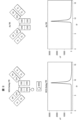
図1に示される通り、Seldeg20は細胞10の表面上の細胞表面受容体又は他の分子30に可逆的に結合する場合がある。細胞外空間50に存在する標的抗原特異的抗体40はSeldeg20に可逆的に結合する場合がある。この結合は典型的に6.8より高く且つ7.5未満のpHのような中性近辺のpHにおいて起こり、それは、それが細胞外空間50の典型的なpHだからである。非標的抗体60はSeldeg20に結合しないか又はいずれの結合も非特異的であるほど非常に低い親和性で結合する。Seldeg20及び標的抗原特異的抗体40が結合した細胞表面受容体又は分子30は経路Aを介し、エンドソーム70中への受容体媒介吸収を介して細胞10内に移行する。受容体又は他の分子を細胞の表面に再循環させて戻すことができる。従って経路B上で、Seldeg20及び標的抗原特異的抗体40が結合した受容体又は分子30を細胞10の表面に循環させて戻す。次いで標的抗原特異的抗体40は放出される場合があり、Seldeg20から同じ又は別のSeldegに再結合する場合があるか又はそれは結合したままの場合がある。類似して、Seldeg20は次いで受容体又は分子30から放出される場合があり、同じ又は別の細胞表面受容体又は分子に再結合する場合がある。いくつかのSeldegsの場合、Seldeg20と抗体40の複合体は、初期又は後期エンドソーム中でこの区画内の酸性のpH又は低いCa-2濃度の故に受容体又は他の分子30から解離する場合がある(経路C)。従って、経路Dにおいて受容体又は細胞表面分子は細胞表面に再循環して戻る場合があるが、抗体40に結合したSeldeg20はリソソーム中に入り、経路Eにおいてフラグメント80に分解する。いくつかのSeldegsの場合、細胞10中への移行に続くいくつかの時点に、Seldeg20及び標的抗原特異的抗体40が結合した受容体又は分子30は経路Fにおいて後期エンドソーム/リソソームに入り、そこで少なくとも標的抗原特異的抗体40はフラグメント80に分解する。このリソソーム中への進入は、Seldeg20が受容体又は分子30を二量体又はもっと高次の凝集物に架橋すると増加すると思われる。
As shown in FIG. 1,
選択的枯渇のこの機構を介し、Seldegは標的とされない特異性の抗体のレベルに不利に影響せずに抗原特異的抗体を標的とし、体から選択的に枯渇させる。 Through this mechanism of selective depletion, Seldeg targets and selectively depletes antigen-specific antibodies from the body without adversely affecting antibody levels of non-targeted specificities.
特に本明細書に説明されるSeldegは、標的とされない特異性の抗体の枯渇の故の患者における不利な臨床的影響を有することなく、抗原特異的抗体を標的とし、体から選択的に枯渇させることができる。そのような不利な臨床的影響には、例えば免疫抑制ならびに流行性結膜炎、気管支炎、耳感染症、副鼻腔感染症、風邪、下痢、肺炎、カンジダ症、髄膜炎、皮膚感染症及び他の日和見感染症、特に通常は抗体媒介免疫反応を介して抑制される日和見感染症のようなその症状;ならびに低血小板数(low platelet
counts)又は貧血のような血液疾患ならびに低ガンマグロブリン血症及び腹痛、鼓張、吐き気、嘔吐、下痢又は体重減少のようなその症状が含まれる。
In particular, Seldeg, as described herein, targets and selectively depletes antigen-specific antibodies from the body without adverse clinical consequences in patients due to depletion of antibodies of untargeted specificity. be able to. Such adverse clinical effects include, for example, immunosuppression and epidemic conjunctivitis, bronchitis, ear infections, sinus infections, colds, diarrhea, pneumonia, candidiasis, meningitis, skin infections and others. opportunistic infections, especially their symptoms, such as opportunistic infections, which are usually suppressed through antibody-mediated immune responses;
counts) or blood disorders such as anemia and hypogammaglobulinemia and symptoms thereof such as abdominal pain, bloating, nausea, vomiting, diarrhea or weight loss.
一般に本開示に従うSeldegsは、中性近辺のpHにおいて標的成分を介して細胞表面受容体/分子に特異的に結合するように且つ標的成分に直接又は間接的に融合した抗原成分を介して中性近辺のpHにおいて抗原特異的抗体にも特異的に結合するように構成される。本明細書で用いられる「特異的に結合する」という用語は、標的成分と細胞表面受容体/分子の間又は抗原成分と抗原特異的抗体の間の検出可能な選択的分子間相互作用を指す。例えば特異的に結合するために、抗原は標的とされている抗体との検出可能な相互作用を示す必要があり、一方で他の抗体との検出可能な相互作用を示さないことが必要である。特異的な結合の検出法は、ELISA、表面プラズモン共鳴分析及び当業者により同定され得る他の方法のように当該技術分野内で既知である。 In general, Seldegs according to the present disclosure are neutralized via antigen moieties directly or indirectly fused to the targeting moieties so as to specifically bind to cell surface receptors/molecules at near-neutral pH. It is also configured to specifically bind antigen-specific antibodies at nearby pH. As used herein, the term "specifically binds" refers to a detectable selective intermolecular interaction between a target component and a cell surface receptor/molecule or between an antigen component and an antigen-specific antibody. . For example, to bind specifically, an antigen must exhibit detectable interaction with the antibody being targeted, while exhibiting no detectable interaction with other antibodies. . Methods for detecting specific binding are known within the art, such as ELISA, surface plasmon resonance analysis and other methods that can be identified by those skilled in the art.
従ってSeldegは、患者の循環中の抗原特異的抗体の少なくとも一部が、標的とされる細胞表面受容体又は標的とされる他の細胞表面分子を発現する細胞中に移行し、その後細胞内で分解することを可能にする。 Seldeg therefore suggests that at least a portion of the patient's circulating antigen-specific antibodies translocate into cells that express the targeted cell surface receptor or other targeted cell surface molecule, and then intracellularly. allow to decompose.
本開示に従うSeldegは、抗原、抗原フラグメント又は抗原類似物の各型の、他の場合に本明細書で「1分子」と呼ばれるSeldeg当たりに1つのコピーを含有することにより免疫反応を避ける場合があり、それはFcガンマ受容体結合及び/又は補体結合を減少させるか又は排除する突然変異の挿入と組み合わされて、抗体架橋及びおそらく炎症性の免疫複合体の形成を減少させると思われる。特にSeldegsの少なくとも99%、Seldegsの少なくとも99.5%又はSeldegsの少なくとも99.9%は、中性近辺のpHにおいて抗原、抗原フラグメント又は抗原類似物のSeldeg当たりに1つのコピーのみを含有する場合がある。本開示に従う他のSeldegsは、抗原、抗原フラグメント又は抗原類似物の1つより多い分子を含有することができる。Seldeg分子当たりに1つの抗原の分子を含有するSeldegsが結合する抗体の2価性(bivalent nature)は、抗体当たりに2つのSeldeg分子の複合体を生ずる場合があり、それは標的受容体二量化を介してSeldeg-抗体複合体のリソソーム送達の有効性を向上させると思われる。 Seldeg according to the present disclosure may avoid immune response by containing one copy per Seldeg, otherwise referred to herein as "one molecule", of each type of antigen, antigen fragment or antigen analogue. Yes, which, in combination with the insertion of mutations that reduce or eliminate Fc gamma receptor binding and/or complement binding, appear to reduce antibody cross-linking and possibly inflammatory immune complex formation. especially when at least 99% of the Seldegs, at least 99.5% of the Seldegs or at least 99.9% of the Seldegs contain only one copy per Seldeg of the antigen, antigen fragment or antigen analog at near neutral pH There is Other Seldegs according to this disclosure can contain more than one molecule of an antigen, antigen fragment or antigen analogue. The bivalent nature of antibodies bound by Seldeg containing one molecule of antigen per Seldeg molecule can result in complexes of two Seldeg molecules per antibody, which can lead to target receptor dimerization. appear to improve the efficacy of lysosomal delivery of Seldeg-antibody conjugates via
Seldegは少なくとも1つの第1の抗原成分及び第2の抗原成分を含むことができ、ここで第1の抗原成分の抗原、抗原フラグメント又は抗原類似物の1分子は第2の抗原成分の抗原分子、抗原フラグメント又は抗原類似物の1分子と異なる。従って少なくとも1つの第1の抗原成分及び第2の抗原成分を含むSeldegsは1つより多い特異性の抗原特異的抗体のクリアランスを可能にする。 The Seldeg can comprise at least one first antigenic component and a second antigenic component, wherein one molecule of the antigen, antigenic fragment or antigenic analogue of the first antigenic component is an antigenic molecule of the second antigenic component. , antigen fragments or antigen analogues differ from one molecule. Thus, Seldegs comprising at least one first antigenic component and a second antigenic component allow clearance of antigen-specific antibodies of more than one specificity.
Seldegは少なくとも1つの第1の抗原成分及び第2の抗原成分を含むことができ、ここで第1の抗原成分の抗原、抗原フラグメント又は抗原類似物の1分子は第2の抗原成分の抗原分子、抗原フラグメント又は抗原類似物の1分子と同じである。 The Seldeg can comprise at least one first antigenic component and a second antigenic component, wherein one molecule of the antigen, antigenic fragment or antigenic analogue of the first antigenic component is an antigenic molecule of the second antigenic component. , antigen fragment or antigen analogue molecule.
従ってSeldegsは、例えば標的成分のC末端及び/又はN末端に融合した1つ以上の抗原成分を含むことができ、ここでそれぞれの抗原成分の抗原、抗原フラグメント又は抗原類似物の1分子は同じ又は異なることができる。 Thus, Seldegs can comprise one or more antigenic components fused, for example, to the C-terminus and/or N-terminus of a target component, wherein one molecule of antigen, antigenic fragment or antigenic analogue of each antigenic component is the same. or can be different.
さらにSeldegsは、ヒトに投与する場合のSeldegへの免疫反応の可能性を避けるか又は低下させるために、ヒト又はヒト化タンパク質又はタンパク質フラグメントを含有する場合がある。抗原成分の抗原、抗原フラグメント又は抗原類似物は、好ましくはヒトへのSeldegの投与のためにヒトタンパク質又はタンパク質フラグメントである。標的成分も、ヒトへのSeldegの投与のために、好ましくはヒト抗体フラグメント又はヒトアルブミン又はアルブミンフラグメントのようなヒトタンパク質又はタンパク質フラグメントあるいはヒト化抗体又はヒト化抗体フラグメントである。Seldegをヒト以外の動物における使用のために開発する場合、その動物に由来するか又はその動物と免疫学的に適合性であるように改変されたタンパク質又はタンパク質フラグメントを代わりに用いる場合がある。 In addition, Seldegs may contain human or humanized proteins or protein fragments to avoid or reduce the potential for immune reactions to Seldeg when administered to humans. The antigen, antigen fragment or antigen analogue of the antigen component is preferably a human protein or protein fragment for administration of Seldeg to humans. The targeting moiety is also preferably a human antibody fragment or a human protein or protein fragment such as human albumin or an albumin fragment or a humanized antibody or humanized antibody fragment for administration of Seldeg to humans. When Seldeg is developed for use in a non-human animal, a protein or protein fragment derived from or modified to be immunologically compatible with that animal may be used instead.
図2Aは、IgGのFcフラグメント110を有する標的成分に融合した抗原100を含むSeldeg20aの活性の略図である。当業者が理解する通り、IgGのFcフラグメントは抗体のY形の基部(lower base)のすべてであり、それはスルフヒドリル架橋ヒンジ領域及びCH2及びCH3ドメインである。Seldegはヒンジ領域を有していないFcフラグメントを有する場合があるか、又はヒンジ領域がスルフヒドリル架橋を有していないFcフラグメントを有する場合がある。Fcフラグメント110はSeldeg20aがFcRn発現細胞上のFcRn分子に結合するのを可能にする。図2Aに示される例において、抗原100はヒンジ-CH2-CH3120のN末端においてFcフラグメント110aに融合する場合がある。抗原100をFcフラグメント110aに融合させ、得られる抗原-Fcフラグメントをノブズイントゥホールズ(knobs-into-holes)戦略(例えばMoore,G.L.,Bautista,C.,Pong,E.,Nguyen,D.H.,Jacinto,J.,Eivazi,A.,Muchhal,U.S.,Karki,S.,Chu,S.Y.,Lazar,G.A.,(2011).A novel bispecific antibody format enables simultaneous bivalent and
monovalent co-engagement of distinct target anatigens.MAbs 3,546-557に説明されているような)を用いて抗原のない別のFcフラグメント110bと二量化させると、示されているようなヘテロダイマー的Seldeg分子20aが生産される。Seldeg20aは抗原100がモノマー的に表示されたFcフラグメントを有し、それは炎症又は他の不利な影響を引き起こし得る多量体的免疫複合体の形成を避ける。Fcフラグメント110aに融合した抗原100のみを含むSeldegsが生産され、いくつかの状況において用いられる場合があるが、Fcフラグメントの二量化する傾向の故に、典型的に二量体が生産されるであろう。多量体的免疫複合体の形成に導き得る両方のFcフラグメント110aが融合抗原100を有するFcフラグメント二量体を避けるために、Seldegsはノブズイントウホールズ突然変異及び/又は静電ステアリング突然変異(electrostatic steering mutations)(例えばGunasekaran,K.,Pentony,M.,Shen,M.,Garrett,L.,Forte,C.,Woodward,A.,Ng,S.B.,Born,T.,Retter,M.,Manchulenko,K.,Sweet,H.,Foltz,I.N.,Wittekind,M.,Yan,W.(2010) Enhancing antibody Fc heterodimer formation through electrostatic steering effects:applications to bispecific molecules and monovalent IgG.J Biol Chem 285,19637-19646に説明されているような)を
用いてヘテロダイマー形成を促進するように設計され、従って1つの抗原が融合したFcは1つしかない。ヘテロダイマーの形成のために、抗原-Fc融合体のC末端と第2のFcフラグメントのN末端の間における(G4S)13リンカーペプチドの挿入(例えばZhou,L.,Wang,H-Y.,Tong,S.,Okamono,C.T.,Shen,W-C.,Zaro,J.L.(2016) Single chain Fc-dimer-human growth hormone fusion protein for improved drug delivery.Biomaterials,117,24-31に説明されているような)のような他の方法を用いることもできる。ノブズイントウホールズ突然変異、静電ステアリング突然変異及び/又はアルギニン突然変異あるいはFcガンマ受容体及び補体結合を減少させる他の突然変異を含むSeldegsのいくつかの例のDNA配列及びタンパク質配列を実施例10に説明する。
FIG. 2A is a schematic representation of the activity of
monovalent co-engagement of distinct target anatigens. When dimerized with another antigen-
ノブズイントウホールズ突然変異の追加の例には、当業者が同定可能な他のものの中でもY349T/T394F:S364H/F405A及びY349T/F405F:S364H/T394F(例えばMoore,G.L.,Bautista,C.,Pong,E.,Nguyen,D.H.,Jacinto,J.,Eivazi,A.,Muchhal,U.S.,Karki,S.,Chu,S.Y.,Lazar,G.A.,(2011) A novel bispecific antibody format
enables simultaneous bivalent and monovalent co-engagement of distinct target anatigens.MAbs 3,546-557に説明されているような)ならびにT366W:T366S:L368A/Y407V(例えばAtwell,S.,Ridgway,J.B.B.,Wells,J.A.,Carter,P(1997) Stabel heterodimers from remodeling the domain interface of a homodimer using a phage display libary.J.Mol.Biol.,270,26-35に説明されているような)が含まれる。これらの代表的なノブズイントウホールズ突然変異の残基のナンバリング(numbering)は、当業者が理解する通り、EU抗体ナンバリングシステムを指す。
Additional examples of knobs-in-to-holds mutations include Y349T/T394F:S364H/F405A and Y349T/F405F:S364H/T394F (e.g., Moore, GL, Bautista, C.), among others identifiable to those skilled in the art. , Pong, E., Nguyen, DH, Jacinto, J., Eivazi, A., Muchhal, U.S., Karki, S., Chu, SY, Lazar, GA, (2011) A novel bispecific antibody format
enables simultaneous bivalent and monovalent co-engagement of distinct target anatigens. MAbs 3,546-557) and T366W:T366S:L368A/Y407V (eg, Atwell, S., Ridgway, JBB, Wells, JA, Carter, P (1997). ) Stabel heterodimers from remodeling the domain interface of a homodimer using a phage display library. The residue numbering of these representative knobs-in-to-holes mutations refers to the EU antibody numbering system, as understood by those skilled in the art.
静電ステアリング突然変異の追加の例には、当業者が同定可能な他のものの中でもE356K/D399K:K392D/K409D及びK409D/K370D:D357K/D399K(例えばGunasekaran,K.,Pentony,M.,Shen,M.,Garrett,L.,Forte,C.,Woodward,A.,Ng,S.B.,Born,T.,Retter,M.,Manchulenko,K.,Sweet,H.,Foltz,I.N.,Wittekind,M.,Yan,W.(2010) Enhancing antibody Fc heterodimer formation through electrostatic steering effects:applications to bispecific molecules and monovalent IgG.J Biol Chem 285,19637-19646に説明されているような)が含まれる。これらの代表的な静電ステアリング突然変異の残基のナンバリングは、当業者が理解する通り、EU抗体ナンバリングシステムを指す。 Additional examples of electrostatic steering mutations include E356K/D399K:K392D/K409D and K409D/K370D:D357K/D399K (e.g. Gunasekaran, K., Pentony, M., Shen , M., Garrett, L., Forte, C., Woodward, A., Ng, SB, Born, T., Letter, M., Manchulenko, K., Sweet, H., Foltz, I. N., Wittekind, M., Yan, W. (2010) Enhancing antibody Fc heterodimer formation through electronic steering effects: applications to bispecific molecules and mononov alent IgG.J Biol Chem 285, 19637-19646). included. The residue numbering of these representative electrostatic steering mutations refers to the EU antibody numbering system, as understood by those skilled in the art.
アルギニン突然変異又はFcガンマ受容体及び補体(C1q)への結合を減少させる他の突然変異の追加の例には、当業者が同定可能な他のものの中でもG236R/L328R(例えばHorton,H.M.,Bernett,M.J.,Pong,E.,Peipp,M.,Karki,S.,Chu,S.Y.,Richards,J.O.,Vostiar,I.,Joyce,P.F.,Repp,R.,Dasjarlais,J.R.,Zhukosky,E.(2010) Potent in vitro a
nd in vivo activity of an Fc-engineered anti-CD19 monoclonal antibody against lymphoma and leukemia.Cancer Res.,68,8049-8057;Moore,G.L.,Bautista,C.,Pong,E.,Nguyen,D.H.,Jacinto,J.,Eivazi,A.,Muchhal,U.S.,Karki,S.,Chu,S.Y.,Lazar,G.A.,(2011) A novel bispecific antibody format enables
simultaneous bivalent and monovalent co-engagement of distinct target anatigens.MAbs 3,546-557に説明されているような)、N297A又はN297Q(例えばTao,M-H.,Morrison,S.L.(1989) Studies
of aglycosylated chimeric mouse-human IgG:role of carbohydrate in the structure
and effector functions mediated bu the human IgG constant region.J.Immunol.,143,2595-2601;Lux,A.,Yu,X.,Scanlan,C.N.,Nimmerjahn,F.(2013) Impact of immune complex size and glycosylation on IgG binding to human FcγRs.J.Immunol.,190,4315-4323に説明されているような)、D265A(例えばLux,A.,Yu,X.,Scanlan,C.N.,Nimmerjahn,F.(2013) Impact of immune complex size and glycosylation on IgG binding to human FcγRs.J.Immunol.,190,4315-4323;Clynes,R.A.,Towers,T.L.,Presta,L.G.,Ravetch,J.V.(2000) Inhibitory Fc receptors modulate in vivo cytotoxicity against tumor targets.Nat.Med.6,443-446に説明されているような)、L234A/L235A(例えばWins,B.D.,Powell,M.S.,Parren,P.W.H.I.,Barnes,N.,Hogarth,P.M.,(2000) The IgG Fc contains distinct Fc receptor(FcR) binding sites:the leukocyte receptors FcγRI and FcγRIIa bind
to a region in the Fc distinct from that recognized by neonatal FcR and protein.A.J.Immunol.,164,5313-5318に説明されているような)及びL234A/L235A/P329G(例えばSchlothauer,T.,Herter,S.,Koller,C.F.,Grau-Richards,S.,Steinhart,V.,Spick,C.,Kubbies,M.,Klein,C.,Umana,P.,Mossner,E.(2016) Novel human IgG1
and IgG4 Fc-engineered antibodies with completely abolished effector functions.PEDS,29,457-466に説明されているような)が含まれる。これらの代表的なアルギニン突然変異又はFcガンマ受容体及び補体(C1q)への結合を減少させる他の突然変異の残基のナンバリングは、当業者が理解する通り、EU抗体ナンバリングシステムを指す。
Additional examples of arginine mutations or other mutations that reduce binding to the Fc gamma receptor and complement (C1q) include G236R/L328R (eg Horton, H.; M., Bernett, MJ, Pong, E., Peipp, M., Karki, S., Chu, SY, Richards, JO, Vostiar, I., Joyce, PF. , Repp, R., Dasjarlais, JR, Zhukosky, E. (2010) Potent in vitro a
nd in vivo activity of an Fc-engineered anti-CD19 monoclonal antibody against lymphoma and leukemia. Cancer Res. , 68, 8049-8057; Moore, G.; L. , Bautista, C.; , Pong,E. , Nguyen, D.; H. , Jacinto, J.; , Eivazi, A.; , Muchhal, U.S.A. S. , Karki, S.; , Chu, S.; Y. , Lazar, G.; A. , (2011) A novel bispecific antibody format enables
Simultaneous bivalent and monovalent co-engagement of distinct target anatigens.
of aglycosylated chimeric mouse-human IgG: role of carbohydrate in the structure
and effector functions mediated by the human IgG constant region. J. Immunol. , 143, 2595-2601; Lux, A.; , Yu, X.; , Scanlan, C.; N. , Nimmerjahn, F.; (2013) Impact of immune complex size and glycosylation on IgG binding to human FcγRs. J. Immunol. , 190, 4315-4323), D265A (for example Lux, A., Yu, X., Scanlan, CN, Nimmerjahn, F. (2013) Impact of immune complex size and glycosylation on IgG binding to human FcγRs J. Immunol., 190, 4315-4323; Nat. Med. 6, 443-446), L234A/L235A (eg Wins, BD, Powell, M.S., Parren, P.W.). HI, Barnes, N., Hogarth, PM, (2000) The IgG Fc contains distinct Fc receptor (FcR) binding sites: the leukocyte receptors FcγRI and FcγRIIa bind
To a region in the Fc distinct from that recognized by neonatal FcR and protein. A. J. Immunol. , 164, 5313-5318) and L234A/L235A/P329G (eg Schlothauer, T., Herter, S., Koller, CF, Grau-Richards, S., Steinhart, V.; , Spick, C., Kubbies, M., Klein, C., Umana, P., Mossner, E. (2016) Novel human IgG1.
and IgG4 Fc-engineered antibodies with completely abolished effector functions. PEDS, 29, 457-466). The residue numbering of these representative arginine mutations or other mutations that reduce binding to the Fc gamma receptor and complement (C1q) refer to the EU antibody numbering system, as understood by those skilled in the art.
FcγRの位置及び補体結合部位におけるか又はそれらに近い残基を標的とするFcγR及び/又は補体結合を排除する(ablate)ための他の突然変異を用いることができる。IgGのFc領域上のこれらの部位は位置決定されている(例えばJefferis,R.,Lund,J.(2002) Interaction sites on
human IgG-Fc for FcγR:current models.Immunol. Letts.,82,57-65;Duncan,A.R.,Winter,G.(1988) The binding site for C1q on IgG.Nature,332,738-740;Idusogie,E.E.,Presta,L.G.,Gazzano-Santoro,H.,Totpal,K.,Wong,P.Y.,Ultsch,M.,Meng,G.,Mulkerrin,M.G.(2000) Mapping of the C1q binding site on rituxan,a chimeric human antibody with a
human IgG1 Fc.J.Immunol.,164,4178-4184;Hogarth,P.M.,Anania,J.,Wines,B.D.(2014) The FcγR of humans and non-human primates and their interaction with IgG: implications for induction of inflammation, resistance to infection and the use of therapeutic monoclonal antibodies.Curr.Top.Microbiol.Immunol.,382,321-352に説明されている通り)。
Other mutations to ablate FcγR and/or complement binding that target residues at or near the position of the FcγR and the complement binding site can be used. These sites on the Fc region of IgG have been mapped (eg Jefferis, R., Lund, J. (2002) Interaction sites on
human IgG-Fc for FcγR: current models. Immunol. Letts. , 82, 57-65; Duncan, A.; R. , Winter, G.; (1988) The binding site for C1q on IgG. Nature, 332, 738-740; Idusogie, E.; E. , Presta, L.; G. , Gazzano-Santoro, H.; , Toppal, K.; , Wong, P.; Y. , Ultsch, M.; , Meng, G.; , Mulkerrin, M.; G. (2000) Mapping of the C1q binding site on rituxan, a chimeric human antibody with a
human IgG1 Fc. J. Immunol. , 164, 4178-4184; Hogarth, P.; M. , Anania, J.; , Wines, B.; D. (2014) The FcγR of humans and non-human primates and their interaction with IgG: Implications for induction of infection, resistance to infection and the use of therapeutic monoclonal antibodies. Curr. Top. Microbiol. Immunol. , 382, 321-352).
Seldegsは、ヒトIgG2又はヒトIgG4のようにFcガンマ受容体又は補体に結合しないか又は非常に弱い結合を有する免疫グロブリンのクラス又はイソタイプに由来するFcプラグメントを含む場合がある。 Seldegs may contain Fc fragments derived from immunoglobulin classes or isotypes that do not or have very weak binding to Fc gamma receptors or complement, such as human IgG2 or human IgG4.
診断用画像化のようないくつかの用途のために、Seldegsは、Seldeg中に存在する抗原に対する炎症反応を向上させるためにFcガンマ受容体及び/又は補体に関する結合部位を有するFcフラグメントを含む場合がある。 For some uses, such as diagnostic imaging, Seldegs contain Fc fragments with binding sites for the Fc gamma receptor and/or complement to enhance the inflammatory response to antigens present in the Seldegs. Sometimes.
Fcフラグメント110を、非修飾Fcフラグメントと比較して中性近辺のpHにおけるFcRnに関するその結合親和性を実質的に向上させるように修飾する場合がある。例えば6.8より高く且つ7.5未満のpHにおけるFcフラグメント110とFcRnの間の解離定数は、表面プラズモン共鳴又は他の生物物理学的方法により決定されると10μM未満の場合がある。しかしながら、Fcフラグメント110は酸性のエンドソームpH(約6.0)において非修飾Fcフラグメントと比較して類似か又は向上したFcRnに関する親和性を有する場合があるか、又はそれを非修飾Fcフラグメントと比較してエンドソームpHにおけるFcRnに関するずっと低いか又は無視し得る結合親和性を有するように修飾する場合がある。中性近辺のpHにおけるこの結合親和性における向上は、それぞれのSeldegがその結合した標的抗原特異的抗体を有効に移行させ、FcRn発現細胞中の後期エンドソーム又はリソソーム中に輸送することを可能にする。FcRnに関するFcフラグメントの結合親和性の強化は、突然変異の挿入により達成される場合がある。天然に存在するIgGsは、中性近辺のpHと反対に酸性のpHレベルにおいてFcRnに関する実質的により高い結合親和性を有する。この性質は、FcRn発現細胞内におけるIgGの再循環及び輸送のために必須である。対照的に、pH7.4におけるFcRnに関する結合親和性の向上は、例えば細胞中への受容体媒介移行及びリソソーム送達を生ずる。 Fc fragment 110 may be modified to substantially improve its binding affinity for FcRn at near-neutral pH compared to an unmodified Fc fragment. For example, the dissociation constant between Fc fragment 110 and FcRn at pH greater than 6.8 and less than 7.5 may be less than 10 μM as determined by surface plasmon resonance or other biophysical methods. However, Fc fragment 110 may have similar or improved affinity for FcRn compared to unmodified Fc fragment at acidic endosomal pH (approximately 6.0), or it may be compared to unmodified Fc fragment. may be modified to have much lower or negligible binding affinity for FcRn at endosomal pH. This improvement in binding affinity at near-neutral pH allows each Seldeg to effectively translocate its bound target antigen-specific antibody and transport it into late endosomes or lysosomes in FcRn-expressing cells. . Enhanced binding affinity of Fc fragments for FcRn may be achieved by insertion of mutations. Naturally occurring IgGs have substantially higher binding affinities for FcRn at acidic pH levels as opposed to near neutral pH. This property is essential for IgG recycling and transport within FcRn-expressing cells. In contrast, enhanced binding affinity for FcRn at pH 7.4 results in, for example, receptor-mediated translocation into cells and lysosomal delivery.
Fcフラグメント110を修飾して、Fcガンマ受容体及び補体(C1q)に関する結合親和性を取り除くか又は実質的に低下させる場合もある。この修飾は、多量体的免疫複合体の形成により引き起こされる炎症反応を予防する。例えば実施例10に説明する通り、Fc領域を、本明細書で「アルギニン突然変異」とも呼ばれる突然変異をさせてそれらがFcガンマ受容体に結合しないようにすることができる(G236R/L328R;EUナンバリング)(例えばMoore,G.L.,Bautista,C.,Pong,
E.,Nguyen,D.H.,Jacinto,J.,Eivazi A.,Muchhal,U.S. Karki,S.,Chu,S.Y.,Lazar,G.A.(2011).A novel bispecific antibody format enables simultaneous bivalent and monovalent co-engagement of distinct target antigens.MAbs 3,546-557に説明されている通り)(EUナンバリング)。実施例10において、これらの突然変異はFc-Syt1の残基22及び114(配列番号10を参照されたい)ならびにMOG-Seldeg-PSの残基144及び236(配列番号8を参照されたい)に相当する。Fcガンマ受容体及び補体への結合を実質的に低下させるか又は排除する他の突然変異の例には、当業者が同定可能な他のものの中でもN297A又はN297Q(EUナンバリング;例えばTao,M-H.,Morrison,S.L.(1989) Studies of aglycosylated
chimeric mouse-human IgG:role of carbohydrate in the structure and effector functions mediated by the human IgG constant region.J.Immunol.,143,2595-2601;Lux,A.,Yu,X.,Scanlan,C.N.,Nimmerjahn,F.(2013) Impact of immune complex size and glycosylation on IgG binding to human FcγRs.J.Immunol.,190,4315-4323に説明されているような)、D265A(EUナンバリング;例えばLux,A.,Yu,X.,Scanlan,C.N.,Nimmerjahn,F.(2013) Impact of immune complex size and glycosylation on IgG binding to human FcγRs.J.Immunol.,190,4315-4323;Clynes,R.A.,Towers,T.L.,Presta,L.G.,Ravetch,J.V.(2000) Inhibitory Fc receptors
modulate in vivo cytotoxicity against tumor targets.Nat.Med.6,443-446に説明されているような)、L234A/L235A(EUナンバリング;例えばWins,B.D.,Powell,M.S.,Parren,P.W.H.I.,Barnes,N.,Hogarth,P.M.,(2000) The IgG Fc contains distinct Fc receptor(FcR) binding sites:the leukocyte receptors FcγRI and FcγRIIa bind to a region in the Fc distinct from that recognized by neonatal FcR and protein.A.J.Immunol.,164,5313-5318に説明されているような)、L234A/L235A/P329G(EUナンバリング;例えばSchlothauer,T.,Herter,S.,Koller,C.F.,Grau-Richards,S.,Steinhart,V.,Spick,C.,Kubbies,M.,Klein,C.,Umana,P.,Mossner,E.(2016) Novel human IgG1 and IgG4 Fc-engineered antibodies with completely abolished effector functions.PEDS,29,457-466に説明されているような)が含まれる。Fcガンマ受容体に関する結合親和性における少なくとも10分の1への低下(reduction of at least 10-fold)が好ましい。
Fc fragment 110 may also be modified to eliminate or substantially reduce binding affinity for the Fc gamma receptor and complement (C1q). This modification prevents the inflammatory response caused by the formation of multimeric immune complexes. For example, as described in Example 10, the Fc regions can be mutated, also referred to herein as "arginine mutations", to prevent them from binding to Fc gamma receptors (G236R/L328R; EU numbering) (e.g. Moore, GL, Bautista, C., Pong,
E. , Nguyen, D.; H. , Jacinto, J.; , Eivazi A.; , Muchhal, U.S.A. S. Karki, S.; , Chu, S.; Y. , Lazar, G.; A. (2011). A novel bispecific antibody format enables simultaneous bivalent and monovalent co-engagement of distinct target antibodies. MAbs 3,546-557) (EU numbering). In Example 10, these mutations were at residues 22 and 114 of Fc-Sytl (see SEQ ID NO: 10) and
chimeric mouse-human IgG: role of carbohydrate in the structure and effector functions mediated by the human IgG constant region. J. Immunol. , 143, 2595-2601; Lux, A.; , Yu, X.; , Scanlan, C.; N. , Nimmerjahn, F.; (2013) Impact of immune complex size and glycosylation on IgG binding to human FcγRs. J. Immunol. , 190, 4315-4323), D265A (EU numbering; e.g. Lux, A., Yu, X., Scanlan, CN, Nimmerjahn, F. (2013) Impact of immune complex size and glycosylation on IgG binding to human FcγRs J. Immunol., 190, 4315-4323; Inhibitory Fc receptors
modulate in vivo cytotoxicity against tumor targets. Nat. Med. 6, 443-446), L234A/L235A (EU numbering; e.g. Wins, BD, Powell, M.S., Parren, P.W.H.I., Barnes, N.). Hogarth, PM, (2000) The IgG Fc contains distinct Fc receptor (FcR) binding sites: the leukocyte receptors FcγRI and FcγRIIa bind to a region in the region. Fc distinct from that recognized by neonatal FcR and protein.A. J. Immunol., 164, 5313-5318), L234A/L235A/P329G (EU numbering; e.g. Schlothauer, T., Herter, S., Koller, CF, Grau-Richards, S., Steinhart, V., Spick, C., Kubbies, M., Klein, C., Umana, P., Mossner, E. (2016) Novel human IgG1 and IgG4 Fc-engineered antibodies with completely abolished effector functions. PEDS, 29, 457-466). A reduction of at least 10-fold in binding affinity for the Fc gamma receptor is preferred.
図2Bに示す通り、Seldeg20bにおいて抗原100は別の末端位置においてFcフラグメント110aに結合する場合があるか又は図2Cに示す通り、Seldeg20cにおいて抗原100は非末端位置で結合する場合がある。特異的なFcRn結合を妨げないいずれの位置も適している。そのような位置には、当業者が同定可能である通り、
直接又は立体的にFcRn結合を遮断しないようにFcRn相互作用部位(CH2-CH3ドメイン境界面における残基252-256、309-311、433-436を包含する;EUナンバリング)から十分に遠いアミノ酸残基が含まれる。
In Seldeg20b,
Amino acid residues sufficiently far from the FcRn interaction site (including residues 252-256, 309-311, 433-436 at the CH2-CH3 domain interface; EU numbering) so as not to directly or sterically block FcRn binding groups are included.
化学反応を介する結合、リンカーを介する結合又は1つの組み合わされた抗原-Fcフラグメントタンパク質の形成の間を含むいずれかの適した方法で抗原100をFcフラグメント110に融合させる場合がある。用いることができる化学結合の例は:アミンからアミン(amine-to-amine)(NHSエステル)、スルフヒドリルからスルフヒドリル(マレイミド)、アミンからスルフヒドリル(NHSエステル/マレイミド)、スルフヒドリルから炭水化物(マレイミド/ヒドラジド)又は所望の化学反応性を有する非天然アミノ酸を介する結合である。この非天然アミノ酸をFcフラグメント及び/又は抗原の組換え体生産の間に挿入することができる。ポリエチレングリコール(PEG)スペーサーを化学的に共役したタンパク質、タンパク質フラグメント又は他の分子の間に挿入することもできる。可能なリンカーにはグリシン-セリンリンカーペプチド又は他のもっと硬い(rigid)リンカーペプチドの反復が含まれ、それは抗原-Fc融合のための組換え体発現プラスミド中でコードされる。連結の化学、連結の部位及びペプチドの選択は分子モデリングにより導かれ得、当業者が理解する通り、抗原又は細胞表面分子を標的とするタンパク質/タンパク質フラグメントの結合活性の損失を最小にするように設計されることができる。
図2Dは、抗原100が抗体可変領域130に結合しているSeldeg20dの略図である。抗体可変領域130は細胞表面受容体又は細胞表面分子に特異的に結合する。抗体可変領域130は、それが細胞表面受容体又は細胞表面分子に特異的に結合する限り、可変領域全体又はそのフラグメントの場合がある。抗体可変領域130は、細胞表面受容体又は細胞表面分子に結合するように構成されている抗体の非可変領域の部分を含む場合がある。例えば抗体可変領域130はシングルドメイン抗体(single-domain antibody)(sdAb)又はラクダ由来VHHドメイン(通常ナノボディーとも呼ばれる)の場合がある。そのような可変領域は2つの逆平行βシートを含む免疫グロブリンドメインの全体的な折り畳み(overall fold)を有し、且つ当業者が同定可能な他のものの中でもT細胞受容体可変ドメイン、抗体の定常領域ドメイン又は補助受容体、CD4のドメインのような免疫グロブリンスーパーファミリーの他のメンバーからのドメインも含むことができる。抗体可変領域130は図2Dに示す通りモノマーとして又は多量体として存在する場合がある。例えば抗体可変領域130がナノボディーとして存在する場合、第1のナノボディーのC末端と第2のナノボディーのN末端の間にGSSGGSGGGGSのようなリンカーペプチドを用いてそれを改変して二量体を形成し、標的受容体/分子に関する結合活性を向上させる場合がある。抗体可変領域130がナノボディー又は多量体を形成するように改変された別のタンパク質である場合、ちょうどFcブラグメントを含有するSeldegsに関して上記に説明した通りに、多量体が抗原100の1つのコピーのみを含有するように、Seldeg形成の間に抗原100のない変異体を含ませる場合がある。抗体可変領域は、ペプチドリンカーにより軽鎖可変(VL)ドメインに連結された重鎖可変(VH)ドメインのヘテロダイマーを含むこともでき、scFvフラグメントを形成する。VH及びVLドメインを連結するために用いられるリンカー配列は当業者に周知であり、VHドメインのC末端をVLドメインのN末端に連結するGGGGSGGGGSGGGGS[(G4S)3]配列を含む。いくつかの態様において、類似のリンカー配列を用いてVLドメインのC末端をVHドメインのN末端に連結することができる。細胞表面受容体又は他の細胞表面分子に結合するscFvsは、ファージディスプレイ、酵母ディスプレイ又は他の抗体ディスプレイ法を用いてscFvsのライブラリから単離され得る。Seldegの標的タンパク質成分は、ファージディスプレイ、酵母ディスプレイなどを用いてFabフラグメントのライブラリから単離され得る抗体のFabフラグメントを含むこともできる。ナノボディー、scFvs及びFa
bフラグメントのために、相補性決定領域(CDRs)中の残基をランダムに突然変異させるか、又はエラープローンポリメラーゼ連鎖反応を用い、突然変異したナノボディー又は可変ドメインのライブラリを形成することにより細胞表面受容体又は細胞表面分子への結合に関する親和性を向上させることができる。標的とされる代表的なCDR残基は軽鎖可変ドメインのCDR3(残基89-97;Kabatナンバリング)及び重鎖可変ドメインのCDR3(残基95-102;Kabatナンバリング)中のものである。これらのライブラリをファージ又は酵母上に表示し(displayed)、当業者に既知の方法を用いて親和性がより高い変異体を選択することができる。
FIG. 2D is a schematic representation of Seldeg20d with
For the b fragment, cells can be generated by randomly mutating residues in the complementarity determining regions (CDRs) or using the error-prone polymerase chain reaction to form a library of mutated nanobodies or variable domains. Affinity can be improved for binding to surface receptors or cell surface molecules. Representative CDR residues targeted are those in the light chain variable domain CDR3 (residues 89-97; Kabat numbering) and the heavy chain variable domain CDR3 (residues 95-102; Kabat numbering). These libraries can be displayed on phage or yeast and higher affinity variants selected using methods known to those of skill in the art.
図2Dは抗体可変領域130の末端位置における抗原100を示しているが、それは代わりに非末端位置にある場合もある。抗原100は、化学結合を介する結合、リンカーを介する結合又は1つの組み合わされた抗原-抗体可変領域融合タンパク質の形成の間を含むいずれかの適した方法で抗体可変領域110に融合する場合がある。
Although FIG. 2D shows
Seldegは、抗体又は抗体フラグメント以外のタンパク質を含む標的成分に融合した抗原成分を含有する場合もあり、但しこのタンパク質は細胞表面受容体又は他の細胞表面分子に結合するように構成される。例えば図2Eに示す通り、Seldeg20eはFcRnに結合することができるアルブミン又はアルブミンフラグメント140に融合した抗原100を含む。アルブミン又はアルブミンフラグメントを、それが向上した親和性でFcRnに結合するように突然変異させるか又は修飾する場合がある。例えばエラープローンPCRを用いて(ヒト血清)アルブミンのFcRn結合ドメイン(DIII)中に突然変異を挿入し、続いて突然変異したアルブミン変異体のライブラリを酵母又はファージ上に表示し、親和性がより高い変異体を選択することができる。あるいはまた、アルブミン:FcRn境界面におけるか又はその近辺の残基を突然変異させ、結合親和性が向上したアルブミン変異体を選択するか又はそれに関してスクリーニングすることにより、親和性がより高い変異体を生むことができる。図2Eはアルブミン又はアルブミンフラグメント140の非末端位置における抗原100を示しているが、それは代わりに末端位置にある場合もある。抗原100は、化学結合を介する結合、リンカーを介する結合又は1つの組み合わされた抗原-FcRn結合タンパク質の形成の間を含むいずれかの適した方法でアルブミン又はアルブミンフラグメント140に融合する場合がある。
Seldeg may also contain an antigenic component fused to a targeting component, including proteins other than antibodies or antibody fragments, provided that the protein is configured to bind to a cell surface receptor or other cell surface molecule. For example, as shown in Figure 2E, Seldeg20e comprises
図2Fは、Fcフラグメント150のN末端に結合した抗原100を含む代表的なSeldeg20fの略図である。図2Fに示す例において、細胞表面受容体又は細胞表面分子に結合するタンパク質又はタンパク質フラグメント160がFcフラグメント150aのC末端に結合している。例えばタンパク質又はタンパク質フラグメントはホスファチジルセリン(PS)に結合するシナプトタグミンのC2Aドメインの場合がある。Fcフラグメント150を向上した親和性でFcRnに結合するように改変することができ、且つそれがFcガンマ受容体及び補体に非常に低いか又は検出可能でない結合親和性で結合するように突然変異させる場合がある。融合した抗原100を有するFcフラグメント150aを2つ有するFcフラグメントホモダイマーは多量体的免疫複合体の形成に導き得、それを避けるために、図2Fに示すSeldegsはノブズイントゥーホールズ及び/又は静電ステアリング突然変異を用い、ヘテロダイマー形成を促進するように設計され、従って1つのFc-抗原を有するFcは1つだけある。図2Fにおいて、Fcフラグメント150a及び150bの両方がそれらに融合したタンパク質又はタンパク質フラグメントを有し、それは細胞表面タンパク質又は他の細胞表面分子に結合する;あるいはまた、1つのみのそのようなタンパク質又はタンパク質フラグメントが存在する場合がある。図2Gに示す代表的なSeldegにおいて、抗原100及び細胞表面受容体又は細胞表面分子に結合するタンパク質又はタンパク質フラグメント160はそれぞれFcフラグメント150a及び150bのC末端及びN末端に融合し、Seldeg20gを形成している。
FIG. 2F is a schematic representation of a representative
図2Hは、細胞表面タンパク質又は細胞表面分子に結合する抗体170のC末端に結合した抗原100を含む代表的なSeldeg20hの略図である。抗体中のFcフラグメント(Fc)を向上した親和性でFcRnに結合するように改変することができ、それがFcガンマ受容体及び補体に非常に低いか又は検出可能でない結合親和性で結合するように突然変異させる場合がある。多量体的免疫複合体の形成に導き得る両方のFcフラグメントに抗原100が融合した抗体ホモダイマーを避けるために、図2Hに示すSeldegsはノブズイントゥーホールズ及び/又は静電ステアリング突然変異を用い、ヘテロダイマー形成を促進するように設計され、従って抗原100が結合した抗体重鎖は抗体分子当たりに1つだけある。抗体の両方のFabフラグメントは同じ細胞表面タンパク質又は他の細胞表面分子に結合する場合があるか、あるいはまたそれらは2種類以上の細胞表面タンパク質又は分子に結合することができる。
FIG. 2H is a schematic representation of a representative
図2Iは、細胞表面タンパク質又は細胞表面分子に結合するscFv(180)-Fc融合体のC末端に結合した抗原100を含む代表的なSeldeg20iの略図である。抗体中のFcフラグメント(Fc)を向上した親和性でFcRnに結合するように改変することができ、それがFcガンマ受容体及び補体に非常に低いか又は検出可能でない結合親和性で結合するように突然変異させる場合がある。多量体的免疫複合体の形成に導き得る両方のFcフラグメントに抗原100が融合した抗体ホモダイマーを避けるために、図2Iに示すSeldegsはノブズイントゥーホールズ及び/又は静電ステアリング突然変異を用い、ヘテロダイマー形成を促進するように設計され、従って抗原100が結合した抗体重鎖-scFv融合体は分子当たりに1つだけある。抗体の両方のscFvフラグメントは同じ細胞表面タンパク質又は他の細胞表面分子に結合する場合があるか、あるいはまたそれらは2種類以上の細胞表面タンパク質又は分子に結合することができる。
FIG. 2I is a schematic representation of a representative
図2Jは、細胞表面タンパク質又は細胞表面分子に結合する-Fc-scFv(180)融合体のN末端に結合した抗原100を含む代表的なSeldeg20jの略図である。抗体中のFcフラグメント(Fc)を向上した親和性でFcRnに結合するように改変することができ、それがFcガンマ受容体及び補体に非常に低いか又は検出可能でない結合親和性で結合するように突然変異させる場合がある。多量体的免疫複合体の形成に導き得る両方のFcフラグメントに抗原100が融合した抗体ホモダイマーを避けるために、図2Jに示すSeldegsはノブズイントゥーホールズ及び/又は静電ステアリング突然変異を用い、ヘテロダイマー形成を促進するように設計され、従って抗原100が結合した抗体重鎖-scFv融合体は分子当たりに1つだけある。抗体の両方のscFvフラグメントは同じ細胞表面タンパク質又は他の細胞表面分子に結合する場合があるか、あるいはまたそれらは2種類以上の細胞表面タンパク質又は分子に結合することができる。
FIG. 2J is a schematic representation of a representative
図2Kに示す通り、代表的なSeldeg20kにおいて2つの抗原成分(100、190)が標的成分、例えばFcフラグメント110a及びFcフラグメント110bにN末端又は他の位置で結合し、異なる特異性の抗原特異的抗体をクリアランスすることができるSeldegを形成する場合がある。図2Kに示すSeldegsは、ノブズイントゥーホールズ及び/又は静電ステアリング突然変異を用い、ヘテロダイマー形成を促進するように設計され、従って各Seldeg分子中にそれぞれの型の抗原分子(100、190)がある。
As shown in FIG. 2K, in a representative Seldeg20k, two antigenic components (100, 190) are attached to targeting components, e.g.,
図2LはFcフラグメント150のN末端に結合した抗原100及び抗原190を含む代表的なSeldeg20lの略図である。図2Lに示す代表的な態様において、細胞表面受容体又は細胞表面分子に結合するタンパク質又はタンパク質フラグメント160はFcフラグメント150a及び150bのC末端に結合している。図2Lに示すSeldegsは、ノブズイントゥーホールズ及び/又は静電ステアリング突然変異を用い、ヘテロ
ダイマー形成を促進するように設計され、従って各Seldeg分子中にそれぞれの型の抗原分子(100、190)がある。図2Lにおいて、Fcフラグメント150a及び150bの両方はそれらに融合したタンパク質又はタンパク質フラグメントを有し、それは細胞表面タンパク質又は他の細胞表面分子に結合するが、他の態様において、1つのみのそのようなタンパク質又はタンパク質フラグメントが存在する場合がある。
FIG. 2L is a schematic representation of a representative
図2Mに示す通り、代表的なSeldeg20mにおいて同じ抗原(100)の2つの分子がFcフラグメント200にN末端又は他の位置において結合し、代表的なSeldeg20mを形成する場合がある。この代表的なSeldegはFcRnへの結合を強化する突然変異を含有するホモダイマーであり、ノブズイントゥーホールズ及び/又は静電ステアリング突然変異を含有しない。
As shown in Figure 2M, two molecules of the same antigen (100) in a representative Seldeg20m may bind to
図2NはFcフラグメント210のN末端に結合した同じ抗原(100)の2つの分子を含む代表的なSeldeg20nの略図である。図2Nに示す代表的な態様において、細胞表面受容体又は細胞表面分子に結合するタンパク質又はタンパク質フラグメント160はFcフラグメント210のC末端に結合している。この代表的なSeldegはホモダイマーであり、ノブズイントゥーホールズ及び/又は静電ステアリング突然変異を含有しない。図2Nにおいて、ホモダイマー的Fcフラグメント200は両方のポリペプチド鎖に融合したタンパク質又はタンパク質フラグメントを有し、それらは細胞表面タンパク質又は他の細胞表面分子に結合するが、他の態様において1つのみのそのようなタンパク質又はタンパク質フラグメントが存在する場合がある。
FIG. 2N is a schematic representation of a representative Seldeg20n containing two molecules of the same antigen (100) attached to the N-terminus of
FcRnを標的とするSeldegsのために、類似の原理をFcRnに結合することができる他のタンパク質に適用する場合がある。さらに、FcRn標的化Seldeg又はその形成方法はFcRn結合タンパク質の性質により影響される場合がある。アルブミンは二量体又は他の多量体を形成しない傾向があるが、他のFcRn結合タンパク質は形成する場合があり、その場合には各Seldegが抗原の1つだけのコピーを含有するように抗体フラグメントを含有する方法に類似の方法で最終的なSeldegを形成する場合がある。図2A、2B、2C、2F、2G、2H、2I、2J、2K、2Lに示す例において、Seldegはノブズイントゥーホールズ及び/又は静電ステアリング突然変異を用いて、1つのFcフラグメントに連結した1つの抗原を含み、1つのFcフラグメントに抗原が結合していないヘテロダイマーの形成を促進する(drive)ように改変された2つの抗体Fcフラグメントを有する。Fcフラグメントをさらに中性近辺のpHにおいて向上した親和性でFcRnに結合するように改変することができるか(図2A、2B、2C、2K、2M)、又は1つ以上の細胞表面受容体若しくは分子を標的とする1つ以上のタンパク質、scFvフラグメント、Fabフラグメント又は他の分子に連結することができる(図2F、2G、2H、2I、2J、2L、2N)。図2F、2G、2H、2I、2J、2L、2Nに示す例におけるFcフラグメントを、より高い親和性でFcRnに結合するように改変し、それらがFcRn及び1つ以上の細胞表面受容体又は分子の両方を標的とするようにすることもできる。 For FcRn-targeted Seldegs, similar principles may apply to other proteins capable of binding to FcRn. Furthermore, the FcRn-targeted Seldeg or method of formation thereof may be influenced by the nature of the FcRn binding protein. Albumin tends not to form dimers or other multimers, but other FcRn-binding proteins may, in which case the antibody is conjugated so that each Seldeg contains only one copy of the antigen. The final Seldeg may be formed in a manner similar to that containing fragments. In the examples shown in Figures 2A, 2B, 2C, 2F, 2G, 2H, 2I, 2J, 2K, 2L, Seldeg was linked to one Fc fragment using knobs-in-to-holes and/or electrostatic steering mutagenesis. It has two antibody Fc fragments that contain one antigen and are modified to drive the formation of heterodimers with no antigen attached to one Fc fragment. The Fc fragment can be further modified to bind FcRn with improved affinity at near-neutral pH (FIGS. 2A, 2B, 2C, 2K, 2M), or to one or more cell surface receptors or It can be linked to one or more proteins, scFv fragments, Fab fragments or other molecules targeting molecules (Figures 2F, 2G, 2H, 2I, 2J, 2L, 2N). The Fc fragments in the examples shown in Figures 2F, 2G, 2H, 2I, 2J, 2L, 2N were modified to bind FcRn with higher affinity, and they bind to FcRn and one or more cell surface receptors or molecules. can also target both
アルブミンは酸性のpHにおいて中性のpHにおけるより強くFcRnに結合する。しかしながらアルブミンを修飾して中性近辺のpH又はエンドソームpHにおけるその結合親和性を変え、標的抗原特異的抗体の分解及びSeldegの再循環を促進することもある。類似して、抗体可変領域FcRn結合タンパク質はそのタンパク質に特異的なやり方でpHにより影響を受ける場合があるが、それでもそれらを修飾して中性近辺のpH又はエンドソームpHにおけるその結合親和性を変え、標的抗原特異的抗体の分解を促進する場合がある。これらのFcRn結合タンパク質は免疫グロブリン可変ドメイン、scFv(VHとVLドメインがGGGGSGGGGSGGGGSのようなリンカーペプチドにより互いに連結されているVH:VLヘテロダイマー)又はFabフラグメントのライブラ
リから、ファージディスプレイ、酵母ディスプレイ又は当業者に既知の他の方法を用いて単離され得る。これらのライブラリは天然に存在する抗体可変遺伝子に由来することができるか、又は相補性決定領域(CDRs)がランダム化オリゴヌクレオチド配列を用いて作られる「半合成」ライブラリを生ずる方法を用いて作成され得る。さらなるそれらの親和性の向上を、例えばエラープローンPCRを用いてCDRsにランダム突然変異を挿入し、続いてファージディスプレイ又は酵母ディスプレイを用いて選択することにより達成することができる。標的とされる代表的なCDR残基は軽鎖可変ドメインのCDR3(残基89-97;Kabatナンバリング)及び重鎖可変ドメインのCDR3(残基95-102;Kabatナンバリング)中のものである。類似の方法を用い、他の細胞表面受容体/分子に結合する抗体に基づくタンパク質又は足場(scaffold)に基づくタンパク質を単離することができる。
Albumin binds to FcRn more strongly at acidic pH than at neutral pH. However, albumin may be modified to alter its binding affinity at near-neutral or endosomal pH, promoting target antigen-specific antibody degradation and Seldeg recycling. Analogously, antibody variable region FcRn binding proteins can be affected by pH in a manner specific to the protein, yet they can be modified to alter their binding affinity at near-neutral or endosomal pH. , may accelerate the degradation of target antigen-specific antibodies. These FcRn binding proteins can be obtained from libraries of immunoglobulin variable domains, scFv (a VH:VL heterodimer in which the VH and VL domains are linked together by a linker peptide such as GGGGSGGGGSGGGGGS) or Fab fragments, phage display, yeast display or the like. It can be isolated using other methods known to those skilled in the art. These libraries can be derived from naturally occurring antibody variable genes, or created using methods that produce "semi-synthetic" libraries in which the complementarity determining regions (CDRs) are created using randomized oligonucleotide sequences. can be Further enhancement of their affinity can be achieved, for example, by inserting random mutations into the CDRs using error-prone PCR, followed by selection using phage or yeast display. Representative CDR residues targeted are those in the light chain variable domain CDR3 (residues 89-97; Kabat numbering) and the heavy chain variable domain CDR3 (residues 95-102; Kabat numbering). Similar methods can be used to isolate antibody-based proteins or scaffold-based proteins that bind to other cell surface receptors/molecules.
Seldegsは細胞表面上の受容体又は他の分子に特異的に結合するように構成されるいずれかの標的成分を含む場合もある(図2D、2E、2F、2G、2H、2I、2J、2L又は2N)。標的成分を直接又は間接的に(例えばリンカーを介して)、抗体媒介架橋を減少させるために抗原、抗原フラグメント又は抗原類似物のそれぞれの型の1分子を有する抗原成分に融合させる。本明細書で用いられる「抗原の型」という用語は、特異的な抗体に結合する抗原を指す。従ってSeldegは1つより多い抗原の型を含むことができ、ここで各Seldegはそれぞれの抗原の型の1分子のみを有する。標的タンパク質が免疫グロブリン由来Fcフラグメントを含有する場合、Fc領域を、それがFcガンマ受容体及び補体に結合しないか又は実質的に低下したレベルで結合するように突然変異させることができる。Seldegsのいくつかの異なる可能な構成を図2A-Nに示す;多数の他の構成も当業者により構想され得るので、これらは例として示され、制限ではない。 The Seldegs may contain any targeting moiety configured to specifically bind to a receptor or other molecule on the cell surface (Figures 2D, 2E, 2F, 2G, 2H, 2I, 2J, 2L). or 2N). The targeting moiety is fused, directly or indirectly (eg, via a linker), to the antigen moiety with one molecule of each type of antigen, antigen fragment, or antigen analogue to reduce antibody-mediated cross-linking. As used herein, the term "antigen type" refers to the antigen that binds to a specific antibody. Thus, a Seldeg can contain more than one antigen type, where each Seldeg has only one molecule of each antigen type. If the target protein contains an immunoglobulin-derived Fc fragment, the Fc region can be mutated so that it does not bind Fc gamma receptors and complement, or does so at substantially reduced levels. Several different possible configurations of the Seldegs are shown in Figures 2A-N; these are shown by way of example and not limitation, as many other configurations can also be envisioned by those skilled in the art.
SeldegsはFcガンマ受容体及び補体に結合するFcフラグメントを含む場合がある。Fcガンマ及び補体結合部位の存在は、特定の用途分野の状況下で、Seldeg中の抗原に対する免疫反応が望まれる場合(例えば腫瘍画像化において)に望ましい場合がある。そのような用途において、Fcガンマ受容体及び補体に結合するように構成されるSeldegsが好ましい場合がある。例えばアルギニン突然変異のような、本明細書に説明されるFcガンマ受容体及び/又は補体(C1q)に関する結合親和性が低下したか又は結合親和性を有さないための改変された突然変異がないFcフラグメントを含むSeldegsを、そのような免疫反応を引き出すように構成する場合がある。Fcガンマ受容体及び/又は補体(C1q)への結合を向上させるための当業者に既知の突然変異を有するFcフラグメントを含むSeldegsを、そのような免疫反応を引き出すように構成する場合もある。そのようなSeldegsを、Seldeg当たりに1つより多い同じ型の抗原分子を含むように構成し(図2K、2L、2M又は2N)、免疫複合体形成を強化する場合もある。 Seldegs may contain an Fc fragment that binds the Fc gamma receptor and complement. The presence of Fc gamma and complement binding sites may be desirable under certain application circumstances when an immune response to antigens in Seldeg is desired (eg, in tumor imaging). In such applications, Seldegs configured to bind Fc gamma receptors and complement may be preferred. Altered mutations for reduced or no binding affinity for the Fc gamma receptor and/or complement (C1q) described herein, e.g., arginine mutations Seldegs containing Fc fragments without a cytoplasm may be constructed to elicit such an immune response. Seldegs comprising Fc fragments with mutations known to those skilled in the art to improve binding to the Fc gamma receptor and/or complement (C1q) may be constructed to elicit such an immune response. . Such Seldegs may also be constructed to contain more than one antigen molecule of the same type per Seldeg (FIGS. 2K, 2L, 2M or 2N) to enhance immune complex formation.
例えばSeldegsは標的ドメイン又は抗体フラグメント(例えばFabフラグメント又はscFvフラグメント)の数が変動させることができ、それは1-3個の標的ドメイン又は抗体フラグメントの範囲である(図2)。これらの標的ドメイン又は抗体フラグメントを免疫グロブリンFcフラグメントに連結することができるが、他の場合、標的ドメイン又は抗体フラグメントを互いに連結する場合がある;抗原及び抗体フラグメントをFcフラグメントに又は互いに、種々の向き(orientation)で融合させることができる(図2);Seldegsはリンカー配列を含むことができ、それは融合タンパク質の間で長さ及び組成においてさまざまであり、ドメイン又はフラグメント、例えばIEGRMD、GGGGS又はこのリンカーの2-3個の反復を用いることができる;小分子又はペプチドのような抗原類似物を用いることができる;Seldegs中のFcフラグメントを、それがFcガンマ受容体、補体に関する実質的に低下した結合親和性及び
FcRnへの結合に関する向上した親和性を有するように突然変異させる場合がある;SeldegのFcフラグメントは、抗原連結Fcフラグメントと非抗原連結Fcフラグメントのヘテロダイマー又は2種類の抗原を含有するヘテロダイマーが形成されるように、ノブズイントゥーホールズ及び/又は静電ステアリング突然変異のような突然変異を有する場合がある。
For example, Seldegs can vary in the number of target domains or antibody fragments (eg, Fab or scFv fragments), ranging from 1-3 target domains or antibody fragments (Figure 2). These targeting domains or antibody fragments can be linked to immunoglobulin Fc fragments, but in other cases the targeting domains or antibody fragments may be linked to each other; can be fused in any orientation (Fig. 2); Seldegs can include linker sequences, which vary in length and composition among fusion proteins, and can be used for domains or fragments such as IEGRMD, GGGGS or this 2-3 repeats of linkers can be used; antigen analogues such as small molecules or peptides can be used; The Fc fragment of Seldeg may be mutated to have reduced binding affinity and improved affinity for binding to FcRn; may have mutations such as knobs-in-to-holes and/or electrostatic steering mutations such that heterodimers containing are formed.
Seldegsは、自己免疫性脳炎を含む自己免疫疾患と関連するタンパク質、糖タンパク質及び核酸を含む以下の抗原を含むことができる:当業者が同定可能な他のものの中でもミエリンオリゴデンドロサイト糖タンパク質(MOG)、ミエリン塩基性タンパク質、プロテオリピドタンパク質、ミエリン関連糖タンパク質、ミエリン関連オリゴデンドロサイト塩基性タンパク質、トランスアルドラーゼ、アセチルコリン受容体、筋特異的キナーゼ、低密度リポタンパク質受容体関連タンパク質4、インスリン、膵島抗原2、グルタミン酸デカルボキシラーゼ65、亜鉛トランスポーター8、シトルリン化抗原、カルバミル化抗原、コラーゲン、軟骨gp39、gp130-RAPS、65kDa熱ショックタンパク質、フィブリラリン、小核タンパク質(snoRNP)、アクアポリン4、甲状腺刺激因子受容体、核抗原、DNA、ヒストン、糖タンパク質gp70、リボソーム、ピルビン酸デヒドロゲナーゼデヒドロリオアミドアセチルトランスフェラーゼ、毛包抗原、ヒトトロポミオシンイソフォーム5、N-メチル-D-アスパルテート(NMDA)受容体、α-アミノ-3-ヒドロキシ-5-メチル-4-イソオキサゾールプロピオン酸(AMPA)受容体、GABAA及びGABAB受容体、グリシン受容体、ジペプチジル-ペプチダーゼ様タンパク質6(DPPX)、グルタメート受容体(GluR5)、電位依存性カリウムチャンネル、Hu、甲状腺ペルオキシダーゼ、チログロブリン、甲状腺刺激ホルモン(TSH)受容体、甲状腺ホルモンT3及びT4、デスモグレイン1及び3。以下の抗原は腫瘍と関連する抗原の例であり、診断用画像化の間に腫瘍特異的抗体をクリアランスするためにSeldegs中に導入され得る:HER2、前立腺特異的膜抗原(PSMA)、前立腺幹細胞関連抗原(PSCA)、c-Met、EpCAM、癌胎児性抗原(CEA)。他の抗原には、患者が特異的な抗体を有している治療薬又は移植片受容者中の抗体が認識する移植抗原が含まれる。さらに、抗原の分子類似物(合成、タンパク質フラグメントなど)を作製することが可能であり、これらをSeldegsの作製に用いることもできる。上記の抗原は例であり、本開示を読んで当業者が同定し得る追加の型の可能な抗原を制限していない。
Seldegs can include the following antigens, including proteins, glycoproteins and nucleic acids associated with autoimmune diseases, including autoimmune encephalitis: myelin oligodendrocyte glycoprotein (MOG), among others identifiable to those skilled in the art; ), myelin basic protein, proteolipid protein, myelin-associated glycoprotein, myelin-associated oligodendrocyte basic protein, transaldolase, acetylcholine receptor, muscle-specific kinase, low-density lipoprotein receptor-associated
本明細書に説明するいくつかの例において、Seldegは配列番号2プラス配列番号6、配列番号4プラス配列番号6、配列番号8プラス配列番号10、配列番号12プラス配列番号14、配列番号16プラス配列番号18プラス抗体軽鎖配列番号20、配列番号22プラス配列番号24プラス抗体軽鎖配列番号20、配列番号26プラス配列番号28、配列番号30プラス配列番号6、配列番号32プラス配列番号6又は配列番号34プラス配列番号6又はそれらの相同体のアミノ酸配列を含む融合タンパク質のヘテロダイマーであることができる。 In some examples described herein, Seldeg is SEQ ID NO:2 plus SEQ ID NO:6, SEQ ID NO:4 plus SEQ ID NO:6, SEQ ID NO:8 plus SEQ ID NO:10, SEQ ID NO:12 plus SEQ ID NO:14, SEQ ID NO:16 plus SEQ ID NO: 18 plus antibody light chain SEQ ID NO: 20, SEQ ID NO: 22 plus SEQ ID NO: 24 plus antibody light chain SEQ ID NO: 20, SEQ ID NO: 26 plus SEQ ID NO: 28, SEQ ID NO: 30 plus SEQ ID NO: 6, SEQ ID NO: 32 plus SEQ ID NO: 6 or It can be a heterodimer of a fusion protein comprising the amino acid sequence of SEQ ID NO:34 plus SEQ ID NO:6 or homologues thereof.
Seldegは、配列番号2、配列番号4、配列番号6、配列番号8、配列番号10、配列番号12、配列番号14、配列番号16、配列番号18、配列番号20、配列番号22、配列番号24、配列番号26、配列番号28、配列番号30、配列番号32又は配列番号34と少なくとも50%の同一性を有するアミノ酸配列を含む融合タンパク質であることができる。 Seldeg is SEQ ID NO:2, SEQ ID NO:4, SEQ ID NO:6, SEQ ID NO:8, SEQ ID NO:10, SEQ ID NO:12, SEQ ID NO:14, SEQ ID NO:16, SEQ ID NO:18, SEQ ID NO:20, SEQ ID NO:22, SEQ ID NO:24 , SEQ ID NO:26, SEQ ID NO:28, SEQ ID NO:30, SEQ ID NO:32 or SEQ ID NO:34.
本明細書で用いられる場合、2つの核酸又はポリペプチド配列の状況における「配列同一性」又は「同一性」は、特定される比較ウィンドウ(comparison window)にわたって最大対応(maximum correspondence)に関して並べられると同じである2つの配列におけるヌクレオチド塩基又は残基を指す(make
s reference)。タンパク質への言及において配列同一性又は類似性のパーセンテージが用いられる場合、同一でない残基の位置は多くの場合に同類アミノ酸置換により異なることが認識され、同類アミノ酸置換ではアミノ酸残基が類似の物理化学的性質を有するアミノ酸残基の機能的に等価の残基で置換され、従って分子の機能的性質を変化させない。
As used herein, "sequence identity" or "identity" in the context of two nucleic acid or polypeptide sequences is said to be aligned for maximum correspondence over a specified comparison window. Refers to the nucleotide bases or residues in two sequences that are the same (make
s reference). When percentages of sequence identity or similarity are used in reference to proteins, it is recognized that non-identical residue positions often differ by conservative amino acid substitutions, in which amino acid residues have similar physical properties. Amino acid residues having chemical properties are replaced with functionally equivalent residues and thus do not change the functional properties of the molecule.
本明細書で用いられるアミノ酸の機能的に等価の残基は、典型的に最初のアミノ酸に実質的に類似の物理化学的及び立体化学的特性を有する他のアミノ酸残基を指す。物理化学的性質には水溶性(疎水性又は親水性)、誘電的及び電気化学的性質、生理学的pH、側鎖の部分的電荷(正、負又は中性)及び当業者が同定可能な他の性質が含まれる。立体化学的特性にはアミノ酸の空間的及びコンフォーメーション的配置ならびにそれらのキラリティーが含まれる。例えばグルタミン酸は本開示の意味においてアスパラギン酸に機能的に等価の残基であると考えられる。チロシン及びトリプトファンはフェニルアラニンに機能的に等価の残基と考えられる。アルギニン及びリシンはヒスチジンに機能的に等価の残基と考えられる。 As used herein, functionally equivalent residues of amino acids typically refer to other amino acid residues that have substantially similar physicochemical and stereochemical properties to the initial amino acid. Physico-chemical properties include water solubility (hydrophobic or hydrophilic), dielectric and electrochemical properties, physiological pH, partial side-chain charges (positive, negative or neutral) and others identifiable to those skilled in the art. includes the nature of Stereochemical properties include the spatial and conformational arrangement of amino acids as well as their chirality. For example, glutamic acid is considered to be a functionally equivalent residue to aspartic acid within the meaning of this disclosure. Tyrosine and tryptophan are considered residues functionally equivalent to phenylalanine. Arginine and lysine are considered residues functionally equivalent to histidine.
当業者は、典型的に2つのポリペプチド又はポリヌクレオチド配列を並べて整列配列(aligned sequences)を形成し、次いで2つの整列配列の間で一致する文字、すなわち類似又は同一の文字の数を検出し、ギャップ(gaps)を含む各ポリペプチド又はポリヌクレオチド配列中の整列文字の合計数で割られる一致する文字の合計数を計算する段階を含む方法により、配列間の類似性が測定されることを理解するであろう。類似性の結果は同一性のパーセンテージとして表される。 One skilled in the art typically aligns two polypeptide or polynucleotide sequences to form aligned sequences and then detects the number of matching letters, ie similar or identical, between the two aligned sequences. , calculating the total number of matching letters divided by the total number of aligned letters in each polypeptide or polynucleotide sequence, including gaps, to measure similarity between sequences. will understand. Similarity results are expressed as percentage identity.
本明細書で用いられる場合、「配列同一性のパーセンテージ」は、比較ウィンドウに及ぶ2つの最適整列配列の比較により決定される値を意味し、ここでポリヌクレオチド配列の比較ウィンドウ中の部分は2つの配列の最適整列のための参照配列(付加又は欠失を含まない)と比較して付加又は欠失(ギャップ)を含む場合がある。パーセンテージは、両配列中に同一の核酸塩基又はアミノ酸残基が存在する位置の数を決定して一致する位置の数を与え、一致する位置の数を比較のウィンドウ中の位置の合計数で割り、結果を100倍して配列同一性のパーセンテージを与えることにより計算される。 As used herein, "percentage of sequence identity" means the value determined by comparing two best aligned sequences spanning a comparison window, wherein the portion of the polynucleotide sequence in the comparison window is 2 may contain additions or deletions (gaps) relative to a reference sequence (not containing additions or deletions) for optimal alignment of two sequences. The percentage is determined by determining the number of positions where identical nucleobases or amino acid residues are present in both sequences to give the number of matching positions, dividing the number of matching positions by the total number of positions in the window of comparison. , is calculated by multiplying the result by 100 to give the percentage of sequence identity.
本明細書で用いられる場合、「参照配列」は配列比較のための基礎として用いられる定義された配列である。参照配列は、例えば全長タンパク質又はタンパク質フラグメントの切片として特定される配列のサブセット又は全体の場合がある。参照配列は、例えばGenBank及びUniprot及び当業者が同定可能な他のデータベースようなデータベースにおいて同定され得る配列の場合がある。 As used herein, a "reference sequence" is a defined sequence used as a basis for sequence comparison. A reference sequence may be a subset or the entire sequence specified, eg, as a full-length protein or a fragment of a protein. A reference sequence may be a sequence that can be identified in a database such as, for example, GenBank and Uniprot and other databases identifiable to one skilled in the art.
当業者が理解する通り、いずれの2つの配列間のパーセント同一性の決定も、数学的アルゴリズムを用いて行うことができる。配列同一性の決定のための配列の比較のために、適した数学的アルゴリズムのコンピューターインプリメンテーションを用いることができる。そのようなインプリメンテーションには:当業者が同定可能な他のものの中でもCLUSTAL、ALIGN、GAP、BESTFIT、BLAST、FASTAが含まれるがこれらに限られない。 As those skilled in the art understand, the determination of percent identity between any two sequences can be accomplished using a mathematical algorithm. For comparing sequences to determine sequence identity, computer implementations of suitable mathematical algorithms can be used. Such implementations include, but are not limited to: CLUSTAL, ALIGN, GAP, BESTFIT, BLAST, FASTA, among others identifiable to those skilled in the art.
例えば本開示に従うSeldegsは、配列番号2、配列番号4、配列番号6又は配列番号8又は配列番号10又は配列番号12又は配列番号14又は配列番号16又は配列番号18、配列番号20、配列番号22、配列番号24、配列番号26、配列番号28、配列番号30、配列番号32又は配列番号34に比較して少なくとも50%の配列同一性、好ましくは少なくとも80%、より好ましくは少なくとも90%、最も好ましくは少なくとも95%の配列同一性を有するアミノ酸配列を有することができる。 For example, Seldegs according to the present disclosure may be SEQ ID NO: 2, SEQ ID NO: 4, SEQ ID NO: 6 or SEQ ID NO: 8 or SEQ ID NO: 10 or SEQ ID NO: 12 or SEQ ID NO: 14 or SEQ ID NO: 16 or SEQ ID NO: 18, SEQ ID NO: 20, SEQ ID NO: 22 , SEQ ID NO:24, SEQ ID NO:26, SEQ ID NO:28, SEQ ID NO:30, SEQ ID NO:32 or SEQ ID NO:34 with at least 50% sequence identity, preferably at least 80%, more preferably at least 90%, Preferably, they will have amino acid sequences with at least 95% sequence identity.
Seldegsが標的とする抗原特異的抗体は、当業者が同定可能な他のものの中でも患者中に存在する自己抗体、治療薬に結合する抗体、移植片を認識する抗体及び診断用画像化において用いられる修飾された(例えば放射性標識された)抗体又はフラグメント/改変された形態の場合がある。 Antigen-specific antibodies targeted by Seldegs are used in autoantibodies present in patients, antibodies that bind therapeutic agents, antibodies that recognize grafts and in diagnostic imaging, among others identifiable by those skilled in the art. It may be a modified (eg radiolabeled) antibody or fragment/altered form.
下記の実施例に示す通り、Seldegsはそれらの融合した抗原に関する特異性を有する標的抗原特異的抗体、例えば標的抗原特異的抗体HER2-特異的トラスツズマブ(trastuzumab)又はペルツズマブ(pertuzumab)(“TZB”又は“PZB”)及びMOG特異的抗体(“8-18C5”)を選択的に枯渇させることができる。下記の実施例に示す通り、全体的なIgGレベルに負の影響を与えるか又は不利な免疫反応を引き出すことなく、標的抗原特異的抗体を選択的に枯渇させることができる。これらの発見は、処置がFcRn阻害剤又はB細胞を破壊する抗体の使用を介して全体的なIgGsの枯渇を生ずる他の方法と対照的である。そのような方法は、それらがSeldegsにより与えられる選択性を欠いているために、標的とされない特異性の抗体又はB細胞機能に不利に影響する。 As shown in the Examples below, Seldegs are targeted antigen-specific antibodies with specificity for their fused antigens, such as the target antigen-specific antibodies HER2-specific trastuzumab or pertuzumab (“TZB” or "PZB") and MOG-specific antibodies ("8-18C5") can be selectively depleted. As shown in the Examples below, target antigen-specific antibodies can be selectively depleted without negatively affecting overall IgG levels or eliciting an adverse immune response. These findings are in contrast to other methods in which treatment results in global depletion of IgGs through the use of FcRn inhibitors or antibodies that destroy B cells. Such methods adversely affect antibody or B-cell function of untargeted specificities because they lack the selectivity afforded by Seldegs.
この独特の選択性に基づき、Seldegプラットフォームは多くの用途を有し、それは多くの他の標的タンパク質及び抗原を有するSeldegsが作製される場合があるからである。そのような用途の例には自己免疫疾患の処置、移植前又はその間の抗体媒介拒絶の処置、腫瘍の全身画像化の間のコントラストの向上(例えば追加のSeldegsの開発のために抗原として腫瘍抗原PSMA、EpCAM及びCEAが用いられる場合がある)、投与後に不利な反応が観察された場合の特定の生物学的製剤の体濃度の枯渇、治療薬の送達の前の治療薬を認識する抗体をクリアランスするための抗体のクリアランスが含まれる。 Based on this unique selectivity, the Seldeg platform has many uses, as Seldegs with many other target proteins and antigens may be generated. Examples of such uses include treatment of autoimmune diseases, treatment of antibody-mediated rejection prior to or during transplantation, enhancement of contrast during systemic imaging of tumors (e.g. tumor antigens as antigens for the development of additional Seldegs). PSMA, EpCAM and CEA may be used), depletion of body concentrations of certain biologics when adverse reactions are observed after administration, antibodies recognizing the therapeutic agent prior to therapeutic agent delivery. Clearance of antibodies to clear is included.
Seldegsは、注射、特に静脈内、皮下若しくは筋肉内注射あるいは枯渇させられるべき抗原特異的抗体が標的とする組織中への注射を介するような、標的とされている細胞表面上の受容体又は他の分子を発現する細胞にそれらを送達できるいずれかの方法で投与される場合がある。Seldegsをコードする発現構築物を含有するように遺伝子改変された細胞においてSeldegsを発現させることもできる。特に、遺伝子改変された細胞からその場でSeldegsを分泌することを可能にするタンパク質又はペプチドを含むSeldegタンパク質をコードする発現構築物を導入することにより、細胞を遺伝子改変することができる。例えばSeldegsをコードする発現構築物を用いて患者由来の細胞をトランスフェクションし、トランスフェクションされた細胞をキメラ抗原-受容体(CAR)T細胞治療に関して説明されている方法に類似の方法を用いて患者内に送達して戻すことができる。発現構築物は当業者に既知の発現ベクターを含み、例えばMOG-Seldegをコードする遺伝子(配列番号1及び5)を、成熟MOG-Seldeg(配列番号1)及びFc(配列番号5)に関するコード配列の5’末端に連結した免疫グロブリン遺伝子に由来するもののような分泌リーダーペプチドと一緒に含有する場合がある。 Seldegs can be used to target cell surface receptors or other receptors, such as via injection, particularly intravenous, subcutaneous or intramuscular injection, or into the tissue targeted by the antigen-specific antibody to be depleted. may be administered in any manner capable of delivering them to cells expressing the molecules. Seldegs can also be expressed in cells that are genetically modified to contain an expression construct encoding Seldegs. In particular, cells can be genetically modified by introducing an expression construct encoding a Seldeg protein comprising a protein or peptide that allows the Seldegs to be secreted in situ from the genetically modified cell. For example, an expression construct encoding Seldegs can be used to transfect cells from a patient, and transfect the transfected cells into a patient using methods similar to those described for chimeric antigen-receptor (CAR) T cell therapy. can be delivered inside and returned. Expression constructs include expression vectors known to those of skill in the art, for example, the genes encoding MOG-Seldeg (SEQ ID NO: 1 and 5), the coding sequences for mature MOG-Seldeg (SEQ ID NO: 1) and Fc (SEQ ID NO: 5). It may be included with a secretory leader peptide such as that derived from an immunoglobulin gene linked to the 5' end.
Seldegsは、すべての標的とされる受容体/細胞表面分子を遮断せず、細胞表面受容体/分子の正常な機能を可能にする量で投与される場合がある。用いられるSeldegの用量は、クリアランスのために標的とされている抗体の量に類似している場合があり、それは例えば抗体媒介疾患の詳細(specifics)又はSeldegsが診断用画像化においてコントラストの向上ために用いられているかどうかに依存するであろう。用いられるSeldegの量は標的とされている受容体の型の合計数未満であると思われ、標的とされる受容体の正常な機能は不利に影響されない。さらに、例えばIgG結合部位と重ならない部位でFcRnに結合するナノボディー(VHH)を用いることにより
、Seldegsを、それらが細胞表面受容体又は細胞表面分子の自然のリガンドと結合に関して競争しないように設計することができる(例えばAndersen,J.T.,Gonzalez-Pajuelo,M.,Foss,S.,Landsverk,O.J.B.,Pinto,D.,Szyroki,A.,de Haard,H.J.,Saunders,M.,Vanlandshoot,P.,Sandlie,I.(2012) Selection of nanobodies that target human neonatal receptor. Sci. Rep., 3, 1118に説明されている通り)。さらにSeldegsは循環中又は枯渇させられるべき抗原特異的抗体が標的とする組織中の標的とされない抗体の10%未満、5%未満、1%未満又は0.1%未満をクリアランスする場合がある。Seleg処置の間及び後の標的とされない抗体の保持は、本明細書に説明される他の効果の中でも正常な免疫機能及び感染の回避において重要な場合がある。
Seldegs may be administered in amounts that do not block all targeted receptors/cell surface molecules and allow normal function of cell surface receptors/molecules. The dose of Seldeg used may be similar to the amount of antibody being targeted for clearance, for example in specifics of antibody-mediated diseases or because Seldeg is used for contrast enhancement in diagnostic imaging. will depend on whether it is used in The amount of Seldeg used appears to be less than the total number of types of receptors being targeted, and the normal function of the receptors being targeted is not adversely affected. Additionally, by using nanobodies (VHHs) that bind FcRn at sites that do not overlap with the IgG binding site, for example, the Seldegs are designed such that they do not compete for binding with the natural ligands of cell surface receptors or cell surface molecules. (eg Andersen, JT, Gonzalez-Pajuelo, M., Foss, S., Landsverk, OJB, Pinto, D., Szyroki, A., de Haard, HJ Sci. Rep., 3, 1118). Additionally, Seldegs may clear less than 10%, less than 5%, less than 1% or less than 0.1% of non-targeted antibodies in the circulation or in tissues targeted by antigen-specific antibodies to be depleted. Retention of non-targeted antibodies during and after Seleg treatment may be important in normal immune function and avoidance of infection, among other effects described herein.
Seldegsは毎日か、毎週か、患者の50%が循環中又は標的とされる抗原特異的抗体が認識する組織中で閾量の標的とされる抗原特異的抗体を再生したと予想されるときはいつでも投与される場合がある。標的とされる抗原特異的抗体のレベルは、血清試料を分析するための酵素結合イムノソルベント検定(ELISAs)を用いることにより決定され得る。あるいはまた、当業者に周知の他の方法を用いることができる。 Seldegs is administered daily or weekly, when 50% of patients are expected to regenerate a threshold amount of targeted antigen-specific antibody in the circulation or in tissues recognized by the targeted antigen-specific antibody. May be administered at any time. Levels of targeted antigen-specific antibodies can be determined by using enzyme-linked immunosorbent assays (ELISAs) to analyze serum samples. Alternatively, other methods well known to those skilled in the art can be used.
抗体媒介拒絶に関するリスクがある移植患者において、Seldegsは移植の前又は後に投与される場合がある。標的抗原特異的抗体が循環中又は標的抗原特異的抗体により認識される組織中で標的抗原特異的抗体の閾値に達したら、緊急時用量(emergency dose)のSeldegsを投与する場合がある。標的とされる抗原特異的抗体のレベルは、血清試料を分析するための酵素結合イムノソルベント検定(ELISAs)を用いることにより決定され得る。あるいはまた、当業者に周知の他の方法を用いることができる。 In transplant patients at risk for antibody-mediated rejection, Seldegs may be administered before or after transplantation. An emergency dose of Seldegs may be administered once target antigen-specific antibodies reach a threshold of target antigen-specific antibodies in circulation or in tissues recognized by target antigen-specific antibodies. Levels of targeted antigen-specific antibodies can be determined by using enzyme-linked immunosorbent assays (ELISAs) to analyze serum samples. Alternatively, other methods well known to those skilled in the art can be used.
タンパク質に基づく治療薬のような治療薬に関して特異的な抗体を有する患者において、Seldegsはそのような抗体を枯渇させるために治療薬の送達の前に投与される場合がある。これは、治療薬が前の送達の間に免疫反応を引き出していた場合、又は治療薬に特異的な既存の抗体が患者内に存在している場合、急速な治療薬の抗体媒介クリアランスに関連する問題を克服すると思われる。タンパク質に基づく治療薬に関して特異的な既存の又は誘導される抗体を複数の異なる方法を用いて検出することができる(例えばXue,L.,Clements-Egan,A.,Amaravadi,L.,Birchler,M.,Gorovits,B.,Liang,M.,Myler,H.,Purushothama,S.,Manning,M.S.,Sung,C.(2017) Recommendations for the assessment and management of pre-existing drug-reactive antibodies during biotherapeutic development.AAPS,19,1576-1586に説明されているもののような)。 In patients with antibodies specific for therapeutics, such as protein-based therapeutics, Seldegs may be administered prior to delivery of the therapeutic to deplete such antibodies. This is associated with rapid antibody-mediated clearance of the therapeutic if the therapeutic has elicited an immune response during previous delivery or if pre-existing antibodies specific to the therapeutic are present in the patient. seems to overcome the problem of Pre-existing or induced antibodies specific for protein-based therapeutics can be detected using a number of different methods (eg, Xue, L., Clements-Egan, A., Amaravadi, L., Birchler, M., Gorovits, B., Liang, M., Myler, H., Purushotama, S., Manning, M.S., Sung, C. (2017) Recommendations for the assessment and management of pre-existing drug-react ive antibodies during biotherapeutic development.AAPS, 19, 1576-1586).
診断用/セラノスティック画像化において、放射性標識された画像化抗体の送達に腫瘍局在化(localization)を許すある期間が続くであろうと思われる。続いてSeldegsを用いてオフターゲット部位(例えば循環)から放射性標識された抗体をクリアランスし、コントラストの向上を生ずる。例えばこの方法は以下の段階を含むことができる:第1に腫瘍抗原に結合する放射性標識(又は他の標識)された抗体を患者に注入する。第2に、放射性標識された抗体が腫瘍に結合するのを許すためのある期間(例えば16-24時間)の後に、注入された画像化剤の用量にモル量において等しい量でSeldegsを注入する。クリアランス期間(例えば4-24時間)の後にポジトロンエミッショントモグラフィー又は他の全身画像化様式を用いて患者を画像化する。 In diagnostic/theranostic imaging, it is likely that there will be a period of time that allows delivery of radiolabeled imaging antibodies to allow tumor localization. Seldegs are then used to clear the radiolabeled antibody from off-target sites (eg, circulation), resulting in enhanced contrast. For example, the method can include the following steps: First, a radiolabeled (or otherwise labeled) antibody that binds to a tumor antigen is injected into the patient. Second, after a period of time (eg, 16-24 hours) to allow the radiolabeled antibody to bind to the tumor, inject Seldegs in an amount equal in molarity to the dose of the injected imaging agent. . Patients are imaged using positron emission tomography or other whole-body imaging modalities after a clearance period (eg, 4-24 hours).
投与から1時間以内、2時間以内、5時間以内、24時間以内又は48時間以内あるいはもっと長い時間以内に循環中又は標的抗原特異的抗体により認識される組織中の標的抗原特異的抗体の濃度の少なくとも50%、少なくとも80%又は少なくとも90%を枯渇させるのに十分な量でSeldegsを投与する場合がある。体内におけるSeldegsの持続性は、抗原特異的抗体の枯渇においてそれがどれだけ長く活性を有しているかの決定要因であろう。Seldegsを、それらが標的とする細胞表面受容体又は細胞表面分子の行動により種々の生体内半減期を有するように設計することができる。この細胞表面受容体又は細胞表面分子に関するSeldegの親和性を突然変異誘発及び当業者に既知の方法を用いて修飾し、循環及び/又は組織中における種々の持続性を有するSeldegsを生ずることもできる。特に枯渇させられるべき抗原特異的抗体の量と少なくとも大体等モルにおける量でSeldegを投与する場合がある。例えば標的抗原特異的抗体に対して約1:1、2:1、3:1、4:1、5:1、6:1、7:1、8:1、9:1、10:1又はもっと高いモル比にある量でSeldegを投与することができる。特に例えばSeldegが患者に投与される抗体(例えば抗MOG又は抗HER2抗体など)を標的とする場合、例えば投与される標的抗原特異的抗体に対して約1:1、2:1、3:1、4:1、5:1、6:1、7:1、8:1、9:1、10:1又はもっと高いモル比にある用量でSeldegを投与することができる。 The concentration of the target antigen-specific antibody in circulation or in tissues recognized by the target antigen-specific antibody within 1 hour, 2 hours, 5 hours, 24 hours, 48 hours, or longer after administration Seldegs may be administered in an amount sufficient to deplete at least 50%, at least 80%, or at least 90%. The persistence of Seldegs in the body may be a determinant of how long it remains active in depleting antigen-specific antibodies. Seldegs can be designed to have different half-lives in vivo depending on the behavior of the cell surface receptors or molecules they target. The affinity of Seldeg for this cell surface receptor or cell surface molecule can also be modified using mutagenesis and methods known to those skilled in the art to produce Seldegs with varying persistence in circulation and/or tissues. . In particular, Seldeg may be administered in an amount that is at least about equimolar to the amount of antigen-specific antibody to be depleted. for example about 1:1, 2:1, 3:1, 4:1, 5:1, 6:1, 7:1, 8:1, 9:1, 10:1 or Seldeg can be administered in amounts in higher molar ratios. Especially when, for example, Seldeg targets an antibody administered to a patient (such as an anti-MOG or anti-HER2 antibody), for example, about 1:1, 2:1, 3:1 relative to the administered target antigen-specific antibody. , 4:1, 5:1, 6:1, 7:1, 8:1, 9:1, 10:1 or higher molar ratios of Seldeg.
以下の実施例は本開示の特定の態様をさらに例示するために与えられる。それらは本開示のそれぞれの及びすべての側面を完全に詳細に開示又は説明することを意図しておらず、そのように理解されるべきではない。他にことわらなければ、細胞系及び組成物の呼称は、これらの実施例全体を通じて一貫して用いられる。 The following examples are provided to further illustrate certain aspects of the disclosure. They are not intended to, and should not be construed as, disclosing or describing each and every aspect of the present disclosure in complete detail. Unless otherwise indicated, cell line and composition designations are used consistently throughout these examples.
-FcRnに結合するSeldegsの発現及び精製
細胞上のFcRnを標的とするSeldegsは、中性近辺のpHにおいてヒトFcγRsとの相互作用を排除し且つFcRnへのSeldegFcフラグメントの結合親和性を強化するための突然変異を有する二量体的ヒトIgG1由来Fcフラグメント(図3A)に連結したモノマーとしての組換え抗原を含有する。これらの2つのSeldegsのヘテロダイマー形成は、CH3ドメインにおけるノブズイントゥーホールズ突然変異の挿入により達成される。
Expression and purification of Seldegs that binds to FcRn Seldegs, which targets FcRn on cells, eliminates interaction with human FcγRs at near-neutral pH and enhances the binding affinity of SeldegFc fragments to FcRn. containing the recombinant antigen as a monomer linked to a dimeric human IgG1-derived Fc fragment (Fig. 3A) with a mutation of . Heterodimerization of these two Seldegs is achieved by the insertion of knobs-in-to-holes mutations in the CH3 domain.
代表的なHER2-Seldeg(配列番号3、4、5及び6)を作製するための発現構築物を以下の通りに作製した:改変されたFcフラグメントに融合したHER2を有するポリペプチド鎖を発現するために(配列番号3)、HER2リーダーペプチド及び細胞外ドメイン(630残基からなるECD)をコードする遺伝子を標準的な分子生物学的方法を用いてHER2過剰発現乳癌細胞系(BT-474)から単離した。この遺伝子をヒトIgG1由来Fcフラグメントをコードする遺伝子のヒンジ領域のN末端に、オーバーラップ伸長(overlap extention)によるスプライシングを用いてIEGRMDリンカーペプチドを介して融合させた。FcγRsへの結合を排除するため(G236R/L328R;EUナンバリング)、FcRnへの結合を強化するため(MST-HN;M252Y/S254T/T256E/H433K/N434F;EUナンバリング)及び‘ノブズイントゥーホールズ’を作製するため(Y349T/T394F;EUナンバリング)の突然変異を標準的な方法を用いてFcフラグメント遺伝子中に挿入した。軽鎖定常領域中のシステインと架橋するヒンジ領域中のシステイン(C220;EUナンバリング)もセリンに突然変異させた。FcRn強化突然変異、FcγRs結合を排除するための突然変異を有し、融合抗原のないFcフラグメント遺伝子を相補的ノブズイントゥーホールズ突然変異を用いて作製した(S364H/F405A;EUナンバリン
グ)(配列番号5)。Gibco Expi293TM 発現系キット(Life Technologies)を用いる一過性トランスフェクションの後にHEK-293F(life Technologies)細胞中で組換えタンパク質を発現させた。アニオン交換カラム(SOURCE-15Q,GE Healthcare)を用い、pH8.0及び直線状塩勾配(0-0.5M NaCl)においてHER2-Seldegを培養上澄み液から精製した。あるいはまた、タンパク質A-セファロース及び標準的な方法を用いてHER2-Seldegを精製することができる。カラム(イオン交換又はタンパク質A-セファロース)からの溶離の後、HER2-Seldegをリン酸緩衝生理食塩水(PBS)に対して透析した。実験における使用の前にPBS(Lonza)中でサイズ排除クロマトグラフィー(SEC)(GE Healthcare)を用いてHER2-Seldegをさらに精製した。類似の方法及びトランスフェクションされたHEK-293F細胞中で発現される組換えタンパク質を用いて他のSeldegsのための発現プラスミドを作製した。FcRn強化突然変異のないSeldegs(例えばMOG-Seldeg-PS、MOG-Seldeg-TfR;配列番号7、8、9、10、11、12、13、14、15、16、17、18、19及び20)をタンパク質G-セファロースを用いて精製した。
Expression constructs for making representative HER2-Seldeg (SEQ ID NOS: 3, 4, 5 and 6) were made as follows: to express a polypeptide chain with HER2 fused to a modified Fc fragment. (SEQ ID NO:3), the gene encoding the HER2 leader peptide and extracellular domain (ECD consisting of 630 residues) was isolated from a HER2 overexpressing breast cancer cell line (BT-474) using standard molecular biology methods. Isolated. This gene was fused to the N-terminus of the hinge region of the gene encoding the human IgG1-derived Fc fragment via an IEGRMD linker peptide using splicing by overlap extension. To eliminate binding to FcγRs (G236R/L328R; EU numbering), to enhance binding to FcRn (MST-HN; M252Y/S254T/T256E/H433K/N434F; EU numbering) and 'Nobs Into Holes'(Y349T/T394F; EU numbering) were inserted into the Fc fragment gene using standard methods. A cysteine in the hinge region that bridges the cysteine in the light chain constant region (C220; EU numbering) was also mutated to serine. An Fc fragment gene with FcRn-enhancing mutations, mutations to abolish FcγRs binding, and no fusion antigen was generated using complementary knobs-in-to-holes mutations (S364H/F405A; EU numbering) (SEQ ID NO: 5). Recombinant proteins were expressed in HEK-293F (life Technologies) cells after transient transfection using the Gibco Expi293 ™ Expression System Kit (Life Technologies). HER2-Seldeg was purified from the culture supernatant using an anion exchange column (SOURCE-15Q, GE Healthcare) at pH 8.0 and a linear salt gradient (0-0.5 M NaCl). Alternatively, HER2-Seldeg can be purified using Protein A-Sepharose and standard methods. After elution from the column (ion exchange or protein A-Sepharose), HER2-Seldeg was dialyzed against phosphate buffered saline (PBS). HER2-Seldeg was further purified using size exclusion chromatography (SEC) (GE Healthcare) in PBS (Lonza) prior to use in experiments. Similar methods and recombinant proteins expressed in transfected HEK-293F cells were used to generate expression plasmids for other Seldegs. Seldegs without FcRn-enhancing mutations (eg MOG-Seldeg-PS, MOG-Seldeg-TfR; SEQ ID NOs: 7, 8, 9, 10, 11, 12, 13, 14, 15, 16, 17, 18, 19 and 20 ) was purified using Protein G-Sepharose.
2つの抗原に関して特異的な抗体を標的とするSeldegs:(1)HER2-Seldeg;及び(2)MOG-Seldegを作製した。抗原HER2はトラスツズマブ(TZB)又はペルツズマブ(PZB)のようなHER2特異的抗体を用いる治療のため及びHER2過剰発現腫瘍の診断用画像化のためにも十分に定義された標的である。抗原MOGは多発性硬化症(MS)の動物モデル及びヒトにおけるMSの両方において自己反応性抗体(autoreactive antibodies)により認識される。 Seldegs were generated that target antibodies specific for two antigens: (1) HER2-Seldeg; and (2) MOG-Seldeg. The antigen HER2 is a well-defined target for therapy with HER2-specific antibodies such as trastuzumab (TZB) or pertuzumab (PZB) and also for diagnostic imaging of HER2-overexpressing tumors. The antigen MOG is recognized by autoreactive antibodies in both animal models of multiple sclerosis (MS) and MS in humans.
HER2-Seldeg及びMOG-Seldegは、中性近辺のpHにおける結合親和性を向上させるFcRn強化MST-HN突然変異(M252Y,S254T,T256E,H433K,N434F;EUナンバリング)がないことを除いてHER2-Seldeg及びMOG-Seldegに類似の構築物(それぞれ“HER2-WT”及び“MOG-WT”)を作製するためにFcフラグメントに融合されたHER2又はMOGを含む組換え融合タンパク質と対照的に、中性のpH及び酸性のpHにおいてFcRnに関する有意により高い結合親和性を保持している(図3B)。BIAcore T200(GE Healthcare)を用いて、組換えタンパク質とFcRnの相互作用を分析するための表面プラズモン共鳴実験を行った。PBS(pH6.0又は7.4)プラス0.01%v/vTween-20中の固定化されたFcRn(CM5センサーチップ上に~600RUにおいて結合)上に100nMのMOG/HER2-Seldeg又はMOG/HER2-WTを10μl/分の流量で注入することによりSeldeg/WTの組換えマウスFcRnへの結合を分析した。0.15M NaCl,0.1M 重炭酸ナトリウム,pH8.5を用いて、各注入及び解離段階に続いてフローセル(flow cells)を再生させた。データをゼロ調整し、バックグラウンドを引き去った(バックグラウンドは結合反応の間に緩衝液のみと結合したフローセル上に注入することにより得られた)。 HER2-Seldeg and MOG-Seldeg are HER2-Seldeg except that they lack the FcRn-enhancing MST-HN mutations (M252Y, S254T, T256E, H433K, N434F; EU numbering) that improve binding affinity at near-neutral pH. Neutral in contrast to recombinant fusion proteins containing HER2 or MOG fused to an Fc fragment to create constructs similar to Seldeg and MOG-Seldeg (“HER2-WT” and “MOG-WT”, respectively). and retains significantly higher binding affinity for FcRn at pH of 10 and at acidic pH (Fig. 3B). A BIAcore T200 (GE Healthcare) was used to perform surface plasmon resonance experiments to analyze the interaction of recombinant proteins with FcRn. 100 nM MOG/HER2-Seldeg or MOG/ over immobilized FcRn (binding at ~600 RU on CM5 sensor chip) in PBS (pH 6.0 or 7.4) plus 0.01% v/v Tween-20 Binding of Seldeg/WT to recombinant mouse FcRn was analyzed by injecting HER2-WT at a flow rate of 10 μl/min. 0.15M NaCl, 0.1M sodium bicarbonate, pH 8.5 was used to regenerate the flow cells following each injection and dissociation step. Data were zeroed and background subtracted (background was obtained by injecting over a flow cell bound with buffer only during the binding reaction).
MST-HNのようなFcRn結合を向上させるための突然変異は以下の方法を用いて同定された:FcRn結合に必須であることが既知のアミノ酸(例えば253、435)に近接する残基をFcフラグメント遺伝子においてランダムに突然変異させ、突然変異したFcブラグメントのライブラリをファージ上で表示する。ファージディスプレー法
(Ghetie,V.,Popov,S.,Borvak,J.,Radu,C.,Matesoi,D.,Medesan,C.,Ober,R.J.,Ward,E.S.(1997)Increasing the serum persistence of
an IgG fragment by random mutagenesis,
Nature Biotech.,15,637-640;Dall’Acqua,W.F.,Woods,R.M.,Ward,E.S.,Palaszynski,S.R.,Patel,N.K.,Brrewah,Y.A.,Wu,H.,Kiener,P.A.,Langermann,S.(2001) Increasing the affinity of a human IgG1 for the neonatal receptor: biological consquences,J.Immunol.,169,5171-5180)を用い、FcRnに関する結合親和性が向上したFcフラグメントを選択した。あるいはまた、これらの残基をすべての他の可能なアミノ酸に突然変異させ、ELISA又は表面プラズモン共鳴結合分析のような方法を用いてFcRnに関するより高い親和性を有するFcフラグメントを同定することができる。
Mutations to improve FcRn binding, such as MST-HN, were identified using the following method: residues adjacent to amino acids known to be essential for FcRn binding (e.g., 253, 435) A library of mutated Fc fragments is randomly mutated in the fragment gene and displayed on phage. Phage display method (Ghetie, V., Popov, S., Borvak, J., Radu, C., Matesoi, D., Medesan, C., Ober, R.J., Ward, E.S. (1997) Increasing the serum persistence of
an IgG fragment by random mutagenesis,
Nature Biotech. , 15, 637-640; Dall' Acqua, W.; F. , Woods, R.; M. , Ward, E. S. , Palaszynski, S.; R. , Patel, N.; K. , Brrewah, Y.; A. , Wu, H.; , Kiener, P.; A. , Langermann, S.; (2001) Increasing the affinity of a human IgG1 for the neonatal receptor: biological concepts, J. Am. Immunol. , 169, 5171-5180) were used to select Fc fragments with improved binding affinity for FcRn. Alternatively, these residues can be mutated to all other possible amino acids and methods such as ELISA or surface plasmon resonance binding analysis can be used to identify Fc fragments with higher affinity for FcRn. .
サイズ排除分析は、最高で4℃で30日間又は37℃で5日間インキュベートされる場合、Seldegsを含む組換えタンパク質がリン酸緩衝生理食塩水中のインキュベーション後に凝集物を形成しないことを示す(図3C)。 Size exclusion analysis shows that recombinant proteins containing Seldegs do not form aggregates after incubation in phosphate-buffered saline when incubated up to 30 days at 4°C or 5 days at 37°C (Fig. 3C). ).
-FcRnを標的とするSeldegsが形質転換によりヒトFcγRsを発現するマウス(huFcγRマウス)において標的抗原特異的抗体を枯渇させる能力
Seldegsが標的抗原特異的抗体を枯渇させる能力を決定するために、外来遺伝子導入によりヒトFcγRsを発現するマウス(huFcγRマウス;Smith,P.,DiLillo,D.J.,Bournazos,S.,Li,F.,Ravetch,J.V.(2012). Mouse model recapitulating human Fcγ receptor structural and functional diversity. Proc.Natl.Acad.Sci.USA 109,6181-6186.http://dx.doi.org/10.1073/pnas.1203954109)に15μgの放射性標識された(125I標識された)MOG特異的抗体8-18C5を注入した。huFcγRマウスに8-18C5を注入してから24時間後、注射を介して125μg又は31μgのMOG-Seldeg又は参照標準タンパク質を投与した。図3Dにおいて、“MOG-Seldeg-High”は125μgの用量に対応し、“MOG-Seldeg-Low”は31μgの用量に対応する。参照標準はリン酸緩衝生理食塩水(“PBS”)及び非修飾MOG-Fc融合タンパク質(“MOG-WT”)である。MOG-WTは、それが中性近辺のpHにおける結合親和性を向上させるFcRn強化MST-HN突然変異を欠いていることを除いてMOG-Seldegに類似の構築物である。
- Ability of FcRn-targeted Seldegs to deplete target antigen-specific antibodies in mice transgenic to express human FcγRs (huFcγR mice). Mice expressing human FcγRs by introduction (huFcγR mice; Smith, P., DiLillo, DJ, Bournazos, S., Li, F., Ravetch, JV (2012). Mouse model recapitulating human Fcγ receptor Structural and functional diversity.Proc.Natl.Acad.Sci.USA 109,6181-6186.http://dx.doi.org/10.1073/pnas.1203954109. been d) MOG-specific antibody 8-18C5 was injected. Twenty-four hours after injection of 8-18C5 into huFcγR mice, 125 μg or 31 μg of MOG-Seldeg or reference standard protein was administered via injection. In FIG. 3D, "MOG-Seldeg-High" corresponds to a dose of 125 μg and "MOG-Seldeg-Low" corresponds to a dose of 31 μg. Reference standards are phosphate buffered saline (“PBS”) and unmodified MOG-Fc fusion protein (“MOG-WT”). MOG-WT is a similar construct to MOG-Seldeg, except it lacks the FcRn-enhancing MST-HN mutation that improves binding affinity at near-neutral pH.
図3Dのデータを作成するために、放射性標識された8-18C5の全身カウントを、指示される時間に取った。8-18C5投与から24時間後、Seldeg又は参照標準送達の直前に得られる分当たりのカウント(“CPM”)を各マウスに関する100%とみなし、その後に得られる全身のCPMをこの時点に正規化する。エラーバー(error bars)は標準偏差を示し、チューキーの多重比較(Tukey’s multiple comparisons)と共に二元配置分散分析(two-way ANOVA)を用い、p<0.05及びn=グループ当たり6匹のマウスで統計的に有意な差を決定した。 To generate the data in Figure 3D, whole body counts of radiolabeled 8-18C5 were taken at the times indicated. Twenty-four hours after 8-18C5 administration, counts per minute (“CPM”) obtained immediately prior to Seldeg or reference standard delivery were considered 100% for each mouse, and whole body CPM obtained thereafter were normalized to this time point. do. Error bars indicate standard deviation, p<0.05 and n=6 animals per group using two-way ANOVA with Tukey's multiple comparisons. of mice was determined to be statistically significant.
MOG-Seldegの125μg及び31μgの用量は、マウスにおいてIgGレベルを全体的に排除することが示された500μg,MST-HN Abdegの用量より約2ないし8倍低い(2 to 8-fold lower)(モルに基づいて)。そのように比較的低いMOG-Seldegの用量は、FcRn結合に関するSeldegとの競争を介する融合した抗原に関して特異的でないIgGsへの影響を最小にするために用いられた。125μg又は31μgの用量は、8-18C5標的抗原特異的抗体よりそ
れぞれ16倍及び4倍モル過剰を意味する。
The 125 μg and 31 μg doses of MOG-Seldeg are about 2 to 8-fold lower than the 500 μg, MST-HN Abdeg dose that was shown to totally eliminate IgG levels in mice ( based on moles). Such relatively low doses of MOG-Seldeg were used to minimize the effects on IgGs that were not specific for the fused antigen via competition with Seldeg for FcRn binding. A dose of 125 μg or 31 μg represents a 16-fold and 4-fold molar excess, respectively, over the 8-18C5 target antigen-specific antibody.
図3Dは、時間に対する放射性標識された8-18C5(標的抗原特異的抗体)の正規化された体カウントのグラフを与えている。MOG-Seldegの投与は、正規化された全身カウントにおいて8-18C5の急速な用量依存性減少を生じ(図3D)、それは体から8-18C5を選択的に枯渇させるMOG-Seldegの能力を示す。対照的に、FcRn強化MST-HN突然変異を欠いた参照標準MOG-WTの注入は、8-18C5抗体枯渇に影響を有していなかった。これは、pH6.0-7.4におけるFcRnに関する高い結合親和性を与えるためのMST-HN突然変異の重要性を示す。 FIG. 3D provides a graph of normalized body counts of radiolabeled 8-18C5 (target antigen-specific antibody) versus time. Administration of MOG-Seldeg produced a rapid dose-dependent decrease in 8-18C5 in normalized whole body counts (Fig. 3D), demonstrating the ability of MOG-Seldeg to selectively deplete 8-18C5 from the body. . In contrast, injection of reference standard MOG-WT lacking the FcRn-enhancing MST-HN mutation had no effect on 8-18C5 antibody depletion. This demonstrates the importance of the MST-HN mutation to confer high binding affinity for FcRn at pH 6.0-7.4.
-FcRnを標的とするSeldegsの特異性ならびに血液中及び体全体の両方において標的抗原特異的抗体レベルにおける有意な低下を誘導するそれらの能力
Seldegsの特異性及び種々の抗原認識性を有する抗体へのそれらの影響を分析するために、HER2特異的抗体TZBの行動をHER2-Seldeg及びMOG-Seldegの両方の存在下で調べた(図3E)。図3Eのデータを作成するために、huFcγRマウスに15μgの放射性標識された(125I標識された)TZBを静脈内注射し、続いて4倍モル過剰のHER2-Seldeg又は抗原特異性に関する参照標準としてMOG-Seldegを静脈内注射した。追加の参照標準はリン酸緩衝生理食塩水(“PBS”)、Abdeg及び中性近辺のpHにおける結合親和性を向上させるFcRn強化MST-HN突然変異を欠いていること以外はHER2-Seldegに類似の構築物(“HER2-WT”)を作製するためにFcフラグメントに融合したHER2を含む組換え融合タンパク質である。Seldegと対照的に、“Abdeg”はMST-HN突然変異を有するヒトIgG1由来抗体であり、それは非選択的に抗体を枯渇させる。そのようなタンパク質は、それらが一般的にIgG分解を引き起こす抗体なのでAbdegsと呼ばれる。
- the specificity of Seldegs targeting FcRn and their ability to induce significant reductions in target antigen-specific antibody levels both in the blood and throughout the body To analyze their effects, the behavior of the HER2-specific antibody TZB was examined in the presence of both HER2-Seldeg and MOG-Seldeg (Fig. 3E). To generate the data in FIG. 3E, huFcγR mice were injected intravenously with 15 μg radiolabeled ( 125 I-labeled) TZB followed by a 4-fold molar excess of HER2-Seldeg or a reference standard for antigen specificity. MOG-Seldeg was injected intravenously as Additional reference standards were phosphate-buffered saline (“PBS”), Abdeg and similar to HER2-Seldeg except lacking the FcRn-enhancing MST-HN mutations that improve binding affinity at near-neutral pH. A recombinant fusion protein comprising HER2 fused to an Fc fragment to create a construct (“HER2-WT”). In contrast to Seldeg, "Abdeg" is a human IgG1-derived antibody with MST-HN mutations, which non-selectively depletes antibodies. Such proteins are called Abdegs because they are commonly antibodies that cause IgG degradation.
図3Eのデータを作成するために、マウスを出血させ、血液及び全身カウントを指示される時間に取った。TZB投与から24時間後、Seldeg又は参照標準送達の直前に得られる分当たりのカウント(“CPM”)を各マウスに関する100%とみなし、その後に得られる血液及び全身のCPMをこの時点に正規化する。エラーバーは標準偏差を示し、チューキーの多重比較と共に二元配置分散分析を用い、p<0.05及びn=グループ当たり6匹のマウスで統計的に有意な差を決定した。 To generate the data in Figure 3E, mice were bled and blood and whole body counts were taken at the times indicated. Twenty-four hours after TZB administration, counts per minute (“CPM”) obtained immediately prior to Seldeg or reference standard delivery were considered 100% for each mouse, and blood and whole body CPM obtained thereafter were normalized to this time point. do. Error bars indicate standard deviation, two-way ANOVA with Tukey's multiple comparisons was used to determine statistically significant differences with p<0.05 and n=6 mice per group.
図3Eは、時間に対する放射性標識されたTZB(標的抗原特異的抗体)の正規化された血液及び体カウントのグラフを与えている。HER2-Seldegの投与は、血液及び全身カウントの両方において正規化されたTZBの体カウントにおける有意な低下を引き起こし、それは体からTZBを選択的に枯渇させるHER2-Seldegの能力を示す。対照的に、参照標準タンパク質はPBS参照標準に関して観察された行動に類似の行動を示した(図3E)。 FIG. 3E provides graphs of normalized blood and body counts of radiolabeled TZB (target antigen-specific antibody) versus time. Administration of HER2-Seldeg caused a significant decrease in normalized somatic counts of TZB in both blood and whole body counts, demonstrating the ability of HER2-Seldeg to selectively deplete TZB from the body. In contrast, the reference standard protein exhibited behavior similar to that observed for the PBS reference standard (Fig. 3E).
-FcRnを標的とするSeldegsが標的抗原特異的抗体を急速に枯渇させる能力
マウスに放射性標識された(125I標識された)TZB(15μg)を静脈内注射し、24時間後にHER2-Seldeg(51μg;TZBより4倍モル過剰)を静脈内送達した。追加の参照標準はリン酸緩衝生理食塩水(“PBS”)及びAbdeg(60μg)である。
Ability of FcRn-Targeted Seldegs to Rapidly Deplete Target Antigen- Specific Antibodies 4-fold molar excess over TZB) was delivered intravenously. Additional reference standards are phosphate buffered saline (“PBS”) and Abdeg (60 μg).
図3Fのデータを作成するために、マウスを出血させ、血液及び全身カウントを指示される時間に取った。TZB投与から24時間後、Seldeg又は参照標準送達の直前に
得られる分当たりのカウント(“CPM”)を各マウスに関する100%とみなし、その後に得られる血液及び全身のCPMをこの時点に正規化する。エラーバーは標準偏差を示し、チューキーの多重比較と共に二元配置分散分析を用い、p<0.05及びn=グループ当たり5-6匹のマウスで統計的に有意な差を決定した。
To generate the data in Figure 3F, mice were bled and blood and whole body counts were taken at the times indicated. Twenty-four hours after TZB administration, counts per minute (“CPM”) obtained immediately prior to Seldeg or reference standard delivery were considered 100% for each mouse, and blood and whole body CPM obtained thereafter were normalized to this time point. do. Error bars indicate standard deviation, two-way ANOVA with Tukey's multiple comparisons was used to determine statistically significant differences at p<0.05 and n=5-6 mice per group.
図3Fは、時間に対する放射性標識されたTZB(標的抗原特異的抗体)の正規化された血液及び体カウントのグラフを与えている。HER2-Seldegの投与は、正規化されたTZBの体及び血液カウントにおける急速な低下を引き起こし、それは体からTZBを急速に枯渇させるHER2-Seldegの能力を示す。注目すべきことに、TZBの血液カウントはSeldeg投与から2時間以内にバックグラウンドレベルの近くまで低下し(図3F)、体から標的抗原特異的抗体を急速に枯渇させるHER2-Seldegの能力をさらに支持している。対照的に、60μg/マウスの用量におけるAbdegの送達はPBS参照標準に関して観察された行動に類似の行動を示した(図3F)。 FIG. 3F provides graphs of normalized blood and body counts of radiolabeled TZB (target antigen specific antibody) versus time. Administration of HER2-Seldeg caused rapid decreases in normalized TZB body and blood counts, indicating the ability of HER2-Seldeg to rapidly deplete TZB from the body. Remarkably, TZB blood counts dropped to near background levels within 2 hours of Seldeg administration (Fig. 3F), further demonstrating the ability of HER2-Seldeg to rapidly deplete target antigen-specific antibodies from the body. Support. In contrast, delivery of Abdeg at a dose of 60 μg/mouse showed behavior similar to that observed for the PBS reference standard (Fig. 3F).
-細胞表面上の露出したホスファチジルセリン(PS)を標的とするSeldegsの活性
我々は、標的タンパク質がシナプトタグミン1のC2ドメイン(Syt1)であるSeldegがhuFcγRマウスにおいてMOGに関して特異的な抗体を選択的に枯渇させる能力も調べた。Syt1は細胞上の露出したPSに結合し、Seldegはそのために設計されたMOG-Seldeg-PSであり、略図的に示される(図4A)。‘DN’突然変異(D173N,D179N,D231N,D233N及びD239N)の存在の故にPSに結合しないMOG-Seldeg-PS(DN)も作製した。MOG-Seldeg-PS及びMOG-Seldeg-PS(DN)を、トランスフェクションされたHEK-293F細胞の培養上澄み液からタンパク質G-セファロース及び標準的な方法を用いて精製した。ヘテロダイマー形成はFc領域中へのノブズイントゥーホールズ及び静電ステアリング突然変異の挿入により達成され、サイズ排除分析は組換えタンパク質が良好に行動することを示す(図4A)。
- Activity of Seldegs targeting exposed phosphatidylserine (PS) on the cell surface The ability to deplete was also examined. Syt1 binds to exposed PS on cells for which Seldeg is designed MOG-Seldeg-PS, shown schematically (FIG. 4A). MOG-Seldeg-PS (DN) was also generated that does not bind PS due to the presence of 'DN' mutations (D173N, D179N, D231N, D233N and D239N). MOG-Seldeg-PS and MOG-Seldeg-PS(DN) were purified from culture supernatants of transfected HEK-293F cells using Protein G-Sepharose and standard methods. Heterodimer formation was achieved by the insertion of knobs-in-to-holes and electrostatic steering mutations into the Fc region, and size exclusion analysis indicates that the recombinant protein behaves well (Fig. 4A).
図4Bに示すデータの作成のために、マウスに放射性標識された(125I)キメラ8-18C5(ヒト定常/マウス可変ドメイン;MOG特異的;15-20μg)を静脈内注射し、24時間後にリン酸緩衝生理食塩水(PBS)、40μgのMOG-Seldeg-PS又は参照標準として34μgのFc-Syt1(MOGが結合していない)又は40μgのMOG-Seldeg-PS(DN)を静脈内送達した。指示される時間に放射性レベルを決定した。MOG-Seldeg-PS又は参照標準送達の直前に得られる全身CPM又は血液CPMレベルを100%とみなし、得られる続くCPMのすべてをこれらのCPMレベルに対して正規化した。エラーバーは標準誤差(SEM)を示し、MOG-Seldeg-PS処置グループと参照標準グループ(Fc-Syt1、MOG-Seldeg-PS(DN)及びPBS)の間の統計的に有意な差を*により示した(p<0.05,チューキーの多重比較を用いる二元配置分散分析;n=グループ当たり5-6匹のマウス)。 For the generation of the data shown in FIG. 4B, mice were injected intravenously with radiolabeled ( 125 I) chimeric 8-18C5 (human constant/mouse variable domain; MOG-specific; 15-20 μg) and 24 h later. Phosphate-buffered saline (PBS), 40 μg MOG-Seldeg-PS or 34 μg Fc-Syt1 (no MOG conjugated) or 40 μg MOG-Seldeg-PS (DN) as a reference standard were delivered intravenously. . Radioactivity levels were determined at the times indicated. Systemic or blood CPM levels obtained immediately prior to MOG-Seldeg-PS or reference standard delivery were considered 100% and all subsequent CPM obtained were normalized to these CPM levels. Error bars indicate standard error (SEM), statistically significant differences between MOG-Seldeg-PS treated group and reference control group (Fc-Syt1, MOG-Seldeg-PS (DN) and PBS) by * (p<0.05, two-way ANOVA with Tukey's multiple comparisons; n=5-6 mice per group).
MOG-Seldeg-PSの投与は血液及び全身カウントの両方において8-18C5の正規化された体カウントにおける有意な低下を引き起こし、それは体から8-18C5を選択的に枯渇させるMOG-Seldeg-PSの能力を示す。対照的に、参照標準タンパク質Fc-Syt1はPBS参照標準に関して観察された行動と類似の行動を示し、MOG-Seldeg-PS(DN)は8-18C5における低下を誘導したが(PSへの残留結合の故に)、影響はMOG-Seldeg-PSのそれよりずっと低かった(図4B)。 Administration of MOG-Seldeg-PS caused a significant decrease in normalized body counts of 8-18C5 in both blood and whole body counts, indicating that MOG-Seldeg-PS selectively depletes 8-18C5 from the body. Show ability. In contrast, the reference standard protein Fc-Syt1 showed behavior similar to that observed for the PBS reference standard, MOG-Seldeg-PS(DN) induced a decrease in 8-18C5 (residual binding to PS ), the effect was much lower than that of MOG-Seldeg-PS (Fig. 4B).
-FcRnを標的とするSeldegsが標的抗原特異的抗体を有効にエンドソーム中に移行させ、リソソームへの送達を介して分解を引き起こす能力
細胞レベルにおけるSeldegsの活性の機構を決定するために、フローサイトメトリー及び蛍光顕微鏡検査を行い、標的抗原特異的抗体TZB、PZB及び8-18C5の移行及び堆積へのSeldegsの効果を分析した。最初に結合性がマウスFcRnに類似であるように突然変異したヒトFcRn-GFP発現構築物を用いてヒト内皮細胞(HMEC-1)をトランスフェクションした。
- Ability of FcRn-targeted Seldegs to effectively translocate target antigen-specific antibodies into endosomes and cause degradation via delivery to lysosomes To determine the mechanism of Seldegs activity at the cellular level, flow cytometry and fluorescence microscopy were performed to analyze the effect of Seldegs on the migration and deposition of target antigen-specific antibodies TZB, PZB and 8-18C5. Human endothelial cells (HMEC-1) were first transfected with a human FcRn-GFP expression construct mutated to have binding similar to mouse FcRn.
Seldegsは、結合した標的抗原特異的抗体を有効に細胞内のエンドソーム中に移行させ、8時間のインキュベーション後に標的抗原特異的抗体はリソソームに送達された。 Seldegs effectively translocated the bound target antigen-specific antibodies into intracellular endosomes, and after 8 hours of incubation the target antigen-specific antibodies were delivered to lysosomes.
図5Aは平均蛍光強度(“MFI)”)のグラフであり、図5B及び図5Cは標的抗原特異的抗体の存在下におけるSeldeg活性の顕微鏡画像であり、代表的なエンドソームの顕微鏡画像を切り出し、拡大し、右上の角の差し込み図に示した。図5AはそれぞれHER2-Seldegsと一緒のAlexa 647標識されたTZB及びMOG-Selegと一緒のAlexa 647標識された8-18C5の存在下におけるより大きなMFIを示す。このより大きなMFIは、TZB及び8-18C5とのそれぞれHER2-及びMOG-Seldegの共-インキュベーション(400nMのSeldeg又はWT参照標準プラス100nMの抗原特異的抗体;30分のパルス、追跡なし又は60分の追跡、図5A、5B及び5Cにおいてそれぞれ30’P又は30’P,60’Cと標識)がTZB及び8-18C5進入細胞の高いレベルを生ずることを示す。HER2-Seldegの存在下でTZBの代わりにHER2特異的抗体ペルツズマブ(PZB)を用いた場合、類似の結果が得られた(図5A)。さらに、各標的抗原特異的抗体の大部分は60分の追跡の間、細胞により保持された。対照的に、負の参照標準HER2-WT(TZB又はPZBに関して選択的)及びMOG-WT(8-18C5に関して選択的)の存在下で細胞内の標的抗原特異的抗体の堆積は実質的により低かった。 FIG. 5A is a graph of mean fluorescence intensity (“MFI”), and FIGS. 5B and 5C are microscopic images of Seldeg activity in the presence of target antigen-specific antibodies, cropping microscopic images of representative endosomes; Enlarged and shown in the inset in the upper right corner. FIG. 5A shows greater MFI in the presence of Alexa 647-labeled TZB with HER2-Seldegs and Alexa 647-labeled 8-18C5 with MOG-Seleg, respectively. This greater MFI was associated with co-incubation of HER2- and MOG-Seldeg with TZB and 8-18C5, respectively (400 nM Seldeg or WT reference standard plus 100 nM antigen-specific antibody; 30 min pulse, no chase or 60 min (labeled 30′P or 30′P, 60′C in FIGS. 5A, 5B and 5C, respectively) produced high levels of TZB and 8-18C5 invading cells. Similar results were obtained when the HER2-specific antibody pertuzumab (PZB) was used instead of TZB in the presence of HER2-Seldeg (Fig. 5A). Moreover, the majority of each target antigen-specific antibody was retained by the cells during the 60 min chase. In contrast, deposition of intracellular target antigen-specific antibodies was substantially lower in the presence of negative reference standards HER2-WT (selective for TZB or PZB) and MOG-WT (selective for 8-18C5). rice field.
図5B及びCは抗原特異的抗体が細胞中に堆積すること、そしてSeldegsの存在下におけるエンドソーム中のFcRnと関連していることを示す。図5Bの顕微鏡画像の作成のために、400nMのAlexa 555標識されたHER2-Seldeg又はHER2-WT(示される通り)との複合体における100nMのAlexa 647標識されたTZB(HER2特異的)を用いてHMEC-1細胞を30分間パルスし、洗浄し、直後に固定するか又は洗浄し、媒体中で60分間追跡し、続いて固定した。固定された細胞を蛍光顕微鏡検査を用いて画像化した。図5C中のデータは、400nMのAlexa 555標識されたMOG-Seldeg又はMOG-WTとの複合体における100nMのAlexa 647標識された8-18C5(MOG特異的)を用いる以外は同じ方法を用いて作成された。それぞれの顕微鏡画像中のバーは5μmであり、それぞれの顕微鏡画像差し込み図中のバーは0.25μmである。図5B及び5Cに示されるデータは、抗原特異的抗体が参照標準(WT)タンパク質の存在下と比較してFcRnに結合するSeldegsの存在下で実質的により高いレベルまでFcRn発現細胞中に堆積することを示している。 Figures 5B and C show that antigen-specific antibodies are deposited in cells and associated with FcRn in endosomes in the presence of Seldegs. 100 nM Alexa 647-labeled TZB (HER2-specific) in complex with 400 nM Alexa 555-labeled HER2-Seldeg or HER2-WT (as indicated) was used to generate the microscopic images in FIG. 5B. HMEC-1 cells were pulsed for 30 min, washed, immediately fixed or washed and chased in medium for 60 min followed by fixation. Fixed cells were imaged using fluorescence microscopy. Data in FIG. 5C were generated using the same method but using 400 nM Alexa 555-labeled MOG-Seldeg or 100 nM Alexa 647-labeled 8-18C5 (MOG-specific) in complex with MOG-WT. Created. The bar in each microscopic image is 5 μm and the bar in each microscopic image inset is 0.25 μm. The data shown in Figures 5B and 5C demonstrate that antigen-specific antibodies are deposited in FcRn-expressing cells to substantially higher levels in the presence of Seldegs, which binds FcRn, compared to the presence of the reference standard (WT) protein. It is shown that.
図6Aは標的抗原特異的抗体の存在下におけるSeldeg活性の顕微鏡画像を示し、代表的なリソソームの顕微鏡画像を切り出し、拡大し、右上の角の差し込み図に示した。図6AのためにAlexa 555標識されたデキストランを用いてHMEC-1細胞を2時間予備パルスし、洗浄し、次いで400nMのMOG-Seldeg又はMOG-WT(示される通り)との複合体における100nMのAlexa 647標識された8-18C5(MOG特異的)を用いて30分間パルスし、続いて8時間追跡し、洗浄し、固
定し、画像化した。図6Bの顕微鏡画像の作成のために、Alexa 555標識されたデキストランを用いてHMEC-1細胞を2時間予備パルスし、洗浄し、次いで100nMのAlexa 647標識されたTZB又は8-18C5抗体及び400nMのMOG-Seldeg又はHER2-Seldeg(示される通り)を用いて30分間パルスし、続いて8時間追跡し、洗浄し、固定し、画像化した。それぞれのオーバーレイ画像(overlay image)(右の列に示す)のために、GFP、Alexa 555及びAlexa 647をそれぞれ緑、赤及び青に疑似着色した(pseudo-colored)。それぞれの顕微鏡画像中のバーは5μmであり、それぞれの顕微鏡画像差し込み図中のバーは0.25μmである。注目すべてことに、図6BはそれぞれMOG-Seldeg又はHER2-Seldegの存在下で細胞がリソソーム中にTZB及び8-18C5を堆積させないことを示し、それはそれらの効果の抗原特異性を示している。
FIG. 6A shows microscopic images of Seldeg activity in the presence of target antigen-specific antibodies, and microscopic images of representative lysosomes are cropped, enlarged, and shown in the upper right corner inset. HMEC-1 cells were pre-pulsed for 2 hours with Alexa 555-labeled dextran for FIG. Alexa 647 labeled 8-18C5 (MOG specific) was pulsed for 30 min followed by 8 h chase, washed, fixed and imaged. For the generation of the microscopic images in FIG. 6B, HMEC-1 cells were prepulsed for 2 hours with Alexa 555-labeled dextran, washed, and then treated with 100 nM Alexa 647-labeled TZB or 8-18C5 antibody and 400 nM MOG-Seldeg or HER2-Seldeg (as indicated) for 30 min followed by 8 h chase, washing, fixing and imaging. For each overlay image (shown in right column), GFP, Alexa 555 and Alexa 647 were pseudo-colored green, red and blue, respectively. The bar in each microscopic image is 5 μm and the bar in each microscopic image inset is 0.25 μm. Notably, Figure 6B shows that cells do not deposit TZB and 8-18C5 in the lysosomes in the presence of MOG-Seldeg or HER2-Seldeg, respectively, indicating the antigen specificity of their effects.
-ホスファチジルセリンを標的とするSeldegsが標的抗原特異的抗体を有効に細胞中に移行させる能力
図7は、MOG-Seldeg-PSがMOG特異的抗体(キメラ8-18C5)を内皮細胞及びマクロファージ中に有効に移行させること及び吸収はPS結合依存性であることを示す。PSを露出させた内皮細胞(2H11)又はマクロファージ(RAW264.7)を、20nMのMOG-Seldeg-PS、MOG-Seldeg-PS(DN)又はFc-Syt1(MOGなし)と混合された10nMのAlexa 647標識されたキメラ8-18C5と一緒に2時間インキュベーションした。三重の試料に関するAlexa 647標識されたキメラ8-18C5(マウス抗体、8-18C5の可変ドメインを当業者に周知の方法を用いてヒトIgG1の定常領域、Cκに融合させることにより作製)の平均蛍光強度(MFI)をフローサイトメトリーにより決定した。
- Ability of phosphatidylserine-targeted Seldegs to effectively translocate target antigen-specific antibodies into cells Efficient translocation and absorption are shown to be PS binding dependent. PS-exposed endothelial cells (2H11) or macrophages (RAW264.7) were treated with 10 nM Alexa mixed with 20 nM MOG-Seldeg-PS, MOG-Seldeg-PS (DN) or Fc-Syt1 (no MOG). Incubated with 647-labeled chimeric 8-18C5 for 2 hours. Average fluorescence of Alexa 647-labeled chimeric 8-18C5 (mouse antibody, generated by fusing the variable domain of 8-18C5 to the constant region of human IgG1, Cκ, using methods well known to those skilled in the art) for triplicate samples. Intensity (MFI) was determined by flow cytometry.
この代表的なPS結合Seldegはカルシウム依存性であり、従ってカルシウム濃度がずっと低いエンドソーム中で解離する。当業者に理解される通り、すべてのPS標的化Seldegがカルシウム依存性であるわけではないと思われるが、いくつか、アネキシンVを含むもののようないくつかもこの性質を有するであろう。さらに、いくつかの抗体はベータ2糖タンパク質1に結合することができ、それが今度はPSに結合することができる。
This representative PS-bound Seldeg is calcium dependent and thus dissociates in endosomes where calcium concentrations are much lower. As will be appreciated by those skilled in the art, not all PS-targeted Seldeg will be calcium dependent, but some, such as those containing annexin V, will have this property. In addition, some antibodies can bind to
-トランスフェリン受容体を標的とするSeldegsが標的抗原特異的抗体を有効にヒトトランスフェリン受容体を発現する細胞中に移行させる能力
トランスフェリン受容体を標的とする抗体に融合した抗原(MOG)を含むSeldeg(MOG-Seldeg-TfR)を作製し、図8に略図的に示す。MOG-Seldeg-TfR及び参照標準抗体、Ab-TfR(MOGが存在しない)を、トランスフェクションされたHEK-293F細胞の培養上澄み液からタンパク質G-セファロース及び標準的な方法を用いて精製した。MOG-Seldeg-TfRはMOG特異的抗体(キメラ8-18C5)を有効に内皮(HMEC-1)細胞中に移行させ、吸収はSeldeg中のMOGの存在に依存性である(図9)。ヒトTfRを発現するHMEC-1細胞を、200nMのMOG-Seldeg-TfR又はTfR特異的抗体(Ab-TfR;MOGなし)と混合された50nMのAlexa 647標識されたキメラ8-18C5(MOGに関して特異的)と一緒に30分間インキュベーションし、続いて洗浄し、追跡しないか(30’P)又は30分間に続いて洗浄し、60分間追跡した(30’P,60’C)。三重の試料に関するAlexa 647標識された8-18C5の平均蛍光強度(MFI)をフローサイトメトリーにより決定した(図9)。
- the ability of transferrin receptor-targeted Seldegs to effectively translocate target antigen-specific antibodies into cells expressing human transferrin receptor MOG-Seldeg-TfR) was generated and shown schematically in FIG. MOG-Seldeg-TfR and a reference standard antibody, Ab-TfR (without MOG), were purified from culture supernatants of transfected HEK-293F cells using protein G-sepharose and standard methods. MOG-Seldeg-TfR effectively translocates a MOG-specific antibody (chimeric 8-18C5) into endothelial (HMEC-1) cells and uptake is dependent on the presence of MOG in Seldeg (Fig. 9). HMEC-1 cells expressing human TfR were treated with 50 nM Alexa 647-labeled chimeric 8-18C5 (specific for MOG) mixed with 200 nM MOG-Seldeg-TfR or TfR-specific antibody (Ab-TfR; no MOG). target) followed by washing and no chase (30′P) or 30 min followed by washing and 60 min chase (30′P, 60′C). The mean fluorescence intensity (MFI) of Alexa 647-labeled 8-18C5 for triplicate samples was determined by flow cytometry (Fig. 9).
-FcRnを標的とするSeldegsが放射性標識された抗体を用いる腫瘍のポジトロンエミッショントモグラフィー分析の間にバックグラウンドをクリアランスする能力
雌のBALB/cSCIDマウスに、HER2陽性乳癌細胞系、HCC1954を乳房脂肪体中において移植した(マウス当たり0.5x106個の細胞;注入前に細胞をRPMI-1640/Matrigel中に懸濁させた)。6-7日後、マウスに124-I標識されたTZB(60μg/マウス)を注入し、22時間後にポジトロンエミッショントモグラフィー(PET)を用いて分析した。マウスにモル当量のHER2-Seldeg(51μg/マウス)又は参照標準としてMOG-Seldeg(31μg/マウス)若しくはPBSビヒクルを注入した(n=グループ当たりに3匹のマウス)。Seldegs又はPBSの注入から4時間後にPETを用いてマウスを分析した。Siemens
Inveon PET-computed tomography(CT)Multimodality Systemを用いてマウスの画像化を行った。
- Ability of Seldegs targeting FcRn to clear background during positron emission tomography analysis of tumors using radiolabeled antibodies (0.5×10 6 cells per mouse; cells were suspended in RPMI-1640/Matrigel prior to injection). After 6-7 days, mice were injected with 124-I labeled TZB (60 μg/mouse) and analyzed 22 hours later using positron emission tomography (PET). Mice were injected with molar equivalents of HER2-Seldeg (51 μg/mouse) or MOG-Seldeg (31 μg/mouse) as reference standard or PBS vehicle (n=3 mice per group). Mice were analyzed using
Mice were imaged using the Inveon PET-computed tomography (CT) Multimodality System.
図10Aに示すデータの作成のために、124-I標識されたTZBの注入から22時間に(‘22時間’)及びHER2-Seldeg、MOG-Seldeg又はPBSの注入から4.5-5時間後に(‘4.5時間クリアランス’)マウスを画像化した。AMIDEソフトウェアパッケージにおいてPET及びCT画像を共登録した(co-registered)。図10Bは124-I標識されたTZBの注入後22時間及びHER2-Seldeg、MOG-Seldeg又はPBSの注入から4.5-5時間後の場合の放射性標識強度に関するコントラスト測定値のグラフを与えている。コントラスト測定値の決定のために、問題の2つの領域を胸部領域(バックグラウンド)及び腫瘍において同定した。腫瘍における平均強度対胸部領域における平均強度の比に関する平均値(n=グループ当たり3匹のマウス)をプロットする。エラーバーは標準誤差を示す。HER2-Seldegで処置されたマウス対MOG-Seldeg又はPBSで処置されたマウスの間の統計的に有意な差(p<0.05)を示す。データは、HER2-Seldegの送達がマウスの胸部領域におけるバックグラウンドを低下させることを示している。 For the generation of the data shown in FIG. 10A, 124-I labeled TZB was injected at 22 hours ('22 hours') and HER2-Seldeg, MOG-Seldeg or PBS at 4.5-5 hours after injection. ('4.5 hour clearance') mice were imaged. PET and CT images were co-registered in the AMIDE software package. FIG. 10B provides graphs of contrast measurements on radiolabel intensity at 22 hours after injection of 124-I labeled TZB and 4.5-5 hours after injection of HER2-Seldeg, MOG-Seldeg or PBS. there is For the determination of contrast measurements, two areas of interest were identified in the breast area (background) and the tumor. Mean values (n=3 mice per group) for the ratio of the mean intensity in the tumor versus the mean intensity in the thoracic region are plotted. Error bars indicate standard error. Statistically significant differences (p<0.05) between mice treated with HER2-Seldeg versus mice treated with MOG-Seldeg or PBS are shown. The data show that delivery of HER2-Seldeg reduces background in the thoracic region of mice.
-代表的なSeldegs及び参照標準ならびにノブズイントゥーホールズ突然変異、アルギニン突然変異及び静電ステアリング突然変異のような突然変異を有するそれらの変異体のDNA及びタンパク質配列
表1は、本明細書に説明される代表的なタンパク質をコードするポリヌクレオチドのDNA配列を示し、表2は表1に示されるポリヌクレオチドによりコードされる代表的なタンパク質のアミノ酸配列を示し、ここで配列番号1のDNA配列は配列番号2のタンパク質をコードし、配列番号3のDNA配列は配列番号4のタンパク質をコードし、配列番号5のDNA配列は配列番号6のタンパク質をコードし、配列番号7のDNA配列は配列番号8のタンパク質をコードし、配列番号9のDNA配列は配列番号10のタンパク質をコードし、配列番号11のDNA配列は配列番号12のタンパク質をコードし、配列番号13のDNA配列は配列番号14のタンパク質をコードし、配列番号15のDNA配列は配列番号16のタンパク質をコードし、配列番号17のDNA配列は配列番号18のタンパク質をコードし、配列番号19のDNA配列は配列番号20のタンパク質をコードし、配列番号21のDNA配列は配列番号22のタンパク質をコードし、配列番号23のDNA配列は配列番号24のタンパク質をコードし、配列番号25のDNA配列は配列番号26のタンパク質をコードし、配列番号27のDNA配列は配列番号28のタンパク質をコードし、配列番号29のDNA配列は配列番号30のタンパク質をコードし、配列番号31のDNA配列は配列番号32のタンパク質をコードし、配列番号33のDNA配列は配列番号34のタンパク質をコードする。
- DNA and protein sequences of representative Seldegs and reference standards and their variants with mutations such as knobs-in-to-holes mutations, arginine mutations and electrostatic steering mutations. Table 2 shows the amino acid sequences of representative proteins encoded by the polynucleotides shown in Table 1, wherein the DNA sequence of SEQ ID NO: 1 is The DNA sequence of SEQ ID NO:2 encodes the protein of SEQ ID NO:3, the DNA sequence of SEQ ID NO:5 encodes the protein of SEQ ID NO:6, the DNA sequence of SEQ ID NO:7 encodes the protein of SEQ ID NO:7 8, the DNA sequence of SEQ ID NO:9 encodes the protein of SEQ ID NO:10, the DNA sequence of SEQ ID NO:11 encodes the protein of SEQ ID NO:12, the DNA sequence of SEQ ID NO:13 encodes the protein of SEQ ID NO:14. The DNA sequence of SEQ ID NO:15 encodes the protein of SEQ ID NO:16, the DNA sequence of SEQ ID NO:17 encodes the protein of SEQ ID NO:18, the DNA sequence of SEQ ID NO:19 encodes the protein of SEQ ID NO:20. DNA sequence of SEQ ID NO:21 encodes the protein of SEQ ID NO:22, DNA sequence of SEQ ID NO:23 encodes the protein of SEQ ID NO:24, and DNA sequence of SEQ ID NO:25 encodes the protein of SEQ ID NO:26. , the DNA sequence of SEQ ID NO:27 encodes the protein of SEQ ID NO:28, the DNA sequence of SEQ ID NO:29 encodes the protein of SEQ ID NO:30, the DNA sequence of SEQ ID NO:31 encodes the protein of SEQ ID NO:32, and the sequence The DNA sequence numbered 33 encodes the protein of SEQ ID NO:34.


配列番号1のDNA配列は、FcRn結合を向上させる突然変異、ノブズイントゥーホールズ及びアルギニン突然変異を有する代表的なMOG-Seldeg融合タンパク質(配列番号2)をコードするポリヌクレオチドのものである。それぞれ配列番号1及び2のMOGの代表的なDNA及びアミノ酸配列はマウス起源のものである。 The DNA sequence of SEQ ID NO: 1 is that of a polynucleotide encoding a representative MOG-Seldeg fusion protein (SEQ ID NO: 2) with mutations that improve FcRn binding, knobs-in-to-holes and arginine mutations. Representative DNA and amino acid sequences of MOG, SEQ ID NOS: 1 and 2, respectively, are of murine origin.
特に配列番号2の代表的なMOG-Seldegのアミノ酸配列は、N末端からC末端への順序でマウス(m)MOGの残基1-117、残基118-122に第1のリンカー、残基123-138に免疫グロブリンヒンジ(ヒトIgG1由来)、残基139-248に免疫グロブリンCH2ドメイン(ヒトIgG1由来)、残基249-355に免疫グロブリンCH3ドメイン(ヒトIgG1由来)を有する融合タンパク質を形成する。配列番号2の代表的なMOG-Seldegは、残基160、162、164,341及び342にFcRn結合を向上させる突然変異、残基144及び236にアルギニン突然変異ならびに残基257及び302に‘ノブズイントゥーホールズ’突然変異を有する。通常免疫グロブリン軽鎖と対をなすシステイン残基(128)はセリンに突然変異している。配列番号2において言及されるアミノ酸残基の番号はタンパク質配列の番号であり、EUナンバリング協定(EU numbering convention)を指していない。
In particular, the representative MOG-Seldeg amino acid sequence of SEQ ID NO: 2 contains, in N-terminal to C-terminal order, residues 1-117 of mouse (m) MOG, residues 118-122 to the first linker, residues forming a fusion protein with an immunoglobulin hinge (from human IgG1) at 123-138, an immunoglobulin CH2 domain (from human IgG1) at residues 139-248, and an immunoglobulin CH3 domain (from human IgG1) at residues 249-355 do. The representative MOG-Seldeg of SEQ ID NO:2 has mutations that improve FcRn binding at
配列番号3のDNA配列は、FcRn結合を向上させる突然変異、ノブズイントゥーホールズ及びアルギニン突然変異を有する代表的なHER2-Seldeg融合タンパク質(配列番号4)をコードするポリヌクレオチドのものである。 The DNA sequence of SEQ ID NO:3 is that of a polynucleotide encoding a representative HER2-Seldeg fusion protein (SEQ ID NO:4) with mutations that improve FcRn binding, knobs-in-to-holes and arginine mutations.
特に配列番号4の代表的なHER2-Seldegのアミノ酸配列は、N末端からC末端への順序でHER2の残基1-630、残基631-636に第1のリンカー、残基637-650に免疫グロブリンヒンジ(ヒトIgG1由来)、残基651-760に免疫グロブリンCH2ドメイン(ヒトIgG1由来)、残基761-876に免疫グロブリンCH3ドメイン(ヒトIgG1由来)を有する融合タンパク質を形成する。配列番号4の代表的なHER2-Seldegは残基672、674、676、853及び854にFcRn結合を向上させる突然変異、残基656及び748にアルギニン突然変異ならびに残基769及び814に‘ノブズイントゥーホールズ’突然変異を有する。通常免疫グロブリン軽鎖と対をなすシステイン残基(640)はセリンに突然変異している。配列番号4において言及されるアミノ酸残基の番号はタンパク質配列の番号であり、EUナンバリング協定を指していない。 In particular, the representative HER2-Seldeg amino acid sequence of SEQ ID NO: 4 contains, in N-terminal to C-terminal order, residues 1-630 of HER2, residues 631-636 to the first linker, residues 637-650 to A fusion protein is formed with an immunoglobulin hinge (from human IgG1), an immunoglobulin CH2 domain (from human IgG1) at residues 651-760, and an immunoglobulin CH3 domain (from human IgG1) at residues 761-876. The representative HER2-Seldeg of SEQ ID NO:4 has mutations that improve FcRn binding at residues 672, 674, 676, 853 and 854, arginine mutations at residues 656 and 748, and 'nobuzuin' at residues 769 and 814. It has the Two Holes' mutation. The cysteine residue (640) normally paired with immunoglobulin light chains is mutated to serine. The amino acid residue numbers referred to in SEQ ID NO: 4 are protein sequence numbers and do not refer to the EU numbering convention.
配列番号5のDNA配列は、FcRn結合を向上させる突然変異、ノブズイントゥーホールズ及びアルギニン突然変異を有する代表的なFcフラグメント(配列番号6)をコードするポリヌクレオチドのものである。配列番号6の融合タンパク質は、例えばMOG-Seldeg(配列番号2)又はHER2-Seldeg(配列番号4)融合体とのヘテロダイマー形成のために構成される。 The DNA sequence of SEQ ID NO:5 is that of a polynucleotide encoding a representative Fc fragment (SEQ ID NO:6) with mutations that improve FcRn binding, knobs-into-holes and arginine mutations. The fusion protein of SEQ ID NO:6 is configured for heterodimer formation with, for example, MOG-Seldeg (SEQ ID NO:2) or HER2-Seldeg (SEQ ID NO:4) fusions.
特に配列番号6の代表的なFcフラグメントのアミノ酸配列は、N末端からC末端への順序で残基1-16に免疫グロブリンヒンジ(ヒトIgG1由来)、残基17-126に免疫グロブリンCH2ドメイン(ヒトIgG1由来)、残基127-233に免疫グロブリンCH3ドメイン(ヒトIgG1由来)を有する。配列番号6の代表的なFcフラグメントは、残基38、40、42、219及び220にFcRn結合を向上させる突然変異、残基22及び114にアルギニン突然変異ならびに残基150及び191に‘ノブズイントゥーホールズ’突然変異を有する。通常免疫グロブリン軽鎖と対をなすシステイン残基(6)はセリンに突然変異している。配列番号6において言及されるアミノ酸残基の番号はタンパク質配列の番号であり、EUナンバリング協定を指していない。
Specifically, the amino acid sequence of a representative Fc fragment of SEQ ID NO: 6 has, in N-terminal to C-terminal order, residues 1-16 of the immunoglobulin hinge (from human IgG1), residues 17-126 of the immunoglobulin CH2 domain ( human IgG1), with an immunoglobulin CH3 domain (from human IgG1) at residues 127-233. A representative Fc fragment of SEQ ID NO:6 has mutations at
配列番号7のDNA配列は、ノブズイントゥーホールズ突然変異及びアルギニン突然変異を有する代表的なMOG-Seldeg-PS融合体をコードするポリヌクレオチドのものであり、コードされる融合タンパク質の対応するアミノ酸配列は配列番号8である。 The DNA sequence of SEQ ID NO: 7 is of a polynucleotide encoding a representative MOG-Seldeg-PS fusion with knobs-in-to-holes and arginine mutations, and the corresponding amino acid sequence of the encoded fusion protein: is SEQ ID NO:8.
特に配列番号8の代表的なMOG-Seldeg-PS融合体のアミノ酸配列は、N末
端からC末端への順序でmMOGの残基1-117、残基118-122に第1のリンカー、残基123-138に免疫グロブリンヒンジ(ヒトIgG1由来)、残基139-248に免疫グロブリンCH2ドメイン(ヒトIgG1由来)、残基249-355に免疫グロブリンCH3ドメイン(ヒトIgG1由来)を有する。配列番号8の代表的なMOG-Seldeg-PSは、残基144及び236にアルギニン突然変異ならびに残基257及び302に‘ノブズイントゥーホールズ’突然変異を有する。免疫グロブリン軽鎖と対をなすシステイン残基(128)はセリンに突然変異している。シナプトタグミンのC2A PS結合ドメインの残基141-266(Syt1)(残基361-486として示されている)がGGGGSリンカーペプチド(残基356-360)を介してCH3ドメインのC末端に融合している。配列番号8において言及されるアミノ酸残基の番号はタンパク質配列の番号であり、EUナンバリング協定を指していない。
In particular, the amino acid sequence of a representative MOG-Seldeg-PS fusion of SEQ ID NO:8 is, in N-terminal to C-terminal order, residues 1-117 of mMOG, residues 118-122 of the first linker, residues It has an immunoglobulin hinge (derived from human IgG1) at 123-138, an immunoglobulin CH2 domain (derived from human IgG1) at residues 139-248, and an immunoglobulin CH3 domain (derived from human IgG1) at residues 249-355. The representative MOG-Seldeg-PS of SEQ ID NO:8 has arginine mutations at
配列番号9のDNA配列は、例えばMOG-Seldeg-PS(配列番号8)とのヘテロダイマー形成のために構成されるノブズイントゥーホールズ突然変異及びアルギニン突然変異を有する代表的なFc-Syt1融合体(配列番号10)をコードするポリヌクレオチドのものである。 The DNA sequence of SEQ ID NO: 9 shows a representative Fc-Syt1 fusion with knobs-in-to-holes mutations and arginine mutations configured for heterodimer formation with, for example, MOG-Seldeg-PS (SEQ ID NO: 8). of a polynucleotide encoding (SEQ ID NO: 10).
特に配列番号10の代表的なFc-Syt1融合体のアミノ酸配列は、N末端からC末端への順序で、残基1-16に免疫グロブリンヒンジ(ヒトIgG1由来)、残基17-126に免疫グロブリンCH2ドメイン(ヒトIgG1由来)、残基126-233に免疫グロブリンCH3ドメイン(ヒトIgG1由来)を有する。配列番号10の代表的なFc-Syt1融合タンパク質は、残基22及び114にアルギニン突然変異ならびに残基150及び191に‘ノブズイントゥーホールズ’突然変異を有する。通常免疫グロブリン軽鎖と対をなすシステイン残基(6)はセリンに突然変異している。シナプトタグミンのC2A PS結合ドメインの残基141-266(Syt1)(残基239-364として示されている)がGGGGSリンカーペプチド(残基234-238)を介してCH3ドメインのC末端に融合している。配列番号10において言及されるアミノ酸残基の番号はタンパク質配列の番号であり、EUナンバリング協定を指していない。
In particular, the amino acid sequence of a representative Fc-Syt1 fusion of SEQ ID NO: 10 is, in N-terminal to C-terminal order, residues 1-16 immunoglobulin hinge (from human IgG1), residues 17-126 The globulin CH2 domain (derived from human IgG1) has an immunoglobulin CH3 domain (derived from human IgG1) at residues 126-233. The representative Fc-Syt1 fusion protein of SEQ ID NO: 10 has arginine mutations at residues 22 and 114 and 'nobs into holes' mutations at
配列番号11のDNA配列は、ノブズイントゥーホールズ突然変異、静電ステアリング突然変異及びアルギニン突然変異を有する代表的なMOG-Seldeg-PS融合体(配列番号12)をコードするポリヌクレオチドのものである。 The DNA sequence of SEQ ID NO: 11 is that of a polynucleotide encoding a representative MOG-Seldeg-PS fusion (SEQ ID NO: 12) with knobs-into-holes mutations, electrostatic steering mutations and arginine mutations. .
特に配列番号12の代表的なMOG-Seldeg-PS融合体のアミノ酸配列は、N末端からC末端への順序でmMOGの残基1-117、残基118-122に第1のリンカー、残基123-138に免疫グロブリンヒンジ(ヒトIgG1由来)、残基139-248に免疫グロブリンCH2ドメイン(ヒトIgG1由来)、残基249-355に免疫グロブリンCH3ドメイン(ヒトIgG1由来)を有する。配列番号12の代表的なMOG-Seldeg-PSは、残基144及び236にアルギニン突然変異、残基300及び317に静電ステアリング突然変異ならびに残基257及び302に‘ノブズイントゥーホールズ’突然変異を有する。免疫グロブリン軽鎖と対をなすシステイン残基(128)はセリンに突然変異している。シナプトタグミンのC2A PS結合ドメインの残基141-266(Syt1)(残基361-486として示されている)がGGGSリンカーペプチド(残基356-360)を介してCH3ドメインのC末端に融合している。配列番号12において言及されるアミノ酸残基の番号はタンパク質配列の番号であり、EUナンバリング協定を指していない。
In particular, the amino acid sequence of a representative MOG-Seldeg-PS fusion of SEQ ID NO: 12 is, in N-terminal to C-terminal order, residues 1-117 of mMOG, residues 118-122 of the first linker, residues It has an immunoglobulin hinge (derived from human IgG1) at 123-138, an immunoglobulin CH2 domain (derived from human IgG1) at residues 139-248, and an immunoglobulin CH3 domain (derived from human IgG1) at residues 249-355. The representative MOG-Seldeg-PS of SEQ ID NO: 12 has arginine mutations at
配列番号13のDNA配列は、例えばMOG-Seldeg-PS(配列番号12)とのヘテロダイマー形成のために構成されるノブズイントゥーホールズ突然変異、静電ステアリング突然変異及びアルギニン突然変異を有する代表的なFc-Syt1融合体(配列
番号14)をコードするポリヌクレオチドのものである。
The DNA sequence of SEQ ID NO: 13 is representative with knobs-in-to-holes mutations, electrostatic steering mutations and arginine mutations configured for heterodimer formation with, for example, MOG-Seldeg-PS (SEQ ID NO: 12). of a polynucleotide encoding an Fc-Syt1 fusion (SEQ ID NO: 14).
特に配列番号14の代表的なFc-Syt1融合タンパク質のアミノ酸配列は、N末端からC末端への順序で、残基1-16に免疫グロブリンヒンジ(ヒトIgG1由来)、残基17-126に免疫グロブリンCH2ドメイン(ヒトIgG1由来)、残基127-233に免疫グロブリンCH3ドメイン(ヒトIgG1由来)を有する。配列番号14の代表的なMOG-Seldeg-PSは、残基22及び114にアルギニン突然変異、残基143及び185に静電ステアリング突然変異ならびに残基150及び191に‘ノブズイントゥーホールズ’突然変異を有する。免疫グロブリン軽鎖と対をなすシステイン残基(6)はセリンに突然変異している。シナプトタグミンのC2A PS結合ドメインの残基141-266(Syt1)(残基239-364として示されている)がGGGGSリンカーペプチド(残基234-238)を介してCH3ドメインのC末端に融合している。配列番号14において言及されるアミノ酸残基の番号はタンパク質配列の番号であり、EUナンバリング協定を指していない。
In particular, the amino acid sequence of a representative Fc-Syt1 fusion protein of SEQ ID NO: 14 is, in order from N-terminus to C-terminus, residues 1-16 immunoglobulin hinge (from human IgG1), residues 17-126 The globulin CH2 domain (derived from human IgG1) has an immunoglobulin CH3 domain (derived from human IgG1) at residues 127-233. The representative MOG-Seldeg-PS of SEQ ID NO: 14 has arginine mutations at residues 22 and 114, electrostatic steering mutations at residues 143 and 185 and 'nobs into holes' mutations at
配列番号15のDNA配列は、ノブズイントゥーホールズ突然変異を有するTfR特異的抗体重鎖を含む代表的なMOG-Seldeg-TfR融合タンパク質(配列番号16)をコードするポリヌクレオチドのものである。 The DNA sequence of SEQ ID NO: 15 is that of a polynucleotide encoding a representative MOG-Seldeg-TfR fusion protein (SEQ ID NO: 16) comprising a TfR-specific antibody heavy chain with knobs-into-holes mutations.
特に配列番号16の代表的なTfR特異的抗体重鎖-MOG融合体(MOG-Seldeg-TfR)のアミノ酸配列は、N末端からC末端への順序で、残基1-116にTfR特異的VHドメイン、残基117-213に免疫グロブリンCH1ドメイン(ヒトIgG1由来)、残基214-229に免疫グロブリンヒンジ(ヒトIgG1由来)、残基230-339に免疫グロブリンCH2ドメイン(ヒトIgG1由来)、残基340-446に免疫グロブリンCH3ドメイン(ヒトIgG1由来)を有する。配列番号16の代表的なTfR特異的抗体重鎖は残基348及び393に‘ノブズイントゥーホールズ’突然変異を有する。mMOGの残基1-117(残基452-568として示される)がGGGGSリンカーペプチド(残基447-451)を介してCH3ドメインのC末端に融合している。配列番号16において言及されるアミノ酸残基の番号はタンパク質配列の番号であり、EUナンバリング協定を指していない。 In particular, the amino acid sequence of a representative TfR-specific antibody heavy chain-MOG fusion (MOG-Seldeg-TfR) of SEQ ID NO: 16 has, in N-terminal to C-terminal order, residues 1-116 of the TfR-specific VH domain, residues 117-213 immunoglobulin CH1 domain (from human IgG1), residues 214-229 immunoglobulin hinge (from human IgG1), residues 230-339 immunoglobulin CH2 domain (from human IgG1), residues It has an immunoglobulin CH3 domain (derived from human IgG1) at residues 340-446. A representative TfR-specific antibody heavy chain of SEQ ID NO: 16 has 'knobs into holes' mutations at residues 348 and 393. Residues 1-117 of mMOG (indicated as residues 452-568) are fused to the C-terminus of the CH3 domain via a GGGGS linker peptide (residues 447-451). The amino acid residue numbers referred to in SEQ ID NO: 16 are protein sequence numbers and do not refer to the EU numbering convention.
配列番号17のDNA配列は代表的なTfR特異的抗体重鎖をコードするポリヌクレオチドのものである。ノブズイントゥーホールズ突然変異を有するコードされる融合タンパク質(配列番号18)は、例えばMOG-Seldeg-TfR(配列番号16)とのヘテロダイマー形成のために構成される。 The DNA sequence of SEQ ID NO: 17 is of a polynucleotide encoding a representative TfR-specific antibody heavy chain. The encoded fusion protein (SEQ ID NO: 18) with the knobs-in-to-holes mutation is configured for heterodimer formation with, for example, MOG-Seldeg-TfR (SEQ ID NO: 16).
特に、TfR特異的抗体重鎖-MOG融合体(配列番号16)とのヘテロダイマー形成のための配列番号18の代表的なTfR特異的抗体重鎖のアミノ酸配列は、N末端からC末端への順序で、残基1-116にTfR特異的VHドメイン、残基117-213に免疫グロブリンCH1ドメイン(ヒトIgG1由来)、残基214-229に免疫グロブリンヒンジ(ヒトIgG1由来)、残基230-339に免疫グロブリンCH2ドメイン(ヒトIgG1由来)、残基340-446に免疫グロブリンCH3ドメイン(ヒトIgG1由来)を有する。配列番号18の代表的なTfR特異的抗体重鎖は残基363及び404に‘ノブズイントゥーホールズ’突然変異を有する。配列番号18において言及されるアミノ酸残基の番号はタンパク質配列の番号であり、EUナンバリング協定を指していない。 In particular, the amino acid sequence of a representative TfR-specific antibody heavy chain of SEQ ID NO: 18 for heterodimer formation with a TfR-specific antibody heavy chain-MOG fusion (SEQ ID NO: 16) is In order, residues 1-116 the TfR-specific VH domain, residues 117-213 the immunoglobulin CH1 domain (from human IgG1), residues 214-229 the immunoglobulin hinge (from human IgG1), residues 230- It has an immunoglobulin CH2 domain (from human IgG1) at 339 and an immunoglobulin CH3 domain (from human IgG1) at residues 340-446. A representative TfR-specific antibody heavy chain of SEQ ID NO: 18 has 'knobs into holes' mutations at residues 363 and 404. The amino acid residue numbers referred to in SEQ ID NO: 18 are protein sequence numbers and do not refer to the EU numbering convention.
配列番号19のDNA配列は、代表的なTfR特異的抗体の軽鎖(配列番号20)をコードするポリヌクレオチドのものであり、例えばMOG-Seldeg-TfR融合体(配列番号16)及びTfR特異的抗体重鎖(配列番号18)とのヘテロダイマー形成のた
めに構成される。
The DNA sequence of SEQ ID NO: 19 is that of a polynucleotide encoding the light chain of a representative TfR-specific antibody (SEQ ID NO: 20), such as the MOG-Seldeg-TfR fusion (SEQ ID NO: 16) and the TfR-specific Configured for heterodimer formation with an antibody heavy chain (SEQ ID NO: 18).
特に、配列番号20の代表的なTfR特異的抗体軽鎖のアミノ酸配列は、N末端からC末端への順序で、残基1-107にTfR特異的VLドメイン及び残基108-213に免疫グロブリンCLドメイン(ヒトCκ)を有する。配列番号20において言及されるアミノ酸残基の番号はタンパク質配列の番号であり、EUナンバリング協定を指していない。 In particular, the amino acid sequence of a representative TfR-specific antibody light chain of SEQ ID NO: 20, in N-terminal to C-terminal order, comprises a TfR-specific VL domain at residues 1-107 and an immunoglobulin domain at residues 108-213. It has a CL domain (human Cκ). The amino acid residue numbers referred to in SEQ ID NO:20 are protein sequence numbers and do not refer to the EU numbering convention.
配列番号21のDNA配列は、アルギニン突然変異及びノブズイントゥーホールズ突然変異を有するTfR特異的抗体重鎖を有する代表的なMOG-Seldeg-TfR融合タンパク質(配列番号22)をコードするポリヌクレオチドのものである。 The DNA sequence of SEQ ID NO:21 is that of a polynucleotide encoding a representative MOG-Seldeg-TfR fusion protein (SEQ ID NO:22) having a TfR-specific antibody heavy chain with an arginine mutation and knobs-in-to-holes mutation. is.
特に、配列番号22の代表的なTfR特異的抗体重鎖-MOG融合体(MOG-Seldeg-TfR)のアミノ酸配列は、N末端からC末端への順序で、残基1-116にTfR特異的VHドメイン、残基117-213に免疫グロブリンCH1ドメイン(ヒトIgG1由来)、残基214-229に免疫グロブリンヒンジ(ヒトIgG1由来)、残基230-339に免疫グロブリンCH2ドメイン(ヒトIgG1由来)、残基340-446に免疫グロブリンCH3ドメイン(ヒトIgG1由来)を有する。配列番号22の代表的なTfR特異的抗体重鎖は残基235及び327にアルギニン突然変異ならびに残基348及び393に‘ノブズイントゥーホールズ’突然変異を有する。mMOGの残基1-117(残基452-568として示される)がGGGGSリンカーペプチド(残基447-451)を介してCH3ドメインのC末端に融合している。配列番号22において言及されるアミノ酸残基の番号はタンパク質配列の番号であり、EUナンバリング協定を指していない。 In particular, the amino acid sequence of a representative TfR-specific antibody heavy chain-MOG fusion (MOG-Seldeg-TfR) of SEQ ID NO: 22 has residues 1-116, in N-terminal to C-terminal order, the TfR-specific VH domain, immunoglobulin CH1 domain from residues 117-213 (from human IgG1), immunoglobulin hinge from residues 214-229 (from human IgG1), immunoglobulin CH2 domain from residues 230-339 (from human IgG1), It has an immunoglobulin CH3 domain (from human IgG1) at residues 340-446. A representative TfR-specific antibody heavy chain of SEQ ID NO:22 has arginine mutations at residues 235 and 327 and 'nobs into holes' mutations at residues 348 and 393. Residues 1-117 of mMOG (indicated as residues 452-568) are fused to the C-terminus of the CH3 domain via a GGGGS linker peptide (residues 447-451). The amino acid residue numbers referred to in SEQ ID NO:22 are protein sequence numbers and do not refer to the EU numbering convention.
配列番号23のDNA配列は、アルギニン突然変異及びノブズイントゥーホールズ突然変異を有し、例えばMOG-Seldeg-TfR融合体(配列番号22)とのヘテロダイマー形成のために構成される代表的なTfR特異的抗体重鎖(配列番号24)をコードするポリヌクレオチドのものである。 The DNA sequence of SEQ ID NO:23 is a representative TfR with arginine mutations and knobs-in-to-holes mutations, configured for heterodimerization with, for example, the MOG-Seldeg-TfR fusion (SEQ ID NO:22). of a polynucleotide encoding a specific antibody heavy chain (SEQ ID NO: 24).
特に、配列番号24の代表的なTfR特異的抗体重鎖のアミノ酸配列は、N末端からC末端への順序で、残基1-116にTfR特異的VHドメイン、残基117-213に免疫グロブリンCH1ドメイン(ヒトIgG1由来)、残基214-229に免疫グロブリンヒンジ(ヒトIgG1由来)、残基230-339に免疫グロブリンCH2ドメイン(ヒトIgG1由来)、残基340-446に免疫グロブリンCH3ドメイン(ヒトIgG1由来)を有する。配列番号24のTfR特異的抗体重鎖は残基235及び327にアルギニン突然変異ならびに残基363及び404に‘ノブズイントゥーホールズ’突然変異を有する。配列番号24において言及されるアミノ酸残基の番号はタンパク質配列の番号であり、EUナンバリング協定を指していない。 In particular, the amino acid sequence of a representative TfR-specific antibody heavy chain of SEQ ID NO:24 has, in N-terminal to C-terminal order, residues 1-116 the TfR-specific VH domain, residues 117-213 the immunoglobulin CH1 domain (from human IgG1), immunoglobulin hinge (from human IgG1) at residues 214-229, immunoglobulin CH2 domain (from human IgG1) at residues 230-339, immunoglobulin CH3 domain at residues 340-446 ( derived from human IgG1). The TfR-specific antibody heavy chain of SEQ ID NO:24 has arginine mutations at residues 235 and 327 and 'nobs into holes' mutations at residues 363 and 404. The amino acid residue numbers referred to in SEQ ID NO:24 are protein sequence numbers and do not refer to the EU numbering convention.
配列番号25のDNA配列は、FcRn結合を向上させる突然変異及びノブズイントゥーホールズ突然変異を有する代表的なHER2-Seldeg融合タンパク質(配列番号26)をコードするポリヌクレオチドのものである。 The DNA sequence of SEQ ID NO:25 is that of a polynucleotide encoding a representative HER2-Seldeg fusion protein (SEQ ID NO:26) with mutations that improve FcRn binding and knobs-in-to-holes mutations.
特に、配列番号26の代表的な変異体HER2-Seldegのアミノ酸配列は、N末端からC末端への順序で、HER2の残基1-630、631-636に第1のリンカー、残基637-650に免疫グロブリンヒンジ(ヒトIgG1由来)、残基651-760に免疫グロブリンCH2ドメイン(ヒトIgG1由来)、残基761-867に免疫グロブリンCH3ドメイン(ヒトIgG1由来)を有する融合タンパク質を形成する。配列番号26のHER2-Seldegは残基672、674、676、853及び854に
FcRn結合を向上させる突然変異及び残基769及び814に‘ノブズイントゥーホールズ’突然変異を有する。通常免疫グロブリン軽鎖と対をなすシステイン残基(640)はセリンに突然変異している。配列番号26において言及されるアミノ酸残基の番号はタンパク質配列の番号であり、EUナンバリング協定を指していない。
In particular, the amino acid sequence of representative variant HER2-Seldeg of SEQ ID NO: 26 is, in N-terminal to C-terminal order, residues 1-630, 631-636 of HER2 with the first linker, residues 637- A fusion protein is formed with an immunoglobulin hinge (from human IgG1) at 650, an immunoglobulin CH2 domain (from human IgG1) at residues 651-760, and an immunoglobulin CH3 domain (from human IgG1) at residues 761-867. HER2-Seldeg of SEQ ID NO:26 has mutations at residues 672, 674, 676, 853 and 854 that improve FcRn binding and 'knobs into holes' mutations at residues 769 and 814. The cysteine residue (640) normally paired with immunoglobulin light chains is mutated to serine. The amino acid residue numbers referred to in SEQ ID NO:26 are protein sequence numbers and do not refer to the EU numbering convention.
配列番号27のDNA配列は、FcRn結合を向上させる突然変異及びノブズイントゥーホールズ突然変異を有し、例えばHER2-Seldeg(配列番号26)とのヘテロダイマー形成のために構成される代表的なFcフラグメント(配列番号28)をコードするポリヌクレオチドのものである。 The DNA sequence of SEQ ID NO:27 has mutations that improve FcRn binding and knobs-in-to-holes mutations, and is a representative Fc configured for heterodimerization with, for example, HER2-Seldeg (SEQ ID NO:26). of the polynucleotide encoding the fragment (SEQ ID NO: 28).
特に、配列番号28の代表的な変異体Fcフラグメントのアミノ酸配列は、N末端からC末端への順序で、残基1-16に免疫グロブリンヒンジ(ヒトIgG1由来)、残基17-126に免疫グロブリンCH2ドメイン(ヒトIgG1由来)、残基127-233に免疫グロブリンCH3ドメイン(ヒトIgG1由来)を有する。配列番号28の変異体Fcフラグメントは残基38、40、42、219及び220にFcRn結合を向上させる突然変異ならびに残基150及び191に‘ノブズイントゥーホールズ’突然変異を有する。通常免疫グロブリン軽鎖と対をなすシステイン残基(6)はセリンに突然変異している。配列番号28において言及されるアミノ酸残基の番号はタンパク質配列の番号であり、EUナンバリング協定を指していない。
In particular, the amino acid sequence of a representative variant Fc fragment of SEQ ID NO:28 is, in order from N-terminus to C-terminus, residues 1-16 immunoglobulin hinge (from human IgG1), residues 17-126 The globulin CH2 domain (derived from human IgG1) has an immunoglobulin CH3 domain (derived from human IgG1) at residues 127-233. The variant Fc fragment of SEQ ID NO:28 has mutations at
配列番号29のDNA配列は、FcRn結合を向上させる突然変異、ノブズイントゥーホールズ突然変異及びアルギニン突然変異を有する代表的な前立腺特異的膜抗原(PSMA)-Seldeg融合タンパク質(配列番号30)をコードするポリヌクレオチドのものである。この融合タンパク質は、例えば代表的なFcフラグメント(配列番号6)とヘテロダイマーを形成するであろう。 The DNA sequence of SEQ ID NO:29 encodes a representative prostate specific membrane antigen (PSMA)-Seldeg fusion protein (SEQ ID NO:30) with mutations that improve FcRn binding, knobs-into-holes mutations and arginine mutations. It is a polynucleotide that This fusion protein will form a heterodimer with, for example, the representative Fc fragment (SEQ ID NO: 6).
特に、配列番号30の代表的な変異体PSMA-Seldegのアミノ酸配列は、N末端からC末端への順序で、残基1-16に免疫グロブリンヒンジ(ヒトIgG1由来)、残基17-126に免疫グロブリンCH2ドメイン(ヒトIgG1由来)、残基127-233に免疫グロブリンCH3ドメイン(ヒトIgG1由来)を有し、それらが残基234-238におけるリンカーを介してPSMAの細胞外ドメイン(残基239-945)にC末端において融合している融合タンパク質を形成する。配列番号30の変異体PSMA-Seldegは、残基38、40、42、219及び220にFcRn結合を向上させる突然変異、残基22及び114にアルギニン突然変異ならびに残基135及び180に‘ノブズイントゥーホールズ’突然変異を有する。通常免疫グロブリン軽鎖と対をなすシステイン残基(6)はセリンに突然変異している。配列番号30において言及されるアミノ酸残基の番号はタンパク質配列の番号であり、EUナンバリング協定を指していない。
In particular, the amino acid sequence of representative variant PSMA-Seldeg of SEQ ID NO: 30 is, in N-terminal to C-terminal order, residues 1-16 for the immunoglobulin hinge (from human IgG1), residues 17-126 for An immunoglobulin CH2 domain (derived from human IgG1), with an immunoglobulin CH3 domain (derived from human IgG1) at residues 127-233, which connects to the extracellular domain of PSMA (residue 239) via a linker at residues 234-238. -945) at the C-terminus to form a fusion protein. Mutant PSMA-Seldeg of SEQ ID NO:30 has mutations that improve FcRn binding at
配列番号31のDNA配列は、FcRn結合を向上させる突然変異、ノブズイントゥーホールズ突然変異及びアルギニン突然変異を有する代表的なGAD65-Seldeg融合タンパク質(配列番号32)をコードするポリヌクレオチドのものである。この融合タンパク質は、例えば代表的なFcフラグメント(配列番号6)とヘテロダイマーを形成するであろう。 The DNA sequence of SEQ ID NO:31 is that of a polynucleotide encoding a representative GAD65-Seldeg fusion protein (SEQ ID NO:32) with mutations that improve FcRn binding, knobs-into-holes mutations and arginine mutations. . This fusion protein will form a heterodimer with, for example, the representative Fc fragment (SEQ ID NO: 6).
特に、配列番号32の代表的な変異体GAD65-Seldegのアミノ酸配列は、N末端からC末端への順序で、ヒトグルタミン酸カルボキシラーゼ65(GAD65)の残基1-585、残基586-590に第1のリンカー、残基591-606に免疫グロブリンヒンジ(ヒトIgG1由来)、残基607-716に免疫グロブリンCH2ドメイン(ヒトIgG1由来)、残基717-823に免疫グロブリンCH3ドメイン(ヒトIg
G1由来)を有する融合タンパク質を形成する。配列番号32の変異体GAD65-Seldegは残基628、630、632、809及び810にFcRn結合を向上させる突然変異、残基612及び704にアルギニン突然変異ならびに残基725及び770に‘ノブズイントゥーホールズ’突然変異を有する。通常免疫グロブリン軽鎖と対をなすシステイン残基(596)はセリンに突然変異している。配列番号32において言及されるアミノ酸残基の番号はタンパク質配列の番号であり、EUナンバリング協定を指していない。
In particular, the amino acid sequence of representative variant GAD65-Seldeg of SEQ ID NO: 32, in order from N-terminus to C-terminus, corresponds to residues 1-585, residues 586-590 of human glutamate carboxylase 65 (GAD65). 1 linker, immunoglobulin hinge (from human IgG1) at residues 591-606, immunoglobulin CH2 domain (from human IgG1) at residues 607-716, immunoglobulin CH3 domain (from human IgG1) at residues 717-823.
G1-derived) to form a fusion protein. Mutant GAD65-Seldeg of SEQ ID NO:32 has mutations at residues 628, 630, 632, 809 and 810 that improve FcRn binding, arginine mutations at residues 612 and 704 and 'nobs into' at residues 725 and 770. It has the Halls' mutation. The cysteine residue (596) normally paired with immunoglobulin light chains is mutated to serine. The amino acid residue numbers referred to in SEQ ID NO:32 are protein sequence numbers and do not refer to the EU numbering convention.
配列番号33のDNA配列は、FcRn結合を向上させる突然変異、ノブズイントゥーホールズ突然変異及びアルギニン突然変異を有する代表的なアクアポリン4-Seldeg融合タンパク質(配列番号34)をコードするポリヌクレオチドのものである。この融合タンパク質は、例えば代表的なFcフラグメント(配列番号6)とヘテロダイマーを形成するであろう。 The DNA sequence of SEQ ID NO:33 is that of a polynucleotide encoding a representative aquaporin 4-Seldeg fusion protein (SEQ ID NO:34) with mutations that improve FcRn binding, knobs-into-holes mutations and arginine mutations. be. This fusion protein will form a heterodimer with, for example, the representative Fc fragment (SEQ ID NO: 6).
特に、配列番号34の代表的な変異体アクアポリン4(AQP4)-Seldegのアミノ酸配列は、N末端からC末端への順序で、ヒトアクアポリン4の残基1-323、残基324-328に第1のリンカー、残基329-344に免疫グロブリンヒンジ(ヒトIgG1由来)、残基345-454に免疫グロブリンCH2ドメイン(ヒトIgG1由来)、残基455-561に免疫グロブリンCH3ドメイン(ヒトIgG1由来)を有する融合タンパク質を形成する。配列番号34の変異体AQP4-Seldegは残基366、368、370、547、548にFcRn結合を向上させる突然変異、残基350及び442にアルギニン突然変異ならびに残基463及び508に‘ノブズイントゥーホールズ’突然変異を有する。通常免疫グロブリン軽鎖と対をなすシステイン残基(334)はセリンに突然変異している。配列番号34において言及されるアミノ酸残基の番号はタンパク質配列の番号であり、EUナンバリング協定を指していない。
In particular, the amino acid sequence of a representative variant aquaporin 4 (AQP4)-Seldeg of SEQ ID NO: 34 is sequenced from residues 1-323, residues 324-328 of
上記で開示した主題は例であって制限的ではないと考えられるべきであり、添付の特許請求の範囲は本開示の真の精神及び範囲内に含まれる修正、強調及び他の態様のすべてを網羅することを意図している。かくして法が許す最大の程度まで、本開示の範囲は以下の請求項及びそれらの同等事項の許される最も広い解釈により決定されるべきであり、前記の詳細な記述により限定又は制限されるべきではない。 The above-disclosed subject matter is to be considered illustrative and not restrictive, and the appended claims cover all modifications, enhancements and other aspects that come within the true spirit and scope of this disclosure. intended to be comprehensive. Thus, to the fullest extent permitted by law, the scope of the disclosure should be determined by the broadest permissible interpretation of the following claims and their equivalents, and not limited or limited by the foregoing detailed description. do not have.
本出願及び添付の特許請求の範囲中で用いられる場合、単数形“a”、“an”及び“the”は、内容が明らかに他のように指定していなければ、複数の指示対象を含む。「複数の」(“plurality”)という用語は、内容が明らかに他のように指定していなければ、2つ以上の指示対象を含む。他に断らなければ、本明細書で用いられるすべての技術用語及び科学用語は、本開示が関連する技術分野における通常の熟練者が通常理解すると同じ意味を有する。 As used in this application and the appended claims, the singular forms "a," "an," and "the" include plural referents unless the content clearly dictates otherwise. . The term "plurality" includes two or more referents unless the content clearly dictates otherwise. Unless defined otherwise, all technical and scientific terms used herein have the same meaning as commonly understood by one of ordinary skill in the art to which this disclosure pertains.
Claims (15)
(A)細胞表面受容体又は細胞表面分子に特異的に結合するように構成されるタンパク質又はタンパク質フラグメントを有する標的成分、但し、前記タンパク質又はタンパク質フラグメントは、下記(1)~(3)からなる群より選ばれる:
(1)抗体Fcフラグメントであって、野生型Fcフラグメントと比較して、
(i)6.8より高く且つ7.5未満のpHでのFcRnへの結合親和性は増加するように改変され、且つ
(ii)FcγR及び/又は補体(C1q)への低下した結合親和性を有するか、或いはIgG2又はIgG4由来のFcフラグメントである、抗体Fcフラグメント、
(2)細胞表面リガンドに特異的な抗体であって、但し、前記抗体のFcフラグメントは、
(i)野生型Fcフラグメントと比較して、FcγR及び/又は補体(C1q)への結合親和性は低下するように改変されているか、或いは
(ii)IgG2又はIgG4由来のFcフラグメントである、抗体、
(3)細胞表面レセプターに対するリガンドであって、但し、前記リガンドは、下記(i)又は(ii)の抗体Fcフラグメントと融合している
(i)野生型Fcフラグメントと比較して、FcγR及び/又は補体(C1q)への結合親和性は低下するように改変されている抗体Fcフラグメント
(ii)IgG2又はIgG4由来のFcフラグメントである、リガンド、
及び
(B)標的抗原特異的抗体に特異的に結合するように構成される抗原の1分子を有する1価の抗原成分、
ここで、前記標的成分は前記抗原成分と直接又は間接的に融合していることを特徴とする、Seldeg。 Seldeg including the following (A) and (B),
(A) a targeting component having a protein or protein fragment configured to specifically bind to a cell surface receptor or cell surface molecule, wherein said protein or protein fragment consists of (1) to (3) below Selected from a group:
(1) an antibody Fc fragment, compared to a wild-type Fc fragment,
(i) modified to increase binding affinity to FcRn at pH greater than 6.8 and less than 7.5, and (ii) reduced binding affinity to FcγR and/or complement (C1q) an antibody Fc fragment that has a specific sex or is an Fc fragment derived from IgG2 or IgG4;
(2) an antibody specific for a cell surface ligand, wherein the Fc fragment of said antibody is
(i) modified to have reduced binding affinity to FcγR and/or complement (C1q) compared to a wild-type Fc fragment, or (ii) an IgG2- or IgG4-derived Fc fragment, antibody,
(3) a ligand for a cell surface receptor, wherein said ligand is fused to an antibody Fc fragment of (i) or (ii) below (i) compared to a wild-type Fc fragment, FcγR and/or or an antibody Fc fragment that has been modified to have reduced binding affinity to complement (C1q) (ii) a ligand that is an Fc fragment derived from IgG2 or IgG4;
and (B) a monovalent antigen component having one molecule of antigen configured to specifically bind to a target antigen-specific antibody;
Seldeg, wherein said targeting component is directly or indirectly fused to said antigenic component.
1つ以上のアルブミン分子、アルブミンフラグメント若しくはFcRnに特異的に結合するように構成される突然変異アルブミン変異体;
トランスフェリン受容体に結合するように構成される1つ以上の抗体可変ドメイン若しくはナノボディー;
トランスフェリン受容体に結合するように構成される1つ以上のタンパク質分子若しくはタンパク質ドメイン;
ホスファチジルセリンに結合するように構成される1つ以上のタンパク質分子若しくはタンパク質ドメイン;及び/又は
ホスファチジルセリンに結合するように構成される1つ以上の抗体可変ドメイン若しくはナノボディー;
を含む、Seldeg。 The Seldeg of claim 1, wherein the target component comprises
Mutant albumin variants configured to specifically bind to one or more albumin molecules, albumin fragments or FcRn;
one or more antibody variable domains or nanobodies configured to bind to the transferrin receptor;
one or more protein molecules or protein domains configured to bind to the transferrin receptor;
one or more protein molecules or protein domains configured to bind phosphatidylserine; and/or one or more antibody variable domains or nanobodies configured to bind phosphatidylserine;
including, Seldeg.
前記標的成分が、シナプトタグミン1のC2Aドメインを含む、請求項7に記載のSeldeg。 the one or more protein molecules or protein domains are configured to bind phosphatidylserine via a calcium-dependent mechanism; or
8. The Seldeg of claim 7, wherein said targeting component comprises the C2A domain of synaptotagmin-1.
前記Seldegが患者中の循環又は標的組織から標的抗原特異的抗体の少なくとも50%をクリアランスするのに十分な量で患者に投与される、Seldeg。 Seldeg according to any of claims 1 to 8 for depleting target antigen-specific antibodies from a patient,
Seldeg is administered to the patient in an amount sufficient to clear at least 50% of the target antigen-specific antibodies from circulation or target tissues in the patient.
投与から24時間以内に前記患者中の循環又は標的組織から前記標的抗原特異的抗体の少なくとも50%をクリアランスするのに十分な量、
投与から48時間以内に前記患者中の循環又は標的組織から前記標的抗原特異的抗体の少なくとも50%をクリアランスするのに十分な量、或いは
投与から48時間以内に前記患者中の循環又は標的組織から前記標的抗原特異的抗体の少なくとも90%をクリアランスするのに十分な量、のいずれかの量で投与される、請求項9に記載のSeldeg。 an amount of said Seldeg sufficient to clear at least 50% of said target antigen-specific antibody from circulation or target tissues in said patient within 24 hours of administration;
an amount sufficient to clear at least 50% of said target antigen-specific antibody from circulation or target tissues in said patient within 48 hours of administration, or from circulation or target tissues in said patient within 48 hours of administration 10. The Seldeg of claim 9, administered in any amount sufficient to clear at least 90% of said target antigen-specific antibodies.
前記循環中又は前記標的抗原特異的抗体が標的とする組織中で非標的抗体の10%未満をクリアランスする、
前記循環中又は前記標的抗原特異的抗体が標的とする組織中で非標的抗体の1%未満をクリアランスする、或いは
前記患者において臨床的に不利な影響を引き起こさない量の該患者の循環又は標的組織中の標的とされない抗体をクリアランスする、請求項9に記載のSeldeg。 said Seldeg clears less than 10% of non-target antibodies in said circulation or in tissues targeted by said target antigen-specific antibodies;
clearance of less than 1% of non-target antibody in the circulation or tissue targeted by the target antigen-specific antibody, or an amount that does not cause clinically adverse effects in the patient's circulation or target tissue 10. The Seldeg of claim 9, which clears non-targeted antibodies therein.
自己免疫疾患に冒された患者に投与され、前記標的抗原特異的抗体は自己抗原に特異的に結合するか、
臓器を移植された患者に投与され、前記標的抗原特異的抗体は移植された前記臓器上の抗原に特異的に結合するか、
腫瘍画像化の間にコントラストを増すために投与され、前記標的抗原特異的抗体は腫瘍抗原に特異的に結合するか、
生物学的製剤を受け取った患者に投与され、前記標的抗原特異的抗体は前記生物学的製剤であるか、
前記患者が治療薬に特異的な抗体を有する場合、治療薬の送達の前に該患者に投与され、Seldegは前記治療薬に関して特異的な抗体を標的とするように構成されるか、或いは
診断用画像化においてコントラストの向上のために投与される、請求項9に記載のSeldeg。 said Seldeg is administered to a patient afflicted with an autoimmune disease and said target antigen-specific antibody specifically binds to an autoantigen; or
administered to a patient having a transplanted organ, wherein said target antigen-specific antibody specifically binds to an antigen on said transplanted organ; or
administered to increase contrast during tumor imaging, said target antigen-specific antibody specifically binding to a tumor antigen; or
administered to a patient who has received a biologic, wherein said target antigen-specific antibody is said biologic;
If the patient has antibodies specific to a therapeutic agent, Seldeg is administered to the patient prior to delivery of the therapeutic agent and is configured to target antibodies specific for the therapeutic agent; 10. The Seldeg of claim 9, administered for contrast enhancement in medical imaging.
前記標的抗原特異的抗体は抗MOG抗体であり、
前記標的抗原特異的抗体は抗HER2抗体であり、
当該Seldegは、配列番号2、配列番号4、配列番号6、配列番号8、配列番号10、配列番号12、配列番号14、配列番号16、配列番号18、配列番号20、配列番号22、配列番号24、配列番号26、配列番号28、配列番号30、配列番号32、若しくは配列番号34のアミノ酸配列、又はこれらアミノ酸配列に対し90%の同一性を有する配列、の少なくとも1つのアミノ酸配列を有するタンパク質を含むか、或いは
当該Seldegは、i)配列番号2プラス配列番号6、ii)配列番号4プラス配列番号6、iii)配列番号8プラス配列番号10、iv)配列番号12プラス配列番号14、v)配列番号16プラス配列番号18プラス配列番号20、vi)配列番号20プラス配列番号22プラス配列番号24、vii)配列番号26プラス配列番号28、viii)配列番号30プラス配列番号6、ix)配列番号32プラス配列番号6若しくはx)配列番号34プラス配列番号6のアミノ酸配列、又はこれらアミノ酸配列に対し90%の同一性を有する配列、を有するタンパク質のヘテロダイマーを含む、Seldeg。 The Seldeg of claim 1, wherein
the target antigen-specific antibody is an anti-MOG antibody;
the target antigen-specific antibody is an anti-HER2 antibody;
The Seldeg is SEQ ID NO: 2, SEQ ID NO: 4, SEQ ID NO: 6, SEQ ID NO: 8, SEQ ID NO: 10, SEQ ID NO: 12, SEQ ID NO: 14, SEQ ID NO: 16, SEQ ID NO: 18, SEQ ID NO: 20, SEQ ID NO: 22, SEQ ID NO: 24, SEQ ID NO: 26, SEQ ID NO: 28, SEQ ID NO: 30, SEQ ID NO: 32, or SEQ ID NO: 34, or a sequence having 90% identity to these amino acid sequences or the Seldeg comprises: i) SEQ ID NO: 2 plus SEQ ID NO: 6, ii) SEQ ID NO: 4 plus SEQ ID NO: 6, iii) SEQ ID NO: 8 plus SEQ ID NO: 10, iv) SEQ ID NO: 12 plus SEQ ID NO: 14, v ) SEQ ID NO: 16 plus SEQ ID NO: 18 plus SEQ ID NO: 20, vi) SEQ ID NO: 20 plus SEQ ID NO: 22 plus SEQ ID NO: 24, vii) SEQ ID NO: 26 plus SEQ ID NO: 28, viii) SEQ ID NO: 30 plus SEQ ID NO: 6, ix) sequence Seldeg, comprising a heterodimer of a protein having the amino acid sequence of number 32 plus SEQ ID NO: 6 or x) SEQ ID NO: 34 plus SEQ ID NO: 6, or a sequence having 90% identity to these amino acid sequences.
前記標的抗原特異的抗体は抗MOG抗体であり、
前記標的成分は、下記(i)および(ii)のヘテロダイマーを含み、
(i)免疫グロブリンFcフラグメントに直接又はリンカーを介して連結されたMOGの残基1-117;および
(ii)免疫グロブリンFcフラグメント、
ここで、前記免疫グロブリンFcフラグメントは、IgG1由来であり、FCγRおよびC1qへの結合を減少させるためのL234A、L235A、P329G(EUナンバリングシステム)の変異、およびFcRnへの結合を増加させるためのM252Y、S254T、T256E、H433K、N434F(EUナンバリングシステム)の変異を含み、
前記ヘテロダイマーの成分(i)は、配列番号2に対して少なくとも90%の配列同一性を有し、前記ヘテロダイマーの成分(ii)は、配列番号6に対して少なくとも90%の配列同一性を有する、Seldeg。 The Seldeg of claim 14, wherein
the target antigen-specific antibody is an anti-MOG antibody;
The target component comprises the following (i) and (ii) heterodimers,
(i) residues 1-117 of MOG linked directly or via a linker to an immunoglobulin Fc fragment; and (ii) an immunoglobulin Fc fragment,
wherein said immunoglobulin Fc fragment is derived from IgG1 and has mutations of L234A, L235A, P329G (EU numbering system) to decrease binding to FCγR and C1q, and M252Y to increase binding to FcRn. , S254T, T256E, H433K, N434F (EU numbering system),
Component (i) of said heterodimer has at least 90% sequence identity to SEQ ID NO:2 and component (ii) of said heterodimer has at least 90% sequence identity to SEQ ID NO:6 , Seldeg.
Priority Applications (1)
| Application Number | Priority Date | Filing Date | Title |
|---|---|---|---|
| JP2023095266A JP2023113894A (en) | 2016-12-02 | 2023-06-09 | Fusion proteins for selectively depleting antigen-specific antibodies |
Applications Claiming Priority (3)
| Application Number | Priority Date | Filing Date | Title |
|---|---|---|---|
| US201662429367P | 2016-12-02 | 2016-12-02 | |
| US62/429,367 | 2016-12-02 | ||
| PCT/US2017/064186 WO2018102668A1 (en) | 2016-12-02 | 2017-12-01 | Fusion proteins for selectively depleting antigen-specific antibodies |
Related Child Applications (1)
| Application Number | Title | Priority Date | Filing Date |
|---|---|---|---|
| JP2023095266A Division JP2023113894A (en) | 2016-12-02 | 2023-06-09 | Fusion proteins for selectively depleting antigen-specific antibodies |
Publications (3)
| Publication Number | Publication Date |
|---|---|
| JP2020500885A JP2020500885A (en) | 2020-01-16 |
| JP2020500885A5 JP2020500885A5 (en) | 2021-01-21 |
| JP7295799B2 true JP7295799B2 (en) | 2023-06-21 |
Family
ID=62242041
Family Applications (2)
| Application Number | Title | Priority Date | Filing Date |
|---|---|---|---|
| JP2019529975A Active JP7295799B2 (en) | 2016-12-02 | 2017-12-01 | Fusion proteins for selective depletion of antigen-specific antibodies |
| JP2023095266A Pending JP2023113894A (en) | 2016-12-02 | 2023-06-09 | Fusion proteins for selectively depleting antigen-specific antibodies |
Family Applications After (1)
| Application Number | Title | Priority Date | Filing Date |
|---|---|---|---|
| JP2023095266A Pending JP2023113894A (en) | 2016-12-02 | 2023-06-09 | Fusion proteins for selectively depleting antigen-specific antibodies |
Country Status (7)
| Country | Link |
|---|---|
| US (2) | US11459396B2 (en) |
| EP (1) | EP3548073A4 (en) |
| JP (2) | JP7295799B2 (en) |
| CN (1) | CN110799210A (en) |
| AU (1) | AU2017366674A1 (en) |
| CA (1) | CA3045797A1 (en) |
| WO (1) | WO2018102668A1 (en) |
Families Citing this family (13)
| Publication number | Priority date | Publication date | Assignee | Title |
|---|---|---|---|---|
| WO2018136455A1 (en) | 2017-01-17 | 2018-07-26 | The Texas A&M University System | Endolysosomal targeting conjugates for improved delivery of cargo molecules to the endolysosomal compartment of target cells |
| EP3715375A1 (en) | 2019-03-23 | 2020-09-30 | Ablevia biotech GmbH | Compound for the prevention or treatment of pre-eclampsia |
| EP3715376A1 (en) | 2019-03-23 | 2020-09-30 | Ablevia biotech GmbH | Compound for the prevention or treatment of myasthenia gravis |
| EP3715374A1 (en) | 2019-03-23 | 2020-09-30 | Ablevia biotech GmbH | Compound for the sequestration of undesirable antibodies in a patient |
| CN116669755A (en) | 2020-09-23 | 2023-08-29 | 艾柏力维亚生技有限公司 | Compounds for increasing the efficacy of factor VIII replacement therapy |
| TW202228784A (en) | 2020-09-23 | 2022-08-01 | 奧地利商艾柏力維亞生技有限公司 | Compound for the sequestration of undesirale anti-peg antibodies in a patient |
| KR20230074775A (en) | 2020-09-23 | 2023-05-31 | 아블레비아 바이오테크 게엠베하 | Compounds for increasing the potency of viral vectors |
| CN116635081A (en) | 2020-09-23 | 2023-08-22 | 艾柏力维亚生技有限公司 | Compounds for preventing or treating autoantibody-mediated diseases |
| US20230381334A1 (en) | 2020-09-23 | 2023-11-30 | Ablevia Biotech Gmbh | Compound for the sequestration of undesirable antibodies in a patient |
| US20230381328A1 (en) | 2020-09-24 | 2023-11-30 | Ablevia Biotech Gmbh | Compound for the prevention or treatment of myasthenia gravis |
| WO2023180502A1 (en) | 2022-03-24 | 2023-09-28 | Ablevia Biotech Gmbh | Compound for increasing efficacy of oncolytic viruses |
| WO2023198806A1 (en) | 2022-04-13 | 2023-10-19 | Ose Immunotherapeutics | New class of molecules for selective clearance of antibody |
| WO2024092033A1 (en) * | 2022-10-26 | 2024-05-02 | Amgen Inc. | Multispecific molecules for clearance of immunoglobulins in the treatment of autoantibody-induced diseases |
Citations (1)
| Publication number | Priority date | Publication date | Assignee | Title |
|---|---|---|---|---|
| JP2012503007A (en) | 2008-09-17 | 2012-02-02 | ゼンコア インコーポレイテッド | Novel compositions and methods for treating IgE-mediated diseases |
Family Cites Families (10)
| Publication number | Priority date | Publication date | Assignee | Title |
|---|---|---|---|---|
| WO2002016414A2 (en) * | 2000-08-22 | 2002-02-28 | Micromet Ag | Composition for the elimination of autoreactive b-cells |
| WO2002044329A2 (en) | 2000-11-30 | 2002-06-06 | Uab Research Foundation | Receptor-mediated uptake of peptides that bind the human transferrin receptor |
| US20060235208A1 (en) | 2002-09-27 | 2006-10-19 | Xencor, Inc. | Fc variants with optimized properties |
| US20050249723A1 (en) | 2003-12-22 | 2005-11-10 | Xencor, Inc. | Fc polypeptides with novel Fc ligand binding sites |
| EP1896503B1 (en) * | 2005-05-31 | 2014-10-29 | Board of Regents, The University of Texas System | IgG1 ANTIBODIES WITH MUTATED Fc PORTION FOR INCREASED BINDING TO FcRn RECEPTOR AND USES TEHEREOF |
| WO2012142515A2 (en) | 2011-04-13 | 2012-10-18 | Bristol-Myers Squibb Company | Fc fusion proteins comprising novel linkers or arrangements |
| RU2650784C2 (en) | 2011-05-05 | 2018-04-17 | Альбумедикс А/С | Versions of albumin |
| PT3087095T (en) | 2013-12-24 | 2019-10-09 | Argenx Bvba | Fcrn antagonists and methods of use |
| US20160060358A1 (en) * | 2014-08-28 | 2016-03-03 | California Institute Of Technology | Induction of antigen-specific tolerance |
| US20170334962A1 (en) | 2016-05-19 | 2017-11-23 | Texas A&M University System | FcRn-TARGETED ANTIGEN FUSION PROTEINS |
-
2017
- 2017-12-01 AU AU2017366674A patent/AU2017366674A1/en active Pending
- 2017-12-01 US US16/465,975 patent/US11459396B2/en active Active
- 2017-12-01 EP EP17876621.8A patent/EP3548073A4/en active Pending
- 2017-12-01 CA CA3045797A patent/CA3045797A1/en active Pending
- 2017-12-01 WO PCT/US2017/064186 patent/WO2018102668A1/en unknown
- 2017-12-01 CN CN201780084570.5A patent/CN110799210A/en active Pending
- 2017-12-01 JP JP2019529975A patent/JP7295799B2/en active Active
-
2022
- 2022-09-08 US US17/930,545 patent/US20230087965A1/en active Pending
-
2023
- 2023-06-09 JP JP2023095266A patent/JP2023113894A/en active Pending
Patent Citations (1)
| Publication number | Priority date | Publication date | Assignee | Title |
|---|---|---|---|---|
| JP2012503007A (en) | 2008-09-17 | 2012-02-02 | ゼンコア インコーポレイテッド | Novel compositions and methods for treating IgE-mediated diseases |
Non-Patent Citations (1)
| Title |
|---|
| The International Journal of BiologicalMarkers, 2004, Vol.19, No.3, p.213-220 |
Also Published As
| Publication number | Publication date |
|---|---|
| US20230087965A1 (en) | 2023-03-23 |
| WO2018102668A1 (en) | 2018-06-07 |
| EP3548073A4 (en) | 2020-07-22 |
| JP2020500885A (en) | 2020-01-16 |
| CA3045797A1 (en) | 2018-06-07 |
| US11459396B2 (en) | 2022-10-04 |
| EP3548073A1 (en) | 2019-10-09 |
| CN110799210A (en) | 2020-02-14 |
| US20200031948A1 (en) | 2020-01-30 |
| AU2017366674A1 (en) | 2019-06-20 |
| JP2023113894A (en) | 2023-08-16 |
Similar Documents
| Publication | Publication Date | Title |
|---|---|---|
| JP7295799B2 (en) | Fusion proteins for selective depletion of antigen-specific antibodies | |
| JP7021127B2 (en) | Heterodimer antibody that binds to somatostatin receptor 2 | |
| JP7314356B2 (en) | Binding molecules with modified J chains | |
| JP6873901B2 (en) | Antigen-binding molecule containing TNF family ligand trimer | |
| US20160009824A1 (en) | Tetravalent bispecific antibodies | |
| US20230390413A1 (en) | Endolysosomal targeting conjugates for improved delivery of cargo molecules to the endolysosomal compartment of target cells | |
| TR201809743T4 (en) | Increasing the transport of therapeutic molecules through the blood brain barrier. | |
| KR20230141817A (en) | bispecific antibody | |
| JP2024501403A (en) | Antibodies that bind to gamma-delta T cell receptors | |
| TW202400642A (en) | Anti-cd28 x anti-psma antibodies | |
| WO2023028715A1 (en) | Fc variant with high thermal stability and attenuated effector function | |
| IL302412A (en) | Anti-cd19 agent and b cell targeting agent combination therapy for treating b cell malignancies | |
| EP4292609A1 (en) | Compositions comprising antibodies that bind gamma-delta t cell receptors | |
| KR20240052030A (en) | FC variants with high thermal stability and attenuated effector functions | |
| TW202412838A (en) | Compositions comprising antibodies that bind gamma-delta t cell receptors | |
| JP2022525261A (en) | Vista-binding antibody and its use | |
| WO2023038803A2 (en) | Engineered anti-her2 bispecific proteins |
Legal Events
| Date | Code | Title | Description |
|---|---|---|---|
| A521 | Request for written amendment filed |
Free format text: JAPANESE INTERMEDIATE CODE: A523 Effective date: 20201201 |
|
| A621 | Written request for application examination |
Free format text: JAPANESE INTERMEDIATE CODE: A621 Effective date: 20201201 |
|
| A977 | Report on retrieval |
Free format text: JAPANESE INTERMEDIATE CODE: A971007 Effective date: 20210930 |
|
| A131 | Notification of reasons for refusal |
Free format text: JAPANESE INTERMEDIATE CODE: A131 Effective date: 20211019 |
|
| A601 | Written request for extension of time |
Free format text: JAPANESE INTERMEDIATE CODE: A601 Effective date: 20220119 |
|
| A601 | Written request for extension of time |
Free format text: JAPANESE INTERMEDIATE CODE: A601 Effective date: 20220318 |
|
| A521 | Request for written amendment filed |
Free format text: JAPANESE INTERMEDIATE CODE: A523 Effective date: 20220419 |
|
| A131 | Notification of reasons for refusal |
Free format text: JAPANESE INTERMEDIATE CODE: A131 Effective date: 20220713 |
|
| A601 | Written request for extension of time |
Free format text: JAPANESE INTERMEDIATE CODE: A601 Effective date: 20221013 |
|
| A521 | Request for written amendment filed |
Free format text: JAPANESE INTERMEDIATE CODE: A523 Effective date: 20221212 |
|
| A131 | Notification of reasons for refusal |
Free format text: JAPANESE INTERMEDIATE CODE: A131 Effective date: 20230308 |
|
| A521 | Request for written amendment filed |
Free format text: JAPANESE INTERMEDIATE CODE: A523 Effective date: 20230310 |
|
| TRDD | Decision of grant or rejection written | ||
| A01 | Written decision to grant a patent or to grant a registration (utility model) |
Free format text: JAPANESE INTERMEDIATE CODE: A01 Effective date: 20230510 |
|
| A61 | First payment of annual fees (during grant procedure) |
Free format text: JAPANESE INTERMEDIATE CODE: A61 Effective date: 20230609 |
|
| R150 | Certificate of patent or registration of utility model |
Ref document number: 7295799 Country of ref document: JP Free format text: JAPANESE INTERMEDIATE CODE: R150 |