JP7237364B2 - gene therapy - Google Patents
gene therapy Download PDFInfo
- Publication number
- JP7237364B2 JP7237364B2 JP2019542703A JP2019542703A JP7237364B2 JP 7237364 B2 JP7237364 B2 JP 7237364B2 JP 2019542703 A JP2019542703 A JP 2019542703A JP 2019542703 A JP2019542703 A JP 2019542703A JP 7237364 B2 JP7237364 B2 JP 7237364B2
- Authority
- JP
- Japan
- Prior art keywords
- ids
- sequence
- apoeii
- nucleic acid
- mps
- Prior art date
- Legal status (The legal status is an assumption and is not a legal conclusion. Google has not performed a legal analysis and makes no representation as to the accuracy of the status listed.)
- Active
Links
Images


Classifications
-
- A—HUMAN NECESSITIES
- A61—MEDICAL OR VETERINARY SCIENCE; HYGIENE
- A61K—PREPARATIONS FOR MEDICAL, DENTAL OR TOILETRY PURPOSES
- A61K35/00—Medicinal preparations containing materials or reaction products thereof with undetermined constitution
- A61K35/12—Materials from mammals; Compositions comprising non-specified tissues or cells; Compositions comprising non-embryonic stem cells; Genetically modified cells
- A61K35/28—Bone marrow; Haematopoietic stem cells; Mesenchymal stem cells of any origin, e.g. adipose-derived stem cells
-
- A—HUMAN NECESSITIES
- A61—MEDICAL OR VETERINARY SCIENCE; HYGIENE
- A61K—PREPARATIONS FOR MEDICAL, DENTAL OR TOILETRY PURPOSES
- A61K38/00—Medicinal preparations containing peptides
- A61K38/16—Peptides having more than 20 amino acids; Gastrins; Somatostatins; Melanotropins; Derivatives thereof
-
- A—HUMAN NECESSITIES
- A61—MEDICAL OR VETERINARY SCIENCE; HYGIENE
- A61P—SPECIFIC THERAPEUTIC ACTIVITY OF CHEMICAL COMPOUNDS OR MEDICINAL PREPARATIONS
- A61P43/00—Drugs for specific purposes, not provided for in groups A61P1/00-A61P41/00
-
- A—HUMAN NECESSITIES
- A61—MEDICAL OR VETERINARY SCIENCE; HYGIENE
- A61P—SPECIFIC THERAPEUTIC ACTIVITY OF CHEMICAL COMPOUNDS OR MEDICINAL PREPARATIONS
- A61P5/00—Drugs for disorders of the endocrine system
-
- C—CHEMISTRY; METALLURGY
- C07—ORGANIC CHEMISTRY
- C07K—PEPTIDES
- C07K14/00—Peptides having more than 20 amino acids; Gastrins; Somatostatins; Melanotropins; Derivatives thereof
- C07K14/435—Peptides having more than 20 amino acids; Gastrins; Somatostatins; Melanotropins; Derivatives thereof from animals; from humans
- C07K14/775—Apolipopeptides
-
- C—CHEMISTRY; METALLURGY
- C12—BIOCHEMISTRY; BEER; SPIRITS; WINE; VINEGAR; MICROBIOLOGY; ENZYMOLOGY; MUTATION OR GENETIC ENGINEERING
- C12N—MICROORGANISMS OR ENZYMES; COMPOSITIONS THEREOF; PROPAGATING, PRESERVING, OR MAINTAINING MICROORGANISMS; MUTATION OR GENETIC ENGINEERING; CULTURE MEDIA
- C12N15/00—Mutation or genetic engineering; DNA or RNA concerning genetic engineering, vectors, e.g. plasmids, or their isolation, preparation or purification; Use of hosts therefor
- C12N15/09—Recombinant DNA-technology
- C12N15/63—Introduction of foreign genetic material using vectors; Vectors; Use of hosts therefor; Regulation of expression
-
- C—CHEMISTRY; METALLURGY
- C12—BIOCHEMISTRY; BEER; SPIRITS; WINE; VINEGAR; MICROBIOLOGY; ENZYMOLOGY; MUTATION OR GENETIC ENGINEERING
- C12N—MICROORGANISMS OR ENZYMES; COMPOSITIONS THEREOF; PROPAGATING, PRESERVING, OR MAINTAINING MICROORGANISMS; MUTATION OR GENETIC ENGINEERING; CULTURE MEDIA
- C12N5/00—Undifferentiated human, animal or plant cells, e.g. cell lines; Tissues; Cultivation or maintenance thereof; Culture media therefor
- C12N5/10—Cells modified by introduction of foreign genetic material
- C12N5/12—Fused cells, e.g. hybridomas
- C12N5/16—Animal cells
-
- C—CHEMISTRY; METALLURGY
- C07—ORGANIC CHEMISTRY
- C07H—SUGARS; DERIVATIVES THEREOF; NUCLEOSIDES; NUCLEOTIDES; NUCLEIC ACIDS
- C07H21/00—Compounds containing two or more mononucleotide units having separate phosphate or polyphosphate groups linked by saccharide radicals of nucleoside groups, e.g. nucleic acids
- C07H21/04—Compounds containing two or more mononucleotide units having separate phosphate or polyphosphate groups linked by saccharide radicals of nucleoside groups, e.g. nucleic acids with deoxyribosyl as saccharide radical
-
- C—CHEMISTRY; METALLURGY
- C12—BIOCHEMISTRY; BEER; SPIRITS; WINE; VINEGAR; MICROBIOLOGY; ENZYMOLOGY; MUTATION OR GENETIC ENGINEERING
- C12N—MICROORGANISMS OR ENZYMES; COMPOSITIONS THEREOF; PROPAGATING, PRESERVING, OR MAINTAINING MICROORGANISMS; MUTATION OR GENETIC ENGINEERING; CULTURE MEDIA
- C12N15/00—Mutation or genetic engineering; DNA or RNA concerning genetic engineering, vectors, e.g. plasmids, or their isolation, preparation or purification; Use of hosts therefor
- C12N15/09—Recombinant DNA-technology
- C12N15/63—Introduction of foreign genetic material using vectors; Vectors; Use of hosts therefor; Regulation of expression
- C12N15/79—Vectors or expression systems specially adapted for eukaryotic hosts
-
- C—CHEMISTRY; METALLURGY
- C12—BIOCHEMISTRY; BEER; SPIRITS; WINE; VINEGAR; MICROBIOLOGY; ENZYMOLOGY; MUTATION OR GENETIC ENGINEERING
- C12N—MICROORGANISMS OR ENZYMES; COMPOSITIONS THEREOF; PROPAGATING, PRESERVING, OR MAINTAINING MICROORGANISMS; MUTATION OR GENETIC ENGINEERING; CULTURE MEDIA
- C12N2510/00—Genetically modified cells
Landscapes
- Health & Medical Sciences (AREA)
- Life Sciences & Earth Sciences (AREA)
- Chemical & Material Sciences (AREA)
- Engineering & Computer Science (AREA)
- Genetics & Genomics (AREA)
- Organic Chemistry (AREA)
- General Health & Medical Sciences (AREA)
- Zoology (AREA)
- Bioinformatics & Cheminformatics (AREA)
- Biomedical Technology (AREA)
- Biotechnology (AREA)
- Medicinal Chemistry (AREA)
- Cell Biology (AREA)
- Immunology (AREA)
- Pharmacology & Pharmacy (AREA)
- Animal Behavior & Ethology (AREA)
- Public Health (AREA)
- Veterinary Medicine (AREA)
- Wood Science & Technology (AREA)
- Developmental Biology & Embryology (AREA)
- Biochemistry (AREA)
- General Engineering & Computer Science (AREA)
- Epidemiology (AREA)
- Microbiology (AREA)
- Biophysics (AREA)
- Molecular Biology (AREA)
- Proteomics, Peptides & Aminoacids (AREA)
- Gastroenterology & Hepatology (AREA)
- Hematology (AREA)
- Virology (AREA)
- Nuclear Medicine, Radiotherapy & Molecular Imaging (AREA)
- Chemical Kinetics & Catalysis (AREA)
- General Chemical & Material Sciences (AREA)
- Toxicology (AREA)
- Physics & Mathematics (AREA)
- Plant Pathology (AREA)
- Endocrinology (AREA)
- Diabetes (AREA)
- Enzymes And Modification Thereof (AREA)
- Medicines That Contain Protein Lipid Enzymes And Other Medicines (AREA)
Description
本発明は、ムコ多糖症(MPS:mucopolysaccharidosis)II型の処置のための幹細胞遺伝子治療に関する。 The present invention relates to stem cell gene therapy for the treatment of mucopolysaccharidosis (MPS) type II.
ムコ多糖症II型(MPS II、OMIM#309900)またはハンター症候群は、小児科のX連鎖リソソーム貯蔵障害であり、その原因はIDS遺伝子の突然変異によってイズロン酸-2-スルファターゼ酵素(EC 3.1.6.13)の欠損がもたらされることである。このIDS酵素の不足は、次いでヘパラン硫酸(HS:heparan sulphate)およびデルマタン硫酸の両方の異化反応に影響し、その後すべての細胞のリソソームコンパートメントにそれらの調節されない蓄積がもたらされる(非特許文献1)。MPS IIは出生男性100,000人当り1.3人が発症し(非特許文献2~4)、歴史的に軽症型または重症型のいずれかに分類されてきた。しかし近年、ほとんどの臨床的展望は、症状の重症度に依存して2つの極値の間の連続体を記述している(非特許文献4)。MPS IIは慢性かつ進行性の多系統疾患であり、たとえば脳、心臓、骨格、および関節などの多数の器官に影響する。より軽症型のMPS IIの臨床症状は重度の骨格異常を含み、それらは多発性骨形成不全、低身長、関節硬直、および肝脾腫として公知であり、心臓呼吸性の症状を伴う(非特許文献4、5)。重症型MPS IIは付加的に進行性神経変性を特徴とし、その後閉塞性気道疾患および心不全によって10代の年齢で死に至る(非特許文献4、6、7)。 Mucopolysaccharidosis type II (MPS II, OMIM#309900), or Hunter's syndrome, is a pediatric X-linked lysosomal storage disorder caused by mutations in the IDS gene that inhibit the enzyme iduronate-2-sulfatase (EC 3.1. 6.13) is introduced. Deficiency of this IDS enzyme then affects the catabolic reactions of both heparan sulfate (HS) and dermatan sulfate, leading to their subsequent unregulated accumulation in the lysosomal compartments of all cells (1). . MPS II affects 1.3 per 100,000 male births (2-4) and has historically been classified as either mild or severe. In recent years, however, most clinical landscapes describe a continuum between two extremes, depending on the severity of symptoms (4). MPS II is a chronic and progressive multisystem disease, affecting multiple organs such as the brain, heart, skeleton, and joints. The clinical manifestations of the milder form of MPS II include severe skeletal abnormalities, known as multiple osteogenesis imperfecta, short stature, joint stiffness, and hepatosplenomegaly, with associated cardiorespiratory symptoms. 4, 5). Severe MPS II is additionally characterized by progressive neurodegeneration, followed by death in the teenage years from obstructive airway disease and heart failure (4, 6, 7).
酵素補充療法(ERT:Enzyme replacement therapy)は、外来性の補充酵素を静脈内に送達し、マンノース-6-リン酸受容体を用いる細胞によって内部移行させるものであり、疾患の重症度にかかわらずMPS II患者の身体症状を処置するために用いられてきた(非特許文献8、9)。しかし、血流中を循環する酵素は血液脳関門(BBB:blood-brain barrier)によってCNSへの到達を妨げられ、3分の2のMPS II患者に対する治療上の利益がかなり低減して、認知的に影響される。さらに、補充酵素に対する重度のアナフィラキシー反応(非特許文献9、10)、および酵素に対する中和抗体(非特許文献11)が報告されており、これらは処置の有効性を減少させるおそれがある(非特許文献12)。 Enzyme replacement therapy (ERT) is the intravenous delivery of exogenous replacement enzymes for internalization by cells using mannose-6-phosphate receptors, regardless of disease severity. It has been used to treat physical symptoms in MPS II patients (8, 9). However, enzymes circulating in the bloodstream are blocked from reaching the CNS by the blood-brain barrier (BBB), significantly diminishing therapeutic benefit for two-thirds of MPS II patients and reducing cognition. impacted. In addition, severe anaphylactic reactions to replacement enzymes (9, 10) and neutralizing antibodies to the enzymes (11) have been reported, which may reduce the effectiveness of treatment (Non-Patent Document 11). Patent document 12).
MPS IIにおける神経症状を処置するために特異的に設計されて認可された治療法は現在存在しないが、多様な戦略が開発されている。特に遺伝子治療は、MPS IIなどの単一遺伝子疾患に対する魅力的な治療の可能性である。第2世代レンチウイルスベクター(LV:lentiviral vectors)を用いた幹細胞遺伝子治療(非特許文献13)、およびCNSへのさまざまなアデノ随伴ベクター(AAV:adeno-associated vectors)の直接注入(非特許文献5、14、15)によって、有望な結果が得られている。しかし、マウスの脳からヒトの脳へのスケールアップが主要なハードルであり、治療ベクターの脳組織全体への適切な分配も同様である。 Although there are currently no approved therapies specifically designed to treat the neurological symptoms in MPS II, a variety of strategies have been developed. Gene therapy in particular is an attractive therapeutic potential for monogenic diseases such as MPS II. Stem cell gene therapy using second-generation lentiviral vectors (LV) (Non-Patent Document 13), and direct injection of various adeno-associated vectors (AAV) into the CNS (Non-Patent Document 5). , 14, 15) have yielded promising results. However, scale-up from mouse brain to human brain is a major hurdle, as is proper distribution of therapeutic vectors throughout brain tissue.
同種幹細胞移植は、MPS Iハーラーにおける神経症状を処置するために推奨されている(非特許文献16~18)が、MPS IIのCNSの処置においては可変性が高く、主に拒絶反応および移植片対宿主病によってもたらされる高い罹患率および死亡率に関連付けられる(非特許文献19、20)。同種移植片から送達される酵素のレベルは、末梢器官における主要な貯蔵材料を除去するためには十分であるが、脳においては低すぎるのかもしれず、したがって完全な神経補正に対する制限因子であることが示唆されている(非特許文献21、22)。実際に、LV形質導入した造血幹細胞および前駆細胞(HSPC:haematopoietic stem and progenitor cells)ならびにそれらの後代における超生理学的酵素レベルは、異染性白質ジストロフィー、MPS I、およびMPS IIIAにおける神経疾患の徴候を補正することが示されている(非特許文献21、23~25)。これは、隣接細胞を交差補正するためにリソソーム酵素を合成および分泌する、遺伝子改変ドナーHSPCに由来する細胞によるレシピエントのマクロファージおよびミクログリア集団の再構成をもたらす(非特許文献22)。さらに、骨髄系前駆細胞に由来する補正されたミクログリア細胞もCNSミクログリアのターンオーバーに著しく寄与する可能性があるが、成人CNSにおけるそれらの維持および再生に関する議論が存在する。 Allogeneic stem cell transplantation has been recommended for the treatment of neurological manifestations in MPS I Hurler (16-18), but is highly variable in treating the CNS of MPS II, primarily rejection and transplantation. It is associated with high morbidity and mortality caused by disease versus host (19, 20). The levels of enzymes delivered from allografts are sufficient to remove major depot material in peripheral organs, but may be too low in the brain, thus being the limiting factor for complete neurocorrection. has been suggested (Non-Patent Documents 21, 22). Indeed, supraphysiological enzyme levels in LV-transduced haematopoietic stem and progenitor cells (HSPCs) and their progeny are indicative of neurological disease in metachromatic leukodystrophy, MPS I, and MPS IIIA. (Non-Patent Documents 21, 23-25). This results in reconstitution of recipient macrophage and microglial populations by cells derived from genetically modified donor HSPCs that synthesize and secrete lysosomal enzymes to cross-correct neighboring cells (22). In addition, corrected microglial cells derived from myeloid progenitors may also contribute significantly to CNS microglial turnover, although controversy exists regarding their maintenance and regeneration in the adult CNS.
CNSを処置するための幹細胞遺伝子治療アプローチの有効性の程度は、HSCおよびそれらの後代で産生されて分泌される酵素のレベルに大きく依拠すると考えられる(非特許文献22、23)。一般的に、血流には高レベルの酵素が見出されるが、BBBの高密度の微小血管系によって、CNSに入ることが妨げられる(非特許文献26)。 The degree of efficacy of stem cell gene therapy approaches to treat the CNS is believed to be highly dependent on the levels of secreted enzymes produced by HSCs and their progeny (22, 23). High levels of enzymes are generally found in the bloodstream, but the dense microvasculature of the BBB prevents entry into the CNS (26).
CNSの関与する疾患の神経病理の処置における主要なハードルは、BBBをバイパスすることである。こうした戦略の1つは、たとえばトランスフェリン受容体(TfR:transferrin receptor)および低密度リポタンパク質受容体(LDLR:low-density lipoprotein receptor)などのBBB表面に位置する受容体を利用する、経細胞輸送として公知のプロセスを介してCNSに移動させるために、タンパク質を標的にすることである(非特許文献27)。LDLRファミリーは、アリポタンパク質(Apo:alipoprotein)複合体に結合してそれらをリソソームに対する標的とする細胞表面受容体である(非特許文献28)。Apo複合体はBBBの表面でLDLRに結合し、ニューロンおよびアストロサイトへの取り込みの前に放出される前に反管腔側に経細胞輸送される(非特許文献29)。多数の研究では、LDLR結合ドメインペプチドと目的の酵素とを融合することによってこの最小限の侵襲性の技術を利用しており、動物モデルにおいてこれらのキメラコンストラクトがBBBを横断する効率的な送達を示している(非特許文献29~33)。 A major hurdle in treating the neuropathology of diseases involving the CNS is bypassing the BBB. One such strategy is transcellular transport, which utilizes receptors located on the BBB surface, such as the transferrin receptor (TfR) and the low-density lipoprotein receptor (LDLR). targeting proteins to move to the CNS through known processes (27). The LDLR family is a cell surface receptor that binds to alipoprotein (Apo) complexes and targets them to lysosomes (28). The Apo complex binds to LDLR at the surface of the BBB and is transcytosed abluminally before being released prior to uptake into neurons and astrocytes (29). Numerous studies have exploited this minimally invasive technique by fusing an LDLR-binding domain peptide with the enzyme of interest, and these chimeric constructs demonstrated efficient delivery across the BBB in animal models. (Non-Patent Documents 29-33).
本発明者らはこの技術を利用して、第3世代レンチウイルスベクターにおいてC末端におけるインバリアント可動性リンカーによってヒトアリポタンパク質E(ApoE:alipoprotein E)の受容体結合ドメインを縦列反復としてIDS遺伝子に融合することによって、MPS IIにおける脳病理および認知機能低下を効率的に処置することを探求した。これによって、エクスビボで補正されたHSPCが、移植された動物においてBBBを優先的にバイパスし得る超生理学的レベルのIDS酵素を発現することによって、血流から脳実質に到達する酵素のレベルを増加させることが可能になる。 We have used this technique to attach the receptor binding domain of human alipoprotein E (ApoE) to the IDS gene as a tandem repeat by an invariant flexible linker at the C-terminus in a third generation lentiviral vector. We sought to efficiently treat brain pathology and cognitive decline in MPS II by fusing. Ex vivo corrected HSPCs thereby express supraphysiological levels of the IDS enzyme that can preferentially bypass the BBB in transplanted animals, thereby increasing the level of the enzyme reaching the brain parenchyma from the bloodstream. It becomes possible to let
本発明の目的は、MPSIIに対する上に提案された治療に関連する問題の1つ以上を克服することである。MPSIIに対する効果的な処置を提供することも、本発明の目的である。こうした処置は、投与が比較的容易であり、かつ低い毒性学的プロファイルを有することが理想的であろう。こうした処置が、血液脳関門を横断する必要のある処置の投与に関する問題を克服または回避できることも望ましいだろう。 It is an object of the present invention to overcome one or more of the problems associated with the above proposed treatments for MPSII. It is also an object of the present invention to provide effective treatments for MPSII. Ideally, such a treatment would be relatively easy to administer and have a low toxicological profile. It would also be desirable if such a treatment could overcome or avoid the problems associated with administration of treatments requiring crossing the blood-brain barrier.
本発明の態様によると、イズロン酸-2-スルファターゼ(IDS:iduronate-2-sulfatase)遺伝子配列と、アポリポタンパク質(Apolipoprotein)E(ApoEII)遺伝子配列の反復、またはApoEII遺伝子配列の一部の反復とを含む核酸が提供される。 According to an aspect of the present invention, the iduronate-2-sulfatase (IDS) gene sequence and a repeat of the Apolipoprotein E (ApoEII) gene sequence or partial repeat of the ApoEII gene sequence. A nucleic acid is provided comprising:
この核酸は、IDS配列とApoEII配列との間に位置する介在リンカー配列をさらに含んでもよい。 The nucleic acid may further include an intervening linker sequence located between the IDS and ApoEII sequences.
IDS配列は、野生型IDS配列のコドン最適化配列であってもよい。 The IDS sequence may be a codon-optimized sequence of the wild-type IDS sequence.
ApoEII配列の反復は、縦列反復の形であってもよい。ApoEII配列の反復は、IDS配列の上流および/または下流であってもよい。 Repeats of the ApoEII sequence may be in the form of tandem repeats. Repeats of the ApoEII sequence may be upstream and/or downstream of the IDS sequence.
IDS配列は、配列番号1もしくは配列番号2による配列、またはその少なくとも90%の相同性を有する誘導体配列を含んでもよい。その配列が誘導体配列であるとき、好ましくはその配列は少なくとも93%の相同性を有する。さらにより好ましくは、その配列は、配列番号1または配列番号2と少なくとも95%、少なくとも96%、少なくとも97%、少なくとも98%、または少なくとも99%の相同性を有する誘導体配列であってもよい。 The IDS sequence may comprise a sequence according to SEQ ID NO: 1 or SEQ ID NO: 2, or a derivative sequence having at least 90% homology thereof. When the sequences are derivative sequences, preferably the sequences have at least 93% homology. Even more preferably, the sequence may be a derivative sequence having at least 95%, at least 96%, at least 97%, at least 98%, or at least 99% homology with SEQ ID NO:1 or SEQ ID NO:2.
ApoEII配列は、配列番号3またはその少なくとも95%、少なくとも96%、少なくとも97%、少なくとも98%、もしくは少なくとも99%の相同性を有する誘導体配列による1つ以上の配列を含んでもよい。 The ApoEII sequence may comprise one or more sequences according to SEQ ID NO:3 or derivative sequences having at least 95%, at least 96%, at least 97%, at least 98%, or at least 99% homology thereof.
介在リンカー配列は、配列番号4による配列、またはその少なくとも95%、少なくとも96%、少なくとも97%、少なくとも98%、もしくは少なくとも99%の相同性を有する誘導体配列を含んでもよい。 The intervening linker sequence may comprise a sequence according to SEQ ID NO: 4, or a derivative sequence having at least 95%, at least 96%, at least 97%, at least 98%, or at least 99% homology thereof.
本発明の別の態様によると、個体における酵素の血漿レベルまたは血漿における安定性の増加に用いるための核酸が提供され、この核酸は酵素遺伝子配列と、アポリポタンパク質E(ApoEII)遺伝子配列の反復とを含む。 According to another aspect of the invention, there is provided a nucleic acid for use in increasing plasma levels or plasma stability of an enzyme in an individual, the nucleic acid comprising an enzyme gene sequence and repeats of the apolipoprotein E (ApoEII) gene sequence. including.
この核酸は、酵素配列とApoEII配列との間に位置する介在リンカー配列をさらに含んでもよい。 The nucleic acid may further contain an intervening linker sequence located between the enzyme sequence and the ApoEII sequence.
酵素配列は、酵素配列のコドン最適化配列であってもよい。 The enzyme sequence may be a codon optimized sequence of the enzyme sequence.
ApoEII配列の反復は、縦列反復の形であってもよい。ApoEII配列の反復は、酵素配列の上流および/または下流であってもよい。 Repeats of the ApoEII sequence may be in the form of tandem repeats. Repeats of the ApoEII sequence may be upstream and/or downstream of the enzyme sequence.
ApoEII配列は、配列番号3またはその少なくとも95%、少なくとも96%、少なくとも97%、少なくとも98%、もしくは少なくとも99%の相同性を有する誘導体配列による1つ以上の配列を含んでもよい。 The ApoEII sequence may comprise one or more sequences according to SEQ ID NO:3 or derivative sequences having at least 95%, at least 96%, at least 97%, at least 98%, or at least 99% homology thereof.
介在リンカー配列は、配列番号4による配列、またはその少なくとも95%、少なくとも96%、少なくとも97%、少なくとも98%、もしくは少なくとも99%の相同性を有する誘導体配列を含んでもよい。 The intervening linker sequence may comprise a sequence according to SEQ ID NO: 4, or a derivative sequence having at least 95%, at least 96%, at least 97%, at least 98%, or at least 99% homology thereof.
酵素は、リソソーム貯蔵疾患である個体において欠損しているか、または低血漿レベルにて存在する酵素であってもよい。リソソーム貯蔵疾患は、ムコ多糖症II型(MPS II)を含んでもよい。 The enzyme may be an enzyme that is deficient or present at low plasma levels in individuals with a lysosomal storage disease. Lysosomal storage diseases may include mucopolysaccharidosis type II (MPS II).
核酸はDNA、RNA、cDNA、またはPNAであってもよく、組み換えまたは合成のものであってもよい。核酸は一本鎖であっても、二本鎖であってもよい。核酸配列は、たとえば制限消化、連結、ゲル電気泳動(たとえば、Sambrook et al;Molecular Cloning:A laboratory manual,Cold Spring Harbour laboratory Pressに記載されるものなど)を含む標準的な分子クローニング技術などを用いるクローニングによって導出されてもよい。核酸配列は、PCR技術を用いて単離または増幅されてもよい。こうした技術は、増幅されるべき核酸配列の配列に基づくプライマーを使用してもよい。提供される配列情報によって、当業者は利用可能なクローニング技術を用いて、細胞への形質導入のために好適な核酸配列またはベクターを生成できる。 Nucleic acids may be DNA, RNA, cDNA, or PNA, and may be recombinant or synthetic. Nucleic acids may be single-stranded or double-stranded. Nucleic acid sequences are identified using standard molecular cloning techniques, including, for example, restriction digestion, ligation, gel electrophoresis (such as those described in Sambrook et al; Molecular Cloning: A laboratory manual, Cold Spring Harbor laboratory Press), and the like. It may be derived by cloning. Nucleic acid sequences may be isolated or amplified using PCR techniques. Such techniques may use primers based on the sequence of the nucleic acid sequence to be amplified. The sequence information provided enables one skilled in the art, using available cloning techniques, to generate suitable nucleic acid sequences or vectors for transduction of cells.
コドン最適化IDS核酸配列は、発現または活性の増強を可能にするように、多数のやり方で最適化されてもよい。たとえば、ヒト細胞において最も一般的なコドンを選択すること、ならびに/または後で形成されるmRNAにおいて生じ得る1つもしくはそれ以上の二次構造およびヘアピンを低減させること、ならびに/またはATG開始点にコザック配列を挿入することなどによって、配列が最適化されていてもよい。 A codon-optimized IDS nucleic acid sequence may be optimized in a number of ways to allow for enhanced expression or activity. For example, selecting the most common codons in human cells and/or reducing one or more secondary structures and hairpins that may occur in the subsequently formed mRNA, and/or Sequences may be optimized, such as by inserting Kozak sequences.
好ましくは、核酸配列は発現ベクターとともに、その中に、またはその一部として提供される。好ましくは、核酸配列は遺伝子治療ベクターとしてとして提供されてもよく、それは好ましくは後で発現のために哺乳動物の体に戻される造血幹細胞および前駆細胞(HSPC)へのエクスビボ形質導入のために好適なものである。ベクターは、ウイルス性または非ウイルス性(例、プラスミド)であってもよい。ウイルスベクターは、レンチウイルス、アデノウイルス、突然変異形を含むアデノ随伴ウイルス(AAV:adenoassociated virus)、レトロウイルス、ヘルペスウイルス、ワクシニアウイルス、MMLV、GaLV、サル免疫不全ウイルス(SIV:Simian Immune Deficiency Virus)、HIV、ポックスウイルス、およびSV40に由来するものを含む。ウイルスベクターは好ましくは複製欠損であるが、ウイルスベクターは複製欠損、複製可能、または条件的であってもよいことが想定される。ウイルスベクターは通常、標的神経細胞のゲノムに組み込まれずに染色体外の状態で存続してもよい。好ましいウイルスベクターは、レンチウイルスベクターである。ウイルスベクターは、任意の非必須配列を除去するために改変されてもよく、これらの非必須配列は当業者に明らかになるだろう。 Preferably, the nucleic acid sequence is provided with, within, or as part of an expression vector. Preferably, the nucleic acid sequence may be provided as a gene therapy vector, which is preferably suitable for ex vivo transduction into hematopoietic stem and progenitor cells (HSPC) that are then returned to the mammalian body for expression. It is. Vectors may be viral or non-viral (eg, plasmids). Viral vectors include lentivirus, adenovirus, adeno-associated virus (AAV) including mutant forms, retrovirus, herpes virus, vaccinia virus, MMLV, GaLV, simian immunodeficiency virus (SIV). , HIV, poxvirus, and SV40. Viral vectors are preferably replication-defective, but it is envisioned that viral vectors may be replication-defective, replication-competent, or conditional. Viral vectors may generally remain extrachromosomal without integrating into the genome of the target neuron. A preferred viral vector is a lentiviral vector. Viral vectors may be modified to remove any non-essential sequences, which will be apparent to those skilled in the art.
ウイルスベクターは、細胞に入る能力を有する。しかし、たとえばプラスミドなどの非ウイルス性ベクターは、標的細胞による自身の取り込みを促進するための薬剤と複合体を形成してもよい。こうした薬剤は、ポリカチオン剤を含む。代替的に、たとえばリポソームに基づく送達系などの送達系が用いられてもよい。 Viral vectors have the ability to enter cells. However, non-viral vectors, eg plasmids, may be complexed with agents to facilitate their uptake by target cells. Such agents include polycationic agents. Alternatively, delivery systems such as, for example, liposome-based delivery systems may be used.
本発明において用いるためのベクターは、好ましくはインビボもしくはエクスビボまたはインビトロで用いるために好適であり、かつ好ましくはヒトに用いるために好適である。最も好ましくは、ベクターはエクスビボで造血幹細胞および前駆細胞(HSPC)に形質導入するために好適である。 Vectors for use in the present invention are preferably suitable for in vivo or ex vivo or in vitro use, and are preferably suitable for human use. Most preferably, the vector is suitable for transducing hematopoietic stem and progenitor cells (HSPC) ex vivo.
ベクターは、好ましくは核酸の発現を指示するための1つ以上の制御配列を含むこととなる。制御配列は、IDSおよびApoEII核酸配列に動作可能にリンクされたプロモーター、エンハンサー、転写終結シグナル、ポリアデニル化配列、複製起点、核酸制限部位、および相同組み換え部位を含んでもよい。加えてベクターは、たとえば成長系(たとえばバクテリア細胞など)または標的細胞におけるベクターの発現を判定するための選択可能マーカーを含んでもよい。 Vectors will preferably include one or more regulatory sequences to direct the expression of the nucleic acid. Regulatory sequences may include promoters, enhancers, transcription termination signals, polyadenylation sequences, origins of replication, nucleic acid restriction sites, and homologous recombination sites operably linked to the IDS and ApoEII nucleic acid sequences. In addition, vectors may include selectable markers, for example, to determine expression of the vector in growth systems (such as bacterial cells) or target cells.
「動作可能にリンクされる」とは、核酸配列が自身に動作可能にリンクされた配列と機能的に関連して、互いの発現または機能に影響するような方式でリンクされることを意味する。たとえば、プロモーターに動作可能にリンクされた核酸配列は、そのプロモーターに影響される発現パターンを有することとなる。 By "operably linked" is meant that a nucleic acid sequence is linked in such a manner that it is functionally related to the sequence to which it is operably linked and affects each other's expression or function. . For example, a nucleic acid sequence operably linked to a promoter will have an expression pattern that is influenced by that promoter.
本発明の態様によると、個体におけるイズロン酸-2-スルファターゼ(IDS)欠損および/またはムコ多糖症II型(MPS II)の処置、管理、進行の遅延、または発達の正常化に用いるための造血幹細胞および前駆細胞(HSPC)が提供され、このHSPCは患者から除去され、本明細書において上述された核酸によってエクスビボで形質導入され、形質導入されたHSPCは個体に投与される。 According to an aspect of the present invention, hematopoiesis for use in the treatment, management, delay of progression, or normalization of development of iduronate-2-sulfatase (IDS) deficiency and/or mucopolysaccharidosis type II (MPS II) in an individual. Stem and progenitor cells (HSPCs) are provided, the HSPCs are removed from the patient, transduced ex vivo with a nucleic acid as described herein above, and the transduced HSPCs are administered to the individual.
本発明の関連する態様によると、個体におけるイズロン酸-2-スルファターゼ(IDS)欠損および/またはムコ多糖症II型(MPS II)の処置、管理、進行の遅延、または発達の正常化の方法に用いるため、あるいはそれを行うための造血幹細胞および前駆細胞(HSPC)が提供され、このHSPCは患者から除去されて、本明細書において上述された核酸によってエクスビボで形質導入されており、形質導入されたHSPCは個体に投与される。 According to a related aspect of the invention, a method of treating, managing, delaying progression of, or normalizing development of iduronate-2-sulfatase (IDS) deficiency and/or mucopolysaccharidosis type II (MPS II) in an individual Hematopoietic stem and progenitor cells (HSPC) are provided for use or to do so, wherein the HSPC have been removed from the patient and transduced ex vivo with a nucleic acid as described herein above; The HSPCs are administered to the individual.
本発明の関連する態様によると、個体におけるイズロン酸-2-スルファターゼ(IDS)欠損および/またはムコ多糖症II型(MPS II)の処置、管理、進行の遅延、または発達の正常化の方法に用いるための造血幹細胞および前駆細胞(HSPC)が提供され、この方法は、前記イズロン酸-2-スルファターゼ(IDS)欠損を有し、かつ/またはイズロン酸-2-スルファターゼ(IDS)レベルの上昇を必要とする個体を識別するステップと、その個体からHSPCの一部分を除去するステップと、そのHSPCを本明細書において上述された核酸によってエクスビボで形質導入するステップと、形質導入されたHSPCの治療上有効な量を個体に投与するステップとを含む。 According to a related aspect of the invention, a method of treating, managing, delaying progression of, or normalizing development of iduronate-2-sulfatase (IDS) deficiency and/or mucopolysaccharidosis type II (MPS II) in an individual Hematopoietic stem and progenitor cells (HSPCs) for use are provided, wherein said iduronate-2-sulfatase (IDS) deficiency and/or elevated iduronate-2-sulfatase (IDS) levels are provided. identifying an individual in need; removing a portion of the HSPC from that individual; transducing the HSPC ex vivo with a nucleic acid as described herein above; and administering an effective amount to the individual.
関連する発明によると、イズロン酸-2-スルファターゼ(IDS)欠損に起因し得る疾患または状態の処置、管理、進行の遅延、または発達の正常化のための薬物の製造に用いるための造血幹細胞および前駆細胞(HSPC)が提供され、このHSPCは患者から除去されて、本明細書において上述された核酸によってエクスビボで形質導入されており、形質導入されたHSPCは、患者への投与のための薬物に形成される。 According to a related invention, hematopoietic stem cells for use in the manufacture of a drug for the treatment, management, delay of progression, or normalization of development of diseases or conditions attributable to iduronate-2-sulfatase (IDS) deficiency and Progenitor cells (HSPCs) are provided, the HSPCs have been removed from the patient and transduced ex vivo with a nucleic acid as described herein above, and the transduced HSPCs are treated with a drug for administration to the patient. formed in
上記のHSPCの態様は、好ましくは自己HSPCを使用する(最初に患者または個体から除去されている)が、同種HSPCも使用されてもよく、したがって患者または個体からHSPCを除去して、エクスビボで形質導入してから患者または個体に投与する必要がなくなる。同種HSPCは、臍帯血に由来していてもよい。 Although the above HSPC embodiments preferably use autologous HSPCs (which have been initially removed from the patient or individual), allogeneic HSPCs may also be used, thus removing the HSPCs from the patient or individual and ex vivo It eliminates the need to transduce and then administer to a patient or individual. Allogeneic HSPC may be derived from cord blood.
本発明の態様によると、
a)イズロン酸-2-スルファターゼ(IDS)を含む第1の部分と、
b)アポリポタンパク質E(ApoEII)の反復を含む第2の部分とを含む組成物が提供される。
According to an aspect of the invention,
a) a first portion comprising iduronate-2-sulfatase (IDS);
b) a second portion comprising repeats of apolipoprotein E (ApoEII).
ApoEIIの反復は、縦列反復の形であってもよい。第2の部分は、第1の部分の上流および/または下流であってもよい。 ApoEII repeats may be in the form of tandem repeats. The second portion may be upstream and/or downstream of the first portion.
第1および第2の部分は、それらの間に位置する介在リンカー部分を有してもよい。 The first and second portions may have an intervening linker portion located therebetween.
第1の部分のアミノ酸配列は、配列番号5による配列、またはその少なくとも90%の相同性を有する誘導体配列を含んでもよい。その配列が誘導体配列であるとき、好ましくはその配列は少なくとも93%の相同性を有する。さらにより好ましくは、その配列は、配列番号5と少なくとも95%、少なくとも96%、少なくとも97%、少なくとも98%、または少なくとも99%の相同性を有する誘導体配列であってもよい。 The amino acid sequence of the first portion may comprise the sequence according to SEQ ID NO: 5 or a derivative sequence having at least 90% homology thereof. When the sequences are derivative sequences, preferably the sequences have at least 93% homology. Even more preferably, the sequence may be a derivative sequence having at least 95%, at least 96%, at least 97%, at least 98%, or at least 99% homology with SEQ ID NO:5.
第2の部分のアミノ酸配列は、配列番号7またはその少なくとも95%、少なくとも96%、少なくとも97%、少なくとも98%、もしくは少なくとも99%の相同性を有する誘導体配列による1つ以上の配列を含んでもよい。 The amino acid sequence of the second portion may comprise one or more sequences according to SEQ ID NO: 7 or derivative sequences having at least 95%, at least 96%, at least 97%, at least 98%, or at least 99% homology thereof. good.
介在リンカー部分のアミノ酸配列は、配列番号8による配列、またはその少なくとも95%、少なくとも96%、少なくとも97%、少なくとも98%、もしくは少なくとも99%の相同性を有する誘導体配列を含んでもよい。 The amino acid sequence of the intervening linker portion may comprise a sequence according to SEQ ID NO:8, or a derivative sequence having at least 95%, at least 96%, at least 97%, at least 98%, or at least 99% homology thereof.
本発明のさらなる態様によると、酵素の血漿レベルまたは血漿における安定性を増加させるための組成物が提供され、この組成物は、
a)酵素を含む第1の部分と、
b)アポリポタンパク質E(ApoEII)の反復を含む第2の部分とを含む。
According to a further aspect of the invention there is provided a composition for increasing plasma levels or stability in plasma of an enzyme, the composition comprising:
a) a first portion comprising an enzyme;
b) a second portion comprising repeats of apolipoprotein E (ApoEII).
ApoEIIの反復は、縦列反復の形であってもよい。第2の部分は、第1の部分の上流および/または下流であってもよい。 ApoEII repeats may be in the form of tandem repeats. The second portion may be upstream and/or downstream of the first portion.
第1および第2の部分は、それらの間に位置する介在リンカー部分を有してもよい。 The first and second portions may have an intervening linker portion located therebetween.
有利なことに、これらの部分は酵素の血漿における活性を増加させ得ることを発明者らは見出し、これは酵素に対するリンカーを介したApoEIIの縦列反復を含むことによって、酵素の安定性および循環時間、分泌または細胞への取り込みが変化する可能性を示唆する。 Advantageously, we found that these moieties can increase the activity of the enzyme in the plasma, which by including the tandem repeats of ApoEII via a linker to the enzyme improves the stability and circulation time of the enzyme. , suggesting that secretion or cellular uptake may be altered.
本発明のさらなる態様においては、配列番号1またはその少なくとも95%の相同性を有する誘導体配列による核酸を含む組成物が提供される。 In a further aspect of the invention there is provided a composition comprising a nucleic acid according to SEQ ID NO: 1 or a derivative sequence having at least 95% homology thereof.
この組成物は、配列番号1の少なくとも96%、少なくとも97%、少なくとも98%、または少なくとも99%の相同性を有する誘導体配列を含んでもよい。 The composition may comprise derivative sequences having at least 96%, at least 97%, at least 98%, or at least 99% homology to SEQ ID NO:1.
この組成物は、個体におけるイズロン酸-2-スルファターゼ(IDS)欠損および/またはムコ多糖症II型(MPS II)の処置、管理、進行の遅延、または発達の正常化に用いるためのものであってもよい。 The composition is for use in treating, managing, slowing progression, or normalizing development of iduronate-2-sulfatase (IDS) deficiency and/or mucopolysaccharidosis type II (MPS II) in an individual. may
本発明の別の態様においては、個体におけるイズロン酸-2-スルファターゼ(IDS)欠損および/またはムコ多糖症II型(MPS II)の処置、管理、進行の遅延、または発達の正常化に用いるためのポリペプチドまたは核酸が提供され、このポリペプチドはアポリポタンパク質E(ApoEII)の縦列反復に係留されたイズロン酸-2-スルファターゼ(IDS)を含むか、またはこの核酸はアポリポタンパク質E(ApoEII)遺伝子配列の縦列反復に係留されたイズロン酸-2-スルファターゼ(IDS)遺伝子配列を含む。 In another aspect of the invention, for use in the treatment, management, delay of progression, or normalization of development of iduronate-2-sulfatase (IDS) deficiency and/or mucopolysaccharidosis type II (MPS II) in an individual. A polypeptide or nucleic acid of is provided, the polypeptide comprising iduronate-2-sulfatase (IDS) tethered to tandem repeats of the apolipoprotein E (ApoEII), or the nucleic acid comprising the apolipoprotein E (ApoEII) gene It contains the iduronate-2-sulfatase (IDS) gene sequence tethered to the tandem repeats of the sequence.
ポリペプチドまたは核酸は、リンカーまたはリンカー配列によって係留されてもよい。 A polypeptide or nucleic acid may be tethered by a linker or linker sequence.
ApoEIIの縦列反復は、IDSの上流および/または下流であってもよい。 The ApoEII tandem repeats may be upstream and/or downstream of the IDS.
IDS遺伝子配列は、野生型IDS遺伝子配列のコドン最適化配列であってもよい。 The IDS gene sequence may be a codon-optimized sequence of the wild-type IDS gene sequence.
IDSは、配列番号5によるアミノ酸配列、またはその少なくとも90%の相同性を有する誘導体配列を含んでもよい。その配列が誘導体配列であるとき、好ましくはその配列は少なくとも93%の相同性を有する。さらにより好ましくは、その配列は配列番号5と少なくとも95%、少なくとも96%、少なくとも97%、少なくとも98%、または少なくとも99%の相同性を有する誘導体配列であってもよい。 The IDS may comprise an amino acid sequence according to SEQ ID NO:5, or a derivative sequence having at least 90% homology thereof. When the sequences are derivative sequences, preferably the sequences have at least 93% homology. Even more preferably, the sequence may be a derivative sequence having at least 95%, at least 96%, at least 97%, at least 98%, or at least 99% homology with SEQ ID NO:5.
IDS遺伝子配列は、配列番号1もしくは配列番号2による配列、またはその少なくとも90%の相同性を有する誘導体配列を含んでもよい。その配列が誘導体配列であるとき、好ましくはその配列は少なくとも93%の相同性を有する。さらにより好ましくは、その配列は、配列番号1または配列番号2と少なくとも95%、少なくとも96%、少なくとも97%、少なくとも98%、または少なくとも99%の相同性を有する誘導体配列であってもよい。 The IDS gene sequence may comprise a sequence according to SEQ ID NO: 1 or SEQ ID NO: 2, or a derivative sequence having at least 90% homology thereof. When the sequences are derivative sequences, preferably the sequences have at least 93% homology. Even more preferably, the sequence may be a derivative sequence having at least 95%, at least 96%, at least 97%, at least 98%, or at least 99% homology with SEQ ID NO:1 or SEQ ID NO:2.
アポリポタンパク質E(ApoEII)の縦列反復は、配列番号7によるアミノ酸配列、またはその少なくとも95%、少なくとも96%、少なくとも97%、少なくとも98%、もしくは少なくとも99%の相同性を有する誘導体配列を含んでもよい。アポリポタンパク質E(ApoEII)遺伝子配列の縦列反復は、配列番号3によるアミノ酸配列、またはその少なくとも95%、少なくとも96%、少なくとも97%、少なくとも98%、もしくは少なくとも99%の相同性を有する誘導体配列を含んでもよい。 A tandem repeat of apolipoprotein E (ApoEII) may comprise an amino acid sequence according to SEQ ID NO: 7, or a derivative sequence having at least 95%, at least 96%, at least 97%, at least 98%, or at least 99% homology thereof. good. A tandem repeat of the apolipoprotein E (ApoEII) gene sequence comprises an amino acid sequence according to SEQ ID NO: 3, or a derivative sequence having at least 95%, at least 96%, at least 97%, at least 98%, or at least 99% homology thereof. may contain.
リンカーは、配列番号8によるアミノ酸配列、またはその少なくとも95%、少なくとも96%、少なくとも97%、少なくとも98%、もしくは少なくとも99%の相同性を有する誘導体配列を含んでもよい。 The linker may comprise an amino acid sequence according to SEQ ID NO:8, or a derivative sequence having at least 95%, at least 96%, at least 97%, at least 98%, or at least 99% homology thereof.
リンカー配列は、配列番号4による配列、またはその少なくとも95%、少なくとも96%、少なくとも97%、少なくとも98%、もしくは少なくとも99%の相同性を有する誘導体配列を含んでもよい。 The linker sequence may comprise a sequence according to SEQ ID NO:4 or a derivative sequence having at least 95%, at least 96%, at least 97%, at least 98%, or at least 99% homology thereof.
ポリペプチドまたは核酸は、好適なペプチドまたは核酸送達媒体またはベクターに関連付けられるか、あるいはそれに組み込まれてもよい。 A polypeptide or nucleic acid may be associated with or incorporated in a suitable peptide or nucleic acid delivery vehicle or vector.
「遺伝子配列」という用語は、一般的に発現されて必要なタンパク質となり得る核酸配列を包含することが意図されており、それはゲノム配列、およびcDNAを含む1つ以上の非コード構成要素(たとえばイントロンなど)が存在しない配列を含む。 The term "gene sequence" is intended to encompass nucleic acid sequences that can be expressed generally into a desired protein, including genomic sequences and one or more non-coding components, including cDNAs (e.g., introns). ) contains non-existent arrays.
上記態様のすべてにおいて、組成物、核酸、ベクター、ポリペプチド、ならびに造血幹細胞および前駆細胞(HSPC)は、リソソーム貯蔵疾患の処置、管理、進行の遅延、または発達の正常化に用いるためのものであってもよい。代替的または付加的に、組成物、核酸、ベクター、ポリペプチド、ならびに造血幹細胞および前駆細胞(HSPC)は、リソソーム貯蔵疾患の処置、管理、進行の遅延、または発達の正常化の方法に用いられてもよい。代替的または付加的に、組成物、核酸、ベクター、ポリペプチド、ならびに造血幹細胞および前駆細胞(HSPC)は、リソソーム貯蔵疾患の処置、管理、進行の遅延、または発達の正常化の方法に用いられてもよく、この方法は、前記リソソーム貯蔵疾患を有し、かつ/または酵素レベルの上昇を必要とする個体を識別するステップと、前記個体に治療上有効な量の組成物、核酸、ベクター、またはポリペプチドを投与するステップとを含む。なおさらに代替的または付加的に、組成物、核酸、ベクター、ポリペプチド、ならびに造血幹細胞および前駆細胞(HSPC)は、リソソーム貯蔵疾患の処置、管理、進行の遅延、または発達の正常化のための薬物の製造に用いるためのものであってもよい。 In all of the above aspects, the compositions, nucleic acids, vectors, polypeptides, and hematopoietic stem and progenitor cells (HSPCs) are for use in treating, managing, slowing progression, or normalizing development of lysosomal storage diseases. There may be. Alternatively or additionally, the compositions, nucleic acids, vectors, polypeptides, and hematopoietic stem and progenitor cells (HSPCs) are used in methods of treatment, management, slowing progression, or normalizing development of lysosomal storage diseases. may Alternatively or additionally, the compositions, nucleic acids, vectors, polypeptides, and hematopoietic stem and progenitor cells (HSPCs) are used in methods of treatment, management, slowing progression, or normalizing development of lysosomal storage diseases. The method may comprise identifying an individual having said lysosomal storage disease and/or in need of elevated enzyme levels; and administering to said individual a therapeutically effective amount of a composition, nucleic acid, vector, or administering the polypeptide. Still further alternatively or additionally, the compositions, nucleic acids, vectors, polypeptides, and hematopoietic stem and progenitor cells (HSPC) are used for the treatment, management, delay of progression, or normalization of development of lysosomal storage diseases. It may be for use in the manufacture of drugs.
特に、上記態様のすべてにおいて、組成物、核酸、ベクター、ポリペプチド、ならびに造血幹細胞および前駆細胞(HSPC)は、イズロン酸-2-スルファターゼ(IDS)欠損に起因し得る疾患または状態の処置、管理、進行の遅延、または発達の正常化に用いるためのものであってもよい。代替的または付加的に、組成物、核酸、ベクター、ポリペプチド、ならびに造血幹細胞および前駆細胞(HSPC)は、イズロン酸-2-スルファターゼ(IDS)欠損に起因し得る疾患または状態の処置、管理、進行の遅延、または発達の正常化の方法に用いられてもよい。代替的または付加的に、組成物、核酸、ベクター、ポリペプチド、ならびに造血幹細胞および前駆細胞(HSPC)は、イズロン酸-2-スルファターゼ(IDS)欠損に起因し得る疾患または状態の処置、管理、進行の遅延、または発達の正常化の方法に用いられてもよく、この方法は、前記イズロン酸-2-スルファターゼ(IDS)欠損を有し、かつ/またはイズロン酸-2-スルファターゼ(IDS)レベルの上昇を必要とする個体を識別するステップと、前記個体に治療上有効な量の組成物、核酸、ベクター、またはポリペプチドを投与するステップとを含む。なおさらに代替的または付加的に、組成物、核酸、ベクター、ポリペプチド、ならびに造血幹細胞および前駆細胞(HSPC)は、イズロン酸-2-スルファターゼ(IDS)欠損に起因し得る疾患または状態の処置、管理、進行の遅延、または発達の正常化のための薬物の製造に用いるためのものであってもよい。イズロン酸-2-スルファターゼ(IDS)欠損に起因し得る疾患または状態は、ムコ多糖症II型(MPS II)またはハンター症候群を含むだろう。 In particular, in all of the above aspects, the compositions, nucleic acids, vectors, polypeptides, and hematopoietic stem and progenitor cells (HSPC) are used for the treatment, management of diseases or conditions attributable to iduronate-2-sulfatase (IDS) deficiency. , slow progression, or normalize development. Alternatively or additionally, the compositions, nucleic acids, vectors, polypeptides, and hematopoietic stem and progenitor cells (HSPC) are used for the treatment, management, of diseases or conditions attributable to iduronate-2-sulfatase (IDS) deficiency. It may be used in methods of slowing progression or normalizing development. Alternatively or additionally, the compositions, nucleic acids, vectors, polypeptides, and hematopoietic stem and progenitor cells (HSPC) are used for the treatment, management, of diseases or conditions attributable to iduronate-2-sulfatase (IDS) deficiency. It may be used in a method of delaying progression, or normalizing development, wherein said iduronate-2-sulfatase (IDS) deficiency and/or iduronate-2-sulfatase (IDS) levels and administering to said individual a therapeutically effective amount of the composition, nucleic acid, vector, or polypeptide. Still further alternatively or additionally, the compositions, nucleic acids, vectors, polypeptides, and hematopoietic stem and progenitor cells (HSPC) treat diseases or conditions attributable to iduronate-2-sulfatase (IDS) deficiency; It may be for use in the manufacture of a drug for management, delay of progression, or normalization of development. Diseases or conditions that can result from iduronate-2-sulfatase (IDS) deficiency would include mucopolysaccharidosis type II (MPS II) or Hunter's syndrome.
組成物、核酸、ベクター、およびポリペプチドは液体または固体であってもよく、たとえば粉末、ゲル、またはペーストなどであってもよい。好ましくは、組成物、核酸、ベクター、ポリペプチド、ならびに造血幹細胞および前駆細胞(HSPC)は液体であり、好ましくは注射可能な液体である。こうした注射可能な液体は、好ましくは静脈内および頭蓋内投与に好適となる。 Compositions, nucleic acids, vectors, and polypeptides may be liquid or solid, such as powders, gels, or pastes. Preferably, the compositions, nucleic acids, vectors, polypeptides, and hematopoietic stem and progenitor cells (HSPCs) are liquids, preferably injectable liquids. Such injectable liquids are preferably suitable for intravenous and intracranial administration.
本発明の先行態様の組成物、核酸、ベクター、およびポリペプチドは、さらに薬学的に許容できる賦形剤、アジュバント、希釈剤、または担体を含んで、調合物を提供してもよい。 The compositions, nucleic acids, vectors and polypeptides of the preceding aspects of the invention may further comprise pharmaceutically acceptable excipients, adjuvants, diluents or carriers to provide formulations.
「薬学的に許容できる」には、調合物が無菌でありかつ発熱物質を含まないことが含まれる。好適な医薬担体は、薬学の技術分野において周知である。担体(単数または複数)は、本発明の薬剤と適合可能であり、かつ自身のレシピエントに対して有害でないという意味で「許容できる」必要がある。通常、担体は無菌かつ発熱物質を含まない水または食塩水となるが、他の許容できる担体が用いられてもよい。 "Pharmaceutically acceptable" includes that the formulation is sterile and pyrogen-free. Suitable pharmaceutical carriers are well known in the pharmaceutical art. The carrier(s) must be "acceptable" in the sense of being compatible with the agents of the invention and not deleterious to the recipient thereof. Typically, the carriers will be water or saline which will be sterile and pyrogen-free, but other acceptable carriers may be used.
ヒトの治療において、本発明(単数または複数)の調合物は単独で投与され得るが、一般的には意図される投与経路および標準的な医薬診療に関して選択された好適な医薬賦形剤希釈剤または担体との混合物において投与されることとなる。 In human therapy, the formulation(s) of the invention can be administered alone, but will generally be administered with a suitable pharmaceutical excipient diluent selected with regard to the intended route of administration and standard pharmaceutical practice. Or it will be administered in a mixture with a carrier.
加えて、本発明(単数または複数)の調合物は非経口的に、たとえば静脈内、動脈内、腹腔内、くも膜下腔内、脳室内、胸骨内、頭蓋内、筋肉内、または皮下などに投与されてもよいし、それらは注入技術によって投与されてもよい。それらの調合物は無菌水溶液の形で最良に用いられ、この無菌水溶液は、たとえば溶液を血液と等張にするために十分な塩またはグルコースなどの他の物質を含有してもよい。もし必要であれば、水溶液は好適に(好ましくはpH3から9に)緩衝されるべきである。無菌条件下での好適な非経口調合物の調製は、当業者に周知の標準的な製薬技術によって容易に達成される。
In addition, the formulation(s) of the present invention may be administered parenterally, such as intravenously, intraarterially, intraperitoneally, intrathecally, intracerebroventricularly, intrasternally, intracranially, intramuscularly, or subcutaneously. may be administered or they may be administered by injection techniques. These formulations are best used in the form of a sterile aqueous solution which may contain other substances such as sufficient salt or glucose to make the solution isotonic with blood. The aqueous solution should be suitably buffered (preferably
非経口投与のために好適な調合物は、抗酸化剤と、緩衝剤と、静菌剤と、調合物を意図されるレシピエントの血液と等張にする溶質とを含有し得る水性および非水性の無菌注射溶液;ならびに懸濁剤と増粘剤とを含み得る水性および非水性の無菌懸濁物を含む。調合物は、単位用量または複数用量の容器、たとえば密封アンプルおよびバイアルなどにおいて提供されてもよく、使用の直前にたとえば注射用水などの無菌液体担体の添加のみを必要とするフリーズドライ(凍結乾燥)の状態で保存されてもよい。前に記載される種類の無菌粉末、顆粒、および錠剤から、即時の注射溶液および懸濁物が調製されてもよい。 Formulations suitable for parenteral administration are aqueous and non-aqueous which may contain antioxidants, buffering agents, bacteriostatic agents and solutes which render the formulation isotonic with the blood of the intended recipient. This includes aqueous sterile injectable solutions; and aqueous and non-aqueous sterile suspensions which may include suspending agents and thickening agents. The formulations may be presented in unit-dose or multi-dose containers, such as sealed ampoules and vials, and may be freeze-dried (lyophilized) requiring only the addition of a sterile liquid carrier, such as water for injection, immediately prior to use. may be stored as Extemporaneous injection solutions and suspensions may be prepared from sterile powders, granules, and tablets of the kind previously described.
本発明の別の態様によると、タンパク質の欠損の結果もたらされる状態である個体における血液脳関門を横断して脳に欠損タンパク質を送達するための方法が提供され、組成物は、アポリポタンパク質E(ApoEII)遺伝子配列の縦列反復に係留された欠損タンパク質をコードする遺伝子配列を含むウイルスベクターを含み、このベクターは造血幹細胞および前駆細胞(HSPC)の集団によってエクスビボで形質導入され、形質導入されたHSPCは個体に投与され、そこで形質導入されたHSPCは血液脳関門を横断するために十分である十分なレベルの欠損タンパク質を発現する。 According to another aspect of the invention, a method is provided for delivering a defective protein across the blood-brain barrier to the brain in an individual having a condition resulting from a deficiency of the protein, the composition comprising apolipoprotein E ( ApoEII) comprising a viral vector containing a gene sequence encoding a defective protein tethered to a tandem repeat of the gene sequence, the vector being transduced ex vivo by a population of hematopoietic stem and progenitor cells (HSPC) and the transduced HSPC is administered to an individual, where transduced HSPCs express sufficient levels of the defective protein sufficient to cross the blood-brain barrier.
ベクターは、欠損タンパク質をコードする遺伝子配列と、アポリポタンパク質E(ApoEII)遺伝子配列の縦列反復との間に、インバリアント可動性リンカーに対する配列を含んでもよい。 The vector may contain a sequence for an invariant flexible linker between the gene sequence encoding the defective protein and the tandem repeats of the apolipoprotein E (ApoEII) gene sequence.
欠損タンパク質は酵素を含んでもよい。酵素は、リソソーム貯蔵疾患酵素であってもよい。好ましくは、酵素はイズロン酸-2-スルファターゼ(IDS)を含む。 Defective proteins may include enzymes. The enzyme may be a lysosomal storage disease enzyme. Preferably, the enzyme comprises iduronate-2-sulfatase (IDS).
この方法において、ApoEII縦列反復遺伝子配列は、配列番号3による配列、またはその最大95%、最大96%、最大97%、最大98%、もしくは最大99%の相同性を有する変異体配列を含んでもよく、一方でインバリアント可動性リンカー配列は、配列番号4による配列、またはその最大95%、最大96%、最大97%、最大98%、もしくは最大99%の相同性を有する変異体配列を含んでもよい。 In this method, the ApoEII tandem repeat gene sequence may comprise a sequence according to SEQ ID NO: 3, or a variant sequence having up to 95%, up to 96%, up to 97%, up to 98%, or up to 99% homology thereto. Often, the invariant flexible linker sequence, on the other hand, comprises the sequence according to SEQ ID NO: 4, or a variant sequence having up to 95%, up to 96%, up to 97%, up to 98%, or up to 99% homology thereto. It's okay.
本発明のさらに別の態様によると、リソソーム貯蔵疾患の処置に用いるための組成物が提供され、この組成物は欠損タンパク質をコードする遺伝子配列を含むウイルスベクターを含み、この遺伝子配列はリソソーム貯蔵疾患に関係しており、かつアポリポタンパク質E(ApoEII)遺伝子配列の縦列反復に係留される。 According to yet another aspect of the invention there is provided a composition for use in treating a lysosomal storage disease, the composition comprising a viral vector comprising a gene sequence encoding a defective protein, wherein the gene sequence is associated with a lysosomal storage disease. and are tethered to tandem repeats of the apolipoprotein E (ApoEII) gene sequence.
ベクターは好ましくは、造血幹細胞および前駆細胞(HSPC)の集団によってエクスビボで形質導入され、形質導入されたHSPCは、リソソーム貯蔵疾患である個体に投与される。 The vector is preferably transduced ex vivo by a population of hematopoietic stem and progenitor cells (HSPCs), and the transduced HSPCs are administered to an individual with a lysosomal storage disease.
ベクターは、欠損タンパク質をコードする遺伝子配列と、アポリポタンパク質E(ApoEII)遺伝子配列の縦列反復との間に、インバリアント可動性リンカーに対する配列を含んでもよい。 The vector may contain a sequence for an invariant flexible linker between the gene sequence encoding the defective protein and the tandem repeats of the apolipoprotein E (ApoEII) gene sequence.
欠損タンパク質は酵素を含んでもよい。酵素は、リソソーム貯蔵疾患酵素であってもよい。好ましくは、酵素はイズロン酸-2-スルファターゼ(IDS)を含む。 Defective proteins may include enzymes. The enzyme may be a lysosomal storage disease enzyme. Preferably, the enzyme comprises iduronate-2-sulfatase (IDS).
ApoEII縦列反復遺伝子配列は、配列番号3による配列、またはその最大95%、最大96%、最大97%、最大98%、もしくは最大99%の相同性を有する変異体配列を含んでもよく、一方でインバリアント可動性リンカー配列は、配列番号4による配列、またはその最大95%、最大96%、最大97%、最大98%、もしくは最大99%の相同性を有する変異体配列を含んでもよい。 The ApoEII tandem repeat gene sequence may comprise a sequence according to SEQ ID NO: 3, or a variant sequence having up to 95%, up to 96%, up to 97%, up to 98%, or up to 99% homology thereto, while An invariant flexible linker sequence may comprise a sequence according to SEQ ID NO: 4, or a variant sequence having up to 95%, up to 96%, up to 97%, up to 98%, or up to 99% homology thereto.
本発明の別の態様によると、イズロン酸-2-スルファターゼ(IDS)遺伝子配列およびアポリポタンパク質E(ApoEII)遺伝子配列の反復を含む核酸と、1つ以上の造血幹細胞および前駆細胞(HSPC)との組み合わせが提供され、この核酸はHSPCに形質導入できる。 According to another aspect of the invention, a nucleic acid comprising repeats of the iduronate-2-sulfatase (IDS) and apolipoprotein E (ApoEII) gene sequences is combined with one or more hematopoietic stem and progenitor cells (HSPC). A combination is provided and this nucleic acid can transduce HSPC.
HSPCは好ましくは自己のものとなり、すなわち形質導入されたHSPCが投与されるべき個体に由来する。代替的に、HSPCは同種のものであってもよく、すなわち形質導入されたHSPCが投与されるべき個体とは異なる個体に由来する。 The HSPCs are preferably autologous, ie derived from the individual to whom the transduced HSPCs are to be administered. Alternatively, the HSPCs may be allogeneic, ie derived from a different individual than the individual to whom the transduced HSPCs are to be administered.
この組み合わせは、本明細書において前の態様に関して上述された核酸を含んでもよく、同様に本明細書において上述されたベクターに組み込まれてもよい。 This combination may comprise a nucleic acid as described herein above with respect to the previous aspect, and may also be incorporated into a vector as described herein above.
別の態様において、イズロン酸-2-スルファターゼ(IDS)欠損に起因し得る疾患または状態の処置、管理、進行の遅延、または発達の正常化に用いるための薬物を調製する方法が提供され、この方法は、
(a)1つ以上のHSPCを提供するステップと、
(b)アポリポタンパク質E(ApoEII)配列の縦列反復に係留されたIDSをコードする配列を含むウイルスベクターを提供するステップと、
(c)遺伝子配列によるHSPCの形質導入を可能にするために有効な条件下でHSPCとウイルスベクターとを組み合わせるステップとを含み、形質導入されたHSPCはIDSを発現する。
In another aspect, methods of preparing a medicament for use in treating, managing, slowing progression, or normalizing development of a disease or condition attributable to iduronate-2-sulfatase (IDS) deficiency are provided, The method is
(a) providing one or more HSPCs;
(b) providing a viral vector comprising a sequence encoding an IDS tethered to tandem repeats of apolipoprotein E (ApoEII) sequences;
(c) combining the HSPCs with the viral vector under conditions effective to allow transduction of the HSPCs with the gene sequences, wherein the transduced HSPCs express the IDS.
好ましくは、ベクターはレンチウイルスベクターを含み、IDSをコードする配列はリンカーによってアポリポタンパク質E(ApoEII)遺伝子配列の縦列反復に係留される。 Preferably, the vector comprises a lentiviral vector, wherein the IDS-encoding sequence is tethered to tandem repeats of the apolipoprotein E (ApoEII) gene sequence by a linker.
さらなる態様において、造血幹細胞および前駆細胞(HSPC)において、欠損タンパク質および/またはより高いレベルのタンパク質を発現させる方法が提供され、この方法は、
(a)1つ以上のHSPCを提供するステップと、
(b)アポリポタンパク質E(ApoEII)配列の縦列反復に係留されたタンパク質をコードする配列を含むウイルスベクターを提供するステップと、
(c)その配列によるHSPCの形質導入を可能にするために有効な条件下でHSPCとウイルスベクターとを組み合わせるステップとを含む。
In a further aspect, a method of expressing a defective protein and/or higher levels of a protein in hematopoietic stem and progenitor cells (HSPCs) is provided, the method comprising:
(a) providing one or more HSPCs;
(b) providing a viral vector comprising sequences encoding a protein tethered to tandem repeats of apolipoprotein E (ApoEII) sequences;
(c) combining the HSPC and the viral vector under conditions effective to allow transduction of the HSPC with the sequences.
好ましくは、ベクターはレンチウイルスベクターを含み、タンパク質をコードする配列はリンカーによってアポリポタンパク質E(ApoEII)遺伝子配列の縦列反復に係留される。 Preferably, the vector comprises a lentiviral vector, wherein the protein-encoding sequence is tethered to tandem repeats of the apolipoprotein E (ApoEII) gene sequence by a linker.
驚くべきことに、かつ有利なことに、本発明者らは、第3世代レンチウイルスベクターにおいてC末端におけるインバリアント可動性リンカーによってヒトアリポタンパク質E(ApoE)の受容体結合ドメインを縦列反復としてIDS遺伝子に融合することによって、それらがMPS IIにおける脳病理および認知機能低下を効率的に処置することを示すことに成功した。これによって、エクスビボで補正されたHSPCが、移植された動物においてBBBを優先的にバイパスし得る超生理学的レベルのIDS酵素を発現することによって、血流から脳実質に到達する酵素のレベルを増加させることが可能になる。 Surprisingly and advantageously, we have demonstrated that the receptor-binding domain of human alipoprotein E (ApoE) is IDS as a tandem repeat by an invariant flexible linker at the C-terminus in a third generation lentiviral vector. By fusing to genes, we have successfully shown that they effectively treat brain pathology and cognitive decline in MPS II. Ex vivo corrected HSPCs thereby express supraphysiological levels of the IDS enzyme that can preferentially bypass the BBB in transplanted animals, thereby increasing the level of the enzyme reaching the brain parenchyma from the bloodstream. It becomes possible to let
本明細書における「a」または「an」への言及は、その範囲内に単数形および複数形の両方、すなわち1つ以上を含む。 Reference herein to "a" or "an" includes within its scope both the singular and the plural, ie, one or more.
別様に述べられない限り、各態様の特徴は、必要な変更を加えて本発明の他の態様にも適用される。 Features of each aspect apply mutatis mutandis to other aspects of the invention, unless stated otherwise.
この明細書の記載および請求項の全体にわたって、「含む」および「含有する」という言葉およびそれらの変形は、「含むがそれ(ら)に限定されない」ことを意味し、それらの言葉は他の部分、付加物、構成要素、整数、またはステップを除外することを意図しない(除外しない)。この明細書の記載および特許請求の範囲の全体にわたって、状況が別様を要求しない限り、単数形は複数形を包含する。特に不定冠詞が用いられるところで、状況が別様を要求しない限り、この明細書は単一性と同様に複数性を予期するものと理解されるべきである。 Throughout the description and claims of this specification, the words "include" and "contain" and variations thereof mean "including but not limited to" and such words may be used to refer to other No parts, adjuncts, components, integers, or steps are intended (not excluded). Throughout the description and claims of this specification, the singular encompasses the plural unless the context dictates otherwise. Unless the context dictates otherwise, particularly where the indefinite article is used, this specification should be understood to contemplate the plural as well as the singular.
本発明の特定の態様、実施形態、または実施例とともに記載される特徴、整数、特性、化合物、化学的な部分または基は、不適合性でない限り、本明細書に記載される任意のその他の態様、実施形態、または実施例に適用可能であることが理解されるべきである。この明細書(任意の添付の特許請求の範囲、要約書、および図面を含む)において開示されるすべての特徴、および/または開示される任意の方法またはプロセスのすべてのステップは、こうした特徴および/またはステップの少なくともいくつかが互いに排他的であるような組み合わせ以外は、任意の組み合わせで組み合わされてもよい。本発明は、任意の前述の実施形態の詳細に制限されない。本発明は、この明細書(任意の添付の特許請求の範囲、要約書、および図面を含む)において開示される特徴の任意の新規のものもしくは任意の新規の組み合わせ、または開示される任意の方法もしくはプロセスのステップの任意の新規のものもしくは任意の新規の組み合わせに及ぶ。 A feature, integer, property, compound, chemical moiety or group described in conjunction with a particular aspect, embodiment, or example of the invention may be combined with any other aspect described herein, unless incompatible. , embodiments, or examples. All features disclosed in this specification (including any appended claims, abstract, and drawings) and/or all steps of any method or process disclosed may be interpreted as such features and/or Or they may be combined in any combination, except those combinations in which at least some of the steps are mutually exclusive. The invention is not restricted to the details of any foregoing embodiments. The present invention resides in any novel or any novel combination of features disclosed in this specification (including any appended claims, abstract, and drawings), or in any manner disclosed. or extend to any novel one or any novel combination of process steps.
本発明の詳細な説明
ここで添付の図面を参照して、例として本発明の態様および実施形態を示すこととする。当業者にはさらなる態様および実施形態が明らかになるだろう。この本文において言及したすべての文書は、本明細書において引用により援用される。
DETAILED DESCRIPTION OF THE INVENTION Aspects and embodiments of the present invention will now be illustrated, by way of example, with reference to the accompanying drawings. Further aspects and embodiments will be apparent to those skilled in the art. All documents mentioned in this text are hereby incorporated by reference.
実施例1
コドン最適化IDSを含有するベクターの造血幹細胞および前駆細胞(HSPC)での形質導入を行うための実験を行い、その後マウスモデルにおいてこれらの細胞のIDS発現をテストし、特に血液脳関門を通過するIDSの量を評価した。
Example 1
Experiments were conducted to transduce hematopoietic stem and progenitor cells (HSPC) with vectors containing codon-optimized IDS, and then test these cells for IDS expression in a mouse model, specifically to cross the blood-brain barrier. The amount of IDS was evaluated.
発現ベクター
コドン最適化IDS cDNA(coIDS)(配列番号1)を形成するようにヒトIDS cDNA(配列番号2)を適合させて、GeneArt技術(サーモフィッシャー(ThermoFisher)、ペーズリー(Paisley)、UK)を用いて合成し、Gatewayクローニングを用いて第3世代LV pCCL.sin.cPPT.hCD11b.ccdB.wpreにクローニングしてpCCL.sin.cPPT.hCD11b.IDS.wpre)を作製した。LGGGGSGGGGSGGGGSGGGGSリンカー(配列番号8)を提供するように長いインバリアントリンカーcDNA配列(配列番号4)を用いて、縦列反復(LRKLRKRLLLRKLRKRLL)(配列番号7)としての脳標的ペプチド配列ApoEIIのcDNA配列(配列番号3)を含有する付加的なベクターを、コドン最適化ヒトIDS cDNAの下流に挿入した(非特許文献32)。プラスミドをコドン最適化し、GeneArt技術を用いて合成して、前に記載されるとおりに第3世代レンチウイルスバックボーンにクローニングした。ヒトIDS cDNA(配列番号2)に対するアミノ酸配列を配列番号6として参照し、ここでcoIDS cDNA(配列番号1)に対するアミノ酸配列を配列番号5として参照する。ヒトIDS cDNAおよびcoIDS cDNAの両方に対する結果として得られるアミノ酸配列は同じである。
Human IDS cDNA (SEQ ID NO:2) was adapted to form an expression vector codon-optimized IDS cDNA (coIDS) (SEQ ID NO:1) using GeneArt technology (ThermoFisher, Paisley, UK). was synthesized using Gateway cloning into third generation LV pCCL. sin. cPPT. hCD11b. ccdB. wpre to create pCCL.wpre. sin. cPPT. hCD11b. IDS. wpre) were created. cDNA sequence of brain targeting peptide sequence ApoEII (SEQ ID NO: 7) as tandem repeats (LRKLRKRLLLLRKLRKRLL) (SEQ ID NO: 7) using a long invariant linker cDNA sequence (SEQ ID NO: 4) to provide a LGGGGSGGGGGSGGGGSGGGGS linker (SEQ ID NO: 8) 3) was inserted downstream of the codon-optimized human IDS cDNA (32). Plasmids were codon-optimized, synthesized using GeneArt technology and cloned into the third generation lentiviral backbone as previously described. The amino acid sequence for the human IDS cDNA (SEQ ID NO:2) is referenced as SEQ ID NO:6, while the amino acid sequence for the coIDS cDNA (SEQ ID NO:1) is referenced as SEQ ID NO:5. The resulting amino acid sequences for both human IDS cDNA and coIDS cDNA are the same.
トランスフェクションおよび交差補正
7.5mMの高力価直鎖ポリエチレンイミン(pH7.4、MW40,000、ポリサイエンス社(Polysciences Inc.)、ウォリントン(Warrington)、PA、USA)および150mM NaClを用いて、ヒトミクログリア細胞(CHME3)を、2μgのプラスミドCD11b.IDSまたはCD11b.IDS.ApoEII DNAによってトランスフェクトした。トランスフェクションの48時間後に、RIPA緩衝剤(150mM NaCl、1%Triton-X100、0.5%デオキシコール酸ナトリウム、0.1%SDS、50mM Tris、pH8)中に細胞を収集し、振とう機上で4℃にて30分間インキュベートした後、14,000rpm、4℃にて20分間遠心分離した。細胞可溶化物を収集し、-80℃にて保存した。トランスフェクションの48時間後に培地上清を収集し、1,000rpm、4℃にて10分間遠心分離して細胞片を除去し、-80℃にて保存した。
Transfection and cross-correction Using 7.5 mM high titer linear polyethylenimine (pH 7.4, MW 40,000, Polysciences Inc., Warrington, PA, USA) and 150 mM NaCl, Human microglial cells (CHME3) were transfected with 2 μg of plasmid CD11b. IDS or CD11b. IDS. transfected with ApoEII DNA. 48 hours after transfection, cells were harvested in RIPA buffer (150 mM NaCl, 1% Triton-X100, 0.5% sodium deoxycholate, 0.1% SDS, 50 mM Tris, pH 8) and placed on a shaker. After incubation at 4°C for 30 minutes, the cells were centrifuged at 14,000 rpm at 4°C for 20 minutes. Cell lysates were collected and stored at -80°C. Medium supernatants were collected 48 hours after transfection, centrifuged at 1,000 rpm for 10 minutes at 4°C to remove cell debris, and stored at -80°C.
LV産生および滴定
pMD2G、pΔ8.91gag/pol、LVプラスミド(非特許文献24、非特許文献25、34、35)、および7.5mMポリエチレンイミン(40kDa、ポリサイエンス、ウォリントン、PA、USA)(36)によるHEK293T細胞の一過性トランスフェクションによって、LVを産生した(非特許文献25)。4℃にて21,191gで150分間遠心分離することによってレンチウイルスベクター粒子を濃縮し、調合物緩衝剤(PBS、1mg/mlヒト血清アルブミン、5μg/mlプロタミン硫酸塩、40mg/mlラクトース、pH7.2)に再懸濁した。EL4マウスリンパ腫細胞(ATCC TIB-39;ATCC、マナッサス(Manassas)、VA、USA)を濃縮LVの3つの希釈物によって形質導入し、72時間後に収集した。GenElute Mammalian Genomic DNA Miniprepキット(シグマアルドリッチ(Sigma-Aldrich)、プール(Poole)、UK)を用いて、ゲノムDNAを抽出した。2コピー2組み込みコピー/細胞のpHRsin.SFFV.eGFP.att.wpre(ALS EL4 eGFP2.2)を含有するEL4細胞株クローンからのゲノムDNAの希釈物によって生成された標準曲線を用いて、定量的PCRによって細胞当りの組み込まれたウイルスゲノムの数を定めた(非特許文献24)。前に記載されるとおりにwpreに対するプライマーおよびプローブのセット(TAMRA)を使用し(非特許文献24、25)、げっ歯類gapdh(VIC)(アプライドバイオシステムズ(Applied Biosystems)、ペーズリー、UK)に対して標準化した。
LV production and titration pMD2G, pΔ8.91 gag/pol, LV plasmid (24, 25, 34, 35), and 7.5 mM polyethylenimine (40 kDa, Polysciences, Warrington, PA, USA) (36 LV was produced by transient transfection of HEK293T cells with ) (Non-Patent Document 25). Lentiviral vector particles were concentrated by centrifugation at 21,191 g for 150 min at 4° C. and added to formulation buffer (PBS, 1 mg/ml human serum albumin, 5 μg/ml protamine sulfate, 40 mg/ml lactose, pH 7). .2). EL4 mouse lymphoma cells (ATCC TIB-39; ATCC, Manassas, VA, USA) were transduced with 3 dilutions of concentrated LV and harvested 72 hours later. Genomic DNA was extracted using the GenElute Mammalian Genomic DNA Miniprep kit (Sigma-Aldrich, Poole, UK). 2
マウスおよび移植手順
X連鎖アレルに対するヘテロ接合性のメスをジョゼフ・ミュンツァー(Joseph Muenzer)教授(ノースカロライナ大学チャペルヒル校(University of North Carolina at Chapel Hill)、NC、USA)から得て、野生型C57BL/6Jのオス(エンヴィーゴ(Envigo)、アルコンベリー(Alconbury)、UK)と繁殖させて、野生型のオスおよびメス、ならびに罹患したヘミ接合性のオスおよびキャリアのメスを得た。前に記載されるとおりにドナーおよびレシピエント細胞を区別するために、PEP3CD45.1コンジェニックバックグラウンド(B6.SJL-PtprcaPepcb/BoyJ)にMPSIIを戻し交配した(非特許文献24)。全体を通じて、WT同腹仔を対照として用いた。
Mice and Transplantation Procedure Females heterozygous for the X-linked allele were obtained from Professor Joseph Muenzer (University of North Carolina at Chapel Hill, NC, USA) and wild-type C57BL/ 6J males (Envigo, Alconbury, UK) were bred to obtain wild-type males and females, as well as affected hemizygous males and carrier females. MPSII was backcrossed into the PEP3CD45.1 congenic background (B6.SJL-Ptprc a Pepc b /BoyJ) to distinguish between donor and recipient cells as previously described (24). WT littermates were used as controls throughout.
MPSIIマウスからの全骨髄単核細胞を大腿骨および脛骨から単離し、murine lineage cell depletionキット(ミルテニーバイオテク(Miltenyi Biotec)、ビズリー(Bisley)、UK)を用いて製造者の指示に従って系統枯渇させた。2%ウシ血清アルブミンを含有するX-Vivo-10培地(バイオウィッタカー(BioWhittaker))中に細胞を1x10⁶細胞/mlにて再懸濁し、100ng/mlマウス幹細胞因子、100ng/mlマウスfms様チロシンキナーゼ-3、および10ng/ml組み換えマウスインターロイキン3(ペプロテック(Peprotech)、ロッキーヒル(Rocky Hill)、NJ、USA)を用いて3時間刺激した後に、感染効率100にてレンチウイルスベクターによって20~24時間形質導入した。 Total bone marrow mononuclear cells from MPSII mice were isolated from femurs and tibias and lineage depleted using the murine lineage cell depletion kit (Miltenyi Biotec, Bisley, UK) according to the manufacturer's instructions. rice field. Cells were resuspended at 1×10 6 cells/ml in X-Vivo-10 medium (BioWhittaker) containing 2% bovine serum albumin, 100 ng/ml mouse stem cell factor, 100 ng/ml mouse fms-like tyrosine. Kinase-3 and 10 ng/ml recombinant murine interleukin-3 (Peprotech, Rocky Hill, NJ, USA) followed by 20-20 to 20-20 ng with lentiviral vectors at an infection efficiency of 100 after stimulation for 3 h. Transduced for 24 hours.
個別に通気したケージに収容した6~8週齢のマウスに対し、腹腔内注射による5日量(25mg/kg/日)の125mg/kgブスルファン(Busilvex;ピエールファーブル(Pierre Fabre)、カストル(Castres)、フランス(France))を用いて骨髄破壊した。骨髄破壊の前に、マウスに酸性化した水(pH2.8)、照射した食物、およびマッシュを与えた。ブスルファンの最後の注射を受けてから24時間以内に、外側尾静脈を通じてマウスに3~4x10⁵の系統枯渇し形質導入した造血幹細胞を注射した。野生型移植(WT-HSCT)に対して、マウスは1~2x10⁷の非形質導入全骨髄細胞を受けた。 Mice aged 6-8 weeks housed in individually ventilated cages were given a 5-day dose (25 mg/kg/day) of 125 mg/kg busulfan (Busilvex; Pierre Fabre, Castres) by intraperitoneal injection. ), France). Prior to myeloablation, mice were fed acidified water (pH 2.8), irradiated food, and mash. Mice were injected with 3-4×10 5 lineage-depleted transduced hematopoietic stem cells through the lateral tail vein within 24 hours of receiving the last injection of busulfan. For wild-type transplants (WT-HSCT), mice received 1-2×10 7 untransduced whole bone marrow cells.
フローサイトメトリーを用いたキメラ化分析
移植の4週間後に、末梢血においてドナー造血幹細胞の生着を評価した。細胞をToPro3ヨウ化物(サーモフィッシャーサイエンティフィック(ThermoFisher Scientific)、ペーズリー、UK)の5%溶液中で抗マウスCD45.1-PE(ドナーHSC)、CD45.2-FITC(レシピエントHSC)、CD3-Pe-Cy5(T細胞)、CD19-APC-Cy7(B細胞)、およびCD11b-Pe-Cy7(マクロファージ/ミクログリア)(BD Pharmingen、オックスフォード(Oxford)、UK)によって染色し、BD FACS Canto IIフローサイトメーター(BD)において分析した。
Chimerization Analysis Using Flow Cytometry Engraftment of donor hematopoietic stem cells was assessed in
サンプル処理
8月齢において、マウスに麻酔をして37℃リン酸緩衝食塩水による経心腔的灌流を行って器官から血液を除去した。1つの脳半球を4%パラホルムアルデヒド中で24時間固定し、30%ショ糖、2mmol/l MgCl2/リン酸緩衝食塩水溶液に48時間移した後に、-80℃で凍結した。脳、脾臓、心臓、腎臓、筋肉、および肝臓の小片をドライアイス上で急速凍結し、-80℃にて保存した。IDS酵素活性アッセイに対して、均質化緩衝剤(0.5mol/l NaCl、0.02mol/l Tris、0.1% Triton-X100、pH7~7.5)中でサンプルをホモジナイズおよび超音波処理した後に、4℃にて14,000rpmで30分間遠心分離した。1つの脛骨および大腿骨を1mlの2%FBS/PBSで洗浄し、70μmフィルターを用いてろ過し、赤血球溶解緩衝剤(150mM NH4Cl、10mM KHCO3、0.1mM EDTA、pH7.2~7.4)を用いて溶解することによって、骨髄サンプルを収集した。上清を収集し、-80℃にて保存した。GenElute Mammalian Genomic DNA Miniprepキットを用いて、器官VCN分析に用いるゲノムDNAを抽出した。
Sample Treatment At 8 months of age, mice were anesthetized and perfused transcardially with 37° C. phosphate-buffered saline to remove blood from the organs. One brain hemisphere was fixed in 4% paraformaldehyde for 24 hours and transferred to 30% sucrose, 2 mmol/l MgCl2/phosphate buffered saline solution for 48 hours before freezing at -80°C. Pieces of brain, spleen, heart, kidney, muscle, and liver were snap frozen on dry ice and stored at -80°C. For the IDS enzymatic activity assay, homogenize and sonicate the samples in homogenization buffer (0.5 mol/l NaCl, 0.02 mol/l Tris, 0.1% Triton-X100, pH 7-7.5). After that, it was centrifuged at 14,000 rpm for 30 minutes at 4°C. One tibia and femur was washed with 1 ml of 2% FBS/PBS, filtered using a 70 μm filter, and red blood cell lysis buffer (150 mM NH4Cl, 10 mM KHCO3, 0.1 mM EDTA, pH 7.2-7.4). Bone marrow samples were collected by lysing with . Supernatants were collected and stored at -80°C. Genomic DNA for organ VCN analysis was extracted using the GenElute Mammalian Genomic DNA Miniprep kit.
IDS酵素活性
前に記載されるとおりに蛍光基質MU-αIdoA-2S(カルボシンス(Carbosynth)、コンプトン(Compton)、UK)および第2のステップの基質としてのラロニダーゼ(Aldurazyme、ジェンザイム(Genzyme))を用いた2ステッププロトコルにおいて、IDS酵素活性を測定した(37)。出発材料の量は、BCAアッセイ(サーモフィッシャー)を用いて血漿に対して20μgの総タンパク質、肝臓、脾臓および骨髄に対して40μg、脳に対して60μgに標準化した。
IDS enzyme activity Using the fluorogenic substrate MU-αIdoA-2S (Carbosynth, Compton, UK) and laronidase (Aldurazyme, Genzyme) as the second step substrate as previously described. IDS enzymatic activity was measured in a two-step protocol (37). Amounts of starting material were normalized to 20 μg total protein for plasma, 40 μg for liver, spleen and bone marrow, and 60 μg for brain using the BCA assay (Thermo Fisher).
精製グリコサミノグリカンの調製
前に記載されるとおりに、可溶性脳フラクションを収集して処理した(38)。簡単にいうと、脳組織を機械的にホモジナイズして3ml PBS中で37℃にて4時間Pronase処理(1mg/組織)した。各サンプルに30μlのTritonX100を加えて1%の最終濃度にし、室温にて1時間インキュベートした後に、1mgのPronaseの2度目の添加を37℃にて4時間行った。予め平衡化したDEAE-Sephacelカラムにサンプルをロードし、カラムを50mlの0.25M NaCl/20mM NaH2PO4・H2O(pH7)で洗浄することによってヒアルロナンを除去した。5mlの1.5M NaCl/20mM NaH2PO4・H2O(pH7)によってGAGを溶出し、PD10カラム(アマシャム(Amersham)、GEヘルスケア(Healthcare))を用いて脱塩して凍結乾燥した。
Preparation of Purified Glycosaminoglycans Soluble brain fractions were collected and processed as previously described (38). Briefly, brain tissue was mechanically homogenized and Pronase-treated (1 mg/tissue) in 3 ml PBS at 37° C. for 4 hours. 30 μl of Triton X100 was added to each sample to a final concentration of 1% and incubated for 1 hour at room temperature, followed by a second addition of 1 mg of Pronase for 4 hours at 37°C. Hyaluronan was removed by loading the sample onto a pre-equilibrated DEAE-Sephacel column and washing the column with 50 ml of 0.25 M NaCl/20 mM NaH 2 PO 4 .H 2 O (pH 7). GAGs were eluted with 5 ml of 1.5
HSおよびDSのヘパリナーゼ消化に続くAMAC標識分析
100μlの0.1M酢酸ナトリウムおよび0.1M酢酸カルシウム(pH7)中で、各5mIUのヘパリナーゼI、II、およびIII(生化学工業(Seikagaku)、東京(Tokyo)、日本(Japan))を用いてHS鎖を消化した。結果として得られる二糖類を凍結乾燥し、20μlの0.1M 2-アミノアクリドン(AMAC:2-aminoacridone)の85%Me2SO/15%酢酸(v/v)溶液に再溶解し、室温にて20分間インキュベートした。各サンプルに20μlのNaBH3CNを加え、室温にて一晩インキュベートした。Zorbax Eclipse XDB-C18カラム(2.1x500mm、3.5μm)(アジレントテクノロジー(Agilent Technologies)、ストックポート(Stockport)、UK)を用いた逆相高速液体クロマトグラフィーによって、AMAC標識した二糖類を分離した。
Heparinase digestion of HS and DS followed by
サイトメトリックビーズアレイ(CBA:Cytometric bead array)
BDサイトメトリックビーズアレイ(CBA)フレックスセット(Flex Set)キット(BDバイオサイエンス(Biosciences)、オックスフォード、UK)を用いて、8月齢の全脳抽出物(n=6/群)におけるIL-1α、MCP-1、MIP-1α、およびRANTESのレベルを測定した(40)。FACS Canto IIフローサイトメーター(BD)において分析を行った。FSC vs SSCプロットを用いてシングレットビーズ集団の識別を行い、APCおよびAPC-Cy7を用いて各個々のサイトカインビーズを分離し、PEを用いてサイトカインレベルを測定した。FCAP Arrayソフトウェア(BD)を用いて、結果をエクスポートして分析した。BCAアッセイを用いて脳タンパク質濃度を得て、各サンプルに対するタンパク質レベルに対してサイトカインレベルを標準化した。
Cytometric bead array (CBA: Cytometric bead array)
IL-1α in 8-month-old whole brain extracts (n=6/group) using the BD Cytometric Bead Array (CBA) Flex Set kit (BD Biosciences, Oxford, UK); Levels of MCP-1, MIP-1α, and RANTES were measured (40). Analysis was performed on a FACS Canto II flow cytometer (BD). FSC vs. SSC plots were used to discriminate singlet bead populations, APC and APC-Cy7 were used to separate each individual cytokine bead, and PE was used to measure cytokine levels. Results were exported and analyzed using FCAP Array software (BD). Brain protein concentrations were obtained using the BCA assay and cytokine levels were normalized to protein levels for each sample.
行動分析
ロータロッド
わずかな変更を伴う前の記載のとおりに32週齢における運動協調性およびバランスを評価するために、ロータロッドテストを用いた(41)。オスのマウス(WT、n=16、MUT、n=12、処置群n=12~16)を、ロータロッド(ウゴバジレ(Ugo Basile)、ヴァレーゼ(Varese)、イタリア(Italy))における3回の訓練試験(4rpmで120秒、4rpmで300秒、4~40rpmで300秒)にわたって訓練し、各セッションの間に30分の間隔を置いた。訓練の24時間後に3回のテスト試験を行った。テスト試験に対しては、ロータロッドを4~40rpmの加速する速度で300秒間回転させ、各試験の間に5分間静止した。すべての訓練およびテスト試験に対して落下までの時間を記録し、落下までの時間を合計試験時間のパーセンテージとして算出した。
Behavioral Analysis Rotarod The rotarod test was used to assess motor coordination and balance at 32 weeks of age as previously described with minor modifications (41). Male mice (WT, n=16, MUT, n=12, treatment groups n=12-16) were trained 3 times in a rotarod (Ugo Basile, Varese, Italy). Trained for trials (4 rpm for 120 s, 4 rpm for 300 s, 4-40 rpm for 300 s) with a 30 min interval between each session. Three test trials were performed 24 hours after training. For the test trials, the rotarod was spun at accelerating speeds from 4 to 40 rpm for 300 seconds, resting for 5 minutes between each trial. Time to fall was recorded for all training and test trials and time to fall was calculated as a percentage of total test time.
自発的交替
自発的交替テスト(41、42)を用いて、32週齢のすべてのマウスの空間的作業記憶を評価した。3つの同一のアームからなるY迷路における単一の10分間の試験において、自発的交替を評価した。テストマウスを3つのアームの中央に置き、自由に探索させた。重複する3つ組のセットにおいて3つのアームに連続的に進入することを、自発的交替と記述した。その効果を、交替パーセント=[交替回数/(合計アーム進入数-2)]x100として算出した。
Spontaneous Alternation The spontaneous alternation test (41, 42) was used to assess spatial working memory in all mice at 32 weeks of age. Spontaneous alternation was assessed in a single 10-min trial in a Y-maze consisting of three identical arms. A test mouse was placed in the middle of the three arms and allowed to explore freely. Sequential entry into the three arms in a set of overlapping triplets was described as spontaneous alternation. The effect was calculated as percent alternation=[number of alternations/(total number of arm entries−2)]×100.
生マウスのX線イメージング
イソフルランを用いて対照および処置マウスを麻酔し、高感度の裏面入射型裏面照射型4MP、16ビット、デジタルCCDカメラで適合させたブルカー(Bruker)InVivo Xtremeシステムを用いて、放射線透過写真を撮影した(45keV)。個々の骨の幅に対して、ImageJソフトウェアを用いてX線画像を分析した。
X-Ray Imaging of Live Mice Control and treated mice were anesthetized with isoflurane and were scanned using a Bruker InVivo Xtreme system fitted with a highly sensitive, back-thinned, back-illuminated 4MP, 16-bit, digital CCD camera. Radiographs were taken (45 keV). X-ray images were analyzed using ImageJ software for individual bone widths.
統計
GraphPad Prism7ソフトウェア(ラホヤ(La Jolla)、CA、USA)を用いて、統計分析を行った。個々の群比較に対して、両側パラメトリックの対応のないt検定を適用し、有意性をp<0.05に設定した。多群分析に対して一方向ANOVAを行い、その後チューキー多重比較検定を行った。
Statistics Statistical analysis was performed using GraphPad Prism7 software (La Jolla, CA, USA). Two-tailed parametric unpaired t-tests were applied for individual group comparisons and significance was set at p<0.05. One-way ANOVA was performed for multiple group analysis followed by Tukey's multiple comparison test.
血液脳関門を標的とするIDS酵素の開発およびインビトロ確認
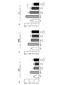
実験の際に発明者らは、受容体介在性の経細胞輸送を介して、LDLRの使用を通じて効率的にBBBを横断できる新規のMPS II特異的スルファターゼ酵素の開発を探求した。ヒトIDS単独、または縦列反復としてのヒトApoE受容体結合領域にリンクされたヒトIDSをコードする新規のレンチウイルスベクターを、ヒト骨髄特異的CD11bプロモーターの下に構築した(図1aに示す)。
Development and In Vitro Validation of IDS Enzymes Targeting the Blood-Brain Barrier We explored the development of specific sulfatase enzymes. Novel lentiviral vectors encoding human IDS alone or linked to human ApoE receptor binding regions as tandem repeats were constructed under the human myeloid-specific CD11b promoter (shown in FIG. 1a).
IDS遺伝子のC末端にインバリアント可動性リンカーを加えた後に、ヒトApoEの受容体結合部分のコドン最適化配列を縦列反復として加えることによって、このスルファターゼを改変した(図1bに示す)。通常、リンカーおよびペプチドの付加によってタンパク質の折り畳みが変わり、酵素活性に有害な影響を与える可能性がある。 This sulfatase was modified by adding an invariant flexible linker to the C-terminus of the IDS gene followed by codon-optimized sequences of the receptor-binding portion of human ApoE as tandem repeats (shown in FIG. 1b). Addition of linkers and peptides usually alters protein folding, which can adversely affect enzymatic activity.
本発明のコンストラクトがなおもIDSの過剰発現および分泌を可能にすることを検証するために、ヒトミクログリア細胞株(CHME3)をLV.IDSまたはLV.IDS.ApoEIIのいずれかを発現するプラスミドDNAによってトランスフェクトした。LV.IDSおよびLV.IDS.ApoEIIによって、それぞれ26倍および24倍の細胞活性の増加を観察した(図1cに示す)。より重要なことに、トランスフェクションの72時間後に分泌IDS活性の84倍および81倍の増加を検出した(図1d)。このことは特に、C末端の改変がインビトロにおける改変IDS酵素の分泌または発現に負の影響を与えないことを示す。24時間後にマウス内皮bEND.3細胞におけるIDSに対するIDS.ApoEIIの取り込みの有意な増加を検出し(図1E、図1F)、これは排他的ではないが主にM6P受容体を介するものであった(図1F)。 To verify that the constructs of the present invention still allow overexpression and secretion of IDS, a human microglial cell line (CHME3) was grown to LV. IDS or LV. IDS. ApoEII was transfected with plasmid DNA expressing either ApoEII. LV. IDS and LV. IDS. A 26- and 24-fold increase in cell activity, respectively, was observed with ApoEII (shown in Figure 1c). More importantly, we detected an 84-fold and 81-fold increase in secreted IDS activity 72 h after transfection (Fig. 1d). This in particular indicates that the C-terminal modification does not negatively affect the secretion or expression of the modified IDS enzyme in vitro. Twenty-four hours later, mouse endothelial bEND. IDS.3 versus IDS in 3 cells. A significant increase in uptake of ApoEII was detected (FIGS. 1E, 1F), mainly but not exclusively through the M6P receptor (FIG. 1F).
LV.IDSおよびLV.IDS.ApoEIIの介在する幹細胞遺伝子治療は、脳におけるIDS酵素活性を改善し、末梢器官において超生理学的レベルの活性IDSを発現する
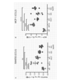
IDSまたはIDS.ApoEIIを発現するLV.IDSまたはLV.IDS.ApoEIIのいずれかで形質導入したMPS IIドナーからの4x10⁵の系統枯渇した造血幹細胞(HSC)を、16匹のブスルファン条件付けした6~8週齢のMPS IIマウスレシピエントに移植した(図2aに示す)。同種幹細胞移植と同等に、改変していない全骨髄細胞も、完全に骨髄破壊したMPS IIレシピエントに注射しており、これを以後WT-HSCTと呼ぶ。
LV. IDS and LV. IDS. ApoEII-mediated stem cell gene therapy improves IDS enzymatic activity in the brain and expresses supraphysiological levels of active IDS in peripheral organs. ApoEII-expressing LV. IDS or LV. IDS. 4×10 5 lineage-depleted hematopoietic stem cells (HSCs) from MPS II donors transduced with either ApoEII were transplanted into 16 busulfan-conditioned 6- to 8-week-old MPS II mouse recipients (Fig. 2a). show). Equivalent to allogeneic stem cell transplantation, unmodified whole bone marrow cells have also been injected into fully myeloablated MPS II recipients, hereafter referred to as WT-HSCT.
移植前に、コロニー形成単位(CFU:colony-forming unit)アッセイから単離した系統枯渇したHSC(lin-HSC:lineage-depleted HSC)において、IDS活性およびベクターコピー数(VCN:vector copy number)を測定した。この実験は、LV.IDSおよびLV.IDS.ApoEIIで形質導入したHSCにおける平均ベクターコピー数が3.1および3.8であること(図2bに示す)、ならびにIDS酵素の過剰発現がそれぞれWTの124倍および152倍であること(図2cに示す)を示した。これらのデータは、造血細胞におけるベクターゲノム組み込み数と、酵素過剰発現との正の相関を示す。移植の4週間後の末梢WBCのフローサイトメトリー分析は、MPS IIレシピエントへの形質導入細胞の完全な生着を示し、80~100%のドナーCD45.1+細胞を達成した(図2dに示す)。
Prior to transplantation, IDS activity and vector copy number (VCN) were measured in lineage-depleted HSCs (lin-HSCs) isolated from colony-forming unit (CFU) assays. It was measured. This experiment was carried out by LV. IDS and LV. IDS. Average vector copy numbers of 3.1 and 3.8 in HSCs transduced with ApoEII (shown in Figure 2b) and overexpression of the IDS enzyme 124-fold and 152-fold over WT, respectively (Figure 2c). ) was shown. These data show a positive correlation between the number of vector genome integrations and enzyme overexpression in hematopoietic cells. Flow cytometric analysis of
MPS IIにおけるこの新規の遺伝子治療の治療有効性を評価するために、各群からの6匹の動物を8月齢で屠殺して、中枢および末梢器官の生化学的分析を行った。全BM、WBC、脾臓、および脳においてベクター組み込みを検出し、WBCにおけるLV.IDS.ApoEII処置マウスのみが有意に低い平均を有した(図2eに示す)。少数の動物は、評価されたすべての器官において一貫してより低いVCNを示したが、別の移植手順において改変HSCを受取った。LV.IDSおよびLV.IDS.ApoEII処置群の両方に対するBM、血漿、および脾臓において、超生理学的レベルのIDSを観察した。脳におけるIDS酵素活性レベルも未処置MPS II動物に比べて上昇しており、それはWT脳における通常のIDS活性の1.5%から8%に相当する。WT-HSCT移植はBM、血漿、および脾臓のみにおいてWTに相当するIDSレベルを回復させたが、脳においては未処置MPS II動物に比べて顕著なIDS活性の増加を検出しなかった。 To assess the therapeutic efficacy of this novel gene therapy in MPS II, 6 animals from each group were sacrificed at 8 months of age for biochemical analysis of central and peripheral organs. Vector integration was detected in whole BM, WBC, spleen, and brain, and LV. IDS. Only ApoEII-treated mice had significantly lower means (shown in Fig. 2e). A few animals consistently showed lower VCN in all organs evaluated but received modified HSCs in a separate transplantation procedure. LV. IDS and LV. IDS. Supraphysiological levels of IDS were observed in BM, plasma, and spleen for both ApoEII-treated groups. IDS enzyme activity levels in the brain were also elevated compared to untreated MPS II animals, corresponding to 1.5% to 8% of normal IDS activity in WT brain. WT-HSCT transplantation restored WT-equivalent IDS levels only in BM, plasma, and spleen, whereas no significant increase in IDS activity was detected in brain compared to untreated MPS II animals.
有利なことに、LV.IDS.ApoEII処置動物の血漿のIDS活性は、LV.IDS処置群よりも3倍高く、これは酵素の安定性および循環時間、分泌または細胞への取り込みが変わった可能性を示唆する(図2fに示す)。さらに、VCNとIDS酵素活性レベルとの正の相関は、IDS遺伝子のゲノム組み込みの増加によって酵素の過剰発現が駆動されていることをさらに示唆する(図2g)。しかし、血漿におけるWBC VCNとIDS活性との相関は、LV.IDS群とLV.IDS.ApoEII群とで有意差を示し、ここではLV.IDS.ApoEII WBCにおける低VCNの結果もたらされるIDSレベルがどのLV.IDS処置マウスよりも高く、これは酵素の産生、取り込み、または分泌の機構の相違を示唆している。加えて、LV.IDS.ApoEII処置動物の血漿におけるより高いIDSレベルは、脳におけるより高いIDS酵素レベルをもたらさず、脳における酵素レベルは両方のLV処置群において同等である(図2fに示す)。血漿における注射した酵素のクリアランス速度は、IDS群とIDS.ApoEII群とで類似であった(図2J)。BM、脾臓、脳、肺、肝臓、および心臓におけるVCNとIDS酵素活性レベルとの正の相関は、IDSおよびIDS.ApoEIIの両方に対してほぼ同等の活性/VCNを伴って観察しており、これはIDS遺伝子のゲノム組み込みの増加によって酵素の過剰発現が駆動されていることを示唆する(図2K)。 Advantageously, LV. IDS. IDS activity in the plasma of ApoEII-treated animals was LV. It was 3-fold higher than the IDS-treated group, suggesting that enzyme stability and circulation time, secretion or cellular uptake may have been altered (shown in Fig. 2f). Moreover, the positive correlation between VCN and IDS enzyme activity levels further suggests that enzyme overexpression is driven by increased genomic integration of IDS genes (Fig. 2g). However, the correlation between WBC VCN and IDS activity in plasma is LV. IDS group and LV. IDS. A significant difference from the ApoEII group was shown, and here the LV. IDS. At which LV. higher than in IDS-treated mice, suggesting differences in the mechanisms of enzyme production, uptake, or secretion. In addition, LV. IDS. Higher IDS levels in plasma of ApoEII-treated animals did not result in higher IDS enzyme levels in brain, and enzyme levels in brain are comparable in both LV-treated groups (shown in Figure 2f). The clearance rate of the injected enzyme in plasma was different between the IDS group and the IDS. It was similar with the ApoEII group (Fig. 2J). A positive correlation between VCN and IDS enzymatic activity levels in BM, spleen, brain, lung, liver, and heart was observed in IDS and IDS. We observed nearly equivalent activity/VCN for both ApoEIIs, suggesting that increased genomic integration of the IDS gene is driving overexpression of the enzyme (Fig. 2K).
血漿におけるLV.IDSにおける2.56単位/VCNに比べて、LV.IDS.ApoEIIにおいては15.41単位/VCNの酵素活性の値を検出しており、これはApoEII残基の融合によって血漿におけるIDS酵素活性が有意に増加することを示唆する。IDS.ApoEIIにおける酵素活性のこの増加は、血漿におけるIDSタンパク質に対するIDS酵素活性の比較によってさらに示され、IDS.ApoEIIはIDSタンパク質の単位当りの活性がより高いことを示している(図2M)。 LV. Compared to 2.56 units/VCN in IDS, LV. IDS. An enzymatic activity value of 15.41 units/VCN was detected for ApoEII, suggesting that fusion of ApoEII residues significantly increases IDS enzymatic activity in plasma. IDS. This increase in enzymatic activity in ApoEII was further demonstrated by comparison of IDS enzymatic activity to IDS proteins in plasma, showing that IDS. ApoEII shows higher activity per unit of IDS protein (Fig. 2M).
リソソームの恒常性
MPS IIにおけるリソソームの機能障害および主要貯蔵材料の制御されない蓄積は、リソソームの調節およびその後の恒常性に大きな影響を有すると考えられる。重要なことに、神経変性はリソソーム機能障害およびオートファジー障害に関連する。たとえばMPS IIなどの疾患状態において、内在性リソソーム酵素は、別のリソソーム酵素の欠損を補償できる。未処置MPS II動物の血漿(WTの2倍)、脾臓(WTの1.53倍)、および脳(WTの2倍)において、リソソームヒドロラーゼβ-ヘキソサミニダーゼのレベル上昇を検出した(図2N~P)。β-ヘキソサミニダーゼの血漿、脾臓、および脳レベルは、LV.IDSおよびLV.IDS.ApoEII処置群の両方においてWTレベルまで完全に正常化した(図2N~P)。WT-HSCTは血漿および脾臓におけるβ-ヘキソサミニダーゼレベルを正常化し(図2N、図2O)、脳におけるレベルをWTレベルまでの完全な正常化は達成せずに回復させた(図2P)。
Lysosomal Homeostasis Lysosomal dysfunction and uncontrolled accumulation of major storage materials in MPS II are thought to have profound effects on lysosomal regulation and subsequent homeostasis. Importantly, neurodegeneration is associated with lysosomal dysfunction and autophagy impairment. For example, in disease states such as MPS II, endogenous lysosomal enzymes can compensate for deficiencies in other lysosomal enzymes. Elevated levels of lysosomal hydrolase β-hexosaminidase were detected in plasma (2x WT), spleen (1.53x WT), and brain (2x WT) of untreated MPS II animals (Fig. 2N-P). Plasma, spleen, and brain levels of β-hexosaminidase were measured in LV. IDS and LV. IDS. There was complete normalization to WT levels in both ApoEII treated groups (Fig. 2N-P). WT-HSCT normalized β-hexosaminidase levels in plasma and spleen (Fig. 2N, Fig. 2O) and restored levels in brain without achieving complete normalization to WT levels (Fig. 2P). .
ヘパラン硫酸の蓄積をLV.IDS.ApoEIIは完全に正常化するがLV.IDSは正常化しない
脳サンプルからHSおよびCS/DSグリコサミノグリカンを精製し、逆相HPLCによって分析および定量化した。MPSIIマウスおよびWT-HSCTで処置したマウスの脳において、合計HSの6倍の増加を検出した(図6A)。LV.IDSで処置したマウスは、脳における合計HS蓄積がWTレベルの約3倍までの減少を示した。最も重要なことに、LV.IDS.ApoEII処置マウスは、脳HSレベルのWTレベルまでの完全な正常化、およびLV.IDSと比較して有意に低いレベルのHSを示した(図6A)。
Accumulation of heparan sulfate was measured by LV. IDS. ApoEII completely normalizes, but LV. IDS Does Not Normalize HS and CS/DS glycosaminoglycans were purified from brain samples and analyzed and quantified by reverse-phase HPLC. A six-fold increase in total HS was detected in the brains of MPSII and WT-HSCT-treated mice (Fig. 6A). LV. Mice treated with IDS showed a reduction in total HS accumulation in the brain up to approximately 3-fold that of WT levels. Most importantly, LV. IDS. ApoEII-treated mice showed complete normalization of brain HS levels to WT levels, and LV. It showed significantly lower levels of HS compared to IDS (Fig. 6A).
MPS IIマウスにおけるHS組成分析は、脳HSの31.1%が完全に硫酸化したUA(2S)-GlcNS(6S)からなることを示し、これに比べて対照WTマウスでは12.3%であった(図6B)。WTマウスにおいて、これらは一般的に鎖に沿って他の硫酸化二糖とクラスタを形成し、MPS IIマウスにおいてはHS鎖全体に沿って硫酸化が有意に増加する。この傾向を、WT-HSCT、LV.IDS、および最も効果的にLV.IDS.ApoEIIによって逆転した。同様に、UA(2S)-GlcNSはWTマウスにおける16.8%からMPSIIマウスにおける31.1%に増加することを観察し、これはLV.IDS.ApoEII群のみにおいて18.4%まで完全に補正されて減少した。 HS composition analysis in MPS II mice showed that 31.1% of brain HS consisted of fully sulfated UA(2S)-GlcNS(6S) compared to 12.3% in control WT mice. There was (Fig. 6B). In WT mice they generally cluster with other sulfated disaccharides along the chain, and in MPS II mice there is a significant increase in sulfation along the entire HS chain. This trend is documented in WT-HSCT, LV. IDS, and most effectively LV. IDS. reversed by ApoEII. Similarly, we observed an increase in UA(2S)-GlcNS from 16.8% in WT mice to 31.1% in MPSII mice, which was consistent with LV. IDS. There was a fully corrected reduction to 18.4% in the ApoEII group only.
加えて、合計脳CS/DSレベルがLV.IDS.ApoEIIマウスにおいてMPS IIと比べて有意に低減することを観察した(補足図2A)が、WTとMPS IIマウスとの有意差は検出できなかった。UA(2S)-GalNAc(4S)はWTマウスにおける1.03%からMPS IIマウスにおける6.5%に増加することを検出し、これはLV.IDS.ApoEII群において完全な補正が得られた。 In addition, total brain CS/DS levels were LV. IDS. We observed a significant reduction in ApoEII mice compared to MPS II (Supplementary Fig. 2A), but no significant difference could be detected between WT and MPS II mice. We detected an increase in UA(2S)-GalNAc(4S) from 1.03% in WT mice to 6.5% in MPS II mice, which is consistent with LV. IDS. Complete correction was obtained in the ApoEII group.
LV.IDS.ApoEIIは脳全体にわたるニューロンのリソソーム蓄積を補正する
次いで、8月齢(移植の6ヵ月後)の移植MPS IIマウスの脳におけるIDS酵素レベルの増加が、ニューロン(NeuN)におけるリソソーム拡大および基質蓄積に与える影響を、リソソームマーカーLAMP2を用いて定めた。WT動物は、運動皮質(層V/VI)においてNeuNと部分的にしか共存しない弱い点状の核周囲LAMP2染色を示した。未処置MPS II動物およびWT-HSCT処置動物は、運動皮質、尾状核被殻、海馬、および扁桃体においてNeuNおよびLAMP2の強い共存染色を示し、ニューロンまたは衛星グリア細胞における重いリソソーム負荷を示唆した(図6C)。大脳皮質、尾状核被殻、および扁桃体におけるLV.IDSの介在する改善、ならびに海馬において充血したリソソームコンパートメントがなおもみられることは、脳に検出されるHSのレベルに強く相関する主要基質蓄積の単なる部分的な補正のみを示唆する(図6A、図6B)。LV.IDS.ApoEIIは皮質、尾状核被殻、および海馬における増加したLAMP2染色を完全に正常化し、扁桃体におけるリソソーム負荷の量をかなり低減した(図6C)。
LV. IDS. ApoEII Corrects Neuronal Lysosomal Accumulation Throughout the Brain Increasing IDS Enzyme Levels in the Brain of Transplanted MPS II Mice at 8 Months of Age (6 Months After Transplantation) Then Contributes to Lysosomal Expansion and Substrate Accumulation in Neurons (NeuN) Effects were defined using the lysosomal marker LAMP2. WT animals displayed weak punctate perinuclear LAMP2 staining that was only partially colocalized with NeuN in the motor cortex (layers V/VI). Untreated MPS II and WT-HSCT treated animals showed strong co-staining of NeuN and LAMP2 in motor cortex, caudate putamen, hippocampus, and amygdala, suggesting heavy lysosomal load in neurons or satellite glial cells ( Figure 6C). LV. in the cerebral cortex, caudate-putamen, and amygdala. The IDS-mediated amelioration, as well as the continued presence of congested lysosomal compartments in the hippocampus, suggests only a partial correction of major substrate accumulation, which strongly correlates with levels of HS detected in the brain (Fig. 6A, Figure 6). 6B). LV. IDS. ApoEII completely normalized the increased LAMP2 staining in the cortex, caudate putamen, and hippocampus and significantly reduced the amount of lysosomal load in the amygdala (Fig. 6C).
MPS IIマウスにおいてLV.IDS.ApoEIIは神経炎症を完全に正常化するのに対し、LV.IDSは改善を仲介する
アストロサイトはMPS障害における強い神経炎症反応に介在することが見出されており、これは反応性のグリオーシス、アストログリオーシス、および炎症性サイトカインのレベル増加につながる。対照および処置MPS IIマウスの脳冠状切片を、アストロサイトのマーカーGFAP(グリア線維関連タンパク質;緑色)およびLAMP2(赤色)で染色した。未処置MPS IIにおいてはWTマウスよりも顕著に多いGFAP染色を皮質、尾状核被殻、および扁桃体において観察し、これは広範囲のアストログリオーシスを示した(図7A)。加えて、未処置およびWT-HSCTマウスの尾状核被殻、海馬、および扁桃体においてGFAPおよびLAMP2の強い共存を観察し(図7A)、ニューロンに加えてアストロサイトにおける顕著なリソソーム基質蓄積を示唆した(図6C)。LV.IDS.ApoEIIはアストログリオーシスを完全に抑制して、皮質、尾状核被殻、海馬、および扁桃体におけるLAMP2染色を減少させてWTレベルに戻すことができ、このことは基質貯蔵および炎症性サイトカインの減少と相関した(図6A、図6B、図6A~D)。同様に、LV.IDSは皮質、尾状核被殻、および扁桃体に存在する反応性アストロサイトの数を減少させた。
LV. IDS. ApoEII completely normalizes neuroinflammation, whereas LV. IDS Mediates Improvement Astrocytes have been found to mediate a robust neuroinflammatory response in MPS disorders, leading to reactive gliosis, astrogliosis, and increased levels of proinflammatory cytokines. Brain coronal sections of control and treated MPS II mice were stained with the astrocyte markers GFAP (glial fibril associated protein; green) and LAMP2 (red). Significantly more GFAP staining was observed in cortex, caudate putamen, and amygdala in untreated MPS II than in WT mice, indicating extensive astrogliosis (Fig. 7A). In addition, we observed strong co-localization of GFAP and LAMP2 in the caudate putamen, hippocampus, and amygdala of untreated and WT-HSCT mice (Fig. 7A), suggesting prominent lysosomal substrate accumulation in astrocytes in addition to neurons. (Fig. 6C). LV. IDS. ApoEII was able to completely suppress astrogliosis and reduce LAMP2 staining in cortex, caudate putamen, hippocampus, and amygdala back to WT levels, which was associated with a decrease in substrate storage and inflammatory cytokines. correlated (FIGS. 6A, 6B, 6A-D). Similarly, LV. IDS decreased the number of reactive astrocytes present in cortex, caudate putamen, and amygdala.
LV.IDS.ApoEIIはMPS IIマウスにおける認知異常ならびに協調およびバランスを完全に補正する
MPS IIマウスモデルにおけるCNSおよび骨格の表現型がLV.IDSまたはLV.IDS.ApoEII改変HSCによって回復し得るかどうかを理解するために、各群のすべての処置動物に対して移植6ヵ月後に相当する8月齢において認知および感覚運動性能の機能研究を行った。10分間のY迷路テストを用いて空間的作業記憶の認知的評価を評価し、このY迷路テストはマウスが最近探索したアームよりも新規のアームを探索することを生得的に好むことを利用したものである(図3aに示す)。
LV. IDS. ApoEII Completely Corrects Cognitive Defects and Coordination and Balance in MPS II Mice IDS or LV. IDS. To understand whether ApoEII-modified HSCs can be restored, functional studies of cognitive and sensorimotor performance were performed at 8 months of age corresponding to 6 months post-transplantation for all treated animals in each group. Cognitive assessments of spatial working memory were assessed using a 10-minute Y-maze test, which exploited the innate preference of mice to explore novel arms over recently explored arms. (shown in FIG. 3a).
神経認知評価は、LV.IDS.ApoEII処置マウスにおける自発的交替の完全な正常化を示したが、LV.IDS処置群またはWT-HSCT群においては示さなかった(図3b)。このことは、LV.IDS.ApoEIIを用いてMPS IIマウスモデルにおいてみられた認知機能障害の完全な補正を示唆するだけでなく、進行性のMPS II疾患に関連するさらなる認知衰退の防止も示す。興味深いことに、WT-HSCTはMPS IIに関連する認知症状に正の影響を有さず、これはWT-HSCTがMPS IIの脳の処置に好適ではない可能性をさらに強調する。Y迷路の異なるアームへの合計進入数を、全体の活動性の代理尺度として用いることもできる。すべてのテスト群の間に合計進入数の差は検出されず、これはLV.IDS.ApoEII処置動物における認知症状の真の表現型の救済を示唆した(図3cに示す)。 Neurocognitive assessments are performed according to LV. IDS. showed complete normalization of spontaneous alternation in ApoEII-treated mice, whereas LV. None were shown in the IDS-treated or WT-HSCT groups (Fig. 3b). This is confirmed by LV. IDS. Not only do we suggest complete correction of the cognitive impairment seen in the MPS II mouse model using ApoEII, but we also demonstrate prevention of further cognitive decline associated with progressive MPS II disease. Interestingly, WT-HSCT had no positive effect on cognitive symptoms associated with MPS II, further highlighting the possibility that WT-HSCT may not be suitable for brain treatment of MPS II. The total number of entries in different arms of the Y-maze can also be used as a surrogate measure of overall activity. No differences in the total number of entries were detected between all test groups, indicating that LV. IDS. suggesting true phenotypic rescue of cognitive symptoms in ApoEII-treated animals (shown in Figure 3c).
対照および処置マウスに対して、げっ歯類の運動障害における感覚運動の協調およびバランスに対するよく確立されたテストであるロータロッドのテストも行った(図3dに示す)。前に記載されるとおり、8月齢のMPSIIマウスは加速するロッドにおける能力の低減を示した。WT-HSCTを含むすべての移植処置は、これを完全に救済した(図3eに示す)。 The rotarod test, a well-established test for sensorimotor coordination and balance in rodent motor deficits, was also performed on control and treated mice (shown in Fig. 3d). As previously described, 8-month-old MPSII mice showed reduced ability in accelerating rods. All transplantation treatments, including WT-HSCT, completely rescued this (shown in Figure 3e).
LV.IDS.ApoEIIは神経炎症性サイトカインを正常化するが、LV.IDSは正常化しない
8月齢の対照および処置マウスの全脳抽出物からの慢性神経炎症に関連する多数の炎症性サイトカインを定量化するために、サイトメトリックビーズアレイ(CBA)を用いた。本発明者らは最初に、未処置MPS II動物におけるマクロファージ炎症性タンパク質(macrophage inflammatory protein)(MIP-1α/CCL3)、インターロイキン1α(IL-1α:interleukin 1α)タンパク質、RANTES(CCL5)、および単球走化性タンパク質(monocyte chemoattractant protein)(MCP-1/CCL2)の有意な増加を識別した(図4a、図4b、図4c、図4dに示す)。MIP-1α、IL-1α、およびRANTESタンパク質レベルをLV.IDS.ApoEIIは正常化し、LV.IDSは部分的にのみ正常化し、MCP-1レベルはすべての群において上昇したままだった(図4bに示す)。
LV. IDS. ApoEII normalizes neuroinflammatory cytokines, whereas LV. IDS Does Not Normalize Cytometric bead arrays (CBA) were used to quantify a number of inflammatory cytokines associated with chronic neuroinflammation from whole brain extracts of 8-month-old control and treated mice. We first demonstrated macrophage inflammatory protein (MIP-1α/CCL3), interleukin 1α (IL-1α) protein, RANTES (CCL5), and A significant increase in monocyte chemoattractant protein (MCP-1/CCL2) was identified (shown in Figures 4a, 4b, 4c, 4d). MIP-1α, IL-1α, and RANTES protein levels were measured by LV. IDS. ApoEII normalized and LV. IDS was only partially normalized and MCP-1 levels remained elevated in all groups (shown in Figure 4b).
加えて本発明者らは、未処理MPS IIマウスの皮質および線条体において、イソレクチンB4(ILB4)陽性細胞のそれぞれ30倍および25倍の増加を観察し(図4E、図4F、図4G)、これはミクログリアの広範囲の活性化およびその後の神経炎症を示唆する。興味深いことに、WT-HSCTおよびLV.IDSはどちらもILB4染色を皮質においてWTレベルのそれぞれ14倍および12倍に低減し(図4F)、線条体においてWT染色の15倍および13倍に低減した(図4G)。最も重要なことに、LV.IDS.ApoEIIは皮質および線条体の両方において活性化したミクログリアの数を完全に正常化でき(図4F、図4G)、このことは前に観察した神経炎症サイトカインの低減およびLV.IDS.ApoEIIにおけるアストロサイト増加の完全な抑止と強く相関する。 In addition, we observed a 30-fold and 25-fold increase in isolectin B4 (ILB4)-positive cells, respectively, in the cortex and striatum of untreated MPS II mice (Fig. 4E, 4F, 4G). , which suggests widespread activation of microglia and subsequent neuroinflammation. Interestingly, WT-HSCT and LV. Both IDS reduced ILB4 staining to 14- and 12-fold, respectively, WT levels in the cortex (Fig. 4F) and to 15- and 13-fold WT staining in the striatum (Fig. 4G). Most importantly, LV. IDS. ApoEII was able to completely normalize the number of activated microglia in both the cortex and striatum (FIGS. 4F, 4G), which is consistent with the previously observed reduction in neuroinflammatory cytokines and LV. IDS. Strongly correlates with complete abrogation of astrocyte proliferation in ApoEII.
このことは全体として、MPS IIにおける神経炎症性サイトカインおよびケモカインの放出、反応性アストログリオーシス、およびミクログリアの活性化をLV.IDS.ApoEIIは完全に抑止でき、LV.IDSは部分的にのみ抑止できることを示唆する。 Overall, this suggests that the release of neuroinflammatory cytokines and chemokines, reactive astrogliosis, and microglial activation in MPS II are associated with LV. IDS. ApoEII can be completely abrogated and LV. It suggests that IDS can only be partially deterred.
ヘパラン硫酸の蓄積をLV.IDS.ApoEIIは完全に正常化するがLV.IDSは正常化しない
対照および処置MPS IIマウスの脳からHS GAGを精製し、細菌ヘパリナーゼ酵素を用いて脱重合して個々のHS二糖にした後、AMACを用いて個々の二糖の還元末端の蛍光タグ付けを行った。逆相HPLC分離を用いて、各HS二糖の個別の寄与および硫酸化パターンを定量化および決定した。
Accumulation of heparan sulfate was measured by LV. IDS. ApoEII completely normalizes, but LV. IDS does not normalize HS GAGs were purified from the brains of control and treated MPS II mice, depolymerized into individual HS disaccharides using bacterial heparinase enzymes, and then reduced ends of individual disaccharides using AMAC. was fluorescently tagged. Reversed-phase HPLC separation was used to quantify and determine the individual contribution and sulfation pattern of each HS disaccharide.
HPLC分析は、WT、未処置MPS II、およびすべての処置群の合計HSの相対的レベルの算出を可能にする(図4eに示す)。MPSIIマウスおよびWT-HSCTで処置したマウスの脳において、合計HSの6倍の増加を検出した(p<0.0001)。興味深いことに、LV.IDSで処置したマウスは、脳における合計HS蓄積がWTレベルの約3倍までの減少を示した。最も重要なことに、LV.IDS.ApoEII処置マウスは、脳HSレベルのWTレベルまでの完全な正常化を示した(図4eに示す)。 HPLC analysis allows calculation of relative levels of WT, untreated MPS II, and total HS in all treatment groups (shown in Figure 4e). A 6-fold increase in total HS was detected in the brains of MPSII and WT-HSCT-treated mice (p<0.0001). Interestingly, LV. Mice treated with IDS showed a reduction in total HS accumulation in the brain up to approximately 3-fold that of WT levels. Most importantly, LV. IDS. ApoEII-treated mice showed complete normalization of brain HS levels to WT levels (shown in Figure 4e).
HS組成分析は、未処理MPS II脳における三硫酸化二糖UA(2S)-GlcNS(6S)の有意な増加、およびUA(2S)-GlcNSの増加を示した(図4fに示す)。この傾向をWT-HSCTおよびLV.IDSは部分的に逆転させたが、LV.IDS.ApoEII群だけがWTレベルまで戻す完全な補正を得た。 HS composition analysis showed a significant increase in trisulfated disaccharide UA(2S)-GlcNS(6S) and an increase in UA(2S)-GlcNS in untreated MPS II brains (shown in Figure 4f). This trend was observed in WT-HSCT and LV. IDS partially reversed, but LV. IDS. Only the ApoEII group obtained complete correction back to WT levels.
一硫酸化HS二糖は著しく異なる傾向を示し、UA-GlcNSの未処置MPS IIおよびWT-HSCT処置レベルはWT脳よりも低かった(図4fに示す)。LV.IDS処置はこれを部分的に改善したが、LV.IDS.ApoEII群のみがこれを完全に補正した。 Monosulfated HS disaccharides showed markedly different trends, with untreated MPS II and WT-HSCT treated levels of UA-GlcNS lower than in WT brain (shown in Figure 4f). LV. IDS treatment partially ameliorated this, but LV. IDS. Only the ApoEII group corrected this completely.
MPS IIマウスにおけるすべての移植戦略が骨格病理を寛解させる
MPS IIマウスモデルにおける骨格症状の程度に対する強固なデータを得るために、対照および処置マウスに対する完全麻酔下の全身X線を行った。ImageJイメージングソフトウェアを用いて、頬骨弓ならびに四肢長管骨(上腕骨および大腿骨)の幅を測定した。
All Transplantation Strategies Ameliorate Skeletal Pathology in MPS II Mice To obtain robust data on the extent of skeletal symptoms in the MPS II mouse model, whole-body X-rays under total anesthesia were performed on control and treated mice. The width of the zygomatic arch and limb long bones (humerus and femur) was measured using ImageJ imaging software.
未処置MPS II動物において有意に増加する頬骨弓の幅は、WT-HSCTを含むすべての移植群においてWTの寸法まで低減した(図5a、図5bに示す)。上腕骨の幅はWT-HSCTおよびLV.IDS群において有意に低減したが、LV.IDS.ApoEII処置動物のみが完全な補正を得た(図5cに示す)。さらに、すべての処置動物における大腿骨の幅は、WT動物と比べて有意差を示さず、顕著な骨格の救済を示唆した。 The width of the zygomatic arch, which increased significantly in untreated MPS II animals, was reduced to WT dimensions in all graft groups, including WT-HSCT (shown in Figures 5a, 5b). Humeral width was measured by WT-HSCT and LV. Although significantly reduced in the IDS group, LV. IDS. Only ApoEII-treated animals obtained complete correction (shown in Figure 5c). Furthermore, femoral width in all treated animals showed no significant difference compared to WT animals, suggesting significant skeletal salvage.
WT、LV.IDSまたはLV.IDS.ApoEII改変HSCの移植後に末梢炎症が抑止される
8月齢のMPS IIマウスの肝臓においてMCP-1、MIP-1α、およびRANTESのレベルの上昇を検出し、それをWT-HSCT、LV.IDSまたはLV.IDS.ApoEII処置が完全に抑止した(図5E~5G)。これらのデータは、WT-HSCT、LV.IDS、およびLV.IDS.ApoEIIがMPS IIマウスにおける慢性的末梢炎症を完全に補正するために十分な量の末梢酵素を産生することを示唆する。
WT, LV. IDS or LV. IDS. Peripheral inflammation is abrogated after transplantation of ApoEII-modified HSCs. IDS or LV. IDS. ApoEII treatment completely abrogated (FIGS. 5E-5G). These data are based on WT-HSCT, LV. IDS, and LV. IDS. We suggest that ApoEII produces sufficient amounts of peripheral enzymes to completely correct chronic peripheral inflammation in MPS II mice.
WT-HSCT、LV.IDS、およびLV.IDS.ApoEII群において、心不全マーカーNppbおよびMyh7の遺伝子発現が正常化する
MPS IIマウスモデルにおける心不全のリスクがより高いことの指標となり得る、心筋症および心臓病理に関連する2つのマーカーであるNppbおよびMyh7の発現を調査した。心室から分泌されて筋細胞の伸展および血圧を調節する脳ナトリウム利尿ペプチド(BNP:brain natriuretic peptide)をコードするNppbの発現は、MPS IIにおいてWTオスマウスよりも約16倍高いことを見出した(図5H)。WT-HSCT、LV.IDS、およびLV.IDS.ApoEII群において、Nppb発現はWTレベルまで完全に安定化した(図5H)。主に胎仔の心臓において見出され、心筋およびI型筋線維の主要構成要素としてのミオシン重鎖ベータをコードするMyh7の発現は、未処置MPS II動物において上昇した。WT-HSCT、LV.IDS、およびLV.IDS.ApoEII群におけるMyh7発現は、WT発現と類似であった(図5I)。
WT-HSCT, LV. IDS, and LV. IDS. Gene expression of heart failure markers Nppb and Myh7 is normalized in the ApoEII group. Expression was investigated. We found that the expression of Nppb, which encodes brain natriuretic peptide (BNP), which is secreted from the ventricles and regulates myocyte spreading and blood pressure, was approximately 16-fold higher in MPS II than in WT male mice (Fig. 5H). WT-HSCT, LV. IDS, and LV. IDS. In the ApoEII group, Nppb expression was completely stabilized to WT levels (Fig. 5H). Expression of Myh7, found primarily in the fetal heart and encoding myosin heavy chain beta as a major component of myocardium and type I muscle fibers, was elevated in untreated MPS II animals. WT-HSCT, LV. IDS, and LV. IDS. Myh7 expression in the ApoEII group was similar to WT expression (Fig. 5I).
LV.IDSおよびLV.IDS.ApoEII形質導入HSCの移植に続くIDSの過剰発現は、ヒトIDSに対する免疫応答をもたらさない
遺伝子改変細胞は移植後のヒトIDSに対する耐性を仲介できたかどうかを調べるために、本発明者らは、完全な骨髄破壊的条件付けの後に、どちらもヒトIDSを過剰発現するLV.IDSまたはLV.IDS.ApoEII移植のいずれかを受けたマウスの血漿のヒトIDSに対するIgG抗体を分析した。LV.IDSおよびLV.IDS.ApoEII群における全体のIDS特異的IgG力価は正常範囲にとどまり、酵素に対する免疫応答に寄与しなかった(図5J)。
LV. IDS and LV. IDS. Overexpression of IDS following transplantation of ApoEII-transduced HSCs does not result in an immune response to human IDS To investigate whether genetically modified cells could mediate resistance to human IDS after transplantation, we performed a complete LV. IDS or LV. IDS. Plasma of mice receiving either ApoEII transplantation was analyzed for IgG antibodies against human IDS. LV. IDS and LV. IDS. Overall IDS-specific IgG titers in the ApoEII group remained in the normal range and did not contribute to the immune response to the enzyme (Fig. 5J).
LV.IDS.ApoEII処置は複数の機構を通じて作用する
LV.IDS.ApoEII処置マウスの血漿における酵素活性の増加およびVCN当りの酵素活性の増加の観察(図2f、図2j)の後、本発明者らは酵素の循環時間が増加したのではないかという仮説を立てた。等価レベルのIDSおよびIDS.ApoEII酵素をMPS IIマウスに注射して、ELISAによってタンパク質レベルをモニタした(図9A)。興味深いことに、酵素クリアランス速度は、分解または器官への取り込みのいずれによるものであっても、IDSおよびIDS.ApoEII群において類似であり、両方の群において30分以内に効果的に除去された。
LV. IDS. ApoEII treatment acts through multiple mechanisms LV. IDS. After observing increased enzyme activity in the plasma of ApoEII-treated mice and increased enzyme activity per VCN (Fig. 2f, Fig. 2j), we hypothesized that the circulation time of the enzyme might have been increased. rice field. Equivalent levels of IDS and IDS. ApoEII enzyme was injected into MPS II mice and protein levels were monitored by ELISA (Fig. 9A). Interestingly, the enzyme clearance rate, whether due to degradation or organ uptake, was significantly different from that of IDS and IDS. It was similar in the ApoEII group and was effectively cleared within 30 minutes in both groups.
次に、ApoEII残基の付加によって酵素の立体構造が変化して、分解に対する付加的な保護を与えたかもしれないという仮説を立てて、合計IDSタンパク質と比較した血漿における活性酵素のレベルをELISAによって比較および測定した。血漿において、IDS.ApoEIIはIDSタンパク質の単位当りの活性がより高いことを示した(図9B)。このことは、天然IDS酵素の活性部位は血漿中で迅速に分解するのに対し、ApoEII付加は保護的であってインビボにおける酵素活性を効果的に延長することを示唆し得る。 Next, hypothesizing that the addition of ApoEII residues might have altered the conformation of the enzyme, conferring additional protection against degradation, the level of active enzyme in plasma relative to total IDS protein was assessed by ELISA. compared and measured by In plasma, IDS. ApoEII showed higher activity per unit of IDS protein (Fig. 9B). This may suggest that the active site of the native IDS enzyme is rapidly degraded in plasma, whereas the ApoEII addition is protective and effectively prolongs enzyme activity in vivo.
BBB内皮細胞株bEND.3を用いて、内皮細胞による酵素取り込みに何らかの差があるかどうかを定めた。これらの細胞はtranswellに有効なBBB層を産生し(図9C)、LDLRおよびLDLR関連タンパク質1(LRP1:LDLR-related protein 1)受容体の両方を発現する(図9D、図9E)。各酵素がHSCGT産生酵素のものと類似のM6P状態を有することを確認するために、本発明者らは、CHME3ヒトミクログリア細胞のトランスフェクションの後に培地に分泌されたIDSおよびIDS.ApoEII酵素を用い、ELISAによって標準化した。単層培養において、本発明者らはIDS対IDS.ApoEIIのbEND.3細胞への取り込みを比較し、ApoEIIペプチドの付加による細胞取り込みの4.7倍の増加を識別した(図9F)。BBBを擬態するためのtranswellにおけるbEND.3内皮細胞の極性化細胞層を横断するIDSまたはIDS.ApoEIIの取り込みおよび経細胞輸送を、次いで比較した。細胞を単層において成長させたときと同様に、取り込みの2.6倍の増加が明らかであり、それとともにtranswellの基底外側への経細胞輸送の1.5倍の増加が明らかであった(図9G、図9H)。 BBB endothelial cell line bEND. 3 was used to determine if there were any differences in enzyme uptake by endothelial cells. These cells produced a transwell-effective BBB layer (FIG. 9C) and expressed both LDLR and LDLR-related protein 1 (LRP1) receptors (FIGS. 9D, 9E). To confirm that each enzyme has an M6P status similar to that of the HSCGT-producing enzyme, we tested IDS and IDS. ApoEII enzyme was used and normalized by ELISA. In monolayer cultures, we tested IDS vs. IDS. ApoEII bEND. We compared uptake into 3 cells and identified a 4.7-fold increase in cellular uptake with the addition of the ApoEII peptide (Fig. 9F). bEND.in the transwell to mimic the BBB. 3 IDS or IDS. ApoEII uptake and transcellular transport were then compared. Similar to when cells were grown in monolayers, a 2.6-fold increase in uptake was evident, along with a 1.5-fold increase in basolateral transcellular transport of the transwell ( 9G, 9H).
加えて、ApoEを用いたApoE依存性受容体の遮断、またはM6Pを用いたM6P受容体の封鎖(図9I)は、IDS.ApoEIIが優先的かつ予想外にM6P受容体を介して取り込まれることを明らかにしたが、ApoEペプチドによる遮断もIDS.ApoEIIの取り込みを約30%有意に阻害した。ApoEIIペプチド配列はHS部位もコードしており、このHS部位はLDLRおよびLRP1を含む受容体へのApoEの受容体/リガンド結合のための共受容体として必要とされる(Jiら、1993)。ApoEペプチドによる結果と同様に、細胞のヘパリナーゼ前処理もIDS.ApoEII取り込みの約30%の低減をもたらしており、ApoE/HSも取り込みおよびbEND3細胞を横断する経細胞輸送に重要である可能性を示唆した。 In addition, blockade of ApoE-dependent receptors with ApoE, or blockade of M6P receptors with M6P (Fig. 9I), was associated with IDS. Although ApoEII was shown to be preferentially and unexpectedly taken up through the M6P receptor, blockade by the ApoE peptide also resulted in IDS. ApoEII uptake was significantly inhibited by about 30%. The ApoEII peptide sequence also encodes an HS site, which is required as a co-receptor for receptor/ligand binding of ApoE to receptors including LDLR and LRP1 (Ji et al., 1993). Similar to the results with ApoE peptides, heparinase pretreatment of cells also resulted in IDS. It resulted in an approximately 30% reduction in ApoEII uptake, suggesting that ApoE/HS may also be important for uptake and transcellular transport across bEND3 cells.
考察
リンカーおよびApoEIIペプチドの付加は、インビトロにおけるIDS酵素の発現、産生、または分泌を変えなかったが、類似のベクターコピー数に対するLV.IDSおよびLV.IDS.ApoEIIベクターの両方を用いて形質導入したHSCにおいて超生理学的レベルのIDS酵素を産生した。すべての移植動物においてブスルファンを用いた完全な骨髄破壊的条件付けを用いて、CD45.1+ドナー細胞(>80%)の完全な生着を得て、移植時の下流インビトロ分化アッセイにおける有意差はなかった(データは示さず)。
Discussion Addition of linkers and ApoEII peptides did not alter IDS enzyme expression, production, or secretion in vitro, but LV. IDS and LV. IDS. Supraphysiological levels of the IDS enzyme were produced in HSCs transduced with both ApoEII vectors. Complete myeloablative conditioning with busulfan in all transplanted animals resulted in complete engraftment of CD45.1+ donor cells (>80%) with no significant differences in downstream in vitro differentiation assays at the time of transplantation. (data not shown).
驚くべきことに、LV.IDS.ApoEII群における血漿のIDS酵素活性レベルは、たとえWBCにおいてより低いVCNであってもLV.IDSより約3倍高かった。このことは、LV.IDS.ApoEII群におけるコピー当りのより高い酵素活性につながり、ApoEII残基は自身の立体構造を変えることによって活性を安定化または改善することを示唆し、酵素不活性化に対する保護効果を提供するが血漿におけるクリアランスに対しては提供しないことを示す(図8)。IDS.ApoEIIは非改変IDSよりもbEND.3細胞への取り込みが高いことを検出しており、これには主にM6PRが介在すると考えられた。IDS.ApoEII融合酵素に用いたapoEの受容体結合部分(残基142~147)は、内皮細胞表面に豊富にあり、かつMPS IIにはさらにより豊富にあるUA(2S)-GlcNS(6S)の4反復で構成される八糖HSフラグメントと高親和性結合複合体を形成できる。HSは通常多くの受容体リガンド相互作用における共受容体の役割をし、ApoEIIを通じたHSプロテオグリカン(HSPG:HS proteoglycans)への結合の増加は、LDLR、LDLR関連タンパク質1(LRP1)、M6Pを通じた細胞取り込み、またはapoE-HSPG複合体の直接の取り込みによる細胞取り込みの増加に介在し得る。 Surprisingly, LV. IDS. Plasma IDS enzyme activity levels in the ApoEII group were higher than LV. It was about three times higher than IDS. This is confirmed by LV. IDS. This leads to higher enzymatic activity per copy in the ApoEII group, suggesting that ApoEII residues stabilize or improve activity by altering their conformation, providing a protective effect against enzyme inactivation, whereas in plasma It shows no provision for clearance (Fig. 8). IDS. ApoEII is more sensitive to bEND. A high uptake into 3 cells was detected, which was thought to be mainly mediated by M6PR. IDS. The receptor-binding portion of apoE (residues 142-147) used for the ApoEII fusion enzyme is the UA(2S)-GlcNS(6S) 4-part, which is abundant on the endothelial cell surface and even more abundant on MPS II. High affinity binding complexes can be formed with octasaccharide HS fragments composed of repeats. HS normally plays a co-receptor role in many receptor-ligand interactions, and increased binding to HS proteoglycans (HSPG) through ApoEII has been mediated through LDLR, LDLR-related protein 1 (LRP1), M6P It may mediate cellular uptake or increased cellular uptake by direct uptake of the apoE-HSPG complex.
LV.IDS群とLV.IDS.ApoEII群とで類似の量の脳内酵素活性を検出したが、LV.IDS.ApoEIIのみにおいてニューロンおよびアストロサイトからのLAMP2のクリアランス改善を伴う完全な補正を検出した。これに対する説明の1つは、IDS.ApoEIIを用いた方が間質空間からの酵素取り込みが効率的なのかもしれず、それによって同じ全体レベルを維持しながら非改変IDSよりもかなり効果的に常在脳細胞を補正するのではないかというものである(図8)。 LV. IDS group and LV. IDS. A similar amount of brain enzymatic activity was detected in the ApoEII group, whereas the LV. IDS. We detected complete correction with improved clearance of LAMP2 from neurons and astrocytes in ApoEII only. One explanation for this is that IDS. Enzyme uptake from the interstitial space may be more efficient with ApoEII, thereby compensating resident brain cells significantly more effectively than unmodified IDS while maintaining the same overall levels. (Fig. 8).
LV.IDS.ApoEII処置動物において、主要貯蔵上昇の完全な補正と、神経炎症において大きな役割を果たす硫酸化パターン形成の全体的正常化とを観察した。脳における主要HS貯蔵の部分的補正は、認知行動を補正するには不十分である。MPS IIにおけるHSの蓄積は、神経炎症および神経変性の悪化を引き起こす第1のシグナルの1つを提供し得ると考えられる。硫酸化パターン形成は細胞機能およびHS結合恒常性にも重要な役割を果たす。なぜなら、6-O-および2-O-硫酸化HSは線維芽細胞増殖因子およびストロマ細胞由来因子1(CXCL12)のシグナル伝達のために必須だからである。実際に、MPS Iにおける異常な過剰HSは、CXCL12の隔離を介した制限条件下でのHSCの移動を低減させることが示された(42)。この研究の状況において、疾患動物の脳組織内に硫酸化二糖が増加することは、IDS.ApoEII酵素の細胞取り込みの改善を促進することによって、疾患細胞を酵素の標的とすることを増強し得る。 LV. IDS. In ApoEII-treated animals, we observed complete correction of major store elevations and global normalization of sulfation patterning, which plays a major role in neuroinflammation. Partial correction of major HS stores in the brain is insufficient to correct cognitive behavior. It is believed that accumulation of HS in MPS II may provide one of the first signals to trigger exacerbation of neuroinflammation and neurodegeneration. Sulfation patterning also plays an important role in cellular function and HS binding homeostasis. This is because 6-O- and 2-O-sulfated HS are essential for fibroblast growth factor and stromal cell-derived factor 1 (CXCL12) signaling. Indeed, aberrant excess HS in MPS I was shown to reduce HSC migration under restrictive conditions through sequestration of CXCL12 (42). In the context of this study, increased sulfated disaccharides in the brain tissue of diseased animals was reported in IDS. By facilitating improved cellular uptake of the ApoEII enzyme, targeting of the enzyme to diseased cells may be enhanced.
ApoE/HSに基づく機構およびM6P受容体の両方を介して、bEND.3細胞におけるIDS.ApoEIIのIDSよりも有意に増加した取り込みを観察した。複数の標的化機構を使用することによって、酵素欠損細胞に対するより効率的な標的化を身体全体にわたって、およびBBBを横断して仲介できた。ここで用いたApoEの受容体結合部分は、内皮細胞表面に豊富にあり、かつMPS IIにはさらにより豊富にあるUA(2S)-GlcNS(6S)の4反復で構成される八糖HSフラグメントと高親和性結合複合体を形成できる。この研究の状況において、疾患動物の脳組織内に硫酸化二糖が増加することは、ApoEIIペプチドのHS結合モチーフを介してIDS.ApoEII酵素の細胞取り込みの改善を促進することによって、疾患細胞を酵素の標的とすることを増強し得る。HSは通常多くの受容体リガンド相互作用における共受容体の役割をし、ApoEIIを通じたHSプロテオグリカンへの結合の増加は、LDLR、LRP1、M6Pを通じた細胞取り込み、またはApoE-HSPG複合体の直接の取り込みによる細胞取り込みの増加に介在し得る。血漿安定性の改善と、BBB取り込みおよび経細胞輸送の増加とを一緒にすることで、脳酵素量の有意差の不在下でLV.IDS.ApoEIIによってみられる脳内のHSおよびDS貯蔵ならびにいくつかのその他の神経病理の正常化を説明できる。IGFII受容体に向けられたペプチドを介してM6Pの代替的エピトープを標的とするGILTタグに結合した酵素を用いて、MPS IIIBおよびポンぺ病における類似の酵素活性に対する取り込み、クリアランス、および細胞結合の増加の類似の効果がみられた。この研究は、天然IDSに対する顕著な変化なしにSGSH.ApoBによる脳酵素活性の少しの増加を示した。 Through both ApoE/HS-based mechanisms and M6P receptors, bEND. IDS.3 in cells. A significantly increased uptake of ApoEII over IDS was observed. By using multiple targeting mechanisms, more efficient targeting to enzyme-deficient cells could be mediated throughout the body and across the BBB. The receptor-binding portion of ApoE used here is an octasaccharide HS fragment composed of 4 repeats of UA(2S)-GlcNS(6S) that is abundant on the endothelial cell surface and even more abundant on MPS II. can form high-affinity binding complexes with In the context of this study, increased sulfated disaccharides in the brain tissue of diseased animals suggested that IDS. By facilitating improved cellular uptake of the ApoEII enzyme, targeting of the enzyme to diseased cells may be enhanced. HS normally plays a co-receptor role in many receptor-ligand interactions, and increased binding to HS proteoglycans via ApoEII may result in cellular uptake via LDLR, LRP1, M6P, or direct activation of the ApoE-HSPG complex. It may mediate increased cellular uptake by uptake. The improved plasma stability combined with increased BBB uptake and transcellular transport suggested that LV. IDS. It may explain the normalization of HS and DS storage in the brain and several other neuropathologies seen by ApoEII. Using a GILT-tagged enzyme that targets an alternative epitope of M6P via a peptide directed to the IGFII receptor, we demonstrated uptake, clearance, and cell-binding effects on similar enzymatic activities in MPS IIIB and Pompe disease. A similar effect of increase was seen. This study demonstrated that SGSH. A small increase in brain enzymatic activity by ApoB was shown.
神経炎症はLSDにおいて一般的に報告されており、これはさまざまな未分解分子の蓄積によって引き起こされると考えられ、これらの分子は協調的に神経炎症性の環境を活性化および永続化して、疾患自体を悪化させるおそれがある。MPS IIマウスは脳において強い炎症反応を誘発し、それはMIP-1α、IL-1α、RANTES、およびMCP-1のレベル上昇を伴う。MIP-1α、IL-1α、およびRANTESレベルはLV.IDS.ApoEII群において完全に正常化し、LV.IDS処置マウスにおいては部分的な減少のみであった。興味深いことに、MPS IIおよびすべてのブスルファン条件付けマウスにおいてはMCP-1レベルが上昇したままであった。本発明者らが前に示したとおり、ブスルファン条件付けは脳におけるMCP-1レベルの長期的増加をもたらし、これはCNSに対する細胞遊出の主要なメディエータであり、かつ長期的な遊出効果を発揮する。静脈内注射したブスルファンの約20%がBBBを横断でき、さらにより強力な移動促進性MCP-1応答を駆動し、これが最終的にMPS IIマウスにおいてドナー由来白血球がBBBを横断してCNSに入る遊出を促進して、酵素を脳実質に輸送する付加的な手段を提供してもよい。 Neuroinflammation has been commonly reported in LSD, which is thought to be caused by the accumulation of various undegraded molecules that cooperatively activate and perpetuate the neuroinflammatory milieu, leading to disease. It can make itself worse. MPS II mice elicit a strong inflammatory response in the brain, which is accompanied by elevated levels of MIP-1α, IL-1α, RANTES, and MCP-1. MIP-1α, IL-1α, and RANTES levels were LV. IDS. Completely normalized in the ApoEII group, LV. There was only a partial reduction in IDS-treated mice. Interestingly, MCP-1 levels remained elevated in MPS II and all busulfan-conditioned mice. As we have previously shown, busulfan conditioning results in a long-term increase in MCP-1 levels in the brain, which is a major mediator of cell transmigration to the CNS and exerts long-term transmigration effects. do. Approximately 20% of intravenously injected busulfan can cross the BBB, driving even more potent promigratory MCP-1 responses that ultimately allow donor-derived leukocytes to cross the BBB and enter the CNS in MPS II mice. Transmigration may be facilitated to provide an additional means of transporting the enzyme to the brain parenchyma.
ミクログリアの活性化およびアストロサイト増加は、MPS障害においてこの研究を含め一般的に報告されている。本発明者らはLV.IDS.ApoEIIマウスの皮質、尾状核被殻、海馬、および扁桃体におけるGFAPおよびLAMP2染色の完全な抑止を観察し、LV.IDS群の同じ範囲においては部分的な補正しか観察しなかった。さらに、LV.IDS.ApoEII処置マウスの皮質および線条体において活性化ミクログリアの完全な正常化を観察し、LV.IDSにおいてはわずか50%の低減であった。LV.IDS.ApoEII処置は、CSFへの直接のAAV9-IDS注射を用いて報告されたアストログリオーシスおよびミクログリア活性化の補正に匹敵する(非特許文献15)。MPS IIマウスの肝臓において末梢炎症を検出し、これはMCP-1、MIP-1α、およびRANTESのレベルの明確な上昇を伴い、かつすべての移植によって抑止され、同種移植によって得られる末梢IDS酵素レベルは末梢における炎症の低減を仲介し得ることを示した。 Microglial activation and astrocyte hyperplasia have been commonly reported in MPS disorders, including in this study. The present inventors found that LV. IDS. Observing complete abrogation of GFAP and LAMP2 staining in the cortex, caudate putamen, hippocampus, and amygdala of ApoEII mice, LV. Only partial correction was observed in the same range in the IDS group. Furthermore, LV. IDS. Observing complete normalization of activated microglia in the cortex and striatum of ApoEII-treated mice, LV. There was only a 50% reduction in IDS. LV. IDS. ApoEII treatment is comparable to the correction of astrogliosis and microglial activation reported with direct AAV9-IDS injection into the CSF (15). Peripheral inflammation was detected in the livers of MPS II mice, accompanied by distinct elevations in levels of MCP-1, MIP-1α, and RANTES, and abrogated by all transplants, with peripheral IDS enzyme levels obtained by allografts. showed that it can mediate the reduction of inflammation in the periphery.
探索行動および空間的作業記憶の感受性かつ広く受容されるパラダイムとして、Y迷路はMPS IIマウスにおける身体障害の可能性を説明するものであり、これは異なる身体能力によって無効化され得るバーンズ迷路とは異なる。LV.IDS.ApoEII群において、認知障害の完全な行動補正を観察し、それとともに協調およびバランスの正常化を観察した。本発明者らは、認知の改善が以下の要素の組み合わせから生じるのではないかという仮説を立てている。すなわち、HSの主要貯蔵の低減とともに、慢性神経炎症、アストログリオーシス、およびミクログリア活性化の完全な抑止であり、これらはすべてLV.IDS.ApoEII処置動物のみにおいて観察した。協調およびバランスの救済は、中枢または末梢の救済のいずれか、またはそれらの組み合わせに起因し得る。最も重要なことに、このことは、MPS IIマウスにおける神経認知の態様の完全な補正を提供するためにIDS酵素に脳を標的とさせるためには、ApoE縦列ペプチドの付加が絶対に必要であることをさらに強調している。 As a sensitive and widely accepted paradigm for exploratory behavior and spatial working memory, the Y-maze accounts for the potential for physical deficits in MPS II mice, unlike the Barnes maze, which can be overridden by different physical abilities. different. LV. IDS. In the ApoEII group, we observed complete behavioral correction of cognitive deficits, along with normalization of coordination and balance. The inventors hypothesize that improved cognition may result from a combination of the following factors. complete abrogation of chronic neuroinflammation, astrogliosis, and microglial activation, together with a reduction in the major stores of HS, all of which are associated with LV. IDS. Observed only in ApoEII treated animals. Coordination and balance relief can result from either central or peripheral relief, or a combination thereof. Most importantly, this indicates that the addition of ApoE tandem peptides is absolutely necessary to target the IDS enzyme to the brain to provide a complete correction of neurocognitive aspects in MPS II mice. further emphasizes that
本発明者らなどは、MPS II患者にみられる骨形成不全に関連する、MPS IIマウスモデルにおけるたとえば頭蓋顔面骨構造および大腿骨の拡大)などの進行性の骨格異常を報告していた。わずかな酵素が軟骨または骨病変を標的とするため、イデュルスルファーゼを用いたERTは関節の疼痛、硬直、または運動範囲に限られた利益しか示さなかったが、より早期の処置は利益を提供できた。本発明者らの研究においては、WT-HSCTを含むすべての移植動物において頬骨弓、上腕骨、および大腿骨の幅が有意に低減しており、これは骨格表現型がまだ軽度である早い時点で処置されれば、いくらかのレベルの酵素が骨組織に侵入し得ることを示唆した。これは肝臓に向けられたAAV2/8TBG-IDS遺伝子治療と部分的に比較可能であり、この遺伝子治療においても頭蓋顔面奇形が補正された。重要なことに、早い時点からの骨格および関節への酵素の持続的利用能が臨床的改善のために必要であると考えられる。 The inventors and others have reported progressive skeletal abnormalities in the MPS II mouse model, such as craniofacial bone structure and enlargement of the femur, associated with osteogenesis imperfecta seen in MPS II patients. Because few enzymes target cartilage or bone lesions, ERT with idursulfase showed limited benefit in joint pain, stiffness, or range of motion, whereas earlier treatment showed benefit was able to provide In our study, there was a significant reduction in zygomatic arch, humeral, and femoral widths in all transplanted animals, including WT-HSCT, indicating that the skeletal phenotype is still mild at early time points. suggested that some levels of the enzyme may enter bone tissue if treated with This was partially comparable to liver-directed AAV2/8 TBG-IDS gene therapy, which also corrected craniofacial malformations. Importantly, sustained availability of enzymes to the skeleton and joints from an early time point appears necessary for clinical improvement.
MPS IIにおける心臓合併症の範囲は進行性心臓弁病理、左室肥大、壁厚の増加から全身性高血圧症にわたり、まだ処置が困難な複合心臓表現型をもたらす(49)。多量の因子が転写因子、共調節因子、およびマイクロRNAの活性化を引き起こすことができ、それらが心臓遺伝子発現を変更することとなる。加えてHS GAGは、MPS IIマウスにおいてPtch1、FoxM1、およびBmp4とともにダウンレギュレートされる、心臓個体発生および心臓再生に関与するソニックヘッジホッグ(Shh:Sonic Hedgehog)を部分的に調節する。MPS IIIBマウスにおける心不全に以前関係付けられた2つの遺伝子NppbおよびMyh7の発現は、それぞれWTレベルの最大18倍および9倍に上昇し、MPS IIマウスにおける心臓遺伝子および心臓経路の顕著な調節解除を示唆し、それはすべての移植によって救済される。よって、末梢におけるIDS酵素の持続的送達は、部分的にでも心臓組織に利益を与えて正常な心臓遺伝子発現を回復できる。全体として、内在性または超生理学的レベルのIDS酵素を発現するHSCを移植することによって、心不全に関与する心臓遺伝子の発現をWTレベルまで調節できること、およびこのことがMPS II患者における心臓呼吸系の表現型の処置における重要なステップであり得ることが初めて示された。 The range of cardiac complications in MPS II ranges from progressive heart valve pathology, left ventricular hypertrophy, increased wall thickness to systemic hypertension, resulting in a complex cardiac phenotype that is still difficult to treat (49). A large number of factors can cause the activation of transcription factors, co-regulators and microRNAs, which in turn alter cardiac gene expression. In addition, HS GAG partially regulates Sonic Hedgehog (Shh), which is involved in cardiac ontogeny and regeneration, which is downregulated along with Ptch1, FoxM1, and Bmp4 in MPS II mice. Expression of Nppb and Myh7, two genes previously implicated in heart failure in MPS IIIB mice, were elevated up to 18-fold and 9-fold above WT levels, respectively, leading to marked deregulation of cardiac genes and pathways in MPS II mice. Suggest, it will be rescued by all transplants. Sustained delivery of IDS enzymes in the periphery can thus partially benefit cardiac tissue to restore normal cardiac gene expression. Overall, transplantation of HSCs expressing endogenous or supraphysiological levels of IDS enzymes can modulate the expression of cardiac genes involved in heart failure to WT levels, and this suggests that cardiorespiratory system in MPS II patients. It has been shown for the first time that it can be an important step in the treatment of phenotypes.
たとえばERTなどの免疫学的に外来性のタンパク質および酵素は、治療有効性を減少させ得る阻害抗体の放出を引き起こし得るが、HSCT後の分子および細胞のキメラ化はドナー特異的抗原に対する耐性を誘導できる。LV.IDSおよびLV.IDS.ApoEII処置マウスの血漿においてヒト組み換えIDSに対するIgG抗体は検出されず、中和抗体に起因し得る有害な症状は識別されなかった。全体として、このことは造血幹細胞遺伝子治療(HSCGT:hematopoietic stem cell gene therapy)が耐性を誘導できること、および産生されるどちらの酵素も免疫系によってよく許容されることを強く示す。最後に、ApoEIIペプチド残基の付加は、非改変IDSと比べて免疫感受性の増加を生じない。 Immunologically foreign proteins and enzymes such as ERT can cause the release of inhibitory antibodies that can reduce therapeutic efficacy, whereas chimerization of molecules and cells after HSCT induces tolerance to donor-specific antigens. can. LV. IDS and LV. IDS. No IgG antibodies to human recombinant IDS were detected in the plasma of ApoEII-treated mice, and no adverse symptoms attributable to neutralizing antibodies were identified. Overall, this strongly indicates that hematopoietic stem cell gene therapy (HSCGT) can induce tolerance and that both enzymes produced are well tolerated by the immune system. Finally, addition of ApoEII peptide residues does not result in increased immune susceptibility compared to unmodified IDS.
顕著なことに、ヒトIDSへのapoE縦列反復の付加は、MPS IIマウスモデルにおける神経炎症、神経変性、および認知行動の完全な補正を可能にし、これはおそらく取り込みの増加および酵素安定化機構の組み合わせによるものである。過去の遺伝子治療アプローチは、LSDの疾患モデルにおける脳病理を補正するための、たとえばApoBおよびApoEなどの他のApo複合体の受容体結合ドメインとのBBB融合酵素の治療可能性、およびMPS障害に対するそれらの有望な臨床的使用を強調していた。この研究は、MPS IIマウスにおける神経、骨格、炎症、および行動の表現型を完全に補正するためのHSCGTとApoEII融合酵素との併用を強調する最初の研究である。これらの実験は、MPS II患者の処置に対するApoEII改変酵素を用いたHSCGTの適合性を示している。 Remarkably, the addition of the apoE tandem repeats to the human IDS allowed full correction of neuroinflammation, neurodegeneration, and cognitive behavior in the MPS II mouse model, possibly due to increased uptake and enzyme stabilization mechanisms. It depends on the combination. Past gene therapy approaches have explored the therapeutic potential of BBB fusion enzymes with receptor-binding domains of other Apo complexes, such as ApoB and ApoE, to correct brain pathology in disease models of LSD, and for MPS disorders. highlighted their promising clinical use. This study is the first to highlight the combination of HSCGT and ApoEII fusion enzymes to fully correct neurological, skeletal, inflammatory and behavioral phenotypes in MPS II mice. These experiments demonstrate the suitability of HSCGT with ApoEII modifying enzymes for the treatment of MPS II patients.
前述の実施形態は、請求項によって提供される保護の範囲を限定することは意図されておらず、本発明をいかに実現し得るかという例を説明することが意図されている。 The foregoing embodiments are not intended to limit the scope of protection provided by the claims, but are intended to illustrate examples of how the invention may be implemented.
配列リスト
配列番号1(coIDS DNA配列)
atgcctccac ctagaactgg aagaggcctg ctgtggctgg gcctggtgct gtctagtgtg 60
tgtgtggccc tgggcagcga gacacaggcc aacagcacaa ccgacgccct gaacgtgctg 120
ctgatcatcg tggacgacct gaggcctagc ctgggctgct acggcgataa gctcgtgcgg 180
agccccaaca tcgatcagct ggcctctcac agcctgctgt tccagaacgc attcgcacag 240
caggcagtgt gcgcccccag cagagtgtct ttcctgaccg gcagaaggcc cgacaccacc 300
cggctgtacg acttcaacag ctactggcgg gtgcacgccg gcaacttcag caccatcccc 360
cagtacttca aagaaaacgg ctacgtgacc atgagcgtgg gcaaggtgtt ccaccccggc 420
atcagctcca accacaccga cgacagcccc tacagctggt ccttcccacc ctaccacccc 480
agcagcgaga agtacgagaa caccaagacc tgcagaggcc ccgacggcga gctgcatgcc 540
aatctgctgt gccccgtgga cgtgctggat gtgcctgagg gaaccctgcc cgacaagcag 600
tctaccgagc aggccattca gctgctggaa aagatgaaga ccagcgccag ccccttcttc 660
ctggccgtgg gctatcacaa gccccacatc cctttcagat accccaaaga gttccagaag 720
ctgtaccccc tggaaaacat caccctggcc cccgatcctg aggtgccaga tggactgcct 780
cccgtggcct acaacccctg gatggacatc cggcagcgcg aggatgtgca ggccctgaat 840
atcagcgtgc cctacggccc catccccgtg gatttccagc ggaagatccg gcagagctac 900
ttcgccagcg tgtcctacct ggacacccaa gtgggcagac tgctgagcgc cctggacgat 960
ctgcagctgg ccaactccac catcattgcc ttcaccagcg accacggctg ggccctggga 1020
gaacatggcg agtgggccaa gtacagcaac ttcgacgtgg ccacccacgt gcccctgatc 1080
ttctacgtgc caggcagaac cgccagcctg cctgaggctg gcgagaagct gttcccttac 1140
ctggacccct tcgacagcgc ctcccagctg atggaacctg gcagacagag catggacctg 1200
gtggaactgg tgtccctgtt ccccacactg gccggactgg ctggactgca ggtgccccct 1260
agatgtcccg tgcctagctt tcacgtggaa ctgtgcagag agggcaagaa cctgctgaag 1320
cacttccggt tccgggacct ggaagaagat ccctacctgc ccggcaaccc cagagagctg 1380
atcgcctaca gccagtaccc cagacccagc gacatccctc agtggaacag cgacaagccc 1440
agcctgaagg acatcaagat catgggctac agcatccgga ccatcgacta ccggtacacc 1500
gtgtgggtgg gattcaaccc cgacgagttc ctggccaatt tctccgacat ccacgccggg 1560
gagctgtact tcgtggacag cgatcccctg caggaccaca acatgtacaa cgacagccag 1620
ggcggcgacc tgttccagct gctgatgccc 1650
配列番号2(野生型IDS DNA配列)
atgccgccac cccggaccgg ccgaggcctt ctctggctgg gtctggttct gagctccgtc 60
tgcgtcgccc tcggatccga aacgcaggcc aactcgacca cagatgctct gaacgttctt 120
ctcatcatcg tggatgacct gcgcccctcc ctgggctgtt atggggataa gctggtgagg 180
tccccaaata ttgaccaact ggcatcccac agcctcctct tccagaatgc ctttgcgcag 240
caagcagtgt gcgccccgag ccgcgtttct ttcctcactg gcaggagacc tgacaccacc 300
cgcctgtacg acttcaactc ctactggagg gtgcacgctg gaaacttctc caccatcccc 360
cagtacttca aggagaatgg ctatgtgacc atgtcggtgg gaaaagtctt tcaccctggg 420
atatcttcta accataccga tgattctccg tatagctggt cttttccacc ttatcatcct 480
tcctctgaga agtatgaaaa cactaagaca tgtcgagggc cagatggaga actccatgcc 540
aacctgcttt gccctgtgga tgtgctggat gttcccgagg gcaccttgcc tgacaaacag 600
agcactgagc aagccataca gttgttggaa aagatgaaaa cgtcagccag tcctttcttc 660
ctggccgttg ggtatcataa gccacacatc cccttcagat accccaagga atttcagaag 720
ttgtatccct tggagaacat caccctggcc cccgatcccg aggtccctga tggcctaccc 780
cctgtggcct acaacccctg gatggacatc aggcaacggg aagacgtcca agccttaaac 840
atcagtgtgc cgtatggtcc aattcctgtg gactttcagc ggaaaatccg ccagagctac 900
tttgcctctg tgtcatattt ggatacacag gtcggccgcc tcttgagtgc tttggacgat 960
cttcagctgg ccaacagcac catcattgca tttacctcgg atcatgggtg ggctctaggt 1020
gaacatggag aatgggccaa atacagcaat tttgatgttg ctacccatgt tcccctgata 1080
ttctatgttc ctggaaggac ggcttcactt ccggaggcag gcgagaagct tttcccttac 1140
ctcgaccctt ttgattccgc ctcacagttg atggagccag gcaggcaatc catggacctt 1200
gtggaacttg tgtctctttt tcccacgctg gctggacttg caggactgca ggttccacct 1260
cgctgccccg ttccttcatt tcacgttgag ctgtgcagag aaggcaagaa ccttctgaag 1320
cattttcgat tccgtgactt ggaagaggat ccgtacctcc ctggtaatcc ccgtgaactg 1380
attgcctata gccagtatcc ccggccttca gacatccctc agtggaattc tgacaagccg 1440
agtttaaaag atataaagat catgggctat tccatacgca ccatagacta taggtatact 1500
gtgtgggttg gcttcaatcc tgatgaattt ctagctaact tttctgacat ccatgcaggg 1560
gaactgtatt ttgtggattc tgacccattg caggatcaca atatgtataa tgattcccaa 1620
ggtggagatc ttttccagtt gttgatgcct 1650
配列番号3(ApoE縦列反復(ApoEII)DNA配列)
ctgagaaagc tgcggaagcg gctgctgctg aggaagctga gaaaaagact gctg 54
配列番号4(リンカーDNA配列)
ctgggagggg gaggatctgg cggaggcgga agtggcggcg gaggatcagg gggcggaggc 60
tct 63
配列番号5(coIDSアミノ酸配列)
MPPPRTGRGL LWLGLVLSSV CVALGSETQA NSTTDALNVL LIIVDDLRPS LGCYGDKLVR 60
SPNIDQLASH SLLFQNAFAQ QAVCAPSRVS FLTGRRPDTT RLYDFNSYWR VHAGNFSTIP 120
QYFKENGYVT MSVGKVFHPG ISSNHTDDSP YSWSFPPYHP SSEKYENTKT CRGPDGELHA 180
NLLCPVDVLD VPEGTLPDKQ STEQAIQLLE KMKTSASPFF LAVGYHKPHI PFRYPKEFQK 240
LYPLENITLA PDPEVPDGLP PVAYNPWMDI RQREDVQALN ISVPYGPIPV DFQRKIRQSY 300
FASVSYLDTQ VGRLLSALDD LQLANSTIIA FTSDHGWALG EHGEWAKYSN FDVATHVPLI 360
FYVPGRTASL PEAGEKLFPY LDPFDSASQL MEPGRQSMDL VELVSLFPTL AGLAGLQVPP 420
RCPVPSFHVE LCREGKNLLK HFRFRDLEED PYLPGNPREL IAYSQYPRPS DIPQWNSDKP 480
SLKDIKIMGY SIRTIDYRYT VWVGFNPDEF LANFSDIHAG ELYFVDSDPL QDHNMYNDSQ 540
GGDLFQLLMP 550
配列番号6(野生型IDSアミノ酸配列)
MPPPRTGRGL LWLGLVLSSV CVALGSETQA NSTTDALNVL LIIVDDLRPS LGCYGDKLVR 60
SPNIDQLASH SLLFQNAFAQ QAVCAPSRVS FLTGRRPDTT RLYDFNSYWR VHAGNFSTIP 120
QYFKENGYVT MSVGKVFHPG ISSNHTDDSP YSWSFPPYHP SSEKYENTKT CRGPDGELHA 180
NLLCPVDVLD VPEGTLPDKQ STEQAIQLLE KMKTSASPFF LAVGYHKPHI PFRYPKEFQK 240
LYPLENITLA PDPEVPDGLP PVAYNPWMDI RQREDVQALN ISVPYGPIPV DFQRKIRQSY 300
FASVSYLDTQ VGRLLSALDD LQLANSTIIA FTSDHGWALG EHGEWAKYSN FDVATHVPLI 360
FYVPGRTASL PEAGEKLFPY LDPFDSASQL MEPGRQSMDL VELVSLFPTL AGLAGLQVPP 420
RCPVPSFHVE LCREGKNLLK HFRFRDLEED PYLPGNPREL IAYSQYPRPS DIPQWNSDKP 480
SLKDIKIMGY SIRTIDYRYT VWVGFNPDEF LANFSDIHAG ELYFVDSDPL QDHNMYNDSQ 540
GGDLFQLLMP 550
配列番号7(ApoE縦列反復(ApoEII)アミノ酸配列)
LRKLRKRLLL RKLRKRLL 18
配列番号8(リンカーアミノ酸配列)
LGGGGSGGGG SGGGGSGGGG S 21
Sequence Listing SEQ ID NO: 1 (coIDS DNA sequence)
atgcctccac ctagaactgg aagaggcctg ctgtggctgg gcctggtgct gtctagtgtg 60
tgtgtggccc tgggcagcga gacacaggcc aacagcacaa ccgacgccct gaacgtgctg 120
ctgatcatcg tggacgacct gaggcctagc ctgggctgct acggcgataa gctcgtgcgg 180
agccccaaca tcgatcagct ggcctctcac agcctgctgt tccagaacgc attcgcacag 240
caggcagtgt gcgcccccag cagagtgtct ttcctgaccg gcagaaggcc cgacaccacc 300
cggctgtacg acttcaacag ctactggcgg gtgcacgccg gcaacttcag caccatcccc 360
cagtacttca aagaaaacgg ctacgtgacc atgagcgtgg gcaaggtgtt ccaccccggc 420
atcagctcca accacaccga cgacagcccc tacagctggt ccttcccacc ctaccacccc 480
agcagcgaga agtacgagaa caccaagacc tgcagaggcc ccgacggcga gctgcatgcc 540
aatctgctgt gccccgtgga cgtgctggat gtgcctgagg gaaccctgcc cgacaagcag 600
tctaccgagc aggccattca gctgctggaa aagatgaaga ccagcgccag ccccttcttc 660
ctggccgtgg gctatcacaa gccccacatc cctttcagat accccaaaga gttccagaag 720
ctgtaccccc tggaaaacat caccctggcc cccgatcctg aggtgccaga tggactgcct 780
cccgtggcct acaacccctg gatggacatc cggcagcgcg aggatgtgca ggccctgaat 840
atcagcgtgc cctacggccc catccccgtg gatttccagc ggaagatccg gcagagctac 900
ttcgccagcg tgtcctacct ggacacccaa gtgggcagac tgctgagcgc cctggacgat 960
ctgcagctgg ccaactccac catcattgcc ttcaccagcg accacggctg ggccctggga 1020
gaacatggcg agtgggccaa gtacagcaac ttcgacgtgg ccacccacgt gcccctgatc 1080
ttctacgtgc caggcagaac cgccagcctg cctgaggctg gcgagaagct gttcccttac 1140
ctggacccct tcgacagcgc ctcccagctg atggaacctg gcagacagag catggacctg 1200
gtggaactgg tgtccctgtt ccccacactg gccggactgg ctggactgca ggtgccccct 1260
agatgtcccg tgcctagctt tcacgtggaa ctgtgcagag aggcaagaa cctgctgaag 1320
cacttccggt tccgggacct ggaagaagat ccctacctgc ccggcaaccc cagagagctg 1380
atcgcctaca gccagtaccc cagacccagc gacatccctc agtggaacag cgacaagccc 1440
agcctgaagg acatcaagat catgggctac agcatccgga ccatcgacta ccggtacacc 1500
gtgtgggtgg gattcaaccc cgacgagttc ctggccaatt tctccgacat ccacgccggg 1560
gagctgtact tcgtggacag cgatcccctg caggaccaca acatgtacaa cgacagccag 1620
ggcggcgacc tgttccagct gctgatgccc 1650
SEQ ID NO: 2 (wild-type IDS DNA sequence)
atgccgccac cccggaccgg ccgaggcctt ctctggctgg gtctggttct gagctccgtc 60
tgcgtcgccc tcggatccga aacgcaggcc aactcgacca cagatgctct gaacgttctt 120
ctcatcatcg tggatgacct gcgcccctcc ctgggctgtt atggggataa gctggtgagg 180
tccccaaata ttgaccaact ggcatcccac agcctcctct tccagaatgc ctttgcgcag 240
caagcagtgt gcgccccgag ccgcgtttct ttcctcactg gcaggagacc tgacaccacc 300
cgcctgtacg acttcaactc ctactggagg gtgcacgctg gaaacttctc caccatcccc 360
cagtacttca aggagaatgg ctatgtgacc atgtcggtgg gaaaagtctt tcaccctggg 420
atatcttcta accataccga tgattctccg tatagctggt cttttccacc ttatcatcct 480
tcctctgaga agtatgaaaa cactaagaca tgtcgagggc cagatggaga actccatgcc 540
aacctgcttt gccctgtgga tgtgctggat gttcccgagg gcaccttgcc tgacaaacag 600
agcactgagc aagccataca gtgtttggaa aagatgaaaa cgtcagccag tcctttcttc 660
ctggccgttg ggtatcataa gccacacatc cccttcagat accccaagga atttcagaag 720
ttgtatccct tggagaacat caccctggcc cccgatcccg aggtccctga tggcctaccc 780
cctgtggcct acaacccctg gatggacatc aggcaacggg aagacgtcca agccttaaac 840
atcagtgtgc cgtatggtcc aattcctgtg gactttcagc ggaaaatccg ccagagctac 900
tttgcctctg tgtcatattt ggatacacag gtcggccgcc tcttgagtgc tttggacgat 960
cttcagctgg ccaacagcac catcattgca tttacctcgg atcatgggtg ggctctaggt 1020
gaacatggag aatgggccaa atacagcaat tttgatgttg ctacccatgt tcccctgata 1080
ttctatgttc ctggaaggac ggcttcactt ccggaggcag gcgagaagct tttcccttac 1140
ctcgaccct ttgattccgc ctcacagttg atggagccag gcaggcaatc catggacctt 1200
gtggaacttg tgtctctttt tccccacgctg gctggacttg caggactgca ggttccacct 1260
cgctgccccg ttccttcatt tcacgttgag ctgtgcagag aaggcaagaa ccttctgaag 1320
cattttcgat tccgtgactt ggaagaggat ccgtacctcc ctggtaatcc ccgtgaactg 1380
attgcctata gccagtatcc ccggccttca gacatccctc agtggaattc tgacaagccg 1440
agtttaaaag atataaagat catgggctat tccatacgca ccatagacta taggtatact 1500
gtgtgggttg gcttcaatcc tgatgaattt ctagctaact tttctgacat ccatgcaggg 1560
gaactgtatt ttgtggattc tgacccattg caggatcaca atatgtataa tgattcccaa 1620
ggtggagatc ttttccagtt gttgatgcct 1650
SEQ ID NO: 3 (ApoE tandem repeat (ApoEII) DNA sequence)
ctgagaaagc tgcggaagcg gctgctgctg aggaagctga gaaaaagact gctg 54
SEQ ID NO: 4 (linker DNA sequence)
ctgggagggg gaggatctgg cggaggcgga agtggcggcg gaggatcagg gggcggaggc 60
tct63
SEQ ID NO: 5 (coIDS amino acid sequence)
MPPPRTGRGL LWLGLVLSSV CVALGSETQA
SPNIDQLASH SLLFQNAFAQ QAVCAPSRVS FLTGRRPDTT RLYDFNSYWR VHAGNFSTIP 120
QYFKENGYVT MSVGKVFHPG ISSNHTDDSP YSWSFPPYHP SSEKYENTKT CRGPDGELHA 180
NLLCPVDVLD VPEGTLPDKQ STEQAIQLLE KMKTSASPFF LAVGYHKPHI PFRYPKEFQK 240
LYPLENITLA PDPEVPDGLP PVAYNPWMDI RQREDVQALN ISVPYGPIPV DFQRKIRQSY 300
FASVSYLDTQ VGRLLSALDD LQLANSTIIA FTSDHGWALG EHGEWAKYSN FDVATHVPLI 360
FYVPGRTASL PEAGEKLFPY LDPFDSASQL MEPGRQSMDL VELVSLFPTL AGLAGLQVPP 420
RCPVPSFHVE LCREGKNLLK HFRFRDLEED PYLPGNPREL IAYSQYPRPS DIPQWNSDKP 480
SLKDIKIMGY SIRTIDYRYT VWVGFNPDEF LANFSDIHAG ELYFVDSDPL QDHNMYNDSQ 540
GGDLFQLLMP 550
SEQ ID NO: 6 (wild-type IDS amino acid sequence)
MPPPRTGRGL LWLGLVLSSV CVALGSETQA
SPNIDQLASH SLLFQNAFAQ QAVCAPSRVS FLTGRRPDTT RLYDFNSYWR VHAGNFSTIP 120
QYFKENGYVT MSVGKVFHPG ISSNHTDDSP YSWSFPPYHP SSEKYENTKT CRGPDGELHA 180
NLLCPVDVLD VPEGTLPDKQ STEQAIQLLE KMKTSASPFF LAVGYHKPHI PFRYPKEFQK 240
LYPLENITLA PDPEVPDGLP PVAYNPWMDI RQREDVQALN ISVPYGPIPV DFQRKIRQSY 300
FASVSYLDTQ VGRLLSALDD LQLANSTIIA FTSDHGWALG EHGEWAKYSN FDVATHVPLI 360
FYVPGRTASL PEAGEKLFPY LDPFDSASQL MEPGRQSMDL VELVSLFPTL AGLAGLQVPP 420
RCPVPSFHVE LCREGKNLLK HFRFRDLEED PYLPGNPREL IAYSQYPRPS DIPQWNSDKP 480
SLKDIKIMGY SIRTIDYRYT VWVGFNPDEF LANFSDIHAG ELYFVDSDPL QDHNMYNDSQ 540
GGDLFQLLMP 550
SEQ ID NO: 7 (ApoE tandem repeat (ApoEII) amino acid sequence)
LRKLRKRLLL RKLRKRLL 18
SEQ ID NO: 8 (linker amino acid sequence)
LGGGGSGGGG SGGGGSGGGG S 21
参考文献
1. Neufeld EF, Muenzer J. The Mucopolysaccharidoses. In: McGraw-Hill, editor. Lysosomal Disorders2001. p. 3421-52.
2. Baehner F, Schmiedeskamp C, Krummenauer F, Miebach E, Bajbouj M, Whybra C, et al. Cumulative incidence rates of the mucopolysaccharidoses in Germany. Journal of inherited metabolic disease. 2005;28(6):1011-7.
3. Poorthuis BJ, Wevers RA, Kleijer WJ, Groener JE, de Jong JG, van Weely S, et al. The frequency of lysosomal storage diseases in The Netherlands. Human genetics. 1999;105(1-2):151-6. Epub 1999/09/10.
4. Wraith JE, Scarpa M, Beck M, Bodamer OA, De Meirleir L, Guffon N, et al. Mucopolysaccharidosis type II (Hunter syndrome): a clinical review and recommendations for treatment in the era of enzyme replacement therapy. European journal of pediatrics. 2008;167(3):267-77. Epub 2007/11/27.
5. Cardone M, Polito VA, Pepe S, Mann L, D’Azzo A, Auricchio A, et al. Correction of Hunter syndrome in the MPSII mouse model by AAV2/8-mediated gene delivery. Human molecular genetics. 2006;15(7):1225-36. Epub 2006/03/01.
6. Meikle PJ, Hopwood JJ, Clague AE, Carey WF. Prevalence of lysosomal storage disorders. Jama. 1999;281(3):249-54. Epub 1999/01/26.
7. Holt JB, Poe MD, Escolar ML. Natural Progression of Neurological Disease in Mucopolysaccharidosis Type II. Pediatrics. 2011;127(5):E1258-E65.
8. Eng C, Muenzer J, Wraith E, Beck M, Giugliani R, Harmatz P, et al. Clinical benefit of enzyme replacement therapy (ERT) in mucopolysaccharidosis II (MPS II, Hunter syndrome). Molecular genetics and metabolism. 2007;92(4):S18-S.
9. Muenzer J, Wraith JE, Beck M, Giugliani R, Harmatz P, Eng CM, et al. A phase II/III clinical study of enzyme replacement therapy with idursulfase in mucopolysaccharidosis II (Hunter syndrome). Genetics in Medicine. 2006;8(8):465-73.
10. Muenzer J, Beck M, Eng CM, Giugliani R, Harmatz P, Martin R, et al. Long-term, open-labeled extension study of idursulfase in the treatment of Hunter syndrome. Genetics in Medicine. 2011;13(2):95-101.
11. Scarpa M, Almassy Z, Beck M, Bodamer O, Bruce IA, De Meirleir L, et al. Mucopolysaccharidosis type II: European recommendations for the diagnosis and multidisciplinary management of a rare disease. Orphanet journal of rare diseases. 2011;6:72. Epub 2011/11/09.
12. Brooks DA, Kakavanos R, Hopwood JJ. Significance of immune response to enzyme-replacement therapy for patients with a lysosomal storage disorder. Trends in molecular medicine. 2003;9(10):450-3. Epub 2003/10/15.
13. Wakabayashi T, Shimada Y, Akiyama K, Higuchi T, Fukuda T, Kobayashi H, et al. Hematopoietic Stem Cell Gene Therapy Corrects Neuropathic Phenotype in Murine Model of Mucopolysaccharidosis Type II. Hum Gene Ther. 2015. Epub 2015/03/13.
14. Hinderer C, Katz N, Louboutin JP, Bell P, Yu H, Nayal M, et al. Delivery of an adeno-associated virus vector into CSF attenuates central nervous system disease in mucopolysaccharidosis type II mice. Hum Gene Ther. 2016. Epub 2016/08/12.
15. Motas S, Haurigot V, Garcia M, Marco S, Ribera A, Roca C, et al. CNS-directed gene therapy for the treatment of neurologic and somatic mucopolysaccharidosis type II (Hunter syndrome). JCI Insight. 2016;1(9).
16. Aldenhoven M, Jones SA, Bonney D, Borrill RE, Coussons M, Mercer J, et al. Hematopoietic cell transplantation for mucopolysaccharidosis patients is safe and effective: results after implementation of international guidelines. Biology of blood and marrow transplantation : journal of the American Society for Blood and Marrow Transplantation. 2015;21(6):1106-9. Epub 2015/02/25.
17. Aldenhoven M, Wynn RF, Orchard PJ, O’Meara A, Veys P, Fischer A, et al. Long-term outcome of Hurler syndrome patients after hematopoietic cell transplantation: an international multicenter study. Blood. 2015;125(13):2164-72. Epub 2015/01/28.
18. Boelens JJ, Aldenhoven M, Purtill D, Ruggeri A, Defor T, Wynn R, et al. Outcomes of transplantation using various hematopoietic cell sources in children with Hurler syndrome after myeloablative conditioning. Blood. 2013;121(19):3981-7. Epub 2013/03/16.
19. Guffon N, Bertrand Y, Forest I, Fouilhoux A, Froissart R. Bone marrow transplantation in children with Hunter syndrome: outcome after 7 to 17 years. The Journal of pediatrics. 2009;154(5):733-7. Epub 2009/01/27.
20. Vellodi A, Young E, Cooper A, Lidchi V, Winchester B, Wraith JE. Long-term follow-up following bone marrow transplantation for Hunter disease. Journal of inherited metabolic disease. 1999;22:638-48.
21. Visigalli I, Delai S, Politi LS, Di Domenico C, Cerri F, Mrak E, et al. Gene therapy augments the efficacy of hematopoietic cell transplantation and fully corrects mucopolysaccharidosis type I phenotype in the mouse model. Blood. 2010;116(24):5130-9. Epub 2010/09/18.
22. Biffi A, Visigalli I. Hematopoietic Stem Cell Gene Therapy for Lysosomal Storage Disorders: Expected Benefits and Limitations. Stem Cells Biol Reg. 2013:127-38.
23. Biffi A, De Palma M, Quattrini A, Del Carro U, Amadio S, Visigalli I, et al. Correction of metachromatic leukodystrophy in the mouse model by transplantation of genetically modified hematopoietic stem cells. The Journal of clinical investigation. 2004;113(8):1118-29. Epub 2004/04/16.
24. Langford-Smith A, Wilkinson FL, Langford-Smith KJ, Holley RJ, Sergijenko A, Howe SJ, et al. Hematopoietic stem cell and gene therapy corrects primary neuropathology and behavior in mucopolysaccharidosis IIIA mice. Molecular therapy : the journal of the American Society of Gene Therapy. 2012;20(8):1610-21. Epub 2012/05/02.
25. Sergijenko A, Langford-Smith A, Liao AY, Pickford CE, McDermott J, Nowinski G, et al. Myeloid/Microglial driven autologous hematopoietic stem cell gene therapy corrects a neuronopathic lysosomal disease. Molecular therapy : the journal of the American Society of Gene Therapy. 2013;21(10):1938-49. Epub 2013/06/12.
26. Pardridge WM. Molecular biology of the blood-brain barrier. Molecular biotechnology. 2005;30(1):57-70. Epub 2005/04/05.
27. Pardridge WM. Targeting neurotherapeutic agents through the blood-brain barrier. Archives of neurology. 2002;59(1):35-40. Epub 2002/01/16.
28. Boren J, Lee I, Zhu W, Arnold K, Taylor S, Innerarity TL. Identification of the low density lipoprotein receptor-binding site in apolipoprotein B100 and the modulation of its binding activity by the carboxyl terminus in familial defective apo-B100. The Journal of clinical investigation. 1998;101(5):1084-93. Epub 1998/04/16.
29. Sorrentino NC, D’Orsi L, Sambri I, Nusco E, Monaco C, Spampanato C, et al. A highly secreted sulphamidase engineered to cross the blood-brain barrier corrects brain lesions of mice with mucopolysaccharidoses type IIIA. Embo Mol Med. 2013;5(5):675-90. Epub 2013/04/10.
30. Spencer B, Verma I, Desplats P, Morvinski D, Rockenstein E, Adame A, et al. A neuroprotective brain-penetrating endopeptidase fusion protein ameliorates Alzheimer disease pathology and restores neurogenesis. The Journal of biological chemistry. 2014;289(25):17917-31. Epub 2014/05/16.
31. Spencer BJ, Verma IM. Targeted delivery of proteins across the blood-brain barrier. Proc Natl Acad Sci U S A. 2007;104(18):7594-9. Epub 2007/04/28.
32. Bockenhoff A, Cramer S, Wolte P, Knieling S, Wohlenberg C, Gieselmann V, et al. Comparison of five peptide vectors for improved brain delivery of the lysosomal enzyme arylsulfatase A. The Journal of neuroscience : the official journal of the Society for Neuroscience. 2014;34(9):3122-9. Epub 2014/02/28.
33. Wang D, El-Amouri SS, Dai M, Kuan CY, Hui DY, Brady RO, et al. Engineering a lysosomal enzyme with a derivative of receptor-binding domain of apoE enables delivery across the blood-brain barrier. Proc Natl Acad Sci U S A. 2013;110(8):2999-3004. Epub 2013/02/06.
34. Bigger BW, Siapati EK, Mistry A, Waddington SN, Nivsarkar MS, Jacobs L, et al. Permanent partial phenotypic correction and tolerance in a mouse model of hemophilia B by stem cell gene delivery of human factor IX. Gene therapy. 2006;13(2):117-26.
35. Siapati EK, Bigger BW, Miskin J, Chipchase D, Parsley KL, Mitrophanous K, et al. Comparison of HIV- and EIAV-based vectors on their efficiency in transducing murine and human hematopoietic repopulating cells. Molecular Therapy. 2005;12(3):537-46.
36. Kuroda H, Kutner RH, Bazan NG, Reiser J. Simplified lentivirus vector production in protein-free media using polyethylenimine-mediated transfection. J Virol Methods. 2009;157(2):113-21.
37. Lu JZ, Hui EK, Boado RJ, Pardridge WM. Genetic engineering of a bifunctional IgG fusion protein with iduronate-2-sulfatase. Bioconjugate chemistry. 2010;21(1):151-6. Epub 2009/12/17.
38. Holley RJ, Deligny A, Wei W, Watson HA, Ninonuevo MR, Dagalv A, et al. Mucopolysaccharidosis Type I, Unique Structure of Accumulated Heparan Sulfate and Increased N-Sulfotransferase Activity in Mice Lacking alpha-L-iduronidase. Journal of Biological Chemistry. 2011;286(43):37515-24.
39. Nolan T, Hands RE, Bustin SA. Quantification of mRNA using real-time RT-PCR. Nature protocols. 2006;1(3):1559-82.
40. Wilkinson FL, Holley RJ, Langford-Smith KJ, Badrinath S, Liao A, Langford-Smith A, et al. Neuropathology in mouse models of mucopolysaccharidosis type I, IIIA and IIIB. PloS one. 2012;7(4):e35787. Epub 2012/05/05.
41. Jiang L, O’Leary C, Kim HA, Parish CL, Massalas J, Waddington JL, et al. Motor and behavioral phenotype in conditional mutants with targeted ablation of cortical D1 dopamine receptor-expressing cells. Neurobiology of disease. 2015;76:137-58. Epub 2015/02/17.
42. O’Tuathaigh CMP, Babovic D, O’Sullivan GJ, Clifford JJ, Tighe O, Croke DT, et al. Phenotypic characterization of spatial cognition and social behavior in mice with ‘knockout’ of the schizophrenia risk gene neuregulin 1. Neuroscience.2007;147(1):18-27.
References
1. Neufeld EF, Muenzer J. The Mucopolysaccharidoses. In: McGraw-Hill, editor. Lysosomal Disorders2001. p.
2. Baehner F, Schmiedeskamp C, Krummenauer F, Miebach E, Bajbouj M, Whybra C, et al. Cumulative incidence rates of the mucopolysaccharidoses in Germany. Journal of inherited metabolic disease. 2005;28(6):1011-7.
3. Poorthuis BJ, Wevers RA, Kleijer WJ, Groener JE, de Jong JG, van Weely S, et al. The frequency of lysosomal storage diseases in The Netherlands. Human genetics. 1999;105(1-2):151-6 . Epub 1999/09/10.
4. Wraith JE, Scarpa M, Beck M, Bodamer OA, De Meirleir L, Guffon N, et al. Mucopolysaccharidosis type II (Hunter syndrome): a clinical review and recommendations for treatment in the era of enzyme replacement therapy. 2008;167(3):267-77. Epub 2007/11/27.
5. Cardone M, Polito VA, Pepe S, Mann L, D'Azzo A, Auricchio A, et al. Correction of Hunter syndrome in the MPSII mouse model by AAV2/8-mediated gene delivery. Human molecular genetics. 2006;15 (7):1225-36. Epub 2006/03/01.
6. Meikle PJ, Hopwood JJ, Clague AE, Carey WF. Prevalence of lysosomal storage disorders. Jama. 1999;281(3):249-54. Epub 1999/01/26.
7. Holt JB, Poe MD, Escolar ML. Natural Progression of Neurological Disease in Mucopolysaccharidosis Type II. Pediatrics. 2011;127(5):E1258-E65.
8. Eng C, Muenzer J, Wraith E, Beck M, Giugliani R, Harmatz P, et al. Clinical benefit of enzyme replacement therapy (ERT) in mucopolysaccharidosis II (MPS II, Hunter syndrome). Molecular genetics and metabolism. 2007; 92(4): S18-S.
9. Muenzer J, Wraith JE, Beck M, Giugliani R, Harmatz P, Eng CM, et al. A phase II/III clinical study of enzyme replacement therapy with idursulfase in mucopolysaccharidosis II (Hunter syndrome). Genetics in Medicine. 2006; 8(8):465-73.
10. Muenzer J, Beck M, Eng CM, Giugliani R, Harmatz P, Martin R, et al. Long-term, open-labeled extension study of idursulfase in the treatment of Hunter syndrome. Genetics in Medicine. 2011;13(2 ): 95-101.
11. Scarpa M, Almassy Z, Beck M, Bodamer O, Bruce IA, De Meirleir L, et al. Mucopolysaccharidosis type II: European recommendations for the diagnosis and multidisciplinary management of a rare disease. Orphanet journal of rare diseases. 2011;6 :72. Epub 2011/11/09.
12. Brooks DA, Kakavanos R, Hopwood JJ. Significance of immune response to enzyme-replacement therapy for patients with a lysosomal storage disorder. Trends in molecular medicine. 2003;9(10):450-3. .
13. Wakabayashi T, Shimada Y, Akiyama K, Higuchi T, Fukuda T, Kobayashi H, et al. Hematopoietic Stem Cell Gene Therapy Corrects Neuropathic Phenotype in Murine Model of Mucopolysaccharidosis Type II. Hum Gene Ther. 2015. Epub 2015/03/ 13.
14. Hinderer C, Katz N, Louboutin JP, Bell P, Yu H, Nayal M, et al. Delivery of an adeno-associated virus vector into CSF attenuates central nervous system disease in mucopolysaccharidosis type II mice. Hum Gene Ther. 2016. Epub 2016/08/12.
15. Motas S, Haurigot V, Garcia M, Marco S, Ribera A, Roca C, et al. CNS-directed gene therapy for the treatment of neurologic and somatic mucopolysaccharidosis type II (Hunter syndrome). JCI Insight. 2016;1( 9).
16. Aldenhoven M, Jones SA, Bonney D, Borrill RE, Coussons M, Mercer J, et al. Hematopoietic cell transplantation for mucopolysaccharidosis patients is safe and effective: results after implementation of international guidelines. Biology of blood and marrow transplantation: journal of the American Society for Blood and Marrow Transplantation. 2015;21(6):1106-9. Epub 2015/02/25.
17. Aldenhoven M, Wynn RF, Orchard PJ, O'Meara A, Veys P, Fischer A, et al. Long-term outcome of Hurler syndrome patients after hematopoietic cell transplantation: an international multicenter study. Blood. 2015;125(13 ):2164-72. Epub 2015/01/28.
18. Boelens JJ, Aldenhoven M, Purtill D, Ruggeri A, Defor T, Wynn R, et al. Outcomes of transplantation using various hematopoietic cell sources in children with Hurler syndrome after myeloablative conditioning. Blood. 2013;121(19):3981 -7. Epub 2013/03/16.
19. Guffon N, Bertrand Y, Forest I, Fouilhoux A, Froissart R. Bone marrow transplantation in children with Hunter syndrome: outcome after 7 to 17 years. The Journal of pediatrics. 2009;154(5):733-7. Epub 2009/01/27.
20. Vellodi A, Young E, Cooper A, Lidchi V, Winchester B, Wraith JE. Long-term follow-up following bone marrow transplantation for Hunter disease. Journal of inherited metabolic disease. 1999;22:638-48.
21. Visigalli I, Delai S, Politi LS, Di Domenico C, Cerri F, Mrak E, et al. Gene therapy augments the efficacy of hematopoietic cell transplantation and fully corrects mucopolysaccharidosis type I phenotype in the mouse model. Blood. 2010;116 (24):5130-9. Epub 2010/09/18.
22. Biffi A, Visigalli I. Hematopoietic Stem Cell Gene Therapy for Lysosomal Storage Disorders: Expected Benefits and Limitations. Stem Cells Biol Reg. 2013:127-38.
23. Biffi A, De Palma M, Quattrini A, Del Carro U, Amadio S, Visigalli I, et al. Correction of metachromatic leukodystrophy in the mouse model by transplantation of genetically modified hematopoietic stem cells. The Journal of clinical investigation. 2004; 113(8):1118-29. Epub 2004/04/16.
24. Langford-Smith A, Wilkinson FL, Langford-Smith KJ, Holley RJ, Sergijenko A, Howe SJ, et al. Hematopoietic stem cell and gene therapy corrects primary neuropathology and behavior in mucopolysaccharidosis IIIA mice. Molecular therapy: the journal of the American Society of Gene Therapy. 2012;20(8):1610-21. Epub 2012/05/02.
25. Sergijenko A, Langford-Smith A, Liao AY, Pickford CE, McDermott J, Nowinski G, et al. Myeloid/Microglial driven autologous hematopoietic stem cell gene therapy corrects a neuronopathic lysosomal disease. Molecular therapy: the journal of the American Society of Gene Therapy. 2013;21(10):1938-49. Epub 2013/06/12.
26. Pardridge WM. Molecular biology of the blood-brain barrier. Molecular biotechnology. 2005;30(1):57-70.
27. Pardridge WM. Targeting neurotherapeutic agents through the blood-brain barrier. Archives of neurology. 2002;59(1):35-40. Epub 2002/01/16.
28. Boren J, Lee I, Zhu W, Arnold K, Taylor S, Innerarity TL. Identification of the low density lipoprotein receptor-binding site in apolipoprotein B100 and the modulation of its binding activity by the carboxyl terminus in familial defective apo-B100 1998;101(5):1084-93. Epub 1998/04/16.
29. Sorrentino NC, D'Orsi L, Sambri I, Nusco E, Monaco C, Spampanato C, et al. A highly secreted sulphamidase engineered to cross the blood-brain barrier corrects brain lesions of mice with mucopolysaccharidoses type IIIA. Embo Mol Med 2013;5(5):675-90. Epub 2013/04/10.
30. Spencer B, Verma I, Desplats P, Morvinski D, Rockenstein E, Adame A, et al. A neuroprotective brain-penetrating endopeptidase fusion protein ameliorates Alzheimer disease pathology and restores neurogenesis. The Journal of biological chemistry. 2014;289(25 ):17917-31. Epub 2014/05/16.
31. Spencer BJ, Verma IM. Targeted delivery of proteins across the blood-brain barrier. Proc Natl Acad Sci US A. 2007;104(18):7594-9. Epub 2007/04/28.
32. Bockenhoff A, Cramer S, Wolte P, Knieling S, Wohlenberg C, Gieselmann V, et al. Comparison of five peptide vectors for improved brain delivery of the lysosomal enzyme arylsulfatase A. The Journal of neuroscience : the official journal of the Society for Neuroscience. 2014;34(9):3122-9. Epub 2014/02/28.
33. Wang D, El-Amouri SS, Dai M, Kuan CY, Hui DY, Brady RO, et al. Engineering a lysosomal enzyme with a derivative of receptor-binding domain of apoE enables delivery across the blood-brain barrier. Proc Natl Acad Sci US A. 2013;110(8):2999-3004. Epub 2013/02/06.
34. Bigger BW, Siapati EK, Mistry A, Waddington SN, Nivsarkar MS, Jacobs L, et al. Permanent partial phenotypic correction and tolerance in a mouse model of hemophilia B by stem cell gene delivery of human factor IX. Gene therapy. 2006 ;13(2):117-26.
35. Siapati EK, Bigger BW, Miskin J, Chipchase D, Parsley KL, Mitrophanous K, et al. Comparison of HIV- and EIAV-based vectors on their efficiency in transducing murine and human hematopoietic repopulating cells. Molecular Therapy. 2005;12 (3): 537-46.
36. Kuroda H, Kutner RH, Bazan NG, Reiser J. Simplified lentivirus vector production in protein-free media using polyethylenimine-mediated transfection. J Virol Methods. 2009;157(2):113-21.
37. Lu JZ, Hui EK, Boado RJ, Pardridge WM. Genetic engineering of a bifunctional IgG fusion protein with iduronate-2-sulfatase. Bioconjugate chemistry. 2010;21(1):151-6. Epub 2009/12/17.
38. Holley RJ, Deligny A, Wei W, Watson HA, Ninonuevo MR, Dagalv A, et al. Mucopolysaccharidosis Type I, Unique Structure of Accumulated Heparan Sulfate and Increased N-Sulfotransferase Activity in Mice Lacking alpha-L-iduronidase. Biological Chemistry. 2011;286(43):37515-24.
39. Nolan T, Hands RE, Bustin SA. Quantification of mRNA using real-time RT-PCR. Nature protocols. 2006;1(3):1559-82.
40. Wilkinson FL, Holley RJ, Langford-Smith KJ, Badrinath S, Liao A, Langford-Smith A, et al. Neuropathology in mouse models of mucopolysaccharidosis type I, IIIA and IIIB. PloS one. 2012;7(4): e35787.Epub 2012/05/05.
41. Jiang L, O'Leary C, Kim HA, Parish CL, Massalas J, Waddington JL, et al. Motor and behavioral phenotype in conditional mutants with targeted ablation of cortical D1 dopamine receptor-expressing cells. Neurobiology of disease. 2015; 76:137-58. Epub 2015/02/17.
42. O'Tuathaigh CMP, Babovic D, O'Sullivan GJ, Clifford JJ, Tighe O, Croke DT, et al. Phenotypic characterization of spatial cognition and social behavior in mice with 'knockout' of the schizophrenia
Claims (20)
b)第2のアポリポタンパク質E(ApoEII)の縦列反復部分とを含む、組成物であって、前記第1のIDS部分と、前記第2のApoEIIの縦列反復部分とが、それらの間に位置する介在リンカー部分を有する、前記組成物。 a) a first iduronate-2-sulfatase (IDS ) moiety ;
b) a second apolipoprotein E (ApoEII) tandem repeat portion , wherein said first IDS portion and said second ApoEII tandem repeat portion are separated from each other by , wherein the composition has an intervening linker moiety located at
b) 前記第2の部分のアミノ酸配列は、配列番号7による配列、またはその少なくとも95%の配列同一性を有する誘導体配列の配列を含む、および/または、
c) 前記介在リンカー部分のアミノ酸配列は、配列番号8による配列、またはその少なくとも95%の配列同一性を有する誘導体配列を含む、
請求項11に記載の組成物。 a) the amino acid sequence of said first portion comprises a sequence according to SEQ ID NO: 5, or a derivative sequence having at least 90% sequence identity thereof, and/or
b) the amino acid sequence of said second portion comprises a sequence according to SEQ ID NO: 7 or a derivative sequence having at least 95% sequence identity thereof, and/or
c) the amino acid sequence of said intervening linker portion comprises a sequence according to SEQ ID NO: 8, or a derivative sequence having at least 95% sequence identity thereof;
12. The composition of claim 11 .
Applications Claiming Priority (3)
| Application Number | Priority Date | Filing Date | Title |
|---|---|---|---|
| GBGB1701968.8A GB201701968D0 (en) | 2017-02-07 | 2017-02-07 | Gene therapy |
| GB1701968.8 | 2017-02-07 | ||
| PCT/GB2018/050347 WO2018146473A1 (en) | 2017-02-07 | 2018-02-07 | Gene therapy |
Publications (3)
| Publication Number | Publication Date |
|---|---|
| JP2020506714A JP2020506714A (en) | 2020-03-05 |
| JP2020506714A5 JP2020506714A5 (en) | 2021-03-25 |
| JP7237364B2 true JP7237364B2 (en) | 2023-03-13 |
Family
ID=58462471
Family Applications (1)
| Application Number | Title | Priority Date | Filing Date |
|---|---|---|---|
| JP2019542703A Active JP7237364B2 (en) | 2017-02-07 | 2018-02-07 | gene therapy |
Country Status (6)
| Country | Link |
|---|---|
| US (2) | US11701390B2 (en) |
| EP (2) | EP3579849B1 (en) |
| JP (1) | JP7237364B2 (en) |
| ES (1) | ES2895263T3 (en) |
| GB (1) | GB201701968D0 (en) |
| WO (1) | WO2018146473A1 (en) |
Families Citing this family (2)
| Publication number | Priority date | Publication date | Assignee | Title |
|---|---|---|---|---|
| BR112020005271A2 (en) | 2017-10-02 | 2020-09-15 | Denali Therapeutics Inc. | protein, polypeptide, polynucleotide, vector, host cell, method for producing a polypeptide, methods of treating a disorder, reducing accumulation, monitoring, transporting an agent and treating an lsd and pharmaceutical composition |
| WO2024096738A1 (en) | 2022-11-01 | 2024-05-10 | Erasmus University Medical Center Rotterdam | Gene therapy constructs for metabolic disorders |
Citations (2)
| Publication number | Priority date | Publication date | Assignee | Title |
|---|---|---|---|---|
| JP2007513188A (en) | 2003-12-04 | 2007-05-24 | リージェンツ オブ ザ ユニバーシティ オブ ミネソタ | Compositions and methods for the treatment of lysosomal storage diseases |
| WO2016077356A2 (en) | 2014-11-10 | 2016-05-19 | Synageva Biopharma Corp. | Therapeutic compositions of alpha-l-iduronidase, iduronate-2-sulfatase, and alpha-galactosidase a and methods of use thereof |
Family Cites Families (3)
| Publication number | Priority date | Publication date | Assignee | Title |
|---|---|---|---|---|
| WO2004108071A2 (en) | 2003-06-05 | 2004-12-16 | Salk Institute For Biological Studies | Targeting polypeptides to the central nervous system |
| EP2709670A4 (en) | 2011-05-18 | 2015-01-21 | Childrens Hosp Medical Center | Targeted delivery of proteins across the blood brain barrier |
| US20140335163A1 (en) | 2011-12-01 | 2014-11-13 | Angiochem Inc. | Targeted iduronate-2-sulfatase compounds |
-
2017
- 2017-02-07 GB GBGB1701968.8A patent/GB201701968D0/en not_active Ceased
-
2018
- 2018-02-07 EP EP18705462.2A patent/EP3579849B1/en active Active
- 2018-02-07 US US16/484,311 patent/US11701390B2/en active Active
- 2018-02-07 JP JP2019542703A patent/JP7237364B2/en active Active
- 2018-02-07 EP EP21187825.1A patent/EP3960188A1/en active Pending
- 2018-02-07 WO PCT/GB2018/050347 patent/WO2018146473A1/en unknown
- 2018-02-07 ES ES18705462T patent/ES2895263T3/en active Active
-
2022
- 2022-12-22 US US18/086,719 patent/US20230390340A1/en active Pending
Patent Citations (2)
| Publication number | Priority date | Publication date | Assignee | Title |
|---|---|---|---|---|
| JP2007513188A (en) | 2003-12-04 | 2007-05-24 | リージェンツ オブ ザ ユニバーシティ オブ ミネソタ | Compositions and methods for the treatment of lysosomal storage diseases |
| WO2016077356A2 (en) | 2014-11-10 | 2016-05-19 | Synageva Biopharma Corp. | Therapeutic compositions of alpha-l-iduronidase, iduronate-2-sulfatase, and alpha-galactosidase a and methods of use thereof |
Non-Patent Citations (2)
| Title |
|---|
| MOLECULAR THERAPY,2014年12月,VOL:22, NR:12,PAGE(S):2028 - 2037,http://dx.doi.org/10.1038/mt.2014.152 |
| The J of Neuroscience, 2014, Vol.34, No.9, pp.3122-3129 |
Also Published As
| Publication number | Publication date |
|---|---|
| US20230390340A1 (en) | 2023-12-07 |
| JP2020506714A (en) | 2020-03-05 |
| WO2018146473A1 (en) | 2018-08-16 |
| US20200000854A1 (en) | 2020-01-02 |
| US11701390B2 (en) | 2023-07-18 |
| EP3579849B1 (en) | 2021-09-08 |
| ES2895263T3 (en) | 2022-02-18 |
| EP3579849A1 (en) | 2019-12-18 |
| EP3960188A1 (en) | 2022-03-02 |
| GB201701968D0 (en) | 2017-03-22 |
Similar Documents
| Publication | Publication Date | Title |
|---|---|---|
| Gleitz et al. | Brain‐targeted stem cell gene therapy corrects mucopolysaccharidosis type II via multiple mechanisms | |
| JP6850736B2 (en) | Adeno-associated virus vector for the treatment of mucopolysaccharidosis | |
| US20230390340A1 (en) | Gene therapy | |
| Kaufmann et al. | Gene therapy on the move | |
| JP6066902B2 (en) | Vectors and sequences for treatment of disease | |
| US8669104B1 (en) | Agents and method for treating inflammation-related conditions and diseases | |
| JP2021502105A (en) | Protein-manipulated extracellular vesicles | |
| AU2018210326B9 (en) | Treatment of krabbe disease with umbilical cord blood transplantion (UCBT) and increased galactocerebrosidase (GALC) expression | |
| US20230332112A1 (en) | Novel In Vitro and In Vivo Enrichment Strategy Targeting Lymphocytes Derived from Vector Transduced HSCS for Therapy of Disorders | |
| Hurt et al. | Mucopolysaccharidoses type I gene therapy | |
| EP3784695B1 (en) | Use of lentivector-transduced t-rapa cells for amelioration of lysosomal storage disorders | |
| US20220290157A1 (en) | Compositions and methods for treating amyotrophic lateral sclerosis | |
| US20230295658A1 (en) | Compositions and methods for in vivo gene transfer | |
| Russell | Non-Genotoxic Anti-CD117 Conditioning for Hematopoietic Stem Cell Transplantation and Gene Therapy Applications | |
| WO2024096738A1 (en) | Gene therapy constructs for metabolic disorders | |
| TW201919676A (en) | Gene therapy for treating mucopolysaccharidosis type II | |
| Larochelle et al. | Mobilization for Gene Therapy |
Legal Events
| Date | Code | Title | Description |
|---|---|---|---|
| A521 | Request for written amendment filed |
Free format text: JAPANESE INTERMEDIATE CODE: A523 Effective date: 20210208 |
|
| A621 | Written request for application examination |
Free format text: JAPANESE INTERMEDIATE CODE: A621 Effective date: 20210208 |
|
| A131 | Notification of reasons for refusal |
Free format text: JAPANESE INTERMEDIATE CODE: A131 Effective date: 20211130 |
|
| A977 | Report on retrieval |
Free format text: JAPANESE INTERMEDIATE CODE: A971007 Effective date: 20211130 |
|
| A601 | Written request for extension of time |
Free format text: JAPANESE INTERMEDIATE CODE: A601 Effective date: 20220224 |
|
| A521 | Request for written amendment filed |
Free format text: JAPANESE INTERMEDIATE CODE: A523 Effective date: 20220427 |
|
| A131 | Notification of reasons for refusal |
Free format text: JAPANESE INTERMEDIATE CODE: A131 Effective date: 20220830 |
|
| A521 | Request for written amendment filed |
Free format text: JAPANESE INTERMEDIATE CODE: A523 Effective date: 20221031 |
|
| TRDD | Decision of grant or rejection written | ||
| A01 | Written decision to grant a patent or to grant a registration (utility model) |
Free format text: JAPANESE INTERMEDIATE CODE: A01 Effective date: 20230124 |
|
| A61 | First payment of annual fees (during grant procedure) |
Free format text: JAPANESE INTERMEDIATE CODE: A61 Effective date: 20230221 |
|
| R150 | Certificate of patent or registration of utility model |
Ref document number: 7237364 Country of ref document: JP Free format text: JAPANESE INTERMEDIATE CODE: R150 |