JP7220149B2 - Cell delivery system and method of operation - Google Patents
Cell delivery system and method of operation Download PDFInfo
- Publication number
- JP7220149B2 JP7220149B2 JP2019534331A JP2019534331A JP7220149B2 JP 7220149 B2 JP7220149 B2 JP 7220149B2 JP 2019534331 A JP2019534331 A JP 2019534331A JP 2019534331 A JP2019534331 A JP 2019534331A JP 7220149 B2 JP7220149 B2 JP 7220149B2
- Authority
- JP
- Japan
- Prior art keywords
- syringe
- barrel
- cannula
- lumen
- plunger
- Prior art date
- Legal status (The legal status is an assumption and is not a legal conclusion. Google has not performed a legal analysis and makes no representation as to the accuracy of the status listed.)
- Active
Links
Images


Classifications
-
- A—HUMAN NECESSITIES
- A61—MEDICAL OR VETERINARY SCIENCE; HYGIENE
- A61M—DEVICES FOR INTRODUCING MEDIA INTO, OR ONTO, THE BODY; DEVICES FOR TRANSDUCING BODY MEDIA OR FOR TAKING MEDIA FROM THE BODY; DEVICES FOR PRODUCING OR ENDING SLEEP OR STUPOR
- A61M5/00—Devices for bringing media into the body in a subcutaneous, intra-vascular or intramuscular way; Accessories therefor, e.g. filling or cleaning devices, arm-rests
- A61M5/178—Syringes
- A61M5/31—Details
- A61M5/315—Pistons; Piston-rods; Guiding, blocking or restricting the movement of the rod or piston; Appliances on the rod for facilitating dosing ; Dosing mechanisms
- A61M5/31525—Dosing
- A61M5/31531—Microsyringes, e.g. having piston bore diameter close or equal to needle shaft diameter
-
- A—HUMAN NECESSITIES
- A61—MEDICAL OR VETERINARY SCIENCE; HYGIENE
- A61K—PREPARATIONS FOR MEDICAL, DENTAL OR TOILETRY PURPOSES
- A61K35/00—Medicinal preparations containing materials or reaction products thereof with undetermined constitution
- A61K35/12—Materials from mammals; Compositions comprising non-specified tissues or cells; Compositions comprising non-embryonic stem cells; Genetically modified cells
- A61K35/28—Bone marrow; Haematopoietic stem cells; Mesenchymal stem cells of any origin, e.g. adipose-derived stem cells
-
- A—HUMAN NECESSITIES
- A61—MEDICAL OR VETERINARY SCIENCE; HYGIENE
- A61M—DEVICES FOR INTRODUCING MEDIA INTO, OR ONTO, THE BODY; DEVICES FOR TRANSDUCING BODY MEDIA OR FOR TAKING MEDIA FROM THE BODY; DEVICES FOR PRODUCING OR ENDING SLEEP OR STUPOR
- A61M5/00—Devices for bringing media into the body in a subcutaneous, intra-vascular or intramuscular way; Accessories therefor, e.g. filling or cleaning devices, arm-rests
- A61M5/178—Syringes
- A61M5/31—Details
- A61M5/3129—Syringe barrels
-
- A—HUMAN NECESSITIES
- A61—MEDICAL OR VETERINARY SCIENCE; HYGIENE
- A61M—DEVICES FOR INTRODUCING MEDIA INTO, OR ONTO, THE BODY; DEVICES FOR TRANSDUCING BODY MEDIA OR FOR TAKING MEDIA FROM THE BODY; DEVICES FOR PRODUCING OR ENDING SLEEP OR STUPOR
- A61M5/00—Devices for bringing media into the body in a subcutaneous, intra-vascular or intramuscular way; Accessories therefor, e.g. filling or cleaning devices, arm-rests
- A61M5/178—Syringes
- A61M5/31—Details
- A61M5/315—Pistons; Piston-rods; Guiding, blocking or restricting the movement of the rod or piston; Appliances on the rod for facilitating dosing ; Dosing mechanisms
-
- A—HUMAN NECESSITIES
- A61—MEDICAL OR VETERINARY SCIENCE; HYGIENE
- A61M—DEVICES FOR INTRODUCING MEDIA INTO, OR ONTO, THE BODY; DEVICES FOR TRANSDUCING BODY MEDIA OR FOR TAKING MEDIA FROM THE BODY; DEVICES FOR PRODUCING OR ENDING SLEEP OR STUPOR
- A61M5/00—Devices for bringing media into the body in a subcutaneous, intra-vascular or intramuscular way; Accessories therefor, e.g. filling or cleaning devices, arm-rests
- A61M5/178—Syringes
- A61M5/31—Details
- A61M5/32—Needles; Details of needles pertaining to their connection with syringe or hub; Accessories for bringing the needle into, or holding the needle on, the body; Devices for protection of needles
-
- A—HUMAN NECESSITIES
- A61—MEDICAL OR VETERINARY SCIENCE; HYGIENE
- A61M—DEVICES FOR INTRODUCING MEDIA INTO, OR ONTO, THE BODY; DEVICES FOR TRANSDUCING BODY MEDIA OR FOR TAKING MEDIA FROM THE BODY; DEVICES FOR PRODUCING OR ENDING SLEEP OR STUPOR
- A61M2202/00—Special media to be introduced, removed or treated
- A61M2202/09—Body tissue
-
- A—HUMAN NECESSITIES
- A61—MEDICAL OR VETERINARY SCIENCE; HYGIENE
- A61M—DEVICES FOR INTRODUCING MEDIA INTO, OR ONTO, THE BODY; DEVICES FOR TRANSDUCING BODY MEDIA OR FOR TAKING MEDIA FROM THE BODY; DEVICES FOR PRODUCING OR ENDING SLEEP OR STUPOR
- A61M2209/00—Ancillary equipment
- A61M2209/04—Tools for specific apparatus
- A61M2209/045—Tools for specific apparatus for filling, e.g. for filling reservoirs
-
- A—HUMAN NECESSITIES
- A61—MEDICAL OR VETERINARY SCIENCE; HYGIENE
- A61M—DEVICES FOR INTRODUCING MEDIA INTO, OR ONTO, THE BODY; DEVICES FOR TRANSDUCING BODY MEDIA OR FOR TAKING MEDIA FROM THE BODY; DEVICES FOR PRODUCING OR ENDING SLEEP OR STUPOR
- A61M2209/00—Ancillary equipment
- A61M2209/08—Supports for equipment
- A61M2209/088—Supports for equipment on the body
-
- A—HUMAN NECESSITIES
- A61—MEDICAL OR VETERINARY SCIENCE; HYGIENE
- A61M—DEVICES FOR INTRODUCING MEDIA INTO, OR ONTO, THE BODY; DEVICES FOR TRANSDUCING BODY MEDIA OR FOR TAKING MEDIA FROM THE BODY; DEVICES FOR PRODUCING OR ENDING SLEEP OR STUPOR
- A61M2210/00—Anatomical parts of the body
- A61M2210/06—Head
- A61M2210/0693—Brain, cerebrum
-
- A—HUMAN NECESSITIES
- A61—MEDICAL OR VETERINARY SCIENCE; HYGIENE
- A61M—DEVICES FOR INTRODUCING MEDIA INTO, OR ONTO, THE BODY; DEVICES FOR TRANSDUCING BODY MEDIA OR FOR TAKING MEDIA FROM THE BODY; DEVICES FOR PRODUCING OR ENDING SLEEP OR STUPOR
- A61M5/00—Devices for bringing media into the body in a subcutaneous, intra-vascular or intramuscular way; Accessories therefor, e.g. filling or cleaning devices, arm-rests
- A61M5/178—Syringes
- A61M5/1782—Devices aiding filling of syringes in situ
-
- A—HUMAN NECESSITIES
- A61—MEDICAL OR VETERINARY SCIENCE; HYGIENE
- A61M—DEVICES FOR INTRODUCING MEDIA INTO, OR ONTO, THE BODY; DEVICES FOR TRANSDUCING BODY MEDIA OR FOR TAKING MEDIA FROM THE BODY; DEVICES FOR PRODUCING OR ENDING SLEEP OR STUPOR
- A61M5/00—Devices for bringing media into the body in a subcutaneous, intra-vascular or intramuscular way; Accessories therefor, e.g. filling or cleaning devices, arm-rests
- A61M5/36—Devices for bringing media into the body in a subcutaneous, intra-vascular or intramuscular way; Accessories therefor, e.g. filling or cleaning devices, arm-rests with means for eliminating or preventing injection or infusion of air into body
Description
関連出願の相互参照
[0001] 本出願は、2016年12月28日に出願された米国仮特許出願第62/439,818号の利益を主張し、その出願の内容は全体として参照により本明細書に援用される。
Cross-reference to related applications
[0001] This application claims the benefit of U.S. Provisional Patent Application No. 62/439,818, filed December 28, 2016, the contents of which are incorporated herein by reference in their entirety. .
技術分野
[0002] 本開示は、概して、細胞送達の分野に関し、より具体的には、治療処置に関連する対象に高粘度細胞懸濁液を送達する細胞送達システムに関する。
Technical field
[0002] The present disclosure relates generally to the field of cell delivery, and more specifically to cell delivery systems for delivering highly viscous cell suspensions to subjects involved in therapeutic treatment.
背景
[0003] 幹細胞移植は、損傷した組織の機能を回復するために標的部位への生細胞の送達に頼る新規な治療処置である。幹細胞移植は、脳卒中、パーキンソン病、外傷性脳損傷、麻痺及び末梢動脈疾患等の神経学的疾患及び損傷を処置する可能性を示した。ある一般的な移植手順は、患者に幹細胞を直接注入することを伴う。こうした手順には、特に、標的部位が小さいか又はアクセス性が限られている場合、小径針を有するシリンジの使用が必要であることが多い。さらに、多くの医師は、標的部位への傷害を最小限にするために細針を使用することを好む。
background
[0003] Stem cell transplantation is a novel therapeutic procedure that relies on the delivery of living cells to target sites to restore function to damaged tissue. Stem cell transplantation has shown potential for treating neurological diseases and injuries such as stroke, Parkinson's disease, traumatic brain injury, paralysis and peripheral arterial disease. One common transplantation procedure involves injecting stem cells directly into the patient. Such procedures often require the use of syringes with small diameter needles, especially if the target site is small or has limited accessibility. Additionally, many physicians prefer to use fine needles to minimize trauma to the target site.
[0004] したがって、細針の精度を用いて標的部位に高濃度の細胞を投与する必要がある場合、困難が生じる可能性がある。たとえば、いくつかの幹細胞処置では、有効であるために20,000細胞/μLを超える濃度を有する細胞を送達する必要がある。しかしながら、針を使用してこうした高濃度の細胞を引き上げる結果、針経路が閉塞する可能性があり、それにより、細胞傷害及び一貫しない処置結果がもたらされる可能性がある。 [0004] Difficulties can therefore arise when it is necessary to administer high concentrations of cells to a target site using the precision of a fine needle. For example, some stem cell treatments require the delivery of cells with concentrations greater than 20,000 cells/μL to be effective. However, the use of needles to draw such high concentrations of cells can result in occlusion of the needle pathway, which can lead to cell injury and inconsistent treatment results.
[0005] したがって、高濃度細胞懸濁液を移植のために正確に且つ確実に送達することができる解決法が必要とされている。こうした解決法は、細胞傷害を低減させ、処置結果を改善するはずである。 [0005] Therefore, there is a need for a solution that can accurately and reliably deliver highly concentrated cell suspensions for implantation. Such a solution should reduce cell injury and improve treatment outcome.
概要
[0006] 治療処置に関連するシリンジを準備する方法を開示する。本方法は、シリンジのバレルからシリンジのプランジャを取り外すことを含むことができる。バレルは、近位端、遠位端、及び近位端と遠位端との間の内腔を有することができる。本方法は、バレルを水平向きに位置合わせすることと、近位端の開口部を通してバレル内腔の少なくとも一部を粘性物質(たとえば、細胞懸濁液)で満たすこととを含むことができる。細胞懸濁液は、Notch細胞内ドメインをコードするポリヌクレオチドで一過性トランスフェクションされた間葉系幹細胞の子孫である細胞の懸濁液であり得る。近位端の開口部を通してバレルの内腔内にマイクロピペットのピペットチップを挿入し、マイクロピペットを使用して内腔内に細胞懸濁液を注入することにより、内腔を満たすことができる。
overview
[0006] A method of preparing a syringe associated with a therapeutic procedure is disclosed. The method can include removing the plunger of the syringe from the barrel of the syringe. The barrel can have a proximal end, a distal end, and a lumen between the proximal and distal ends. The method can include horizontally aligning the barrel and filling at least a portion of the barrel lumen with a viscous substance (eg, a cell suspension) through the proximal end opening. The cell suspension can be a suspension of cells that are progeny of mesenchymal stem cells that have been transiently transfected with a polynucleotide encoding a Notch intracellular domain. The lumen can be filled by inserting the pipette tip of a micropipette into the lumen of the barrel through the opening in the proximal end and using the micropipette to inject a cell suspension into the lumen.
[0007] 本方法はまた、内腔内にプランジャのプランジャ先端部を挿入して内腔を密封することも含むことができる。プランジャ先端部は、化学的に不活性なポリマー材料を含むことができる。化学的に不活性なポリマー材料はポリテトラフルオロエチレン(PTFE)を含むことができる。本方法はまた、シリンジのハブにカニューレを取り付けることをさらに含むことができる。ハブは、遠位端においてバレルに接続することができる。本方法はまた、細胞懸濁液がカニューレを満たし、細胞懸濁液の液滴がカニューレの遠位先端部から排出されるまで、プランジャを押し込むことも含むことができる。 [0007] The method may also include inserting a plunger tip of a plunger within the lumen to seal the lumen. The plunger tip can comprise a chemically inert polymeric material. Chemically inert polymeric materials can include polytetrafluoroethylene (PTFE). The method can also further include attaching a cannula to the hub of the syringe. A hub can be connected to the barrel at the distal end. The method can also include depressing the plunger until the cell suspension fills the cannula and a droplet of cell suspension is expelled from the distal tip of the cannula.
[0008] 本方法は、カニューレから排出される細胞懸濁液の液滴を廃棄することをさらに含むことができる。排出される液滴は、10μL~30μLの容量を有することができる。本方法は、バレルの内腔を満たす前に細胞懸濁液を再懸濁することを含むことができ、再懸濁は、細胞懸濁液が均一になるまで細胞懸濁液を繰り返しピペッティングすることを含む。 [0008] The method may further comprise discarding the droplet of cell suspension expelled from the cannula. The ejected droplets can have a volume of 10 μL to 30 μL. The method can include resuspending the cell suspension prior to filling the lumen of the barrel, the resuspension comprising repeated pipetting of the cell suspension until the cell suspension is homogenous. including doing
[0009] 治療処置に関連する対象に粘性物質(たとえば、細胞懸濁液)を埋め込む方法もまた開示する。細胞懸濁液は、Notch細胞内ドメインをコードするポリヌクレオチドで一過性トランスフェクションされた間葉系幹細胞の子孫である細胞の懸濁液であり得る。本方法は、シリンジのバレルからシリンジのプランジャを取り外すことを含むことができる。バレルは、近位端、遠位端、及び近位端と遠位端との間の内腔を有することができる。本方法はまた、近位端の開口部を通してバレルの内腔の少なくとも一部を細胞懸濁液で満たし、プランジャを再挿入することも含むことができる。本方法は、1つ又は複数の沈着部位においてシリンジを使用して対象に細胞懸濁液を注入することをさらに含むことができる。本方法は、後続する注入の前に、シリンジの長手方向軸を中心にシリンジを回転させることを含むことができる。 [0009] Also disclosed is a method of implanting a viscous material (eg, a cell suspension) in a subject associated with therapeutic treatment. The cell suspension can be a suspension of cells that are progeny of mesenchymal stem cells that have been transiently transfected with a polynucleotide encoding a Notch intracellular domain. The method can include removing the plunger of the syringe from the barrel of the syringe. The barrel can have a proximal end, a distal end, and a lumen between the proximal and distal ends. The method can also include filling at least a portion of the lumen of the barrel with the cell suspension through the opening in the proximal end and reinserting the plunger. The method can further comprise injecting the cell suspension into the subject using a syringe at one or more deposition sites. The method can include rotating the syringe about its longitudinal axis prior to the subsequent injection.
[0010] 本方法は、内腔の少なくとも一部を細胞で満たす前に、バレルを水平向きに位置合わせすることを含むことができる。本方法は、細胞懸濁液が、バレルの遠位端においてシリンジに接続された埋込みデバイスを満たし、細胞懸濁液の液滴が埋込みデバイスの先端部から排出され、細胞懸濁液を注入する前に廃棄されるまで、プランジャを押し込むことを含むことができる。埋込みデバイスは針又はカニューレとすることができ、先端部は針先端部又はカニューレ先端部とすることができる。 [0010] The method may include horizontally aligning the barrel prior to filling at least a portion of the lumen with cells. The method is such that the cell suspension fills an implantation device connected to a syringe at the distal end of the barrel and a droplet of cell suspension is expelled from the tip of the implantation device to inject the cell suspension. It can include depressing the plunger until previously discarded. The implantation device can be a needle or cannula and the tip can be a needle tip or a cannula tip.
[0011] 本方法は、約15μL~約25μL、少なくとも20μL、又は20μLの細胞懸濁液を、約5μL/分~約15μL/分、少なくとも10μL/分、又は10μL/分の注入速度で対象の5つの沈着部位に注入することを含むことができる。本方法は、細胞懸濁液を注入する前に、定位フレーム内にシリンジを配置することを含むことができる。本方法は、バレルに残っている細胞懸濁液のメニスカスを使用して、注入された細胞懸濁液の量(たとえば、容量)を測定することを含むことができる。 [0011] The method includes injecting about 15 μL to about 25 μL, at least 20 μL, or 20 μL of cell suspension into the subject at an infusion rate of about 5 μL/min to about 15 μL/min, at least 10 μL/min, or 10 μL/min. Injection at 5 deposition sites can be included. The method can include positioning the syringe in a stereotaxic frame prior to injecting the cell suspension. The method can include measuring the amount (eg, volume) of the injected cell suspension using the meniscus of the cell suspension remaining in the barrel.
[0012] 細胞を用いて対象を処置する方法を開示する。本方法は、シリンジのバレルからシリンジのプランジャを取り外すことを含むことができる。バレルは、近位端、遠位端、及び近位端と遠位端との間の内腔を有する。本方法は、バレルを水平向きに位置合せすることを含むことができる。 [0012] A method of treating a subject with a cell is disclosed. The method can include removing the plunger of the syringe from the barrel of the syringe. The barrel has a proximal end, a distal end, and a lumen between the proximal and distal ends. The method may include horizontally aligning the barrel.
[0013] 本方法はまた、近位端の開口部を通してバレルの内腔の少なくとも一部を細胞で満たすことも含むことができる。細胞懸濁液は、Notch細胞内ドメインをコードするポリヌクレオチドで一過性トランスフェクションされた間葉系幹細胞の子孫である細胞の懸濁液であり得る。内腔の少なくとも一部を満たすことは、近位端の開口部を通してバレルの内腔内にマイクロピペットのピペットチップを挿入し、マイクロピペットを使用して内腔内に細胞を注入することを含むことができる。本方法はまた、プランジャ先端部を内腔内に再挿入することも含むことができる。 [0013] The method can also include filling at least a portion of the lumen of the barrel with cells through the opening at the proximal end. The cell suspension can be a suspension of cells that are progeny of mesenchymal stem cells that have been transiently transfected with a polynucleotide encoding a Notch intracellular domain. Filling at least a portion of the lumen includes inserting a pipette tip of a micropipette into the lumen of the barrel through the opening at the proximal end and injecting cells into the lumen using the micropipette. be able to. The method can also include reinserting the plunger tip into the lumen.
[0014] 本方法は、バレルの遠位端においてシリンジに埋込みデバイスを接続することをさらに含むことができる。本方法はまた、細胞が埋込みデバイスを満たし、細胞の液滴が、埋込みデバイスの遠位先端部を通して埋込みデバイスから排出され、廃棄されるまで、プランジャを押し込むことも含むことができる。埋込みデバイスは、細胞送達カニューレ又は針であり得る。 [0014] The method may further include connecting the implantation device to the syringe at the distal end of the barrel. The method can also include depressing the plunger until the cells fill the implantation device and a droplet of cells exits the implantation device through the distal tip of the implantation device and is discarded. The implant device can be a cell delivery cannula or needle.
[0015] 本方法は、安定化カニューレ内に埋込みデバイスを配置することを含むことができ、それに、対象の第1沈着部位においてシリンジ及び埋込デバイスを使用してある量の細胞が対象に注入される第1注入が続くことができる。本方法は、第1注入ステップの後、シリンジの長手方向軸を中心にシリンジ及び/又は埋込みデバイスを回転させることを含むことができる。本方法はまた、第1注入に続き、シリンジ及び細胞送達カニューレのうちの少なくとも一方を所定距離後退させることも含むことができ、それに、対象の第2沈着部位においてシリンジ及び細胞送達カニューレを使用して別の量の細胞が対象に注入される第2注入が続くことができる。本方法は、後退及び再注入(たとえば、第3、第4、第5等の注入)のさらなるステップを含むことができる。所定距離は、約1mm~10mmであり得る。第1沈着部位又は第2沈着部位は、対象の脳及び/又は脊髄にあり得る。 [0015] The method can include positioning an implantation device within the stabilizing cannula, wherein an amount of cells is injected into the subject using the syringe and the implantation device at a first deposition site of the subject. The first injection given can follow. The method can include rotating the syringe and/or the implantation device about the longitudinal axis of the syringe after the first injection step. The method can also include retracting at least one of the syringe and the cell delivery cannula a predetermined distance following the first injection, wherein the syringe and the cell delivery cannula are used at the second deposition site in the subject. A second injection can follow in which another amount of cells is injected into the subject. The method can include further steps of retraction and re-injection (eg, third, fourth, fifth, etc., injections). The predetermined distance can be approximately 1 mm to 10 mm. The first deposition site or the second deposition site can be in the subject's brain and/or spinal cord.
[0016] 注入される細胞の量は、約15μL~約25μL、少なくとも20μL、又は20μLであり得る。注入速度は、約5μL/分~約15μL/分、少なくとも10μL/分、又は10μL/分であり得る。注入される細胞は、Notch細胞内ドメインをコードするポリヌクレオチドで一過性トランスフェクションされた間葉系細胞の子孫である細胞を含むことができる。本方法はまた、バレル内に残っている細胞のメニスカスを使用して、注入された細胞懸濁液の容量を測定することも含むことができる。 [0016] The amount of cells injected can be from about 15 μL to about 25 μL, at least 20 μL, or 20 μL. Infusion rates can be from about 5 μL/minute to about 15 μL/minute, at least 10 μL/minute, or 10 μL/minute. Injected cells can include cells that are progeny of mesenchymal cells that have been transiently transfected with a polynucleotide encoding a Notch intracellular domain. The method can also include measuring the volume of the injected cell suspension using the meniscus of cells remaining in the barrel.
[0017] 本明細書に開示する方法のうちの任意のものを治療処置に関連して使用して、外傷性脳損傷を処置することができる。治療処置はまた、虚血性損傷、網膜変性、神経変性疾患又はそれらの組合せを処置するためにも使用することができる。 [0017] Any of the methods disclosed herein can be used in conjunction with therapeutic procedures to treat traumatic brain injury. Therapeutic treatment can also be used to treat ischemic injury, retinal degeneration, neurodegenerative disease, or a combination thereof.
図面の簡単な説明
詳細な説明
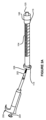
[0031] 図1Aは、細胞を移植するか又は埋め込むために使用されるシリンジ100が、プランジャ102、バレル104及びハブ106を含むことができることを示す。バレル104は、細長い円筒状チューブの形状とすることができる。バレル104は、バレル近位端114、バレル遠位端116、及びバレル近位端114とバレル遠位端116との間の内腔118を有することができる。1つの変形形態では、バレル104は、ホウケイ酸ガラス等のセラミック材料から構成することができ、又はそうしたセラミック材料を含むことができる。他の変形形態では、バレル104は、ポリテトラフルオロエチレン(PTFE)等のポリマーから構成することができ、又はそうしたポリマーを含むことができる。
detailed description
[0031] FIG. 1A shows that a
[0032] 1つの変形形態では、バレル104は、約6.00mm~10.00mmの範囲の外径を有することができる。バレル104は、約1.25mm~5.00mmの範囲の内径を有することができる。
[0032] In one variation, the
[0033] ハブ106は、バレル遠位端116においてバレル104に接続することができ、針124を受け入れ且つ固定することができる嵌合コネクタ120を備えることができる。ハブ106は、ロックハブとすることができ、ポリマー、金属、若しくはニッケルめっき真鍮等の合金、又はそれらの組合せから製造することができ、又はそうしたものを含むことができる。嵌合コネクタ120は、オスルアーテーパ等のルアーロックコネクタとすることができ、PTFE等のポリマーから構成することができ、又はそうしたポリマーを含むことができる。嵌合コネクタ120の内部連続ボアは、針124が内部で密に嵌まるようなものとすることができる。ハブ106は、シリンジ100と針124との間のインタフェースとして機能することができる。
[0033] The
[0034] 針124は、約0.40mm~0.90mmの範囲の外径を有することができる。針124は、約0.10mm~0.35mmの範囲の内部ボア径を有することができる。
[0034] The
[0035] 針124の壁厚さは、約0.10mm~0.35mmの範囲であり得る。1つの変形形態では、27~33ゲージの範囲の針124を備えたHamilton(登録商標)シリンジを使用することができる。
[0035] The wall thickness of
[0036] 針124の先端部は、尖った先端、傾斜した先端、湾曲した先端、ノンコアリング先端、鈍い先端及び円錐形先端のうちの少なくとも1つ等、さまざまな先端様式を有することができる。1つの変形形態では、針124の先端部は、細胞埋込み中に脳組織に対する傷害を低減させるように、丸くすることができる。いくつかの変形形態では、針124の代わりに、図3Aの細胞送達カニューレ300等のカニューレを使用することができる。
[0036] The tip of
[0037] シリンジ100は、約10μL~500μLの範囲の総容積を有することができる。シリンジ100は、シリンジ100の総容積の10%以上の供給容量を有することができる。1つの変形形態では、供給容量は、約1μL~50μLの範囲であり得る。シリンジ100はまた、バレル104の外面にエッチングされるか又は他の方法で画定された容量マーク122又は目盛りも含むことができる。1つの変形形態では、シリンジ100は、100μLのHamilton(登録商標)Model 1710 TLLシリンジ(品番/参照番号:81027)等、Hamilton(登録商標)Gastight 1700シリーズシリンジであり得る。
[0037]
[0038] 図1Bは、シリンジ100のバレル104からプランジャ102を取り外すか又は後退させることができることを示す。図1Bに示すように、プランジャ102は、バレル近位端114から取り外すか又は後退させることができる。プランジャ102がバレル近位端114から取り外されるか又は後退すると、バレル近位端114に開口部を通して、バレル104内の内腔118にアクセスすることができる。
[0038] FIG. 1B shows that the
[0039] 図1Bに示すように、プランジャ102は、プランジャロッド108と、プランジャロッド108の近位端に取り付けられるか又はそこから延在するプランジャヘッド110と、プランジャロッド108の遠位端のプランジャ先端部112とを含むことができる。プランジャ102は、金属、合金、ポリマー、ポリマー複合材又はそれらの組合せから製造することができ、又はそれらを含むことができる。たとえば、プランジャ102は、ステンレス鋼から製造することができ、又はそれを含むことができる。プランジャロッド108は、細長い円筒体の形状とすることができる。プランジャ先端部112は、化学的に不活性なポリマー材料でコーティングすることができる。化学的に不活性なポリマー材料としては、ポリテトラフルオロエチレン(PTFE)を挙げることができる。プランジャ先端部112は、精密加工PTFEプランジャ先端部であり得る。プランジャ先端部112は、バレル104の内腔118に挿入されると、漏れのない又は気密シールを生成することができる。プランジャ先端部112はまた、プランジャヘッド110が押し込まれ、プランジャ先端部112がバレル遠位端116に向かって内腔118内を移動するとき、内腔118の半径方向の表面から細胞破片を除去するか又はふき取ることも可能である。シリンジ100の内腔118は、内腔118の表面をPTFEで覆うか又はコーティングすることにより、実質的に化学的に不活性であるように設計することができる。
[0039] As shown in FIG. A
[0040] 図2Aは、治療処置に関連するシリンジ100を準備する方法の一部として、バレル104を水平向きに位置合せすることができることを示す。本方法は、バレル104からシリンジ100のプランジャ102を取り外すことと、バレル104を水平向きに位置合せすることとを含むことができる。本方法は、バレル近位端114の開口部を通してバレル104の内腔118を細胞懸濁液206で満たすことをさらに含むことができる。
[0040] Figure 2A illustrates that
[0041] 1つの変形形態では、細胞懸濁液206は、マイクロピペット200を使用して送達することができる。マイクロピペット200は、バレル近位端114の開口部を通してバレル104の内腔118に挿入することができるピペットチップ202を含むことができる。ピペットチップ202は、VWR(登録商標)Aerosol Filter Pipet Tip等の滅菌した使い捨てプラスチックチップであり得る。
[0041] In one variation, the
[0042] 細胞懸濁液206は、細胞埋込み又は移植手順に関連して、バレル近位端114を通してシリンジ100内に装填することができる。本開示の目的で、「移植」及び「埋込み」は、対象への外因性細胞の導入を指す。外因性細胞には、自己細胞及び同種細胞が含まれ得る。自己細胞は、対象から得ることができ、同種細胞は、対象以外の人から得ることができる。
[0042]
[0043] 細胞懸濁液206としては、限定されないが、緩衝液内に懸濁している採取細胞、培養細胞、幹細胞、遺伝子組換え細胞又はそれらの組合せを挙げることができる。緩衝液としては、Baxter International Inc.製のPlasma-Lyte A(登録商標)等の晶質液を挙げることができる。
[0043] The
[0044] たとえば、細胞懸濁液206としては、限定されないが、間葉系細胞、間葉系幹細胞(MSC)、骨髄間質細胞(BMSC)、MSC若しくはMBSCの子孫の細胞、それらの培養物、又はそれらの組合せを挙げることができる。細胞懸濁液206又はその前駆体は、異なるタイプの結合組織、臍帯血、ホウォートンゼリー、脂肪組織又は歯髄から採取することができる。この開示の目的で、MSCは、骨髄から導出される粘着性の非血液生成多能性細胞を指すことができる。
[0044] For example, the
[0045] 細胞懸濁液206はまた、骨髄接着性幹細胞(MASC)又はヒト骨髄MSCの子孫である細胞も含むことができ、それらのいずれも、Notch細胞内ドメイン(NICD)(たとえば、ヒトNotch 1細胞内ドメイン(NICD1))をコードするベクターで一過性トランスフェクションされ、それに続き、選択及び後続する培養が行われている。こうした細胞は、NICDで一過性トランスフェクションしたMSCの子孫すなわちDNTT-MSCと呼ぶことができる。DNTT-MSCはまた、MSCの培養物を提供するステップと、MSC細胞培養物を、NICDをコードするが全長Notchタンパク質はコードしない配列を含むポリヌクレオチドと接触させるステップと、ポリヌクレオチドを含む細胞を選択するステップと、選択された細胞を選択なしでさらに培養するステップとを含むプロセスから製造された細胞も指すことができる。たとえば、選択された細胞は、いかなる追加の成長因子又は分化因子(培養基に血清が存在する場合、血清に存在する可能性があるもの以外)もなしで、任意選択的に血清で補完して、標準培養基でさらに培養することができる。このプロセスにより、親MSCと比較して優れた血管新生及び神経新生(すなわち、神経前駆体細胞の増殖及び分化)特性を実証する細胞集団が生成される。細胞懸濁液206は、こうした細胞集団からの細胞を含むことができる。
[0045]
[0046] 細胞懸濁液206は、2006年1月24日に発行された米国特許第6,989,271号、2010年3月23日に発行された米国特許第7,682,825号、213年1月29日に発行された米国特許第8,361,456号、2012年3月13日に発行された米国特許第8,133,725号、2015年3月3日に発行された米国特許第8,969,078号、2016年7月26日に発行された米国特許第9,399,046号、2016年9月13日に発行された米国特許第9,441,199号、2012年1月10日に発行された米国特許第8,092,792号、2015年2月3日に発行された米国特許第8,945,919号、2014年7月22日に発行された米国特許第8,785,190号、2016年5月3日に発行された米国特許第9,326,999号、2017年5月23日に発行された米国特許第9,655,927号、2017年11月28日に発行された米国特許第9,828,593号、2017年5月2日に発行された米国特許RE第46,382号、(米国特許出願公開第2015/0104435A1号として公開された)2014年12月16日に出願された米国特許出願第14/572,177号、(米国特許出願公開第2016/0304835A1号として公開された)2016年6月24日に出願された米国特許出願第15/192,671号、(米国特許出願公開第2010/0310529号として公開された)2010年8月23日に出願された米国特許出願第12/734,855号、(米国特許出願公開第2014/0363408A1号として公開された)2014年8月21日に出願された米国特許出願第14/465,344号、(米国特許出願公開第2011/0136114A1号として公開された)2011年2月9日に出願された米国特許出願第12/736,665号、(米国特許出願公開第2013/0210000A1号として公開された)2012年8月20日に出願された米国特許出願第13/589,849号、(米国特許出願公開第2016/0263159A1号として公開された)2016年3月7日に出願された米国特許出願第15/063,290号、(米国特許出願公開第2014/0186316A1号として公開された)2013年3月13日に出願された米国特許出願第13/800,585号、(米国特許出願公開第2015/0197741A1号として公開された)2014年9月18日に出願された米国特許出願第14/489,934号、(米国特許出願公開第2014/0219976A1号として公開された)2014年4月3日に出願された米国特許出願第14/244,685号、(米国特許出願公開第2016/0271181A1号として公開された)2016年3月21日に出願された米国特許出願第15/076,378号に開示されている細胞を含むことができ、上記出願のすべてが全体として参照により本明細書に援用される。
[0046] The
[0047] 幹細胞又はDNTT-MSCの埋込み又は移植を含む治療処置は、多くの場合、1回の注入ごとに200万~1000万の細胞の送達又は沈着を必要とする可能性がある。これらの投与量を考慮すると、細胞懸濁液206は、シリンジ100の針124を通して引き上げ且つ装填するには過度に粘性がある可能性がある。たとえば、細胞懸濁液206は、針124の遠位端を通して引き上げられるときに針124を詰まらせる可能性がある。さらに、針124を通して引き上げられる細胞の生存能力は、こうした細胞にかけられるせん断応力によって悪影響を受ける可能性がある。したがって、シリンジ100のバレル近位端114の開口部を通して細胞懸濁液206を装填することにより、細胞生存能力が向上し、器具の誤作動が回避される。
[0047] Therapeutic procedures involving stem cell or DNTT-MSC implantation or transplantation can often require delivery or deposition of 2-10 million cells per injection. Given these doses,
[0048] さらに、バレル近位端114の開口部を通して細胞懸濁液206を装填することにより、望ましくない気泡が細胞懸濁液206とともに注入される可能性も低減する。こうした望ましくない気泡は、流れの動態を妨害し、針124を詰まらせ、細胞生存能力に悪影響を与える可能性がある。
[0048] In addition, loading
[0049] 細胞懸濁液206は、バレル104が水平に位置合せされているとき、バレル近位端114を通してシリンジ100内に装填することができる。バレル104は、平坦な水平面に配置されるか又は使用者によって保持されることにより、水平に位置合せすることができる。シリンジ100を満たしながらバレル104を水平に位置合せする1つの利点は、細胞が、バレル遠位端116に向かって沈降する可能性が低くなり、バレル104の長さに沿って均一に層状になる傾向があるということである。バレル104を水平に位置合せする別の利点は、緩衝液が細胞懸濁液206内のより多くの細胞を押し流し且つより多くの細胞と接触するということである。バレル104を水平に位置合せするさらなる利点は、細胞懸濁液206がバレル104からこぼれるか又は漏れ出るのを防止するということである。
[0049]
[0050] 図2Bは、細胞懸濁液206がバレル104の内腔118内に導入された後に、バレル近位端114を通してバレル104内に戻すようにプランジャ102を再挿入することができることを示す。プランジャ102がバレル104内に戻るように再挿入されるとき、バレル104を水平向きに位置合せすることができる。図2Bに示すように、プランジャ102をバレル104内にさらに押し込むことなく、バレル近位端114を通して内腔118内にプランジャ先端部112を再挿入することができる。
[0050] FIG. 2B shows that after the
[0051] 図3Aは、細胞送達カニューレ300が、シリンジ100のハブ106に取り付けられるように構成されたカニューレハブ304を有することができることを示す。細胞送達カニューレ300はまた、カニューレ300の内部容積を満たす内部スタイレット302と、カニューレロッド310と、カニューレ先端部308とを含むことができる。
[0051] FIG. 3A illustrates that
[0052] 細胞送達カニューレ300は、0.75mm~0.90mmの外径と0.20mm~0.40mmの内径とを有することができる。細胞送達カニューレ300は、約15.0cm~20.0cmの範囲の長さを有することができる。
[0052]
[0053] 細胞送達カニューレ300は、304型ステンレス鋼等のステンレス鋼から構成することができ、又はそうしたステンレス鋼を含むことができる。細胞送達カニューレ300は、約15μL~50μLの範囲の内部容積を有することができる。1つの変形形態では、内部容積は、約20μLであり得る。カニューレロッド310は、カニューレハブ304からカニューレ先端部308まで延在する内腔を含むことができる。
[0053]
[0054] たとえば、細胞送達カニューレ300は、Pittsburgh Cell Implantation Cannula(Synergetics(登録商標)品番SB2023)の部品とすることができ又はそうした部品を含むことができる。内部スタイレット302は、細胞埋込み又は移植手順に対して細胞送達カニューレ300を準備するために、カニューレハブ304から取り外すことができる。
[0054] For example, the
[0055] 図3Bは、細胞送達カニューレ300の内腔から内部スタイレット302を取り外いた後に、カニューレ延長部306が可視であり得ることを示す。カニューレ延長部306は、カニューレハブ304から突出することができる。カニューレ延長部306は、細胞送達カニューレ300の内腔と流体連通している剛性中空チューブであり得る。
[0055] FIG. 3B shows that after removing the
[0056] カニューレ延長部306は、シリンジ100の嵌合コネクタ120の内部ボア内に密に嵌まることができる。カニューレ延長部306が、シリンジ100の嵌合コネクタ120のボア内に挿入されると、細胞懸濁液206は、シリンジハブ106、カニューレ延長部306及びカニューレハブ304を介して、シリンジ100の内腔118から細胞送達カニューレ300の内腔に直接進むことができる。
[0056] The
[0057] カニューレ延長部306の1つの利点は、カニューレハブ304内のデッドスペースをなくすか又は低減させることを含むことができる。針又はカニューレハブ内のデッドスペースは、注入の後にシリンジからの著しい量の標本を保持する可能性がある。
[0057] One advantage of the
[0058] 図4Aは、カニューレハブ304が、嵌合コネクタ120に接続するように構成された嵌合コネクタを備えることができることを示す。カニューレハブ304の嵌合コネクタは、ポリマー材料、ステンレス鋼若しくはニッケルめっき真鍮等の金属、又はそれらの組合せから製造することができ、又はそうした材料を含むことができる。カニューレハブ304の嵌合コネクタは、メスルアーフィッティング等のルアーロックコネクタであり得る。
[0058] FIG. 4A illustrates that
[0059] 図4Bは、細胞送達カニューレ300を細胞懸濁液206で満たすためにプランジャ102を押し込むことができることを示す。プランジャ102は、細胞懸濁液206の液滴400が細胞送達カニューレ300からカニューレ先端部308を通って排出されるまで、押し込むことができる。図には示さないが、この開示により、細胞送達カニューレ300ではなく針124がシリンジ100に接続される場合、プランジャ102を押し込むことによって針124を細胞懸濁液206で満たすことも可能であり、針124から細胞懸濁液206の液滴400を排出することができることが企図されている。
[0059] FIG. 4B shows that
[0060] 液滴400は、10μL~40μLの容量を有することができる。たとえば、液滴400は約25μLであり得る。液滴400は、細胞懸濁液206を対象に注入する前に廃棄することができる。細胞懸濁液206の液滴400を排出するいくつかの利点としては、気泡が対象に注入されるのを防止することと、細胞送達カニューレ300内のデッドスペースをなくすこととを挙げることができる。
[0060]
[0061] 外科医又は手術室技師等、シリンジ100の操作者は、バレル104内の細胞懸濁液206のメニスカスとバレル104のマーク122とを使用して、送達された細胞懸濁液206の量と残っている細胞懸濁液206の量とを判断することができる。
[0061] An operator of
[0062] 細胞送達カニューレ300のカニューレ先端部308は、脳組織を通過するように構成することができる様式を含むことができる。たとえば、カニューレ先端部308は、鈍いか又は丸い先端部であり得る。細胞送達カニューレ300は、全体として参照により本明細書に援用される、2007年11月15日に出願された(米国特許出願公開第2008/0132878A1号として公開された)米国特許出願第11/940,868号に記載されているような細胞送達カニューレであり得る。
[0062]
[0063] 図5は、定位フレーム502に取り付けられた安定化カニューレ500の内部に細胞送達カニューレ300を配置することができることを示す。1つの変形形態では、安定化カニューレ500は、Synergetics(登録商標)SB2100安定化カニューレ(Synergetics Inc.、ミズーリ州セントチャールズ(St.Charles, MO))を含むことができる。シリンジ100はまた、定位フレーム502又は別のタイプのフレームによっても固定することができる。
[0063] FIG. 5 shows that the
[0064] 安定化カニューレ500は、剛性があるか又は細胞送達カニューレ300より剛性が高いものであり得る。安定化カニューレ500は、細胞送達カニューレ300の長さより短い長さを有することができる。
[0064]
[0065] たとえば、細胞送達カニューレ300は、約19cmの長さであり得る。こうした長さは、脳組織内への細胞埋込みに有用であり得る。この例では、安定化カニューレ500は、約15cm又は16cmの長さであり得る。
[0065] For example, the
[0066] 安定化カニューレ500は、約0.8mm~2.0mmの範囲の外径を有することができる。1つの変形形態では、安定化カニューレ500は、約1.5mmの外径を有することができる。安定化カニューレ400は、約80μL~120μLの内部容積を有することができる。
[0066] Stabilizing
[0067] 安定化カニューレ500は、二方活栓、取外し可能なスタイレット又はそれらの組合せを有することができる。取外し可能なスタイレットは、細胞送達カニューレ300の外径に等しい直径を有することができる。
[0067] Stabilizing
[0068] 安定化カニューレ500は、定位フレーム502に取り付けるか又は他の方法で定位フレーム502によって固定することができる。1つの変形形態では、定位フレーム502は、Leksell(登録商標)モデルG定位フレーム(Elekta Instruments、ジョージア州アトランタ(Atlanta, GA))であり得る。定位フレーム502は、安定化カニューレ500を取り付けるための止め具及びガイドを含むことができる。1つの変形形態では、止め具は、Leksell(登録商標)Stop(Elekta品番48764-10)とすることができ、ガイドは、Leksell(登録商標)Guide(Elekta品番50150)とすることができる。
[0068] The stabilizing
[0069] 安定化カニューレ500は、最初に、対象の組織内に挿入することができる。たとえば、安定化カニューレ500は、対象の沈着部位604(図6を参照)から約4.00cm~10.0cmに挿入することができる。そして、安定化カニューレ500内に細胞送達カニューレ300を挿入することができ、細胞送達カニューレ300のカニューレ先端部308は、安定化カニューレ500から突出し、標的部位600(図6を参照)又は標的部位600に対して遠位側の部位のいずれかまで前進することができる。細胞送達カニューレ300が安定化カニューレ400内に挿入されている間、細胞送達カニューレ300にシリンジ100を接続することができる。
[0069] The
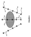
[0070] 図6は、治療処置に関連する対象の標的部位600に又はその近くに細胞懸濁液206を埋め込むことができることを示す。細胞懸濁液206は、標的部位600に又はその近くにある量の細胞懸濁液206を沈着させることによって、対象に埋め込むことができる。細胞懸濁液206は、シリンジ100と針124又は細胞送達カニューレ300のいずれかを使用して埋め込むことができる。
[0070] Figure 6 illustrates that a
[0071] 標的部位600は、損傷部位、推奨される投与部位、又は対象の任意の器官若しくは組織領域であり得る。たとえば、標的部位600が損傷部位である場合、損傷部位は、治療されている疾患又は病状に基づいて異なる可能性がある。治療されている障害が、脳卒中、パーキンソン病又はハンチントン病等、中枢神経系(CNS)障害である場合、損傷部位は、脳幹神経節内の部位であり得る。脳病変の治療では、損傷部位は、病変の又は病変の近くのニューロン組織であり得る。網膜傷害の処置では、損傷部位は、硝子体腔又は網膜下腔であり得る。
[0071] The
[0072] 図6は、標的部位600の付近の3つの投与軌跡602を示す。投与軌跡602は、対象への投与のルート又は経路であり得る。各投与軌跡602は、初期沈着部位606及び最終沈着部位608を含む複数の沈着部位604を含むことができ又はそうした沈着部位604によって画定することができる。投与軌跡602は、直線状、曲線状、又はそれらの組合せであり得る。投与軌跡602は、手順の前に臨床医又は使用者によって事前に決定することができる。
[0072] FIG. 6 shows three
[0073] 図6に示すように、沈着部位604は、部位離隔距離610だけ離隔させることができる。部位離隔距離610は、約2.0mm~10.0mmの範囲であり得る。たとえば、部位離隔距離610は、約5.0mmであり得る。1つの変形形態では、部位離隔距離610は、1つの投与軌跡602に沿って同じ距離であり得る。他の変形形態では、部位離隔距離610は、1つの投与軌跡602に沿って変化させることができる。部位離隔距離610はまた、異なる投与軌跡602に対して異なるものとすることができる。
[0073] As shown in FIG.
[0074] 図6は、治療処置が、投与軌跡602の各々が5つの沈着部位608における細胞懸濁液206の5回の注入を含む、標的部位600を包囲する3つの投与軌跡602を含むことができることも示す。たとえば、細胞懸濁液206は、幹細胞又はDNTT-MSC等の幹細胞の子孫を含むことができ、投与軌跡602の各々は、各々10μL/分の速度での20μLの5回の沈着を含むことができる。使用者は、シリンジ100のバレル104内の残っている細胞懸濁液206のメニスカスとバレル104のマーク122とを使用して、注入された又は沈着した細胞懸濁液206の量を測定することができる。
[0074] FIG. 6 illustrates that the therapeutic treatment includes three
[0075] 図6に示すように、初期沈着部位606は、標的部位600に対して遠位側の場所又は位置にあり得る。この開示の目的で、標的部位600に対して遠位側とは、シリンジ100を操作又は制御している、外科医又は医療関係者等の使用者からさらに離れている、標的部位600を越える場所又は位置を指すことができる。他の変形形態では、初期沈着部位606は、使用者に最も近い標的部位600の前の場所又は位置である、標的部位600に対して近位側の場所又は位置にあり得る。
[0075] As shown in FIG. For the purposes of this disclosure, distal to target
[0076] 治療処置は、カニューレ先端部308又は針124の先端部を初期沈着部位606まで前進させることと、初期沈着部位606においてある量の細胞懸濁液206を注入することとを含むことができる。カニューレ先端部308又は針先端部124は、磁気共鳴画像法(MRI)、超音波又はそれらの組合せを使用して誘導することができる。
[0076] The therapeutic procedure can include advancing the
[0077] 初期沈着部位606が標的部位600に対して遠位側にあるとき、カニューレ先端部308又は針先端部は、部位離隔距離610に実質的に等価な距離、引き出すことができる。細胞送達カニューレ300が使用される場合、使用者が安定化カニューレ500に対する細胞送達カニューレ300の位置を調整するとき、又は使用者が、定位フレーム502に対する安定化カニューレ500の位置を調整するとき、カニューレ先端部308を引き出すことができる。カニューレ先端部308又は針124の先端部は、初期沈着部位606に対して近位側の方向に後退させるか又は引っ張ることができる。
[0077] When the
[0078] 治療処置はまた、使用者が、初期沈着部位606に対して近位側の後続する沈着部位604において別の量の細胞懸濁液を注入することも含むことができる。いくつかの変形形態では、使用者は、後続する注入の前に、シリンジ100のバレル104又はシリンジ100及び細胞送達カニューレ300の両方をシリンジ100の長手方向軸を中心に回転させることができる。たとえば、シリンジ100のバレル104、又はシリンジ100のバレル104及び細胞送達カニューレ300の両方を、右回り方向に又は左回り方向に角度的に回転させることができる。いくつかの変形形態では、回転は、約45度の右回り又は左回りの回転であり得る。他の変形形態では、回転は、約90度の右回り又は左回りであり得る。シリンジ100及び細胞送達カニューレ300は、シリンジ100及び細胞送達カニューレ300内に残っている細胞懸濁液206が沈降するか又は詰まらせるのを防止するように回転させることができる。
[0078] Therapeutic treatment can also include the user injecting another volume of the cell suspension at
[0079] 治療処置は、針124の先端部又はカニューレ先端部308が最終沈着部位608に達するまで、使用者が、各注入の後にシリンジ100又はシリンジ100及び細胞送達カニューレ300の両方を引き抜いて回転させることを含むことができる。1つの変形形態では、最終沈着部位608は、標的部位600に対して近位側の場所又は位置であり得る。他の変形形態では、最終沈着部位608は、標的部位600における若しくは標的部位600内の場所若しくは位置であるか、又は初期沈着部位606に対して近位側であるが依然として標的部位600に対して遠位側である場所若しくは位置であり得る。さらなる変形形態では、最終沈着部位608は、初期沈着部位606に対して遠位側の場所又は位置であり得る。最終沈着部位608において沈着又は注入が行われると、針124又は細胞送達カニューレ300及び安定化カニューレ500の組合せを対象から後退させることができる。
[0079] Therapeutic procedure is such that the user withdraws and rotates
[0080] 図6には3つの投与軌跡602を示すが、当業者であれば、投与軌跡602の数及び沈着部位608の数は、投与される細胞、処置プロトコル、及び特定の処置に対して投与される治療的に有効な細胞の量に応じて変更することができることが理解されるべきである。本明細書における「治療的に有効な量」又は「治療効果のある量」は、特定の病状又は疾患に関連する症状の重症度を低減させる、移植されるか(transplanted)又は埋め込まれる(implanted)細胞の数又は量を指すことができる。治療的に有効な量は、損傷のタイプ及び程度とともに、対象の全体的な状態によって変更することができる。
[0080] Although three
[0081] 細胞の治療的に有効な量は、約200万細胞~3000万細胞の用量を指すことができる。たとえば、投与されている細胞又は細胞懸濁液206が、幹細胞、又はDNTT-MSC等の幹細胞の子孫を含む場合、細胞の治療的に有効な量は、250万細胞、500万細胞又は1000万細胞のうちの任意のものを含むことができる。
[0081] A therapeutically effective amount of cells can refer to a dose of about 2-30 million cells. For example, if the cells or
[0082] 治療的に有効な量の幹細胞、又はDNTT-MSC等の幹細胞の子孫は、神経変性疾患、障害、又は中枢神経系(CNS)に関連する病状を処置するために、損傷部位に投与することができる。これらの疾患、障害又はCNS病状としては、限定されないが、アルツハイマー病、パーキンソン病、ハンチントン病、筋委縮性側索硬化症(ALS)、外傷性脳損傷(TBI)、脳卒中(虚血性及び/又は出血性)及び脊髄損傷を挙げることができる。 [0082] A therapeutically effective amount of stem cells, or progeny of stem cells such as DNTT-MSCs, is administered to the site of injury to treat neurodegenerative diseases, disorders, or conditions associated with the central nervous system (CNS). can do. These diseases, disorders or CNS conditions include, but are not limited to Alzheimer's disease, Parkinson's disease, Huntington's disease, amyotrophic lateral sclerosis (ALS), traumatic brain injury (TBI), stroke (ischemic and/or hemorrhagic) and spinal cord injury.
[0083] たとえば、DNTT-MSCは、神経新生(新たなニューロンの形成)及び血管新生(新たな血管の形成)を促進する因子を合成し分泌することができる。DNTT-MSCは、生物活性線維芽細胞増殖因子(FGF2)又は塩基性線維芽細胞増殖因子(bFGF)を放出することができる。間葉系細胞は、FGF2及びFGF2アイソフォームの大型の細胞内ストアを含み、それは、損傷した組織部位に対して神経新生及び血管新生効果を与えることができる。FGF2はまた、血管新生の強力な誘導物質及び創傷治癒媒介物質でもあり得る。損傷したCNS部位にMSC及びそれらの誘導体又は子孫を埋め込むことからもたらされる治療効果は、特に、埋め込まれた生存細胞からの可溶性因子の分泌、埋め込まれた細胞の死の後の可溶性細胞内因子の放出、埋め込まれた細胞による免疫機能の調整、埋め込まれた細胞による神経支持(neurosupportive)細胞外マトリックスの生成、及び、埋め込まれた細胞による、神経原性ニッチから損傷部位への内因性神経細胞の移動のための経路の形成に起因することができる。 [0083] For example, DNTT-MSCs can synthesize and secrete factors that promote neurogenesis (formation of new neurons) and angiogenesis (formation of new blood vessels). DNTT-MSCs can release bioactive fibroblast growth factor (FGF2) or basic fibroblast growth factor (bFGF). Mesenchymal cells contain large intracellular stores of FGF2 and FGF2 isoforms, which can confer neurogenic and angiogenic effects on injured tissue sites. FGF2 may also be a potent inducer of angiogenesis and wound healing mediator. Therapeutic effects resulting from the implantation of MSCs and their derivatives or progeny into injured CNS sites are, inter alia, the secretion of soluble factors from the implanted viable cells, the release of soluble intracellular factors after the implanted cells die. release, modulation of immune function by the implanted cells, generation of a neurosupportive extracellular matrix by the implanted cells, and release of endogenous neurons from the neurogenic niche to the site of injury by the implanted cells. It can be attributed to the formation of pathways for migration.
[0084] 処置例では、1回の埋込み手順で約200万~1000万DNTT-MSCを埋め込むことができる。こうした処置は、標的部位600における又はその近くの複数の投与軌跡602を含むことができ、こうした投与軌跡602の各々は複数の沈着部位608を含む。沈着部位608における各沈着又は注入は、約10μL~20μLの細胞懸濁液206を含むことができる。たとえば、投与軌跡602毎に合計100μLの細胞懸濁液206を送達することができる。
[0084] In an example procedure, approximately 2-10 million DNTT-MSCs can be implanted in a single implantation procedure. Such treatment can include
[0085] 虚血性又は出血性脳卒中のため等、脳卒中からの回復のための同種細胞療法の一部として、DNTT-MSCを投与することができる。DNTT-MSCは、脳内の虚血組織の再灌流を促進することができる。たとえば、治療効果のある量のDNTT-MSCを脳内の虚血損傷部位に移植することにより、新たな血管の増殖をもたらすことができる。 [0085] DNTT-MSCs can be administered as part of allogeneic cell therapy for recovery from stroke, such as for ischemic or hemorrhagic stroke. DNTT-MSCs can promote reperfusion of ischemic tissue in the brain. For example, transplantation of therapeutically effective amounts of DNTT-MSCs into the site of ischemic injury in the brain can result in the growth of new blood vessels.
[0086] 幹細胞又はDNTT-MSCを含む細胞懸濁液206はまた、外傷性脳損傷(TBI)に対する治療処置に関連して、対象の標的部位600に又はその近くにも埋め込むことができる。こうした処置では、沈着部位608は、損傷した皮質領域周辺又は内側皮質に対応する領域にあり得る。たとえば、DNTT-MSC若しくは他の幹細胞又はその誘導体は、損傷部位に対して遠位側又は近位側の周縁部内に埋め込むことができる。さらに、DNTT-MSC又は他の幹細胞は、損傷によって影響を受ける運動神経路の近くに埋め込むことができる。
[0086] A
[0087] さらに、治療的に有効な量のDNTT-MSCは、リハビリテーションを促進するために、神経原性ニッチ(たとえば、脳室下帯(SVZ)又は歯状回)と脳損傷部位との間の生物学的架橋を形成することにより、運動及び神経学的機能を改善することができる。移植されたDNTT-MSCは、神経原性ニッチから脳損傷部位までの宿主神経原性細胞の長距離移動のために、バイオブリッジ(biobridge)としても知られる過渡経路としての役割を果たすことができる。上述した病状を処置するために、頭蓋内注射等の直接注入方法により、治療効果のある量のDNTT-MSCを局部的に投与することができる。 [0087] In addition, therapeutically effective amounts of DNTT-MSCs may be administered between the neurogenic niche (eg, the subventricular zone (SVZ) or dentate gyrus) and the site of brain injury to facilitate rehabilitation. can improve motor and neurological function by forming biological bridges of Transplanted DNTT-MSCs can serve as a transient pathway, also known as a biobridge, for long-distance migration of host neurogenic cells from the neurogenic niche to the site of brain injury. . A therapeutically effective amount of DNTT-MSCs can be administered locally by direct injection methods, such as intracranial injection, to treat the conditions described above.
[0088] 幹細胞又はDNTT-MSCを含む細胞懸濁液206はまた、網膜変性のための治療処置に関連して、対象の眼内の標的部位600に又はその近くに埋め込むことができる。たとえば、限定されないが、網膜色素変性症(RP)、加齢黄斑変性(AMD)、アッシャー症候群、スターガルト病、コロイデレミア(choroideremia)、バルデー・ビードル症候群、レフサム病、ベスト病又は小口病を含む、さまざまなタイプの網膜変性の病状を処置するために、対象の眼内にDNTT-MSCを埋め込むことができる。たとえば、眼のある特定の光受容体活動又は機能を促進するために、対象の眼球にDNTT-MSCを含む細胞懸濁液206を投与することができる。また、たとえば、網膜から脳の視覚野への視覚信号の送信を促進するために、対象の眼球に、DNTT-MSCを含む細胞懸濁液206を投与することができる。こうした処置では、シリンジ100の針124を使用して対象の眼内に直接細胞懸濁液206を移植することができる。1つの変形形態では、30ゲージ針を備えたHamilton(登録商標)シリンジを使用して、硝子体腔又は網膜下腔を含む対象の眼内にDNTT-MSCを注入することができる。
[0088] A
[0089] 図7Aは、治療処置に関連するシリンジ100及び細胞送達カニューレ300を準備する方法700を示す。方法700は、ステップ702において、細胞送達カニューレ300のカニューレハブ304をシリンジハブ106に取り付けて、カニューレハブ304を締めることを含むことができる。1つの変形形態では、シリンジ100は、100μLのHamilton(登録商標)シリンジとすることができ、細胞送達カニューレ300は、Pittsburgh Cell Implantation Cannula(PIC)とすることができる。方法700はまた、ステップ704において、細胞送達カニューレ300のカニューレ先端部308を通して緩衝液を吸引することにより、シリンジ100のバレル104の内腔118の少なくとも一部を緩衝液で満たすことも含むことができる。緩衝液は、Plasma-Lyte A(登録商標)を含むことができる。緩衝液は、約1mL増分を含むさまざまな量で提供することができる。
[0089] Figure 7A shows a
[0090] 方法700はまた、ステップ706において、バレル104内に緩衝液を導入するか又は送達するために、シリンジ100のプランジャ102を押し込むことも含むことができる。シリンジ100及び細胞送達カニューレ300をすすぐために、ステップ704及び706を数回繰り返すことができる。この時点で、カニューレ300が気泡を引き上げないように、細胞送達カニューレ300がシリンジ100に適切に取り付けられることを確認するために、臨床医又は外科医により、細胞送達カニューレ300に取り付けられたシリンジ100を検査することができる。緩衝液を排出した後、ステップ706はまた、シリンジ100内に緩衝液が残らなくなるまで、カニューレ先端部308を通して繰返し空気を引き込み排出することも含むことができる。
[0090] The
[0091] 方法700は、ステップ708において、シリンジハブ106からカニューレハブ304を分離し、シリンジ100のバレル104からプランジャ102を取り外すことをさらに含むことができる。プランジャ102及び細胞送達カニューレ300を脇に置くことができる。方法700は、ステップ710において、バレル104を水平向きに位置合せし、滅菌した平坦面の上にバレル104を配置することを含むことができる。方法700はまた、ステップ712において、マイクロピペット200に滅菌したエアロゾルピペットチップ202を固定し、ピペットプランジャ204を押し込んで緩衝液を引き上げることも含むことができる。滅菌したエアロゾルピペットチップ202は、VWR(登録商標)Aerosol Filterピペットチップ等、滅菌した使い捨てプラスチックチップであり得る。1つの変形形態では、マイクロピペット200は、20~200μLのThermo Scientific Finnpipette(登録商標)マイクロピペットであり得る。たとえば、マイクロピペット200は、125μLのPlasma-Lyte A(登録商標)緩衝液を引き上げるために、125μLで設定することができる。
[0091] The
[0092] 方法700は、水平に位置合せされたバレル104の内腔118内にマイクロピペット200のピペットチップ202を挿入することをさらに含むことができる。ステップ714において、バレル近位端114を通して内腔118内にマイクロピペット200のピペットチップ202を挿入することができ、内腔118内に緩衝液を注入することができる。バレル近位端114内にピペットチップ202をまっすぐ押し込むことができる。緩衝液がバレル遠位端116から排出されないように、緩衝液は徐々に注入することができる。方法700はまた、ステップ716において、バレル近位端114からピペットチップ202を取り外し、バレル近位端114内にプランジャ先端部112を挿入することも含むことができる。バレル近位端114の開口部が密封される直前まで、プランジャ先端部112を挿入することができる。
[0092]
[0093] 方法700は、ステップ718において、シリンジハブ106に細胞送達カニューレ300のカニューレハブ304を取り付け、プランジャ102を押し込んでシリンジ100及び細胞送達カニューレ300から緩衝液を排出することをさらに含むことができる。緩衝液を排出するとき、プランジャ102を徐々に押し込むことができる。さらに、シリンジ100及び細胞送達カニューレ300から緩衝液を除去するために、空気も引き込み排出することができる。
[0093]
[0094] 図7Bは、治療処置に関連するシリンジ100及び細胞送達カニューレ300を準備する方法の第2部分を示す。方法700の第2部分は、ステップ720において、マイクロピペット200の端部に滅菌したエアロゾルピペットチップ202を固定し、細胞懸濁液206をピペッティングする前に、ピペットプランジャ204を第1止め具まで押し込むことを含むことができる。たとえば、マイクロピペット200は、200μLのThermo Scientific Finnpipette(商標)Micropipetteであり得る。1つの変形形態では、マイクロピペット200は、125μLの細胞懸濁液206を引き上げることができる。
[0094] Figure 7B shows a second part of the method of preparing the
[0095] 方法700はまた、ステップ722において、細胞懸濁液206を、懸濁液が均一になるまで上下に数回静かにピペッティングすることも含むことができる。1つの変形形態では、細胞懸濁液206は、幹細胞又はDNTT-MSC等の幹細胞の子孫を含むことができる。たとえば、ステップ722において、マイクロピペット200を使用して細胞を上下に繰返しピペッティングすることにより、125μLのDNTT-MSCを再懸濁することができる。気泡がないか細胞懸濁液206を検査することも可能である。
[0095]
[0096] 方法700は、ステップ724において、シリンジ100のバレル104からプランジャ102を取り外すことをさらに含むことができる。バレル近位端114を通してバレル104の内腔118からプランジャ102の一部を引き出すことにより、プランジャ102を取り除くことができる。方法700はまた、ステップ726において、バレル104を水平向きに位置合せすることも含むことができる。
[0096] The
[0097] 方法700は、ステップ728において、バレル近位端114の開口部を通して、バレル104の内腔118の少なくとも一部を細胞懸濁液206で満たすことをさらに含むことができる。バレル近位端114の開口部を通して内腔118内にマイクロピペット200のピペットチップ202を挿入し、内腔118内に細胞懸濁液206を徐々に注入することにより、内腔118を満たすことができる。
[0097]
[0098] ステップ730において、細胞懸濁液206を送達した後、バレル近位端114を通して、内腔118からマイクロピペット200のピペットチップ202を取り外すことができ、バレル近位端114の開口部内にプランジャ102のプランジャ先端部112を挿入して内腔118を密封することができる。プランジャ先端部112は、内腔118内にわずかにのみ、又はバレル近位端114の開口部が密封されるまでのみ、挿入することができる。
[0098] At
[0099] 方法700はまた、ステップ732において、シリンジハブ106に細胞送達カニューレ300のカニューレハブ304を取り付け、カニューレハブ304を締めることも含むことができる。方法700は、ステップ734において、細胞懸濁液206が細胞送達カニューレ300を満たし、細胞懸濁液206の液滴400がカニューレ先端部308から排出されるまで、プランジャ102を押し込むことをさらに含むことができる。たとえば、最初に、125μLの細胞懸濁液206がシリンジ100内に導入される場合、細胞懸濁液206のメニスカスがシリンジ100の100μLマーク又はその近くになるまで、プランジャ102を徐々に押し込むことができる。この場合、排出される細胞懸濁液206の液滴400は、約100μLの細胞懸濁液206がシリンジバレル104内に残るように、約25μLに等価であり得る。滅菌したガーゼ片を用いて、細胞懸濁液206の液滴400を吸い取ることができる。図には示さないが、この開示により、方法700のいくつかのステップはまた、シリンジ100に接続された細胞送達カニューレ300ではなくシリンジ100に接続された針124によって行うことも可能であることが企図されている。
[0099]
[0100] 図8は、治療処置に関連する対象に細胞懸濁液206を埋め込む方法800を示す。方法800は、ステップ802において、細胞懸濁液206(方法700のステップ734からの細胞懸濁液206等)で満たされたシリンジ100に取り付けられた細胞送達カニューレ300を安定化カニューレ500内に挿入することを含むことができる。定位フレーム502に安定化カニューレ500を取り付けることができる。1つの変形形態では、定位フレーム502は、Leksell(登録商標)モデルG定位座標フレーム(Elekta Instruments、ジョージア州アトランタ(Atlanta, GA))であり得る。方法800はまた、ステップ804において、対象の初期沈着部位606内にある量の細胞懸濁液206を注入することも含むことができる。1つの変形形態では、1つの投与軌跡602に沿って、各々10μL/分の速度で、20μLの5回の沈着で、DNTT-MSCを注入することができる。
[0100] Figure 8 illustrates a
[0101] 方法800はまた、ステップ806において、シリンジ110のバレル104に残っている細胞懸濁液206のメニスカスを使用して、注入された細胞懸濁液206の容量又は量を測定することも含むことができる。方法800はまた、ステップ808において、各注入後に、後続する沈着部位604までの部位離隔距離610等の所定距離、シリンジ100及び細胞送達カニューレ300を後退させることも含むことができる。たとえば、各注入又は沈着の後に、細胞送達カニューレ300を約5.0mm引き出すことができる。方法800はまた、ステップ810において、シリンジ100の長手方向軸を中心にシリンジ100のバレル104(及び接続された細胞送達カニューレ300)を回転させることも含むことができる。シリンジ100は、約45度~90度右回り又は左回り方向に回転させることができる。シリンジ100の1つの利点としては、細胞懸濁液206内の細胞が懸濁液から出て沈降することを防止することが挙げられる。
[0101] The
[0102] 方法800は、ステップ812において、後続する沈着部位604内にさらなる量の細胞懸濁液206を注入することをさらに含むことができる。各沈着における細胞懸濁液206の量と沈着部位の数とは、各処置によって変更することができる。1つの変形形態では、細胞懸濁液206は、10μL/分の速度で注入することができ、各沈着は、約20μLの細胞懸濁液206を含むことができる。この変形形態では、100μLの総沈着に対して、1つの投与軌跡602に沿って5回の沈着を行うことができる。移植時間は、約50分~70分の範囲であり、細胞調製から最終的な移植までの最大許容可能時間は180分間とすることができる。
[0102] The
[0103] 方法800はまた、ステップ814において、最終沈着部位608に細胞懸濁液206を注入した後、安定化カニューレ500から細胞送達カニューレ300を取り外すことも含むことができる。最終沈着部位608における最後の沈着の後、さらなる埋込みに対して、細胞送達システムを準備することができる。Plasma-Lyte A等の懸濁液のバイアルを使用して、方法700において記載した手順により(図7Aを参照)、細胞送達システム(シリンジ100及び細胞送達カニューレ300を含む)をすすぐことができる。第2バイアル又は細胞のアリコートから、さらなる細胞懸濁液206を提供し、方法700のステップ722(図7Bを参照)に従って再懸濁することができる。次の投与軌跡602に対して、安定化カニューレ500を再配置することができる。方法700において記載した手順を使用して、シリンジ100及び細胞送達カニューレ300をさらなる細胞懸濁液206で満たすことができる。投与軌跡602の数に応じて、方法及びステップを必要に応じて繰り返すことができる。
[0103]
[0104] 本明細書に記載した方法は、治療処置の一部として、幹細胞又はDNTT-MSC等の幹細胞誘導体等の粘性細胞懸濁液を患者に送達する際に有効である。図7A、図7B及び図8に示すフローチャートでは、所望の結果を達成するために図示する特定の順序が必須ではなく、いくつかのステップは、並列に行い、省略し、又はその組合せを行うことができる。 [0104] The methods described herein are effective in delivering a viscous cell suspension, such as stem cells or stem cell derivatives such as DNTT-MSCs, to a patient as part of a therapeutic treatment. In the flowcharts shown in FIGS. 7A, 7B and 8, the particular order shown is not required to achieve desired results, and some steps may be performed in parallel, omitted, or a combination thereof. can be done.
[0105] 本明細書に記載し例示した個々の変形形態の各々は、他の変形形態のうちの任意のものの特徴から容易に分離し又はそうした特徴と組み合わせることができる別個の構成要素及び特徴を有する。本発明の目的、趣旨又は範囲に対して、特定の状況、材料、組成物、プロセス、プロセス行為又はステップを適合させるように、変更を行うことができる。 [0105] Each of the individual variations described and illustrated herein presents distinct components and features that can be readily separated from or combined with features of any of the other variations. have. Changes may be made to adapt a particular situation, material, composition of matter, process, process act or step to the objective, spirit or scope of the present invention.
[0106] 本明細書に列挙した方法は、列挙した事象の論理的に可能である任意の順序で行うことができるとともに、事象の列挙した順序で行うことができる。たとえば、図に示すフローチャートでは、所望の結果を達成するために図示する特定の順序は必須ではない。さらに、所望の結果を達成するために、追加のステップ若しくは動作を提供することができ、又は、ステップ若しくは動作を削除することができる。 [0106] The methods recited herein may be performed in any logically possible order of the recited events and may be performed in the recited order of events. For example, the flowcharts shown in the figures do not require the particular order shown to achieve desired results. Further, additional steps or actions can be provided, or steps or actions can be deleted, to achieve desired results.
[0107] 値の範囲が提供される場合、その範囲の上限及び下限とその提示された範囲における他の任意の提示された又は介在する値との間のすべての介在する値が、本発明の範囲内に包含される。また、記載した発明の変形形態のいかなる任意選択的な特徴も、独立して、又は本明細書に記載した特徴のうちの任意の1つ若しくは複数と組み合せて、示し請求することができる。 [0107] When a range of values is provided, all intervening values between the upper and lower limits of that range and any other stated or intervening value in the stated range are subject to the present invention. Included in scope. Also, any optional feature of the described variations of the invention may be shown and claimed independently or in combination with any one or more of the features described herein.
[0108] 本明細書で言及したすべての既存の主題(たとえば、刊行物、特許及び特許出願)は、その主題が本発明の主題と矛盾する(その場合、本明細書に存在するものが優先されるものとする)可能性がある場合を除き、全体として参照により本明細書に援用される。参照した記事は、単に、本出願の出願日の前のそれらの開示に対してのみ提供される。本明細書におけるいずれも、先行発明という理由で本発明がこうした資料に先行する権利が与えられないと認めるものとして解釈されるべきではない。 [0108] All pre-existing subject matter (e.g., publications, patents and patent applications) referred to in this specification is such that that subject matter is inconsistent with the subject matter of the present invention, in which case what exists herein takes precedence. is hereby incorporated by reference in its entirety, except where possible. The referenced articles are provided solely for their disclosure prior to the filing date of the present application. Nothing herein is to be construed as an admission that the present invention is not entitled to antedate such material by virtue of prior invention.
[0109] 単数の項目への言及は、複数の同じ項目が存在する可能性を含む。より具体的には、本明細書において且つ添付の特許請求の範囲において、単数形「1つの(a、an)」、「前記」及び「その(the)」は、文脈において明確な別段の指示がない限り、複数の指示対象を含む。特許請求の範囲は、いかなる任意選択的な要素も排除するように書かれている可能性があることにさらに留意されたい。したがって、この言及は、請求項の要素の記載に関連する「単に」等の排他的な用語の使用、又は「消極的な」限定の使用に対する、先行詞としての役割を果たすように意図されている。別段の規定がない限り、本明細書で使用するすべての技術的用語及び科学的用語は、この発明が属する技術分野における当業者により一般的に理解される意味と同じ意味を有する。 [0109] Reference to a singular item includes the possibility that there is a plurality of the same item. More specifically, as used herein and in the appended claims, the singular forms "a, an," "said," and "the" indicate to the contrary the context clearly indicates otherwise. Including plural referents unless specified. It is further noted that the claims may be drafted to exclude any optional element. Accordingly, this reference is intended to serve as antecedent to the use of exclusive terms such as "merely" or the use of "negative" limitations in connection with reciting claim elements. there is Unless defined otherwise, all technical and scientific terms used herein have the same meaning as commonly understood by one of ordinary skill in the art to which this invention belongs.
[0110] 本開示は、示した特定の形態の範囲に限定されるように意図されておらず、本明細書に記載した変形形態の代替態様、変更態様及び均等物を包含するように意図されている。さらに、本開示の範囲は、本開示を考慮して本技術分野における当業者には明らかとなり得る他の変形形態を完全に包含する。本発明の範囲は、添付の特許請求の範囲によってのみ限定される。 [0110] This disclosure is not intended to be limited in scope to the particular forms shown, but is intended to encompass alternatives, modifications, and equivalents of the variations set forth herein. ing. Moreover, the scope of the present disclosure fully encompasses other variations that may become apparent to those skilled in the art in view of the present disclosure. The scope of the invention is limited only by the appended claims.
Claims (20)
前記シリンジのプランジャを、前記シリンジのバレルであって、近位端、遠位端、及び前記近位端と前記遠位端との間の内腔を有するバレルから取り外すことと、
前記シリンジの前記バレルの軸方向が水平面内にあるように前記シリンジの前記バレルを配置することと、
前記シリンジの前記バレルの軸方向が前記水平面内にあるように前記シリンジの前記バレルが配置される時に前記近位端の開口部を通して前記内腔の少なくとも一部を粘性物質で満たすことと、
前記内腔内に前記プランジャのプランジャ先端部を挿入して前記内腔を密封することと、
前記シリンジのハブであって、前記遠位端において前記バレルに接続しているハブに、埋込みデバイスを取り付けることと、
前記粘性物質が前記埋込みデバイスを満たし、前記粘性物質の液滴が前記埋込みデバイスの遠位先端部を通して前記埋込みデバイスから排出されるまで、前記プランジャを押し込むことと、を含む方法。 A method of preparing a syringe, comprising:
removing the plunger of the syringe from a barrel of the syringe having a proximal end, a distal end, and a lumen between the proximal and distal ends;
positioning the barrel of the syringe such that the axial direction of the barrel of the syringe is in a horizontal plane ;
filling at least a portion of the lumen with a viscous material through the proximal end opening when the barrel of the syringe is positioned such that the axial direction of the barrel of the syringe is within the horizontal plane ;
inserting a plunger tip of the plunger into the lumen to seal the lumen;
attaching an implantation device to a hub of the syringe, the hub connecting to the barrel at the distal end;
Depressing the plunger until the viscous material fills the implantation device and a droplet of the viscous material is expelled from the implantation device through a distal tip of the implantation device.
前記シリンジのプランジャを、前記シリンジのバレルであって、近位端、遠位端、及び前記近位端と前記遠位端との間の内腔、を有するバレルから取り外すことと、
前記シリンジの前記バレルの軸方向が水平面内にあるように前記シリンジの前記バレルを配置することと、
前記シリンジの前記バレルの軸方向が前記水平面内にあるように前記シリンジの前記バレルが配置される時に前記近位端の開口部を通して前記内腔の少なくとも一部を前記細胞懸濁液で満たすことと、
前記内腔内に前記プランジャのプランジャ先端部を挿入して前記内腔を密封することと、
前記シリンジのハブであって、前記遠位端において前記バレルに接続しているハブにカニューレを取り付けることと、
前記細胞懸濁液の液滴が前記カニューレの遠位先端部を通して前記カニューレから排出されるまで、前記プランジャを押し込むことと、を含む方法。 A method of loading a syringe with a cell suspension, comprising:
removing the plunger of the syringe from a barrel of the syringe having a proximal end, a distal end, and a lumen between the proximal and distal ends;
positioning the barrel of the syringe such that the axial direction of the barrel of the syringe is in a horizontal plane ;
filling at least a portion of the lumen with the cell suspension through the proximal end opening when the barrel of the syringe is positioned such that the axial direction of the barrel of the syringe is within the horizontal plane; and
inserting a plunger tip of the plunger into the lumen to seal the lumen;
attaching a cannula to a hub of the syringe, the hub connecting to the barrel at the distal end;
depressing the plunger until a droplet of the cell suspension is expelled from the cannula through the distal tip of the cannula.
Applications Claiming Priority (3)
| Application Number | Priority Date | Filing Date | Title |
|---|---|---|---|
| US201662439818P | 2016-12-28 | 2016-12-28 | |
| US62/439,818 | 2016-12-28 | ||
| PCT/US2017/068288 WO2018125829A1 (en) | 2016-12-28 | 2017-12-22 | Cell delivery system and methods of operation thereof |
Publications (2)
| Publication Number | Publication Date |
|---|---|
| JP2020503113A JP2020503113A (en) | 2020-01-30 |
| JP7220149B2 true JP7220149B2 (en) | 2023-02-09 |
Family
ID=62710583
Family Applications (1)
| Application Number | Title | Priority Date | Filing Date |
|---|---|---|---|
| JP2019534331A Active JP7220149B2 (en) | 2016-12-28 | 2017-12-22 | Cell delivery system and method of operation |
Country Status (6)
| Country | Link |
|---|---|
| US (2) | US11439761B2 (en) |
| EP (1) | EP3548114A4 (en) |
| JP (1) | JP7220149B2 (en) |
| CN (1) | CN110382020A (en) |
| CA (1) | CA3047329A1 (en) |
| WO (1) | WO2018125829A1 (en) |
Families Citing this family (8)
| Publication number | Priority date | Publication date | Assignee | Title |
|---|---|---|---|---|
| WO2018125829A1 (en) | 2016-12-28 | 2018-07-05 | Sanbio, Inc. | Cell delivery system and methods of operation thereof |
| KR20210151904A (en) * | 2019-04-09 | 2021-12-14 | 콘그루언스 메디칼 솔루션즈, 엘엘씨 | injection device |
| US20220233767A1 (en) * | 2019-05-31 | 2022-07-28 | Rainbow Inc. | Puncture needle, puncture needle kit, and stereotactic brain surgery device |
| CN111748461A (en) * | 2020-07-02 | 2020-10-09 | 成都恩喜医疗管理有限公司 | Cell transplantation equipment and use method |
| EP4346853A1 (en) * | 2021-05-27 | 2024-04-10 | SanBio, Inc. | Cell therapies and methods of treatment for small-volume stroke |
| US11504485B1 (en) * | 2021-08-18 | 2022-11-22 | Cellular Vehicles, Inc. | Cellular therapy infusion devices, systems, and methods for use |
| GB202200706D0 (en) * | 2022-01-20 | 2022-03-09 | Cambridge Univ Hospitals Nhs Foundation Trust | A surgical instrument |
| KR102530997B1 (en) * | 2022-12-13 | 2023-05-11 | 서정성 | Stem cell injection device |
Citations (7)
| Publication number | Priority date | Publication date | Assignee | Title |
|---|---|---|---|---|
| JP2002306596A (en) | 2001-04-18 | 2002-10-22 | Unisis:Kk | Composite needle for anesthesia |
| JP2004520892A (en) | 2001-01-09 | 2004-07-15 | ジェンザイム・コーポレーション | Apparatus for delivering liquid and gel-like surgical materials and methods of using the same |
| US20040158226A1 (en) | 2003-02-07 | 2004-08-12 | Genoptix, Inc. | Syringe tissue sieve |
| JP2013541380A (en) | 2010-10-18 | 2013-11-14 | サノフィ・パスツール | Method for preserving vaccine containing aluminum adjuvant |
| JP2015504922A (en) | 2012-01-27 | 2015-02-16 | サンバイオ,インコーポレイティド | Methods and compositions for modulating angiogenesis and vascular development |
| WO2015152721A1 (en) | 2014-04-04 | 2015-10-08 | Sparkle Innovations B.V. | Device and method for administering a liquid drop by drop |
| JP2016515004A (en) | 2013-03-15 | 2016-05-26 | マフィン・インコーポレイテッドMuffin Incorporated | Cell injection needle |
Family Cites Families (42)
| Publication number | Priority date | Publication date | Assignee | Title |
|---|---|---|---|---|
| US6336932B1 (en) | 1994-08-05 | 2002-01-08 | Bausch & Lomb Surgical, Inc. | Device for inserting a flexible intraocular lens |
| ZA9811087B (en) * | 1997-12-04 | 1999-06-03 | Bracco Research Sa | Automatic liquid injection system and method |
| US20040028656A1 (en) | 1998-07-29 | 2004-02-12 | University Of South Florida, A Non-Profit Institution | Survival of neurons |
| US6907679B2 (en) * | 1998-11-12 | 2005-06-21 | Qlt Usa, Inc. | Method for lyophilizing an active agent |
| US20030211085A1 (en) | 2000-03-16 | 2003-11-13 | Sanberg Paul R. | Cell therapy for chronic stroke |
| EP1949904A3 (en) | 1999-04-27 | 2008-08-06 | Layton Bioscience, Inc. | Cell therapy for chronic stroke |
| CA2282007C (en) * | 1999-09-09 | 2002-05-28 | Ivar Mendez | Neural transplantation delivery system |
| WO2002063938A2 (en) | 2000-12-05 | 2002-08-22 | Layton Bioscience Inc. | Production and use of dopaminergic cells to treat dopaminergic deficiencies |
| JP3886346B2 (en) | 2001-06-22 | 2007-02-28 | サンバイオ,インコーポレイティド | Pharmaceutical composition for nerve regeneration comprising bone marrow stromal cell-derived Schwann cells |
| AU2002317573B2 (en) * | 2001-07-24 | 2007-07-26 | Artes Medical, Inc. | Elongated syringe |
| US7682825B2 (en) | 2002-02-06 | 2010-03-23 | Sanbio, Inc. | Differentiation of bone marrow stromal cells to neural cells or skeletal muscle cells by introduction of notch gene |
| US20020198498A1 (en) * | 2002-07-18 | 2002-12-26 | David Porat | Syringe for high-viscosity fluids |
| BR0315842A (en) * | 2002-10-29 | 2005-09-27 | Vasogen Ireland Ltd | Device and method for controlled gas expression of medical fluid delivery systems |
| US7329241B2 (en) | 2003-02-14 | 2008-02-12 | Valeant Pharmaceuticals North America | Drug delivery system for administering an adjustable preset dose |
| KR101277310B1 (en) | 2004-04-12 | 2013-06-25 | 산바이오 인코포레이티드 | Cells exhibiting neuronal progenitor cell characteristics |
| US7723086B2 (en) * | 2004-06-16 | 2010-05-25 | Agency For Science, Technology + Research | Apparatus for encapsulating cells |
| DE102004030155B4 (en) * | 2004-06-22 | 2020-04-23 | Robert Bosch Gmbh | Metering device and method for operating the same |
| US8357147B2 (en) * | 2005-08-17 | 2013-01-22 | Spinal Restoration, Inc. | Method for repairing intervertebral discs |
| US9107850B2 (en) * | 2004-10-25 | 2015-08-18 | Celonova Biosciences, Inc. | Color-coded and sized loadable polymeric particles for therapeutic and/or diagnostic applications and methods of preparing and using the same |
| EP1855700B1 (en) | 2005-03-07 | 2013-10-30 | SanBio, Inc. | Use of neuronal precursor cells for treatment of central nervous system lesions |
| US20090208464A1 (en) * | 2006-01-24 | 2009-08-20 | Centeno Christopher J | Mesenchymal stem cell isolation and transplantation method and system to be used in a clinical setting |
| WO2008067183A1 (en) | 2006-11-30 | 2008-06-05 | University Of Pittsburgh - Of The Commonwealth System Of Higher Education | A device for cell delivery into the brain or body |
| AU2007356895B2 (en) * | 2007-07-25 | 2012-11-15 | Varian Medical Systems, Inc. | Color-coded and sized loadable polymeric particles for therapeutic and/or diagnostic applications and methods of preparing and using the same |
| CN101801396B (en) | 2007-08-15 | 2013-10-30 | 桑比欧公司 | Methods and compositions for treating neural degeneration |
| EP2215216B1 (en) | 2007-12-03 | 2018-05-30 | SanBio, Inc. | Extracellular matrix from pluripotent cells |
| WO2009089409A2 (en) * | 2008-01-10 | 2009-07-16 | Bausch & Lomb Incorporated | Intravitreal injection system having coaxial cannulae and use thereof |
| EP2252362A1 (en) | 2008-02-07 | 2010-11-24 | University of Pittsburgh - Of the Commonwealth System of Higher Education | Intracorporeal gas exchange devices, systems and methods |
| ES2869955T3 (en) | 2008-04-30 | 2021-10-26 | Sanbio Inc | Neural regeneration cells with alterations in DNA methylation |
| US9808578B2 (en) * | 2008-07-07 | 2017-11-07 | Gabriel Institute, Inc. | Delivery system for injections throughout zone of body |
| US8460269B2 (en) | 2009-09-14 | 2013-06-11 | University of Pittsburgh—of the Commonwealth System of Higher Education | Directed cell-based therapy using microbubble tagged cells |
| US9174007B2 (en) * | 2010-03-15 | 2015-11-03 | Becton, Dickinson And Company | Medical device including an air evacuation system |
| EP2605862B1 (en) | 2010-06-29 | 2020-04-29 | SiO2 Medical Products, Inc. | Syringe with integrated needle |
| WO2012048023A2 (en) | 2010-10-05 | 2012-04-12 | University Of Pittsburgh - Of The Commonwealth System Of Higher Education | Endoscopic ports for minimally invasive surgical access and methods of use thereof |
| JP2014030436A (en) * | 2010-11-19 | 2014-02-20 | Terumo Corp | Medicine injection tool |
| JP5932970B2 (en) | 2011-04-06 | 2016-06-08 | サンバイオ,インコーポレイティド | Methods and compositions for modulating peripheral immune function |
| WO2013028625A1 (en) | 2011-08-19 | 2013-02-28 | Sanbio, Inc. | Neurogenic and gliogenic factors and assays therefor |
| US9931434B2 (en) | 2011-11-09 | 2018-04-03 | Trustees Of Tufts College | Injectable silk fibroin particles and uses thereof |
| CN104519891B (en) | 2012-05-16 | 2019-03-22 | 桑比欧公司 | Method and composition for treating traumatic brain injury and for adjusting neurogenic cell migration |
| CN104797264A (en) | 2012-10-09 | 2015-07-22 | 桑比欧公司 | Methods and compositions for treatment of retinal degeneration |
| US20140219976A1 (en) | 2012-10-09 | 2014-08-07 | Sanbio, Inc. | Methods and compositions for treatment of retinal degeneration |
| US20170151286A1 (en) * | 2015-12-01 | 2017-06-01 | Invivo Therapeutics Corporation | Compositions and methods for preparing an injectable medium for administration into the central nervous system |
| WO2018125829A1 (en) | 2016-12-28 | 2018-07-05 | Sanbio, Inc. | Cell delivery system and methods of operation thereof |
-
2017
- 2017-12-22 WO PCT/US2017/068288 patent/WO2018125829A1/en unknown
- 2017-12-22 CA CA3047329A patent/CA3047329A1/en not_active Abandoned
- 2017-12-22 CN CN201780081485.3A patent/CN110382020A/en active Pending
- 2017-12-22 JP JP2019534331A patent/JP7220149B2/en active Active
- 2017-12-22 EP EP17886155.5A patent/EP3548114A4/en active Pending
-
2019
- 2019-06-11 US US16/437,206 patent/US11439761B2/en active Active
-
2022
- 2022-08-08 US US17/818,244 patent/US20220379030A1/en active Pending
Patent Citations (7)
| Publication number | Priority date | Publication date | Assignee | Title |
|---|---|---|---|---|
| JP2004520892A (en) | 2001-01-09 | 2004-07-15 | ジェンザイム・コーポレーション | Apparatus for delivering liquid and gel-like surgical materials and methods of using the same |
| JP2002306596A (en) | 2001-04-18 | 2002-10-22 | Unisis:Kk | Composite needle for anesthesia |
| US20040158226A1 (en) | 2003-02-07 | 2004-08-12 | Genoptix, Inc. | Syringe tissue sieve |
| JP2013541380A (en) | 2010-10-18 | 2013-11-14 | サノフィ・パスツール | Method for preserving vaccine containing aluminum adjuvant |
| JP2015504922A (en) | 2012-01-27 | 2015-02-16 | サンバイオ,インコーポレイティド | Methods and compositions for modulating angiogenesis and vascular development |
| JP2016515004A (en) | 2013-03-15 | 2016-05-26 | マフィン・インコーポレイテッドMuffin Incorporated | Cell injection needle |
| WO2015152721A1 (en) | 2014-04-04 | 2015-10-08 | Sparkle Innovations B.V. | Device and method for administering a liquid drop by drop |
Also Published As
| Publication number | Publication date |
|---|---|
| US11439761B2 (en) | 2022-09-13 |
| CA3047329A1 (en) | 2018-07-05 |
| US20190290846A1 (en) | 2019-09-26 |
| EP3548114A4 (en) | 2020-07-15 |
| JP2020503113A (en) | 2020-01-30 |
| EP3548114A1 (en) | 2019-10-09 |
| CN110382020A (en) | 2019-10-25 |
| WO2018125829A1 (en) | 2018-07-05 |
| US20220379030A1 (en) | 2022-12-01 |
Similar Documents
| Publication | Publication Date | Title |
|---|---|---|
| JP7220149B2 (en) | Cell delivery system and method of operation | |
| US20170151416A1 (en) | Methods and Systems for Delivery of a Trail of a Therapeutic Substance into an Anatomical Space | |
| CN105246529B (en) | Device and method for ocular injection | |
| Potts et al. | Devices for cell transplantation into the central nervous system: Design considerations and emerging technologies | |
| JP6293892B2 (en) | Injection device for minimally invasive treatment and use thereof | |
| US9498575B2 (en) | Substance delivery devices, systems and methods | |
| JP6334524B2 (en) | Neurosurgical instruments and methods | |
| US20140276582A1 (en) | Substance delivery devices, systems and methods | |
| US7837668B2 (en) | Needle assembly for use in delivering precise dosages of proteinaceous pharmaceutical compositions and methods for use of same | |
| US9891296B2 (en) | Intrabody fluid transfer devices, systems and methods | |
| JP2004528092A (en) | Syringe system | |
| JP2006516006A (en) | Intradermal cell delivery using a fine gauge microcannula | |
| JP2015523172A (en) | Microinfusion catheter | |
| WO2012158487A1 (en) | System and methods for motorized injection and aspiration | |
| WO2022076938A1 (en) | Apparatus and methods of retinal injection | |
| WO2017095747A1 (en) | Methods and systems for delivery of a trail of a therapeutic substance into an anatomical space | |
| Gobbel et al. | Manual vs automated delivery of cells for transplantation: accuracy, reproducibility, and impact on viability | |
| US20080132878A1 (en) | Device for cell delivery into the brain or body | |
| Winkler et al. | Stem Cell Transplantation for Neurological Disease: Technical Considerations and Delivery Devices | |
| CN207721968U (en) | Human retina cavity of resorption needle for injection | |
| US20120108899A1 (en) | Device for the controlled infusion of liquid formulations in tissues and organs in cellular therapeutic procedures | |
| US20220409855A1 (en) | Methods of delivering cells and therapeutic agents to organs and extravascular sites | |
| RU147763U1 (en) | DEVICE FOR INTRAVITRAL AND REGIONAL ENDOLYMPHATIC ADMINISTRATION OF MEDICINES AND CARRYING OUT BLOCKADE OF REFLEXOGENIC ZONES IN OPHTHALMOLOGY | |
| Dubach | Adrenal medullary “ribbon” grafts in non-human primates: Transplant method | |
| US20180099098A1 (en) | Device for injecting stem cells |
Legal Events
| Date | Code | Title | Description |
|---|---|---|---|
| A521 | Request for written amendment filed |
Free format text: JAPANESE INTERMEDIATE CODE: A523 Effective date: 20190822 |
|
| A621 | Written request for application examination |
Free format text: JAPANESE INTERMEDIATE CODE: A621 Effective date: 20200625 |
|
| A131 | Notification of reasons for refusal |
Free format text: JAPANESE INTERMEDIATE CODE: A131 Effective date: 20210323 |
|
| A977 | Report on retrieval |
Free format text: JAPANESE INTERMEDIATE CODE: A971007 Effective date: 20210324 |
|
| A521 | Request for written amendment filed |
Free format text: JAPANESE INTERMEDIATE CODE: A523 Effective date: 20210610 |
|
| A131 | Notification of reasons for refusal |
Free format text: JAPANESE INTERMEDIATE CODE: A131 Effective date: 20211202 |
|
| A521 | Request for written amendment filed |
Free format text: JAPANESE INTERMEDIATE CODE: A523 Effective date: 20220222 |
|
| A131 | Notification of reasons for refusal |
Free format text: JAPANESE INTERMEDIATE CODE: A131 Effective date: 20220704 |
|
| TRDD | Decision of grant or rejection written | ||
| A01 | Written decision to grant a patent or to grant a registration (utility model) |
Free format text: JAPANESE INTERMEDIATE CODE: A01 Effective date: 20230113 |
|
| A61 | First payment of annual fees (during grant procedure) |
Free format text: JAPANESE INTERMEDIATE CODE: A61 Effective date: 20230130 |
|
| R150 | Certificate of patent or registration of utility model |
Ref document number: 7220149 Country of ref document: JP Free format text: JAPANESE INTERMEDIATE CODE: R150 |