JP7212615B2 - Method for Directed Differentiation from Pluripotent Stem Cells to HLA Homozygous Immune Cells - Google Patents
Method for Directed Differentiation from Pluripotent Stem Cells to HLA Homozygous Immune Cells Download PDFInfo
- Publication number
- JP7212615B2 JP7212615B2 JP2019518229A JP2019518229A JP7212615B2 JP 7212615 B2 JP7212615 B2 JP 7212615B2 JP 2019518229 A JP2019518229 A JP 2019518229A JP 2019518229 A JP2019518229 A JP 2019518229A JP 7212615 B2 JP7212615 B2 JP 7212615B2
- Authority
- JP
- Japan
- Prior art keywords
- cells
- cell
- hla
- differentiation
- hpcs
- Prior art date
- Legal status (The legal status is an assumption and is not a legal conclusion. Google has not performed a legal analysis and makes no representation as to the accuracy of the status listed.)
- Active
Links
- 230000004069 differentiation Effects 0.000 title claims description 183
- 238000000034 method Methods 0.000 title claims description 118
- 210000002865 immune cell Anatomy 0.000 title claims description 53
- 210000001778 pluripotent stem cell Anatomy 0.000 title description 15
- 210000004027 cell Anatomy 0.000 claims description 535
- 210000001744 T-lymphocyte Anatomy 0.000 claims description 225
- 210000003958 hematopoietic stem cell Anatomy 0.000 claims description 173
- 102100031573 Hematopoietic progenitor cell antigen CD34 Human genes 0.000 claims description 98
- 101000777663 Homo sapiens Hematopoietic progenitor cell antigen CD34 Proteins 0.000 claims description 98
- 210000000822 natural killer cell Anatomy 0.000 claims description 69
- 210000004263 induced pluripotent stem cell Anatomy 0.000 claims description 67
- 101000608935 Homo sapiens Leukosialin Proteins 0.000 claims description 41
- 102100039564 Leukosialin Human genes 0.000 claims description 41
- 101000914496 Homo sapiens T-cell antigen CD7 Proteins 0.000 claims description 40
- 102100027208 T-cell antigen CD7 Human genes 0.000 claims description 40
- 210000000601 blood cell Anatomy 0.000 claims description 40
- 101000738771 Homo sapiens Receptor-type tyrosine-protein phosphatase C Proteins 0.000 claims description 38
- 102100037422 Receptor-type tyrosine-protein phosphatase C Human genes 0.000 claims description 38
- 210000004698 lymphocyte Anatomy 0.000 claims description 36
- 102100029761 Cadherin-5 Human genes 0.000 claims description 34
- 101000794587 Homo sapiens Cadherin-5 Proteins 0.000 claims description 34
- 102100039064 Interleukin-3 Human genes 0.000 claims description 32
- 108010002386 Interleukin-3 Proteins 0.000 claims description 29
- 102000005789 Vascular Endothelial Growth Factors Human genes 0.000 claims description 29
- 108010019530 Vascular Endothelial Growth Factors Proteins 0.000 claims description 29
- -1 CD31 Proteins 0.000 claims description 26
- 238000012258 culturing Methods 0.000 claims description 26
- 108090000379 Fibroblast growth factor 2 Proteins 0.000 claims description 22
- 102100024616 Platelet endothelial cell adhesion molecule Human genes 0.000 claims description 20
- 230000024245 cell differentiation Effects 0.000 claims description 16
- 210000003819 peripheral blood mononuclear cell Anatomy 0.000 claims description 15
- 102100024505 Bone morphogenetic protein 4 Human genes 0.000 claims description 14
- 102000003974 Fibroblast growth factor 2 Human genes 0.000 claims description 14
- 101000762379 Homo sapiens Bone morphogenetic protein 4 Proteins 0.000 claims description 14
- 210000003719 b-lymphocyte Anatomy 0.000 claims description 14
- 102100028972 HLA class I histocompatibility antigen, A alpha chain Human genes 0.000 claims description 13
- 108010075704 HLA-A Antigens Proteins 0.000 claims description 13
- HTTJABKRGRZYRN-UHFFFAOYSA-N Heparin Chemical compound OC1C(NC(=O)C)C(O)OC(COS(O)(=O)=O)C1OC1C(OS(O)(=O)=O)C(O)C(OC2C(C(OS(O)(=O)=O)C(OC3C(C(O)C(O)C(O3)C(O)=O)OS(O)(=O)=O)C(CO)O2)NS(O)(=O)=O)C(C(O)=O)O1 HTTJABKRGRZYRN-UHFFFAOYSA-N 0.000 claims description 13
- 229960002897 heparin Drugs 0.000 claims description 13
- 229920000669 heparin Polymers 0.000 claims description 13
- 239000003112 inhibitor Substances 0.000 claims description 13
- 102100039620 Granulocyte-macrophage colony-stimulating factor Human genes 0.000 claims description 12
- 102100028976 HLA class I histocompatibility antigen, B alpha chain Human genes 0.000 claims description 12
- 108010058607 HLA-B Antigens Proteins 0.000 claims description 12
- 108700028369 Alleles Proteins 0.000 claims description 11
- 102100020715 Fms-related tyrosine kinase 3 ligand protein Human genes 0.000 claims description 11
- 101710162577 Fms-related tyrosine kinase 3 ligand protein Proteins 0.000 claims description 11
- 108010058597 HLA-DR Antigens Proteins 0.000 claims description 11
- 102000006354 HLA-DR Antigens Human genes 0.000 claims description 11
- 108010017213 Granulocyte-Macrophage Colony-Stimulating Factor Proteins 0.000 claims description 10
- 102100028971 HLA class I histocompatibility antigen, C alpha chain Human genes 0.000 claims description 8
- 239000003102 growth factor Substances 0.000 claims description 7
- 108010010378 HLA-DP Antigens Proteins 0.000 claims description 6
- 102000015789 HLA-DP Antigens Human genes 0.000 claims description 6
- 108010062347 HLA-DQ Antigens Proteins 0.000 claims description 6
- AQGNHMOJWBZFQQ-UHFFFAOYSA-N CT 99021 Chemical group CC1=CNC(C=2C(=NC(NCCNC=3N=CC(=CC=3)C#N)=NC=2)C=2C(=CC(Cl)=CC=2)Cl)=N1 AQGNHMOJWBZFQQ-UHFFFAOYSA-N 0.000 claims description 5
- 150000001412 amines Chemical class 0.000 claims description 5
- 210000001151 cytotoxic T lymphocyte Anatomy 0.000 claims description 5
- 101001078143 Homo sapiens Integrin alpha-IIb Proteins 0.000 claims description 4
- 102100025306 Integrin alpha-IIb Human genes 0.000 claims description 4
- 210000003071 memory t lymphocyte Anatomy 0.000 claims description 3
- 210000000581 natural killer T-cell Anatomy 0.000 claims description 3
- 210000003289 regulatory T cell Anatomy 0.000 claims description 3
- 102000001267 GSK3 Human genes 0.000 claims 2
- 108060006662 GSK3 Proteins 0.000 claims 2
- NFVJNJQRWPQVOA-UHFFFAOYSA-N n-[2-chloro-5-(trifluoromethyl)phenyl]-2-[3-(4-ethyl-5-ethylsulfanyl-1,2,4-triazol-3-yl)piperidin-1-yl]acetamide Chemical compound CCN1C(SCC)=NN=C1C1CN(CC(=O)NC=2C(=CC=C(C=2)C(F)(F)F)Cl)CCC1 NFVJNJQRWPQVOA-UHFFFAOYSA-N 0.000 claims 2
- 210000001266 CD8-positive T-lymphocyte Anatomy 0.000 claims 1
- 102100033553 Delta-like protein 4 Human genes 0.000 claims 1
- 101000872077 Homo sapiens Delta-like protein 4 Proteins 0.000 claims 1
- 101100395312 Homo sapiens HLA-C gene Proteins 0.000 claims 1
- 102000000646 Interleukin-3 Human genes 0.000 claims 1
- 239000002609 medium Substances 0.000 description 137
- 108090000623 proteins and genes Proteins 0.000 description 87
- 230000014509 gene expression Effects 0.000 description 86
- 239000013598 vector Substances 0.000 description 59
- 230000008672 reprogramming Effects 0.000 description 48
- 102000017420 CD3 protein, epsilon/gamma/delta subunit Human genes 0.000 description 45
- 108050005493 CD3 protein, epsilon/gamma/delta subunit Proteins 0.000 description 45
- 150000007523 nucleic acids Chemical class 0.000 description 42
- 241000282414 Homo sapiens Species 0.000 description 39
- CIWBSHSKHKDKBQ-JLAZNSOCSA-N Ascorbic acid Chemical compound OC[C@H](O)[C@H]1OC(=O)C(O)=C1O CIWBSHSKHKDKBQ-JLAZNSOCSA-N 0.000 description 36
- 101000946843 Homo sapiens T-cell surface glycoprotein CD8 alpha chain Proteins 0.000 description 36
- 102100034922 T-cell surface glycoprotein CD8 alpha chain Human genes 0.000 description 36
- 102000004127 Cytokines Human genes 0.000 description 31
- 108090000695 Cytokines Proteins 0.000 description 31
- 230000003394 haemopoietic effect Effects 0.000 description 31
- 210000000130 stem cell Anatomy 0.000 description 30
- 101000581981 Homo sapiens Neural cell adhesion molecule 1 Proteins 0.000 description 29
- 102100027347 Neural cell adhesion molecule 1 Human genes 0.000 description 29
- 206010021143 Hypoxia Diseases 0.000 description 28
- 102000036693 Thrombopoietin Human genes 0.000 description 28
- 108010041111 Thrombopoietin Proteins 0.000 description 28
- 108010073929 Vascular Endothelial Growth Factor A Proteins 0.000 description 28
- 102000039446 nucleic acids Human genes 0.000 description 28
- 108020004707 nucleic acids Proteins 0.000 description 28
- 210000001519 tissue Anatomy 0.000 description 28
- 210000004443 dendritic cell Anatomy 0.000 description 27
- 230000001146 hypoxic effect Effects 0.000 description 26
- 229940076264 interleukin-3 Drugs 0.000 description 25
- 108010072388 Methyl-CpG-Binding Protein 2 Proteins 0.000 description 24
- 210000001082 somatic cell Anatomy 0.000 description 24
- 102000006890 Methyl-CpG-Binding Protein 2 Human genes 0.000 description 23
- 102000054766 genetic haplotypes Human genes 0.000 description 23
- 230000008569 process Effects 0.000 description 23
- 102000005650 Notch Receptors Human genes 0.000 description 21
- 108010070047 Notch Receptors Proteins 0.000 description 21
- 108090000765 processed proteins & peptides Proteins 0.000 description 21
- 102000004169 proteins and genes Human genes 0.000 description 21
- 101000716102 Homo sapiens T-cell surface glycoprotein CD4 Proteins 0.000 description 20
- 102100036011 T-cell surface glycoprotein CD4 Human genes 0.000 description 20
- 239000003623 enhancer Substances 0.000 description 20
- 239000003446 ligand Substances 0.000 description 20
- 210000000066 myeloid cell Anatomy 0.000 description 20
- 102000004889 Interleukin-6 Human genes 0.000 description 19
- 108090001005 Interleukin-6 Proteins 0.000 description 19
- 235000001014 amino acid Nutrition 0.000 description 19
- 150000001413 amino acids Chemical class 0.000 description 19
- 235000010323 ascorbic acid Nutrition 0.000 description 19
- 108700014844 flt3 ligand Proteins 0.000 description 19
- 108020004414 DNA Proteins 0.000 description 18
- 239000007760 Iscove's Modified Dulbecco's Medium Substances 0.000 description 18
- 239000011668 ascorbic acid Substances 0.000 description 18
- 229960005070 ascorbic acid Drugs 0.000 description 18
- 239000007640 basal medium Substances 0.000 description 18
- 235000018102 proteins Nutrition 0.000 description 18
- 229940024606 amino acid Drugs 0.000 description 17
- 210000004369 blood Anatomy 0.000 description 17
- 239000008280 blood Substances 0.000 description 17
- 238000000684 flow cytometry Methods 0.000 description 17
- 230000006870 function Effects 0.000 description 17
- 102000004196 processed proteins & peptides Human genes 0.000 description 17
- 238000004519 manufacturing process Methods 0.000 description 16
- 239000000047 product Substances 0.000 description 16
- 108010056030 retronectin Proteins 0.000 description 16
- 108091028043 Nucleic acid sequence Proteins 0.000 description 15
- 229940100601 interleukin-6 Drugs 0.000 description 15
- 239000011159 matrix material Substances 0.000 description 15
- 239000013612 plasmid Substances 0.000 description 15
- 230000010076 replication Effects 0.000 description 15
- 241000701044 Human gammaherpesvirus 4 Species 0.000 description 14
- ZDXPYRJPNDTMRX-VKHMYHEASA-N L-glutamine Chemical compound OC(=O)[C@@H](N)CCC(N)=O ZDXPYRJPNDTMRX-VKHMYHEASA-N 0.000 description 14
- 239000003795 chemical substances by application Substances 0.000 description 14
- 150000001875 compounds Chemical class 0.000 description 14
- 230000000694 effects Effects 0.000 description 14
- 210000002242 embryoid body Anatomy 0.000 description 14
- 238000000338 in vitro Methods 0.000 description 14
- 229920001184 polypeptide Polymers 0.000 description 14
- 101100247004 Rattus norvegicus Qsox1 gene Proteins 0.000 description 13
- ZDXPYRJPNDTMRX-UHFFFAOYSA-N glutamine Natural products OC(=O)C(N)CCC(N)=O ZDXPYRJPNDTMRX-UHFFFAOYSA-N 0.000 description 13
- 230000006698 induction Effects 0.000 description 13
- 229910052760 oxygen Inorganic materials 0.000 description 13
- 230000010261 cell growth Effects 0.000 description 12
- 238000001727 in vivo Methods 0.000 description 12
- PJUIMOJAAPLTRJ-UHFFFAOYSA-N monothioglycerol Chemical compound OCC(O)CS PJUIMOJAAPLTRJ-UHFFFAOYSA-N 0.000 description 12
- 230000001177 retroviral effect Effects 0.000 description 12
- 239000013589 supplement Substances 0.000 description 12
- 108700018351 Major Histocompatibility Complex Proteins 0.000 description 11
- 108091008874 T cell receptors Proteins 0.000 description 11
- 108010031318 Vitronectin Proteins 0.000 description 11
- 102100035140 Vitronectin Human genes 0.000 description 11
- 238000004458 analytical method Methods 0.000 description 11
- 239000000427 antigen Substances 0.000 description 11
- 108091007433 antigens Proteins 0.000 description 11
- 102000036639 antigens Human genes 0.000 description 11
- 238000005516 engineering process Methods 0.000 description 11
- 239000001963 growth medium Substances 0.000 description 11
- 230000001965 increasing effect Effects 0.000 description 11
- 239000000203 mixture Substances 0.000 description 11
- 230000020382 suppression by virus of host antigen processing and presentation of peptide antigen via MHC class I Effects 0.000 description 11
- JKMHFZQWWAIEOD-UHFFFAOYSA-N 2-[4-(2-hydroxyethyl)piperazin-1-yl]ethanesulfonic acid Chemical compound OCC[NH+]1CCN(CCS([O-])(=O)=O)CC1 JKMHFZQWWAIEOD-UHFFFAOYSA-N 0.000 description 10
- 239000012583 B-27 Supplement Substances 0.000 description 10
- 239000007995 HEPES buffer Substances 0.000 description 10
- 108010002350 Interleukin-2 Proteins 0.000 description 10
- 102000000588 Interleukin-2 Human genes 0.000 description 10
- 108020004684 Internal Ribosome Entry Sites Proteins 0.000 description 10
- 108700021430 Kruppel-Like Factor 4 Proteins 0.000 description 10
- DFPAKSUCGFBDDF-UHFFFAOYSA-N Nicotinamide Chemical compound NC(=O)C1=CC=CN=C1 DFPAKSUCGFBDDF-UHFFFAOYSA-N 0.000 description 10
- 102000016266 T-Cell Antigen Receptors Human genes 0.000 description 10
- SHGAZHPCJJPHSC-YCNIQYBTSA-N all-trans-retinoic acid Chemical compound OC(=O)\C=C(/C)\C=C\C=C(/C)\C=C\C1=C(C)CCCC1(C)C SHGAZHPCJJPHSC-YCNIQYBTSA-N 0.000 description 10
- QVGXLLKOCUKJST-UHFFFAOYSA-N atomic oxygen Chemical compound [O] QVGXLLKOCUKJST-UHFFFAOYSA-N 0.000 description 10
- 230000005291 magnetic effect Effects 0.000 description 10
- 238000002826 magnetic-activated cell sorting Methods 0.000 description 10
- 239000003550 marker Substances 0.000 description 10
- 239000001301 oxygen Substances 0.000 description 10
- 102000040430 polynucleotide Human genes 0.000 description 10
- 108091033319 polynucleotide Proteins 0.000 description 10
- 239000002157 polynucleotide Substances 0.000 description 10
- 230000001105 regulatory effect Effects 0.000 description 10
- 229930002330 retinoic acid Natural products 0.000 description 10
- 230000035897 transcription Effects 0.000 description 10
- 238000013518 transcription Methods 0.000 description 10
- 238000002054 transplantation Methods 0.000 description 10
- 229960001727 tretinoin Drugs 0.000 description 10
- 241001529453 unidentified herpesvirus Species 0.000 description 10
- 108091026890 Coding region Proteins 0.000 description 9
- 108010002586 Interleukin-7 Proteins 0.000 description 9
- 102000043129 MHC class I family Human genes 0.000 description 9
- 108091054437 MHC class I family Proteins 0.000 description 9
- 241000829100 Macaca mulatta polyomavirus 1 Species 0.000 description 9
- 206010028980 Neoplasm Diseases 0.000 description 9
- 230000027455 binding Effects 0.000 description 9
- LZAXPYOBKSJSEX-UHFFFAOYSA-N blebbistatin Chemical compound C1CC2(O)C(=O)C3=CC(C)=CC=C3N=C2N1C1=CC=CC=C1 LZAXPYOBKSJSEX-UHFFFAOYSA-N 0.000 description 9
- 238000012546 transfer Methods 0.000 description 9
- 239000013603 viral vector Substances 0.000 description 9
- 239000006144 Dulbecco’s modified Eagle's medium Substances 0.000 description 8
- 102100024785 Fibroblast growth factor 2 Human genes 0.000 description 8
- 241001465754 Metazoa Species 0.000 description 8
- 101710135898 Myc proto-oncogene protein Proteins 0.000 description 8
- 102100038895 Myc proto-oncogene protein Human genes 0.000 description 8
- 239000012580 N-2 Supplement Substances 0.000 description 8
- 102100025244 T-cell surface glycoprotein CD5 Human genes 0.000 description 8
- 101710150448 Transcriptional regulator Myc Proteins 0.000 description 8
- 102000008579 Transposases Human genes 0.000 description 8
- 108010020764 Transposases Proteins 0.000 description 8
- 230000015572 biosynthetic process Effects 0.000 description 8
- 230000008859 change Effects 0.000 description 8
- 101150111214 lin-28 gene Proteins 0.000 description 8
- 230000002829 reductive effect Effects 0.000 description 8
- 238000000926 separation method Methods 0.000 description 8
- 238000001890 transfection Methods 0.000 description 8
- 230000003612 virological effect Effects 0.000 description 8
- 102000002254 Glycogen Synthase Kinase 3 Human genes 0.000 description 7
- 108010014905 Glycogen Synthase Kinase 3 Proteins 0.000 description 7
- 108010017080 Granulocyte Colony-Stimulating Factor Proteins 0.000 description 7
- 102000004269 Granulocyte Colony-Stimulating Factor Human genes 0.000 description 7
- 108010052199 HLA-C Antigens Proteins 0.000 description 7
- 241000699666 Mus <mouse, genus> Species 0.000 description 7
- 210000000662 T-lymphocyte subset Anatomy 0.000 description 7
- 241000700605 Viruses Species 0.000 description 7
- 238000004113 cell culture Methods 0.000 description 7
- 230000006378 damage Effects 0.000 description 7
- 239000012737 fresh medium Substances 0.000 description 7
- 230000002068 genetic effect Effects 0.000 description 7
- 208000014951 hematologic disease Diseases 0.000 description 7
- 230000000977 initiatory effect Effects 0.000 description 7
- 238000003780 insertion Methods 0.000 description 7
- 230000037431 insertion Effects 0.000 description 7
- 238000002955 isolation Methods 0.000 description 7
- 230000035772 mutation Effects 0.000 description 7
- 210000005259 peripheral blood Anatomy 0.000 description 7
- 239000011886 peripheral blood Substances 0.000 description 7
- 210000002966 serum Anatomy 0.000 description 7
- 239000000243 solution Substances 0.000 description 7
- 238000012360 testing method Methods 0.000 description 7
- 108091032973 (ribonucleotides)n+m Proteins 0.000 description 6
- HZAXFHJVJLSVMW-UHFFFAOYSA-N 2-Aminoethan-1-ol Chemical compound NCCO HZAXFHJVJLSVMW-UHFFFAOYSA-N 0.000 description 6
- 229910019142 PO4 Inorganic materials 0.000 description 6
- FAPWRFPIFSIZLT-UHFFFAOYSA-M Sodium chloride Chemical compound [Na+].[Cl-] FAPWRFPIFSIZLT-UHFFFAOYSA-M 0.000 description 6
- 230000008901 benefit Effects 0.000 description 6
- 210000001185 bone marrow Anatomy 0.000 description 6
- 230000007423 decrease Effects 0.000 description 6
- 230000001939 inductive effect Effects 0.000 description 6
- 229940074358 magnesium ascorbate Drugs 0.000 description 6
- AIOKQVJVNPDJKA-ZZMNMWMASA-L magnesium;(2r)-2-[(1s)-1,2-dihydroxyethyl]-4-hydroxy-5-oxo-2h-furan-3-olate Chemical compound [Mg+2].OC[C@H](O)[C@H]1OC(=O)C(O)=C1[O-].OC[C@H](O)[C@H]1OC(=O)C(O)=C1[O-] AIOKQVJVNPDJKA-ZZMNMWMASA-L 0.000 description 6
- 230000007246 mechanism Effects 0.000 description 6
- 210000001616 monocyte Anatomy 0.000 description 6
- 239000010452 phosphate Substances 0.000 description 6
- 210000004986 primary T-cell Anatomy 0.000 description 6
- 238000012216 screening Methods 0.000 description 6
- 238000013519 translation Methods 0.000 description 6
- 238000011282 treatment Methods 0.000 description 6
- KCXVZYZYPLLWCC-UHFFFAOYSA-N EDTA Chemical compound OC(=O)CN(CC(O)=O)CCN(CC(O)=O)CC(O)=O KCXVZYZYPLLWCC-UHFFFAOYSA-N 0.000 description 5
- 102000018233 Fibroblast Growth Factor Human genes 0.000 description 5
- 108050007372 Fibroblast Growth Factor Proteins 0.000 description 5
- 101000946889 Homo sapiens Monocyte differentiation antigen CD14 Proteins 0.000 description 5
- 108700005092 MHC Class II Genes Proteins 0.000 description 5
- 102100035877 Monocyte differentiation antigen CD14 Human genes 0.000 description 5
- 101100335081 Mus musculus Flt3 gene Proteins 0.000 description 5
- 108010039445 Stem Cell Factor Proteins 0.000 description 5
- 208000027418 Wounds and injury Diseases 0.000 description 5
- 230000004913 activation Effects 0.000 description 5
- 238000003556 assay Methods 0.000 description 5
- 210000002798 bone marrow cell Anatomy 0.000 description 5
- 201000011510 cancer Diseases 0.000 description 5
- 238000000576 coating method Methods 0.000 description 5
- 208000037265 diseases, disorders, signs and symptoms Diseases 0.000 description 5
- 210000001671 embryonic stem cell Anatomy 0.000 description 5
- 210000003743 erythrocyte Anatomy 0.000 description 5
- 229940126864 fibroblast growth factor Drugs 0.000 description 5
- 238000001943 fluorescence-activated cell sorting Methods 0.000 description 5
- 239000012634 fragment Substances 0.000 description 5
- 230000012010 growth Effects 0.000 description 5
- 210000005260 human cell Anatomy 0.000 description 5
- 208000014674 injury Diseases 0.000 description 5
- 230000001404 mediated effect Effects 0.000 description 5
- 210000003643 myeloid progenitor cell Anatomy 0.000 description 5
- 210000000440 neutrophil Anatomy 0.000 description 5
- 235000005152 nicotinamide Nutrition 0.000 description 5
- 239000011570 nicotinamide Substances 0.000 description 5
- 229960003966 nicotinamide Drugs 0.000 description 5
- 238000004806 packaging method and process Methods 0.000 description 5
- 239000000725 suspension Substances 0.000 description 5
- 238000002560 therapeutic procedure Methods 0.000 description 5
- 230000002103 transcriptional effect Effects 0.000 description 5
- 238000011144 upstream manufacturing Methods 0.000 description 5
- 208000019838 Blood disease Diseases 0.000 description 4
- 102100035793 CD83 antigen Human genes 0.000 description 4
- CURLTUGMZLYLDI-UHFFFAOYSA-N Carbon dioxide Chemical compound O=C=O CURLTUGMZLYLDI-UHFFFAOYSA-N 0.000 description 4
- 108020004705 Codon Proteins 0.000 description 4
- 108010035532 Collagen Proteins 0.000 description 4
- 102000008186 Collagen Human genes 0.000 description 4
- 108010031111 EBV-encoded nuclear antigen 1 Proteins 0.000 description 4
- 102000003951 Erythropoietin Human genes 0.000 description 4
- 108090000394 Erythropoietin Proteins 0.000 description 4
- 108700028146 Genetic Enhancer Elements Proteins 0.000 description 4
- DHMQDGOQFOQNFH-UHFFFAOYSA-N Glycine Chemical compound NCC(O)=O DHMQDGOQFOQNFH-UHFFFAOYSA-N 0.000 description 4
- 101000946856 Homo sapiens CD83 antigen Proteins 0.000 description 4
- 101001033279 Homo sapiens Interleukin-3 Proteins 0.000 description 4
- 101000589301 Homo sapiens Natural cytotoxicity triggering receptor 1 Proteins 0.000 description 4
- 101000971513 Homo sapiens Natural killer cells antigen CD94 Proteins 0.000 description 4
- 108091006905 Human Serum Albumin Proteins 0.000 description 4
- 102000008100 Human Serum Albumin Human genes 0.000 description 4
- 102100022297 Integrin alpha-X Human genes 0.000 description 4
- 102100032870 Natural cytotoxicity triggering receptor 1 Human genes 0.000 description 4
- 102100021462 Natural killer cells antigen CD94 Human genes 0.000 description 4
- 108700026244 Open Reading Frames Proteins 0.000 description 4
- 108091081024 Start codon Proteins 0.000 description 4
- 108010053099 Vascular Endothelial Growth Factor Receptor-2 Proteins 0.000 description 4
- 102000016549 Vascular Endothelial Growth Factor Receptor-2 Human genes 0.000 description 4
- 102100033177 Vascular endothelial growth factor receptor 2 Human genes 0.000 description 4
- 238000013019 agitation Methods 0.000 description 4
- 238000010171 animal model Methods 0.000 description 4
- 239000011324 bead Substances 0.000 description 4
- 239000000872 buffer Substances 0.000 description 4
- 239000006285 cell suspension Substances 0.000 description 4
- 229920001436 collagen Polymers 0.000 description 4
- 201000010099 disease Diseases 0.000 description 4
- 238000009826 distribution Methods 0.000 description 4
- 229940105423 erythropoietin Drugs 0.000 description 4
- 239000003797 essential amino acid Substances 0.000 description 4
- 235000020776 essential amino acid Nutrition 0.000 description 4
- 239000013604 expression vector Substances 0.000 description 4
- 210000004700 fetal blood Anatomy 0.000 description 4
- 239000012091 fetal bovine serum Substances 0.000 description 4
- 230000004927 fusion Effects 0.000 description 4
- 239000005090 green fluorescent protein Substances 0.000 description 4
- 238000003306 harvesting Methods 0.000 description 4
- 208000018706 hematopoietic system disease Diseases 0.000 description 4
- 210000003630 histaminocyte Anatomy 0.000 description 4
- 239000004615 ingredient Substances 0.000 description 4
- NOESYZHRGYRDHS-UHFFFAOYSA-N insulin Chemical compound N1C(=O)C(NC(=O)C(CCC(N)=O)NC(=O)C(CCC(O)=O)NC(=O)C(C(C)C)NC(=O)C(NC(=O)CN)C(C)CC)CSSCC(C(NC(CO)C(=O)NC(CC(C)C)C(=O)NC(CC=2C=CC(O)=CC=2)C(=O)NC(CCC(N)=O)C(=O)NC(CC(C)C)C(=O)NC(CCC(O)=O)C(=O)NC(CC(N)=O)C(=O)NC(CC=2C=CC(O)=CC=2)C(=O)NC(CSSCC(NC(=O)C(C(C)C)NC(=O)C(CC(C)C)NC(=O)C(CC=2C=CC(O)=CC=2)NC(=O)C(CC(C)C)NC(=O)C(C)NC(=O)C(CCC(O)=O)NC(=O)C(C(C)C)NC(=O)C(CC(C)C)NC(=O)C(CC=2NC=NC=2)NC(=O)C(CO)NC(=O)CNC2=O)C(=O)NCC(=O)NC(CCC(O)=O)C(=O)NC(CCCNC(N)=N)C(=O)NCC(=O)NC(CC=3C=CC=CC=3)C(=O)NC(CC=3C=CC=CC=3)C(=O)NC(CC=3C=CC(O)=CC=3)C(=O)NC(C(C)O)C(=O)N3C(CCC3)C(=O)NC(CCCCN)C(=O)NC(C)C(O)=O)C(=O)NC(CC(N)=O)C(O)=O)=O)NC(=O)C(C(C)CC)NC(=O)C(CO)NC(=O)C(C(C)O)NC(=O)C1CSSCC2NC(=O)C(CC(C)C)NC(=O)C(NC(=O)C(CCC(N)=O)NC(=O)C(CC(N)=O)NC(=O)C(NC(=O)C(N)CC=1C=CC=CC=1)C(C)C)CC1=CN=CN1 NOESYZHRGYRDHS-UHFFFAOYSA-N 0.000 description 4
- 210000002540 macrophage Anatomy 0.000 description 4
- 210000003716 mesoderm Anatomy 0.000 description 4
- 108020004999 messenger RNA Proteins 0.000 description 4
- 239000002773 nucleotide Substances 0.000 description 4
- 125000003729 nucleotide group Chemical group 0.000 description 4
- 210000000056 organ Anatomy 0.000 description 4
- 230000010412 perfusion Effects 0.000 description 4
- OXCMYAYHXIHQOA-UHFFFAOYSA-N potassium;[2-butyl-5-chloro-3-[[4-[2-(1,2,4-triaza-3-azanidacyclopenta-1,4-dien-5-yl)phenyl]phenyl]methyl]imidazol-4-yl]methanol Chemical compound [K+].CCCCC1=NC(Cl)=C(CO)N1CC1=CC=C(C=2C(=CC=CC=2)C2=N[N-]N=N2)C=C1 OXCMYAYHXIHQOA-UHFFFAOYSA-N 0.000 description 4
- 230000035755 proliferation Effects 0.000 description 4
- RXWNCPJZOCPEPQ-NVWDDTSBSA-N puromycin Chemical compound C1=CC(OC)=CC=C1C[C@H](N)C(=O)N[C@H]1[C@@H](O)[C@H](N2C3=NC=NC(=C3N=C2)N(C)C)O[C@@H]1CO RXWNCPJZOCPEPQ-NVWDDTSBSA-N 0.000 description 4
- 238000011160 research Methods 0.000 description 4
- 230000028327 secretion Effects 0.000 description 4
- 241000894007 species Species 0.000 description 4
- 238000010186 staining Methods 0.000 description 4
- 230000005030 transcription termination Effects 0.000 description 4
- OYHQOLUKZRVURQ-NTGFUMLPSA-N (9Z,12Z)-9,10,12,13-tetratritiooctadeca-9,12-dienoic acid Chemical compound C(CCCCCCC\C(=C(/C\C(=C(/CCCCC)\[3H])\[3H])\[3H])\[3H])(=O)O OYHQOLUKZRVURQ-NTGFUMLPSA-N 0.000 description 3
- 108091003079 Bovine Serum Albumin Proteins 0.000 description 3
- 102100037362 Fibronectin Human genes 0.000 description 3
- 108010067306 Fibronectins Proteins 0.000 description 3
- PEDCQBHIVMGVHV-UHFFFAOYSA-N Glycerine Chemical compound OCC(O)CO PEDCQBHIVMGVHV-UHFFFAOYSA-N 0.000 description 3
- 101000599951 Homo sapiens Insulin-like growth factor I Proteins 0.000 description 3
- 101000998120 Homo sapiens Interleukin-3 receptor subunit alpha Proteins 0.000 description 3
- 101000851007 Homo sapiens Vascular endothelial growth factor receptor 2 Proteins 0.000 description 3
- 102100037852 Insulin-like growth factor I Human genes 0.000 description 3
- 102000003812 Interleukin-15 Human genes 0.000 description 3
- 108090000172 Interleukin-15 Proteins 0.000 description 3
- 102000013691 Interleukin-17 Human genes 0.000 description 3
- 108050003558 Interleukin-17 Proteins 0.000 description 3
- 102100033493 Interleukin-3 receptor subunit alpha Human genes 0.000 description 3
- 108090000978 Interleukin-4 Proteins 0.000 description 3
- 102000004388 Interleukin-4 Human genes 0.000 description 3
- 102100020880 Kit ligand Human genes 0.000 description 3
- AGPKZVBTJJNPAG-WHFBIAKZSA-N L-isoleucine Chemical compound CC[C@H](C)[C@H](N)C(O)=O AGPKZVBTJJNPAG-WHFBIAKZSA-N 0.000 description 3
- ROHFNLRQFUQHCH-YFKPBYRVSA-N L-leucine Chemical compound CC(C)C[C@H](N)C(O)=O ROHFNLRQFUQHCH-YFKPBYRVSA-N 0.000 description 3
- KZSNJWFQEVHDMF-BYPYZUCNSA-N L-valine Chemical compound CC(C)[C@H](N)C(O)=O KZSNJWFQEVHDMF-BYPYZUCNSA-N 0.000 description 3
- 108010085895 Laminin Proteins 0.000 description 3
- 102000007547 Laminin Human genes 0.000 description 3
- 101710128836 Large T antigen Proteins 0.000 description 3
- ROHFNLRQFUQHCH-UHFFFAOYSA-N Leucine Natural products CC(C)CC(N)C(O)=O ROHFNLRQFUQHCH-UHFFFAOYSA-N 0.000 description 3
- 241001529936 Murinae Species 0.000 description 3
- 241000699660 Mus musculus Species 0.000 description 3
- 101710126211 POU domain, class 5, transcription factor 1 Proteins 0.000 description 3
- 239000004372 Polyvinyl alcohol Substances 0.000 description 3
- 108091027981 Response element Proteins 0.000 description 3
- 241000714474 Rous sarcoma virus Species 0.000 description 3
- MTCFGRXMJLQNBG-UHFFFAOYSA-N Serine Natural products OCC(N)C(O)=O MTCFGRXMJLQNBG-UHFFFAOYSA-N 0.000 description 3
- 241000700584 Simplexvirus Species 0.000 description 3
- 108700026226 TATA Box Proteins 0.000 description 3
- AYFVYJQAPQTCCC-UHFFFAOYSA-N Threonine Natural products CC(O)C(N)C(O)=O AYFVYJQAPQTCCC-UHFFFAOYSA-N 0.000 description 3
- 239000004473 Threonine Substances 0.000 description 3
- 102000040945 Transcription factor Human genes 0.000 description 3
- 108091023040 Transcription factor Proteins 0.000 description 3
- 108700019146 Transgenes Proteins 0.000 description 3
- 102000004142 Trypsin Human genes 0.000 description 3
- 108090000631 Trypsin Proteins 0.000 description 3
- 108060008682 Tumor Necrosis Factor Proteins 0.000 description 3
- 102000000852 Tumor Necrosis Factor-alpha Human genes 0.000 description 3
- KZSNJWFQEVHDMF-UHFFFAOYSA-N Valine Natural products CC(C)C(N)C(O)=O KZSNJWFQEVHDMF-UHFFFAOYSA-N 0.000 description 3
- 108010076089 accutase Proteins 0.000 description 3
- 239000008186 active pharmaceutical agent Substances 0.000 description 3
- 230000001464 adherent effect Effects 0.000 description 3
- 238000002617 apheresis Methods 0.000 description 3
- 210000003651 basophil Anatomy 0.000 description 3
- 230000032823 cell division Effects 0.000 description 3
- 238000002659 cell therapy Methods 0.000 description 3
- 230000003833 cell viability Effects 0.000 description 3
- 239000011248 coating agent Substances 0.000 description 3
- 239000002299 complementary DNA Substances 0.000 description 3
- 230000016396 cytokine production Effects 0.000 description 3
- 230000003247 decreasing effect Effects 0.000 description 3
- 238000012217 deletion Methods 0.000 description 3
- 230000037430 deletion Effects 0.000 description 3
- 108700041286 delta Proteins 0.000 description 3
- 238000010586 diagram Methods 0.000 description 3
- BVTBRVFYZUCAKH-UHFFFAOYSA-L disodium selenite Chemical compound [Na+].[Na+].[O-][Se]([O-])=O BVTBRVFYZUCAKH-UHFFFAOYSA-L 0.000 description 3
- 229940079593 drug Drugs 0.000 description 3
- 239000003814 drug Substances 0.000 description 3
- 230000003511 endothelial effect Effects 0.000 description 3
- 210000003979 eosinophil Anatomy 0.000 description 3
- 230000001605 fetal effect Effects 0.000 description 3
- 210000002950 fibroblast Anatomy 0.000 description 3
- 108020001507 fusion proteins Proteins 0.000 description 3
- 102000037865 fusion proteins Human genes 0.000 description 3
- 238000001476 gene delivery Methods 0.000 description 3
- 230000030279 gene silencing Effects 0.000 description 3
- 210000004602 germ cell Anatomy 0.000 description 3
- 210000001654 germ layer Anatomy 0.000 description 3
- 210000003714 granulocyte Anatomy 0.000 description 3
- 210000002443 helper t lymphocyte Anatomy 0.000 description 3
- 230000028993 immune response Effects 0.000 description 3
- 208000015181 infectious disease Diseases 0.000 description 3
- 230000010354 integration Effects 0.000 description 3
- AGPKZVBTJJNPAG-UHFFFAOYSA-N isoleucine Natural products CCC(C)C(N)C(O)=O AGPKZVBTJJNPAG-UHFFFAOYSA-N 0.000 description 3
- 229960000310 isoleucine Drugs 0.000 description 3
- 150000002632 lipids Chemical class 0.000 description 3
- 210000001165 lymph node Anatomy 0.000 description 3
- 210000004962 mammalian cell Anatomy 0.000 description 3
- 210000003593 megakaryocyte Anatomy 0.000 description 3
- 239000003607 modifier Substances 0.000 description 3
- 238000010369 molecular cloning Methods 0.000 description 3
- PVNIIMVLHYAWGP-UHFFFAOYSA-N nicotinic acid Natural products OC(=O)C1=CC=CN=C1 PVNIIMVLHYAWGP-UHFFFAOYSA-N 0.000 description 3
- 210000003463 organelle Anatomy 0.000 description 3
- 230000005298 paramagnetic effect Effects 0.000 description 3
- 238000007747 plating Methods 0.000 description 3
- 229920002451 polyvinyl alcohol Polymers 0.000 description 3
- 230000002062 proliferating effect Effects 0.000 description 3
- 238000011002 quantification Methods 0.000 description 3
- 230000003362 replicative effect Effects 0.000 description 3
- 230000002441 reversible effect Effects 0.000 description 3
- 150000003384 small molecules Chemical class 0.000 description 3
- 239000011780 sodium chloride Substances 0.000 description 3
- 229960001471 sodium selenite Drugs 0.000 description 3
- 239000011781 sodium selenite Substances 0.000 description 3
- 235000015921 sodium selenite Nutrition 0.000 description 3
- 230000002269 spontaneous effect Effects 0.000 description 3
- 239000011573 trace mineral Substances 0.000 description 3
- 235000013619 trace mineral Nutrition 0.000 description 3
- 238000010361 transduction Methods 0.000 description 3
- 230000026683 transduction Effects 0.000 description 3
- 230000009466 transformation Effects 0.000 description 3
- 239000012588 trypsin Substances 0.000 description 3
- 210000003171 tumor-infiltrating lymphocyte Anatomy 0.000 description 3
- 241001430294 unidentified retrovirus Species 0.000 description 3
- 239000004474 valine Substances 0.000 description 3
- MTCFGRXMJLQNBG-REOHCLBHSA-N (2S)-2-Amino-3-hydroxypropansäure Chemical compound OC[C@H](N)C(O)=O MTCFGRXMJLQNBG-REOHCLBHSA-N 0.000 description 2
- YXHLJMWYDTXDHS-IRFLANFNSA-N 7-aminoactinomycin D Chemical compound C[C@H]1OC(=O)[C@H](C(C)C)N(C)C(=O)CN(C)C(=O)[C@@H]2CCCN2C(=O)[C@@H](C(C)C)NC(=O)[C@H]1NC(=O)C1=C(N)C(=O)C(C)=C2OC(C(C)=C(N)C=C3C(=O)N[C@@H]4C(=O)N[C@@H](C(N5CCC[C@H]5C(=O)N(C)CC(=O)N(C)[C@@H](C(C)C)C(=O)O[C@@H]4C)=O)C(C)C)=C3N=C21 YXHLJMWYDTXDHS-IRFLANFNSA-N 0.000 description 2
- 108700012813 7-aminoactinomycin D Proteins 0.000 description 2
- 102100031585 ADP-ribosyl cyclase/cyclic ADP-ribose hydrolase 1 Human genes 0.000 description 2
- HJCMDXDYPOUFDY-WHFBIAKZSA-N Ala-Gln Chemical compound C[C@H](N)C(=O)N[C@H](C(O)=O)CCC(N)=O HJCMDXDYPOUFDY-WHFBIAKZSA-N 0.000 description 2
- 108020002663 Aldehyde Dehydrogenase Proteins 0.000 description 2
- 239000004475 Arginine Substances 0.000 description 2
- 208000023275 Autoimmune disease Diseases 0.000 description 2
- 102100024222 B-lymphocyte antigen CD19 Human genes 0.000 description 2
- 241000283690 Bos taurus Species 0.000 description 2
- 206010006187 Breast cancer Diseases 0.000 description 2
- 101100342337 Caenorhabditis elegans klf-1 gene Proteins 0.000 description 2
- 101100257372 Caenorhabditis elegans sox-3 gene Proteins 0.000 description 2
- 241000282472 Canis lupus familiaris Species 0.000 description 2
- 108010035563 Chloramphenicol O-acetyltransferase Proteins 0.000 description 2
- 208000035473 Communicable disease Diseases 0.000 description 2
- 108010062580 Concanavalin A Proteins 0.000 description 2
- 241000701022 Cytomegalovirus Species 0.000 description 2
- 102000004163 DNA-directed RNA polymerases Human genes 0.000 description 2
- 108090000626 DNA-directed RNA polymerases Proteins 0.000 description 2
- 101100239628 Danio rerio myca gene Proteins 0.000 description 2
- MYMOFIZGZYHOMD-UHFFFAOYSA-N Dioxygen Chemical compound O=O MYMOFIZGZYHOMD-UHFFFAOYSA-N 0.000 description 2
- 241000196324 Embryophyta Species 0.000 description 2
- 108700024394 Exon Proteins 0.000 description 2
- 241000282326 Felis catus Species 0.000 description 2
- 239000004471 Glycine Substances 0.000 description 2
- 102100035716 Glycophorin-A Human genes 0.000 description 2
- 101000777636 Homo sapiens ADP-ribosyl cyclase/cyclic ADP-ribose hydrolase 1 Proteins 0.000 description 2
- 101000980825 Homo sapiens B-lymphocyte antigen CD19 Proteins 0.000 description 2
- 101001074244 Homo sapiens Glycophorin-A Proteins 0.000 description 2
- 101000746373 Homo sapiens Granulocyte-macrophage colony-stimulating factor Proteins 0.000 description 2
- 101000934376 Homo sapiens T-cell differentiation antigen CD6 Proteins 0.000 description 2
- 101000914514 Homo sapiens T-cell-specific surface glycoprotein CD28 Proteins 0.000 description 2
- 101000914484 Homo sapiens T-lymphocyte activation antigen CD80 Proteins 0.000 description 2
- 102000002265 Human Growth Hormone Human genes 0.000 description 2
- 108010000521 Human Growth Hormone Proteins 0.000 description 2
- 239000000854 Human Growth Hormone Substances 0.000 description 2
- 241000725303 Human immunodeficiency virus Species 0.000 description 2
- 108060003951 Immunoglobulin Proteins 0.000 description 2
- 102000004877 Insulin Human genes 0.000 description 2
- 108090001061 Insulin Proteins 0.000 description 2
- 102100030704 Interleukin-21 Human genes 0.000 description 2
- 108010002335 Interleukin-9 Proteins 0.000 description 2
- 102000000585 Interleukin-9 Human genes 0.000 description 2
- 101150072501 Klf2 gene Proteins 0.000 description 2
- QNAYBMKLOCPYGJ-REOHCLBHSA-N L-alanine Chemical compound C[C@H](N)C(O)=O QNAYBMKLOCPYGJ-REOHCLBHSA-N 0.000 description 2
- FFEARJCKVFRZRR-BYPYZUCNSA-N L-methionine Chemical compound CSCC[C@H](N)C(O)=O FFEARJCKVFRZRR-BYPYZUCNSA-N 0.000 description 2
- AYFVYJQAPQTCCC-GBXIJSLDSA-N L-threonine Chemical compound C[C@@H](O)[C@H](N)C(O)=O AYFVYJQAPQTCCC-GBXIJSLDSA-N 0.000 description 2
- QIVBCDIJIAJPQS-VIFPVBQESA-N L-tryptophane Chemical compound C1=CC=C2C(C[C@H](N)C(O)=O)=CNC2=C1 QIVBCDIJIAJPQS-VIFPVBQESA-N 0.000 description 2
- 241000713666 Lentivirus Species 0.000 description 2
- 108090001030 Lipoproteins Proteins 0.000 description 2
- 102000004895 Lipoproteins Human genes 0.000 description 2
- KDXKERNSBIXSRK-UHFFFAOYSA-N Lysine Natural products NCCCCC(N)C(O)=O KDXKERNSBIXSRK-UHFFFAOYSA-N 0.000 description 2
- 239000004472 Lysine Substances 0.000 description 2
- 102000043131 MHC class II family Human genes 0.000 description 2
- 108091054438 MHC class II family Proteins 0.000 description 2
- 241000124008 Mammalia Species 0.000 description 2
- 241000713869 Moloney murine leukemia virus Species 0.000 description 2
- 101100310657 Mus musculus Sox1 gene Proteins 0.000 description 2
- 101100257376 Mus musculus Sox3 gene Proteins 0.000 description 2
- 102000005640 Myosin Type II Human genes 0.000 description 2
- 108010045128 Myosin Type II Proteins 0.000 description 2
- 241001028048 Nicola Species 0.000 description 2
- 101710163270 Nuclease Proteins 0.000 description 2
- 108020005187 Oligonucleotide Probes Proteins 0.000 description 2
- 240000007019 Oxalis corniculata Species 0.000 description 2
- 241001494479 Pecora Species 0.000 description 2
- 241000288906 Primates Species 0.000 description 2
- 241000700159 Rattus Species 0.000 description 2
- 108020004511 Recombinant DNA Proteins 0.000 description 2
- 241000283984 Rodentia Species 0.000 description 2
- 101150086694 SLC22A3 gene Proteins 0.000 description 2
- 108091027967 Small hairpin RNA Proteins 0.000 description 2
- 108020004459 Small interfering RNA Proteins 0.000 description 2
- 101150001847 Sox15 gene Proteins 0.000 description 2
- 230000006044 T cell activation Effects 0.000 description 2
- 102100025131 T-cell differentiation antigen CD6 Human genes 0.000 description 2
- 102100027213 T-cell-specific surface glycoprotein CD28 Human genes 0.000 description 2
- 102100027222 T-lymphocyte activation antigen CD80 Human genes 0.000 description 2
- 238000010459 TALEN Methods 0.000 description 2
- 108010043645 Transcription Activator-Like Effector Nucleases Proteins 0.000 description 2
- 102000004338 Transferrin Human genes 0.000 description 2
- 108090000901 Transferrin Proteins 0.000 description 2
- QIVBCDIJIAJPQS-UHFFFAOYSA-N Tryptophan Natural products C1=CC=C2C(CC(N)C(O)=O)=CNC2=C1 QIVBCDIJIAJPQS-UHFFFAOYSA-N 0.000 description 2
- 239000012190 activator Substances 0.000 description 2
- 235000004279 alanine Nutrition 0.000 description 2
- 230000000735 allogeneic effect Effects 0.000 description 2
- 230000003321 amplification Effects 0.000 description 2
- 208000007502 anemia Diseases 0.000 description 2
- 230000003466 anti-cipated effect Effects 0.000 description 2
- 210000000612 antigen-presenting cell Anatomy 0.000 description 2
- ODKSFYDXXFIFQN-UHFFFAOYSA-N arginine Natural products OC(=O)C(N)CCCNC(N)=N ODKSFYDXXFIFQN-UHFFFAOYSA-N 0.000 description 2
- 125000003118 aryl group Chemical group 0.000 description 2
- 239000012298 atmosphere Substances 0.000 description 2
- 230000001580 bacterial effect Effects 0.000 description 2
- 210000004271 bone marrow stromal cell Anatomy 0.000 description 2
- 239000001506 calcium phosphate Substances 0.000 description 2
- 229910000389 calcium phosphate Inorganic materials 0.000 description 2
- 235000011010 calcium phosphates Nutrition 0.000 description 2
- 229910002092 carbon dioxide Inorganic materials 0.000 description 2
- 238000005119 centrifugation Methods 0.000 description 2
- 238000003776 cleavage reaction Methods 0.000 description 2
- 238000011109 contamination Methods 0.000 description 2
- 239000012228 culture supernatant Substances 0.000 description 2
- 210000004748 cultured cell Anatomy 0.000 description 2
- 230000001186 cumulative effect Effects 0.000 description 2
- 230000001086 cytosolic effect Effects 0.000 description 2
- 231100000433 cytotoxic Toxicity 0.000 description 2
- 230000001472 cytotoxic effect Effects 0.000 description 2
- 230000003013 cytotoxicity Effects 0.000 description 2
- 231100000135 cytotoxicity Toxicity 0.000 description 2
- 230000032459 dedifferentiation Effects 0.000 description 2
- 230000002950 deficient Effects 0.000 description 2
- 230000001419 dependent effect Effects 0.000 description 2
- 238000001514 detection method Methods 0.000 description 2
- 230000029087 digestion Effects 0.000 description 2
- 229910001882 dioxygen Inorganic materials 0.000 description 2
- 101150109170 dll4 gene Proteins 0.000 description 2
- 235000013601 eggs Nutrition 0.000 description 2
- 238000004520 electroporation Methods 0.000 description 2
- 231100000655 enterotoxin Toxicity 0.000 description 2
- 210000003527 eukaryotic cell Anatomy 0.000 description 2
- 238000011156 evaluation Methods 0.000 description 2
- 230000001747 exhibiting effect Effects 0.000 description 2
- 238000010362 genome editing Methods 0.000 description 2
- 208000034737 hemoglobinopathy Diseases 0.000 description 2
- HNDVDQJCIGZPNO-UHFFFAOYSA-N histidine Natural products OC(=O)C(N)CC1=CN=CN1 HNDVDQJCIGZPNO-UHFFFAOYSA-N 0.000 description 2
- 238000002744 homologous recombination Methods 0.000 description 2
- 230000006801 homologous recombination Effects 0.000 description 2
- JYGXADMDTFJGBT-VWUMJDOOSA-N hydrocortisone Chemical compound O=C1CC[C@]2(C)[C@H]3[C@@H](O)C[C@](C)([C@@](CC4)(O)C(=O)CO)[C@@H]4[C@@H]3CCC2=C1 JYGXADMDTFJGBT-VWUMJDOOSA-N 0.000 description 2
- 230000007954 hypoxia Effects 0.000 description 2
- 102000018358 immunoglobulin Human genes 0.000 description 2
- 239000003018 immunosuppressive agent Substances 0.000 description 2
- 230000001976 improved effect Effects 0.000 description 2
- 230000002779 inactivation Effects 0.000 description 2
- 238000010348 incorporation Methods 0.000 description 2
- 238000011534 incubation Methods 0.000 description 2
- 208000018337 inherited hemoglobinopathy Diseases 0.000 description 2
- 230000005764 inhibitory process Effects 0.000 description 2
- 229940125396 insulin Drugs 0.000 description 2
- 108010074108 interleukin-21 Proteins 0.000 description 2
- 229940118526 interleukin-9 Drugs 0.000 description 2
- 230000003834 intracellular effect Effects 0.000 description 2
- 239000002502 liposome Substances 0.000 description 2
- 210000004185 liver Anatomy 0.000 description 2
- 210000003563 lymphoid tissue Anatomy 0.000 description 2
- 239000004137 magnesium phosphate Substances 0.000 description 2
- 229910000157 magnesium phosphate Inorganic materials 0.000 description 2
- 229960002261 magnesium phosphate Drugs 0.000 description 2
- 235000010994 magnesium phosphates Nutrition 0.000 description 2
- 230000035800 maturation Effects 0.000 description 2
- 239000012528 membrane Substances 0.000 description 2
- 229930182817 methionine Natural products 0.000 description 2
- 239000011325 microbead Substances 0.000 description 2
- 239000003226 mitogen Substances 0.000 description 2
- 230000004048 modification Effects 0.000 description 2
- 238000012986 modification Methods 0.000 description 2
- 210000002894 multi-fate stem cell Anatomy 0.000 description 2
- 235000001968 nicotinic acid Nutrition 0.000 description 2
- 229960003512 nicotinic acid Drugs 0.000 description 2
- 239000011664 nicotinic acid Substances 0.000 description 2
- 238000003199 nucleic acid amplification method Methods 0.000 description 2
- 239000002751 oligonucleotide probe Substances 0.000 description 2
- NBIIXXVUZAFLBC-UHFFFAOYSA-K phosphate Chemical compound [O-]P([O-])([O-])=O NBIIXXVUZAFLBC-UHFFFAOYSA-K 0.000 description 2
- 229920003023 plastic Polymers 0.000 description 2
- 239000004033 plastic Substances 0.000 description 2
- 230000008488 polyadenylation Effects 0.000 description 2
- 230000029279 positive regulation of transcription, DNA-dependent Effects 0.000 description 2
- 238000001556 precipitation Methods 0.000 description 2
- 239000002243 precursor Substances 0.000 description 2
- 125000002924 primary amino group Chemical group [H]N([H])* 0.000 description 2
- 230000037452 priming Effects 0.000 description 2
- 238000012545 processing Methods 0.000 description 2
- XJMOSONTPMZWPB-UHFFFAOYSA-M propidium iodide Chemical compound [I-].[I-].C12=CC(N)=CC=C2C2=CC=C(N)C=C2[N+](CCC[N+](C)(CC)CC)=C1C1=CC=CC=C1 XJMOSONTPMZWPB-UHFFFAOYSA-M 0.000 description 2
- 229950010131 puromycin Drugs 0.000 description 2
- 102000005962 receptors Human genes 0.000 description 2
- 108020003175 receptors Proteins 0.000 description 2
- 238000010188 recombinant method Methods 0.000 description 2
- 230000022532 regulation of transcription, DNA-dependent Effects 0.000 description 2
- 239000000523 sample Substances 0.000 description 2
- 230000007017 scission Effects 0.000 description 2
- 230000000405 serological effect Effects 0.000 description 2
- 239000012679 serum free medium Substances 0.000 description 2
- 239000004055 small Interfering RNA Substances 0.000 description 2
- DAEPDZWVDSPTHF-UHFFFAOYSA-M sodium pyruvate Chemical compound [Na+].CC(=O)C([O-])=O DAEPDZWVDSPTHF-UHFFFAOYSA-M 0.000 description 2
- 230000000638 stimulation Effects 0.000 description 2
- UCSJYZPVAKXKNQ-HZYVHMACSA-N streptomycin Chemical compound CN[C@H]1[C@H](O)[C@@H](O)[C@H](CO)O[C@H]1O[C@@H]1[C@](C=O)(O)[C@H](C)O[C@H]1O[C@@H]1[C@@H](NC(N)=N)[C@H](O)[C@@H](NC(N)=N)[C@H](O)[C@H]1O UCSJYZPVAKXKNQ-HZYVHMACSA-N 0.000 description 2
- 239000000126 substance Substances 0.000 description 2
- 238000006467 substitution reaction Methods 0.000 description 2
- 230000001502 supplementing effect Effects 0.000 description 2
- 230000008093 supporting effect Effects 0.000 description 2
- 238000010257 thawing Methods 0.000 description 2
- 230000001225 therapeutic effect Effects 0.000 description 2
- 206010043554 thrombocytopenia Diseases 0.000 description 2
- 230000009258 tissue cross reactivity Effects 0.000 description 2
- 230000005026 transcription initiation Effects 0.000 description 2
- 108091008023 transcriptional regulators Proteins 0.000 description 2
- 239000012581 transferrin Substances 0.000 description 2
- 230000001052 transient effect Effects 0.000 description 2
- 230000017105 transposition Effects 0.000 description 2
- QORWJWZARLRLPR-UHFFFAOYSA-H tricalcium bis(phosphate) Chemical compound [Ca+2].[Ca+2].[Ca+2].[O-]P([O-])([O-])=O.[O-]P([O-])([O-])=O QORWJWZARLRLPR-UHFFFAOYSA-H 0.000 description 2
- 239000003981 vehicle Substances 0.000 description 2
- 230000035899 viability Effects 0.000 description 2
- 238000005406 washing Methods 0.000 description 2
- DIGQNXIGRZPYDK-WKSCXVIASA-N (2R)-6-amino-2-[[2-[[(2S)-2-[[2-[[(2R)-2-[[(2S)-2-[[(2R,3S)-2-[[2-[[(2S)-2-[[2-[[(2S)-2-[[(2S)-2-[[(2R)-2-[[(2S,3S)-2-[[(2R)-2-[[(2S)-2-[[(2S)-2-[[(2S)-2-[[2-[[(2S)-2-[[(2R)-2-[[2-[[2-[[2-[(2-amino-1-hydroxyethylidene)amino]-3-carboxy-1-hydroxypropylidene]amino]-1-hydroxy-3-sulfanylpropylidene]amino]-1-hydroxyethylidene]amino]-1-hydroxy-3-sulfanylpropylidene]amino]-1,3-dihydroxypropylidene]amino]-1-hydroxyethylidene]amino]-1-hydroxypropylidene]amino]-1,3-dihydroxypropylidene]amino]-1,3-dihydroxypropylidene]amino]-1-hydroxy-3-sulfanylpropylidene]amino]-1,3-dihydroxybutylidene]amino]-1-hydroxy-3-sulfanylpropylidene]amino]-1-hydroxypropylidene]amino]-1,3-dihydroxypropylidene]amino]-1-hydroxyethylidene]amino]-1,5-dihydroxy-5-iminopentylidene]amino]-1-hydroxy-3-sulfanylpropylidene]amino]-1,3-dihydroxybutylidene]amino]-1-hydroxy-3-sulfanylpropylidene]amino]-1,3-dihydroxypropylidene]amino]-1-hydroxyethylidene]amino]-1-hydroxy-3-sulfanylpropylidene]amino]-1-hydroxyethylidene]amino]hexanoic acid Chemical compound C[C@@H]([C@@H](C(=N[C@@H](CS)C(=N[C@@H](C)C(=N[C@@H](CO)C(=NCC(=N[C@@H](CCC(=N)O)C(=NC(CS)C(=N[C@H]([C@H](C)O)C(=N[C@H](CS)C(=N[C@H](CO)C(=NCC(=N[C@H](CS)C(=NCC(=N[C@H](CCCCN)C(=O)O)O)O)O)O)O)O)O)O)O)O)O)O)O)N=C([C@H](CS)N=C([C@H](CO)N=C([C@H](CO)N=C([C@H](C)N=C(CN=C([C@H](CO)N=C([C@H](CS)N=C(CN=C(C(CS)N=C(C(CC(=O)O)N=C(CN)O)O)O)O)O)O)O)O)O)O)O)O DIGQNXIGRZPYDK-WKSCXVIASA-N 0.000 description 1
- PSLCKQYQNVNTQI-BHFSHLQUSA-N (2s)-2-aminobutanedioic acid;(2s)-2-aminopentanedioic acid Chemical compound OC(=O)[C@@H](N)CC(O)=O.OC(=O)[C@@H](N)CCC(O)=O PSLCKQYQNVNTQI-BHFSHLQUSA-N 0.000 description 1
- VVJYUAYZJAKGRQ-BGZDPUMWSA-N 1-[(2r,4r,5s,6r)-4,5-dihydroxy-6-(hydroxymethyl)oxan-2-yl]-5-methylpyrimidine-2,4-dione Chemical compound O=C1NC(=O)C(C)=CN1[C@@H]1O[C@H](CO)[C@@H](O)[C@H](O)C1 VVJYUAYZJAKGRQ-BGZDPUMWSA-N 0.000 description 1
- 208000030507 AIDS Diseases 0.000 description 1
- 108010085238 Actins Proteins 0.000 description 1
- 102000007469 Actins Human genes 0.000 description 1
- 102100034540 Adenomatous polyposis coli protein Human genes 0.000 description 1
- 201000010000 Agranulocytosis Diseases 0.000 description 1
- 241000589158 Agrobacterium Species 0.000 description 1
- 102000005369 Aldehyde Dehydrogenase Human genes 0.000 description 1
- 102100035248 Alpha-(1,3)-fucosyltransferase 4 Human genes 0.000 description 1
- GUBGYTABKSRVRQ-XLOQQCSPSA-N Alpha-Lactose Chemical compound O[C@@H]1[C@@H](O)[C@@H](O)[C@@H](CO)O[C@H]1O[C@@H]1[C@@H](CO)O[C@H](O)[C@H](O)[C@H]1O GUBGYTABKSRVRQ-XLOQQCSPSA-N 0.000 description 1
- IYMAXBFPHPZYIK-BQBZGAKWSA-N Arg-Gly-Asp Chemical class NC(N)=NCCC[C@H](N)C(=O)NCC(=O)N[C@@H](CC(O)=O)C(O)=O IYMAXBFPHPZYIK-BQBZGAKWSA-N 0.000 description 1
- DCXYFEDJOCDNAF-UHFFFAOYSA-N Asparagine Natural products OC(=O)C(N)CC(N)=O DCXYFEDJOCDNAF-UHFFFAOYSA-N 0.000 description 1
- 102100022005 B-lymphocyte antigen CD20 Human genes 0.000 description 1
- 208000035143 Bacterial infection Diseases 0.000 description 1
- 102100026189 Beta-galactosidase Human genes 0.000 description 1
- 108010007726 Bone Morphogenetic Proteins Proteins 0.000 description 1
- 102000007350 Bone Morphogenetic Proteins Human genes 0.000 description 1
- 241000701822 Bovine papillomavirus Species 0.000 description 1
- 241000701922 Bovine parvovirus Species 0.000 description 1
- 108700012434 CCL3 Proteins 0.000 description 1
- 102100027207 CD27 antigen Human genes 0.000 description 1
- 108010041397 CD4 Antigens Proteins 0.000 description 1
- 241000283707 Capra Species 0.000 description 1
- 102000053642 Catalytic RNA Human genes 0.000 description 1
- 108090000994 Catalytic RNA Proteins 0.000 description 1
- 102000000013 Chemokine CCL3 Human genes 0.000 description 1
- IVOMOUWHDPKRLL-KQYNXXCUSA-N Cyclic adenosine monophosphate Chemical compound C([C@H]1O2)OP(O)(=O)O[C@H]1[C@@H](O)[C@@H]2N1C(N=CN=C2N)=C2N=C1 IVOMOUWHDPKRLL-KQYNXXCUSA-N 0.000 description 1
- 102100031620 Cysteine and glycine-rich protein 3 Human genes 0.000 description 1
- 102000053602 DNA Human genes 0.000 description 1
- 230000006820 DNA synthesis Effects 0.000 description 1
- 230000004568 DNA-binding Effects 0.000 description 1
- 101100193633 Danio rerio rag2 gene Proteins 0.000 description 1
- 206010011968 Decreased immune responsiveness Diseases 0.000 description 1
- 241000702421 Dependoparvovirus Species 0.000 description 1
- 229920002307 Dextran Polymers 0.000 description 1
- 102100024746 Dihydrofolate reductase Human genes 0.000 description 1
- 108090000204 Dipeptidase 1 Proteins 0.000 description 1
- 206010059866 Drug resistance Diseases 0.000 description 1
- 101150059079 EBNA1 gene Proteins 0.000 description 1
- 238000002965 ELISA Methods 0.000 description 1
- 241000710188 Encephalomyocarditis virus Species 0.000 description 1
- 108010041308 Endothelial Growth Factors Proteins 0.000 description 1
- 241000991587 Enterovirus C Species 0.000 description 1
- 102000004190 Enzymes Human genes 0.000 description 1
- 108090000790 Enzymes Proteins 0.000 description 1
- YQYJSBFKSSDGFO-UHFFFAOYSA-N Epihygromycin Natural products OC1C(O)C(C(=O)C)OC1OC(C(=C1)O)=CC=C1C=C(C)C(=O)NC1C(O)C(O)C2OCOC2C1O YQYJSBFKSSDGFO-UHFFFAOYSA-N 0.000 description 1
- 101150099612 Esrrb gene Proteins 0.000 description 1
- 108700039887 Essential Genes Proteins 0.000 description 1
- 102000010834 Extracellular Matrix Proteins Human genes 0.000 description 1
- 108010037362 Extracellular Matrix Proteins Proteins 0.000 description 1
- 108091092566 Extrachromosomal DNA Proteins 0.000 description 1
- 229920001917 Ficoll Polymers 0.000 description 1
- 238000012413 Fluorescence activated cell sorting analysis Methods 0.000 description 1
- 241000710198 Foot-and-mouth disease virus Species 0.000 description 1
- 241000701047 Gallid alphaherpesvirus 2 Species 0.000 description 1
- 108010010803 Gelatin Proteins 0.000 description 1
- 108700039691 Genetic Promoter Regions Proteins 0.000 description 1
- 208000013607 Glanzmann thrombasthenia Diseases 0.000 description 1
- 102100022626 Glutamate receptor ionotropic, NMDA 2D Human genes 0.000 description 1
- WHUUTDBJXJRKMK-UHFFFAOYSA-N Glutamic acid Natural products OC(=O)C(N)CCC(O)=O WHUUTDBJXJRKMK-UHFFFAOYSA-N 0.000 description 1
- 102000003886 Glycoproteins Human genes 0.000 description 1
- 108090000288 Glycoproteins Proteins 0.000 description 1
- 206010018687 Granulocytopenia Diseases 0.000 description 1
- 102000001398 Granzyme Human genes 0.000 description 1
- 108060005986 Granzyme Proteins 0.000 description 1
- 108010043121 Green Fluorescent Proteins Proteins 0.000 description 1
- 102000004144 Green Fluorescent Proteins Human genes 0.000 description 1
- 208000031886 HIV Infections Diseases 0.000 description 1
- 241000713858 Harvey murine sarcoma virus Species 0.000 description 1
- 101100118545 Holotrichia diomphalia EGF-like gene Proteins 0.000 description 1
- 241000282412 Homo Species 0.000 description 1
- 101000924577 Homo sapiens Adenomatous polyposis coli protein Proteins 0.000 description 1
- 101001022185 Homo sapiens Alpha-(1,3)-fucosyltransferase 4 Proteins 0.000 description 1
- 101000897405 Homo sapiens B-lymphocyte antigen CD20 Proteins 0.000 description 1
- 101000914511 Homo sapiens CD27 antigen Proteins 0.000 description 1
- 101000972840 Homo sapiens Glutamate receptor ionotropic, NMDA 2D Proteins 0.000 description 1
- 101001046686 Homo sapiens Integrin alpha-M Proteins 0.000 description 1
- 101000917858 Homo sapiens Low affinity immunoglobulin gamma Fc region receptor III-A Proteins 0.000 description 1
- 101000917839 Homo sapiens Low affinity immunoglobulin gamma Fc region receptor III-B Proteins 0.000 description 1
- 101001014572 Homo sapiens MARCKS-related protein Proteins 0.000 description 1
- 101001005719 Homo sapiens Melanoma-associated antigen 3 Proteins 0.000 description 1
- 101000797623 Homo sapiens Protein AMBP Proteins 0.000 description 1
- 101000934346 Homo sapiens T-cell surface antigen CD2 Proteins 0.000 description 1
- 101000934341 Homo sapiens T-cell surface glycoprotein CD5 Proteins 0.000 description 1
- 241001502974 Human gammaherpesvirus 8 Species 0.000 description 1
- 241000713772 Human immunodeficiency virus 1 Species 0.000 description 1
- 241000713340 Human immunodeficiency virus 2 Species 0.000 description 1
- 206010061598 Immunodeficiency Diseases 0.000 description 1
- 108020005350 Initiator Codon Proteins 0.000 description 1
- 102100034343 Integrase Human genes 0.000 description 1
- 102100022338 Integrin alpha-M Human genes 0.000 description 1
- 102000003814 Interleukin-10 Human genes 0.000 description 1
- 108090000174 Interleukin-10 Proteins 0.000 description 1
- 102000003815 Interleukin-11 Human genes 0.000 description 1
- 108090000177 Interleukin-11 Proteins 0.000 description 1
- 102000000704 Interleukin-7 Human genes 0.000 description 1
- 108091092195 Intron Proteins 0.000 description 1
- 101150008942 J gene Proteins 0.000 description 1
- 208000007766 Kaposi sarcoma Diseases 0.000 description 1
- 101150088608 Kdr gene Proteins 0.000 description 1
- XUJNEKJLAYXESH-REOHCLBHSA-N L-Cysteine Chemical compound SC[C@H](N)C(O)=O XUJNEKJLAYXESH-REOHCLBHSA-N 0.000 description 1
- ZQISRDCJNBUVMM-UHFFFAOYSA-N L-Histidinol Natural products OCC(N)CC1=CN=CN1 ZQISRDCJNBUVMM-UHFFFAOYSA-N 0.000 description 1
- ODKSFYDXXFIFQN-BYPYZUCNSA-P L-argininium(2+) Chemical compound NC(=[NH2+])NCCC[C@H]([NH3+])C(O)=O ODKSFYDXXFIFQN-BYPYZUCNSA-P 0.000 description 1
- 150000000994 L-ascorbates Chemical class 0.000 description 1
- MIJPAVRNWPDMOR-ZAFYKAAXSA-N L-ascorbic acid 2-phosphate Chemical compound OC[C@H](O)[C@H]1OC(=O)C(OP(O)(O)=O)=C1O MIJPAVRNWPDMOR-ZAFYKAAXSA-N 0.000 description 1
- DCXYFEDJOCDNAF-REOHCLBHSA-N L-asparagine Chemical compound OC(=O)[C@@H](N)CC(N)=O DCXYFEDJOCDNAF-REOHCLBHSA-N 0.000 description 1
- CKLJMWTZIZZHCS-REOHCLBHSA-N L-aspartic acid Chemical compound OC(=O)[C@@H](N)CC(O)=O CKLJMWTZIZZHCS-REOHCLBHSA-N 0.000 description 1
- WHUUTDBJXJRKMK-VKHMYHEASA-N L-glutamic acid Chemical compound OC(=O)[C@@H](N)CCC(O)=O WHUUTDBJXJRKMK-VKHMYHEASA-N 0.000 description 1
- HNDVDQJCIGZPNO-YFKPBYRVSA-N L-histidine Chemical compound OC(=O)[C@@H](N)CC1=CN=CN1 HNDVDQJCIGZPNO-YFKPBYRVSA-N 0.000 description 1
- ZQISRDCJNBUVMM-YFKPBYRVSA-N L-histidinol Chemical compound OC[C@@H](N)CC1=CNC=N1 ZQISRDCJNBUVMM-YFKPBYRVSA-N 0.000 description 1
- KDXKERNSBIXSRK-YFKPBYRVSA-N L-lysine Chemical compound NCCCC[C@H](N)C(O)=O KDXKERNSBIXSRK-YFKPBYRVSA-N 0.000 description 1
- COLNVLDHVKWLRT-QMMMGPOBSA-N L-phenylalanine Chemical compound OC(=O)[C@@H](N)CC1=CC=CC=C1 COLNVLDHVKWLRT-QMMMGPOBSA-N 0.000 description 1
- OUYCCCASQSFEME-QMMMGPOBSA-N L-tyrosine Chemical compound OC(=O)[C@@H](N)CC1=CC=C(O)C=C1 OUYCCCASQSFEME-QMMMGPOBSA-N 0.000 description 1
- GUBGYTABKSRVRQ-QKKXKWKRSA-N Lactose Natural products OC[C@H]1O[C@@H](O[C@H]2[C@H](O)[C@@H](O)C(O)O[C@@H]2CO)[C@H](O)[C@@H](O)[C@H]1O GUBGYTABKSRVRQ-QKKXKWKRSA-N 0.000 description 1
- 108090001090 Lectins Proteins 0.000 description 1
- 102000004856 Lectins Human genes 0.000 description 1
- 102100032352 Leukemia inhibitory factor Human genes 0.000 description 1
- 108090000581 Leukemia inhibitory factor Proteins 0.000 description 1
- 102100029185 Low affinity immunoglobulin gamma Fc region receptor III-B Human genes 0.000 description 1
- 108700005089 MHC Class I Genes Proteins 0.000 description 1
- 101150039798 MYC gene Proteins 0.000 description 1
- 101150022024 MYCN gene Proteins 0.000 description 1
- 241000282560 Macaca mulatta Species 0.000 description 1
- 210000002361 Megakaryocyte Progenitor Cell Anatomy 0.000 description 1
- 102100025082 Melanoma-associated antigen 3 Human genes 0.000 description 1
- 102000003792 Metallothionein Human genes 0.000 description 1
- 108090000157 Metallothionein Proteins 0.000 description 1
- 101100193635 Mus musculus Rag2 gene Proteins 0.000 description 1
- 101100310650 Mus musculus Sox18 gene Proteins 0.000 description 1
- 241000699670 Mus sp. Species 0.000 description 1
- 108091057508 Myc family Proteins 0.000 description 1
- 201000003793 Myelodysplastic syndrome Diseases 0.000 description 1
- 229930193140 Neomycin Natural products 0.000 description 1
- 102000003729 Neprilysin Human genes 0.000 description 1
- 108090000028 Neprilysin Proteins 0.000 description 1
- 102100023181 Neurogenic locus notch homolog protein 1 Human genes 0.000 description 1
- 108700037638 Neurogenic locus notch homolog protein 1 Proteins 0.000 description 1
- 102100025246 Neurogenic locus notch homolog protein 2 Human genes 0.000 description 1
- 108700037064 Neurogenic locus notch homolog protein 2 Proteins 0.000 description 1
- 102000001760 Notch3 Receptor Human genes 0.000 description 1
- 108010029756 Notch3 Receptor Proteins 0.000 description 1
- 108010029741 Notch4 Receptor Proteins 0.000 description 1
- 102000001753 Notch4 Receptor Human genes 0.000 description 1
- 239000012124 Opti-MEM Substances 0.000 description 1
- 240000007594 Oryza sativa Species 0.000 description 1
- 235000007164 Oryza sativa Nutrition 0.000 description 1
- 108010058846 Ovalbumin Proteins 0.000 description 1
- 238000012408 PCR amplification Methods 0.000 description 1
- 102100035593 POU domain, class 2, transcription factor 1 Human genes 0.000 description 1
- 101710084414 POU domain, class 2, transcription factor 1 Proteins 0.000 description 1
- 206010033661 Pancytopenia Diseases 0.000 description 1
- 229930182555 Penicillin Natural products 0.000 description 1
- JGSARLDLIJGVTE-MBNYWOFBSA-N Penicillin G Chemical compound N([C@H]1[C@H]2SC([C@@H](N2C1=O)C(O)=O)(C)C)C(=O)CC1=CC=CC=C1 JGSARLDLIJGVTE-MBNYWOFBSA-N 0.000 description 1
- 108010087702 Penicillinase Proteins 0.000 description 1
- 241000709664 Picornaviridae Species 0.000 description 1
- 239000002202 Polyethylene glycol Substances 0.000 description 1
- 108010039918 Polylysine Proteins 0.000 description 1
- ONIBWKKTOPOVIA-UHFFFAOYSA-N Proline Natural products OC(=O)C1CCCN1 ONIBWKKTOPOVIA-UHFFFAOYSA-N 0.000 description 1
- 102100032859 Protein AMBP Human genes 0.000 description 1
- 108010029485 Protein Isoforms Proteins 0.000 description 1
- 102000001708 Protein Isoforms Human genes 0.000 description 1
- 108700020978 Proto-Oncogene Proteins 0.000 description 1
- 102000052575 Proto-Oncogene Human genes 0.000 description 1
- 230000006819 RNA synthesis Effects 0.000 description 1
- 108010092799 RNA-directed DNA polymerase Proteins 0.000 description 1
- 108091030071 RNAI Proteins 0.000 description 1
- 239000012980 RPMI-1640 medium Substances 0.000 description 1
- 238000010240 RT-PCR analysis Methods 0.000 description 1
- 241000700157 Rattus norvegicus Species 0.000 description 1
- 108010008281 Recombinant Fusion Proteins Proteins 0.000 description 1
- 102000007056 Recombinant Fusion Proteins Human genes 0.000 description 1
- 241001068295 Replication defective viruses Species 0.000 description 1
- 108700008625 Reporter Genes Proteins 0.000 description 1
- 240000004808 Saccharomyces cerevisiae Species 0.000 description 1
- 241000282695 Saimiri Species 0.000 description 1
- 108091081021 Sense strand Proteins 0.000 description 1
- 108700025832 Serum Response Element Proteins 0.000 description 1
- 241000713880 Spleen focus-forming virus Species 0.000 description 1
- ANLMVXSIPASBFL-UHFFFAOYSA-N Streptamin D Natural products NC1C(O)C(N)C(O)C(O)C1O ANLMVXSIPASBFL-UHFFFAOYSA-N 0.000 description 1
- 241000282887 Suidae Species 0.000 description 1
- NINIDFKCEFEMDL-UHFFFAOYSA-N Sulfur Chemical compound [S] NINIDFKCEFEMDL-UHFFFAOYSA-N 0.000 description 1
- 230000024932 T cell mediated immunity Effects 0.000 description 1
- 108010092262 T-Cell Antigen Receptors Proteins 0.000 description 1
- 102100025237 T-cell surface antigen CD2 Human genes 0.000 description 1
- 102100038126 Tenascin Human genes 0.000 description 1
- 108010008125 Tenascin Proteins 0.000 description 1
- 241001648840 Thosea asigna virus Species 0.000 description 1
- 108060008245 Thrombospondin Proteins 0.000 description 1
- 102000002938 Thrombospondin Human genes 0.000 description 1
- 102000006601 Thymidine Kinase Human genes 0.000 description 1
- 108020004440 Thymidine kinase Proteins 0.000 description 1
- 108700009124 Transcription Initiation Site Proteins 0.000 description 1
- 108090000992 Transferases Proteins 0.000 description 1
- IVOMOUWHDPKRLL-UHFFFAOYSA-N UNPD107823 Natural products O1C2COP(O)(=O)OC2C(O)C1N1C(N=CN=C2N)=C2N=C1 IVOMOUWHDPKRLL-UHFFFAOYSA-N 0.000 description 1
- 241000700618 Vaccinia virus Species 0.000 description 1
- 108700005077 Viral Genes Proteins 0.000 description 1
- 108010067390 Viral Proteins Proteins 0.000 description 1
- 208000036142 Viral infection Diseases 0.000 description 1
- 102000013814 Wnt Human genes 0.000 description 1
- 108050003627 Wnt Proteins 0.000 description 1
- 108010084455 Zeocin Proteins 0.000 description 1
- 230000005856 abnormality Effects 0.000 description 1
- 239000002253 acid Substances 0.000 description 1
- 230000002378 acidificating effect Effects 0.000 description 1
- 230000009471 action Effects 0.000 description 1
- 230000003213 activating effect Effects 0.000 description 1
- 230000002411 adverse Effects 0.000 description 1
- 230000002776 aggregation Effects 0.000 description 1
- 238000004220 aggregation Methods 0.000 description 1
- 125000001931 aliphatic group Chemical group 0.000 description 1
- 239000012080 ambient air Substances 0.000 description 1
- 150000001408 amides Chemical class 0.000 description 1
- 230000000692 anti-sense effect Effects 0.000 description 1
- 108010072041 arginyl-glycyl-aspartic acid Proteins 0.000 description 1
- 210000004507 artificial chromosome Anatomy 0.000 description 1
- 235000009582 asparagine Nutrition 0.000 description 1
- 229960001230 asparagine Drugs 0.000 description 1
- 235000003704 aspartic acid Nutrition 0.000 description 1
- 230000001363 autoimmune Effects 0.000 description 1
- 208000022362 bacterial infectious disease Diseases 0.000 description 1
- 244000052616 bacterial pathogen Species 0.000 description 1
- 230000006399 behavior Effects 0.000 description 1
- 230000009286 beneficial effect Effects 0.000 description 1
- 108010005774 beta-Galactosidase Proteins 0.000 description 1
- OQFSQFPPLPISGP-UHFFFAOYSA-N beta-carboxyaspartic acid Natural products OC(=O)C(N)C(C(O)=O)C(O)=O OQFSQFPPLPISGP-UHFFFAOYSA-N 0.000 description 1
- 102000006635 beta-lactamase Human genes 0.000 description 1
- 238000001574 biopsy Methods 0.000 description 1
- 210000002459 blastocyst Anatomy 0.000 description 1
- 210000001109 blastomere Anatomy 0.000 description 1
- 230000037396 body weight Effects 0.000 description 1
- 210000000988 bone and bone Anatomy 0.000 description 1
- 239000007853 buffer solution Substances 0.000 description 1
- 239000002775 capsule Substances 0.000 description 1
- 239000001569 carbon dioxide Substances 0.000 description 1
- 125000003178 carboxy group Chemical group [H]OC(*)=O 0.000 description 1
- 231100000357 carcinogen Toxicity 0.000 description 1
- 239000003183 carcinogenic agent Substances 0.000 description 1
- 230000022131 cell cycle Effects 0.000 description 1
- 239000002771 cell marker Substances 0.000 description 1
- 230000011748 cell maturation Effects 0.000 description 1
- 210000000170 cell membrane Anatomy 0.000 description 1
- 210000003855 cell nucleus Anatomy 0.000 description 1
- 230000001413 cellular effect Effects 0.000 description 1
- 230000033077 cellular process Effects 0.000 description 1
- 238000006243 chemical reaction Methods 0.000 description 1
- 239000003153 chemical reaction reagent Substances 0.000 description 1
- 238000002512 chemotherapy Methods 0.000 description 1
- 210000003763 chloroplast Anatomy 0.000 description 1
- 239000013611 chromosomal DNA Substances 0.000 description 1
- 230000002759 chromosomal effect Effects 0.000 description 1
- 210000000349 chromosome Anatomy 0.000 description 1
- 238000010367 cloning Methods 0.000 description 1
- 238000012411 cloning technique Methods 0.000 description 1
- 230000003021 clonogenic effect Effects 0.000 description 1
- 238000003501 co-culture Methods 0.000 description 1
- 230000004186 co-expression Effects 0.000 description 1
- 230000000295 complement effect Effects 0.000 description 1
- 238000004590 computer program Methods 0.000 description 1
- 238000010276 construction Methods 0.000 description 1
- 239000000356 contaminant Substances 0.000 description 1
- 238000007796 conventional method Methods 0.000 description 1
- 238000004132 cross linking Methods 0.000 description 1
- 229940095074 cyclic amp Drugs 0.000 description 1
- XUJNEKJLAYXESH-UHFFFAOYSA-N cysteine Natural products SCC(N)C(O)=O XUJNEKJLAYXESH-UHFFFAOYSA-N 0.000 description 1
- 235000018417 cysteine Nutrition 0.000 description 1
- 208000024389 cytopenia Diseases 0.000 description 1
- 210000000805 cytoplasm Anatomy 0.000 description 1
- 239000000824 cytostatic agent Substances 0.000 description 1
- 230000001085 cytostatic effect Effects 0.000 description 1
- 230000007812 deficiency Effects 0.000 description 1
- 238000002716 delivery method Methods 0.000 description 1
- 238000009795 derivation Methods 0.000 description 1
- 229960003957 dexamethasone Drugs 0.000 description 1
- UREBDLICKHMUKA-CXSFZGCWSA-N dexamethasone Chemical compound C1CC2=CC(=O)C=C[C@]2(C)[C@]2(F)[C@@H]1[C@@H]1C[C@@H](C)[C@@](C(=O)CO)(O)[C@@]1(C)C[C@@H]2O UREBDLICKHMUKA-CXSFZGCWSA-N 0.000 description 1
- 238000003745 diagnosis Methods 0.000 description 1
- 108020001096 dihydrofolate reductase Proteins 0.000 description 1
- BFMYDTVEBKDAKJ-UHFFFAOYSA-L disodium;(2',7'-dibromo-3',6'-dioxido-3-oxospiro[2-benzofuran-1,9'-xanthene]-4'-yl)mercury;hydrate Chemical compound O.[Na+].[Na+].O1C(=O)C2=CC=CC=C2C21C1=CC(Br)=C([O-])C([Hg])=C1OC1=C2C=C(Br)C([O-])=C1 BFMYDTVEBKDAKJ-UHFFFAOYSA-L 0.000 description 1
- 208000035475 disorder Diseases 0.000 description 1
- 238000002224 dissection Methods 0.000 description 1
- 238000007878 drug screening assay Methods 0.000 description 1
- 230000008406 drug-drug interaction Effects 0.000 description 1
- 230000002500 effect on skin Effects 0.000 description 1
- 230000013020 embryo development Effects 0.000 description 1
- 210000002308 embryonic cell Anatomy 0.000 description 1
- 210000002257 embryonic structure Anatomy 0.000 description 1
- 210000001900 endoderm Anatomy 0.000 description 1
- 210000002889 endothelial cell Anatomy 0.000 description 1
- 230000009762 endothelial cell differentiation Effects 0.000 description 1
- 230000002708 enhancing effect Effects 0.000 description 1
- 239000000147 enterotoxin Substances 0.000 description 1
- 108700004025 env Genes Proteins 0.000 description 1
- 230000007613 environmental effect Effects 0.000 description 1
- 230000002255 enzymatic effect Effects 0.000 description 1
- 230000000925 erythroid effect Effects 0.000 description 1
- 210000002304 esc Anatomy 0.000 description 1
- 230000007717 exclusion Effects 0.000 description 1
- 238000002474 experimental method Methods 0.000 description 1
- 230000002349 favourable effect Effects 0.000 description 1
- 239000000835 fiber Substances 0.000 description 1
- 239000012847 fine chemical Substances 0.000 description 1
- 239000012530 fluid Substances 0.000 description 1
- 238000000799 fluorescence microscopy Methods 0.000 description 1
- 238000009472 formulation Methods 0.000 description 1
- 230000037433 frameshift Effects 0.000 description 1
- 108700004026 gag Genes Proteins 0.000 description 1
- 210000001035 gastrointestinal tract Anatomy 0.000 description 1
- 229920000159 gelatin Polymers 0.000 description 1
- 239000008273 gelatin Substances 0.000 description 1
- 235000019322 gelatine Nutrition 0.000 description 1
- 235000011852 gelatine desserts Nutrition 0.000 description 1
- 238000003197 gene knockdown Methods 0.000 description 1
- 238000003209 gene knockout Methods 0.000 description 1
- 230000009368 gene silencing by RNA Effects 0.000 description 1
- 238000012226 gene silencing method Methods 0.000 description 1
- 238000010363 gene targeting Methods 0.000 description 1
- 238000001415 gene therapy Methods 0.000 description 1
- 102000034356 gene-regulatory proteins Human genes 0.000 description 1
- 108091006104 gene-regulatory proteins Proteins 0.000 description 1
- 238000010353 genetic engineering Methods 0.000 description 1
- 239000008103 glucose Substances 0.000 description 1
- 235000013922 glutamic acid Nutrition 0.000 description 1
- 239000004220 glutamic acid Substances 0.000 description 1
- 229960002743 glutamine Drugs 0.000 description 1
- 230000023597 hemostasis Effects 0.000 description 1
- 238000012615 high-resolution technique Methods 0.000 description 1
- 230000005745 host immune response Effects 0.000 description 1
- 238000009396 hybridization Methods 0.000 description 1
- 229960000890 hydrocortisone Drugs 0.000 description 1
- 230000002209 hydrophobic effect Effects 0.000 description 1
- 230000003463 hyperproliferative effect Effects 0.000 description 1
- 230000001900 immune effect Effects 0.000 description 1
- 210000000987 immune system Anatomy 0.000 description 1
- 238000003364 immunohistochemistry Methods 0.000 description 1
- 229960003444 immunosuppressant agent Drugs 0.000 description 1
- 229940124589 immunosuppressive drug Drugs 0.000 description 1
- 238000002650 immunosuppressive therapy Methods 0.000 description 1
- 238000002513 implantation Methods 0.000 description 1
- 239000000411 inducer Substances 0.000 description 1
- 238000002347 injection Methods 0.000 description 1
- 239000007924 injection Substances 0.000 description 1
- 230000003993 interaction Effects 0.000 description 1
- 238000005304 joining Methods 0.000 description 1
- 230000009191 jumping Effects 0.000 description 1
- 230000000366 juvenile effect Effects 0.000 description 1
- 238000011813 knockout mouse model Methods 0.000 description 1
- 239000008101 lactose Substances 0.000 description 1
- 210000001821 langerhans cell Anatomy 0.000 description 1
- 238000011031 large-scale manufacturing process Methods 0.000 description 1
- 239000002523 lectin Substances 0.000 description 1
- 230000021633 leukocyte mediated immunity Effects 0.000 description 1
- 230000000670 limiting effect Effects 0.000 description 1
- 238000011068 loading method Methods 0.000 description 1
- 230000004807 localization Effects 0.000 description 1
- 210000004072 lung Anatomy 0.000 description 1
- 230000003526 lymphopoietic effect Effects 0.000 description 1
- 102000033952 mRNA binding proteins Human genes 0.000 description 1
- 108091000373 mRNA binding proteins Proteins 0.000 description 1
- 229920002521 macromolecule Polymers 0.000 description 1
- 239000000463 material Substances 0.000 description 1
- 108010082117 matrigel Proteins 0.000 description 1
- 201000001441 melanoma Diseases 0.000 description 1
- 210000002901 mesenchymal stem cell Anatomy 0.000 description 1
- 230000002503 metabolic effect Effects 0.000 description 1
- 229920000609 methyl cellulose Polymers 0.000 description 1
- 125000002496 methyl group Chemical group [H]C([H])([H])* 0.000 description 1
- 239000001923 methylcellulose Substances 0.000 description 1
- 244000005700 microbiome Species 0.000 description 1
- 238000000520 microinjection Methods 0.000 description 1
- 230000003278 mimic effect Effects 0.000 description 1
- 210000003470 mitochondria Anatomy 0.000 description 1
- 230000011278 mitosis Effects 0.000 description 1
- 230000000394 mitotic effect Effects 0.000 description 1
- 230000001483 mobilizing effect Effects 0.000 description 1
- 238000012544 monitoring process Methods 0.000 description 1
- 210000005087 mononuclear cell Anatomy 0.000 description 1
- 230000000877 morphologic effect Effects 0.000 description 1
- 238000010172 mouse model Methods 0.000 description 1
- 210000003205 muscle Anatomy 0.000 description 1
- 231100000219 mutagenic Toxicity 0.000 description 1
- 230000003505 mutagenic effect Effects 0.000 description 1
- 108700024542 myc Genes Proteins 0.000 description 1
- 206010028537 myelofibrosis Diseases 0.000 description 1
- 229960004927 neomycin Drugs 0.000 description 1
- 210000000653 nervous system Anatomy 0.000 description 1
- 208000004235 neutropenia Diseases 0.000 description 1
- 125000000627 niacin group Chemical group 0.000 description 1
- 230000005937 nuclear translocation Effects 0.000 description 1
- 238000010899 nucleation Methods 0.000 description 1
- 235000015097 nutrients Nutrition 0.000 description 1
- 210000002997 osteoclast Anatomy 0.000 description 1
- 229940092253 ovalbumin Drugs 0.000 description 1
- 238000012261 overproduction Methods 0.000 description 1
- 239000002245 particle Substances 0.000 description 1
- 230000001575 pathological effect Effects 0.000 description 1
- 230000007170 pathology Effects 0.000 description 1
- 239000008188 pellet Substances 0.000 description 1
- 229940049954 penicillin Drugs 0.000 description 1
- 229950009506 penicillinase Drugs 0.000 description 1
- 239000000813 peptide hormone Substances 0.000 description 1
- 210000004976 peripheral blood cell Anatomy 0.000 description 1
- 230000002093 peripheral effect Effects 0.000 description 1
- 230000000144 pharmacologic effect Effects 0.000 description 1
- COLNVLDHVKWLRT-UHFFFAOYSA-N phenylalanine Natural products OC(=O)C(N)CC1=CC=CC=C1 COLNVLDHVKWLRT-UHFFFAOYSA-N 0.000 description 1
- CWCMIVBLVUHDHK-ZSNHEYEWSA-N phleomycin D1 Chemical compound N([C@H](C(=O)N[C@H](C)[C@@H](O)[C@H](C)C(=O)N[C@@H]([C@H](O)C)C(=O)NCCC=1SC[C@@H](N=1)C=1SC=C(N=1)C(=O)NCCCCNC(N)=N)[C@@H](O[C@H]1[C@H]([C@@H](O)[C@H](O)[C@H](CO)O1)O[C@@H]1[C@H]([C@@H](OC(N)=O)[C@H](O)[C@@H](CO)O1)O)C=1N=CNC=1)C(=O)C1=NC([C@H](CC(N)=O)NC[C@H](N)C(N)=O)=NC(N)=C1C CWCMIVBLVUHDHK-ZSNHEYEWSA-N 0.000 description 1
- PHEDXBVPIONUQT-RGYGYFBISA-N phorbol 13-acetate 12-myristate Chemical compound C([C@]1(O)C(=O)C(C)=C[C@H]1[C@@]1(O)[C@H](C)[C@H]2OC(=O)CCCCCCCCCCCCC)C(CO)=C[C@H]1[C@H]1[C@]2(OC(C)=O)C1(C)C PHEDXBVPIONUQT-RGYGYFBISA-N 0.000 description 1
- 239000002644 phorbol ester Substances 0.000 description 1
- 210000004623 platelet-rich plasma Anatomy 0.000 description 1
- 108700004029 pol Genes Proteins 0.000 description 1
- 229920001223 polyethylene glycol Polymers 0.000 description 1
- 230000003234 polygenic effect Effects 0.000 description 1
- 229920000656 polylysine Polymers 0.000 description 1
- 230000001124 posttranscriptional effect Effects 0.000 description 1
- 238000002360 preparation method Methods 0.000 description 1
- 210000001236 prokaryotic cell Anatomy 0.000 description 1
- 230000001737 promoting effect Effects 0.000 description 1
- 238000000746 purification Methods 0.000 description 1
- 230000008707 rearrangement Effects 0.000 description 1
- 230000010837 receptor-mediated endocytosis Effects 0.000 description 1
- 230000006798 recombination Effects 0.000 description 1
- 238000005215 recombination Methods 0.000 description 1
- 230000009467 reduction Effects 0.000 description 1
- 230000008929 regeneration Effects 0.000 description 1
- 238000011069 regeneration method Methods 0.000 description 1
- 102000037983 regulatory factors Human genes 0.000 description 1
- 108091008025 regulatory factors Proteins 0.000 description 1
- 230000001850 reproductive effect Effects 0.000 description 1
- 230000000284 resting effect Effects 0.000 description 1
- 230000000717 retained effect Effects 0.000 description 1
- 238000012552 review Methods 0.000 description 1
- 108091092562 ribozyme Proteins 0.000 description 1
- 235000009566 rice Nutrition 0.000 description 1
- 239000007320 rich medium Substances 0.000 description 1
- 239000011435 rock Substances 0.000 description 1
- 238000011808 rodent model Methods 0.000 description 1
- 230000003248 secreting effect Effects 0.000 description 1
- 239000004017 serum-free culture medium Substances 0.000 description 1
- 230000001568 sexual effect Effects 0.000 description 1
- 208000007056 sickle cell anemia Diseases 0.000 description 1
- 230000019491 signal transduction Effects 0.000 description 1
- HBMJWWWQQXIZIP-UHFFFAOYSA-N silicon carbide Chemical compound [Si+]#[C-] HBMJWWWQQXIZIP-UHFFFAOYSA-N 0.000 description 1
- 229910010271 silicon carbide Inorganic materials 0.000 description 1
- 238000007390 skin biopsy Methods 0.000 description 1
- 210000004927 skin cell Anatomy 0.000 description 1
- VWDWKYIASSYTQR-UHFFFAOYSA-N sodium nitrate Chemical compound [Na+].[O-][N+]([O-])=O VWDWKYIASSYTQR-UHFFFAOYSA-N 0.000 description 1
- 229940054269 sodium pyruvate Drugs 0.000 description 1
- 239000007787 solid Substances 0.000 description 1
- 239000002904 solvent Substances 0.000 description 1
- 230000009870 specific binding Effects 0.000 description 1
- 238000009987 spinning Methods 0.000 description 1
- 210000000952 spleen Anatomy 0.000 description 1
- 210000004988 splenocyte Anatomy 0.000 description 1
- 230000010473 stable expression Effects 0.000 description 1
- 230000004936 stimulating effect Effects 0.000 description 1
- 210000002784 stomach Anatomy 0.000 description 1
- 238000003860 storage Methods 0.000 description 1
- ANLMVXSIPASBFL-FAEUDGQSSA-N streptamine Chemical compound N[C@H]1[C@H](O)[C@@H](N)[C@H](O)[C@@H](O)[C@@H]1O ANLMVXSIPASBFL-FAEUDGQSSA-N 0.000 description 1
- 229960005322 streptomycin Drugs 0.000 description 1
- 229910052717 sulfur Inorganic materials 0.000 description 1
- 239000011593 sulfur Substances 0.000 description 1
- 231100000617 superantigen Toxicity 0.000 description 1
- 239000006228 supernatant Substances 0.000 description 1
- 230000004083 survival effect Effects 0.000 description 1
- 208000011580 syndromic disease Diseases 0.000 description 1
- 230000008685 targeting Effects 0.000 description 1
- 210000002303 tibia Anatomy 0.000 description 1
- 230000001256 tonic effect Effects 0.000 description 1
- 238000002723 toxicity assay Methods 0.000 description 1
- 230000009261 transgenic effect Effects 0.000 description 1
- 230000007704 transition Effects 0.000 description 1
- 230000005945 translocation Effects 0.000 description 1
- 230000001573 trophoblastic effect Effects 0.000 description 1
- 239000000717 tumor promoter Substances 0.000 description 1
- OUYCCCASQSFEME-UHFFFAOYSA-N tyrosine Natural products OC(=O)C(N)CC1=CC=C(O)C=C1 OUYCCCASQSFEME-UHFFFAOYSA-N 0.000 description 1
- 241000701161 unidentified adenovirus Species 0.000 description 1
- 241001515965 unidentified phage Species 0.000 description 1
- 210000002444 unipotent stem cell Anatomy 0.000 description 1
- 210000000689 upper leg Anatomy 0.000 description 1
- 230000009385 viral infection Effects 0.000 description 1
- 244000052613 viral pathogen Species 0.000 description 1
- 210000002845 virion Anatomy 0.000 description 1
- 229940088594 vitamin Drugs 0.000 description 1
- 229930003231 vitamin Natural products 0.000 description 1
- 235000013343 vitamin Nutrition 0.000 description 1
- 239000011782 vitamin Substances 0.000 description 1
Images


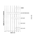


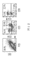


Classifications
-
- C—CHEMISTRY; METALLURGY
- C12—BIOCHEMISTRY; BEER; SPIRITS; WINE; VINEGAR; MICROBIOLOGY; ENZYMOLOGY; MUTATION OR GENETIC ENGINEERING
- C12N—MICROORGANISMS OR ENZYMES; COMPOSITIONS THEREOF; PROPAGATING, PRESERVING, OR MAINTAINING MICROORGANISMS; MUTATION OR GENETIC ENGINEERING; CULTURE MEDIA
- C12N5/00—Undifferentiated human, animal or plant cells, e.g. cell lines; Tissues; Cultivation or maintenance thereof; Culture media therefor
- C12N5/06—Animal cells or tissues; Human cells or tissues
- C12N5/0602—Vertebrate cells
- C12N5/0634—Cells from the blood or the immune system
- C12N5/0636—T lymphocytes
-
- A—HUMAN NECESSITIES
- A61—MEDICAL OR VETERINARY SCIENCE; HYGIENE
- A61K—PREPARATIONS FOR MEDICAL, DENTAL OR TOILETRY PURPOSES
- A61K39/00—Medicinal preparations containing antigens or antibodies
- A61K39/46—Cellular immunotherapy
- A61K39/461—Cellular immunotherapy characterised by the cell type used
- A61K39/4611—T-cells, e.g. tumor infiltrating lymphocytes [TIL], lymphokine-activated killer cells [LAK] or regulatory T cells [Treg]
-
- A—HUMAN NECESSITIES
- A61—MEDICAL OR VETERINARY SCIENCE; HYGIENE
- A61K—PREPARATIONS FOR MEDICAL, DENTAL OR TOILETRY PURPOSES
- A61K39/00—Medicinal preparations containing antigens or antibodies
- A61K39/46—Cellular immunotherapy
- A61K39/461—Cellular immunotherapy characterised by the cell type used
- A61K39/4613—Natural-killer cells [NK or NK-T]
-
- A—HUMAN NECESSITIES
- A61—MEDICAL OR VETERINARY SCIENCE; HYGIENE
- A61K—PREPARATIONS FOR MEDICAL, DENTAL OR TOILETRY PURPOSES
- A61K39/00—Medicinal preparations containing antigens or antibodies
- A61K39/46—Cellular immunotherapy
- A61K39/461—Cellular immunotherapy characterised by the cell type used
- A61K39/4615—Dendritic cells
-
- A—HUMAN NECESSITIES
- A61—MEDICAL OR VETERINARY SCIENCE; HYGIENE
- A61P—SPECIFIC THERAPEUTIC ACTIVITY OF CHEMICAL COMPOUNDS OR MEDICINAL PREPARATIONS
- A61P35/00—Antineoplastic agents
-
- C—CHEMISTRY; METALLURGY
- C12—BIOCHEMISTRY; BEER; SPIRITS; WINE; VINEGAR; MICROBIOLOGY; ENZYMOLOGY; MUTATION OR GENETIC ENGINEERING
- C12N—MICROORGANISMS OR ENZYMES; COMPOSITIONS THEREOF; PROPAGATING, PRESERVING, OR MAINTAINING MICROORGANISMS; MUTATION OR GENETIC ENGINEERING; CULTURE MEDIA
- C12N5/00—Undifferentiated human, animal or plant cells, e.g. cell lines; Tissues; Cultivation or maintenance thereof; Culture media therefor
- C12N5/06—Animal cells or tissues; Human cells or tissues
- C12N5/0602—Vertebrate cells
- C12N5/0634—Cells from the blood or the immune system
- C12N5/0639—Dendritic cells, e.g. Langherhans cells in the epidermis
-
- C—CHEMISTRY; METALLURGY
- C12—BIOCHEMISTRY; BEER; SPIRITS; WINE; VINEGAR; MICROBIOLOGY; ENZYMOLOGY; MUTATION OR GENETIC ENGINEERING
- C12N—MICROORGANISMS OR ENZYMES; COMPOSITIONS THEREOF; PROPAGATING, PRESERVING, OR MAINTAINING MICROORGANISMS; MUTATION OR GENETIC ENGINEERING; CULTURE MEDIA
- C12N5/00—Undifferentiated human, animal or plant cells, e.g. cell lines; Tissues; Cultivation or maintenance thereof; Culture media therefor
- C12N5/06—Animal cells or tissues; Human cells or tissues
- C12N5/0602—Vertebrate cells
- C12N5/0634—Cells from the blood or the immune system
- C12N5/0646—Natural killers cells [NK], NKT cells
-
- C—CHEMISTRY; METALLURGY
- C12—BIOCHEMISTRY; BEER; SPIRITS; WINE; VINEGAR; MICROBIOLOGY; ENZYMOLOGY; MUTATION OR GENETIC ENGINEERING
- C12N—MICROORGANISMS OR ENZYMES; COMPOSITIONS THEREOF; PROPAGATING, PRESERVING, OR MAINTAINING MICROORGANISMS; MUTATION OR GENETIC ENGINEERING; CULTURE MEDIA
- C12N5/00—Undifferentiated human, animal or plant cells, e.g. cell lines; Tissues; Cultivation or maintenance thereof; Culture media therefor
- C12N5/06—Animal cells or tissues; Human cells or tissues
- C12N5/0602—Vertebrate cells
- C12N5/0634—Cells from the blood or the immune system
- C12N5/0647—Haematopoietic stem cells; Uncommitted or multipotent progenitors
-
- C—CHEMISTRY; METALLURGY
- C12—BIOCHEMISTRY; BEER; SPIRITS; WINE; VINEGAR; MICROBIOLOGY; ENZYMOLOGY; MUTATION OR GENETIC ENGINEERING
- C12N—MICROORGANISMS OR ENZYMES; COMPOSITIONS THEREOF; PROPAGATING, PRESERVING, OR MAINTAINING MICROORGANISMS; MUTATION OR GENETIC ENGINEERING; CULTURE MEDIA
- C12N5/00—Undifferentiated human, animal or plant cells, e.g. cell lines; Tissues; Cultivation or maintenance thereof; Culture media therefor
- C12N5/06—Animal cells or tissues; Human cells or tissues
- C12N5/0602—Vertebrate cells
- C12N5/0696—Artificially induced pluripotent stem cells, e.g. iPS
-
- A—HUMAN NECESSITIES
- A61—MEDICAL OR VETERINARY SCIENCE; HYGIENE
- A61K—PREPARATIONS FOR MEDICAL, DENTAL OR TOILETRY PURPOSES
- A61K2121/00—Preparations for use in therapy
-
- A—HUMAN NECESSITIES
- A61—MEDICAL OR VETERINARY SCIENCE; HYGIENE
- A61K—PREPARATIONS FOR MEDICAL, DENTAL OR TOILETRY PURPOSES
- A61K2300/00—Mixtures or combinations of active ingredients, wherein at least one active ingredient is fully defined in groups A61K31/00 - A61K41/00
-
- C—CHEMISTRY; METALLURGY
- C12—BIOCHEMISTRY; BEER; SPIRITS; WINE; VINEGAR; MICROBIOLOGY; ENZYMOLOGY; MUTATION OR GENETIC ENGINEERING
- C12N—MICROORGANISMS OR ENZYMES; COMPOSITIONS THEREOF; PROPAGATING, PRESERVING, OR MAINTAINING MICROORGANISMS; MUTATION OR GENETIC ENGINEERING; CULTURE MEDIA
- C12N2500/00—Specific components of cell culture medium
- C12N2500/02—Atmosphere, e.g. low oxygen conditions
-
- C—CHEMISTRY; METALLURGY
- C12—BIOCHEMISTRY; BEER; SPIRITS; WINE; VINEGAR; MICROBIOLOGY; ENZYMOLOGY; MUTATION OR GENETIC ENGINEERING
- C12N—MICROORGANISMS OR ENZYMES; COMPOSITIONS THEREOF; PROPAGATING, PRESERVING, OR MAINTAINING MICROORGANISMS; MUTATION OR GENETIC ENGINEERING; CULTURE MEDIA
- C12N2500/00—Specific components of cell culture medium
- C12N2500/30—Organic components
- C12N2500/38—Vitamins
-
- C—CHEMISTRY; METALLURGY
- C12—BIOCHEMISTRY; BEER; SPIRITS; WINE; VINEGAR; MICROBIOLOGY; ENZYMOLOGY; MUTATION OR GENETIC ENGINEERING
- C12N—MICROORGANISMS OR ENZYMES; COMPOSITIONS THEREOF; PROPAGATING, PRESERVING, OR MAINTAINING MICROORGANISMS; MUTATION OR GENETIC ENGINEERING; CULTURE MEDIA
- C12N2501/00—Active agents used in cell culture processes, e.g. differentation
- C12N2501/10—Growth factors
- C12N2501/125—Stem cell factor [SCF], c-kit ligand [KL]
-
- C—CHEMISTRY; METALLURGY
- C12—BIOCHEMISTRY; BEER; SPIRITS; WINE; VINEGAR; MICROBIOLOGY; ENZYMOLOGY; MUTATION OR GENETIC ENGINEERING
- C12N—MICROORGANISMS OR ENZYMES; COMPOSITIONS THEREOF; PROPAGATING, PRESERVING, OR MAINTAINING MICROORGANISMS; MUTATION OR GENETIC ENGINEERING; CULTURE MEDIA
- C12N2501/00—Active agents used in cell culture processes, e.g. differentation
- C12N2501/10—Growth factors
- C12N2501/145—Thrombopoietin [TPO]
-
- C—CHEMISTRY; METALLURGY
- C12—BIOCHEMISTRY; BEER; SPIRITS; WINE; VINEGAR; MICROBIOLOGY; ENZYMOLOGY; MUTATION OR GENETIC ENGINEERING
- C12N—MICROORGANISMS OR ENZYMES; COMPOSITIONS THEREOF; PROPAGATING, PRESERVING, OR MAINTAINING MICROORGANISMS; MUTATION OR GENETIC ENGINEERING; CULTURE MEDIA
- C12N2501/00—Active agents used in cell culture processes, e.g. differentation
- C12N2501/20—Cytokines; Chemokines
- C12N2501/23—Interleukins [IL]
- C12N2501/2307—Interleukin-7 (IL-7)
-
- C—CHEMISTRY; METALLURGY
- C12—BIOCHEMISTRY; BEER; SPIRITS; WINE; VINEGAR; MICROBIOLOGY; ENZYMOLOGY; MUTATION OR GENETIC ENGINEERING
- C12N—MICROORGANISMS OR ENZYMES; COMPOSITIONS THEREOF; PROPAGATING, PRESERVING, OR MAINTAINING MICROORGANISMS; MUTATION OR GENETIC ENGINEERING; CULTURE MEDIA
- C12N2501/00—Active agents used in cell culture processes, e.g. differentation
- C12N2501/20—Cytokines; Chemokines
- C12N2501/26—Flt-3 ligand (CD135L, flk-2 ligand)
-
- C—CHEMISTRY; METALLURGY
- C12—BIOCHEMISTRY; BEER; SPIRITS; WINE; VINEGAR; MICROBIOLOGY; ENZYMOLOGY; MUTATION OR GENETIC ENGINEERING
- C12N—MICROORGANISMS OR ENZYMES; COMPOSITIONS THEREOF; PROPAGATING, PRESERVING, OR MAINTAINING MICROORGANISMS; MUTATION OR GENETIC ENGINEERING; CULTURE MEDIA
- C12N2501/00—Active agents used in cell culture processes, e.g. differentation
- C12N2501/40—Regulators of development
- C12N2501/42—Notch; Delta; Jagged; Serrate
-
- C—CHEMISTRY; METALLURGY
- C12—BIOCHEMISTRY; BEER; SPIRITS; WINE; VINEGAR; MICROBIOLOGY; ENZYMOLOGY; MUTATION OR GENETIC ENGINEERING
- C12N—MICROORGANISMS OR ENZYMES; COMPOSITIONS THEREOF; PROPAGATING, PRESERVING, OR MAINTAINING MICROORGANISMS; MUTATION OR GENETIC ENGINEERING; CULTURE MEDIA
- C12N2506/00—Differentiation of animal cells from one lineage to another; Differentiation of pluripotent cells
- C12N2506/45—Differentiation of animal cells from one lineage to another; Differentiation of pluripotent cells from artificially induced pluripotent stem cells
-
- C—CHEMISTRY; METALLURGY
- C12—BIOCHEMISTRY; BEER; SPIRITS; WINE; VINEGAR; MICROBIOLOGY; ENZYMOLOGY; MUTATION OR GENETIC ENGINEERING
- C12N—MICROORGANISMS OR ENZYMES; COMPOSITIONS THEREOF; PROPAGATING, PRESERVING, OR MAINTAINING MICROORGANISMS; MUTATION OR GENETIC ENGINEERING; CULTURE MEDIA
- C12N2533/00—Supports or coatings for cell culture, characterised by material
- C12N2533/50—Proteins
-
- C—CHEMISTRY; METALLURGY
- C12—BIOCHEMISTRY; BEER; SPIRITS; WINE; VINEGAR; MICROBIOLOGY; ENZYMOLOGY; MUTATION OR GENETIC ENGINEERING
- C12N—MICROORGANISMS OR ENZYMES; COMPOSITIONS THEREOF; PROPAGATING, PRESERVING, OR MAINTAINING MICROORGANISMS; MUTATION OR GENETIC ENGINEERING; CULTURE MEDIA
- C12N2533/00—Supports or coatings for cell culture, characterised by material
- C12N2533/90—Substrates of biological origin, e.g. extracellular matrix, decellularised tissue
Landscapes
- Health & Medical Sciences (AREA)
- Engineering & Computer Science (AREA)
- Life Sciences & Earth Sciences (AREA)
- Biomedical Technology (AREA)
- Chemical & Material Sciences (AREA)
- Organic Chemistry (AREA)
- Zoology (AREA)
- Biotechnology (AREA)
- Bioinformatics & Cheminformatics (AREA)
- Genetics & Genomics (AREA)
- Wood Science & Technology (AREA)
- Immunology (AREA)
- General Health & Medical Sciences (AREA)
- Cell Biology (AREA)
- Microbiology (AREA)
- Biochemistry (AREA)
- General Engineering & Computer Science (AREA)
- Hematology (AREA)
- Developmental Biology & Embryology (AREA)
- Transplantation (AREA)
- Animal Behavior & Ethology (AREA)
- Veterinary Medicine (AREA)
- Medicinal Chemistry (AREA)
- Public Health (AREA)
- Pharmacology & Pharmacy (AREA)
- Mycology (AREA)
- Epidemiology (AREA)
- Nuclear Medicine, Radiotherapy & Molecular Imaging (AREA)
- General Chemical & Material Sciences (AREA)
- Chemical Kinetics & Catalysis (AREA)
- Micro-Organisms Or Cultivation Processes Thereof (AREA)
- Medicines Containing Material From Animals Or Micro-Organisms (AREA)
- Measuring Or Testing Involving Enzymes Or Micro-Organisms (AREA)
Description
本出願は、2016年10月5日出願の米国仮特許出願第62/404,470号および2017年4月18日出願の同第62/486,895号に基づく利益を主張し、これらの全体が参照により本明細書に組み込まれる。 This application claims the benefit of U.S. Provisional Patent Application Nos. 62/404,470 filed October 5, 2016 and 62/486,895 filed April 18, 2017, the entirety of which is incorporated herein by reference.
本発明は、一般的には、分子生物学の分野に関する。より特定的には、血液細胞由来の誘導多能性幹細胞(iPSC)からHLAホモ接合免疫細胞を産生するための方法および組成物に関する。 The present invention relates generally to the field of molecular biology. More particularly, it relates to methods and compositions for generating HLA homozygous immune cells from blood cell-derived induced pluripotent stem cells (iPSCs).
細胞治療法は、腫瘍、ウイルス、および細菌病原体に対する宿主免疫反応を増強するために開発されてきた。細胞治療法は、多くの場合、ex vivoでのT細胞の活性化および拡大を必要とする。こうしたタイプの治療の例としては、腫瘍浸潤リンパ球(TIL)細胞、細胞傷害性T細胞、拡大腫瘍流入領域リンパ節細胞、および各種の他のリンパ球調製物の使用が挙げられる。造血幹細胞および造血プロジェニター細胞の医学的可能性がかなり高いため、造血プロジェニター細胞からT細胞やNK細胞などの免疫細胞への分化の方法の改善を試みて実質的研究がなされてきた。 Cell therapy has been developed to enhance host immune responses against tumor, viral, and bacterial pathogens. Cell therapy often requires activation and expansion of T cells ex vivo. Examples of these types of treatments include the use of tumor infiltrating lymphocyte (TIL) cells, cytotoxic T cells, extended tumor draining lymph node cells, and various other lymphocyte preparations. Due to the considerable medical potential of hematopoietic stem cells and hematopoietic progenitor cells, substantial research has been devoted to trying to improve the methods of differentiation of hematopoietic progenitor cells into immune cells such as T cells and NK cells.
ヒトにおいて、誘導多能性幹(iPS)細胞は、通常、真皮線維芽細胞から生成させる。しかしながら、皮膚生検に対する要件および数回のin vitro継代で線維芽細胞を拡大する必要性があるため、患者特異的幹細胞の生成源として扱いにくいものとなっている。さらに、これまでのヒト体細胞リプログラム方法は、ヒト被験体から体細胞を直接得る必要があるかまたは多くの労力を要する細胞培養系で細胞を維持する必要があるため不便である。したがって、単純で便利でしかも容易に利用可能な代替源から多能性幹細胞を誘導する方法を開発する必要性が存在する。したがって、血液は、患者もしくは健常者からの採取、貯蔵、またはたとえば中央配布ユニットから1つ以上の遠隔地への移送を行いうるので、血液サンプルはかかる源でありうる。そのため、血液細胞からiPS細胞にリプログラムしてから血液細胞由来iPS細胞をT細胞、NK細胞、T/NK細胞、樹状細胞などの免疫細胞に効率的に分化させる方法の明確な必要性が現在存在する。 In humans, induced pluripotent stem (iPS) cells are usually generated from dermal fibroblasts. However, the requirement for skin biopsies and the need to expand fibroblasts over several in vitro passages makes them a cumbersome source of patient-specific stem cells. Furthermore, previous methods of reprogramming human somatic cells are inconvenient because they require either obtaining somatic cells directly from a human subject or maintaining the cells in labor-intensive cell culture systems. Therefore, there is a need to develop methods for deriving pluripotent stem cells from alternative sources that are simple, convenient and readily available. Accordingly, blood samples may be such a source, as blood may be drawn from a patient or healthy individual, stored, or transported, for example, from a central distribution unit to one or more remote locations. Therefore, there is a clear need for a method for reprogramming blood cells to iPS cells and then efficiently differentiating the blood cell-derived iPS cells into immune cells such as T cells, NK cells, T/NK cells, and dendritic cells. currently exist.
本開示の第1の実施形態は、誘導多能性幹細胞(iPSC)を得ることであって、iPSCがHLAホモ接合血液細胞集団からリプログラムされること、iPSCを造血前駆細胞(HPC)に分化させること、および免疫細胞分化を促進する条件下でHPCを培養することによりHLAホモ接合免疫細胞を産生することを含む、HLAホモ接合免疫細胞の産生方法を提供する。いくつかの態様では、iPSCは、組込み外因性ウイルスエレメントが本質的にフリーである。 A first embodiment of the present disclosure is to obtain induced pluripotent stem cells (iPSCs), wherein the iPSCs are reprogrammed from an HLA homozygous blood cell population, differentiating the iPSCs into hematopoietic progenitor cells (HPCs) and culturing the HPCs under conditions that promote immune cell differentiation to produce HLA homozygous immune cells. In some aspects, iPSCs are essentially free of integrated exogenous viral elements.
いくつかの態様では、HLAホモ接合血液細胞は、遺伝子座対立遺伝子HLA-A、HLA-B、HLA-C、HLA-DR、HLA-DP、またはHLA-DQの1つ以上がホモ接合である。特定の態様では、HLAホモ接合血液細胞は、遺伝子座対立遺伝子HLA-A、HLA-B、HLA-C、HLA-DR、HLA-DP、またはHLA-DQの2つがホモ接合である。特定の態様では、HLAホモ接合血液細胞は、HLA-AおよびHLA-Bがホモ接合である。特定の一態様では、HLAホモ接合血液細胞は、HLA-A、HLA-B、およびHLA-Cがホモ接合である。 In some aspects, the HLA homozygous blood cell is homozygous for one or more of the locus alleles HLA-A, HLA-B, HLA-C, HLA-DR, HLA-DP, or HLA-DQ . In particular aspects, the HLA homozygous blood cell is homozygous for two of the locus alleles HLA-A, HLA-B, HLA-C, HLA-DR, HLA-DP, or HLA-DQ. In certain aspects, HLA homozygous blood cells are homozygous for HLA-A and HLA-B. In one particular aspect, the HLA homozygous blood cell is homozygous for HLA-A, HLA-B and HLA-C.
ある特定の態様では、HLAホモ接合免疫細胞はリンフォイド細胞である。特定の態様では、リンフォイド細胞は、T細胞、B細胞、および/またはNK細胞である。いくつかの態様では、HLAホモ接合免疫細胞はミエロイド細胞である。特定の態様では、ミエロイド細胞は樹状細胞である。 In certain aspects, the HLA homozygous immune cells are lymphoid cells. In certain aspects, lymphoid cells are T cells, B cells, and/or NK cells. In some aspects, the HLA homozygous immune cells are myeloid cells. In certain aspects, the myeloid cells are dendritic cells.
いくつかの態様では、HLAホモ接合血液細胞集団は哺乳動物由来である。特定の態様では、血液細胞集団はヒト由来である。いくつかの態様では、HLAホモ接合血液細胞集団は、外因的適用G-CSFにより動員されていない。ある特定の態様では、HLAホモ接合血液細胞集団は、T細胞、B細胞、および/またはNK細胞を含む。いくつかの態様では、HLAホモ接合血液細胞集団は、プロジェニター血液細胞、末梢血単核細胞、またはリンパ芽球様細胞としてさらに規定される。ある特定の態様では、HLAホモ接合血液細胞集団は、末梢血、臍帯血、またはリンパ組織から単離される。特定の態様では、リンパ組織は、骨髄、リンパ節、または胎児肝臓を含む。特定の態様では、HLAホモ接合血液細胞集団はT細胞を含む。いくつかの態様では、T細胞は、抗CD3抗体および/または抗CD28抗体の存在下で培養される。特定の態様では、T細胞は、CD4+またはCD8+のT細胞である。いくつかの態様では、T細胞は、Tヘルパー1(TH1)細胞、Tヘルパー2(TH2)細胞、TH17細胞、細胞傷害性T細胞、レギュラトリーT細胞、ナチュラルキラーT細胞、ナイーブT細胞、メモリーT細胞、またはγδT細胞である。 In some aspects, the HLA homozygous blood cell population is from a mammal. In certain aspects, the blood cell population is of human origin. In some aspects, the HLA homozygous blood cell population has not been mobilized by exogenously applied G-CSF. In certain aspects, the HLA homozygous blood cell population comprises T cells, B cells, and/or NK cells. In some aspects, the HLA homozygous blood cell population is further defined as progenitor blood cells, peripheral blood mononuclear cells, or lymphoblastoid cells. In certain aspects, the HLA homozygous blood cell population is isolated from peripheral blood, cord blood, or lymphoid tissue. In particular aspects, the lymphoid tissue comprises bone marrow, lymph nodes, or fetal liver. In certain aspects, the HLA homozygous blood cell population comprises T cells. In some aspects, T cells are cultured in the presence of anti-CD3 and/or anti-CD28 antibodies. In a particular aspect, the T cells are CD4 + or CD8 + T cells. In some aspects, the T cells are T helper 1 (TH1) cells, T helper 2 (TH2) cells, TH17 cells, cytotoxic T cells, regulatory T cells, natural killer T cells, naive T cells, memory T cells, or γδ T cells.
ある特定の態様では、リプログラミングは、血液細胞集団にリプログラミング因子を導入することを含む。いくつかの態様では、リプログラミングは、HLAホモ接合血液細胞集団にRNA、タンパク質、または低分子を導入することを含む。 In certain aspects, reprogramming comprises introducing a reprogramming factor into the blood cell population. In some aspects, reprogramming comprises introducing RNA, proteins, or small molecules into the HLA homozygous blood cell population.
いくつかの態様では、リプログラミング因子は、1つ以上の発現カセットによりコードされる。ある特定の態様では、リプログラミング因子は、Sox2、Oct4、cMyc、Klf4、Nanog、SV40ラージT抗原、およびLin28からなる群から選択される2つ以上の遺伝子を含む。いくつかの態様では、リプログラミング因子は、Sox2、Oct4、cMyc、Klf4、Nanog、SV40ラージT抗原、およびLin28からなる群から選択される遺伝子の3つ、4つ、5つ、または6つを含む。ある特定の態様では、1つ以上の発現カセットは、ウイルスベクター、エピソームベクター、およびトランスポゾンからなる群から選択されるリプログラミングベクターに含まれる。いくつかの態様では、ウイルスベクターは、レトロウイルスベクターとしてさらに規定される。特定の態様では、エピソームベクターは、エプスタイン・バーウイルス(EBV)ベースのエピソームベクターとしてさらに規定される。いくつかの態様では、リプログラミングは、規定されたフィーダーフリー条件下で細胞を培養することを含む。 In some aspects, reprogramming factors are encoded by one or more expression cassettes. In certain aspects, the reprogramming factor comprises two or more genes selected from the group consisting of Sox2, Oct4, cMyc, Klf4, Nanog, SV40 large T antigen, and Lin28. In some aspects, the reprogramming factor comprises 3, 4, 5, or 6 genes selected from the group consisting of Sox2, Oct4, cMyc, Klf4, Nanog, SV40 large T antigen, and Lin28. include. In certain aspects, the one or more expression cassettes are contained in a reprogramming vector selected from the group consisting of viral vectors, episomal vectors, and transposons. In some aspects, the viral vector is further defined as a retroviral vector. In certain aspects, the episomal vector is further defined as an Epstein-Barr virus (EBV)-based episomal vector. In some aspects, reprogramming comprises culturing the cells under defined feeder-free conditions.
いくつかの態様では、HPCは、1個のHPC当たり少なくとも20のHLAホモ接合免疫細胞、たとえば、1個のHPC当たり少なくとも10、20、30、40、50、60、70、80、90、100、またはそれ以上のHLAホモ接合免疫細胞に分化する。特定の態様では、HPCは、1個のHPC当たり少なくとも10、20、30、40、50、75、100、125、150、175、200、またはそれ以上のNK細胞に分化する。いくつかの態様では、HPCは、1個のHPC当たり少なくとも5、10、15、20、25、30、40、50、またはそれ以上のT細胞に分化する。 In some aspects, the HPC is at least 20 HLA homozygous immune cells per HPC, such as at least 10, 20, 30, 40, 50, 60, 70, 80, 90, 100 per HPC , or higher HLA homozygous immune cells. In certain aspects, the HPCs differentiate into at least 10, 20, 30, 40, 50, 75, 100, 125, 150, 175, 200, or more NK cells per HPC. In some aspects, the HPCs differentiate into at least 5, 10, 15, 20, 25, 30, 40, 50, or more T cells per HPC.
ある特定の態様では、iPSCをHPCに分化させることは、少なくとも1つの成長因子を含む第1の規定培地でiPSCを培養すること、IL-3、Flt3リガンド、およびGM-CSFがフリーまたは本質的にフリーの第2の規定培地で細胞をインキュベートすること、複数の細胞を拡大または分化を促進するのに十分なBMP4、FGF2、およびVEGFを含む第3の規定培地で細胞を培養すること、および複数の細胞を拡大または分化を促進するのに十分なIL-3およびFlt3リガンドを含む第4の規定培地で細胞を培養すること、の逐次工程を含む。いくつかの態様では、複数の多能性細胞はHPCに分化される。いくつかの態様では、第2の規定培地はブレビスタチンを含む。ある特定の態様では、第2の規定培地はGSK3阻害剤をさらに含む。いくつかの態様では、GSK3阻害剤はCHIR99021である。いくつかの態様では、第2の規定培地は、BMP4、VEGF、およびFGF2をさらに含む。ある特定の態様では、細胞は、第2の規定培地で細胞をインキュベートする前に個別化される。いくつかの態様では、第2の規定培地で細胞をインキュベートすること、第3の規定培地で細胞を培養すること、および第4の規定培地で細胞を培養することは、アミン培養プレートを用いて実施される。ある特定の態様では、第2の規定培地はVEGFおよびFGF2をさらに含む。特定の態様では、第4の規定培地は、IL-3、IL-6、SCF、TPO、およびBMP4からなる群から選択されるサイトカインの1つ以上をさらに含む。いくつかの態様では、第4の規定培地はヘパリンを含む。いくつかの態様では、本方法は、25%未満の酸素、たとえば、24%未満、23%未満、22%未満、21%未満、20%未満、19%未満、18%未満、17%未満、16%未満、15%未満、10%未満、または5%未満の酸素の大気圧で細胞を培養することを含む。いくつかの態様では、複数の多能性細胞は胚様体(EB)を形成する。ある特定の態様では、HPCはCD34を発現する。特定の態様では、HPCは、CD43、CD34、CD31、CD41、CD235、およびCD45からなる群から少なくとも2つのマーカー、たとえば、CD43およびCD34の両方を発現する。 In certain aspects, differentiating the iPSCs into HPCs comprises culturing the iPSCs in a first defined medium comprising at least one growth factor, free or essentially free of IL-3, Flt3 ligand, and GM-CSF. incubating the cells in a second defined medium free of , culturing the cells in a third defined medium containing sufficient BMP4, FGF2, and VEGF to promote expansion or differentiation of the plurality of cells, and culturing the cells in a fourth defined medium containing sufficient IL-3 and Flt3 ligand to promote expansion or differentiation of the plurality of cells. In some aspects, the plurality of pluripotent cells are differentiated into HPCs. In some aspects, the second defined medium comprises blebbistatin. In certain aspects, the second defined medium further comprises a GSK3 inhibitor. In some aspects, the GSK3 inhibitor is CHIR99021. In some aspects, the second defined medium further comprises BMP4, VEGF, and FGF2. In certain aspects, the cells are individualized prior to incubating the cells in the second defined medium. In some aspects, incubating the cells in the second defined medium, culturing the cells in the third defined medium, and culturing the cells in the fourth defined medium are performed using amine culture plates. be implemented. In certain aspects, the second defined medium further comprises VEGF and FGF2. In certain aspects, the fourth defined medium further comprises one or more cytokines selected from the group consisting of IL-3, IL-6, SCF, TPO, and BMP4. In some aspects, the fourth defined medium comprises heparin. In some aspects, the method uses less than 25% oxygen, e.g., less than 24%, less than 23%, less than 22%, less than 21%, less than 20%, less than 19%, less than 18%, less than 17%, Culturing the cells at atmospheric pressure of less than 16%, less than 15%, less than 10%, or less than 5% oxygen. In some aspects, the plurality of pluripotent cells form an embryoid body (EB). In certain aspects, the HPC expresses CD34. In certain aspects, the HPCs express at least two markers from the group consisting of CD43, CD34, CD31, CD41, CD235, and CD45, eg, both CD43 and CD34.
いくつかの態様では、免疫細胞分化を促進する条件は、リンフォイド分化を促進する条件としてさらに規定される。ある特定の態様では、CD34およびCD43を発現するHPCは、リンフォイド分化を促進する条件下で培養される。いくつかの態様では、リンフォイド分化を促進するように細胞を培養することは、マトリックスおよびNotchリガンドで被覆された表面上の規定培地でHPCを培養することであって、HPCが、CD34、CD43、CD7、DLL4、CD144、およびCD235からなる群から選択される細胞表面マーカーの1つ以上を発現すること、および1つ以上のサイトカインの存在下で培養を維持することによりリンフォイド細胞を産生することを含む。いくつかの態様では、HPCは、CD144、CD34、CD45、およびCD7を発現する。特定の態様では、HPCは、CD144、CD34、CD45、およびCD7を発現する。 In some aspects, conditions that promote immune cell differentiation are further defined as conditions that promote lymphoid differentiation. In certain aspects, HPCs expressing CD34 and CD43 are cultured under conditions that promote lymphoid differentiation. In some aspects, culturing the cells to promote lymphoid differentiation is culturing the HPCs in a defined medium on a surface coated with matrix and Notch ligands, wherein the HPCs are characterized by CD34, CD43, producing lymphoid cells by expressing one or more cell surface markers selected from the group consisting of CD7, DLL4, CD144, and CD235 and maintaining the culture in the presence of one or more cytokines; include. In some aspects, the HPC expresses CD144, CD34, CD45, and CD7. In certain aspects, the HPCs express CD144, CD34, CD45, and CD7.
追加の態様では、リンフォイド分化の第1の工程は、1つ以上の細胞表面マーカーを発現するHPCを単離することをさらに含む。いくつかの態様では、単離は磁気活性化細胞選別(MACS)を含む。ある特定の態様では、細胞は5%未満の酸素の大気圧で培養される。ある特定の態様では、細胞は約5%の酸素の大気圧で培養される。いくつかの態様では、規定培地はアスコルビン酸および/またはニコチンアミドを含む。ある特定の態様では、アスコルビン酸は50μM~1mMたとえば90μM~100μMの濃度で存在する。いくつかの態様では、ニコチンアミドは0.1mM~5mMの濃度で存在する。いくつかの態様では、ニコチンアミドはニコチン酸である。いくつかの態様では、マトリックスは細胞外マトリックスタンパク質である。いくつかの態様では、マトリックスはレトロネクチン、コラーゲン、ラミニン、またはフィブロネクチンである。特定の態様では、マトリックスはレトロネクチンである。いくつかの態様では、NotchリガンドはDLL4である。ある特定の態様では、DLL4はDLL4:Fcキメラタンパク質である。特定の態様では、1つ以上のサイトカインは、SCF、TPO、IL-7、およびFlt-3からなる群から選択される。いくつかの態様では、第2の工程は1~6週間たとえば2~4週間である。いくつかの態様では、リンフォイド細胞は、CD8、CD7、CD45、CD5、CD4、およびCD3からなる群から選択されるマーカーの1つ以上を発現する。いくつかの態様では、リンフォイド細胞の5%超は、マーカーの少なくとも2つが陽性である。特定の態様では、リンフォイド細胞の10%超は、マーカーの少なくとも2つが陽性である。 In an additional aspect, the first step of lymphoid differentiation further comprises isolating HPCs expressing one or more cell surface markers. In some aspects, isolation comprises magnetic activated cell sorting (MACS). In certain aspects, cells are cultured at atmospheric pressure of less than 5% oxygen. In certain aspects, cells are cultured at atmospheric pressure of about 5% oxygen. In some embodiments, the defined medium comprises ascorbic acid and/or nicotinamide. In certain aspects, ascorbic acid is present at a concentration of 50 μM to 1 mM, such as 90 μM to 100 μM. In some aspects, nicotinamide is present at a concentration of 0.1 mM to 5 mM. In some aspects, the nicotinamide is nicotinic acid. In some aspects, the matrix is an extracellular matrix protein. In some embodiments, the matrix is retronectin, collagen, laminin, or fibronectin. In certain embodiments, the matrix is Retronectin. In some aspects, the Notch ligand is DLL4. In certain aspects, DLL4 is a DLL4:Fc chimeric protein. In certain aspects, the one or more cytokines are selected from the group consisting of SCF, TPO, IL-7, and Flt-3. In some embodiments, the second step is 1-6 weeks, such as 2-4 weeks. In some aspects, the lymphoid cells express one or more markers selected from the group consisting of CD8, CD7, CD45, CD5, CD4, and CD3. In some aspects, more than 5% of lymphoid cells are positive for at least two of the markers. In certain aspects, more than 10% of lymphoid cells are positive for at least two of the markers.
いくつかの態様では、免疫細胞分化を促進する条件は、ミエロイド分化を促進する条件としてさらに規定される。ある特定の態様では、ミエロイド分化を促進する条件下でHPCを培養することは、TPO、SCF、およびFlt3リガンドを含む第1の規定培地でHPCを培養することによりミエロイド細胞集団を産生すること、およびTPO、SCF、およびFlt3リガンドが本質的にフリーの第2の規定培地で細胞をインキュベートすることによりミエロイド細胞富化集団を産生することを含む。いくつかの態様では、第1の規定培地はIL-6およびIL-3をさらに含む。ある特定の態様では、第2の規定培地はGM-CSFを含む。特定の態様では、第1の工程で産生されるミエロイド細胞集団の少なくとも50%は、CD45、CD43、およびCD31が陽性である。いくつかの態様では、CD45、CD43、およびCD31が陽性のミエロイド細胞集団は、CD34を本質的に発現しない。特定の態様では、ミエロイド細胞富化集団の少なくとも80%は、CD43+、CD45+、CD31+、およびCD34-である。いくつかの態様では、第2の工程は5~10日間である。 In some aspects, conditions that promote immune cell differentiation are further defined as conditions that promote myeloid differentiation. In certain aspects, culturing the HPCs under conditions that promote myeloid differentiation comprises producing a myeloid cell population by culturing the HPCs in a first defined medium comprising TPO, SCF, and Flt3 ligand; and producing a myeloid cell-enriched population by incubating the cells in a second defined medium essentially free of TPO, SCF, and Flt3 ligand. In some aspects, the first defined medium further comprises IL-6 and IL-3. In certain aspects, the second defined medium comprises GM-CSF. In certain aspects, at least 50% of the myeloid cell population produced in the first step is positive for CD45, CD43, and CD31. In some aspects, the CD45, CD43, and CD31 positive myeloid cell population does not essentially express CD34. In certain aspects, at least 80% of the myeloid cell-enriched population is CD43 + , CD45 + , CD31 + , and CD34 − . In some embodiments, the second step is 5-10 days.
ある特定の態様では、本方法は、ミエロイド細胞富化集団を樹状細胞に分化させることをさらに含む。いくつかの態様では、ミエロイド細胞富化集団を樹状細胞に分化させることは、GM-CSF、IL-4、およびTNFαを含む規定培地でミエロイド細胞富化集団を培養することにより樹状細胞を産生することを含む。いくつかの態様では、規定培地はリポタンパク質をさらに含む。ある特定の態様では、樹状細胞は、CD209、CD1a、HLA-DR、CD11c、CD14、CD83、およびCD86からなる群から選択されるマーカーの1つ以上を発現する。特定の態様では、樹状細胞はCD12を本質的に発現しない。 In certain aspects, the method further comprises differentiating the myeloid cell-enriched population into dendritic cells. In some aspects, differentiating the myeloid cell-enriched population into dendritic cells comprises culturing the myeloid cell-enriched population in a defined medium comprising GM-CSF, IL-4, and TNFα. Including producing. In some aspects, the defined medium further comprises lipoproteins. In certain aspects, the dendritic cells express one or more markers selected from the group consisting of CD209, CD1a, HLA-DR, CD11c, CD14, CD83, and CD86. In certain aspects, the dendritic cells essentially do not express CD12.
さらなる実施形態では、各ライブラリーメンバーがHLA型に従って特徴付けられる形でHLAホモ接合免疫細胞のライブラリーが提供される。いくつかの態様では、HLAホモ接合免疫細胞は、スーパードナーから得られる血液サンプルに由来する。特定の態様では、スーパードナーはヒトである。いくつかの態様では、HLAホモ接合免疫細胞は同種異系である。いくつかの態様では、HLAホモ接合免疫細胞は自己由来である。ある特定の態様では、HLAホモ接合免疫細胞は、1つ以上のHLAクラスI遺伝子および/またはHLAクラスII遺伝子が少なくともホモ接合である。ある特定の態様では、HLAホモ接合免疫細胞は、遺伝子座対立遺伝子HLA-A、HLA-B、HLA-C、HLA-DR、HLA-DP、またはHLA-DQの1つ以上がホモ接合である。いくつかの態様では、HLAホモ接合免疫細胞は、遺伝子座対立遺伝子HLA-A、HLA-B、HLA-C、HLA-DR、HLA-DP、またはHLA-DQの2つがホモ接合である。いくつかの態様では、HLAホモ接合免疫細胞は、HLA-AおよびHLA-Bがホモ接合である。ある特定の態様では、HLAホモ接合免疫細胞は、HLA-A、HLA-B、およびHLA-Cがホモ接合である。いくつかの態様では、HLAホモ接合免疫細胞は、リンフォイド細胞、たとえば、T細胞、B細胞、および/またはNK細胞である。ある特定の態様では、HLAホモ接合免疫細胞は、ミエロイド細胞たとえば樹状細胞である。特定の態様では、各ライブラリーメンバーは潜在的レシピエントにHLAがマッチする。いくつかの態様では、ライブラリーは凍結保存HLAホモ接合免疫細胞を含む。特定の態様では、ライブラリーは、メンバーが別個のHLAハプロタイプを含む形で少なくとも15のライブラリーメンバーを含む。いくつかの態様では、ライブラリーは、少なくとも30のライブラリーメンバー、たとえば、少なくとも35、40、45、50、60、70、80、90、100、またはそれ以上のライブラリーメンバーを含む。 In a further embodiment, a library of HLA homozygous immune cells is provided in which each library member is characterized according to HLA type. In some aspects, the HLA homozygous immune cells are derived from a blood sample obtained from a superdonor. In certain aspects, the superdonor is human. In some aspects, the HLA homozygous immune cells are allogeneic. In some aspects, the HLA homozygous immune cells are autologous. In certain aspects, HLA homozygous immune cells are at least homozygous for one or more HLA class I genes and/or HLA class II genes. In certain aspects, the HLA homozygous immune cell is homozygous for one or more of the locus alleles HLA-A, HLA-B, HLA-C, HLA-DR, HLA-DP, or HLA-DQ . In some aspects, the HLA homozygous immune cell is homozygous for two of the locus alleles HLA-A, HLA-B, HLA-C, HLA-DR, HLA-DP, or HLA-DQ. In some aspects, the HLA homozygous immune cells are homozygous for HLA-A and HLA-B. In certain aspects, HLA homozygous immune cells are homozygous for HLA-A, HLA-B, and HLA-C. In some aspects, the HLA homozygous immune cells are lymphoid cells, eg, T cells, B cells, and/or NK cells. In certain aspects, the HLA homozygous immune cells are myeloid cells, such as dendritic cells. In certain embodiments, each library member is HLA-matched to a potential recipient. In some aspects, the library comprises cryopreserved HLA homozygous immune cells. In certain aspects, the library comprises at least 15 library members, such that the members comprise distinct HLA haplotypes. In some aspects, the library comprises at least 30 library members, such as at least 35, 40, 45, 50, 60, 70, 80, 90, 100, or more library members.
他の一実施形態では、ヒトスーパードナーから得られる複数の血液サンプルのHLAタイピングを行うこと、複数の血液サンプルからiPSCに細胞をリプログラムすること、iPSCをHPCに分化させること、および免疫細胞分化を促進する条件下でHPCを培養することによりHLAホモ接合免疫細胞のライブラリーを作製することを含む、実施形態のHLAホモ接合免疫細胞のライブラリーの作製方法が提供される。 In another embodiment, performing HLA typing of multiple blood samples obtained from human superdonors, reprogramming cells from multiple blood samples into iPSCs, differentiating iPSCs into HPCs, and immune cell differentiation Methods of making a library of HLA-homozygous immune cells of the embodiments are provided, comprising making the library of HLA-homozygous immune cells by culturing HPCs under conditions that promote
本発明の他の目的、特徴、および利点は、以下の詳細な説明から明らかになるであろう。しかしながら、本発明の趣旨および範囲に含まれる各種の変更形態および修正形態は、本詳細な説明から当業者には明らかになるであろうから、詳細な説明および具体例は、本発明の好ましい実施形態を示すが単に例示を目的として与えられているにすぎないことを理解すべきである。 Other objects, features, and advantages of the present invention will become apparent from the detailed description below. The detailed description and specific examples, however, are intended as a preferred embodiment of the invention, since various changes and modifications within the spirit and scope of the invention will become apparent to those skilled in the art from this detailed description. It should be understood that any morphology shown is given for illustrative purposes only.
以下の図面は、本明細書の一部を形成するものであり、本発明のある特定の態様をさらに実証するために含まれる。本発明は、本明細書に提示される具体的な実施形態の詳細な説明と組み合わせてこれらの図面の1つ以上を参照すればよりよく理解されよう。 The following drawings form part of this specification and are included to further demonstrate certain aspects of the present invention. The invention may be better understood by reference to one or more of these drawings in conjunction with the detailed description of specific embodiments presented herein.
ある特定の実施形態では、本開示は、体細胞(たとえば血液細胞)の出発集団からリプログラムされた誘導多能性幹細胞から免疫細胞を生成させるための高効率の方法を提供する。本方法により産生される免疫細胞は、T細胞、NK細胞、T/NK細胞、および樹状細胞を含みうる。いくつかの態様では、体細胞の出発集団はT細胞を含む。T細胞は、血液サンプルなどの各種供給源から単離しうる。 In certain embodiments, the present disclosure provides highly efficient methods for generating immune cells from induced pluripotent stem cells reprogrammed from a starting population of somatic cells (eg, blood cells). Immune cells produced by this method can include T cells, NK cells, T/NK cells, and dendritic cells. In some aspects, the starting population of somatic cells comprises T cells. T cells can be isolated from various sources such as blood samples.
特定の実施形態では、血液細胞の出発集団は、HLAホモ接合細胞を含む(すなわち、MHCクラスIおよびIIの遺伝子がホモ接合である)。したがって、iPSCは、HLAホモ接合被験体(本明細書ではHLAスーパードナーという)から単離された細胞から産生可能である。 In certain embodiments, the starting population of blood cells comprises HLA homozygous cells (ie, homozygous for MHC class I and II genes). Thus, iPSCs can be produced from cells isolated from HLA homozygous subjects (referred to herein as HLA superdonors).
血液細胞集団は、ウイルスベクターやエピソームベクターなどを介してリプログラミング因子を導入することによりiPSCにリプログラムしうる。次いで、こうした血液細胞由来iPSC(たとえばT細胞由来iPSC(TiPSC))を造血前駆細胞に分化させる。一方法では、分化プロセスは、WNT活性化、造血誘導サイトカインを用いた培養、およびCD34+細胞単離を必要とする。次いで、最後に、HPCをリンフォイド細胞(たとえば、T細胞、NK細胞、およびT/NK細胞)やミエロイド細胞(たとえば、樹状細胞)などの免疫細胞に分化させる。 Blood cell populations can be reprogrammed into iPSCs by introducing reprogramming factors via viral vectors, episomal vectors, and the like. These blood cell-derived iPSCs (eg, T cell-derived iPSCs (TiPSCs)) are then differentiated into hematopoietic progenitor cells. In one method, the differentiation process requires WNT activation, culture with hematopoietic-inducing cytokines, and CD34 + cell isolation. HPCs are then finally differentiated into immune cells such as lymphoid cells (eg, T cells, NK cells, and T/NK cells) and myeloid cells (eg, dendritic cells).
リンフォイド細胞への分化は、フィーダーフリーマトリックスとしてRetroNectinおよびDLL-4を用いて行いうる。T細胞分化は、効率および成熟を増加させるアスコルビン酸を用いることにより、さらには低酸素条件下で培養することによりさらに増強しうる。興味深いことに、本発明者らは、リンフォイド潜在能力に関してHPC分化時に最適時間枠(たとえば7~11日目)を決定した。リンフォイド潜在能力を有するこうしたHPCは、CD34およびCD43の発現により同定しうる。そのほか、リンフォイド潜在能力を向上させたHPCは、マーカーCD144、CD34、CD45、およびCD7の2つ以上が陽性の細胞画分を選別することにより単離しうる。いくつかの態様では、樹状細胞誘導プロジェニターは、HPC分化の約16日目に出現する共通ミエロイドプロジェニターである。
Differentiation into lymphoid cells can be performed using RetroNectin and DLL-4 as feeder-free matrices. T cell differentiation can be further enhanced by using ascorbic acid, which increases efficiency and maturation, and by culturing under hypoxic conditions. Interestingly, we determined an optimal time frame (eg, days 7-11) during HPC differentiation for lymphoid potential. Such HPCs with lymphoid potential can be identified by expression of CD34 and CD43. Alternatively, HPCs with increased lymphoid potential can be isolated by sorting for cell fractions positive for two or more of the markers CD144, CD34, CD45, and CD7. In some aspects, the dendritic cell-derived progenitor is a common myeloid progenitor that appears at about
血液細胞由来PSCから産生されるリンフォイド細胞は、特徴的なT細胞レセプター(TCR)遺伝子再構成を保持するとともにたとえば遺伝的追跡マーカーとしてまたはヒトT細胞生成を研究する再分化実験で活用可能な性質を保持するT細胞、NK細胞、およびT/NK細胞を含みうる。本開示の特定の利点は、分化させたT細胞で保持しうるT細胞レセプターの再構成され低減されたV、D、J遺伝子セグメントにある。これは、T細胞由来iPS細胞のさまざまなクローン集団の特異的な特徴または「バーコード」として機能するとともに、V(D)J組換えを受けていない多能性幹細胞からそうしたiPS細胞を分化させるのに役立つ。 Lymphoid cells produced from blood cell-derived PSCs possess characteristic T-cell receptor (TCR) gene rearrangements and can be exploited, for example, as genetic tracking markers or in regeneration experiments to study human T-cell generation. can include T cells, NK cells, and T/NK cells that retain A particular advantage of the present disclosure resides in rearranged and reduced V, D, J gene segments of the T cell receptor that can be retained in differentiated T cells. It serves as a unique signature or 'barcode' for different clonal populations of T-cell-derived iPS cells and allows them to differentiate from pluripotent stem cells that have not undergone V(D)J recombination. Helpful.
そのため、本開示の方法は、安定なin vivo移植、化合物のin vitroスクリーニング、血液疾患および傷害の機序の解明などの広範にわたる用途で、無制限の数のHLAホモ接合免疫細胞、たとえば、T細胞、NK細胞、T/NK細胞、および樹状細胞を提供可能である。 As such, the methods of the present disclosure can be used to generate unlimited numbers of HLA-homozygous immune cells, e.g., T cells, for a wide range of applications such as stable in vivo transplantation, in vitro screening of compounds, elucidation of mechanisms of hematological diseases and injury. , NK cells, T/NK cells, and dendritic cells.
I.定義
本明細書で用いられる場合、特定の成分に関して「本質的にフリー」とは、特定の成分がいずれも組成物に意図的に配合されたものではないことおよび/または汚染物質としてのみもしくは痕跡量でのみ存在するにすぎないことを意味するものとして本明細書で用いられる。したがって、組成物の意図されないなんらかの汚染から生じる特定の成分の全量は、0.05%を十分に下回る、好ましくは0.01%未満である。最も好ましいのは、特定の成分の量が標準的分析方法でまったく検出できない組成物である。
I. DEFINITIONS As used herein, “essentially free” with respect to a specified ingredient means that none of the specified ingredient is intentionally incorporated into the composition and/or only as a contaminant or trace It is used herein to mean present only in amounts. Therefore, the total amount of the specified ingredient resulting from any unintentional contamination of the composition is well below 0.05%, preferably below 0.01%. Most preferred are compositions in which no amount of a particular ingredient is detectable by standard analytical methods.
本明細書の中で用いられる場合、「a」または「an」は1つ以上を意味しうる。本特許請求の範囲の中で用いられる場合、「comprising(~を含む)」という語と組み合わせて用いられるとき、「a」または「an」という語は1つまたは2つ以上を意味しうる。 As used herein, "a" or "an" may mean one or more. As used in the claims, the terms "a" or "an" when used in conjunction with the term "comprising" can mean one or more than one.
本特許請求の範囲における「or(または)」という用語の使用は、選択肢のみを意味するものとして明示的な指定がない限りまたは選択肢が互いに排他的でない限り「and/or(および/または)」を意味するものとして用いられるが、本開示は、選択肢のみおよび「and/or(および/または)」を意味する定義を支持する。本明細書で用いられる場合、「他の一つ」とは、少なくとも第2のものまたはそれよりも多くのものを意味しうる。 The use of the term "or" in the claims means "and/or" unless expressly specified as meaning alternatives only or alternatives are not mutually exclusive. , this disclosure supports definitions that mean alternatives only and "and/or." As used herein, "another" can mean at least a second one or more.
本出願全体を通じて、「about(約)」という用語は、その値がデバイスやその値の決定に利用される方法に固有の誤差の変動または研究対象に存在する変動を含むことを意味するものとして用いられる。 Throughout this application, the term "about" is meant to include variations in error inherent in the device or method used to determine the value or variations present in the study subject. Used.
「外因性」という用語は、細胞もしくは生物のタンパク質、遺伝子、核酸、もしくはポリヌクレオチドとの関連で用いられるとき、人工もしくは天然の手段により細胞もしくは生物に導入されたタンパク質、遺伝子、核酸、もしくはポリヌクレオチドを意味し、または細胞との関連では、この用語は、単離後に人工もしくは天然の手段により他の細胞もしくは生物に導入された細胞を意味する。外因性核酸は、異なる生物もしくは細胞に由来しうるか、または生物内もしくは細胞内に天然に存在する核酸の1つ以上の追加のコピーでありうる。外因性細胞は、異なる生物に由来しうるかまたは同一の生物に由来しうる。限定されるものではないが、例として、外因性核酸は、天然の細胞のときとは異なる染色体上の位置にあるものであるか、さもなければ天然に見いだされるものとは異なる核酸配列がフランキングするものである。 The term "exogenous," when used in reference to a protein, gene, nucleic acid, or polynucleotide of a cell or organism, refers to a protein, gene, nucleic acid, or polynucleotide that has been introduced into a cell or organism by artificial or natural means. It means a nucleotide or, in the context of a cell, a cell that has been isolated and then introduced into another cell or organism by artificial or natural means. Exogenous nucleic acid can be from a different organism or cell, or can be one or more additional copies of nucleic acid naturally occurring within the organism or cell. Exogenous cells can be from different organisms or can be from the same organism. By way of non-limiting example, an exogenous nucleic acid is one that is in a different chromosomal location than it is in the cell in nature or otherwise has a different nucleic acid sequence than is found in nature. It is for ranking.
「発現構築物」または「発現カセット」とは、転写の指令が可能な核酸分子を意味する。発現構築物は、1つ以上の所望の細胞型、組織、または器官で遺伝子発現を指令する少なくとも1つ以上の転写制御エレメント(たとえば、プロモーター、エンハンサー、または機能的にそれらと等価な構造)を含む。転写終止シグナルなどの追加のエレメントもまた含まれうる。 By "expression construct" or "expression cassette" is meant a nucleic acid molecule capable of directing transcription. Expression constructs contain at least one or more transcriptional control elements (e.g., promoters, enhancers, or functionally equivalent structures thereof) that direct gene expression in one or more desired cell types, tissues, or organs. . Additional elements such as transcription termination signals may also be included.
「ベクター」または「構築物」(遺伝子送達系または遺伝子移入「媒体」ということもある)とは、in vitroまたはin vivoのいずれかで宿主細胞に送達されるポリヌクレオチドを含むマクロ分子または分子複合体を意味する。 A "vector" or "construct" (sometimes referred to as a gene delivery system or gene transfer "vehicle") is a macromolecule or molecular complex containing a polynucleotide that is delivered to a host cell either in vitro or in vivo. means
ベクターの通常のタイプである「プラスミド」は、染色体DNAとは独立して染色体DNAに依存せずに複製可能な染色体外DNA分子である。ある特定の場合には、それは環状の二本鎖である。 A "plasmid", the common type of vector, is an extrachromosomal DNA molecule capable of replication independently of and independently of chromosomal DNA. In certain cases it is a circular double strand.
「複製の起点」(「ori」)または「複製起点」とは、細胞のプラスミド中に存在するとき、結合された配列をプラスミド中におよび/またはDNA合成が開始される部位もしくはその近傍に維持可能なDNA配列たとえばリンパ球向性ヘルペスウイルスのDNA配列のことである。例として、EBV(エプスタイン・バー(Ebstein-Barr)ウイルス)のoriは、FR配列(30bpリピートの不完全な20コピー)および好ましくはDS配列を含むが、EBVの他の部位はEBNA-1に結合し、たとえば、Rep*配列は、複製の起点としてDSの代わりとなりうる(Kirshmaier and Sugden,1998)。そのため、EBVの複製起点は、FR配列、DSもしくはRep*配列、または核酸修飾された機能的に等価な任意の配列、またはそれらに由来する合成的組合せを含む。たとえば、本開示はまた、Lindner et al.,2008に具体的に記載されるように、個々のエレメントの挿入や突然変異などによりEBVの複製起点を遺伝子工学操作したものも使用しうる。 "Origin of replication"("ori") or "origin of replication" means that, when present in a plasmid of a cell, the linked sequences are maintained in the plasmid and/or at or near the site where DNA synthesis is initiated. A possible DNA sequence is that of, for example, a lymphotropic herpesvirus. As an example, the ori of EBV (Epstein-Barr virus) contains FR sequences (20 imperfect copies of 30 bp repeats) and preferably DS sequences, whereas other sites of EBV are in EBNA-1. For example, Rep * sequences can substitute for DS as origins of replication (Kirshmaier and Sugden, 1998). Thus, the EBV origin of replication includes FR sequences, DS or Rep* sequences, or any nucleic acid-modified functionally equivalent sequences, or synthetic combinations derived therefrom. For example, this disclosure also relates to Lindner et al. , 2008, genetic engineering of the EBV origin of replication, such as by insertion or mutation of individual elements, may also be used.
特定のタンパク質を「コード」する「遺伝子」、「ポリヌクレオチド」、「コード領域」、「配列」、「セグメント」、「断片」、または「トランスジーン」とは、適切なレギュラトリー配列の制御下に配置されたときに転写されるとともに任意選択的に遺伝子産物たとえばポリペプチドにin vitroまたはin vivoで翻訳される核酸分子のことである。コード領域は、cDNA、ゲノムDNA、またはRNAの形態のいずれかで存在しうる。DNAの形態で存在するとき、核酸分子は一本鎖(すなわちセンス鎖)または二本鎖でありうる。コード領域の境界は、5’(アミノ)末端の開始コドンおよび3’(カルボキシ)末端の翻訳停止コドンにより決定される。限定されるものではないが、遺伝子は、原核または真核mRNA由来のcDNA、原核または真核DNA由来のゲノムDNA配列、および合成DNA配列を含みうる。転写終止配列は、通常は遺伝子配列の3’側に位置するであろう。 A "gene", "polynucleotide", "coding region", "sequence", "segment", "fragment", or "transgene" that "encodes" a specific protein is defined under the control of appropriate regulatory sequences. A nucleic acid molecule that is transcribed and optionally translated into a gene product, such as a polypeptide, in vitro or in vivo when placed in a cell. Coding regions can be in either cDNA, genomic DNA, or RNA form. When present in the form of DNA, the nucleic acid molecule can be single-stranded (ie, the sense strand) or double-stranded. The boundaries of the coding region are determined by a start codon at the 5' (amino) terminus and a translational stop codon at the 3' (carboxy) terminus. A gene can include, but is not limited to, cDNA from prokaryotic or eukaryotic mRNA, genomic DNA sequences from prokaryotic or eukaryotic DNA, and synthetic DNA sequences. A transcription termination sequence will usually be located 3' to the gene sequence.
「制御エレメント」という用語は、プロモーター領域、ポリアデニル化シグナル、転写終止配列、上流レギュラトリードメイン、複製の起点、内部リボソーム侵入部位(IRES)、エンハンサー、スプライス接合部などを総称して意味し、全体としてレシピエント細胞において複製、転写、転写後プロセシング、コード配列の翻訳を提供する。これらの制御エレメントは、選択されたコード配列が適切な宿主細胞において複製、転写、および翻訳されうるのであれば、すべてが存在する必要はない。 The term "regulatory element" refers collectively to promoter regions, polyadenylation signals, transcription termination sequences, upstream regulatory domains, origins of replication, internal ribosome entry sites (IRES), enhancers, splice junctions, etc. provide replication, transcription, post-transcriptional processing, and translation of coding sequences in recipient cells. All of these control elements need not be present provided that the selected coding sequence can be replicated, transcribed and translated in a suitable host cell.
「プロモーター」という用語は、本明細書では、DNAレギュラトリー配列を含むヌクレオチド領域を意味するものとしてその通常の意味で用いられ、この場合、レギュラトリー配列は、RNAポリメラーゼに結合可能かつ下流(3’方向)のコード配列の転写を開始可能な遺伝子に由来する。それは、核酸配列の特異的転写を開始するようにRNAポリメラーゼや他の転写因子などのレギュラトリータンパク質および調節分子が結合しうる遺伝的エレメントを含有しうる。「作動的に配置」、「作動的に結合」、「制御下」、および「転写制御下」という語句は、プロモーターが核酸配列との関連で適正な機能位置および/または配向状態にありその配列の転写開始および/または発現を制御することを意味する。 The term "promoter" is used herein in its ordinary sense to mean a nucleotide region containing a DNA regulatory sequence, where the regulatory sequence is capable of binding RNA polymerase and downstream (3 ' direction) from a gene capable of initiating transcription of the coding sequence. It may contain genetic elements at which regulatory proteins and regulatory molecules, such as RNA polymerase and other transcription factors, may bind to initiate the specific transcription of nucleic acid sequences. The phrases "operably positioned," "operably linked," "under control," and "under transcriptional control" refer to a promoter in its proper functional position and/or orientation in relation to a nucleic acid sequence. is meant to control transcription initiation and/or expression of
「エンハンサー」とは、プロモーターに近接して配置したときにエンハンサードメインの不在下でプロモーターからもたらされる転写活性と比較して増加した転写活性を付与する核酸配列を意味する。 By "enhancer" is meant a nucleic acid sequence that, when placed in close proximity to a promoter, confers increased transcriptional activity relative to the transcriptional activity resulting from the promoter in the absence of the enhancer domain.
核酸分子に関して「作動可能に結合」または共発現」とは、2つ以上の核酸分子(たとえば、転写される核酸分子、プロモーター、およびエンハンサーエレメント)が、核酸分子の転写を可能にするよう接続されることを意味する。ペプチドおよび/またはポリペプチド分子に関して「作動可能に結合」または「共発現」とは、2つ以上のペプチドおよび/またはポリペプチド分子が、単一のポリペプチド鎖、すなわち、融合体の各ペプチドおよび/またはポリペプチド成分の少なくとも1つの性質を有する融合ポリペプチドを生じるように接続されることを意味する。融合ポリペプチドは、好ましくはキメラである。すなわち、異種分子で構成される。 "Operably linked" or co-expressed with respect to nucleic acid molecules means that two or more nucleic acid molecules (e.g., transcribed nucleic acid molecules, promoters, and enhancer elements) are joined to permit transcription of the nucleic acid molecules. means that "Operably linked" or "co-expressed" with respect to peptide and/or polypeptide molecules means that two or more peptide and/or polypeptide molecules are combined into a single polypeptide chain, i.e., fusions of each peptide and /or joined so as to result in a fusion polypeptide having at least one property of the polypeptide components. Fusion polypeptides are preferably chimeric. That is, it is composed of heterogeneous molecules.
「相同性」とは、2つのポリヌクレオチド間または2つのポリペプチド間の同一性パーセントを意味する。一方の配列と他方の配列との対応関係は、当技術分野で公知の技術により決定可能である。たとえば、相同性は、配列情報をアライメントして2つのポリペプチド分子間の配列情報を直接比較することによりおよび容易に入手可能なコンピュータープログラムを用いることにより決定可能である。代替的に、相同性は、相同領域間の安定な二本鎖の形成を促進する条件下でポリヌクレオチドをハイブリダイズさせた後で一本鎖特異的ヌクレアーゼで消化して消化断片のサイズを決定することにより決定可能である。2つのDNAまたは2つポリペプチドの配列は、以上の方法を用いて決定した場合、それぞれヌクレオチドまたはアミノ酸の少なくとも約80%、好ましくは少なくとも約90%、最も好ましくは少なくとも約95%が分子の規定の長さにわたりマッチしたとき、互いに「実質的に相同」である。 "Homology" means the percent identity between two polynucleotides or between two polypeptides. The correspondence between one sequence and another can be determined by techniques known in the art. For example, homology can be determined by aligning the sequence information and directly comparing the sequence information between two polypeptide molecules and using readily available computer programs. Alternatively, homology can be determined by hybridizing the polynucleotides under conditions that promote the formation of stable duplexes between regions of homology followed by digestion with a single-strand specific nuclease to determine the size of digested fragments. can be determined by At least about 80%, preferably at least about 90%, and most preferably at least about 95% of the nucleotides or amino acids of the two DNAs or of the two polypeptides, respectively, define the molecule when determined using the methods described above. are "substantially homologous" to each other when matched over the length of
「細胞」という用語は、本明細書では、当技術分野でのその最広義で用いられ、多細胞生物の組織の構造ユニットである、外部からそれを隔離する膜構造体に取り囲まれる、自己複製能を有する、かつ遺伝情報およびそれを発現する機序を有する、生存体を意味する。本明細書で用いられる細胞は、天然に存在する細胞または人工的に改変された細胞(たとえば、融合細胞、遺伝子改変細胞など)でありうる。 The term "cell" is used herein in its broadest sense in the art and is the structural unit of tissue in multicellular organisms, a self-renewing cell surrounded by membrane structures that isolate it from the outside. means an organism that has the ability and has the genetic information and the mechanisms to express it. Cells used herein can be naturally occurring cells or artificially modified cells (eg, fusion cells, genetically modified cells, etc.).
「幹細胞」という用語は、本明細書では、好適な条件下では多岐にわたる特殊化された細胞型に分化可能であるが、他の好適な条件下では自己再生および本質的に未分化の多能性状態の維持が可能である細胞を意味する。「幹細胞」という用語はまた、多能性細胞、複能性細胞、前駆細胞、およびプロジェニター細胞を包含する。例示的なヒト幹細胞は、骨髄組織から得られる造血幹細胞もしくは間葉幹細胞、胚組織から得られる胚性幹細胞、または胎児の生殖器組織から得られる胚性生殖細胞から取得可能である。例示的な多能性幹細胞はまた、多能性に関連するある特定の転写因子を発現して多能性状態にリプログラムすることにより体細胞からも産生可能である。これらの細胞は、「誘導多能性幹細胞」または「iPSCもしくはiPS細胞」と呼ばれる。 The term "stem cell" is used herein to refer to cells capable of differentiating into a wide variety of specialized cell types under suitable conditions, but self-renewing and essentially undifferentiated pluripotent cells under other suitable conditions. It means a cell capable of maintaining a sexual state. The term "stem cell" also includes pluripotent, multipotent, progenitor, and progenitor cells. Exemplary human stem cells can be obtained from hematopoietic or mesenchymal stem cells obtained from bone marrow tissue, embryonic stem cells obtained from embryonic tissue, or embryonic germ cells obtained from fetal reproductive tissue. Exemplary pluripotent stem cells can also be produced from somatic cells by expressing certain transcription factors associated with pluripotency and reprogramming them to a pluripotent state. These cells are called "induced pluripotent stem cells" or "iPSCs or iPS cells".
「胚性幹(ES)細胞」とは、初期段階の胚たとえば胚盤胞段階の内部細胞塊から得られる、または人為的手段(たとえば核移行)により産生され、生殖細胞(たとえば精子および卵子)を含めて胚もしくは成人の任意の分化細胞型をもたらしうる、未分化多能性細胞のことである。 "Embryonic stem (ES) cells" are obtained from early stage embryos, e.g., the inner cell mass at the blastocyst stage, or produced by artificial means (e.g., nuclear transfer) and are derived from germ cells (e.g., sperm and eggs) An undifferentiated pluripotent cell that can give rise to any embryonic or adult differentiated cell type, including cells.
「誘導多能性幹細胞(iPSCまたはiPS細胞)」とは、因子(本明細書ではリプログラミング因子という)の組合せを発現してまたは発現を誘導して体細胞をリプログラムすることにより生成される細胞のことである。iPS細胞は、胎児、出生直後の産児、新生児、若年者、または成人の体細胞を用いて生成可能である。ある特定の実施形態では、体細胞から多能性幹細胞へのリプログラミングに使用可能な因子としては、たとえば、Oct4(Oct3/4ということもある)、Sox2、c-Myc、Klf4、Nanog、およびLin28が挙げられる。いくつかの実施形態では、体細胞は、体細胞を多能性幹細胞にリプログラムするために少なくとも2つのリプログラミング因子、少なくとも3つのリプログラミング因子、少なくとも4つのリプログラミング因子、少なくとも5つのリプログラミング因子、少なくとも6つのリプログラミング因子、または少なくとも7つのリプログラミング因子を発現することによりリプログラムされる。 "Induced pluripotent stem cells (iPSCs or iPS cells)" are generated by expressing or inducing the expression of a combination of factors (referred to herein as reprogramming factors) to reprogram somatic cells It's about cells. iPS cells can be generated using fetal, postnatal, neonatal, juvenile, or adult somatic cells. In certain embodiments, factors that can be used to reprogram somatic cells to pluripotent stem cells include, for example, Oct4 (sometimes Oct3/4), Sox2, c-Myc, Klf4, Nanog, and Lin28 can be mentioned. In some embodiments, the somatic cell comprises at least 2 reprogramming factors, at least 3 reprogramming factors, at least 4 reprogramming factors, at least 5 reprogramming factors to reprogram the somatic cell to a pluripotent stem cell. reprogrammed by expressing a factor, at least 6 reprogramming factors, or at least 7 reprogramming factors.
「造血プロジェニター細胞」または「造血前駆細胞」とは、造血系列にコミットするがただしさらなる造血分化が可能である細胞を意味し、たとえば、造血幹細胞、複能性造血幹細胞、共通ミエロイドプロジェニター、巨核球プロジェニター、赤血球プロジェニター、およびリンフォイドプロジェニターである。造血幹細胞(HSC)とは、ミエロイド系列(単球およびマクロファージ、顆粒球(好中球、好塩基球、好酸球、および肥満細胞)、赤血球、巨核球/血小板、樹状細胞)ならびにリンフォイド系列(T細胞、B細胞、NK細胞)をはじめとするすべての血液細胞型をもたらす複能性幹細胞のことである(たとえば、Doulatov et al.,2012、Notta et al.,2015を参照されたい)。「マルチリンフォイドプロジェニター」(MLP)とは、すべてのリンフォイド系列(B、T、およびNK細胞)をもたらすが他の(ミエロイド)潜在能力を有していても有してしなくてもよいかつCD45RA+/CD10+/CD7-である、任意のプロジェニターを記述するものとして定義される(Doulatov et al.,2010)。B、T、およびNKプロジェニターは、MLPともいいうる。「共通ミエロイドプロジェニター」(CMP)とは、顆粒球、単球、巨核球、および赤血球をもたらしうるCD45+/CD31+/CD43+/CD34-細胞の発現により規定される共通ミエロイドプロジェニターを意味する。造血プロジェニター細胞はCD34を発現しうる。造血プロジェニター細胞は、CD133を共発現しうるとともにCD38発現に関して陰性でありうる。造血前駆細胞は、CD34+/CD45+造血前駆細胞およびCD34+/CD45+/CD43+造血前駆細胞を含む。CD34+/CD43+/CD45+造血前駆細胞は、ミエロイドプロジェニターを高度に富化しうる。造血細胞はまた、CD34-/CD133+/CD38-(原始造血前駆細胞)、CD43(+)CD235a(+)CD41a(+/-)(エリスロ巨核球新生)、lin(-)CD34(+)CD43(+)CD45(-)(複能性)、およびlin(-)CD34(+)CD43(+)CD45(+)(ミエロイド偏向)細胞、CD133+/ALDH+(アルデヒドデヒドロゲナーゼ)をはじめとする原始造血細胞の各種サブセットを含む(たとえば、Hess et al.2004、Christ et al.,2007)。これらの原始造血細胞型または造血前駆細胞はいずれも、本明細書に記載のiPS細胞に変換しうると予想される。いくつかの態様では、細胞は、肥満細胞、ランゲルハンス細胞、破骨細胞、NK細胞、T細胞、CIK T細胞を含みうるか またはT細胞、NK細胞、およびB細胞の他のサブタイプを含みうる。 "Hematopoietic progenitor cells" or "hematopoietic progenitor cells" refer to cells that are committed to the hematopoietic lineage but are capable of further hematopoietic differentiation, e.g., hematopoietic stem cells, multipotent hematopoietic stem cells, common myeloid progenitors, A megakaryocyte progenitor, an erythrocyte progenitor, and a lymphoid progenitor. Hematopoietic stem cells (HSCs) are myeloid lineages (monocytes and macrophages, granulocytes (neutrophils, basophils, eosinophils, and mast cells), erythrocytes, megakaryocytes/platelets, dendritic cells) and lymphoid lineages. (T cells, B cells, NK cells) and other multipotent stem cells that give rise to all blood cell types (see, e.g., Doulatov et al., 2012, Notta et al., 2015). . A "multilymphoid progenitor" (MLP) is one that brings about all the lymphoid lineages (B, T, and NK cells) but with or without other (myeloid) potentials. It is defined as describing any progenitor that is good and CD45RA + /CD10 + /CD7 − (Doulatov et al., 2010). B, T, and NK progenitors may also be referred to as MLPs. By "common myeloid progenitor" (CMP) is meant the common myeloid progenitor defined by the expression of CD45+/CD31+/CD43+/ CD34- cells that can give rise to granulocytes, monocytes, megakaryocytes, and erythrocytes. Hematopoietic progenitor cells can express CD34. Hematopoietic progenitor cells can co-express CD133 and be negative for CD38 expression. Hematopoietic progenitor cells include CD34 + /CD45 + hematopoietic progenitor cells and CD34 + /CD45 + /CD43 + hematopoietic progenitor cells. CD34 + /CD43 + /CD45 + hematopoietic progenitor cells can be highly enriched for myeloid progenitors. Hematopoietic cells are also CD34 − /CD133 + /CD38 − (primitive hematopoietic progenitor cells), CD43(+)CD235a(+)CD41a(+/-) (erythromegakaryopoiesis), lin(−)CD34(+)CD43 Primitive hematopoietic cells including (+)CD45(-) (multipotent) and lin(-)CD34(+)CD43(+)CD45(+) (myeloid-biased) cells, CD133+/ALDH+ (aldehyde dehydrogenase) (eg, Hess et al. 2004, Christ et al., 2007). Any of these primitive hematopoietic cell types or hematopoietic progenitor cells are expected to be convertible into the iPS cells described herein. In some aspects, the cells may comprise mast cells, Langerhans cells, osteoclasts, NK cells, T cells, CIK T cells, or may comprise other subtypes of T cells, NK cells, and B cells.
本明細書で用いられる場合、「免疫細胞」という用語は、限定されるものではないが、T細胞、NK細胞、T/NK細胞、樹状細胞、マクロファージ、B細胞、好中球、赤血球、単球、好塩基球、好中球、肥満細胞、好酸球、およびそれらの任意の組合せをはじめとする免疫系の細胞を意味する。 As used herein, the term "immune cells" includes, but is not limited to, T cells, NK cells, T/NK cells, dendritic cells, macrophages, B cells, neutrophils, erythrocytes, It refers to cells of the immune system including monocytes, basophils, neutrophils, mast cells, eosinophils, and any combination thereof.
T細胞の「アクチベーター」またはT細胞を活性化する条件とは、T細胞を活性化する刺激を意味し、抗原提示細胞上または他の表面上に提示されうる抗原、特異性にかかわらず多くのT細胞レセプター(TCR)複合体に結合するポリクローナルアクチベーターを含み、かつコンカナバリンA(ConA)やフィトヘマグルチニン(PHA)などのレクチンおよびTCRまたはCD3タンパク質の不変フレームワークエピトープに特異的に結合する抗体などの作用剤ならびにかなりの数のT細胞を刺激するスーパー抗原を含み、かつたとえばブドウ球菌エンテロトキシンなどのエンテロトキシンを含む。 By "activator" of a T cell or a condition that activates a T cell is meant a stimulus that activates a T cell, antigens that may be presented on antigen presenting cells or on other surfaces, many regardless of specificity. antibodies comprising polyclonal activators that bind to the T-cell receptor (TCR) complex of and specifically bind to invariant framework epitopes of lectins and TCR or CD3 proteins such as concanavalin A (ConA) and phytohaemagglutinin (PHA) as well as superantigens that stimulate a significant number of T cells and include enterotoxins such as staphylococcal enterotoxin.
「Tリンパ球」および「T細胞」という用語は、同義的に用いられ、1つ以上のMHC分子または1つ以上の非古典的MHC分子と共に抗原提示細胞の表面またはマトリックスにディスプレイされたときに抗原を認識可能なT細胞抗原レセプター(TCR)を発現する細胞を意味する。 The terms "T lymphocyte" and "T cell" are used interchangeably and when displayed on the surface or matrix of an antigen presenting cell with one or more MHC molecules or one or more non-classical MHC molecules. A cell expressing a T cell antigen receptor (TCR) capable of recognizing an antigen is meant.
「T細胞」という用語は、当技術分野で定義されるTリンパ球を意味し、胸腺細胞、未成熟Tリンパ球、成熟Tリンパ球、休止Tリンパ球、または活性化Tリンパ球を含むこが意図される。T細胞は、CD4+T細胞、CD8+T細胞、CD4+CD8+T細胞、またはCD4-CD8-細胞でありうる。T細胞はまた、Tヘルパー1(TH1)細胞やTヘルパー2(TH2)細胞やTH17細胞などのTヘルパー細胞、さらには細胞傷害性T細胞、レギュラトリーT細胞、ナチュラルキラーT細胞、ナイーブT細胞、メモリーT細胞、またはγδT細胞でありうる(Wilson et al.,2009、Wynn,2005、Ladi et al.,2006)。本明細書では、CD4などの少なくとも1つのマーカーが互いに異なるT細胞をT細胞の「サブセット」という。 The term "T cell" means a T lymphocyte as defined in the art and can include thymocytes, immature T lymphocytes, mature T lymphocytes, resting T lymphocytes, or activated T lymphocytes. is intended. The T cells can be CD4 + T cells, CD8 + T cells, CD4 + CD8 + T cells, or CD4 − CD8 − cells. T cells also include T helper cells such as T helper 1 (TH1) cells, T helper 2 (TH2) cells and TH17 cells, as well as cytotoxic T cells, regulatory T cells, natural killer T cells and naive T cells. , memory T cells, or γδ T cells (Wilson et al., 2009; Wynn, 2005; Ladi et al., 2006). As used herein, T cells that differ from each other in at least one marker, such as CD4, are referred to as "subsets" of T cells.
「CD4+T細胞」とは、表面上にCD4を発現して細胞媒介免疫反応に関与するT細胞サブセットを意味する。それは刺激後の分泌プロファイルにより特徴付けられ、IFNγ、TNFα、IL-2、IL-4、IL-10などのサイトカインの分泌を含みうる。「CD4」は、Tリンパ球上の分化抗原として最初に定義されたものだけでなく単球/マクロファージをはじめとする他の細胞上にも見いだされる55kD糖タンパク質である。CD4抗原は、免疫グロブリンスーパー遺伝子ファミリーのメンバーであり、連合認識エレメントとしてMHC(主要組織適合複合体)クラスII拘束免疫反応に関与する。Tリンパ球では、ヘルパー/インデューサーサブセットが定義される。 By "CD4 + T cells" is meant a T cell subset that expresses CD4 on their surface and participates in cell-mediated immune responses. It is characterized by a secretory profile following stimulation and may involve secretion of cytokines such as IFNγ, TNFα, IL-2, IL-4, IL-10. "CD4" is a 55 kD glycoprotein that was first defined as a differentiation antigen on T lymphocytes, but is also found on other cells, including monocytes/macrophages. The CD4 antigen is a member of the immunoglobulin supergene family and participates in MHC (major histocompatibility complex) class II-restricted immune responses as an associated recognition element. In T lymphocytes, helper/inducer subsets are defined.
「CD8+T細胞」とは、表面上にCD8を発現し、MHCクラスI拘束であり、かつ細胞傷害性T細胞として機能するT細胞サブセットを意味する。「CD8」分子は、胸腺細胞上ならびに細胞傷害性Tリンパ球上およびサプレッサーTリンパ球上に見いだされる分化抗原である。CD8抗原は、免疫グロブリンスーパー遺伝子ファミリーのメンバーであり、主要組織適合複合体クラスI拘束相互作用における連合認識エレメントである。 By "CD8 + T cells" is meant a T cell subset that expresses CD8 on their surface, is MHC class I restricted, and functions as cytotoxic T cells. The "CD8" molecule is a differentiation antigen found on thymocytes and on cytotoxic and suppressor T lymphocytes. The CD8 antigen is a member of the immunoglobulin supergene family and an associated recognition element in major histocompatibility complex class I restricted interactions.
「多能性幹細胞」とは、1つ以上の組織もしくは器官または好ましくは3つの胚葉:内胚葉(胃内壁、胃腸管、肺)、中胚葉(筋肉、骨、血液、泌尿生殖器)、もしくは外胚葉(表皮組織および神経系)のいずれかを構成するすべての細胞に分化する潜在能力を有する幹細胞を意味する。 A "pluripotent stem cell" refers to one or more tissues or organs or preferably three germ layers: endoderm (stomach lining, gastrointestinal tract, lung), mesoderm (muscle, bone, blood, genitourinary), or exodermal cells. It means a stem cell that has the potential to differentiate into all the cells that make up any of the germ layers (epidermal tissue and nervous system).
本明細書で用いられる場合、「体細胞」という用語は、卵子や精子などの生殖細胞以外の任意の細胞でありそのDNAを次世代に直接伝達しないものを意味する。典型的には、体細胞は、限られた多能性を有するかまたは多能性をまったく有していない。本明細書で用いられる体細胞は、天然に存在しうるかまたは遺伝子改変しうる。 As used herein, the term "somatic cell" means any cell other than germ cells, such as an egg or sperm, that does not directly transmit its DNA to the next generation. Typically, somatic cells have limited or no pluripotency. As used herein, somatic cells can be naturally occurring or genetically modified.
「プログラミング」とは、細胞により生成可能な後代の型を改変するプロセスのことである。たとえば、プログラミングを用いずに同一の条件下で生成可能な型と比較して少なくとも1つの新しい細胞型の後代を培養下またはin vivoのいずれかで生成可能になるように改変されたとき、細胞はプログラムされている。このことは、かかる後代をプログラミング前に本質的にまったく生成できないならば、十分な増殖後、新しい細胞型の表現型特性を有する後代が測定可能な割合で観測されることを意味し、代替的には、新しい細胞型の特性を有する割合は、プログラミング前よりも多く測定されうる。このプロセスは、分化、脱分化、および分化転換を含む。 "Programming" refers to the process of modifying the type of progeny that can be produced by a cell. For example, a cell when modified to be capable of generating, either in culture or in vivo, at least one new cell type progeny as compared to the types that could be generated under identical conditions without programming. is programmed. This means that after sufficient growth, a measurable proportion of progeny with the phenotypic characteristics of the new cell type will be observed, provided that essentially none of such progeny can be generated prior to programming. In practice, the proportion with the new cell type's characteristics can be measured more than before programming. This process includes differentiation, dedifferentiation, and transdifferentiation.
「分化」とは、あまり特殊化されていない細胞がより特殊化された細胞型になるプロセスのことである。「脱分化」とは、部分的または最終的に分化された細胞がより初期の発生段階、たとえば、多能性または複能性の段階に戻る細胞プロセスのことである。「分化転換」とは、ある分化された細胞型を他の分化された細胞型に変換するプロセスのことである。典型的には、プログラミングによる分化転換は、細胞が中間的多能性段階を経由することなく行われる。すなわち、細胞は、ある分化された細胞型から他の分化された細胞型に直接プログラムされる。ある特定の条件下では、新しい細胞型の特性を有する後代の割合は、優先度が高い順に少なくとも約1%、5%、25%、またはそれ以上でありうる。 "Differentiation" refers to the process by which a less specialized cell becomes a more specialized cell type. "Dedifferentiation" refers to a cellular process in which a partially or terminally differentiated cell reverts to an earlier developmental stage, eg, the pluripotent or multipotent stage. "Transdifferentiation" refers to the process of converting one differentiated cell type into another differentiated cell type. Typically, programmed transdifferentiation occurs without cells going through an intermediate pluripotent stage. That is, cells are directly programmed from one differentiated cell type to another differentiated cell type. Under certain conditions, the percentage of progeny with the characteristics of the new cell type can be at least about 1%, 5%, 25%, or more, in order of preference.
「リプログラミング」とは、リプログラミングを用いずに同一の条件下で有しうるよりも少なくとも1つの新しい細胞型の後代を形成する測定可能に向上した能力を培養下またはin vivoのいずれかで細胞に付与するプロセスのことである。より具体的には、リプログラミングとは、多能性の潜在能力を体細胞に付与するプロセスのことである。このことは、かかる後代をリプログラミング前に本質的にまったく生成できないならば、十分な増殖後、新しい細胞型の表現型特性を有する後代が測定可能な割合になることを意味し、さもなければ、新しい細胞型の特性を有する割合は、リプログラミング前よりも多く測定されうる。ある特定の条件下では、新しい細胞型の特性を有する後代の割合は、優先度が高い順に少なくとも約1%、5%、25%、またはそれ以上でありうる。 By "reprogramming" is meant either in culture or in vivo a measurably enhanced ability to form progeny of at least one new cell type than one would have under identical conditions without reprogramming. It is the process of giving to cells. More specifically, reprogramming is the process of endowing somatic cells with pluripotent potential. This means that after sufficient proliferation, there will be a measurable proportion of progeny with the phenotypic characteristics of the new cell type, provided that essentially none of such progeny can be generated prior to reprogramming; , the percentage with the characteristics of the new cell type can be measured higher than before reprogramming. Under certain conditions, the percentage of progeny with the characteristics of the new cell type can be at least about 1%, 5%, 25%, or more, in order of preference.
「フォワードプログラミング」という用語は、1つ以上の特定の系列決定遺伝子または遺伝子産物を複能性細胞または多能性細胞に提供することにより多能性をまったく有していない分化された体細胞とは対照的な複能性細胞または多能性細胞をプログラムすることを意味する。たとえば、フォワードプログラミングとは、造血前駆細胞もしくは他の前駆細胞または造血細胞もしくは他の分化された体細胞になるようにESCまたはiPSCをプログラムするプロセスを意味しうる。 The term "forward programming" refers to the provision of one or more specific lineage-determining genes or gene products to multipotent or pluripotent cells to differentiate differentiated somatic cells that have no pluripotency. means to program contrasting multipotent or pluripotent cells. For example, forward programming can refer to the process of programming ESCs or iPSCs to become hematopoietic or other progenitor cells or hematopoietic or other differentiated somatic cells.
本明細書で用いられる場合、「被験体」または「必要とする被験体」という用語は、哺乳動物、好ましくは、細胞または組織の移植を必要とする任意の年齢の男性または女性のヒトを意味する。典型的には、被験体(本明細書ではレシピエントともいう)は、細胞または組織の移植による治療に適合しうる障害または病理学的もしくは望ましくない病態、状態、もしくは症候群または物理的、形態学的、もしくは生理学的な異常が原因で細胞または組織の移植を必要とする。 As used herein, the term "subject" or "subject in need" means a mammal, preferably a human male or female of any age in need of cell or tissue transplantation. do. Typically, a subject (also referred to herein as a recipient) has a disorder or pathological or undesirable condition, condition or syndrome or physical, morphological, amenable to treatment by cell or tissue transplantation. necessitate cell or tissue transplantation due to a physical or physiological abnormality.
本明細書で用いられる場合、遺伝子の「破壊」とは、破壊の不在下における遺伝子産物の発現レベルと比較して、細胞において被験体の遺伝子によりコードされる1つ以上の遺伝子産物の発現が排除または低減されることを意味する。例示的な遺伝子産物としては、遺伝子によりコードされるmRNAおよびタンパク質産物が挙げられる。破壊は、いくつかの場合には一過的また可逆的であり、他の場合には永久的である。破壊は、いくつかの場合には、機能性または全長のタンパク質またはmRNAの破壊であり、トランケート型または非機能性の産物が産生されうるという事実は関係しない。本明細書のいくつかの実施形態では、遺伝子の活性または機能は、発現とは対照的に破壊される。遺伝子破壊は、人工的方法により、すなわち、化合物、分子、複合体、もしくは組成物の添加もしくは導入により、および/またはDNAレベルなどで遺伝子の核酸の破壊もしくは遺伝子との会合により、一般に誘導される。遺伝子破壊の例示的な方法としては、遺伝子サイレンシング、ノックダウン、ノックアウト、および/または遺伝子破壊技術たとえば遺伝子編集が挙げられる。例としては、アンチセンス技術、たとえば、一般に発現の一過的低減をもたらすRNAi、siRNA、shRNA、および/またはリボザイム、さらには標的遺伝子の不活性化または破壊をもたらす遺伝子編集技術、たとえば、破壊および/または相同組換えの誘導によるものが挙げられる。例としては、挿入、突然変異、および欠失が挙げられる。破壊は、典型的には、遺伝子によりコードされる正常産物または「(野生型」産物の発現の抑制および/または完全欠如をもたらす。かかる遺伝子破壊の例としては、全遺伝子の欠失を含めて、遺伝子または遺伝子の一部の挿入、フレームシフト突然変異およびミスセンス突然変異、欠失、ノックイン、ならびにノックアウトが挙げられる。かかる破壊は、コード領域たとえば1つ以上のエクソンで行うことが可能であり、その結果、停止コドンの挿入などにより全長産物、機能性産物、またはいずれかの産物を産生できなくなる。かかる破壊はまた、遺伝子の転写を防止するようにプロモーターまたはエンハンサーまたは転写の活性化に影響を及ぼす他の領域を破壊することにより行いうる。遺伝子破壊としては、相同組換えによる標的遺伝子の不活性化をはじめとする遺伝子ターゲッティングが挙げられる。 As used herein, "disruption" of a gene means that expression of one or more gene products encoded by a gene of interest is reduced in a cell compared to the level of expression of the gene product in the absence of the disruption. Means eliminated or reduced. Exemplary gene products include mRNA and protein products encoded by the gene. Destruction is transient or reversible in some cases, and permanent in others. Disruption is in some cases disruption of a functional or full-length protein or mRNA, regardless of the fact that truncated or non-functional products may be produced. In some embodiments herein, gene activity or function is disrupted as opposed to expression. Gene disruption is generally induced by artificial means, i.e., by addition or introduction of a compound, molecule, complex, or composition, and/or by disruption of the nucleic acid of the gene or association with the gene, such as at the DNA level. . Exemplary methods of gene disruption include gene silencing, knockdown, knockout, and/or gene disruption techniques such as gene editing. Examples include antisense technologies, such as RNAi, siRNA, shRNA, and/or ribozymes, which generally result in a transient reduction in expression, as well as gene editing technologies, which result in inactivation or destruction of target genes, such as disruption and / Or by induction of homologous recombination. Examples include insertions, mutations, and deletions. The disruption typically results in repressed and/or complete lack of expression of the normal or "(wild-type" product encoded by the gene. Examples of such gene disruptions include deletion of the entire gene. , insertions of genes or portions of genes, frameshift and missense mutations, deletions, knock-ins and knock-outs, such disruptions can be in coding regions such as one or more exons, The result is an inability to produce a full-length product, a functional product, or either by insertion of a stop codon, etc. Such disruption also affects promoters or enhancers or transcriptional activation so as to prevent transcription of the gene. Gene disruption includes gene targeting, including inactivation of the target gene by homologous recombination.
「Notchリガンド」とは、造血幹細胞などのいくつかの異なる哺乳動物細胞の細胞膜に存在するNotchレセプターポリペプチドに結合可能なタンパク質のことである。ヒト細胞で同定されたNotchレセプターとしては、Notch-1、Notch-2、Notch-3、およびNotch-4が挙げられる。Notchリガンドは、典型的には、アミノ末端に20~22アミノ酸を含むDSLドメイン(D-Delta、S-Serrate、およびL-Lag2)および細胞外表面に3~8EGF様リピートを有する(Furie and Furie,1988、Knust et al.,1987、Suzuki et al.,1987)。 A "Notch ligand" is a protein capable of binding to Notch receptor polypeptides present on the plasma membrane of several different mammalian cells, including hematopoietic stem cells. Notch receptors identified in human cells include Notch-1, Notch-2, Notch-3, and Notch-4. Notch ligands typically have a DSL domain containing 20-22 amino acids at the amino terminus (D-Delta, S-Serrate, and L-Lag2) and 3-8 EGF-like repeats on the extracellular surface (Furie and Furie , 1988, Knust et al., 1987, Suzuki et al., 1987).
本明細書では、「スーパードナー」とは、ある特定のMHCクラスIおよびII遺伝子がホモ接合である個体のことをいう。こうしたホモ接合個体は、スーパードナーとして機能しうるとともに、その細胞は、その細胞を含む組織および他の物質を含めて、ハプロタイプがホモ接合またはヘテロ接合のいずれの個体にも移植可能である。スーパードナーは、HLA-A、HLA-B、HLA-C、HLA-DR、HLA-DP、またはHLA-DQ遺伝子座/遺伝子座対立遺伝子がそれぞれホモ接合でありうる。 As used herein, "superdonor" refers to an individual who is homozygous for certain MHC class I and II genes. Such homozygous individuals can serve as superdonors and the cells, including tissue and other material containing the cells, can be transplanted into individuals who are either homozygous or heterozygous for the haplotype. A superdonor can be homozygous for each HLA-A, HLA-B, HLA-C, HLA-DR, HLA-DP, or HLA-DQ locus/locus allele.
II.体細胞誘導iPSC
A.出発体細胞集団
本開示の実施形態は、iPSCにリプログラムされる出発体細胞集団(たとえば血液細胞または皮膚細胞)に関する。血液細胞集団は、末梢血単核細胞(PBMC)、混合集団を含有する全血またはその画分、脾臓細胞、骨髄細胞、腫瘍浸潤リンパ球、白血球搬出により得られる細胞、生検組織、およびリンパ節、たとえば、腫瘍から排液が流入するリンパ節を含みうる。好適なドナーとしては、免疫ドナー、非免疫(ナイーブ)ドナー、治療ドナー、または未治療ドナーが挙げられる。「治療」ドナーとは、1つ以上の生物学的モディファイヤーに晒されたドナーのことである。「未治療」ドナーとは、1つ以上の生物学的モディファイヤーに晒されていないドナーのことである。
II. somatic cell-derived iPSCs
A. Starting Somatic Cell Populations Embodiments of the present disclosure relate to starting somatic cell populations (eg, blood cells or skin cells) that are reprogrammed into iPSCs. Blood cell populations include peripheral blood mononuclear cells (PBMC), whole blood containing mixed populations or fractions thereof, splenocytes, bone marrow cells, tumor-infiltrating lymphocytes, cells obtained by leukapheresis, biopsy tissues, and lymphocytes. Nodes may include, for example, lymph nodes that drain from a tumor. Suitable donors include immune donors, non-immune (naive) donors, treated donors, or untreated donors. A "treated" donor is one that has been exposed to one or more biological modifiers. A "naïve" donor is a donor that has not been exposed to one or more biological modifiers.
いくつかの態様では、血液細胞集団はT細胞を含む。T細胞は、精製T細胞集団でありうるか、または代替的にT細胞は、異なるタイプの細胞たとえばB細胞および/もしくは他の末梢血細胞を含む集団でありうる。T細胞は、CD4+T細胞などの精製T細胞サブセット集団でありうるか、または異なるT細胞サブセットを含むT細胞集団でありえる。他の一実施形態では、T細胞は、培養下で長期間維持されたT細胞クローンである。T細胞クローンは、さまざまな程度でトランスフォームしうる。特定的な実施形態では、T細胞は、培養下で無制限に増殖するT細胞クローンである。 In some aspects, the blood cell population comprises T cells. The T cells can be a purified T cell population, or alternatively the T cells can be a population comprising different types of cells such as B cells and/or other peripheral blood cells. The T cells can be a purified T cell subset population, such as CD4 + T cells, or can be a T cell population comprising different T cell subsets. In another embodiment, the T cells are T cell clones maintained in culture for long periods of time. T cell clones can be transformed to varying degrees. In a specific embodiment, the T cells are T cell clones that grow indefinitely in culture.
いくつかの態様では、T細胞は初代T細胞である。「初代T細胞」という用語は、培養下で長期間維持されたT細胞とは対照的に、個体から得られたT細胞を含むことが意図される。そのため、初代T細胞は、とくに、被験体から得られた末梢血T細胞である。初代T細胞集団は、主に、1つのT細胞サブセットで構成しうる。代替的に、初代T細胞集団は、異なるT細胞サブセットで構成しうる。 In some aspects, the T cells are primary T cells. The term "primary T cells" is intended to include T cells obtained from an individual, as opposed to T cells maintained in culture for long periods of time. Primary T cells are thus in particular peripheral blood T cells obtained from a subject. A primary T cell population may consist predominantly of one T cell subset. Alternatively, the primary T cell population may consist of different T cell subsets.
T細胞は、これまで貯蔵されていた血液サンプル、健常個体、または代替的に病態に罹患している個体に由来しうる。病態は、感染疾患、たとえば、ウイルス感染、細菌感染、もしくは任意の他の微生物による感染から生じる病態、または過増殖疾患、たとえば、黒色腫のような癌でありうる。特定的な実施形態では、T細胞は、ヒト免疫不全ウイルス(HIV)に感染している個体に由来しうる。さらに他の一実施形態では、T細胞は、自己免疫疾患またはT細胞病変に罹患しているまたは罹患しやすい被験体に由来しうる。T細胞は、ヒト起源、ネズミ起源、または任意の他の哺乳動物種のものでありうる。 T cells may be derived from previously pooled blood samples, healthy individuals, or alternatively individuals suffering from a disease state. The condition can be an infectious disease, such as a condition resulting from viral infection, bacterial infection, or infection by any other microorganism, or a hyperproliferative disease, such as cancer, such as melanoma. In a specific embodiment, the T cells can be derived from an individual infected with human immunodeficiency virus (HIV). In yet another embodiment, the T cells may be derived from a subject suffering from or susceptible to an autoimmune disease or T cell pathology. T cells can be of human origin, murine origin, or of any other mammalian species.
T細胞を含む細胞集団を得る方法は、当技術分野で周知である。たとえば、末梢血単核細胞(PBMC)は、当技術分野で公知の方法により記載されるように取得しうる。かかる方法の例は、実施例に示されており、Kim et al.(1992)、Biswas et al.(1990)、Biswas et al.(1991)で考察されている。 Methods of obtaining cell populations containing T cells are well known in the art. For example, peripheral blood mononuclear cells (PBMC) can be obtained as described by methods known in the art. Examples of such methods are provided in the Examples, see Kim et al. (1992), Biswas et al. (1990), Biswas et al. (1991).
いくつかの態様では、出発血液細胞集団は造血幹細胞(HSC)を含む。HSCは、骨髄に通常存在するが、血液に押し込まれることもあり、このプロセスは動員と称され、末梢血で多数のHSCを採取するために臨床的に使用される。最適な動員剤の1つは、果粒球コロニー刺激因子(G-CSF)である。末梢血で循環するCD34+造血幹細胞またはプロジェニターは、非撹乱状態またはG-CSFのような造血成長因子の外部投与に続く動員後のいずれかでアフェレーシス技術により採取可能である。動員後に採取される幹細胞またはプロジェニター細胞の数は、非撹乱状態のアフェレーシス後に得られるよりも多い。いくつかの態様では、造血幹細胞やプロジェニター細胞を富化する必要性がないことから、細胞集団源は、細胞が外因的適用因子により動員されていない被験体である。 In some aspects, the starting blood cell population comprises hematopoietic stem cells (HSC). HSCs normally reside in the bone marrow, but can also be pushed into the blood, a process called mobilization and used clinically to harvest large numbers of HSCs in the peripheral blood. One of the mobilizing agents of choice is granulocyte colony-stimulating factor (G-CSF). CD34 + hematopoietic stem cells or progenitors circulating in peripheral blood can be harvested by apheresis techniques either in the unperturbed state or after mobilization following exogenous administration of hematopoietic growth factors such as G-CSF. The number of stem or progenitor cells harvested after mobilization is greater than that obtained after unperturbed apheresis. In some embodiments, the source of the cell population is a subject whose cells have not been recruited by an exogenously applied agent, since there is no need to enrich for hematopoietic stem cells or progenitor cells.
細胞集団から造血前駆細胞を得る方法もまた、当技術分野で周知である。造血前駆細胞は、各種サイトカイン、たとえば、hSCF、hFLT3、および/またはIL-3を用いて拡大させうるか、またはCD34+細胞は、MACSもしくはFACSを用いて富化させうる(Akkina et al.,1996)。上述したように、CD34+細胞を富化するためにネガティブ選択技術も使用しうる。 Methods of obtaining hematopoietic progenitor cells from cell populations are also well known in the art. Hematopoietic progenitor cells can be expanded using various cytokines such as hSCF, hFLT3, and/or IL-3, or CD34+ cells can be enriched using MACS or FACS (Akkina et al., 1996). . As noted above, negative selection techniques may also be used to enrich for CD34 + cells.
本明細書に記載の方法に使用される細胞集団は、哺乳動物細胞、たとえば、ヒト細胞、非ヒト霊長動物細胞、齧歯動物細胞(たとえばマウスもしくはラット)、ウシ細胞、ヒツジ細胞、ブタ細胞、ウマ細胞、ヒツジ細胞、イヌ細胞、およびネコ細胞、またはそれらの混合物でありうる。非ヒト霊長動物細胞はアカゲザル細胞を含む。細胞は、動物たとえばヒト患者から取得しうるかまたは細胞系に由来しうる。細胞を動物から得る場合、そのままで、たとえば、非分離細胞(すなわち混合集団)として使用しうるか、最初にトランスフォーメーションなどにより培養下で樹立されたものでありうるか、または予備精製方法に付されたものでありうる。たとえば、細胞集団は、細胞表面マーカーの発現に基づいてポジティブ選択もしくはネガティブ選択により操作しうるか、in vitroもしくはin vivoで1つ以上の抗原で刺激しうるか、in vitroもしくはin vivoで1つ以上の生物学的モディファイヤーで処理しうるか、またはこれらのいずれかもしくはすべての組合せでありうる。例示的な実施形態では、細胞集団は、非T細胞および/または特定のT細胞サブセットを枯渇させるためにネガティブ選択に付される。ネガティブ選択は、CD19やCD20などのB細胞マーカー、単球マーカーCD14、NK細胞マーカーCD56をはじめとするさまざまな分子の細胞表面発現に基づいて実施可能である。代替的に、細胞集団は、非CD34+造血細胞および/または特定の造血細胞サブセットを枯渇させるためにネガティブ選択に付されうる。ネガティブ選択は、さまざまな分子の細胞表面発現に基づいて、たとえば、抗体のカクテル(たとえば、MACSやカラム分離などにより他の細胞型の分離に使用しうるCD2、CD3、CD11b、CD14、CD15、CD16、CD19、CD56、CD123、CD235a、およびCD41(たとえば、巨核球系列の細胞に対して))を用いて実施可能である。 Cell populations used in the methods described herein include mammalian cells such as human cells, non-human primate cells, rodent cells (eg mouse or rat), bovine cells, ovine cells, porcine cells, It can be horse cells, sheep cells, dog cells, and cat cells, or mixtures thereof. Non-human primate cells include rhesus monkey cells. Cells may be obtained from an animal, such as a human patient, or derived from a cell line. When the cells are obtained from an animal, they can be used as is, e.g., as non-dissociated cells (i.e., mixed populations), or have been first established in culture, such as by transformation, or have been subjected to pre-purification methods. can be a thing For example, cell populations can be manipulated by positive or negative selection based on the expression of cell surface markers, stimulated in vitro or in vivo with one or more antigens, in vitro or in vivo with one or more It may be treated with biological modifiers, or a combination of any or all of these. In exemplary embodiments, the cell population is subjected to negative selection to deplete non-T cells and/or specific T cell subsets. Negative selection can be performed based on cell surface expression of a variety of molecules including B cell markers such as CD19 and CD20, monocyte marker CD14, NK cell marker CD56. Alternatively, cell populations can be subjected to negative selection to deplete non-CD34+ hematopoietic cells and/or specific hematopoietic cell subsets. Negative selection is based on cell surface expression of various molecules, such as CD2, CD3, CD11b, CD14, CD15, CD16, which can be used to separate other cell types by cocktails of antibodies (e.g., MACS, column separation, etc.). , CD19, CD56, CD123, CD235a, and CD41 (eg, for cells of the megakaryocyte lineage)).
被験体から細胞サンプルを得てから所望の細胞型を富化することも可能である。たとえば、本明細書に記載されるようにPBMCおよび/またはCD34+造血細胞を血液から単離可能である。PBMCからT細胞を富化するために、カウンターフロー遠心分離(水簸)を使用可能である。また、所望の細胞型の細胞表面上のエピトープに結合する抗体により単離および/または活性化を行うなどのさまざまな技術を用いて、細胞を他の細胞から単離可能であり、たとえば、いくつかのT細胞単離キットでは、細胞を活性化するために、次いで、同一のビーズを用いてカラム分離も行えるように、抗体コンジュゲートビーズを使用する。使用可能な他の一方法は、レセプターエンゲージメントにより細胞を活性化されることなく特定の細胞型を選択的に富化するために、細胞表面マーカーに対する抗体を用いたネガティブ選択を含む。 It is also possible to obtain a cell sample from a subject and then enrich for the desired cell type. For example, PBMC and/or CD34 + hematopoietic cells can be isolated from blood as described herein. Counter-flow centrifugation (elutriation) can be used to enrich T cells from PBMC. Cells can also be isolated from other cells using a variety of techniques, such as isolation and/or activation with antibodies that bind to epitopes on the cell surface of the desired cell type. One T-cell isolation kit uses antibody-conjugated beads to activate the cells so that the same beads can then also be used for column separation. Another method that can be used involves negative selection using antibodies against cell surface markers to selectively enrich for specific cell types without the cells being activated by receptor engagement.
骨髄細胞は、腸骨稜、大腿骨、脛骨、脊椎、肋骨、または他の髄腔から取得しうる。骨髄を患者から採取し、各種の分離および洗浄手順により単離しうる。骨髄細胞を単離するための公知の手順は、次の工程:a)骨髄懸濁物を3つの画分に遠心分離して中間画分またはバフィーコートを捕集する工程、b)分離流体、通常はFicoll(Pharmacia Fine Chemicals ABの商標)を用いて工程(a)のバフィーコート画分をもう1回遠心分離し、骨髄細胞を含有する中間画分を捕集する工程、およびc)再輸液可能な骨髄細胞を回収するために工程(b)の捕集画分を洗浄する工程を含む。 Bone marrow cells may be obtained from the iliac crest, femur, tibia, spine, rib, or other medullary cavity. Bone marrow can be taken from a patient and isolated by various separation and washing procedures. A known procedure for isolating bone marrow cells comprises the following steps: a) centrifuging the bone marrow suspension into three fractions to collect the middle fraction or buffy coat, b) separation fluid, Another centrifugation of the buffy coat fraction of step (a), usually using Ficoll (trademark of Pharmacia Fine Chemicals AB) to collect the intermediate fraction containing bone marrow cells, and c) reinfusion washing the collected fraction of step (b) to recover possible bone marrow cells.
T細胞が富化された細胞集団を使用することを望むのであれば、かかる細胞集団は、連続フローセル分離器を用いて白血球搬出および機械的アフェレーシスにより混合細胞集団から取得可能である。たとえば、T細胞は、Ficoll-Hypaque(商標)グラジエントによる分離、Percollグラジエントによる分離、または水簸をはじめとする任意の公知の方法によりバフィーコートから単離可能である。 If it is desired to use a cell population enriched for T cells, such a cell population can be obtained from a mixed cell population by leukapheresis and mechanical apheresis using a continuous flow cell separator. For example, T cells can be isolated from the buffy coat by any known method, including Ficoll-Hypaque™ gradient separation, Percoll gradient separation, or elutriation.
ある特定の態様では、T細胞は、T細胞レセプターに結合してT細胞活性化のシグナリングカスケードをトリガーする作用剤により活性化される。たとえば、CD3抗体を使用しうる。リプログラミングに供すべく有意数および増殖状態にT細胞を拡大するために、IL-2などのサイトカインも使用しうる。ある特定の態様では、共刺激が関与するT細胞活性化のために抗CD3および抗CD28の両方を使用しうる。代替態様では、プレート結合抗CD3などの抗CD3の架橋を適用しうる。PBMC中のT細胞を活性化するために可溶性抗CD3を使用する場合、可溶性抗CD3抗体は、PBMC中のAPCに結合しうるとともに、次いで、T細胞に抗体を提示する。精製T細胞集団で可溶性抗CD3抗体を単独で使用する場合、以上に挙げた理由でアネルギーを生じるであろう。ある特定の実施形態は、抗CD3(OKT3)およびIL2の存在下でT細胞を培養することを含む。これは、コストのかかる厄介なビーズやプレート結合抗体を使用する必要がないので、有利かつ便利である。OKT3およびIL2の添加後、PBMCの細胞環境は、T細胞を活性化するのに役立つであろう。次いで、T細胞は、優先的拡大によりPBMC培養で他の細胞型よりも過度に多くなる。 In certain aspects, T cells are activated by agents that bind to T cell receptors and trigger the signaling cascade of T cell activation. For example, a CD3 antibody may be used. Cytokines such as IL-2 may also be used to expand T cells into significant numbers and proliferative states for reprogramming. In certain aspects, both anti-CD3 and anti-CD28 may be used for T cell activation involving co-stimulation. In alternative embodiments, cross-linking of anti-CD3, such as plate-bound anti-CD3, may be applied. When using soluble anti-CD3 to activate T cells in PBMCs, soluble anti-CD3 antibodies can bind to APCs in PBMCs and then present the antibody to the T cells. Using a soluble anti-CD3 antibody alone on a purified T cell population would result in anergy for the reasons given above. Certain embodiments involve culturing T cells in the presence of anti-CD3 (OKT3) and IL2. This is advantageous and convenient as it eliminates the need to use costly and cumbersome beads and plate-bound antibodies. After addition of OKT3 and IL2, the cellular environment of PBMCs will help activate T cells. T cells then become over-represented in PBMC cultures over other cell types due to preferential expansion.
ある特定の態様では、出発血液細胞集団は、リンパ芽球様細胞、たとえば、リンパ芽球様細胞系(LCL)のものを含む。LCLの生成は、当技術分野で公知であり、たとえば、エプスタイン・バーウイルス(EBV)をB細胞に感染させることにより行われる(Frisan et al.,Epstein-Barr Virus Protocols,Part III,125-127,2001)。 In certain aspects, the starting blood cell population comprises lymphoblastoid cells, eg, those of the lymphoblastoid cell line (LCL). Production of LCL is known in the art and is performed, for example, by infecting B cells with Epstein-Barr virus (EBV) (Frisan et al., Epstein-Barr Virus Protocols, Part III, 125-127 , 2001).
B.MHCハプロタイプマッチング
主要組織適合複合体は、同種異系臓器移植の免疫拒絶の主要因である。3つの主要MHCクラスIハプロタイプ(A、B、およびC)ならびに3つの主要MHCクラスIIハプロタイプ(DR、DP、およびDQ)が存在する。HLA遺伝子座はきわめて多型であり、6番染色体上に4Mbにわたり分布する。この領域は自己免疫疾患および感染疾患に関連するため、また、ドナーとレシピエントとの間のHLAハプロタイプの適合性は移植の臨床アウトカムに影響を及ぼす可能性があるため、領域内のHLA遺伝子のハプロタイプを決定する能力は臨床上重要である。MHCクラスIに対応するHLAは、細胞内のペプチドをTリンパ球に提示し、MHCクラスIIに対応するHLAは、細胞外の抗原をTリンパ球に提示する。移植片と宿主との間のMHCハプロタイプの不適合は、移植片に対する免疫反応をトリガーしてその拒絶を引き起こす。そのため、拒絶を予防するために患者を免疫抑制剤で治療しうる。HLAマッチ幹細胞系は、免疫拒絶のリスクを克服しうる。
B. MHC Haplotype Matching The major histocompatibility complex is a major factor in immune rejection of allogeneic organ transplantation. There are three major MHC class I haplotypes (A, B, and C) and three major MHC class II haplotypes (DR, DP, and DQ). The HLA loci are highly polymorphic and distributed over 4 Mb on
移植にはHLAが重要であるため、HLA遺伝子座は、通常、有利なドナー-レシピエントペアを同定するために血清学およびPCRによりタイプが決定される。HLAクラスIおよびIIの抗原の血清学的検出は、精製されたTリンパ球またはBリンパ球による補体媒介リンパ球傷害性試験を用いて達成可能である。この手順は、HLA-Aおよび-Bの遺伝子座のマッチングに主に使用される。分子ベースの組織タイピングは、多くの場合、血清学的試験よりも正確でありうる。一連のオリゴヌクレオチドプローブに対してPCR産物が試験されるSSOP(配列特異的オリゴヌクレオチドプローブ)法などの低分解能分子法は、HLA抗原の同定に使用可能であり、現在、こうした方法は、クラスII-HLAタイピングに使用される最も一般的な方法である。PCR増幅用の対立遺伝子特異的プライマーを利用するSSP(配列特異的プライマー)法などの高分解能技術は、特異的MHC対立遺伝子を同定可能である。 Because of the importance of HLA in transplantation, HLA loci are usually typed by serology and PCR to identify favorable donor-recipient pairs. Serological detection of HLA class I and II antigens can be achieved using a complement-mediated lymphotoxicity test with purified T or B lymphocytes. This procedure is primarily used for matching HLA-A and -B loci. Molecular-based tissue typing can often be more accurate than serological testing. Low-resolution molecular methods such as the SSOP (sequence-specific oligonucleotide probe) method, in which PCR products are tested against a series of oligonucleotide probes, are available for identification of HLA antigens, and currently such methods are class II - The most common method used for HLA typing. High resolution techniques such as the SSP (Sequence Specific Primers) method that utilizes allele-specific primers for PCR amplification can identify specific MHC alleles.
ドナー細胞がHLAホモ接合である場合、すなわち、各抗原提示タンパク質に対して同一の対立遺伝子を含有する場合、ドナーとレシピエントとの間のMHC適合性は有意に増加する。ほとんどの個体は、MHCクラスIおよびIIの遺伝子がヘテロ接合であるが、ある特定の個体は、こうした遺伝子がホモ接合である。こうしたホモ接合個体は、スーパードナーとして機能しうるとともに、その細胞から生成させた移植片は、ハプロタイプがホモ接合またはヘテロ接合のいずれの個体にもすべて移植可能である。さらに、ホモ接合ドナー細胞が集団で高頻度に見いだされるハプロタイプを有する場合、こうした細胞は多数の個体で移植療法の用途を有しうる。 MHC compatibility between donor and recipient is significantly increased when the donor cells are HLA homozygous, ie, contain identical alleles for each antigen-presenting protein. Most individuals are heterozygous for MHC class I and II genes, but certain individuals are homozygous for these genes. Such homozygous individuals can serve as superdonors, and all grafts generated from the cells can be transplanted into individuals either homozygous or heterozygous for the haplotype. Furthermore, if homozygous donor cells have haplotypes that are frequently found in the population, such cells may have application in transplantation therapy in large numbers of individuals.
したがって、いくつかの実施形態では、本方法のiPSCは、治療される被験体または患者のものと同一もしくは実質的に同一のHLA型を有する他の一被験体の体細胞から産生可能である。ある場合には、ドナーの主要HLA(たとえば、HLA-A、HLA-B、およびHLA-DRの3つの主要遺伝子座)は、レシピエントの主要HLAと同一である。いくつかの場合には、体細胞ドナーはスーパードナーでありうるので、MHCホモ接合スーパードナーに由来するiPSCを用いてHPCおよび続いてT細胞などの免疫細胞を生成させうる。そのため、スーパードナーに由来する免疫細胞は、ハプロタイプがホモ接合またはヘテロ接合のいずれの被験体にも移植しうる。たとえば、免疫細胞は、HLA-AやHLA-Bなどの2つのHLA対立遺伝子がホモ接合でありうる。したがって、スーパードナーから産生された免疫細胞を本明細書に開示される方法に使用して、多数の潜在的レシピエントに潜在的に「マッチ」しうる免疫細胞を産生可能である。 Thus, in some embodiments, the iPSCs of the present methods can be produced from the somatic cells of the subject being treated or another subject that has the same or substantially the same HLA type as that of the patient. In some cases, the major HLA of the donor (eg, the three major loci of HLA-A, HLA-B, and HLA-DR) are identical to the major HLA of the recipient. In some cases, somatic cell donors can be superdonors and thus iPSCs derived from MHC homozygous superdonors can be used to generate HPCs and subsequently immune cells such as T cells. As such, immune cells derived from superdonors can be transplanted into subjects who are either homozygous or heterozygous for the haplotype. For example, immune cells can be homozygous for two HLA alleles, such as HLA-A and HLA-B. Thus, immune cells generated from superdonors can be used in the methods disclosed herein to generate immune cells that can potentially be "matched" to a large number of potential recipients.
したがって、本開示のある特定の実施形態は、HLAホモ接合免疫細胞(たとえば、T細胞、NK細胞、および樹状細胞)のリポジトリー(たとえばライブラリー)を提供する。被験体ライブラリーに提示されるHLAハプロタイプは、ヒト集団に見いだされる最も共通したHLAハプロタイプ、たとえば、共通コーカサス人HLAハプロタイプ、アフリカ系個体に見いだされる共通HLAハプロタイプ、共通アジア人HLAハプロタイプ、共通ヒスパニック人HLAハプロタイプ、共通先住アメリカ人HLAハプロタイプなどを反映しうる。たとえば、集団のかなりの割合に単一の豊富なハプロタイプが存在しうるので、単一のHLAホモ接合細胞系は、患者の有意なパーセントで組織適合性ドナーとして機能しうる。ライブラリーは、1、2、3、4、5、6、7、8、9、10、10~15、15~20、20~25、25~30、または30超の異なる型のHLAホモ接合細胞を含む。被験体ライブラリーは、第1のHLAハプロタイプがホモ接合である第1のHLAホモ接合細胞と、第2のHLAハプロタイプがホモ接合である少なくとも第2のHLAホモ接合細胞と、を含みうる。被験体ライブラリーは、単一細胞型を含みうるか、または2つ以上の異なる細胞型を含みうる。被験体ライブラリーは、たとえば、HLAハプロタイプに関する情報および任意選択的に追加情報、たとえば、細胞表面マーカー、核型情報などを記憶して検索できる検索可能なコンピューターデータベースにより、カタログ可能である。 Accordingly, certain embodiments of the present disclosure provide repositories (eg, libraries) of HLA homozygous immune cells (eg, T cells, NK cells, and dendritic cells). The HLA haplotypes represented in the subject library are the most common HLA haplotypes found in the human population, e.g., the common Caucasian HLA haplotype, the common HLA haplotype found in African individuals, the common Asian HLA haplotype, the common Hispanic It can reflect HLA haplotypes, common Native American HLA haplotypes, and the like. For example, a single HLA homozygous cell line may serve as a histocompatible donor in a significant percentage of patients, since a single abundant haplotype may be present in a significant fraction of the population. The library includes 1, 2, 3, 4, 5, 6, 7, 8, 9, 10, 10-15, 15-20, 20-25, 25-30, or more than 30 different types of HLA homozygous Contains cells. A subject library can comprise a first HLA homozygous cell homozygous for a first HLA haplotype and at least a second HLA homozygous cell homozygous for a second HLA haplotype. A subject library can contain a single cell type or can contain two or more different cell types. A subject library can be cataloged, for example, by a searchable computer database that stores and can search information about HLA haplotypes and optionally additional information, such as cell surface markers, karyotype information, and the like.
本明細書に記載のHLAホモ接合免疫細胞は、細胞および/または組織の移植を必要とする広範にわたる臨床用途に使用可能である。HLAホモ接合免疫細胞はレシピエントに適合可能なHLAであるので、免疫抑制療法を必要とすることなくまたは免疫抑制療法の必要性を少なくとも低減させてレシピエントに導入可能である。標準的免疫抑制剤レジメンは、1ヶ月当たり何千ドルものをコストがかかり、しかもしばしば命にかかわるうえに治療費がかかる感染症および癌をはじめとする望ましくない副作用を伴うおそれがある。本HLAホモ接合免疫細胞は、臨床用途でのヒト細胞の使用を現在制限する障害のいくつかをこうして克服する。 The HLA homozygous immune cells described herein can be used in a wide variety of clinical applications requiring cell and/or tissue transplantation. Because HLA homozygous immune cells are HLA compatible with the recipient, they can be introduced into the recipient without, or at least with reduced, need for immunosuppressive therapy. Standard immunosuppressive drug regimens can cost thousands of dollars per month and be associated with undesirable side effects, including infections and cancers, which are often life-threatening and costly to treat. The present HLA homozygous immune cells thus overcome some of the obstacles that currently limit the use of human cells in clinical applications.
C.リプログラミング因子
ある特定の実施形態では、出発体細胞集団は、リプログラミング因子の導入によりiPS細胞にリプログラムされる。iPS細胞の生成は、誘導に使用されるリプログラミング因子にとってきわめて重要である。次の因子またはそれらの組合せは、本開示に開示される方法に使用しうる。ある特定の態様では、SoxおよびOct(とくにOct3/4)をコードする核酸がリプログラミングベクターに組み込まれるであろう。たとえば、1つ以上のリプログラミングベクターは、Sox2、Oct4、Nanog、および任意選択的にLin28をコードする発現カセット、またはSox2、Oct4、Klf4、および任意選択的にc-Mycをコードする発現カセット、またはSox2、Oct4、および任意選択的にEsrrbをコードする発現カセット、またはSox2、Oct4、Nanog、Lin28、Klf4、c-Myc、および任意選択的にSV40ラージT抗原をコードする発現カセットを含みうる。こうしたリプログラミング因子をコードする核酸は、同一の発現カセット、異なる発現カセット、同一のリプログラミングベクター、または異なるリプログラミングベクターに含まれうる。
C. Reprogramming Factors In certain embodiments, the starting somatic cell population is reprogrammed into iPS cells by introduction of a reprogramming factor. Generation of iPS cells is crucial for the reprogramming factors used for induction. The following factors or combinations thereof may be used in the methods disclosed in this disclosure. In certain embodiments, nucleic acids encoding Sox and Oct (particularly Oct3/4) will be incorporated into reprogramming vectors. For example, the one or more reprogramming vectors may be an expression cassette encoding Sox2, Oct4, Nanog, and optionally Lin28, or an expression cassette encoding Sox2, Oct4, Klf4, and optionally c-Myc; or an expression cassette encoding Sox2, Oct4, and optionally Esrrb, or an expression cassette encoding Sox2, Oct4, Nanog, Lin28, Klf4, c-Myc, and optionally SV40 large T antigen. Nucleic acids encoding such reprogramming factors can be contained in the same expression cassette, different expression cassettes, the same reprogramming vector, or different reprogramming vectors.
Oct4およびSox遺伝子ファミリーのある特定のメンバー(Sox1、Sox2、Sox3、およびSox15)は、誘導プロセスに関与するきわめて重要な転写レギュレーターとして同定されており、それらが存在しないと誘導を引き起こすことが不可能になる。しかしながら、Klfファミリーのある特定のメンバー(Klf1、Klf2、Klf4、およびKlf5)、Mycファミリー(c-Myc、L-Myc、およびNMyc)、Nanog、およびLin28をはじめとする追加の遺伝子は、誘導効率を向上させるものとして同定されている。 Oct4 and certain members of the Sox gene family (Sox1, Sox2, Sox3, and Sox15) have been identified as critical transcriptional regulators involved in the induction process, their absence incapable of causing induction. become. However, additional genes, including certain members of the Klf family (Klf1, Klf2, Klf4, and Klf5), the Myc family (c-Myc, L-Myc, and NMyc), Nanog, and Lin28, reduce induction efficiency. have been identified as improving
Oct4(Pou5f1)は、オクタマー(「Oct」)転写因子ファミリーの1つであり、多能性の維持にきわめて重要な役割を果たす。割球や胚性幹細胞などのOct4+細胞にOct4が存在しないと自発的栄養芽細胞分化がもたらされ、その際、Oct4が存在すると胚性幹細胞の多能性および分化潜在能力がもたらされる。Oct4の近縁であるOct1およびOct6をはじめとする「Oct」ファミリーの各種の他の遺伝子は、誘導を惹起できないので、誘導プロセスに対するOct-4の排他性が実証される。 Oct4 (Pou5f1) is a member of the octamer (“Oct”) transcription factor family and plays a crucial role in maintaining pluripotency. The absence of Oct4 in Oct4 + cells, such as blastomeres and embryonic stem cells, leads to spontaneous trophoblastic differentiation, while the presence of Oct4 leads to embryonic stem cell pluripotency and differentiation potential. Various other genes in the 'Oct' family, including Oct4's close relatives Oct1 and Oct6, fail to elicit induction, demonstrating the exclusivity of Oct-4 to the induction process.
Sox遺伝子ファミリーは、Oct4と同様に多能性の維持に関与するが、多能性幹細胞で排他的に発現されるOct4とは対照的に複能性および単能性の幹細胞に関与する。Sox2は、リプログラミング誘導に使用された初期の遺伝子であったが、Soxファミリーの他の遺伝子も、誘導プロセスで同様に動作することが見いだされている。Sox1は、Sox2と類似の効率でiPS細胞を生成し、遺伝子Sox3、Sox15、およびSox18もまた、効率の減少を伴うがiPS細胞を生成させる。 The Sox gene family, like Oct4, is involved in maintaining pluripotency, but in multipotent and unipotent stem cells, in contrast to Oct4, which is exclusively expressed in pluripotent stem cells. Sox2 was an early gene used for reprogramming induction, but other genes in the Sox family have been found to operate similarly in the induction process. Sox1 generates iPS cells with similar efficiency as Sox2, and the genes Sox3, Sox15, and Sox18 also generate iPS cells, albeit with reduced efficiency.
胚性幹細胞では、Nanogは、Oct4およびSox2と共に多能性の促進に必要である。したがって、Nanogは誘導に不要であると山中らが報告した際、因子の1つとしてNanogを用いてiPS細胞を生成可能であるとThomsonらが報告したことは、驚くべきことであった。 In embryonic stem cells, Nanog is required along with Oct4 and Sox2 to promote pluripotency. Therefore, when Yamanaka et al. reported that Nanog was dispensable for induction, it was surprising that Thomson et al. reported that iPS cells could be generated using Nanog as one of the factors.
Lin28は、分化および増殖に関与する胚性幹細胞および胚性癌細胞で発現されるmRNA結合タンパク質である。それはiPS生成の因子であるが不要であることを、Thomsonらが実証した。 Lin28 is an mRNA-binding protein expressed in embryonic stem and embryonic cancer cells involved in differentiation and proliferation. Thomson et al. demonstrated that it is a factor in iPS generation but dispensable.
Klf遺伝子ファミリーのKlf4は、Yamanaka et al.,2007で最初に同定され、Jaenisch et al.,1988でマウスiPS細胞の生成の因子として確認され、そしてYamanaka et al.,2007でヒトiPS細胞の生成の因子として実証された。しかしながら、Klf4はヒトiPS細胞の生成に不要であり実際にヒトiPS細胞を生成できないと、トンプソンらが報告した。Klf2およびKlf4は、iPS細胞を生成可能な因子であり、関連遺伝子Klf1およびKlf5も同様であったが、効率の低下を伴うことが判明した。 Klf4 of the Klf gene family has been described by Yamanaka et al. , 2007 and described in Jaenisch et al. , 1988 as a factor in the generation of mouse iPS cells, and Yamanaka et al. , 2007 as a factor in the generation of human iPS cells. However, Thompson et al. reported that Klf4 is dispensable for the generation of human iPS cells and cannot actually generate human iPS cells. Klf2 and Klf4 were found to be factors capable of generating iPS cells, as were the related genes Klf1 and Klf5, but with reduced efficiency.
Myc遺伝子ファミリーは癌に関与する癌原遺伝子である。c-MycはマウスiPS細胞の生成に関与する因子であることが、Yamanaka et al.,2007およびJaenisch et al.,1988で実証され、かつそれはヒトiPS細胞の生成に関与する因子であることが、Yamanaka et al.,2007で実証された。しかしながら、c-MycはヒトiPS細胞の生成に不要であると、Thomsonらおよび山中らが報告した。SV40ラージ抗原は、c-Mycが発現されたときに生じうる細胞傷害性を低減または予防するために使用しうる。 The Myc gene family are proto-oncogenes involved in cancer. Yamanaka et al. have shown that c-Myc is a factor involved in the generation of mouse iPS cells. , 2007 and Jaenisch et al. , 1988, and that it is a factor involved in the generation of human iPS cells, Yamanaka et al. , 2007. However, Thomson et al. and Yamanaka et al. reported that c-Myc is dispensable for the generation of human iPS cells. The SV40 large antigen can be used to reduce or prevent cytotoxicity that can occur when c-Myc is expressed.
本開示で使用されるリプログラミングタンパク質は、ほぼ同一のリプログラミング機能を有するタンパク質ホモログで置き換えることが可能である。そうしたホモログをコードする核酸は、リプログラミングに使用可能である。保存的アミノ酸置換は、たとえば、極性酸性アミノ酸としてアスパラギン酸-グルタミン酸、極性塩基性アミノ酸としてリシン/アルギニン/ヒスチジン、非極性または疎水性のアミノ酸としてロイシン/イソロイシン/メチオニン/バリン/アラニン/グリシン/プロリン、極性または非荷電親水性のアミノ酸としてセリン/トレオニンを含みうる。保存的アミノ酸置換はまた、側鎖に基づくグループ化も含む。たとえば、脂肪族側鎖を有する一群のアミノ酸は、グリシン、アラニン、バリン、ロイシン、およびイソロイシンであり、脂肪族ヒドロキシル側鎖を有する一群のアミノ酸は、セリンおよびトレオニンであり、アミド含有側鎖を有する一群のアミノ酸は、アスパラギンおよびグルタミンであり、芳香族側鎖を有する一群のアミノ酸は、フェニルアラニン、チロシン、およびトリプトファンであり、塩基性側鎖を有する一群のアミノ酸は、リシン、アルギニン、およびヒスチジンであり、そして硫黄含有側鎖を有する一群のアミノ酸は、システインおよびメチオニンである。たとえば、ロイシンをイソロイシンまたはバリンで、アスパラギン酸をグルタミン酸で、トレオニンをセリンで置き換えたり、またはアミノ酸を構造的に関連するアミノ酸で同様に置き換えたりしても、得られるポリペプチドの性質に大きな影響を及ぼさないと予想することは理にかなっている。アミノ酸の変更が機能性ポリペプチドをもたらすかは、ポリペプチドの比活性をアッセイすることにより容易に決定可能である。 The reprogramming proteins used in this disclosure can be replaced with protein homologues that have nearly identical reprogramming functions. Nucleic acids encoding such homologues can be used for reprogramming. Conservative amino acid substitutions include, for example, aspartic acid-glutamic acid for polar acidic amino acids, lysine/arginine/histidine for polar basic amino acids, leucine/isoleucine/methionine/valine/alanine/glycine/proline for non-polar or hydrophobic amino acids, Serine/threonine may be included as polar or uncharged hydrophilic amino acids. Conservative amino acid substitutions also include groupings based on side chains. For example, a group of amino acids with aliphatic side chains are glycine, alanine, valine, leucine, and isoleucine, and a group of amino acids with aliphatic hydroxyl side chains are serine and threonine, with amide-containing side chains. The group of amino acids with aromatic side chains is asparagine and glutamine, the group of amino acids with aromatic side chains are phenylalanine, tyrosine, and tryptophan, and the group of amino acids with basic side chains are lysine, arginine, and histidine. , and a group of amino acids with sulfur-containing side chains are cysteine and methionine. For example, replacement of leucine with isoleucine or valine, aspartic acid with glutamic acid, threonine with serine, or similar replacement of an amino acid with a structurally related amino acid does not significantly affect the properties of the resulting polypeptide. It is reasonable to expect that it will not. Whether an amino acid change results in a functional polypeptide can be readily determined by assaying the specific activity of the polypeptide.
D.血液細胞のリプログラミング
いくつかの実施形態では、出発血液細胞集団は、米国特許出願公開第2014/0315304号明細書(その全体が参照により本明細書に組み込まれる)に記載の方法によりiPSCにリプログラムされる。本開示のある特定の態様では、リプログラミング因子は、組込みベクターやエピソームベクターなどの1つ以上のベクターに含まれる発現カセットから発現される。さらなる態様では、リプログラミングタンパク質は、タンパク質トランスダクションにより体細胞に直接導入しうる。
D. Blood Cell Reprogramming In some embodiments, the starting blood cell population is reprogrammed into iPSCs by methods described in US Patent Application Publication No. 2014/0315304, which is incorporated herein by reference in its entirety. programmed. In certain aspects of the present disclosure, reprogramming factors are expressed from expression cassettes contained in one or more vectors, such as integrating vectors and episomal vectors. In a further aspect, reprogramming proteins can be introduced directly into somatic cells by protein transduction.
当業者であれば、標準的組換え技術によりベクターを構築すべく十分な設備が整えられるであろう(たとえば、Sambrook et al.,2001およびAusubel et al.,1996(両方とも参照により本明細書に組み込まれる)を参照されたい)。ベクターとしては、限定されるものではないが、プラスミド、コスミド、ウイルス(バクテリオファージ、動物ウイルス、および植物ウイルス)、および人工染色体(たとえば、YAC)、たとえば、レトロウイルスベクター(たとえば、モロニーネズミ白血病ウイルスベクター(MoMLV)、MSCV、SFFV、MPSV、SNVなどに由来する)、レンチウイルスベクター(たとえば、HIV-1、HIV-2、SIV、BIV、FIVなどに由来する)、アデノウイルス(Ad)ベクター(複製コンピテント、複製欠損、およびガットレスの形態を含む)、アデノ随伴ウイルス(AAV)ベクター、シミアンウイルス40(SV-40)ベクター、ウシパピローマウイルスベクター、エプスタイン・バーウイルスベクター、ヘルペスウイルスベクター、ワクシニアウイルスベクター、ハーベイネズミ肉腫ウイルスベクター、ネズミ乳房腫瘍ウイルスベクター、ラウス肉腫ウイルスベクターが挙げられる。 The skilled artisan will be well equipped to construct vectors by standard recombinant techniques (see, for example, Sambrook et al., 2001 and Ausubel et al., 1996, both incorporated herein by reference). incorporated in)). Vectors include, but are not limited to, plasmids, cosmids, viruses (bacteriophages, animal viruses, and plant viruses), and artificial chromosomes (eg, YACs), including retroviral vectors (eg, Moloney murine leukemia virus). vector (MoMLV), MSCV, SFFV, MPSV, SNV, etc.), lentiviral vectors (e.g., derived from HIV-1, HIV-2, SIV, BIV, FIV, etc.), adenoviral (Ad) vectors ( including replication competent, replication defective and gutless forms), adeno-associated virus (AAV) vectors, simian virus 40 (SV-40) vectors, bovine papilloma virus vectors, Epstein-Barr virus vectors, herpes virus vectors, vaccinia virus vectors, Harvey murine sarcoma virus vectors, murine mammary tumor virus vectors, Rous sarcoma virus vectors.
1.ウイルスベクター
本開示のある特定の態様では、ウイルスベクターを提供しうる。組換えウイルスベクターを生成する場合、非本質的遺伝子は、典型的には、異種(または非天然)タンパク質に対する遺伝子またはコード配列で置き換えられる。ウイルスベクターは、核酸および可能であればタンパク質を細胞に導入するためにウイルス配列を利用する類の発現構築物である。細胞に感染してレセプター媒介エンドサイトーシスを介して細胞に侵入し、そして宿主細胞ゲノムに組み込まれて安定かつ効率的にウイルス遺伝子を発現する能力を有するある特定のウイルスは、細胞(たとえば哺乳動物細胞)に外来核酸を移入するための魅力的候補になっている。本開示のある特定の態様の核酸を送達するために使用しうるウイルスベクターの例は、限定されるものではないが以下に記載される。
1. Viral Vectors In certain aspects of the present disclosure, viral vectors may be provided. When generating recombinant viral vectors, non-essential genes are typically replaced with genes or coding sequences for heterologous (or non-naturally occurring) proteins. Viral vectors are a class of expression constructs that utilize viral sequences to introduce nucleic acids and possibly proteins into cells. Certain viruses, which have the ability to infect and enter cells via receptor-mediated endocytosis, and integrate into the host cell genome to stably and efficiently express viral genes, are used by cells (e.g., mammalian cells) have become attractive candidates for the transfer of foreign nucleic acids. Non-limiting examples of viral vectors that can be used to deliver nucleic acids of certain aspects of the disclosure are described below.
レトロウイルスは、遺伝子を宿主ゲノムに組み込んだり、多量の外来遺伝物質を移入したり、広範にわたる種および細胞型に感染したり、特別な細胞系にパッケージしたりできるので、遺伝子送達ベクターとして将来性がある(Miller,1992)。 Retroviruses have potential as gene delivery vectors because they can integrate genes into the host genome, transfer large amounts of foreign genetic material, infect a wide range of species and cell types, and package into specialized cell lines. (Miller, 1992).
レトロウイルスベクターを構築するために、核酸をウイルス配列の代わりウイルスゲノムに挿入して複製欠損のウイルスを産生する。ビリオンを産生するために、gag、pol、およびenv遺伝子を含有する(ただし、LTRやパッケージング成分を有していない)パッケージング細胞系を構築する(Mann et al.,1983)。レトロウイルスLTRおよびパッケージング配列と共にcDNAを含有する組換えプラスミドが特別な細胞系に導入されると(たとえば、リン酸カルシウム沈殿により)、パッケージング配列により組換えプラスミドのRNA転写物がウイルス粒子にパッケージング可能になり、次いで、培養培地に分泌される(Nicolas and Rubenstein,1988、Temin,1986、Mann et al.,1983)。次いで、組換えレトロウイルスを含有する培地は、捕集され、任意選択的に濃縮され、そして遺伝子移入に使用される。レトロウイルスベクターは、広範にわたるさまざまな細胞型に感染可能である。しかしながら、組込みおよび安定発現は、宿主細胞の分裂を必要とする(Paskind et al.,1975)。 To construct a retroviral vector, the nucleic acid is inserted into the viral genome in place of the viral sequences to produce replication-defective virus. To produce virions, a packaging cell line is constructed containing the gag, pol, and env genes (but without the LTR or packaging components) (Mann et al., 1983). When a recombinant plasmid containing the cDNA together with the retroviral LTR and packaging sequences is introduced into a particular cell line (eg, by calcium phosphate precipitation), the packaging sequences package the RNA transcripts of the recombinant plasmid into viral particles. It is enabled and then secreted into the culture medium (Nicolas and Rubenstein, 1988; Temin, 1986; Mann et al., 1983). The medium containing the recombinant retrovirus is then collected, optionally concentrated, and used for gene transfer. Retroviral vectors are capable of infecting a wide variety of cell types. However, integration and stable expression require host cell division (Paskind et al., 1975).
レンチウイルスは、共通のレトロウイルス遺伝子gag、pol、およびenvに加えてレギュラトリー機能または構造機能を有する他の遺伝子を含有する複雑なレトロウイルスである。レンチウイルスベクターは当技術分野で周知である(たとえば、Naldini et al.,1996、Zufferey et al.,1997、Blomer et al.,1997、米国特許第6,013,516号明細書、および同第5,994,136号明細書を参照されたい)。 Lentiviruses are complex retroviruses that contain the common retroviral genes gag, pol, and env, as well as other genes with regulatory or structural functions. Lentiviral vectors are well known in the art (e.g., Naldini et al., 1996, Zufferey et al., 1997, Blomer et al., 1997, U.S. Pat. No. 6,013,516, and See US Pat. No. 5,994,136).
組換えレンチウイルスベクターは、非分裂細胞に感染可能であり、in vivoおよびex vivoの両方で遺伝子移入および核酸配列発現に使用可能である。たとえば、非分裂細胞に感染可能な組換えレンチウイルス(この場合、好適な宿主細胞は、パッケージング機能を有する2つ以上のベクター、すなわち、gag、pol、およびenv、さらにはrevおよびtatと共にトランスフェクトされる)は、米国特許第5,994,136号明細書(参照により本明細書に組み込まれる)に記載されている。 Recombinant lentiviral vectors are capable of infecting non-dividing cells and can be used for gene transfer and nucleic acid sequence expression both in vivo and ex vivo. For example, a recombinant lentivirus capable of infecting non-dividing cells (where suitable host cells are transfected with two or more vectors with packaging functions, namely gag, pol, and env, as well as rev and tat). are described in US Pat. No. 5,994,136, incorporated herein by reference.
2.エピソームベクター
本開示のある特定の態様では、プラスミドベースまたはリポソームベースの染色体外(すなわちエピソーム)ベクターの使用も提供しうる。かかるエピソームベクターは、たとえば、oriPベースのベクターおよび/またはEBNA-1の誘導体をコードするベクターを含みうる。こうしたベクターを用いれば、DNAの大きな断片を細胞に導入して染色体外に維持したり、1回/細胞周期で複製したり、娘細胞に効率的に分割したり、免疫反応を実質的にまったく誘発しないようにしたりすることが可能になる。
2. Episomal Vectors Certain aspects of the disclosure may also provide for the use of plasmid-based or liposome-based extrachromosomal (ie, episomal) vectors. Such episomal vectors can include, for example, oriP-based vectors and/or vectors encoding derivatives of EBNA-1. With such vectors, large pieces of DNA can be introduced into cells and maintained extrachromosomally, replicated once/cell cycle, efficiently partitioned into daughter cells, and elicited virtually no immune response. It becomes possible to prevent it from being induced.
とくに、oriPベースの発現ベクターの複製に必要とされる唯一のウイルスタンパク質EBNA-1は、MHCクラスI分子上にその抗原を提示するのに必要とされる処理をバイパスする効率的機序を発達させているため、細胞免疫反応を誘発しない(Levitskaya et al.,1997)。さらに、EBNA-1は、トランスに作用してクローン遺伝子の発現を増強し、いくつかの細胞系ではクローン遺伝子の発現を100倍まで誘導しうる(Langle-Rouault et al.,1998、Evans et al.,1997)。最後に、かかるoriPベースの発現ベクターの製造は安価である。 Notably, the only viral protein EBNA-1 required for replication of oriP-based expression vectors has developed an efficient mechanism to bypass the processing required to present its antigen on MHC class I molecules. It does not induce a cellular immune response (Levitskaya et al., 1997). Furthermore, EBNA-1 acts in trans to enhance expression of cloned genes and can induce expression of cloned genes up to 100-fold in some cell lines (Langle-Rouault et al., 1998, Evans et al. ., 1997). Finally, production of such oriP-based expression vectors is inexpensive.
他の染色体外ベクターとしては、他のリンパ球向性ヘルペスウイルスベースのベクターが挙げられる。リンパ球向性ヘルペスウイルスは、リンパ芽球(たとえばヒトBリンパ芽球)で複製されてその天然生活環の一部でプラスミドになるヘルペスウイルスである。単純ヘルペスウイルス(HSV)は「リンパ球向性」ヘルペスウイルスではない。例示的なリンパ球向性ヘルペスウイルスとしては、限定されるものではないが、EBV(カポジ肉腫ヘルペスウイルス(KSHV))、リスザルヘルペスウイルス(HS)、およびマレック病ウイルス(MDV)が挙げられる。酵母ARS、アデノウイルス、SV40、BPVなどのエピソームベースのベクターの他の供給源も企図される。 Other extrachromosomal vectors include other lymphotropic herpesvirus-based vectors. Lymphotropic herpesviruses are herpesviruses that replicate in lymphoblasts (eg, human B lymphoblasts) and become plasmids as part of their natural life cycle. Herpes simplex virus (HSV) is not a "lymphotropic" herpes virus. Exemplary lymphotropic herpesviruses include, but are not limited to, EBV (Kaposi's sarcoma herpesvirus (KSHV)), squirrel monkey herpesvirus (HS), and Marek's disease virus (MDV). Other sources of episomal-based vectors such as yeast ARS, adenovirus, SV40, BPV are also contemplated.
当業者であれば、標準的組換え技術によりベクターを構築すべく十分な設備が整えられるであろう(たとえば、Maniatis et al.,1988およびAusubel et al.,1994(両方とも参照により本明細書に組み込まれる)を参照されたい)。 The skilled artisan will be well equipped to construct vectors by standard recombinant techniques (see, for example, Maniatis et al., 1988 and Ausubel et al., 1994, both incorporated herein by reference). incorporated in)).
ベクターはまた、遺伝子送達および/もしくは遺伝子発現をさらにモジュレートするまたはさもなければ有益な性質を標的細胞に提供する他の成分または機能を含みうる。かかる他の成分としては、たとえば、細胞への結合または標的化に影響を及ぼす成分(細胞型または組織に特異的な結合を媒介する成分を含む)、細胞によるベクター核酸の取込みに影響を及ぼす成分、取込み後の細胞内のポリヌクレオチドの局在化に影響を及ぼす成分(核移行を媒介する作用剤など)、およびポリヌクレオチドの発現に影響を及ぼす成分が挙げられる。 Vectors may also contain other components or functions that further modulate gene delivery and/or gene expression or otherwise provide beneficial properties to target cells. Such other moieties include, for example, moieties that affect binding or targeting to cells (including moieties that mediate cell-type or tissue-specific binding), moieties that affect uptake of vector nucleic acids by cells. , components that affect the localization of polynucleotides within cells after uptake (such as agents that mediate nuclear translocation), and components that affect expression of polynucleotides.
かかる成分はまた、取り込まれているかつベクターにより送達された核酸を発現している細胞の検出または選択に使用可能な検出可能マーカーおよび/または選択マーカーなどのマーカーを含みうる。かかる成分は、ベクターの天然の特徴として提供可能であるか(たとえば、結合および取込みを媒介する成分または機能を有するある特定のウイルスベクターの使用)、またはベクターは、かかる機能を提供するように改変可能である。多種多様なかかるベクターは当技術分野で公知であり、かつ一般に入手可能である。ベクターが宿主細胞に維持される場合、ベクターは、自律的構造として有糸分裂時に細胞によって安定に複製されるか、宿主細胞のゲノムに組み込まれるか、または宿主の細胞核もしくは細胞質を維持されるか、のいずれかが可能である。 Such components may also contain markers such as detectable and/or selectable markers that can be used to detect or select cells expressing the nucleic acid that has been taken up and delivered by the vector. Such components can be provided as a natural feature of the vector (e.g., use of certain viral vectors with components or functions that mediate binding and uptake), or vectors have been modified to provide such functions. It is possible. A wide variety of such vectors are known in the art and are generally available. If the vector is maintained in a host cell, is it stably replicated by the cell during mitosis as an autonomous structure, integrated into the genome of the host cell, or maintained in the host cell nucleus or cytoplasm? , is possible.
3.トランスポゾンベースの系
ある特定の態様では、プログラミング因子の送達にトランスポゾン-トランスポザーゼ系を使用可能である。たとえば、トランスポゾン-トランスポザーゼ系は、周知のSleeping Beauty、Frog Princeトランスポゾン-トランスポザーゼ系(後者の説明に関しては、たとえば、欧州特許第1507865号明細書を参照されたい)、またはTTAA特異的トランスポゾンPiggyBac系でありうる。
3. Transposon-Based Systems In certain embodiments, transposon-transposase systems can be used to deliver programming factors. For example, the transposon-transposase system is the well-known Sleeping Beauty, Frog Prince transposon-transposase system (for a description of the latter see, for example, EP 1507865), or the TTAA-specific transposon PiggyBac system. sell.
トランスポゾンとは、単一細胞のゲノム内のさまざまな位置に動き回ることが可能なDNA配列のことである(転位と呼ばれるプロセス)。プロセスでは、突然変異を引き起こしてゲノムのDNA量を変化させることが可能である。トランスポゾンはまた、以前はジャンピング遺伝子と呼ばれ、可動遺伝エレメントの例である。 A transposon is a DNA sequence that can move around to different locations within the genome of a single cell (a process called transposition). The process can cause mutations to change the amount of DNA in the genome. Transposons, also formerly called jumping genes, are examples of mobile genetic elements.
さまざまな可動遺伝エレメントが存在し、転位機序に基づいてグループ分けが可能である。クラスI可動遺伝エレメントまたはレトロトランスポゾンは、最初にRNAに転写されることによりそれ自体をコピーし、次いで、逆転写酵素により逆転写されてDNAに戻り、次いで、ゲノムの他の位置に挿入される。クラスII可動遺伝エレメントは、トランスポザーゼを用いて一方の位置から他方の位置に直接移動され、ゲノム内で「切り貼り」される。 A variety of mobile genetic elements exist and can be grouped based on transposition mechanisms. Class I mobile genetic elements or retrotransposons copy themselves by first being transcribed into RNA, then reverse transcribed back into DNA by reverse transcriptase, and then inserted elsewhere in the genome. . Class II mobile genetic elements are directly moved from one location to another using transposases and are "cut and pasted" within the genome.
特定の実施形態では、本開示に提供される構築物(たとえば複系列構築物)では、PiggyBac発現系が使用される。PiggyBac(PB)DNAトランスポゾンは「切り貼り」機序を介して動員され、それにより、トランスポゾン自体によりコードされるトランスポザーゼ酵素(PBトランスポザーゼ)は、トランスポゾンを切除してゲノム内の他の部位に再組込みする。PBトランスポザーゼは、トランスポゾンにフランキングするPB逆位末端リピート(ITR)を特異的に認識し、この配列に結合してトランスポゾンの切除を触媒する。次いで、PBは、比較的ランダムにゲノム全体を通じてTTAA部位に組み込まれる。遺伝子トラップ突然変異を生成するために(またはトランスジェニック動物の生成に適合化される)、トランスポザーゼは、トランスに一方のプラスミドに供給され、トランスポザーゼの結合部位(ITR)にフランキングする遺伝子トラップを含む組換えトランスポゾンであるドナートランスポゾンを含有するプラスミドと共に共トランスフェクトされる。トランスポザーゼは、プラスミドからのトランスポゾンの切除およびゲノム内への後続組込みを触媒するであろう。コード領域内への組込みは、遺伝子トラップ発現に必要なエレメントをキャプチャーするであろう。PBは、いくつかの理想的性質、すなわち、(1)遺伝子内に優先的に挿入され(挿入の50~67%は遺伝子にヒットする)(2)局所ホッピングを示さず(広範にわたるゲノム範囲)(3)高レベルのトランスポザーゼが転位の減少をもたらす過剰産生阻止に対して感受性がなく4)Sleeping Beautyとは異なりドナー部位からクリーンに切除されて「フットプリント」を残さないという性質を有する。 In certain embodiments, constructs provided in this disclosure (eg, multilineage constructs) use the PiggyBac expression system. PiggyBac (PB) DNA transposons are recruited via a 'cut-and-paste' mechanism whereby a transposase enzyme (PB transposase) encoded by the transposon itself excises the transposon and reintegrates at other sites in the genome. . PB transposases specifically recognize the PB inverted terminal repeats (ITRs) flanking the transposon and bind to this sequence to catalyze excision of the transposon. PBs then integrate into TTAA sites relatively randomly throughout the genome. To generate gene trap mutations (or adapted for generation of transgenic animals), the transposase is supplied in trans on one plasmid and contains gene traps flanking the transposase binding sites (ITRs). It is co-transfected with a plasmid containing a donor transposon that is a recombinant transposon. A transposase will catalyze the excision of the transposon from the plasmid and subsequent integration into the genome. Integration within the coding region will capture the elements necessary for gene trap expression. PBs exhibit several ideal properties: (1) they preferentially insert within genes (50-67% of insertions hit genes) and (2) they do not exhibit local hopping (broad genomic coverage). (3) high levels of transposase are insensitive to overproduction inhibition leading to reduced translocation and 4) unlike Sleeping Beauty, they are cleanly excised from the donor site leaving no "footprint".
4.レギュラトリーエレメント
本開示に有用なリプログラミングベクターに含まれる発現カセットは、好ましくは、(5’→3’方向に)タンパク質コード配列に作動可能に結合された真核転写プロモーター、介在配列を含むスプライスシグナル、および転写終止/ポリアデニル化配列を含む。
4. Regulatory elements Expression cassettes contained in reprogramming vectors useful in the present disclosure are preferably eukaryotic transcriptional promoters operably linked (in a 5′→3′ direction) to protein coding sequences, splices including intervening sequences signal, and transcription termination/polyadenylation sequences.
a.プロモーター/エンハンサー
本明細書に提供される発現構築物は、プログラミング遺伝子の発現を駆動するプロモーターを含む。プロモーターは、一般に、RNA合成の開始部位を位置決めするように機能する配列を含む。この最もよく知られている例はTATAボックスであるが、TATAボックスを欠如しているいくつかのプロモーター、たとえば、哺乳動物末端デオキシヌクレオチドトランスフェラーゼ遺伝子のプロモーターやSV40後期遺伝子のプロモーターなどでは、開始部位自体を覆う個別エレメントが開始場所を固定するのに役立つ。追加のプロモーターエレメントは、転写開始の頻度をレギュレートする。典型的には、これらは、開始部位の30~110bp上流の領域に位置するが、いくつかのプロモーターは、同様に開始部位の下流に機能エレメントを含有することが示されている。プロモーター「の制御下に」コード配列を配置するために、転写リーディングフレームの転写開始部位の5’末端は、選択されたプロモーターの「下流」(すなわち3’側)に配置される。「上流の」プロモーターは、DNAの転写を刺激し、コードRNAの発現を促進する。
a. Promoter/Enhancer The expression constructs provided herein contain a promoter that drives expression of the programming gene. A promoter generally includes a sequence that functions to position the initiation site for RNA synthesis. The best-known example of this is the TATA box, but in some promoters that lack a TATA box, such as the promoter of the mammalian terminal deoxynucleotide transferase gene and the promoter of the SV40 late gene, the initiation site itself A separate element covering the to help fix the starting location. Additional promoter elements regulate the frequency of transcription initiation. Typically these are located in the region 30-110 bp upstream of the initiation site, although several promoters have been shown to contain functional elements downstream of the initiation site as well. To place a coding sequence “under the control of” a promoter, the 5′ end of the transcription initiation site of the transcriptional reading frame is placed “downstream” (ie, 3′) of the promoter of choice. An "upstream" promoter stimulates transcription of DNA and promotes expression of coding RNA.
プロモーターエレメント間の間隔は、エレメントが反転されたりまたは互いに移動されたりしたときにプロモーター機能が保存されるようにフレキシブルであることが多い。tkプロモーターでは、プロモーターエレメント間の間隔は、活性が低下し始める位置から50bp前まで増加させることが可能である。プロモーターに依存して、個々のエレメントは、協同的または独立的のいずれかで転写を活性化するように機能可能であると思われる。プロモーターは、核酸配列の転写活性化に関与するシス作用レギュラトリー配列を意味する「エンハンサー」と組み合わせて使用してもしなくてもよい。 The spacing between promoter elements is often flexible so that promoter function is preserved when the elements are inverted or moved together. In the tk promoter, the spacing between promoter elements can be increased up to 50 bp before the position where activity begins to decline. Depending on the promoter, individual elements can function either cooperatively or independently to activate transcription. A promoter may or may not be used in conjunction with an "enhancer," which refers to a cis-acting regulatory sequence involved in the transcriptional activation of a nucleic acid sequence.
プロモーターは、コードセグメントおよび/またはエクソンの上流に位置する5’非コード配列を単離することにより取得しうる核酸配列に天然で関連付けられるものでありうる。かかるプロモーターは「内因性」といいうる。同様に、エンハンサーは、その配列の下流または上流のいずれかに位置する核酸配列に天然で関連付けられるものでありうる。代替的に、組換えプロモーターまたは異種プロモーターの制御下にコード核酸セグメントを位置決めすることにより、ある特定の利点が得られるであろう。それはその天然環境で核酸配列に通常は関連付けられないプロモーターを意味する。組換えエンハンサーまたは異種エンハンサーは、その天然環境で核酸配列に関連付けられないエンハンサーも通常は意味する。かかるプロモーターまたはエンハンサー、他の遺伝子のプロモーターまたはエンハンサー、および任意の他のウイルスまたは原核細胞または真核細胞から単離されるプロモーターまたはエンハンサー、および「天然に存在しない」プロモーターまたはエンハンサー、すなわち、異なる転写レギュラトリー領域の異なるエレメントおよび/または発現を改変する突然変異を含有するものを含みうる。たとえば、組換えDNA構築に最も一般的に使用されるプロモーターとしては、β-ラクタマーゼ(ペニシリナーゼ)、ラクトース、およびトリプトファン(trp)のプロモーター系が挙げられる。プロモーターおよびエンハンサーの核酸配列を合成的に産生することに加えて、本明細書に開示される組成物との関連でPCR(商標)をはじめとする組換えクローニング技術および/または核酸増幅技術を用いて配列を産生しうる(米国特許第4,683,202号明細書および同第5,928,906号明細書(それぞれ参照により本明細書に組み込まれる)を参照されたい)。さらに、ミトコンドリア、葉緑体などの非核オルガネラ内での配列の転写および/または発現を指令する制御配列も同様に利用可能であることが企図される。 A promoter may be one that is naturally associated with a nucleic acid sequence that may be obtained by isolating the 5' non-coding sequences located upstream of the coding segment and/or exons. Such promoters may be referred to as "endogenous." Similarly, an enhancer can be one that is naturally associated with a nucleic acid sequence located either downstream or upstream of that sequence. Alternatively, placing the encoding nucleic acid segment under the control of recombinant or heterologous promoters may offer certain advantages. It refers to a promoter not normally associated with the nucleic acid sequence in its natural environment. A recombinant or heterologous enhancer also generally refers to an enhancer that is not associated with a nucleic acid sequence in its natural environment. Such promoters or enhancers, promoters or enhancers of other genes, and promoters or enhancers isolated from any other viruses or prokaryotic or eukaryotic cells, and "non-naturally occurring" promoters or enhancers, i.e., different transcriptional regulators. These may include those containing different elements of the tree region and/or mutations that alter expression. For example, the most commonly used promoters in recombinant DNA construction include the β-lactamase (penicillinase), lactose, and tryptophan (trp) promoter systems. In addition to producing promoter and enhancer nucleic acid sequences synthetically, using recombinant cloning techniques, including PCR™, and/or nucleic acid amplification techniques in connection with the compositions disclosed herein. (See US Pat. Nos. 4,683,202 and 5,928,906, each incorporated herein by reference). Additionally, it is contemplated that control sequences that direct transcription and/or expression of sequences within non-nuclear organelles such as mitochondria, chloroplasts, etc., are available as well.
当然のことながら、発現のために選択されるオルガネラ、細胞型、組織、器官、または生物でDNA断片の発現を効果的に指令するプロモーターおよび/またはエンハンサーを利用することは重要であろう。タンパク質発現のためのプロモーター、エンハンサー、および細胞型の組合せの使用については、分子生物学分野の当業者に一般に知られている(たとえば、Sambrook et al.1989(参照により本明細書に組み込まれる)を参照されたい)。利用されるプロモーターは、構成的、組織特異的、誘導的、および/または導入DNA断片の高レベル発現を指令するのに適切な条件下で有用でありうる。たとえば、組換えタンパク質および/またはペプチドの大規模生産に有利でありうる。プロモーターは異種または内因性でありうる。 Of course, it will be important to utilize promoters and/or enhancers that effectively direct the expression of the DNA fragment in the organelle, cell type, tissue, organ, or organism chosen for expression. The use of promoters, enhancers, and cell type combinations for protein expression is generally known to those skilled in the molecular biology art (e.g., Sambrook et al. 1989, incorporated herein by reference). (see ). The promoters utilized may be useful under appropriate conditions to direct high-level expression of constitutive, tissue-specific, inducible, and/or introduced DNA fragments. For example, it may be advantageous for large-scale production of recombinant proteins and/or peptides. Promoters can be heterologous or endogenous.
そのほか、任意のプロモーター/エンハンサーの組合せ(たとえば、真核生物プロモーターデータベースEPDBに基づく)を用いて発現を駆動することが可能である。T3、T7、またはSP6細胞質発現系の使用は、他の可能な一実施形態である。送達複合体の一部または追加の遺伝子発現構築物のいずれかとして適切な細菌ポリメラーゼが提供される場合、真核細胞は、ある特定の細菌プロモーターからの細胞質転写を支持可能である。 Alternatively, any promoter/enhancer combination (eg, based on the Eukaryotic Promoter Database EPDB) can be used to drive expression. Use of T3, T7, or SP6 cytoplasmic expression systems is another possible embodiment. Eukaryotic cells can support cytoplasmic transcription from certain bacterial promoters when the appropriate bacterial polymerase is provided either as part of the delivery complex or as an additional gene expression construct.
限定されるものではないがプロモーターの例としては、初期または後期ウイルスプロモーター、たとえば、SV40初期または後期プロモーター、サイトメガロウイルス(CMV)前初期プロモーター、ラウス肉腫ウイルス(RSV)初期プロモーター、真核細胞プロモーター、たとえば、βアクチンプロモーター(Ng,1989、Quitsche et al.,1989)、GADPHプロモーター(Alexander et al.,1988、Ercolani et al.,1988)、メタロチオネインプロモーター(Karin et al.,1989、Richards et al.,1984)など、ならびにコンカチネート反応エレメントプロモーター、たとえば、サイクリックAMP反応エレメントプロモーター(cre)、血清反応エレメントプロモーター(sre)、ホルボールエステルプロモーター(TPA)、および最小TATAボックス近傍の反応エレメントプロモーター(tre)が挙げられる。ヒト成長ホルモンプロモーター配列(たとえば、Genbank、受託番号X05244、ヌクレオチド283~341に記載のヒト成長ホルモン最小プロモーター)またはマウス乳房腫瘍プロモーター(ATCC、カタログ番号ATCC45007から入手可能)を使用することも可能である。 Non-limiting examples of promoters include early or late viral promoters, such as SV40 early or late promoters, cytomegalovirus (CMV) immediate early promoter, Rous sarcoma virus (RSV) early promoter, eukaryotic promoters. , for example, β-actin promoter (Ng, 1989, Quitsche et al., 1989), GADPH promoter (Alexander et al., 1988, Ercolani et al., 1988), metallothionein promoter (Karin et al., 1989, Richards et al. , 1984), and concatenate response element promoters such as the cyclic AMP response element promoter (cre), the serum response element promoter (sre), the phorbol ester promoter (TPA), and the response element promoter near the minimal TATA box. (tre). A human growth hormone promoter sequence (eg, Genbank, accession number X05244, human growth hormone minimal promoter as set forth in nucleotides 283-341) or mouse mammary tumor promoter (available from ATCC, catalog number ATCC45007) can also be used. .
組織特異的トランスジーン発現は、とりわけ、プログラミングに由来する造血細胞および造血細胞の前駆体におけるレポーター遺伝子発現の場合、誘導された造血細胞および前駆体を同定する方法として望ましいであろう。特異性および活性の両方を向上させるために、シス作用レギュラトリーエレメントの使用が企図されてきた。たとえば、造血細胞特異的プロモーターを使用しうる。多くのかかる造血細胞特異的プロモーターは当技術分野で公知である。 Tissue-specific transgene expression would be desirable as a method of identifying induced hematopoietic cells and progenitors, especially in the case of reporter gene expression in hematopoietic cells and progenitors of hematopoietic cells derived from programming. The use of cis-acting regulatory elements has been contemplated to improve both specificity and activity. For example, hematopoietic cell-specific promoters may be used. Many such hematopoietic cell-specific promoters are known in the art.
ある特定の態様では、本開示の方法はまた、エンハンサー配列、すなわち、配向にかかわらず、比較的長い距離(標的プロモーターから数キロ塩基まで離れた距離)でさえも、プロモーターの活性を向上させかつシスに作用する潜在能力を有する核酸配列に関する。しかしながら、エンハンサー機能はまた、所与のプロモーターにごく近接した状態でも機能しうるので、必ずしもかかる長距離に制限されるとは限らない。 In certain aspects, the methods of the present disclosure also enhance the activity of enhancer sequences, i.e., promoters, regardless of orientation, even at relatively long distances (up to several kilobases away from the target promoter) and It relates to nucleic acid sequences with cis-acting potential. However, it is not necessarily restricted to such long distances, as enhancer function can also function in close proximity to a given promoter.
多くの造血細胞プロモーターおよびエンハンサー配列が同定されており、本方法に有用でありうる。たとえば、米国特許第5,556,954号明細書、米国特許出願公開第20020055144号明細書、米国特許出願公開第20090148425号明細書を参照されたい。 A number of hematopoietic cell promoter and enhancer sequences have been identified and may be useful in the present methods. See, for example, US Pat. No. 5,556,954, US Patent Application Publication No. 20020055144, US Patent Application Publication No. 20090148425.
b.開始シグナルおよび結合発現
コード配列の効率的翻訳のために、本開示に提供される発現構築物で特異的開始シグナルも使用しうる。こうしたシグナルは、ATG開始コドンまたは隣接配列を含む。ATG開始コドンを含めて外因性翻訳制御シグナルを提供することが必要になることもある。当業者であれば、これを容易に決定して必要なシグナルを提供可能であろう。全インサートの翻訳を確保するために開始コドンが所望のコード配列のリーディングフレームに対して「インフレーム」でなければならないことは周知である。外因性翻訳制御シグナルおよび開始コドン、は天然または合成のいずれかでありうる。発現の効率は、適切な転写エンハンサーエレメントを組み込むことにより向上させうる。
b. Initiation Signals and Linked Expression Specific initiation signals may also be used in the expression constructs provided in this disclosure for efficient translation of coding sequences. Such signals include the ATG initiation codon or adjacent sequences. It may be necessary to provide exogenous translational control signals, including the ATG initiation codon. A person skilled in the art could easily determine this and provide the required signal. It is well known that the initiation codon must be "in-frame" with the reading frame of the desired coding sequence to ensure translation of the entire insert. Exogenous translational control signals and initiation codons can be either natural or synthetic. Efficiency of expression may be improved by incorporating appropriate transcriptional enhancer elements.
ある特定の実施形態では、内部リボソーム侵入部位(IRES)エレメントを用いて複遺伝子メッセージまたはポリシストロニックメッセージを生成する。IRESエレメントは、5’メチル化Cap依存翻訳のリボソームスキャニングモデルをバイパスして内部部位で翻訳を開始することが可能である(Pelletier and Sonenberg,1988)。ピコルナウイルスファミリーの2つのメンバー(ポリオウイルスおよび脳心筋炎ウイルス)に由来するIRESエレメント(Pelletier and Sonenberg,1988)、さらには哺乳動物メッセージに由来するIRES(Macejak and Sarnow,1991)が記載されている。IRESエレメントは、異種オープンリーディングフレームに結合可能である。IRESによりそれぞれ分離された複数のオープンリーディングフレームを一緒に転写してポリシストロニックメッセージを生成可能である。IRESエレメントにより各オープンリーディングフレームはリボソームにアクセスして効率的翻訳が可能である。単一のプロモーター/エンハンサーを用いて複数の遺伝子を効率的に発現させて単一のメッセージを転写することが可能である(米国特許第5,925,565号明細書および同第5,935,819号明細書(それぞれ参照により本明細書に組み込まれる)を参照されたい)。 In certain embodiments, an internal ribosome entry site (IRES) element is used to generate polygenic or polycistronic messages. IRES elements can bypass the ribosome scanning model of 5'methylated Cap-dependent translation and initiate translation at internal sites (Pelletier and Sonenberg, 1988). IRES elements from two members of the picornavirus family (poliovirus and encephalomyocarditis virus) have been described (Pelletier and Sonenberg, 1988), as well as an IRES from a mammalian message (Macejak and Sarnow, 1991). there is IRES elements can be linked to heterologous open reading frames. Multiple open reading frames, each separated by an IRES, can be transcribed together to generate polycistronic messages. The IRES element allows each open reading frame access to the ribosome for efficient translation. A single promoter/enhancer can be used to efficiently express multiple genes to transcribe a single message (U.S. Pat. Nos. 5,925,565 and 5,935, 819, each incorporated herein by reference).
そのほか、ある特定の2A配列エレメントを用いて本開示に提供される構築物でプログラミング遺伝子の結合発現または共発現を引き起こしうる。たとえば、切断配列を用いてオープンリーディングフレームを結合することにより遺伝子を共発現させて単一のシストロンを生成しうる。例示的な切断配列は、F2A(口蹄疫ウイルス2A)または「2A様」配列(たとえば、トセア・アシグナ(Thosea asigna)ウイルス2A、T2A)(Minskaia and Ryan,2013)である。特定の実施形態では、F2A切断ペプチドを用いて複系列構築物で遺伝子の発現を結合する。 Additionally, certain 2A sequence elements can be used to cause joint or co-expression of programming genes in constructs provided in this disclosure. For example, genes can be co-expressed to produce a single cistron by joining open reading frames using cleavage sequences. Exemplary cleavage sequences are F2A (foot and mouth disease virus 2A) or "2A-like" sequences (eg, Thosea asigna virus 2A, T2A) (Minskaia and Ryan, 2013). In certain embodiments, the F2A truncating peptide is used to couple expression of genes in multilineage constructs.
c.複製の起点
宿主細胞でベクターを増殖させるために、複製部位の1つ以上の起点(「ori」と称されることが多い)、たとえば、以上に記載のEBVのoriPまたはプログラミングで類似機能もしくは高機能を有する遺伝子工学操作oriPに対応する核酸配列(複製が開始される特定核酸配列)を含有しうる。代替的に、以上に記載の他の染色体外複製ウイルスまたは自律複製配列(ARS)の複製起点を利用可能である。
c. Origin of replication In order to propagate the vector in a host cell, one or more origins of replication sites (often referred to as "ori"), such as the oriP of EBV described above, or similar functional or higher in programming. It may contain a nucleic acid sequence corresponding to a functional engineered oriP (a specific nucleic acid sequence from which replication is initiated). Alternatively, origins of replication of other extrachromosomally replicating viruses or autonomously replicating sequences (ARS) as described above can be used.
d.選択およびスクリーニング可能マーカー
ある特定の実施形態では、発現ベクターにマーカーを組み込むことによりin vitroまたはin vivoで核酸構築物を含有する細胞を同定しうる。かかるマーカーは、同定可能な変化を細胞に付与して発現ベクターを含有する細胞の容易な同定を可能にするであろう。一般に、選択マーカーは、選択を可能にする性質を付与するものである。ポジティブ選択マーカーは、マーカーの存在がその選択を可能にするものであり、一方、ネガティブ選択マーカーは、その存在がその選択を防止するものである。ポジティブ選択マーカーの例は薬剤耐性マーカーである。
d. Selectable and Screenable Markers In certain embodiments, cells containing nucleic acid constructs can be identified in vitro or in vivo by incorporating markers into expression vectors. Such markers would confer an identifiable change to the cell to allow easy identification of cells containing the expression vector. In general, a selectable marker is one that confers a property that allows for selection. A positive selectable marker is one in which the presence of the marker allows its selection, whereas a negative selectable marker is one in which its presence prevents its selection. Examples of positive selectable markers are drug resistance markers.
通常、薬剤選択マーカーの組込みは、トランスフォーマントのクローニングおよび同定を支援する。たとえば、ネオマイシン、ピューロマイシン、ハイグロマイシン、DHFR、GPT、ゼオシン、およびヒスチジノールに対する耐性を付与する遺伝子は、有用な選択マーカーである。状態の具現化に基づいてトランスフォーマントの識別を可能にする表現型を付与するマーカーに加えて、比色分析に基づくGFPなどのスクリーニング可能マーカーをはじめとする他のタイプのマーカーも企図される。代替的に、単純ヘルペスウイルスチミジンキナーゼ(tk)やクロラムフェニコールアセチルトランスフェラーゼ(CAT)などのネガティブ選択マーカーとしてスクリーニング可能な酵素を利用しうる。当業者であればFACS分析と組み合わせて免疫学的マーカーを利用する方法も分かるであろう。遺伝子産物をコードする核酸と同時に発現可能である限り、使用されるマーカーは重要でないと考えられる。選択およびスクリーニング可能マーカーのさらなる例は、当業者には周知である。 Incorporation of a drug selectable marker usually aids in the cloning and identification of transformants. For example, genes that confer resistance to neomycin, puromycin, hygromycin, DHFR, GPT, zeocin, and histidinol are useful selectable markers. In addition to phenotypically conferring markers that allow for the identification of transformants based on the manifestation of the state, other types of markers are also contemplated, including colorimetric-based screenable markers such as GFP. Alternatively, enzymes that can be screened as negative selectable markers such as herpes simplex virus thymidine kinase (tk) and chloramphenicol acetyltransferase (CAT) can be utilized. Those skilled in the art will also know how to utilize immunological markers in combination with FACS analysis. The marker used is not believed to be critical so long as it can be co-expressed with the nucleic acid encoding the gene product. Further examples of selectable and screenable markers are well known to those of skill in the art.
本開示により造血前駆細胞にプログラムされる多能性幹細胞にDNAやRNAなどの核酸を導入する場合、本明細書に記載されるようにまたは当業者に公知であるように、細胞のトランスフォーメーションに好適な任意の核酸送達方法を使用しうる。かかる方法は、限定されるものではないが、DNAの直接送達、たとえば、ex vivoトランスフェクションによるもの(Wilson et al.,1989、Nabel et al,1989)、マイクロインジェクション(Harland and Weintraub,1985、米国特許第5,789,215号明細書(参照により本明細書に組み込まれる))を含めて注入によるもの(米国特許第5,994,624号明細書、同第5,981,274号明細書、同第5,945,100号明細書、同第5,780,448号明細書、同第5,736,524号明細書、同第5,702,932号明細書、同第5,656,610号明細書、同第5,589,466号明細書、および同第5,580,859号明細書(それぞれ参照により本明細書に組み込まれる))、電気穿孔によるもの(米国特許第5,384,253号明細書(参照により本明細書に組み込まれる)、Tur-Kaspa et al.,1986、Potter et al.,1984)、リン酸カルシウム沈殿によるもの(Graham and Van Der Eb,1973、Chen and Okayama,1987、Rippe et al.,1990)、DEAE-デキストランおよび続いてポリエチレングリコールを使用するもの(Gopal,1985)、直接超音波ローディングによるもの(Fechheimer et al.,1987)、リポソーム媒介トランスフェクション(Nicolau and Sene,1982、Fraley et al.,1979、Nicolau et al.,1987、Wong et al.,1980、Kaneda et al.,1989、Kato et al.,1991)およびレセプター媒介トランスフェクション(Wu and Wu,1987、Wu and Wu,1988)によるもの、マイクロプロジェクタイルボンバードメントによるもの(PCT国際公開第94/09699号パンフレットおよび同第95/06128号パンフレット、米国特許第5,610,042号明細書、同第5,322,783号明細書、同第5,563,055号明細書、同第5,550,318号明細書、同第5,538,877号明細書、および同第5,538,880号明細書(それぞれ参照により本明細書に組み込まれる))、炭化ケイ素繊維を用いたアジテーションによるもの(Kaeppler et al.,1990、米国特許第5,302,523号明細書、および同第5,464,765号明細書(それぞれ参照により本明細書に組み込まれる))、アグロバクテリウム属(Agrobacterium)媒介トランスフォーメーションによるもの(米国特許第5,591,616号明細書および同第5,563,055号明細書(それぞれ参照により本明細書に組み込まれる))、乾燥/阻止媒介DNA取込みによるもの(Potrykus et al.,1985)、およびかかる方法の任意の組合せが挙げられる。こうした技術の適用により、オルガネラ、細胞、組織、または生物を安定的または一過的にトランスフォームしうる。 When introducing nucleic acids such as DNA or RNA into pluripotent stem cells that are programmed into hematopoietic progenitor cells according to the present disclosure, transformation of the cells, as described herein or known to those of skill in the art, is Any suitable nucleic acid delivery method may be used. Such methods include, but are not limited to, direct delivery of DNA, such as by ex vivo transfection (Wilson et al., 1989; Nabel et al., 1989), microinjection (Harland and Weintraub, 1985, USA by injection (U.S. Pat. Nos. 5,994,624, 5,981,274), including U.S. Pat. No. 5,789,215, incorporated herein by reference. , 5,945,100, 5,780,448, 5,736,524, 5,702,932, 5,656 , 610, 5,589,466, and 5,580,859 (each incorporated herein by reference)), by electroporation (U.S. Patent No. 5 , 384,253 (incorporated herein by reference), Tur-Kaspa et al., 1986, Potter et al., 1984), by calcium phosphate precipitation (Graham and Van Der Eb, 1973, Chen and Okayama, 1987, Rippe et al., 1990), using DEAE-dextran followed by polyethylene glycol (Gopal, 1985), by direct ultrasonic loading (Fechheimer et al., 1987), liposome-mediated transfection ( Nicolau and Sene, 1982, Fraley et al., 1979, Nicolau et al., 1987, Wong et al., 1980, Kaneda et al., 1989, Kato et al., 1991) and receptor-mediated transfection (Wu and Wu , 1987, Wu and Wu, 1988), by microprojectile bombardment (PCT Publication Nos. WO 94/09699 and WO 95/06128, U.S. Patent No. 5,610,042, 5,322,783, 5,563,055, 5,550,318, 5,538,877, and No. 5,538,880 (each incorporated herein by reference)), by agitation with silicon carbide fibers (Kaeppler et al. , 1990, U.S. Pat. Nos. 5,302,523 and 5,464,765, each incorporated herein by reference), by Agrobacterium-mediated transformation (US Pat. Nos. 5,591,616 and 5,563,055, each incorporated herein by reference), by desiccation/inhibition-mediated DNA uptake (Potrykus et al. , 1985), and any combination of such methods. Application of such techniques can stably or transiently transform organelles, cells, tissues, or organisms.
III.免疫細胞の産生
A.HPCの産生
本開示のある特定の実施形態は、体細胞誘導iPSCからHPCへの分化に関する。体細胞誘導iPSCは、米国特許第8,372,642号明細書(参照により本明細書に組み込まれる)に記載されるような当技術分野で公知の方法によりHPCに分化させることが可能である。たとえば、BMP4とVEGFとFlt3リガンドとIL-3とGM-CSFとの組合せを用いて、造血分化を促進しうる。ある特定の実施形態では、第1の培地に細胞培養物を暴露して分化用のPSCを調製し、逐次的にBMP4とVEGFとFGFとを含む第2の培地に暴露し、続いてFlt3リガンドとSCFとTPOとIL-3とIL-6とを含む第3の培地で培養することにより、多能性細胞を造血前駆細胞および造血細胞に分化させることが可能である。第2の規定培地はまた、ヘパリンも含みうる。さらに、BMP4とVEGFとを含有する培地にFGF-2(50ng/ml)を組み込むことにより、多能性細胞から造血前駆細胞を生成させる効率を向上させることが可能である。そのほか、第1の規定培地にグリコーゲンシンターゼキナーゼ3(GSK3)阻害剤(たとえば、CHIR99021、BIO、およびSB-216763)を組み込むことにより、HPCの産生をさらに向上させることが可能である。
III. Production of immune cellsA. Production of HPCs Certain embodiments of the present disclosure relate to differentiation of somatically-derived iPSCs into HPCs. Somatically-derived iPSCs can be differentiated into HPCs by methods known in the art, such as those described in US Pat. No. 8,372,642, incorporated herein by reference. . For example, a combination of BMP4, VEGF, Flt3 ligand, IL-3 and GM-CSF can be used to promote hematopoietic differentiation. In certain embodiments, PSCs for differentiation are prepared by exposing the cell culture to a first medium and sequentially exposing to a second medium comprising BMP4, VEGF and FGF, followed by Flt3 ligand. By culturing in a third medium containing SCF, TPO, IL-3 and IL-6, the pluripotent cells can be differentiated into hematopoietic progenitor cells and hematopoietic cells. The second defined medium may also contain heparin. Furthermore, incorporation of FGF-2 (50 ng/ml) into media containing BMP4 and VEGF can improve the efficiency of generating hematopoietic progenitor cells from pluripotent cells. Additionally, HPC production can be further enhanced by incorporating glycogen synthase kinase 3 (GSK3) inhibitors (eg, CHIR99021, BIO, and SB-216763) into the first defined medium.
多能性細胞から造血前駆細胞への分化は、規定条件または未規定条件を用いて行いうる。一般的には、得られた細胞をヒト被験体に投与することが意図される実施形態では、規定条件が一般に好ましいことは分かるであろう。造血幹細胞は、規定条件下で多能性幹細胞に由来しうるとともに(たとえば、TeSR培地を用いて)、造血細胞は、多能性細胞に由来する胚様体から生成させうる。他の実施形態では、多能性細胞は、OP9細胞上またはマウス胚性線維芽細胞上で共培養してから分化させうる。 Differentiation of pluripotent cells into hematopoietic progenitor cells can be performed using defined or undefined conditions. In general, it will be appreciated that in embodiments intended to administer the resulting cells to a human subject, defined conditions are generally preferred. Hematopoietic stem cells can be derived from pluripotent stem cells under defined conditions (eg, using TeSR medium), and hematopoietic cells can be generated from embryoid bodies derived from pluripotent cells. In other embodiments, pluripotent cells can be co-cultured on OP9 cells or mouse embryonic fibroblasts prior to differentiation.
多能性細胞は、分化プロセスの一部として胚様体または凝集体を形成するようにしうる。分化を誘導するための「胚様体」(EB)または成長細胞クラスターの形成は、一般に、ヒト多能性幹細胞からEBへのin vitro凝集を含み、ヒト多能性幹細胞から内胚葉起源、外胚葉起源、および中胚葉起源を呈する複数の組織型への自発的ランダム分化を可能にする。そのため、三次元EBを用いて造血細胞および内皮細胞のいくつかの画分を産生することが可能である。 Pluripotent cells may be forced to form embryoid bodies or aggregates as part of the differentiation process. Formation of “embryoid bodies” (EBs) or growing cell clusters to induce differentiation generally involves the in vitro aggregation of human pluripotent stem cells into EBs, from human pluripotent stem cells to endodermal origin, ectodermal It allows spontaneous random differentiation into multiple tissue types exhibiting germ layer and mesoderm origin. Therefore, it is possible to produce several fractions of hematopoietic and endothelial cells using 3D EBs.
次のプロトコルを用いてEBを形成しうる。MATRIGEL(商標)被覆プレート上でのフィーダーフリー成長に適合化された未分化のiPSCは、室温で約8~10分間の0.5M EDTA処理を用いて集密状態で採取しうる。EDTAは、インキュベーション後に吸引され、EBは、rock阻害剤またはブレビスタチンを含有するSFD培地に細胞を採取することにより形成しうる。翌日、培地は、さまざまなサイトカイン配合物を含有するEB1分化培地に変更しうる。細胞は、凝集体形成を促進するために25万~50万細胞/mlの密度でプレーティングされる。 The following protocol may be used to form an EB. Undifferentiated iPSCs adapted for feeder-free growth on MATRIGEL™-coated plates can be harvested at confluence using 0.5 M EDTA treatment for about 8-10 minutes at room temperature. EDTA is aspirated after incubation and EBs can be formed by harvesting cells into SFD medium containing rock inhibitors or blebbistatin. The next day, the medium can be changed to EB1 differentiation medium containing various cytokine formulations. Cells are plated at a density of 250,000-500,000 cells/ml to promote aggregate formation.
凝集体形成を促進するために、細胞を低付着プレートに移して、75%IMDM(Gibco)と、0.05%N2およびB-27(RAサプリメントなし)、200mM l-グルタミン、0.05mg/mlアスコルビン酸-2-リン酸マグネシウム塩(Asc 2-P)(WAKO)、ならびに4.5×10⁻⁴ MTGが補充された25%ハム改変F12(Cellgro)と、からなる血清フリー分化(SFD)培地で一晩インキュベーションに付しうる。翌日、各ウェルから細胞を採取して遠心分離しうる。次いで、分化の最初の4日間で、約50ng/ml骨形態形成因子(BMP-4)、約50ng/ml血管内皮成長因子(VEGF)、および50ng/ml zb FGFが補充されたSFD基本培地からなる「EB分化培地」に細胞を再懸濁させうる。細胞は、48時間ごとに半分ずつ供給する。分化5日目、培地は、50ng/ml幹細胞因子(SCF)、約50ng/ml Flt-3リガンド(Flt-3L)、50ng/mlインターロイキン-6(IL-6)、50ng/mlインターロイキン-3(IL-3)、50ng/mlトロンボポイエチン(TPO)が補充されたSFD培地で構成された第2の培地と交換する。細胞は、48時間ごとに新鮮な分化培地と共に半分ずつ供給する。培地変更は、分化培養物を300gで5分間遠心沈降させること、分化培養物から体積の半分を吸引すること、およびそれを新鮮培地で補充することにより実施される。ある特定の実施形態では、EB分化培地は、BMP4(たとえば約50ng/ml)、VEGF(たとえば約50ng/ml)、および任意選択的にFGF-2(たとえば約25~75ng/mlまたは約50ng/ml)を含みうる。上清は、吸引して新鮮な分化培地と交換しうる。代替的に、細胞は、2日ごとに新鮮培地と共に半分ずつ供給しうる。細胞は、分化プロセス時の異なる時間点で採取しうる。
To promote aggregate formation, cells were transferred to low attachment plates and supplemented with 75% IMDM (Gibco) plus 0.05% N2 and B-27 (no RA supplement), 200 mM l-glutamine, 0.05 mg/ml. Serum-free differentiation ( SFD ) medium for overnight incubation. The next day, cells can be harvested from each well and centrifuged. from SFD basal medium supplemented with about 50 ng/ml bone morphogenetic factor (BMP-4), about 50 ng/ml vascular endothelial growth factor (VEGF), and 50 ng/ml zb FGF for the first four days of differentiation. The cells can be resuspended in "EB Differentiation Medium". Cells are fed in half every 48 hours. On
造血前駆細胞は、規定培地を用いて多能性幹細胞から培養しうる。規定培地を用いて多能性細胞を造血CD34+幹細胞に分化させる方法は、米国特許出願第12/715,136号明細書(権利放棄することなくその全体が参照により組み込まれる)に記載されている。この方法は、本開示で使用しうると予想される。 Hematopoietic progenitor cells can be cultured from pluripotent stem cells using defined media. A method of differentiating pluripotent cells into hematopoietic CD34 + stem cells using defined media is described in U.S. Patent Application Serial No. 12/715,136, which is incorporated by reference in its entirety without disclaimer. there is It is anticipated that this method may be used in the present disclosure.
たとえば、規定培地を用いて造血CD34+分化を誘導しうる。規定培地は、成長因子BMP-4、VEGF、Flt3リガンド、IL-3、および/またはGMCSFを含有しうる。多能性細胞は、BMP4、VEGF、および任意選択的にFGF-2を含む第1の規定培地で培養してから(Flt3リガンド、IL-3、およびGMCSF)または(Flt3リガンド、IL-3、IL-6、およびTPO)のいずれかを含む第2の培地で培養しうる。第1および第2の培地はまた、SCF、IL-6、G-CSF、EPO、FGF-2、および/またはTPOの1つ以上を含みうる。実質的に低酸素の条件(たとえば、20%未満のO2)は、造血分化または内皮分化をさらに促進しうる。 For example, defined media can be used to induce hematopoietic CD34+ differentiation. Defined media may contain growth factors BMP-4, VEGF, Flt3 ligand, IL-3, and/or GMCSF. Pluripotent cells are cultured in a first defined medium containing BMP4, VEGF, and optionally FGF-2 before (Flt3 ligand, IL-3 and GMCSF) or (Flt3 ligand, IL-3, IL-6, and TPO). The first and second media may also contain one or more of SCF, IL-6, G-CSF, EPO, FGF-2, and/or TPO. Substantially hypoxic conditions (eg, less than 20% O 2 ) can further promote hematopoietic or endothelial differentiation.
細胞は、機械的または酵素的な手段により実質的に個別化しうる(たとえば、トリプシンまたはTrypLE(商標)を用いて)。また、ROCK阻害剤(たとえば、H1152またはY-27632)も培地に含まれうる。これらの方法は、ロボット自動化などを用いて自動化しうると予想される。 Cells can be substantially individualized by mechanical or enzymatic means (eg, using trypsin or TrypLE™). A ROCK inhibitor (eg, H1152 or Y-27632) may also be included in the medium. It is anticipated that these methods may be automated using robotic automation and the like.
ある特定の実施形態では、実質的に低酸素の条件を用いて、多能性細胞から造血プロジェニター細胞への分化を促進する。当業者であれば分かるであろうが、約20.8%未満の大気酸素含有量は、低酸素であるとみなされよう。培養下のヒト細胞は、周囲空気と比較して低減された酸素含有率を有する大気条件で成長させることが可能である。この相対的な低酸素は、培養培地に暴露される大気酸素を減少させることにより達成しうる。胚細胞は、in vivoでは、典型的には低酸素条件下で、一般的には周囲レベルの二酸化炭素を伴って約1%~約6%の大気酸素で発生する。理論により拘束されることを望むものではないが、低酸素条件は、ある特定の胚発生条件の態様をミミックしうると予想される。以下の例に示されるように、ある特定の実施形態では、低酸素条件は、iPSCやhESCなどの多能性細胞から造血前駆細胞などのより分化された細胞型へのさらなる分化を促進するために使用可能である。 In certain embodiments, substantially hypoxic conditions are used to promote differentiation of pluripotent cells into hematopoietic progenitor cells. As will be appreciated by those skilled in the art, an atmospheric oxygen content of less than about 20.8% would be considered hypoxic. Human cells in culture can be grown in atmospheric conditions that have a reduced oxygen content compared to ambient air. This relative hypoxia can be achieved by reducing the atmospheric oxygen exposed to the culture medium. Embryonic cells typically develop in vivo under hypoxic conditions, generally from about 1% to about 6% atmospheric oxygen with ambient levels of carbon dioxide. While not wishing to be bound by theory, it is expected that hypoxic conditions may mimic aspects of certain embryonic development conditions. As shown in the examples below, in certain embodiments, hypoxic conditions promote further differentiation of pluripotent cells such as iPSCs and hESCs into more differentiated cell types such as hematopoietic progenitor cells. can be used for
多能性細胞から造血プロジェニター細胞への分化を促進するために、次の低酸素条件を使用しうる。ある特定の実施形態では、造血前駆細胞への分化を促進するために、約20%未満、約19%未満、約18%未満、約17%未満、約16%未満、約15%未満、約14%未満、約13%未満、約12%未満、約11%未満、約10%未満、約9%未満、約8%未満、約7%未満、約6%未満、約5%未満、約5%、約4%、約3%、約2%、または約1%の大気酸素含有量を使用しうる。ある特定の実施形態では、低酸素大気は約5%の酸素ガスを含む。 The following hypoxic conditions may be used to promote differentiation of pluripotent cells into hematopoietic progenitor cells. In certain embodiments, less than about 20%, less than about 19%, less than about 18%, less than about 17%, less than about 16%, less than about 15%, about less than 14%, less than about 13%, less than about 12%, less than about 11%, less than about 10%, less than about 9%, less than about 8%, less than about 7%, less than about 6%, less than about 5%, about Atmospheric oxygen content of 5%, about 4%, about 3%, about 2%, or about 1% may be used. In one particular embodiment, the hypoxic atmosphere contains about 5% oxygen gas.
特定の培地がいずれの所与の造血プロジェニター細胞の拡大に使用されるかにかかわらず、使用される培地は、好ましくは約0.1ng/mL~約500ng mL、より一般的には10ng/mL~100ng/mLの濃度の少なくとも1つのサイトカインが追加される。好適なサイトカインとしては、限定されるものではないが、c-kitリガンド(KL)(steel因子(StI)、肥満細胞成長因子(MGF)、および幹細胞因子(SCF)とも呼ばれる)、IL-6、G-CSF、IL-3、GM-CSF、IL-1α、IL-11、MIP-1α、LIF、c-mplリガンド/TPO、およびflk2/flk3リガンド(Flt2LまたはFlt3L)が挙げられる(Nicola et al.,1979、Golde et al.,1980、Lusis,1981、Abboud et al.,1981、Okabe,1982、Fauser et al.,1981)。とくに、培養物は、SCF、Flt3L、およびTPOの少なくとも1つを含むであろう。より特定的には、培養物は、SCF、Flt3L、およびTPOを含むであろう。 Regardless of which particular medium is used for expansion of any given hematopoietic progenitor cell, the medium used preferably contains between about 0.1 ng/ml and about 500 ng/ml, more typically 10 ng/ml. At least one cytokine is added at a concentration of mL-100 ng/mL. Suitable cytokines include, but are not limited to, c-kit ligand (KL) (also called steel factor (StI), mast cell growth factor (MGF), and stem cell factor (SCF)), IL-6, G-CSF, IL-3, GM-CSF, IL-1α, IL-11, MIP-1α, LIF, c-mpl ligand/TPO, and flk2/flk3 ligand (Flt2L or Flt3L) (Nicola et al. , 1979, Golde et al., 1980, Lusis, 1981, Abboud et al., 1981, Okabe, 1982, Fauser et al., 1981). In particular, the culture will contain at least one of SCF, Flt3L, and TPO. More specifically, the culture will contain SCF, Flt3L, and TPO.
一実施形態では、サイトカインは、培地に含まれかつ培地灌流により補充される。代替的に、バイオリアクター系を使用する場合、サイトカインは、培地灌流を行うことなく個別の入口ポートを介して濃厚溶液として個別に添加しうる。灌流を用いることなくサイトカインを添加する場合、典型的には、約2~4日ごとに新鮮なサイトカインを添加しながらバイオリアクターの容積の1/10~1/100に等しい量の10×~100×溶液として添加されるであろう。さらに、灌流培地のサイトカインに加えて、新鮮な濃厚サイトカインを個別に添加することも可能である。 In one embodiment, cytokines are contained in the medium and supplemented by medium perfusion. Alternatively, when using a bioreactor system, cytokines can be added individually as concentrated solutions through separate inlet ports without media perfusion. If cytokines are added without perfusion, typically 10ט100 aliquots equal to 1/10 to 1/100 of the bioreactor volume are added with fresh cytokines added about every 2-4 days. x will be added as a solution. Additionally, fresh concentrated cytokines can be added separately in addition to the cytokines in the perfusion medium.
いくつかの実施形態では、HPCは、破壊されたメチルCpG結合タンパク質2(MeCP2)を呈し、ミエロイド分化またはリンフォイド分化を促進する条件下で培養される。いくつかの態様では、HPCは、メチル化DNAに本質的に結合しない非機能性MeCP2を発現する。ある特定の態様では、HPCは、MeCP2のDNA結合活性を発揮するのに十分なレベルでMeCP2を発現しない。特定の態様では、MeCP2は、MeCP2遺伝子のトランケーションまたは突然変異により非機能性である。いくつかの態様では、破壊されたMeCP2を呈するHPCを得ることは、HPCとMeCP2に対するsiRNA、shRNA、または低分子阻害剤とを接触させることを含む。 In some embodiments, the HPCs exhibit disrupted methylCpG binding protein 2 (MeCP2) and are cultured under conditions that promote myeloid or lymphoid differentiation. In some aspects, the HPCs express non-functional MeCP2, which essentially does not bind to methylated DNA. In certain aspects, HPCs do not express MeCP2 at levels sufficient to exert the DNA binding activity of MeCP2. In certain aspects, MeCP2 is non-functional due to truncation or mutation of the MeCP2 gene. In some aspects, obtaining HPCs exhibiting disrupted MeCP2 comprises contacting HPCs with siRNA, shRNA, or small molecule inhibitors against MeCP2.
(i)例示的な3D分化方法
HPCへのPSC分化のための例示的な方法は、Essential8(E8)培地でMatrigel(商標)被覆プレート上やビトロネクチン被覆プレート上などのフィーダーフリー条件下に維持することを含む。凝集体は、FGF2、VEGF、ブレビスタチン、およびGSK-3阻害剤の存在下、50万~100万細胞/mlの密度で、とくに半集密集状態で、たとえば、<80%集密度で)、PSCから作製される。たとえば、細胞は、50ng/ml FGF2、50ng/ml VEGF、2μM CHIR99021(GSK-3阻害剤)、および10μMブレビスタチン(ミオシンII阻害剤)が補充されたEssential3(E3)培地(たとえば、E8培地の8成分のうち3成分:DMEM/F12基本培地、アスコルビン酸2-リン酸マグネシウム、および亜セレン酸ナトリウムのみを含有する)で培養される。とくに、凝集体形成工程および後続工程は、持続アジテーション下、超低付着(ULA)フラスコ内で24時間培養時に行いうる。
(i) Exemplary 3D Differentiation Method An exemplary method for PSC differentiation into HPCs is maintained under feeder-free conditions such as on Matrigel™-coated plates or on vitronectin-coated plates in Essential 8 (E8) medium. Including. aggregates at densities of 500,000-1,000,000 cells/ml in the presence of FGF2, VEGF, blebbistatin, and GSK-3 inhibitors, especially at sub-confluency, e.g., <80% confluency); Made from PSC. For example, cells are cultured in Essential 3 (E3) medium (e.g.,
形成された細胞凝集体(すなわち、胚様体-EB)は、造血中胚葉誘導サイトカイン-25ng/ml BMP4、50mg/ml VEGF、および50ng/ml FGF2が補充された、BMP4、VEGF、およびFGF2を含む血清フリー分化培地(たとえば、50%IMDM、50%ハムF12培地、100μg/mlポリビニルアルコール、100μg/ml組換えヒト血清アルブミン、1×非必須アミノ酸サプリメント(Invitrogen)、0.1×化学的規定脂質サプリメント(Invitrogen)、125μMアスコルビン酸2-リン酸マグネシウム、0.25μMリノール酸、微量元素サプリメントA(0.3×)、B(0.2×)、およびC(0.1×)(Corning)、5mM塩化ナトリウム、100μMモノチオグリセロール、20μMエタノールアミン、100ng/mlヘパリン、および10ng/ml IGF1)にさらに移す。2日目に完全培地交換を行って培養をたとえば4日間継続する。 Formed cell aggregates (i.e., embryoid bodies-EBs) loaded hematopoietic mesoderm-inducing cytokines-BMP4, VEGF, and FGF2 supplemented with 25 ng/ml BMP4, 50 mg/ml VEGF, and 50 ng/ml FGF2. serum-free differentiation medium (e.g., 50% IMDM, 50% Ham's F12 medium, 100 μg/ml polyvinyl alcohol, 100 μg/ml recombinant human serum albumin, 1× non-essential amino acid supplement (Invitrogen), 0.1× chemical defined Lipid supplement (Invitrogen), 125 μM magnesium ascorbate 2-phosphate, 0.25 μM linoleic acid, trace element supplement A (0.3×), B (0.2×), and C (0.1×) (Corning ), 5 mM sodium chloride, 100 μM monothioglycerol, 20 μM ethanolamine, 100 ng/ml heparin, and 10 ng/ml IGF1). A complete medium change is performed on the second day and the culture is continued for, for example, 4 days.
造血CD34+プロジェニターの分化および拡大を支持するために、細胞凝集体は、造血支持サイトカイン、たとえば、50ng/ml SCF、20mg/ml TPO、10ng/ml FLT3L、20ng/ml IL-3、および25ng/ml BMP4が補充された血清フリー分化培地(上記の通り)にさらに移される。2日目に完全培地交換を行って培養をたとえば4日間継続する。 In order to support differentiation and expansion of hematopoietic CD34+ progenitors, cell aggregates are treated with hematopoietic-supporting cytokines such as 50 ng/ml SCF, 20 mg/ml TPO, 10 ng/ml FLT3L, 20 ng/ml IL-3, and 25 ng/ml Further transferred to serum-free differentiation medium (as above) supplemented with ml BMP4. A complete medium change is performed on the second day and the culture is continued for, for example, 4 days.
たとえば9日間の分化プロセスの後に培養物を採取する。たとえばAccutaseで分化細胞凝集体の消化を行って単一細胞懸濁物を得る。次いで、たとえばMACSにより単離された単離CD34+細胞をT/NK分化培養物にプレーティングするか、または後で使用するために単離後1時間以内に凍結保存する。 Cultures are harvested after, for example, a 9 day differentiation process. Digestion of differentiated cell aggregates is performed, for example with Accutase, to obtain a single cell suspension. Isolated CD34+ cells, eg, by MACS, are then plated into T/NK differentiation cultures or cryopreserved within 1 hour of isolation for later use.
(ii)例示的な2D分化方法
代替的な例示的方法では、PSCはHPC産生用の2D分化プロトコルに付される。最初に、Matrigel(商標)被覆上またはビトロネクチン被覆上のフィーダーフリー条件下のEssential8(E8)培地でPSCをたとえば5~10継代にわたり低酸素条件に順化させる。PSCを個別化し、ブレビスタチン(たとえば1~10μM、たとえば5μM)の存在下でアミン被覆プレート上に、たとえばPureCoatアミン被覆6ウェルプレート(Corning Inc.)上にプレーティングする。BMP-4、VEGF、およびbFGFの存在下で細胞を培養する。たとえば、培地は、50ng/ml BMP-4、VEGF、およびbFGFが補充された、75%IMDM(Invitrogen12200-069)(グルタミンおよび25mM HEPES+p/Sを含む)、25%ハムF12(Mediatech10-080-CV)、0.5%N2サプリメント(Invitrogen17502-048)、1%B27サプリメント(レチノイン酸を含まない)(Invitrogen12587-010)、0.05%BSA、50μg/mlアスコルビン酸、および4.5×10-4Mモノチオグリセロールを含有するSFD基本培地でありうる。
(ii) Exemplary 2D Differentiation Method In an alternative exemplary method, PSCs are subjected to a 2D differentiation protocol for HPC production. First, PSCs are acclimated to hypoxic conditions for eg 5-10 passages in Essential 8 (E8) medium under feeder-free conditions on Matrigel™ coatings or on vitronectin coatings. PSCs are individualized and plated on amine-coated plates, eg, PureCoat amine-coated 6-well plates (Corning Inc.) in the presence of blebbistatin (eg, 1-10 μM, eg, 5 μM). Cells are cultured in the presence of BMP-4, VEGF, and bFGF. For example, the medium is 75% IMDM (Invitrogen 12200-069) (with glutamine and 25 mM HEPES+p/S), 25% Ham's F12 (Mediatech 10-080-CV ), 0.5% N2 supplement (Invitrogen 17502-048), 1% B27 supplement (without retinoic acid) (Invitrogen 12587-010), 0.05% BSA, 50 μg/ml ascorbic acid, and 4.5 x 10- It can be SFD basal medium containing 4M monothioglycerol.
造血分化誘導は、BMP-4、VEGF、およびbFGFの存在下で培養することにより1日目に開始される。たとえば、50ng/ml BMP-4、VEGF、およびbFGFが補充された、75%IMDM(Invitrogen12200-069)(グルタミンおよび25mM HEPES+p/Sを含む)、25%ハムF12(Mediatech10-080-CV)、0.5%N2サプリメント(Invitrogen17502-048)、1%B27サプリメント(レチノイン酸を含まない)(Invitrogen12587-010)、0.05%BSA、50μg/mlアスコルビン酸、および4.5×10-4Mモノチオグリセロールを含有するSFD基本培地で細胞を培養する。2日目に、培地を吸引し、新鮮なEB1培地(たとえば、50ng/ml BMP-4、VEGF、およびbFGFが補充された、75%IMDM(Invitrogen12200-069)(グルタミンおよび25mM HEPES+p/Sを含む)、25%ハムF12(Mediatech10-080-CV)、0.5%N2サプリメント(Invitrogen17502-048)、1%B27サプリメント(レチノイン酸を含まない)(Invitrogen12587-010)、0.05%BSA、50μg/mlアスコルビン酸、および4.5×10-4Mモノチオグリセロールを含有するSFD基本培地)に細胞をさらに48時間配置する。
Hematopoietic differentiation induction is initiated on
5~10日目に、培地を吸引し、Flt-3リガンド、IL3、IL6、SCF、TPO、およびヘパリンを含む培地に細胞をさらに48時間配置する。たとえば、EB2培地は、50ng/ml Flt-3リガンド、IL3、IL6、SCF、およびTPO(各50ng/ml)、ならびに5000U/mlヘパリンが補充された、75%IMDM(Invitrogen12200-069)(グルタミンおよび25mM HEPES+p/Sを含む)、25%ハムF12(Mediatech10-080-CV)、0.5%N2サプリメント(Invitrogen17502-048)、1%B27サプリメント(レチノイン酸を含まない)(Invitrogen12587-010)、0.05%BSA、50μg/mlアスコルビン酸、および4.5×10-4Mモノチオグリセロールを含有する新鮮なSFD基本培地を含みうる。TrypLEを用いて分化7、8、9、10日目に細胞を採取し、HPCマーカーおよびリンフォイドプロジェニターが存在するものを染色した。
On days 5-10, medium is aspirated and cells are placed in medium containing Flt-3 ligand, IL3, IL6, SCF, TPO, and heparin for an additional 48 hours. For example, EB2 medium is 75% IMDM (Invitrogen 12200-069) supplemented with 50 ng/ml Flt-3 ligand, IL3, IL6, SCF, and TPO (50 ng/ml each), and 5000 U/ml heparin (glutamine and 25 mM HEPES+p/S), 25% Ham's F12 (Mediatech 10-080-CV), 0.5% N2 supplement (Invitrogen 17502-048), 1% B27 supplement (without retinoic acid) (Invitrogen 12587-010), 0 Fresh SFD basal medium containing 0.05% BSA, 50 μg/ml ascorbic acid, and 4.5×10 −4 M monothioglycerol can be included. Cells were harvested on
B.リンフォイド細胞分化
次いで、体細胞誘導PSCから分化させたHPCを、T細胞、NK細胞、およびT/NK細胞をはじめとするリンフォイド系列細胞にさらに分化させることが可能である。いくつかの態様では、リンフォイド細胞への分化7~12日目、たとえば8~11日目に分化時のHPCを単離する。この段階のHPCは、CD34およびCD43の発現による同定しうる。そのほか、リンフォイド潜在能力を有するHPCは、CD144、DLL4、CD7、およびCD235を低レベルで発現可能であり、こうしたレベルが11日目に低下することから、DLL4の存在下でリンフォイド分化の方向に細胞をプライミングするためには、これらのマーカーのある特定の発現閾値レベルが必要であることが示唆される。
B. Lymphoid Cell Differentiation HPCs differentiated from somatically derived PSCs can then be further differentiated into lymphoid lineage cells, including T cells, NK cells, and T/NK cells. In some aspects, differentiated HPCs are isolated at 7-12 days, such as 8-11 days, of differentiation into lymphoid cells. HPCs at this stage can be identified by expression of CD34 and CD43. In addition, HPCs with lymphoid potential can express low levels of CD144, DLL4, CD7, and CD235, and these levels decline at
いくつかの態様では、分化プロセスの7~11日目、たとえば、7日目、8日目、9日目、10日目、または11日目に単離されたHPCは、T細胞やNK細胞などのリンフォイド細胞への分化が可能である。いくつかの態様では、リンフォイドプロジェニターの起源のタイミングは、HPC分化時の血液内皮プロジェニターの低下およびエリスロイドプロジェニターの出現に一致する。特定の態様では、9日目のHPCは、増加したT細胞生成効率を有しうる。リンフォイド分化可能なHPCは、ある特定のマーカーの発現により単離および/または同定が可能である。たとえば、CD34および/またはCD43の表面発現を有する細胞は、リンフォイド分化用として選択しうる。リンフォイドプロジェニターを検出するための追加のマーカーとしては、DLL4、CD144、CD31、CD34、CD43lo、CD45lo/-、CD235、CD7、Flk-1、APNLRが挙げられる。特定の態様では、CD34/CD7、CD235/CD7、DLL4/CD34、DLL4/CD31、DLL4/CD144、またはCD34/CD43loの二重陽性集団の存在は、リンフォイドプロジェニターを同定するために使用される。HPC上のCD144発現をCD31、CD34、およびDLL4と共に共染色する。HPC上のCD7発現をCD235、CD34、およびCD43と共に共染色する。そのため、CD144およびCD7を共発現するHPCは、血液内皮マーカーと共に膜結合notchリガンド(DLL4)を発現するリンフォイド潜在能力獲得前駆体を示し、in vitroで最終的造血系列を生成可能なリンフォイドプロジェニターの出現の表現型シグネチャーを形成する。特定の態様では、HPCは、CD31、CD34、CD144、CD43、CD45、CD6、CD335、Flk-1、およびDLL4を含む表面マーカーを選別することにより、向上したリンフォイド潜在能力を有する細胞をさらに選別しうる。いくつかの態様では、HPCのCD114/CD34、CD144/CD45、CD144/CD7、およびCD144/CD34/CD45/CD7の陽性画分は、リンフォイド細胞に分化する。特定の態様では、HPCのCD144/CD7陽性画分は、リンフォイド細胞に分化する。
In some aspects, HPCs isolated on days 7-11, e.g.,
HPCは、リンフォイド分化のための規定フィーダーフリー条件で培養しうる。培養培地は、RetroNectin、フィブロネクチン、RGDペプチドなどの1つ以上のマトリックス成分を含有しうる。いかなる理論にも拘束されることを望むものではないが、マトリックス成分は、胚性幹細胞の成長のための固体担体を提供しうる。ある特定の実施形態では、マトリックス成分は、培養表面に適用して培地に細胞を播種する前に培養培地に接触させうる。たとえば、細胞は、細胞を集塊に機械的に分離してまたは細胞を個別化して造血前駆細胞への分化を誘導する前に、フィブロネクチンまたはMatrigel(商標)で被覆されたプレート上の規定培地(たとえばTeSR培地)で培養しうる。 HPCs can be cultured in defined feeder-free conditions for lymphoid differentiation. Culture media may contain one or more matrix components such as RetroNectin, fibronectin, RGD peptides, and the like. Without wishing to be bound by any theory, the matrix component may provide a solid support for embryonic stem cell growth. In certain embodiments, the matrix component may be contacted with the culture medium prior to application to the culture surface and seeding the medium with cells. For example, cells may be grown on fibronectin- or Matrigel™-coated plates in defined media ( for example TeSR medium).
多能性細胞を培養するために、コラーゲン(たとえばコラーゲンIV)、ラミニン、ビトロネクチン、Matrigel(商標)、ゼラチン、ポリリシン、トロンボスポンジン(たとえば、TSP-1、-2、-3、および/または-4 -5)、および/またはProNectin-F(商標)をはじめとする各種マトリックス成分を使用しうる。ある特定の実施形態では、細胞生存能に悪影響を及ぼす可能性があることから、事前にTeSRを用いて培養された細胞と共にMatrigel(商標)、コラーゲンIV、またはラミニンのみを使用することは回避しうるが、それにもかかわらず、これらの組成物は、他のマトリックス成分と組み合わせて有利に使用しうる。これらのマトリックス成分の組合せは、細胞成長および細胞生存能を向上させる追加の利点を提供しうる。ある特定の実施形態では、細胞を培養するために、造血分化の前などに1、2、3、4、5、6、またはそれ以上のマトリックス成分を使用しうる。 For culturing pluripotent cells collagen (eg collagen IV), laminin, vitronectin, Matrigel™, gelatin, polylysine, thrombospondin (eg TSP-1, -2, -3 and/or - 4-5), and/or various matrix components may be used, including ProNectin-F™. In certain embodiments, the use of Matrigel™, Collagen IV, or Laminin alone with cells previously cultured with TeSR was avoided as it may adversely affect cell viability. However, these compositions may nevertheless be used to advantage in combination with other matrix components. Combinations of these matrix components may provide additional benefits of enhancing cell growth and cell viability. In certain embodiments, 1, 2, 3, 4, 5, 6, or more matrix components may be used to culture cells, such as prior to hematopoietic differentiation.
リンフォイド分化のための例示的なフィーダーフリーマトリックスは、実施例4に開示される。特定の態様では、HPCのリンフォイド分化に使用するために、非組織培養処理プレートをDLL4:Fcキメラタンパク質およびRetroNectin(フィブロネクチン断片CH-296、日本国の宝酒造株式会社)で被覆しうる。 An exemplary feeder-free matrix for lymphoid differentiation is disclosed in Example 4. In certain embodiments, non-tissue culture treated plates can be coated with DLL4:Fc chimeric protein and RetroNectin (fibronectin fragment CH-296, Takara Shuzo Co., Ltd., Japan) for use in lymphoid differentiation of HPCs.
いくつかの実施形態では、リンフォイド分化を向上させるためにアスコルビン酸を使用しうる。規定培地は、約10μM~約1mM、たとえば約50μM~約100μM、たとえば約95μMアスコルビン酸が補充されうる。アスコルビン酸は、アスコルビン酸リン酸マグネシウムなどのさまざまなものアスコルビン酸塩から選択しうる。いくつかの実施形態では、リンフォイド分化を増強するために、たとえば、約0.1mM~約5mMの濃度でニコチンアミド(たとえばニコチン酸)を使用しうる。 In some embodiments, ascorbic acid may be used to enhance lymphoid differentiation. A defined medium may be supplemented with about 10 μM to about 1 mM, such as about 50 μM to about 100 μM, such as about 95 μM ascorbic acid. Ascorbic acid may be selected from a variety of ascorbates, such as magnesium ascorbate phosphate. In some embodiments, nicotinamide (eg, nicotinic acid), eg, at a concentration of about 0.1 mM to about 5 mM, may be used to enhance lymphoid differentiation.
いくつかの態様では、被験体においてNotchリガンドの産生を増加させる物質を投与してNotchリガンドの内因性活性を改変することによりHPCをT細胞などのリンフォイド細胞に分化させる。本方法はまた、培地で細胞を培養することを含む。この場合、培地は、有効量のnotchリガンドと、IL-7、IL-15、SCF、Flt-3、およびIL-3からなる群から選択される1つ以上のサイトカインと、を含む。いくつかの特定の実施形態、培地は、IL-6をさらに含みうる。いくつかの実施形態では、notchリガンドは、DLL4:Fcキメラなどのdelta4 notchリガンド(DLL4)である。 In some aspects, HPCs are differentiated into lymphoid cells, such as T cells, by altering the endogenous activity of Notch ligands by administering an agent that increases Notch ligand production in a subject. The method also includes culturing the cells in culture medium. In this case, the medium contains an effective amount of a notch ligand and one or more cytokines selected from the group consisting of IL-7, IL-15, SCF, Flt-3, and IL-3. Some specific embodiments, the medium may further comprise IL-6. In some embodiments, the notch ligand is a delta4 notch ligand (DLL4), such as a DLL4:Fc chimera.
T細胞系列の細胞の分化および増殖を促進および維持するNotchリガンドが選択される。Notchリガンドは、ヒト起源でありうるか、または他の種、たとえば、齧歯動物、イヌ、ネコ、ブタ、ヒツジ、ウシ、ヤギ、霊長動物などの哺乳動物種に由来しうる。Notchリガンドの特定例としては、Deltaファミリーが挙げられる。Deltaファミリーとしては、Delta-1(Genbank受託番号AF003522、ホモ・サピエンス(Homo sapiens))、Delta-3(Genbank受託番号AF084576、ラタス・ノルベギカス(Rattus norvegicus))、Delta様1(Genbank受託番号NM_005618およびNP_005609(ホモ・サピエンス(Homo sapiens)、Genbank受託番号X80903、I48324、M.ムスクルス(M.musculus))、Delta様3(Genbank受託番号NM_053666、N_446118、ラタス・ノルベギカス(Rattus norvegicus))、Delta-4(Genbank受託番号AF273454、BAB18580、ムス・ムスクルス(Mus musculus)、Genbank受託番号AF279305、AAF81912、ホモ・サピエンス(Homo sapiens))、およびDelta様4(Genbank受託番号Q9NR61、AAF76427、AF253468、NM_019074、ホモ・サピエンス(Homo sapiens)、Genbank受託番号NM_019454、ムス・ムスクルス(mus musculus))が挙げられる。Notchリガンドは、市販されているか、または組換えDNA技術により産生してさまざまな度合いで精製可能である。 Notch ligands are selected that promote and maintain differentiation and proliferation of cells of the T cell lineage. Notch ligands may be of human origin or may be derived from other species, eg, mammalian species such as rodents, dogs, cats, pigs, sheep, cows, goats, primates, and the like. Particular examples of Notch ligands include the Delta family. The Delta family includes Delta-1 (Genbank Accession No. AF003522, Homo sapiens), Delta-3 (Genbank Accession No. AF084576, Rattus norvegicus), Delta-like 1 (Genbank Accession No. NM_005618 and NP_005609 (Homo sapiens, Genbank Accession Nos. X80903, I48324, M. musculus), Delta-like 3 (Genbank Accession Nos. NM_053666, N_446118, Rattus norvegicus-Deglicus4), (Genbank Accession Nos. AF273454, BAB18580, Mus musculus, Genbank Accession Nos. AF279305, AAF81912, Homo sapiens), and Delta-like 4 (Genbank Accession Nos. Q9NR61, AAF710, N64427, AF253 Homo Homo sapiens, Genbank Accession No. NM_019454, mus musculus.Notch ligands are commercially available or produced by recombinant DNA technology and can be purified to varying degrees.
本方法は、分化されたNK細胞を産生するのに十分な継続時間にわたり以上に記載の培養物中にHPC細胞を維持する工程をさらに含む。いくつかの実施形態では、分化されたNK細胞は、T細胞と共に培養物中で出現するが、NK細胞は、6週間後に増殖を停止しうる。一般に、NK細胞の数の増加および/またはその分化状態の決定は、当業者に公知の従来の方法を用いて評価される。たとえば、培養細胞は、抗CD56抗体および抗CD3抗体で細胞を染色することによりNK細胞の生成に関してフローサイトメトリーによりモニターしうる。CD56+/CD3-の細胞は、分化されたNK細胞の指標となるであろう。 The method further comprises maintaining the HPC cells in the culture described above for a duration sufficient to produce differentiated NK cells. In some embodiments, differentiated NK cells appear in culture along with T cells, but NK cells may stop proliferating after 6 weeks. Generally, increasing the number of NK cells and/or determining their differentiation state is assessed using conventional methods known to those skilled in the art. For example, cultured cells can be monitored by flow cytometry for NK cell production by staining the cells with anti-CD56 and anti-CD3 antibodies. CD56 + /CD3 − cells would be indicative of differentiated NK cells.
C.ミエロイド分化
体細胞誘導PSCから産生されたHPCは、たとえば、ミエロイド分化培地を用いて、ミエロイド細胞に分化させうる。ミエロイド分化培地は、血清フリー培地または規定培地でありうる。また、培地は、SCF、EPO、TPO、インスリン、デキサメタゾンもしくはヒドロコルチゾン、および/またはトランスフェリンを含有しうる。ミエロイド細胞は、樹状細胞、マクロファージ、好中球、単球、好塩基球、好中球、肥満細胞、および/または好酸球でありうる。特定の態様では、ミエロイド細胞は樹状細胞である。例示的なミエロイド分化培地および拡大培地は、たとえば、表4~6に記載されている。
C. Myeloid Differentiation HPCs produced from somatic cell-derived PSCs can be differentiated into myeloid cells using, for example, myeloid differentiation medium. A myeloid differentiation medium can be a serum-free medium or a defined medium. The medium may also contain SCF, EPO, TPO, insulin, dexamethasone or hydrocortisone, and/or transferrin. Myeloid cells can be dendritic cells, macrophages, neutrophils, monocytes, basophils, neutrophils, mast cells, and/or eosinophils. In certain aspects, the myeloid cells are dendritic cells. Exemplary myeloid differentiation and expansion media are described, eg, in Tables 4-6.
代表的な一方法では、HPCは、低付着プレート中でSFEM(Stem Cell Technologies)、ヘパリン(たとえば1~10U/mL、たとえば5U/mL、Sigma)、TPO(たとえば50~150ng/mL、たとえば100ng/mL)、ヒト組換えSCF(たとえば50~150ng/mL、たとえば100ng/mL)、FLT3L(たとえば50~150ng/mL、たとえば100ng/mL)、IL-3(たとえば1~20ng/mL、たとえば10ng/mL)、およびIL-6(たとえば1~20ng/mL、たとえば10ng/mL)を含有する培地に移される。約5~15日後、たとえば8日後、GM-CSF(たとえば25~150ng/mL、たとえば100ng/mL)を含有するSFEM培地でミエロイド細胞を拡大させる。最後に、SFEM(Stem Cell Technologies)、Excyte(たとえば0.1%~2%、たとえば1%)、GM-CSF(25~150ng/mL、たとえば100ng/mL)、IL-4(10~30ng/mL、たとえば20ng/mL)、およびTNFα(0.5~5ng/mL、たとえば2.5ng/mL)を含有する培地で細胞をさらに1~2週間培養して樹状細胞を産生する。樹状細胞は、CD209+、CD1a+、HLA-DR+、CD11c+、CD14+、CD83+、およびCD86+からなる群から選択される1つ以上のマーカーの発現により特徴付け可能である。これらのマーカーは、主にミエロイドDCを染色し、形質細胞様DC(CD123+)を染色しない。樹状細胞の古典的形態を確認するために、サイトスピンサンプルでライト染色を実施可能である。 In one exemplary method, HPC is treated with SFEM (Stem Cell Technologies), heparin (eg 1-10 U/mL, eg 5 U/mL, Sigma), TPO (eg 50-150 ng/mL, eg 100 ng) in low attachment plates. /mL), human recombinant SCF (eg 50-150 ng/mL, eg 100 ng/mL), FLT3L (eg 50-150 ng/mL, eg 100 ng/mL), IL-3 (eg 1-20 ng/mL, eg 10 ng /mL), and IL-6 (eg, 1-20 ng/mL, eg, 10 ng/mL). After about 5-15 days, such as 8 days, the myeloid cells are expanded in SFEM medium containing GM-CSF (eg, 25-150 ng/mL, such as 100 ng/mL). Finally, SFEM (Stem Cell Technologies), Exccyte (eg 0.1%-2%, eg 1%), GM-CSF (25-150 ng/mL, eg 100 ng/mL), IL-4 (10-30 ng/mL) 20 ng/mL), and TNFα (0.5-5 ng/mL, eg 2.5 ng/mL) for an additional 1-2 weeks to produce dendritic cells. Dendritic cells can be characterized by expression of one or more markers selected from the group consisting of CD209 + , CD1a + , HLA-DR + , CD11c + , CD14 + , CD83 + , and CD86 + . These markers primarily stain myeloid DCs and not plasmacytoid DCs (CD123 + ). Wright's staining can be performed on cytospin samples to confirm the classical morphology of dendritic cells.
D.細胞培養
ある特定の実施形態では、HPCからミエロイド系列、またはリンフォイド系列への分化を促進するために、実質的に低酸素の条件を使用しうる。ある特定の実施形態では、造血前駆細胞への分化を促進するために、約20%未満、約19%未満、約18%未満、約17%未満、約16%未満、約15%未満、約14%未満、約13%未満、約12%未満、約11%未満、約10%未満、約9%未満、約8%未満、約7%未満、約6%未満、約5%未満、約5%、約4%、約3%、約2%、または約1%の大気酸素含有量を使用しうる。ある特定の実施形態では、低酸素大気は約5%の酸素ガスを含む。
D. Cell Culture In certain embodiments, substantially hypoxic conditions may be used to promote differentiation of HPCs to the myeloid or lymphoid lineages. In certain embodiments, less than about 20%, less than about 19%, less than about 18%, less than about 17%, less than about 16%, less than about 15%, about less than 14%, less than about 13%, less than about 12%, less than about 11%, less than about 10%, less than about 9%, less than about 8%, less than about 7%, less than about 6%, less than about 5%, about Atmospheric oxygen content of 5%, about 4%, about 3%, about 2%, or about 1% may be used. In one particular embodiment, the hypoxic atmosphere contains about 5% oxygen gas.
本明細書に記載されるように、HPCかミエロイド系列、またはリンフォイド系列への分化を促進するために、1つ以上の規定培養培地を有利に使用しうる。とくに、血清やマウスフィーダー層などの動物産物を排除することにより、動物産物への細胞の暴露に関連付けられるリスクを低減可能であり、より安全にヒト被験体に投与できる細胞の生成を可能にしうる。伝統的な幹細胞培養生成は、幹細胞をさまざまな細胞型に分化させるために、血清産物およびマウスフィーダー層に依拠するので、これらの伝統的な手順では、分化を実施可能なスケール、増大する生物学的多様性および潜在的な汚染、有用性の証明を妨げるおそれのあるトランスレーショナル療法における著しく阻まれるES細胞の使用が制限されてきた。 As described herein, one or more defined culture media may be advantageously used to promote HPC or myeloid or lymphoid lineage differentiation. In particular, the exclusion of animal products such as serum and mouse feeder layers can reduce the risks associated with exposing cells to animal products and may allow the generation of cells that can be administered to human subjects more safely. . Because traditional stem cell culture generation relies on serum products and mouse feeder layers to differentiate stem cells into various cell types, these traditional procedures have limited differentiation to a viable scale, increasing biology. The use of ES cells has been severely hampered in translational therapy, which may hinder clinical diversity and potential contamination, proof of utility.
一般に、本開示の細胞は、細胞成長を持続可能な栄養素リッチ緩衝溶液である培養培地で培養される。本明細書に記載の方法に従って多能性幹細胞から造血前駆細胞および造血細胞の単離、拡大、および分化を行うのに好適な培養培地としては、限定されるものではないが、高グルコースダルベッコ改変イーグル培地(DMEM)、DMEM/F-15、RPMI1640、イスコフ改変ダルベッコ培地(IMDM)、およびOpti-MEM SFM(Invitrogen Inc.)が挙げられる。既知組成培地は最少必須培地を含み、たとえば、ヒト血清アルブミン、ヒトExCyteリポタンパク質、トランスフェリン、インスリン、ビタミン、必須および非必須アミノ酸、ナトリウムピルベート、グルタミン、ならびにマイトジェンが補充されたイスコフ改変ダルベッコ培地(IMDM)(Gibco)もまた好適である。本明細書で用いられる場合、マイトジェンとは、細胞分裂を刺激する作用剤を意味する。作用剤は、有糸分裂をトリガーする化学物質、通常は、細胞分裂を開始するように細胞に働きかけるなんらかの形態のタンパク質でありうる。一実施形態では、血清フリー培地、たとえば、米国特許出願第08/464,599号明細書および国際公開第96/39487号パンフレットに記載のもの、ならびに米国特許第5,486,359号明細書に記載の「完全培地」は、本明細書に記載の方法での使用が企図される。いくつかの実施形態では、培養培地は、10%ウシ胎仔血清(FBS)、ヒト自己血清、ヒトAB血清、またはヘパリン(2U/ml)補充血小板リッチ血漿が補充される。 Generally, cells of the present disclosure are cultured in a culture medium that is a nutrient-rich buffer solution that sustains cell growth. Suitable culture media for isolating, expanding, and differentiating hematopoietic progenitor and hematopoietic cells from pluripotent stem cells according to the methods described herein include, but are not limited to, high-glucose Dulbecco's modified Eagle's Medium (DMEM), DMEM/F-15, RPMI 1640, Iscove's Modified Dulbecco's Medium (IMDM), and Opti-MEM SFM (Invitrogen Inc.). Chemically defined media include minimal essential media, such as Iscove's Modified Dulbecco's Medium supplemented with human serum albumin, human ExCyte lipoproteins, transferrin, insulin, vitamins, essential and non-essential amino acids, sodium pyruvate, glutamine, and mitogens ( IMDM) (Gibco) are also suitable. As used herein, mitogen means an agent that stimulates cell division. The agent can be a mitotic triggering chemical, usually some form of protein that causes the cell to initiate cell division. In one embodiment, serum-free media such as those described in U.S. Patent Application No. 08/464,599 and WO 96/39487, and U.S. Patent No. 5,486,359. The described "complete medium" is contemplated for use in the methods described herein. In some embodiments, the culture medium is supplemented with 10% fetal bovine serum (FBS), human autologous serum, human AB serum, or platelet-rich plasma supplemented with heparin (2U/ml).
免疫細胞は、ミエロイド系列、またはリンフォイド系列への細胞の分化を促進するのに十分な程度に細胞内因子レベルを増加させた条件下の培地で多能性幹細胞または造血前駆細胞を培養することにより生成可能である。培地はまた、さまざまな種類の成長因子のような1つ以上の造血細胞分化剤および成熟剤を含有しうる。これらの作用剤は、より成熟した表現型にコミットするか-または好ましくは成熟細胞の生存を促進するか-またはこれらの作用の両方を兼備するかのいずれかになるように細胞を誘導するのを支援しうる。分化剤および成熟剤は、造血細胞系列の細胞の成長を促進可能な可溶性成長因子(ペプチドホルモン、サイトカイン、リガンド-レセプター複合体、および他の化合物)を含みうる。限定されるものではないが、かかる作用剤の例としては、造血成長因子または内皮成長因子、たとえば、線維芽細胞成長因子(FGF)、血管内皮成長因子(VEGF)、幹細胞因子(SCF)、トロンボポイエチン(TPO)、FLT-3リガンド(FLT3L)、インターロイキン-3(IL-3)、インターロイキン-6(IL-6)、インターロイキン-9(IL-9)、もしくは果粒球コロニー刺激因子(G-CSF)、またはそれらのアイソフォームもしく変異体が挙げられる。 Immune cells are produced by culturing pluripotent stem cells or hematopoietic progenitor cells in medium under conditions that increase levels of intracellular factors sufficient to promote differentiation of the cells to the myeloid or lymphoid lineages. can be generated. The medium may also contain one or more hematopoietic cell differentiation and maturation agents such as various types of growth factors. These agents induce cells to either commit to a more mature phenotype--or preferably promote survival of mature cells--or combine both of these actions. can support Differentiation and maturation agents can include soluble growth factors (peptide hormones, cytokines, ligand-receptor complexes, and other compounds) capable of promoting growth of cells of the hematopoietic lineage. Non-limiting examples of such agents include hematopoietic or endothelial growth factors such as fibroblast growth factor (FGF), vascular endothelial growth factor (VEGF), stem cell factor (SCF), thrombocytopenia Poietin (TPO), FLT-3 ligand (FLT3L), interleukin-3 (IL-3), interleukin-6 (IL-6), interleukin-9 (IL-9), or granulocyte colony stimulation factors (G-CSF), or isoforms or variants thereof.
IV.免疫細胞の使用
ある特定の態様の方法および組成物より提供される免疫細胞は、さまざまな用途に使用可能である。こうしたものとしては、わずかではあるが、in vivoでの細胞の移植または植込み、細胞傷害性化合物、発癌性物質、突然変異原成長/レギュラトリー因子、医薬化合物などのin vitroスクリーニング、血液疾患および傷害の機序の解明、薬剤および/または成長因子が奏功する機序の研究、患者における癌の診断およびモニター、遺伝子療法、ならびに生物学的活性産物の産生が挙げられるが、これらに限定されるものではない。
IV. Uses of Immune Cells Immune cells provided by the methods and compositions of certain embodiments can be used in a variety of applications. These include, to a lesser extent, in vivo cell transplantation or implantation, in vitro screening of cytotoxic compounds, carcinogens, mutagenic growth/regulatory factors, pharmaceutical compounds, etc., blood diseases and injuries. studies of mechanisms by which drugs and/or growth factors work, diagnosis and monitoring of cancer in patients, gene therapy, and production of biologically active products. is not.
A.試験化合物のスクリーニング
本開示の免疫細胞は、本明細書に提供されるリンフォイド細胞の特性に影響を及ぼす因子(たとえば、溶媒、低分子薬剤、ペプチド、およびポリヌクレオチド)または環境条件(たとえば、培養条件または操作)をスクリーニングするために使用可能である。
A. Screening of test compounds Immune cells of the present disclosure can be isolated from factors (e.g., solvents, small molecule agents, peptides, and polynucleotides) or environmental conditions (e.g., culture conditions) that affect the properties of the lymphoid cells provided herein. or manipulation).
本開示の特定のスクリーニング用途は、薬剤研究における医薬化合物の試験に関する。読者は、一般的には、標準的教科書In vitro Methods in Pharmaceutical Research,Academic Press,1997、および米国特許第5,030,015号明細書を参照されたい。ある特定の態様では、ミエロイド細胞およびリンフォイド細胞は、これまで短期培養で造血細胞および前駆体に対して実施されてきた標準的な薬剤スクリーニングおよび毒性アッセイに供される試験細胞の役割を果たす。候補医薬化合物の活性の評価は、一般的には、ある特定の態様に提供される造血細胞または前駆体を候補化合物と組み合わせること、化合物に帰属可能な細胞の形態、マーカー表現型、または代謝活性の変化を決定すること(未処置細胞または不活性化合物で処理された細胞と比較して)、次いで、化合物の作用を観測された変化と相関付けることを含む。スクリーニングは、化合物が造血細胞または前駆体に対して薬理学的作用を有するように設計されるので、または他の箇所で作用を有するように設計された化合物が造血細胞または前駆体に対して予期せぬ副作用を有することもありうるので、行われうる。可能性のある薬剤間相互作用を検出するために、2種以上の薬剤を組み合わせて(同時にまたは逐次的に細胞と組み合わせることにより)試験することが可能である。 A particular screening application of the present disclosure relates to the testing of pharmaceutical compounds in pharmaceutical research. The reader is directed generally to standard textbooks, In vitro Methods in Pharmaceutical Research, Academic Press, 1997, and US Pat. No. 5,030,015. In certain embodiments, myeloid and lymphoid cells serve as test cells that are subjected to standard drug screening and toxicity assays that have previously been performed on hematopoietic cells and progenitors in short-term culture. Evaluation of the activity of a candidate pharmaceutical compound generally involves combining hematopoietic cells or progenitors provided in certain embodiments with a candidate compound, determining cell morphology, marker phenotype, or metabolic activity attributable to the compound. (relative to untreated cells or cells treated with an inactive compound), and then correlating the effect of the compound with the observed changes. Screening may be done because a compound is designed to have a pharmacological effect on hematopoietic cells or progenitors, or if a compound designed to have an effect elsewhere is expected to act on hematopoietic cells or progenitors. It can be done because it can have unwanted side effects. Two or more drugs can be tested in combination (simultaneously or sequentially in combination with cells) to detect potential drug-drug interactions.
B.造血細胞療法
本開示はまた、血液疾患もしくは障害または傷害が原因でかかる療法を必要とする被験体においてある程度の機能を回復するための、本明細書に提供される免疫細胞の使用を提供する。たとえば、本明細書に開示される方法により誘導される免疫細胞は、異常ヘモグロビン症、貧血などの血液疾患および障害を治療するために使用しうる。かかる細胞は、化学療法などの細胞抑制療法により引き起こされる造血細胞欠損の治療に有用でありうる。
B. Hematopoietic Cell Therapy The present disclosure also provides use of the immune cells provided herein to restore some function in a subject in need of such therapy due to a blood disease or disorder or injury. For example, immune cells induced by the methods disclosed herein can be used to treat blood diseases and disorders such as hemoglobinopathies, anemia, and the like. Such cells may be useful in treating hematopoietic cell deficiencies caused by cytostatic therapies such as chemotherapy.
治療用途の本明細書に提供される細胞の好適性を決定するために、最初に好適な動物モデルで細胞を試験することが可能である。あるレベルにおいて、in vivoで生存するかつその表現型を維持する能力に関して細胞を評価する。本明細書に提供される細胞は、免疫不全動物(たとえば、NOGマウスまたは化学的にもしくは照射により免疫不全を起こした動物)のさらなる観察に適した部位、たとえば、腎被膜下、脾臓中、肝小葉中、または骨髄中に投与される。組織は、数日後~数週間後またはそれよりも後に採取され、赤血球などの出発細胞型が依然として存在するかを評価する。これは、検出可能な標識(たとえば、緑色蛍光タンパク質またはβ-ガラクトシダーゼ)を用いてまたは投与されたヒト細胞に特異的な構成マーカーを測定することにより、実施可能である。本明細書に提供される細胞を齧歯動物モデルで試験する場合、投与された細胞の存在および表現型は、ヒト特異的抗体を用いて免疫組織化学もしくはELISAにより、またはヒトポリヌクレオチド配列に特異的な増幅を引き起こすプライマーおよびハイブリダイゼーション条件を用いてRT-PCR分析により、評価可能である。mRNAレベルまたはタンパク質レベルで遺伝子発現を評価するのに好適なマーカーは、本開示の他の場所に提供される。 To determine the suitability of the cells provided herein for therapeutic use, the cells can first be tested in suitable animal models. At one level, cells are assessed for their ability to survive and maintain their phenotype in vivo. The cells provided herein are suitable for further observation in immunodeficient animals (e.g., NOG mice or animals that have been immunocompromised chemically or by irradiation), e.g., under the renal capsule, in the spleen, in the liver. Administered into the lobules or into the bone marrow. Tissues are harvested days to weeks or later to assess if the starting cell type, such as red blood cells, is still present. This can be done using a detectable label (eg green fluorescent protein or β-galactosidase) or by measuring a specific constitutive marker in the administered human cells. When testing the cells provided herein in rodent models, the presence and phenotype of the administered cells can be determined by immunohistochemistry or ELISA using human-specific antibodies or specific to human polynucleotide sequences. It can be evaluated by RT-PCR analysis using primers and hybridization conditions that cause significant amplification. Markers suitable for assessing gene expression at the mRNA or protein level are provided elsewhere in this disclosure.
本開示の方法により提供される免疫細胞は、血液障害および傷害を治療するそれらの能力に関して各種動物モデルで試験しうる。たとえば、鎌状赤血球貧血マウスモデルまたはT/B細胞欠損Rag-2ノックアウトマウスは、本明細書に開示されるミエロイド細胞、およびリンフォイドの細胞を試験するのにとくに有用な動物モデルでありうる Immune cells provided by the disclosed methods can be tested in various animal models for their ability to treat blood disorders and injuries. For example, the sickle cell anemia mouse model or the T/B cell-deficient Rag-2 knockout mouse can be particularly useful animal models for testing the myeloid and lymphoid cells disclosed herein.
動物モデルで望ましい機能特性または有効性を実証する本開示のある特定の態様に提供される免疫細胞は、また、それを必要とするヒト被験体への直接投与に好適でありうる。止血の目的では、細胞は、循環系を適正に利用できる任意の部位に投与可能である。造血細胞またはその前駆体はまた、傷害または疾患の部位に送達しうる。 Immune cells provided in certain aspects of this disclosure that demonstrate desirable functional properties or efficacy in animal models may also be suitable for direct administration to a human subject in need thereof. For the purpose of hemostasis, the cells can be administered to any site with proper access to the circulatory system. Hematopoietic cells or their precursors can also be delivered to the site of injury or disease.
本開示のある特定の態様に提供される細胞は、それを必要とするいずれの被験体の治療にも使用可能である。かかる治療に適切でありうるヒト病態としては、各種貧血および異常ヘモグロビン症さらには造血細胞の数の減少により特徴付けられる疾患(たとえば、骨髄異形成症候群、骨髄線維症、好中球減少症、顆粒球減少症、グランツマン血小板無力症、血小板減少症、後天性免疫不全症候群など)が挙げられる。ヒト治療では、用量は、一般的には約109~1012細胞、典型的には約5×10⁹~5×10¹⁰細胞であり、被験体の体重、罹患の性質および重症度、ならびに投与される細胞の複製能に合わせて調整される。治療モードおよび適切な用量を決定する最終的な責任は、担当臨床医にある。 Cells provided in certain aspects of the disclosure can be used to treat any subject in need thereof. Human conditions that may be suitable for such treatment include various anemias and hemoglobinopathies as well as diseases characterized by a decrease in the number of hematopoietic cells (e.g. myelodysplastic syndrome, myelofibrosis, neutropenia, granulocytopenia). cytopenia, Glanzmann's thrombasthenia, thrombocytopenia, acquired immunodeficiency syndrome, etc.). For human therapy, doses are generally about 10 9 to 10 12 cells, typically about 5×10 9 to 5×10 10 cells, depending on the body weight of the subject, the nature and severity of the affliction, and It is adjusted according to the replicative capacity of the cells to be administered. Ultimate responsibility for determining the mode of treatment and appropriate dosage rests with the attending clinician.
C.商業、治療、および研究の目的での配布
製造、配布、および使用の目的では、本開示の免疫細胞は、典型的には、輸送または貯蔵を容易にするために任意選択的に凍結されて等張賦形剤中または培養培地中の細胞培養物または懸濁物の形態で供給される。
C. Distribution for Commercial, Therapeutic, and Research Purposes For purposes of manufacture, distribution, and use, the immune cells of the present disclosure are typically optionally frozen for ease of transportation or storage, and the like. It is supplied in the form of a cell culture or suspension in tonic vehicle or culture medium.
本開示はまた、製造時、配布時、または使用時の任意の時点で存在する細胞のセットまたは組合せを含めてさまざまな試薬系を提供する。細胞セットは、本開示に記載の2つ以上の細胞集団の任意の組合せ、たとえば、限定されるものではないが、未分化幹細胞、体細胞誘導造血細胞、または他の分化細胞型と組み合わされたプログラミング誘導細胞(造血系列細胞、その前駆体およびサブタイプ)が挙げられる。セット中の細胞集団は、同一のゲノムまたはその遺伝子改変形態を共有することもある。セット中の各細胞型は、全部まとめてまたは個別の容器に入れて、同一の施設または異なる場所で、同一または異なる時間で、同一のエンティティーまたは取引関係を共有する異なるエンティティーの制御下でパッケージ化しうる。 The present disclosure also provides various reagent systems, including sets or combinations of cells present at any point in time of manufacture, distribution, or use. Cell sets are any combination of two or more cell populations described in this disclosure, including but not limited to undifferentiated stem cells, somatically derived hematopoietic cells, or other differentiated cell types. Programming-induced cells (hematopoietic lineage cells, their precursors and subtypes) are included. Cell populations in a set may share the same genome or genetically modified forms thereof. Each cell type in the set may be grouped together or in separate containers, at the same facility or at different locations, at the same or different times, and under the control of the same entity or different entities sharing a business relationship. can be packaged.
V.実施例
以下の実施例は、本発明の好ましい実施形態を実証するために含まれている。以下の実施例に開示される技術は、本発明者により発見された技術が、本発明の実施で十分に機能することを示しており、したがって、その好ましい実施形態を構成するとみなしうることを、当業者であれば認識すべきである。しかしながら、当業者であれば、本開示に照らして、開示されている特定の実施形態に多くの変更を加えうることかつそれにより本発明の趣旨および範囲から逸脱することなく同一もしくは類似の結果が得られることは分かるはずである。
V. Examples The following examples are included to demonstrate preferred embodiments of the invention. The techniques disclosed in the examples which follow demonstrate that the techniques discovered by the inventors function well in the practice of the invention and, therefore, can be considered to constitute preferred embodiments thereof. Those skilled in the art should recognize. However, one of ordinary skill in the art, in light of the present disclosure, can make many changes to the particular embodiments disclosed and thereby achieve the same or similar results without departing from the spirit and scope of the invention. You should know what you get.
実施例1-T細胞由来PSC(TiPSC)の産生
iPS細胞を産生するために、血液サンプルからT細胞を単離し、iPSCへのレトロウイルス的リプログラミング前に活性化させた。最初に、新たに調製したAIM-V培地+pen/strep/グルタミン(AIV-V/ps/s/g培地)(Invitrogen)+300IU/ml rhIL2(Peprotech)および10ng/ml可溶性抗CD3抗体(OKT3クローン、eBiosciences)および抗CD28抗体中で末梢血単核細胞(PBMC)を拡大させた。活性化の数日後、CEDEX細胞数により指数成長を検証した。培養下3日後、T細胞表現型に関して細胞をアッセイし、次いで、リプログラミング因子を用いてトランスデュースした。
Example 1 - Generation of T Cell-Derived PSCs (TiPSCs) To generate iPS cells, T cells were isolated from blood samples and activated prior to retroviral reprogramming into iPSCs. First, freshly prepared AIM-V medium + pen/strep/glutamine (AIV-V/ps/s/g medium) (Invitrogen) + 300 IU/ml rhIL2 (Peprotech) and 10 ng/ml soluble anti-CD3 antibody (OKT3 clone, eBiosciences) and anti-CD28 antibody peripheral blood mononuclear cells (PBMC) were expanded. Several days after activation, exponential growth was verified by CEDEX cell counts. After 3 days in culture, cells were assayed for T cell phenotype and then transduced with reprogramming factors.
すでに記載されているようにレトロウイルスベクターNanog RFP、Lin28 RFP、Oct4 eGFP、およびSox2 eGFPを構築した(米国特許出願第61/088,054号明細書(参照により本明細書に組み込まれる)を参照されたい)。同様に、レトロウイルスベクターc-Myc RFP、Klf4 RFP、Oct4 eGFP、およびSox2 eGFPを構築した。 The retroviral vectors Nanog RFP, Lin28 RFP, Oct4 eGFP, and Sox2 eGFP were constructed as previously described (see US Patent Application No. 61/088,054, incorporated herein by reference). want to be). Similarly, retroviral vectors c-Myc RFP, Klf4 RFP, Oct4 eGFP, and Sox2 eGFP were constructed.
2%ヒトAB血清および10ng/mL IL-2を含有するAIM-V培地を含むT細胞培地でCD3およびCD28活性化末梢単核細胞を培養した。6日目に、Amaxa U-014プログラム(1~5×10⁶細胞/トランスフェクション、Amaxa T細胞トランスフェクション溶液)を用いて電気穿孔により6つのリプログラミング因子をT細胞にトランスフェクトした。トランスフェクション後25日目まで、6ウェルプレートのレトロネクチン(0.3μg/cm2)被覆ウェル上およびビトロネクチン(0.2μg/cm2)被覆ウェル上において1トランスフェクション/ウェルで細胞を培養し、14日目から始めてT細胞培地からE8 PSC培地に徐々に移行した。
CD3 and CD28 activated peripheral mononuclear cells were cultured in T cell medium containing AIM-V medium containing 2% human AB serum and 10 ng/mL IL-2. On
活性化されて拡大するT細胞は、特徴的細胞形態およびクラスタリング挙動を呈した。初期トランスダクションの72時間後、GFPおよびRFP発現によりレトロウイルストランスダクション効率の検出を判定し、約3週間にわたりトランスジーンをサイレンシングしたところ、hES細胞表現型を呈する。明確なiPS細胞コロニーは23日目に出現し始めた。蛍光顕微鏡法によりGFPおよびRFPサイレンシングを検証し、ピペットチップを用いて解剖フード内でコロニーを採取した。次いで、コロニー片を新鮮な6ウェルプレートに移した。コロニー数をカウントし、投入プレーティング細胞数を考慮してリプログラミング効率を推定した。この時点からクローンコロニーに毎日補給し、手動でもう1回継代させた後、拡大させてTiPSCを産生した。 Activated and expanding T cells exhibited characteristic cell morphology and clustering behavior. 72 hours after initial transduction, detection of retroviral transduction efficiency is determined by GFP and RFP expression, and silencing of the transgene for approximately 3 weeks exhibits a hES cell phenotype. Distinct iPS cell colonies began to appear on day 23. GFP and RFP silencing was verified by fluorescence microscopy and colonies were picked in the dissection hood using pipette tips. Colony pieces were then transferred to fresh 6-well plates. Colonies were counted and reprogramming efficiency was estimated considering the number of input plated cells. From this point on, clonal colonies were replenished daily, manually passaged one more time, and then expanded to produce TiPSCs.
実施例2-造血前駆細胞(HPC)へのTiPSC分化
実施例1のTiPSCを含めて各種のエピソーム的およびウイルス的にリプログラムされたiPSC(表1)をHPC産生用の3D分化プロトコルに付した(図1)。最初に、Matrigel被覆(商標)またはビトロネクチン被覆の超低付着(ULA)プレート上のフィーダーフリー条件下のEssential8(E8)培地でiPSCを5~10継代にわたり低酸素条件に順化させた。凝集体は、5μMブレビスタチンが補充された血清フリー規定(SFD)培地の存在下で25万~50万細胞/mlの密度の半集密iPSCから作製した。プロセスは、超低付着(ULA)プレートまたはスピンナーフラスコにおいて75%IMDM(Invitrogen12200-069)(グルタミンおよび25mM HEPES+P/Sを含む)、25%ハムF12(Mediatech10-080-CV)、0.5%N2サプリメント(Invitrogen17502-048)、1%B27サプリメント(レチノイン酸を含まない)(Invitrogen12587-010)、0.05%BSA、50μg/mlアスコルビン酸、および4.5×10-4Mモノチオグリセロールを含有するSFD基本培地で実施された。
Example 2 - TiPSC Differentiation into Hematopoietic Progenitor Cells (HPCs) Various episomally and virally reprogrammed iPSCs (Table 1), including the TiPSCs of Example 1, were subjected to a 3D differentiation protocol for HPC production. (Fig. 1). iPSCs were first acclimated to hypoxic conditions for 5-10 passages in Essential 8 (E8) medium under feeder-free conditions on Matrigel™- or vitronectin-coated ultra-low attachment (ULA) plates. Aggregates were made from sub-confluent iPSCs at a density of 250,000-500,000 cells/ml in the presence of serum-free defined (SFD) medium supplemented with 5 μM blebbistatin. The process is 75% IMDM (Invitrogen 12200-069) (with glutamine and 25 mM HEPES+P/S), 25% Ham's F12 (Mediatech 10-080-CV), 0.5% N2 in ultra-low attachment (ULA) plates or spinner flasks. supplement (Invitrogen 17502-048), 1% B27 supplement without retinoic acid (Invitrogen 12587-010), 0.05% BSA, 50 μg/ml ascorbic acid, and 4.5×10 −4 M monothioglycerol was performed in SFD basal medium.
EBが形成されたら、最初の4日間にわたりSFD基本培地に50ng/ml BMP-4、VEGF、およびFGF2を補充することにより分化を開始した。EBの分化の5日目に、それぞれ50ng/mlのFlt-3リガンド、IL3、IL6、SCF、TPO、および5000ユニットのヘパリンの存在下に培養物を配置した。EB培養物は、分化12~16日目まで低酸素条件下で分化プロセス時に2日ごとにサイトカインを含有する新鮮な分化培地を体積の半分ずつ補充した。分化プロセス後に細胞を採取し、フローサイトメトリーにより表現型を評価し、CFUアッセイを用いて機能能力を評価した。細胞を採取し、CD43/CD34細胞のパーセントをフローサイトメトリーにより定量した(図1B)。プロセスの効率は、生成したHPCの絶対数を投入iPS細胞数で除算することにより計算した(図1C)。
Once EBs were formed, differentiation was initiated by supplementing SFD basal medium with 50 ng/ml BMP-4, VEGF, and FGF2 for the first 4 days. On

フローサイトメトリー分析では、細胞を採取し、培地で1回洗浄した。37℃のインキュベーター内でTrypLE(商標)または0.5%トリプシンを用いて細胞ペレットを5~10分間消化し、続いて、培地で洗浄し、70μm細胞ストレーナーに通した。FACS緩衝液を含有するPBS-FBS中に細胞を再懸濁させ、カウントして細胞生存能を推定し、そして蛍光色素コンジュゲートモノクローナル抗体:抗ヒトCD43(1G10)、抗ヒトCD31(WM-59)、抗ヒトCD41(HIP8)、抗ヒトCD45(HI30)、抗ヒトCD34(581、8G12)(BD Biosciences San Jose,CA)、および抗ヒトCD235で染色した。7-アミノアクチノマイシンD(7-AAD、BD Biosciences)を用いて生存不能細胞を除外した。生細胞分析は、FACSCalibur(商標)またはAccuriフローサイトメーター、およびCell Questソフトウェアを用いて実施した。 For flow cytometric analysis, cells were harvested and washed once with medium. Cell pellets were digested with TrypLE™ or 0.5% trypsin for 5-10 minutes in a 37° C. incubator, then washed with medium and passed through a 70 μm cell strainer. Cells were resuspended in PBS-FBS containing FACS buffer, counted to estimate cell viability, and fluorochrome-conjugated monoclonal antibodies: anti-human CD43 (1G10), anti-human CD31 (WM-59). ), anti-human CD41 (HIP8), anti-human CD45 (HI30), anti-human CD34 (581, 8G12) (BD Biosciences San Jose, Calif.), and anti-human CD235. Nonviable cells were excluded using 7-aminoactinomycin D (7-AAD, BD Biosciences). Live cell analysis was performed using a FACSCalibur™ or Accuri flow cytometer and Cell Quest software.
クローン原性造血プロジェニターアッセイ(CFUアッセイ)では、TrypLEまたは0.5%トリプシン/EDTAを用いてEBを単一細胞懸濁物として分散した。生存細胞を定量し、プレーティングし(50,000~300,000細胞/mL)、そして幹細胞因子(SCF)50ng/mL、エリトロポイエチン(EPO)3U/mL、顆粒球マクロファージコロニー刺激因子(GM-CSF)10ng/mL、インターロイキン-3(IL-3)10ng/mLを含有するヒトメチルセルロース完全培地(R&D Systems,Minneapolis,MN)を用いて加湿チャンバー内で造血CFCをアッセイした。14日後、形態に従ってコロニーをスコア付けし、コロニー数/105細胞を定量した。 For clonogenic hematopoietic progenitor assays (CFU assays), EBs were dispersed as single cell suspensions using TrypLE or 0.5% trypsin/EDTA. Viable cells were quantified, plated (50,000-300,000 cells/mL) and treated with stem cell factor (SCF) 50 ng/mL, erythropoietin (EPO) 3 U/mL, granulocyte-macrophage colony-stimulating factor ( Hematopoietic CFC were assayed in a humidified chamber using human methylcellulose complete medium (R&D Systems, Minneapolis, Minn.) containing 10 ng/mL GM-CSF) and 10 ng/mL interleukin-3 (IL-3). After 14 days, colonies were scored according to morphology and colonies/10 5 cells were quantified.
実施例3-造血前駆細胞(HPC)への改変iPSC分化
凝集体懸濁物(3D)培養を介して1C T細胞由来iPSC(レトロウイルス的リプログラミングに由来するTiPSC)をCD34+造血プロジェニターに分化させた。Matrigel(商標)被覆またはビトロネクチン被覆の6ウェルプレート上のフィーダーフリー条件下のEssential8(E8)培地に1C細胞を維持した。50ng/ml FGF2、50ng/ml VEGF、2μM CHIR99021(GSK-3阻害剤)、および10μMブレビスタチン(ミオシンII阻害剤)が補充されたEssential3(E3)培地(E8培地の8成分のうち3成分:DMEM/F12基本培地、アスコルビン酸2-リン酸マグネシウム、および亜セレン酸ナトリウムのみを含有する)で、50万~100万細胞/mlの密度で半集密的1C細胞(<80%集密度)から凝集体を作製した。ロッカープラットフォーム上の持続アジテーション下のULAフラスコ内で15rpmで24時間培養時に凝集体形成を実施した(後続のすべての培養工程を含む)。
Example 3 - Modified iPSC Differentiation into Hematopoietic Progenitor Cells (HPCs) 1C T cell-derived iPSCs (TiPSCs derived from retroviral reprogramming) into CD34 + hematopoietic progenitors via aggregate suspension (3D) culture differentiated. 1C cells were maintained in Essential 8 (E8) medium under feeder-free conditions on Matrigel™-coated or vitronectin-coated 6-well plates. Essential 3 (E3) medium supplemented with 50 ng/ml FGF2, 50 ng/ml VEGF, 2 μM CHIR99021 (GSK-3 inhibitor), and 10 μM blebbistatin (myosin II inhibitor) (3 of the 8 components of E8 medium: DMEM/F12 basal medium, magnesium ascorbate 2-phosphate, and sodium selenite) at densities of 500,000-1,000,000 cells/ml to
形成された細胞凝集体(すなわち、胚様体-EB)は、造血中胚葉誘導サイトカイン-25ng/ml BMP4、50mg/ml VEGF、および50ng/ml FGF2が補充された血清フリー分化培地(50%IMDM、50%ハムF12培地、100μg/mlポリビニルアルコール、100μg/ml組換えヒト血清アルブミン、1×非必須アミノ酸サプリメント(Invitrogen)、0.1×化学的規定脂質サプリメント(Invitrogen)、125μMアスコルビン酸2-リン酸マグネシウム、0.25μMリノール酸、微量元素サプリメントA(0.3×)、B(0.2×)、およびC(0.1×)(Corning)、5mM塩化ナトリウム、100μMモノチオグリセロール、20μMエタノールアミン、100ng/mlヘパリン、および10ng/ml IGF1)にさらに移す。2日目に完全培地交換を行って培養を4日間継続させた。 Formed cell aggregates (i.e., embryoid bodies-EBs) were isolated in serum-free differentiation medium (50% IMDM , 50% Ham's F12 medium, 100 μg/ml polyvinyl alcohol, 100 μg/ml recombinant human serum albumin, 1× non-essential amino acid supplement (Invitrogen), 0.1× chemically defined lipid supplement (Invitrogen), 125 μM ascorbic acid 2- magnesium phosphate, 0.25 μM linoleic acid, trace element supplements A (0.3×), B (0.2×), and C (0.1×) (Corning), 5 mM sodium chloride, 100 μM monothioglycerol, Transfer further to 20 μM ethanolamine, 100 ng/ml heparin, and 10 ng/ml IGF1). A complete medium exchange was performed on the second day and the culture was continued for 4 days.
造血CD34+プロジェニターの分化および拡大を支持するために、造血支持サイトカイン-50ng/ml SCF、20mg/ml TPO、10ng/ml FLT3L、20ng/ml IL-3、および25ng/ml BMP4が補充された血清フリー分化培地(上記の通り)に細胞凝集体をさらに移した。2日目に完全培地交換を行って培養を4日間継続させた。 Serum supplemented with hematopoietic supporting cytokines—50 ng/ml SCF, 20 mg/ml TPO, 10 ng/ml FLT3L, 20 ng/ml IL-3, and 25 ng/ml BMP4 to support differentiation and expansion of hematopoietic CD34+ progenitors Cell aggregates were further transferred to free differentiation medium (as above). A complete medium exchange was performed on the second day and the culture was continued for 4 days.
分化プロセスの7~9日目に培養物を採取した。37℃で分化細胞凝集体をAccutase(またはAccumax)溶液で15~20分間消化させることにより単一細胞懸濁物を得た。MACS緩衝液(たとえば、5mg/ml BSAおよび1mM EDTAを含有するPBS)で細胞を洗浄し、70μM細胞ストレーナーに通して濾過し、そして4℃でダイレクトCD34常磁性マイクロビーズ(Miltenyi Biotec)で30分間標識した。製造業者(Miltenyi Biotec)の推奨に従って、MSまたはLS磁気カラム、適切な磁石、および標準的分離手順を用いて、CD34+細胞を単離した。単離されたCD34+細胞をT/NK分化培養物にプレーティングするか、または後で使用するために単離後1時間以内に凍結保存した。 Cultures were harvested on days 7-9 of the differentiation process. Single cell suspensions were obtained by digesting differentiated cell aggregates with Accutase (or Accumax) solution for 15-20 minutes at 37°C. Cells were washed with MACS buffer (eg, PBS containing 5 mg/ml BSA and 1 mM EDTA), filtered through a 70 μM cell strainer, and incubated with direct CD34 paramagnetic microbeads (Miltenyi Biotec) for 30 min at 4°C. labeled. CD34 + cells were isolated using MS or LS magnetic columns, appropriate magnets, and standard separation procedures according to the manufacturer's recommendations (Miltenyi Biotec). Isolated CD34 + cells were either plated in T/NK differentiation cultures or cryopreserved within 1 hour after isolation for later use.
実施例4-HPCのリンフォイド分化
実施例2および実施例3のHPCのリンフォイド分化のパラメーターを決定するために、細胞系をTおよびNK細胞分化の培養条件に付した。最初に、いくつかの変数をT細胞分化に関して試験し、間質依存プロトコルで試験した。OP9骨髄間質細胞およびMS5ネズミ骨髄間質細胞をはじめとする間質系で、異なる細胞系に由来する12日目のHPCをT細胞潜在能力に関して試験した。20%FBS、10ng/mL SCF、5ng/mL Flt-3、および5ng/mL IL-7を含むαMEM培地で細胞を培養した。培地の半分を週3回交換することにより細胞をリフレッシュさせた。T細胞の存在に関して細胞を分析したところ、細胞は、ミエロイド細胞を生成させる傾向を有しCD3+細胞の存在が検出できないことが示された。そのほか、間質共培養は、低酸素条件下で不十分な性能であった。
Example 4 Lymphoid Differentiation of HPCs To determine the parameters of lymphoid differentiation of the HPCs of Examples 2 and 3, the cell lines were subjected to T and NK cell differentiation culture conditions. Initially, several variables were tested on T cell differentiation and tested in a stroma-dependent protocol. Day 12 HPCs from different cell lines were tested for T cell potential in stromal lines including OP9 bone marrow stromal cells and MS5 murine bone marrow stromal cells. Cells were cultured in αMEM medium containing 20% FBS, 10 ng/mL SCF, 5 ng/mL Flt-3, and 5 ng/mL IL-7. Cells were refreshed by changing half of the medium three times a week. Analysis of the cells for the presence of T cells showed that the cells tended to generate myeloid cells and the presence of CD3 + cells was undetectable. In addition, stromal co-cultures performed poorly under hypoxic conditions.


したがって、フィーダーフリーT細胞分化プロトコルを開発した。0.5μg/cm2のレトロネクチンおよびNotch DLL4で被覆された未処理組織培養プレート上に約5,000~約25,000の細胞/cm2の細胞密度でHPCをプレーティングした。1%Glutamax、1%ペニシリンストレプトマイシン、95μMアスコルビン酸(WAKO labs)、さらには50ng/mLIL-7、SCF、Flt-3、およびTPO(Peprotech)が補充されたStemSpan Serum-Free Expansion Medium II(SFEM、StemCell Technologies)培地でHPCを培養した。48時間ごとに培地を補充し、2週間で細胞をスプリットして新しいリガンド被覆プレートに移した。そのほか、2週間と3週間との間で、細胞表面マーカーCD5およびCD7によりプレT細胞の存在に関して細胞を分析した。4週間で、細胞表面マーカーCD3、CD4、およびCD8によりT細胞の存在に関して細胞を分析した。6~8週間で、細胞表面マーカーCD4、CD8、CD3、CD94、およびCD56を用いてT細胞およびNK細胞の存在に関して細胞を分析した。 Therefore, a feeder-free T cell differentiation protocol was developed. HPCs were plated at a cell density of about 5,000 to about 25,000 cells/cm 2 on untreated tissue culture plates coated with 0.5 μg/cm 2 of Retronectin and Notch DLL4. StemSpan Serum-Free Expansion Medium II (SFEM, HPCs were cultured in StemCell Technologies) medium. Medium was replenished every 48 hours and cells were split and transferred to new ligand-coated plates at 2 weeks. Additionally, cells were analyzed for the presence of pre-T cells by the cell surface markers CD5 and CD7 between 2 and 3 weeks. At 4 weeks, cells were analyzed for the presence of T cells by cell surface markers CD3, CD4, and CD8. At 6-8 weeks, cells were analyzed for the presence of T cells and NK cells using the cell surface markers CD4, CD8, CD3, CD94 and CD56.
T細胞分化に及ぼす影響に関して試験したパラメーターの1つは、培養プレート上のマトリックスコーティングの選択であった。プレーティングの3週間後、Notch DLL4と臍帯血細胞との各種マトリックスの組合せを用いて血清フリー条件下でプレT細胞の出現を分析することにより、比較を行った。その結果、レトロネクチンとDLL4との組合せは、ビトロネクチンまたはテネイシンを含む組合せよりも臍帯血細胞をプレT細胞に分化させるのにより有効であることが示された(表2)。 One of the parameters tested for effects on T cell differentiation was the choice of matrix coating on the culture plates. Comparisons were made by analyzing the appearance of pre-T cells under serum-free conditions with various matrix combinations of Notch DLL4 and cord blood cells three weeks after plating. The results showed that the combination of Retronectin and DLL4 was more effective in differentiating cord blood cells into pre-T cells than combinations containing vitronectin or tenascin (Table 2).
驚くべきことに、低酸素条件はフィーダーフリーT細胞分化を向上させることが分かった。具体的には、低酸素は、常酸素条件下で分化された細胞と比較して、CD8が陽性の細胞のパーセントの増加およびCD4が陽性の細胞のパーセントの減少をもたらすことが観測された(表3)。 Surprisingly, hypoxic conditions were found to enhance feeder-free T cell differentiation. Specifically, it was observed that hypoxia resulted in an increase in the percentage of CD8-positive cells and a decrease in the percentage of CD4-positive cells compared to cells differentiated under normoxic conditions ( Table 3).
実施例2に記載のHPC分化の5日目、7日目、9日目、11日目に各種細胞系を採取することにより、リンフォイド系列への血液細胞由来iPSCの分化効率を分析した。HPC細胞を解凍し、レトロネクチン被覆プレート上およびDLL4被覆プレート上にプレーティングした。2日ごとに細胞に新鮮培地を供給し、HPC細胞解凍後の2週間でプレT細胞マーカー、4週間でT細胞マーカー、そして6週間でT細胞マーカーおよびNK細胞マーカーに関して分析した。T細胞、NK細胞、およびNK/T細胞の存在に関して、CD7、CD8、CD56の表面発現を有する細胞(図3A)、CD45、CD7、CD5の表面発現を有する細胞(図3B)、およびCD56、CD8、CD3の表面発現を有する細胞(図3C)を染色した。TiPSCおよびエピソーム的にリプログラムされた3908細胞は、7~11日目に向上したリンフォイド潜在能力を有することが観測された(図3A)。
By harvesting various cell lines on
表面マーカーを用いてリンフォイド分化の効率を向上させることができるかを決定するために、TiPSC1E系およびエピソーム3902系の両方でCD43/CD34、CD34、DLL4、CD31/CD144、およびCD235の分析を実施した(図4)。DLL4の発現およびCD235のレベルは分化11日目に低下し、一方、分化日と共にCD34の発現は減少しCD43の発現は増加することが分かった。分化11日目にリンフォイド細胞が不在であるので、このことから、これらのマーカーのある特定の発現閾値レベルは、DLL4の存在下でリンフォイド分化に向かって細胞をプライミングするうえで本質的なものであることが示唆される。
To determine if surface markers can be used to improve the efficiency of lymphoid differentiation, analysis of CD43/CD34, CD34, DLL4, CD31/CD144, and CD235 was performed in both the TiPSC1E and episomal 3902 lines. (Fig. 4). It was found that DLL4 expression and CD235 levels decreased on
表面マーカーCD31、CD34、CD144、CD43、CD45、CD6、CD335、Flk-1、およびDLL4の磁気選別により、HPC分化時のリンフォイドプロジェニターのさらなる分析を実施した。8日目に、HPCをCD114/CD34、CD144/CD45、CD144/CD7、およびCD144/CD34/CD45/CD7の陽性画分および陰性画分、さらには非選別対照に選別した(図5)。次いで、これらの画分をリンフォイド分化プロセスに付して、16日目にCD3+細胞の存在に関して分析した(図6A)。陽性画分はそれぞれ、陰性画分および非選別対照と比較して有意に向上したリンフォイド潜在能力を呈することが観測された。このことは、陽性画分の磁気選別に基づいてT細胞生成の富化倍率の増加によりさらに支持された(図6B)。陽性画分は、さらに2週間にわたり新鮮なRet-DLL4表面上にプレーティングした。
Further analysis of lymphoid progenitors during HPC differentiation was performed by magnetic sorting for surface markers CD31, CD34, CD144, CD43, CD45, CD6, CD335, Flk-1, and DLL4. On
4週間のリンフォイド分化で、8日目のCD144+/CD7+およびCD144+/CD45+のHPCは、図7AのCD3陽性細胞のパーセントにより示されるように、in vitroでT細胞の生成を持続した。CD3細胞は、CD335陽性、CD161陽性、および不変T細胞レセプター(6B11)陰性であった。そのため、後期状態の培養物は、出現するNK/T細胞表現型を有する。そのほか、CD144+/CD7+HPCは、HPC投入対T細胞産生の比により測定したとき、16日目にT細胞の産生効率の増加を有することが示された。しかしながら、4週間の終りのプロセスの累積効率は、CD144/CD34/Cd45/CD7陽性画分が最も高いことが示された。
With 4 weeks of lymphoid differentiation,
さらなる研究では、機能に関してTiPSC由来T細胞をアッセイした。TiPSC(1C)由来CD34+細胞から2週間のT/NK分化培養で生成させたT細胞をT細胞拡大培養(固定化抗CD3mAb、IL2、およびIL7)に移した。並行培養で末梢血T細胞を拡大させた。2週間の拡大の後にT細胞を採取し、洗浄し、カウントし、106/ml濃度に調整し、そして活性化サイトカイン産生(T細胞+抗CD3)で固定化抗CD3mAb上での培養で36時間再刺激するかまたは自発的/構成サイトカイン産生(T細胞)で抗CD3mAbを用いることなく培養した。LEGENDplexフローサイトメトリーマルチプレックスサイトカインアッセイ(CD8/NKパネル、BioLegend)を用いて培養上清でサイトカインを測定した。相対サイトカインレベルは図22のそれぞれのドットプロットに描かれる。同一の培養条件で2週間拡大させた末梢血T細胞およびPSC由来T細胞は両方とも、高レベルのIL2、IFNγ、およびTNFの産生により特徴付けられる顕しく類似した1型サイトカイン分泌プロファイルを生成した。拡大させたT細胞の細胞傷害性もまた、活性化グランザイムB分泌により示された。そのため、TiPSC由来T細胞は、1型サイトカイン分泌プロファイルにより観測される初代T細胞に類似した機能を呈する。
Further studies assayed TiPSC-derived T cells for function. T cells generated from TiPSC(1C)-derived CD34+ cells in 2-week T/NK differentiation cultures were transferred to T cell expansion cultures (immobilized anti-CD3 mAb, IL2, and IL7). Peripheral blood T cells were expanded in parallel cultures. After 2 weeks of expansion, T cells were harvested, washed, counted, adjusted to 10 6 /ml concentration and cultured on immobilized anti-CD3 mAb with activating cytokine production (T cells + anti-CD3). Timed restimulation or spontaneous/constitutive cytokine production (T cells) were cultured without anti-CD3 mAb. Cytokines were measured in culture supernatants using the LEGENDplex flow cytometric multiplex cytokine assay (CD8/NK panel, BioLegend). Relative cytokine levels are plotted in each dot plot in FIG. Both peripheral blood and PSC-derived T cells expanded for 2 weeks in identical culture conditions produced remarkably
実施例5-HPCのミエロイド分化
比較的純粋なヒト樹状細胞集団(DC)を産生するために、実施例2および3のCD34+HPCをミエロイド分化に付した。全プロセスにわたり低付着組織培養プレート上またはフラスコ上で細胞を培養した。50ng/mL Flt-3リガンド(Flt3L)、50ng/mL幹細胞因子(SCF)、50ng/mL Thrombopoeitin(TPO)、50ng/mLインターロイキン-3(IL-3)、および50ng/mLインターロイキン-6(IL-6)を含有する血清フリー培地に0.5~1×106細胞/mLの密度で細胞を再懸濁させた。
Example 5 - Myeloid Differentiation of HPCs The CD34 + HPCs of Examples 2 and 3 were subjected to myeloid differentiation to generate relatively pure human dendritic cell populations (DCs). Cells were cultured on low attachment tissue culture plates or flasks throughout the process. 50 ng/mL Flt-3 ligand (Flt3L), 50 ng/mL stem cell factor (SCF), 50 ng/mL Thrombopoeitin (TPO), 50 ng/mL interleukin-3 (IL-3), and 50 ng/mL interleukin-6 ( Cells were resuspended in serum-free medium containing IL-6) at a density of 0.5-1×10 6 cells/mL.
ミエロイド分化を開始するために、ミエロイドプロジェニター培地(表4)に25万~50万細胞/mLの密度で細胞を播種し、約2週間拡大させた。4日目および8日目に生存能およびCD34+/CD45+/CD43+の発現に関して細胞をモニターした。CD34+集団は減少が観測され、培養物内にCD45+/CD43+/CD31+集団が出現した。この段階の培養物の表現型は主にCD43+/CD45+/CD31+/CD34Loであった。
To initiate myeloid differentiation, cells were seeded in myeloid progenitor medium (Table 4) at a density of 250,000-500,000 cells/mL and allowed to expand for approximately 2 weeks. Cells were monitored for viability and CD34 + /CD45 + /CD43 + expression on

細胞がCD43+/CD45+/CD31+/CD34-細胞表面マーカーに関して50%超の発現を示したとき、細胞をミエロイド拡大培地(表5)に8日間配置した。1日おきに新鮮培地を細胞に供給した。8日間の終了時に、培養細胞は、80~90%のCD43+/CD45+/CD31+/CD34-を示すミエロイド細胞富化集団を有していた。このミエロイド拡大期の終了時に、細胞数、生存能、および純度を決定した。 When cells showed greater than 50% expression for CD43 + /CD45 + /CD31 + /CD34 − cell surface markers, cells were placed in myeloid expansion medium (Table 5) for 8 days. Cells were fed with fresh medium every other day. At the end of 8 days, cultured cells had an enriched myeloid cell population representing 80-90% CD43 + /CD45 + /CD31 + /CD34 − . At the end of this myeloid expansion phase, cell number, viability, and purity were determined.

最後に、16日間の培養の終了時に、培養物を樹状細胞富化培地(表6)に配置した。細胞密度を50万~100万細胞/mLに維持し、回転工程を用いることなく4日ごとに細胞に新鮮培地を供給した。この分化段階では増殖はほとんど観測されなかった。その代わりに、細胞は低付着プレートへの固着およびサイズの増加が観測された。1週間の終了時に、サンプルを採取し、フローサイトメトリーによりCD209+、CD1a+、HLA-DR+、CD11c+、CD14+、CD83+、およびCD86+の存在に関して試験した(図9~10)。これらのマーカーは、主にミエロイドDCを染色し、形質細胞様DC(CD123+)を染色しない。細胞を樹状細胞富化培地に維持し、種々の時間点で分析して収率および純度を定量した。サイトスピンサンプルのライト染色を実施し、樹状細胞の古典的形態を確認した。 Finally, at the end of 16 days of culture, cultures were placed in dendritic cell-enriched medium (Table 6). Cell density was maintained between 500,000 and 1,000,000 cells/mL and cells were fed with fresh medium every 4 days without a spinning step. Little proliferation was observed at this stage of differentiation. Instead, cells were observed to adhere to the low attachment plate and increase in size. At the end of one week, samples were taken and tested for the presence of CD209 + , CD1a + , HLA-DR + , CD11c + , CD14 + , CD83 + , and CD86 + by flow cytometry (FIGS. 9-10). These markers primarily stain myeloid DCs and not plasmacytoid DCs (CD123 + ). Cells were maintained in dendritic cell-rich medium and analyzed at various time points to quantify yield and purity. Wright's staining of cytospin samples was performed to confirm the classical morphology of dendritic cells.

実施例6-PSC分化およびT細胞拡大の方法
CD34+リンフォイド造血プロジェニターへのPSC分化:凝集体懸濁物(3D)培養を介して1C T細胞由来iPSC(レトロウイルス的リプログラミングに由来するTiPSC)をCD34+造血プロジェニターに分化させた。Matrigel(商標)被覆またはビトロネクチン被覆の6ウェルプレート上のフィーダーフリー条件下のEssential8(E8)培地に1C細胞を維持した。50ng/ml FGF2、50ng/ml VEGF、2μM CHIR99021(GSK-3阻害剤)、および10μMブレビスタチン(ミオシンII阻害剤)が補充されたEssential3(E3)培地(E8培地の8成分のうち3成分:DMEM/F12基本培地、アスコルビン酸2-リン酸マグネシウム、および亜セレン酸ナトリウムのみを含有する)で、50万~100万細胞/mlの密度で半集密的1C細胞(<80%集密度)から凝集体を作製した。ロッカープラットフォーム上の持続アジテーション下の超低付着(ULA)フラスコ内で15rpmで24時間培養時に凝集体形成を実施した(後続のすべての培養工程を含む)。
Example 6 - Methods of PSC Differentiation and T Cell Expansion PSC Differentiation into CD34+ Lymphoid Hematopoietic Progenitors: 1C T Cell-Derived iPSCs (TiPSCs Derived from Retroviral Reprogramming) via Aggregate Suspension (3D) Culture were differentiated into CD34+ hematopoietic progenitors. 1C cells were maintained in Essential 8 (E8) medium under feeder-free conditions on Matrigel™- or vitronectin-coated 6-well plates. Essential 3 (E3) medium supplemented with 50 ng/ml FGF2, 50 ng/ml VEGF, 2 μM CHIR99021 (GSK-3 inhibitor), and 10 μM blebbistatin (myosin II inhibitor) (3 of the 8 components of E8 medium: DMEM/F12 basal medium, magnesium ascorbate 2-phosphate, and sodium selenite) at a density of 500,000-1,000,000 cells/ml to
形成された細胞凝集体(胚様体-EB)は、造血中胚葉誘導サイトカイン-25ng/ml BMP4、50mg/ml VEGF、および50ng/ml FGF2が補充された血清フリー分化培地(たとえば、50%IMDM、50%ハムF12培地、100μg/mlポリビニルアルコール、100μg/ml組換えヒト血清アルブミン、1×非必須アミノ酸サプリメント(Invitrogen)、0.1×化学的規定脂質サプリメント(Invitrogen)、125μMアスコルビン酸2-リン酸マグネシウム、0.25μMリノール酸、微量元素サプリメントA(0.3×)、B(0.2×)、およびC(0.1×)(Corning)、5mM塩化ナトリウム、100μMモノチオグリセロール、20μMエタノールアミン、100ng/mlヘパリン、および10ng/ml IGF1)にさらに移す。2日目に完全培地交換を行って培養を4日間継続させた。 Formed cell aggregates (embryoid bodies-EBs) were isolated in serum-free differentiation medium (e.g., 50% IMDM , 50% Ham's F12 medium, 100 μg/ml polyvinyl alcohol, 100 μg/ml recombinant human serum albumin, 1× non-essential amino acid supplement (Invitrogen), 0.1× chemically defined lipid supplement (Invitrogen), 125 μM ascorbic acid 2- magnesium phosphate, 0.25 μM linoleic acid, trace element supplements A (0.3×), B (0.2×), and C (0.1×) (Corning), 5 mM sodium chloride, 100 μM monothioglycerol, Transfer further to 20 μM ethanolamine, 100 ng/ml heparin, and 10 ng/ml IGF1). A complete medium exchange was performed on the second day and the culture was continued for 4 days.
造血CD34+プロジェニターの分化および拡大を支持するために、造血支持サイトカイン-50ng/ml SCF、20mg/ml TPO、10ng/ml FLT3L、20ng/ml IL-3、および25ng/ml BMP4が補充された血清フリー分化培地(上記の通り)に細胞凝集体をさらに移した。2日目に完全培地交換を行って培養を4日間継続させた。 Serum supplemented with hematopoietic supporting cytokines—50 ng/ml SCF, 20 mg/ml TPO, 10 ng/ml FLT3L, 20 ng/ml IL-3, and 25 ng/ml BMP4 to support differentiation and expansion of hematopoietic CD34+ progenitors Cell aggregates were further transferred to free differentiation medium (as above). A complete medium exchange was performed on the second day and the culture was continued for 4 days.
1+4+4(合計9日間)分化プロセス後に培養物を採取した。37Cで分化細胞凝集体をAccutase(またはAccumax)溶液で15~20分間消化させることにより単一細胞懸濁物を得た。MACS緩衝液(5mg/ml BSAおよび1mM EDTAを含有するPBS)で細胞を洗浄し、70μM細胞ストレーナーに通して濾過し、そして4CでダイレクトCD34常磁性マイクロビーズ(Myltenyi Biotech)で30分間標識した。製造業者(Myltenyi Biotec)の推奨に従って、MSまたはLS磁気カラム、適切な磁石、および標準的分離手順を用いて、CD34+細胞を単離した。単離されたCD34+細胞をT/NK分化培養物にプレーティングするか、または後で使用するために単離後1時間以内に凍結保存した。 Cultures were harvested after 1+4+4 (9 days total) differentiation process. Single cell suspensions were obtained by digesting differentiated cell aggregates with Accutase (or Accumax) solution for 15-20 minutes at 37C. Cells were washed with MACS buffer (PBS containing 5 mg/ml BSA and 1 mM EDTA), filtered through a 70 μM cell strainer, and labeled with direct CD34 paramagnetic microbeads (Miltenyi Biotech) at 4 C for 30 min. CD34+ cells were isolated using MS or LS magnetic columns, appropriate magnets, and standard separation procedures according to the manufacturer's recommendations (Myltenyi Biotec). Isolated CD34+ cells were either plated in T/NK differentiation cultures or cryopreserved within 1 hour after isolation for later use.
T/NK分化培養:T/NK分化では、非組織培養処理プラスチックプレートをPBS中に希釈されたNotchリガンドhDLL4-Fcキメラタンパク質およびレトロネクチンで被覆した(各0.5μg/cm2)。細胞プレーティング前に、コーティング溶液を吸引し、プレートを細胞培養基本培地(DMEM/F12または他のもの)で1回洗浄し、そしてStemSpan SFEM(Stem Cell Technologies)、GlumaMax(1/100)、PenStrep(1/200)、アスコルビン酸リン酸マグネシウム(250μM)、ニコチンアミド(2mM)、ならびにサイトカインSCF、TPO、FLT3L、およびIL7(各50ng/ml)からなる0.25ml/cm2 T細胞分化培地(TCDM)を充填した。単離されたPSC由来CD34+細胞を5000細胞/cm2の密度でプレーティングし、低酸素(5%O2)CO2インキュベーター中で2週間培養し、3日目および6日目に新鮮なTCDMを培養体積で添加し、続いて3日ごとに培養体積の半分を交換した。穏やかに再懸濁させて非付着細胞を捕集することにより全分化細胞を採取し、続いてPBS-EDTA(0.5mM)で10~15分間処理することにより付着細胞を脱離させた。
T/NK differentiation cultures: For T/NK differentiation, non-tissue culture treated plastic plates were coated with Notch ligand hDLL4-Fc chimeric protein and retronectin diluted in PBS (0.5 μg/cm 2 each). Prior to cell plating, the coating solution was aspirated, plates were washed once with cell culture basal medium (DMEM/F12 or others), and StemSpan SFEM (Stem Cell Technologies), GlumaMax (1/100), PenStrep (1/200), magnesium ascorbate phosphate (250 μM), nicotinamide (2 mM), and cytokines SCF, TPO, FLT3L, and IL7 (50 ng/ml each) in 0.25 ml/cm 2 T cell differentiation medium ( TCDM). Isolated PSC-derived CD34+ cells were plated at a density of 5000 cells/ cm2 and cultured in a hypoxic (5% O2 ) CO2 incubator for 2 weeks, with fresh TCDM on
T細胞拡大培養:T細胞拡大では、組織培養プラスチックプレートをPBS(各0.5μg/cm2)中に希釈された抗CD3mAb(クローンOKT3)およびレトロネクチンで被覆した。細胞プレーティングの前に、コーティング溶液を吸引し、プレートを細胞培養基本培地(DMEM/F12または他のもの)で2回洗浄し、そしてImmunoCult XF培地(Stem Cell Technologies)、GlumaMax(1/100)、PenStrep(1/200)、ならびにサイトカインIL2およびIL7(各10ng/ml)からなる0.2ml/cm2 T細胞拡大培地(TCEM)を充填した。拡大を改善するために、IL15および/またはIL21も添加しうる。T/NK分化培養物から採取された細胞を20000細胞/cm2の密度でプレーティングし、低酸素(5%O2)CO2インキュベーター中で2週間培養し、3日目に新鮮なTCEMを培養体積で添加し、続いて3日ごとに培養体積の半分を交換した。穏やかに再懸濁させて非付着細胞を捕集することにより、拡大させたT細胞を採取した。
T cell expansion cultures: For T cell expansion, tissue culture plastic plates were coated with anti-CD3 mAb (clone OKT3) and retronectin diluted in PBS (0.5 μg/cm 2 each). Prior to cell plating, the coating solution was aspirated, the plates were washed twice with cell culture basal medium (DMEM/F12 or others) and ImmunoCult XF medium (Stem Cell Technologies), GlumaMax (1/100). , PenStrep (1/200), and cytokines IL2 and IL7 (10 ng/ml each) with 0.2 ml/cm 2 T cell expansion medium (TCEM). IL15 and/or IL21 may also be added to improve expansion. Cells harvested from T/NK differentiation cultures were plated at a density of 20000 cells/cm 2 and cultured in a hypoxic (5% O 2 ) CO 2 incubator for 2 weeks with fresh TCEM on
実施例7-HPC産生のための2Dプロトコル
HPCを産生するために、01279.107.3902MeCP2ノックアウト細胞およびTiPSC1E細胞を2D分化プロトコルに付した(図16)。最初に、Matrigel被覆(商標)上またはビトロネクチン被覆上のフィーダーフリー条件下のEssential8(E8)培地でiPSCを5~10継代にわたり低酸素条件に順化させた。iPSCを個別化し、5μMブレビスタチンが補充された血清フリー規定(SFD)培地の存在下でPureCoatアミン被覆6ウェルプレート(Corning Inc.)上に25000/cm2の密度でプレーティングした。SFD基本培地は、50ng/ml BMP-4、VEGF、およびbFGFが補充された、75%IMDM(Invitrogen12200-069)(グルタミンおよび25mM HEPES+p/Sを含む)、25%ハムF12(Mediatech10-080-CV)、0.5%N2サプリメント(Invitrogen17502-048)、1%B27サプリメント(レチノイン酸を含まない)(Invitrogen12587-010)、0.05%BSA、50μg/mlアスコルビン酸、および4.5×10-4Mモノチオグリセロールを含有していた。
Example 7 - 2D Protocol for HPC Production To produce HPCs, 01279.107.3902MeCP2 knockout cells and TiPSC1E cells were subjected to a 2D differentiation protocol (Figure 16). First, iPSCs were acclimated to hypoxic conditions for 5-10 passages in Essential 8 (E8) medium under feeder-free conditions on Matrigel-coated™ or vitronectin-coated. iPSCs were individualized and plated at a density of 25000/cm 2 on PureCoat amine-coated 6-well plates (Corning Inc.) in the presence of serum-free defined (SFD) medium supplemented with 5 μM blebbistatin. SFD basal medium is 75% IMDM (Invitrogen 12200-069) with glutamine and 25 mM HEPES+p/S, 25% Ham's F12 (Mediatech 10-080-CV), supplemented with 50 ng/ml BMP-4, VEGF, and bFGF ), 0.5% N2 supplement (Invitrogen 17502-048), 1% B27 supplement (without retinoic acid) (Invitrogen 12587-010), 0.05% BSA, 50 μg/ml ascorbic acid, and 4.5 x 10- It contained 4M monothioglycerol.
50ng/ml BMP-4、VEGF、およびbFGFが補充された、75%IMDM(Invitrogen12200-069)(グルタミンおよび25mM HEPES+p/Sを含む)、25%ハムF12(Mediatech10-080-CV)、0.5%N2サプリメント(Invitrogen17502-048)、1%B27サプリメント(レチノイン酸を含まない)(Invitrogen12587-010)、0.05%BSA、50μg/mlアスコルビン酸、および4.5×10-4Mモノチオグリセロールを含有するSFD基本培地で培養することにより、造血分化の誘導を1日目に開始した。2日目に、培地を吸引し、新鮮なEB1培地に細胞を配置した。(50ng/ml BMP-4、VEGF、およびbFGFが補充された、75%IMDM(Invitrogen12200-069)(グルタミンおよび25mM HEPES+p/Sを含む)、25%ハムF12(Mediatech10-080-CV)、0.5%N2サプリメント(Invitrogen17502-048)、1%B27サプリメント(レチノイン酸を含まない)(Invitrogen12587-010)、0.05%BSA、50μg/mlアスコルビン酸、および4.5×10-4Mモノチオグリセロールを含有するSFD基本培地)でさらに48時間。
75% IMDM (Invitrogen 12200-069) (with glutamine and 25 mM HEPES+p/S), 25% Ham's F12 (Mediatech 10-080-CV), supplemented with 50 ng/ml BMP-4, VEGF, and bFGF, 0.5 % N2 supplement (Invitrogen 17502-048), 1% B27 supplement (without retinoic acid) (Invitrogen 12587-010), 0.05% BSA, 50 μg/ml ascorbic acid, and 4.5×10 M monothioglycerol. Induction of hematopoietic differentiation was initiated on
5~10日目に、培地を吸引し、EB2培地に細胞をさらに48時間配置した。EB2培地は、50ng/ml Flt-3リガンド、IL3、IL6、SCF、およびTPO(各50ng/ml)、ならびに5000U/mlヘパリンが補充された、75%IMDM(Invitrogen12200-069)(グルタミンおよび25mM HEPES+p/Sを含む)、25%ハムF12(Mediatech10-080-CV)、0.5%N2サプリメント(Invitrogen17502-048)、1%B27サプリメント(レチノイン酸を含まない)(Invitrogen12587-010)、0.05%BSA、50μg/mlアスコルビン酸、および4.5×10-4Mモノチオグリセロールを含有する新鮮なSFD基本培地を含んでいた。TrypLEを用いて分化7、8、9、10日目に細胞を採取し、HPCマーカーおよびリンフォイドプロジェニターが存在するものを染色した。
On days 5-10, the medium was aspirated and the cells were placed in EB2 medium for an additional 48 hours. EB2 medium was 75% IMDM (Invitrogen 12200-069) (glutamine and 25 mM HEPES+p /S), 25% Ham F12 (Mediatech 10-080-CV), 0.5% N2 supplement (Invitrogen 17502-048), 1% B27 supplement (without retinoic acid) (Invitrogen 12587-010), 0.05 Fresh SFD basal medium containing % BSA, 50 μg/ml ascorbic acid, and 4.5×10 −4 M monothioglycerol was included. Cells were harvested on
T細胞分化では、0.5μg/cm2のレトロネクチンおよびNotch DLL4で被覆された未処理組織培養プレート上に約5,000~約25,000細胞/cm2の細胞密度でHPCをプレーティングした。1%Glutamax、1%ペニシリンストレプトマイシン、95μMアスコルビン酸(WAKO labs)、さらには50ng/mLIL-7、SCF、Flt-3、およびTPO(Peprotech)が補充されたStemSpan Serum-Free Expansion Medium II(SFEM、StemCell Technologies)培地またはSFDでHPCを培養した。48時間ごとに培地を補充し、2週間で細胞を非酵素的にスプリットして新しいリガンド被覆プレートに移した。そのほか、2週間と3週間との間で、細胞表面マーカーCD5およびCD7によりプレT細胞の存在に関して細胞を分析した。4週間で、細胞表面マーカーCD3、CD4、およびCD8によりT細胞の存在に関して細胞を分析した。6~8週間で、細胞表面マーカーCD4、CD8、CD3、CD94、およびCD56を用いてT細胞およびNK細胞の存在に関して細胞を分析した。 For T cell differentiation, HPCs were plated at a cell density of about 5,000 to about 25,000 cells/cm 2 on untreated tissue culture plates coated with 0.5 μg/cm 2 of Retronectin and Notch DLL4. StemSpan Serum-Free Expansion Medium II (SFEM, HPCs were cultured in StemCell Technologies) medium or SFD. Medium was replenished every 48 hours and cells were split non-enzymatically to new ligand-coated plates at 2 weeks. In addition, cells were analyzed for the presence of pre-T cells by the cell surface markers CD5 and CD7 between 2 and 3 weeks. At 4 weeks, cells were analyzed for the presence of T cells by cell surface markers CD3, CD4, and CD8. At 6-8 weeks, cells were analyzed for the presence of T cells and NK cells using cell surface markers CD4, CD8, CD3, CD94 and CD56.
実施例8-リンフォイド分化に及ぼすMeCP2破壊の影響
造血分化プロセスにおけるMeCP2の役割を決定するために、MeCP2ノックアウトiPSC細胞系を生成させた。MeCP2をノックアウトするように雄性野生型(WT)01279iPSC細胞系を工学操作することにより、MyCell(登録商標)01279.107.3902細胞系を作製した。TALヌクレアーゼを用いてMeCP2 TALENおよび停止コドン挿入を含有するドナープラスミドのトランスフェクションによりMeCP2のメチルCpG結合ドメインの前に一連の停止コドンを挿入し(図17B)、続いて、LoxPをフランキングさせてPGKp-ピューロマイシン-SV40pAを逆向きに挿入した。0.1279iPSCは、MeCP2 TALENおよび野生型EBNA1を発現するドナープラスミドp1553でトランスフェクトした。
Example 8 Effects of MeCP2 Disruption on Lymphoid Differentiation To determine the role of MeCP2 in the hematopoietic differentiation process, MeCP2 knockout iPSC cell lines were generated. The MyCell® 01279.107.3902 cell line was generated by engineering a male wild type (WT) 01279 iPSC cell line to knock out MeCP2. A series of stop codons were inserted in front of the methyl CpG binding domain of MeCP2 by transfection of a donor plasmid containing the MeCP2 TALEN and a stop codon insertion using TAL nuclease (Fig. 17B), followed by flanking LoxP. PGKp-Puromycin-SV40pA was inserted in reverse orientation. 0.1279 iPSCs were transfected with donor plasmid p1553 expressing MeCP2 TALENs and wild-type EBNA1.
挿入が陽性の細胞をピューロマイシン選択により選択し、次いで、コロニーを採取し、組込みPCRによりスクリーニングした。スクリーニングされたコロニーのうち、96%は2つのPCRスクリーニング反応で挿入が陽性であった。14クローンを拡大し、3継代でスクリーニングしたところ、8クローンは骨格プラスミドの取込みに関して陰性であることが分かった。そのため、残りのクローンのうち3つをインサートを介してシーケンスしたところ、2つがポリクローナルであることが分かった。1つのモノクローナル系0.1279.107.302を選択し、さらなる研究で十分に特徴付けした。追加のクローンも取得し、適正に工学操作されたものとして特徴付けた。MeCP2変異体001、002、005、および008のアミノ酸アライメントは、図17Cに描かれている。変異体008は、メチルCpG結合ドメインをコードしていない。
Insertion-positive cells were selected by puromycin selection, then colonies were picked and screened by integrative PCR. Of the colonies screened, 96% were positive for the insert in two PCR screening reactions. 14 clones were expanded and screened at 3 passages, 8 clones were found to be negative for backbone plasmid uptake. Therefore, 3 of the remaining clones were sequenced through their inserts and 2 were found to be polyclonal. One monoclonal line, 0.1279.107.302, was selected and fully characterized for further studies. Additional clones were also obtained and characterized as properly engineered. Amino acid alignments of
HPCを産生するために、実施例1の01279.107.3902 MeCP2ノックアウト細胞およびWT01279細胞を3D分化プロトコルに付した(図17A)。最初に、Matrigel被覆(商標)上またはビトロネクチン被覆上のフィーダーフリー条件下のEssential8(E8)培地でiPSCを5~10継代にわたり低酸素条件に順化させた。凝集体は、5μMブレビスタチンが補充された血清フリー規定(SFD)培地の存在下で25万~50万細胞/mlの密度の半集密iPSCから作製した。プロセスは、超低付着(ULA)プレートまたはスピンナーフラスコにおいて75%IMDM(Invitrogen12200-069)(グルタミンおよび25mM HEPES+P/Sを含む)、25%ハムF12(Mediatech10-080-CV)、0.5%N2サプリメント(Invitrogen17502-048)、1%B27サプリメント(レチノイン酸を含まない)(Invitrogen12587-010)、0.05%BSA、50μg/mlアスコルビン酸、GlutaMAX、Pen/Strep、および4.5の×10-4Mモノチオグリセロールを含有するSFD基本培地で実施された。 To produce HPCs, the 01279.107.3902 MeCP2 knockout cells of Example 1 and WT01279 cells were subjected to a 3D differentiation protocol (Figure 17A). First, iPSCs were acclimated to hypoxic conditions for 5-10 passages in Essential 8 (E8) medium under feeder-free conditions on Matrigel-coated™ or vitronectin-coated. Aggregates were made from sub-confluent iPSCs at a density of 250,000-500,000 cells/ml in the presence of serum-free defined (SFD) medium supplemented with 5 μM blebbistatin. The process is 75% IMDM (Invitrogen 12200-069) (with glutamine and 25 mM HEPES+P/S), 25% Ham's F12 (Mediatech 10-080-CV), 0.5% N2 in ultra-low attachment (ULA) plates or spinner flasks. supplement (Invitrogen 17502-048), 1% B27 supplement (without retinoic acid) (Invitrogen 12587-010), 0.05% BSA, 50 μg/ml ascorbic acid, GlutaMAX, Pen/Strep, and 4.5 x 10- Performed in SFD basal medium containing 4 M monothioglycerol.
EBが形成されたら、最初の4日間にわたりSFD基本培地に50ng/ml BMP-4、VEGF、およびbFGFを補充することにより分化を開始した。5日目に、それぞれ50ng/mlのFlt-3リガンド、IL3、IL6、SCF、ヘパリン、およびTPOの存在下にEB培養物を配置した。EB培養物は、分化12~16日目まで低酸素条件下で分化プロセス時に2日ごとにサイトカインを含有する新鮮な分化培地を体積の半分ずつ補充した。
Once EBs were formed, differentiation was initiated by supplementing SFD basal medium with 50 ng/ml BMP-4, VEGF, and bFGF for the first 4 days. On
リンフォイド分化では、0.5μg/cm2のレトロネクチンおよびNotch DLL4で被覆された未処理組織培養プレート上に約5,000~約25,000細胞/cm2の細胞密度でHPCをプレーティングした。1%Glutamax、1%ペニシリンストレプトマイシン、95μMアスコルビン酸(WAKO labs)、さらには50ng/mLIL-7、SCF、Flt-3、およびTPO(Peprotech)が補充されたStemSpan Serum-Free Expansion Medium II(SFEM、StemCell Technologies)培地でHPCを培養した。48時間ごとに培地を補充し、2週間で細胞を非酵素的にスプリットして新しいリガンド被覆プレートに移した。そのほか、2週間と3週間との間で、細胞表面マーカーCD5およびCD7によりプレT細胞の存在に関して細胞を分析した。4週間で、細胞表面マーカーCD3、CD4、およびCD8によりT細胞の存在に関して細胞を分析した。6~8週間で、細胞表面マーカーCD4、CD8、CD3、CD94、およびCD56を用いてT細胞およびNK細胞の存在に関して細胞を分析した。 For lymphoid differentiation, HPCs were plated at a cell density of about 5,000 to about 25,000 cells/cm 2 on untreated tissue culture plates coated with 0.5 μg/cm 2 of Retronectin and Notch DLL4. StemSpan Serum-Free Expansion Medium II (SFEM, HPCs were cultured in StemCell Technologies) medium. Medium was replenished every 48 hours and cells were split non-enzymatically to new ligand-coated plates at 2 weeks. In addition, cells were analyzed for the presence of pre-T cells by the cell surface markers CD5 and CD7 between 2 and 3 weeks. At 4 weeks, cells were analyzed for the presence of T cells by cell surface markers CD3, CD4, and CD8. At 6-8 weeks, cells were analyzed for the presence of T cells and NK cells using cell surface markers CD4, CD8, CD3, CD94 and CD56.
HPC分化の5日目、7日目、9日目、および11日目に01279.107.3902、01279.107.3905、01279.107.3906、01279.107.3907、および01279.107.3908のクローンを採取し、リンフォイド系列への分化のMeCP2ノックアウトクローンの効率を分析した。以上に記載の時点で凍結保存されていたHPC細胞を解凍し、レトロネクチン被覆プレート上およびDLL4被覆プレート上にプレーティングした。2日ごとに細胞に新鮮培地を供給し、HPC細胞解凍後の2週間でプレT細胞マーカー(図18A、18B)、4週間でT細胞マーカーおよびNK細胞マーカーに関して分析した。プレT細胞マーカーの分析では、野生型01279.107.0904細胞以外の細胞はすべて、CD5+CD7+、CD7+CD45+、およびCD5+CD45+として同定されるプレT細胞の存在を有していた。CD45、CD7、およびCD5(図19)、ならびにCD56、CD8、およびCD3(図20)の表面発現を有する細胞を染色し、T細胞、NK細胞、およびNK/T細胞の存在を定量した。
01279.107.3902, 01279.107.3905, 01279.107.3906, 01279.107.3907, and 01279.107.3908 on
投入細胞数は分かったので、T(CD3+/CD8+)、NK(CD3-/CD56+)、およびNK/T細胞(CD3+/CD56+)の絶対数を決定した。プロセスの効率は、細胞型(T、NK、またはNK/T)絶対数/全投入細胞数の比または細胞型絶対数/投入HPC数の比により計算される。T細胞(CD3+/CD8+)のパーセント(図17)およびNK細胞(CD3-/CD56+)のパーセント(図21)は、FSC-SSCゲート下およびリンフォイド散乱ゲート下でフローサイトメトリーにより定量した。出現するNK/T(CD3+/CD56+)、(CD3+/CD8+)、NK/T(CD3+/CD56+)、およびNK(CD3-/CD56+)の細胞の量も決定した。さらなる分析を行ったところ、CD235/CD7、CD144+/DLL4+、およびFlk1+/CD34+の発現は、分化11日目に低下することが示された。分化11日目にリンフォイド細胞が不在であるので、このことから、これらのマーカーのある特定の発現閾値レベルは、DLL4の存在下でリンフォイド分化に向かって細胞をプライミングするうえで本質的なものであることが示唆されうる。
Since input cell numbers were known, absolute numbers of T (CD3 + /CD8 + ), NK (CD3 − /CD56 + ), and NK/T cells (CD3 + /CD56 + ) were determined. The efficiency of the process is calculated by the absolute cell type (T, NK, or NK/T) ratio/total input cell number ratio or the absolute cell type/input HPC number ratio. The percentage of T cells (CD3 + /CD8 + ) (FIG. 17) and NK cells (CD3 − /CD56 + ) (FIG. 21) were quantified by flow cytometry under the FSC-SSC gate and under the lymphoid scatter gate. . The amount of emerging NK/T (CD3 + /CD56 + ), (CD3 + /CD8 + ), NK/T (CD3 + /CD56 + ), and NK (CD3 − /CD56 + ) cells was also determined. Further analysis showed that the expression of CD235/CD7, CD144 + /DLL4 + and Flk1 + /CD34 + decreased on
T細胞マーカーの分析から、MeCP2WT細胞系ではなくMeCP2KO細胞系がリンフォイド分化の潜在能力を有することが示された。MeCP2WT細胞由来の9日目のHPCプロジェニターはCD3+CD8+T細胞を本質的に有していなかったが、試験した他のHPCプロジェニターは、CD3+CD8+T細胞集団に分化した。そのため、MeCP2のメチル結合ドメインのノックアウトは、T細胞およびNK細胞を産生するHPCプロジェニターの潜在能力を向上させた。
Analysis of T cell markers indicated that the MeCP2KO cell line, but not the MeCP2WT cell line, has potential for lymphoid differentiation.
実施例9-HLAスーパードナー細胞系の分化
以上で考察されるように、ドナー細胞がHLAホモ接合である場合、すなわち、各抗原提示タンパク質に対して同一の対立遺伝子を含有する場合、ドナーとレシピエントとの間のMHC適合性は有意に増加する。ほとんどの個体は、MHCクラスIおよびIIの遺伝子がヘテロ接合であるが、ある特定の個体は、こうした遺伝子がホモ接合である。こうしたホモ接合個体は、スーパードナーとして機能しうるとともに、その細胞から生成させた移植片は、ハプロタイプがホモ接合またはヘテロ接合のいずれの個体にもすべて移植可能である。
Example 9 - Differentiation of HLA Superdonor Cell Lines As discussed above, when donor cells are HLA homozygous, i.e., contain identical alleles for each antigen-presenting protein, donor and recipe MHC compatibility between ents is significantly increased. Most individuals are heterozygous for MHC class I and II genes, but certain individuals are homozygous for these genes. Such homozygous individuals can serve as superdonors, and all grafts generated from the cells can be transplanted into individuals either homozygous or heterozygous for the haplotype.
したがって、HLAスーパードナーPSC系のT/NK分化潜在能力が決定された。PBMCからトランスジーンフリーエピソーム法を用いて誘導されたHLAスーパードナーPSC系H、K、L、およびOのT/NK分化潜在能力を評価し、レトロウイルスによりリプログラムされた高T/NK産生1C TiPSC(たとえば、実施例1~4に記載のもの)と比較した。CD34+T/NKリンフォイド造血プロジェニター細胞(HPC)へのPSC分化の既定のプロトコルを用いて、4つのHLAスーパードナーPSC系のうち3つ(H、K、およびO)をCD34+HPCに分化させたところ、1C TiPSCに匹敵する効率であった(図23A)。HLAスーパードナーPSC系に由来するCD34+HPCのT/NK分化は、試験したすべてのHPCで比較的高いT/NKリンフォイド潜在能力を示したが(1投入CD34+HPC当たり100~200T/NK)、主にT細胞を産生した1C HPCとは対照的に、HLAスーパードナーHPCはT細胞よりもNK細胞を多く生成させた。しかしながら、こうした傾向にもかかわらず、試験した4つのHLAスーパードナーPSC系の中で、比較的効率的なT細胞産生H系が同定された(>30T/HPC)(図23A)。 Therefore, the T/NK differentiation potential of the HLA superdonor PSC line was determined. Evaluation of the T/NK differentiation potential of HLA superdonor PSC lines H, K, L, and O derived from PBMC using a transgene-free episomal method and retrovirally reprogrammed high T/NK-producing 1C Compared to TiPSC (eg, as described in Examples 1-4). Three of the four HLA super-donor PSC lines (H, K, and O) were differentiated into CD34 HPCs using a defined protocol for PSC differentiation into CD34 T/NK lymphoid hematopoietic progenitor cells (HPCs). The efficiency was comparable to 1C TiPSC (Fig. 23A). T/NK differentiation of CD34+ HPCs derived from HLA super-donor PSC lines showed relatively high T/NK lymphoid potential in all HPCs tested (100-200 T/NK per input CD34+ HPC), but mainly T In contrast to the 1C HPCs that produced cells, the HLA superdonor HPCs produced more NK cells than T cells. However, despite this trend, among the four HLA superdonor PSC lines tested, a relatively efficient T-cell producing H line was identified (>30T/HPC) (Fig. 23A).
本明細書に開示および特許請求される方法はすべて、本開示に照らして過度の実験を行うことなく実行可能である。本発明の組成物および方法は、好ましい実施形態に関連して記載されてきたが、本発明の概念、趣旨、および範囲から逸脱することなく本方法および本明細書に記載の方法の工程または工程の順序に変更を加えうることは、当業者にことは明らかであろう。より具体的には、化学的にも生理学的にも関連するある特定の作用剤は、本明細書に記載の作用剤の代わりになりうるとともに、同一または類似の結果を達成するであろうことは明らかであろう。当業者に明らかなかかる類似の代替物および改変物はすべて、添付の特許請求の範囲により規定される本発明の趣旨、範囲、および概念に包含されるとみなされる。 All of the methods disclosed and claimed herein are doable without undue experimentation in light of the present disclosure. Although the compositions and methods of the present invention have been described in terms of preferred embodiments, any step or steps of the methods and methods described herein may be made without departing from the concept, spirit and scope of the invention. It will be apparent to those skilled in the art that changes can be made to the order of More specifically, certain agents that are both chemically and physiologically related may be substituted for the agents described herein and would achieve the same or similar results. would be clear. All such similar substitutes and modifications apparent to those skilled in the art are deemed to be within the spirit, scope and concept of the invention as defined by the appended claims.
参照文献
以下の参照文献は、本明細書の記載内容を補足する例示的な手順または他の詳細事項を提供する範囲内で参照により本明細書に組み込まれる。
Abboud et al.,Blood,58:1148-1154,1981.
Akkina et al.,J.Virol.,70:2581-2585,1996.
Alexander et al.,Proc.Nat.Acad.Sci.USA,85:5092-5096,1988.
Ausubel et al.,Current Protocols in Molecular Biology,Greene Publ.Assoc.Inc.& John Wiley & Sons,Inc.,MA,1996.
Biswas et al.,Annals NY Acad.Sci.,590:582-583,1990.
Biswas,et al.,J.Clin.Microbiol.,29:2228-2233,1991.
Blomer et al.,J.Virol.,71(9):6641-6649,1997.
Chen and Okayama,Mol.Cell Biol.,7(8):2745-2752,1987.
Doulatov et al.,Cell Stem Cell.10:120-36,2012.
Ercolani et al.,J.Biol.Chem.,263:15335-15341,1988.
Evans,et al.,In: Cancer Principles and Practice of Oncology,Devita et al.(Eds.),Lippincot-Raven,NY,1054-1087,1997.
Fauser et al.,Stem Cells,1:73-80,1981
Fechheimer et al.,Proc Natl.Acad.Sci.USA,84:8463-8467,1987.
Fraley et al.,Proc.Natl.Acad.Sci.USA,76:3348-3352,1979.
Frisan et al.,Epstein-Barr Virus Protocols,Part III,125-127,2001
Furie and Furie,Cell 53: 505-518,1988.
Golde et al.,Proc.Natl.Acad.Sci.USA,77:593-596,1980.
Graham and Van Der Eb,Virology,52:456-467,1973.
Haddada et al.,in Current Topics in Microbiology and Immunology,1995.
国際公開第94/09699号パンフレット
国際公開第95/06128号パンフレット
国際公開第96/39487号パンフレット
Jaenisch,Science 240:1468-1474,1988.
Kaeppler et al.,Plant Cell Reports 9: 415-418,1990.
Karin et al.Cell,36:371-379,1989.
Kato et al,J.Biol.Chem.,266:3361-3364,1991.
Kim et al.,J.Virol.,66:3879-3882,1992.
Knust et al,EMBO J.761-766,1987.
Ladi et al.,Nature Immunology,7: 338-343,2006.
Langle-Rouault et al.,J.Virol.,72(7):6181-6185,1998.
Levitskaya et al.,Proc.Natl.Acad.Sci.USA,94(23):12616-12621,1997.
Lusis,Blood,57:13-21,1981.
Macejak and Sarnow,Nature,353:90-94,1991.
Maniatis,et al.,Molecular Cloning,A Laboratory Manual,Cold Spring Harbor Press,Cold Spring Harbor,N.Y.,1988.
Mann et al.,Cell,33:153-159,1983.
Minskaia and Ryan,2013
Nabel et al.,Science,244(4910):1342-1344,1989.
Naldini et al.,Science,272(5259):263-267,1996.Zufferey et al.,Nat.Biotechnol.,15(9):871-875,1997.Ng,Nuc.Acid Res.,17:601-615,1989.
Nicola,et al.,Blood,54:614-627,1979.
Nicolas and Rubenstein,In: Vectors: A survey of molecular cloning vectors and their uses,Rodriguez and Denhardt,eds.,Stoneham: Butterworth,pp.494-513,1988.
Nicolau and Sene,Biochim.Biophys.Acta,721:185-190,1982.
Nicolau et al.,Methods Enzymol.,149:157-176,1987.
Notta et al.,Science,218-221,2011.
Okabe,J.Cell.Phys.,110:43-49,1982.
Paskind et al.,Virology,67:242-248,1975.
欧州特許第1507865号明細書
Pelletier and Sonenberg,Nature,334(6180):320-325,1988.
Potrykus et al.,Mol.Gen.Genet.,199(2):169-177,1985.
Potter et al.,Proc.Natl.Acad.Sci.USA,81:7161-7165,1984.
Quitsche et al.,J.Biol.Chem.,264:9539-9545,1989.
Richards et al.,Cell,37:263-272,1984.
Rippe,et al.,Mol.Cell Biol.,10:689-695,1990.
Sambrook and Russell,Molecular Cloning: A Laboratory Manual,3rd Ed.Cold Spring Harbor Lab.Press,2001.
Suzuki et al,EMBO J.6:1891-1897,1987.
Temin,In: Gene Transfer,Kucherlapati (Ed.),NY,Plenum Press,149-188,1986.
Tur-Kaspa et al.,Mol.Cell Biol.,6:716-718,1986.
米国特許出願第12/715,136号明細書
米国特許出願第08/464,599号明細書
米国特許出願第61/088,054号明細書
米国特許第4,683,202号明細書
米国特許第5,030,015号明細書
米国特許第5,302,523号明細書
米国特許第5,322,783号明細書
米国特許第5,384,253号明細書
米国特許第5,464,765号明細書
米国特許第5,486,359号明細書
米国特許第5,538,877号明細書
米国特許第5,538,880号明細書
米国特許第5,550,318号明細書
米国特許第5,556,954号明細書
米国特許第5,563,055号明細書
米国特許第5,580,859号明細書
米国特許第5,589,466号明細書
米国特許第5,591,616号明細書
米国特許第5,610,042号明細書
米国特許第5,656,610号明細書
米国特許第5,702,932号明細書
米国特許第5,736,524号明細書
米国特許第5,780,448号明細書
米国特許第5,789,215号明細書
米国特許第5,928,906号明細書
米国特許第5,945,100号明細書
米国特許第5,981,274号明細書
米国特許第5,994,136号明細書
米国特許第5,994,624号明細書
米国特許第6,013,516号明細書
米国特許第8,372,642号明細書
米国特許出願公開第20020055144号明細書
米国特許出願公開第20090148425号明細書
Wilson et al.,Nature Reviews Immunology,9: 91-105,2009.
Wilson et al.,Science,244:1344-1346,1989.
Wong et al.,Gene,10:87-94,1980.
Kaneda et al.,Science,243:375-378,1989.
Wynn,Nature Immunology,6:1069-1070,2005.
Yamanaka et al.,Cell,131(5):861-72,2007.
REFERENCES The following references, to the extent that they provide exemplary procedural or other details supplementary to those described herein, are incorporated herein by reference.
Abboud et al. , Blood, 58:1148-1154, 1981.
Akkina et al. , J. Virol. , 70:2581-2585, 1996.
Alexander et al. , Proc. Nat. Acad. Sci. USA, 85:5092-5096, 1988.
Ausubel et al. , Current Protocols in Molecular Biology, Greene Publ. Assoc. Inc. & John Wiley & Sons, Inc. , MA, 1996.
Biswas et al. , Annals NY Acad. Sci. , 590:582-583, 1990.
Biswas, et al. , J. Clin. Microbiol. , 29:2228-2233, 1991.
Blomer et al. , J. Virol. , 71(9):6641-6649, 1997.
Chen and Okayama, Mol. Cell Biol. , 7(8):2745-2752, 1987.
Doulatov et al. , Cell Stem Cell. 10:120-36, 2012.
Ercolani et al. , J. Biol. Chem. , 263:15335-15341, 1988.
Evans, et al. , In: Cancer Principles and Practice of Oncology, Devita et al. (Eds.), Lippincot-Raven, NY, 1054-1087, 1997.
Fauser et al. , Stem Cells, 1:73-80, 1981
Fechheimer et al. , Proc Natl. Acad. Sci. USA, 84:8463-8467, 1987.
Fraley et al. , Proc. Natl. Acad. Sci. USA, 76:3348-3352, 1979.
Frisan et al. , Epstein-Barr Virus Protocols, Part III, 125-127, 2001
Furie and Furie, Cell 53: 505-518, 1988.
Golde et al. , Proc. Natl. Acad. Sci. USA, 77:593-596, 1980.
Graham and Van Der Eb, Virology, 52:456-467, 1973.
Haddada et al. , in Current Topics in Microbiology and Immunology, 1995.
WO 94/09699 pamphlet WO 95/06128 pamphlet WO 96/39487 pamphlet Jaenisch, Science 240:1468-1474,1988.
Kaeppler et al. , Plant Cell Reports 9: 415-418, 1990.
Karin et al. Cell, 36:371-379, 1989.
Kato et al,J. Biol. Chem. , 266:3361-3364, 1991.
Kim et al. , J. Virol. , 66:3879-3882, 1992.
Knust et al, EMBOJ. 761-766, 1987.
Ladi et al. , Nature Immunology, 7: 338-343, 2006.
Langle-Rouault et al. , J. Virol. , 72(7):6181-6185, 1998.
Levitskaya et al. , Proc. Natl. Acad. Sci. USA, 94(23): 12616-12621, 1997.
Lusis, Blood, 57:13-21, 1981.
Macejak and Sarnow, Nature, 353:90-94, 1991.
Maniatis, et al. , Molecular Cloning, A Laboratory Manual, Cold Spring Harbor Press, Cold Spring Harbor, N.W. Y. , 1988.
Mann et al. , Cell, 33:153-159, 1983.
Minskaia and Ryan, 2013
Nabel et al. , Science, 244(4910): 1342-1344, 1989.
Naldini et al. , Science, 272(5259):263-267, 1996. Zufferey et al. , Nat. Biotechnol. , 15(9):871-875, 1997. Ng, Nuc. Acid Res. , 17:601-615, 1989.
Nicola, et al. , Blood, 54:614-627, 1979.
Nicolas and Rubenstein, In: Vectors: A survey of molecular cloning vectors and their uses, Rodriguez and Denhardt, eds. , Stoneham: Butterworth, pp. 494-513, 1988.
Nicolau and Sene, Biochim. Biophys. Acta, 721:185-190, 1982.
Nicolau et al. , Methods Enzymol. , 149:157-176, 1987.
Notta et al. , Science, 218-221, 2011.
Okabe, J.; Cell. Phys. , 110:43-49, 1982.
Paskind et al. , Virology, 67:242-248, 1975.
EP 1507865 Pelletier and Sonenberg, Nature, 334(6180):320-325,1988.
Potrykus et al. , Mol. Gen. Genet. , 199(2): 169-177, 1985.
Potter et al. , Proc. Natl. Acad. Sci. USA, 81:7161-7165, 1984.
Quitsche et al. , J. Biol. Chem. , 264:9539-9545, 1989.
Richards et al. , Cell, 37:263-272, 1984.
Rippe, et al. , Mol. Cell Biol. , 10:689-695, 1990.
Sambrook and Russell, Molecular Cloning: A Laboratory Manual, 3rd Ed. Cold Spring Harbor Lab. Press, 2001.
Suzuki et al, EMBOJ. 6:1891-1897, 1987.
Temin, In: Gene Transfer, Kucherlapati (Ed.), NY, Plenum Press, 149-188, 1986.
Tur-Kaspa et al. , Mol. Cell Biol. , 6:716-718, 1986.
US Patent Application No. 12/715,136 US Patent Application No. 08/464,599 US Patent Application No. 61/088,054 US Patent No. 4,683,202 US Patent No. 5,030,015 U.S. Patent No. 5,302,523 U.S. Patent No. 5,322,783 U.S. Patent No. 5,384,253 U.S. Patent No. 5,464,765 Description U.S. Patent No. 5,486,359 U.S. Patent No. 5,538,877 U.S. Patent No. 5,538,880 U.S. Patent No. 5,550,318 U.S. Patent No. 5 , 556,954 U.S. Pat. No. 5,563,055 U.S. Pat. No. 5,580,859 U.S. Pat. No. 5,589,466 U.S. Pat. US Patent No. 5,610,042 US Patent No. 5,656,610 US Patent No. 5,702,932 US Patent No. 5,736,524 US Patent No. 5, 780,448 US Patent No. 5,789,215 US Patent No. 5,928,906 US Patent No. 5,945,100 US Patent No. 5,981,274 US Patent No. 5,994,136 US Patent No. 5,994,624 US Patent No. 6,013,516 US Patent No. 8,372,642 US Patent Application Publication No. 20020055144 US Patent Application Publication No. 20090148425 Wilson et al. , Nature Reviews Immunology, 9: 91-105, 2009.
Wilson et al. , Science, 244:1344-1346, 1989.
Wong et al. , Gene, 10:87-94, 1980.
Kaneda et al. , Science, 243:375-378, 1989.
Wynn, Nature Immunology, 6:1069-1070, 2005.
Yamanaka et al. , Cell, 131(5):861-72, 2007.
Claims (14)
(a)誘導多能性幹細胞(iPSC)を得ることであって、前記iPSCがHLAホモ接合血液細胞集団からリプログラムされること、
(b)前記iPSCを、CD7、DLL4、CD144、およびCD235からなる群から選択される細胞表面マーカーの1つ以上を発現する造血前駆細胞(HPC)に分化させること、および
(c)免疫細胞分化を促進する条件下で下記単離されたHPCを培養することによりHLAホモ接合免疫細胞を産生すること、
を含み、
前記(b)と(c)との間に、CD144及びCD34、CD144及びCD45、CD144及びCD7、又は、CD144、CD34、CD45及びCD7のいずれかの細胞表面マーカーの組み合わせを発現するHPCを単離することを更に含む、方法。 A method of producing HLA homozygous immune cells comprising:
(a) obtaining induced pluripotent stem cells (iPSCs), said iPSCs being reprogrammed from an HLA homozygous blood cell population;
(b) differentiating said iPSCs into hematopoietic progenitor cells (HPCs) expressing one or more of the cell surface markers selected from the group consisting of CD7, DLL4, CD144, and CD235; and (c) immune cell differentiation. producing HLA homozygous immune cells by culturing HPCs isolated below under conditions that promote
including
Between (b) and (c) above, isolating HPCs expressing any combination of cell surface markers CD144 and CD34, CD144 and CD45, CD144 and CD7, or CD144, CD34, CD45 and CD7 The method further comprising :
(a)少なくとも1つの成長因子を含む第1の規定培地で前記iPSCを培養すること、(b)IL-3、Flt3リガンド、およびGM-CSFがフリーまたは本質的にフリーの第2の規定培地で前記iPSCをインキュベートすること、
(c)複数の前記iPSCを拡大または分化を促進するのに十分なBMP4、FGF2、およびVEGFを含む第3の規定培地で前記iPSCを培養すること、および
(d)複数の前記細胞を拡大または分化を促進するのに十分なIL-3およびFlt3リガンドを含む第4の規定培地で前記iPSCを培養すること、の逐次工程を含み、
複数の前記PSCをHPCに分化させる、請求項1に記載の方法。 Differentiating said iPSCs into HPCs,
(a) culturing said iPSCs in a first defined medium comprising at least one growth factor; (b) a second defined medium free or essentially free of IL-3, Flt3 ligand, and GM-CSF. incubating the iPSCs with
(c) culturing said iPSCs in a third defined medium comprising sufficient BMP4, FGF2, and VEGF to promote expansion or differentiation of a plurality of said iPSCs; and (d) expanding or culturing said iPSCs in a fourth defined medium containing IL-3 and Flt3 ligand sufficient to promote differentiation;
2. The method of claim 1, wherein a plurality of said PSCs are differentiated into HPCs.
Priority Applications (1)
| Application Number | Priority Date | Filing Date | Title |
|---|---|---|---|
| JP2022034065A JP7560502B2 (en) | 2016-10-05 | 2022-03-07 | Method for directed differentiation of pluripotent stem cells into HLA homozygous immune cells |
Applications Claiming Priority (5)
| Application Number | Priority Date | Filing Date | Title |
|---|---|---|---|
| US201662404470P | 2016-10-05 | 2016-10-05 | |
| US62/404,470 | 2016-10-05 | ||
| US201762486895P | 2017-04-18 | 2017-04-18 | |
| US62/486,895 | 2017-04-18 | ||
| PCT/US2017/055369 WO2018067836A1 (en) | 2016-10-05 | 2017-10-05 | Methods for directed differentiation of pluripotent stem cells to hla homozygous immune cells |
Related Child Applications (1)
| Application Number | Title | Priority Date | Filing Date |
|---|---|---|---|
| JP2022034065A Division JP7560502B2 (en) | 2016-10-05 | 2022-03-07 | Method for directed differentiation of pluripotent stem cells into HLA homozygous immune cells |
Publications (3)
| Publication Number | Publication Date |
|---|---|
| JP2019528771A JP2019528771A (en) | 2019-10-17 |
| JP2019528771A5 JP2019528771A5 (en) | 2020-11-12 |
| JP7212615B2 true JP7212615B2 (en) | 2023-01-25 |
Family
ID=60302439
Family Applications (2)
| Application Number | Title | Priority Date | Filing Date |
|---|---|---|---|
| JP2019518229A Active JP7212615B2 (en) | 2016-10-05 | 2017-10-05 | Method for Directed Differentiation from Pluripotent Stem Cells to HLA Homozygous Immune Cells |
| JP2022034065A Active JP7560502B2 (en) | 2016-10-05 | 2022-03-07 | Method for directed differentiation of pluripotent stem cells into HLA homozygous immune cells |
Family Applications After (1)
| Application Number | Title | Priority Date | Filing Date |
|---|---|---|---|
| JP2022034065A Active JP7560502B2 (en) | 2016-10-05 | 2022-03-07 | Method for directed differentiation of pluripotent stem cells into HLA homozygous immune cells |
Country Status (12)
| Country | Link |
|---|---|
| US (1) | US20200263139A1 (en) |
| EP (1) | EP3523423B1 (en) |
| JP (2) | JP7212615B2 (en) |
| KR (1) | KR102598351B1 (en) |
| CN (1) | CN110023491B (en) |
| AU (1) | AU2017340644B2 (en) |
| BR (1) | BR112019006910A2 (en) |
| CA (1) | CA3038701A1 (en) |
| DK (1) | DK3523423T3 (en) |
| IL (1) | IL265810B2 (en) |
| MX (1) | MX2019003689A (en) |
| WO (1) | WO2018067836A1 (en) |
Families Citing this family (17)
| Publication number | Priority date | Publication date | Assignee | Title |
|---|---|---|---|---|
| EP2934555B1 (en) | 2012-12-21 | 2021-09-22 | Astellas Institute for Regenerative Medicine | Methods for production of platelets from pluripotent stem cells |
| WO2018067836A1 (en) * | 2016-10-05 | 2018-04-12 | Cellular Dynamics International, Inc. | Methods for directed differentiation of pluripotent stem cells to hla homozygous immune cells |
| WO2019094614A1 (en) * | 2017-11-08 | 2019-05-16 | The United States Of America, As Represented By The Secretary, Department Of Health And Human Services | Methods of preparing hematopoietic progenitor cells in vitro |
| EP3927353A4 (en) * | 2019-02-24 | 2022-11-23 | Gamida-Cell Ltd. | Method of homing and retention of gammadelta t cells for generating cell compositions for use in therapy |
| CN110106144B (en) * | 2019-05-17 | 2021-10-08 | 苏州大学 | Method and kit for inducing hematopoietic stem cells to differentiate into pre-T-lineage cells |
| CN110564691A (en) * | 2019-07-19 | 2019-12-13 | 青岛华赛伯曼医学细胞生物有限公司 | Preparation method of HLA homozygote-derived universal CAR-T cells |
| CN111808812B (en) * | 2020-08-31 | 2020-12-15 | 首都医科大学附属北京友谊医院 | Material and method for differentiation of pluripotent stem cells into hematopoietic stem cells |
| US20220333074A1 (en) | 2021-04-07 | 2022-10-20 | Century Therapeutics, Inc. | Compositions and Methods for Generating Alpha-Beta T Cells from Induced Pluripotent Stem Cells |
| JP2024519515A (en) | 2021-04-07 | 2024-05-15 | センチュリー セラピューティクス,インコーポレイテッド | Compositions and methods for generating gamma-delta T cells from induced pluripotent stem cells - Patents.com |
| EP4373920A1 (en) * | 2021-07-19 | 2024-05-29 | Repairon Immuno GmbH | Method of producing a population of immune cells from pluripotent stem cells |
| CN114324126B (en) * | 2022-01-07 | 2024-03-19 | 南京鼓楼医院 | Method for changing early B cell differentiation |
| CN115896016B (en) * | 2022-09-07 | 2023-09-12 | 普华赛尔生物医疗科技有限公司 | Culture composition and application thereof in culturing immune cells |
| WO2024077159A1 (en) * | 2022-10-05 | 2024-04-11 | Garuda Therapeutics, Inc. | B cell lineages derived from pluripotent cells |
| WO2024077157A2 (en) * | 2022-10-05 | 2024-04-11 | Garuda Therapeutics, Inc. | Myeloid lineages derived from pluripotent cells |
| KR20240069648A (en) * | 2022-11-10 | 2024-05-20 | 의료법인 성광의료재단 | Medium composition for inducing differentiation from pluripotent stem cells to natural killer cells and a differentiation method using the composition |
| WO2024113354A1 (en) * | 2022-12-02 | 2024-06-06 | Nuwacell Biotechnologies Co., Ltd. | Culture medium, coating matrix and method for expanding midbrain dopaminergic progenitor cells |
| CN117050941B (en) * | 2023-10-11 | 2024-01-26 | 苏州艾凯利元生物科技有限公司 | Method for preparing natural killer cells |
Citations (4)
| Publication number | Priority date | Publication date | Assignee | Title |
|---|---|---|---|---|
| WO2010099539A1 (en) | 2009-02-27 | 2010-09-02 | Cellular Dynamics International, Inc. | Differentiation of pluripotent cells |
| JP2013501505A (en) | 2009-08-07 | 2013-01-17 | 国立大学法人京都大学 | Efficient method for establishing induced pluripotent stem cells |
| WO2015164740A1 (en) | 2014-04-24 | 2015-10-29 | Board Of Regents, The University Of Texas System | Application of induced pluripotent stem cells to generate adoptive cell therapy products |
| JP2016537337A (en) | 2013-11-13 | 2016-12-01 | コーネル ユニヴァーシティ | Postnatal hematopoietic endothelial cells and their isolation and use |
Family Cites Families (44)
| Publication number | Priority date | Publication date | Assignee | Title |
|---|---|---|---|---|
| US4683202A (en) | 1985-03-28 | 1987-07-28 | Cetus Corporation | Process for amplifying nucleic acid sequences |
| GB2197915B (en) | 1986-11-19 | 1990-11-14 | Rolls Royce Plc | Improvements in or relating to fluid bearings |
| US5703055A (en) | 1989-03-21 | 1997-12-30 | Wisconsin Alumni Research Foundation | Generation of antibodies through lipid mediated DNA delivery |
| US5302523A (en) | 1989-06-21 | 1994-04-12 | Zeneca Limited | Transformation of plant cells |
| US7705215B1 (en) | 1990-04-17 | 2010-04-27 | Dekalb Genetics Corporation | Methods and compositions for the production of stably transformed, fertile monocot plants and cells thereof |
| US5550318A (en) | 1990-04-17 | 1996-08-27 | Dekalb Genetics Corporation | Methods and compositions for the production of stably transformed, fertile monocot plants and cells thereof |
| US5322783A (en) | 1989-10-17 | 1994-06-21 | Pioneer Hi-Bred International, Inc. | Soybean transformation by microparticle bombardment |
| US5484956A (en) | 1990-01-22 | 1996-01-16 | Dekalb Genetics Corporation | Fertile transgenic Zea mays plant comprising heterologous DNA encoding Bacillus thuringiensis endotoxin |
| US5486359A (en) | 1990-11-16 | 1996-01-23 | Osiris Therapeutics, Inc. | Human mesenchymal stem cells |
| US5384253A (en) | 1990-12-28 | 1995-01-24 | Dekalb Genetics Corporation | Genetic transformation of maize cells by electroporation of cells pretreated with pectin degrading enzymes |
| WO1993004169A1 (en) | 1991-08-20 | 1993-03-04 | Genpharm International, Inc. | Gene targeting in animal cells using isogenic dna constructs |
| US5610042A (en) | 1991-10-07 | 1997-03-11 | Ciba-Geigy Corporation | Methods for stable transformation of wheat |
| US5556954A (en) | 1992-02-13 | 1996-09-17 | Beth Israel Hospital Boston Association | Hematopoietic stem cell specific gene expression |
| US5591616A (en) | 1992-07-07 | 1997-01-07 | Japan Tobacco, Inc. | Method for transforming monocotyledons |
| US5702932A (en) | 1992-07-20 | 1997-12-30 | University Of Florida | Microinjection methods to transform arthropods with exogenous DNA |
| WO1994002620A2 (en) | 1992-07-27 | 1994-02-03 | Pioneer Hi-Bred International, Inc. | An improved method of agrobacterium-mediated transformation of cultured soybean cells |
| DE4228457A1 (en) | 1992-08-27 | 1994-04-28 | Beiersdorf Ag | Production of heterodimeric PDGF-AB using a bicistronic vector system in mammalian cells |
| GB9222888D0 (en) | 1992-10-30 | 1992-12-16 | British Tech Group | Tomography |
| US5656610A (en) | 1994-06-21 | 1997-08-12 | University Of Southern California | Producing a protein in a mammal by injection of a DNA-sequence into the tongue |
| FR2722208B1 (en) | 1994-07-05 | 1996-10-04 | Inst Nat Sante Rech Med | NEW INTERNAL RIBOSOME ENTRY SITE, VECTOR CONTAINING SAME AND THERAPEUTIC USE |
| US5736524A (en) | 1994-11-14 | 1998-04-07 | Merck & Co.,. Inc. | Polynucleotide tuberculosis vaccine |
| US5908782A (en) | 1995-06-05 | 1999-06-01 | Osiris Therapeutics, Inc. | Chemically defined medium for human mesenchymal stem cells |
| US6013516A (en) | 1995-10-06 | 2000-01-11 | The Salk Institute For Biological Studies | Vector and method of use for nucleic acid delivery to non-dividing cells |
| US5780448A (en) | 1995-11-07 | 1998-07-14 | Ottawa Civic Hospital Loeb Research | DNA-based vaccination of fish |
| AU5389096A (en) | 1996-04-11 | 1997-10-29 | Human Genome Sciences, Inc. | Human hematopoietic-specific protein |
| US5928906A (en) | 1996-05-09 | 1999-07-27 | Sequenom, Inc. | Process for direct sequencing during template amplification |
| US5945100A (en) | 1996-07-31 | 1999-08-31 | Fbp Corporation | Tumor delivery vehicles |
| US5981274A (en) | 1996-09-18 | 1999-11-09 | Tyrrell; D. Lorne J. | Recombinant hepatitis virus vectors |
| US5994624A (en) | 1997-10-20 | 1999-11-30 | Cotton Incorporated | In planta method for the production of transgenic plants |
| US5994136A (en) | 1997-12-12 | 1999-11-30 | Cell Genesys, Inc. | Method and means for producing high titer, safe, recombinant lentivirus vectors |
| DE10224242A1 (en) | 2002-05-29 | 2003-12-11 | Max Delbrueck Centrum | Frog Prince, a transposon vector for vertebrate gene transfer |
| CA2504451A1 (en) * | 2004-08-10 | 2006-02-10 | Geron Corporation | Dendritic cell vaccines for treating cancer made from embryonic stem cells |
| US7606580B2 (en) | 2005-05-11 | 2009-10-20 | Aol Llc | Personalized location information for mobile devices |
| JPWO2007049749A1 (en) | 2005-10-28 | 2009-04-30 | ディナベック株式会社 | Treatment method for abnormal blood coagulation |
| CN102083963A (en) * | 2008-05-06 | 2011-06-01 | 先进细胞技术公司 | Hemangio colony forming cells and non-engrafting hemangio cells |
| EP3150701B1 (en) | 2009-06-05 | 2018-10-03 | FUJIFILM Cellular Dynamics, Inc. | Reprogramming t cells and hematopoietic cells |
| AU2015200413B2 (en) * | 2009-06-05 | 2017-08-24 | FUJIFILM Cellular Dynamics, Inc. | Reprogramming t cells and hematopoietic cells |
| US8464599B2 (en) | 2011-01-10 | 2013-06-18 | GM Global Technology Operations LLC | Eight speed dual clutch transmission |
| PL2970912T3 (en) * | 2013-03-13 | 2019-08-30 | Wisconsin Alumni Research Foundation | Methods and materials for hematoendothelial differentiation of human pluripotent stem cells under defined conditions |
| DK2981607T3 (en) * | 2013-04-03 | 2020-11-16 | Memorial Sloan Kettering Cancer Center | EFFICIENT GENERATION OF TUMOR-TARGETED T-CELLS DERIVED FROM PLURIPOTENT STEM CELLS |
| US20160272937A1 (en) * | 2013-10-16 | 2016-09-22 | The Brigham And Women's Hospital, Inc. | Methods of generating intermediate mesoderm cells from human pluripotent stem cells |
| EP3152301B1 (en) * | 2014-06-04 | 2021-05-05 | Fred Hutchinson Cancer Research Center | Expansion and engraftment of stem cells using notch 1 and/or notch 2 agonists |
| WO2018067836A1 (en) * | 2016-10-05 | 2018-04-12 | Cellular Dynamics International, Inc. | Methods for directed differentiation of pluripotent stem cells to hla homozygous immune cells |
| DK3612557T3 (en) * | 2017-04-18 | 2022-04-19 | Fujifilm Cellular Dynamics Inc | ANTIGEN-SPECIFIC IMMUNE EFFECTOR CELLS |
-
2017
- 2017-10-05 WO PCT/US2017/055369 patent/WO2018067836A1/en unknown
- 2017-10-05 CA CA3038701A patent/CA3038701A1/en active Pending
- 2017-10-05 JP JP2019518229A patent/JP7212615B2/en active Active
- 2017-10-05 KR KR1020197013026A patent/KR102598351B1/en active IP Right Grant
- 2017-10-05 AU AU2017340644A patent/AU2017340644B2/en active Active
- 2017-10-05 IL IL265810A patent/IL265810B2/en unknown
- 2017-10-05 BR BR112019006910A patent/BR112019006910A2/en unknown
- 2017-10-05 CN CN201780071847.0A patent/CN110023491B/en active Active
- 2017-10-05 EP EP17797460.7A patent/EP3523423B1/en active Active
- 2017-10-05 DK DK17797460.7T patent/DK3523423T3/en active
- 2017-10-05 MX MX2019003689A patent/MX2019003689A/en unknown
- 2017-10-05 US US16/339,676 patent/US20200263139A1/en active Pending
-
2022
- 2022-03-07 JP JP2022034065A patent/JP7560502B2/en active Active
Patent Citations (4)
| Publication number | Priority date | Publication date | Assignee | Title |
|---|---|---|---|---|
| WO2010099539A1 (en) | 2009-02-27 | 2010-09-02 | Cellular Dynamics International, Inc. | Differentiation of pluripotent cells |
| JP2013501505A (en) | 2009-08-07 | 2013-01-17 | 国立大学法人京都大学 | Efficient method for establishing induced pluripotent stem cells |
| JP2016537337A (en) | 2013-11-13 | 2016-12-01 | コーネル ユニヴァーシティ | Postnatal hematopoietic endothelial cells and their isolation and use |
| WO2015164740A1 (en) | 2014-04-24 | 2015-10-29 | Board Of Regents, The University Of Texas System | Application of induced pluripotent stem cells to generate adoptive cell therapy products |
Also Published As
| Publication number | Publication date |
|---|---|
| WO2018067836A1 (en) | 2018-04-12 |
| MX2019003689A (en) | 2019-08-22 |
| JP2019528771A (en) | 2019-10-17 |
| JP2022078215A (en) | 2022-05-24 |
| CN110023491B (en) | 2024-03-08 |
| AU2017340644B2 (en) | 2024-05-09 |
| CN110023491A (en) | 2019-07-16 |
| BR112019006910A2 (en) | 2019-07-02 |
| US20200263139A1 (en) | 2020-08-20 |
| JP7560502B2 (en) | 2024-10-02 |
| CA3038701A1 (en) | 2018-04-12 |
| IL265810A (en) | 2019-06-30 |
| KR20190057387A (en) | 2019-05-28 |
| EP3523423B1 (en) | 2024-07-31 |
| KR102598351B1 (en) | 2023-11-08 |
| AU2017340644A1 (en) | 2019-04-18 |
| IL265810B1 (en) | 2024-04-01 |
| IL265810B2 (en) | 2024-08-01 |
| EP3523423A1 (en) | 2019-08-14 |
| DK3523423T3 (en) | 2024-09-02 |
Similar Documents
| Publication | Publication Date | Title |
|---|---|---|
| JP7560502B2 (en) | Method for directed differentiation of pluripotent stem cells into HLA homozygous immune cells | |
| US10947502B2 (en) | Methods for directed differentiation of pluripotent stem cells to immune cells | |
| JP2018531025A6 (en) | Methods for inducing differentiation of pluripotent stem cells into immune cells | |
| EP3612557B1 (en) | Antigen-specific immune effector cells | |
| Jackson et al. | Modulation of APLNR signaling is required during the development and maintenance of the hematopoietic system | |
| WO2024077146A2 (en) | Erythroid lineages derived from pluripotent cells | |
| US12129486B2 (en) | Methods for directed differentiation of pluripotent stem cells to immune cells | |
| WO2024077153A1 (en) | Pluripotent stem cell-derived megakaryocytes and platelets | |
| WO2024077159A1 (en) | B cell lineages derived from pluripotent cells | |
| EA046022B1 (en) | ANTIGENSPECIFIC IMMUNE EFFECTOR CELLS |
Legal Events
| Date | Code | Title | Description |
|---|---|---|---|
| A521 | Request for written amendment filed |
Free format text: JAPANESE INTERMEDIATE CODE: A523 Effective date: 20201002 |
|
| A621 | Written request for application examination |
Free format text: JAPANESE INTERMEDIATE CODE: A621 Effective date: 20201002 |
|
| A131 | Notification of reasons for refusal |
Free format text: JAPANESE INTERMEDIATE CODE: A131 Effective date: 20211207 |
|
| A521 | Request for written amendment filed |
Free format text: JAPANESE INTERMEDIATE CODE: A523 Effective date: 20220307 |
|
| A02 | Decision of refusal |
Free format text: JAPANESE INTERMEDIATE CODE: A02 Effective date: 20220802 |
|
| A521 | Request for written amendment filed |
Free format text: JAPANESE INTERMEDIATE CODE: A523 Effective date: 20221202 |
|
| C60 | Trial request (containing other claim documents, opposition documents) |
Free format text: JAPANESE INTERMEDIATE CODE: C60 Effective date: 20221202 |
|
| A911 | Transfer to examiner for re-examination before appeal (zenchi) |
Free format text: JAPANESE INTERMEDIATE CODE: A911 Effective date: 20221208 |
|
| C21 | Notice of transfer of a case for reconsideration by examiners before appeal proceedings |
Free format text: JAPANESE INTERMEDIATE CODE: C21 Effective date: 20221213 |
|
| TRDD | Decision of grant or rejection written | ||
| A01 | Written decision to grant a patent or to grant a registration (utility model) |
Free format text: JAPANESE INTERMEDIATE CODE: A01 Effective date: 20221227 |
|
| A61 | First payment of annual fees (during grant procedure) |
Free format text: JAPANESE INTERMEDIATE CODE: A61 Effective date: 20230113 |
|
| R150 | Certificate of patent or registration of utility model |
Ref document number: 7212615 Country of ref document: JP Free format text: JAPANESE INTERMEDIATE CODE: R150 |