JP7196557B2 - Dynamic image analysis device and dynamic image analysis system - Google Patents
Dynamic image analysis device and dynamic image analysis system Download PDFInfo
- Publication number
- JP7196557B2 JP7196557B2 JP2018217827A JP2018217827A JP7196557B2 JP 7196557 B2 JP7196557 B2 JP 7196557B2 JP 2018217827 A JP2018217827 A JP 2018217827A JP 2018217827 A JP2018217827 A JP 2018217827A JP 7196557 B2 JP7196557 B2 JP 7196557B2
- Authority
- JP
- Japan
- Prior art keywords
- heart
- dynamic image
- region
- calculated
- evaluation value
- Prior art date
- Legal status (The legal status is an assumption and is not a legal conclusion. Google has not performed a legal analysis and makes no representation as to the accuracy of the status listed.)
- Active
Links
- 238000010191 image analysis Methods 0.000 title claims description 39
- 210000004072 lung Anatomy 0.000 claims description 106
- 230000004872 arterial blood pressure Effects 0.000 claims description 90
- 230000000747 cardiac effect Effects 0.000 claims description 87
- 238000011156 evaluation Methods 0.000 claims description 85
- 230000004217 heart function Effects 0.000 claims description 84
- XLYOFNOQVPJJNP-UHFFFAOYSA-N water Substances O XLYOFNOQVPJJNP-UHFFFAOYSA-N 0.000 claims description 62
- 230000005855 radiation Effects 0.000 claims description 52
- 230000036772 blood pressure Effects 0.000 claims description 50
- 210000001147 pulmonary artery Anatomy 0.000 claims description 49
- 238000004364 calculation method Methods 0.000 claims description 46
- 238000003384 imaging method Methods 0.000 claims description 31
- 238000000034 method Methods 0.000 claims description 27
- 230000000241 respiratory effect Effects 0.000 claims description 27
- 238000000605 extraction Methods 0.000 claims description 19
- 238000001356 surgical procedure Methods 0.000 claims description 11
- 210000002837 heart atrium Anatomy 0.000 claims description 10
- 238000002601 radiography Methods 0.000 claims description 7
- 238000004458 analytical method Methods 0.000 claims description 4
- 238000012937 correction Methods 0.000 claims description 3
- 210000000038 chest Anatomy 0.000 description 101
- 238000005259 measurement Methods 0.000 description 41
- 210000000476 body water Anatomy 0.000 description 31
- 230000002685 pulmonary effect Effects 0.000 description 15
- 238000010586 diagram Methods 0.000 description 14
- 238000012545 processing Methods 0.000 description 13
- 238000004891 communication Methods 0.000 description 12
- 230000000875 corresponding effect Effects 0.000 description 8
- 230000033001 locomotion Effects 0.000 description 8
- 238000009530 blood pressure measurement Methods 0.000 description 6
- 210000004204 blood vessel Anatomy 0.000 description 6
- 230000002123 temporal effect Effects 0.000 description 6
- 230000007423 decrease Effects 0.000 description 5
- 239000008280 blood Substances 0.000 description 4
- 210000004369 blood Anatomy 0.000 description 4
- 230000017531 blood circulation Effects 0.000 description 4
- 239000000284 extract Substances 0.000 description 4
- 238000012360 testing method Methods 0.000 description 4
- 238000006243 chemical reaction Methods 0.000 description 3
- 230000002596 correlated effect Effects 0.000 description 3
- 230000006870 function Effects 0.000 description 3
- 230000029058 respiratory gaseous exchange Effects 0.000 description 3
- 230000009885 systemic effect Effects 0.000 description 3
- 238000009825 accumulation Methods 0.000 description 2
- 210000000709 aorta Anatomy 0.000 description 2
- 230000001276 controlling effect Effects 0.000 description 2
- 238000007796 conventional method Methods 0.000 description 2
- 238000013135 deep learning Methods 0.000 description 2
- 238000002474 experimental method Methods 0.000 description 2
- 230000002349 favourable effect Effects 0.000 description 2
- 238000001914 filtration Methods 0.000 description 2
- 230000005484 gravity Effects 0.000 description 2
- 230000000004 hemodynamic effect Effects 0.000 description 2
- 230000003434 inspiratory effect Effects 0.000 description 2
- 238000010801 machine learning Methods 0.000 description 2
- 239000011159 matrix material Substances 0.000 description 2
- 239000004065 semiconductor Substances 0.000 description 2
- 239000000758 substrate Substances 0.000 description 2
- 210000002700 urine Anatomy 0.000 description 2
- QBFXBDUCRNGHSA-UHFFFAOYSA-N 1-(4-fluorophenyl)-2-(methylamino)pentan-1-one Chemical compound FC1=CC=C(C=C1)C(C(CCC)NC)=O QBFXBDUCRNGHSA-UHFFFAOYSA-N 0.000 description 1
- 206010019280 Heart failures Diseases 0.000 description 1
- 230000005540 biological transmission Effects 0.000 description 1
- 230000000740 bleeding effect Effects 0.000 description 1
- 230000008602 contraction Effects 0.000 description 1
- 230000000593 degrading effect Effects 0.000 description 1
- 238000001514 detection method Methods 0.000 description 1
- 230000006866 deterioration Effects 0.000 description 1
- 238000003708 edge detection Methods 0.000 description 1
- 238000012854 evaluation process Methods 0.000 description 1
- 238000002438 flame photometric detection Methods 0.000 description 1
- 239000011521 glass Substances 0.000 description 1
- 238000001802 infusion Methods 0.000 description 1
- 238000003780 insertion Methods 0.000 description 1
- 230000037431 insertion Effects 0.000 description 1
- 210000005246 left atrium Anatomy 0.000 description 1
- 210000005240 left ventricle Anatomy 0.000 description 1
- 239000004973 liquid crystal related substance Substances 0.000 description 1
- 230000007257 malfunction Effects 0.000 description 1
- 230000004660 morphological change Effects 0.000 description 1
- 238000003909 pattern recognition Methods 0.000 description 1
- 210000005245 right atrium Anatomy 0.000 description 1
- 210000005241 right ventricle Anatomy 0.000 description 1
- 230000035939 shock Effects 0.000 description 1
- 239000010409 thin film Substances 0.000 description 1
- 210000000115 thoracic cavity Anatomy 0.000 description 1
- YDLQKLWVKKFPII-UHFFFAOYSA-N timiperone Chemical compound C1=CC(F)=CC=C1C(=O)CCCN1CCC(N2C(NC3=CC=CC=C32)=S)CC1 YDLQKLWVKKFPII-UHFFFAOYSA-N 0.000 description 1
- 229950000809 timiperone Drugs 0.000 description 1
Images


Classifications
-
- G—PHYSICS
- G06—COMPUTING; CALCULATING OR COUNTING
- G06T—IMAGE DATA PROCESSING OR GENERATION, IN GENERAL
- G06T7/00—Image analysis
- G06T7/0002—Inspection of images, e.g. flaw detection
- G06T7/0012—Biomedical image inspection
- G06T7/0014—Biomedical image inspection using an image reference approach
- G06T7/0016—Biomedical image inspection using an image reference approach involving temporal comparison
-
- G—PHYSICS
- G06—COMPUTING; CALCULATING OR COUNTING
- G06T—IMAGE DATA PROCESSING OR GENERATION, IN GENERAL
- G06T7/00—Image analysis
- G06T7/0002—Inspection of images, e.g. flaw detection
- G06T7/0012—Biomedical image inspection
-
- G—PHYSICS
- G06—COMPUTING; CALCULATING OR COUNTING
- G06T—IMAGE DATA PROCESSING OR GENERATION, IN GENERAL
- G06T2207/00—Indexing scheme for image analysis or image enhancement
- G06T2207/10—Image acquisition modality
- G06T2207/10116—X-ray image
-
- G—PHYSICS
- G06—COMPUTING; CALCULATING OR COUNTING
- G06T—IMAGE DATA PROCESSING OR GENERATION, IN GENERAL
- G06T2207/00—Indexing scheme for image analysis or image enhancement
- G06T2207/30—Subject of image; Context of image processing
- G06T2207/30004—Biomedical image processing
- G06T2207/30048—Heart; Cardiac
-
- G—PHYSICS
- G06—COMPUTING; CALCULATING OR COUNTING
- G06T—IMAGE DATA PROCESSING OR GENERATION, IN GENERAL
- G06T2207/00—Indexing scheme for image analysis or image enhancement
- G06T2207/30—Subject of image; Context of image processing
- G06T2207/30004—Biomedical image processing
- G06T2207/30061—Lung
Landscapes
- Engineering & Computer Science (AREA)
- Health & Medical Sciences (AREA)
- General Health & Medical Sciences (AREA)
- Medical Informatics (AREA)
- Nuclear Medicine, Radiotherapy & Molecular Imaging (AREA)
- Radiology & Medical Imaging (AREA)
- Quality & Reliability (AREA)
- Computer Vision & Pattern Recognition (AREA)
- Physics & Mathematics (AREA)
- General Physics & Mathematics (AREA)
- Theoretical Computer Science (AREA)
- Apparatus For Radiation Diagnosis (AREA)
Description
本発明は、動態画像解析装置及び動態画像解析システムに関する。 The present invention relates to a dynamic image analysis device and a dynamic image analysis system.
従来、ショックや心不全などの重篤な循環状態患者の心機能を測定するためには、肺動脈カテーテルが用いられている。しかし、肺動脈カテーテルは侵襲性が大きく、長期間留置出来ないという問題がある。 Conventionally, pulmonary artery catheters have been used to measure cardiac function in patients with severe circulatory conditions such as shock and heart failure. However, pulmonary artery catheters are highly invasive and cannot be indwelled for a long period of time.
そこで、例えば、特許文献1には、被写体の心臓部を動態撮影し、複数の時間位相におけるX線画像を用いて、心機能に関する評価値を算出することが記載されている。具体的には、大動脈の脈動部の重心を算出して、重心の単位時間における移動量を血流速の評価値として算出すること、脈動部の体積を心拍出量の評価値として算出すること、心臓の局部の動き量、方向性の評価値、及び周期性の評価値を算出すること、が記載されている。
Therefore, for example,
特許文献1に記載の技術では、血管内の脈動部の濃度や形状、心臓の局所的な動き(画素単位の動き)から心機能の評価値を算出している。しかしながら、患者被曝低減の観点により、一般的に、動態撮影の各撮影では静止画撮影よりも線量を低減して撮影を行う。そのため、静止画に比べて画質が低下する。また、肺動脈カテーテルのように手術中にリアルタイムで心機能を評価する場合には、撮影時間が長くなるため、さらにフレーム画像毎の線量を低減する必要があり、さらに画質が低下する。このような低画質の画像から血管内の脈動部の濃度や形状、心臓の局所的な動きを正確に検出することは難しい。また、大動脈などの血管は、正面撮影だと胸椎と重なるため、認識することは困難である。さらに、特許文献1には、胸部の動態画像から被検体の水分量の評価値を算出することについて記載されていない。
In the technique described in
本発明の課題は、胸部動態画像から被検体の心機能の評価値又は水分量の評価値を精度良く算出できるようにすることである。 SUMMARY OF THE INVENTION An object of the present invention is to accurately calculate an evaluation value of cardiac function or an evaluation value of water content of a subject from a dynamic chest image.
上記課題を解決するため、本発明の第1の側面による動態画像解析装置は、
被検体の胸部の動態を放射線撮影することにより得られた動態画像から心臓領域又は肺野領域を抽出する抽出手段と、
前記抽出手段により抽出された心臓領域の面積、径、若しくは画素値、又は前記抽出手段により抽出された肺野領域の画素値に基づいて、前記被検体の心機能の評価値として、一回拍出量、心拍出量、血圧、肺動脈圧、動脈圧、又は中心静脈圧を算出する算出手段と、
を備える。
In order to solve the above problems, the dynamic image analysis device according to the first aspect of the present invention includes:
Extraction means for extracting a heart region or a lung region from a dynamic image obtained by radiography of the chest dynamics of a subject;
Based on the area, diameter, or pixel value of the heart region extracted by the extracting means, or the pixel value of the lung region extracted by the extracting means , one beat as an evaluation value of the cardiac function of the subject calculating means for calculating output, cardiac output, blood pressure, pulmonary artery pressure, arterial pressure, or central venous pressure ;
Prepare.
また、本発明の第2の側面による動態画像解析装置は、
被検体の胸部の動態を放射線撮影することにより得られた動態画像から心臓領域又は肺野領域を抽出する抽出手段と、
前記抽出手段により抽出された心臓領域の面積、径、若しくは画素値、又は前記抽出手段により抽出された肺野領域の画素値に基づいて、前記被検体の心機能の評価値又は水分量の評価値を算出する算出手段と、
を備え、
前記抽出手段及び前記算出手段は、撮影装置により順次取得される動態画像に対して順次前記抽出及び前記算出を行って前記心機能の評価値又は前記水分量の評価値を算出し、
前記算出手段により算出された前記心機能の評価値又は前記水分量の評価値に基づいて、前記撮影装置により照射する放射線量を制御する制御手段を備える。
In addition, the dynamic image analysis device according to the second aspect of the present invention is
Extraction means for extracting a heart region or a lung region from a dynamic image obtained by radiography of the chest dynamics of a subject;
Based on the area, diameter, or pixel value of the heart region extracted by the extraction means, or the pixel value of the lung region extracted by the extraction means, evaluation of the heart function evaluation value or water content of the subject. a calculation means for calculating a value;
with
The extraction means and the calculation means sequentially perform the extraction and the calculation on dynamic images sequentially acquired by an imaging device to calculate the evaluation value of the cardiac function or the evaluation value of the water content,
A control means is provided for controlling the dose of radiation emitted by the imaging device based on the evaluation value of the cardiac function or the evaluation value of the water content calculated by the calculation means .
本発明によれば、胸部動態画像から被検体の心機能の評価値又は水分量の評価値を精度良く算出することが可能となる。 According to the present invention, it is possible to accurately calculate an evaluation value of cardiac function or an evaluation value of water content of a subject from a dynamic chest image.
以下、図面を参照して本発明に係る好適な実施形態について説明する。なお、本発明は、図示例に限定されるものではない。 Preferred embodiments of the present invention will be described below with reference to the drawings. It should be noted that the present invention is not limited to the illustrated examples.
<第1の実施形態>
(動態画像解析システム100の構成)
まず、本発明に係る第1の実施形態の構成について説明する。
図1に、本実施形態における動態画像解析システム100の全体構成例を示す。
動態画像解析システム100は、例えば、集中治療室や手術室等に在室している移動が困難な患者を被検者として撮影を行うための回診用のシステムであり、放射線発生装置1と、コンソール2と、アクセスポイント3と、FPD(Flat Panel Detector)カセッテ4と、を備えて構成されている。放射線発生装置1は、車輪を有し、コンソール2やアクセスポイント3を設置した移動可能な回診車として構成されている。動態画像解析システム100において、コンソール2は、アクセスポイント3を介して放射線発生装置1及びFPDカセッテ4と通信接続可能である。
<First Embodiment>
(Configuration of dynamic image analysis system 100)
First, the configuration of the first embodiment according to the present invention will be described.
FIG. 1 shows an example of the overall configuration of a dynamic
The dynamic
動態画像解析システム100は、図1に示すように、手術室(集中治療室)Rc等に持ち込まれ、FPDカセッテ4を、例えばベッドBに寝ている被検者H(被検体)とベッドBとの間もしくは、図示しないベッドBの被検者Hとは反対面に設けられた挿入口に差し込む等した状態で、放射線発生装置1のポータブルの放射線源11から放射線を照射して、被検者Hの動態撮影を行うシステムである。
動態撮影とは、被検者Hに対し、X線等の放射線をパルス状にして所定時間間隔で繰り返し照射するか(パルス照射)、もしくは、低線量率にして途切れなく継続して照射する(
連続照射)ことで、複数の画像を取得することをいう。動態撮影では、例えば、呼吸運動
に伴う肺の膨張及び収縮の形態変化、心臓の拍動等の、周期性(サイクル)を持つ被検者Hの動態を撮影する。この連続撮影により得られた一連の画像を動態画像と呼ぶ。また、動態画像を構成する複数の画像のそれぞれをフレーム画像と呼ぶ。
本実施形態においては、動態画像解析システム100は、被検者Hの胸部正面の動態を撮影するものとして説明する。
The dynamic
Dynamic imaging means that the subject H is irradiated with pulsed radiation such as X-rays repeatedly at predetermined time intervals (pulse irradiation), or continuously irradiated at a low dose rate (
Continuous irradiation) to acquire multiple images. In the dynamic imaging, for example, the dynamics of the subject H having periodicity (cycle) such as morphological changes in expansion and contraction of the lungs due to respiratory motion, heart beats, etc., are photographed. A series of images obtained by this continuous shooting is called a dynamic image. Also, each of the plurality of images forming the dynamic image is called a frame image.
In the present embodiment, the dynamic
以下、動態画像解析システム100を構成する各装置について説明する。
放射線発生装置1は、パルス照射、連続照射の少なくともいずれか一方が可能な放射線発生装置である。放射線発生装置1は、放射線を照射する放射線源11と、放射線照射制御部12と、曝射スイッチ13等を備えて構成されている。
Each device constituting the dynamic
The
放射線源11は、放射線照射制御部12の制御に従って、被検者Hに対し放射線(X線)を照射する。
放射線照射制御部12は、コンソール2から送信された放射線照射条件に基づいて放射線源11を制御して放射線撮影を行う。コンソール2から入力される放射線照射条件は、例えば、管電流、管電圧、フレームレート(1単位時間(1秒)当たりに撮影するフレーム画像数)、1撮影当たりの総撮影時間もしくは総撮影フレーム画像数、付加フィルター種、パルス照射の場合は1フレーム画像当たりの放射線照射時間等である。
曝射スイッチ13は、押下されることにより、放射線照射指示信号をコンソール2に入力する。
The
The radiation
The exposure switch 13 inputs a radiation exposure instruction signal to the
コンソール2は、放射線照射条件を放射線発生装置1に出力し、画像読取条件をFPDカセッテ4に出力して、放射線撮影及び放射線画像の読み取り動作を制御したり、FPDカセッテ4から送信された胸部動態画像に基づいて被検者Hの心機能の評価値として、一回拍出量(SV)や心拍出量(CO)を算出し、算出結果を表示したりする。
The
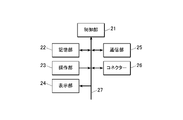
図2に、コンソール2の機能構成例を示す。図2に示すように、コンソール2は、制御部21、記憶部22、操作部23、表示部24、通信部25、コネクター26等を備えて構成され、各部はバス27により接続されている。
FIG. 2 shows an example of the functional configuration of the
制御部21は、CPU(Central Processing Unit)、RAM(Random Access Memory)等により構成される。制御部21のCPUは、操作部23の操作に応じて、記憶部22に記憶されているシステムプログラムや各種処理プログラムを読み出してRAM内に展開し、展開されたプログラムに従って、コンソール2各部の動作や、放射線発生装置1及びFPDカセッテ4の動作を集中制御する。また、制御部21は、展開されたプログラムに従って後述する心機能評価処理Aを始めとする各種処理を実行し、抽出手段、算出手段、解析手段、制御手段、除去手段として機能する。
The
記憶部22は、不揮発性の半導体メモリーやハードディスク等により構成される。記憶部22は、制御部21で実行される各種プログラムやプログラムにより処理の実行に必要なパラメーター、或いは処理結果等のデータを記憶する。例えば、記憶部22は、図4に示す心機能測定処理Aを実行するためのプログラムを記憶している。各種プログラムは、読取可能なプログラムコードの形態で格納され、制御部21は、当該プログラムコードに従った動作を逐次実行する。
また、記憶部22は、動態撮影時の放射線照射条件及び画像読取条件を記憶している。放射線照射条件及び画像読取条件は、操作部23の操作によりユーザーが設定可能である。
The
The
また、記憶部22は、年齢及び体格(例えば、身長及び/又は体重)の組み合わせ毎に、胸部動態画像の心臓面積の心拍一周期における極大値と極小値の差分値から一回拍出量を算出するための算出式(直線の式)に用いられる係数を格納した係数テーブルを記憶している。
この係数テーブルに格納されている係数は、実験的に求められたものである。
Further, the
The coefficients stored in this coefficient table are obtained experimentally.
操作部23は、カーソルキー、数字入力キー、及び各種機能キー等を備えたキーボードと、マウス等のポインティングデバイスを備えて構成され、キーボードに対するキー操作やマウス操作により入力された指示信号を制御部21に出力する。また、操作部23は、表示部24の表示画面にタッチパネルを備えても良く、この場合、タッチパネルを介して入力された指示信号を制御部21に出力する。
The
表示部24は、LCD(Liquid Crystal Display)やCRT(Cathode Ray Tube)等のモニタにより構成され、制御部21から入力される表示信号の指示に従って、操作部23からの入力指示やデータ等を表示する。
The
通信部25は、無線LANアダプタ等を備え、アクセスポイント3を介して無線LAN等の通信ネットワークに接続された放射線発生装置1、FPDカセッテ4を始めとする外部機器との間のデータ送受信を制御する。
The
コネクター26は、図示しないケーブルを介してFPDカセッテ4と通信接続するためのコネクターである。
The
図1に戻り、アクセスポイント3は、放射線発生装置1とコンソール2との間の通信や、コンソール2とFPDカセッテ4との間の通信等を中継する。
Returning to FIG. 1, the
FPDカセッテ4は、動態撮影対応の可搬型の放射線検出器である。FPDカセッテ4は、ガラス基板等の基板上の所定位置に、放射線源11から照射されて少なくとも被検者Hを透過した放射線をその強度に応じて検出し、検出した放射線を電気信号に変換して蓄積する複数の放射線検出素子がマトリックス状(二次元状)に配列されて構成されている。各放射線検出素子には、例えばTFT(Thin Film Transistor)等のスイッチング部が接続され、スイッチング部により各放射線検出素子への電気信号の蓄積及び読み取りが制御され、画像データ(フレーム画像)が取得される。FPDには放射線をシンチレータを介して光電変換素子により電気信号に変換する間接変換型、放射線を直接的に電気信号に変換する直接変換型があるが、何れを用いてもよい。
The
FPDカセッテ4は、スイッチング部による電気信号の蓄積及び読み取りを制御する読取制御部と、アクセスポイント3を介してコンソール2と通信接続するための通信部とを備えている(何れも図示せず)。フレームレート、1撮影当たりの撮影フレーム画像数、画像サイズ(マトリックスサイズ)等の画像読取条件は、通信部を介してコンソール2により設定される。読取制御部は、設定された画像読取条件に基づいて、スイッチング部による各放射線検出素子への電気信号の蓄積及び読み取りを制御する。また、FPDカセッテ4は、コネクターを有し、図示しないケーブルを介してコンソール2と通信接続可能である。
The
なお、FPDカセッテ4は、放射線技師等の撮影実施者が持参してもよいが、FPDカセッテ4は比較的重く、落下すると壊れたり故障したりする可能性があるため、回診車に設けられたカセッテ用のポケット61aに挿入されて搬送できるようになっている。
The
(動態画像解析システム100の動作)
次に、動態画像解析システム100における動作について説明する。
まず、撮影実施者は、コンソール2の操作部23により被検者Hの患者情報(氏名、年齢、性別、体格等)、検査情報(検査対象部位(ここでは、胸部)、検査年月日等)を入力するとともに、放射線源11やFPDカセッテ4を所定の位置にセットする。
コンソール2の制御部21は、検査対象部位(ここでは胸部)に対応する放射線照射条件を放射線照射制御部12に設定するとともに、画像読取条件をFPDカセッテ4に設定する。
曝射スイッチ13が押下されると、制御部21は、放射線照射制御部12及びFPDカセッテ4を制御して、被検者Hの胸部の動態撮影を行わせる。FPDカセッテ4は、撮影により取得したフレーム画像を順次コンソール2に送信する。
(Operation of dynamic image analysis system 100)
Next, operations in the dynamic
First, the radiographer uses the
The
When the
コンソール2においては、FPDカセッテ4から送信された胸部動態画像のフレーム画像の受信が開始されると、心機能測定処理Aを開始する。
When the
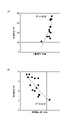
ここで、本願発明者は、多数の被検者について、肺動脈カテーテルを用いた熱希釈法などの方法で心拍出量を測定するとともに、測定時の胸部動態画像から心臓面積を算出し、心拍出量と心臓面積との関係について検討した。また、測定時の胸部動態画像から肺野領域を抽出し、心拍出量と肺野領域内の平均画素値との関係について検討した。また、測定時の胸部動態画像から心臓領域を抽出し、心拍出量と心臓領域内の平均画素値との関係について検討した。また、測定時の胸部動態画像から心臓領域の直径(例えば、心臓領域の水平方向の直径の最大、以下同じ。)を算出し、心拍出量と心臓の直径との関係について検討した。なお、検討は、被検者の年齢及び体格によってグループ分けして行った。 Here, the inventor of the present application measured the cardiac output of a large number of subjects by a method such as thermodilution using a pulmonary artery catheter, and calculated the cardiac area from the chest dynamic image at the time of measurement. We investigated the relationship between stroke volume and heart area. In addition, the lung area was extracted from the dynamic chest image at the time of measurement, and the relationship between the cardiac output and the average pixel value in the lung area was examined. In addition, the heart region was extracted from the dynamic chest image during measurement, and the relationship between the cardiac output and the average pixel value within the heart region was examined. In addition, the diameter of the heart region (for example, the maximum horizontal diameter of the heart region; the same shall apply hereinafter) was calculated from the dynamic chest image at the time of measurement, and the relationship between the cardiac output and the diameter of the heart was examined. In addition, examination was performed by grouping according to the age and physique of the subject.
図3(a)は、上記検討の一例として作成した、心収縮期(心拍位相のうち最も心臓が収縮した位相を指す。以下同じ。)に撮影された胸部動態画像(フレーム画像)の心臓面積と心拍出量との関係を示す散布図、図3(b)は、胸部動態画像の肺野領域内の平均画素値の呼吸一周期における平均と心拍出量との関係を示す散布図である。図3(a)~(b)に示すように、心臓面積、肺野領域の平均画素値は、それぞれ心拍出量と高い相関があり、両者の関係は、直線の式により表すことができることがわかる。 FIG. 3(a) shows the heart area of a dynamic chest image (frame image) captured during systole (refers to the phase in which the heart contracts the most among the heartbeat phases; the same shall apply hereinafter), which was created as an example of the above study. , and FIG. 3B is a scatter diagram showing the relationship between the average pixel value in the lung region of the chest dynamic image and the cardiac output in one respiratory cycle. is. As shown in FIGS. 3(a) and 3(b), the heart area and the average pixel value of the lung area are highly correlated with the cardiac output, respectively, and the relationship between the two can be represented by a linear equation. I understand.
図3(a)においては、心収縮期に撮影された胸部動態画像(フレーム画像)の心臓面積と心拍出量との関係を一例として示しているが、検討の結果、所定の心拍位相(例えば、心収縮期や心拡張期(心拍位相のうち最も心臓が拡張した位相を指す。以下同じ。))に撮影された胸部動態画像(フレーム画像)から算出された心臓面積と心拍出量、胸部動態画像から算出された心臓面積の心拍一周期における極大値と極小値の差分値と心拍出量、胸部動態画像から算出された心臓面積の心拍一周期における平均と心拍出量についても同様に、直線の式によりその関係を表すことができることが見出された。また、多数の被検者について、熱希釈法などにより測定された心拍出量から一回拍出量を算出して検討した結果、所定の心拍位相に撮影された胸部動態画像(フレーム画像)から算出された心臓面積と一回拍出量、胸部動態画像から算出された心臓面積の心拍一周期における極大値と極小値の差分値と一回拍出量、胸部動態画像から算出された心臓面積の心拍一周期における平均と心拍出量についても同様に、直線の式により両者の関係を表すことができることが見出された。ただし、直線式の係数は、それぞれ異なるものであった。 FIG. 3A shows, as an example, the relationship between the cardiac area and the cardiac output of a dynamic chest image (frame image) captured during systole. For example, cardiac area and cardiac output calculated from chest dynamic images (frame images) captured during systole and diastole (referring to the phase in which the heart is most dilated among cardiac phases; the same shall apply hereinafter) , About the difference value and cardiac output between the maximum and minimum values of the cardiac area in one cardiac cycle calculated from the dynamic chest image, and the average of the cardiac area in one cardiac cycle and the cardiac output calculated from the dynamic chest image can also be expressed by a linear equation. In addition, as a result of calculating the stroke volume from the cardiac output measured by the thermodilution method for a large number of subjects and examining it, chest dynamic images (frame images) taken at a predetermined cardiac phase heart area and stroke volume calculated from the dynamic chest image, the difference value and stroke volume between the maximum and minimum values of the heart area in one cardiac cycle calculated from the chest dynamic image, the heart calculated from the chest dynamic image It was found that the relationship between the mean of the area and the cardiac output in one cardiac cycle can also be represented by a linear equation. However, the linear coefficients were different.
また、図3(b)においては、胸部動態画像から算出された肺野領域内の平均画素値の呼吸一周期における平均と心拍出量との関係を一例として示しているが、検討の結果、所定の呼吸位相に撮影された胸部動態画像(フレーム画像)から算出された肺野領域内の平均画素値と心拍出量、胸部動態画像から算出された肺野領域内の平均画素値の呼吸一周期における極大値と極小値の差分値と心拍出量についても同様に、直線の式により両者の関係を表すことができることが見出された。また、多数の被検者について、熱希釈法などにより測定された心拍出量から一回拍出量を算出して同様に検討した結果、胸部動態画像から算出された肺野領域内の平均画素値の呼吸一周期における平均と一回拍出量、所定の呼吸位相に撮影された胸部動態画像(フレーム画像)から算出された肺野領域内の平均画素値と一回拍出量、胸部動態画像から算出された肺野領域内の平均画素値の呼吸一周期における極大値と極小値の差分値と一回拍出量についても同様に、直線の式により両者の関係を表すことができることが見出された。ただし、直線式の係数は、それぞれ異なるものであった。 In addition, FIG. 3B shows, as an example, the relationship between the average of the average pixel values in the lung region calculated from the chest dynamic image and the cardiac output in one respiratory cycle. , the average pixel value and cardiac output in the lung region calculated from the chest dynamic image (frame image) captured in a predetermined respiratory phase, and the average pixel value in the lung region calculated from the chest dynamic image It was found that the relationship between the difference value between the maximal value and the minimal value in one respiratory cycle and the cardiac output can also be represented by a linear equation. In addition, the stroke volume was calculated from the cardiac output measured by the thermodilution method for a large number of subjects, and the same study was conducted. Average pixel values and stroke volume in one respiratory cycle, average pixel values and stroke volume in the lung region calculated from chest dynamic images (frame images) captured in a predetermined respiratory phase, chest Similarly, the relationship between the stroke volume and the difference value between the maximum and minimum values of the average pixel value in the lung region calculated from the dynamic image in one respiratory cycle can be expressed by a linear equation. was found. However, the linear coefficients were different.
また、胸部動態画像から算出された心臓領域内の平均画素値の心拍一周期における平均と心拍出量、所定の心拍位相に撮影された胸部動態画像(フレーム画像)から算出された心臓領域内の平均画素値と心拍出量、胸部動態画像から算出された心臓領域内の平均画素値の心拍一周期における極大値と極小値の差分値と心拍出量についても同様に検討した結果、それぞれ直線の式により両者の関係を表すことができることが見出された。また、多数の被検者について、熱希釈法などにより求めた心拍出量から一回拍出量を算出して同様に検討した結果、心拍一周期における心臓領域内の平均画素値の平均と一回拍出量、所定の心拍位相に撮影された胸部動態画像(フレーム画像)から算出された心臓領域内の平均画素値と一回拍出量、心拍一周期における心臓領域内の平均画素値の極大値と極小値の差分値と一回拍出量についても同様に、直線の式により両者の関係を表すことができることが見出された。ただし、直線式の係数は、それぞれ異なるものであった。 In addition, the average of the average pixel values in the heart region calculated from the chest dynamic image and the cardiac output in one heartbeat cycle, and the heart region calculated from the chest dynamic image (frame image) captured at a predetermined heartbeat phase The average pixel value and cardiac output, and the difference between the maximum and minimum values of the average pixel value in the heart region calculated from the chest dynamic image in one cardiac cycle and the cardiac output were also examined. It has been found that the relationship between the two can be represented by a linear equation, respectively. In addition, for many subjects, the stroke volume was calculated from the cardiac output obtained by the thermodilution method, etc., and the same study was performed. Stroke volume, average pixel value and stroke volume in the heart region calculated from chest dynamic images (frame images) captured at a predetermined heartbeat phase, average pixel value in the heart region in one heartbeat cycle Similarly, it was found that the relationship between the difference between the maximum and minimum values of , and the stroke volume can be represented by a linear equation. However, the linear coefficients were different.
また、所定の心拍位相に撮影された胸部動態画像(フレーム画像)の心臓領域の直径(例えば、水平方向の直径の最大)と心拍出量、胸部動態画像から算出された心臓領域の直径の心拍一周期における極大値と極小値の差分値と心拍出量、胸部動態画像から算出された心臓領域の直径の心拍一周期における平均と心拍出量についても同様に、直線の式によりその関係を表すことができることが見出された。また、多数の被検者について、熱希釈法などにより測定した心拍出量から一回拍出量を算出して同様に検討した結果、所定の心拍位相に撮影された胸部動態画像(フレーム画像)の心臓領域の直径と一回拍出量、胸部動態画像から算出された心臓領域の直径の心拍一周期における極大値と極小値の差分値と一回拍出量、胸部動態画像から算出された心臓領域の直径の心拍一周期における平均と一回拍出量についても同様に、直線の式により両者の関係を表すことができることが見出された。ただし、直線式の係数は、それぞれ異なるものであった。 In addition, the diameter of the heart region (e.g., the maximum horizontal diameter) and cardiac output of the chest dynamic image (frame image) captured at a predetermined cardiac phase, and the diameter of the heart region calculated from the chest dynamic image Similarly, the difference between the maximum and minimum values in one heartbeat cycle and the cardiac output, and the average diameter of the heart region calculated from the dynamic chest image and the cardiac output in one heartbeat cycle were calculated using the linear equation. It has been found that a relationship can be represented. In addition, for a large number of subjects, the stroke volume was calculated from the cardiac output measured by the thermodilution method, etc., and the same study was conducted. ) of the heart region and stroke volume, the difference value and stroke volume between the maximum and minimum values of the diameter of the heart region in one cardiac cycle calculated from the dynamic chest image, and the stroke volume calculated from the dynamic chest image It was also found that the relationship between the average diameter of the heart region in one heartbeat cycle and the stroke volume can be similarly expressed by a linear equation. However, the linear coefficients were different.
コンソール2は、これらの検討結果に基づいて、胸部動態画像から一回拍出量、心拍出量を算出する。本実施形態では、一例として、胸部動態画像から算出された心臓面積の心拍一周期における極大値と極小値の差分値をパラメーターとして一回拍出量及び心拍出量を算出する心機能測定処理Aについて説明する。
The
図4に、コンソール2により実行される心機能測定処理Aの流れを示す。心機能測定処理Aは、制御部21と記憶部22に記憶されているプログラムとの協働により実行される。
FIG. 4 shows the flow of the cardiac function measurement process A executed by the
まず、制御部21は、受信したフレーム画像から心臓面積を算出する(ステップS1)。
図5は、胸部動態画像のフレーム画像を模式的に示す図である。図5に示すように、胸部正面の動態画像では、心臓(図5においてR1で示す領域)は略楕円形状の領域として描画される。
First, the
FIG. 5 is a diagram schematically showing a frame image of a dynamic chest image. As shown in FIG. 5, in the dynamic image of the front of the chest, the heart (region indicated by R1 in FIG. 5) is drawn as a substantially elliptical region.
ステップS1において、制御部21は、まず、受信したフレーム画像から心臓領域を抽出する。心臓領域の抽出は、特に限定されないが、例えば、テンプレートマッチング処理を用いて行うことができる。例えば、記憶部22に心臓のテンプレート画像を記憶しておき、フレーム画像内においてテンプレート画像を移動させながらテンプレート画像とテンプレート画像に重なる部分の類似度を算出し、類似度が最も高い領域を心臓領域として抽出する。類似度は、例えば、SSD(Sum of Squared Difference)、SAD(Sum of Absolute Difference)等を用いて算出することができる。次いで、求めた心臓領域の画素数をカウントして画素サイズを乗算することにより、心臓面積を算出することができる。
In step S1, the
次いで、制御部21は、算出済みの心臓面積の時間変化に基づいて、心拍一周期分のフレーム画像の心臓面積を算出したか否かを判断する(ステップS2)。心拍一周期分のフレーム画像の心臓面積を算出していないと判断した場合(ステップS2;NO)、制御部21は、ステップS1に戻る。
Next, the
心拍一周期分の心臓面積を算出したと判断した場合(ステップS2;YES)、制御部21は、図6に示すように、取得した周期の心臓面積の極大値と極小値を算出し、その差分値を算出する(ステップS3)。
If it is determined that the heart area for one heartbeat cycle has been calculated (step S2; YES), the
次いで、制御部21は、記憶部22の係数テーブルから被検者Hの年齢及び体格に応じた係数を取得して、取得した係数を用いた算出式(直線式)にステップS3で求めた差分値を代入して、被検者Hの心臓の一回拍出量を算出する(ステップS4)。
Next, the
次いで、制御部21は、心拍数を算出し、一回拍出量及び心拍数から心拍出量を算出する(ステップS5)。
まず、制御部21は、心臓面積の時間変化の周期[sec]を算出する。この周期は、心拍周期に相当する。次いで、(式1)により心拍数を算出する。
心拍数=60÷周期[sec]・・・(式1)
次いで、制御部21は、以下の(式2)により心拍出量を算出する。
心拍出量=一回拍出量×心拍数・・・(式2)
なお、算出した心拍数、一回拍出量及び心拍出量は、胸部動態画像から算出した心臓面積、肺野面積、被検者Hの身長、又は体重に基づく補正係数で正規化することが好ましい。
Next, the
First, the
Heart rate = 60/cycle [sec] (Formula 1)
Next, the
Cardiac output = stroke volume x heart rate (Formula 2)
The calculated heart rate, stroke volume, and cardiac output shall be normalized by a correction factor based on the heart area, lung area, and height or weight of the subject H calculated from the dynamic chest image. is preferred.
次いで、制御部21は、算出した心拍数、一回拍出量及び心拍出量を表示部24に表示する(ステップS6)。
本実施形態では、FPDカセッテ4から心拍一周期分のフレーム画像が受信される毎に、心拍数、一回拍出量及び心拍出量が算出され、表示部24に表示される(表示が更新される)。すなわち、ほぼリアルタイムに心拍数、一回拍出量及び心拍出量を測定して表示することができる。
Next, the
In this embodiment, every time a frame image for one heartbeat cycle is received from the
ステップS6における表示例としては、例えば、受信したフレーム画像を順次表示するとともに、算出された最新の心拍数、一回拍出量及び心拍出量を数値で表示する。または、算出された心拍数、一回拍出量及び心拍出量をそれぞれ時系列のグラフとして表示してもよい。或いは、表示するフレーム画像の心臓領域を、心拍数、一回拍出量又は心拍出量の値に応じた色で表示することとしてもよい。心拍数、一回拍出量、心拍出量のいずれを表示するかは、ユーザーが設定できるようにしてもよい。 As a display example in step S6, for example, the received frame images are sequentially displayed, and the calculated latest heart rate, stroke volume, and cardiac output are numerically displayed. Alternatively, the calculated heart rate, stroke volume, and cardiac output may be displayed as time-series graphs. Alternatively, the heart region of the frame image to be displayed may be displayed in a color corresponding to the value of heart rate, stroke volume, or cardiac output. The user may be able to set which of the heart rate, stroke volume, and cardiac output is displayed.
次いで、制御部21は、FPDカセッテ4からの動態画像の一連のフレーム画像の受信が終了したか否かを判断する(ステップS7)。例えば、FPDカセッテ4から動態画像のフレーム画像の送信終了が通知された場合に、制御部21は、動態画像の一連のフレーム画像の受信が終了したと判断する。
Next, the
動態画像の一連のフレーム画像の受信が終了していないと判断した場合(ステップS7;NO)、制御部21は、ステップS1に戻る。
動態画像の一連のフレーム画像の受信が終了したと判断した場合(ステップS7;YES)、制御部21は、心機能測定処理Aを終了する。
When determining that the reception of the series of frame images of the dynamic image has not ended (step S7; NO), the
If it is determined that the series of frame images of the dynamic image has been received (step S7; YES), the
このように、心機能測定処理Aにおいては、胸部動態画像の各フレーム画像から心臓領域を抽出し、抽出した心臓領域から算出した心臓面積を、被検者Hの年齢や体格に応じた係数を用いた算出式に代入することにより一回拍出量を算出し、一回拍出量と、心臓面積の時間変化から求めた心拍数に基づいて心拍出量を算出する。したがって、従来のように、胸部動態画像の各フレーム画像から血管内の脈動部や心臓の局所的な部分等の認識しづらい微細な構造を検出してその計測値や画素値を利用するのではなく、容易に認識することが可能な心臓領域の面積を利用して心拍数、一回拍出量及び心拍出量を算出するため、心拍数、一回拍出量及び心拍出量を精度良く算出することが可能となる。 As described above, in the cardiac function measurement process A, the heart region is extracted from each frame image of the chest dynamic image, and the heart area calculated from the extracted heart region is calculated by a coefficient according to the age and build of the subject H. The stroke volume is calculated by substituting it into the calculation formula used, and the cardiac output is calculated based on the stroke volume and the heart rate obtained from the change in heart area over time. Therefore, it is not necessary to detect minute structures that are difficult to recognize, such as pulsating parts in blood vessels and local parts of the heart, from each frame image of the chest dynamic image and use the measured values and pixel values as in the conventional method. Since the heart rate, stroke volume and cardiac output are calculated using the area of the heart region that can be easily recognized, the heart rate, stroke volume and cardiac output are It is possible to calculate with high accuracy.
心機能測定処理Aにおいては、FPDカセッテ4から順次送信される胸部動態画像のフレーム画像から、心拍一周期毎に心臓面積の極大値と極小値の差分値を算出し、算出した差分値を用いて一回拍出量を算出し、一回拍出量と心拍数から心拍出量を算出する例について説明したが、一回拍出量や心拍出量を求めるためのパラメーターとしては、上記差分値に限定されない。例えば、記憶部22の係数テーブルに、実験により検証された、胸部動態画像から算出された以下のいずれかのパラメーターの値から一回拍出量を算出するための算出式に用いられる係数を記憶しておき、制御部21は、記憶部22の係数テーブルから被検者Hの患者情報に応じた係数を取得して、取得した係数を用いた算出式に胸部動態画像から算出されたパラメーターの値を代入して一回拍出量を算出してもよい。そして、算出した一回拍出量と心拍数から心拍出量を算出してもよい。または、記憶部22の係数テーブルに、以下のいずれかのパラメーターの値から一回拍出量及び心拍出量のそれぞれを算出するための算出式に用いられる係数を記憶しておき、制御部21は、記憶部22の係数テーブルから被検者Hの患者情報に応じた係数を取得して、取得した係数を用いた算出式に胸部動態画像から算出されたパラメーターの値を代入して、一回拍出量及び心拍出量を算出してもよい。
In the cardiac function measurement process A, the difference value between the maximum value and the minimum value of the heart area is calculated for each heartbeat cycle from the frame images of the dynamic chest image sequentially transmitted from the
以下のパラメーターは、フレーム画像から容易に認識可能な心臓領域の面積、径、画素値、又は肺野領域の画素値から求められるものであり、パラメーターの算出に微細な構造の抽出や認識を伴わないため、精度良く一回拍出量及び心拍出量を算出することが可能である。
なお、心臓面積又は心臓領域の画素値に基づくパラメーターを用いる場合は、体動や呼吸による誤差を低減するため、動態撮影を行う際に、被検者Hに息止めを指示して息止めの状態で動態撮影を行うことが好ましい。
The following parameters are obtained from the area, diameter, and pixel value of the heart region, which can be easily recognized from the frame image, or the pixel value of the lung region. Therefore, it is possible to calculate stroke volume and cardiac output with high accuracy.
In the case of using a parameter based on the pixel value of the heart area or the heart region, in order to reduce errors due to body movement and breathing, when performing dynamic imaging, the subject H is instructed to hold his breath. It is preferable to perform dynamic imaging in this state.
〈パラメーター例〉
・予め定められた心拍位相(例えば、心収縮期、心拡張期、その中間等)の心臓面積
・心臓面積の心拍一周期における平均値
・肺野領域内の平均画素値の呼吸一周期における平均
・予め定められた呼吸位相(例えば、最大呼気位、最大吸気位、その中間等)の肺野領域内の平均画素値
・肺野領域内の平均画素値の呼吸一周期における極大値と極小値の差分値
・心臓領域内の平均画素値の心拍一周期における平均
・予め定められた心拍位相(例えば、心収縮期、心拡張期、その中間等)の心臓領域内の平均画素値
・心臓領域内の平均画素値の心拍一周期における極大値と極小値の差分値
・心臓領域の直径の心拍一周期における平均
・予め定められた心拍位相(例えば、心収縮期、心拡張期、その中間等)の心臓領域の直径
・心臓領域の直径の極大値と極小値の心拍一周期における差分値
<Parameter example>
- Cardiac area in a predetermined heartbeat phase (e.g., systole, diastole, intermediate, etc.) - Average value of heart area in one heartbeat cycle - Average of average pixel value in the lung area in one respiration cycle・Average pixel value in the lung region at a predetermined respiratory phase (e.g., maximum expiratory position, maximum inspiratory position, intermediate, etc.) ・Maximum value and minimum value in one respiratory cycle of the average pixel value in the lung region・Average of the average pixel value in the heart region in one heartbeat cycle ・Average pixel value in the heart region of a predetermined heartbeat phase (e.g., systole, diastole, intermediate, etc.) ・Heart region The difference value between the maximum value and the minimum value of the average pixel value in one heartbeat cycle ・Average of the diameter of the heart region in one heartbeat cycle ) diameter of the heart region ・The difference value between the maximum and minimum values of the diameter of the heart region in one heartbeat cycle
なお、第1の実施形態においては、心機能の評価値を算出するための算出式に用いる係数を予め実験的に求めて記憶部22に記憶しておくこととしたが、一回拍出量を算出する直前に、熱希釈法等の他の手法により被検者から心拍出量を測定し、測定した心拍出量及び測定時の胸部動態画像のフレーム画像から算出したパラメーター(上述した心臓領域の面積、径、画素値又は肺野領域の画素値)に基づいて係数を算出することとしてもよい。例えば、(式2)より心拍出量=一回拍出量×心拍数なので、
心拍出量=パラメーター×係数×心拍数・・・(式3)
となる。よって、係数は、(式4)により求めることができる。
係数=心拍出量÷(パラメーター×心拍数)・・・(式4)
In the first embodiment, the coefficients used in the calculation formula for calculating the evaluation value of the cardiac function are experimentally obtained in advance and stored in the
Cardiac output = Parameter x Coefficient x Heart rate (Formula 3)
becomes. Therefore, the coefficient can be obtained by (Equation 4).
Coefficient = cardiac output/(parameter x heart rate) (Equation 4)
<第2の実施形態>
次に、本発明の第2の実施形態について説明する。
第1の実施形態では、胸部動態画像から算出した心臓面積に基づいて、心機能の評価値として、心拍数、一回拍出量及び心拍出量を算出する場合について説明したが、第2の実施形態においては、胸部動態画像から算出した肺野領域内の画素値に基づいて、心機能の評価値として血圧(BP)、肺動脈圧(PAP)、動脈圧(AP)、及び中心静脈圧を算出し、被検者Hの水分量の評価値として肺水分量及び全身水分量を算出する場合を例にとり説明する。
<Second embodiment>
Next, a second embodiment of the invention will be described.
In the first embodiment, the heart rate, stroke volume, and cardiac output are calculated as evaluation values of cardiac function based on the heart area calculated from the dynamic chest image. In the embodiment of , based on the pixel values in the lung region calculated from the dynamic chest image, blood pressure (BP), pulmonary artery pressure (PAP), arterial pressure (AP), and central venous pressure are used as evaluation values for cardiac function. is calculated, and the lung water content and the whole body water content are calculated as the evaluation values of the water content of the subject H will be described as an example.
第2の実施形態において、記憶部22には、後述する心機能測定処理Bを実行するためのプログラムが記憶されている。また、記憶部22には、年齢及び体格(例えば、身長及び/又は体重)の組み合わせ毎に、胸部動態画像における肺野領域内の平均画素値の呼吸一周期における平均から血圧を算出するための算出式(直線の式)に用いられる係数(血圧測定用の係数)、肺動脈圧を算出するための算出式(直線の式)に用いられる係数(肺動脈圧測定用の係数)、動脈圧を算出するための算出式(直線の式)に用いられる係数(動脈圧測定用の係数)、中心静脈圧を算出するための算出式(直線の式)に用いられる係数(中心静脈圧測定用の係数)、肺水分量を算出するための算出式(直線の式)に用いられる係数(肺水分量測定用の係数)、全身水分量を算出するための算出式(直線の式)に用いられる係数(全身水分量測定用の係数)がそれぞれ記憶されている。
In the second embodiment, the
その他の第2の実施形態における動態画像解析システム100の構成は、第1の実施形態で説明したものと同様であるので説明を援用する。また、動態画像解析システム100における胸部動態画像の撮影動作についても、第1の実施形態で説明したものと同様であるので説明を援用し、以下、コンソール2において、FPDカセッテ4から胸部動態画像のフレーム画像の受信が開始された際に開始される心機能測定処理Bについて説明する。
The configuration of the dynamic
ここで、本願発明者は、多数の被検者について、血圧計で血圧を測定するとともに、測定時の胸部動態画像から心臓面積を算出し、血圧と心臓面積との関係について検討した。また、測定時の胸部動態画像から肺野領域を抽出し、血圧と肺野領域内の平均画素値との関係について検討した。また、測定時の胸部動態画像から心臓領域を抽出し、血圧と心臓領域内の平均画素値との関係について検討した。また、測定時の胸部動態画像から心臓の直径を算出し、血圧と心臓領域の直径との関係について検討した。なお、検討は、被検者の年齢及び体格によってグループ分けして行った。 Here, the inventor of the present application measured the blood pressure of a large number of subjects with a sphygmomanometer, calculated the heart area from the dynamic chest image at the time of measurement, and examined the relationship between the blood pressure and the heart area. In addition, the lung area was extracted from the dynamic chest image at the time of measurement, and the relationship between blood pressure and the average pixel value in the lung area was examined. In addition, the heart region was extracted from the chest dynamic image at the time of measurement, and the relationship between blood pressure and the average pixel value within the heart region was examined. In addition, the diameter of the heart was calculated from the dynamic chest image at the time of measurement, and the relationship between the blood pressure and the diameter of the heart region was examined. In addition, examination was performed by grouping according to the age and physique of the subject.
図7(a)は、上記検討の一例として、心収縮期に撮影された胸部動態画像(フレーム画像)の心臓面積と血圧との関係を示す散布図、図7(b)は、胸部動態画像の肺野領域内の平均画素値の呼吸一周期における平均と血圧との関係を示す散布図である。図7(a)~(b)に示すように、心臓面積、肺野領域内の平均画素値は、それぞれ血圧と高い相関があり、両者の関係は、直線の式により表すことができることがわかる。 FIG. 7(a) is a scatter diagram showing, as an example of the above study, the relationship between heart area and blood pressure in chest dynamic images (frame images) captured during systole, and FIG. 7(b) is a chest dynamic image. is a scatter diagram showing the relationship between the average pixel value in one respiratory cycle and the blood pressure in the lung field region. As shown in FIGS. 7A and 7B, the heart area and the average pixel value in the lung area are highly correlated with blood pressure, and the relationship between the two can be represented by a linear equation. .
図7(a)においては、心収縮期に撮影された胸部動態画像(フレーム画像)の心臓面積と血圧との関係を一例として示しているが、検討の結果、所定の心拍位相に撮影された胸部動態画像(フレーム画像)から算出された心臓面積と血圧、心臓面積の心拍一周期における極大値と極小値の差分値と血圧、心臓面積の心拍一周期における平均と血圧についても同様に、直線の式によりその関係を表すことができることが見出された。ただし、直線式の係数は、それぞれ異なるものであった。 FIG. 7A shows, as an example, the relationship between the heart area and the blood pressure in a dynamic chest image (frame image) captured during systole. The heart area calculated from the chest dynamic image (frame image) and the blood pressure, the difference between the maximum and minimum values of the heart area in one heartbeat cycle and the blood pressure, and the average of the heart area in one heart beat cycle and the blood pressure are also shown as straight lines. It has been found that the relationship can be expressed by the formula: However, the linear coefficients were different.
また、図7(b)においては、胸部動態画像の肺野領域内の平均画素値の呼吸一周期における平均と血圧との関係を一例として示しているが、検討の結果、所定の呼吸位相に撮影された胸部動態画像(フレーム画像)から算出された肺野領域内の平均画素値と血圧、肺野領域内の平均画素値の呼吸一周期における極大値と極小値の差分値と血圧についても同様に、直線の式によりその関係を表すことができることが見出された。ただし、直線式の係数は、それぞれ異なるものであった。 FIG. 7(b) shows, as an example, the relationship between the average pixel value in the lung region of the dynamic chest image and the blood pressure in one respiratory cycle. The average pixel value and blood pressure in the lung region calculated from the captured chest dynamic image (frame image), the difference between the maximum and minimum values of the average pixel value in the lung region in one respiratory cycle, and the blood pressure It has also been found that the relationship can be represented by a linear equation. However, the linear coefficients were different.
また、胸部動態画像の心臓領域内の平均画素値の心拍一周期における平均と血圧、所定の心拍位相に撮影された胸部動態画像(フレーム画像)から算出された心臓領域内の平均画素値と血圧、心臓領域内の平均画素値の心拍一周期における極大値と極小値の差分値と血圧についても同様に検討した結果、それぞれ直線の式により両者の関係を表すことができることが見出された。ただし、直線式の係数は、それぞれ異なるものであった。 In addition, the mean of the average pixel values in the heart region of the chest dynamic image and the blood pressure in one heartbeat cycle, and the mean pixel value and blood pressure in the heart region calculated from the chest dynamic image (frame image) captured at a predetermined heartbeat phase As a result of similarly examining the difference between the maximum and minimum values of the average pixel value in the heart region in one heart beat cycle and the blood pressure, it was found that the relationship between the two can be expressed by a linear equation. However, the linear coefficients were different.
また、検討の結果、所定の心拍位相に撮影された胸部動態画像(フレーム画像)の心臓領域の直径(例えば、水平方向の直径の最大)と血圧、胸部動態画像の心臓領域の直径の心拍一周期における極大値と極小値の差分値と血圧、心臓領域の直径の心拍一周期における平均と血圧についても同様に、直線の式によりその関係を表すことができることが見出された。ただし、直線式の係数は、それぞれ異なるものであった。 In addition, as a result of the examination, the diameter (for example, the maximum horizontal diameter) and blood pressure of the cardiac region of the chest dynamic image (frame image) captured at a predetermined cardiac phase, and the heartbeat rate of the diameter of the cardiac region of the chest dynamic image It was found that the relationship between the difference value between the maximum value and the minimum value in the cycle and the blood pressure, and the average diameter of the heart region in one heartbeat cycle and the blood pressure can also be represented by a linear equation. However, the linear coefficients were different.
同様に、本願発明者は、多数の被検者について、肺動脈圧、動脈圧、中心静脈圧、肺水分量、全身水分量をそれぞれ測定するとともに、測定時の胸部動態画像から心臓面積を算出し、肺動脈圧、動脈圧、中心静脈圧、肺水分量、全身水分量のそれぞれと心臓面積との関係について検討した。また、測定時の胸部動態画像から肺野領域を抽出し、肺動脈圧、動脈圧、中心静脈圧、肺水分量、全身水分量のそれぞれと肺野領域内の平均画素値との関係について検討した。また、測定時の胸部動態画像から心臓領域を抽出し、肺動脈圧、動脈圧、中心静脈圧、肺水分量、全身水分量のそれぞれと心臓領域内の平均画素値との関係について検討した。また、測定時の胸部動態画像から心臓の直径を算出し、肺動脈圧、動脈圧、中心静脈圧、肺水分量、全身水分量のそれぞれと心臓の直径との関係について検討した。なお、検討は、被検者の年齢及び体格によってグループ分けして行った。また、肺動脈圧の測定は、肺動脈カテーテル等によって測定し、動脈圧及び肺水分量の測定は、循環動態モニター等で測定した。また、中心静脈圧は中心静脈カテーテルで測定し、全身水分量は輸液量及び尿量と出血量から判断した。 Similarly, the inventor of the present application measured pulmonary arterial pressure, arterial pressure, central venous pressure, lung water content, and whole body water content for a large number of subjects, and calculated the heart area from the chest dynamic image at the time of measurement. , pulmonary arterial pressure, arterial pressure, central venous pressure, pulmonary water content, whole body water content, and heart area. In addition, the lung area was extracted from the chest dynamic image during measurement, and the relationship between the pulmonary arterial pressure, arterial pressure, central venous pressure, lung water content, and whole body water content and the average pixel value in the lung area was examined. . In addition, the heart region was extracted from the dynamic chest image at the time of measurement, and the relationship between the pulmonary artery pressure, arterial pressure, central venous pressure, lung water content, and whole body water content and the average pixel value in the heart region was examined. In addition, the diameter of the heart was calculated from the dynamic chest image at the time of measurement, and the relationship between the diameter of the heart and pulmonary artery pressure, arterial pressure, central venous pressure, lung water content, and whole body water content was examined. In addition, examination was performed by grouping according to the age and physique of the subject. Pulmonary artery pressure was measured using a pulmonary artery catheter or the like, and arterial pressure and pulmonary water content were measured using a hemodynamic monitor or the like. In addition, central venous pressure was measured with a central venous catheter, and systemic water content was determined from the amount of transfusion, urine, and blood loss.
図8(a)は、上記検討の一例として、心収縮期に撮影された胸部動態画像(フレーム画像)の心臓面積と肺動脈圧との関係を示す散布図、図8(b)は、胸部動態画像の肺野領域内の平均画素値の呼吸一周期における平均と肺動脈圧との関係を示す散布図である。図8(a)~(b)に示すように、心臓面積、肺野領域内の平均画素値は、それぞれ肺動脈圧と高い相関があり、両者の関係は、直線の式により表すことができることがわかる。 FIG. 8(a) is a scatter diagram showing, as an example of the above study, the relationship between the heart area and the pulmonary artery pressure in chest dynamic images (frame images) taken during systole, and FIG. FIG. 4 is a scatter diagram showing the relationship between the average pixel value in one respiratory cycle in the lung region of the image and the pulmonary artery pressure. As shown in FIGS. 8(a) and 8(b), the heart area and the average pixel value in the lung area are highly correlated with the pulmonary artery pressure, respectively, and the relationship between the two can be expressed by a linear equation. Understand.
図8(a)においては、心収縮期に撮影された胸部動態画像(フレーム画像)の心臓面積と肺動脈圧との関係を一例として示しているが、検討の結果、所定の心拍位相に撮影された胸部動態画像(フレーム画像)から算出された心臓面積と肺動脈圧、胸部動態画像の心臓面積の心拍一周期における極大値と極小値の差分値と肺動脈圧、心臓面積の心拍一周期における平均と肺動脈圧についても同様に、直線の式によりその関係を表すことができることが見出された。動脈圧、中心静脈圧、肺水分量、全身水分量についても同様であった。ただし、直線の式の係数は、それぞれ異なるものであった。 FIG. 8A shows, as an example, the relationship between the heart area and the pulmonary artery pressure in chest dynamic images (frame images) captured during systole. heart area and pulmonary artery pressure calculated from the dynamic chest image (frame image), the difference value and pulmonary artery pressure between the maximum and minimum values of the heart area in one heartbeat cycle of the chest dynamic image, and the average of the heart area in one heartbeat cycle It was found that the relationship for pulmonary artery pressure can be similarly expressed by a linear equation. The same was true for arterial pressure, central venous pressure, lung water content, and whole body water content. However, the coefficients of the linear equations were different.
また、図8(b)においては、胸部動態画像の肺野領域内の平均画素値の呼吸一周期における平均と肺動脈圧との関係を一例として示しているが、検討の結果、所定の呼吸位相に撮影された胸部動態画像(フレーム画像)から算出された肺野領域内の平均画素値と肺動脈圧、胸部動態画像の肺野領域内の平均画素値の呼吸一周期における極大値と極小値の差分値と肺動脈圧についても同様に、直線の式によりその関係を表すことができることが見出された。動脈圧、中心静脈圧、肺水分量、全身水分量についても同様であった。ただし、直線の式の係数は、それぞれ異なるものであった。 FIG. 8B shows, as an example, the relationship between the average of the average pixel values in the lung region of the dynamic chest image in one respiratory cycle and the pulmonary artery pressure. Average pixel value and pulmonary artery pressure in the lung area calculated from the dynamic chest image (frame image) captured in It was found that the relationship between the difference value and the pulmonary artery pressure can also be represented by a linear equation. The same was true for arterial pressure, central venous pressure, lung water content, and whole body water content. However, the coefficients of the linear equations were different.
また、胸部動態画像の心臓領域内の平均画素値の心拍一周期における平均と肺動脈圧、動脈圧、中心静脈圧、肺水分量、全身水分量のそれぞれ、所定の心拍位相に撮影された胸部動態画像(フレーム画像)から算出された心臓領域内の平均画素値と肺動脈圧、動脈圧、中心静脈圧、肺水分量、全身水分量のそれぞれ、心臓領域内の平均画素値の心拍一周期における極大値と極小値の差分値と肺動脈圧、動脈圧、中心静脈圧、肺水分量、全身水分量のそれぞれについても同様に検討した結果、それぞれ直線の式により両者の関係を表すことができることが見出された。ただし、直線式の係数は、それぞれ異なるものであった。 In addition, the average of the average pixel values in the heart region of the chest dynamic image in one heartbeat cycle, the pulmonary artery pressure, the arterial pressure, the central venous pressure, the lung water content, and the whole body water content, respectively. The average pixel value in the heart region calculated from the image (frame image), pulmonary artery pressure, arterial pressure, central venous pressure, lung water content, and whole body water content. As a result of the same examination of the difference between the value and the minimum value, pulmonary arterial pressure, arterial pressure, central venous pressure, pulmonary water content, and whole body water content, it was found that the relationship between the two can be represented by linear equations. served. However, the linear coefficients were different.
また、検討の結果、所定の心拍位相に撮影された胸部動態画像(フレーム画像)の心臓領域の直径(例えば、水平方向の直径の最大)と肺動脈圧、動脈圧、中心静脈圧、肺水分量、全身水分量のそれぞれ、胸部動態画像の心臓領域の直径の心拍一周期における極大値と極小値の差分値と肺動脈圧、動脈圧、中心静脈圧、肺水分量、全身水分量のそれぞれについても同様に、直線の式によりその関係を表すことができることが見出された。ただし、直線の式の係数は、それぞれ異なるものであった。 In addition, as a result of the examination, the diameter of the heart region (e.g., the maximum horizontal diameter), the pulmonary artery pressure, the arterial pressure, the central venous pressure, and the pulmonary water content in dynamic chest images (frame images) captured at a predetermined cardiac phase , the total body water content, the difference between the maximum and minimum values of the diameter of the heart region in the dynamic chest image in one heartbeat cycle, and the pulmonary artery pressure, arterial pressure, central venous pressure, lung water content, and whole body water content. It has also been found that the relationship can be represented by a linear equation. However, the coefficients of the linear equations were different.
コンソール2は、これらの検討結果に基づいて、胸部動態画像から血圧、肺動脈圧、動脈圧、中心静脈圧、肺水分量、全身水分量を算出する。本実施形態では、一例として、最大呼気位における肺野領域内の平均画素値の平均をパラメーターとして、血圧、肺動脈圧、動脈圧、中心静脈圧、肺水分量、全身水分量を算出する心機能測定処理Bについて説明する。
Based on these examination results, the
図9に、心機能測定処理Bの流れを示す。心機能測定処理Bは、制御部21と記憶部22に記憶されているプログラムとの協働により実行される。
FIG. 9 shows the flow of the cardiac function measurement process B. As shown in FIG. The cardiac function measurement process B is executed by cooperation between the
まず、制御部21は、受信したフレーム画像から肺野領域を抽出し、抽出した肺野領域内の平均画素値を算出する(ステップS11)。
First, the
ステップS11において、制御部21は、まず、受信したフレーム画像から肺野領域を抽出する。肺野領域は、例えば、各画素の画素値のヒストグラムから判別分析によって閾値を求め、この閾値より高信号の領域を肺野領域候補として1次抽出する。次いで、1次抽出された肺野領域候補の境界付近でエッジ検出を行い、境界付近の小領域でエッジが最大となる点を境界に沿って抽出すれば肺野領域の境界を抽出することができる。
In step S11, the
次いで、制御部21は、算出済みの平均画素値の時間変化に基づいて、呼吸一周期分のフレーム画像から肺野領域内の平均画素値を算出したか否かを判断する(ステップS12)。呼吸一周期分のフレーム画像から肺野領域内の平均画素値を算出していないと判断した場合(ステップS12;NO)、制御部21は、ステップS11に戻る。
Next, the
呼吸一周期分のフレーム画像から肺野領域内の平均画素値を算出したと判断した場合(ステップS12;YES)、制御部21は、最大呼気位における肺野領域内の平均画素値を取得する(ステップS13)。最大呼気位における肺野領域内の平均画素値は、呼吸一周期分の各フレーム画像における肺野領域内の平均画素値のうち、最も低い値である。
If it is determined that the average pixel value in the lung region has been calculated from the frame images for one respiratory cycle (step S12; YES), the
次いで、制御部21は、記憶部22の係数テーブルから被検者Hの患者情報(年齢及び体格)に応じた血圧測定用の係数を取得して、取得した係数を用いた算出式にステップS13で算出した平均画素値を代入して、被検者Hの血圧を算出する(ステップS14)。
Next, the
次いで、制御部21は、記憶部22の係数テーブルから被検者Hの患者情報に応じた肺動脈圧測定用の係数を算出して、取得した係数を用いた算出式にステップS13で取得した平均画素値を代入して、被検者Hの肺動脈圧を算出する(ステップS15)。
Next, the
次いで、制御部21は、記憶部22の係数テーブルから被検者Hの患者情報に応じた動脈圧測定用の係数を算出して、取得した係数を用いた算出式にステップS13で取得した平均画素値を代入して、被検者Hの動脈圧を算出する(ステップS16)。
Next, the
次いで、制御部21は、記憶部22の係数テーブルから被検者Hの患者情報に応じた中心静脈圧測定用の係数を算出して、取得した係数を用いた算出式にステップS13で取得した平均画素値を代入して、被検者Hの中心静脈圧を算出する(ステップS17)。
Next, the
次いで、制御部21は、記憶部22の係数テーブルから被検者Hの患者情報に応じた肺水分量測定用の係数を取得して、取得した係数を用いた算出式にステップS13で算出した平均画素値を代入して、被検者Hの肺水分量を算出する(ステップS18)。
Next, the
次いで、制御部21は、記憶部22の係数テーブルから被検者Hの患者情報に応じた全身水分量測定用の係数を取得して、取得した係数を用いた算出式にステップS13で算出した平均画素値を代入して、被検者Hの全身水分量を算出する(ステップS19)。
なお、算出した各評価値は、胸部動態画像から算出した心臓面積、肺野面積、被検者Hの身長、又は体重に基づく補正係数で正規化することが好ましい。
Next, the
Each calculated evaluation value is preferably normalized by a correction coefficient based on the cardiac area, lung field area, height or weight of the subject H calculated from the chest dynamic image.
次いで、制御部21は、算出した血圧、肺動脈圧、動脈圧、中心静脈圧、肺水分量、及び全身水分量を表示部24に表示する(ステップS20)。
本実施形態では、FPDカセッテ4から最大呼気位のフレーム画像が受信される毎に、血圧、肺動脈圧、動脈圧、中心静脈圧、肺水分量、及び全身水分量が算出され、表示部24に表示される(表示が更新される)。すなわち、ほぼリアルタイムに血圧、肺動脈圧、動脈圧、中心静脈圧、肺水分量、及び全身水分量を測定して表示することができる。
Next, the
In the present embodiment, blood pressure, pulmonary artery pressure, arterial pressure, central venous pressure, pulmonary water content, and whole body water content are calculated each time a frame image at the maximum expiratory position is received from the
ステップS20における表示例としては、例えば、受信したフレーム画像を順次表示するとともに、算出された最新の血圧、肺動脈圧、動脈圧、中心静脈圧、肺水分量、及び全身水分量を数値で表示する。または、算出された血圧、肺動脈圧、動脈圧、中心静脈圧、肺水分量、及び全身水分量をそれぞれ時系列のグラフとして表示してもよい。表示するフレーム画像の心臓領域を、血圧、肺動脈圧、動脈圧、中心静脈圧、肺水分量又は全身水分量の値に応じた色で表示することとしてもよい。フレーム画像の心臓領域をいずれの評価値に応じた色で表示するかは、ユーザーが設定できるようにしてもよい。 As a display example in step S20, for example, the received frame images are sequentially displayed, and the latest calculated blood pressure, pulmonary artery pressure, arterial pressure, central venous pressure, lung water content, and whole body water content are displayed numerically. . Alternatively, the calculated blood pressure, pulmonary artery pressure, arterial pressure, central venous pressure, lung water content, and whole body water content may be displayed as time series graphs. The heart region of the frame image to be displayed may be displayed in a color corresponding to the value of blood pressure, pulmonary artery pressure, arterial pressure, central venous pressure, lung water content, or whole body water content. The user may be allowed to set the color corresponding to which evaluation value the heart region of the frame image is to be displayed.
次いで、制御部21は、FPDカセッテ4からの動態画像の一連のフレーム画像の受信が終了したか否かを判断する(ステップS21)。例えば、FPDカセッテ4から動態画像のフレーム画像の送信終了が通知された場合に、制御部21は、動態画像の一連のフレーム画像の受信が終了したと判断する。
Next, the
動態画像の一連のフレーム画像の受信が終了していないと判断した場合(ステップS21;NO)、制御部21は、ステップS11に戻る。
動態画像の一連のフレーム画像の受信が終了したと判断した場合(ステップS20;YES)、制御部21は、心機能測定処理Bを終了する。
When determining that the reception of the series of frame images of the dynamic image has not ended (step S21; NO), the
If it is determined that the series of frame images of the dynamic image has been received (step S20; YES), the
このように、心機能測定処理Bにおいては、胸部動態画像の各フレーム画像から肺野領域を抽出し、抽出した肺野領域から算出した平均画素値の呼吸一周期分の平均を、患者の年齢や体格に応じた係数を用いた算出式に代入することにより血圧を算出する。また、算出式の係数を変えて肺動脈圧、動脈圧、中心静脈圧、肺水分量、全身水分量を算出する。したがって、従来のように、胸部動態画像の各フレーム画像から血管内の脈動部や心臓の局所的な部分等の認識しづらい微細な構造を検出してその計測値や画素値を利用するのではなく、容易に認識することが可能な肺野領域の画素値を利用して血圧、肺動脈圧、動脈圧、中心静脈圧、肺水分量及び全身水分量を算出するため、血圧、肺動脈圧、動脈圧、中心静脈圧、肺水分量、及び全身水分量を精度良く算出することが可能となる。 As described above, in the cardiac function measurement process B, the lung region is extracted from each frame image of the chest dynamic image, and the average of the average pixel values for one respiratory cycle calculated from the extracted lung region is calculated as the age of the patient. The blood pressure is calculated by substituting it into the calculation formula using the coefficient according to the physique. In addition, pulmonary artery pressure, arterial pressure, central venous pressure, lung water content, and whole body water content are calculated by changing the coefficients of the calculation formula. Therefore, it is not necessary to detect minute structures that are difficult to recognize, such as pulsating parts in blood vessels and local parts of the heart, from each frame image of the chest dynamic image and use the measured values and pixel values as in the conventional method. In order to calculate blood pressure, pulmonary artery pressure, arterial pressure, central venous pressure, pulmonary water content, and whole body water content using pixel values of the lung area that can be easily recognized, blood pressure, pulmonary artery pressure, arterial Pressure, central venous pressure, lung water content, and whole body water content can be calculated with high accuracy.
心機能測定処理Bにおいては、FPDカセッテ4から順次送信される胸部動態画像のフレーム画像から、最大呼気位の肺野領域内の平均画素値を算出し、算出した平均画素値を用いて血圧、肺動脈圧、動脈圧、中心静脈圧、肺水分量、及び全身水分量を算出する例について説明したが、血圧、肺動脈圧、動脈圧、中心静脈圧、肺水分量、及び全身水分量を求めるためのパラメーターとしては、上記肺野領域内の平均画素値に限定されない。例えば、記憶部22の係数テーブルに、実験により検証された、胸部動態画像から算出された以下のいずれかのパラメーターの値から血圧、肺動脈圧、動脈圧、中心静脈圧、肺水分量、全身水分量のそれぞれを算出するための算出式に用いられる係数をそれぞれ記憶しておき、制御部21は、記憶部22の係数テーブルから被検者Hの患者情報に応じた血圧、肺動脈圧、動脈圧、中心静脈圧、肺水分量、全身水分量のそれぞれの測定用の係数を取得して、取得した係数を用いた算出式に胸部動態画像から算出されたパラメーターの値を代入し、血圧、肺動脈圧、動脈圧、中心静脈圧、肺水分量、全身水分量のそれぞれを算出してもよい。以下のパラメーターは、フレーム画像から抽出した心臓領域の面積、径、画素値、又は肺野領域の画素値から求められるものであり、パラメーターの算出に微細な構造の抽出や認識を伴わないため、精度良く血圧、肺動脈圧、動脈圧、中心静脈圧、肺水分量、全身水分量を算出することが可能である。
なお、心臓面積又は心臓領域の画素値に基づくパラメーターを用いる場合は、体動や呼吸による誤差を低減するため、動態撮影を行う際に、被検者Hに息止めを指示して息止めの状態で動態撮影を行うことが好ましい。
In the cardiac function measurement process B, the average pixel value in the lung region at the maximum expiratory position is calculated from the frame images of the chest dynamic images sequentially transmitted from the
In the case of using a parameter based on the pixel value of the heart area or the heart region, in order to reduce errors due to body movement and breathing, when performing dynamic imaging, the subject H is instructed to hold his breath. It is preferable to perform dynamic imaging in this state.
〈パラメーター例〉
・所定の心拍位相(例えば、心収縮期、心拡張期、その中間等)の心臓面積
・心臓面積の心拍一周期における平均値
・心臓面積の心拍一周期における極大値と極小値の差分値
・肺野領域内の平均画素値の呼吸一周期における平均
・所定の呼吸位相(例えば、最大呼気位、最大吸気位、その中間等)の肺野領域内の平均画素値
・肺野領域内の平均画素値の呼吸一周期における極大値と極小値の差分値
・心臓領域内の平均画素値の心拍一周期における平均
・所定の心拍位相(例えば、心収縮期、心拡張期、その中間等)の心臓領域内の平均画素値
・心臓領域内の平均画素値の心拍一周期における極大値と極小値の差分値
・心臓領域の直径の心拍一周期における平均
・所定の心拍位相(例えば、心収縮期、心拡張期、その中間等)の心臓領域の直径
・心臓領域の直径の心拍一周期における極大値と極小値の差分値
<Parameter example>
・Heart area at a given heartbeat phase (e.g., systole, diastole, intermediate, etc.) ・Average value of heart area in one heartbeat cycle ・Difference value between maximum and minimum values of heart area in one heartbeat cycle ・Average pixel value in the lung area in one respiratory cycle ・Average pixel value in the lung area in a predetermined respiratory phase (e.g., maximum expiratory position, maximum inspiratory position, intermediate position, etc.) ・Average in the lung area Difference value between maximum and minimum values of pixel values in one respiratory cycle ・Average of average pixel values in heart region in one heartbeat cycle Average pixel value in the heart region Difference value between the maximum and minimum values of the average pixel value in the heart region in one heartbeat cycle Average diameter of the heart region in one heartbeat cycle Predetermined heartbeat phase (for example, systole , diastole, intermediate, etc.) ・The difference value between the maximum and minimum values of the diameter of the heart region in one heartbeat cycle
また、上記のパラメーターのいずれかと、実験的に予め求められた係数を用いた算出式に基づいて、輸液量を求めることとしてもよい。 Alternatively, the infusion volume may be calculated based on a calculation formula using any of the parameters described above and coefficients obtained experimentally in advance.
なお、第2の実施形態においては、心機能の評価値及び水分量の評価値を算出するための算出式に用いる係数を予め実験的に求めて記憶部22に記憶しておくこととしたが、各評価値を算出する直前に、他の検査(例えば、血圧の場合は血圧計による検査、肺動脈圧の場合は肺動脈カテーテルによる検査、動脈圧及び肺水分量の場合は循環動態モニターによる検査、中心静脈圧の場合は中心静脈カテーテルによる検査、全身水分量の場合は、輸液量、尿量と出血量の検査)により被検者から各評価値を測定し、測定した評価値及び測定時のフレーム画像から算出したパラメーターに基づいて、各評価値を算出するための係数を算出することとしてもよい。
In the second embodiment, the coefficients used in the calculation formulas for calculating the cardiac function evaluation value and the water content evaluation value are experimentally obtained in advance and stored in the
以上説明したように、コンソール2の制御部21によれば、胸部の動態を放射線撮影することにより得られた動態画像から心臓領域又は肺野領域を抽出し、抽出した心臓領域の面積、径、若しくは画素値、又は肺野領域の画素値に基づいて、心機能の評価値を算出し、表示部24に表示させる。
したがって、心機能の評価値の算出のために、低画質の動態画像から血管等の微細な構造の抽出や認識を行う必要がないため、精度良く心機能の評価値を算出することができる。
As described above, according to the
Therefore, since it is not necessary to extract and recognize fine structures such as blood vessels from low-quality dynamic images in order to calculate the cardiac function evaluation value, the cardiac function evaluation value can be calculated with high accuracy.
なお、上記実施形態における記述内容は、本発明に係る動態画像解析システムの好適な一例であり、これに限定されるものではない。 It should be noted that the description in the above embodiment is a preferred example of the dynamic image analysis system according to the present invention, and the present invention is not limited to this.
例えば、第1の実施形態においては一回拍出量及び心拍量を、第2の実施形態においては血圧、肺動脈圧、動脈圧、及び肺水分量を算出する場合について説明したが、コンソール2において一回拍出量、心拍量、血圧、肺動脈圧、動脈圧、及び肺水分量を全て算出することとしてもよいし、いずれか一つを算出してもよいし、いずれか二以上を算出することとしてもよい。 For example, the stroke volume and heart rate are calculated in the first embodiment, and the blood pressure, pulmonary artery pressure, arterial pressure, and lung water content are calculated in the second embodiment. Stroke volume, heart rate, blood pressure, pulmonary arterial pressure, arterial pressure, and lung water content may all be calculated, any one of them may be calculated, or any two or more may be calculated. You can do it.
また、上記実施形態においては、各パラメーターから心機能の評価値を算出するための算出式の係数を記憶部22に記憶しておくこととしたが、算出式そのものを記憶部22に記憶しておくこととしてもよい。また、各パラメーターの値と心機能の評価値との対応関係を示すテーブルを記憶部22に記憶しておき、テーブルを参照して心機能の評価値を求めることとしてもよい。
Further, in the above-described embodiment, the coefficients of the calculation formula for calculating the cardiac function evaluation value from each parameter are stored in the
また、例えば、心臓領域の平均画素値をパラメーターとして心機能の評価値(心拍数、一回拍出量、心拍出量、血圧、肺動脈圧、動脈圧、中心静脈圧等)を測定する場合、制御部21は、心臓領域を心房と心室とに区分して、心房と心室のそれぞれの心機能の評価値を測定することとしてもよい。
心臓領域を心房と心室に区分する手法としては、例えば、血流波形のタイミングから判断する方法がある。例えば、肺野領域の血流による画素値(平均画素値)の時間変化において、血液が肺野に充満する(画素値が低下する)タイミングを基準とし、心臓領域上に複数のROIを配置し、基準と同タイミングで画素値が上昇する(血液が減る)ROIの位置を心室、基準と同タイミングで画素値が低下(血液が増える)ROIの位置を心房と区分することができる。
ここで、肺野領域の血流による画素値の時間変化は、例えば、肺野領域の画素値の時間変化方向を時間方向のハイパスフィルター(例えば、カットオフ周波数0.80Hz)でフィルタリングすることで取得することができる。肺野領域の画素値の時間変化に対して時間方向のバンドパスフィルター(例えば、低域のカットオフ周波数0.8Hz、高域のカットオフ周波数2.4Hz)を用いてフィルタリングを行うこととしてもよい。
このように、心房と心室のそれぞれの心機能の評価値(心拍数、一回拍出量、心拍出量、血圧、肺動脈圧、動脈圧、中心静脈圧等)を測定することで、ユーザーが心房と心室のそれぞれの心機能を把握し、心機能の低下等がある場合は、その箇所を特定することが可能となる。
Also, for example, when measuring cardiac function evaluation values (heart rate, stroke volume, cardiac output, blood pressure, pulmonary artery pressure, arterial pressure, central venous pressure, etc.) using the average pixel value of the heart region as a parameter Alternatively, the
As a method for dividing the heart region into the atrium and the ventricle, for example, there is a method of judging from the timing of the blood flow waveform. For example, in the temporal change of pixel values (average pixel values) due to blood flow in the lung field, a plurality of ROIs are arranged on the heart region based on the timing at which the lung fields are filled with blood (the pixel value decreases). , the position of the ROI where the pixel value increases (blood decreases) at the same timing as the reference can be classified as the ventricle, and the position of the ROI where the pixel value decreases (blood increases) at the same timing as the reference can be classified as the atrium.
Here, the temporal change in pixel values due to blood flow in the lung area can be obtained, for example, by filtering the temporal change direction of the pixel values in the lung area with a high-pass filter (for example, cutoff frequency of 0.80 Hz) in the temporal direction. can be obtained. Filtering using a bandpass filter in the temporal direction (for example, a low-frequency cutoff frequency of 0.8 Hz and a high-frequency cutoff frequency of 2.4 Hz) for temporal changes in pixel values in the lung area is also possible. good.
In this way, by measuring cardiac function evaluation values (heart rate, stroke volume, cardiac output, blood pressure, pulmonary artery pressure, arterial pressure, central venous pressure, etc.) in the atria and ventricles, the user can grasps the cardiac functions of the atrium and the ventricle, and if there is a decline in cardiac function, it is possible to identify the location.
また、図10に示すように、胸部動態画像において、左心は心臓領域の長軸の上側、右心は下側に位置している。そこで、心臓領域や心臓領域の平均画素値をパラメーターとして心機能の評価値を測定する場合、心臓領域の長軸の上側を左心、下側を右心として、左心と右心を区別して心機能の評価値を測定してもよい。これにより、ユーザーが右心と左心のそれぞれの心機能を把握し、心機能の低下等がある場合は、その箇所を特定することが可能となる。
また、心房と心室の区別と左心と右心の区別を組み合わせて、左心房、左心室、右心房、右心室を区別して、それぞれの心機能の評価値を測定することとしてもよい。これにより、心機能の低下等がある場合は、その箇所をより詳細に特定することが可能となる。
Also, as shown in FIG. 10, in the dynamic chest image, the left heart is positioned above the long axis of the heart region, and the right heart is positioned below. Therefore, when measuring the evaluation value of cardiac function using the cardiac region or the average pixel value of the cardiac region as a parameter, the upper side of the long axis of the cardiac region is the left heart and the lower side is the right heart, and the left heart and the right heart are distinguished. An assessment of cardiac function may be measured. This enables the user to grasp the cardiac functions of the right heart and the left heart, respectively, and to specify the location when there is a decline in cardiac function or the like.
Alternatively, the left atrium, left ventricle, right atrium, and right ventricle may be distinguished by combining the distinction between the atrium and the ventricle and the distinction between the left heart and the right heart, and the evaluation values of the respective cardiac functions may be measured. As a result, when there is deterioration in cardiac function, etc., it becomes possible to specify the location in more detail.
また、手術前と、手術中及び/又は手術後とにおいて、上述の動態撮影及び心機能測定処理A及び/又は心機能測定処理Bを行って心機能の評価値や水分量の評価値を取得し、制御部21は、手術前に取得した心機能の評価値や水分量の評価値を基準として、手術中及び/又は手術後の心機能の評価値の差分値や水分量の評価値を算出して表示部24に表示することとしてもよい。これにより、手術前と手術中及び/又は手術後の心機能の差異や水分量の差異をユーザーが直感的に把握することが可能となる。
Before surgery, during surgery, and/or after surgery, dynamic imaging and cardiac function measurement processing A and/or cardiac function measurement processing B are performed to obtain cardiac function evaluation values and water content evaluation values. Then, the
手術中の撮影では、医療器具や医師の手が撮影したフレーム画像に写りこみ、心機能や水分量の測定に影響を及ぼす可能性がある。そこで、制御部21は、受信したフレーム画像に対し、まず、画素値における被写体(人体の胸部)以外の陰影の信号成分を除去する処理を施してから、ステップS1又はステップS11の処理を開始することが好ましい。被写体以外の陰影の領域(医療器具や手の領域)は、その形状や画素値により特定することができる。例えば、閾値処理、パターン認識、ディープラーニング等の機械学習により構築した識別器等を用いて特定することができる。また、被写体以外の陰影の信号成分は、例えば、胸部ファントムを撮影したX線画像と胸部ファントムに被写体以外の構造物(医療器具等)を配置した状態で撮影したX線画像を用いてディープラーニング等の機械学習により構築した識別器を用いて推定することができる。フレーム画像において認識された領域の画素値から推定された画素値を減算することで、被写体以外の陰影の信号成分を除去することができる。
When photographing during surgery, medical instruments and the doctor's hand may appear in the frame image taken, affecting the measurement of cardiac function and water content. Therefore, the
また、上記実施形態では、コンソール2の制御部21は、動態撮影中において取得されたフレーム画像を順次受信して心機能の評価値や水分量の評価値を算出したが、例えば、算出した心機能の評価値又は水分量の評価値が良好な状態であることを示す所定の範囲である場合、照射する放射線量を変更(低減)するよう放射線発生装置1を制御することとしてもよい。心機能の評価値が良好な状態である場合、大まかな評価値がわかればよいので、測定精度が少し低減しても被曝量を低減させた方が好ましいからである。なお、照射する放射線量を変更した場合は、放射線量に応じて測定に用いる係数も変更する。
In the above-described embodiment, the
また、上記実施形態においては、本発明の動態画像解析システムが回診用のシステムである場合を例にとり説明したが、本発明は、撮影室で撮影を行い、得られた動態画像に解析を行う動態画像解析システムにおいても適用可能である。回診用のシステムとすることで、災害現場、戦場などの屋外環境で撮影して被検者の心機能の評価値や水分量の評価値を測定することが可能となる。 Further, in the above embodiment, the case where the dynamic image analysis system of the present invention is a system for medical rounds has been described as an example, but the present invention performs imaging in an imaging room and analyzes the obtained dynamic images. It can also be applied to a dynamic image analysis system. By adopting a system for rounds, it becomes possible to measure the evaluation value of the heart function and the evaluation value of the water content of the examinee by photographing in an outdoor environment such as a disaster site or a battlefield.
また、上記実施形態においては、心臓領域や肺野領域全体をROIとしてそのROI内の平均画素値に基づいて心機能の評価値や水分量の評価値を算出することとして説明したが、心臓領域や肺野領域の一部にROIを設定してROI内の平均画素値に基づいて心機能の評価値や水分量の評価値を算出することとしてもよい。 Further, in the above embodiment, the cardiac function evaluation value and the water content evaluation value are calculated based on the average pixel value in the ROI of the heart region and the entire lung region. Alternatively, an ROI may be set in a part of the lung field region, and the heart function evaluation value and the water content evaluation value may be calculated based on the average pixel value in the ROI.
また、上記の説明では、本発明に係るプログラムのコンピューター読み取り可能な媒体としてハードディスクや半導体の不揮発性メモリー等を使用した例を開示したが、この例に限定されない。その他のコンピューター読み取り可能な媒体として、CD-ROM等の可搬型記録媒体を適用することが可能である。また、本発明に係るプログラムのデータを通信回線を介して提供する媒体として、キャリアウエーブ(搬送波)も適用される。 In addition, in the above description, an example using a hard disk, a semiconductor non-volatile memory, or the like is disclosed as a computer-readable medium for the program according to the present invention, but the present invention is not limited to this example. As other computer-readable media, portable recording media such as CD-ROMs can be applied. A carrier wave is also applied as a medium for providing program data according to the present invention via a communication line.
その他、動態画像解析システムを構成する各装置の細部構成及び細部動作に関しても、発明の趣旨を逸脱することのない範囲で適宜変更可能である。 In addition, the detailed configuration and detailed operation of each device that constitutes the dynamic image analysis system can be changed as appropriate without departing from the gist of the invention.
100 動態画像解析システム
1 放射線発生装置
11 放射線源
12 放射線照射制御部
13 曝射スイッチ
2 コンソール
21 制御部
22 記憶部
23 操作部
24 表示部
25 通信部
26 コネクター
27 バス
3 アクセスポイント
4 FPDカセッテ
100 dynamic
Claims (16)
前記抽出手段により抽出された心臓領域の面積、径、若しくは画素値、又は前記抽出手段により抽出された肺野領域の画素値に基づいて、前記被検体の心機能の評価値として、一回拍出量、心拍出量、血圧、肺動脈圧、動脈圧、又は中心静脈圧を算出する算出手段と、
を備える動態画像解析装置。 Extraction means for extracting a heart region or a lung region from a dynamic image obtained by radiography of the chest dynamics of a subject;
Based on the area, diameter, or pixel value of the heart region extracted by the extracting means, or the pixel value of the lung region extracted by the extracting means , one beat as an evaluation value of the cardiac function of the subject calculating means for calculating output, cardiac output, blood pressure, pulmonary artery pressure, arterial pressure, or central venous pressure ;
A dynamic image analysis device.
前記算出手段は、前記心臓領域の平均画素値に基づいて前記心機能の評価値を算出する場合、前記心房に区分された領域の平均画素値と前記心室に区分された領域の平均画素値に基づいて、心房と心室のそれぞれの前記心機能の評価値を算出する請求項1~4のいずれか一項に記載の動態画像解析装置。 comprising means for dividing the heart region extracted by the extraction means into an atrium and a ventricle;
When calculating the evaluation value of the cardiac function based on the average pixel value of the cardiac region, the calculating means calculates the average pixel value of the region divided into the atrium and the average pixel value of the region divided into the ventricle. 5. The dynamic image analysis apparatus according to any one of claims 1 to 4 , wherein the evaluation values of the cardiac functions of the atrium and the ventricle are calculated based on the results.
前記算出手段は、前記心臓領域の平均画素値に基づいて前記心機能の評価値を算出する場合、前記左心に区分された領域の平均画素値と前記右心に区分された領域の平均画素値に基づいて、左心と右心のそれぞれの前記心機能の評価値を算出する請求項1~4、8のいずれか一項に記載の動態画像解析装置。 means for dividing the heart region extracted by the extraction means into left heart and right heart;
When calculating the evaluation value of the cardiac function based on the average pixel value of the heart region, the calculation means may calculate an average pixel value of the region divided into the left heart and an average pixel value of the region divided into the right heart. The dynamic image analysis apparatus according to any one of claims 1 to 4 and 8, wherein the evaluation values of the cardiac functions of the left heart and the right heart are calculated based on the values.
前記抽出手段及び前記算出手段は、前記除去手段により前記被写体以外の陰影の信号成分が除去された動態画像に基づいて前記抽出及び前記算出を行う請求項1~12のいずれか一項に記載の動態画像解析装置。 A removing means for removing signal components of shadows other than the subject from the dynamic image,
13. The method according to any one of claims 1 to 12 , wherein said extraction means and said calculation means perform said extraction and said calculation based on a dynamic image from which signal components of shadows other than said object have been removed by said removal means. Dynamic image analyzer.
前記算出手段により算出された前記心機能の評価値に基づいて、前記撮影装置により照射する放射線量を制御する制御手段を備える請求項1~13のいずれか一項に記載の動態画像解析装置。 The extracting means and the calculating means sequentially perform the extraction and the calculation on dynamic images sequentially acquired by an imaging device to calculate the evaluation value of the cardiac function,
14. The dynamic image analysis apparatus according to any one of claims 1 to 13 , further comprising control means for controlling a dose of radiation emitted by said imaging device based on said cardiac function evaluation value calculated by said calculation means.
前記抽出手段により抽出された心臓領域の面積、径、若しくは画素値、又は前記抽出手段により抽出された肺野領域の画素値に基づいて、前記被検体の心機能の評価値又は水分量の評価値を算出する算出手段と、
を備え、
前記抽出手段及び前記算出手段は、撮影装置により順次取得される動態画像に対して順次前記抽出及び前記算出を行って前記心機能の評価値又は前記水分量の評価値を算出し、
前記算出手段により算出された前記心機能の評価値又は前記水分量の評価値に基づいて、前記撮影装置により照射する放射線量を制御する制御手段を備える動態画像解析装置。 Extraction means for extracting a heart region or a lung region from a dynamic image obtained by radiography of the chest dynamics of a subject;
Based on the area, diameter, or pixel value of the heart region extracted by the extraction means, or the pixel value of the lung region extracted by the extraction means, evaluation of the heart function evaluation value or water content of the subject. a calculation means for calculating a value;
with
The extraction means and the calculation means sequentially perform the extraction and the calculation on dynamic images sequentially acquired by an imaging device to calculate the evaluation value of the cardiac function or the evaluation value of the water content,
A dynamic image analysis apparatus comprising control means for controlling the dose of radiation irradiated by the imaging device based on the evaluation value of the cardiac function or the evaluation value of the water content calculated by the calculation means .
前記動態画像を撮影する撮影装置と、
を備える動態画像解析システム。 The dynamic image analysis device according to any one of claims 1 to 15,
a photographing device for photographing the dynamic image;
dynamic image analysis system.
Priority Applications (2)
| Application Number | Priority Date | Filing Date | Title |
|---|---|---|---|
| JP2018217827A JP7196557B2 (en) | 2018-11-21 | 2018-11-21 | Dynamic image analysis device and dynamic image analysis system |
| US16/690,654 US20200160518A1 (en) | 2018-11-21 | 2019-11-21 | Dynamic image analysis apparatus and dynamic image analysis system |
Applications Claiming Priority (1)
| Application Number | Priority Date | Filing Date | Title |
|---|---|---|---|
| JP2018217827A JP7196557B2 (en) | 2018-11-21 | 2018-11-21 | Dynamic image analysis device and dynamic image analysis system |
Publications (2)
| Publication Number | Publication Date |
|---|---|
| JP2020081185A JP2020081185A (en) | 2020-06-04 |
| JP7196557B2 true JP7196557B2 (en) | 2022-12-27 |
Family
ID=70726650
Family Applications (1)
| Application Number | Title | Priority Date | Filing Date |
|---|---|---|---|
| JP2018217827A Active JP7196557B2 (en) | 2018-11-21 | 2018-11-21 | Dynamic image analysis device and dynamic image analysis system |
Country Status (2)
| Country | Link |
|---|---|
| US (1) | US20200160518A1 (en) |
| JP (1) | JP7196557B2 (en) |
Families Citing this family (1)
| Publication number | Priority date | Publication date | Assignee | Title |
|---|---|---|---|---|
| CN115835819A (en) * | 2020-07-06 | 2023-03-21 | 株式会社岛津制作所 | X-ray imaging apparatus, method for generating learned model, and image processing method |
Citations (8)
| Publication number | Priority date | Publication date | Assignee | Title |
|---|---|---|---|---|
| JP2009136573A (en) | 2007-12-07 | 2009-06-25 | Konica Minolta Medical & Graphic Inc | Kinetics photographing system |
| US20100189320A1 (en) | 2007-06-19 | 2010-07-29 | Agfa Healthcare N.V. | Method of Segmenting Anatomic Entities in 3D Digital Medical Images |
| US20120330557A1 (en) | 2011-06-22 | 2012-12-27 | Siemens Medical Solutions Usa, Inc. | System for Cardiac Condition Analysis Based on Cardiac Operation Patterns |
| JP2015084894A (en) | 2013-10-30 | 2015-05-07 | 横河電機株式会社 | Cardio-thoracic ratio calculation device |
| JP2017018317A (en) | 2015-07-10 | 2017-01-26 | コニカミノルタ株式会社 | Breast image display system and image processing device |
| JP2017113344A (en) | 2015-12-25 | 2017-06-29 | コニカミノルタ株式会社 | Dynamic state imaging device and dynamic state imaging system |
| JP2017176202A (en) | 2016-03-28 | 2017-10-05 | コニカミノルタ株式会社 | Dynamics analysis system |
| JP2018130264A (en) | 2017-02-15 | 2018-08-23 | コニカミノルタ株式会社 | Dynamic analysis system |
-
2018
- 2018-11-21 JP JP2018217827A patent/JP7196557B2/en active Active
-
2019
- 2019-11-21 US US16/690,654 patent/US20200160518A1/en not_active Abandoned
Patent Citations (8)
| Publication number | Priority date | Publication date | Assignee | Title |
|---|---|---|---|---|
| US20100189320A1 (en) | 2007-06-19 | 2010-07-29 | Agfa Healthcare N.V. | Method of Segmenting Anatomic Entities in 3D Digital Medical Images |
| JP2009136573A (en) | 2007-12-07 | 2009-06-25 | Konica Minolta Medical & Graphic Inc | Kinetics photographing system |
| US20120330557A1 (en) | 2011-06-22 | 2012-12-27 | Siemens Medical Solutions Usa, Inc. | System for Cardiac Condition Analysis Based on Cardiac Operation Patterns |
| JP2015084894A (en) | 2013-10-30 | 2015-05-07 | 横河電機株式会社 | Cardio-thoracic ratio calculation device |
| JP2017018317A (en) | 2015-07-10 | 2017-01-26 | コニカミノルタ株式会社 | Breast image display system and image processing device |
| JP2017113344A (en) | 2015-12-25 | 2017-06-29 | コニカミノルタ株式会社 | Dynamic state imaging device and dynamic state imaging system |
| JP2017176202A (en) | 2016-03-28 | 2017-10-05 | コニカミノルタ株式会社 | Dynamics analysis system |
| JP2018130264A (en) | 2017-02-15 | 2018-08-23 | コニカミノルタ株式会社 | Dynamic analysis system |
Also Published As
| Publication number | Publication date |
|---|---|
| JP2020081185A (en) | 2020-06-04 |
| US20200160518A1 (en) | 2020-05-21 |
Similar Documents
| Publication | Publication Date | Title |
|---|---|---|
| JP5672147B2 (en) | Chest diagnosis support information generation system | |
| JP6597548B2 (en) | Dynamic analysis system | |
| JP5919717B2 (en) | Dynamic medical image generation system | |
| JP7073961B2 (en) | Dynamic image analyzer, dynamic image analysis method and program | |
| JP6743662B2 (en) | Dynamic image processing system | |
| JP6418091B2 (en) | Chest image display system and image processing apparatus | |
| JP7047574B2 (en) | Dynamic image analysis device, dynamic image analysis system, dynamic image analysis program and dynamic image analysis method | |
| US10977793B2 (en) | Dynamic analysis apparatus, dynamic analysis system, and storage medium | |
| JP6690774B2 (en) | Dynamic analysis system, program and dynamic analysis device | |
| JP2017176202A (en) | Dynamics analysis system | |
| JP6662428B2 (en) | Dynamic analysis system | |
| JP6740910B2 (en) | Dynamic image processing system | |
| US20190298290A1 (en) | Imaging support apparatus and radiographic imaging system | |
| JP7020125B2 (en) | Dynamic image analyzer and program | |
| JP7196557B2 (en) | Dynamic image analysis device and dynamic image analysis system | |
| JP7255725B2 (en) | Dynamic image analysis system and dynamic image analysis program | |
| JP7074101B2 (en) | Dynamic analysis device, dynamic analysis system, prediction rate calculation method and program | |
| US20200327689A1 (en) | Dynamic image analysis apparatus, dynamic image analysis system and storage medium | |
| JP2014079655A (en) | Radiological imaging system | |
| JP2020171475A (en) | Dynamic image analysis apparatus, dynamic image analysis method, and program | |
| JP6241199B2 (en) | Diagnostic support information generation method | |
| JP2017217047A (en) | Image display system | |
| JP2022116402A (en) | Dynamic image analysis device and program | |
| JP2020175227A (en) | Dynamic image processing system |
Legal Events
| Date | Code | Title | Description |
|---|---|---|---|
| A621 | Written request for application examination |
Free format text: JAPANESE INTERMEDIATE CODE: A621 Effective date: 20210628 |
|
| A977 | Report on retrieval |
Free format text: JAPANESE INTERMEDIATE CODE: A971007 Effective date: 20220428 |
|
| A131 | Notification of reasons for refusal |
Free format text: JAPANESE INTERMEDIATE CODE: A131 Effective date: 20220531 |
|
| A521 | Request for written amendment filed |
Free format text: JAPANESE INTERMEDIATE CODE: A523 Effective date: 20220721 |
|
| TRDD | Decision of grant or rejection written | ||
| A01 | Written decision to grant a patent or to grant a registration (utility model) |
Free format text: JAPANESE INTERMEDIATE CODE: A01 Effective date: 20221115 |
|
| A61 | First payment of annual fees (during grant procedure) |
Free format text: JAPANESE INTERMEDIATE CODE: A61 Effective date: 20221128 |
|
| R150 | Certificate of patent or registration of utility model |
Ref document number: 7196557 Country of ref document: JP Free format text: JAPANESE INTERMEDIATE CODE: R150 |