JP7170868B2 - LEARNING APPARATUS, METHOD AND PROGRAM, MEDICAL IMAGE PROCESSING APPARATUS, METHOD AND PROGRAM, AND CLASSIFIER - Google Patents
LEARNING APPARATUS, METHOD AND PROGRAM, MEDICAL IMAGE PROCESSING APPARATUS, METHOD AND PROGRAM, AND CLASSIFIER Download PDFInfo
- Publication number
- JP7170868B2 JP7170868B2 JP2021528288A JP2021528288A JP7170868B2 JP 7170868 B2 JP7170868 B2 JP 7170868B2 JP 2021528288 A JP2021528288 A JP 2021528288A JP 2021528288 A JP2021528288 A JP 2021528288A JP 7170868 B2 JP7170868 B2 JP 7170868B2
- Authority
- JP
- Japan
- Prior art keywords
- learning
- teacher label
- image
- medical image
- teacher
- Prior art date
- Legal status (The legal status is an assumption and is not a legal conclusion. Google has not performed a legal analysis and makes no representation as to the accuracy of the status listed.)
- Active
Links
Images


Classifications
-
- A—HUMAN NECESSITIES
- A61—MEDICAL OR VETERINARY SCIENCE; HYGIENE
- A61B—DIAGNOSIS; SURGERY; IDENTIFICATION
- A61B6/00—Apparatus for radiation diagnosis, e.g. combined with radiation therapy equipment
- A61B6/52—Devices using data or image processing specially adapted for radiation diagnosis
- A61B6/5211—Devices using data or image processing specially adapted for radiation diagnosis involving processing of medical diagnostic data
- A61B6/5217—Devices using data or image processing specially adapted for radiation diagnosis involving processing of medical diagnostic data extracting a diagnostic or physiological parameter from medical diagnostic data
-
- A—HUMAN NECESSITIES
- A61—MEDICAL OR VETERINARY SCIENCE; HYGIENE
- A61B—DIAGNOSIS; SURGERY; IDENTIFICATION
- A61B5/00—Measuring for diagnostic purposes; Identification of persons
- A61B5/05—Detecting, measuring or recording for diagnosis by means of electric currents or magnetic fields; Measuring using microwaves or radio waves
- A61B5/055—Detecting, measuring or recording for diagnosis by means of electric currents or magnetic fields; Measuring using microwaves or radio waves involving electronic [EMR] or nuclear [NMR] magnetic resonance, e.g. magnetic resonance imaging
-
- A—HUMAN NECESSITIES
- A61—MEDICAL OR VETERINARY SCIENCE; HYGIENE
- A61B—DIAGNOSIS; SURGERY; IDENTIFICATION
- A61B6/00—Apparatus for radiation diagnosis, e.g. combined with radiation therapy equipment
- A61B6/50—Clinical applications
- A61B6/501—Clinical applications involving diagnosis of head, e.g. neuroimaging, craniography
-
- G—PHYSICS
- G01—MEASURING; TESTING
- G01T—MEASUREMENT OF NUCLEAR OR X-RADIATION
- G01T1/00—Measuring X-radiation, gamma radiation, corpuscular radiation, or cosmic radiation
- G01T1/16—Measuring radiation intensity
- G01T1/161—Applications in the field of nuclear medicine, e.g. in vivo counting
-
- G—PHYSICS
- G06—COMPUTING; CALCULATING OR COUNTING
- G06N—COMPUTING ARRANGEMENTS BASED ON SPECIFIC COMPUTATIONAL MODELS
- G06N3/00—Computing arrangements based on biological models
- G06N3/02—Neural networks
- G06N3/04—Architecture, e.g. interconnection topology
- G06N3/045—Combinations of networks
-
- G—PHYSICS
- G06—COMPUTING; CALCULATING OR COUNTING
- G06N—COMPUTING ARRANGEMENTS BASED ON SPECIFIC COMPUTATIONAL MODELS
- G06N3/00—Computing arrangements based on biological models
- G06N3/02—Neural networks
- G06N3/08—Learning methods
-
- G—PHYSICS
- G06—COMPUTING; CALCULATING OR COUNTING
- G06T—IMAGE DATA PROCESSING OR GENERATION, IN GENERAL
- G06T7/00—Image analysis
- G06T7/0002—Inspection of images, e.g. flaw detection
- G06T7/0012—Biomedical image inspection
-
- G—PHYSICS
- G06—COMPUTING; CALCULATING OR COUNTING
- G06V—IMAGE OR VIDEO RECOGNITION OR UNDERSTANDING
- G06V10/00—Arrangements for image or video recognition or understanding
- G06V10/70—Arrangements for image or video recognition or understanding using pattern recognition or machine learning
- G06V10/77—Processing image or video features in feature spaces; using data integration or data reduction, e.g. principal component analysis [PCA] or independent component analysis [ICA] or self-organising maps [SOM]; Blind source separation
- G06V10/774—Generating sets of training patterns; Bootstrap methods, e.g. bagging or boosting
- G06V10/7747—Organisation of the process, e.g. bagging or boosting
-
- G—PHYSICS
- G16—INFORMATION AND COMMUNICATION TECHNOLOGY [ICT] SPECIALLY ADAPTED FOR SPECIFIC APPLICATION FIELDS
- G16H—HEALTHCARE INFORMATICS, i.e. INFORMATION AND COMMUNICATION TECHNOLOGY [ICT] SPECIALLY ADAPTED FOR THE HANDLING OR PROCESSING OF MEDICAL OR HEALTHCARE DATA
- G16H30/00—ICT specially adapted for the handling or processing of medical images
- G16H30/40—ICT specially adapted for the handling or processing of medical images for processing medical images, e.g. editing
-
- G—PHYSICS
- G16—INFORMATION AND COMMUNICATION TECHNOLOGY [ICT] SPECIALLY ADAPTED FOR SPECIFIC APPLICATION FIELDS
- G16H—HEALTHCARE INFORMATICS, i.e. INFORMATION AND COMMUNICATION TECHNOLOGY [ICT] SPECIALLY ADAPTED FOR THE HANDLING OR PROCESSING OF MEDICAL OR HEALTHCARE DATA
- G16H50/00—ICT specially adapted for medical diagnosis, medical simulation or medical data mining; ICT specially adapted for detecting, monitoring or modelling epidemics or pandemics
- G16H50/20—ICT specially adapted for medical diagnosis, medical simulation or medical data mining; ICT specially adapted for detecting, monitoring or modelling epidemics or pandemics for computer-aided diagnosis, e.g. based on medical expert systems
-
- G—PHYSICS
- G16—INFORMATION AND COMMUNICATION TECHNOLOGY [ICT] SPECIALLY ADAPTED FOR SPECIFIC APPLICATION FIELDS
- G16H—HEALTHCARE INFORMATICS, i.e. INFORMATION AND COMMUNICATION TECHNOLOGY [ICT] SPECIALLY ADAPTED FOR THE HANDLING OR PROCESSING OF MEDICAL OR HEALTHCARE DATA
- G16H50/00—ICT specially adapted for medical diagnosis, medical simulation or medical data mining; ICT specially adapted for detecting, monitoring or modelling epidemics or pandemics
- G16H50/70—ICT specially adapted for medical diagnosis, medical simulation or medical data mining; ICT specially adapted for detecting, monitoring or modelling epidemics or pandemics for mining of medical data, e.g. analysing previous cases of other patients
-
- A—HUMAN NECESSITIES
- A61—MEDICAL OR VETERINARY SCIENCE; HYGIENE
- A61B—DIAGNOSIS; SURGERY; IDENTIFICATION
- A61B6/00—Apparatus for radiation diagnosis, e.g. combined with radiation therapy equipment
- A61B6/02—Devices for diagnosis sequentially in different planes; Stereoscopic radiation diagnosis
- A61B6/03—Computerised tomographs
- A61B6/032—Transmission computed tomography [CT]
-
- A—HUMAN NECESSITIES
- A61—MEDICAL OR VETERINARY SCIENCE; HYGIENE
- A61B—DIAGNOSIS; SURGERY; IDENTIFICATION
- A61B6/00—Apparatus for radiation diagnosis, e.g. combined with radiation therapy equipment
- A61B6/02—Devices for diagnosis sequentially in different planes; Stereoscopic radiation diagnosis
- A61B6/03—Computerised tomographs
- A61B6/037—Emission tomography
-
- G—PHYSICS
- G06—COMPUTING; CALCULATING OR COUNTING
- G06T—IMAGE DATA PROCESSING OR GENERATION, IN GENERAL
- G06T2207/00—Indexing scheme for image analysis or image enhancement
- G06T2207/10—Image acquisition modality
- G06T2207/10072—Tomographic images
- G06T2207/10081—Computed x-ray tomography [CT]
-
- G—PHYSICS
- G06—COMPUTING; CALCULATING OR COUNTING
- G06T—IMAGE DATA PROCESSING OR GENERATION, IN GENERAL
- G06T2207/00—Indexing scheme for image analysis or image enhancement
- G06T2207/10—Image acquisition modality
- G06T2207/10072—Tomographic images
- G06T2207/10088—Magnetic resonance imaging [MRI]
-
- G—PHYSICS
- G06—COMPUTING; CALCULATING OR COUNTING
- G06T—IMAGE DATA PROCESSING OR GENERATION, IN GENERAL
- G06T2207/00—Indexing scheme for image analysis or image enhancement
- G06T2207/10—Image acquisition modality
- G06T2207/10072—Tomographic images
- G06T2207/10104—Positron emission tomography [PET]
-
- G—PHYSICS
- G06—COMPUTING; CALCULATING OR COUNTING
- G06T—IMAGE DATA PROCESSING OR GENERATION, IN GENERAL
- G06T2207/00—Indexing scheme for image analysis or image enhancement
- G06T2207/20—Special algorithmic details
- G06T2207/20081—Training; Learning
-
- G—PHYSICS
- G06—COMPUTING; CALCULATING OR COUNTING
- G06T—IMAGE DATA PROCESSING OR GENERATION, IN GENERAL
- G06T2207/00—Indexing scheme for image analysis or image enhancement
- G06T2207/20—Special algorithmic details
- G06T2207/20084—Artificial neural networks [ANN]
-
- G—PHYSICS
- G06—COMPUTING; CALCULATING OR COUNTING
- G06T—IMAGE DATA PROCESSING OR GENERATION, IN GENERAL
- G06T2207/00—Indexing scheme for image analysis or image enhancement
- G06T2207/30—Subject of image; Context of image processing
- G06T2207/30004—Biomedical image processing
- G06T2207/30016—Brain
-
- G—PHYSICS
- G06—COMPUTING; CALCULATING OR COUNTING
- G06V—IMAGE OR VIDEO RECOGNITION OR UNDERSTANDING
- G06V2201/00—Indexing scheme relating to image or video recognition or understanding
- G06V2201/03—Recognition of patterns in medical or anatomical images
-
- G—PHYSICS
- G06—COMPUTING; CALCULATING OR COUNTING
- G06V—IMAGE OR VIDEO RECOGNITION OR UNDERSTANDING
- G06V2201/00—Indexing scheme relating to image or video recognition or understanding
- G06V2201/03—Recognition of patterns in medical or anatomical images
- G06V2201/031—Recognition of patterns in medical or anatomical images of internal organs
Description
本開示は、医用画像に含まれる疾病領域を抽出するための判別器を学習する学習装置、方法およびプログラム、学習された判別器を用いた医用画像処理装置、方法およびプログラム、並びに学習により構築された判別器に関するものである。 The present disclosure is constructed by a learning device, method and program for learning a discriminator for extracting a disease region included in a medical image, a medical image processing device, method and program using the learned discriminator, and learning. It is related to the discriminator.
近年、CT(Computed Tomography)装置およびMRI(Magnetic Resonance Imaging)装置等の医療機器の進歩により、より質の高い高解像度の医用画像を用いての画像診断が可能となってきている。とくに、対象部位を脳とした場合において、CT画像およびMRI画像等を用いた画像診断により、脳梗塞および脳出血等の血管障害を起こしている疾病領域を特定することができるため、特定した結果に基づいて適切な治療が行われるようになってきている。一般に疾病領域はCT画像上において周囲の領域と比較して高いもしくは低いCT値を示す。このため、画像診断においては周囲の領域と比較して高いもしくは低いCT値を示す領域の有無を読影することにより、疾病領域を判別することができる。 In recent years, advances in medical equipment such as CT (Computed Tomography) devices and MRI (Magnetic Resonance Imaging) devices have enabled image diagnosis using medical images of higher quality and higher resolution. In particular, when the target site is the brain, diagnostic imaging using CT and MRI images can identify disease areas that cause vascular disorders such as cerebral infarction and cerebral hemorrhage. Appropriate treatment is being provided based on A diseased region generally exhibits a higher or lower CT value than the surrounding region on a CT image. Therefore, in image diagnosis, a diseased region can be determined by interpreting the presence or absence of a region showing a higher or lower CT value than surrounding regions.
また、読影を行う医師の負担を軽減するために、ディープラーニング等により機械学習がなされたニューラルネットワーク等からなる判別器を用いたCAD(Computer-Aided Diagnosis)により医用画像を解析して、脳内における出血領域および梗塞領域等の疾病領域を検出することも行われている。 In addition, in order to reduce the burden on the doctor who interprets the image, CAD (Computer-Aided Diagnosis) using a discriminator consisting of a neural network that has been machine-learned by deep learning etc. is used to analyze medical images and It has also been done to detect diseased areas, such as bleeding areas and infarcted areas in human.
ここで、上述したCADに用いられる判別器の学習に際しては、疾病領域を含む学習用画像とその学習用画像内における疾病領域がラベリングされることにより疾病領域が特定された教師ラベルとを含む教師データが予め用意される。学習用画像の疾病領域に対するラベルの付与は、医師により手作業で行われる。判別器の学習に際しては、学習用画像を判別器に入力し、学習用画像における疾病領域を検出し、検出結果と教師ラベルとの相違を損失として導出し、導出した損失を用いて判別器の学習が行われる。 Here, when learning the discriminator used in the above-described CAD, a teacher including a learning image containing a disease region and a teacher label in which the disease region is identified by labeling the disease region in the learning image Data are prepared in advance. Labeling of diseased regions in training images is performed manually by a physician. When learning the classifier, input the learning image to the classifier, detect the disease area in the learning image, derive the difference between the detection result and the teacher label as a loss, and use the derived loss to classify the classifier. learning takes place.
一方、判別器を学習するに際して、医師の希望に沿った形の検出精度を調整するための各種手法が提案されている。例えば、特開2018-061771号公報には、判別器の学習により得られる学習パラメータを用いて疾病領域の特徴量を抽出し、疾病領域に関する、ユーザ入力によって新たに追加された特徴ラベルに応じて、特徴ラベルの種類を追加し、学習パラメータを更新する手法が提案されている。また、特開2018-061771号公報においては、特徴ラベルを追加して機械学習を行うことも提案されている。 On the other hand, various techniques have been proposed for adjusting the detection accuracy of the shape according to the doctor's wishes when learning the discriminator. For example, in Japanese Patent Application Laid-Open No. 2018-061771, the feature amount of the disease region is extracted using the learning parameter obtained by learning the classifier, and the disease region is newly added by user input. , a method of adding feature label types and updating learning parameters has been proposed. Further, Japanese Patent Application Laid-Open No. 2018-061771 proposes performing machine learning by adding feature labels.
ところで、医用画像が非造影CT画像である場合、軽度のくも膜下出血、または超急性期の脳梗塞では、疾患が発生している部分とその周辺部分とのコントラストが不明瞭な場合が多い。このような場合、疾病領域のラベリングを正確に行うことが難しい。また、疾患が発生している部分とその周辺部分とのコントラストが不明瞭な場合、ラベリングを行う医師により、疾病領域であると判断する領域が異なるものとなることが多い。例えば、ある医師がラベリングした疾病領域を他の医師が見たときに、ラベリングされた領域よりも広い領域を疾病領域と判断する場合がある。このような場合、狭くラベリングされた疾病領域を教師ラベルとして用いて判別器の学習を行うと、検出されてもよい領域が検出されるべきでない領域として学習されてしまうため、疾病領域を見逃してしまう可能性がある。 By the way, when the medical image is a non-contrast enhanced CT image, in cases of mild subarachnoid hemorrhage or hyperacute cerebral infarction, the contrast between the diseased area and its surroundings is often unclear. In such cases, it is difficult to accurately label disease areas. In addition, when the contrast between a diseased area and its surrounding area is unclear, the area judged to be the diseased area is often different depending on the doctor who performs the labeling. For example, when a disease area labeled by a certain doctor is viewed by another doctor, an area wider than the labeled area may be determined to be the disease area. In such a case, if the classifier is trained using a narrowly labeled disease region as a teacher label, the region that may be detected is learned as a region that should not be detected. It may get lost.
上記特開2018-061771号公報に記載された手法においては、疾病領域のサイズを特徴ラベルとして追加することが可能である。しかしながら、特開2018-061771号公報に記載された手法は、医師の入力により特徴ラベルが追加される。このため、医師に応じて疾病領域と判断する領域が異なることに起因する、疾病領域を見逃してしまうという問題は解決されない。 In the method described in JP-A-2018-061771, it is possible to add the size of the diseased region as a feature label. However, in the method described in Japanese Patent Application Laid-Open No. 2018-061771, the feature label is added by the doctor's input. For this reason, the problem of overlooking the diseased area due to the fact that the area judged as the diseased area differs depending on the doctor cannot be solved.
本開示は上記事情に鑑みなされたものであり、疾病領域の見逃しを防止することを目的とする。 The present disclosure has been made in view of the above circumstances, and aims to prevent overlooking disease areas.
本開示による学習装置は、疾病領域を含む学習用画像および学習用画像に含まれる疾病領域を特定する第1教師ラベルを取得する情報取得部と、
疾病領域を特定する基準が第1教師ラベルとは異なる少なくとも1つの第2教師ラベルを生成する教師ラベル生成部と、
学習用画像、第1教師ラベルおよび少なくとも1つの第2教師ラベルに基づいて、対象画像に含まれる疾病領域を検出する判別器を学習する学習部とを備える。A learning device according to the present disclosure includes an information acquisition unit that acquires a learning image that includes a disease region and a first teacher label that identifies the disease region included in the learning image;
a teacher label generation unit that generates at least one second teacher label whose criterion for identifying a disease area is different from that of the first teacher label;
a learning unit that learns a classifier for detecting a diseased region included in the target image based on the learning image, the first teacher label and at least one second teacher label.
なお、本開示による学習装置においては、教師ラベル生成部は、第1教師ラベルを用いて少なくとも1つの第2教師ラベルを生成するものであってもよい。 In addition, in the learning device according to the present disclosure, the teacher label generation unit may generate at least one second teacher label using the first teacher label.
また、本開示による学習装置においては、教師ラベル生成部は、学習用画像における第1教師ラベル内の領域の信号値の分布、および第1教師ラベルの位置に基づいて、少なくとも1つの第2教師ラベルを生成するものであってもよい。 Further, in the learning device according to the present disclosure, the teacher label generation unit generates at least one second teacher It may be one that generates a label.
また、本開示による学習装置においては、教師ラベル生成部は、学習用画像における第1教師ラベル内の領域の信号値の代表値を導出し、第1教師ラベルに対応する領域および学習用画像における第1教師ラベル内の領域に隣接する領域の信号値が、代表値に対して予め定められた範囲内にある領域を、第2教師ラベルとして生成するものであってもよい。 Further, in the learning device according to the present disclosure, the teacher label generation unit derives the representative value of the signal values of the regions within the first teacher label in the learning image, and A region in which the signal value of the region adjacent to the region in the first teacher label is within a predetermined range with respect to the representative value may be generated as the second teacher label.
「代表値」としては、例えば平均値、重み付け平均値、中央値、最大値および最小値等を用いることができる。 As the "representative value", for example, an average value, a weighted average value, a median value, a maximum value, a minimum value, or the like can be used.
また、本開示による学習装置においては、学習部は、判別器に学習用画像を入力することにより学習用疾病領域を検出し、学習用疾病領域と第1教師ラベルとの第1の損失、および学習用疾病領域と第2教師ラベルとの第2の損失を導出し、第1の損失および第2の損失からトータル損失を導出し、トータル損失を判別器の学習に使用することにより、判別器を学習するものであってもよい。 In addition, in the learning device according to the present disclosure, the learning unit detects the learning disease region by inputting the learning image to the classifier, the first loss between the learning disease region and the first teacher label, and Deriving a second loss for the learning disease area and the second teacher label, deriving a total loss from the first loss and the second loss, and using the total loss for training the classifier, the classifier may be learned.
また、本開示による学習装置においては、学習用画像は脳を含み、疾病領域は脳疾患の領域であってもよい。 Further, in the learning device according to the present disclosure, the learning image may include the brain, and the disease region may be a brain disease region.
本開示による医用画像処理装置は、本開示による学習装置により学習された判別器が適用されてなり、対象医用画像の入力により、対象医用画像に含まれる疾病領域を検出する疾病領域検出部を備える。 A medical image processing apparatus according to the present disclosure includes a disease area detection unit that applies a discriminator learned by a learning apparatus according to the present disclosure and detects a disease area included in a target medical image by inputting the target medical image. .
なお、本開示による医用画像処理装置においては、対象医用画像から検出された疾病領域にラベリングを行うラベリング部と、
ラベリングされた対象医用画像を表示部に表示する表示制御部とをさらに備えるものであってもよい。In the medical image processing apparatus according to the present disclosure, a labeling unit that labels a diseased region detected from a target medical image;
It may further include a display control unit that displays the labeled target medical image on the display unit.
本開示による判別器は、本開示による学習装置により学習され、対象医用画像の入力により、対象医用画像に含まれる疾病領域を検出する。 The discriminator according to the present disclosure is learned by the learning device according to the present disclosure, and detects a disease region included in the target medical image by inputting the target medical image.
本開示による学習方法は、疾病領域を含む学習用画像および学習用画像に含まれる疾病領域を特定する第1教師ラベルを取得し、
疾病領域を特定する基準が第1教師ラベルとは異なる少なくとも1つの第2教師ラベルを生成し、
学習用画像、第1教師ラベルおよび少なくとも1つの第2教師ラベルに基づいて、対象画像に含まれる疾病領域を検出する判別器を学習する。A learning method according to the present disclosure acquires a learning image including a disease region and a first teacher label that identifies the disease region included in the learning image,
generating at least one second teacher label that differs from the first teacher label in terms of identifying disease areas;
Based on the training image, the first teacher label and at least one second teacher label, a classifier for detecting diseased regions contained in the target image is trained.
本開示による医用画像処理方法は、本開示による学習方法により学習された判別器を用いて、対象医用画像の入力により、対象医用画像に含まれる疾病領域を検出する。 A medical image processing method according to the present disclosure uses a discriminator learned by a learning method according to the present disclosure to detect a diseased region included in a target medical image by inputting the target medical image.
なお、本開示による学習方法および医用画像処理方法をコンピュータに実行させるためのプログラムとして提供してもよい。 Note that the learning method and the medical image processing method according to the present disclosure may be provided as a program for causing a computer to execute the method.
本開示による他の学習装置は、コンピュータに実行させるための命令を記憶するメモリと、
記憶された命令を実行するよう構成されたプロセッサとを備え、プロセッサは、
疾病領域を含む学習用画像および学習用画像に含まれる疾病領域を特定する第1教師ラベルを取得し、
疾病領域を特定する基準が第1教師ラベルとは異なる少なくとも1つの第2教師ラベルを生成し、
学習用画像、第1教師ラベルおよび少なくとも1つの第2教師ラベルに基づいて、対象画像に含まれる疾病領域を検出する判別器を学習する処理を実行する。Another learning device according to the present disclosure includes a memory storing instructions for a computer to execute;
a processor configured to execute stored instructions, the processor comprising:
Acquiring a learning image containing a diseased region and a first teacher label that identifies the diseased region included in the learning image,
generating at least one second teacher label that differs from the first teacher label in terms of identifying disease areas;
Based on the learning image, the first teacher label and at least one second teacher label, a process of learning a classifier for detecting disease regions included in the target image is performed.
本開示による他の医用画像処理装置は、コンピュータに実行させるための命令を記憶するメモリと、
記憶された命令を実行するよう構成されたプロセッサとを備え、プロセッサは、
本開示による学習方法により学習された判別器を用いて、対象医用画像の入力により、対象医用画像に含まれる疾病領域を検出する処理を実行する。Another medical image processing apparatus according to the present disclosure includes a memory storing instructions for a computer to execute;
a processor configured to execute stored instructions, the processor comprising:
Using the discriminator learned by the learning method according to the present disclosure, a process of detecting a diseased region included in a target medical image is executed by inputting the target medical image.
本開示によれば、対象画像に含まれる疾病領域の見逃しを防止できる。 Advantageous Effects of Invention According to the present disclosure, overlooking of a diseased region included in a target image can be prevented.
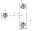
以下、図面を参照して本開示の実施形態について説明する。図1は、本開示の実施形態による学習装置および医用画像処理装置を適用した、診断支援システムの概要を示すハードウェア構成図である。図1に示すように、診断支援システムでは、本実施形態による学習装置および医用画像処理装置(以下、医用画像処理装置とする)1、3次元画像撮影装置2、および画像保管サーバ3が、ネットワーク4を経由して通信可能な状態で接続されている。
Embodiments of the present disclosure will be described below with reference to the drawings. FIG. 1 is a hardware configuration diagram showing an outline of a diagnosis support system to which a learning device and a medical image processing device according to an embodiment of the present disclosure are applied. As shown in FIG. 1, in the diagnosis support system, a learning device and a medical image processing device (hereinafter referred to as a medical image processing device) 1, a three-dimensional image capturing
3次元画像撮影装置2は、被検体の診断対象となる部位を撮影することにより、その部位を表す3次元画像を生成する装置であり、具体的には、CT装置、MRI装置、およびPET(Positron Emission Tomography)装置等である。3次元画像撮影装置2により生成された3次元画像は画像保管サーバ3に送信され、保存される。なお、本実施形態においては、被検体である患者の診断対象部位は脳であり、3次元画像撮影装置2はCT装置であり、被検体の脳を含む頭部のCT画像を対象画像として生成する。また、後述するように学習に使用する学習用画像は脳のCT画像であり、学習用画像における疾病領域がラベリングされて教師ラベルが生成されているものとする。
The three-
画像保管サーバ3は、各種データを保存して管理するコンピュータであり、大容量外部記憶装置およびデータベース管理用ソフトウェアを備えている。画像保管サーバ3は、有線あるいは無線のネットワーク4を介して他の装置と通信を行い、画像データ等を送受信する。具体的には3次元画像撮影装置2で生成された対象画像の画像データを含む各種データをネットワーク経由で取得し、大容量外部記憶装置等の記録媒体に保存して管理する。なお、画像データの格納形式およびネットワーク4経由での各装置間の通信は、DICOM(Digital Imaging and Communication in Medicine)等のプロトコルに基づいている。
The
医用画像処理装置1は、1台のコンピュータに、本実施形態の学習プログラムおよび医用画像処理プログラムをインストールしたものである。コンピュータは、診断を行う医師が直接操作するワークステーションまたはパーソナルコンピュータでもよいし、それらとネットワークを介して接続されたサーバコンピュータでもよい。学習プログラムおよび医用画像処理プログラムは、ネットワークに接続されたサーバコンピュータの記憶装置、もしくはネットワークストレージに、外部からアクセス可能な状態で記憶され、要求に応じてコンピュータにダウンロードされ、インストールされる。または、DVD(Digital Versatile Disc)あるいはCD-ROM(Compact Disc Read Only Memory)等の記録媒体に記録されて配布され、その記録媒体からコンピュータにインストールされる。
The medical
図2は、コンピュータに学習プログラムおよび医用画像処理プログラムをインストールすることにより実現される医用画像処理装置の概略構成を示す図である。図2に示すように、医用画像処理装置1は、標準的なワークステーションの構成として、CPU(Central Processing Unit)11、メモリ12およびストレージ13を備えている。また、医用画像処理装置1には、液晶ディスプレイ等の表示部14、並びにキーボードおよびマウス等の入力部15が接続されている。
FIG. 2 is a diagram showing a schematic configuration of a medical image processing apparatus realized by installing a learning program and a medical image processing program in a computer. As shown in FIG. 2, the medical
ストレージ13はハードディスクドライブ等からなり、ネットワーク4を経由して画像保管サーバ3から取得した処理対象となる対象画像、後述するようにニューラルネットワークの学習を行うための学習用画像、学習用画像に対する教師ラベル、および処理に必要な情報を含む各種情報が記憶されている。
The
また、メモリ12には、学習プログラムおよび医用画像処理プログラムが記憶されている。学習プログラムは、CPU11に実行させる処理として、疾病領域を含む学習用画像および学習用画像に含まれる疾病領域を特定する第1教師ラベルを取得する情報取得処理、疾病領域を特定する基準が第1教師ラベルとは異なる少なくとも1つの第2教師ラベルを生成する教師ラベル生成処理、並びに学習用画像、第1教師ラベルおよび少なくとも1つの第2教師ラベルに基づいて、対象画像に含まれる疾病領域を検出する判別器を学習する学習処理を規定する。
The
医用画像処理プログラムは、CPU11に実行させる処理として、情報取得処理により取得した疾病領域の検出の対象となる対象画像に含まれる疾病領域を検出する疾病領域検出処理、検出された疾病領域にラベリングを行うラベリング処理、およびラベリングされた対象画像を表示部14に表示する表示制御処理を規定する。
The medical image processing program executes, as processes to be executed by the
そして、CPU11が学習プログラムおよび医用画像処理プログラムに従いこれらの処理を実行することで、コンピュータは、情報取得部21、教師ラベル生成部22、学習部23、疾病領域検出部24、ラベリング部25および表示制御部26として機能する。
Then, by the
情報取得部21は、ネットワークに接続されたインターフェース(不図示)を介して、画像保管サーバ3から、学習用画像および学習用画像に含まれる疾病領域を特定する第1教師ラベルを取得する。また、処理の対象となる対象画像も取得する。なお、学習用画像、第1教師ラベルおよび対象画像が既にストレージ13に記憶されている場合には、情報取得部21は、ストレージ13から学習用画像、第1教師ラベルおよび対象画像を取得するようにしてもよい。
The
ここで、脳のCT画像において、脳出血等の疾病領域は、周囲の領域と比較して高いもしくは低いCT値を示す。例えば、図3に示すような脳画像30の場合、疾病領域31が他の領域と比較して高いCT値を示している。このような場合、疾病領域31とその周囲の領域とのコントラストが明瞭であるため、誰が作成しても教師ラベル32は脳画像30における疾病領域31とほぼ一致するものとなる。
Here, in a CT image of the brain, a diseased area such as cerebral hemorrhage shows a higher or lower CT value than the surrounding area. For example, in a brain image 30 as shown in FIG. 3, a
一方、図4に示す脳画像40のように、軽度のくも膜下出血、または超急性期の脳梗塞のような疾患では、疾病領域41とその周辺領域とのコントラストが不明瞭な場合が多い。なお、図4においてはコントラストが不明瞭であることを破線で示している。このような場合、どこまでを疾病領域と見なしてラベリングを行うかが、ラベリングを行う医師に応じて異なる。例えば、ある医師は小さめのサイズの教師ラベル42を付与するが、他の医師は大きめのサイズの教師ラベル43を付与する事態が生じうる。
On the other hand, as in the
教師ラベル生成部22は、疾病領域を特定する基準が第1教師ラベルとは異なる少なくとも1つの第2教師ラベルを生成する。このため、教師ラベル生成部22は、図5に示すように取得した学習用画像50において、第1教師ラベル51が付与された領域55内のCT値の代表値を導出し、第1教師ラベル51に対応する領域および学習用画像50における第1教師ラベル51内の領域に隣接する領域のCT値が、代表値に対して予め定められた範囲内にある領域を、第2教師ラベルとして生成する。
The teacher
なお、本実施形態においては、教師ラベル生成部22は、領域55内のCT値の平均値μを代表値として導出するものとするが、これに限定されるものではない。中央値、重み付け平均値、最大値または最小値等を代表値として用いてもよい。また、本実施形態においては、教師ラベル生成部22は、領域55内のCT値の標準偏差σおよび学習用画像50における第1教師ラベル51により特定される領域の重心位置56を導出する。
In the present embodiment, the
教師ラベル生成部22は、学習用画像50における重心位置56から予め定められた距離内にある画素のうち、μ±σの範囲のCT値を有する画素からなる領域をラベリングすることにより、図5に示すように第2教師ラベル52を生成する。ここで、図6は疾病領域におけるCT値の分布を示す図である。図6に示すように、疾病領域のCT値はその周囲のCT値と比較すると大きい値となり、疾病領域の周辺ほどCT値は小さくなり、疾病領域の周囲の領域のCT値と徐々に一致するように分布する。このため、図6に示す矢印Aに示す範囲に第1教師ラベルが付与されていたとすると、μ-σのCT値を有する画素からなる領域をラベリングすることにより、矢印Bに示すように、第1教師ラベル51よりも大きいサイズの第2教師ラベル52を生成することができる。一方、μ+σのCT値を有する画素からなる領域をラベリングすることにより、矢印Cに示すように、第1教師ラベル51よりも小さいサイズの第2教師ラベル52を生成することができる。
The teacher
なお、学習用画像50における疾病領域の境界が不明瞭な場合、図5に示すように、第1教師ラベル51とは異なる第2教師ラベル52が生成される。一方、図3に示すように、疾病領域の境界が明瞭な場合、疾病領域内のCT値は一定となるため、標準偏差σは実質的に0となる。このような場合、教師ラベル生成部22が生成する第2教師ラベル52は、実質的に第1教師ラベル51と同一となる。
Note that when the boundary of the diseased region in the
また、本実施形態においては、第1教師ラベル51から1つの第2教師ラベル52を生成するものとするが、複数の第2の教師ラベルを生成してもよい。この場合、例えば、μ±0.5σ、μ±σ、μ±1.5σ等のそれぞれのCT値を有する画素からなる領域をラベリングして複数の第2教師ラベル52を生成すればよい。
Also, in this embodiment, one
学習部23は、学習用画像50、第1教師ラベル51および第2教師ラベル52に基づいて、対象画像に含まれる疾病領域を検出する判別器28を学習する。判別器28は、対象画像に含まれる脳の疾病領域を判別する。本実施形態においては、判別器28は、複数の処理層が階層的に接続され、深層学習(ディープラーニング)がなされた多層ニューラルネットワークの1つである、畳み込みニューラルネットワーク(以下CNN(Convolutional Neural Network)とする)であるものとする。
The
畳み込みニューラルネットワークは、複数の畳み込み層およびプーリング層からなる。畳み込み層は、入力される画像に対して各種カーネルを用いた畳み込み処理を行い、畳み込み処理により得られた特徴量データからなる特徴量マップを出力する。カーネルは、n×n画素サイズ(例えばn=3)を有し、各要素に重みが設定されている。具体的には入力された画像のエッジを強調する微分フィルタのような重みが設定されている。畳み込み層は、カーネルの注目画素をずらしながら、入力された画像または前段の処理層から出力された特徴量マップの全体にカーネルを適用する。さらに、畳み込み層は、畳み込みされた値に対して、シグモイド関数等の活性化関数を適用し、特徴量マップを出力する。 A convolutional neural network consists of multiple convolutional and pooling layers. The convolution layer performs convolution processing using various kernels on an input image, and outputs a feature amount map composed of feature amount data obtained by the convolution processing. The kernel has an n×n pixel size (eg n=3) and weights are assigned to each element. Specifically, a weight like a differential filter that emphasizes the edges of the input image is set. The convolution layer applies the kernel to the input image or the entire feature map output from the previous processing layer while shifting the pixel of interest of the kernel. Furthermore, the convolution layer applies an activation function such as a sigmoid function to the convolved values and outputs a feature quantity map.
プーリング層は、畳み込み層が出力した特徴量マップをプーリングすることにより、特徴量マップのデータ量を低減して、データ量が低減された特徴量マップを出力する。 The pooling layer reduces the data amount of the feature map by pooling the feature map output by the convolution layer, and outputs the feature map with the reduced data amount.
図7は本実施形態において行われる学習の概念図である。図7に示すように、学習部23は、学習用画像50を判別器28となるCNN60に入力し、CNN60から学習用画像50における疾病領域の判別結果57を出力させる。判別結果57は、学習用画像50の各画素が疾病領域であることの確率を表すものとなる。学習部23は、確率が予め定められたしきい値以上となる画素からなる領域を、学習用疾病領域58に特定する。そして、学習部23は、第1教師ラベル51と学習用疾病領域58の判別結果57との相違に基づいて、第1損失L1を導出する。第1損失L1は、第1教師ラベル51において疾病領域であるのに、疾病領域でないと判別された画素についての、確率と上記しきい値との差、および第1教師ラベル51において疾病領域でないのに疾病領域であると判別された画素についての、上記しきい値と確率との差である。
FIG. 7 is a conceptual diagram of learning performed in this embodiment. As shown in FIG. 7 , the
また、学習部23は、第2教師ラベル52と判別結果57との相違に基づいて、第2損失L2を導出する。第2損失L2は、第2教師ラベル52において疾病領域であるのに、疾病領域でないと判別された画素についての、確率と上記しきい値との差、および第2教師ラベル52において疾病領域でないのに疾病領域であると判別された画素についての、上記しきい値と確率との差である。
Also, the
さらに、学習部23は、第1損失L1および第2損失L2を下記の式(1)に示すように重み付け加算して、学習用画像50の各画素についてのトータル損失L0を導出する。なお、式(1)のαは重み係数であり、例えば0.5の値をとるが、これに限定されるものではない。
Furthermore, the
L0=L1+α・L2 (1) L0=L1+α・L2 (1)
そして、学習部23はトータル損失L0が予め定められたしきい値以下となるように、多数の学習用画像50、第1教師ラベル51および第2教師ラベル52を用いて、CNN60すなわち判別器28を学習する。具体的には、トータル損失L0が予め定められたしきい値以下となるように、CNN60を構成する畳み込み層の数、プーリング層の数、畳み込み層におけるカーネルの係数およびカーネルの大きさ等を導出することにより、CNN60すなわち判別器28の学習を行う。これにより、学習された判別器28に対象画像が入力されると、判別器28は、対象画像の各画素が脳の疾病領域であることの確率を出力するものとなる。なお、学習部23は、トータル損失L0が予め定められたしきい値以下となるように学習を行うことに代えて、予め定められた回数の学習を行うものであってもよい。
Then, the
上記のように学習部23がCNN60すなわち判別器28の学習を行うことにより、対象画像が入力されると、対象画像に含まれる疾病領域であることの確率を判別結果として出力する学習済みモデルが構築される。学習済みモデルが判別器28として疾病領域検出部に適用される。
As described above, the
疾病領域検出部24は、対象画像が入力されると、判別器28を用いて、対象画像に含まれる疾病領域を検出する。すなわち、疾病領域検出部24は、対象画像を判別器28に入力し、判別器28から対象画像の各画素が脳の疾病領域であることの確率を出力させる。そして、疾病領域検出部24は、確率が予め定められたしきい値を超えた画素を、対象画像に含まれる疾病領域の画素として検出する。
When the target image is input, the diseased
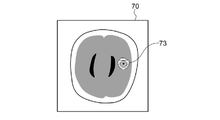
ラベリング部25は、疾病領域検出部24による検出結果に基づいて、対象画像に含まれる疾病領域のラベリングを行う。例えば、図8に示すように疾病領域71(破線で示す)が含まれる対象画像70が疾病領域検出部24に入力された場合、疾病領域検出部24は、対象画像70に含まれる疾病領域71を検出する。ラベリング部25は、対象画像70に含まれる疾病領域71にラベルを付与することにより、ラベリングを行う。例えば、図8に示すように、疾病領域の色を変化させることにより疾病領域71にラベル72を付与して、ラベリングを行う。なお、図8においては色を変化させることをハッチングを付与することにより示している。また、疾病領域を囲む枠を付与することにより、ラベリングを行うようにしてもよい。
The
表示制御部26は、ラベリングされた対象画像を表示部14に表示する。
The
次いで、本実施形態において行われる処理について説明する。図9は本実施形態において行われる学習処理を示すフローチャートである。なお、複数の学習用画像および第1教師ラベルが画像保管サーバ3から情報取得部21により取得されて、ストレージ13に保存されているものとする。まず、情報取得部21がストレージ13に保存された複数の学習用画像および第1教師ラベルから1組の学習用画像50および第1教師ラベル51を取得する(ステップST1)。次いで、教師ラベル生成部22が、疾病領域を特定する基準が第1教師ラベル51とは異なる少なくとも1つの第2教師ラベル52を生成する(ステップST2)。
Next, processing performed in this embodiment will be described. FIG. 9 is a flow chart showing the learning process performed in this embodiment. It is assumed that a plurality of learning images and first teacher labels are acquired from the
そして、学習部23が、CNN60に対して、学習用画像50、第1教師ラベル51および第2教師ラベル52を入力してトータル損失L0を導出し、トータル損失L0が予め定められたしきい値以下となるように、CNN60すなわち判別器28を学習する(ステップST3)。
Then, the
そして、ステップST1にリターンし、次の学習用画像50および第1教師ラベル51をストレージ13から取得して、ステップST2およびステップST3の処理を繰り返す。これにより、学習済みの判別器28が構築される。
Then, the process returns to step ST1, acquires the
次いで、本実施形態において行われる疾病領域を検出する医用画像処理について説明する。図10は本実施形態において行われる医用画像処理のフローチャートである。情報取得部21が対象画像を取得し(ステップST11)、疾病領域検出部24が対象画像に含まれる疾病領域を検出する(ステップST12)。次いで、ラベリング部25が、対象画像70から検出された疾病領域のラベリングを行う(ステップST13)。そして、表示制御部26がラベリングされた対象画像を表示部14に表示し(ステップST14)、処理を終了する。
Next, medical image processing for detecting diseased regions performed in this embodiment will be described. FIG. 10 is a flowchart of medical image processing performed in this embodiment. The
このように、本実施形態においては、疾病領域を含む学習用画像50および学習用画像50に含まれる疾病領域を特定する第1教師ラベル51を取得し、疾病領域を特定する基準が第1教師ラベル51とは異なる少なくとも1つの第2教師ラベル52を生成する。そして、学習用画像50、第1教師ラベル51および少なくとも1つの第2教師ラベル52に基づいて、対象画像70に含まれる疾病領域を検出するための判別器28を学習するようにした。このため、判別器28は、第1教師ラベル51の基準のみならず、第2教師ラベル52の基準にも基づいて、対象画像から疾病領域を検出するものとなる。これにより、本実施形態においては、異なる基準の複数の教師ラベルを用いて判別器28が学習されることから、医師によって判断がぶれる可能性が高い、周囲とのコントラストが不明瞭な疾病領域を、学習された判別器28によりある程度の許容範囲を持って検出することができる。このため、とくに第1教師ラベル51よりも大きい範囲の疾病をラベリングするように第2教師ラベル52を生成することにより、第1教師ラベル51のみを使用して学習を行う場合よりも、広い範囲の疾病領域を検出できるように判別器28を構築することができる。したがって、本実施形態によれば、対象画像に含まれる疾病領域の見逃しを防止できる。
Thus, in the present embodiment, the learning
なお、上記実施形態において、疾病領域検出部24の判別器28が出力する疾病領域であることを表す確率は、疾病領域の周辺ほど小さいものとなる。このため、図11に示すように、判別器28が出力した確率に応じて段階的に透明度が異なるラベルを付与するようにしてもよい。なお、図11においては、透明度が異なることを異なるハッチングにより示している。また、段階的に色を変更するのみならず、徐々に透明度を変更するようにしてもよい。また、透明度に代えて、色を変更してもよい。
In the above-described embodiment, the probability that the
学習部26は、学習用医用画像GL0の特徴ベクトルCL0、および学習用医用画像GL0に関する学習用医療文書TL0を学習データとして複数使用して、CNNの学習を行う。具体的には、学習部26は、図7に示す特徴ベクトルCL0および図8に示す学習用医療文書TL0が入力されると、学習用医療文書TL0の表現ベクトルzL0を表現ベクトル生成部25に生成させる。そして、学習用医用画像GL0の特徴ベクトルCL0および学習用の表現ベクトルzL0が入力されると、図8に示す学習用医療文書TL0を出力するようにCNNの学習を行って学習済みモデルM3を生成する。
The
また、上記実施形態においては、第1教師ラベル51よりも大きいサイズの第2教師ラベル52を生成しているが、第1教師ラベル51よりも小さいサイズの第2教師ラベル52を生成するようにしてもよい。
Also, in the above embodiment, the
また、上記実施形態においては、教師ラベル生成部22が、第1教師ラベル51から第2教師ラベル52を生成しているが、これに限定されるものではない。例えば学習用画像50から第2教師ラベル52を生成してもよい。
Further, in the above embodiment, the teacher
また、上記実施形態においては、対象画像を脳を含む3次元画像として、脳の疾病領域を検出しているが、これに限定されるものではない。脳以外の肺、肝臓、心臓および腎臓等の他の構造物に含まれる疾病領域を検出する場合にも、本開示の技術を適用することができる。例えば、肝臓を含む医用画像を学習用画像、肝臓の腫瘤をラベリングした第1教師ラベルを用いることにより、肝臓に含まれる腫瘤を見逃すことなく検出できる判別器28を構築できる。また、肺を含む医用画像を学習用画像、肺結節をラベリングした第1教師ラベルを用いることにより、肺に含まれる肺結節を見逃すことなく検出できる判別器28を構築できる。
In the above embodiment, the target image is a three-dimensional image including the brain, and the diseased region of the brain is detected, but the present invention is not limited to this. The technology of the present disclosure can also be applied to detect diseased regions in structures other than the brain, such as lungs, livers, hearts, and kidneys. For example, by using a medical image including the liver as a learning image and a first teacher label labeled with a liver mass, a
また、上記実施形態においては、対象画像として3次元の医用画像を用いているが、これに限定されるものではない。3次元の医用画像を構成する個々の断層画像を対象画像として用いてもよい。また、単純X線撮影により取得された2次元のX線画像を対象画像として用いてもよい。この場合、対象画像の種類に応じた学習用画像および第1教師ラベルが用意されてCNN60すなわち判別器28の学習が行われることとなる。
Also, in the above embodiment, a three-dimensional medical image is used as the target image, but the present invention is not limited to this. Individual tomographic images forming a three-dimensional medical image may be used as target images. Also, a two-dimensional X-ray image obtained by plain X-ray imaging may be used as the target image. In this case, learning images and first teacher labels corresponding to the types of target images are prepared, and the
また、上記実施形態においては、判別器28としてCNN60を用いているが、これに限定されるものではない。複数の処理層から構成されるニューラルネットワークであれば、ディープニューラルネットワーク(DNN(Deep Neural Network))およびリカレントニューラルネットワーク(RNN(Recurrent Neural Network))等を用いることができる。
Also, in the above embodiment, the
また、上記実施形態において、例えば、情報取得部21、教師ラベル生成部22、学習部23、疾病領域検出部24、ラベリング部25および表示制御部26といった各種の処理を実行する処理部(Processing Unit)のハードウェア的な構造としては、次に示す各種のプロセッサ(Processor)を用いることができる。上記各種のプロセッサには、上述したように、ソフトウェア(プログラム)を実行して各種の処理部として機能する汎用的なプロセッサであるCPUに加えて、FPGA(Field Programmable Gate Array)等の製造後に回路構成を変更可能なプロセッサであるプログラマブルロジックデバイス(Programmable Logic Device :PLD)、ASIC(Application Specific Integrated Circuit)等の特定の処理を実行させるために専用に設計された回路構成を有するプロセッサである専用電気回路等が含まれる。 Further, in the above-described embodiment, for example, a processing unit (processing unit ), the following various processors can be used as the hardware structure. In addition to the CPU, which is a general-purpose processor that executes software (programs) and functions as various processing units, as described above, the various processors described above include circuits such as FPGAs (Field Programmable Gate Arrays) after manufacturing. Programmable Logic Device (PLD), which is a processor whose configuration can be changed, ASIC (Application Specific Integrated Circuit), etc. Circuits, etc. are included.
1つの処理部は、これらの各種のプロセッサのうちの1つで構成されてもよいし、同種または異種の2つ以上のプロセッサの組み合わせ(例えば、複数のFPGAの組み合わせまたはCPUとFPGAとの組み合わせ)で構成されてもよい。また、複数の処理部を1つのプロセッサで構成してもよい。 One processing unit may be configured with one of these various processors, or a combination of two or more processors of the same or different type (for example, a combination of multiple FPGAs or a combination of a CPU and an FPGA). ). Also, a plurality of processing units may be configured by one processor.
複数の処理部を1つのプロセッサで構成する例としては、第1に、クライアントおよびサーバ等のコンピュータに代表されるように、1つ以上のCPUとソフトウェアとの組み合わせで1つのプロセッサを構成し、このプロセッサが複数の処理部として機能する形態がある。第2に、システムオンチップ(System On Chip:SoC)等に代表されるように、複数の処理部を含むシステム全体の機能を1つのIC(Integrated Circuit)チップで実現するプロセッサを使用する形態がある。このように、各種の処理部は、ハードウェア的な構造として、上記各種のプロセッサの1つ以上を用いて構成される。 As an example of configuring a plurality of processing units in one processor, first, as represented by computers such as clients and servers, one processor is configured by combining one or more CPUs and software, There is a form in which this processor functions as a plurality of processing units. Secondly, as typified by System On Chip (SoC), etc., there is a form of using a processor that realizes the functions of the entire system including multiple processing units with a single IC (Integrated Circuit) chip. be. In this way, the various processing units are configured using one or more of the above various processors as a hardware structure.
さらに、これらの各種のプロセッサのハードウェア的な構造としては、より具体的には、半導体素子等の回路素子を組み合わせた電気回路(Circuitry)を用いることができる。 Furthermore, more specifically, as the hardware structure of these various processors, an electric circuit (circuitry) in which circuit elements such as semiconductor elements are combined can be used.
1 医用画像処理装置
2 3次元画像撮影装置
3 画像保管サーバ
4 ネットワーク
11 CPU
12 メモリ
13 ストレージ
14 表示部
15 入力部
21 情報取得部
22 教師ラベル生成部
23 学習部
24 疾病領域検出部
25 ラベリング部
26 表示制御部
28 判別器
30,40 脳画像
31,41 疾病領域
32,42,43 教師ラベル
50 学習用画像
51 第1教師ラベル
52 第2教師ラベル
55 領域
56 重心位置
57 判別結果
60 CNN
70 対象画像
71 疾病領域
72,73 ラベル
1 medical
12
15
32, 42, 43
57
70
Claims (13)
前記疾病領域を特定する基準が前記第1教師ラベルとは異なる少なくとも1つの第2教師ラベルを生成する教師ラベル生成部と、
前記学習用画像、前記第1教師ラベルおよび前記少なくとも1つの第2教師ラベルに基づいて、対象画像に含まれる疾病領域を検出する判別器を学習する学習部とを備えた学習装置。an information acquisition unit that acquires a learning image including a diseased region and a first teacher label that identifies the diseased region included in the learning image;
a teacher label generation unit that generates at least one second teacher label whose criterion for identifying the disease area is different from that of the first teacher label;
and a learning unit that learns a classifier for detecting a diseased region included in a target image based on the learning image, the first teacher label, and the at least one second teacher label.
前記ラベリングされた対象医用画像を表示部に表示する表示制御部とをさらに備えた請求項7に記載の医用画像処理装置。a labeling unit that labels the diseased region detected from the target medical image;
8. The medical image processing apparatus according to claim 7, further comprising a display control unit that displays the labeled target medical image on a display unit.
前記疾病領域を特定する基準が前記第1教師ラベルとは異なる少なくとも1つの第2教師ラベルを生成し、
前記学習用画像、前記第1教師ラベルおよび前記少なくとも1つの第2教師ラベルに基づいて、対象画像に含まれる疾病領域を検出する判別器を学習する学習方法。Acquiring a learning image containing a diseased region and a first teacher label that identifies the diseased region contained in the learning image,
generating at least one second teacher label whose criteria for identifying the disease area is different from the first teacher label;
A learning method for learning a discriminator for detecting a diseased region included in a target image based on the learning image, the first teacher label and the at least one second teacher label.
前記疾病領域を特定する基準が前記第1教師ラベルとは異なる少なくとも1つの第2教師ラベルを生成する手順と、
前記学習用画像、前記第1教師ラベルおよび前記少なくとも1つの第2教師ラベルに基づいて、対象画像に含まれる疾病領域を検出する判別器を学習する手順とをコンピュータに実行させる学習プログラム。A procedure for acquiring a learning image containing a diseased region and a first teacher label identifying the diseased region contained in the learning image;
generating at least one second teacher label different from the first teacher label in criteria for identifying the disease area;
A learning program that causes a computer to execute a procedure for learning a discriminator for detecting a diseased region included in a target image based on the learning image, the first teacher label and the at least one second teacher label.
Applications Claiming Priority (3)
| Application Number | Priority Date | Filing Date | Title |
|---|---|---|---|
| JP2019121015 | 2019-06-28 | ||
| JP2019121015 | 2019-06-28 | ||
| PCT/JP2020/025399 WO2020262681A1 (en) | 2019-06-28 | 2020-06-26 | Learning device, method, and program, medical image processing device, method, and program, and discriminator |
Publications (3)
| Publication Number | Publication Date |
|---|---|
| JPWO2020262681A1 JPWO2020262681A1 (en) | 2020-12-30 |
| JPWO2020262681A5 JPWO2020262681A5 (en) | 2022-03-11 |
| JP7170868B2 true JP7170868B2 (en) | 2022-11-14 |
Family
ID=74060193
Family Applications (1)
| Application Number | Title | Priority Date | Filing Date |
|---|---|---|---|
| JP2021528288A Active JP7170868B2 (en) | 2019-06-28 | 2020-06-26 | LEARNING APPARATUS, METHOD AND PROGRAM, MEDICAL IMAGE PROCESSING APPARATUS, METHOD AND PROGRAM, AND CLASSIFIER |
Country Status (3)
| Country | Link |
|---|---|
| US (1) | US20220108451A1 (en) |
| JP (1) | JP7170868B2 (en) |
| WO (1) | WO2020262681A1 (en) |
Citations (2)
| Publication number | Priority date | Publication date | Assignee | Title |
|---|---|---|---|---|
| JP2019509813A (en) | 2016-03-16 | 2019-04-11 | ハートフロー, インコーポレイテッド | System and method for estimating healthy lumen diameter and quantifying stenosis in coronary arteries |
| US20190192096A1 (en) | 2017-12-21 | 2019-06-27 | Beijing Curacloud Technology Co., Ltd. | Method and device for generating anatomical labels for a physiological tree structure |
Family Cites Families (1)
| Publication number | Priority date | Publication date | Assignee | Title |
|---|---|---|---|---|
| JP2018061771A (en) * | 2016-10-14 | 2018-04-19 | 株式会社日立製作所 | Image processing apparatus and image processing method |
-
2020
- 2020-06-26 JP JP2021528288A patent/JP7170868B2/en active Active
- 2020-06-26 WO PCT/JP2020/025399 patent/WO2020262681A1/en active Application Filing
-
2021
- 2021-12-16 US US17/553,641 patent/US20220108451A1/en active Pending
Patent Citations (2)
| Publication number | Priority date | Publication date | Assignee | Title |
|---|---|---|---|---|
| JP2019509813A (en) | 2016-03-16 | 2019-04-11 | ハートフロー, インコーポレイテッド | System and method for estimating healthy lumen diameter and quantifying stenosis in coronary arteries |
| US20190192096A1 (en) | 2017-12-21 | 2019-06-27 | Beijing Curacloud Technology Co., Ltd. | Method and device for generating anatomical labels for a physiological tree structure |
Non-Patent Citations (3)
| Title |
|---|
| HAN, Jiangfan et al.,"Deep Self-Learning From Noisy Labels",arXiv,2019年08月06日,arXiv:1908.02160v1 |
| NATARAJAN, Nagarajan et al.,"Leaning with Noisy Labels",NIPS'13: Proceedings of the 26th International Conference on Neural Information Processing Systems,2013年,Volume 1,pp.1196-1204 |
| 島原 佑基,"[企業総説]深層学習を活用した脳動脈瘤検知ソフトウェアの開発",医用画像情報学会雑誌,2017年,Vol.34, No.2,pp.103-104 |
Also Published As
| Publication number | Publication date |
|---|---|
| WO2020262681A1 (en) | 2020-12-30 |
| JPWO2020262681A1 (en) | 2020-12-30 |
| US20220108451A1 (en) | 2022-04-07 |
Similar Documents
| Publication | Publication Date | Title |
|---|---|---|
| JP7225295B2 (en) | MEDICAL IMAGE DISPLAY APPARATUS, METHOD AND PROGRAM | |
| US11069056B2 (en) | Multi-modal computer-aided diagnosis systems and methods for prostate cancer | |
| US11893729B2 (en) | Multi-modal computer-aided diagnosis systems and methods for prostate cancer | |
| JP7129870B2 (en) | Learning device, method, and program for discriminator for discriminating disease area, discriminator for discriminating disease area, and device and program for discriminating disease region | |
| JP7203978B2 (en) | LEARNING DEVICE, METHOD AND PROGRAM, REGION OF INTEREST EXTRACTION DEVICE, METHOD AND PROGRAM, AND LEARNED EXTRACT MODEL | |
| US11915414B2 (en) | Medical image processing apparatus, method, and program | |
| JP2019518288A (en) | Change detection in medical image | |
| JP2022018060A (en) | Medical information processing apparatus and medical information processing program | |
| Kurachka et al. | Vertebrae detection in X-ray images based on deep convolutional neural networks | |
| JP7007469B2 (en) | Medical document creation support devices, methods and programs, trained models, and learning devices, methods and programs | |
| JP2023114463A (en) | Display device, method and program | |
| JP7170868B2 (en) | LEARNING APPARATUS, METHOD AND PROGRAM, MEDICAL IMAGE PROCESSING APPARATUS, METHOD AND PROGRAM, AND CLASSIFIER | |
| Öztürk | Convolutional neural networks for medical image processing applications | |
| EP3965117A1 (en) | Multi-modal computer-aided diagnosis systems and methods for prostate cancer | |
| JP7321271B2 (en) | LEARNING IMAGE GENERATION DEVICE, METHOD AND PROGRAM, AND LEARNING METHOD, DEVICE AND PROGRAM | |
| CN116420165A (en) | Detection of anatomical anomalies by segmentation results with and without shape priors | |
| WO2020110520A1 (en) | Similarity determination device, method, and program | |
| WO2020044736A1 (en) | Similarity determination device, method, and program | |
| JP7342120B2 (en) | Learning device, method and program, classification device, method and program, and trained model | |
| WO2021060461A1 (en) | Image processing device, method, and program | |
| WO2022137855A1 (en) | Information processing device, method, and program | |
| JP7299314B2 (en) | Medical documentation device, method and program, learning device, method and program, and trained model | |
| Akpan et al. | XAI for medical image segmentation in medical decision support systems | |
| WO2022054541A1 (en) | Image processing device, method, and program | |
| US20230088616A1 (en) | Progression prediction apparatus, progression prediction method, and progression prediction program |
Legal Events
| Date | Code | Title | Description |
|---|---|---|---|
| A521 | Request for written amendment filed |
Free format text: JAPANESE INTERMEDIATE CODE: A523 Effective date: 20211215 |
|
| A621 | Written request for application examination |
Free format text: JAPANESE INTERMEDIATE CODE: A621 Effective date: 20211215 |
|
| TRDD | Decision of grant or rejection written | ||
| A01 | Written decision to grant a patent or to grant a registration (utility model) |
Free format text: JAPANESE INTERMEDIATE CODE: A01 Effective date: 20221011 |
|
| A61 | First payment of annual fees (during grant procedure) |
Free format text: JAPANESE INTERMEDIATE CODE: A61 Effective date: 20221101 |
|
| R150 | Certificate of patent or registration of utility model |
Ref document number: 7170868 Country of ref document: JP Free format text: JAPANESE INTERMEDIATE CODE: R150 |