JP7146399B2 - Novel peptides and peptide and scaffold combinations for use in immunotherapy against renal cell carcinoma (RCC) and other cancers - Google Patents
Novel peptides and peptide and scaffold combinations for use in immunotherapy against renal cell carcinoma (RCC) and other cancers Download PDFInfo
- Publication number
- JP7146399B2 JP7146399B2 JP2017550874A JP2017550874A JP7146399B2 JP 7146399 B2 JP7146399 B2 JP 7146399B2 JP 2017550874 A JP2017550874 A JP 2017550874A JP 2017550874 A JP2017550874 A JP 2017550874A JP 7146399 B2 JP7146399 B2 JP 7146399B2
- Authority
- JP
- Japan
- Prior art keywords
- cancer
- peptide
- cells
- cell
- peptides
- Prior art date
- Legal status (The legal status is an assumption and is not a legal conclusion. Google has not performed a legal analysis and makes no representation as to the accuracy of the status listed.)
- Active
Links
- 108090000765 processed proteins & peptides Proteins 0.000 title claims description 539
- 206010028980 Neoplasm Diseases 0.000 title claims description 330
- 102000004196 processed proteins & peptides Human genes 0.000 title description 282
- 208000006265 Renal cell carcinoma Diseases 0.000 title description 87
- 238000009169 immunotherapy Methods 0.000 title description 12
- 210000004027 cell Anatomy 0.000 claims description 230
- 201000011510 cancer Diseases 0.000 claims description 157
- 108090000623 proteins and genes Proteins 0.000 claims description 120
- 210000001744 T-lymphocyte Anatomy 0.000 claims description 114
- 238000000034 method Methods 0.000 claims description 95
- 239000000427 antigen Substances 0.000 claims description 81
- 108091007433 antigens Proteins 0.000 claims description 81
- 102000036639 antigens Human genes 0.000 claims description 81
- 241000282414 Homo sapiens Species 0.000 claims description 67
- 102000004169 proteins and genes Human genes 0.000 claims description 63
- 206010006187 Breast cancer Diseases 0.000 claims description 53
- 208000026310 Breast neoplasm Diseases 0.000 claims description 51
- 150000007523 nucleic acids Chemical class 0.000 claims description 49
- 206010009944 Colon cancer Diseases 0.000 claims description 45
- 102000039446 nucleic acids Human genes 0.000 claims description 44
- 108020004707 nucleic acids Proteins 0.000 claims description 44
- 206010061535 Ovarian neoplasm Diseases 0.000 claims description 35
- 208000020816 lung neoplasm Diseases 0.000 claims description 35
- 208000000236 Prostatic Neoplasms Diseases 0.000 claims description 32
- 210000000612 antigen-presenting cell Anatomy 0.000 claims description 32
- 206010060862 Prostate cancer Diseases 0.000 claims description 31
- 206010033128 Ovarian cancer Diseases 0.000 claims description 30
- 125000003275 alpha amino acid group Chemical group 0.000 claims description 30
- 206010061902 Pancreatic neoplasm Diseases 0.000 claims description 29
- 108091054437 MHC class I family Proteins 0.000 claims description 28
- 208000005718 Stomach Neoplasms Diseases 0.000 claims description 28
- 239000013604 expression vector Substances 0.000 claims description 28
- 206010058467 Lung neoplasm malignant Diseases 0.000 claims description 26
- 239000003814 drug Substances 0.000 claims description 26
- 201000005202 lung cancer Diseases 0.000 claims description 26
- 208000001333 Colorectal Neoplasms Diseases 0.000 claims description 25
- 206010017758 gastric cancer Diseases 0.000 claims description 24
- 201000002528 pancreatic cancer Diseases 0.000 claims description 24
- 201000011549 stomach cancer Diseases 0.000 claims description 24
- 208000015486 malignant pancreatic neoplasm Diseases 0.000 claims description 23
- 208000008443 pancreatic carcinoma Diseases 0.000 claims description 23
- 201000001441 melanoma Diseases 0.000 claims description 22
- 208000003174 Brain Neoplasms Diseases 0.000 claims description 21
- 208000000461 Esophageal Neoplasms Diseases 0.000 claims description 20
- 238000000338 in vitro Methods 0.000 claims description 20
- 208000014018 liver neoplasm Diseases 0.000 claims description 19
- 201000007270 liver cancer Diseases 0.000 claims description 18
- 206010030155 Oesophageal carcinoma Diseases 0.000 claims description 17
- 201000004101 esophageal cancer Diseases 0.000 claims description 17
- 239000008194 pharmaceutical composition Substances 0.000 claims description 17
- 239000003795 chemical substances by application Substances 0.000 claims description 15
- 208000032839 leukemia Diseases 0.000 claims description 15
- 150000003839 salts Chemical class 0.000 claims description 15
- 210000004698 lymphocyte Anatomy 0.000 claims description 14
- 229940079593 drug Drugs 0.000 claims description 13
- 102000008949 Histocompatibility Antigens Class I Human genes 0.000 claims description 12
- 210000004443 dendritic cell Anatomy 0.000 claims description 11
- 230000002018 overexpression Effects 0.000 claims description 11
- 239000003085 diluting agent Substances 0.000 claims description 8
- 239000000872 buffer Substances 0.000 claims description 6
- 239000012931 lyophilized formulation Substances 0.000 claims description 6
- 239000001963 growth medium Substances 0.000 claims description 5
- 108010088652 Histocompatibility Antigens Class I Proteins 0.000 claims description 2
- 210000001519 tissue Anatomy 0.000 description 117
- 108091008874 T cell receptors Proteins 0.000 description 107
- 102000016266 T-Cell Antigen Receptors Human genes 0.000 description 107
- 108700018351 Major Histocompatibility Complex Proteins 0.000 description 90
- 230000020382 suppression by virus of host antigen processing and presentation of peptide antigen via MHC class I Effects 0.000 description 90
- 230000014509 gene expression Effects 0.000 description 84
- 235000018102 proteins Nutrition 0.000 description 59
- 235000001014 amino acid Nutrition 0.000 description 57
- 229940024606 amino acid Drugs 0.000 description 56
- 150000001413 amino acids Chemical class 0.000 description 55
- 229920001184 polypeptide Polymers 0.000 description 54
- 230000027455 binding Effects 0.000 description 53
- 108020004414 DNA Proteins 0.000 description 48
- 239000000523 sample Substances 0.000 description 38
- 239000000203 mixture Substances 0.000 description 34
- 229960005486 vaccine Drugs 0.000 description 34
- 239000013598 vector Substances 0.000 description 34
- 206010073071 hepatocellular carcinoma Diseases 0.000 description 31
- 231100000844 hepatocellular carcinoma Toxicity 0.000 description 31
- 210000004881 tumor cell Anatomy 0.000 description 29
- 239000012634 fragment Substances 0.000 description 28
- 208000029742 colonic neoplasm Diseases 0.000 description 23
- 108091032973 (ribonucleotides)n+m Proteins 0.000 description 21
- 239000002671 adjuvant Substances 0.000 description 21
- 230000005847 immunogenicity Effects 0.000 description 21
- 238000002955 isolation Methods 0.000 description 21
- 125000000539 amino acid group Chemical group 0.000 description 20
- 230000028993 immune response Effects 0.000 description 20
- 238000006467 substitution reaction Methods 0.000 description 20
- 238000011282 treatment Methods 0.000 description 20
- 108091054438 MHC class II family Proteins 0.000 description 19
- IAZDPXIOMUYVGZ-UHFFFAOYSA-N Dimethylsulphoxide Chemical compound CS(C)=O IAZDPXIOMUYVGZ-UHFFFAOYSA-N 0.000 description 18
- 208000008839 Kidney Neoplasms Diseases 0.000 description 18
- 102000043129 MHC class I family Human genes 0.000 description 18
- 210000004072 lung Anatomy 0.000 description 18
- 102100028972 HLA class I histocompatibility antigen, A alpha chain Human genes 0.000 description 17
- 108010075704 HLA-A Antigens Proteins 0.000 description 17
- 210000000481 breast Anatomy 0.000 description 17
- 230000000694 effects Effects 0.000 description 17
- 238000009472 formulation Methods 0.000 description 17
- 102000040430 polynucleotide Human genes 0.000 description 17
- 108091033319 polynucleotide Proteins 0.000 description 17
- 239000002157 polynucleotide Substances 0.000 description 17
- 230000000638 stimulation Effects 0.000 description 17
- 206010038389 Renal cancer Diseases 0.000 description 16
- 238000013459 approach Methods 0.000 description 16
- 210000003734 kidney Anatomy 0.000 description 16
- 201000010982 kidney cancer Diseases 0.000 description 16
- 238000004519 manufacturing process Methods 0.000 description 16
- 238000011002 quantification Methods 0.000 description 16
- 230000004044 response Effects 0.000 description 16
- 108091023037 Aptamer Proteins 0.000 description 15
- 238000004458 analytical method Methods 0.000 description 15
- 230000000875 corresponding effect Effects 0.000 description 15
- -1 BAD Proteins 0.000 description 14
- 230000005867 T cell response Effects 0.000 description 14
- 239000000090 biomarker Substances 0.000 description 14
- 210000001072 colon Anatomy 0.000 description 14
- 238000001514 detection method Methods 0.000 description 14
- 230000006870 function Effects 0.000 description 14
- 239000003446 ligand Substances 0.000 description 14
- 230000004048 modification Effects 0.000 description 14
- 238000012986 modification Methods 0.000 description 14
- 239000000047 product Substances 0.000 description 14
- 102000043131 MHC class II family Human genes 0.000 description 13
- 238000002648 combination therapy Methods 0.000 description 13
- 210000001151 cytotoxic T lymphocyte Anatomy 0.000 description 13
- 230000001965 increasing effect Effects 0.000 description 13
- 240000004808 Saccharomyces cerevisiae Species 0.000 description 12
- 235000014680 Saccharomyces cerevisiae Nutrition 0.000 description 12
- 210000004369 blood Anatomy 0.000 description 12
- 239000008280 blood Substances 0.000 description 12
- 208000037265 diseases, disorders, signs and symptoms Diseases 0.000 description 12
- 208000005017 glioblastoma Diseases 0.000 description 12
- 238000001727 in vivo Methods 0.000 description 12
- 210000001165 lymph node Anatomy 0.000 description 12
- 210000002307 prostate Anatomy 0.000 description 12
- 239000000243 solution Substances 0.000 description 12
- 230000004083 survival effect Effects 0.000 description 12
- 230000001225 therapeutic effect Effects 0.000 description 12
- 210000001266 CD8-positive T-lymphocyte Anatomy 0.000 description 11
- 208000030808 Clear cell renal carcinoma Diseases 0.000 description 11
- 102100039620 Granulocyte-macrophage colony-stimulating factor Human genes 0.000 description 11
- 201000010099 disease Diseases 0.000 description 11
- 230000002611 ovarian Effects 0.000 description 11
- 210000001672 ovary Anatomy 0.000 description 11
- 108060003951 Immunoglobulin Proteins 0.000 description 10
- 108091028043 Nucleic acid sequence Proteins 0.000 description 10
- 108010038807 Oligopeptides Proteins 0.000 description 10
- 102000015636 Oligopeptides Human genes 0.000 description 10
- 230000015572 biosynthetic process Effects 0.000 description 10
- 210000004899 c-terminal region Anatomy 0.000 description 10
- 238000011161 development Methods 0.000 description 10
- 239000012636 effector Substances 0.000 description 10
- 102000018358 immunoglobulin Human genes 0.000 description 10
- 238000004895 liquid chromatography mass spectrometry Methods 0.000 description 10
- 210000004185 liver Anatomy 0.000 description 10
- 239000003550 marker Substances 0.000 description 10
- 208000002154 non-small cell lung carcinoma Diseases 0.000 description 10
- 210000000496 pancreas Anatomy 0.000 description 10
- 238000002360 preparation method Methods 0.000 description 10
- 238000012360 testing method Methods 0.000 description 10
- 208000029729 tumor suppressor gene on chromosome 11 Diseases 0.000 description 10
- YBJHBAHKTGYVGT-ZKWXMUAHSA-N (+)-Biotin Chemical compound N1C(=O)N[C@@H]2[C@H](CCCCC(=O)O)SC[C@@H]21 YBJHBAHKTGYVGT-ZKWXMUAHSA-N 0.000 description 9
- 206010008342 Cervix carcinoma Diseases 0.000 description 9
- 102000004127 Cytokines Human genes 0.000 description 9
- 108090000695 Cytokines Proteins 0.000 description 9
- 108010017213 Granulocyte-Macrophage Colony-Stimulating Factor Proteins 0.000 description 9
- 208000015634 Rectal Neoplasms Diseases 0.000 description 9
- 208000006105 Uterine Cervical Neoplasms Diseases 0.000 description 9
- 239000002585 base Substances 0.000 description 9
- 210000004556 brain Anatomy 0.000 description 9
- 201000010881 cervical cancer Diseases 0.000 description 9
- 230000018109 developmental process Effects 0.000 description 9
- 210000002443 helper t lymphocyte Anatomy 0.000 description 9
- 210000000987 immune system Anatomy 0.000 description 9
- 230000002163 immunogen Effects 0.000 description 9
- 210000000265 leukocyte Anatomy 0.000 description 9
- 238000004949 mass spectrometry Methods 0.000 description 9
- 206010061289 metastatic neoplasm Diseases 0.000 description 9
- 230000035772 mutation Effects 0.000 description 9
- 210000003819 peripheral blood mononuclear cell Anatomy 0.000 description 9
- 206010038038 rectal cancer Diseases 0.000 description 9
- 201000001275 rectum cancer Diseases 0.000 description 9
- 210000001550 testis Anatomy 0.000 description 9
- 238000002560 therapeutic procedure Methods 0.000 description 9
- 108700028369 Alleles Proteins 0.000 description 8
- 108091034117 Oligonucleotide Proteins 0.000 description 8
- 208000000102 Squamous Cell Carcinoma of Head and Neck Diseases 0.000 description 8
- 241000700605 Viruses Species 0.000 description 8
- 230000000890 antigenic effect Effects 0.000 description 8
- 231100000504 carcinogenesis Toxicity 0.000 description 8
- 238000006243 chemical reaction Methods 0.000 description 8
- 206010073251 clear cell renal cell carcinoma Diseases 0.000 description 8
- 239000002299 complementary DNA Substances 0.000 description 8
- 201000008129 pancreatic ductal adenocarcinoma Diseases 0.000 description 8
- 210000003491 skin Anatomy 0.000 description 8
- 239000007787 solid Substances 0.000 description 8
- 210000000952 spleen Anatomy 0.000 description 8
- 210000002784 stomach Anatomy 0.000 description 8
- 208000005623 Carcinogenesis Diseases 0.000 description 7
- 102000053602 DNA Human genes 0.000 description 7
- 241000238631 Hexapoda Species 0.000 description 7
- 102000006992 Interferon-alpha Human genes 0.000 description 7
- 108010047761 Interferon-alpha Proteins 0.000 description 7
- 206010061534 Oesophageal squamous cell carcinoma Diseases 0.000 description 7
- 208000000453 Skin Neoplasms Diseases 0.000 description 7
- 208000036765 Squamous cell carcinoma of the esophagus Diseases 0.000 description 7
- DTQVDTLACAAQTR-UHFFFAOYSA-N Trifluoroacetic acid Chemical compound OC(=O)C(F)(F)F DTQVDTLACAAQTR-UHFFFAOYSA-N 0.000 description 7
- 208000002495 Uterine Neoplasms Diseases 0.000 description 7
- 230000001093 anti-cancer Effects 0.000 description 7
- 230000036952 cancer formation Effects 0.000 description 7
- 239000003153 chemical reaction reagent Substances 0.000 description 7
- 208000007276 esophageal squamous cell carcinoma Diseases 0.000 description 7
- 238000002474 experimental method Methods 0.000 description 7
- 108020001507 fusion proteins Proteins 0.000 description 7
- 102000037865 fusion proteins Human genes 0.000 description 7
- 230000001939 inductive effect Effects 0.000 description 7
- 201000010453 lymph node cancer Diseases 0.000 description 7
- 210000000813 small intestine Anatomy 0.000 description 7
- 210000001541 thymus gland Anatomy 0.000 description 7
- 210000001685 thyroid gland Anatomy 0.000 description 7
- QTBSBXVTEAMEQO-UHFFFAOYSA-N Acetic acid Chemical compound CC(O)=O QTBSBXVTEAMEQO-UHFFFAOYSA-N 0.000 description 6
- WEVYAHXRMPXWCK-UHFFFAOYSA-N Acetonitrile Chemical compound CC#N WEVYAHXRMPXWCK-UHFFFAOYSA-N 0.000 description 6
- 108091026890 Coding region Proteins 0.000 description 6
- 241000701022 Cytomegalovirus Species 0.000 description 6
- 206010014759 Endometrial neoplasm Diseases 0.000 description 6
- 108010007712 Hepatitis A Virus Cellular Receptor 1 Proteins 0.000 description 6
- 102100034459 Hepatitis A virus cellular receptor 1 Human genes 0.000 description 6
- 102000018713 Histocompatibility Antigens Class II Human genes 0.000 description 6
- 101000662909 Homo sapiens T cell receptor beta constant 1 Proteins 0.000 description 6
- 101000662902 Homo sapiens T cell receptor beta constant 2 Proteins 0.000 description 6
- 108010021625 Immunoglobulin Fragments Proteins 0.000 description 6
- 102000008394 Immunoglobulin Fragments Human genes 0.000 description 6
- 108010002350 Interleukin-2 Proteins 0.000 description 6
- 102000000588 Interleukin-2 Human genes 0.000 description 6
- KDXKERNSBIXSRK-UHFFFAOYSA-N Lysine Natural products NCCCCC(N)C(O)=O KDXKERNSBIXSRK-UHFFFAOYSA-N 0.000 description 6
- 241000699670 Mus sp. Species 0.000 description 6
- 206010029260 Neuroblastoma Diseases 0.000 description 6
- 208000008589 Obesity Diseases 0.000 description 6
- 102100037272 T cell receptor beta constant 1 Human genes 0.000 description 6
- 102100037298 T cell receptor beta constant 2 Human genes 0.000 description 6
- 208000007097 Urinary Bladder Neoplasms Diseases 0.000 description 6
- JLCPHMBAVCMARE-UHFFFAOYSA-N [3-[[3-[[3-[[3-[[3-[[3-[[3-[[3-[[3-[[3-[[3-[[5-(2-amino-6-oxo-1H-purin-9-yl)-3-[[3-[[3-[[3-[[3-[[3-[[5-(2-amino-6-oxo-1H-purin-9-yl)-3-[[5-(2-amino-6-oxo-1H-purin-9-yl)-3-hydroxyoxolan-2-yl]methoxy-hydroxyphosphoryl]oxyoxolan-2-yl]methoxy-hydroxyphosphoryl]oxy-5-(5-methyl-2,4-dioxopyrimidin-1-yl)oxolan-2-yl]methoxy-hydroxyphosphoryl]oxy-5-(6-aminopurin-9-yl)oxolan-2-yl]methoxy-hydroxyphosphoryl]oxy-5-(6-aminopurin-9-yl)oxolan-2-yl]methoxy-hydroxyphosphoryl]oxy-5-(6-aminopurin-9-yl)oxolan-2-yl]methoxy-hydroxyphosphoryl]oxy-5-(6-aminopurin-9-yl)oxolan-2-yl]methoxy-hydroxyphosphoryl]oxyoxolan-2-yl]methoxy-hydroxyphosphoryl]oxy-5-(5-methyl-2,4-dioxopyrimidin-1-yl)oxolan-2-yl]methoxy-hydroxyphosphoryl]oxy-5-(4-amino-2-oxopyrimidin-1-yl)oxolan-2-yl]methoxy-hydroxyphosphoryl]oxy-5-(5-methyl-2,4-dioxopyrimidin-1-yl)oxolan-2-yl]methoxy-hydroxyphosphoryl]oxy-5-(5-methyl-2,4-dioxopyrimidin-1-yl)oxolan-2-yl]methoxy-hydroxyphosphoryl]oxy-5-(6-aminopurin-9-yl)oxolan-2-yl]methoxy-hydroxyphosphoryl]oxy-5-(6-aminopurin-9-yl)oxolan-2-yl]methoxy-hydroxyphosphoryl]oxy-5-(4-amino-2-oxopyrimidin-1-yl)oxolan-2-yl]methoxy-hydroxyphosphoryl]oxy-5-(4-amino-2-oxopyrimidin-1-yl)oxolan-2-yl]methoxy-hydroxyphosphoryl]oxy-5-(4-amino-2-oxopyrimidin-1-yl)oxolan-2-yl]methoxy-hydroxyphosphoryl]oxy-5-(6-aminopurin-9-yl)oxolan-2-yl]methoxy-hydroxyphosphoryl]oxy-5-(4-amino-2-oxopyrimidin-1-yl)oxolan-2-yl]methyl [5-(6-aminopurin-9-yl)-2-(hydroxymethyl)oxolan-3-yl] hydrogen phosphate Polymers Cc1cn(C2CC(OP(O)(=O)OCC3OC(CC3OP(O)(=O)OCC3OC(CC3O)n3cnc4c3nc(N)[nH]c4=O)n3cnc4c3nc(N)[nH]c4=O)C(COP(O)(=O)OC3CC(OC3COP(O)(=O)OC3CC(OC3COP(O)(=O)OC3CC(OC3COP(O)(=O)OC3CC(OC3COP(O)(=O)OC3CC(OC3COP(O)(=O)OC3CC(OC3COP(O)(=O)OC3CC(OC3COP(O)(=O)OC3CC(OC3COP(O)(=O)OC3CC(OC3COP(O)(=O)OC3CC(OC3COP(O)(=O)OC3CC(OC3COP(O)(=O)OC3CC(OC3COP(O)(=O)OC3CC(OC3COP(O)(=O)OC3CC(OC3COP(O)(=O)OC3CC(OC3COP(O)(=O)OC3CC(OC3COP(O)(=O)OC3CC(OC3CO)n3cnc4c(N)ncnc34)n3ccc(N)nc3=O)n3cnc4c(N)ncnc34)n3ccc(N)nc3=O)n3ccc(N)nc3=O)n3ccc(N)nc3=O)n3cnc4c(N)ncnc34)n3cnc4c(N)ncnc34)n3cc(C)c(=O)[nH]c3=O)n3cc(C)c(=O)[nH]c3=O)n3ccc(N)nc3=O)n3cc(C)c(=O)[nH]c3=O)n3cnc4c3nc(N)[nH]c4=O)n3cnc4c(N)ncnc34)n3cnc4c(N)ncnc34)n3cnc4c(N)ncnc34)n3cnc4c(N)ncnc34)O2)c(=O)[nH]c1=O JLCPHMBAVCMARE-UHFFFAOYSA-N 0.000 description 6
- 239000002253 acid Substances 0.000 description 6
- 210000004100 adrenal gland Anatomy 0.000 description 6
- 210000001367 artery Anatomy 0.000 description 6
- 210000001185 bone marrow Anatomy 0.000 description 6
- 150000001875 compounds Chemical class 0.000 description 6
- 238000005516 engineering process Methods 0.000 description 6
- 210000003238 esophagus Anatomy 0.000 description 6
- 230000004927 fusion Effects 0.000 description 6
- 210000000232 gallbladder Anatomy 0.000 description 6
- 230000002496 gastric effect Effects 0.000 description 6
- 210000002216 heart Anatomy 0.000 description 6
- DOUYETYNHWVLEO-UHFFFAOYSA-N imiquimod Chemical compound C1=CC=CC2=C3N(CC(C)C)C=NC3=C(N)N=C21 DOUYETYNHWVLEO-UHFFFAOYSA-N 0.000 description 6
- 230000003308 immunostimulating effect Effects 0.000 description 6
- 239000006166 lysate Substances 0.000 description 6
- 239000000463 material Substances 0.000 description 6
- 239000000178 monomer Substances 0.000 description 6
- 235000020824 obesity Nutrition 0.000 description 6
- 239000013612 plasmid Substances 0.000 description 6
- 210000004224 pleura Anatomy 0.000 description 6
- 230000008569 process Effects 0.000 description 6
- 229950010550 resiquimod Drugs 0.000 description 6
- BXNMTOQRYBFHNZ-UHFFFAOYSA-N resiquimod Chemical compound C1=CC=CC2=C(N(C(COCC)=N3)CC(C)(C)O)C3=C(N)N=C21 BXNMTOQRYBFHNZ-UHFFFAOYSA-N 0.000 description 6
- 201000000849 skin cancer Diseases 0.000 description 6
- 238000010186 staining Methods 0.000 description 6
- 238000007920 subcutaneous administration Methods 0.000 description 6
- 239000000126 substance Substances 0.000 description 6
- 238000013518 transcription Methods 0.000 description 6
- 230000035897 transcription Effects 0.000 description 6
- 229940121358 tyrosine kinase inhibitor Drugs 0.000 description 6
- 239000005483 tyrosine kinase inhibitor Substances 0.000 description 6
- 210000004291 uterus Anatomy 0.000 description 6
- 210000003462 vein Anatomy 0.000 description 6
- XLYOFNOQVPJJNP-UHFFFAOYSA-N water Chemical compound O XLYOFNOQVPJJNP-UHFFFAOYSA-N 0.000 description 6
- 206010005003 Bladder cancer Diseases 0.000 description 5
- 108020004705 Codon Proteins 0.000 description 5
- 108010047041 Complementarity Determining Regions Proteins 0.000 description 5
- CMSMOCZEIVJLDB-UHFFFAOYSA-N Cyclophosphamide Chemical compound ClCCN(CCCl)P1(=O)NCCCO1 CMSMOCZEIVJLDB-UHFFFAOYSA-N 0.000 description 5
- 206010014733 Endometrial cancer Diseases 0.000 description 5
- WSFSSNUMVMOOMR-UHFFFAOYSA-N Formaldehyde Chemical compound O=C WSFSSNUMVMOOMR-UHFFFAOYSA-N 0.000 description 5
- XUJNEKJLAYXESH-REOHCLBHSA-N L-Cysteine Chemical compound SC[C@H](N)C(O)=O XUJNEKJLAYXESH-REOHCLBHSA-N 0.000 description 5
- 239000002147 L01XE04 - Sunitinib Substances 0.000 description 5
- 239000004472 Lysine Substances 0.000 description 5
- 241000829100 Macaca mulatta polyomavirus 1 Species 0.000 description 5
- 206010027476 Metastases Diseases 0.000 description 5
- 241001465754 Metazoa Species 0.000 description 5
- 208000034578 Multiple myelomas Diseases 0.000 description 5
- 241001529936 Murinae Species 0.000 description 5
- 241000699666 Mus <mouse, genus> Species 0.000 description 5
- 206010035226 Plasma cell myeloma Diseases 0.000 description 5
- 108010026552 Proteome Proteins 0.000 description 5
- 108091006735 SLC22A2 Proteins 0.000 description 5
- 102100032417 Solute carrier family 22 member 2 Human genes 0.000 description 5
- 108010060818 Toll-Like Receptor 9 Proteins 0.000 description 5
- 102100033117 Toll-like receptor 9 Human genes 0.000 description 5
- 230000004913 activation Effects 0.000 description 5
- 210000000577 adipose tissue Anatomy 0.000 description 5
- 239000002246 antineoplastic agent Substances 0.000 description 5
- 238000003556 assay Methods 0.000 description 5
- 229960002685 biotin Drugs 0.000 description 5
- 235000020958 biotin Nutrition 0.000 description 5
- 239000011616 biotin Substances 0.000 description 5
- 238000002619 cancer immunotherapy Methods 0.000 description 5
- 208000006990 cholangiocarcinoma Diseases 0.000 description 5
- 230000000295 complement effect Effects 0.000 description 5
- 229960004397 cyclophosphamide Drugs 0.000 description 5
- 235000018417 cysteine Nutrition 0.000 description 5
- 238000003745 diagnosis Methods 0.000 description 5
- 210000001198 duodenum Anatomy 0.000 description 5
- 238000010195 expression analysis Methods 0.000 description 5
- 210000004602 germ cell Anatomy 0.000 description 5
- 229960002751 imiquimod Drugs 0.000 description 5
- 238000002347 injection Methods 0.000 description 5
- 239000007924 injection Substances 0.000 description 5
- 150000002500 ions Chemical class 0.000 description 5
- 230000003211 malignant effect Effects 0.000 description 5
- 210000004962 mammalian cell Anatomy 0.000 description 5
- 230000001394 metastastic effect Effects 0.000 description 5
- 239000002773 nucleotide Substances 0.000 description 5
- 125000003729 nucleotide group Chemical group 0.000 description 5
- 238000011275 oncology therapy Methods 0.000 description 5
- 201000002740 oral squamous cell carcinoma Diseases 0.000 description 5
- 238000010647 peptide synthesis reaction Methods 0.000 description 5
- 210000000578 peripheral nerve Anatomy 0.000 description 5
- 210000004303 peritoneum Anatomy 0.000 description 5
- 238000002823 phage display Methods 0.000 description 5
- 230000001817 pituitary effect Effects 0.000 description 5
- 210000002826 placenta Anatomy 0.000 description 5
- 238000012545 processing Methods 0.000 description 5
- 238000000746 purification Methods 0.000 description 5
- 102000005962 receptors Human genes 0.000 description 5
- 108020003175 receptors Proteins 0.000 description 5
- 230000001105 regulatory effect Effects 0.000 description 5
- 230000002441 reversible effect Effects 0.000 description 5
- 230000028327 secretion Effects 0.000 description 5
- 210000002027 skeletal muscle Anatomy 0.000 description 5
- 238000003786 synthesis reaction Methods 0.000 description 5
- 230000008685 targeting Effects 0.000 description 5
- 239000003053 toxin Substances 0.000 description 5
- 231100000765 toxin Toxicity 0.000 description 5
- 108700012359 toxins Proteins 0.000 description 5
- 230000009466 transformation Effects 0.000 description 5
- 230000004614 tumor growth Effects 0.000 description 5
- 201000005112 urinary bladder cancer Diseases 0.000 description 5
- 206010046766 uterine cancer Diseases 0.000 description 5
- 208000010839 B-cell chronic lymphocytic leukemia Diseases 0.000 description 4
- 241000894006 Bacteria Species 0.000 description 4
- 102000004190 Enzymes Human genes 0.000 description 4
- 108090000790 Enzymes Proteins 0.000 description 4
- 102100027421 Heat shock cognate 71 kDa protein Human genes 0.000 description 4
- 101001080568 Homo sapiens Heat shock cognate 71 kDa protein Proteins 0.000 description 4
- 101000599782 Homo sapiens Insulin-like growth factor 2 mRNA-binding protein 3 Proteins 0.000 description 4
- 101001015006 Homo sapiens Integrin beta-4 Proteins 0.000 description 4
- 101000669513 Homo sapiens Metalloproteinase inhibitor 1 Proteins 0.000 description 4
- VEXZGXHMUGYJMC-UHFFFAOYSA-N Hydrochloric acid Chemical compound Cl VEXZGXHMUGYJMC-UHFFFAOYSA-N 0.000 description 4
- 102100037920 Insulin-like growth factor 2 mRNA-binding protein 3 Human genes 0.000 description 4
- 102100033000 Integrin beta-4 Human genes 0.000 description 4
- 239000003798 L01XE11 - Pazopanib Substances 0.000 description 4
- 102100028389 Melanoma antigen recognized by T-cells 1 Human genes 0.000 description 4
- 102100039364 Metalloproteinase inhibitor 1 Human genes 0.000 description 4
- 206010061306 Nasopharyngeal cancer Diseases 0.000 description 4
- 108700020796 Oncogene Proteins 0.000 description 4
- 108700026244 Open Reading Frames Proteins 0.000 description 4
- 102000017794 Perilipin-2 Human genes 0.000 description 4
- 108010067163 Perilipin-2 Proteins 0.000 description 4
- 108010004729 Phycoerythrin Proteins 0.000 description 4
- 208000006664 Precursor Cell Lymphoblastic Leukemia-Lymphoma Diseases 0.000 description 4
- 108020004511 Recombinant DNA Proteins 0.000 description 4
- 230000000735 allogeneic effect Effects 0.000 description 4
- 230000005975 antitumor immune response Effects 0.000 description 4
- 230000006907 apoptotic process Effects 0.000 description 4
- 230000001580 bacterial effect Effects 0.000 description 4
- 239000011324 bead Substances 0.000 description 4
- 125000002915 carbonyl group Chemical group [*:2]C([*:1])=O 0.000 description 4
- 230000004663 cell proliferation Effects 0.000 description 4
- 238000010367 cloning Methods 0.000 description 4
- 238000012258 culturing Methods 0.000 description 4
- XUJNEKJLAYXESH-UHFFFAOYSA-N cysteine Natural products SCC(N)C(O)=O XUJNEKJLAYXESH-UHFFFAOYSA-N 0.000 description 4
- 230000034994 death Effects 0.000 description 4
- 231100000517 death Toxicity 0.000 description 4
- 230000001419 dependent effect Effects 0.000 description 4
- 230000029087 digestion Effects 0.000 description 4
- 229940088598 enzyme Drugs 0.000 description 4
- 238000000684 flow cytometry Methods 0.000 description 4
- 238000001943 fluorescence-activated cell sorting Methods 0.000 description 4
- 230000006607 hypermethylation Effects 0.000 description 4
- 238000003384 imaging method Methods 0.000 description 4
- 230000005764 inhibitory process Effects 0.000 description 4
- 230000002147 killing effect Effects 0.000 description 4
- 239000007788 liquid Substances 0.000 description 4
- 230000007246 mechanism Effects 0.000 description 4
- BDAGIHXWWSANSR-UHFFFAOYSA-N methanoic acid Natural products OC=O BDAGIHXWWSANSR-UHFFFAOYSA-N 0.000 description 4
- 239000013642 negative control Substances 0.000 description 4
- 210000000056 organ Anatomy 0.000 description 4
- 201000008968 osteosarcoma Diseases 0.000 description 4
- CUIHSIWYWATEQL-UHFFFAOYSA-N pazopanib Chemical compound C1=CC2=C(C)N(C)N=C2C=C1N(C)C(N=1)=CC=NC=1NC1=CC=C(C)C(S(N)(=O)=O)=C1 CUIHSIWYWATEQL-UHFFFAOYSA-N 0.000 description 4
- 239000000546 pharmaceutical excipient Substances 0.000 description 4
- 102000054765 polymorphisms of proteins Human genes 0.000 description 4
- 230000002062 proliferating effect Effects 0.000 description 4
- 210000002966 serum Anatomy 0.000 description 4
- BNRNXUUZRGQAQC-UHFFFAOYSA-N sildenafil Chemical compound CCCC1=NN(C)C(C(N2)=O)=C1N=C2C(C(=CC=1)OCC)=CC=1S(=O)(=O)N1CCN(C)CC1 BNRNXUUZRGQAQC-UHFFFAOYSA-N 0.000 description 4
- 206010041823 squamous cell carcinoma Diseases 0.000 description 4
- 210000000130 stem cell Anatomy 0.000 description 4
- WINHZLLDWRZWRT-ATVHPVEESA-N sunitinib Chemical compound CCN(CC)CCNC(=O)C1=C(C)NC(\C=C/2C3=CC(F)=CC=C3NC\2=O)=C1C WINHZLLDWRZWRT-ATVHPVEESA-N 0.000 description 4
- 229960001796 sunitinib Drugs 0.000 description 4
- 238000012546 transfer Methods 0.000 description 4
- 238000002255 vaccination Methods 0.000 description 4
- NHJVRSWLHSJWIN-UHFFFAOYSA-N 2,4,6-trinitrobenzenesulfonic acid Chemical compound OS(=O)(=O)C1=C([N+]([O-])=O)C=C([N+]([O-])=O)C=C1[N+]([O-])=O NHJVRSWLHSJWIN-UHFFFAOYSA-N 0.000 description 3
- 208000010507 Adenocarcinoma of Lung Diseases 0.000 description 3
- 108060000903 Beta-catenin Proteins 0.000 description 3
- 102000015735 Beta-catenin Human genes 0.000 description 3
- 108010078791 Carrier Proteins Proteins 0.000 description 3
- 102100031655 Cytochrome b5 Human genes 0.000 description 3
- 102100038418 Cytoplasmic FMR1-interacting protein 2 Human genes 0.000 description 3
- RTZKZFJDLAIYFH-UHFFFAOYSA-N Diethyl ether Chemical compound CCOCC RTZKZFJDLAIYFH-UHFFFAOYSA-N 0.000 description 3
- 102100027043 Discoidin, CUB and LCCL domain-containing protein 2 Human genes 0.000 description 3
- 238000002965 ELISA Methods 0.000 description 3
- 241000588724 Escherichia coli Species 0.000 description 3
- 102100040583 Galactosylceramide sulfotransferase Human genes 0.000 description 3
- 201000010915 Glioblastoma multiforme Diseases 0.000 description 3
- 102000006354 HLA-DR Antigens Human genes 0.000 description 3
- 108010058597 HLA-DR Antigens Proteins 0.000 description 3
- 102100034049 Heat shock factor protein 2 Human genes 0.000 description 3
- 102100034047 Heat shock factor protein 4 Human genes 0.000 description 3
- 241000282412 Homo Species 0.000 description 3
- 101000922386 Homo sapiens Cytochrome b5 Proteins 0.000 description 3
- 101000956870 Homo sapiens Cytoplasmic FMR1-interacting protein 2 Proteins 0.000 description 3
- 101000911787 Homo sapiens Discoidin, CUB and LCCL domain-containing protein 2 Proteins 0.000 description 3
- 101000893879 Homo sapiens Galactosylceramide sulfotransferase Proteins 0.000 description 3
- 101001016883 Homo sapiens Heat shock factor protein 2 Proteins 0.000 description 3
- 101000891031 Homo sapiens Peptidyl-prolyl cis-trans isomerase FKBP10 Proteins 0.000 description 3
- 101000881678 Homo sapiens Prolyl hydroxylase EGLN3 Proteins 0.000 description 3
- 108010054477 Immunoglobulin Fab Fragments Proteins 0.000 description 3
- 102000001706 Immunoglobulin Fab Fragments Human genes 0.000 description 3
- 108010065805 Interleukin-12 Proteins 0.000 description 3
- 102000013462 Interleukin-12 Human genes 0.000 description 3
- DCXYFEDJOCDNAF-REOHCLBHSA-N L-asparagine Chemical compound OC(=O)[C@@H](N)CC(N)=O DCXYFEDJOCDNAF-REOHCLBHSA-N 0.000 description 3
- ZDXPYRJPNDTMRX-VKHMYHEASA-N L-glutamine Chemical compound OC(=O)[C@@H](N)CCC(N)=O ZDXPYRJPNDTMRX-VKHMYHEASA-N 0.000 description 3
- 206010025323 Lymphomas Diseases 0.000 description 3
- OFOBLEOULBTSOW-UHFFFAOYSA-N Malonic acid Chemical compound OC(=O)CC(O)=O OFOBLEOULBTSOW-UHFFFAOYSA-N 0.000 description 3
- 102100025276 Monocarboxylate transporter 4 Human genes 0.000 description 3
- 125000001429 N-terminal alpha-amino-acid group Chemical group 0.000 description 3
- 208000002454 Nasopharyngeal Carcinoma Diseases 0.000 description 3
- MUBZPKHOEPUJKR-UHFFFAOYSA-N Oxalic acid Chemical compound OC(=O)C(O)=O MUBZPKHOEPUJKR-UHFFFAOYSA-N 0.000 description 3
- 108090000526 Papain Proteins 0.000 description 3
- 102100040349 Peptidyl-prolyl cis-trans isomerase FKBP10 Human genes 0.000 description 3
- KWYUFKZDYYNOTN-UHFFFAOYSA-M Potassium hydroxide Chemical compound [OH-].[K+] KWYUFKZDYYNOTN-UHFFFAOYSA-M 0.000 description 3
- 102100037247 Prolyl hydroxylase EGLN3 Human genes 0.000 description 3
- 239000004365 Protease Substances 0.000 description 3
- 108010008281 Recombinant Fusion Proteins Proteins 0.000 description 3
- 102000007056 Recombinant Fusion Proteins Human genes 0.000 description 3
- 108091006601 SLC16A3 Proteins 0.000 description 3
- HEMHJVSKTPXQMS-UHFFFAOYSA-M Sodium hydroxide Chemical compound [OH-].[Na+] HEMHJVSKTPXQMS-UHFFFAOYSA-M 0.000 description 3
- 108010090804 Streptavidin Proteins 0.000 description 3
- CBPNZQVSJQDFBE-FUXHJELOSA-N Temsirolimus Chemical compound C1C[C@@H](OC(=O)C(C)(CO)CO)[C@H](OC)C[C@@H]1C[C@@H](C)[C@H]1OC(=O)[C@@H]2CCCCN2C(=O)C(=O)[C@](O)(O2)[C@H](C)CC[C@H]2C[C@H](OC)/C(C)=C/C=C/C=C/[C@@H](C)C[C@@H](C)C(=O)[C@H](OC)[C@H](O)/C(C)=C/[C@@H](C)C(=O)C1 CBPNZQVSJQDFBE-FUXHJELOSA-N 0.000 description 3
- 208000024770 Thyroid neoplasm Diseases 0.000 description 3
- 102000002689 Toll-like receptor Human genes 0.000 description 3
- 108020000411 Toll-like receptor Proteins 0.000 description 3
- 241000251539 Vertebrata <Metazoa> Species 0.000 description 3
- 150000001408 amides Chemical class 0.000 description 3
- 230000005875 antibody response Effects 0.000 description 3
- 229960000397 bevacizumab Drugs 0.000 description 3
- 238000009566 cancer vaccine Methods 0.000 description 3
- 229940022399 cancer vaccine Drugs 0.000 description 3
- 239000000969 carrier Substances 0.000 description 3
- 230000004709 cell invasion Effects 0.000 description 3
- 230000036755 cellular response Effects 0.000 description 3
- 238000007385 chemical modification Methods 0.000 description 3
- KRKNYBCHXYNGOX-UHFFFAOYSA-N citric acid Chemical compound OC(=O)CC(O)(C(O)=O)CC(O)=O KRKNYBCHXYNGOX-UHFFFAOYSA-N 0.000 description 3
- 208000035250 cutaneous malignant susceptibility to 1 melanoma Diseases 0.000 description 3
- 125000000151 cysteine group Chemical class N[C@@H](CS)C(=O)* 0.000 description 3
- 230000003247 decreasing effect Effects 0.000 description 3
- 230000002950 deficient Effects 0.000 description 3
- 230000004069 differentiation Effects 0.000 description 3
- 102000052116 epidermal growth factor receptor activity proteins Human genes 0.000 description 3
- 108700015053 epidermal growth factor receptor activity proteins Proteins 0.000 description 3
- 238000011156 evaluation Methods 0.000 description 3
- 238000013467 fragmentation Methods 0.000 description 3
- 238000006062 fragmentation reaction Methods 0.000 description 3
- 230000002068 genetic effect Effects 0.000 description 3
- 230000013595 glycosylation Effects 0.000 description 3
- 201000010536 head and neck cancer Diseases 0.000 description 3
- 208000014829 head and neck neoplasm Diseases 0.000 description 3
- 201000000459 head and neck squamous cell carcinoma Diseases 0.000 description 3
- HNDVDQJCIGZPNO-UHFFFAOYSA-N histidine Natural products OC(=O)C(N)CC1=CN=CN1 HNDVDQJCIGZPNO-UHFFFAOYSA-N 0.000 description 3
- 230000002998 immunogenetic effect Effects 0.000 description 3
- 238000003364 immunohistochemistry Methods 0.000 description 3
- 230000001024 immunotherapeutic effect Effects 0.000 description 3
- 230000006698 induction Effects 0.000 description 3
- 238000001802 infusion Methods 0.000 description 3
- 230000003993 interaction Effects 0.000 description 3
- 108010028930 invariant chain Proteins 0.000 description 3
- 238000005304 joining Methods 0.000 description 3
- 238000002372 labelling Methods 0.000 description 3
- 239000002502 liposome Substances 0.000 description 3
- 201000005249 lung adenocarcinoma Diseases 0.000 description 3
- 201000005243 lung squamous cell carcinoma Diseases 0.000 description 3
- 210000002540 macrophage Anatomy 0.000 description 3
- 238000012423 maintenance Methods 0.000 description 3
- 230000001404 mediated effect Effects 0.000 description 3
- 230000009401 metastasis Effects 0.000 description 3
- 239000011859 microparticle Substances 0.000 description 3
- 238000013508 migration Methods 0.000 description 3
- YOHYSYJDKVYCJI-UHFFFAOYSA-N n-[3-[[6-[3-(trifluoromethyl)anilino]pyrimidin-4-yl]amino]phenyl]cyclopropanecarboxamide Chemical compound FC(F)(F)C1=CC=CC(NC=2N=CN=C(NC=3C=C(NC(=O)C4CC4)C=CC=3)C=2)=C1 YOHYSYJDKVYCJI-UHFFFAOYSA-N 0.000 description 3
- OHDXDNUPVVYWOV-UHFFFAOYSA-N n-methyl-1-(2-naphthalen-1-ylsulfanylphenyl)methanamine Chemical compound CNCC1=CC=CC=C1SC1=CC=CC2=CC=CC=C12 OHDXDNUPVVYWOV-UHFFFAOYSA-N 0.000 description 3
- 201000011216 nasopharynx carcinoma Diseases 0.000 description 3
- 201000011330 nonpapillary renal cell carcinoma Diseases 0.000 description 3
- 238000010606 normalization Methods 0.000 description 3
- JMANVNJQNLATNU-UHFFFAOYSA-N oxalonitrile Chemical compound N#CC#N JMANVNJQNLATNU-UHFFFAOYSA-N 0.000 description 3
- 229940055729 papain Drugs 0.000 description 3
- 235000019834 papain Nutrition 0.000 description 3
- 239000002245 particle Substances 0.000 description 3
- 229960000639 pazopanib Drugs 0.000 description 3
- 229940023041 peptide vaccine Drugs 0.000 description 3
- 239000012071 phase Substances 0.000 description 3
- 229920001606 poly(lactic acid-co-glycolic acid) Polymers 0.000 description 3
- 229920001223 polyethylene glycol Polymers 0.000 description 3
- 230000003389 potentiating effect Effects 0.000 description 3
- 239000002243 precursor Substances 0.000 description 3
- 125000002924 primary amino group Chemical group [H]N([H])* 0.000 description 3
- 230000037452 priming Effects 0.000 description 3
- 238000004393 prognosis Methods 0.000 description 3
- 230000009145 protein modification Effects 0.000 description 3
- 230000002829 reductive effect Effects 0.000 description 3
- 239000011347 resin Substances 0.000 description 3
- 229920005989 resin Polymers 0.000 description 3
- 230000001177 retroviral effect Effects 0.000 description 3
- 238000004007 reversed phase HPLC Methods 0.000 description 3
- 238000012552 review Methods 0.000 description 3
- 239000007790 solid phase Substances 0.000 description 3
- 239000002904 solvent Substances 0.000 description 3
- 230000004936 stimulating effect Effects 0.000 description 3
- 230000035882 stress Effects 0.000 description 3
- 238000001356 surgical procedure Methods 0.000 description 3
- 238000002626 targeted therapy Methods 0.000 description 3
- 201000002510 thyroid cancer Diseases 0.000 description 3
- 238000013519 translation Methods 0.000 description 3
- 230000003827 upregulation Effects 0.000 description 3
- MTCFGRXMJLQNBG-REOHCLBHSA-N (2S)-2-Amino-3-hydroxypropansäure Chemical group OC[C@H](N)C(O)=O MTCFGRXMJLQNBG-REOHCLBHSA-N 0.000 description 2
- DRHZYJAUECRAJM-DWSYSWFDSA-N (2s,3s,4s,5r,6r)-6-[[(3s,4s,4ar,6ar,6bs,8r,8ar,12as,14ar,14br)-8a-[(2s,3r,4s,5r,6r)-3-[(2s,3r,4s,5r,6s)-5-[(2s,3r,4s,5r)-4-[(2s,3r,4r)-3,4-dihydroxy-4-(hydroxymethyl)oxolan-2-yl]oxy-3,5-dihydroxyoxan-2-yl]oxy-3,4-dihydroxy-6-methyloxan-2-yl]oxy-5-[(3s,5s, Chemical compound O([C@H]1[C@H](O)[C@H](O[C@H]([C@@H]1O[C@H]1[C@@H]([C@@H](O)[C@@H](O)[C@@H](CO)O1)O)O[C@H]1CC[C@]2(C)[C@H]3CC=C4[C@@H]5CC(C)(C)CC[C@@]5([C@@H](C[C@@]4(C)[C@]3(C)CC[C@H]2[C@@]1(C=O)C)O)C(=O)O[C@@H]1O[C@H](C)[C@@H]([C@@H]([C@H]1O[C@H]1[C@@H]([C@H](O)[C@@H](O[C@H]2[C@@H]([C@@H](O[C@H]3[C@@H]([C@@](O)(CO)CO3)O)[C@H](O)CO2)O)[C@H](C)O1)O)O)OC(=O)C[C@@H](O)C[C@H](OC(=O)C[C@@H](O)C[C@@H]([C@@H](C)CC)O[C@H]1[C@@H]([C@@H](O)[C@H](CO)O1)O)[C@@H](C)CC)C(O)=O)[C@@H]1OC[C@@H](O)[C@H](O)[C@H]1O DRHZYJAUECRAJM-DWSYSWFDSA-N 0.000 description 2
- OSWFIVFLDKOXQC-UHFFFAOYSA-N 4-(3-methoxyphenyl)aniline Chemical compound COC1=CC=CC(C=2C=CC(N)=CC=2)=C1 OSWFIVFLDKOXQC-UHFFFAOYSA-N 0.000 description 2
- JVJFIQYAHPMBBX-UHFFFAOYSA-N 4-hydroxynonenal Chemical compound CCCCCC(O)C=CC=O JVJFIQYAHPMBBX-UHFFFAOYSA-N 0.000 description 2
- QTBSBXVTEAMEQO-UHFFFAOYSA-M Acetate Chemical compound CC([O-])=O QTBSBXVTEAMEQO-UHFFFAOYSA-M 0.000 description 2
- 102100030891 Actin-associated protein FAM107A Human genes 0.000 description 2
- 208000024893 Acute lymphoblastic leukemia Diseases 0.000 description 2
- 208000014697 Acute lymphocytic leukaemia Diseases 0.000 description 2
- 208000031261 Acute myeloid leukaemia Diseases 0.000 description 2
- 241000321096 Adenoides Species 0.000 description 2
- 208000003200 Adenoma Diseases 0.000 description 2
- 102100038910 Alpha-enolase Human genes 0.000 description 2
- 102100033804 Alpha-protein kinase 2 Human genes 0.000 description 2
- 102100030762 Apolipoprotein L1 Human genes 0.000 description 2
- 102100026424 Arrestin domain-containing protein 3 Human genes 0.000 description 2
- DCXYFEDJOCDNAF-UHFFFAOYSA-N Asparagine Natural products OC(=O)C(N)CC(N)=O DCXYFEDJOCDNAF-UHFFFAOYSA-N 0.000 description 2
- IJGRMHOSHXDMSA-UHFFFAOYSA-N Atomic nitrogen Chemical compound N#N IJGRMHOSHXDMSA-UHFFFAOYSA-N 0.000 description 2
- 108010074708 B7-H1 Antigen Proteins 0.000 description 2
- MLDQJTXFUGDVEO-UHFFFAOYSA-N BAY-43-9006 Chemical compound C1=NC(C(=O)NC)=CC(OC=2C=CC(NC(=O)NC=3C=C(C(Cl)=CC=3)C(F)(F)F)=CC=2)=C1 MLDQJTXFUGDVEO-UHFFFAOYSA-N 0.000 description 2
- 208000032791 BCR-ABL1 positive chronic myelogenous leukemia Diseases 0.000 description 2
- 102100027314 Beta-2-microglobulin Human genes 0.000 description 2
- 206010004593 Bile duct cancer Diseases 0.000 description 2
- 201000009030 Carcinoma Diseases 0.000 description 2
- 102100024937 Caveolae-associated protein 3 Human genes 0.000 description 2
- 241000282693 Cercopithecidae Species 0.000 description 2
- 102000007644 Colony-Stimulating Factors Human genes 0.000 description 2
- 108010071942 Colony-Stimulating Factors Proteins 0.000 description 2
- 102100031461 Cytochrome P450 2J2 Human genes 0.000 description 2
- 229920002307 Dextran Polymers 0.000 description 2
- QSJXEFYPDANLFS-UHFFFAOYSA-N Diacetyl Chemical compound CC(=O)C(C)=O QSJXEFYPDANLFS-UHFFFAOYSA-N 0.000 description 2
- 102100024364 Disintegrin and metalloproteinase domain-containing protein 8 Human genes 0.000 description 2
- AOJJSUZBOXZQNB-TZSSRYMLSA-N Doxorubicin Chemical compound O([C@H]1C[C@@](O)(CC=2C(O)=C3C(=O)C=4C=CC=C(C=4C(=O)C3=C(O)C=21)OC)C(=O)CO)[C@H]1C[C@H](N)[C@H](O)[C@H](C)O1 AOJJSUZBOXZQNB-TZSSRYMLSA-N 0.000 description 2
- 241000255581 Drosophila <fruit fly, genus> Species 0.000 description 2
- 206010059866 Drug resistance Diseases 0.000 description 2
- 241000196324 Embryophyta Species 0.000 description 2
- 102100021860 Endothelial cell-specific molecule 1 Human genes 0.000 description 2
- 102100021056 Ferroptosis suppressor protein 1 Human genes 0.000 description 2
- 108090000386 Fibroblast Growth Factor 1 Proteins 0.000 description 2
- 102100031706 Fibroblast growth factor 1 Human genes 0.000 description 2
- 108010040721 Flagellin Proteins 0.000 description 2
- GHASVSINZRGABV-UHFFFAOYSA-N Fluorouracil Chemical compound FC1=CNC(=O)NC1=O GHASVSINZRGABV-UHFFFAOYSA-N 0.000 description 2
- VZCYOOQTPOCHFL-OWOJBTEDSA-N Fumaric acid Chemical compound OC(=O)\C=C\C(O)=O VZCYOOQTPOCHFL-OWOJBTEDSA-N 0.000 description 2
- 102100024165 G1/S-specific cyclin-D1 Human genes 0.000 description 2
- 208000022072 Gallbladder Neoplasms Diseases 0.000 description 2
- WHUUTDBJXJRKMK-UHFFFAOYSA-N Glutamic acid Natural products OC(=O)C(N)CCC(O)=O WHUUTDBJXJRKMK-UHFFFAOYSA-N 0.000 description 2
- AEMRFAOFKBGASW-UHFFFAOYSA-N Glycolic acid Chemical compound OCC(O)=O AEMRFAOFKBGASW-UHFFFAOYSA-N 0.000 description 2
- 102100028976 HLA class I histocompatibility antigen, B alpha chain Human genes 0.000 description 2
- 102100028971 HLA class I histocompatibility antigen, C alpha chain Human genes 0.000 description 2
- 108010058607 HLA-B Antigens Proteins 0.000 description 2
- 108010052199 HLA-C Antigens Proteins 0.000 description 2
- 101100323521 Homo sapiens APOL1 gene Proteins 0.000 description 2
- 101001063917 Homo sapiens Actin-associated protein FAM107A Proteins 0.000 description 2
- 101000882335 Homo sapiens Alpha-enolase Proteins 0.000 description 2
- 101000779565 Homo sapiens Alpha-protein kinase 2 Proteins 0.000 description 2
- 101000785775 Homo sapiens Arrestin domain-containing protein 3 Proteins 0.000 description 2
- 101000761506 Homo sapiens Caveolae-associated protein 3 Proteins 0.000 description 2
- 101000941723 Homo sapiens Cytochrome P450 2J2 Proteins 0.000 description 2
- 101000832767 Homo sapiens Disintegrin and metalloproteinase domain-containing protein 8 Proteins 0.000 description 2
- 101000897959 Homo sapiens Endothelial cell-specific molecule 1 Proteins 0.000 description 2
- 101000818014 Homo sapiens Ferroptosis suppressor protein 1 Proteins 0.000 description 2
- 101001016879 Homo sapiens Heat shock factor protein 4 Proteins 0.000 description 2
- 101000599951 Homo sapiens Insulin-like growth factor I Proteins 0.000 description 2
- 101000578784 Homo sapiens Melanoma antigen recognized by T-cells 1 Proteins 0.000 description 2
- 101001055106 Homo sapiens Metastasis-associated in colon cancer protein 1 Proteins 0.000 description 2
- 101001002235 Homo sapiens Polypeptide N-acetylgalactosaminyltransferase 2 Proteins 0.000 description 2
- 101001078093 Homo sapiens Reticulocalbin-1 Proteins 0.000 description 2
- 101000654679 Homo sapiens Semaphorin-5B Proteins 0.000 description 2
- 101000798076 Homo sapiens T cell receptor delta constant Proteins 0.000 description 2
- 101000800116 Homo sapiens Thy-1 membrane glycoprotein Proteins 0.000 description 2
- 101000894525 Homo sapiens Transforming growth factor-beta-induced protein ig-h3 Proteins 0.000 description 2
- 101000621945 Homo sapiens Vitamin K epoxide reductase complex subunit 1 Proteins 0.000 description 2
- MHAJPDPJQMAIIY-UHFFFAOYSA-N Hydrogen peroxide Chemical compound OO MHAJPDPJQMAIIY-UHFFFAOYSA-N 0.000 description 2
- 102000009786 Immunoglobulin Constant Regions Human genes 0.000 description 2
- 108010009817 Immunoglobulin Constant Regions Proteins 0.000 description 2
- 102000018071 Immunoglobulin Fc Fragments Human genes 0.000 description 2
- 108010091135 Immunoglobulin Fc Fragments Proteins 0.000 description 2
- 108090001117 Insulin-Like Growth Factor II Proteins 0.000 description 2
- 102100037852 Insulin-like growth factor I Human genes 0.000 description 2
- 102100025947 Insulin-like growth factor II Human genes 0.000 description 2
- 108090000467 Interferon-beta Proteins 0.000 description 2
- 102000008070 Interferon-gamma Human genes 0.000 description 2
- 108010074328 Interferon-gamma Proteins 0.000 description 2
- 108010002586 Interleukin-7 Proteins 0.000 description 2
- 108091092195 Intron Proteins 0.000 description 2
- XEEYBQQBJWHFJM-UHFFFAOYSA-N Iron Chemical compound [Fe] XEEYBQQBJWHFJM-UHFFFAOYSA-N 0.000 description 2
- 150000008575 L-amino acids Chemical class 0.000 description 2
- CKLJMWTZIZZHCS-REOHCLBHSA-N L-aspartic acid Chemical compound OC(=O)[C@@H](N)CC(O)=O CKLJMWTZIZZHCS-REOHCLBHSA-N 0.000 description 2
- WHUUTDBJXJRKMK-VKHMYHEASA-N L-glutamic acid Chemical compound OC(=O)[C@@H](N)CCC(O)=O WHUUTDBJXJRKMK-VKHMYHEASA-N 0.000 description 2
- AYFVYJQAPQTCCC-GBXIJSLDSA-N L-threonine Chemical group C[C@@H](O)[C@H](N)C(O)=O AYFVYJQAPQTCCC-GBXIJSLDSA-N 0.000 description 2
- OUYCCCASQSFEME-QMMMGPOBSA-N L-tyrosine Chemical compound OC(=O)[C@@H](N)CC1=CC=C(O)C=C1 OUYCCCASQSFEME-QMMMGPOBSA-N 0.000 description 2
- 239000005511 L01XE05 - Sorafenib Substances 0.000 description 2
- 208000031422 Lymphocytic Chronic B-Cell Leukemia Diseases 0.000 description 2
- 108010010995 MART-1 Antigen Proteins 0.000 description 2
- 102100026892 Metastasis-associated in colon cancer protein 1 Human genes 0.000 description 2
- AFVFQIVMOAPDHO-UHFFFAOYSA-N Methanesulfonic acid Chemical compound CS(O)(=O)=O AFVFQIVMOAPDHO-UHFFFAOYSA-N 0.000 description 2
- AIJULSRZWUXGPQ-UHFFFAOYSA-N Methylglyoxal Chemical compound CC(=O)C=O AIJULSRZWUXGPQ-UHFFFAOYSA-N 0.000 description 2
- 108010006519 Molecular Chaperones Proteins 0.000 description 2
- 102000005431 Molecular Chaperones Human genes 0.000 description 2
- 241000699660 Mus musculus Species 0.000 description 2
- 102100038895 Myc proto-oncogene protein Human genes 0.000 description 2
- 101710135898 Myc proto-oncogene protein Proteins 0.000 description 2
- 208000033776 Myeloid Acute Leukemia Diseases 0.000 description 2
- PCLIMKBDDGJMGD-UHFFFAOYSA-N N-bromosuccinimide Chemical compound BrN1C(=O)CCC1=O PCLIMKBDDGJMGD-UHFFFAOYSA-N 0.000 description 2
- 208000001894 Nasopharyngeal Neoplasms Diseases 0.000 description 2
- 241000283973 Oryctolagus cuniculus Species 0.000 description 2
- 240000007594 Oryza sativa Species 0.000 description 2
- 235000007164 Oryza sativa Nutrition 0.000 description 2
- 208000007571 Ovarian Epithelial Carcinoma Diseases 0.000 description 2
- 206010033307 Overweight Diseases 0.000 description 2
- 208000018737 Parkinson disease Diseases 0.000 description 2
- 102000002508 Peptide Elongation Factors Human genes 0.000 description 2
- 108010068204 Peptide Elongation Factors Proteins 0.000 description 2
- ISWSIDIOOBJBQZ-UHFFFAOYSA-N Phenol Chemical compound OC1=CC=CC=C1 ISWSIDIOOBJBQZ-UHFFFAOYSA-N 0.000 description 2
- 102000011755 Phosphoglycerate Kinase Human genes 0.000 description 2
- 102100020950 Polypeptide N-acetylgalactosaminyltransferase 2 Human genes 0.000 description 2
- 239000004793 Polystyrene Substances 0.000 description 2
- RJKFOVLPORLFTN-LEKSSAKUSA-N Progesterone Chemical compound C1CC2=CC(=O)CC[C@]2(C)[C@@H]2[C@@H]1[C@@H]1CC[C@H](C(=O)C)[C@@]1(C)CC2 RJKFOVLPORLFTN-LEKSSAKUSA-N 0.000 description 2
- 102100024216 Programmed cell death 1 ligand 1 Human genes 0.000 description 2
- 102100035703 Prostatic acid phosphatase Human genes 0.000 description 2
- LCTONWCANYUPML-UHFFFAOYSA-N Pyruvic acid Chemical compound CC(=O)C(O)=O LCTONWCANYUPML-UHFFFAOYSA-N 0.000 description 2
- 239000013614 RNA sample Substances 0.000 description 2
- 241000700159 Rattus Species 0.000 description 2
- 101710140403 Regulator of G-protein signaling 5 Proteins 0.000 description 2
- 102100037421 Regulator of G-protein signaling 5 Human genes 0.000 description 2
- 102100025335 Reticulocalbin-1 Human genes 0.000 description 2
- 241000283984 Rodentia Species 0.000 description 2
- 206010039491 Sarcoma Diseases 0.000 description 2
- 102100032780 Semaphorin-5B Human genes 0.000 description 2
- 108020004682 Single-Stranded DNA Proteins 0.000 description 2
- UIIMBOGNXHQVGW-UHFFFAOYSA-M Sodium bicarbonate Chemical compound [Na+].OC([O-])=O UIIMBOGNXHQVGW-UHFFFAOYSA-M 0.000 description 2
- 241000713880 Spleen focus-forming virus Species 0.000 description 2
- 208000034254 Squamous cell carcinoma of the cervix uteri Diseases 0.000 description 2
- QAOWNCQODCNURD-UHFFFAOYSA-N Sulfuric acid Chemical compound OS(O)(=O)=O QAOWNCQODCNURD-UHFFFAOYSA-N 0.000 description 2
- 230000037453 T cell priming Effects 0.000 description 2
- 102100032272 T cell receptor delta constant Human genes 0.000 description 2
- 108700042076 T-Cell Receptor alpha Genes Proteins 0.000 description 2
- 108700042077 T-Cell Receptor beta Genes Proteins 0.000 description 2
- 208000024313 Testicular Neoplasms Diseases 0.000 description 2
- 206010057644 Testis cancer Diseases 0.000 description 2
- 101001099217 Thermotoga maritima (strain ATCC 43589 / DSM 3109 / JCM 10099 / NBRC 100826 / MSB8) Triosephosphate isomerase Proteins 0.000 description 2
- 102100033523 Thy-1 membrane glycoprotein Human genes 0.000 description 2
- 101710150448 Transcriptional regulator Myc Proteins 0.000 description 2
- 102100021398 Transforming growth factor-beta-induced protein ig-h3 Human genes 0.000 description 2
- 108700019146 Transgenes Proteins 0.000 description 2
- DTQVDTLACAAQTR-UHFFFAOYSA-M Trifluoroacetate Chemical compound [O-]C(=O)C(F)(F)F DTQVDTLACAAQTR-UHFFFAOYSA-M 0.000 description 2
- 102100033579 Trophoblast glycoprotein Human genes 0.000 description 2
- 108700025716 Tumor Suppressor Genes Proteins 0.000 description 2
- 102000044209 Tumor Suppressor Genes Human genes 0.000 description 2
- 108010040002 Tumor Suppressor Proteins Proteins 0.000 description 2
- 102000001742 Tumor Suppressor Proteins Human genes 0.000 description 2
- 102000005789 Vascular Endothelial Growth Factors Human genes 0.000 description 2
- 108010019530 Vascular Endothelial Growth Factors Proteins 0.000 description 2
- 102100023485 Vitamin K epoxide reductase complex subunit 1 Human genes 0.000 description 2
- 239000008186 active pharmaceutical agent Substances 0.000 description 2
- 239000000654 additive Substances 0.000 description 2
- 210000002534 adenoid Anatomy 0.000 description 2
- 230000002411 adverse Effects 0.000 description 2
- 125000001931 aliphatic group Chemical group 0.000 description 2
- RDOXTESZEPMUJZ-UHFFFAOYSA-N anisole Chemical compound COC1=CC=CC=C1 RDOXTESZEPMUJZ-UHFFFAOYSA-N 0.000 description 2
- 239000005557 antagonist Substances 0.000 description 2
- 230000000259 anti-tumor effect Effects 0.000 description 2
- 229940041181 antineoplastic drug Drugs 0.000 description 2
- 239000007864 aqueous solution Substances 0.000 description 2
- 125000000637 arginyl group Chemical group N[C@@H](CCCNC(N)=N)C(=O)* 0.000 description 2
- 235000009582 asparagine Nutrition 0.000 description 2
- 229960001230 asparagine Drugs 0.000 description 2
- 230000001363 autoimmune Effects 0.000 description 2
- 229960003005 axitinib Drugs 0.000 description 2
- RITAVMQDGBJQJZ-FMIVXFBMSA-N axitinib Chemical compound CNC(=O)C1=CC=CC=C1SC1=CC=C(C(\C=C\C=2N=CC=CC=2)=NN2)C2=C1 RITAVMQDGBJQJZ-FMIVXFBMSA-N 0.000 description 2
- 230000008901 benefit Effects 0.000 description 2
- 108010081355 beta 2-Microglobulin Proteins 0.000 description 2
- 208000026900 bile duct neoplasm Diseases 0.000 description 2
- 239000011230 binding agent Substances 0.000 description 2
- 238000001574 biopsy Methods 0.000 description 2
- 230000000903 blocking effect Effects 0.000 description 2
- 210000000601 blood cell Anatomy 0.000 description 2
- 239000008364 bulk solution Substances 0.000 description 2
- 230000021235 carbamoylation Effects 0.000 description 2
- 125000003178 carboxy group Chemical group [H]OC(*)=O 0.000 description 2
- 210000000170 cell membrane Anatomy 0.000 description 2
- 230000012292 cell migration Effects 0.000 description 2
- 230000001413 cellular effect Effects 0.000 description 2
- 201000006612 cervical squamous cell carcinoma Diseases 0.000 description 2
- 238000012512 characterization method Methods 0.000 description 2
- 238000002512 chemotherapy Methods 0.000 description 2
- 210000004978 chinese hamster ovary cell Anatomy 0.000 description 2
- 208000032852 chronic lymphocytic leukemia Diseases 0.000 description 2
- 235000019504 cigarettes Nutrition 0.000 description 2
- DQLATGHUWYMOKM-UHFFFAOYSA-L cisplatin Chemical compound N[Pt](N)(Cl)Cl DQLATGHUWYMOKM-UHFFFAOYSA-L 0.000 description 2
- 229960004316 cisplatin Drugs 0.000 description 2
- 229940047120 colony stimulating factors Drugs 0.000 description 2
- 230000001268 conjugating effect Effects 0.000 description 2
- 230000021615 conjugation Effects 0.000 description 2
- 239000000470 constituent Substances 0.000 description 2
- 238000004132 cross linking Methods 0.000 description 2
- XVOYSCVBGLVSOL-UHFFFAOYSA-N cysteic acid Chemical compound OC(=O)C(N)CS(O)(=O)=O XVOYSCVBGLVSOL-UHFFFAOYSA-N 0.000 description 2
- 229940127089 cytotoxic agent Drugs 0.000 description 2
- 238000012217 deletion Methods 0.000 description 2
- 230000037430 deletion Effects 0.000 description 2
- 239000005547 deoxyribonucleotide Substances 0.000 description 2
- 125000002637 deoxyribonucleotide group Chemical group 0.000 description 2
- XBDQKXXYIPTUBI-UHFFFAOYSA-N dimethylselenoniopropionate Natural products CCC(O)=O XBDQKXXYIPTUBI-UHFFFAOYSA-N 0.000 description 2
- 238000010494 dissociation reaction Methods 0.000 description 2
- 230000005593 dissociations Effects 0.000 description 2
- OQALFHMKVSJFRR-UHFFFAOYSA-N dityrosine Chemical compound OC(=O)C(N)CC1=CC=C(O)C(C=2C(=CC=C(CC(N)C(O)=O)C=2)O)=C1 OQALFHMKVSJFRR-UHFFFAOYSA-N 0.000 description 2
- 230000003828 downregulation Effects 0.000 description 2
- 239000003937 drug carrier Substances 0.000 description 2
- 230000009977 dual effect Effects 0.000 description 2
- 238000001962 electrophoresis Methods 0.000 description 2
- 238000004520 electroporation Methods 0.000 description 2
- 238000000605 extraction Methods 0.000 description 2
- 230000002349 favourable effect Effects 0.000 description 2
- VLMZMRDOMOGGFA-WDBKCZKBSA-N festuclavine Chemical compound C1=CC([C@H]2C[C@H](CN(C)[C@@H]2C2)C)=C3C2=CNC3=C1 VLMZMRDOMOGGFA-WDBKCZKBSA-N 0.000 description 2
- 229940029303 fibroblast growth factor-1 Drugs 0.000 description 2
- 239000007850 fluorescent dye Substances 0.000 description 2
- 229960002949 fluorouracil Drugs 0.000 description 2
- 235000019253 formic acid Nutrition 0.000 description 2
- 238000004108 freeze drying Methods 0.000 description 2
- 239000012520 frozen sample Substances 0.000 description 2
- 201000008396 gallbladder adenocarcinoma Diseases 0.000 description 2
- 201000010175 gallbladder cancer Diseases 0.000 description 2
- 201000006585 gastric adenocarcinoma Diseases 0.000 description 2
- 210000001035 gastrointestinal tract Anatomy 0.000 description 2
- 238000003500 gene array Methods 0.000 description 2
- 239000011521 glass Substances 0.000 description 2
- 125000000291 glutamic acid group Chemical group N[C@@H](CCC(O)=O)C(=O)* 0.000 description 2
- ZDXPYRJPNDTMRX-UHFFFAOYSA-N glutamine Natural products OC(=O)C(N)CCC(N)=O ZDXPYRJPNDTMRX-UHFFFAOYSA-N 0.000 description 2
- 238000006206 glycosylation reaction Methods 0.000 description 2
- PCHJSUWPFVWCPO-UHFFFAOYSA-N gold Chemical compound [Au] PCHJSUWPFVWCPO-UHFFFAOYSA-N 0.000 description 2
- 229910052737 gold Inorganic materials 0.000 description 2
- 239000010931 gold Substances 0.000 description 2
- 230000036541 health Effects 0.000 description 2
- 238000010438 heat treatment Methods 0.000 description 2
- 201000005787 hematologic cancer Diseases 0.000 description 2
- 229920001519 homopolymer Polymers 0.000 description 2
- 239000005556 hormone Substances 0.000 description 2
- 229940088597 hormone Drugs 0.000 description 2
- 210000004408 hybridoma Anatomy 0.000 description 2
- 230000002209 hydrophobic effect Effects 0.000 description 2
- 125000001165 hydrophobic group Chemical group 0.000 description 2
- 210000002865 immune cell Anatomy 0.000 description 2
- 230000006058 immune tolerance Effects 0.000 description 2
- 230000003053 immunization Effects 0.000 description 2
- 229940124452 immunizing agent Drugs 0.000 description 2
- 230000016784 immunoglobulin production Effects 0.000 description 2
- 239000002955 immunomodulating agent Substances 0.000 description 2
- 230000008676 import Effects 0.000 description 2
- 238000010348 incorporation Methods 0.000 description 2
- 229960003130 interferon gamma Drugs 0.000 description 2
- 108010074108 interleukin-21 Proteins 0.000 description 2
- 238000007918 intramuscular administration Methods 0.000 description 2
- PGLTVOMIXTUURA-UHFFFAOYSA-N iodoacetamide Chemical compound NC(=O)CI PGLTVOMIXTUURA-UHFFFAOYSA-N 0.000 description 2
- 108010045069 keyhole-limpet hemocyanin Proteins 0.000 description 2
- 210000003292 kidney cell Anatomy 0.000 description 2
- 230000021633 leukocyte mediated immunity Effects 0.000 description 2
- 208000003747 lymphoid leukemia Diseases 0.000 description 2
- 230000010534 mechanism of action Effects 0.000 description 2
- 239000002609 medium Substances 0.000 description 2
- 210000002752 melanocyte Anatomy 0.000 description 2
- 238000002493 microarray Methods 0.000 description 2
- 239000011325 microbead Substances 0.000 description 2
- 230000005012 migration Effects 0.000 description 2
- 210000001616 monocyte Anatomy 0.000 description 2
- 238000002703 mutagenesis Methods 0.000 description 2
- 231100000350 mutagenesis Toxicity 0.000 description 2
- 238000002170 nanoflow liquid chromatography-tandem mass spectrometry Methods 0.000 description 2
- 210000005170 neoplastic cell Anatomy 0.000 description 2
- 230000007935 neutral effect Effects 0.000 description 2
- 231100000590 oncogenic Toxicity 0.000 description 2
- 230000002246 oncogenic effect Effects 0.000 description 2
- 230000008520 organization Effects 0.000 description 2
- 201000003707 ovarian clear cell carcinoma Diseases 0.000 description 2
- 230000037361 pathway Effects 0.000 description 2
- 210000005105 peripheral blood lymphocyte Anatomy 0.000 description 2
- 229940124531 pharmaceutical excipient Drugs 0.000 description 2
- OJUGVDODNPJEEC-UHFFFAOYSA-N phenylglyoxal Chemical compound O=CC(=O)C1=CC=CC=C1 OJUGVDODNPJEEC-UHFFFAOYSA-N 0.000 description 2
- 108700002563 poly ICLC Proteins 0.000 description 2
- 229940115272 polyinosinic:polycytidylic acid Drugs 0.000 description 2
- 229920002223 polystyrene Polymers 0.000 description 2
- 238000010837 poor prognosis Methods 0.000 description 2
- 239000013641 positive control Substances 0.000 description 2
- 230000004481 post-translational protein modification Effects 0.000 description 2
- 108010043671 prostatic acid phosphatase Proteins 0.000 description 2
- 125000006239 protecting group Chemical group 0.000 description 2
- 238000000159 protein binding assay Methods 0.000 description 2
- 230000006337 proteolytic cleavage Effects 0.000 description 2
- 230000009467 reduction Effects 0.000 description 2
- 238000006722 reduction reaction Methods 0.000 description 2
- 230000010076 replication Effects 0.000 description 2
- 108091008146 restriction endonucleases Proteins 0.000 description 2
- 230000000717 retained effect Effects 0.000 description 2
- 210000003705 ribosome Anatomy 0.000 description 2
- 235000009566 rice Nutrition 0.000 description 2
- YGSDEFSMJLZEOE-UHFFFAOYSA-N salicylic acid Chemical compound OC(=O)C1=CC=CC=C1O YGSDEFSMJLZEOE-UHFFFAOYSA-N 0.000 description 2
- 210000003079 salivary gland Anatomy 0.000 description 2
- 108010038379 sargramostim Proteins 0.000 description 2
- 229960002530 sargramostim Drugs 0.000 description 2
- 230000003248 secreting effect Effects 0.000 description 2
- 238000012163 sequencing technique Methods 0.000 description 2
- 230000035939 shock Effects 0.000 description 2
- 230000011664 signaling Effects 0.000 description 2
- 229960003310 sildenafil Drugs 0.000 description 2
- 230000000391 smoking effect Effects 0.000 description 2
- 229960003787 sorafenib Drugs 0.000 description 2
- 241000894007 species Species 0.000 description 2
- 230000009870 specific binding Effects 0.000 description 2
- 230000003595 spectral effect Effects 0.000 description 2
- 238000010561 standard procedure Methods 0.000 description 2
- UCSJYZPVAKXKNQ-HZYVHMACSA-N streptomycin Chemical compound CN[C@H]1[C@H](O)[C@@H](O)[C@H](CO)O[C@H]1O[C@@H]1[C@](C=O)(O)[C@H](C)O[C@H]1O[C@@H]1[C@@H](NC(N)=N)[C@H](O)[C@@H](NC(N)=N)[C@H](O)[C@H]1O UCSJYZPVAKXKNQ-HZYVHMACSA-N 0.000 description 2
- 230000008093 supporting effect Effects 0.000 description 2
- 229960000235 temsirolimus Drugs 0.000 description 2
- QFJCIRLUMZQUOT-UHFFFAOYSA-N temsirolimus Natural products C1CC(O)C(OC)CC1CC(C)C1OC(=O)C2CCCCN2C(=O)C(=O)C(O)(O2)C(C)CCC2CC(OC)C(C)=CC=CC=CC(C)CC(C)C(=O)C(OC)C(O)C(C)=CC(C)C(=O)C1 QFJCIRLUMZQUOT-UHFFFAOYSA-N 0.000 description 2
- 238000010998 test method Methods 0.000 description 2
- 201000003120 testicular cancer Diseases 0.000 description 2
- JOXIMZWYDAKGHI-UHFFFAOYSA-N toluene-4-sulfonic acid Chemical compound CC1=CC=C(S(O)(=O)=O)C=C1 JOXIMZWYDAKGHI-UHFFFAOYSA-N 0.000 description 2
- VZCYOOQTPOCHFL-UHFFFAOYSA-N trans-butenedioic acid Natural products OC(=O)C=CC(O)=O VZCYOOQTPOCHFL-UHFFFAOYSA-N 0.000 description 2
- 230000002103 transcriptional effect Effects 0.000 description 2
- 230000001131 transforming effect Effects 0.000 description 2
- 238000011830 transgenic mouse model Methods 0.000 description 2
- 238000002054 transplantation Methods 0.000 description 2
- GETQZCLCWQTVFV-UHFFFAOYSA-N trimethylamine Chemical compound CN(C)C GETQZCLCWQTVFV-UHFFFAOYSA-N 0.000 description 2
- 108010060175 trypsinogen activation peptide Proteins 0.000 description 2
- 239000000439 tumor marker Substances 0.000 description 2
- 150000004917 tyrosine kinase inhibitor derivatives Chemical class 0.000 description 2
- 241000701161 unidentified adenovirus Species 0.000 description 2
- 230000003612 virological effect Effects 0.000 description 2
- 239000000277 virosome Substances 0.000 description 2
- 239000003643 water by type Substances 0.000 description 2
- QBYIENPQHBMVBV-HFEGYEGKSA-N (2R)-2-hydroxy-2-phenylacetic acid Chemical compound O[C@@H](C(O)=O)c1ccccc1.O[C@@H](C(O)=O)c1ccccc1 QBYIENPQHBMVBV-HFEGYEGKSA-N 0.000 description 1
- YXTKHLHCVFUPPT-YYFJYKOTSA-N (2s)-2-[[4-[(2-amino-5-formyl-4-oxo-1,6,7,8-tetrahydropteridin-6-yl)methylamino]benzoyl]amino]pentanedioic acid;(1r,2r)-1,2-dimethanidylcyclohexane;5-fluoro-1h-pyrimidine-2,4-dione;oxalic acid;platinum(2+) Chemical compound [Pt+2].OC(=O)C(O)=O.[CH2-][C@@H]1CCCC[C@H]1[CH2-].FC1=CNC(=O)NC1=O.C1NC=2NC(N)=NC(=O)C=2N(C=O)C1CNC1=CC=C(C(=O)N[C@@H](CCC(O)=O)C(O)=O)C=C1 YXTKHLHCVFUPPT-YYFJYKOTSA-N 0.000 description 1
- FYGDTMLNYKFZSV-URKRLVJHSA-N (2s,3r,4s,5s,6r)-2-[(2r,4r,5r,6s)-4,5-dihydroxy-2-(hydroxymethyl)-6-[(2r,4r,5r,6s)-4,5,6-trihydroxy-2-(hydroxymethyl)oxan-3-yl]oxyoxan-3-yl]oxy-6-(hydroxymethyl)oxane-3,4,5-triol Chemical compound O[C@@H]1[C@@H](O)[C@H](O)[C@@H](CO)O[C@H]1OC1[C@@H](CO)O[C@@H](OC2[C@H](O[C@H](O)[C@H](O)[C@H]2O)CO)[C@H](O)[C@H]1O FYGDTMLNYKFZSV-URKRLVJHSA-N 0.000 description 1
- MZOFCQQQCNRIBI-VMXHOPILSA-N (3s)-4-[[(2s)-1-[[(2s)-1-[[(1s)-1-carboxy-2-hydroxyethyl]amino]-4-methyl-1-oxopentan-2-yl]amino]-5-(diaminomethylideneamino)-1-oxopentan-2-yl]amino]-3-[[2-[[(2s)-2,6-diaminohexanoyl]amino]acetyl]amino]-4-oxobutanoic acid Chemical compound OC[C@@H](C(O)=O)NC(=O)[C@H](CC(C)C)NC(=O)[C@H](CCCN=C(N)N)NC(=O)[C@H](CC(O)=O)NC(=O)CNC(=O)[C@@H](N)CCCCN MZOFCQQQCNRIBI-VMXHOPILSA-N 0.000 description 1
- DMQYDVBIPXAAJA-VHXPQNKSSA-N (3z)-5-[(1-ethylpiperidin-4-yl)amino]-3-[(3-fluorophenyl)-(5-methyl-1h-imidazol-2-yl)methylidene]-1h-indol-2-one Chemical compound C1CN(CC)CCC1NC1=CC=C(NC(=O)\C2=C(/C=3NC=C(C)N=3)C=3C=C(F)C=CC=3)C2=C1 DMQYDVBIPXAAJA-VHXPQNKSSA-N 0.000 description 1
- POVNCJSPYFCWJR-USZUGGBUSA-N (4s)-4-[[(2s)-2-[[(2s)-2-amino-3-(4-hydroxyphenyl)propanoyl]amino]-4-methylpentanoyl]amino]-5-[(2s)-2-[[2-[(2s)-2-[[(2s)-1-[[(2s,3r)-1-[[(1s)-1-carboxy-2-methylpropyl]amino]-3-hydroxy-1-oxobutan-2-yl]amino]-3-methyl-1-oxobutan-2-yl]carbamoyl]pyrrolidin-1- Chemical compound C([C@H](N)C(=O)N[C@@H](CC(C)C)C(=O)N[C@@H](CCC(O)=O)C(=O)N1[C@@H](CCC1)C(=O)NCC(=O)N1[C@@H](CCC1)C(=O)N[C@@H](C(C)C)C(=O)N[C@@H]([C@@H](C)O)C(=O)N[C@@H](C(C)C)C(O)=O)C1=CC=C(O)C=C1 POVNCJSPYFCWJR-USZUGGBUSA-N 0.000 description 1
- BJEPYKJPYRNKOW-REOHCLBHSA-N (S)-malic acid Chemical compound OC(=O)[C@@H](O)CC(O)=O BJEPYKJPYRNKOW-REOHCLBHSA-N 0.000 description 1
- 125000003088 (fluoren-9-ylmethoxy)carbonyl group Chemical group 0.000 description 1
- 102000040650 (ribonucleotides)n+m Human genes 0.000 description 1
- WBYWAXJHAXSJNI-VOTSOKGWSA-M .beta-Phenylacrylic acid Natural products [O-]C(=O)\C=C\C1=CC=CC=C1 WBYWAXJHAXSJNI-VOTSOKGWSA-M 0.000 description 1
- DHBXNPKRAUYBTH-UHFFFAOYSA-N 1,1-ethanedithiol Chemical compound CC(S)S DHBXNPKRAUYBTH-UHFFFAOYSA-N 0.000 description 1
- ASOKPJOREAFHNY-UHFFFAOYSA-N 1-Hydroxybenzotriazole Chemical compound C1=CC=C2N(O)N=NC2=C1 ASOKPJOREAFHNY-UHFFFAOYSA-N 0.000 description 1
- DURPTKYDGMDSBL-UHFFFAOYSA-N 1-butoxybutane Chemical class CCCCOCCCC DURPTKYDGMDSBL-UHFFFAOYSA-N 0.000 description 1
- LMDZBCPBFSXMTL-UHFFFAOYSA-N 1-ethyl-3-(3-dimethylaminopropyl)carbodiimide Chemical compound CCN=C=NCCCN(C)C LMDZBCPBFSXMTL-UHFFFAOYSA-N 0.000 description 1
- 102100030390 1-phosphatidylinositol 4,5-bisphosphate phosphodiesterase beta-1 Human genes 0.000 description 1
- GVJXGCIPWAVXJP-UHFFFAOYSA-N 2,5-dioxo-1-oxoniopyrrolidine-3-sulfonate Chemical compound ON1C(=O)CC(S(O)(=O)=O)C1=O GVJXGCIPWAVXJP-UHFFFAOYSA-N 0.000 description 1
- KFDPCYZHENQOBV-UHFFFAOYSA-N 2-(bromomethyl)-4-nitrophenol Chemical compound OC1=CC=C([N+]([O-])=O)C=C1CBr KFDPCYZHENQOBV-UHFFFAOYSA-N 0.000 description 1
- VUCNQOPCYRJCGQ-UHFFFAOYSA-N 2-[4-(hydroxymethyl)phenoxy]acetic acid Chemical class OCC1=CC=C(OCC(O)=O)C=C1 VUCNQOPCYRJCGQ-UHFFFAOYSA-N 0.000 description 1
- IOJUJUOXKXMJNF-UHFFFAOYSA-N 2-acetyloxybenzoic acid [3-(nitrooxymethyl)phenyl] ester Chemical compound CC(=O)OC1=CC=CC=C1C(=O)OC1=CC=CC(CO[N+]([O-])=O)=C1 IOJUJUOXKXMJNF-UHFFFAOYSA-N 0.000 description 1
- IZQAUUVBKYXMET-UHFFFAOYSA-N 2-bromoethanamine Chemical compound NCCBr IZQAUUVBKYXMET-UHFFFAOYSA-N 0.000 description 1
- JQPFYXFVUKHERX-UHFFFAOYSA-N 2-hydroxy-2-cyclohexen-1-one Natural products OC1=CCCCC1=O JQPFYXFVUKHERX-UHFFFAOYSA-N 0.000 description 1
- MWOOKDULMBMMPN-UHFFFAOYSA-N 3-(2-ethyl-1,2-oxazol-2-ium-5-yl)benzenesulfonate Chemical compound O1[N+](CC)=CC=C1C1=CC=CC(S([O-])(=O)=O)=C1 MWOOKDULMBMMPN-UHFFFAOYSA-N 0.000 description 1
- LJGHYPLBDBRCRZ-UHFFFAOYSA-N 3-(3-aminophenyl)sulfonylaniline Chemical compound NC1=CC=CC(S(=O)(=O)C=2C=C(N)C=CC=2)=C1 LJGHYPLBDBRCRZ-UHFFFAOYSA-N 0.000 description 1
- HXHAJRMTJXHJJZ-UHFFFAOYSA-N 3-[(4-bromo-2,6-difluorophenyl)methoxy]-5-(4-pyrrolidin-1-ylbutylcarbamoylamino)-1,2-thiazole-4-carboxamide Chemical compound S1N=C(OCC=2C(=CC(Br)=CC=2F)F)C(C(=O)N)=C1NC(=O)NCCCCN1CCCC1 HXHAJRMTJXHJJZ-UHFFFAOYSA-N 0.000 description 1
- BMYNFMYTOJXKLE-UHFFFAOYSA-N 3-azaniumyl-2-hydroxypropanoate Chemical compound NCC(O)C(O)=O BMYNFMYTOJXKLE-UHFFFAOYSA-N 0.000 description 1
- WHNPOQXWAMXPTA-UHFFFAOYSA-N 3-methylbut-2-enamide Chemical compound CC(C)=CC(N)=O WHNPOQXWAMXPTA-UHFFFAOYSA-N 0.000 description 1
- XXJWYDDUDKYVKI-UHFFFAOYSA-N 4-[(4-fluoro-2-methyl-1H-indol-5-yl)oxy]-6-methoxy-7-[3-(1-pyrrolidinyl)propoxy]quinazoline Chemical compound COC1=CC2=C(OC=3C(=C4C=C(C)NC4=CC=3)F)N=CN=C2C=C1OCCCN1CCCC1 XXJWYDDUDKYVKI-UHFFFAOYSA-N 0.000 description 1
- FWMNVWWHGCHHJJ-SKKKGAJSSA-N 4-amino-1-[(2r)-6-amino-2-[[(2r)-2-[[(2r)-2-[[(2r)-2-amino-3-phenylpropanoyl]amino]-3-phenylpropanoyl]amino]-4-methylpentanoyl]amino]hexanoyl]piperidine-4-carboxylic acid Chemical compound C([C@H](C(=O)N[C@H](CC(C)C)C(=O)N[C@H](CCCCN)C(=O)N1CCC(N)(CC1)C(O)=O)NC(=O)[C@H](N)CC=1C=CC=CC=1)C1=CC=CC=C1 FWMNVWWHGCHHJJ-SKKKGAJSSA-N 0.000 description 1
- 102100023990 60S ribosomal protein L17 Human genes 0.000 description 1
- 102100026802 72 kDa type IV collagenase Human genes 0.000 description 1
- 101710151806 72 kDa type IV collagenase Proteins 0.000 description 1
- 101150054149 ANGPTL4 gene Proteins 0.000 description 1
- 102100028162 ATP-binding cassette sub-family C member 3 Human genes 0.000 description 1
- 102100035623 ATP-citrate synthase Human genes 0.000 description 1
- 206010069754 Acquired gene mutation Diseases 0.000 description 1
- 102100022900 Actin, cytoplasmic 1 Human genes 0.000 description 1
- 108010085238 Actins Proteins 0.000 description 1
- 208000016557 Acute basophilic leukemia Diseases 0.000 description 1
- 101150061984 Adcy5 gene Proteins 0.000 description 1
- 206010001197 Adenocarcinoma of the cervix Diseases 0.000 description 1
- 208000034246 Adenocarcinoma of the cervix uteri Diseases 0.000 description 1
- 206010001233 Adenoma benign Diseases 0.000 description 1
- 230000007730 Akt signaling Effects 0.000 description 1
- 102100026448 Aldo-keto reductase family 1 member A1 Human genes 0.000 description 1
- PQSUYGKTWSAVDQ-UHFFFAOYSA-N Aldosterone Natural products C1CC2C3CCC(C(=O)CO)C3(C=O)CC(O)C2C2(C)C1=CC(=O)CC2 PQSUYGKTWSAVDQ-UHFFFAOYSA-N 0.000 description 1
- PQSUYGKTWSAVDQ-ZVIOFETBSA-N Aldosterone Chemical compound C([C@@]1([C@@H](C(=O)CO)CC[C@H]1[C@@H]1CC2)C=O)[C@H](O)[C@@H]1[C@]1(C)C2=CC(=O)CC1 PQSUYGKTWSAVDQ-ZVIOFETBSA-N 0.000 description 1
- VHUUQVKOLVNVRT-UHFFFAOYSA-N Ammonium hydroxide Chemical compound [NH4+].[OH-] VHUUQVKOLVNVRT-UHFFFAOYSA-N 0.000 description 1
- 102000045205 Angiopoietin-Like Protein 4 Human genes 0.000 description 1
- 108700042530 Angiopoietin-Like Protein 4 Proteins 0.000 description 1
- 108010049777 Ankyrins Proteins 0.000 description 1
- 102000008102 Ankyrins Human genes 0.000 description 1
- 241000272478 Aquila Species 0.000 description 1
- 239000004475 Arginine Substances 0.000 description 1
- 241000228212 Aspergillus Species 0.000 description 1
- 102100023610 Autophagy-related protein 2 homolog B Human genes 0.000 description 1
- 208000025324 B-cell acute lymphoblastic leukemia Diseases 0.000 description 1
- 208000003950 B-cell lymphoma Diseases 0.000 description 1
- BXTVQNYQYUTQAZ-UHFFFAOYSA-N BNPS-skatole Chemical compound N=1C2=CC=CC=C2C(C)(Br)C=1SC1=CC=CC=C1[N+]([O-])=O BXTVQNYQYUTQAZ-UHFFFAOYSA-N 0.000 description 1
- 244000063299 Bacillus subtilis Species 0.000 description 1
- 235000014469 Bacillus subtilis Nutrition 0.000 description 1
- 108020000946 Bacterial DNA Proteins 0.000 description 1
- 108020004513 Bacterial RNA Proteins 0.000 description 1
- 102100021663 Baculoviral IAP repeat-containing protein 5 Human genes 0.000 description 1
- 102100036597 Basement membrane-specific heparan sulfate proteoglycan core protein Human genes 0.000 description 1
- 208000033775 Basophilic Acute Leukemia Diseases 0.000 description 1
- 102100035388 Beta-enolase Human genes 0.000 description 1
- 229920002498 Beta-glucan Polymers 0.000 description 1
- 125000001433 C-terminal amino-acid group Chemical group 0.000 description 1
- 102000014835 CACNA1H Human genes 0.000 description 1
- 102000014836 CACNA1I Human genes 0.000 description 1
- 210000003359 CD4-positive helper T lymphocyte Anatomy 0.000 description 1
- 210000001239 CD8-positive, alpha-beta cytotoxic T lymphocyte Anatomy 0.000 description 1
- 206010006895 Cachexia Diseases 0.000 description 1
- 101100314454 Caenorhabditis elegans tra-1 gene Proteins 0.000 description 1
- OYPRJOBELJOOCE-UHFFFAOYSA-N Calcium Chemical compound [Ca] OYPRJOBELJOOCE-UHFFFAOYSA-N 0.000 description 1
- 102100025570 Cancer/testis antigen 1 Human genes 0.000 description 1
- 102000014914 Carrier Proteins Human genes 0.000 description 1
- 102100024974 Caspase recruitment domain-containing protein 8 Human genes 0.000 description 1
- 102100026550 Caspase-9 Human genes 0.000 description 1
- 108090000566 Caspase-9 Proteins 0.000 description 1
- 206010007953 Central nervous system lymphoma Diseases 0.000 description 1
- VEXZGXHMUGYJMC-UHFFFAOYSA-M Chloride anion Chemical compound [Cl-] VEXZGXHMUGYJMC-UHFFFAOYSA-M 0.000 description 1
- 102100023508 Chloride intracellular channel protein 4 Human genes 0.000 description 1
- 102100029319 Chondroitin sulfate synthase 2 Human genes 0.000 description 1
- 208000010833 Chronic myeloid leukaemia Diseases 0.000 description 1
- 108090000317 Chymotrypsin Proteins 0.000 description 1
- WBYWAXJHAXSJNI-SREVYHEPSA-N Cinnamic acid Chemical compound OC(=O)\C=C/C1=CC=CC=C1 WBYWAXJHAXSJNI-SREVYHEPSA-N 0.000 description 1
- 101100007328 Cocos nucifera COS-1 gene Proteins 0.000 description 1
- 102100031162 Collagen alpha-1(XVIII) chain Human genes 0.000 description 1
- 102100031518 Collagen alpha-2(VI) chain Human genes 0.000 description 1
- 206010052358 Colorectal cancer metastatic Diseases 0.000 description 1
- 108091035707 Consensus sequence Proteins 0.000 description 1
- 241000723655 Cowpea mosaic virus Species 0.000 description 1
- 241000699802 Cricetulus griseus Species 0.000 description 1
- 239000004971 Cross linker Substances 0.000 description 1
- JPVYNHNXODAKFH-UHFFFAOYSA-N Cu2+ Chemical compound [Cu+2] JPVYNHNXODAKFH-UHFFFAOYSA-N 0.000 description 1
- 102100025522 Cullin-7 Human genes 0.000 description 1
- 108010058546 Cyclin D1 Proteins 0.000 description 1
- 108010025464 Cyclin-Dependent Kinase 4 Proteins 0.000 description 1
- 102100036252 Cyclin-dependent kinase 4 Human genes 0.000 description 1
- 150000008574 D-amino acids Chemical class 0.000 description 1
- YVGGHNCTFXOJCH-UHFFFAOYSA-N DDT Chemical compound C1=CC(Cl)=CC=C1C(C(Cl)(Cl)Cl)C1=CC=C(Cl)C=C1 YVGGHNCTFXOJCH-UHFFFAOYSA-N 0.000 description 1
- 108091008102 DNA aptamers Proteins 0.000 description 1
- 230000004536 DNA copy number loss Effects 0.000 description 1
- 238000007399 DNA isolation Methods 0.000 description 1
- 230000007067 DNA methylation Effects 0.000 description 1
- 238000000018 DNA microarray Methods 0.000 description 1
- 238000013382 DNA quantification Methods 0.000 description 1
- 230000004543 DNA replication Effects 0.000 description 1
- 230000006820 DNA synthesis Effects 0.000 description 1
- 102000016928 DNA-directed DNA polymerase Human genes 0.000 description 1
- 108010014303 DNA-directed DNA polymerase Proteins 0.000 description 1
- 102000004163 DNA-directed RNA polymerases Human genes 0.000 description 1
- 108090000626 DNA-directed RNA polymerases Proteins 0.000 description 1
- 206010073135 Dedifferentiated liposarcoma Diseases 0.000 description 1
- 208000017815 Dendritic cell tumor Diseases 0.000 description 1
- 241000702421 Dependoparvovirus Species 0.000 description 1
- 208000008743 Desmoplastic Small Round Cell Tumor Diseases 0.000 description 1
- 206010064581 Desmoplastic small round cell tumour Diseases 0.000 description 1
- FEWJPZIEWOKRBE-JCYAYHJZSA-N Dextrotartaric acid Chemical compound OC(=O)[C@H](O)[C@@H](O)C(O)=O FEWJPZIEWOKRBE-JCYAYHJZSA-N 0.000 description 1
- QOSSAOTZNIDXMA-UHFFFAOYSA-N Dicylcohexylcarbodiimide Chemical compound C1CCCCC1N=C=NC1CCCCC1 QOSSAOTZNIDXMA-UHFFFAOYSA-N 0.000 description 1
- BWGNESOTFCXPMA-UHFFFAOYSA-N Dihydrogen disulfide Chemical compound SS BWGNESOTFCXPMA-UHFFFAOYSA-N 0.000 description 1
- 206010061818 Disease progression Diseases 0.000 description 1
- 101150082224 Dusp14 gene Proteins 0.000 description 1
- 102000001301 EGF receptor Human genes 0.000 description 1
- 108060006698 EGF receptor Proteins 0.000 description 1
- 238000012286 ELISA Assay Methods 0.000 description 1
- 208000017701 Endocrine disease Diseases 0.000 description 1
- 102100031780 Endonuclease Human genes 0.000 description 1
- 108010042407 Endonucleases Proteins 0.000 description 1
- 108010059378 Endopeptidases Proteins 0.000 description 1
- 102000005593 Endopeptidases Human genes 0.000 description 1
- 102100021598 Endoplasmic reticulum aminopeptidase 1 Human genes 0.000 description 1
- 102100021469 Equilibrative nucleoside transporter 1 Human genes 0.000 description 1
- 108700039887 Essential Genes Proteins 0.000 description 1
- 241000206602 Eukaryota Species 0.000 description 1
- HKVAMNSJSFKALM-GKUWKFKPSA-N Everolimus Chemical compound C1C[C@@H](OCCO)[C@H](OC)C[C@@H]1C[C@@H](C)[C@H]1OC(=O)[C@@H]2CCCCN2C(=O)C(=O)[C@](O)(O2)[C@H](C)CC[C@H]2C[C@H](OC)/C(C)=C/C=C/C=C/[C@@H](C)C[C@@H](C)C(=O)[C@H](OC)[C@H](O)/C(C)=C/[C@@H](C)C(=O)C1 HKVAMNSJSFKALM-GKUWKFKPSA-N 0.000 description 1
- 208000006168 Ewing Sarcoma Diseases 0.000 description 1
- 240000008168 Ficus benjamina Species 0.000 description 1
- 102100021259 Frizzled-1 Human genes 0.000 description 1
- 102100021265 Frizzled-2 Human genes 0.000 description 1
- 102100039676 Frizzled-7 Human genes 0.000 description 1
- 102100022629 Fructose-2,6-bisphosphatase Human genes 0.000 description 1
- 241000233866 Fungi Species 0.000 description 1
- 102100038392 GRAM domain-containing protein 4 Human genes 0.000 description 1
- 102100033962 GTP-binding protein RAD Human genes 0.000 description 1
- 229910052688 Gadolinium Inorganic materials 0.000 description 1
- 102100028652 Gamma-enolase Human genes 0.000 description 1
- 241001663880 Gammaretrovirus Species 0.000 description 1
- 206010061968 Gastric neoplasm Diseases 0.000 description 1
- 206010017993 Gastrointestinal neoplasms Diseases 0.000 description 1
- 206010051066 Gastrointestinal stromal tumour Diseases 0.000 description 1
- 229930182566 Gentamicin Natural products 0.000 description 1
- CEAZRRDELHUEMR-URQXQFDESA-N Gentamicin Chemical compound O1[C@H](C(C)NC)CC[C@@H](N)[C@H]1O[C@H]1[C@H](O)[C@@H](O[C@@H]2[C@@H]([C@@H](NC)[C@@](C)(O)CO2)O)[C@H](N)C[C@@H]1N CEAZRRDELHUEMR-URQXQFDESA-N 0.000 description 1
- 208000032320 Germ cell tumor of testis Diseases 0.000 description 1
- WQZGKKKJIJFFOK-GASJEMHNSA-N Glucose Chemical compound OC[C@H]1OC(O)[C@H](O)[C@@H](O)[C@@H]1O WQZGKKKJIJFFOK-GASJEMHNSA-N 0.000 description 1
- SXRSQZLOMIGNAQ-UHFFFAOYSA-N Glutaraldehyde Chemical compound O=CCCCC=O SXRSQZLOMIGNAQ-UHFFFAOYSA-N 0.000 description 1
- 208000009329 Graft vs Host Disease Diseases 0.000 description 1
- 206010060980 Granular cell tumour Diseases 0.000 description 1
- 108060005986 Granzyme Proteins 0.000 description 1
- 102000001398 Granzyme Human genes 0.000 description 1
- 101150009006 HIS3 gene Proteins 0.000 description 1
- 102100034048 Heat shock factor 2-binding protein Human genes 0.000 description 1
- 101710190267 Heat shock factor protein 4 Proteins 0.000 description 1
- 102100028515 Heat shock-related 70 kDa protein 2 Human genes 0.000 description 1
- 102000001554 Hemoglobins Human genes 0.000 description 1
- 108010054147 Hemoglobins Proteins 0.000 description 1
- 206010067943 Hereditary papillary renal carcinoma Diseases 0.000 description 1
- 208000027927 Hereditary papillary renal cell carcinoma Diseases 0.000 description 1
- 101000583063 Homo sapiens 1-phosphatidylinositol 4,5-bisphosphate phosphodiesterase beta-1 Proteins 0.000 description 1
- 101000986633 Homo sapiens ATP-binding cassette sub-family C member 3 Proteins 0.000 description 1
- 101000782969 Homo sapiens ATP-citrate synthase Proteins 0.000 description 1
- 101000718007 Homo sapiens Aldo-keto reductase family 1 member A1 Proteins 0.000 description 1
- 101000905703 Homo sapiens Autophagy-related protein 2 homolog B Proteins 0.000 description 1
- 101001000001 Homo sapiens Basement membrane-specific heparan sulfate proteoglycan core protein Proteins 0.000 description 1
- 101000877537 Homo sapiens Beta-enolase Proteins 0.000 description 1
- 101000793666 Homo sapiens Calpain-5 Proteins 0.000 description 1
- 101000856237 Homo sapiens Cancer/testis antigen 1 Proteins 0.000 description 1
- 101000761247 Homo sapiens Caspase recruitment domain-containing protein 8 Proteins 0.000 description 1
- 101000906636 Homo sapiens Chloride intracellular channel protein 4 Proteins 0.000 description 1
- 101000940068 Homo sapiens Collagen alpha-1(XVIII) chain Proteins 0.000 description 1
- 101000941585 Homo sapiens Collagen alpha-2(VI) chain Proteins 0.000 description 1
- 101000856425 Homo sapiens Cullin-7 Proteins 0.000 description 1
- 101000898750 Homo sapiens Endoplasmic reticulum aminopeptidase 1 Proteins 0.000 description 1
- 101000819438 Homo sapiens Frizzled-1 Proteins 0.000 description 1
- 101000819477 Homo sapiens Frizzled-2 Proteins 0.000 description 1
- 101000885797 Homo sapiens Frizzled-7 Proteins 0.000 description 1
- 101000823463 Homo sapiens Fructose-2,6-bisphosphatase Proteins 0.000 description 1
- 101000980756 Homo sapiens G1/S-specific cyclin-D1 Proteins 0.000 description 1
- 101001033004 Homo sapiens GRAM domain-containing protein 4 Proteins 0.000 description 1
- 101001132495 Homo sapiens GTP-binding protein RAD Proteins 0.000 description 1
- 101001058231 Homo sapiens Gamma-enolase Proteins 0.000 description 1
- 101001016882 Homo sapiens Heat shock factor 2-binding protein Proteins 0.000 description 1
- 101000985806 Homo sapiens Heat shock-related 70 kDa protein 2 Proteins 0.000 description 1
- 101000840273 Homo sapiens Immunoglobulin lambda constant 1 Proteins 0.000 description 1
- 101000840271 Homo sapiens Immunoglobulin lambda constant 2 Proteins 0.000 description 1
- 101000956885 Homo sapiens Immunoglobulin lambda variable 2-14 Proteins 0.000 description 1
- 101001005365 Homo sapiens Immunoglobulin lambda variable 3-21 Proteins 0.000 description 1
- 101000599573 Homo sapiens InaD-like protein Proteins 0.000 description 1
- 101000633984 Homo sapiens Influenza virus NS1A-binding protein Proteins 0.000 description 1
- 101000994378 Homo sapiens Integrin alpha-3 Proteins 0.000 description 1
- 101100126930 Homo sapiens KCNK15 gene Proteins 0.000 description 1
- 101001027143 Homo sapiens Kelch domain-containing protein 7B Proteins 0.000 description 1
- 101001008946 Homo sapiens Kinesin-like protein KIF12 Proteins 0.000 description 1
- 101000608935 Homo sapiens Leukosialin Proteins 0.000 description 1
- 101001043562 Homo sapiens Low-density lipoprotein receptor-related protein 2 Proteins 0.000 description 1
- 101001063392 Homo sapiens Lymphocyte function-associated antigen 3 Proteins 0.000 description 1
- 101001014223 Homo sapiens MAPK/MAK/MRK overlapping kinase Proteins 0.000 description 1
- 101000990902 Homo sapiens Matrix metalloproteinase-9 Proteins 0.000 description 1
- 101001057154 Homo sapiens Melanoma-associated antigen D2 Proteins 0.000 description 1
- 101001013999 Homo sapiens Microtubule cross-linking factor 1 Proteins 0.000 description 1
- 101001133056 Homo sapiens Mucin-1 Proteins 0.000 description 1
- 101000589519 Homo sapiens N-acetyltransferase 8 Proteins 0.000 description 1
- 101000708645 Homo sapiens N-lysine methyltransferase SMYD2 Proteins 0.000 description 1
- 101001125417 Homo sapiens Na(+)/H(+) exchange regulatory cofactor NHE-RF3 Proteins 0.000 description 1
- 101001108246 Homo sapiens Neuronal pentraxin-2 Proteins 0.000 description 1
- 101000897042 Homo sapiens Nucleotide pyrophosphatase Proteins 0.000 description 1
- 101000720966 Homo sapiens Opsin-3 Proteins 0.000 description 1
- 101000693238 Homo sapiens PDZ domain-containing protein 2 Proteins 0.000 description 1
- 101001064774 Homo sapiens Peroxidasin-like protein Proteins 0.000 description 1
- 101000923322 Homo sapiens Phospholipid-transporting ATPase IH Proteins 0.000 description 1
- 101000874165 Homo sapiens Probable ATP-dependent RNA helicase DDX41 Proteins 0.000 description 1
- 101000952113 Homo sapiens Probable ATP-dependent RNA helicase DDX5 Proteins 0.000 description 1
- 101000595904 Homo sapiens Procollagen-lysine,2-oxoglutarate 5-dioxygenase 1 Proteins 0.000 description 1
- 101000775052 Homo sapiens Protein AHNAK2 Proteins 0.000 description 1
- 101000979748 Homo sapiens Protein NDRG1 Proteins 0.000 description 1
- 101000979565 Homo sapiens Protein NLRC5 Proteins 0.000 description 1
- 101000893100 Homo sapiens Protein fantom Proteins 0.000 description 1
- 101000994434 Homo sapiens Protein jagged-2 Proteins 0.000 description 1
- 101000870945 Homo sapiens Ras guanyl-releasing protein 3 Proteins 0.000 description 1
- 101000703439 Homo sapiens Rho GTPase-activating protein 42 Proteins 0.000 description 1
- 101000936731 Homo sapiens Sarcoplasmic/endoplasmic reticulum calcium ATPase 1 Proteins 0.000 description 1
- 101000936922 Homo sapiens Sarcoplasmic/endoplasmic reticulum calcium ATPase 2 Proteins 0.000 description 1
- 101001041393 Homo sapiens Serine protease HTRA1 Proteins 0.000 description 1
- 101000998897 Homo sapiens Serine protease HTRA3 Proteins 0.000 description 1
- 101000839335 Homo sapiens Synaptotagmin-9 Proteins 0.000 description 1
- 101000914514 Homo sapiens T-cell-specific surface glycoprotein CD28 Proteins 0.000 description 1
- 101000653469 Homo sapiens T-complex protein 1 subunit zeta Proteins 0.000 description 1
- 101000649020 Homo sapiens Thyroid receptor-interacting protein 6 Proteins 0.000 description 1
- 101000669460 Homo sapiens Toll-like receptor 5 Proteins 0.000 description 1
- 101000669402 Homo sapiens Toll-like receptor 7 Proteins 0.000 description 1
- 101000800483 Homo sapiens Toll-like receptor 8 Proteins 0.000 description 1
- 101000649115 Homo sapiens Translocating chain-associated membrane protein 1 Proteins 0.000 description 1
- 101000801742 Homo sapiens Triosephosphate isomerase Proteins 0.000 description 1
- 101000939529 Homo sapiens UDP-glucose 6-dehydrogenase Proteins 0.000 description 1
- 101000844418 Homo sapiens Ubiquitin-conjugating enzyme E2Q-like protein 1 Proteins 0.000 description 1
- 101000851018 Homo sapiens Vascular endothelial growth factor receptor 1 Proteins 0.000 description 1
- 101000932804 Homo sapiens Voltage-dependent T-type calcium channel subunit alpha-1H Proteins 0.000 description 1
- 101000932785 Homo sapiens Voltage-dependent T-type calcium channel subunit alpha-1I Proteins 0.000 description 1
- 108090000144 Human Proteins Proteins 0.000 description 1
- 102000003839 Human Proteins Human genes 0.000 description 1
- 241000701024 Human betaherpesvirus 5 Species 0.000 description 1
- DGAQECJNVWCQMB-PUAWFVPOSA-M Ilexoside XXIX Chemical compound C[C@@H]1CC[C@@]2(CC[C@@]3(C(=CC[C@H]4[C@]3(CC[C@@H]5[C@@]4(CC[C@@H](C5(C)C)OS(=O)(=O)[O-])C)C)[C@@H]2[C@]1(C)O)C)C(=O)O[C@H]6[C@@H]([C@H]([C@@H]([C@H](O6)CO)O)O)O.[Na+] DGAQECJNVWCQMB-PUAWFVPOSA-M 0.000 description 1
- 108700005091 Immunoglobulin Genes Proteins 0.000 description 1
- 102000012745 Immunoglobulin Subunits Human genes 0.000 description 1
- 108010079585 Immunoglobulin Subunits Proteins 0.000 description 1
- 108010067060 Immunoglobulin Variable Region Proteins 0.000 description 1
- 102000017727 Immunoglobulin Variable Region Human genes 0.000 description 1
- 102100029610 Immunoglobulin lambda constant 1 Human genes 0.000 description 1
- 102100029620 Immunoglobulin lambda constant 2 Human genes 0.000 description 1
- 102100038429 Immunoglobulin lambda variable 2-14 Human genes 0.000 description 1
- 102100025934 Immunoglobulin lambda variable 3-21 Human genes 0.000 description 1
- 238000012404 In vitro experiment Methods 0.000 description 1
- 102100037978 InaD-like protein Human genes 0.000 description 1
- 206010061218 Inflammation Diseases 0.000 description 1
- 102100029241 Influenza virus NS1A-binding protein Human genes 0.000 description 1
- 206010022489 Insulin Resistance Diseases 0.000 description 1
- 102100032819 Integrin alpha-3 Human genes 0.000 description 1
- 108010064593 Intercellular Adhesion Molecule-1 Proteins 0.000 description 1
- 102100037877 Intercellular adhesion molecule 1 Human genes 0.000 description 1
- 102100026720 Interferon beta Human genes 0.000 description 1
- 102000003996 Interferon-beta Human genes 0.000 description 1
- 108010002352 Interleukin-1 Proteins 0.000 description 1
- 102000000589 Interleukin-1 Human genes 0.000 description 1
- 102000003812 Interleukin-15 Human genes 0.000 description 1
- 108090000172 Interleukin-15 Proteins 0.000 description 1
- 102100030703 Interleukin-22 Human genes 0.000 description 1
- 102000013264 Interleukin-23 Human genes 0.000 description 1
- 108010065637 Interleukin-23 Proteins 0.000 description 1
- 108090000978 Interleukin-4 Proteins 0.000 description 1
- 102000004388 Interleukin-4 Human genes 0.000 description 1
- 108090001005 Interleukin-6 Proteins 0.000 description 1
- 102000000704 Interleukin-7 Human genes 0.000 description 1
- 102000015696 Interleukins Human genes 0.000 description 1
- 108010063738 Interleukins Proteins 0.000 description 1
- 108020004684 Internal Ribosome Entry Sites Proteins 0.000 description 1
- 101150043513 KCNK15 gene Proteins 0.000 description 1
- 102100037648 Kelch domain-containing protein 7B Human genes 0.000 description 1
- 102100027616 Kinesin-like protein KIF12 Human genes 0.000 description 1
- QNAYBMKLOCPYGJ-REOHCLBHSA-N L-alanine Chemical compound C[C@H](N)C(O)=O QNAYBMKLOCPYGJ-REOHCLBHSA-N 0.000 description 1
- AGPKZVBTJJNPAG-WHFBIAKZSA-N L-isoleucine Chemical compound CC[C@H](C)[C@H](N)C(O)=O AGPKZVBTJJNPAG-WHFBIAKZSA-N 0.000 description 1
- 102000003855 L-lactate dehydrogenase Human genes 0.000 description 1
- 108700023483 L-lactate dehydrogenases Proteins 0.000 description 1
- ROHFNLRQFUQHCH-YFKPBYRVSA-N L-leucine Chemical compound CC(C)C[C@H](N)C(O)=O ROHFNLRQFUQHCH-YFKPBYRVSA-N 0.000 description 1
- QIVBCDIJIAJPQS-VIFPVBQESA-N L-tryptophane Chemical compound C1=CC=C2C(C[C@H](N)C(O)=O)=CNC2=C1 QIVBCDIJIAJPQS-VIFPVBQESA-N 0.000 description 1
- 208000006404 Large Granular Lymphocytic Leukemia Diseases 0.000 description 1
- 206010023791 Large granular lymphocytosis Diseases 0.000 description 1
- 241000713666 Lentivirus Species 0.000 description 1
- ROHFNLRQFUQHCH-UHFFFAOYSA-N Leucine Natural products CC(C)CC(N)C(O)=O ROHFNLRQFUQHCH-UHFFFAOYSA-N 0.000 description 1
- 108010020246 Leucine-Rich Repeat Serine-Threonine Protein Kinase-2 Proteins 0.000 description 1
- 102100032693 Leucine-rich repeat serine/threonine-protein kinase 2 Human genes 0.000 description 1
- 102100039564 Leukosialin Human genes 0.000 description 1
- 102100021922 Low-density lipoprotein receptor-related protein 2 Human genes 0.000 description 1
- 206010050017 Lung cancer metastatic Diseases 0.000 description 1
- 102100030984 Lymphocyte function-associated antigen 3 Human genes 0.000 description 1
- 108091054455 MAP kinase family Proteins 0.000 description 1
- 102000043136 MAP kinase family Human genes 0.000 description 1
- PEEHTFAAVSWFBL-UHFFFAOYSA-N Maleimide Chemical compound O=C1NC(=O)C=C1 PEEHTFAAVSWFBL-UHFFFAOYSA-N 0.000 description 1
- 208000030070 Malignant epithelial tumor of ovary Diseases 0.000 description 1
- 206010064912 Malignant transformation Diseases 0.000 description 1
- 241000124008 Mammalia Species 0.000 description 1
- 229920000057 Mannan Polymers 0.000 description 1
- 102100030412 Matrix metalloproteinase-9 Human genes 0.000 description 1
- 102000000440 Melanoma-associated antigen Human genes 0.000 description 1
- 108050008953 Melanoma-associated antigen Proteins 0.000 description 1
- 102100027251 Melanoma-associated antigen D2 Human genes 0.000 description 1
- 206010059282 Metastases to central nervous system Diseases 0.000 description 1
- 206010027458 Metastases to lung Diseases 0.000 description 1
- 108700011259 MicroRNAs Proteins 0.000 description 1
- 208000032818 Microsatellite Instability Diseases 0.000 description 1
- 102100031339 Microtubule cross-linking factor 1 Human genes 0.000 description 1
- 102100025273 Monocarboxylate transporter 5 Human genes 0.000 description 1
- 208000003445 Mouth Neoplasms Diseases 0.000 description 1
- 102100034256 Mucin-1 Human genes 0.000 description 1
- 102100032877 Multidrug and toxin extrusion protein 1 Human genes 0.000 description 1
- 101100096028 Mus musculus Smok1 gene Proteins 0.000 description 1
- 102000008300 Mutant Proteins Human genes 0.000 description 1
- 108010021466 Mutant Proteins Proteins 0.000 description 1
- 208000033761 Myelogenous Chronic BCR-ABL Positive Leukemia Diseases 0.000 description 1
- GUVMFDICMFQHSZ-UHFFFAOYSA-N N-(1-aminoethenyl)-1-[4-[[5-(4-amino-5-methyl-2-oxopyrimidin-1-yl)-3-[[5-(4-amino-5-methyl-2-oxopyrimidin-1-yl)-3-[[3-[[3-[[3-[[3-[[3-[[3-[[5-(4-amino-5-methyl-2-oxopyrimidin-1-yl)-3-[[5-(4-amino-5-methyl-2-oxopyrimidin-1-yl)-3-[[5-(4-amino-5-methyl-2-oxopyrimidin-1-yl)-3-[[5-(4-amino-5-methyl-2-oxopyrimidin-1-yl)-3-[[5-(2-amino-6-oxo-1H-purin-9-yl)-3-[[3-[[3-[[5-(2-amino-6-oxo-1H-purin-9-yl)-3-[hydroxy-[[3-[hydroxy-[[3-hydroxy-5-(5-methyl-2,4-dioxopyrimidin-1-yl)oxolan-2-yl]methoxy]phosphinothioyl]oxy-5-(5-methyl-2,4-dioxopyrimidin-1-yl)oxolan-2-yl]methoxy]phosphinothioyl]oxyoxolan-2-yl]methoxy-hydroxyphosphinothioyl]oxy-5-(4-amino-2-oxopyrimidin-1-yl)oxolan-2-yl]methoxy-hydroxyphosphinothioyl]oxy-5-(5-methyl-2,4-dioxopyrimidin-1-yl)oxolan-2-yl]methoxy-hydroxyphosphinothioyl]oxyoxolan-2-yl]methoxy-hydroxyphosphinothioyl]oxyoxolan-2-yl]methoxy-hydroxyphosphinothioyl]oxyoxolan-2-yl]methoxy-hydroxyphosphinothioyl]oxyoxolan-2-yl]methoxy-hydroxyphosphinothioyl]oxyoxolan-2-yl]methoxy-hydroxyphosphinothioyl]oxy-5-(2-amino-6-oxo-1H-purin-9-yl)oxolan-2-yl]methoxy-hydroxyphosphinothioyl]oxy-5-(4-amino-2-oxopyrimidin-1-yl)oxolan-2-yl]methoxy-hydroxyphosphinothioyl]oxy-5-(5-methyl-2,4-dioxopyrimidin-1-yl)oxolan-2-yl]methoxy-hydroxyphosphinothioyl]oxy-5-(2-amino-6-oxo-1H-purin-9-yl)oxolan-2-yl]methoxy-hydroxyphosphinothioyl]oxy-5-(5-methyl-2,4-dioxopyrimidin-1-yl)oxolan-2-yl]methoxy-hydroxyphosphinothioyl]oxy-5-(5-methyl-2,4-dioxopyrimidin-1-yl)oxolan-2-yl]methoxy-hydroxyphosphinothioyl]oxyoxolan-2-yl]methoxy-hydroxyphosphinothioyl]oxyoxolan-2-yl]methoxy-hydroxyphosphinothioyl]oxy-5-[[[2-[[[2-[[[5-(2-amino-6-oxo-1H-purin-9-yl)-2-[[[5-(4-amino-2-oxopyrimidin-1-yl)-2-[[hydroxy-[2-(hydroxymethyl)-5-(5-methyl-2,4-dioxopyrimidin-1-yl)oxolan-3-yl]oxyphosphinothioyl]oxymethyl]oxolan-3-yl]oxy-hydroxyphosphinothioyl]oxymethyl]oxolan-3-yl]oxy-hydroxyphosphinothioyl]oxymethyl]-5-(5-methyl-2,4-dioxopyrimidin-1-yl)oxolan-3-yl]oxy-hydroxyphosphinothioyl]oxymethyl]-5-(4-amino-2-oxopyrimidin-1-yl)oxolan-3-yl]oxy-hydroxyphosphinothioyl]oxymethyl]oxolan-2-yl]-5-methylimidazole-4-carboxamide Chemical compound CC1=C(C(=O)NC(N)=C)N=CN1C1OC(COP(O)(=S)OC2C(OC(C2)N2C(N=C(N)C=C2)=O)COP(O)(=S)OC2C(OC(C2)N2C(NC(=O)C(C)=C2)=O)COP(O)(=S)OC2C(OC(C2)N2C3=C(C(NC(N)=N3)=O)N=C2)COP(O)(=S)OC2C(OC(C2)N2C(N=C(N)C=C2)=O)COP(O)(=S)OC2C(OC(C2)N2C(NC(=O)C(C)=C2)=O)CO)C(OP(O)(=S)OCC2C(CC(O2)N2C(N=C(N)C(C)=C2)=O)OP(O)(=S)OCC2C(CC(O2)N2C(N=C(N)C(C)=C2)=O)OP(O)(=S)OCC2C(CC(O2)N2C(NC(=O)C(C)=C2)=O)OP(O)(=S)OCC2C(CC(O2)N2C(NC(=O)C(C)=C2)=O)OP(O)(=S)OCC2C(CC(O2)N2C3=C(C(NC(N)=N3)=O)N=C2)OP(O)(=S)OCC2C(CC(O2)N2C(NC(=O)C(C)=C2)=O)OP(O)(=S)OCC2C(CC(O2)N2C(N=C(N)C=C2)=O)OP(O)(=S)OCC2C(CC(O2)N2C3=C(C(NC(N)=N3)=O)N=C2)OP(O)(=S)OCC2C(CC(O2)N2C(N=C(N)C(C)=C2)=O)OP(O)(=S)OCC2C(CC(O2)N2C(N=C(N)C(C)=C2)=O)OP(O)(=S)OCC2C(CC(O2)N2C(N=C(N)C(C)=C2)=O)OP(O)(=S)OCC2C(CC(O2)N2C(N=C(N)C(C)=C2)=O)OP(O)(=S)OCC2C(CC(O2)N2C3=C(C(NC(N)=N3)=O)N=C2)OP(O)(=S)OCC2C(CC(O2)N2C(NC(=O)C(C)=C2)=O)OP(O)(=S)OCC2C(CC(O2)N2C(N=C(N)C=C2)=O)OP(O)(=S)OCC2C(CC(O2)N2C3=C(C(NC(N)=N3)=O)N=C2)OP(O)(=S)OCC2C(CC(O2)N2C(NC(=O)C(C)=C2)=O)OP(O)(=S)OCC2C(CC(O2)N2C(NC(=O)C(C)=C2)=O)O)C1 GUVMFDICMFQHSZ-UHFFFAOYSA-N 0.000 description 1
- VIHYIVKEECZGOU-UHFFFAOYSA-N N-acetylimidazole Chemical compound CC(=O)N1C=CN=C1 VIHYIVKEECZGOU-UHFFFAOYSA-N 0.000 description 1
- 102100032394 N-acetyltransferase 8 Human genes 0.000 description 1
- 102100032806 N-lysine methyltransferase SMYD2 Human genes 0.000 description 1
- 108010084333 N-palmitoyl-S-(2,3-bis(palmitoyloxy)propyl)cysteinyl-seryl-lysyl-lysyl-lysyl-lysine Proteins 0.000 description 1
- 229910002651 NO3 Inorganic materials 0.000 description 1
- 108010017405 NRH - quinone oxidoreductase2 Proteins 0.000 description 1
- 102100029467 Na(+)/H(+) exchange regulatory cofactor NHE-RF3 Human genes 0.000 description 1
- 229930193140 Neomycin Natural products 0.000 description 1
- 206010061309 Neoplasm progression Diseases 0.000 description 1
- 208000008846 Neurocytoma Diseases 0.000 description 1
- 102100021878 Neuronal pentraxin-2 Human genes 0.000 description 1
- NHNBFGGVMKEFGY-UHFFFAOYSA-N Nitrate Chemical compound [O-][N+]([O-])=O NHNBFGGVMKEFGY-UHFFFAOYSA-N 0.000 description 1
- 102100022883 Nuclear receptor coactivator 3 Human genes 0.000 description 1
- 108700001237 Nucleic Acid-Based Vaccines Proteins 0.000 description 1
- 102100021969 Nucleotide pyrophosphatase Human genes 0.000 description 1
- 230000004989 O-glycosylation Effects 0.000 description 1
- 108020005187 Oligonucleotide Probes Proteins 0.000 description 1
- 102100025909 Opsin-3 Human genes 0.000 description 1
- 108700006640 OspA Proteins 0.000 description 1
- 206010061328 Ovarian epithelial cancer Diseases 0.000 description 1
- 102100025646 PDZ domain-containing protein 2 Human genes 0.000 description 1
- 108091007960 PI3Ks Proteins 0.000 description 1
- 102000038030 PI3Ks Human genes 0.000 description 1
- 239000012648 POLY-ICLC Substances 0.000 description 1
- 208000008900 Pancreatic Ductal Carcinoma Diseases 0.000 description 1
- 208000000821 Parathyroid Neoplasms Diseases 0.000 description 1
- 206010033964 Parathyroid tumour benign Diseases 0.000 description 1
- 229930182555 Penicillin Natural products 0.000 description 1
- JGSARLDLIJGVTE-MBNYWOFBSA-N Penicillin G Chemical compound N([C@H]1[C@H]2SC([C@@H](N2C1=O)C(O)=O)(C)C)C(=O)CC1=CC=CC=C1 JGSARLDLIJGVTE-MBNYWOFBSA-N 0.000 description 1
- 102000057297 Pepsin A Human genes 0.000 description 1
- 108090000284 Pepsin A Proteins 0.000 description 1
- KHGNFPUMBJSZSM-UHFFFAOYSA-N Perforine Natural products COC1=C2CCC(O)C(CCC(C)(C)O)(OC)C2=NC2=C1C=CO2 KHGNFPUMBJSZSM-UHFFFAOYSA-N 0.000 description 1
- 102100031894 Peroxidasin-like protein Human genes 0.000 description 1
- 102100032688 Phospholipid-transporting ATPase IH Human genes 0.000 description 1
- 229940122791 Plasmin inhibitor Drugs 0.000 description 1
- 239000004952 Polyamide Substances 0.000 description 1
- 239000002202 Polyethylene glycol Substances 0.000 description 1
- 102100035727 Probable ATP-dependent RNA helicase DDX41 Human genes 0.000 description 1
- 102100037434 Probable ATP-dependent RNA helicase DDX5 Human genes 0.000 description 1
- 101710093543 Probable non-specific lipid-transfer protein Proteins 0.000 description 1
- 102100035202 Procollagen-lysine,2-oxoglutarate 5-dioxygenase 1 Human genes 0.000 description 1
- 101710089372 Programmed cell death protein 1 Proteins 0.000 description 1
- 102100031838 Protein AHNAK2 Human genes 0.000 description 1
- 108010029485 Protein Isoforms Proteins 0.000 description 1
- 102000001708 Protein Isoforms Human genes 0.000 description 1
- 102100024980 Protein NDRG1 Human genes 0.000 description 1
- 102100023432 Protein NLRC5 Human genes 0.000 description 1
- 102100040970 Protein fantom Human genes 0.000 description 1
- 102100032733 Protein jagged-2 Human genes 0.000 description 1
- LCTONWCANYUPML-UHFFFAOYSA-M Pyruvate Chemical compound CC(=O)C([O-])=O LCTONWCANYUPML-UHFFFAOYSA-M 0.000 description 1
- IWYDHOAUDWTVEP-UHFFFAOYSA-N R-2-phenyl-2-hydroxyacetic acid Natural products OC(=O)C(O)C1=CC=CC=C1 IWYDHOAUDWTVEP-UHFFFAOYSA-N 0.000 description 1
- 230000004570 RNA-binding Effects 0.000 description 1
- 108091030071 RNAI Proteins 0.000 description 1
- 102100033450 Ras guanyl-releasing protein 3 Human genes 0.000 description 1
- 229940127361 Receptor Tyrosine Kinase Inhibitors Drugs 0.000 description 1
- 201000000582 Retinoblastoma Diseases 0.000 description 1
- 208000008938 Rhabdoid tumor Diseases 0.000 description 1
- 206010073334 Rhabdoid tumour Diseases 0.000 description 1
- 102100030757 Rho GTPase-activating protein 42 Human genes 0.000 description 1
- 101100394989 Rhodopseudomonas palustris (strain ATCC BAA-98 / CGA009) hisI gene Proteins 0.000 description 1
- 102100022353 Ribosyldihydronicotinamide dehydrogenase [quinone] Human genes 0.000 description 1
- 238000010847 SEQUEST Methods 0.000 description 1
- 108091006600 SLC16A4 Proteins 0.000 description 1
- 108091006157 SLC17A3 Proteins 0.000 description 1
- 108091006754 SLC22A11 Proteins 0.000 description 1
- 108091006530 SLC28A1 Proteins 0.000 description 1
- 108091006531 SLC28A3 Proteins 0.000 description 1
- 108091006551 SLC29A1 Proteins 0.000 description 1
- 108091007574 SLC47A1 Proteins 0.000 description 1
- 102000005021 SLC6A13 Human genes 0.000 description 1
- 108060007752 SLC6A13 Proteins 0.000 description 1
- 102100027697 Sarcoplasmic/endoplasmic reticulum calcium ATPase 1 Human genes 0.000 description 1
- 102100027732 Sarcoplasmic/endoplasmic reticulum calcium ATPase 2 Human genes 0.000 description 1
- 229920002684 Sepharose Polymers 0.000 description 1
- MTCFGRXMJLQNBG-UHFFFAOYSA-N Serine Natural products OCC(N)C(O)=O MTCFGRXMJLQNBG-UHFFFAOYSA-N 0.000 description 1
- 102100021119 Serine protease HTRA1 Human genes 0.000 description 1
- 102100033197 Serine protease HTRA3 Human genes 0.000 description 1
- 108010089417 Sex Hormone-Binding Globulin Proteins 0.000 description 1
- 102100030758 Sex hormone-binding globulin Human genes 0.000 description 1
- VYPSYNLAJGMNEJ-UHFFFAOYSA-N Silicium dioxide Chemical compound O=[Si]=O VYPSYNLAJGMNEJ-UHFFFAOYSA-N 0.000 description 1
- 108020004459 Small interfering RNA Proteins 0.000 description 1
- FAPWRFPIFSIZLT-UHFFFAOYSA-M Sodium chloride Chemical compound [Na+].[Cl-] FAPWRFPIFSIZLT-UHFFFAOYSA-M 0.000 description 1
- 102100038439 Sodium-dependent phosphate transport protein 4 Human genes 0.000 description 1
- 102100023116 Sodium/nucleoside cotransporter 1 Human genes 0.000 description 1
- 208000021712 Soft tissue sarcoma Diseases 0.000 description 1
- 102100021493 Solute carrier family 22 member 11 Human genes 0.000 description 1
- 102100035270 Solute carrier family 22 member 7 Human genes 0.000 description 1
- 101710102689 Solute carrier family 22 member 7 Proteins 0.000 description 1
- 102100021470 Solute carrier family 28 member 3 Human genes 0.000 description 1
- KDYFGRWQOYBRFD-UHFFFAOYSA-N Succinic acid Natural products OC(=O)CCC(O)=O KDYFGRWQOYBRFD-UHFFFAOYSA-N 0.000 description 1
- 108010002687 Survivin Proteins 0.000 description 1
- 102100028189 Synaptotagmin-9 Human genes 0.000 description 1
- 108700005078 Synthetic Genes Proteins 0.000 description 1
- 201000008717 T-cell large granular lymphocyte leukemia Diseases 0.000 description 1
- 102100027213 T-cell-specific surface glycoprotein CD28 Human genes 0.000 description 1
- 102100030664 T-complex protein 1 subunit zeta Human genes 0.000 description 1
- 102000003714 TNF receptor-associated factor 6 Human genes 0.000 description 1
- 108090000009 TNF receptor-associated factor 6 Proteins 0.000 description 1
- 239000012163 TRI reagent Substances 0.000 description 1
- FEWJPZIEWOKRBE-UHFFFAOYSA-N Tartaric acid Natural products [H+].[H+].[O-]C(=O)C(O)C(O)C([O-])=O FEWJPZIEWOKRBE-UHFFFAOYSA-N 0.000 description 1
- 108010017842 Telomerase Proteins 0.000 description 1
- BPEGJWRSRHCHSN-UHFFFAOYSA-N Temozolomide Chemical compound O=C1N(C)N=NC2=C(C(N)=O)N=CN21 BPEGJWRSRHCHSN-UHFFFAOYSA-N 0.000 description 1
- NYTOUQBROMCLBJ-UHFFFAOYSA-N Tetranitromethane Chemical compound [O-][N+](=O)C([N+]([O-])=O)([N+]([O-])=O)[N+]([O-])=O NYTOUQBROMCLBJ-UHFFFAOYSA-N 0.000 description 1
- RYYWUUFWQRZTIU-UHFFFAOYSA-N Thiophosphoric acid Chemical group OP(O)(S)=O RYYWUUFWQRZTIU-UHFFFAOYSA-N 0.000 description 1
- AYFVYJQAPQTCCC-UHFFFAOYSA-N Threonine Natural products CC(O)C(N)C(O)=O AYFVYJQAPQTCCC-UHFFFAOYSA-N 0.000 description 1
- 239000004473 Threonine Substances 0.000 description 1
- 102100028099 Thyroid receptor-interacting protein 6 Human genes 0.000 description 1
- 102100039357 Toll-like receptor 5 Human genes 0.000 description 1
- 102100039390 Toll-like receptor 7 Human genes 0.000 description 1
- 102100033110 Toll-like receptor 8 Human genes 0.000 description 1
- 102000040945 Transcription factor Human genes 0.000 description 1
- 108091023040 Transcription factor Proteins 0.000 description 1
- 108020004566 Transfer RNA Proteins 0.000 description 1
- 102100033598 Triosephosphate isomerase Human genes 0.000 description 1
- 108090000631 Trypsin Proteins 0.000 description 1
- 102000004142 Trypsin Human genes 0.000 description 1
- QIVBCDIJIAJPQS-UHFFFAOYSA-N Tryptophan Natural products C1=CC=C2C(CC(N)C(O)=O)=CNC2=C1 QIVBCDIJIAJPQS-UHFFFAOYSA-N 0.000 description 1
- 108060008682 Tumor Necrosis Factor Proteins 0.000 description 1
- 102000000852 Tumor Necrosis Factor-alpha Human genes 0.000 description 1
- 102100039094 Tyrosinase Human genes 0.000 description 1
- 108060008724 Tyrosinase Proteins 0.000 description 1
- 102100029640 UDP-glucose 6-dehydrogenase Human genes 0.000 description 1
- 108090000848 Ubiquitin Proteins 0.000 description 1
- 102000044159 Ubiquitin Human genes 0.000 description 1
- 102100032056 Ubiquitin-conjugating enzyme E2Q-like protein 1 Human genes 0.000 description 1
- 241000700618 Vaccinia virus Species 0.000 description 1
- 206010046865 Vaccinia virus infection Diseases 0.000 description 1
- SECKRCOLJRRGGV-UHFFFAOYSA-N Vardenafil Chemical compound CCCC1=NC(C)=C(C(N=2)=O)N1NC=2C(C(=CC=1)OCC)=CC=1S(=O)(=O)N1CCN(CC)CC1 SECKRCOLJRRGGV-UHFFFAOYSA-N 0.000 description 1
- 102100033178 Vascular endothelial growth factor receptor 1 Human genes 0.000 description 1
- 108020005202 Viral DNA Proteins 0.000 description 1
- 108010067390 Viral Proteins Proteins 0.000 description 1
- 108020000999 Viral RNA Proteins 0.000 description 1
- 229930003448 Vitamin K Natural products 0.000 description 1
- 108700020467 WT1 Proteins 0.000 description 1
- 101150084041 WT1 gene Proteins 0.000 description 1
- 241001492404 Woodchuck hepatitis virus Species 0.000 description 1
- 230000001594 aberrant effect Effects 0.000 description 1
- 238000002835 absorbance Methods 0.000 description 1
- KRHYYFGTRYWZRS-BJUDXGSMSA-N ac1l2y5h Chemical compound [18FH] KRHYYFGTRYWZRS-BJUDXGSMSA-N 0.000 description 1
- 159000000021 acetate salts Chemical class 0.000 description 1
- 125000002777 acetyl group Chemical group [H]C([H])([H])C(*)=O 0.000 description 1
- 238000005903 acid hydrolysis reaction Methods 0.000 description 1
- 238000010306 acid treatment Methods 0.000 description 1
- 230000002378 acidificating effect Effects 0.000 description 1
- 150000007513 acids Chemical class 0.000 description 1
- 230000003213 activating effect Effects 0.000 description 1
- 239000004480 active ingredient Substances 0.000 description 1
- 239000013543 active substance Substances 0.000 description 1
- 230000010933 acylation Effects 0.000 description 1
- 238000005917 acylation reaction Methods 0.000 description 1
- 230000006978 adaptation Effects 0.000 description 1
- 230000003044 adaptive effect Effects 0.000 description 1
- 230000002730 additional effect Effects 0.000 description 1
- 230000000996 additive effect Effects 0.000 description 1
- 208000002517 adenoid cystic carcinoma Diseases 0.000 description 1
- 201000011091 adenosquamous gallbladder carcinoma Diseases 0.000 description 1
- 230000000240 adjuvant effect Effects 0.000 description 1
- 238000011467 adoptive cell therapy Methods 0.000 description 1
- 229940009456 adriamycin Drugs 0.000 description 1
- 238000001261 affinity purification Methods 0.000 description 1
- 235000004279 alanine Nutrition 0.000 description 1
- 229940060265 aldara Drugs 0.000 description 1
- 229960002478 aldosterone Drugs 0.000 description 1
- 150000001447 alkali salts Chemical class 0.000 description 1
- 239000013566 allergen Substances 0.000 description 1
- 102000003801 alpha-2-Antiplasmin Human genes 0.000 description 1
- 108090000183 alpha-2-Antiplasmin Proteins 0.000 description 1
- BJEPYKJPYRNKOW-UHFFFAOYSA-N alpha-hydroxysuccinic acid Natural products OC(=O)C(O)CC(O)=O BJEPYKJPYRNKOW-UHFFFAOYSA-N 0.000 description 1
- VREFGVBLTWBCJP-UHFFFAOYSA-N alprazolam Chemical compound C12=CC(Cl)=CC=C2N2C(C)=NN=C2CN=C1C1=CC=CC=C1 VREFGVBLTWBCJP-UHFFFAOYSA-N 0.000 description 1
- 229940037003 alum Drugs 0.000 description 1
- AZDRQVAHHNSJOQ-UHFFFAOYSA-N alumane Chemical class [AlH3] AZDRQVAHHNSJOQ-UHFFFAOYSA-N 0.000 description 1
- 125000003368 amide group Chemical group 0.000 description 1
- 150000003862 amino acid derivatives Chemical class 0.000 description 1
- 239000000908 ammonium hydroxide Substances 0.000 description 1
- 229960000723 ampicillin Drugs 0.000 description 1
- AVKUERGKIZMTKX-NJBDSQKTSA-N ampicillin Chemical compound C1([C@@H](N)C(=O)N[C@H]2[C@H]3SC([C@@H](N3C2=O)C(O)=O)(C)C)=CC=CC=C1 AVKUERGKIZMTKX-NJBDSQKTSA-N 0.000 description 1
- 239000002870 angiogenesis inducing agent Substances 0.000 description 1
- 239000004037 angiogenesis inhibitor Substances 0.000 description 1
- 150000008064 anhydrides Chemical class 0.000 description 1
- 210000004102 animal cell Anatomy 0.000 description 1
- 238000010171 animal model Methods 0.000 description 1
- 230000003042 antagnostic effect Effects 0.000 description 1
- 230000001772 anti-angiogenic effect Effects 0.000 description 1
- 230000003110 anti-inflammatory effect Effects 0.000 description 1
- 230000001740 anti-invasion Effects 0.000 description 1
- 230000002001 anti-metastasis Effects 0.000 description 1
- 230000001028 anti-proliverative effect Effects 0.000 description 1
- 230000005809 anti-tumor immunity Effects 0.000 description 1
- 230000006023 anti-tumor response Effects 0.000 description 1
- 229940038444 antibody-based vaccine Drugs 0.000 description 1
- 210000000628 antibody-producing cell Anatomy 0.000 description 1
- 230000030741 antigen processing and presentation Effects 0.000 description 1
- 230000014102 antigen processing and presentation of exogenous peptide antigen via MHC class I Effects 0.000 description 1
- 230000008349 antigen-specific humoral response Effects 0.000 description 1
- 239000008365 aqueous carrier Substances 0.000 description 1
- 239000008346 aqueous phase Substances 0.000 description 1
- ODKSFYDXXFIFQN-UHFFFAOYSA-N arginine Natural products OC(=O)C(N)CCCNC(N)=N ODKSFYDXXFIFQN-UHFFFAOYSA-N 0.000 description 1
- 238000003491 array Methods 0.000 description 1
- 125000003118 aryl group Chemical group 0.000 description 1
- 235000003704 aspartic acid Nutrition 0.000 description 1
- 238000003149 assay kit Methods 0.000 description 1
- 230000005784 autoimmunity Effects 0.000 description 1
- 230000004900 autophagic degradation Effects 0.000 description 1
- 238000011888 autopsy Methods 0.000 description 1
- 229940120638 avastin Drugs 0.000 description 1
- 210000003719 b-lymphocyte Anatomy 0.000 description 1
- 230000006399 behavior Effects 0.000 description 1
- 150000001558 benzoic acid derivatives Chemical class 0.000 description 1
- OQFSQFPPLPISGP-UHFFFAOYSA-N beta-carboxyaspartic acid Natural products OC(=O)C(N)C(C(O)=O)C(O)=O OQFSQFPPLPISGP-UHFFFAOYSA-N 0.000 description 1
- 230000001588 bifunctional effect Effects 0.000 description 1
- 108091008324 binding proteins Proteins 0.000 description 1
- 230000000975 bioactive effect Effects 0.000 description 1
- 238000002306 biochemical method Methods 0.000 description 1
- 230000003115 biocidal effect Effects 0.000 description 1
- 230000008436 biogenesis Effects 0.000 description 1
- 230000004071 biological effect Effects 0.000 description 1
- 230000008827 biological function Effects 0.000 description 1
- 230000033228 biological regulation Effects 0.000 description 1
- 239000000091 biomarker candidate Substances 0.000 description 1
- 229960000074 biopharmaceutical Drugs 0.000 description 1
- 238000005422 blasting Methods 0.000 description 1
- 230000037396 body weight Effects 0.000 description 1
- 210000000988 bone and bone Anatomy 0.000 description 1
- 201000008274 breast adenocarcinoma Diseases 0.000 description 1
- 201000008275 breast carcinoma Diseases 0.000 description 1
- KDYFGRWQOYBRFD-NUQCWPJISA-N butanedioic acid Chemical compound O[14C](=O)CC[14C](O)=O KDYFGRWQOYBRFD-NUQCWPJISA-N 0.000 description 1
- 125000000484 butyl group Chemical group [H]C([*])([H])C([H])([H])C([H])([H])C([H])([H])[H] 0.000 description 1
- 125000004744 butyloxycarbonyl group Chemical group 0.000 description 1
- 239000011575 calcium Substances 0.000 description 1
- 229910052791 calcium Inorganic materials 0.000 description 1
- AXCZMVOFGPJBDE-UHFFFAOYSA-L calcium dihydroxide Chemical compound [OH-].[OH-].[Ca+2] AXCZMVOFGPJBDE-UHFFFAOYSA-L 0.000 description 1
- 239000000920 calcium hydroxide Substances 0.000 description 1
- 229910001861 calcium hydroxide Inorganic materials 0.000 description 1
- 239000001506 calcium phosphate Substances 0.000 description 1
- 229910000389 calcium phosphate Inorganic materials 0.000 description 1
- 235000011010 calcium phosphates Nutrition 0.000 description 1
- 230000009702 cancer cell proliferation Effects 0.000 description 1
- 239000003560 cancer drug Substances 0.000 description 1
- 230000009400 cancer invasion Effects 0.000 description 1
- 238000005251 capillar electrophoresis Methods 0.000 description 1
- 230000015556 catabolic process Effects 0.000 description 1
- 125000002091 cationic group Chemical group 0.000 description 1
- 229920006317 cationic polymer Polymers 0.000 description 1
- 229960002412 cediranib Drugs 0.000 description 1
- 229940047495 celebrex Drugs 0.000 description 1
- RZEKVGVHFLEQIL-UHFFFAOYSA-N celecoxib Chemical compound C1=CC(C)=CC=C1C1=CC(C(F)(F)F)=NN1C1=CC=C(S(N)(=O)=O)C=C1 RZEKVGVHFLEQIL-UHFFFAOYSA-N 0.000 description 1
- 230000021164 cell adhesion Effects 0.000 description 1
- 230000030833 cell death Effects 0.000 description 1
- 230000011712 cell development Effects 0.000 description 1
- 230000024245 cell differentiation Effects 0.000 description 1
- 230000032823 cell division Effects 0.000 description 1
- 230000007910 cell fusion Effects 0.000 description 1
- 230000022534 cell killing Effects 0.000 description 1
- 239000013592 cell lysate Substances 0.000 description 1
- 239000002771 cell marker Substances 0.000 description 1
- 238000003516 cell number determination Methods 0.000 description 1
- 238000002659 cell therapy Methods 0.000 description 1
- 229940030156 cell vaccine Drugs 0.000 description 1
- 210000002421 cell wall Anatomy 0.000 description 1
- 230000007969 cellular immunity Effects 0.000 description 1
- 201000010702 central neurocytoma Diseases 0.000 description 1
- 210000002230 centromere Anatomy 0.000 description 1
- 201000006662 cervical adenocarcinoma Diseases 0.000 description 1
- 210000003679 cervix uteri Anatomy 0.000 description 1
- 239000002738 chelating agent Substances 0.000 description 1
- 230000002925 chemical effect Effects 0.000 description 1
- 125000003636 chemical group Chemical group 0.000 description 1
- 208000018805 childhood acute lymphoblastic leukemia Diseases 0.000 description 1
- 201000011633 childhood acute lymphocytic leukemia Diseases 0.000 description 1
- VDQQXEISLMTGAB-UHFFFAOYSA-N chloramine T Chemical compound [Na+].CC1=CC=C(S(=O)(=O)[N-]Cl)C=C1 VDQQXEISLMTGAB-UHFFFAOYSA-N 0.000 description 1
- 150000003841 chloride salts Chemical class 0.000 description 1
- 108010026522 chondroitin sulfate synthase-2 Proteins 0.000 description 1
- 210000000349 chromosome Anatomy 0.000 description 1
- 229960002376 chymotrypsin Drugs 0.000 description 1
- 235000013985 cinnamic acid Nutrition 0.000 description 1
- 229930016911 cinnamic acid Natural products 0.000 description 1
- 235000015165 citric acid Nutrition 0.000 description 1
- 208000009060 clear cell adenocarcinoma Diseases 0.000 description 1
- 238000003776 cleavage reaction Methods 0.000 description 1
- 238000011260 co-administration Methods 0.000 description 1
- 230000000112 colonic effect Effects 0.000 description 1
- 201000002758 colorectal adenoma Diseases 0.000 description 1
- 201000010989 colorectal carcinoma Diseases 0.000 description 1
- 238000009096 combination chemotherapy Methods 0.000 description 1
- 230000000052 comparative effect Effects 0.000 description 1
- 239000002131 composite material Substances 0.000 description 1
- 238000002591 computed tomography Methods 0.000 description 1
- 238000004624 confocal microscopy Methods 0.000 description 1
- 239000000562 conjugate Substances 0.000 description 1
- 229910001431 copper ion Inorganic materials 0.000 description 1
- 230000002596 correlated effect Effects 0.000 description 1
- 230000008878 coupling Effects 0.000 description 1
- 238000005859 coupling reaction Methods 0.000 description 1
- 238000009109 curative therapy Methods 0.000 description 1
- XLJMAIOERFSOGZ-UHFFFAOYSA-M cyanate Chemical compound [O-]C#N XLJMAIOERFSOGZ-UHFFFAOYSA-M 0.000 description 1
- ATDGTVJJHBUTRL-UHFFFAOYSA-N cyanogen bromide Chemical compound BrC#N ATDGTVJJHBUTRL-UHFFFAOYSA-N 0.000 description 1
- OILAIQUEIWYQPH-UHFFFAOYSA-N cyclohexane-1,2-dione Chemical compound O=C1CCCCC1=O OILAIQUEIWYQPH-UHFFFAOYSA-N 0.000 description 1
- 230000009089 cytolysis Effects 0.000 description 1
- 210000000172 cytosol Anatomy 0.000 description 1
- 230000001086 cytosolic effect Effects 0.000 description 1
- 231100000433 cytotoxic Toxicity 0.000 description 1
- 230000001472 cytotoxic effect Effects 0.000 description 1
- 125000001295 dansyl group Chemical group [H]C1=C([H])C(N(C([H])([H])[H])C([H])([H])[H])=C2C([H])=C([H])C([H])=C(C2=C1[H])S(*)(=O)=O 0.000 description 1
- 238000007405 data analysis Methods 0.000 description 1
- 230000007423 decrease Effects 0.000 description 1
- 230000007547 defect Effects 0.000 description 1
- 230000007812 deficiency Effects 0.000 description 1
- 238000006731 degradation reaction Methods 0.000 description 1
- 230000004041 dendritic cell maturation Effects 0.000 description 1
- 229940029030 dendritic cell vaccine Drugs 0.000 description 1
- 108010017271 denileukin diftitox Proteins 0.000 description 1
- 238000010511 deprotection reaction Methods 0.000 description 1
- 125000004386 diacrylate group Chemical group 0.000 description 1
- 239000000104 diagnostic biomarker Substances 0.000 description 1
- 125000004989 dicarbonyl group Chemical group 0.000 description 1
- FFYPMLJYZAEMQB-UHFFFAOYSA-N diethyl pyrocarbonate Chemical compound CCOC(=O)OC(=O)OCC FFYPMLJYZAEMQB-UHFFFAOYSA-N 0.000 description 1
- 239000000539 dimer Substances 0.000 description 1
- 238000006471 dimerization reaction Methods 0.000 description 1
- 230000005750 disease progression Effects 0.000 description 1
- 208000035475 disorder Diseases 0.000 description 1
- 238000009826 distribution Methods 0.000 description 1
- 230000006334 disulfide bridging Effects 0.000 description 1
- 231100000673 dose–response relationship Toxicity 0.000 description 1
- 239000000975 dye Substances 0.000 description 1
- 210000003162 effector t lymphocyte Anatomy 0.000 description 1
- 229940056913 eftilagimod alfa Drugs 0.000 description 1
- 238000001493 electron microscopy Methods 0.000 description 1
- 238000000132 electrospray ionisation Methods 0.000 description 1
- 238000001437 electrospray ionisation time-of-flight quadrupole detection Methods 0.000 description 1
- 210000002308 embryonic cell Anatomy 0.000 description 1
- 239000000839 emulsion Substances 0.000 description 1
- 230000002124 endocrine Effects 0.000 description 1
- 230000012202 endocytosis Effects 0.000 description 1
- 230000002357 endometrial effect Effects 0.000 description 1
- 229940066758 endopeptidases Drugs 0.000 description 1
- 230000003511 endothelial effect Effects 0.000 description 1
- YQGOJNYOYNNSMM-UHFFFAOYSA-N eosin Chemical compound [Na+].OC(=O)C1=CC=CC=C1C1=C2C=C(Br)C(=O)C(Br)=C2OC2=C(Br)C(O)=C(Br)C=C21 YQGOJNYOYNNSMM-UHFFFAOYSA-N 0.000 description 1
- 210000002919 epithelial cell Anatomy 0.000 description 1
- CCIVGXIOQKPBKL-UHFFFAOYSA-M ethanesulfonate Chemical compound CCS([O-])(=O)=O CCIVGXIOQKPBKL-UHFFFAOYSA-M 0.000 description 1
- 229960005167 everolimus Drugs 0.000 description 1
- 230000001747 exhibiting effect Effects 0.000 description 1
- 238000001400 expression cloning Methods 0.000 description 1
- 239000000284 extract Substances 0.000 description 1
- 201000005306 familial renal papillary carcinoma Diseases 0.000 description 1
- 238000010265 fast atom bombardment Methods 0.000 description 1
- 229950008454 favipiravir Drugs 0.000 description 1
- 210000002950 fibroblast Anatomy 0.000 description 1
- 239000010408 film Substances 0.000 description 1
- 238000001914 filtration Methods 0.000 description 1
- 239000012467 final product Substances 0.000 description 1
- 239000000796 flavoring agent Substances 0.000 description 1
- 235000019634 flavors Nutrition 0.000 description 1
- 108700014844 flt3 ligand Proteins 0.000 description 1
- GNBHRKFJIUUOQI-UHFFFAOYSA-N fluorescein Chemical compound O1C(=O)C2=CC=CC=C2C21C1=CC=C(O)C=C1OC1=CC(O)=CC=C21 GNBHRKFJIUUOQI-UHFFFAOYSA-N 0.000 description 1
- 238000000799 fluorescence microscopy Methods 0.000 description 1
- 238000002594 fluoroscopy Methods 0.000 description 1
- 231100000221 frame shift mutation induction Toxicity 0.000 description 1
- 230000037433 frameshift Effects 0.000 description 1
- 239000012458 free base Substances 0.000 description 1
- 239000001530 fumaric acid Substances 0.000 description 1
- 235000011087 fumaric acid Nutrition 0.000 description 1
- 238000010230 functional analysis Methods 0.000 description 1
- 125000000524 functional group Chemical group 0.000 description 1
- 239000005350 fused silica glass Substances 0.000 description 1
- UIWYJDYFSGRHKR-UHFFFAOYSA-N gadolinium atom Chemical compound [Gd] UIWYJDYFSGRHKR-UHFFFAOYSA-N 0.000 description 1
- 206010073067 gallbladder adenosquamous carcinoma Diseases 0.000 description 1
- 201000011243 gastrointestinal stromal tumor Diseases 0.000 description 1
- 229960005277 gemcitabine Drugs 0.000 description 1
- 238000003208 gene overexpression Methods 0.000 description 1
- 230000009368 gene silencing by RNA Effects 0.000 description 1
- 238000001415 gene therapy Methods 0.000 description 1
- 229960002518 gentamicin Drugs 0.000 description 1
- 230000001434 glomerular Effects 0.000 description 1
- 235000013922 glutamic acid Nutrition 0.000 description 1
- 239000004220 glutamic acid Substances 0.000 description 1
- 150000004676 glycans Chemical class 0.000 description 1
- 239000003163 gonadal steroid hormone Substances 0.000 description 1
- 230000036449 good health Effects 0.000 description 1
- 208000024908 graft versus host disease Diseases 0.000 description 1
- 201000006604 granular cell tumor Diseases 0.000 description 1
- 210000003714 granulocyte Anatomy 0.000 description 1
- 230000012010 growth Effects 0.000 description 1
- 239000003102 growth factor Substances 0.000 description 1
- 201000009277 hairy cell leukemia Diseases 0.000 description 1
- 208000006359 hepatoblastoma Diseases 0.000 description 1
- 229920000140 heteropolymer Polymers 0.000 description 1
- 238000013537 high throughput screening Methods 0.000 description 1
- 125000000487 histidyl group Chemical group [H]N([H])C(C(=O)O*)C([H])([H])C1=C([H])N([H])C([H])=N1 0.000 description 1
- 102000049409 human MOK Human genes 0.000 description 1
- 210000005260 human cell Anatomy 0.000 description 1
- 238000009396 hybridization Methods 0.000 description 1
- 239000000017 hydrogel Substances 0.000 description 1
- 239000001257 hydrogen Substances 0.000 description 1
- 229910052739 hydrogen Inorganic materials 0.000 description 1
- 238000004191 hydrophobic interaction chromatography Methods 0.000 description 1
- 229920001600 hydrophobic polymer Polymers 0.000 description 1
- 230000007124 immune defense Effects 0.000 description 1
- 230000001900 immune effect Effects 0.000 description 1
- 230000037451 immune surveillance Effects 0.000 description 1
- 230000036039 immunity Effects 0.000 description 1
- 238000002649 immunization Methods 0.000 description 1
- 229940072221 immunoglobulins Drugs 0.000 description 1
- 238000011532 immunohistochemical staining Methods 0.000 description 1
- 239000000568 immunological adjuvant Substances 0.000 description 1
- 229940121354 immunomodulator Drugs 0.000 description 1
- 230000002584 immunomodulator Effects 0.000 description 1
- 238000001114 immunoprecipitation Methods 0.000 description 1
- 229940032219 immunotherapy vaccine Drugs 0.000 description 1
- 230000002637 immunotoxin Effects 0.000 description 1
- 239000002596 immunotoxin Substances 0.000 description 1
- 229940051026 immunotoxin Drugs 0.000 description 1
- 231100000608 immunotoxin Toxicity 0.000 description 1
- 230000001771 impaired effect Effects 0.000 description 1
- 230000001976 improved effect Effects 0.000 description 1
- 238000011503 in vivo imaging Methods 0.000 description 1
- 238000011065 in-situ storage Methods 0.000 description 1
- 238000011534 incubation Methods 0.000 description 1
- 231100000268 induced nephrotoxicity Toxicity 0.000 description 1
- 239000000411 inducer Substances 0.000 description 1
- 230000002757 inflammatory effect Effects 0.000 description 1
- 230000004054 inflammatory process Effects 0.000 description 1
- 239000003112 inhibitor Substances 0.000 description 1
- 238000011221 initial treatment Methods 0.000 description 1
- 238000003780 insertion Methods 0.000 description 1
- 230000037431 insertion Effects 0.000 description 1
- 230000010354 integration Effects 0.000 description 1
- 229940117681 interleukin-12 Drugs 0.000 description 1
- 229940047122 interleukins Drugs 0.000 description 1
- 238000010813 internal standard method Methods 0.000 description 1
- 208000026919 intracranial meningioma Diseases 0.000 description 1
- 201000007450 intrahepatic cholangiocarcinoma Diseases 0.000 description 1
- 230000002601 intratumoral effect Effects 0.000 description 1
- 238000001990 intravenous administration Methods 0.000 description 1
- 238000010253 intravenous injection Methods 0.000 description 1
- JDNTWHVOXJZDSN-UHFFFAOYSA-N iodoacetic acid Chemical compound OC(=O)CI JDNTWHVOXJZDSN-UHFFFAOYSA-N 0.000 description 1
- 238000004255 ion exchange chromatography Methods 0.000 description 1
- 229910052742 iron Inorganic materials 0.000 description 1
- JXDYKVIHCLTXOP-UHFFFAOYSA-N isatin Chemical compound C1=CC=C2C(=O)C(=O)NC2=C1 JXDYKVIHCLTXOP-UHFFFAOYSA-N 0.000 description 1
- 229960000310 isoleucine Drugs 0.000 description 1
- AGPKZVBTJJNPAG-UHFFFAOYSA-N isoleucine Natural products CCC(C)C(N)C(O)=O AGPKZVBTJJNPAG-UHFFFAOYSA-N 0.000 description 1
- 125000000741 isoleucyl group Chemical group [H]N([H])C(C(C([H])([H])[H])C([H])([H])C([H])([H])[H])C(=O)O* 0.000 description 1
- 229910052747 lanthanoid Inorganic materials 0.000 description 1
- 150000002602 lanthanoids Chemical class 0.000 description 1
- 206010023841 laryngeal neoplasm Diseases 0.000 description 1
- 230000003902 lesion Effects 0.000 description 1
- 230000000670 limiting effect Effects 0.000 description 1
- 208000012987 lip and oral cavity carcinoma Diseases 0.000 description 1
- 239000002960 lipid emulsion Substances 0.000 description 1
- 230000013190 lipid storage Effects 0.000 description 1
- 206010024627 liposarcoma Diseases 0.000 description 1
- 238000001294 liquid chromatography-tandem mass spectrometry Methods 0.000 description 1
- 239000012669 liquid formulation Substances 0.000 description 1
- 239000006193 liquid solution Substances 0.000 description 1
- 208000019423 liver disease Diseases 0.000 description 1
- 238000011068 loading method Methods 0.000 description 1
- 230000004807 localization Effects 0.000 description 1
- 238000007477 logistic regression Methods 0.000 description 1
- 239000000314 lubricant Substances 0.000 description 1
- 125000003588 lysine group Chemical group [H]N([H])C([H])([H])C([H])([H])C([H])([H])C([H])([H])C([H])(N([H])[H])C(*)=O 0.000 description 1
- 230000002101 lytic effect Effects 0.000 description 1
- 102000033952 mRNA binding proteins Human genes 0.000 description 1
- 108091000373 mRNA binding proteins Proteins 0.000 description 1
- 229940124302 mTOR inhibitor Drugs 0.000 description 1
- 108010051618 macrophage stimulatory lipopeptide 2 Proteins 0.000 description 1
- 238000002595 magnetic resonance imaging Methods 0.000 description 1
- VZCYOOQTPOCHFL-UPHRSURJSA-N maleic acid Chemical compound OC(=O)\C=C/C(O)=O VZCYOOQTPOCHFL-UPHRSURJSA-N 0.000 description 1
- 239000011976 maleic acid Substances 0.000 description 1
- 239000001630 malic acid Substances 0.000 description 1
- 235000011090 malic acid Nutrition 0.000 description 1
- 230000036212 malign transformation Effects 0.000 description 1
- 230000036210 malignancy Effects 0.000 description 1
- 239000003628 mammalian target of rapamycin inhibitor Substances 0.000 description 1
- 229960002510 mandelic acid Drugs 0.000 description 1
- 238000001819 mass spectrum Methods 0.000 description 1
- 239000011159 matrix material Substances 0.000 description 1
- 238000001840 matrix-assisted laser desorption--ionisation time-of-flight mass spectrometry Methods 0.000 description 1
- 230000035800 maturation Effects 0.000 description 1
- 238000005259 measurement Methods 0.000 description 1
- 229940126601 medicinal product Drugs 0.000 description 1
- 230000008099 melanin synthesis Effects 0.000 description 1
- 210000004379 membrane Anatomy 0.000 description 1
- 239000012528 membrane Substances 0.000 description 1
- 206010027191 meningioma Diseases 0.000 description 1
- 150000002730 mercury Chemical class 0.000 description 1
- 229940098779 methanesulfonic acid Drugs 0.000 description 1
- 125000001360 methionine group Chemical group N[C@@H](CCSC)C(=O)* 0.000 description 1
- UZKWTJUDCOPSNM-UHFFFAOYSA-N methoxybenzene Substances CCCCOC=C UZKWTJUDCOPSNM-UHFFFAOYSA-N 0.000 description 1
- ZCQGVFNHUATAJY-UHFFFAOYSA-N methyl 2-[methyl(prop-2-enoyl)amino]acetate Chemical compound COC(=O)CN(C)C(=O)C=C ZCQGVFNHUATAJY-UHFFFAOYSA-N 0.000 description 1
- WBYWAXJHAXSJNI-UHFFFAOYSA-N methyl p-hydroxycinnamate Natural products OC(=O)C=CC1=CC=CC=C1 WBYWAXJHAXSJNI-UHFFFAOYSA-N 0.000 description 1
- 239000002679 microRNA Substances 0.000 description 1
- 230000000813 microbial effect Effects 0.000 description 1
- 201000011591 microinvasive gastric cancer Diseases 0.000 description 1
- 150000007522 mineralic acids Chemical class 0.000 description 1
- 230000017205 mitotic cell cycle checkpoint Effects 0.000 description 1
- 239000011259 mixed solution Substances 0.000 description 1
- 238000002156 mixing Methods 0.000 description 1
- 238000010369 molecular cloning Methods 0.000 description 1
- 239000003068 molecular probe Substances 0.000 description 1
- 238000012544 monitoring process Methods 0.000 description 1
- 229940035032 monophosphoryl lipid a Drugs 0.000 description 1
- 239000004570 mortar (masonry) Substances 0.000 description 1
- 210000003205 muscle Anatomy 0.000 description 1
- 210000000066 myeloid cell Anatomy 0.000 description 1
- 208000025113 myeloid leukemia Diseases 0.000 description 1
- CMWYAOXYQATXSI-UHFFFAOYSA-N n,n-dimethylformamide;piperidine Chemical compound CN(C)C=O.C1CCNCC1 CMWYAOXYQATXSI-UHFFFAOYSA-N 0.000 description 1
- LBWFXVZLPYTWQI-IPOVEDGCSA-N n-[2-(diethylamino)ethyl]-5-[(z)-(5-fluoro-2-oxo-1h-indol-3-ylidene)methyl]-2,4-dimethyl-1h-pyrrole-3-carboxamide;(2s)-2-hydroxybutanedioic acid Chemical compound OC(=O)[C@@H](O)CC(O)=O.CCN(CC)CCNC(=O)C1=C(C)NC(\C=C/2C3=CC(F)=CC=C3NC\2=O)=C1C LBWFXVZLPYTWQI-IPOVEDGCSA-N 0.000 description 1
- 239000002105 nanoparticle Substances 0.000 description 1
- 210000000822 natural killer cell Anatomy 0.000 description 1
- 239000005445 natural material Substances 0.000 description 1
- 229930014626 natural product Natural products 0.000 description 1
- 230000014399 negative regulation of angiogenesis Effects 0.000 description 1
- 230000006654 negative regulation of apoptotic process Effects 0.000 description 1
- 229960004927 neomycin Drugs 0.000 description 1
- 230000001613 neoplastic effect Effects 0.000 description 1
- 230000010309 neoplastic transformation Effects 0.000 description 1
- 201000002120 neuroendocrine carcinoma Diseases 0.000 description 1
- 201000011519 neuroendocrine tumor Diseases 0.000 description 1
- 230000007171 neuropathology Effects 0.000 description 1
- FEMOMIGRRWSMCU-UHFFFAOYSA-N ninhydrin Chemical compound C1=CC=C2C(=O)C(O)(O)C(=O)C2=C1 FEMOMIGRRWSMCU-UHFFFAOYSA-N 0.000 description 1
- 229910052757 nitrogen Inorganic materials 0.000 description 1
- 231100001221 nontumorigenic Toxicity 0.000 description 1
- 238000002414 normal-phase solid-phase extraction Methods 0.000 description 1
- 230000005937 nuclear translocation Effects 0.000 description 1
- 230000000269 nucleophilic effect Effects 0.000 description 1
- 239000002751 oligonucleotide probe Substances 0.000 description 1
- 229940100027 ontak Drugs 0.000 description 1
- 230000003287 optical effect Effects 0.000 description 1
- 238000000399 optical microscopy Methods 0.000 description 1
- 238000005457 optimization Methods 0.000 description 1
- 150000007524 organic acids Chemical class 0.000 description 1
- 235000005985 organic acids Nutrition 0.000 description 1
- 208000012988 ovarian serous adenocarcinoma Diseases 0.000 description 1
- 235000006408 oxalic acid Nutrition 0.000 description 1
- 230000003647 oxidation Effects 0.000 description 1
- 238000007254 oxidation reaction Methods 0.000 description 1
- 230000036542 oxidative stress Effects 0.000 description 1
- 210000004923 pancreatic tissue Anatomy 0.000 description 1
- FJKROLUGYXJWQN-UHFFFAOYSA-N papa-hydroxy-benzoic acid Natural products OC(=O)C1=CC=C(O)C=C1 FJKROLUGYXJWQN-UHFFFAOYSA-N 0.000 description 1
- 201000010279 papillary renal cell carcinoma Diseases 0.000 description 1
- 208000000309 papillary thyroid Microcarcinoma Diseases 0.000 description 1
- 208000003154 papilloma Diseases 0.000 description 1
- 230000005298 paramagnetic effect Effects 0.000 description 1
- 201000003686 parathyroid adenoma Diseases 0.000 description 1
- 208000014643 parathyroid gland adenoma Diseases 0.000 description 1
- 238000007911 parenteral administration Methods 0.000 description 1
- 230000036961 partial effect Effects 0.000 description 1
- 244000052769 pathogen Species 0.000 description 1
- 230000001575 pathological effect Effects 0.000 description 1
- 230000007170 pathology Effects 0.000 description 1
- 229940049954 penicillin Drugs 0.000 description 1
- 229940111202 pepsin Drugs 0.000 description 1
- 239000000863 peptide conjugate Substances 0.000 description 1
- 230000016446 peptide cross-linking Effects 0.000 description 1
- 108010011903 peptide receptors Proteins 0.000 description 1
- 229940125667 peptide vaccine candidate Drugs 0.000 description 1
- 239000000816 peptidomimetic Substances 0.000 description 1
- 229930192851 perforin Natural products 0.000 description 1
- 210000005259 peripheral blood Anatomy 0.000 description 1
- 239000011886 peripheral blood Substances 0.000 description 1
- 239000000825 pharmaceutical preparation Substances 0.000 description 1
- 229940127557 pharmaceutical product Drugs 0.000 description 1
- 230000002974 pharmacogenomic effect Effects 0.000 description 1
- 230000000144 pharmacologic effect Effects 0.000 description 1
- RLZZZVKAURTHCP-UHFFFAOYSA-N phenanthrene-3,4-diol Chemical compound C1=CC=C2C3=C(O)C(O)=CC=C3C=CC2=C1 RLZZZVKAURTHCP-UHFFFAOYSA-N 0.000 description 1
- 230000026731 phosphorylation Effects 0.000 description 1
- 238000006366 phosphorylation reaction Methods 0.000 description 1
- SHUZOJHMOBOZST-UHFFFAOYSA-N phylloquinone Natural products CC(C)CCCCC(C)CCC(C)CCCC(=CCC1=C(C)C(=O)c2ccccc2C1=O)C SHUZOJHMOBOZST-UHFFFAOYSA-N 0.000 description 1
- 210000004694 pigment cell Anatomy 0.000 description 1
- 230000036470 plasma concentration Effects 0.000 description 1
- 239000013600 plasmid vector Substances 0.000 description 1
- 239000002806 plasmin inhibitor Substances 0.000 description 1
- 239000004033 plastic Substances 0.000 description 1
- 229920003023 plastic Polymers 0.000 description 1
- 229940115270 poly iclc Drugs 0.000 description 1
- 229920002401 polyacrylamide Polymers 0.000 description 1
- 229920002647 polyamide Polymers 0.000 description 1
- 229920000642 polymer Polymers 0.000 description 1
- 238000003752 polymerase chain reaction Methods 0.000 description 1
- 229920001282 polysaccharide Polymers 0.000 description 1
- 239000005017 polysaccharide Substances 0.000 description 1
- 238000002600 positron emission tomography Methods 0.000 description 1
- 230000001124 posttranscriptional effect Effects 0.000 description 1
- GKKCIDNWFBPDBW-UHFFFAOYSA-M potassium cyanate Chemical compound [K]OC#N GKKCIDNWFBPDBW-UHFFFAOYSA-M 0.000 description 1
- 239000003755 preservative agent Substances 0.000 description 1
- 230000002335 preservative effect Effects 0.000 description 1
- 230000002265 prevention Effects 0.000 description 1
- 208000016800 primary central nervous system lymphoma Diseases 0.000 description 1
- 210000001978 pro-t lymphocyte Anatomy 0.000 description 1
- 239000000186 progesterone Substances 0.000 description 1
- 229960003387 progesterone Drugs 0.000 description 1
- 239000000092 prognostic biomarker Substances 0.000 description 1
- 230000035755 proliferation Effects 0.000 description 1
- 230000001737 promoting effect Effects 0.000 description 1
- 230000000069 prophylactic effect Effects 0.000 description 1
- 229940021993 prophylactic vaccine Drugs 0.000 description 1
- 238000011321 prophylaxis Methods 0.000 description 1
- 235000019260 propionic acid Nutrition 0.000 description 1
- 208000023958 prostate neoplasm Diseases 0.000 description 1
- 201000009474 prostate rhabdomyosarcoma Diseases 0.000 description 1
- 238000001742 protein purification Methods 0.000 description 1
- 230000016434 protein splicing Effects 0.000 description 1
- 230000017854 proteolysis Effects 0.000 description 1
- 210000000512 proximal kidney tubule Anatomy 0.000 description 1
- 229940076788 pyruvate Drugs 0.000 description 1
- 229940107700 pyruvic acid Drugs 0.000 description 1
- 239000002096 quantum dot Substances 0.000 description 1
- IUVKMZGDUIUOCP-BTNSXGMBSA-N quinbolone Chemical compound O([C@H]1CC[C@H]2[C@H]3[C@@H]([C@]4(C=CC(=O)C=C4CC3)C)CC[C@@]21C)C1=CCCC1 IUVKMZGDUIUOCP-BTNSXGMBSA-N 0.000 description 1
- 230000005855 radiation Effects 0.000 description 1
- 239000002516 radical scavenger Substances 0.000 description 1
- 238000007347 radical substitution reaction Methods 0.000 description 1
- 239000000941 radioactive substance Substances 0.000 description 1
- 238000001959 radiotherapy Methods 0.000 description 1
- 239000000376 reactant Substances 0.000 description 1
- 230000009257 reactivity Effects 0.000 description 1
- 238000011084 recovery Methods 0.000 description 1
- 230000007115 recruitment Effects 0.000 description 1
- 238000001953 recrystallisation Methods 0.000 description 1
- 210000000664 rectum Anatomy 0.000 description 1
- 230000000306 recurrent effect Effects 0.000 description 1
- 238000005932 reductive alkylation reaction Methods 0.000 description 1
- 230000022983 regulation of cell cycle Effects 0.000 description 1
- 230000003252 repetitive effect Effects 0.000 description 1
- 238000011160 research Methods 0.000 description 1
- 238000002271 resection Methods 0.000 description 1
- 238000004366 reverse phase liquid chromatography Methods 0.000 description 1
- 201000009410 rhabdomyosarcoma Diseases 0.000 description 1
- PYWVYCXTNDRMGF-UHFFFAOYSA-N rhodamine B Chemical compound [Cl-].C=12C=CC(=[N+](CC)CC)C=C2OC2=CC(N(CC)CC)=CC=C2C=1C1=CC=CC=C1C(O)=O PYWVYCXTNDRMGF-UHFFFAOYSA-N 0.000 description 1
- 229920002477 rna polymer Polymers 0.000 description 1
- 229960004889 salicylic acid Drugs 0.000 description 1
- 201000007416 salivary gland adenoid cystic carcinoma Diseases 0.000 description 1
- 229930182490 saponin Natural products 0.000 description 1
- 235000017709 saponins Nutrition 0.000 description 1
- 150000007949 saponins Chemical class 0.000 description 1
- 238000004621 scanning probe microscopy Methods 0.000 description 1
- 230000007017 scission Effects 0.000 description 1
- 238000012216 screening Methods 0.000 description 1
- 238000009094 second-line therapy Methods 0.000 description 1
- 238000000926 separation method Methods 0.000 description 1
- 230000019491 signal transduction Effects 0.000 description 1
- 238000002741 site-directed mutagenesis Methods 0.000 description 1
- 238000001542 size-exclusion chromatography Methods 0.000 description 1
- 229940074386 skatole Drugs 0.000 description 1
- ZFRKQXVRDFCRJG-UHFFFAOYSA-N skatole Natural products C1=CC=C2C(C)=CNC2=C1 ZFRKQXVRDFCRJG-UHFFFAOYSA-N 0.000 description 1
- 210000002356 skeleton Anatomy 0.000 description 1
- 150000003384 small molecules Chemical class 0.000 description 1
- 229910052708 sodium Inorganic materials 0.000 description 1
- 239000011734 sodium Substances 0.000 description 1
- 235000017557 sodium bicarbonate Nutrition 0.000 description 1
- 229910000030 sodium bicarbonate Inorganic materials 0.000 description 1
- 239000011780 sodium chloride Substances 0.000 description 1
- 210000004872 soft tissue Anatomy 0.000 description 1
- 238000010532 solid phase synthesis reaction Methods 0.000 description 1
- 239000006104 solid solution Substances 0.000 description 1
- 230000037439 somatic mutation Effects 0.000 description 1
- 238000004611 spectroscopical analysis Methods 0.000 description 1
- 238000012420 spiking experiment Methods 0.000 description 1
- 206010062113 splenic marginal zone lymphoma Diseases 0.000 description 1
- 230000002269 spontaneous effect Effects 0.000 description 1
- 201000010700 sporadic breast cancer Diseases 0.000 description 1
- 230000000087 stabilizing effect Effects 0.000 description 1
- 230000010473 stable expression Effects 0.000 description 1
- 239000007858 starting material Substances 0.000 description 1
- 150000003431 steroids Chemical class 0.000 description 1
- 229960005322 streptomycin Drugs 0.000 description 1
- 210000002536 stromal cell Anatomy 0.000 description 1
- 239000006228 supernatant Substances 0.000 description 1
- 230000001629 suppression Effects 0.000 description 1
- 238000013268 sustained release Methods 0.000 description 1
- 239000012730 sustained-release form Substances 0.000 description 1
- 229940034785 sutent Drugs 0.000 description 1
- 230000002195 synergetic effect Effects 0.000 description 1
- 230000009885 systemic effect Effects 0.000 description 1
- 238000009121 systemic therapy Methods 0.000 description 1
- 229960000835 tadalafil Drugs 0.000 description 1
- IEHKWSGCTWLXFU-IIBYNOLFSA-N tadalafil Chemical compound C1=C2OCOC2=CC([C@@H]2C3=C([C]4C=CC=CC4=N3)C[C@H]3N2C(=O)CN(C3=O)C)=C1 IEHKWSGCTWLXFU-IIBYNOLFSA-N 0.000 description 1
- 108010010186 talactoferrin alfa Proteins 0.000 description 1
- 238000004885 tandem mass spectrometry Methods 0.000 description 1
- 239000011975 tartaric acid Substances 0.000 description 1
- 235000002906 tartaric acid Nutrition 0.000 description 1
- 229960004964 temozolomide Drugs 0.000 description 1
- 230000002381 testicular Effects 0.000 description 1
- 208000002918 testicular germ cell tumor Diseases 0.000 description 1
- 229940021747 therapeutic vaccine Drugs 0.000 description 1
- 238000004809 thin layer chromatography Methods 0.000 description 1
- 125000003396 thiol group Chemical group [H]S* 0.000 description 1
- 206010043554 thrombocytopenia Diseases 0.000 description 1
- 229940100411 torisel Drugs 0.000 description 1
- 230000001988 toxicity Effects 0.000 description 1
- 231100000419 toxicity Toxicity 0.000 description 1
- 210000003437 trachea Anatomy 0.000 description 1
- 230000009261 transgenic effect Effects 0.000 description 1
- 230000010474 transient expression Effects 0.000 description 1
- 230000007704 transition Effects 0.000 description 1
- 206010044412 transitional cell carcinoma Diseases 0.000 description 1
- 102000035160 transmembrane proteins Human genes 0.000 description 1
- 108091005703 transmembrane proteins Proteins 0.000 description 1
- QORWJWZARLRLPR-UHFFFAOYSA-H tricalcium bis(phosphate) Chemical compound [Ca+2].[Ca+2].[Ca+2].[O-]P([O-])([O-])=O.[O-]P([O-])([O-])=O QORWJWZARLRLPR-UHFFFAOYSA-H 0.000 description 1
- 238000001665 trituration Methods 0.000 description 1
- 125000002221 trityl group Chemical group [H]C1=C([H])C([H])=C([H])C([H])=C1C([*])(C1=C(C(=C(C(=C1[H])[H])[H])[H])[H])C1=C([H])C([H])=C([H])C([H])=C1[H] 0.000 description 1
- 239000012588 trypsin Substances 0.000 description 1
- 230000005748 tumor development Effects 0.000 description 1
- 230000005751 tumor progression Effects 0.000 description 1
- 210000003171 tumor-infiltrating lymphocyte Anatomy 0.000 description 1
- 231100000588 tumorigenic Toxicity 0.000 description 1
- 230000000381 tumorigenic effect Effects 0.000 description 1
- 208000001072 type 2 diabetes mellitus Diseases 0.000 description 1
- OUYCCCASQSFEME-UHFFFAOYSA-N tyrosine Natural products OC(=O)C(N)CC1=CC=C(O)C=C1 OUYCCCASQSFEME-UHFFFAOYSA-N 0.000 description 1
- 125000001493 tyrosinyl group Chemical group [H]OC1=C([H])C([H])=C(C([H])=C1[H])C([H])([H])C([H])(N([H])[H])C(*)=O 0.000 description 1
- 238000000108 ultra-filtration Methods 0.000 description 1
- 238000009281 ultraviolet germicidal irradiation Methods 0.000 description 1
- 230000009452 underexpressoin Effects 0.000 description 1
- 241000701447 unidentified baculovirus Species 0.000 description 1
- 241001529453 unidentified herpesvirus Species 0.000 description 1
- 241001430294 unidentified retrovirus Species 0.000 description 1
- 210000003932 urinary bladder Anatomy 0.000 description 1
- 230000002485 urinary effect Effects 0.000 description 1
- 208000023747 urothelial carcinoma Diseases 0.000 description 1
- 208000037965 uterine sarcoma Diseases 0.000 description 1
- 239000012646 vaccine adjuvant Substances 0.000 description 1
- 229940124931 vaccine adjuvant Drugs 0.000 description 1
- 208000007089 vaccinia Diseases 0.000 description 1
- 238000007738 vacuum evaporation Methods 0.000 description 1
- 229960002381 vardenafil Drugs 0.000 description 1
- 239000003981 vehicle Substances 0.000 description 1
- 239000013603 viral vector Substances 0.000 description 1
- 238000012800 visualization Methods 0.000 description 1
- 235000019168 vitamin K Nutrition 0.000 description 1
- 239000011712 vitamin K Substances 0.000 description 1
- 150000003721 vitamin K derivatives Chemical class 0.000 description 1
- 229940046010 vitamin k Drugs 0.000 description 1
- 229940069559 votrient Drugs 0.000 description 1
- 239000008215 water for injection Substances 0.000 description 1
- 238000001262 western blot Methods 0.000 description 1
- 238000012070 whole genome sequencing analysis Methods 0.000 description 1
- 210000005253 yeast cell Anatomy 0.000 description 1
Images


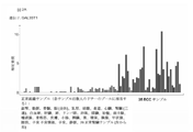


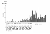


Classifications
-
- A—HUMAN NECESSITIES
- A61—MEDICAL OR VETERINARY SCIENCE; HYGIENE
- A61K—PREPARATIONS FOR MEDICAL, DENTAL OR TOILETRY PURPOSES
- A61K39/00—Medicinal preparations containing antigens or antibodies
- A61K39/0005—Vertebrate antigens
- A61K39/0011—Cancer antigens
-
- A—HUMAN NECESSITIES
- A61—MEDICAL OR VETERINARY SCIENCE; HYGIENE
- A61K—PREPARATIONS FOR MEDICAL, DENTAL OR TOILETRY PURPOSES
- A61K35/00—Medicinal preparations containing materials or reaction products thereof with undetermined constitution
- A61K35/12—Materials from mammals; Compositions comprising non-specified tissues or cells; Compositions comprising non-embryonic stem cells; Genetically modified cells
- A61K35/14—Blood; Artificial blood
- A61K35/17—Lymphocytes; B-cells; T-cells; Natural killer cells; Interferon-activated or cytokine-activated lymphocytes
-
- A—HUMAN NECESSITIES
- A61—MEDICAL OR VETERINARY SCIENCE; HYGIENE
- A61K—PREPARATIONS FOR MEDICAL, DENTAL OR TOILETRY PURPOSES
- A61K38/00—Medicinal preparations containing peptides
-
- A—HUMAN NECESSITIES
- A61—MEDICAL OR VETERINARY SCIENCE; HYGIENE
- A61K—PREPARATIONS FOR MEDICAL, DENTAL OR TOILETRY PURPOSES
- A61K39/00—Medicinal preparations containing antigens or antibodies
- A61K39/0005—Vertebrate antigens
- A61K39/0011—Cancer antigens
- A61K39/001102—Receptors, cell surface antigens or cell surface determinants
- A61K39/001111—Immunoglobulin superfamily
-
- A—HUMAN NECESSITIES
- A61—MEDICAL OR VETERINARY SCIENCE; HYGIENE
- A61K—PREPARATIONS FOR MEDICAL, DENTAL OR TOILETRY PURPOSES
- A61K39/00—Medicinal preparations containing antigens or antibodies
- A61K39/46—Cellular immunotherapy
- A61K39/461—Cellular immunotherapy characterised by the cell type used
- A61K39/4611—T-cells, e.g. tumor infiltrating lymphocytes [TIL], lymphokine-activated killer cells [LAK] or regulatory T cells [Treg]
-
- A—HUMAN NECESSITIES
- A61—MEDICAL OR VETERINARY SCIENCE; HYGIENE
- A61K—PREPARATIONS FOR MEDICAL, DENTAL OR TOILETRY PURPOSES
- A61K39/00—Medicinal preparations containing antigens or antibodies
- A61K39/46—Cellular immunotherapy
- A61K39/464—Cellular immunotherapy characterised by the antigen targeted or presented
- A61K39/4643—Vertebrate antigens
- A61K39/4644—Cancer antigens
-
- A—HUMAN NECESSITIES
- A61—MEDICAL OR VETERINARY SCIENCE; HYGIENE
- A61P—SPECIFIC THERAPEUTIC ACTIVITY OF CHEMICAL COMPOUNDS OR MEDICINAL PREPARATIONS
- A61P35/00—Antineoplastic agents
-
- A—HUMAN NECESSITIES
- A61—MEDICAL OR VETERINARY SCIENCE; HYGIENE
- A61P—SPECIFIC THERAPEUTIC ACTIVITY OF CHEMICAL COMPOUNDS OR MEDICINAL PREPARATIONS
- A61P35/00—Antineoplastic agents
- A61P35/02—Antineoplastic agents specific for leukemia
-
- C—CHEMISTRY; METALLURGY
- C07—ORGANIC CHEMISTRY
- C07K—PEPTIDES
- C07K14/00—Peptides having more than 20 amino acids; Gastrins; Somatostatins; Melanotropins; Derivatives thereof
- C07K14/435—Peptides having more than 20 amino acids; Gastrins; Somatostatins; Melanotropins; Derivatives thereof from animals; from humans
- C07K14/44—Peptides having more than 20 amino acids; Gastrins; Somatostatins; Melanotropins; Derivatives thereof from animals; from humans from protozoa
- C07K14/445—Plasmodium
-
- C—CHEMISTRY; METALLURGY
- C07—ORGANIC CHEMISTRY
- C07K—PEPTIDES
- C07K14/00—Peptides having more than 20 amino acids; Gastrins; Somatostatins; Melanotropins; Derivatives thereof
- C07K14/435—Peptides having more than 20 amino acids; Gastrins; Somatostatins; Melanotropins; Derivatives thereof from animals; from humans
- C07K14/46—Peptides having more than 20 amino acids; Gastrins; Somatostatins; Melanotropins; Derivatives thereof from animals; from humans from vertebrates
- C07K14/47—Peptides having more than 20 amino acids; Gastrins; Somatostatins; Melanotropins; Derivatives thereof from animals; from humans from vertebrates from mammals
- C07K14/4701—Peptides having more than 20 amino acids; Gastrins; Somatostatins; Melanotropins; Derivatives thereof from animals; from humans from vertebrates from mammals not used
- C07K14/4748—Tumour specific antigens; Tumour rejection antigen precursors [TRAP], e.g. MAGE
-
- C—CHEMISTRY; METALLURGY
- C07—ORGANIC CHEMISTRY
- C07K—PEPTIDES
- C07K14/00—Peptides having more than 20 amino acids; Gastrins; Somatostatins; Melanotropins; Derivatives thereof
- C07K14/435—Peptides having more than 20 amino acids; Gastrins; Somatostatins; Melanotropins; Derivatives thereof from animals; from humans
- C07K14/705—Receptors; Cell surface antigens; Cell surface determinants
- C07K14/70503—Immunoglobulin superfamily
- C07K14/7051—T-cell receptor (TcR)-CD3 complex
-
- C—CHEMISTRY; METALLURGY
- C07—ORGANIC CHEMISTRY
- C07K—PEPTIDES
- C07K14/00—Peptides having more than 20 amino acids; Gastrins; Somatostatins; Melanotropins; Derivatives thereof
- C07K14/435—Peptides having more than 20 amino acids; Gastrins; Somatostatins; Melanotropins; Derivatives thereof from animals; from humans
- C07K14/705—Receptors; Cell surface antigens; Cell surface determinants
- C07K14/70503—Immunoglobulin superfamily
- C07K14/70539—MHC-molecules, e.g. HLA-molecules
-
- C—CHEMISTRY; METALLURGY
- C07—ORGANIC CHEMISTRY
- C07K—PEPTIDES
- C07K16/00—Immunoglobulins [IGs], e.g. monoclonal or polyclonal antibodies
- C07K16/18—Immunoglobulins [IGs], e.g. monoclonal or polyclonal antibodies against material from animals or humans
-
- C—CHEMISTRY; METALLURGY
- C07—ORGANIC CHEMISTRY
- C07K—PEPTIDES
- C07K16/00—Immunoglobulins [IGs], e.g. monoclonal or polyclonal antibodies
- C07K16/18—Immunoglobulins [IGs], e.g. monoclonal or polyclonal antibodies against material from animals or humans
- C07K16/28—Immunoglobulins [IGs], e.g. monoclonal or polyclonal antibodies against material from animals or humans against receptors, cell surface antigens or cell surface determinants
- C07K16/2803—Immunoglobulins [IGs], e.g. monoclonal or polyclonal antibodies against material from animals or humans against receptors, cell surface antigens or cell surface determinants against the immunoglobulin superfamily
- C07K16/2833—Immunoglobulins [IGs], e.g. monoclonal or polyclonal antibodies against material from animals or humans against receptors, cell surface antigens or cell surface determinants against the immunoglobulin superfamily against MHC-molecules, e.g. HLA-molecules
-
- C—CHEMISTRY; METALLURGY
- C07—ORGANIC CHEMISTRY
- C07K—PEPTIDES
- C07K16/00—Immunoglobulins [IGs], e.g. monoclonal or polyclonal antibodies
- C07K16/18—Immunoglobulins [IGs], e.g. monoclonal or polyclonal antibodies against material from animals or humans
- C07K16/28—Immunoglobulins [IGs], e.g. monoclonal or polyclonal antibodies against material from animals or humans against receptors, cell surface antigens or cell surface determinants
- C07K16/30—Immunoglobulins [IGs], e.g. monoclonal or polyclonal antibodies against material from animals or humans against receptors, cell surface antigens or cell surface determinants from tumour cells
- C07K16/3038—Kidney, bladder
-
- C—CHEMISTRY; METALLURGY
- C07—ORGANIC CHEMISTRY
- C07K—PEPTIDES
- C07K7/00—Peptides having 5 to 20 amino acids in a fully defined sequence; Derivatives thereof
- C07K7/04—Linear peptides containing only normal peptide links
- C07K7/06—Linear peptides containing only normal peptide links having 5 to 11 amino acids
-
- C—CHEMISTRY; METALLURGY
- C07—ORGANIC CHEMISTRY
- C07K—PEPTIDES
- C07K7/00—Peptides having 5 to 20 amino acids in a fully defined sequence; Derivatives thereof
- C07K7/04—Linear peptides containing only normal peptide links
- C07K7/08—Linear peptides containing only normal peptide links having 12 to 20 amino acids
-
- C—CHEMISTRY; METALLURGY
- C12—BIOCHEMISTRY; BEER; SPIRITS; WINE; VINEGAR; MICROBIOLOGY; ENZYMOLOGY; MUTATION OR GENETIC ENGINEERING
- C12N—MICROORGANISMS OR ENZYMES; COMPOSITIONS THEREOF; PROPAGATING, PRESERVING, OR MAINTAINING MICROORGANISMS; MUTATION OR GENETIC ENGINEERING; CULTURE MEDIA
- C12N15/00—Mutation or genetic engineering; DNA or RNA concerning genetic engineering, vectors, e.g. plasmids, or their isolation, preparation or purification; Use of hosts therefor
- C12N15/09—Recombinant DNA-technology
- C12N15/11—DNA or RNA fragments; Modified forms thereof; Non-coding nucleic acids having a biological activity
- C12N15/115—Aptamers, i.e. nucleic acids binding a target molecule specifically and with high affinity without hybridising therewith ; Nucleic acids binding to non-nucleic acids, e.g. aptamers
-
- C—CHEMISTRY; METALLURGY
- C12—BIOCHEMISTRY; BEER; SPIRITS; WINE; VINEGAR; MICROBIOLOGY; ENZYMOLOGY; MUTATION OR GENETIC ENGINEERING
- C12N—MICROORGANISMS OR ENZYMES; COMPOSITIONS THEREOF; PROPAGATING, PRESERVING, OR MAINTAINING MICROORGANISMS; MUTATION OR GENETIC ENGINEERING; CULTURE MEDIA
- C12N5/00—Undifferentiated human, animal or plant cells, e.g. cell lines; Tissues; Cultivation or maintenance thereof; Culture media therefor
- C12N5/06—Animal cells or tissues; Human cells or tissues
- C12N5/0602—Vertebrate cells
- C12N5/0634—Cells from the blood or the immune system
- C12N5/0636—T lymphocytes
- C12N5/0638—Cytotoxic T lymphocytes [CTL] or lymphokine activated killer cells [LAK]
-
- C—CHEMISTRY; METALLURGY
- C12—BIOCHEMISTRY; BEER; SPIRITS; WINE; VINEGAR; MICROBIOLOGY; ENZYMOLOGY; MUTATION OR GENETIC ENGINEERING
- C12Q—MEASURING OR TESTING PROCESSES INVOLVING ENZYMES, NUCLEIC ACIDS OR MICROORGANISMS; COMPOSITIONS OR TEST PAPERS THEREFOR; PROCESSES OF PREPARING SUCH COMPOSITIONS; CONDITION-RESPONSIVE CONTROL IN MICROBIOLOGICAL OR ENZYMOLOGICAL PROCESSES
- C12Q1/00—Measuring or testing processes involving enzymes, nucleic acids or microorganisms; Compositions therefor; Processes of preparing such compositions
- C12Q1/68—Measuring or testing processes involving enzymes, nucleic acids or microorganisms; Compositions therefor; Processes of preparing such compositions involving nucleic acids
- C12Q1/6876—Nucleic acid products used in the analysis of nucleic acids, e.g. primers or probes
- C12Q1/6883—Nucleic acid products used in the analysis of nucleic acids, e.g. primers or probes for diseases caused by alterations of genetic material
- C12Q1/6886—Nucleic acid products used in the analysis of nucleic acids, e.g. primers or probes for diseases caused by alterations of genetic material for cancer
-
- G—PHYSICS
- G16—INFORMATION AND COMMUNICATION TECHNOLOGY [ICT] SPECIALLY ADAPTED FOR SPECIFIC APPLICATION FIELDS
- G16B—BIOINFORMATICS, i.e. INFORMATION AND COMMUNICATION TECHNOLOGY [ICT] SPECIALLY ADAPTED FOR GENETIC OR PROTEIN-RELATED DATA PROCESSING IN COMPUTATIONAL MOLECULAR BIOLOGY
- G16B25/00—ICT specially adapted for hybridisation; ICT specially adapted for gene or protein expression
- G16B25/10—Gene or protein expression profiling; Expression-ratio estimation or normalisation
-
- G—PHYSICS
- G16—INFORMATION AND COMMUNICATION TECHNOLOGY [ICT] SPECIALLY ADAPTED FOR SPECIFIC APPLICATION FIELDS
- G16B—BIOINFORMATICS, i.e. INFORMATION AND COMMUNICATION TECHNOLOGY [ICT] SPECIALLY ADAPTED FOR GENETIC OR PROTEIN-RELATED DATA PROCESSING IN COMPUTATIONAL MOLECULAR BIOLOGY
- G16B30/00—ICT specially adapted for sequence analysis involving nucleotides or amino acids
-
- G—PHYSICS
- G16—INFORMATION AND COMMUNICATION TECHNOLOGY [ICT] SPECIALLY ADAPTED FOR SPECIFIC APPLICATION FIELDS
- G16C—COMPUTATIONAL CHEMISTRY; CHEMOINFORMATICS; COMPUTATIONAL MATERIALS SCIENCE
- G16C20/00—Chemoinformatics, i.e. ICT specially adapted for the handling of physicochemical or structural data of chemical particles, elements, compounds or mixtures
- G16C20/50—Molecular design, e.g. of drugs
-
- A—HUMAN NECESSITIES
- A61—MEDICAL OR VETERINARY SCIENCE; HYGIENE
- A61K—PREPARATIONS FOR MEDICAL, DENTAL OR TOILETRY PURPOSES
- A61K35/00—Medicinal preparations containing materials or reaction products thereof with undetermined constitution
- A61K35/12—Materials from mammals; Compositions comprising non-specified tissues or cells; Compositions comprising non-embryonic stem cells; Genetically modified cells
- A61K2035/124—Materials from mammals; Compositions comprising non-specified tissues or cells; Compositions comprising non-embryonic stem cells; Genetically modified cells the cells being hematopoietic, bone marrow derived or blood cells
-
- A—HUMAN NECESSITIES
- A61—MEDICAL OR VETERINARY SCIENCE; HYGIENE
- A61K—PREPARATIONS FOR MEDICAL, DENTAL OR TOILETRY PURPOSES
- A61K39/00—Medicinal preparations containing antigens or antibodies
- A61K2039/505—Medicinal preparations containing antigens or antibodies comprising antibodies
-
- A—HUMAN NECESSITIES
- A61—MEDICAL OR VETERINARY SCIENCE; HYGIENE
- A61K—PREPARATIONS FOR MEDICAL, DENTAL OR TOILETRY PURPOSES
- A61K39/00—Medicinal preparations containing antigens or antibodies
- A61K2039/51—Medicinal preparations containing antigens or antibodies comprising whole cells, viruses or DNA/RNA
- A61K2039/515—Animal cells
- A61K2039/5158—Antigen-pulsed cells, e.g. T-cells
-
- A—HUMAN NECESSITIES
- A61—MEDICAL OR VETERINARY SCIENCE; HYGIENE
- A61K—PREPARATIONS FOR MEDICAL, DENTAL OR TOILETRY PURPOSES
- A61K39/00—Medicinal preparations containing antigens or antibodies
- A61K2039/51—Medicinal preparations containing antigens or antibodies comprising whole cells, viruses or DNA/RNA
- A61K2039/53—DNA (RNA) vaccination
-
- A—HUMAN NECESSITIES
- A61—MEDICAL OR VETERINARY SCIENCE; HYGIENE
- A61K—PREPARATIONS FOR MEDICAL, DENTAL OR TOILETRY PURPOSES
- A61K39/00—Medicinal preparations containing antigens or antibodies
- A61K2039/57—Medicinal preparations containing antigens or antibodies characterised by the type of response, e.g. Th1, Th2
- A61K2039/572—Medicinal preparations containing antigens or antibodies characterised by the type of response, e.g. Th1, Th2 cytotoxic response
-
- C—CHEMISTRY; METALLURGY
- C07—ORGANIC CHEMISTRY
- C07K—PEPTIDES
- C07K2317/00—Immunoglobulins specific features
- C07K2317/70—Immunoglobulins specific features characterized by effect upon binding to a cell or to an antigen
- C07K2317/73—Inducing cell death, e.g. apoptosis, necrosis or inhibition of cell proliferation
-
- C—CHEMISTRY; METALLURGY
- C07—ORGANIC CHEMISTRY
- C07K—PEPTIDES
- C07K2317/00—Immunoglobulins specific features
- C07K2317/70—Immunoglobulins specific features characterized by effect upon binding to a cell or to an antigen
- C07K2317/76—Antagonist effect on antigen, e.g. neutralization or inhibition of binding
-
- C—CHEMISTRY; METALLURGY
- C07—ORGANIC CHEMISTRY
- C07K—PEPTIDES
- C07K2319/00—Fusion polypeptide
-
- C—CHEMISTRY; METALLURGY
- C07—ORGANIC CHEMISTRY
- C07K—PEPTIDES
- C07K2319/00—Fusion polypeptide
- C07K2319/33—Fusion polypeptide fusions for targeting to specific cell types, e.g. tissue specific targeting, targeting of a bacterial subspecies
-
- C—CHEMISTRY; METALLURGY
- C07—ORGANIC CHEMISTRY
- C07K—PEPTIDES
- C07K2319/00—Fusion polypeptide
- C07K2319/55—Fusion polypeptide containing a fusion with a toxin, e.g. diphteria toxin
-
- C—CHEMISTRY; METALLURGY
- C12—BIOCHEMISTRY; BEER; SPIRITS; WINE; VINEGAR; MICROBIOLOGY; ENZYMOLOGY; MUTATION OR GENETIC ENGINEERING
- C12N—MICROORGANISMS OR ENZYMES; COMPOSITIONS THEREOF; PROPAGATING, PRESERVING, OR MAINTAINING MICROORGANISMS; MUTATION OR GENETIC ENGINEERING; CULTURE MEDIA
- C12N2310/00—Structure or type of the nucleic acid
- C12N2310/10—Type of nucleic acid
- C12N2310/16—Aptamers
-
- C—CHEMISTRY; METALLURGY
- C12—BIOCHEMISTRY; BEER; SPIRITS; WINE; VINEGAR; MICROBIOLOGY; ENZYMOLOGY; MUTATION OR GENETIC ENGINEERING
- C12N—MICROORGANISMS OR ENZYMES; COMPOSITIONS THEREOF; PROPAGATING, PRESERVING, OR MAINTAINING MICROORGANISMS; MUTATION OR GENETIC ENGINEERING; CULTURE MEDIA
- C12N2501/00—Active agents used in cell culture processes, e.g. differentation
- C12N2501/50—Cell markers; Cell surface determinants
-
- C—CHEMISTRY; METALLURGY
- C12—BIOCHEMISTRY; BEER; SPIRITS; WINE; VINEGAR; MICROBIOLOGY; ENZYMOLOGY; MUTATION OR GENETIC ENGINEERING
- C12Q—MEASURING OR TESTING PROCESSES INVOLVING ENZYMES, NUCLEIC ACIDS OR MICROORGANISMS; COMPOSITIONS OR TEST PAPERS THEREFOR; PROCESSES OF PREPARING SUCH COMPOSITIONS; CONDITION-RESPONSIVE CONTROL IN MICROBIOLOGICAL OR ENZYMOLOGICAL PROCESSES
- C12Q2600/00—Oligonucleotides characterized by their use
- C12Q2600/158—Expression markers
Description
本発明は、免疫療法において使用するためのペプチド、タンパク質、核酸、および細胞に関する。特に、本発明は、がんの免疫療法に関する。本発明は、単独のまたはその他の腫瘍関連ペプチドと組み合わされた、腫瘍関連T細胞ペプチドエピトープにさらに関し、それは、例えば、抗腫瘍免疫応答を刺激し、または生体外でT細胞を刺激して患者に移入する、ワクチン組成物の活性医薬品成分の役割を果たし得る。主要組織適合性複合体(MHC)の分子と結合しているペプチド、またはペプチドそれ自体もまた、抗体、可溶性T細胞受容体、およびその他の結合分子の標的になり得る。 The present invention relates to peptides, proteins, nucleic acids and cells for use in immunotherapy. In particular, the present invention relates to cancer immunotherapy. The invention further relates to tumor-associated T-cell peptide epitopes, alone or in combination with other tumor-associated peptides, which, for example, stimulate anti-tumor immune responses or stimulate T-cells ex vivo to can serve as the active pharmaceutical ingredient of the vaccine composition, transfecting the Peptides bound to molecules of the major histocompatibility complex (MHC), or peptides themselves, can also be targeted by antibodies, soluble T-cell receptors, and other binding molecules.
本発明は、ヒト腫瘍細胞のHLAクラスI分子に由来する、いくつかの新規ペプチド配列およびそれらの変異型に関し、それらは抗腫瘍免疫応答を引き起こすためのワクチン組成物中で、または薬理的/免疫学的活性化合物および細胞の開発のための標的として、使用され得る。 The present invention relates to several novel peptide sequences and variants thereof derived from HLA class I molecules of human tumor cells, which may be used in vaccine compositions or for pharmacological/immunological purposes to elicit an anti-tumor immune response. can be used as targets for the development of biologically active compounds and cells.
腎臓がんは女性より男性でより一般的であり、2012年には世界的に、男性では9番目に一般的ながんであり(214,000症例)、女性では14番目に一般的ながんである(124,000症例)。新規症例の70%は先進国および高度先進国で出現し、新規症例の34%は欧州にあり、新規症例の19%は北米にあると推定される。2012年における腎臓がんによる推定死亡者は、143,000人であった(男性91,000人、女性52,000人);腎臓がんは、全世界のがん死亡の第16番目に一般的な原因である。 Kidney cancer is more common in men than women and was the 9th most common cancer in men (214,000 cases) and the 14th most common cancer in women worldwide in 2012. Yes (124,000 cases). It is estimated that 70% of new cases appear in developed and highly developed countries, 34% of new cases are in Europe, and 19% of new cases are in North America. The estimated death toll from kidney cancer in 2012 was 143,000 (91,000 men, 52,000 women); kidney cancer is the 16th most common cancer death worldwide. is the cause.
最も高い発生率は、チェコ共和国で見られる。高い発生率は、北欧、東欧、北米、オーストラリアでも見られる。アフリカや東アジアの多くでは、低い発生率が推定されている。症例死亡率は、中進国または開発途上国(全体的な死亡対罹患率、0.5)よりも、高度先進国(0.4)においてより低い。症例の3.1%のみがアフリカで診断されたが、この地域では死亡の5.7%が発生した。発生率と死亡率は、開発程度が異なる多くの国々において増大している(World Cancer Report,2014)。 The highest incidence is found in the Czech Republic. High incidence is also found in Northern and Eastern Europe, North America and Australia. Low incidence is estimated in much of Africa and East Asia. Case-fatality rates are lower in highly developed countries (0.4) than in semi-developed or developing countries (overall mortality-to-morbidity ratio, 0.5). Only 3.1% of cases were diagnosed in Africa, but 5.7% of deaths occurred in this region. Incidence and mortality are increasing in many countries with different degrees of development (World Cancer Report, 2014).
大部分の腎臓がんは、腎実質内の異なる細胞型から生じる腫瘍の不均一なクラスである、腎細胞がん(RCC)である。大部分は明細胞腎臓がん(腎臓がん症例の約70%)であり、乳頭状(10~15%)、色素嫌性(約5%)、集合尿細管(<1%)腎細胞がんがそれに続く。これらの腎細胞腫瘍サブタイプのそれぞれは、異なる遺伝的特徴を有する(Moch,2013;World Health Organization Classification of Tumours,2004)。 Most kidney cancers are renal cell carcinomas (RCCs), a heterogeneous class of tumors arising from different cell types within the renal parenchyma. The majority are clear cell renal carcinomas (approximately 70% of renal cancer cases), with papillary (10-15%), chromophobic (approximately 5%), collecting tubular (<1%) renal cells followed by n. Each of these renal cell tumor subtypes has distinct genetic characteristics (Moch, 2013; World Health Organization Classification of Tumors, 2004).
腎細胞がん(RCC)は、早期警告徴候の欠如、多様な臨床徴候、および放射線および化学療法に対する抵抗性によって特徴付けられる。RCCを有する患者の合計25~30%が、最初に明らかな転移を呈する(Hofmann et al.,2005)。RCC患者の約3分の1は、時間経過と共に転移性疾患を発症する。したがって、RCC患者のほぼ50~60%が最終的に転移性疾患を呈する(Bleumer et al.,2003;Hofmann et al.,2005)。転移性疾患がある者のうち、およそ75%が肺転移、36%がリンパ節および/または軟部組織合併症、20%が骨合併症、および18%が肝臓合併症を有する(Sachdeva et al.,2010)。 Renal cell carcinoma (RCC) is characterized by a lack of early warning signs, variable clinical manifestations, and resistance to radiation and chemotherapy. A total of 25-30% of patients with RCC initially present with overt metastases (Hofmann et al., 2005). About one-third of RCC patients develop metastatic disease over time. Therefore, approximately 50-60% of RCC patients will eventually develop metastatic disease (Bleumer et al., 2003; Hofmann et al., 2005). Of those with metastatic disease, approximately 75% have lung metastases, 36% have lymph node and/or soft tissue complications, 20% have bone complications, and 18% have liver complications (Sachdeva et al. , 2010).
サイトカインベースの第一選択全身療法を受けている、転移性疾患があるRCC患者は、5つの予後因子に基づいて、生存を予測するリスク群に分類され得る(Motzer et al.,2004)。より短い生存率に関連する治療前の特徴は、カルノフスキー・パフォーマンス・ステータス(<80%)、高血清乳酸デヒドロゲナーゼ(>1.5ULN)、低ヘモグロビン(<LLN)、高い補正血清カルシウム(>10mg/dL)、1年未満の診断から治療までの期間である。これらのリスク因子に基づいて、患者は3つのリスク群に分類された。リスク因子ゼロ(好ましいリスク)がある患者の18%における、死亡までの中位時間は30ヶ月であった。患者の62%は、1つまたは2つのリスク因子(中程度のリスク)を有し、この群の生存期間中央値は14ヶ月であった。患者の20%を構成する3つ以上のリスク因子(芳しくないリスク)がある患者は、5ヶ月間の生存期間中央値を有した。このMSKCCリスク群分類の適用は、進行したRCCの臨床試験に広く用いられている。リスク分類は、臨床試験の計画および結果の解釈、治療の方向付けのために使用し得る。 RCC patients with metastatic disease receiving cytokine-based first-line systemic therapy can be classified into risk groups that predict survival based on five prognostic factors (Motzer et al., 2004). Pretreatment characteristics associated with shorter survival were Karnofsky performance status (<80%), high serum lactate dehydrogenase (>1.5 ULN), low hemoglobin (<LLN), high corrected serum calcium (>10 mg). /dL), the time from diagnosis to treatment of <1 year. Based on these risk factors, patients were classified into three risk groups. Median time to death was 30 months in 18% of patients with zero risk factor (favorable risk). Sixty-two percent of patients had 1 or 2 risk factors (intermediate risk) and the median survival in this group was 14 months. Patients with 3 or more risk factors (poor risk), comprising 20% of patients, had a median survival time of 5 months. This application of the MSKCC risk group classification has been widely used in advanced RCC clinical trials. Risk classification can be used for designing clinical trials, interpreting results, and directing treatment.
RCCのリスク要因は、タバコの喫煙および肥満である。様々なメタ分析が、喫煙常習は非喫煙と比較して、腎臓がんのリスクを高めることを確認した(Cho et al.,2011;Hunt et al.,2005)。1日当たりの喫煙タバコ本数と関連がある、リスクの用量依存性増大もある。リスクは、禁煙後5年間で低下する。過体重、特に肥満は、女性と男性の双方にとって腎臓がんのリスク因子である(Ljungberg et al.,2011)。過体重および肥満に帰せられる腎臓がんの全ての症例の比率は、米国では約40%、欧州諸国では最高40%と推定されている(Renehan et al.,2008;Renehan et al.,2010)。肥満が腎臓の発がんに影響を与える機序は、不明確である。性ステロイドホルモンは、直接的な内分泌受容体媒介効果によって、腎細胞の増殖に影響を及ぼしてもよい。性ホルモン結合性グロブリンおよびプロゲステロンのレベルの低下、インスリン抵抗性、およびインスリン様成長因子1(IGF-1)などの成長因子のレベル増大などの内分泌疾患を併発する肥満は、腎臓の発がんに寄与してもよい。近年、症例対照研究が、明細胞がんと肥満とのより強い関連性を報告している(World Cancer Report,2014)。 Risk factors for RCC are cigarette smoking and obesity. Various meta-analyses have confirmed that regular smokers increase the risk of kidney cancer compared to nonsmokers (Cho et al., 2011; Hunt et al., 2005). There is also a dose-dependent increase in risk associated with the number of cigarettes smoked per day. Risk decreases 5 years after quitting smoking. Overweight, especially obesity, is a risk factor for kidney cancer for both women and men (Ljungberg et al., 2011). The proportion of all kidney cancer cases attributable to overweight and obesity is estimated at about 40% in the United States and up to 40% in European countries (Renehan et al., 2008; Renehan et al., 2010). . The mechanism by which obesity affects renal carcinogenesis is unclear. Sex steroid hormones may affect renal cell proliferation through direct endocrine receptor-mediated effects. Obesity, combined with endocrine disorders such as decreased levels of sex hormone-binding globulin and progesterone, insulin resistance, and increased levels of growth factors such as insulin-like growth factor 1 (IGF-1), contribute to renal carcinogenesis. may Recently, case-control studies have reported a stronger association between clear cell carcinoma and obesity (World Cancer Report, 2014).
初期治療は、最も一般的には、罹患した腎臓の部分的または完全除去のどちらかであり、依然として根治的治療の主流である(Rini et al.,2008)。予後スコアの悪い患者の第一選択治療として、いくつかのがん団体および協会によって作成された指針は、インターフェロン-α(IFN-α)およびmTOR阻害剤テムシロリムス(Torisel(登録商標))と組み合わされた、受容体チロシンキナーゼ阻害物質(TKI)であるスニチニブ(Sutent(登録商標))およびパゾパニブ(Votrient(登録商標))、モノクローナル抗体であるベバシズマブ(Avastin(登録商標))を推奨する。米国NCCNならびに欧州EAUおよびESMOによって作成された指針に基づいて、サイトカイン(IFN-α、IL-2)による先行治療法が失敗したRCC患者では、TKIであるソラフェニブ、パゾパニブまたは最近ではアキシチニブが、第二選択治療として推奨される。NCCN指針は、この状況(NCCNカテゴリーIによる高レベルの証拠)において、スニチニブもまた勧める。 Initial treatment, most commonly, is either partial or complete removal of the affected kidney and remains the mainstay of curative therapy (Rini et al., 2008). Guidelines developed by several cancer groups and associations combine interferon-α (IFN-α) and the mTOR inhibitor temsirolimus (Torisel®) as first-line treatment for patients with poor prognostic scores. Also recommended are the receptor tyrosine kinase inhibitors (TKIs) sunitinib (Sutent®) and pazopanib (Votrient®), and the monoclonal antibody bevacizumab (Avastin®). Based on guidelines developed by the US NCCN and the European EAU and ESMO, the TKIs sorafenib, pazopanib or, more recently, axitinib are recommended in patients with RCC who have failed prior therapy with cytokines (IFN-α, IL-2). Recommended as a two-line treatment. NCCN guidelines also recommend sunitinib in this setting (high level of evidence by NCCN Category I).
エベロリムスおよびアキシチニブは、確立されたガイドラインに従ったTKIを用いたVEGF標的治療の恩恵を受けていない患者のための第2選択療法として推奨される。 Everolimus and axitinib are recommended as second-line therapy for patients who have not benefited from VEGF-targeted therapy with TKIs according to established guidelines.
RCCの既知の免疫原性(immunogenity)は、進行したRCCにおける免疫療法およびがんワクチンの使用を支持する基礎に相当する。 The known immunogenicity of RCC represents a basis for supporting the use of immunotherapy and cancer vaccines in advanced RCC.
リンパ球PD-1発現とRCCの進行期、等級、および予後との間の興味深い相関、ならびにRCC腫瘍細胞による選択的PD-L1発現と、より芳しくない臨床転帰との潜在的関連性は、単独のまたは抗血管新生薬またはその他の免疫療法アプローチと併用される、RCC治療のための新規抗PD-1/PD-L1薬の開発をもたらした(Massari et al.,2015)。 Interesting correlations between lymphocyte PD-1 expression and RCC stage, grade, and prognosis, as well as the potential association of selective PD-L1 expression by RCC tumor cells with poorer clinical outcomes, have been reported alone. led to the development of novel anti-PD-1/PD-L1 agents for the treatment of RCC, either in combination with antiangiogenic agents or other immunotherapeutic approaches (Massari et al., 2015).
進行したRCCでは、TRIST試験と称される第III相がんワクチン治験が、第一選択標準的治療に追加された、TroVax(ポックスウイルスベクターと共に腫瘍関連抗原である5T4を使用したワクチン)が、局所的進行またはmRCCがある患者の生存期間を延長するかどうかを評価する。いずれの群でも生存期間中央値に達しておらず、399人の患者(54%)が試験に残っているが、データの解析は、TroVaxが免疫学的に活性であり、5T4特異的抗体応答の強度と改善された生存期間との間に相関があることの双方を実証する、以前の臨床結果を裏付ける。さらにRCCにおいて過剰発現されるエピトープを使用してペプチドワクチンを検索する、いくつかの研究がある。 In advanced RCC, a phase III cancer vaccine trial called the TRIST trial added TroVax (a vaccine that uses the tumor-associated antigen 5T4 with a poxvirus vector) to the first-line standard of care. To assess whether it prolongs survival in patients with local progression or mRCC. Median survival was not reached in either group, with 399 patients (54%) remaining in the trial, but analysis of the data indicated that TroVax was immunologically active and that 5T4-specific antibody responses corroborates previous clinical results demonstrating both that there is a correlation between the intensity of thrombocytopenia and improved survival. In addition, there are several studies searching for peptide vaccines using epitopes overexpressed in RCC.
腫瘍ワクチンの様々なアプローチが、研究されている。腫瘍細胞溶解産物、樹状細胞と腫瘍細胞の融合物、または全腫瘍RNAをはじめとする、全腫瘍アプローチを使用した研究が、RCC患者において実施され、これらの治験のいくつかで腫瘍病変の寛解が報告された(Avigan et al.,2004;Holtl et al.,2002;Marten et al.,2002;Su et al.,2003;Wittig et al.,2001)。 Various approaches to tumor vaccines have been investigated. Studies using whole-tumor approaches, including tumor cell lysates, dendritic cell-tumor cell fusions, or total tumor RNA, have been performed in patients with RCC and some of these trials have demonstrated remission of tumor lesions. was reported (Avigan et al., 2004; Holtl et al., 2002; Marten et al., 2002; Su et al., 2003; Wittig et al., 2001).
ここ数年間に、RCCで発現され、抗原特異的CTLによって認識されるいくつかのヒトTAAが、発現クローニング、逆免疫学アプローチ、またはDNAマイクロアレイ技術を使用して定義され、特徴付けられている(Dannenmann et al.,2013;Michael and Pandha,2003;Minami et al.,2014;Renkvist et al.,2001;Wierecky et al.,2006)。 In the last few years, several human TAAs expressed in RCC and recognized by antigen-specific CTL have been defined and characterized using expression cloning, reverse immunology approaches, or DNA microarray technology ( Dannenmann et al., 2013; Michael and Pandha, 2003; Minami et al., 2014; Renkvist et al., 2001; Wierecky et al., 2006).
がんの治療に伴う重度の副作用および費用を考慮すると、がん全般、そして特にRCCの治療に使用し得る要素を同定する必要がある。がんのより良い診断、予後の評価、および治療成功の予測につながる、がん全般、特にRCCのためのバイオマーカーに相当する要素を同定する必要性もまたある。 Given the severe side effects and costs associated with cancer treatment, there is a need to identify agents that can be used to treat cancer in general and RCC in particular. There is also a need to identify elements that represent biomarkers for cancer in general, and RCC in particular, that will lead to better diagnosis of cancer, assessment of prognosis, and prediction of treatment success.
がんの免疫療法は、がん細胞を特異的に標的化しながら副作用を最小化する選択肢に相当する。がん免疫療法は、腫瘍関連抗原の存在を利用する。 Cancer immunotherapy represents an option that specifically targets cancer cells while minimizing side effects. Cancer immunotherapy exploits the presence of tumor-associated antigens.
腫瘍関連抗原(TAA)の現行の分類は、次の主要群を含んでなる:
a)がん精巣抗原:T細胞によって認識され得る、これまでに同定された最初のTAAは、このクラスに属し、元々はがん精巣(CT)抗原と称されたが、それは、そのメンバーが組織学的に異なるヒト腫瘍において発現し、正常組織では、精巣の精母細胞/精原細胞のみに存在し、時として胎盤に存在するためであった。精巣の細胞は、クラスIおよびII HLA分子を発現しないので、これらの抗原は正常組織のT細胞によって認識され得ず、したがって免疫学的に腫瘍特異的と見なされる。CT抗原の周知の例は、MAGEファミリーメンバーおよびNY-ESO-1である。
b)分化抗原:これらのTAAは、腫瘍と、それから腫瘍が生じる正常組織との間で共有される。既知の分化抗原のほとんどは、メラノーマおよび正常メラノサイトに見られる。これらのメラノサイト系関連タンパク質の多くは、メラニン生合成に関与し、したがって腫瘍特異的でないが、それでもなおがん免疫療法のために広く利用されている。例としては、メラノーマに対するチロシナーゼとMelan-A/MART-1、または前立腺がんに対するPSAが挙げられるが、これに限定されるものではない。
c)過剰発現されたTAA:広範に発現されるTAAをエンコードする遺伝子は、組織学的に異なる型の腫瘍において検出され、多数の正常組織においても概してより低い発現レベルで検出されている。正常組織によってプロセスされて潜在的に提示され得るエピトープの多くは、T細胞認識の閾値レベル未満であり得る一方で、腫瘍細胞におけるそれらの過剰発現は、先に確立された免疫寛容の破壊による抗がん応答を始動し得る。このクラスのTAAの顕著な例は、Her-2/neu、サバイビン、テロメラーゼまたはWT1である。
d)腫瘍特異的抗原:これらのユニークなTAAは、正常な遺伝子(β-カテニン、CDK4など)の変異から生じる。これらの分子変化のいくつかは、腫瘍性形質転換および/または進行に関連する。腫瘍特異的抗原は、通常、正常組織に対する自己免疫反応のリスクなしに、強力な免疫応答を誘導できる。他方、これらのTAAは、ほとんどの場合、その上でそれらが同定されたまさにその腫瘍のみと関係があり、通常は、多くの個々の腫瘍間で共有されない。腫瘍特異的(関連)イソ型があるタンパク質では、ペプチドの腫瘍特異性(または関連性)はまた、ペプチドが腫瘍(関連)エクソンに由来する場合に生じてもよい。
e)異常な翻訳後修飾から生じるTAA:このようなTAAは、特異的でなく腫瘍において過剰発現もされないタンパク質から生じてもよいが、それでもなお、腫瘍において主に活性である翻訳後プロセスによって腫瘍関連になる。このクラスの例は、腫瘍にMUC1のような新規エピトープをもたらす改変グリコシル化パターン、または腫瘍特異的であってもなくてもよい分解中のタンパク質スプライシングのような事象から生じる。
f)オンコウイルスタンパク質:これらのTAAはウイルスタンパク質であり、それらは発がん過程において重要な役割を果たしてもよく、外来性である(ヒト由来でない)ため、それらはT細胞応答を誘起し得る。このようなタンパク質の例は、子宮頸がんにおいて発現されるヒト乳頭腫16型ウイルスタンパク質E6およびE7である。
The current classification of tumor-associated antigens (TAAs) comprises the following major groups:
a) Cancer Testis Antigen: The first TAA identified so far that can be recognized by T cells belongs to this class, originally called cancer testis (CT) antigen, because its members are This was because it is expressed in histologically distinct human tumors and in normal tissues is present only in spermatocytes/spermatogonia of the testis and occasionally in the placenta. Since testicular cells do not express class I and II HLA molecules, these antigens cannot be recognized by normal tissue T cells and are therefore considered immunologically tumor-specific. Well-known examples of CT antigens are MAGE family members and NY-ESO-1.
b) Differentiation Antigens: These TAAs are shared between tumors and normal tissue from which tumors arise. Most of the known differentiation antigens are found on melanoma and normal melanocytes. Many of these melanocyte system-associated proteins are involved in melanin biosynthesis and are therefore not tumor-specific, but are nonetheless widely used for cancer immunotherapy. Examples include, but are not limited to, tyrosinase and Melan-A/MART-1 for melanoma, or PSA for prostate cancer.
c) Overexpressed TAAs: Genes encoding ubiquitously expressed TAAs have been detected in histologically distinct types of tumors, and also at generally lower expression levels in many normal tissues. While many of the epitopes that can be processed and potentially presented by normal tissues may be below the threshold level for T-cell recognition, their overexpression in tumor cells may lead to anti-inflammatory effects through disruption of previously established immune tolerance. May initiate a cancer response. Prominent examples of this class of TAAs are Her-2/neu, survivin, telomerase or WT1.
d) Tumor-specific antigens: These unique TAAs arise from mutations of normal genes (β-catenin, CDK4, etc.). Some of these molecular changes are associated with neoplastic transformation and/or progression. Tumor-specific antigens can usually induce strong immune responses without the risk of autoimmune reactions against normal tissues. On the other hand, these TAAs are most often associated only with the very tumor on which they are identified and are usually not shared among many individual tumors. For proteins with tumor-specific (relevant) isoforms, tumor specificity (or relevance) of a peptide may also occur if the peptide is derived from a tumor (relevant) exon.
e) TAAs arising from aberrant post-translational modifications: Such TAAs may arise from proteins that are neither specific nor overexpressed in tumors, but nevertheless are tumor-specific by post-translational processes that are predominantly active in tumors. become relevant. Examples of this class arise from events such as altered glycosylation patterns leading to novel epitopes such as MUC1 in tumors, or protein splicing during degradation that may or may not be tumor specific.
f) Oncoviral proteins: These TAAs are viral proteins, they may play an important role in oncogenic processes, and because they are foreign (not of human origin) they can elicit T cell responses. Examples of such proteins are the human papilloma type 16 virus proteins E6 and E7 expressed in cervical cancer.
T細胞ベースの免疫療法は、主要組織適合性複合体(MHC)の分子によって提示される、腫瘍関連または腫瘍特異的タンパク質由来ペプチドエピトープを標的とする。腫瘍特異的Tリンパ球によって認識される抗原、すなわちそれらのエピトープは、酵素、受容体、転写因子などの全てのタンパク質クラスに由来する分子であり得て、それはそれぞれの腫瘍細胞で発現されて、同一起源の非改変細胞と比較して、通常、上方制御される。 T cell-based immunotherapy targets tumor-associated or tumor-specific protein-derived peptide epitopes presented by molecules of the major histocompatibility complex (MHC). Antigens recognized by tumor-specific T lymphocytes, i.e. their epitopes, can be molecules from all protein classes such as enzymes, receptors, transcription factors, etc., which are expressed in the respective tumor cells, Usually upregulated compared to unmodified cells of the same origin.
MHC分子には、MHCクラスIおよびMHCクラスIIの2つのクラスがある。MHCクラスI分子はα重鎖およびβ2ミクログロブリンから構成され、MHCクラスII分子はαおよびβ鎖から構成される。それらの三次元立体構造は結合溝をもたらし、それはペプチドとの非共有結合相互作用のために使用される。 There are two classes of MHC molecules, MHC class I and MHC class II. MHC class I molecules are composed of α heavy chains and β2 microglobulin, and MHC class II molecules are composed of α and β chains. Their three-dimensional conformation provides a binding groove, which is used for non-covalent interactions with peptides.
MHCクラスI分子は、ほとんどの有核細胞上に見られる。それらは、主に内因性タンパク質、欠陥リボソーム産物(DRIP)、およびより大型のペプチドのタンパク質切断から得られる、ペプチドを提示する。しかし、エンドソームコンパートメントまたは外因性起源に由来するペプチドもまた、MHCクラスI分子上に頻繁に見られる。この非古典的様式のクラスI提示は、文献中で交差提示と称される(Brossart and Bevan,1997;Rock et al.,1990)。MHCクラスII分子は、大部分はプロフェッショナル抗原提示細胞(APC)に見られ、例えば、エンドサイトーシス中にAPCに取り込まれて引き続きプロセシングされる、外因性または膜貫通タンパク質のペプチドを主に提示する。 MHC class I molecules are found on most nucleated cells. They present peptides that result primarily from endogenous proteins, defective ribosomal products (DRIPs), and proteolytic cleavages of larger peptides. However, peptides derived from endosomal compartments or exogenous sources are also frequently found on MHC class I molecules. This non-classical mode of Class I presentation is referred to in the literature as cross-presentation (Brossart and Bevan, 1997; Rock et al., 1990). MHC class II molecules are mostly found in professional antigen-presenting cells (APCs) and primarily present peptides of exogenous or transmembrane proteins, e.g., that are taken up by APCs during endocytosis and subsequently processed. .
ペプチドとMHCクラスIの複合体が、適切なT細胞受容体(TCR)を有するCD8陽性T細胞によって認識される一方で、ペプチドとMHCクラスII分子の複合体は、適切なTCRを有するCD4陽性ヘルパーT細胞によって認識される。その結果、TCR、ペプチド、およびMHCは、化学量論的に1:1:1の量で存在することが良く知られている。 Peptide-MHC class I complexes are recognized by CD8-positive T cells with the appropriate T-cell receptor (TCR), whereas peptide-MHC class II molecule complexes are CD4-positive with the appropriate TCR. Recognized by helper T cells. As a result, it is well known that TCR, peptides and MHC are present in stoichiometric 1:1:1 amounts.
CD4陽性ヘルパーT細胞は、CD8陽性細胞傷害性T細胞による、効果的な応答の誘導と維持において重要な役割を果たす。腫瘍関連抗原(TAA)に由来するCD4陽性T細胞エピトープの同定は、抗腫瘍免疫応答を始動させる医薬品の開発に非常に重要である(Gnjatic et al.,2003)。腫瘍部位では、Tヘルパー細胞が、細胞毒性T細胞(CTL)親和的サイトカイン環境を維持して(Mortara et al.,2006)、例えば、CTL、ナチュラルキラー(NK)細胞、マクロファージ、および顆粒球などのエフェクター細胞を引きつける(Hwang et al.,2007)。 CD4-positive helper T cells play an important role in inducing and maintaining an effective response by CD8-positive cytotoxic T cells. Identification of CD4-positive T-cell epitopes derived from tumor-associated antigens (TAA) is of great importance for the development of drugs that trigger anti-tumor immune responses (Gnjatic et al., 2003). At tumor sites, T helper cells maintain a cytotoxic T cell (CTL)-friendly cytokine milieu (Mortara et al., 2006), e.g., CTLs, natural killer (NK) cells, macrophages, and granulocytes. of effector cells (Hwang et al., 2007).
炎症不在下では、MHCクラスII分子の発現は、免疫系細胞、特に、例えば、単球、単球由来細胞、マクロファージ、樹状細胞などのプロフェッショナル抗原提示細胞(APC)に主に限定される。がん患者においては、腫瘍細胞がMHCクラスII分子を発現することが判明している(Dengjel et al.,2006)。 In the absence of inflammation, expression of MHC class II molecules is primarily restricted to immune system cells, particularly professional antigen presenting cells (APCs) such as monocytes, monocyte-derived cells, macrophages, dendritic cells. In cancer patients, tumor cells have been found to express MHC class II molecules (Dengjel et al., 2006).
伸長された(より長い)本発明のペプチドは、MHCクラスII活性エピトープとして作用し得る。 Elongated (longer) peptides of the invention can act as MHC class II active epitopes.
MHCクラスIIエピトープによって活性化されたTヘルパー細胞は、抗腫瘍免疫におけるCTLのエフェクター機能を統合する上で、重要な役割を果たす。TH1型のTヘルパー細胞応答を始動するTヘルパー細胞エピトープは、し、それらの細胞表面に腫瘍関連ペプチド/MHC複合体を提示する腫瘍細胞に向けられた、細胞傷害機能をはじめとする、CD8陽性キラーT細胞のエフェクター機能を支持する。このようにして腫瘍関連Tヘルパー細胞ペプチドエピトープは、単独で、またはその他の腫瘍関連ペプチドとの組み合わせで、抗腫瘍免疫応答を刺激するワクチン組成物の活性医薬品成分の役割を果たし得る。 T helper cells activated by MHC class II epitopes play an important role in integrating the effector functions of CTLs in anti-tumor immunity. The T helper cell epitopes that initiate TH1-type T helper cell responses are CD8-positive, including cytotoxic functions directed against tumor cells presenting tumor-associated peptide/MHC complexes on their cell surface. Supports killer T cell effector functions. Thus, tumor-associated T helper cell peptide epitopes, alone or in combination with other tumor-associated peptides, can serve as active pharmaceutical ingredients of vaccine compositions that stimulate anti-tumor immune responses.
例えば、マウスなどの哺乳類動物モデルにおいて、CD8陽性Tリンパ球の不在下であっても、インターフェロンγ(IFNγ)の分泌による血管新生阻害を通じて腫瘍発現を阻害するには、CD4陽性T細胞で十分であることが示された(Beatty and Paterson,2001;Mumberg et al.,1999)。CD4 T細胞が、直接抗腫瘍エフェクターであるという証拠がある(Braumuller et al.,2013;Tran et al.,2014a)。 For example, in mammalian animal models such as mice, even in the absence of CD8-positive T-lymphocytes, CD4-positive T cells are sufficient to inhibit tumor development through inhibition of angiogenesis through secretion of interferon gamma (IFNγ). It has been shown that there is (Beatty and Paterson, 2001; Mumberg et al., 1999). There is evidence that CD4 T cells are direct anti-tumor effectors (Braumuller et al., 2013; Tran et al., 2014a).
HLAクラスII分子の構成的発現は、通常、免疫細胞に限定されるので、原発性腫瘍からクラスIIペプチドを直接単離する可能性があり得るとは、これまで考えられなかった。しかし、Dengjel et al.は、いくつかのMHCクラスIIエピトープを腫瘍から直接成功裏に同定した(国際公開第2007/028574号パンフレット、欧州特許第1760088B1号明細書)。 Since constitutive expression of HLA class II molecules is normally restricted to immune cells, the possibility of isolating class II peptides directly from primary tumors was not previously thought possible. However, Dengjel et al. successfully identified several MHC class II epitopes directly from tumors (WO2007/028574, EP1760088B1).
CD8およびCD4依存性の双方のタイプの応答は、抗腫瘍効果に共同して相乗的に寄与するので、CD8+T細胞(リガンド:MHCクラスI分子+ペプチドエピトープ)、またはCD4陽性Tヘルパー細胞(リガンド:MHCクラスII分子+ペプチドエピトープ)のどちらかによって認識される、腫瘍関連抗原の同定および特性解析は、腫瘍ワクチンの開発にとって重要である。 Since both CD8- and CD4-dependent types of responses jointly and synergistically contribute to the antitumor effect, CD8+ T cells (ligands: MHC class I molecules + peptide epitopes), or CD4+ T helper cells (ligands: Identification and characterization of tumor-associated antigens, recognized by either MHC class II molecules plus peptide epitopes), are important for the development of tumor vaccines.
MHCクラスIペプチドが、細胞性免疫応答を始動(惹起)するためには、それはまた、MHC分子に結合しなくてはならない。この過程は、MHC分子の対立遺伝子と、ペプチドのアミノ酸配列の特定の多形性とに依存する。MHCクラスI結合ペプチドは、通常は8~12アミノ酸残基長であり、通常は、MHC分子の対応する結合溝と相互作用するそれらの配列中に、2つの保存残基(「アンカー」)を含有する。このようにして、各MHC対立遺伝子は、どのペプチドが結合溝と特異的に結合し得るかを決定する、「結合モチーフ」を有する。 In order for an MHC class I peptide to initiate a cell-mediated immune response, it must also bind to MHC molecules. This process is dependent on alleles of the MHC molecule and specific polymorphisms in the amino acid sequence of the peptide. MHC class I binding peptides are usually 8-12 amino acid residues long and usually have two conserved residues (an "anchor") in their sequence that interact with the corresponding binding groove of the MHC molecule. contains. Thus, each MHC allele has a "binding motif" that determines which peptides can bind specifically to the binding groove.
MHCクラスI依存免疫反応においては、ペプチドは腫瘍細胞によって発現される特定のMHCクラスI分子に結合できるだけでなく、それらはまた、引き続いて特異的T細胞受容体(TCR)を有するT細胞によって認識されなくてはならない。 In an MHC class I dependent immune response, peptides not only can bind to specific MHC class I molecules expressed by tumor cells, they are also subsequently recognized by T cells with specific T cell receptors (TCR). must be.
タンパク質が、Tリンパ球によって腫瘍特異的または腫瘍関連抗原として認識され、治療で利用されるためには、特定の必要条件が満たされなくてはならない。抗原は、主に腫瘍細胞によって発現され、健常組織によって発現されず、または比較的少量発現されるべきである。好ましい実施形態では、ペプチドは、腫瘍細胞によって、健常組織と比較して過剰提示されるべきである。それぞれの抗原は、ある種の腫瘍に存在するだけでなく、高い濃度(すなわち、それぞれのペプチド細胞当たりのコピー数)で存在することもさらに望ましい。腫瘍特異的および腫瘍関連抗原は、例えば、細胞周期調節またはアポトーシス抑制における機能のために、正常細胞から腫瘍細胞への形質転換に直接関与するタンパク質に由来することが多い。さらに、形質転換の直接原因となるタンパク質の下流標的が、上方制御されてもよく、(und)したがって間接的に腫瘍関連であってもよい。このような間接的腫瘍関連抗原もまた、ワクチン接種アプローチの標的であってもよい(Singh-Jasuja et al.,2004)。このようなペプチド(「免疫原性ペプチド」)が、腫瘍関連抗原に由来して、生体外または生体内T細胞応答をもたらすことを確実にするためには、抗原のアミノ酸配列内にエピトープが存在することが必須である。 In order for a protein to be recognized as a tumor-specific or tumor-associated antigen by T lymphocytes and utilized therapeutically, certain requirements must be met. Antigens should be expressed primarily by tumor cells and not, or expressed in relatively small amounts, by healthy tissues. In preferred embodiments, the peptide should be over-presented by tumor cells compared to healthy tissue. It is further desirable that each antigen not only be present in certain tumors, but also at high concentrations (ie, copy number of each peptide per cell). Tumor-specific and tumor-associated antigens are often derived from proteins that are directly involved in the transformation of normal cells into tumor cells, eg, due to their function in cell cycle regulation or inhibition of apoptosis. In addition, downstream targets of proteins directly responsible for transformation may be upregulated and thus indirectly tumor-associated. Such indirect tumor-associated antigens may also be targets for vaccination approaches (Singh-Jasuja et al., 2004). To ensure that such peptides ("immunogenic peptides") are derived from tumor-associated antigens and elicit an in vitro or in vivo T cell response, epitopes must be present within the amino acid sequence of the antigen. It is essential to
基本的に、MHC分子に結合できるあらゆるペプチドが、T細胞エピトープとして機能してもよい。生体外または生体内T細胞応答誘導のための必要条件は、対応するTCRがあるT細胞の存在、およびこの特定のエピトープに対する免疫寛容の不在である。 Basically any peptide that can bind to an MHC molecule may serve as a T cell epitope. A prerequisite for in vitro or in vivo T cell response induction is the presence of T cells with the corresponding TCR and the absence of tolerance to this particular epitope.
したがって、TAAは、腫瘍ワクチンをはじめとするが、これに限定されるものではない、T細胞ベースの治療法開発の出発点である。TAAを同定し特性決定する方法は、通常は、患者または健常人から単離され得るT細胞の使用に基づき、またはそれらは、腫瘍および正常組織間の示差的転写プロファイル、または示差的ペプチド発現パターンの生成に基づく。しかし、腫瘍組織またはヒト腫瘍細胞株において過剰発現され、またはこのような組織または細胞株において選択的に発現される遺伝子の同定は、免疫療法においてこれらの遺伝子から転写される抗原の使用に関する、正確な情報を提供しない。これは、これらの抗原のエピトープの個々の亜集団のみが、このような用途に適するためであり、その理由は、対応するTCRがあるT細胞が存在しなくてはならず、この特定のエピトープに対する免疫寛容が不在または最小でなくてはならないからである。したがって本発明の非常に好ましい実施形態では、それに対する機能性および/または増殖性T細胞がある、過剰にまたは選択的に提示されるペプチドのみを選択することが、重要である。このような機能性T細胞は、T細胞と定義され、それは特異的抗原による刺激時にクローン増殖され得て、エフェクター機能を果たすことができる(「エフェクターT細胞」)。 TAAs are therefore a starting point for the development of T cell-based therapeutics, including but not limited to tumor vaccines. Methods to identify and characterize TAAs are usually based on the use of T cells, which can be isolated from patients or healthy individuals, or they are based on differential transcriptional profiles, or differential peptide expression patterns, between tumor and normal tissues. based on the generation of However, the identification of genes that are overexpressed in tumor tissue or human tumor cell lines, or that are selectively expressed in such tissues or cell lines, may provide accurate information regarding the use of antigens transcribed from these genes in immunotherapy. do not provide accurate information. This is because only individual subpopulations of these antigenic epitopes are suitable for such applications, because T cells with corresponding TCRs must be present and this particular epitope there must be no or minimal immune tolerance to Therefore, in a highly preferred embodiment of the present invention, it is important to select only over- or selectively-presented peptides against which there are functional and/or proliferating T-cells. Such functional T cells are defined as T cells, which can be clonally expanded upon stimulation with a specific antigen and can perform effector functions (“effector T cells”).
本発明による特異的TCR(例えば、可溶性TCR)および抗体またはその他の結合分子(スキャフォールド)によってペプチドMHCを標的化する場合、基礎となるペプチドの免疫原性は二次的である。これらの場合には、提示が決定要因である。 When targeting peptide MHC with a specific TCR (eg a soluble TCR) and an antibody or other binding molecule (scaffold) according to the invention, the immunogenicity of the underlying peptide is secondary. Presentation is the determining factor in these cases.
本発明の第1の態様では、本発明は、配列番号1~配列番号114、または配列番号1~配列番号114と少なくとも77%、好ましくは少なくとも88%相同的な(好ましくは、少なくとも77%または少なくとも88%同一の)その変異配列からなる群から選択されるアミノ酸配列を含んでなるペプチドに関し、その中で前記変異体は、MHCと結合し、および/またはT細胞と前記ペプチドまたはその薬学的に許容可能な塩との交差反応を誘導し、その中で前記ペプチドは、基礎となる完全長ポリペプチドでない。 In a first aspect of the invention, the invention provides a SEQ ID NO: 1 to SEQ ID NO: 114, or a a peptide comprising an amino acid sequence selected from the group consisting of variant sequences thereof which are at least 88% identical), wherein said variant binds to MHC and/or binds to T cells and said peptide or its pharmaceutical tolerable salts in which the peptide is not the underlying full-length polypeptide.
本発明は、配列番号1~配列番号114、または配列番号1~配列番号114と少なくとも77%、好ましくは少なくとも88%相同的な(好ましくは少なくとも77%または少なくとも88%同一の)その変異体からなる群から選択される配列を含んでなる、本発明のペプチドにさらに関し、前記ペプチドまたはその変異型は、8~100、好ましくは8~30、最も好ましくは8~14アミノ酸の全長を有する。 The present invention relates to SEQ ID NO: 1 to SEQ ID NO: 114, or variants thereof at least 77%, preferably at least 88% homologous (preferably at least 77% or at least 88% identical) to SEQ ID NO: 1 to SEQ ID NO: 114 It further relates to a peptide of the invention comprising a sequence selected from the group consisting of said peptide or variant thereof having a total length of 8-100, preferably 8-30, most preferably 8-14 amino acids.
続く表は、本発明によるペプチド、それらの各配列番号、およびそれらのペプチドの予測される起源(基礎)遺伝子を示す。表1および表2の全てのペプチドは、HLA-A*02に結合する。表2のペプチドは、高い誤り率があるまたはアルゴリズムを使用して計算された、ハイスループットスクリーニングの結果としての大きなリスト中で以前開示されているが、これまでがんとは全く関連付けられていなかった。表3のペプチドは、本発明のその他のペプチドとの組み合わせで有用であってもよい、追加的なペプチドである。表4AおよびBのペプチドは、それぞれの基礎ポリペプチドの過剰発現または過剰提示を伴う様々なその他の悪性腫瘍の診断および/または治療においてさらに有用である。
The table that follows shows the peptides according to the invention, their respective SEQ ID NOs, and the predicted origin (basic) gene of those peptides. All peptides in Tables 1 and 2 bind to HLA-


本発明は、例えば、肺がん、脳がん、胃がん、結腸または直腸がん、肝臓がん、膵臓がん、前立腺がん、白血病、乳がん、メラノーマ、卵巣がん、および食道がんなどの増殖性疾患の治療において使用するための本発明によるペプチドにさらに一般に関する。 The present invention is useful for proliferative cancers such as, for example, lung cancer, brain cancer, stomach cancer, colon or rectal cancer, liver cancer, pancreatic cancer, prostate cancer, leukemia, breast cancer, melanoma, ovarian cancer, and esophageal cancer. It further relates generally to peptides according to the invention for use in treating disease.
特に好ましいのは、配列番号1~配列番号114からなる群から選択される、本発明による単独のまたは組み合わせのペプチドである。より好ましいのは、配列番号1~配列番号63(表1を参照されたい)からなる群から選択される単独のまたは組み合わせのペプチド、およびRCC、肺がん、脳がん、胃がん、結腸または直腸がん、肝臓がん、膵臓がん、前立腺がん、白血病、乳がん、メラノーマ、卵巣がん、および食道がん、および好ましくはRCCの免疫療法におけるそれらの使用である。 Especially preferred are peptides according to the invention, alone or in combination, selected from the group consisting of SEQ ID NO: 1 to SEQ ID NO: 114. More preferred are peptides alone or in combination selected from the group consisting of SEQ ID NO: 1 to SEQ ID NO: 63 (see Table 1) and RCC, lung cancer, brain cancer, stomach cancer, colon or rectal cancer , liver, pancreatic, prostate, leukemia, breast, melanoma, ovarian and esophageal cancer, and preferably RCC.
以下の表4Aに示されるように、本発明によるペプチドの多くは、その他の腫瘍型上にもまた見られ、したがって、その他の適応症のための免疫療法においても使用され得る。図1Eおよび実施例1もまた参照されたい。 As shown in Table 4A below, many of the peptides according to the invention are also found on other tumor types and can therefore be used in immunotherapy for other indications. See also FIG. 1E and Example 1.


したがって、本発明の別の態様は、好ましい一実施形態では、肺がんの併用療法のための、配列番号21、27、29、30、33、37、39、44、50、51、52、63、67、68、69、79、91、93、96、97、98、100、102、114、117、119、121,123、126、127、128、129、130、131、165、137、138、140、142、143、145、146、147、148、および149のいずれか1つに記載の本発明によるペプチドの少なくとも1つの使用に関する。 Accordingly, another aspect of the present invention is, in one preferred embodiment, SEQ ID NOS: 21, 27, 29, 30, 33, 37, 39, 44, 50, 51, 52, 63, for combination therapy of lung cancer. 67, 68, 69, 79, 91, 93, 96, 97, 98, 100, 102, 114, 117, 119, 121, 123, 126, 127, 128, 129, 130, 131, 165, 137, 138, 140, 142, 143, 145, 146, 147, 148 and 149.
したがって、本発明の別の態様は、好ましい一実施形態では、脳がんの併用療法のための、配列番号31、38、52、66、69、73、79、100、102、103、114、119、126、129、130、135、136、137、138、139、140、141、143、145、および149のいずれか1つに記載の本発明によるペプチドの少なくとも1つの使用に関する。 Accordingly, another aspect of the present invention is, in one preferred embodiment, SEQ ID NOS: 31, 38, 52, 66, 69, 73, 79, 100, 102, 103, 114, for combination therapy of brain cancer. 119, 126, 129, 130, 135, 136, 137, 138, 139, 140, 141, 143, 145 and 149.
したがって、本発明の別の態様は、好ましい一実施形態では、胃がんの併用療法のための、配列番号32、67、96、110、126、130、134、135、137、143、および148のいずれか1つに記載の本発明によるペプチドの少なくとも1つの使用に関する。 Accordingly, another aspect of the present invention is, in one preferred embodiment, any of SEQ ID NOS: 32, 67, 96, 110, 126, 130, 134, 135, 137, 143, and 148 for combination therapy of gastric cancer. or at least one use of a peptide according to the invention according to one or more.
したがって、本発明の別の態様は、好ましい一実施形態では、結腸直腸がんの併用療法のための、配列番号21、26、30、36、37、46、63、79、82、93、96、97、100、103、115、119、126、129、130、133、137、138、139、143、145、146、147、148、および149のいずれか1つに記載の本発明によるペプチドの少なくとも1つの使用に関する。 Accordingly, another aspect of the present invention is, in one preferred embodiment, SEQ ID NOs: 21, 26, 30, 36, 37, 46, 63, 79, 82, 93, 96 for combination therapy of colorectal cancer. , 97,100,103,115,119,126,129,130,133,137,138,139,143,145,146,147,148, and 149 At least one use.
したがって、本発明の別の態様は、好ましい一実施形態では、肝臓がんの併用療法のための、配列番号1、5、15、21、23、24、27、38、39、47、67、72、78、79、84、85、86、91、93、94、98、99、100、101、103、104、113、114、115、119、121、122、124、126、129、130、131、132、133、135、136、137、139、141、143、144、145、147、148、149、および150のいずれか1つに記載の本発明によるペプチドの少なくとも1つの使用に関する。 Accordingly, another aspect of the present invention is, in one preferred embodiment, SEQ ID NOS: 1, 5, 15, 21, 23, 24, 27, 38, 39, 47, 67, for combination therapy of liver cancer, 72, 78, 79, 84, 85, 86, 91, 93, 94, 98, 99, 100, 101, 103, 104, 113, 114, 115, 119, 121, 122, 124, 126, 129, 130, 131, 132, 133, 135, 136, 137, 139, 141, 143, 144, 145, 147, 148, 149 and 150.
したがって、本発明の別の態様は、好ましい一実施形態では、膵臓がんの併用療法のための、配列番号8、16、26、27、29、37、38、51、52、56、59、67、68、69、73、79、82、96、97、100、105、109、111、114、117、119、121、124、126、127、128、129、131、133、134、135、137、138、140、141、142、146、147、148、および149のいずれか1つに記載の本発明によるペプチドの少なくとも1つの使用に関する。 Accordingly, another aspect of the present invention is, in one preferred embodiment, SEQ ID NO: 8, 16, 26, 27, 29, 37, 38, 51, 52, 56, 59, for combination therapy of pancreatic cancer. 67, 68, 69, 73, 79, 82, 96, 97, 100, 105, 109, 111, 114, 117, 119, 121, 124, 126, 127, 128, 129, 131, 133, 134, 135, 137, 138, 140, 141, 142, 146, 147, 148 and 149.
したがって、本発明の別の態様は、好ましい一実施形態では、前立腺がんの併用療法のための、配列番号24、26、29、45、63、78、79、90、96、97、107、113、119、125、137、138、139、および149のいずれか1つに記載の本発明によるペプチドの少なくとも1つの使用に関する。 Accordingly, another aspect of the invention is, in one preferred embodiment, SEQ ID NOs: 24, 26, 29, 45, 63, 78, 79, 90, 96, 97, 107, for combination therapy of prostate cancer. 113, 119, 125, 137, 138, 139 and 149.
したがって、本発明の別の態様は、好ましい一実施形態では、白血病の併用療法のための、配列番号14、24、27、33、39、61、63、68、107、108、110、112、126、135、138、および149のいずれか1つに記載の本発明によるペプチドの少なくとも1つの使用に関する。 Accordingly, another aspect of the present invention is, in one preferred embodiment, SEQ ID NO: 14, 24, 27, 33, 39, 61, 63, 68, 107, 108, 110, 112, for combination therapy of leukemia, 126, 135, 138 and 149.
したがって、本発明の別の態様は、好ましい一実施形態では、乳がんの併用療法のための、配列番号16、29、65、67、69、73、87、96、97、98、111、117、121、127、132、135、137、148、および149のいずれか1つに記載の本発明によるペプチドの少なくとも1つの使用に関する。 Accordingly, another aspect of the invention is, in one preferred embodiment, SEQ ID NOs: 16, 29, 65, 67, 69, 73, 87, 96, 97, 98, 111, 117, for combination therapy of breast cancer, 121, 127, 132, 135, 137, 148 and 149.
したがって、本発明の別の態様は、好ましい一実施形態では、メラノーマの併用療法のための、配列番号28、34、38、44、47、54、55、63、73、86、87、97、100、104、105、127、130、140、141、142、143、145、147、および149のいずれか1つに記載の本発明によるペプチドの少なくとも1つの使用に関する。 Accordingly, another aspect of the present invention is, in one preferred embodiment, SEQ ID NOS: 28, 34, 38, 44, 47, 54, 55, 63, 73, 86, 87, 97, for combination therapy of melanoma. 100, 104, 105, 127, 130, 140, 141, 142, 143, 145, 147 and 149.
したがって、本発明の別の態様は、好ましい一実施形態では、卵巣がんの併用療法のための、配列番号2、12、16、21、26、29、30、31、33、37、38、44、45、49、52、54、59、63、69、79、88、96、97、99、100、101、107、109、116、117、119、121、126、129、135、137、140、141、145、146、147、148、および149のいずれか1つに記載の本発明によるペプチドの少なくとも1つの使用に関する。 Accordingly, another aspect of the present invention is, in one preferred embodiment, SEQ ID NO: 2, 12, 16, 21, 26, 29, 30, 31, 33, 37, 38, for combination therapy of ovarian cancer. 44, 45, 49, 52, 54, 59, 63, 69, 79, 88, 96, 97, 99, 100, 101, 107, 109, 116, 117, 119, 121, 126, 129, 135, 137, 140, 141, 145, 146, 147, 148 and 149.
したがって、本発明の別の態様は、好ましい一実施形態では、食道がんの併用療法のための、配列番号24、34、45、51、59、67、68、79、82、93、96、97、104、114、119、126、130、131、133、134、135、138、140、141、142、143、および148のいずれか1つに記載の本発明によるペプチドの少なくとも1つの使用に関する。 Accordingly, another aspect of the present invention is, in one preferred embodiment, SEQ ID NOS: 24, 34, 45, 51, 59, 67, 68, 79, 82, 93, 96, for combination therapy of esophageal cancer. 97, 104, 114, 119, 126, 130, 131, 133, 134, 135, 138, 140, 141, 142, 143 and 148. .
したがって、本発明の別の態様は、好ましくは、RCC、肺がん、脳がん、胃がん、結腸直腸がん、肝臓のがん、膵臓がん、前立腺がん、白血病、乳がん、メラノーマ、卵巣がん、および食道がん、および好ましくはRCCの群から選択される増殖性疾患の併用治療のための、本発明によるペプチドの使用に関する。 Therefore, another aspect of the invention is preferably RCC, lung cancer, brain cancer, gastric cancer, colorectal cancer, liver cancer, pancreatic cancer, prostate cancer, leukemia, breast cancer, melanoma, ovarian cancer , and the use of peptides according to the invention for the combined treatment of proliferative diseases selected from the group of esophageal cancer, and preferably RCC.
好ましくは、本発明は、RCC、肺がん、脳がん、胃がん、結腸直腸がん、肝臓のがん、膵臓がん、前立腺がん、白血病、乳がん、メラノーマ、卵巣がん、および食道がん、および好ましくはRCCの群から選択される増殖性疾患の好ましくは併用療法のための、配列番号1および/または15に記載の本発明によるペプチドの使用に関する。 Preferably, the present invention treats RCC, lung cancer, brain cancer, gastric cancer, colorectal cancer, liver cancer, pancreatic cancer, prostate cancer, leukemia, breast cancer, melanoma, ovarian cancer, and esophageal cancer, and preferably the use of a peptide according to the invention according to SEQ ID NO: 1 and/or 15 for the combination therapy of proliferative diseases preferably selected from the group of RCC.
本発明は、ヒト主要組織適合性複合体(MHC)クラスI分子に結合する能力、または長さ変異体などの伸長形態ではMHCクラスIIに結合する能力を有する、本発明によるペプチドにさらに関する。 The invention further relates to peptides according to the invention which have the ability to bind human major histocompatibility complex (MHC) class I molecules or, in extended forms such as length variants, MHC class II.
本発明は、本発明によるペプチドにさらに関し、前記ペプチドは(それぞれ)配列番号1~配列番号114に記載のアミノ酸配列からなり、またはそれから本質的になる。 The invention further relates to a peptide according to the invention, said peptide consisting of or consisting essentially of the amino acid sequences set forth in SEQ ID NO: 1 to SEQ ID NO: 114 (respectively).
本発明は、本発明によるペプチドにさらに関し、前記ペプチドは、修飾され、および/または非ペプチド結合を含む。 The invention further relates to peptides according to the invention, said peptides being modified and/or comprising non-peptide bonds.
本発明は、本発明によるペプチドにさらに関し、前記ペプチドは、特にHLA-DR抗原関連不変鎖(Ii)のN末端アミノ酸に融合した、または例えば樹状細胞に対して特異的な抗体などの抗体(またはその配列中)に融合した、融合タンパク質の一部である。 The present invention further relates to a peptide according to the invention, said peptide being fused in particular to the N-terminal amino acid of an HLA-DR antigen-related invariant chain (Ii) or an antibody, such as an antibody specific for dendritic cells. (or in its sequence) is part of a fusion protein.
本発明は、本発明によるペプチドをエンコードする核酸にさらに関する。本発明は、DNA、cDNA、PNA、RNA、またはそれらの組み合わせである、本発明による核酸にさらに関する。 The invention further relates to nucleic acids encoding the peptides according to the invention. The invention further relates to nucleic acids according to the invention which are DNA, cDNA, PNA, RNA, or combinations thereof.
本発明は、本発明による核酸を発現でき、および/または発現する、発現ベクターにさらに関する。 The invention further relates to an expression vector capable of and/or expressing a nucleic acid according to the invention.
本発明は、疾患の治療において、および医療において、特にがんの治療において使用される、本発明によるペプチド、本発明による核酸または本発明による発現ベクターにさらに関する。 The invention further relates to a peptide according to the invention, a nucleic acid according to the invention or an expression vector according to the invention for use in the treatment of diseases and in medicine, especially in the treatment of cancer.
本発明は、本発明によるペプチドに、または前記本発明によるペプチドとMHCの複合体に、特異的に対抗する抗体と、そしてこれらを製造する方法とにさらに関する。 The present invention further relates to antibodies specifically directed against a peptide according to the invention or to a complex of said peptide according to the invention and MHC, and to methods for their production.
本発明は、自己由来または同種異系T細胞に組み込まれた、T細胞受容体(TCR)、特に可溶性TCR(sTCR)、およびクローン化TCR、そしてこれらを製造する方法、ならびに前記TCRを有するまたは前記TCRと交差反応する、NK細胞またはその他の細胞を製造する方法にさらに関する。 The present invention comprises T-cell receptors (TCRs), particularly soluble TCRs (sTCRs), and cloned TCRs, and methods of making them and said TCRs integrated into autologous or allogeneic T-cells or It further relates to methods of producing NK cells or other cells that cross-react with said TCR.
抗体およびTCRは、本発明の配列番号1~配列番号151に記載のペプチドの免疫療法的使用の追加的な実施形態である。 Antibodies and TCRs are additional embodiments of immunotherapeutic uses of the peptides set forth in SEQ ID NOs: 1-151 of the present invention.
本発明は、前述のような本発明による核酸または発現ベクターを含んでなる、宿主細胞にさらに関する。本発明は、抗原提示細胞であり、好ましくは樹状細胞である、本発明による宿主細胞にさらに関する。 The invention further relates to a host cell comprising a nucleic acid or expression vector according to the invention as described above. The invention further relates to host cells according to the invention which are antigen presenting cells, preferably dendritic cells.
本発明は、本発明による宿主細胞を培養するステップと、宿主細胞またはその培養液からペプチドを単離するステップとを含んでなる、本発明によるペプチドを製造する方法にさらに関する。 The present invention further relates to a method of producing a peptide according to the invention comprising culturing a host cell according to the invention and isolating the peptide from the host cell or culture medium thereof.
本発明は、十分な量の抗原を抗原提示細胞に接触させることで、適切な抗原提示細胞または人工抗原提示細胞の表面に発現されるクラスIまたはII MHC分子上に、抗原が負荷される、本発明による方法にさらに関する。 The present invention provides that contacting an antigen-presenting cell with a sufficient amount of antigen loads the antigen onto class I or II MHC molecules expressed on the surface of the appropriate antigen-presenting cell or artificial antigen-presenting cell. It further relates to a method according to the invention.
本発明は、抗原提示細胞が、配列番号1~配列番号114を含有する、好ましくは配列番号1~配列番号63または変異アミノ酸配列を含有する、前記ペプチドを発現する能力がありまたは発現する、発現ベクターを含んでなる、本発明による方法にさらに関する。 The present invention provides that antigen-presenting cells are capable of expressing, expressing, or expressing said peptides containing SEQ ID NO: 1 to SEQ ID NO: 114, preferably containing SEQ ID NO: 1 to SEQ ID NO: 63 or mutated amino acid sequences. It further relates to a method according to the invention comprising a vector.
本発明は、本発明による方法によって製造される活性化T細胞にさらに関し、前記T細胞は、本発明によるアミノ酸配列を含んでなるポリペプチドを発現する細胞を選択的に認識する。 The invention further relates to activated T cells produced by the method according to the invention, said T cells selectively recognizing cells expressing a polypeptide comprising an amino acid sequence according to the invention.
本発明は、本発明によって製造されるT細胞の有効数を患者に投与するステップを含んでなる、患者において、本発明による任意のアミノ酸配列を含んでなるポリペプチドを異常に発現する標的細胞を死滅させる方法にさらに関する。 The present invention provides for targeting cells in a patient that aberrantly express a polypeptide comprising any amino acid sequence according to the present invention, comprising administering to the patient an effective number of T cells produced by the present invention. It further relates to methods of killing.
本発明は、薬剤としてのまたは薬剤の製造における、記載される任意のペプチド、本発明による核酸、本発明による発現ベクター、本発明による細胞、本発明による活性化Tリンパ球、T細胞受容体または抗体またはその他のペプチド-および/またはペプチド-MHC-結合分子の使用にさらに関する。好ましくは、前記薬剤は、がんに対して有効である。 The present invention relates to any of the peptides described, nucleic acids according to the invention, expression vectors according to the invention, cells according to the invention, activated T lymphocytes according to the invention, T cell receptors or It further relates to the use of antibodies or other peptide- and/or peptide-MHC-binding molecules. Preferably, said medicament is effective against cancer.
好ましくは、前記薬剤は、細胞療法、可溶性TCRまたは抗体に基づくワクチンまたはタンパク質のためのものである。 Preferably, said agent is for cell therapy, soluble TCR or antibody-based vaccines or proteins.
本発明は、本発明による使用にさらに関し、前記がん細胞は、RCC、肺がん、脳がん、胃がん、結腸または直腸がん、肝臓がん、膵臓がん、前立腺がん、白血病、乳がん、メラノーマ、卵巣がん、および食道がん、および好ましくはRCC細胞である。 The invention further relates to the use according to the invention, wherein said cancer cells are RCC, lung cancer, brain cancer, gastric cancer, colon or rectal cancer, liver cancer, pancreatic cancer, prostate cancer, leukemia, breast cancer, melanoma, ovarian and esophageal cancer, and preferably RCC cells.
本発明は、がん、好ましくはRCCの診断において使用され得る、本明細書で「標的」と称される、本発明によるペプチドをベースとするバイオマーカーにさらに関する。マーカーは、ペプチドそれ自体の過剰提示、または対応遺伝子の過剰発現であり得る。マーカーはまた、好ましくは免疫療法、最も好ましくはバイオマーカーによって同定されるのと同じ標的を標的とする免疫療法である、治療の成功確率を予測するのに、使用されてもよい。例えば、抗体または可溶性TCRを使用して腫瘍切片が染色され、MHCと複合体形成する目的ペプチドの存在が検出され得る。 The present invention further relates to peptide-based biomarkers according to the invention, referred to herein as "targets", which can be used in the diagnosis of cancer, preferably RCC. A marker can be over-presentation of the peptide itself, or over-expression of the corresponding gene. The markers may also be used to predict the probability of successful treatment, preferably immunotherapy, most preferably immunotherapy targeting the same target as identified by the biomarker. For example, antibodies or soluble TCR can be used to stain tumor sections and detect the presence of peptides of interest complexed with MHC.
任意選択的に、抗体は、免疫刺激ドメインまたは毒素などのさらなるエフェクター機能を保有する。 Optionally, the antibody possesses additional effector functions such as an immunostimulatory domain or a toxin.
本発明はまた、がん治療の文脈におけるこれらの新規標的の使用に関する。 The invention also relates to the use of these novel targets in the context of cancer therapy.
ABCC3は、肝細胞がん、卵巣がん、直腸がん、骨肉腫、乳がん、非小細胞肺がん、多形性神経膠芽細胞腫、および膵管腺がんに関連する(Mohelnikova-Duchonova et al.,2013;Molina-Pinelo et al.,2014;Goode et al.,2014;Liu et al.,2014c;Yu et al.,2014;Sedlakova et al.,2015;Zuniga-Garcia et al.,2015;Wang et al.,2014c)。 ABCC3 is associated with hepatocellular carcinoma, ovarian cancer, rectal cancer, osteosarcoma, breast cancer, non-small cell lung cancer, glioblastoma multiforme, and pancreatic ductal adenocarcinoma (Mohelnikova-Duchonova et al. Goode et al., 2014; Liu et al., 2014c; Yu et al., 2014; Sedlakova et al., 2015; Zuniga-Garcia et al., 2015; et al., 2014c).
ACLYは、乳がん、肝臓がん、結腸がん、肺がん、および前立腺がんなどの様々な腫瘍において異常に発現され、腫瘍病期および分化と逆相関する(Zu et al.,2012)。 ACLY is aberrantly expressed in various tumors such as breast cancer, liver cancer, colon cancer, lung cancer, and prostate cancer, and is inversely correlated with tumor stage and differentiation (Zu et al., 2012).
膵臓がんにおけるADAM8の過剰発現は、膵管腺がん細胞の移動および侵襲性の増大に関連する(Schlomann et al.,2015)。ADAM8は、肺がん、腎細胞がん、および脳がんにおける腫瘍細胞の移動および浸潤に関与する(Mochizuki and Okada,2007)。 ADAM8 overexpression in pancreatic cancer is associated with increased migration and invasiveness of pancreatic ductal adenocarcinoma cells (Schlomann et al., 2015). ADAM8 is involved in tumor cell migration and invasion in lung, renal cell, and brain cancers (Mochizuki and Okada, 2007).
ADCY5遺伝子の過剰メチル化およびmRNA発現の低下は、急性リンパ芽球性白血病、慢性リンパ球性白血病、および肺腺がんにおいて生じる(Kuang et al.,2008;Tong et al.,2010;Sato et al.,2013a)。 ADCY5 gene hypermethylation and decreased mRNA expression occur in acute lymphoblastic leukemia, chronic lymphocytic leukemia, and lung adenocarcinoma (Kuang et al., 2008; Tong et al., 2010; Sato et al. al., 2013a).
AHNAK2は、線維芽細胞成長因子1(FGF1)の非古典的分泌経路の重要な要素であり、腫瘍増殖および浸潤に関与する因子である(Kirov et al.,2015)。 AHNAK2 is a key component of the non-classical secretory pathway of fibroblast growth factor 1 (FGF1), a factor involved in tumor growth and invasion (Kirov et al., 2015).
AIFM2の発現は、大部分のヒト腫瘍において下方制御されることが示された(Wu et al.,2004;Mei et al.,2006)。AIFM2は、卵巣がん細胞株における腫瘍形成能の機能抑制に関連する9つの遺伝子の1つとして同定された(Notaridou et al.,2011)。 Expression of AIFM2 has been shown to be downregulated in most human tumors (Wu et al., 2004; Mei et al., 2006). AIFM2 was identified as one of nine genes associated with functional suppression of tumorigenicity in ovarian cancer cell lines (Notaridou et al., 2011).
AKR1A1は、乳がんにおいて上方制御されることが示され、肺がんおよび喉頭がんに関連する(Penning,2014;Hlavac et al.,2014;Kim et al.,2012)。 AKR1A1 has been shown to be upregulated in breast cancer and is associated with lung and laryngeal cancers (Penning, 2014; Hlavac et al., 2014; Kim et al., 2012).
ALPK2発現は、結腸直腸腺腫において下方制御され、正常な結腸陰窩から腺腫への移行において可能な役割を果たす(Yoshida et al.,2012)。ALPK2は、胃がんにおけるコピー数の喪失と過小発現との間に強い関連性を示す(Junnila et al.,2010)。 ALPK2 expression is downregulated in colorectal adenomas and plays a possible role in the transition from normal colonic crypts to adenomas (Yoshida et al., 2012). ALPK2 shows a strong association between copy number loss and underexpression in gastric cancer (Junnila et al., 2010).
ANGPTL4は、乳がん、漿液性卵巣がんにおいて上方制御されることが示され、神経膠芽腫、肝細胞がん、口腔扁平上皮がん、および肺がんに関連する(Ferguson et al.,2013;Tanaka et al.,2015;Ng et al.,2014;Garner et al.,2015;Schumann et al.,2015;Johnson et al.,2015)。 ANGPTL4 has been shown to be upregulated in breast cancer, serous ovarian cancer, and is associated with glioblastoma, hepatocellular carcinoma, oral squamous cell carcinoma, and lung cancer (Ferguson et al., 2013; Tanaka Ng et al., 2014; Garner et al., 2015; Schumann et al., 2015; Johnson et al., 2015).
APOL1発現は、腎細胞がん組織および細胞株において下方制御される(Hu et al.,2012)。 APOL1 expression is downregulated in renal cell carcinoma tissues and cell lines (Hu et al., 2012).
ARRDC3は、乳がんおよび前立腺がんに関連する(Wang et al.,2014a;Huang et al.,2012a)。 ARRDC3 is associated with breast and prostate cancer (Wang et al., 2014a; Huang et al., 2012a).
ATG2Bフレームシフト変異体は、高いマイクロサテライト不安定性がある胃および結腸がんにおいて、一般的である(Kang et al.,2009)。 ATG2B frameshift mutations are common in gastric and colon cancers with high microsatellite instability (Kang et al., 2009).
ATP11Aは、結腸直腸がんにおいて上方制御されることが示され、リンパ芽球性白血病に関連しており、結腸直腸がんにおける転移バイオマーカーとして提言されている(Miyoshi et al.,2010;Zhang et al.,2005)。 ATP11A has been shown to be upregulated in colorectal cancer, is associated with lymphoblastic leukemia, and has been proposed as a metastatic biomarker in colorectal cancer (Miyoshi et al., 2010; Zhang et al., 2005).
ATP2A1は、がん悪液質において上方制御されることが示された(Fontes-Oliveira et al.,2013)。 ATP2A1 has been shown to be upregulated in cancer cachexia (Fontes-Oliveira et al., 2013).
ATP2A2は、皮膚がん、結腸がん、および肺がんに関連する(Korosec et al.,2006;Hovnanian,2007)。 ATP2A2 is associated with skin, colon and lung cancer (Korosec et al., 2006; Hovnanian, 2007).
CACNA1Hは、アルドステロン産生腺腫、前立腺がん、および乳がんに関連する(Felizola et al.,2014;Asaga et al.,2006;Gackiere et al.,2013)。 CACNA1H is associated with aldosterone-producing adenoma, prostate cancer, and breast cancer (Felizola et al., 2014; Asaga et al., 2006; Gackiere et al., 2013).
CACNA1Iは、結腸がん、乳がんおよび前立腺がんに関連する(Basson et al.,2015)。 CACNA1I is associated with colon, breast and prostate cancer (Basson et al., 2015).
CARD8は、卵巣がん、乳がん、および肺がんをはじめとするいくつかのがん細胞株において、ならびに結腸直腸がん、胃がんまたは乳がんがある患者に由来する組織において、高度に発現される(Pathan et al.,2001;Yamamoto et al.,2005)。 CARD8 is highly expressed in several cancer cell lines, including ovarian, breast, and lung cancers, and in tissues derived from patients with colorectal, gastric, or breast cancer (Pathan et al. al., 2001; Yamamoto et al., 2005).
CCT6Aは、精巣胚細胞腫瘍および悪性メラノーマに関連する(Tanic et al.,2006;Alagaratnam et al.,2011)。 CCT6A is associated with testicular germ cell tumors and malignant melanoma (Tanic et al., 2006; Alagaratnam et al., 2011).
CITは、隣接する非腫瘍組織と比較して、肝細胞がん(HCC)において頻繁に上方制御されるRNAiによるCITノックダウンは、生体内でHCC細胞の腫瘍形成能を抑制する(Fu et al.,2011)。 CIT is frequently upregulated in hepatocellular carcinoma (HCC) compared to adjacent non-tumor tissue. CIT knockdown by RNAi suppresses the tumorigenic potential of HCC cells in vivo (Fu et al. ., 2011).
CLIC4の腫瘍細胞における下方制御、腫瘍間質における上方制御は、腎臓がん、卵巣がん、乳がん、肺がん、および皮膚性がんをはじめとする多くのヒトがんにおいて一般的であり、悪性化進展を標識する(Suh et al.,2007a;Okudela et al.,2014;Suh et al.,2007b)。 Downregulation in tumor cells and upregulation in the tumor stroma of CLIC4 is common in many human cancers, including renal, ovarian, breast, lung, and cutaneous cancers, and is associated with malignant transformation. Mark progression (Suh et al., 2007a; Okudela et al., 2014; Suh et al., 2007b).
COL18A1の差次的発現が、膀胱がん、ラブドイド腫瘍、および卵巣がんにおいて報告され、遺伝子内の特異的多形性は、散発性乳がんのリスクを増大させることが示された(Fang et al.,2013;Gadd et al.,2010;Lourenco et al.,2006;Peters et al.,2005)。 Differential expression of COL18A1 was reported in bladder, rhabdoid tumor, and ovarian cancers, and specific polymorphisms within the gene were shown to increase the risk of sporadic breast cancer (Fang et al. Gadd et al., 2010; Lourenco et al., 2006; Peters et al., 2005).
COL6A2は、子宮頸がん、高悪性度漿液性卵巣がんにおける全生存率不良、B前駆体急性リンパ芽球性白血病、肝細胞がん、原発性および転移性脳腫瘍、肺の扁平上皮がん、頭頸部扁上皮がんに関連し、子宮頸がんの潜在的DNAメチル化として記載された(Cheon et al.,2014;Chen et al.,2014c;Vachani et al.,2007;Liu et al.,2010b;Seong et al.,2012;Hogan et al.,2011)。 COL6A2 is associated with poor overall survival in cervical cancer, high-grade serous ovarian cancer, precursor B acute lymphoblastic leukemia, hepatocellular carcinoma, primary and metastatic brain tumors, squamous cell carcinoma of the lung , associated with head and neck squamous cell carcinoma and described as potential DNA methylation in cervical cancer (Cheon et al., 2014; Chen et al., 2014c; Vachani et al., 2007; Liu et al. Seong et al., 2012; Hogan et al., 2011).
CUL7は、膵臓がんおよび肝細胞がんに関連する(Wang et al.,2014b;Paradis et al.,2013)。 CUL7 is associated with pancreatic cancer and hepatocellular carcinoma (Wang et al., 2014b; Paradis et al., 2013).
CYB5A発現は、肝細胞がんにおいて下方制御される(Khan et al.,2013a)。CYB5Aは、膵管腺がんの予後因子であり、自食作用誘導およびTRAF6調節を通じてその腫瘍抑制因子機能を発揮する(Giovannetti et al.,2014)。CYB5Aは発がん性分子を解毒する酵素をコードし、膵臓がんの予後因子である(Blanke et al.,2014;Giovannetti et al.,2014)。 CYB5A expression is downregulated in hepatocellular carcinoma (Khan et al., 2013a). CYB5A is a prognostic factor for pancreatic ductal adenocarcinoma and exerts its tumor suppressor function through autophagy induction and TRAF6 regulation (Giovannetti et al., 2014). CYB5A encodes an enzyme that detoxifies oncogenic molecules and is a prognostic factor for pancreatic cancer (Blanke et al., 2014; Giovannetti et al., 2014).
CYFIP2発現は、乳がんにおいて新たに形成されたリンパ節において増大する(Gantsev et al.,2013)。CYFIP2発現は、対象組織と比較して胃腫瘍サンプル中で低下する(Cheng et al.,2013)。CYFIP2は、慢性リンパ球性白血病においてメチル化されるいくつかのアポトーシス関連遺伝子の1つである(Halldorsdottir et al.,2012)。 CYFIP2 expression is increased in newly formed lymph nodes in breast cancer (Gantsev et al., 2013). CYFIP2 expression is reduced in gastric tumor samples compared to control tissues (Cheng et al., 2013). CYFIP2 is one of several apoptosis-related genes that are methylated in chronic lymphocytic leukemia (Halldorsdottir et al., 2012).
CYP2J2は酵素であり、それは食道がん、肺がん、乳がん、胃がん、肝臓がん、および結腸がんをはじめとする多様なヒトがんにおいて過剰発現されることが示された。CYP2J2は、EGFRリン酸化とPI3KおよびMAPKシグナル伝達活性化とを促進し、チロシンキナーゼ阻害剤をさらに代謝し、それにより抗がん剤抵抗性を与えることで、がん細胞の増殖を増大させてアポトーシスを阻害する(Jiang et al.,2005;Narjoz et al.,2014)。 CYP2J2 is an enzyme that has been shown to be overexpressed in a variety of human cancers, including esophageal, lung, breast, stomach, liver, and colon cancers. CYP2J2 enhances cancer cell proliferation by promoting EGFR phosphorylation and PI3K and MAPK signaling activation, further metabolizing tyrosine kinase inhibitors, thereby conferring anticancer drug resistance. Inhibits apoptosis (Jiang et al., 2005; Narjoz et al., 2014).
DCBLD2は、膠芽細胞腫および頭頸部がん(HNC)において上方制御され、EGFR刺激腫瘍形成に必要である(Feng et al.,2014)。さらに、DCBLD2は高度に転移性の肺がん亜系および組織サンプルにおいて、上方制御される(Koshikawa et al.,2002)。対照的に、DCBLD2の発現は、胃がんにおいてそのプロモーターの過剰メチル化によって停止される(Kim et al.,2008)。 DCBLD2 is upregulated in glioblastoma and head and neck cancer (HNC) and is required for EGFR-stimulated tumorigenesis (Feng et al., 2014). Furthermore, DCBLD2 is upregulated in highly metastatic lung cancer sublines and tissue samples (Koshikawa et al., 2002). In contrast, DCBLD2 expression is turned off by hypermethylation of its promoter in gastric cancer (Kim et al., 2008).
DDX41は、急性骨髄性白血病に関連する(Antony-Debre and Steidl,2015)。 DDX41 is associated with acute myeloid leukemia (Antony-Debre and Steidl, 2015).
マイクロRNA生合成における2つの重要な酵素の1つであるDROSHAは、胃腸腫瘍、乳がん、および子宮頸がんをはじめとする、いくつかのがんにおいて過剰発現されて、腫瘍細胞の増殖、コロニー形成、および遊走を促進するようである(Avery-Kiejda et al.,2014;Havens et al.,2014;Zhou et al.,2013)。 DROSHA, one of the two key enzymes in microRNA biogenesis, is overexpressed in several cancers, including gastrointestinal, breast, and cervical cancers, to regulate tumor cell proliferation, colonization, and colonization. formation, and migration (Avery-Kiejda et al., 2014; Havens et al., 2014; Zhou et al., 2013).
DUSP14遺伝子における一塩基多型は、メラノーマリスクの変化に関連する(Yang et al.,2014a;Liu et al.,2013)。 A single nucleotide polymorphism in the DUSP14 gene is associated with altered melanoma risk (Yang et al., 2014a; Liu et al., 2013).
EGFRは、乳がんおよび唾液腺アデノイドがんにおいて上方制御されることが示され、非小細胞肺がん、肝細胞がん、および結腸直腸がんに関連する(Dienstmann et al.,2015;Wang et al.,2015b;Steinway et al.,2015;Xiao et al.,2015;Inoue et al.,2015)。 EGFR has been shown to be upregulated in breast cancer and salivary gland adenoid cancer, and is associated with non-small cell lung cancer, hepatocellular carcinoma, and colorectal cancer (Dienstmann et al., 2015; Wang et al., 2015b; Steinway et al., 2015; Xiao et al., 2015; Inoue et al., 2015).
EGLN3発現は、非小細胞肺がんおよび腎細胞がんにおいて上方制御されることが示された。さらに、EGLN3は、明細胞腎細胞がんおよび結腸直腸がんに関連する(Tanaka et al.,2014;Yang et al.,2014d;Toth et al.,2014;Chu et al.,2014)。 EGLN3 expression was shown to be upregulated in non-small cell lung cancer and renal cell carcinoma. Furthermore, EGLN3 is associated with clear cell renal cell carcinoma and colorectal cancer (Tanaka et al., 2014; Yang et al., 2014d; Toth et al., 2014; Chu et al., 2014).
ENO1発現は、非小細胞肺がんにおいて上方制御されることが示された。さらに、ENO1は、子宮内膜がん、膵管腺がん、神経膠芽腫、および鼻咽頭がんに関連する(Yang et al.,2014b;Naryzhnyi et al.,2014;Principe et al.,2015;Fu et al.,2015;Zhao et al.,2015a)。 ENO1 expression was shown to be upregulated in non-small cell lung cancer. Furthermore, ENO1 is associated with endometrial cancer, pancreatic ductal adenocarcinoma, glioblastoma, and nasopharyngeal cancer (Yang et al., 2014b; Naryzhnyi et al., 2014; Principle et al., 2015 Fu et al., 2015; Zhao et al., 2015a).
ENO2は、肺がん、固体神経内分泌がん、顆粒状細胞腫瘍、膵臓がんに関連する(Sigari et al.,2014;Zizi-Sermpetzoglou et al.,2014;Liu et al.,2014a;Bedir et al.,2015;Wang et al.,2013b)。 ENO2 is associated with lung cancer, solid neuroendocrine carcinoma, granular cell tumor and pancreatic cancer (Sigari et al., 2014; Zizi-Sermpetzoglou et al., 2014; Liu et al., 2014a; Bedir et al. , 2015; Wang et al., 2013b).
ENO3は、B細胞リンパ腫、胞状軟部肉腫、横紋筋肉腫、および神経芽細胞腫に関連する(Oka et al.,1989;Mukai et al.,1986;Royds et al.,1985;Ishiguro et al.,1984)。ENPP3は、神経芽細胞腫、病期II結腸直腸がん、頭頸部扁上皮がん、急性好塩基性白血病、および胆管がんに関連する(Agesen et al.,2012;Staal-Viliare et al.,2007;Gomez-Villafuertes et al.,2014;Yano et al.,2004;Thiel et al.,2011)。 ENO3 is associated with B-cell lymphoma, antral soft tissue sarcoma, rhabdomyosarcoma, and neuroblastoma (Oka et al., 1989; Mukai et al., 1986; Royds et al., 1985; Ishiguro et al. , 1984). ENPP3 is associated with neuroblastoma, stage II colorectal cancer, head and neck squamous cell carcinoma, acute basophilic leukemia, and cholangiocarcinoma (Agesen et al., 2012; Staal-Viliare et al. Gomez-Villafuertes et al., 2014; Yano et al., 2004; Thiel et al., 2011).
ERAP1は、子宮頸がん、腎細胞がん、食道扁平上皮がん、メラノーマ、卵巣がん、および神経芽細胞腫に関連する(Mehta et al.,2015;Forloni et al.,2010;Liu et al.,2010a;Kamphausen et al.,2010;Ayshamgul et al.,2011;Stoehr et al.,2013)。 ERAP1 is associated with cervical cancer, renal cell carcinoma, esophageal squamous cell carcinoma, melanoma, ovarian cancer, and neuroblastoma (Mehta et al., 2015; Forloni et al., 2010; Liu et al., 2010; Kamphausen et al., 2010; Ayshamgul et al., 2011; Stoehr et al., 2013).
ESM1発現は、胃がんにおいて上昇することが示され、肝芽細胞腫、鼻咽頭がん、および卵巣がんに関連しており、胃がんの潜在的バイオマーカーであってもよい(Yu et al.,2013;Dong et al.,2014;Lv et al.,2014;El Behery et al.,2013)。 ESM1 expression has been shown to be elevated in gastric cancer, is associated with hepatoblastoma, nasopharyngeal carcinoma, and ovarian cancer, and may be a potential biomarker for gastric cancer (Yu et al., 2013; Dong et al., 2014; Lv et al., 2014; El Behery et al., 2013).
FHL2は、急性骨髄性白血病、卵巣がん、肺がん、大腸がん、乳がん、膵管腺がん、およびヒト悪性メラノーマにおいて上方制御され、前立腺がんおよび横紋筋肉腫において下方制御されることが示された(Kleiber et al.,2007;Westphal et al.,2015;Qian et al.,2010;Zienert et al.,2015)。 FHL2 has been shown to be upregulated in acute myeloid leukemia, ovarian cancer, lung cancer, colon cancer, breast cancer, pancreatic ductal adenocarcinoma, and human malignant melanoma, and downregulated in prostate cancer and rhabdomyosarcoma. (Kleiber et al., 2007; Westphal et al., 2015; Qian et al., 2010; Zienert et al., 2015).
FKBP10は、白血病細胞におけるアドリアマイシン耐性表現型の獲得および維持に関与する新規遺伝子として同定される(Sun et al.,2014b)。FKBP10は、その上方制御を通じて、結腸直腸がんに関連付けられている(Olesen et al.,2005)。対照的に、FKBP10の低発現は上皮性卵巣がんに特徴的であった(Quinn et al.,2013)。 FKBP10 is identified as a novel gene involved in the acquisition and maintenance of the adriamycin-resistant phenotype in leukemic cells (Sun et al., 2014b). FKBP10 has been implicated in colorectal cancer through its upregulation (Olesen et al., 2005). In contrast, low expression of FKBP10 was characteristic of epithelial ovarian cancer (Quinn et al., 2013).
FLT1は、結腸がん、前立腺がん、非小細胞肺がん、および膵臓がんに関連する(Awasthi et al.,2015;Heist et al.,2015;Tsourlakis et al.,2015;Zhang et al.,2015)。 FLT1 is associated with colon cancer, prostate cancer, non-small cell lung cancer, and pancreatic cancer (Awasthi et al., 2015; Heist et al., 2015; Tsourlakis et al., 2015; Zhang et al., 2015).
FZD1は、食道がん、甲状腺がん、子宮肉腫、前立腺がん、扁平細胞/腺扁平上皮がんおよび、胆嚢の腺がん、結腸がん、および乳がんに関連する(Goksel et al.,2014;Hung et al.,2014;Davidov et al.,2014;Su et al.,2015;Zhang et al.,2012a;Planutis et al.,2013;Devaney et al.,2013;Li et al.,2014a)。 FZD1 is associated with esophageal cancer, thyroid cancer, uterine sarcoma, prostate cancer, squamous cell/adenosquamous carcinoma and adenocarcinoma of the gallbladder, colon cancer and breast cancer (Goksel et al., 2014 Hung et al., 2014; Davidov et al., 2014; Su et al., 2015; Zhang et al., 2012a; .
FZD2は、食道がんにおいて上方制御されることが示され、消化管間質腫瘍、唾液腺様嚢胞がん、および結腸直腸がんに関連する(Wang and Zheng,2014;Ding et al.,2015;Prakash et al.,2005;Liu et al.,2014b)。 FZD2 has been shown to be upregulated in esophageal cancer and is associated with gastrointestinal stromal tumors, salivary adenoid cystic carcinoma, and colorectal cancer (Wang and Zheng, 2014; Ding et al., 2015; Prakash et al., 2005; Liu et al., 2014b).
FZD7は、卵巣がんにおいて上方制御されることが示され、子宮頸がん、肝細胞がん、結腸直腸がん、メラノーマ、乳がん、胃がん、および中枢神経細胞腫に関連する(Anastas et al.,2014;Li et al.,2014c;Gonzalez et al.,2014;Song et al.,2014;Deng et al.,2015;Asad et al.,2014;Vasiljevic et al.,2013;Rocken and Warneke,2012;Dey et al.,2013)。 FZD7 has been shown to be upregulated in ovarian cancer and is associated with cervical cancer, hepatocellular carcinoma, colorectal cancer, melanoma, breast cancer, gastric cancer, and central neurocytoma (Anastas et al. Gonzalez et al., 2014; Song et al., 2014; Deng et al., 2015; Asad et al., 2014; Dey et al., 2013).
GAL3ST1活性は、腎細胞がん(RCC)組織およびRCC細胞株SMKT-R3において増大する(Honke et al.,1996)。GAL3ST1発現は、正常な卵巣間質組織および正常な表面卵巣上皮細胞との対比で、卵巣上皮性がん細胞において上方制御される(Liu et al.,2010c)。 GAL3ST1 activity is increased in renal cell carcinoma (RCC) tissue and the RCC cell line SMKT-R3 (Honke et al., 1996). GAL3ST1 expression is upregulated in ovarian epithelial cancer cells versus normal ovarian stromal tissue and normal surface ovarian epithelial cells (Liu et al., 2010c).
N-アセチルガラクトサミニルトランスフェラーゼ2であるGALNT2は、胃がん細胞内のMMP-2およびTGF-s1の低減を通じて、そしてそこでそれが頻繁に下方制御される肝細胞がんにおけるEGF受容体活性の阻害を通じて、抗増殖および抗転移活性を発揮することが示された。対照的に、扁平上皮がんにおいては、GALNT2の過剰発現が、O-グリコシル化とGFR活性を改変することで、腫瘍細胞の浸潤能を高めることが報告された(Hua et al.,2012;Lin et al.,2014;Wu et al.,2011)。 GALNT2, an N-acetylgalactosaminyltransferase 2, through reduction of MMP-2 and TGF-s1 in gastric cancer cells and through inhibition of EGF receptor activity in hepatocellular carcinoma where it is frequently downregulated , has been shown to exert antiproliferative and antimetastatic activity. In contrast, overexpression of GALNT2 in squamous cell carcinoma was reported to enhance the invasive potential of tumor cells by altering O-glycosylation and GFR activity (Hua et al., 2012; Lin et al., 2014; Wu et al., 2011).
GRAMD4は肝細胞がん(HCC)細胞株およびHCC組織において上方制御され、その発現増大はHCCの臨床病理学的特徴と相関する(Zhang et al.,2013)。 GRAMD4 is upregulated in hepatocellular carcinoma (HCC) cell lines and HCC tissues, and its increased expression correlates with clinicopathological features of HCC (Zhang et al., 2013).
HAVCR1は、卵巣明細胞がんおよび腎細胞がんに関連する、新規バイオマーカー候補として記載されている(Bonventre,2014;Kobayashi et al.,2015)。HAVCR1は、腎明細胞がん由来細胞株において、IL-6/STAT-3/HIF-1A軸を活性化することが示されて、腫瘍進行および患者転帰を決定する(Cuadros et al.,2014)。腎臓におけるHAVCR1の構成的発現は、腎明細胞がん発症の潜在的易罹患性形質として記載された(Cuadros et al.,2013)。HAVCR1は、腎細胞がんおよび卵巣明細胞がん、および結腸直腸がんにおいて、上方制御されると記載されている(Wang et al.,2013c)。HAVCR1上方制御は、結腸直腸がんの潜在的診断バイオマーカー、そして手術後のより長い無病期間の予後マーカーとして記載され、それはまた、結腸直腸がんにおける転移カスケードに関与してもよい(Wang et al.,2013c)。HAVCR1は、T細胞大顆粒リンパ球白血病に関連することが示された(Wlodarski et al.,2008)。 HAVCR1 has been described as a novel candidate biomarker associated with ovarian clear cell carcinoma and renal cell carcinoma (Bonventre, 2014; Kobayashi et al., 2015). HAVCR1 has been shown to activate the IL-6/STAT-3/HIF-1A axis in renal clear cell carcinoma-derived cell lines to determine tumor progression and patient outcome (Cuadros et al., 2014 ). Constitutive expression of HAVCR1 in the kidney has been described as a potential susceptibility trait for the development of clear cell carcinoma of the kidney (Cuadros et al., 2013). HAVCR1 has been described to be upregulated in renal and ovarian clear cell carcinomas and colorectal cancer (Wang et al., 2013c). HAVCR1 upregulation has been described as a potential diagnostic biomarker for colorectal cancer and a prognostic marker for longer disease-free interval after surgery, and it may also be involved in the metastatic cascade in colorectal cancer (Wang et al. al., 2013c). HAVCR1 has been shown to be associated with T-cell large granular lymphocytic leukemia (Wlodarski et al., 2008).
HSF2BPは、HSF2と結合するHSF2結合タンパク質をコードし、HSF2活性化の調節に関与してもよい(RefSeq,2002)。 HSF2BP encodes an HSF2 binding protein that binds HSF2 and may be involved in regulating HSF2 activation (RefSeq, 2002).
HSF4は、加熱またはその他のストレス条件下で熱ショック応答遺伝子を活性化する、熱ショック転写因子4をコードする(RefSeq,2002)。HSF4は、神経膠芽腫において下方制御されることが示された(Mustafa et al.,2010)。 HSF4 encodes heat shock transcription factor 4, which activates heat shock response genes under heating or other stress conditions (RefSeq, 2002). HSF4 has been shown to be downregulated in glioblastoma (Mustafa et al., 2010).
異なる研究が、子宮頸がん、腎細胞がん、および膀胱がんの疾患進行における、HSPA2の重要な役割を示唆する遺伝子内多形性は、胃がんの発生と関係がある(Ferrer-Ferrer et al.,2013;Garg et al.,2010a;Garg et al.,2010b;Singh and Suri,2014)。 Different studies suggest an important role for HSPA2 in disease progression of cervical, renal cell, and bladder cancers. al., 2013; Garg et al., 2010a; Garg et al., 2010b; Singh and Suri, 2014).
HSPA8は食道扁平上皮がんにおいて過剰発現され、生体外では食道がん細胞内のHSPA8の高発現レベルが、これらの細胞の酸化ストレス誘発アポトーシスに反作用することが示された。さらに、HSPA8は、多発性骨髄腫および結腸がんにおいて過剰発現され、BCR-ABL1誘導性のHSPA8発現は、慢性骨髄性白血病において細胞生存を促進する(Chatterjee et al.,2013;Dadkhah et al.,2013;Jose-Eneriz et al.,2008;Kubota et al.,2010;Wang et al.,2013a)。 HSPA8 is overexpressed in esophageal squamous cell carcinoma and in vitro it was shown that high expression levels of HSPA8 in esophageal cancer cells counteract oxidative stress-induced apoptosis of these cells. Furthermore, HSPA8 is overexpressed in multiple myeloma and colon cancer, and BCR-ABL1-induced HSPA8 expression promotes cell survival in chronic myelogenous leukemia (Chatterjee et al., 2013; Dadkhah et al. Jose-Eneriz et al., 2008; Kubota et al., 2010; Wang et al., 2013a).
HSPG2はメラノーマ、口腔扁平上皮がん、腎明細胞がん、および前立腺腫瘍に関連し、肝細胞がんおよび結腸腫瘍において、その下方制御が示された(Nikitovic et al.,2014;Warren et al.,2014;Gbormittah et al.,2014;Zaghloul et al.,2015;Kawahara et al.,2014;Suhovskih et al.,2015;2015;Elewa et al.,2015;Lai et al.,2011)。 HSPG2 is associated with melanoma, oral squamous cell carcinoma, renal clear cell carcinoma, and prostate tumors, and its downregulation was shown in hepatocellular carcinoma and colon tumors (Nikitovic et al., 2014; Warren et al. Zaghloul et al., 2015; Kawahara et al., 2014; Suhovskih et al., 2015; 2015; Elewa et al., 2015; Lai et al., 2011).
HTRA1は、肝細胞がん、脾臓辺縁帯リンパ腫、扁平上皮がん、および神経芽細胞腫に関連し、胃がん、乳がん、胆嚢がん、および肺腺がんにおいて下方制御されることが示された(Xu et al.,2014;D'Angelo et al.,2014;Fujinaga et al.,2014;Sahasrabuddhe et al.,2014;Franco et al.,2015;Arribas et al.,2015;Zhao et al.,2015b;Bao et al.,2015)。 HTRA1 is associated with hepatocellular carcinoma, splenic marginal zone lymphoma, squamous cell carcinoma, and neuroblastoma, and has been shown to be downregulated in gastric, breast, gallbladder, and lung adenocarcinoma. (Xu et al., 2014; D'Angelo et al., 2014; Fujinaga et al., 2014; Sahasrabuddhe et al., 2014; Franco et al., 2015; Arribas et al., 2015; Zhao et al. , 2015b; Bao et al., 2015).
HTRA3は、口腔扁平上皮がんおよび甲状腺がんにおいて上方制御され、卵巣がん、乳がん、子宮内膜がん、および肺がんにおいて下方制御されることが示され、結腸直腸がんに関連し、口腔がんの潜在的予後バイオマーカーであってもよい(Karagiannis et al.,2014;Zhao et al.,2014;Moriya et al.,2015;Narkiewicz et al.,2009;Beleford et al.,2010;Zurawa-Janicka et al.,2012;Yin et al.,2013)。 HTRA3 has been shown to be upregulated in oral squamous cell carcinoma and thyroid cancer, downregulated in ovarian, breast, endometrial, and lung cancers, is associated with colorectal cancer, and is associated with oral It may be a potential prognostic biomarker for cancer (Karagiannis et al., 2014; Zhao et al., 2014; Moriya et al., 2015; Narkiewicz et al., 2009; Beleford et al., 2010; - Janicka et al., 2012; Yin et al., 2013).
IGF2BP3は、インスリン様成長因子IIの翻訳を抑制するがん胎児性タンパク質である、インスリン様成長因子II mRNA結合タンパク質3をコードする(RefSeq,2002)。生体外実験は、IGF2BP3が、腫瘍細胞増殖、付着、および浸潤を促進することを示した。さらに、IGF2BP3は、高悪性度かつ進行したがんに関連することが示されている(Bell et al.,2013;Gong et al.,2014)。IGF2BP3過剰発現は、多数の腫瘍型で記載され、例えば、神経芽細胞腫、結腸直腸がん、肝臓内胆管細胞がん、肝細胞がん、前立腺がん、および腎細胞がんなどにおいて、予後不良、進行した腫瘍病期および転移と相関する(Bell et al.,2013;Findeis-Hosey and Xu,2012;Hu et al.,2014;Szarvas et al.,2014;Jeng et al.,2009;Chen et al.,2011;Chen et al.,2013;Hoffmann et al.,2008;Lin et al.,2013;Yuan et al.,2009)。
IGF2BP3 encodes insulin-like growth factor II
慢性骨髄性白血病(CML)においてチロシンキナーゼ阻害剤(TKI)耐性を示した45人の患者の内3人において、IGLC1が新たに検出され、再発する欠損が検出された(Nowak et al.,2010)。 De novo and recurrent defects in IGLC1 were detected in 3 of 45 patients with tyrosine kinase inhibitor (TKI) resistance in chronic myelogenous leukemia (CML) (Nowak et al., 2010). ).
IGLC2のヘテロ接合性喪失は、頭蓋内髄膜腫の情報価値のある症例の50%で示されている(Kim et al.,1993)。 Heterozygous loss of IGLC2 has been shown in 50% of informative cases of intracranial meningioma (Kim et al., 1993).
IGLJ3は、有毛細胞白血病における好ましい軽鎖IGλの組成物のために普遍的に使用される連結遺伝子である(Forconi et al.,2008)。 IGLJ3 is a universally used junction gene for the composition of the preferred light chain IGλ in hairy cell leukemia (Forconi et al., 2008).
IGLV2-14は、正常、自己反応性、および新生細胞由来の関連レパートリーと比較して、慢性リンパ性白血病(CLL)において3番目に頻度の高いIGLV遺伝子である(Stamatopoulos et al.,2005)。 IGLV2-14 is the third most frequent IGLV gene in chronic lymphocytic leukemia (CLL) compared to related repertoires from normal, autoreactive, and neoplastic cells (Stamatopoulos et al., 2005).
IGLV3-21は、正常、自己反応性、および新生細胞由来の関連レパートリーと比較して、慢性リンパ性白血病(CLL)において最も頻度の高いIGLV遺伝子である(Stamatopoulos et al.,2005)。 IGLV3-21 is the most frequent IGLV gene in chronic lymphocytic leukemia (CLL) compared to related repertoires from normal, autoreactive, and neoplastic cells (Stamatopoulos et al., 2005).
INADLは、シスプラチン-ゲムシタビン併用化学療法に応答して、非小細胞肺がんにおいて下方制御される(Ma et al.,2015)。 INADL is downregulated in non-small cell lung cancer in response to cisplatin-gemcitabine combination chemotherapy (Ma et al., 2015).
ITGA3は、結腸直腸がんにおいて上方制御されることが示され、前立腺がん、類表皮がん、早期胃がん、および骨肉腫に関連する(Yang et al.,2014c;Chong et al.,2014;Lustosa et al.,2014;Ren et al.,2014;Bauer et al.,2014;Mertens-Walker et al.,2015)。 ITGA3 has been shown to be upregulated in colorectal cancer and is associated with prostate cancer, epidermoid carcinoma, early gastric cancer, and osteosarcoma (Yang et al., 2014c; Chong et al., 2014; Lustosa et al., 2014; Ren et al., 2014; Bauer et al., 2014; Mertens-Walker et al., 2015).
ITGB4は、前立腺がん、胃がん、乳がん、口腔扁平上皮がん、および卵巣がんに関連し、膵管腺がんにおいて上方制御されることが示された(Chen et al.,2014b;Xin et al.,2014;Zubor et al.,2015;Masugi et al.,2015;Gao et al.,2015b;Kawakami et al.,2015)。ITGB4(CD104とも称される)は、α6サブユニットと結合する傾向があり、食道扁平上皮がん、膀胱がん、および卵巣がんなどのいくつかの浸潤性がんの生物学において、中心的役割を果たす可能性が高い(Kwon et al.,2013;Pereira et al.,2014;Chen et al.,2014b)。ITGB4における一塩基多型は、腫瘍の攻撃性および生存に影響を与えるようであり、乳がん患者にとって予後的重要性を有してもよい(Brendle et al.,2008)。 ITGB4 is associated with prostate cancer, gastric cancer, breast cancer, oral squamous cell carcinoma, and ovarian cancer, and was shown to be upregulated in pancreatic ductal adenocarcinoma (Chen et al., 2014b; Xin et al. Zubor et al., 2015; Masugi et al., 2015; Gao et al., 2015b; Kawakami et al., 2015). ITGB4 (also called CD104) tends to associate with the α6 subunit and is central to the biology of several invasive cancers such as esophageal squamous cell carcinoma, bladder cancer, and ovarian cancer. likely to play a role (Kwon et al., 2013; Pereira et al., 2014; Chen et al., 2014b). Single nucleotide polymorphisms in ITGB4 appear to affect tumor aggressiveness and survival and may have prognostic significance for breast cancer patients (Brendle et al., 2008).
IVNS1ABPは、BCL1/JH陽性多発性骨髄腫に関連し、多発性骨髄腫の潜在的予後マーカーであるかもしれない(Ni et al.,2012)。 IVNS1ABP is associated with BCL1/JH-positive multiple myeloma and may be a potential prognostic marker for multiple myeloma (Ni et al., 2012).
JAG2は、膵管腺がん、肝細胞がん、および網膜芽細胞腫において上方制御されることが示され、多発性骨髄腫、子宮内膜がん、前立腺がん、骨肉腫、頭頸部がん、および膀胱の尿路上皮がんに関連する(Sun et al.,2014a;Li et al.,2014b;Carvalho et al.,2014;Xiao et al.,2014;Zhang et al.,2014a;Sasnauskiene et al.,2014;Lu et al.,2014;Hu et al.,2015;Li et al.,2013)。 JAG2 has been shown to be upregulated in pancreatic ductal adenocarcinoma, hepatocellular carcinoma, and retinoblastoma, multiple myeloma, endometrial cancer, prostate cancer, osteosarcoma, head and neck cancer. , and associated with urothelial carcinoma of the bladder (Sun et al., 2014a; Li et al., 2014b; Carvalho et al., 2014; Xiao et al., 2014; Zhang et al., 2014a; al., 2014; Lu et al., 2014; Hu et al., 2015; Li et al., 2013).
KCNK15遺伝子の過剰メチル化は、結腸がん、白血病、および膀胱がんをはじめとするいくつかの細胞株において見いだされた(Shu et al.,2006)。 Hypermethylation of the KCNK15 gene has been found in several cell lines, including colon cancer, leukemia, and bladder cancer (Shu et al., 2006).
KIF12は、乳がんにおいて過剰発現されることが示され、腎臓腫瘍、子宮がん、および膵臓がんに関連する(Katoh and Katoh、2005;Tan et al.,2012)。 KIF12 has been shown to be overexpressed in breast cancer and is associated with renal, uterine and pancreatic cancers (Katoh and Katoh, 2005; Tan et al., 2012).
KLHDC7Bは、子宮頸部扁平上皮がんに関連し、子宮頸部扁平上皮がんの潜在的バイオマーカーである(Guo et al.,2015)。 KLHDC7B is associated with cervical squamous cell carcinoma and is a potential biomarker for cervical squamous cell carcinoma (Guo et al., 2015).
LRP2は、肝細胞がん、膵臓がん、悪性メラノーマ、原発性中枢神経系リンパ腫、および腎明細胞がんに関連する(Fernandez-Banet et al.,2014;Andersen et al.,2015;Pedersen et al.,2010;Schuetz et al.,2005;Anderson et al.,2013)。 LRP2 is associated with hepatocellular carcinoma, pancreatic cancer, malignant melanoma, primary central nervous system lymphoma, and clear cell renal carcinoma (Fernandez-Banet et al., 2014; Andersen et al., 2015; Pedersen et al., 2015; al., 2010; Schuetz et al., 2005; Anderson et al., 2013).
LRRK2は、ホルモン関連がん、乳がん、およびパーキンソン病患者における随伴性非皮膚がんに関連し、パーキンソン病がある患者における血液学的がんに関連してもよい(Ruiz-Martinez et al.,2014;Agalliu et al.,2015;Inzelberg et al.,2012)。 LRRK2 is associated with hormone-related cancers, breast cancer, and concomitant non-skin cancers in patients with Parkinson's disease, and may be associated with hematologic cancers in patients with Parkinson's disease (Ruiz-Martinez et al., 2014; Agalliu et al., 2015; Inzelberg et al., 2012).
MACC1は、胃がん、結腸直腸がん、肺がん、および乳がんをはじめとする多くのがん実体において過剰発現され、患者のがんの進行、転移、および低生存率に関連する(Huang et al.,2013;Ma et al.,2013;Stein,2013;Wang et al.,2015a;Wang et al.,2015c;Ilm et al.,2015)。MACC1は、c-Metおよびβカテニンの増加と、c-Myc、サイクリンD1、caspase9、BAD、およびMMP9をはじめとするそれらの下流標的遺伝子の増加とをもたらす、βカテニンおよびPI3K/AKTシグナル伝達経路を標的化することを通じて、発がんを促進する(Zhen et al.,2014;Yao et al.,2015)。 MACC1 is overexpressed in many cancer entities, including gastric, colorectal, lung, and breast cancer, and is associated with cancer progression, metastasis, and poor survival in patients (Huang et al., 2013; Ma et al., 2013; Stein, 2013; Wang et al., 2015a; Wang et al., 2015c; Ilm et al., 2015). MACC1 leads to increased c-Met and β-catenin and their downstream target genes including c-Myc, cyclin D1, caspase9, BAD, and MMP9 β-catenin and PI3K/AKT signaling pathways promotes carcinogenesis through targeting (Zhen et al., 2014; Yao et al., 2015).
MAGED2の過剰発現は、メラノーマ、乳がんおよび結腸がんに関連する(Li et al.,2004;Strekalova et al.,2015)。 Overexpression of MAGED2 is associated with melanoma, breast cancer and colon cancer (Li et al., 2004; Strekalova et al., 2015).
METは、脱分化型脂肪肉腫において上方制御されることが示され、メラノサイト腫瘍、肝細胞がん、非小細胞肺がん、遺伝性乳頭状腎臓がん、および胃腺がんに関連する(Petrini,2015;Finocchiaro et al.,2015;Steinway et al.,2015;Bill et al.,2015;Yeh et al.,2015)。 MET has been shown to be upregulated in dedifferentiated liposarcoma and is associated with melanocytic tumors, hepatocellular carcinoma, non-small cell lung cancer, hereditary papillary renal carcinoma, and gastric adenocarcinoma (Petrini, 2015 Finocchiaro et al., 2015; Steinway et al., 2015; Bill et al., 2015; Yeh et al., 2015).
NAT8は、リンパ芽球性白血病に関連する(Mak et al.,2014)。 NAT8 is associated with lymphoblastic leukemia (Mak et al., 2014).
NDRG1は、膵臓がんなどのがんにおける転移抑制因子であり、前立腺がんおよび結腸がんにおいて下方制御されることが示された一方で、肝細胞がんおよび子宮頸腺がんにおいて上方制御されることが示された(Nishio et al.,2008;Cheng et al.,2011;Bae et al.,2013;Richardson et al.,2013)。 NDRG1 is a metastasis suppressor in cancers such as pancreatic cancer and was shown to be downregulated in prostate and colon cancers, while upregulated in hepatocellular carcinoma and cervical adenocarcinoma (Nishio et al., 2008; Cheng et al., 2011; Bae et al., 2013; Richardson et al., 2013).
NLRC5は、リンパ系由来腫瘍細胞株において下方制御されることが示された(Staehli et al.,2012)。 NLRC5 was shown to be downregulated in lymphoid-derived tumor cell lines (Staehli et al., 2012).
NPTX2は、膵臓がん、ユーイング肉腫、および神経膠芽腫において、プロモーター過剰メチル化によって下方制御された(Zhang et al.,2012b;Alholle et al.,2013;Shukla et al.,2013)。 NPTX2 was downregulated by promoter hypermethylation in pancreatic cancer, Ewing's sarcoma, and glioblastoma (Zhang et al., 2012b; Alholle et al., 2013; Shukla et al., 2013).
NQO2は、子宮内膜がん、乳頭状甲状腺微小がん、および食道がんに関連する(Hevir-Kene and Rizner,2015;Malik et al.,2012;Lee et al.,2013)。 NQO2 is associated with endometrial cancer, papillary thyroid microcarcinoma, and esophageal cancer (Hevir-Kene and Rizner, 2015; Malik et al., 2012; Lee et al., 2013).
OPN3発現は、5-フルオロウラシル感受性Bel7402およびHepG2細胞と比較して、5-フルオロウラシル耐性肝細胞がん細胞株Bel7402およびHepG2において低下する(Jiao et al.,2012)。 OPN3 expression is decreased in 5-fluorouracil-resistant hepatocellular carcinoma cell lines Bel7402 and HepG2 compared to 5-fluorouracil-sensitive Bel7402 and HepG2 cells (Jiao et al., 2012).
PDZD2は、小腸神経内分泌腫瘍および前立腺がんに関連する(Tam et al.,2006;Rehfeld et al.,2014)。 PDZD2 is associated with small bowel neuroendocrine tumors and prostate cancer (Tam et al., 2006; Rehfeld et al., 2014).
PDZK1の過剰発現は、多発性骨髄腫の薬剤耐性における役割を有してもよい(RefSeq,2002)。 Overexpression of PDZK1 may have a role in drug resistance in multiple myeloma (RefSeq, 2002).
PFKFB3は、胃がん、結腸がん、肺がん、乳がんにおいて上方制御されることが示され、膵臓がん、前立腺がん、および神経膠芽腫に関連する(Minchenko et al.,2014;Fleischer et al.,2011;Ragnum et al.,2013)。 PFKFB3 has been shown to be upregulated in gastric cancer, colon cancer, lung cancer, breast cancer, and is associated with pancreatic cancer, prostate cancer, and glioblastoma (Minchenko et al., 2014; Fleischer et al. , 2011; Ragnum et al., 2013).
PLCB1は、原発性頭頸部扁上皮がん、骨髄性白血病、および多形性神経膠芽細胞腫に関連する(Guerrero-Preston et al.,2014;Waugh,2014;Ramazzotti et al.,2011)。 PLCB1 is associated with primary head and neck squamous cell carcinoma, myeloid leukemia, and glioblastoma multiforme (Guerrero-Preston et al., 2014; Waugh, 2014; Ramazzotti et al., 2011).
PLIN2は、脂質貯蔵と、初期結腸直腸がん検出のための血漿バイオマーカーとに関与する(Matsubara et al.,2011)。PLIN2は、対照と比較して、明細胞および乳頭腎細胞がんがある患者において顕著に増大する。PLIN2の術前尿濃度は、腫瘍のサイズと病期を反映する(Morrissey et al.,2014)。PLIN2発現は、正常組織および肺扁平上皮がんよりも、肺腺がん標本で有意により高い(Zhang et al.,2014b)。 PLIN2 is involved in lipid storage and a plasma biomarker for early-stage colorectal cancer detection (Matsubara et al., 2011). PLIN2 is significantly elevated in patients with clear cell and papillary renal cell carcinoma compared to controls. Preoperative urinary concentrations of PLIN2 reflect tumor size and stage (Morrissey et al., 2014). PLIN2 expression is significantly higher in lung adenocarcinoma specimens than in normal tissues and lung squamous cell carcinoma (Zhang et al., 2014b).
PLOD1発現は、ヒト乳がん進行に関連する(Gilkes et al.,2013)。 PLOD1 expression is associated with human breast cancer progression (Gilkes et al., 2013).
PRKCDBPは、乳がん脳転移および原発性乳がんにおいて下方制御されることが示された。さらに、PRKCDBPは、乳がん、結腸直腸がん、子宮内膜がん、肺がん、および胃がんに関連する(Bai et al.,2012;Moutinho et al.,2014;Li et al.,2015;Tong et al.,2012;Wikman et al.,2012)。 PRKCDBP was shown to be downregulated in breast cancer brain metastases and primary breast cancer. In addition, PRKCDBP is associated with breast cancer, colorectal cancer, endometrial cancer, lung cancer, and gastric cancer (Bai et al., 2012; Moutinho et al., 2014; Li et al., 2015; Tong et al., 2015; ., 2012; Wikman et al., 2012).
RASGRP3は、神経膠芽腫、乳がん、およびヒトメラノーマにおいて上方制御されることが示され、前立腺がん、肝細胞がん、および経口がんに関連する(Nagy et al.,2014;Sowalsky et al.,2015;Lee et al.,2015;Yang et al.,2011;Bhatnagar et al.,2012;Martinez-Lopez et al.,2012)。 RASGRP3 has been shown to be upregulated in glioblastoma, breast cancer, and human melanoma, and is associated with prostate cancer, hepatocellular carcinoma, and oral cancer (Nagy et al., 2014; Sowalsky et al., 2014; Lee et al., 2015; Yang et al., 2011; Bhatnagar et al., 2012; Martinez-Lopez et al., 2012).
RCN1は、ヒト内皮および前立腺がん細胞株の原形質膜に局在する(RefSeq,2002)。RCN1は、乳がんにおいて過剰発現される(Amatschek et al.,2004)。 RCN1 is localized to the plasma membrane of human endothelial and prostate cancer cell lines (RefSeq, 2002). RCN1 is overexpressed in breast cancer (Amatschek et al., 2004).
RENは、腎細胞がん、膵臓がん、線維形成性小円形細胞腫瘍、および糸球体傍細胞腫瘍に関連する(Elouazzani et al.,2014;Lee et al.,2014;Nakai et al.,2015;Araujo et al.,2015)。 REN is associated with renal cell carcinoma, pancreatic cancer, desmoplastic small round cell tumor, and glomerular paracellular tumor (Elouazzani et al., 2014; Lee et al., 2014; Nakai et al., 2015 ; Araujo et al., 2015).
RGS5は、肺がんにおいて下方制御され、腎明細胞がん、肝細胞がん、いくつかのリンパ腫亜型、および副甲状腺腺腫において上方制御されることが示され、結腸直腸がん、神経芽細胞腫、卵巣がん、非小細胞肺がん、および胃がんに関連する(Volz et al.,2015;Xu et al.,2015;Wang et al.,2010a;Dannenmann et al.,2013;Koh et al.,2011;Huang et al.,2012b;Altman et al.,2012;Kumps et al.,2013;Sethakorn and Dulin,2013;Hu et al.,2013)。 RGS5 has been shown to be downregulated in lung cancer, upregulated in clear cell renal carcinoma, hepatocellular carcinoma, several lymphoma subtypes, and parathyroid adenoma, colorectal cancer, neuroblastoma , associated with ovarian cancer, non-small cell lung cancer, and gastric cancer (Volz et al., 2015; Xu et al., 2015; Wang et al., 2010a; Dannenmann et al., 2013; Koh et al., 2011 Huang et al., 2012b; Altman et al., 2012; Kumps et al., 2013; Sethakorn and Dulin, 2013; Hu et al., 2013).
RPGRIP1Lは、部分的に有糸分裂チェックポイントタンパク質Mad2を介して足場非依存性増殖を抑制し、ヒト肝細胞がんにおける腫瘍抑制因子遺伝子候補である(Lin et al.,2009)。 RPGRIP1L suppresses anchorage-independent growth in part through the mitotic checkpoint protein Mad2 and is a candidate tumor suppressor gene in human hepatocellular carcinoma (Lin et al., 2009).
RRADは、肺がん、卵巣がん、および鼻咽頭がんにおいて下方制御されることが示され、多形性神経膠芽細胞腫、食道扁平上皮がん、および肝細胞がんに関連する(Wang et al.,2014d;Yeom et al.,2014;Liu et al.,2015;Mo et al.,2012;Lin and Chuang,2012;Jin et al.,2013)。 RRAD has been shown to be downregulated in lung, ovarian, and nasopharyngeal cancers and is associated with glioblastoma multiforme, esophageal squamous cell carcinoma, and hepatocellular carcinoma (Wang et al. Yeom et al., 2014; Liu et al., 2015; Mo et al., 2012; Lin and Chuang, 2012; Jin et al., 2013).
SEMA5Bは、腎細胞がんにおいて上方制御されるが、これまでに、その発現は、他のがんタイプまたは正常組織において見出されていない(Hirota et al.,2006)。 SEMA5B is upregulated in renal cell carcinoma, but so far its expression has not been found in other cancer types or normal tissues (Hirota et al., 2006).
プラスミン-α2-プラスミン阻害剤複合体の血漿レベルは、非小細胞肺がんにおける生存期間の予測因子であることが示され、前立腺がんがある患者の血中では、α2-抗プラスミンの低活性が観察された(Taguchi et al.,1996;Zietek et al.,1996)。 Plasma levels of plasmin-α2-plasmin inhibitor complexes have been shown to be predictors of survival in non-small cell lung cancer, and low α2-antiplasmin activity is present in the blood of patients with prostate cancer. (Taguchi et al., 1996; Zietek et al., 1996).
SLC16A3発現は、肝細胞がん患者における予後不良に関連付けられており、細胞株実験において、細胞増殖、移動、および浸潤を増大させた(Gao et al.,2015a)。腫瘍形成におけるSLC16A3の機能的関与が、膵臓がんのサブセットにおいて示された(Baek et al.,2014)。 SLC16A3 expression has been associated with poor prognosis in hepatocellular carcinoma patients and increased cell proliferation, migration, and invasion in cell line experiments (Gao et al., 2015a). A functional involvement of SLC16A3 in tumorigenesis was demonstrated in a subset of pancreatic cancers (Baek et al., 2014).
SLC16A4は、非小細胞肺がん、腺アデノイドがん、膵臓乳管がん、口腔扁平上皮がん、および胃がんにおいて上方制御されることが示され、乳がんおよび肝細胞がんに関連する(Baek et al.,2014; Gao et al.,2015a; Yan et al.,2014;Jensen et al.,2014;Koo and Yoon,2015;Granja et al.,2015;Baenke et al.,2015)。 SLC16A4 has been shown to be upregulated in non-small cell lung cancer, adenoid carcinoma, pancreatic ductal carcinoma, oral squamous cell carcinoma, and gastric cancer, and is associated with breast and hepatocellular carcinoma (Baek et al. Yan et al., 2014; Jensen et al., 2014; Koo and Yoon, 2015; Granja et al., 2015; Baenke et al., 2015).
有機アニオン輸送体2および有機カチオン輸送体2の高度発現は、FOLFOXベースの化学療法を用いて治療された転移性結腸直腸がんがある患者における良好な転帰の独立した予測因子である(Tashiro et al.,2014)。SLC22A2、SLC22A11、SLC28A1、SLC28A3およびSLC29A1のmRNA発現は、非新生物膵臓組織と比較すると、膵臓腫瘍において下方制御される(Mohelnikova-Duchonova et al.,2013a)。SLC22A2(OCT2としてもまた知られている)は、近位尿細管におけるシスプラチン取り込みを制御して、OCT2の阻害は、重度のシスプラチン誘発腎毒性から保護する(Sprowl et al.,2013)。 High expression of organic anion transporter 2 and organic cation transporter 2 is an independent predictor of favorable outcome in patients with metastatic colorectal cancer treated with FOLFOX-based chemotherapy (Tashiro et al. al., 2014). SLC22A2, SLC22A11, SLC28A1, SLC28A3 and SLC29A1 mRNA expression is downregulated in pancreatic tumors compared to non-neoplastic pancreatic tissues (Mohelnikova-Duchonova et al., 2013a). SLC22A2 (also known as OCT2) regulates cisplatin uptake in proximal tubules and inhibition of OCT2 protects against severe cisplatin-induced nephrotoxicity (Sprowl et al., 2013).
SLC47A1は、前立腺がんに関連する(Joerger et al.,2015)。 SLC47A1 is associated with prostate cancer (Joerger et al., 2015).
SLC6A13発現は、大腸がんにおいて上方制御されることが示された(Tran et al.,2014b)。 SLC6A13 expression was shown to be upregulated in colon cancer (Tran et al., 2014b).
SMYD2は、食道扁平上皮原発性がん、乳がん、肝臓がん、胃がん、および急性リンパ芽球性白血病において上方制御されることが示された(Sakamoto et al.,2014;Komatsu et al.,2015;Nguyen et al.,2015)。 SMYD2 was shown to be upregulated in primary esophageal squamous cell carcinoma, breast cancer, liver cancer, gastric cancer, and acute lymphoblastic leukemia (Sakamoto et al., 2014; Komatsu et al., 2015). ; Nguyen et al., 2015).
SYT9は、子宮頸がんおよび前立腺がんに関連し、子宮頸がんの潜在的バイオマーカーを提示し得る(Chen et al.,2014c;Bao et al.,2011)。 SYT9 is associated with cervical and prostate cancer and may represent a potential biomarker for cervical cancer (Chen et al., 2014c; Bao et al., 2011).
TGFBI発現は、胆管細胞がん、肝臓がん、胃がん、食道扁平上皮がん、および腎明細胞がんにおいて上昇することが示されたさらに、TGFBIは、結腸直腸がんに関連することが示された(Lebdai et al.,2015;Ozawa et al.,2014;Zhu et al.,2015;Han et al.,2015)。 TGFBI expression was shown to be elevated in cholangiocarcinoma, liver cancer, gastric cancer, esophageal squamous cell carcinoma, and renal clear cell carcinoma.In addition, TGFBI was shown to be associated with colorectal cancer. (Lebdai et al., 2015; Ozawa et al., 2014; Zhu et al., 2015; Han et al., 2015).
THY1は、抗侵襲活性を有する、鼻咽頭がんにおける候補腫瘍抑制因子遺伝子である(Lung et al.,2010)。 THY1 is a candidate tumor suppressor gene in nasopharyngeal carcinoma with anti-invasive activity (Lung et al., 2010).
TIMP1タンパク質発現は、転移性肝疾患がある患者における、小児期急性リンパ芽球性白血病および乳がんにおける、予後不良に関連する(Bunatova et al.,2012;Scrideli et al.,2010;Sieuwerts et al.,2007)。TIMP1は、膵臓がん検出のための潜在的血清マーカーである(Slater et al.,2013)。TIMP1は、周知の甲状腺がんマーカーである(Griffith et al.,2006)。 TIMP1 protein expression is associated with poor prognosis in childhood acute lymphoblastic leukemia and breast cancer in patients with metastatic liver disease (Bunatova et al., 2012; Scrideli et al., 2010; Sieuwerts et al. , 2007). TIMP1 is a potential serum marker for pancreatic cancer detection (Slater et al., 2013). TIMP1 is a well-known thyroid cancer marker (Griffith et al., 2006).
TPI1は、骨肉腫において下方制御されることが示され、乳がん、食道扁平上皮がん、神経膠芽腫、子宮内膜がん、および卵巣がんに関連する(Zamani-Ahmadmahmudi et al.,2014;Chen et al.,2014a;Yoshida et al.,2013;Khan et al.,2013b;Gao et al.,2014)。 TPI1 has been shown to be downregulated in osteosarcoma and is associated with breast cancer, esophageal squamous cell carcinoma, glioblastoma, endometrial cancer, and ovarian cancer (Zamani-Ahmadmahmudi et al., 2014 Chen et al., 2014a; Yoshida et al., 2013; Khan et al., 2013b; Gao et al., 2014).
TRIP6は、ユーイング(Ewings’s)肉腫、鼻咽頭がん、および神経膠芽腫において上方制御されることが示され、乳がんに関連する(Pavlikova et al.,2015;Lai et al.,2010;Fei et al.,2013;Grunewald et al.,2013)。 TRIP6 has been shown to be upregulated in Ewings' sarcoma, nasopharyngeal carcinoma, and glioblastoma, and is associated with breast cancer (Pavlikova et al., 2015; Lai et al., 2010; Fei et al., 2013; Grunewald et al., 2013).
UBE2QL1転写は、腎細胞がんにおいて下方制御されることが示された(Wake et al.,2013)。 UBE2QL1 transcription has been shown to be downregulated in renal cell carcinoma (Wake et al., 2013).
UGDHは、結腸直腸がん、前立腺がん、卵巣漿液性腺がん、乳がん、および肝細胞がんに関連する(Lapointe and Labrie,1999;Konno,2001;Fan et al.,2009;Wei et al.,2009;Wang et al.,2010b)。 UGDH is associated with colorectal cancer, prostate cancer, ovarian serous adenocarcinoma, breast cancer, and hepatocellular carcinoma (Lapointe and Labrie, 1999; Konno, 2001; Fan et al., 2009; Wei et al. , 2009; Wang et al., 2010b).
VKORC1多形性は、前立腺がんを発症するリスクに関連するかもしれない(Nimptsch et al.,2009)。VKORC1は、PIVKAII(ビタミンK不在によって誘導されるタンパク質)濃度に影響を及ぼし、それは肝細胞がんをスクリーニングするために利用される(Wang et al.,2010c)。 VKORC1 polymorphisms may be associated with the risk of developing prostate cancer (Nimptsch et al., 2009). VKORC1 affects PIVKAII (a protein induced by vitamin K absence) levels, which is utilized to screen hepatocellular carcinoma (Wang et al., 2010c).
免疫応答の刺激は、宿主免疫系によって外来性として認識された抗原の存在に依存する。腫瘍関連抗原の存在の発見は、宿主の免疫系を用いて腫瘍成長に介入する可能性を高めた。免疫系の体液性および細胞性アームの双方を活用する様々な機構が、がん免疫療法のために目下探求されている。 Stimulation of an immune response depends on the presence of antigens recognized as foreign by the host immune system. The discovery of the existence of tumor-associated antigens has raised the possibility of using the host's immune system to intervene in tumor growth. Various mechanisms that exploit both the humoral and cellular arms of the immune system are currently being explored for cancer immunotherapy.
細胞性免疫応答の特定の要素は、腫瘍細胞を特異的に認識して破壊できる。腫瘍浸潤性細胞集団からの、または末梢血からのT細胞の単離は、がんに対する自然免疫防御において、このような細胞が重要な役割を果たすことを示唆する。特に、細胞質ゾル内に位置するタンパク質または欠陥リボソーム産物(DRIPS)に由来する、通常は8~10アミノ酸残基の主要組織適合性複合体(MHC)保有ペプチドのクラスI分子を認識するCD8陽性T細胞が、この応答において重要な役割を果たす。ヒトのMHC分子はまた、ヒト白血球抗原(HLA)とも称される。 Certain elements of the cell-mediated immune response can specifically recognize and destroy tumor cells. Isolation of T cells from tumor-infiltrating cell populations or from peripheral blood suggests that such cells play an important role in innate immune defense against cancer. In particular, CD8-positive T that recognize class I molecules of major histocompatibility complex (MHC)-bearing peptides, usually 8-10 amino acid residues, derived from proteins located in the cytosol or defective ribosomal products (DRIPS) Cells play an important role in this response. Human MHC molecules are also called human leukocyte antigens (HLA).
「T細胞応答」という用語は、生体外または生体内でペプチドによって誘導される、エフェクター機能の特異的増殖および活性化を意味する。MHCクラスI限定細胞毒性T細胞では、エフェクター機能は、ペプチドパルスされた、ペプチド前駆体パルスされたまたは天然ペプチド提示標的細胞の溶解;好ましくはペプチドによって誘導されるインターフェロン-γ、TNF-α、またはIL-2であるサイトカインの分泌;好ましくはペプチドによって誘導されるグランザイムまたはパーフォリンであるエフェクター分子の分泌;または脱顆粒であってもよい。 The term "T cell response" refers to the specific proliferation and activation of effector functions induced by peptides in vitro or in vivo. In MHC class I-restricted cytotoxic T cells, the effector function is lysis of peptide-pulsed, peptide precursor-pulsed or native peptide-presenting target cells; preferably peptide-induced interferon-γ, TNF-α, or secretion of a cytokine that is IL-2; secretion of an effector molecule that is preferably a peptide-induced granzyme or perforin; or degranulation.
「ペプチド」という用語は、典型的に、隣接するアミノ酸のα-アミノおよびカルボニル基の間のペプチド結合によって互いに連結する、一連のアミノ酸残基を命名するために、本明細書で使用される。ペプチドは、好ましくは9アミノ酸長であるが、8アミノ酸長程度に短くあり得て、10、11または12アミノ酸長以上に長くあり得て、MHCクラスIIペプチド(本発明のペプチドの伸長された変種)の場合、それらは13、14、15、16、17、18、19または20アミノ酸長以上に長くあり得る。 The term "peptide" is used herein to designate a series of amino acid residues typically linked together by peptide bonds between the α-amino and carbonyl groups of adjacent amino acids. Peptides are preferably 9 amino acids in length, but can be as short as 8 amino acids in length, can be as long as 10, 11 or 12 amino acids in length or longer, and are MHC class II peptides (extended variants of the peptides of the invention). ), they can be 13, 14, 15, 16, 17, 18, 19 or 20 or more amino acids long.
さらに「ペプチド」という用語は、典型的に、隣接するアミノ酸のα-アミノおよびカルボニル基の間のペプチド結合によって互いに連結する、一連のアミノ酸残基の塩を含むものとする。好ましくは、塩は、例えば、塩化物塩または酢酸塩(トリフルオロ酢酸塩)などの、ペプチドの薬学的に許容可能な塩である。ペプチドは生体内で塩ではないので、本発明によるペプチドの塩は、それらの生体内の状態が、ペプチドと実質的に異なることに留意すべきである。 Furthermore, the term "peptide" is intended to include salts of a series of amino acid residues typically linked together by peptide bonds between the α-amino and carbonyl groups of adjacent amino acids. Preferably, the salt is a pharmaceutically acceptable salt of the peptide, eg a chloride salt or acetate (trifluoroacetate). It should be noted that the salts of peptides according to the invention differ substantially in their in vivo state from peptides, since peptides are not salts in vivo.
「ペプチド」という用語は、「オリゴペプチド」もまた含むものとする。「オリゴペプチド」という用語は、典型的に、隣接するアミノ酸のα-アミノおよびカルボニル基の間のペプチド結合によって互いに連結する、一連のアミノ酸残基を命名するために、本明細書で使用される。オリゴペプチドの長さは、その中で正しいエピトープまたはエピトープが保持されれば、本発明には重要でない。オリゴペプチドは、典型的に、約30アミノ酸残基長未満であり、約15アミノ酸長を超える。 The term "peptide" shall also include "oligopeptides". The term "oligopeptide" is used herein to designate a series of amino acid residues typically linked together by peptide bonds between the α-amino and carbonyl groups of adjacent amino acids. . The length of the oligopeptide is not critical to the invention, so long as the correct epitope or epitopes are retained therein. Oligopeptides are typically less than about 30 amino acid residues in length and greater than about 15 amino acids in length.
「ポリペプチド」という用語は、典型的に、隣接するアミノ酸のα-アミノおよびカルボニル基の間のペプチド結合によって互いに連結する、一連のアミノ酸残基を指す。正しいエピトープが保持されれば、ポリペプチドの長さは本発明には重要でない。ペプチドまたはオリゴペプチドという用語とは対照的に、ポリペプチドという用語は、約30を超えるアミノ酸残基を含有する分子を指すことが意図される。 The term "polypeptide" refers to a series of amino acid residues typically linked together by peptide bonds between the α-amino and carbonyl groups of adjacent amino acids. The length of the polypeptide is not critical to the invention as long as the correct epitope is retained. In contrast to the terms peptide or oligopeptide, the term polypeptide is intended to refer to molecules containing more than about 30 amino acid residues.
ペプチド、オリゴペプチド、タンパク質またはこのような分子をコードするポリヌクレオチドは、免疫応答を誘導できれば「免疫原性」である(したがって本発明における「免疫原」である)。本発明では、免疫原性は、より具体的には、T細胞応答を誘導する能力と定義される。したがって「免疫原」は、免疫応答を誘導できる分子であり、本発明では、T細胞応答を誘導できる分子である。別の態様では、免疫原は、それに対する特異的抗体またはTCRを生じさせるのに使用される、ペプチド、ペプチドとMHCの複合体、オリゴペプチド、および/またはタンパク質であり得る。 A peptide, oligopeptide, protein or polynucleotide encoding such a molecule is "immunogenic" (and thus an "immunogen" in the context of the present invention) if it is capable of inducing an immune response. In the present invention, immunogenicity is more specifically defined as the ability to induce a T cell response. An "immunogen" is therefore a molecule capable of inducing an immune response, and in the present invention a molecule capable of inducing a T-cell response. In another aspect, the immunogen can be a peptide, a complex of peptide and MHC, an oligopeptide, and/or a protein that is used to raise specific antibodies or TCRs against it.
クラスI T細胞「エピトープ」は、クラスI MHC受容体に結合している短いペプチドを必要とし、三成分複合体(MHCクラスIα鎖、β-2-ミクログロブリン、およびペプチド)を形成し、それは、適切な親和性でMHC/ペプチド複合体に結合する適合T細胞受容体を保有するT細胞によって、認識され得る。MHCクラスI分子に結合するペプチドは、典型的に8~14アミノ酸長であり、最も典型的には9アミノ酸長である。 Class I T cell "epitopes" require short peptides bound to class I MHC receptors to form ternary complexes (MHC class I α-chain, β-2-microglobulin, and peptide), which are , can be recognized by T cells bearing compatible T cell receptors that bind MHC/peptide complexes with appropriate affinity. Peptides that bind to MHC class I molecules are typically 8-14 amino acids long, most typically 9 amino acids long.
ヒトにおいては、MHCクラスI分子(ヒト白血球抗原(HLA)ともまた称されるヒトのMHC分子)をコードする、3つの異なる遺伝子座、HLA-A、HLA-B、およびHLA-Cがある。HLA-A*01、HLA-A*02、およびHLA-B*07は、これらの遺伝子座から発現され得る、異なるMHCクラスI対立遺伝子の例である。
In humans, there are three distinct loci, HLA-A, HLA-B, and HLA-C, which encode MHC class I molecules (human MHC molecules, also called human leukocyte antigens (HLA)). HLA-A * 01, HLA-


本発明のペプチドは、好ましくは、本明細書に記載される本発明のワクチンに包含される場合、A*02に結合する。ワクチンはまた、汎結合MHCクラスIIペプチドを含んでもよい。したがって、本発明のワクチンを使用して、A*02陽性の患者においてがんを治療し得る一方で、これらのペプチドの汎結合特性のために、MHCクラスIIアロタイプを選択する必要はない。 Peptides of the invention preferably bind to A * 02 when included in vaccines of the invention as described herein. Vaccines may also include pan-binding MHC class II peptides. Thus, while the vaccines of the invention may be used to treat cancer in A * 02-positive patients, it is not necessary to select the MHC class II allotype due to the pan-binding properties of these peptides.
本発明のA*02ペプチドが、例えばA*24などの別の対立遺伝子に結合するペプチドと組み合わされた場合、MHCクラスI対立遺伝子のいずれか単独による対処と比較して、任意の患者集団のより高い割合を治療し得る。大多数の母集団では、対立遺伝子のいずれか単独によって、50%未満の患者が対処され得る一方で、HLA-A*24およびHLA-A*02エピトープを含んでなるワクチンは、任意の妥当な母集団で、少なくとも60%の患者を治療し得る。具体的には、様々な地域において、以下の百分率の患者が、これらの対立遺伝子の少なくとも1つについて陽性である:米国61%、西欧62%、中国75%、韓国77%、日本86%(www.allelefrequencies.netから計算された)。
When the
好ましい実施形態では、「ヌクレオチド配列」という用語は、デオキシリボヌクレオチドのヘテロ重合体を指す。 In preferred embodiments, the term "nucleotide sequence" refers to a heteropolymer of deoxyribonucleotides.
特定のペプチド、オリゴペプチド、またはポリペプチドをコードするヌクレオチド配列は、天然起源であってもよく、またはそれらは合成的に構築されてもよい。一般に、本発明のペプチド、ポリペプチド、およびタンパク質をエンコードするDNA断片は、cDNAフラグメントと短いオリゴヌクレオチドリンカーから構築され、またはひと続きのオリゴヌクレオチドから構築されて、微生物またはウイルスオペロンに由来する調節因子を含んでなる、組換え転写単位で発現できる合成遺伝子が提供される。 Nucleotide sequences encoding a particular peptide, oligopeptide or polypeptide may be of natural origin or they may be synthetically constructed. In general, DNA fragments encoding the peptides, polypeptides and proteins of the present invention are constructed from cDNA fragments and short oligonucleotide linkers, or constructed from stretches of oligonucleotides, regulatory elements derived from microbial or viral operons. A synthetic gene is provided that can be expressed in a recombinant transcription unit, comprising:
本明細書の用法では「ペプチドコーディング(またはコードする)ヌクレオチド」という用語は、配列が、例えば、TCRの製造に有用な樹状細胞または別の細胞株によって発現される生体系と適合性である、人工(人造)開始および停止コドンを含むペプチドをコードする、ヌクレオチド配列を指す。 As used herein, the term "peptide-encoding (or encoding) nucleotide" is compatible with a biological system in which the sequence is expressed by, for example, dendritic cells or another cell line useful for the production of TCRs. , refers to a nucleotide sequence that encodes a peptide, including artificial (artificial) start and stop codons.
本明細書の用法では、核酸配列への言及は、一本鎖および二本鎖の核酸の双方を含む。したがって、例えば、特異的配列は、文脈上明らかに別の意味が示唆されない限り、このような配列の一本鎖DNA、このような配列とその補体との二本鎖(二本鎖DNA)、およびこのような配列の補体を指す。 As used herein, references to nucleic acid sequences include both single- and double-stranded nucleic acids. Thus, for example, a specific sequence, unless the context clearly dictates otherwise, includes the single-stranded DNA of such a sequence, the double-stranded (double-stranded DNA) of such a sequence and its complement, , and the complement of such sequences.
「コード領域」という用語は、その天然ゲノム環境内で、遺伝子の発現産物を天然にまたは正常にコードする遺伝子の部分、すなわち、遺伝子の天然発現産物を生体内でコードする領域を指す。 The term "coding region" refers to that portion of a gene that naturally or normally encodes the expression product of the gene in its native genomic environment, ie, the region that encodes the natural expression product of the gene in vivo.
コード領域は、非変異(「正常」)、変異または改変遺伝子に由来し得て、またはDNA合成技術の当業者に周知の方法を使用して実験室で完全に合成された、DNA配列または遺伝子にさえ由来し得る。 A coding region is a DNA sequence or gene that may be derived from an unmutated (“normal”), mutated or modified gene, or synthesized entirely in the laboratory using methods well known to those skilled in the art of DNA synthesis technology. can even come from
「発現産物」という用語は、遺伝子の、そして遺伝コード縮重に起因する同等物をコードし、したがって同一アミノ酸をコードする任意の核酸配列の、天然翻訳産物である、ポリペプチドまたはタンパク質を意味する。 The term "expression product" means a polypeptide or protein that is the natural translation product of a gene and of any nucleic acid sequence that encodes equivalents due to the degeneracy of the genetic code and thus encodes the same amino acids. .
コード配列に言及する場合、「フラグメント」という用語は、その発現産物が、完全コード領域の発現産物と本質的に同一の生物学的機能または活性を保つ、完全未満のコード領域を含んでなるDNAの部分を意味する。 When referring to a coding sequence, the term "fragment" refers to DNA comprising a less than complete coding region whose expression product retains essentially the same biological function or activity as the expression product of the complete coding region. means the part of
「DNA断片」という用語は、別々のフラグメントの形態の、またはより大型のDNAコンストラクトの構成要素としての、DNAポリマーを指し、それは、実質的に純粋な、すなわち、混入内因性物質を含まない形態で、例えばクローニングベクターを使用した標準生化学的方法によって、断片およびその構成ヌクレオチド配列が同定、操作、および回収できる量または濃度で、少なくとも1回単離されたDNAに由来する。このような断片は、典型的に真核生物遺伝子内に存在する内部非翻訳配列またはイントロンによって中断されていない、読み取り枠の形態で提供される。非翻訳DNA配列は、それがコード領域の操作または発現を妨げない、読み取り枠下流に存在してもよい。 The term "DNA fragment" refers to a DNA polymer, either in the form of discrete fragments or as components of a larger DNA construct, which is substantially pure, i.e., free of contaminating endogenous substances. are derived from DNA that has been isolated at least once in amounts or concentrations that allow fragments and their constituent nucleotide sequences to be identified, manipulated, and recovered by standard biochemical methods, eg, using cloning vectors. Such fragments are provided in the form of open reading frames, uninterrupted by internal untranslated sequences or introns, which are typically present in eukaryotic genes. Non-translated DNA sequences may be present downstream of the open reading frame where they do not interfere with manipulation or expression of the coding region.
「プライマー」という用語は、短い核酸配列を意味し、それはDNAの1本鎖と対合し得て、DNAポリメラーゼがそこでデオキシリボヌクレオチド鎖合成を開始する、遊離3’-OH末端を提供する。 The term "primer" means a short nucleic acid sequence that is capable of pairing with a single strand of DNA to provide a free 3'-OH terminus at which DNA polymerase initiates deoxyribonucleotide chain synthesis.
「プロモーター」という用語は、転写を開始するためのRNAポリメラーゼ結合に関与する、DNAの領域を意味する。 The term "promoter" refers to the region of DNA involved in RNA polymerase binding to initiate transcription.
「単離」という用語は、物質が、その元の環境(例えば、それが天然起源であれば天然環境)から取り出されていることを意味する。例えば、生きている動物に存在する天然ポリヌクレオチドまたはポリペプチドは単離されていないが、天然システムで共存する物質の一部または全部から分離された同じポリヌクレオチドまたはポリペプチドは、単離されている。このようなポリヌクレオチドはベクターの一部であり得て、および/またはこのようなポリヌクレオチドまたはポリペプチドは組成物の一部であり得るが、このようなベクターまたは組成物がその天然環境の一部でないと言う意味では、なおも単離されている。 The term "isolated" means that the material has been removed from its original environment (eg, the natural environment if it is of natural origin). For example, a naturally occurring polynucleotide or polypeptide present in a living animal is not isolated, but the same polynucleotide or polypeptide separated from some or all of the coexisting materials in the natural system is isolated. there is Such polynucleotides may be part of vectors and/or such polynucleotides or polypeptides may be part of compositions, although such vectors or compositions may be part of their natural environment. It is still isolated in the sense that it is not a part.
本発明によって開示されるポリヌクレオチド、および組換えまたは免疫原性ポリペプチドは、「精製」形態であってもよい。「精製」という用語は、完全に純粋である必要はなく;むしろ、それは相対的定義であることが意図されて、これらの用語が当業者によって理解されるように、高度に精製された調製物、または部分的にのみ精製された調製物を含み得る。例えば、cDNAライブラリーから単離された個々のクローンは、電気泳動的に均一に、従来法で精製されている。少なくとも1桁、好ましくは2または3桁、より好ましくは4または5桁までの、出発原料または天然物質の精製が明示的に検討される。さらに、重量基準で、好ましくは99.999%、または少なくとも99.99%または99.9%;さらに望ましくは99%以上の純度を有する、特許請求されるポリペプチドが明示的に包含される。 Polynucleotides and recombinant or immunogenic polypeptides disclosed by the present invention may be in "purified" form. The term "purified" need not be absolutely pure; rather, it is intended to be a relative definition and, as these terms are understood by those skilled in the art, highly purified preparations , or only partially purified preparations. For example, individual clones isolated from a cDNA library are conventionally purified to electrophoretic homogeneity. Purification of the starting material or natural material by at least one order of magnitude, preferably two or three orders of magnitude, more preferably up to four or five orders of magnitude is explicitly contemplated. Further, expressly included are claimed polypeptides that are preferably 99.999%, or at least 99.99% or 99.9% pure by weight; more desirably 99% or greater.
本発明によって開示される核酸およびポリペプチド発現産物、ならびにこのような核酸および/またはこのようなポリペプチドを含有する発現ベクターは、「富化形態」であってもよい。本明細書の用法では、「富化」という用語は、物質濃度が、(例えば)その天然濃度の少なくとも約2、5、10、100、または1000倍であることを意味し、有利には重量基準で0.01%、好ましくは重量基準で少なくとも約0.1%である。重量基準で約0.5%、1%、5%、10%、および20%の富化調製物もまた、検討される。本発明を構成する、配列、コンストラクト、ベクター、クローン、およびその他の物質は、有利には、富化または単離形態であり得る。「活性フラグメント」という用語は、通常は、単独で、または任意選択的に適切なアジュバントと共に、またはベクター中で、例えば、ウサギまたはマウスのようなそしてまたヒトをはじめとする哺乳類などの動物に投与すると免疫応答を生じる(すなわち、免疫原性を有する)ペプチド、ポリペプチドまたは核酸配列のフラグメントを意味し、このような免疫応答は、ヒトなどのレシピエント動物内でT細胞応答を刺激する形態を取る。代案としては、「活性フラグメント」はまた、生体外T細胞応答を誘導するのに使用されてもよい。 Nucleic acid and polypeptide expression products disclosed by the present invention, and expression vectors containing such nucleic acids and/or such polypeptides, may be in "enriched form." As used herein, the term "enriched" means that the concentration of a substance is (for example) at least about 2, 5, 10, 100, or 1000 times its natural concentration, advantageously 0.01% on a basis, preferably at least about 0.1% by weight. Enriched preparations of about 0.5%, 1%, 5%, 10%, and 20% by weight are also contemplated. Sequences, constructs, vectors, clones and other materials that make up the invention may advantageously be in enriched or isolated form. The term "active fragment" is usually administered alone or optionally with suitable adjuvants or in vectors to animals such as e.g. rabbits or mice and also mammals including humans. means a fragment of a peptide, polypeptide or nucleic acid sequence that produces an immune response (i.e., is immunogenic) in response, and such immune response takes the form of stimulating a T cell response in a recipient animal such as a human. take. Alternatively, an "active fragment" may also be used to induce an ex vivo T cell response.
本明細書の用法では、ポリペプチドとの関連で使用される場合、「部分」、「断片」、および「フラグメント」という用語は、アミノ酸残基などの連続する残基の配列を指し、その配列は、より大型の配列の部分集合を形成する。例えば、ポリペプチドが、トリプシンまたはキモトリプシンなどの一般的エンドペプチダーゼのいずれかによって処理されれば、このような処理から得られるオリゴペプチドは、出発ポリペプチドの部分、断片またはフラグメントに相当するであろう。ポリヌクレオチドに関して使用される場合、これらの用語は、いずれかのエンドヌクレアーゼによる前記ポリヌクレオチドの処理によって生じる生成物を指す。 As used herein, the terms "portion," "fragment," and "fragment" when used in the context of a polypeptide refer to a sequence of contiguous residues, such as amino acid residues, which sequence form a subset of the larger array. For example, if a polypeptide is treated with any of the common endopeptidases such as trypsin or chymotrypsin, the oligopeptides resulting from such treatment will correspond to parts, fragments or fragments of the starting polypeptide. . When used in reference to a polynucleotide, these terms refer to the product resulting from processing said polynucleotide with any endonuclease.
本発明によると、配列に言及する場合、「同一性百分率」または「パーセント同一」という用語は、比較される配列(「比較配列」)と、記載されまたは特許請求される配列(「参照配列」)とのアライメント後に、配列が、特許請求されまたは記載される配列と比較されることを意味する。次に同一性百分率は、次式に従って判定される:
同一性百分率=100[1-(C/R)]
式中、Cは、参照配列と比較される配列との間のアライメント長にわたる、参照配列と比較配列の間の差異の数であり、
(i)比較配列中に対応する整列塩基またはアミノ酸を有しない、参照配列中の各塩基またはアミノ酸、および
(ii)参照配列中の各ギャップ、および
(iii)比較配列中の整列塩基またはアミノ酸と異なる、参照配列中の各整列塩基またはアミノ酸が差異を構成して、
(iiii)アライメントは、整合配列の1位から開始しなくてはならず;
Rは、比較配列とのアライメント長にわたる参照配列中の塩基またはアミノ酸の数であり、参照配列中に生じる任意のギャップもまた、塩基またはアミノ酸として数えられる。
According to the present invention, the terms "percentage identity" or "percent identity" when referring to sequences refer to the sequences to be compared ("comparison sequences") and the sequences described or claimed ("reference sequences"). ), then the sequence is compared to the claimed or described sequence. Percent identity is then determined according to the formula:
Percent identity = 100 [1-(C/R)]
where C is the number of differences between the reference and comparison sequences over the alignment length between the reference and comparison sequences;
(i) each base or amino acid in the reference sequence that does not have a corresponding aligned base or amino acid in the compared sequence, and (ii) each gap in the reference sequence, and (iii) the aligned base or amino acid in the compared sequence. each aligned base or amino acid in the reference sequence that is different constitutes a difference,
(iii) the alignment must start at
R is the number of bases or amino acids in the reference sequence over the length of the alignment with the comparison sequence; any gaps occurring in the reference sequence are also counted as bases or amino acids.
比較配列と、それに対して同一性百分率が上のように計算される参照配列との間に、特定の最小同一性百分率とほぼ同じまたはそれを上回るアライメントが存在すれば、その中に、上記のように計算された同一性百分率が特定の同一性百分率未満であるアライメントが存在したとしても、比較配列は、参照配列との特定の最小同一性百分率を有する。 Among the alignments, if any, that approximately equal or exceed the specified minimum percent identity exist between the comparative sequence and the reference sequence against which the percent identity is calculated as above. A comparison sequence has a specified minimum percent identity with a reference sequence, even if an alignment exists for which the percent identity, calculated as such, is less than the specified percent identity.
したがって上述したように、本発明は、配列番号1~配列番号114、または配列番号1~配列番号114と88%相同的であるその変異体、またはT細胞を前記ペプチドと交差反応させるその変異体からなる群から選択される配列を含んでなる、ペプチドを提供する。本発明のペプチドは、ヒト主要組織適合性複合体(MHC)クラスI分子または前記ペプチドの伸長バージョンを、クラスIIに結合する能力を有する。 Thus, as noted above, the present invention provides SEQ ID NO: 1 to SEQ ID NO: 114, or variants thereof that are 88% homologous to SEQ ID NO: 1 to SEQ ID NO: 114, or variants thereof that cross-react T cells with said peptides. A peptide is provided comprising a sequence selected from the group consisting of: The peptides of the invention have the ability to bind human major histocompatibility complex (MHC) class I molecules or extended versions of said peptides to class II.
本発明では、「相同的」という用語は、2つのアミノ酸配列、すなわちペプチドまたはポリペプチド配列の配列間の同一性の程度を指す(上の同一性百分率を参照されたい)。前述の「相同性」は、比較される配列にわたり、最適条件下でアライメントされた2つの配列を比較することで判定される。このような配列相同性は、例えばClustalWアルゴリズムを使用してアライメントを作成することで、計算され得る。一般に利用できる配列解析ソフトウェア、より具体的には、Vector NTI、GENETYXまたはその他のツールが、公共データベースによって提供される。 In the present invention, the term "homologous" refers to the degree of identity between the sequences of two amino acid sequences, ie peptide or polypeptide sequences (see percent identity above). The aforementioned "homology" is determined by comparing two sequences aligned under optimal conditions over the sequences being compared. Such sequence homology can be calculated, for example, by creating an alignment using the ClustalW algorithm. Generally available sequence analysis software, more specifically Vector NTI, GENETYX or other tools, are provided by public databases.
当業者は、特定のペプチドの変異体によって誘導されるT細胞が、ペプチドそれ自体と交差反応できるかどうかを評価できるであろう(Appay et al.,2006;Colombetti et al.,2006;Fong et al.,2001;Zaremba et al.,1997)。 One skilled in the art will be able to assess whether T cells induced by a particular peptide variant can cross-react with the peptide itself (Appay et al., 2006; Colombetti et al., 2006; Fong et al., 2006; al., 2001; Zaremba et al., 1997).
所与のアミノ酸配列の「変異型」によって、本発明者らは、ペプチドが、配列番号1~配列番号114からなる所与のアミノ酸配列からなるペプチドと実質的に同様に、HLA分子となおも結合できるように、(例えば、それらを別の天然アミノ酸残基の側鎖で、またはその他の側鎖で、置換することにより)例えば、アミノ酸の1つまたは2つの残基の側鎖が改変されることを意味する。例えば、ペプチドは、それがHLA-A*02または-DRなどの適切なMHC分子の結合溝と相互作用して結合する能力を改善せずとも、少なくとも維持するように修飾されてもよく、このようにしてそれは、活性化CTLのTCRに結合する能力を改善せずとも、少なくとも維持する。
By "variant" of a given amino acid sequence, we mean that the peptide behaves substantially like a peptide consisting of the given amino acid sequence consisting of SEQ ID NO: 1 to SEQ ID NO: 114 and still an HLA molecule. For example, the side chains of one or two residues of the amino acid are modified (e.g., by replacing them with the side chain of another naturally occurring amino acid residue, or with the other side chain) so that they can bind. means that For example, a peptide may be modified to at least maintain, if not improve, its ability to interact and bind to the binding groove of an appropriate MHC molecule, such as HLA-
これらのT細胞は、引き続いて細胞と交差反応して、本発明の態様で定義される同族ペプチドの天然アミノ酸配列を含有するポリペプチドを発現する細胞を殺滅し得る。学術文献およびデータベース(Rammensee et al.,1999;Godkin et al.,1997)から演繹され得るように、HLA結合ペプチドの特定の位置は、典型的に、アンカー残基であり、結合溝を構成するポリペプチド鎖の極性、電気物理的、疎水性、および空間特性によって画定されるHLA受容体の結合モチーフと適合する、コア配列を形成する。したがって、当業者は、既知のアンカー残基を保つことで、配列番号1~配列番号114に記載されるアミノ酸配列を修飾でき、このような変異型がMHCクラスIまたはII分子に結合する能力を維持するかどうかを判定できるであろう。本発明の変異型は、活性化T細胞のTCRに結合する能力を維持し、それは引き続いて、本発明の態様で定義されるような同族ペプチドの天然アミノ酸配列を含有するポリペプチドを発現する細胞と交差反応して、それを殺滅し得る。 These T cells can subsequently cross-react with the cells to kill cells expressing a polypeptide containing the natural amino acid sequence of the cognate peptide defined in this aspect of the invention. As can be deduced from the academic literature and databases (Rammensee et al., 1999; Godkin et al., 1997), certain positions in HLA-binding peptides are typically anchor residues and constitute the binding groove. It forms a core sequence that matches the HLA receptor binding motif defined by the polarity, electrophysical, hydrophobicity, and spatial properties of the polypeptide chain. Accordingly, one of ordinary skill in the art can modify the amino acid sequences set forth in SEQ ID NOs: 1-114 by retaining the known anchor residues to determine the ability of such variants to bind MHC class I or II molecules. You can decide whether to keep it or not. Variants of the invention maintain the ability to bind the TCR of activated T cells, which in turn expresses a polypeptide containing the native amino acid sequence of the cognate peptide as defined in this aspect of the invention. can cross-react with and kill it.
本明細書で開示される元の(未修飾)ペプチドは、特に明記されない場合は、ペプチド鎖内の異なる、おそらくは選択的な部位における、1つまたは複数の残基の置換によって修飾され得る。好ましくはこれらの置換は、アミノ酸鎖の末端に位置する。このような置換は、保存的性質であってもよく、例えば、疎水性アミノ酸が別の疎水性アミノ酸によって置換されるなど、構造および特徴の類似したアミノ酸によってアミノ酸が置換される。さらにより保存的な置換は、ロイシンのイソロイシンによる置換などの、同一または類似サイズおよび化学的性質のアミノ酸の置換である。天然起源相同タンパク質ファミリーの配列多様性の研究では、特定のアミノ酸置換は、他よりも耐容されることが多く、これらは、元のアミノ酸とその置換物の間のサイズ、電荷、極性、および疎水性の類似性との相関を示すことが多く、これが「保存的置換」の定義の基礎である。 The original (unmodified) peptides disclosed herein may be modified by substitution of one or more residues at different, possibly selective sites within the peptide chain, unless otherwise specified. Preferably these substitutions are located at the ends of the amino acid chain. Such substitutions may be conservative in nature, eg, replacing an amino acid with one of similar structure and characteristics, such as a hydrophobic amino acid is replaced by another hydrophobic amino acid. Even more conservative substitutions are those of amino acids of the same or similar size and chemical properties, such as replacement of leucine with isoleucine. In studies of sequence diversity in families of naturally occurring homologous proteins, certain amino acid substitutions are more often tolerated than others, and these reflect differences in size, charge, polarity, and hydrophobicity between the original amino acid and its substitutes. They often correlate with gender similarity, which is the basis for the definition of "conservative substitutions."
保存的置換は、本明細書では、以下の5つのグループの1つの中の交換として定義される:グループ1-小型脂肪族、非極性またはわずかに極性の残基(Ala、Ser、Thr、Pro、Gly);グループ2-極性の負に帯電した残基およびそれらのアミド(Asp、Asn、Glu、Gln);グループ3-極性の正に帯電した残基(His、Arg、Lys);グループ4-大型脂肪族非極性残基(Met、Leu、Ile、Val、Cys);およびグループ5-大型芳香族残基(Phe、Tyr、Trp)。
Conservative substitutions are defined herein as exchanges within one of the following five groups: Group 1 - small aliphatic, non-polar or slightly polar residues (Ala, Ser, Thr, Pro Group 2—polar negatively charged residues and their amides (Asp, Asn, Glu, Gln);
より保存的でない置換は、アラニンのイソロイシン残基による置換などの、類似した特徴を有するがサイズがいくらか異なる別のアミノ酸による置換を伴うかもしれない。高度に非保存的な置換は、極性アミノ酸の、または塩基性アミノ酸の酸性アミノ酸による置換を伴うかもしれない。しかし化学効果は完全に予測可能でなく、遊離基置換は単純な化学的原理からは予測できない偶然の効果を生じさせる可能性があるので、このような「遊離基」置換は、潜在的に無効であるとして却下され得ない。 Less conservative substitutions may involve substitution of another amino acid with similar characteristics but somewhat different size, such as substitution of an isoleucine residue for alanine. Highly non-conservative substitutions may involve substitution of a polar amino acid or of a basic amino acid by an acidic amino acid. However, such "free-radical" substitutions are potentially ineffective because chemical effects are not entirely predictable, and free-radical substitutions can give rise to random effects that cannot be predicted from simple chemical principles. cannot be dismissed as being
もちろんこのような置換には、通常のL-アミノ酸以外の構造体が関与してもよい。したがってD-アミノ酸が、本発明の抗原性ペプチドに通常見られるL-アミノ酸を置換するかもしれず、依然として本明細書の開示に包含される。さらに、非標準アミノ酸(すなわち、一般的な天然タンパク質新生アミノ酸以外)もまた置換目的で使用して、本発明による免疫原および免疫原性ポリペプチドが製造されてもよい Of course, such substitutions may involve structures other than the usual L-amino acids. Thus, D-amino acids may replace L-amino acids normally found in the antigenic peptides of the invention and are still encompassed by the present disclosure. Additionally, non-standard amino acids (i.e. other than common naturally occurring proteinogenic amino acids) may also be used for substitution purposes to produce immunogens and immunogenic polypeptides according to the invention.
2つ以上の位置における置換が、以下に定義されるように実質的に同等のまたはそれを超える抗原活性のあるペプチドをもたらすことが発見された場合、これらの置換の組み合わせを試験して、置換の組み合わせが、ペプチドの抗原性に相加または相乗効果をもたらすかどうかが判定される。最大でも、ペプチド内の4つ以上の位置を超えて同時に置換されることはない。 If substitutions at two or more positions are found to result in a peptide with substantially equivalent or greater antigenic activity as defined below, combinations of these substitutions are tested and substitutions are made. have an additive or synergistic effect on the antigenicity of the peptide. At most, no more than four positions within a peptide are substituted simultaneously.
本明細書で示されるようなアミノ酸配列から本質的になるペプチドは、非修飾ペプチドと比較すると、ヒト主要組織適合性複合体(MHC)クラスIまたはII分子に結合する能力が、実質的に変化したり悪影響を受けたりすることなく交換される、1つまたは2つの非アンカーアミノ酸を有し得る(アンカーモチーフについては下記を参照されたい)。別の実施形態では、本明細書で示されるようなアミノ酸配列から本質的になるペプチド中では、ヒト主要組織適合性複合体(MHC)クラスIまたはII分子に結合する能力が非修飾ペプチドと比較して実質的に変化したり悪影響を受けることなく、1つまたは2つのアミノ酸が、それらの保存的交換パートナー(以下を参照されたい)で交換され得る。 A peptide consisting essentially of an amino acid sequence as set forth herein has a substantially altered ability to bind a human major histocompatibility complex (MHC) class I or II molecule when compared to an unmodified peptide. It can have one or two non-anchor amino acids that are exchanged without harming or adversely affecting it (see below for anchor motifs). In another embodiment, in a peptide consisting essentially of an amino acid sequence as set forth herein, the ability to bind a human major histocompatibility complex (MHC) class I or II molecule is compared to an unmodified peptide. One or two amino acids can be exchanged with their conservative exchange partners (see below) without being substantially changed or adversely affected.
T細胞受容体との相互作用に実質的に寄与しないアミノ酸残基は、その組み込みが、T細胞反応性に実質的に影響を及ぼさず、関連MHCとの結合を排除しない、その他のアミノ酸での置換によって修飾され得る。したがって与えられた但し書きを除いて、本発明のペプチドは、与えられたようなアミノ酸配列またはそれらの部分または変異体を含む、任意のペプチド(本発明者らは、その用語にオリゴペプチドまたはポリペプチドを含める)であってもよい。 Amino acid residues that do not substantially contribute to interaction with T-cell receptors are those at other amino acids whose incorporation does not substantially affect T-cell reactivity and does not eliminate binding to relevant MHC. It can be modified by substitution. Thus, except where provided, a peptide of the present invention includes any peptide (which we use the term oligopeptide or polypeptide) comprising an amino acid sequence as given or a portion or variant thereof. ) may be included.


より長い(伸長された)ペプチドもまた、適切であってもよい。MHCクラスIエピトープは、通常は8~11アミノ酸長であるが、実際のエピトープを含むより長いペプチドまたはタンパク質から、ペプチドプロセッシングによって生成することが可能である。実際のエピトープ側面に位置する残基は、プロセッシング中に実際のエピトープを曝露させるのに必要なタンパク質分解切断に、実質的に影響を及ぼさない残基であることが好ましい。 Longer (elongated) peptides may also be suitable. MHC Class I epitopes are usually 8-11 amino acids long, but can be generated by peptide processing from longer peptides or proteins containing the actual epitope. Preferably, the residues flanking the actual epitope are those residues that do not substantially affect the proteolytic cleavage required to expose the actual epitope during processing.
本発明のペプチドは、最大4個のアミノ酸によって伸長させ得て、すなわち4:0~0:4の間のあらゆる組み合わせで、どちらかの末端に1、2、3または4個のアミノ酸が付加され得る。本発明による伸長の組み合わせは、表7にある。 The peptides of the invention may be extended by up to 4 amino acids, i.e. 1, 2, 3 or 4 amino acids added at either end in any combination between 4:0 and 0:4. obtain. Combinations of extensions according to the invention are given in Table 7.

伸長/延長のためのアミノ酸は、元のタンパク質配列のペプチドまたは任意のその他のアミノ酸であり得る。伸長を利用して、ペプチドの安定性または溶解度を高め得る。 Amino acids for elongation/extension can be peptides or any other amino acids of the original protein sequence. Elongation may be used to increase peptide stability or solubility.
したがって本発明のエピトープは、天然起源腫瘍関連または腫瘍特異的エピトープと同一であってもよく、またはそれらが実質的に同一の抗原活性を有しさえすれば、4つ以下の残基が参照ペプチドと異なるエピトープを含んでもよい。 Thus, the epitopes of the invention may be identical to naturally occurring tumor-associated or tumor-specific epitopes, or no more than four residues of the reference peptide, as long as they have substantially the same antigenic activity. may contain epitopes different from
代案の実施形態では、ペプチドは、4つを超えるアミノ酸で、好ましくは最大30アミノ酸の全長まで、片側または両側で伸長される。これは、MHCクラスII結合ペプチドをもたらしてもよい。MHCクラスIIへの結合は、当該技術分野で公知の方法によって試験される得る。 In an alternative embodiment, the peptide is extended on one or both sides by more than 4 amino acids, preferably up to a total length of 30 amino acids. This may result in MHC class II binding peptides. Binding to MHC class II can be tested by methods known in the art.
したがって、本発明は、MHCクラスIエピトープのペプチドおよび変異型を提供し、ペプチドまたは変異型は、8~100、好ましくは8~30、最も好ましくは8~14、すなわち8、9、10、11、12、13、14アミノ酸の全長を有し、伸長されたクラスII結合ペプチドの場合、長さはまた、15、16、17、18、19、20、21または22アミノ酸であり得る。
Accordingly, the present invention provides peptides and variants of MHC class I epitopes, wherein the peptides or variants are 8-100, preferably 8-30, most preferably 8-14,
もちろん、本発明によるペプチドまたは変異型は、ヒト主要組織適合性複合体(MHC)クラスIまたはIIの分子に結合する能力を有する。ペプチドまたは変異体のMHC複合体への結合は、当該技術分野で既知の方法によって試験されてもよい。 Of course, the peptides or variants according to the invention have the ability to bind to molecules of the human major histocompatibility complex (MHC) class I or II. Binding of peptides or variants to MHC complexes may be tested by methods known in the art.
好ましくは、本発明によるペプチドに特異的なT細胞を置換ペプチドについて試験する場合、置換ペプチドが背景に対して最大溶解増大の半分を達成するペプチド濃度は、約1mM以下、好ましくは約1μM以下、より好ましくは約1nM以下、さらにより好ましくは約100pM以下、最も好ましくは約10pM以下である。置換ペプチドが、2人以上、少なくとも2人、より好ましくは3人の個人からのT細胞によって認識されることもまた好ましい。 Preferably, when testing T cells specific for a peptide according to the invention for a replacement peptide, the peptide concentration at which the replacement peptide achieves half-maximal lytic enhancement over background is about 1 mM or less, preferably about 1 μM or less, More preferably about 1 nM or less, even more preferably about 100 pM or less, and most preferably about 10 pM or less. It is also preferred that the replacement peptide is recognized by T cells from two or more, at least two, more preferably three individuals.
本発明の特に好ましい実施形態では、ペプチドは、配列番号に1~配列番号114に記載のアミノ酸配列からなり、またはそれから本質的になる。 In a particularly preferred embodiment of the invention, the peptide consists of or consists essentially of the amino acid sequences set forth in SEQ ID NO:1 to SEQ ID NO:114.
「から本質的になる」は、本発明によるペプチドが、配列番号1~配列番号114のいずれかに記載の配列またはその変異体に加えて、MHC分子エピトープのエピトープとして機能するペプチドの一部を必ずしも構成しない、追加的なNおよび/またはC末端に位置するアミノ酸の一連の配列を含有することを意味するものとする。 "consisting essentially of" means that the peptide according to the present invention, in addition to the sequence set forth in any of SEQ ID NO: 1 to SEQ ID NO: 114 or a variant thereof, contains a portion of the peptide that functions as an epitope of an MHC molecular epitope shall mean including, but not necessarily constituting, additional sequences of N- and/or C-terminally positioned amino acids.
それでもなお、これらの一連の配列は、本発明によるペプチドの細胞への効率的な導入を提供する上で重要であり得る。本発明の一実施形態では、ペプチドは、例えば、NCBI、GenBank受入番号X00497に由来する、HLA-DR抗原関連不変鎖(p33、以下の「Ii」)の80個のN末端アミノ酸を含んでなる、融合タンパク質の一部である。その他の融合物中では、本発明のペプチドは、本明細書に記載されるような抗体、またはその機能的部分に、特に抗体の配列中に、前記抗体によって特異的に標的化されるように融合し得て、または例えば、本明細書に記載されるような樹状細胞に対して特異的な抗体に、またはその中に融合し得る。 Nonetheless, these series of sequences may be important in providing efficient introduction of peptides according to the invention into cells. In one embodiment of the invention, the peptide comprises the 80 N-terminal amino acids of the HLA-DR antigen-related invariant chain (p33, hereinafter "Ii"), for example from NCBI, GenBank Accession No. X00497. , is part of the fusion protein. In other fusions, the peptides of the invention are specifically targeted to an antibody, or functional portion thereof, as described herein, particularly within the sequence of an antibody, by said antibody. It may be fused, or fused to or into an antibody specific for dendritic cells, eg, as described herein.
さらにペプチドまたは変異型は、より強力な免疫応答を引き起こすために、安定性および/またはMHC分子への結合を改善するようにさらに修飾されてもよい。ペプチド配列のこのような最適化方法は当該技術分野で周知であり、例えば、逆ペプチド結合または非ペプチド結合の導入が挙げられる。 Additionally, the peptide or variant may be further modified to improve stability and/or binding to MHC molecules in order to elicit a stronger immune response. Such optimization methods of peptide sequences are well known in the art and include, for example, the introduction of reverse peptide bonds or non-peptide bonds.
逆ペプチド結合においては、アミノ酸残基はペプチド(-CO-NH-)結合によって連結せず、ペプチド結合が逆転する。このようなレトロ-インベルソペプチド模倣剤は、例えば、参照により本明細書に援用される、Meziere et al(1997)(Meziere et al.,1997)に記載されるものなどの当該技術分野で既知の方法を使用して製造されてもよい。このアプローチは、側鎖の方向でなく主鎖に関与する変化を含有する、擬ペプチドの生成を伴う。Meziere et al.(Meziere et al.,1997)は、MHC結合およびTヘルパー細胞応答のために、これらの擬ペプチドが有用であることを示す。CO-NHペプチド結合の代わりにNH-CO結合を含有するレトロインバースペプチドは、タンパク質分解に対してはるかにより高い耐性がある。 In a reverse peptide bond, the amino acid residues are not linked by a peptide (--CO--NH--) bond and the peptide bond is reversed. Such retro-inverso peptidomimetics are known in the art, for example, as described in Meziere et al. (1997) (Meziere et al., 1997), which is incorporated herein by reference. may be manufactured using the method of This approach involves the generation of pseudopeptides containing changes involving the backbone rather than the orientation of the side chains. Meziere et al. (Meziere et al., 1997) show that these pseudopeptides are useful for MHC binding and T helper cell responses. Retro-inverse peptides containing NH-CO bonds instead of CO-NH peptide bonds are much more resistant to proteolysis.
非ペプチド結合は、例えば、-CH2-NH、-CH2S-、-CH2CH2-、-CH=CH-、-COCH2-、-CH(OH)CH2-、および-CH2SO-である。米国特許第4,897,445号明細書は、標準手順によって合成されるポリペプチド、およびNaCNBH3の存在下でアミノアルデヒドとアミノ酸を反応させることで合成される非ペプチド結合が関与する、ポリペプチド鎖中の非ペプチド結合(-CH2-NH)を固相合成する方法を提供する。 Non-peptide bonds are, for example, -CH 2 -NH, -CH 2 S-, -CH 2 CH 2 -, -CH=CH-, -COCH 2 -, -CH(OH)CH 2 -, and -CH 2 SO-. U.S. Pat. No. 4,897,445 describes polypeptides synthesized by standard procedures and polypeptides involving non-peptide bonds synthesized by reacting aminoaldehydes with amino acids in the presence of NaCNBH3 . A method for solid-phase synthesis of non-peptide bonds (-CH 2 -NH) in chains is provided.
上述の配列を含んでなるペプチドは、それらのアミノおよび/またはカルボキシ末端に存在する追加的な化学基と共に合成して、ペプチドの安定性、生物学的利用能、および/または親和性を高めてもよい。例えば、カルボベンゾキシル、ダンシル、またはt-ブチルオキシカルボニル基などの疎水性基が、ペプチドのアミノ末端に付加されてもよい。同様に、アセチル基または9-フルオレニルメトキシ-カルボニル基が、ペプチドのアミノ末端に配置されてもよい。さらに、疎水性基、t-ブチルオキシカルボニル、またはアミド基が、ペプチドのカルボキシ末端に付加されてもよい。 Peptides comprising the above sequences are synthesized with additional chemical groups present at their amino and/or carboxy termini to enhance peptide stability, bioavailability and/or affinity. good too. For example, a hydrophobic group such as a carbobenzoxyl, dansyl, or t-butyloxycarbonyl group may be added to the amino terminus of the peptide. Similarly, an acetyl group or a 9-fluorenylmethoxy-carbonyl group may be placed at the amino terminus of the peptide. Additionally, a hydrophobic group, t-butyloxycarbonyl, or an amide group may be added to the carboxy terminus of the peptide.
さらに、本発明のペプチドは、それらの立体配置を改変するように合成されてもよい。例えば、通常のL異性体でなく、ペプチドの1つまたは複数のアミノ酸残基のD異性体が使用されてもよい。なおもさらに、本発明のペプチドのアミノ酸残基の少なくとも1つが、周知の非天然起源アミノ酸残基の1つで置換されてもよい。これらのような変化は、本発明のペプチドの安定性、生物学的利用能および/または結合作用の増大に役立ってもよい。 Additionally, the peptides of the invention may be synthesized to alter their conformation. For example, the D isomer of one or more amino acid residues of the peptide may be used rather than the normal L isomer. Still further, at least one of the amino acid residues of the peptides of the invention may be replaced with one of the well-known non-naturally occurring amino acid residues. Changes such as these may serve to increase the stability, bioavailability and/or binding activity of the peptides of the invention.
同様に、本発明のペプチドまたは変異体は、ペプチド合成の前または後のどちらかに、特定のアミノ酸を反応させることで化学的に修飾されてもよい。このような修飾の例は、当該技術分野で周知であり、例えば、参照により本明細書に援用される、R.Lundblad,Chemical Reagents for Protein Modification,3rd ed.CRC Press,2004(Lundblad,2004)に要約される。アミノ酸の化学修飾としては、これに限定されるものではないが(although without limitation thereto)、アシル化、アミジン化、リジンのピリドキシル化、還元アルキル化、2,4,6-トリニトロベンゼンスルホン酸(TNBS)によるアミノ基のトリニトロベンジル化、システインのシステイン酸への過ギ酸酸化によるカルボキシル基のアミド修飾およびスルフヒドリル修飾、水銀誘導体形成、その他のチオール化合物との混合ジスルフィド形成、マレイミドとの反応、ヨード酢酸またはヨードアセトアミドによるカルボキシメチル化、およびアルカリ性pHでのシアネートによるカルバモイル化による修飾が挙げられるが、これに限定されるものではない(is not limited to)。この点において、当業者は、タンパク質の化学修飾に関するより詳細な手順について、Current Protocols In Protein Science,Eds.Coligan et al.(John Wiley and Sons NY 1995-2000)(Coligan et al.,1995)の第15章を参照されたい。
Similarly, the peptides or variants of the invention may be chemically modified by reacting specific amino acids either before or after peptide synthesis. Examples of such modifications are well known in the art; Lundblad, Chemical Reagents for Protein Modification, 3rd ed. summarized in CRC Press, 2004 (Lundblad, 2004). Chemical modifications of amino acids include, but are not limited to, acylation, amidination, pyridoxylation of lysine, reductive alkylation, 2,4,6-trinitrobenzenesulfonic acid (TNBS ), amide and sulfhydryl modification of carboxyl groups by performic acid oxidation of cysteine to cysteic acid, mercury derivative formation, mixed disulfide formation with other thiol compounds, reaction with maleimide, iodoacetic acid or modification by carboxymethylation with iodoacetamide and carbamoylation with cyanate at alkaline pH is not limited to. In this regard, the skilled artisan will refer to Current Protocols In Protein Science, Eds., for more detailed procedures for chemical modification of proteins. Coligan et al. (John Wiley and Sons NY 1995-2000) (Coligan et al., 1995),
簡単に述べると、例えばタンパク質中のアルギニル残基の修飾は、付加体を形成するためのフェニルグリオキサール、2,3-ブタンジオン、および1,2-シクロヘキサンジオンなどの隣接するジカルボニル化合物の反応に基づくことが多い。別の実施例は、メチルグリオキサールとアルギニン残基の反応である。システインは、リジンおよびヒスチジンなどのその他の求核性部位の同時の修飾なしに修飾され得る。その結果、システイン修飾のために多数の試薬が利用可能である。Sigma-Aldrichなどの会社のウェブサイト(http://www.sigma-aldrich.com)が、特定の試薬に関する情報を提供する。 Briefly, modification of arginyl residues in proteins, for example, is based on the reaction of vicinal dicarbonyl compounds such as phenylglyoxal, 2,3-butanedione, and 1,2-cyclohexanedione to form adducts. There are many things. Another example is the reaction of methylglyoxal with arginine residues. Cysteines can be modified without concomitant modification of other nucleophilic sites such as lysine and histidine. As a result, a large number of reagents are available for cysteine modification. Websites of companies such as Sigma-Aldrich (http://www.sigma-aldrich.com) provide information regarding specific reagents.
タンパク質中のジスルフィド結合の選択的還元もまた、一般的である。ジスルフィド結合は、生物医薬品の加熱処理中に形成されて酸化され得る。ウッドワード試薬Kを使用して、特定のグルタミン酸残基が修飾されてもよい。N-(3-(ジメチルアミノ)プロピル)-N’-エチルカルボジイミドを利用して、リジン残基とグルタミン酸残基の間に分子内架橋が形成され得る。例えば、ジエチルピロ炭酸は、タンパク質中のヒスチジル残基修飾のための試薬である。ヒスチジンはまた、4-ヒドロキシ-2-ノネナールを使用して修飾され得る。リジン残基およびその他のα-アミノ基の反応物は、例えば、ペプチドの表面への結合またはタンパク質/ペプチド架橋で有用である。リジンはポリ(エチレン)グリコールの付着部位であり、タンパク質のグリコシル化の主要な修飾部位である。タンパク質中のメチオニン残基は、例えば、ヨードアセトアミド、ブロモエチルアミン、およびクロラミンTによって修飾され得る。 Selective reduction of disulfide bonds in proteins is also common. Disulfide bonds can be formed and oxidized during heat treatment of biopharmaceuticals. Woodward's reagent K may be used to modify specific glutamic acid residues. N-(3-(dimethylamino)propyl)-N'-ethylcarbodiimide can be utilized to form intramolecular crosslinks between lysine and glutamic acid residues. For example, diethylpyrocarbonate is a reagent for the modification of histidyl residues in proteins. Histidine can also be modified using 4-hydroxy-2-nonenal. Lysine residues and other α-amino group reactants are useful, for example, in conjugating peptides to surfaces or in protein/peptide cross-linking. Lysine is the attachment site for poly(ethylene) glycol and the major modification site for protein glycosylation. Methionine residues in proteins can be modified by iodoacetamide, bromoethylamine, and chloramine T, for example.
テトラニトロメタンおよびN-アセチルイミダゾールを使用して、チロシル残基が修飾され得る。ジチロシンの形成を通じた架橋は、過酸化水素/銅イオンによって達成され得る。 Tyrosyl residues can be modified using tetranitromethane and N-acetylimidazole. Cross-linking through the formation of dityrosine can be achieved by hydrogen peroxide/copper ions.
トリプトファンの修飾に関する最近の研究では、N-ブロモサクシニミド、臭化2-ヒドロキシ-5-ニトロベンジルまたは3-ブロモ-3-メチル-2-(2-ニトロフェニルメルカプト)-3H-インドール(BPNS-スカトール)が使用されている。 Recent studies on modifications of tryptophan have included N-bromosuccinimide, 2-hydroxy-5-nitrobenzyl bromide or 3-bromo-3-methyl-2-(2-nitrophenylmercapto)-3H-indole (BPNS - skatole) is used.
PEGによる治療用タンパク質およびペプチドの成功裏の修飾が、循環半減期の延長に関連することが多い一方で、タンパク質と、グルタルアルデヒド、ポリエチレングリコールジアクリレート、およびホルムアルデヒドとの架橋は、ハイドロゲル調製のために使用される。免疫療法のためのアレルゲンの化学修飾は、カリウムシアネートでのカルバミル化によって達成されることが多い。 Successful modification of therapeutic proteins and peptides with PEG is often associated with increased circulation half-life, while cross-linking of proteins with glutaraldehyde, polyethylene glycol diacrylate, and formaldehyde has been associated with hydrogel preparations. used for Chemical modification of allergens for immunotherapy is often accomplished by carbamylation with potassium cyanate.
ペプチドが修飾されまたは非ペプチド結合を含む、ペプチドまたは変異体は、本発明の好ましい実施形態である。一般に、ペプチドおよび変異体(少なくともアミノ酸残基間にペプチド結合を含有するもの)は、Lukas et al.(Lukas et al.,1981)によって、そしてその中で引用される参考文献によって開示される、Fmoc-ポリアミド様式の固相ペプチド合成によって合成されてもよい。一時的なN-アミノ基保護は、9-フルオレニルメチルオキシカルボニル(Fmoc)基によってもたらされる。この高度に塩基不安定性の保護基の反復性切断は、N,N-ジメチルホルムアミド中の20%ピペリジンを使用して実施される。側鎖官能基は、それらのブチルエーテル(セリン、スレオニン、およびチロシンの場合)、ブチルエステル(グルタミン酸およびアスパラギン酸の場合)、ブチルオキシカルボニル誘導体(リジンおよびヒスチジンの場合)、トリチル誘導体(システインの場合)、および4-メトキシ-2,3,6-トリメチルベンゼンスルホニル誘導体(アルギニンの場合)として保護されてもよい。グルタミンまたはアスパラギンがC末端残基である場合、側鎖アミド官能基を保護するために、4,4’-ジメトキシベンズヒドリル基が活用される。固相担体は、ジメチルアクリルアミド(主鎖単量体)、ビスアクリロイルエチレンジアミン(架橋剤)、およびアクリロイルサルコシンメチルエステル(機能化因子)の3つの単量体から構成される、ポリジメチル-アクリルアミドポリマーをベースとする。使用されるペプチド-対-樹脂の切断可能な結合因子は、酸不安定性4-ヒドロキシメチル-フェノキシ酢酸誘導体である。逆転N,N-ジシクロヘキシル-カルボジイミド/1ヒドロキシベンゾトリアゾール媒介共役手順を使用して付加されるアスパラギンおよびグルタミンを除いて、全てのアミノ酸誘導体は、それらのあらかじめ形成された対称的な無水物誘導体として付加される。全ての共役および脱保護反応は、ニンヒドリン、トリニトロベンゼンスルホン酸またはイサチン(isotin)試験手順を使用してモニターされる。合成完了時に、ペプチドは樹脂担体から切断され、同時に、50%スカベンジャー混合物を含有する95%トリフルオロ酢酸での処理によって、側鎖保護基が除去される。一般に使用されるスカベンジャーとしては、エタンジチオール、フェノール、アニソール、および水が挙げられ、正確な選択は、合成されるペプチドの構成アミノ酸に左右される。ペプチドの合成のための固相法と溶液相法の組み合わせもまた、可能である(例えば、(Bruckdorfer et al.,2004)、およびその中で引用される参考文献を参照されたい)。 Peptides or variants in which the peptide is modified or contain non-peptide bonds are preferred embodiments of the invention. In general, peptides and variants (those containing at least peptide bonds between amino acid residues) are described in Lukas et al. (Lukas et al., 1981) and by the references cited therein, by the Fmoc-polyamide mode of solid-phase peptide synthesis. Temporary N-amino group protection is provided by the 9-fluorenylmethyloxycarbonyl (Fmoc) group. Repetitive cleavage of this highly base-labile protecting group is performed using 20% piperidine in N,N-dimethylformamide. Side chain functional groups are their butyl ethers (for serine, threonine, and tyrosine), butyl esters (for glutamic acid and aspartic acid), butyloxycarbonyl derivatives (for lysine and histidine), trityl derivatives (for cysteine). , and 4-methoxy-2,3,6-trimethylbenzenesulfonyl derivatives (in the case of arginine). When glutamine or asparagine is the C-terminal residue, a 4,4'-dimethoxybenzhydryl group is utilized to protect the side chain amide functionality. The solid support is a polydimethyl-acrylamide polymer composed of three monomers: dimethylacrylamide (backbone monomer), bisacryloylethylenediamine (crosslinker), and acryloylsarcosine methyl ester (functionalizing agent). base. The peptide-to-resin cleavable linker used is an acid-labile 4-hydroxymethyl-phenoxyacetic acid derivative. All amino acid derivatives were added as their preformed symmetrical anhydride derivatives, with the exception of asparagine and glutamine, which were added using a reverse N,N-dicyclohexyl-carbodiimide/1-hydroxybenzotriazole-mediated conjugation procedure. be done. All coupling and deprotection reactions are monitored using ninhydrin, trinitrobenzenesulfonic acid or isotin test procedures. Upon completion of synthesis, the peptide is cleaved from the resin support and side chain protecting groups are simultaneously removed by treatment with 95% trifluoroacetic acid containing a 50% scavenger mixture. Commonly used scavengers include ethanedithiol, phenol, anisole, and water, with the exact choice depending on the constituent amino acids of the peptide being synthesized. A combination of solid-phase and solution-phase methods for the synthesis of peptides is also possible (see, eg, (Bruckdorfer et al., 2004), and references cited therein).
トリフルオロ酢酸は、真空蒸発によって除去され、引き続くジエチルエーテルを用いた磨砕は、粗製ペプチドをもたらす。存在する任意のスカベンジャーは、単純な抽出手順によって除去され、それは水相の凍結乾燥時に、スカベンジャーを含まない粗製ペプチドを与える。ペプチド合成のための試薬は、通常、例えば、Calbiochem-Novabiochem(Nottingham,UK)から入手できる。 Trifluoroacetic acid is removed by vacuum evaporation and subsequent trituration with diethyl ether yields the crude peptide. Any scavengers present are removed by a simple extraction procedure, which upon lyophilization of the aqueous phase gives crude peptide free of scavengers. Reagents for peptide synthesis are generally available, for example, from Calbiochem-Novabiochem (Nottingham, UK).
精製は、再結晶化、サイズ排除クロマトグラフィー、イオン交換クロマトグラフィー、疎水性相互作用クロマトグラフィー、および(通常は)例えば、アセトニトリル/水勾配分離を使用した逆相高速液体クロマトグラフィーなどの技術の任意の1つまたは組み合わせによって実施されてもよい。 Purification can be any of techniques such as recrystallization, size exclusion chromatography, ion exchange chromatography, hydrophobic interaction chromatography, and (usually) reversed-phase high performance liquid chromatography using, for example, acetonitrile/water gradient separation. may be implemented by one or a combination of
ペプチドの分析は、薄層クロマトグラフィー、電気泳動法、特にキャピラリー電気泳動法、固相抽出(CSPE)、逆相高速液体クロマトグラフィー、酸加水分解後のアミノ酸分析を使用して、高速原子衝撃(FAB)質量分光分析によって、ならびにMALDIおよびESI-Q-TOF質量分光分析によって、実施されてもよい。 Analysis of peptides has been performed using thin-layer chromatography, electrophoresis, particularly capillary electrophoresis, solid-phase extraction (CSPE), reverse-phase high-performance liquid chromatography, amino acid analysis after acid hydrolysis, and fast atom bombardment ( FAB) by mass spectroscopy and by MALDI and ESI-Q-TOF mass spectroscopy.
過剰提示ペプチドを選択するために、中央値サンプル提示ならびに反復試験変動を示す、提示プロファイルが計算される。プロファイルは、目的腫瘍実体のサンプルを正常なサンプルのベースラインに並置させる。次に、線形混合効果モデルのp値を計算し(Pinheiro et al.,2015)、誤検出率によって複数試験について補正する(Benjamini and Hochberg,1995)ことで、これらの各プロファイルが過剰提示スコアに統合され得る。 To select over-presented peptides, a presentation profile is calculated showing median sample presentation as well as replicate variation. The profile juxtaposes the sample of the tumor entity of interest to the baseline of the normal sample. We then calculated the p-value for the linear mixed-effects model (Pinheiro et al., 2015) and adjusted for multiple testing by the false discovery rate (Benjamini and Hochberg, 1995) to determine whether each of these profiles had an overrepresentation score. can be integrated.
質量分析によるHLAリガンドの同定と相対的定量化のために、衝撃凍結サンプルからのHLA分子が精製されて、HLA関連ペプチドが単離された。単離ペプチドが分離され、オンラインナノエレクトロスプレーイオン化(nanoESI)液体クロマトグラフィー質量分析(LC-MS)実験によって、配列が同定された。その結果生じたペプチド配列は、RCCサンプル(N=18*02陽性サンプル)から記録された天然TUMAPの断片化パターンと、同一配列の対応する合成標準ペプチドの断片化パターンとの比較によって、確認された。ペプチドは、原発性腫瘍のHLA分子のリガンドとして直接、同定されたので、これらの結果は、18人のRCC患者から入手された原発性がん組織上における、同定されたペプチドの天然プロセッシングおよび提示の直接的証拠を提供する。 HLA molecules from shock-frozen samples were purified to isolate HLA-related peptides for identification and relative quantification of HLA ligands by mass spectrometry. Isolated peptides were separated and sequenced by on-line nano-electrospray ionization (nanoESI) liquid chromatography-mass spectrometry (LC-MS) experiments. The resulting peptide sequences were confirmed by comparing the fragmentation pattern of native TUMAP recorded from RCC samples (N=18 * 02 positive samples) with that of the corresponding synthetic standard peptide of the same sequence. rice field. Since the peptides were directly identified as ligands of HLA molecules of primary tumors, these results support the natural processing and presentation of the identified peptides on primary cancer tissue obtained from 18 RCC patients. provide direct evidence of
発見パイプラインXPRESIDENT(登録商標)v2.1(例えば、その内容全体が参照により本明細書に援用される、米国特許第2013-0096016号明細書を参照されたい)は、いくつかの異なる非がん性組織および臓器と比較した、がん組織上のHLA拘束性ペプチドレベルの直接相対定量化に基づく、妥当な過剰提示ペプチドワクチン候補の同定と選択ができるようにする。これは、独自仕様のデータ解析パイプラインで処理された獲得LC-MSデータを使用して、配列同定のためのアルゴリズム、スペクトルクラスタリング、イオン計数、滞留時間アライメント、電荷状態のデコンボリューション、および正規化を組み合わせる、無標識示差定量化の開発によって達成された。 The discovery pipeline XPRESIDENT® v2.1 (see, eg, US Pat. No. 2013-0096016, the entire contents of which are incorporated herein by reference) has several different Allowing identification and selection of viable over-presenting peptide vaccine candidates based on direct relative quantification of HLA-restricted peptide levels on cancer tissue compared to malignant tissues and organs. It uses acquired LC-MS data processed in a proprietary data analysis pipeline to provide algorithms for sequence identification, spectral clustering, ion counting, residence time alignment, charge state deconvolution, and normalization. This was achieved by developing a label-free differential quantification that combines
各ペプチドおよびサンプルの誤差推定値を含む、提示レベルが確立された。腫瘍組織上で排他的に提示されるペプチド、および腫瘍内で過剰提示されるペプチドが、非がん性の組織および臓器との比較で同定された。 Presentation levels were established, including error estimates for each peptide and sample. Peptides exclusively presented on tumor tissue and peptides over-presented within tumors were identified in comparison to non-cancerous tissues and organs.
RCC組織サンプルからのHLAペプチド複合体は精製されてHLA結合ペプチドが単離され、LC-MSによって分析された(実施例を参照されたい)。本出願に含まれる全てのTUMAPは、このアプローチによって原発性RCCサンプル上で同定され、原発性RCC上におけるそれらの提示が確認された。 HLA-peptide complexes from RCC tissue samples were purified to isolate HLA-binding peptides and analyzed by LC-MS (see Examples). All TUMAPs included in this application were identified on primary RCC samples by this approach and their presentation on primary RCC was confirmed.
複数のRCCおよび正常組織上で同定されたTUMAPは、無標識LC-MSデータのイオン計数を使用して定量化された。方法は、ペプチドのLC-MSシグナル面積が、サンプル中のその存在量に相関すると仮定する。様々なLC-MS実験におけるペプチドの全ての定量的シグナルは、中心傾向に基づいて正規化され、サンプル当たりで平均化されて、提示プロファイルと称される棒グラフにマージされた。提示プロファイルは、タンパク質データベース検索、スペクトルクラスタリング、電荷状態デコンボリューション(除電)、および滞留時間アライメントおよび正規化のような、異なる解析法を統合する。 TUMAPs identified on multiple RCC and normal tissues were quantified using ion counting of label-free LC-MS data. The method assumes that the LC-MS signal area of a peptide correlates with its abundance in the sample. All quantitative signals of peptides in various LC-MS experiments were normalized based on central tendency, averaged per sample and merged into a bar graph called presentation profile. The presentation profile integrates different analysis methods such as protein database searching, spectral clustering, charge state deconvolution, and residence time alignment and normalization.
さらに、発見パイプラインXPRESIDENT(登録商標)v2.は、がんまたはその他の感染組織上で、MHC拘束性、好ましくはHLA拘束性のペプチドレベルの直接絶対定量化ができるようにする。簡単に述べると、分析された組織サンプルの全DNA含有量から、総細胞数が計算された。組織サンプル中のTUMAPの全ペプチド量は、天然TUMAPと、既知量のTUMAPの同位体標識バージョン、いわゆる内標準との比率として、ナノLC-MS/MSによって測定された。TUMAP単離の効率は、TUMAP単離手順の可能な限り早い時点で、全ての選択されたTUMAPのペプチド:MHC複合体を組織溶解産物に添加して、ペプチド単離手順の完了に続く、ナノLC-MS/MSによるそれらの検出によって判定された。総細胞数および全ペプチド量は、組織サンプル当たり三連の測定から計算された。ペプチド単離効率は、それぞれ三連で測定された、10回の添加実験からの平均として計算された(実施例6および表12を参照されたい)。 Additionally, the discovery pipeline XPRESIDENT® v2. allows direct absolute quantification of MHC-restricted, preferably HLA-restricted, peptide levels on cancer or other infected tissues. Briefly, total cell number was calculated from the total DNA content of the analyzed tissue samples. The total peptide amount of TUMAP in tissue samples was determined by nanoLC-MS/MS as a ratio of native TUMAP to a known amount of an isotopically labeled version of TUMAP, the so-called internal standard. The efficiency of TUMAP isolation was evaluated by adding peptide:MHC complexes of all selected TUMAPs to the tissue lysate at the earliest possible point in the TUMAP isolation procedure and following completion of the peptide isolation procedure. determined by their detection by LC-MS/MS. Total cell numbers and total peptide amounts were calculated from triplicate measurements per tissue sample. Peptide isolation efficiency was calculated as the average from 10 spiking experiments, each measured in triplicate (see Example 6 and Table 12).
本発明は、本発明のペプチドを過剰にまたは排他的に提示する、好ましくはRCCである、がん/腫瘍を治療するのに有用なペプチドを提供する。これらのペプチドは、原発性ヒトRCCサンプル上で、HLA分子によって天然に提示されることが、質量分析法によって示された。 The present invention provides peptides useful for treating cancers/tumors, preferably RCC, that over- or exclusively present the peptides of the invention. These peptides were shown by mass spectrometry to be naturally presented by HLA molecules on primary human RCC samples.
それにペプチドが由来する起源遺伝子/タンパク質(「完全長タンパク質」または「基礎タンパク質」とも称される)の多くは、正常組織と比較してがんにおいて高度に過剰発現されることが示されて、起源遺伝子の高度な腫瘍関連性が実証され、「正常組織」は、本発明との関連で、健康な腎細胞またはその他の正常組織細胞のどちらかを意味するものとする(実施例2を参照されたい)。さらに、ペプチドそれ自体は、腫瘍組織上で強く過剰提示されるが正常組織上では過剰提示されず、「腫瘍組織」は本発明との関連で、RCCに罹患している患者に由来するサンプルを意味するものとする(実施例1を参照されたい)。 Many of the origin genes/proteins from which the peptides are derived (also referred to as "full-length proteins" or "basic proteins") have been shown to be highly overexpressed in cancer compared to normal tissues, A high degree of tumor relevance of the genes of origin has been demonstrated and "normal tissue" shall mean either healthy kidney cells or other normal tissue cells in the context of the present invention (see Example 2). want to be). Furthermore, the peptide itself is strongly over-represented on tumor tissue but not on normal tissue, and "tumor tissue" in the context of the present invention refers to samples derived from patients suffering from RCC. (see Example 1).
HLA結合ペプチドは、免疫系、特にTリンパ球によって認識され得る。T細胞は、例えば、誘導ペプチドを提示するRCC細胞などの、認識されたHLA/ペプチド複合体を提示する細胞を破壊し得る。 HLA-binding peptides can be recognized by the immune system, especially T lymphocytes. T cells can destroy cells presenting the recognized HLA/peptide complexes, eg, RCC cells presenting the derived peptide.
本発明のペプチドは、T細胞応答を刺激でき、および/または過剰提示されることが示されおり、したがって本発明に従って、抗体および/または可溶性TCRなどのTCRの製造のために使用され得る(実施例3、実施例4を参照されたい)。さらに、ペプチドは、それぞれのMHCと複合体化した場合に、本発明による抗体および/またはTCR、特にTCR製造のためにも使用され得る。それぞれの方法は当業者に良く知られており、それぞれの参考文献にもまた見られる。したがって本発明のペプチドは、それによって腫瘍細胞が破壊され得る、患者における免疫応答を生じさせるのに有用である。患者における免疫応答は、理想的には免疫原性を増強する薬剤(すなわちアジュバント)との組み合わせで、記載されるペプチド、または適切な前駆体(例えば伸長ペプチド、タンパク質、またはこれらのペプチドをコードする核酸)を患者に直接投与することで、誘導され得る。本発明の標的ペプチドは、正常組織上では同等のコピー数で提示されないので、このような治療的ワクチン接種から生じる免疫応答は、腫瘍細胞に対して高度に特異的であることが予測され得て、患者の正常細胞に対する望まれない自己免疫反応のリスクを防止する。 The peptides of the invention have been shown to be capable of stimulating and/or overpresenting T cell responses and can therefore be used according to the invention for the production of TCRs such as antibodies and/or soluble TCRs (implementation See Example 3, Example 4). Furthermore, the peptides may also be used for the production of antibodies and/or TCRs, in particular TCRs, according to the invention when complexed with the respective MHC. Each method is well known to those skilled in the art and can also be found in the respective references. The peptides of the invention are therefore useful for generating an immune response in a patient by which tumor cells can be destroyed. The immune response in patients encodes the described peptides, or suitable precursors (e.g. extended peptides, proteins, or peptides thereof), ideally in combination with agents that enhance immunogenicity (i.e. adjuvants). nucleic acid) directly to the patient. Since the target peptides of the invention are not presented in equal copy numbers on normal tissues, immune responses resulting from such therapeutic vaccination can be expected to be highly specific to tumor cells. , to prevent the risk of unwanted autoimmune reactions against the patient's normal cells.
本明細書は、鎖およびaβ鎖(「α/βTCR」)を含んでなるT細胞受容体(TCR)にさらに関する。MHC分子によって提示された際に、TCRおよび抗体に結合できるペプチドもまた提供される。本明細書はまた、本明細書のTCRおよびペプチドを発現するための核酸、ベクター、および宿主細胞;そしてそれを使用する方法にも関する。 The present specification further relates to a T cell receptor (TCR) comprising a chain and an aβ chain (“α/βTCR”). Also provided are peptides capable of binding to TCRs and antibodies when presented by an MHC molecule. This specification also relates to nucleic acids, vectors, and host cells for expressing the TCRs and peptides herein; and methods of using same.
「T細胞受容体」(TCRと略記される)という用語は、αポリペプチド鎖(α鎖)およびaβポリペプチド鎖(β鎖)を含んでなるヘテロ二量体分子を指し、ヘテロ二量体受容体は、HLA分子によって提示されるペプチド抗原と結合できる。本用語は、いわゆるγ/δTCRもまた含む。 The term "T cell receptor" (abbreviated TCR) refers to a heterodimeric molecule comprising an α polypeptide chain (α chain) and an aβ polypeptide chain (β chain), the heterodimeric Receptors can bind peptide antigens presented by HLA molecules. The term also includes the so-called γ/δTCR.
一実施形態では、本明細書は、本明細書に記載されるようなTCRを製造する方法を提供し、方法は、TCRの発現を促進するのに適した条件下でTCRを発現できる、宿主細胞を培養するステップを含んでなる。 In one embodiment, the specification provides a method of producing a TCR as described herein, the method comprising a host capable of expressing the TCR under conditions suitable to promote expression of the TCR. culturing the cells.
本明細書の別の態様では、十分な量の抗原を抗原提示細胞に接触させることで、適切な抗原提示細胞または人工抗原提示細胞の表面に発現されるクラスIまたはII MHC分子上に、抗原が負荷され、または抗原/クラスIまたはII MHC複合体モノマーを四量体化することで、クラスIまたはII MHC四量体上に抗原が負荷される、本明細書に記載の方法に関する。 In another aspect herein, contacting the antigen-presenting cell with a sufficient amount of the antigen results in the antigen on a class I or II MHC molecule expressed on the surface of a suitable antigen-presenting cell or artificial antigen-presenting cell. is loaded, or antigen is loaded onto class I or II MHC tetramers by tetramerizing antigen/class I or II MHC complex monomers.
α/βTCRのαおよびβ鎖、そしてγ/δTCRのγおよびδ鎖は、一般にそれぞれ2つの「領域」、すなわち可変および定常領域を有すると見なされる。可変領域は、可変領域(V)の連結と、連結領域(J)とからなる。可変領域はまた、リーダー領域(L)を含んでもよい。βおよびδ鎖はまた、多様性領域(D)を含んでもよい。αおよびβ定常領域はまた、αおよびβ鎖を細胞膜に固着させるC末端膜貫通(TM)領域を含んでもよい。 The α and β chains of α/β TCRs and the γ and δ chains of γ/δ TCRs are generally considered to each have two “regions”, variable and constant regions. The variable region consists of a junction of variable regions (V) and a junction region (J). A variable region may also include a leader region (L). The β and δ chains may also contain a diversity region (D). The α and β constant regions may also contain a C-terminal transmembrane (TM) region that anchors the α and β chains to the cell membrane.
γ/δTCRに関して、「TCRγ可変領域」という用語は、本明細書の用法ではリーダー領域(L)のないTCRγV(TRGV)領域とTCRγJ(TRGJ)領域との連結を指し、TCRγ定常領域という用語は、細胞外TRGC領域を指し、またはC末端トランケート型TRGC配列を指す同様に「TCRδ可変領域」という用語は、リーダー領域(L)のないTCRδV(TRDV)領域とTCRδD/J(TRDD/TRDJ)領域との連結を指し、「TCRδ定常領域」という用語は、細胞外TRDC領域を指し、またはC末端トランケート型TRDC配列を指す。 With respect to the γ/δ TCR, the term "TCRγ variable region" as used herein refers to the joining of the TCRγV (TRGV) region without the leader region (L) and the TCRγJ (TRGJ) region, and the term TCRγ constant region is , refers to the extracellular TRGC region, or similarly to the C-terminal truncated TRGC sequence, the term “TCRδ variable region” refers to the TCRδV (TRDV) region and the TCRδD/J (TRDD/TRDJ) region without the leader region (L). and the term "TCRδ constant region" refers to the extracellular TRDC region or to the C-terminal truncated TRDC sequence.
本明細書のTCRは、好ましくは、約100μM以下、約50μM以下、約25μM以下、または約10μM以下の結合親和性(KD)で、ペプチド-HLA分子複合体に結合する。より好ましいのは、約1μM以下、約100nM以下、約50nM以下、約25nM以下の結合親和性を有する、高親和性TCRである。本発明のTCRの好ましい結合親和性範囲の非限定的例としては、約1nM~約10nM;約10nM~約20nM;約20nM~約30nM;約30nM~約40nM;約40nM~約50nM;約50nM~約60nM;約60nM~約70nM;約70nM~約80nM;約80nM~約90nM;および約90nM~約100nMが挙げられる。 The TCRs herein preferably bind peptide-HLA molecule complexes with a binding affinity (KD) of about 100 μM or less, about 50 μM or less, about 25 μM or less, or about 10 μM or less. More preferred are high affinity TCRs with binding affinities of about 1 μM or less, about 100 nM or less, about 50 nM or less, about 25 nM or less. Non-limiting examples of preferred binding affinity ranges for TCRs of the invention include about 1 nM to about 10 nM; about 10 nM to about 20 nM; about 20 nM to about 30 nM; about 30 nM to about 40 nM; about 60 nM to about 70 nM; about 70 nM to about 80 nM; about 80 nM to about 90 nM; and about 90 nM to about 100 nM.
本明細書の用法では、本明細書のTCRとの関連で、「特異的結合」およびそれらの文法的変種は、ペプチド-HLA分子複合体に対して、100μM以下の結合親和性(KD)を有するTCRを意味するために使用される。 As used herein, "specific binding" and grammatical variants thereof, in the context of TCRs herein, refers to a binding affinity (KD) of 100 μM or less for a peptide-HLA molecule complex. used to mean a TCR with
本明細書のα/βヘテロ二量体TCRは、それらの定常領域の間に導入された、ジスルフィド結合を有してもよい。このタイプの好ましいTCRとしては、TRAC定常領域配列とTRBC1またはTRBC2定常領域配列とを有するものが挙げられるが、ただし、TRACのThr48およびTRBC1またはTRBC2のSer57は、システイン残基によって置換されており、前記システインは、TCRのTRAC定常領域配列とTRBC1またはTRBC2定常領域配列との間に、ジスルフィド結合を形成する。 The α/β heterodimeric TCRs herein may have disulfide bonds introduced between their constant regions. Preferred TCRs of this type include those having a TRAC constant region sequence and a TRBC1 or TRBC2 constant region sequence, provided that Thr48 of TRAC and Ser57 of TRBC1 or TRBC2 are replaced by a cysteine residue; The cysteines form disulfide bonds between the TRAC constant region sequences of the TCR and the TRBC1 or TRBC2 constant region sequences.
上述の導入された鎖間結合の存在下または不在下で、本明細書のα/βヘテロ二量体TCRは、TRAC定常領域配列とTRBC1またはTRBC2定常領域配列とを有してもよく、TCRのTRAC定常領域配列と、TRBC1またはTRBC2定常領域配列とが、TRACのエクソン2のCys4と、TRBC1またはTRBC2のエクソン2のCys2との間の天然ジスルフィド結合によって連結されてもよい。 The α/β heterodimeric TCRs herein may have a TRAC constant region sequence and a TRBC1 or TRBC2 constant region sequence, in the presence or absence of the introduced interchain linkages described above, the TCR and the TRBC1 or TRBC2 constant region sequences may be linked by a natural disulfide bond between Cys4 of exon 2 of TRAC and Cys2 of exon 2 of TRBC1 or TRBC2.
本明細書のTCRは、放射性核種、フルオロフォア、およびビオチンからなる群から選択される、検出可能な標識を含んでなってもよい。本明細書のTCRは、放射性核種、化学療法剤、または毒素などの治療的活性薬剤にコンジュゲートされてもよい。 The TCR herein may comprise a detectable label selected from the group consisting of radionuclides, fluorophores and biotin. The TCRs herein may be conjugated to therapeutically active agents such as radionuclides, chemotherapeutic agents, or toxins.
一実施形態では、α鎖に少なくとも1つの変異を有し、および/またはβ鎖に少なくとも1つの変異を有する本明細書のTCRは、非変異TCRと比較して修飾されたグリコシル化を有する。 In one embodiment, a TCR herein having at least one mutation in the α chain and/or having at least one mutation in the β chain has modified glycosylation compared to an unmutated TCR.
一実施形態では、TCRα鎖および/またはTCRβ鎖に少なくとも1つの変異を含んでなるTCRは、ペプチド-HLA分子複合体に対して、非変異TCRα鎖および/または非変異TCRβ鎖を含んでなるTCRの少なくとも2倍の結合親和性および/または結合半減期を有する。腫瘍特異的TCRの親和性増大とその利用は、最適TCR親和性のウィンドウの存在に依存する。このようなウィンドウの存在は、HLA-A2限定病原体に対して特異的なTCRが、HLA-A2限定腫瘍関連自己抗原に対して特異的なTCRと比較して、一般に約10分の1のKD値を有するという観察に基づく。腫瘍抗原は免疫原性である可能性を有するが、腫瘍は個人自身の細胞から生じるので、改変された翻訳プロセッシングのある変異型タンパク質またはタンパク質のみが、免疫系によって異質と見なされことが今や知られている。上方制御されまたは過剰発現される抗原(いわゆる自己抗原)は、腫瘍に対する機能性免疫応答を必ずしも誘導しない。これらの抗原に対して高度に反応性のTCRを発現するT細胞は、中枢性免疫寛容として知られている過程、すなわち自己抗原に対する低親和性TCRがあるT細胞のみが残留する過程によって、胸腺内で負選択される。したがって、発明による(tot he)ペプチドに対する本明細書のTCRまたは変異体の親和性は、当該技術分野で周知の方法によって増大され得る。 In one embodiment, a TCR comprising at least one mutation in the TCRα chain and/or TCRβ chain is a TCR comprising a non-mutated TCRα chain and/or a non-mutated TCRβ chain relative to the peptide-HLA molecule complex. has a binding affinity and/or half-life that is at least twice that of Tumor-specific TCR affinity enhancement and its utilization depend on the existence of a window of optimal TCR affinity. The existence of such a window indicates that TCRs specific for HLA-A2-restricted pathogens generally have about a 10-fold lower KD compared to TCRs specific for HLA-A2-restricted tumor-associated autoantigens. based on the observation that it has value. Although tumor antigens have the potential to be immunogenic, it is now known that since tumors arise from the individual's own cells, only mutant proteins or proteins with altered translational processing are considered foreign by the immune system. It is Antigens that are upregulated or overexpressed (so-called autoantigens) do not necessarily induce functional immune responses against tumors. T cells expressing TCRs that are highly reactive to these antigens enter the thymus by a process known as central tolerance, a process in which only T cells with low-affinity TCRs for self-antigens remain. is negatively selected within Thus, the affinity of the TCRs or variants herein for peptides according to the invention can be increased by methods well known in the art.
本明細書は、本明細書に従ってTCRを同定して単離する方法にさらに関し、前記方法は、HLA-A*02陰性健常ドナーからのPBMCをA2/ペプチドモノマーと共にインキュベートするステップと、PBMCを四量体フィコエリトリン(PE)と共にインキュベートするステップと、高結合活性T細胞を蛍光活性化細胞選別(FACS)Calibur分析によって単離するステップとを含んでなる
The present specification further relates to a method of identifying and isolating TCRs according to the specification, said method comprising incubating PBMCs from HLA-
本明細書は、本明細書に従ってTCRを同定して単離する方法にさらに関し、前記方法は、そのT細胞がマウスTCR欠損を補償する多様なヒトTCRレパートリーを発現する、全ヒトTCRαβ遺伝子遺伝子座(1.1および0.7Mb)がある遺伝子組換えマウスを得るステップと、マウスを目的ペプチドで免疫化するステップと、四量体フィコエリトリン(PE)がある遺伝子組換えマウスから得られるPBMCをインキュベートするステップと、高結合活性T細胞を蛍光活性化細胞選別(FACS)Calibur分析によって単離するステップとを含んでなる The present specification further relates to a method of identifying and isolating a TCR according to the present invention, wherein the T cells express a diverse human TCR repertoire that compensates for the mouse TCR deficiency. Obtaining transgenic mice harboring the loci (1.1 and 0.7 Mb), immunizing the mice with peptides of interest, and exposing PBMCs obtained from the transgenic mice harboring tetrameric phycoerythrin (PE). and isolating high avidity T cells by fluorescence activated cell sorting (FACS) Calibur analysis.
一態様では、本明細書のTCRを発現するT細胞を得るために、本明細書のTCR-αおよび/またはTCR-β鎖をコードする核酸が、γレトロウイルスまたはレンチウイルスなどの発現ベクターにクローン化される。組換えウイルスが生成され、次に、抗原特異性および機能性結合活性などの機能について試験される。次に、最終生成物のアリコートを使用して、標的T細胞集団(一般に患者のPBMCから精製される)が形質導入され、それは患者への輸液前に増殖される。別の態様では、本明細書のTCRを発現するT細胞を得るために、例えば、生体外転写システム(sys-tems)などの当該技術分野で公知の技術によって、TCR RNAが合成される。次に生体外で合成されたTCR RNAは、健常ドナーから得られた原発性CD8+T細胞内に、電気穿孔によって導入され、腫瘍特異的TCR-αおよび/またはTCR-β鎖が再発現される。 In one aspect, nucleic acids encoding the TCR-α and/or TCR-β chains herein are placed in expression vectors such as gamma retroviruses or lentiviruses to obtain T cells expressing the TCRs herein. cloned. Recombinant viruses are produced and then tested for function such as antigen specificity and functional binding activity. An aliquot of the final product is then used to transduce a target T cell population (generally purified from the patient's PBMC), which is expanded prior to infusion into the patient. In another aspect, TCR RNA is synthesized by techniques known in the art, such as, for example, in vitro transcription systems (sys-tems), to obtain T cells expressing the TCRs herein. The ex vivo synthesized TCR RNA is then introduced by electroporation into primary CD8+ T cells obtained from healthy donors to re-express the tumor-specific TCR-α and/or TCR-β chains.
発現を増大させるために、本明細書のTCRをコードする核酸は、レトロウイルス長末端反復(LTR)、サイトメガロウイルス(CMV)、マウス幹細胞ウイルス(MSCV)U3、ホスホグリセリン酸キナーゼ(PGK)、β-アクチン、ユビキチン、およびシミアンウイルス40(SV40)/CD43複合プロモーター、伸長因子(EF)-1a、および脾臓フォーカス形成ウイルス(SFFV)プロモーターなどの強力なプロモーターと作動可能に連結されてもよい。好ましい実施形態では、プロモーターは、発現される核酸に対して異種である。強力なプロモーターに加えて、本明細書のTCR発現カセットは、レンチウイルスコンストラクトの核転座を促進する、中央ポリプリントラクト(cPPT)(Follenzi et al.,2000)、およびRNA安定性を増大させることで導入遺伝子発現のレベルを高める、ウッドチャック肝炎ウイルス転写後調節因子(wPRE)(Zufferey et al.,1999)をはじめとする、導入遺伝子発現を促進し得る追加的な要素を含有してもよい。 To increase expression, the nucleic acids encoding the TCRs herein may be retroviral long terminal repeats (LTRs), cytomegalovirus (CMV), murine stem cell virus (MSCV) U3, phosphoglycerate kinase (PGK), β-actin, ubiquitin, and strong promoters such as the simian virus 40 (SV40)/CD43 composite promoter, elongation factor (EF)-1a, and the spleen focus forming virus (SFFV) promoter may be operably linked. In preferred embodiments, the promoter is heterologous to the expressed nucleic acid. In addition to strong promoters, the TCR expression cassette herein promotes nuclear translocation of lentiviral constructs, the central polypurine tract (cPPT) (Follenzi et al., 2000), and increases RNA stability. It may also contain additional elements that can promote transgene expression, including the woodchuck hepatitis virus posttranscriptional regulator (wPRE) (Zufferey et al., 1999), which increases the level of transgene expression by good.
本発明のTCRのαおよびβ鎖は、別々のベクターにある核酸によってコードされてもよく、または同一ベクターにあるポリヌクレオチドによってコードされてもよい。 The α and β chains of a TCR of the invention can be encoded by nucleic acids on separate vectors or by polynucleotides on the same vector.
高レベルのTCR表面発現の達成には、導入されたTCRのTCR-αおよびTCR-β鎖の双方が、高レベルで転写される必要がある。これを行うために、本明細書のTCR-αおよびTCR-β鎖は、この障害を克服できることが示されている、単一ベクター内のバイシストロニックコンストラクトにクローン化されてもよい。TCR-αおよびTCR-β鎖は、翻訳中に2つのタンパク質に分かれて等モル比のTCR-αおよびTCR-β鎖の生成を確実にする単一転写物から生成されるので、TCR-αおよびTCR-β鎖の間のウイルス配列内リボソーム進入部位の使用は、双方の鎖の協調発現をもたらす(Schmitt et al.2009)。 Achieving high levels of TCR surface expression requires that both the TCR-α and TCR-β chains of the introduced TCR are transcribed at high levels. To do this, the TCR-α and TCR-β chains herein may be cloned into a bicistronic construct within a single vector that has been shown to be able to overcome this obstacle. Since the TCR-α and TCR-β chains are produced from a single transcript that splits into two proteins during translation to ensure the production of an equimolar ratio of TCR-α and TCR-β chains, TCR-α The use of the viral internal ribosome entry site between the TCR-β and TCR-β chains results in coordinated expression of both chains (Schmitt et al. 2009).
本明細書のTCRをコードする核酸はコドン最適化されて、宿主細胞からの発現が増大されてもよい。遺伝コードの重複は、いくつかのアミノ酸が2つ以上のコドンによってコードされるようにするが、特定のコドンは、適合tRNAの相対可用性ならびにその他の要因のために、他のものよりも「最適」でない(Gustafsson et al.,2004)。各アミノ酸が、哺乳類遺伝子発現のための最適コドンによってコードされるように、TCR-αおよびTCR-β遺伝子配列を修飾すること、ならびにmRNA不安定モチーフまたは潜在的スプライス部位を除去することは、TCR-αおよびTCR-β遺伝子発現を有意に促進することが示されている(Scholten et al.,2006)。 Nucleic acids encoding the TCRs herein may be codon optimized to increase expression from the host cell. Although genetic code duplication causes some amino acids to be encoded by more than one codon, certain codons are more "optimal" than others due to the relative availability of compatible tRNAs as well as other factors. (Gustafsson et al., 2004). Modifying the TCR-α and TCR-β gene sequences so that each amino acid is encoded by an optimal codon for mammalian gene expression, and removing mRNA instability motifs or potential splice sites is a TCR -α and TCR-β gene expression (Scholten et al., 2006).
さらに、導入TCR鎖と内因性TCR鎖の間の誤対合は、自己免疫に重大なリスクをもたらす特異性の獲得を引き起こすこともある。例えば、混合TCR二量体の形成は、適切に対合するTCR複合体を形成するために利用できるCD3分子の数を減少させてもよく、ひいては導入TCRを発現する細胞の機能性結合活性を有意に低下させ得る(Kuball et al.,2007)。 Furthermore, mismatches between introduced and endogenous TCR chains can lead to the acquisition of specificity that poses a significant risk for autoimmunity. For example, the formation of mixed TCR dimers may reduce the number of CD3 molecules available to form properly paired TCR complexes, thus reducing the functional binding activity of cells expressing the introduced TCR. can be significantly reduced (Kuball et al., 2007).
誤対合を減少させるために、本明細書の導入TCR鎖のC末端領域は、鎖間親和性を高める一方で、導入鎖が内因性TCRと対形成する能力を低下させるために、修飾されてもよい。これらのストラテジーとしては、ヒトTCR-αおよびTCR-βのC末端領域をそれらのマウス対応物(マウス化C末端領域)で置換する;導入TCRのTCR-αおよびTCR-β鎖の双方に第2のシステイン残基を導入することで、C末端領域に第2の鎖間ジスルフィド結合を作製する(システイン修飾);TCR-αおよびTCR-β鎖C末端領域内の相互作用残基を交換する(「ノブ・イン・ホール」);そしてTCR-αおよびTCR-β鎖の可変領域をCD3ζに直接融合させる(CD3ζ融合)が挙げられる(Schmitt et al.2009)。 To reduce mismatches, the C-terminal region of the introduced TCR strand herein is modified to increase interstrand affinity while reducing the ability of the introduced strand to pair with the endogenous TCR. may These strategies include replacing the C-terminal regions of human TCR-α and TCR-β with their murine counterparts (murinized C-terminal regions); Creates a second interchain disulfide bond in the C-terminal region by introducing two cysteine residues (cysteine modification); exchanging interacting residues in the TCR-α and TCR-β chain C-terminal regions (“knob-in-hole”); and direct fusion of the variable regions of the TCR-α and TCR-β chains to CD3ζ (CD3ζ fusion) (Schmitt et al. 2009).
一実施形態では、宿主細胞は、本細書のTCRを発現するように遺伝子操作される。好ましい実施形態では、宿主細胞は、ヒトT細胞またはT細胞前駆細胞である。いくつかの実施形態では、T細胞またはT細胞前駆細胞は、がん患者から得られる。その他の実施形態では、T細胞またはT細胞前駆細胞は、健常ドナーから得られる。本明細書の宿主細胞は、治療される患者に関して、同種異系または自己由来であり得る。一実施形態では、宿主は、α/βTCRを発現するように形質転換されたγ/δT細胞である。 In one embodiment, the host cell is genetically engineered to express the TCRs herein. In preferred embodiments, the host cells are human T cells or T cell progenitor cells. In some embodiments, T cells or T cell progenitor cells are obtained from cancer patients. In other embodiments, T cells or T cell progenitor cells are obtained from healthy donors. The host cells herein can be allogeneic or autologous with respect to the patient to be treated. In one embodiment, the host is a γ/δ T cell transformed to express the α/β TCR.
「医薬組成物」は、医学的状況においてヒトへの投与に適する組成物である。好ましくは、医薬組成物は無菌であり、GMPガイドラインに準拠して製造される。 A "pharmaceutical composition" is a composition suitable for administration to humans in a medical context. Preferably, the pharmaceutical compositions are sterile and manufactured in compliance with GMP guidelines.
医薬組成物は、遊離形態または薬学的に許容可能な塩の形態のどちらかのペプチドを含んでなる(上記もまた参照されたい)。本明細書の用法では、「薬学的に許容可能な塩」は、開示されたペプチドの誘導体を指し、ペプチドは、薬剤の酸性または塩基性塩を生成することで修飾される。例えば、酸性塩は、適切な酸との反応を伴って、遊離塩基から調製される(典型的に、薬剤の中性形態が中性NH2基を有する)。酸性塩を調製するための適切な酸としては、例えば、酢酸、プロピオン酸、グリコール酸、ピルビン酸、シュウ酸、リンゴ酸、マロン酸、コハク酸、マレイン酸、フマル酸、酒石酸、クエン酸、安息香酸、ケイ皮酸、マンデル酸、メタンスルホン酸、エタンスルホン酸、p-トルエンスルホン酸、サリチル酸などの有機酸、ならびに例えば、塩酸、臭化水素酸、硫酸、硝酸リン酸などの無機酸の双方が挙げられる。逆に、ペプチド上に存在してもよい酸部分の塩基性塩の調製物は、水酸化ナトリウム、水酸化カリウム、水酸化アンモニウム、水酸化カルシウム、トリメチルアミンなどの薬学的に許容可能な塩基を用いて調製される。 The pharmaceutical composition comprises the peptide either in free form or in the form of a pharmaceutically acceptable salt (see also above). As used herein, "pharmaceutically acceptable salt" refers to derivatives of the disclosed peptides, wherein the peptides are modified by making acid or base salts of the drug. For example, acid salts are prepared from the free base (typically the neutral form of the drug has a neutral NH2 group) involving reaction with a suitable acid. Suitable acids for preparing acid salts include, for example, acetic acid, propionic acid, glycolic acid, pyruvic acid, oxalic acid, malic acid, malonic acid, succinic acid, maleic acid, fumaric acid, tartaric acid, citric acid, benzoic acid acids, both organic acids such as cinnamic acid, mandelic acid, methanesulfonic acid, ethanesulfonic acid, p-toluenesulfonic acid, salicylic acid, and inorganic acids such as, for example, hydrochloric acid, hydrobromic acid, sulfuric acid, phosphoric nitrate. is mentioned. Conversely, basic salt preparations of acid moieties that may be present on the peptide are prepared using pharmaceutically acceptable bases such as sodium hydroxide, potassium hydroxide, ammonium hydroxide, calcium hydroxide, trimethylamine, and the like. prepared by
特に好ましい一実施形態では、医薬組成物は、酢酸(酢酸塩)、トリフルオロ酢酸または塩酸(塩化物)の塩としてのペプチドを含んでなる。 In one particularly preferred embodiment, the pharmaceutical composition comprises the peptide as a salt of acetic acid (acetate), trifluoroacetic acid or hydrochloric acid (chloride).
好ましくは、本発明の薬剤は、ワクチンなどの免疫療法剤である。それは、患者に直接、罹患臓器に、または全身的に、i.d.、i.m.、s.c.、i.p.、およびi.v.投与され、または生体外で患者またはヒト細胞株に由来する細胞に適用されて、それが引き続いて患者に投与され、または生体外で使用されて患者に由来する免疫細胞の亜集団が選択され、次にそれが患者に再投与されてもよい。核酸が、生体外で細胞に投与される場合、インターロイキン2などの免疫刺激サイトカインを同時発現させるように、細胞を形質移入することが有用であってもよい。ペプチドは、実質的に純粋であり、または免疫刺激アジュバント(下記参照)と組み合わされ、または免疫賦活性サイトカインと組み合わせて使用され、または例えばリポソームなどの適切な送達系によって投与されてもよい。ペプチドはまた、キーホールリンペットヘモシニアン(KLH)またはマンナンなどの適切な担体に共役されてもよい(国際公開第95/18145号パンフレットおよび(Longenecker et al.,1993)を参照されたい)。ペプチドはまた、標識されてもよく、融合タンパク質であってもよく、またはハイブリッド分子であってもよい。その配列が本発明に記載されるペプチドは、CD4またはCD8 T細胞を刺激することが予測される。しかし、CD8 T細胞の刺激は、CD4 Tヘルパー細胞によって提供される援助の存在下で、より効率的である。したがって、CD8 T細胞を刺激するMHCクラスIエピトープでは、ハイブリッド分子の融合パートナーまたはセクションは、適切にはCD4陽性T細胞を刺激するエピトープを提供する。CD4およびCD8刺激エピトープは、当該技術分野で周知であり、本発明で同定されたものを含む。 Preferably, the agents of the invention are immunotherapeutic agents such as vaccines. It may be administered directly to the patient, to the affected organ, or systemically, i. d. , i. m. , s. c. , i. p. , and i. v. administered or applied ex vivo to cells derived from a patient or a human cell line, which is subsequently administered to the patient or used ex vivo to select a subpopulation of immune cells derived from the patient; It may then be re-administered to the patient. When nucleic acids are administered to cells ex vivo, it may be useful to transfect the cells so as to co-express immunostimulatory cytokines such as interleukin-2. Peptides may be substantially pure, combined with immunostimulatory adjuvants (see below), or used in combination with immunostimulatory cytokines, or administered by a suitable delivery system such as, for example, liposomes. Peptides may also be conjugated to suitable carriers such as keyhole limpet hemocyanin (KLH) or mannan (see WO 95/18145 and (Longenecker et al., 1993)). . Peptides may also be labeled, be fusion proteins, or be hybrid molecules. Peptides whose sequences are described in the present invention are expected to stimulate CD4 or CD8 T cells. However, stimulation of CD8 T cells is more efficient in the presence of help provided by CD4 T helper cells. Thus, for MHC class I epitopes that stimulate CD8 T cells, the fusion partner or section of the hybrid molecule suitably provides an epitope that stimulates CD4 positive T cells. CD4 and CD8 stimulatory epitopes are well known in the art and include those identified in the present invention.
一態様では、ワクチンは、配列番号1~配列番号114に記載されるアミノ酸配列を有する少なくとも1つのペプチドと、少なくとも1つの追加的なペプチド、好ましくは2~50、より好ましくは2~25、なおもより好ましくは2~20、最も好ましくは2、3、4、5、6、7、8、9、10、11、12、13、14、15、16、17または18個のペプチドとを含んでなる。ペプチドは、1つまたは複数の特異的TAAから誘導されてもよく、MHCクラスI分子に結合してもよい。 In one aspect, the vaccine comprises at least one peptide having an amino acid sequence set forth in SEQ ID NOs: 1-114 and at least one additional peptide, preferably 2-50, more preferably 2-25, and still more preferably 2 to 20, most preferably 2, 3, 4, 5, 6, 7, 8, 9, 10, 11, 12, 13, 14, 15, 16, 17 or 18 peptides Become. Peptides may be derived from one or more specific TAAs and may bind to MHC class I molecules.
本発明のさらなる態様は、本発明のペプチドまたはペプチド変異体をエンコードする核酸(例えばポリヌクレオチド)を提供する。ポリヌクレオチドは、それがペプチドをコードしさえすれば、例えば、単鎖および/または二本鎖のいずれかのDNA、cDNA、PNA、RNAまたはそれらの組み合わせであってもよく、または例えばホスホロチオエート主鎖があるポリヌクレオチドなどのポリヌクレオチドの未変性または安定化形態であってもよく、それはイントロンを含有してもまたはしなくてもよい。もちろん、天然起源ペプチド結合によって連結する天然アミノ酸残基を含有するペプチドのみが、ポリヌクレオチドによってエンコードされ得る。本発明のなおもさらなる態様は、本発明によるポリペプチドを発現できる発現ベクターを提供する。 A further aspect of the invention provides a nucleic acid (eg, polynucleotide) encoding a peptide or peptide variant of the invention. The polynucleotide may be, for example, either single- and/or double-stranded DNA, cDNA, PNA, RNA, or combinations thereof, as long as it encodes a peptide, or has a phosphorothioate backbone, for example. A can be a native or stabilized form of a polynucleotide, such as a polynucleotide, which may or may not contain introns. Of course, only peptides containing naturally occurring amino acid residues joined by naturally occurring peptide bonds can be encoded by polynucleotides. A still further aspect of the invention provides an expression vector capable of expressing a polypeptide according to the invention.
例えば相補的付着端を通じて、ポリヌクレオチド、特にDNAをベクターに連結する、多様な方法が開発されている。例えば、ベクターDNAに挿入されるDNA断片に、相補的ホモポリマー配列が付加され得る。次に、相補的ホモポリマー尾部間の水素結合によって、ベクターおよびDNA断片が連結されて、組換えDNA分子が形成する。 A variety of methods have been developed to link polynucleotides, particularly DNA, to vectors, for example, through complementary cohesive ends. For example, complementary homopolymeric sequences can be added to the DNA fragment inserted into the vector DNA. The vector and DNA fragment are then ligated to form the recombinant DNA molecule by hydrogen bonding between the complementary homopolymeric tails.
1つまたは複数の制限酵素認識部位を含有する合成リンカーは、DNA断片をベクターに連結する代替え方法を提供する。多様な制限エンドヌクレアーゼ部位を含有する合成リンカーは、International Biotechnologies Inc.New Haven,CN,USAをはじめとするいくつかの供給元から商業的に入手できる。 Synthetic linkers containing one or more restriction sites provide an alternative method of joining the DNA segment to vectors. Synthetic linkers containing various restriction endonuclease sites are available from International Biotechnologies Inc. It is commercially available from several sources including New Haven, CN, USA.
本発明のポリペプチドをコードするDNAを修飾する望ましい方法は、Saiki RK、et al.(Saiki et al.,1988)で開示されるようなポリメラーゼ連鎖反応を用いる。この方法は、例えば適切な制限酵素認識部位を改変することで、DNAを適切なベクターに導入するために使用されてもよく、またはそれは、当該技術分野で既知のその他の有用な様式でDNAを修飾するために使用されてもよい。ウイルスベクターを使用するのであれば、ポックスウイルスまたはアデノウイルスベクターが好ましい。 Desirable methods for modifying the DNA encoding the polypeptides of the invention are described by Saiki RK, et al. (Saiki et al., 1988) using the polymerase chain reaction. This method may be used to introduce the DNA into a suitable vector, for example by modifying appropriate restriction enzyme recognition sites, or it may convert the DNA into other useful manners known in the art. May be used to modify. If viral vectors are used, poxvirus or adenovirus vectors are preferred.
次にDNA(またはレトロウイルスベクターの場合はRNA)を適切な宿主において発現させ、本発明のペプチドまたは変異体を含んでなるポリペプチドが製造されてもよい。このようにして、本明細書に含まれる教示を考慮して適切に修正された既知の技術に従って、本発明のペプチドまたは変異体をコードするDNAを使用して、発現ベクターが構築されてもよく、次にそれを使用して、本発明のポリペプチドの発現および製造のために、適切な宿主細胞が形質転換される。このような技術としては、例えば、米国特許第4,440,859号明細書、米国特許第4,530,901号明細書、米国特許第4,582,800号明細書、米国特許第4,677,063号明細書、米国特許第4,678,751号明細書、米国特許第4,704,362号明細書、米国特許第4,710,463号明細書、米国特許第4,757,006号明細書、米国特許第4,766,075号明細書、および米国特許第4,810,648号明細書で開示されるものが挙げられる。 The DNA (or RNA in the case of retroviral vectors) may then be expressed in a suitable host to produce a polypeptide comprising a peptide or variant of the invention. Thus, expression vectors may be constructed using DNA encoding the peptides or variants of the invention according to known techniques, appropriately modified in view of the teachings contained herein. is then used to transform a suitable host cell for the expression and production of the polypeptides of the invention. Such techniques include, for example, US Pat. No. 4,440,859, US Pat. No. 4,530,901, US Pat. 677,063, U.S. Pat. No. 4,678,751, U.S. Pat. No. 4,704,362, U.S. Pat. No. 4,710,463, U.S. Pat. 006, US Pat. No. 4,766,075, and US Pat. No. 4,810,648.
本発明の化合物を構成するポリペプチドをエンコードするDNA(またはレトロウイルスベクターの場合はRNA)は、適切な宿主への導入のために、多種多様なその他のDNA配列に連結されてもよい。コンパニオンDNAは、宿主の性質、DNAの宿主への導入様式、およびエピソームの維持または組み込みが所望されるかどうかに左右される。 The DNA (or RNA in the case of retroviral vectors) encoding polypeptides making up the compounds of the invention may be ligated to a wide variety of other DNA sequences for introduction into a suitable host. Companion DNA depends on the nature of the host, the mode of introduction of the DNA into the host, and whether episomal maintenance or integration is desired.
一般に、DNAは、発現のための適切な方向および正しい読み枠で、プラスミドなどの発現ベクターに挿入される。必要ならば、DNAは、所望の宿主によって認識される適切な転写および翻訳制御調節ヌクレオチド配列に連結されてもよいが、このような調節は、一般に発現ベクター中で利用できる。次に、標準的な技術を通じて、ベクターが宿主に導入される。一般に、全ての宿主がベクターによって形質転換されるわけではない。したがって、形質転換された宿主細胞を選択することが必要になる。一選択技術は、抗生物質耐性などの形質転換細胞内で選択可能な形質をコードする、任意の必要な制御要素があるDNA配列を発現ベクター内に組み込むことを伴う。 Generally, the DNA is inserted into an expression vector, such as a plasmid, in proper orientation and correct reading frame for expression. If desired, the DNA may be ligated to appropriate transcriptional and translational control regulatory nucleotide sequences recognized by the desired host, although such controls are generally available in the expression vector. The vector is then introduced into the host through standard techniques. Generally, not all hosts are transformed by vectors. Therefore, it will be necessary to select for transformed host cells. One selection technique involves incorporating into an expression vector a DNA sequence with any necessary regulatory elements that encode a selectable trait in transformed cells, such as antibiotic resistance.
代案としては、このような選択可能な形質の遺伝子は、所望の宿主細胞を同時形質転換するのに使用される、別のベクター上にあり得る。 Alternatively, the genes for such selectable traits can be on separate vectors that are used to co-transform the desired host cells.
次に、本明細書で開示される教示を考慮して、当業者に知られている適切な条件下で十分な時間にわたり、本発明の組換えDNAによって形質転換された宿主細胞が培養されてポリペプチドが発現され、次にそれが回収れさ得る。 Host cells transformed with a recombinant DNA of the invention are then cultured under appropriate conditions known to those of skill in the art, given the teachings disclosed herein, for a sufficient period of time. A polypeptide is expressed and then it can be recovered.
細菌(例えば大腸菌(E.coli)およびバチルス・サブチリス(Bacillus subtilis)、酵母(例えばサッカロミセス・セレビシエ(Saccharomyces cerevisiae)、糸状菌(例えばアスペルギルス属(Aspergillus))、植物細胞、動物細胞、および昆虫細胞をはじめとする多数の発現系が知られている。好ましくは、発現系は、ATCC Cell Biology Collectionから入手できるCHO細胞などの哺乳類細胞であり得る。 Bacteria (such as E. coli and Bacillus subtilis), yeast (such as Saccharomyces cerevisiae), filamentous fungi (such as Aspergillus), plant cells, animal cells, and insect cells. Numerous expression systems are known, including, but not limited to, Preferably, the expression system may be mammalian cells, such as CHO cells available from the ATCC Cell Biology Collection.
構成的発現のための典型的な哺乳類細胞ベクタープラスミドは、適切なポリA尾部と、ネオマイシンなどの耐性マーカーとがある、CMVまたはSV40プロモーターを含んでなる。一例は、Pharmacia,Piscataway,NJ,USAから入手できるpSVLである。誘導性哺乳類発現ベクターの一例であるpMSGもまた、Pharmaciaから入手できる。有用な酵母プラスミドベクターは、pRS403-406およびpRS413-416であり、通常、Stratagene Cloning Systems,La Jolla,CA 92037,USAから入手できる。プラスミドpRS403、pRS404、pRS405、およびpRS406は、酵母組み込みプラスミド(YIps)であり、酵母の選択可能なマーカーHIS3、TRP1、LEU2、およびURA3が組み込まれている。プラスミドpRS413-416は、酵母セントロメアプラスミド(Ycps)である。CMVプロモーターベースのベクター(例えばSigma-Aldrich製)は、一過性または安定性発現、細胞質内発現または分泌、およびFRAG、3xFLAG、c-mycまたはMATの様々な組み合わせでのN末端またはC末端標識付けを提供する。これらの融合タンパク質は、組換えタンパク質を検出、精製、および分析できるようにする。二重標識融合物は、検出に融通性を与える。 A typical mammalian cell vector plasmid for constitutive expression comprises a CMV or SV40 promoter with a suitable polyA tail and a resistance marker such as neomycin. One example is pSVL available from Pharmacia, Piscataway, NJ, USA. An example of an inducible mammalian expression vector, pMSG, is also available from Pharmacia. Useful yeast plasmid vectors are pRS403-406 and pRS413-416, generally available from Stratagene Cloning Systems, La Jolla, CA 92037, USA. Plasmids pRS403, pRS404, pRS405 and pRS406 are Yeast Integrating plasmids (Yips) and incorporate the yeast selectable markers HIS3, TRP1, LEU2 and URA3. Plasmids pRS413-416 are yeast centromere plasmids (Ycps). CMV promoter-based vectors (eg from Sigma-Aldrich) provide transient or stable expression, cytoplasmic expression or secretion, and N- or C-terminal labeling with various combinations of FRAG, 3xFLAG, c-myc or MAT. provide attachments. These fusion proteins allow detection, purification and analysis of recombinant proteins. Dual-labeled fusions provide flexibility in detection.
強力なヒトサイトメガロウイルス(CMV)プロモーター調節領域は、COS細胞において、構成タンパク質発現レベルを1mg/L程度の高さに駆動する。効力がより低い細胞株では、タンパク質レベルは、典型的に約0.1mg/Lである。SV40複製起点の存在は、SV40複製許容COS細胞における高レベルのDNA複製をもたらす。CMVベクターは、例えば、細菌細胞におけるpMB1(pBR322の誘導体)複製起点、細菌におけるアンピシリン耐性選択のためのb-ラクタマーゼ遺伝子、hGHポリA、およびf1起点を含有し得る。プレプロトリプシンリーダー(PPT)配列を含有するベクターは、抗FRAG抗体、樹脂、およびプレートを使用した精製のために、培養液中へのFRAG融合タンパク質分泌を誘導し得る。多様な宿主細胞において使用するためのその他のベクターおよび発現系が、当該技術分野で周知である。 A strong human cytomegalovirus (CMV) promoter regulatory region drives constitutive protein expression levels as high as 1 mg/L in COS cells. In less potent cell lines, protein levels are typically around 0.1 mg/L. The presence of the SV40 origin of replication results in high levels of DNA replication in SV40 replication-permissive COS cells. CMV vectors can contain, for example, the pMB1 (a derivative of pBR322) origin of replication in bacterial cells, the b-lactamase gene for ampicillin resistance selection in bacteria, hGH polyA, and the f1 origin. Vectors containing the preprotrypsin leader (PPT) sequence can direct FRAG fusion protein secretion into the culture medium for purification using anti-FRAG antibodies, resins, and plates. Other vectors and expression systems for use in various host cells are well known in the art.
別の実施形態では、本発明の2つ以上のペプチドまたはペプチド変異型がコードされ、したがって順次発現される(「数珠玉構造」コンストラクトに類似する)。その際に、ペプチドまたはペプチド変異型は、例えばLLLLLLなどの一続きのリンカーアミノ酸によって、共に連結または融合されてもよく、またはそれらの間のいかなる追加的なペプチドもなしに連結されてもよい。これらのコンストラクトはまた、がん療法のために使用され得て、MHC IとMHC IIの双方が関与する免疫応答を誘導してもよい。 In another embodiment, two or more peptides or peptide variants of the invention are encoded and therefore expressed sequentially (analogous to "beaded" constructs). In so doing, the peptides or peptide variants may be linked or fused together by a stretch of linker amino acids such as LLLLLL, or may be linked without any additional peptide between them. These constructs can also be used for cancer therapy and may induce an immune response involving both MHC I and MHC II.
本発明はまた、本発明のポリヌクレオチドベクターコンストラクトで形質転換された宿主細胞にも関する。宿主細胞は、原核または真核生物のどちらかであり得る。細菌細胞は、いくつかの状況では、好ましい原核宿主細胞であってもよく、典型的には、例えば、Bethesda Research Laboratories Inc.,Bethesda,MD,USAから入手できる大腸菌(E.coli)DH5株、および米国微生物系統保存機関(ATCC)Rockville,MD,USAから入手できるRR1(ATCC番号31343)などの大腸菌(E.coli)株である。好ましい真核宿主細胞としては、酵母、昆虫、および哺乳類細胞、好ましくはマウス、ラット、サルまたはヒト線維芽および結腸細胞株に由来するものなどの脊椎動物細胞が挙げられる。酵母宿主細胞としては、Stratagene Cloning Systems,La Jolla,CA 92037,USAから一般に入手できる、YPH499、YPH500、およびYPH501が挙げられる。好ましい哺乳類宿主細胞としては、ATCCからCCL61として入手できるチャイニーズハムスター卵巣(CHO)細胞、ATCCからCRL1658として入手できるNIH Swissマウス胚細胞NIH/3T3、ATCCからCRL1650として入手できるサル腎臓由来COS-1細胞、およびヒト胎児由来腎細胞である293細胞が挙げられる。好ましい昆虫細胞は、バキュロウイルス発現ベクターで形質移入され得るSf9細胞である。発現のための適切な宿主細胞の選択に関する概説は、例えば、Paulina Balbas and Argelia Lorence”Methods in Molecular Biology Recombinant Gene Expression,Reviews and Protocols,”Part One,Second Edition,ISBN 978-1-58829-262-9の教科書、および当業者に知られているその他の文献にある。 The invention also relates to host cells transformed with the polynucleotide vector constructs of the invention. Host cells can be either prokaryotic or eukaryotic. Bacterial cells may be preferred prokaryotic host cells in some circumstances, and are typically available from, for example, Bethesda Research Laboratories Inc.; E. coli strains such as E. coli strain DH5, available from the US, Inc., Bethesda, MD, USA, and RR1 (ATCC No. 31343) available from the American Type Collection (ATCC), Rockville, MD, USA. is. Preferred eukaryotic host cells include yeast, insect and mammalian cells, preferably vertebrate cells such as those derived from mouse, rat, monkey or human fibroblast and colon cell lines. Yeast host cells include YPH499, YPH500, and YPH501, commonly available from Stratagene Cloning Systems, La Jolla, CA 92037, USA. Preferred mammalian host cells include Chinese hamster ovary (CHO) cells available from ATCC as CCL61, NIH Swiss mouse embryonic cells NIH/3T3 available from ATCC as CRL1658, monkey kidney derived COS-1 cells available from ATCC as CRL1650, and 293 cells, which are human embryonic kidney cells. A preferred insect cell is an Sf9 cell that can be transfected with a baculovirus expression vector.発現のための適切な宿主細胞の選択に関する概説は、例えば、Paulina Balbas and Argelia Lorence”Methods in Molecular Biology Recombinant Gene Expression,Reviews and Protocols,”Part One,Second Edition,ISBN 978-1-58829-262- 9 textbooks and other references known to those skilled in the art.
本発明のDNAコンストラクトによる適切な細胞宿主の形質転換は、典型的に使用されるベクターのタイプに左右される周知の方法によって達成される。原核宿主細胞の形質転換に関しては、例えば、Cohen et al.(Cohen et al.,1972)および(Green and Sambrook,2012)を参照されたい。酵母細胞の形質転換は、Sherman et al.(Sherman et al.,1986)に記載される。Beggs(Beggs,1978)の方法もまた有用である。脊椎動物細胞に関しては、このような細胞を形質移入するのに有用な、例えば、リン酸カルシウムおよびDEAE-デキストランまたはリポソーム製剤などの試薬が、Stratagene Cloning Systems,or Life Technologies Inc.,Gaithersburg,MD 20877,USAから入手できる。電気穿孔もまた、細胞を形質転換および/または形質移入するのに有用であり、酵母細胞、細菌細胞、昆虫細胞、および脊椎動物細胞を形質転換する技術分野で周知である。 Transformation of suitable cell hosts with the DNA constructs of the present invention is accomplished by well known methods that typically depend on the type of vector used. For transformation of prokaryotic host cells, see, eg, Cohen et al. See (Cohen et al., 1972) and (Green and Sambrook, 2012). Transformation of yeast cells is described in Sherman et al. (Sherman et al., 1986). The method of Beggs (Beggs, 1978) is also useful. With respect to vertebrate cells, reagents such as calcium phosphate and DEAE-dextran or liposome formulations useful for transfecting such cells are available from Stratagene Cloning Systems, or Life Technologies Inc.; , Gaithersburg, MD 20877, USA. Electroporation is also useful for transforming and/or transfecting cells and is well known in the art for transforming yeast, bacterial, insect, and vertebrate cells.
成功裏に形質転換された細胞、すなわち本発明のDNAコンストラクトを含有する細胞は、PCRなどの周知の技術によって同定され得る。代案としては、上清中のタンパク質の存在が、抗体を使用して検出され得る。 Successfully transformed cells, ie, cells containing the DNA constructs of the invention, can be identified by well-known techniques such as PCR. Alternatively, the presence of protein in the supernatant can be detected using antibodies.
例えば、細菌、酵母、および昆虫細胞などの本発明の特定の宿主細胞は、本発明のペプチドの調製において有用であることが理解されるであろう。しかしその他の宿主細胞が、特定の治療法において有用であってもよい。例えば、樹状細胞などの抗原提示細胞は、それらが適切なMHC分子中に負荷されてもよいように、本発明のペプチドを発現するために有用に使用されてもよい。したがって、本発明は、本発明による核酸または発現ベクターを含んでなる宿主細胞を提供する。 It will be appreciated that certain host cells of the invention, such as bacteria, yeast, and insect cells, are useful in preparing the peptides of the invention. However, other host cells may be useful in certain therapeutic methods. For example, antigen-presenting cells such as dendritic cells may usefully be used to express the peptides of the invention so that they may be loaded into the appropriate MHC molecule. Accordingly, the invention provides a host cell comprising a nucleic acid or expression vector according to the invention.
好ましい実施形態では、宿主細胞は、抗原提示細胞、特に樹状細胞または抗原提示細胞である。前立腺酸性ホスファターゼ(PAP)を含有する組換え融合タンパク質が負荷されたAPCは、無症候性または微小症候性転移性HRPCを治療するために、米国食品医薬品局(FDA)によって2010年4月20日に認可された(シプロイセルT)(Rini et al.,2006;Small et al.,2006)。 In preferred embodiments, the host cell is an antigen-presenting cell, particularly a dendritic cell or an antigen-presenting cell. APC loaded with a recombinant fusion protein containing prostatic acid phosphatase (PAP) was approved by the U.S. Food and Drug Administration (FDA) on April 20, 2010 to treat asymptomatic or minimally symptomatic metastatic HRPC. (sipuleucel T) (Rini et al., 2006; Small et al., 2006).
本発明のさらなる態様は、宿主細胞を培養するステップと、宿主細胞またはその培養液からペプチドを単離するステップとを含んでなる。ペプチドまたはその変異型を製造する方法を提供する。 A further aspect of the invention comprises culturing the host cell and isolating the peptide from the host cell or culture medium thereof. Methods of producing peptides or variants thereof are provided.
別の実施形態では、本発明のペプチド、核酸または発現ベクターは、医療において使用される。例えば、ペプチドまたはその変異体は、静脈内(i.v.)注射、皮下(s.c.)注射、皮内(i.d.)注射、腹腔内(i.p.)注射、筋肉内(i.m.)注射のために調合されてもよい。ペプチド注射の好ましい方法としては、s.c.、i.d.、i.p.、i.m.、およびi.v.が挙げられる。DNA注射の好ましい方法としては、i.d.、i.m.、s.c.、i.p.、およびi.v.が挙げられる。例えば、50μg~1.5mg、好ましくは125μg~500μgのペプチドまたはDNAの用量が投与されてもよく、それぞれのペプチドまたはDNAに左右される。この範囲の用量は、以前の治験で成功裏に使用された(Walter et al.,2012)。 In another embodiment, the peptides, nucleic acids or expression vectors of the invention are used in medicine. For example, peptides or variants thereof can be injected intravenously (i.v.), subcutaneously (s.c.), intradermally (i.d.), intraperitoneally (ip), intramuscularly (i.m.) may be formulated for injection. Preferred methods of peptide injection include s.c. c. , i. d. , i. p. , i. m. , and i. v. is mentioned. Preferred methods of DNA injection include i. d. , i. m. , s. c. , i. p. , and i. v. is mentioned. For example, a dose of 50 μg to 1.5 mg, preferably 125 μg to 500 μg of peptide or DNA may be administered, depending on the respective peptide or DNA. This range of doses has been used successfully in previous trials (Walter et al., 2012).
活性ワクチン接種のために使用されるポリヌクレオチドは、実質的に純粋であってもよく、または適切なベクターまたは送達系に含有されてもよい。核酸は、DNA、cDNA、PNA、RNAまたはそれらの組み合わせであってもよい。このような核酸をデザインして導入する方法は、当該技術分野で周知である。概説は、例えば、Teufel et al.(Teufel et al., 2005)によって提供される。ポリヌクレオチドワクチンは調製が容易であるが、免疫応答誘導におけるこれらのベクターの作用機序は、完全には分かっていない。適切なベクターおよび送達系としては、アデノウイルス、ワクシニアウイルス、レトロウイルス、ヘルペスウイルス、アデノ随伴ウイルス、または2つ以上のウイルスの構成要素を含有するハイブリッドベースのシステムなどのウイルスDNAおよび/またはRNAが挙げられる。非ウイルス送達系としては、カチオン性脂質およびカチオン性ポリマーが挙げられ、DNA送達技術分野において周知である。「遺伝子銃」などを通じた物理的送達もまた、使用されてもよい。核酸によってコードされるペプチド(単数)またはペプチド(複数)は、例えば、上述のように、それぞれの逆CDRのT細胞を刺激する、エピトープとの融合タンパク質であってもよい。 Polynucleotides used for active vaccination may be substantially pure or contained in a suitable vector or delivery system. Nucleic acids may be DNA, cDNA, PNA, RNA or combinations thereof. Methods for designing and introducing such nucleic acids are well known in the art. Reviews can be found, for example, in Teufel et al. (Teufel et al., 2005). Polynucleotide vaccines are easy to prepare, but the mechanism of action of these vectors in inducing immune responses is not fully understood. Suitable vectors and delivery systems include viral DNA and/or RNA, such as adenovirus, vaccinia virus, retrovirus, herpes virus, adeno-associated virus, or hybrid-based systems containing components of two or more viruses. mentioned. Non-viral delivery systems include cationic lipids and cationic polymers and are well known in the DNA delivery art. Physical delivery, such as through a "gene gun" may also be used. The peptide(s) or peptide(s) encoded by the nucleic acids may be fusion proteins with epitopes that stimulate T cells of the respective reverse CDR, eg, as described above.
本発明の薬剤は、1つまたは複数のアジュバントもまた含んでもよい。アジュバントは、免疫応答(例えば、CD8陽性T細胞およびヘルパーT(TH)細胞によって媒介される抗原に対する免疫応答を非特異的に促進または増強する物質であり、したがって本発明の薬剤中で有用であると見なされる。適切なアジュバントとしては、1018 ISS、アルミニウム塩、AMPLIVAX(登録商標)、AS15、BCG、CP-870,893、CpG7909、CyaA、dSLIM、フラジェリンに由来するフラジェリンまたはTLR5リガンド、FLT3リガンド、GM-CSF、IC30、IC31、イミキモド(ALDARA(登録商標))、レシキモド、ImuFact IMP321、IL-2やL-13やIL-21などのインターロイキン、インターフェロン-αまたは-βまたはそれらのPEG化誘導体、ISパッチ、ISS、ISCOMATRIX、ISCOM、JuvImmune(登録商標)、LipoVac、MALP2、MF59、モノホスホリルリピドA、モンタニドIMS1312、モンタニドISA206、モンタニドISA50V、モンタニドISA-51、油中水型および水中油型エマルション、OK-432、OM-174、OM-197-MP-EC、ONTAK、OspA、PepTel(登録商標)ベクター系、ポリ(ラクチドコグリコリド)[PLG]ベースおよびデキストラン微粒子、タラクトフェリンSRL172、ビロソームおよびその他のウイルス様粒子、YF-17D、VEGFトラップ、R848、β-グルカン、Pam3Cys、サポニンに由来するAquila’s QS21 stimulon、マイコバクテリア抽出物および合成細菌細胞壁模倣体、およびRibi’s DetoxまたはQuilまたはSuperfosなどのその他の独自仕様の補助剤が挙げられるが、これに限定されるものではない。フロイントまたはGM-CSFなどのアジュバントが好ましい。樹状細胞およびそれらの調製物に対して特異的な、いくつかの免疫学的アジュバント(例えばMF59)が、以前記載されている(Allison and Krummel,1995)。サイトカインもまた、使用されてもよい。数種のサイトカインは、樹状細胞のリンパ組織(例えばTNF-)への移動に影響を与えること、Tリンパ球(例えば、GM-CSF、IL-1、およびIL-4)のための効率的な抗原提示細胞への樹状細胞の成熟を加速すること(その内容全体が参照により本明細書に具体的に援用される、米国特許第5,849,589号明細書)、および免疫増強剤(例えば、IL-12、IL-15、IL-23、IL-7、IFN-α、IFN-β)として作用することと、直接関連づけられている(Gabrilovich et al.,1996)。 Agents of the invention may also contain one or more adjuvants. Adjuvants are substances that non-specifically enhance or enhance the immune response (e.g., immune response to antigens mediated by CD8-positive T cells and helper T (TH) cells) and are therefore useful in the agents of the present invention. Suitable adjuvants include 1018 ISS, aluminum salts, AMPLIVAX®, AS15, BCG, CP-870,893, CpG7909, CyaA, dSLIM, flagellin derived from flagellin or TLR5 ligand, FLT3 ligand, GM-CSF, IC30, IC31, imiquimod (ALDARA®), resiquimod, ImuFact IMP321, interleukins such as IL-2 or L-13 or IL-21, interferon-α or -β or pegylated derivatives thereof , IS Patch, ISS, ISCOMATRIX, ISCOM, JuvImmune®, LipoVac, MALP2, MF59, Monophosphoryl Lipid A, Montanide IMS 1312, Montanide ISA206, Montanide ISA50V, Montanide ISA-51, Water-in-oil and oil-in-water emulsions , OK-432, OM-174, OM-197-MP-EC, ONTAK, OspA, PepTel® vector system, poly(lactide-co-glycolide) [PLG]-based and dextran microparticles, talactoferrin SRL172, virosomes and others virus-like particles, YF-17D, VEGF trap, R848, β-glucan, Pam3Cys, Aquila's QS21 stimulon derived from saponins, mycobacterial extracts and synthetic bacterial cell wall mimics, and Ribi's Detox or Quil or Superfos Other proprietary adjuvants such as, but not limited to, Freund's or adjuvants such as GM-CSF are preferred. Some immunological adjuvants, such as MF59, have been previously described (Allison and Krummel, 1995).Cytokines may also be used. -), an efficient antigen-presenting cell for T lymphocytes (eg, GM-CSF, IL-1, and IL-4). accelerating the maturation of dendritic cells into cells (U.S. Pat. No. 5,849,589, the entire contents of which are specifically incorporated herein by reference); IL-12, IL-15, IL-23, IL-7, IFN-α, IFN-β) have been directly associated (Gabrilovich et al. , 1996).
CpG免疫賦活性オリゴヌクレオチドもまた、ワクチン環境において、アジュバント効果を促進することが報告されている。理論により拘束されることなく、CpGオリゴヌクレオチドは、Toll様受容体(TLR)、主にTLR9を通じた、内在的(非適応性)免疫系の活性化によって作用する。CpG誘導性TLR9活性化は、ペプチドまたはタンパク質抗原、生きたまたは死滅ウイルス、樹状細胞ワクチン、自己細胞ワクチン、そして予防的および治療的ワクチンの双方における多糖コンジュゲートをはじめとする、多種多様な抗原に対する、抗原特異的体液性および細胞性応答を増強する。より重要なことには、それは樹状細胞の成熟と分化を促進し、CD4 T細胞援助の不在下であってさえも、TH1細胞の活性化促進、および強力な細胞傷害性Tリンパ球(CTL)生成をもたらす。TLR9刺激によって誘導されるTH1バイアスは、通常はTH2バイアスを促進するミョウバンまたは不完全フロイントアジュバント(IFA)などのワクチンアジュバント存在下であってさえも、維持される。CpGオリゴヌクレオチドは、その他のアジュバントと調合されまたは同時投与された際に、または微粒子、ナノ粒子、脂質エマルションなどの配合物、または類似配合物中で、なおもより高いアジュバント活性を示し、それは、抗原が比較的弱い場合、強力な応答を誘導するのに特に必要である。それらは免疫応答もまた加速し、いくつかの実験では、CpGなしのワクチン総量と同等の抗体応答で、抗原用量のほぼ2桁分の低減を可能にする(Krieg,2006)。米国特許第6,406,705B1号明細書は、抗原特異的免疫応答を誘導するためのCpGオリゴヌクレオチド、非核酸アジュバント、および抗原の併用を記載する。CpG TLR9拮抗薬は、本発明の医薬組成物の好ましい構成要素である、Mologen(Berlin,Germany)製のdSLIM(二重ステムループ免疫調節剤)である。RNA結合TLR7、TLR8および/またはTLR9などのその他のTLR結合分子もまた、使用されてもよい。 CpG immunostimulatory oligonucleotides have also been reported to enhance adjuvant effects in vaccine settings. Without being bound by theory, CpG oligonucleotides act by activating the innate (non-adaptive) immune system through Toll-like receptors (TLRs), primarily TLR9. CpG-induced TLR9 activation is associated with a wide variety of antigens, including peptide or protein antigens, live or killed viruses, dendritic cell vaccines, autologous cell vaccines, and polysaccharide conjugates in both prophylactic and therapeutic vaccines. enhances antigen-specific humoral and cellular responses to More importantly, it promotes dendritic cell maturation and differentiation, promotes activation of TH1 cells, and potent cytotoxic T lymphocytes (CTLs), even in the absence of CD4 T cell help. ) results in generation. The TH1 bias induced by TLR9 stimulation is maintained even in the presence of vaccine adjuvants such as alum or incomplete Freund's adjuvant (IFA), which normally promote TH2 bias. CpG oligonucleotides exhibit even greater adjuvant activity when formulated or co-administered with other adjuvants, or in formulations such as microparticles, nanoparticles, lipid emulsions, or similar formulations, which It is especially necessary to induce a strong response when the antigen is relatively weak. They also accelerate the immune response, allowing in some experiments to reduce the antigen dose by almost two orders of magnitude with antibody responses equivalent to the total vaccine dose without CpG (Krieg, 2006). US Pat. No. 6,406,705 B1 describes combinations of CpG oligonucleotides, non-nucleic acid adjuvants, and antigens to induce antigen-specific immune responses. The CpG TLR9 antagonist is dSLIM (Dual Stem Loop Immunomodulator) from Mologen (Berlin, Germany), which is a preferred component of the pharmaceutical composition of the present invention. Other TLR binding molecules such as RNA binding TLR7, TLR8 and/or TLR9 may also be used.
有用なアジュバントその他の例としては、化学修飾CpG(例えば、CpR、Idera);ポリ(I:C)などのdsRNAアナログおよびそれらの誘導体(例えばAmpliGen(登録商標)、Hiltonol(登録商標)、ポリ(ICLC)、ポリ(IC-R)、ポリ(I:C12U)、非CpG細菌DNAまたはRNA;ならびにシクロホスファミド、スニチニブ、ベバシズマブ(登録商標)、セレブレックス、NCX-4016、シルデナフィル、タダラフィル、バルデナフィル、ソラフェニブ、テモゾロマイド、テムシロリムス、XL-999、CP-547632、パゾパニブ、VEGF Trap、ZD2171、AZD2171、抗CTLA4などの免疫活性小型分子および抗体;免疫系の重要な構造体を標的にするその他の抗体(例えば、抗CD40、抗TGFβ、抗TNFα受容体);SC58175が挙げられるが、これに限定されるものではなく、これらは治療的におよび/またはアジュバントとして作用してもよい。本発明の文脈で有用なアジュバントおよび添加剤の量と濃度は、過度の実験を実施することなく、当業者によって容易に判定され得る。 Other examples of useful adjuvants include chemically modified CpG (e.g. CpR, Idera); dsRNA analogs such as poly(I:C) and their derivatives (e.g. ICLC), poly(IC-R), poly(I:C12U), non-CpG bacterial DNA or RNA; and cyclophosphamide, sunitinib, bevacizumab®, Celebrex, NCX-4016, sildenafil, tadalafil, vardenafil , sorafenib, temozolomide, temsirolimus, XL-999, CP-547632, pazopanib, VEGF Trap, ZD2171, AZD2171, immunoactive small molecules and antibodies such as anti-CTLA4; SC58175, which may act therapeutically and/or as an adjuvant. Amounts and concentrations of useful adjuvants and additives can be readily determined by those of ordinary skill in the art without undue experimentation.
好ましいアジュバントは、抗CD40、イミキモド、レシキモド、GM-CSF、シクロホスファミド、スニチニブ、ベバシズマブ、インターフェロンα、CpGオリゴヌクレオチドおよび誘導体、ポリ(I:C)および誘導体、RNA、シルデナフィル、およびPLGまたはビロソーム微粒子調合物である。 Preferred adjuvants are anti-CD40, imiquimod, resiquimod, GM-CSF, cyclophosphamide, sunitinib, bevacizumab, interferon alpha, CpG oligonucleotides and derivatives, poly(I:C) and derivatives, RNA, sildenafil, and PLG or virosomes It is a particulate formulation.
本発明による薬剤組成物の好ましい実施形態では、アジュバントは、顆粒球マクロファージコロニー刺激因子(GM-CSF、サルグラモスチム)、シクロホスファミド、イミキモド、レシキモド、およびインターフェロンαなどのコロニー刺激因子からなる群から選択される。 In a preferred embodiment of the pharmaceutical composition according to the invention, the adjuvant is from the group consisting of colony-stimulating factors such as granulocyte-macrophage colony-stimulating factor (GM-CSF, sargramostim), cyclophosphamide, imiquimod, resiquimod and interferon alpha. selected.
本発明による医薬組成物の好ましい実施形態では、アジュバントは、顆粒球マクロファージコロニー刺激因子(GM-CSF、サルグラモスチム)、シクロホスファミド、イミキモド、およびレシキモドなどのコロニー刺激因子からなる群から選択される。本発明による薬剤組成物の好ましい実施形態では、アジュバントは、シクロホスファミド、イミキモドまたはレシキモドである。なおもより好ましいアジュバントは、Montanide IMS 1312、Montanide ISA 20、Montanide ISA 50V、Montanide ISA-51、ポリICLC(Hiltonol(登録商標))、および抗CD40mABまたはそれらの組み合わせである。
In a preferred embodiment of the pharmaceutical composition according to the invention, the adjuvant is selected from the group consisting of colony-stimulating factors such as granulocyte-macrophage colony-stimulating factor (GM-CSF, sargramostim), cyclophosphamide, imiquimod and resiquimod. . In a preferred embodiment of the pharmaceutical composition according to the invention the adjuvant is cyclophosphamide, imiquimod or resiquimod. Even more preferred adjuvants are Montanide IMS 1312,
この組成物は、皮下、皮内、筋肉内などの非経口投与、または経口投与のために使用される。このためには、ペプチドおよび任意選択的にその他の分子が、薬学的に許容可能な、好ましくは水性担体に溶解され、または懸濁される。さらに組成物は、緩衝液、結合剤、ブラスチング剤、希釈剤、風味、潤滑剤などの賦形剤を含有し得る。ペプチドはまた、サイトカインなどの免疫刺激物質と共に投与され得る。このような組成物中で使用され得る賦形剤の詳細な一覧は、例えば、A.Kibbe,Handbook of Pharmaceutical Excipients(Kibbe,2000)から採用され得る。組成物は、腺腫様またはがん性疾患の阻止、予防法および/または治療法のために使用され得る。例示的調合物は、例えば、欧州特許第2112253号明細書にある。 The composition is used for parenteral administration, such as subcutaneous, intradermal, intramuscular, or oral administration. For this, the peptides and optionally other molecules are dissolved or suspended in a pharmaceutically acceptable, preferably aqueous carrier. Additionally, the compositions may contain excipients such as buffers, binders, blasting agents, diluents, flavors, lubricants, and the like. Peptides can also be administered with immunostimulatory agents such as cytokines. A detailed list of excipients that can be used in such compositions can be found, for example, in A.M. Kibbe, Handbook of Pharmaceutical Excipients (Kibbe, 2000). The compositions can be used for prevention, prophylaxis and/or treatment of adenomatous or cancerous diseases. Exemplary formulations are found, for example, in EP2112253.
本発明によるワクチンによって引き起こされる免疫応答は、異なる細胞分裂期および異なる発生段階のがんを攻撃することを理解することが重要である。さらに、異なるがん関連シグナル伝達経路が攻撃される。これは、1つまたは少数の標的のみに対処して、攻撃に対する腫瘍の容易な適応(腫瘍エスケープ)を引き起こすこともある、ワクチンに優る利点である。さらに個々の腫瘍の全てが、同一パターンの抗原を発現するとは限らない。したがって、いくつかの腫瘍関連ペプチドの組み合わせによって、ありとあらゆる腫瘍が標的の少なくとも一部を有することが確実になる。組成物は、それぞれの腫瘍が抗原のいくつかを発現することを予期して設計され、腫瘍の増殖と維持に必要ないくつかの独立した経路をカバーする。したがって、ワクチンは、より大きな患者集団のために、容易に「出来合」で使用され得る。これは、ワクチンで治療される患者の予備選択が、HLAタイピングに限定され得て、抗原発現に関する任意の追加的なバイオマーカーアセスメントを必要としないことを意味するが、いくつかの標的が誘導免疫応答によって同時に攻撃されることはなおも確実であり、これは有効性にとって重要である(Banchereau et al.,2001;Walter et al.,2012)。 It is important to understand that the immune response elicited by the vaccine according to the invention attacks cancers at different stages of cell division and development. In addition, different cancer-associated signaling pathways are attacked. This is an advantage over vaccines, which may address only one or a few targets, causing easy adaptation of tumors to attack (tumor escape). Moreover, not all individual tumors express the same pattern of antigens. Therefore, the combination of several tumor-associated peptides ensures that every and every tumor has at least part of the target. The compositions are designed in anticipation that each tumor will express several of the antigens, covering several independent pathways required for tumor growth and maintenance. Therefore, the vaccine can be readily used "off-the-shelf" for larger patient populations. This means that pre-selection of patients to be treated with a vaccine may be limited to HLA typing and does not require any additional biomarker assessment of antigen expression, although some targets may induce immunity. Simultaneous attack by responses is still guaranteed, which is important for efficacy (Banchereau et al., 2001; Walter et al., 2012).
本明細書の用法では、「スキャフォールド」という用語は、(例えば、抗原性)決定因子に特異的に結合する分子を指す。一実施形態では、スキャフォールドはまた、それが付着する実体(例えば、(第2の)抗原結合部分)を例えば、抗原決定基(例えば本出願書に記載のペプチドとMHCの複合体)を有する特異的腫瘍細胞または腫瘍間質などの型標的部位に誘導できる。別の実施形態では、キャフォールドは、例えば、T細胞受容体複合体抗原などのその標的抗原を介して、シグナル伝達を活性化できる。スキャフォールドとしては、抗体およびそれらのフラグメント、抗体重鎖可変領域および抗体軽鎖可変領域を含んでなる抗体の抗原結合ドメイン、少なくとも1つのアンキリンリピートモチーフと単一ドメイン抗原結合(SDAB)分子とを含んでなる結合タンパク質、アプタマー、(可溶性)TCR、および同種または自己由来T細胞などの(改変)細胞が挙げられるが、これに限定されるものではない。分子が標的に結合するスキャフォールドであるかどうかを評価するために、結合アッセイが実施され得る。 As used herein, the term "scaffold" refers to a molecule that specifically binds to a (eg antigenic) determinant. In one embodiment, the scaffold also has an entity (e.g., a (second) antigen binding moiety) to which it attaches, e.g., an antigenic determinant (e.g., a peptide-MHC complex as described herein). It can be directed to specific tumor cells or type target sites such as tumor stroma. In another embodiment, the scaffold can activate signaling through its target antigen, eg, T cell receptor complex antigen. Scaffolds include antibodies and fragments thereof, antibody antigen binding domains comprising antibody heavy chain variable regions and antibody light chain variable regions, at least one ankyrin repeat motif and a single domain antigen binding (SDAB) molecule. Binding proteins comprising, but not limited to, aptamers, (soluble) TCRs, and (engineered) cells such as allogeneic or autologous T cells. Binding assays can be performed to assess whether a molecule is a scaffold that binds to a target.
「特異的」結合は、特異的標的を保有する細胞を殺滅できる活性分子を装備したスキャフォールドが、特異的標的がないがその他のペプチド-MHC複合体を提示する別の細胞を殺滅できない程度に、スキャフォールドがその他の天然ペプチド-MHC-複合体よりもさらに良好に、目的ペプチド-MHC-複合体に結合することを意味する。交差反応性ペプチド-MHCのペプチドが天然に存在せず、すなわち、ヒトHLA-ペプチドームに由来しない場合、その他のペプチド-MHC複合体への結合は無関係である。標的細胞死滅を評価する試験は、当該技術分野で周知である。それらは、非改変ペプチド-MHC提示がある標的細胞(初代細胞または細胞株)、または天然に存在するペプチド-MHCレベルに達するようにペプチドを負荷された細胞を使用して、実施されるべきである。 "Specific" binding means that a scaffold loaded with active molecules that can kill cells bearing a specific target cannot kill other cells that lack a specific target but present other peptide-MHC complexes. To some extent, it means that the scaffold binds the peptide-MHC-complex of interest even better than other natural peptide-MHC-complexes. If the cross-reactive peptide-MHC peptide is non-naturally occurring, ie not derived from the human HLA-peptidome, binding to other peptide-MHC complexes is irrelevant. Tests to assess target cell killing are well known in the art. They should be performed using target cells (primary cells or cell lines) with unaltered peptide-MHC presentation, or cells loaded with peptides to reach naturally occurring peptide-MHC levels. be.
各スキャフォールドは標識を含んでなり得て、それは、標識によって提供されるシグナルの存在または不在を判定することで、結合スキャフォールドが検出され得ることを提供する。例えば、スキャフォールドは、蛍光染料または任意のその他の適用可能な細胞マーカー分子で標識され得る。このようなマーカー分子は、当該技術分野で周知である。例えば、蛍光染料によって提供される蛍光標識は、蛍光またはレーザー走査顕微鏡またはフローサイトメトリーによる、結合アプタマーの視覚化を提供し得る。 Each scaffold can comprise a label, which provides that binding scaffolds can be detected by determining the presence or absence of the signal provided by the label. For example, scaffolds can be labeled with fluorescent dyes or any other applicable cell marker molecule. Such marker molecules are well known in the art. For example, fluorescent labels provided by fluorescent dyes can provide visualization of bound aptamers by fluorescence or laser scanning microscopy or flow cytometry.
各スキャフォールドは、例えば、IL-21、抗-CD3、および抗-CD28などの第2の活性分子にコンジュゲートされ得る。 Each scaffold can be conjugated to a second active molecule, eg, IL-21, anti-CD3, and anti-CD28.
ポリペプチドスキャフォールドに関するさらなる情報については、例えば国際公開第2014/071978A1号パンフレットの背景セクション、およびその中で引用された参考文献を参照されたい。 For further information regarding polypeptide scaffolds, see, for example, the background section of WO2014/071978A1 and references cited therein.
本発明は、アプタマーにさらに関する。アプタマー(例えば、国際公開第2014/191359号パンフレット、およびその中で引用される文献を参照されたい)は、短い一本鎖核酸分子であり、それは、所定の三次元構造に折り畳まれて、特異的標的構造体を認識し得る。それらは、標的療法を開発するための適切な代案のようであった。アプタマーは、高い親和性および特異性で、多様な複合体標的と選択的に結合することが示されている。 The invention further relates to aptamers. Aptamers (see, e.g., WO 2014/191359, and references cited therein) are short, single-stranded nucleic acid molecules that fold into a defined three-dimensional structure to create a specific can recognize target structures. They appeared to be suitable alternatives for developing targeted therapies. Aptamers have been shown to selectively bind diverse complex targets with high affinity and specificity.
細胞表面に位置する分子を認識するアプタマーは、過去10年内に同定されており、診断および治療的アプローチを開発する手段を提供する。アプタマーは、毒性および免疫原性がほぼ皆無であることが示されているので、それらは生物医学的用途のための有望な候補である。確かに、例えば、前立腺特異的膜抗原認識アプタマーなどのアプタマーは、標的療法のために成功裏に用いられており、異種移植片生体内モデルにおいて機能できることが示されている。さらに、特異的腫瘍細胞株を認識するアプタマーが同定されている。 Aptamers that recognize molecules located on the cell surface have been identified within the last decade and provide a means to develop diagnostic and therapeutic approaches. Aptamers have been shown to have near-zero toxicity and immunogenicity, making them promising candidates for biomedical applications. Indeed, aptamers, such as prostate-specific membrane antigen-recognizing aptamers, have been successfully used for targeted therapy and shown to be able to function in xenograft in vivo models. Additionally, aptamers that recognize specific tumor cell lines have been identified.
DNAアプタマーは、様々ながん細胞、特に固形腫瘍に由来するものに対して広域スペクトル認識特性を示す一方で、非腫瘍形成性および主要健常細胞を認識しないように選択され得る。同定されたアプタマーが、特異的腫瘍サブタイプを認識するだけでなく、むしろ一連の腫瘍と相互作用する場合、これは、アプタマーをいわゆる広域スペクトル診断薬および治療薬として応用可能にする。 DNA aptamers can be selected to exhibit broad-spectrum recognition properties against a variety of cancer cells, particularly those derived from solid tumors, while not recognizing non-tumorigenic and primary healthy cells. If the identified aptamers not only recognize specific tumor subtypes, but rather interact with a range of tumors, this makes them applicable as so-called broad-spectrum diagnostics and therapeutics.
さらに、フローサイトメトリーによる細胞結合挙動の研究は、アプタマーが、ナノモル濃度範囲内の非常に良好な見かけの親和性を見せたことを示した。 Furthermore, studies of cell binding behavior by flow cytometry showed that the aptamers displayed very good apparent affinities in the nanomolar range.
アプタマーは、診断および治療目的で有用である。さらに、アプタマーの一部は腫瘍細胞に取り込まれ、したがって腫瘍細胞内へのsiRNAなどの抗がん剤の標的化送達のための分子ビヒクルとして、機能し得ることが示され得た。 Aptamers are useful for diagnostic and therapeutic purposes. Furthermore, it could be shown that some of the aptamers are taken up by tumor cells and thus can serve as molecular vehicles for the targeted delivery of anti-cancer agents such as siRNA into tumor cells.
アプタマーは、細胞SELEX(試験管内進化法)技術を使用して、細胞および組織などの複合体標的に対して、および本発明による配列番号1~配列番号114のいずれかに記載の配列とMHC分子とを含んでなり、好ましくはそれからなるペプチド複合体などに対して、選択され得る。 Aptamers are directed against complex targets such as cells and tissues using cellular SELEX (in vitro evolution) technology and sequences according to any of SEQ ID NO: 1 to SEQ ID NO: 114 and MHC molecules according to the invention. may be selected for peptide conjugates comprising, preferably consisting of, and the like.
本発明のペプチドを使用して、MHC/ペプチド複合体に対する特異的抗体が生成され、開発され得る。これらは、毒素または放射性物質を患部組織に標的化する治療法のために、使用され得る。これらの抗体の別の用途は、PETなどのイメージング目的の放射性核種の患部組織への標的化であり得る。この用途は、小規模な転移の検出、または病的組織のサイズと正確な位置確認の判定を助け得る。 Using the peptides of the invention, specific antibodies against MHC/peptide complexes can be generated and developed. These can be used for therapeutics that target toxins or radioactive substances to diseased tissue. Another use of these antibodies may be targeting radionuclides to diseased tissue for imaging purposes such as PET. This application may help detect small metastases or determine the size and precise localization of diseased tissue.
したがってHLA拘束性抗原と複合体化した、ヒト主要組織適合性複合体(MHC)クラスIまたはIIと特異的に結合する、組換え抗体を製造する方法を提供することが、本発明のさらなる態様であり、方法は、前記ヒト主要組織適合性複合体(MHC)クラスIまたはIIを発現する細胞を含んでなる、遺伝子操作された非ヒト哺乳類を前記HLA拘束性抗原と複合体化した可溶性形態のMHCクラスIまたはII分子によって免疫化するステップと;mRNA分子を前記非ヒト哺乳類の抗体産生細胞から単離するステップと;前記mRNA分子によってコードされるタンパク質分子を提示する、ファージディスプレイライブラリーを作製するステップと;少なくとも1つのファージを前記ファージディスプレイライブラリーから単離するステップとを含んでなり、前記少なくとも1つのファージは、前記HLA拘束性抗原と複合体化した前記ヒト主要組織適合性複合体(MHC)クラスIまたはIIと特異的に結合する、前記抗体を提示する。 Accordingly, it is a further aspect of the present invention to provide a method for producing a recombinant antibody that specifically binds to human major histocompatibility complex (MHC) class I or II, complexed with an HLA-restricted antigen. and the method comprises a soluble form of a genetically engineered non-human mammal comprising cells expressing said human major histocompatibility complex (MHC) class I or II, complexed with said HLA-restricted antigen isolating mRNA molecules from antibody-producing cells of said non-human mammal; and creating a phage display library displaying protein molecules encoded by said mRNA molecules. and isolating at least one phage from said phage display library, wherein said at least one phage is said human major histocompatibility complex complexed with said HLA-restricted antigen. The antibody is presented that specifically binds to body (MHC) class I or II.
HLA拘束性抗原と複合体化したヒト主要組織適合性複合体(MHC)クラスIまたはIIと特異的に結合する抗体を提供することも、本発明のさらなる態様であり、その中で抗体は、好ましくは、ポリクローナル抗体、モノクローナル抗体、二重特異性抗体および/またはキメラ抗体である。 It is a further aspect of the invention to provide an antibody that specifically binds to human major histocompatibility complex (MHC) class I or II complexed with an HLA-restricted antigen, in which the antibody comprises Preferred are polyclonal antibodies, monoclonal antibodies, bispecific antibodies and/or chimeric antibodies.
このような抗体および一本鎖クラスI主要組織適合性複合体を製造するそれぞれの方法、ならびにこれらの抗体を製造するためのその他のツールは、本発明の目的で、その内容全体が参照により全て明示的に援用される、国際公開第03/068201号パンフレット、国際公開第2004/084798号パンフレット、国際公開第01/72768号パンフレット、国際公開第03/070752号パンフレット、および文献(Cohen et al.,2003a;Cohen et al.,2003b;Denkberg et al.,2003)で開示される。 Each of the methods for producing such antibodies and single-chain class I major histocompatibility complex, as well as other tools for producing these antibodies, are incorporated herein by reference in their entirety for the purposes of the present invention. WO 03/068201, WO 2004/084798, WO 01/72768, WO 03/070752 and the literature (Cohen et al. Cohen et al., 2003b; Denkberg et al., 2003).
好ましくは、抗体は、20ナノモル濃度未満、好ましくは10ナノモル濃度未満の結合親和性で複合体に結合し、それは本発明の文脈で「特異的」とも見なされる。 Preferably, the antibody binds to the complex with a binding affinity of less than 20 nanomolar, preferably less than 10 nanomolar, which is also considered "specific" in the context of the present invention.
本発明は、配列番号1~配列番号114からなる群から選択される配列、または配列番号1~配列番号114と少なくとも88%相同的な(好ましくは同一の)その変異体を含んでなるペプチド、またはT細胞を前記ペプチドと交差反応させるその変異体に関し、前記ペプチドは、基礎となる完全長ポリペプチドでない。 The present invention provides a peptide comprising a sequence selected from the group consisting of SEQ ID NO: 1 to SEQ ID NO: 114, or a variant thereof that is at least 88% homologous (preferably identical) to SEQ ID NO: 1 to SEQ ID NO: 114; Or for variants thereof that cross-react T cells with said peptide, said peptide is not the underlying full-length polypeptide.
本発明は、配列番号1~配列番号114からなる群から選択される配列、または、配列番号1~配列番号114と少なくとも88%相同的な(好ましくは同一の)その変異体を含んでなるペプチドにさらに関し、前記ペプチドまたは変異体は、8~100、好ましくは8~30、最も好ましくは8~14アミノ酸の全長を有する。 The present invention provides a peptide comprising a sequence selected from the group consisting of SEQ ID NO: 1 to SEQ ID NO: 114, or a variant thereof that is at least 88% homologous (preferably identical) to SEQ ID NO: 1 to SEQ ID NO: 114 With further regard, said peptide or variant has a total length of 8-100, preferably 8-30, most preferably 8-14 amino acids.
本発明は、ヒト主要組織適合性複合体(MHC)クラスIまたはIIの分子に結合する能力を有する、本発明によるペプチドにさらに関する。 The invention further relates to peptides according to the invention, which are capable of binding to molecules of the human major histocompatibility complex (MHC) class I or II.
本発明は、ペプチドが、配列番号1~配列番号114に記載のアミノ酸配列からなり、またはそれから本質的になる、本発明によるペプチドにさらに関する。 The invention further relates to a peptide according to the invention, wherein the peptide consists of or consists essentially of the amino acid sequences set forth in SEQ ID NO: 1 to SEQ ID NO: 114.
本発明は、ペプチドが(化学的に)修飾された、および/または非ペプチド結合を含む、本発明によるペプチドにさらに関する。 The invention further relates to peptides according to the invention, wherein the peptides are (chemically) modified and/or contain non-peptide bonds.
本発明は、本発明によるペプチドにさらに関し、ペプチドは、融合タンパク質の一部であり、特にHLA-DR抗原関連不変鎖(Ii)のN末端アミノ酸を含んでなり、またはペプチドは、例えば樹状細胞特異的抗体などの抗体に(またその中に)融合する。 The invention further relates to a peptide according to the invention, the peptide being part of a fusion protein, in particular comprising the N-terminal amino acids of the HLA-DR antigen-related invariant chain (Ii), or the peptide being, for example, a dendritic Fusing to (or into) an antibody, such as a cell-specific antibody.
本発明は、本発明によるペプチドをエンコードする核酸にさらに関するが、ただしペプチドは完全(完全長)ヒトタンパク質でない。 The invention further relates to nucleic acids encoding peptides according to the invention, provided that the peptides are not fully (full-length) human proteins.
本発明は、DNA、cDNA、PNA、RNAまたはそれらの組み合わせである、本発明による核酸にさらに関する。 The invention further relates to a nucleic acid according to the invention which is DNA, cDNA, PNA, RNA or a combination thereof.
本発明は、本発明による核酸を発現できる、発現ベクターにさらに関する。 The invention further relates to expression vectors capable of expressing nucleic acids according to the invention.
本発明は、医療において、特にRCC治療において使用するための本発明によるペプチド、本発明による核酸または本発明による発現ベクターにさらに関する。 The invention further relates to a peptide according to the invention, a nucleic acid according to the invention or an expression vector according to the invention for use in medicine, in particular in RCC therapy.
本発明は、本発明による核酸または本発明による発現ベクターを含んでなる、宿主細胞にさらに関する。 The invention further relates to a host cell comprising a nucleic acid according to the invention or an expression vector according to the invention.
本発明は、抗原提示細胞、好ましくは樹状細胞である、本発明による宿主細胞にさらに関する。 The invention further relates to host cells according to the invention which are antigen presenting cells, preferably dendritic cells.
本発明は、本発明による宿主細胞を培養するステップと、宿主細胞またはその培養液からペプチドを単離するステップとを含んでなる、本発明によるペプチドを製造する方法にさらに関する。 The present invention further relates to a method of producing a peptide according to the invention comprising culturing a host cell according to the invention and isolating the peptide from the host cell or culture medium thereof.
本発明は、十分な量の抗原を抗原提示細胞に接触させることで、適切な抗原提示細胞の表面に発現されるクラスIまたはII MHC分子上に、抗原が負荷される、本発明による方法にさらに関する。 The present invention provides a method according to the invention, wherein contacting antigen-presenting cells with a sufficient amount of the antigen loads the antigen onto class I or II MHC molecules expressed on the surface of the appropriate antigen-presenting cells. Regarding further.
本発明は、抗原提示細胞が、配列番号1~配列番号114または前記異アミノ酸配列を含有する、前記ペプチドを発現できる、発現ベクターを含んでなる、本発明による方法にさらに関する。 The invention further relates to a method according to the invention, wherein the antigen-presenting cell comprises an expression vector capable of expressing said peptide containing SEQ ID NO: 1 to SEQ ID NO: 114 or said different amino acid sequence.
本発明は、本発明による方法によって製造される活性化T細胞にさらに関し、前記T細胞は、本発明によるアミノ酸配列を含んでなるポリペプチドを異常に発現する細胞を選択的に認識する。 The invention further relates to activated T-cells produced by the method according to the invention, said T-cells selectively recognizing cells aberrantly expressing a polypeptide comprising an amino acid sequence according to the invention.
本発明は、本発明によるT細胞の有効数を患者に投与するステップを含んでなる、患者において、本発明による任意のアミノ酸配列を含んでなるポリペプチドを異常に発現する標的細胞を死滅させる方法にさらに関する。 The invention provides a method of killing target cells in a patient that aberrantly express a polypeptide comprising any amino acid sequence according to the invention, comprising administering to the patient an effective number of T cells according to the invention. further relates to.
本発明は、記載される任意のペプチド、本発明による核酸、本発明による発現ベクター、本発明による細胞、または本発明による活性化細胞傷害性Tリンパ球の、薬剤としての、または薬剤の製造における、使用にさらに関する。本発明は、薬剤ががんに対して有効である、本発明による使用にさらに関する。 The present invention provides any of the peptides described, nucleic acids according to the invention, expression vectors according to the invention, cells according to the invention or activated cytotoxic T lymphocytes according to the invention as a medicament or in the manufacture of a medicament. , further relates to the use. The invention further relates to the use according to the invention, wherein the medicament is effective against cancer.
本発明は、薬剤がワクチンである、本発明による使用にさらに関する。本発明は、薬剤ががんに対して有効である、本発明による使用にさらに関する。 The invention further relates to the use according to the invention, wherein the medicament is a vaccine. The invention further relates to the use according to the invention, wherein the medicament is effective against cancer.
本発明は、発明による使用にさらに関し、前記がん細胞は、RCC細胞であり、また肺がん、脳がん、胃がん、結腸または直腸がん、肝臓がん、膵臓がん、前立腺がん、白血病、乳がん、メラノーマ、卵巣がん、および食道がんなどのその他の固形または血液学的腫瘍細胞である。 The present invention further relates to the use according to the invention, wherein said cancer cells are RCC cells and also lung cancer, brain cancer, stomach cancer, colon or rectal cancer, liver cancer, pancreatic cancer, prostate cancer, leukemia , breast cancer, melanoma, ovarian cancer, and other solid or hematologic tumor cells such as esophageal cancer.
本発明は、RCCの診断および/または予後診断において使用され得る、本明細書で「標的」と称される、本発明によるペプチドベースの特定の標識タンパク質およびバイオマーカーにさらに関する。本発明はまた、がん治療のためのこれらの新規標的の使用に関する。 The present invention further relates to peptide-based specific labeled proteins and biomarkers according to the present invention, referred to herein as "targets", which may be used in the diagnosis and/or prognosis of RCC. The invention also relates to the use of these novel targets for cancer therapy.
「抗体(単数)」または「抗体(複数)」という用語は、本明細書では広義に使用され、ポリクローナルおよびモノクローナル抗体の双方を含む。無処理または「完全」免疫グロブリン分子に加えて、「抗体」という用語には、本発明による所望の特性(例えば、RCCマーカー(ポリ)ペプチドの特異的結合、がんマーカー遺伝子を増大レベルで発現するRCC細胞への毒素の送達、および/またはRCCマーカーポリペプチドの活性阻害)のいずれかを示しさえすれば、フラグメント(例えば、CDRs、Fv、Fab、およびFcフラグメント)、またはこれらの免疫グロブリン分子および免疫グロブリン分子ヒト化バージョンのポリマーもまた含まれる。 The terms "antibody" or "antibodies" are used broadly herein and include both polyclonal and monoclonal antibodies. In addition to intact or "intact" immunoglobulin molecules, the term "antibody" also includes immunoglobulin molecules with the desired properties according to the invention (e.g., specific binding of RCC marker (poly)peptides, expression of cancer marker genes at increased levels). fragments (e.g., CDRs, Fv, Fab, and Fc fragments), or immunoglobulin molecules thereof, so long as they exhibit either toxin delivery to RCC cells, and/or inhibition of activity of RCC marker polypeptides). and polymers of humanized versions of immunoglobulin molecules are also included.
可能な場合は常に、本発明の抗体は、商業的供給元から購入されてもよい。また本発明の抗体は、周知の方法を使用して生成されてもよい。当業者は、本発明の抗体を製造するために、完全長RCCマーカーポリペプチドまたはそのフラグメントのどちらを使用してもよいことを理解するであろう。本発明の抗体を製造するために使用されるポリペプチドは、天然原料から部分的にまたは完全に精製されてもよく、または組換えDNA技術を使用して製造されてもよい。 Whenever possible, antibodies of the invention may be purchased from commercial sources. Antibodies of the invention may also be generated using well known methods. Those skilled in the art will appreciate that either the full-length RCC marker polypeptide or fragments thereof may be used to produce the antibodies of the invention. Polypeptides used to produce the antibodies of the invention may be partially or completely purified from natural sources, or may be produced using recombinant DNA technology.
例えば、配列番号1~配列番号114ポリペプチドに記載のペプチドなどの本発明によるペプチドをコードするcDNA;またはその変異体またはフラグメントが、原核細胞(例えば、細菌)または真核細胞(例えば、酵母、昆虫、または哺乳類細胞)で発現され得て、その後、組換えタンパク質が精製されて、本発明による抗体を生成するために使用される、RCCマーカーポリペプチドに特異的に結合する、モノクローナルまたはポリクローナル抗体製剤を生成するために使用され得る。 For example, a cDNA encoding a peptide according to the invention, such as a peptide set forth in SEQ ID NO: 1 to SEQ ID NO: 114 polypeptides; A monoclonal or polyclonal antibody that specifically binds to an RCC marker polypeptide, which can be expressed in insect, or mammalian cells), after which the recombinant protein is purified and used to generate antibodies according to the invention. It can be used to generate formulations.
当業者は、モノクローナルまたはポリクローナル抗体の2つ以上の異なるセットの生成が、その目的の用途(例えば、ELISA、免疫組織化学的検査、生体内イメージング、免疫毒素療法)に必要な特異性および親和性がある抗体を得る可能性を最大化することを理解するであろう。抗体は、それに対して抗体が使用される目的に従って、既知の方法によりそれらの所望の活性について試験される(例えば、ELISA、免疫組織化学的検査、免疫療法など;抗体の生成および試験のさらなるガイダンスについては、例えば、Greenfield,2014(Greenfield,2014))を参照されたい。例えば、抗体は、ELISAアッセイ、ウエスタンブロット、ホルマリン固定がんまたは冷凍組織切片の免疫組織化学染色で試験されてもよい。それらの最初の生体外特性解析後、治療または生体内診断用途を意図した抗体が、既知の臨床試験法によって試験される。 One skilled in the art will appreciate that the generation of two or more different sets of monoclonal or polyclonal antibodies has the specificity and affinity necessary for its intended use (e.g., ELISA, immunohistochemistry, in vivo imaging, immunotoxin therapy). will maximize the chances of obtaining an antibody. Antibodies are tested for their desired activity by known methods according to the purpose for which the antibodies are to be used (e.g., ELISA, immunohistochemistry, immunotherapy, etc.; further guidance on generating and testing antibodies). See, for example, Greenfield, 2014 (Greenfield, 2014)). For example, antibodies may be tested in ELISA assays, Western blots, immunohistochemical staining of formalin-fixed cancers or frozen tissue sections. After their initial in vitro characterization, antibodies intended for therapeutic or in vivo diagnostic use are tested by known clinical test methods.
「モノクローナル抗体」という用語は、本明細書の用法では、実質的に均質な抗体集団から入手される抗体を指し;すなわち、母集団を構成する個々の抗体は、微量で存在してもよい可能な自然発生的変異以外は同一である。本明細書では、「モノクローナル抗体」は、それらが所望の拮抗活性を示しさえすれば、その中で重鎖および/または軽鎖の一部が、特定の種に由来しまたは特定の抗体クラスまたはサブクラスに属する、抗体中の対応する配列と同一または相同的である一方、鎖の残部は、別の種に由来しまたは別の抗体クラスまたはサブクラスに属する抗体中の対応する配列と同一または相同的である、「キメラ」抗体、ならびにこのような抗体のフラグメントを特に含む(その内容全体が本明細書に援用される、米国特許第4,816,567号明細書)。 The term "monoclonal antibody," as used herein, refers to an antibody obtained from a substantially homogeneous antibody population; i.e., individual antibodies that make up the population may be present in minute amounts. are identical except for some spontaneous mutations. As used herein, a "monoclonal antibody" is defined in which a portion of the heavy and/or light chain is derived from a particular species or antibody class or class, so long as they exhibit the desired antagonistic activity. Identical or homologous to the corresponding sequence in an antibody belonging to a subclass, while the remainder of the chain is identical or homologous to the corresponding sequence in an antibody from another species or belonging to another antibody class or subclass is, as well as fragments of such antibodies (US Pat. No. 4,816,567, the entire contents of which are incorporated herein by reference).
本発明のモノクローナル抗体は、ハイブリドーマ法を使用して調製されてもよい。ハイブリドーマ法においては、マウスまたはその他の適切な宿主動物が免疫剤によって典型的に免疫化されて、免疫剤と特異的に結合する抗体を産生するまたは産生できるリンパ球を生じさせる。代案としては、リンパ球は、生体外で免疫化されてもよい。 A monoclonal antibody of the invention may be prepared using the hybridoma method. In the hybridoma method, a mouse or other suitable host animal is typically immunized with an immunizing agent to produce lymphocytes that produce or are capable of producing antibodies that specifically bind the immunizing agent. Alternatively, lymphocytes may be immunized ex vivo.
モノクローナル抗体はまた、米国特許第4,816,567号明細書に記載されるものなどの組換えDNA法によって製造されるものであってもよい。本発明のモノクローナル抗体をコードするDNAは、従来の手順を使用して、容易に単離および配列決定され得る(例えば、マウス抗体の重鎖および軽鎖をコードする遺伝子と特異的に結合できる、オリゴヌクレオチドプローブの使用によって)。 Monoclonal antibodies may also be made by recombinant DNA methods, such as those described in US Pat. No. 4,816,567. DNA encoding the monoclonal antibodies of the invention can be readily isolated and sequenced using conventional procedures (e.g., can specifically bind to the genes encoding the heavy and light chains of murine antibodies, by the use of oligonucleotide probes).
インビトロ法もまた、一価の抗体を調製するのに適する。抗体フラグメント、特にFabフラグメントを作製するための抗体の消化は、当該技術分野で既知の通例の技術を使用して達成され得る。例えば、消化は、パパインを使用して実施され得る。パパイン消化の例は、国際公開第94/29348号パンフレットおよび米国特許第4,342,566号明細書に記載される。抗体のパパイン消化は、それぞれ単一抗原結合部位があるFabフラグメントと称される2つの同一の抗原結合フラグメントと、残りのFcフラグメントとを典型的に生じる。ペプシン処理は、F(ab’)2フラグメントおよびpFc’フラグメントをもたらす。 In vitro methods are also suitable for preparing monovalent antibodies. Digestion of antibodies to produce antibody fragments, particularly Fab fragments, can be accomplished using routine techniques known in the art. For example, digestion can be performed using papain. Examples of papain digestion are described in WO 94/29348 and US Pat. No. 4,342,566. Papain digestion of antibodies typically produces two identical antigen binding fragments, called Fab fragments, each with a single antigen binding site, and a residual Fc fragment. Pepsin treatment yields an F(ab')2 fragment and a pFc' fragment.
抗体フラグメントは、その他の配列に付着するかどうかに関わりなく、フラグメントの活性が非修飾抗体または抗体フラグメントと比較して顕著に変化せずまたは損なわれないという条件で、特定領域または特定アミノ酸残基の挿入、欠失、置換、またはその他の選択された修飾もまた含み得る。これらの修飾は、ジスルフィド結合できるアミノ酸の除去/付加、そのバイオ寿命増大、その分泌特性改変などのいくつかの追加的な特性を提供し得る。いずれにしても、抗体フラグメントは、結合活性、結合領域における結合調節などの生理活性特性を有しなくてはならない。抗体の機能性または活性領域は、タンパク質の特定領域の変異誘発と、それに続く発現と、発現したポリペプチドの試験によって同定されてもよい。このような方法は、当該技術分野の熟練した実務家には容易に分かり、抗体フラグメントをエンコードする核酸の部位特異的変異誘発を含み得る。 An antibody fragment, whether or not attached to other sequences, may be modified to a specific region or specific amino acid residue(s), provided that the activity of the fragment is not significantly altered or impaired as compared to the unmodified antibody or antibody fragment. can also include insertions, deletions, substitutions, or other selected modifications of These modifications may provide some additional properties such as removal/addition of amino acids capable of disulfide bonding, increasing its bio-lifetime, altering its secretory properties. In any event, the antibody fragment must possess bioactive properties such as binding activity, binding modulation at the binding region. Functional or active regions of antibodies may be identified by mutagenesis of specific regions of the protein, subsequent expression and examination of the expressed polypeptide. Such methods are readily known to those skilled in the art and may involve site-directed mutagenesis of the nucleic acid encoding the antibody fragment.
本発明の抗体は、ヒト化抗体またはヒト抗体をさらに含んでなってもよい。非ヒト(例えばマウス)抗体などのヒト化形態は、非ヒト免疫グロブリンに由来する最小配列を含有する、キメラ免疫グロブリン、免疫グロブリン鎖またはそのフラグメント(抗体のFv、Fab、Fab’またはその他の抗原結合部分配列など)である。ヒト化抗体としては、その中でレシピエントの相補性決定領域(CDR)からの残基が、所望の特異性、親和性、および能力を有する、マウス、ラットまたはウサギなどの非ヒト生物種(ドナー抗体)のCDRからの残基によって置換される、ヒト免疫グロブリン(レシピエント抗体)が挙げられる。場合によっては、ヒト免疫グロブリンのFvフレームワーク(FR)残基は、対応する非ヒト残基によって置換される。ヒト化抗体はまた、レシピエント抗体または移入CDRまたはフレームワーク配列のどちらにも見られない、残基を含んでなってもよい。一般に、ヒト化抗体は、少なくとも1つおよび典型的に2つの可変領域の実質的に全てを含んでなり、その中では、CDR領域の全てまたは実質的に全てが、非ヒト免疫グロブリンのものに対応し、FR領域の全てまたは実質的に全てが、ヒト免疫グロブリン共通配列のものである。ヒト化抗体は、至適には、免疫グロブリン定常領域(Fc)、典型的にヒト免疫グロブリン定常領域の少なくとも一部もまた含んでなる。 The antibodies of the invention may further comprise humanized or human antibodies. Humanized forms, such as non-human (e.g., murine) antibodies, include chimeric immunoglobulins, immunoglobulin chains or fragments thereof (antibody Fv, Fab, Fab' or other antigens) that contain minimal sequence derived from non-human immunoglobulin. binding subsequences, etc.). Humanized antibodies include non-human species, such as mouse, rat or rabbit, in which residues from the complementarity determining regions (CDRs) of the recipient have the desired specificity, affinity and potency. A human immunoglobulin (recipient antibody) is substituted by residues from the CDRs of the donor antibody). In some instances, Fv framework (FR) residues of the human immunoglobulin are replaced by corresponding non-human residues. Humanized antibodies may also comprise residues which are found neither in the recipient antibody nor in the imported CDR or framework sequences. Generally, a humanized antibody will comprise substantially all of at least one, and typically two, variable regions in which all or substantially all of the CDR regions are those of a non-human immunoglobulin. Correspondingly, all or substantially all of the FR regions are of the human immunoglobulin consensus sequence. The humanized antibody optimally also will comprise at least a portion of an immunoglobulin constant region (Fc), typically that of a human immunoglobulin constant region.
非ヒト抗体をヒト化する方法は、当該技術分野で周知である。通常、ヒト化抗体は、非ヒト起源から導入された、1つまたは複数のアミノ酸残基を有する。これらの非ヒトアミノ酸残基は、しばしば「移入」残基と称され、それは典型的に「移入」可変領域から得られる。ヒト化は、齧歯類CDR(複数)またはCDR(単数)配列を対応するヒト抗体配列によって置換することで、基本的に実施され得る。したがって、このような「ヒト化」抗体は、キメラ抗体(米国特許第4,816,567号明細書)であり、その中では、実質的に非損傷ヒト可変領域未満が、非ヒト生物種からの対応する配列によって置換されている。実際には、ヒト化抗体は典型的にヒト抗体であり、その中ではいくつかのCDR残基と、おそらくはいくつかのFR残基とが、齧歯類抗体中の類似部位からの残基によって置換される。 Methods for humanizing non-human antibodies are well known in the art. A humanized antibody typically has one or more amino acid residues introduced from a non-human source. These non-human amino acid residues are often referred to as "import" residues, which are typically obtained from an "import" variable region. Humanization can basically be performed by replacing the rodent CDR(s) or CDR(s) sequences by the corresponding human antibody sequences. Thus, such "humanized" antibodies are chimeric antibodies (U.S. Pat. No. 4,816,567) in which substantially less than undamaged human variable regions are derived from non-human species. is replaced by the corresponding sequence of In practice, humanized antibodies are typically human antibodies in which some CDR residues and possibly some FR residues are replaced by residues from analogous sites in rodent antibodies. replaced.
免疫化に際して、内因性免疫グロブリン生成不在下で、ヒト抗体の完全レパートリーを産生できる遺伝子組換え動物(例えばマウス)を用い得る。例えば、キメラおよび生殖細胞系変異マウスにおける、抗体重鎖連結領域遺伝子のホモ接合型欠失が、内因性抗体生成の完全阻害をもたらすことが記載されている。このような生殖細胞系変異マウスにおけるヒト生殖細胞系免疫グロブリン遺伝子アレイの転写は、抗原チャレンジに際してヒト抗体の産生をもたらす。ヒト抗体はまた、ファージディスプレイライブラリー中でも産生され得る。 For immunization, transgenic animals (eg, mice) capable of producing a full repertoire of human antibodies in the absence of endogenous immunoglobulin production may be used. For example, it has been described that the homozygous deletion of the antibody heavy chain joining region gene in chimeric and germ-line mutant mice results in complete inhibition of endogenous antibody production. Transcription of the human germ-line immunoglobulin gene array in such germ-line mutant mice results in the production of human antibodies upon antigen challenge. Human antibodies can also be produced in phage display libraries.
本発明の抗体は、好ましくは薬学的に許容できる担体中で、対象に投与される。典型的に、製剤中で適当量の薬理的に許容可能な塩が使用されて、製剤を等張にする。薬理的に許容可能な担体の例としては、生理食塩水、リンゲル液、およびデキストロース溶液が挙げられる。溶液のpHは、好ましくは約5~約8、より好ましくは約7~約7.5である。さらなる担体としては、抗体を含有する固体疎水性ポリマーの半透性マトリックス徐放性製剤が挙げられ、そのマトリックスは、例えば、フィルム、リポソームまたは微粒子などの造形品の形態である。当業者には、例えば、投与される抗体の投与経路と濃度次第で、特定の担体がより好ましくあってもよいことが明らかであろう。 Antibodies of the invention are preferably administered to a subject in a pharmaceutically acceptable carrier. A suitable amount of a pharmaceutically acceptable salt is typically used in the formulation to render the formulation isotonic. Examples of pharmaceutically acceptable carriers include saline, Ringer's solution, and dextrose solution. The pH of the solution is preferably from about 5 to about 8, more preferably from about 7 to about 7.5. Additional carriers include semipermeable matrix sustained-release formulations of solid hydrophobic polymers containing the antibody, which matrices are in the form of shaped articles, eg films, liposomes or microparticles. It will be apparent to those skilled in the art that certain carriers may be more preferred depending, for example, on the route of administration and concentration of antibody administered.
抗体は、注射(例えば、静脈内、腹腔内、皮下、筋肉内)によって、またはその有効形態での血流への送達を確実にする輸液などのその他の方法によって、対象、患者、または細胞に投与され得る。抗体はまた、腫瘍内または腫瘍周囲経路によって投与されて、局所性ならびに全身性の治療効果を発揮してもよい。局所注射または静脈注射が好ましい。 Antibodies are administered to a subject, patient, or cell by injection (e.g., intravenously, intraperitoneally, subcutaneously, intramuscularly) or by other methods such as infusion that ensure delivery to the bloodstream in its active form. can be administered. Antibodies may also be administered by intratumoral or peritumoral routes to exert local as well as systemic therapeutic effects. Local or intravenous injection is preferred.
抗体を投与するための有効投与量およびスケジュールは、経験的に判定されてもよく、このような測定の実施は、当該技術分野の技術範囲内である。当業者は、投与しなくてはならない抗体用量が、例えば、抗体を投与される対象、投与経路、使用される特定の抗体型、および投与されるその他の薬剤次第で変動することを理解するであろう。単独使用される抗体の典型的な1日量は、上述の要素次第で、1日当たり約1(μg/kg~最大100mg/kg体重またはそれ以上の範囲に及ぶかもしれない。好ましくはRCCを治療するための抗体投与に続いて、治療用抗体の効力は、熟練した実務家に良く知られている様々な方法で評価され得る。例えば、標準腫瘍イメージング技術を使用して、治療を受ける対象におけるがんのサイズ、数、および/または分布がモニターされてもよい。抗体投与不在下で起こるであろう疾患経過と比較して、腫瘍成長を停止させ、腫瘍収縮をもたらし、および/または新規腫瘍の発生を予防する、治療的に投与された抗体は、がん治療のための有効な抗体である。 Effective dosages and schedules for administering antibodies may be determined empirically, and making such determinations is within the skill in the art. Those skilled in the art will appreciate that antibody doses that must be administered will vary depending, for example, on the subject to whom the antibody is administered, the route of administration, the particular antibody type used, and other agents administered. be. A typical daily dosage of an antibody used alone may range from about 1 μg/kg to up to 100 mg/kg body weight or more per day, depending on the factors mentioned above. Following administration of the antibody to treat, the efficacy of the therapeutic antibody can be assessed in a variety of ways familiar to the skilled practitioner, such as using standard tumor imaging techniques in the subject undergoing treatment. Cancer size, number, and/or distribution may be monitored, causing tumor growth arrest, tumor shrinkage, and/or new tumor growth compared to the disease course that would occur in the absence of antibody administration. A therapeutically administered antibody that prevents the development of is an effective antibody for cancer therapy.
特異的ペプチド-MHC複合体を認識する可溶性T細胞受容体(sTCR)を製造する方法を提供することもまた、本発明のさらなる態様である。このような可溶性T細胞受容体は、特異的T細胞クローンから生成され得て、それらの親和性は、相補性決定領域を標的とする変異誘発によって増大させ得る。T細胞受容体の選択目的で、ファージディスプレイを利用し得る(米国特許第2010/0113300号明細書、(Liddy et al.,2012))。ファージディスプレイ中に、そして薬剤として実用する際に、T細胞受容体を安定化させる目的で、例えば、非天然ジスルフィド結合、その他の共有結合(一本鎖T細胞受容体)、または二量体化ドメインによって、αおよびβ鎖を連結させ得る(Boulter et al.,2003;Card et al.,2004;Willcox et al.,1999)。T細胞受容体は、標的細胞上で特定機能を発揮させるために、毒素、薬剤、サイトカイン(例えば、米国特許第2013/0115191号明細書を参照されたい)、および抗CD3ドメインのようなエフェクター細胞動員ドメインなどに、連結させ得る。さらにそれは、養子免疫伝達のために使用されるT細胞において発現され得る。さらなる情報は、国際公開第2004/033685A1号パンフレットおよび国際公開第2004/074322A1号パンフレットにある。TCRの組み合わせは、国際公開第2012/056407A1号パンフレットに記載される。追加的な製造法は、国際公開第2013/057586A1号パンフレットで開示される。 It is a further aspect of the invention to provide a method of producing soluble T-cell receptors (sTCRs) that recognize specific peptide-MHC complexes. Such soluble T cell receptors can be generated from specific T cell clones and their affinity increased by targeted mutagenesis of the complementarity determining regions. For the purpose of selecting T cell receptors, phage display may be utilized (US 2010/0113300, (Liddy et al., 2012)). For example, non-native disulfide bonds, other covalent bonds (single-chain T-cell receptors), or dimerization for the purpose of stabilizing the T-cell receptor during phage display and in its use as a drug. The domain may connect the α and β chains (Boulter et al., 2003; Card et al., 2004; Willcox et al., 1999). T-cell receptors are used to bind toxins, drugs, cytokines (see, e.g., US Patent No. 2013/0115191), and effector cells such as anti-CD3 domains to exert specific functions on target cells. It can be linked to a recruitment domain and the like. Additionally, it can be expressed in T cells used for adoptive transfer. Further information can be found in WO2004/033685A1 and WO2004/074322A1. TCR combinations are described in WO2012/056407A1. Additional manufacturing methods are disclosed in WO2013/057586A1.
例えば、本明細書で開示されるような特定のペプチド-MHC複合体を認識する可溶性T細胞受容体(sTCR)などのTCRを提供することが、本発明のさらなる態様である。 It is a further aspect of the invention to provide TCRs, such as soluble T-cell receptors (sTCRs) that recognize specific peptide-MHC complexes, eg, as disclosed herein.
さらに本発明のペプチドおよび/またはTCRまたは抗体またはその他の結合分子を使用して、病理学者の生検サンプルに基づくがん診断を確認し得る。 Additionally, peptides and/or TCRs or antibodies or other binding molecules of the invention may be used to confirm cancer diagnoses based on pathologist's biopsy samples.
抗体またはTCRはまた、生体内診断アッセイのために使用されてもよい。通常、抗体は、免疫シンチグラフィー(immunoscintiography)を使用して腫瘍が位置確認され得るように、放射性ヌクレオチド(111In、99Tc、14C、131I、3H、32Pまたは35Sなど)で標識される。一実施形態では、抗体またはそれらのフラグメントは、上述のタンパク質からなる群から選択されるタンパク質の2つ以上の標的の細胞外ドメインに結合し、親和性(Kd)は1×10μM未満である。 Antibodies or TCRs may also be used for in vivo diagnostic assays. Antibodies are usually radionucleotides (such as 111 In, 99 Tc, 14 C, 131 I, 3 H, 32 P or 35 S) so that tumors can be localized using immunoscintiography. be labeled. In one embodiment, the antibody or fragment thereof binds to two or more target extracellular domains of a protein selected from the group consisting of the above proteins with an affinity (Kd) of less than 1×10 μM.
診断用の抗体は、様々なイメージング法による検出に適するプローブで標識されてもよい。プローブの検出方法としては、蛍光、光学、共焦点および電子顕微鏡検査;磁気共鳴画像法および分光法;蛍光透視法、コンピュータ断層撮影および陽電子放射型断層撮影法が挙げられるが、これに限定されるものではない。適切なプローブとしては、フルオレセイン、ローダミン、エオジンおよびその他のフルオロフォア、放射性同位体、金、ガドリニウムおよびその他のランタニド、常磁性鉄、フッ素18およびその他の陽電子放出放射性核種が挙げられるが、これに限定されるものではない。さらに、プローブは二官能価または多官能価であってもよく、列挙される方法の2つ以上によって検出可能であってもよい。 これらの抗体は、前記プローブで直接または間接的に標識されてもよい。特に十分に技術分野で承認されている、プローブの抗体への付着としては、プローブの共有結合、プローブの抗体への組み込み、およびプローブ結合のためのキレート化合物の共有結合が挙げられる。免疫組織化学的検査では、疾患組織サンプルは、新鮮または冷凍であってもよく、またはパラフィン包埋されてホルマリンなどの保存料で固定されてもよい。サンプルを含有する固定または包埋切片は、標識一次抗体および二次抗体と接触されて、抗体を使用して原位置タンパク質発現が検出される。 Diagnostic antibodies may be labeled with probes suitable for detection by various imaging methods. Probe detection methods include, but are not limited to, fluorescence, optical, confocal and electron microscopy; magnetic resonance imaging and spectroscopy; fluoroscopy, computed tomography and positron emission tomography. not a thing Suitable probes include, but are not limited to, fluorescein, rhodamine, eosin and other fluorophores, radioisotopes, gold, gadolinium and other lanthanides, paramagnetic iron, fluorine-18 and other positron emitting radionuclides. not to be Additionally, probes may be bifunctional or multifunctional, and detectable by two or more of the methods listed. These antibodies may be directly or indirectly labeled with the probe. Particularly well-art-recognized attachments of probes to antibodies include covalent attachment of probes, incorporation of probes into antibodies, and covalent attachment of chelating compounds for probe attachment. For immunohistochemistry, disease tissue samples may be fresh or frozen, or paraffin-embedded and fixed in a preservative such as formalin. Fixed or embedded sections containing the sample are contacted with labeled primary and secondary antibodies, and the antibodies are used to detect in situ protein expression.
本発明の別の態様は、活性化T細胞を製造するインビトロ法を含み、方法は、生体外T細胞を、適切な抗原提示細胞の表面に発現される抗原負荷ヒトMHC分子に、T細胞を抗原特異的様式で活性化するのに十分な時間にわたり接触させるステップを含んでなり、抗原は本発明によるペプチドである。好ましくは、抗原提示細胞と共に、十分な量の抗原が使用される。 Another aspect of the invention includes an in vitro method of producing activated T cells, which method converts ex vivo T cells to antigen-loaded human MHC molecules expressed on the surface of appropriate antigen presenting cells. contacting for a time sufficient to activate in an antigen-specific manner, wherein the antigen is a peptide according to the invention. Preferably, a sufficient amount of antigen is used along with antigen-presenting cells.
好ましくは、哺乳類細胞は、TAPペプチド輸送体のレベルまたは機能が皆無でありまたは低下している。TAPペプチド輸送体が欠如している適切な細胞としては、T2、RMA-S、およびショウジョウバエ細胞が挙げられる。TAPは、抗原処理に関連する輸送体である。 Preferably, the mammalian cell has no or reduced levels or function of the TAP peptide transporter. Suitable cells lacking the TAP peptide transporter include T2, RMA-S, and Drosophila cells. TAP is a transporter involved in antigen processing.
ヒトペプチド負荷欠損細胞株T2は、12301 Parklawn Drive,Rockville,Maryland 20852,USAの米国微生物系統保存機関からカタログ番号CRL1992の下に入手でき;ショウジョウバエ細胞株Schneider株2は、カタログ番号CRL19863の下にATCCから入手でき;マウスRMA-S細胞株は、Ljunggren et al.(Ljunggren and Karre,1985)に記載される。 The human peptide-loading deficient cell line T2 is available from the American Genealogical Collection at 12301 Parklawn Drive, Rockville, Maryland 20852, USA under catalog number CRL1992; the murine RMA-S cell line was described by Ljunggren et al. (Ljunggren and Karre, 1985).
好ましくは、移入前に、宿主細胞は、MHCクラスI分子を実質的に発現しない。刺激因子細胞が、B7.1、B7.2、ICAM-1、およびLFA3のいずれかなどのT細胞のための共刺激シグナルを提供する上で重要な分子を発現することもまた、好ましい。多数のMHCクラスI分子および共刺激因子分子の核酸配列は、GenBankおよびEMBLデータベースから公的に入手可能である。 Preferably, prior to transfer, the host cell does not substantially express MHC class I molecules. It is also preferred that the stimulator cells express molecules important in providing co-stimulatory signals for T cells, such as any of B7.1, B7.2, ICAM-1, and LFA3. Nucleic acid sequences of numerous MHC class I and co-stimulatory factor molecules are publicly available from the GenBank and EMBL databases.
MHCクラスIエピトープが抗原として使用される場合、T細胞はCD8陽性T細胞である。 When MHC class I epitopes are used as antigens, the T cells are CD8 positive T cells.
抗原提示細胞が、このようなエピトープを発現するように形質移入される場合、好ましくは細胞は、配列番号1~配列番号114および/または配列番号115~配列番号:151またはそれらの変異アミノ酸配列を含有するペプチドを発現できる、発現ベクターを含んでなる。 When antigen-presenting cells are transfected to express such epitopes, the cells preferably contain SEQ ID NOs: 1-114 and/or SEQ ID NOs: 115-151 or variant amino acid sequences thereof. It comprises an expression vector capable of expressing the containing peptide.
生体外でT細胞を製造するために、その他のいくつかの方法が使用されてもよい。例えば、自己由来腫瘍浸潤性リンパ球が、CTLを生成するために使用され得る。Plebanski et al.(Plebanski et al.,1995)は、T細胞の調製において、自己由来末梢血リンパ球(PLB)を利用した。さらに、樹状細胞をペプチドまたはポリペプチドでパルス処理する、または組換えウイルスで感染させることによる、自己由来T細胞の製造も可能である。B細胞もまた、自己由来T細胞の製造において使用され得る。さらに、ペプチドまたはポリペプチドでパルス処理された、または組換えウイルスで感染されたマクロファージが、自己CTLの調製において使用されてもよい。S.Walter et al.(Walter et al.,2003)は、人工抗原提示細胞(aAPC)を使用したT細胞の生体外プライミングを記載し、それはまた、選択されたペプチドに対するT細胞を製造するための適切な方法でもある。本発明では、ビオチン:ストレプトアビジン生化学によって、あらかじめ形成されたMHC:ペプチド複合体を表面ポリスチレン粒子(ミクロビーズ)に共役することで、aAPCが生成された。このシステムは、aAPC上のMHC密度の正確な調節を可能にし、それは、血液サンプルから高効率で、高または低結合活性の抗原特異的T細胞応答を選択的に引き起こすことを可能にする。MHC:ペプチド複合体の他に、aAPCは、それらの表面に共役する、抗CD28抗体のような共刺激活性があるその他のタンパク質を保有すべきである。さらにこのようなaAPCベースのシステムは、例えばサイトカイン様インターロイキン12などの適切な可溶性因子の付加を要することが多い。 Several other methods may be used to produce T cells in vitro. For example, autologous tumor-infiltrating lymphocytes can be used to generate CTLs. Plebanski et al. (Plebanski et al., 1995) utilized autologous peripheral blood lymphocytes (PLB) in the preparation of T cells. Additionally, autologous T cells can be produced by pulsing dendritic cells with peptides or polypeptides or by infecting them with recombinant viruses. B cells can also be used in the production of autologous T cells. In addition, macrophages pulsed with peptides or polypeptides or infected with recombinant viruses may be used in the preparation of autologous CTLs. S. Walter et al. (Walter et al., 2003) describe in vitro priming of T cells using artificial antigen-presenting cells (aAPCs), which is also a suitable method for generating T cells against selected peptides. . In the present invention, aAPCs were generated by conjugating pre-formed MHC:peptide complexes to surface polystyrene particles (microbeads) by biotin:streptavidin biochemistry. This system allows for precise modulation of MHC density on aAPCs, which allows selective induction of high- or low-avidity antigen-specific T cell responses from blood samples with high efficiency. In addition to MHC:peptide complexes, aAPCs should possess other proteins with co-stimulatory activity, such as anti-CD28 antibodies, coupled to their surface. Furthermore, such aAPC-based systems often require the addition of appropriate soluble factors, such as cytokine-like interleukin-12.
同種異系細胞はまた、T細胞の調製において使用されてもよく、方法は、参照により本明細書に援用される、国際公開第97/26328号パンフレットで詳述される。例えば、ショウジョウバエ細胞およびT2細胞に加えて、その他の細胞を使用して、CHO細胞、バキュロウイルス感染昆虫細胞、細菌、酵母、およびワクシニア感染標的細胞などの抗原が提示されてもよい。さらに植物ウイルスが使用されてもよい(例えば、外来性ペプチド提示のための高収率システムとしてのササゲモザイクウイルス開発を記載するPorta et al.(Porta et al.,1994)を参照されたい)。 Allogeneic cells may also be used in the preparation of T cells, methods detailed in WO 97/26328, incorporated herein by reference. For example, in addition to Drosophila and T2 cells, other cells may be used to present antigens such as CHO cells, baculovirus-infected insect cells, bacteria, yeast, and vaccinia-infected target cells. Additionally, plant viruses may be used (see, eg, Porta et al. (Porta et al., 1994), which describes the development of the cowpea mosaic virus as a high-yield system for exogenous peptide presentation).
本発明のペプチドに向けられた活性化T細胞は、治療法において有用である。したがって、本発明のさらなる態様は、前述の本発明の方法によって入手可能な活性化T細胞を提供する。 Activated T cells directed to the peptides of the invention are useful in therapeutic methods. A further aspect of the invention therefore provides an activated T cell obtainable by the method of the invention as described above.
上記方法によって製造される活性化T細胞は、配列番号1~配列番号114のアミノ酸配列を含んでなるポリペプチドを異常に発現する細胞を選択的に認識する。 Activated T cells produced by the above method selectively recognize cells that aberrantly express a polypeptide comprising the amino acid sequences of SEQ ID NO:1-SEQ ID NO:114.
好ましくは、T細胞は、そのTCRを通じた、HLA/ペプチド複合体(例えば結合)との相互作用によって、細胞を認識する。T細胞は、その標的細胞が、本発明のアミノ酸配列を含んでなるポリペプチドを異常に発現する患者において、標的細胞を死滅させる方法で有用であり、患者には有効数の活性化T細胞が投与される。患者に投与されるT細胞は、患者に由来して、上述のように活性化されてもよい(すなわち、それらは自己T細胞である)。代案としては、T細胞は、患者でなく別の個人に由来する。もちろん、個人が健常人であれば、それが好ましい。「健常人」によって、本発明者らは、個人が概して健康良好であり、好ましくは有能な免疫系を有して、より好ましくは容易に検査され検出され得るいかなる疾患にも罹患していないことを意味する。 Preferably, T-cells recognize cells by interaction with (eg, binding to) HLA/peptide complexes through their TCRs. T-cells are useful in methods of killing target cells in patients whose target cells aberrantly express a polypeptide comprising an amino acid sequence of the invention, wherein the patient has an effective number of activated T-cells. administered. The T cells administered to the patient may be derived from the patient and activated as described above (ie, they are autologous T cells). Alternatively, the T cells are derived from another individual rather than the patient. Of course, it is preferred if the individual is healthy. By "healthy person" we mean that the individual is generally in good health, preferably has a competent immune system, and more preferably does not suffer from any disease that can be easily tested and detected. means that
生体内で、本発明によるCD8陽性T細胞の標的細胞は、(時にMHCクラスIIを発現する)腫瘍細胞であり得て、および/または腫瘍(腫瘍細胞)周囲の間質細胞であり得る(それは時にMHCクラスIIもまた発現する;(Dengjel et al.,2006))。 In vivo, target cells for CD8-positive T cells according to the present invention may be tumor cells (sometimes expressing MHC class II) and/or stromal cells surrounding a tumor (tumor cells), which are Occasionally MHC class II is also expressed; (Dengjel et al., 2006)).
本発明のT細胞は、治療用組成物の活性成分として使用されてもよい。したがって、本発明は、その標的細胞が、本発明のアミノ酸配列を含んでなるポリペプチドを異常に発現する患者において、標的細胞を死滅させる方法もまた提供し、方法は、上で定義されるようなT細胞の有効数を患者に投与するステップを含んでなる。 The T cells of the invention may be used as an active ingredient in therapeutic compositions. Accordingly, the invention also provides a method of killing target cells in a patient in which the target cells aberrantly express a polypeptide comprising an amino acid sequence of the invention, the method as defined above. administering to the patient an effective number of T cells.
「異常に発現される」によって、本発明者らは、正常な発現レベルと比較して、ポリペプチドが過剰発現されること、または腫瘍がそれに由来する組織においては遺伝子がサイレントであるが、腫瘍においてはそれが発現されることもまた意味する。「過剰発現」によって、本発明者らは、ポリペプチドが、正常組織に存在するレベルの少なくとも1.2倍のレベルで;好ましくは正常組織に存在するレベルの少なくとも2倍、より好ましくは少なくとも5倍または10倍のレベルで存在することを意味する。 By "abnormally expressed" we mean that a polypeptide is overexpressed compared to normal expression levels, or that the gene is silent in the tissue from which the tumor is derived, but that the tumor in also means that it is expressed. By "overexpression" we mean that the polypeptide is at a level at least 1.2 times the level present in normal tissue; preferably at least 2 times the level present in normal tissue, more preferably at least 5 times It means present at double or tenfold levels.
T細胞は、例えば上で記載されるものなどの当該技術分野で公知の方法によって得られてもよい。T細胞のこのいわゆる養子免疫伝達のためのプロトコルもまた、当該技術分野で周知である。概説は、Gattioni et al.and Morgan et al.(Gattinoni et al.,2006;Morgan et al.,2006)にある。 T cells may be obtained by methods known in the art, such as those described above. Protocols for this so-called adoptive transfer of T cells are also well known in the art. A review can be found in Gattioni et al. and Morgan et al. (Gattinoni et al., 2006; Morgan et al., 2006).
本発明の別の態様は、その核酸がクローン化されて、好ましくはT細胞である宿主細胞に導入されるT細胞受容体を生成するための、MHCと複合体形成するペプチドの使用を含む。次に、この遺伝子操作T細胞は、がん治療のために患者に移入され得る。 Another aspect of the invention involves the use of peptides that complex with MHC to generate T-cell receptors whose nucleic acids are cloned and introduced into host cells, preferably T-cells. This genetically engineered T cell can then be transferred into a patient for cancer therapy.
本発明の任意の分子、すなわちペプチド、核酸、抗体、発現ベクター、細胞、活性化T細胞、T細胞受容体またはそれをエンコードする核酸は、免疫応答を逃れた細胞によって特徴付けられる障害の治療に有用である。したがって本発明の任意の分子は、薬剤として、または薬剤の製造において使用されてもよい。分子は、単独で、または本発明のその他の分子または既知の分子との組み合わで、使用されてもよい。 Any molecule of the invention, i.e. peptide, nucleic acid, antibody, expression vector, cell, activated T cell, T cell receptor or nucleic acid encoding same, may be used in the treatment of disorders characterized by cells escaping an immune response. Useful. Thus any molecule of the invention may be used as a drug or in the manufacture of a drug. Molecules may be used alone or in combination with other molecules of the invention or known molecules.
本発明は、
(a)溶液中のまたは凍結乾燥形態の上述の医薬組成物を含有する容器;
(b)任意選択的に、凍結乾燥製剤のための希釈剤または再構成溶液を含有する第2の容器;および
(c)任意選択的に、(i)溶液の使用、または(ii)凍結乾燥製剤の再構成および/または使用のための取扱説明書
を含んでなるキットをさらに目的とする。
The present invention
(a) a container containing the aforementioned pharmaceutical composition in solution or in lyophilized form;
(b) optionally a second container containing a diluent or reconstitution solution for the lyophilized formulation; and (c) optionally (i) use of the solution or (ii) lyophilization. Also of interest are kits comprising instructions for reconstitution and/or use of the formulations.
キットは、(iii)緩衝液、(iv)希釈剤、(V)濾過、(vi)針、または(V)シリンジの1つまたは複数をさらに含んでなってもよい。容器は、好ましくは、ボトル、バイアル、シリンジまたは試験管であり;それは、多回使用容器であってもよい。医薬組成物は、好ましくは凍結乾燥される。 The kit may further comprise one or more of (iii) buffers, (iv) diluents, (V) filtration, (vi) needles, or (V) syringes. The container is preferably a bottle, vial, syringe or test tube; it may also be a multi-use container. Pharmaceutical compositions are preferably lyophilized.
本発明のキットは、好ましくは、適切な容器内の本発明の凍結乾燥製剤と、その再構成および/または使用のための取扱説明書とを含んでなる。適切な容器としては、例えば、ボトル、バイアル(例えば二重チャンバーバイアル)、シリンジ(二重チャンバーシリンジなど)、および試験管が挙げられる。容器は、ガラスまたはプラスチックなどの多様な材料から形成されてもよい。好ましくは、キットおよび/または容器は、容器上の、または容器に付随する、取扱説明を含み、それは再構成および/または使用上の指示を示す。例えば、ラベルは、凍結乾燥製剤が、上述されるようなペプチド濃度に再構成されることを表示してもよい。ラベルは、製剤が皮下投与に有用であり、または皮下投与用であることをさらに表示してもよい。 A kit of the invention preferably comprises a lyophilized formulation of the invention in a suitable container and instructions for its reconstitution and/or use. Suitable containers include, for example, bottles, vials (eg, dual-chamber vials), syringes (such as dual-chamber syringes), and test tubes. The container may be formed from a variety of materials such as glass or plastic. Preferably, the kit and/or container includes instructions on or associated with the container, which indicate directions for reconstitution and/or use. For example, the label may indicate that the lyophilized formulation is reconstituted to a peptide concentration as described above. The label may further indicate that the formulation is useful or intended for subcutaneous administration.
製剤を収容する容器は、多回使用バイアルであってもよく、それは再構成製剤の反復投与(例えば2~6回の投与)を可能にする。キットは、適切な希釈剤(例えば、炭酸水素ナトリウム溶液)を含んでなる、第2の容器をさらに含んでなってもよい。 The container containing the formulation may be a multi-use vial, which allows for repeated administrations (eg, 2-6 administrations) of the reconstituted formulation. The kit may further comprise a second container comprising a suitable diluent (eg sodium bicarbonate solution).
希釈剤と凍結乾燥製剤の混合時に、再構成製剤中の最終ペプチド濃度は、好ましくは少なくとも0.15mg/mL/ペプチド(=75μg)であり、好ましくは3mg/mL/ペプチド(=1500μg)以下である。キットは、その他の緩衝液、希釈剤、フィルター、針、シリンジ、および取扱説明が掲載されるパッケージインサートをはじめとする、商業的および使用者観点から望ましい、その他の物品をさらに含んでもよい。 Upon mixing the diluent and lyophilized formulation, the final peptide concentration in the reconstituted formulation is preferably at least 0.15 mg/mL/peptide (=75 μg) and preferably no more than 3 mg/mL/peptide (=1500 μg). be. Kits may further include other items desirable from a commercial and user standpoint, including other buffers, diluents, filters, needles, syringes, and package inserts with instructions.
本発明のキットは、その他の構成要素(例えば、その他の化合物またはこれらのその他の化合物の医薬組成物)が添加されたまたは添加されない、本発明による医薬組成物製剤を含有する単回容器を有してもよく、または各構成要素のための別個の容器を有してもよい。 Kits of the invention have single containers containing pharmaceutical composition formulations according to the invention, with or without the addition of other components (e.g., other compounds or pharmaceutical compositions of these other compounds). or have separate containers for each component.
好ましくは、本発明のキットは、第2の化合物(アジュバント(例えばGM-CSF)、化学療法剤、天然物、ホルモンまたは拮抗薬、抗血管新生因子または阻害剤、アポトーシス誘導剤またはキレート剤など)またはその医薬組成物の同時投与と合わせて使用するためにパッケージされた、本発明の製剤を含む。キットの構成要素は、あらかじめ混合されてもよく、または各構成要素は、患者への投与前に別個の異なる容器内にあってもよい。キットの構成要素は、1つまたは複数の液体溶液、好ましくは水溶液、より好ましくは無菌水溶液中で、提供されてもよい。またキットの構成要素は、固体として提供されてもよく、それは、好ましくは別の異なる容器内に提供される、適切な溶媒の添加によって液体に変換されてもよい。 Preferably, the kit of the invention contains a second compound (such as an adjuvant (eg GM-CSF), a chemotherapeutic agent, a natural product, a hormone or antagonist, an anti-angiogenic factor or inhibitor, an apoptosis inducer or chelator). or formulations of the invention packaged for use in conjunction with co-administration of pharmaceutical compositions thereof. The components of the kit may be premixed or each component may be in separate and distinct containers prior to administration to the patient. The components of the kit may be provided in one or more liquid solutions, preferably aqueous solutions, more preferably sterile aqueous solutions. Kit components may also be provided as solids, which may be converted to liquids by the addition of a suitable solvent, preferably provided in a separate and distinct container.
治療用キットの容器は、バイアル、試験管、フラスコ、ボトル、シリンジ、または固体または液体を封入するその他のあらゆる手段であってもよい。通常、2つ以上の構成要素がある場合、キットは、第2のバイアルまたはその他の容器を含有して、別々の投薬を可能にする。キットは、薬学的に許容可能な液体のための別の容器もまた、含有してもよい。好ましくは、治療用キットは、装置(例えば、1本または複数本の針、シリンジ、点眼器、ピペットなど)を含有して、本キットの構成要素である本発明の作用物質の投与を可能にする。 A therapeutic kit container may be a vial, test tube, flask, bottle, syringe, or any other means of enclosing a solid or liquid. Usually, where there is more than one component, the kit will contain a second vial or other container to allow for separate dosing. The kit may also contain another container for a pharmaceutically acceptable liquid. Preferably, therapeutic kits contain devices (e.g., one or more needles, syringes, eyedroppers, pipettes, etc.) to enable administration of agents of the invention that are components of the kit. do.
本製剤は、経口(腸内)、経鼻、眼、皮下、皮内、筋肉内、静脈内または経皮などの任意の許容できる手段によるペプチド投与に適するものである。好ましくは、投与はs.c.であり、最も好ましくはi.d.投与であり、輸液ポンプによってもよい。 The formulation is suitable for administration of the peptide by any acceptable means such as oral (enteral), nasal, ocular, subcutaneous, intradermal, intramuscular, intravenous or transdermal. Preferably, administration is s.c. c. and most preferably i. d. administration and may be by infusion pump.
本発明のペプチドは、RCCから単離されたので、本発明の薬剤は、好ましくはRCCを治療するために使用される。 Since the peptides of the invention were isolated from RCC, the agents of the invention are preferably used to treat RCC.
本発明は、予備スクリーニングTUMAPの貯蔵庫から選択される少なくとも1つのペプチドを含んでなる、医薬組成物を製造するステップを含んでなる、個々の患者のための個別化医薬品を製造する方法にさらに関し、医薬組成物中で使用される少なくとも1つのペプチドは、個々の患者における適切さについて選択される。一実施形態では、医薬組成物はワクチンである。方法はまた、TCR単離などの下流用途、または可溶性抗体、およびその他の治療選択肢のためのT細胞クローンを製造するのにも適応され得る。 The invention further relates to a method of manufacturing a personalized pharmaceutical product for an individual patient, comprising manufacturing a pharmaceutical composition comprising at least one peptide selected from a reservoir of pre-screening TUMAPs. , the at least one peptide used in the pharmaceutical composition is selected for its suitability in the individual patient. In one embodiment the pharmaceutical composition is a vaccine. The method can also be adapted to produce T cell clones for downstream applications such as TCR isolation, or soluble antibodies, and other therapeutic options.
「個別化医薬品」は、積極的個別化がんワクチンおよび自己由来患者組織を使用した養子細胞療法をはじめとする、このような個々の患者の治療のためにのみ使用される、一個人の患者のために特に調整された治療法を意味するものとする。 “Personalized medicinal products” are those of a single patient that are used solely for the treatment of such individual patients, including aggressive personalized cancer vaccines and adoptive cell therapy using autologous patient tissue. shall mean a therapy specifically tailored for
本明細書の用法では、「貯蔵庫」という用語は、特定の腫瘍型における免疫原性および/または過剰提示について予備スクリーニングされている、一群のまたは一組のペプチドを指すものとする。「貯蔵庫」という用語は、ワクチンに含まれる特定のペプチドが、予備製造されて物理的設備内で貯蔵されることを暗示することは意図されないが、その可能性も検討される。ペプチドは、製造される各個別化ワクチンのために新規に製造されてもよく、または予備製造されて貯蔵されてもよいことが、明示的に検討される。貯蔵庫(例えば、データベースの形態)は、様々なHLA-AHLA-BおよびHLA-C対立遺伝子があるR患者の腫瘍組織内で高度に過剰発現される、腫瘍関連ペプチドから構成される。それは、MHCクラスIおよびMHCクラスIIペプチドまたは伸長MHCクラスIペプチドを含有してもよい。いくつかのRCC組織から採取された腫瘍関連ペプチドに加えて、貯蔵庫は、HLA-A*02およびHLA-A*24標識ペプチドを含有してもよい。これらのペプチドは、TUMAPによって誘導されるT細胞免疫の規模を定量的に比較できるようにし、したがって抗腫瘍応答を引き起こすワクチンの能力について、重要な結論が導かれるようにする。第2に、それらは、患者において、「自己」抗原に由来するTUMAPに対するいかなるワクチン誘導T細胞応答も観察されない症例において、「非自己」抗原に由来する重要な陽性対照ペプチドとして機能する。第3に、それは、患者の免疫能力状態に関する結論が導かれるようにしてもよい。
As used herein, the term "repository" shall refer to a collection or set of peptides that have been prescreened for immunogenicity and/or over-presentation in a particular tumor type. The term "repository" is not intended to imply that the particular peptides contained in the vaccine are pre-manufactured and stored in a physical facility, although such possibility is contemplated. It is expressly contemplated that the peptides may be manufactured fresh for each individualized vaccine manufactured, or may be premanufactured and stored. The repository (eg, in the form of a database) is composed of tumor-associated peptides that are highly overexpressed in tumor tissue of R patients with various HLA-AHLA-B and HLA-C alleles. It may contain MHC class I and MHC class II peptides or extended MHC class I peptides. In addition to tumor-associated peptides harvested from some RCC tissues, the reservoir may contain HLA-
貯蔵庫のためのTUMAPは、遺伝子発現解析、質量分析、およびT細胞免疫学を組み合わせた、統合ゲノム機能解析アプローチ(XPresident(登録商標))を使用して同定される。アプローチは、高い割合の腫瘍上に真に存在するが、正常組織上では発現されず、または最小限にのみ発現されるTUMAPだけが、さらなる分析のために選択されることを保証する。最初のペプチド選択のために、患者に由来するRCCサンプルおよび健常ドナーに由来する血液を段階的アプローチで分析した:
1.悪性物質からのHLAリガンドを質量分析法によって同定した
2.ゲノム規模メッセンジャーリボ核酸(mRNA)発現解析を使用して、一連の正常器官および組織と比較して悪性組織(RCC)中の遺伝子過剰発現を同定した
3.同定されたHLAリガンドを遺伝子発現データと比較した。好ましくは、ステップ2で検出されたような選択的に発現されまたは過剰発現される遺伝子によってコードされる、腫瘍組織上で過剰提示されまたは選択的に提示されるペプチドが、多重ペプチドワクチンのための適切なTUMAP候補と見なされた。
4.同定されたペプチドのTUMAPとしての妥当性を支持する追加的な証拠を同定するために、文献調査を実施した
5.mRNAレベルでの過剰発現の関連性をステップ3からの選択されたTUMAPの腫瘍組織上における再検出と、健常組織における検出の欠如(またはまれな)検出によって確認した。
6.選択されたペプチドによる生体内T細胞応答の誘導が可能かどうかを評価するために、健常ドナーならびにRCC患者からのヒトT細胞を使用して、生体外免疫原性アッセイを実施した。
TUMAPs for the reservoir are identified using an integrated genomic functional analysis approach (XPresident®), which combines gene expression analysis, mass spectrometry, and T-cell immunology. The approach ensures that only TUMAPs that are truly present on a high percentage of tumors but not or only minimally expressed on normal tissues are selected for further analysis. For initial peptide selection, RCC samples from patients and blood from healthy donors were analyzed in a stepwise approach:
1. HLA ligands from malignant substances were identified by mass spectrometry2. Genome-scale messenger ribonucleic acid (mRNA) expression analysis was used to identify gene overexpression in malignant tissue (RCC) compared to a range of normal organs and tissues3. Identified HLA ligands were compared with gene expression data. Preferably, peptides over- or selectively-presented on tumor tissue encoded by selectively-expressed or over-expressed genes as detected in step 2 are used for multiple peptide vaccines. It was considered a suitable TUMAP candidate.
4. A literature survey was performed to identify additional evidence supporting the validity of the identified peptides as TUMAP5. The relevance of overexpression at the mRNA level was confirmed by re-detection of selected TUMAPs from
6. In vitro immunogenicity assays were performed using human T cells from healthy donors as well as RCC patients to assess the ability of the selected peptides to induce in vivo T cell responses.
一態様では、貯蔵庫に含める前に、ペプチドが免疫原性について予備スクリーニングされる。制限を意図しない一例として、貯蔵庫に包含されるペプチドの免疫原性は、ペプチド/MHC複合体および抗CD28抗体が負荷された人工抗原提示細胞による、健常ドナーからのCD8+T細胞の反復刺激を通じた、生体外T細胞プライミングを含んでなる方法によって判定される。 In one aspect, peptides are prescreened for immunogenicity prior to inclusion in the repository. As a non-limiting example, the immunogenicity of peptides contained in the reservoir was demonstrated through repeated stimulation of CD8+ T cells from healthy donors with artificial antigen-presenting cells loaded with peptide/MHC complexes and anti-CD28 antibodies. Determined by a method comprising ex vivo T cell priming.
この方法は、稀ながんに、そして稀な発現プロファイルがある患者にとって、好ましい。一定組成がある多重ペプチド混合物とは対照的に、現在開発されている貯蔵庫は、腫瘍における抗原の実際の発現とワクチンとの顕著により高いマッチングを可能にする。多標的アプローチでは、各患者のために、選択された単一のまたは組み合わされた数種の「既製」ペプチドが利用される。理論上は、例えば50個の抗原性ペプチドのライブラリーからの5個の異なる抗原性ペプチドの選択に基づくアプローチは、それだけでおよそ1700万個の可能な医薬品(DP)組成物をもたらす。 This method is preferred for rare cancers and for patients with rare expression profiles. In contrast to multiple peptide mixtures that have a fixed composition, currently developed reservoirs allow significantly higher matching between the actual expression of the antigen in the tumor and the vaccine. The multi-target approach utilizes several "off-the-shelf" peptides, single or combined, selected for each patient. In theory, for example, an approach based on selecting 5 different antigenic peptides from a library of 50 antigenic peptides alone would yield approximately 17 million possible pharmaceutical (DP) compositions.
一態様では、ペプチドは、本明細書に記載される、または以下のような本発明による方法に基づく、個々の患者のためのそれらの適切さに基づいて、ワクチンへの包含のために選択される。 In one aspect, peptides are selected for inclusion in a vaccine based on their suitability for individual patients, as described herein or based on methods according to the invention as follows. be.
HLA表現型、トランスクリプトミクスおよびペプチドミクスデータが、患者の腫瘍材料および血液サンプルから収集されて、「貯蔵庫」および患者に特有の(すなわち変異)TUMAPを含有する、各患者に対して最も適切なペプチドが同定される。患者腫瘍内で選択的にまたは過剰発現されて、可能であれば、患者の個々のPBMCと共に試験すると、強力な生体外免疫原性を示すペプチドが選択される。 HLA phenotype, transcriptomics and peptidomics data are collected from the patient's tumor material and blood samples to determine the most relevant patient-specific (i.e., mutated) TUMAP containing "reservoir" and patient-specific (i.e., mutated) TUMAPs. A peptide is identified. Peptides are selected that are selectively or overexpressed in patient tumors and, where possible, demonstrate potent in vitro immunogenicity when tested with the patient's individual PBMCs.
好ましくは、ワクチンに含まれるペプチドは、(a)個々の患者からの腫瘍サンプルによって提示される腫瘍関連ペプチド(TUMAP)を同定するステップと;(b)(a)で同定されたペプチドを上述のペプチド貯蔵庫と比較するステップと;(c)少なくとも1つのペプチドを患者において同定された腫瘍関連ペプチドと関連がある貯蔵庫(データベース)から選択するステップとを含んでなる方法によって同定される。例えば、腫瘍サンプルによって提示されるTUMAPは、(a1)前記腫瘍サンプルからの発現データを前記腫瘍サンプルの組織型に対応する正常組織サンプルからの発現データと比較して、前記腫瘍サンプルにおいて過剰発現されまたは異常に発現されるタンパク質を同定するステップと;(a2)発現データを腫瘍サンプル中のMHCクラスIおよび/またはクラスII分子と結合しているMHCリガンドの配列と相関させて、腫瘍によって過剰発現されまたは異常に発現されるタンパク質に由来するMHCリガンドを同定するステップとによって同定される。好ましくは、MHCリガンドの配列は、腫瘍サンプルから単離されたMHC分子から結合ペプチドを溶出させて、溶出したリガンドを配列決定することで同定される。好ましくは、腫瘍サンプルおよび正常組織は、同一患者から入手される。 Preferably, the peptides included in the vaccine are: (a) identifying tumor-associated peptides (TUMAPs) presented by tumor samples from individual patients; (c) selecting at least one peptide from a repository (database) associated with the tumor-associated peptide identified in the patient. For example, TUMAP presented by a tumor sample is (a1) overexpressed in said tumor sample by comparing expression data from said tumor sample with expression data from a normal tissue sample corresponding to the histology of said tumor sample. or identifying aberrantly expressed proteins; (a2) correlating the expression data with the sequence of MHC ligands bound to MHC class I and/or class II molecules in the tumor sample, and overexpressed by the tumor; and identifying an MHC ligand derived from the protein that is either mutated or aberrantly expressed. Preferably, the sequences of MHC ligands are identified by eluting binding peptides from MHC molecules isolated from tumor samples and sequencing the eluted ligands. Preferably, the tumor sample and normal tissue are obtained from the same patient.
貯蔵庫(データベース)モデルを使用してペプチドを選択するのに加えて、またはその代案として、TUMAPを患者において新規に同定し、次に、ワクチンに含めてもよい。一実施例として、(a1)前記腫瘍サンプルからの発現データを前記腫瘍サンプルの組織型に対応する正常組織サンプルからの発現データと比較して、前記腫瘍サンプルにおいて過剰発現されまたは異常に発現されるタンパク質を同定するステップと;(a2)発現データを腫瘍サンプル中のMHCクラスIおよび/またはクラスII分子と結合しているMHCリガンドの配列と相関させて、腫瘍によって過剰発現されまたは異常に発現されるタンパク質に由来するMHCリガンドを同定するステップとによって、候補TUMAPが患者において同定されてもよい。別の実施例として、個々の患者からの正常な対応組織と比較して、腫瘍サンプルに特有の変異を含有するタンパク質が同定されてもよく、特異的に変異を標的とするTUMAPが同定され得る。例えば、腫瘍のゲノム、および対応する正常組織のゲノムは、全ゲノム配列決定によって配列決定され得る。遺伝子のタンパク質コード領域における非同義の変異を発見するために、ゲノムDNAおよびRNAが腫瘍組織から抽出され、正常な非変異ゲノム生殖細胞系DNAが末梢血単核細胞(PBMC)から抽出される。適用されたNGSアプローチは、タンパク質コード領域の再配列決定(エクソーム再配列決定)に限定される。この目的で、供給業者が提供する標的富化キットを使用して、ヒトサンプルからのエクソンDNAが捕捉され、例えばHiSeq2000(Illumina)による配列決定がそれに続く。それに加えて、遺伝子発現の直接定量化と、変異遺伝子が患者の腫瘍において発現されることの妥当性評価とのために、腫瘍mRNAが配列決定される。結果として得られる数百万の配列読み取りは、ソフトウェアアルゴリズムを通じて処理される。出力一覧は、変異および遺伝子発現を含有する。PBMC由来生殖細胞系の多様性と比較することで腫瘍特異的体細胞突然変異が判定され、優先順位がつけられる。次に、新規に同定されたペプチドは、貯蔵庫について上述した免疫原性について試験され得て、適切な免疫原性を保持する候補TUMAPが、ワクチンへの包含のために選択される。 In addition to or alternatively to using a depot (database) model to select peptides, TUMAP may be identified de novo in patients and then included in vaccines. As an example, (a1) comparing expression data from said tumor sample with expression data from a normal tissue sample corresponding to the histology of said tumor sample, said tumor sample is overexpressed or aberrantly expressed (a2) correlating the expression data with sequences of MHC ligands bound to MHC class I and/or class II molecules in the tumor sample to identify proteins that are overexpressed or aberrantly expressed by the tumor; Candidate TUMAPs may be identified in the patient by identifying MHC ligands derived from the protein. As another example, proteins containing mutations unique to tumor samples may be identified compared to normal counterpart tissue from individual patients, and TUMAPs that specifically target the mutations may be identified. . For example, the genome of a tumor and the corresponding normal tissue genome can be sequenced by whole genome sequencing. To discover non-synonymous mutations in the protein-coding regions of genes, genomic DNA and RNA are extracted from tumor tissue and normal, non-mutated genomic germline DNA is extracted from peripheral blood mononuclear cells (PBMCs). The applied NGS approach is limited to resequencing of protein coding regions (exome resequencing). For this purpose, exonic DNA from human samples is captured using a supplier-provided target enrichment kit, followed by sequencing, for example by HiSeq2000 (Illumina). In addition, tumor mRNA is sequenced for direct quantification of gene expression and assessment of the validity that the mutated gene is expressed in the patient's tumor. The resulting millions of sequence reads are processed through software algorithms. The output list contains mutations and gene expressions. Tumor-specific somatic mutations are determined and prioritized by comparison with PBMC-derived germline diversity. Newly identified peptides can then be tested for immunogenicity as described above for the reservoir, and candidate TUMAPs that retain adequate immunogenicity are selected for inclusion in vaccines.
例示的一実施形態では、ワクチンに包含されるペプチドは、(a)個々の患者からの腫瘍サンプルによって提示される腫瘍関連ペプチド(TUMAP)を上述の方法(方法)によって同定するステップと;(b)a)で同定されたペプチドを対応する正常組織との比較で腫瘍における免疫原性および過剰提示について予備選別されたペプチドの貯蔵庫と比較するステップと;(c)少なくとも1つのペプチドを患者において同定された腫瘍関連ペプチドと関連がある貯蔵庫から選択するステップと;(d)任意選択的に、(a)で新規に同定された少なくとも1つのペプチドを選択して、その免疫原性を確認するステップとによって同定される。 In an exemplary embodiment, the peptides included in the vaccine are identified by (a) identifying a tumor-associated peptide (TUMAP) presented by a tumor sample from an individual patient by the method (method) described above; (c) identifying at least one peptide in a patient; (d) optionally selecting at least one peptide newly identified in (a) to confirm its immunogenicity; and are identified by
例示的一実施形態では、ワクチンに包含されるペプチドは、(a)個々の患者からの腫瘍サンプルによって提示される腫瘍関連ペプチド(TUMAP)を同定するステップと;(b)(a)で新規に同定された少なくとも1つのペプチドを選択して、その免疫原性を確認するステップとによって同定される。 In an exemplary embodiment, the peptides included in the vaccine are: (a) identifying tumor-associated peptides (TUMAPs) presented by tumor samples from individual patients; and selecting at least one identified peptide to confirm its immunogenicity.
ひとたび個別化ペプチドベースのワクチンのためのペプチドを選択したら、ワクチンを製造する。ワクチンは、好ましくは、約33%DMSOなどの20~40%DMSO、好ましくは約30~35%DMSOに溶解された、個々のペプチドからなる液体製剤である。 Once the peptides for a personalized peptide-based vaccine are selected, the vaccine is manufactured. The vaccine is preferably a liquid formulation consisting of individual peptides dissolved in 20-40% DMSO, preferably about 30-35% DMSO, such as about 33% DMSO.
製品に包含される各ペプチドをDMSOに溶解する。単一ペプチド溶液の濃度は、製品に包含されるペプチド数に応じて選択しなくてはならない。単一ペプチドDMSO溶液を等量で混合し、ペプチド当たり約2.5mg/mlの濃度で、製品に包含される全てのペプチドを含有する溶液を得る。次に、混合溶液を注射用水で1:3に希釈して、33%DMSO中でペプチド当たり0.826mg/mlの濃度を得る。希釈溶液を0.22μmの無菌フィルターを通して濾過する。最終バルク溶液を得る。 Each peptide to be included in the product is dissolved in DMSO. The concentration of the single peptide solution should be chosen according to the number of peptides included in the product. Equal volumes of single peptide DMSO solutions are mixed to obtain a solution containing all peptides included in the product at a concentration of approximately 2.5 mg/ml per peptide. The mixed solution is then diluted 1:3 with water for injection to obtain a concentration of 0.826 mg/ml per peptide in 33% DMSO. Filter the diluted solution through a 0.22 μm sterile filter. Obtain the final bulk solution.
最終バルク溶液をバイアルに充填して、使用時まで-20℃で保存する。1本のバイアルは、0.578mgの各ペプチドを含有する700μLの溶液を含有する。この内、500μL(ペプチド当たりおよそ400μg)を皮内注射のために適用する。 The final bulk solution is filled into vials and stored at −20° C. until use. One vial contains 700 μL of solution containing 0.578 mg of each peptide. Of this, 500 μL (approximately 400 μg per peptide) is applied for intradermal injection.
がんを治療するために有用であるのに加えて、本発明のペプチドは、診断法としてもまた有用である。ペプチドはRCCから生成されたので、そしてこれらのペプチドは正常組織には存在せずまたはより低レベルで存在すると判定されたので、これらのペプチドを利用してがんの存在を診断し得る。 In addition to being useful for treating cancer, the peptides of the invention are also useful as diagnostics. Because the peptides were generated from RCC, and because they were determined to be absent or present at lower levels in normal tissues, these peptides may be utilized to diagnose the presence of cancer.
特許請求されるペプチドの血液サンプル中の組織生検上の存在は、がん診断において病理学者を補佐し得る。抗体、質量分析法またはその他の当該技術分野で公知の方法の手段による特定のペプチドの検出は、組織サンプルが悪性または炎症性または概して病的であることを病理学者に告げ得て、またはRCCのバイオマーカーとして利用され得る。ペプチド基の存在は、病的組織の分類または下位分類を可能にし得る。 The presence of the claimed peptides on tissue biopsies in blood samples can assist pathologists in cancer diagnosis. Detection of specific peptides by means of antibodies, mass spectrometry, or other methods known in the art can tell a pathologist that a tissue sample is malignant or inflammatory or generally pathological, or that the tissue sample is of RCC. It can be used as a biomarker. The presence of peptide groups may allow classification or subclassification of diseased tissue.
患部組織検体上のペプチドの検出は、特にTリンパ球が作用機序に関与することが知られておりまたは予測される場合に、免疫系が関与する治療法の利点を判定できるようにする。MHC発現の喪失は、それによって感染悪性細胞が免疫監視を逃れる、十分に説明された機序である。したがってペプチドの存在は、この機序が、分析した細胞によって活用されていないことを示す。 Detection of peptides on diseased tissue specimens allows the benefit of therapies involving the immune system to be determined, particularly when T lymphocytes are known or predicted to be involved in the mechanism of action. Loss of MHC expression is a well-described mechanism by which infected malignant cells escape immune surveillance. The presence of the peptide therefore indicates that this mechanism is not exploited by the analyzed cells.
本発明のペプチドは、ペプチドまたはMHC分子と複合体化したペプチドに対するT細胞応答または抗体応答などの、これらのペプチドに対するリンパ球応答を分析するのに使用されるかもしれない。これらのリンパ球応答は、さらなる治療段階を決定するための予後マーカーとして使用され得る。これらの応答はまた、例えば、タンパク質、核酸、自己材料のワクチン接種や、リンパ球の養子免疫伝達などの異なる手段によるリンパ球応答の誘導を目指す、免疫療法アプローチにおける代理応答マーカーとして使用され得る。遺伝子治療の設定では、副作用の評価において、ペプチドに対するリンパ球応答が考慮され得る。リンパ球応答のモニタリングはまた、例えば移植片対宿主病および宿主対移植片病の検出など、移植治療の経過観察検査のための有益な手段かもしれない。 The peptides of the invention may be used to analyze lymphocyte responses to these peptides, such as T cell responses or antibody responses to peptides or peptides complexed with MHC molecules. These lymphocyte responses can be used as prognostic markers to determine further treatment steps. These responses can also be used as surrogate response markers in immunotherapeutic approaches aimed at inducing lymphocyte responses by different means such as, for example, protein, nucleic acid, autologous vaccination, or adoptive transfer of lymphocytes. In the gene therapy setting, lymphocyte responses to peptides may be considered in the evaluation of side effects. Monitoring of lymphocyte responses may also be a valuable tool for follow-up studies of transplantation therapy, such as detection of graft-versus-host disease and host-versus-graft disease.
本発明をここで、その好ましい実施形態を描写する以下の実施例において、添付図面を参照して説明するが、それでもなお、それらには限定されないものとする。本発明の目的で、本明細書で引用される全ての参考文献は、その内容全体が参照により援用される。 The invention will now be described in the following examples, which depict preferred embodiments thereof, with reference to the accompanying drawings, but nevertheless it is not to be limited thereto. For the purposes of the present invention, all references cited herein are incorporated by reference in their entirety.
実施例1
細胞表面に提示される腫瘍関連ペプチドの同定および定量化
組織サンプル
患者の腫瘍組織は、BioServe(Beltsville,MD,USA);University Hospital of Munich;京都府立医科大学(KPUM);University Hospital of Tubingenから入手された。正常(健常)組織は、Bio-Options Inc.,CA,USA;BioServe,Beltsville,MD,USA;Capital BioScience Inc.,Rockville,MD,USA;Geneticist Inc.,Glendale,CA,USA;University Hospital of Geneva;University Hospital of Heidelberg;京都府立医科大学(KPUM);University Hospital Munich;ProteoGenex Inc.,Culver City,CA,USA;University Hospital of Tubingenから入手された。
Example 1
Identification and quantification of cell surface-presented tumor-associated peptides Tissue samples Patient tumor tissues were obtained from BioServe (Beltsville, MD, USA); University Hospital of Munich; Kyoto Prefectural University of Medicine (KPUM); University Hospital of Tubingen was done. Normal (healthy) tissue was obtained from Bio-Options Inc. BioServe, Beltsville, MD, USA; Capital BioScience Inc.; , Rockville, MD, USA; University Hospital of Geneva; University Hospital of Heidelberg; Kyoto Prefectural University of Medicine (KPUM); University Hospital Munich; , Culver City, CA, USA; University Hospital of Tubingen.
全ての患者の告知に基づく同意書は、外科手術または検死解剖の前に得られた。組織は切除直後に衝撃凍結され、TUMAPの単離まで-70℃未満で保存された。 Informed consent for all patients was obtained prior to surgery or autopsy. Tissues were shock frozen immediately after resection and stored at <-70°C until TUMAP isolation.
組織サンプルからのHLAペプチドの単離
衝撃凍結組織サンプルからのHLAペプチド貯留は、わずかに修正されたプロトコル(Falk et al.,1991;Seeger et al.,1999)に従って、HLA-A*02-特異的抗体BB7.2、HLA-A、-B、-C特異的抗体W6/32、CNBr活性化セファロース、酸処理、および限外濾過を使用して、免疫沈殿によって固形組織から得られた。
Isolation of HLA Peptides from Tissue Samples HLA peptide pools from shock-frozen tissue samples were analyzed according to slightly modified protocols (Falk et al., 1991; Seeger et al., 1999) for HLA-A * 02-specific was obtained from solid tissue by immunoprecipitation using specific antibody BB7.2, HLA-A, -B, -C specific antibody W6/32, CNBr-activated Sepharose, acid treatment, and ultrafiltration.
質量分析
得られたHLAペプチド貯留は、逆相クロマトグラフィー(nanoAcquity UPL C system、Waters)によってそれらの疎水性に従って分離し、ESI源を装着したLTQ-velosおよびfusion hybrid質量分光計(ThermoElectron)内で溶出ペプチドを分析した。ペプチド貯留は、毎分400nLの流速を適用して、1.7μm C18逆相材料(Waters)で充填された分析用融合シリカマイクロキャピラリーカラム(75μm内径×250mm)上に直接挿入した。引き続いて、毎分300nLの流速で10%から33%へのBの二段階180分間二成分勾配を用いて、ペプチドを分離した。勾配は、溶媒A(水中の0.1%ギ酸)および溶媒B(アセトニトリル中の0.1%ギ酸)から構成された。nanoESI源への導入には、金被覆ガラス毛管(PicoTip、New Objective)を使用した。LTQ-Orbitrap質量分光計は、TOP5ストラテジーを使用してデータ依存モードで操作した。手短に述べると、Orbitrap(R=30000)内の高質量精度の完全スキャンでスキャンサイクルを開始し、これもまたOrbitrap(R=7500)内の5種の最も豊富な前駆イオンのMS/MSスキャンがそれに続き、以前選択されたイオンは動的に排除された。タンデム質量スペクトルは、SEQUESTおよび追加的な手動調節によって解釈した。同定されたペプチド配列は、生成された天然ペプチド断片化パターンと、配列が同一の合成参照ペプチドの断片化パターンとの比較によって確認した。
Mass Spectrometry The resulting HLA peptide pools were separated according to their hydrophobicity by reversed-phase chromatography (nanoAcquity UPL C system, Waters) and analyzed in LTQ-velos and fusion hybrid mass spectrometers (ThermoElectron) equipped with an ESI source. Eluted peptides were analyzed. Peptide pools were inserted directly onto an analytical fused silica microcapillary column (75 μm id×250 mm) packed with 1.7 μm C18 reversed-phase material (Waters) applying a flow rate of 400 nL per minute. Peptides were subsequently separated using a two-step 180 min binary gradient of B from 10% to 33% at a flow rate of 300 nL per minute. The gradient consisted of solvent A (0.1% formic acid in water) and solvent B (0.1% formic acid in acetonitrile). A gold-coated glass capillary (PicoTip, New Objective) was used for introduction into the nanoESI source. The LTQ-Orbitrap mass spectrometer was operated in data dependent mode using the TOP5 strategy. Briefly, the scan cycle was started with a high mass accuracy full scan in Orbitrap (R=30000), which is also an MS/MS scan of the five most abundant precursor ions in Orbitrap (R=7500). was followed, and previously selected ions were dynamically excluded. Tandem mass spectra were interpreted with SEQUEST and additional manual adjustments. The identified peptide sequences were confirmed by comparison of the generated natural peptide fragmentation pattern with that of a sequence-identical synthetic reference peptide.
イオン計数によって、すなわちLC-MS特性の抽出と解析(Mueller et al.,2007)によって、無標識相対LC-MS定量化を実施した。方法は、ペプチドのLC-MSシグナル面積が、サンプル中のその存在量に相関すると仮定する。抽出された特性は、電荷状態デコンボリューションと滞留時間アライメント(Mueller et al.,2008;Sturm et al.,2008)によってさらに処理した。最終的に、全てのLC-MS特性を配列同定結果と相互参照して、異なるサンプルの定量的データと、組織からペプチドへの提示プロファイルとを組み合わせた。定量的データは、技術的および生物学的複製内の変動を説明する中心的傾向に従って、2段階で正規化された。このようにして、それぞれの同定されたペプチドが定量的データに関連付けられ得て、サンプルと組織の間の相対定量化ができるようになる。さらに、ペプチド候補について得られた全ての定量的データを手動で検査し、データ整合性を保証して自動解析の確度を確認した。各ペプチドについて提示プロファイルを計算し、平均サンプル提示ならびに反復試験変動を示した。プロファイルは、RCCサンプルを正常組織サンプルのベースラインに並置する。例示的過剰提示ペプチドの提示プロファイルは、図1に示される。代表的ペプチドの提示スコアは、表8に示される。 Label-free relative LC-MS quantification was performed by ion counting, ie extraction and analysis of LC-MS features (Mueller et al., 2007). The method assumes that the LC-MS signal area of a peptide correlates with its abundance in the sample. The extracted features were further processed by charge state deconvolution and dwell time alignment (Mueller et al., 2008; Sturm et al., 2008). Finally, all LC-MS properties were cross-referenced with sequence identification results to combine quantitative data from different samples with tissue-to-peptide presentation profiles. Quantitative data were normalized in two steps according to the central trend that accounts for variation within technical and biological replicates. In this way, each identified peptide can be associated with quantitative data, allowing relative quantification between samples and tissues. In addition, all quantitative data obtained for peptide candidates were manually inspected to ensure data integrity and to confirm accuracy of automated analysis. A presentation profile was calculated for each peptide, showing average sample presentation as well as replicate variation. Profiles juxtapose RCC samples to baseline normal tissue samples. Presentation profiles of exemplary over-presented peptides are shown in FIG. Presentation scores for representative peptides are shown in Table 8.


実施例2
本発明のペプチドをコードする遺伝子発現プロファイリング
正常細胞と比較した腫瘍細胞上のペプチドの過剰提示または特異的提示は、免疫療法におけるその有用性にとって十分であり、いくつかのペプチドは、それらの起源タンパク質が正常組織にもまた存在するにもかかわらず、腫瘍特異的である。それでもなお、mRNA発現プロファイリングは、免疫療法のためのペプチド標的の選択において、安全性のレベルを高めることができる。特に、アフィニティ成熟TCRなどの高い安全性リスクがある治療の選択肢では、理想的な標的ペプチドは、腫瘍に特有で正常組織上には見られないタンパク質に由来する。
Example 2
Expression Profiling of Genes Encoding the Peptides of the Invention Over- or specific presentation of peptides on tumor cells compared to normal cells is sufficient for their utility in immunotherapy, and some peptides are derived from their protein of origin. is tumor-specific, although is also present in normal tissues. Nonetheless, mRNA expression profiling can add a level of safety in the selection of peptide targets for immunotherapy. Especially for therapeutic options with high safety risks, such as affinity-matured TCRs, ideal target peptides are derived from proteins unique to tumors and not found on normal tissue.
RNA起源および調製
外科的に除去された組織標本は、告知に基づく同意書が各患者から入手された後に、上述の通り提供された(実施例1を参照されたい)。腫瘍組織標本を手術直後にスナップ凍結し、その後、液体窒素下で乳鉢と乳棒を用いて均質化した。TRI試薬(Ambion,Darmstadt,Germany)を使用して、これらのサンプルから全RNAを調製し、RNeasy(QIAGEN,Hilden,Germany)による精製がそれに続き;どちらの方法も製造業者のプロトコルに従って実施した。
RNA Source and Preparation Surgically removed tissue specimens were provided as described above after informed consent was obtained from each patient (see Example 1). Tumor tissue specimens were snap frozen immediately after surgery and then homogenized with a mortar and pestle under liquid nitrogen. Total RNA was prepared from these samples using TRI reagent (Ambion, Darmstadt, Germany), followed by purification by RNeasy (QIAGEN, Hilden, Germany); both methods were performed according to the manufacturer's protocol.
健常ヒト組織からの全RNAは、商業的に入手された(Ambion,Huntingdon,UK;Clontech,Heidelberg,Germany;Stratagene,Amsterdam,Netherlands;BioChain,Hayward,CA,USA)。幾人(2~123人)かの個人からのRNAは、各個人からのRNAが等しく重み付けされるように混合した。 Total RNA from healthy human tissues was obtained commercially (Ambion, Huntingdon, UK; Clontech, Heidelberg, Germany; Stratagene, Amsterdam, Netherlands; BioChain, Hayward, CA, USA). RNA from several individuals (2-123) was mixed so that RNA from each individual was weighted equally.
全てのRNAサンプルの品質および量は、RNA 6000 Pico LabChipキット(Agilent)を使用して、Agilent 2100 Bioanalyzer(Agilent,Waldbronn,Germany)上で評価した。 Quality and quantity of all RNA samples were evaluated on an Agilent 2100 Bioanalyzer (Agilent, Waldbronn, Germany) using the RNA 6000 Pico LabChip kit (Agilent).
マイクロアレイ実験
全ての腫瘍および正常組織RNAサンプルの遺伝子発現解析は、Affymetrix Human Genome(HG)U133AまたはHG-U133 Plus 2.0オリゴヌクレオチドマイクロアレイ(Affymetrix,Santa Clara,CA,USA)によって実施した。全てのステップは、Affymetrixマニュアルに従って実施した。簡単に述べると、マニュアルに記載されるようにして、SuperScript RTII(Invitrogen)およびオリゴdT-T7プライマー(MWG Biotech,Ebersberg,Germany)を使用して、5~8μgの全RNAから二本鎖cDNAを合成した。生体外転写は、U133AアレイのためのBioArray High Yield RNA Transcript Labelling Kit(ENZO Diagnostics,Inc.,Farmingdale,NY,USA)を用いて、またはU133 Plus 2.0のためのGeneChip IVT Labelling Kit(Affymetrix)を用いて実施し、cRNA断片化、ハイブリダイゼーション、そしてストレプトアビジン-フィコエリトリンとビオチン化抗ストレプトアビジン抗体(Molecular Probes,Leiden,Netherlands)とを用いた染色がそれに続いた。Agilent 2500A GeneArray Scanner(U133A)またはAffymetrix Gene-Chip Scanner 3000(U133 Plus 2.0)で画像をスキャンして、全てのパラメータについてデフォルト設定を使用して、GCOSソフトウェア(Affymetrix)によってデータを解析した。正規化のために、Affymetrixによって提供される100個のハウスキーピング遺伝子を使用した。ソフトウェアによって与えられるシグナルlog比から、相対的発現値を計算し、正常な腎臓サンプルを自由裁量で1.0に設定した。RCC中で高度に過剰発現されまたは排他的に発現される本発明の起源遺伝子の代表的発現プロファイルは、図2に示される。さらなる例示的遺伝子の発現スコアは、表9に示される。
Microarray Experiments Gene expression analysis of all tumor and normal tissue RNA samples was performed by Affymetrix Human Genome (HG) U133A or HG-U133 Plus 2.0 oligonucleotide microarrays (Affymetrix, Santa Clara, Calif., USA). All steps were performed according to the Affymetrix manual. Briefly, double-stranded cDNA was isolated from 5-8 μg of total RNA using SuperScript RTII (Invitrogen) and oligo dT-T7 primers (MWG Biotech, Ebersberg, Germany) as described in the manual. Synthesized. In vitro transcription was performed using the BioArray High Yield RNA Transcript Labeling Kit (ENZO Diagnostics, Inc., Farmingdale, NY, USA) for U133A arrays or the GeneChip IVT Labelling Kit (Arifymet) for U133 Plus 2.0. followed by cRNA fragmentation, hybridization, and staining with streptavidin-phycoerythrin and biotinylated anti-streptavidin antibody (Molecular Probes, Leiden, Netherlands). Images were scanned with an Agilent 2500A GeneArray Scanner (U133A) or an Affymetrix Gene-Chip Scanner 3000 (U133 Plus 2.0) and data were analyzed by GCOS software (Affymetrix) using default settings for all parameters. For normalization, 100 housekeeping genes provided by Affymetrix were used. Relative expression values were calculated from the signal log ratio given by the software, and normal kidney samples were arbitrarily set to 1.0. Representative expression profiles of the origin genes of the present invention that are highly overexpressed or exclusively expressed in RCC are shown in FIG. Expression scores for additional exemplary genes are shown in Table 9.

実施例3
MHCクラスI提示ペプチドの生体外免疫原性
本発明のTUMAPの免疫原性に関する情報を得るために、本発明者らは、ペプチド/MHC複合体および抗CD28抗体を負荷した人工抗原提示細胞(aAPC)によるCD8+T細胞の反復刺激に基づく、生体外T細胞プライミングアッセイを用いて研究を実施した。このようにして、本発明者らは、これまでに本発明の22個のHLA-A*0201拘束性TUMAPの免疫原性を示し得て、これらのペプチドが、それに対するCD8+前駆T細胞がヒトに存在する、T細胞エピトープであることを実証した(表10)。
Example 3
In Vitro Immunogenicity of MHC Class I Presenting Peptides To obtain information on the immunogenicity of the TUMAPs of the present invention, we performed artificial antigen presenting cells (aAPCs) loaded with peptide/MHC complexes and anti-CD28 antibodies. Studies were performed using an in vitro T-cell priming assay, based on repeated stimulation of CD8+ T cells by ). Thus, we have thus far been able to demonstrate the immunogenicity of 22 HLA-A * 0201-restricted TUMAPs of the present invention, demonstrating that these peptides inhibit the CD8+ progenitor T cells for which they are human. (Table 10).
CD8+T細胞の生体外プライミング
ペプチドMHC複合体(pMHC)および抗CD28抗体を負荷した、人工抗原提示細胞による生体外刺激を実施するために、本発明者らは、最初に、告知に基づく同意後に、University clinics Mannheim,Germanyから得られた健常ドナーのCD8ミクロビーズ(Miltenyi Biotec,Bergisch-Gladbach,Germany)を使用した正の選択を通じて、新鮮HLA-A*02白血球除去生成物からCD8+T細胞を単離した。
In vitro priming of CD8+ T cells To perform ex vivo stimulation with artificial antigen-presenting cells loaded with peptide MHC complexes (pMHC) and anti-CD28 antibodies, we first, after informed consent, CD8+ T cells were isolated from fresh HLA-
PBMCおよび単離CD8+リンパ球またはPBMCは、10%熱不活性化ヒトAB血清(PAN-Biotech,Aidenbach,Germany)、100U/mlペニシリン/100μg/mlストレプトマイシン(Cambrex,Cologne,Germany)、1mMピルビン酸ナトリウム(CC Pro,Oberdorla,Germany)、20μg/mlゲンタマイシン(Cambrex)を添加した、RPMI-Glutamax(Invitrogen,Karlsruhe,Germany)からなるT細胞培地(TCM)中で、使用時まで培養した。2.5ng/mlのIL-7(PromoCell,Heidelberg,Germany)および10U/mlのIL-2(Novartis Pharma,Nurnberg,Germany)もまた、この段階でTCMに添加した。 PBMC and isolated CD8+ lymphocytes or PBMC were treated with 10% heat-inactivated human AB serum (PAN-Biotech, Aidenbach, Germany), 100 U/ml penicillin/100 μg/ml streptomycin (Cambrex, Cologne, Germany), 1 mM pyruvate. Cultured until use in T cell medium (TCM) consisting of RPMI-Glutamax (Invitrogen, Karlsruhe, Germany) supplemented with sodium (CC Pro, Oberdorla, Germany), 20 μg/ml gentamicin (Cambrex). 2.5 ng/ml IL-7 (PromoCell, Heidelberg, Germany) and 10 U/ml IL-2 (Novartis Pharma, Nurnberg, Germany) were also added to the TCM at this stage.
pMHC/抗CD28被覆ビーズの生成、T細胞刺激、および読み取りは、高度に定義された生体外システム内で、刺激条件当たり4種の異なるpMHC分子と、読み取り条件当たり8種の異なるpMHC分子を使用して実施した。 Generation of pMHC/anti-CD28 coated beads, T cell stimulation and readout in a highly defined ex vivo system using 4 different pMHC molecules per stimulation condition and 8 different pMHC molecules per readout condition and implemented.
製造会社(Perbio,Bonn,Germany)が推奨する通りにスルホ-N-ヒドロキシスクシンイミドビオチンを使用して、精製共刺激マウスIgG2a抗ヒトCD28 Ab9.3(Jung et al.,1987)を化学的にビオチン化した。使用されたビーズは、直径5.6μmのストレプトアビジン被覆ポリスチレン粒子(Bangs Laboratories,Illinois,USA)であった。 Purified co-stimulatory mouse IgG2a anti-human CD28 Ab9.3 (Jung et al., 1987) was chemically biotinylated using sulfo-N-hydroxysuccinimide biotin as recommended by the manufacturer (Perbio, Bonn, Germany). turned into The beads used were 5.6 μm diameter streptavidin-coated polystyrene particles (Bangs Laboratories, Illinois, USA).
陽性および陰性対照刺激のために使用されたpMHCは、それぞれ、A*0201/MLA-001(修飾Melan-A/MART-1に由来するペプチドELAGIGILTV(配列番号152))およびA*0201/DDX5-001(DDX5に由来するYLLPAIVHI、配列番号153)であった。 The pMHC used for positive and negative control stimulation were A * 0201/MLA-001 (peptide ELAGIGILTV (SEQ ID NO: 152) derived from modified Melan-A/MART-1) and A * 0201/DDX5- 001 (YLLPAIVHI from DDX5, SEQ ID NO: 153).
4×12.5ngの異なるビオチンpMHCの存在下で、800,000個のビーズ/200μlを96ウェルプレート内で被覆し、洗浄して、引き続いて200μlの容量中で600ngのビオチン抗CD28を添加した。5ng/mlのIL-12(PromoCell)を添加した200μlのTCM中で、1×106のCD8+T細胞を2×10 5個の洗浄被覆ビーズと、37℃で3日間にわたり同時インキュベートすることで、96ウェルプレート内で刺激を開始した。次に80U/mlのIL-2を添加した新鮮TCMで培地の半分を交換し、37℃で4日間にわたり培養を継続した。この刺激サイクルを合計3回実施した。条件当たり8種の異なるpMHC分子を使用したpMHC多量体読み取りでは、5種の異なる蛍光色素への共役を包含するわずかな修正を加えて、以前記載されたような(Andersen et al.,2012)二次元コンビナトリアルコーディングアプローチを使用した。最後に、Live/dead近赤外染料(Invitrogen,Karlsruhe,Germany)、CD8-FITC抗体クローンSK1(BD,Heidelberg,Germany)、および蛍光性pMHC多量体による細胞の染色によって多量体解析を実施した。解析では、適切なレーザーおよびフィルターを装着したBD LSRII SORP血球計数器を使用した。ペプチド特異的細胞を全CD8+細胞の百分率として計算した。FlowJoソフトウェア(Tree Star,Oregon,USA)を使用して、多量体解析の評価を実施した。特異的多量体+CD8+リンパ球の生体外初回刺激は、陰性対照刺激と比較することで検出された。1人の健常ドナーの少なくとも1つの評価可能生体外刺激ウェルが、生体外刺激後に、特異的CD8+T細胞株を含有することが認められれば、所与の抗原の免疫原性が検出された(すなわちこのウェルは、CD8+T細胞内に少なくとも1%の特異的多量体+を含有し、特異的多量体+細胞の百分率は、陰性対照刺激の中央値の少なくとも10倍であった)。 800,000 beads/200 μl were coated in a 96-well plate in the presence of 4×12.5 ng of different biotin pMHC, washed and subsequently 600 ng of biotin anti-CD28 was added in a volume of 200 μl. . Co-incubation of 1×10 6 CD8+ T cells with 2× 10 5 wash-coated beads in 200 μl TCM supplemented with 5 ng/ml IL-12 (PromoCell) for 3 days at 37° C. Stimulation was initiated in 96-well plates. Half of the medium was then replaced with fresh TCM supplemented with 80 U/ml IL-2 and culture continued at 37° C. for 4 days. This stimulation cycle was performed a total of three times. pMHC multimer readout using 8 different pMHC molecules per condition, as previously described (Andersen et al., 2012), with minor modifications involving conjugation to 5 different fluorochromes. A two-dimensional combinatorial coding approach was used. Finally, multimer analysis was performed by staining cells with Live/dead near-infrared dye (Invitrogen, Karlsruhe, Germany), CD8-FITC antibody clone SK1 (BD, Heidelberg, Germany), and fluorescent pMHC multimers. Analysis used a BD LSRII SORP cytometer equipped with appropriate lasers and filters. Peptide-specific cells were calculated as percentage of total CD8+ cells. Evaluation of multimer analysis was performed using FlowJo software (Tree Star, Oregon, USA). Ex vivo priming of specific multimer + CD8 + lymphocytes was detected by comparison with negative control stimulation. Immunogenicity of a given antigen was detected if at least one evaluable ex vivo stimulation well of one healthy donor was found to contain a specific CD8+ T cell line after ex vivo stimulation (i.e. This well contained at least 1% specific multimer+ among CD8+ T cells, and the percentage of specific multimer+ cells was at least 10 times the median negative control stimulation).
RCCペプチドの生体外免疫原性
HLAクラスIペプチドを試験するために、ペプチド特異的T細胞株の生成によって生体外免疫原性が実証され得た。本発明の2種のペプチドの、TUMAP特異的多量体染色後の例示的フローサイトメトリー結果は、対応する陰性対照と共に図3に示される。本発明からの3つのペプチドの結果が、表10A)およびB)に要約される。
In Vitro Immunogenicity of RCC Peptides In vitro immunogenicity could be demonstrated by generation of peptide-specific T cell lines to test HLA class I peptides. Exemplary flow cytometry results after TUMAP-specific multimer staining for two peptides of the invention are shown in FIG. 3 along with corresponding negative controls. Results for three peptides from the invention are summarized in Tables 10A) and B).


実施例4
ペプチドの合成
Fmocストラテジーを使用した標準的な十分に確立された固相ペプチド合成を使用して、全てのペプチドを合成した。個々のペプチドのアイデンティティーおよび純度は、質量分析および分析用RP-HPLCによって判定した。純度>50%の白色から灰白色の凍結乾燥物(トリフルオロ酢酸塩)として、ペプチドを得た。全てのTUMAPは、好ましくはトリフルオロ酢酸塩または酢酸塩として投与され、その他の塩形態もまた可能である。
Example 4
Peptide Synthesis All peptides were synthesized using standard well-established solid-phase peptide synthesis using the Fmoc strategy. The identity and purity of individual peptides were determined by mass spectrometry and analytical RP-HPLC. Peptides were obtained as white to off-white lyophilizates (trifluoroacetates) with >50% purity. All TUMAPs are preferably administered as trifluoroacetate or acetate salts, other salt forms are also possible.
実施例5
MHC結合アッセイ
本発明によるT細胞ベースの治療法のための候補ペプチドを、それらのMHC結合能力(親和性)についてさらに試験した。個々のペプチド-MHC複合体は、UVリガンド交換によって生成され、UV感受性ペプチドはUV照射に際して切断されて、分析される目的ペプチドで交換された。ペプチド受容性MHC分子と効果的に結合して安定化し得るペプチド候補のみが、MHC複合体の分離を防止する。交換反応の収率を判定するために、安定化MHC複合体の軽鎖(β2m)の検出に基づくELISAを実施した。アッセイは、Rodenko et al.(Rodenko et al.,2006)に一般的に記載されるようにして実施した。
Example 5
MHC Binding Assay Candidate peptides for T cell-based therapy according to the invention were further tested for their MHC binding capacity (affinity). Individual peptide-MHC complexes were generated by UV ligand exchange and UV sensitive peptides were cleaved upon UV irradiation and exchanged with target peptides to be analyzed. Only peptide candidates that can effectively bind and stabilize peptide-receptive MHC molecules prevent MHC complex dissociation. To determine the yield of the exchange reaction, an ELISA based on detection of the light chain (β2m) of stabilized MHC complexes was performed. The assay is described in Rodenko et al. (Rodenko et al., 2006).
96ウェルMAXISorpプレート(NUNC)をPBS中の2μg/mlストレプトアビジンにより室温で一晩被覆して4回洗浄し、ブロック緩衝液を含有する2%BSA中で37℃で1時間ブロックした。再折りたたみされたHLA-A*020102:01/MLA-001単量体が、15~500ng/mlの範囲をカバーする標準物質の役割を果たした。UV交換反応のペプチド-MHC単量体をブロック緩衝液で100倍に希釈した。サンプルを37℃で1時間インキュベートして4回洗浄し、2ug/mlのHRP共役結合抗β2mと共に37℃で1時間インキュベートして再度洗浄し、NH2SO4で停止させたTMB溶液で検出した。吸光は、450nmで測定した。抗体またはそれらのフラグメント、および/またはT細胞受容体またはそれらのフラグメントの生成および製造のためには、高い交換収率(好ましくは50%より高い、最も好ましくは75%より高い)を示す候補ペプチドが、MHC分子に対する十分な結合活性を示してMHC複合体の分離を防止することから、一般に好ましい。 96-well MAXISorp plates (NUNC) were coated with 2 μg/ml streptavidin in PBS overnight at room temperature, washed 4 times and blocked for 1 hour at 37° C. in 2% BSA containing blocking buffer. The refolded HLA-A * 020102:01/MLA-001 monomer served as a standard covering the range of 15-500 ng/ml. Peptide-MHC monomers for the UV exchange reaction were diluted 100-fold in blocking buffer. Samples were incubated for 1 hour at 37°C, washed 4 times, incubated with 2ug/ml HRP-conjugated anti-β2m for 1 hour at 37°C, washed again, and detected with NH2SO4 - quenched TMB solution. . Absorbance was measured at 450 nm. Candidate peptides exhibiting high exchange yields (preferably greater than 50%, most preferably greater than 75%) for the production and manufacture of antibodies or fragments thereof and/or T-cell receptors or fragments thereof are generally preferred as they exhibit sufficient avidity for MHC molecules to prevent dissociation of MHC complexes.


実施例6
細胞表面に提示される腫瘍関連ペプチドの絶対定量化
抗体および/またはTCRなどのバインダーの生成は、骨の折れる工程であり、いくつかの選択された標的に対してのみ実施されてもよい。腫瘍関連および特異的ペプチドの場合、選択基準としては、提示の排他性および細胞表面に提示されるペプチドの密度が挙げられるが、これに限定されない。固形腫瘍サンプル中の細胞当たりのTUMAPコピーの定量化は、単離されたTUMAPの絶対定量化、TUMAP単離の効率、および分析される組織サンプルの細胞計数を必要とする。
Example 6
Absolute Quantification of Tumor-Associated Peptides Presented on the Cell Surface The generation of binders such as antibodies and/or TCRs is a laborious process and may only be performed against a few selected targets. For tumor-associated and specific peptides, selection criteria include, but are not limited to, exclusivity of presentation and density of peptides displayed on the cell surface. Quantification of TUMAP copies per cell in solid tumor samples requires absolute quantification of isolated TUMAP, efficiency of TUMAP isolation, and cell counts of the analyzed tissue sample.
ナノLC-MS/MSによるペプチド定量化
質量分析によるペプチドの正確な定量化のために、内標準法を使用して各ペプチドの検量線を作成した。内標準は各ペプチドの二重同位体標識変異体であり、すなわち、2つの同位体標識アミノ酸がTUMAP合成に含まれた。それは、腫瘍関連ペプチドとはその質量異なるのみであるが、他の物理化学的性質に差異を示さない(Anderson et al.,2012)。内標準を各MSサンプルに添加して、全てのMSシグナルを内標準のMSシグナルに対して正規化し、MS実験間の潜在的な技術的変動を平準化した。
Peptide quantification by nanoLC-MS/MS For accurate quantification of peptides by mass spectrometry, a standard curve for each peptide was generated using the internal standard method. The internal standard was a double isotope labeled variant of each peptide, ie two isotopically labeled amino acids were included in the TUMAP synthesis. It differs from tumor-associated peptides only in its mass but shows no other differences in physicochemical properties (Anderson et al., 2012). An internal standard was added to each MS sample to normalize all MS signals to the MS signal of the internal standard to smooth out potential technical variations between MS experiments.
少なくとも3つの異なるマトリックス中、すなわち、ルーチンのMSサンプルと同様の天然サンプルからのHLAペプチド溶出液中で検量線を作成し、各調製物を二連のMS試験で測定した。評価のために、MSシグナルを内標準のシグナルに対して正規化し、検量線をロジスティック回帰によって算出した。 Standard curves were generated in at least three different matrices, ie HLA peptide eluates from routine MS samples and natural samples as well, and each preparation was measured in duplicate MS tests. For evaluation, the MS signal was normalized to the signal of the internal standard and a standard curve was calculated by logistic regression.
組織サンプルからの腫瘍関連ペプチドの定量化のために、それぞれのサンプルにも内標準を添加し;MSシグナルを内標準に対して正規化し、ペプチド検量線を使用して定量化した。 For quantification of tumor-associated peptides from tissue samples, an internal standard was also added to each sample; MS signals were normalized to the internal standard and quantified using a peptide standard curve.
ペプチド/MHC単離の効率
あらゆるタンパク質精製処理と同様に、組織サンプルからのタンパク質の単離には、目的タンパク質のいくらかの損失が伴う。TUMAP単離の効率を判定するために、絶対定量化のために選択された全てのTUMAPについて、ペプチド/MHC複合体を生成した。添加されたものを天然ペプチド/MHC複合体から識別できるように、TUMAPの単一同位体標識バージョンを使用し、すなわち、1つの同位体標識アミノ酸をTUMAP合成に含めた。これらの複合体は、新鮮に調製された組織溶解産物に、すなわち、TUMAP単離手順の可能な限り早い時点で添加して、次に、後続の親和性精製において、天然ペプチド/MHC複合体のように捕捉した。したがって単一標識TUMAPの回収率を測定することで、個々の天然TUMAPの単離効率に関する結論が可能になる。
Efficiency of Peptide/MHC Isolation As with any protein purification process, isolation of proteins from tissue samples is accompanied by some loss of the protein of interest. To determine the efficiency of TUMAP isolation, peptide/MHC complexes were generated for all TUMAPs selected for absolute quantification. A single isotope-labeled version of TUMAP was used, ie, one isotope-labeled amino acid was included in the TUMAP synthesis so that the added could be distinguished from the native peptide/MHC complex. These complexes are added to freshly prepared tissue lysates, i.e., at the earliest possible point in the TUMAP isolation procedure, and then in subsequent affinity purification, the natural peptide/MHC complexes captured like this. Measuring the recovery of single-labeled TUMAPs therefore allows conclusions about the isolation efficiency of individual native TUMAPs.
少数のサンプルで単離効率が分析され、これらの組織サンプル間で同等であった。対照的に、単離効率は個々のペプチド間で異なる。これは、単離効率が、限定数の組織サンプルにおいてのみ判定されるが、任意のその他の組織標本に外挿されてもよいことを提案する。しかしながら、単離効率がペプチドからその他のペプチドに外挿されないこともあるので、各TUMAPは個別に分析する必要がある。 Isolation efficiencies were analyzed in a small number of samples and were comparable among these tissue samples. In contrast, isolation efficiencies differ between individual peptides. This suggests that isolation efficiency is determined only in a limited number of tissue samples, but may be extrapolated to any other tissue specimen. However, isolation efficiencies may not extrapolate from peptide to other peptide, so each TUMAP needs to be analyzed individually.
固体冷凍組織中の細胞数測定
絶対ペプチド定量化に供した組織サンプルの細胞数を測定するために、本発明者らは、DNA含量分析を適用した。この方法は、異なる起点の幅広いサンプルに、最も重要なことには、冷凍サンプルに適用できる(Alcoser et al.,2011;Forsey and Chaudhuri,2009;Silva et al.,2013)。ペプチド単離プロトコル中に、組織サンプルを均質溶解産物に処理して、それから小さな溶解産物アリコートを取り出す。アリコートを3つに分割し、それからDNAを単離する(QiaAmp DNA Mini Kit,Qiagen,Hilden,Germany)。蛍光ベースのDNA定量化アッセイ(Qubit dsDNA HS Assay Kit,Life Technologies,Darmstadt,Germany)を使用して、少なくとも2つの反復試験において、各DNA単離からの全DNA含有量を定量化する。
Cell Number Determination in Solid Frozen Tissue To determine the cell number of tissue samples subjected to absolute peptide quantification, we applied DNA content analysis. The method is applicable to a wide range of samples of different origins, most importantly frozen samples (Alcoser et al., 2011; Forsey and Chaudhuri, 2009; Silva et al., 2013). During the peptide isolation protocol, tissue samples are processed into homogenous lysates from which small lysate aliquots are removed. Aliquots are divided into three and DNA is isolated therefrom (QiaAmp DNA Mini Kit, Qiagen, Hilden, Germany). A fluorescence-based DNA quantification assay (Qubit dsDNA HS Assay Kit, Life Technologies, Darmstadt, Germany) is used to quantify the total DNA content from each DNA isolation in at least two replicates.
細胞数を計算するために、一連の定義された細胞数がある、単一健常血液細胞のアリコートから、DNA標準曲線を作成した。標準曲線を使用して、各DNA単離物からの全DNA含有量から、全細胞含有量を計算する。既知の溶解産物アリコートの容量および全溶解産物容量を考慮して、ペプチド単離のために使用された組織サンプルの平均総細胞数を外挿する。 To calculate cell number, a DNA standard curve was generated from aliquots of single healthy blood cells with a series of defined cell numbers. Total cell content is calculated from total DNA content from each DNA isolate using a standard curve. Given the known lysate aliquot volume and total lysate volume, extrapolate the average total cell number of the tissue samples used for peptide isolation.
ペプチド細胞当たりコピー数
前述の実験のデータを用いて、本発明者らは、サンプルの全ペプチド量を総細胞数で除算して、それに続いて単離効率により除算することで、細胞当たりのTUMAPコピー数を算出した。選択されたペプチドの細胞コピー数は、表12に示される。
Peptide copy number per cell Using the data from the previous experiment, we determined that the total peptide amount in the sample was divided by the total cell number, followed by the isolation efficiency, to give TUMAP per cell. Copy number was calculated. The cell copy numbers of selected peptides are shown in Table 12.

参考文献一覧
PLoS.One. 10(2015): e0119247
Agalliu, I. et al., JAMA Neurol. 72 (2015): 58-65
Agesen, T. H. et al., Gut 61 (2012): 1560-1567
Alagaratnam, S. et al., Int.J Androl 34 (2011): e133-e150
Alcoser, S. Y. et al., BMC.Biotechnol. 11 (2011): 124
Alholle, A. et al., Epigenetics. 8 (2013): 1198-1204
Allison, J. P. et al., Science 270 (1995): 932-933
Altman, M. K. et al., Biochem.Res.Int. 2012 (2012): 518437
Amatschek, S. et al., Cancer Res 64 (2004): 844-856
Anastas, J. N. et al., J Clin Invest 124 (2014): 2877-2890
Andersen, R. K. et al., Pigment Cell Melanoma Res. 28 (2015): 267-280
Andersen, R. S. et al., Nat.Protoc. 7 (2012): 891-902
Anderson, L. N. et al., PLoS.One. 8 (2013): e66768
Anderson, N. L. et al., J Proteome.Res 11 (2012): 1868-1878
Antony-Debre, I. et al., Cancer Cell 27 (2015): 609-611
Appay, V. et al., Eur.J Immunol. 36 (2006): 1805-1814
Araujo, W. F. et al., Urol.Oncol (2015)
Arribas, A. J. et al., Blood 125 (2015): 1922-1931
Asad, M. et al., Cell Death.Dis. 5 (2014): e1346
Asaga, S. et al., Anticancer Res. 26 (2006): 35-42
Avery-Kiejda, K. A. et al., BMC.Cancer 14 (2014): 253
Avigan, D. et al., Clin Cancer Res. 10 (2004): 4699-4708
Awasthi, N. et al., Cancer Lett. 358 (2015): 59-66
Ayshamgul, H. et al., Chin Med.J (Engl.) 124 (2011): 341-346
Bae, D. H. et al., J Clin Pathol. 66 (2013): 911-917
Baek, G. et al., Cell Rep. 9 (2014): 2233-2249
Baenke, F. et al., J Pathol. (2015)
Bai, L. et al., J Cell Biochem. 113 (2012): 322-328
Banchereau, J. et al., Cell 106 (2001): 271-274
Bao, B. Y. et al., Clin Cancer Res. 17 (2011): 928-936
Bao, W. et al., Biomed.Pharmacother. 70 (2015): 97-102
Basson, M. D. et al., Mol.Oncol 9 (2015): 513-526
Bauer, K. M. et al., J Proteome.Res. 13 (2014): 4910-4918
Beatty, G. et al., J Immunol 166 (2001): 2276-2282
Bedir, R. et al., Iran J Otorhinolaryngol. 27 (2015): 69-74
Beggs, J. D., Nature 275(1978): 104-109
Beleford, D. et al., Clin Cancer Res. 16 (2010): 398-409
Bell, J. L. et al., Cell Mol Life Sci. 70 (2013): 2657-2675
Benjamini, Y. et al., Journal of the Royal StatisticalSociety.Series B (Methodological), Vol.57(1995): 289-300
Bhatnagar, R. et al., Oral Oncol 48 (2012): 831-835
Bill, K. L. et al., Lab Invest (2015)
Blanke, K. L. et al., Cancer Causes Control 25 (2014): 1513-1521
Bleumer, I. et al., Eur.Urol. 44 (2003): 65-75
Bonventre, J. V., Trans.Am.Clin Climatol.Assoc. 125 (2014): 293-299
Boulter, J. M. et al., Protein Eng 16 (2003): 707-711
Braumuller, H. et al., Nature (2013)
Brendle, A. et al., Carcinogenesis 29 (2008): 1394-1399
Brossart, P. et al., Blood 90 (1997): 1594-1599
Bruckdorfer, T. et al., Curr.Pharm.Biotechnol. 5 (2004): 29-43
Bunatova, K. et al., Anticancer Res. 32 (2012): 4601-4606
Card, K. F. et al., Cancer Immunol Immunother. 53 (2004): 345-357
Carvalho, F. L. et al., Prostate 74 (2014): 933-945
Chanock, S. J. et al., Hum.Immunol. 65 (2004): 1211-1223
Chatterjee, M. et al., Haematologica 98 (2013): 1132-1141
Chen, K. et al., J Cancer Res.Clin Oncol 140 (2014a): 1715-1721
Chen, Q. et al., PLoS.One. 9 (2014b): e88386
Chen, S. T. et al., Cancer Sci. 102 (2011): 2191-2198
Chen, Y. C. et al., Int.J Cancer 135 (2014c): 117-127
Chen, Y. L. et al., Int J Surg. 11 (2013): 85-91
Cheng, A. S. et al., Gastroenterology 144 (2013): 122-133
Cheng, J. et al., Cancer Lett. 310 (2011): 35-45
Cheon, D. J. et al., Clin Cancer Res 20 (2014): 711-723
Cho, E. et al., Hematol.Oncol.Clin.North Am. 25 (2011): 651-665
Chong, Y. et al., Oncol Rep. 31 (2014): 2535-2544
Chu, X. et al., Asian Pac.J Cancer Prev. 15 (2014): 5819-5823
Cohen, C. J. et al., J Mol Recognit. 16 (2003a): 324-332
Cohen, C. J. et al., J Immunol 170 (2003b): 4349-4361
Cohen, S. N. et al., Proc.Natl.Acad.Sci.U.S.A 69 (1972): 2110-2114
Coligan, J. E. et al., Current Protocols in ProteinScience (1995)
Colombetti, S. et al., J Immunol. 176 (2006): 2730-2738
Cuadros, T. et al., Cancer Res 74 (2014): 1416-1428
Cuadros, T. et al., Eur.J Cancer 49 (2013): 2034-2047
D'Angelo, V. et al., J Neurooncol. 117 (2014): 287-294
Dadkhah, E. et al., Arch.Iran Med. 16 (2013): 463-470
Dannenmann, S. R. et al., Cancer Immunol.Res. 1 (2013): 288-295
Davidov, T. et al., J Surg.Res. 190 (2014): 565-574
Deng, B. et al., Tumour.Biol. (2015)
Dengjel, J. et al., Clin Cancer Res 12 (2006): 4163-4170
Denkberg, G. et al., J Immunol 171 (2003): 2197-2207
Devaney, J. M. et al., Prostate Cancer Prostatic.Dis. 16 (2013): 292-300
Dey, N. et al., PLoS.One. 8 (2013): e77425
Dienstmann, R. et al., Am.Soc.Clin Oncol Educ.Book. 35 (2015): e149-e156
Ding, L. C. et al., Oncol Rep. (2015)
Dong, R. et al., PLoS.One. 9 (2014): e85599
El Behery, M. M. et al., Arch.Gynecol.Obstet. 288 (2013): 1371-1376
Elewa, M. A. et al., Clin Exp.Metastasis (2015)
Elouazzani, H. et al., J Clin Imaging Sci. 4 (2014): 33
Falk, K. et al., Nature 351 (1991): 290-296
Fan, H. Z. et al., J Cancer Res.Clin Oncol 135 (2009): 591-602
Fang, Z. Q. et al., Genet.Mol Res 12 (2013): 1479-1489
Fei, J. et al., Tumour.Biol. 34 (2013): 2329-2335
Felizola, S. J. et al., J Steroid Biochem.Mol.Biol. 144 Pt B (2014): 410-416
Feng, H. et al., J Clin Invest 124 (2014): 3741-3756
Ferguson, B. W. et al., BMC.Cancer 13 (2013): 593
Fernandez-Banet, J. et al., Genomics 103 (2014): 189-203
Ferrer-Ferrer, M. et al., Arch.Med.Res 44 (2013): 467-474
Findeis-Hosey, J. J. et al., Biotech.Histochem. 87 (2012): 24-29
Finocchiaro, G. et al., Ann.Transl.Med. 3 (2015): 83
Fleischer, M. et al., Genes Chromosomes.Cancer 50 (2011): 1010-1020
Fong, L. et al., Proc.Natl.Acad.Sci.U.S.A 98 (2001): 8809-8814
Fontes-Oliveira, C. C. et al., Biochim.Biophys.Acta 1830 (2013): 2770-2778
Forconi, F. et al., Haematologica 93 (2008): 697-705
Forloni, M. et al., Cancer Res. 70 (2010): 916-924
Forsey, R. W. et al., Biotechnol.Lett. 31 (2009): 819-823
Franco, R. et al., Histol.Histopathol. 30 (2015): 707-714
Fu, Q. F. et al., J Hematol.Oncol 8 (2015): 22
Fu, Y. et al., Mol.Biol.Rep. 38 (2011): 693-702
Fujinaga, T. et al., Int.J Oncol 44 (2014): 1614-1624
Gabrilovich, D. I. et al., Nat Med. 2 (1996): 1096-1103
Gackiere, F. et al., Biol.Open. 2 (2013): 941-951
Gadd, S. et al., Lab Invest 90 (2010): 724-738
Gantsev, S. K. et al., Biomed.Pharmacother. 67 (2013): 363-366
Gao, H. et al., Cancer Lett. 344 (2014): 54-61
Gao, H. J. et al., J Cancer Res.Clin Oncol 141 (2015a): 1151-1162
Gao, W. et al., BMC.Cancer 15 (2015b): 367
Garg, M. et al., Cancer 116 (2010a): 3785-3796
Garg, M. et al., Eur.J Cancer 46 (2010b): 207-215
Garner, J. M. et al., PLoS.One. 10 (2015): e0125838
Gattinoni, L. et al., Nat Rev.Immunol 6 (2006): 383-393
Gbormittah, F. O. et al., J Proteome.Res. 13 (2014): 4889-4900
Gilkes, D. M. et al., Mol Cancer Res 11 (2013): 456-466
Giovannetti, E. et al., J Natl.Cancer Inst. 106 (2014): djt346
Gnjatic, S. et al., Proc Natl.Acad.Sci.U.S.A 100 (2003): 8862-8867
Godkin, A. et al., Int.Immunol 9 (1997): 905-911
Goksel, G. et al., J BUON. 19 (2014): 207-214
Gomez-Villafuertes, R. et al., J Neurochem. 131 (2014): 290-302
Gong, Y. et al., Adv.Anat.Pathol. 21 (2014): 191-200
Gonzalez, J. E. et al., J Am.Acad.Audiol. 25 (2014): 253-260
Goode, G. et al., PLoS.One. 9 (2014): e100103
Granja, S. et al., Oncotarget. 6 (2015): 6708-6721
Green, M. R. et al., Molecular Cloning, A LaboratoryManual 4th (2012)
Greenfield, E. A., Antibodies: A Laboratory Manual 2nd (2014)
Griffith, O. L. et al., J Clin.Oncol. 24 (2006): 5043-5051
Grunewald, T. G. et al., Biol.Cell 105 (2013): 535-547
Guerrero-Preston, R. et al., Epigenetics. 9 (2014): 1031-1046
Guo, P. et al., Onco.Targets.Ther. 8 (2015): 73-79
Halldorsdottir, A. M. et al., Am.J Hematol. 87 (2012): 361-367
Han, B. et al., Mol.Cancer 14 (2015): 64
Havens, M. A. et al., PLoS.Genet. 10 (2014): e1004312
Heist, R. S. et al., Proc.Natl.Acad.Sci.U.S.A 112 (2015): 1547-1552
Hevir-Kene, N. et al., Chem Biol.Interact. 234 (2015): 309-319
Hirota, E. et al., Int.J Oncol. 29 (2006): 799-827
Hlavac, V. et al., Medicine (Baltimore) 93 (2014): e255
Hoffmann, N. E. et al., Cancer 112 (2008): 1471-1479
Hofmann, H. S. et al., Eur.Urol. 48 (2005): 77-81
Hogan, L. E. et al., Blood 118 (2011): 5218-5226
Holtl, L. et al., Clin.Cancer Res. 8 (2002): 3369-3376
Honke, K. et al., J Biochem. 119 (1996): 421-427
Hovnanian, A., Subcell.Biochem. 45 (2007): 337-363
Hu, C. A. et al., FEBS Lett. 586 (2012): 947-955
Hu, M. et al., J Surg.Oncol 108 (2013): 192-196
Hu, S. et al., J Cancer Res Clin Oncol 140 (2014): 883-893
Hu, Y. et al., Mol.Cancer Ther. 14 (2015): 289-297
Hua, D. et al., Int.J Mol Med. 30 (2012): 1267-1274
Huang, C. N. et al., Ann.Oncol 23 (2012a): 707-713
Huang, G. et al., J Surg.Oncol 105 (2012b): 420-424
Huang, Y. et al., Cell Biosci. 3 (2013): 16
Hung, T. H. et al., Int.J Biochem.Cell Biol. 53 (2014): 55-65
Hunt, J. D. et al., Int.J Cancer 114 (2005): 101-108
Hwang, M. L. et al., J Immunol. 179 (2007): 5829-5838
Ilm, K. et al., Mol.Cancer 14 (2015): 38
Inoue, Y. et al., Cancer Chemother.Pharmacol. (2015)
Inzelberg, R. et al., Neurology 78 (2012): 781-786
Ishiguro, Y. et al., Gan 75 (1984): 53-60
Jeng, Y. M. et al., Br.J Surg. 96 (2009): 66-73
Jensen, D. H. et al., J Oral Pathol.Med. (2014)
Jiang, J. G. et al., Cancer Res 65 (2005): 4707-4715
Jiao, J. et al., Cancer Lett. 320 (2012): 96-103
Jin, Z. et al., Anticancer Res. 33 (2013): 5199-5203
Joerger, M. et al., Prostate Cancer Prostatic.Dis. 18 (2015): 167-172
Johnson, R. H. et al., Oncotarget. (2015)
Jose-Eneriz, E. S. et al., Br.J Haematol. 142 (2008): 571-582
Jung, G. et al., Proc Natl Acad Sci U S A 84 (1987): 4611-4615
Junnila, S. et al., BMC.Cancer 10 (2010): 73
Kamphausen, E. et al., Cancer Immunol.Immunother. 59 (2010): 1273-1284
Kang, M. R. et al., J Pathol. 217 (2009): 702-706
Karagiannis, G. S. et al., Mol.Oncol 8 (2014): 1240-1252
Katoh, M. et al., Oncol Rep. 13 (2005): 367-370
Kawahara, R. et al., PLoS.One. 9 (2014): e115004
Kawakami, K. et al., Int.J Oncol (2015)
Khan, R. et al., Clin.Proteomics. 10 (2013a): 6
Khan, Z. et al., Cancer Invest 31 (2013b): 404-411
Kibbe, A. H., Handbook of Pharmaceutical Excipients rd (2000)
Kim, J. H. et al., J Korean Med.Sci. 8 (1993): 68-72
Kim, J. S. et al., Cancer Biol.Ther. 13 (2012): 638-646
Kim, M. et al., Mol Cancer Res 6 (2008): 222-230
Kirov, A. et al., J Cell Biochem. (2015)
Kleiber, K. et al., Anticancer Res. 27 (2007): 55-61
Kobayashi, H. et al., Oncol Lett. 10 (2015): 612-618
Koh, J. et al., Mol.Endocrinol. 25 (2011): 867-876
Komatsu, S. et al., Br.J Cancer 112 (2015): 357-364
Konno, R., Hum.Cell 14(2001): 261-266
Koo, J. S. et al., Am.J Clin Pathol. 143 (2015): 584-592
Korosec, B. et al., Cancer Genet.Cytogenet. 171 (2006): 105-111
Koshikawa, K. et al., Oncogene 21 (2002): 2822-2828
Krieg, A. M., Nat Rev.Drug Discov. 5 (2006): 471-484
Kuang, S. Q. et al., Leukemia 22 (2008): 1529-1538
Kubota, H. et al., Cell Stress.Chaperones. 15 (2010): 1003-1011
Kumps, C. et al., PLoS.One. 8 (2013): e52321
Kwon, J. et al., Int J Oncol 43 (2013): 1523-1530
Lai, K. K. et al., PLoS.Genet. 7 (2011): e1002147
Lai, Y. J. et al., Mol.Cell Biol. 30 (2010): 5582-5596
Lapointe, J. et al., Endocrinology 140 (1999): 4486-4493
Lebdai, S. et al., Urol.Oncol 33 (2015): 69-8
Lee, H. J. et al., Oncol Lett. 8 (2014): 1986-1992
Lee, H. K. et al., Oncotarget. 6 (2015): 1850-1864
Lee, J. et al., Yonsei Med.J 54 (2013): 1158-1167
Li, J. et al., J Mol.Histol. 45 (2014a): 47-57
Li, M. et al., Int.J Oncol. 24 (2004): 305-312
Li, R. et al., Curr.Cancer Drug Targets. 14 (2014b): 274-285
Li, W. et al., Int.J Clin Exp.Pathol. 6 (2013): 2430-2440
Li, X. X. et al., Int.J Clin Exp.Pathol. 7 (2014c): 2729-2736
Li, Y. et al., BMC.Cancer 15 (2015): 417
Liddy, N. et al., Nat Med. 18 (2012): 980-987
Lin, L. et al., Oncol Lett. 6 (2013): 740-744
Lin, M. C. et al., Oral Oncol 50 (2014): 478-484
Lin, Y. W. et al., Eur.J Cancer 45 (2009): 2041-2049
Lin, Z. Y. et al., Biomed.Pharmacother. 66 (2012): 454-458
Liu, B. et al., Zhonghua Fu Chan Ke.Za Zhi. 45 (2010a): 41-44
Liu, F. et al., World J Surg.Oncol 12 (2014a): 333
Liu, H. et al., Carcinogenesis 34 (2013): 885-892
Liu, J. et al., Oncotarget. (2015)
Liu, K. et al., Tumour.Biol. 35 (2014b): 995-1002
Liu, S. et al., Tumour.Biol. 35 (2014c): 9897-9904
Liu, Y. et al., J Neurooncol. 99 (2010b): 13-24
Liu, Y. et al., Mol.Cancer 9 (2010c): 186
Ljungberg, B. et al., Eur.Urol. 60 (2011): 615-621
Ljunggren, H. G. et al., J Exp.Med. 162 (1985): 1745-1759
Longenecker, B. M. et al., Ann N.Y.Acad.Sci. 690 (1993): 276-291
Lourenco, G. J. et al., Breast Cancer Res Treat. 100 (2006): 335-338
Lu, Y. Y. et al., Zhongguo Shi Yan.Xue.Ye.Xue.Za Zhi. 22 (2014): 1336-1340
Lukas, T. J. et al., Proc.Natl.Acad.Sci.U.S.A 78 (1981): 2791-2795
Lundblad, R. L., Chemical Reagents for ProteinModification 3rd (2004)
Lung, H. L. et al., Int.J Cancer 127 (2010): 304-312
Lustosa, S. A. et al., ScientificWorldJournal. 2014 (2014): 102541
Lv, Z. et al., Tumour.Biol. 35 (2014): 10497-10502
Ma, G. F. et al., Eur.Rev.Med.Pharmacol.Sci. 19 (2015): 578-585
Ma, J. et al., Pathol.Oncol Res 19 (2013): 821-832
Mak, A. B. et al., J Mol.Biol. 426 (2014): 2175-2182
Malik, M. A. et al., Mol.Biol.Rep. 39 (2012): 9095-9104
Marten, A. et al., Cancer Immunol.Immunother. 51 (2002): 637-644
Martinez-Lopez, N. et al., Gastroenterology 143 (2012): 787-798
Massari, F. et al., Cancer Treat.Rev. 41 (2015): 114-121
Masugi, Y. et al., Lab Invest 95 (2015): 308-319
Matsubara, J. et al., Cancer Epidemiol.Biomarkers Prev. 20 (2011): 2195-2203
Mehta, A. M. et al., Immunogenetics 67 (2015): 267-275
Mei, J. et al., Oncogene 25 (2006): 849-856
Mertens-Walker, I. et al., BMC.Cancer 15 (2015): 164
Meziere, C. et al., J Immunol 159 (1997): 3230-3237
Michael, A. et al., Lancet Oncol. 4 (2003): 215-223
Minami, T. et al., Int.Immunopharmacol. 20 (2014): 59-65
Minchenko, O. H. et al., World J Gastroenterol. 20 (2014): 13705-13717
Miyoshi, N. et al., Oncol Rep. 23 (2010): 505-510
Mo, Y. et al., Cancer Lett. 323 (2012): 147-154
Moch, H., Semin.Cancer Biol. 23 (2013): 3-9
Mochizuki, S. et al., Cancer Sci. 98 (2007): 621-628
Mohelnikova-Duchonova, B. et al., CancerChemother.Pharmacol. 72 (2013a):669-682
Mohelnikova-Duchonova, B. et al., Pancreas 42 (2013b): 707-716
Molina-Pinelo, S. et al., PLoS.One. 9 (2014): e90524
Morgan, R. A. et al., Science 314 (2006): 126-129
Mori, M. et al., Transplantation 64 (1997): 1017-1027
Moriya, Y. et al., Oral Oncol 51 (2015): 84-89
Morrissey, J. J. et al., Urology 83 (2014): 256-14
Mortara, L. et al., Clin Cancer Res. 12 (2006): 3435-3443
Motzer, R. J. et al., Clin.Cancer Res. 10 (2004): 6302S-6303S
Moutinho, C. et al., J Natl.Cancer Inst. 106 (2014): djt322
Mueller, L. N. et al., J Proteome.Res 7 (2008): 51-61
Mueller, L. N. et al., Proteomics. 7 (2007): 3470-3480
Mukai, M. et al., Am.J Surg.Pathol. 10 (1986): 212-218
Mumberg, D. et al., Proc.Natl.Acad.Sci.U.S.A 96 (1999): 8633-8638
Mustafa, D. A. et al., Gene Regul.Syst.Bio 4 (2010): 103-107
Nagy, Z. et al., Mol.Cancer 13 (2014): 96
Nakai, Y. et al., J Cancer Res.Clin Oncol 141 (2015): 933-939
Narjoz, C. et al., PLoS.One. 9 (2014): e95532
Narkiewicz, J. et al., Oncol Rep. 21 (2009): 1529-1537
Naryzhnyi, S. N. et al., Biomed.Khim. 60 (2014): 308-321
Ng, K. T. et al., Mol.Cancer 13 (2014): 196
Nguyen, H. et al., J Biol.Chem 290 (2015): 13641-13653
Ni, I. B. et al., Hematol.Rep. 4 (2012): e19
Nikitovic, D. et al., Biochim.Biophys.Acta 1840 (2014): 2471-2481
Nimptsch, K. et al., Cancer Epidemiol.Biomarkers Prev. 18 (2009): 49-56
Nishio, S. et al., Cancer Lett. 264 (2008): 36-43
Notaridou, M. et al., Int.J Cancer 128 (2011): 2063-2074
Nowak, D. et al., Blood 115 (2010): 1049-1053
Oka, K. et al., Lab Invest 60 (1989): 38-44
Okudela, K. et al., PLoS.One. 9 (2014): e87193
Olesen, S. H. et al., Mol Cell Proteomics. 4 (2005): 534-544
Ozawa, D. et al., Ann.Surg.Oncol (2014)
Paradis, V. et al., Gut 62 (2013): 911-919
Pathan, N. et al., J Biol.Chem 276 (2001): 32220-32229
Pavlikova, N. et al., Exp.Cell Res. 333 (2015): 1-10
Pedersen, M. O. et al., Leuk.Lymphoma 51 (2010): 314-328
Penning, T. M., Chem Res.Toxicol. 27 (2014): 1901-1917
Pereira, P. M. et al., Org.Biomol.Chem. 12 (2014): 1804-1811
Peters, D. G. et al., Cancer Epidemiol.Biomarkers Prev. 14 (2005): 1717-1723
Petrini, I., Ann.Transl.Med. 3 (2015): 82
Pinheiro, J. et al., nlme: Linear and Nonlinear MixedEffects Models (http://CRAN.R-project.org/packe=nlme) (2015)
Planutis, K. et al., J Transl.Med. 11 (2013): 50
Plebanski, M. et al., Eur.J Immunol 25 (1995): 1783-1787
Porta, C. et al., Virology 202 (1994): 949-955
Prakash, S. et al., J Pediatr.Hematol.Oncol 27 (2005): 179-187
Principe, M. et al., Oncotarget. 6 (2015): 11098-11113
Qian, Z. et al., Chem Biol.Interact. 184 (2010): 50-57
Quinn, M. C. et al., Int J Oncol 42 (2013): 912-920
Ragnum, H. B. et al., Int.J Radiat.Oncol Biol.Phys. 87 (2013): 753-760
Ramazzotti, G. et al., Crit Rev.Eukaryot.Gene Expr. 21 (2011): 291-301
Rammensee, H. G. et al., Immunogenetics 50 (1999): 213-219
RefSeq, The NCBI handbook [Internet], Chapter 18, (2002), http://www.ncbi.nlm.nih.gov/books/NBK21091/
Rehfeld, A. et al., Front Endocrinol.(Lausanne) 5 (2014): 46
Ren, Y. et al., Mol.Cell Proteomics. 13 (2014): 3126-3137
Renehan, A. G. et al., Int.J Cancer 126 (2010): 692-702
Renehan, A. G. et al., Lancet 371 (2008): 569-578
Renkvist, N. et al., Cancer Immunol.Immunother. 50 (2001): 3-15
Richardson, A. et al., Crit Rev.Oncog. 18 (2013): 409-434
Rini, B. I. et al., Curr.Opin.Oncol. 20 (2008): 300-306
Rini, B. I. et al., Cancer 107 (2006): 67-74
Rock, K. L. et al., Science 249 (1990): 918-921
Rocken, C. et al., Pathologe 33 Suppl 2 (2012): 235-240
Rodenko, B. et al., Nat Protoc. 1 (2006): 1120-1132
Royds, J. A. et al., J Clin Pathol. 38 (1985): 1258-1260
Ruiz-Martinez, J. et al., Mov Disord. 29 (2014): 750-755
Sachdeva K et al.,http://emedicine.medscape.com/article/281340-overview (2010)
Sahasrabuddhe, N. A. et al., Biochem.Biophys.Res.Commun. 446 (2014): 863-869
Saiki, R. K. et al., Science 239 (1988): 487-491
Sakamoto, L. H. et al., Leuk.Res. 38 (2014): 496-502
Sasnauskiene, A. et al., Medicina (Kaunas.) 50 (2014): 14-18
Sato, T. et al., PLoS.One. 8 (2013): e59444
Schlomann, U. et al., Nat Commun. 6 (2015): 6175
Schuetz, A. N. et al., J Mol.Diagn. 7 (2005): 206-218
Schumann, T. et al., Oncotarget. (2015)
Scrideli, C. A. et al., Leuk.Res. 34 (2010): 32-37
Sedlakova, I. et al., Int.J Gynecol.Cancer 25 (2015): 236-243
Seeger, F. H. et al., Immunogenetics 49 (1999): 571-576
Seong, J. et al., Mol.Biol.Rep. 39 (2012): 3597-3601
Sethakorn, N. et al., J Recept.Signal.Transduct.Res. 33 (2013): 166-171
Sherman, F. et al., Laboratory Course Manual for Methodsin Yeast Genetics (1986)
Shu, J. et al., Cancer Res. 66 (2006): 5077-5084
Shukla, S. et al., Cancer Res. 73 (2013): 6563-6573
Sieuwerts, A. M. et al., Clin.Chem 53 (2007): 1280-1288
Sigari, N. et al., Clin Lab 60 (2014): 23-27
Silva, L. P. et al., Anal.Chem. 85 (2013): 9536-9542
Singh, S. et al., Tumour.Biol. (2014)
Singh-Jasuja, H. et al., Cancer Immunol.Immunother. 53 (2004): 187-195
Slater, E. P. et al., Transl.Oncol. 6 (2013): 99-103
Small, E. J. et al., J Clin Oncol. 24 (2006): 3089-3094
Song, J. et al., PLoS.One. 9 (2014): e110074
Sowalsky, A. G. et al., Mol.Cancer Res. 13 (2015): 98-106
Sprowl, J. A. et al., Clin Pharmacol.Ther. 94 (2013): 585-592
Staal-Viliare, A. et al., Leuk.Lymphoma 48 (2007): 439-441
Staehli, F. et al., J Immunol. 188 (2012): 3820-3828
Stamatopoulos, K. et al., Blood 106 (2005): 3575-3583
Stein, U., Expert.Opin.Ther.Targets. 17 (2013): 1039-1052
Steinway, S. N. et al., PLoS.One. 10 (2015): e0128159
Stoehr, C. G. et al., Int.J Clin Exp.Pathol. 6 (2013): 998-1008
Strekalova, E. et al., Clin.Cancer Res. (2015)
Sturm, M. et al., BMC.Bioinformatics. 9 (2008): 163
Su, H. et al., J Transl.Med. 13 (2015): 104
Su, Z. et al., Cancer Res. 63 (2003): 2127-2133
Suh, K. S. et al., Clin.Cancer Res. 13 (2007a): 121-131
Suh, K. S. et al., Mol.Carcinog. 46 (2007b): 599-604
Suhovskih, A. V. et al., Cell Tissue Res. (2015)
Sun, W. et al., Cancer Res. 74 (2014a): 1091-1104
Sun, Z. et al., J Proteome.Res 13 (2014b): 1593-1601
Szarvas, T. et al., Int J Cancer 135 (2014): 1596-1604
Taguchi, O. et al., Clin Chim.Acta 244 (1996): 69-81
Tam, C. W. et al., Endocrinology 147 (2006): 5023-5033
Tan, M. H. et al., Breast Cancer Res.Treat. 131 (2012): 849-858
Tanaka, J. et al., J Oral Pathol.Med. 44 (2015): 126-133
Tanaka, T. et al., J Cancer Res.Clin Oncol 140 (2014): 503-513
Tanic, N. et al., Anticancer Res. 26 (2006): 2137-2142
Tashiro, A. et al., Am.J Cancer Res. 4 (2014): 528-536
Teufel, R. et al., Cell Mol Life Sci. 62 (2005): 1755-1762
Thiel, A. et al., Oncol Rep. 26 (2011): 615-620
Tong, S. Y. et al., Cancer Invest 30 (2012): 642-645
Tong, W. G. et al., Epigenetics. 5 (2010): 499-508
Toth, K. et al., Appl.Immunohistochem.Mol.Morphol. 22 (2014): 642-647
Tran, E. et al., Science 344 (2014a): 641-645
Tran, T. T. et al., Photochem.Photobiol. 90 (2014b): 1136-1143
Tsourlakis, M. C. et al., Int.J Mol.Sci. 16 (2015): 8591-8606
Vachani, A. et al., Clin Cancer Res. 13 (2007): 2905-2915
Vasiljevic, A. et al., Neuropathology. 33 (2013): 149-161
Volz, N. B. et al., Pharmacogenomics.J 15 (2015): 69-76
Wake, N. C. et al., Hum.Mutat. 34 (2013): 1650-1661
Walter, S. et al., J Immunol 171 (2003): 4974-4978
Walter, S. et al., Nat Med. 18 (2012): 1254-1261
Wang, B. S. et al., Cell Stress.Chaperones. 18 (2013a): 359-366
Wang, D. et al., Mol.Cell Biochem. 396 (2014a): 67-77
Wang, G. et al., Tumour.Biol 36 (2015a): 1055-1065
Wang, H. et al., J Biol.Chem 289 (2014b): 4009-4017
Wang, J. H. et al., World J Gastroenterol. 16 (2010a): 5642-5646
Wang, L. H. et al., Tumour.Biol. 35 (2014c): 1157-1168
Wang, R. et al., Biomed.Res.Int. 2013 (2013b): 195692
Wang, T. P. et al., Exp.Cell Res. 316 (2010b): 2893-2902
Wang, W. M. et al., Mol.Med.Rep. (2015b)
Wang, Y. et al., J Biol.Chem 289 (2014d): 14225-14238
Wang, Y. et al., Clin.Chem Lab Med. 48 (2010c): 1475-1479
Wang, Y. et al., Anticancer Res 33 (2013c): 207-214
Wang, Y. et al., Pathol.Oncol Res. 20 (2014): 611-618
Wang, Z. et al., J Cancer Res Clin Oncol 141 (2015c): 1353-1361
Warren, C. R. et al., J Cell Biochem. 115 (2014): 1322-1333
Waugh, M. G., Mol.Neurobiol. (2014)
Wei, Q. et al., Cancer Res. 69 (2009): 2332-2339
Westphal, P. et al., Am.J Clin Pathol. 143 (2015): 248-256
Wierecky, J. et al., Cancer Res. 66 (2006): 5910-5918
Wikman, H. et al., PLoS.One. 7 (2012): e47537
Willcox, B. E. et al., Protein Sci. 8 (1999): 2418-2423
Wittig, B. et al., Hum.Gene Ther. 12 (2001): 267-278
Wlodarski, M. W. et al., J Leukoc.Biol 83 (2008): 589-601
World Cancer Report, (2014)
World Health Organization Classification of Tumours,(2004)
Wu, M. et al., Oncogene 23 (2004): 6815-6819
Wu, Y. M. et al., Cancer Res 71 (2011): 7270-7279
Xiao, L. et al., Biochim.Biophys.Acta (2015)
Xiao, W. et al., Mol.Med.Rep. 10 (2014): 453-458
Xin, Z. et al., Virchows Arch. 465 (2014): 35-47
Xu, Y. et al., J Cell Biochem. 115 (2014): 1112-1121
Xu, Z. et al., Oncol Rep. 33 (2015): 2899-2907
Yamamoto, M. et al., Cancer Res. 65 (2005): 8706-8714
Yan, P. et al., Asian Pac.J Cancer Prev. 15 (2014): 8923-8929
Yang, C. Y. et al., J Immunol 192 (2014a): 1547-1557
Yang, D. et al., Oncogene 30 (2011): 4590-4600
Yang, J. et al., J Proteomics. 109 (2014b): 162-175
Yang, W. et al., Med.Oncol 31 (2014c): 826
Yang, W. et al., Tumour.Biol. 35 (2014d): 8267-8279
Yano, Y. et al., Cancer Lett. 207 (2004): 139-147
Yao, Y. et al., Cell Physiol Biochem. 35 (2015): 983-996
Yeh, I. et al., Nat.Commun. 6 (2015): 7174
Yeom, S. Y. et al., Mol.Cancer Ther. 13 (2014): 3049-3061
Yin, Y. et al., Tumour.Biol. 34 (2013): 3611-3617
Yoshida, A. et al., Hum.Cell 26 (2013): 56-66
Yoshida, Y. et al., Anticancer Res. 32 (2012): 2301-2308
Yu, P. H. et al., PLoS.One. 8 (2013): e82254
Yu, Z. et al., Cancer Lett. 353 (2014): 182-193
Yuan, R. H. et al., Ann Surg.Oncol 16 (2009): 1711-1719
Zaghloul, R. A. et al., Eur.J Pharmacol. 746 (2015): 353-362
Zamani-Ahmadmahmudi, M. et al., Electrophoresis 35 (2014): 901-910
Zaremba, S. et al., Cancer Res. 57 (1997): 4570-4577
Zhang, B. et al., Blood 106 (2005): 1355-1361
Zhang, H. et al., Cancer Lett. 323 (2012a): 106-113
Zhang, J. et al., Mol.Med.Rep. 10 (2014a): 749-754
Zhang, J. et al., Xi.Bao.Yu Fen.Zi.Mian.Yi.Xue.Za Zhi. 29 (2013): 190-193
Zhang, L. et al., Dig.Dis.Sci. 57 (2012b): 2608-2614
Zhang, S. D. et al., Onco.Targets.Ther. 8 (2015): 835-843
Zhang, X. D. et al., Int.J Clin Exp.Med. 7 (2014b): 1190-1196
Zhao, M. et al., Cancer Invest 32 (2014): 464-469
Zhao, M. et al., Oncotarget. (2015a)
Zhao, Z. et al., Med.Oncol 32 (2015b): 112
Zhen, T. et al., Oncotarget. 5 (2014): 3756-3769
Zhou, J. et al., Oncol Rep. 30 (2013): 2229-2237
Zhu, J. et al., Int.J Clin Exp.Pathol. 8 (2015): 702-710
Zienert, E. et al., Cancer Lett. 364 (2015): 17-24
Zietek, Z. et al., Pol.Tyg.Lek. 51 (1996): 86-88
Zizi-Sermpetzoglou, A. et al., Eur.J Gynaecol.Oncol 35 (2014): 325-327
Zu, X. Y. et al., Recent Pat Anticancer Drug Discov. 7 (2012): 154-167
Zubor, P. et al., Mol.Biol.Rep. 42 (2015): 977-988
Zuniga-Garcia, V. et al., Dig.Dis.Sci. (2015)
Zurawa-Janicka, D. et al., Oncol Rep. 28(2012): 1838-1844
Follenzi A,et al. Nat Genet. 2000Jun;25(2):217-22.
Zufferey R, et al. J Virol. 1999 Apr;73(4):2886-92.
Scholten KB, et al. Clin Immunol. 2006May;119(2):135-45.
Gustafsson C, et al. TrendsBiotechnol. 2004 Jul;22(7):346-53. Review.
Kuball, J., et al. (2007).Blood 109, 2331–2338.
Schmitt, T. M.,et al. (2009). Hum. Gene Ther. 20, 1240–1248
Reference list
PLoS.One.10(2015): e0119247
Agalliu, I. et al., JAMA Neurol. 72 (2015): 58-65
Agesen, TH et al., Gut 61 (2012): 1560-1567
Alagaratnam, S. et al., Int. J Androl 34 (2011): e133-e150
Alcoser, SY et al., BMC. Biotechnol. 11 (2011): 124
Alholle, A. et al., Epigenetics. 8 (2013): 1198-1204
Allison, JP et al., Science 270 (1995): 932-933
Altman, MK et al., Biochem. Res. Int. 2012 (2012): 518437
Amatschek, S. et al., Cancer Res 64 (2004): 844-856
Anastas, JN et al., J Clin Invest 124 (2014): 2877-2890
Andersen, RK et al., Pigment Cell Melanoma Res. 28 (2015): 267-280
Andersen, RS et al., Nat.Protoc.7 (2012): 891-902
Anderson, LN et al., PLoS.One.8 (2013): e66768
Anderson, NL et al., J Proteome. Res 11 (2012): 1868-1878
Antony-Debre, I. et al., Cancer Cell 27 (2015): 609-611
Appay, V. et al., Eur.J Immunol. 36 (2006): 1805-1814
Araujo, WF et al., Urol. Oncol (2015)
Arribas, AJ et al., Blood 125 (2015): 1922-1931
Asad, M. et al., Cell Death. Dis. 5 (2014): e1346
Asaga, S. et al., Anticancer Res. 26 (2006): 35-42
Avery-Kiejda, KA et al., BMC.Cancer 14 (2014): 253
Avigan, D. et al., Clin Cancer Res. 10 (2004): 4699-4708
Awasthi, N. et al., Cancer Lett. 358 (2015): 59-66
Ayshamgul, H. et al., Chin Med. J (Engl.) 124 (2011): 341-346
Bae, DH et al., J Clin Pathol. 66 (2013): 911-917
Baek, G. et al., Cell Rep. 9 (2014): 2233-2249
Baenke, F. et al., J Pathol. (2015)
Bai, L. et al., J Cell Biochem. 113 (2012): 322-328
Banchereau, J. et al., Cell 106 (2001): 271-274
Bao, BY et al., Clin Cancer Res. 17 (2011): 928-936
Bao, W. et al., Biomed. Pharmacother. 70 (2015): 97-102
Basson, MD et al., Mol.Oncol 9 (2015): 513-526
Bauer, KM et al., J Proteome. Res. 13 (2014): 4910-4918
Beatty, G. et al., J Immunol 166 (2001): 2276-2282
Bedir, R. et al., Iran J Otorhinolaryngol. 27 (2015): 69-74
Beggs, J.D., Nature 275(1978): 104-109
Beleford, D. et al., Clin Cancer Res. 16 (2010): 398-409
Bell, JL et al., Cell Mol Life Sci. 70 (2013): 2657-2675
Benjamini, Y. et al., Journal of the Royal Statistical Society. Series B (Methodological), Vol.57(1995): 289-300
Bhatnagar, R. et al., Oral Oncol 48 (2012): 831-835
Bill, KL et al., Lab Invest (2015)
Blanke, KL et al., Cancer Causes Control 25 (2014): 1513-1521
Bleumer, I. et al., Eur. Urol. 44 (2003): 65-75
Bonventre, JV, Trans. Am. Clin Climatol. Assoc. 125 (2014): 293-299
Boulter, JM et al., Protein Eng 16 (2003): 707-711
Braumuller, H. et al., Nature (2013)
Brendle, A. et al., Carcinogenesis 29 (2008): 1394-1399
Brossart, P. et al., Blood 90 (1997): 1594-1599
Bruckdorfer, T. et al., Curr. Pharm. Biotechnol. 5 (2004): 29-43
Bunatova, K. et al., Anticancer Res. 32 (2012): 4601-4606
Card, KF et al., Cancer Immunol Immunother. 53 (2004): 345-357
Carvalho, FL et al., Prostate 74 (2014): 933-945
Chanock, SJ et al., Hum. Immunol. 65 (2004): 1211-1223
Chatterjee, M. et al., Haematologica 98 (2013): 1132-1141
Chen, K. et al., J Cancer Res. Clin Oncol 140 (2014a): 1715-1721
Chen, Q. et al., PLoS.One.9 (2014b): e88386
Chen, ST et al., Cancer Sci. 102 (2011): 2191-2198
Chen, YC et al., Int.J Cancer 135 (2014c): 117-127
Chen, YL et al., Int J Surg. 11 (2013): 85-91
Cheng, AS et al., Gastroenterology 144 (2013): 122-133
Cheng, J. et al., Cancer Lett. 310 (2011): 35-45
Cheon, DJ et al., Clin Cancer Res 20 (2014): 711-723
Cho, E. et al., Hematol. Oncol. Clin. North Am. 25 (2011): 651-665
Chong, Y. et al., Oncol Rep. 31 (2014): 2535-2544
Chu, X. et al., Asian Pac.J Cancer Prev. 15 (2014): 5819-5823
Cohen, CJ et al., J Mol Recognit. 16 (2003a): 324-332
Cohen, CJ et al., J Immunol 170 (2003b): 4349-4361
Cohen, SN et al., Proc. Natl. Acad. Sci. USA 69 (1972): 2110-2114
Coligan, JE et al., Current Protocols in Protein Science (1995)
Colombetti, S. et al., J Immunol. 176 (2006): 2730-2738
Cuadros, T. et al., Cancer Res 74 (2014): 1416-1428
Cuadros, T. et al., Eur.J Cancer 49 (2013): 2034-2047
D'Angelo, V. et al., J Neurooncol. 117 (2014): 287-294
Dadkhah, E. et al., Arch. Iran Med. 16 (2013): 463-470
Dannenmann, SR et al., Cancer Immunol. Res. 1 (2013): 288-295
Davidov, T. et al., J Surg. Res. 190 (2014): 565-574
Deng, B. et al., Tumor. Biol. (2015)
Dengjel, J. et al., Clin Cancer Res 12 (2006): 4163-4170
Denkberg, G. et al., J Immunol 171 (2003): 2197-2207
Devaney, JM et al., Prostate Cancer Prostatic. Dis. 16 (2013): 292-300
Dey, N. et al., PLoS.One.8 (2013): e77425
Dienstmann, R. et al., Am. Soc. Clin Oncol Educ. Book. 35 (2015): e149-e156
Ding, LC et al., Oncol Rep. (2015)
Dong, R. et al., PLoS.One.9 (2014): e85599
El Behery, MM et al., Arch. Gynecol. Obstet. 288 (2013): 1371-1376
Elewa, MA et al., Clin Exp. Metastasis (2015)
Elouazzani, H. et al., J Clin Imaging Sci. 4 (2014): 33
Falk, K. et al., Nature 351 (1991): 290-296
Fan, HZ et al., J Cancer Res. Clin Oncol 135 (2009): 591-602
Fang, ZQ et al., Genet. Mol Res 12 (2013): 1479-1489
Fei, J. et al., Tumor. Biol. 34 (2013): 2329-2335
Felizola, SJ et al., J Steroid Biochem. Mol. Biol. 144 Pt B (2014): 410-416
Feng, H. et al., J Clin Invest 124 (2014): 3741-3756
Ferguson, BW et al., BMC.Cancer 13 (2013): 593
Fernandez-Banet, J. et al., Genomics 103 (2014): 189-203
Ferrer-Ferrer, M. et al., Arch. Med. Res 44 (2013): 467-474
Findeis-Hosey, JJ et al., Biotech. Histochem. 87 (2012): 24-29
Finocchiaro, G. et al., Ann. Transl. Med. 3 (2015): 83
Fleischer, M. et al., Genes Chromosomes. Cancer 50 (2011): 1010-1020
Fong, L. et al., Proc. Natl. Acad. Sci. USA 98 (2001): 8809-8814
Fontes-Oliveira, CC et al., Biochim. Biophys. Acta 1830 (2013): 2770-2778
Forconi, F. et al., Haematologica 93 (2008): 697-705
Forloni, M. et al., Cancer Res. 70 (2010): 916-924
Forsey, RW et al., Biotechnol. Lett. 31 (2009): 819-823
Franco, R. et al., Histol. Histopathol. 30 (2015): 707-714
Fu, QF et al., J Hematol. Oncol 8 (2015): 22
Fu, Y. et al., Mol. Biol. Rep. 38 (2011): 693-702
Fujinaga, T. et al., Int.J Oncol 44 (2014): 1614-1624
Gabrilovich, DI et al., Nat Med. 2 (1996): 1096-1103
Gackiere, F. et al., Biol. Open. 2 (2013): 941-951
Gadd, S. et al., Lab Invest 90 (2010): 724-738
Gantsev, SK et al., Biomed. Pharmacother. 67 (2013): 363-366
Gao, H. et al., Cancer Lett. 344 (2014): 54-61
Gao, HJ et al., J Cancer Res. Clin Oncol 141 (2015a): 1151-1162
Gao, W. et al., BMC Cancer 15 (2015b): 367
Garg, M. et al., Cancer 116 (2010a): 3785-3796
Garg, M. et al., Eur.J Cancer 46 (2010b): 207-215
Garner, JM et al., PLoS.One.10 (2015): e0125838
Gattinoni, L. et al., Nat Rev. Immunol 6 (2006): 383-393
Gbormittah, FO et al., J Proteome. Res. 13 (2014): 4889-4900
Gilkes, DM et al., Mol Cancer Res 11 (2013): 456-466
Giovannetti, E. et al., J Natl. Cancer Inst. 106 (2014): djt346
Gnjatic, S. et al., Proc Natl. Acad. Sci. USA 100 (2003): 8862-8867
Godkin, A. et al., Int. Immunol 9 (1997): 905-911
Goksel, G. et al., J BUON. 19 (2014): 207-214
Gomez-Villafuertes, R. et al., J Neurochem. 131 (2014): 290-302
Gong, Y. et al., Adv. Anat. Pathol. 21 (2014): 191-200
Gonzalez, JE et al., J Am. Acad. Audiol. 25 (2014): 253-260
Goode, G. et al., PLoS. One. 9 (2014): e100103
Granja, S. et al., Oncotarget. 6 (2015): 6708-6721
Green, MR et al., Molecular Cloning, A Laboratory Manual 4th (2012)
Greenfield, EA, Antibodies: A Laboratory Manual 2nd (2014)
Griffith, OL et al., J Clin. Oncol. 24 (2006): 5043-5051
Grunewald, TG et al., Biol. Cell 105 (2013): 535-547
Guerrero-Preston, R. et al., Epigenetics. 9 (2014): 1031-1046
Guo, P. et al., Onco. Targets. Ther. 8 (2015): 73-79
Halldorsdottir, AM et al., Am.J Hematol. 87 (2012): 361-367
Han, B. et al., Mol. Cancer 14 (2015): 64
Havens, MA et al., PLoS. Genet. 10 (2014): e1004312
Heist, RS et al., Proc. Natl. Acad. Sci. USA 112 (2015): 1547-1552
Hevir-Kene, N. et al., Chem Biol. Interact. 234 (2015): 309-319
Hirota, E. et al., Int. J Oncol. 29 (2006): 799-827
Hlavac, V. et al., Medicine (Baltimore) 93 (2014): e255
Hoffmann, NE et al., Cancer 112 (2008): 1471-1479
Hofmann, HS et al., Eur. Urol. 48 (2005): 77-81
Hogan, LE et al., Blood 118 (2011): 5218-5226
Holtl, L. et al., Clin. Cancer Res. 8 (2002): 3369-3376
Honke, K. et al., J Biochem. 119 (1996): 421-427
Hovnanian, A., Subcell. Biochem. 45 (2007): 337-363
Hu, CA et al., FEBS Lett. 586 (2012): 947-955
Hu, M. et al., J Surg. Oncol 108 (2013): 192-196
Hu, S. et al., J Cancer Res Clin Oncol 140 (2014): 883-893
Hu, Y. et al., Mol. Cancer Ther. 14 (2015): 289-297
Hua, D. et al., Int. J Mol Med. 30 (2012): 1267-1274
Huang, CN et al., Ann.Oncol 23 (2012a): 707-713
Huang, G. et al., J Surg. Oncol 105 (2012b): 420-424
Huang, Y. et al., Cell Biosci. 3 (2013): 16
Hung, TH et al., Int.J Biochem.Cell Biol. 53 (2014): 55-65
Hunt, JD et al., Int.J Cancer 114 (2005): 101-108
Hwang, ML et al., J Immunol. 179 (2007): 5829-5838
Ilm, K. et al., Mol. Cancer 14 (2015): 38
Inoue, Y. et al., Cancer Chemother. Pharmacol. (2015)
Inzelberg, R. et al., Neurology 78 (2012): 781-786
Ishiguro, Y. et al., Gan 75 (1984): 53-60
Jeng, YM et al., Br.J Surg. 96 (2009): 66-73
Jensen, DH et al., J Oral Pathol. Med. (2014)
Jiang, JG et al., Cancer Res 65 (2005): 4707-4715
Jiao, J. et al., Cancer Lett. 320 (2012): 96-103
Jin, Z. et al., Anticancer Res. 33 (2013): 5199-5203
Joerger, M. et al., Prostate Cancer Prostatic. Dis. 18 (2015): 167-172
Johnson, RH et al., Oncotarget (2015)
Jose-Eneriz, ES et al., Br.J Haematol. 142 (2008): 571-582
Jung, G. et al., Proc Natl Acad Sci USA 84 (1987): 4611-4615
Junnila, S. et al., BMC. Cancer 10 (2010): 73
Kamphausen, E. et al., Cancer Immunol. Immunother. 59 (2010): 1273-1284
Kang, MR et al., J Pathol. 217 (2009): 702-706
Karagiannis, GS et al., Mol.Oncol 8 (2014): 1240-1252
Katoh, M. et al., Oncol Rep. 13 (2005): 367-370
Kawahara, R. et al., PLoS.One.9 (2014): e115004
Kawakami, K. et al., Int.J Oncol (2015)
Khan, R. et al., Clin. Proteomics. 10 (2013a): 6
Khan, Z. et al., Cancer Invest 31 (2013b): 404-411
Kibbe, AH, Handbook of Pharmaceutical Excipients rd (2000)
Kim, JH et al., J Korean Med. Sci. 8 (1993): 68-72
Kim, JS et al., Cancer Biol. Ther. 13 (2012): 638-646
Kim, M. et al., Mol Cancer Res 6 (2008): 222-230
Kirov, A. et al., J Cell Biochem. (2015)
Kleiber, K. et al., Anticancer Res. 27 (2007): 55-61
Kobayashi, H. et al., Oncol Lett. 10 (2015): 612-618
Koh, J. et al., Mol. Endocrinol. 25 (2011): 867-876
Komatsu, S. et al., Br.J Cancer 112 (2015): 357-364
Konno, R., Hum. Cell 14 (2001): 261-266
Koo, JS et al., Am. J Clin Pathol. 143 (2015): 584-592
Korosec, B. et al., Cancer Genet. Cytogenet. 171 (2006): 105-111
Koshikawa, K. et al., Oncogene 21 (2002): 2822-2828
Krieg, AM, Nat Rev. Drug Discov. 5 (2006): 471-484
Kuang, SQ et al., Leukemia 22 (2008): 1529-1538
Kubota, H. et al., Cell Stress. Chaperones. 15 (2010): 1003-1011
Kumps, C. et al., PLoS.One.8 (2013): e52321
Kwon, J. et al., Int J Oncol 43 (2013): 1523-1530
Lai, KK et al., PLoS. Genet. 7 (2011): e1002147
Lai, YJ et al., Mol. Cell Biol. 30 (2010): 5582-5596
Lapointe, J. et al., Endocrinology 140 (1999): 4486-4493
Lebdai, S. et al., Urol. Oncol 33 (2015): 69-8
Lee, HJ et al., Oncol Lett. 8 (2014): 1986-1992
Lee, HK et al., Oncotarget.6 (2015): 1850-1864
Lee, J. et al., Yonsei Med. J 54 (2013): 1158-1167
Li, J. et al., J Mol. Histol. 45 (2014a): 47-57
Li, M. et al., Int. J Oncol. 24 (2004): 305-312
Li, R. et al., Curr. Cancer Drug Targets. 14 (2014b): 274-285
Li, W. et al., Int. J Clin Exp. Pathol. 6 (2013): 2430-2440
Li, XX et al., Int. J Clin Exp. Pathol. 7 (2014c): 2729-2736
Li, Y. et al., BMC. Cancer 15 (2015): 417
Liddy, N. et al., Nat Med. 18 (2012): 980-987
Lin, L. et al., Oncol Lett. 6 (2013): 740-744
Lin, MC et al., Oral Oncol 50 (2014): 478-484
Lin, YW et al., Eur.J Cancer 45 (2009): 2041-2049
Lin, ZY et al., Biomed. Pharmacother. 66 (2012): 454-458
Liu, B. et al., Zhonghua Fu Chan Ke.Za Zhi.45 (2010a): 41-44
Liu, F. et al., World J Surg. Oncol 12 (2014a): 333
Liu, H. et al., Carcinogenesis 34 (2013): 885-892
Liu, J. et al., Oncotarget (2015)
Liu, K. et al., Tumor. Biol. 35 (2014b): 995-1002
Liu, S. et al., Tumor. Biol. 35 (2014c): 9897-9904
Liu, Y. et al., J Neurooncol. 99 (2010b): 13-24
Liu, Y. et al., Mol. Cancer 9 (2010c): 186
Ljungberg, B. et al., Eur. Urol. 60 (2011): 615-621
Ljunggren, HG et al., J Exp. Med. 162 (1985): 1745-1759
Longenecker, BM et al., Ann NYAcad. Sci. 690 (1993): 276-291
Lourenco, GJ et al., Breast Cancer Res Treat. 100 (2006): 335-338
Lu, YY et al., Zhongguo Shi Yan.Xue.Ye.Xue.Za Zhi.22 (2014): 1336-1340
Lukas, TJ et al., Proc. Natl. Acad. Sci. USA 78 (1981): 2791-2795
Lundblad, RL, Chemical Reagents for Protein Modification 3rd (2004)
Lung, HL et al., Int.J Cancer 127 (2010): 304-312
Lustosa, SA et al., ScientificWorldJournal. 2014 (2014): 102541
Lv, Z. et al., Tumor. Biol. 35 (2014): 10497-10502
Ma, GF et al., Eur.Rev.Med.Pharmacol.Sci. 19 (2015): 578-585
Ma, J. et al., Pathol. Oncol Res 19 (2013): 821-832
Mak, AB et al., J Mol. Biol. 426 (2014): 2175-2182
Malik, MA et al., Mol. Biol. Rep. 39 (2012): 9095-9104
Marten, A. et al., Cancer Immunol. Immunother. 51 (2002): 637-644
Martinez-Lopez, N. et al., Gastroenterology 143 (2012): 787-798
Massari, F. et al., Cancer Treat. Rev. 41 (2015): 114-121
Masugi, Y. et al., Lab Invest 95 (2015): 308-319
Matsubara, J. et al., Cancer Epidemiol. Biomarkers Prev. 20 (2011): 2195-2203
Mehta, AM et al., Immunogenetics 67 (2015): 267-275
Mei, J. et al., Oncogene 25 (2006): 849-856
Mertens-Walker, I. et al., BMC.Cancer 15 (2015): 164
Meziere, C. et al., J Immunol 159 (1997): 3230-3237
Michael, A. et al., Lancet Oncol. 4 (2003): 215-223
Minami, T. et al., Int. Immunopharmacol. 20 (2014): 59-65
Minchenko, OH et al., World J Gastroenterol. 20 (2014): 13705-13717
Miyoshi, N. et al., Oncol Rep. 23 (2010): 505-510
Mo, Y. et al., Cancer Lett. 323 (2012): 147-154
Moch, H., Semin. Cancer Biol. 23 (2013): 3-9
Mochizuki, S. et al., Cancer Sci. 98 (2007): 621-628
Mohelnikova-Duchonova, B. et al., Cancer Chemother. Pharmacol. 72 (2013a):669-682
Mohelnikova-Duchonova, B. et al., Pancreas 42 (2013b): 707-716
Molina-Pinelo, S. et al., PLoS. One. 9 (2014): e90524
Morgan, RA et al., Science 314 (2006): 126-129
Mori, M. et al., Transplantation 64 (1997): 1017-1027
Moriya, Y. et al., Oral Oncol 51 (2015): 84-89
Morrissey, JJ et al., Urology 83 (2014): 256-14
Mortara, L. et al., Clin Cancer Res. 12 (2006): 3435-3443
Motzer, RJ et al., Clin. Cancer Res. 10 (2004): 6302S-6303S
Moutinho, C. et al., J Natl. Cancer Inst. 106 (2014): djt322
Mueller, LN et al., J Proteome. Res 7 (2008): 51-61
Mueller, LN et al., Proteomics. 7 (2007): 3470-3480
Mukai, M. et al., Am. J Surg. Pathol. 10 (1986): 212-218
Mumberg, D. et al., Proc. Natl. Acad. Sci. USA 96 (1999): 8633-8638
Mustafa, DA et al., Gene Regul. Syst. Bio 4 (2010): 103-107
Nagy, Z. et al., Mol. Cancer 13 (2014): 96
Nakai, Y. et al., J Cancer Res. Clin Oncol 141 (2015): 933-939
Narjoz, C. et al., PLoS.One.9 (2014): e95532
Narkiewicz, J. et al., Oncol Rep. 21 (2009): 1529-1537
Naryzhnyi, SN et al., Biomed. Khim. 60 (2014): 308-321
Ng, KT et al., Mol. Cancer 13 (2014): 196
Nguyen, H. et al., J Biol. Chem 290 (2015): 13641-13653
Ni, IB et al., Hematol. Rep. 4 (2012): e19
Nikitovic, D. et al., Biochim. Biophys. Acta 1840 (2014): 2471-2481
Nimptsch, K. et al., Cancer Epidemiol. Biomarkers Prev. 18 (2009): 49-56
Nishio, S. et al., Cancer Lett. 264 (2008): 36-43
Notaridou, M. et al., Int.J Cancer 128 (2011): 2063-2074
Nowak, D. et al., Blood 115 (2010): 1049-1053
Oka, K. et al., Lab Invest 60 (1989): 38-44
Okudela, K. et al., PLoS. One. 9 (2014): e87193
Olesen, SH et al., Mol Cell Proteomics. 4 (2005): 534-544
Ozawa, D. et al., Ann.Surg.Oncol (2014)
Paradis, V. et al., Gut 62 (2013): 911-919
Pathan, N. et al., J Biol. Chem 276 (2001): 32220-32229
Pavlikova, N. et al., Exp. Cell Res. 333 (2015): 1-10
Pedersen, MO et al., Leuk. Lymphoma 51 (2010): 314-328
Penning, TM, Chem Res. Toxicol. 27 (2014): 1901-1917
Pereira, PM et al., Org. Biomol. Chem. 12 (2014): 1804-1811
Peters, DG et al., Cancer Epidemiol. Biomarkers Prev. 14 (2005): 1717-1723
Petrini, I., Ann. Transl. Med. 3 (2015): 82
Pinheiro, J. et al., nlme: Linear and Nonlinear Mixed Effects Models (http://CRAN.R-project.org/packe=nlme) (2015)
Planutis, K. et al., J Transl. Med. 11 (2013): 50
Plebanski, M. et al., Eur. J Immunol 25 (1995): 1783-1787
Porta, C. et al., Virology 202 (1994): 949-955
Prakash, S. et al., J Pediatr. Hematol. Oncol 27 (2005): 179-187
Principe, M. et al., Oncotarget. 6 (2015): 11098-11113
Qian, Z. et al., Chem Biol. Interact. 184 (2010): 50-57
Quinn, MC et al., Int J Oncol 42 (2013): 912-920
Ragnum, HB et al., Int.J Radiat.Oncol Biol.Phys. 87 (2013): 753-760
Ramazzotti, G. et al., Crit Rev. Eukaryot. Gene Expr. 21 (2011): 291-301
Rammensee, HG et al., Immunogenetics 50 (1999): 213-219
RefSeq, The NCBI handbook [Internet],
Rehfeld, A. et al., Front Endocrinol. (Lausanne) 5 (2014): 46
Ren, Y. et al., Mol. Cell Proteomics. 13 (2014): 3126-3137
Renehan, AG et al., Int.J Cancer 126 (2010): 692-702
Renehan, AG et al., Lancet 371 (2008): 569-578
Renkvist, N. et al., Cancer Immunol. Immunother. 50 (2001): 3-15
Richardson, A. et al., Crit Rev. Oncog. 18 (2013): 409-434
Rini, BI et al., Curr. Opin. Oncol. 20 (2008): 300-306
Rini, BI et al., Cancer 107 (2006): 67-74
Rock, KL et al., Science 249 (1990): 918-921
Rocken, C. et al., Pathology 33 Suppl 2 (2012): 235-240
Rodenko, B. et al., Nat Protoc. 1 (2006): 1120-1132
Royds, JA et al., J Clin Pathol. 38 (1985): 1258-1260
Ruiz-Martinez, J. et al., Mov Disord. 29 (2014): 750-755
Sachdeva K et al., http://emedicine.medscape.com/article/281340-overview (2010)
Sahasrabuddhe, NA et al., Biochem.Biophys.Res.Commun. 446 (2014): 863-869
Saiki, RK et al., Science 239 (1988): 487-491
Sakamoto, LH et al., Leuk. Res. 38 (2014): 496-502
Sasnauskiene, A. et al., Medicina (Kaunas.) 50 (2014): 14-18
Sato, T. et al., PLoS.One.8 (2013): e59444
Schlomann, U. et al., Nat Commun. 6 (2015): 6175
Schuetz, AN et al., J Mol. Diagn. 7 (2005): 206-218
Schumann, T. et al., Oncotarget. (2015)
Scrideli, CA et al., Leuk. Res. 34 (2010): 32-37
Sedlakova, I. et al., Int. J Gynecol. Cancer 25 (2015): 236-243
Seeger, FH et al., Immunogenetics 49 (1999): 571-576
Seong, J. et al., Mol. Biol. Rep. 39 (2012): 3597-3601
Sethakorn, N. et al., J Recept. Signal. Transduct. Res. 33 (2013): 166-171
Sherman, F. et al., Laboratory Course Manual for Methods in Yeast Genetics (1986)
Shu, J. et al., Cancer Res. 66 (2006): 5077-5084
Shukla, S. et al., Cancer Res. 73 (2013): 6563-6573
Sieuwerts, AM et al., Clin. Chem 53 (2007): 1280-1288
Sigari, N. et al., Clin Lab 60 (2014): 23-27
Silva, LP et al., Anal. Chem. 85 (2013): 9536-9542
Singh, S. et al., Tumor. Biol. (2014)
Singh-Jasuja, H. et al., Cancer Immunol. Immunother. 53 (2004): 187-195
Slater, EP et al., Transl. Oncol. 6 (2013): 99-103
Small, EJ et al., J Clin Oncol. 24 (2006): 3089-3094
Song, J. et al., PLoS.One.9 (2014): e110074
Sowalsky, AG et al., Mol. Cancer Res. 13 (2015): 98-106
Sprowl, JA et al., Clin Pharmacol. Ther. 94 (2013): 585-592
Staal-Viliare, A. et al., Leuk. Lymphoma 48 (2007): 439-441
Staehli, F. et al., J Immunol. 188 (2012): 3820-3828
Stamatopoulos, K. et al., Blood 106 (2005): 3575-3583
Stein, U., Expert. Opin. Ther. Targets. 17 (2013): 1039-1052
Steinway, SN et al., PLoS.One.10 (2015): e0128159
Stoehr, CG et al., Int. J Clin Exp. Pathol. 6 (2013): 998-1008
Strekalova, E. et al., Clin. Cancer Res. (2015)
Sturm, M. et al., BMC. Bioinformatics. 9 (2008): 163
Su, H. et al., J Transl. Med. 13 (2015): 104
Su, Z. et al., Cancer Res. 63 (2003): 2127-2133
Suh, KS et al., Clin. Cancer Res. 13 (2007a): 121-131
Suh, KS et al., Mol. Carcinog. 46 (2007b): 599-604
Suhovskih, AV et al., Cell Tissue Res. (2015)
Sun, W. et al., Cancer Res. 74 (2014a): 1091-1104
Sun, Z. et al., J Proteome. Res 13 (2014b): 1593-1601
Szarvas, T. et al., Int J Cancer 135 (2014): 1596-1604
Taguchi, O. et al., Clin Chim. Acta 244 (1996): 69-81
Tam, CW et al., Endocrinology 147 (2006): 5023-5033
Tan, MH et al., Breast Cancer Res.Treat. 131 (2012): 849-858
Tanaka, J. et al., J Oral Pathol. Med. 44 (2015): 126-133
Tanaka, T. et al., J Cancer Res. Clin Oncol 140 (2014): 503-513
Tanic, N. et al., Anticancer Res. 26 (2006): 2137-2142
Tashiro, A. et al., Am.J Cancer Res. 4 (2014): 528-536
Teufel, R. et al., Cell Mol Life Sci. 62 (2005): 1755-1762
Thiel, A. et al., Oncol Rep. 26 (2011): 615-620
Tong, SY et al., Cancer Invest 30 (2012): 642-645
Tong, WG et al., Epigenetics. 5 (2010): 499-508
Toth, K. et al., Appl. Immunohistochem. Mol. Morphol. 22 (2014): 642-647
Tran, E. et al., Science 344 (2014a): 641-645
Tran, TT et al., Photochem. Photobiol. 90 (2014b): 1136-1143
Tsourlakis, MC et al., Int.J Mol.Sci. 16 (2015): 8591-8606
Vachani, A. et al., Clin Cancer Res. 13 (2007): 2905-2915
Vasiljevic, A. et al., Neuropathology. 33 (2013): 149-161
Volz, NB et al., Pharmacogenomics. J 15 (2015): 69-76
Wake, NC et al., Hum.Mutat. 34 (2013): 1650-1661
Walter, S. et al., J Immunol 171 (2003): 4974-4978
Walter, S. et al., Nat Med. 18 (2012): 1254-1261
Wang, BS et al., Cell Stress. Chaperones. 18 (2013a): 359-366
Wang, D. et al., Mol. Cell Biochem. 396 (2014a): 67-77
Wang, G. et al., Tumor. Biol 36 (2015a): 1055-1065
Wang, H. et al., J Biol. Chem 289 (2014b): 4009-4017
Wang, JH et al., World J Gastroenterol. 16 (2010a): 5642-5646
Wang, LH et al., Tumor. Biol. 35 (2014c): 1157-1168
Wang, R. et al., Biomed. Res. Int. 2013 (2013b): 195692
Wang, TP et al., Exp. Cell Res. 316 (2010b): 2893-2902
Wang, WM et al., Mol.Med.Rep. (2015b)
Wang, Y. et al., J Biol. Chem 289 (2014d): 14225-14238
Wang, Y. et al., Clin. Chem Lab Med. 48 (2010c): 1475-1479
Wang, Y. et al., Anticancer Res 33 (2013c): 207-214
Wang, Y. et al., Pathol. Oncol Res. 20 (2014): 611-618
Wang, Z. et al., J Cancer Res Clin Oncol 141 (2015c): 1353-1361
Warren, CR et al., J Cell Biochem. 115 (2014): 1322-1333
Waugh, MG, Mol. Neurobiol. (2014)
Wei, Q. et al., Cancer Res. 69 (2009): 2332-2339
Westphal, P. et al., Am. J Clin Pathol. 143 (2015): 248-256
Wierecky, J. et al., Cancer Res. 66 (2006): 5910-5918
Wikman, H. et al., PLoS.One.7 (2012): e47537
Willcox, BE et al., Protein Sci. 8 (1999): 2418-2423
Wittig, B. et al., Hum. Gene Ther. 12 (2001): 267-278
Wlodarski, MW et al., J Leukoc. Biol 83 (2008): 589-601
World Cancer Report, (2014)
World Health Organization Classification of Tumors, (2004)
Wu, M. et al., Oncogene 23 (2004): 6815-6819
Wu, YM et al., Cancer Res 71 (2011): 7270-7279
Xiao, L. et al., Biochim. Biophys. Acta (2015)
Xiao, W. et al., Mol. Med. Rep. 10 (2014): 453-458
Xin, Z. et al., Virchows Arch. 465 (2014): 35-47
Xu, Y. et al., J Cell Biochem. 115 (2014): 1112-1121
Xu, Z. et al., Oncol Rep. 33 (2015): 2899-2907
Yamamoto, M. et al., Cancer Res. 65 (2005): 8706-8714
Yan, P. et al., Asian Pac.J Cancer Prev. 15 (2014): 8923-8929
Yang, CY et al., J Immunol 192 (2014a): 1547-1557
Yang, D. et al., Oncogene 30 (2011): 4590-4600
Yang, J. et al., J Proteomics. 109 (2014b): 162-175
Yang, W. et al., Med.Oncol 31 (2014c): 826
Yang, W. et al., Tumor. Biol. 35 (2014d): 8267-8279
Yano, Y. et al., Cancer Lett. 207 (2004): 139-147
Yao, Y. et al., Cell Physiol Biochem. 35 (2015): 983-996
Yeh, I. et al., Nat. Commun. 6 (2015): 7174
Yeom, SY et al., Mol. Cancer Ther. 13 (2014): 3049-3061
Yin, Y. et al., Tumor. Biol. 34 (2013): 3611-3617
Yoshida, A. et al., Hum. Cell 26 (2013): 56-66
Yoshida, Y. et al., Anticancer Res. 32 (2012): 2301-2308
Yu, PH et al., PLoS.One.8 (2013): e82254
Yu, Z. et al., Cancer Lett. 353 (2014): 182-193
Yuan, RH et al., Ann Surg. Oncol 16 (2009): 1711-1719
Zaghloul, RA et al., Eur.J Pharmacol. 746 (2015): 353-362
Zamani-Ahmadmahmudi, M. et al., Electrophoresis 35 (2014): 901-910
Zaremba, S. et al., Cancer Res. 57 (1997): 4570-4577
Zhang, B. et al., Blood 106 (2005): 1355-1361
Zhang, H. et al., Cancer Lett. 323 (2012a): 106-113
Zhang, J. et al., Mol. Med. Rep. 10 (2014a): 749-754
Zhang, J. et al., Xi.Bao.Yu Fen.Zi.Mian.Yi.Xue.Za Zhi.29 (2013): 190-193
Zhang, L. et al., Dig. Dis. Sci. 57 (2012b): 2608-2614
Zhang, SD et al., Onco. Targets. Ther. 8 (2015): 835-843
Zhang, XD et al., Int.J Clin Exp.Med.7 (2014b): 1190-1196
Zhao, M. et al., Cancer Invest 32 (2014): 464-469
Zhao, M. et al., Oncotarget. (2015a)
Zhao, Z. et al., Med.Oncol 32 (2015b): 112
Zhen, T. et al., Oncotarget. 5 (2014): 3756-3769
Zhou, J. et al., Oncol Rep. 30 (2013): 2229-2237
Zhu, J. et al., Int. J Clin Exp. Pathol. 8 (2015): 702-710
Zienert, E. et al., Cancer Lett. 364 (2015): 17-24
Zietek, Z. et al., Pol. Tyg. Lek. 51 (1996): 86-88
Zizi-Sermpetzoglou, A. et al., Eur.J Gynaecol.Oncol 35 (2014): 325-327
Zu, XY et al., Recent Pat Anticancer Drug Discov. 7 (2012): 154-167
Zubor, P. et al., Mol. Biol. Rep. 42 (2015): 977-988
Zuniga-Garcia, V. et al., Dig.Dis.Sci. (2015)
Zurawa-Janicka, D. et al., Oncol Rep. 28(2012): 1838-1844
Follenzi A, et al. Nat Genet. 2000Jun;25(2):217-22.
Zufferey R, et al. J Virol. 1999 Apr;73(4):2886-92.
Scholten KB, et al. Clin Immunol. 2006 May;119(2):135-45.
Gustafsson C, et al. TrendsBiotechnol. 2004 Jul;22(7):346-53.
Kuball, J., et al. (2007).Blood 109, 2331–2338.
Schmitt, TM, et al. (2009). Hum. Gene Ther. 20, 1240–1248
Claims (15)
(b)前記凍結乾燥製剤のための希釈剤または再構成溶液を含有する第2の容器;
(c)配列番号1~配列番号11、配列番号13~配列番号151からなる群から選択されるアミノ酸配列からなる少なくとももう1つのペプチド、および
(d)(i)前記溶液の使用、または(ii)前記凍結乾燥製剤の再構成および/または使用のための取扱説明書
の(b)~(d)の少なくとも1つをさらに含んでなるキット。 (a) the peptide of any one of claims 1 to 3, the nucleic acid of claim 4 , the expression vector of claim 5 , the cell of claim 6 , or the cell of claim 10 . 1. A kit comprising a container comprising a pharmaceutical composition containing activated T lymphocytes in solution or lyophilized form,
(b) a second container containing a diluent or reconstitution solution for said lyophilized formulation;
(c) at least one more peptide consisting of an amino acid sequence selected from the group consisting of SEQ ID NO: 1 to SEQ ID NO: 11, SEQ ID NO: 13 to SEQ ID NO: 151, and (d) (i) use of said solution, or (ii) ) a kit further comprising at least one of (b)-(d) of instructions for reconstitution and/or use of said lyophilized formulation.
Applications Claiming Priority (5)
| Application Number | Priority Date | Filing Date | Title |
|---|---|---|---|
| US201562140767P | 2015-03-31 | 2015-03-31 | |
| GB1505585.8 | 2015-03-31 | ||
| US62/140,767 | 2015-03-31 | ||
| GBGB1505585.8A GB201505585D0 (en) | 2015-03-31 | 2015-03-31 | Novel peptides and combination of peptides and scaffolds for use in immunotherapy against renal cell carinoma (RCC) and other cancers |
| PCT/EP2016/056601 WO2016156230A1 (en) | 2015-03-31 | 2016-03-24 | Novel peptides and combination of peptides and scaffolds for use in immunotherapy against renal cell carcinoma (rcc) and other cancers |
Related Child Applications (4)
| Application Number | Title | Priority Date | Filing Date |
|---|---|---|---|
| JP2020192309A Division JP2021048854A (en) | 2015-03-31 | 2020-11-19 | Novel peptides and combination of peptides and scaffolds for use in immunotherapy against renal cell carcinoma (rcc) and other cancers |
| JP2020192307A Division JP2021048852A (en) | 2015-03-31 | 2020-11-19 | Novel peptides and combination of peptides and scaffolds for use in immunotherapy against renal cell carcinoma (rcc) and other cancers |
| JP2020192308A Division JP2021048853A (en) | 2015-03-31 | 2020-11-19 | Novel peptides and combination of peptides and scaffolds for use in immunotherapy against renal cell carcinoma (rcc) and other cancers |
| JP2020192310A Division JP7272667B2 (en) | 2015-03-31 | 2020-11-19 | Novel peptides and peptide and scaffold combinations for use in immunotherapy against renal cell carcinoma (RCC) and other cancers |
Publications (3)
| Publication Number | Publication Date |
|---|---|
| JP2018511321A JP2018511321A (en) | 2018-04-26 |
| JP2018511321A5 JP2018511321A5 (en) | 2019-03-28 |
| JP7146399B2 true JP7146399B2 (en) | 2022-10-04 |
Family
ID=53178474
Family Applications (3)
| Application Number | Title | Priority Date | Filing Date |
|---|---|---|---|
| JP2017550874A Active JP7146399B2 (en) | 2015-03-31 | 2016-03-24 | Novel peptides and peptide and scaffold combinations for use in immunotherapy against renal cell carcinoma (RCC) and other cancers |
| JP2023010823A Pending JP2023061974A (en) | 2015-03-31 | 2023-01-27 | Novel peptides and combination of peptides and scaffolds for use in immunotherapy against renal cell carcinoma (rcc) and other cancers |
| JP2023197848A Pending JP2024028730A (en) | 2015-03-31 | 2023-11-22 | Novel peptides and peptide-scaffold combinations for use in immunotherapy against renal cell carcinoma (RCC) and other cancers |
Family Applications After (2)
| Application Number | Title | Priority Date | Filing Date |
|---|---|---|---|
| JP2023010823A Pending JP2023061974A (en) | 2015-03-31 | 2023-01-27 | Novel peptides and combination of peptides and scaffolds for use in immunotherapy against renal cell carcinoma (rcc) and other cancers |
| JP2023197848A Pending JP2024028730A (en) | 2015-03-31 | 2023-11-22 | Novel peptides and peptide-scaffold combinations for use in immunotherapy against renal cell carcinoma (RCC) and other cancers |
Country Status (25)
| Country | Link |
|---|---|
| US (16) | US9993536B2 (en) |
| EP (4) | EP3517128A1 (en) |
| JP (3) | JP7146399B2 (en) |
| KR (2) | KR102451645B1 (en) |
| CN (2) | CN113912702A (en) |
| AR (3) | AR104114A1 (en) |
| AU (5) | AU2016240341B2 (en) |
| BR (1) | BR112017019804A2 (en) |
| CA (1) | CA2980814A1 (en) |
| CL (5) | CL2017002448A1 (en) |
| CO (1) | CO2017009616A2 (en) |
| CR (3) | CR20170440A (en) |
| EA (1) | EA037050B1 (en) |
| GB (1) | GB201505585D0 (en) |
| HK (1) | HK1247085A1 (en) |
| IL (3) | IL289444B (en) |
| MA (5) | MA41844A (en) |
| MX (2) | MX2017012504A (en) |
| MY (1) | MY181449A (en) |
| PE (2) | PE20231650A1 (en) |
| PH (1) | PH12017501651A1 (en) |
| SG (2) | SG10202001834UA (en) |
| TW (5) | TWI758912B (en) |
| UA (1) | UA124519C2 (en) |
| WO (1) | WO2016156230A1 (en) |
Families Citing this family (37)
| Publication number | Priority date | Publication date | Assignee | Title |
|---|---|---|---|---|
| BR112016006245A2 (en) | 2013-10-03 | 2017-08-01 | Nitto Denko Corp | mucosal vaccine composition |
| CN105555308A (en) | 2013-10-03 | 2016-05-04 | 日东电工株式会社 | Mucosal vaccine composition |
| RU2715058C2 (en) | 2015-03-31 | 2020-02-25 | Мутабилис | Heterocyclic compounds and use thereof for preventing or treating bacterial infections |
| IL259931B2 (en) | 2015-12-16 | 2024-02-01 | Gritstone Bio Inc | Neoantigen identification, manufacture, and use |
| GB201604490D0 (en) | 2016-03-16 | 2016-04-27 | Immatics Biotechnologies Gmbh | Peptides combination of peptides for use in immunotherapy against cancers |
| GB201609193D0 (en) | 2016-05-25 | 2016-07-06 | Immatics Biotechnologies Gmbh | Novel peptides, combination of peptides as targets for use in immunotherapy against gallbladder cancer and cholangiocarcinoma and other cancers |
| JP7075125B2 (en) | 2016-05-25 | 2022-05-25 | イマティクス バイオテクノロジーズ ゲーエムベーハー | A novel peptide, peptide combination as a target and for use in immunotherapy for gallbladder cancer and cholangiocarcinoma and other cancers |
| DE102016123893A1 (en) | 2016-12-08 | 2018-06-14 | Immatics Biotechnologies Gmbh | T cell receptors with improved binding |
| NZ754222A (en) | 2016-12-08 | 2022-02-25 | Immatics Biotechnologies Gmbh | T cell receptors with improved pairing |
| CA3048108A1 (en) * | 2017-01-27 | 2018-08-02 | Immatics Biotechnologies Gmbh | Novel peptides and combination of peptides for use in immunotherapy against ovarian cancer and other cancers |
| DE102017115966A1 (en) | 2017-07-14 | 2019-01-17 | Immatics Biotechnologies Gmbh | Polypeptide molecule with improved dual specificity |
| KR20200026995A (en) | 2017-07-14 | 2020-03-11 | 이매틱스 바이오테크놀로지스 게엠베하 | Enhanced Bispecific Polypeptide Molecules |
| WO2019050478A1 (en) * | 2017-09-05 | 2019-03-14 | Agency For Science, Technology And Research | Clear cell renal cell carcinoma biomarkers |
| CN111465989A (en) | 2017-10-10 | 2020-07-28 | 磨石肿瘤生物技术公司 | Identification of neoantigens using hot spots |
| JP2021503897A (en) | 2017-11-22 | 2021-02-15 | グリットストーン オンコロジー インコーポレイテッド | Reduced junction epitope presentation for nascent antigens |
| DE102017127984B4 (en) | 2017-11-27 | 2019-12-05 | Immatics US, Inc. | Method for the propagation and activation of γδ T cells |
| MX2020008327A (en) | 2018-02-09 | 2020-10-28 | Immatics Us Inc | Methods for manufacturing t cells. |
| US20200297768A1 (en) | 2019-03-19 | 2020-09-24 | Immatics US, Inc. | Cd28 t cell cultures, compositions, and methods of using thereof |
| KR20220029584A (en) | 2019-05-27 | 2022-03-08 | 이매틱스 유에스 인코포레이티드 | Viral vectors and their use in adoptive cell therapy |
| CA3142386A1 (en) | 2019-06-06 | 2020-12-10 | Immatics Biotechnologies Gmbh | Sorting with counter selection using sequence similar peptides |
| US20210032370A1 (en) | 2019-08-02 | 2021-02-04 | Immatics Biotechnologies Gmbh | Recruiting agent further binding an mhc molecule |
| EP4110901A1 (en) | 2020-02-24 | 2023-01-04 | immatics US, Inc. | Methods for expanding t cells for the treatment of cancer and related malignancies |
| DE102020106710A1 (en) | 2020-03-11 | 2021-09-16 | Immatics US, Inc. | WPRE MUTANT CONSTRUCTS, COMPOSITIONS, AND RELATED PROCEDURES |
| DE102020111571A1 (en) | 2020-03-11 | 2021-09-16 | Immatics US, Inc. | WPRE MUTANT CONSTRUCTS, COMPOSITIONS, AND RELATED PROCEDURES |
| CN111413498B (en) * | 2020-04-08 | 2023-08-04 | 复旦大学附属中山医院 | Autoantibody 7-AAb detection panel for liver cell liver cancer and application thereof |
| WO2022040631A1 (en) | 2020-08-21 | 2022-02-24 | Immatics US, Inc. | Methods for isolating cd8+ selected t cells |
| AU2021411495A1 (en) | 2020-12-31 | 2023-06-29 | Immatics US, Inc. | Cd8 polypeptides, compositions, and methods of using thereof |
| US20220356252A1 (en) | 2021-05-05 | 2022-11-10 | Immatics Biotechnologies Gmbh | Bma031 antigen binding polypeptides |
| WO2023025851A1 (en) | 2021-08-24 | 2023-03-02 | Immatics US, Inc. | Selection of immune cells using peptide mhc complexes generated by conditional ligand exchange |
| TW202332765A (en) | 2021-09-20 | 2023-08-16 | 美商英麥提克斯股份有限公司 | Monocyte depletion of t cells populations for t-cell therapy |
| WO2023081925A1 (en) | 2021-11-08 | 2023-05-11 | Immatics Biotechnologies Gmbh | Adoptive cell therapy combination treatment and compositions thereof |
| WO2023153763A1 (en) * | 2022-02-08 | 2023-08-17 | 고려대학교 산학협력단 | Cancer vaccine comprising epitope of c-met and epitope of hif1alpha, and use thereof |
| WO2023212697A1 (en) | 2022-04-28 | 2023-11-02 | Immatics US, Inc. | Membrane-bound il-15, cd8 polypeptides, cells, compositions, and methods of using thereof |
| US20240066127A1 (en) | 2022-04-28 | 2024-02-29 | Immatics US, Inc. | Il-12 polypeptides, il-15 polypeptides, il-18 polypeptides, cd8 polypeptides, compositions, and methods of using thereof |
| WO2023212691A1 (en) | 2022-04-28 | 2023-11-02 | Immatics US, Inc. | DOMINANT NEGATIVE TGFβ RECEPTOR POLYPEPTIDES, CD8 POLYPEPTIDES, CELLS, COMPOSITIONS, AND METHODS OF USING THEREOF |
| WO2023215825A1 (en) | 2022-05-05 | 2023-11-09 | Immatics US, Inc. | Methods for improving t cell efficacy |
| CN115074445B (en) * | 2022-08-09 | 2023-08-08 | 河北医科大学第二医院 | Application of ENO3 in diagnosis and treatment of kidney cancer |
Citations (1)
| Publication number | Priority date | Publication date | Assignee | Title |
|---|---|---|---|---|
| JP2014519311A (en) | 2011-06-10 | 2014-08-14 | オンコセラピー・サイエンス株式会社 | SEMA5B peptide and vaccine containing the same |
Family Cites Families (67)
| Publication number | Priority date | Publication date | Assignee | Title |
|---|---|---|---|---|
| US4440859A (en) | 1977-05-27 | 1984-04-03 | The Regents Of The University Of California | Method for producing recombinant bacterial plasmids containing the coding sequences of higher organisms |
| US4704362A (en) | 1977-11-08 | 1987-11-03 | Genentech, Inc. | Recombinant cloning vehicle microbial polypeptide expression |
| NO794196L (en) | 1978-12-22 | 1980-06-24 | Biogen Nv | PROCEDURE FOR PREPARING A RECOMBINED DNA MOLECULE |
| US4530901A (en) | 1980-01-08 | 1985-07-23 | Biogen N.V. | Recombinant DNA molecules and their use in producing human interferon-like polypeptides |
| US4342566A (en) | 1980-02-22 | 1982-08-03 | Scripps Clinic & Research Foundation | Solid phase anti-C3 assay for detection of immune complexes |
| US4678751A (en) | 1981-09-25 | 1987-07-07 | Genentech, Inc. | Hybrid human leukocyte interferons |
| US4766075A (en) | 1982-07-14 | 1988-08-23 | Genentech, Inc. | Human tissue plasminogen activator |
| US4582800A (en) | 1982-07-12 | 1986-04-15 | Hoffmann-La Roche Inc. | Novel vectors and method for controlling interferon expression |
| US4816567A (en) | 1983-04-08 | 1989-03-28 | Genentech, Inc. | Recombinant immunoglobin preparations |
| US4757006A (en) | 1983-10-28 | 1988-07-12 | Genetics Institute, Inc. | Human factor VIII:C gene and recombinant methods for production |
| US4677063A (en) | 1985-05-02 | 1987-06-30 | Cetus Corporation | Human tumor necrosis factor |
| US4810648A (en) | 1986-01-08 | 1989-03-07 | Rhone Poulenc Agrochimie | Haloarylnitrile degrading gene, its use, and cells containing the gene |
| US4897445A (en) | 1986-06-27 | 1990-01-30 | The Administrators Of The Tulane Educational Fund | Method for synthesizing a peptide containing a non-peptide bond |
| ES2108460T3 (en) | 1993-06-03 | 1997-12-16 | Therapeutic Antibodies Inc | FRAGMENTS OF ANTIBODIES IN THERAPEUTICS. |
| AUPM322393A0 (en) | 1993-12-24 | 1994-01-27 | Austin Research Institute, The | Mucin carbohydrate compounds and their use in immunotherapy |
| PT879282E (en) | 1996-01-17 | 2003-11-28 | Imp College Innovations Ltd | IMMUNOTHERAPY USING CITOTOXIC T-LYMPHOCYTES (CTL) |
| US5849589A (en) | 1996-03-11 | 1998-12-15 | Duke University | Culturing monocytes with IL-4, TNF-α and GM-CSF TO induce differentiation to dendric cells |
| DE69738157T2 (en) * | 1996-10-01 | 2008-05-08 | Bio-Rad Laboratories, Inc., Hercules | POLYMORPHISM AND NEW GENES IN THE REGION OF HUMAN HEMOCHROMATOSIS |
| US6406705B1 (en) | 1997-03-10 | 2002-06-18 | University Of Iowa Research Foundation | Use of nucleic acids containing unmethylated CpG dinucleotide as an adjuvant |
| US6934639B1 (en) | 2000-02-25 | 2005-08-23 | Wyeth | Methods for designing agents that interact with MMP-13 |
| US20040191260A1 (en) | 2003-03-26 | 2004-09-30 | Technion Research & Development Foundation Ltd. | Compositions capable of specifically binding particular human antigen presenting molecule/pathogen-derived antigen complexes and uses thereof |
| WO2001072768A2 (en) | 2000-03-27 | 2001-10-04 | Technion Research And Development Foundation Ltd. | Single chain class i major histo-compatibility complexes, constructs encoding same and methods of generating same |
| AU2001275246B2 (en) | 2000-06-05 | 2006-06-29 | Altor Bioscience Corporation | T cell receptor fusions and conjugates and methods of use thereof |
| US20070037165A1 (en) * | 2000-09-08 | 2007-02-15 | Applera Corporation | Polymorphisms in known genes associated with human disease, methods of detection and uses thereof |
| US20030191073A1 (en) * | 2001-11-07 | 2003-10-09 | Challita-Eid Pia M. | Nucleic acid and corresponding protein entitled 161P2F10B useful in treatment and detection of cancer |
| US7803915B2 (en) * | 2001-06-20 | 2010-09-28 | Genentech, Inc. | Antibody compositions for the diagnosis and treatment of tumor |
| AU2002324451A1 (en) | 2001-06-21 | 2003-01-08 | Millennium Pharmaceuticals, Inc. | Compositions, kits, and methods for identification, assessment, prevention, and therapy of breast and ovarian cancer |
| US7705120B2 (en) | 2001-06-21 | 2010-04-27 | Millennium Pharmaceuticals, Inc. | Compositions, kits, and methods for identification, assessment, prevention, and therapy of breast cancer |
| JP4200100B2 (en) * | 2001-11-07 | 2008-12-24 | アジェンシス,インコーポレイテッド | Nucleic acids and corresponding proteins referred to as 161P2F10B useful in cancer treatment and detection |
| US6992176B2 (en) | 2002-02-13 | 2006-01-31 | Technion Research & Development Foundation Ltd. | Antibody having a T-cell receptor-like specificity, yet higher affinity, and the use of same in the detection and treatment of cancer, viral infection and autoimmune disease |
| WO2003070752A2 (en) | 2002-02-20 | 2003-08-28 | Dyax Corporation | Mhc-peptide complex binding ligands |
| NZ539225A (en) | 2002-10-09 | 2006-09-29 | Avidex Ltd | Single chain recombinant T cell receptors |
| EP1554408A2 (en) * | 2002-10-25 | 2005-07-20 | Oncotherapy Science, Inc. | Method for diagnosing diffuse-type gastric cancers |
| DK1558643T3 (en) | 2002-11-09 | 2009-09-07 | Immunocore Ltd | Cell receptor presentation |
| AU2003294510A1 (en) * | 2002-11-22 | 2004-06-18 | Incyte Corporation | Transporters and ion channels |
| GB0304068D0 (en) | 2003-02-22 | 2003-03-26 | Avidex Ltd | Substances |
| DE10313819A1 (en) | 2003-03-24 | 2004-10-07 | Immatics Biotechnologies Gmbh | Tumor-associated peptides binding to MHC molecules |
| KR101192496B1 (en) * | 2003-11-06 | 2012-10-18 | 시애틀 지네틱스, 인크. | Monomethylvaline compounds capable of conjugation to ligands |
| US7569662B2 (en) | 2004-01-27 | 2009-08-04 | Compugen Ltd | Nucleotide and amino acid sequences, and assays and methods of use thereof for diagnosis of lung cancer |
| US7368548B2 (en) | 2004-01-27 | 2008-05-06 | Compugen Ltd. | Nucleotide and amino acid sequences, and assays and methods of use thereof for diagnosis of prostate cancer |
| US7667001B1 (en) | 2004-01-27 | 2010-02-23 | Compugen Ltd. | Nucleotide and amino acid sequences, and assays and methods of use thereof for diagnosis of lung cancer |
| DE102004026135A1 (en) * | 2004-05-25 | 2006-01-05 | Immatics Biotechnologies Gmbh | Tumor-associated peptides binding to MHC molecules |
| PT1760089E (en) * | 2005-09-05 | 2009-10-19 | Immatics Biotechnologies Gmbh | Tumor-associated peptides binding to human leukocyte antigen (hla) class i or ii molecules and related anti-cancer vaccine |
| PT1806358E (en) | 2005-09-05 | 2010-05-28 | Immatics Biotechnologies Gmbh | Tumor-associated peptides binding promiscuously to human leukocyte antigen (hla) class ii molecules |
| JPWO2007119515A1 (en) * | 2006-03-28 | 2009-08-27 | 昇志 佐藤 | Novel tumor antigen peptide |
| US20070264651A1 (en) | 2006-04-05 | 2007-11-15 | Corixa Corporation | Methods, compositions, and kits for the detection and monitoring of kidney cancer |
| CA2660286A1 (en) | 2006-08-09 | 2008-02-21 | Homestead Clinical Corporation | Organ-specific proteins and methods of their use |
| WO2008053573A1 (en) | 2006-10-30 | 2008-05-08 | National University Corporation Hokkaido University | Remedy for malignant neoplasm |
| HUE026776T2 (en) | 2007-07-27 | 2016-08-29 | Immatics Biotechnologies Gmbh | Novel immunogenic epitopes for immunotherapy |
| US20090263574A1 (en) | 2008-04-21 | 2009-10-22 | Quinn Daniel E | Method of restoring an article |
| FR2933304A1 (en) * | 2008-07-07 | 2010-01-08 | Adocia | OSTEOGENIC SYNERGIC COMPOSITION |
| WO2010102262A1 (en) | 2009-03-06 | 2010-09-10 | Halozyme, Inc. | Temperature sensitive mutants of matrix metalloprotease 1 und uses thereof |
| US20120315283A1 (en) * | 2010-02-02 | 2012-12-13 | Dana-Farber Cancer Institute, Inc. | Methods of promoting tissue growth and tissue regeneration |
| US10706955B2 (en) * | 2010-03-23 | 2020-07-07 | Iogenetics, Llc | Bioinformatic processes for determination of peptide binding |
| GB201006360D0 (en) | 2010-04-16 | 2010-06-02 | Immatics Biotechnologies Gmbh | Method for differentially quantifying naturally processed HLA-restricted peptides for cancer, autoimmune and infectious diseases immunotherapy development |
| EP2632955A1 (en) | 2010-10-26 | 2013-09-04 | Technion Research & Development Foundation Ltd. | Antibodies which bind soluble t-cell receptor ligands |
| GB201021289D0 (en) * | 2010-12-15 | 2011-01-26 | Immatics Biotechnologies Gmbh | Novel biomarkers for a prediction of the outcome of an immunotherapy against cancer |
| US20120207743A1 (en) * | 2011-02-14 | 2012-08-16 | Allergan, Inc. | Inhibiting Aberrant Blood Vessel Formation Using Retargeted Endopeptidases |
| WO2012162468A1 (en) | 2011-05-25 | 2012-11-29 | Janssen Pharmaceutica Nv | Thiazol derivatives as pro -matrix metalloproteinase inhibitors |
| WO2013057586A1 (en) | 2011-10-19 | 2013-04-25 | Oslo Universitetssykehus Hf | Compositions and methods for producing soluble t - cell receptors |
| CA2868996A1 (en) | 2012-04-02 | 2013-10-10 | Moderna Therapeutics, Inc. | Modified polynucleotides for the production of proteins |
| WO2013177533A1 (en) * | 2012-05-25 | 2013-11-28 | California Institute Of Technology | Expression of secreted and cell-surface polypeptides |
| WO2014071978A1 (en) | 2012-11-08 | 2014-05-15 | Roche Diagnostics Gmbh | Nucleic acids encoding chimeric polypeptides for library screening |
| CA2892369A1 (en) * | 2012-12-04 | 2014-06-12 | Oncotherapy Science, Inc. | Sema5b peptides and vaccines containing the same |
| EP2808392A1 (en) | 2013-05-28 | 2014-12-03 | Rheinische Friedrich-Wilhelms-Universität Bonn | Aptamers and use of the aptamers in the diagnosis and treatment of cancer |
| TWI777195B (en) | 2013-08-05 | 2022-09-11 | 德商伊瑪提克斯生物科技有限公司 | Novel peptides, cells, and their use against several tumors, methods for production thereof and pharmaceutical composition comprising the same |
| JP6559227B2 (en) * | 2014-08-17 | 2019-08-14 | セリベリー セラピューティクス,インコーポレーテッド | Advanced macromolecule transduction domain (AMTD) sequence for improved cell permeability, polynucleotide encoding it, method for identifying unique properties of AMTD containing it, and AMTD sequence comprising the same How to develop |
-
2015
- 2015-03-31 GB GBGB1505585.8A patent/GB201505585D0/en not_active Ceased
-
2016
- 2016-03-23 MA MA041844A patent/MA41844A/en unknown
- 2016-03-24 EP EP19160336.4A patent/EP3517128A1/en active Pending
- 2016-03-24 MA MA046303A patent/MA46303A/en unknown
- 2016-03-24 MA MA40794A patent/MA40794A1/en unknown
- 2016-03-24 MA MA046305A patent/MA46305A/en unknown
- 2016-03-24 SG SG10202001834UA patent/SG10202001834UA/en unknown
- 2016-03-24 CR CR20170440A patent/CR20170440A/en unknown
- 2016-03-24 CN CN202111198057.3A patent/CN113912702A/en active Pending
- 2016-03-24 CN CN201680020165.2A patent/CN107438619B/en active Active
- 2016-03-24 CR CR20220163A patent/CR20220163A/en unknown
- 2016-03-24 CR CR20220164A patent/CR20220164A/en unknown
- 2016-03-24 AU AU2016240341A patent/AU2016240341B2/en active Active
- 2016-03-24 JP JP2017550874A patent/JP7146399B2/en active Active
- 2016-03-24 BR BR112017019804-5A patent/BR112017019804A2/en active Search and Examination
- 2016-03-24 WO PCT/EP2016/056601 patent/WO2016156230A1/en active Application Filing
- 2016-03-24 EP EP19160307.5A patent/EP3517126A1/en active Pending
- 2016-03-24 KR KR1020177027590A patent/KR102451645B1/en active IP Right Grant
- 2016-03-24 EP EP19160322.4A patent/EP3517127A1/en active Pending
- 2016-03-24 MX MX2017012504A patent/MX2017012504A/en unknown
- 2016-03-24 EP EP16715808.8A patent/EP3277310A1/en active Pending
- 2016-03-24 UA UAA201708037A patent/UA124519C2/en unknown
- 2016-03-24 SG SG11201707043WA patent/SG11201707043WA/en unknown
- 2016-03-24 KR KR1020227015914A patent/KR20220070328A/en not_active Application Discontinuation
- 2016-03-24 PE PE2022002819A patent/PE20231650A1/en unknown
- 2016-03-24 PE PE2017001627A patent/PE20180505A1/en unknown
- 2016-03-24 MY MYPI2017703153A patent/MY181449A/en unknown
- 2016-03-24 MA MA046304A patent/MA46304A/en unknown
- 2016-03-24 EA EA201791950A patent/EA037050B1/en unknown
- 2016-03-24 CA CA2980814A patent/CA2980814A1/en active Pending
- 2016-03-24 IL IL289444A patent/IL289444B/en unknown
- 2016-03-28 TW TW109136260A patent/TWI758912B/en active
- 2016-03-28 TW TW109136262A patent/TWI745127B/en active
- 2016-03-28 TW TW105109709A patent/TWI758243B/en active
- 2016-03-28 AR ARP160100819A patent/AR104114A1/en unknown
- 2016-03-28 US US15/082,876 patent/US9993536B2/en active Active
- 2016-03-28 US US15/082,920 patent/US9931388B2/en active Active
- 2016-03-28 TW TW109136263A patent/TWI756874B/en active
- 2016-03-28 TW TW111113655A patent/TW202231656A/en unknown
-
2017
- 2017-08-29 IL IL254204A patent/IL254204B/en unknown
- 2017-09-11 PH PH12017501651A patent/PH12017501651A1/en unknown
- 2017-09-22 CO CONC2017/0009616A patent/CO2017009616A2/en unknown
- 2017-09-28 CL CL2017002448A patent/CL2017002448A1/en unknown
- 2017-09-28 MX MX2022000219A patent/MX2022000219A/en unknown
- 2017-10-31 US US15/799,495 patent/US10105428B2/en active Active
-
2018
- 2018-05-14 US US15/978,700 patent/US10213498B2/en active Active
- 2018-05-17 HK HK18106394.9A patent/HK1247085A1/en unknown
- 2018-08-03 US US16/054,561 patent/US10357552B2/en active Active
- 2018-11-23 CL CL2018003348A patent/CL2018003348A1/en unknown
- 2018-12-07 US US16/212,787 patent/US10314899B2/en active Active
- 2018-12-07 US US16/212,783 patent/US10363296B2/en active Active
-
2019
- 2019-03-13 AU AU2019201714A patent/AU2019201714B2/en active Active
- 2019-03-13 AU AU2019201713A patent/AU2019201713B2/en active Active
- 2019-03-13 AU AU2019201708A patent/AU2019201708B2/en active Active
- 2019-03-28 US US16/367,790 patent/US10548958B2/en active Active
- 2019-05-17 US US16/415,753 patent/US10478481B2/en active Active
- 2019-05-17 US US16/415,686 patent/US10449240B2/en active Active
- 2019-09-17 CL CL2019002663A patent/CL2019002663A1/en unknown
- 2019-12-16 US US16/715,846 patent/US10835586B2/en active Active
-
2020
- 2020-06-11 AU AU2020203850A patent/AU2020203850B2/en active Active
- 2020-10-16 US US17/072,528 patent/US20210030854A1/en not_active Abandoned
-
2021
- 2021-02-19 AR ARP210100436A patent/AR121380A2/en unknown
- 2021-02-19 AR ARP210100439A patent/AR121383A2/en unknown
- 2021-05-07 US US17/314,492 patent/US11938176B2/en active Active
- 2021-07-05 CL CL2021001776A patent/CL2021001776A1/en unknown
- 2021-08-08 IL IL285434A patent/IL285434B/en unknown
- 2021-08-13 US US17/402,068 patent/US20210393756A1/en active Pending
- 2021-10-29 US US17/514,599 patent/US20220040278A1/en active Pending
- 2021-11-04 US US17/519,192 patent/US20220054612A1/en active Pending
-
2023
- 2023-01-27 JP JP2023010823A patent/JP2023061974A/en active Pending
- 2023-03-21 CL CL2023000812A patent/CL2023000812A1/en unknown
- 2023-11-22 JP JP2023197848A patent/JP2024028730A/en active Pending
Patent Citations (1)
| Publication number | Priority date | Publication date | Assignee | Title |
|---|---|---|---|---|
| JP2014519311A (en) | 2011-06-10 | 2014-08-14 | オンコセラピー・サイエンス株式会社 | SEMA5B peptide and vaccine containing the same |
Also Published As
Similar Documents
| Publication | Publication Date | Title |
|---|---|---|
| US11938176B2 (en) | Peptides and combination of peptides and scaffolds for use in immunotherapy against renal cell carcinoma (RCC) and other cancers | |
| JP7142957B2 (en) | Novel peptides and peptide combinations for use in immunotherapy against prostate cancer and other cancers | |
| JP7272667B2 (en) | Novel peptides and peptide and scaffold combinations for use in immunotherapy against renal cell carcinoma (RCC) and other cancers |
Legal Events
| Date | Code | Title | Description |
|---|---|---|---|
| A521 | Request for written amendment filed |
Free format text: JAPANESE INTERMEDIATE CODE: A523 Effective date: 20190214 |
|
| A621 | Written request for application examination |
Free format text: JAPANESE INTERMEDIATE CODE: A621 Effective date: 20190214 |
|
| A131 | Notification of reasons for refusal |
Free format text: JAPANESE INTERMEDIATE CODE: A131 Effective date: 20200303 |
|
| A521 | Request for written amendment filed |
Free format text: JAPANESE INTERMEDIATE CODE: A523 Effective date: 20200522 |
|
| A02 | Decision of refusal |
Free format text: JAPANESE INTERMEDIATE CODE: A02 Effective date: 20200825 |
|
| A521 | Request for written amendment filed |
Free format text: JAPANESE INTERMEDIATE CODE: A523 Effective date: 20201119 |
|
| C60 | Trial request (containing other claim documents, opposition documents) |
Free format text: JAPANESE INTERMEDIATE CODE: C60 Effective date: 20201119 |
|
| A911 | Transfer to examiner for re-examination before appeal (zenchi) |
Free format text: JAPANESE INTERMEDIATE CODE: A911 Effective date: 20201130 |
|
| C21 | Notice of transfer of a case for reconsideration by examiners before appeal proceedings |
Free format text: JAPANESE INTERMEDIATE CODE: C21 Effective date: 20201201 |
|
| A912 | Re-examination (zenchi) completed and case transferred to appeal board |
Free format text: JAPANESE INTERMEDIATE CODE: A912 Effective date: 20210108 |
|
| C211 | Notice of termination of reconsideration by examiners before appeal proceedings |
Free format text: JAPANESE INTERMEDIATE CODE: C211 Effective date: 20210114 |
|
| C22 | Notice of designation (change) of administrative judge |
Free format text: JAPANESE INTERMEDIATE CODE: C22 Effective date: 20210805 |
|
| C13 | Notice of reasons for refusal |
Free format text: JAPANESE INTERMEDIATE CODE: C13 Effective date: 20210922 |
|
| C22 | Notice of designation (change) of administrative judge |
Free format text: JAPANESE INTERMEDIATE CODE: C22 Effective date: 20211007 |
|
| A601 | Written request for extension of time |
Free format text: JAPANESE INTERMEDIATE CODE: A601 Effective date: 20211215 |
|
| A521 | Request for written amendment filed |
Free format text: JAPANESE INTERMEDIATE CODE: A523 Effective date: 20220318 |
|
| C22 | Notice of designation (change) of administrative judge |
Free format text: JAPANESE INTERMEDIATE CODE: C22 Effective date: 20220406 |
|
| C13 | Notice of reasons for refusal |
Free format text: JAPANESE INTERMEDIATE CODE: C13 Effective date: 20220413 |
|
| C28A | Non-patent document cited |
Free format text: JAPANESE INTERMEDIATE CODE: C2838 Effective date: 20220413 |
|
| A521 | Request for written amendment filed |
Free format text: JAPANESE INTERMEDIATE CODE: A523 Effective date: 20220712 |
|
| C23 | Notice of termination of proceedings |
Free format text: JAPANESE INTERMEDIATE CODE: C23 Effective date: 20220721 |
|
| C03 | Trial/appeal decision taken |
Free format text: JAPANESE INTERMEDIATE CODE: C03 Effective date: 20220822 |
|
| C30A | Notification sent |
Free format text: JAPANESE INTERMEDIATE CODE: C3012 Effective date: 20220822 |
|
| A61 | First payment of annual fees (during grant procedure) |
Free format text: JAPANESE INTERMEDIATE CODE: A61 Effective date: 20220921 |
|
| R150 | Certificate of patent or registration of utility model |
Ref document number: 7146399 Country of ref document: JP Free format text: JAPANESE INTERMEDIATE CODE: R150 |