JP7130449B2 - Diagnosis support device and diagnosis support system - Google Patents
Diagnosis support device and diagnosis support system Download PDFInfo
- Publication number
- JP7130449B2 JP7130449B2 JP2018109714A JP2018109714A JP7130449B2 JP 7130449 B2 JP7130449 B2 JP 7130449B2 JP 2018109714 A JP2018109714 A JP 2018109714A JP 2018109714 A JP2018109714 A JP 2018109714A JP 7130449 B2 JP7130449 B2 JP 7130449B2
- Authority
- JP
- Japan
- Prior art keywords
- image
- subject
- acquiring
- ray
- lesion
- Prior art date
- Legal status (The legal status is an assumption and is not a legal conclusion. Google has not performed a legal analysis and makes no representation as to the accuracy of the status listed.)
- Active
Links
- 238000003745 diagnosis Methods 0.000 title claims description 84
- 238000003384 imaging method Methods 0.000 claims description 230
- 230000003902 lesion Effects 0.000 claims description 126
- 238000001514 detection method Methods 0.000 claims description 125
- 230000004044 response Effects 0.000 claims description 3
- 230000006870 function Effects 0.000 description 178
- 238000002591 computed tomography Methods 0.000 description 161
- 238000013473 artificial intelligence Methods 0.000 description 80
- 238000010586 diagram Methods 0.000 description 60
- 238000012986 modification Methods 0.000 description 27
- 230000004048 modification Effects 0.000 description 27
- 238000012545 processing Methods 0.000 description 18
- 238000012549 training Methods 0.000 description 12
- 238000013170 computed tomography imaging Methods 0.000 description 11
- 238000010801 machine learning Methods 0.000 description 6
- 238000002595 magnetic resonance imaging Methods 0.000 description 6
- 206010028980 Neoplasm Diseases 0.000 description 5
- 201000011510 cancer Diseases 0.000 description 5
- 238000004195 computer-aided diagnosis Methods 0.000 description 5
- 238000004891 communication Methods 0.000 description 4
- 238000002604 ultrasonography Methods 0.000 description 4
- 230000002159 abnormal effect Effects 0.000 description 3
- 230000005540 biological transmission Effects 0.000 description 2
- 238000013527 convolutional neural network Methods 0.000 description 2
- 238000005401 electroluminescence Methods 0.000 description 2
- 238000010191 image analysis Methods 0.000 description 2
- 230000003287 optical effect Effects 0.000 description 2
- 238000004393 prognosis Methods 0.000 description 2
- 206010073306 Exposure to radiation Diseases 0.000 description 1
- 241000699670 Mus sp. Species 0.000 description 1
- 238000002583 angiography Methods 0.000 description 1
- 238000003491 array Methods 0.000 description 1
- 230000000052 comparative effect Effects 0.000 description 1
- 229940079593 drug Drugs 0.000 description 1
- 239000003814 drug Substances 0.000 description 1
- 239000000284 extract Substances 0.000 description 1
- 238000002594 fluoroscopy Methods 0.000 description 1
- 238000012880 independent component analysis Methods 0.000 description 1
- 239000004973 liquid crystal related substance Substances 0.000 description 1
- 238000009607 mammography Methods 0.000 description 1
- 238000000034 method Methods 0.000 description 1
- 230000005855 radiation Effects 0.000 description 1
- 239000004065 semiconductor Substances 0.000 description 1
Images


Description
本発明の実施形態は、診断支援装置及び診断支援システムに関する。 TECHNICAL FIELD Embodiments of the present invention relate to a diagnosis support device and a diagnosis support system.
医療の分野において、医師は、表示部に表示された医用画像を読影して、がん等の病変部の状態や経時変化を観察する。医用画像を用いた画像診断において、医師は、医用画像から異常陰影等を視認し、異常陰影等が何であるかを診断する。また、医師による画像診断の精度を向上させる情報を提供することを目的として、医用画像をデジタル化して画像解析することにより病変部を自動的に検出して、コンピュータ支援診断(CAD:Computer-aided Diagnosis)を行う装置が開発されている。CADは、自動的に異常陰影候補を病変部として検出することができる。 2. Description of the Related Art In the medical field, a doctor interprets a medical image displayed on a display unit and observes the state of a lesion such as cancer and changes over time. 2. Description of the Related Art In image diagnosis using medical images, a doctor visually recognizes an abnormal shadow or the like from the medical image and diagnoses what the abnormal shadow or the like is. In addition, for the purpose of providing information that improves the accuracy of image diagnosis by doctors, it is possible to automatically detect lesions by digitizing medical images and analyzing the images, and to perform computer-aided diagnosis (CAD). Devices have been developed to perform the Diagnosis. CAD can automatically detect abnormal shadow candidates as lesions.
本発明が解決しようとする課題は、第1の画像に基づいて、生成予定の第2の画像を収集するための適切な撮像条件を提供することである。 A problem to be solved by the present invention is to provide appropriate imaging conditions for acquiring a second image to be generated based on the first image.
実施形態に係る診断支援装置は、画像取得手段と、決定手段とを備える。画像取得手段は、第1のモダリティ装置によって撮像された被検体の第1の画像を取得する。決定手段は、画像取得手段により取得された第1の画像から、第2のモダリティ装置により被検体の第2の画像を生成するための被検体の撮像方向を含む撮像条件を決定する。 A diagnosis support apparatus according to an embodiment includes image acquisition means and determination means. The image acquisition means acquires a first image of the subject captured by the first modality device. The determining means determines imaging conditions including an imaging direction of the subject for generating a second image of the subject by the second modality device from the first image acquired by the image acquiring means.
以下、図面を参照しながら、診断支援装置及び診断支援システムの実施形態について詳細に説明する。 Hereinafter, embodiments of a diagnostic support device and a diagnostic support system will be described in detail with reference to the drawings.
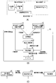
図1は、実施形態に係る診断支援システムの構成例を示す概略図である。 FIG. 1 is a schematic diagram showing a configuration example of a diagnosis support system according to an embodiment.
図1は、実施形態に係る診断支援システム1を示す。診断支援システム1は、実施形態に係る診断支援装置10、画像提供装置11、及び第2のモダリティ装置12を備える。診断支援装置10、画像提供装置11、及び第2のモダリティ装置12は、LAN(Local Area Network)等のネットワークNを介して相互に通信可能である。
FIG. 1 shows a diagnosis support system 1 according to an embodiment. A diagnosis support system 1 includes a
ここで、画像提供装置11は、第1の医用画像を生成するX線CT(Computed Tomography)装置又はMRI(Magnetic Resonance Imaging)装置等の第1のモダリティ装置であるか、又は、第1のモダリティ装置から第1の医用画像を取得して管理するPACS(Picture Archiving and Communication Systems)等の医用画像管理装置である。以下、画像提供装置11が第1のモダリティ装置、例えばX線CT装置であり、第1の医用画像としてCT画像を生成する場合について説明する。
Here, the
また、第2のモダリティ装置12は、撮像に当たり撮像方向を定義でき、第2の医用画像を生成する装置である。以下、第2のモダリティ装置12が、例えばX線診断装置であり、第2の医用画像としてX線画像を生成する場合について説明する。X線診断装置は、単純X線装置、アンギオ装置(X線透視撮影装置)、マンモグラフィ装置、及びテレスコープ撮像装置等を含む。
Also, the
診断支援装置10は、医用画像管理装置や、ワークステーションや、読影端末等であり、ネットワークNを介して接続されたシステム上に設けられる。なお、診断支援装置10は、オフラインの装置であってもよい。その場合、診断支援装置10は、可搬型の記録媒体を介してX線CT装置11から第1の医用画像、つまり、CT画像を取得すると共に、可搬型の記録媒体を介してX線診断装置12から第2の医用画像、つまり、X線画像を取得する。
The diagnosis support
ここで、診断支援装置10と、X線CT装置11及びX線診断装置12とにより、クライアントサーバシステムが構成されてもよい。具体的には、診断支援装置10が、データを提供するサーバ装置に含まれる一方、X線CT装置11がデータを利用する第1のクライアント装置に含まれ、X線診断装置12がデータを利用する第2のクライアント装置に含まれる。
Here, the
診断支援装置10は、処理回路21、記憶回路22、入力インターフェース23、ディスプレイ24、及びネットワークインターフェース25、決定支援手段26、及び検出支援手段27を備える。なお、入力インターフェース23、ディスプレイ24、ネットワークインターフェース25、決定支援手段26、及び検出支援手段27は、診断支援装置10に必須の機能ではない。
The
処理回路21は、専用又は汎用のCPU(Central Processing Unit)又はMPU(Micro Processor Unit)の他、特定用途向け集積回路(ASIC:Application Specific Integrated Circuit)、及び、プログラマブル論理デバイス等の処理回路を意味する。プログラマブル論理デバイスとしては、例えば、単純プログラマブル論理デバイス(SPLD:Simple Programmable Logic Device)、複合プログラマブル論理デバイス(CPLD:Complex Programmable Logic Device)、及び、フィールドプログラマブルゲートアレイ(FPGA:Field Programmable Gate Array)等の回路が挙げられる。処理回路21は、記憶回路22に記憶された、又は、処理回路21内に直接組み込まれたプログラムを読み出し実行することで後述する機能を実現する。
The
また、処理回路21は、単一の回路によって構成されてもよいし、複数の独立した処理回路要素の組み合わせによって構成されてもよい。後者の場合、記憶回路22の複数の記憶回路要素が複数の処理回路要素の機能に対応するプログラムをそれぞれ記憶するものであってもよいし、記憶回路22の1個の記憶回路要素が複数の処理回路要素の機能に対応するプログラムを記憶するものであってもよい。なお、処理回路21は、制御部の一例である。
Moreover, the
処理回路21がプログラムを実行することによって、診断支援装置10は、第1の画像取得機能31、決定機能32、撮像条件提供機能33、第2の画像取得機能34、及び検出機能35を実現する。なお、機能31~35の全部又は一部は、診断支援装置10にASIC等の回路として実現されるものであってもよい。また、機能33~35は、診断支援装置10に必須の機能ではない。
The diagnosis support
第1の画像取得機能31は、ネットワークインターフェース25を介してX線CT装置11から、該当患者に係るCT画像を取得する機能である。CT画像としては、ボリュームデータ、特定断面のスライスデータ、又は、ボリュームデータから任意に切り出された1又は複数のスライスデータ等が挙げられる。
A first
決定機能32は、第1の画像取得機能31により取得された第1の画像から、X線診断装置12により該当患者の第2の画像を生成するための該当患者の撮像方向を含む撮像条件を決定する機能である。なお、第2のモダリティ装置12が、X線診断装置である場合、撮像条件は、X線撮像条件を意味し、患者に対するX線の照射角度である撮像方向を少なくとも含むものとする。なお、X線撮像条件は、撮像方向の他、管電流、管電圧、撮像時間、及び距離(SID:Source to Image Distance)等のうち少なくとも1個を含む。
The
撮像条件提供機能33は、決定機能32により決定されたX線撮像条件を、ネットワークインターフェース25を介してX線診断装置12に提供する機能である。X線診断装置12は、撮像条件提供機能33により提供されたX線撮像条件に従って、該当患者に対するX線撮像を実行する。
The imaging
第2の画像取得機能34は、決定機能32により決定されたX線撮像条件によってX線診断装置12のX線撮像による該当患者に係るX線画像を、ネットワークインターフェース25を介してX線診断装置12から取得する機能である。
The second
検出機能35は、第1の画像取得機能31により取得された該当患者の第1の画像と、第2の画像取得機能34により取得された該当患者の第2の画像とに基づいて、該当患者の病変部の状態を検出する機能である。また、検出機能35は、検出された該当患者の病変部の状態をディスプレイ24に表示させることもできる。
The
なお、機能31~35の具体的な動作については、図4~図14を用いて後述する。
Note that specific operations of the
記憶回路22は、RAM(Random Access Memory)、フラッシュメモリ(Flash Memory)等の半導体メモリ素子、ハードディスク、及び光ディスク等によって構成される。記憶回路22は、USB(Universal Serial Bus)メモリ及びDVD(Digital Video Disk)等の可搬型メディアによって構成されてもよい。記憶回路22は、処理回路21において用いられる各種処理プログラム(アプリケーションプログラムの他、OS(Operating System)等も含まれる)や、プログラムの実行に必要なデータ等を記憶する。また、OSに、診断医等の操作者に対するディスプレイ24への情報の表示にグラフィックを多用し、基礎的な操作を入力インターフェース23によって行うことができるGUI(Graphical User Interface)を含めることもできる。なお、記憶回路22は、記憶部の一例である。
The
入力インターフェース23は、操作者によって操作が可能な入力デバイスと、入力デバイスからの信号を入力する入力回路とを含む。入力デバイスは、トラックボール、スイッチ、マウス、キーボード、走査面に触れることで入力操作を行うタッチパッド、表示画面とタッチパッドとが一化されたタッチスクリーン、光学センサを用いた非接触入力デバイス、及び音声入力デバイス等によって実現される。操作者により入力デバイスが操作されると、入力回路はその操作に応じた信号を生成して処理回路21に出力する。なお、診断支援装置10は、入力デバイスがディスプレイ24と一体に構成されたタッチパネルを備えてもよい。なお、入力インターフェース23は、入力部の一例である。
The
ディスプレイ24は、液晶ディスプレイパネル、プラズマディスプレイパネル、及び有機EL(Electro Luminescence)パネル等の表示デバイスである。ディスプレイ24は、処理回路21の制御に従って生成された医用画像を表示する。なお、ディスプレイ24は、表示部の一例である。
The
ネットワークインターフェース25は、パラレル接続仕様やシリアル接続仕様に合わせたコネクタによって構成される。ネットワークインターフェース25は、各規格に応じた通信制御を行い、電話回線を通じてネットワークに接続することができる機能を有しており、これにより、診断支援装置10をネットワークに接続させることができる。なお、ネットワークインターフェース25は、通信部の一例である。
The
決定支援手段26は、決定機能32からのデータを入力し、所定のデータを取得して決定機能32に出力する。
The decision support means 26 inputs data from the
例えば、決定支援手段26は、GPU(Graphics Processing Unit)及びデータベース等から成る装置である。決定支援手段26は、X線CT装置11のCT撮像により取得された複数(大量)のCT画像によりデータベースを構成する。決定支援手段26は、X線CT装置11のCT撮像により過去に取得された複数のCT画像を第1の学習用データとし、例えばCNN(畳み込みニューラルネットワーク)、畳み込み深層信念ネットワーク(CDBN:Convolutional Deep Belief Network)、再構成型トポグラフィック独立成分分析(TICA:Topographic Independent Component Analysis)等の機械学習に利用することでパラメータが成熟された第1のAI(Artificial Intelligence)を構築する。そして、決定支援手段26は、決定機能32から供給された、該当患者に係るCT画像を第1のAIに入力することで、出力を決定機能32に提供する。
For example, the decision support means 26 is a device comprising a GPU (Graphics Processing Unit), a database, and the like. The decision support means 26 constructs a database from a plurality of (a large amount of) CT images obtained by CT imaging with the
図2は、決定機能32の動作例を示す図である。図2(A)は、第1のAIの構築における第1の訓練データを示す図である。図2(B)は、X線撮像条件を決定するための構成を示すブロック図である。図2(C)は、X線撮像条件を決定するためのデータの流れを示す図である。
FIG. 2 is a diagram showing an operation example of the
X線CT装置11から患者に因らない複数、例えばm個のCT画像Fmが診断支援装置10の決定支援手段26に入力される。一方で、入力インターフェース23を介して決定支援手段26に、m個のCT画像Fmのそれぞれに対応するように、がん等の病変部の検出能を考慮した適切なX線撮像条件Pmが入力される。これにより、決定支援手段26は、m個のCT画像Fmを第1の学習用データとし、m個のX線撮像条件Pmを第1の教師データとして第1のAIを構築する(図2(A)に図示)。なお、決定支援手段26は、第1のAIとして、撮像部位毎に複数のAIを構築することが好適である。
A plurality of, for example, m CT images Fm are input from the
図2(B),(C)に示すように、決定機能32は、X線CT装置11からの該当患者に係るCT画像fを、決定支援手段26に提供する。これにより、決定支援手段26は、CT画像fを第1のAIに入力し、第1のAIの出力、つまり、該当患者に係るX線撮像条件pを決定機能32に提供する。
As shown in FIGS. 2B and 2C, the
決定機能32は、決定支援手段26から、第1のAIの出力、つまり、X線撮像条件pを受け、X線撮像条件pに基づいて、該当患者に係る適切なX線撮像条件を決定し、それをX線診断装置12に提供する。ここでは、決定機能32は、決定支援手段26からのX線撮像条件pそのものを、該当患者に係る適切なX線撮像条件として決定する。
The
図1の説明に戻って、検出支援手段27は、検出機能35からのデータを入力し、所定のデータを取得して検出機能35に出力する。例えば、検出支援手段27は、GPU及びデータベース等から成る装置である。検出支援手段27は、X線CT装置11のCT撮像により取得された複数のCT画像と、X線診断装置12のX線撮像により取得された複数のX線画像との組み合わせによりデータベースを構成する。検出支援手段27は、X線CT装置11のCT撮像により過去に取得された複数のCT画像と、X線診断装置12のX線撮像により過去に取得された複数のX線画像との組み合わせを第2の学習用データとして機械学習に利用することでパラメータが成熟された第2のAIを構築する。そして、検出支援手段27は、該当患者に係るCT画像及びX線画像の組み合わせを第2のAIに入力することで、出力を検出機能35に提供する。
Returning to the description of FIG. 1 , the detection support means 27 inputs data from the
図3は、検出機能35の動作例を示す図である。図3(A)は、第2のAIの構築における第2の訓練データを示す図である。図3(B)は、X線撮像条件を決定するための構成を示すブロック図である。図3(C)は、病変部の状態を決定するためのデータの流れを示す図である。病変部の状態は、例えば、がん等の病変部の有無、変化量(位置及びサイズ)等を意味する。
FIG. 3 is a diagram showing an operation example of the
X線CT装置11から患者に因らない複数、例えばm個のCT画像Fmが診断支援装置10の検出支援手段27に入力される。X線診断装置12から患者に因らないm個のX線画像Gmが診断支援装置10の検出支援手段27に入力される。一方で、入力インターフェース23を介して検出支援手段27に、m個のCT画像及びm個のX線画像の組み合わせのそれぞれに対応するように、適切な病変部の状態Qmが入力される。これにより、検出支援手段27は、m個のCT画像Fm及びm個のX線画像Gmの組み合わせを第2の学習用データとし、m個の病変部の状態Qmを第2の教師データとして第2のAIを構築する(図3(A)に図示)。なお、検出支援手段27は、第2のAIとして、撮像部位毎に複数のAIを構築することが好適である。
A plurality of, for example, m CT images Fm that are independent of the patient are input from the
図3(B),(C)に示すように、検出機能35は、X線CT装置11からの該当患者に係るCT画像fと、X線診断装置12からの該当患者に係るX線画像gとを、検出支援手段27に提供する。これにより、検出支援手段27は、CT画像f及びX線画像gの組み合わせを第2のAIに入力し、第2のAIの出力、つまり、該当患者に係る病変部の状態qを検出機能35に提供する。
As shown in FIGS. 3B and 3C, the
検出機能35は、検出支援手段27から、第2のAIの出力、つまり、病変部の状態qを受け、病変部の状態qに基づいて、該当患者に係る病変部の適切な状態を検出する。ここでは、検出機能35は、検出支援手段27からの病変部の状態qそのものを、該当患者に係る病変部の適切な状態として検出する。また、検出機能35は、該当患者に係る病変部の適切な状態をディスプレイ24に表示させる。
The
図1の説明に戻って、X線CT装置11は、患者に対してCT撮像を実行することで、CT画像を生成し、ネットワークNを介して診断支援装置10に提供する。ここで、X線CT装置(第1のクライアント装置)11は、診断支援装置(サーバ装置)10に該当患者に係るCT画像を送信する送信手段を設ける。
Returning to the description of FIG. 1, the
X線診断装置12は、診断支援装置10から提供されたX線撮像条件に従ってX線撮像を実行してX線画像を生成し、ネットワークNを介して診断支援装置10に提供する。ここで、X線診断装置12は、診断支援装置10に当該患者に係るX線撮像条件を要求し、その要求に応じてX線撮像条件を取得してもよい。その場合、X線診断装置(第2のクライアント装置)12は、診断支援装置(サーバ装置)10に該当患者に係るX線撮像条件を要求する要求手段と、要求手段による要求に応じて診断支援装置10からX線撮像条件を取得する取得手段とを設ける。
The X-ray
図4は、診断支援システム1の動作をフローチャートとして示す図である。図4において、「ST」に数字を付した符号はフローチャートの各ステップを示す。 FIG. 4 is a diagram showing the operation of the diagnostic support system 1 as a flowchart. In FIG. 4, numerals attached to "ST" indicate respective steps of the flow chart.
がん等の病変部の治療や予後の経過観察のために、CT画像等の医用画像により診断医等の操作者が病変部の進行具合を観察するワークフローが存在する。診断支援システム1は、例えば、病変部の治療や予後の経過観察における該当患者のフォローアップに使用されるものである。 There is a workflow in which an operator such as a diagnostician observes the progress of a lesion using medical images such as CT images for treatment of a lesion such as cancer and follow-up observation of prognosis. The diagnosis support system 1 is used, for example, for treatment of lesions and follow-up of patients in follow-up of prognosis.
第1の画像取得機能31は、ネットワークインターフェース25を介してX線CT装置11から、病変部を有する該当患者に係るCT画像を取得する(ステップST1)。ここで、ステップST1によって取得されたCT画像の収集時からの病変部の経過観察を行うために、同一モダリティ装置であるX線CT装置を用いてCT画像を生成することが一般的である。しかし、該当患者がX線CT装置を備えた大きい病院に行きCT撮像を行うには手間がかかりすぎるし、また、CT撮像では患者の被ばくも大きい。
The first
一方で、ステップST1によって取得されたCT画像の収集時からの病変部の経過観察を行うために、同一モダリティ装置ではないモダリティ装置、例えば、患者に身近な医療機関でも保有することができるX線診断装置を用いてX線画像を生成することも有り得る。その場合、患者に身近な医療機関で手軽に比較画像を取得することができ、かつ、患者の被ばくも比較的少ない一方で、CT画像と比較して検出能が劣る。 On the other hand, in order to perform follow-up observation of the lesion from the acquisition of the CT image acquired in step ST1, a modality device other than the same modality device, such as an X-ray that can be possessed by a medical institution close to the patient, is used. It is also possible that the diagnostic equipment is used to generate the X-ray images. In this case, comparative images can be easily obtained at a medical institution close to the patient, and the patient's exposure to radiation is relatively low.
そこで、ステップST1によって取得されたCT画像の収集時からの病変部の経過観察を行うために、同一モダリティ装置ではないX線診断装置12を用い、病変部の検出能が高いX線画像を生成することを考える。決定機能32は、X線CT装置11のCT撮像により取得されたCT画像から、X線診断装置12により該当患者のX線画像を収集するための適切なX線撮像条件を決定する(ステップST2)。なお、X線撮像条件は、少なくとも撮像方向を含むものとする。
Therefore, in order to observe the progress of the lesion from the acquisition of the CT image acquired in step ST1, an X-ray
例えば、決定機能32は、決定支援手段26を制御することで、該当患者のX線画像を収集するための適切なX線撮像条件を決定する。決定支援手段26は、上述したように、X線CT装置11のCT撮像により過去に取得された複数のCT画像を学習用データとして機械学習に利用することで第1のAIを構築する。そして、決定支援手段26は、決定機能32から供給された、該当患者に係るCT画像を第1のAIに入力することで、該当患者に係るCT画像に対応するX線撮像条件を第1のAIの出力として取得し、決定機能32に出力する。決定機能32は、決定支援手段26の出力、つまり、X線撮像条件に基づいて、該当患者に係る適切なX線撮像条件を決定する。
For example, the
撮像条件提供機能33は、ステップST2により決定されたX線撮像条件をX線診断装置12に提供する(ステップST3)。X線診断装置12は、ステップST3によって提供されたX線撮像条件に従って、該当患者に対するX線撮像を行う(ステップST4)。なお、該当患者に係るCT画像と、決定されたX線撮像条件とは、訓練データとして第1のAIに入力される。
The imaging
このように、適切なX線撮像条件の提供により、X線診断装置12は、ステップST4において、該当患者に係るCT画像と比較可能な程度である病変部の検出能の高いX線画像を生成することができる。
In this way, by providing appropriate X-ray imaging conditions, the X-ray
第2の画像取得機能34は、ステップST2により決定されたX線撮像条件によってX線診断装置12で撮像された該当患者のX線画像を、ネットワークインターフェース25を介してX線診断装置12から取得する(ステップST5)。検出機能35は、X線CT装置11のCT撮像により取得された該当患者に係るCT画像と、X線診断装置12のX線撮像により取得されたX線画像とに基づいて、該当患者に係る病変部の状態を検出する(ステップST6)。
The second
例えば、検出機能35は、検出支援手段27を制御することで、該当患者に係る適切な病変部の変化を検出する。検出支援手段27は、上述したように、X線CT装置11のCT撮像により過去に取得された複数のCT画像と、X線診断装置12のX線撮像により過去に取得された複数のX線画像との組み合わせを学習用データとして機械学習に利用することで第2のAIを構築する。そして、検出支援手段27は、検出機能35から供給された、該当患者に係るCT画像及びX線画像の組み合わせを第2のAIに入力することで、該当患者に係る病変部の状態を第2のAIの出力として取得し、検出機能35に出力する。検出機能35は、検出支援手段27の出力、つまり、該当患者の病変部の状態に基づいて、該当患者に係る病変部の適切な状態を検出する。病変部の変化は、例えば、がん等の病変部の有無、変化量(位置及びサイズ)等を意味する。
For example, the
検出機能35は、ステップST6によって検出された該当患者に係る病変部の変化をディスプレイ24に表示させる(ステップST7)。検出機能35は、ステップST7において、病変部の変化を、該当患者に係るX線画像にオーバーレイ表示させてもよいし、文字情報にて表示させてもよい。なお、該当患者に係るCT画像及びX線画像の組み合わせと、検出された病変部の状態とは、訓練データとして第2のAIに入力される。
The
このように、診断支援装置10は、ステップST7において、該当患者に係るCT画像と、該当患者に係る、病変部の適度な検出能を有するX線画像とに基づいて、該当患者に係る適切な自動診断結果(病変部の状態)を診断医等の操作者に提示することができる。操作者は、ディスプレイ24を介して提示された病変部の変化に基づいて、該当患者に係る精度の高い自動診断結果を視認することができる。
In this way, in step ST7, the
以上のように、診断支援システム1の決定機能32によれば、該当患者に係るCT画像の病変部に応じて適切なX線撮像条件をX線診断装置12に提供することができる。また、診断支援システム1の検出機能35によれば、該当患者に係るCT画像及びX線画像の組み合わせに応じて適切な病変部の状態を検出し操作者に提示することができる。
As described above, according to the
1.決定機能32の動作の変形例
図1~図4を用いて、決定機能32が、該当患者に係るCT画像に基づいて、該当患者に係る適切なX線撮像条件を決定する場合について説明した。しかし、その場合に限定されるものではない。以下、図5~図10を用いて、決定機能32の動作の変形例について説明する。
1. Modified Example of Operation of Determining Function 32 A case where the determining
1-1.決定機能32の動作の第1変形例
決定機能32は、決定支援手段26Aを利用して、CT画像に加え、CT画像の付帯情報を用いて適切なX線撮像条件を決定する。
1-1. First Modified Example of Operation of
図5は、決定機能32の動作例を示す図である。図5(A)は、第1のAIの構築における第1の訓練データを示す図である。図5(B)は、X線撮像条件を決定するための構成を示すブロック図である。図5(C)は、X線撮像条件を決定するためのデータの流れを示す図である。
FIG. 5 is a diagram showing an operation example of the
X線CT装置11から患者に因らない複数、例えばm個のCT画像Fm及びその付帯情報Rmが診断支援装置10の決定支援手段26Aに入力される。付帯情報は、医用画像に付帯されるDICOM(Digital Imaging and Communications in Medicine)データに含まれる所見、病変部の位置、及びオーバーレイ情報等を意味する。一方で、入力インターフェース23を介して決定支援手段26Aに、m個のCT画像のそれぞれに対応するように、m個のX線撮像条件Pmが入力される。これにより、決定支援手段26Aは、m個のCT画像Fm及びm個の付帯情報Rmの組み合わせを第1の学習用データとし、m個のX線撮像条件Pmを第1の教師データとして第1のAIを構築する(図5(A)に図示)。
A plurality of patient-independent CT images Fm, for example, m CT images Fm and accompanying information Rm are input from the
図5(B),(C)に示すように、決定機能32は、X線CT装置11からの該当患者に係るCT画像f及びその付帯情報rを、決定支援手段26Aに提供する。これにより、決定支援手段26Aは、CT画像f及び付帯情報rを第1のAIに入力し、第1のAIの出力、つまり、該当患者に係るX線撮像条件pを決定機能32に提供する。
As shown in FIGS. 5B and 5C, the
決定機能32は、決定支援手段26Aから、第1のAIの出力、つまり、X線撮像条件pを受け、X線撮像条件pに基づいて、該当患者に係る適切なX線撮像条件を決定し、それをX線診断装置12に提供する。ここでは、決定機能32は、決定支援手段26AからのX線撮像条件pそのものを、該当患者に係る適切なX線撮像条件として決定する。
The
以上のように、図5に示す決定機能32の動作によれば、該当患者に係るCT画像のみに基づいてX線撮像条件を決定する場合に比べて、該当患者に係るX線撮像条件の出力精度が向上する。
As described above, according to the operation of the
1-2.決定機能32の動作の第2変形例
決定機能32は、決定支援手段26Bを利用して、CT画像に加え、X線撮像により生成されたX線画像を用いて、再撮像に係る適切なX線撮像条件を決定する。
1-2. Second Modified Example of Operation of
図6は、決定機能32の動作の第2変形例を示す図である。図6(A)は、第1のAIの構築における第1の訓練データを示す図である。図6(B)は、X線撮像条件を決定するための構成を示すブロック図である。図6(C)は、X線撮像条件を決定するためのデータの流れを示す図である。
FIG. 6 is a diagram showing a second modification of the operation of the
X線CT装置11から患者に因らない複数、例えばm個のCT画像Fmが診断支援装置10の決定支援手段26Bに入力される。一方で、X線診断装置12から患者に因らないm個のX線撮像条件Pm及びm個のX線画像Gmが診断支援装置10の決定支援手段26Bに入力される。m個目のCT画像Fm及びm個目のX線画像Gmの組み合わせは、それぞれ、m個目のX線撮像条件Pmに対応付けられる。決定支援手段26は、m個のCT画像Fm及びm個のX線画像Gmの組み合わせを第1の学習用データとし、m個のX線撮像条件Pmを第1の教師データとして第1のAIを構築する(図6(A)に図示)。
A plurality of patient-independent CT images Fm, for example, m CT images Fm, are input from the
図6(B),(C)に示すように、決定機能32は、X線CT装置11からの該当患者に係るCT画像fと、X線診断装置12からの該当患者に係るX線画像gとを、診断支援装置10の決定支援手段26Bに提供する。X線画像gは、例えば、上述の決定機能32,32Aにより決定されたX線撮像条件による該当患者のX線撮像により生成されたものである。これにより、決定支援手段26Bは、CT画像f及びX線画像gを第1のAIに入力し、第1のAIの出力、つまり、該当患者に係るX線撮像画像pを決定機能32に提供する。
As shown in FIGS. 6B and 6C, the
決定機能32は、決定支援手段26Bから、第1のAIの出力、つまり、X線撮像条件pを受け、X線撮像条件pに基づいて、該当患者に係る適切なX線撮像条件を決定し、それをX線診断装置12に提供する。ここでは、決定機能32は、決定支援手段26BからのX線撮像条件pそのものを、該当患者に係る適切なX線撮像条件として決定する。
The
以上のように、図6に示す決定支援手段26Bの動作によれば、該当患者に係る最適なX線撮像条件を求めることで、該当患者の再撮像における適切なX線撮像条件をX線診断装置12に提供することができる。
As described above, according to the operation of the determination support means 26B shown in FIG. 6, the optimum X-ray imaging conditions for the relevant patient are obtained, and the appropriate X-ray imaging conditions for re-imaging of the relevant patient can be determined by X-ray diagnosis.
図1~図6を用いて、決定支援手段26~26Bが直接的にX線撮像条件を取得して決定機能32~32Bに出力し、決定機能32~32Bが決定支援手段26~26Bの出力であるX線撮像条件そのものを適切なX線撮像条件として決定する場合について説明した。しかし、その場合に限定されるものではない。例えば、決定支援手段が、適切なX線撮像条件を決定するための複数のパラメータを生成して決定機能32に出力し、決定機能32が決定支援手段の出力である複数のパラメータに基づいて適切なX線撮像条件を決定する場合であってもよい。その場合について、図7~図10を用いて説明する。
1 to 6, the decision support means 26 to 26B directly acquire the X-ray imaging conditions and output them to the decision functions 32 to 32B, and the decision functions 32 to 32B output the decision support means 26 to 26B. A case has been described in which the X-ray imaging condition itself is determined as an appropriate X-ray imaging condition. However, it is not limited to that case. For example, the decision support means generates a plurality of parameters for determining appropriate X-ray imaging conditions and outputs them to the
1-3.決定機能32の動作の第3変形例
決定機能32は、決定支援手段26Cを利用して、適切なX線撮像条件を決定するための複数のパラメータとして複数の仮想的なX線画像を生成し、複数の仮想的なX線画像に基づいて、適切なX線撮像条件を決定する。
1-3. Third Modified Example of Operation of
図7は、決定機能32の動作の第3変形例を示す図である。図7(A)は、第1のAIの構築における第1の訓練データを示す図である。図7(B)は、X線撮像条件を決定するための構成を示すブロック図である。図7(C)は、X線撮像条件を決定するためのデータの流れを示す図である。
FIG. 7 is a diagram showing a third modification of the operation of the
X線CT装置11から患者に因らない複数、例えばm個のCT画像Fmが診断支援装置10の決定支援手段26Cに入力される。一方で、X線診断装置12から患者に因らないm個のX線撮像条件Pm及びm個のX線画像Gmが診断支援装置10の決定支援手段26Cに入力される。m個のCT画像Fm及びm個のX線撮像条件Pmは、それぞれ、m個のX線画像Gmに対応付けられる。決定支援手段26Cは、m個のCT画像Fm及びm個のX線撮像条件Pmの組み合わせを第1の学習用データとし、m個のX線画像Gmを第1の教師データとして第1のAIを構築する(図7(A)に図示)。
A plurality of, for example, m CT images Fm that do not depend on the patient are input from the
図7(B),(C)に示すように、決定機能32は、X線CT装置11からの該当患者に係るCT画像fと、入力インターフェース23を介して任意に設定されたX線撮像条件pとを、決定支援手段26Cに入力する。これにより、決定支援手段26Cは、CT画像f及びX線撮像条件pを第1のAIに入力し、第1のAIの出力、つまり、該当患者に係る第3の医用画像(仮想X線画像)hを決定機能32に提供する。また、決定支援手段26Cは、異なる複数のX線撮像条件によりループで第1のAIを利用することで、適切なX線撮像条件を決定するための複数のパラメータとして、複数の仮想X線画像hを生成することができる。
As shown in FIGS. 7B and 7C, the
決定機能32は、決定支援手段26Cから、第1のAIの出力、つまり、複数のX線撮像条件に対応する複数の仮想X線画像hを受け、複数の仮想X線画像hに基づいて、該当患者に係る適切なX線撮像条件を決定し、それをX線診断装置12に提供する。例えば、操作者が、複数の仮想X線画像hを参照しながら、入力インターフェース23を操作して複数の仮想X線画像hの中から所要の仮想X線画像を選択することで、決定支援手段26Cは、選択された仮想X線画像に対応する条件を適切なX線撮像条件として決定する。また、例えば、決定支援手段26Cは、複数の仮想X線画像hの中から画像の品質情報が最大の仮想X線画像に対応する条件を適切なX線撮像条件として決定する。品質情報は、画像のS/N(Signal to Noise)及びCNR比(Contrast to Noise Ratio)等に基づく指標(例えば、1点~10点)を意味する。
The
以上のように、図7に示す決定機能32の動作によれば、適切なX線撮像条件を決定するためのパラメータとして仮想X線画像を生成し、仮想X線画像に基づいて適切なX線撮像条件を設定することができる。
As described above, according to the operation of the
1-4.決定機能32の動作の第4変形例
決定機能32は、決定支援手段26Dを利用して、適切なX線撮像条件を決定するための複数のパラメータとして複数の品質情報を生成し、複数の品質情報に基づいて、適切なX線撮像条件を決定する。品質情報は、画像のS/N(Signal to Noise)及びCNR比(Contrast to Noise Ratio)等に基づく指標(例えば、1点~10点)を意味する。
1-4. Fourth Modified Example of Operation of
図8は、決定機能32の動作の第4変形例を示す図である。図8(A)は、第1のAIの構築における第1の訓練データを示す図である。図8(B)は、X線撮像条件を決定するための構成を示すブロック図である。図8(C)は、X線撮像条件を決定するためのデータの流れを示す図である。
FIG. 8 is a diagram showing a fourth modification of the operation of the
X線CT装置11から患者に因らない複数、例えばm個のCT画像Fmが診断支援装置10の決定支援手段26Dに入力される。X線診断装置12から患者に因らないm個のX線画像Gmが診断支援装置10の決定支援手段26Dに入力される。また、CT画像及びX線画像に基づいて生成された患者に因らないm個の品質情報Smが決定支援手段26Dに入力される。m個目のCT画像Fm及びm個目のX線画像Gmの組み合わせは、それぞれ、m個目の品質情報Smに対応付けられる。決定支援手段26Dは、m個のCT画像Fm及びm個のX線画像Gmの組み合わせを第1の学習用データとし、m個の品質情報Smを第1の教師データとして第1のAIを構築する(図8(A)に図示)。
A plurality of patient-independent CT images Fm, for example, m CT images Fm are input from the
図8(B),(C)に示すように、決定機能32は、X線CT装置11からの該当患者に係るCT画像fと、X線診断装置12からの該当患者に係るX線画像gとを、診断支援装置10の決定支援手段26Dに入力する。これにより、決定支援手段26Dは、CT画像f及びX線画像gを第1のAIに入力し、第1のAIの出力、つまり、該当患者に係る品質情報sを決定機能32に提供する。また、決定支援手段26Dは、異なる複数のCT画像及びX線画像によりループで第1のAIを利用することで、適切なX線撮像条件を決定するための複数のパラメータとして、複数の品質情報sを生成することができる。
As shown in FIGS. 8B and 8C, the
決定機能32は、決定支援手段26Dから、第1のAIの出力、つまり、複数のX線撮像条件に対応する複数の品質情報sを受け、複数の品質情報sに基づいて、該当患者に係る適切なX線撮像条件を決定し、それをX線診断装置12に提供する。例えば、操作者が、複数の品質情報sを参照しながら、入力インターフェース23を操作して複数の品質情報sの中から所要の品質情報を選択することで、決定支援手段26Dは、選択された品質情報に対応する条件を適切なX線撮像条件として決定する。また、例えば、決定支援手段26Dは、最大の品質情報に対応する条件を適切なX線撮像条件として決定する。
The
以上のように、図8に示す決定機能32の動作によれば、適切なX線撮像条件を決定するためのパラメータとして画像の品質情報を生成し、画像の品質情報に基づいて適切なX線撮像条件を設定することができる。
As described above, according to the operation of the
1-5.決定機能32の動作の第5変形例
決定機能32は、決定支援手段26Eを利用して、適切なX線撮像条件を決定するための複数のパラメータとして複数の品質情報を生成し、複数の品質情報に基づいて、適切なX線撮像条件を決定する。
1-5. Fifth Modified Example of Operation of
図9は、決定機能32の動作の第5変形例を示す図である。図9(A)は、第1のAIの構築における第1の訓練データを示す図である。図9(B)は、X線撮像条件を決定するための構成を示すブロック図である。図9(C)は、X線撮像条件を決定するためのデータの流れを示す図である。
FIG. 9 is a diagram showing a fifth modification of the operation of the
X線CT装置11から患者に因らない複数、例えばm個のCT画像Fmが診断支援装置10の決定支援手段26Eに入力される。X線診断装置12から患者に因らないm個のX線撮像条件Pmが診断支援装置10の決定支援手段26Eに入力される。また、CT画像及びX線画像に基づいて生成された患者に因らないm個の品質情報Smが決定支援手段26Eに入力される。m個目のCT画像Fm及びm個目のX線撮像条件Pmの組み合わせは、それぞれ、m個目の品質情報Smに対応付けられる。決定支援手段26Eは、m個のCT画像Fm及びm個のX線撮像条件Pmの組み合わせを第1の学習用データとし、m個の品質情報Smを第1の教師データとして第1のAIを構築する(図9(A)に図示)。
A plurality of patient-independent CT images Fm, for example, m CT images Fm, are input from the
図9(B),(C)に示すように、決定機能32は、X線CT装置11からの該当患者に係るCT画像fと、入力インターフェース23を介して任意に設定されたX線撮像条件pとを、診断支援装置10の決定支援手段26Eに入力する。これにより、決定支援手段26Eは、CT画像f及びX線撮像条件pを第1のAIに入力し、第1のAIの出力、つまり、該当患者に係る適切な品質情報sを検出機能32に提供する。また、決定支援手段26Eは、異なる複数のX線撮像条件によりループで第1のAIを利用することで、適切なX線撮像条件を決定するための複数のパラメータとして、複数の品質情報sを生成することができる。
As shown in FIGS. 9B and 9C, the
決定機能32は、決定支援手段26Eから、第1のAIの出力、つまり、複数のX線撮像条件に対応する複数の品質情報sを受け、複数の品質情報sに基づいて、該当患者に係る適切なX線撮像条件を決定し、それをX線診断装置12に提供する。例えば、操作者が、複数の品質情報sを参照しながら、入力インターフェース23を操作して複数の品質情報sの中から所要の品質情報を選択することで、決定支援手段26Eは、選択された品質情報に対応する条件を適切なX線撮像条件として決定する。また、例えば、決定支援手段26Eは、最大の品質情報に対応する条件を適切なX線撮像条件として決定する。
The
以上のように、図9に示す決定機能32の動作によれば、適切なX線撮像条件を決定するためのパラメータとして画像の品質情報を生成し、画像の品質情報に基づいて適切なX線撮像条件を設定することができる。
As described above, according to the operation of the
以上において、決定機能32が、5個の決定支援機能26~26E、つまり、5個のAIのいずれかを用いてX線撮像条件を決定する場合について説明したが、その場合に限定されるものではない。例えば、決定機能32は、3個の決定支援機能26C~26E、つまり、3個のAIを用いてX線撮像条件を決定することもできる。
In the above, the case where the
図10は、決定機能32の動作の第6変形例を示す図である。図10は、複数のAIを利用する場合のデータの流れを示す図である。
FIG. 10 is a diagram showing a sixth modification of the operation of the
図10に示すように、決定機能32は、決定支援手段26Cから複数のX線撮像条件に対応する複数の仮想X線画像h(図7に図示)を受け、決定支援手段26Dから複数のX線撮像条件に対応する複数の品質情報s(図8に図示)を受け、決定支援手段26Eから複数のX線撮像条件に対応する複数の品質情報s(図8に図示)を受ける。操作者が、複数の仮想X線画像hに対応する品質情報や複数の品質情報sを参照しながら、入力インターフェース23を操作して複数の品質情報の中から所要の品質情報を選択することで、決定機能32は、選択された品質情報に対応する条件を適切なX線撮像条件として決定する。また、例えば、決定機能32は、複数の仮想X線画像hに対応する品質情報を含む複数の品質情報の中から最大の品質情報に対応する条件を適切なX線撮像条件として決定する。
As shown in FIG. 10,
以上のように、図10に示す決定機能32の動作によれば、適切なX線撮像条件を決定するためのパラメータとして仮想X線画像や画像の品質情報を生成し、仮想X線画像や画像の品質情報に基づいて適切なX線撮像条件を決定することができる。
As described above, according to the operation of the
2.検出機能35の動作の変形例
図1~図4を用いて、検出機能35が、該当患者に係るCT画像及びX線画像に基づいて、該当患者に係る病変部の適切な状態を検出する場合について説明した。しかし、その場合に限定されるものではない。以下、図11~図14を用いて、検出機能35の動作の変形例について説明する。
2. Modified Example of Operation of Detecting
2-1.検出機能35の動作の第1変形例
検出機能35は、検出支援手段27Aを利用して、CT画像及びX線画像に加えX線撮像条件に基づいて、病変部の適切な状態を検出するための複数のパラメータとして複数の位置情報を生成し、複数の位置情報に基づいて、病変部の適切な状態を検出する。
2-1. First Modified Example of Operation of
図11は、検出機能35の動作の第1変形例を示す図である。図11(A)は、第2のAIの構築における第2の訓練データを示す図である。図11(B)は、病変部の状態を決定するための構成を示すブロック図である。図11(C)は、病変部の状態を決定するためのデータの流れを示す図である。
11A and 11B are diagrams showing a first modification of the operation of the
X線CT装置11から患者に因らない複数、例えばm個のCT画像Fmが診断支援装置10の検出支援手段27Aに入力される。X線診断装置12から患者に因らないm個のX線撮像条件Pm及びm個のX線画像Gmの組み合わせが診断支援装置10の検出支援手段27Aに入力される。また、入力インターフェース23を介してm個の病変部の位置Tmが検出支援手段27Aに入力される。又は、CT画像を画像解析することにより病変部を自動的に検出するCADにより、病変部の位置Tmが特定され検出支援手段27Aに入力される。m個目のCT画像Fm、m個目のX線画像Gm、及びm個目のX線撮像条件Pmの組み合わせは、それぞれ、m個目の病変部の状態Qmに対応付けられる。検出支援手段27Aは、m個のCT画像Fm、m個のX線画像Gm、及びm個のX線撮像条件Pmの組み合わせを第2の学習用データとし、m個の病変部の状態Qmを第2の教師データとして第2のAIを構築する(図11(A)に図示)。
A plurality of, for example, m CT images Fm that do not depend on the patient are input from the
図11(B),(C)に示すように、検出機能35は、X線CT装置11からの該当患者に係るCT画像fと、X線診断装置12からのX線撮像条件p及びX線画像gの組み合わせとを、診断支援装置10の検出支援手段27Aに入力する。これにより、検出支援手段27Aは、CT画像f、X線画像g、及びX線撮像条件pの組み合わせを第2のAIに入力し、第2のAIの出力、つまり、該当患者に係る病変部の位置tを検出機能35に提供する。検出支援手段27Aは、該当患者に係る病変部の位置情報をもつCT画像fと、該当患者に係る最新画像であるX線画像(投影画像)gとに基づいて、該当患者に係る病変部の現在の位置情報(病変部の候補領域)を取得することができる。また、検出支援手段27Aは、異なる複数のX線撮像条件によりループで第2のAIを利用することで、病変部の適切な状態を検出するための複数のパラメータとして、病変部に係る複数の位置を生成することもできる。
As shown in FIGS. 11B and 11C, the
検出機能35は、検出支援手段27Aから、第2のAIの出力、つまり、病変部の位置tを受け、病変部の位置tに基づいて、該当患者に係る病変部の適切な状態(病変部の有無、変化量(位置及びサイズ))を検出し、それをディスプレイ24に表示させる。
The
以上のように、図11に示す検出機能35の動作によれば、該当患者に係るCT画像及びX線画像のみに基づいて病変部の状態を検出する場合に比べて、該当患者に係る病変部の状態の出力精度が向上する。
As described above, according to the operation of the
2-2.検出機能35の動作の第2変形例
検出機能35は、検出支援手段27Bを利用して、CT画像、X線画像、及びX線撮像条件に加え仮想X線画像に基づいて、病変部の適切な状態を検出するための複数のパラメータとして複数の位置情報を生成し、複数の位置情報に基づいて、病変部の適切な状態を検出する。
2-2. Second Modified Example of Operation of
図12は、検出機能35の動作の第2変形例を示す図である。図12(A)は、第2のAIの構築における第2の訓練データを示す図である。図12(B)は、病変部の状態を決定するための構成を示すブロック図である。図12(C)は、病変部の状態を決定するためのデータの流れを示す図である。
12A and 12B are diagrams showing a second modification of the operation of the
X線CT装置11から患者に因らない複数、例えばm個のCT画像Fmが診断支援装置10の検出支援手段27Bに入力される。X線診断装置12から患者に因らないm個のX線撮像条件Pm及びX線画像Gmの組み合わせが診断支援装置10の検出支援手段27Bに入力される。また、入力インターフェース23を介してm個の病変部の位置Tmが検出支援手段27Bに入力される。又は、CT画像を画像解析することにより病変部を自動的に検出するCADにより、病変部の位置Tmが特定され検出支援手段27Bに入力される。さらに、決定支援手段26C(図7に図示)から仮想X線画像Hmが診断支援装置10の検出支援手段27Bに入力される。
A plurality of, for example, m CT images Fm that do not depend on the patient are input from the
m個目のCT画像Fm、m個目のX線画像Gm、m個目のX線撮像条件Pm、及びm個目のX線画像Hmの組み合わせは、それぞれ、m個目の病変部の状態Qmに対応付けられる。検出支援手段27Bは、m個のCT画像Fm、m個のX線画像Gm、m個のX線撮像条件Pm、及びm個の仮想X線画像Hmの組み合わせを第2の学習用データとし、m個の病変部の位置Tmを第2の教師データとして第2のAIを構築する(図12(A)に図示)。 The combination of the m-th CT image Fm, the m-th X-ray image Gm, the m-th X-ray imaging condition Pm, and the m-th X-ray image Hm is the state of the m-th lesion area. Qm. The detection support means 27B uses a combination of m CT images Fm, m X-ray images Gm, m X-ray imaging conditions Pm, and m virtual X-ray images Hm as second learning data, A second AI is constructed using the m lesion positions Tm as second teacher data (illustrated in FIG. 12A).
図12(B),(C)に示すように、検出機能35は、X線CT装置11からの該当患者に係るCT画像fと、X線診断装置12からのX線撮像条件p及びX線画像gの組み合わせと、決定支援手段26Cからの仮想X線画像hとを入力する。これにより、検出支援手段27Bは、CT画像f、X線画像g、X線撮像条件p、及び仮想X線画像hの組み合わせを第2のAIに入力し、第2のAIの出力、つまり、該当患者に係る適切な病変部の位置tを検出機能35に提供する。検出支援手段27Bは、該当患者に係る病変部の位置情報をもつCT画像fと、該当患者に係る最新画像であるX線画像(投影画像)gとに基づいて、該当患者に係る病変部の現在の位置情報(病変部の候補領域)を取得することができる。また、検出支援手段27Bは、異なる複数のX線撮像条件によりループで第2のAIを利用することで、病変部の適切な状態を検出するための複数のパラメータとして、病変部に係る複数の位置を生成することもできる。
As shown in FIGS. 12B and 12C, the
検出機能35は、検出支援手段27Bから、第2のAIの出力、つまり、病変部の位置tを受け、病変部の位置tに基づいて、該当患者に係る病変部の適切な状態を検出し、それをディスプレイ24に表示させる。
The
以上のように、図12に示す検出機能35の動作によれば、該当患者に係るCT画像、X線画像、及びX線撮像条件のみに基づいて病変部の状態を検出する場合に比べて、該当患者に係る病変部の状態の出力精度が向上する。
As described above, according to the operation of the
2-3.検出機能の動作の第3変形例
検出機能35は、検出支援手段27Cを利用して、CT画像及びX線画像に加え仮想X線画像に基づいて、病変部の適切な状態を検出する。
2-3. Third Modification of Operation of Detection Function The
図13は、検出機能35の動作の第3変形例を示す図である。図13(A)は、第2のAIの構築における第2の訓練データを示す図である。図13(B)は、病変部の状態を決定するための構成を示すブロック図である。図13(C)は、病変部の状態を決定するためのデータの流れを示す図である。
13A and 13B are diagrams showing a third modification of the operation of the
X線CT装置11から患者に因らない複数、例えばm個のCT画像Fmが診断支援装置10の検出支援手段27Cに入力される。X線診断装置12から患者に因らないm個のX線画像Gmが診断支援装置10の検出支援手段27Cに入力される。また、入力インターフェース23を介してm個の病変部の状態Smが検出支援手段27Cに入力される。さらに、決定支援手段26C(図7に図示)から仮想X線画像Hmが検出支援手段27Cに入力される。
A plurality of, for example, m CT images Fm are input from the
m個目のCT画像Fm及びm個目のX線画像Gmの組み合わせは、それぞれ、m個目の病変部の状態Smに対応付けられる。m個目のCT画像Fm及びm個目の仮想X線画像Hmの組み合わせは、それぞれ、m個目の病変部の状態Smに対応付けられる。検出支援手段27Cは、m個のCT画像Fm及びm個のX線画像Gmを第2の学習用データとし、m個の病変部の状態Smを第2の教師データとして第2のAIを構築する(図13(A)に図示)。 A combination of the m-th CT image Fm and the m-th X-ray image Gm is associated with the m-th lesion state Sm. A combination of the m-th CT image Fm and the m-th virtual X-ray image Hm is associated with the m-th lesion state Sm. The detection support means 27C constructs a second AI using m CT images Fm and m X-ray images Gm as second learning data and m lesion states Sm as second teacher data. (illustrated in FIG. 13(A)).
図13(B),(C)に示すように、検出機能35は、X線CT装置11からの該当患者に係るCT画像fと、X線診断装置12からのX線画像gとを、診断支援装置10の検出支援手段27Cに入力する。これにより、検出支援手段27Cは、CT画像f及びX線画像gの組み合わせを第2のAIに入力し、第2のAIの出力、つまり、該当患者に係る病変部の状態sを検出機能35に供給することができる。一方で、検出機能35は、X線CT装置11からの該当患者に係るCT画像fと、決定支援手段26Cからの仮想X線画像hとを検出支援手段27Cに入力する。これにより、検出支援手段27Cは、CT画像f及び仮想X線画像hの組み合わせを第2のAIに入力し、第2のAIの出力、つまり、該当患者に係る病変部の状態sを検出機能35に供給することができる。
As shown in FIGS. 13B and 13C, the
検出機能35は、検出支援手段27Cから、第2のAIの出力、つまり、病変部に係る複数の状態sを受け、病変部に係る複数の状態sに基づいて、該当患者に係る病変部の適切な状態を検出し、それをディスプレイ24に表示させる。ここでは、検出機能35は、検出支援手段27Cからの病変部に係る複数の状態sの代表値そのものを、該当患者に係る病変部の適切な状態として検出すればよい。
The
以上のように、図13に示す検出機能35の動作によれば、病変部の位置tの変化を操作者に提示することができる。
As described above, according to the operation of the
以上において、検出機能35が、4個の検出支援機能27~27C、つまり、4個のAIのいずれかを用いて病変部の状態を検出する場合について説明したが、その場合に限定されるものではない。例えば、検出機能35は、2個の検出支援機能27B~27C、つまり、2個のAIを用いて病変部の状態を検出することもできる。
In the above, the case where the
2-4.検出機能の動作の第4変形例
図14は、検出機能35の動作の第4変形例を示す図である。図14は、複数のAIを利用する場合のデータの流れを示す図である。
2-4. Fourth Modification of Operation of Detection Function FIG. 14 is a diagram showing a fourth modification of the operation of the
図14に示すように、検出機能35は、検出支援手段27A、検出支援手段27Bから複数の病変部の位置tを受ける。操作者が、複数の病変部の位置tを参照しながら、入力インターフェース23を操作して複数の病変部の位置tの中から所要の病変部の位置を選択することで、検出機能35は、選択された病変部の位置を病変部の適切な状態として検出する。
As shown in FIG. 14, the
以上のように、図14に示す検出機能35の動作によれば、病変部の適切な状態を検出するためのパラメータとして病変部の位置を生成し、病変部の位置に基づいて病変部の適切な状態を検出することができる。
As described above, according to the operation of the
3.診断支援システム1の構成の変形例
3-1.第1のモダリティ装置11の変形例
図1では、診断支援システム1の第1のモダリティ装置11が、1種類のモダリティ装置(X線CT装置)である場合について説明したが、その場合に限定されるものではない。第1のモダリティ装置11は、複数種類、例えば、2種類のX線CT装置及びMRI装置であってもよい。その場合、決定支援手段26は、過去に取得された複数のCT画像及びMRI画像の組み合わせを第1の学習用データとし、各組み合わせに対応するX線撮像条件を第1の教師データとし、機械学習に利用することで第1のAIを構築する。そして、決定機能32は、該当患者に係るCT画像及びMRI画像を第1のAIに入力することで、該当患者に係るCT画像及びMRI画像に対応する適切なX線撮像条件を決定してX線診断装置12に提供する。
3. Modification of Configuration of Diagnosis Support System 1 3-1. Modification of
以上のような第1のモダリティ装置11の構成によれば、該当患者に係るCT画像のみを第1のAIに入力する場合に比べて、該当患者に係るX線撮像条件の出力精度が向上する。
According to the configuration of the
3-2.第2のモダリティ装置12の変形例
図1では、診断支援システム1の第2のモダリティ装置12が、1種類のモダリティ装置(X線診断装置)である場合について説明したが、その場合に限定されるものではない。第2のモダリティ装置12は、複数種類、例えば、2種類のX線診断装置及び超音波診断装置であってもよい。その場合、決定支援手段26は、過去に取得された複数のCT画像を第1の学習用データとし、各CT画像に対応するX線撮像条件及び超音波のスキャン条件を第1の教師データとし、機械学習に利用することで第1のAIを構築する。そして、決定機能32は、該当患者に係るCT画像を第1のAIに入力することで、該当患者に係るCT画像に対応する適切なX線撮像条件及び超音波のスキャン条件を決定してX線診断装置及び超音波診断装置にそれぞれ提供する。超音波のスキャン条件は、超音波の送信周波数、パルス繰り返し周波数(PRF:Pulse Repetition Frequency)、及びフレームレート等を意味する。
3-2. Modification of
以上のような第2のモダリティ装置12の構成によれば、該当患者に係るCT画像のみに基づいてX線撮像条件を決定する場合に比べて、該当患者に係るX線撮像条件の出力精度が向上する。
According to the configuration of the
また、第2のモダリティ装置12が2種類のX線診断装置及び超音波診断装置である場合、決定支援手段26は、過去に取得された複数のCT画像を第1の学習用データとし、各CT画像に対応するモダリティ装置の種別を第1の教師データとし、機械学習に利用することで第1のAIを構築する。そして、決定機能32は、該当患者に係るCT画像を第1のAIに入力することで、該当患者に係るCT画像に対応する適切なモダリティ装置の種別を決定して該当するX線診断装置又は超音波診断装置に提供することもできる。
Further, when the
以上説明した少なくとも1つの実施形態によれば、第1の画像に基づいて、生成予定の第2の画像を収集するための適切な撮像条件を提供することができる。 According to at least one embodiment described above, appropriate imaging conditions for acquiring the second image to be generated can be provided based on the first image.
なお、第1の画像取得機能31は、画像取得手段及び第1の画像取得手段の一例である。決定機能32は、決定手段の一例である。撮像条件提供機能33は、撮像条件提供手段の一例である。第2の画像取得機能34は、第2の画像取得手段の一例である。検出機能35は、検出手段の一例である。
The first
以上、本発明のいくつかの実施形態を説明したが、これらの実施形態は、例として提示したものであり発明の範囲を限定することは意図していない。これら新規な実施形態は、その他の様々な形態で実施されることが可能であり、発明の要旨を逸脱しない範囲で種々の省略、置き換え、変更を行うことができる。これらの実施形態やその変形は、発明の範囲や要旨に含まれるとともに、特許請求の範囲に記載された発明とその均等の範囲に含まれる。 Although several embodiments of the invention have been described above, these embodiments are presented by way of example and are not intended to limit the scope of the invention. These novel embodiments can be implemented in various other forms, and various omissions, replacements, and modifications can be made without departing from the scope of the invention. These embodiments and their modifications are included in the scope and gist of the invention, and are included in the scope of the invention described in the claims and its equivalents.
1 診断支援システム
10 診断支援装置
11 画像提供装置(第1のモダリティ装置)
12 第2のモダリティ装置
21 処理回路
22 記憶回路
26~26E 決定支援手段
27~27C 検出支援手段
31 第1の画像取得機能
32 決定機能
33 撮像条件提供機能
34 第2の画像取得機能
35 検出機能
1
12
Claims (17)
前記画像取得手段により取得された前記第1の画像から、X線診断装置により前記被検体の第2の画像を生成するための前記被検体の撮像方向を含む撮像条件を決定する決定手段と、
を備える診断支援装置。 an image acquisition means for acquiring a first image of a subject imaged by an X-ray CT apparatus;
determination means for determining imaging conditions including an imaging direction of the subject for generating a second image of the subject by an X-ray diagnostic apparatus from the first image obtained by the image obtaining means;
A diagnostic support device comprising:
請求項1に記載の診断支援装置。 an imaging condition providing means for providing the imaging condition determined by the determining means to the X-ray diagnostic apparatus and causing the X-ray diagnostic apparatus to perform imaging of the subject according to the imaging condition;
The diagnosis support device according to claim 1.
請求項1又は2に記載の診断支援装置。 The determination means obtains information specifying the position of the lesion from the first image, and determines imaging conditions including the imaging direction of the subject based on the information.
3. The diagnostic support device according to claim 1 or 2.
前記撮像条件によって前記X線診断装置で撮像された前記被検体の第2の画像を取得する第2の画像取得手段と、
前記第1の画像取得手段により取得された前記第1の画像と、前記第2の画像取得手段により取得された前記第2の画像とに基づいて、前記被検体の病変部の状態を検出する検出手段と、をさらに備える、
請求項1又は2に記載の診断支援装置。 a first image acquisition means as the image acquisition means;
a second image acquiring means for acquiring a second image of the subject imaged by the X-ray diagnostic apparatus under the imaging condition;
detecting the state of the lesion of the subject based on the first image acquired by the first image acquisition means and the second image acquired by the second image acquisition means; further comprising a detection means;
3. The diagnostic support device according to claim 1 or 2.
前記決定手段は、前記画像取得手段により取得された前記第1の画像及び前記付帯情報の組み合わせから、前記撮像条件を決定する、
請求項1乃至4のうち何れか1項に記載の診断支援装置。 The image acquisition means further acquires incidental information of the first image,
The determination means determines the imaging conditions from a combination of the first image and the incidental information acquired by the image acquisition means.
5. The diagnosis support device according to any one of claims 1 to 4.
前記第1の画像取得手段により取得された前記第1の画像から、第2のモダリティ装置により前記被検体の第2の画像を生成するための前記被検体の撮像方向を含む撮像条件を決定する決定手段と、
前記第2のモダリティ装置で撮像された前記被検体の第2の画像を取得する第2の画像取得手段と、を備え、
前記決定手段は、前記第1の画像及び前記第2の画像の組み合わせから、前記撮像条件を決定する、
診断支援装置。 a first image acquiring means for acquiring a first image of the subject imaged by the first modality device ;
determining an imaging condition including an imaging direction of the subject for generating a second image of the subject by a second modality device from the first image obtained by the first image obtaining means; a determining means;
a second image acquiring means for acquiring a second image of the subject imaged by the second modality device ;
The determining means determines the imaging conditions from a combination of the first image and the second image.
Diagnostic support device.
前記第1の画像取得手段により取得された前記第1の画像から、第2のモダリティ装置により前記被検体の第2の画像を生成するための前記被検体の撮像方向を含む撮像条件を決定する決定手段と、
前記第2のモダリティ装置で撮像された前記被検体の第2の画像を取得する第2の画像取得手段と、を備え、
前記決定手段は、前記第1の画像及び複数の撮像条件の組み合わせから、複数の第3の画像を取得し、前記複数の第3の画像から前記撮像条件を決定する、
診断支援装置。 a first image acquiring means for acquiring a first image of the subject imaged by the first modality device ;
determining an imaging condition including an imaging direction of the subject for generating a second image of the subject by a second modality device from the first image obtained by the first image obtaining means; a determining means;
a second image acquiring means for acquiring a second image of the subject imaged by the second modality device ;
The determination means acquires a plurality of third images from a combination of the first image and a plurality of imaging conditions, and determines the imaging conditions from the plurality of third images.
Diagnostic support device.
前記第1の画像取得手段により取得された前記第1の画像から、第2のモダリティ装置により前記被検体の第2の画像を生成するための前記被検体の撮像方向を含む撮像条件を決定する決定手段と、
前記第2のモダリティ装置で撮像された前記被検体の第2の画像を取得する第2の画像取得手段と、を備え、
前記決定手段は、複数の第1の画像及び複数の第2の画像の組み合わせから、複数の品質情報を取得し、前記複数の品質情報から前記撮像条件を決定する、
診断支援装置。 a first image acquiring means for acquiring a first image of the subject imaged by the first modality device ;
determining an imaging condition including an imaging direction of the subject for generating a second image of the subject by a second modality device from the first image obtained by the first image obtaining means; a determining means;
a second image acquiring means for acquiring a second image of the subject imaged by the second modality device ;
The determining means acquires a plurality of quality information from a combination of a plurality of first images and a plurality of second images, and determines the imaging condition from the plurality of quality information.
Diagnostic support device.
前記第1の画像取得手段により取得された前記第1の画像から、第2のモダリティ装置により前記被検体の第2の画像を生成するための前記被検体の撮像方向を含む撮像条件を決定する決定手段と、
前記第2のモダリティ装置で撮像された前記被検体の第2の画像を取得する第2の画像取得手段と、を備え、
前記決定手段は、前記第1の画像及び複数の撮像条件の組み合わせから、複数の品質情報を取得し、前記複数の品質情報から前記撮像条件を決定する、
診断支援装置。 a first image acquiring means for acquiring a first image of the subject imaged by the first modality device ;
determining an imaging condition including an imaging direction of the subject for generating a second image of the subject by a second modality device from the first image obtained by the first image obtaining means; a determining means;
a second image acquiring means for acquiring a second image of the subject imaged by the second modality device ;
The determining means obtains a plurality of quality information from a combination of the first image and a plurality of imaging conditions, and determines the imaging condition from the plurality of quality information.
Diagnostic support device.
前記第1の画像取得手段により取得された前記第1の画像から、第2のモダリティ装置により前記被検体の第2の画像を生成するための前記被検体の撮像方向を含む撮像条件を決定する決定手段と、
前記第2のモダリティ装置で撮像された前記被検体の第2の画像を取得する第2の画像取得手段と、を備え、
前記決定手段は、
前記第1の画像及び複数の撮像条件の組み合わせから、複数の第3の画像を取得し、
複数の第1の画像及び複数の第2の画像の組み合わせから、複数の品質情報を取得し、
前記第1の画像及び複数の撮像条件の組み合わせから、複数の品質情報を取得し、
前記複数の第3の画像に基づく複数の品質情報を含む複数の品質情報から前記撮像条件を決定する、
診断支援装置。 a first image acquiring means for acquiring a first image of the subject imaged by the first modality device ;
determining an imaging condition including an imaging direction of the subject for generating a second image of the subject by a second modality device from the first image obtained by the first image obtaining means; a determining means;
a second image acquiring means for acquiring a second image of the subject imaged by the second modality device ;
The determining means is
Acquiring a plurality of third images from a combination of the first image and a plurality of imaging conditions;
obtaining a plurality of quality information from a combination of the plurality of first images and the plurality of second images;
Obtaining a plurality of pieces of quality information from a combination of the first image and a plurality of imaging conditions;
determining the imaging condition from a plurality of quality information including a plurality of quality information based on the plurality of third images;
Diagnostic support device.
前記第1の画像取得手段により取得された前記第1の画像から、第2のモダリティ装置により前記被検体の第2の画像を生成するための前記被検体の撮像方向を含む撮像条件を決定する決定手段と、
前記撮像条件によって前記第2のモダリティ装置で撮像された前記被検体の第2の画像を取得する第2の画像取得手段と、
前記第1の画像取得手段により取得された前記第1の画像と、前記第2の画像取得手段により取得された前記第2の画像とに基づいて、前記被検体の病変部の状態を検出する検出手段と、を備え、
前記検出手段は、前記第1の画像、前記第2の画像、及び複数の撮像条件の組み合わせから、前記病変部の複数の位置を取得し、前記病変部の複数の位置から、前記病変部の状態を検出する、
診断支援装置。 a first image acquiring means for acquiring a first image of the subject imaged by the first modality device ;
determining an imaging condition including an imaging direction of the subject for generating a second image of the subject by a second modality device from the first image obtained by the first image obtaining means; a determining means;
a second image acquiring means for acquiring a second image of the subject imaged by the second modality device under the imaging condition ;
detecting the state of the lesion of the subject based on the first image acquired by the first image acquisition means and the second image acquired by the second image acquisition means; a detection means,
The detection means acquires a plurality of positions of the lesion from a combination of the first image, the second image, and a plurality of imaging conditions, and obtains a plurality of positions of the lesion from the plurality of positions of the lesion. detect the state,
Diagnostic support device.
前記第1の画像取得手段により取得された前記第1の画像から、第2のモダリティ装置により前記被検体の第2の画像を生成するための前記被検体の撮像方向を含む撮像条件を決定する決定手段と、
前記撮像条件によって前記第2のモダリティ装置で撮像された前記被検体の第2の画像を取得する第2の画像取得手段と、
前記第1の画像取得手段により取得された前記第1の画像と、前記第2の画像取得手段により取得された前記第2の画像とに基づいて、前記被検体の病変部の状態を検出する検出手段と、を備え、
前記決定手段は、前記第1の画像及び撮像条件の組み合わせから、第3の画像を取得し、
前記検出手段は、前記第1の画像、前記第2の画像、複数の撮像条件、及び前記第3の画像の組み合わせから、前記病変部の複数の位置を取得し、前記病変部の複数の位置から、前記病変部の状態を検出する、
診断支援装置。 a first image acquiring means for acquiring a first image of the subject imaged by the first modality device ;
determining an imaging condition including an imaging direction of the subject for generating a second image of the subject by a second modality device from the first image obtained by the first image obtaining means; a determining means;
a second image acquiring means for acquiring a second image of the subject imaged by the second modality device under the imaging condition ;
detecting the state of the lesion of the subject based on the first image acquired by the first image acquisition means and the second image acquired by the second image acquisition means; a detection means,
The determining means acquires a third image from a combination of the first image and imaging conditions,
The detection means acquires a plurality of positions of the lesion from a combination of the first image, the second image, a plurality of imaging conditions, and the third image, and obtains a plurality of positions of the lesion. Detecting the state of the lesion from
Diagnostic support device.
前記第1の画像取得手段により取得された前記第1の画像から、第2のモダリティ装置により前記被検体の第2の画像を生成するための前記被検体の撮像方向を含む撮像条件を決定する決定手段と、
前記撮像条件によって前記第2のモダリティ装置で撮像された前記被検体の第2の画像を取得する第2の画像取得手段と、
前記第1の画像取得手段により取得された前記第1の画像と、前記第2の画像取得手段により取得された前記第2の画像とに基づいて、前記被検体の病変部の状態を検出する検出手段と、を備え、
前記決定手段は、前記第1の画像及び撮像条件の組み合わせから、第3の画像を取得し、
前記検出手段は、
前記第1の画像及び前記第2の画像の組み合わせから、前記病変部の状態を取得し、
前記第1の画像及び前記第3の画像の組み合わせから、前記病変部の状態を取得し、
前記取得された複数の病変部の状態から、前記病変部の状態を検出する、
診断支援装置。 a first image acquiring means for acquiring a first image of the subject imaged by the first modality device ;
determining an imaging condition including an imaging direction of the subject for generating a second image of the subject by a second modality device from the first image obtained by the first image obtaining means; a determining means;
a second image acquiring means for acquiring a second image of the subject imaged by the second modality device under the imaging condition ;
detecting the state of the lesion of the subject based on the first image acquired by the first image acquisition means and the second image acquired by the second image acquisition means; a detection means,
The determining means acquires a third image from a combination of the first image and imaging conditions,
The detection means is
obtaining the state of the lesion from the combination of the first image and the second image;
obtaining the state of the lesion from the combination of the first image and the third image;
detecting the state of the lesion from the acquired states of the plurality of lesions;
Diagnostic support device.
前記第1の画像取得手段により取得された前記第1の画像から前記被検体の撮像条件を決定する決定手段と、
前記撮像条件によってX線診断装置で撮像された前記被検体の第2の画像を取得する第2の画像取得手段と、
前記第1の画像取得手段により取得された前記第1の画像と、前記第2の画像取得手段により取得された前記第2の画像とに基づいて、前記被検体の病変部の状態を検出する検出手段と、を備える、
診断支援装置。 a first image acquiring means for acquiring a first image of a subject imaged by an X-ray CT apparatus;
determination means for determining imaging conditions of the subject from the first image acquired by the first image acquisition means;
a second image acquiring means for acquiring a second image of the subject imaged by the X-ray diagnostic apparatus under the imaging condition;
detecting the state of the lesion of the subject based on the first image acquired by the first image acquisition means and the second image acquired by the second image acquisition means; detecting means;
Diagnostic support device.
前記画像取得手段により取得された前記第1の画像から、前記被検体の撮像方向を含む撮像条件を決定する決定手段と、
前記決定手段により決定された前記撮像条件を送信する撮像条件送信手段と、を備えるサーバ装置と、
前記サーバ装置から送信された前記撮像条件を取得する取得手段を備える、X線診断装置を含む第2のクライアント装置と、
を備える診断支援システム。 an image acquisition means for acquiring a first image of a subject captured by an X-ray CT apparatus included in the first client apparatus;
a determining means for determining an imaging condition including an imaging direction of the subject from the first image acquired by the image acquiring means;
a server device comprising: an imaging condition transmitting means for transmitting the imaging conditions determined by the determining means;
a second client device including an X-ray diagnostic apparatus, comprising an acquisition unit that acquires the imaging conditions transmitted from the server device;
diagnostic support system.
前記取得手段は、前記要求手段による要求に応じて前記サーバ装置から前記撮像条件を取得する、
請求項15に記載の診断支援システム。 The second client device further comprises request means for requesting the imaging conditions from the server device,
The acquisition means acquires the imaging conditions from the server device in response to a request from the request means.
The diagnosis support system according to claim 15 .
前記被検体の第1の画像を前記サーバ装置に送信する送信手段を備える、
請求項15又は16に記載の診断支援システム。 The first client device
transmitting means for transmitting the first image of the subject to the server device;
The diagnostic support system according to claim 15 or 16 .
Priority Applications (1)
| Application Number | Priority Date | Filing Date | Title |
|---|---|---|---|
| JP2018109714A JP7130449B2 (en) | 2018-06-07 | 2018-06-07 | Diagnosis support device and diagnosis support system |
Applications Claiming Priority (1)
| Application Number | Priority Date | Filing Date | Title |
|---|---|---|---|
| JP2018109714A JP7130449B2 (en) | 2018-06-07 | 2018-06-07 | Diagnosis support device and diagnosis support system |
Publications (2)
| Publication Number | Publication Date |
|---|---|
| JP2019209014A JP2019209014A (en) | 2019-12-12 |
| JP7130449B2 true JP7130449B2 (en) | 2022-09-05 |
Family
ID=68846101
Family Applications (1)
| Application Number | Title | Priority Date | Filing Date |
|---|---|---|---|
| JP2018109714A Active JP7130449B2 (en) | 2018-06-07 | 2018-06-07 | Diagnosis support device and diagnosis support system |
Country Status (1)
| Country | Link |
|---|---|
| JP (1) | JP7130449B2 (en) |
Citations (1)
| Publication number | Priority date | Publication date | Assignee | Title |
|---|---|---|---|---|
| JP2004180932A (en) | 2002-12-03 | 2004-07-02 | Toshiba Corp | Computer aided diagnostic apparatus |
Family Cites Families (1)
| Publication number | Priority date | Publication date | Assignee | Title |
|---|---|---|---|---|
| JP3725277B2 (en) * | 1996-02-15 | 2005-12-07 | 株式会社東芝 | X-ray diagnostic system and X-ray CT scanner |
-
2018
- 2018-06-07 JP JP2018109714A patent/JP7130449B2/en active Active
Patent Citations (1)
| Publication number | Priority date | Publication date | Assignee | Title |
|---|---|---|---|---|
| JP2004180932A (en) | 2002-12-03 | 2004-07-02 | Toshiba Corp | Computer aided diagnostic apparatus |
Also Published As
| Publication number | Publication date |
|---|---|
| JP2019209014A (en) | 2019-12-12 |
Similar Documents
| Publication | Publication Date | Title |
|---|---|---|
| US20180317890A1 (en) | Method of sharing information in ultrasound imaging | |
| US11594002B2 (en) | Overlay and manipulation of medical images in a virtual environment | |
| US9865059B2 (en) | Medical image processing method and apparatus for determining plane of interest | |
| US10290097B2 (en) | Medical imaging device and method of operating the same | |
| US20150139518A1 (en) | Image processing apparatus | |
| JPWO2007000940A1 (en) | Abnormal shadow candidate detection method, abnormal shadow candidate detection device | |
| US20160225181A1 (en) | Method and apparatus for displaying medical image | |
| KR102545008B1 (en) | Ultrasound imaging apparatus and control method for the same | |
| JP6853095B2 (en) | Medical information processing device and medical information processing method | |
| EP2889744A1 (en) | Method and apparatus for displaying medical images | |
| US20230260129A1 (en) | Constrained object correction for a segmented image | |
| JP7246912B2 (en) | Medical information processing device and medical information processing system | |
| JP7130449B2 (en) | Diagnosis support device and diagnosis support system | |
| EP4169449B1 (en) | Determining a consensus plane for imaging a medical device | |
| JP7323989B2 (en) | Computer program, recording medium, display device and display method | |
| JP6645904B2 (en) | Medical image display device and display program | |
| JP7086621B2 (en) | Machining information supply device, machining information supply method, and machining information supply system | |
| JP7304150B2 (en) | Image analysis device, diagnostic imaging device, and ROI setting program | |
| EP3863551B1 (en) | Using a current workflow step for control of medical data processing | |
| KR20150113940A (en) | Virtual user interface apparatus for assisting reading medical images and method of providing virtual user interface | |
| JP2023101391A (en) | Medical information processing system, method for processing information, and program | |
| JP2023128704A (en) | Image processing device, method, program, and storage medium | |
| JP2022162852A (en) | Medical image display device, medical image display method, and program | |
| KR20150113490A (en) | Virtual user interface apparatus for assisting reading medical images and method of providing virtual user interface | |
| JP2022119308A (en) | Medical image processing apparatus |
Legal Events
| Date | Code | Title | Description |
|---|---|---|---|
| A621 | Written request for application examination |
Free format text: JAPANESE INTERMEDIATE CODE: A621 Effective date: 20210323 |
|
| A131 | Notification of reasons for refusal |
Free format text: JAPANESE INTERMEDIATE CODE: A131 Effective date: 20220222 |
|
| A977 | Report on retrieval |
Free format text: JAPANESE INTERMEDIATE CODE: A971007 Effective date: 20220222 |
|
| A521 | Request for written amendment filed |
Free format text: JAPANESE INTERMEDIATE CODE: A523 Effective date: 20220420 |
|
| TRDD | Decision of grant or rejection written | ||
| A01 | Written decision to grant a patent or to grant a registration (utility model) |
Free format text: JAPANESE INTERMEDIATE CODE: A01 Effective date: 20220726 |
|
| A61 | First payment of annual fees (during grant procedure) |
Free format text: JAPANESE INTERMEDIATE CODE: A61 Effective date: 20220824 |
|
| R150 | Certificate of patent or registration of utility model |
Ref document number: 7130449 Country of ref document: JP Free format text: JAPANESE INTERMEDIATE CODE: R150 |