JP7123806B2 - Combinations for treatment of neoplasms with quiescent cell targeting and EGFR inhibitors - Google Patents
Combinations for treatment of neoplasms with quiescent cell targeting and EGFR inhibitors Download PDFInfo
- Publication number
- JP7123806B2 JP7123806B2 JP2018554361A JP2018554361A JP7123806B2 JP 7123806 B2 JP7123806 B2 JP 7123806B2 JP 2018554361 A JP2018554361 A JP 2018554361A JP 2018554361 A JP2018554361 A JP 2018554361A JP 7123806 B2 JP7123806 B2 JP 7123806B2
- Authority
- JP
- Japan
- Prior art keywords
- cancer
- cells
- composition
- inhibitor
- egfr
- Prior art date
- Legal status (The legal status is an assumption and is not a legal conclusion. Google has not performed a legal analysis and makes no representation as to the accuracy of the status listed.)
- Active
Links
Images


Classifications
-
- A—HUMAN NECESSITIES
- A61—MEDICAL OR VETERINARY SCIENCE; HYGIENE
- A61K—PREPARATIONS FOR MEDICAL, DENTAL OR TOILETRY PURPOSES
- A61K31/00—Medicinal preparations containing organic active ingredients
- A61K31/33—Heterocyclic compounds
- A61K31/395—Heterocyclic compounds having nitrogen as a ring hetero atom, e.g. guanethidine or rifamycins
- A61K31/495—Heterocyclic compounds having nitrogen as a ring hetero atom, e.g. guanethidine or rifamycins having six-membered rings with two or more nitrogen atoms as the only ring heteroatoms, e.g. piperazine or tetrazines
- A61K31/505—Pyrimidines; Hydrogenated pyrimidines, e.g. trimethoprim
- A61K31/519—Pyrimidines; Hydrogenated pyrimidines, e.g. trimethoprim ortho- or peri-condensed with heterocyclic rings
-
- A—HUMAN NECESSITIES
- A61—MEDICAL OR VETERINARY SCIENCE; HYGIENE
- A61K—PREPARATIONS FOR MEDICAL, DENTAL OR TOILETRY PURPOSES
- A61K31/00—Medicinal preparations containing organic active ingredients
- A61K31/33—Heterocyclic compounds
- A61K31/395—Heterocyclic compounds having nitrogen as a ring hetero atom, e.g. guanethidine or rifamycins
- A61K31/495—Heterocyclic compounds having nitrogen as a ring hetero atom, e.g. guanethidine or rifamycins having six-membered rings with two or more nitrogen atoms as the only ring heteroatoms, e.g. piperazine or tetrazines
- A61K31/505—Pyrimidines; Hydrogenated pyrimidines, e.g. trimethoprim
- A61K31/506—Pyrimidines; Hydrogenated pyrimidines, e.g. trimethoprim not condensed and containing further heterocyclic rings
-
- A—HUMAN NECESSITIES
- A61—MEDICAL OR VETERINARY SCIENCE; HYGIENE
- A61K—PREPARATIONS FOR MEDICAL, DENTAL OR TOILETRY PURPOSES
- A61K31/00—Medicinal preparations containing organic active ingredients
- A61K31/33—Heterocyclic compounds
- A61K31/395—Heterocyclic compounds having nitrogen as a ring hetero atom, e.g. guanethidine or rifamycins
- A61K31/495—Heterocyclic compounds having nitrogen as a ring hetero atom, e.g. guanethidine or rifamycins having six-membered rings with two or more nitrogen atoms as the only ring heteroatoms, e.g. piperazine or tetrazines
- A61K31/505—Pyrimidines; Hydrogenated pyrimidines, e.g. trimethoprim
- A61K31/517—Pyrimidines; Hydrogenated pyrimidines, e.g. trimethoprim ortho- or peri-condensed with carbocyclic ring systems, e.g. quinazoline, perimidine
-
- A—HUMAN NECESSITIES
- A61—MEDICAL OR VETERINARY SCIENCE; HYGIENE
- A61K—PREPARATIONS FOR MEDICAL, DENTAL OR TOILETRY PURPOSES
- A61K31/00—Medicinal preparations containing organic active ingredients
- A61K31/33—Heterocyclic compounds
- A61K31/395—Heterocyclic compounds having nitrogen as a ring hetero atom, e.g. guanethidine or rifamycins
- A61K31/55—Heterocyclic compounds having nitrogen as a ring hetero atom, e.g. guanethidine or rifamycins having seven-membered rings, e.g. azelastine, pentylenetetrazole
-
- A—HUMAN NECESSITIES
- A61—MEDICAL OR VETERINARY SCIENCE; HYGIENE
- A61K—PREPARATIONS FOR MEDICAL, DENTAL OR TOILETRY PURPOSES
- A61K45/00—Medicinal preparations containing active ingredients not provided for in groups A61K31/00 - A61K41/00
- A61K45/06—Mixtures of active ingredients without chemical characterisation, e.g. antiphlogistics and cardiaca
-
- A—HUMAN NECESSITIES
- A61—MEDICAL OR VETERINARY SCIENCE; HYGIENE
- A61P—SPECIFIC THERAPEUTIC ACTIVITY OF CHEMICAL COMPOUNDS OR MEDICINAL PREPARATIONS
- A61P11/00—Drugs for disorders of the respiratory system
-
- A—HUMAN NECESSITIES
- A61—MEDICAL OR VETERINARY SCIENCE; HYGIENE
- A61P—SPECIFIC THERAPEUTIC ACTIVITY OF CHEMICAL COMPOUNDS OR MEDICINAL PREPARATIONS
- A61P35/00—Antineoplastic agents
-
- A—HUMAN NECESSITIES
- A61—MEDICAL OR VETERINARY SCIENCE; HYGIENE
- A61P—SPECIFIC THERAPEUTIC ACTIVITY OF CHEMICAL COMPOUNDS OR MEDICINAL PREPARATIONS
- A61P35/00—Antineoplastic agents
- A61P35/02—Antineoplastic agents specific for leukemia
-
- A—HUMAN NECESSITIES
- A61—MEDICAL OR VETERINARY SCIENCE; HYGIENE
- A61P—SPECIFIC THERAPEUTIC ACTIVITY OF CHEMICAL COMPOUNDS OR MEDICINAL PREPARATIONS
- A61P35/00—Antineoplastic agents
- A61P35/04—Antineoplastic agents specific for metastasis
-
- A—HUMAN NECESSITIES
- A61—MEDICAL OR VETERINARY SCIENCE; HYGIENE
- A61P—SPECIFIC THERAPEUTIC ACTIVITY OF CHEMICAL COMPOUNDS OR MEDICINAL PREPARATIONS
- A61P43/00—Drugs for specific purposes, not provided for in groups A61P1/00-A61P41/00
-
- A—HUMAN NECESSITIES
- A61—MEDICAL OR VETERINARY SCIENCE; HYGIENE
- A61K—PREPARATIONS FOR MEDICAL, DENTAL OR TOILETRY PURPOSES
- A61K2300/00—Mixtures or combinations of active ingredients, wherein at least one active ingredient is fully defined in groups A61K31/00 - A61K41/00
Description
〔背景〕
癌細胞静止、事実上睡眠状態にある細胞は、処置に対する癌細胞の耐性の、また疾患再発の経路を与えるための、主要なメカニズムとして近年認識されている。この静止は、代わりに細胞休眠とも呼ばれるが、細胞周期のG0期での停止によるものである。典型的には、細胞は、図1に示すように、ギャップ期1(G1)から細胞周期に入る。合成期(S)および短い有糸分裂前インターバル(G2)の後、細胞は、有糸分裂(M)により分裂し、その後G1に戻る。しかしながら、G1の代わりに、細胞は、G0期として示される細胞休眠または静止に入る場合がある。癌細胞は、老化と呼ばれる、最終分化を受ける前の不可逆状態に入るか、または、可逆性で真に静止したG0状態に入ることができ、このG0状態から、細胞は、静止繊維芽細胞のように、循環を再開し得る(Coller HA, Sang L, and Roberts JM (2006) A new description of cellular quiescence, PLoS Biology 4, e83)。
〔background〕
Cancer cell quiescence, effectively sleeping cells, has recently been recognized as a major mechanism for providing a pathway for cancer cell resistance to treatment and for disease recurrence. This quiescence, alternatively called cell dormancy, is due to arrest in the G0 phase of the cell cycle. Typically, cells enter the cell cycle from gap phase 1 (G 1 ), as shown in FIG. After a synthetic phase (S) and a short premitotic interval (G2), cells divide by mitosis (M) and then revert to G1. However, instead of G1, cells may enter cell dormancy or quiescence denoted as the G0 phase . Cancer cells can enter an irreversible state before undergoing terminal differentiation, called senescence, or they can enter a reversible, truly quiescent G0 state, from which the cells develop quiescent fibroblasts. Like cells, they can resume circulation (Coller HA, Sang L, and Roberts JM (2006) A new description of cellular quiescence, PLoS Biology 4, e83).
細胞集団は当然、いつでも静止状態にあってよく、また細胞分裂周期に入る信号を受け取るまでの予測できない期間にわたり静止したままであってよい。一例では、腫瘍内の集団において静止状態にある癌細胞の割合は、栄養素の欠乏、低酸素、高濃度の活性酸素種等といった、環境因子によって増大し得る。細胞はまた、薬理学的静止に見られるように、原薬の作用によって静止状態へと誘導され得る。 Cell populations may, of course, be quiescent at any time and may remain quiescent for unpredictable periods of time until they receive the signal to enter the cell division cycle. In one example, the proportion of quiescent cancer cells in a population within a tumor can be increased by environmental factors such as nutrient deprivation, hypoxia, high concentrations of reactive oxygen species, and the like. Cells can also be induced into a quiescent state by the action of drug substances, as in pharmacological quiescence.
静止細胞のエネルギーおよび栄養素の必要性は、分裂細胞と比べて少ない。現在の癌療法は図2に示すように分裂細胞を標的としているので、癌細胞は、そのような処置が癌細胞に影響を及ぼすには細胞分裂周期になければならない。したがって、静止癌細胞は、露出したDNAを損傷すること、DNA複製もしくは修復を妨げること、有糸分裂を妨げること、または他のメカニズムによって1つ以上の(one of more)細胞増殖プロセスに影響を及ぼす処置に耐性を示す。 The energy and nutrient needs of quiescent cells are less than those of dividing cells. Since current cancer therapies target dividing cells as shown in Figure 2, cancer cells must be in the cell division cycle for such treatments to affect cancer cells. Thus, quiescent cancer cells can damage exposed DNA, interfere with DNA replication or repair, interfere with mitosis, or affect one of more cell proliferative processes by other mechanisms. Resistant to severe treatment.
抗癌療法および放射線処置の両方が副作用を生じる。したがって、投与量および処置期間は、毒性により制限され、より低い有効投与量および/またはより短い処置期間が大いに望ましい。しかしながら、投与量を減少させるか、または処置を中止すると、生存静止癌細胞は、細胞周期に再び入った時に癌再発を引き起こす場合があり、そのタイミングは予測できない。さらに、血流中の転移性の癌細胞は、新たな微環境に順応する間、静止期間を経験し得る(Chaffer CL and Weinberg RA (2011) A perspective on cancer cell metastasis, Science 331, 1559-1564)。静止癌細胞はそれらのポリリボソームを分解し、よって、翻訳を阻止し、全RNAおよびタンパク質含量を低下させる。これらの収縮した癌細胞は、毛細血管の孔(約8μm直径)に入ることができ得るが、循環する癌細胞は、通常はるかに大きい(20~30μm)。 Both anticancer therapy and radiation treatment produce side effects. Dosage and treatment duration are therefore limited by toxicity, and lower effective dosages and/or shorter treatment durations are highly desirable. However, if the dose is reduced or treatment is discontinued, surviving quiescent cancer cells may trigger cancer recurrence upon re-entry into the cell cycle, the timing of which is unpredictable. In addition, metastatic cancer cells in the bloodstream can experience periods of quiescence while they adapt to the new microenvironment (Chaffer CL and Weinberg RA (2011) A perspective on cancer cell metastasis, Science 331, 1559-1564). ). Quiescent cancer cells degrade their polyribosomes, thus blocking translation and reducing total RNA and protein content. These contracted cancer cells may be able to enter the pores of capillaries (approximately 8 μm diameter), but circulating cancer cells are usually much larger (20-30 μm).
したがって、新生物内における静止癌細胞集団の存在は、好結果で永続性のある処置に対する障害として認識されている(Jackson RC (1989) The problem of the quiescent cancer cell, Advances in Enzyme Regulation 29, 27-46)。さまざまな癌型に由来する静止癌細胞の、さまざまな抗癌処置に対する耐性のエビデンスが報告されている。 Thus, the presence of quiescent cancer cell populations within neoplasms is recognized as an obstacle to successful and durable treatment (Jackson RC (1989) The problem of the quiescent cancer cell, Advances in Enzyme Regulation 29, 27). -46). Evidence of resistance of quiescent cancer cells from various cancer types to various anticancer treatments has been reported.
しかし、癌細胞静止の重要性の評価の高まりにもかかわらず、この問題は、臨床的に取り組まれていない。 However, despite the increasing appreciation of the importance of cancer cell stasis, this issue has not been addressed clinically.
〔発明の概要〕
本発明は、特に、特定の新生物状態(neoplastic conditions)に対して有効な他の処置、特にEGFR阻害薬による抗癌処置、と組み合わせた治療薬による静止癌細胞の標的化によって、新生物を処置する組成物および方法を提供する。
[Outline of the invention]
The present invention is particularly useful in treating neoplastic conditions by targeting quiescent cancer cells with therapeutic agents in combination with other treatments effective against certain neoplastic conditions, particularly anti-cancer treatments with EGFR inhibitors. Compositions and methods of treatment are provided.
概して、本発明は、新生物を処置する方法を特徴とし、この方法は、新生物の処置を必要とする被験者に、治療上有効量の、(a)静止癌細胞に対して有効な治療薬、および(b)EGFR阻害剤である第2の薬剤を投与することを含み、これら2つの薬剤は、順次または同時に投与され得る。いくつかの実施形態では、新生物は、in vitroまたはin vivoの癌または癌細胞集団である。いくつかの実施形態では、この処置を受ける被験者は、(例えば転移性または前転移性の)癌と診断されている。いくつかの実施形態では、被験者は、以前に、癌に対する第一選択治療で処置されている。いくつかの実施形態では、被験者は、順次または同時に2つ以上のEGFR阻害剤で処置されるか、または処置されている。 In general, the invention features a method of treating a neoplasm, comprising administering to a subject in need of treatment of a neoplasm a therapeutically effective amount of (a) a therapeutic agent effective against quiescent cancer cells. and (b) administering a second agent that is an EGFR inhibitor, the two agents may be administered sequentially or simultaneously. In some embodiments, the neoplasm is an in vitro or in vivo cancer or cancer cell population. In some embodiments, the subject undergoing this treatment has been diagnosed with cancer (eg, metastatic or pre-metastatic). In some embodiments, the subject has previously been treated with a first line therapy for cancer. In some embodiments, the subject is or has been treated with two or more EGFR inhibitors either sequentially or concurrently.
いくつかの実施形態では、組み合わせ処置は、生存の増加、重症度の低下、再発の遅れもしくは排除、または一次処置(すなわち、EGFR阻害剤)の副作用の減少など、転帰の改善を生じ得る。いくつかの実施形態では、第2の薬剤は、組み合わせの一環として投与される場合には、その薬剤のみでの処置に比べて、より低い投与量で、および/またはより短い期間にわたり、投与される。例えば、いくつかの実施形態では、EGFR阻害剤のEC50値は、例えば細胞ベースのアッセイで決定される場合に、単一の薬剤としてのEGFR阻害剤での同じ処置と比べて、組み合わせ処置では少なくとも20%低い。いくつかの実施形態では、組み合わせ処置は、例えばFACSアッセイにおけるサブ-G0期細胞の割合によって決定される場合に、処置集団のアポトーシス細胞の割合を、いずれかの薬剤単独の場合と比較して少なくとも2倍だけ増やす。 In some embodiments, combination treatment may result in improved outcomes, such as increased survival, reduced severity, delayed or eliminated recurrence, or reduced side effects of primary treatment (ie, EGFR inhibitors). In some embodiments, the second agent, when administered as part of a combination, is administered at a lower dosage and/or for a shorter period of time than treatment with that agent alone. be. For example, in some embodiments, the EC 50 value of an EGFR inhibitor is less than that of a combination treatment compared to the same treatment with the EGFR inhibitor as a single agent, e.g., as determined in a cell-based assay. at least 20% lower. In some embodiments, combination treatment reduces the percentage of apoptotic cells in the treated population compared to either agent alone, e.g., as determined by the percentage of sub- G0 phase cells in a FACS assay. Increase by at least two times.
一実施形態では、静止癌細胞に対して有効な治療薬はDYRK1阻害剤である。いくつかの実施形態では、DYRK1阻害剤は、例えば生化学アッセイにおいて100nM以下のIC50で、DYRK1キナーゼであるDYRK1AまたはDYRK1B(in vitroまたはin vivo)いずれかの活性を阻害する化合物である。いくつかの実施形態では、DYRK1阻害剤は、このような阻害剤がない場合に見られるであろう静止癌細胞(in vitroまたはin vivo)の割合を、例えば少なくとも10%だけ減少させる。いくつかの実施形態では、DYRK1阻害剤は、DYRK1AおよびDYRK1Bの両方を阻害する。いくつかの実施形態では、DYRK1阻害剤は、DYRK1AまたはDYRK1Bに対して選択的である。 In one embodiment, the therapeutic agent effective against quiescent cancer cells is a DYRK1 inhibitor. In some embodiments, a DYRK1 inhibitor is a compound that inhibits the activity of the DYRK1 kinase, either DYRK1A or DYRK1B (in vitro or in vivo), eg, with an IC50 of 100 nM or less in a biochemical assay. In some embodiments, DYRK1 inhibitors reduce the percentage of quiescent cancer cells (in vitro or in vivo) that would be seen in the absence of such inhibitors, eg, by at least 10%. In some embodiments, the DYRK1 inhibitor inhibits both DYRK1A and DYRK1B. In some embodiments, the DYRK1 inhibitor is selective for DYRK1A or DYRK1B.
一実施形態では、静止癌細胞に対して有効な治療薬はDYRK1阻害剤である。一実施形態では、DYRK1阻害剤は、式I:

式中、
R1は、置換もしくは非置換のC1~8アルキル、置換もしくは非置換のフェニル、または置換もしくは非置換のベンジルであり、
R2は、オプションとしてハロ、CN、NO2、NHC(O)C1~4アルキル、C1~4アルキル、OH、OC1~4アルキルから独立して選択された最大で4つの基で置換された、フェニルであり、2つの隣接する基およびそれらの介在炭素原子は、N、O、またはSから選択された1つ以上のヘテロ原子を含有する五~六員環を形成し得る。
In one embodiment, the therapeutic agent effective against quiescent cancer cells is a DYRK1 inhibitor. In one embodiment, the DYRK1 inhibitor has Formula I:
During the ceremony,
R 1 is substituted or unsubstituted C 1-8 alkyl, substituted or unsubstituted phenyl, or substituted or unsubstituted benzyl;
R 2 is optionally substituted with up to 4 groups independently selected from halo, CN, NO 2 , NHC(O)C 1-4 alkyl, C 1-4 alkyl, OH, OC 1-4 alkyl two adjacent groups and their intervening carbon atoms may form a 5- to 6-membered ring containing one or more heteroatoms selected from N, O, or S.
一実施形態では、式Iの化合物は以下から選択される。

別の実施形態では、本発明の方法は、(c)被験者に、別の癌療法、例えば放射線療法または他の癌処置を施すこと、をさらに提供する。 In another embodiment, the methods of this invention further provide (c) administering to the subject another cancer therapy, such as radiation therapy or other cancer treatment.
一実施形態では、本発明の方法は、必要とする被験者に、治療上有効量の(a)式Iの治療薬、(b)EGFR阻害剤、および(c)放射線療法を投与することを含み、各療法は、順次または同時に施される。例えば、いくつかの実施形態では、被験者はまず、放射線療法で処置され、その際、被験者は、式Iの治療薬を単独で、またはEGFR阻害剤と組み合わせて、投与される。いくつかの実施形態では、被験者は、(a)静止癌細胞に対して有効な治療薬、(b)EGFR阻害剤、およびオプションとして(c)放射線療法を同時投与される。いくつかの実施形態では、EGFR阻害剤は、例えば生化学アッセイにおいて100nM以下のIC50で、野生型または突然変異もしくは切断(truncated)EGFRチロシンキナーゼ(in vitroまたはin vivo)の活性を阻害する化合物である。いくつかの実施形態では、EGFR阻害剤は、癌の処置に承認されたすべてのそのような化合物および哺乳動物の被験者(例えばマウス、ラット、イヌ、サル、ヒト)の癌の処置において別様に効力を示す化合物、ならびにin vitroで新生細胞に対して効力を示す化合物を含むがこれらに限定されない、新生物を処置または予防するのに有効なEGFR阻害剤である。多くのそのような化合物が既知である。 In one embodiment, the methods of the invention comprise administering to a subject in need thereof a therapeutically effective amount of (a) a therapeutic agent of Formula I, (b) an EGFR inhibitor, and (c) radiation therapy. , each therapy administered sequentially or simultaneously. For example, in some embodiments, a subject is first treated with radiation therapy, wherein the subject is administered a Formula I therapeutic alone or in combination with an EGFR inhibitor. In some embodiments, the subject is co-administered with (a) a therapeutic agent effective against quiescent cancer cells, (b) an EGFR inhibitor, and optionally (c) radiation therapy. In some embodiments, the EGFR inhibitor is a compound that inhibits the activity of wild-type or mutant or truncated EGFR tyrosine kinase (in vitro or in vivo), e.g., with an IC50 of 100 nM or less in a biochemical assay. is. In some embodiments, EGFR inhibitors are all such compounds approved for the treatment of cancer and otherwise in the treatment of cancer in mammalian subjects (e.g., mice, rats, dogs, monkeys, humans). EGFR inhibitors that are effective in treating or preventing neoplasia, including but not limited to compounds that exhibit efficacy, as well as compounds that exhibit efficacy against neoplastic cells in vitro. Many such compounds are known.
EGFR阻害剤は、例えば、小分子または抗EGFR抗体であってよい。 An EGFR inhibitor can be, for example, a small molecule or an anti-EGFR antibody.
一実施形態では、EGFR阻害剤は、可逆性EGFRチロシンキナーゼ阻害剤(EGFR TKI)である。さらなる実施形態では、可逆性EGFR TKIは、例えば、ブリガチニブ、CUDC-101、エルロチニブ、ゲフィチニブ、イコチニブ、ラパチニブ、サピチニブ(sapitinib)、バンデタニブ、バルリチニブ(varlitinib)、テセバチニブ(tesevatinib)、およびチルホスチンAG 1478である。さらに別の実施形態では、可逆性EGFR TKIは、AZD3759またはMTKi-327(JNJ-26483327)である。いくつかの実施形態では、EGFR TKIの可逆性阻害剤はエルロチニブまたはラパチニブではない。 In one embodiment, the EGFR inhibitor is a reversible EGFR tyrosine kinase inhibitor (EGFR TKI). In further embodiments, reversible EGFR TKIs are, for example, brigatinib, CUDC-101, erlotinib, gefitinib, icotinib, lapatinib, sapitinib, vandetanib, varlitinib, tesevatinib, and tyrphostin AG 1478 . In yet another embodiment, the reversible EGFR TKI is AZD3759 or MTKi-327 (JNJ-26483327). In some embodiments, the reversible inhibitor of EGFR TKI is not erlotinib or lapatinib.
別の実施形態では、EGFR阻害剤は、不可逆性EGFR TKIである。さらなる実施形態では、不可逆性EGFR阻害剤は、例えば、アファチニブ、オルムティニブ(HM61713)、カネルチニブ、CL-387785(EKI-785)、CNX-2006、ダコミチニブ、ナコチニブ(ASP8273)、ネラチニブ、オシメルチニブ、PD168393、ペリチニブ、ポジオチニブ、ロシレチニブ、TAK285、およびWZ4002である。さらに別の実施形態では、不可逆性EGFR TKIは、例えば、アリチニブ(allitinib)(ALS-1306;AST-1306)、AV-412(MP-412)、ナザルチニブ(EGF816)、およびピロチニブである。 In another embodiment, the EGFR inhibitor is an irreversible EGFR TKI. In further embodiments, the irreversible EGFR inhibitor is e.g. , poziotinib, rosiletinib, TAK285, and WZ4002. In yet another embodiment, irreversible EGFR TKIs are, for example, allitinib (ALS-1306; AST-1306), AV-412 (MP-412), nazartinib (EGF816), and pirotinib.
さらに別の実施形態では、EGFR阻害剤は、EGFRに対する抗体、例えば、セツキシマブ(Erbitux(登録商標))およびパニツムマブ(Vectibix(登録商標))である。 In yet another embodiment, the EGFR inhibitor is an antibody to EGFR, such as cetuximab (Erbitux®) and panitumumab (Vectibix®).
別の実施形態では、処置される新生物は、癌、例えば、胆道癌、脳癌、乳癌、子宮頸癌、結腸癌、胃癌、腎癌、頭頸部癌、白血病、肝癌、肺癌、リンパ腫、卵巣癌、膵癌、前立腺癌、直腸癌、肉腫、皮膚癌、精巣癌、甲状腺癌、または子宮癌である。さらなる実施形態では、癌は非小細胞肺癌である。さらなる実施形態では、癌は原発性または転移性である。なおさらなる実施形態では、癌は、実施例に示す細胞株種(cell line types)で表される種類のものである。いくつかの実施形態では、癌を有する被験者は、癌のリスクの増加および/または特定のEGFR TKIへの耐性と関連付けられるEGFR遺伝子に突然変異を有する。 In another embodiment, the neoplasm to be treated is cancer, e.g., biliary tract cancer, brain cancer, breast cancer, cervical cancer, colon cancer, gastric cancer, kidney cancer, head and neck cancer, leukemia, liver cancer, lung cancer, lymphoma, ovarian cancer. cancer, pancreatic cancer, prostate cancer, rectal cancer, sarcoma, skin cancer, testicular cancer, thyroid cancer, or uterine cancer. In further embodiments, the cancer is non-small cell lung cancer. In further embodiments, the cancer is primary or metastatic. In still further embodiments, the cancer is of the types represented by the cell line types shown in the Examples. In some embodiments, a subject with cancer has a mutation in the EGFR gene that is associated with increased cancer risk and/or resistance to a particular EGFR TKI.
本明細書に記載する実施形態は、例示的なものであり、追加の組み合わせ成分、投与経路および順序、患者のタイプ(以前に処置を受けていないか、もしくは以前に処置を受けている、共存症状態の有無、年齢など)、または患者の疾患の病期、EGFR阻害剤の種類などに関して制限することは意図していない。 The embodiments described herein are exemplary and include additional combination components, routes and sequences of administration, patient type (previously untreated or previously treated, coexisting condition, age, etc.), or the stage of the patient's disease, type of EGFR inhibitor, etc.
〔発明の詳細な説明〕
用語集
本発明では、「アルキル」基は、別段の指示がない限り、1~8個の炭素原子(C1~8アルキル基)、特に1~6個または1~4個の炭素原子を含む、飽和、直鎖、または分枝炭化水素基である。1~6個の炭素原子を有するアルキル基の例は、メチル、エチル、プロピル(例えばn-プロピル、イソ-プロピル)、ブチル(例えばtert-ブチル、sec-ブチル、n-ブチル)、ペンチル(例えばneo-ペンチル)、ヘキシル(例えばn-ヘキシル)、2-メチルブチル、2-メチルペンチル、およびそれらの他の異性体型である。アルキル基は、非置換であるか、または、ハロゲン原子、シクロアルキル、ヘテロシクロアルキル、アリール、ヘテロアリール、ヒドロキシル、アルコキシル、アルケニル、アルキニル、CN、ニトロ、およびアミノ基から選択された少なくとも1つの基で置換されていてよい。
[Detailed description of the invention]
Glossary In the present invention, unless otherwise indicated, an “alkyl” group contains 1 to 8 carbon atoms (C 1-8 alkyl groups), especially 1 to 6 or 1 to 4 carbon atoms. , is a saturated, straight chain, or branched hydrocarbon group. Examples of alkyl groups having 1-6 carbon atoms are methyl, ethyl, propyl (eg n-propyl, iso-propyl), butyl (eg tert-butyl, sec-butyl, n-butyl), pentyl (eg neo-pentyl), hexyl (eg n-hexyl), 2-methylbutyl, 2-methylpentyl, and their other isomeric forms. Alkyl groups are unsubstituted or at least one group selected from halogen atoms, cycloalkyl, heterocycloalkyl, aryl, heteroaryl, hydroxyl, alkoxyl, alkenyl, alkynyl, CN, nitro, and amino groups. may be replaced with
本発明では、「アルケニル」基は、(別段の指示がない限り)2~8個の炭素原子を含む、少なくとも1つの二重炭素-炭素結合を含む、直鎖または分枝炭化水素基である。2~6個の炭素原子を含有するアルケニルの例は、ビニル、アリル、1-プロペニル、2-プロペニル、1-ブテニル、2-ブテニル、3-ブテニル、1-ペンテニル、2-ペンテニル、3-ペンテニル、4-ペンテニル、1-ヘキセニル、2-ヘキセニル、3-ヘキセニル、4-ヘキセニル、5-ヘキセニル、およびそれらの異性体型である。アルケニル基は、非置換であるか、または、ハロゲン原子、シクロアルキル、ヘテロシクロアルキル、アリール、ヘテロアリール、ヒドロキシル、アルコキシル、アルケニル、アルキニル、CN、ニトロ、およびアミノ基から選択される少なくとも1つの基によって置換されていてよい。 In the present invention, an "alkenyl" group is a straight or branched hydrocarbon group containing at least one double carbon-carbon bond, containing from 2 to 8 carbon atoms (unless otherwise indicated). . Examples of alkenyl containing 2 to 6 carbon atoms are vinyl, allyl, 1-propenyl, 2-propenyl, 1-butenyl, 2-butenyl, 3-butenyl, 1-pentenyl, 2-pentenyl, 3-pentenyl. , 4-pentenyl, 1-hexenyl, 2-hexenyl, 3-hexenyl, 4-hexenyl, 5-hexenyl, and isomeric forms thereof. Alkenyl groups are unsubstituted or at least one group selected from halogen atoms, cycloalkyl, heterocycloalkyl, aryl, heteroaryl, hydroxyl, alkoxyl, alkenyl, alkynyl, CN, nitro and amino groups. may be replaced by
本発明では、「アルキニル」基は、2~8個の炭素原子を含む、少なくとも1つの三重炭素-炭素結合を含む直鎖または分枝炭化水素基である。アルキニル基は、ハロゲン原子、シクロアルキル、ヘテロシクロアルキル、アリール、ヘテロアリール、ヒドロキシル、アルコキシル、アルケニル、アルキニル、CN、ニトロ、およびアミノ基から選択される少なくとも1つの基によって置換されていてよい。 In the present invention, an "alkynyl" group is a straight or branched chain hydrocarbon group containing at least one triple carbon-carbon bond, containing from 2 to 8 carbon atoms. Alkynyl groups may be substituted with at least one group selected from halogen atoms, cycloalkyl, heterocycloalkyl, aryl, heteroaryl, hydroxyl, alkoxyl, alkenyl, alkynyl, CN, nitro, and amino groups.
本発明では、「アリール」基は、5~14個の炭素原子を含む芳香族炭化水素環である。最も好適なアリール基は、単環式または二環式であり、6~14個の炭素原子を含み、例えばフェニル、α-ナフチル、3-ナフチル、アントラセニル(antracenyl)、好ましくはフェニルである。「アリール」基は、少なくとも別のアリール、ヘテロアリール、シクロアルキルまたはヘテロシクロアルキル基に融合されたアリール環を含む、二環(bicycles)または三環(tricycles)、例えばベンゾジオキソラン(benzodioxolane)、ベンゾジオキサン、ジヒドロベンゾフラン(dihydrobenzofurane)、またはベンゾイミダゾールも含む。アリール基は、非置換であるか、または、ハロゲン原子、シクロアルキル、ヘテロシクロアルキル、アリール、ヘテロアリール、ヒドロキシル、アルコキシル、アルケニル、アルキニル、CN、ニトロ、およびアミノ基から選択される少なくとも1つ(例えば、1つ、2つ、もしくは3つ)の基で置換されていてよい。さらに、アリール基は、付着する炭素原子と合わせると、N、O、およびSから選択される1つ以上のヘテロ原子を含有し得る五~六員環を形成し得る隣接する置換基によって、置換されていてよい。 In the present invention, an "aryl" group is an aromatic hydrocarbon ring containing 5-14 carbon atoms. Most preferred aryl groups are monocyclic or bicyclic and contain 6 to 14 carbon atoms, eg phenyl, α-naphthyl, 3-naphthyl, anthracenyl, preferably phenyl. "Aryl" groups include bicycles or tricycles comprising an aryl ring fused to at least another aryl, heteroaryl, cycloalkyl or heterocycloalkyl group, such as benzodioxolane, benzodioxolane, Also includes dioxane, dihydrobenzofurane, or benzimidazole. Aryl groups are unsubstituted or at least one selected from halogen atoms, cycloalkyl, heterocycloalkyl, aryl, heteroaryl, hydroxyl, alkoxyl, alkenyl, alkynyl, CN, nitro, and amino groups ( For example, it may be substituted with 1, 2, or 3) groups. In addition, aryl groups are substituted with adjacent substituents that, together with the attached carbon atoms, form a 5- to 6-membered ring that can contain one or more heteroatoms selected from N, O, and S. It can be.
本発明では、「ハロゲン原子」または「ハロ」は、Cl、Br、F、またはI原子である。 As used herein, a "halogen atom" or "halo" is a Cl, Br, F, or I atom.
本発明では、「アルコキシル」基は、式O-アルキルの、酸素原子を通じて分子の残りに結合されたアルキル基である。 For purposes of this invention, an "alkoxyl" group is an alkyl group of formula O-alkyl attached to the remainder of the molecule through an oxygen atom.
本発明では、「アミノ」基は、NH2、NH-アルキル、またはN(アルキル)2基である。 In the present invention an "amino" group is an NH2 , NH-alkyl or N(alkyl) 2 group.
本発明では、「ヘテロアリール」基は、環が少なくとも少なくとも1つのヘテロ原子、例えばN、O、またはS原子によって遮られたアリール基、例えばチオフェンまたはピリジンである。ヘテロアリール基は、非置換であるか、または、ハロゲン原子、シクロアルキル、ヘテロシクロアルキル、アリール、ヘテロアリール、ヒドロキシル、アルコキシル、アルケニル、アルキニル、CN、ニトロ、およびアミノ基から選択される少なくとも1つ(例えば、1つ、2つ、もしくは3つ)の基で置換されていてよい。さらに、ヘテロアリール基は、付着する炭素原子と合わせると、N、O、およびSから選択される1つ以上のヘテロ原子を含有し得る五~六員環を形成し得る隣接する置換基によって、置換されていてよい。 For purposes of the present invention, a "heteroaryl" group is an aryl group in which the ring is interrupted by at least one heteroatom, such as a N, O, or S atom, such as thiophene or pyridine. Heteroaryl groups are unsubstituted or at least one selected from halogen atoms, cycloalkyl, heterocycloalkyl, aryl, heteroaryl, hydroxyl, alkoxyl, alkenyl, alkynyl, CN, nitro, and amino groups. It may be substituted with (eg, 1, 2, or 3) groups. In addition, heteroaryl groups are characterized by adjacent substituents that, together with the attached carbon atoms, can form a five- to six-membered ring that can contain one or more heteroatoms selected from N, O, and S. may be substituted.
本発明では、「シクロアルキル」は、好ましくは3~14個の炭素原子、さらに好ましくは3~8個の炭素原子を有する1つの環を形成する飽和アルキル基、例えば、シクロプロピル、シクロブチル、シクロペンチル、シクロヘキシル、シクロヘプチル、およびシクロオクチルを指す。シクロアルキル基は、非置換であるか、または、ハロゲン原子、シクロアルキル、ヘテロシクロアルキル、アリール、ヘテロアリール、ヒドロキシル、アルコキシル、アルケニル、アルキニル、CN、ニトロ、およびアミノ基から選択される少なくとも1つ(例えば、1つ、2つ、もしくは3つ)の基によって置換されていてよい。さらに、シクロアルキル基は、付着する炭素原子と合わせると、N、O、およびSから選択される1つ以上のヘテロ原子を含有し得る五~六員環を形成し得る隣接する置換基によって、置換されていてよい。 In the present invention, "cycloalkyl" refers to a saturated alkyl group forming one ring, preferably having 3 to 14 carbon atoms, more preferably 3 to 8 carbon atoms, such as cyclopropyl, cyclobutyl, cyclopentyl. , cyclohexyl, cycloheptyl, and cyclooctyl. Cycloalkyl groups are unsubstituted or at least one selected from halogen atoms, cycloalkyl, heterocycloalkyl, aryl, heteroaryl, hydroxyl, alkoxyl, alkenyl, alkynyl, CN, nitro, and amino groups. It may be substituted by (eg, 1, 2, or 3) groups. In addition, cycloalkyl groups are further characterized by adjacent substituents that, together with the attached carbon atom, can form a five- to six-membered ring that can contain one or more heteroatoms selected from N, O, and S. may be substituted.
本発明では、「ヘテロシクロアルキル」基は、少なくとも1つのヘテロ原子を含むシクロアルキル基、例えば、ピロリジン、テトラヒドロチオフェン、テトラヒドロフラン、ピペリジン、ピラン、ダイオキシン、モルフォリン、またはピペラジンである。ヘテロシクロアルキル基は、特に4~14個の炭素原子を含み得、例えば、モルホリニル、ピペリジニル、ピロリジニル、テトラヒドロピラニル、ジチオラニル(dithiolanyl)である。ヘテロシクロアルキル基は、非置換であるか、または、ハロゲン原子、シクロアルキル、ヘテロシクロアルキル、アリール、ヘテロアリール、ヒドロキシル、アルコキシル、アルケニル、アルキニル、CN、ニトロ、およびアミノ基から選択される少なくとも1つの基によって置換されていてよい。さらに、ヘテロシクロアルキル基は、付着する炭素原子と合わせると、N、O、およびSから選択される1つ以上のヘテロ原子を含有し得る五~六員環を形成し得る隣接する置換基によって、置換されていてよい。 As used herein, a "heterocycloalkyl" group is a cycloalkyl group containing at least one heteroatom, such as pyrrolidine, tetrahydrothiophene, tetrahydrofuran, piperidine, pyran, dioxin, morpholine, or piperazine. Heterocycloalkyl groups may especially contain 4 to 14 carbon atoms, eg morpholinyl, piperidinyl, pyrrolidinyl, tetrahydropyranyl, dithiolanyl. Heterocycloalkyl groups are unsubstituted or contain at least one selected from halogen atoms, cycloalkyl, heterocycloalkyl, aryl, heteroaryl, hydroxyl, alkoxyl, alkenyl, alkynyl, CN, nitro, and amino groups. may be substituted by one group. Additionally, heterocycloalkyl groups are defined by adjacent substituents that, together with the attached carbon atom, can form a five- to six-membered ring that can contain one or more heteroatoms selected from N, O, and S. , may be substituted.
本明細書で使用される「新生物」は、新生組織形成によって生じる異常な組織塊を意味する。「新生組織形成」は、細胞の異常な増殖のプロセスを意味する。本発明のいくつかの実施形態では、新生物は、固形癌、または代わりに造血性癌である。新生組織形成は、良性、前悪性、または悪性であり得る。新生物という用語は、哺乳動物の癌、いくつかの実施形態ではヒト癌、および任意の組織の癌腫、肉腫、芽細胞腫(例えば腺癌、扁平上皮癌、骨肉腫など)、胚細胞性腫瘍、グリア細胞腫、リンパ腫、白血病を包含し、これらは、固形癌およびリンパ癌、腎癌、乳癌、肺癌、頭頸部癌、膀胱癌、結腸癌、卵巣癌、前立腺癌、直腸癌、膵癌、胃癌、脳癌、頭頸部癌、皮膚癌、子宮癌、子宮頸癌、精巣癌、食道癌、甲状腺癌、肝癌、胆道癌、ならびに骨および軟骨組織の癌を含み、これらは、非ホジキンリンパ腫(例えばバーキットリンパ腫、小細胞型リンパ腫、および大細胞型リンパ腫)およびホジキンリンパ腫、白血病、多発性骨髄腫、および骨髄異形成症候群を含む。 As used herein, "neoplasm" means an abnormal mass of tissue resulting from neoplasia. "Neoplasia" means the process of abnormal proliferation of cells. In some embodiments of the invention, the neoplasm is a solid cancer, or alternatively a hematopoietic cancer. A neoplasia can be benign, premalignant, or malignant. The term neoplasm includes mammalian cancer, in some embodiments human cancer, and carcinoma of any tissue, sarcoma, blastoma (e.g., adenocarcinoma, squamous cell carcinoma, osteosarcoma, etc.), germ cell tumor. , glioma, lymphoma, leukemia, which include solid and lymphatic cancers, kidney cancer, breast cancer, lung cancer, head and neck cancer, bladder cancer, colon cancer, ovarian cancer, prostate cancer, rectal cancer, pancreatic cancer, gastric cancer. , brain cancer, head and neck cancer, skin cancer, uterine cancer, cervical cancer, testicular cancer, esophageal cancer, thyroid cancer, liver cancer, biliary tract cancer, and cancer of the bone and cartilage tissues, which include non-Hodgkin's lymphoma (e.g. Burkitt's lymphoma, small cell lymphoma, and large cell lymphoma) and Hodgkin's lymphoma, leukemia, multiple myeloma, and myelodysplastic syndrome.
本明細書で使用される用語「処置する(treat)」、「処置すること(treating)」、または「処置(treatment)」は、医学的状態(例えば癌)を、その医学的状態が臨床的に許容可能な標準に従って改善される範囲で妨げることを意味する。癌の改善は、1)腫瘍増殖率の低下(腫瘍増殖阻害)、2)腫瘍縮小(退縮)、3)部分的かもしくは全体的かに関わらず、寛解、4)転移の減少、5)無増悪生存期間の延長、および6)再発の遅れもしくは排除を含み得る。本発明の特定の実施形態では、処置することは、以下の結果:癌の大きさ(mass)、もしくは体積、または悪性細胞数を部分的もしくは全体的に減らすこと;固形癌もしくは造血性癌に関連する臨床症状もしくは指標を向上もしくは改善すること;固形癌もしくは造血性癌の進行を遅らせるか、阻害するか、もしくは防止すること;または、固形癌もしくは造血性癌の発病もしくは発症を部分的もしくは全体的に遅らせるか、阻害するか、もしくは防止すること、のうちの1つ以上を部分的もしくは実質的に達成することを含む。また、「処置」は、処置なしで予測される生存と比べて、または標準的処置と比べて、長く生存することを意味し得る。 The terms "treat," "treating," or "treatment," as used herein, refer to a medical condition (e.g., cancer) when the medical condition is clinically to the extent that it is improved according to an acceptable standard. Improvement in cancer is 1) reduced tumor growth rate (tumor growth inhibition), 2) tumor shrinkage (regression), 3) remission, whether partial or total, 4) reduced metastasis, 5) no and 6) delaying or eliminating recurrence. In certain embodiments of the invention, treating results in: partial or total reduction in cancer mass, or volume, or number of malignant cells; solid or hematopoietic cancer; enhancing or ameliorating associated clinical symptoms or indicators; slowing, inhibiting or preventing the progression of solid or hematopoietic cancer; Including partially or substantially achieving one or more of retarding, inhibiting or preventing altogether. "Treatment" can also mean prolonging survival as compared to expected survival without treatment or as compared to standard treatment.
処置することは、予防的または防止的処置を含む。「予防的処置」は、対象の疾患の発症、重症度、または進行を防止、阻止、または低減するための、その疾患の臨床症状の出現または再発の前の処置を指す。 Treating includes prophylactic or preventive treatment. "Prophylactic treatment" refers to treatment prior to the appearance or recurrence of clinical symptoms of a disease to prevent, arrest, or reduce the onset, severity, or progression of the disease in a subject.
本明細書で使用される「有効量」は、対象の疾患の望ましい改善に作用するのに治療上または予防的に十分である治療薬、または治療薬の組み合わせの量を指す。有効量の例は、典型的には、1回の投薬につき体重1kg当たり約0.0001mg~体重1kg当たり約500mgの範囲であり、このような投薬は、1回、またはある期間にわたって施される。例としての範囲は、1回の投薬につき体重1kg当たり約0.0001mg~体重1kg当たり約5mgである。他の実施例では、この範囲は、1回の投薬につき約0.0001mg/kg~約5mg/kgであってよい。さらに他の実施例では、有効量は、1回の投薬につき体重1kg当たり約0.01mg~体重1kg当たり50mg、または1回の投薬につき体重1kg当たり0.01mg~体重1kg当たり0.1mg、体重1kg当たり0.5mg、体重1kg当たり1mg、体重1kg当たり2mg、体重1kg当たり3mg、体重1kg当たり4mg、体重1kg当たり5mg、体重1kg当たり6mg、体重1kg当たり10mg、体重1kg当たり20mg、体重1kg当たり25mg、体重1kg当たり30mg、または体重1kg当たり40mgの範囲である。既知の臨床用途の薬剤では、有効投与量の例は、適応症の処置のための規制機関により承認された量である。
As used herein, an "effective amount" refers to that amount of a therapeutic agent, or combination of therapeutic agents, that is therapeutically or prophylactically sufficient to effect the desired amelioration of the disease in question. Examples of effective amounts typically range from about 0.0001 mg/kg body weight to about 500 mg/kg body weight per dose, such doses being administered once or over a period of time. . An exemplary range is from about 0.0001 mg/kg body weight to about 5 mg/kg body weight per dose. In other examples, the range may be from about 0.0001 mg/kg to about 5 mg/kg per dose. In yet other embodiments, the effective amount is from about 0.01 mg/kg body weight to 50 mg/kg body weight per dose, or from 0.01 mg/kg body weight to 0.1 mg/kg body weight per dose, 0.5 mg/
本明細書で使用される用語「被験者」は、哺乳動物、例えばヒトを指すが、獣医学的処置を必要とする動物、例えば伴侶動物(例えばイヌ、ネコなど)、家畜(例えばウシ、ヒツジ、ブタ、ウマなど)、および実験動物(例えば、ラット、マウス、モルモットなど)も意味し得る。 The term "subject" as used herein refers to mammals, e.g. humans, but animals in need of veterinary treatment, e.g. companion animals (e.g. dogs, cats, etc.), farm animals (e.g. cattle, sheep, pigs, horses, etc.), and laboratory animals (eg, rats, mice, guinea pigs, etc.).
本明細書で使用される用語「治療薬」は、作用機序に関係なく、小分子であるか、もしくはペプチドであるか、もしくは抗体であるか、もしくはオリゴヌクレオチドであるかに関わらず、細胞毒性剤、細胞増殖抑制剤、もしくは標的剤を含む、癌処置に使用されるか、癌処置での使用を企図されるか、または癌処置での使用について研究される、任意の化学分子を意味する。本明細書で使用される用語「治療用物質」または「治療薬」は、医薬品有効成分(API)またはその薬学的に許容される塩もしくは水和物(溶媒和物)、あるいは治療薬を含有するが調合された製剤を指し、APIが非晶質であるか、または結晶質であるかを問わず、またいかなる多形体であるかを問わない。調合は、投与可能な剤形(製剤)を作るために賦形剤および/または送達ビヒクル(delivery vehicle)と組み合わせられた1つの医薬品有効成分(API、原薬)または複数の医薬品有効成分(APIs)の組み合わせを意味する。 The term "therapeutic agent" as used herein, regardless of mechanism of action, whether small molecule or peptide or antibody or oligonucleotide, is used to treat cells Any chemical molecule used, contemplated for use in, or studied for use in treating cancer, including toxic, cytostatic, or targeting agents do. The term "therapeutic agent" or "therapeutic agent" as used herein includes an active pharmaceutical ingredient (API) or a pharmaceutically acceptable salt or hydrate (solvate) thereof, or therapeutic agent. refers to a formulated formulation, whether the API is amorphous or crystalline, and whether it is in any polymorph. A formulation is an active pharmaceutical ingredient (API) or multiple active pharmaceutical ingredients (APIs) combined with excipients and/or delivery vehicles to make an administrable dosage form (formulation). ) means a combination of
生物学的原薬、例えばセツキシマブへの言及は、その生物学的製剤、または当業者によって、また規制機関によってバイオシミラーとして製造され、特徴づけられ、定義された、そのバイオシミラーを含有する、任意の製剤を意味する。 References to a biological drug substance, e.g., cetuximab, include that biologic, or a biosimilar thereof that has been manufactured, characterized, and defined as a biosimilar by those skilled in the art and by regulatory agencies. , means any formulation.
本発明の治療薬は、概して、(Remington: The Science and Practice of Pharmacy, 21st Edition, Lippincott Williams & Wilkinsに記載されるような)標準的な薬務に関して、薬学的に許容されるキャリアと共に投与される。したがって、本発明のさらなる目的は、本明細書に定義される薬学的組成物、および薬学的に許容されるキャリアに関する。 The therapeutic agents of the invention are generally administered with a pharmaceutically acceptable carrier according to standard pharmaceutical practice (as described in Remington: The Science and Practice of Pharmacy, 21st Edition, Lippincott Williams & Wilkins). be done. A further object of the invention therefore relates to a pharmaceutical composition as defined herein and a pharmaceutically acceptable carrier.
本明細書で使用される用語「阻害剤」は、酵素活性を低下させる任意の組成物を意味する。阻害剤の例は化学分子である。阻害剤の効能の指標は、その「50%阻害濃度」(IC50)である。IC50濃度またはIC50値は、酵素活性の50%が阻害剤により阻害される、阻害剤の濃度である。例えばキナーゼ阻害剤の、IC50値の決定方法は、当業者には既知であり、HotSpot(商標)キナーゼアッセイテクノロジー(ペンシルバニア州マルバーンのReaction Biology Corporation、www.reactionbiology.com)などの直接および間接的な機能アッセイ、またはKINOMEscan(登録商標)(カリフォルニア州フリーモントのDiscoverX Corporation、www.discoverx.com)などの競合結合アッセイを含む。 As used herein, the term "inhibitor" means any composition that reduces enzymatic activity. Examples of inhibitors are chemical molecules. A measure of an inhibitor's efficacy is its "50% inhibitory concentration" ( IC50 ). The IC50 concentration or IC50 value is the concentration of inhibitor at which 50% of the enzymatic activity is inhibited by the inhibitor. Methods for determining IC 50 values, e.g., for kinase inhibitors, are known to those of skill in the art and include direct and indirect assays such as HotSpot™ kinase assay technology (Reaction Biology Corporation, Malvern, Pa., www.reactionbiology.com). functional assays, or competitive binding assays such as KINOMEscan® (DiscoverX Corporation, Fremont, Calif.; www.discoverx.com).
細胞株に対する治療薬の効能の指標は、その「50%効果濃度」(EC50)である。EC50値は、例えば50%の細胞増殖阻害または50%の細胞生存能力低下など、最大半量の反応を生じる、薬品濃度である。例えばキナーゼ阻害剤の、EC50値の決定方法は、当業者には既知である。 A measure of the efficacy of a therapeutic agent on a cell line is its " 50 % effective concentration" (EC50). The EC50 value is the drug concentration that produces a half-maximal response, eg, 50 % inhibition of cell growth or 50% loss of cell viability. Methods for determining EC50 values, eg for kinase inhibitors, are known to those skilled in the art.
本明細書で使用される用語「静止」または「静止状態」は、当技術分野の専門家が理解するように、細胞周期のG0状態を指す。 The term "quiescent" or "quiescent state" as used herein refers to the G0 state of the cell cycle, as understood by those skilled in the art.
本明細書で使用される用語「静止癌細胞に対して有効な治療薬」は、細胞集団中の静止癌細胞の割合を減少させるか、または、別の状況では集団中の静止癌細胞の割合の増加を生じるであろう条件下で、そのような増加を完全にもしくは実質的に防止する、分子を指す。 As used herein, the term "therapeutic agent effective against quiescent cancer cells" means that it reduces the proportion of quiescent cancer cells in a cell population or otherwise reduces the proportion of quiescent cancer cells in a population. refers to a molecule that completely or substantially prevents an increase in , under conditions that would cause such an increase.
「静止新生細胞」は、代わりに(alternately)「静止癌細胞」と呼ばれるが、細胞周期の静止状態すなわちG0状態で存在する癌細胞を意味する。本明細書で使用される「静止新生細胞の割合」または「静止癌細胞の割合」は、細胞周期のG0状態で存在する癌細胞集団の部分を意味する。静止新生細胞の割合の決定は、細胞周期のステージ内のその構成細胞の分布によって、細胞集団を特徴づけることを含む。G0状態の細胞(すなわち、静止新生細胞)の割合は、細胞集団全体に対して定量化される。この割合は、細胞集団全体のパーセンテージ(すなわち(静止細胞の数を細胞集団中の全細胞で割ったもの)に100を掛けたもの)として表すことができる。細胞周期のステージ内のその構成細胞の分布によって、細胞集団を特徴づけることは、当業者に既知の技術により達成され得、フローサイトメトリー法、例えば蛍光活性化細胞分類(FACS)を用いた細胞周期内のDNAおよび/またはRNA含量分布による分析を含み得る。 A "quiescent neoplastic cell", alternatively referred to as a "quiescent cancer cell", refers to a cancer cell that exists in the quiescent or G0 state of the cell cycle. As used herein, "percentage of quiescent neoplastic cells" or "percentage of quiescent cancer cells" refers to the portion of the cancer cell population that exists in the G0 state of the cell cycle. Determination of the proportion of quiescent neoplastic cells involves characterizing a cell population by the distribution of its constituent cells within stages of the cell cycle. The percentage of cells in the G0 state (ie, quiescent neoplastic cells) is quantified relative to the total cell population. This ratio can be expressed as a percentage of the total cell population (ie, (the number of quiescent cells divided by the total cells in the cell population) multiplied by 100). Characterization of a cell population by the distribution of its constituent cells within the stages of the cell cycle can be accomplished by techniques known to those skilled in the art, including flow cytometric methods such as fluorescence-activated cell sorting (FACS). Analysis by DNA and/or RNA content distribution within a cycle may be included.
本明細書で使用される用語「EGFR阻害剤」および「EGFRチロシンキナーゼ阻害剤」および「EGFR TK阻害剤」は、等価であり、互換的に使用され得る。EGFR阻害剤の例は、可逆性および不可逆性の小分子阻害剤を含む。例えば、可逆性EGFR阻害剤は、ブリガチニブ、エルロチニブ、ゲフィチニブ、イコチニブ、ラパチニブ、MTKi-327(JNJ-26483327)、サピチニブ、バンデタニブ、およびバルリチニブを含み、不可逆性EGFR阻害剤は、アファチニブ、カネルチニブ、ダコミチニブ、ネラチニブ、オシメルチニブ、ペリチニブ、TAK285、ロシレチニブ、WZ4002を含む。 As used herein, the terms "EGFR inhibitor" and "EGFR tyrosine kinase inhibitor" and "EGFR TK inhibitor" are equivalent and can be used interchangeably. Examples of EGFR inhibitors include reversible and irreversible small molecule inhibitors. For example, reversible EGFR inhibitors include brigatinib, erlotinib, gefitinib, icotinib, lapatinib, MTKi-327 (JNJ-26483327), sapitinib, vandetanib, and varlitinib, and irreversible EGFR inhibitors include afatinib, canertinib, dacomitinib, Includes neratinib, osimertinib, peritinib, TAK285, rosiletinib, WZ4002.
〔詳細な説明〕
本発明は、特に、特定の新生物状態に対して有効な他の処置、特にEGFR阻害剤治療薬による抗癌処置、と組み合わせた、治療薬による静止癌細胞の標的化によって、新生物を処置する組成物および方法を提供する。
[Detailed description]
The present invention specifically treats neoplasms by targeting quiescent cancer cells with therapeutic agents in combination with other treatments effective against certain neoplastic conditions, particularly anti-cancer treatments with EGFR inhibitor therapeutic agents. Provided are compositions and methods for
概して、本発明は、新生物を処置する方法を特徴とし、この方法は、新生物の処置を必要とする被験者に、治療上有効量の、(a)静止癌細胞に対して有効な治療薬、および(b)EGFR阻害剤である第2の薬剤を投与することを含み、これら2つの薬剤は、順次または同時に投与され得る。いくつかの実施形態では、新生物は、in vitroまたはin vivoの癌または癌細胞集団である。いくつかの実施形態では、この処置を受ける被験者は、(例えば転移性または前転移性の)癌と診断されている。いくつかの実施形態では、被験者は、以前に、癌に対する第一選択治療で処置されている。いくつかの実施形態では、被験者は、以前に、第二選択治療および/または他の療法で処置されている。いくつかの実施形態では、被験者は、放射線療法で処置されるか、または処置されている。いくつかの実施形態では、被験者は、例えば腫瘍を切除または摘除するために、手術で処置されている。他の実施形態では、被験者の新生物は再発している。いくつかの実施形態では、被験者は、順次または同時に2つ以上のEGFR阻害剤で処置されるか、または処置されている。 In general, the invention features a method of treating a neoplasm, comprising administering to a subject in need of treatment of a neoplasm a therapeutically effective amount of (a) a therapeutic agent effective against quiescent cancer cells. and (b) administering a second agent that is an EGFR inhibitor, the two agents may be administered sequentially or simultaneously. In some embodiments, the neoplasm is an in vitro or in vivo cancer or cancer cell population. In some embodiments, the subject undergoing this treatment has been diagnosed with cancer (eg, metastatic or pre-metastatic). In some embodiments, the subject has previously been treated with a first line therapy for cancer. In some embodiments, the subject has previously been treated with second line therapy and/or other therapy. In some embodiments, the subject is or has been treated with radiation therapy. In some embodiments, the subject has been treated with surgery, eg, to resect or remove a tumor. In other embodiments, the subject's neoplasm is recurrent. In some embodiments, the subject is or has been treated with two or more EGFR inhibitors either sequentially or concurrently.
いくつかの実施形態では、組み合わせ処置は、生存の増加、重症度の低下、再発の遅れもしくは排除、または一次処置(すなわち、EGFR阻害剤)の副作用の減少など、転帰の改善を生じ得る。いくつかの実施形態では、第2の薬剤は、その薬剤のみでの処置に比べて、組み合わせの一環として投与される場合に、より低い投与量で、および/またはより短い期間にわたり、投与される。例えば、いくつかの実施形態では、EGFR阻害剤のEC50値は、例えば細胞ベースのアッセイで決定される場合に、単一の薬剤としてのEGFR阻害剤での同じ処置と比べて、組み合わせ処置では少なくとも20%、25%、30%、40%、50%低い。いくつかの実施形態では、組み合わせ処置は、例えばFACSアッセイにおけるサブ-G0期細胞の割合によって決定される場合に、処置集団中のアポトーシス細胞の割合を、いずれかの薬剤単独の場合と比較して、少なくとも2倍、3倍、4倍、5倍だけ増やす。いくつかの実施形態では、静止癌細胞の割合は、例えば細胞ベースのアッセイで決定される場合に、単一の薬剤としてのEGFR阻害剤での同じ処置と比べて、組み合わせ処置では少なくとも20%、25%、30%、40%、50%以上、減少する。 In some embodiments, combination treatment may result in improved outcomes, such as increased survival, reduced severity, delayed or eliminated recurrence, or reduced side effects of primary treatment (ie, EGFR inhibitors). In some embodiments, the second agent is administered at a lower dosage and/or over a shorter period of time when administered as part of a combination compared to treatment with that agent alone. . For example, in some embodiments, the EC 50 value of an EGFR inhibitor is less than that of a combination treatment compared to the same treatment with the EGFR inhibitor as a single agent, e.g., as determined in a cell-based assay. At least 20%, 25%, 30%, 40%, 50% lower. In some embodiments, combination treatment compares the percentage of apoptotic cells in the treated population to either agent alone, as determined, for example, by the percentage of sub- G0 phase cells in a FACS assay. increase by at least 2x, 3x, 4x, 5x. In some embodiments, the percentage of quiescent cancer cells is at least 20% in the combination treatment compared to the same treatment with the EGFR inhibitor as a single agent, e.g., as determined by a cell-based assay; 25%, 30%, 40%, 50% or more.
一実施形態では、静止癌細胞に対して有効な治療薬はDYRK1阻害剤である。いくつかの実施形態では、DYRK1阻害剤は、例えば生化学アッセイにおいて<100nM、<90nM、<80nM、<70nM、<60nM、<50nM、<40nM、<30nM、<20nM、<10nM、<5nMであるか、またはそれより低いIC50値で、DYRK1AまたはDYRK1B(in vitroまたはin vivo)いずれかのDYRK1キナーゼの活性を阻害する化合物である。いくつかの実施形態では、DYRK1阻害剤は、このような阻害剤がない場合に見られるであろう腫瘍または集団中の静止癌細胞(in vitroまたはin vivo)の割合を、例えば少なくとも5%、10%、15%、20%、25%、30%、40%、50%だけ、またはこれより多く減少させる。 In one embodiment, the therapeutic agent effective against quiescent cancer cells is a DYRK1 inhibitor. In some embodiments, the DYRK1 inhibitor is <100 nM, <90 nM, <80 nM, <70 nM, <60 nM, <50 nM, <40 nM, <30 nM, <20 nM, <10 nM, <5 nM, e.g. Compounds that inhibit the activity of the DYRK1 kinase, either DYRK1A or DYRK1B (in vitro or in vivo), with an IC50 value that is at or below. In some embodiments, the DYRK1 inhibitor reduces the percentage of quiescent cancer cells (in vitro or in vivo) in a tumor or population that would be seen in the absence of such inhibitor, e.g., at least 5%, Reduce by 10%, 15%, 20%, 25%, 30%, 40%, 50% or more.
いくつかの実施形態では、DYRK1阻害剤は、DYRK1AおよびDYRK1Bの両方を阻害する。いくつかの実施形態では、DYRK1阻害剤は、DYRK1BのIC50とDYRK1AのIC50との比率が1000、100、50、25、10:1で、DYRK1Aに対して選択的である。いくつかの実施形態では、DYRK1阻害剤は、DYRK1AのIC50とDYRK1BのIC50との比率が1000、100、50、25、10:1で、DYRK1Bに対して選択的である。いくつかの実施形態では、DYRK1阻害剤は、IC50値の比率によって決定される場合に、DYRK2および/またはDYRK3および/またはDYRK4と比べて、少なくとも4倍、5倍、10倍、20倍、50倍、100倍だけDYRK1に対して選択的である。いくつかの実施形態では、DYRK1阻害剤は、IC50値の比率によって決定される場合に、例えばCDK2などのサイクリン依存性キナーゼ(CDK)と比べて、少なくとも4倍、5倍、10倍、20倍、50倍、100倍、500倍、1000倍だけDYRK1に対して選択的である。 In some embodiments, the DYRK1 inhibitor inhibits both DYRK1A and DYRK1B. In some embodiments, the DYRK1 inhibitor is selective for DYRK1A with a ratio of IC50 for DYRK1B to IC50 for DYRK1A of 1000, 100, 50 , 25, 10:1. In some embodiments, the DYRK1 inhibitor is selective for DYRK1B with a ratio of IC50 for DYRK1A to IC50 for DYRK1B of 1000, 100, 50 , 25, 10:1. In some embodiments, the DYRK1 inhibitor is at least 4-fold, 5-fold, 10-fold, 20-fold greater than DYRK2 and/or DYRK3 and/or DYRK4 as determined by the ratio of IC50 values. 50-fold, 100-fold selective for DYRK1. In some embodiments, the DYRK1 inhibitor is at least 4-fold, 5-fold, 10-fold, 20-fold greater than a cyclin-dependent kinase (CDK), e.g., CDK2, as determined by the ratio of IC50 values. 100-fold, 50-fold, 500-fold, 1000-fold selective for DYRK1.
既知のDYRK1阻害剤の例は、AZ191、DYRKi、ハルミン、ID-8、leucettine L41、NCGC00185981、INDY、ProINDY、TC-S 7004、およびTG003を含む。少なくとも1つの既知のDYRK1阻害剤であるTC-S 7004(US20120184562)が、in vitroの静止癌細胞に対して有効であることが報告されている(Ewton DZ, Hu J, Vilenchik M, Deng X, Luk KC, Polonskaia A, Hoffman AF, Zipf K, Boylan JF, and Friedman EA. (2011) Inactivation of MIRK/DYRK1B kinase targets quiescent pancreatic cancer cells. Molecular Cancer Therapeutics 10: 2104-2114)。

一実施形態では、DYRK1阻害剤は、式I:

式中、
R1は、置換もしくは非置換のC1~8アルキル、置換もしくは非置換のフェニル、または置換もしくは非置換のベンジルであり、
R2は、オプションとしてハロ、CN、NO2、NHC(O)C1~4アルキル、C1~4アルキル、OH、OC1~4アルキルから独立して選択された最大で4つの基で置換された、フェニルであり、2つの隣接する基およびそれらの介在炭素原子は、N、O、またはSから選択された1つ以上のヘテロ原子を含有する五~六員環を形成し得る。
In one embodiment, the DYRK1 inhibitor has Formula I:
During the ceremony,
R 1 is substituted or unsubstituted C 1-8 alkyl, substituted or unsubstituted phenyl, or substituted or unsubstituted benzyl;
R 2 is optionally substituted with up to 4 groups independently selected from halo, CN, NO 2 , NHC(O)C 1-4 alkyl, C 1-4 alkyl, OH, OC 1-4 alkyl two adjacent groups and their intervening carbon atoms may form a 5- to 6-membered ring containing one or more heteroatoms selected from N, O, or S.
一実施形態では、式Iの化合物は以下から選択される。

別の実施形態では、本発明の方法は、(c)被験者に、別の癌療法、例えば放射線療法または他の癌処置を施すこと、をさらに提供する。 In another embodiment, the methods of this invention further provide (c) administering to the subject another cancer therapy, such as radiation therapy or other cancer treatment.
一実施形態では、本発明の方法は、必要とする被験者に、治療上有効量の(a)式Iの治療薬、(b)EGFR阻害剤、および(c)放射線療法を投与することを含み、各療法は、順次または同時に施される。例えば、いくつかの実施形態では、被験者はまず、放射線療法で処置され、その際、被験者は、式Iの治療薬を単独で、またはEGFR阻害剤と組み合わせて、投与される。いくつかの実施形態では、被験者は、(a)静止癌細胞に対して有効な治療薬、(b)EGFR阻害剤、およびオプションとして(c)放射線療法を同時投与される。いくつかの実施形態では、EGFR阻害剤は、例えば生化学アッセイにおいて<100nM、<90nM、<80nM、<70nM、<60nM、<50nM、<40nM、<30nM、<20nM、<10nM、<5nM、またはそれより低いIC50で、野生型または突然変異もしくは切断EGFRチロシンキナーゼ(in vitroまたはin vivo)の活性を阻害する化合物である。いくつかの実施形態では、EGFR阻害剤は、HER2/c-neu(ErbB-2)、Her 3(ErbB-3)、およびHer 4(ErbB-4)と比べて、EGFRに対して4倍、5倍、10倍、20倍、50倍、100倍、1000倍選択的である。いくつかの実施形態では、EGFR阻害剤はまた、生化学アッセイにおいて<100nM、<90nM、<80nM、<70nM、<60nM、<50nM、<40nM、<30nM、<20nM、<10nM、<5nM、またはそれより低いIC50値で、HER2/c-neu(ErbB-2)、Her 3(ErbB-3)、およびHer 4(ErbB-4)のうちの1つ以上を阻害する。いくつかの実施形態では、EGFR阻害剤はまた、生化学アッセイにおいて<100nM、<90nM、<80nM、<70nM、<60nM、<50nM、<40nM、<30nM、<20nM、<10nM、<5nM、またはそれより低いIC50値で、ヒストンデアセチラーゼ(HDAC)、例えばクラスI、クラスII、クラスIII、および/またはクラスIVのHDACのうちの1つ以上を阻害する。いくつかの実施形態では、EGFR阻害剤は、野生型EGFRと比べて、突然変異EGFR、例えばT790M突然変異を含むEGFRに対して選択的である。いくつかの実施形態では、EGFR阻害剤は、癌の処置に承認されたすべてのそのような化合物、癌の処置のための臨床試験における化合物、哺乳動物の被験者(例えばマウス、ラット、イヌ、サル、ヒト)の癌の処置において別様に効力を示す化合物、およびin vitroで新生細胞に対して効力を示す化合物を含むがこれらに限定されない、新生物を処置または予防するのに有効なEGFR阻害剤である。多くのそのような化合物が既知である。 In one embodiment, the methods of the invention comprise administering to a subject in need thereof a therapeutically effective amount of (a) a therapeutic agent of Formula I, (b) an EGFR inhibitor, and (c) radiation therapy. , each therapy administered sequentially or simultaneously. For example, in some embodiments, a subject is first treated with radiation therapy, wherein the subject is administered a Formula I therapeutic alone or in combination with an EGFR inhibitor. In some embodiments, the subject is co-administered with (a) a therapeutic agent effective against quiescent cancer cells, (b) an EGFR inhibitor, and optionally (c) radiation therapy. In some embodiments, the EGFR inhibitor is <100 nM, <90 nM, <80 nM, <70 nM, <60 nM, <50 nM, <40 nM, <30 nM, <20 nM, <10 nM, <5 nM, e.g. or a compound that inhibits the activity of wild-type or mutant or truncated EGFR tyrosine kinase (in vitro or in vivo) with a lower IC50 . In some embodiments, the EGFR inhibitor is 4-fold against EGFR compared to HER2/c-neu (ErbB-2), Her 3 (ErbB-3), and Her 4 (ErbB-4) 5-fold, 10-fold, 20-fold, 50-fold, 100-fold, 1000-fold selective. In some embodiments, the EGFR inhibitor is also <100 nM, <90 nM, <80 nM, <70 nM, <60 nM, <50 nM, <40 nM, <30 nM, <20 nM, <10 nM, <5 nM, or inhibits one or more of HER2/c-neu (ErbB-2), Her 3 (ErbB-3), and Her 4 (ErbB-4) with a lower IC 50 value. In some embodiments, the EGFR inhibitor is also <100 nM, <90 nM, <80 nM, <70 nM, <60 nM, <50 nM, <40 nM, <30 nM, <20 nM, <10 nM, <5 nM, Inhibits one or more histone deacetylases (HDACs), eg, class I, class II, class III, and/or class IV HDACs, with IC 50 values of or lower. In some embodiments, the EGFR inhibitor is selective for mutant EGFR, eg, EGFR comprising a T790M mutation, over wild-type EGFR. In some embodiments, the EGFR inhibitor is any such compound approved for the treatment of cancer, compounds in clinical trials for the treatment of cancer, mammalian subjects (e.g., mice, rats, dogs, monkeys). EGFR inhibition effective in treating or preventing neoplasms, including, but not limited to, compounds that are otherwise efficacious in treating cancer in humans, and compounds that are efficacious against neoplastic cells in vitro. is an agent. Many such compounds are known.
EGFR阻害剤は、例えば、小分子または抗EGFR抗体であってよい。 An EGFR inhibitor can be, for example, a small molecule or an anti-EGFR antibody.
一実施形態では、EGFR阻害剤は、可逆性EGFRチロシンキナーゼ阻害剤(EGFR TKI)である。さらなる実施形態では、可逆性EGFR TKIは、例えば、ブリガチニブ、CUDC-101、エルロチニブ、ゲフィチニブ、イコチニブ、ラパチニブ、サピチニブ、バンデタニブ、バルリチニブ、テセバチニブ、およびチルホスチンAG 1478である。さらに別の実施形態では、可逆性EGFR TKIは、AZD3759またはMTKi-327(JNJ-26483327)である。いくつかの実施形態では、可逆性EGFR TKIはエルロチニブまたはラパチニブではない。 In one embodiment, the EGFR inhibitor is a reversible EGFR tyrosine kinase inhibitor (EGFR TKI). In further embodiments, reversible EGFR TKIs are, for example, brigatinib, CUDC-101, erlotinib, gefitinib, icotinib, lapatinib, sapitinib, vandetanib, vallitinib, tesevatinib, and tyrphostin AG 1478. In yet another embodiment, the reversible EGFR TKI is AZD3759 or MTKi-327 (JNJ-26483327). In some embodiments, the reversible EGFR TKI is not erlotinib or lapatinib.
別の実施形態では、EGFR阻害剤は、不可逆性EGFR TKIである。さらなる実施形態では、不可逆性EGFR阻害剤は、例えば、アファチニブ、オルムティニブ(HM61713)、カネルチニブ、CL-387785(EKI-785)、CNX-2006、ダコミチニブ、ナコチニブ(ASP8273)、ネラチニブ、オシメルチニブ、PD168393、ペリチニブ、ポジオチニブ、ロシレチニブ、TAK285、およびWZ4002である。さらに別の実施形態では、不可逆性EGFR TKIは、例えば、アリチニブ(ALS-1306;AST-1306)、AV-412(MP-412)、ナザルチニブ(EGF816)、およびピロチニブである。 In another embodiment, the EGFR inhibitor is an irreversible EGFR TKI. In further embodiments, the irreversible EGFR inhibitor is e.g. , poziotinib, rosiletinib, TAK285, and WZ4002. In yet another embodiment, irreversible EGFR TKIs are, for example, aritinib (ALS-1306; AST-1306), AV-412 (MP-412), nazartinib (EGF816), and pirotinib.
さらに別の実施形態では、EGFR阻害剤は、EGFRに対する抗体、例えば、セツキシマブ(Erbitux(登録商標))およびパニツムマブ(Vectibix(登録商標))である。 In yet another embodiment, the EGFR inhibitor is an antibody to EGFR, such as cetuximab (Erbitux®) and panitumumab (Vectibix®).
別の実施形態では、処置される新生物は、癌、例えば、胆道癌、脳癌、乳癌、子宮頸癌、結腸癌、胃癌、腎癌、頭頸部癌、白血病、肝癌、非小細胞肺癌、小細胞肺癌、リンパ腫、卵巣癌、膵癌、前立腺癌、直腸癌、肉腫、皮膚癌(例えば、黒色腫)、精巣癌、甲状腺癌、または子宮癌である。さらなる実施形態では、癌は非小細胞肺癌、膵癌、および頭頸部癌である。さらなる実施形態では、癌は原発性または転移性である。なおさらなる実施形態では、癌は、実施例に示す細胞株種で表される種類のものである。いくつかの実施形態では、癌を有する被験者は、癌のリスクの増加および/または特定のEGFR TKIへの耐性と関連付けられるEGFR遺伝子に突然変異を有する。 In another embodiment, the neoplasm to be treated is cancer, e.g., biliary tract cancer, brain cancer, breast cancer, cervical cancer, colon cancer, gastric cancer, renal cancer, head and neck cancer, leukemia, liver cancer, non-small cell lung cancer, Small cell lung cancer, lymphoma, ovarian cancer, pancreatic cancer, prostate cancer, rectal cancer, sarcoma, skin cancer (eg, melanoma), testicular cancer, thyroid cancer, or uterine cancer. In further embodiments, the cancer is non-small cell lung cancer, pancreatic cancer, and head and neck cancer. In further embodiments, the cancer is primary or metastatic. In still further embodiments, the cancer is of the type represented by the cell line types shown in the Examples. In some embodiments, a subject with cancer has a mutation in the EGFR gene that is associated with increased cancer risk and/or resistance to a particular EGFR TKI.
本明細書に記載する実施形態は、例示的なものであり、追加の組み合わせ成分、投与経路および順序、患者のタイプ(以前に処置を受けていないか、もしくは以前に処置を受けている、共存症状態の有無、年齢、性別など)、または患者の疾患の病期、EGFR阻害剤の種類などに関して制限することは意図していない。 The embodiments described herein are exemplary and include additional combination components, routes and sequences of administration, patient type (previously untreated or previously treated, coexisting condition, age, sex, etc.), or the stage of the patient's disease, type of EGFR inhibitor, etc. are not intended to be limiting.
EGFR阻害剤は、当技術分野で既知である(Lee CC, et al. (2014) Small-molecule EGFR tyrosine kinase inhibitors for the treatment of cancer, Expert Opinion on Investigational Drugs 23, 1333-1348)。これらの薬品は、癌がEGFRを活性化する突然変異または他のEGFR異常(過剰発現など)を含む患者、最も重要なことには非小細胞肺癌(NSCLC)ならびに膵癌、乳癌、および頭頸部癌の患者を処置するのに使用される。臨床上使用されるEGFR阻害剤は、特に無増悪生存期間の点で、患者に大きな利益を与える。しかしながら、癌がEGFR阻害剤による最初の処置に反応する大部分の患者は、1~2年の短期間で再発を経験する。さらに、それらの患者の癌は、最初に有効であった処置に対して耐性ができている。EGFRタンパク質における突然変異は、この耐性の一部を証明し、突然変異EGFR、特にT790M突然変異を標的とした新しいEGFR阻害剤が、利用可能となっている。 EGFR inhibitors are known in the art (Lee CC, et al. (2014) Small-molecule EGFR tyrosine kinase inhibitors for the treatment of cancer, Expert Opinion on Investigational Drugs 23-143). These agents are indicated for patients whose cancers contain EGFR-activating mutations or other EGFR abnormalities (such as overexpression), most importantly non-small cell lung cancer (NSCLC) and pancreatic, breast, and head and neck cancers. used to treat patients with EGFR inhibitors in clinical use offer significant benefits to patients, particularly in terms of progression-free survival. However, most patients whose cancer responds to initial treatment with EGFR inhibitors experience recurrence within a short period of 1-2 years. Moreover, the cancers in those patients have become resistant to the treatments that were initially effective. Mutations in the EGFR protein have demonstrated some of this resistance, and new EGFR inhibitors targeting mutant EGFR, specifically the T790M mutation, have become available.
近年、可逆性EGFR阻害剤であるエルロチニブまたはラパチニブへのPC9ヒト非小細胞肺癌細胞の曝露によって、薬理学的静止、すなわちG0にある細胞の割合の著しい増加を生じることが発見された(Tyson DR, Garbett SP, Frick PL, et al. (2012) Fractional proliferation: a method to deconvolve cell population dynamics from single-cell data, Nature Methods 9, 923-928)。In vitroでは、EGFR TK阻害剤に過敏なPC9細胞のエルロチニブでの処置に対する抗増殖反応は、主に、細胞がアポトーシスではなく静止状態になることによるものである。
Recently, it was discovered that exposure of PC9 human non-small cell lung cancer cells to the reversible EGFR inhibitors erlotinib or lapatinib resulted in a marked increase in the proportion of cells in pharmacological quiescence, G 0 (Tyson et al. DR, Garbett SP, Frick PL, et al.(2012) Fractional proliferation: a method to deconvolve cell population dynamics from single-cell data,
アニリノキナゾリンまたはアニリノピリミジン足場のいずれに基づくかに関わらず、不可逆性阻害剤を含む、第2および第3世代EGFR TKIへの異なる癌細胞株の曝露は、G0の割合の大きな増加をもたらしたことが分かった。したがって、G0にある細胞(静止細胞)の増加は、EGFRチロシンキナーゼ阻害剤の一般的な性質であり、これまでに報告されたいくつかの特定の例に限られるものではなく、これは予測および予期されていないことであった。EGFR TKIへの曝露時のG0にある細胞の集団の増加は、血清飢餓で誘発されるものより顕著であり、予期しない観察結果であった。したがって、EGFR TKIは、薬理学的静止を誘発する。この細胞静止作用は、一時的な癌の寛解に続く再発の、EGFR阻害剤に関する臨床的観察を少なくとも部分的に説明することができる。 Exposure of different cancer cell lines to second- and third-generation EGFR TKIs, including irreversible inhibitors, whether based on anilinoquinazoline or anilinopyrimidine scaffolds, resulted in a large increase in the rate of G0. I know it brought. Therefore, an increase in cells in G0 (quiescent cells) is a general property of EGFR tyrosine kinase inhibitors, not limited to the few specific examples reported so far, which is predictive. and was not expected. The increase in the population of cells in G 0 upon exposure to EGFR TKIs was more pronounced than that induced by serum starvation, an unexpected observation. Thus, EGFR TKIs induce pharmacologic quiescence. This cytostatic effect may at least partially explain the clinical observations with EGFR inhibitors of transient cancer remission followed by relapse.
G0状態は、遺伝子発現の特別なプログラムにより維持される。DYRK1AおよびDYRK1BなどのDYRK1キナーゼが、癌細胞の中でG0状態(静止状態)にある癌細胞を維持するのに重要となり得るというエビデンスが出現している。 The G0 state is maintained by a special program of gene expression. There is emerging evidence that DYRK1 kinases, such as DYRK1A and DYRK1B , may be important in maintaining cancer cells in the G0 state (quiescent state) in cancer cells.
DYRK1B/Mirkは、特定の正常組織において生存および分化を媒介するキナーゼのMinibrain/DYRKファミリーのメンバーである。(Kentrup H, Becker W, Heukelbach J, Wilmes A, Schurmann A, Huppertz C, Kainulainen H, and Joost HG (1996) Dyrk, a dual specificity protein kinase with unique structural features whose activity is dependent on tyrosine residues between subdomains VII and VIII, Journal of Biological Chemistry 271, 3488-3495;Becker W, Weber Y, Wetzel K, Eirmbter K, Tejedor FJ, and Joost HG (1998) Sequence characteristics, subcellular localization, and substrate specificity of DYRK-related kinases, a novel family of dual specificity protein kinases, Journal of Biological Chemistry 273, 25893-25902)。DYRK1Bは、骨格筋細胞および精巣において検出可能なレベルで発現する。DYRK1Bのノックアウトは、マウスの発達中の筋肉においても明らかに異常な表現型を引き起こさず、これは、DYRK1Bが正常な発達にとって不可欠の遺伝子ではないことを示唆している。この解釈を裏付けるように、正常な繊維芽細胞は、DYRK1Bキナーゼレベルの20倍枯渇後の生存に全く変化を示さなかった。よって、DYRK1Bは、正常細胞の生存に不可欠な遺伝子であるとは思われないが、DYRK1Bが癌細胞を静止状態に保持することにより生存を媒介すると考えられる特定の悪性癌細胞内でアップレギュレートされるというエビデンスがある。これらの異常な特徴は、DYRK1Bが治療的介入、特に静止癌細胞に直接対抗する抗癌療法にとって魅力的な標的となり得ることを示唆している。
DYRK1B/Mirk is a member of the Minibrain/DYRK family of kinases that mediate survival and differentiation in certain normal tissues. (Kentrup H, Becker W, Heukelbach J, Wilmes A, Schurmann A, Huppertz C, Kainulainen H, and Joost HG (1996) Dyrk, a dual specificity protein kinase with unique structural features whose activity is dependent on tyrosine residues between subdomains VII and VIII, Journal of Biological Chemistry 271, 3488-3495;Becker W, Weber Y, Wetzel K, Eirmbter K, Tejedor FJ, and Joost HG (1998) Sequence characteristics, subcellular localization, and substrate specificity of DYRK-related kinases, a novel family of dual specificity protein kinases, Journal of
開示される組み合わせおよび方法は、個々のそれぞれの成分または既存の単一および組み合わせ処置の使用に比べて、用語集で定義されるような改善のうちの1つ以上を提供し得る。また、開示される組み合わせおよび方法は、治療薬および放射線の投与量および/または投与回数の減少を可能にし、個々の成分または既存の単一および組み合わせ処置を用いて可能となるものに比べて、処置の結果として同じ改善を達成することができる。 The disclosed combinations and methods may provide one or more of the improvements as defined in the Glossary over the use of each individual component or existing single and combination treatments. The disclosed combinations and methods also allow for reduced dosage and/or frequency of administration of therapeutic agents and radiation compared to what is possible with the individual components or existing single and combination treatments. The same improvement can be achieved as a result of treatment.
開示される組み合わせは、EGFR阻害剤での単一療法に比べて処置の有効性の著しい改善を生じるように、相乗的であるか、またはEC50値の著しい低下すらもたらす必要はない。前述のとおり、静止癌細胞は、EGFR阻害剤を含む抗癌治療用物質に本質的に影響を受けにくく、処置後生存しているほんのわずかな静止細胞さえ、再発をもたらし得る。したがって、新生物中の耐性のある静止細胞集団を根絶することにより、EC50値の相乗的な減少をもたらす場合ももたらさない場合もあるが、癌再発率および転移性の新生物の出現において著しい改善をもたらし得る。 The disclosed combinations need not be synergistic or even result in significantly reduced EC50 values to result in significantly improved efficacy of treatment compared to monotherapy with an EGFR inhibitor. As mentioned above, quiescent cancer cells are inherently impervious to anti-cancer therapeutics, including EGFR inhibitors, and even the few quiescent cells that survive treatment can lead to relapse. Thus, eradicating resistant, quiescent cell populations in neoplasms may or may not result in a synergistic reduction in EC50 values, but is significant in cancer recurrence rates and the emergence of metastatic neoplasms. can bring about improvement.
開示される組み合わせの投与経路およびレジメンは、処置される新生物状態、新生物の進行の程度、被験者の年齢および健康状態、選択される厳密な組み合わせ、ならびに他の要因に応じて大いに変化し得る。投与レジメンは、ある期間につき複数回の投与を含み得、処置は、同時に、または連続するなどして、施される。例えば、静止癌細胞に対して有効な治療薬が、EGFR阻害剤の前に投与され得る。静止癌細胞に対して有効な治療薬は、EGFR阻害剤より6時間、12時間、24時間、48時間、72時間、96時間前に投与され得る。静止癌細胞に対して有効な治療薬は、EGFR阻害剤と同時に(付随して)投与され得る。静止癌細胞に対して有効な治療薬は、EGFR阻害剤の6時間、12時間、24時間、48時間、72時間、96時間後に投与され得る。静止癌細胞に対して有効な治療薬および/またはEGFR阻害剤は、放射線または他の療法の前、後、またはそれに付随して投与され得る。 The routes and regimens of administration of the disclosed combinations can vary greatly depending on the neoplastic condition being treated, the extent of progression of the neoplasm, the age and health of the subject, the exact combination selected, as well as other factors. . A dosage regimen may include multiple administrations over a period of time, the treatments being administered simultaneously, sequentially, and the like. For example, a therapeutic agent effective against quiescent cancer cells can be administered prior to the EGFR inhibitor. A therapeutic agent effective against quiescent cancer cells may be administered 6 hours, 12 hours, 24 hours, 48 hours, 72 hours, 96 hours prior to the EGFR inhibitor. A therapeutic agent effective against quiescent cancer cells can be co-administered (concomitantly) with an EGFR inhibitor. A therapeutic agent effective against quiescent cancer cells may be administered 6 hours, 12 hours, 24 hours, 48 hours, 72 hours, 96 hours after the EGFR inhibitor. A therapeutic agent effective against quiescent cancer cells and/or an EGFR inhibitor may be administered before, after, or in conjunction with radiation or other therapy.
静止癌細胞に対して有効な治療薬は、毎日、2日毎、3日毎、4日毎、週2回(1週間当たり2回)、週1回、2週間に1回、1か月に1回、経口(PO)、静脈内(IV)、腹膜内(IP)、皮下(SC)、腫瘍内(IT)、髄腔内、または他の投与経路によって、投与され得る。 Therapeutic agents effective against quiescent cancer cells are administered daily, every two days, every three days, every four days, twice a week (twice per week), once a week, once every two weeks, once a month , orally (PO), intravenously (IV), intraperitoneally (IP), subcutaneously (SC), intratumoral (IT), intrathecally, or by other routes of administration.
組み合わせは、処置を受けたことがない(処置されていない)被験者、または第一選択、第二選択、第三選択、もしくは他の療法、放射線処置による処置を以前に受けたか、もしくは固形腫瘍の外科的切除もしくは摘除を受けている被験者、または、癌が再発した被験者、または、癌が非転移性もしくは転移性である被験者に投与され得る。 The combination includes subjects who are treatment naïve (untreated) or who have previously received treatment with first-line, second-line, third-line, or other therapies, radiation treatments, or have solid tumors. It can be administered to subjects undergoing surgical resection or resection, or whose cancer has recurred, or whose cancer is non-metastatic or metastatic.
〔実施例〕
以下の実施例は限定的とすることを意図しない。当業者は、本開示を鑑みて、多くの変更を特定の材料において行うことができ、それらは、開示され、本発明の趣旨および範囲から逸脱せずに依然として同じようなまたは類似の結果を得ることを、認識するであろう。
〔Example〕
The following examples are not intended to be limiting. One skilled in the art, in light of this disclosure, may make many changes in the particular materials disclosed and still obtain the same or similar results without departing from the spirit and scope of the invention. will recognize that.
実施例1. 集団内の静止癌細胞の割合の決定
以下の細胞株をATCCから入手し、ATCC推奨に従って培養した:L858RおよびT790M突然変異を含むH1975-非小細胞肺癌細胞株;EGFR TK中にE746-A750欠失を有するHCC827-非小細胞肺癌細胞株;EGFR TK中にE746-A750欠失を有するPC9-非小細胞肺癌細胞株;野生型EGFRを有するA549-非小細胞肺癌細胞株;PANC1-膵癌細胞株;MiaPaCa-2-膵癌細胞株、およびSW620-結腸癌細胞株。これらの株の細胞培養物を、6ウェルプレートに、3×10⁵~6×10⁵細胞/ウェルで播種した;培養された細胞数は、細胞の大きさおよび増殖速度次第であり、約50%コンフルエンシーを目標とした。播種後、細胞は、37℃で、湿気のある5% CO2環境においてインキュベートされながら、24時間付着させられ、その後、所望の長さの時間(通常24時間)にわたり化合物で処置されて、同じ条件下でインキュベートされた。次に、細胞を、トリプシン処置で採取し、細胞を浮遊させて貯蔵し、PBS中で洗浄し、氷のように冷たい70%エタノールで一晩固定した。アクリジン・オレンジ(AO)染色では、固定細胞を、氷のように冷たいPBSで一度洗浄し、100μLのPBS中に再懸濁させ、200μLの透過処置溶液(permeabilizing solution)と600μLのAO染色溶液とを添加した。488nmで励起するよう青色レーザーを用いてGuava easyCyte HTフローサイトメーター(EMD Millipore)によって測定を行い、526nmでのAO-DNA複合体および650nmでのAO-RNA複合体の発光をモニタリングした。緩衝液の完全なプロトコルおよび組成が文献に記載されている(Darzynkiewicz Z, Juan G, and Srour EF (2004) Differential Staining of DNA and RNA (2004). Current Protocols in Cytometry, Chapter 7:Unit 7.3)。
Example 1. Determination of Percentage of Quiescent Cancer Cells in Population The following cell lines were obtained from ATCC and cultured according to ATCC recommendations: H1975—non-small cell lung cancer cell line containing L858R and T790M mutations; E746-A750 deletion in EGFR TK. PC9—non-small cell lung cancer cell line with E746-A750 deletion in EGFR TK; A549—non-small cell lung cancer cell line with wild-type EGFR; PANC1—pancreatic cancer cells Strains; MiaPaCa-2—pancreatic cancer cell line, and SW620—colon cancer cell line. Cell cultures of these strains were seeded in 6-well plates at 3×10 5 to 6×10 5 cells/well; % confluency was targeted. After seeding, the cells were allowed to attach for 24 hours while incubated at 37° C. in a humidified 5% CO 2 environment, then treated with compounds for the desired length of time (usually 24 hours) and treated with the same Incubated under conditions. Cells were then harvested by trypsinization, stored in suspension, washed in PBS, and fixed overnight in ice-cold 70% ethanol. For acridine orange (AO) staining, fixed cells were washed once with ice-cold PBS, resuspended in 100 μL PBS, treated with 200 μL permeabilizing solution and 600 μL AO staining solution. was added. Measurements were performed with a Guava easyCyte HT flow cytometer (EMD Millipore) using a blue laser to excite at 488 nm, monitoring the emission of AO-DNA complexes at 526 nm and AO-RNA complexes at 650 nm. Complete protocols and compositions of buffers are described in the literature (Darzynkiewicz Z, Juan G, and Srour EF (2004) Differential Staining of DNA and RNA (2004). Current Protocols in Cytometry, Chapter 7: Unit 7.3. ).
実施例2. 2D細胞培養における細胞生存率測定のための一般的な手順
生存率分析では、細胞を、96ウェルプレートに2×10³~6×10³細胞/ウェルで播種した;細胞の大きさおよびおよび増殖速度次第であり、約50%コンフルエンシーを目標とした。細胞は、37℃で、湿気のある5% CO2環境においてインキュベートされて、24時間付着させられた。この処置は、1:3連続希釈法において少なくとも6つの異なる濃度の化合物を用いて行った。結果を読み取る前に、細胞を、37℃で5% CO2インキュベーターにおいて96時間インキュベートした。各処置は3回行った。結果を、CellTiter-Glo(商標)Luminescent Cell Viability Assay(Promega、カタログ番号G7571)によって、SpectraMAX Gemini分光光度計(Molecular Devices)を用いて製造業者の指示に従って、分析した。
Example 2. General Procedure for Cell Viability Measurements in 2D Cell Culture For viability analysis, cells were seeded in 96-well plates at 2×10 3 to 6×10 3 cells/well; It was speed dependent and was targeted for approximately 50% confluency. Cells were incubated at 37° C. in a humidified 5% CO 2 environment to allow attachment for 24 hours. The treatments were performed with at least 6 different concentrations of compound in a 1:3 serial dilution. Cells were incubated at 37° C. in a 5% CO 2 incubator for 96 hours before reading the results. Each treatment was performed in triplicate. Results were analyzed by the CellTiter-Glo™ Luminescent Cell Viability Assay (Promega, catalog number G7571) using a SpectraMAX Gemini spectrophotometer (Molecular Devices) according to the manufacturer's instructions.
実施例3. 静止癌細胞に対して有効な分子とエルロチニブとの組み合わせ
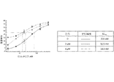
HCC827細胞を、実施例1および2で説明したように培養および処置した。このアッセイで使用したエルロチニブの最高濃度は40nMであり、化合物I-5の濃度は2μMおよび4μMであった。エルロチニブについて観察されたEC50値は、化合物I-5が存在しなかった場合に12.15nM、化合物I-5が2μMの濃度で存在した場合に2.95nM、化合物I-5が4μMの濃度で存在した場合に<0.1nMであった。図3を参照のこと。
Example 3. Combination of erlotinib with molecules effective against quiescent cancer cells HCC827 cells were cultured and treated as described in Examples 1 and 2. The highest concentration of erlotinib used in this assay was 40 nM and the concentrations of compound I-5 were 2 μM and 4 μM. EC 50 values observed for erlotinib were 12.15 nM when Compound I-5 was not present, 2.95 nM when Compound I-5 was present at a concentration of 2 μM, and Compound I-5 at a concentration of 4 μM. <0.1 nM when present at . See FIG.
PC9細胞を、実施例1および2で説明したように培養および処置した。このアッセイで使用したエルロチニブの最高濃度は40nMであり、化合物I-5の濃度はそれぞれ2μMおよび4μMであった。エルロチニブについて観察されたEC50値は、化合物I-5が存在しなかった場合に17.3nM、化合物I-5が2μMの濃度で存在した場合に9.3nM、化合物I-5が4μMの濃度で存在した場合に3.7nMであった。図4を参照のこと。 PC9 cells were cultured and treated as described in Examples 1 and 2. The highest concentration of erlotinib used in this assay was 40 nM, and the concentrations of compound I-5 were 2 μM and 4 μM, respectively. EC 50 values observed for erlotinib were 17.3 nM when Compound I-5 was not present, 9.3 nM when Compound I-5 was present at a concentration of 2 μM, and Compound I-5 at a concentration of 4 μM. 3.7 nM when present at . See FIG.
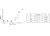
A549細胞を、実施例1および2で説明したように培養および処置した。このアッセイで使用したエルロチニブの最高濃度は10μMであり、化合物I-7の濃度はそれぞれ3μMおよび6μMであった。エルロチニブについて観察されたEC50値は、化合物I-7が存在しなかった場合に10.4μM、化合物I-7が3μMの濃度で存在した場合に5.9μM、化合物I-7が6μMの濃度で存在した場合に0.4μMであった。図5を参照のこと。 A549 cells were cultured and treated as described in Examples 1 and 2. The highest concentration of erlotinib used in this assay was 10 μM and the concentrations of compound I-7 were 3 μM and 6 μM, respectively. EC 50 values observed for erlotinib were 10.4 μM when compound I-7 was not present, 5.9 μM when compound I-7 was present at a concentration of 3 μM, and compound I-7 at a concentration of 6 μM. 0.4 μM when present at . See FIG.
PANC1細胞を、実施例1および2で説明したように培養および処置した。このアッセイで使用したエルロチニブの最高濃度は30μMであり、化合物I-5の濃度はそれぞれ2.5μMおよび5μMであった。エルロチニブについて観察されたEC50値は、化合物I-5が存在しなかった場合に>30μM、化合物I-5が2.5μMの濃度で存在した場合に>30μM、化合物I-5が5μMの濃度で存在した場合に14μMであった。図6を参照のこと。 PANC1 cells were cultured and treated as described in Examples 1 and 2. The highest concentration of erlotinib used in this assay was 30 μM and the concentrations of compound I-5 were 2.5 μM and 5 μM, respectively. Observed EC 50 values for erlotinib were >30 μM when compound I-5 was not present, >30 μM when compound I-5 was present at a concentration of 2.5 μM, and compound I-5 at a concentration of 5 μM. 14 μM when present at . See FIG.
MiaPaCa-2細胞を、実施例1および2で説明したように培養および処置した。このアッセイで使用したエルロチニブの最高濃度は30μMであり、化合物I-5の濃度はそれぞれ2.5μMおよび5μMであった。エルロチニブについて観察されたEC50値は、化合物I-5が存在しなかった場合に>30μM、化合物I-5が2.5μMの濃度で存在した場合に6.4μM、化合物I-5が5μMの濃度で存在した場合に3.5μMであった。図7を参照のこと。 MiaPaCa-2 cells were cultured and treated as described in Examples 1 and 2. The highest concentration of erlotinib used in this assay was 30 μM and the concentrations of compound I-5 were 2.5 μM and 5 μM, respectively. Observed EC 50 values for erlotinib were >30 μM when compound I-5 was not present, 6.4 μM when compound I-5 was present at a concentration of 2.5 μM, and compound I-5 at 5 μM. concentration was 3.5 μM when present. See FIG.
これらの実験では、可逆性EGFR阻害剤であるエルロチニブを化合物I-5と組み合わせると、HCC827、PC9、およびA549非小細胞肺癌細胞株に対するエルロチニブの細胞毒性が著しく増加した(EC50値の低下)ことが証明された。さらに、細胞毒性の著しい増加(より低いEC50値)が、PANC1およびMiaPaCa-2膵癌細胞株に対して観察された。この結果は、EGFRの比較的低い発現を有する、MiaPaCa-2細胞株で特に予想外であった。 In these experiments, combining the reversible EGFR inhibitor erlotinib with compound I-5 significantly increased the cytotoxicity of erlotinib against HCC827, PC9 , and A549 non-small cell lung cancer cell lines (decreased EC50 values). It was proved. Furthermore, a significant increase in cytotoxicity (lower EC50 values) was observed against PANC1 and MiaPaCa -2 pancreatic cancer cell lines. This result was particularly unexpected in the MiaPaCa-2 cell line, which has relatively low expression of EGFR.
実施例4. 静止癌細胞に対して有効な分子とアファチニブとの組み合わせ
H1975細胞を、実施例1および2で説明したように培養および処置した。このアッセイで使用したアファチニブの最高濃度は500nMであり、化合物I-7の濃度は2μMおよび4μMであった。アファチニブについて観察されたEC50値は、化合物I-7が存在しなかった場合に89.4nM、化合物I-7が2μMの濃度で存在した場合に25.2nM、化合物I-7が4μMの濃度で存在した場合に8.2nMであった。図8を参照のこと。
Example 4. Afatinib combination with molecules effective against quiescent cancer cells H1975 cells were cultured and treated as described in Examples 1 and 2. The highest concentration of afatinib used in this assay was 500 nM and the concentrations of compound I-7 were 2 μM and 4 μM. EC 50 values observed for afatinib were 89.4 nM when Compound I-7 was not present, 25.2 nM when Compound I-7 was present at a concentration of 2 μM, and Compound I-7 at a concentration of 4 μM. 8.2 nM when present at . See FIG.
HCC827細胞を、実施例1および2で説明したように培養および処置した。このアッセイで使用したアファチニブの最高濃度は50nMであり、化合物I-7の濃度は3μMおよび6μMであった。アファチニブについて観察されたEC50値は、化合物I-7が存在しなかった場合に3.8nM、化合物I-7が3μMの濃度で存在した場合に1.6nM、化合物I-7が6μMの濃度で存在した場合に0.2nMであった。図9を参照のこと。 HCC827 cells were cultured and treated as described in Examples 1 and 2. The highest concentration of afatinib used in this assay was 50 nM and the concentrations of compound I-7 were 3 μM and 6 μM. EC 50 values observed for afatinib were 3.8 nM when compound I-7 was not present, 1.6 nM when compound I-7 was present at a concentration of 3 μM, and compound I-7 at a concentration of 6 μM. 0.2 nM when present at . See FIG.
PC9細胞を、実施例1および2で説明したように培養および処置した。このアッセイで使用したアファチニブの最高濃度は5nMであり、化合物I-7の濃度は4μMおよび8μMであった。アファチニブについて観察されたEC50値は、化合物I-7が存在しなかった場合に2.1nM、化合物I-7が3μMの濃度で存在した場合に1.3nM、化合物I-7が6μMの濃度で存在した場合に0.4nMであった。図10を参照のこと。 PC9 cells were cultured and treated as described in Examples 1 and 2. The highest concentration of afatinib used in this assay was 5 nM and the concentrations of compound I-7 were 4 μM and 8 μM. EC 50 values observed for afatinib were 2.1 nM when compound I-7 was not present, 1.3 nM when compound I-7 was present at a concentration of 3 μM, and compound I-7 at a concentration of 6 μM. 0.4 nM when present at . See FIG.
A549細胞を、実施例1および2で説明したように培養および処置した。このアッセイで使用したアファチニブの最高濃度は10μMであり、化合物I-7の濃度は4μMおよび8μMであった。アファチニブについて観察されたEC50値は、化合物I-7が存在しなかった場合に4.7μM、化合物I-7が3μMの濃度で存在した場合に2.6μM、化合物I-7が6μMの濃度で存在した場合に1.0μMであった。図11を参照のこと。 A549 cells were cultured and treated as described in Examples 1 and 2. The highest concentration of afatinib used in this assay was 10 μM and the concentrations of compound I-7 were 4 μM and 8 μM. EC 50 values observed for afatinib were 4.7 μM when compound I-7 was not present, 2.6 μM when compound I-7 was present at a concentration of 3 μM, and compound I-7 at a concentration of 6 μM. 1.0 μM when present at . See FIG.
PANC1細胞を、実施例1および2で説明したように培養および処置した。このアッセイで使用したアファチニブの最高濃度は10μMであり、化合物I-7の濃度は2μMおよび4μMであった。アファチニブについて観察されたEC50値は、化合物I-7が存在しなかった場合に1.9μM、化合物I-7が2μMの濃度で存在した場合に1.8μM、化合物I-7が4μMの濃度で存在した場合に1.5μMであった。図12を参照のこと。 PANC1 cells were cultured and treated as described in Examples 1 and 2. The highest concentration of afatinib used in this assay was 10 μM and the concentrations of compound I-7 were 2 μM and 4 μM. EC 50 values observed for afatinib were 1.9 μM when compound I-7 was not present, 1.8 μM when compound I-7 was present at a concentration of 2 μM, and compound I-7 at a concentration of 4 μM. 1.5 μM when present at . See FIG.
この実験では、不可逆性EGFR阻害剤であるアファチニブを化合物I-7と組み合わせると、H1975細胞および他の非小細胞肺癌細胞であるHCC827、PC9、およびA549に対するアファチニブの細胞毒性が著しく増加したこと(より低いEC50値)が証明された。さらに、アファチニブを化合物I-7と組み合わせると、PANC1膵癌細胞に対する細胞毒性が増大したことが証明された。この最後の組み合わせは、相乗的なものではない、あるいは、EC50値の劇的な減少を引き起こさないかもしれないが、単一療法による処置では生存する、新生物中の耐性のある静止細胞集団を根絶することにより、EGFR阻害剤での単一療法と比べて処置の有効性の著しい改善を十分にもたらし得る。 In this experiment, combining the irreversible EGFR inhibitor afatinib with compound I-7 significantly increased the cytotoxicity of afatinib against H1975 cells and other non-small cell lung cancer cells HCC827, PC9, and A549 ( lower EC50 values) were demonstrated. Furthermore, combining afatinib with compound I-7 demonstrated increased cytotoxicity against PANC1 pancreatic cancer cells. This last combination may not be synergistic or cause dramatic reductions in EC50 values, but resistant quiescent cell populations in neoplasms that survive treatment with monotherapy Eradication may well lead to significant improvement in treatment efficacy compared to monotherapy with EGFR inhibitors.
実施例5. 静止癌細胞に対して有効な分子とオシメルチニブとの組み合わせ
H1975細胞を、実施例1および2で説明したように培養および処置した。このアッセイで使用したオシメルチニブの最高濃度は50nMであり、化合物I-7の濃度は2μMおよび4μMであった。観察されたEC50値は、化合物I-7が存在しなかった場合に8.7nM、化合物I-7が2μMの濃度で存在した場合に7.9nM、化合物I-7が4μMの濃度で存在した場合に4.2nMであった。図13を参照のこと。
Example 5. Combination of molecules effective against quiescent cancer cells with osimertinib H1975 cells were cultured and treated as described in Examples 1 and 2. The highest concentration of osimertinib used in this assay was 50 nM and the concentrations of compound I-7 were 2 μM and 4 μM. The EC 50 values observed were 8.7 nM when compound I-7 was not present, 7.9 nM when compound I-7 was present at a concentration of 2 μM, and compound I-7 was present at a concentration of 4 μM. was 4.2 nM when See FIG.
PANC1細胞を、実施例1および2で説明したように培養および処置した。このアッセイで使用したオシメルチニブの最高濃度は20μMであり、化合物I-7の濃度は2μMおよび4μMであった。観察されたEC50値は、化合物I-7が存在しなかった場合に5.3μM、化合物I-7が2μMの濃度で存在した場合に4.6μM、化合物I-7が4μMの濃度で存在した場合に4.5μMであった。図14を参照のこと。 PANC1 cells were cultured and treated as described in Examples 1 and 2. The highest concentration of osimertinib used in this assay was 20 μM, and the concentrations of compound I-7 were 2 μM and 4 μM. The EC50 values observed were 5.3 μM when compound I-7 was not present, 4.6 μM when compound I-7 was present at a concentration of 2 μM, and compound I-7 was present at a concentration of 4 μM. was 4.5 μM when See FIG.
この実験では、不可逆性EGFR阻害剤であるオシメルチニブを化合物I-7と組み合わせると、H1975細胞に対するオシメルチニブの細胞毒性が著しく増加したこと(より低いEC50値)が証明された。さらに、オシメルチニブを化合物I-7と組み合わせると、PANC1膵癌細胞に対する細胞毒性が増大したことが証明された。この最後の組み合わせは、相乗的なものではない、あるいは、EC50値の劇的な減少を引き起こさないかもしれないが、単一療法による処置では生存する、新生物中の耐性のある静止細胞集団を根絶することにより、EGFR阻害剤での単一療法と比べて処置の有効性の著しい改善を十分にもたらし得る。 This experiment demonstrated that combining the irreversible EGFR inhibitor osimertinib with compound I-7 significantly increased the cytotoxicity of osimertinib against H1975 cells (lower EC 50 values). Furthermore, combining osimertinib with compound I-7 demonstrated increased cytotoxicity against PANC1 pancreatic cancer cells. This last combination may not be synergistic or cause dramatic reductions in EC50 values, but resistant quiescent cell populations in neoplasms that survive treatment with monotherapy Eradication may well lead to significant improvement in treatment efficacy compared to monotherapy with EGFR inhibitors.
実施例6. オシメルチニブ、および静止癌細胞に対して有効な分子とオシメルチニブとの組み合わせの細胞周期効果および細胞毒性
H1975細胞を、実施例1および2に記載のとおりに培養し、処置し、分析した。異なる濃度のオシメルチニブ、化合物I-7、またはオシメルチニブおよび化合物I-7の両方が存在する場合の結果を図15および図16に示す。
Example 6. Cell cycle effects and cytotoxicity of osimertinib and combinations of osimertinib with molecules effective against quiescent cancer cells H1975 cells were cultured, treated and analyzed as described in Examples 1 and 2. Results in the presence of different concentrations of osimertinib, compound I-7, or both osimertinib and compound I-7 are shown in FIGS.
これらの実験では、H1975細胞をオシメルチニブに暴露することで細胞周期分布に変化が生じ、大部分の細胞を静止(G0)状態に誘発するのに、血清飢餓(FBS-)よりもオシメルチニブが有効であることが証明された。無血清培地(FBS-)および標準的な成長培地(FBS+)でインキュベートされたものである、正常に増殖するH1975細胞の細胞周期分布を比較のため示す。オシメルチニブは、細胞が、オシメルチニブで処置する前にG0にある細胞の割合を増やすために正常な成長培地(FBS+)で予めインキュベートされているか、無血清(FBS-)培地で予め飢餓状態にされている(pre-starved)かに関わらず、細胞周期分布に変化をもたらした。さらに、化合物I-7とオシメルチニブとの組み合わせは、サブG0集団として証明され生存率測定によって証明されるアポトーシス細胞の著しい増加によって記録されるように、G0にある細胞の割合を減らし、結果として生じる細胞毒性を大いに高めた。この効果は、細胞が成長培地で予めインキュベートされたか、または血清飢餓条件下にあったかに関係なく、観察された。 In these experiments, exposure of H1975 cells to osimertinib resulted in changes in cell cycle distribution, with osimertinib being more effective than serum starvation (FBS-) in inducing most cells into a quiescent (G 0 ) state. proved to be Cell cycle distributions of normally growing H1975 cells incubated in serum-free medium (FBS-) and standard growth medium (FBS+) are shown for comparison. Osimertinib was used when cells were pre-incubated with normal growth medium (FBS+) or pre-starved with serum-free (FBS-) medium to increase the percentage of cells in G0 prior to treatment with osimertinib . induced changes in cell cycle distribution, whether pre-starved or not. Furthermore, the combination of compound I-7 and osimertinib reduced the proportion of cells in G 0 as documented by a significant increase in apoptotic cells evidenced by viability measurements as evidenced by sub-G 0 populations, resulting in greatly enhanced the cytotoxicity produced as This effect was observed regardless of whether the cells were pre-incubated with growth medium or under serum starvation conditions.
実施例7. 静止癌細胞に対して有効な分子とロシレチニブとの組み合わせ
H1975細胞を、実施例1および2で説明したように培養および処置した。このアッセイで使用したロシレチニブの最高濃度は500nMであり、化合物I-7の濃度は2μMおよび4μMであった。ロシレチニブの阻害について観察されたEC50値は、化合物I-7が存在しなかった場合に351nM、化合物I-7が2μMの濃度で存在した場合に22.5nM、化合物I-7が4μMの濃度で存在した場合に14.9nMであった。図17を参照のこと。
Example 7. Combination of rociletinib with molecules effective against quiescent cancer cells H1975 cells were cultured and treated as described in Examples 1 and 2. The highest concentration of rociletinib used in this assay was 500 nM and the concentrations of compound I-7 were 2 μM and 4 μM. EC 50 values observed for inhibition of rociletinib were 351 nM when Compound I-7 was not present, 22.5 nM when Compound I-7 was present at a concentration of 2 μM, and Compound I-7 at a concentration of 4 μM. 14.9 nM when present at . See FIG.
PANC1細胞を、実施例1および2で説明したように培養および処置した。このアッセイで使用したロシレチニブの最高濃度は20μMであり、化合物I-7の濃度は2μMおよび4μMであった。観察されたEC50値は、化合物I-7が存在しなかった場合に11.4μM、化合物I-7が2μMの濃度で存在した場合に9.9μM、化合物I-7が4μMの濃度で存在した場合に7.1μMであった。図18を参照のこと。 PANC1 cells were cultured and treated as described in Examples 1 and 2. The highest concentration of rociletinib used in this assay was 20 μM and the concentrations of compound I-7 were 2 μM and 4 μM. Observed EC 50 values were 11.4 μM when compound I-7 was not present, 9.9 μM when compound I-7 was present at a concentration of 2 μM, and compound I-7 was present at a concentration of 4 μM. was 7.1 μM when See FIG.
この実験では、不可逆性EGFR阻害剤であるロシレチニブを化合物I-7と組み合わせると、H1975細胞に対するロシレチニブの細胞毒性が著しく増加したこと(より低いEC50値)が証明された。さらに、オシメルチニブを化合物I-7と組み合わせると、PANC1膵癌細胞に対する細胞毒性が増加したこと(より低いEC50値)が証明された。この最後の組み合わせは、相乗的なものではない、あるいは、EC50値の劇的な減少を引き起こさないかもしれないが、単一療法による処置では生存する、新生物中の耐性のある静止細胞集団を根絶することにより、EGFR阻害剤での単一療法と比べて処置の有効性の著しい改善を十分にもたらし得る。 This experiment demonstrated that when the irreversible EGFR inhibitor, rosiletinib, was combined with compound I-7, the cytotoxicity of rosiletinib against H1975 cells was significantly increased (lower EC 50 values). Furthermore, combining osimertinib with compound I-7 demonstrated increased cytotoxicity (lower EC 50 values) against PANC1 pancreatic cancer cells. This last combination may not be synergistic or cause dramatic reductions in EC50 values, but resistant quiescent cell populations in neoplasms that survive treatment with monotherapy Eradication may well lead to significant improvement in treatment efficacy compared to monotherapy with EGFR inhibitors.
実施例8. ロシレチニブ、および静止癌細胞に対して有効な分子とロシレチニブとの組み合わせの細胞周期効果および細胞毒性
H1975細胞を、実施例1および2に記載のとおりに培養し、処置し、分析した。異なる濃度のロシレチニブ、化合物I-7、またはロシレチニブおよび化合物I-7の両方が存在する場合の結果を図19に示す。
Example 8. Cell Cycle Effects and Cytotoxicity of Rociletinib and Combinations of Rociletinib with Molecules Effective Against Quiescent Cancer Cells H1975 cells were cultured, treated and analyzed as described in Examples 1 and 2. Results in the presence of different concentrations of rosiletinib, compound I-7, or both rosiletinib and compound I-7 are shown in FIG.
この実施例では、H1975細胞を、血清飢餓(FBS-)条件下で24時間インキュベートし、その後、処置の有無にかかわらず正常な成長培地(FBS+)中に「放出(released)」した。これらの条件下で、ロシレチニブへの曝露は、静止状態(G0)にある細胞の割合を著しく増加させた。細胞をロシレチニブと化合物I-7との組み合わせで同時に処置した場合、サブ-G0の割合によって決定されるようなアポトーシス細胞の大幅な増加によって判断されるように、このような静止細胞の割合の増加は観察されず、ロシレチニブの細胞毒性の大幅な増加が観察された。 In this example, H1975 cells were incubated under serum starvation (FBS-) conditions for 24 hours and then "released" into normal growth medium (FBS+) with or without treatment. Under these conditions, exposure to rociletinib significantly increased the proportion of cells in the quiescent state (G 0 ). When cells were treated simultaneously with the combination of rociletinib and compound I-7, the percentage of such quiescent cells decreased, as judged by a significant increase in apoptotic cells, as determined by the percentage of sub-G0. No increase was observed and a significant increase in rociletinib cytotoxicity was observed.
実施例9. EGFR阻害剤でH1975細胞を処置した際のDYRK1Bの誘導
H1975細胞を、実施例1および2で説明したように培養および処置した。ウエスタンブロット分析では、細胞を6ウェルプレートに(細胞の大きさおよび増殖速度に応じて)5×10⁵~9×10⁵細胞/ウェルで播種し、24時間付着させ、その後、化合物で24時間処置し、採取した。免疫ブロット法を、Cell Signaling Technologies Western Blotting protocol(www.cellsignal.com)に記載されるとおりに、従来の技術を用いて実行した。
Example 9. Induction of DYRK1B upon treatment of H1975 cells with EGFR inhibitors H1975 cells were cultured and treated as described in Examples 1 and 2. For Western blot analysis, cells were seeded in 6-well plates at 5×10 5 to 9×10 5 cells/well (depending on cell size and growth rate) and allowed to adhere for 24 hours, followed by compounds for 24 hours. treated and collected. Immunoblotting was performed using conventional techniques as described in Cell Signaling Technologies Western Blotting protocol (www.cellsignal.com).
ブロッティングに使用した抗体は、Cell Signaling Technology(CST)からのものであった:DYRK1B(D40D1)Rabbit mAb #5672;EGF Receptor(D38B1)XP(登録商標)Rabbit mAb #4267;Phospho-EGF Receptor(Tyr1068)(D7A5)XP(登録商標)Rabbit mAb #3777;β-Actin(13E5)Rabbit mAb #4970;抗ウサギIgG、HRP結合抗体 #7074。5%BSA(CST #9998)を含むPrimary Antibody Dilution Buffer 1X TBSTを用いた。検出には、SignalFire(商標)ECL Reagent(CST #6883)を使用した。 Antibodies used for blotting were from Cell Signaling Technology (CST): DYRK1B (D40D1) Rabbit mAb #5672; EGF Receptor (D38B1) XP® Rabbit mAb #4267; β-Actin (13E5) Rabbit mAb #4970; anti-rabbit IgG, HRP-conjugated antibody #7074. Primary Antibody Dilution Buffer 1X with 5% BSA (CST #9998) TBST was used. SignalFire™ ECL Reagent (CST #6883) was used for detection.
ウエスタンブロット分析により観察されたような、ロシレチニブ、オシメルチニブ、ダコミチニブ、およびアファチニブで24時間処置した後のH1975細胞におけるDYRK1B、ph-Y1068 EGFR、全EGFR、およびβ-アクチンの発現レベルを図20に示す。DYRK1Bタンパク質の発現を、FBSを含有する標準的な成長培地(FBS+)または無血清培地(FBS-)でインキュベートされた未処置細胞と比較した。EGFR阻害剤それぞれによる処置が、EGFRリン酸化を抑制し、血清飢餓により生じたのと同様であるかまたはそれより高いDYRK1Bタンパク質の発現も誘発したことが、証明された。 Expression levels of DYRK1B, ph-Y1068 EGFR, total EGFR, and β-actin in H1975 cells after 24 h treatment with rosiletinib, osimertinib, dacomitinib, and afatinib, as observed by Western blot analysis, are shown in FIG. . DYRK1B protein expression was compared to untreated cells incubated in standard growth medium containing FBS (FBS+) or serum-free medium (FBS-). It was demonstrated that treatment with each of the EGFR inhibitors suppressed EGFR phosphorylation and also induced DYRK1B protein expression similar to or higher than that produced by serum starvation.
実施例10. 静止癌細胞に対して有効な分子とダコミチニブとの組み合わせ
H1975細胞を、実施例1および2で説明したように培養および処置した。このアッセイで使用したダコミチニブの最高濃度は500nMであり、化合物I-7の濃度は2μMおよび4μMであった。ダコミチニブ阻害について観察されたEC50値は、化合物I-7が存在しなかった場合に110.3nM、化合物I-7が2μMの濃度で存在した場合に88.6nM、化合物I-7が4μMの濃度で存在した場合に34.8nMであった。図21を参照のこと。
Example 10. Combination of molecules effective against quiescent cancer cells with dacomitinib H1975 cells were cultured and treated as described in Examples 1 and 2. The highest concentration of dacomitinib used in this assay was 500 nM and the concentrations of compound I-7 were 2 μM and 4 μM. EC50 values observed for dacomitinib inhibition were 110.3 nM when compound I-7 was not present, 88.6 nM when compound I-7 was present at a concentration of 2 μM, and compound I-7 at 4 μM. When present at a concentration of 34.8 nM. See FIG.
PANC1細胞を、実施例1および2で説明したように培養および処置した。このアッセイで使用したダコミチニブの最高濃度は10μMであり、化合物I-7の濃度は2μMおよび4μMであった。観察されたEC50値は、化合物I-7が存在しなかった場合に11.4μM、化合物I-7が2μMの濃度で存在した場合に9.9μM、化合物I-7が4μMの濃度で存在した場合に7.1μMであった。図22を参照のこと。 PANC1 cells were cultured and treated as described in Examples 1 and 2. The highest concentration of dacomitinib used in this assay was 10 μM and the concentrations of compound I-7 were 2 μM and 4 μM. Observed EC 50 values were 11.4 μM when compound I-7 was not present, 9.9 μM when compound I-7 was present at a concentration of 2 μM, and compound I-7 was present at a concentration of 4 μM. was 7.1 μM when See Figure 22.
この実験では、不可逆性EGFR阻害剤であるダコミチニブを化合物I-7と組み合わせると、H1975細胞に対するダコミチニブの細胞毒性が著しく増加したこと(より低いEC50値)が証明された。さらに、オシメルチニブを化合物I-7と組み合わせると、PANC1膵癌細胞に対する細胞毒性が増加したことが証明された。この最後の組み合わせは、相乗的なものではない、あるいは、EC50値の劇的な減少を引き起こさないかもしれないが、単一療法による処置では生存する、新生物中の耐性のある静止細胞集団を根絶することにより、EGFR阻害剤での単一療法と比べて処置の有効性の著しい改善を十分にもたらし得る。 This experiment demonstrated that combining the irreversible EGFR inhibitor dacomitinib with compound I-7 significantly increased the cytotoxicity of dacomitinib against H1975 cells (lower EC 50 values). Furthermore, combining osimertinib with compound I-7 demonstrated increased cytotoxicity against PANC1 pancreatic cancer cells. This last combination may not be synergistic or cause dramatic reductions in EC50 values, but resistant quiescent cell populations in neoplasms that survive treatment with monotherapy Eradication may well lead to significant improvement in treatment efficacy compared to monotherapy with EGFR inhibitors.
実施例11. AZ191の細胞周期効果および化合物I-7との比較
SW620細胞を、実施例1に記載のとおりに培養し、処置した。ヨウ化プロピジウム(PI)染色では、フローサイトメトリー用のGuava Cell Cycle試薬(EMD Millipore)に付属の製造業者のプロトコルに従った。535nmで励起するよう緑色レーザーを用いてGuava PCA-96フローサイトメーター(EMD Millipore)によって測定を行い、617nmでの発光をモニタリングした。
Example 11. Cell Cycle Effects of AZ191 and Comparison with Compound I-7 SW620 cells were cultured and treated as described in Example 1. For propidium iodide (PI) staining, the manufacturer's protocol supplied with the Guava Cell Cycle reagents for flow cytometry (EMD Millipore) was followed. Measurements were performed with a Guava PCA-96 flow cytometer (EMD Millipore) using a green laser to excite at 535 nm and monitor emission at 617 nm.
異なる濃度のAZ191が存在した場合の結果を図23に示す。データは、2つの複製物の平均である。 Results in the presence of different concentrations of AZ191 are shown in FIG. Data are the average of two replicates.
SW620細胞を、実施例1で説明したように培養し、処置し、分析した。ヨウ化プロピジウム(PI)染色では、フローサイトメトリー用のGuava Cell Cycle試薬(EMD Millipore)に付属の製造業者のプロトコルに従った。535nmで励起するよう緑色レーザーを用いてGuava PCA-96フローサイトメーター(EMD Millipore)によって測定を行い、617nmでの発光をモニタリングした。 SW620 cells were cultured, treated and analyzed as described in Example 1. For propidium iodide (PI) staining, the manufacturer's protocol supplied with the Guava Cell Cycle reagents for flow cytometry (EMD Millipore) was followed. Measurements were performed with a Guava PCA-96 flow cytometer (EMD Millipore) using a green laser to excite at 535 nm and monitor emission at 617 nm.
異なる濃度の化合物I-7が存在した場合の結果を図24に示す。データは、2つの複製物の平均である。 Results are shown in FIG. 24 when different concentrations of compound I-7 were present. Data are the average of two replicates.
これらの実験では、SW620細胞を、異なる濃度のAZ191または化合物I-7を含有するFBS-培地で24時間インキュベートした。これらの条件下で、AZ191への曝露は、G0+G1期の細胞の割合に変化が観察されなかったことに基づくと、静止状態(G0)にある細胞の割合を減少させなかった。同じ条件下で、同じかまたはより低い濃度の化合物I-7への曝露は、G0+G1期の細胞の割合が著しく減少したことに基づくと、静止状態(G0)にある細胞の割合を著しく減少させた。 In these experiments, SW620 cells were incubated with FBS-medium containing different concentrations of AZ191 or compound I-7 for 24 hours. Under these conditions, exposure to AZ191 did not decrease the percentage of cells in the quiescent state (G 0 ) based on no observed change in the percentage of cells in the G 0 +G 1 phase. Under the same conditions, exposure to the same or lower concentrations of compound I-7 increased the percentage of cells in the quiescent state (G 0 ) based on the marked decrease in the percentage of cells in the G 0 +G 1 phase. significantly decreased.
AZ191は、17nMでDYRK1Bを阻害した(Ashford AL, Oxley D, Kettle J, Hudson K, Guichard S, Cook SJ, Lochhead PA (2014) A novel DYRK1B inhibitor AZ191 demonstrates that DYRK1B acts independently of GSK3beta to phosphorylate cyclin D1 at Thr(286), not Thr(288). Biochemical Journal 457, 43-56)。 AZ191は、17nMでDYRK1Bを阻害した(Ashford AL, Oxley D, Kettle J, Hudson K, Guichard S, Cook SJ, Lochhead PA (2014) A novel DYRK1B inhibitor AZ191 demonstrates that DYRK1B acts independently of GSK3beta to phosphorylate cyclin D1 at Thr (286), not Thr (288).Biochemical Journal 457, 43-56).
この実験では、すべてのDYRK1阻害剤が静止癌細胞に対して有効とは限らないことが証明された。 This experiment demonstrated that not all DYRK1 inhibitors are effective against quiescent cancer cells.
実施例12. 3D細胞培養(スフェロイド)における細胞生存率測定の一般的手順
3D培養での生存率分析では、処置の初めに、400~600μMの直径のスフェロイド形成を目的として、細胞を96ウェルULA(超低付着(ultra-low attachment))プレート(Corning #4515)に、細胞の大きさおよび増殖速度に応じて5×10³~6×10³細胞/ウェルで播種した。細胞は、37℃で、湿気のある5% CO2環境において(細胞株に応じて)2~3日間インキュベートされ、緊密なスフェロイド形成を可能にした。この処置では、50μLの培地を各ウェルから除去し、化合物を有する新鮮培地と取り換えた。この処置は、1:3連続希釈法において少なくとも6つの異なる濃度の化合物を用いて実行した。結果を読み取る前に、細胞を、37℃で5% CO2インキュベーターにおいて4~10日間インキュベートした。処置時間が4日を超えた場合、各ウェルの70μLの培地を、試験化合物を含有する新鮮培地と、3日おきに取り換えた。各処置は、2回行った。結果を、CellTiter-Glo(商標)3D Luminescent Cell Viability Assay(Promega、カタログ番号G9682)によって、SpectraMAX Gemini分光光度計(Molecular Devices)を用いて製造業者の指示に従って、分析した。分析前に、スフェロイドを、50倍の倍率で撮影した。
Example 12. General Procedure for Cell Viability Measurements in 3D Cell Cultures (Spheroids) For viability analysis in 3D cultures, cells were placed in 96-well ULA (ultra-low attachment) cells at the beginning of treatment for spheroid formation of 400-600 μM diameter. (ultra-low attachment) plates (Corning #4515) were seeded at 5×10 3 to 6×10 3 cells/well depending on cell size and growth rate. Cells were incubated at 37° C. in a humid 5% CO 2 environment for 2-3 days (depending on the cell line) to allow tight spheroid formation. For this treatment, 50 μL of medium was removed from each well and replaced with fresh medium with compound. The treatment was performed with at least 6 different concentrations of compound in a 1:3 serial dilution. Cells were incubated at 37° C. in a 5% CO 2 incubator for 4-10 days before reading the results. If treatment time exceeded 4 days, 70 μL of medium in each well was replaced with fresh medium containing test compound every 3 days. Each treatment was performed twice. Results were analyzed by the CellTiter-Glo™ 3D Luminescent Cell Viability Assay (Promega, catalog number G9682) using a SpectraMAX Gemini spectrophotometer (Molecular Devices) according to the manufacturer's instructions. Spheroids were photographed at 50x magnification before analysis.
実施例13. 3D細胞培養における静止癌細胞に対して有効な分子とアファチニブとの組み合わせ
H1975スフェロイドを、実施例1および12で説明したように培養および処置した。このアッセイで使用したアファチニブの濃度は100nMであり、化合物I-7の濃度は2.5μMであった。処置時間は7日であった。図25を参照のこと。
Example 13. Combination of molecules and afatinib effective against quiescent cancer cells in 3D cell culture H1975 spheroids were cultured and treated as described in Examples 1 and 12. The concentration of afatinib used in this assay was 100 nM and the concentration of compound I-7 was 2.5 μM. Treatment time was 7 days. See Figure 25.
この実験では、不可逆性EGFR阻害剤であるアファチニブを化合物I-7と組み合わせた療法により、H1975細胞の3D培養(スフェロイド)に対するアファチニブの細胞毒性が著しく増加した(より低いEC50値)ことが証明され;この組み合わせは、スフェロイドを完全に根絶したが、スフェロイドの一部は、アファチニブ単独での処置後も生存していた。 This experiment demonstrated that combination therapy of the irreversible EGFR inhibitor afatinib with compound I-7 significantly increased the cytotoxicity of afatinib (lower EC50 values) against 3D cultures (spheroids) of H1975 cells. this combination completely eradicated the spheroids, but some of the spheroids survived treatment with afatinib alone.
実施例14. 3D細胞培養における静止癌細胞に対して有効な分子とオシメルチニブとの組み合わせ
H1975スフェロイドを、実施例1、2、および12で説明したように7日間培養および処置した。このアッセイで使用したオシメルチニブの最高濃度は10nMであり、化合物I-7の濃度は2.5μMであった。標準的な成長培地(FBS+)では、オシメルチニブについて決定されたEC50値は、化合物I-7が存在しなかった場合に0.68nM、化合物I-7が2.5μMの濃度で存在した場合に0.28nMであった。血清飢餓(FBS-)条件下では、オシメルチニブについて決定されたEC50値は、化合物I-7が存在しなかった場合に3.3nM、化合物I-7が2.5μmの濃度で存在した場合に1.0nMであった。図26を参照のこと。
Example 14. Combination of osimertinib with molecules effective against quiescent cancer cells in 3D cell culture H1975 spheroids were cultured and treated as described in Examples 1, 2, and 12 for 7 days. The highest concentration of osimertinib used in this assay was 10 nM and the concentration of compound I-7 was 2.5 μM. In standard growth medium (FBS+), the EC 50 value determined for osimertinib was 0.68 nM when Compound I-7 was not present and 0.28 nM. Under serum starvation (FBS-) conditions, the EC50 value determined for osimertinib was 3.3 nM when compound I-7 was not present and It was 1.0 nM. See Figure 26.
この実験では、不可逆性EGFR阻害剤であるオシメルチニブを化合物I-7と組み合わせると、スフェロイドが標準的な培地(FBS+)で培養されるか枯渇培地(FBS-)で培養されるかに関わらず、H1975細胞の3D培養(スフェロイド)に対するオシメルチニブの細胞毒性が著しく増加したこと(EC50の低下)が証明された。意外にも、このEC50の低下率は、FBS+での3D培養よりもFBS-のほうが大きかった。 In this experiment, when the irreversible EGFR inhibitor osimertinib was combined with compound I-7, whether spheroids were cultured in standard medium (FBS+) or depleted medium (FBS-), Significantly increased cytotoxicity (decreased EC50) of osimertinib against 3D cultures (spheroids) of H1975 cells was demonstrated. Surprisingly, this EC50 reduction was greater with FBS- than with 3D cultures with FBS+.
本発明は、例としての実施形態を参照して具体的に図示および説明してきたが、形態および詳細のさまざまな変更が、特許請求の範囲により包含される本発明の範囲を逸脱せずに行われ得ることを、当業者は理解するであろう。 Although the invention has been particularly shown and described with reference to example embodiments, various changes in form and detail can be made without departing from the scope of the invention as encompassed by the claims. Those skilled in the art will understand that
〔実施の態様〕
(1) 新生物を有する被験者を処置する方法において、
前記被験者に、順次または同時に、DYRK1阻害剤を投与し、前記被験者にEGFR TKIを投与することを含み、前記DYRK1阻害剤は、式I:

式中、
R1は、置換もしくは非置換のC1~8アルキル、置換もしくは非置換のフェニル、または置換もしくは非置換のベンジルであり、
R2は、オプションとしてハロ、CN、NO2、NHC(O)C1~4アルキル、C1~4アルキル、OH、OC1~4アルキルから独立して選択された最大で4つの基で置換された、フェニルであり、2つの隣接する基およびそれらの介在炭素原子は、N、O、またはSから選択された1つ以上のヘテロ原子を含有する五~六員環を形成し得る、方法。
(2) 実施態様1に記載の方法において、
前記被験者に有効量の放射線療法を施すことをさらに含む、方法。
(3) 実施態様1に記載の方法において、
処置される前記新生物は、胆道癌、脳癌、乳癌、子宮頸癌、結腸癌、胃癌、腎癌、頭頸部癌、白血病、肝癌、小細胞肺癌、リンパ腫、卵巣癌、前立腺癌、直腸癌、肉腫、皮膚癌、精巣癌、甲状腺癌、子宮癌、膀胱癌、乳癌、大腸癌、肺癌、卵巣癌、および前立腺癌から選択される原発性または転移性の癌である、方法。
(4) 実施態様1に記載の方法において、
処置される前記新生物は、原発性または転移性の非小細胞肺癌である、方法。
(5) 実施態様1に記載の方法において、
処置される前記新生物は、原発性または転移性の膵癌である、方法。
[Mode of implementation]
(1) In a method of treating a subject with a neoplasm, comprising:
administering to said subject, either sequentially or concurrently, a DYRK1 inhibitor and administering to said subject an EGFR TKI, wherein said DYRK1 inhibitor comprises Formula I:
During the ceremony,
R 1 is substituted or unsubstituted C 1-8 alkyl, substituted or unsubstituted phenyl, or substituted or unsubstituted benzyl;
R 2 is optionally substituted with up to 4 groups independently selected from halo, CN, NO 2 , NHC(O)C 1-4 alkyl, C 1-4 alkyl, OH, OC 1-4 alkyl two adjacent groups and their intervening carbon atoms may form a five- to six-membered ring containing one or more heteroatoms selected from N, O, or S. .
(2) In the method of
The method further comprising administering an effective amount of radiation therapy to said subject.
(3) In the method of
The neoplasms to be treated include biliary tract cancer, brain cancer, breast cancer, cervical cancer, colon cancer, gastric cancer, renal cancer, head and neck cancer, leukemia, liver cancer, small cell lung cancer, lymphoma, ovarian cancer, prostate cancer, rectal cancer. , sarcoma, skin cancer, testicular cancer, thyroid cancer, uterine cancer, bladder cancer, breast cancer, colon cancer, lung cancer, ovarian cancer, and prostate cancer, a primary or metastatic cancer.
(4) In the method of
A method, wherein the neoplasm to be treated is primary or metastatic non-small cell lung cancer.
(5) In the method of
A method, wherein the neoplasm to be treated is primary or metastatic pancreatic cancer.
(6) 実施態様1に記載の方法において、
前記EGFR TKIは、ブリガチニブ、CUDC-101、エルロチニブ、ゲフィチニブ、イコチニブ、ラパチニブ、サピチニブ、テセバチニブ、チルホスチンAG 1478、バンデタニブ、およびバルリチニブから選択される、方法。
(7) 実施態様1に記載の方法において、
前記EGFR TKIは、AZD3759およびMTKi-327(JNJ-26483327)から選択される、方法。
(8) 実施態様1に記載の方法において、
前記EGFR TKIは、アファチニブ、カネルチニブ、CL-387785(EKI-785)、CNX-2006、ダコミチニブ、ナコチニブ(ASP8273)、ネラチニブ、オルムティニブ(HM61713)、オシメルチニブ、PD168393、ペリチニブ、ポジオチニブ、TAK285、ロシレチニブ、およびWZ4002のリストから選択される、方法。
(9) 実施態様1に記載の方法において、
前記EGFR TKIは、アリチニブ(ALS-1306;AST-1306)、AV-412(MP-412)、ナザルチニブ(EGF816)、およびピロチニブから選択される、方法。
(10) 実施態様1に記載の方法において、
前記DYRK1阻害剤は、I-1、I-2、I-3、I-4、I-5、I-6、およびI-7から選択される、方法。
(6) In the method of
The method, wherein the EGFR TKI is selected from brigatinib, CUDC-101, erlotinib, gefitinib, icotinib, lapatinib, sapitinib, tesevatinib, tyrphostin AG 1478, vandetanib, and vallitinib.
(7) In the method of
The method, wherein said EGFR TKI is selected from AZD3759 and MTKi-327 (JNJ-26483327).
(8) In the method of
The EGFR TKIs are afatinib, canertinib, CL-387785 (EKI-785), CNX-2006, dacomitinib, nacotinib (ASP8273), neratinib, ormtinib (HM61713), osimertinib, PD168393, peritinib, poziotinib, TAK2402 and rosiletinib method, selected from a list of
(9) In the method of
The method, wherein said EGFR TKI is selected from aritinib (ALS-1306; AST-1306), AV-412 (MP-412), nazartinib (EGF816), and pirotinib.
(10) In the method of
The method, wherein said DYRK1 inhibitor is selected from 1-1, 1-2, 1-3, 1-4, 1-5, 1-6, and 1-7.
(11) 実施態様10に記載の方法において、
前記EGFR TKIは、ブリガチニブ、CUDC-101、エルロチニブ、ゲフィチニブ、イコチニブ、ラパチニブ、サピチニブ、テセバチニブ、チルホスチンAG 1478、バンデタニブ、およびバルリチニブから選択される、方法。
(12) 実施態様10に記載の方法において、
前記EGFR TKIは、AZD3759およびMTKi-327(JNJ-26483327)から選択される、方法。
(13) 実施態様10に記載の方法において、
前記EGFR TKIは、アファチニブ、カネルチニブ、CL-387785(EKI-785)、CNX-2006、ダコミチニブ、ナコチニブ(ASP8273)、ネラチニブ、オルムティニブ(HM61713)、オシメルチニブ、PD168393、ペリチニブ、ポジオチニブ、TAK285、ロシレチニブ、およびWZ4002から選択される、方法。
(14) 実施態様10に記載の方法において、
前記EGFR TKIは、アリチニブ(ALS-1306;AST-1306)、AV-412(MP-412)、ナザルチニブ(EGF816)、およびピロチニブから選択される、方法。
(15) 実施態様10に記載の方法において、
処置される前記新生物は、胆道癌、脳癌、乳癌、子宮頸癌、結腸癌、胃癌、腎癌、頭頸部癌、白血病、肝癌、小細胞肺癌、リンパ腫、卵巣癌、前立腺癌、直腸癌、肉腫、皮膚癌、精巣癌、甲状腺癌、子宮癌、膀胱癌、乳癌、大腸癌、肺癌、卵巣癌、および前立腺癌から選択される原発性または転移性の癌である、方法。
(11) The method of
The method, wherein the EGFR TKI is selected from brigatinib, CUDC-101, erlotinib, gefitinib, icotinib, lapatinib, sapitinib, tesevatinib, tyrphostin AG 1478, vandetanib, and vallitinib.
(12) In the method of
The method, wherein said EGFR TKI is selected from AZD3759 and MTKi-327 (JNJ-26483327).
(13) In the method of
The EGFR TKIs are afatinib, canertinib, CL-387785 (EKI-785), CNX-2006, dacomitinib, nacotinib (ASP8273), neratinib, ormtinib (HM61713), osimertinib, PD168393, peritinib, poziotinib, TAK2402 and rosiletinib A method selected from.
(14) In the method of
The method, wherein said EGFR TKI is selected from aritinib (ALS-1306; AST-1306), AV-412 (MP-412), nazartinib (EGF816), and pirotinib.
(15) The method of
The neoplasms to be treated include biliary tract cancer, brain cancer, breast cancer, cervical cancer, colon cancer, gastric cancer, renal cancer, head and neck cancer, leukemia, liver cancer, small cell lung cancer, lymphoma, ovarian cancer, prostate cancer, rectal cancer. , sarcoma, skin cancer, testicular cancer, thyroid cancer, uterine cancer, bladder cancer, breast cancer, colon cancer, lung cancer, ovarian cancer, and prostate cancer, a primary or metastatic cancer.
(16) 実施態様10に記載の方法において、
処置される前記新生物は、原発性または転移性の非小細胞肺癌である、方法。
(17) 実施態様10に記載の方法において、
処置される前記新生物は、原発性または転移性の膵癌である、方法。
(18) 新生物を有する被験者を処置する方法において、
前記被験者に、順次または同時に、
(a)DYRK1阻害剤であって、生化学アッセイにおいて100nM以下のIC50で、DYRK1AまたはDYRK1Bキナーゼ活性を阻害し、このような阻害剤がない場合に見られるであろう静止癌細胞(in vitroまたはin vivo)の割合を、少なくとも10%だけ減少させる、DYRK1阻害剤と、
(b)EGFR TKIと、
を投与することを含む、方法。
(19) 実施態様18に記載の方法において、
前記DYRK1阻害剤は、式I:

式中、
R1は、置換もしくは非置換のC1~8アルキル、置換もしくは非置換のフェニル、または置換もしくは非置換のベンジルであり、
R2は、オプションとしてハロ、CN、NO2、NHC(O)C1~4アルキル、C1~4アルキル、OH、OC1~4アルキルから独立して選択された最大で4つの基で置換された、フェニルであり、2つの隣接する基およびそれらの介在炭素原子は、N、O、またはSから選択された1つ以上のヘテロ原子を含有する五~六員環を形成し得る、方法。
(20) 実施態様18に記載の方法において、
前記被験者に有効量の放射線療法を施すことをさらに含む、方法。
(16) In the method of
A method, wherein the neoplasm to be treated is primary or metastatic non-small cell lung cancer.
(17) The method of
A method, wherein the neoplasm to be treated is primary or metastatic pancreatic cancer.
(18) In a method of treating a subject with a neoplasm,
to said subject, either sequentially or simultaneously,
(a) A DYRK1 inhibitor that inhibits DYRK1A or DYRK1B kinase activity with an IC50 of 100 nM or less in biochemical assays and inhibits quiescent cancer cells that would be seen in the absence of such inhibitors (in vitro or in vivo) by at least 10%;
(b) EGFR TKIs;
A method comprising administering
(19) The method of embodiment 18, wherein
Said DYRK1 inhibitor has a formula I:
During the ceremony,
R 1 is substituted or unsubstituted C 1-8 alkyl, substituted or unsubstituted phenyl, or substituted or unsubstituted benzyl;
R 2 is optionally substituted with up to 4 groups independently selected from halo, CN, NO 2 , NHC(O)C 1-4 alkyl, C 1-4 alkyl, OH, OC 1-4 alkyl two adjacent groups and their intervening carbon atoms may form a five- to six-membered ring containing one or more heteroatoms selected from N, O, or S. .
(20) The method of embodiment 18, wherein
The method further comprising administering an effective amount of radiation therapy to said subject.
(21) 実施態様18に記載の方法において、
処置される前記新生物は、胆道癌、脳癌、乳癌、子宮頸癌、結腸癌、胃癌、腎癌、頭頸部癌、白血病、肝癌、肺癌、リンパ腫、卵巣癌、前立腺癌、直腸癌、肉腫、皮膚癌、精巣癌、甲状腺癌、子宮癌、膀胱癌、乳癌、大腸癌、小細胞肺癌、卵巣癌、および前立腺癌から選択される原発性または転移性の癌である、方法。
(22) 実施態様18に記載の方法において、
処置される前記新生物は、原発性または転移性の非小細胞肺癌である、方法。
(23) 実施態様18に記載の方法において、
処置される前記新生物は、原発性または転移性の膵癌である、方法。
(24) 実施態様18に記載の方法において、
前記EGFR TKIは、ブリガチニブ、CUDC-101、エルロチニブ、ゲフィチニブ、イコチニブ、ラパチニブ、サピチニブ、テセバチニブ、チルホスチンAG 1478、バンデタニブ、およびバルリチニブのリストから選択される、可逆性阻害剤である、方法。
(25) 実施態様18に記載の方法において、
前記EGFR TKIは、AZD3759およびMTKi-327(JNJ-26483327)から選択される、方法。
(21) The method of embodiment 18, wherein
The neoplasms to be treated include biliary tract cancer, brain cancer, breast cancer, cervical cancer, colon cancer, gastric cancer, renal cancer, head and neck cancer, leukemia, liver cancer, lung cancer, lymphoma, ovarian cancer, prostate cancer, rectal cancer, sarcoma. , skin cancer, testicular cancer, thyroid cancer, uterine cancer, bladder cancer, breast cancer, colon cancer, small cell lung cancer, ovarian cancer, and prostate cancer, primary or metastatic cancer.
(22) The method of embodiment 18, wherein
A method, wherein the neoplasm to be treated is primary or metastatic non-small cell lung cancer.
(23) The method of embodiment 18, wherein
A method, wherein the neoplasm to be treated is primary or metastatic pancreatic cancer.
(24) The method of embodiment 18, wherein
The method, wherein said EGFR TKI is a reversible inhibitor selected from the list of brigatinib, CUDC-101, erlotinib, gefitinib, icotinib, lapatinib, sapitinib, tesevatinib, tyrphostin AG 1478, vandetanib, and vallitinib.
(25) The method of embodiment 18, wherein
The method, wherein said EGFR TKI is selected from AZD3759 and MTKi-327 (JNJ-26483327).
(26) 実施態様18に記載の方法において、
前記EGFR TKIは、アファチニブ、カネルチニブ、CL-387785(EKI-785)、CNX-2006、ダコミチニブ、ナコチニブ(ASP8273)、ネラチニブ、オルムティニブ(HM61713)、オシメルチニブ、PD168393、ペリチニブ、ポジオチニブ、TAK285、ロシレチニブ、およびWZ4002のリストから選択される、不可逆性阻害剤である、方法。
(27) 実施態様18に記載の方法において、
前記EGFR TKIは、アリチニブ(ALS-1306;AST-1306)、AV-412(MP-412)、ナザルチニブ(EGF816)、およびピロチニブのリストから選択される、方法。
(28) 実施態様18に記載の方法において、
前記DYRK1阻害剤は、I-1、I-2、I-3、I-4、I-5、I-6、およびI-7から選択される、方法。
(29) 新生物を有する被験者を処置する方法において、
前記被験者に、順次または同時に、DYRK1阻害剤を投与し、前記被験者にEGFR TKIを投与することを含み、
前記EGFR TKIのEC50値は、細胞ベースのアッセイで決定される場合に、前記EGFR TKI単独での同じ処置と比べて、組み合わせ処置では少なくとも20%低い、方法。
(30) 実施態様29に記載の方法において、
前記DYRK1阻害剤は、式I:

式中、
R1は、置換もしくは非置換のC1~8アルキル、置換もしくは非置換のフェニル、または置換もしくは非置換のベンジルであり、
R2は、オプションとしてハロ、CN、NO2、NHC(O)C1~4アルキル、C1~4アルキル、OH、OC1~4アルキルから独立して選択された最大で4つの基で置換された、フェニルであり、2つの隣接する基およびそれらの介在炭素原子は、N、O、またはSから選択された1つ以上のヘテロ原子を含有する五~六員環を形成し得る、方法。
(26) The method of embodiment 18, wherein
The EGFR TKIs are afatinib, canertinib, CL-387785 (EKI-785), CNX-2006, dacomitinib, nacotinib (ASP8273), neratinib, ormtinib (HM61713), osimertinib, PD168393, peritinib, poziotinib, TAK2402 and rosiletinib is an irreversible inhibitor selected from the list of
(27) The method of embodiment 18, wherein
The method, wherein said EGFR TKI is selected from the list of Aritinib (ALS-1306; AST-1306), AV-412 (MP-412), Nazartinib (EGF816), and Pirotinib.
(28) The method of embodiment 18, wherein
The method, wherein said DYRK1 inhibitor is selected from 1-1, 1-2, 1-3, 1-4, 1-5, 1-6, and 1-7.
(29) In a method of treating a subject with a neoplasm,
administering a DYRK1 inhibitor to said subject and administering an EGFR TKI to said subject, either sequentially or concurrently;
A method, wherein the EC50 value of said EGFR TKI is at least 20 % lower with combination treatment compared to the same treatment with said EGFR TKI alone as determined in a cell-based assay.
(30) The method of
Said DYRK1 inhibitor has a formula I:
During the ceremony,
R 1 is substituted or unsubstituted C 1-8 alkyl, substituted or unsubstituted phenyl, or substituted or unsubstituted benzyl;
R 2 is optionally substituted with up to 4 groups independently selected from halo, CN, NO 2 , NHC(O)C 1-4 alkyl, C 1-4 alkyl, OH, OC 1-4 alkyl two adjacent groups and their intervening carbon atoms may form a five- to six-membered ring containing one or more heteroatoms selected from N, O, or S. .
(31) 実施態様29に記載の方法において、
前記被験者に有効量の放射線療法を施すことをさらに含む、方法。
(32) 実施態様29に記載の方法において、
処置される前記新生物は、胆道癌、脳癌、乳癌、子宮頸癌、結腸癌、胃癌、腎癌、頭頸部癌、白血病、肝癌、小細胞肺癌、リンパ腫、卵巣癌、前立腺癌、直腸癌、肉腫、皮膚癌、精巣癌、甲状腺癌、子宮癌、膀胱癌、乳癌、大腸癌、肺癌、卵巣癌、および前立腺癌から選択される原発性または転移性の癌である、方法。
(33) 実施態様29に記載の方法において、
処置される前記新生物は、原発性または転移性の非小細胞肺癌である、方法。
(34) 実施態様29に記載の方法において、
処置される前記新生物は、原発性または転移性の膵癌である、方法。
(35) 実施態様29に記載の方法において、
前記EGFR TKIは、ブリガチニブ、CUDC-101、エルロチニブ、ゲフィチニブ、イコチニブ、ラパチニブ、サピチニブ、テセバチニブ、チルホスチンAG 1478、バンデタニブ、およびバルリチニブから選択される、方法。
(31) The method of
The method further comprising administering an effective amount of radiation therapy to said subject.
(32) The method of
The neoplasms to be treated include biliary tract cancer, brain cancer, breast cancer, cervical cancer, colon cancer, gastric cancer, renal cancer, head and neck cancer, leukemia, liver cancer, small cell lung cancer, lymphoma, ovarian cancer, prostate cancer, rectal cancer. , sarcoma, skin cancer, testicular cancer, thyroid cancer, uterine cancer, bladder cancer, breast cancer, colon cancer, lung cancer, ovarian cancer, and prostate cancer, a primary or metastatic cancer.
(33) In the method of
A method, wherein the neoplasm to be treated is primary or metastatic non-small cell lung cancer.
(34) The method of
A method, wherein the neoplasm to be treated is primary or metastatic pancreatic cancer.
(35) The method of
The method, wherein the EGFR TKI is selected from brigatinib, CUDC-101, erlotinib, gefitinib, icotinib, lapatinib, sapitinib, tesevatinib, tyrphostin AG 1478, vandetanib, and vallitinib.
(36) 実施態様29に記載の方法において、
前記EGFR TKIは、AZD3759およびMTKi-327(JNJ-26483327)から選択される、方法。
(37) 実施態様29に記載の方法において、
前記EGFR TKIは、アファチニブ、カネルチニブ、CL-387785(EKI-785)、CNX-2006、ダコミチニブ、ナコチニブ(ASP8273)、ネラチニブ、オルムティニブ(HM61713)、オシメルチニブ、PD168393、ペリチニブ、ポジオチニブ、TAK285、ロシレチニブ、およびWZ4002から選択される、方法。
(38) 実施態様29に記載の方法において、
前記EGFR TKIは、アリチニブ(ALS-1306;AST-1306)、AV-412(MP-412)、ナザルチニブ(EGF816)、およびピロチニブから選択される、方法。
(39) 実施態様29に記載の方法において、
前記DYRK1阻害剤は、I-1、I-2、I-3、I-4、I-5、I-6、およびI-7から選択される、方法。
(40) 新生物を有する被験者を処置する方法において、
前記被験者に、順次または同時に、DYRK1阻害剤を投与し、前記被験者にEGFR TKIを投与することを含み、この組み合わせ処置は、FACSアッセイによりサブ-G0細胞の割合によって決定される場合に、処置集団のアポトーシス細胞の割合を、いずれかの薬剤単独の場合と比較して少なくとも2倍だけ増やす、方法。
(36) The method of
The method, wherein said EGFR TKI is selected from AZD3759 and MTKi-327 (JNJ-26483327).
(37) The method of
The EGFR TKIs are afatinib, canertinib, CL-387785 (EKI-785), CNX-2006, dacomitinib, nacotinib (ASP8273), neratinib, ormtinib (HM61713), osimertinib, PD168393, peritinib, poziotinib, TAK2402 and rosiletinib A method selected from.
(38) The method of
The method, wherein said EGFR TKI is selected from aritinib (ALS-1306; AST-1306), AV-412 (MP-412), nazartinib (EGF816), and pirotinib.
(39) In the method of
The method, wherein said DYRK1 inhibitor is selected from 1-1, 1-2, 1-3, 1-4, 1-5, 1-6, and 1-7.
(40) In a method of treating a subject with a neoplasm comprising:
administering to said subject a DYRK1 inhibitor and administering to said subject an EGFR TKI, either sequentially or concurrently, wherein said combination treatment comprises administering to said subject, if determined by the percentage of sub-G 0 cells by FACS assay; A method that increases the percentage of apoptotic cells in the population by at least two-fold compared to either agent alone.
(41) 実施態様40に記載の方法において、
前記DYRK1阻害剤は、式I:

式中、
R1は、置換もしくは非置換のC1~8アルキル、置換もしくは非置換のフェニル、または置換もしくは非置換のベンジルであり、
R2は、オプションとしてハロ、CN、NO2、NHC(O)C1~4アルキル、C1~4アルキル、OH、OC1~4アルキルから独立して選択された最大で4つの基で置換された、フェニルであり、2つの隣接する基およびそれらの介在炭素原子は、N、O、またはSから選択された1つ以上のヘテロ原子を含有する五~六員環を形成し得る、方法。
(42) 実施態様40に記載の方法において、
前記被験者に有効量の放射線療法を施すことをさらに含む、方法。
(43) 実施態様40に記載の方法において、
処置される前記新生物は、胆道癌、脳癌、乳癌、子宮頸癌、結腸癌、胃癌、腎癌、頭頸部癌、白血病、肝癌、小細胞肺癌、リンパ腫、卵巣癌、前立腺癌、直腸癌、肉腫、皮膚癌、精巣癌、甲状腺癌、子宮癌、膀胱癌、乳癌、大腸癌、肺癌、卵巣癌、および前立腺癌から選択される原発性または転移性の癌である、方法。
(44) 実施態様40に記載の方法において、
処置される前記新生物は、原発性または転移性の非小細胞肺癌である、方法。
(45) 実施態様40に記載の方法において、
処置される前記新生物は、原発性または転移性の膵癌である、方法。
(41) The method of
Said DYRK1 inhibitor has a formula I:
During the ceremony,
R 1 is substituted or unsubstituted C 1-8 alkyl, substituted or unsubstituted phenyl, or substituted or unsubstituted benzyl;
R 2 is optionally substituted with up to 4 groups independently selected from halo, CN, NO 2 , NHC(O)C 1-4 alkyl, C 1-4 alkyl, OH, OC 1-4 alkyl two adjacent groups and their intervening carbon atoms may form a five- to six-membered ring containing one or more heteroatoms selected from N, O, or S. .
(42) The method of
The method further comprising administering an effective amount of radiation therapy to said subject.
(43) The method of
The neoplasms to be treated include biliary tract cancer, brain cancer, breast cancer, cervical cancer, colon cancer, gastric cancer, renal cancer, head and neck cancer, leukemia, liver cancer, small cell lung cancer, lymphoma, ovarian cancer, prostate cancer, rectal cancer. , sarcoma, skin cancer, testicular cancer, thyroid cancer, uterine cancer, bladder cancer, breast cancer, colon cancer, lung cancer, ovarian cancer, and prostate cancer, a primary or metastatic cancer.
(44) The method of
A method, wherein the neoplasm to be treated is primary or metastatic non-small cell lung cancer.
(45) The method of
A method, wherein the neoplasm to be treated is primary or metastatic pancreatic cancer.
(46) 実施態様40に記載の方法において、
前記EGFR TKIは、ブリガチニブ、CUDC-101、エルロチニブ、ゲフィチニブ、イコチニブ、ラパチニブ、サピチニブ、テセバチニブ、チルホスチンAG 1478、バンデタニブ、およびバルリチニブのリストから選択される、方法。
(47) 実施態様40に記載の方法において、
前記EGFR TKIは、AZD3759およびMTKi-327(JNJ-26483327)から選択される、方法。
(48) 実施態様40に記載の方法において、
前記EGFR TKIは、アファチニブ、カネルチニブ、CL-387785(EKI-785)、CNX-2006、ダコミチニブ、ナコチニブ(ASP8273)、ネラチニブ、オルムティニブ(HM61713)、オシメルチニブ、PD168393、ペリチニブ、ポジオチニブ、TAK285、ロシレチニブ、およびWZ4002のリストから選択される、方法。
(49) 実施態様40に記載の方法において、
前記EGFR TKIは、アリチニブ(ALS-1306;AST-1306)、AV-412(MP-412)、ナザルチニブ(EGF816)、およびピロチニブから選択される、方法。
(50) 実施態様40に記載の方法において、
前記DYRK1阻害剤は、I-1、I-2、I-3、I-4、I-5、I-6、およびI-7から選択される、方法。
(46) The method of
The method, wherein said EGFR TKI is selected from the list of brigatinib, CUDC-101, erlotinib, gefitinib, icotinib, lapatinib, sapitinib, tesevatinib, tyrphostin AG 1478, vandetanib, and vallitinib.
(47) The method of
The method, wherein said EGFR TKI is selected from AZD3759 and MTKi-327 (JNJ-26483327).
(48) The method of
The EGFR TKIs are afatinib, canertinib, CL-387785 (EKI-785), CNX-2006, dacomitinib, nacotinib (ASP8273), neratinib, ormtinib (HM61713), osimertinib, PD168393, peritinib, poziotinib, TAK2402 and rosiletinib method, selected from a list of
(49) The method of
The method, wherein said EGFR TKI is selected from aritinib (ALS-1306; AST-1306), AV-412 (MP-412), nazartinib (EGF816), and pirotinib.
(50) The method of
The method, wherein said DYRK1 inhibitor is selected from 1-1, 1-2, 1-3, 1-4, 1-5, 1-6, and 1-7.
(51) 新生物を有する被験者を処置する方法において、
前記被験者に、順次または同時に、
(a)DYRK1阻害剤であって、生化学アッセイにおいて100nM以下のIC50で、DYRK1AまたはDYRK1Bキナーゼ活性を阻害し、このような阻害剤がない場合に見られるであろう静止癌細胞(in vitroまたはin vivo)の割合を、少なくとも10%だけ減少させる、DYRK1阻害剤と、
(b)EGFR TKIであって、細胞集団(in vitroまたはin vivo)を前記EGFR TKIで処置すると、FACSアッセイにより決定される場合に、静止細胞(細胞周期のG0期にある細胞)の割合が、同じ細胞の未処置集団に比べて少なくとも20%だけ増える、EGFR TKIと、
を投与することを含む、方法。
(52) 実施態様51に記載の方法において、
前記DYRK1阻害剤は、式I:

式中、
R1は、置換もしくは非置換のC1~8アルキル、置換もしくは非置換のフェニル、または置換もしくは非置換のベンジルであり、
R2は、オプションとしてハロ、CN、NO2、NHC(O)C1~4アルキル、C1~4アルキル、OH、OC1~4アルキルから独立して選択された最大で4つの基で置換された、フェニルであり、2つの隣接する基およびそれらの介在炭素原子は、N、O、またはSから選択された1つ以上のヘテロ原子を含有する五~六員環を形成し得る、方法。
(53) 実施態様51に記載の方法において、
前記被験者に有効量の放射線療法を施すことをさらに含む、方法。
(54) 実施態様51に記載の方法において、
処置される前記新生物は、胆道癌、脳癌、乳癌、子宮頸癌、結腸癌、胃癌、腎癌、頭頸部癌、白血病、肝癌、肺癌、リンパ腫、卵巣癌、前立腺癌、直腸癌、肉腫、皮膚癌、精巣癌、甲状腺癌、子宮癌、膀胱癌、乳癌、大腸癌、小細胞肺癌、卵巣癌、および前立腺癌から選択される原発性または転移性の癌である、方法。
(55) 実施態様51に記載の方法において、
処置される前記新生物は、原発性または転移性の非小細胞肺癌である、方法。
(51) In a method of treating a subject with a neoplasm comprising:
to said subject, either sequentially or simultaneously,
(a) A DYRK1 inhibitor that inhibits DYRK1A or DYRK1B kinase activity with an IC50 of 100 nM or less in biochemical assays and inhibits quiescent cancer cells that would be seen in the absence of such inhibitors (in vitro or in vivo) by at least 10%;
(b) an EGFR TKI, and upon treatment of a cell population (in vitro or in vivo) with said EGFR TKI, the percentage of quiescent cells (cells in the G0 phase of the cell cycle) as determined by FACS assay; is increased by at least 20% over an untreated population of the same cells, and
A method comprising administering
(52) The method of embodiment 51, wherein
Said DYRK1 inhibitor has a formula I:
During the ceremony,
R 1 is substituted or unsubstituted C 1-8 alkyl, substituted or unsubstituted phenyl, or substituted or unsubstituted benzyl;
R 2 is optionally substituted with up to 4 groups independently selected from halo, CN, NO 2 , NHC(O)C 1-4 alkyl, C 1-4 alkyl, OH, OC 1-4 alkyl two adjacent groups and their intervening carbon atoms may form a five- to six-membered ring containing one or more heteroatoms selected from N, O, or S. .
(53) The method of embodiment 51, wherein
The method further comprising administering an effective amount of radiation therapy to said subject.
(54) The method of embodiment 51, wherein
The neoplasms to be treated include biliary tract cancer, brain cancer, breast cancer, cervical cancer, colon cancer, gastric cancer, renal cancer, head and neck cancer, leukemia, liver cancer, lung cancer, lymphoma, ovarian cancer, prostate cancer, rectal cancer, sarcoma. , skin cancer, testicular cancer, thyroid cancer, uterine cancer, bladder cancer, breast cancer, colon cancer, small cell lung cancer, ovarian cancer, and prostate cancer, primary or metastatic cancer.
(55) The method of embodiment 51, wherein
A method, wherein the neoplasm to be treated is primary or metastatic non-small cell lung cancer.
(56) 実施態様51に記載の方法において、
処置される前記新生物は、原発性または転移性の膵癌である、方法。
(57) 実施態様51に記載の方法において、
前記EGFR TKIは、ブリガチニブ、CUDC-101、エルロチニブ、ゲフィチニブ、イコチニブ、ラパチニブ、サピチニブ、テセバチニブ、チルホスチンAG 1478、バンデタニブ、およびバルリチニブから選択される、方法。
(58) 実施態様51に記載の方法において、
前記EGFR TKIは、AZD3759およびMTKi-327(JNJ-26483327)から選択される、方法。
(59) 実施態様51に記載の方法において、
前記EGFR TKIは、アファチニブ、カネルチニブ、CL-387785(EKI-785)、CNX-2006、ダコミチニブ、ナコチニブ(ASP8273)、ネラチニブ、オルムティニブ(HM61713)、オシメルチニブ、PD168393、ペリチニブ、ポジオチニブ、TAK285、ロシレチニブ、およびWZ4002から選択される、方法。
(60) 実施態様51に記載の方法において、
前記EGFR TKIは、アリチニブ(ALS-1306;AST-1306)、AV-412(MP-412)、ナザルチニブ(EGF816)、およびピロチニブから選択される、方法。
(56) The method of embodiment 51, wherein
A method, wherein the neoplasm to be treated is primary or metastatic pancreatic cancer.
(57) The method of embodiment 51, wherein
The method, wherein the EGFR TKI is selected from brigatinib, CUDC-101, erlotinib, gefitinib, icotinib, lapatinib, sapitinib, tesevatinib, tyrphostin AG 1478, vandetanib, and vallitinib.
(58) The method of embodiment 51, wherein
The method, wherein said EGFR TKI is selected from AZD3759 and MTKi-327 (JNJ-26483327).
(59) The method of embodiment 51, wherein
The EGFR TKIs are afatinib, canertinib, CL-387785 (EKI-785), CNX-2006, dacomitinib, nacotinib (ASP8273), neratinib, ormtinib (HM61713), osimertinib, PD168393, peritinib, poziotinib, TAK2402 and rosiletinib A method selected from.
(60) The method of embodiment 51, wherein
The method, wherein said EGFR TKI is selected from aritinib (ALS-1306; AST-1306), AV-412 (MP-412), nazartinib (EGF816), and pirotinib.
(61) 実施態様51に記載の方法において、
前記DYRK1阻害剤は、I-1、I-2、I-3、I-4、I-5、I-6、およびI-7から選択される、方法。
(61) The method of embodiment 51, wherein
The method, wherein said DYRK1 inhibitor is selected from 1-1, 1-2, 1-3, 1-4, 1-5, 1-6, and 1-7.
Claims (14)
二重特異性チロシンリン酸化調節キナーゼ1(DYRK1)阻害剤と、
上皮成長因子受容体チロシンキナーゼ阻害剤(EGFR TKI)と、を含み、
前記組成物は、前記DYRK1阻害剤および前記EGFR TKIを、別々の成分として含み、
前記DYRK1阻害剤は、式I:

式中、
R1は、非置換のC1~2 アルキルであり、
R2は、2~4つのハロで置換された、フェニルであり、
前記EGFR TKIは、エルロチニブ、アファチニブ、オシメルチニブ、ロシレチニブ、およびダコミチニブのリストから選択されている、組成物。 In a composition for treating neoplasms with quiescent cell targeting,
a dual specificity tyrosine phosphorylation-regulated kinase 1 (DYRK1) inhibitor;
an epidermal growth factor receptor tyrosine kinase inhibitor (EGFR TKI);
said composition comprising said DYRK1 inhibitor and said EGFR TKI as separate components;
Said DYRK1 inhibitor has a formula I:
During the ceremony,
R 1 is unsubstituted C 1-2 alkyl ,
R 2 is phenyl substituted with 2-4 halo ;
A composition , wherein said EGFR TKI is selected from the list of erlotinib, afatinib, osimertinib, rosiletinib, and dacomitinib .
前記組成物は、前記DYRK1阻害剤および前記EGFR TKIが順次または同時に投与されるように構成されている、組成物。 The composition of claim 1, wherein
A composition wherein said DYRK1 inhibitor and said EGFR TKI are administered sequentially or simultaneously.
前記DYRK1阻害剤は、生化学アッセイにおいて100nM以下のIC50で、二重特異性チロシンリン酸化調節キナーゼ1A(DYRK1A)キナーゼ活性および/または二重特異性チロシンリン酸化調節キナーゼ1B(DYRK1B)キナーゼ活性を阻害する、組成物。 A composition according to claim 1 or 2,
Said DYRK1 inhibitor has dual specificity tyrosine phosphorylation-regulated kinase 1A (DYRK1A) kinase activity and/or dual specificity tyrosine phosphorylation-regulated kinase 1B (DYRK1B) kinase activity with an IC50 of 100 nM or less in a biochemical assay A composition that inhibits
前記組成物は、前記DYRK1阻害剤がない場合に見られるであろう静止癌細胞の割合を、少なくとも10%だけ減少させる、組成物。 The composition according to any one of claims 1-3,
A composition wherein said composition reduces the percentage of quiescent cancer cells that would be seen in the absence of said DYRK1 inhibitor by at least 10%.
前記組成物中の前記EGFR TKIのEC50値は、細胞ベースのアッセイで決定される場合に、前記EGFR TKI単独の場合と比べて、少なくとも20%低い、組成物。 A composition according to any one of claims 1 to 4, wherein
A composition, wherein the EC50 value of said EGFR TKI in said composition is at least 20 % lower than that of said EGFR TKI alone as determined in a cell-based assay.
前記DYRK1阻害剤および前記EGFR TKIを含む前記組成物は、FACSアッセイによりサブ-G0細胞の割合によって決定される場合に、前記DYRK1阻害剤又は前記EGFR TKIのいずれか単独の場合と比較して、細胞集団におけるアポトーシス活性の少なくとも2倍の増加を引き起こす、組成物。 A composition according to any one of claims 1 to 5, wherein
The composition comprising the DYRK1 inhibitor and the EGFR TKI was found to be less than the DYRK1 inhibitor or the EGFR TKI alone, as determined by the percentage of sub-G 0 cells by FACS assay. , a composition that causes at least a two-fold increase in apoptotic activity in a cell population.
前記新生物は、非小細胞肺癌、膵癌、および結腸癌から選択される原発性または転移性の癌である、組成物。 A composition according to any one of claims 1 to 6,
The composition, wherein said neoplasm is a primary or metastatic cancer selected from non-small cell lung cancer, pancreatic cancer, and colon cancer.
前記新生物は、原発性または転移性の非小細胞肺癌である、組成物。 A composition according to any one of claims 1 to 6,
The composition, wherein the neoplasm is primary or metastatic non-small cell lung cancer.
式Iの前記DYRK1阻害剤は、Said DYRK1 inhibitor of formula I is

式Iの前記DYRK1阻害剤は、Said DYRK1 inhibitor of formula I is

式Iの前記DYRK1阻害剤は、Said DYRK1 inhibitor of formula I is

式Iの前記DYRK1阻害剤は、

Said DYRK1 inhibitor of formula I is
前記DYRK1阻害剤は、DYRK1Aに対して選択的である、組成物。 A composition according to any one of claims 1 to 12, wherein
The composition, wherein said DYRK1 inhibitor is selective for DYRK1A.
前記DYRK1阻害剤は、DYRK1Bに対して選択的である、組成物。 A composition according to any one of claims 1 to 12, wherein
The composition, wherein said DYRK1 inhibitor is selective for DYRK1B.
Priority Applications (1)
| Application Number | Priority Date | Filing Date | Title |
|---|---|---|---|
| JP2022126194A JP2022145800A (en) | 2016-04-15 | 2022-08-08 | Combinations for treatment of neoplasms using quiescent cell-targeting with egfr inhibitors |
Applications Claiming Priority (3)
| Application Number | Priority Date | Filing Date | Title |
|---|---|---|---|
| US201662323537P | 2016-04-15 | 2016-04-15 | |
| US62/323,537 | 2016-04-15 | ||
| PCT/US2017/027719 WO2017181075A1 (en) | 2016-04-15 | 2017-04-14 | Combinations for the treatment of neoplasms using quiescent cell targeting and egfr inhibitors |
Related Child Applications (1)
| Application Number | Title | Priority Date | Filing Date |
|---|---|---|---|
| JP2022126194A Division JP2022145800A (en) | 2016-04-15 | 2022-08-08 | Combinations for treatment of neoplasms using quiescent cell-targeting with egfr inhibitors |
Publications (3)
| Publication Number | Publication Date |
|---|---|
| JP2019511554A JP2019511554A (en) | 2019-04-25 |
| JP2019511554A5 JP2019511554A5 (en) | 2020-06-11 |
| JP7123806B2 true JP7123806B2 (en) | 2022-08-23 |
Family
ID=60039950
Family Applications (2)
| Application Number | Title | Priority Date | Filing Date |
|---|---|---|---|
| JP2018554361A Active JP7123806B2 (en) | 2016-04-15 | 2017-04-14 | Combinations for treatment of neoplasms with quiescent cell targeting and EGFR inhibitors |
| JP2022126194A Pending JP2022145800A (en) | 2016-04-15 | 2022-08-08 | Combinations for treatment of neoplasms using quiescent cell-targeting with egfr inhibitors |
Family Applications After (1)
| Application Number | Title | Priority Date | Filing Date |
|---|---|---|---|
| JP2022126194A Pending JP2022145800A (en) | 2016-04-15 | 2022-08-08 | Combinations for treatment of neoplasms using quiescent cell-targeting with egfr inhibitors |
Country Status (11)
| Country | Link |
|---|---|
| US (3) | US10322128B2 (en) |
| EP (1) | EP3442505A4 (en) |
| JP (2) | JP7123806B2 (en) |
| KR (2) | KR102494174B1 (en) |
| CN (1) | CN109562176A (en) |
| AU (2) | AU2017248762A1 (en) |
| BR (1) | BR112018071031A2 (en) |
| CA (1) | CA3021021A1 (en) |
| IL (2) | IL262323B (en) |
| MX (2) | MX2018012511A (en) |
| WO (1) | WO2017181075A1 (en) |
Families Citing this family (7)
| Publication number | Priority date | Publication date | Assignee | Title |
|---|---|---|---|---|
| EP3400003B1 (en) * | 2016-01-05 | 2023-05-03 | The Trustees of Columbia University in the City of New York | Compositions for regulating activity of inhibitor of dna binding-2 (id2) protein |
| CA3021021A1 (en) | 2016-04-15 | 2017-10-19 | Felicitex Therapeutics, Inc. | Combinations for the treatment of neoplasms using quiescent cell targeting and egfr inhibitors |
| BR112018071023A2 (en) | 2016-04-15 | 2019-02-05 | Felicitex Therapeutics Inc | combinations for treating neoplasms using inactive cell targeting and mitosis inhibitors |
| CN114423434A (en) * | 2019-05-22 | 2022-04-29 | 沃若诺伊公司 | Novel use of pyrrolopyridine derivative compound for preventing and/or treating cancer |
| EP4355330A1 (en) * | 2021-06-15 | 2024-04-24 | Genentech, Inc. | Egfr inhibitor and perk activator in combination therapy and their use for treating cancer |
| US11945785B2 (en) | 2021-12-30 | 2024-04-02 | Biomea Fusion, Inc. | Pyrazine compounds as inhibitors of FLT3 |
| CN115947673B (en) * | 2022-12-19 | 2024-03-15 | 湖南岳靶生物医药有限公司 | Chemotherapeutic drug sensitizer, composition and application thereof |
Citations (5)
| Publication number | Priority date | Publication date | Assignee | Title |
|---|---|---|---|---|
| WO2013026806A1 (en) | 2011-08-19 | 2013-02-28 | Exonhit Sa | Dyrk1 inhibitors and uses thereof |
| JP2015534584A (en) | 2012-10-10 | 2015-12-03 | フェリシテックス・セラピューティクス,インコーポレイテッド | Cancer treatment by targeting quiescent cancer cells |
| WO2016038610A1 (en) | 2014-09-08 | 2016-03-17 | Yeda Research And Development Co. Ltd. | Compositions and methods for treating cancer resistant to a tyrosine kinase inhibitor (tki) |
| JP2017508763A (en) | 2014-03-20 | 2017-03-30 | サミュメッド リミテッド ライアビリティ カンパニー | 5-Substituted indazole-3-carboxamides and methods for their preparation and use |
| JP2018529639A (en) | 2015-07-23 | 2018-10-11 | タイペイ・メディカル・ユニバーシティTaipei Medical University | Aminonaphthoquinone compounds and pharmaceutical compositions for blocking the ubiquitination-proteasome system in disease |
Family Cites Families (5)
| Publication number | Priority date | Publication date | Assignee | Title |
|---|---|---|---|---|
| EP2560004A1 (en) * | 2011-08-19 | 2013-02-20 | DST Diagnostische Systeme & Technologien GmbH | New PoC test system and method |
| GB2519623A (en) * | 2013-06-18 | 2015-04-29 | 4Sc Discovery Gmbh | Method of inhibiting DYRK1B |
| WO2016055916A1 (en) | 2014-10-06 | 2016-04-14 | Novartis Ag | Therapeutic combination for the treatment of cancer |
| CA3021021A1 (en) | 2016-04-15 | 2017-10-19 | Felicitex Therapeutics, Inc. | Combinations for the treatment of neoplasms using quiescent cell targeting and egfr inhibitors |
| BR112018071023A2 (en) | 2016-04-15 | 2019-02-05 | Felicitex Therapeutics Inc | combinations for treating neoplasms using inactive cell targeting and mitosis inhibitors |
-
2017
- 2017-04-14 CA CA3021021A patent/CA3021021A1/en active Pending
- 2017-04-14 JP JP2018554361A patent/JP7123806B2/en active Active
- 2017-04-14 BR BR112018071031-8A patent/BR112018071031A2/en not_active Application Discontinuation
- 2017-04-14 KR KR1020187033052A patent/KR102494174B1/en active IP Right Grant
- 2017-04-14 CN CN201780037696.7A patent/CN109562176A/en active Pending
- 2017-04-14 US US15/488,116 patent/US10322128B2/en active Active
- 2017-04-14 EP EP17783286.2A patent/EP3442505A4/en active Pending
- 2017-04-14 KR KR1020237002826A patent/KR20230020565A/en not_active Application Discontinuation
- 2017-04-14 MX MX2018012511A patent/MX2018012511A/en unknown
- 2017-04-14 WO PCT/US2017/027719 patent/WO2017181075A1/en active Application Filing
- 2017-04-14 AU AU2017248762A patent/AU2017248762A1/en not_active Abandoned
-
2018
- 2018-10-11 IL IL262323A patent/IL262323B/en unknown
- 2018-10-12 MX MX2023000549A patent/MX2023000549A/en unknown
-
2019
- 2019-06-17 US US16/442,973 patent/US11202779B2/en active Active
-
2021
- 2021-12-08 IL IL288777A patent/IL288777A/en unknown
- 2021-12-20 US US17/556,869 patent/US20220110940A1/en active Pending
-
2022
- 2022-08-08 JP JP2022126194A patent/JP2022145800A/en active Pending
-
2023
- 2023-01-27 AU AU2023200428A patent/AU2023200428A1/en not_active Abandoned
Patent Citations (5)
| Publication number | Priority date | Publication date | Assignee | Title |
|---|---|---|---|---|
| WO2013026806A1 (en) | 2011-08-19 | 2013-02-28 | Exonhit Sa | Dyrk1 inhibitors and uses thereof |
| JP2015534584A (en) | 2012-10-10 | 2015-12-03 | フェリシテックス・セラピューティクス,インコーポレイテッド | Cancer treatment by targeting quiescent cancer cells |
| JP2017508763A (en) | 2014-03-20 | 2017-03-30 | サミュメッド リミテッド ライアビリティ カンパニー | 5-Substituted indazole-3-carboxamides and methods for their preparation and use |
| WO2016038610A1 (en) | 2014-09-08 | 2016-03-17 | Yeda Research And Development Co. Ltd. | Compositions and methods for treating cancer resistant to a tyrosine kinase inhibitor (tki) |
| JP2018529639A (en) | 2015-07-23 | 2018-10-11 | タイペイ・メディカル・ユニバーシティTaipei Medical University | Aminonaphthoquinone compounds and pharmaceutical compositions for blocking the ubiquitination-proteasome system in disease |
Also Published As
| Publication number | Publication date |
|---|---|
| MX2018012511A (en) | 2019-07-08 |
| US20220110940A1 (en) | 2022-04-14 |
| US11202779B2 (en) | 2021-12-21 |
| US20170296541A1 (en) | 2017-10-19 |
| AU2017248762A1 (en) | 2018-11-01 |
| MX2023000549A (en) | 2023-02-13 |
| CA3021021A1 (en) | 2017-10-19 |
| BR112018071031A2 (en) | 2019-02-12 |
| KR20230020565A (en) | 2023-02-10 |
| JP2019511554A (en) | 2019-04-25 |
| WO2017181075A1 (en) | 2017-10-19 |
| KR20180132882A (en) | 2018-12-12 |
| IL262323A (en) | 2018-11-29 |
| EP3442505A1 (en) | 2019-02-20 |
| JP2022145800A (en) | 2022-10-04 |
| KR102494174B1 (en) | 2023-02-02 |
| US20190298723A1 (en) | 2019-10-03 |
| US10322128B2 (en) | 2019-06-18 |
| CN109562176A (en) | 2019-04-02 |
| IL288777A (en) | 2022-02-01 |
| IL262323B (en) | 2022-03-01 |
| EP3442505A4 (en) | 2019-12-18 |
| AU2023200428A1 (en) | 2023-03-02 |
Similar Documents
| Publication | Publication Date | Title |
|---|---|---|
| JP7123806B2 (en) | Combinations for treatment of neoplasms with quiescent cell targeting and EGFR inhibitors | |
| US20210100813A1 (en) | Combination therapy for cancer using bromodomain and extra-terminal (bet) protein inhibitors | |
| JP7193475B2 (en) | Combination therapy with diaryl macrocycles | |
| JP7288482B2 (en) | Combination therapy of Notch inhibitors and CDK4/6 inhibitors for the treatment of cancer | |
| AU2020391220A1 (en) | Combination therapy involving diaryl macrocyclic compounds | |
| AU2023214240A1 (en) | Combinations for the treatment of neoplasms using quiescent cell targeting and inhibitors of mitosis | |
| JP2019519573A (en) | Methods for treating cancer | |
| US20230226061A1 (en) | Combination cancer therapy with dyrk1 inhibitors and inhibitors of the ras-raf-mek-erk (mapk) pathway | |
| WO2023140846A1 (en) | Combination cancer therapy with dyrk1 inhibitors and inhibitors of the ras-raf-mek-erk (mapk) pathway | |
| NZ786604A (en) | Combination therapy with notch and cdk4/6 inhibitors for the treatment of |
Legal Events
| Date | Code | Title | Description |
|---|---|---|---|
| A521 | Request for written amendment filed |
Free format text: JAPANESE INTERMEDIATE CODE: A523 Effective date: 20200414 |
|
| A621 | Written request for application examination |
Free format text: JAPANESE INTERMEDIATE CODE: A621 Effective date: 20200414 |
|
| A521 | Request for written amendment filed |
Free format text: JAPANESE INTERMEDIATE CODE: A523 Effective date: 20200423 |
|
| A131 | Notification of reasons for refusal |
Free format text: JAPANESE INTERMEDIATE CODE: A131 Effective date: 20210316 |
|
| A521 | Request for written amendment filed |
Free format text: JAPANESE INTERMEDIATE CODE: A523 Effective date: 20210616 |
|
| A131 | Notification of reasons for refusal |
Free format text: JAPANESE INTERMEDIATE CODE: A131 Effective date: 20211012 |
|
| A601 | Written request for extension of time |
Free format text: JAPANESE INTERMEDIATE CODE: A601 Effective date: 20220112 |
|
| A601 | Written request for extension of time |
Free format text: JAPANESE INTERMEDIATE CODE: A601 Effective date: 20220311 |
|
| A521 | Request for written amendment filed |
Free format text: JAPANESE INTERMEDIATE CODE: A523 Effective date: 20220412 |
|
| TRDD | Decision of grant or rejection written | ||
| A01 | Written decision to grant a patent or to grant a registration (utility model) |
Free format text: JAPANESE INTERMEDIATE CODE: A01 Effective date: 20220712 |
|
| A61 | First payment of annual fees (during grant procedure) |
Free format text: JAPANESE INTERMEDIATE CODE: A61 Effective date: 20220810 |
|
| R150 | Certificate of patent or registration of utility model |
Ref document number: 7123806 Country of ref document: JP Free format text: JAPANESE INTERMEDIATE CODE: R150 |