JP7057775B2 - Methods and compositions for treating degenerated bone - Google Patents
Methods and compositions for treating degenerated bone Download PDFInfo
- Publication number
- JP7057775B2 JP7057775B2 JP2019508159A JP2019508159A JP7057775B2 JP 7057775 B2 JP7057775 B2 JP 7057775B2 JP 2019508159 A JP2019508159 A JP 2019508159A JP 2019508159 A JP2019508159 A JP 2019508159A JP 7057775 B2 JP7057775 B2 JP 7057775B2
- Authority
- JP
- Japan
- Prior art keywords
- bone
- injectable biomaterial
- injectable
- biomaterial
- solid
- Prior art date
- Legal status (The legal status is an assumption and is not a legal conclusion. Google has not performed a legal analysis and makes no representation as to the accuracy of the status listed.)
- Active
Links
Images


Classifications
-
- A—HUMAN NECESSITIES
- A61—MEDICAL OR VETERINARY SCIENCE; HYGIENE
- A61L—METHODS OR APPARATUS FOR STERILISING MATERIALS OR OBJECTS IN GENERAL; DISINFECTION, STERILISATION OR DEODORISATION OF AIR; CHEMICAL ASPECTS OF BANDAGES, DRESSINGS, ABSORBENT PADS OR SURGICAL ARTICLES; MATERIALS FOR BANDAGES, DRESSINGS, ABSORBENT PADS OR SURGICAL ARTICLES
- A61L27/00—Materials for grafts or prostheses or for coating grafts or prostheses
- A61L27/14—Macromolecular materials
- A61L27/20—Polysaccharides
-
- A—HUMAN NECESSITIES
- A61—MEDICAL OR VETERINARY SCIENCE; HYGIENE
- A61B—DIAGNOSIS; SURGERY; IDENTIFICATION
- A61B17/00—Surgical instruments, devices or methods, e.g. tourniquets
- A61B17/34—Trocars; Puncturing needles
- A61B17/3468—Trocars; Puncturing needles for implanting or removing devices, e.g. prostheses, implants, seeds, wires
-
- A—HUMAN NECESSITIES
- A61—MEDICAL OR VETERINARY SCIENCE; HYGIENE
- A61B—DIAGNOSIS; SURGERY; IDENTIFICATION
- A61B17/00—Surgical instruments, devices or methods, e.g. tourniquets
- A61B17/34—Trocars; Puncturing needles
- A61B17/3472—Trocars; Puncturing needles for bones, e.g. intraosseus injections
-
- A—HUMAN NECESSITIES
- A61—MEDICAL OR VETERINARY SCIENCE; HYGIENE
- A61B—DIAGNOSIS; SURGERY; IDENTIFICATION
- A61B17/00—Surgical instruments, devices or methods, e.g. tourniquets
- A61B17/56—Surgical instruments or methods for treatment of bones or joints; Devices specially adapted therefor
- A61B17/58—Surgical instruments or methods for treatment of bones or joints; Devices specially adapted therefor for osteosynthesis, e.g. bone plates, screws, setting implements or the like
- A61B17/88—Osteosynthesis instruments; Methods or means for implanting or extracting internal or external fixation devices
- A61B17/8802—Equipment for handling bone cement or other fluid fillers
- A61B17/8805—Equipment for handling bone cement or other fluid fillers for introducing fluid filler into bone or extracting it
- A61B17/8816—Equipment for handling bone cement or other fluid fillers for introducing fluid filler into bone or extracting it characterised by the conduit, e.g. tube, along which fluid flows into the body or by conduit connections
-
- A—HUMAN NECESSITIES
- A61—MEDICAL OR VETERINARY SCIENCE; HYGIENE
- A61B—DIAGNOSIS; SURGERY; IDENTIFICATION
- A61B17/00—Surgical instruments, devices or methods, e.g. tourniquets
- A61B17/56—Surgical instruments or methods for treatment of bones or joints; Devices specially adapted therefor
- A61B17/58—Surgical instruments or methods for treatment of bones or joints; Devices specially adapted therefor for osteosynthesis, e.g. bone plates, screws, setting implements or the like
- A61B17/88—Osteosynthesis instruments; Methods or means for implanting or extracting internal or external fixation devices
- A61B17/8802—Equipment for handling bone cement or other fluid fillers
- A61B17/8805—Equipment for handling bone cement or other fluid fillers for introducing fluid filler into bone or extracting it
- A61B17/8825—Equipment for handling bone cement or other fluid fillers for introducing fluid filler into bone or extracting it characterised by syringe details
-
- A—HUMAN NECESSITIES
- A61—MEDICAL OR VETERINARY SCIENCE; HYGIENE
- A61B—DIAGNOSIS; SURGERY; IDENTIFICATION
- A61B17/00—Surgical instruments, devices or methods, e.g. tourniquets
- A61B17/56—Surgical instruments or methods for treatment of bones or joints; Devices specially adapted therefor
- A61B17/58—Surgical instruments or methods for treatment of bones or joints; Devices specially adapted therefor for osteosynthesis, e.g. bone plates, screws, setting implements or the like
- A61B17/88—Osteosynthesis instruments; Methods or means for implanting or extracting internal or external fixation devices
- A61B17/8802—Equipment for handling bone cement or other fluid fillers
- A61B17/8833—Osteosynthesis tools specially adapted for handling bone cement or fluid fillers; Means for supplying bone cement or fluid fillers to introducing tools, e.g. cartridge handling means
-
- A—HUMAN NECESSITIES
- A61—MEDICAL OR VETERINARY SCIENCE; HYGIENE
- A61L—METHODS OR APPARATUS FOR STERILISING MATERIALS OR OBJECTS IN GENERAL; DISINFECTION, STERILISATION OR DEODORISATION OF AIR; CHEMICAL ASPECTS OF BANDAGES, DRESSINGS, ABSORBENT PADS OR SURGICAL ARTICLES; MATERIALS FOR BANDAGES, DRESSINGS, ABSORBENT PADS OR SURGICAL ARTICLES
- A61L24/00—Surgical adhesives or cements; Adhesives for colostomy devices
- A61L24/001—Use of materials characterised by their function or physical properties
- A61L24/0031—Hydrogels or hydrocolloids
-
- A—HUMAN NECESSITIES
- A61—MEDICAL OR VETERINARY SCIENCE; HYGIENE
- A61L—METHODS OR APPARATUS FOR STERILISING MATERIALS OR OBJECTS IN GENERAL; DISINFECTION, STERILISATION OR DEODORISATION OF AIR; CHEMICAL ASPECTS OF BANDAGES, DRESSINGS, ABSORBENT PADS OR SURGICAL ARTICLES; MATERIALS FOR BANDAGES, DRESSINGS, ABSORBENT PADS OR SURGICAL ARTICLES
- A61L24/00—Surgical adhesives or cements; Adhesives for colostomy devices
- A61L24/001—Use of materials characterised by their function or physical properties
- A61L24/0036—Porous materials, e.g. foams or sponges
-
- A—HUMAN NECESSITIES
- A61—MEDICAL OR VETERINARY SCIENCE; HYGIENE
- A61L—METHODS OR APPARATUS FOR STERILISING MATERIALS OR OBJECTS IN GENERAL; DISINFECTION, STERILISATION OR DEODORISATION OF AIR; CHEMICAL ASPECTS OF BANDAGES, DRESSINGS, ABSORBENT PADS OR SURGICAL ARTICLES; MATERIALS FOR BANDAGES, DRESSINGS, ABSORBENT PADS OR SURGICAL ARTICLES
- A61L24/00—Surgical adhesives or cements; Adhesives for colostomy devices
- A61L24/001—Use of materials characterised by their function or physical properties
- A61L24/0042—Materials resorbable by the body
-
- A—HUMAN NECESSITIES
- A61—MEDICAL OR VETERINARY SCIENCE; HYGIENE
- A61L—METHODS OR APPARATUS FOR STERILISING MATERIALS OR OBJECTS IN GENERAL; DISINFECTION, STERILISATION OR DEODORISATION OF AIR; CHEMICAL ASPECTS OF BANDAGES, DRESSINGS, ABSORBENT PADS OR SURGICAL ARTICLES; MATERIALS FOR BANDAGES, DRESSINGS, ABSORBENT PADS OR SURGICAL ARTICLES
- A61L24/00—Surgical adhesives or cements; Adhesives for colostomy devices
- A61L24/0047—Composite materials, i.e. containing one material dispersed in a matrix of the same or different material
- A61L24/0073—Composite materials, i.e. containing one material dispersed in a matrix of the same or different material with a macromolecular matrix
- A61L24/0084—Composite materials, i.e. containing one material dispersed in a matrix of the same or different material with a macromolecular matrix containing fillers of phosphorus-containing inorganic compounds, e.g. apatite
-
- A—HUMAN NECESSITIES
- A61—MEDICAL OR VETERINARY SCIENCE; HYGIENE
- A61L—METHODS OR APPARATUS FOR STERILISING MATERIALS OR OBJECTS IN GENERAL; DISINFECTION, STERILISATION OR DEODORISATION OF AIR; CHEMICAL ASPECTS OF BANDAGES, DRESSINGS, ABSORBENT PADS OR SURGICAL ARTICLES; MATERIALS FOR BANDAGES, DRESSINGS, ABSORBENT PADS OR SURGICAL ARTICLES
- A61L27/00—Materials for grafts or prostheses or for coating grafts or prostheses
- A61L27/02—Inorganic materials
- A61L27/025—Other specific inorganic materials not covered by A61L27/04 - A61L27/12
-
- A—HUMAN NECESSITIES
- A61—MEDICAL OR VETERINARY SCIENCE; HYGIENE
- A61L—METHODS OR APPARATUS FOR STERILISING MATERIALS OR OBJECTS IN GENERAL; DISINFECTION, STERILISATION OR DEODORISATION OF AIR; CHEMICAL ASPECTS OF BANDAGES, DRESSINGS, ABSORBENT PADS OR SURGICAL ARTICLES; MATERIALS FOR BANDAGES, DRESSINGS, ABSORBENT PADS OR SURGICAL ARTICLES
- A61L27/00—Materials for grafts or prostheses or for coating grafts or prostheses
- A61L27/02—Inorganic materials
- A61L27/12—Phosphorus-containing materials, e.g. apatite
-
- A—HUMAN NECESSITIES
- A61—MEDICAL OR VETERINARY SCIENCE; HYGIENE
- A61L—METHODS OR APPARATUS FOR STERILISING MATERIALS OR OBJECTS IN GENERAL; DISINFECTION, STERILISATION OR DEODORISATION OF AIR; CHEMICAL ASPECTS OF BANDAGES, DRESSINGS, ABSORBENT PADS OR SURGICAL ARTICLES; MATERIALS FOR BANDAGES, DRESSINGS, ABSORBENT PADS OR SURGICAL ARTICLES
- A61L27/00—Materials for grafts or prostheses or for coating grafts or prostheses
- A61L27/40—Composite materials, i.e. containing one material dispersed in a matrix of the same or different material
- A61L27/44—Composite materials, i.e. containing one material dispersed in a matrix of the same or different material having a macromolecular matrix
- A61L27/46—Composite materials, i.e. containing one material dispersed in a matrix of the same or different material having a macromolecular matrix with phosphorus-containing inorganic fillers
-
- A—HUMAN NECESSITIES
- A61—MEDICAL OR VETERINARY SCIENCE; HYGIENE
- A61L—METHODS OR APPARATUS FOR STERILISING MATERIALS OR OBJECTS IN GENERAL; DISINFECTION, STERILISATION OR DEODORISATION OF AIR; CHEMICAL ASPECTS OF BANDAGES, DRESSINGS, ABSORBENT PADS OR SURGICAL ARTICLES; MATERIALS FOR BANDAGES, DRESSINGS, ABSORBENT PADS OR SURGICAL ARTICLES
- A61L27/00—Materials for grafts or prostheses or for coating grafts or prostheses
- A61L27/50—Materials characterised by their function or physical properties, e.g. injectable or lubricating compositions, shape-memory materials, surface modified materials
- A61L27/52—Hydrogels or hydrocolloids
-
- A—HUMAN NECESSITIES
- A61—MEDICAL OR VETERINARY SCIENCE; HYGIENE
- A61L—METHODS OR APPARATUS FOR STERILISING MATERIALS OR OBJECTS IN GENERAL; DISINFECTION, STERILISATION OR DEODORISATION OF AIR; CHEMICAL ASPECTS OF BANDAGES, DRESSINGS, ABSORBENT PADS OR SURGICAL ARTICLES; MATERIALS FOR BANDAGES, DRESSINGS, ABSORBENT PADS OR SURGICAL ARTICLES
- A61L27/00—Materials for grafts or prostheses or for coating grafts or prostheses
- A61L27/50—Materials characterised by their function or physical properties, e.g. injectable or lubricating compositions, shape-memory materials, surface modified materials
- A61L27/56—Porous materials, e.g. foams or sponges
-
- A—HUMAN NECESSITIES
- A61—MEDICAL OR VETERINARY SCIENCE; HYGIENE
- A61L—METHODS OR APPARATUS FOR STERILISING MATERIALS OR OBJECTS IN GENERAL; DISINFECTION, STERILISATION OR DEODORISATION OF AIR; CHEMICAL ASPECTS OF BANDAGES, DRESSINGS, ABSORBENT PADS OR SURGICAL ARTICLES; MATERIALS FOR BANDAGES, DRESSINGS, ABSORBENT PADS OR SURGICAL ARTICLES
- A61L27/00—Materials for grafts or prostheses or for coating grafts or prostheses
- A61L27/50—Materials characterised by their function or physical properties, e.g. injectable or lubricating compositions, shape-memory materials, surface modified materials
- A61L27/58—Materials at least partially resorbable by the body
-
- A—HUMAN NECESSITIES
- A61—MEDICAL OR VETERINARY SCIENCE; HYGIENE
- A61P—SPECIFIC THERAPEUTIC ACTIVITY OF CHEMICAL COMPOUNDS OR MEDICINAL PREPARATIONS
- A61P19/00—Drugs for skeletal disorders
- A61P19/02—Drugs for skeletal disorders for joint disorders, e.g. arthritis, arthrosis
-
- A—HUMAN NECESSITIES
- A61—MEDICAL OR VETERINARY SCIENCE; HYGIENE
- A61P—SPECIFIC THERAPEUTIC ACTIVITY OF CHEMICAL COMPOUNDS OR MEDICINAL PREPARATIONS
- A61P19/00—Drugs for skeletal disorders
- A61P19/08—Drugs for skeletal disorders for bone diseases, e.g. rachitism, Paget's disease
-
- A—HUMAN NECESSITIES
- A61—MEDICAL OR VETERINARY SCIENCE; HYGIENE
- A61B—DIAGNOSIS; SURGERY; IDENTIFICATION
- A61B17/00—Surgical instruments, devices or methods, e.g. tourniquets
- A61B2017/00831—Material properties
- A61B2017/00893—Material properties pharmaceutically effective
-
- A—HUMAN NECESSITIES
- A61—MEDICAL OR VETERINARY SCIENCE; HYGIENE
- A61B—DIAGNOSIS; SURGERY; IDENTIFICATION
- A61B17/00—Surgical instruments, devices or methods, e.g. tourniquets
- A61B17/56—Surgical instruments or methods for treatment of bones or joints; Devices specially adapted therefor
- A61B17/58—Surgical instruments or methods for treatment of bones or joints; Devices specially adapted therefor for osteosynthesis, e.g. bone plates, screws, setting implements or the like
- A61B17/88—Osteosynthesis instruments; Methods or means for implanting or extracting internal or external fixation devices
- A61B17/8802—Equipment for handling bone cement or other fluid fillers
- A61B17/8833—Osteosynthesis tools specially adapted for handling bone cement or fluid fillers; Means for supplying bone cement or fluid fillers to introducing tools, e.g. cartridge handling means
- A61B2017/8838—Osteosynthesis tools specially adapted for handling bone cement or fluid fillers; Means for supplying bone cement or fluid fillers to introducing tools, e.g. cartridge handling means for mixing bone cement or fluid fillers
-
- A—HUMAN NECESSITIES
- A61—MEDICAL OR VETERINARY SCIENCE; HYGIENE
- A61L—METHODS OR APPARATUS FOR STERILISING MATERIALS OR OBJECTS IN GENERAL; DISINFECTION, STERILISATION OR DEODORISATION OF AIR; CHEMICAL ASPECTS OF BANDAGES, DRESSINGS, ABSORBENT PADS OR SURGICAL ARTICLES; MATERIALS FOR BANDAGES, DRESSINGS, ABSORBENT PADS OR SURGICAL ARTICLES
- A61L2400/00—Materials characterised by their function or physical properties
- A61L2400/06—Flowable or injectable implant compositions
-
- A—HUMAN NECESSITIES
- A61—MEDICAL OR VETERINARY SCIENCE; HYGIENE
- A61L—METHODS OR APPARATUS FOR STERILISING MATERIALS OR OBJECTS IN GENERAL; DISINFECTION, STERILISATION OR DEODORISATION OF AIR; CHEMICAL ASPECTS OF BANDAGES, DRESSINGS, ABSORBENT PADS OR SURGICAL ARTICLES; MATERIALS FOR BANDAGES, DRESSINGS, ABSORBENT PADS OR SURGICAL ARTICLES
- A61L2430/00—Materials or treatment for tissue regeneration
- A61L2430/02—Materials or treatment for tissue regeneration for reconstruction of bones; weight-bearing implants
Description
関連出願の相互参照
本願は、2016年4月27日に出願された米国仮出願第62/328,313号の優先権の恩典を主張し、前記仮特許出願の内容は、参照により、本明細書に組み入れられる。
Cross-reference to related applications This application claims the priority of US Provisional Application No. 62 / 328,313 filed April 27, 2016, the content of which provisional patent application is hereby by reference. Be incorporated.
発明の分野
本開示は、患者における変性骨(degenerate bone)を処置するための方法および組成物に関する。いくつかの態様において、本明細書に開示する方法および組成物は、骨変性に関連する骨疾患、例えば変形性関節症(「OA」)、関節リウマチおよび虚血壊死を処置し、防止しまたはその進行を遅延させるのに有用である。
INDUSTRIAL APPLICABILITY The present disclosure relates to methods and compositions for treating degenerate bone in a patient. In some embodiments, the methods and compositions disclosed herein treat, prevent or treat bone diseases associated with bone degeneration, such as osteoarthritis (“OA”), rheumatoid arthritis and ischemic necrosis. It is useful for delaying its progress.
背景
変性骨の領域は患者にとって多数の問題につながりうる。例えば症候性OA、関節リウマチおよび虚血壊死の発症および進行は、患部内のまたは患部に隣接する変性骨の領域に関連すると考えられている。これらの疾患の病因はさまざまであるが、それぞれ、著しい疼痛および機能喪失を伴うことが多い。骨変性を減速し、阻止し、修復することにより、疼痛を低減し、疾患の進行を減速し、防止しまたは反転させることができる。
Background The area of degenerated bone can lead to numerous problems for the patient. For example, the onset and progression of symptomatic OA, rheumatoid arthritis and ischemic necrosis are thought to be associated with areas of degenerative bone within or adjacent to the affected area. The causes of these diseases vary, but are often associated with significant pain and loss of function, respectively. By slowing, stopping and repairing bone degeneration, pain can be reduced and disease progression can be slowed, prevented or reversed.
変性骨に関連すると考えられる病変の一例はOAである。変形性関節症は、手、肘、股関節(hip)、脊柱および他の関節を冒す最も一般的な形態の関節炎であり、およそ2700万人の米国人を冒していると推定される、生産性喪失の主因である。http://www.arthritis.org/about-arthritis/types/osteoarthritis/what-is-osteoarthritis.phpにおいて閲覧することができる「Arthritis Foundation: What is Osteoarthritis」(最終表示日2017年4月12日)(非特許文献1)(このウェブサイトの内容は、参照により、それらの全てが本明細書に組み入れられる)。OAは、関節内の軟骨の損傷、疼痛、腫脹および運動障害をもたらす。OAが進行するにつれて、区域内の骨は変性し始めて、骨棘およびさらなる炎症をもたらす。OAの病因は、完全には理解されていないが、これには、外傷(例えば骨折)、変性、炎症、虚血、先天性関節異常、代謝欠損、内分泌疾患および神経因性疾患、ならびに感染症などの原因が含まれると考えられている。 An example of a lesion that may be associated with degenerated bone is OA. Osteoarthritis is the most common form of arthritis that affects the hands, elbows, hips, spine and other joints, and is estimated to affect approximately 27 million Americans, productivity. It is the main cause of loss. "Arthritis Foundation: What is Osteoarthritis" available at http://www.arthritis.org/about-arthritis/types/osteoarthritis/what-is-osteoarthritis.php (last displayed April 12, 2017) (Non-Patent Document 1) (The content of this website is incorporated herein by reference in its entirety). OA results in cartilage damage, pain, swelling and movement disorders in the joints. As OA progresses, the bone in the area begins to degenerate, resulting in osteophytes and further inflammation. The etiology of OA is not fully understood, but includes trauma (eg, fractures), degeneration, inflammation, ischemia, congenital joint abnormalities, metabolic defects, endocrine and neuropathic disorders, and infectious diseases. It is thought that the causes such as are included.
骨変性に関連する有痛性骨疾患を訴えて初診を受けた患者は、通常は非外科的に処置される。非外科的処置は、一時的な疼痛の軽減には多少有効であるが、リスクがないわけではない。例えば、薬理学的介入(例えば非ステロイド性抗炎症薬)は、胃潰瘍、卒中および心臓発作などの重大な合併症と関連すると報告されている。一般的に言って、非外科的介入は、骨疾患が引き起こす疼痛の軽減に有効であるに過ぎず、疾患の進行を減速することも防止することもない。 Patients who are first seen for painful bone disease associated with bone degeneration are usually treated non-surgically. Non-surgical procedures are somewhat effective in reducing temporary pain, but they are not without risk. For example, pharmacological interventions (eg, non-steroidal anti-inflammatory drugs) have been reported to be associated with serious complications such as gastric ulcer, stroke and heart attack. Generally speaking, non-surgical interventions are only effective in reducing the pain caused by bone disease and do not slow or prevent the progression of the disease.
骨疾患の非外科的処置が奏効しない場合は、侵襲的であれ、低侵襲的であれ、外科的介入がしばしば推奨される。現行の侵襲的外科アプローチは、骨切り術もしくは他の手段によって体重を損傷した軟骨の領域から健常な軟骨の領域へとシフトさせることによって、または関節置換ハードウェアの使用によって関節を完全に置き換え、生体力学的機能を復元することによって、罹患関節の領域上の生体力学的力を改変することを目指している。低侵襲的外科アプローチとしては、その存在がOAの発症および進行と関連付けられている、骨髄病変(bone marrow lesion)(「BML」)などの変性骨の領域の処置が挙げられる。例えばSharkey, P.F. et al. Am. J. Orthop.(Belle Mead NJ)2012, 41(9), 413-17(非特許文献2)参照(この文献の内容は、参照により、それらの全てが本明細書に組み入れられる)。骨変性のための低侵襲的先行技術処置としては、さまざまなリン酸カルシウムセメント(「CPC」)を、CPCが関節を生体力学的に安定化するように、変性骨の領域に注入することが挙げられる。例えばHisatome, T. et al., J. Biomed. Mater. Res. 2002, 59(3), 490-98(非特許文献3)(CPCなどの増強材料を使用することにより、その機械的強度ゆえに、関節軟骨を温存しつつ、大腿骨顆への軟骨下アクセスを作り、CPCを使って大きな欠損を満たすことで、崩壊を防止し、関節軟骨を温存するための必要な支持を与える); Chatterjee, D. et al. Clin. Orthop. Relat. Res. 2015, 473(7), 2334-42(非特許文献4)(軟骨下骨に、その構造的完全性および生体力学的強度を改良する目的で、150~500μmの孔径でCPCを注入することを開示している)参照。前述したどの参考文献の内容も、参照により、それらの全てが本明細書に組み入れられる。他の先行技術CPCは、骨折固定のために、または骨格系(例えば四肢、頭蓋顔面、脊柱および骨盤)の骨の空隙(void)もしくは間隙(gap)を満たすために、使用されている。例えばNishizuka, T. et al. PLoS One 2014, 9(8), e104603(非特許文献5)参照(この参考文献の内容は、参照により、それらの全てが本明細書に組み入れられる)。 If non-surgical treatment of bone disease is ineffective, surgical intervention, whether invasive or minimally invasive, is often recommended. Current invasive surgical approaches completely replace joints by shifting from areas of cartilage that have been weight-damaged by osteotomy or other means to areas of healthy cartilage, or by the use of joint replacement hardware. By restoring biomechanical function, we aim to modify the biomechanical force on the area of the affected joint. Minimally invasive surgical approaches include treatment of areas of degenerative bone such as bone marrow lesions (“BML”) whose presence is associated with the onset and progression of OA. See, for example, Sharkey, P.F. et al. Am. J. Orthop. (Belle Mead NJ) 2012, 41 (9), 413-17 (Non-Patent Document 2). Incorporated in the specification). Minimally invasive prior art procedures for bone degeneration include injecting various calcium phosphate cements (“CPC”) into the area of degenerated bone so that the CPC biomechanically stabilizes the joint. .. For example, Hisatome, T. et al., J. Biomed. Mater. Res. 2002, 59 (3), 490-98 (Non-Patent Document 3) (By using a reinforcing material such as CPC, because of its mechanical strength. , Creating subchondral access to the femoral condyle while preserving articular cartilage and filling large defects with CPC to prevent collapse and provide the necessary support for preserving articular cartilage); Chatterjee , D. et al. Clin. Orthop. Relat. Res. 2015, 473 (7), 2334-42 (Non-Patent Document 4) (Purpose of improving the structural integrity and biomechanical strength of the subchondral bone) Discloses that CPC is injected with a pore size of 150-500 μm). All of the contents of any of the references mentioned above are incorporated herein by reference. Other prior art CPCs have been used for fracture fixation or to fill bone voids or gaps in the skeletal system (eg, limbs, craniofacial, spinal column and pelvis). See, for example, Nishizuka, T. et al. PLoS One 2014, 9 (8), e104603 (Non-Patent Document 5) (the content of this reference is incorporated herein by reference in its entirety).
重要なことに、OAなどの骨疾患を処置するために使用する場合、これらの先行技術処置には、重大な短所がある。例えば侵襲的外科アプローチは、感染症、深部静脈血栓症、そして極端な場合には死亡を含む、かなりのリスクを伴う。そのうえ、全関節置換術はおよそ20年しか有効でない。骨疾患のための先行技術の低侵襲的処置は、骨変性がさらに進行している患者では有効でないことも示されている。例えばChatterjee, D. et al. Clin. Orthop. Relat. Res. 2015, 473(7), 2334-42(非特許文献4)参照(この参考文献の内容は、参照により、それらの全てが本明細書に組み入れられる)。最後に、骨の生体力学的安定化を提供する侵襲的および非侵襲的先行技術処置の使用は、どちらも、術後にかなりの疼痛をもたらす。 Importantly, these prior art procedures have significant drawbacks when used to treat bone disorders such as OA. For example, an invasive surgical approach carries significant risks, including infection, deep vein thrombosis, and, in extreme cases, death. Moreover, total joint replacement is only effective for about 20 years. Prior art minimally invasive treatments for bone disease have also been shown to be ineffective in patients with more advanced bone degeneration. See, for example, Chatterjee, D. et al. Clin. Orthop. Relat. Res. 2015, 473 (7), 2334-42 (Non-Patent Document 4). Incorporated in the book). Finally, the use of invasive and non-invasive prior art procedures that provide biomechanical stabilization of bone both results in significant postoperative pain.
さらにまた、骨の生体力学的安定化を提供する先行技術処置は、骨変性を特徴とする骨疾患の原因因子に対処することもない。骨疾患の発症および進行中は、患部の骨が、炎症性メディエーターおよび/または非炎症性メディエーターによる傷害を受けやすい。これらのメディエーターは関節腔から発出し、関節腔と骨の患部とをつなぐ軟骨下骨板中のチャネルを通過する。これらのメディエーターの流入は、骨の変性および弱くなった骨梁構造内の流体貯留を引き起こし、軟骨下骨中の侵害受容器の活性化による激しい疼痛をもたらす。さらに進行した骨疾患の症例では、関節軟骨の少なくとも一部分が破壊され、それが結果として、関節腔から皮質骨板を通って骨の患部に入るメディエーターの流れを増加させるために、患部における骨への傷害は、さらに悪化する。 Furthermore, prior art procedures that provide biomechanical stabilization of bone do not address the causative factors of bone disease characterized by bone degeneration. During the onset and progression of bone disease, the affected bone is susceptible to injury by inflammatory and / or non-inflammatory mediators. These mediators exit the joint cavity and pass through channels in the subchondral bone plate that connect the joint cavity to the affected area of the bone. The influx of these mediators causes bone degeneration and fluid retention in the weakened trabecular structure, resulting in severe pain due to activation of nociceptors in the subchondral bone. In cases of more advanced bone disease, at least a portion of the articular cartilage is destroyed, resulting in increased flow of mediators from the joint cavity through the cortical bone plate into the affected area of the bone, to the bone in the affected area. Injuries are exacerbated.
骨疾患の処置に使用されるCPCは、骨の患部を効果的に処置するために、注入可能性、流動可能性、固化可能性、凝集、および骨への付着を含むいくつかの特徴を必要とする。残念なことに従来のCPCは、通例、望ましい特徴の1つまたは複数に関して十分でなく、そのことが、所望の解剖学的場所に低侵襲的に投与できるCPCの開発を妨げてきた。CPCは、通例、固形物と液状物とを混合することで、骨の患部への投与後に、後から固化し硬化する注入に適したペーストを得ることによって形成される。先行技術CPCは、骨の患部に生体力学的安定化を提供する目的で、高い圧縮強度および弾性率を有するように設計されている。そのようなCPCは一般に、高い圧縮強度と弾性率および一般に低い有孔性をもたらす高い固液比(solid-to-liquid ratio)で作製されるが、これらのCPCは、高い注入圧を必要とすることから注入可能性に乏しく、流動可能性に乏しいので、この材料は患部中の腔を十分には満たさない。これら高い固液比で作製されたCPCは、注入中に脱液を起こすことで、器具内にセメント固形物を残し、CPCがインサイチューで固化および硬化するのを妨げる場合もある。CPCには親水的性質があり、体液と混ざる傾向があるため、低い固液比でCPCを調製することによってこれらの問題に対処しようとする試みは、乏しい凝集性ならびに投与後の固化および/または硬化の欠如をもたらした。加えて、これらの低い固液比で作製された材料が固化する能力を持つ場合でも、それらは凝集や骨への付着を維持しないため、投与後に患部に留まることなく、多孔質の骨構造を通って流れてしまう。 CPCs used in the treatment of bone disease require several features, including injectability, fluidity, solidification potential, aggregation, and attachment to bone, in order to effectively treat the affected area of bone. And. Unfortunately, traditional CPCs are usually inadequate with respect to one or more of the desired features, which has hampered the development of CPCs that can be administered minimally invasively to the desired anatomical location. CPCs are typically formed by mixing solids and liquids to obtain a paste suitable for injection that will later solidify and harden after administration to the affected area of bone. Prior art CPCs are designed to have high compressive strength and modulus with the aim of providing biomechanical stabilization to the affected area of bone. Such CPCs are generally made with high solid-to-liquid ratios that result in high compressive strength and modulus and generally low porosity, but these CPCs require high injection pressure. This material does not fully fill the cavity in the affected area, as it is less likely to be injected and fluidized. CPCs made at these high solid-liquid ratios may deflate during infusion, leaving cement solids in the device and preventing the CPC from solidifying and hardening in situ. Due to the hydrophilic nature of CPCs and their tendency to mix with body fluids, attempts to address these issues by preparing CPCs at low solid-liquid ratios have poor cohesiveness and post-dose solidification and / or Caused a lack of hardening. In addition, even when these low solid-liquid ratio materials have the ability to solidify, they do not maintain aggregation or adhesion to bone, thus forming a porous bone structure without staying in the affected area after administration. It flows through.
これらの難題の結果として、骨の患部を満たすために注入可能性および流動可能性を維持しつつ患部に生体力学的安定性を与えるという望ましい組合せを有する、利用可能なCPCの数は、極めて限られている。例えば、http://subchondroplasty.com/healthcare-professionals-bsm.htmlにおいて閲覧することができる「Subchondroplasty(登録商標)Procedure AccuFill(登録商標)Bone Substitute Material(BSM)」(最終表示日2017年4月18日)(非特許文献6)を参照されたい;また、Tofighi, A. et al. J. Biomimetics Biomat. Tissue Eng'g 2009, 2, 39-28(非特許文献7)も参照されたい(前述したどの参考資料の内容も、参照により、それらの全てが本明細書に組み入れられる)。残念なことに、骨疾患を処置するために使用されるこれらのCPCは、硬化すると、術後に著しい疼痛をもたらす有孔度の高い生体材料を形成する。例えばFarr, J.; Cohen, S. B. Oper. Tech. Sports Med. 2013, 21(2), 138-43(非特許文献8); Eliaz, N.; Metoki, N. Materials 2017, 10, 334(非特許文献9)参照(前述したどの参考文献の内容も、参照により、それらの全てが本明細書に組み入れられる)。 As a result of these challenges, the number of CPCs available with the desired combination of providing biomechanical stability to the affected area while maintaining injectability and fluidity to fill the affected area of bone is extremely limited. Has been done. For example, "Subchondroplasty (registered trademark) Procedure AccuFill (registered trademark) Bone Substitute Material (BSM)" that can be viewed at http://subchondroplasty.com/healthcare-professionals-bsm.html (last display date April 2017) 18) (Non-Patent Document 6); also see Tofighi, A. et al. J. Biomimetics Biomat. Tissue Eng'g 2009, 2, 39-28 (Non-Patent Document 7) (Non-Patent Document 7). All of the contents of any of the references mentioned above are incorporated herein by reference). Unfortunately, these CPCs used to treat bone disease, when cured, form a highly perforated biomaterial that causes significant postoperative pain. For example, Farr, J .; Cohen, S. B. Oper. Tech. Sports Med. 2013, 21 (2), 138-43 (Non-Patent Document 8); Eliaz, N .; Metoki, N. Materials 2017, 10, 334 (Non-Patent Document 8). See Patent Document 9) (all of the contents of any of the references mentioned above are incorporated herein by reference).
先行技術は炭水化物を含むCPCをいくつか提供しているが、これらの材料は短い固化時間を有するか、または高い粉液比(powder-to-liquid ratio)で作製されるので、容易な調製およびシリンジからの直接投与をもたらすには、混和可能性が不十分である。例えばPek, Y. S. et al. Biomat. 2009, 30, 822-28(非特許文献10); Ahmadzadeh-Asl, S. et al. Adv. Applied Ceramics 2011, 110(6), 340-45(非特許文献11)参照(前述したどの参考文献の内容も、参照により、それらの全てが本明細書に組み入れられる)。 Prior arts provide some CPCs containing carbohydrates, but these materials have a short solidification time or are made with a high powder-to-liquid ratio, so easy preparation and Miscibility is insufficient to result in direct administration from a syringe. For example, Pek, Y. S. et al. Biomat. 2009, 30, 822-28 (Non-Patent Document 10); Ahmadzadeh-Asl, S. et al. Adv. Applied Ceramics 2011, 110 (6), 340-45 (Non-Patent Document 10). 11) Reference (all of the contents of any of the references mentioned above are incorporated herein by reference).
したがって当技術分野では、変性骨と関連する骨疾患の根本原因に、先行技術の方法より少ないリスクおよび副作用で対処する、より安全でより有効な処置選択肢が必要とされている。 Therefore, there is a need for safer and more effective treatment options in the art to address the underlying causes of bone disease associated with degenerated bone with less risk and side effects than prior art methods.
本明細書では、変性骨の処置を必要とする患者において変性骨を処置するための方法および組成物を開示する。 This specification discloses methods and compositions for treating degenerated bone in patients in need of treatment for degenerated bone.
一局面において、本明細書では、固形構成成分と炭水化物を含む液状構成成分とを含む注入可能な生体材料であって、固形構成成分と液状構成成分との混合後に固化および硬化してアパタイト型結晶構造を形成する、注入可能な生体材料を開示する。 In one aspect, in the present specification, an injectable biomaterial containing a solid constituent and a liquid constituent including carbohydrate, which is solidified and hardened after being mixed with the solid constituent and the liquid constituent to form an apatite-type crystal. Disclose the injectable biomaterials that form the structure.
別の局面において、本明細書では、注入可能な生体材料を作製するための方法であって、溶液を用意し、その溶液のpHをpH調節剤で調節し、かつその溶液に炭水化物を溶解して液状構成成分を形成させることによって、液状構成成分を作る工程;固形構成成分を用意する工程;および液状構成成分と固形構成成分とを混合して、注入可能な生体材料を形成させる工程を含む方法を開示する。 In another aspect, herein is a method for making injectable biomaterials, in which a solution is prepared, the pH of the solution is adjusted with a pH adjuster, and the carbohydrate is dissolved in the solution. Including a step of making a liquid constituent by forming a liquid constituent; a step of preparing a solid constituent; and a step of mixing the liquid constituent and the solid constituent to form an injectable biomaterial. Disclose the method.
いくつかの態様において、注入可能な生体材料は、ある期間かけて固化する。いくつかの態様において、注入可能な生体材料は、ある期間かけて硬化する。いくつかの態様において、注入可能な生体材料は、完全に硬化する前に固化する。 In some embodiments, the injectable biomaterial solidifies over a period of time. In some embodiments, the injectable biomaterial cures over a period of time. In some embodiments, the injectable biomaterial solidifies before it is completely cured.
いくつかの態様において、固形構成成分は、金属リン酸塩および金属炭酸塩のうちの少なくとも1つを含む。いくつかの態様において、固形構成成分は反応性リン酸カルシウムを含む。いくつかの態様において、固形構成成分は、α-リン酸三カルシウム(Ca3(PO4)2)、炭酸カルシウム(CaCO3)およびリン酸一カルシウム一水和物(Ca(H2PO4)2 H2O)のうちの少なくとも1つを含む。いくつかの態様において、固形構成成分は、70~90%のα-リン酸三カルシウム、10~20%の炭酸カルシウムおよび0.5~2%のリン酸二水素カルシウム一水和物(質量/質量)を含む。いくつかの態様において、固形構成成分は、80~89%のα-リン酸三カルシウム、11~19%の炭酸カルシウムおよび0.75~1.5%のリン酸二水素カルシウム一水和物(質量/質量)を含む。いくつかの態様において、固形構成成分は、82~86%のα-リン酸三カルシウム、13~16%の炭酸カルシウムおよび0.9~1.2%のリン酸二水素カルシウム一水和物(質量/質量)を含む。いくつかの態様において、固形構成成分は、84.3%のα-リン酸三カルシウム、14.7%の炭酸カルシウムおよび1.02%のリン酸二水素カルシウム一水和物(質量/質量)を含む。いくつかの態様において、固形構成成分は、人体中に天然に存在する少なくとも1つの少数元素(oligoelement)からなる少なくとも1つのイオン性化合物を、さらに含む。いくつかの態様において、前記少なくとも1つのイオン性化合物は、Na+、K+、Mg2+、Ca2+、Sr2+、H+およびそれらの混合物からなる群より選択される陽イオンを含む。さらなる態様において、前記少なくとも1つのイオン性化合物は、PO4 3-、HPO4 2-、H2PO4 -、P2O7 4-、CO3 2-、HCO3 -、SO4 2-、HSO4 -、Cl-、OH-、F-、SiO4 4-およびそれらの混合物からなる群より選択される陰イオンを含む。 In some embodiments, the solid constituent comprises at least one of a metal phosphate and a metal carbonate. In some embodiments, the solid constituents include reactive calcium phosphate. In some embodiments, the solid constituents are α-tricalcium phosphate (Ca 3 (PO 4 ) 2 ), calcium carbonate (CaCO 3 ) and monocalcium phosphate monohydrate (Ca (H 2 PO 4 )). Includes at least one of 2 H 2 O). In some embodiments, the solid constituents are 70-90% α-tricalcium phosphate, 10-20% calcium carbonate and 0.5-2% calcium dihydrogen phosphate monohydrate (mass / mass). including. In some embodiments, the solid constituents are 80-89% α-tricalcium phosphate, 11-19% calcium carbonate and 0.75-1.5% calcium dihydrogen phosphate monohydrate (mass / mass). including. In some embodiments, the solid constituents are 82-86% α-tricalcium phosphate, 13-16% calcium carbonate and 0.9-1.2% calcium dihydrogen phosphate monohydrate (mass / mass). including. In some embodiments, the solid constituents include 84.3% α-tricalcium phosphate, 14.7% calcium carbonate and 1.02% calcium dihydrogen phosphate monohydrate (mass / mass). In some embodiments, the solid constituent further comprises at least one ionic compound consisting of at least one naturally occurring oligoelement in the human body. In some embodiments, the at least one ionic compound comprises a cation selected from the group consisting of Na + , K + , Mg 2+ , Ca 2+ , Sr 2+ , H + and mixtures thereof. In a further embodiment, the at least one ionic compound is PO 4 3- , HPO 4 2- , H 2 PO 4- , P 2 O 7 4- , CO 3 2- , HCO 3- , SO 4 2- , Contains anions selected from the group consisting of HSO 4- , Cl- , OH- , F- , SiO 4 4- and mixtures thereof.
いくつかの態様において、液状構成成分は塩をさらに含む。いくつかの態様において、塩は金属塩である。いくつかの態様において、塩は、リン酸塩、ケイ酸塩、塩化物塩、水酸化物塩およびそれらの混合物から選択される。いくつかの態様において、塩は、リン酸水素二ナトリウム、ケイ酸ナトリウム、塩化ナトリウムおよび水酸化カルシウムのうちの少なくとも1つを含む。 In some embodiments, the liquid constituent further comprises a salt. In some embodiments, the salt is a metal salt. In some embodiments, the salt is selected from phosphates, silicates, chloride salts, hydroxide salts and mixtures thereof. In some embodiments, the salt comprises at least one of disodium hydrogen phosphate, sodium silicate, sodium chloride and calcium hydroxide.
いくつかの態様において、炭水化物は、デキストラン、アルギネート、カルボキシメチルセルロースおよびヒアルロン酸からなる群より選択される。いくつかの態様において、炭水化物は、ヒアルロン酸、またはそのエステル、アシルウレア、アシルイソウレア、ジスルフィドもしくはアミドである。いくつかの態様において、ヒアルロン酸は、ヒアルロナン、ヒアルロン酸ナトリウム、ヒアルロン酸カリウム、ヒアルロン酸マグネシウム、ヒアルロン酸カルシウム、ヒアルロン酸アンモニウムおよびそれらの組合せからなる群より選択される。いくつかの態様において、ヒアルロン酸は少なくとも1つの架橋を含む。いくつかの態様において、ヒアルロン酸は細菌または動物に由来する。いくつかの態様において、ヒアルロン酸は、硫酸化ヒアルロン酸、またはそのエステル、アシルウレア、アシルイソウレア、カルボマー、ジスルフィドもしくはアミドを含む。いくつかの態様において、ヒアルロン酸は、N-硫酸化ヒアルロン酸、またはそのエステル、アシルウレア、アシルイソウレア、カルボマー、ジスルフィドもしくはアミドを含む。いくつかの態様において、ヒアルロン酸はヒアルロン酸エステルを含む。いくつかの態様において、ヒアルロン酸エステルは、約20~100%の量でエステル化されている。いくつかの態様において、非エステル化ヒアルロン酸は、有機塩基または無機塩基で塩化されている。 In some embodiments, the carbohydrate is selected from the group consisting of dextran, alginate, carboxymethyl cellulose and hyaluronic acid. In some embodiments, the carbohydrate is hyaluronic acid, or an ester thereof, an acylurea, an acylisourea, a disulfide or an amide. In some embodiments, hyaluronic acid is selected from the group consisting of hyaluronan, sodium hyaluronate, potassium hyaluronate, magnesium hyaluronate, calcium hyaluronate, ammonium hyaluronate and combinations thereof. In some embodiments, hyaluronic acid comprises at least one crosslink. In some embodiments, hyaluronic acid is derived from bacteria or animals. In some embodiments, the hyaluronic acid comprises a sulfated hyaluronic acid, or an ester thereof, an acylurea, an acylisourea, a carbomer, a disulfide or an amide. In some embodiments, the hyaluronic acid comprises N-sulfated hyaluronic acid, or an ester thereof, an acylurea, an acylisourea, a carbomer, a disulfide or an amide. In some embodiments, the hyaluronic acid comprises a hyaluronic acid ester. In some embodiments, the hyaluronic acid ester is esterified in an amount of about 20-100%. In some embodiments, the non-esterified hyaluronic acid is chlorinated with an organic or inorganic base.
いくつかの態様において、炭水化物は水溶性である。いくつかの態様において、液状構成成分はヒドロゲルの形態である。 In some embodiments, the carbohydrate is water soluble. In some embodiments, the liquid constituent is in the form of a hydrogel.
いくつかの態様において、炭水化物は注入可能な生体材料中に約0.1~約100mg/mLの濃度で存在する。いくつかの態様において、炭水化物は注入可能な生体材料中に約0.1~約50mg/mLの濃度で存在する。いくつかの態様において、炭水化物は注入可能な生体材料中に約0.1~約10mg/mLの濃度で存在する。いくつかの態様において、炭水化物は注入可能な生体材料中に約1~約10mg/mLの濃度で存在する。いくつかの態様において、炭水化物は注入可能な生体材料中に約2~約10mg/mLの濃度で存在する。いくつかの態様において、炭水化物は注入可能な生体材料中に約4~約8mg/mLの濃度で存在する。いくつかの態様において、炭水化物は注入可能な生体材料中に約5~約7mg/mLの濃度で存在する。 In some embodiments, the carbohydrate is present in the injectable biomaterial at a concentration of about 0.1 to about 100 mg / mL. In some embodiments, the carbohydrate is present in the injectable biomaterial at a concentration of about 0.1 to about 50 mg / mL. In some embodiments, the carbohydrate is present in the injectable biomaterial at a concentration of about 0.1 to about 10 mg / mL. In some embodiments, the carbohydrate is present in the injectable biomaterial at a concentration of about 1 to about 10 mg / mL. In some embodiments, the carbohydrate is present in the injectable biomaterial at a concentration of about 2 to about 10 mg / mL. In some embodiments, the carbohydrate is present in the injectable biomaterial at a concentration of about 4 to about 8 mg / mL. In some embodiments, the carbohydrate is present in the injectable biomaterial at a concentration of about 5 to about 7 mg / mL.
いくつかの態様において、炭水化物は、約0.90×10⁶Da~約1.0×10⁷Daの分子量を有する。いくつかの態様において、炭水化物は、約0.90×10⁶Da~約5.0×10⁶Daの分子量を有する。いくつかの態様において、炭水化物は約0.90×10⁶Da~約4.0×10⁶Daの分子量を有する。いくつかの態様において、炭水化物は約0.90×10⁶Da~約3.0×10⁶Daの分子量を有する。いくつかの態様において、炭水化物は約1.5×10⁶Da~約3.0×10⁶Daの分子量を有する。いくつかの態様において、炭水化物は約1.7×10⁶Da~約2.5×10⁶Daの分子量を有する。いくつかの態様において、炭水化物は約0.90×10⁶Daの分子量を有するヒアルロン酸であり、約6.0mg/mLの濃度で存在する。いくつかの態様において、炭水化物は約1.7×106Daの分子量を有するヒアルロン酸であり、約6.0mg/mLの濃度で存在する。いくつかの態様において、炭水化物は約2.6×106Daの分子量を有するヒアルロン酸であり、約6.0mg/mLの濃度で存在する。 In some embodiments, the carbohydrate has a molecular weight of about 0.90 × 10 6 Da to about 1.0 × 10 7 Da. In some embodiments, the carbohydrate has a molecular weight of about 0.90 × 10 6 Da to about 5.0 × 10 6 Da. In some embodiments, the carbohydrate has a molecular weight of about 0.90 × 10 6 Da to about 4.0 × 10 6 Da. In some embodiments, the carbohydrate has a molecular weight of about 0.90 × 10 6 Da to about 3.0 × 10 6 Da. In some embodiments, the carbohydrate has a molecular weight of about 1.5 × 10 6 Da to about 3.0 × 10 6 Da. In some embodiments, the carbohydrate has a molecular weight of about 1.7 × 10 6 Da to about 2.5 × 10 6 Da. In some embodiments, the carbohydrate is hyaluronic acid with a molecular weight of about 0.90 × 10⁶ Da and is present at a concentration of about 6.0 mg / mL. In some embodiments, the carbohydrate is hyaluronic acid with a molecular weight of about 1.7 × 10⁶ Da and is present at a concentration of about 6.0 mg / mL. In some embodiments, the carbohydrate is hyaluronic acid with a molecular weight of about 2.6 × 10 6 Da and is present at a concentration of about 6.0 mg / mL.
いくつかの態様において、炭水化物の分子量は少なくとも3ヶ月間は安定である。いくつかの態様において、炭水化物の分子量は少なくとも6ヶ月間は安定である。いくつかの態様において、炭水化物の分子量は少なくとも1年間は安定である。いくつかの態様において、炭水化物の分子量は少なくとも2年間は安定である。いくつかの態様において、炭水化物の分子量は少なくとも3年間は安定である。いくつかの態様において、炭水化物の分子量は少なくとも4年間は安定である。いくつかの態様において、炭水化物の分子量は少なくとも5年間は安定である。 In some embodiments, the molecular weight of the carbohydrate is stable for at least 3 months. In some embodiments, the molecular weight of the carbohydrate is stable for at least 6 months. In some embodiments, the molecular weight of the carbohydrate is stable for at least one year. In some embodiments, the molecular weight of the carbohydrate is stable for at least 2 years. In some embodiments, the molecular weight of the carbohydrate is stable for at least 3 years. In some embodiments, the molecular weight of the carbohydrate is stable for at least 4 years. In some embodiments, the molecular weight of the carbohydrate is stable for at least 5 years.
いくつかの態様において、固形構成成分 対 液状構成成分の比(ratio of solid component to liquid component)は、質量で約3 対 約1である。いくつかの態様において、固形構成成分 対 液状構成成分の比は、質量で約2 対 約1である。いくつかの態様において、固形構成成分 対 液状構成成分の比は、質量で約1.5 対 約1である。いくつかの態様において、固形構成成分 対 液状構成成分の比は、質量で約1 対 約1である。 In some embodiments, the ratio of solid component to liquid component is about 3 to about 1 by mass. In some embodiments, the ratio of solid to liquid constituents is about 2 to about 1 by mass. In some embodiments, the ratio of solid constituents to liquid constituents is about 1.5 to about 1 by mass. In some embodiments, the ratio of solid constituents to liquid constituents is about 1: 1 by mass.
いくつかの態様において、注入可能な生体材料は、初期固化する前に、針またはカニューレを通して注入することができる。いくつかの態様において、針またはカニューレは、少なくとも21ゲージのサイズを有する。いくつかの態様において、針またはカニューレは、少なくとも20ゲージのサイズを有する。いくつかの態様において、針またはカニューレは、少なくとも18ゲージのサイズを有する。いくつかの態様において、針またはカニューレは、少なくとも16ゲージのサイズを有する。いくつかの態様において、針またはカニューレは、少なくとも15ゲージのサイズを有する。いくつかの態様において、針またはカニューレは、少なくとも14ゲージのサイズを有する。いくつかの態様において、針またはカニューレは、少なくとも12ゲージのサイズを有する。いくつかの態様において、針またはカニューレは、少なくとも10ゲージのサイズを有する。 In some embodiments, the injectable biomaterial can be injected through a needle or cannula prior to initial solidification. In some embodiments, the needle or cannula has a size of at least 21 gauge. In some embodiments, the needle or cannula has a size of at least 20 gauge. In some embodiments, the needle or cannula has a size of at least 18 gauge. In some embodiments, the needle or cannula has a size of at least 16 gauge. In some embodiments, the needle or cannula has a size of at least 15 gauge. In some embodiments, the needle or cannula has a size of at least 14 gauge. In some embodiments, the needle or cannula has a size of at least 12 gauge. In some embodiments, the needle or cannula has a size of at least 10 gauge.
いくつかの態様において、注入可能な生体材料は針またはカニューレを通して施用される際に脱液を起こさない。いくつかの態様において、注入可能な生体材料は、針またはカニューレを通して施用される際に詰まりを起こさない。 In some embodiments, the injectable biomaterial does not deflate when applied through a needle or cannula. In some embodiments, the injectable biomaterial does not clog when applied through a needle or cannula.
いくつかの態様において、注入可能な生体材料は凝集性である。いくつかの態様において、注入可能な生体材料は、その初期固化時間中は、凝集性を保つ。 In some embodiments, the injectable biomaterial is cohesive. In some embodiments, the injectable biomaterial remains cohesive during its initial solidification time.
いくつかの態様において、注入可能な生体材料は骨に付着する。いくつかの態様において、注入可能な生体材料は、その初期固化時間中は、骨への付着性を保つ。 In some embodiments, the injectable biomaterial adheres to the bone. In some embodiments, the injectable biomaterial retains its adherence to bone during its initial solidification time.
いくつかの態様において、注入可能な生体材料は、固形構成成分と液状構成成分との混合後、約60分未満にわたって、加工可能である。いくつかの態様において、注入可能な生体材料は、固形構成成分と液状構成成分との混合後、約50分未満にわたって、加工可能である。いくつかの態様において、注入可能な生体材料は、固形構成成分と液状構成成分との混合後、約40分未満にわたって、加工可能である。いくつかの態様において、注入可能な生体材料は、固形構成成分と液状構成成分との混合後、約30分未満にわたって、加工可能である。いくつかの態様において、注入可能な生体材料は、固形構成成分と液状構成成分との混合後、約20分未満にわたって、加工可能である。いくつかの態様において、注入可能な生体材料は、固形構成成分と液状構成成分との混合後、約10分未満にわたって、加工可能である。いくつかの態様において、注入可能な生体材料は、固形構成成分と液状構成成分との混合後、約5分未満にわたって、加工可能である。いくつかの態様において、注入可能な生体材料は、固形構成成分と液状構成成分との混合後、約4分未満にわたって、加工可能である。いくつかの態様において、注入可能な生体材料は、固形構成成分と液状構成成分との混合後、約3分未満にわたって、加工可能である。いくつかの態様において、注入可能な生体材料は、固形構成成分と液状構成成分との混合後、約2分未満にわたって、加工可能である。いくつかの態様において、注入可能な生体材料は、固形構成成分と液状構成成分との混合後、約1分未満にわたって、加工可能である。 In some embodiments, the injectable biomaterial can be processed for less than about 60 minutes after mixing the solid and liquid constituents. In some embodiments, the injectable biomaterial can be processed for less than about 50 minutes after mixing the solid and liquid constituents. In some embodiments, the injectable biomaterial can be processed for less than about 40 minutes after mixing the solid and liquid constituents. In some embodiments, the injectable biomaterial can be processed for less than about 30 minutes after mixing the solid and liquid constituents. In some embodiments, the injectable biomaterial can be processed for less than about 20 minutes after mixing the solid and liquid constituents. In some embodiments, the injectable biomaterial can be processed for less than about 10 minutes after mixing the solid and liquid constituents. In some embodiments, the injectable biomaterial can be processed for less than about 5 minutes after mixing the solid and liquid constituents. In some embodiments, the injectable biomaterial can be processed for less than about 4 minutes after mixing the solid and liquid constituents. In some embodiments, the injectable biomaterial can be processed for less than about 3 minutes after mixing the solid and liquid constituents. In some embodiments, the injectable biomaterial can be processed for less than about 2 minutes after mixing the solid and liquid constituents. In some embodiments, the injectable biomaterial can be processed for less than about 1 minute after mixing the solid and liquid constituents.
いくつかの態様において、注入可能な生体材料は、固形構成成分と液状構成成分とを混合した後、約60分未満で初期固化する。いくつかの態様において、注入可能な生体材料は、固形構成成分と液状構成成分とを混合した後、約50分未満で初期固化する。いくつかの態様において、注入可能な生体材料は、固形構成成分と液状構成成分とを混合した後、約40分未満で初期固化する。いくつかの態様において、注入可能な生体材料は、固形構成成分と液状構成成分とを混合した後、約30分未満で初期固化する。いくつかの態様において、注入可能な生体材料は、固形構成成分と液状構成成分とを混合した後、約20分未満で初期固化する。いくつかの態様において、注入可能な生体材料は、固形構成成分と液状構成成分とを混合した後、約10分未満で初期固化する。いくつかの態様において、注入可能な生体材料は、固形構成成分と液状構成成分とを混合した後、約5分未満で初期固化する。いくつかの態様において、注入可能な生体材料は、固形構成成分と液状構成成分とを混合した後、約4分未満で初期固化する。いくつかの態様において、注入可能な生体材料は、固形構成成分と液状構成成分とを混合した後、約3分未満で初期固化する。いくつかの態様において、注入可能な生体材料は、固形構成成分と液状構成成分とを混合した後、約2分未満で初期固化する。いくつかの態様において、注入可能な生体材料は、固形構成成分と液状構成成分とを混合した後、約1分未満で初期固化する。 In some embodiments, the injectable biomaterial initially solidifies in less than about 60 minutes after mixing the solid and liquid constituents. In some embodiments, the injectable biomaterial initially solidifies in less than about 50 minutes after mixing the solid and liquid constituents. In some embodiments, the injectable biomaterial initially solidifies in less than about 40 minutes after mixing the solid and liquid constituents. In some embodiments, the injectable biomaterial initially solidifies in less than about 30 minutes after mixing the solid and liquid constituents. In some embodiments, the injectable biomaterial initially solidifies in less than about 20 minutes after mixing the solid and liquid constituents. In some embodiments, the injectable biomaterial initially solidifies in less than about 10 minutes after mixing the solid and liquid constituents. In some embodiments, the injectable biomaterial initially solidifies in less than about 5 minutes after mixing the solid and liquid constituents. In some embodiments, the injectable biomaterial initially solidifies in less than about 4 minutes after mixing the solid and liquid constituents. In some embodiments, the injectable biomaterial initially solidifies in less than about 3 minutes after mixing the solid and liquid constituents. In some embodiments, the injectable biomaterial initially solidifies in less than about 2 minutes after mixing the solid and liquid constituents. In some embodiments, the injectable biomaterial initially solidifies in less than about 1 minute after mixing the solid and liquid constituents.
いくつかの態様において、注入可能な生体材料は、固形構成成分と液状構成成分との混合後、約96時間未満で、完全に硬化する。いくつかの態様において、注入可能な生体材料は、固形構成成分と液状構成成分との混合後、約72時間未満で、完全に硬化する。いくつかの態様において、注入可能な生体材料は、固形構成成分と液状構成成分との混合後、約48時間未満で、完全に硬化する。いくつかの態様において、注入可能な生体材料は、固形構成成分と液状構成成分との混合後、約24時間未満で、完全に硬化する。いくつかの態様において、注入可能な生体材料は、固形構成成分と液状構成成分との混合後、約12時間未満で、完全に硬化する。いくつかの態様において、注入可能な生体材料は、固形構成成分と液状構成成分との混合後、約6時間未満で、完全に硬化する。いくつかの態様において、注入可能な生体材料は、固形構成成分と液状構成成分との混合後、約5時間未満で、完全に硬化する。いくつかの態様において、注入可能な生体材料は、固形構成成分と液状構成成分との混合後、約4時間未満で、完全に硬化する。いくつかの態様において、注入可能な生体材料は、固形構成成分と液状構成成分との混合後、約3時間未満で、完全に硬化する。いくつかの態様において、注入可能な生体材料は、固形構成成分と液状構成成分との混合後、約2時間未満で、完全に硬化する。いくつかの態様において、注入可能な生体材料は、固形構成成分と液状構成成分との混合後、約1時間未満で、完全に硬化する。 In some embodiments, the injectable biomaterial is fully cured in less than about 96 hours after mixing the solid and liquid constituents. In some embodiments, the injectable biomaterial is fully cured in less than about 72 hours after mixing the solid and liquid constituents. In some embodiments, the injectable biomaterial is fully cured in less than about 48 hours after mixing the solid and liquid constituents. In some embodiments, the injectable biomaterial is fully cured in less than about 24 hours after mixing the solid and liquid constituents. In some embodiments, the injectable biomaterial is fully cured in less than about 12 hours after mixing the solid and liquid constituents. In some embodiments, the injectable biomaterial is fully cured in less than about 6 hours after mixing the solid and liquid constituents. In some embodiments, the injectable biomaterial is fully cured in less than about 5 hours after mixing the solid and liquid constituents. In some embodiments, the injectable biomaterial is fully cured in less than about 4 hours after mixing the solid and liquid constituents. In some embodiments, the injectable biomaterial is fully cured in less than about 3 hours after mixing the solid and liquid constituents. In some embodiments, the injectable biomaterial is fully cured in less than about 2 hours after mixing the solid and liquid constituents. In some embodiments, the injectable biomaterial is fully cured in less than about 1 hour after mixing the solid and liquid constituents.
いくつかの態様において、注入可能な生体材料の初期固化および硬化は、ガス放出をもたらさない。 In some embodiments, the initial solidification and hardening of the injectable biomaterial does not result in outgassing.
いくつかの態様において、注入可能な生体材料は、患者内に配置されたときに、隣接する流体のpHを顕著に変化させることがない。 In some embodiments, the injectable biomaterial does not significantly change the pH of the adjacent fluid when placed in the patient.
いくつかの態様において、注入可能な生体材料の初期固化および硬化は、患者内に配置されたときに、隣接する流体の温度を顕著に変化させることがない。 In some embodiments, the initial solidification and hardening of the injectable biomaterial does not significantly change the temperature of the adjacent fluid when placed in the patient.
いくつかの態様において、注入可能な生体材料の硬化は、ヒドロキシアパタイトのアパタイト型結晶構造と実質的に一致するアパタイト型結晶構造を生じる。いくつかの態様において、注入可能な生体材料の硬化は、少なくとも約90%ヒドロキシアパタイトであるアパタイト型結晶構造を生じる。いくつかの態様において、注入可能な生体材料の硬化は、少なくとも約95%ヒドロキシアパタイトであるアパタイト型結晶構造を生じる。いくつかの態様において、注入可能な生体材料の硬化は、少なくとも約96%ヒドロキシアパタイトであるアパタイト型結晶構造を生じる。いくつかの態様において、注入可能な生体材料の硬化は、少なくとも約97%ヒドロキシアパタイトであるアパタイト型結晶構造を生じる。いくつかの態様において、注入可能な生体材料は、少なくとも約98%ヒドロキシアパタイトであるアパタイト型結晶構造を生じる。いくつかの態様において、注入可能な生体材料の硬化は、少なくとも約99%ヒドロキシアパタイトであるアパタイト型結晶構造を生じる。いくつかの態様において、注入可能な生体材料の硬化は、約99%超ヒドロキシアパタイトであるアパタイト型結晶構造を生じる。 In some embodiments, curing of the injectable biomaterial results in an apatite-type crystal structure that is substantially consistent with the apatite-type crystal structure of hydroxyapatite. In some embodiments, curing of the injectable biomaterial results in an apatite-type crystal structure that is at least about 90% hydroxyapatite. In some embodiments, curing of the injectable biomaterial results in an apatite-type crystal structure that is at least about 95% hydroxyapatite. In some embodiments, curing of the injectable biomaterial results in an apatite-type crystal structure that is at least about 96% hydroxyapatite. In some embodiments, curing of the injectable biomaterial results in an apatite-type crystal structure that is at least about 97% hydroxyapatite. In some embodiments, the injectable biomaterial yields an apatite-type crystal structure that is at least about 98% hydroxyapatite. In some embodiments, curing of the injectable biomaterial results in an apatite-type crystal structure that is at least about 99% hydroxyapatite. In some embodiments, curing of the injectable biomaterial results in an apatite-type crystal structure that is approximately 99% hyperhydroxyapatite.
いくつかの態様において、完全に固化かつ硬化した注入可能な生体材料は、約1~約2のモルCa/P比を有する。いくつかの態様において、完全に固化かつ硬化した注入可能な生体材料は、約1.3~約1.8のモルCa/P比を有する。いくつかの態様において、完全に固化かつ硬化した注入可能な生体材料は、約1.4~約1.7のモルCa/P比を有する。いくつかの態様において、完全に固化かつ硬化した注入可能な生体材料は、約1.5~約1.7のモルCa/P比を有する。いくつかの態様において、完全に固化かつ硬化した注入可能な生体材料は、約1.5~約1.667のモルCa/P比を有する。 In some embodiments, the fully solidified and cured injectable biomaterial has a molar Ca / P ratio of about 1-2. In some embodiments, the fully solidified and cured injectable biomaterial has a molar Ca / P ratio of about 1.3 to about 1.8. In some embodiments, the fully solidified and cured injectable biomaterial has a molar Ca / P ratio of about 1.4 to about 1.7. In some embodiments, the fully solidified and cured injectable biomaterial has a molar Ca / P ratio of about 1.5 to about 1.7. In some embodiments, the fully solidified and cured injectable biomaterial has a molar Ca / P ratio of about 1.5 to about 1.667.
いくつかの態様において、完全に固化かつ硬化した注入可能な生体材料は、約20MPa未満の圧縮強度を有する。いくつかの態様において、完全に固化かつ硬化した注入可能な生体材料は、約15MPa未満の圧縮強度を有する。いくつかの態様において、完全に固化かつ硬化した注入可能な生体材料は、約10MPa未満の圧縮強度を有する。いくつかの態様において、完全に固化かつ硬化した注入可能な生体材料は、約9MPa未満の圧縮強度を有する。いくつかの態様において、完全に固化かつ硬化した注入可能な生体材料は、約8MPa未満の圧縮強度を有する。いくつかの態様において、完全に固化かつ硬化した注入可能な生体材料は、約7MPa未満の圧縮強度を有する。いくつかの態様において、完全に固化かつ硬化した注入可能な生体材料は、約6MPa未満の圧縮強度を有する。いくつかの態様において、完全に固化かつ硬化した注入可能な生体材料は、約5MPa未満の圧縮強度を有する。いくつかの態様において、完全に固化かつ硬化した注入可能な生体材料は、約4MPa未満の圧縮強度を有する。いくつかの態様において、完全に固化かつ硬化した注入可能な生体材料は、約3MPa未満の圧縮強度を有する。いくつかの態様において、完全に固化かつ硬化した注入可能な生体材料は、約2MPa未満の圧縮強度を有する。いくつかの態様において、完全に固化かつ硬化した注入可能な生体材料は、約1MPa未満の圧縮強度を有する。 In some embodiments, the fully solidified and cured injectable biomaterial has a compressive strength of less than about 20 MPa. In some embodiments, the fully solidified and hardened injectable biomaterial has a compressive strength of less than about 15 MPa. In some embodiments, the fully solidified and cured injectable biomaterial has a compressive strength of less than about 10 MPa. In some embodiments, the fully solidified and cured injectable biomaterial has a compressive strength of less than about 9 MPa. In some embodiments, the fully solidified and cured injectable biomaterial has a compressive strength of less than about 8 MPa. In some embodiments, the fully solidified and hardened injectable biomaterial has a compressive strength of less than about 7 MPa. In some embodiments, the fully solidified and hardened injectable biomaterial has a compressive strength of less than about 6 MPa. In some embodiments, the fully solidified and hardened injectable biomaterial has a compressive strength of less than about 5 MPa. In some embodiments, the fully solidified and hardened injectable biomaterial has a compressive strength of less than about 4 MPa. In some embodiments, the fully solidified and hardened injectable biomaterial has a compressive strength of less than about 3 MPa. In some embodiments, the fully solidified and hardened injectable biomaterial has a compressive strength of less than about 2 MPa. In some embodiments, the fully solidified and hardened injectable biomaterial has a compressive strength of less than about 1 MPa.
いくつかの態様において、完全に固化かつ硬化した注入可能な生体材料は約5GPa未満の弾性率を有する。いくつかの態様において、完全に固化かつ硬化した注入可能な生体材料は約4GPa未満の弾性率を有する。いくつかの態様において、完全に固化かつ硬化した注入可能な生体材料は約3GPa未満の弾性率を有する。いくつかの態様において、完全に固化かつ硬化した注入可能な生体材料は約2GPa未満の弾性率を有する。いくつかの態様において、完全に固化かつ硬化した注入可能な生体材料は約1GPa未満の弾性率を有する。いくつかの態様において、完全に固化かつ硬化した注入可能な生体材料は約0.5GPa未満の弾性率を有する。いくつかの態様において、完全に固化かつ硬化した注入可能な生体材料は約0.25GPa未満の弾性率を有する。 In some embodiments, the fully solidified and cured injectable biomaterial has a modulus of elasticity of less than about 5 GPa. In some embodiments, the fully solidified and cured injectable biomaterial has a modulus of elasticity of less than about 4 GPa. In some embodiments, the fully solidified and cured injectable biomaterial has a modulus of elasticity of less than about 3 GPa. In some embodiments, the fully solidified and cured injectable biomaterial has a modulus of elasticity of less than about 2 GPa. In some embodiments, the fully solidified and cured injectable biomaterial has a modulus of elasticity of less than about 1 GPa. In some embodiments, the fully solidified and cured injectable biomaterial has a modulus of elasticity of less than about 0.5 GPa. In some embodiments, the fully solidified and cured injectable biomaterial has a modulus of elasticity of less than about 0.25 GPa.
いくつかの態様において、注入可能な生体材料は、固形構成成分と液状構成成分との混合の直後に、室温で測定した場合に、約5Pa・sおよび約30Pa・sの粘度を有する。いくつかの態様において、注入可能な生体材料は、固形構成成分と液状構成成分との混合の直後に、室温で測定した場合に、約5Pa・sおよび約20Pa・sの粘度を有する。いくつかの態様において、注入可能な生体材料は、固形構成成分と液状構成成分との混合の直後に、室温で測定した場合に、約5Pa・sおよび約18Pa・sの粘度を有する。いくつかの態様において、注入可能な生体材料は、骨を生体力学的に安定化しない。 In some embodiments, the injectable biomaterial has viscosities of about 5 Pa · s and about 30 Pa · s when measured at room temperature immediately after mixing the solid and liquid constituents. In some embodiments, the injectable biomaterial has viscosities of about 5 Pa · s and about 20 Pa · s when measured at room temperature immediately after mixing the solid and liquid constituents. In some embodiments, the injectable biomaterial has viscosities of about 5 Pa · s and about 18 Pa · s when measured at room temperature immediately after mixing the solid and liquid constituents. In some embodiments, the injectable biomaterial does not biomechanically stabilize the bone.
いくつかの態様において、完全に固化かつ硬化した注入可能な生体材料は、約1g/cm3~約4g/cm3の真密度を有する。いくつかの態様において、完全に固化かつ硬化した注入可能な生体材料は、約1.5g/cm3~約3.5g/cm3の真密度を有する。いくつかの態様において、完全に固化かつ硬化した注入可能な生体材料は、約1.83g/cm3~約3.14g/cm3の真密度を有する。いくつかの態様において、完全に固化かつ硬化した注入可能な生体材料は、約2g/cm3~約3g/cm3の真密度を有する。 In some embodiments, the fully solidified and cured injectable biomaterial has a true density of about 1 g / cm 3 to about 4 g / cm 3 . In some embodiments, the fully solidified and cured injectable biomaterial has a true density of about 1.5 g / cm 3 to about 3.5 g / cm 3 . In some embodiments, the fully solidified and cured injectable biomaterial has a true density of about 1.83 g / cm 3 to about 3.14 g / cm 3 . In some embodiments, the fully solidified and cured injectable biomaterial has a true density of about 2 g / cm 3 to about 3 g / cm 3 .
いくつかの態様において、完全に固化かつ硬化した注入可能な生体材料は、約1μm未満の中央孔径を含む。いくつかの態様において、完全に固化かつ硬化した注入可能な生体材料は、約0.8μm未満の中央孔径を含む。いくつかの態様において、完全に固化かつ硬化した注入可能な生体材料は、約0.6μm未満の中央孔径を含む。いくつかの態様において、完全に固化かつ硬化した注入可能な生体材料は、約0.5μm未満の中央孔径を含む。いくつかの態様において、完全に固化かつ硬化した注入可能な生体材料は、約0.4μm未満の中央孔径を含む。いくつかの態様において、完全に固化かつ硬化した注入可能な生体材料は、約0.2μm未満の中央孔径を含む。いくつかの態様において、完全に固化かつ硬化した注入可能な生体材料は、約0.15μm未満の中央孔径を含む。 In some embodiments, the fully solidified and cured injectable biomaterial comprises a central pore size of less than about 1 μm. In some embodiments, the fully solidified and cured injectable biomaterial comprises a central pore size of less than about 0.8 μm. In some embodiments, the fully solidified and cured injectable biomaterial comprises a central pore size of less than about 0.6 μm. In some embodiments, the fully solidified and cured injectable biomaterial comprises a central pore size of less than about 0.5 μm. In some embodiments, the fully solidified and cured injectable biomaterial comprises a central pore size of less than about 0.4 μm. In some embodiments, the fully solidified and cured injectable biomaterial comprises a central pore size of less than about 0.2 μm. In some embodiments, the fully solidified and cured injectable biomaterial comprises a central pore size of less than about 0.15 μm.
いくつかの態様において、完全に固化かつ硬化した注入可能な生体材料は約4m2/g未満の総孔面積を含む。いくつかの態様において、完全に固化かつ硬化した注入可能な生体材料は約3m2/g未満の総孔面積を含む。いくつかの態様において、完全に固化かつ硬化した注入可能な生体材料は約2m2/g未満の総孔面積を含む。 In some embodiments, the fully solidified and hardened injectable biomaterial comprises a cloaca area of less than about 4 m 2 / g. In some embodiments, the fully solidified and hardened injectable biomaterial comprises a cloaca area of less than about 3 m 2 / g. In some embodiments, the fully solidified and hardened injectable biomaterial comprises a cloaca area of less than about 2 m 2 / g.
いくつかの態様において、完全に固化かつ硬化した注入可能な生体材料は、炎症性メディエーターおよび非炎症性メディエーターのうちの少なくとも1つの拡散通過を防止するのに十分な有孔性を含む。 In some embodiments, the fully solidified and hardened injectable biomaterial comprises sufficient perforation to prevent diffusion passage of at least one of the inflammatory and non-inflammatory mediators.
いくつかの態様において、完全に固化かつ硬化した注入可能な生体材料は骨誘導性である。 In some embodiments, the fully solidified and hardened injectable biomaterial is bone-inducible.
いくつかの態様において、完全に固化かつ硬化した注入可能な生体材料は骨伝導性である。 In some embodiments, the fully solidified and hardened injectable biomaterial is bone conductive.
いくつかの態様において、完全に固化かつ硬化した注入可能な生体材料は再吸収性である。 In some embodiments, the fully solidified and cured injectable biomaterial is reabsorptive.
いくつかの態様において、注入可能な生体材料の硬化は約5%未満の酸化カルシウムを生じる。いくつかの態様において、注入可能な生体材料の硬化は約4%未満の酸化カルシウムを生じる。いくつかの態様において、注入可能な生体材料の硬化は約3%未満の酸化カルシウムを生じる。いくつかの態様において、注入可能な生体材料の硬化は約2%未満の酸化カルシウムを生じる。いくつかの態様において、注入可能な生体材料の硬化は約1%未満の酸化カルシウムを生じる。 In some embodiments, curing of the injectable biomaterial yields less than about 5% calcium oxide. In some embodiments, curing of the injectable biomaterial yields less than about 4% calcium oxide. In some embodiments, curing of the injectable biomaterial yields less than about 3% calcium oxide. In some embodiments, curing of the injectable biomaterial yields less than about 2% calcium oxide. In some embodiments, curing of the injectable biomaterial yields less than about 1% calcium oxide.
いくつかの態様において、液状構成成分は無菌である。いくつかの態様において、固形構成成分は無菌である。 In some embodiments, the liquid constituents are sterile. In some embodiments, the solid constituents are sterile.
いくつかの態様において、注入可能な生体材料は混和可能である。 In some embodiments, the injectable biomaterial is miscible.
いくつかの態様において、pH調節剤は有機酸および無機酸から選択される。いくつかの態様において、pH調節剤は、クエン酸、ギ酸、酢酸およびそれらの混合物からなる群より選択される。いくつかの態様において、pH調節剤は、塩酸、リン酸、硝酸およびそれらの混合物からなる群より選択される。 In some embodiments, the pH regulator is selected from organic and inorganic acids. In some embodiments, the pH regulator is selected from the group consisting of citric acid, formic acid, acetic acid and mixtures thereof. In some embodiments, the pH regulator is selected from the group consisting of hydrochloric acid, phosphoric acid, nitric acid and mixtures thereof.
いくつかの態様において、固形構成成分を用意する工程は、固形構成成分を乾燥させる工程をさらに含む。いくつかの態様において、乾燥させる工程は、固形構成成分をある期間にわたって熱に曝露する工程を含む。いくつかの態様において、前記熱は少なくとも約165℃を含む。いくつかの態様において、前記期間は少なくとも約12時間を含む。 In some embodiments, the step of preparing the solid constituents further comprises drying the solid constituents. In some embodiments, the drying step comprises exposing the solid constituents to heat over a period of time. In some embodiments, the heat comprises at least about 165 ° C. In some embodiments, the period comprises at least about 12 hours.
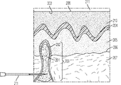
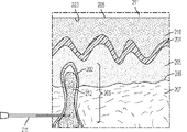
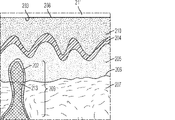
別の局面において、本明細書では、その必要がある患者において骨の患部を処置する方法であって、患者の骨中の患部を特定する工程;骨の患部中の変性海綿骨腔(cancellous space)へのアクセスを得るために、骨の皮質壁を貫く切開部を骨に作る工程;ある体積の、前記請求項のいずれか一項記載の注入可能な生体材料を、骨の皮質壁を貫く前記切開部を介して変性海綿骨腔中へと投与する工程を含む方法を開示する。 In another aspect, herein is a method of treating an affected area of bone in a patient in need thereof, a step of identifying the affected area in the bone of the patient; a degenerated cancellous space in the affected area of the bone. ) To make an incision in the bone that penetrates the cortical wall of the bone; a volume of injectable biomaterial according to any one of the above claims penetrates the cortical wall of the bone. Disclosed is a method comprising the step of administering into the degenerative cancellous bone cavity through the incision.
いくつかの態様において、骨の患部は、関節病変を経験している患者の関節に隣接している。いくつかの態様において、関節病変は、膝、肩、手首、手、脊柱、足首、肘または股関節の病変である。いくつかの態様において、関節病変は、疼痛、変形性関節症、関節リウマチ、虚血壊死およびそれらの組合せからなる群より選択される。いくつかの態様において、本方法は、患者の関節における変形性関節症を処置するための方法である。いくつかの態様において、変形性関節症は、1~3のチェルグレン・ローレンス(Kellgren Lawrence)(KL)グレードを有する。いくつかの態様において、関節病変は関節不安定性に関係しない。 In some embodiments, the affected area of the bone is adjacent to the joint of a patient experiencing a joint lesion. In some embodiments, the joint lesion is a lesion of the knee, shoulder, wrist, hand, spine, ankle, elbow or hip joint. In some embodiments, the joint lesion is selected from the group consisting of pain, osteoarthritis, rheumatoid arthritis, ischemic necrosis and combinations thereof. In some embodiments, the method is a method for treating osteoarthritis in a patient's joint. In some embodiments, osteoarthritis has 1-3 Kellgren Lawrence (KL) grades. In some embodiments, joint lesions are not associated with joint instability.
いくつかの態様において、患部は、炎症性メディエーターおよび非炎症性メディエーターのうちの少なくとも1つの結果として、炎症性変化または分解性変化のうちの少なくとも1つを呈する。 In some embodiments, the affected area exhibits at least one of an inflammatory or degrading change as a result of at least one of an inflammatory and non-inflammatory mediator.
いくつかの態様において、炎症性変化または分解性変化はMRIによって特定される。いくつかの態様において、MRIはT2 MRIである。 In some embodiments, inflammatory or degradable changes are identified by MRI. In some embodiments, the MRI is a T2 MRI.
いくつかの態様において、炎症性変化または分解性変化は海綿骨中に位置している。 In some embodiments, the inflammatory or degradable changes are located in the cancellous bone.
いくつかの態様において、患部は、患者の関節から約0インチ~約5インチに位置している。いくつかの態様において、患部は、患者の関節から約0インチ~約4インチに位置している。いくつかの態様において、患部は、患者の関節から約0インチ~約3インチに位置している。いくつかの態様において、患部は、患者の関節から約0インチ~約2インチに位置している。いくつかの態様において、患部は、患者の関節から約0インチ~約1インチに位置している。いくつかの態様において、患部は、患者の関節から約0インチ~約20mmに位置している。いくつかの態様において、患部は、患者の関節から約0mm~約10mmに位置している。いくつかの態様において、患部は、患者の関節から約0mm~約5mmに位置している。いくつかの態様において、患部は、患者の関節から約0mm~約1mmに位置している。 In some embodiments, the affected area is located about 0 inches to about 5 inches from the patient's joints. In some embodiments, the affected area is located about 0 inches to about 4 inches from the patient's joints. In some embodiments, the affected area is located about 0 inches to about 3 inches from the patient's joints. In some embodiments, the affected area is located about 0 inches to about 2 inches from the patient's joints. In some embodiments, the affected area is located about 0 inches to about 1 inch from the patient's joints. In some embodiments, the affected area is located about 0 inches to about 20 mm from the patient's joints. In some embodiments, the affected area is located about 0 mm to about 10 mm from the patient's joint. In some embodiments, the affected area is located about 0 mm to about 5 mm from the patient's joint. In some embodiments, the affected area is located approximately 0 mm to approximately 1 mm from the patient's joint.
いくつかの態様において、切開部は経皮的である。 In some embodiments, the incision is percutaneous.
いくつかの態様において、海綿骨腔へのアクセスを得る工程は、骨の皮質壁の切開部を、患部を含む海綿骨腔へと接続するために、患者の骨にチャネルを作る工程を含む。いくつかの態様において、チャネルは骨の長軸に対して直角である。いくつかの態様において、チャネルは骨の長軸に対して直角でない。いくつかの態様において、チャネルは近位軟骨下板から約5インチ以内にある。いくつかの態様において、チャネルは近位軟骨下板から約4インチ以内にある。いくつかの態様において、チャネルは近位軟骨下板から約3インチ以内にある。いくつかの態様において、チャネルは近位軟骨下板から約2インチ以内にある。いくつかの態様において、チャネルは近位軟骨下板から約1インチ以内にある。いくつかの態様において、チャネルは近位軟骨下板から約20mm以内にある。いくつかの態様において、チャネルは近位軟骨下板から約10mm以内にある。いくつかの態様において、チャネルは近位軟骨下板から約5mm以内にある。いくつかの態様において、チャネルは近位軟骨下板から約1mm以内にある。いくつかの態様において、チャネルは、追加の誘導器具を必要とすることなく位置決めされかつ挿入されるカニューレによってアクセスされる。 In some embodiments, the step of gaining access to the cancellous bone cavity comprises creating a channel in the patient's bone to connect the incision in the cortical wall of the bone to the cancellous bone cavity, including the affected area. In some embodiments, the channel is perpendicular to the long axis of the bone. In some embodiments, the channel is not perpendicular to the long axis of the bone. In some embodiments, the channel is within about 5 inches of the proximal subchondral plate. In some embodiments, the channel is within about 4 inches of the proximal subchondral plate. In some embodiments, the channel is within about 3 inches of the proximal subchondral plate. In some embodiments, the channel is within about 2 inches of the proximal subchondral plate. In some embodiments, the channel is within about 1 inch from the proximal subchondral plate. In some embodiments, the channel is within about 20 mm of the proximal subchondral plate. In some embodiments, the channel is within about 10 mm of the proximal subchondral plate. In some embodiments, the channel is within about 5 mm of the proximal subchondral plate. In some embodiments, the channel is within about 1 mm from the proximal subchondral plate. In some embodiments, the channel is accessed by a cannula that is positioned and inserted without the need for additional guidance equipment.
いくつかの態様において、本方法は、患部への注入可能な生体材料の投与に先だって、患部を減圧し、その内容物を吸引する工程を、さらに含む。いくつかの態様において、減圧および吸引は患部における局所的炎症を低減する。いくつかの態様において、減圧および吸引は患部における骨内圧を低減する。いくつかの態様において、前記内容物は流体を含む。いくつかの態様において、前記流体は、炎症性メディエーターおよび非炎症性メディエーターのうちの少なくとも1つを含む。 In some embodiments, the method further comprises decompressing the affected area and aspirating its contents prior to administration of the injectable biomaterial into the affected area. In some embodiments, decompression and suction reduce local inflammation in the affected area. In some embodiments, decompression and suction reduce intraosseous pressure in the affected area. In some embodiments, the content comprises a fluid. In some embodiments, the fluid comprises at least one of an inflammatory mediator and a non-inflammatory mediator.
いくつかの態様において、少なくとも1つの炎症性メディエーターは、ブラジキニン、ヒスタミン、プロスタグランジン、乳酸、サブスタンスP、血管作動性腸ペプチド、カルシトニン遺伝子関連ペプチド(CGRP)およびそれらの混合物のうちの少なくとも1つを含む。いくつかの態様において、少なくとも1つの炎症性メディエーターは炎症性サイトカインを含む。いくつかの態様において、炎症性サイトカインは、AIMP1(SCYE1)、BMP2、CD40LG(TNFSF5)、CSF1(MCSF)、CSF2(GM-CSF)、CSF3(GCSF)、FASLG(TNFSF6)、GM-CSF、IFNA2、IFNG、IL-1、IL-6、IL-8、IL-15、IL-16、IL-17、IL-18、IFN-γ、LTA(TNFB)、LTB、MIF、NAMPT、OSM、SPP1、TGF-β、TNF、TNF-α、TNFSF10(TRAIL)、TNFSF11(RANKL)、TNFSF13、TNFSF13B、TNFSF4(OX40L)、VEGFAおよびそれらの混合物からなる群より選択される。いくつかの態様において、少なくとも1つの非炎症性メディエーターは、タンパク質分解酵素を含む。いくつかの態様において、タンパク質分解酵素は、マトリックスメタロプロテイナーゼ(MMP)、組織メタロプロテイナーゼ阻害因子(TIMP)、トロンボスポンジンモチーフを有するディスインテグリンおよびメタロプロテイナーゼ(a disintegrin and metalloproteinase with thrombospondin motifs)(ADAM-TS)ならびにそれらの混合物からなる群より選択される。いくつかの態様において、炎症性メディエーターは、炎症性ケモカインを含む。いくつかの態様において、炎症性ケモカインは、C5、CCL1(I-309)、CCL11(エオタキシン)、CCL13(MCP-4)、CCL15(MIP-1d)、CCL16(HCC-4)、CCL17(TARC)、CCL2(MCP-1)、CCL20(MIP-3a)、CCL22(MDC)、CCL23(MPIF-1)、CCL24(MPIF-2、エオタキシン-2、MPIF-2、エオタキシン-2)、CCL26(エオタキシン-3)、CCL3(MIP-1A)、CCL4(MIP-1B)、CCL5(RANTES)、CCL7(MCP-3)、CCL8(MCP-2)、CX3CL1、CXCL1(GRO1、GRO-α、SCYB1)、CXCL10(INP10)、CXCL11(I-TAC、IP-9)、CXCL12(SDF1)、CXCL13、CXCL2(GRO2、GRO-β、SCYB2)、CXCL3、CXCL5(ENA-78、LIX)、CXCL6(GCP-2)、CXCL9(MIG)およびそれらの混合物からなる群より選択される。いくつかの態様において、炎症性メディエーターはインターロイキンを含む。いくつかの態様において、インターロイキンは、IL13、IL15、IL16、IL17A、IL17C、IL17F、IL1A、IL1B、IL1RN、IL21、IL27、IL3、IL33、IL5、IL7、CXCL8、IL9およびそれらの混合物からなる群より選択される。いくつかの態様において、炎症性メディエーターは、ブラジキニン、カルシトニン遺伝子関連ペプチド(CGRP)、ヒスタミン、乳酸、神経成長因子(NGF)、プロスタグランジン、サブスタンスP、血管作動性腸ペプチドおよびそれらの混合物からなる群より選択される炎症性メディエーターを含む。 In some embodiments, the at least one inflammatory mediator is at least one of bradykinin, histamine, prostaglandin, lactic acid, substance P, vasoactive intestinal peptide, calcitonin gene-related peptide (CGRP) and mixtures thereof. including. In some embodiments, the at least one inflammatory mediator comprises an inflammatory cytokine. In some embodiments, the inflammatory cytokines are AIMP1 (SCYE1), BMP2, CD40LG (TNFSF5), CSF1 (MCSF), CSF2 (GM-CSF), CSF3 (GCSF), FASLG (TNFSF6), GM-CSF, IFNA2. , IFNG, IL-1, IL-6, IL-8, IL-15, IL-16, IL-17, IL-18, IFN-γ, LTA (TNFB), LTB, MIF, NAMPT, OSM, SPP1, It is selected from the group consisting of TGF-β, TNF, TNF-α, TNFSF10 (TRAIL), TNFSF11 (RANKL), TNFSF13, TNFSF13B, TNFSF4 (OX40L), VEGFA and mixtures thereof. In some embodiments, the at least one non-inflammatory mediator comprises a proteolytic enzyme. In some embodiments, the proteolytic enzyme is a disintegrin and metalloproteinase with thrombospondin motifs (ADAM-), a matrix metalloproteinase (MMP), a tissue metalloproteinase inhibitor (TIMP), a disintegrin and metalloproteinase with thrombospondin motifs. It is selected from the group consisting of TS) and mixtures thereof. In some embodiments, the inflammatory mediator comprises an inflammatory chemokine. In some embodiments, inflammatory chemokines are C5, CCL1 (I-309), CCL11 (eotaxin), CCL13 (MCP-4), CCL15 (MIP-1d), CCL16 (HCC-4), CCL17 (TARC). , CCL2 (MCP-1), CCL20 (MIP-3a), CCL22 (MDC), CCL23 (MPIF-1), CCL24 (MPIF-2, Eotaxin-2, MPIF-2, Eotaxin-2), CCL26 (Eotaxin- 3), CCL3 (MIP-1A), CCL4 (MIP-1B), CCL5 (RANTES), CCL7 (MCP-3), CCL8 (MCP-2), CX3CL1, CXCL1 (GRO1, GRO-α, SCYB1), CXCL10 (INP10), CXCL11 (I-TAC, IP-9), CXCL12 (SDF1), CXCL13, CXCL2 (GRO2, GRO-β, SCYB2), CXCL3, CXCL5 (ENA-78, LIX), CXCL6 (GCP-2) , CXCL9 (MIG) and mixtures thereof are selected. In some embodiments, the inflammatory mediator comprises interleukin. In some embodiments, the interleukin is a group consisting of IL13, IL15, IL16, IL17A, IL17C, IL17F, IL1A, IL1B, IL1RN, IL21, IL27, IL3, IL33, IL5, IL7, CXCL8, IL9 and mixtures thereof. Will be selected. In some embodiments, the inflammatory mediator consists of bradykinin, calcitonin gene-related peptide (CGRP), histamine, lactic acid, nerve growth factor (NGF), prostaglandin, substance P, vasoactive intestinal peptide and mixtures thereof. Includes inflammatory mediators selected from the group.
いくつかの態様において、注入可能な生体材料は、カニューレまたは針を通して投与される。いくつかの態様において、針またはカニューレは、少なくとも21ゲージのサイズを有する。いくつかの態様において、針またはカニューレは、少なくとも20ゲージのサイズを有する。いくつかの態様において、針またはカニューレは、少なくとも18ゲージのサイズを有する。いくつかの態様において、針またはカニューレは、少なくとも16ゲージのサイズを有する。いくつかの態様において、針またはカニューレは、少なくとも15ゲージのサイズを有する。いくつかの態様において、針またはカニューレは、少なくとも14ゲージのサイズを有する。いくつかの態様において、針またはカニューレは、少なくとも12ゲージのサイズを有する。いくつかの態様において、針またはカニューレは、少なくとも10ゲージのサイズを有する。 In some embodiments, the injectable biomaterial is administered through a cannula or needle. In some embodiments, the needle or cannula has a size of at least 21 gauge. In some embodiments, the needle or cannula has a size of at least 20 gauge. In some embodiments, the needle or cannula has a size of at least 18 gauge. In some embodiments, the needle or cannula has a size of at least 16 gauge. In some embodiments, the needle or cannula has a size of at least 15 gauge. In some embodiments, the needle or cannula has a size of at least 14 gauge. In some embodiments, the needle or cannula has a size of at least 12 gauge. In some embodiments, the needle or cannula has a size of at least 10 gauge.
いくつかの態様において、注入可能な生体材料は針またはカニューレを通して施用される際に脱液を起こさない。 In some embodiments, the injectable biomaterial does not deflate when applied through a needle or cannula.
いくつかの態様において、注入可能な生体材料は針またはカニューレを通して施用される際に詰まりを起こさない。 In some embodiments, the injectable biomaterial does not clog when applied through a needle or cannula.
いくつかの態様において、注入可能な生体材料は、外科的損傷を最小限に抑えるために、操舵可能な(steerable)カニューレを通して投与される。 In some embodiments, the injectable biomaterial is administered through a steerable cannula to minimize surgical damage.
いくつかの態様において、注入可能な生体材料は、軟骨下板の破壊を最小限に抑えつつ、患部に注入される。 In some embodiments, the injectable biomaterial is injected into the affected area with minimal destruction of the subchondral plate.
いくつかの態様において、注入可能な生体材料は、軟骨下板の破壊を最小限に抑えつつ、患部の上または下、約0mm~約20mmの層中に注入される。いくつかの態様において、注入可能な生体材料は、軟骨下板の破壊を最小限に抑えつつ、患部の上または下、約0mm~約10mmの層中に注入される。いくつかの態様において、注入可能な生体材料は、軟骨下板の破壊を最小限に抑えつつ、患部の上または下、約0mm~約5mmの層中に注入される。いくつかの態様において、注入可能な生体材料は、軟骨下板の破壊を最小限に抑えつつ、患部の上または下、約0mm~約1mmの層中に注入される。 In some embodiments, the injectable biomaterial is injected into a layer of about 0 mm to about 20 mm above or below the affected area, with minimal destruction of the subchondral plate. In some embodiments, the injectable biomaterial is injected into a layer of about 0 mm to about 10 mm above or below the affected area, with minimal destruction of the subchondral plate. In some embodiments, the injectable biomaterial is injected into a layer of about 0 mm to about 5 mm above or below the affected area, with minimal destruction of the subchondral plate. In some embodiments, the injectable biomaterial is injected into a layer of about 0 mm to about 1 mm above or below the affected area, with minimal destruction of the subchondral plate.
いくつかの態様において、注入可能な生体材料は、骨の構造的安定性にとって本質的でない領域に投与される。 In some embodiments, the injectable biomaterial is administered to a region that is not essential to the structural stability of the bone.
いくつかの態様において、本方法は、注入可能な生体材料が関節中に存在しないことを保証するために、注入後の関節腔を関節鏡で検査する工程を、さらに含む。 In some embodiments, the method further comprises the step of arthroscopically inspecting the post-injection joint cavity to ensure that no injectable biomaterial is present in the joint.
いくつかの態様において、注入可能な生体材料は、患部へ投与されている間に、海綿骨の孔へと流れ込む。 In some embodiments, the injectable biomaterial flows into the cancellous bone pores while being administered to the affected area.
いくつかの態様において、注入可能な生体材料は、患部へ投与されている間、凝集性を保ち、かつ骨の空隙を実質的に満たす。 In some embodiments, the injectable biomaterial remains cohesive and substantially fills the bone voids while being administered to the affected area.
いくつかの態様において、注入可能な生体材料は、固化時に保護層を与えるように、海綿骨腔と隣接する関節との間の界面を少なくとも部分的に覆う。 In some embodiments, the injectable biomaterial at least partially covers the interface between the cancellous bone cavity and adjacent joints to provide a protective layer upon solidification.
いくつかの態様において、注入可能な生体材料は、隣接する関節腔から患部への炎症性メディエーターおよび非炎症性メディエーターのうちの少なくとも1つの拡散通過を防止する。 In some embodiments, the injectable biomaterial prevents the diffusion passage of at least one of the inflammatory and non-inflammatory mediators from the adjacent joint cavity to the affected area.
いくつかの態様において、保護層は、骨リモデリング中に破骨細胞が消費するための犠牲層を与える。 In some embodiments, the protective layer provides a sacrificial layer for osteoclast consumption during bone remodeling.
いくつかの態様において、注入可能な生体材料の投与は、無荷重の骨の脆弱化をもたらす応力遮蔽を引き起こさない。 In some embodiments, administration of injectable biomaterial does not cause stress shielding resulting in unloaded bone weakening.
いくつかの態様において、本方法は、実質的な術後痛を引き起こさない。 In some embodiments, the method does not cause substantial postoperative pain.
いくつかの態様において、本方法は関節の疼痛を減少させる。 In some embodiments, the method reduces joint pain.
いくつかの態様において、本方法は関節における変形性関節症の進行を減速する。 In some embodiments, the method slows the progression of osteoarthritis in a joint.
いくつかの態様において、本方法は、患者の関節における関節リウマチを処置するための方法である。いくつかの態様において、本方法は、関節における関節リウマチの進行を減速する。 In some embodiments, the method is a method for treating rheumatoid arthritis in a patient's joints. In some embodiments, the method slows the progression of rheumatoid arthritis in a joint.
いくつかの態様において、本方法は、関節における虚血壊死の進行を減速する。 In some embodiments, the method slows the progression of ischemic necrosis in the joint.
別の局面において、本明細書では、本明細書に開示する注入可能な生体材料を調製するための固形構成成分および液状構成成分と、それらの使用説明書とを含むキットを開示する。 In another aspect, the specification discloses a kit comprising solid and liquid constituents for preparing the injectable biomaterials disclosed herein and instructions for their use.
いくつかの態様において、説明書は、その必要がある患者における骨の患部を処置する方法に関する説明書である。 In some embodiments, the instructions are instructions on how to treat the affected area of bone in a patient in need thereof.
いくつかの態様において、処置は、疼痛、変形性関節症、関節リウマチ、虚血壊死またはそれらの組合せのための処置である。 In some embodiments, the treatment is for pain, osteoarthritis, rheumatoid arthritis, ischemic necrosis or a combination thereof.
いくつかの態様において、固形構成成分および液状構成成分は、別々の無菌容器に配置される。 In some embodiments, the solid and liquid constituents are placed in separate sterile containers.
いくつかの態様において、パッケージング構成は、固形構成成分および液状構成成分が、2℃~25℃で、少なくとも約3ヶ月間、安定性を保つことを可能にする。いくつかの態様において、パッケージング構成は、固形構成成分および液状構成成分が、2℃~25℃で、少なくとも約6ヶ月間、安定性を保つことを可能にする。いくつかの態様において、パッケージング構成は、固形構成成分および液状構成成分が、2℃~25℃で、少なくとも約1年間、安定性を保つことを可能にする。いくつかの態様において、パッケージング構成は、固形構成成分および液状構成成分が、2℃~25℃で、少なくとも約2年間、安定性を保つことを可能にする。いくつかの態様において、パッケージング構成は、固形構成成分および液状構成成分が、2℃~25℃で、少なくとも約3年間、安定性を保つことを可能にする。いくつかの態様において、パッケージング構成は、固形構成成分および液状構成成分が、2℃~25℃で、少なくとも約4年間、安定性を保つことを可能にする。いくつかの態様において、パッケージング構成は、固形構成成分および液状構成成分が、2℃~25℃で、少なくとも約5年間、安定性を保つことを可能にする。 In some embodiments, the packaging configuration allows the solid and liquid constituents to remain stable at 2 ° C to 25 ° C for at least about 3 months. In some embodiments, the packaging configuration allows the solid and liquid constituents to remain stable at 2 ° C to 25 ° C for at least about 6 months. In some embodiments, the packaging configuration allows the solid and liquid constituents to remain stable at 2 ° C to 25 ° C for at least about 1 year. In some embodiments, the packaging configuration allows solid and liquid constituents to remain stable at 2 ° C to 25 ° C for at least about 2 years. In some embodiments, the packaging configuration allows solid and liquid constituents to remain stable at 2 ° C to 25 ° C for at least about 3 years. In some embodiments, the packaging configuration allows solid and liquid constituents to remain stable at 2 ° C to 25 ° C for at least about 4 years. In some embodiments, the packaging configuration allows solid and liquid constituents to remain stable at 2 ° C to 25 ° C for at least about 5 years.
いくつかの態様において、液状構成成分は無菌シリンジに配置される。 In some embodiments, the liquid component is placed in a sterile syringe.
いくつかの態様において、固形構成成分は、予め計り取られた分量の固形構成成分と液状構成成分とをインサイチューで混合して注入可能な生体材料を形成させるための一体型混合デバイス(integrated mixing device)を備えたシリンジに配置される。いくつかの態様において、シリンジは無菌である。 In some embodiments, the solid component is an integrated mixing device for in situ mixing a pre-measured amount of the solid component and the liquid component to form an injectable biomaterial. Placed in a syringe equipped with device). In some embodiments, the syringe is sterile.
いくつかの態様において、キットはルアーロックをさらに含む。 In some embodiments, the kit further comprises a lure lock.
いくつかの態様において、キットはエンドキャップをさらに含む。
[本発明1001]
(a)固形構成成分と
(b)炭水化物を含む液状構成成分と
を含み、固形構成成分と液状構成成分との混合後に固化および硬化してアパタイト型結晶構造を形成する、注入可能な生体材料。
[本発明1002]
(a)(i)溶液を用意し、
(ii)前記溶液のpHをpH調節剤で調節し、かつ
(iii)前記溶液に炭水化物を溶解して液状構成成分を形成させる
ことによって、液状構成成分を作る工程、
(b)固形構成成分を用意する工程、および
(c)液状構成成分と固形構成成分とを混合して、注入可能な生体材料を形成させる工程
を含む、本発明1001の注入可能な生体材料を作製するための方法。
[本発明1003]
注入可能な生体材料が、ある期間かけて固化する、前記本発明のいずれかの注入可能な生体材料または方法。
[本発明1004]
注入可能な生体材料が、ある期間かけて硬化する、前記本発明のいずれかの注入可能な生体材料または方法。
[本発明1005]
注入可能な生体材料が、完全に硬化する前に固化する、前記本発明のいずれかの注入可能な生体材料または方法。
[本発明1006]
固形構成成分が金属リン酸塩および金属炭酸塩のうちの少なくとも1つを含む、前記本発明のいずれかの注入可能な生体材料または方法。
[本発明1007]
固形構成成分が反応性リン酸カルシウムを含む、前記本発明のいずれかの注入可能な生体材料または方法。
[本発明1008]
固形構成成分が、α-リン酸三カルシウム(Ca
3
(PO
4
)
2
)、炭酸カルシウム(CaCO
3
)およびリン酸一カルシウム一水和物(Ca(H
2
PO
4
)
2
H
2
O)のうちの少なくとも1つを含む、前記本発明のいずれかの注入可能な生体材料または方法。
[本発明1009]
固形構成成分が、70~90%のα-リン酸三カルシウム、10~20%の炭酸カルシウムおよび0.5~2%のリン酸二水素カルシウム一水和物(質量/質量)を含む、前記本発明のいずれかの注入可能な生体材料または方法。
[本発明1010]
固形構成成分が、80~89%のα-リン酸三カルシウム、11~19%の炭酸カルシウムおよび0.75~1.5%のリン酸二水素カルシウム一水和物(質量/質量)を含む、前記本発明のいずれかの注入可能な生体材料または方法。
[本発明1011]
固形構成成分が、82~86%のα-リン酸三カルシウム、13~16%の炭酸カルシウムおよび0.9~1.2%のリン酸二水素カルシウム一水和物(質量/質量)を含む、前記本発明のいずれかの注入可能な生体材料または方法。
[本発明1012]
固形構成成分が、84.3%のα-リン酸三カルシウム、14.7%の炭酸カルシウムおよび1.02%のリン酸二水素カルシウム一水和物(質量/質量)を含む、前記本発明のいずれかの注入可能な生体材料または方法。
[本発明1013]
固形構成成分が、人体中に天然に存在する1つまたは複数の少数元素からなる1つまたは複数のイオン性化合物をさらに含む、前記本発明のいずれかの注入可能な生体材料または方法。
[本発明1014]
少なくとも1つのイオン性化合物が、Na
+
、K
+
、Mg
2+
、Ca
2+
、Sr
2+
、H
+
およびそれらの混合物からなる群より選択される陽イオンを含む、本発明1013の注入可能な生体材料または方法。
[本発明1015]
少なくとも1つのイオン性化合物が、PO
4
3-
、HPO
4
2-
、H
2
PO
4
-
、P
2
O
7
4-
、CO
3
2-
、HCO
3
-
、SO
4
2-
、HSO
4
-
、Cl
-
、OH
-
、F
-
、SiO
4
4-
およびそれらの混合物からなる群より選択される陰イオンを含む、本発明1013の注入可能な生体材料または方法。
[本発明1016]
液状構成成分が塩をさらに含む、前記本発明のいずれかの注入可能な生体材料または方法。
[本発明1017]
塩が金属塩である、本発明1016の注入可能な生体材料または方法。
[本発明1018]
塩が、リン酸塩、ケイ酸塩、塩化物塩、水酸化物塩およびそれらの混合物から選択される、本発明1016~1017のいずれかの注入可能な生体材料または方法。
[本発明1019]
塩が、リン酸水素二ナトリウム、ケイ酸ナトリウム、塩化ナトリウムおよび水酸化カルシウムのうちの少なくとも1つを含む、本発明1016~1018のいずれかの注入可能な生体材料または方法。
[本発明1020]
炭水化物が、デキストラン、アルギネート、カルボキシメチルセルロースおよびヒアルロン酸からなる群より選択される、前記本発明のいずれかの注入可能な生体材料または方法。
[本発明1021]
炭水化物が、ヒアルロン酸、またはそのエステル、アシルウレア、アシルイソウレア、ジスルフィドもしくはアミドである、前記本発明のいずれかの注入可能な生体材料または方法。
[本発明1022]
ヒアルロン酸が、ヒアルロナン、ヒアルロン酸ナトリウム、ヒアルロン酸カリウム、ヒアルロン酸マグネシウム、ヒアルロン酸カルシウム、ヒアルロン酸アンモニウムおよびそれらの組合せからなる群より選択される、本発明1021の注入可能な生体材料または方法。
[本発明1023]
ヒアルロン酸が少なくとも1つの架橋を含む、本発明1021~1022のいずれかの注入可能な生体材料または方法。
[本発明1024]
ヒアルロン酸が細菌または動物に由来する、本発明1021~1023のいずれかの注入可能な生体材料または方法。
[本発明1025]
ヒアルロン酸が、硫酸化ヒアルロン酸、またはそのエステル、アシルウレア、アシルイソウレア、カルボマー、ジスルフィドもしくはアミドを含む、本発明1021~1024のいずれかの注入可能な生体材料または方法。
[本発明1026]
ヒアルロン酸が、N-硫酸化ヒアルロン酸、またはそのエステル、アシルウレア、アシルイソウレア、カルボマー、ジスルフィドもしくはアミドを含む、本発明1025の注入可能な生体材料または方法。
[本発明1027]
ヒアルロン酸がヒアルロン酸エステルを含む、本発明1021~1026のいずれかの注入可能な生体材料または方法。
[本発明1028]
ヒアルロン酸エステルが約20~100%の量でエステル化されている、本発明1027の注入可能な生体材料または方法。
[本発明1029]
非エステル化ヒアルロン酸が有機塩基または無機塩基で塩化されている、本発明1028の注入可能な生体材料または方法。
[本発明1030]
炭水化物が水溶性である、前記本発明のいずれかの注入可能な生体材料または方法。
[本発明1031]
液状構成成分がヒドロゲルの形態である、前記本発明のいずれかの注入可能な生体材料または方法。
[本発明1032]
炭水化物が、注入可能な生体材料中に約0.1~約100mg/mLの濃度で存在する、前記本発明のいずれかの注入可能な生体材料または方法。
[本発明1033]
炭水化物が、注入可能な生体材料中に約0.1~約50mg/mLの濃度で存在する、本発明1032の注入可能な生体材料または方法。
[本発明1034]
炭水化物が、注入可能な生体材料中に約0.1~約10mg/mLの濃度で存在する、本発明1032の注入可能な生体材料または方法。
[本発明1035]
炭水化物が、注入可能な生体材料中に約1~約10mg/mLの濃度で存在する、本発明1032の注入可能な生体材料または方法。
[本発明1036]
炭水化物が、注入可能な生体材料中に約2~約10mg/mLの濃度で存在する、本発明1032の注入可能な生体材料または方法。
[本発明1037]
炭水化物が、注入可能な生体材料中に約4~約8mg/mLの濃度で存在する、本発明1032の注入可能な生体材料または方法。
[本発明1038]
炭水化物が、注入可能な生体材料中に約5~約7mg/mLの濃度で存在する、本発明1032の注入可能な生体材料または方法。
[本発明1039]
炭水化物が約0.90×10
6
Da~約1.0×10
7
Daの分子量を有する、前記本発明のいずれかの注入可能な生体材料または方法。
[本発明1040]
炭水化物が約0.90×10
6
Da~約5.0×10
6
Daの分子量を有する、本発明1039の注入可能な生体材料または方法。
[本発明1041]
炭水化物が約0.90×10
6
Da~約4.0×10
6
Daの分子量を有する、本発明1039の注入可能な生体材料または方法。
[本発明1042]
炭水化物が約0.90×10
6
Da~約3.0×10
6
Daの分子量を有する、本発明1039の注入可能な生体材料または方法。
[本発明1043]
炭水化物が約1.5×10
6
Da~約3.0×10
6
Daの分子量を有する、本発明1039の注入可能な生体材料または方法。
[本発明1044]
炭水化物が約1.7×10
6
Da~約2.5×10
6
Daの分子量を有する、本発明1039の注入可能な生体材料または方法。
[本発明1045]
炭水化物が、約0.90×10
6
Daの分子量を有するヒアルロン酸であり、かつ約6.0mg/mLの濃度で存在する、前記本発明のいずれかの注入可能な生体材料または方法。
[本発明1046]
炭水化物が、約1.7×10
6
Daの分子量を有するヒアルロン酸であり、かつ約6.0mg/mLの濃度で存在する、前記本発明のいずれかの注入可能な生体材料または方法。
[本発明1047]
炭水化物が、約2.6×10
6
Daの分子量を有するヒアルロン酸であり、かつ約6.0mg/mLの濃度で存在する、前記本発明のいずれかの注入可能な生体材料または方法。
[本発明1048]
炭水化物の分子量が少なくとも3ヶ月間は安定である、前記本発明のいずれかの注入可能な生体材料または方法。
[本発明1049]
炭水化物の分子量が少なくとも6ヶ月間は安定である、前記本発明のいずれかの注入可能な生体材料または方法。
[本発明1050]
炭水化物の分子量が少なくとも1年間は安定である、前記本発明のいずれかの注入可能な生体材料または方法。
[本発明1051]
炭水化物の分子量が少なくとも2年間は安定である、前記本発明のいずれかの注入可能な生体材料または方法。
[本発明1052]
炭水化物の分子量が少なくとも3年間は安定である、前記本発明のいずれかの注入可能な生体材料または方法。
[本発明1053]
炭水化物の分子量が少なくとも4年間は安定である、前記本発明のいずれかの注入可能な生体材料または方法。
[本発明1054]
炭水化物の分子量が少なくとも5年間は安定である、前記本発明のいずれかの注入可能な生体材料または方法。
[本発明1055]
固形構成成分 対 液状構成成分の比が質量で約3 対 約1である、前記本発明のいずれかの注入可能な生体材料または方法。
[本発明1056]
固形構成成分 対 液状構成成分の比が質量で約2 対 約1である、本発明1055の注入可能な生体材料または方法。
[本発明1057]
固形構成成分 対 液状構成成分の比が質量で約1.5 対 約1である、本発明1055の注入可能な生体材料または方法。
[本発明1058]
固形構成成分 対 液状構成成分の比が質量で約1 対 約1である、本発明1055の注入可能な生体材料または方法。
[本発明1059]
注入可能な生体材料が、初期固化する前に、針またはカニューレを通して注入可能である、前記本発明のいずれかの注入可能な生体材料または方法。
[本発明1060]
針またはカニューレが少なくとも21ゲージのサイズを有する、本発明1059の注入可能な生体材料または方法。
[本発明1061]
針またはカニューレが少なくとも20ゲージのサイズを有する、本発明1059の注入可能な生体材料または方法。
[本発明1062]
針またはカニューレが少なくとも18ゲージのサイズを有する、本発明1059の注入可能な生体材料または方法。
[本発明1063]
針またはカニューレが少なくとも16ゲージのサイズを有する、本発明1059の注入可能な生体材料または方法。
[本発明1064]
針またはカニューレが少なくとも15ゲージのサイズを有する、本発明1059の注入可能な生体材料または方法。
[本発明1065]
針またはカニューレが少なくとも14ゲージのサイズを有する、本発明1059の注入可能な生体材料または方法。
[本発明1066]
針またはカニューレが少なくとも12ゲージのサイズを有する、本発明1059の注入可能な生体材料または方法。
[本発明1067]
針またはカニューレが少なくとも10ゲージのサイズを有する、本発明1059の注入可能な生体材料または方法。
[本発明1068]
注入可能な生体材料が、針またはカニューレを通して施用される際に脱液を起こさない、前記本発明のいずれかの注入可能な生体材料または方法。
[本発明1069]
注入可能な生体材料が、針またはカニューレを通して施用される際に詰まりを起こさない、前記本発明のいずれかの注入可能な生体材料または方法。
[本発明1070]
注入可能な生体材料が凝集性である、前記本発明のいずれかの注入可能な生体材料または方法。
[本発明1071]
注入可能な生体材料が、その初期固化時間中、凝集性を保っている、前記本発明のいずれかの注入可能な生体材料または方法。
[本発明1072]
注入可能な生体材料が骨に付着する、前記本発明のいずれかの注入可能な生体材料または方法。
[本発明1073]
注入可能な生体材料が、その初期固化時間中、骨への付着性を保っている、前記本発明のいずれかの注入可能な生体材料または方法。
[本発明1074]
注入可能な生体材料が、固形構成成分と液状構成成分との混合後、約60分未満にわたって、加工可能である、前記本発明のいずれかの注入可能な生体材料または方法。
[本発明1075]
注入可能な生体材料が、固形構成成分と液状構成成分との混合後、約50分未満にわたって、加工可能である、本発明1074の注入可能な生体材料または方法。
[本発明1076]
注入可能な生体材料が、固形構成成分と液状構成成分との混合後、約40分未満にわたって、加工可能である、本発明1074の注入可能な生体材料または方法。
[本発明1077]
注入可能な生体材料が、固形構成成分と液状構成成分との混合後、約30分未満にわたって、加工可能である、本発明1074の注入可能な生体材料または方法。
[本発明1078]
注入可能な生体材料が、固形構成成分と液状構成成分との混合後、約20分未満にわたって、加工可能である、本発明1074の注入可能な生体材料または方法。
[本発明1079]
注入可能な生体材料が、固形構成成分と液状構成成分との混合後、約10分未満にわたって、加工可能である、本発明1074の注入可能な生体材料または方法。
[本発明1080]
注入可能な生体材料が、固形構成成分と液状構成成分との混合後、約5分未満にわたって、加工可能である、本発明1074の注入可能な生体材料または方法。
[本発明1081]
注入可能な生体材料が、固形構成成分と液状構成成分との混合後、約4分未満にわたって、加工可能である、本発明1074の注入可能な生体材料または方法。
[本発明1082]
注入可能な生体材料が、固形構成成分と液状構成成分との混合後、約3分未満にわたって、加工可能である、本発明1074の注入可能な生体材料または方法。
[本発明1083]
注入可能な生体材料が、固形構成成分と液状構成成分との混合後、約2分未満にわたって、加工可能である、本発明1074の注入可能な生体材料または方法。
[本発明1084]
注入可能な生体材料が、固形構成成分と液状構成成分との混合後、約1分未満にわたって、加工可能である、本発明1074の注入可能な生体材料または方法。
[本発明1085]
注入可能な生体材料が、固形構成成分と液状構成成分との混合後、約60分未満で初期固化する、前記本発明のいずれかの注入可能な生体材料または方法。
[本発明1086]
注入可能な生体材料が、固形構成成分と液状構成成分との混合後、約50分未満で初期固化する、本発明1085の注入可能な生体材料または方法。
[本発明1087]
注入可能な生体材料が、固形構成成分と液状構成成分との混合後、約40分未満で初期固化する、本発明1085の注入可能な生体材料または方法。
[本発明1088]
注入可能な生体材料が、固形構成成分と液状構成成分との混合後、約30分未満で初期固化する、本発明1085の注入可能な生体材料または方法。
[本発明1089]
注入可能な生体材料が、固形構成成分と液状構成成分との混合後、約20分未満で初期固化する、本発明1085の注入可能な生体材料または方法。
[本発明1090]
注入可能な生体材料が、固形構成成分と液状構成成分との混合後、約10分未満で初期固化する、本発明1085の注入可能な生体材料または方法。
[本発明1091]
注入可能な生体材料が、固形構成成分と液状構成成分との混合後、約5分未満で初期固化する、本発明1085の注入可能な生体材料または方法。
[本発明1092]
注入可能な生体材料が、固形構成成分と液状構成成分との混合後、約4分未満で初期固化する、本発明1085の注入可能な生体材料または方法。
[本発明1093]
注入可能な生体材料が、固形構成成分と液状構成成分との混合後、約3分未満で初期固化する、本発明1085の注入可能な生体材料または方法。
[本発明1094]
注入可能な生体材料が、固形構成成分と液状構成成分との混合後、約2分未満で初期固化する、本発明1085の注入可能な生体材料または方法。
[本発明1095]
注入可能な生体材料が、固形構成成分と液状構成成分との混合後、約1分未満で初期固化する、本発明1085の注入可能な生体材料または方法。
[本発明1096]
注入可能な生体材料が、固形構成成分と液状構成成分との混合後、約96時間未満で完全に硬化する、前記本発明のいずれかの注入可能な生体材料または方法。
[本発明1097]
注入可能な生体材料が、固形構成成分と液状構成成分との混合後、約72時間未満で完全に硬化する、本発明1096の注入可能な生体材料または方法。
[本発明1098]
注入可能な生体材料が、固形構成成分と液状構成成分との混合後、約48時間未満で完全に硬化する、本発明1096の注入可能な生体材料または方法。
[本発明1099]
注入可能な生体材料が、固形構成成分と液状構成成分との混合後、約24時間未満で完全に硬化する、本発明1096の注入可能な生体材料または方法。
[本発明1100]
注入可能な生体材料が、固形構成成分と液状構成成分との混合後、約12時間未満で完全に硬化する、本発明1096の注入可能な生体材料または方法。
[本発明1101]
注入可能な生体材料が、固形構成成分と液状構成成分との混合後、約6時間未満で完全に硬化する、本発明1096の注入可能な生体材料または方法。
[本発明1102]
注入可能な生体材料が、固形構成成分と液状構成成分との混合後、約5時間未満で完全に硬化する、本発明1096の注入可能な生体材料または方法。
[本発明1103]
注入可能な生体材料が、固形構成成分と液状構成成分との混合後、約4時間未満で完全に硬化する、本発明1096の注入可能な生体材料または方法。
[本発明1104]
注入可能な生体材料が、固形構成成分と液状構成成分との混合後、約3時間未満で完全に硬化する、本発明1096の注入可能な生体材料または方法。
[本発明1105]
注入可能な生体材料が、固形構成成分と液状構成成分との混合後、約2時間未満で完全に硬化する、本発明1096の注入可能な生体材料または方法。
[本発明1106]
注入可能な生体材料が、固形構成成分と液状構成成分との混合後、約1時間未満で完全に硬化する、本発明1096の注入可能な生体材料または方法。
[本発明1107]
注入可能な生体材料の初期固化および硬化がガス放出をもたらさない、前記本発明のいずれかの注入可能な生体材料または方法。
[本発明1108]
注入可能な生体材料が、患者内に配置されたときに、隣接する流体のpHを顕著に変化させることがない、前記本発明のいずれかの注入可能な生体材料または方法。
[本発明1109]
注入可能な生体材料の初期固化および硬化が、患者内に配置されたときに、隣接する流体の温度を顕著に変化させることがない、前記本発明のいずれかの注入可能な生体材料または方法。
[本発明1110]
注入可能な生体材料の硬化が、ヒドロキシアパタイトのアパタイト型結晶構造と実質的に一致するアパタイト型結晶構造を生じる、前記本発明のいずれかの注入可能な生体材料または方法。
[本発明1111]
注入可能な生体材料の硬化が、少なくとも約90%ヒドロキシアパタイトであるアパタイト型結晶構造を生じる、前記本発明のいずれかの注入可能な生体材料または方法。
[本発明1112]
注入可能な生体材料の硬化が、少なくとも約95%ヒドロキシアパタイトであるアパタイト型結晶構造を生じる、本発明1111の注入可能な生体材料または方法。
[本発明1113]
注入可能な生体材料の硬化が、少なくとも約96%ヒドロキシアパタイトであるアパタイト型結晶構造を生じる、本発明1111の注入可能な生体材料または方法。
[本発明1114]
注入可能な生体材料の硬化が、少なくとも約97%ヒドロキシアパタイトであるアパタイト型結晶構造を生じる、本発明1111の注入可能な生体材料または方法。
[本発明1115]
注入可能な生体材料の硬化が、少なくとも約98%ヒドロキシアパタイトであるアパタイト型結晶構造を生じる、本発明1111の注入可能な生体材料または方法。
[本発明1116]
注入可能な生体材料の硬化が、少なくとも約99%ヒドロキシアパタイトであるアパタイト型結晶構造を生じる、本発明1111の注入可能な生体材料または方法。
[本発明1117]
注入可能な生体材料の硬化が、約99%超ヒドロキシアパタイトであるアパタイト型結晶構造を生じる、本発明1111の注入可能な生体材料または方法。
[本発明1118]
完全に固化かつ硬化した注入可能な生体材料が、約1~約2のモルCa/P比を有する、前記本発明のいずれかの注入可能な生体材料または方法。
[本発明1119]
完全に固化かつ硬化した注入可能な生体材料が、約1.3~約1.8のモルCa/P比を有する、本発明1117の注入可能な生体材料または方法。
[本発明1120]
完全に固化かつ硬化した注入可能な生体材料が、約1.4~約1.7のモルCa/P比を有する、本発明1117の注入可能な生体材料または方法。
[本発明1121]
完全に固化かつ硬化した注入可能な生体材料が、約1.5~約1.7のモルCa/P比を有する、本発明1117の注入可能な生体材料または方法。
[本発明1122]
完全に固化かつ硬化した注入可能な生体材料が、約1.5~約1.667のモルCa/P比を有する、本発明1117の注入可能な生体材料または方法。
[本発明1123]
完全に固化かつ硬化した注入可能な生体材料が、約20MPa未満の圧縮強度を有する、前記本発明のいずれかの注入可能な生体材料または方法。
[本発明1124]
完全に固化かつ硬化した注入可能な生体材料が、約15MPa未満の圧縮強度を有する、本発明1123の注入可能な生体材料または方法。
[本発明1125]
完全に固化かつ硬化した注入可能な生体材料が、約10MPa未満の圧縮強度を有する、本発明1123の注入可能な生体材料または方法。
[本発明1126]
完全に固化かつ硬化した注入可能な生体材料が、約9MPa未満の圧縮強度を有する、本発明1123の注入可能な生体材料または方法。
[本発明1127]
完全に固化かつ硬化した注入可能な生体材料が、約8MPa未満の圧縮強度を有する、本発明1123の注入可能な生体材料または方法。
[本発明1128]
完全に固化かつ硬化した注入可能な生体材料が、約7MPa未満の圧縮強度を有する、本発明1123の注入可能な生体材料または方法。
[本発明1129]
完全に固化かつ硬化した注入可能な生体材料が、約6MPa未満の圧縮強度を有する、本発明1123の注入可能な生体材料または方法。
[本発明1130]
完全に固化かつ硬化した注入可能な生体材料が、約5MPa未満の圧縮強度を有する、本発明1123の注入可能な生体材料または方法。
[本発明1131]
完全に固化かつ硬化した注入可能な生体材料が、約4MPa未満の圧縮強度を有する、本発明1123の注入可能な生体材料または方法。
[本発明1132]
完全に固化かつ硬化した注入可能な生体材料が、約3MPa未満の圧縮強度を有する、本発明1123の注入可能な生体材料または方法。
[本発明1133]
完全に固化かつ硬化した注入可能な生体材料が、約2MPa未満の圧縮強度を有する、本発明1123の注入可能な生体材料または方法。
[本発明1134]
完全に固化かつ硬化した注入可能な生体材料が、約1MPa未満の圧縮強度を有する、本発明1123の注入可能な生体材料または方法。
[本発明1135]
完全に固化かつ硬化した注入可能な生体材料が、約5GPa未満の弾性率を有する、前記本発明のいずれかの注入可能な生体材料または方法。
[本発明1136]
完全に固化かつ硬化した注入可能な生体材料が、約4GPa未満の弾性率を有する、本発明1133の注入可能な生体材料または方法。
[本発明1137]
完全に固化かつ硬化した注入可能な生体材料が、約3GPa未満の弾性率を有する、本発明1133の注入可能な生体材料または方法。
[本発明1138]
完全に固化かつ硬化した注入可能な生体材料が、約2GPa未満の弾性率を有する、本発明1133の注入可能な生体材料または方法。
[本発明1139]
完全に固化かつ硬化した注入可能な生体材料が、約1GPa未満の弾性率を有する、本発明1133の注入可能な生体材料または方法。
[本発明1140]
完全に固化かつ硬化した注入可能な生体材料が、約0.5GPa未満の弾性率を有する、本発明1133の注入可能な生体材料または方法。
[本発明1141]
完全に固化かつ硬化した注入可能な生体材料が、約0.25GPa未満の弾性率を有する、本発明1133の注入可能な生体材料または方法。
[本発明1142]
固形構成成分と液状構成成分との混合の直後に、室温で測定した場合に、約5Pa・sおよび約30Pa・sの粘度を有する、前記本発明のいずれかの注入可能な生体材料。
[本発明1143]
固形構成成分と液状構成成分との混合の直後に、室温で測定した場合に、約5Pa・sおよび約20Pa・sの粘度を有する、前記本発明のいずれかの注入可能な生体材料。
[本発明1144]
固形構成成分と液状構成成分との混合の直後に、室温で測定した場合に、約5Pa・sおよび約18Pa・sの粘度を有する、前記本発明のいずれかの注入可能な生体材料。
[本発明1145]
注入可能な生体材料が骨を生体力学的に安定化しない、前記本発明のいずれかの注入可能な生体材料または方法。
[本発明1146]
完全に固化かつ硬化した注入可能な生体材料が、約1g/cm
3
~約4g/cm
3
の真密度を有する、前記本発明のいずれかの注入可能な生体材料または方法。
[本発明1147]
完全に固化かつ硬化した注入可能な生体材料が、約1.5g/cm
3
~約3.5g/cm
3
の真密度を有する、前記本発明のいずれかの注入可能な生体材料または方法。
[本発明1148]
完全に固化かつ硬化した注入可能な生体材料が、約1.83g/cm
3
~約3.14g/cm
3
の真密度を有する、前記本発明のいずれかの注入可能な生体材料または方法。
[本発明1149]
完全に固化かつ硬化した注入可能な生体材料が、約2g/cm
3
~約3g/cm
3
の真密度を有する、前記本発明のいずれかの注入可能な生体材料または方法。
[本発明1150]
完全に固化かつ硬化した注入可能な生体材料が、約1μm未満の中央孔径を含む、前記本発明のいずれかの注入可能な生体材料または方法。
[本発明1151]
完全に固化かつ硬化した注入可能な生体材料が、約0.8μm未満の中央孔径を含む、前記本発明のいずれかの注入可能な生体材料または方法。
[本発明1152]
完全に固化かつ硬化した注入可能な生体材料が、約0.6μm未満の中央孔径を含む、前記本発明のいずれかの注入可能な生体材料または方法。
[本発明1153]
完全に固化かつ硬化した注入可能な生体材料が、約0.5μm未満の中央孔径を含む、前記本発明のいずれかの注入可能な生体材料または方法。
[本発明1154]
完全に固化かつ硬化した注入可能な生体材料が、約0.4μm未満の中央孔径を含む、前記本発明のいずれかの注入可能な生体材料または方法。
[本発明1155]
完全に固化かつ硬化した注入可能な生体材料が、約0.2μm未満の中央孔径を含む、前記本発明のいずれかの注入可能な生体材料または方法。
[本発明1156]
完全に固化かつ硬化した注入可能な生体材料が、約0.15μm未満の中央孔径を含む、前記本発明のいずれかの注入可能な生体材料または方法。
[本発明1157]
完全に固化かつ硬化した注入可能な生体材料が、約4m
2
/g未満の総孔面積を含む、前記本発明のいずれかの注入可能な生体材料または方法。
[本発明1158]
完全に固化かつ硬化した注入可能な生体材料が、約3m
2
/g未満の総孔面積を含む、前記本発明のいずれかの注入可能な生体材料または方法。
[本発明1159]
完全に固化かつ硬化した注入可能な生体材料が、約2m
2
/g未満の総孔面積を含む、前記本発明のいずれかの注入可能な生体材料または方法。
[本発明1160]
完全に固化かつ硬化した注入可能な生体材料が、炎症性メディエーターおよび非炎症性メディエーターのうちの少なくとも1つの拡散通過を防止するのに十分な有孔性を含む、前記本発明のいずれかの注入可能な生体材料または方法。
[本発明1161]
完全に固化かつ硬化した注入可能な生体材料が骨誘導性である、前記本発明のいずれかの注入可能な生体材料または方法。
[本発明1162]
完全に固化かつ硬化した注入可能な生体材料が骨伝導性である、前記本発明のいずれかの注入可能な生体材料または方法。
[本発明1163]
完全に固化かつ硬化した注入可能な生体材料が再吸収性である、前記本発明のいずれかの注入可能な生体材料または方法。
[本発明1164]
注入可能な生体材料の硬化が約5%未満の酸化カルシウムを生じる、前記本発明のいずれかの注入可能な生体材料または方法。
[本発明1165]
注入可能な生体材料の硬化が約4%未満の酸化カルシウムを生じる、本発明1164の注入可能な生体材料または方法。
[本発明1166]
注入可能な生体材料の硬化が約3%未満の酸化カルシウムを生じる、本発明1164の注入可能な生体材料または方法。
[本発明1167]
注入可能な生体材料の硬化が約2%未満の酸化カルシウムを生じる、本発明1164の注入可能な生体材料または方法。
[本発明1168]
注入可能な生体材料の硬化が約1%未満の酸化カルシウムを生じる、本発明1164の注入可能な生体材料または方法。
[本発明1169]
液状構成成分が無菌である、前記本発明のいずれかの注入可能な生体材料または方法。
[本発明1170]
固形構成成分が無菌である、前記本発明のいずれかの注入可能な生体材料または方法。
[本発明1171]
注入可能な生体材料が混和可能である、前記本発明のいずれかの注入可能な生体材料または方法。
[本発明1172]
pH調節剤が有機酸および無機酸から選択される、本発明1002~1170のいずれかの方法。
[本発明1173]
pH調節剤が、クエン酸、ギ酸、酢酸およびそれらの混合物からなる群より選択される、本発明1002~1172のいずれかの方法。
[本発明1174]
pH調節剤が、塩酸、リン酸、硝酸およびそれらの混合物からなる群より選択される、本発明1002~1173のいずれかの方法。
[本発明1175]
固形構成成分を用意する工程が、固形構成成分を乾燥させることをさらに含む、本発明1002~1174のいずれかの方法。
[本発明1176]
乾燥させることが、固形構成成分をある期間にわたって熱に曝露することを含む、本発明1175の方法。
[本発明1177]
熱が少なくとも約165℃を含む、本発明1176の方法。
[本発明1178]
前記期間が少なくとも約12時間を含む、本発明1176の方法。
[本発明1179]
a)患者の骨中の患部を特定する工程、
b)骨の患部中の変性海綿骨腔へのアクセスを得るために、骨の皮質壁を貫く切開部を骨に作る工程、
c)ある体積の、前記本発明のいずれかの注入可能な生体材料を、骨の皮質壁を貫く前記切開部を介して変性海綿骨腔中へと投与する工程
を含む、その必要がある患者において骨の患部を処置する方法。
[本発明1180]
骨の患部が、関節病変を経験している患者の関節に隣接している、本発明1179の方法。
[本発明1181]
関節病変が、膝、肩、足首、肘、手首、手、脊柱または股関節の病変である、本発明1179~1180のいずれかの方法。
[本発明1182]
関節病変が、疼痛、変形性関節症、関節リウマチ、虚血壊死およびそれらの組合せからなる群より選択される、本発明1179~1181のいずれかの方法。
[本発明1183]
患者の関節における変形性関節症を処置するための、本発明1179~1182のいずれかの方法。
[本発明1184]
変形性関節症が、1~3のチェルグレン・ローレンス(Kellgren Lawrence)(KL)グレードを有する、本発明1183の方法。
[本発明1185]
関節病変が関節不安定性に関係しない、本発明1179~1184のいずれかの方法。
[本発明1186]
患部が、炎症性メディエーターおよび非炎症性メディエーターのうちの少なくとも1つの結果として、炎症性変化または分解性変化のうちの少なくとも1つを呈する、本発明1179~1185のいずれかの方法。
[本発明1187]
少なくとも1つの炎症性メディエーターが、ブラジキニン、ヒスタミン、プロスタグランジン、乳酸、サブスタンスP、血管作動性腸ペプチド、神経成長因子(NGF)、カルシトニン遺伝子関連ペプチド(CGRP)およびそれらの混合物のうちの少なくとも1つを含む、本発明1186の方法。
[本発明1188]
少なくとも1つの炎症性メディエーターが炎症性サイトカインを含む、本発明1186の方法。
[本発明1189]
炎症性サイトカインが、AIMP1(SCYE1)、BMP2、CD40LG(TNFSF5)、CSF1(MCSF)、CSF2(GM-CSF)、CSF3(GCSF)、FASLG(TNFSF6)、GM-CSF、IFNA2、IFNG、IL-1、IL-6、IL-8、IL-15、IL-16、IL-17、IL-18、IFN-γ、LTA(TNFB)、LTB、MIF、NAMPT、OSM、SPP1、TGF-β、TNF、TNF-α、TNFSF10(TRAIL)、TNFSF11(RANKL)、TNFSF13、TNFSF13B、TNFSF4(OX40L)、VEGFAおよびそれらの混合物からなる群より選択される、本発明1188の方法。
[本発明1190]
少なくとも1つの非炎症性メディエーターがタンパク質分解酵素を含む、本発明1186の方法。
[本発明1191]
タンパク質分解酵素が、マトリックスメタロプロテイナーゼ(MMP)、組織メタロプロテイナーゼ阻害因子(TIMP)、トロンボスポンジンモチーフを有するディスインテグリンおよびメタロプロテイナーゼ(a disintegrin and metalloproteinase with thrombospondin motifs)(ADAM-TS)ならびにそれらの混合物からなる群より選択される、本発明1190の方法。
[本発明1192]
炎症性メディエーターが炎症性ケモカインを含む、本発明1186の方法。
[本発明1193]
炎症性ケモカインが、C5、CCL1(I-309)、CCL11(エオタキシン)、CCL13(MCP-4)、CCL15(MIP-1d)、CCL16(HCC-4)、CCL17(TARC)、CCL2(MCP-1)、CCL20(MIP-3a)、CCL22(MDC)、CCL23(MPIF-1)、CCL24(MPIF-2、エオタキシン-2、MPIF-2、エオタキシン-2)、CCL26(エオタキシン-3)、CCL3(MIP-1A)、CCL4(MIP-1B)、CCL5(RANTES)、CCL7(MCP-3)、CCL8(MCP-2)、CX3CL1、CXCL1(GRO1、GRO-α、SCYB1)、CXCL10(INP10)、CXCL11(I-TAC、IP-9)、CXCL12(SDF1)、CXCL13、CXCL2(GRO2、GRO-β、SCYB2)、CXCL3、CXCL5(ENA-78、LIX)、CXCL6(GCP-2)、CXCL9(MIG)およびそれらの混合物からなる群より選択される、本発明1190の方法。
[本発明1194]
炎症性メディエーターがインターロイキンを含む、本発明1186の方法。
[本発明1195]
インターロイキンが、IL13、IL15、IL16、IL17A、IL17C、IL17F、IL1A、IL1B、IL1RN、IL21、IL27、IL3、IL33、IL5、IL7、CXCL8、IL9およびそれらの混合物からなる群より選択される、本発明1194の方法。
[本発明1196]
炎症性メディエーターが、ブラジキニン、カルシトニン遺伝子関連ペプチド(CGRP)、ヒスタミン、乳酸、神経成長因子(NGF)、プロスタグランジン、サブスタンスP、血管作動性腸ペプチドおよびそれらの混合物からなる群より選択される炎症性メディエーターを含む、本発明1186の方法。
[本発明1197]
炎症性変化または分解性変化がMRIによって特定される、本発明1179~1191のいずれかの方法。
[本発明1198]
MRIがT2 MRIである、本発明1197の方法。
[本発明1199]
炎症性変化または分解性変化が海綿骨に位置している、本発明1186~1198のいずれかの方法。
[本発明1200]
患部が、患者の関節から約0インチ~約5インチに位置している、本発明1179~1199のいずれかの方法。
[本発明1201]
患部が、患者の関節から約0インチ~約4インチに位置している、本発明1179~1200のいずれかの方法。
[本発明1202]
患部が、患者の関節から約0インチ~約3インチに位置している、本発明1179~1201のいずれかの方法。
[本発明1203]
患部が、患者の関節から約0インチ~約2インチに位置している、本発明1179~1202のいずれかの方法。
[本発明1204]
患部が、患者の関節から約0インチ~約1インチに位置している、本発明1179~1203のいずれかの方法。
[本発明1205]
患部が、患者の関節から約0インチ~約20mmに位置している、本発明1179~1204のいずれかの方法。
[本発明1206]
患部が、患者の関節から約0mm~約10mmに位置している、本発明1179~1205のいずれかの方法。
[本発明1207]
患部が、患者の関節から約0mm~約5mmに位置している、本発明1179~1206のいずれかの方法。
[本発明1208]
患部が、患者の関節から約0mm~約1mmに位置している、本発明1179~1207のいずれかの方法。
[本発明1209]
前記切開部が経皮的である、本発明1179~1208のいずれかの方法。
[本発明1210]
海綿骨腔へのアクセスを得ることが、骨の皮質壁の前記切開部を、患部を含む海綿骨腔へと接続するために、患者の骨にチャネルを作ることを含む、本発明1179~1209のいずれかの方法。
[本発明1211]
チャネルが骨の長軸に対して直角である、本発明1210の方法。
[本発明1212]
チャネルが骨の長軸に対して直角でない、本発明1210の方法。
[本発明1213]
チャネルが近位軟骨下板から約5インチ以内にある、本発明1210~1212のいずれかの方法。
[本発明1214]
チャネルが近位軟骨下板から約4インチ以内にある、本発明1210~1213のいずれかの方法。
[本発明1215]
チャネルが近位軟骨下板から約3インチ以内にある、本発明1210~1214のいずれかの方法。
[本発明1216]
チャネルが近位軟骨下板から約2インチ以内にある、本発明1210~1215のいずれかの方法。
[本発明1217]
チャネルが近位軟骨下板から約1インチ以内にある、本発明1210~1216のいずれかの方法。
[本発明1218]
チャネルが近位軟骨下板から約20mm以内にある、本発明1210~1217のいずれかの方法。
[本発明1219]
チャネルが近位軟骨下板から約10mm以内にある、本発明1210~1218のいずれかの方法。
[本発明1220]
チャネルが近位軟骨下板から約5mm以内にある、本発明1210~1219のいずれかの方法。
[本発明1221]
チャネルが近位軟骨下板から約1mm以内にある、本発明1210~1220のいずれかの方法。
[本発明1222]
チャネルが、追加の誘導器具を必要とすることなく位置決めされかつ挿入されるカニューレによってアクセスされる、本発明1210~1221のいずれかの方法。
[本発明1223]
患部への注入可能な生体材料の投与に先だって、患部を減圧しかつその内容物を吸引する工程
をさらに含む、本発明1179~1222のいずれかの方法。
[本発明1224]
減圧および吸引が患部における局所的炎症を低減する、本発明1223の方法。
[本発明1225]
減圧および吸引が患部における骨内圧を低減する、本発明1223の方法。
[本発明1226]
内容物が流体を含む、本発明1223の方法。
[本発明1227]
流体が、炎症性メディエーターおよび非炎症性メディエーターのうちの少なくとも1つを含む、本発明1223の方法。
[本発明1228]
少なくとも1つの炎症性メディエーターが、ブラジキニン、ヒスタミン、神経成長因子(NGF)、プロスタグランジン、乳酸、サブスタンスP、血管作動性腸ペプチド、カルシトニン遺伝子関連ペプチド(CGRP)およびそれらの混合物のうちの少なくとも1つを含む、本発明1227の方法。
[本発明1229]
少なくとも1つの炎症性メディエーターが炎症性サイトカインを含む、本発明1227の方法。
[本発明1230]
炎症性サイトカインが、AIMP1(SCYE1)、BMP2、CD40LG(TNFSF5)、CSF1(MCSF)、CSF2(GM-CSF)、CSF3(GCSF)、FASLG(TNFSF6)、GM-CSF、IFNA2、IFNG、IL-1、IL-6、IL-8、IL-15、IL-16、IL-17、IL-18、IFN-γ、LTA(TNFB)、LTB、MIF、NAMPT、OSM、SPP1、TGF-β、TNF、TNF-α、TNFSF10(TRAIL)、TNFSF11(RANKL)、TNFSF13、TNFSF13B、TNFSF4(OX40L)、VEGFAおよびそれらの混合物からなる群より選択される、本発明1229の方法。
[本発明1231]
少なくとも1つの非炎症性メディエーターがタンパク質分解酵素を含む、本発明1227の方法。
[本発明1232]
タンパク質分解酵素が、マトリックスメタロプロテイナーゼ(MMP)、組織メタロプロテイナーゼ阻害因子(TIMP)、トロンボスポンジンモチーフを有するディスインテグリンおよびメタロプロテイナーゼ(ADAM-TS)ならびにそれらの混合物からなる群より選択される、本発明1231の方法。
[本発明1233]
炎症性メディエーターが炎症性ケモカインを含む、本発明1227の方法。
[本発明1234]
炎症性ケモカインが、C5、CCL1(I-309)、CCL11(エオタキシン)、CCL13(MCP-4)、CCL15(MIP-1d)、CCL16(HCC-4)、CCL17(TARC)、CCL2(MCP-1)、CCL20(MIP-3a)、CCL22(MDC)、CCL23(MPIF-1)、CCL24(MPIF-2、エオタキシン-2、MPIF-2、エオタキシン-2)、CCL26(エオタキシン-3)、CCL3(MIP-1A)、CCL4(MIP-1B)、CCL5(RANTES)、CCL7(MCP-3)、CCL8(MCP-2)、CX3CL1、CXCL1(GRO1、GRO-α、SCYB1)、CXCL10(INP10)、CXCL11(I-TAC、IP-9)、CXCL12(SDF1)、CXCL13、CXCL2(GRO2、GRO-β、SCYB2)、CXCL3、CXCL5(ENA-78、LIX)、CXCL6(GCP-2)、CXCL9(MIG)およびそれらの混合物からなる群より選択される、本発明1233の方法。
[本発明1235]
炎症性メディエーターがインターロイキンを含む、本発明1227の方法。
[本発明1236]
インターロイキンが、IL13、IL15、IL16、IL17A、IL17C、IL17F、IL1A、IL1B、IL1RN、IL21、IL27、IL3、IL33、IL5、IL7、CXCL8、IL9およびそれらの混合物からなる群より選択される、本発明1235の方法。
[本発明1237]
注入可能な生体材料がカニューレまたは針を通して投与される、本発明1179~1236のいずれかの方法。
[本発明1238]
針またはカニューレが少なくとも21ゲージのサイズを有する、本発明1237の方法。
[本発明1239]
針またはカニューレが少なくとも20ゲージのサイズを有する、本発明1237の方法。
[本発明1240]
針またはカニューレが少なくとも18ゲージのサイズを有する、本発明1237の方法。
[本発明1241]
針またはカニューレが少なくとも16ゲージのサイズを有する、本発明1237の方法。
[本発明1242]
針またはカニューレが少なくとも15ゲージのサイズを有する、本発明1237の方法。
[本発明1243]
針またはカニューレが少なくとも14ゲージのサイズを有する、本発明1237の方法。
[本発明1244]
針またはカニューレが少なくとも12ゲージのサイズを有する、本発明1237の方法。
[本発明1245]
針またはカニューレが少なくとも10ゲージのサイズを有する、本発明1237の方法。
[本発明1246]
注入可能な生体材料が針またはカニューレを通して施用される際に脱液を起こさない、本発明1237の方法。
[本発明1247]
注入可能な生体材料が針またはカニューレを通して施用される際に詰まりを起こさない、本発明1237の方法。
[本発明1248]
注入可能な生体材料が、外科的損傷を最小限に抑えるために、操舵可能なカニューレを通して投与される、本発明1237の方法。
[本発明1249]
注入可能な生体材料が、軟骨下板の破壊を最小限に抑えつつ、患部に注入される、本発明1179~1248のいずれかの方法。
[本発明1250]
注入可能な生体材料が、軟骨下板の破壊を最小限に抑えつつ、患部の上または下、約0mm~約20mmの層中に注入される、本発明1179~1249のいずれかの方法。
[本発明1251]
注入可能な生体材料が、軟骨下板の破壊を最小限に抑えつつ、患部の上または下、約0mm~約10mmの層中に注入される、本発明1179~1250のいずれかの方法。
[本発明1252]
注入可能な生体材料が、軟骨下板の破壊を最小限に抑えつつ、患部の上または下、約0mm~約5mmの層中に注入される、本発明1179~1251のいずれかの方法。
[本発明1253]
注入可能な生体材料が、軟骨下板の破壊を最小限に抑えつつ、患部の上または下、約0mm~約1mmの層中に注入される、本発明1179~1252のいずれかの方法。
[本発明1254]
注入可能な生体材料が、骨の構造的安定性にとって本質的でない領域に投与される、本発明1179~1253のいずれかの方法。
[本発明1255]
注入可能な生体材料が関節中に存在しないことを保証するために、注入後の関節腔を関節鏡で検査する工程をさらに含む、本発明1179~1254のいずれかの方法。
[本発明1256]
注入可能な生体材料が、患部へ投与されている間に、海綿骨の孔へと流れ込む、本発明1179~1255のいずれかの方法。
[本発明1257]
注入可能な生体材料が、患部へ投与されている間、凝集性を保ち、かつ骨の空隙を実質的に満たす、本発明1179~1256のいずれかの方法。
[本発明1258]
注入可能な生体材料が、固化時に保護層を与えるように、海綿骨腔と隣接する関節との間の界面を少なくとも部分的に覆う、本発明1179~1257のいずれかの方法。
[本発明1259]
注入可能な生体材料が、隣接する関節腔から患部への炎症性メディエーターおよび非炎症性メディエーターのうちの少なくとも1つの拡散通過を防止する、本発明1179~1258のいずれかの方法。
[本発明1260]
保護層が、骨リモデリング中に破骨細胞が消費するための犠牲層を与える、本発明1258の方法。
[本発明1261]
注入可能な生体材料の投与が、無荷重の骨の脆弱化をもたらす応力遮蔽を引き起こさない、本発明1179~1260のいずれかの方法。
[本発明1262]
実質的な術後痛を引き起こさない、本発明1179~1261のいずれかの方法。
[本発明1263]
関節の疼痛を減少させる、本発明1179~1262のいずれかの方法。
[本発明1264]
関節における変形性関節症の進行を減速する、本発明1179~1263のいずれかの方法。
[本発明1265]
患者の関節における関節リウマチを処置するための、本発明1179~1264のいずれかの方法。
[本発明1266]
関節における関節リウマチの進行を減速する、本発明1179~1265のいずれかの方法。
[本発明1267]
関節における虚血壊死の進行を減速する、本発明1179~1266のいずれかの方法。
[本発明1268]
(a)本発明1001~1178のいずれかの注入可能な生体材料を調製するための固形構成成分および液状構成成分と、
(b)それらの使用説明書と
を含むキット。
[本発明1269]
前記説明書が、その必要がある患者における骨の患部を処置する方法に関する説明書である、本発明1268のキット。
[本発明1270]
処置が、疼痛、変形性関節症、関節リウマチ、虚血壊死またはそれらの組合せのための処置である、本発明1268のキット。
[本発明1271]
固形構成成分および液状構成成分が、別々の無菌容器に配置されている、本発明1268~1270のいずれかのキット。
[本発明1272]
パッケージング構成は、固形構成成分および液状構成成分が、2℃~25℃で、少なくとも約3ヶ月間、安定性を保つことを可能にする、本発明1268~1271のいずれかのキット。
[本発明1273]
パッケージング構成は、固形構成成分および液状構成成分が、2℃~25℃で、少なくとも約6ヶ月間、安定性を保つことを可能にする、本発明1268~1272のいずれかのキット。
[本発明1274]
パッケージング構成は、固形構成成分および液状構成成分が、2℃~25℃で、少なくとも約1年間、安定性を保つことを可能にする、本発明1272のキット。
[本発明1275]
パッケージング構成は、固形構成成分および液状構成成分が、2℃~25℃で、少なくとも約2年間、安定性を保つことを可能にする、本発明1272のキット。
[本発明1276]
パッケージング構成は、固形構成成分および液状構成成分が、2℃~25℃で、少なくとも約3年間、安定性を保つことを可能にする、本発明1272のキット。
[本発明1277]
パッケージング構成は、固形構成成分および液状構成成分が、2℃~25℃で、少なくとも約4年間、安定性を保つことを可能にする、本発明1272のキット。
[本発明1278]
パッケージング構成は、固形構成成分および液状構成成分が、2℃~25℃で、少なくとも約5年間、安定性を保つことを可能にする、本発明1272のキット。
[本発明1279]
液状構成成分が無菌シリンジに配置されている、本発明1268~1278のいずれかのキット。
[本発明1280]
予め計り取られた分量の固形構成成分と液状構成成分とをインサイチューで混合して注入可能な生体材料を形成させるための一体型混合デバイスを備えたシリンジに、固形構成成分が配置されている、本発明1268~1279のいずれかのキット。
[本発明1281]
シリンジが無菌である、本発明1280のキット。
[本発明1282]
ルアーロックをさらに含む、本発明1268~1281のいずれかのキット。
[本発明1283]
エンドキャップをさらに含む、本発明1268~1282のいずれかのキット。
In some embodiments, the kit further comprises an end cap.
[Invention 1001]
(A) With solid constituents
(B) With liquid constituents including carbohydrates
An injectable biomaterial comprising, solidified and cured after mixing of solid and liquid constituents to form an apatite-type crystal structure.
[Invention 1002]
(A) Prepare the solution (i) and prepare
(Ii) Adjust the pH of the solution with a pH regulator, and
(Iii) Dissolve carbohydrates in the solution to form liquid constituents.
By doing so, the process of making liquid constituents,
(B) Steps to prepare solid constituents, and
(C) A step of mixing a liquid component and a solid component to form an injectable biomaterial.
A method for making an injectable biomaterial of the present invention 1001 comprising.
[Invention 1003]
The injectable biomaterial or method of any of the present inventions, wherein the injectable biomaterial solidifies over a period of time.
[Invention 1004]
The injectable biomaterial or method of any of the present inventions, wherein the injectable biomaterial cures over a period of time.
[Invention 1005]
The injectable biomaterial or method of any of the present inventions, wherein the injectable biomaterial solidifies before it is completely cured.
[Invention 1006]
The injectable biomaterial or method of any of the present invention, wherein the solid constituent comprises at least one of a metal phosphate and a metal carbonate.
[Invention 1007]
The injectable biomaterial or method of any of the present invention, wherein the solid constituent comprises reactive calcium phosphate.
[Invention 1008]
The solid constituents are α-tricalcium phosphate (Ca 3 (PO 4 ) 2 ), calcium carbonate (CaCO 3 ) and monocalcium phosphate monohydrate (Ca (H 2 PO 4 ) 2 H 2 O). An injectable biomaterial or method of any of the above inventions, comprising at least one of the above.
[Invention 1009]
The present invention comprises 70-90% α-tricalcium phosphate, 10-20% calcium carbonate and 0.5-2% calcium dihydrogen phosphate monohydrate (mass / mass) as solid constituents. Any injectable biomaterial or method.
[Invention 1010]
The present invention comprises 80-89% α-tricalcium phosphate, 11-19% calcium carbonate and 0.75-1.5% calcium dihydrogen phosphate monohydrate (mass / mass) as solid constituents. Any injectable biomaterial or method.
[Invention 1011]
The present invention comprises a solid constituent containing 82-86% α-tricalcium phosphate, 13-16% calcium carbonate and 0.9-1.2% calcium dihydrogen phosphate monohydrate (mass / mass). Any injectable biomaterial or method.
[Invention 1012]
Injectable of any of the inventions described above, wherein the solid constituents include 84.3% α-tricalcium phosphate, 14.7% calcium carbonate and 1.02% calcium dihydrogen phosphate monohydrate (mass / mass). Biomaterial or method.
[Invention 1013]
The injectable biomaterial or method of any of the present invention, wherein the solid constituent further comprises one or more ionic compounds consisting of one or more minority elements naturally occurring in the human body.
[Invention 1014]
The injectable biomaterial of the invention 1013, wherein at least one ionic compound comprises a cation selected from the group consisting of Na + , K + , Mg 2+ , Ca 2+ , Sr 2+ , H + and mixtures thereof. Or how.
[Invention 1015]
At least one ionic compound is PO 4 3- , HPO 4 2- , H 2 PO 4- , P 2 O 7 4- , CO 3 2- , HCO 3- , SO 4 2- , HSO 4- , Cl . - , OH- , F- , SiO 4 4- , an injectable biomaterial or method of the present invention 1013 comprising an anion selected from the group consisting of and mixtures thereof.
[Invention 1016]
The injectable biomaterial or method of any of the present inventions, wherein the liquid constituent further comprises a salt.
[Invention 1017]
The injectable biomaterial or method of the invention 1016, wherein the salt is a metal salt.
[Invention 1018]
The injectable biomaterial or method of any of the present inventions 1016-1017, wherein the salt is selected from phosphates, silicates, chloride salts, hydroxide salts and mixtures thereof.
[Invention 1019]
The injectable biomaterial or method of any of the present inventions 1016-1018, wherein the salt comprises at least one of disodium hydrogen phosphate, sodium silicate, sodium chloride and calcium hydroxide.
[Invention 1020]
The injectable biomaterial or method of any of the present invention, wherein the carbohydrate is selected from the group consisting of dextran, alginate, carboxymethyl cellulose and hyaluronic acid.
[Invention 1021]
The injectable biomaterial or method of any of the present invention, wherein the carbohydrate is hyaluronic acid or an ester thereof, acylurea, acylisourea, disulfide or amide.
[Invention 1022]
The injectable biomaterial or method of the present invention 1021 wherein hyaluronic acid is selected from the group consisting of hyaluronan, sodium hyaluronate, potassium hyaluronate, magnesium hyaluronate, calcium hyaluronate, ammonium hyaluronate and combinations thereof.
[Invention 1023]
The injectable biomaterial or method of any of 1021 to 1022 of the present invention, wherein hyaluronic acid comprises at least one crosslink.
[Invention 1024]
The injectable biomaterial or method of any of 1021-1023 of the present invention, wherein the hyaluronic acid is derived from a bacterium or animal.
[Invention 1025]
The injectable biomaterial or method of any of the 1021-1024 of the present invention, wherein the hyaluronic acid comprises sulfated hyaluronic acid or an ester thereof, acylurea, acylisourea, carbomer, disulfide or amide.
[Invention 1026]
The injectable biomaterial or method of the present invention 1025, wherein the hyaluronic acid comprises N-sulfated hyaluronic acid or an ester thereof, acylurea, acylisourea, carbomer, disulfide or amide.
[Invention 1027]
The injectable biomaterial or method of any of 1021-1026 of the present invention, wherein the hyaluronic acid comprises a hyaluronic acid ester.
[Invention 1028]
The injectable biomaterial or method of the present invention 1027, wherein the hyaluronic acid ester is esterified in an amount of about 20-100%.
[Invention 1029]
The injectable biomaterial or method of the present invention 1028, wherein the deesterified hyaluronic acid is chlorided with an organic or inorganic base.
[Invention 1030]
An injectable biomaterial or method of any of the above inventions, wherein the carbohydrate is water soluble.
[Invention 1031]
The injectable biomaterial or method of any of the present inventions, wherein the liquid constituent is in the form of a hydrogel.
[Invention 1032]
The injectable biomaterial or method of any of the inventions said, wherein the carbohydrate is present in the injectable biomaterial at a concentration of about 0.1 to about 100 mg / mL.
[Invention 1033]
The injectable biomaterial or method of the present invention 1032, wherein the carbohydrate is present in the injectable biomaterial at a concentration of about 0.1 to about 50 mg / mL.
[Invention 1034]
The injectable biomaterial or method of the present invention 1032, wherein the carbohydrate is present in the injectable biomaterial at a concentration of about 0.1 to about 10 mg / mL.
[Invention 1035]
The injectable biomaterial or method of the present invention 1032, wherein the carbohydrate is present in the injectable biomaterial at a concentration of about 1 to about 10 mg / mL.
[Invention 1036]
The injectable biomaterial or method of the present invention 1032, wherein the carbohydrate is present in the injectable biomaterial at a concentration of about 2 to about 10 mg / mL.
[Invention 1037]
The injectable biomaterial or method of the present invention 1032, wherein the carbohydrate is present in the injectable biomaterial at a concentration of about 4 to about 8 mg / mL.
[Invention 1038]
The injectable biomaterial or method of the present invention 1032, wherein the carbohydrate is present in the injectable biomaterial at a concentration of about 5 to about 7 mg / mL.
[Invention 1039]
The injectable biomaterial or method of any of the present invention, wherein the carbohydrate has a molecular weight of about 0.90 × 10 6 Da to about 1.0 × 10 7 Da.
[Invention 1040]
The injectable biomaterial or method of the present invention 1039, wherein the carbohydrate has a molecular weight of about 0.90 × 10 6 Da to about 5.0 × 10 6 Da.
[Invention 1041]
The injectable biomaterial or method of the present invention 1039, wherein the carbohydrate has a molecular weight of about 0.90 × 10 6 Da to about 4.0 × 10 6 Da.
[Invention 1042]
The injectable biomaterial or method of the present invention 1039, wherein the carbohydrate has a molecular weight of about 0.90 × 10 6 Da to about 3.0 × 10 6 Da.
[Invention 1043]
The injectable biomaterial or method of the present invention 1039, wherein the carbohydrate has a molecular weight of about 1.5 × 10 6 Da to about 3.0 × 10 6 Da.
[Invention 1044]
The injectable biomaterial or method of the present invention 1039, wherein the carbohydrate has a molecular weight of about 1.7 × 10 6 Da to about 2.5 × 10 6 Da.
[Invention 1045]
The injectable biomaterial or method of any of the inventions, wherein the carbohydrate is hyaluronic acid having a molecular weight of about 0.90 × 10 6 Da and is present at a concentration of about 6.0 mg / mL.
[Invention 1046]
The injectable biomaterial or method of any of the inventions, wherein the carbohydrate is hyaluronic acid having a molecular weight of about 1.7 × 10 6 Da and is present at a concentration of about 6.0 mg / mL.
[Invention 1047]
The injectable biomaterial or method of any of the inventions, wherein the carbohydrate is hyaluronic acid having a molecular weight of about 2.6 × 10 6 Da and is present at a concentration of about 6.0 mg / mL.
[Invention 1048]
The injectable biomaterial or method of any of the present inventions, wherein the molecular weight of the carbohydrate is stable for at least 3 months.
[Invention 1049]
The injectable biomaterial or method of any of the present inventions, wherein the molecular weight of the carbohydrate is stable for at least 6 months.
[Invention 1050]
The injectable biomaterial or method of any of the present inventions, wherein the molecular weight of the carbohydrate is stable for at least one year.
[Invention 1051]
The injectable biomaterial or method of any of the present inventions, wherein the molecular weight of the carbohydrate is stable for at least 2 years.
[Invention 1052]
The injectable biomaterial or method of any of the present inventions, wherein the molecular weight of the carbohydrate is stable for at least 3 years.
[Invention 1053]
The injectable biomaterial or method of any of the present inventions, wherein the molecular weight of the carbohydrate is stable for at least 4 years.
[Invention 1054]
The injectable biomaterial or method of any of the present inventions, wherein the molecular weight of the carbohydrate is stable for at least 5 years.
[Invention 1055]
The injectable biomaterial or method of any of the present inventions, wherein the ratio of solid constituents to liquid constituents is about 3 to about 1 by mass.
[Invention 1056]
The injectable biomaterial or method of the present invention 1055, wherein the ratio of solid constituents to liquid constituents is about 2 to about 1 by mass.
[Invention 1057]
The injectable biomaterial or method of the present invention 1055, wherein the ratio of solid constituents to liquid constituents is about 1.5 to about 1 by mass.
[Invention 1058]
The injectable biomaterial or method of the present invention 1055, wherein the ratio of solid constituents to liquid constituents is about 1: 1 by mass.
[Invention 1059]
The injectable biomaterial or method of any of the present inventions, wherein the injectable biomaterial can be injected through a needle or cannula before initial solidification.
[Invention 1060]
The injectable biomaterial or method of the invention 1059, wherein the needle or cannula has a size of at least 21 gauge.
[Invention 1061]
The injectable biomaterial or method of the invention 1059, wherein the needle or cannula has a size of at least 20 gauge.
[Invention 1062]
The injectable biomaterial or method of the invention 1059, wherein the needle or cannula has a size of at least 18 gauge.
[Invention 1063]
The injectable biomaterial or method of the invention 1059, wherein the needle or cannula has a size of at least 16 gauge.
[Invention 1064]
The injectable biomaterial or method of the invention 1059, wherein the needle or cannula has a size of at least 15 gauge.
[Invention 1065]
The injectable biomaterial or method of the invention 1059, wherein the needle or cannula has a size of at least 14 gauge.
[Invention 1066]
The injectable biomaterial or method of the invention 1059, wherein the needle or cannula has a size of at least 12 gauge.
[Invention 1067]
The injectable biomaterial or method of the invention 1059, wherein the needle or cannula has a size of at least 10 gauge.
[Invention 1068]
The injectable biomaterial or method of any of the inventions said, wherein the injectable biomaterial does not drain when applied through a needle or cannula.
[Invention 1069]
The injectable biomaterial or method of any of the inventions said, wherein the injectable biomaterial does not clog when applied through a needle or cannula.
[Invention 1070]
The injectable biomaterial or method of any of the present inventions, wherein the injectable biomaterial is cohesive.
[Invention 1071]
The injectable biomaterial or method of any of the present inventions, wherein the injectable biomaterial remains cohesive during its initial solidification time.
[Invention 1072]
The injectable biomaterial or method of any of the present inventions, wherein the injectable biomaterial adheres to the bone.
[Invention 1073]
The injectable biomaterial or method of any of the present inventions, wherein the injectable biomaterial remains adherent to bone during its initial solidification time.
[Invention 1074]
The injectable biomaterial or method of any of the present invention, wherein the injectable biomaterial can be processed for less than about 60 minutes after mixing the solid and liquid constituents.
[Invention 1075]
The injectable biomaterial or method of the present invention 1074, wherein the injectable biomaterial can be processed for less than about 50 minutes after mixing the solid and liquid constituents.
[Invention 1076]
The injectable biomaterial or method of the present invention 1074, wherein the injectable biomaterial can be processed for less than about 40 minutes after mixing the solid and liquid constituents.
[Invention 1077]
The injectable biomaterial or method of the present invention 1074, wherein the injectable biomaterial can be processed for less than about 30 minutes after mixing the solid and liquid constituents.
[Invention 1078]
The injectable biomaterial or method of the present invention 1074, wherein the injectable biomaterial can be processed for less than about 20 minutes after mixing the solid and liquid constituents.
[Invention 1079]
The injectable biomaterial or method of the present invention 1074, wherein the injectable biomaterial can be processed for less than about 10 minutes after mixing the solid and liquid constituents.
[Invention 1080]
The injectable biomaterial or method of the present invention 1074, wherein the injectable biomaterial can be processed for less than about 5 minutes after mixing the solid and liquid constituents.
[Invention 1081]
The injectable biomaterial or method of the present invention 1074, wherein the injectable biomaterial can be processed for less than about 4 minutes after mixing the solid and liquid constituents.
[Invention 1082]
The injectable biomaterial or method of the present invention 1074, wherein the injectable biomaterial can be processed for less than about 3 minutes after mixing the solid and liquid constituents.
[Invention 1083]
The injectable biomaterial or method of the present invention 1074, wherein the injectable biomaterial can be processed for less than about 2 minutes after mixing the solid and liquid constituents.
[Invention 1084]
The injectable biomaterial or method of the present invention 1074, wherein the injectable biomaterial can be processed for less than about 1 minute after mixing the solid and liquid constituents.
[Invention 1085]
The injectable biomaterial or method of any of the present inventions, wherein the injectable biomaterial initially solidifies in less than about 60 minutes after mixing the solid and liquid constituents.
[Invention 1086]
The injectable biomaterial or method of the present invention 1085, wherein the injectable biomaterial initially solidifies in less than about 50 minutes after mixing the solid and liquid constituents.
[Invention 1087]
The injectable biomaterial or method of the present invention 1085, wherein the injectable biomaterial initially solidifies in less than about 40 minutes after mixing the solid and liquid constituents.
[Invention 1088]
The injectable biomaterial or method of the present invention 1085, wherein the injectable biomaterial initially solidifies in less than about 30 minutes after mixing the solid and liquid constituents.
[Invention 1089]
The injectable biomaterial or method of the present invention 1085, wherein the injectable biomaterial initially solidifies in less than about 20 minutes after mixing the solid and liquid constituents.
[Invention 1090]
The injectable biomaterial or method of the present invention 1085, wherein the injectable biomaterial initially solidifies in less than about 10 minutes after mixing the solid and liquid constituents.
[Invention 1091]
The injectable biomaterial or method of the present invention 1085, wherein the injectable biomaterial initially solidifies in less than about 5 minutes after mixing the solid and liquid constituents.
[Invention 1092]
The injectable biomaterial or method of the present invention 1085, wherein the injectable biomaterial initially solidifies in less than about 4 minutes after mixing the solid and liquid constituents.
[Invention 1093]
The injectable biomaterial or method of the present invention 1085, wherein the injectable biomaterial initially solidifies in less than about 3 minutes after mixing the solid and liquid constituents.
[Invention 1094]
The injectable biomaterial or method of the present invention 1085, wherein the injectable biomaterial initially solidifies in less than about 2 minutes after mixing the solid and liquid constituents.
[Invention 1095]
The injectable biomaterial or method of the present invention 1085, wherein the injectable biomaterial initially solidifies in less than about 1 minute after mixing the solid and liquid constituents.
[Invention 1096]
The injectable biomaterial or method of any of the present inventions, wherein the injectable biomaterial is completely cured in less than about 96 hours after mixing the solid and liquid constituents.
[Invention 1097]
The injectable biomaterial or method of the present invention 1096, wherein the injectable biomaterial completely cures in less than about 72 hours after mixing the solid and liquid constituents.
[Invention 1098]
The injectable biomaterial or method of the present invention 1096, wherein the injectable biomaterial is completely cured in less than about 48 hours after mixing the solid and liquid constituents.
[Invention 1099]
The injectable biomaterial or method of the present invention 1096, wherein the injectable biomaterial is completely cured in less than about 24 hours after mixing the solid and liquid constituents.
[Invention 1100]
The injectable biomaterial or method of the present invention 1096, wherein the injectable biomaterial is completely cured in less than about 12 hours after mixing the solid and liquid constituents.
[Invention 1101]
The injectable biomaterial or method of the present invention 1096, wherein the injectable biomaterial is completely cured in less than about 6 hours after mixing the solid and liquid constituents.
[Invention 1102]
The injectable biomaterial or method of the present invention 1096, wherein the injectable biomaterial is completely cured in less than about 5 hours after mixing the solid and liquid constituents.
[Invention 1103]
The injectable biomaterial or method of the present invention 1096, wherein the injectable biomaterial is completely cured in less than about 4 hours after mixing the solid and liquid constituents.
[Invention 1104]
The injectable biomaterial or method of the present invention 1096, wherein the injectable biomaterial is completely cured in less than about 3 hours after mixing the solid and liquid constituents.
[Invention 1105]
The injectable biomaterial or method of the present invention 1096, wherein the injectable biomaterial is completely cured in less than about 2 hours after mixing the solid and liquid constituents.
[Invention 1106]
The injectable biomaterial or method of the present invention 1096, wherein the injectable biomaterial is completely cured in less than about 1 hour after mixing the solid and liquid constituents.
[Invention 1107]
The injectable biomaterial or method of any of the inventions said, wherein the initial solidification and hardening of the injectable biomaterial does not result in outgassing.
[Invention 1108]
The injectable biomaterial or method of any of the inventions said, wherein the injectable biomaterial does not significantly change the pH of an adjacent fluid when placed in the patient.
[Invention 1109]
The injectable biomaterial or method of any of the inventions said, wherein the initial solidification and hardening of the injectable biomaterial does not significantly change the temperature of the adjacent fluid when placed in the patient.
[Invention 1110]
The injectable biomaterial or method of any of the present invention, wherein curing of the injectable biomaterial results in an apatite-type crystal structure that is substantially consistent with the apatite-type crystal structure of hydroxyapatite.
[Invention 1111]
The injectable biomaterial or method of any of the present invention, wherein curing of the injectable biomaterial results in an apatite-type crystal structure that is at least about 90% hydroxyapatite.
[Invention 1112]
The injectable biomaterial or method of the present invention 1111, wherein curing of the injectable biomaterial yields an apatite-type crystal structure that is at least about 95% hydroxyapatite.
[Invention 1113]
The injectable biomaterial or method of the present invention 1111, wherein curing of the injectable biomaterial yields an apatite-type crystal structure that is at least about 96% hydroxyapatite.
[Invention 1114]
The injectable biomaterial or method of the present invention 1111, wherein curing of the injectable biomaterial yields an apatite-type crystal structure that is at least about 97% hydroxyapatite.
[Invention 1115]
The injectable biomaterial or method of the present invention 1111, wherein curing of the injectable biomaterial yields an apatite-type crystal structure that is at least about 98% hydroxyapatite.
[Invention 1116]
The injectable biomaterial or method of the present invention 1111, wherein curing of the injectable biomaterial yields an apatite-type crystal structure that is at least about 99% hydroxyapatite.
[Invention 1117]
The injectable biomaterial or method of the present invention 1111, wherein curing of the injectable biomaterial yields an apatite-type crystal structure that is approximately 99% ultrahydroxyapatite.
[Invention 1118]
The injectable biomaterial or method of any of the present invention, wherein the fully solidified and cured injectable biomaterial has a molar Ca / P ratio of about 1-2.
[Invention 1119]
The injectable biomaterial or method of the invention 1117, wherein the fully solidified and cured injectable biomaterial has a molar Ca / P ratio of about 1.3 to about 1.8.
[Invention 1120]
The injectable biomaterial or method of the invention 1117, wherein the fully solidified and cured injectable biomaterial has a molar Ca / P ratio of about 1.4 to about 1.7.
[Invention 1121]
The injectable biomaterial or method of the invention 1117, wherein the fully solidified and cured injectable biomaterial has a molar Ca / P ratio of about 1.5 to about 1.7.
[Invention 1122]
The injectable biomaterial or method of the invention 1117, wherein the fully solidified and cured injectable biomaterial has a molar Ca / P ratio of about 1.5 to about 1.667.
[Invention 1123]
The injectable biomaterial or method of any of the present invention, wherein the fully solidified and cured injectable biomaterial has a compressive strength of less than about 20 MPa.
[Invention 1124]
The injectable biomaterial or method of 1123 of the invention, wherein the fully solidified and cured injectable biomaterial has a compressive strength of less than about 15 MPa.
[Invention 1125]
The injectable biomaterial or method of 1123 of the invention, wherein the fully solidified and cured injectable biomaterial has a compressive strength of less than about 10 MPa.
[Invention 1126]
The injectable biomaterial or method of 1123 of the invention, wherein the fully solidified and cured injectable biomaterial has a compressive strength of less than about 9 MPa.
[Invention 1127]
The injectable biomaterial or method of 1123 of the invention, wherein the fully solidified and cured injectable biomaterial has a compressive strength of less than about 8 MPa.
[Invention 1128]
The injectable biomaterial or method of 1123 of the invention, wherein the fully solidified and cured injectable biomaterial has a compressive strength of less than about 7 MPa.
[Invention 1129]
The injectable biomaterial or method of 1123 of the invention, wherein the fully solidified and cured injectable biomaterial has a compressive strength of less than about 6 MPa.
[Invention 1130]
The injectable biomaterial or method of 1123 of the invention, wherein the fully solidified and cured injectable biomaterial has a compressive strength of less than about 5 MPa.
[Invention 1131]
The injectable biomaterial or method of 1123 of the invention, wherein the fully solidified and cured injectable biomaterial has a compressive strength of less than about 4 MPa.
[Invention 1132]
The injectable biomaterial or method of 1123 of the invention, wherein the fully solidified and cured injectable biomaterial has a compressive strength of less than about 3 MPa.
[Invention 1133]
The injectable biomaterial or method of 1123 of the invention, wherein the fully solidified and cured injectable biomaterial has a compressive strength of less than about 2 MPa.
[Invention 1134]
The injectable biomaterial or method of 1123 of the present invention, wherein the fully solidified and cured injectable biomaterial has a compressive strength of less than about 1 MPa.
[Invention 1135]
The injectable biomaterial or method of any of the present invention, wherein the fully solidified and cured injectable biomaterial has an elastic modulus of less than about 5 GPa.
[Invention 1136]
The injectable biomaterial or method of 1133 of the invention, wherein the fully solidified and cured injectable biomaterial has a modulus of elasticity less than about 4 GPa.
[Invention 1137]
The injectable biomaterial or method of 1133 of the invention, wherein the fully solidified and cured injectable biomaterial has a modulus of elasticity less than about 3 GPa.
[Invention 1138]
The injectable biomaterial or method of 1133 of the invention, wherein the fully solidified and cured injectable biomaterial has a modulus of elasticity less than about 2 GPa.
[Invention 1139]
The injectable biomaterial or method of 1133 of the invention, wherein the fully solidified and cured injectable biomaterial has a modulus of elasticity less than about 1 GPa.
[Invention 1140]
The injectable biomaterial or method of 1133 of the invention, wherein the fully solidified and cured injectable biomaterial has a modulus of elasticity less than about 0.5 GPa.
[Invention 1141]
The injectable biomaterial or method of 1133 of the invention, wherein the fully solidified and cured injectable biomaterial has a modulus of elasticity less than about 0.25 GPa.
[Invention 1142]
Immediately after mixing the solid and liquid constituents, any of the injectable biomaterials of the invention, having viscosities of about 5 Pa · s and about 30 Pa · s when measured at room temperature.
[Invention 1143]
Immediately after mixing the solid and liquid constituents, any of the injectable biomaterials of the invention, having viscosities of about 5 Pa · s and about 20 Pa · s when measured at room temperature.
[Invention 1144]
Immediately after mixing the solid and liquid constituents, any of the injectable biomaterials of the invention, having viscosities of about 5 Pa · s and about 18 Pa · s when measured at room temperature.
[Invention 1145]
The injectable biomaterial or method of any of the inventions said, wherein the injectable biomaterial does not biomechanically stabilize the bone.
[Invention 1146]
The injectable biomaterial or method of any of the present invention, wherein the fully solidified and cured injectable biomaterial has a true density of about 1 g / cm 3 to about 4 g / cm 3 .
[Invention 1147]
The injectable biomaterial or method of any of the present invention, wherein the fully solidified and cured injectable biomaterial has a true density of about 1.5 g / cm 3 to about 3.5 g / cm 3 .
[Invention 1148]
The injectable biomaterial or method of any of the present invention, wherein the fully solidified and cured injectable biomaterial has a true density of about 1.83 g / cm 3 to about 3.14 g / cm 3 .
[Invention 1149]
The injectable biomaterial or method of any of the present invention, wherein the fully solidified and cured injectable biomaterial has a true density of about 2 g / cm 3 to about 3 g / cm 3 .
[Invention 1150]
The injectable biomaterial or method of any of the present invention, wherein the fully solidified and cured injectable biomaterial comprises a central pore size of less than about 1 μm.
[Invention 1151]
The injectable biomaterial or method of any of the present invention, wherein the fully solidified and cured injectable biomaterial comprises a central pore size of less than about 0.8 μm.
[Invention 1152]
The injectable biomaterial or method of any of the present invention, wherein the fully solidified and cured injectable biomaterial comprises a central pore size of less than about 0.6 μm.
[Invention 1153]
The injectable biomaterial or method of any of the present invention, wherein the fully solidified and cured injectable biomaterial comprises a central pore size of less than about 0.5 μm.
[Invention 1154]
The injectable biomaterial or method of any of the present invention, wherein the fully solidified and cured injectable biomaterial comprises a central pore size of less than about 0.4 μm.
[Invention 1155]
The injectable biomaterial or method of any of the present invention, wherein the fully solidified and cured injectable biomaterial comprises a central pore size of less than about 0.2 μm.
[Invention 1156]
The injectable biomaterial or method of any of the present invention, wherein the fully solidified and cured injectable biomaterial comprises a central pore size of less than about 0.15 μm.
[Invention 1157]
The injectable biomaterial or method of any of the present invention, wherein the fully solidified and cured injectable biomaterial comprises a cloaca area of less than about 4 m 2 / g.
[Invention 1158]
The injectable biomaterial or method of any of the present invention, wherein the fully solidified and cured injectable biomaterial comprises a total pore area of less than about 3 m 2 / g.
[Invention 1159]
The injectable biomaterial or method of any of the present invention, wherein the fully solidified and cured injectable biomaterial comprises a total pore area of less than about 2 m 2 / g.
[Invention 1160]
Injection of any of the inventions described above, wherein the fully solidified and hardened injectable biomaterial comprises sufficient perforation to prevent diffusion passage of at least one of the inflammatory and non-inflammatory mediators. Possible biomaterial or method.
[Invention 1161]
The injectable biomaterial or method of any of the present invention, wherein the fully solidified and hardened injectable biomaterial is bone-inducible.
[Invention 1162]
The injectable biomaterial or method of any of the inventions said, wherein the fully solidified and hardened injectable biomaterial is bone conduction.
[Invention 1163]
The injectable biomaterial or method of any of the inventions said, wherein the fully solidified and cured injectable biomaterial is reabsorptive.
[Invention 1164]
The injectable biomaterial or method of any of the inventions said, wherein curing of the injectable biomaterial yields less than about 5% calcium oxide.
[Invention 1165]
The injectable biomaterial or method of the present invention 1164, wherein curing of the injectable biomaterial yields less than about 4% calcium oxide.
[Invention 1166]
The injectable biomaterial or method of the present invention 1164, wherein curing of the injectable biomaterial yields less than about 3% calcium oxide.
[Invention 1167]
The injectable biomaterial or method of the present invention 1164, wherein curing of the injectable biomaterial yields less than about 2% calcium oxide.
[Invention 1168]
The injectable biomaterial or method of the present invention 1164, wherein curing of the injectable biomaterial yields less than about 1% calcium oxide.
[Invention 1169]
An injectable biomaterial or method of any of the present inventions, wherein the liquid constituent is sterile.
[Invention 1170]
An injectable biomaterial or method of any of the above inventions, wherein the solid constituents are sterile.
[Invention 1171]
The injectable biomaterial or method of any of the present inventions, wherein the injectable biomaterial is miscible.
[Invention 1172]
The method of any of 1002-1170 of the present invention, wherein the pH regulator is selected from organic and inorganic acids.
[Invention 1173]
The method of any of 1002-1172 of the present invention, wherein the pH regulator is selected from the group consisting of citric acid, formic acid, acetic acid and mixtures thereof.
[Invention 1174]
The method of any of 1002 to 1173 of the present invention, wherein the pH regulator is selected from the group consisting of hydrochloric acid, phosphoric acid, nitric acid and mixtures thereof.
[Invention 1175]
The method of any of 1002-1174 of the present invention, wherein the step of preparing the solid constituents further comprises drying the solid constituents.
[Invention 1176]
The method of the invention 1175, wherein drying comprises exposing the solid constituents to heat over a period of time.
[Invention 1177]
The method of 1176 of the present invention, wherein the heat comprises at least about 165 ° C.
[Invention 1178]
The method of 1176 of the present invention, wherein the period comprises at least about 12 hours.
[Invention 1179]
a) The process of identifying the affected area in the patient's bone,
b) The process of making an incision in the bone that penetrates the cortical wall of the bone to gain access to the degenerated cancellous bone cavity in the affected area of the bone.
c) A step of administering a volume of the injectable biomaterial of any of the present invention into the degenerative cancellous bone cavity through the incision penetrating the cortical wall of the bone.
A method of treating an affected area of bone in a patient in need thereof, including.
[Invention 1180]
The method of 1179 of the present invention, wherein the affected portion of the bone is adjacent to the joint of a patient experiencing a joint lesion.
[Invention 1181]
The method of any of the present inventions 1179 to 1180, wherein the joint lesion is a lesion of the knee, shoulder, ankle, elbow, wrist, hand, spine or hip joint.
[Invention 1182]
The method of any of 1179 to 1181 of the present invention, wherein the joint lesion is selected from the group consisting of pain, osteoarthritis, rheumatoid arthritis, ischemic necrosis and combinations thereof.
[Invention 1183]
The method of any of the present inventions 1179 to 1182 for treating osteoarthritis in a patient's joint.
[Invention 1184]
The method of 1183 of the present invention, wherein osteoarthritis has 1-3 Kellgren Lawrence (KL) grades.
[Invention 1185]
The method of any of the present inventions 1179 to 1184, wherein the joint lesion is not associated with joint instability.
[Invention 1186]
The method of any of the present inventions 1179 to 1185, wherein the affected area exhibits at least one of an inflammatory or degrading change as a result of at least one of an inflammatory and non-inflammatory mediator.
[Invention 1187]
At least one inflammatory mediator is bradykinin, histamine, prostaglandin, lactic acid, substance P, vasoactive intestinal peptide, nerve growth factor (NGF), calcitonin gene-related peptide (CGRP) and at least one of their mixtures. The method of the present invention 1186, comprising one.
[Invention 1188]
The method of the present invention 1186, wherein at least one inflammatory mediator comprises an inflammatory cytokine.
[Invention 1189]
Inflammatory cytokines are AIMP1 (SCYE1), BMP2, CD40LG (TNFSF5), CSF1 (MCSF), CSF2 (GM-CSF), CSF3 (GCSF), FASLG (TNFSF6), GM-CSF, IFNA2, IFNG, IL-1. , IL-6, IL-8, IL-15, IL-16, IL-17, IL-18, IFN-γ, LTA (TNFB), LTB, MIF, NAMPT, OSM, SPP1, TGF-β, TNF, The method of the invention 1188 selected from the group consisting of TNF-α, TNFSF10 (TRAIL), TNFSF11 (RANKL), TNFSF13, TNFSF13B, TNFSF4 (OX40L), VEGFA and mixtures thereof.
[Invention 1190]
The method of the invention 1186, wherein at least one non-inflammatory mediator comprises a proteolytic enzyme.
[Invention 1191]
Proteolytic enzymes include matrix metalloproteinase (MMP), tissue metalloproteinase inhibitor (TIMP), disintegrin and metalloproteinase with thrombospondin motifs (ADAM-TS) with thrombospondin motifs, and mixtures thereof. The method of the present invention 1190, selected from the group consisting of.
[Invention 1192]
The method of the present invention 1186, wherein the inflammatory mediator comprises an inflammatory chemokine.
[Invention 1193]
Inflammatory chemokines are C5, CCL1 (I-309), CCL11 (eotaxin), CCL13 (MCP-4), CCL15 (MIP-1d), CCL16 (HCC-4), CCL17 (TARC), CCL2 (MCP-1). ), CCL20 (MIP-3a), CCL22 (MDC), CCL23 (MPIF-1), CCL24 (MPIF-2, Eotaxin-2, MPIF-2, Eotaxin-2), CCL26 (Eotaxin-3), CCL3 (MIP) -1A), CCL4 (MIP-1B), CCL5 (RANTES), CCL7 (MCP-3), CCL8 (MCP-2), CX3CL1, CXCL1 (GRO1, GRO-α, SCYB1), CXCL10 (INP10), CXCL11 ( I-TAC, IP-9), CXCL12 (SDF1), CXCL13, CXCL2 (GRO2, GRO-β, SCYB2), CXCL3, CXCL5 (ENA-78, LIX), CXCL6 (GCP-2), CXCL9 (MIG) and The method of the present invention 1190, selected from the group consisting of mixtures thereof.
[Invention 1194]
The method of the present invention 1186, wherein the inflammatory mediator comprises interleukin.
[Invention 1195]
Interleukin is selected from the group consisting of IL13, IL15, IL16, IL17A, IL17C, IL17F, IL1A, IL1B, IL1RN, IL21, IL27, IL3, IL33, IL5, IL7, CXCL8, IL9 and mixtures thereof. The method of invention 1194.
[Invention 1196]
Inflammation selected by the inflammatory mediator from the group consisting of bradykinin, calcitonin gene-related peptide (CGRP), histamine, lactic acid, nerve growth factor (NGF), prostaglandin, substance P, vasoactive intestinal peptide and mixtures thereof. The method of the present invention 1186 comprising a sex mediator.
[Invention 1197]
The method of any of the present inventions 1179 to 1191, wherein inflammatory or degradative changes are identified by MRI.
[Invention 1198]
The method of the present invention 1197, wherein the MRI is T2 MRI.
[Invention 1199]
The method of any of the present inventions 1186-1198, wherein the inflammatory or degrading changes are located in the cancellous bone.
[Invention 1200]
The method of any of the present inventions 1179 to 1199, wherein the affected area is located approximately 0 inches to approximately 5 inches from the patient's joint.
[Invention 1201]
The method of any of the present inventions 1179-1200, wherein the affected area is located approximately 0 "to approximately 4" from the patient's joint.
[Invention 1202]
The method of any of the present inventions 1179 to 1201, wherein the affected area is located approximately 0 inches to approximately 3 inches from the patient's joint.
[Invention 1203]
The method of any of the present inventions 1179 to 1202, wherein the affected area is located approximately 0 inches to approximately 2 inches from the patient's joint.
[Invention 1204]
The method of any of the present inventions 1179 to 1203, wherein the affected area is located approximately 0 inches to approximately 1 inch from the patient's joint.
[Invention 1205]
The method of any of the present inventions 1179 to 1204, wherein the affected area is located approximately 0 inches to approximately 20 mm from the patient's joint.
[Invention 1206]
The method of any of the present inventions 1179 to 1205, wherein the affected area is located approximately 0 mm to approximately 10 mm from the patient's joint.
[Invention 1207]
The method of any of the present inventions 1179 to 1206, wherein the affected area is located approximately 0 mm to approximately 5 mm from the patient's joint.
[Invention 1208]
The method of any of the present inventions 1179 to 1207, wherein the affected area is located approximately 0 mm to approximately 1 mm from the patient's joint.
[Invention 1209]
The method of any of the present inventions 1179 to 1208, wherein the incision is percutaneous.
[Invention 1210]
Gaining access to the cancellous bone cavity comprises creating a channel in the patient's bone to connect the incision in the cortical wall of the bone to the cancellous bone cavity, including the affected area, 1179 to 1209 of the present invention. Either way.
[Invention 1211]
The method of the invention 1210, wherein the channels are perpendicular to the long axis of the bone.
[Invention 1212]
The method of the invention 1210, wherein the channels are not perpendicular to the long axis of the bone.
[Invention 1213]
The method of any of the inventions 1210-1212, wherein the channel is within approximately 5 inches from the proximal cartilage subplate.
[Invention 1214]
The method of any of the inventions 1210-1213, wherein the channel is within approximately 4 inches from the proximal cartilage subplate.
[Invention 1215]
The method of any of the inventions 1210-1214, wherein the channel is within approximately 3 inches from the proximal cartilage subplate.
[Invention 1216]
The method of any of the inventions 1210-1215, wherein the channel is within approximately 2 inches from the proximal cartilage subplate.
[Invention 1217]
The method of any of the inventions 1210-1216, wherein the channel is within approximately 1 inch from the proximal cartilage subplate.
[Invention 1218]
The method of any of the inventions 1210-1217, wherein the channel is within approximately 20 mm from the proximal cartilage subplate.
[Invention 1219]
The method of any of the inventions 1210-1218, wherein the channel is within approximately 10 mm from the proximal cartilage subplate.
[Invention 1220]
The method of any of the inventions 1210-1219, wherein the channel is within approximately 5 mm from the proximal cartilage subplate.
[Invention 1221]
The method of any of the inventions 1210-1220, wherein the channel is within approximately 1 mm from the proximal cartilage subplate.
[Invention 1222]
The method of any of the inventions 1210-1221, wherein the channel is accessed by a cannula that is positioned and inserted without the need for additional guidance instruments.
[Invention 1223]
A step of depressurizing the affected area and aspirating its contents prior to administration of the injectable biomaterial to the affected area.
The method of any of the present inventions 1179 to 1222, further comprising.
[Invention 1224]
The method of 1223 of the present invention, wherein decompression and suction reduce local inflammation in the affected area.
[Invention 1225]
The method of 1223 of the present invention, wherein decompression and suction reduce intraosseous pressure in the affected area.
[Invention 1226]
The method of 1223 of the present invention, wherein the content comprises a fluid.
[Invention 1227]
The method of 1223 of the present invention, wherein the fluid comprises at least one of an inflammatory mediator and a non-inflammatory mediator.
[Invention 1228]
At least one inflammatory mediator is bradykinin, histamine, nerve growth factor (NGF), prostaglandin, lactic acid, substance P, vasoactive intestinal peptide, calcitonin gene-related peptide (CGRP) and at least one of their mixtures. The method of the present invention 1227, comprising one.
[Invention 1229]
The method of the invention 1227, wherein at least one inflammatory mediator comprises an inflammatory cytokine.
[Invention 1230]
Inflammatory cytokines are AIMP1 (SCYE1), BMP2, CD40LG (TNFSF5), CSF1 (MCSF), CSF2 (GM-CSF), CSF3 (GCSF), FASLG (TNFSF6), GM-CSF, IFNA2, IFNG, IL-1. , IL-6, IL-8, IL-15, IL-16, IL-17, IL-18, IFN-γ, LTA (TNFB), LTB, MIF, NAMPT, OSM, SPP1, TGF-β, TNF, The method of the invention 1229 selected from the group consisting of TNF-α, TNFSF10 (TRAIL), TNFSF11 (RANKL), TNFSF13, TNFSF13B, TNFSF4 (OX40L), VEGFA and mixtures thereof.
[Invention 1231]
The method of the invention 1227, wherein at least one non-inflammatory mediator comprises a proteolytic enzyme.
[Invention 1232]
The proteolytic enzyme is selected from the group consisting of matrix metalloproteinase (MMP), tissue metalloproteinase inhibitor (TIMP), disintegrin and metalloproteinase (ADAM-TS) with a thrombospondin motif, and mixtures thereof. The method of invention 1231.
[Invention 1233]
The method of the invention 1227, wherein the inflammatory mediator comprises an inflammatory chemokine.
[Invention 1234]
Inflammatory chemokines are C5, CCL1 (I-309), CCL11 (eotaxin), CCL13 (MCP-4), CCL15 (MIP-1d), CCL16 (HCC-4), CCL17 (TARC), CCL2 (MCP-1). ), CCL20 (MIP-3a), CCL22 (MDC), CCL23 (MPIF-1), CCL24 (MPIF-2, Eotaxin-2, MPIF-2, Eotaxin-2), CCL26 (Eotaxin-3), CCL3 (MIP) -1A), CCL4 (MIP-1B), CCL5 (RANTES), CCL7 (MCP-3), CCL8 (MCP-2), CX3CL1, CXCL1 (GRO1, GRO-α, SCYB1), CXCL10 (INP10), CXCL11 ( I-TAC, IP-9), CXCL12 (SDF1), CXCL13, CXCL2 (GRO2, GRO-β, SCYB2), CXCL3, CXCL5 (ENA-78, LIX), CXCL6 (GCP-2), CXCL9 (MIG) and The method of the present invention 1233, selected from the group consisting of mixtures thereof.
[Invention 1235]
The method of the invention 1227, wherein the inflammatory mediator comprises interleukin.
[Invention 1236]
Interleukin is selected from the group consisting of IL13, IL15, IL16, IL17A, IL17C, IL17F, IL1A, IL1B, IL1RN, IL21, IL27, IL3, IL33, IL5, IL7, CXCL8, IL9 and mixtures thereof. The method of invention 1235.
[Invention 1237]
The method of any of the present inventions 1179 to 1236, wherein the injectable biomaterial is administered through a cannula or needle.
[Invention 1238]
The method of the invention 1237, wherein the needle or cannula has a size of at least 21 gauge.
[Invention 1239]
The method of the invention 1237, wherein the needle or cannula has a size of at least 20 gauge.
[Invention 1240]
The method of the invention 1237, wherein the needle or cannula has a size of at least 18 gauge.
[Invention 1241]
The method of the invention 1237, wherein the needle or cannula has a size of at least 16 gauge.
[Invention 1242]
The method of the invention 1237, wherein the needle or cannula has a size of at least 15 gauge.
[Invention 1243]
The method of the invention 1237, wherein the needle or cannula has a size of at least 14 gauge.
[Invention 1244]
The method of the invention 1237, wherein the needle or cannula has a size of at least 12 gauge.
[Invention 1245]
The method of the invention 1237, wherein the needle or cannula has a size of at least 10 gauge.
[Invention 1246]
The method of the invention 1237, wherein the injectable biomaterial does not deflate when applied through a needle or cannula.
[Invention 1247]
The method of the invention 1237, wherein the injectable biomaterial does not clog when applied through a needle or cannula.
[Invention 1248]
The method of the invention 1237, wherein the injectable biomaterial is administered through a steerable cannula to minimize surgical damage.
[Invention 1249]
The method of any of the present inventions 1179 to 1248, wherein the injectable biomaterial is injected into the affected area while minimizing the destruction of the subchondral plate.
[Invention 1250]
The method of any of the present inventions 1179 to 1249, wherein the injectable biomaterial is injected above or below the affected area, into a layer of about 0 mm to about 20 mm, with minimal destruction of the subchondral plate.
[Invention 1251]
The method of any of the present inventions 1179 to 1250, wherein the injectable biomaterial is injected above or below the affected area, into a layer of about 0 mm to about 10 mm, with minimal destruction of the subchondral plate.
[Invention 1252]
The method of any of the present inventions 1179 to 1251, wherein the injectable biomaterial is injected above or below the affected area, into a layer of about 0 mm to about 5 mm, with minimal destruction of the subchondral plate.
[Invention 1253]
The method of any of the present inventions 1179 to 1252, wherein the injectable biomaterial is injected above or below the affected area, into a layer of about 0 mm to about 1 mm, with minimal destruction of the subchondral plate.
[Invention 1254]
The method of any of 1179-1253 of the present invention, wherein the injectable biomaterial is administered to a region that is not essential for the structural stability of the bone.
[Invention 1255]
The method of any of the present inventions 1179 to 1254, further comprising an arthroscopic examination of the post-injection joint cavity to ensure that no injectable biomaterial is present in the joint.
[Invention 1256]
The method of any of the present inventions 1179 to 1255, wherein the injectable biomaterial flows into the cancellous bone pores while being administered to the affected area.
[Invention 1257]
The method of any of 1179 to 1256 of the present invention, wherein the injectable biomaterial remains cohesive and substantially fills the bone voids while being administered to the affected area.
[Invention 1258]
One of the methods of the invention 1179 to 1257, wherein the injectable biomaterial at least partially covers the interface between the cancellous bone cavity and the adjacent joint so that it provides a protective layer upon solidification.
[Invention 1259]
The method of any of the present inventions 1179 to 1258, wherein the injectable biomaterial prevents the diffusion passage of at least one of the inflammatory and non-inflammatory mediators from the adjacent joint cavity to the affected area.
[Invention 1260]
The method of the invention 1258, wherein the protective layer provides a sacrificial layer for consumption by osteoclasts during bone remodeling.
[Invention 1261]
The method of any of the present inventions 1179 to 1260, wherein administration of the injectable biomaterial does not cause stress shielding resulting in weakening of unloaded bone.
[Invention 1262]
The method of any of 1179 to 1261 of the present invention, which does not cause substantial postoperative pain.
[Invention 1263]
The method of any of the present inventions 1179 to 1262 that reduces joint pain.
[Invention 1264]
The method of any of the present inventions 1179 to 1263 that slows the progression of osteoarthritis in a joint.
[Invention 1265]
The method of any of the present inventions 1179 to 1264 for treating rheumatoid arthritis in a patient's joint.
[Invention 1266]
The method of any of the present inventions 1179 to 1265 that slows the progression of rheumatoid arthritis in a joint.
[Invention 1267]
The method of any of the present inventions 1179 to 1266 that slows the progression of ischemic necrosis in a joint.
[Invention 1268]
(A) A solid component and a liquid component for preparing an injectable biomaterial according to any one of the present inventions 1001 to 1178.
(B) With their instruction manual
Kit including.
[Invention 1269]
The kit of the present invention 1268, wherein the instructions are instructions on how to treat the affected area of bone in a patient in need thereof.
[Invention 1270]
The kit of the present invention 1268, wherein the treatment is for pain, osteoarthritis, rheumatoid arthritis, ischemic necrosis or a combination thereof.
[Invention 1271]
The kit of any of the inventions 1268 to 1270, wherein the solid and liquid constituents are placed in separate sterile containers.
[Invention 1272]
The packaging composition is any kit of 1268-1271 of the present invention that allows solid and liquid components to remain stable at 2 ° C to 25 ° C for at least about 3 months.
[Invention 1273]
The packaging composition is any kit of 1268 to 1272 of the present invention that allows solid and liquid components to remain stable at 2 ° C to 25 ° C for at least about 6 months.
[Invention 1274]
The packaging configuration allows solid and liquid components to remain stable at 2 ° C to 25 ° C for at least about 1 year, the kit of 1272 of the present invention.
[Invention 1275]
The packaging configuration allows solid and liquid components to remain stable at 2 ° C to 25 ° C for at least about 2 years, the kit of 1272 of the present invention.
[Invention 1276]
The packaging configuration allows solid and liquid components to remain stable at 2 ° C to 25 ° C for at least about 3 years, the kit of 1272 of the present invention.
[Invention 1277]
The packaging configuration allows solid and liquid components to remain stable at 2 ° C to 25 ° C for at least about 4 years, the kit of 1272 of the present invention.
[Invention 1278]
The packaging configuration allows solid and liquid components to remain stable at 2 ° C to 25 ° C for at least about 5 years, the kit of 1272 of the present invention.
[Invention 1279]
The kit of any of the inventions 1268 to 1278, wherein the liquid constituents are located in a sterile syringe.
[Invention 1280]
The solid constituents are placed in a syringe equipped with an integrated mixing device for in situ mixing a pre-measured amount of the solid and liquid constituents to form an injectable biomaterial. , The kit of any of the present inventions 1268 to 1279.
[Invention 1281]
The kit of the present invention 1280 in which the syringe is sterile.
[Invention 1282]
A kit of any of the inventions 1268 to 1281, further comprising a luer lock.
[Invention 1283]
A kit of any of the inventions 1268 to 1282, further comprising an end cap.
発明の詳細な説明
本開示は、患者における変性骨を処置するための方法および組成物に関する。いくつかの態様において、本明細書に開示する方法および組成物は、骨変性に関連する骨疾患、例えば変形性関節症(「OA」)、関節リウマチおよび虚血壊死を処置し、防止しまたはその進行を遅延させるのに有用である。
Detailed Description of the Invention The present disclosure relates to methods and compositions for treating degenerated bone in a patient. In some embodiments, the methods and compositions disclosed herein treat, prevent or treat bone diseases associated with bone degeneration, such as osteoarthritis (“OA”), rheumatoid arthritis and ischemic necrosis. It is useful for delaying its progress.
驚いたことに、本発明者らは、注入可能な生体材料に炭水化物を加えると、注入可能であり、混和可能であり、流動可能であり、固化可能であり、硬化可能であり、凝集性であって、骨に付着する組成物が得られることを発見した。さらに、本明細書に開示する注入可能な生体材料は、先行技術の生体材料にはない特性であってさまざまな骨疾患の低侵襲的処置における使用にとって望ましい、低い有孔性および高い寸法安定性を備える。 Surprisingly, we added carbohydrates to injectable biomaterials that were injectable, miscible, fluidable, solidifiable, curable, and cohesive. It was discovered that a composition that adheres to bone can be obtained. In addition, the injectable biomaterials disclosed herein have low perforation and high dimensional stability that are not found in prior art biomaterials and are desirable for use in minimally invasive treatment of various bone diseases. To prepare for.
理論に束縛されることは望まないが、本発明者らは、有孔性が低く寸法安定性が高い注入可能な生体材料は、関節腔と隣接する骨とが関わる生化学的コミュニケーションを阻止するのに役立つと考えている。この生化学的コミュニケーションは、疼痛および流体貯留を含む骨変性のいくつかの症状の発症の原因となり、継続的な骨変性は、OA、関節リウマチおよび虚血壊死などといった骨変性に関連する骨疾患の進行を容易にしうると、本発明者らは考えている。例えば、軟骨および滑膜が傷つくと、外傷によるものであれ、使い過ぎによるものであれ、どちらの組織からも、炎症性メディエーターおよび/または非炎症性メディエーターの環境(milieu)が放出される。これらのメディエーターは皮質骨面中のチャネルを通って骨に進入し、侵害受容器の活性化、流体貯留および骨の患部における変性的変化(例えば骨髄病変の形成によるもの)によって、疼痛を刺激する。本発明者らは、関節腔と隣接する骨との間のこの生化学的コミュニケーションの結果として、骨の患部のホメオスタシスが乱され、その結果、骨タンパク質の合成および分解の調節不全が起こり、OAなどの病変の進行の原因となる変性的な正のフィードバックループが形成されるという仮説を立てている。したがって、骨変性を引き起こす炎症性メディエーターおよび/または非炎症性メディエーターの効果をブロックすることは、骨疾患の処置、防止および/またはその進行の遅延にとって不可欠である。 Although not bound by theory, we hope that injectable biomaterials with low perforation and high dimensional stability prevent biochemical communication involving the joint cavity and adjacent bone. I think it will be useful for you. This biochemical communication causes the development of several symptoms of bone degeneration, including pain and fluid retention, and continuous bone degeneration is a bone disease associated with bone degeneration such as OA, rheumatoid arthritis and ischemic necrosis. The present inventors believe that the progress of ischemia can be facilitated. For example, damage to cartilage and synovium releases an inflammatory and / or non-inflammatory mediator environment (milieu) from both tissues, whether traumatic or overuse. These mediators enter the bone through channels throughout the cortical bone surface and stimulate pain through nociceptor activation, fluid retention and degenerative changes in the affected area of the bone (eg, due to the formation of bone marrow lesions). .. As a result of this biochemical communication between the joint cavity and adjacent bone, we disrupt homeostasis of the affected area of bone, resulting in dysregulation of bone protein synthesis and degradation, resulting in OA. It is hypothesized that a degenerative positive feedback loop is formed that causes the progression of such lesions. Therefore, blocking the effects of inflammatory and / or non-inflammatory mediators that cause bone degeneration is essential for the treatment, prevention and / or delay of their progression of bone disease.
そこで本発明者らは、低い有孔性および高い寸法安定性を持つが、低い固液比を使って調製することができるので、骨変性に関連する骨疾患の低侵襲的処置に使用することが可能な、注入可能な生体材料を生産することにした。発明者らは、その研究中に、そのような注入可能材料を生産するために使用されている既存の技法が、一般に、不十分な凝集、付着、固化および硬化特性を示す材料をもたらし、そのことが、それらの材料を骨疾患の低侵襲的処置における使用には不適切にしていることを見いだした。また、これらの先行技法は、一般に、骨の患部と隣接する関節腔との間の生化学的コミュニケーションを阻止するにはあまり適さないであろう高い有孔性を持つ材料をもたらした。例えばEliaz, N.; Metoki, N. Materials 2017, 10, 334の13参照(この参考文献の内容は、参照により、それらの全てが本明細書に組み入れられる)。次に本発明者らは、驚いたことに、注入可能な生体材料に炭水化物を添加することによって、より低い固液比で調製することができ、それにも関わらず、固化し、硬化し、凝集性および骨への付着性を維持することができる材料であって、炭水化物を添加せずに調製した注入可能な生体材料と比較して低い有孔性および高い寸法安定性を備えた材料が可能になることを発見した。言い換えると、本発明者らは、炭水化物の添加が、凝集性、付着性、低い有孔性および高い寸法安定性という望ましい特性を維持しつつ、注入可能性、混和可能性、流動可能性、固化可能性および硬化可能性の重要な組合せを有する、注入可能な生体材料を与えることを発見した。したがって一態様において、本開示は、高い固液比を使って従来法で作製された材料の凝集性、骨への付着性、低い有孔性および高い寸法安定性を維持しつつ、今まで可能であった固液比よりも低い固液比を使って調製することができる、注入可能な生体材料を提供する。したがって、ここに開示する注入可能な生体材料は、混和し、注入され、正常な体液交換の機能によって排除されることなく、変性骨の領域に流入し、留まり、固化し、硬化するという望ましい能力を犠牲にすることなく、隣接する関節腔から骨の患部への炎症性メディエーターおよび非炎症性メディエーターの流れを食い止め、その移入を防止することができる。これらの材料は、この驚くべきユニークな特性の組合せを提供し、圧縮強度および弾性率が高い材料の提供に傾注してきた当技術分野における常識に反して、先行組成物と比較して優れた、骨変性に関連する骨疾患の処置を可能にする。 Therefore, the present inventors have low perforation and high dimensional stability, but since they can be prepared using a low solid-liquid ratio, they should be used for minimally invasive treatment of bone diseases associated with bone degeneration. Decided to produce injectable biomaterials. The inventors, during their study, found that existing techniques used to produce such injectable materials generally yield materials that exhibit inadequate agglomeration, adhesion, solidification and hardening properties. It was found that these materials were unsuitable for use in the minimally invasive treatment of bone disease. Also, these prior techniques generally resulted in highly perforated materials that would not be well suited to block biochemical communication between the affected area of bone and the adjacent joint cavity. See, for example, Eliaz, N .; Metoki, N. Materials 2017, 10, 334, 13 (the content of this reference is incorporated herein by reference in its entirety). Next, we were surprised to be able to prepare lower solid-liquid ratios by adding carbohydrates to the injectable biomaterial, nevertheless solidifying, hardening and agglomerating. A material that can maintain sex and adherence to bone, with low porosity and high dimensional stability compared to injectable biomaterials prepared without the addition of carbohydrates. I found that it would be. In other words, we found that the addition of carbohydrates was injectable, miscible, fluidable and solidified while maintaining the desirable properties of cohesiveness, adhesion, low porosity and high dimensional stability. It has been found to provide an injectable biomaterial with an important combination of potential and curability. Thus, in one aspect, the present disclosure is ever possible while maintaining cohesiveness, bone adhesion, low porosity and high dimensional stability of materials made by conventional methods using high solid-liquid ratios. Provided is an injectable biomaterial that can be prepared using a solid-liquid ratio lower than the solid-liquid ratio. Thus, the injectable biomaterial disclosed herein is the desired ability to mix, inject, and flow into, retain, solidify, and harden in the area of degenerated bone without being eliminated by the function of normal fluid exchange. The flow of inflammatory and non-inflammatory mediators from adjacent joint cavities to the affected area of bone can be stopped and their introduction can be prevented without sacrificing. These materials provide this amazing combination of unique properties and, contrary to common wisdom in the art, which has been devoted to providing materials with high compressive strength and modulus, are superior to the predecessor compositions. Enables treatment of bone diseases associated with bone degeneration.
理論に束縛されることは望まないが、本明細書に開示する注入可能な生体材料は、骨の患部と隣接する関節腔との間の生化学的コミュニケーションを解消しその再発を防止することによって骨変性に関連する骨疾患の根本原因を処置するのに役立つと、本発明者らは考えている。本明細書に開示する注入可能な生体材料は、関節腔からの炎症性メディエーターおよび/または非炎症性メディエーターの移入を防止する障壁を、骨の患部と隣接する関節腔との間に設けることで、例えばタンパク質分解酵素および炎症性サイトカインなどが媒介する分解を阻止し、骨が正常な動態(骨吸収)によって回復することを可能にする。これに対し、今までの注入可能な生体材料は、骨変性に関連する骨疾患の症状には対処するが、その根本原因に対処するものではなかった。 Although not bound by theory, the injectable biomaterials disclosed herein by eliminating biochemical communication between the affected area of bone and the adjacent joint cavity and preventing its recurrence. We believe that it helps to treat the underlying causes of bone disease associated with bone degeneration. The injectable biomaterials disclosed herein provide a barrier between the affected area of bone and the adjacent joint cavity to prevent the entry of inflammatory and / or non-inflammatory mediators from the joint cavity. , For example, block degradation mediated by proteolytic enzymes and inflammatory cytokines, allowing bone to recover by normal kinetics (bone resorption). In contrast, injectable biomaterials to date address the symptoms of bone disease associated with bone degeneration, but not the underlying cause.
さらにまた、ここに開示する注入可能な生体材料は、先行技術において典型的に使用される材料よりも低い圧縮強度を備える。理論に束縛されることは望まないが、当技術分野における一般的な知識とは逆に、骨の患部を生体力学的に安定化しない注入可能な生体材料の提供は、骨変性に関連する骨疾患の処置、防止、およびその進行の減速において、より優れたアウトカムをもたらすと、本発明者らは考えている。当技術分野における従来の知見は、生体力学的安定化を提供することによって関節病変を適正に処置するには、高い圧縮強度を持つ注入可能な生体材料(CPCなど)が必要であることを示していた。この従来の知見に反して、本発明者らは、驚いたことに、ここに開示する方法および組成物は、患部に生体力学的支持を提供することのない、より弱い注入可能な生体材料を提供することにより、関節が曝される生体力学的力を人工的に改変せず、そのような病変に対処するための、より優れた方法論となることを発見した。理論に束縛されることは望まないが、変性骨の領域への生体力学的支持の提供は、応力および歪みを本来であれば健常な組織へとシフトさせ、それが潜在的に、ウォルフの法則に従って骨疾患および/またはその症状の伝播をもたらすことにより、有害でありうると、本発明者らは考えている。これは、健常な生体力学の正確な復元が不可能であり、それが、他の場所での過度の歪みに必然的に関係するからであると、本発明者らは考えている。例えば、健常な人または動物における骨はそこに課された荷重に適応することになると述べるウォルフの法則に従って、応力が高い領域は厚みと堅さを増し、一方、応力の低い領域は再吸収されることになる。ウォルフの法則によれば、特定の骨への荷重が増加すると、骨は、時間をかけて自身をリモデリングすることで、より強くなってその種の荷重に抵抗する。しかし骨疾患では、これらの正常な生物学的応答が不可能である。なぜなら、これらの状態において存在する変性的な生化学的フィードバックループが、正常な骨リモデリング過程に有害な影響を及ぼし、それらを完全に止めうるからである。したがって、ここに開示する方法および組成物は、生体力学的支持を提供しないことにより、骨の患部が、自然の骨リモデリングによって回復し再建することを許す生理的条件を可能にし、よって骨疾患の進行を減速しかつ/または反転し、症候性OA、関節リウマチおよび虚血壊死などの骨疾患の発症および進行を防止すると共に、隣接する健常組織にその疾患または症状が伝播するのを防止する。 Furthermore, the injectable biomaterials disclosed herein have lower compressive strength than the materials typically used in the prior art. Although not bound by theory, contrary to common knowledge in the art, the provision of injectable biomaterials that do not biomechanically stabilize the affected area of bone is associated with bone degeneration. We believe that it provides better outcomes in the treatment, prevention, and slowing of its progression of the disease. Previous findings in the art show that injectable biomaterials with high compressive strength (such as CPC) are required to properly treat joint lesions by providing biomechanical stabilization. Was there. Contrary to this conventional finding, we are surprised that the methods and compositions disclosed herein provide a weaker injectable biomaterial that does not provide biomechanical support to the affected area. It has been found that by providing, it provides a better methodology for dealing with such lesions without artificially altering the biomechanical forces exposed to the joint. Although not bound by theory, the provision of biomechanical support to the area of degenerated bone shifts stress and strain to otherwise healthy tissue, which is potentially Wolff's law. We believe that it can be detrimental by causing the transmission of bone disease and / or its symptoms accordingly. We believe that this is because accurate restoration of healthy biomechanics is not possible, which is inevitably associated with excessive strain elsewhere. For example, according to Wolff's law, which states that bone in a healthy person or animal will adapt to the load applied to it, the high stress areas increase in thickness and stiffness, while the low stress areas are reabsorbed. Will be. According to Wolff's Law, as the load on a particular bone increases, the bone becomes stronger and resists that kind of load by remodeling itself over time. However, in bone disease, these normal biological responses are not possible. This is because the degenerative biochemical feedback loops present in these conditions can adversely affect the normal bone remodeling process and stop them altogether. Accordingly, the methods and compositions disclosed herein allow for physiological conditions that allow the affected area of bone to recover and reconstruct by natural bone remodeling by not providing biomechanical support, and thus bone disease. Slows and / or reverses the progression of bone disease, preventing the onset and progression of bone diseases such as symptomatic OA, rheumatoid arthritis and ischemic necrosis, as well as preventing the transmission of the disease or symptoms to adjacent healthy tissues. ..
いくつかの態様において、完全に固化かつ硬化した注入可能な生体材料の強度は、先行技術の注入可能な生体材料によって提供されるものより低い。例えば、一定の先行技術の注入可能な生体材料は、健常な海綿骨の平均5~15MPaという圧縮強度より4~10倍強い約50MPaの圧縮強度を備える。例えばhttp://www.rch.org.au/uploadedFiles/Main/Content/ortho/Norian_SRS_Tibia_plateau_fractures.pdfにおいて閲覧することができる「Norian(登録商標)SRS(登録商標)Tiabia plateau fractures, Synthes(登録商標)」(最終表示日2017年4月24日)参照(この参考文献の内容は、参照により、それらの全てが本明細書に組み入れられる)。いくつかの態様において、完全に固化かつ硬化した注入可能な生体材料の強度は、圧縮強度および弾性率のうちの1つまたは複数によって特徴づけられる。いくつかの態様において、完全に固化かつ硬化した注入可能な生体材料は、約20MPa未満の圧縮強度を有する。いくつかの態様において、完全に固化かつ硬化した注入可能な生体材料は、約15MPa未満の圧縮強度を有する。いくつかの態様において、完全に固化かつ硬化した注入可能な生体材料は、約10MPa未満の圧縮強度を有する。いくつかの態様において、完全に固化かつ硬化した注入可能な生体材料は、約9MPa未満の圧縮強度を有する。いくつかの態様において、完全に固化かつ硬化した注入可能な生体材料は、約8MPa未満の圧縮強度を有する。いくつかの態様において、完全に固化かつ硬化した注入可能な生体材料は、約7MPa未満の圧縮強度を有する。いくつかの態様において、完全に固化かつ硬化した注入可能な生体材料は、約6MPa未満の圧縮強度を有する。いくつかの態様において、完全に固化かつ硬化した注入可能な生体材料は、約5MPa未満の圧縮強度を有する。いくつかの態様において、完全に固化かつ硬化した注入可能な生体材料は、約4MPa未満の圧縮強度を有する。いくつかの態様において、完全に固化かつ硬化した注入可能な生体材料は、約3MPa未満の圧縮強度を有する。いくつかの態様において、完全に固化かつ硬化した注入可能な生体材料は、約2MPa未満の圧縮強度を有する。いくつかの態様において、完全に固化かつ硬化した注入可能な生体材料は、約1MPa未満の圧縮強度を有する。 In some embodiments, the strength of the fully solidified and cured injectable biomaterial is lower than that provided by the prior art injectable biomaterial. For example, certain prior art injectable biomaterials have a compressive strength of about 50 MPa, which is 4-10 times stronger than the average compressive strength of 5-15 MPa for healthy cancellous bone. For example, "Norian (registered trademark) SRS (registered trademark) Tiabia plateau fractures, Synthes (registered trademark)" that can be viewed at http://www.rch.org.au/uploadedFiles/Main/Content/ortho/Norian_SRS_Tibia_plateau_fractures.pdf (Last Display Date April 24, 2017) (The content of this reference is incorporated herein by reference in its entirety). In some embodiments, the strength of a fully solidified and hardened injectable biomaterial is characterized by one or more of compressive strength and modulus. In some embodiments, the fully solidified and cured injectable biomaterial has a compressive strength of less than about 20 MPa. In some embodiments, the fully solidified and hardened injectable biomaterial has a compressive strength of less than about 15 MPa. In some embodiments, the fully solidified and cured injectable biomaterial has a compressive strength of less than about 10 MPa. In some embodiments, the fully solidified and cured injectable biomaterial has a compressive strength of less than about 9 MPa. In some embodiments, the fully solidified and cured injectable biomaterial has a compressive strength of less than about 8 MPa. In some embodiments, the fully solidified and hardened injectable biomaterial has a compressive strength of less than about 7 MPa. In some embodiments, the fully solidified and hardened injectable biomaterial has a compressive strength of less than about 6 MPa. In some embodiments, the fully solidified and hardened injectable biomaterial has a compressive strength of less than about 5 MPa. In some embodiments, the fully solidified and hardened injectable biomaterial has a compressive strength of less than about 4 MPa. In some embodiments, the fully solidified and hardened injectable biomaterial has a compressive strength of less than about 3 MPa. In some embodiments, the fully solidified and hardened injectable biomaterial has a compressive strength of less than about 2 MPa. In some embodiments, the fully solidified and hardened injectable biomaterial has a compressive strength of less than about 1 MPa.
さらなる態様において、完全に固化かつ硬化した注入可能な生体材料の弾性率は、先行技術の注入可能な生体材料によって提供されるものより低い。さらなる態様において、完全に固化かつ硬化した注入可能な生体材料の弾性率は、健常な軟骨下骨の弾性率に近い。理論に束縛されることは望まないが、健常な軟骨下骨の弾性率に似た弾性率を有する注入可能な生体材料の提供は、自然の生体力学を変化させてウォルフの法則(すなわち応力遮蔽)によるさらなる骨分解をもたらすリスクを低減すると、本発明者らは考えている。例えばEliaz, N.; Metoki, N. Materials 2017, 10, 334の3参照。例えば、ヒト軟骨下骨の試料の弾性率は、約1.15GPaであると報告されている。例えばChoi, K. et al. J. Biomech. 1990, 23(11), 1103-13参照; Brown, T. D.; Vrahas, M. S. J. Orthoped. Res. 1984, 2(1), 32-38(軟骨下骨の機械加工されたキャップについて1.372GPaという見掛けの弾性率を報告している); Mente, P. L.; Lewis, J. L. J. Orthoped. Res. 1994, 12(5), 637-47(「純粋」なウシ軟骨下骨ビームから算出された2.3±1.5GPaという弾性率を報告している)も参照されたい(前述したどの参考文献の内容も、参照により、それらの全てが本明細書に組み入れられる)。いくつかの態様において、完全に固化かつ硬化した注入可能な生体材料は約5GPa未満の弾性率を有する。いくつかの態様において、完全に固化かつ硬化した注入可能な生体材料は約4GPa未満の弾性率を有する。いくつかの態様において、完全に固化かつ硬化した注入可能な生体材料は約3GPa未満の弾性率を有する。いくつかの態様において、完全に固化かつ硬化した注入可能な生体材料は約2GPa未満の弾性率を有する。いくつかの態様において、完全に固化かつ硬化した注入可能な生体材料は約1GPa未満の弾性率を有する。いくつかの態様において、完全に固化かつ硬化した注入可能な生体材料は約0.5GPa未満の弾性率を有する。いくつかの態様において、完全に固化かつ硬化した注入可能な生体材料は約0.25GPa未満の弾性率を有する。 In a further embodiment, the modulus of elasticity of the fully solidified and cured injectable biomaterial is lower than that provided by prior art injectable biomaterials. In a further embodiment, the modulus of fully solidified and hardened injectable biomaterial is close to the modulus of healthy subchondral bone. Although not bound by theory, the provision of injectable biomaterials with a modulus similar to that of healthy subchondral bone alters natural biomechanics and Wolff's law (ie, stress shielding). ) Will reduce the risk of further osteolysis. See, for example, Eliaz, N .; Metoki, N. Materials 2017, 10, 334. For example, the elastic modulus of a human subchondral bone sample is reported to be about 1.15 GPa. See, for example, Choi, K. et al. J. Biomech. 1990, 23 (11), 1103-13; Brown, T. D .; Vrahas, M. S. J. Orthoped. Res. 1984, 2 (1), 32-38 (subchondral bone) (Reports an apparent modulus of elasticity of 1.372 GPa for machined caps); Mente, P. L .; Lewis, J. L. J. Orthoped. Res. 1994, 12 (5), 637-47 ("pure" bovine subchondral bone See also (reporting a modulus of elasticity of 2.3 ± 1.5 GPa calculated from the beam) (all of the contents of any of the references mentioned above are incorporated herein by reference). In some embodiments, the fully solidified and cured injectable biomaterial has a modulus of elasticity of less than about 5 GPa. In some embodiments, the fully solidified and cured injectable biomaterial has a modulus of elasticity of less than about 4 GPa. In some embodiments, the fully solidified and cured injectable biomaterial has a modulus of elasticity of less than about 3 GPa. In some embodiments, the fully solidified and cured injectable biomaterial has a modulus of elasticity of less than about 2 GPa. In some embodiments, the fully solidified and cured injectable biomaterial has a modulus of elasticity of less than about 1 GPa. In some embodiments, the fully solidified and cured injectable biomaterial has a modulus of elasticity of less than about 0.5 GPa. In some embodiments, the fully solidified and cured injectable biomaterial has a modulus of elasticity of less than about 0.25 GPa.
そのうえ、本発明者らは、驚いたことに、ここに開示する注入可能な生体材料は、顕著なガス放射を伴わずに固化し、硬化することを発見した。理論に束縛されることは望まないが、投与後の固化および硬化中にガス放出がないことにより、注入可能な生体材料は固化および硬化中に膨張せず、よって先行技術の材料と比較して、術後痛が低減されまたは解消されると、本発明者らは考えている。さらにまた、投与後の固化および硬化中にガス放出がないことは、先行技術の材料と比較して、有孔性が望ましく減少している構造の形成を容易にすると、本発明者らは考えている。炭酸水素塩を含まない注入可能な生体材料の提供は、ガス放出を防止するのに役立ち、よって術後痛を引き起こさない有孔性の減少した生体材料をもたらしうると、本発明者らは考えている。 Moreover, we have surprisingly found that the injectable biomaterials disclosed herein solidify and cure without significant gas radiation. Although not bound by theory, the absence of outgassing during solidification and curing after administration causes the injectable biomaterial to not expand during solidification and curing, thus compared to prior art materials. We believe that postoperative pain will be reduced or eliminated. Furthermore, we believe that the absence of outgassing during solidification and curing after administration facilitates the formation of structures with desirablely reduced porosity compared to prior art materials. ing. We believe that providing an injectable biomaterial that does not contain bicarbonate can help prevent outgassing and thus result in a less perforated biomaterial that does not cause postoperative pain. ing.
定義
注入可能な生体材料に関して本明細書において使用する「骨に付着する」(adherent to bone)という用語は、体液によって注入部位から容易には排除されないように、骨に対して十分なアフィニティーを示す材料を指す。
Definitions The term "adherent to bone" as used herein with respect to injectable biomaterials indicates sufficient affinity for bone so that it is not easily removed from the site of injection by body fluids. Refers to the material.
本明細書において使用する「骨疾患」という用語は、骨変性に関連する、患者における疾患、状態または病変を指す。例えば骨疾患は変性骨に隣接する関節に影響を及ぼしうる。 As used herein, the term "bone disease" refers to a disease, condition or lesion in a patient associated with bone degeneration. For example, bone disease can affect joints adjacent to degenerated bone.
注入可能な生体材料に関して本明細書において使用する「凝集性」(cohesive)という用語は、注入後に、ある期間にわたって非一過性にその形状を維持し、変性骨の患部を満たすように、自分自身に付着して、型成形されるという材料の能力を指す。 The term "cohesive" as used herein with respect to injectable biomaterials refers to itself so as to maintain its shape non-transiently for a period of time after injection and to fill the affected area of degenerated bone. It refers to the ability of a material to adhere to itself and be molded.
本明細書において使用する「硬化する」(cure)という用語は、注入可能な生体材料の構成成分が化学的および物理的に反応して、最終的な所望の結晶構造を形成する過程を指す。「硬化した」材料は、圧縮強度または有孔性にそれとわかるほどの変化を、もはや起こさない。注入可能な生体材料に関して本明細書において使用する「硬化時間」(「cure time」および「curing time」)とは、注入可能な生体材料が完全に硬化される時間を指す。いくつかの態様において、本明細書に開示する注入可能な生体材料は、硬化してアパタイト型結晶構造を形成する。 As used herein, the term "cure" refers to the process by which the components of an injectable biomaterial react chemically and physically to form the final desired crystal structure. The "cured" material no longer undergoes a noticeable change in compressive strength or porosity. As used herein with respect to injectable biomaterials, "cure time" and "curing time") refer to the time during which an injectable biomaterial is completely cured. In some embodiments, the injectable biomaterial disclosed herein cures to form an apatite-type crystal structure.
注入可能な生体材料に関して本明細書において使用する「減圧」(decompression)という用語は、構造物上の圧力を除去する手順を指す。 The term "decompression" as used herein with respect to injectable biomaterial refers to a procedure for relieving pressure on a structure.
本明細書において使用する「変性組織」という用語は、機能的活性が低いまたは少ない形態への変化を起こした組織を指す。 As used herein, the term "denatured tissue" refers to tissue that has undergone a change to a form with low or low functional activity.
本明細書において使用する「変性骨」という用語は、機能的活性が低いまたは少ない形態への変化を起こした骨の領域を指す。いくつかの態様において、変性骨は、(1)正常骨と比較して高い体積分率の骨梁の形成;(2)正常骨と比較して減少したミネラル/マトリックス値および炭酸塩/マトリックス値;(3)正常骨と比較して増加した骨内の流体貯留;および(4)正常骨と比較して増加した線維状コラーゲンネットワークの髄腔への浸潤から選択される少なくとも1つの変化を呈する。 As used herein, the term "denatured bone" refers to the area of bone that has undergone a change to a morphology with low or low functional activity. In some embodiments, degenerated bone has (1) formation of trabecular bone with a higher body integration rate compared to normal bone; (2) reduced mineral / matrix and carbonate / matrix values compared to normal bone. (3) Increased intraosseous fluid retention compared to normal bone; and (4) Increased fibrous collagen network infiltration into the medullary cavity compared to normal bone. ..
本明細書において使用する「脱液を起こす」(dewater)という用語は、固形構成成分と液状構成成分との分離を指す。 As used herein, the term "dewater" refers to the separation of solid and liquid constituents.
注入可能な生体材料に関して本明細書において使用する「流動可能」(flowable)という用語は、圧力下または重力下で流動させることができる任意の一般に非圧縮性の材料を指す。 As used herein with respect to injectable biomaterials, the term "flowable" refers to any generally incompressible material that can be flowed under pressure or gravity.
注入可能な生体材料に関して本明細書において使用する「炎症性メディエーター」という用語は、動物またはヒトにおいて炎症性応答を誘発する生物学的構成成分を指す。炎症性メディエーターとしては、ケモカイン、例えばC5、CCL1(I-309)、CCL11(エオタキシン)、CCL13(MCP-4)、CCL15(MIP-1d)、CCL16(HCC-4)、CCL17(TARC)、CCL2(MCP-1)、CCL20(MIP-3a)、CCL22(MDC)、CCL23(MPIF-1)、CCL24(MPIF-2、エオタキシン-2、MPIF-2、エオタキシン-2)、CCL26(エオタキシン-3)、CCL3(MIP-1A)、CCL4(MIP-1B)、CCL5(RANTES)、CCL7(MCP-3)、CCL8(MCP-2)、CX3CL1、CXCL1(GRO1、GRO-α、SCYB1)、CXCL10(INP10)、CXCL11(I-TAC、IP-9)、CXCL12(SDF1)、CXCL13、CXCL2(GRO2、GRO-β、SCYB2)、CXCL3、CXCL5(ENA-78、LIX)、CXCL6(GCP-2)、CXCL9(MIG);インターロイキン、例えばIL13、IL15、IL16、IL17A、IL17C、IL17F、IL1A、IL1B、IL1RN、IL21、IL27、IL3、IL33、IL5、IL7、CXCL8、IL9;サイトカイン、例えばAIMP1(SCYE1)、BMP2、CD40LG(TNFSF5)、CSF1(MCSF)、CSF2(GM-CSF)、CSF3(GCSF)、FASLG(TNFSF6)、GM-CSF、IFNA2、IFNG、IL-1、IL-6、IL-8、IL-15、IL-16、IL-17、IL-18、IFN-γ、LTA(TNFB)、LTB、MIF、NAMPT、OSM、SPP1、TGF-β、TNF、TNF-α、TNFSF10(TRAIL)、TNFSF11(RANKL)、TNFSF13、TNFSF13B、TNFSF4(OX40L)、VEGFA;および他の炎症性メディエーター、例えばブラジキニン、カルシトニン遺伝子関連ペプチド(CGRP)、ヒスタミン、乳酸、神経成長因子(NGF)、プロスタグランジン、サブスタンスPおよび血管作動性腸ペプチド;ならびにそれらの混合物が挙げられるが、それらに限定されるわけではない。他の炎症性メディエーターは当業者には公知である。 As used herein with respect to injectable biomaterials, the term "inflammatory mediator" refers to a biological component that elicits an inflammatory response in an animal or human. Inflammatory mediators include chemokines such as C5, CCL1 (I-309), CCL11 (eotaxin), CCL13 (MCP-4), CCL15 (MIP-1d), CCL16 (HCC-4), CCL17 (TARC), CCL2. (MCP-1), CCL20 (MIP-3a), CCL22 (MDC), CCL23 (MPIF-1), CCL24 (MPIF-2, Eotaxin-2, MPIF-2, Eotaxin-2), CCL26 (Eotaxin-3) , CCL3 (MIP-1A), CCL4 (MIP-1B), CCL5 (RANTES), CCL7 (MCP-3), CCL8 (MCP-2), CX3CL1, CXCL1 (GRO1, GRO-α, SCYB1), CXCL10 (INP10) ), CXCL11 (I-TAC, IP-9), CXCL12 (SDF1), CXCL13, CXCL2 (GRO2, GRO-β, SCYB2), CXCL3, CXCL5 (ENA-78, LIX), CXCL6 (GCP-2), CXCL9 (MIG); interleukins such as IL13, IL15, IL16, IL17A, IL17C, IL17F, IL1A, IL1B, IL1RN, IL21, IL27, IL3, IL33, IL5, IL7, CXCL8, IL9; cytokines such as AIMP1 (SCYE1), BMP2, CD40LG (TNFSF5), CSF1 (MCSF), CSF2 (GM-CSF), CSF3 (GCSF), FASLG (TNFSF6), GM-CSF, IFNA2, IFNG, IL-1, IL-6, IL-8, IL -15, IL-16, IL-17, IL-18, IFN-γ, LTA (TNFB), LTB, MIF, NAMPT, OSM, SPP1, TGF-β, TNF, TNF-α, TNFSF10 (TRAIL), TNFSF11 (RANKL), TNFSF13, TNFSF13B, TNFSF4 (OX40L), VEGFA; and other inflammatory mediators such as brazikinin, calcitonin gene-related peptide (CGRP), histamine, lactic acid, nerve growth factor (NGF), prostaglandin, substance P And vasoactive intestinal peptides; as well as mixtures thereof, but are not limited to them. Other inflammatory mediators are known to those of skill in the art.
本明細書において使用する「初期固化時間」(initial setting time)という用語は、(1)固形構成成分および液状構成成分の混合と(2)1/24インチ(1.06mm)の先端径を有する1ポンド(lb)(454g)ギルモア針が、針先の長さ3/16インチ(5mm)の均一な厚さの注入可能な生体材料の試料に、1分未満で貫通しない時点との間の最短の時間を指す。ASTM C414-03の7.2、8.2(再承認2012)参照(この参考文献の内容は、参照により、それらの全てが本明細書に組み入れられる)。注入可能な生体材料に関して本明細書において使用する「固化する」(set)または「固化可能」(settable)という用語は、上述の時点(1)で達成される状態から上述の時点(2)で達成される状態に遷移するという、注入可能な材料の能力を指す。 As used herein, the term "initial setting time" has (1) a mixture of solid and liquid constituents and (2) a tip diameter of 1/24 inch (1.06 mm) 1 The shortest time between times when a pound (lb) (454 g) Gilmour needle does not penetrate a sample of injectable biomaterial with a uniform thickness of 3/16 inch (5 mm) tip in less than a minute. Refers to the time of. See ASTM C414-03, 7.2, 8.2 (Reapproval 2012) (the content of this reference is incorporated herein by reference in its entirety). As used herein with respect to injectable biomaterials, the term "solidify" (set) or "solidify" (settable) is used from the state achieved at time point (1) above to time point (2) above. Refers to the ability of an injectable material to transition to a state to be achieved.
注入可能な生体材料に関して本明細書において使用する「注入可能」(injectable)という用語は、プランジャーロッドへの許容できる手圧(hand pressure)だけでシリンジから押出すことができる材料を指す。許容できる手圧は当業者には公知である。いくつかの態様において、注入可能な生体材料は、ねじ式シリンジなどのメカニカルアドバンテージを使用することなく、シリンジから押出すことが可能でなければならない。いくつかの態様において、シリンジは3mLシリンジである。いくつかの態様において、シリンジは5mLシリンジである。いくつかの態様において、シリンジは10mLシリンジである。いくつかの態様において、シリンジは14mLシリンジである。いくつかの態様において、注入可能な生体材料は、15ポンド(lb)以下の押出力を使って、6mL/分の速度でシリンジから押出すことができる。いくつかの態様において、注入可能な生体材料は、10ポンド(lb)以下の押出力を使って、6mL/分の速度でシリンジから押出すことができる。いくつかの態様において、注入可能な生体材料は、7.5ポンド(lb)以下の押出力を使って、6mL/分の速度でシリンジから押出すことができる。いくつかの態様において、注入可能な生体材料は、5ポンド(lb)以下の押出力を使って、6mL/分の速度でシリンジから押出すことができる。いくつかの態様において、シリンジは11ゲージのカニューレに接続される。いくつかの態様において、シリンジは14ゲージのカニューレに接続される。いくつかの態様において、シリンジは15ゲージのカニューレに接続される。いくつかの態様において、シリンジは18ゲージのカニューレに接続される。いくつかの態様において、シリンジは21ゲージのカニューレに接続される。いくつかの態様において、シリンジはカニューレに接続され、注入可能な生体材料は、注入可能であるとみなされるためには、そのカニューレの長さ全体を、詰まりを起こすことも脱液を起こすこともなく、流れ落ちなければならない。いくつかの態様において、カニューレは約11cm長である。いくつかの態様において、カニューレは約15cm長である。 As used herein with respect to injectable biomaterials, the term "injectable" refers to a material that can be extruded from a syringe with only an acceptable hand pressure on the plunger rod. Acceptable manual pressure is known to those of skill in the art. In some embodiments, the injectable biomaterial must be able to be extruded from the syringe without the use of mechanical advantages such as a threaded syringe. In some embodiments, the syringe is a 3 mL syringe. In some embodiments, the syringe is a 5 mL syringe. In some embodiments, the syringe is a 10 mL syringe. In some embodiments, the syringe is a 14 mL syringe. In some embodiments, the injectable biomaterial can be extruded from the syringe at a rate of 6 mL / min using an extrusion force of 15 pounds (lb) or less. In some embodiments, the injectable biomaterial can be extruded from the syringe at a rate of 6 mL / min with an extrusion force of 10 pounds (lb) or less. In some embodiments, the injectable biomaterial can be extruded from the syringe at a rate of 6 mL / min with an extrusion force of 7.5 lbs or less. In some embodiments, the injectable biomaterial can be extruded from the syringe at a rate of 6 mL / min with an extrusion force of 5 pounds (lb) or less. In some embodiments, the syringe is connected to an 11 gauge cannula. In some embodiments, the syringe is connected to a 14 gauge cannula. In some embodiments, the syringe is connected to a 15 gauge cannula. In some embodiments, the syringe is connected to an 18 gauge cannula. In some embodiments, the syringe is connected to a 21 gauge cannula. In some embodiments, the syringe is connected to a cannula and the injectable biomaterial can clog or deflate the entire length of the cannula in order to be considered injectable. It has to run down. In some embodiments, the cannula is approximately 11 cm long. In some embodiments, the cannula is approximately 15 cm long.
注入可能な生体材料に関して本明細書において使用する「混和可能」(intermixable)という用語は、固形構成成分と液状構成成分のそれぞれを5mLシリンジに配し、それらのシリンジを接続して、手圧を使って内容物をシリンジ間で1分間押出すと、目に見える詰まりも脱液も起こすことなく、固形構成成分と液状構成成分との完全な混合を達成できることを指す。 As used herein with respect to injectable biomaterials, the term "intermixable" refers to placing each of the solid and liquid constituents in a 5 mL syringe and connecting those syringes to apply manual pressure. It means that when the contents are extruded between syringes for 1 minute using, a complete mixture of solid and liquid constituents can be achieved without any visible clogging or deflation.
注入可能な生体材料に関して本明細書において使用する「マクロポーラス」という用語は、肉眼で見える細孔を備えた材料を指す。 As used herein with respect to injectable biomaterials, the term "macroporous" refers to materials with macroscopic pores.
本明細書において使用する「中央孔径」という用語は、累積細孔容積対直径プロットからの50%総細孔容積(total intrusion volume)に対応する孔径を指す。Webb, P. An introduction to the physical characterization of materials by mercury intrusion porosimetry with emphasis on reduction and presentation of experimental data, Micromeritics Instrument Corp.(2001)参照(この参考文献の内容は、参照により、それらの全てが本明細書に組み入れられる)。 As used herein, the term "central pore size" refers to the pore size corresponding to 50% total intrusion volume from the cumulative pore volume vs. diameter plot. See Webb, P. An introduction to the physical characterization of materials by mercury intrusion porosimetry with emphasis on reduction and presentation of experimental data, Micromeritics Instrument Corp. (2001). Incorporated in the specification).
本明細書において使用する「骨伝導性」(osteoconductive)という用語は、既存骨(native bone)によって永続化される新しい骨成長を支持する基材、足場またはフレームワークとして役立つ骨形成性材料の能力を指す。 As used herein, the term "osteoconductive" refers to the ability of bone-forming materials to serve as a substrate, scaffold or framework to support new bone growth that is perpetuated by native bone. Point to.
注入可能な生体材料に関して本明細書において使用する「骨形成性」(osteogenic)という用語は、新しい骨組織の成長を促進する材料の能力を指す。例示的な骨形成性材料として、骨髄吸引物、骨髄吸引濃縮物、多血小板血漿、乏血小板血漿、体細胞自家移植片、幹細胞自家移植片、幹細胞同種移植片およびそれらの混合物が挙げられるが、それらに限定されるわけではない。他の例示的骨形成性材料は当業者には公知である。 As used herein with respect to injectable biomaterials, the term "osteogenic" refers to the ability of a material to promote the growth of new bone tissue. Exemplary bone-forming materials include bone marrow aspirators, bone marrow aspiration concentrates, platelet-rich plasma, platelet-rich plasma, somatic autologous transplants, stem autologous transplants, stem cell allogeneic transplants and mixtures thereof. Not limited to them. Other exemplary bone-forming materials are known to those of skill in the art.
注入可能な生体材料に関して本明細書において使用する「骨誘導性」(osteoinductive)という用語は、新しい骨を形成し骨組織を修復する潜在能力を有する細胞を宿主から動員する骨形成性材料の能力を指す。本開示の骨誘導性の注入可能な生体材料は、骨前駆細胞を刺激して骨芽細胞へと分化させ、次にそれが新しい骨形成を始める。例示的な骨誘導性材料としては、BMP2、BMP7、BMP9、PDGFおよびP15が挙げられるが、それらに限定されるわけではない。例えばNeiva, R. F. et al. J. Periodontol. 2008, 79(2), 291-99参照(この参考文献の内容は、参照により、それらの全てが本明細書に組み入れられる)。他の骨誘導性材料は当業者には公知である。 As used herein with respect to injectable biomaterials, the term "osteoinductive" refers to the ability of bone-forming materials to recruit cells from the host that have the potential to form new bone and repair bone tissue. Point to. The bone-inducible injectable biomaterials of the present disclosure stimulate bone progenitor cells to differentiate into osteoblasts, which in turn initiate new bone formation. Exemplary bone-inducible materials include, but are not limited to, BMP2, BMP7, BMP9, PDGF and P15. See, for example, Neiva, R. F. et al. J. Periodontol. 2008, 79 (2), 291-99 (the content of this reference is incorporated herein by reference in its entirety). Other bone-inducible materials are known to those of skill in the art.
注入可能な生体材料に関して本明細書において使用する「タンパク質分解酵素」という用語は、さまざまなタンパク質を分解する能力を有する酵素を指す。タンパク質分解酵素としては、マトリックスメタロプロテイナーゼ(MMP)、組織メタロプロテイナーゼ阻害因子(TIMP)、トロンボスポンジンモチーフを有するディスインテグリンおよびメタロプロテイナーゼ(ADAM-TS)ならびにそれらの混合物が挙げられるが、それらに限定されるわけではない。他のタンパク質分解酵素は当業者には公知である。 As used herein with respect to injectable biomaterials, the term "proteolytic enzyme" refers to an enzyme capable of degrading a variety of proteins. Proteolytic enzymes include, but are limited to, matrix metalloproteinases (MMPs), tissue metalloproteinase inhibitors (TIMPs), disintegrins and metalloproteinases (ADAM-TS) with thrombospondin motifs, and mixtures thereof. Not done. Other proteolytic enzymes are known to those of skill in the art.
本明細書において使用する「患者」(patient)という用語は、ヒトと、例えば霊長類、ペットおよび農用動物などのヒト以外を指す。 As used herein, the term "patient" refers to humans and non-humans such as primates, pets and agricultural animals.
本明細書において使用する「再吸収性」(resorbable)という用語は、時間が経つにつれて分解され、体内に再び同化される、材料の能力を指す。 As used herein, the term "resorbable" refers to the ability of a material to be degraded over time and reassimilated into the body.
注入可能な生体材料に関して本明細書において使用する「自己固化」(self-setting)という用語は、固形構成成分と液状構成成分との混合の結果としてアパタイト型結晶構造を形成するという、材料の能力を指す。これに対し、一定の先行技術の材料は、例えば予め形成されたヒドロキシアパタイトをセメント形成材料と混合することによって、アパタイト型結晶構造を与える。そのうえ、一定のそのようなセメント形成材料(例えば膨潤性ポリマー)は寸法安定性を欠き、透過性であり、それゆえに、隣接する関節腔からの炎症性メディエーターおよび/または非炎症性メディエーターの移入を防止するには不適当である。 As used herein with respect to injectable biomaterials, the term "self-setting" is the ability of a material to form an apatite-type crystal structure as a result of mixing solid and liquid constituents. Point to. In contrast, certain prior art materials provide an apatite-type crystal structure, for example by mixing preformed hydroxyapatite with a cement-forming material. Moreover, certain such cement-forming materials (eg, swelling polymers) lack dimensional stability and are permeable, thus allowing the introduction of inflammatory and / or non-inflammatory mediators from adjacent joint cavities. Inappropriate to prevent.
注入可能な生体材料に関して本明細書において使用する「詰まりを起こす」(seize)という用語は、注入可能な生体材料が注入不可能な材料へと急激に転換することを指す。 As used herein with respect to injectable biomaterials, the term "seize" refers to the rapid conversion of injectable biomaterials into non-injectable materials.
注入可能な生体材料に関して本明細書において使用する「安定」(stable)という用語は、ある期間が経過してもなお、注入可能な生体材料またはその前駆体が、本開示に従ってその意図した目的のために機能するように、十分な物理的および/または化学的特性を維持するという、注入可能な生体材料またはその前駆体の能力を指す。例えば安定性には、注入可能な生体材料の、混合し、固化しかつ/または硬化する能力が含まれるが、それらに限定されるわけではない。 The term "stable" as used herein with respect to an injectable biomaterial is the intended purpose of the injectable biomaterial or precursor thereof in accordance with the present disclosure, even after a period of time. Refers to the ability of an injectable biomaterial or precursor thereof to maintain sufficient physical and / or chemical properties to function for. Stability, for example, includes, but is not limited to, the ability of injectable biomaterials to mix, solidify and / or cure.
「軟骨下骨板」(subchondral bone plate)および「皮質骨板」(cortical bone plate)という用語は、本明細書では相互可換的に使用され、石灰化軟骨の直下に位置する薄い皮質層板(cortical lamella)を指す。 The terms "subchondral bone plate" and "cortical bone plate" are used interchangeably herein and are a thin cortical lamella located just below the calcified cartilage. Refers to (cortical lamella).
本明細書において使用する「総孔面積」(total porous area)という用語は、各漸増圧入工程(incremental intrusion step)の容積から導き出される全ての細孔壁面積の総和を指す。円柱状の細孔を仮定すると、各工程iの壁面積はAi=4Vi/Diであり、ここでVi=細孔容積、Di=孔径である。Webb, P. An introduction to the physical characterization of materials by mercury intrusion porosimetry with emphasis on reduction and presentation of experimental data, Micromeritics Instrument Corp.(2001)参照(この参考文献の内容は、参照により、それらの全てが本明細書に組み入れられる)。 As used herein, the term "total porous area" refers to the sum of all pore wall areas derived from the volume of each incremental intrusion step. Assuming columnar pores, the wall area of each step i is A i = 4 V i / D i , where V i = pore volume and D i = pore diameter. See Webb, P. An introduction to the physical characterization of materials by mercury intrusion porosimetry with emphasis on reduction and presentation of experimental data, Micromeritics Instrument Corp. (2001). Incorporated in the specification).
「処置」、「処置すること」、「処置する」、「治療」、「治療的」などの用語は、本明細書では、所望の薬学的、生物学的および/または生理学的効果を得ようと試みることを広く指すために使用される。前記効果は、ある状態またはその症状の発症を完全にまたは部分的に防止しまたは遅延させるという点から予防的であってもよく、かつ/または前記状態もしくは症状の部分的なまたは完全な安定化、改善または矯正という点から治療的であってもよい。 Terms such as "treatment," "treatment," "treatment," "treatment," and "therapeutic" are used herein to obtain the desired pharmaceutical, biological, and / or physiological effect. Used to broadly refer to trying. The effect may be prophylactic in that it completely or partially prevents or delays the onset of a condition or symptom and / or partially or completely stabilizes the condition or symptom. It may be therapeutic in terms of improvement or correction.
本明細書において使用する「真密度」(true density)という用語は、質量を実体積または真の骨格体積(true skeletal volume)で割ったものを指す。真密度は、通常は、内部空隙を内包しないほど小さな粒径まで物質を小さくしてから決定される。Webb, P. An introduction to the physical characterization of materials by mercury intrusion porosimetry with emphasis on reduction and presentation of experimental data, Micromeritics Instrument Corp.(2001)参照(この参考文献の内容は、参照により、それらの全てが本明細書に組み入れられる)。真密度は、例えばヘリウム比重測定法により(例えばAccuPyc II 1340比重計を使って)、測定することができる。 As used herein, the term "true density" refers to mass divided by real volume or true skeletal volume. The true density is usually determined after reducing the material to a particle size small enough not to enclose internal voids. See Webb, P. An introduction to the physical characterization of materials by mercury intrusion porosimetry with emphasis on reduction and presentation of experimental data, Micromeritics Instrument Corp. (2001). Incorporated in the specification). The true density can be measured, for example, by a helium hydrometer (eg, using an AccuPyc II 1340 hydrometer).
本明細書において使用する「加工時間」(working time)という用語は、(1)固形構成成分および液状構成成分の混合と(2)注入可能な生体材料がもはや加工可能でない時点との間の最長時間を指す。注入可能な生体材料は、固形構成成分と液状構成成分とを1分間混合し、変性骨の領域(またはソーボーンモデルなどの代替物)に注入した後の経過時間後に、注入可能な生体材料が固化する前に流動して投与領域の孔を満たすのであれば、加工可能である。注入可能な生体材料は、固形構成成分と液状構成成分とを1分間混合し、変性骨の領域(またはソーボーンモデルなどの代替物)に注入した後の経過時間後に、注入可能な生体材料が投与領域の孔を満たす前に固化するのであれば、もはや加工可能でない。加工時間に関する試験は当業者に公知である。例えばASTM C414-03の7.2、8.2(再承認2012)参照(この参考文献の内容は、参照により、それらの全てが本明細書に組み入れられる)。 As used herein, the term "working time" is the longest between (1) a mixture of solid and liquid constituents and (2) a time when the injectable biomaterial is no longer processable. Refers to time. The injectable biomaterial is a biomaterial that can be injected after a lapse of time after mixing the solid and liquid constituents for 1 minute and injecting into the area of degenerated bone (or an alternative such as a sawbone model). It can be processed as long as it flows before solidifying and fills the pores of the administration area. Injectable biomaterials are those that can be injected after an elapsed time after mixing solid and liquid constituents for 1 minute and injecting into the area of degenerated bone (or an alternative such as a sawbone model). If it solidifies before filling the pores in the dosing area, it is no longer processable. Tests relating to processing time are known to those of skill in the art. See, for example, ASTM C414-03, 7.2, 8.2 (Reapproval 2012) (the content of this reference is incorporated herein by reference in its entirety).
骨構造
骨疾患はしばしば隣接する関節全体に影響を及ぼす。図1に、骨疾患に冒された関節の模式図を示す。大腿骨101は軟骨102および大腿関節面103を含み、一方、膝蓋骨106は関節腔110の上に示されている。図1の下側の部分は腓骨109および脛骨108を示しており、そのうちの後者は変性骨の領域105を含み、そこにカニューレ104が挿入されている。脛骨関節面107の一部は、以下に詳述する図2A~Eの模式図に、より詳しく示されるコールアウト111に包含されている。
Bone structure Bone disease often affects the entire adjacent joint. FIG. 1 shows a schematic diagram of a joint affected by a bone disease. The
軟骨下骨は、OAなどの骨疾患の開始と進行において決定的な役割を果たす。本明細書において使用する「軟骨下骨」という用語は、石灰化軟骨に対して遠位に位置する骨質構成成分を指す。軟骨下骨は軟骨下骨板(別名、皮質骨板)および軟骨下骨梁骨(別名、海綿骨)を含む。軟骨下骨板と海綿骨は、鮮明な境界によって分割されるものではないが、それでもなお、異なる解剖学的実体である。 Subchondral bone plays a decisive role in the initiation and progression of bone diseases such as OA. As used herein, the term "subchondral bone" refers to a bony component located distal to calcified cartilage. Subchondral bone includes subchondral bone plate (also known as cortical bone) and subchondral trabecular bone (also known as cancellous bone). The subchondral bone plate and cancellous bone are not separated by sharp boundaries, but are nonetheless different anatomical entities.
図2A~Eに示すように、軟骨下骨板は、石灰化軟骨205の直下に位置する薄い皮質層板である。この骨板は貫通不能な構造ではなく、むしろ著しい有孔性を備えている。骨板には、関節の石灰化軟骨と軟骨下骨梁骨との間の直接的なつながりを提供するチャネルが入り込んでいる。多数の動脈血管および静脈血管ならびに/または神経(まとめて202)がチャネルを貫通し、石灰化軟骨205に分枝を送り込んでいる。チャネルの分布と強度は、組織の齢に依存するだけでなく、関節内または関節間で石灰化軟骨および軟骨下骨を通して伝達される圧縮力の大きさにも依存する。強い応力が加わる関節の領域にはこれらのチャネルが優先的に集中する。チャネルの形状および直径も皮質板の厚さによって異なる。板が厚い区域ではチャネルは細く樹状の網目を形成し、軟骨下骨板が薄いところではチャネルは幅広くなり膨大部に似る傾向がある。
As shown in FIGS. 2A-E, the subchondral bone plate is a thin cortical lamella located just below the calcified
軟骨下骨板からは支持骨梁が生じている。支持骨梁は、骨梁骨の一部であって、骨梁骨を支持しており、さらに深い骨構造も含む構造物である。軟骨下骨梁骨は、正常な関節において重要な衝撃吸収機能および支持機能を発揮し、石灰化軟骨の栄養供給および代謝にとっても重要でありうる。軟骨下骨板207と比較して、軟骨下骨梁骨(図示されていないが、軟骨下骨板207から関節腔に対して遠位に位置している)は、より多孔質で、代謝的に活性であって、血管、感覚神経、および骨髄を含有している。軟骨下骨梁骨は、関節面203/208からの距離によって変化する不均質な構造を有する。軟骨下骨梁骨は著しい構造的および機械的異方性を呈する。すなわち骨梁は、選択的な空間的配向および平行性を示す。
Supporting trabeculae arise from the subchondral bone plate. The supporting trabecula is a structure that is a part of the trabecular bone, supports the trabecular bone, and includes a deeper bone structure. Subchondral trabecular bone exerts important shock-absorbing and supporting functions in normal joints and may also be important for the nutritional supply and metabolism of calcified cartilage. Compared to
軟骨下骨は動的構造であり、関節越しに課せられる機械的力に対してユニークに適応する。骨密度パターンおよび機械的特性に加えて、軟骨下骨は、主応力との正確な関係で、骨梁配向およびスケールパラメータも動的に調節する。機械的応力は、骨のモデリングおよびリモデリングによって、軟骨下骨の輪郭および形状も改変する。軟骨下骨と石灰化軟骨は、関節の荷重負荷において相補的役割を果たす動的な応力負荷構造である。軟骨下骨は、上にある関節軟骨210を支持し、応力および歪みの段階的移行を伴って、関節面越しの機械的荷重を分散させる。堅くなりしなやかさが低下した軟骨下骨は、上にある軟骨に増加した荷重を伝達する傾向があり、それが二次的な軟骨の損傷および変性につながる。関節軟骨の損傷または喪失後は、下にある骨に伝達される荷重が実質的に増加する。
The subchondral bone is a dynamic structure that uniquely adapts to the mechanical forces exerted over the joints. In addition to bone mineral density patterns and mechanical properties, subchondral bone also dynamically adjusts trabecular orientation and scale parameters in exact relation to principal stress. Mechanical stress also modifies the contour and shape of subchondral bone by bone modeling and remodeling. Subchondral bone and calcified cartilage are dynamic stress-loading structures that play complementary roles in joint loading. The subchondral bone supports the underlying
関節軟骨は軟骨下骨の上にあって、関節環境のホメオスタシスを維持する生活機能を提供する。関節軟骨は表層の非石灰化軟骨210と深部の石灰化軟骨205とを包含する。石灰化軟骨205は小分子輸送に対して透過性であり、非石灰化軟骨210と軟骨下骨との間の生化学的相互作用において重要な役割を果たす。石灰化軟骨は、組織切片中に好塩基性の線として現れる動的構造である「タイドマーク」(tidemark)(204)と呼ばれる境界によって、非石灰化軟骨210から隔てられている。タイドマーク204は石灰化軟骨205のミネラル化前端に相当し、これら2つの異なる軟骨区域の間の段階的移行を与える。連続的なコラーゲン原線維がタイドマークを横切ることから、非石灰化軟骨210と石灰化軟骨205との間の強いつながりが示される。石灰化軟骨205と軟骨下骨との間にも「セメントライン」(cement line)(206)と呼ばれる鮮明な境界線がある。しかし、タイドマーク204とは異なり、連続的コラーゲン原線維はセメントラインを横切らない。
Articular cartilage is located above the subchondral bone and provides a vital function that maintains homeostasis of the joint environment. Articular cartilage includes superficial
関節軟骨210と軟骨下骨との間には密接な接触があるので、これらは「骨軟骨接合部」(osteochondral junction)(200)と呼ばれる密に複合化された機能単位を形成する。骨軟骨接合部200は格別に複雑であり、非石灰化軟骨210の層、タイドマーク204、石灰化軟骨205、セメントライン206および軟骨下骨からなる。
Due to the close contact between the
いくつかの態様において、骨の患部は、骨の患部の近位にある関節の関節面から約50mm以内に位置している。いくつかの態様において、骨の患部は、骨の患部の近位にある関節の関節面から約40mm以内に位置している。いくつかの態様において、骨の患部は、骨の患部の近位にある関節の関節面から約30mm以内に位置している。いくつかの態様において、骨の患部は、骨の患部の近位にある関節の関節面から約20mm以内に位置している。いくつかの態様において、骨の患部は、骨の患部の近位にある関節の関節面から約15mm以内に位置している。いくつかの態様において、骨の患部は、骨の患部の近位にある関節の関節面から約10mm以内に位置している。いくつかの態様において、骨の患部は、骨の患部の近位にある関節の関節面から約9mm以内に位置している。いくつかの態様において、骨の患部は、骨の患部の近位にある関節の関節面から約8mm以内に位置している。いくつかの態様において、骨の患部は、骨の患部の近位にある関節の関節面から約7mm以内に位置している。いくつかの態様において、骨の患部は、骨の患部の近位にある関節の関節面から約6mm以内に位置している。いくつかの態様において、骨の患部は、骨の患部の近位にある関節の関節面から約5mm以内に位置している。いくつかの態様において、骨の患部は、骨の患部の近位にある関節の関節面から約4mm以内に位置している。いくつかの態様において、骨の患部は、骨の患部の近位にある関節の関節面から約3mm以内に位置している。いくつかの態様において、骨の患部は、骨の患部の近位にある関節の関節面から約2mm以内に位置している。いくつかの態様において、骨の患部は、骨の患部の近位にある関節の関節面から約1mm以内に位置している。 In some embodiments, the affected area of the bone is located within about 50 mm from the articular surface of the joint proximal to the affected area of the bone. In some embodiments, the affected area of the bone is located within about 40 mm from the articular surface of the joint proximal to the affected area of the bone. In some embodiments, the affected area of the bone is located within about 30 mm from the articular surface of the joint proximal to the affected area of the bone. In some embodiments, the affected area of the bone is located within about 20 mm from the articular surface of the joint proximal to the affected area of the bone. In some embodiments, the affected area of the bone is located within about 15 mm from the articular surface of the joint proximal to the affected area of the bone. In some embodiments, the affected area of the bone is located within about 10 mm from the articular surface of the joint proximal to the affected area of the bone. In some embodiments, the affected area of the bone is located within about 9 mm from the articular surface of the joint proximal to the affected area of the bone. In some embodiments, the affected area of the bone is located within about 8 mm from the articular surface of the joint proximal to the affected area of the bone. In some embodiments, the affected area of the bone is located within about 7 mm from the articular surface of the joint proximal to the affected area of the bone. In some embodiments, the affected area of the bone is located within about 6 mm from the articular surface of the joint proximal to the affected area of the bone. In some embodiments, the affected area of the bone is located within about 5 mm from the articular surface of the joint proximal to the affected area of the bone. In some embodiments, the affected area of the bone is located within about 4 mm from the articular surface of the joint proximal to the affected area of the bone. In some embodiments, the affected area of the bone is located within about 3 mm from the articular surface of the joint proximal to the affected area of the bone. In some embodiments, the affected area of the bone is located within about 2 mm from the articular surface of the joint proximal to the affected area of the bone. In some embodiments, the affected area of the bone is located within about 1 mm from the articular surface of the joint proximal to the affected area of the bone.
骨疾患の病因
関節軟骨は無神経かつ無血管である。したがって軟骨は、疼痛、こわばり(例えば関節稼働時の疼痛の症状、可動域損失の症状、または可動域低減の身体的徴候)または骨疾患を持つ患者が典型的に述べる症状のいずれをも、直接的には生成することができない。これに対し、軟骨下骨、骨膜、関節周囲靱帯、関節周囲筋攣縮(periarticular muscle spasm)、滑膜および関節包は、いずれも豊富に神経が分布しており、骨疾患における侵害受容源となることができる。さらにまた、軟骨下骨、関節軟骨210および関節腔211間のクロストーク(すなわち生化学的コミュニケーション)は、疼痛、機能および病変に関して、骨変性に関連する骨疾患の開始および進行にとって極めて重要である。どの組織の改変も、骨軟骨接合部200の他の部分の特性および機能を調整することになる。関節の維持および変性に役割を果たすこの区域全体にわたる、集中的な応力移行および生化学的クロストークがある。
Causes of bone disease Articular cartilage is insensitive and avascular. Thus, cartilage is directly associated with any of the symptoms typically described by patients with pain, stiffness (eg, symptoms of pain during joint movement, range of motion loss, or physical signs of reduced range of motion) or bone disease. Cannot be generated. In contrast, subchondral bone, bone membrane, periarticular ligament, periarticular muscle spasm, synovium and joint capsule are all abundantly distributed nerves and are nociceptive sources in bone disease. be able to. Furthermore, crosstalk (ie, biochemical communication) between subchondral bone,
石灰化軟骨205および軟骨下骨板207の透過性は、クロスオーバーコミュニケーションを可能にし、軟骨下骨と関節腔211との間に連絡チャネルを提供する。インビボ研究により、軟骨下骨リモデリング中に骨芽細胞が放出するプロスタグランジン、ロイコトリエンおよびさまざまな成長因子は、上にある関節軟骨210に到達しうることが示された。逆に、関節軟骨210が放出する炎症性刺激因子および破骨細胞刺激因子は、骨疾患における増加した骨リモデリングによる軟骨下骨の劣化にもつながる。
The permeability of the
骨変性に関連する骨疾患中は、局所的な生化学的および生物学的シグナルに適応するために、軟骨および軟骨下骨を含む関節の機能単位が、制御されない異化および同化リモデリング過程を起こしうる。軟骨および軟骨下骨における変化は、骨疾患の単なる副次的所見ではなく、疾患の活動的構成成分であり、その重症度に寄与する。骨疾患中の関節における血管化の増加および微小亀裂の形成により、関節腔から軟骨下骨への、そしてその逆の、滑液および骨髄液による分子移動の促進が示唆されている。両組織から生産されたいくつかの生物学的因子およびシグナリング分子は、一方のゾーンからもう一つのゾーンへと通過して、近隣組織のホメオスタシスに影響を及ぼしうる。軟骨-骨生化学的単位からの分泌サイトカイン、成長因子およびシグナリング分子は、WNT(wingless type)、BMP(骨形態形成タンパク質)、TGF-β(トランスフォーミング成長因子β)およびMAPK(マイトジェン活性化プロテインキナーゼ)シグナリングなどの経路によって骨疾患中の関節の病態生理を改変するために、調整的役割を果たす。軟骨と軟骨下骨とが近接していることは、分子的相互作用によって互いに物理的および機能的改変を誘発する十分な機会を与える。 During bone disease associated with bone degeneration, functional units of joints, including cartilage and subchondral bone, undergo uncontrolled catabolic and assimilation remodeling processes to adapt to local biochemical and biological signals. sell. Changes in cartilage and subchondral bone are not merely secondary findings of bone disease, but are active constituents of the disease and contribute to its severity. Increased vascularization and formation of microcracks in joints during bone disease suggests promotion of synovial and bone marrow fluid migration from the joint space to the subchondral bone and vice versa. Several biological factors and signaling molecules produced from both tissues can pass from one zone to the other and affect the homeostasis of neighboring tissues. The cytokines, growth factors and signaling molecules secreted from cartilage-bone biochemical units are WNT (wingless type), BMP (bone morphogenetic protein), TGF-β (transforming growth factor β) and MAPK (mitogen activated protein). Kinases) It plays a regulatory role in modifying the pathophysiology of joints during bone disease through pathways such as signaling. The close proximity of cartilage and subchondral bone provides ample opportunity to induce physical and functional alterations to each other through molecular interactions.
関節腔211と軟骨下骨との間の生化学的クロストークの結果として、変性的な生化学的応答が開始され、その変性的な生化学的応答は、骨疾患を持つ患者において生体力学的変化が現れ始めると加速する。軟骨下皮質板207と海綿骨はどちらも、骨疾患の進行中に、それぞれの挙動に異なる相違を示すので、関節変形事象を理解するには、別々の単位とみなさなければならない。骨疾患の進行中は、軟骨下骨の代謝回転は正常な骨代謝回転と比較して20倍増加しうる。骨疾患患者における軟骨下骨は、正常軟骨下骨と比較して高レベルのアルカリホスファターゼ(ALP)、オステオカルシン、オステオポンチン、IL-6、IL-8、進行性関節硬直タンパク質ホモログ(progressive ankylosis protein homolog)(ANKH)、ウロキナーゼプラスミノーゲン活性化因子(uPA)、プロスタグランジン、IGF-1、IGF-2およびTGF-βなどの成長因子、ならびに1型コラーゲンを分泌する。これらの分泌生化学的因子は骨形成の一因となることから、骨疾患において観察される骨増殖体の形成および硬化症によって例示される軟骨下骨骨芽細胞の骨同化活性の強化が示唆される。しかし、軟骨下骨の骨形成活性は等価なミネラル化を必ずしも伴わない。ミネラル化されていない未熟な新しい骨の形成は、組織の特性に反対の効果をもたらす軟骨下骨における(皮質板のレベルでも骨梁骨のレベルでも)豊富な類骨につながりうる。
As a result of the biochemical crosstalk between the
また、滑液中に存在する炎症性メディエーターは、軟骨細胞外マトリックスのリモデリングにつながる軟骨細胞の異化活性の一因になる。軟骨細胞から分泌されて滑液中に存在するケモカイン、サイトカインおよびプロテアーゼは、軟骨の生化学的(例えば異化)能力および機能的能力を改変する。骨疾患時に軟骨細胞は、TNF-α、IL-1、IL1β変換酵素(カスパーゼ-1)および1型IL-1受容体を分泌することが見いだされている。軟骨細胞がIL-1を合成する濃度は、罹患軟骨中のマトリックス欠乏区域におけるマトリックスメタロプロテイナーゼ(MMP)、アグリカナーゼ、トロンボスポンジンモチーフを有するディスインテグリンおよびメタロプロテイナーゼ(ADAM-TS)などのタンパク質分解酵素および他の異化遺伝子の活性化を誘導することができる。さらにまた、これらの条件下で、軟骨細胞は、VEGF、runt関連転写因子2(RUNX2)およびMMP-13のような、軟骨細胞の肥大および最終分化と関連する分子を発現するように刺激される。VEGFなどの血管新生因子の分泌は、関節軟骨の深層内での血管分布を増加させて、微小亀裂の存在と相まって、関節腔から関節軟骨および軟骨下骨への拡散による炎症性メディエーターおよび/または非炎症性メディエーターの分子輸送を容易にする。細い無髄神経(C線維および交感神経)もこれらの血管に付随し、骨疾患痛源である通常は無神経の組織に神経を分布させる。加えてIL-6は、IL1βのような他のサイトカインと組み合わされて、骨芽細胞を正常表現型から硬化性表現型へと切り換えることができる。これらの作用物質(actor)は、骨リモデリングの過程を増強し、刺激して、軟骨化骨の生理機能を改変する。 Inflammatory mediators present in synovial fluid also contribute to chondrocyte catabolic activity leading to remodeling of chondrocyte extracellular matrix. Chemokines, cytokines and proteases secreted from chondrocytes and present in synovial fluid alter the biochemical (eg, catabolic) and functional abilities of cartilage. During bone disease, chondrocytes have been found to secrete TNF-α, IL-1, IL1β converting enzyme (caspase-1) and type 1 IL-1 receptors. The concentration at which chondrocytes synthesize IL-1 is a proteolytic enzyme such as matrix metalloproteinase (MMP), aglycanase, disintegrin with thrombospondin motif and metalloproteinase (ADAM-TS) in the matrix-deficient area of the affected cartilage. And can induce activation of other catabolic genes. Furthermore, under these conditions, chondrocytes are stimulated to express molecules associated with chondrocyte hypertrophy and end differentiation, such as VEGF, runt-related transcription factor 2 (RUNX2) and MMP-13. .. Secretion of angiogenic factors such as VEGF increases the distribution of blood vessels in the deep layers of articular cartilage and, coupled with the presence of microcracks, is an inflammatory mediator and / or by diffusion from the articular space into the articular cartilage and subchondral bone. Facilitates molecular transport of non-inflammatory mediators. Fine unmyelinated nerves (C fibers and sympathetic nerves) also accompany these blood vessels and distribute nerves to the normally insensitive tissue that is the source of bone disease pain. In addition, IL-6 can be combined with other cytokines such as IL1β to switch osteoblasts from a normal phenotype to a sclerosing phenotype. These actors enhance and stimulate the process of bone remodeling and alter the physiology of chondrogenic bone.
これらの炎症性メディエーターおよび/または非炎症性メディエーターは、滑膜組織および軟骨下骨における侵害受容にも影響を及ぼす。侵害受容器は、これらのニューロンの特性を、活動電位を発火すのに必要な閾が低くなるように変化させるリガンド、さらには受容体がエンゲージされると自然発火するように変化させるリガンドに対する、広範な受容体を包含する。これらのリガンドとして、限定するわけではないが、サイトカイン、ケモカイン、神経ペプチドおよびプロスタグランジンが挙げられ、いくつかの態様では、それらの全てが罹患関節の生化学的環境の一部を形成する。この末梢感作の結果として、正常域内での関節運動が痛むようになる(機械的異痛として公知の現象)。 These inflammatory and / or non-inflammatory mediators also affect nociception in synovial tissue and subchondral bone. Nociceptors are responsible for ligands that alter the properties of these neurons to lower the threshold required to fire action potentials, as well as to ligands that spontaneously ignite when the receptor is engaged. Includes a wide range of receptors. These ligands include, but are not limited to, cytokines, chemokines, neuropeptides and prostaglandins, all of which, in some embodiments, form part of the biochemical environment of the affected joint. As a result of this peripheral sensitization, joint movements within the normal range become painful (a phenomenon known as mechanical allodynia).
さらにまた、ブラジキニン、ヒスタミン、プロスタグランジン、乳酸、サブスタンスP、血管作動性腸ペプチド、神経成長因子(NGF)およびカルシトニン遺伝子関連ペプチド(CGRP)などの炎症性メディエーターが、例えば滑液線維芽細胞から関節中に放出され、軟骨下骨および滑膜中に移動して、これらの区域にある侵害受容器を活性化する。これらのメディエーターは、侵害受容器の発火閾を低減し、それらが非有害疼痛刺激および有害疼痛刺激の両方に応答する可能性を高める。疾患が進行するにつれて、これらのメディエーターはますます関節に蓄積し、軟骨下腔(subchondral space)および滑膜に移動することにより、果てしない疼痛生成サイクルの引き金を引く。 Furthermore, inflammatory mediators such as bradykinin, histamine, prostaglandins, lactic acid, substance P, vasoactive intestinal peptides, nerve growth factor (NGF) and calcitonin gene-related peptides (CGRP) can be used, for example, from synovial fibroblasts. It is released into the joints and migrates into the subchondral bones and synovium to activate nociceptors in these areas. These mediators reduce the firing threshold of nociceptors and increase their likelihood of responding to both non-harmful and noxious pain stimuli. As the disease progresses, these mediators increasingly accumulate in joints and migrate to the subchondral space and synovium, triggering an endless pain-producing cycle.
損傷が進行するにつれて、軟骨下骨の変性はラジオグラフィーで目に見えるようになる。ラジオグラフでの関節損傷の重症度は、経験している疼痛の重症度とはほとんど関係がないこともありうる。しかし、磁気共鳴イメージング(「MRI」)などのイメージングモダリティを利用することで、骨変性、関節下骨摩耗、滑膜炎および滲出などの顕著な構造的関連が、膝痛に関係付けられている。 As the damage progresses, the degeneration of the subchondral bone becomes visible on radiography. The severity of joint injury on the radiograph may have little to do with the severity of the pain being experienced. However, by utilizing imaging modalities such as magnetic resonance imaging (“MRI”), prominent structural associations such as bone degeneration, subarticular bone wear, synovitis and exudation have been associated with knee pain. ..
理論に束縛されることは望まないが、本明細書に開示する方法および組成物は、任意で軟骨下骨から炎症性メディエーターおよび/または非炎症性メディエーターを吸引することによって、関節腔と骨の患部との間の生物学的コミュニケーションを遮断した後、相互連絡している細孔を注入可能な生体材料で満たすことにより、これらの問題に対処すると、本発明者らは考えている。本開示に従って処置することができる罹患した関節の例示的態様の模式図を、図2Aに示す。図2A~Eは、図1に示す変性骨の領域105のコールアウトである。骨軟骨接合部200は、血管および/または神経202で取り囲まれた変性骨の領域209を含む。炎症性メディエーターおよび/または非炎症性メディエーターを含む生体液201が、関節腔211から、関節面203/208を通り過ぎ、関節軟骨210、タイドマーク204、石灰化軟骨205を経て透過しており、骨の変性領域209内に集まっている。図2Bに示す例示的一態様では、カニューレ211が変性骨の領域209に挿入され、生体液201がカニューレ211を通して吸引され、除去される。図2Cに示すように、本開示に従って調製された注入可能な生体材料212が、患部中の変性骨の領域209を満たす。図2Dに示すように、注入可能な生体材料は硬化することで、有孔性が低く寸法安定性が高い材料213を形成し、それによって、関節腔へのおよび関節腔からの生化学的コミュニケーションを中断し、患部および周囲の細胞を保護する。そのうえ、注入可能な生体材料は、ある期間にわたってその場に留まり、これらの炎症性メディエーターおよび/または非炎症性メディエーターの再浸潤を防止する。したがって、骨の患部と関節腔との間の生化学的コミュニケーションは、骨の患部と関節腔とを連絡する孔をブロックし、生化学的コミュニケーションを一時的に遮断し、骨の患部を回復させることによって、阻止される。骨の患部をこれらの作用物質から遮蔽することにより、関節の関節炎変性が防止される。これは、骨の、および隣接する半月板組織および軟骨組織の、さらなる生化学的変性の防止、ならびに関節における対応する疼痛の軽減によって起こる。さらにまた、一定の本開示の注入可能な生体材料は、損傷/修復平衡を修復側にシフトさせる自然の骨治癒過程を助長するための骨伝導性および/または骨誘導性表面の提供などによって、患部の生体力学的修復を提供する。この例示的態様を図2Eに示す。ここでは、破骨細胞214が硬化した注入可能な生体材料213を再吸収し、骨芽細胞215が新しい健常骨216を構築し始める。重要なことに、本明細書に開示する組成物および方法は、それらの意図した効果、すなわち骨変性に関連する骨疾患の根本原因の休止を、処置される関節の生体力学的安定性をそれ以上改変することなく、またガスによる体積膨張と関連しうる著しい術後痛を引き起こすこともなく、達成する。それゆえに、これらは、骨疾患の先行技術の処置と比べて、いくつかの利点を有する。
Although not bound by theory, the methods and compositions disclosed herein are optionally by aspirating inflammatory and / or non-inflammatory mediators from the subchondral bone of the joint cavity and bone. We believe that these problems can be addressed by blocking the biological communication with the affected area and then filling the interconnected pores with injectable biomaterial. A schematic diagram of an exemplary embodiment of an affected joint that can be treated according to the present disclosure is shown in FIG. 2A. 2A-E are callouts of the degenerated
処置を受ける骨の特定
いくつかの態様において、本明細書に開示する組成物および方法による処置のための患部は、MRIを使って特定される。さらなる態様において、MRIは膝MRIである。さらなる態様において、MRIは足首MRIである。いくつかの態様において、MRIは体重負荷MRI(weight-bearing MRI)である。いくつかの態様において、MRIはオープン型MRI(open MRI)である。いくつかの態様において、MIRは直立オープン型MRI(upright open MRI)である。さらなる態様において、MRIは低磁場強度MRIである。さらなる態様において、MRIは超高磁場MRIである。さらなる態様において、MRIは四肢MRI(extremity MRI)である。さらなる態様において、MRIは全身スキャナMRI(whole body scanner MRI)である。いくつかの態様において、患部は、T2強調脂肪抑制MRI像での高信号によって特定される。いくつかの態様では、初期軟骨分解の指標であるMRI T1ρ値が骨髄病変の上にある軟骨中で上昇し、軟骨分解のレベルは骨髄病変におけるT1ρ信号強度に比例する。さらなる態様において、患部は、テクネチウム-99骨スキャンを使って特定される。いくつかの態様において、患部は蛍光透視法(fluoroscopy)を使って特定される。
Identification of Bone Receiving In some embodiments, the affected area for treatment with the compositions and methods disclosed herein is identified using MRI. In a further embodiment, the MRI is a knee MRI. In a further embodiment, the MRI is ankle MRI. In some embodiments, the MRI is a weight-bearing MRI. In some embodiments, the MRI is an open MRI (open MRI). In some embodiments, the MIR is an upright open MRI. In a further embodiment, the MRI is a low magnetic field intensity MRI. In a further embodiment, the MRI is an ultra-high magnetic field MRI. In a further embodiment, MRI is extremity MRI. In a further embodiment, the MRI is a whole body scanner MRI. In some embodiments, the affected area is identified by a hyperintensity on a T2-enhanced fat-suppressed MRI image. In some embodiments, the MRI T1ρ value, which is an indicator of early cartilage degradation, is elevated in the cartilage above the bone marrow lesion, and the level of cartilage degradation is proportional to the T1ρ signal intensity in the bone marrow lesion. In a further embodiment, the affected area is identified using a technetium-99 bone scan. In some embodiments, the affected area is identified using fluoroscopy.
理論に束縛されることは望まないが、関節炎と関連すると考えられる骨の患部は、関節から50mm、10mmまたは1mm未満に位置していると、本発明者らは考えている。したがっていくつかの態様において、本明細書に開示する方法および組成物は、関節から約0mm~約50mmに位置している骨の患部を処置するために使用される。さらなる態様において、骨の患部は、関節から約0mm~約10mmに位置している。さらなる態様において、骨の患部は、関節から約0mm~約1mmに位置している。いくつかの態様において、骨の患部は関節から約40mm以内に位置している。いくつかの態様において、骨の患部は関節から約20mm以内に位置している。いくつかの態様において、骨の患部は関節から約15mm以内に位置している。いくつかの態様において、骨の患部は関節から約10mm以内に位置している。いくつかの態様において、骨の患部は関節から約9mm以内に位置している。いくつかの態様において、骨の患部は関節から約8mm以内に位置している。いくつかの態様において、骨の患部は関節から約7mm以内に位置している。いくつかの態様において、骨の患部は関節から約6mm以内に位置している。いくつかの態様において、骨の患部は関節から約5mm以内に位置している。いくつかの態様において、骨の患部は関節から約4mm以内に位置している。いくつかの態様において、骨の患部は関節から約3mm以内に位置している。いくつかの態様において、骨の患部は関節から約2mm以内に位置している。いくつかの態様において、骨の患部は関節から約1mm以内に位置している。 Although not bound by theory, we believe that the affected area of bone that is thought to be associated with arthritis is located less than 50 mm, 10 mm, or 1 mm from the joint. Thus, in some embodiments, the methods and compositions disclosed herein are used to treat affected areas of bone located approximately 0 mm to approximately 50 mm from a joint. In a further embodiment, the affected area of the bone is located about 0 mm to about 10 mm from the joint. In a further embodiment, the affected area of the bone is located about 0 mm to about 1 mm from the joint. In some embodiments, the affected area of the bone is located within about 40 mm of the joint. In some embodiments, the affected area of the bone is located within about 20 mm of the joint. In some embodiments, the affected area of the bone is located within about 15 mm from the joint. In some embodiments, the affected area of the bone is located within about 10 mm of the joint. In some embodiments, the affected area of the bone is located within about 9 mm from the joint. In some embodiments, the affected area of the bone is located within about 8 mm from the joint. In some embodiments, the affected area of the bone is located within about 7 mm from the joint. In some embodiments, the affected area of the bone is located within about 6 mm of the joint. In some embodiments, the affected area of the bone is located within about 5 mm of the joint. In some embodiments, the affected area of the bone is located within about 4 mm from the joint. In some embodiments, the affected area of the bone is located within about 3 mm from the joint. In some embodiments, the affected area of the bone is located within about 2 mm from the joint. In some embodiments, the affected area of the bone is located within about 1 mm from the joint.
いくつかの態様において、骨の患部は骨髄病変を含む。さらなる態様において、骨の患部は変性海綿骨腔を含む。 In some embodiments, the affected area of bone comprises a bone marrow lesion. In a further embodiment, the affected area of the bone comprises a degenerated cancellous bone cavity.
いくつかの態様において、注入可能な生体材料の投与場所は、予め取り込まれた患部の画像を調べることによって決定される。さらなる態様において、注入可能な生体材料の投与場所は、外科的処置中に追加のガイダンスを使って決定される。いくつかの態様において、追加のガイダンスはリアルタイム蛍光透視イメージングを含む。さらなる態様において、追加のガイダンスはロボット機器を含む。さらなる態様において、追加のガイダンスは、予め取り込まれた関節の画像と合致する位置に関節を維持するための固定具を含む。さらなる態様において、追加のガイダンスは1つまたは複数のラベルの使用を含む。いくつかの態様において、1つまたは複数のラベルは放射性ラベルを含む。いくつかの態様において、放射性ラベルはテクネチウム-99を含む。いくつかの態様において、1つまたは複数のラベルは放射性ラベル基準マーカー(fiducial marker)を含む。 In some embodiments, the place of administration of the injectable biomaterial is determined by examining a pre-captured image of the affected area. In a further embodiment, the place of administration of the injectable biomaterial is determined with additional guidance during the surgical procedure. In some embodiments, additional guidance includes real-time fluoroscopic imaging. In a further aspect, additional guidance includes robotic equipment. In a further aspect, additional guidance includes a fixture to keep the joint in a position that matches the pre-captured image of the joint. In a further embodiment, additional guidance involves the use of one or more labels. In some embodiments, the label may include a radioactive label. In some embodiments, the radioactive label comprises technetium-99. In some embodiments, one or more labels include a fiducial marker.
注入可能な生体材料
本明細書に開示する注入可能な生体材料は、骨疾患の症候である骨の患部中の孔を満たす生理学的に適合する材料である。注入可能な生体材料の一定の特性、すなわち注入可能であり、混和可能であり、流動可能であり、凝集性であり、骨に付着するという特性により、この生体材料は、体液によって容易に排除されることなく、骨の患部に存在する孔を満たすことができる。
Injectable Biomaterials The injectable biomaterials disclosed herein are physiologically compatible materials that fill the pores in the affected area of bone, which is a symptom of bone disease. Due to certain properties of the injectable biomaterial, namely injectable, miscible, fluid, cohesive and adherent to bone, this biomaterial is easily eliminated by body fluids. It is possible to fill the holes present in the affected area of the bone without having to.
本明細書に開示する注入可能な生体材料は、固形構成成分と炭水化物を含む液状構成成分とを含む。本明細書に開示する注入可能な生体材料は、固形構成成分と液状構成成分の混合によって調製することができる。本明細書に開示する注入可能な生体材料は、固形構成成分と液状構成成分との混合後に固化し、硬化することで、アパタイト型結晶構造を形成する。固形構成成分は、液状構成成分と組み合わせたときに反応してアパタイト型結晶構造を形成する固形の材料または材料混合物を与える。液状構成成分は、固形構成成分の構成成分が混合し反応してアパタイト型結晶構造を形成するための媒質を与える。いくつかの態様において、固形構成成分はリン酸カルシウムを含む。いくつかの態様において、液状構成成分は水を含む。いくつかの態様において、固形構成成分および/または液状構成成分は追加の構成成分を含む。 Injectable biomaterials disclosed herein include solid constituents and liquid constituents including carbohydrates. The injectable biomaterial disclosed herein can be prepared by mixing solid and liquid constituents. The injectable biomaterial disclosed herein solidifies and hardens after mixing the solid and liquid constituents to form an apatite-type crystal structure. The solid constituents provide a solid material or material mixture that reacts with the liquid constituents to form an apatite-type crystal structure. The liquid constituents provide a medium for the constituents of the solid constituents to mix and react to form an apatite-type crystal structure. In some embodiments, the solid constituents include calcium phosphate. In some embodiments, the liquid constituent comprises water. In some embodiments, the solid and / or liquid constituents include additional constituents.
本明細書に開示する注入可能な生体材料の液状構成成分は炭水化物を含む。炭水化物そのものは液状物でないこともありうるが、液状構成成分の構成要素として提供される場合は、そこに溶解されまたは懸濁される。いくつかの態様において、液状構成成分はゲルまたはヒドロゲルの形態である。 The liquid constituents of injectable biomaterials disclosed herein include carbohydrates. The carbohydrate itself may not be a liquid, but if provided as a component of a liquid component, it will be dissolved or suspended therein. In some embodiments, the liquid constituent is in the form of a gel or hydrogel.
いくつかの態様において、固形構成成分と液状構成成分は、所望の注入可能な生体材料を得るために、特定の比で混合される。いくつかの態様において、固形構成成分 対 液状構成成分の比は、質量で約3 対 約1である。いくつかの態様において、固形構成成分 対 液状構成成分の比は、質量で約2 対 約1である。いくつかの態様において、固形構成成分 対 液状構成成分の比は、質量で約1.5 対 約1である。いくつかの態様において、固形構成成分 対 液状構成成分の比は、質量で約1 対 約1である。いくつかの態様において、固形構成成分 対 液状構成成分のこれらの比は、注入可能である注入可能な生体材料を与える。固形構成成分と炭水化物を含む液状構成成分とを含む先行技術の材料は開示されているが、これらの材料では、本開示に従って達成できる粉液比よりも高い粉液比が利用される。例えばAhmadzadeh-Asl, S. et al. Adv. Applied Ceramics 2011, 110(6), 340-45参照(この参考文献の内容は、参照により、それらの全てが本明細書に組み入れられる)。したがって、これらの材料は混和可能でなく、材料全体を施用するのに高い押出力を必要とすることから、ここに開示する注入可能な生体材料のような容易さで低侵襲的に簡単に投与することはできない組成物になる。これに対し、本開示の注入可能な生体材料は容易に混和可能であるので、固形構成成分と液状構成成分とを別々のシリンジに入れて提供し、それらを互いに接続して、内容物を混和してから、変性骨の領域に直接投与することができる。この特性は、混合と使用が容易であるという新たな利便性を持つ注入可能な生体材料を与えるだけでなく、汚染の可能性も低減した。対照的に、高い粉液比を使って作製された先行技術の材料は混和可能性を欠くので、容器中で手作業によって混合してから、スパチュラなどによってシリンジに移さなければならず、それが汚染の可能性を増加させ、無菌性を損なう。例えばAhmadzadeh-Asl, S. et al. Adv. Applied Ceramics 2011, 110(6), 340-45の341参照(この参考文献の内容は、参照により、それらの全てが本明細書に組み入れられる)。本発明者らは、驚いたことに、従来の知見に反して、本開示の注入可能な生体材料で達成可能な低減した粉液比は、依然として、産業上実行可能な時間内に固化し、凝集性を保ち、骨に接着し、生体力学的安定化を防止するために本発明者らが望む低減した強度を与えうることを発見した。そのうえ、炭水化物を利用する先行技術の材料は、固化する能力がある材料から始めて、流動可能性および注入可能性を改良するために炭水化物を添加した。これに対し、本発明者らは、驚いたことに、固化する能力または凝集性を保つ能力を持たない注入可能な生体材料への炭水化物の添加が、驚いたことに、その炭水化物添加ゆえに、固化し、凝集性を保ちうることを発見した。 In some embodiments, the solid and liquid constituents are mixed in a particular ratio to obtain the desired injectable biomaterial. In some embodiments, the ratio of solid to liquid constituents is about 3 to about 1 by mass. In some embodiments, the ratio of solid to liquid constituents is about 2 to about 1 by mass. In some embodiments, the ratio of solid constituents to liquid constituents is about 1.5 to about 1 by mass. In some embodiments, the ratio of solid constituents to liquid constituents is about 1: 1 by mass. In some embodiments, these ratios of solid to liquid constituents provide an injectable biomaterial that is injectable. Although prior art materials containing solid constituents and liquid constituents including carbohydrates have been disclosed, these materials utilize powder-liquid ratios higher than those achievable in accordance with the present disclosure. See, for example, Ahmadzadeh-Asl, S. et al. Adv. Applied Ceramics 2011, 110 (6), 340-45 (the content of this reference is incorporated herein by reference in its entirety). Therefore, these materials are immiscible and require a high extrusion force to apply the entire material, so they are as easy and minimally invasive as the injectable biomaterials disclosed herein. It becomes a composition that cannot be done. On the other hand, since the injectable biomaterials of the present disclosure are easily miscible, the solid and liquid constituents are provided in separate syringes and connected to each other to miscible the contents. It can then be administered directly to the area of degenerated bone. This property not only provided an injectable biomaterial with the new convenience of ease of mixing and use, but also reduced the potential for contamination. In contrast, prior art materials made with high powder ratios lack miscibility, so they must be mixed manually in a container and then transferred to a syringe, such as with a spatula. Increases the potential for contamination and impairs sterility. See, for example, 341 in Ahmadzadeh-Asl, S. et al. Adv. Applied Ceramics 2011, 110 (6), 340-45 (the content of this reference is incorporated herein by reference in its entirety). Surprisingly, contrary to conventional wisdom, the reduced powder ratios achievable with the injectable biomaterials of the present invention still solidify within an industrially feasible time. It has been discovered that it can provide the reduced strength desired by the inventors to remain cohesive, adhere to bone and prevent biomechanical stabilization. Moreover, prior art materials that utilize carbohydrates started with materials capable of solidifying and added carbohydrates to improve fluidity and injectability. In contrast, we were surprised to find that the addition of carbohydrates to injectable biomaterials that did not have the ability to solidify or remain cohesive was surprisingly solidified due to the addition of carbohydrates. And discovered that it can maintain cohesiveness.
例示的な本開示の注入可能な生体材料は、リン酸カルシウムセメント、固化可能なポリマー、例えばリジンジイソシアネート、タンパク質、例えばコラーゲン、ゼラチンおよびそれらの誘導体、ならびに炭水化物、例えばヒアルロン酸、アルギネート、キトサン、セルロース、デキストランおよびそれらの誘導体を含むことができる。さらなる態様において、注入可能な生体材料は、骨、例えば自家移植片、同種移植片、および人工または合成骨代用物を含む。一定の態様において、注入可能な生体材料は、多血小板血漿(「PRP」)、乏血小板血漿(「PPP」)、骨髄吸引物(「BMA」)、骨髄吸引濃縮物(「BMAC」)または細胞溶解物のうちの1つまたは複数を含む。さらなる態様において、注入可能な生体材料は少なくとも1つのポリマー材料を含む。 Exemplary injectable biomaterials of the present disclosure are calcium phosphate cements, solidifiable polymers such as lysine diisocyanates, proteins such as collagen, gelatin and derivatives thereof, and carbohydrates such as hyaluronic acid, alginate, chitosan, cellulose, dextran. And their derivatives can be included. In a further embodiment, injectable biomaterials include bones such as autologous implants, allogeneic implants, and artificial or synthetic bone substitutes. In certain embodiments, the injectable biomaterial is platelet-rich plasma (“PRP”), platelet-rich plasma (“PPP”), bone marrow aspirator (“BMA”), bone marrow aspiration concentrate (“BMAC”) or cells. Contains one or more of the lysates. In a further embodiment, the injectable biomaterial comprises at least one polymeric material.
いくつかの態様において、本明細書に開示する注入可能な生体材料は自己固化性である。いくつかの態様において、本明細書に開示する自己固化性の注入可能な生体材料は、固化し、硬化することで、ヒドロキシアパタイトの主相を有する完全に固化かつ硬化した注入可能な生体材料を形成する。さらなる態様において、主相は少なくとも95%ヒドロキシアパタイトである。ASTM F1185-03(再承認2014)の4.2; ISO13175-3(2012)の4.2.2参照(前述したどの参考文献の内容も、参照により、それらの全てが本明細書に組み入れられる)。 In some embodiments, the injectable biomaterial disclosed herein is self-solidifying. In some embodiments, the self-solidifying injectable biomaterial disclosed herein is solidified and cured to provide a fully solidified and cured injectable biomaterial with a main phase of hydroxyapatite. Form. In a further embodiment, the main phase is at least 95% hydroxyapatite. See ASTM F1185-03 (Reapproval 2014) 4.2; ISO 13175-3 (2012) 4.2.2 (all of the contents of any of the references mentioned above are incorporated herein by reference).
いくつかの態様において、本明細書に開示する注入可能な生体材料は流動可能である。これに対して、先行技術の材料には、変性骨の領域に流れ込んでその領域を実質的に満たすように注入することができるほどの十分な流動可能性がない。いくつかの先行技術の材料は注入可能であるが、逆圧が止まると流動を止める。したがってこれらの材料は、変性骨の領域中に存在する増加した孔に流れ込んでそれを満たすのではなく、投与場所のすぐ隣に残留しつづける傾向がある。加えて、これらの材料は、全量投与する前に固化しかつ/または器具内で詰まりを起こす傾向がある。したがってこれらの材料では、隣接する関節腔からの炎症性メディエーターおよび/または非炎症性メディエーターの流入を防止するために必要な障壁を提供することができない。 In some embodiments, the injectable biomaterial disclosed herein is fluid. Prior art materials, on the other hand, do not have sufficient fluidity to flow into and substantially fill the area of degenerated bone. Some prior art materials can be injected, but stop flowing when the back pressure stops. Thus, these materials tend to remain in the immediate vicinity of the dosing site rather than flowing into and filling the increased pores present in the area of degenerated bone. In addition, these materials tend to solidify and / or clog in the device prior to full dose administration. Therefore, these materials cannot provide the necessary barriers to prevent the influx of inflammatory and / or non-inflammatory mediators from adjacent joint cavities.
いくつかの態様において、固形構成成分は複数の構成要素を含むことができる。いくつかの態様において、構成要素は反応してアパタイト型結晶構造を形成する。いくつかの態様において、構成要素は固形粉末として提供される。いくつかの態様において、所望の固化および硬化時間を達成するために、構成要素の粉末の粒径を調節することができる。例えば構成要素の粒径は、他の構成要素との反応が速くなり、その結果、初期硬化時間が短縮されるように低減することができる。逆に、構成要素の粒径は、他の構成要素との反応が遅くなり、その結果、初期硬化時間が長くなるように増加させることができる。例えばBohner, M. et al. J. Mater. Chem. 2008, 18, 5669-75参照(この参考文献の内容は、参照により、それらの全てが本明細書に組み入れられる)。いくつかの態様において、固形構成成分は炭酸水素塩を実質的に含まない。 In some embodiments, the solid component can include multiple components. In some embodiments, the components react to form an apatite-type crystal structure. In some embodiments, the components are provided as a solid powder. In some embodiments, the particle size of the component powder can be adjusted to achieve the desired solidification and cure time. For example, the particle size of a component can be reduced so that it reacts faster with other components and, as a result, shortens the initial curing time. Conversely, the particle size of the components can be increased to slow down the reaction with other components and, as a result, increase the initial cure time. See, for example, Bohner, M. et al. J. Mater. Chem. 2008, 18, 5669-75 (the content of this reference is incorporated herein by reference in its entirety). In some embodiments, the solid constituents are substantially free of bicarbonate.
本発明の一定の局面によれば、注入可能な材料は、患部を満たすために骨に注入することができる。いくつかの態様において、生体材料の注入体積は約1~約6mLである。いくつかの態様において、生体材料の注入体積は約6mLである。いくつかの態様において、生体材料の注入体積は約5mLである。いくつかの態様において、生体材料の注入体積は約4mLである。いくつかの態様において、生体材料の注入体積は約3mLである。いくつかの態様において、生体材料の注入体積は約2mLである。いくつかの態様において、生体材料の注入体積は約1mLである。理論に束縛されることは望まないが、本明細書に開示する注入可能な生体材料は、骨の患部を炎症性メディエーターおよび/または非炎症性メディエーターから遮蔽し、それによって関節の変性を阻止し、防止しかつ/または反転させると、本発明者らは考えている。これらの効果は、骨の、および隣接する半月板組織および軟骨組織の、さらなる生化学的変性の防止、ならびに関節における対応する疼痛の軽減によって起こる。さらにまた、本明細書に開示する注入可能な生体材料は、損傷/修復平衡を修復側にシフトさせる自然の骨治癒過程を助長するための骨伝導性および/または骨誘導性表面の提供などによって、患部の生体力学的修復を提供する。重要なことに、本明細書に開示する注入可能な生体材料は、それらの付随効果、すなわち骨疾患の根本原因の休止を、処置される関節の生体力学的安定性をそれ以上改変することなく、またガスによる体積膨張と関連しうる著しい術後痛を引き起こすこともなく、達成する。 According to certain aspects of the invention, the injectable material can be injected into the bone to fill the affected area. In some embodiments, the injection volume of the biomaterial is from about 1 to about 6 mL. In some embodiments, the injection volume of the biomaterial is about 6 mL. In some embodiments, the injection volume of the biomaterial is about 5 mL. In some embodiments, the injection volume of the biomaterial is about 4 mL. In some embodiments, the injection volume of the biomaterial is about 3 mL. In some embodiments, the injection volume of the biomaterial is about 2 mL. In some embodiments, the injection volume of the biomaterial is about 1 mL. Although not bound by theory, the injectable biomaterials disclosed herein shield the affected area of bone from inflammatory and / or non-inflammatory mediators, thereby preventing joint degeneration. We believe that, to prevent and / or to reverse. These effects result from the prevention of further biochemical degeneration of bone and adjacent meniscal and cartilage tissues, as well as the corresponding relief of pain in the joints. Furthermore, the injectable biomaterials disclosed herein include by providing osteoconducting and / or bone-inducible surfaces to facilitate the natural bone healing process that shifts the damage / repair equilibrium to the repair side. , Provides biomechanical repair of the affected area. Importantly, the injectable biomaterials disclosed herein have their ancillary effects, the cessation of the underlying cause of bone disease, without further altering the biomechanical stability of the joint being treated. Also achieved without causing significant postoperative pain that may be associated with gas-induced volume expansion.
いくつかの態様において、注入可能な生体材料は、10~21ゲージのカニューレを通してシリンジから患部に注入することができるように、適切な粘度を有する。いくつかの態様において、注入可能な生体材料は、平均的な手および指の力(hand and finger strength)だけで適用される圧力で注入することができる。平均的な手および指の力の典型的範囲は当技術分野において公知であり、例えばDiDomenico, A.; Nussbaum, M. A. Ergonomics 2003, 46(15), 1531-1548に開示されており、この文献の内容は、参照により、それらの全てが本明細書に組み入れられる。いくつかの態様において、注入可能な生体材料は、15ポンド(lb)以下の押出力を使って、6mL/分の速度でシリンジから押出すことができる。いくつかの態様において、注入可能な生体材料は、10ポンド(lb)以下の押出力を使って、6mL/分の速度でシリンジから押出すことができる。いくつかの態様において、注入可能な生体材料は、5ポンド(lb)以下の押出力を使って、6mL/分の速度でシリンジから押出すことができる。 In some embodiments, the injectable biomaterial has the appropriate viscosity so that it can be injected into the affected area from a syringe through a 10-21 gauge cannula. In some embodiments, the injectable biomaterial can be infused at a pressure applied with only average hand and finger strength. Typical ranges of average hand and finger force are known in the art and are disclosed, for example, in DiDomenico, A .; Nussbaum, M.A. Ergonomics 2003, 46 (15), 1531-1548, in this document. The contents are incorporated herein by reference in their entirety. In some embodiments, the injectable biomaterial can be extruded from the syringe at a rate of 6 mL / min using an extrusion force of 15 pounds (lb) or less. In some embodiments, the injectable biomaterial can be extruded from the syringe at a rate of 6 mL / min with an extrusion force of 10 pounds (lb) or less. In some embodiments, the injectable biomaterial can be extruded from the syringe at a rate of 6 mL / min with an extrusion force of 5 pounds (lb) or less.
いくつかの態様において、流動可能性は、初期固化しかつ/または硬化する前に患部の骨の孔に注入可能な生体材料が流れ込むような、十分な流動可能性を含む。いくつかの態様において、注入可能な生体材料は、それを排出するシリンジに課された手圧の結果として、骨の孔に流れ込む。 In some embodiments, fluidity includes sufficient fluidity such that injectable biomaterial flows into the pores of the affected bone prior to initial solidification and / or hardening. In some embodiments, the injectable biomaterial flows into the bone hole as a result of the manual pressure applied to the syringe that drains it.
いくつかの態様において、本明細書に開示する注入可能な生体材料は凝集性である。注入可能な生体材料の凝集性は、材料を調製して患部に注入するのに必要な時間にわたって、材料を注入することおよび変性骨の孔を貫流させることが依然として可能であるような十分な流動性を同時に維持しつつ、相分離(例えば脱液を起こすこと)に抵抗するという材料の能力に現れる。 In some embodiments, the injectable biomaterial disclosed herein is cohesive. The cohesiveness of the injectable biomaterial is sufficient flow such that it is still possible to inject the material and penetrate the pores of the degenerated bone over the time required to prepare the material and inject it into the affected area. It manifests itself in the ability of the material to resist phase separation (eg, causing deflation) while maintaining sex at the same time.
いくつかの態様において、液状構成成分は、所望の加工時間、初期固化時間および硬化時間が達成されるように変更することができるpHを有する。例えばBohner, M. et al. J. Mater. Chem. 2008, 18, 5669-75参照(この参考文献の内容は、参照により、それらの全てが本明細書に組み入れられる)。例えば、短い加工時間、初期固化時間および硬化時間が望まれる場合は、液状構成成分のpHを低下させることができる。いくつかの態様において、液状構成成分のpHは約3~約8である。いくつかの態様において、液状構成成分のpHは約3~約7である。いくつかの態様において、液状構成成分のpHは約3~約6である。いくつかの態様において、液状構成成分のpHは約4~約6である。いくつかの態様において、液状構成成分のpHは約5~約6である。いくつかの態様において、液状構成成分のpHは約6である。いくつかの態様において、液状構成成分のpHはpH調節剤を使って調節される。いくつかの態様において、pH調節剤は有機酸および無機酸から選択される。いくつかの態様において、pH調節剤は、クエン酸、ギ酸、酢酸およびそれらの混合物からなる群より選択される。いくつかの態様において、pH調節剤は、塩酸、リン酸、硝酸およびそれらの混合物からなる群より選択される。いくつかの態様において、pH調節剤はクエン酸である。 In some embodiments, the liquid constituent has a pH that can be varied to achieve the desired processing time, initial solidification time and curing time. See, for example, Bohner, M. et al. J. Mater. Chem. 2008, 18, 5669-75 (the content of this reference is incorporated herein by reference in its entirety). For example, if short processing times, initial solidification times and curing times are desired, the pH of the liquid constituents can be lowered. In some embodiments, the pH of the liquid constituent is from about 3 to about 8. In some embodiments, the pH of the liquid constituent is from about 3 to about 7. In some embodiments, the pH of the liquid constituent is from about 3 to about 6. In some embodiments, the pH of the liquid constituent is from about 4 to about 6. In some embodiments, the pH of the liquid constituent is from about 5 to about 6. In some embodiments, the pH of the liquid constituent is about 6. In some embodiments, the pH of the liquid component is adjusted using a pH regulator. In some embodiments, the pH regulator is selected from organic and inorganic acids. In some embodiments, the pH regulator is selected from the group consisting of citric acid, formic acid, acetic acid and mixtures thereof. In some embodiments, the pH regulator is selected from the group consisting of hydrochloric acid, phosphoric acid, nitric acid and mixtures thereof. In some embodiments, the pH regulator is citric acid.
いくつかの態様において、液状構成成分は塩を含み、その濃度も、所望の加工時間、初期固化時間および硬化時間に変更を加えるために、変更することができる。例えば短い加工時間、初期固化時間および硬化時間が望まれる場合は、固化溶液の塩濃度を上げることができる。いくつかの態様において、溶液は、約0.01~約10Mの濃度で存在する塩を含む。さらなる態様において、塩の濃度は約0.1~約1Mである。さらなる態様において、塩の濃度は約0.2~約0.4Mである。さらなる態様において、塩の濃度は約0.3Mからである。いくつかの態様において、塩は、リン酸水素二ナトリウム、ケイ酸ナトリウム、塩化ナトリウム、水酸化カルシウムまたはそれらの混合物である。いくつかの態様において、塩はリン酸水素二ナトリウムである。 In some embodiments, the liquid constituent contains a salt, the concentration of which can also be changed to make changes to the desired processing time, initial solidification time and curing time. For example, if short processing time, initial solidification time and curing time are desired, the salt concentration of the solidifying solution can be increased. In some embodiments, the solution comprises a salt present at a concentration of about 0.01 to about 10 M. In a further embodiment, the salt concentration is from about 0.1 to about 1 M. In a further embodiment, the salt concentration is from about 0.2 to about 0.4 M. In a further embodiment, the salt concentration is from about 0.3 M. In some embodiments, the salt is disodium hydrogen phosphate, sodium silicate, sodium chloride, calcium hydroxide or a mixture thereof. In some embodiments, the salt is disodium hydrogen phosphate.
いくつかの態様において、液状構成成分は溶媒として水を含む。 In some embodiments, the liquid constituent comprises water as a solvent.
いくつかの態様において、注入可能な生体材料は硬化することで、約1~約2のモル・カルシウム対リン(「Ca/P」)比を有する材料を形成する。さらなる態様において、材料は約1.3~約1.8のモルCa/P比を有する。さらなる態様において、材料は約1.4~約1.7のモルCa/P比を有する。さらなる態様において、材料は約1.5~約1.7のモルCa/P比を有する。さらなる態様において、材料は約1.5~約1.667のモルCa/P比を有する。Ca/P比は当技術分野において公知の方法に従って決定することができる。いくつかの態様において、Ca/P比は理論的に算出される。いくつかの態様において、Ca/P比は、誘導結合プラズマ質量分析(「ICP-MS」)を使って算出される。いくつかの態様において、Ca/P比はイオンクロマトグラフィーを使って算出される。 In some embodiments, the injectable biomaterial cures to form a material with a molar calcium to phosphorus (“Ca / P”) ratio of about 1-2. In a further embodiment, the material has a molar Ca / P ratio of about 1.3 to about 1.8. In a further embodiment, the material has a molar Ca / P ratio of about 1.4 to about 1.7. In a further embodiment, the material has a molar Ca / P ratio of about 1.5 to about 1.7. In a further embodiment, the material has a molar Ca / P ratio of about 1.5 to about 1.667. The Ca / P ratio can be determined according to methods known in the art. In some embodiments, the Ca / P ratio is calculated theoretically. In some embodiments, the Ca / P ratio is calculated using inductively coupled plasma mass spectrometry (“ICP-MS”). In some embodiments, the Ca / P ratio is calculated using ion chromatography.
いくつかの態様において、本明細書に開示する注入可能な生体材料は骨に付着する。いくつかの態様において、注入可能な生体材料は、炎症性メディエーターおよび/または非炎症性メディエーターの再浸潤を防止し、損傷した骨を治癒させるのに十分な時間、投与場所に残留したままになるように、骨に対して十分な付着を示す。いくつかの態様において、注入可能な生体材料は、約30日まで、投与場所に実質上残留したままである。いくつかの態様において、注入可能な生体材料は、約2ヶ月まで、投与場所に実質上残留したままである。いくつかの態様において、注入可能な生体材料は、約3ヶ月まで、投与場所に実質上残留したままである。いくつかの態様において、注入可能な生体材料は、約6ヶ月まで、投与場所に実質上残留したままである。いくつかの態様において、注入可能な生体材料は、約1年まで、投与場所に実質上残留したままである。いくつかの態様において、注入可能な生体材料は、約18ヶ月まで、投与場所に実質上残留したままである。いくつかの態様において、注入可能な生体材料は、約2年まで、投与場所に実質上残留したままである。いくつかの態様において、注入可能な生体材料は、約30ヶ月まで、投与場所に実質上残留したままである。いくつかの態様において、注入可能な生体材料は、約3年まで、投与場所に実質上残留したままである。いくつかの態様において、注入可能な生体材料は、注入可能な生体材料が完全に再吸収されるまで、投与場所に残留する。再吸収とは、破骨細胞が注入可能な生体材料を分解して、それを健常な骨で置き換える過程である。例えばSheikh, Z. et al. Materials, 2015, 8, 7913-25参照。いくつかの態様において、骨への注入可能な生体材料の付着は、患部への炎症性メディエーターおよび/または非炎症性メディエーターの透過を防止することで、変性骨の治癒および/または修復を可能にする。いくつかの態様において、変性骨の治癒および/または修復は、さらなる軟骨損傷を防止する。 In some embodiments, the injectable biomaterial disclosed herein adheres to bone. In some embodiments, the injectable biomaterial remains in place of administration for sufficient time to prevent reinfiltration of inflammatory and / or non-inflammatory mediators and heal damaged bone. As such, it shows sufficient adhesion to the bone. In some embodiments, the injectable biomaterial remains substantially at the dosing site for up to about 30 days. In some embodiments, the injectable biomaterial remains substantially at the dosing site for up to about 2 months. In some embodiments, the injectable biomaterial remains substantially at the dosing site for up to about 3 months. In some embodiments, the injectable biomaterial remains substantially at the dosing site for up to about 6 months. In some embodiments, the injectable biomaterial remains substantially at the dosing site for up to about 1 year. In some embodiments, the injectable biomaterial remains substantially at the dosing site for up to about 18 months. In some embodiments, the injectable biomaterial remains substantially at the dosing site for up to about 2 years. In some embodiments, the injectable biomaterial remains substantially at the dosing site for up to about 30 months. In some embodiments, the injectable biomaterial remains substantially at the dosing site for up to about 3 years. In some embodiments, the injectable biomaterial remains at the dosing site until the injectable biomaterial is completely reabsorbed. Reabsorption is the process by which osteoclasts break down injectable biomaterials and replace them with healthy bone. See, for example, Sheikh, Z. et al. Materials, 2015, 8, 7913-25. In some embodiments, the attachment of injectable biomaterial to the bone allows healing and / or repair of degenerated bone by preventing the permeation of inflammatory and / or non-inflammatory mediators into the affected area. do. In some embodiments, healing and / or repair of degenerated bone prevents further cartilage damage.
いくつかの態様において、注入可能な生体材料は、時間が経つにつれて再吸収されうる。いくつかの態様において、注入可能な生体材料は約30日で完全に再吸収される。いくつかの態様において、注入可能な生体材料は約2ヶ月で完全に再吸収される。いくつかの態様において、注入可能な生体材料は約3ヶ月で完全に再吸収される。いくつかの態様において、注入可能な生体材料は約6ヶ月で完全に再吸収される。いくつかの態様において、注入可能な生体材料は約1年で完全に再吸収される。いくつかの態様において、注入可能な生体材料は約18ヶ月で完全に再吸収される。いくつかの態様において、注入可能な生体材料は約2年で完全に再吸収される。いくつかの態様において、注入可能な生体材料は約30ヶ月で完全に再吸収される。いくつかの態様において、注入可能な生体材料は約3年で完全に再吸収される。 In some embodiments, the injectable biomaterial can be reabsorbed over time. In some embodiments, the injectable biomaterial is completely reabsorbed in about 30 days. In some embodiments, the injectable biomaterial is completely reabsorbed in about 2 months. In some embodiments, the injectable biomaterial is completely reabsorbed in about 3 months. In some embodiments, the injectable biomaterial is completely reabsorbed in about 6 months. In some embodiments, the injectable biomaterial is completely reabsorbed in about 1 year. In some embodiments, the injectable biomaterial is completely reabsorbed in about 18 months. In some embodiments, the injectable biomaterial is completely reabsorbed in about 2 years. In some embodiments, the injectable biomaterial is completely reabsorbed in about 30 months. In some embodiments, the injectable biomaterial is completely reabsorbed in about 3 years.
いくつかの態様において、本明細書に開示する注入可能な生体材料は、固化または硬化したときに、マクロ有孔性である。マクロ有孔性は宿主からの内在性細胞の浸潤を可能にする。理論に束縛されることは望まないが、マクロ有孔性は、内在性細胞が患部における骨リモデリングを刺激することを可能にすると、本発明者らは考えている。 In some embodiments, the injectable biomaterial disclosed herein is macroperforated when solidified or cured. Macroporous allows infiltration of endogenous cells from the host. Although not bound by theory, we believe that macroperforation allows endogenous cells to stimulate bone remodeling in the affected area.
いくつかの態様において、注入可能な生体材料は、投与場所に留まるように固化および硬化前は十分な凝集力を備えているが、硬化後は、関節の既存の生体力学を実質的に改変もしくは支持し、または患部を生体力学的に安定化するのに必要になるであろう圧縮強度を欠く。 In some embodiments, the injectable biomaterial has sufficient cohesive strength prior to solidification and hardening to remain at the dosing site, but after hardening substantially alters or substantially alters the existing biomechanics of the joint. It lacks the compressive strength that would be needed to support or biomechanically stabilize the affected area.
結果として、骨内の応力分布を変化させる注入は、骨の構造も変化させることになる。そのうえ、変性骨の領域への生体力学的支持の提供だけでは、骨疾患の根本原因および/またはその症状に対処することができず、ウォルフの法則により、関節の他の領域における生体力学的不安定性の増加につながりうる。したがって本開示の方法および組成物は、生体力学的支持を提供することによって骨のさらなる変性を直接防止するのではなく、炎症性作用物質および/または非炎症性作用物質の効果から骨を遮蔽し、よって骨が自然に治癒し、ウォルフの法則に介入されることなく骨がその元の状態を復元することを可能にする生化学的環境を、骨の患部に提供する。 As a result, injections that change the stress distribution within the bone will also change the structure of the bone. Moreover, providing biomechanical support to the area of degenerative bone alone cannot address the underlying cause and / or symptoms of bone disease, and Wolff's Law states that biomechanical anxiety in other areas of the joint. It can lead to an increase in qualitativeness. Accordingly, the methods and compositions of the present disclosure shield bone from the effects of inflammatory and / or non-inflammatory agents, rather than directly preventing further degeneration of bone by providing biomechanical support. It provides the affected area of the bone with a biochemical environment that allows the bone to heal spontaneously and restore its original state without intervention of Wolff's Law.
いくつかの態様において、本明細書に開示する注入可能な生体材料は、骨誘導性構成成分を含む。本開示の骨誘導性の注入可能な生体材料は、前駆細胞(例えば骨前駆体または間葉系幹細胞)を骨芽細胞へと分化させる能力を有し、その骨芽細胞が次に患部における新しい骨形成を始めることで、健常な骨の再建を容易にする。本明細書に開示する注入可能な生体材料における使用に適した例示的骨誘導性構成成分としては、骨形成タンパク質(「BMP」、例えばrhBMP-2)、トランスフォーミング成長因子(例えばトランスフォーミング成長因子β、すなわち「TGF-β」)、骨芽細胞、ヒアルロン酸、ポリ-ヒドロキシエチルメタクリレート(「ポリ-HEMA」)などのポリマー;チタンなどの金属;さまざまな形態のリン酸カルシウム、例えばヒドロキシアパタイト、リン酸三カルシウム、天然セラミック、例えばヒドロキシアパタイトおよびヒドロキシアパタイト/炭酸カルシウム、例えばサンゴ外骨格由来のもの;合成非焼結リン酸カルシウムセラミック、例えばリン酸三カルシウム、リン酸二カルシウム二水和物、リン酸二カルシウム無水物、ヒドロキシアパタイト、二相性リン酸カルシウムおよびリン酸八カルシウム;合成焼結リン酸カルシウム、例えばピロリン酸塩、ヒドロキシアパタイト、二相性リン酸カルシウム、リン酸三カルシウムおよび炭酸アパタイト;他のセラミック、例えば酸化アルミニウム、Bioglass(登録商標)およびPyrex(登録商標);複合材料、例えばヒドロキシアパタイト/ポリ(D,L-ラクチド)、および骨形成を誘導することが当業者に知られている他のさまざまな有機種が挙げられるが、それらに限定されるわけではない。いくつかの態様において、注入可能な生体材料はI型コラーゲンの希釈懸濁系を使って調製される。いくつかの態様において、骨誘導性構成成分はBMPである。いくつかの態様において、骨誘導性構成成分はTGF-βである。いくつかの態様において、骨誘導性構成成分は、PRP、PPP、BMA条件培地、BMAC、BMA溶解物、細胞溶解物およびそれらの混合物から選択される。 In some embodiments, the injectable biomaterial disclosed herein comprises a bone-inducible component. The bone-inducible injectable biomaterials of the present disclosure have the ability to differentiate progenitor cells (eg, osteoblasts or mesenchymal stem cells) into osteoblasts, which are then new in the affected area. Initiating bone formation facilitates the reconstruction of healthy bone. Exemplary bone-inducible components suitable for use in the injectable biomaterials disclosed herein include bone-forming proteins (“BMP”, eg rhBMP-2), transforming growth factors (eg, transforming growth factors). β, ie polymers such as “TGF-β”), osteoblasts, hyaluronic acid, poly-hydroxyethylmethacrylate (“poly-HEMA”); metals such as titanium; various forms of calcium phosphate, such as hydroxyapatite, phosphate. Tricalcium, natural ceramics such as hydroxyapatite and hydroxyapatite / calcium carbonate, such as those derived from coral skeleton; synthetic non-sintered calcium phosphate ceramics, such as tricalcium phosphate, dicalcium phosphate dihydrate, dicalcium phosphate. Anhydrous, hydroxyapatite, biphasic calcium phosphate and octacalcium phosphate; synthetic sintered calcium phosphate, such as pyrophosphate, hydroxyapatite, biphasic calcium phosphate, tricalcium phosphate and carbonate apatite; other ceramics such as aluminum oxide, Bioglass ( Registered Trademarks) and Pyrex®; composite materials such as hydroxyapatite / poly (D, L-lactide), and various other organic species known to those of skill in the art to induce bone formation. However, it is not limited to them. In some embodiments, the injectable biomaterial is prepared using a diluted suspension system of type I collagen. In some embodiments, the bone-inducible component is BMP. In some embodiments, the bone-inducible component is TGF-β. In some embodiments, the bone-inducible component is selected from PRP, PPP, BMA conditioned medium, BMAC, BMA lysate, cytolytic lysate and mixtures thereof.
いくつかの態様において、本明細書に開示する注入可能な生体材料は、骨伝導性構成成分を含む。本開示の骨伝導性の注入可能な生体材料は、既存骨によって永続化される新しい骨成長のための足場またはフレームワークを提供し、よって健常な骨の再建を容易にする。既存骨からの骨芽細胞は、それらが新しい骨を形成する際に、骨伝導性の注入可能な生体材料によって支持される。本明細書に開示する注入可能な生体材料における使用に適した例示的骨伝導性構成成分としては、脱灰骨基質(「DBM」)、コラーゲン、自家移植片、同種移植片、合成足場およびそれらの混合物が挙げられるが、それらに限定されるわけではない。 In some embodiments, the injectable biomaterial disclosed herein comprises an osteoconducting component. The bone conduction injectable biomaterials of the present disclosure provide a scaffold or framework for new bone growth that is perpetuated by existing bone, thus facilitating the reconstruction of healthy bone. Osteoblasts from existing bone are supported by bone-conducting injectable biomaterials as they form new bone. Exemplary osteoconductive components suitable for use in injectable biomaterials disclosed herein include decalcified bone matrix (“DBM”), collagen, autologous grafts, allogeneic grafts, synthetic scaffolds and them. Mixtures, but are not limited to them.
いくつかの態様において、骨伝導特性および/または骨誘導特性は、患者の骨髄、血漿もしくは粉砕骨、または市販の材料によって提供される。いくつかの態様において、骨伝導特性および/または骨誘導特性は、ヒドロキシアパタイト、リン酸三カルシウム、CaSO4、および/または当業者に公知の他の材料によって提供される。 In some embodiments, bone conduction and / or bone induction properties are provided by the patient's bone marrow, plasma or ground bone, or commercially available material. In some embodiments, bone conduction and / or bone induction properties are provided by hydroxyapatite, tricalcium phosphate, CaSO 4 , and / or other materials known to those of skill in the art.
いくつかの態様において、本明細書に開示する注入可能な生体材料は、当業者が、固形構成成分と液状構成成分との混合後、注入可能な生体材料がもはや注入可能でない材料に移行する前に、患者中の患部に注入可能な生体材料を投与することを可能にするのに十分な加工時間を有する。 In some embodiments, the injectable biomaterials disclosed herein will be such that those skilled in the art will mix the solid and liquid constituents and before the injectable biomaterial transitions to a material that is no longer injectable. In addition, it has sufficient processing time to allow injection of injectable biomaterial into the affected area of the patient.
いくつかの態様において、本明細書に開示する注入可能な生体材料の特性は、注入後に得られる。例えばいくつかの態様において、注入可能な生体材料は、注入前は骨への付着性が低いが、患部への注入後に骨への付着性が増す。いくつかの態様において、注入可能な生体材料は、初期固化後に、骨への付着性を増す。いくつかの態様において、注入可能な生体材料は、硬化後に骨への付着性を増す。 In some embodiments, the injectable biomaterial properties disclosed herein are obtained after injection. For example, in some embodiments, the injectable biomaterial has low adhesion to bone prior to injection, but increases adhesion to bone after injection into the affected area. In some embodiments, the injectable biomaterial increases adherence to bone after initial solidification. In some embodiments, the injectable biomaterial increases adhesion to bone after hardening.
いくつかの態様において、注入可能な生体材料の固化または硬化は、著しい発熱を伴わない。いくつかの態様において、注入可能な生体材料の固化または硬化は等温的である。インサイチューでの注入可能な生体材料の固化および/または硬化中の熱の放出は、周囲の組織の損傷をもたらしうるので、等温的に固化および/または硬化する注入可能な生体材料は、これらの組織の損傷を防止することができる。 In some embodiments, solidification or hardening of the injectable biomaterial is not accompanied by significant exotherm. In some embodiments, the solidification or hardening of the injectable biomaterial is isothermal. Injectable biomaterials that solidify and / or cure isothermally can result in damage to the surrounding tissue as injectable biomaterial solidification and / or heat release during curing in an insitu. Tissue damage can be prevented.
いくつかの態様において、炭水化物は、デキストラン、アルギネート、カルボキシメチルセルロースおよびヒアルロン酸からなる群より選択される。いくつかの態様において、炭水化物はヒアルロン酸である。いくつかの態様において、注入可能な生体材料にヒアルロン酸を含めると、固化および硬化することでアパタイト型結晶構造を形成する材料を与えつつ、注入可能な生体材料の混和可能性、流動可能性および凝集が改良される。いくつかの態様において、注入可能な生体材料 にヒアルロン酸を含めると、注入可能な生体材料が弱くなることで、生体力学的安定化が防止される。いくつかの態様において、本明細書に開示するヒアルロン酸を含む注入可能な生体材料は、軟骨下骨の破壊を引き起こす炎症性環境を弱めるように機能する抗炎症特性を呈する。 In some embodiments, the carbohydrate is selected from the group consisting of dextran, alginate, carboxymethyl cellulose and hyaluronic acid. In some embodiments, the carbohydrate is hyaluronic acid. In some embodiments, inclusion of hyaluronic acid in the injectable biomaterial provides a material that solidifies and cures to form an apatite-type crystal structure, while providing miscibility, fluidity and injectability of the injectable biomaterial. Aggregation is improved. In some embodiments, inclusion of hyaluronic acid in the injectable biomaterial weakens the injectable biomaterial and thus prevents biomechanical stabilization. In some embodiments, the injectable biomaterial comprising hyaluronic acid disclosed herein exhibits anti-inflammatory properties that function to weaken the inflammatory environment that causes the destruction of subchondral bone.
いくつかの態様において、注入可能な生体材料は骨に結合する。さらなる態様において、注入可能な生体材料は、骨に張り付く。さらなる態様において、注入可能な生体材料は骨に付着する。いくつかの態様において、結合はインサイチューでの生物学的過程によって形成される。 In some embodiments, the injectable biomaterial binds to bone. In a further embodiment, the injectable biomaterial sticks to the bone. In a further embodiment, the injectable biomaterial attaches to the bone. In some embodiments, the binding is formed by a biological process in situ.
一定の態様において、注入可能な生体材料は流体の形態である。さらなる態様において、注入可能な生体材料は、室温で約5Pa・s~約30Pa・s の粘度を有する粘稠な液体の形態である。いくつかの態様において、粘度は約5Pa・s~約25Pa・sである。いくつかの態様において、粘度は約5Pa・s~約24Pa・sである。いくつかの態様において、粘度は約5Pa・s~約23Pa・sである。いくつかの態様において、粘度は約5Pa・s~約22Pa・sである。いくつかの態様において、粘度は約5Pa・s~約21Pa・sである。いくつかの態様において、粘度は約5Pa・s~約20Pa・sである。いくつかの態様において、粘度は約5Pa・s~約19Pa・sである。いくつかの態様において、粘度は約5Pa・s~約18Pa・sである。いくつかの態様において、粘度は約5Pa・s~約17Pa・sである。いくつかの態様において、粘度は約5Pa・s~約16Pa・sである。いくつかの態様において、粘度は約5Pa・s~約15Pa・sである。いくつかの態様において、粘度は約5Pa・s~約14Pa・sである。いくつかの態様において、粘度は約5Pa・s~約13Pa・sである。いくつかの態様において、粘度は約5Pa・s~約12Pa・sである。いくつかの態様において、粘度は約5Pa・s~約11Pa・sである。いくつかの態様において、粘度は約5Pa・s~約10Pa・sである。いくつかの態様において、粘度は、固形構成成分と液状構成成分との混合の直後に測定される。さらなる態様において、注入可能な生体材料は半固形物の形態である。さらなる態様において、注入可能な生体材料はゲルの形態である。さらなる態様において、注入可能な生体材料はヒドロゲルの形態である。さらなる態様において、注入可能な生体材料は分散系の形態である。さらなる態様において、注入可能な生体材料はスラリーの形態である。 In certain embodiments, the injectable biomaterial is in the form of a fluid. In a further embodiment, the injectable biomaterial is in the form of a viscous liquid with a viscosity of about 5 Pa · s to about 30 Pa · s at room temperature. In some embodiments, the viscosities range from about 5 Pa · s to about 25 Pa · s. In some embodiments, the viscosities range from about 5 Pa · s to about 24 Pa · s. In some embodiments, the viscosities range from about 5 Pa · s to about 23 Pa · s. In some embodiments, the viscosities range from about 5 Pa · s to about 22 Pa · s. In some embodiments, the viscosities range from about 5 Pa · s to about 21 Pa · s. In some embodiments, the viscosities range from about 5 Pa · s to about 20 Pa · s. In some embodiments, the viscosities range from about 5 Pa · s to about 19 Pa · s. In some embodiments, the viscosities range from about 5 Pa · s to about 18 Pa · s. In some embodiments, the viscosities range from about 5 Pa · s to about 17 Pa · s. In some embodiments, the viscosities range from about 5 Pa · s to about 16 Pa · s. In some embodiments, the viscosities range from about 5 Pa · s to about 15 Pa · s. In some embodiments, the viscosities range from about 5 Pa · s to about 14 Pa · s. In some embodiments, the viscosities range from about 5 Pa · s to about 13 Pa · s. In some embodiments, the viscosities range from about 5 Pa · s to about 12 Pa · s. In some embodiments, the viscosities range from about 5 Pa · s to about 11 Pa · s. In some embodiments, the viscosities range from about 5 Pa · s to about 10 Pa · s. In some embodiments, the viscosity is measured immediately after mixing the solid and liquid constituents. In a further embodiment, the injectable biomaterial is in the form of a semi-solid. In a further embodiment, the injectable biomaterial is in the form of a gel. In a further embodiment, the injectable biomaterial is in the form of a hydrogel. In a further embodiment, the injectable biomaterial is in the form of a dispersed system. In a further embodiment, the injectable biomaterial is in the form of a slurry.
いくつかの態様において、注入可能な生体材料は、調製および/または注入後に、その注入当初の状態に留まる。さらなる態様において、注入可能な生体材料は、調製および/または注入後に、低流動状態へと初期固化する。 In some embodiments, the injectable biomaterial remains in its original state after preparation and / or infusion. In a further embodiment, the injectable biomaterial initially solidifies to a low fluid state after preparation and / or injection.
いくつかの態様において、注入可能な生体材料は、調製および/または注入後に、液状物から転化して、半固形物を形成する。いくつかの態様において、注入可能な生体材料は、調製および/または注入後、加工時間にわたって、液状物から転化して、半固形物を形成する。いくつかの態様において、注入可能な生体材料は、調製および/または注入後、初期固化時間にわたって、液状物から転化して、半固形物を形成する。いくつかの態様において、注入可能な生体材料は、調製および/または注入後、硬化時間にわたって、液状物から転化して、半固形物を形成する。いくつかの態様において、注入可能な生体材料は、調製および/または注入後に、液状物から転化して、ゲルを形成する。いくつかの態様において、注入可能な生体材料は、調製および/または注入後、加工時間にわたって、液状物から転化して、ゲルを形成する。いくつかの態様において、注入可能な生体材料は、調製および/または注入後、初期固化時間にわたって、液状物から転化して、ゲルを形成する。いくつかの態様において、注入可能な生体材料は、調製および/または注入後、硬化時間にわたって、液状物から転化して、ゲルを形成する。いくつかの態様において、注入可能な生体材料は、調製および/または注入後に、液状物から転化して、固形物を形成する。いくつかの態様において、注入可能な生体材料は、調製および/または注入後、加工時間にわたって、液状物から転化して、固形物を形成する。いくつかの態様において、注入可能な生体材料は、調製および/または注入後、初期固化時間にわたって、液状物から転化して、固形物を形成する。いくつかの態様において、注入可能な生体材料は、調製および/または注入後、硬化時間にわたって、液状物から転化して、固形物を形成する。 In some embodiments, the injectable biomaterial is converted from the liquid to form a semi-solid after preparation and / or injection. In some embodiments, the injectable biomaterial is converted from the liquid to form a semi-solid over the processing time after preparation and / or injection. In some embodiments, the injectable biomaterial is converted from the liquid to form a semi-solid over the initial solidification time after preparation and / or injection. In some embodiments, the injectable biomaterial is converted from the liquid to form a semi-solid after preparation and / or infusion over a curing time. In some embodiments, the injectable biomaterial is converted from the liquid to form a gel after preparation and / or injection. In some embodiments, the injectable biomaterial is converted from the liquid to form a gel over the processing time after preparation and / or injection. In some embodiments, the injectable biomaterial is converted from the liquid to form a gel over the initial solidification time after preparation and / or injection. In some embodiments, the injectable biomaterial is converted from the liquid to form a gel over a curing time after preparation and / or injection. In some embodiments, the injectable biomaterial is converted from the liquid to form a solid after preparation and / or injection. In some embodiments, the injectable biomaterial is converted from a liquid to form a solid over processing time after preparation and / or injection. In some embodiments, the injectable biomaterial is converted from the liquid to form a solid over the initial solidification time after preparation and / or injection. In some embodiments, the injectable biomaterial is converted from the liquid to form a solid after preparation and / or infusion over a curing time.
いくつかの態様において、注入可能な生体材料は、シリンジに入れて提供される。いくつかの態様において、注入可能な生体材料は、カニューレに接続されるシリンジに入れて提供される。いくつかの態様において、注入可能な生体材料は、インサイチューで注入可能な生体材料が形成されるように、骨に注入される。いくつかの態様では、注入可能な生体材料の注入に先だって、骨に開口部が作られる。 In some embodiments, the injectable biomaterial is provided in syringe. In some embodiments, the injectable biomaterial is provided in a syringe connected to the cannula. In some embodiments, the injectable biomaterial is injected into the bone so that an in situ injectable biomaterial is formed. In some embodiments, an opening is created in the bone prior to the injection of injectable biomaterial.
いくつかの態様において、注入可能な生体材料はシリンジ中で形成される。いくつかの態様において、固形構成成分は第1シリンジ中に配置される。いくつかの態様において、液状構成成分は第2シリンジに配置される。いくつかの態様において、第1シリンジおよび第2シリンジのうちの少なくとも一方は、一体型混合システムを含む。いくつかの態様において、注入可能な生体材料は、第2シリンジの内容物を第1シリンジ中に注入し、それによって固形構成成分と液状構成成分とを組み合わせることによって提供される。いくつかの態様において、固形構成成分と液状構成成分は、第1シリンジと第2シリンジとの間での反復押出によって混合される。いくつかの態様において、固形構成成分と液状構成成分は、統合混合システム(integrating mixing system)を使って混合される。一体型混合システムは、Medmix(登録商標)P-システムおよびFシステムなど、当技術分野において公知である。例えばhttp://www.medmix.ch/portfolio-item/bone-cement-delivery-system-p-system/において閲覧することができる「Bone-Cement Delivery System(P-System)」(最終表示日2017年4月20日)を参照されたい。いくつかの態様において、第1シリンジは第2シリンジに接続される。いくつかの態様において、接続はルアーロックによる。いくつかの態様では、次にルアーロックを解除し、第1シリンジにエンドキャップで蓋をする。いくつかの態様では、次に一体型混合システムを使って第1シリンジ中の混合物を混合することで、注入可能な生体材料を形成させる。 In some embodiments, the injectable biomaterial is formed in a syringe. In some embodiments, the solid constituents are placed in a first syringe. In some embodiments, the liquid component is placed in a second syringe. In some embodiments, at least one of the first and second syringes comprises an integrated mixing system. In some embodiments, the injectable biomaterial is provided by injecting the contents of a second syringe into a first syringe, thereby combining solid and liquid constituents. In some embodiments, the solid and liquid constituents are mixed by repeated extrusion between a first syringe and a second syringe. In some embodiments, the solid and liquid constituents are mixed using an integrating mixing system. Integrated mixing systems are known in the art, such as Medmix® P-systems and F-systems. For example, "Bone-Cement Delivery System (P-System)" that can be viewed at http://www.medmix.ch/portfolio-item/bone-cement-delivery-system-p-system/ (last display date 2017) April 20, 2014). In some embodiments, the first syringe is connected to the second syringe. In some embodiments, the connection is by luer lock. In some embodiments, the luer lock is then released and the first syringe is capped with an end cap. In some embodiments, an integrated mixing system is then used to mix the mixture in the first syringe to form an injectable biomaterial.
いくつかの態様において、注入可能な生体材料および/またはそれもしくはその前駆体を貯蔵しておく容器は、無菌である。いくつかの態様において、無菌性は、対象が10-3以下の無菌性保証水準(SAL)を有する状態を含む。さらなる態様において、無菌性は、対象が10-6以下のSALを有する状態を含む。いくつかの態様において、SALは、医療機器に関する最新のFDAガイドラインに従って決定される。いくつかの態様において、注入可能な材料は、5年まで無菌である。 In some embodiments, the container for storing the injectable biomaterial and / or its precursor is sterile. In some embodiments, sterility comprises a condition in which the subject has a sterility assurance level (SAL) of 10-3 or less. In a further embodiment, sterility comprises a condition in which the subject has a SAL of 10-6 or less. In some embodiments, SAL is determined according to the latest FDA guidelines for medical devices. In some embodiments, the injectable material is sterile for up to 5 years.
いくつかの態様において、注入可能な生体材料は少なくとも約3ヶ月間の貯蔵寿命を有する。いくつかの態様において、注入可能な生体材料は少なくとも約6ヶ月間の貯蔵寿命を有する。いくつかの態様において、注入可能な生体材料は少なくとも約1年間の貯蔵寿命を有する。いくつかの態様において、注入可能な生体材料は少なくとも約18ヶ月間の貯蔵寿命を有する。いくつかの態様において、注入可能な生体材料は少なくとも約2年間の貯蔵寿命を有する。いくつかの態様において、注入可能な生体材料は少なくとも約3年間の貯蔵寿命を有する。いくつかの態様において、注入可能な生体材料は少なくとも約4年間の貯蔵寿命を有する。いくつかの態様において、注入可能な生体材料は少なくとも約5年間の貯蔵寿命を有する。 In some embodiments, the injectable biomaterial has a shelf life of at least about 3 months. In some embodiments, the injectable biomaterial has a shelf life of at least about 6 months. In some embodiments, the injectable biomaterial has a shelf life of at least about 1 year. In some embodiments, the injectable biomaterial has a shelf life of at least about 18 months. In some embodiments, the injectable biomaterial has a shelf life of at least about 2 years. In some embodiments, the injectable biomaterial has a shelf life of at least about 3 years. In some embodiments, the injectable biomaterial has a shelf life of at least about 4 years. In some embodiments, the injectable biomaterial has a shelf life of at least about 5 years.
処置の方法
本明細書に開示する組成物および方法は、患者における変性骨を処置するのに有用である。いくつかの態様において、変性骨は骨の患部に位置している。いくつかの態様において、患部または骨は、炎症性メディエーターおよび/または非炎症性メディエーターの結果として、炎症性変化および/または分解性変化を呈する骨の区域である。いくつかの態様において、本明細書に開示する方法および組成物は、患者における骨疾患の処置に有用である。
Methods of Treatment The compositions and methods disclosed herein are useful for treating degenerated bone in patients. In some embodiments, the degenerated bone is located in the affected area of the bone. In some embodiments, the affected area or bone is an area of bone that exhibits inflammatory and / or degradative changes as a result of inflammatory and / or non-inflammatory mediators. In some embodiments, the methods and compositions disclosed herein are useful in the treatment of bone disease in patients.
いくつかの態様において、本明細書に開示する方法および組成物は、患部における関節痛(joint pain)の処置に有用である。いくつかの態様において、本明細書に開示する方法および組成物は、患部における骨痛の処置に有用である。いくつかの態様において、本明細書に開示する方法および組成物は、患部における関節痛(arthritic pain)の処置に有用である。いくつかの態様において、患部は膝である。さらなる態様において、患部は股関節である。さらなる態様において、患部は肩である。さらなる態様において、患部は足首である。さらなる態様において、患部は手首である。さらなる態様において、患部は肘である。さらなる態様において、患部は椎骨である。さらなる態様において、患部は手である。 In some embodiments, the methods and compositions disclosed herein are useful in the treatment of joint pain in the affected area. In some embodiments, the methods and compositions disclosed herein are useful in the treatment of bone pain in the affected area. In some embodiments, the methods and compositions disclosed herein are useful in the treatment of arthritic pain in the affected area. In some embodiments, the affected area is the knee. In a further embodiment, the affected area is the hip joint. In a further embodiment, the affected area is the shoulder. In a further embodiment, the affected area is the ankle. In a further embodiment, the affected area is the wrist. In a further embodiment, the affected area is the elbow. In a further embodiment, the affected area is a vertebra. In a further embodiment, the affected area is the hand.
いくつかの態様において、本明細書に開示する方法および組成物は、罹患関節における関節炎の処置に有用である。いくつかの態様において、関節炎はOAである。いくつかの態様において、関節炎は関節リウマチである。いくつかの態様において、罹患関節は膝である。さらなる態様において、罹患関節は股関節である。さらなる態様において、罹患関節は肩である。さらなる態様において、罹患関節は足首である。さらなる態様において、罹患関節は手首である。さらなる態様において、罹患関節は肘である。さらなる態様において、罹患関節は椎骨である。さらなる態様において、罹患関節は手の近位にある関節である。 In some embodiments, the methods and compositions disclosed herein are useful in the treatment of arthritis in affected joints. In some embodiments, the arthritis is OA. In some embodiments, the arthritis is rheumatoid arthritis. In some embodiments, the affected joint is the knee. In a further embodiment, the affected joint is the hip joint. In a further embodiment, the affected joint is the shoulder. In a further embodiment, the affected joint is the ankle. In a further embodiment, the affected joint is the wrist. In a further embodiment, the affected joint is the elbow. In a further embodiment, the affected joint is a vertebra. In a further embodiment, the affected joint is a joint proximal to the hand.
いくつかの態様において、本明細書に開示する方法および組成物は、虚血壊死の処置に有用である。いくつかの態様において、罹患関節は膝である。さらなる態様において、罹患関節は股関節である。さらなる態様において、罹患関節は肩である。さらなる態様において、罹患関節は足首である。さらなる態様において、罹患関節は手首である。さらなる態様において、罹患関節は肘である。さらなる態様において、罹患関節は椎骨である。さらなる態様において、罹患関節は手の近位にある関節である。 In some embodiments, the methods and compositions disclosed herein are useful in the treatment of ischemic necrosis. In some embodiments, the affected joint is the knee. In a further embodiment, the affected joint is the hip joint. In a further embodiment, the affected joint is the shoulder. In a further embodiment, the affected joint is the ankle. In a further embodiment, the affected joint is the wrist. In a further embodiment, the affected joint is the elbow. In a further embodiment, the affected joint is a vertebra. In a further embodiment, the affected joint is a joint proximal to the hand.
いくつかの態様において、本明細書に開示する方法および組成物は、罹患骨中の限局性骨軟骨欠損の処置に有用である。いくつかの態様において、罹患骨は大腿骨顆である。いくつかの態様において、罹患骨は上腕骨頭である。いくつかの態様において、罹患骨は距骨である。いくつかの態様において、罹患骨は、上腕骨の小頭である。いくつかの態様において、罹患骨は肘である。いくつかの態様において、罹患骨は手首である。いくつかの態様において、罹患骨は手骨である。いくつかの態様において、罹患骨は足指である。いくつかの態様において、本明細書に開示する方法および組成物は、大腿骨頭の処置に有用である。いくつかの態様において、本明細書に開示する方法および組成物は、寛骨臼の処置に有用である。いくつかの態様において、本明細書に開示する方法および組成物は、脛骨プラトーの処置に有用である。 In some embodiments, the methods and compositions disclosed herein are useful in the treatment of localized osteochondral defects in affected bone. In some embodiments, the affected bone is the femoral condyle. In some embodiments, the affected bone is the head of the humerus. In some embodiments, the affected bone is the talus. In some embodiments, the affected bone is the small head of the humerus. In some embodiments, the affected bone is the elbow. In some embodiments, the affected bone is the wrist. In some embodiments, the affected bone is the hand bone. In some embodiments, the affected bone is the toe. In some embodiments, the methods and compositions disclosed herein are useful in the treatment of the femoral head. In some embodiments, the methods and compositions disclosed herein are useful in the treatment of the acetabulum. In some embodiments, the methods and compositions disclosed herein are useful in the treatment of tibial plateaus.
いくつかの態様において、患部は、閉鎖的な圧力負荷環境(tight, pressure-filled environment)である。したがっていくつかの態様において、本明細書に開示する方法は、患部を減圧または吸引する工程を含む。いくつかの態様において、患部へのアクセスを得る工程は、患部の減圧を与える。 In some embodiments, the affected area is a tight, pressure-filled environment. Thus, in some embodiments, the methods disclosed herein include decompression or aspiration of the affected area. In some embodiments, the step of gaining access to the affected area provides decompression of the affected area.
いくつかの態様において、本明細書に開示する組成物および方法は、骨構造の安定性にとって本質的でない骨の空隙または間隙を満たすために使用される。典型的には、これらの骨の空隙または間隙は、生体力学的完全性が不足している区域ではない。 In some embodiments, the compositions and methods disclosed herein are used to fill bone voids or gaps that are not essential to the stability of bone structure. Typically, these bone voids or gaps are not areas of lack of biomechanical integrity.
いくつかの態様において、本明細書に開示する方法は、関節腔と骨の患部との間の生化学的コミュニケーションを遮断する。いくつかの態様において、本明細書に開示する方法は、骨の患部の周りに炎症性メディエーターおよび/または非炎症性メディエーターに対する保護コーティングを提供する。いくつかの態様において、本明細書に開示する方法は、関節炎と関連する疼痛を減少させる。いくつかの態様において、本明細書に開示する方法は、関節炎の進行を減速する。いくつかの態様において、本明細書に開示する方法は、関節炎の進行を停止させる。 In some embodiments, the methods disclosed herein block biochemical communication between the joint cavity and the affected area of bone. In some embodiments, the methods disclosed herein provide a protective coating against inflammatory and / or non-inflammatory mediators around the affected area of bone. In some embodiments, the methods disclosed herein reduce pain associated with arthritis. In some embodiments, the methods disclosed herein slow the progression of arthritis. In some embodiments, the methods disclosed herein stop the progression of arthritis.
いくつかの態様において、注入可能な生体材料は、カニューレを通して患部に投与される。いくつかの態様において、カニューレは10~21ゲージである。いくつかの態様において、カニューレは、患部における皮質骨の貫通および流体密封シール(fluid-tight seal)の形成を可能にする一体型トロカールを有する。いくつかの態様において、カニューレは、注入可能な生体材料の指向的注入(directional injection)を可能するために窓付き(fenestrated)である。いくつかの態様において、カニューレは窓なしである。場合により、カニューレは、カニューレの壁にある窓(fenestration)を通した生体材料の指向的注入またはカニューレの遠位端にある開口部を通した生体材料の指向的注入のどちらかが容易になるように、設計される。このタイプのシステムでは、外側カニューレと2種の内側カニューレとが用意される。外側カニューレには、カニューレの壁にある窓と開いた遠位端とが設けられる。第1内側カニューレには、カニューレの壁にある窓と閉じた遠位端とが設けられる。第2内側カニューレは、遠位端に開口部を含み、カニューレの側壁には窓がない。ユーザーは、外側カニューレへの挿入のために第1内側カニューレを選択することにより、第1内側カニューレをシリンジに接続して内容物を押出した場合に、指向的注入を達成することができる。あるいは、ユーザーは外側カニューレへの挿入のために第2内側カニューレを選択することにより、第2内側カニューレにシリンジを接続して内容物を押出した場合に、遠位端を通した注入を達成することができる。Ranfac骨髄吸引生検針(Bone Marrow Aspiration and Access Needle)などの窓付きおよび窓なしカニューレは、当技術分野において公知である。いくつかの態様において、カニューレは、アクセス困難な場所にある患部へのより侵入的なアクセスを作る必要があることによる外科的損傷を最小限に抑えるために、操舵可能である。Osseon(登録商標)Osseoflex(登録商標)SNなどの操舵可能なカニューレは、当技術分野において公知である。いくつかの態様において、注入後は、シリンジをカニューレから取り除き、ロッドを使って、カニューレ内に配置されている残りの注入可能な生体材料を患部へと押し込む。 In some embodiments, the injectable biomaterial is administered to the affected area through a cannula. In some embodiments, the cannula is 10-21 gauge. In some embodiments, the cannula has an integrated trocar that allows the penetration of cortical bone and the formation of a fluid-tight seal in the affected area. In some embodiments, the cannula is fenestrated to allow directional injection of injectable biomaterial. In some embodiments, the cannula is windowless. In some cases, the cannula facilitates either directional injection of biomaterial through a window (fenestration) on the wall of the cannula or directional injection of biomaterial through an opening at the distal end of the cannula. Designed as such. For this type of system, an outer cannula and two inner cannsulas are available. The outer cannula is provided with a window on the wall of the cannula and an open distal end. The first medial cannula is provided with a window on the wall of the cannula and a closed distal end. The second medial cannula contains an opening at the distal end and there is no window on the side wall of the cannula. By selecting the first inner cannula for insertion into the outer cannula, the user can achieve directional injection when the first inner cannula is connected to a syringe and the contents are extruded. Alternatively, the user can select a second inner cannula for insertion into the outer cannula to achieve infusion through the distal end when a syringe is connected to the second inner cannula to extrude the contents. be able to. Windowed and windowless cannulas such as the Ranfac Bone Marrow Aspiration and Access Needle are known in the art. In some embodiments, the cannula is steerable to minimize surgical damage due to the need to create more intrusive access to the affected area in inaccessible areas. Steerable cannulas such as the Osseon® Osseoflex® SN are known in the art. In some embodiments, after injection, the syringe is removed from the cannula and a rod is used to push the remaining injectable biomaterial located within the cannula into the affected area.
いくつかの態様において、注入可能な生体材料を注入しても術後骨内圧は増加せず、その結果、ガスによる体積膨張と関連しうる術後痛がないか最小限に抑えられる。 In some embodiments, injection of injectable biomaterial does not increase postoperative intraosseous pressure, thus minimizing or eliminating postoperative pain that may be associated with gas-induced volume expansion.
一定の局面によれば、この手術は、疼痛および/または機能に関する視覚的アナログ尺度(visual analog scored)(VAS)または臨床的に容認されている他の任意の尺度によって測定した場合に、≦7日で短期疼痛低減をもたらし、≧2年にわたって疼痛を低減しつづけ、全関節置換術を防止する。 According to certain aspects, this operation is ≤7 when measured by the visual analog scored (VAS) for pain and / or function or any other clinically acceptable scale. It provides short-term pain reduction in days, continues to reduce pain for ≧ 2 years, and prevents total joint replacement.
いくつかの態様において、患者は、術後は部分体重負荷の維持および歩行補助具の使用を求められる。いくつかの態様では、術後に全体重負荷が許される。いくつかの態様では、介入後理学治療が要求される。いくつかの態様において、患者は通常の介入後管理(routine post intervention)、観察および追跡を必要とする。 In some embodiments, the patient is required to maintain partial weight bearing and use gait aids after surgery. In some embodiments, a total heavy load is allowed postoperatively. In some embodiments, post-intervention physiotherapy is required. In some embodiments, the patient requires routine post intervention, observation and follow-up.
いくつかの態様において、本明細書に開示する方法は、骨治癒を促進するために骨への電気刺激の適用を、さらに含む。 In some embodiments, the methods disclosed herein further include the application of electrical stimulation to bone to promote bone healing.
キット
別の局面において、本開示は、注入可能な生体材料とその使用説明書とを含むキットを提供する。
In a kit -specific aspect, the present disclosure provides a kit comprising an injectable biomaterial and instructions for use thereof.
いくつかの態様において、キットは2本のシリンジを含む。いくつかの態様において、固形構成成分は第1シリンジに配置され、液状構成成分は第2シリンジに配置される。一つに混合されると、固形構成成分と液状構成成分は注入可能な生体材料を形成する。いくつかの態様では、キット中のシリンジの少なくとも1つが、第1シリンジと第2シリンジのそれぞれから予め計り取られた分量の成分をインサイチューで混合するための一体型混合デバイスを含み、それらの成分は、組み合わされると、注入可能な生体材料を形成する。いくつかの態様において、キットはルアーロックを含む。いくつかの態様において、キットはエンドキャップを含む。いくつかの態様において、キットはカニューレを含む。いくつかの態様において、カニューレは内側カニューレと外側カニューレとを含む。いくつかの態様において、シリンジのうちの少なくとも1つは、内容物を湿気から保護するために密封袋(sealed pouch)に配置される。いくつかの態様において、袋は、ナイロンで補強された金属箔で構成される。 In some embodiments, the kit comprises two syringes. In some embodiments, the solid component is placed in the first syringe and the liquid component is placed in the second syringe. When mixed into one, the solid and liquid constituents form an injectable biomaterial. In some embodiments, at least one of the syringes in the kit comprises an integrated mixing device for in situ mixing pre-measured amounts of components from each of the first and second syringes. The ingredients, when combined, form an injectable biomaterial. In some embodiments, the kit comprises a lure lock. In some embodiments, the kit comprises an end cap. In some embodiments, the kit comprises a cannula. In some embodiments, the cannula comprises an inner cannula and an outer cannula. In some embodiments, at least one of the syringes is placed in a sealed pouch to protect the contents from moisture. In some embodiments, the bag is composed of a metal leaf reinforced with nylon.
いくつかの態様において、キットは骨用器具(bone tool)を含む。いくつかの態様において、骨用器具は、注入可能な生体材料が注入される骨中のチャネルを設けるのに適合している。いくつかの態様において、キットは、注入可能な生体材料が注入される骨中のチャネルの開口端を密封するための骨充填材(bone filler)を含む。いくつかの態様において、骨用器具を含むキットは、固形構成成分と液状構成成分とを含むキットとは別である。 In some embodiments, the kit comprises a bone tool. In some embodiments, the bone instrument is adapted to provide a channel in the bone into which the injectable biomaterial is injected. In some embodiments, the kit comprises a bone filler for sealing the open ends of the channels in the bone into which the injectable biomaterial is injected. In some embodiments, the kit comprising the bone instrument is separate from the kit comprising the solid and liquid constituents.
いくつかの態様において、キットおよびその内容物の少なくとも一部分は無菌である。いくつかの態様において、無菌性は、対象が10-3以下の無菌性保証水準(SAL)を有する状態を含む。さらなる態様において、無菌性は、対象が10-6以下のSALを有する状態を含む。いくつかの態様において、SALは、医療機器に関する最新のFDAガイドラインに従って決定される。いくつかの態様において、シリンジは無菌である。 In some embodiments, at least a portion of the kit and its contents is sterile. In some embodiments, sterility comprises a condition in which the subject has a sterility assurance level (SAL) of 10-3 or less. In a further embodiment, sterility comprises a condition in which the subject has a SAL of 10-6 or less. In some embodiments, SAL is determined according to the latest FDA guidelines for medical devices. In some embodiments, the syringe is sterile.
以下の実施例では、本開示の範囲内の態様をさらに説明し、実証する。これらの実施例は、単に例示を目的としており、それらの変形は本開示の要旨および範囲から逸脱することなく数多く可能であるから、これらの実施例を本開示の限定であると解釈してはならない。 The following examples further illustrate and demonstrate aspects within the scope of the present disclosure. These examples are for illustration purposes only, and many variations thereof are possible without departing from the gist and scope of the present disclosure, and thus these examples should be construed as a limitation of the present disclosure. It doesn't become.
実施例1:処置を必要とする患者の診断
ここでは、本開示による骨疾患の処置を必要とする患者の例示的診断を説明する。
Example 1: Diagnosis of a Patient in Need of Treatment Here, an exemplary diagnosis of a patient in need of treatment of a bone disease according to the present disclosure will be described.
患者は、関節、例えば膝関節の疼痛で診察を受ける。KOOS、IKDCおよび/またはTegner Lysholm活動スケール(Activity Scale)などの臨床スコアを使って、疼痛および活動度が評価され、それにより、疼痛の増加および非罹患関節と比較した機能の減少が明らかになる。例えばCollins, N. J. et al. Arthritis Care Res.(Hoboken) 2011, 63(011), S208-228参照(この参考文献の内容は、参照により、それらの全てが本明細書に組み入れられる)。従来のラジオグラフィーではその明白な原因は明らかにならない。そこで患者は、MRI出力において真っ白な領域として見える骨変性の領域を特定するために、T2 MRIを受ける。 Patients are examined for pain in joints, such as the knee joint. Clinical scores such as KOOS, IKDC and / or Tegner Lysholm Activity Scale are used to assess pain and activity, which reveals increased pain and decreased function compared to unaffected joints. .. See, for example, Collins, N. J. et al. Arthritis Care Res. (Hoboken) 2011, 63 (011), S208-228 (the content of this reference is incorporated herein by reference in its entirety). Traditional radiography does not reveal the obvious cause. Patients then undergo T2 MRI to identify areas of bone degeneration that appear as pure white areas on MRI output.
実施例2:患者を処置する方法
ここでは、本開示に従って骨疾患の処置を必要とする患者における骨疾患を処置する例示的方法を説明する。
Example 2: Method of Treating a Patient Here, an exemplary method of treating a bone disease in a patient in need of treatment of the bone disease according to the present disclosure will be described.
手術野(surgical area)を無菌布で覆い、標準的手術プロトコールを使って清掃する。患者の足を外転させ、膝の適当な前後像および側面像を得ることができるように、ミニ蛍光透視ユニット(mini-fluoroscopy unit)を設置する。骨の患部の場所に基づいて適切な出発部位を特定する。カニューレの挿入経路を決定し、患者の皮膚の、骨の患部へのアクセスを可能にする場所に、切開を施す。 Cover the surgical area with a sterile cloth and clean it using a standard surgical protocol. A mini-fluoroscopy unit is installed so that the patient's foot can be abducted to obtain appropriate anterior-posterior and lateral images of the knee. Identify a suitable starting point based on the location of the affected area of the bone. Determine the route of insertion of the cannula and make an incision in the patient's skin where access to the affected area of the bone is possible.
骨髄吸引針のトロカールを使って患部に隣接する領域にアクセスし、骨髄吸引針の先端を挿入し、残っている皮質を通して骨の患部またはその隣まで打ち抜く。任意で、手術室には器具の場所の検証が可能になるように蛍光透視手段が存在する。任意で、K-ワイヤを使って皮質越しに注入部位まで突き通し、カニューレをK-ワイヤ上に設置する。トロカールをカニューレから取り除き、任意で、例えば減圧(suction)などによって、骨の患部の内容物を吸引する。任意で、外側カニューレおよび2種の内側カニューレを用意する。外側カニューレには、カニューレの壁にある窓と開いた遠位端とが設けられる。第1内側カニューレには、カニューレの壁にある窓と閉じた遠位端とが設けられる。第2内側カニューレは、遠位端に開口部を含み、カニューレの側壁には窓がない。ユーザーは、外側カニューレへの挿入のために第1内側カニューレを選択することにより、第1内側カニューレをシリンジに接続して内容物を押出した場合に、指向的注入を達成することができる。あるいは、ユーザーは外側カニューレへの挿入のために第2内側カニューレを選択することにより、第2内側カニューレにシリンジを接続して内容物を押出した場合に、遠位端を通した注入を達成することができる。 Use the bone marrow aspiration needle trocar to access the area adjacent to the affected area, insert the tip of the bone marrow aspiration needle, and punch through the remaining cortex to or next to the affected area of the bone. Optionally, there are fluoroscopic means in the operating room to allow verification of the location of the instrument. Optionally, use a K-wire to penetrate through the cortex to the injection site and place the cannula on the K-wire. The trocar is removed from the cannula and, optionally, the contents of the affected area of the bone are aspirated, for example by suction. Optionally, provide an outer cannula and two inner cannulas. The outer cannula is provided with a window on the wall of the cannula and an open distal end. The first medial cannula is provided with a window on the wall of the cannula and a closed distal end. The second medial cannula contains an opening at the distal end and there is no window on the side wall of the cannula. By selecting the first inner cannula for insertion into the outer cannula, the user can achieve directional injection when the first inner cannula is connected to a syringe and the contents are extruded. Alternatively, the user can select a second inner cannula for insertion into the outer cannula to achieve infusion through the distal end when a syringe is connected to the second inner cannula to extrude the contents. be able to.
次に、本開示の注入可能な生体材料を含むシリンジを準備し、カニューレに接続する。シリンジに圧力を適用することで、注入可能な生体材料が、患部における骨の変性領域を満たすのに十分な量で、カニューレを通して注入されるようにする。投与は、骨の長軸に対して直角であるか、骨の長軸に対して斜めであることができる。任意で、注入可能な生体材料を5~30分間、患部に静置してから、カニューレを取り出す。この時間中に、注入可能な生体材料の加工時間および/または初期固化時間が終了しうる。注入可能な生体材料の初期固化は、流体、例えば体液または外科的灌注からの流体による、患部からの注入可能な生体材料のクリアランスを低減することができる。取り出し中に、任意で、カニューレが存在していた空間を満たすために、追加の注入可能な生体材料を、腔内に押出すことができる。 Next, a syringe containing the injectable biomaterial of the present disclosure is prepared and connected to the cannula. Applying pressure to the syringe ensures that the injectable biomaterial is injected through the cannula in sufficient quantity to fill the degenerated area of the bone in the affected area. Administration can be perpendicular to the long axis of the bone or diagonal to the long axis of the bone. Optionally, the injectable biomaterial is allowed to stand in the affected area for 5-30 minutes before removal of the cannula. During this time, the processing time and / or initial solidification time of the injectable biomaterial may be terminated. Initial solidification of the injectable biomaterial can reduce the clearance of the injectable biomaterial from the affected area by the fluid, eg, fluid from body fluids or surgical irrigation. During removal, an additional injectable biomaterial can optionally be extruded into the cavity to fill the space in which the cannula was present.
最終的な蛍光透視像を得て、注入された生体材料が適当な場所にあることを確認し、膝に関節鏡を挿入して、注入可能な生体材料が嚢中に溢出していないこと(例えば植え込み後に、注入可能な生体材料の押出しが、例えば微小亀裂を通して、関節腔中に起こっていないこと)を検証する。 Obtain a final fluoroscopic image, confirm that the injected biomaterial is in place, insert an arthroscope into the knee, and make sure that the injectable biomaterial does not overflow into the sac ( For example, after implantation, the extrusion of injectable biomaterial does not occur in the joint cavity, eg through microcracks).
任意で、手術中に、他の矯正可能な問題(例えば半月板断裂、骨増殖体など)に対処するために、関節鏡を使用する。カニューレを患者から取り除き、単純縫合などによって、切開を閉じる。 Optionally, during surgery, an arthroscope is used to address other correctable problems (eg, meniscal tears, bone proliferators, etc.). Remove the cannula from the patient and close the incision, such as with a simple suture.
1ヶ月後の臨床評価後に、患者は、KOOS、IKDCおよび/またはTegner Lysholm活動スケールなどの臨床スコアに改善を示す。 After 1 month of clinical evaluation, patients show improvement in clinical scores such as KOOS, IKDC and / or Tegner Lysholm activity scale.
実施例3:例示的な固形構成成分
ここでは、例示的な本開示の固形構成成分を説明する。
Example 3: Exemplary Solid Constituents Here, exemplary solid constituents of the present disclosure will be described.
固形構成成分1
固形構成成分の98.5gバッチを次のとおり作製した。83.0gのα-リン酸三カルシウム(「α-TCP」、Ca3(PO4)2)、14.5gの炭酸カルシウム(CaCO3)および1.00gのリン酸二水素カルシウム一水和物(「リン酸一カルシウム一水和物」、Ca(H2PO4)2 H2O)を粉末として別々に計り取り、少なくとも165℃の温度で一晩、少なくとも12時間にわたって、別々に乾燥した。次に、その乾燥粉末をジャー内で組み合わせ、10分間の手による振とうによって混合することで、84.3%のα-リン酸三カルシウム、14.7%の炭酸カルシウムおよび1.02%のリン酸二水素カルシウム一水和物(質量/質量)を含有する固形構成成分1の98.5gバッチを生産した。
Solid constituent 1
A 98.5 g batch of solid constituents was prepared as follows. 83.0 g α-tricalcium phosphate (“α-TCP”, Ca 3 (PO 4 ) 2 ), 14.5 g calcium carbonate (CaCO 3 ) and 1.00 g calcium dihydrogen phosphate monohydrate (“phosphorus”) Calcium phosphate monohydrate ”, Ca (H 2 PO 4 ) 2 H 2 O) was measured separately as a powder and dried separately at a temperature of at least 165 ° C. overnight for at least 12 hours. The dry powder is then combined in a jar and mixed by hand shaking for 10 minutes to 84.3% α-tricalcium phosphate, 14.7% calcium carbonate and 1.02% calcium dihydrogen phosphate monohydrate. A 98.5 g batch of solid constituent 1 containing Japanese product (mass / mass) was produced.
次に、その結果生じた固形構成成分のアリコートを、一体型混合デバイス(Medmix Systems AG、スイス国ロートクロイツ)を含む無菌シリンジに分注した。3mL無菌シリンジに1.50gの固形構成成分1を分注した。14mL無菌シリンジに4.00gの固形構成成分1を分注した。 The resulting aliquot of the solid constituents was then dispensed into a sterile syringe containing an integrated mixing device (Medmix Systems AG, Rotkreuz, Switzerland). 1.50 g of solid constituent 1 was dispensed into a 3 mL sterile syringe. 4.00 g of solid constituent 1 was dispensed into a 14 mL sterile syringe.
下記表1に詳述する一組の例示的粒径仕様の順守を保証するために、Malvern MasterSizer 2000を使って構成成分粉末に対して粒径分析を行った。 To ensure compliance with the set of exemplary particle size specifications detailed in Table 1 below, particle size analysis was performed on the constituent powders using Malvern MasterSizer 2000.
(表1)固形構成成分構成要素の粒径の例示的測定

α-TCPおよび炭酸カルシウムの粒径は水分散系のレーザー回折によって測定した。リン酸二水素カルシウム一水和物の粒径はイソプロパノール分散系のレーザー回折によって測定した。粒径測定はUSP<429>に従って行った。USP<429>の内容は、参照により、その全てが本明細書に組み入れられる。粒径分析はMasterSizer 2000ソフトウェアを使って行った。測定された粒径は各構成要素に関する許容範囲内にあることがわかった。 The particle sizes of α-TCP and calcium carbonate were measured by laser diffraction of an aqueous dispersion system. The particle size of calcium dihydrogen phosphate monohydrate was measured by laser diffraction of an isopropanol dispersion system. The particle size was measured according to USP <429>. The contents of USP <429> are all incorporated herein by reference. Grain size analysis was performed using MasterSizer 2000 software. The measured particle size was found to be within the permissible range for each component.
固形構成成分2
固形構成成分の100.gバッチを次のとおり作製した。83.0gのα-リン酸三カルシウム(「α-TCP」、Ca3(PO4)2)、16.0gの炭酸カルシウム(CaCO3)および1.00gのリン酸二水素カルシウム一水和物(「リン酸一カルシウム一水和物」、Ca(H2PO4)2 H2O)を粉末として別々に計り取り、少なくとも165℃の温度で一晩、少なくとも12時間にわたって、別々に乾燥した。次に、その乾燥粉末をジャー内で組み合わせ、10分間の手による振とうによって混合することで、83.0%のα-リン酸三カルシウム、16.0%の炭酸カルシウムおよび1.00%のリン酸二水素カルシウム一水和物(質量/質量)を含有する固形構成成分2の100.gバッチを生産した。
Solid constituent 2
A 100.g batch of solid constituents was prepared as follows. 83.0 g α-tricalcium phosphate (“α-TCP”, Ca 3 (PO 4 ) 2 ), 16.0 g calcium carbonate (CaCO 3 ) and 1.00 g calcium dihydrogen phosphate monohydrate (“phosphorus”) Calcium phosphate monohydrate ”, Ca (H 2 PO 4 ) 2 H 2 O) was measured separately as a powder and dried separately at a temperature of at least 165 ° C. overnight for at least 12 hours. The dry powder is then combined in a jar and mixed by hand shaking for 10 minutes to 83.0% α-tricalcium phosphate, 16.0% calcium carbonate and 1.00% calcium dihydrogen phosphate monowater. A 100.g batch of solid constituent 2 containing Japanese product (mass / mass) was produced.
次に、その結果生じた固形構成成分のアリコートを、一体型混合デバイス(Medmix Systems AG、スイス国ロートクロイツ)を含む無菌シリンジに分注した。3mL無菌シリンジに1.50gの固形構成成分2を分注した。14mL無菌シリンジに4.00gの固形構成成分2を分注した。 The resulting aliquot of the solid constituents was then dispensed into a sterile syringe containing an integrated mixing device (Medmix Systems AG, Rotkreuz, Switzerland). 1.50 g of solid constituent 2 was dispensed into a 3 mL sterile syringe. 4.00 g of solid constituent 2 was dispensed into a 14 mL sterile syringe.
実施例4:例示的な液状構成成分
ここでは、例示的な本開示の液状構成成分を説明する。
Example 4: Illustrative Liquid Constituents Here, exemplary liquid constituents of the present disclosure will be described.
対照液状構成成分
リン酸水素二ナトリウムを0.30Mの濃度になるように無菌注射用水に溶解することにより、炭水化物を欠く対照液状構成成分を調製した。完全に溶解したら、クエン酸を使って溶液のpHを約pH6に調節した。
Control liquid component Disodium hydrogen phosphate was dissolved in sterile injectable water to a concentration of 0.30 M to prepare a control liquid component lacking carbohydrates. Once completely dissolved, citric acid was used to adjust the pH of the solution to about pH 6.
次に、その結果生じた液状構成成分のアリコートを、無菌シリンジ(Becton Dickinson、ニュージャージー州フランクリンレイクス)に分注した。3mL無菌シリンジに1.50mLの対照液状構成成分を分注した。5mL無菌シリンジに4.00mLの対照液状構成成分を分注した。 The resulting aliquot of liquid constituents was then dispensed into a sterile syringe (Becton Dickinson, Franklin Lakes, NJ). 1.50 mL of control liquid constituents were dispensed into a 3 mL sterile syringe. 4.00 mL of control liquid constituents were dispensed into a 5 mL sterile syringe.
液状構成成分1
リン酸水素二ナトリウムを0.30Mの濃度になるように無菌注射用水に溶解することにより、ヒアルロン酸を含む液状構成成分を調製した。完全に溶解したら、クエン酸を使って溶液のpHを約pH6に調節した。0.90×10⁶の平均分子量を有するヒアルロン酸ナトリウムを、6.0mg/mLの最終濃度になるように加えた。
Liquid constituent 1
A liquid component containing hyaluronic acid was prepared by dissolving disodium hydrogen phosphate in sterile injectable water to a concentration of 0.30 M. Once completely dissolved, citric acid was used to adjust the pH of the solution to about pH 6. Sodium hyaluronate with an average molecular weight of 0.90 × 10 6 was added to a final concentration of 6.0 mg / mL.
次に、その結果生じた液状構成成分のアリコートを、無菌シリンジ(Becton Dickinson、ニュージャージー州フランクリンレイクス)に分注した。3mL無菌シリンジに1.50mLの液状構成成分を分注した。5mL無菌シリンジに4.00mLの液状構成成分を分注した。 The resulting aliquot of liquid constituents was then dispensed into a sterile syringe (Becton Dickinson, Franklin Lakes, NJ). 1.50 mL of liquid constituents were dispensed into a 3 mL sterile syringe. 4.00 mL of liquid constituents were dispensed into a 5 mL sterile syringe.
液状構成成分2
リン酸水素二ナトリウムを0.30Mの濃度になるように無菌注射用水に溶解することにより、ヒアルロン酸を含む液状構成成分を調製した。完全に溶解したら、クエン酸を使って溶液のpHを約pH6に調節した。1.7×10⁶の平均分子量を有するヒアルロン酸ナトリウムを、6.0mg/mLの最終濃度になるように加えた。
Liquid constituent 2
A liquid component containing hyaluronic acid was prepared by dissolving disodium hydrogen phosphate in sterile injectable water to a concentration of 0.30 M. Once completely dissolved, citric acid was used to adjust the pH of the solution to about pH 6. Sodium hyaluronate with an average molecular weight of 1.7 × 10 6 was added to a final concentration of 6.0 mg / mL.
次に、その結果生じた液状構成成分のアリコートを、無菌シリンジ(Becton Dickinson、ニュージャージー州フランクリンレイクス)に分注した。3mL無菌シリンジに1.50mLの液状構成成分を分注した。5mL無菌シリンジに4.00mLの液状構成成分を分注した。 The resulting aliquot of liquid constituents was then dispensed into a sterile syringe (Becton Dickinson, Franklin Lakes, NJ). 1.50 mL of liquid constituents were dispensed into a 3 mL sterile syringe. 4.00 mL of liquid constituents were dispensed into a 5 mL sterile syringe.
液状構成成分3
リン酸水素二ナトリウムを0.30Mの濃度になるように無菌注射用水に溶解することにより、ヒアルロン酸を含む液状構成成分を調製した。完全に溶解したら、クエン酸を使って溶液のpHを約pH6に調節した。2.6×10⁶の平均分子量を有するヒアルロン酸ナトリウムを、6.0mg/mLの最終濃度になるように加えた。
Liquid constituent 3
A liquid component containing hyaluronic acid was prepared by dissolving disodium hydrogen phosphate in sterile injectable water to a concentration of 0.30 M. Once completely dissolved, citric acid was used to adjust the pH of the solution to about pH 6. Sodium hyaluronate with an average molecular weight of 2.6 × 10 6 was added to a final concentration of 6.0 mg / mL.
次に、その結果生じた液状構成成分のアリコートを、無菌シリンジ(Becton Dickinson、ニュージャージー州フランクリンレイクス)に分注した。3mL無菌シリンジに1.50mLの液状構成成分を分注した。5mL無菌シリンジに4.00mLの液状構成成分を分注した。 The resulting aliquot of liquid constituents was then dispensed into a sterile syringe (Becton Dickinson, Franklin Lakes, NJ). 1.50 mL of liquid constituents were dispensed into a 3 mL sterile syringe. 4.00 mL of liquid constituents were dispensed into a 5 mL sterile syringe.
液状構成成分4
リン酸水素二ナトリウムを0.30Mの濃度になるように無菌注射用水に溶解することにより、アルギン酸を含む液状構成成分を調製する。完全に溶解したら、クエン酸を使って溶液のpHを約pH6に調節する。アルギン酸ナトリウム(Sigma-Aldrich、ミズーリ州セントルイス)を6.0mg/mLの最終濃度になるように加える。
Liquid constituent 4
A liquid component containing alginic acid is prepared by dissolving disodium hydrogen phosphate in sterile injectable water to a concentration of 0.30 M. Once completely dissolved, use citric acid to adjust the pH of the solution to about pH 6. Sodium alginate (Sigma-Aldrich, St. Louis, Missouri) is added to a final concentration of 6.0 mg / mL.
次に、その結果生じた液状構成成分のアリコートを、無菌シリンジ(Becton Dickinson、ニュージャージー州フランクリンレイクス)に分注する。3mL無菌シリンジに1.50mLの液状構成成分を分注する。5mL無菌シリンジに4.00mLの液状構成成分を分注する。 The resulting aliquot of liquid constituents is then dispensed into a sterile syringe (Becton Dickinson, Franklin Lakes, NJ). Dispense 1.50 mL of liquid constituents into a 3 mL sterile syringe. Dispense 4.00 mL of liquid constituents into a 5 mL sterile syringe.
液状構成成分5
リン酸水素二ナトリウムを0.30Mの濃度になるように無菌注射用水に溶解することにより、キトサンを含む液状構成成分を調製する。完全に溶解したら、クエン酸を使って溶液のpHを約pH6に調節する。中分子量キトサン(Sigma-Aldrich、ミズーリ州セントルイス)を6.0mg/mLの最終濃度になるように加える。
A liquid component containing chitosan is prepared by dissolving disodium hydrogen phosphate in sterile injectable water to a concentration of 0.30 M. Once completely dissolved, use citric acid to adjust the pH of the solution to about pH 6. Add medium molecular weight chitosan (Sigma-Aldrich, St. Louis, Missouri) to a final concentration of 6.0 mg / mL.
次に、その結果生じた液状構成成分のアリコートを、無菌シリンジ(Becton Dickinson、ニュージャージー州フランクリンレイクス)に分注する。3mL無菌シリンジに1.50mLの液状構成成分を分注する。5mL無菌シリンジに4.00mLの液状構成成分を分注する。 The resulting aliquot of liquid constituents is then dispensed into a sterile syringe (Becton Dickinson, Franklin Lakes, NJ). Dispense 1.50 mL of liquid constituents into a 3 mL sterile syringe. Dispense 4.00 mL of liquid constituents into a 5 mL sterile syringe.
液状構成成分6
リン酸水素二ナトリウムを0.30Mの濃度になるように無菌注射用水に溶解することにより、セルロースを含む液状構成成分を調製する。完全に溶解したら、クエン酸を使って溶液のpHを約pH6に調節する。微結晶セルロース(Sigma-Aldrich、ミズーリ州セントルイス)を6.0mg/mLの最終濃度になるように加える。
Liquid constituent 6
A liquid component containing cellulose is prepared by dissolving disodium hydrogen phosphate in sterile injectable water to a concentration of 0.30 M. Once completely dissolved, use citric acid to adjust the pH of the solution to about pH 6. Add microcrystalline cellulose (Sigma-Aldrich, St. Louis, Missouri) to a final concentration of 6.0 mg / mL.
次に、その結果生じた液状構成成分のアリコートを、無菌シリンジ(Becton Dickinson、ニュージャージー州フランクリンレイクス)に分注する。3mL無菌シリンジに1.50mLの液状構成成分を分注する。5mL無菌シリンジに4.00mLの液状構成成分を分注する。 The resulting aliquot of liquid constituents is then dispensed into a sterile syringe (Becton Dickinson, Franklin Lakes, NJ). Dispense 1.50 mL of liquid constituents into a 3 mL sterile syringe. Dispense 4.00 mL of liquid constituents into a 5 mL sterile syringe.
液状構成成分7
リン酸水素二ナトリウムを0.30Mの濃度になるように無菌注射用水に溶解することにより、デキストランを含む液状構成成分を調製する。完全に溶解したら、クエン酸を使って溶液のpHを約pH6に調節する。450,000~650,000の相対分子量を持つロイコノストック属(Leuconostoc spp.)由来のデキストラン(Sigma-Aldrich、ミズーリ州セントルイス)を、6.0mg/mLの最終濃度になるように加える。
Liquid constituent 7
A liquid component containing dextran is prepared by dissolving disodium hydrogen phosphate in sterile injectable water to a concentration of 0.30 M. Once completely dissolved, use citric acid to adjust the pH of the solution to about pH 6. Dextran (Sigma-Aldrich, St. Louis, Missouri) from the genus Leuconostoc (Leuconostoc spp.) With a relative molecular weight of 450,000-650,000 is added to a final concentration of 6.0 mg / mL.
次に、その結果生じた液状構成成分のアリコートを、無菌シリンジ(Becton Dickinson、ニュージャージー州フランクリンレイクス)に分注する。3mL無菌シリンジに1.50mLの液状構成成分を分注する。5mL無菌シリンジに4.00mLの液状構成成分を分注する。 The resulting aliquot of liquid constituents is then dispensed into a sterile syringe (Becton Dickinson, Franklin Lakes, NJ). Dispense 1.50 mL of liquid constituents into a 3 mL sterile syringe. Dispense 4.00 mL of liquid constituents into a 5 mL sterile syringe.
実施例5:注入可能な生体材料の調製
ここでは、例示的な本開示の注入可能な生体材料の調製を説明する。
Example 5: Preparation of Injectable Biomaterials Here, an exemplary injectable biomaterial preparation of the present disclosure will be described.
下記表2に示す注入可能な生体材料を以下の手順に従って調製した。実施例3で述べた表示の固形構成成分が入っていて一体型混合デバイスを含む第1シリンジを、ルアーロックを介して、実施例4で述べた表示の液状構成成分が入っている第2シリンジに接続した。第1シリンジの内容物を第2シリンジ中に排出した。ルアーロックおよび第2シリンジを取り除き、第1シリンジにエンドキャップを接続した。一体型混合デバイスを作動させて、第1シリンジの内容物を1分間混合した。その結果得られた注入可能な生体材料は、次に、エンドキャップを取り除き、内容物を直接的に、または第1シリンジに接続したカニューレもしくはシリンジを介して押出すことにより、施用することができる。 The injectable biomaterials shown in Table 2 below were prepared according to the following procedure. A first syringe containing the indicated solid constituents described in Example 3 and containing an integrated mixing device is passed through a luer lock and a second syringe containing the indicated liquid constituents described in Example 4. Connected to. The contents of the first syringe were drained into the second syringe. The luer lock and the second syringe were removed and the end cap was connected to the first syringe. The integrated mixing device was activated to mix the contents of the first syringe for 1 minute. The resulting injectable biomaterial can then be applied by removing the end cap and extruding the contents directly or through a cannula or syringe connected to a first syringe. ..
(表2)例示的な注入可能な生体材料

実施例6:炭水化物を含む注入可能な生体材料と炭水化物を欠く注入可能な生体材料の比較
ここでは、例示的な本開示の注入可能な生体材料と炭水化物を欠く材料との比較を説明する。
Example 6: Comparison of Injectable Biomaterials Containing Carbohydrates and Injectable Biomaterials Lacking Carbohydrates Here, an exemplary injectable biomaterial of the present disclosure will be compared with a material lacking carbohydrates.
対照の注入可能な生体材料と注入可能な生体材料3の試料とを実施例5に示すように調製した。調製後直ちに、各組成物が入っているシリンジを18ゲージ針に接続した。各シリンジの内容物を、それぞれ37℃のリン酸緩衝食塩水(「PBS」)10.0mLが入っている別々のバイアルに、それぞれ排出した。バイアルを、125rpmの振とうプレートに15分間、設置した。振とうプレートから取り出した後、図3A~Bに示すように、バイアルを直ちに写真撮影した。図3Aに示すように、対照の注入可能な生体材料(すなわち炭水化物を欠くもの)は、一部が溶液中に懸濁し、バイアルの底に均等に沈降する、固化していない粉末混合物をもたらした。材料の均等な沈降は、固化および凝集性の欠如を示し、この材料が本明細書に開示する変性骨の処置には不適切であることを示している。これに対して、図3Bは、本開示の注入可能な生体材料(すなわち注入可能な生体材料3)が凝集性であり、固化することで、本明細書に開示する変性骨の処置に適した材料を形成することを実証している。 A control injectable biomaterial and a sample of the injectable biomaterial 3 were prepared as shown in Example 5. Immediately after preparation, the syringe containing each composition was connected to an 18 gauge needle. The contents of each syringe were drained into separate vials each containing 10.0 mL of phosphate buffered saline (“PBS”) at 37 ° C. The vial was placed on a shaking plate at 125 rpm for 15 minutes. After removal from the shaking plate, the vials were immediately photographed as shown in FIGS. 3A-B. As shown in Figure 3A, the control injectable biomaterial (ie, lacking carbohydrates) resulted in a non-solidified powder mixture that was partially suspended in solution and evenly settled to the bottom of the vial. .. Uniform sedimentation of the material indicates a lack of solidification and cohesion, indicating that this material is unsuitable for the treatment of degenerated bone disclosed herein. In contrast, FIG. 3B shows that the injectable biomaterial of the present disclosure (ie, the injectable biomaterial 3) is cohesive and solidifies, making it suitable for the treatment of degenerated bone disclosed herein. Demonstrate to form the material.
実施例7:炭水化物を含む注入可能な生体材料と炭水化物を欠く注入可能な生体材料との第2の比較
ここでは、例示的な本開示の注入可能な生体材料と炭水化物を欠く材料との比較を説明する。
Example 7: Second Comparison of Injectable Biomaterials Containing Carbohydrates and Injectable Biomaterials Lacking Carbohydrates Here is an exemplary comparison of injectable biomaterials of the present disclosure with materials lacking carbohydrates. explain.
さまざまな分子量の炭水化物を使って作製した本開示の注入可能な生体材料と炭水化物を欠く材料との特性を比較する別の例では、対照の注入可能な生体材料、注入可能な生体材料1、注入可能な生体材料2および注入可能な生体材料3の組成物を、実施例5に示すように別々に調製した。調製後直ちに、各組成物が入っているシリンジを18ゲージ針に接続した。各シリンジの内容物を、それぞれ37℃のPBS 10.0mLが入っている別々のバイアルに、それぞれ排出した。バイアルを、125rpmの振とうプレートに15分間、設置した。振とうプレートから取り出した後、図4A~Dに示すように、バイアルを直ちに写真撮影した。図4Aに示すように、対照の注入可能な生体材料(すなわち炭水化物を欠くもの)は、一部が溶液中に懸濁し、バイアルの底に均等に沈降する、固化していない粉末混合物をもたらす。材料の均等な沈降は、固化および凝集性の欠如を示し、この材料が本明細書に開示する変性骨の処置には不適切であることを示している。これに対し、図4B(注入可能な生体材料1)、図4C(注入可能な生体材料2)および図4D(注入可能な生体材料3)は、さまざまな分子量を有する炭水化物を利用して作製した本開示の注入可能な生体材料が凝集性であり、固化することで、本明細書に開示する変性骨の処置に適した材料を形成することを実証している。 Another example comparing the properties of the injectable biomaterials of the present disclosure made with carbohydrates of various molecular weights with carbohydrate-deficient materials is a control injectable biomaterial, an injectable biomaterial 1, infusion. The compositions of possible biomaterial 2 and injectable biomaterial 3 were prepared separately as shown in Example 5. Immediately after preparation, the syringe containing each composition was connected to an 18 gauge needle. The contents of each syringe were drained into separate vials each containing 10.0 mL of PBS at 37 ° C. The vial was placed on a shaking plate at 125 rpm for 15 minutes. After removal from the shaking plate, the vials were immediately photographed as shown in FIGS. 4A-D. As shown in FIG. 4A, the control injectable biomaterial (ie, lacking carbohydrates) results in a non-solidified powder mixture that is partially suspended in solution and evenly settles to the bottom of the vial. Uniform sedimentation of the material indicates a lack of solidification and cohesion, indicating that this material is unsuitable for the treatment of degenerated bone disclosed herein. In contrast, FIGS. 4B (injectable biomaterial 1), FIG. 4C (injectable biomaterial 2) and FIG. 4D (injectable biomaterial 3) were made using carbohydrates with various molecular weights. It has been demonstrated that the injectable biomaterials of the present disclosure are aggregate and solidify to form materials suitable for the treatment of degenerated bone disclosed herein.
図5A~Dは、ピペットで過剰のPBSを除去し、新たなPBS(10mL×3)で洗浄し、乾燥(100℃、3時間)した後の、図4A~Dと同じ材料を示している。図5Aに示すように、対照の注入可能な生体材料(すなわち炭水化物を欠くもの)は、バイアルの底に均等に分布する固化していない緩い粉末混合物をもたらす。材料の均等な沈降は、固化および凝集性の欠如を示し、この材料が本明細書に開示する変性骨の処置には不適切であることを示している。これに対し、図5B~Dは、ある範囲の分子量を有する炭水化物を利用して作製した本開示の注入可能な生体材料が凝集性であり、固化することで、本明細書に開示する変性骨の処置に適した材料を形成することを実証している。 Figures 5A-D show the same materials as Figures 4A-D after pipette removal of excess PBS, washing with fresh PBS (10 mL x 3) and drying (100 ° C, 3 hours). .. As shown in FIG. 5A, the control injectable biomaterial (ie, lacking carbohydrates) results in a loose, unsolidified powder mixture that is evenly distributed at the bottom of the vial. Uniform sedimentation of the material indicates a lack of solidification and cohesion, indicating that this material is unsuitable for the treatment of degenerated bone disclosed herein. In contrast, FIGS. 5B-D show the denatured bone disclosed herein by the injectable biomaterials of the present disclosure made using carbohydrates having a certain range of molecular weights, which are cohesive and solidify. It has been demonstrated to form a material suitable for the treatment of.
図4A~Dおよび図5A~Dに示す注入可能な生体材料の質量を、過剰な液体の除去前と除去後に測定した。図4Aおよび図5Aに示す対照の注入可能な生体材料(すなわち炭水化物を欠くもの)は、その質量のうちの54%しか保っておらず、形成された材料の量がかなり喪失したことになる。これに対して、図4B~Dおよび図5B~Dに示す本開示の注入可能な生体材料 は、それぞれの質量のうちの92%、95%および88%を保っており、所望の材料のはるかに高い収率が実証された。 The masses of injectable biomaterials shown in FIGS. 4A-D and 5A-D were measured before and after removal of excess liquid. The control injectable biomaterials shown in FIGS. 4A and 5A (ie, lacking carbohydrates) retained only 54% of their mass, resulting in a significant loss of the amount of material formed. In contrast, the injectable biomaterials of the present disclosure shown in FIGS. 4B-D and 5B-D retain 92%, 95% and 88% of their respective masses, much of the desired material. High yields have been demonstrated.
実施例8:ソーボーンにおける注入可能な生体材料の評価
ここでは、ソーボーンに注入した場合の例示的な本開示の注入可能な生体材料の評価を、炭水化物を欠く材料と比較して説明する。ソーボーンは海綿骨の孔を模倣しているので、患者骨における注入可能な生体材料の性能を評価するための有用なモデルになる。例えばPatel, P.S.D. et al. BMC Musculoskeletal Disorders 2008, 9, 137参照(この参考文献の内容は、参照により、それらの全てが本明細書に組み入れられる)。
Example 8: Evaluation of Injectable Biomaterials in Sawbone Here, an exemplary injectable biomaterial evaluation of the present disclosure when injected into Sawbone will be described in comparison to a material lacking carbohydrates. Since Sauborn mimics the hole in cancellous bone, it provides a useful model for assessing the performance of injectable biomaterials in patient bone. See, for example, Patel, PSD et al. BMC Musculoskeletal Disorders 2008, 9, 137 (the content of this reference is incorporated herein by reference in its entirety).
弓のこを使って、ソーボーン・オープン・セル・ブロック(Sawbones Open Cell Block)15PCF(1522-524、Pacific Research Laboratories、ワシントン州バションアイランド)を、数個の試料ブロックに分割した。骨の変性領域をシミュレートするために、同じ高さで、各試料ブロックの平らな面に、ドリルで直径約6mm、深さ12mmの穴を開けた。各試料ブロックを37℃のPBSに沈めた。実施例5に示すように別々に調製した対照の注入可能な生体材料、注入可能な生体材料1、注入可能な生体材料2および注入可能な生体材料3の組成物が入っているシリンジに、18ゲージ針を接続した。各針を、試料ブロックの側面から、シミュレートされた骨の変性領域に挿入し、組成物を注入した。加熱したPBS溶液中に15分間置いた後、試料ブロックを媒質から取り出し、一つには体液の正常な機能を模倣するために、脱イオン水で洗浄することでセメント残渣を除去した。ブロックを振って過剰の水を除去し、圧縮空気を使って乾燥し、図6A~Dに示すように写真撮影した。図6Aに示すように、欠損に残って固化している対照の注入可能な生体材料(すなわち炭水化物を欠くもの)は認知できる量では存在せず、欠損は実質上変化せずに残っていた。これに対し、図6B~Dに示すように、さまざまな分子量を有する注入可能な生体材料1、注入可能な生体材料2および注入可能な生体材料3は、凝集を示し、固化することで、ソーボーンの資料ブロック中の欠損を実質的に満たした。これらの結果は、凝集性を保持し骨様物質に付着することで、患者における変性骨の領域を模倣する材料を効果的に満たして保護するのに、本開示の注入可能な生体材料が役立つことを実証している。 Using a hacksaw, the Sawbones Open Cell Block 15PCF (1522-524, Pacific Research Laboratories, Vashon, WA) was divided into several sample blocks. To simulate the degenerated area of the bone, a hole about 6 mm in diameter and 12 mm in depth was drilled in the flat surface of each sample block at the same height. Each sample block was submerged in PBS at 37 ° C. In a syringe containing a control injectable biomaterial, injectable biomaterial 1, injectable biomaterial 2 and injectable biomaterial 3 compositions prepared separately as shown in Example 5, 18 The gauge needle was connected. Each needle was inserted from the side of the sample block into the simulated degenerated area of bone and injected with the composition. After placing in heated PBS solution for 15 minutes, the sample block was removed from the medium and the cement residue was removed by washing with deionized water, in part to mimic the normal functioning of body fluids. The blocks were shaken to remove excess water, dried using compressed air and photographed as shown in Figures 6A-D. As shown in FIG. 6A, the control's injectable biomaterial (ie, lacking carbohydrates) that remained solidified in the defect was not present in recognizable amounts, and the defect remained virtually unchanged. On the other hand, as shown in FIGS. 6B to 6D, the injectable biomaterial 1, the injectable biomaterial 2 and the injectable biomaterial 3 having various molecular weights show aggregation and solidify to form a sawbone. Substantially filled the defect in the material block of. These results indicate that the injectable biomaterials of the present disclosure help to effectively fill and protect materials that mimic areas of degenerated bone in patients by retaining cohesiveness and adhering to bone-like material. It is demonstrating that.
実施例9:ソーボーンにおける注入可能な生体材料の第2の評価
ここでは、ソーボーンに注入した場合の例示的な本開示の注入可能な生体材料の評価を、炭水化物を欠く材料と比較して説明する。
Example 9: Second Evaluation of Injectable Biomaterials in Sawbone Here, an exemplary injectable biomaterial evaluation of the present disclosure when injected into Sawbone will be described in comparison to a material lacking carbohydrates. ..
本開示の注入可能な生体材料と炭水化物を欠く材料との特性を比較する別の例では、対照の注入可能な生体材料および注入可能な生体材料3の組成物を、実施例5に示すように別々に調製した。弓のこを使って、ソーボーン・オープン・セル・ブロック15PCF(1522-524、Pacific Research Laboratories、ワシントン州バションアイランド)のほぼ円柱状の試料を2つ、調製した。骨の変性領域をシミュレートするために、同じ高さで、各試料ブロックの平らな面に、ドリルで直径約6mm、深さ12mmの穴を開けた。各試料ブロックを37℃のPBSに沈めた。対照の注入可能な生体材料および注入可能な生体材料3の組成物が入っているシリンジに、18ゲージ針を接続した。各針を、試料ブロックの側面から、シミュレートされた骨の変性領域に挿入し、組成物を注入した。加熱したPBS溶液中に15分間置いた後、試料ブロックを媒質から取り出し、一つには体液の正常な機能を模倣するために、脱イオン水で洗浄することでセメント残渣を除去した。ブロックを振って過剰の水を除去し、圧縮空気を使って乾燥し、弓のこを使って、シミュレートした欠損を通る断面で切断し、図7A~Bに示すように写真撮影した。図7Aに示すように、欠損に残って固化している対照の注入可能な生体材料(すなわち炭水化物を欠くもの)はほとんどなく、欠損は実質上変化せずに残っていた。これに対して、図7Bに示すように、本開示に従って調製された注入可能な生体材料3は凝集性であり、固化することで、ソーボーン中の欠損を実質的に満たした。これらの結果は、凝集性を保持し骨様物質に付着することで、患者における変性骨の領域を模倣する材料を効果的に満たして保護するのに、本開示の注入可能な生体材料が役立つことを実証している。 In another example comparing the properties of the injectable biomaterial of the present disclosure with a material lacking carbohydrate, the composition of the control injectable biomaterial and the injectable biomaterial 3 is as shown in Example 5. Prepared separately. Using a hacksaw, two nearly columnar samples of the Soborn Open Cell Block 15PCF (1522-524, Pacific Research Laboratories, Vashon, WA) were prepared. To simulate the degenerated area of the bone, a hole about 6 mm in diameter and 12 mm in depth was drilled in the flat surface of each sample block at the same height. Each sample block was submerged in PBS at 37 ° C. An 18 gauge needle was connected to a syringe containing the control injectable biomaterial and the composition of injectable biomaterial 3. Each needle was inserted from the side of the sample block into the simulated degenerated area of bone and injected with the composition. After placing in heated PBS solution for 15 minutes, the sample block was removed from the medium and the cement residue was removed by washing with deionized water, in part to mimic the normal functioning of body fluids. The blocks were shaken to remove excess water, dried with compressed air, cut with a hacksaw through a cross section through the simulated defect, and photographed as shown in Figures 7A-B. As shown in FIG. 7A, there was little control injectable biomaterial (ie, lacking carbohydrates) that remained solidified in the defect, leaving the defect virtually unchanged. In contrast, as shown in FIG. 7B, the injectable biomaterial 3 prepared according to the present disclosure was cohesive and solidified to substantially fill the defects in the sawbone. These results indicate that the injectable biomaterials of the present disclosure help to effectively fill and protect materials that mimic areas of degenerated bone in patients by retaining cohesiveness and adhering to bone-like material. It is demonstrating that.
実施例10:注入可能な生体材料の拡散透過性の評価
ここでは、例示的な本開示の注入可能な生体材料の拡散透過特性の評価を、なかんずく炭水化物を欠く対照組成物と比較して説明する。
Example 10: Evaluation of Diffusion Permeability of Injectable Biomaterials Here, an exemplary evaluation of the diffusion and permeability properties of injectable biomaterials of the present disclosure will be described in comparison to a control composition lacking, among other things, carbohydrates. ..
インビトロ実験において、本開示の注入可能な生体材料の拡散透過性を、炭水化物を欠く注入可能な生体材料と比較した。この実験では、Transwell(登録商標)(Corning Inc.、ニューヨーク州コーニング)を利用した。これは、複数の受器ウェル(receiving well)を持つ下側コンパートメントが、受器ウェルと係合するがそれぞれの基部にメンブレンを含んでいる対応ウェルを含む上側コンパートメントと接続しているものを含む、二つのトレイからなるアセンブリ(two-part tray assembly)である。http://csmedia2.corning.com/LifeSciences/Media/pdf/transwell_guide.pdfで閲覧することができる「Transwell(登録商標)Permeable Supports Selection and Use Guide」(最終表示日2017年4月24日)を参照されたい。Transwell(登録商標)は、受器ウェルを対照液で満たし、上側トレイを下側トレイに固定し、試験材料の上に溶質(例えば呈色指示薬)を含有する液状物を置くことにより、メンブレン上に乗せた試験材料の透過性を測定するのに役立つ。次に、試験材料およびメンブレンを貫通して、下側ウェルの対照液中に拡散する溶質の量を、定量的または定性的に(例えば吸収分光法などによって)評価することができる。 In in vitro experiments, the diffusion permeability of the injectable biomaterials of the present disclosure was compared to injectable biomaterials lacking carbohydrates. In this experiment, Transwell® (Corning Inc., Corning, NY) was used. This includes a lower compartment with multiple receiving wells that engages the receiver wells but connects to an upper compartment that contains a corresponding well that contains a membrane at the base of each. , A two-part tray assembly. "Transwell® Permeable Supports Selection and Use Guide" (last displayed April 24, 2017), which can be viewed at http://csmedia2.corning.com/LifeSciences/Media/pdf/transwell_guide.pdf Please refer. Transwell® is on a membrane by filling the receiver well with a control solution, fixing the upper tray to the lower tray, and placing a liquid containing a solute (eg, a color indicator) on the test material. It is useful for measuring the permeability of the test material placed on the surface. The amount of solute that penetrates the test material and membrane and diffuses into the control solution in the lower well can then be evaluated quantitatively or qualitatively (eg, by absorption spectroscopy).
この実験では、3つの透過性試験を行った:(1)注入可能な生体材料を含まない対照メンブレン(図8A~Bのカラム(i)に示す)、(2)対照の注入可能な生体材料(すなわち炭水化物を欠くもの)で処理したメンブレン(図8A~Bのカラム(ii)に示す)および(3)注入可能な生体材料3で処理したメンブレン(図8A~Bのカラム(iii)に示す)。下側ウェルをPBSで満たし、上側トレイを下側トレイに固定した。対照の注入可能な生体材料および注入可能な生体材料3を実施例5に開示したように調製した。該当する場合は、これらの材料をシリンジから各メンブレンに押出し、次に、アリザリンレッドの0.026M(0.25g/40mL)溶液1mLを、上側トレイウェルのそれぞれの中の材料の上に加えた。次に、そのトレイアセンブリを37℃で一晩インキュベートし、図8Aに示すように写真撮影した。次に、上側トレイを取り除き、結果を図8Bに示すように写真撮影した。図8A~Bに示すように、着色された液体は、カラム(i)ではメンブレンを貫通することができ、カラム(ii)でも対照の注入可能な生体材料で処理したメンブレンを貫通することができたが、カラム(iii)に示す本開示の注入可能な生体材料3で処理されたメンブレンの貫通は実質的に妨害された。これらの結果は、炭水化物を欠く対照組成物と比較して、本開示の注入可能な生体材料によって提供される拡散障壁の有効性を実証している。 In this experiment, three permeability tests were performed: (1) control membrane without injectable biomaterial (shown in columns (i) of Figures 8A-B), (2) control injectable biomaterial. Membranes treated with (ie, carbohydrate-deficient) (shown in column (ii) of FIGS. 8A-B) and (3) membranes treated with injectable biomaterial 3 (shown in column (iii) of FIGS. 8A-B). ). The lower well was filled with PBS and the upper tray was secured to the lower tray. A control injectable biomaterial and an injectable biomaterial 3 were prepared as disclosed in Example 5. If applicable, these materials were extruded from a syringe into each membrane, then 1 mL of a 0.026 M (0.25 g / 40 mL) solution of alizarin red was added onto the materials in each of the upper tray wells. The tray assembly was then incubated overnight at 37 ° C. and photographed as shown in Figure 8A. The upper tray was then removed and the results were photographed as shown in Figure 8B. As shown in FIGS. 8A-B, the colored liquid can penetrate the membrane in column (i) and also through the membrane treated with the control injectable biomaterial in column (ii). However, penetration of the membrane treated with the injectable biomaterial 3 of the present disclosure shown in column (iii) was substantially impeded. These results demonstrate the effectiveness of the diffusion barrier provided by the injectable biomaterials of the present disclosure as compared to control compositions lacking carbohydrates.
この実験の定量は、アリザリンレッドの原液を調製し、さまざまな希釈液の吸収を波長450nmにおいて測定して検量線を作成することによって行われる。次に、Transwell(登録商標)を通過した最終溶液を450nmで測定し、外挿に基づいて比吸光度に相関させる。透過百分率として表された結果は、炭水化物を欠く対照組成物と比較して、本開示の注入可能な生体材料によって提供される拡散障壁の有効性を実証すると予想される。 The quantification of this experiment is performed by preparing a stock solution of alizarin red and measuring the absorption of various diluents at a wavelength of 450 nm to prepare a calibration curve. The final solution that has passed Transwell® is then measured at 450 nm and correlated with specific absorbance based on extrapolation. Results expressed as permeation percentages are expected to demonstrate the effectiveness of the diffusion barrier provided by the injectable biomaterials of the present disclosure as compared to control compositions lacking carbohydrates.
実施例11:加工時間および注入可能性を試験するための方法
ここでは、例示的な本開示の注入可能な生体材料の加工時間および注入可能性に関する試験を説明する。
Example 11: Methods for Testing Processing Time and Injectability Here, a test relating to the processing time and injectability of an exemplary injectable biomaterial of the present disclosure will be described.
加工時間および注入可能性は、ASTM C414-03の7.2、8.2(再承認2012)(この参考文献の内容は、上に開示したとおり、参照により、それらの全てが本明細書に組み入れられる)に詳述されている手順を使って評価される。簡単に述べると、注入可能な生体材料を約0.5oz(15g)ずつ、所望の間隔で押出し、滑らかで清浄な乾燥した水平面上に、こてで塗る。注入可能な生体材料は、それがこてに追随せず、または拡がりながらこての後でカールせず、塗布された位置に留まるのであれば、加工可能であるとみなされる。注入可能な生体材料は、拡がりながら塗布された位置に留まることができなければ、もはや加工可能ではない。 Processing time and injectability are described in ASTM C414-03 7.2, 8.2 (Reapproval 2012) (the content of this reference is incorporated herein by reference in its entirety). Evaluated using the procedure detailed. Briefly, injectable biomaterials are extruded in increments of approximately 0.5 oz (15 g) at desired intervals and applied with a trowel on a smooth, clean, dry horizontal surface. An injectable biomaterial is considered processable if it does not follow the trowel or does not curl after the trowel while spreading and remains in the applied position. Injectable biomaterials are no longer processable unless they can spread and stay in the applied position.
実施例5で開示したように調製された注入可能な生体材料6の14mLシリンジを使った加工時間および注入可能性の試験も行った。調製後に、シリンジを5分間静置した。シリンジを15ゲージのカニューレに接続し、内容物を通常の手圧で排出することができた。Instron 3342を使った注入可能性に関する所要圧力の測定も行う。 Processing time and injectability tests were also performed using a 14 mL syringe of injectable biomaterial 6 prepared as disclosed in Example 5. After preparation, the syringe was allowed to stand for 5 minutes. A syringe could be connected to a 15 gauge cannula and the contents could be drained with normal manual pressure. It also measures the required pressure for injectability using the Instron 3342.
実施例5に示すように調製された注入可能な生体材料3の試料でも、加工時間を試験した。注入可能な生体材料の全量を清浄な表面に押出し、球体に成形した。材料は、その表面がドライグローブをした手(dry gloved hand)で触れたときに粘着する場合には(これは、目に見える残渣がグローブの指に残ることによって示される)、加工可能であるとみなした。材料は、その表面がドライグローブをした手で触れたときにもはや粘着しない場合には(これは、目に見える残渣がグローブの指に残らないことによって示される)、もはや加工可能であるとはみなさなかった。粘着性の試験は15秒ごとに繰り返した。加工時間は約6分15秒と決定された。 A sample of injectable biomaterial 3 prepared as shown in Example 5 was also tested for processing time. The entire amount of injectable biomaterial was extruded onto a clean surface and molded into a sphere. The material is processable if its surface sticks when touched by a dry gloved hand (this is indicated by the visible residue remaining on the fingers of the glove). I regarded it as. A material is no longer processable if its surface is no longer sticky when touched by a dry gloved hand (this is indicated by no visible residue left on the glove's fingers). I didn't consider it. The stickiness test was repeated every 15 seconds. The processing time was determined to be about 6 minutes and 15 seconds.
実施例12:粘度を試験するための方法
ここでは、例示的な本開示の注入可能な生体材料の粘度に関する試験を説明する。
Example 12: Methods for Testing Viscosity Here, a test relating to the viscosity of an exemplary injectable biomaterial of the present disclosure will be described.
注入可能な生体材料1の試料を実施例5に示すように調製した。その結果生じた材料を、25℃でModel AR 1000レオメーター上に、直接押出した。60mmで1度のステンレス鋼プレートを、トランケーションギャップ(truncation gap)28μm、ずり速度1s-1で使用した。10秒後に安定化したら、粘度を記録した。粘度は18.69Pa・sと報告される。 A sample of injectable biomaterial 1 was prepared as shown in Example 5. The resulting material was extruded directly onto a Model AR 1000 leometer at 25 ° C. A 60 mm 1 degree stainless steel plate was used with a truncation gap of 28 μm and a shear rate of 1 s -1 . After stabilization after 10 seconds, the viscosity was recorded. The viscosity is reported to be 18.69 Pa · s.
実施例13:凝集を試験するための方法
ここでは、例示的な本開示の注入可能な生体材料の凝集に関する試験を説明する。
Example 13: Methods for Testing Aggregation Here, an exemplary test for aggregation of injectable biomaterials of the present disclosure will be described.
注入可能な生体材料3の試料を実施例5に示すように調製した。調製後直ちに、組成物が入っているシリンジを18ゲージの針に接続した。37℃のPBS 10.0mLが入っているバイアルに、シリンジの内容物を排出した。材料は視覚的に凝集性であり、溶液中でバラバラにならなかった。 A sample of injectable biomaterial 3 was prepared as shown in Example 5. Immediately after preparation, the syringe containing the composition was connected to an 18 gauge needle. The contents of the syringe were drained into a vial containing 10.0 mL of PBS at 37 ° C. The material was visually cohesive and did not fall apart in solution.
実施例14:初期固化時間を試験するための方法
ここでは、例示的な本開示の注入可能な生体材料の初期固化時間に関する試験を説明する。
Example 14: Methods for Testing Initial Solidification Times Here, an exemplary test for the initial solidification time of injectable biomaterials of the present disclosure will be described.
注入可能な生体材料3を実施例5に従って作製した。14ゲージのカニューレをシリンジに接続し、内容物をアルミニウム皿上に排出した。その表面を、直線状の端縁を持つスパチュラ(straight-edge spatula)で均等に切り落とした。材料の残りを、混合パン(mixing pan)中で3/16インチ(5mm)の均一な厚さに広げた。パンを37℃のPBSに15分間沈めてから、取り出した。取り出し後に、1/24インチ(1.06mm)の先端径を有する1ポンド(lb)(454g)のギルモア針は、針先の長さ分の試料を、1分を超える時間で貫通したことから、初期固化時間は15分以下であることが確立された。ASTM C414-03の7.2、8.2(再承認2012)参照(この参考文献の内容は、参照により、それらの全てが本明細書に組み入れられる)。 Injectable biomaterial 3 was made according to Example 5. A 14 gauge cannula was connected to the syringe and the contents were drained onto an aluminum dish. The surface was evenly cut off with a straight-edge spatula. The rest of the material was spread in a mixing pan to a uniform thickness of 3/16 inch (5 mm). The bread was submerged in PBS at 37 ° C for 15 minutes and then removed. After removal, a 1-pound (lb) (454 g) Gilmour needle with a tip diameter of 1/24 inch (1.06 mm) pierced the sample for the length of the needle tip in more than 1 minute. It was established that the initial solidification time was less than 15 minutes. See ASTM C414-03, 7.2, 8.2 (Reapproval 2012) (the content of this reference is incorporated herein by reference in its entirety).
15秒間隔で追加試験をすれば、初期固化時間のより精密な評価が得られる。 Additional tests at 15 second intervals provide a more precise assessment of the initial solidification time.
実施例15:押出力を試験するための方法
ここでは、例示的な本開示の注入可能な生体材料の押出力に関する試験を説明する。
Example 15: Methods for Testing Push Powers Here, an exemplary test for push powers of injectable biomaterials of the present disclosure will be described.
本開示の注入可能な生体材料は、本明細書に開示するように調製される。800mmの先端内径を持つ10mLシリンジに材料を充填し、クロスヘッドスピードを5mm・min-1に設定したZwick/Roell-HCr 25/400などのコンピュータ制御押出力試験機にシリンジを装填する。注入可能な生体材料は、プランジャーの上に垂直にマウントされた圧縮荷重によって押出される。本開示の注入可能な生体材料は約150N未満のピーク力を使って完全に押出されると予想される。 The injectable biomaterials of the present disclosure are prepared as disclosed herein. Fill a 10 mL syringe with an inner diameter of 800 mm and load the syringe into a computer-controlled push-out tester such as the Zwick / Roell-HCr 25/400 with a crosshead speed of 5 mm min-1. The injectable biomaterial is extruded by a compressive load mounted vertically on top of the plunger. The injectable biomaterials of the present disclosure are expected to be completely extruded with a peak force of less than about 150 N.
実施例16:相組成のX線回折試験
ここでは、例示的な本開示の注入可能な生体材料の相を試験するための方法を説明する。ASTMおよびISOの要件は、結晶相の95%という最小ヒドロキシアパタイト含量と結晶相の1%という酸化カルシウムの最大質量分率とを規定している。ASTM F1185-03(再承認2014)の4.2; ISO13175-3(2012)の4.2.2参照(前述したどの参考文献の内容も、参照により、それらの全てが本明細書に組み入れられる)。
Example 16: X-ray Diffraction Test of Phase Composition Here, a method for testing the phase of an exemplary injectable biomaterial of the present disclosure will be described. ASTM and ISO requirements specify a minimum hydroxyapatite content of 95% of the crystalline phase and a maximum mass fraction of calcium oxide of 1% of the crystalline phase. See ASTM F1185-03 (Reapproval 2014) 4.2; ISO 13175-3 (2012) 4.2.2 (all of the contents of any of the references mentioned above are incorporated herein by reference).
注入可能な生体材料3を実施例5に従って作製した。試料を37℃のPBSに注入し、約16時間静置した。その結果生じた固形物を収集し、ジルコニア製の乳鉢と乳棒で摩砕し、焼結(1時間、1,100℃)した後、室温まで冷まし、Rigaku Mini flex II卓上X線回折機モデル2005H302を使って、1.2°/分、°2θの走査範囲で、x線回折(「XRD」)による分析に付した。図9に示すように、結晶相だけが検出され、無定形材料は存在しなかった。さらに、ヒドロキシアパタイトに特徴的なピークが同定され、ヒドロキシアパタイトのパーセンテージは99%超と決定された。International Center for Diffraction Data (「ICDD」)9-342参照。酸化カルシウムの量は1%未満であることがわかった。ICDD 4-777参照(この参考文献の内容は、参照により、それらの全てが本明細書に組み入れられる)。 Injectable biomaterial 3 was made according to Example 5. The sample was injected into PBS at 37 ° C and allowed to stand for about 16 hours. The resulting solids are collected, ground in a zirconia dairy bowl and nipple, sintered (1 hour, 1,100 ° C), cooled to room temperature, and used with the Rigaku Mini flex II desktop X-ray diffractometer model 2005 H302. Then, it was subjected to analysis by x-ray diffraction (“XRD”) in a scanning range of 1.2 ° / min and ° 2θ. As shown in FIG. 9, only the crystalline phase was detected and no amorphous material was present. In addition, a characteristic peak for hydroxyapatite was identified, and the percentage of hydroxyapatite was determined to be greater than 99%. See International Center for Diffraction Data (“ICDD”) 9-342. The amount of calcium oxide was found to be less than 1%. See ICDD 4-777 (the content of this reference is incorporated herein by reference in its entirety).
実施例17:相組成のFT-IR試験
ここでは、例示的な本開示の注入可能な生体材料の相を試験するための方法を説明する。
Example 17: FT-IR Testing of Phase Composition Here, a method for testing the phase of an exemplary injectable biomaterial of the present disclosure will be described.
注入可能な生体材料3を実施例5に従って作製した。試料を37℃のPBSに注入し、約16時間静置した。その結果生じた固形物を収集し、ジルコニア製の乳鉢と乳棒で摩砕し、焼結(1時間、1,100℃)した後、室温まで冷まし、FTIRに付した。図10に示すように、次のとおりヒドロキシアパタイトに特徴的なバンドが観察された: 563、598、961、1019および1086cm-1のPO4 3-吸収バンド;OH-バンドは629および3570cm-1に存在する。他のバンドは観察されなかった。このスペクトルにより、他のミネラル相は存在せず、ヒドロキシアパタイトが形成されていることが示された。 Injectable biomaterial 3 was made according to Example 5. The sample was injected into PBS at 37 ° C and allowed to stand for about 16 hours. The resulting solids were collected, ground in a zirconia mortar and pestle, sintered (1 hour, 1,100 ° C), cooled to room temperature, and subjected to FTIR. As shown in Figure 10, characteristic bands of hydroxyapatite were observed as follows: PO 4 3- absorbent bands of 563, 598, 961, 1019 and 1086 cm -1 ; OH - bands of 629 and 3570 cm -1 . Exists in. No other bands were observed. This spectrum showed that no other mineral phase was present and hydroxyapatite was formed.
実施例18:注入可能な生体材料の走査型電子顕微鏡検査
ここでは、本開示の注入可能な生体材料の走査型電子顕微鏡像を説明する。
Example 18: Scanning Electron Microscopy of Injectable Biomaterials Here, a scanning electron microscope image of the injectable biomaterials of the present disclosure will be described.
注入可能な生体材料3の試料を実施例5に示すように調製した。その結果生じた材料を37℃のPBSに注入し、約16時間静置した。乾燥(100℃で3時間)した後、試料を破砕し、走査型電子顕微鏡法(「SEM」)によって撮像した。図11A~Cに示すように、代表的なSEM像は、ヒドロキシアパタイト結晶構造に典型的な、ナノサイズの、重なり合い、相互連絡した、結晶構造を示している。 A sample of injectable biomaterial 3 was prepared as shown in Example 5. The resulting material was poured into PBS at 37 ° C and allowed to stand for about 16 hours. After drying (at 100 ° C. for 3 hours), the sample was crushed and imaged by scanning electron microscopy (“SEM”). As shown in FIGS. 11A-C, typical SEM images show nano-sized, overlapping, interconnected crystal structures typical of hydroxyapatite crystal structures.
実施例19:元素分析のための方法
ここでは、例示的な本開示の注入可能な生体材料の元素組成を試験するための方法を説明する。ASTMおよびISOの要件は、骨セメントに使用される組成物について一定の元素の最大含量を規定している。ASTM F1185-03(再承認2014)の4.3; ISO13175-3(2012)の4.1参照(前述したどの参考文献の内容も、参照により、それらの全てが本明細書に組み入れられる)。
Example 19: Methods for Elemental Analysis Here, a method for testing the elemental composition of an exemplary injectable biomaterial of the present disclosure will be described. ASTM and ISO requirements specify the maximum content of certain elements for compositions used in bone cement. See ASTM F1185-03 (Reapproval 2014) 4.3; ISO 13175-3 (2012) 4.1 (all of the contents of any of the references mentioned above are incorporated herein by reference).
注入可能な生体材料3を実施例5に従って作製した。試料を37℃のPBSに注入し、約16時間静置した。その結果生じた固形物を収集し、ジルコニア製の乳鉢と乳棒で摩砕した。次に、材料を分割して、試料1は焼結(1時間、1,100℃)した後、室温まで冷まし、試料2は焼結しなかった。次に、両試料をICP-MSによる分析に付した。結果を確かめるために、各試料について三つ一組の試験を行った。結果は、焼結ありでも焼結なしでも、実質上同じであって、仕様に合致していた。表3に、これらの実験の結果を掲載する。 Injectable biomaterial 3 was made according to Example 5. The sample was injected into PBS at 37 ° C and allowed to stand for about 16 hours. The resulting solids were collected and ground in a zirconia mortar and pestle. Next, the material was divided, and sample 1 was sintered (1 hour, 1,100 ° C.) and then cooled to room temperature, and sample 2 was not sintered. Both samples were then submitted for analysis by ICP-MS. To confirm the results, a set of three tests was performed on each sample. The results were substantially the same with and without sintering and met the specifications. Table 3 lists the results of these experiments.
(表3)焼結ありおよび焼結なしの注入可能な生体材料3のICP-MS元素分析の結果

実施例20:固化反応温度を試験するための方法
ここでは、本開示の注入可能な生体材料の固化反応温度を測定するための例示的試験方法を説明する。
Example 20: Method for Testing Solidification Reaction Temperature Here, an exemplary test method for measuring the solidification reaction temperature of the injectable biomaterial of the present disclosure will be described.
実施例5に従って調製される注入可能な生体材料に関して、固化反応のエネルギー的特徴を試験する。得られた材料を、ポリテトラフルオロエチレン(PTFE)、ポリ(エチレンテレフタレート)、ポリオキシメチレン、高密度ポリエチレン、または超高分子量ポリエチレン(UHMWPE)製の発熱鋳型(exothermic heat mold)であって、24番ゲージワイヤ熱電対(No. 24 gage wire thermocouple)または類似のデバイスが装備され、その接合点が、鋳型の中央に、内部空洞中、3.0mmの高さで配置されているものに押出す。プランジャーをCクランプまたは適切なプレスで直ちに着座させることで、6.0mmの標本高にする。プランジャーを着座させたら、残りの手順のために過剰の材料およびCクランプまたはプレスを取り除く。液状構成成分と固形構成成分との混合開始からの時間に関して、冷却が観察されるまで、温度を連続的に記録する。記録された最大温度を1℃単位で報告する。少なくとも3つの独立した試料を試験する。ASTM 451-16の7.6参照(この参考文献の内容は、参照により、それらの全てが本明細書に組み入れられる)。結果は、固化反応が実質上等温であり、固化する材料のすぐ近くで温度をほとんど変化させないことを示すと予想される。 The energetic characteristics of the solidification reaction are tested for injectable biomaterials prepared according to Example 5. The obtained material is a heat mold (exothermic heat mold) made of polytetrafluoroethylene (PTFE), poly (ethylene terephthalate), polyoxymethylene, high density polyethylene, or ultra high molecular weight polyethylene (UHMWPE). Equipped with a No. 24 gage wire thermocouple or similar device, the junction is extruded into what is located in the center of the mold, in an internal cavity, at a height of 3.0 mm. Immediately seat the plunger with a C-clamp or appropriate press to a specimen height of 6.0 mm. Once the plunger is seated, remove excess material and C-clamps or presses for the rest of the procedure. With respect to the time from the start of mixing the liquid and solid constituents, the temperature is continuously recorded until cooling is observed. Report the maximum recorded temperature in 1 ° C increments. Test at least 3 independent samples. See ASTM 451-16, 7.6 (the content of this reference is incorporated herein by reference in its entirety). The results are expected to indicate that the solidification reaction is substantially isothermal and has little temperature change in the immediate vicinity of the material to be solidified.
実施例21:圧縮強度を試験するための方法
ここでは、本開示の注入可能な生体材料の圧縮強度を試験するための例示的な方法を説明する。
Example 21: Methods for Testing Compressive Strength Here, exemplary methods for testing the compressive strength of the injectable biomaterials of the present disclosure will be described.
実施例5に従って調製された注入可能な生体材料3の圧縮強度を試験した。14ゲージのカニューレをシリンジに接続し、37℃のPBSに沈めた鋳型に手圧を使って混合物を排出することで、高さ約12mm、直径約6mmの円柱状の試験片を生産した。24時間後に試料を取り出し、各試料の両端が平行平面になるようにやすりがけした。次に、試料を、Omega Force Gauge Model DFG35-100を使った圧縮強度試験に付して、12.7mm/分での破断力(force of failure)を読み取ることで、圧縮強度を得た。ASTM F451-16の7.9参照(この参考文献の内容は、参照により、それらの全てが本明細書に組み入れられる)。各試料につき合計3つの圧縮強度測定値を記録した。結果は5.7±1MPaの圧縮強度を示した。 The compressive strength of the injectable biomaterial 3 prepared according to Example 5 was tested. A 14-gauge cannula was connected to a syringe and the mixture was manually drained into a mold submerged in PBS at 37 ° C to produce a cylindrical test piece with a height of approximately 12 mm and a diameter of approximately 6 mm. After 24 hours, the samples were taken out and sanded so that both ends of each sample were parallel planes. Next, the sample was subjected to a compressive strength test using the Omega Force Gauge Model DFG35-100, and the compressive strength was obtained by reading the force of failure at 12.7 mm / min. See ASTM F451-16, 7.9 (The content of this reference is incorporated herein by reference in its entirety). A total of three compressive strength measurements were recorded for each sample. The results showed a compressive strength of 5.7 ± 1 MPa.
実施例22:寸法安定性を試験するための方法
ここでは、本開示の注入可能な生体材料の寸法安定性を試験するための例示的な方法を説明する。
Example 22: Methods for Testing Dimensional Stability Here, exemplary methods for testing the dimensional stability of injectable biomaterials of the present disclosure will be described.
本明細書に開示する実施例に従って注入可能な生体材料を生産する。混合の開始から5分が経過してから、14ゲージのカニューレをシリンジに接続し、37℃のPBSに沈めた高さ約12mm、直径約6mmの鋳型に手圧を使って混合物を排出する。15分後に、試料を鋳型から取り出し、デジタルカリパスを使ってそれらの高さと直径を測定する。次に、試料を37℃のPBSにさらに24時間沈めてから、それらの高さと直径を再度測定する。次に試料を乾燥器で乾燥する(60℃、24時間)。1回目、2回目および3回目の測定時に各試料につき合計3つの測定値を記録する。結果は、第1、第2および第3測定値の間で高さにも直径にも顕著な変化がないことを示すと予想され、それにより、高い寸法安定性が示される。3回の試験で、どの寸法でも最大の変化は、10%未満、7.5%未満、5%未満、または3%未満であると予想される。 Injectable biomaterials are produced according to the examples disclosed herein. Five minutes after the start of mixing, a 14-gauge cannula is connected to the syringe and the mixture is manually drained into a mold about 12 mm high and about 6 mm in diameter submerged in PBS at 37 ° C. After 15 minutes, the samples are removed from the mold and their height and diameter are measured using a digital caliper. The samples are then submerged in PBS at 37 ° C. for an additional 24 hours and then their height and diameter are measured again. The sample is then dried in a desiccator (60 ° C, 24 hours). Record a total of 3 measurements for each sample during the 1st, 2nd and 3rd measurements. The results are expected to show that there is no significant change in height or diameter between the first, second and third measurements, which indicates high dimensional stability. In three tests, the largest change in any dimension is expected to be less than 10%, less than 7.5%, less than 5%, or less than 3%.
実施例23:インビボでの骨形成を試験する方法
ここでは、例示的な本開示の注入可能な生体材料を使ったウサギにおける骨形成のインビボ試験を説明する。
Example 23: Methods for Testing In vivo Bone Formation An in vivo test of bone formation in rabbits using an exemplary injectable biomaterial of the present disclosure will be described.
骨格的に成熟したニュージーランドホワイトウサギの皮膚に穴を開け、遠位大腿骨の内側面(medial aspect)において、骨膜剥離子を使って骨膜を反転させた。深さインジケータ(depth indicator)で制御された6mm平ぎり(flat drill)を持つバー(burr)を使って、両側性臨界サイズ欠損(critical size defect)(直径6mmおよび深さ10mm)を作った。内側上顆を解剖学的ランドマークとして使用した。熱的損傷を最小限に抑えるために欠損は食塩水灌注下で調製した。調製中および完了時に、無菌食塩水を欠損に勢いよく流すことで、残骨を除去した。スパチュラを使って、欠損を、元の皮質の高さまで、約0.3mLの注入可能な生体材料6(実施例5に従って調製したもの)で満たした。3-0Dexonを使って皮膚を閉じた。動物には術後鎮痛を与え、それぞれの飼育ケージに戻した。術後のモビライゼーションと体重負荷は耐えうる限り動物の自由にさせた。皮膚、毛皮、目および粘膜ならびに行動パターンと中枢神経系および体性運動神経活性(somatomotor activity)の変化を含むように、動物を毎日モニターした。 A hole was made in the skin of a skeletalally mature New Zealand white rabbit and the periosteum was inverted using a periosteal dissection in the medial aspect of the distal femur. A bar with a 6 mm flat drill controlled by a depth indicator was used to create a critical size defect (6 mm diameter and 10 mm depth). The medial epicondyle was used as an anatomical landmark. Defects were prepared under saline irrigation to minimize thermal damage. Residual bone was removed by vigorously flushing the defect with sterile saline during preparation and at completion. Using a spatula, the defect was filled with about 0.3 mL of injectable biomaterial 6 (prepared according to Example 5) to the original cortical height. The skin was closed using 3-0 Dexon. The animals were given postoperative analgesia and returned to their respective cages. Postoperative mobilization and weight loading were as free as the animal could tolerate. Animals were monitored daily to include changes in skin, fur, eyes and mucous membranes as well as behavioral patterns and central nervous system and somatomotor activity.
植え込み後、6~12週間の時点で、動物を安楽死させ、左右大腿骨を摘出し、デジタルカメラで写真撮影した。皮膚切開の全般的完全性(general integrity)を肉眼的および下部皮下組織(underlining subcutaneous tissue)に沿って調べた。内臓(例えば心臓、肝臓、肺および脾臓)を切除し、写真撮影し、さらなる加工を行うまで10%中性緩衝ホルマリン(NBF)中に保存した。固定後に、内臓を包埋し、薄切し、ヘマトキシリンおよびエオシン(H&E)で染色した。摘出した大腿骨を、前後面および側面(lateral plane)において、HP Faxitronおよび高分解能マンモグラフィーフィルムを使って24kVで45秒間、X線撮影した。代表的動物については、骨形成および試験物再吸収の高分解能像を得るために、Inveonインビボマイクロコンピュータ断層撮影スキャナを使って、マイクロコンピュータ断層撮影(「マイクロCT」)スライスも撮影した。遠位大腿骨を走査し、Siemensソフトウェアを使って原画像をDICOMデータに再構築した。治癒部位の全体的な質および任意の局所反応を評価するために横断面、矢状面および冠状面で画像を調べた。図12A~Bに示すように、注入可能な生体材料(濃淡のない白色で示されている)は、植え込みの6週間後に投与部位に残留したままである。 Six to twelve weeks after implantation, the animals were euthanized, the left and right femurs were removed, and photographs were taken with a digital camera. The general integrity of the skin incision was examined along the gross and underlining subcutaneous tissue. The internal organs (eg, heart, liver, lungs and spleen) were excised, photographed and stored in 10% neutral buffered formalin (NBF) until further processing. After fixation, the internal organs were embedded, sliced and stained with hematoxylin and eosin (H & E). The excised femur was radiographed on the anterior-posterior and lateral planes at 24 kV for 45 seconds using HP Faxitron and high-resolution mammography film. For representative animals, microcomputer tomography (“micro CT”) slices were also taken using an Inveon in vivo microcomputer tomography scanner to obtain high resolution images of bone formation and specimen reabsorption. The distal femur was scanned and the original image was reconstructed into DICOM data using Siemens software. Images were examined in cross-section, sagittal and coronal planes to assess the overall quality of the healing site and any local response. As shown in FIGS. 12A-B, the injectable biomaterial (shown in plain white) remains at the dosing site 6 weeks after implantation.
マイクロコンピュータ断層撮影法によって新しい骨形成が検出されたら、植え込みの6、12、18および26週間後に追加の分析を行う。 If new bone formation is detected by microcomputer tomography, additional analysis is performed 6, 12, 18 and 26 weeks after implantation.
実施例24:イヌへの、注入可能な生体材料の投与
ここでは、イヌにおける例示的な本開示の注入可能な生体材料のインビボ試験を説明する。
Example 24: Administration of Injectable Biomaterial to Dogs Here, an in vivo test of an exemplary injectable biomaterial of the present disclosure in a dog will be described.
前投薬として0.5mg/lbの用量でマレイン酸アセプロマジン注射液を筋肉内投与することによって動物を麻酔した。次に、1.5~2%の速度でのイソフルラン麻酔に先だって気管内チューブ挿入を可能にするために、麻酔薬としてTelazol(登録商標)(チレタミンHClおよびゾラゼパムHCl、Zoetis)を4.5mg/lbの用量で動物に筋肉内投与した。最後に、無痛手術を保証するために、動物にブプレノルフィン注射薬を1~3μg/lbの用量で筋肉内投与した。後膝関節周囲の毛を全て剃り落とし、アルコールを染み込ませたガーゼを使って手術部位から余分な毛を除去した。最終な手術部位の清浄は、色つきの2%クロルヘキシジン/70%イソプロピル調製物スティック(2% chlorhexidine/70% isopropyl preparation stick with tint)を使用し、手術部位の中央から始めて部位全体が清浄になるまで時計回りに円を描くように塗り広げることで達成した。 Animals were anesthetized by intramuscular administration of acepromazine maleate injection at a dose of 0.5 mg / lb as a premedication. Next, a dose of 4.5 mg / lb of Terazol® (tiletamine HCl and zolazepam HCl, Zoetis) as an anesthetic to allow endotracheal tube insertion prior to isoflurane anesthesia at a rate of 1.5-2%. Was administered intramuscularly to animals. Finally, animals were given buprenorphine injection intramuscularly at doses of 1-3 μg / lb to ensure painless surgery. All hair around the posterior knee joint was shaved off and excess hair was removed from the surgical site using alcohol-soaked gauze. Clean the final surgical site using a colored 2% chlorhexidine / 70% isopropyl preparation stick with tint, starting from the center of the surgical site until the entire site is clean. It was achieved by spreading it in a circle in a clockwise direction.
2つの内側または外側切開を施して遠位大腿骨および近位脛骨上の筋膜を露出させた。これらの切開を通して、一度に、15ゲージの4インチカニューレを、遠位大腿骨または近位脛骨のどちらかの骨に手圧で送り込んだ。設置が適当であることをライブ蛍光透視イメージングによって確認した。トロカールをカニューレから取り除き、実施例5で述べたように調製した注入可能な生体材料3が入っているシリンジをカニューレに接続してから、シリンジからカニューレを通して投与部位へと材料を押出した。蛍光透視によって注入をモニターした。注入は首尾よく達成された。 Two medial or lateral incisions were made to expose the fascia on the distal femur and proximal tibia. Through these incisions, a 15-gauge 4-inch cannula was manually delivered to either the distal femur or the proximal tibia at one time. The suitability of the installation was confirmed by live fluoroscopic imaging. The trocar was removed from the cannula, a syringe containing the injectable biomaterial 3 prepared as described in Example 5 was connected to the cannula, and then the material was extruded from the syringe through the cannula to the dosing site. Injection was monitored by fluoroscopy. The infusion was successfully achieved.
注入可能な生体材料を混合の開始後15分間固化させた。カニューレはその場に残した。静脈内Euthasol(登録商標)を0.1mL/lbの用量で使用して動物を安楽死させた。次に筋膜を切離して骨を露出させた。当該関節の上下をのこぎりで切ることによって、骨を動物から取り出した。その結果得られた標本をエポキシに埋め込み、矢状面に沿って薄切した。図13に示すように、硬化した注入可能な材料1302が、大腿骨顆の海綿骨の既存の孔に侵入していた。カニューレ1301が投与部位に依然として残留していることも示されている。
The injectable biomaterial was allowed to solidify for 15 minutes after the start of mixing. The cannula was left on the spot. Animals were euthanized using intravenous Euthasol® at a dose of 0.1 mL / lb. The fascia was then dissected to expose the bone. Bone was removed from the animal by sawing the top and bottom of the joint. The resulting specimen was embedded in epoxy and sliced along the sagittal plane. As shown in FIG. 13, the hardened
実施例25:ヒト死体骨への注入可能な生体材料の投与
ここでは、ヒト死体骨における例示的な本開示の注入可能な生体材料のインビボ試験を説明する。
Example 25: Administration of Injectable Biomaterials to Human Cadaver Bone Here, an in vivo test of an exemplary injectable biomaterial of the present disclosure in human cadaver bone is described.
ヒト死体の膝関節で手術を行った。標本を適切に配置し、遠位大腿骨または脛骨プラトーのどちらかにカニューレを挿入することができるように、内側および外側に切開を施した。11ゲージの外側カニューレを海綿骨に送り込み、トロカールを取り除き、開いた遠位端または側面の窓を通して注入することができるように、内側15ゲージカニューレを外側カニューレの中に挿入した。関節腔へのセメントの溢出をモニターするために関節鏡を膝に配置し、手術器具および注入可能な生体材料の位置決めの視覚化が可能になるようにX線透視装置を設置した。 Surgery was performed on the knee joint of a human corpse. Specimens were properly placed and medial and lateral incisions were made so that the cannula could be inserted into either the distal femur or the tibial plateau. An inner 15-gauge cannula was inserted into the outer cannula so that an 11-gauge outer cannula could be fed into the cannula, the trocar removed, and injected through an open distal end or lateral window. An arthroscope was placed on the knee to monitor the overflow of cement into the joint cavity and an X-ray fluoroscope was installed to allow visualization of the positioning of surgical instruments and injectable biomaterials.
実施例5で述べたように調製した注入可能な生体材料3が入っているシリンジを内側カニューレに接続してから、通常の握力(hand strength)を使ってシリンジからカニューレを通して投与部位へと材料を押出したが、注入を妨害する逆圧はなかった。関節腔への、または手術器具からの、注入可能な材料の漏出は見られなかった。注入後の海綿骨腔までの切離を行った。図14に示すように、ヒト遠位大腿死体骨の海綿骨の既存の孔に、硬化した注入可能な生体材料1402が侵入していた。空隙1401はカニューレを設置した場所を示している。
A syringe containing the injectable biomaterial 3 prepared as described in Example 5 is connected to the inner cannula and then the material is transferred from the syringe through the cannula to the dosing site using normal hand strength. Extruded, but there was no back pressure to interfere with the injection. No leakage of injectable material into the joint space or from surgical instruments was observed. Dissection to the cancellous bone cavity after injection was performed. As shown in FIG. 14, a hardened,
参照による組み入れ
本明細書において言及する、補正証明書を含む特許文書、特許出願文書、科学論文、政府の報告書、ウェブサイトおよび他の参考文献のそれぞれの全ての開示は、その全体が、あらゆる目的で本明細書に組み入れられる。
Incorporation by Reference All disclosures of patent documents, patent application documents, scientific papers, government reports, websites and other references referred to herein, including amendments, are in their entirety. Incorporated herein for purposes.
組み合わせ
明快になるように別々の態様に関連して説明した本開示の一定の特徴は、単一の態様に組み合わせて提供することもできると理解される。
It is understood that certain features of the present disclosure, described in relation to different embodiments for clarity of combination , can also be provided in combination in a single embodiment.
逆に、簡潔にするために単一の態様に関連して説明した本開示のさまざまな特徴は、別々に、または任意の適切な副組合せで、提供することもできる。態様のあらゆる組合せは、本開示によって明確に包含され、あたかも各組合せが個別にかつ明示的に開示されているかのように本明細書に開示されている。加えて、そのような可変部を記載する態様において列挙される全ての副組合せも、本開示によって明確に包含され、あたかもそのような因子の副組合せのそれぞれが個別にかつ明示的に開示されているかのように本明細書に開示されている。 Conversely, the various features of the present disclosure described in relation to a single aspect for brevity may also be provided separately or in any suitable subcombination. Any combination of embodiments is expressly encapsulated by the present disclosure and is disclosed herein as if each combination were individually and explicitly disclosed. In addition, all sub-combinations listed in embodiments describing such variables are also expressly encapsulated by the present disclosure, as if each of such sub-combinations of factors is disclosed individually and explicitly. It is disclosed herein as if.
等価物
本開示の解釈から当業者には明らかになるとおり、本開示は、その要旨または本質的特徴から逸脱することなく、上に具体的に開示した形態以外の形態で体現することができる。それゆえに、上述した特定態様は例示であって本開示の制限または限定ではないとみなされるべきである。当業者であれば、本明細書に記載する具体的態様の数多くの等価物がわかるか、日常的なものを超えない実験を使って、それらを確認することができるであろう。本開示の範囲は添付の特許請求の範囲に記載するとおりおよびその等価物である。特許請求の範囲の意味および等価性の範囲内にある変更は、上記の説明に含まれている実施例に限定されず、全てがそこに包含されるものとする。
Equivalents As will be apparent to those skilled in the art from the interpretation of this Disclosure, this Disclosure may be embodied in forms other than those specifically disclosed above, without departing from its gist or essential features. Therefore, the particular embodiments described above should be regarded as exemplary and not a limitation or limitation of the present disclosure. One of ordinary skill in the art will be able to find or confirm a number of equivalents of the specific embodiments described herein using experiments that do not go beyond routine. The scope of this disclosure is as described in the appended claims and their equivalents. Modifications within the meaning and equivalence of the claims are not limited to the embodiments contained in the above description, and all of them shall be included therein.
Claims (25)
(b)炭水化物を含む液状構成成分と
を含み、固形構成成分と液状構成成分との混合後に固化および硬化してアパタイト型結晶構造を形成する、注入可能な生体材料であって、
固形構成成分 対 液状構成成分の比が、質量で1.5 対 1~質量で1 対 1の間であり、
炭水化物がヒアルロン酸ナトリウムであり、かつ
固形構成成分が、70~89%のα-リン酸三カルシウム(Ca 3 (PO 4 ) 2 )、10~20%の炭酸カルシウム(CaCO 3 )、および0.5~2%のリン酸二水素カルシウム一水和物(Ca(H 2 PO 4 ) 2 H 2 O)(質量/質量)を含む、前記注入可能な生体材料。 An injectable biomaterial containing (a) solid constituents and (b) liquid constituents containing carbohydrates, which solidifies and hardens after mixing the solid and liquid constituents to form an apatite-type crystal structure. There,
The ratio of solid constituents to liquid constituents is between 1.5: 1 by mass and 1: 1 by mass.
Carbohydrate is sodium hyaluronate and
The solid constituents are 70-89% α-tricalcium phosphate (Ca 3 (PO 4 ) 2 ), 10-20% calcium carbonate (CaCO 3 ), and 0.5-2% calcium dihydrogen phosphate. The injectable biomaterial comprising hydrate (Ca (H 2 PO 4 ) 2 H 2 O) (mass / mass) .
(ii)前記溶液のpHをpH調節剤で調節し、かつ
(iii)前記溶液に炭水化物を溶解して液状構成成分を形成させる
ことによって、液状構成成分を作る工程、
(b)固形構成成分を用意する工程、および
(c)液状構成成分と固形構成成分とを混合して、注入可能な生体材料を形成させる工程
を含む、請求項1~12のいずれか一項記載の注入可能な生体材料を作製するための方法。 (A) Prepare the solution (i) and prepare
(Ii) A step of making a liquid component by adjusting the pH of the solution with a pH regulator and (iii) dissolving carbohydrates in the solution to form a liquid component.
Any one of claims 1 to 12 , including (b) a step of preparing a solid constituent and (c) a step of mixing the liquid constituent and the solid constituent to form an injectable biomaterial. A method for making the described injectable biomaterial.
a)患者の骨中の患部を特定する工程、
b)骨の患部中の変性海綿骨腔へのアクセスを得るために、骨の皮質壁を貫く切開部を骨に作る工程、
c)ある体積の前記注入可能な生体材料を、骨の皮質壁を貫く前記切開部を介して変性海綿骨腔中へと投与する工程
を含む、前記組成物。 The composition comprising the injectable biomaterial according to any one of claims 1 to 12 for use in a method of treating an affected portion of bone in a patient in need thereof.
a) The process of identifying the affected area in the patient's bone,
b) The process of making an incision in the bone that penetrates the cortical wall of the bone to gain access to the degenerated cancellous bone cavity in the affected area of the bone.
c) The composition comprising the step of administering a volume of the injectable biomaterial into the degenerated cancellous bone cavity through the incision penetrating the cortical wall of the bone.
(b)それらの使用説明書と
を含むキット。 (A) The solid and liquid constituents for preparing the injectable biomaterial according to any one of claims 1 to 12 .
(B) A kit containing those instructions and instructions.
a)患者の骨中の患部を特定する工程、
b)骨の患部中の変性海綿骨腔へのアクセスを得るために、骨の皮質壁を貫く切開部を骨に作る工程、
c)ある体積の前記注入可能な生体材料を、骨の皮質壁を貫く前記切開部を介して変性海綿骨腔中へと投与する工程
を含む、前記使用。 The use of an injectable biomaterial according to any one of claims 1-12 in the manufacture of a pharmaceutical for use in a method of treating an affected area of bone in a patient in need thereof.
a) The process of identifying the affected area in the patient's bone,
b) The process of making an incision in the bone that penetrates the cortical wall of the bone to gain access to the degenerated cancellous bone cavity in the affected area of the bone.
c) The use comprising administering a volume of the injectable biomaterial into the degenerated cancellous bone cavity through the incision penetrating the cortical wall of the bone.
Applications Claiming Priority (3)
| Application Number | Priority Date | Filing Date | Title |
|---|---|---|---|
| US201662328313P | 2016-04-27 | 2016-04-27 | |
| US62/328,313 | 2016-04-27 | ||
| PCT/US2017/029651 WO2017189733A1 (en) | 2016-04-27 | 2017-04-26 | Methods and compositions for the treatment of degenerate bone |
Publications (3)
| Publication Number | Publication Date |
|---|---|
| JP2019514651A JP2019514651A (en) | 2019-06-06 |
| JP2019514651A5 JP2019514651A5 (en) | 2020-06-11 |
| JP7057775B2 true JP7057775B2 (en) | 2022-04-20 |
Family
ID=58692612
Family Applications (1)
| Application Number | Title | Priority Date | Filing Date |
|---|---|---|---|
| JP2019508159A Active JP7057775B2 (en) | 2016-04-27 | 2017-04-26 | Methods and compositions for treating degenerated bone |
Country Status (14)
| Country | Link |
|---|---|
| US (2) | US20170312385A1 (en) |
| EP (2) | EP4223328A3 (en) |
| JP (1) | JP7057775B2 (en) |
| KR (1) | KR102528708B1 (en) |
| CN (1) | CN109562204A (en) |
| AU (1) | AU2017257966A1 (en) |
| CA (1) | CA3021765A1 (en) |
| ES (1) | ES2958507T3 (en) |
| IL (1) | IL262508A (en) |
| MX (2) | MX2018012965A (en) |
| RU (1) | RU2018141394A (en) |
| SG (2) | SG10201913379RA (en) |
| TW (1) | TWI756221B (en) |
| WO (1) | WO2017189733A1 (en) |
Families Citing this family (6)
| Publication number | Priority date | Publication date | Assignee | Title |
|---|---|---|---|---|
| KR101919906B1 (en) * | 2017-02-16 | 2018-11-19 | 한국과학기술원 | Bio-inspired, highly stretchable and conductive dry adhesive, method of manufacturing the same and wearable device including the same |
| US20200368027A1 (en) | 2018-01-02 | 2020-11-26 | Cartiheal (2009) Ltd. | Implantation Tool and Protocol for Optimized Solid Substrates Promoting Cell and Tissue Growth |
| WO2021013748A1 (en) * | 2019-07-19 | 2021-01-28 | Graftys | Bone cement with hyaluronic acid |
| EP4096730A1 (en) * | 2020-01-31 | 2022-12-07 | Wright Medical Technology, Inc. | Improved bone graft substitute formulation |
| DE202021004032U1 (en) | 2020-08-29 | 2022-10-10 | BioScientific Designs d.o.o | A sterile medical device for bone replacement, preparation and use |
| WO2024026121A1 (en) * | 2022-07-28 | 2024-02-01 | RevBio, Inc. | Adhesive compositions with tunable porosity and acidity content and methods of use thereof |
Citations (7)
| Publication number | Priority date | Publication date | Assignee | Title |
|---|---|---|---|---|
| JP2000000295A (en) | 1998-04-16 | 2000-01-07 | Merck Patent Gmbh | Tricalcium phosphate-containing biocement paste containing coagulation accelerator |
| JP2002058736A (en) | 2000-08-17 | 2002-02-26 | Seikagaku Kogyo Co Ltd | Composition, medical instrument and kit for bone implantation |
| JP2005530525A (en) | 2002-04-03 | 2005-10-13 | マシーズ メディツィナルテヒニク アクチエンゲゼルシャフト | Kneaded and moldable bone substitute |
| JP2005537821A (en) | 2002-06-19 | 2005-12-15 | ドクトル.ハー.ツェー.ロベルト マシーズ スティフツング | Hydraulic calcium-based hydraulic cement for surgery |
| JP2007527753A (en) | 2004-03-08 | 2007-10-04 | ドクトル.ハー.ツェー.ロベルト マシーズ スティフツング | Hydraulic cement based hydraulic phosphate for surgical use |
| US20080208354A1 (en) | 2006-05-23 | 2008-08-28 | Mathys Ag Bettlach | Solid Precursor For the Preparation of a Pasty Bone Replacement Material By Admixture of a Liquid |
| JP2010512864A (en) | 2006-12-22 | 2010-04-30 | マティス アクチェンゲゼルシャフト ベトラッハ | Precursors for the preparation of paste-like bone replacement materials by mixing liquids |
Family Cites Families (6)
| Publication number | Priority date | Publication date | Assignee | Title |
|---|---|---|---|---|
| US5968253A (en) * | 1998-07-31 | 1999-10-19 | Norian Corporation | Calcium phosphate cements comprising antimicrobial agents |
| US20030158302A1 (en) * | 1999-12-09 | 2003-08-21 | Cyric Chaput | Mineral-polymer hybrid composition |
| ATE243049T1 (en) * | 1999-12-09 | 2003-07-15 | Biosyntech Canada Inc | MINERAL-POLYMER HYBRID COMPOSITION |
| US7294187B2 (en) * | 2001-01-24 | 2007-11-13 | Ada Foundation | Rapid-hardening calcium phosphate cement compositions |
| EP1891984A1 (en) * | 2006-08-24 | 2008-02-27 | Graftys | Macroporous and highly resorbable apatitic calcium-phosphate cement |
| KR102160719B1 (en) * | 2012-01-31 | 2020-09-29 | 더유니버시티오브톨레도 | Injectable, biodegradable bone cements and methods of making and using same |
-
2017
- 2017-04-26 AU AU2017257966A patent/AU2017257966A1/en not_active Abandoned
- 2017-04-26 US US15/497,720 patent/US20170312385A1/en not_active Abandoned
- 2017-04-26 JP JP2019508159A patent/JP7057775B2/en active Active
- 2017-04-26 SG SG10201913379RA patent/SG10201913379RA/en unknown
- 2017-04-26 ES ES17722590T patent/ES2958507T3/en active Active
- 2017-04-26 WO PCT/US2017/029651 patent/WO2017189733A1/en active Application Filing
- 2017-04-26 MX MX2018012965A patent/MX2018012965A/en unknown
- 2017-04-26 EP EP23164800.7A patent/EP4223328A3/en active Pending
- 2017-04-26 RU RU2018141394A patent/RU2018141394A/en not_active Application Discontinuation
- 2017-04-26 CA CA3021765A patent/CA3021765A1/en active Pending
- 2017-04-26 KR KR1020187033902A patent/KR102528708B1/en active IP Right Grant
- 2017-04-26 CN CN201780039442.9A patent/CN109562204A/en active Pending
- 2017-04-26 EP EP17722590.1A patent/EP3448447B1/en active Active
- 2017-04-26 SG SG11201809202XA patent/SG11201809202XA/en unknown
- 2017-04-27 TW TW106114049A patent/TWI756221B/en active
-
2018
- 2018-10-22 IL IL262508A patent/IL262508A/en unknown
- 2018-10-23 MX MX2022013092A patent/MX2022013092A/en unknown
-
2020
- 2020-10-14 US US17/070,183 patent/US20210275720A1/en active Pending
Patent Citations (8)
| Publication number | Priority date | Publication date | Assignee | Title |
|---|---|---|---|---|
| JP2000000295A (en) | 1998-04-16 | 2000-01-07 | Merck Patent Gmbh | Tricalcium phosphate-containing biocement paste containing coagulation accelerator |
| JP2002058736A (en) | 2000-08-17 | 2002-02-26 | Seikagaku Kogyo Co Ltd | Composition, medical instrument and kit for bone implantation |
| JP2005530525A (en) | 2002-04-03 | 2005-10-13 | マシーズ メディツィナルテヒニク アクチエンゲゼルシャフト | Kneaded and moldable bone substitute |
| JP2012066124A (en) | 2002-04-03 | 2012-04-05 | Synthes Gmbh | Kneadable, pliable bone replacement material |
| JP2005537821A (en) | 2002-06-19 | 2005-12-15 | ドクトル.ハー.ツェー.ロベルト マシーズ スティフツング | Hydraulic calcium-based hydraulic cement for surgery |
| JP2007527753A (en) | 2004-03-08 | 2007-10-04 | ドクトル.ハー.ツェー.ロベルト マシーズ スティフツング | Hydraulic cement based hydraulic phosphate for surgical use |
| US20080208354A1 (en) | 2006-05-23 | 2008-08-28 | Mathys Ag Bettlach | Solid Precursor For the Preparation of a Pasty Bone Replacement Material By Admixture of a Liquid |
| JP2010512864A (en) | 2006-12-22 | 2010-04-30 | マティス アクチェンゲゼルシャフト ベトラッハ | Precursors for the preparation of paste-like bone replacement materials by mixing liquids |
Also Published As
| Publication number | Publication date |
|---|---|
| RU2018141394A (en) | 2020-05-27 |
| CA3021765A1 (en) | 2017-11-02 |
| KR20190010565A (en) | 2019-01-30 |
| EP3448447A1 (en) | 2019-03-06 |
| EP4223328A2 (en) | 2023-08-09 |
| WO2017189733A1 (en) | 2017-11-02 |
| TW201739461A (en) | 2017-11-16 |
| EP4223328A3 (en) | 2023-10-04 |
| SG11201809202XA (en) | 2018-11-29 |
| US20170312385A1 (en) | 2017-11-02 |
| SG10201913379RA (en) | 2020-02-27 |
| TWI756221B (en) | 2022-03-01 |
| CN109562204A (en) | 2019-04-02 |
| US20210275720A1 (en) | 2021-09-09 |
| JP2019514651A (en) | 2019-06-06 |
| KR102528708B1 (en) | 2023-05-03 |
| ES2958507T3 (en) | 2024-02-09 |
| RU2018141394A3 (en) | 2020-07-24 |
| MX2018012965A (en) | 2019-06-06 |
| IL262508A (en) | 2018-12-31 |
| EP3448447B1 (en) | 2023-05-03 |
| MX2022013092A (en) | 2023-01-16 |
| AU2017257966A1 (en) | 2018-11-15 |
Similar Documents
| Publication | Publication Date | Title |
|---|---|---|
| JP7057775B2 (en) | Methods and compositions for treating degenerated bone | |
| US20210244422A1 (en) | Methods of treating degenerative bone conditions | |
| Zwingenberger et al. | Recommendations and considerations for the use of biologics in orthopedic surgery | |
| Magnan et al. | Acrylic bone cement: current concept review | |
| ES2427993T3 (en) | Compositions and methods for bone treatment | |
| KR101520114B1 (en) | Fibrin compositions containing strontium compounds | |
| EP2029184B1 (en) | Injectable fibrin composition for bone augmentation | |
| JP5315233B2 (en) | Injectable bone void filler | |
| AU2014331971B2 (en) | Processed bone particle compositions and related methods | |
| JP2019514651A5 (en) | ||
| JP2011511651A (en) | Injectable radiopaque composition for tissue augmentation | |
| Musha et al. | Effects of blood on bone cement made of calcium phosphate: problems and advantages | |
| JP2002058736A (en) | Composition, medical instrument and kit for bone implantation | |
| NZ787545A (en) | Methods and compositions for the treatment of degenerate bone | |
| NZ787544A (en) | Methods and compositions for the treatment of degenerate bone | |
| Klein | Bone augmentation for cancellous bone using variable injectable composites as biomimetic agents | |
| AU2015200464A1 (en) | Composition comprising calcium phosphate and sulfate powders and tri - calcium phosphate particles used in the treatment of degenerative bone conditions |
Legal Events
| Date | Code | Title | Description |
|---|---|---|---|
| A521 | Request for written amendment filed |
Free format text: JAPANESE INTERMEDIATE CODE: A523 Effective date: 20190108 Free format text: JAPANESE INTERMEDIATE CODE: A523 Effective date: 20181031 |
|
| A521 | Request for written amendment filed |
Free format text: JAPANESE INTERMEDIATE CODE: A523 Effective date: 20200424 |
|
| A621 | Written request for application examination |
Free format text: JAPANESE INTERMEDIATE CODE: A621 Effective date: 20200424 |
|
| RD04 | Notification of resignation of power of attorney |
Free format text: JAPANESE INTERMEDIATE CODE: A7424 Effective date: 20210113 |
|
| A977 | Report on retrieval |
Free format text: JAPANESE INTERMEDIATE CODE: A971007 Effective date: 20210224 |
|
| A131 | Notification of reasons for refusal |
Free format text: JAPANESE INTERMEDIATE CODE: A131 Effective date: 20210303 |
|
| A601 | Written request for extension of time |
Free format text: JAPANESE INTERMEDIATE CODE: A601 Effective date: 20210518 |
|
| A521 | Request for written amendment filed |
Free format text: JAPANESE INTERMEDIATE CODE: A523 Effective date: 20210830 |
|
| A02 | Decision of refusal |
Free format text: JAPANESE INTERMEDIATE CODE: A02 Effective date: 20220105 |
|
| A521 | Request for written amendment filed |
Free format text: JAPANESE INTERMEDIATE CODE: A523 Effective date: 20220210 |
|
| C60 | Trial request (containing other claim documents, opposition documents) |
Free format text: JAPANESE INTERMEDIATE CODE: C60 Effective date: 20220210 |
|
| A911 | Transfer to examiner for re-examination before appeal (zenchi) |
Free format text: JAPANESE INTERMEDIATE CODE: A911 Effective date: 20220222 |
|
| C21 | Notice of transfer of a case for reconsideration by examiners before appeal proceedings |
Free format text: JAPANESE INTERMEDIATE CODE: C21 Effective date: 20220224 |
|
| TRDD | Decision of grant or rejection written | ||
| A01 | Written decision to grant a patent or to grant a registration (utility model) |
Free format text: JAPANESE INTERMEDIATE CODE: A01 Effective date: 20220316 |
|
| A61 | First payment of annual fees (during grant procedure) |
Free format text: JAPANESE INTERMEDIATE CODE: A61 Effective date: 20220408 |
|
| R150 | Certificate of patent or registration of utility model |
Ref document number: 7057775 Country of ref document: JP Free format text: JAPANESE INTERMEDIATE CODE: R150 |