JP7034102B2 - Systems and methods for assessing and monitoring mucosal disease in the subject's gastrointestinal tract - Google Patents
Systems and methods for assessing and monitoring mucosal disease in the subject's gastrointestinal tract Download PDFInfo
- Publication number
- JP7034102B2 JP7034102B2 JP2018568969A JP2018568969A JP7034102B2 JP 7034102 B2 JP7034102 B2 JP 7034102B2 JP 2018568969 A JP2018568969 A JP 2018568969A JP 2018568969 A JP2018568969 A JP 2018568969A JP 7034102 B2 JP7034102 B2 JP 7034102B2
- Authority
- JP
- Japan
- Prior art keywords
- mucosal
- git
- segment
- values
- display
- Prior art date
- Legal status (The legal status is an assumption and is not a legal conclusion. Google has not performed a legal analysis and makes no representation as to the accuracy of the status listed.)
- Active
Links
Images


Classifications
-
- A—HUMAN NECESSITIES
- A61—MEDICAL OR VETERINARY SCIENCE; HYGIENE
- A61B—DIAGNOSIS; SURGERY; IDENTIFICATION
- A61B1/00—Instruments for performing medical examinations of the interior of cavities or tubes of the body by visual or photographical inspection, e.g. endoscopes; Illuminating arrangements therefor
- A61B1/00002—Operational features of endoscopes
- A61B1/00004—Operational features of endoscopes characterised by electronic signal processing
- A61B1/00009—Operational features of endoscopes characterised by electronic signal processing of image signals during a use of endoscope
- A61B1/000094—Operational features of endoscopes characterised by electronic signal processing of image signals during a use of endoscope extracting biological structures
-
- A—HUMAN NECESSITIES
- A61—MEDICAL OR VETERINARY SCIENCE; HYGIENE
- A61B—DIAGNOSIS; SURGERY; IDENTIFICATION
- A61B1/00—Instruments for performing medical examinations of the interior of cavities or tubes of the body by visual or photographical inspection, e.g. endoscopes; Illuminating arrangements therefor
- A61B1/00002—Operational features of endoscopes
- A61B1/00004—Operational features of endoscopes characterised by electronic signal processing
- A61B1/00009—Operational features of endoscopes characterised by electronic signal processing of image signals during a use of endoscope
-
- A—HUMAN NECESSITIES
- A61—MEDICAL OR VETERINARY SCIENCE; HYGIENE
- A61B—DIAGNOSIS; SURGERY; IDENTIFICATION
- A61B1/00—Instruments for performing medical examinations of the interior of cavities or tubes of the body by visual or photographical inspection, e.g. endoscopes; Illuminating arrangements therefor
- A61B1/04—Instruments for performing medical examinations of the interior of cavities or tubes of the body by visual or photographical inspection, e.g. endoscopes; Illuminating arrangements therefor combined with photographic or television appliances
- A61B1/041—Capsule endoscopes for imaging
-
- A—HUMAN NECESSITIES
- A61—MEDICAL OR VETERINARY SCIENCE; HYGIENE
- A61B—DIAGNOSIS; SURGERY; IDENTIFICATION
- A61B5/00—Measuring for diagnostic purposes; Identification of persons
- A61B5/0059—Measuring for diagnostic purposes; Identification of persons using light, e.g. diagnosis by transillumination, diascopy, fluorescence
- A61B5/0062—Arrangements for scanning
- A61B5/0066—Optical coherence imaging
-
- A—HUMAN NECESSITIES
- A61—MEDICAL OR VETERINARY SCIENCE; HYGIENE
- A61B—DIAGNOSIS; SURGERY; IDENTIFICATION
- A61B5/00—Measuring for diagnostic purposes; Identification of persons
- A61B5/0059—Measuring for diagnostic purposes; Identification of persons using light, e.g. diagnosis by transillumination, diascopy, fluorescence
- A61B5/0073—Measuring for diagnostic purposes; Identification of persons using light, e.g. diagnosis by transillumination, diascopy, fluorescence by tomography, i.e. reconstruction of 3D images from 2D projections
-
- A—HUMAN NECESSITIES
- A61—MEDICAL OR VETERINARY SCIENCE; HYGIENE
- A61B—DIAGNOSIS; SURGERY; IDENTIFICATION
- A61B5/00—Measuring for diagnostic purposes; Identification of persons
- A61B5/07—Endoradiosondes
-
- A—HUMAN NECESSITIES
- A61—MEDICAL OR VETERINARY SCIENCE; HYGIENE
- A61B—DIAGNOSIS; SURGERY; IDENTIFICATION
- A61B5/00—Measuring for diagnostic purposes; Identification of persons
- A61B5/07—Endoradiosondes
- A61B5/073—Intestinal transmitters
-
- A—HUMAN NECESSITIES
- A61—MEDICAL OR VETERINARY SCIENCE; HYGIENE
- A61B—DIAGNOSIS; SURGERY; IDENTIFICATION
- A61B5/00—Measuring for diagnostic purposes; Identification of persons
- A61B5/42—Detecting, measuring or recording for evaluating the gastrointestinal, the endocrine or the exocrine systems
-
- A—HUMAN NECESSITIES
- A61—MEDICAL OR VETERINARY SCIENCE; HYGIENE
- A61B—DIAGNOSIS; SURGERY; IDENTIFICATION
- A61B5/00—Measuring for diagnostic purposes; Identification of persons
- A61B5/68—Arrangements of detecting, measuring or recording means, e.g. sensors, in relation to patient
- A61B5/6846—Arrangements of detecting, measuring or recording means, e.g. sensors, in relation to patient specially adapted to be brought in contact with an internal body part, i.e. invasive
- A61B5/6847—Arrangements of detecting, measuring or recording means, e.g. sensors, in relation to patient specially adapted to be brought in contact with an internal body part, i.e. invasive mounted on an invasive device
- A61B5/6861—Capsules, e.g. for swallowing or implanting
-
- G—PHYSICS
- G06—COMPUTING; CALCULATING OR COUNTING
- G06T—IMAGE DATA PROCESSING OR GENERATION, IN GENERAL
- G06T7/00—Image analysis
- G06T7/0002—Inspection of images, e.g. flaw detection
- G06T7/0012—Biomedical image inspection
-
- G—PHYSICS
- G06—COMPUTING; CALCULATING OR COUNTING
- G06T—IMAGE DATA PROCESSING OR GENERATION, IN GENERAL
- G06T7/00—Image analysis
- G06T7/10—Segmentation; Edge detection
- G06T7/11—Region-based segmentation
-
- G—PHYSICS
- G16—INFORMATION AND COMMUNICATION TECHNOLOGY [ICT] SPECIALLY ADAPTED FOR SPECIFIC APPLICATION FIELDS
- G16H—HEALTHCARE INFORMATICS, i.e. INFORMATION AND COMMUNICATION TECHNOLOGY [ICT] SPECIALLY ADAPTED FOR THE HANDLING OR PROCESSING OF MEDICAL OR HEALTHCARE DATA
- G16H10/00—ICT specially adapted for the handling or processing of patient-related medical or healthcare data
- G16H10/60—ICT specially adapted for the handling or processing of patient-related medical or healthcare data for patient-specific data, e.g. for electronic patient records
-
- G—PHYSICS
- G16—INFORMATION AND COMMUNICATION TECHNOLOGY [ICT] SPECIALLY ADAPTED FOR SPECIFIC APPLICATION FIELDS
- G16H—HEALTHCARE INFORMATICS, i.e. INFORMATION AND COMMUNICATION TECHNOLOGY [ICT] SPECIALLY ADAPTED FOR THE HANDLING OR PROCESSING OF MEDICAL OR HEALTHCARE DATA
- G16H30/00—ICT specially adapted for the handling or processing of medical images
- G16H30/40—ICT specially adapted for the handling or processing of medical images for processing medical images, e.g. editing
-
- A—HUMAN NECESSITIES
- A61—MEDICAL OR VETERINARY SCIENCE; HYGIENE
- A61B—DIAGNOSIS; SURGERY; IDENTIFICATION
- A61B5/00—Measuring for diagnostic purposes; Identification of persons
- A61B5/0002—Remote monitoring of patients using telemetry, e.g. transmission of vital signals via a communication network
- A61B5/0031—Implanted circuitry
-
- A—HUMAN NECESSITIES
- A61—MEDICAL OR VETERINARY SCIENCE; HYGIENE
- A61B—DIAGNOSIS; SURGERY; IDENTIFICATION
- A61B5/00—Measuring for diagnostic purposes; Identification of persons
- A61B5/48—Other medical applications
- A61B5/4836—Diagnosis combined with treatment in closed-loop systems or methods
-
- G—PHYSICS
- G06—COMPUTING; CALCULATING OR COUNTING
- G06T—IMAGE DATA PROCESSING OR GENERATION, IN GENERAL
- G06T2207/00—Indexing scheme for image analysis or image enhancement
- G06T2207/10—Image acquisition modality
- G06T2207/10016—Video; Image sequence
-
- G—PHYSICS
- G06—COMPUTING; CALCULATING OR COUNTING
- G06T—IMAGE DATA PROCESSING OR GENERATION, IN GENERAL
- G06T2207/00—Indexing scheme for image analysis or image enhancement
- G06T2207/10—Image acquisition modality
- G06T2207/10068—Endoscopic image
-
- G—PHYSICS
- G06—COMPUTING; CALCULATING OR COUNTING
- G06T—IMAGE DATA PROCESSING OR GENERATION, IN GENERAL
- G06T2207/00—Indexing scheme for image analysis or image enhancement
- G06T2207/30—Subject of image; Context of image processing
- G06T2207/30004—Biomedical image processing
- G06T2207/30028—Colon; Small intestine
-
- G—PHYSICS
- G06—COMPUTING; CALCULATING OR COUNTING
- G06T—IMAGE DATA PROCESSING OR GENERATION, IN GENERAL
- G06T2207/00—Indexing scheme for image analysis or image enhancement
- G06T2207/30—Subject of image; Context of image processing
- G06T2207/30004—Biomedical image processing
- G06T2207/30092—Stomach; Gastric
Description
本発明は、被験者の消化管(「GIT」)における粘膜疾患の標準化された評価及び/又は監視の方法及びシステムに関する。より具体的には、限定するものではないが、本発明は、1つ又は複数の処置中に取得された画像に基づいて、被験者のGITの領域の画像を特徴付け、スコア付け、要約し、及び/又は表示することによって、標準化された方法で粘膜疾患を評価及び/又は監視するためのシステム及び方法に関する。 The present invention relates to standardized methods and systems for assessing and / or monitoring mucosal disorders in a subject's gastrointestinal tract (“GIT”). More specifically, but not limited to, the invention characterizes, scores, summarizes, and summarizes images of a subject's GIT area based on images obtained during one or more treatments. And / or by displaying, relating to systems and methods for assessing and / or monitoring mucosal disorders in a standardized manner.
GITは、口から肛門までの消化管を指し、体内の最大の器官の1つであり、栄養製品及び水の消費、消化及び吸収と、廃棄物の排出を中心とする多くの機能を果たしている。これは異物への同時曝露に対する重要な免疫的な防御とともに、体内で最も大きな免疫臓器となる。GITは、その長さに沿って、構造及び機能の両方の観点から、様々な別個の領域に分割される。しかし、GITの長さ全体にわたって、特に腸管に沿った組織構成の観点から、維持される基本的な均一な類似性が存在する。 GIT refers to the gastrointestinal tract from the mouth to the anus and is one of the largest organs in the body, performing many functions centered on the consumption, digestion and absorption of nutritional products and water, and the discharge of waste. .. It is the largest immune organ in the body, with important immune protection against simultaneous exposure to foreign bodies. Along its length, GIT is divided into various separate areas, both in terms of structure and function. However, there is a fundamental uniform similarity that is maintained throughout the length of GIT, especially in terms of tissue composition along the intestinal tract.
GITにおける組織の最も内側の層は粘膜である。この層は、GITの管腔の内容物と直接接触するので非常に重要であり、したがって、力学的、化学的及び感染性のストレスにさらされる。粘膜は、組織学的には、内腔外から、上皮、粘膜固有層及び粘膜筋板から構成される。粘膜上皮は、腸内分泌細胞及び腸細胞によって達成される栄養製品の消化及び吸収の中心である。しかしながら、上皮の正確な細胞構造、例えば、絨毛の突起及び陰窩は、各GIT領域の特定の機能によって大きく異なる。粘膜固有層は主に結合組織で構成され、その下に粘膜筋板の薄い筋層がある。この筋層は、粘膜にひだ及び稜の巨視的外観を与える。 The innermost layer of tissue in GI T is the mucosa. This layer is very important as it comes into direct contact with the contents of the luminal lumen of GIT and is therefore exposed to mechanical, chemical and infectious stress. Histologically, the mucosa is composed of epithelium, lamina propria and muscularis mucosae from outside the lumen. The mucosal epithelium is central to the digestion and absorption of enteroendocrine cells and nutritional products achieved by enterocytes. However, the exact cellular structure of the epithelium, such as villous processes and crypts, varies greatly depending on the particular function of each GIT region. The lamina propria is mainly composed of connective tissue, below which is the thin muscularis mucosae. This muscular layer gives the mucosa a macroscopic appearance of folds and ridges.
粘膜は、GITが内視鏡的に検査され得る観察窓であるため、この層及びその領域特有の正常な外観における変化は、根底にある病理学的過程を示す。粘膜病理学で現れる多くの病状があり、巨視的正常構造(例えば、絨毛)の欠損、びらん及び潰瘍、血管奇形、化生及び/又は腫瘍として存在し得る。これらは、炎症性腸疾患(IBD)、血管疾患、自己免疫疾患、吸収不良疾患、移植片対宿主病、薬害疾患、放射線誘発疾患及び悪性腫瘍の結果であり得るが、これらに限定されない。これらの粘膜疾患を内視鏡的に直接観察する機能のために、疾患進行及び治療に対する応答性を評価する独特の機会及び機能がある。 Since the mucosa is an observation window where GIT can be examined endoscopically, changes in the normal appearance specific to this layer and its areas indicate an underlying pathological process. There are many pathologies manifested in mucosal pathology, which can be present as defects in macroscopic normal structures (eg, villi), erosions and ulcers, vascular malformations, metaplasias and / or tumors. These can be, but are not limited to, the results of inflammatory bowel disease (IBD), vascular disease, autoimmune disease, malabsorption disease, transplant-to-host disease, drug-damaged disease, radiation-induced disease and malignant tumors. Due to the ability to directly observe these mucosal diseases endoscopically, there are unique opportunities and functions to assess disease progression and responsiveness to treatment.
重大な粘膜徴候を有する全身性疾患の典型的な例の1つがIBDである。IBDの2つの主要な形態のクローン病(CD)及び潰瘍性大腸炎(UC)は、被験者のGIT、主に小腸(SB)及び結腸それぞれの慢性及び/又は再発性免疫活性化及び炎症の傾向によって特徴付けることができる。IBDにおける、そして特にCD患者管理における典型的な関心点は、合併症の発症に対する症状軽減及び/又は監視の期間の達成であった。この目的のために、前者は、日常活動及び生活の質の指標(例えば、クローン病活動指標(CDAI)、炎症性腸疾患質問紙(IBDQ)など)及び実験室評価によって評価され、後者は、腸管外(例えば、磁気共鳴腸運動記録又はコンピュータ断層撮影法腸運動記録(MRE又はCTE))による瘻孔、膿瘍及び狭窄)及び腸管内(例えば組織生検による結腸異形成及び悪性腫瘍)の両方の、合併症の特殊な検出を可能にするモダリティを介して評価される。 One of the typical examples of systemic diseases with significant mucosal signs is IBD. Two major forms of IBD, Crohn's disease (CD) and ulcerative colitis (UC), are prone to chronic and / or recurrent immune activation and inflammation of the subject's GI, primarily the small intestine (SB) and colon, respectively. Can be characterized by. A typical concern in IBD, and especially in CD patient management, was the achievement of a period of symptom relief and / or monitoring for the development of complications. To this end, the former is assessed by indicators of daily activity and quality of life (eg, Crohn's disease activity indicator (CDAI), Inflammatory Bowel Disease Questionnaire (IBDQ), etc.) and laboratory evaluation, the latter. Fistulas, abscesses and stenosis by extraintestinal (eg, magnetic resonance bowel motion recording or computer tomography (MRE or CTE)) and intraintestinal (eg, colonic dysplasia and malignant tumor by tissue biopsy) , Evaluated through modalities that allow special detection of complications.
最近、CD患者管理に対する単に症状的アプローチからの脱却があった。現在関心点は、粘膜疾患評価及び粘膜治癒の検出、すなわち病変粘膜を健康で正常に出現する組織に戻すことに焦点を当てている。これは、消化管機能の保存及び疾患の自然経過の変化に中心を置いている。過去の概念とは対照的に、患者の臨床的提示と彼の粘膜疾患の関与との間には必ずしも線形相関があるとは限らない。 Recently, there has been a departure from a mere symptomatic approach to managing CD patients. Currently, the focus is on mucosal disease assessment and detection of mucosal healing, that is, the restoration of lesioned mucosa to healthy and normally emerging tissues. It focuses on the preservation of gastrointestinal function and changes in the natural course of the disease. In contrast to past notions, there is not always a linear correlation between the patient's clinical presentation and his involvement in mucosal disease.
したがって、被験者(例えば、患者)の臨床的、実験的及び健康状態を評価することに加えて、医師は、腸疾患の粘膜炎症活性を推定し、それを経時的に評価することに関心がある。患者の疾患の包括的描写を把握し、患者の治療を効果的に管理し、臨床的意思決定を支援し、患者結果を改善し、構造的合併症への進行を阻止するために、粘膜疾患の内視鏡的評価を含むことが重要である。したがって、粘膜評価は治療計画の重要な部分であり、粘膜を監視することは臨床試験における重要なエンドポイントとなり、臨床実践における望ましい目標となりうる。 Therefore, in addition to assessing the clinical, experimental and health status of a subject (eg, patient), physicians are interested in estimating mucosal inflammatory activity of enteropathy and assessing it over time. .. Mucosal disease to gain a comprehensive picture of the patient's disease, effectively manage the patient's treatment, assist clinical decision making, improve patient outcomes, and prevent progression to structural complications. It is important to include an endoscopic evaluation of. Therefore, mucosal assessment is an important part of treatment planning, and monitoring mucosa can be an important endpoint in clinical trials and a desirable goal in clinical practice.
粘膜疾患患者を治療する際の臨床的決定を助けるために、疾患活動性及び/又は粘膜徴候を評価するための様々な技術を使用することができる。これらの技術は、放射線方法(CTE及びMRE)及び/又は内視鏡方法(従来及びカプセル内視鏡法)と、炎症及び粘膜透過性のバイオマーカ(例えば、カルプロテクチン及びc-反応性タンパク質(CRP))を含む。 Various techniques for assessing disease activity and / or mucosal signs can be used to aid in clinical decisions in treating patients with mucosal disease. These techniques include radiation methods (CTE and MRE) and / or endoscopic methods (conventional and capsule endoscopy) and biomarkers of inflammation and mucosal permeability (eg, calprotectin and c-reactive protein). (CRP)) is included.
放射線モダリティは、粘膜炎症を直接的に示すのではなく、むしろ経壁的変化及び重要な粘膜炎症過程の手がかりを示す。このように、これらのモダリティは、粘膜における程度又は実際の病態の徴候を示さず、通常は望ましくない放射線に関連する。放射線を使用しない処置であるMREは、粘膜疾患の経壁的評価のための有効な方法であるが、高価であり、その結果、広く利用できない。近位(末端回腸近位)小腸粘膜疾患の評価において、CTE及びMREの両方が、カプセル内視鏡法と比較して劣っていることが示されている。炎症マーカと内視鏡所見との間には相関関係があるが、これらの方法は容易で安価であるが、粘膜疾患の重篤度の包括的な評価に到達することはできない。これは、例えば、粘膜疾患の程度及び位置をそのような方法で評価することができないためである。さらに、炎症マーカは高い偽陰性(FN)率を有し、その結果、軽度の疾患に対して感受性が低いことが示されてきた。 Radiation modality does not directly indicate mucosal inflammation, but rather provides clues to transmural changes and important mucosal inflammation processes. Thus, these modalities show no sign of degree or actual pathology in the mucosa and are usually associated with undesired radiation. Radiation-free treatment, MRE, is an effective method for transmural assessment of mucosal disease, but it is expensive and, as a result, not widely available. Both CTE and MRE have been shown to be inferior to capsule endoscopy in the assessment of proximal (proximal terminal ileal) small bowel mucosal disease. Although there is a correlation between inflammatory markers and endoscopic findings, these methods are easy and inexpensive, but do not lead to a comprehensive assessment of the severity of mucosal disease. This is because, for example, the extent and location of mucosal disease cannot be assessed in such a way. In addition, inflammation markers have a high false negative (FN) rate, which has been shown to be less sensitive to mild disease.
内視鏡処置(例えば、無線カプセル内視鏡映像)を評価するプロセスにおいて、医師は、典型的には、粘膜疾患の粘膜徴候を示す特定の画像を探す。一方、評価の間、幾つかの特定の画像のみを考慮することは、疾患の限られた、不正確な可能性がある、又は有害な評価をもたらす可能性がある。一方、評価に到達する前に映像全体を確認するには、医師が各セグメント又は領域で見たことを覚えておき、映像確認全体にわたって、この情報を医師が「保持する」必要がある。これにより、医師は映像の視聴に長い時間(90分以上)を費やし、メモを取ったり、見ているものを覚えたりする必要がある。これは当然、疾患の不正確な評価につながる可能性がある。 In the process of evaluating endoscopic procedures (eg, radiocapsule endoscopic images), physicians typically look for specific images showing mucosal signs of mucosal disease. On the other hand, considering only a few specific images during the assessment can result in a limited, potentially inaccurate, or detrimental assessment of the disease. On the other hand, in order to see the entire image before reaching the evaluation, it is necessary for the doctor to "retain" this information throughout the image confirmation, remembering what the doctor saw in each segment or area. This requires doctors to spend a lot of time (90 minutes or more) watching the video, taking notes and remembering what they are looking at. This, of course, can lead to inaccurate assessment of the disease.
これらの前述のモダリティについて様々な疾病スコア付けアルゴリズムが開発され、検証されている。しかし、今日まで、GIT全体の粘膜関与を監視するために使用すべき究極の判断基準については広く合意されておらず、また粘膜治癒、疾患活動性及び/又は治療に対する応答についての指針に関する合意も存在しない。CDなどの粘膜疾患の幾つかは、典型的にはSB及び結腸上に分布するが、既知の処置は、通常、SB、結腸又はSBの非常に短い部分を有する結腸のいずれかに対して個々に特異的である。 Various disease scoring algorithms have been developed and validated for these aforementioned modalities. However, to date, there has been no broad consensus on the ultimate criteria to be used to monitor mucosal involvement throughout GIT, nor has there been consensus on guidelines for mucosal healing, disease activity and / or response to treatment. not exist. Some mucosal disorders, such as CD, are typically distributed over the SB and colon, but known treatments are usually individual for either the SB, the colon or the colon with a very short portion of the SB. Is specific to.
したがって、GITの粘膜疾患の粘膜評価及び監視方法及びシステムを提供することが望ましく、これは、感度が高く、特異的で、一貫性があり、したがって比較可能で、安価であり、広く利用可能で、使いやすく及び/又は非侵襲性である。 Therefore, it is desirable to provide mucosal assessment and monitoring methods and systems for GI mucosal diseases, which are sensitive, specific, consistent, and therefore comparable, inexpensive, and widely available. Easy to use and / or non-invasive.
本開示によれば、被験者の消化管(GIT)における粘膜疾患の粘膜評価のコンピュータ化された方法が提供され、方法は、被験者のGITの少なくとも一部の画像ストリームを受信することと、画像ストリームを複数のセグメントに分解することであって、各セグメントは被験者のGITの少なくとも一部の領域に対応することと、前記セグメントの各々に対する値の組を取得することであって、値の組は、粘膜疾患におけるセグメントの病理学的関与と、セグメントにおける粘膜疾患の粘膜徴候の重篤度とを意味することと、各セグメントに対する前記値の組に基づいて、被験者のGITの少なくとも一部全体における粘膜疾患の粘膜徴候の位置及び重篤度を示す表示を生成することによって、被験者のGITの少なくとも一部における粘膜疾患の状態を評価することを可能にすることとを備える。 The present disclosure provides a computerized method of mucosal assessment of mucosal disease in a subject's gastrointestinal tract (GIT), in which the method is to receive an image stream of at least a portion of the subject's GIT and the image stream. Is to decompose the , Means the pathological involvement of a segment in mucosal disease and the severity of mucosal signs of mucosal disease in the segment, and in at least a portion of the subject's GIT based on the set of values for each segment. It comprises being able to assess the status of mucosal disease in at least a portion of a subject's GIT by generating indications indicating the location and severity of mucosal signs of mucosal disease.
本開示によれば、被験者の消化管(GIT)における粘膜疾患の粘膜評価のコンピュータ化された方法がさらに提供され、方法は、被験者のGITの少なくとも一部の画像ストリームを受信することと、画像ストリームを複数のセグメントに分解することであって、各セグメントは被験者のGITの少なくとも一部の領域に対応し、GIT領域は実質的に(例えば±5%又は±10%)等しい長さであることと、ユーザから前記セグメントの各々に対する値の組を受信することであって、値の組は粘膜疾患におけるセグメントの病理学的関与と、セグメントにおける粘膜疾患の粘膜徴候の重篤度とを示すことと、各セグメントに対する前記値の組に基づいて、被験者のGITの少なくとも一部全体における粘膜疾患の粘膜徴候の位置及び重篤度を示す表示を生成することによって、被験者のGITの少なくとも一部における粘膜疾患の状態を評価することを可能にすることとを備える。 The present disclosure further provides a computerized method of mucosal assessment of mucosal disease in a subject's gastrointestinal tract (GIT), in which the method is to receive an image stream of at least a portion of the subject's GIT and the image. By breaking the stream into multiple segments, each segment corresponds to at least a portion of the subject's GIT region, which is substantially (eg ± 5% or ± 10%) of equal length. That is, receiving a set of values for each of the segments from the user, the set of values indicates the pathological involvement of the segment in mucosal disease and the severity of mucosal signs of mucosal disease in the segment. And at least a portion of the subject's GIT by generating an indication of the location and severity of mucosal signs of mucosal disease in at least a portion of the subject's GIT based on the set of values for each segment. It comprises being able to assess the state of mucosal disease in.
さらに、本開示によれば、被験者の消化管(GIT)における粘膜疾患の粘膜評価のシステムがさらに提供され、システムは、記憶装置であって、被験者のGITの少なくとも一部の画像ストリームを受信し、画像ストリームを複数のセグメントに分解し、各セグメントは被験者のGITの少なくとも一部の領域に対応し、前記セグメントの各々に対する値の組を取得し、値の組は、粘膜疾患におけるセグメントの病理学的関与と、セグメントにおける粘膜疾患の粘膜徴候の重篤度とを意味し、各セグメントに対する前記値の組に基づいて、被験者のGITの少なくとも一部全体における粘膜疾患の粘膜徴候の位置及び重篤度を示す図を生成する、ための格納された命令を有する、記憶装置と、
前記命令を実行するように構成された少なくとも1つのハードウェアプロセッサと、
生成された表示を表示するように構成されたディスプレイと
を備える。
Further, according to the present disclosure, a system for mucosal evaluation of mucosal diseases in the subject's gastrointestinal tract (GIT) is further provided, in which the system is a storage device and receives an image stream of at least a part of the subject's GIT. , Breaking down the image stream into multiple segments, each segment corresponding to at least a portion of the subject's GIT, obtaining a set of values for each of the segments, the set of values being the disease of the segment in mucosal disease. It means the physical involvement and the severity of mucosal signs of mucosal disease in the segments, and the location and severity of mucosal signs of mucosal disease in at least a portion of the subject's GIT based on the set of values for each segment. With a storage device, with stored instructions for generating a diagram showing severity,
With at least one hardware processor configured to execute the instruction,
It has a display configured to display the generated display.
本開示の別の態様では、前記コンピュータ化された方法は、前記表示された画像ストリームに前記セグメント又はGIT領域を示しながら、前記画像ストリームをユーザに表示することと、前記セグメントの各々に対する前記値の組の1つ又は複数の値を入力するように前記ユーザに指示することとをさらに備え、GIT領域は実質的に等しい長さである。 In another aspect of the present disclosure, the computerized method displays the image stream to the user while showing the segment or GIT region to the displayed image stream and the value for each of the segments. The GIT area is of substantially equal length, further comprising instructing the user to enter one or more values in a set of.
本開示の別の態様では、前記セグメントの各々に対する前記値の組の1つ又は複数の値を入力するように前記ユーザに指示することは、その表示中又は表示の直後で、かつ次のセグメントの表示の前に実行される。 In another aspect of the present disclosure, instructing the user to enter one or more values of the set of values for each of the segments is during or immediately after the display and the next segment. Is executed before the display of.
本開示の別の態様では、GITの少なくとも一部は、小腸及び結腸を含む。 In another aspect of the present disclosure, at least a portion of GIT comprises the small intestine and colon.
本開示の別の態様では、画像ストリームは、小腸及び結腸の3つの領域に対応する4つのセグメントに分解される。 In another aspect of the present disclosure, the image stream is broken down into four segments corresponding to the three regions of the small intestine and colon.
本開示の別の態様では、各セグメントに対して取得された値の組は、セグメント内の粘膜徴候の最も高い重篤度を示す値と、セグメント内の粘膜徴候の一般的な重篤度を示す値と、セグメント内の粘膜徴候の程度を示す値とを備える。 In another aspect of the present disclosure, the set of values obtained for each segment represents the highest severity of mucosal signs within the segment and the general severity of mucosal signs within the segment. It comprises a value indicating and a value indicating the degree of mucosal sign in the segment.
本開示の別の態様では、粘膜徴候の程度値は、粘膜徴候を表すそれぞれのGI領域の組織表面の部分を示す。 In another aspect of the present disclosure, the degree of mucosal sign indicates a portion of the tissue surface of each GI region that represents mucosal sign.
本開示の別の態様では、画像ストリームがカプセル内視鏡によって取り込まれ、画像ストリームの少なくとも一部が、被験者のGITを通るカプセル内視鏡の進行のコンピュータ化された評価を介して複数のセグメントに分解される。 In another aspect of the disclosure, the image stream is captured by the capsule endoscope and at least a portion of the image stream is segmented through a computerized assessment of the progress of the capsule endoscope through the subject's GIT. It is decomposed into.
本開示の別の態様では、表示は、そのGIT領域の各々を示すGITの少なくとも一部の解剖学的グラフ表示と、前記GIT領域の各々に対して得られた値の組の1つ又は複数の値とを含む。 In another aspect of the present disclosure, the indication is one or more of an anatomical graph representation of at least a portion of the GIT indicating each of its GIT regions and a set of values obtained for each of the GIT regions. Including the value of.
本開示の別の態様では、前記セグメントの各々に対する値の組を取得することは、前記セグメントの各々における粘膜疾患の粘膜徴候を識別することを含む。 In another aspect of the present disclosure, obtaining a set of values for each of the segments comprises identifying mucosal signs of mucosal disease in each of the segments.
本開示の別の態様では、前記セグメントのそれぞれに対する値の組を取得することは、前記セグメントの各々に対する値の組の1つ又は複数の値を計算することを含む。 In another aspect of the present disclosure, obtaining a set of values for each of the segments comprises calculating one or more values of the set of values for each of the segments.
本開示の別の態様では、値の組は、GITの関心のある1つ又は複数の部分の病理学的関与をさらに指す。 In another aspect of the disclosure, the set of values further refers to the pathological involvement of one or more parts of interest in GIT.
本開示の別の態様では、被験者の消化管(GIT)における粘膜疾患を監視するためのコンピュータ化された方法は、被験者のGITの少なくとも一部における粘膜疾患の粘膜徴候の位置及び重篤度を示す複数の表示を取得することであって、複数の表示の各表示は、前記コンピュータ化された方法に従って生成され、複数の表示の各表示は、一意の日付を有する処置の間に前記被験者のGITに取り込まれた画像のストリームに基づくことと、被験者のGITの少なくとも一部の複数の表示を隣接して表示することと、各表示にその対応する一意の日付をタグ付けすることにより、ユーザは経時的に粘膜疾患の状態を監視することとを備える。 In another aspect of the present disclosure, a computerized method for monitoring mucosal disease in a subject's gastrointestinal tract (GIT) determines the location and severity of mucosal signs of mucosal disease in at least a portion of the subject's GIT. By acquiring a plurality of indications, each indication of the plurality of indications is generated according to the computerized method, and each indication of the plurality of indications of the subject during a procedure having a unique date. Based on a stream of images captured in GIT, by displaying multiple displays of at least some of the subject's GIT adjacently, and by tagging each display with its corresponding unique date, the user. Provides monitoring of the condition of mucosal disease over time.
本開示の別の態様では、各表示は、そのGIT領域の各々を示すGITの少なくとも一部の解剖学的グラフ表示と、前記GIT領域の各々に対して得られた値の組の1つ又は複数の値とを含み、解剖学的グラフ表示は、それらの対応する一意の日付に従ってタイムラインに沿って表示される。 In another aspect of the present disclosure, each indication is one or a set of values obtained for each of the GIT regions, with an anatomical graph representation of at least a portion of the GIT indicating each of its GIT regions. The anatomical graph display, including multiple values, is displayed along the timeline according to their corresponding unique dates.
本開示の別の態様では、前記コンピュータ化された方法は、治療開始及び停止データを含む被験者の医学治療履歴データを受信することと、対応する一意の日付に従ってタイムラインに沿って複数の表示を表示することと、医学治療履歴データの少なくとも一部をタイムラインに沿って表示することとをさらに備える。 In another aspect of the disclosure, the computerized method receives subject's medical treatment history data, including treatment start and stop data, and displays multiple displays along the timeline according to the corresponding unique date. It further comprises displaying and displaying at least a portion of the medical treatment history data along the timeline.
本開示の別の態様では、医学治療履歴データは、被験者に処方された薬剤を含む。 In another aspect of the disclosure, the medical treatment history data comprises the agent prescribed to the subject.
本開示の別の態様では、GIT領域は実質的に等しい長さである。 In another aspect of the present disclosure, the GIT regions are substantially equal in length.
本開示の上記の態様及び実施形態のいずれも、本開示の範囲から逸脱することなく組み合わせることができる。 Any of the above embodiments and embodiments of the present disclosure may be combined without departing from the scope of the present disclosure.
本発明によるシステム及び方法の原理及び動作は、図面及び以下の説明を参照することによって、よりよく理解することができ、これらの図面は、説明の目的のためだけに与えられているものであり、限定を意図しない。 The principles and operation of the systems and methods according to the invention can be better understood by reference to the drawings and the following description, which are given for purposes of illustration only. , Not intended to be limited.
説明を簡単かつ明瞭にするために、図面に示される要素は、必ずしも一定の縮尺で描かれていないことを理解されたい。例えば、幾つかの要素の寸法及び/又はアスペクト比は、明瞭化のために他の要素に対して誇張されている場合がある。さらに、適切であると考えられる場合、図面間で参照番号を反復して、連続ビュー全体にわたって対応する又は類似の要素を示すことができる。 For the sake of simplicity and clarity, it should be understood that the elements shown in the drawings are not necessarily drawn to a certain scale. For example, the dimensions and / or aspect ratios of some elements may be exaggerated relative to others for clarity. In addition, reference numbers can be repeated between drawings to indicate corresponding or similar elements throughout the continuous view, as appropriate.
幾つかの実施形態によれば、被験者(例えば、患者)の消化管における粘膜疾患の粘膜徴候の評価を行うことができる。 According to some embodiments, it is possible to assess mucosal signs of mucosal disease in the gastrointestinal tract of a subject (eg, a patient).
被験者のGITの画像ストリームを受信することができる。画像ストリームは、カプセル内視鏡法、結腸鏡検査又は胃鏡検査などのGIT内の1つ又は複数の粘膜徴候を指す特徴を画像化するか又は少なくとも示す処置によって取り込むことができる。画像ストリームは、複数のセグメントに分解され、各セグメントは、被験者のGITの領域を示すことができる。幾つかの実施形態では、領域は実質的に等しい長さとすることができる。 The image stream of the subject's GI T can be received. The image stream can be captured by procedures that image or at least indicate features pointing to one or more mucosal signs within the GIT, such as capsule endoscopy, colonoscopy or gastroscopy. The image stream is broken down into multiple segments, each of which can indicate the area of the subject's GIT. In some embodiments, the regions can be of substantially equal length.
疾患の粘膜徴候に関するセグメントの少なくとも病理学的関与及び疾患の重篤度を指す及び/又は示す値の組は、各セグメント及びその対応するGIT領域に割り当てられる。値の組は、例えば、最も典型的なすなわち一般的な粘膜徴候値、最も重篤な粘膜徴候値及び程度値を含み得る。したがって、評価された粘膜徴候が病変を含む場合、値の組は、最も典型的又は一般的な病変値、最も重篤な病変値及び程度値を含むことができる。GIT領域における粘膜徴候の存在/非存在を示す重篤度関連値はまた、GIT領域が相応して疾患に関与している/関与していないことを示すことができる。例えば、GIT領域に割り当てられたゼロに等しい最も重篤な粘膜徴候値は、GIT領域が疾患に関与していないことを示し、その逆もあり得る。GIT領域に割り当てられたゼロとは異なる最も重篤な粘膜徴候値は、GIT領域が疾患に関与していることを示すことができる。 A set of values indicating and / or indicating at least the pathological involvement of a segment with respect to the mucosal sign of the disease and the severity of the disease is assigned to each segment and its corresponding GIT region. The set of values may include, for example, the most typical or general mucosal sign values, the most severe mucosal sign values and degree values. Thus, if the assessed mucosal signs include lesions, the set of values can include the most typical or general lesion values, the most severe lesion values and degree values. Severity-related values indicating the presence / absence of mucosal signs in the GIT region can also indicate that the GIT region is correspondingly involved / not involved in the disease. For example, the most severe mucosal sign value equal to zero assigned to the GIT region indicates that the GIT region is not involved in the disease and vice versa. The most severe mucosal sign values different from zero assigned to the GIT region can indicate that the GIT region is involved in the disease.
全ての値又はそれらの一部のみがユーザによって受信されてもよい。これに代えて又は加えて、全ての値又はその一部が開示されたシステム及び方法によって自動的に計算されてもよい。幾つかの実施形態では、1つ又は複数の値の各々は、自動計算及び手動による識別及び/又はユーザによる評価の両方の組み合わせに基づいて決定されてもよい。 All values or only some of them may be received by the user. Alternatively or additionally, all values or parts thereof may be calculated automatically by the disclosed systems and methods. In some embodiments, each of the one or more values may be determined based on a combination of both automatic calculation and manual identification and / or user evaluation.
各セグメントの値を表示することができる。被験者の画像ストリームは、複数回に(例えば、異なる日に)取り込むことができる。毎回、被験者のGITの解剖学的表示を生成して表示することができる。解剖学的表示は、異なるGIT領域を示してもよい。解剖学的表示はまた、各GIT領域の値の組を提示してもよい。したがって、解剖学的表示は、GIT(又はGIT全体)の全部分における疾患の粘膜徴候の重篤度と、異なるGIT領域によってGITの全部分内にある粘膜徴候の位置(分布又は分散とも呼ばれ得る)とを示すことができる。粘膜徴候の位置はまた、その重篤度又は予後を投影してもよいことが示されるべきであり、例えば遠位SB CDのみと比較して予後が劣っている近位SB CDである。このようにして、被験者のGITの粘膜状態の解剖学的表示を一定期間にわたって見ることができる。 The value of each segment can be displayed. The subject's image stream can be captured multiple times (eg, on different days). Each time, an anatomical representation of the subject's GIT can be generated and displayed. The anatomical representation may indicate different GIT regions. The anatomical representation may also present a set of values for each GIT region. Therefore, anatomical indications are also referred to as the severity of mucosal signs of disease in all parts of GIT (or the entire GIT) and the location (distribution or dispersion) of mucosal signs within all parts of GIT by different GIT regions. (Get) and can be shown. The location of mucosal signs should also be shown to be able to project its severity or prognosis, eg, proximal SB CD with a poorer prognosis compared to distal SB CD alone. In this way, the anatomical indication of the mucosal state of the subject's GI can be seen over a period of time.
開示されたシステム及び方法は、被験者のGITにおける粘膜疾患の粘膜徴候の評価の標準化と、粘膜疾患関与の質的評価と、GIT内の病理学的関心点の時間にわたる粘膜疾患動態の大規模評価への校正とを提供できる。粘膜疾患の評価の標準化は、時間にわたる患者の比較検討及び管理を可能にし、したがって粘膜疾患治療及び患者管理の全体的な質を改善する。 The disclosed systems and methods include standardization of the assessment of mucosal signs of mucosal disease in the subject's GIT, qualitative assessment of mucosal disease involvement, and large-scale assessment of mucosal disease dynamics over time of pathological points of interest within the GIT. And can provide calibration to. Standardization of mucosal disease assessment allows for patient comparison and management over time, thus improving the overall quality of mucosal disease treatment and patient management.
開示されたシステム及び方法は、非限局性(例えば、炎症性、吸収不全性、自己免疫性など)である粘膜徴候を有する粘膜疾患の評価及び/又は監視に非常に有益であり得る。開示されたシステム及び方法はまた、GIT内の粘膜徴候の重篤度及び/又は位置が経時的に変化し得る粘膜疾患の評価及び監視において非常に有益であり得る。 The disclosed systems and methods can be very useful in assessing and / or monitoring mucosal disorders with mucosal signs that are non-localized (eg, inflammatory, malabsorption, autoimmune, etc.). The disclosed systems and methods can also be very useful in the assessment and monitoring of mucosal disorders in which the severity and / or location of mucosal signs within GIT can change over time.
被験者は、GITを有する任意の哺乳動物であり得る。被験者はヒト患者であり得る。上記の説明では、被験者及び患者は交換可能に使用されるが、被験者の使用はヒト患者に限定されることを意味しない。 The subject can be any mammal with GIT. The subject can be a human patient. In the above description, the subject and the patient are used interchangeably, but the use of the subject is not meant to be limited to human patients.
特に明記しない限り、ランドマークを識別することあるいは1つ又は複数の値を提供することを含む、本明細書に記載されている動作の全ては、以下に詳述するように、ユーザによって手動で、又は例えば1つ又は複数の処理ユニットによって自動的に行うことができる。 Unless otherwise stated, all of the actions described herein, including identifying landmarks or providing one or more values, are manually performed by the user, as detailed below. , Or, for example, automatically by one or more processing units.
本明細書で言及する用語「消化管」(「GIT」)は、消化器系全体に関連し、口から肛門まで延び、咽頭、食道、胃及び腸、又はその任意の部分を含む。 The term "gastrointestinal tract" ("GIT") referred to herein relates to the entire digestive system and extends from the mouth to the anus and includes the pharynx, esophagus, stomach and intestines, or any part thereof.
用語「GIT部分」は、状況に応じて、GITの任意の部分(解剖学的に区別されるか否か)又はGIT全体を指してもよい。 The term "GIT portion" may refer to any portion of the GIT (whether or not it is anatomically distinguished) or the entire GIT, depending on the circumstances.
本明細書で言及される「粘膜徴候」という用語は、巨視的正常構造の喪失(例えば絨毛の喪失、狭窄)、びらん及び潰瘍、血管奇形、化生及び/又は腫瘍などの粘膜における病理学的プロセスの巨視的表現に関連してもよい。 The term "mucosal sign" referred to herein is pathological in mucosa such as loss of macroscopic normal structure (eg loss of villi, stenosis), erosions and ulcers, vascular malformations, metaplasias and / or tumors. It may be related to a macroscopic representation of the process.
本明細書において言及される「粘膜疾患」という用語は、粘膜徴候によって特徴付けられ得るGIT又はその一部の任意の疾患に関連し得る。このような疾患は、例えば炎症性疾患(例えばIBD)、自己免疫疾患(例えばセリアック病)、血管疾患、吸収不良疾患、移植片対宿主病、薬物誘発性疾患、放射線誘発性疾患、及び悪性腫瘍を含んでもよい。 The term "mucosal disease" referred to herein may be associated with any disease of GIT or any portion thereof that may be characterized by mucosal signs. Such diseases include, for example, inflammatory diseases (eg IBD), autoimmune diseases (eg Celiac disease), vascular diseases, malabsorption diseases, transplant-to-host diseases, drug-induced diseases, radiation-induced diseases, and malignant tumors. May include.
本明細書で言及する「画像」という用語は、例えば、カプセル内視鏡、内視鏡(例えば、結腸鏡検査、胃鏡検査)及び/又は当技術分野で知られている適切な任意の他の方法によって取り込まれた画像、複数の画像又は画像ストリームであってもよい。 The term "image" referred to herein is, for example, capsule endoscopy, endoscopy (eg, colonoscopy, gastroscopy) and / or any other suitable suitable known in the art. It may be an image captured by the method, a plurality of images, or an image stream.
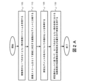
本発明の例示的な実施形態によるCE撮像システムの概略図を示す図1を参照する。例示的な実施形態では、システムは、画像を取り込むための1つ又は複数のイメージャ46と、体内腔を照明するための1つ又は複数の照明源42と、画像及び可能性として他の情報を受信装置に送信するための送信機41を有するカプセル40を含む。
Refer to FIG. 1, which shows a schematic diagram of a CE imaging system according to an exemplary embodiment of the present invention. In an exemplary embodiment, the system captures one or
画像取り込み装置は、Iddanらによる米国特許第7,009,634号明細書及び/又はGiladによる米国特許出願公開第11/603,123号明細書に記載されている実施形態に対応することができるが、代替の実施形態では他の種類の画像取り込み装置であってもよい。イメージャシステムによって取り込まれた画像は、例えば、円形、正方形、長方形、八角形、六角形などを含む任意の適切な形状とすることができる。 The image capture apparatus can accommodate embodiments described in U.S. Pat. No. 7,009,634 by Iddan et al. And / or U.S. Patent Application Publication No. 11 / 603,123 by Gilad. However, in an alternative embodiment, it may be another type of image capture device. The image captured by the imager system can be of any suitable shape, including, for example, circles, squares, rectangles, octagons, hexagons, and the like.
典型的には、1つ又は複数の位置における被験体の外側に位置するのは、特にカプセル40によって記録された画像を表示するためのアンテナ又はアンテナアレイ、画像受信機記憶部16、データプロセッサ14、データプロセッサ記憶部19、及び画像モニタ又は視覚表示部18を典型的に含む画像受信機12である。データプロセッサ記憶部19は、画像データベース21を含むことができる。
Typically located outside the subject at one or more positions is an antenna or antenna array for displaying images recorded by the
典型的には、データプロセッサ14、データプロセッサ記憶部19(例えばメモリ)及びモニタ18は、代替の構成も可能であるが、パーソナルコンピュータ又はワークステーション11の一部であり、これはプロセッサ14、メモリ、ディスクドライブ、並びにマウス及びキーボードなどの入力出力装置22を含む。データプロセッサ14は、マイクロプロセッサ、マルチプロセッサ、アクセラレータボード、又は任意の他のシリアル又はパラレル高性能データプロセッサなどの任意の標準データプロセッサを含むことができる。データプロセッサ14は、その機能の一部として、画像の表示(例えば、どの画像、様々なウィンドウの中の画像の位置、画像の表示のタイミング又は持続時間など)を制御するコントローラとして機能することができる。
Typically, the
画像モニタ18は、従来の映像ディスプレイあるいは画像又は他のデータを表示できる任意の他の装置とすることができる。画像モニタ18は、画像データ、例えば、1つ又は複数のGIT画像、取り込まれた画像の1つ又は複数の解剖学的グラフ表示、静止及び動画の形態の画像、運動データ及び/又は他の情報を提示する。 The image monitor 18 can be a conventional video display or any other device capable of displaying an image or other data. The image monitor 18 may include image data, such as one or more GIT images, one or more anatomical graph displays of captured images, still and moving images, motion data and / or other information. To present.
幾つかの実施形態では、様々なカテゴリの情報がウィンドウに表示される。ウィンドウは、例えば、ディスプレイ又はモニタ上のセクション又は領域(輪郭が描かれているか境界がついている可能性がある)であってもよく、他のウィンドウも使用できる。複数のモニタを使用して画像、運動特性、運動事象及び他のデータを表示することができ、例えば画像モニタを画像受信機12に含めることもできる。フレームのシーケンスの文脈で使用される場合、フレームの組又はシーケンスのウィンドウ(例えば、取り込み又は受信時間、あるいは別の順序付けによって順序付けられた)は、画像フレームのストリーム内の画像フレームの連続するサブセットであり得る。
In some embodiments, various categories of information are displayed in the window. A window may be, for example, a section or area (which may be contoured or bordered) on a display or monitor, and other windows may be used. Images, motor characteristics, motor events and other data can be displayed using a plurality of monitors, for example an image monitor can be included in the
動作中、イメージャ46は画像を取り込み、画像を表すデータを送信機41に送信することができる。送信機41は、例えば電磁波を用いて画像を画像受信機12に(例えば、フレームとして)送信する。画像受信機12は、画像データを画像受信機記憶部16に転送する。一定時間のデータ収集の後、記憶部16に記憶された画像データは、データプロセッサ14又はデータプロセッサ記憶部19に転送することができる。例えば、画像受信機12又は画像受信機記憶部16は、患者の身体から取り外すことができ、標準データリンク、例えばシリアル、パラレル、USB、又は既知の構成の無線インタフェースを介して、データプロセッサ14及びデータプロセッサ記憶部19を含むパーソナルコンピュータ又はワークステーションに接続することができる。画像データは、画像受信機記憶部16からデータプロセッサ記憶部19内の画像データベース21に転送される。
During operation, the
典型的には、画像ストリームは、画像データベース21に一連の画像として記憶され、これは様々な既知の方法で実施することができる。データプロセッサ14は、データを分析し、分析されたデータを画像モニタ18に提供することができ、ここでユーザは画像データを見る。例えば、データプロセッサ14又は別のデータプロセッサ(例えば、受信機12)は、画像を処理し、被験者のGITの解剖学的表示を提示することができる。データプロセッサ14は、オペレーティングシステム及びデバイスドライバなどの基本オペレーティングソフトウェアと共に、データプロセッサ14の動作を制御するソフトウェアを動作させる。データプロセッサ14を制御するソフトウェアは、マイクロソフト社のネットプラットフォームなどの様々な開発プラットフォームを使用して実施されたコードを含み、様々な既知の方法で実施することができる。
Typically, the image stream is stored in the
カプセル40によって記録され送信される画像データはデジタルカラー画像データであってもよいが、代替の実施形態では他の画像フォーマットを使用することができる。例示的な実施形態では、画像データの各フレームは、各320ピクセルの320行(例えば、320行及び320列)を含み、各ピクセルは、既知の方法に従って、色及び輝度のバイトを含む。例えば、各イメージャピクセルは、赤、緑、又は青などの単一の原色に対応することができるカラーセンサを含むことができる。全体のピクセルの輝度は、1バイト(すなわち、0~255)の輝度値によって記録することができる。画像は、例えば、順次、データプロセッサ記憶部19に記憶することができる。記憶されたデータは、色及び明るさを含む1つ又は複数のピクセル値で構成される。他の画像フォーマットも使用できる。
The image data recorded and transmitted by the
データプロセッサ記憶部19は、カプセル40によって記録された一連の画像を記憶することができる。例えば、カプセル40が記録する画像は、例えば患者のGITを通過する際に連続的に結合され、画像ストリームとして表示可能な一連の画像を形成することができる。画像ストリームを見るとき、ユーザは、典型的には、モニタ18上に1つ又は複数のウィンドウで提示される。代替の実施形態では、複数のウィンドウを使用する必要はなく、画像ストリームのみを表示することができる。例えば、複数のウィンドウが提供される実施形態では、画像ウィンドウは、画像ストリーム、又はその画像の静止部分を提供することができる。別のウィンドウは、例えば、停止、再生、一時停止、画像取り込み、ステップ、早送り、巻き戻し、又は他の制御など、画像の表示を変更できるボタンやその他の制御を含むことができる。このような制御は、例えば、マウス又はタッチスクリーン上の指などのポインティング装置によって有効にすることができる。画像ストリームは静止させて1つのフレームを表示したり、スピードアップしたり、巻き戻したりすることができ、セクションはスキップでき、又は画像を見るための任意の他の方法を画像ストリームに適用することができる。
The data
データプロセッサ14は、セグメント表示ジェネレータ24を含むか、又はセグメント表示ジェネレータ24に直接的又は間接的に動作可能に接続することができる。セグメント表示ジェネレータ24は、取り込まれた画像の組からの画像をセグメントに処理することができる。次に、セグメント化された提示を生成し、グラフィカルユーザインタフェース(GUI)の所定のセクションに表示することができる。幾つかの実施形態では、セグメント表示ジェネレータ24はセグメント表示、例えばセグメントカラーバー又は他のグラフィカル表現を生成することができる。
The
一例では、タイムバーを生成するために使用される画像のサブセットは、画像ストリーム内で識別できる特定の解剖学的ランドマークの間に取り込まれた画像を含むことができる。例えば、第1の十二指腸画像と第1の盲腸画像との間、及び第1の盲腸画像と最後の直腸画像との間である。 In one example, the subset of images used to generate the timebar can include images captured between specific anatomical landmarks that can be identified within the image stream. For example, between the first duodenal image and the first cecal image, and between the first cecal image and the last rectal image.
2つの解剖学的ランドマークを選択することができ(例えば、システム内で予め決定することができるか、及び/又はユーザによって選択することができる)、カプセルが最初に取り込まれた選択された解剖学的ランドマークから後から取り込まれた選択された解剖学的ランドマークに移動した時間の間に取り込まれた全ての画像を、タイムバーの生成に含むことができる。 Two anatomical landmarks can be selected (eg, can be pre-determined within the system and / or can be selected by the user) and the selected anatomy in which the capsule was first incorporated. All images captured during the time traveled from the anatomical landmark to the later captured anatomical landmark can be included in the generation of the time bar.
別の例では、画像は、色パラメータ、画像品質パラメータ、画像内の検出された病理学的候補の数などに従って、(例えば、ユーザ(例えば医師)によって、又はコンピュータによって)選択することができる。選択された器官(食道、小腸、結腸、胃など)、選択されたGIT領域に対して、又は完了画像処置から選択された特定の時間長に対して、タイムバーを生成することができる。さらに別の例では、例えば隣接する画像間の類似性に基づいて、画像を結合又は融合することができ、融合又は結合された画像のサブセットに基づいてタイムバーを生成することができる。当技術分野で知られているように、画像のサブセットを決定又は選択するために、他の画像選択方法を使用することができる。タイムバーの生成に用いることができる画像のサブセットを生成するために、異なる画像選択方法を組み合わせることができる。 In another example, the image can be selected (eg, by a user (eg, a doctor) or by a computer) according to color parameters, image quality parameters, number of pathological candidates detected in the image, and so on. Time bars can be generated for selected organs (esophagus, small intestine, colon, stomach, etc.), for selected GIT regions, or for specific time lengths selected from completed imaging procedures. In yet another example, images can be combined or fused, for example based on the similarity between adjacent images, and a time bar can be generated based on a subset of the fused or combined images. As is known in the art, other image selection methods can be used to determine or select a subset of images. Different image selection methods can be combined to generate a subset of images that can be used to generate the timebar.
本発明の実施形態による使用に適した画像化、受信、処理、記憶及び/又は表示部を含む本発明の実施形態での使用に適した装置は、それぞれ本出願と共通の譲受人に譲渡された「System and Method for Editing an Image Stream Captured In- Vivo」という名称の米国特許出願公開第2006/0074275号明細書、及び/又は“Device for In- Vivo Imaging”という名称のIddanらによる米国特許第7,009,634号明細書、及び/又は「Method of Assembling an In- Vivo Imaging Device」という名称の米国特許出願公開第2007/0118012号明細書に説明された実施形態に類似することができる。インビボ画像化カプセルによって取り込まれた画像間の比較に基づいてGIT内の運動性を分析する方法は、例えば、Glukhovskyらによる米国特許第6,944,316号明細書に開示されている。 Devices suitable for use in embodiments of the invention, including imaging, reception, processing, storage and / or display suitable for use according to embodiments of the invention, are each transferred to a common transferee with the present application. US Patent Application Publication No. 2006/0074275, entitled "System and Method for Editing an Image Standard Captured In-Vivo," and / or "Device for In-Vivo," U.S. It can be similar to the embodiment described in US Patent Application Publication No. 7,009,634 and / or US Patent Application Publication No. 2007/0118012 entitled "Method of Assembling an In-Vivo Imaging Device". Methods for analyzing motility within GIT based on comparisons between images captured by in vivo imaging capsules are disclosed, for example, in US Pat. No. 6,944,316 by Gluckhovsky et al.
図2Aは、本発明の例示的な実施形態による、被験者のGITにおける粘膜疾患の状態の評価方法のフローチャートである。ステップ100では、被験者のGITの少なくとも一部の画像ストリームを受信することができる。例えば、図1で上述したように、カプセル内視鏡から画像ストリームを取り込むこと、つまり受信することができる。幾つかの実施形態では、炎症性腸疾患(例えば、クローン病)の状態を評価することが目的である場合、小腸及び結腸を含むGITの一部の画像ストリームを得ることができる。幾つかの実施形態では、被験者識別子(例えば、患者識別子)、画像ストリームが取り込まれた日付、患者診断及び任意の関連する医療データ又はそれらの任意の組み合わせが、取得された画像のストリームについてさらに受信される。
FIG. 2A is a flowchart of a method for evaluating the state of mucosal disease in a subject's GI according to an exemplary embodiment of the present invention. In
ステップ110では、画像ストリームを複数のセグメントに分解することができる。各セグメントは、被験者のGITの画像化された部分の領域に対応することができる。幾つかの実施形態では、領域は実質的に等しい長さとすることができる。1つ又は複数の領域は、小腸又は結腸などの解剖学的領域によって画定することができる。各セグメントは、様々な数の画像フレームを含むことができる。幾つかの実施形態では、複数のセグメントのそれぞれに識別子が割り当てられ、特定のセグメントの他のセグメントに対する位置を決定することができる。例えば、幾つかの実施形態では、複数のセグメントが順次標識される。幾つかの実施形態では、識別子は、各セグメントの開始時間及び停止時間を含む。この分解は、例えば被験者のGITを通るカプセル内視鏡の進行のコンピュータ化された評価に基づいて、図1のワークステーション11などのコンピュータ化されたシステムによって実行されるコンピュータ化された評価を介して行われてもよい。
In step 110, the image stream can be decomposed into a plurality of segments. Each segment can correspond to an area of the imaged portion of the subject's GIT. In some embodiments, the regions can be of substantially equal length. One or more regions can be defined by anatomical regions such as the small intestine or colon. Each segment can contain a different number of image frames. In some embodiments, an identifier is assigned to each of the plurality of segments, and the position of a particular segment with respect to other segments can be determined. For example, in some embodiments, multiple segments are sequentially labeled. In some embodiments, the identifier includes a start time and a stop time for each segment. This decomposition is based on a computerized assessment of the progress of the capsule endoscope through the subject's GIT, for example, via a computerized assessment performed by a computerized system such as
例えば、図1のカプセル内視鏡40などのカプセル内視鏡によって取り込まれた画像ストリームを、被験者の小腸及び結腸を通過させながら、受信することができる。画像ストリームは小腸及び結腸の3つの領域に対応する4つのセグメントに分解されてもよく(以下、「三分位値」とも呼ぶ)、例えば、表示される腸粘膜組織に関しては、全て長さが実質的に等しい。分解は、解剖学的ランドマークの識別に基づいて実行されてもよい。したがって、第1の十二指腸画像の識別は、小腸への進入を示すことができ、第1の盲腸画像(又は最後の末端回腸画像)の識別は、結腸内への進入を示すことができる。そのような解剖学的ランドマークの1つ又は複数の識別は、受信した画像ストリームの表示中にユーザによって実行され、図1のワークステーション11などのコンピュータ化されたシステムによって入力として受信することができる(例えばI/O装置22を介して)。あるいは、解剖学的ランドマークは、コンピュータ化されたシステムによって(例えば、図1のプロセッサ14を介して)自動的に識別することができる。例えば、小腸への入り口は、本出願と共通の譲受人に譲渡された「System and method for real time detection of villi texture in an image stream of the gastrointestinal tract」という名称のZinaty Ofraらによる米国特許第8,768,024号明細書に開示された方法及びシステムに従って識別することができる。結腸への入り口は、本出願と共通の譲受人に譲渡された「Method and system for detecting transition of in-vivo device between sections of the GI system」という名称のPfeffer Yehudaによる米国特許第8,922,633号明細書に開示されている方法及びシステムに従って識別することができる。
For example, an image stream captured by a capsule endoscope such as the
小腸を示す画像ストリームの識別された部分は(すなわち、第1の十二指腸画像から第1の盲腸画像への)、被験者のGITを通るカプセル内視鏡の進行のコンピュータ化された評価を介して、所望により、実質的に等しい長さに、3つのセグメントに分解できる。このようなコンピュータ化された評価は、例えば、本出願と共通の譲受人に譲渡された、「System and method for detecting motion patterns of in vivo imaging devices」という名称のKrupnikらによる米国特許第8,792,691号明細書に開示された方法及びシステムに基づくことができる。 An identified portion of the image stream showing the small intestine (ie, from the first duodenal image to the first cecal image) is through a computerized assessment of the progress of capsule endoscopy through the subject's GIT. If desired, it can be decomposed into three segments into substantially equal lengths. Such a computerized evaluation is, for example, U.S. Pat. No. 8,79 by Krupnik et al. , 691 can be based on the methods and systems disclosed in the specification.
ステップ120では、各セグメントに対する値の組を取得することができる。値の組は、疾患におけるセグメントの病理学的関与及びセグメントにおける粘膜疾患の粘膜徴候の重篤度を示すことができる。幾つかの実施形態では、値の組は、少なくとも各セグメントにおける粘膜疾患の粘膜徴候の重篤度を指してもよい。例えば、IBD患者を評価する場合、粘膜徴候は病変及び狭窄を含み得る。幾つかの実施形態では、各セグメントに対する値の組は、セグメント内の粘膜徴候の最も高い重篤度を示す値と、セグメント内の粘膜徴候の典型的又は一般的な重篤度を示す値と、及び/又はセグメント内の粘膜徴候の程度を示す値を含んでもよい。幾つかの実施形態では、セグメントの最も重篤な粘膜徴候は、セグメントにおける粘膜徴候の典型的又は一般的な重篤度を表さない場合がある。このようにして、最も重篤な粘膜徴候値及び典型的又は一般的な重篤度値の両方を有することにより、セグメントにおける疾患の粘膜徴候のより包括的な図を得ることができる。重篤度は、価値体系によって決定することができる。例えば、IBD患者のGITにおける病変を評価する場合、価値システムは、対応して、1、2、3及び0によって示される軽度、中程度及び重篤又は無しの3つの程度を含むことができる。粘膜徴候の程度値は、疾患の粘膜徴候を表示するそれぞれのGI領域の粘膜組織表面の部分を示すことができる。例えば、IBD患者を評価する場合、程度値は、疾患の病理学的粘膜関与を有するGIT領域の組織表面のパーセントを示すことができる。ある実施形態では、IBDが評価される場合、程度値は、無し、10%未満、10~30%、30~60%、又は60%超であり得る。これは、経験によれば、低いパーセント範囲が疾患の徴候に関してより一般的であるからである。
In
幾つかの実施形態では、値の組の1つ又は複数の値は、各セグメントに対してユーザからの入力として取得されてもよい。幾つかの実施形態では、値の組の1つ又は複数の値は、コンピュータ化されたシステムを介して各セグメントについて計算されてもよい。したがって、幾つかの実施形態では、値の一部はユーザからの入力として受信され、残りの値はコンピュータ化されたシステムを介して計算されてもよい。幾つかの実施形態では、1つ又は複数の値は、ユーザ入力と、コンピュータ化されたシステム及び方法によって実行される計算との組合せに基づいて決定することができる。 In some embodiments, one or more values in a set of values may be obtained as input from the user for each segment. In some embodiments, one or more values in a set of values may be calculated for each segment via a computerized system. Therefore, in some embodiments, some of the values may be received as input from the user and the remaining values may be calculated via a computerized system. In some embodiments, one or more values can be determined based on a combination of user input and calculations performed by computerized systems and methods.
1つ又は複数の値がユーザによって決定される場合、GIT部分を長さが実質的に等しいセグメントに分解することはより有利であり得る。長さが実質的に等しいセグメントの手動評価は、ユーザの評価プロセスを容易かつ簡素化することができる。より簡単で単純な手動評価処置は、より正確な手動評価をもたらす場合がある。 If one or more values are determined by the user, it may be more advantageous to decompose the GIT portion into segments of substantially equal length. Manual evaluation of segments of substantially equal length can facilitate and simplify the user's evaluation process. Simpler and simpler manual assessment procedures may result in more accurate manual assessment.
少なくとも値の一部又は全部が、開示されたコンピュータ化されたシステム及び方法によって単独又は付加的に決定される場合、必ずしも長さが等しくないセグメントにGIT部分を分解することは、より有利であり得る。例えば、GIT部分を、GITの1つ又は複数の解剖学的領域に少なくとも部分的に対応するセグメントに分解することは、幾つかの疾患評価にとってより有益であり得る。幾つかの実施形態では、評価処置中にそのような等しくないセグメントの分解が望ましい場合があり、一方、例えば自動的に(すなわち、コンピュータ化されたシステム及び方法によって)実行される最終スコア又は全体評価では、そのような等しくない分解はそれに応じて重み付けされてもよい。 It is more advantageous to decompose the GIT portion into segments that are not necessarily equal in length, if at least some or all of the values are determined alone or additionally by the disclosed computerized systems and methods. obtain. For example, decomposing a GIT portion into segments that at least partially correspond to one or more anatomical regions of GIT may be more beneficial for some disease assessments. In some embodiments, decomposition of such unequal segments may be desirable during the evaluation procedure, while the final score or overall performed, eg, automatically (ie, by computerized systems and methods). In the evaluation, such unequal decompositions may be weighted accordingly.
上述の例示的な値の組を計算するための例示的な方法が本明細書に開示される。潰瘍検出器を用いて潰瘍を識別することができる。例示的な潰瘍検出器は、本出願と共通の譲受人に譲渡された「Method and system for image-based ulcer detection」という名称のDori Pelegによる米国特許第8,923,585号明細書に開示される。次いで、潰瘍は、次に、その程度を決定するために特徴付けることができる。例えば、潰瘍の程度は、それらのサイズに基づいて決定されてもよい。潰瘍の大きさは、それぞれ本出願人の共通譲受人に譲渡された「System and method for size estimation of in- vivo objects」という名称のKrupnikらによる国際公開第2015/049684号及び、「Device and method for determining a size of in-vivo objects」という名称のKrupnikによる米国特許第9,412,054号明細書に開示されているような方法に従って推定することができる。当該技術分野で知られているように、最大及び中央値を決定するための関数を用いて、各セグメントにおいて潰瘍の最も重篤な程度及び潰瘍の最も一般的な程度を決定することができる。程度値は、次に各セグメントにおける潰瘍の表示として識別された画像と、これらの特定の画像における被験者のGITを通るカプセル内視鏡の進行の評価とに基づいて決定することができる。 An exemplary method for calculating the exemplary set of values described above is disclosed herein. Ulcers can be identified using an ulcer detector. An exemplary ulcer detector is disclosed in US Pat. No. 8,923,585 by Dori Peleg, entitled "Method and system for image-based ulcer detection", which was assigned to an assignee common to this application. To. The ulcer can then be characterized to determine its extent. For example, the extent of ulcers may be determined based on their size. The size of the ulcer was determined by Krupnik et al.'S International Publication No. 2015/049684 and "Devic", which were transferred to the common transferee of the applicant, respectively. It can be estimated according to a method as disclosed in US Pat. No. 9,421,054 by Krupnik, entitled "for determining a size of in-vivo objects". As is known in the art, functions for determining maximum and median can be used to determine the most severe degree of ulcer and the most common degree of ulcer in each segment. Degree values can then be determined based on images identified as indications of ulcers in each segment and an assessment of the progression of capsule endoscopy through the subject's GIT in these particular images.
幾つかの実施形態では、値の組は、GITの関心のある1つ又は複数の部分の粘膜の病理学的関与をさらに指すことができる。例えば、値の組は、十二指腸、回腸末端、右結腸及び/又は左結腸の関与又は非関与を示す値を含み得る。そのような情報は、患者の治療方法及び医学的管理に影響を及ぼし得る。 In some embodiments, the set of values can further refer to the pathological involvement of the mucosa of one or more parts of interest in GIT. For example, a set of values may include values indicating the involvement or non-involvement of the duodenum, terminal ileum, right colon and / or left colon. Such information can affect the patient's treatment and medical management.
ステップ130では、画像化されたGIT部分全体(GIT全体を含む)における粘膜疾患の粘膜徴候の重篤度及び位置を示す表示が生成され、被験者のGIT部分における粘膜疾患の状態の評価が可能になる。表示は、各セグメントについて得られた値の組に基づいて生成される。表示は、表、グラフ、報告、解剖学的グラフ表示及び/又はスコアなどの1つ又は複数の要素を含むことができる。
In
幾つかの実施形態では、表示は、そのGIT領域の各々を示すGIT部分の解剖学的グラフ表示と、領域の各々について得られた値の1つ又は複数の値とを含んでもよい。幾つかの実施形態では、得られた値の組は、GIT領域の各々(例えば、領域の始め又は終わり)内の粘膜徴候の位置を示す情報を含むことができる。幾つかの実施形態では、表示はそのような位置を示すことができる。例えば、粘膜徴候の重篤度の程度の表示は、GIT領域内の粘膜徴候の位置に比較的対応する位置で、解剖学的表示に重ね合わせることができる。幾つかの実施形態では、各GIT領域の1つ又は複数の値を示すために色を使用することができる。幾つかの実施形態では、解剖学的表示は異なるGIT領域を示してもよい。例えば、小腸が3つの領域に分解される場合、小腸の解剖学的表示は、例えば図8に示されるように、並行して配置された3つのセグメントから構成され得る。 In some embodiments, the display may include an anatomical graph display of the GIT portion showing each of its GIT regions and one or more of the values obtained for each of the regions. In some embodiments, the resulting set of values can include information indicating the location of mucosal signs within each of the GIT regions (eg, the beginning or end of the region). In some embodiments, the display can indicate such a position. For example, the indication of the severity of the mucosal sign can be superimposed on the anatomical indication at a position relatively corresponding to the location of the mucosal sign within the GIT region. In some embodiments, colors can be used to indicate one or more values for each GIT region. In some embodiments, the anatomical representation may indicate different GIT regions. For example, if the small intestine is broken down into three regions, the anatomical representation of the small intestine can consist of three juxtaposed segments, eg, as shown in FIG.
GIT領域に基づく粘膜疾患の粘膜徴候の位置はまた、粘膜徴候分布パターンなどのGITの粘膜関与の特徴を指すことができる。したがって、GIT領域による位置は、粘膜関与が存在しない、局所的、まだら状、連続的又はびまん性であることを示すことができる。 The location of mucosal signs of mucosal disease based on the GIT region can also point to features of mucosal involvement of GIT, such as mucosal sign distribution patterns. Therefore, the location by the GIT region can be indicated to be local, mottled, continuous or diffuse with no mucosal involvement.
幾つかの実施形態では、値の組は、GITのサブ領域に対応するサブセグメントの粘膜関与を参照する値をさらに含むことができる。例えば、サブ領域は、GITの解剖学的に画定された部分、例えば十二指腸、末端回腸、右結腸及び左結腸に対応し得る。そのような値は、サブ領域が疾患に関与するかどうかを示す2値を含むことができる(すなわち、粘膜徴候を表示する)。 In some embodiments, the set of values can further include values that refer to the mucosal involvement of the subsegments corresponding to the subregions of GIT. For example, the subregion can correspond to an anatomically defined portion of the GI, such as the duodenum, terminal ileum, right and left colons. Such values can include two values indicating whether the subregion is involved in the disease (ie, displaying mucosal signs).
図2Bは、本発明の別の例示的な実施形態による、被験者(例えば、患者)のGITにおける粘膜疾患の状態の評価のフローチャートである。この実施形態は、他に示されない限り、図2Aに関して説明された実施形態と同様であり得る。この方法は、被験者のGITの画像ストリームを受信することを含む(ステップ205)。 FIG. 2B is a flow chart for assessing the state of mucosal disease in GIT of a subject (eg, a patient) according to another exemplary embodiment of the invention. This embodiment may be similar to the embodiment described with respect to FIG. 2A, unless otherwise indicated. The method comprises receiving an image stream of the subject's GI (step 205).
この方法はまた、画像のストリームの少なくとも一部を複数のセグメントに分解することを含む(ステップ210)。例えば、図1に関して上述したように、画像ストリームの一部又は画像ストリーム全体を、セグメント表示ジェネレータ24によってセグメントに分解することができる。セグメントは実質的に等しい長さのGIT領域に対応することができる。したがって、各セグメントは、GITの領域を示す複数の画像を含むことができる。
The method also comprises decomposing at least a portion of the stream of images into multiple segments (step 210). For example, as described above with respect to FIG. 1, a part of the image stream or the entire image stream can be decomposed into segments by the
幾つかの実施形態では、方法はまた、複数のセグメントの各セグメントについて、a)それぞれのセグメントの1つ又は複数の画像が表示され、b)表示される1つ又は複数の画像に基づいて、疾患の粘膜徴候及び粘膜徴候の重篤度に関する疾患におけるセグメントの病理学的関与を示す1つ又は複数の値が受信され、c)全てのセグメントの粘膜疾患の粘膜徴候の重篤度及び/又は位置をユーザに示す出力表示が生成されることを含む。このようにして、全セグメントを含む全GIT部分の粘膜疾患の重篤度及び位置のビューを得るために、ユーザ(例えば、医師)が行う再表示の量を最小にすることが可能である。 In some embodiments, the method also relies on, for each segment of the plurality of segments, a) one or more images of each segment, and b) one or more images displayed. One or more values indicating the pathological involvement of the segment in the disease with respect to the mucosal sign of the disease and the severity of the mucosal sign are received, c) the severity of the mucosal sign of the mucosal sign of all segments and / or Includes generating an output display that indicates the location to the user. In this way, it is possible to minimize the amount of redisplay performed by the user (eg, physician) in order to obtain a view of the severity and location of mucosal disease in all GIT moieties including all segments.
次いで、表示された画像ストリームにおいてセグメント及び/又はGIT領域を示しながら、画像ストリームがユーザに表示される(ステップ220)。様々な実施形態では、特定のセグメントの再表示のためにユーザに提示される特定のフレームは、例えば、画像ストリームのコンピュータ分析に基づいてコンピュータによって決定される。 The image stream is then displayed to the user, indicating the segment and / or GIT area in the displayed image stream (step 220). In various embodiments, the particular frame presented to the user for redisplaying a particular segment is determined by the computer, for example, based on computer analysis of the image stream.
次いで、ユーザは、各セグメントに対する値の組の1つ又は複数の値を入力するよう指示されてもよい(ステップ230)。 The user may then be instructed to enter one or more values in a set of values for each segment (step 230).
例えば、幾つかの実施形態では、値の組は、各セグメントにおける粘膜徴候の最も典型的又は一般的な重篤度及び/又は最も高い重篤度、例えば病変を示す値を含むことができる。病変の重篤度、例えば炎症性病変は、進行性スペクトルにあり得、充血及びびらんの軽度の粘膜関与から、アフタ性潰瘍へと進行し、最終的に内腔狭窄及び狭窄の形成をもたらすより広く深い潰瘍に進行する。活性な炎症関与を有する画像ストリームは、多くの場合、病変などの多数の粘膜徴候によって特徴付けることができる。しかし、治療上の意思決定は、セグメントに表示される全ての可能性のある粘膜徴候を示すよりも、セグメントにおける最も一般的な粘膜徴候及び/又は最も重篤な粘膜徴候の表示から利益を得ることができる。最も一般的な病変値及び/又は最も重篤な病変値をセグメントに割り当てることによって、レビュアー(すなわち、ユーザ)は、そのセグメントにおける全ての病変を考慮することなく、そのセグメントに関する病変情報を得ることができる。 For example, in some embodiments, the set of values can include the most typical or general severity and / or the highest severity of mucosal signs in each segment, eg, a value indicating a lesion. The severity of the lesion, such as an inflammatory lesion, can be in the progressive spectrum, rather than progressing from mild mucosal involvement of hyperemia and erosion to aphthous ulcers, ultimately leading to luminal stenosis and the formation of stenosis. It progresses to a wide and deep ulcer. Image streams with active inflammatory involvement can often be characterized by numerous mucosal signs such as lesions. However, therapeutic decisions benefit from the display of the most common and / or most severe mucosal signs in the segment, rather than showing all possible mucosal signs displayed in the segment. be able to. By assigning the most common and / or most severe lesion values to a segment, the reviewer (ie, the user) obtains lesion information about that segment without considering all lesions in that segment. Can be done.
幾つかの実施形態では、ユーザは、セグメントの表示中及び/又は表示の直後で、かつ次のセグメントの表示の前に、各セグメントに対する1つ又は複数の値を入力するように指示される。幾つかの実施形態では、ユーザは、例えば、ユーザ入力が受信されていない限り、次のセグメントを表示することを控えることによって、次のセグメントの表示に先立って各セグメントの1つ又は複数の値を入力するように強制されてもよい。ユーザは、入力を提供するように促されてもよい。各セグメントの表示中及び/又は次のセグメントの表示の前に、各セグメントに対する1つ又は複数の値を入力する(すなわち、ユーザによって)ことは、評価プロセスを短縮し、より効率的でより正確にすることができる。したがって、ユーザは、画像ストリームを見直している間、及びユーザの心の中でまだ新鮮であり、例えば異なるセグメントなどの他の画像の影響を受けない間に、彼の入力を提供する。 In some embodiments, the user is instructed to enter one or more values for each segment during and / or immediately after the display of a segment and before the display of the next segment. In some embodiments, the user refrains from displaying the next segment, for example, unless user input has been received, so that the value of each segment is one or more prior to the display of the next segment. You may be forced to enter. The user may be prompted to provide input. Entering one or more values for each segment (ie, by the user) during and / or before displaying each segment shortens the evaluation process and is more efficient and accurate. Can be. Thus, the user provides his input while reviewing the image stream and while still fresh in the user's mind and unaffected by other images, such as different segments.
例えば、幾つかの実施形態では、ユーザは、各セグメントの表示中に最も高い重篤度の値を提供するように指示されてもよい。次いで、ユーザは、第1の粘膜徴候、例えば、セグメントの表示中に特定の程度の重篤度を有する第1の病変を示す画像を識別することができる。次いで、ユーザは、第1の病変よりも重篤度が高いと思われる病変を示す次の画像だけをより注意深く見直し、及び/又は示すことができる。同じ程度の重篤度又はより低い重篤度を有するように見える他の病変を示す他の画像は、ユーザの注意を要求しない。このプロセスは、いずれが先に来ても、セグメント画像が完全に表示されるか、又はセグメント内の病変を示す画像に最も高い重篤度が割り当てられるまで継続することができる。 For example, in some embodiments, the user may be instructed to provide the highest severity value during the display of each segment. The user can then identify an image showing the first mucosal sign, eg, the first lesion having a certain degree of severity during the display of the segment. The user can then more carefully review and / or show only the next image showing a lesion that appears to be more severe than the first lesion. Other images showing other lesions that appear to have the same degree of severity or less severity do not require the user's attention. This process can be continued until the segment image is fully displayed or the image showing the lesion within the segment is assigned the highest severity, whichever comes first.
次に、値の組に基づいてユーザのために表示を生成することができる。表示は、GITの関心のある部分全体(例えば、IBDの小腸及び結腸)における粘膜疾患の位置及び重篤度をユーザに示すことができる(ステップ240)。幾つかの実施形態では、GITの解剖学的表示が、例えば、小腸及び結腸を含めて表示される。幾つかの実施形態では、対応する値の組、例えば、最も一般的な病変重篤度値、程度値及び最も重篤な病変値を有する各セグメントは、解剖学的表示において対応する位置(すなわち、対応するGIT領域)に沿って表示される。 A display can then be generated for the user based on the set of values. The display can indicate to the user the location and severity of the mucosal disease in the entire area of interest of the GI (eg, the small intestine and colon of the IBD) (step 240). In some embodiments, the anatomical representation of GIT is displayed, including, for example, the small intestine and colon. In some embodiments, each segment having the corresponding set of values, eg, the most common lesion severity value, the degree value and the most severe lesion value, is in the corresponding position (ie, the anatomical representation). , Corresponding GIT area).
以下に説明される図3~図17は、値の組がユーザから受信される開示された方法及びシステムの例示的な実施形態を参照する。 FIGS. 3-17, described below, refer to exemplary embodiments of the disclosed methods and systems in which a set of values is received from the user.
図3は、本発明の例示的な実施形態による、被験者のGITにおける粘膜疾患の状態の評価のためのシステムの開始画面の例示的なスクリーンショットである。ユーザは、システムで利用可能な全ての映像を見ることができる。利用可能な各映像は、名前301、姓303、患者識別子305、患者の性別307、カプセルタイプ(「映像を取り込むのに使用されるカプセルのタイプ」)309、処置日311(例えば、映像が撮影された日付)、所見313、及び利用可能な各映像に関する他の情報によって識別される。
FIG. 3 is an exemplary screenshot of the start screen of the system for assessing the state of mucosal disease in a subject's GIT, according to an exemplary embodiment of the invention. The user can see all the footage available on the system. Each available video includes
図4は、本発明の例示的な実施形態による、任意の利用可能な映像についてユーザに提示することができる所見405の例示的リストの例示的なスクリーンショットである。
FIG. 4 is an exemplary screenshot of an exemplary list of
図5は、本発明の例示的な実施形態による、選択された映像(例えば、選択された患者の画像)に対する粘膜疾患の状態の評価の開始の例示的なスクリーンショットである。この実施形態では、2つのカメラを含むカプセルで撮影された画像の右画像501及び左画像503が表示される。特定の画像は、タイムバー505上のマーカ504の位置に対応する。タイムバー505は、ユーザが画像周辺をスキップすることを可能にする。解剖学的ランドマークを識別するためのインジケータ509がユーザに提示される。GIT上の解剖学的ランドマークを入力する領域がユーザに提示される。具体的には、第1の十二指腸画像取り込み領域511、第1の盲腸画像取り込み領域513、及び最後の直腸画像取り込み領域515である。
FIG. 5 is an exemplary screenshot of the initiation of an assessment of mucosal disease status for selected images (eg, images of selected patients) according to an exemplary embodiment of the invention. In this embodiment, a
画像取り込み領域511、513及び515のそれぞれは、ユーザが左又は右の画像を取り込む命令と、提案された範囲の画像に行くためのリンクとを含む。ユーザが提案された画像の範囲をクリックすると、タイムバー505上のマーカ504は、システムが、その領域にマッチする可能性が高いと判断した画像の範囲に移動する。例えば、第1の盲腸画像取り込み領域513において、提案された範囲に行くリンクをユーザがクリックすると、マーカ504は、第1の盲腸画像が存在する可能性のある映像における時間に対応するタイムバー505上の位置に移動する。システムは、画像の提案された範囲を決定するために、かつ上述の例示的な方法に従って、処理部を使用することができる。
Each of the
幾つかの実施形態では、タイムバー505のシェーディングは、映像における画像のグループに対する様々な画像特性を示す。幾つかの実施形態では、タイムバー505の着色は、映像における画像のグループに対する様々な画像特性を示す。 In some embodiments, the shading of the time bar 505 exhibits various image characteristics for a group of images in the video. In some embodiments, the coloring of the time bar 505 exhibits various image characteristics for a group of images in the image.
図6は、本発明の例示的な実施形態による、第1の十二指腸画像の捕捉の例示的なスクリーンショットである。ユーザが画像を選択すると、ユーザの選択のサムネイル603が作成される。第1の十二指腸画像取り込み領域511は、十二指腸画像が取り込まれたことを示す、取り込みインジケータを示す。サムネイルは、映像中のサムネイル603の位置が識別可能となるようにタイムバー505に結合される。
FIG. 6 is an exemplary screenshot of the capture of a first duodenal image according to an exemplary embodiment of the invention. When the user selects an image, a
図7は、本発明の例示的な実施形態による、第1の盲腸画像の捕捉の例示的なスクリーンショットである。ユーザが画像を選択すると、ユーザの選択のサムネイル703が作成される。第1の盲腸画像取り込み領域513は、十二指腸画像が取り込まれたことを示す、取り込みインジケータを示す。サムネイルは、映像中のサムネイル703の位置が識別可能となるようにタイムバー505に結合される。
FIG. 7 is an exemplary screenshot of the capture of a first cecal image according to an exemplary embodiment of the invention. When the user selects an image, a
図8は、本発明の例示的な実施形態による、最後の直腸画像の取得後の例示的なスクリーンショットである。ユーザが画像を選択すると、ユーザの選択のサムネイル803が作成される。図6及び図7に示されているような画像取り込み領域は、前述の解剖学的ランドマークの全てが識別され取り込まれると、セグメント値領域805に置き換えられる。セグメント値領域805は、例えば、図8に示すような表を含むことができる。セグメント値領域805は、4つのセグメントSBI、SBII、SBIII及び結腸を含む。セグメント値領域805内の各セグメントについて、ユーザは、例えば、以下に説明する図9~12に従って値を入力することができる。幾つかの実施形態では、ユーザが値の1つを入力すると、その値が自動的に読み込まれる。代替的又は追加的に、システムによって計算された値が自動的に読み込まれてもよい。被験者807のGITの解剖学的グラフ表示を表示することができる。
FIG. 8 is an exemplary screenshot after acquisition of the last rectal image according to an exemplary embodiment of the invention. When the user selects an image, a
図9は、本発明の例示的な実施形態による、例示的なセグメント評価の例示的なスクリーンショットである。タイムバー505は、画像ストリーム901の解剖学的セグメント、すなわちGITの解剖学的部分(例えば、第1の十二指腸画像と第1の盲腸画像との間のGITに沿った距離、すなわち小腸)に対応する。解剖学的セグメント901は、実質的に等しい長さの3つのセグメントに細分される。この例では、3つの小腸セグメントのそれぞれは、長さに関して結腸と比較され、これらの4つのセグメント(すなわち、小腸の3つのセグメント及び結腸セグメント)のそれぞれは、上記のように、独立かつ逐次的な方法で再表示され評価される。図9に示すように、解剖学的セグメント901のセグメント903が強調表示され、セグメント903の左右の画像が表示される。例えば、病変を表示する第1の画像が(例えば、ユーザからの入力を受信することによって)識別されると、ユーザは、グラフィカル制御要素909を介して、セグメント903の第1の病変値を入力するよう促される。幾つかの実施形態では、ユーザは、図10A及び図10Bに示すように、グラフィカル制御要素909をクリックすることによって、病変重篤度ポップアップツールを利用することを選択することができる。病変重篤度ポップアップツール1003は、病変重篤度等級分け(例えば、度)1005の例をユーザに提示する。このようにして、ユーザは、セグメント903の左右の画像を病変重篤度ポップアップツール1005の例と比較して、セグメント903に割り当てる病変重篤度値を決定することができる。これらの例は、予めロードすることができる。幾つかの実施形態では、例えば、標準化された評価を提供するために、均一で標準化された等級付け及び/又はスコア付けを生成するのを助けるために、画像のカタログが提供される。
FIG. 9 is an exemplary screenshot of an exemplary segment evaluation according to an exemplary embodiment of the invention. The
図9に戻ると、この実施形態では、最も重篤な病変値は、1、2及び3の間である。値、1、2及び3は、スケールに対する重篤度を示すという点で相対値であり、他のスケールを使用することができる。
Returning to FIG. 9, in this embodiment, the most serious lesion values are between 1, 2 and 3. The
グラフィカル制御要素909は、ユーザが特定のサムネイルに関するコメントを入力できるサムネイルコメントセクションを含む。
The
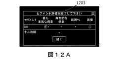
図12A~図12Dは、本発明の例示的な実施形態による例示的なセグメント粘膜評価の例示的なスクリーンショットである。各セグメントについて、最も重篤な病変値の入力が完了すると、ユーザは、グラフィカル制御要素1203で、最も典型的な又は一般的な病変及びセグメントの程度値を入力するのを促される。グラフィカル制御要素1203は、セグメントに対する典型的又は一般的な病変値(例えば、プルダウンメニュー1205に示されるように0~3)及び程度値(例えば、プルダウンメニュー1207に示されるパーセント範囲)をユーザが選択することを可能にする。幾つかの実施形態では、ユーザが典型的又は一般的な病変及び程度値を入力しない限り、システムは、例えば、ユーザに「セグメント評価を完了してください」と要求するポップアップウィンドウ1209を表示することによって、ユーザを評価プロセスにおいて先に進めることを控えることができる。
12A-12D are exemplary screenshots of an exemplary segmented mucosal assessment according to an exemplary embodiment of the invention. After completing the entry of the most severe lesion values for each segment, the user is prompted by the
ユーザがセグメント903に対する値を入力すると、例えば、以下の図13に示すように、解剖学的セグメント901の次のセグメントが評価される。
When the user enters a value for
図11は、図9の例示的な評価のさらなる例示的なスクリーンショットである。図11は、セグメント903の最も重篤な病変値が受信され入力された後の、図9に示す評価プロセスのスクリーンショットを示す。タイムバー505は、強調表示されたセグメント903を示し、セグメントに対するユーザ入力がユーザに表示される。セグメントにおける別の識別された病変の重篤度が、既に入力された病変値の重篤度よりも大きい場合、ユーザは、セグメント903に対する別の最も重篤な病変値を入力することができる。このようにして、各セグメントは最も重篤な病変について評価される。ユーザがセグメント903に対する値を入力すると、例えば、以下の図13に説明されるように、全ての関連セグメントが再表示され、評価されるまで、次のセグメントが順次かつ独立して評価される。
FIG. 11 is a further exemplary screenshot of the exemplary evaluation of FIG. FIG. 11 shows a screenshot of the evaluation process shown in FIG. 9 after the most severe lesion values for
図13は、本発明の例示的な実施形態による、第1のセグメントの評価の完了時のセグメント評価の例示的なスクリーンショットである。セグメント値領域805は、例えば図9~図12で説明したように、セグメント評価の結果としてユーザによって入力された値を表示する表を含む。GIT 807のグラフ表示は、対応する最も重篤な病変値及び最も一般的又は典型的な病変値で第1のセグメントを表示する。この例では、最も重篤な病変値は、対応するGIT領域に隣接して配置された重篤度値(この例では「1」)を含む円アイコンによって示される。典型的又は一般的な重篤度値は、対応するGIT領域の塗りつぶし色で示される。ユーザによって入力された値及び/又は開示されたコンピュータ化されたシステム及び/又は方法によって計算された値に対応する、異なる又は追加のインジケータを表示することができる。幾つかの実施形態では、値の組は、以下の図14に関して詳述するように、例えば、セグメントにおける狭窄の存在又は疾患のサブセグメントの関与を示す、1つ又は複数のバイナリ値を含むことができる。
FIG. 13 is an exemplary screenshot of segment evaluation upon completion of evaluation of the first segment according to an exemplary embodiment of the invention. The
図14は、本発明の例示的な実施形態による、全てのセグメントの評価の完了時のセグメント評価の例示的なスクリーンショットである。セグメント値領域805は、各セグメントのセグメント分析の結果として、ユーザ及び/又は開示されたコンピュータシステム及び/又は方法によって入力された値を表示する。表示され、値が割り当てられた画像は、タイムバー505の下にサムネイルとして表示される。GIT 807のグラフ表示は、各セグメントを対応する最も典型的又は一般的な病変値及び最も重篤な病変値で表示する。幾つかの実施形態では、最も一般的な病変値に色、例えばGIT領域の塗りつぶし色が割り当てられる。例えば、最も一般的な病変値番号1は黄色であり、最も一般的な病変値番号2はオレンジ色であり、最も一般的な病変値番号3は赤色であり得る。このようにして、評価情報をユーザに迅速かつ容易に伝達する方法で評価情報を表示することができる。
FIG. 14 is an exemplary screenshot of segment evaluation upon completion of evaluation of all segments according to an exemplary embodiment of the invention. The
幾つかの実施形態では、値の組は、例えば、十二指腸、末端回腸、右結腸及び左結腸などの関心のあるサブ領域に対応する狭窄又は関与の存在を示すバイナリ値を含むことができる。図14に示すように、受信された入力は、結腸セグメントにおける狭窄の存在を含む(文字「s」を含むアイコンによってセグメント値領域805に示される)。したがって、GIT807のグラフ表示は、結腸領域に隣接して位置する文字「S」を含むアイコンを含む。
In some embodiments, the set of values can include binary values indicating the presence of stenosis or involvement corresponding to subregions of interest such as, for example, duodenum, terminal ileum, right colon and left colon. As shown in FIG. 14, the received input comprises the presence of a stenosis in the colon segment (indicated in
図15A及び図15Bは、本発明の例示的な実施形態による、GITの関与する又は関与していない解剖学的部分を参照するデータを含む例示的な粘膜評価報告である。各報告書は、図14のなどの前の図に示された実施形態により、GITの粘膜評価を提示する、GITの表及びグラフ表示を含む。各表は受信された入力を提示する。サブセグメントは、十二指腸、末端回腸、右結腸及び左結腸の解剖学的部分を含む。図15Aに示す報告によれば、十二指腸及び末端回腸は関与していない。図15Bに示す報告によれば、右結腸のみが関与していない。疾患に関与していないサブセグメントは、塗りつぶしの色を含まない(又は、白色の塗りつぶしの色を含む)ことによって、GITのグラフ表示に示される。一方、関与する残りの対応するセグメント(すなわち、関与していないサブセグメントを除く)は、それぞれのセグメントにおける病変の典型的又は一般的な重篤度を示す塗りつぶし色を有する。 15A and 15B are exemplary mucosal evaluation reports containing data referring to anatomical moieties with or without GIT involvement, according to an exemplary embodiment of the invention. Each report includes a table and graph representation of GIT that presents a mucosal assessment of GIT according to the embodiments shown in the previous figure, such as FIG. Each table presents the input received. Subsegments include the anatomical parts of the duodenum, terminal ileum, right and left colons. According to the report shown in FIG. 15A, the duodenum and terminal ileum are not involved. According to the report shown in FIG. 15B, only the right colon is not involved. Subsegments that are not involved in the disease are shown in the GIT graph display by not including the fill color (or including the white fill color). On the other hand, the remaining corresponding segments involved (ie, excluding the subsegments not involved) have a fill color indicating the typical or general severity of the lesion in each segment.
図16は、本発明の例示的な実施形態による、全てのセグメントの評価の完了時のGIT評価の例示的なスクリーンショット(又はその一部)である。セグメント値領域805は、各セグメントのセグメント分析の結果として、ユーザによって及び/又は開示されたコンピュータ化された方法及び/又はシステムによって入力された値を表示する。GIT 807の解剖学的グラフ表示は、各セグメントを対応する値で表示する。各セグメントにおける最も重篤な病変の画像1601A~1601Cと、狭窄の画像1601Dとが表示される。
FIG. 16 is an exemplary screenshot (or part thereof) of a GIT evaluation upon completion of evaluation of all segments according to an exemplary embodiment of the invention. The
図17は、本発明の例示的な実施形態による、全てのセグメントの評価の完了時のGIT評価の印刷可能なレポート1701の例示的なスクリーンショットである。
FIG. 17 is an exemplary screenshot of a
図18は、本発明の例示的な実施形態による、2つの異なる日付で行われた処置について同じ患者の2つの評価報告の例示的なスクリーンショットである。2つの評価報告は、隣接する方法で表示することができ、したがって、治療奉仕者は、被験者のGITにおける疾患の進行又は経過をよりよく評価し、評価し、及び/又は理解し、よりよく治療することができる。 FIG. 18 is an exemplary screenshot of two evaluation reports of the same patient for treatments performed on two different dates according to an exemplary embodiment of the invention. The two assessment reports can be viewed in adjacent manners so that the therapist better assesses, assesses, and / or understands and better treats the progression or course of the disease in the subject's GIT. can do.
図19は、本発明の例示的な実施形態による、被験者のGITにおける粘膜疾患の粘膜徴候を監視するための方法のフローチャートである。 FIG. 19 is a flow chart of a method for monitoring mucosal signs of mucosal disease in a subject's GIT, according to an exemplary embodiment of the invention.
ステップ1910では、被験者のGITの少なくとも一部における粘膜疾患の粘膜徴候の位置及び重篤度を示す複数の表示が得られる。複数の表示の各表示は、本方法及び/又は本明細書に開示されたシステムに従って生成することができる。各表示は、一意の日付を有する処置の間に被験者のGITに取り込まれた画像ストリームに基づくことができる。被験者は一意の被験者識別子を持つことができる。例えば、カプセル内視鏡法の処置を有する患者0001としての一意の識別子を有する患者に対して、複数の値は2016年1月1日及び2016年2月1日の一意の日付になり得る。患者の識別子、日付及び処置は、単に説明の目的の例であり、任意の数の処置及び日付を複数の値に含めることができることは、当業者には明らかである。
In
ステップ1920では、複数の表示が隣接して表示される。
In
ステップ1930において、各表示は、対応する一意の日付でタグ付けすることができ、したがって、ユーザは、時間にわたって粘膜疾患の状態を監視することができる。
At
幾つかの実施形態では、各表示は、そのGIT領域の各々を示すGIT部分の解剖学的グラフ表示と、GIT領域の各々について得られた値の組の1つ又は複数の値とを含むことができる。解剖学的グラフ表示は、対応する一意の日付に従ってタイムラインに沿って表示することができる。 In some embodiments, each indication comprises an anatomical graph representation of the GIT portion showing each of its GIT regions and one or more of the set of values obtained for each of the GIT regions. Can be done. The anatomical graph display can be displayed along the timeline according to the corresponding unique date.
幾つかの実施形態では、開示されたコンピュータ化された方法及び/又はシステムをさらに利用して、ある期間中に患者GITにおいて実行される複数の処置に割り当てられた値の組を比較するのにさらに利用でき、その比較の結果をユーザに提示することができる。このような比較は、当技術分野で公知の方法に従って行うことができる。 In some embodiments, the disclosed computerized methods and / or systems are further utilized to compare pairs of values assigned to multiple treatments performed in a patient GIT over a period of time. Further available, the result of the comparison can be presented to the user. Such comparisons can be made according to methods known in the art.
幾つかの実施形態では、有益な記述子(例えば、日付、処置名など)を含む、被験者の関連する病歴データを受け取ることができる。複数の表示は、対応する一意の日付に従ってタイムラインに沿って表示することができる。患者の病歴データの少なくとも一部はまた、タイムラインに沿って表示されてもよい。幾つかの実施形態では、本方法は、被験者の治療開始及び終了データ、手術履歴データ及び検査室並びに生理学的履歴、又はそれらの任意の組み合わせを含む医学治療履歴データを受信することをさらに含むことができる。この方法はさらに、タイムラインに沿ってそのようなデータの少なくとも一部を表示することを含むことができる(例えば、図20A及び20Bに記載されるように)。この方法はまた、被験者についてのデータを見る要求を受信することを含むことができる。要求には、一意の被験者識別子を含めることができる In some embodiments, it is possible to receive relevant medical history data of the subject, including useful descriptors (eg, date, treatment name, etc.). Multiple displays can be displayed along the timeline according to the corresponding unique date. At least some of the patient's medical history data may also be displayed along the timeline. In some embodiments, the method further comprises receiving treatment start and end data, surgery history data and laboratory and physiological history of the subject, or medical treatment history data including any combination thereof. Can be done. The method can further include displaying at least a portion of such data along the timeline (eg, as described in FIGS. 20A and 20B). The method can also include receiving a request to view data about the subject. The request can include a unique subject identifier
図20A及び図20Bは、的な実施形態による、タイムラインに沿った複数の解剖学的グラフ表示及び対応する医学治療を含む例示的な報告2003及び2005である。
20A and 20B are
各報告について、同じ患者のGITの一部(すなわち、腸管)の解剖学的グラフ表示、2007 A~C及び2009 A~Cが、3つの異なる時間に行われた処置に基づいて評価のために表示される。各グラフ表示はタイムライン上に示され、時間にわたる患者の医学治療も表示される。このようにして、ユーザは、とりわけ、治療が所与の患者に対して経時的に行った効果を判定することができる。 For each report, an anatomical graph of a portion of the same patient's GIT (ie, the intestinal tract), 2007 AC and 2009 AC, for evaluation based on treatments performed at three different times. Is displayed. Each graph display is shown on the timeline and also shows the patient's medical treatment over time. In this way, the user can determine, among other things, the effect of treatment on a given patient over time.
幾つかの実施形態では、粘膜徴候を少なくとも部分的に自動的に識別し、任意選択的に評価することができ、自動的に識別された粘膜徴候を示す表示をユーザに提示することができる。このような実施形態では、開示されたシステム及び方法は、上記で詳述したように、GIT部分の画像ストリームにおける粘膜徴候を自動的に識別するために利用されてもよい。幾つかの実施形態では、開示されたシステム及び方法は、識別された粘膜徴候(例えば、軽度、中等度又は重度)の重篤度を判定するためにさらに利用され得る。幾つかの実施形態では、1つ又は複数のGIT領域における自動的に識別された粘膜徴候の全てを示すマップを生成し、ユーザに表示することができる。例えば、ヒートマップは、その重篤度値に応じて着色されたGIT領域における同定された粘膜徴候を表す複数の要素を含むように表示されてもよい。ここで、本発明の例示的な実施形態による、GIT領域の病変ヒートマップを表示するスクリーンショットである図21A、図21B及び図21Cを参照する。各要素がGIT領域内の特定された病変を表す一連の要素は、その領域に沿った病変の相対的な位置に従って提示することができる。各要素は、図21Aに示すように自動的に評価された重篤度レベル(例えば、軽度、中程度又は重篤)に従って色づけできる。このようなマップに基づいて、ユーザは、簡単および迅速な方法で値の組の他の値を手動で決定してもよい。例えば、上述した病変ヒートマップを提示されたユーザは、このマップに基づいて、最も一般的な病変値、最も重篤な病変値及び表示された領域に対する関与の程度などの値を決定することができる。図21Cは、その色に従って病変を提示し、対応する重篤度を示す。そのような提示は、例えば、GIT領域における最も一般的な値、最も重篤な値及び程度値の手動による決定をさらに容易にすることができる。幾つかの実施形態では、自動的に特定された粘膜徴候の提示及び/又はそのような自動評価は、提案として、及び/又はユーザによって実行される識別及び/又は評価を検証及び/又は完了するために使用することができる。 In some embodiments, mucosal signs can be at least partially automatically identified and optionally evaluated, and a display indicating the automatically identified mucosal signs can be presented to the user. In such embodiments, the disclosed systems and methods may be utilized to automatically identify mucosal signs in the image stream of the GIT portion, as detailed above. In some embodiments, the disclosed systems and methods may be further utilized to determine the severity of identified mucosal signs (eg, mild, moderate or severe). In some embodiments, a map showing all of the automatically identified mucosal signs in one or more GIT regions can be generated and displayed to the user. For example, the heat map may be displayed to include multiple elements representing the identified mucosal signs in the GIT region colored according to its severity value. Here, reference is made to FIGS. 21A, 21B and 21C which are screenshots displaying a lesion heat map in the GIT region according to an exemplary embodiment of the invention. A set of elements, each element representing a identified lesion within a GIT region, can be presented according to the relative position of the lesion along that region. Each element can be colored according to an automatically assessed severity level (eg, mild, moderate or severe) as shown in FIG. 21A. Based on such a map, the user may manually determine other values in a set of values in a simple and quick way. For example, a user presented with the lesion heatmap described above may use this map to determine values such as the most common lesion values, the most severe lesion values, and the degree of involvement in the displayed area. can. FIG. 21C presents lesions according to their color and indicates the corresponding severity. Such presentations can further facilitate manual determination of, for example, the most common values, the most severe values and the degree values in the GIT domain. In some embodiments, the presentation and / or such automated assessment of automatically identified mucosal signs validates and / or completes the identification and / or assessment performed as a suggestion and / or by the user. Can be used for.
前述の説明では、本発明の様々な態様が説明されている。説明の目的で、本発明の完全な理解を提供するために、特定の構成及び詳細が示される。しかしながら、当業者には、本明細書に提示された特定の詳細無しで本発明を実施できることは明らかである。さらに、本発明を不明瞭にしないために、周知の特徴を省略又は簡略化することができる。 In the above description, various aspects of the invention are described. For purposes of illustration, specific configurations and details are provided to provide a complete understanding of the invention. However, it will be apparent to those skilled in the art that the invention can be practiced without the specific details presented herein. Furthermore, well-known features can be omitted or simplified in order not to obscure the invention.
特に断りのない限り、上記の議論から明らかなように、明細書全体を通して、「処理する」、「計算する」、「記憶する」、「決定する」などの用語を利用する議論は、コンピューティングシステムのレジスタ及び/又はメモリ内の物理量、例えば電子量として表されるデータを、コンピューティングシステムのメモリ、レジスタ又は他のこのような情報記憶装置、送信又は表示装置内の他のデータに、操作及び/又は変換するコンピュータ又はピューディングシステムあるいは同様の電子コンピューティング装置の動作及び/又はプロセスを指すことが分かる。 Unless otherwise noted, as is clear from the discussion above, discussions that use terms such as "process," "calculate," "remember," and "determine" throughout the specification are computing. Manipulating data represented as physical quantities, eg, electronic quantities, in system registers and / or memory to other data in computing system memory, registers or other such information storage, transmission or display. And / or it can be seen to refer to the operation and / or process of a computer or pudging system or similar electronic computing device to convert.
異なる実施形態を本明細書に開示する。特定の実施形態の特徴は、他の実施形態の特徴と組み合わせることができ、したがって、特定の実施形態は、複数の実施形態の特徴の組み合わせとすることができる。 Different embodiments are disclosed herein. The features of a particular embodiment can be combined with the features of other embodiments, and thus a particular embodiment can be a combination of features of a plurality of embodiments.
本発明の実施形態は、プロセッサ又はコントローラによって実行されると、プロセッサ又はコントローラに本明細書で開示された方法を実行させる命令、例えば、コンピュータ実行可能命令を符号化し、含み、又は記憶する、例えばメモリ、ディスクドライブ、又はUSBフラッシュメモリなどの、コンピュータ又はプロセッサ読み取り可能な非一時的記憶媒体などの物品を含むことができる。幾つかの実施形態では、コンピュータプロセッサ又はコンピュータコントローラ、例えばデータプロセッサ14は、例えばプロセッサに接続されたメモリに格納されたソフトウェア又はコードを実行することによって、及び/又は専用の回路を有することによって本発明の実施形態を実行するように構成することができる。
Embodiments of the invention encode, include, or store instructions, eg, computer-executable instructions, that, when executed by a processor or controller, cause the processor or controller to perform the methods disclosed herein, eg. It can include articles such as computer or processor readable non-temporary storage media such as memory, disk drives, or USB flash memory. In some embodiments, the computer processor or computer controller, such as the
本発明の実施形態の前述の説明は、例示及び説明のために提示されたものである。本発明を開示された正確な形態に包括的であること、又は限定することを意図するものではない。当業者であれば、上記の教示に照らして多くの修正、変形、置換、変更、及び等価物が可能であることを理解されたい。したがって、添付の特許請求の範囲は、本発明の真の精神に含まれる全てのそのような改変及び変更を包含するように意図されていることが理解されるべきである。 The above description of embodiments of the invention are presented for illustration and explanation. The invention is not intended to be inclusive or limited to the exact form disclosed. Those skilled in the art should understand that many modifications, modifications, substitutions, modifications, and equivalents are possible in the light of the above teachings. Therefore, it should be understood that the appended claims are intended to include all such modifications and modifications contained in the true spirit of the invention.
Claims (14)
命令を実行するように構成された少なくとも1つのハードウェアプロセッサと、
中に格納された命令を有する記憶装置であって、前記命令は前記ハードウェアプロセッサによって実行されると、前記ハードウェアプロセッサに、
前記対象のGITの少なくとも一部の画像ストリームを受信させ、
前記画像ストリームを複数のセグメントに分解させ、各セグメントは前記対象のGITの前記少なくとも一部の領域に対応し、
前記セグメントの各々に対する値の組を取得させ、前記値の組は、前記粘膜疾患における前記セグメントの病理学的関与と、前記セグメントにおける前記粘膜疾患の粘膜徴候の重篤度との両方を示し、
各セグメントに対する前記値の組に基づいて、前記対象のGITの前記少なくとも一部の全体における前記粘膜疾患の前記粘膜徴候の位置及び重篤度を示す表示を生成させる、記憶装置と、
前記生成された表示を表示するように構成されたディスプレイと、を備える、システム。 A mucosal evaluation system for mucosal diseases in the subject's gastrointestinal tract (GIT).
With at least one hardware processor configured to execute instructions,
A storage device having an instruction stored therein, the instruction being executed by the hardware processor to the hardware processor.
Receive at least a part of the image stream of the target GI,
The image stream is decomposed into a plurality of segments, and each segment corresponds to the at least a part region of the target GIT.
A set of values was obtained for each of the segments, the set of values indicating both the pathological involvement of the segment in the mucosal disease and the severity of the mucosal signs of the mucosal disease in the segment.
A storage device that generates a display indicating the location and severity of the mucosal sign of the mucosal disease in the whole of the at least part of the subject GIT based on the set of values for each segment.
A system comprising a display configured to display the generated display.
表示された前記画像ストリームに前記セグメント又はGIT領域を表示しながら、前記画像ストリームをユーザに表示させ、
前記セグメントの各々に対する前記値の組の1つ又は複数の値を入力するようにユーザに指示させ、
前記GIT領域は実質的に互いに等しい長さである、請求項1に記載のシステム。 When the instruction is executed by the hardware processor, the instruction is further to the hardware processor.
While displaying the segment or the GIT area in the displayed image stream, the user is made to display the image stream.
Instruct the user to enter one or more of the set of values for each of the segments.
The system of claim 1, wherein the GIT regions are substantially equal in length to each other.
前記セグメントにおいて前記粘膜徴候の最も高い重篤度を示す値と、
前記セグメントにおいて前記粘膜徴候の重篤度を示す値と、
前記セグメントにおいて前記粘膜徴候の範囲を示す値と、
を含む、請求項1から8のいずれか一項に記載のシステム。 The set of values is
The value indicating the highest severity of the mucosal sign in the segment,
A value indicating the severity of the mucosal sign in the segment and
A value indicating the range of the mucosal sign in the segment and
The system according to any one of claims 1 to 8, comprising the above.
命令を実行するように構成された少なくとも1つのハードウェアプロセッサと、
中に格納された命令を有する記憶装置であって、前記命令は前記ハードウェアプロセッサによって実行されると、前記ハードウェアプロセッサに、
前記対象のGITの少なくとも一部において、前記粘膜疾患の粘膜徴候の位置及び重篤度を示す複数の表示を取得させ、前記複数の表示の各表示は、請求項1の前記システムによって生成され、前記複数の表示の各表示は、一意の日付を有する処置の間に前記対象のGITにおいて取り込まれた画像ストリームに基づき、
前記対象のGIT前記少なくとも一部の前記複数の表示を隣接する方法で表示させ、
各表示にその対応する一意の日付をタグ付けさせ、
これにより、ユーザは時系列で前記粘膜疾患の状態を監視することができる、記憶装置と、を備える、システム。 A monitoring system for mucosal diseases in the subject's gastrointestinal tract (GIT).
With at least one hardware processor configured to execute instructions,
A storage device having an instruction stored therein, the instruction being executed by the hardware processor to the hardware processor.
In at least a portion of the subject's GIT, a plurality of indications indicating the location and severity of mucosal signs of the mucosal disease are acquired, and each indication of the plurality of indications is generated by the system of claim 1. Each display of the plurality of displays is based on an image stream captured in the subject GIT during a procedure having a unique date.
The GIT of the target The plurality of displays of the at least a part thereof are displayed by an adjacent method.
Have each display tagged with its corresponding unique date and
A system comprising a storage device, which allows the user to monitor the state of the mucosal disease in chronological order.
治療開始データ及び治療停止データを含む前記対象についての医学治療履歴データを受信させ、
前記医学治療履歴データの少なくとも一部を前記タイムラインに沿って表示させ、
前記医学治療履歴データは、前記対象に処方された薬剤を含む、請求項13に記載のシステム。 When the instruction is executed by the hardware processor, the instruction is further to the hardware processor.
Receive medical treatment history data for the subject, including treatment start data and treatment stop data.
At least a part of the medical treatment history data is displayed along the timeline.
13. The system of claim 13, wherein the medical treatment history data comprises a drug prescribed to the subject.
Applications Claiming Priority (5)
| Application Number | Priority Date | Filing Date | Title |
|---|---|---|---|
| US201662357088P | 2016-06-30 | 2016-06-30 | |
| US62/357,088 | 2016-06-30 | ||
| US201762491486P | 2017-04-28 | 2017-04-28 | |
| US62/491,486 | 2017-04-28 | ||
| PCT/IL2017/050728 WO2018002935A1 (en) | 2016-06-30 | 2017-06-29 | Systems and methods for assessment and monitoring of a mucosal disease in a subject's gastrointestinal tract |
Publications (3)
| Publication Number | Publication Date |
|---|---|
| JP2019520916A JP2019520916A (en) | 2019-07-25 |
| JP2019520916A5 JP2019520916A5 (en) | 2020-08-06 |
| JP7034102B2 true JP7034102B2 (en) | 2022-03-11 |
Family
ID=60786586
Family Applications (1)
| Application Number | Title | Priority Date | Filing Date |
|---|---|---|---|
| JP2018568969A Active JP7034102B2 (en) | 2016-06-30 | 2017-06-29 | Systems and methods for assessing and monitoring mucosal disease in the subject's gastrointestinal tract |
Country Status (5)
| Country | Link |
|---|---|
| US (1) | US11478125B2 (en) |
| EP (1) | EP3478159B1 (en) |
| JP (1) | JP7034102B2 (en) |
| CN (1) | CN109788904A (en) |
| WO (1) | WO2018002935A1 (en) |
Families Citing this family (8)
| Publication number | Priority date | Publication date | Assignee | Title |
|---|---|---|---|---|
| JPWO2020066670A1 (en) * | 2018-09-27 | 2021-06-10 | Hoya株式会社 | Electronic endoscopy system |
| JP6877672B2 (en) * | 2019-04-26 | 2021-05-26 | Hoya株式会社 | Electronic endoscopy system and data processing equipment |
| US10671934B1 (en) | 2019-07-16 | 2020-06-02 | DOCBOT, Inc. | Real-time deployment of machine learning systems |
| US11423318B2 (en) * | 2019-07-16 | 2022-08-23 | DOCBOT, Inc. | System and methods for aggregating features in video frames to improve accuracy of AI detection algorithms |
| USD991279S1 (en) * | 2019-12-09 | 2023-07-04 | Ankon Technologies Co., Ltd | Display screen or portion thereof with transitional graphical user interface |
| USD991278S1 (en) * | 2019-12-09 | 2023-07-04 | Ankon Technologies Co., Ltd | Display screen or portion thereof with transitional graphical user interface for auxiliary reading |
| US11100373B1 (en) | 2020-11-02 | 2021-08-24 | DOCBOT, Inc. | Autonomous and continuously self-improving learning system |
| EP4360102A1 (en) | 2021-06-24 | 2024-05-01 | Given Imaging Ltd. | Systems and methods for assessing gastrointestinal cleansing |
Citations (1)
| Publication number | Priority date | Publication date | Assignee | Title |
|---|---|---|---|---|
| JP2012249936A (en) | 2011-06-06 | 2012-12-20 | Toshiba Corp | Medical image processing system |
Family Cites Families (14)
| Publication number | Priority date | Publication date | Assignee | Title |
|---|---|---|---|---|
| US20020010550A1 (en) | 1998-09-14 | 2002-01-24 | George M. Grass | Pharmacokinetic-based drug design tool and method |
| IL173696A (en) | 2000-03-08 | 2010-11-30 | Given Imaging Ltd | Device and system for in vivo imaging |
| US20070224191A1 (en) * | 2002-12-05 | 2007-09-27 | Pdl Biopharma, Inc. | Methods of treatment of Ulcerative Colitis and Crohn's disease with anti-CD3 antibodies |
| US8900124B2 (en) | 2006-08-03 | 2014-12-02 | Olympus Medical Systems Corp. | Image display device |
| JP5005981B2 (en) * | 2006-08-03 | 2012-08-22 | オリンパスメディカルシステムズ株式会社 | Image display device |
| US7953613B2 (en) * | 2007-01-03 | 2011-05-31 | Gizewski Theodore M | Health maintenance system |
| JP2010042182A (en) | 2008-08-18 | 2010-02-25 | Fujifilm Corp | Laser treatment device |
| US8682142B1 (en) | 2010-03-18 | 2014-03-25 | Given Imaging Ltd. | System and method for editing an image stream captured in-vivo |
| US8768024B1 (en) * | 2010-06-01 | 2014-07-01 | Given Imaging Ltd. | System and method for real time detection of villi texture in an image stream of the gastrointestinal tract |
| US20150011542A1 (en) | 2012-01-12 | 2015-01-08 | University Of Connecticut | Methods of Treating Adverse Intestinal Effects of Non-Steroidal Anti-Inflammatory Drugs |
| WO2014073527A1 (en) * | 2012-11-07 | 2014-05-15 | オリンパスメディカルシステムズ株式会社 | Medical image processing device |
| CN104885120A (en) * | 2012-12-31 | 2015-09-02 | 基文影像公司 | System and method for displaying an image stream |
| EP3567547A1 (en) | 2013-10-11 | 2019-11-13 | Mauna Kea Technologies | Method for characterizing images acquired through a video medical device |
| WO2019195146A1 (en) * | 2018-04-03 | 2019-10-10 | Boston Scientific Scimed, Inc. | Systems and methods for diagnosing and/or monitoring disease |
-
2017
- 2017-06-29 EP EP17819504.6A patent/EP3478159B1/en active Active
- 2017-06-29 CN CN201780045752.1A patent/CN109788904A/en active Pending
- 2017-06-29 US US16/314,021 patent/US11478125B2/en active Active
- 2017-06-29 JP JP2018568969A patent/JP7034102B2/en active Active
- 2017-06-29 WO PCT/IL2017/050728 patent/WO2018002935A1/en unknown
Patent Citations (1)
| Publication number | Priority date | Publication date | Assignee | Title |
|---|---|---|---|---|
| JP2012249936A (en) | 2011-06-06 | 2012-12-20 | Toshiba Corp | Medical image processing system |
Also Published As
| Publication number | Publication date |
|---|---|
| WO2018002935A1 (en) | 2018-01-04 |
| EP3478159A1 (en) | 2019-05-08 |
| CN109788904A (en) | 2019-05-21 |
| JP2019520916A (en) | 2019-07-25 |
| EP3478159A4 (en) | 2020-01-08 |
| US11478125B2 (en) | 2022-10-25 |
| EP3478159B1 (en) | 2022-04-13 |
| US20190244351A1 (en) | 2019-08-08 |
Similar Documents
| Publication | Publication Date | Title |
|---|---|---|
| JP7034102B2 (en) | Systems and methods for assessing and monitoring mucosal disease in the subject's gastrointestinal tract | |
| Ahmad et al. | Artificial intelligence and computer-aided diagnosis in colonoscopy: current evidence and future directions | |
| JP6215236B2 (en) | System and method for displaying motility events in an in-vivo image stream | |
| US8185185B2 (en) | System and method for assessing a patient condition using tertiles derived from capsule endoscope images of the small bowel | |
| US20210345865A1 (en) | Systems and methods for generating and displaying a study of a stream of in-vivo images | |
| Kamba et al. | Reducing adenoma miss rate of colonoscopy assisted by artificial intelligence: a multicenter randomized controlled trial | |
| JP4740212B2 (en) | System and method for displaying a data stream | |
| JP2015509026A5 (en) | ||
| Kornbluth et al. | Video capsule endoscopy in inflammatory bowel disease: past, present, and future | |
| US20220296081A1 (en) | Method for real-time detection of objects, structures or patterns in a video, an associated system and an associated computer readable medium | |
| WO2021075418A1 (en) | Image processing method, teacher data generation method, trained model generation method, illness development prediction method, image processing device, image processing program, and recording medium on which program is recorded | |
| Joseph et al. | Artificial intelligence in colonoscopy | |
| CN101273916B (en) | System and method for evaluating status of patient | |
| Jin et al. | Deep learning for gastroscopic images: computer-aided techniques for clinicians | |
| Bernal et al. | Building up the future of colonoscopy–a synergy between clinicians and computer scientists | |
| Luca et al. | An overview on computer processing for endoscopy and colonoscopy videos | |
| RU2564751C1 (en) | Method of determining chnge of glottis area in dynamics of performed treatment | |
| Liu et al. | Deep learning site classification model for automated photodocumentation in upper GI endoscopy (with video) | |
| Liang et al. | The applications and challenges of artificial intelligence in colonoscopy | |
| Biffi et al. | REAL-Colon: A dataset for developing real-world AI applications in colonoscopy | |
| Hoogenboom et al. | Training for Advanced Endoscopic Imaging in Gastrointestinal Diseases | |
| Philp | Detection:(CQI) Quality Measures and Tools for Improvement | |
| Angulo Rodriguez | Real-time Quality Feedback System for Screening Colonoscopy | |
| Banerjee et al. | 45. Diagnostic Colonoscopy |
Legal Events
| Date | Code | Title | Description |
|---|---|---|---|
| A521 | Request for written amendment filed |
Free format text: JAPANESE INTERMEDIATE CODE: A523 Effective date: 20200626 |
|
| A621 | Written request for application examination |
Free format text: JAPANESE INTERMEDIATE CODE: A621 Effective date: 20200626 |
|
| A977 | Report on retrieval |
Free format text: JAPANESE INTERMEDIATE CODE: A971007 Effective date: 20210430 |
|
| A131 | Notification of reasons for refusal |
Free format text: JAPANESE INTERMEDIATE CODE: A131 Effective date: 20210511 |
|
| A521 | Request for written amendment filed |
Free format text: JAPANESE INTERMEDIATE CODE: A523 Effective date: 20210810 |
|
| A02 | Decision of refusal |
Free format text: JAPANESE INTERMEDIATE CODE: A02 Effective date: 20210907 |
|
| A521 | Request for written amendment filed |
Free format text: JAPANESE INTERMEDIATE CODE: A523 Effective date: 20220105 |
|
| C60 | Trial request (containing other claim documents, opposition documents) |
Free format text: JAPANESE INTERMEDIATE CODE: C60 Effective date: 20220105 |
|
| A911 | Transfer to examiner for re-examination before appeal (zenchi) |
Free format text: JAPANESE INTERMEDIATE CODE: A911 Effective date: 20220117 |
|
| C21 | Notice of transfer of a case for reconsideration by examiners before appeal proceedings |
Free format text: JAPANESE INTERMEDIATE CODE: C21 Effective date: 20220118 |
|
| TRDD | Decision of grant or rejection written | ||
| A01 | Written decision to grant a patent or to grant a registration (utility model) |
Free format text: JAPANESE INTERMEDIATE CODE: A01 Effective date: 20220208 |
|
| A61 | First payment of annual fees (during grant procedure) |
Free format text: JAPANESE INTERMEDIATE CODE: A61 Effective date: 20220301 |
|
| R150 | Certificate of patent or registration of utility model |
Ref document number: 7034102 Country of ref document: JP Free format text: JAPANESE INTERMEDIATE CODE: R150 |