JP6944438B2 - Signal amplification in solution-based plasmon-specific binding partner assay - Google Patents
Signal amplification in solution-based plasmon-specific binding partner assay Download PDFInfo
- Publication number
- JP6944438B2 JP6944438B2 JP2018505669A JP2018505669A JP6944438B2 JP 6944438 B2 JP6944438 B2 JP 6944438B2 JP 2018505669 A JP2018505669 A JP 2018505669A JP 2018505669 A JP2018505669 A JP 2018505669A JP 6944438 B2 JP6944438 B2 JP 6944438B2
- Authority
- JP
- Japan
- Prior art keywords
- detection conjugate
- detection
- gold
- nanostructures
- metal
- Prior art date
- Legal status (The legal status is an assumption and is not a legal conclusion. Google has not performed a legal analysis and makes no representation as to the accuracy of the status listed.)
- Active
Links
Images


Classifications
-
- G—PHYSICS
- G01—MEASURING; TESTING
- G01N—INVESTIGATING OR ANALYSING MATERIALS BY DETERMINING THEIR CHEMICAL OR PHYSICAL PROPERTIES
- G01N33/00—Investigating or analysing materials by specific methods not covered by groups G01N1/00 - G01N31/00
- G01N33/48—Biological material, e.g. blood, urine; Haemocytometers
- G01N33/50—Chemical analysis of biological material, e.g. blood, urine; Testing involving biospecific ligand binding methods; Immunological testing
- G01N33/53—Immunoassay; Biospecific binding assay; Materials therefor
- G01N33/543—Immunoassay; Biospecific binding assay; Materials therefor with an insoluble carrier for immobilising immunochemicals
- G01N33/551—Immunoassay; Biospecific binding assay; Materials therefor with an insoluble carrier for immobilising immunochemicals the carrier being inorganic
- G01N33/553—Metal or metal coated
-
- G—PHYSICS
- G01—MEASURING; TESTING
- G01N—INVESTIGATING OR ANALYSING MATERIALS BY DETERMINING THEIR CHEMICAL OR PHYSICAL PROPERTIES
- G01N33/00—Investigating or analysing materials by specific methods not covered by groups G01N1/00 - G01N31/00
- G01N33/48—Biological material, e.g. blood, urine; Haemocytometers
- G01N33/50—Chemical analysis of biological material, e.g. blood, urine; Testing involving biospecific ligand binding methods; Immunological testing
- G01N33/53—Immunoassay; Biospecific binding assay; Materials therefor
- G01N33/543—Immunoassay; Biospecific binding assay; Materials therefor with an insoluble carrier for immobilising immunochemicals
- G01N33/54366—Apparatus specially adapted for solid-phase testing
- G01N33/54373—Apparatus specially adapted for solid-phase testing involving physiochemical end-point determination, e.g. wave-guides, FETS, gratings
-
- G—PHYSICS
- G01—MEASURING; TESTING
- G01N—INVESTIGATING OR ANALYSING MATERIALS BY DETERMINING THEIR CHEMICAL OR PHYSICAL PROPERTIES
- G01N2469/00—Immunoassays for the detection of microorganisms
- G01N2469/10—Detection of antigens from microorganism in sample from host
-
- Y—GENERAL TAGGING OF NEW TECHNOLOGICAL DEVELOPMENTS; GENERAL TAGGING OF CROSS-SECTIONAL TECHNOLOGIES SPANNING OVER SEVERAL SECTIONS OF THE IPC; TECHNICAL SUBJECTS COVERED BY FORMER USPC CROSS-REFERENCE ART COLLECTIONS [XRACs] AND DIGESTS
- Y02—TECHNOLOGIES OR APPLICATIONS FOR MITIGATION OR ADAPTATION AGAINST CLIMATE CHANGE
- Y02A—TECHNOLOGIES FOR ADAPTATION TO CLIMATE CHANGE
- Y02A50/00—TECHNOLOGIES FOR ADAPTATION TO CLIMATE CHANGE in human health protection, e.g. against extreme weather
- Y02A50/30—Against vector-borne diseases, e.g. mosquito-borne, fly-borne, tick-borne or waterborne diseases whose impact is exacerbated by climate change
Description
関連出願の相互参照
本出願は、その内容が、参照によって全体的に本明細書に組み込まれる、2015年8月4日出願の米国仮特許出願第62/201,051号の優先権の利益を主張する。
Cross-reference to related applications This application benefits from the priority of US Provisional Patent Application No. 62 / 201,051 filed August 4, 2015, the contents of which are incorporated herein by reference in its entirety. Insist.
発明の分野
本発明は、試料中の標的分析物を検出するためのシステムおよび方法に関する。特に、本発明は、試料中の微量の標的分析物を検出することができる、局在プラズモン共鳴ベースの分析物検出システムを提供する。
Field of Invention The present invention relates to a system and method for detecting a target analyte in a sample. In particular, the present invention provides a localized plasmon resonance-based analyte detection system capable of detecting trace amounts of a target analyte in a sample.
発明の背景
現在のイムノアッセイおよび生体分子結合アッセイでは、典型的に、アッセイを実行するために、複数のステップおよび精巧な装置が必要とされる。そのような不均一アッセイを実行することに関与する感度の欠如および複雑性は、標識化されていない特異的結合パートナーから標識化されたものを分離するための特定のニーズから生じる。
Background of the Invention Current immunoassays and biomolecular binding assays typically require multiple steps and sophisticated equipment to perform the assay. The lack of sensitivity and complexity involved in performing such heterogeneous assays arises from the specific need for separating labeled ones from unlabeled specific binding partners.
貴金属ナノ粒子の局在表面プラズモン共鳴(LSPR)特性をベースとするアッセイを開発する試みがなされてきた(Tokel et al.,Chem Rev.,Vol.114:5728ー5752,2014(非特許文献1))。LSPRは、入射光によって誘発された、ナノメートルサイズ構造の電子の集団的振動である。金属ナノ粒子は、それらの付近の屈折率変化に対する強い電磁応答を有し、したがって、ナノ粒子の共鳴振動数のシフトを、ナノ粒子表面への分子結合の指標として測定することができる。結合現象を検出する診断アッセイにおいて、金属ナノ粒子、特に金ナノ粒子が利用されているが、そのようなアッセイは、一般に、低い感度のために困難があり、連続的な結合現象の動力学を定量的に監視するために使用することができない。 Attempts have been made to develop an assay based on the localized surface plasmon resonance (LSPR) properties of precious metal nanoparticles (Tokel et al., Chem Rev., Vol. 114: 5728-5752, 2014 (Non-Patent Document 1). )). LSPR is a collective vibration of electrons of nanometer-sized structure induced by incident light. Metal nanoparticles have a strong electromagnetic response to changes in the refractive index in their vicinity, so the shift in the resonance frequency of the nanoparticles can be measured as an indicator of molecular binding to the nanoparticle surface. Metal nanoparticles, especially gold nanoparticles, have been utilized in diagnostic assays to detect binding phenomena, but such assays are generally difficult due to their low sensitivity and provide the dynamics of continuous binding phenomena. Cannot be used for quantitative monitoring.
したがって、感度の増加を提供しながら、均一フォーマットを利用する、改善されたアッセイ法が必要とされている。分光法などの標準的な検査技術を利用するアッセイも望ましいであろう。 Therefore, there is a need for improved assays that utilize uniform formats while providing increased sensitivity. Assays that utilize standard testing techniques such as spectroscopy may also be desirable.
本出願は、限定されないが、リガンド、レセプター、転写因子、結合DNA因子、抗原および抗体に関与する、特異的結合パートナーに関するアッセイを実行するための局在表面プラズモン共鳴(LSPR)技術の使用を記載する。より具体的には、本出願は、複合金属ナノ材料標識パートナーを使用するそのようなアッセイにおける有意な増幅を達成するための方法および材料に関する。 The application describes the use of localized surface plasmon resonance (LSPR) techniques to perform assays for specific binding partners involved, but not limited to, ligands, receptors, transcription factors, binding DNA factors, antigens and antibodies. do. More specifically, the present application relates to methods and materials for achieving significant amplification in such assays using composite metal nanomaterial labeling partners.
本明細書に記載の種々の実施形態において、本出願は、定性的または定量的様式で特異的結合パートナーの結合を決定するための溶液中の複合ナノ材料標識パートナーの使用に関する。 In various embodiments described herein, the present application relates to the use of composite nanomaterial labeling partners in solution to determine the binding of specific binding partners in a qualitative or quantitative manner.
第1の態様において、本出願は、試料中の標的分析物の検出方法を提供する。一実施形態において、この方法は、結合パートナーに連結された金属ナノ構造を含む第1の検出コンジュゲートおよび第2の検出コンジュゲートと試料を混合する工程であって、結合パートナーは、試料中に標的分析物が存在する場合にそれに特異的に結合し、第1の検出コンジュゲートと、分析物と、第2の検出コンジュゲートとの間での複合体の形成が可能である、工程;複合体を、紫外−可視−赤外スペクトル内の波長範囲の光源に曝露する工程;ならびに複合体からの光学シグナルを測定する工程であって、光学シグナルの変化が試料中の標的分析物の存在を示す、工程を含む。代表的な実施形態において、第1の検出コンジュゲートおよび/または第2の検出コンジュゲート中の金属ナノ構造は、複合金属ナノ構造である。別の代表的な実施形態において、混合工程は、ポリエチレングリコール(PEG)、ポリビニルピロリドン、ポリアリルアミン、ポリエチレンイミン、ポリリジン、ポリアクリル酸、ポリビニルアルコールおよびポリアスパラギン酸から選択されるポリマー材料の存在下で実行される。好ましい実施形態において、ポリマー材料はPEGである。さらに別の代表的な実施形態において、混合工程は、ポリサッカライドの存在下で実行される。いくつかの実施形態において、ポリサッカライドは、マルトデキストリン、コーンシロップおよびポリグルコースから選択される。好ましい実施形態において、ポリサッカライドはマルトデキストリンである。さらに別の代表的な実施形態において、混合工程は、ブロッキング剤の存在下で実行される。いくつかの実施形態において、ブロッキング剤は、ウシ血清アルブミン、カゼイン、ゼラチン、オボアルブミンおよびガンマ−グロブリンから選択される。好ましい実施形態において、ブロッキング剤はウシ血清アルブミンである。 In a first aspect, the present application provides a method for detecting a target analyte in a sample. In one embodiment, the method is the step of mixing the sample with a first detection conjugate and a second detection conjugate comprising a metal nanostructure linked to the binding partner, the binding partner being in the sample. It is possible to form a complex between the first detection conjugate and the analyte and the second detection conjugate by specifically binding to the target analyte, if present, step; composite. The step of exposing the body to a light source in the wavelength range within the ultraviolet-visible-infrared spectrum; as well as the step of measuring the optical signal from the complex, where changes in the optical signal indicate the presence of the target analyte in the sample. Including the steps shown. In a typical embodiment, the metal nanostructures in the first detection conjugate and / or the second detection conjugate are composite metal nanostructures. In another typical embodiment, the mixing step is in the presence of a polymeric material selected from polyethylene glycol (PEG), polyvinylpyrrolidone, polyallylamine, polyethyleneimine, polylysine, polyacrylic acid, polyvinyl alcohol and polyaspartic acid. Will be executed. In a preferred embodiment, the polymeric material is PEG. In yet another representative embodiment, the mixing step is performed in the presence of polysaccharides. In some embodiments, the polysaccharide is selected from maltodextrin, corn syrup and polyglucose. In a preferred embodiment, the polysaccharide is maltodextrin. In yet another representative embodiment, the mixing step is performed in the presence of a blocking agent. In some embodiments, the blocking agent is selected from bovine serum albumin, casein, gelatin, ovalbumin and gamma-globulin. In a preferred embodiment, the blocking agent is bovine serum albumin.
いくつかの実施形態において、検出コンジュゲートは、標的分析物に特異的に結合することが可能である結合パートナーを含む。特定の実施形態において、結合パートナーは、ハプテンおよび他の小分子、薬剤、ホルモン、限定されないが、抗体またはその断片(例えば、Fv、Fab、(Fab)2、単鎖、CDRなど)、抗原、レセプター、リガンド、ポリヌクレオチド、アプタマー、ポリペプチド、ポリサッカライド、リポポリサッカライド、グリコペプチド、リポタンパク質または核タンパク質を含む生体高分子である。特定の代表的な実施形態において、結合パートナーは抗体である。他の代表的な実施形態において、結合パートナーは抗原である。いくつかの実施形態において、検出コンジュゲート(例えば、第1の検出コンジュゲートおよび第2の検出コンジュゲート)は、同種の分子である結合パートナーを含む。 In some embodiments, the detection conjugate comprises a binding partner capable of specifically binding to the target analyte. In certain embodiments, the binding partner is a hapten and other small molecule, drug, hormone, but not limited to an antibody or fragment thereof (eg, Fv, Fab, (Fab) 2 , single chain, CDR, etc.), antigen, A biopolymer containing a receptor, a ligand, a polynucleotide, an aptamer, a polypeptide, a polypeptide, a lipopolysaccharide, a glycopeptide, a lipoprotein or a nuclear protein. In certain typical embodiments, the binding partner is an antibody. In other typical embodiments, the binding partner is an antigen. In some embodiments, the detection conjugate (eg, a first detection conjugate and a second detection conjugate) comprises a binding partner that is a homogeneous molecule.
いくつかの実施形態において、検出コンジュゲート中の金属ナノ構造は、貴金属またはその複合体から構成されることが可能である。いくつかの実施形態において、検出コンジュゲート中の金属ナノ構造は、遷移金属またはその複合体から構成されてもよい。いくつかの実施形態において、検出コンジュゲート中の金属ナノ構造は、貴金属または遷移金属と組み合わせて、アルカリ金属またはランタニドを含み得る。特定の実施形態において、検出コンジュゲート中の金属ナノ構造は、金、銀、銅、白金、パラジウム、ルテニウム、ロジウム、オスミウム、イリジウム、チタン、クロム、カドミウム、亜鉛、鉄、コバルト、ニッケルおよびその複合体から選択される金属を含む。代表的な実施形態において、金属ナノ構造は金ナノ構造である。別の代表的な実施形態において、金属ナノ構造は銀ナノ構造である。 In some embodiments, the metal nanostructures in the detection conjugate can be composed of a noble metal or a complex thereof. In some embodiments, the metal nanostructures in the detection conjugate may be composed of transition metals or complexes thereof. In some embodiments, the metal nanostructures in the detection conjugate may include alkali metals or lanthanides in combination with noble or transition metals. In certain embodiments, the metal nanostructures in the detection conjugate are gold, silver, copper, platinum, palladium, ruthenium, rhodium, osmium, iridium, titanium, chromium, cadmium, zinc, iron, cobalt, nickel and composites thereof. Contains metals selected from the body. In a typical embodiment, the metal nanostructure is a gold nanostructure. In another typical embodiment, the metal nanostructures are silver nanostructures.
好ましい実施形態において、検出コンジュゲート中の金属ナノ構造は、少なくとも2種の貴金属、遷移金属、アルカリ金属またはランタニドを含む複合金属ナノ構造である。いくつかの実施形態において、複合金属ナノ構造は、金、銀、銅、白金、パラジウム、ルテニウム、ロジウム、オスミウム、イリジウム、チタン、クロム、カドミウム、亜鉛、鉄、コバルトおよびニッケルから選択される少なくとも2種の金属を含む。他の実施形態において、複合金属ナノ構造は、金、銀、銅、白金、パラジウム、カドミウム、鉄、ニッケルおよび亜鉛をから選択される少なくとも2種の金属を含む。代表的な実施形態において、複合金属ナノ構造は、金および銀を含む。 In a preferred embodiment, the metal nanostructure in the detection conjugate is a composite metal nanostructure containing at least two noble metals, transition metals, alkali metals or lanthanides. In some embodiments, the composite metal nanostructure is selected from at least two selected from gold, silver, copper, platinum, palladium, ruthenium, rhodium, osmium, iridium, titanium, chromium, cadmium, zinc, iron, cobalt and nickel. Contains seed metals. In other embodiments, the composite metal nanostructure comprises at least two metals selected from gold, silver, copper, platinum, palladium, cadmium, iron, nickel and zinc. In a typical embodiment, the composite metal nanostructures include gold and silver.
1つの代表的な実施形態において、第1の結合パートナーは、金または複合ナノ粒子と連結され、第2の結合パートナーは、金、銀、銅、白金、パラジウム、カドミウムおよび亜鉛からなる群から選択される2種の金属を含有する別の複合ナノ材料と連結される。別の代表的な実施形態において、第1の結合パートナーは、銀および金を含有するナノ粒子とコンジュゲートされ、第2の結合パートナーは、金および銅を含むナノ粒子とコンジュゲートされる。 In one representative embodiment, the first binding partner is linked to gold or composite nanoparticles and the second binding partner is selected from the group consisting of gold, silver, copper, platinum, palladium, cadmium and zinc. It is linked with another composite nanomaterial containing the two metals to be made. In another typical embodiment, the first binding partner is conjugated with nanoparticles containing silver and gold and the second binding partner is conjugated with nanoparticles containing gold and copper.
本明細書に記載されるとおり、様々なアッセイにおいて有意なシグナル増幅が達成可能である。特定の実施形態において、アッセイは、直接、間接、サンドウィッチ、競合および二次標識化アッセイである。特定のさらなる実施形態において、これらのアッセイは、特定の結合現象を監視するために、吸光、散乱および/または反射率測定を使用し得る。 As described herein, significant signal amplification can be achieved in various assays. In certain embodiments, the assays are direct, indirect, sandwich, competitive and secondary labeling assays. In certain additional embodiments, these assays may use absorbance, scattering and / or reflectance measurements to monitor specific binding phenomena.
特定の実施形態において、本発明の方法は、試料中の標的分析物のフェムトグラムからナノグラム量を検出することができる。 In certain embodiments, the methods of the invention are capable of detecting nanogram quantities from femtograms of a target analyte in a sample.
上記のとおり、本出願は、定性的または定量的様式で特異的結合パートナーの結合を決定するための、溶液中での、ナノ材料標識パートナー、例えば、複合金属ナノ構造とコンジュゲートした抗体の使用に関する。いくつかの実施形態において、溶液は、ポリサッカライド(例えば、マルトデキストリン)、トレハロース、ポリマー材料(例えば、PEG)、ブロッキング剤(例えば、ウシ血清アルブミン)および/または塩化ナトリウムの1種またはそれ以上を含む。代表的な実施形態において、1種またはそれ以上の溶液成分、例えば、マルトデキストリンは、凍結乾燥された形態で、例えば、ビーズまたはペレットとして提供されてもよい。例えば、1種またはそれ以上の溶液成分は、分光光度法キュベット中で、あるいは分析用ローターの1つまたはそれ以上の反応チャンバー中で、ビーズまたはペレットとして提供されてもよい。ビーズまたはペレットは、液体、例えば、水、食塩溶液、液体試料などの添加時に懸濁してもよい。一実施形態において、溶液は、約2%〜約20%重量/体積(wt/vol)の最終濃度でマルトデキストリンを含む。別の実施形態において、溶液は、約4%〜約15%重量/体積の最終濃度でマルトデキストリンを含む。さらに別の実施形態において、溶液は、約5%〜約10%重量/体積の最終濃度でマルトデキストリンを含む。いくつかの実施形態において、アッセイの感度は、マルトデキストリンが溶液に添加される場合、別の糖、例えば、スクロースまたはficollを含む溶液中でアッセイが実行される場合と比較して、改善される。 As mentioned above, the present application uses an antibody conjugated to a nanomaterial labeled partner, eg, a composite metal nanostructure, in solution to determine the binding of a specific binding partner in a qualitative or quantitative manner. Regarding. In some embodiments, the solution contains one or more of polysaccharides (eg, maltodextrin), trehalose, polymeric materials (eg, PEG), blocking agents (eg, bovine serum albumin) and / or sodium chloride. include. In a typical embodiment, one or more solution components, such as maltodextrin, may be provided in lyophilized form, for example, as beads or pellets. For example, one or more solution components may be provided as beads or pellets in a spectrophotometric cuvette or in one or more reaction chambers of the analytical rotor. The beads or pellets may be suspended upon addition of a liquid, such as water, saline solution, liquid sample, etc. In one embodiment, the solution comprises maltodextrin at a final concentration of about 2% to about 20% by weight / volume (wt / vol). In another embodiment, the solution comprises maltodextrin at a final concentration of about 4% to about 15% by weight / volume. In yet another embodiment, the solution comprises maltodextrin at a final concentration of about 5% to about 10% by weight / volume. In some embodiments, the sensitivity of the assay is improved when maltodextrin is added to the solution compared to when the assay is performed in a solution containing another sugar, such as sucrose or ficoll. ..
別の態様において、本発明は、試料中の標的分析物を検出するための、本明細書に記載される方法を利用するための分析物検出デバイスを提供する。適切な分析物検出デバイスとしては、限定されないが、分光光度法キュベット、分析用ローター、マイクロウェルプレート、臨床分析器(例えば、Cobas Fara)またはフローチャンバーが含まれ得る。光ファイバーの先端または透明ゲルも、本明細書に開示される検出方法を実行するために利用されてよい。代表的な実施形態において、分析物検出デバイスは、分光光度法キュベットおよび分析用ローターから選択される。 In another aspect, the invention provides an analyte detection device for utilizing the methods described herein for detecting a target analyte in a sample. Suitable analytical material detection devices may include, but are not limited to, spectrophotometric cuvettes, analytical rotors, microwell plates, clinical analyzers (eg, Cobas Fara) or flow chambers. Optical fiber tips or clear gels may also be utilized to carry out the detection methods disclosed herein. In a typical embodiment, the analyte detection device is selected from spectrophotometric cuvettes and analytical rotors.
好ましい実施形態において、分析物検出デバイスの構成成分は、遠心ローターまたはディスクに含まれる。いくつかの実施形態において、ローターまたはディスクは、複数の検出コンジュゲートが位置する1つまたはそれ以上の反応チャンバーを含有していてもよい。特定の実施形態において、検出コンジュゲートは、凍結乾燥されたビーズまたはペレットなどの凍結乾燥された組成物の形態で存在する。いくつかの実施形態において、分析物検出デバイスは、1つまたはそれ以上の反応チャンバーを含み、それぞれの反応チャンバーは、複数の検出コンジュゲート(例えば、第1の検出コンジュゲートおよび第2の検出コンジュゲート)を含み、検出コンジュゲートは、金属ナノ粒子、例えば、複合金属ナノ構造に連結されている。ローターまたはディスクが、2つ以上の反応チャンバーを含む実施形態において、検出コンジュゲートは、異なる分析物がそれぞれの反応チャンバーにおいて検出されることができるように選択することができる。 In a preferred embodiment, the components of the analyte detection device are contained in a centrifugal rotor or disc. In some embodiments, the rotor or disc may contain one or more reaction chambers in which multiple detection conjugates are located. In certain embodiments, the detection conjugate is present in the form of a lyophilized composition such as lyophilized beads or pellets. In some embodiments, the analyte detection device comprises one or more reaction chambers, each reaction chamber having a plurality of detection conjugates (eg, a first detection conjugate and a second detection conjugate). The detection conjugate is linked to metal nanoparticles, eg, composite metal nanostructures. In embodiments where the rotor or disc comprises two or more reaction chambers, the detection conjugate can be selected so that different analytes can be detected in each reaction chamber.
さらに別の態様において、本発明は、本発明の分析物検出デバイスを含むキットを提供する。一実施形態において、キットは、複数の検出コンジュゲート(例えば、第1の検出コンジュゲートおよび第2の検出コンジュゲート)を含み、検出コンジュゲートは、金属ナノ粒子、例えば、複合金属ナノ構造に連結されている。いくつかの実施形態において、検出コンジュゲートの1つまたはそれ以上は、凍結乾燥されていてもよい。一実施形態において、検出コンジュゲートの全てが凍結乾燥されている。代表的な実施形態において、第1の検出コンジュゲートおよび/または第2の検出コンジュゲート中の金属ナノ構造は、複合金属ナノ構造である。 In yet another aspect, the invention provides a kit comprising the analyte detection device of the invention. In one embodiment, the kit comprises a plurality of detection conjugates (eg, a first detection conjugate and a second detection conjugate), the detection conjugate being linked to metal nanoparticles, eg, composite metal nanostructures. Has been done. In some embodiments, one or more of the detection conjugates may be lyophilized. In one embodiment, all of the detection conjugates are lyophilized. In a typical embodiment, the metal nanostructures in the first detection conjugate and / or the second detection conjugate are composite metal nanostructures.
さらに別の態様において、本発明は、本明細書に記載の検出デバイスおよび方法で使用するための複合金属ナノ構造を調製する方法を提供する。一実施形態において、この方法は、ポリマーおよび塩化金酸の混合物を含む第1の溶液を調製すること、銀および銅ナノ構造を含む第2の溶液を調製すること、ならびに一定期間、第1の溶液を第2の溶液と一緒にインキュベーションすることを含み、得られる混合物は、金コーティング銀ナノ構造または金コーティング銅ナノ構造を含む。特定の実施形態において、アスコルビン酸などの還元剤が、製造されるナノ構造の量を増加させるために、反応混合物に添加される。一実施形態において、第1の溶液中のポリマーは、ポリビニルピロリドンである。別の実施形態において、第1の溶液中のポリマーは、ポリビニルアルコールである。別の実施形態において、この方法は、CHAPSなどの清浄剤および塩化金酸の混合物、ならびに銀または銅塩を含む溶液を含む第1の溶液を調製すること、ならびに第1の溶液を、アスコルビン酸などの還元剤を含有する第2の溶液と一緒にインキュベーションして、複合ナノ構造の形成を誘導することを含む。ナノ構造の径および形状は、使用される金属の比率、清浄剤の濃度、ならびに最終的に使用されるアスコルビン酸の量を変更することによって変動可能である。
[本発明1001]
(a)試料を第1の検出コンジュゲートおよび第2の検出コンジュゲートと混合する工程であって、第1の検出コンジュゲートおよび第2の検出コンジュゲートは、結合パートナーに連結された複合金属ナノ構造を含み、該結合パートナーは、試料中に標的分析物が存在する場合にそれに特異的に結合し、第1の検出コンジュゲートと、該分析物と、第2の検出コンジュゲートとの間での複合体の形成が可能である、工程;
(b)該複合体を、紫外−可視−赤外スペクトル内の波長範囲の光源に曝露する工程;ならびに
(c)該複合体からの光学シグナルを測定する工程であって、光学シグナルの変化が試料中の標的分析物の存在を示す、工程
を含む、試料中の標的分析物の検出方法。
[本発明1002]
光学シグナルが、反射率、吸光度スペクトル、散乱スペクトルまたは発光スペクトルである、本発明1001の方法。
[本発明1003]
光学シグナルの変化が、スペクトルピーク波長シフトおよび/または全スペクトル波長シフトを含む、本発明1001の方法。
[本発明1004]
全スペクトル波長シフトが差スペクトルである、本発明1003の方法。
[本発明1005]
標的分析物のナノグラム量の存在が検出される、本発明1001の方法。
[本発明1006]
標的分析物のピコグラム量の存在が検出される、本発明1001の方法。
[本発明1007]
標的分析物のフェムトグラム量の存在が検出される、本発明1001の方法。
[本発明1008]
工程(a)が、分光光度法キュベット、分析用ローター、マイクロウェルプレート、臨床分析器、フローチャンバー、光ファイバーの先端上または透明ゲルにおいて実行される、本発明1001の方法。
[本発明1009]
複合金属ナノ構造が、金、銀、銅、白金、パラジウム、カドミウム、鉄、ニッケルおよび亜鉛から選択される少なくとも2種の金属を含む、本発明1001の方法。
[本発明1010]
複合金属ナノ構造のそれぞれが、第1の金属のコアおよび第2の金属のコーティングを含む、本発明1001の方法。
[本発明1011]
複合金属ナノ構造のそれぞれが、金コーティングおよび銀コアを含む、本発明1009の方法。
[本発明1012]
複合金属ナノ構造のそれぞれが、銀コーティングおよび金コアを含む、本発明1009の方法。
[本発明1013]
複合金属ナノ構造のそれぞれが、第1の金属および第2の金属の合金である、本発明1001の方法。
[本発明1014]
複合金属ナノ構造が、球形ナノ粒子、ピラミッド形ナノ粒子、六角形ナノ粒子、ナノチューブ、ナノスター、ナノシェル、ナノロッド、ナノアイランド、ナノドット、ナノワイヤーまたはそれらの組合せから選択される幾何構造を有する、本発明1001の方法。
[本発明1015]
結合パートナーが生体高分子である、本発明1001の方法。
[本発明1016]
生体高分子が、抗体またはその断片、抗原、レセプター、リガンド、ポリヌクレオチド、アプタマー、ポリペプチド、ポリサッカライド、リポポリサッカライド、グリコペプチド、リポタンパク質または核タンパク質を含む、本発明1015の方法。
[本発明1017]
生体高分子が抗体である、本発明1016の方法。
[本発明1018]
生体高分子が抗原である、本発明1016の方法。
[本発明1019]
第1の検出コンジュゲートおよび第2の検出コンジュゲートが、抗体である結合パートナーを含む、本発明1001の方法。
[本発明1020]
前記抗体が、標的分析物の異なるエピトープに結合する、本発明1019の方法。
[本発明1021]
標的分析物が、タンパク質、酵素、抗原、抗体、ペプチド、核酸、ホルモン、糖タンパク質、ポリサッカライド、毒素、ウイルス、ウイルス粒子、薬物分子、ハプテンおよび化学物質から選択される、本発明1001の方法。
[本発明1022]
標的分析物が、病原性抗原、または病原性抗原に対する抗体である、本発明1001の方法。
[本発明1023]
病原性抗原がウイルス性抗原である、本発明1022の方法。
[本発明1024]
ウイルス性抗原が、ネコ白血病ウイルス、イヌパルボウイルス、口蹄疫ウイルス、インフルエンザウイルス、A型肝炎ウイルス、B型肝炎ウイルス、C型肝炎ウイルス、HIVウイルス、ヒトパピローマウイルス、エプスタイン・バーウイルスおよび狂犬病ウイルスから選択されるウイルスに由来する、本発明1023の方法。
[本発明1025]
病原性抗原が細菌性抗原である、本発明1022の方法。
[本発明1026]
細菌性抗原が、エーリキア(Ehrlichia)、ボレリア(Borrelia)、アナプラズマ(Anaplasma)、サルモネラ(Salmonella)、バチルス(Bacillus)およびリケッチア(Rickettsia)から選択される、本発明1025の方法。
[本発明1027]
細菌性抗原が、エーリキア・カニス(Ehrlichia canis)、エーリキア・シャフェンシス(Ehrlichia chafeensis)、エーリキア・エウィンギイ(Ehrlichia ewingii)、ボレリア・ブルグドルフェリ(Borrelia burgdorferi)、アナプラズマ・プラティス(Anaplasma platys)、アナプラズマ・ファゴサイトフィルム(Anaplasma phagocytophilum)、サルモネラ・エンテリカ(Salmonella enterica)、バチルス・アントラシス(Bacillus anthracis)およびリケッチア・リケッチイ(Rickettsia rickettsii)から選択される、本発明1026の方法。
[本発明1028]
病原性抗原が真菌性抗原または寄生虫性抗原である、本発明1022の方法。
[本発明1029]
真菌性抗原または寄生虫性抗原が、イヌ糸状虫、ランブル鞭毛虫(Giardia lamblia)、熱帯熱マラリア原虫、アフリカトリパノソーマ症、トリパノソーマ・ブルセイ(Trypanosoma brucei)から選択される、本発明1028の方法。
[本発明1030]
工程(a)の混合が、ポリエチレングリコール、ポリビニルピロリドン、ポリアリルアミン、ポリエチレンイミン、ポリリジン、ポリアクリル酸、ポリビニルアルコールおよびポリアスパラギン酸から選択されるポリマー材料の存在下で実行される、本発明1001の方法。
[本発明1031]
ポリマー材料がポリエチレングリコールである、本発明1030の方法。
[本発明1032]
工程(a)の混合が、ポリサッカライドの存在下で実行される、本発明1001の方法。
[本発明1033]
ポリサッカライドが、マルトデキストリン、コーンシロップおよびポリグルコースから選択される、本発明1032の方法。
[本発明1034]
ポリサッカライドがマルトデキストリンである、本発明1033の方法。
[本発明1035]
反応混合物中のマルトデキストリンの最終濃度が約2%〜約20%重量/体積である、本発明1034の方法。
[本発明1036]
反応混合物中のマルトデキストリンの最終濃度が約5%〜約10%重量/体積である、本発明1035の方法。
[本発明1037]
工程(a)の混合がブロッキング剤の存在下で実行される、本発明1001の方法。
[本発明1038]
ブロッキング剤が、ウシ血清アルブミン、カゼイン、ゼラチン、オボアルブミンおよびガンマ−グロブリンから選択される、本発明1037の方法。
[本発明1039]
ブロッキング剤がウシ血清アルブミンである、本発明1038の方法。
[本発明1040]
反応混合物中のウシ血清アルブミンの最終濃度が約1%〜約5%重量/体積である、本発明1039の方法。
[本発明1041]
試料中に標的分析物が存在する場合にそれに特異的に結合可能な結合パートナーに連結された金属ナノ構造を含む、第1の検出コンジュゲートと;
試料中に標的分析物が存在する場合にそれに特異的に結合可能な結合パートナーに連結された金属ナノ構造を含む、第2の検出コンジュゲートと
を含む、分析物検出デバイスであって、
第1の検出コンジュゲートおよび/または第2の検出コンジュゲート中の金属ナノ構造が、複合金属ナノ構造である、
分析物検出デバイス。
[本発明1042]
分光光度法キュベット、分析用ローター、マイクロウェルプレートまたはフローチャンバーである、本発明1041の分析物検出デバイス。
[本発明1043]
分析用ローターである、本発明1042の分析物検出デバイス。
[本発明1044]
分析用ローターが、1つまたはそれ以上の反応チャンバーを含み、該反応チャンバーに、第1の検出コンジュゲートおよび第2の検出コンジュゲートが配置される、本発明1043の分析物検出デバイス。
[本発明1045]
第1の検出コンジュゲートおよび/または第2の検出コンジュゲートが凍結乾燥されている、本発明1041の分析物検出デバイス。
[本発明1046]
試験試料を受容するように構成されている、本発明1041の分析物検出デバイス。
[本発明1047]
第1の検出コンジュゲートと、前記分析物と、第2の検出コンジュゲートとの複合体を、紫外−可視−赤外スペクトル内の波長範囲において光源に曝露するように構成されている、本発明1041の分析物検出デバイス。
[本発明1048]
前記デバイスが、前記複合体からの光学シグナルを測定するようにさらに構成されており、光学シグナルの変化が、前記試料中の標的分析物の存在を示す、本発明1047の分析物検出デバイス。
In yet another aspect, the invention provides a method of preparing composite metal nanostructures for use with the detection devices and methods described herein. In one embodiment, the method prepares a first solution containing a mixture of polymer and gold chloride, a second solution containing silver and copper nanostructures, and a first solution for a period of time. The solution comprises incubating the solution with a second solution, the resulting mixture comprising a gold-coated silver nanostructure or a gold-coated copper nanostructure. In certain embodiments, a reducing agent, such as ascorbic acid, is added to the reaction mixture to increase the amount of nanostructures produced. In one embodiment, the polymer in the first solution is polyvinylpyrrolidone. In another embodiment, the polymer in the first solution is polyvinyl alcohol. In another embodiment, the method prepares a first solution containing a mixture of a cleaning agent such as CHAPS and gold chloride, and a solution containing silver or copper salt, and the first solution is ascorbic acid. Includes inducing the formation of composite nanostructures by incubating with a second solution containing a reducing agent such as. The diameter and shape of the nanostructures can be varied by varying the proportion of metal used, the concentration of detergent, and the amount of ascorbic acid ultimately used.
[Invention 1001]
(A) A step of mixing a sample with a first detection conjugate and a second detection conjugate, wherein the first detection conjugate and the second detection conjugate are complex metal nanolinked to a binding partner. Containing the structure, the binding partner specifically binds to the target analyte, if present in the sample, between the first detection conjugate and the analyte and the second detection conjugate. It is possible to form a complex of
(B) The step of exposing the complex to a light source in the wavelength range within the ultraviolet-visible-infrared spectrum;
(C) A step of measuring an optical signal from the complex, wherein a change in the optical signal indicates the presence of a target analyte in the sample.
A method for detecting a target analyte in a sample, including.
[Invention 1002]
The method of the present invention 1001 where the optical signal is reflectance, absorbance spectrum, scattering spectrum or emission spectrum.
[Invention 1003]
The method 1001 of the present invention, wherein the change in optical signal comprises a spectral peak wavelength shift and / or a full spectral wavelength shift.
[Invention 1004]
The method of the present invention 1003, wherein the entire spectrum wavelength shift is a difference spectrum.
[Invention 1005]
The method of the present invention 1001 in which the presence of nanogram amounts of the target analyte is detected.
[Invention 1006]
The method of the present invention 1001 in which the presence of a picogram amount of a target analyte is detected.
[Invention 1007]
The method of the present invention 1001 in which the presence of a femtogram amount of a target analyte is detected.
[Invention 1008]
The method of the present invention 1001 wherein step (a) is performed on a spectrophotometric cuvette, an analytical rotor, a microwell plate, a clinical analyzer, a flow chamber, an optical fiber tip or a clear gel.
[Invention 1009]
The method of the invention 1001 wherein the composite metal nanostructure comprises at least two metals selected from gold, silver, copper, platinum, palladium, cadmium, iron, nickel and zinc.
[Invention 1010]
The method 1001 of the present invention, wherein each of the composite metal nanostructures comprises a first metal core and a second metal coating.
[Invention 1011]
The method of the present invention 1009, wherein each of the composite metal nanostructures comprises a gold coating and a silver core.
[Invention 1012]
The method of the present invention 1009, wherein each of the composite metal nanostructures comprises a silver coating and a gold core.
[Invention 1013]
The method of the present invention 1001 in which each of the composite metal nanostructures is an alloy of a first metal and a second metal.
[Invention 1014]
The present invention, in which the composite metal nanoparticles have a geometric structure selected from spherical nanoparticles, pyramidal nanoparticles, hexagonal nanoparticles, nanotubes, nanostars, nanoshells, nanorods, nanoislands, nanodots, nanowires or combinations thereof. 1001 method.
[Invention 1015]
The method of the present invention 1001 in which the binding partner is a biopolymer.
[Invention 1016]
The method of the invention 1015, wherein the biopolymer comprises an antibody or fragment thereof, an antigen, a receptor, a ligand, a polynucleotide, an aptamer, a polypeptide, a polypeptide, a lipopolysaccharide, a glycopeptide, a lipoprotein or a nuclear protein.
[Invention 1017]
The method of the present invention 1016, wherein the biopolymer is an antibody.
[Invention 1018]
The method of the present invention 1016, wherein the biopolymer is the antigen.
[Invention 1019]
The method of the present invention 1001 comprising a binding partner in which the first detection conjugate and the second detection conjugate are antibodies.
[Invention 1020]
The method of the present invention 1019, wherein the antibody binds to different epitopes of the target analyte.
[Invention 1021]
The method of the invention 1001 wherein the target analyte is selected from proteins, enzymes, antigens, antibodies, peptides, nucleic acids, hormones, glycoproteins, polysaccharides, toxins, viruses, viral particles, drug molecules, haptens and chemicals.
[Invention 1022]
The method of the present invention 1001 where the target analyte is a pathogenic antigen, or an antibody against a pathogenic antigen.
[Invention 1023]
The method of 1022 of the present invention, wherein the pathogenic antigen is a viral antigen.
[1024 of the present invention]
Viral antigens are selected from cat leukemia virus, canine parvovirus, mouthwash virus, influenza virus, hepatitis A virus, hepatitis B virus, hepatitis C virus, HIV virus, human papillomavirus, Epstein bar virus and mad dog disease virus. The method of the present invention 1023, which is derived from a virus.
[Invention 1025]
The method of 1022 of the present invention, wherein the pathogenic antigen is a bacterial antigen.
[Invention 1026]
The method of the invention 1025, wherein the bacterial antigen is selected from Ehrlichia, Borrelia, Anaplasma, Salmonella, Bacillus and Rickettsia.
[Invention 1027]
Bacterial antigens are Ehrlichia canis, Ehrlichia chafeensis, Ehrlichia ewingii, Borrelia bulgur anaplasma anaplasma anaplasma anaplasma anaplasma anaplasma anaplasma anaplasma anaplasma anaplasma anaplasma anaplasma anaplasma anaplasma phagocytosis. -Fagocytofilm (Anaplasma phagocytophilum), Salmonella enterica, Bacillus anthracis and Rickettsia rickettsia (method selected from Rickettsia rickettsia).
[Invention 1028]
The method of 1022 of the present invention, wherein the pathogenic antigen is a fungal or parasitic antigen.
[Invention 1029]
The method of the present invention 1028, wherein the fungal or parasitic antigen is selected from canine filamentous worms, Giardia lamblia, Plasmodium falciparum, African trypanosomiasis, Trypanosoma brucei.
[Invention 1030]
The mixing of step (a) is carried out in the presence of a polymeric material selected from polyethylene glycol, polyvinylpyrrolidone, polyallylamine, polyethyleneimine, polylysine, polyacrylic acid, polyvinyl alcohol and polyaspartic acid, according to the invention 1001. Method.
[Invention 1031]
The method of the present invention 1030, wherein the polymeric material is polyethylene glycol.
[Invention 1032]
The method of the present invention 1001 in which the mixing of step (a) is carried out in the presence of polysaccharides.
[Invention 1033]
The method of the present invention 1032, wherein the polysaccharide is selected from maltodextrin, corn syrup and polyglucose.
[Invention 1034]
The method of the present invention 1033, wherein the polysaccharide is a maltodextrin.
[Invention 1035]
The method of 1034 of the present invention, wherein the final concentration of maltodextrin in the reaction mixture is from about 2% to about 20% by weight / volume.
[Invention 1036]
The method of 1035 of the present invention, wherein the final concentration of maltodextrin in the reaction mixture is from about 5% to about 10% by weight / volume.
[Invention 1037]
The method of the present invention 1001 in which the mixing of step (a) is carried out in the presence of a blocking agent.
[Invention 1038]
The method of the present invention 1037, wherein the blocking agent is selected from bovine serum albumin, casein, gelatin, ovalbumin and gamma-globulin.
[Invention 1039]
The method of the present invention 1038, wherein the blocking agent is bovine serum albumin.
[Invention 1040]
The method of 1039 of the present invention, wherein the final concentration of bovine serum albumin in the reaction mixture is from about 1% to about 5% by weight / volume.
[Invention 1041]
With a first detection conjugate that contains metal nanostructures linked to binding partners that can specifically bind to the target analyte in the presence of the sample;
With a second detection conjugate that contains metal nanostructures linked to binding partners that can specifically bind to the target analyte in the presence of the sample.
Analyte detection device, including
The metal nanostructures in the first detection conjugate and / or the second detection conjugate are composite metal nanostructures,
Analyte detection device.
[Invention 1042]
The analytical object detection device of 1041 of the present invention, which is a spectrophotometric cuvette, an analytical rotor, a microwell plate or a flow chamber.
[Invention 1043]
Analyte detection device of the present invention 1042, which is a rotor for analysis.
[Invention 1044]
The Analyte Detection Device of 1043 of the present invention, wherein the analytical rotor comprises one or more reaction chambers, in which the first detection conjugate and the second detection conjugate are arranged.
[Invention 1045]
The analyte detection device of 1041 of the present invention, wherein the first detection conjugate and / or the second detection conjugate is lyophilized.
[Invention 1046]
The Analyte Detection Device of the Invention 1041 configured to accept a test sample.
[Invention 1047]
The present invention is configured to expose a complex of a first detection conjugate, said analyte, and a second detection conjugate to a light source in the wavelength range within the ultraviolet-visible-infrared spectrum. 1041 Analyte detection device.
[Invention 1048]
The Analyte Detection Device of the Invention 1047, wherein the device is further configured to measure an optical signal from the complex, the change in the optical signal indicating the presence of a target analyte in the sample.
発明の詳細な説明
本発明は、複合金属ナノ構造標識結合パートナーによって、LSPRベースのアッセイにおける有意な増幅が達成可能であるという発見に、部分的に基づく。したがって、本発明は、生体分子に連結された複合金属ナノ構造を含む複数の検出コンジュゲートを利用する、分析物検出方法を提供する。
Detailed Description of the Invention The present invention is based in part on the finding that significant amplification in LSPR-based assays is achievable by composite metal nanostructure-labeled binding partners. Therefore, the present invention provides a method for detecting an analyte that utilizes a plurality of detection conjugates containing composite metal nanostructures linked to biomolecules.
本発明は、一般に複数のステップおよびそのようなステップを実行するために精巧な装置が必要とされる、現在のイムノアッセイ、リガンド−レセプター結合アッセイ、核酸−タンパク質結合アッセイまたは他の特異的結合パートナーアッセイの課題を克服する。そのような不均一アッセイを実行することに関与する感度の欠如および複雑性は、標識化されていない特異的結合パートナーから標識化されたものを分離するための特定のニーズから生じる。本発明は、結合現象が、分光法において当業者によって使用されるいずれかの分光学的技術によってリアルタイムで測定されるLSPR特徴を変化させるため、反応および未反応アッセイ成分の分離が不必要である均一フォーマットで、アッセイに関与する全てのステップを実行することによって、そのような制限を克服する。分離を必要としない、本発明のワンポット(one pot)アッセイでは、プラズモンカップリングおよび関連作用が使用され、最終LSPR変調シグナルの増幅が提供される。 The present invention generally requires multiple steps and sophisticated equipment to perform such steps, current immunoassays, ligand-receptor binding assays, nucleic acid-protein binding assays or other specific binding partner assays. Overcome the challenges of. The lack of sensitivity and complexity involved in performing such heterogeneous assays arises from the specific need for separating labeled ones from unlabeled specific binding partners. The present invention does not require the separation of reactive and unreacted assay components because the binding phenomenon alters the LSPR characteristics measured in real time by any spectroscopic technique used by those skilled in the art in spectroscopy. Overcome such limitations by performing all steps involved in the assay in a uniform format. In the one-pot assay of the present invention, which does not require separation, plasmon coupling and related actions are used to provide amplification of the final LSPR modulated signal.
当業者に明白であるように、本発明は、ヒトおよび動物の両方における感染症と関連するものなどの様々な抗原性分析物、例えば、感染症と関連する抗原およびその応答において生じた抗体の検出に適用されてもよい。抗原および抗体の検出を越えて、本明細書に記載の技術は、リガンドおよびレセプター、ならびに転写因子およびそれらの関連DNA結合因子などの特異的結合パートナーに関与するアッセイを実行するためにも使用されてよい。さらに、特異的結合パートナーとの金属ナノ粒子の適切なコンジュゲートを使用して、RNA−RNA、RNA−DNA、DNA−DNAまたはタンパク質−核酸相互作用が検出され得る。 As will be apparent to those of skill in the art, the present invention relates to various antigenic analyzes, such as those associated with infectious diseases in both humans and animals, such as antigens associated with infectious diseases and antibodies generated in their response. It may be applied to detection. Beyond the detection of antigens and antibodies, the techniques described herein are also used to perform assays involving specific binding partners such as ligands and receptors, as well as transcription factors and their associated DNA binding factors. It's okay. In addition, RNA-RNA, RNA-DNA, DNA-DNA or protein-nucleic acid interactions can be detected using appropriate conjugates of metal nanoparticles with specific binding partners.
本明細書に提供されるように、本発明は、定性的または定量的様式で特異的結合パートナーの結合を決定するための、(化学的または物理的堆積を介して表面に付着することとは対照的に)溶液中での金属ナノ粒子の使用を記載する。金属ナノ粒子に付着した未結合および結合パートナーを含有する領域と相互作用する光の特徴の変化が測定可能であり、このことから、特異的結合パートナー間の定性的および定量的相互作用の両方が、適切な検出器によって決定可能となる。 As provided herein, the invention is to adhere to a surface (via chemical or physical deposition) to determine the binding of a specific binding partner in a qualitative or quantitative manner. (In contrast) describe the use of metal nanoparticles in solution. Changes in light characteristics that interact with regions containing unbound and bound partners attached to metal nanoparticles are measurable, and thus both qualitative and quantitative interactions between specific bound partners are measurable. , Can be determined by an appropriate detector.
第1の態様において、本出願は、試料中の標的分析物の検出方法を提供する。いくつかの実施形態において、この方法は、結合パートナーに連結された金属ナノ構造を含む複数の検出コンジュゲートと、試料を混合する工程を含む。一実施形態において、この方法は、存在する場合、試料中の標的分析物に特異的に結合し、第1の検出コンジュゲートと、分析物と、第2の検出コンジュゲートとの間での複合体の形成が可能である結合パートナーに連結された金属ナノ構造を含む、第1の検出コンジュゲートおよび第2の検出コンジュゲート;複合体を、紫外−可視−赤外スペクトル内の波長範囲の光源に曝露すること;ならびに光学シグナルの変化が試料中の標的分析物の存在を示す、複合体からの光学シグナルを測定することを含む。代表的な実施形態において、第1の検出コンジュゲートおよび/または第2の検出コンジュゲート中の金属ナノ構造は、複合金属ナノ構造である。別の代表的な実施形態において、混合工程は、ポリエチレングリコール(PEG)、ポリビニルピロリドン、ポリアリルアミン、ポリエチレンイミン、ポリリジン、ポリアクリル酸、ポリビニルアルコールおよびポリアスパラギン酸から選択されるポリマー材料の存在下で実行される。好ましい実施形態において、ポリマー材料はPEGである。さらに別の代表的な実施形態において、混合工程は、ポリサッカライドの存在下で実行される。いくつかの実施形態において、ポリサッカライドは、マルトデキストリン、コーンシロップおよびポリグルコースから選択される。好ましい実施形態において、ポリサッカライドはマルトデキストリンである。さらに別の代表的な実施形態において、混合工程は、ブロッキング剤の存在下で実行される。いくつかの実施形態において、ブロッキング剤は、ウシ血清アルブミン、カゼイン、ゼラチン、オボアルブミンおよびガンマ−グロブリンから選択される。好ましい実施形態において、ブロッキング剤はウシ血清アルブミンである。 In a first aspect, the present application provides a method for detecting a target analyte in a sample. In some embodiments, the method comprises mixing the sample with a plurality of detection conjugates comprising metal nanostructures linked to binding partners. In one embodiment, the method, if present, specifically binds to the target analyte in the sample and is a composite of the first detection conjugate and the analyte and the second detection conjugate. A first detection conjugate and a second detection conjugate containing metal nanostructures linked to binding partners capable of forming a body; the complex is a light source in the UV-visible-infrared spectrum. Exposure to; as well as measuring the optical signal from the complex, where changes in the optical signal indicate the presence of the target analyte in the sample. In a typical embodiment, the metal nanostructures in the first detection conjugate and / or the second detection conjugate are composite metal nanostructures. In another typical embodiment, the mixing step is in the presence of a polymeric material selected from polyethylene glycol (PEG), polyvinylpyrrolidone, polyallylamine, polyethyleneimine, polylysine, polyacrylic acid, polyvinyl alcohol and polyaspartic acid. Will be executed. In a preferred embodiment, the polymeric material is PEG. In yet another representative embodiment, the mixing step is performed in the presence of polysaccharides. In some embodiments, the polysaccharide is selected from maltodextrin, corn syrup and polyglucose. In a preferred embodiment, the polysaccharide is maltodextrin. In yet another representative embodiment, the mixing step is performed in the presence of a blocking agent. In some embodiments, the blocking agent is selected from bovine serum albumin, casein, gelatin, ovalbumin and gamma-globulin. In a preferred embodiment, the blocking agent is bovine serum albumin.
本明細書に記載の種々の実施形態において、本発明の方法は、サンドウィッチアッセイフォーマット、直接アッセイフォーマット、間接アッセイフォーマット、ならびに競合および二次標識化フォーマットに構成することができる。 In the various embodiments described herein, the methods of the invention can be configured in sandwich assay formats, direct assay formats, indirect assay formats, as well as competing and secondary labeling formats.
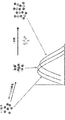
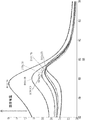
いくつかの実施形態で、検出方法は、サンドウィッチアッセイである。そのような実施形態において、検出コンジュゲートは、試料中に存在する場合、標的分析物に特異的に結合が可能である結合パートナーに連結された金属ナノ構造を含む。例えば、一実施形態において、サンドウィッチアッセイフォーマットの方法は、存在する場合、試料中の標的分析物に特異的に結合し、第1の検出コンジュゲートと、分析物と第2の検出コンジュゲートとの間での複合体の形成が可能である結合パートナーに連結された金属ナノ構造を含む、第1の検出コンジュゲートおよび第2の検出コンジュゲートを含む。代表的な実施形態において、第1の検出コンジュゲートおよび/または第2の検出コンジュゲート中の金属ナノ構造は、複合金属ナノ構造である。複合体を光源に曝露し、光学シグナルを測定する。ここで、光学シグナルの変化は、試料中の分析物の存在を示す。例として、標的分析物を含有する試料が、第1および第2の検出コンジュゲートと混合される場合、標的分析物は、検出コンジュゲートの結合パートナーに結合し、第1の検出コンジュゲートと、分析物と、第2の検出コンジュゲートとの間で複合体が形成される。このような複合体形成によって、互いに近接した検出コンジュゲートにおける金属のナノ構造、すなわち、プラズモン−プラズモンカップリングがもたらされる。金属ナノ構造によって吸収、分散または伝導される光量は、複合体中の金属ナノ構造の近接によって影響を受け、したがって、試料中の標的分析物の存在を示すピーク吸収波長のシフト増強がもたらされる。 In some embodiments, the detection method is a sandwich assay. In such an embodiment, the detection conjugate comprises a metal nanostructure linked to a binding partner that, when present in the sample, is capable of specifically binding to the target analyte. For example, in one embodiment, the sandwich assay format method, if present, specifically binds to the target analyte in the sample, with the first detection conjugate and the analyte and second detection conjugate. Includes a first detection conjugate and a second detection conjugate that include metal nanostructures linked to binding partners capable of forming a complex between them. In a typical embodiment, the metal nanostructures in the first detection conjugate and / or the second detection conjugate are composite metal nanostructures. The complex is exposed to a light source and the optical signal is measured. Here, changes in the optical signal indicate the presence of the analyte in the sample. As an example, when a sample containing the target analyte is mixed with the first and second detection conjugates, the target analyte binds to the binding partner of the detection conjugate and with the first detection conjugate, A complex is formed between the analyte and the second detection conjugate. Such complex formation results in the nanostructures of the metal in the detection conjugates in close proximity to each other, namely the plasmon-plasmon coupling. The amount of light absorbed, dispersed or conducted by the metal nanostructures is affected by the proximity of the metal nanostructures in the complex, thus resulting in a shift enhancement of the peak absorption wavelength indicating the presence of the target analyte in the sample.
他の実施形態において、検出方法は、競合アッセイである。そのような実施形態において、第1の検出コンジュゲートは、重要な標的分析物に結合した金属ナノ構造を含む。サンドウィッチアッセイ方法と同様に、第2の検出コンジュゲートは、標的分析物に特異的に結合することが可能である。この種類のアッセイにおいて、第1の検出コンジュゲートは、最初は第2の検出コンジュゲートに結合するであろう。標的分析物を含有する試料が、これらの初期複合体と混合される場合、試料中の未標識または遊離標的分析物は、第2の検出コンジュゲートへの結合に関して、第1の検出コンジュゲートと競合するであろう。この種類のアッセイにおける光学シグナルの変化は、ピーク吸収波長において波長シフトを比例的に減少させるであろう第2の検出コンジュゲートからの第1の検出コンジュゲートにおける金属ナノ構造の置換に起因するであろう。 In other embodiments, the detection method is a competitive assay. In such an embodiment, the first detection conjugate comprises metal nanostructures bound to an important target analyte. Similar to the sandwich assay method, the second detection conjugate is capable of specifically binding to the target analyte. In this type of assay, the first detection conjugate will initially bind to the second detection conjugate. When the sample containing the target analyte is mixed with these initial complexes, the unlabeled or free target analyte in the sample will be associated with the first detection conjugate with respect to binding to the second detection conjugate. Will compete. The change in optical signal in this type of assay is due to the replacement of metal nanostructures in the first detection conjugate from the second detection conjugate that will proportionally reduce the wavelength shift at the peak absorption wavelength. There will be.
上記のとおり、本発明の方法は、複数の検出コンジュゲートを利用し得る。検出コンジュゲートは、アッセイ構成次第で、標的分析物または別の検出コンジュゲートに特異的に結合することが可能である結合パートナーに連結された金属ナノ構造を含む。例えば、方法が、サンドウィッチアッセイフォーマットで構成される実施形態において、検出コンジュゲートは、標的分析物に特異的に結合することが可能である結合パートナーに連結またはコンジュゲートされた金属ナノ構造を含む。方法が直接競合アッセイフォーマットで構成される他の実施形態において、検出コンジュゲートの少なくとも1つは、標的分析物に結合またはコンジュゲートされた金属ナノ構造を含む。代表的な実施形態において、第1の検出コンジュゲートおよび/または第2の検出コンジュゲート中の金属ナノ構造は、複合金属ナノ構造である。 As mentioned above, the method of the present invention may utilize multiple detection conjugates. Detection conjugates include metal nanostructures linked to binding partners capable of specifically binding to a target analyte or another detection conjugate, depending on the assay configuration. For example, in embodiments where the method comprises a sandwich assay format, the detection conjugate comprises a metal nanostructure linked or conjugated to a binding partner capable of specifically binding to the target analyte. In other embodiments where the method comprises a direct competitive assay format, at least one of the detection conjugates comprises metal nanostructures bound or conjugated to the target analyte. In a typical embodiment, the metal nanostructures in the first detection conjugate and / or the second detection conjugate are composite metal nanostructures.
いくつかの実施形態において、検出コンジュゲートは、標的分析物に特異的に結合することが可能である結合パートナーを含む。本明細書で使用される場合、「特異的結合」とは、高い親和性、例えば、少なくとも10−6Mの親和性による標的分子への結合を意味する。いくつかの実施形態において、結合パートナーは、ハプテンおよび他の小分子、薬剤、ホルモン、限定されないが、抗体またはその断片(例えば、Fv、Fab、(Fab)2、単鎖、CDRなど)、抗原、レセプター、リガンド、ポリヌクレオチド、アプタマー、ポリペプチド、ポリサッカライド、リポポリサッカライド、グリコペプチド、リポタンパク質または核タンパク質を含む生体高分子である。特定の実施形態において、結合パートナーは抗体である。他の実施形態において、結合パートナーは抗原である。 In some embodiments, the detection conjugate comprises a binding partner capable of specifically binding to the target analyte. As used herein, "specific binding" means binding to a target molecule with a high affinity, eg, an affinity of at least 10-6 M. In some embodiments, the binding partner is a hapten and other small molecule, drug, hormone, but not limited to an antibody or fragment thereof (eg, Fv, Fab, (Fab) 2 , single chain, CDR, etc.), antigen. , Receptors, ligands, polynucleotides, aptamers, polypeptides, polypeptides, lipopolysaccharides, glycopeptides, lipoproteins or nuclear proteins. In certain embodiments, the binding partner is an antibody. In other embodiments, the binding partner is an antigen.
いくつかの実施形態において、検出コンジュゲート、例えば、第1の検出コンジュゲートおよび第2の検出コンジュゲートは、同種の分子であるが、好ましくは、互いに別個の位置で標的分析物と結合する、結合パートナーを含む。例として、第1の検出コンジュゲートおよび第2の検出コンジュゲートは、両方とも標的分析物を認識する抗体であることが可能であるが、第1の検出コンジュゲートが標的分析物と結合するエピトープは、第2の検出コンジュゲートが標的分析物と結合するエピトープとは別であり、かつ理想的にはオーバーラップしない。したがって、特定の実施形態において、第1の検出コンジュゲートは、標的分析物の第1のエピトープを識別する抗体を含み、かつ第2の検出コンジュゲートは、標的分析物の第2のエピトープを識別する異なる抗体を含む。本明細書に記載の種々の実施形態において、第1の検出コンジュゲートは、標的分析物の第1のエピトープを識別するモノクローナル抗体を含み得る。さらなる実施形態において、第2の検出コンジュゲートは、第1の検出コンジュゲートによって識別されるエピトープとは別であり、かつ理想的にはオーバーラップしない標的分析物の第2のエピトープを識別するモノクローナル抗体を含み得る。代替的に、第1の検出コンジュゲートおよび/または第2の検出コンジュゲートは、ポリクローナル抗体を含み得る。例えば、第2の検出コンジュゲートはモノクローナル抗体を含んでなるが、第1の検出コンジュゲートはポリクローナル抗体を含み得る。いくつかの実施形態において、第1の検出コンジュゲートはポリクローナル抗体を含み、かつ第2の検出コンジュゲートはポリクローナル抗体を含む。 In some embodiments, the detection conjugates, such as the first detection conjugate and the second detection conjugate, are of the same type of molecule, but preferably bind to the target analyte at separate positions from each other. Includes binding partners. As an example, the first detection conjugate and the second detection conjugate can both be antibodies that recognize the target analyte, but the epitope to which the first detection conjugate binds to the target analyte. Is separate from the epitope to which the second detection conjugate binds to the target analyte and ideally does not overlap. Thus, in certain embodiments, the first detection conjugate comprises an antibody that identifies the first epitope of the target analyte, and the second detection conjugate identifies the second epitope of the target analyte. Contains different antibodies. In the various embodiments described herein, the first detection conjugate may comprise a monoclonal antibody that identifies the first epitope of the target analyte. In a further embodiment, the second detection conjugate is a monoclonal that identifies the second epitope of the target analyte that is separate from the epitope identified by the first detection conjugate and ideally does not overlap. May include antibodies. Alternatively, the first detection conjugate and / or the second detection conjugate may comprise a polyclonal antibody. For example, the second detection conjugate comprises a monoclonal antibody, whereas the first detection conjugate may comprise a polyclonal antibody. In some embodiments, the first detection conjugate comprises a polyclonal antibody and the second detection conjugate comprises a polyclonal antibody.
検出コンジュゲート中の金属ナノ構造は、貴金属またはその複合体から構成されることが可能である。いくつかの実施形態において、検出コンジュゲート中の金属ナノ構造は、遷移金属またはその複合体から構成されてもよい。いくつかの実施形態において、検出コンジュゲート中の金属ナノ構造は、貴金属または遷移金属と組み合わせて、アルカリ金属またはランタニドを含み得る。特定の実施形態において、検出コンジュゲート中の金属ナノ構造は、金、銀、銅、白金、パラジウム、ルテニウム、ロジウム、オスミウム、イリジウム、チタン、クロム、カドミウム、亜鉛、鉄、コバルト、ニッケルおよびその複合体から選択される金属を含む。一実施形態において、金属ナノ構造は金ナノ構造である。別の実施形態において、金属ナノ構造は銀ナノ構造である。 The metal nanostructures in the detection conjugate can be composed of noble metals or complexes thereof. In some embodiments, the metal nanostructures in the detection conjugate may be composed of transition metals or complexes thereof. In some embodiments, the metal nanostructures in the detection conjugate may include alkali metals or lanthanides in combination with noble or transition metals. In certain embodiments, the metal nanostructures in the detection conjugate are gold, silver, copper, platinum, palladium, ruthenium, rhodium, osmium, iridium, titanium, chromium, cadmium, zinc, iron, cobalt, nickel and composites thereof. Contains metals selected from the body. In one embodiment, the metal nanostructures are gold nanostructures. In another embodiment, the metal nanostructures are silver nanostructures.
好ましい実施形態において、検出コンジュゲート中の金属ナノ構造は、複合金属ナノ構造である。「複合金属ナノ構造」とは、少なくとも2種の貴金属、遷移金属、アルカリ金属またはランタニドを含むナノ構造を意味する。2種以上の金属は合金として一緒に混合されてもよく、または2種以上の金属はナノ構造の別個の部分において存在していてもよい。例えば、1種の金属はナノ構造のコアを形成してよく、第2の金属はナノ構造の外殻またはコーティングを形成する。いくつかの実施形態において、複合金属ナノ構造は、金、銀、銅、白金、パラジウム、ルテニウム、ロジウム、オスミウム、イリジウム、チタン、クロム、カドミウム、亜鉛、鉄、コバルトおよびニッケルから選択される少なくとも2種の金属を含む。他の実施形態において、複合金属ナノ構造は、金、銀、銅、白金、パラジウム、カドミウム、鉄、ニッケルおよび亜鉛から選択される少なくとも2種の金属を含む。1つの特定の実施形態において、複合金属ナノ構造は、金および銀を含む。別の実施形態において、複合金属ナノ構造は、金および銅を含む。さらに別の実施形態において、複合金属ナノ構造は、銀および銅を含む。本発明の方法で使用される複合金属ナノ構造は、球形ナノ粒子、ピラミッド形ナノ粒子、六角形ナノ粒子、ナノチューブ、ナノスター、ナノシェル、ナノロッド、ナノドット、ナノアイランド、ナノワイヤー、ナノディスク、ナノキューブまたはそれらの組合せなどの多数の異なる幾何構造を含むことができる。代表的な実施形態において、複合金属ナノ構造は、ナノスターおよびナノロッドから選択される。 In a preferred embodiment, the metal nanostructure in the detection conjugate is a composite metal nanostructure. "Composite metal nanostructure" means a nanostructure containing at least two noble metals, transition metals, alkali metals or lanthanides. Two or more metals may be mixed together as an alloy, or two or more metals may be present in separate parts of the nanostructure. For example, one metal may form a nanostructured core and a second metal may form a nanostructured outer shell or coating. In some embodiments, the composite metal nanostructure is selected from at least two selected from gold, silver, copper, platinum, palladium, ruthenium, rhodium, osmium, iridium, titanium, chromium, cadmium, zinc, iron, cobalt and nickel. Contains seed metals. In other embodiments, the composite metal nanostructure comprises at least two metals selected from gold, silver, copper, platinum, palladium, cadmium, iron, nickel and zinc. In one particular embodiment, the composite metal nanostructures include gold and silver. In another embodiment, the composite metal nanostructures include gold and copper. In yet another embodiment, the composite metal nanostructures include silver and copper. The composite metal nanostructures used in the methods of the invention are spherical nanoparticles, pyramidal nanoparticles, hexagonal nanoparticles, nanotubes, nanostars, nanoshells, nanorods, nanodots, nanoislands, nanowires, nanodisks, nanocubes or It can contain many different geometric structures such as combinations thereof. In typical embodiments, composite metal nanostructures are selected from nanostars and nanorods.
特定の実施形態において、本発明の方法で使用される複合金属ナノ構造は、第1の金属および第2の金属の合金である。いくつかの実施形態において、本発明の方法で使用される複合金属ナノ構造は、第1の金属のコアおよび第2の金属のコーティングを含む。特定の実施形態において、複合金属ナノ構造は、銀コアおよび金コーティングを含む。他の実施形態において、複合金属ナノ構造は、銅コアおよび金コーティングを含む。別の実施形態において、コアは銀であり、かつコーティングは銅である。いくつかの実施形態において、複合金属ナノ構造のそれぞれは、誘電体コア(例えば、二酸化ケイ素、硫化金、二酸化チタン、シリカおよびポリスチレン)、第1の金属の第1のコーティングおよび第2の金属の第2のコーティングを含む。検出方法の1つの特定の実施形態において、コアはシリカであり、第1のコーティング(すなわち、内部コーティング)は銀コーティングであり、第2のコーティングは金コーティング(すなわち、外部コーティング)である。別の実施形態において、コアはシリカであり、第1のコーティング(すなわち、内部コーティング)は銅コーティングであり、第2のコーティングは金コーティング(すなわち、外部コーティング)である。 In certain embodiments, the composite metal nanostructures used in the methods of the invention are alloys of a first metal and a second metal. In some embodiments, the composite metal nanostructures used in the methods of the invention include a first metal core and a second metal coating. In certain embodiments, the composite metal nanostructures include a silver core and a gold coating. In other embodiments, the composite metal nanostructures include a copper core and a gold coating. In another embodiment, the core is silver and the coating is copper. In some embodiments, each of the composite metal nanostructures is of a dielectric core (eg, silicon dioxide, gold sulfide, titanium dioxide, silica and polystyrene), a first coating of a first metal and a second metal. Includes a second coating. In one particular embodiment of the detection method, the core is silica, the first coating (ie, the inner coating) is the silver coating, and the second coating is the gold coating (ie, the outer coating). In another embodiment, the core is silica, the first coating (ie, the inner coating) is the copper coating, and the second coating is the gold coating (ie, the outer coating).
いくつかの実施形態において、第2の金属によるコーティングプロセスに続いて、第1の金属を含むコアは溶解されて、第2の金属から構成される中空構造が作られる。例えば、金ナノ粒子による銀コアのコーティングによって、銀コアの周囲に金シェルが作成され、銀コアは、その後、溶解されるか、または分解され、中空ナノ金シェル構造が形成される。 In some embodiments, following the coating process with the second metal, the core containing the first metal is melted to form a hollow structure composed of the second metal. For example, coating a silver core with gold nanoparticles creates a gold shell around the silver core, which is then melted or decomposed to form a hollow nanogold shell structure.
金属ナノ構造には、球形ナノ粒子、ならびにナノプレートおよびナノシェルが含まれる。ナノプレートは、それらの厚さより大きい側面寸法(例えば、エッジ長さ)を有する。ナノプレートには、ナノディスク、ナノポリゴン、ナノヘキサゴン、ナノキューブ、ナノリング、ナノスターおよびナノプリズムが含まれる。いくつかの実施形態において、複合ナノ構造を含む金属ナノ構造は、球形ナノ粒子、ピラミッド形ナノ粒子、六角形ナノ粒子、ナノチューブ、ナノスター、ナノシェル、ナノロッド、ナノドット、ナノアイランド、ナノワイヤー、ナノディスク、ナノキューブまたはそれらの組合せから選択される幾何構造を有する。不規則形状を含めて、他の形状も可能である。特定の実施形態において、金属ナノ構造の径および形状は均一ではない、すなわち、金属ナノ構造は、異なる形状および径のナノ構造の不均一混合物である。代表的な実施形態において、金属ナノ構造は、ナノスターである。別の代表的な実施形態において、金属ナノ構造は、ナノロッドである。別の代表的な実施形態において、金属ナノ構造は、複合ナノ球体である。 Metal nanostructures include spherical nanoparticles, as well as nanoplates and nanoshells. Nanoplates have side dimensions (eg, edge length) that are greater than their thickness. Nanoplates include nanodisks, nanopolygons, nanohexagons, nanocubes, nanorings, nanostars and nanoprisms. In some embodiments, metal nanostructures, including composite nanoparticles, include spherical nanoparticles, pyramidal nanoparticles, hexagonal nanoparticles, nanotubes, nanostars, nanoshells, nanorods, nanodots, nanoislands, nanowires, nanodisks, It has a geometric structure selected from nanocubes or a combination thereof. Other shapes are possible, including irregular shapes. In certain embodiments, the diameter and shape of the metal nanostructures are not uniform, i.e. the metal nanostructures are a heterogeneous mixture of nanostructures of different shapes and diameters. In a typical embodiment, the metal nanostructure is a nanostar. In another typical embodiment, the metal nanostructure is a nanorod. In another typical embodiment, the metal nanostructure is a composite nanosphere.
球形ナノ粒子に関して、適切な直径範囲としては、約5nm〜約200nm、約10nm〜約100nmおよび約20nm〜約60nmが含まれる。ナノロッドに関して、適切な直径範囲としては、約5nm〜約50nm、約8nm〜約30nmおよび約10nm〜約25nmが含まれる。さらに、ナノロッドに関して、適切な長さ範囲としては、約25nm〜約150nm、約40nm〜約120nmおよび約50nm〜100nmが含まれる。いくつかの実施形態において、ナノロッドのアスペクト比、すなわち、長さ/直径は、2〜10である。ナノプレートに関して、エッジ長さは、約10nm〜約800nm、約20nm〜約500nm、約50nm〜約200nm、約30nm〜約100nmまたは約10nm〜約300nmであってよい。ナノプレートの厚さは、約1〜約100nm、約5nm〜約80nm、約10nm〜約50nmまたは約5nm〜約20nmの範囲であることが可能である。 For spherical nanoparticles, suitable diameter ranges include from about 5 nm to about 200 nm, from about 10 nm to about 100 nm, and from about 20 nm to about 60 nm. For nanorods, suitable diameter ranges include from about 5 nm to about 50 nm, from about 8 nm to about 30 nm and from about 10 nm to about 25 nm. Further, for nanorods, suitable length ranges include from about 25 nm to about 150 nm, from about 40 nm to about 120 nm and from about 50 nm to 100 nm. In some embodiments, the nanorod aspect ratio, i.e. length / diameter, is 2-10. For nanoplates, the edge length may be from about 10 nm to about 800 nm, from about 20 nm to about 500 nm, from about 50 nm to about 200 nm, from about 30 nm to about 100 nm, or from about 10 nm to about 300 nm. The thickness of the nanoplate can range from about 1 to about 100 nm, about 5 nm to about 80 nm, about 10 nm to about 50 nm, or about 5 nm to about 20 nm.
いくつかの実施形態において、ナノプレートは、2より大きいアスペクト比を有する。アスペクト比は、厚さに対するエッジ長さの比率である。好ましくは、ナノプレートは、約2〜約25、約3〜約20、約5〜約10、約2〜約15または約10〜約30のアスペクト比を有する。 In some embodiments, the nanoplate has an aspect ratio greater than 2. The aspect ratio is the ratio of the edge length to the thickness. Preferably, the nanoplate has an aspect ratio of about 2 to about 25, about 3 to about 20, about 5 to about 10, about 2 to about 15, or about 10 to about 30.
金属ナノ構造への分子のコンジュゲート方法は、当業者に既知である。そのような方法としては、1−エチル−3−[3−ジメチルアミノプロピル]カルボジイミドヒドロクロリド(EDC)、スルホ−NHSカップリング、疎水性結合またはチオエーテル化学が関与するものなどのコンジュゲーション化学が含まれる。いくつかの実施形態において、結合パートナーまたは標的分析物は、チオール、アミン、ジチオール、アクリルホスホラミダイト、アジドまたはアルキンを含む種々の化学官能性を介して金属ナノ構造に連結させることができる。いくつかの実施形態において、この分子は、より大きいキャリア分子またはタンパク質を介して間接的に金属ナノ構造に連結させることができる。そのような間接カップリングは、この分子が、ホルモン、薬剤、10kD未満の他の小分子などのように小さい場合、特に有用である。好ましくは、キャリアタンパク質は、標的分析物との特異的な相互作用の能力を有さない。いくつかの実施形態において、プロテインAまたはプロテインGまたはプロテインA/Gがナノ粒子にコンジュゲートされるか、または連結されてもよい。 Methods of conjugating molecules into metal nanostructures are known to those of skill in the art. Such methods include conjugation chemistry such as 1-ethyl-3- [3-dimethylaminopropyl] carbodiimide hydrochloride (EDC), sulfo-NHS couplings, hydrophobic bonds or those involving thioether chemistry. Is done. In some embodiments, the binding partner or target analyte can be linked to the metal nanostructure via various chemical functionalities including thiols, amines, dithiols, acrylic phosphoramidite, azides or alkynes. In some embodiments, the molecule can be indirectly linked to metal nanostructures via a larger carrier molecule or protein. Such indirect couplings are particularly useful when this molecule is small, such as hormones, drugs, other small molecules less than 10 kD, and the like. Preferably, the carrier protein does not have the ability to interact specifically with the target analyte. In some embodiments, protein A or protein G or protein A / G may be conjugated or linked to nanoparticles.
いくつかの実施形態において、第1の検出コンジュゲートにおいて利用される金属は、第2の検出コンジュゲートにおける金属ナノ構造が製造される金属と同一であることが可能である。例えば、一実施形態において、第1の検出コンジュゲートは、金ナノ構造を含み、かつ第2の検出コンジュゲートは、金ナノ構造を含む。他の実施形態において、第1の検出コンジュゲートにおいて利用される金属は、第2の検出コンジュゲートにおける金属ナノ構造を製造するために使用される金属と異なる。例えば、いくつかの実施形態において、第1の検出コンジュゲートは、銀ナノ構造を含み、かつ第2の検出コンジュゲートは、金ナノ構造を含む。他の実施形態において、第1の検出コンジュゲートは、金ナノ構造を含み、かつ第2の検出コンジュゲートは、銀ナノ構造を含む。ある特定の実施形態において、第1の検出コンジュゲートは、金ナノ構造を含み、かつ第2の検出コンジュゲートは、複合ナノ構造を含む。関連する実施形態において、複合ナノ構造は、金コーティング銀ナノ構造を含む。他の特定の実施形態において、第1の検出コンジュゲートは、金ナノ構造を含み、かつ第2の検出コンジュゲートは、金コーティング銅ナノ構造を含む複合ナノ構造を含む。さらに他の実施形態において、第1の検出コンジュゲートは、金ナノ構造を含み、かつ第2の検出コンジュゲートは、金コーティングマグネタイトナノ構造を含む複合ナノ構造を含む。さらに他の実施形態において、第1の検出コンジュゲートは、金ナノ構造を含み、かつ第2の検出コンジュゲートは、金およびアルカリ金属またはランタニドを含む複合ナノ構造を含む。 In some embodiments, the metal utilized in the first detection conjugate can be the same metal in which the metal nanostructures in the second detection conjugate are produced. For example, in one embodiment, the first detection conjugate comprises gold nanostructures and the second detection conjugate comprises gold nanostructures. In other embodiments, the metal utilized in the first detection conjugate is different from the metal used to produce the metal nanostructures in the second detection conjugate. For example, in some embodiments, the first detection conjugate comprises silver nanostructures and the second detection conjugate comprises gold nanostructures. In other embodiments, the first detection conjugate comprises gold nanostructures and the second detection conjugate comprises silver nanostructures. In certain embodiments, the first detection conjugate comprises gold nanostructures and the second detection conjugate comprises composite nanostructures. In a related embodiment, the composite nanostructures include gold-coated silver nanostructures. In other particular embodiments, the first detection conjugate comprises gold nanostructures and the second detection conjugate comprises composite nanostructures including gold coated copper nanostructures. In yet another embodiment, the first detection conjugate comprises gold nanostructures and the second detection conjugate comprises composite nanostructures including gold coated magnetite nanostructures. In yet another embodiment, the first detection conjugate comprises gold nanostructures and the second detection conjugate comprises composite nanostructures comprising gold and alkali metals or lanthanides.
特定の実施形態において、第1の検出コンジュゲートを製造するために使用される金属ナノ構造の径は、第2の検出コンジュゲートにおいて使用される金属ナノ構造の径と類似する。そのような実施形態において、2組のナノ構造の径を合わせることによって、反射率、発光または散乱スペクトルにおける最適な波長シフトを提供することができる。 In certain embodiments, the diameter of the metal nanostructures used to produce the first detection conjugate is similar to the diameter of the metal nanostructures used in the second detection conjugate. In such embodiments, matching the diameters of the two sets of nanostructures can provide optimal wavelength shifts in reflectance, emission or scattering spectra.
いくつかの実施形態において、反応環境は、適切な緩衝剤、イオン強度および他の促進剤によって調整され得る。好ましい実施形態において、反応環境は、本明細書に記載されるように、LSPRシグナルの強度を増強することができるポリエチレングリコール(PEG)を含む。限定されないが、ポリビニルピロリドン、ポリアリルアミン、ポリエチレンイミン、ポリリジン、ポリアクリル酸、ポリビニルアルコールおよびポリアスパラギン酸を含む他の類似のポリマー材料が使用されてもよい。 In some embodiments, the reaction environment can be adjusted with suitable buffers, ionic strength and other accelerators. In a preferred embodiment, the reaction environment comprises polyethylene glycol (PEG) capable of enhancing the intensity of the LSPR signal, as described herein. Other similar polymeric materials may be used, including, but not limited to, polyvinylpyrrolidone, polyallylamine, polyethyleneimine, polylysine, polyacrylic acid, polyvinyl alcohol and polyaspartic acid.
本発明は、試料中の標的分析物を検出するために、本明細書に記載の方法を利用するための分析物検出デバイスも提供する。適切な分析物検出デバイスとしては、限定されないが、分光光度法キュベット、分析用ローター、マイクロウェルプレートまたはフローチャンバーが含まれてよい。当業者によって理解されるように、光ファイバーの先端または透明ゲルも、本明細書に開示される検出方法を実行するために利用されてよい。 The present invention also provides an analyte detection device for utilizing the methods described herein to detect a target analyte in a sample. Suitable analytical material detection devices may include, but are not limited to, spectrophotometric cuvettes, analytical rotors, microwell plates or flow chambers. As will be appreciated by those skilled in the art, fiber optic tips or clear gels may also be utilized to carry out the detection methods disclosed herein.
特定の実施形態において、本明細書に記載の分析物検出デバイスの全ての構成成分は、遠心ローターまたはディスクに含まれる。例えば、ローターまたはディスクは、複数の検出コンジュゲートが位置する1つまたはそれ以上の反応チャンバーを含有していてもよい。いくつかの実施形態において、検出コンジュゲートは、凍結乾燥されたビーズまたはペレットなどの凍結乾燥された組成物の形態で存在する。いくつかの実施形態において、分析物検出デバイスは、1つまたはそれ以上の反応チャンバーを含むローターまたはディスクを含み、それぞれの反応チャンバーは、複数の検出コンジュゲート(例えば、第1の検出コンジュゲートおよび第2の検出コンジュゲート)を含み、検出コンジュゲートは、金属ナノ粒子に連結されている。そのようなデバイスは、ワンステップ分析物検出アッセイを提供し、それによって、試験試料がローターまたはディスクと接触し、ローターまたはディスクへの遠心力の適用によって、反応チャンバーへと試験試料が送達され、そこで、試料は、第1の検出コンジュゲートおよび第2の検出コンジュゲートと混合される。ローターまたはディスクが2つ以上の反応チャンバーを含む実施形態において、検出コンジュゲートは、異なる分析物がそれぞれの反応チャンバーにおいて検出されることができるように選択することができる。これらのローターフォーマット検出デバイスは、サンドウィッチアッセイフォーマット、直接競合フォーマットで、またはローターが複数の反応チャンバーを含む場合は両方で構成されることが可能である。 In certain embodiments, all components of the analyte detection device described herein are contained in a centrifugal rotor or disc. For example, the rotor or disc may contain one or more reaction chambers in which multiple detection conjugates are located. In some embodiments, the detection conjugate is present in the form of a lyophilized composition such as lyophilized beads or pellets. In some embodiments, the analyte detection device comprises a rotor or disk containing one or more reaction chambers, each reaction chamber having a plurality of detection conjugates (eg, a first detection conjugate and). A second detection conjugate) is included, and the detection conjugate is linked to the metal nanoparticles. Such a device provides a one-step analyte detection assay, whereby the test sample comes into contact with the rotor or disc, and the application of centrifugal force to the rotor or disc delivers the test sample to the reaction chamber. There, the sample is mixed with the first detection conjugate and the second detection conjugate. In embodiments where the rotor or disc comprises two or more reaction chambers, the detection conjugate can be selected so that different analytes can be detected in each reaction chamber. These rotor format detection devices can be configured in sandwich assay format, in direct competition format, or both if the rotor contains multiple reaction chambers.
本明細書に記載の金属ナノ構造の種類のいずれも、これらのローターフォーマット検出デバイスによって使用可能である。いくつかの実施形態において、第1の検出コンジュゲートは、金ナノ構造を含み、かつ第2の検出コンジュゲートにおける金属ナノ構造は、金ナノ構造である。他の実施形態において、第1の検出コンジュゲートは、銀ナノ構造を含み、かつ第2の検出コンジュゲートにおける金属ナノ構造は、金ナノ構造である。さらに他の実施形態において、第1の検出コンジュゲートは、金ナノ構造を含み、かつ第2の検出コンジュゲートは、複合ナノ構造を含む。例えば、一実施形態において、複合ナノ構造は、金コーティング銀ナノ構造である。別の実施形態において、複合ナノ構造は、金コーティング銅ナノ構造である。 Any of the types of metal nanostructures described herein can be used with these rotor format detection devices. In some embodiments, the first detection conjugate comprises gold nanostructures and the metal nanostructure in the second detection conjugate is gold nanostructures. In another embodiment, the first detection conjugate comprises silver nanostructures and the metal nanostructure in the second detection conjugate is gold nanostructures. In yet another embodiment, the first detection conjugate comprises gold nanostructures and the second detection conjugate comprises composite nanostructures. For example, in one embodiment, the composite nanostructure is a gold-coated silver nanostructure. In another embodiment, the composite nanostructure is a gold coated copper nanostructure.
本発明は、本明細書に開示される本発明の分析物検出デバイスを含むキットも含む。一実施形態において、キットは、複数の検出コンジュゲート(例えば、第1の検出コンジュゲートおよび第2の検出コンジュゲート)を含み、その中で検出コンジュゲートは、金属ナノ粒子に連結されている。いくつかの実施形態において、検出コンジュゲートの1つまたはそれ以上は、例えば、ペレットまたはビーズの形態で凍結乾燥されてもよい。一実施形態において、検出コンジュゲートの全てが凍結乾燥される。さらなる実施形態において、キットは、1種またはそれ以上の追加の試薬を含んでもよい。いくつかの実施形態において、1種またはそれ以上の追加の試薬は、凍結乾燥された形態で提供される。いくつかの実施形態において、キットは、ブロッキング剤、糖、ポリマー性促進材料、塩化ナトリウムおよび/またはそれらの組合せを含んでよい。「ブロッキング剤」は、検出可能な薬剤および/または分析物と、試料中に存在するタンパク質との会合を防ぐ薬剤である。ブロッキング剤は、典型的に、それら自体がタンパク質であり、それらとしては、限定されないが、ウシ血清アルブミン、カゼイン、ゼラチン、オボアルブミン、ガンマグロブリンおよび非免疫動物からのIgGが含まれうる。いくつかの実施形態において、糖はポリサッカライドである。一実施形態において、ポリサッカライドは、マルトデキストリン、コーンシロップおよびポリグルコースから選択される。好ましい実施形態において、ポリサッカライドはマルトデキストリンである。別の実施形態において、糖はトレハロースである。いくつかの実施形態において、試薬キットは、マルトデキストリンおよびトレハロースを含んでよい。いくつかの実施形態において、ポリマー性促進材料はPEGである。 The present invention also includes a kit comprising the analyte detection device of the present invention disclosed herein. In one embodiment, the kit comprises a plurality of detection conjugates (eg, a first detection conjugate and a second detection conjugate) in which the detection conjugate is linked to metal nanoparticles. In some embodiments, one or more of the detection conjugates may be lyophilized, for example, in the form of pellets or beads. In one embodiment, all of the detection conjugates are lyophilized. In a further embodiment, the kit may include one or more additional reagents. In some embodiments, one or more additional reagents are provided in lyophilized form. In some embodiments, the kit may include a blocking agent, a sugar, a polymeric accelerator, sodium chloride and / or a combination thereof. A "blocking agent" is an agent that prevents the association of a detectable agent and / or analyte with a protein present in the sample. Blocking agents are typically proteins themselves and may include, but are not limited to, bovine serum albumin, casein, gelatin, ovalbumin, gamma globulin and IgG from non-immunized animals. In some embodiments, the sugar is a polysaccharide. In one embodiment, the polysaccharide is selected from maltodextrin, corn syrup and polyglucose. In a preferred embodiment, the polysaccharide is maltodextrin. In another embodiment, the sugar is trehalose. In some embodiments, the reagent kit may include maltodextrin and trehalose. In some embodiments, the polymeric promoting material is PEG.
本発明のキットは、試験試料中の分析物を検出するためのデバイスを使用するための指示、生物学的試料を回収するためのデバイスまたはツール、ならびに/または土壌、食品および生物学的組織などの固体材料から試料を得るための抽出緩衝剤を含んでもよい。 The kits of the present invention include instructions for using a device for detecting an analyte in a test sample, a device or tool for recovering a biological sample, and / or soil, food and biological tissue. May include an extraction buffer to obtain a sample from the solid material of.
本明細書に記載されるように、試験試料は、生物学的試料または環境もしくは食品試料から調製された抽出液を含む、いずれの種類の液体試料であることが可能である。1つの特定の実施形態において、試験試料は、生物学的試料である。生物学的試料としては、限定されないが、全血、血漿、血清、唾液、尿、胸水、汗、胆汁、脳脊髄液、糞便、腟液、精子、眼レンズ液、粘膜、滑液、腹水、羊水、生検組織、唾液および細胞溶解物が含まれる。生物学的試料は、がん、感染性疾患(例えば、ウイルス、細菌、寄生虫または真菌感染)、心臓血管疾患、代謝疾患、自己免疫疾患などの疾患状態を有することが疑われるヒト対象または動物対象から得ることができる。生物学的試料は、定期健康診断を受けている健康な対象(例えば、ヒトまたは動物)から得ることもできる。 As described herein, the test sample can be any type of liquid sample, including biological samples or extracts prepared from environmental or food samples. In one particular embodiment, the test sample is a biological sample. Biological samples include, but are not limited to, whole blood, plasma, serum, saliva, urine, pleural effusion, sweat, bile, cerebrospinal fluid, feces, vaginal fluid, sperm, ophthalmic fluid, mucous membrane, synovial fluid, ascites, Includes pleural effusion, biopsy tissue, saliva and cell lysates. Biological samples are human subjects or animals suspected of having a disease state such as cancer, infectious disease (eg, viral, bacterial, parasitic or fungal infection), cardiovascular disease, metabolic disease, autoimmune disease. Can be obtained from the subject. Biological samples can also be obtained from healthy subjects (eg, humans or animals) undergoing routine health examinations.
方法のいくつかの実施形態において、試験試料は、第1の検出コンジュゲートと混合され、混合物は、その後、第2の検出コンジュゲートと接触する。特定の実施形態において、試料、第1の検出コンジュゲートおよび第2の検出コンジュゲートは、同時に接触する。例えば、両試薬との試料の接触が、本明細書に記載のローターフォーマット検出デバイスにおいて同時に起こってよい。 In some embodiments of the method, the test sample is mixed with the first detection conjugate and the mixture is then contacted with the second detection conjugate. In certain embodiments, the sample, the first detection conjugate and the second detection conjugate come into contact at the same time. For example, sample contact with both reagents may occur simultaneously in the rotor format detection device described herein.
上記のとおり、本出願は、定性的または定量的様式で特異的結合パートナーの結合を決定するための、溶液中の複合ナノ材料標識パートナーの使用に関する。本発明者らは、驚くべきことに、ポリサッカライド、例えば、マルトデキストリンが溶液に添加される場合、スクロース、トレハロースまたはficollなどの他の糖の添加と比較して、溶液ベースアッセイの感度が有意に増強されることを見出した。遠心ローターフォーマットにおいて、分析用チャンバー中に試料を送達するために、低速遠心分離が必要とされる。溶液へのポリサッカライド、例えば、マルトデキストリンの添加は、遠心分離間および後に、複合ナノ材料標識パートナー、例えば、金−銀ナノスターとコンジュゲートした抗体の凝集および沈降を防ぐことに特に有効である。スクロース、トレハロースまたはficollなどの他の糖と比べての感度の改善は予想外であった。凝集および沈降の減少によって、アッセイの感度増加が達成される。したがって、いくつかの実施形態において、本発明の方法は、ポリサッカライド、例えば、マルトデキストリン、コーンシロップまたはポリグルコースを含む溶液中で実行される。 As mentioned above, the present application relates to the use of composite nanomaterial labeling partners in solution to determine the binding of specific binding partners in a qualitative or quantitative manner. Surprisingly, we found that when polysaccharides, such as maltodextrin, were added to the solution, the sensitivity of the solution-based assay was significant compared to the addition of other sugars such as sucrose, trehalose or ficoll. It was found that it was enhanced in. In the centrifugal rotor format, slow centrifugation is required to deliver the sample into the analytical chamber. Addition of polysaccharides to the solution, such as maltodextrin, is particularly effective in preventing aggregation and precipitation of antibodies conjugated to composite nanomaterial labeling partners, such as gold-silver nanostars, during and after centrifugation. The improvement in sensitivity compared to other sugars such as sucrose, trehalose or ficoll was unexpected. Increased assay sensitivity is achieved by reducing aggregation and sedimentation. Thus, in some embodiments, the methods of the invention are performed in a solution containing polysaccharides such as maltodextrin, corn syrup or polyglucose.
一実施形態において、溶液は、約2%〜約20%重量/体積の最終濃度でポリサッカライドを含む。別の実施形態において、溶液は、約4%〜約15%重量/体積の最終濃度でポリサッカライドを含む。さらに別の実施形態において、溶液は、約5%〜約10%重量/体積の最終濃度でポリサッカライドを含む。代表的な実施形態において、溶液は、それらの間の全ての値を含めて、約5%、6%、7%、8%、9%または10%の最終濃度でポリサッカライドを含む。本明細書に記載の種々の実施形態において、アッセイの感度は、ポリサッカライドが溶液に添加される場合、別の糖、例えば、スクロースまたはficollを含む溶液中でアッセイが実行される場合と比較して、改善され得る。代表的な実施形態において、ポリサッカライドは、マルトデキストリンである。 In one embodiment, the solution comprises a polysaccharide at a final concentration of about 2% to about 20% by weight / volume. In another embodiment, the solution comprises a polysaccharide at a final concentration of about 4% to about 15% by weight / volume. In yet another embodiment, the solution comprises a polysaccharide at a final concentration of about 5% to about 10% by weight / volume. In a typical embodiment, the solution comprises polysaccharide at a final concentration of about 5%, 6%, 7%, 8%, 9% or 10%, including all values between them. In the various embodiments described herein, the sensitivity of the assay is compared to when the polypeptide is added to the solution, when the assay is performed in a solution containing another sugar, such as sucrose or Ficoll. Can be improved. In a typical embodiment, the polysaccharide is maltodextrin.
一実施形態において、溶液は、約0.1%〜約20%重量/体積の最終濃度でブロッキング剤を含む。別の実施形態において、溶液は、約0.5%〜約10%重量/体積の最終濃度でブロッキング剤を含む。さらに別の実施形態において、溶液は、約1%〜約5%重量/体積の最終濃度でブロッキング剤を含む。代表的な実施形態において、溶液は、それらの間の全ての値を含めて、約1%、2%、3%、4%または5%の最終濃度でブロッキング剤を含む。本明細書に記載の種々の実施形態において、アッセイの感度は、ブロッキング剤が溶液に添加される場合、ブロッキング剤の不在下でアッセイが実行される場合と比較して、改善され得る。いくつかの実施形態において、ブロッキング剤は、ウシ血清アルブミン、カゼイン、ゼラチン、オボアルブミンおよびガンマ−グロブリンから選択される。代表的な実施形態において、ブロッキング剤は、ウシ血清アルブミンである。 In one embodiment, the solution comprises a blocking agent at a final concentration of about 0.1% to about 20% by weight / volume. In another embodiment, the solution comprises a blocking agent at a final concentration of about 0.5% to about 10% by weight / volume. In yet another embodiment, the solution comprises a blocking agent at a final concentration of about 1% to about 5% by weight / volume. In a typical embodiment, the solution comprises a blocking agent at a final concentration of about 1%, 2%, 3%, 4% or 5%, including all values between them. In the various embodiments described herein, the sensitivity of the assay can be improved when the blocking agent is added to the solution compared to when the assay is performed in the absence of the blocking agent. In some embodiments, the blocking agent is selected from bovine serum albumin, casein, gelatin, ovalbumin and gamma-globulin. In a typical embodiment, the blocking agent is bovine serum albumin.
いくつかの実施形態において、溶液は、マルトデキストリン、トレハロース、PEG、ブロッキング剤(例えばウシ血清アルブミン)および/または塩化ナトリウムの1種またはそれ以上を含む。代表的な実施形態において、溶液成分の1種またはそれ以上、例えば、マルトデキストリンは、液体、例えば、水、食塩溶液または液体試料の添加時に懸濁する凍結乾燥されたビーズまたはペレットとして提供されてもよい。例えば、溶液成分の1種またはそれ以上は、液体の添加後に溶液中で懸濁するビーズとして分光光度法キュベットまたは分析用ローターの反応チャンバー中に提供されてもよい。 In some embodiments, the solution comprises one or more of maltodextrin, trehalose, PEG, blocking agent (eg bovine serum albumin) and / or sodium chloride. In a typical embodiment, one or more of the solution components, such as maltodextrin, are provided as lyophilized beads or pellets that are suspended upon addition of a liquid, such as water, saline solution or liquid sample. May be good. For example, one or more of the solution components may be provided in the reaction chamber of the spectrophotometric cuvette or analytical rotor as beads suspended in the solution after the addition of the liquid.
追加の実施形態において、LSPRシグナルは、ポリエチレングリコール、ポリビニルピロリドン、ポリアリルアミン、ポリエチレンイミン、ポリリジン、ポリアクリル酸、ポリビニルアルコールおよびポリアスパラギン酸から選択されるポリマー性促進材料の存在下で第1および第2の検出コンジュゲートを分析物と混合することによって、実質的に増加し得る。代表的な実施形態において、ポリマー材料は、ポリエチレングリコール(PEG)である。一実施形態において、反応混合物は、約0.1mg/mL〜約200mg/mLの最終濃度でポリマー材料、例えば、PEGを含む。別の実施形態において、反応混合物は、約0.2mg/mL〜約100mg/mLの最終濃度でポリマー材料、例えば、PEGを含む。さらに別の実施形態、反応混合物は、約0.5mg/mL〜約10mg/mLの最終濃度でポリマー材料、例えば、PEGを含む。さらに別の実施形態において、反応混合物は、約2mg/mL〜約8mg/mLの最終濃度でポリマー材料、例えば、PEGを含む。代表的な実施形態において、反応混合物は、それらの間の全ての値を含めて、約2、3、4、5、6、7または8mg/mLの最終濃度でポリマー材料、例えば、PEGを含む。 In additional embodiments, the LSPR signal is first and first in the presence of a polymeric accelerator selected from polyethylene glycol, polyvinylpyrrolidone, polyallylamine, polyethyleneimine, polylysine, polyacrylic acid, polyvinyl alcohol and polyaspartic acid. By mixing the detection conjugate of 2 with the analyte, it can be substantially increased. In a typical embodiment, the polymeric material is polyethylene glycol (PEG). In one embodiment, the reaction mixture comprises a polymeric material, such as PEG, at a final concentration of about 0.1 mg / mL to about 200 mg / mL. In another embodiment, the reaction mixture comprises a polymeric material, such as PEG, at a final concentration of about 0.2 mg / mL to about 100 mg / mL. Yet another embodiment, the reaction mixture, comprises a polymeric material, such as PEG, at a final concentration of about 0.5 mg / mL to about 10 mg / mL. In yet another embodiment, the reaction mixture comprises a polymeric material, such as PEG, at a final concentration of about 2 mg / mL to about 8 mg / mL. In a typical embodiment, the reaction mixture comprises a polymeric material, eg, PEG, at a final concentration of about 2, 3, 4, 5, 6, 7 or 8 mg / mL, including all values between them. ..
本発明の検出方法は、標的分析物の定性的または定量的な量を決定するために使用されてよい。そのような方法は、試料中の標的分析物のおよその量を決定するために特に有用であり、これは、特に、特定の医学的状態を診断するか、または薬物療法の有効性を評価するために使用することができる。一実施形態において、標的分析物の量は、既知量の標的分析物を有する試料に関して、本明細書に記載の金属ナノ粒子からの光学シグナルの変化を測定することによって、特定の分析物に対する標準曲線を確立すること;試験試料に関する光学シグナルの変化を決定すること;そして試験試料に関する光学シグナルの変化を、標準曲線に関して得られる値と比較することによって、決定することができる。いくつかの実施形態において、第1の試薬と第2の試薬との間の複合体の量を決定することは、試験試料からの吸光度比および/または反応速度と、既知量の複合体を有する一試料からの吸光度比および/または反応速度とを比較すること、それによって、試験試料中の複合体の量を決定することを含む。試験試料から得られた定量的な値は、標的分析物の異常または正常レベルのいずれかを示す、あらかじめ決定された閾値と比較されてもよい。 The detection method of the present invention may be used to determine a qualitative or quantitative amount of a target analyte. Such methods are particularly useful for determining the approximate amount of target analyte in a sample, which specifically diagnoses a particular medical condition or assesses the effectiveness of drug therapy. Can be used for. In one embodiment, the amount of target analyte is standard for a particular analyte by measuring changes in the optical signal from the metal nanoparticles described herein for a sample with a known amount of target analyte. It can be determined by establishing a curve; determining the change in the optical signal for the test sample; and comparing the change in the optical signal for the test sample with the values obtained for the standard curve. In some embodiments, determining the amount of complex between the first and second reagents has an absorbance ratio and / or kinetics from the test sample and a known amount of complex. It involves comparing the absorbance ratio and / or kinetics from one sample, thereby determining the amount of complex in the test sample. Quantitative values obtained from the test sample may be compared to a predetermined threshold that indicates either an abnormal or normal level of the target analyte.
本発明の検出方法は、試料中の微量の標的分析物を検出するために非常に感度が高い技術を提供する。いくつかの実施形態において、プラズモン共鳴ベースのシグナルの増幅は、標的分析物のナノグラム量が試料中で検出されることができるように、金ナノ構造コンジュゲートによって達成することができる。したがって、この方法の一実施形態において、標的分析物のナノグラム量の存在が検出される。いくつかの実施形態において、金ナノ粒子を含む検出コンジュゲートからのプラズモン共鳴ベースのシグナルは、複合金属ナノ構造検出コンジュゲートを使用して増幅することができる。分析物に特異的な抗体にコンジュゲートされた金コーティング銀ナノ構造の使用によって、標的分析物のピコグラム量の検出が可能となり得る。したがって、この方法のいくつかの実施形態において、標的分析物のピコグラム量の存在が検出される。方法の他の実施形態において、標的分析物のフェムトグラム量の存在が検出される。複合金属ナノ構造の組成および/または形状を変更することによって、より高い感度が得られうる。 The detection method of the present invention provides a very sensitive technique for detecting a trace amount of a target analyte in a sample. In some embodiments, amplification of the plasmon resonance-based signal can be achieved by a gold nanostructure conjugate so that nanogram quantities of the target analyte can be detected in the sample. Therefore, in one embodiment of this method, the presence of nanogram quantities of the target analyte is detected. In some embodiments, the plasmon resonance-based signal from the detection conjugate containing gold nanoparticles can be amplified using the composite metal nanostructure detection conjugate. The use of gold-coated silver nanostructures conjugated to an antibody specific for the analyte may allow detection of picogram amounts of the target analyte. Therefore, in some embodiments of this method, the presence of picogram amounts of the target analyte is detected. In other embodiments of the method, the presence of femtogram amounts of the target analyte is detected. Higher sensitivities can be obtained by altering the composition and / or shape of the composite metal nanostructures.
入射光が金属ナノ構造に適用される場合、金属中の伝導帯電子は、入射電磁波の同一周波数において集合的に振動する。これらの共鳴振動の結果として、ナノ構造は、特定の波長範囲において強く光を吸収し、散乱する。貴金属または遷移金属を含む金属ナノ構造に関して、この波長範囲は、ナノ構造の特定の組成に依存して、紫外−可視−赤外スペクトルにある。したがって、本発明の方法での使用に適切な電磁エネルギーを適用するための光源としては、アークランプおよびレーザーを含めて、紫外−可視スペクトルまたは紫外−可視−赤外スペクトル内の波長範囲を適用し得るいずれの光源も含むことができる。いくつかの実施形態において、光の特定の波長が適用されるように、光源にモノクロメータが備えられてもよい。 When incident light is applied to metal nanostructures, conduction band electrons in the metal oscillate collectively at the same frequency of the incident electromagnetic wave. As a result of these resonant vibrations, the nanostructures strongly absorb and scatter light in a particular wavelength range. For metal nanostructures containing noble or transition metals, this wavelength range is in the ultraviolet-visible-infrared spectrum, depending on the particular composition of the nanostructure. Therefore, wavelength ranges within the ultraviolet-visible spectrum or ultraviolet-visible-infrared spectrum are applied as light sources for applying electromagnetic energy suitable for use in the methods of the invention, including arc lamps and lasers. Any light source obtained can be included. In some embodiments, the light source may be provided with a monochromator so that a particular wavelength of light is applied.
金属ナノ構造の光学的特性は、それらの径、形状および組成に依存する。例えば、固体金ナノ粒子は、粒径に依存して、約515nm〜約560nmの吸収ピーク波長(λmax)を有する。30nmの直径を有する金球形ナノ粒子は、約520nmにおいて最大吸収を有するが、λmaxは、粒子直径が増加するほど、より長い波長にシフトする。銀および銅粒子は、紫外/青色または赤色領域(例えば、約350nm〜約500nm)においてλmaxを有するが、粒子直径が増加すると、λmaxは、より長い波長にシフトする。金属ナノロッドは、横方向λmax1および縦方向λmax2を有する。異なる金属の合金は、典型的に、含まれる金属の吸収ピーク間の中間範囲において吸収ピークを示す。例えば、金および銀の50/50合金を含むナノ構造は、約480nmのλmaxを示すが、金の量が増加すると、吸収ピークは、より長い波長にシフトする。局在媒体屈折率における変化に対するLSPRシグナルの感度は、ナノ構造の形状または幾何構造を変化させることによって調節することができる。例えば、ナノ球体粒子(例えば、ナノプリズム、ナノロッド、ナノシェルなど)では、球体と比較して、増加したLSPR感度を有する。いくつかの実施形態において、光学的特性(例えば、特定の波長における吸収/散乱)は、検出コンジュゲートで利用される金属ナノ構造の径、形状または組成を変化させることによって、特定の用途のために調整される。 The optical properties of metal nanostructures depend on their diameter, shape and composition. For example, solid gold nanoparticles have an absorption peak wavelength (λ max ) of about 515 nm to about 560 nm, depending on the particle size. Gold-spherical nanoparticles with a diameter of 30 nm have maximum absorption at about 520 nm, while λ max shifts to longer wavelengths as the particle diameter increases. Silver and copper particles have λ max in the ultraviolet / blue or red region (eg, about 350 nm to about 500 nm), but as the particle diameter increases, λ max shifts to longer wavelengths. The metal nanorod has a horizontal direction λ max 1 and a vertical direction λ max 2 . Alloys of different metals typically exhibit absorption peaks in the midrange between the absorption peaks of the contained metals. For example, nanostructures containing 50/50 alloys of gold and silver show a λ max of about 480 nm, but as the amount of gold increases, the absorption peak shifts to longer wavelengths. The sensitivity of the LSPR signal to changes in the index of refraction of the localized medium can be adjusted by changing the shape or geometry of the nanostructures. For example, nanospherical particles (eg, nanoprisms, nanorods, nanoshells, etc.) have increased LSPR sensitivity compared to spheres. In some embodiments, the optical properties (eg, absorption / scattering at a particular wavelength) are for a particular application by varying the diameter, shape or composition of the metal nanostructures utilized in the detection conjugate. Is adjusted to.
入射光および金属ナノ構造の間の相互作用は、反射光または透過光として監視することができる。吸収または散乱される入射光の量は、反射モードでの吸収スペクトルまたは透過モードでの吸収スペクトルとして測定することができる。いくつかの実施形態において、金属ナノ構造から測定される光学シグナルは、光学反射、吸光度スペクトル、散乱スペクトルおよび/または発光スペクトルであることが可能である。 Interactions between incident light and metal nanostructures can be monitored as reflected or transmitted light. The amount of incident light absorbed or scattered can be measured as an absorption spectrum in reflection mode or an absorption spectrum in transmission mode. In some embodiments, the optical signal measured from the metal nanostructures can be an optical reflection, absorbance spectrum, scattering spectrum and / or emission spectrum.
結合パートナーと標的分析物との間での複合体形成から得られる検出コンジュゲートにおける金属ナノ構造間のプラズモンカップリングは、金属ナノ構造の局在表面プラズモン共鳴スペクトルの変化を生じる。例えば、そのような変化としては、光学的吸光の増加、光学的反射の増加ならびに/または散乱および/もしくは発光シグナルの増加を含むことができる。いくつかの実施形態において、試料中の標的分析物の存在を示す光学シグナルにおける変化としては、シフト、光学的散乱の増加または減少、あるいはこれらの特徴の組合せが含まれる。特定の実施形態において、試料中の標的分析物の存在を示す光学シグナルの変化は、スペクトルピーク波長シフトである。特定の他の実施形態において、試料中の標的分析物の存在を示す光学シグナルの変化は、ピーク以外の位置での波長シフトである。例えば、試料中の標的分析物の存在を示す光学シグナルの変化は、中点スペクトル波長シフト、波長ベースでのスペクトル波長シフト、または差スペクトルなどの全スペクトル波長シフトであり得る。一実施形態において、光学スペクトルピークにおける波長シフトは、200nm〜1200nmのスペクトル窓領域内での赤色シフト(例えば、より長波長へのシフト)であり得る。別の実施形態において、光学スペクトルピークにおける波長シフトは、200nm〜1200nmのスペクトル窓領域内での青色シフト(例えば、より短波長へのシフト)であり得る。光学シグナルの変化は、設定反応時間後の特定の時点で測定することができる。さらに、あるいは代わりに、反応期間(例えば、速度決定)の光学シグナルの変化が測定されてもよい。両種類の測定を、標的分析物の定性または定量分析に使用することができる。 Plasmon coupling between metal nanostructures in the detection conjugates obtained from complex formation between the binding partner and the target analyte results in changes in the localized surface plasmon resonance spectrum of the metal nanostructures. For example, such changes can include increased optical absorption, increased optical reflection and / or increased scattering and / or emission signals. In some embodiments, changes in the optical signal indicating the presence of the target analyte in the sample include shifts, increased or decreased optical scattering, or a combination of these features. In certain embodiments, the change in the optical signal indicating the presence of the target analyte in the sample is a spectral peak wavelength shift. In certain other embodiments, the change in the optical signal indicating the presence of the target analyte in the sample is a wavelength shift at a non-peak position. For example, a change in the optical signal indicating the presence of a target analyte in a sample can be a midpoint spectral wavelength shift, a wavelength-based spectral wavelength shift, or a full spectral wavelength shift such as a difference spectrum. In one embodiment, the wavelength shift at the optical spectral peak can be a red shift (eg, a shift to a longer wavelength) within the spectral window region of 200 nm to 1200 nm. In another embodiment, the wavelength shift at the optical spectral peak can be a blue shift (eg, a shift to a shorter wavelength) within the spectral window region of 200 nm to 1200 nm. Changes in the optical signal can be measured at a specific time point after the set reaction time. Alternatively, or instead, changes in the optical signal during the reaction period (eg, rate determination) may be measured. Both types of measurements can be used for qualitative or quantitative analysis of the target analyte.
異なる波長において光学シグナルを測定し、吸光、散乱また発光スペクトルを得るための様々な手段は、当該技術において知られている。いずれの分光光度または光度測定計器も、開示された方法における使用に適切である。いくつかの非限定的な例としては、プレートリーダー、Cobas FaraアナライザーおよびPiccolo xpress(登録商標)ならびにVetscanアナライザー(Abaxis,Inc.,Union City,CA)、光ファイバーリーダー(例えば、LightPath(商標)S4(LamdaGen,Menlo Park,CA))、SPR計器(例えば、GE Healthcareから入手可能なBiacore計器)、Olympus、Hitachiなどからの遠心アナライザーが含まれる。 Various means for measuring optical signals at different wavelengths to obtain absorbance, scattering or emission spectra are known in the art. Any spectrophotometric or photometric instrument is suitable for use in the disclosed methods. Some non-limiting examples include plate readers, Cobas Fara analyzers and Piccolo xpress® and Vetscan analyzers (Abaxis, Inc., Union City, CA), optical fiber readers (eg, LightPath ™ S4 (eg, LightPath ™). Includes centrifugal analyzers from LamdaGen, Menlo Park, CA)), SPR instruments (eg, Biacore instruments available from GE Healthcare), Olympus, Hitachi and others.
本発明は、(i)結合パートナーに連結された金属ナノ構造を含む第1の検出コンジュゲート、(ii)標的分析物、および(iii)結合パートナーに連結された金属ナノ構造を含む第2の検出コンジュゲートを含み、第1の検出コンジュゲートの結合パートナーが、標的分析物上の第1のエピトープに結合し、そして第2の検出コンジュゲートの結合パートナーが、標的分析物上の第2のエピトープに結合し、それによって、第1の検出コンジュゲート、標的分析物および第2の検出コンジュゲートを含む複合体が形成される、アッセイ複合体も含む。いくつかの実施形態において、アッセイ複合体は、遠心ローターでの使用に適合されるキュベットに含まれる。他の実施形態において、アッセイ複合体は、遠心ローターまたはディスク中の反応チャンバー内に含まれる。 The present invention comprises (i) a first detection conjugate comprising a metal nanostructure linked to a binding partner, (ii) a target analyte, and (iii) a second including a metal nanostructure linked to a binding partner. The binding partner of the first detection conjugate, which comprises the detection conjugate, binds to the first epitope on the target assay, and the binding partner of the second detection conjugate is the second on the target assay. Also included is an assay complex that binds to an epitope, thereby forming a complex containing a first detection conjugate, a target analyte and a second detection conjugate. In some embodiments, the assay complex is contained in a cuvette suitable for use in a centrifugal rotor. In other embodiments, the assay complex is contained within a reaction chamber in a centrifugal rotor or disc.
いずれの種類の標的分析物も、本発明の方法、デバイスおよびアッセイ複合体、特に疾患の診断で重要なものを使用して検出することができる。標的分析物としては、限定されないが、タンパク質、酵素、抗原、抗体、ペプチド、核酸(RNA、DNA、mRNA、miRNA)、ホルモン、糖タンパク質、ポリサッカライド、毒素、ウイルス、ウイルス粒子、薬物分子、ハプテンまたは化学物質を含むことができる。いくつかの実施形態において、標的分析物は、ヒトおよび/または動物における感染症と関連するマーカーまたは抗原である。他の実施形態で、標的分析物は、特定の生理学的状態または病理学的状態と関連するマーカーまたは抗原である。 Both types of targeted analytes can be detected using the methods, devices and assay complexes of the invention, especially those important in the diagnosis of disease. Target analysts include, but are not limited to, proteins, enzymes, antigens, antibodies, peptides, nucleic acids (RNA, DNA, mRNA, miRNA), hormones, glycoproteins, polypeptides, toxins, viruses, viral particles, drug molecules, haptens. Or it can contain chemicals. In some embodiments, the target analyte is a marker or antigen associated with an infectious disease in humans and / or animals. In other embodiments, the target analyte is a marker or antigen associated with a particular physiological or pathological condition.
特定の実施形態において、標的分析物は、病原性抗原または病原性抗原への抗体である。例えば、病原性抗原は、ウイルス性抗原(例えば、ネコ白血病ウイルス、イヌパルボウイルス、口蹄疫ウイルス、インフルエンザウイルス、A型、B型、C型肝炎ウイルス、HIVウイルス、ヒトパピローマウイルス、エプスタイン・バーウイルス、狂犬病ウイルスなど)、細菌性抗原(例えば、エーリキア(Ehrlichia)、ボレリア(Borrelia)、アナプラズマ(Anaplasma)、サルモネラ(Salmonella)、バチルス(Bacillus)、リケッチア(Rickettsia)など)、真菌性抗原、または寄生虫性抗原(例えば、イヌ糸状虫、ランブル鞭毛虫(Giardia lamblia)、熱帯熱マラリア原虫、アフリカトリパノソーマ症、トリパノソーマ・ブルセイ(Trypanosoma brucei)など)であることが可能である。特定の実施形態において、細菌性抗原は、エーリキア・カニス(Ehrlichia canis)、エーリキア・シャフェンシス(Ehrlichia chafeensis)、エーリキア・エウィンギイ(Ehrlichia ewingii)、ボレリア・ブルグドルフェリ(Borrelia burgdorferi)、アナプラズマ・プラティス(Anaplasma platys)、アナプラズマ・ファゴサイトフィルム(Anaplasma phagocytophilum)、サルモネラ・エンテリカ(Salmonella enterica)、バチルス・アントラシス(Bacillus anthracis)およびリケッチア・リケッチイ(Rickettsia rickettsii)であり得る。他の実施形態において、標的分析物は、疾患関連抗原または疾患関連抗原への抗体である。疾患関連抗原としては、限定されないが、がん関連抗原またはマーカー(例えば、PSA、AFP、CA125、CA15−3、CA19−9、CEA、NY−ESO−1、MUC1、GM3、GD2、ERBB2など)、心臓血管疾患関連抗原またはマーカー(例えば、トロポニン、C−反応性タンパク質、脳ナトリウム利尿ペプチド、CKMB、脂肪酸結合タンパク質など)、代謝関連抗原またはマーカー(例えば、甲状腺刺激ホルモン、チロキシン、レプチン、インスリン)、あるいは自己免疫疾患関連抗原またはマーカー(例えば、自己抗体)が含まれる。特定の実施形態において、標的分析物は、炎症抗原またはマーカー(例えば、C−反応性タンパク質、MRP14、MRP8、25F9など)である。他の実施形態において、標的分析物は、妊娠関連抗原またはマーカー(例えば、胎児抗原、ヒト絨毛膜ゴナドトロピン)である。 In certain embodiments, the target analyte is a pathogenic antigen or an antibody against a pathogenic antigen. For example, pathogenic antigens are viral antigens (eg, cat leukemia virus, canine parvovirus, mouth-foot disease virus, influenza virus, type A, type B, hepatitis C virus, HIV virus, human papillomavirus, Epstein bar virus, mad dog disease. Viruses, etc.), bacterial antigens (eg, Ehrlicia, Borrelia, Anaplasma, Salmonella, Bacillus, Rickettsia, etc.), fungal antigens, or parasites It can be a sex antigen (eg, canine filamentous worm, rumble lambria, falciparum malaria protozoa, African tripanosoma disease, Tripanosoma virus, etc.). In certain embodiments, the bacterial antigens are Ehrlichia canis, Ehrlichia chafeensis, Ehrlichia ewingii, boleria burgor anaplasma anaplasma phagocytos (Borre). Anaplasma platys), Anaplasma phagocytofilm, Salmonella entrica, Bacillus anthracis and Rickettsia rickettsia rickettsia rickettsia. In other embodiments, the target analyte is a disease-related antigen or an antibody against a disease-related antigen. Disease-related antigens include, but are not limited to, cancer-related antigens or markers (eg, PSA, AFP, CA125, CA15-3, CA19-9, CEA, NY-ESO-1, MUC1, GM3, GD2, ERBB2, etc.). , Cardiovascular disease-related antigens or markers (eg, troponin, C-reactive proteins, brain sodium diuretic peptides, CKMB, fatty acid-binding proteins, etc.), Metabolism-related antigens or markers (eg, thyroid stimulating hormone, tyrosin, leptin, insulin) , Or an autoimmune disease-related antigen or marker (eg, autoantibody). In certain embodiments, the target analyte is an inflammatory antigen or marker (eg, C-reactive protein, MRP14, MRP8, 25F9, etc.). In other embodiments, the target analyte is a pregnancy-related antigen or marker (eg, fetal antigen, human chorionic gonadotropin).
本発明は、複合金属ナノ構造を調製する方法も提供する。一実施形態において、この方法は、ポリマーおよび塩化金酸の混合物を含む第1の溶液を調製すること、銀および銅ナノ構造を含む第2の溶液を調製すること、ならびに一定期間、第1の溶液を第2の溶液と一緒にインキュベーションすることを含み、得られる混合物は、金コーティング銀ナノ構造または金コーティング銅ナノ構造を含む。得られる混合物は、好ましくは、約515nm〜約670nmまたは約520nm〜約560nmのピーク吸光度を有する。一実施形態において、得られる混合物は、約530nm〜約545nmのピーク吸光度を有する。別の実施形態において、この方法は、CHAPSなどの清浄剤および塩化金酸の混合物、ならびに銀または銅塩を含む溶液を含む第1の溶液を調製すること、ならびに第1の溶液を、アスコルビン酸などの還元剤を含有する第2の溶液と一緒にインキュベーションして、複合ナノ構造の形成を誘導することを含む。ナノ構造の径および形状は、使用される金属の比率、清浄剤の濃度、ならびに最終的に使用されるアスコルビン酸の量を変更することによって変動可能である。 The present invention also provides a method for preparing composite metal nanostructures. In one embodiment, the method prepares a first solution containing a mixture of polymer and gold chloride, a second solution containing silver and copper nanostructures, and a first solution for a period of time. The solution comprises incubating the solution with a second solution, the resulting mixture comprising a gold-coated silver nanostructure or a gold-coated copper nanostructure. The resulting mixture preferably has a peak absorbance of about 515 nm to about 670 nm or about 520 nm to about 560 nm. In one embodiment, the resulting mixture has a peak absorbance of about 530 nm to about 545 nm. In another embodiment, the method prepares a first solution containing a mixture of a cleaning agent such as CHAPS and gold chloride, and a solution containing silver or copper salt, and the first solution is ascorbic acid. Includes inducing the formation of composite nanostructures by incubating with a second solution containing a reducing agent such as. The diameter and shape of the nanostructures can be varied by varying the proportion of metal used, the concentration of detergent, and the amount of ascorbic acid ultimately used.
第1の溶液の調製において使用されるポリマーは、ポリビニルピロリドン、ポリビニルアルコール、ポリアクリレート、ポリエチレングリコール、ポリエチレンイミン、ポリアスパラギン酸、ポリグルタミン酸、種々のガム、ゼラチンまたは上記のいずれかを含む混合ポリマーのいずれか1つであることが可能である。1つの特定の実施形態において、ポリマーは、ポリビニルピロリドンである。ポリマーの分子量を変化させることによって、種々の種類のコーティングナノ構造を得ることができる。ポリマーの適切な分子量の範囲には、約5,000ダルトン〜約150,000ダルトン、約10,000ダルトン〜約100,000ダルトン、約20,000ダルトン〜約80,000ダルトンが含まれる。いくつかの実施形態において、ポリマーは、50,000ダルトン未満の分子量を有する。他の実施形態において、ポリマーは、20,000ダルトン未満の分子量を有する。ある特定の実施形態において、ポリマーは、約10,000ダルトンの分子量を有する。 The polymer used in the preparation of the first solution is a mixed polymer containing polyvinylpyrrolidone, polyvinyl alcohol, polyacrylate, polyethylene glycol, polyethyleneimine, polyaspartic acid, polyglutamic acid, various gums, gelatin or any of the above. It can be any one. In one particular embodiment, the polymer is polyvinylpyrrolidone. By varying the molecular weight of the polymer, various types of coated nanostructures can be obtained. Suitable molecular weight ranges for polymers include from about 5,000 daltons to about 150,000 daltons, from about 10,000 daltons to about 100,000 daltons, from about 20,000 daltons to about 80,000 daltons. In some embodiments, the polymer has a molecular weight of less than 50,000 daltons. In other embodiments, the polymer has a molecular weight of less than 20,000 daltons. In certain embodiments, the polymer has a molecular weight of about 10,000 daltons.
金コーティングの特徴は、ポリマー:塩化金酸の濃度比を調整することによって制御することができる。例えば、ポリマー:塩化金酸の濃度比は、約100:1〜約1:100、約2:1〜約5:1、または約1.5:1〜約8:1である。いくつかの実施形態において、ポリマー:塩化金酸の濃度比は1:1である。ポリマーの適切な濃度としては、限定されないが、水またはエタノール中約0.1%〜約20%wt/wtが含まれる。塩化金酸の適切な濃度としては、限定されないが、約0.001M〜約1.0M、約0.010M〜約0.500Mおよび約0.050M〜約0.100Mが含まれる。 The characteristics of the gold coating can be controlled by adjusting the polymer: chloroauric acid concentration ratio. For example, the polymer: chloroauric acid concentration ratio is about 100: 1 to about 1: 100, about 2: 1 to about 5: 1, or about 1.5: 1 to about 8: 1. In some embodiments, the polymer: chloroauric acid concentration ratio is 1: 1. Suitable concentrations of the polymer include, but are not limited to, from about 0.1% to about 20% wt / wt in water or ethanol. Suitable concentrations of chloroauric acid include, but are not limited to, from about 0.001M to about 1.0M, from about 0.010M to about 0.500M and from about 0.050M to about 0.100M.
コーティング効率および厚さは、コーティング溶液(すなわち、第1の溶液)のpHおよびハロゲン化物含有量によっても影響を受ける可能性がある。特定の実施形態において、溶液のpHは、約3〜約14の範囲に保持される。溶液のハロゲン化物含有量は、いくつかの実施形態において、150mM未満である。他の実施形態において、溶液のハロゲン化物含有量は、約0〜約50mMの範囲にある。 Coating efficiency and thickness can also be affected by the pH and halide content of the coating solution (ie, the first solution). In certain embodiments, the pH of the solution is maintained in the range of about 3 to about 14. The halide content of the solution is less than 150 mM in some embodiments. In other embodiments, the halide content of the solution is in the range of about 0 to about 50 mM.
銀および銅ナノ構造の溶液を調製する方法は、当業者に知られている。例えば、銀または銅ナノ構造を含む第2の溶液は、それぞれ、参照によって全体的に本明細書に組み込まれる、米国特許出願公開第2012/0101007号明細書、米国特許出願公開第2014/0105982号明細書または米国特許出願公開第2013/0230717号明細書に記載の方法のいずれかによって調製することができる。一実施形態において、銀または銅ナノ構造を含む第2の溶液は、銀または銅供給源を還元剤と混合することによって調製される。適切な銀供給源としては、硝酸銀などの銀塩が含まれる。適切な銅供給源としては、硫酸銅(II)、塩化銅(II)、水酸化銅(II)および硝酸銅(II)、酢酸銅(II)、ならびにトリフルオロ酢酸銅(II)が含まれる。銀または銅供給源と反応してナノ構造を形成することができる還元剤としては、グルコース、アスコルビン酸、水素化ホウ素ナトリウムおよびPVPなどのポリマーのアルカリ性溶液(例えば、pH7.5より高いもの)を含むことができる。特定の実施形態において、還元剤はアスコルビン酸である。銀ナノ構造または銅ナノ構造の所望の形状および光学スペクトルピークは、当業者に既知であるように反応物の比率または濃度を調整することによって達成することができる。例としてのみであるが、高濃度の還元剤によって、五角形および両すい形ナノ構造をもたらすことができるのに対して、低濃度の還元剤によって、細長いナノワイヤーまたはチューブをもたらすことができる。ナノ構造の特定の形状次第で、銀または銅ナノ構造を含む第2の溶液は、約540nm〜約1000nm、約600nm〜約700nm、約630nm〜約680nm、約750nm〜約850nm、約900nm〜約940nm、約580nm〜約620nmまたは約550nm〜約750nmのピーク吸光度を有し得る。特定の実施形態において、銀ナノ構造を含む第2の溶液は、約600nm(すなわち、595nm〜605nm、両端の値を含む)のピーク吸光度を有する。いくつかの実施形態において、銅ナノ構造を含む第2の溶液は、約585nm(すなわち、580nm〜590nm、両端の値を含む)のピーク吸光度を有する。いくつかの実施形態において、銅ナノ構造を含む溶液のピーク吸光度は、同様の径および形状の銀ナノ構造を含む溶液のピーク吸光度より高い(すなわち、赤色シフト)。 Methods of preparing solutions of silver and copper nanostructures are known to those of skill in the art. For example, a second solution containing a silver or copper nanostructure is incorporated herein by reference in its entirety, U.S. Patent Application Publication No. 2012/0101007, U.S. Patent Application Publication No. 2014/0105982, respectively. It can be prepared by either the specification or the method described in US Patent Application Publication No. 2013/0230717. In one embodiment, a second solution containing silver or copper nanostructures is prepared by mixing a silver or copper source with a reducing agent. Suitable sources of silver include silver salts such as silver nitrate. Suitable copper sources include copper (II) sulfate, copper (II) chloride, copper (II) hydroxide and copper (II) nitrate, copper (II) acetate, and copper (II) trifluoroacetate. .. Reducing agents capable of reacting with silver or copper sources to form nanostructures include alkaline solutions of polymers such as glucose, ascorbic acid, sodium borohydride and PVP (eg, those above pH 7.5). Can include. In certain embodiments, the reducing agent is ascorbic acid. The desired shape and optical spectral peaks of the silver or copper nanostructures can be achieved by adjusting the proportions or concentrations of the reactants as known to those of skill in the art. By way of example only, high concentrations of reducing agents can result in pentagonal and biconical nanostructures, whereas low concentrations of reducing agents can result in elongated nanowires or tubes. Depending on the particular shape of the nanostructures, the second solution containing silver or copper nanostructures may be about 540 nm to about 1000 nm, about 600 nm to about 700 nm, about 630 nm to about 680 nm, about 750 nm to about 850 nm, about 900 nm to about 900 nm. It can have a peak absorbance of 940 nm, about 580 nm to about 620 nm or about 550 nm to about 750 nm. In certain embodiments, the second solution containing the silver nanostructures has a peak absorbance of about 600 nm (ie, 595 nm to 605 nm, including values at both ends). In some embodiments, the second solution containing the copper nanostructures has a peak absorbance of about 585 nm (ie, 580 nm to 590 nm, including values at both ends). In some embodiments, the peak absorbance of a solution containing copper nanostructures is higher than the peak absorbance of a solution containing silver nanostructures of similar diameter and shape (ie, red shift).
いくつかの実施形態において、第2の溶液との第1の溶液のインキュベーション時間は、少なくとも12時間である。他の実施形態において、第2の溶液との第1の溶液のインキュベーション時間は、24時間より長く、好ましくは48時間より長く、より好ましくは少なくとも72時間である。反応混合物のピーク吸光度の変化は、それに応じてインキュベーション時間を調整するために、インキュベーション期間中に監視することができる。例えば、ピーク吸光度のより短波長、例えば、520nm〜550nm領域へのシフトは、金コーティングナノ構造が安定したことを示すことができる。特定の実施形態において、得られたナノ構造の塩化ナトリウム(例えば、0.25〜1M)への安定性は、ナノ構造の適切なコーティングを示すために使用される。ナノロッドなどのCTABコーティング粒子は、塩化ナトリウムに対して耐性がある。 In some embodiments, the incubation time of the first solution with the second solution is at least 12 hours. In other embodiments, the incubation time of the first solution with the second solution is longer than 24 hours, preferably longer than 48 hours, more preferably at least 72 hours. Changes in the peak absorbance of the reaction mixture can be monitored during the incubation period to adjust the incubation time accordingly. For example, a shift of the peak absorbance to a shorter wavelength, eg, the 520 nm to 550 nm region, can indicate that the gold-coated nanostructures are stable. In certain embodiments, the stability of the resulting nanostructures to sodium chloride (eg, 0.25-1M) is used to indicate a suitable coating of the nanostructures. CTAB-coated particles such as nanorods are resistant to sodium chloride.
特定の実施形態において、本発明は、約50/mLより高い光学密度を有するナノ構造の合成方法を提供する。一実施形態において、この方法は、本明細書に記載のポリマーと塩化金酸とを混合すること、第1の期間、設定温度で混合物を撹拌すること、混合物にアスコルビン酸を添加すること、第2の期間、混合物をインキュベーションすることを含む。ナノ構造の径および形状は、ポリマー対塩化金酸の濃度比ならびにインキュベーションの温度および時間によって規定される。ポリマーおよび塩化金酸の濃度は、上記の範囲にあることが可能である。温度は、所望のナノ構造の径および形状に基づいて調整することができるが、約4℃〜約100℃の範囲にあってよい。同様に、インキュベーション期間(すなわち、第1の期間)は、ナノ構造の所望の特性に基づいて調整することができるが、約15分〜1日の範囲にあってよい。 In certain embodiments, the present invention provides a method of synthesizing nanostructures with optical densities higher than about 50 / mL. In one embodiment, the method comprises mixing the polymer described herein with chloroauric acid, stirring the mixture at a set temperature for a first period of time, adding ascorbic acid to the mixture, and the like. Includes incubating the mixture for a period of two. The diameter and shape of the nanostructure is defined by the polymer to chloroauric acid concentration ratio and the temperature and time of incubation. The concentration of polymer and chloroauric acid can be in the above range. The temperature can be adjusted based on the diameter and shape of the desired nanostructure, but may be in the range of about 4 ° C to about 100 ° C. Similarly, the incubation period (ie, the first period) can be adjusted based on the desired properties of the nanostructure, but may range from about 15 minutes to 1 day.
いくつかの実施形態において、第1のインキュベーション期間後、約0.1〜1部(例えば、約1〜5M)のアスコルビン酸を混合物に添加する。アスコルビン酸の添加後の第2のインキュベーション期間は、約1〜約24時間であってよい。理論によって拘束されないが、アスコルビン酸の添加によって、製造されるナノ構造の量の実質的な増加がもたらされる。 In some embodiments, after the first incubation period, about 0.1 to 1 part (eg, about 1-5M) of ascorbic acid is added to the mixture. The second incubation period after the addition of ascorbic acid may be from about 1 to about 24 hours. Without being bound by theory, the addition of ascorbic acid results in a substantial increase in the amount of nanostructures produced.
特定の実施形態において、この方法は、約1〜約100部の塩化金(例えば、約0.001M〜1M)または硝酸銀(例えば、約0.001M〜1M)または他の金属(例えば、貴金属、遷移金属、アルカリ金属もしくはランタニド)を混合物に加えるまたはドープすることをさらに含む。このドーピングステップによって、得られるナノ構造の共鳴強度をさらに増加させることができる。いくつかの実施形態において、塩化金、硝酸銀または他の金属は、アスコルビン酸が反応に添加される前に混合物に添加される。他の実施形態において、塩化金、硝酸銀または他の金属は、アスコルビン酸の添加の後に混合物に添加される。金属およびアスコルビン酸の添加の順番は、得られるナノ構造を所望の形状および径に調整するために調整されてもよい。 In certain embodiments, the method comprises about 1 to about 100 parts of gold chloride (eg, about 0.001M to 1M) or silver nitrate (eg, about 0.001M to 1M) or other metal (eg, noble metal, etc.). It further comprises adding or doping the mixture (transition metal, alkali metal or lanthanide). This doping step can further increase the resonance intensity of the resulting nanostructures. In some embodiments, gold chloride, silver nitrate or other metal is added to the mixture before ascorbic acid is added to the reaction. In other embodiments, gold chloride, silver nitrate or other metal is added to the mixture after the addition of ascorbic acid. The order of addition of the metal and ascorbic acid may be adjusted to adjust the resulting nanostructures to the desired shape and diameter.
いくつかの実施形態において、本開示は、複合ナノ粒子の合成方法を提供する。特定の実施形態において、銀/金ナノ粒子は、単一容器中で、連続的に、かつ強力に混合しながら、次の試薬のあらかじめ決められた量を添加することによって合成される:(1)界面活性剤(例えば、イオン性[アニオン性、カチオン性もしくは両性イオン性]または非イオン性)、あるいはキャッピング剤、例えば、3−((3−コラミドプロピル)ジメチルアミノ)−1−プロパンスルホネート(CHAPS)、SDS、Tween、Tritonもしくはいずれかのスルホベタイン清浄剤、(2)塩化金、(3)水、(4)硝酸銀、(5)クエン酸三ナトリウムおよび最終的に(6)アスコルビン酸がナノ粒子の形成を開始するために添加される。他の実施形態において、ナノ粒子は、単一容器中で、次のあらかじめ決められた量を次の順番で添加することによって合成される:(1)界面活性剤またはキャッピング剤、例えば、CHAPS、SDS、Tween、Triton、CTABもしくはいずれかのスルホベタイン清浄剤、(2)塩化金、(3)硝酸銀、(4)クエン酸三ナトリウム、(5)水および(6)還元体。いくつかの実施形態において、還元体は、CHAPS、アスコルビン酸、クエン酸三ナトリウムおよび水から構成される。さらなる実施形態において、還元体は、約200mgのCHAPS、約4gのアスコルビン酸、約117.6mgのクエン酸三ナトリウムおよび約15.68gの水から構成される。いくつかの実施形態において、約1mLの1%(wt/wt)CHAPS水溶液を、連続的に、約0.25mLの0.1M塩化金、約0.5mLの0.02M硝酸銀、約0.05mLの1Mクエン酸三ナトリウム、約6.2mLの水および約2mLの還元体と混合する。金属塩、キャッピング剤、還元体などの種々の活性成分の濃度ならびに溶液のpHを変化させることによって、異なる粒子の種類(例えば、ナノ球体、ナノスターまたはナノロッド)およびナノ粒子の異なる組成物がもたらされる。 In some embodiments, the present disclosure provides a method of synthesizing composite nanoparticles. In certain embodiments, silver / gold nanoparticles are synthesized by adding a predetermined amount of the following reagents in a single container with continuous and vigorous mixing: (1). ) Surfactants (eg, ionic [anionic, cationic or zwitterionic] or nonionic), or capping agents, such as 3-((3-colamidpropyl) dimethylamino) -1-propanesulfonate. (CHAPS), SDS, Tween, Triton or any sulfobetaine detergent, (2) gold chloride, (3) water, (4) silver nitrate, (5) trisodium citrate and finally (6) ascorbic acid. Is added to initiate the formation of nanoparticles. In other embodiments, nanoparticles are synthesized in a single container by adding the following predetermined amounts in the following order: (1) Surfactant or capping agent, such as CHAPS,. SDS, Tween, Triton, CTAB or any sulfobetaine detergent, (2) gold chloride, (3) silver nitrate, (4) trisodium citrate, (5) water and (6) reduced product. In some embodiments, the reductant is composed of CHAPS, ascorbic acid, trisodium citrate and water. In a further embodiment, the reductant is composed of about 200 mg CHAPS, about 4 g ascorbic acid, about 117.6 mg trisodium citrate and about 15.68 g water. In some embodiments, about 1 mL of 1% (wt / wt) CHAPS aqueous solution is continuously added to about 0.25 mL of 0.1 M gold chloride, about 0.5 mL of 0.02 M silver nitrate, about 0.05 mL. 1M trisodium citrate, about 6.2 mL of water and about 2 mL of reduced product. By varying the concentration of various active ingredients such as metal salts, capping agents, reductants and the pH of the solution, different particle types (eg, nanospheres, nanostars or nanorods) and different compositions of nanoparticles are obtained. ..
いくつかの実施形態において、ナノスターは、順番に、水、臭化セチルトリメチルアンモニウム(CTAB)、塩化金、アスコルビン酸および予め形成された金ナノ球体シードを混合することによって形成される。さらなる実施形態において、約0.825mLの水、約0.1mLの20%CTAB、約0.025mLの0.1M塩化金、約0.05mLの1Mアスコルビン酸および約0.05mLの金ナノ球体シードが、その順序で混合される。シードの古さ(age)およびシード対金属イオンの比率は、ナノ粒子の幾何構造、したがって、光学スペクトルに影響を与える。 In some embodiments, nanostars are formed by, in turn, mixing water, cetyltrimethylammonium bromide (CTAB), gold chloride, ascorbic acid and preformed gold nanospherical seeds. In a further embodiment, about 0.825 mL of water, about 0.1 mL of 20% CTAB, about 0.025 mL of 0.1 M gold chloride, about 0.05 mL of 1 M ascorbic acid and about 0.05 mL of gold nanospherical seeds. Are mixed in that order. The age of the seed and the ratio of the seed to the metal ion affect the geometry of the nanoparticles and thus the optical spectrum.
本明細書に提供される方法を使用するナノ材料の形成は、本質的に数分以内に完了するが、一晩かけて平衡に到達させてもよい。ナノ粒子の合成は分光法によって監視することができ、走査または透過電子顕微鏡法によって確認することができる。 The formation of nanomaterials using the methods provided herein is essentially completed within minutes, but may be allowed to reach equilibrium overnight. The synthesis of nanoparticles can be monitored by spectroscopy and can be confirmed by scanning or transmission electron microscopy.
いくつかの実施形態において、径、したがって、光学的特性は、界面活性剤またはキャッピング剤、アスコルビン酸、クエン酸三ナトリウム、塩化金および/または硝酸銀の濃度を変更することによって変更することができる。合成されたナノスターの径は、銀含有量が増加すると特定の点まで増加するが、次いで減少する。これらの変化は、銀/金の比率の増加時にピークが赤色シフトするが、次いで、金:銀モル比5:2において青色シフトが開始し、合成されたナノスターのLSPRピークにおいて反映される。反応混合物中の選択された清浄剤の最終濃度は、0.05〜5%で変動可能であり、清浄剤の濃度がより高いほど、より小さい粒子が優位を占める。アスコルビン酸の濃度を増加させることによって、より小さいナノスターが製造され、アスコルビン酸の最終濃度は0.05〜0.2Mまで変動する。同様に、クエン酸三ナトリウムの濃度を10mMから100mMまで増加させることによって、ナノスターの径が減少する。 In some embodiments, the diameter, and thus the optical properties, can be altered by varying the concentration of surfactant or capping agent, ascorbic acid, trisodium citrate, gold chloride and / or silver nitrate. The diameter of the synthesized nanostar increases to a certain point as the silver content increases, but then decreases. These changes are reflected in the LSPR peak of the synthesized nanostar, where the peak shifts red as the silver / gold ratio increases, then a blue shift begins at a gold: silver molar ratio of 5: 2. The final concentration of the selected detergent in the reaction mixture can vary from 0.05 to 5%, with higher concentrations of the detergent predominantly smaller particles. Increasing the concentration of ascorbic acid produces smaller nanostars, the final concentration of ascorbic acid varies from 0.05 to 0.2 M. Similarly, increasing the concentration of trisodium citrate from 10 mM to 100 mM reduces the diameter of the nanostars.
いくつかの実施形態において、金−銀ナノ合金は、CTAB(例えば、アルコールに溶解されたCTAB)を塩化金および硝酸銀と混合することによって、アルカリ還元条件下で合成され得る。いくつかの実施形態において、ナノ合金の形成は、順番に、水、CTAB、塩化金(0.5mM〜5mM)、硝酸銀(金の20%〜80%)、アスコルビン酸(10mM〜200mM)またはアスコルビン酸、クエン酸三ナトリウムおよびCHAPSを含有する還元体、ならびにNaOH(アスコルビン酸の50%〜200%)を混合することによって誘導され得る。さらなる実施形態において、ナノ合金は、約0.825mlの水、イソプロパノール中で調製された約0.1mlの20%CTAB、約0.025mlの0.1M塩化金、約0.005〜0.025mlの0.1M硝酸銀、約0.05mlの1Mアスコルビン酸および約0.05mlの1M NaOHを混合することによって形成される。CTABの濃度は、0.05Mから0.2Mまで様々であることが可能であるが、より低い濃度であるほど、合成されたナノスターのより高い含有量に有利に作用する。酸性pHは、ナノロッドの形成に有利に作用し、pHの減少によって、より高いアスペクト比が得られる。 In some embodiments, gold-silver nanoalloys can be synthesized under alkaline reduction conditions by mixing CTAB (eg, CTAB dissolved in alcohol) with gold chloride and silver nitrate. In some embodiments, the formation of nanoalloys, in order, is water, CTAB, gold chloride (0.5 mM to 5 mM), silver nitrate (20% to 80% of gold), ascorbic acid (10 mM to 200 mM) or ascorbic acid. It can be derived by mixing acid, a reducer containing trisodium citrate and CHAPS, and NaOH (50% to 200% of ascorbic acid). In a further embodiment, the nanoalloy is prepared in about 0.825 ml of water, isopropanol, about 0.1 ml of 20% CTAB, about 0.025 ml of 0.1 M gold chloride, about 0.005 to 0.025 ml. Is formed by mixing 0.1 M silver nitrate, about 0.05 ml 1 M ascorbic acid and about 0.05 ml 1 M NaOH. The concentration of CTAB can vary from 0.05M to 0.2M, but lower concentrations favor higher content of the synthesized nanostars. Acidic pH favors the formation of nanorods, and lower pH results in higher aspect ratios.
本発明は、限定するものとして解釈されるべきでない次の追加の実施例によって、さらに例証される。当業者は、本開示を考慮に入れて、開示される特定の実施形態に多くの変更が実施されてよいことを理解し、それによって、本発明の精神および範囲から逸脱することなく、なお同様または類似の結果を得ることができることを認識する。 The present invention is further illustrated by the following additional examples that should not be construed as limiting. Those skilled in the art will appreciate that many changes may be made to the particular embodiments disclosed in light of the present disclosure, thereby still without departing from the spirit and scope of the invention. Or recognize that similar results can be obtained.
本開示を通して参照された全ての特許および非特許文献は、全ての目的に関して、参照によって全体的に本明細書に組み込まれる。 All patented and non-patented documents referenced throughout this disclosure are incorporated herein by reference in their entirety for all purposes.
実施例1.LSPRシグナルの増幅を利用する直接アッセイ
本実施例において、ブランク反応は、抗糸状虫ポリクローナル抗体のコロイドコンジュゲートを含有する溶液、リン酸緩衝塩水などの適切な分散媒体および糸状虫抗原を欠く試料を添加することによって構成される。スペクトル変化を一定期間にわたって記録する。次いで、陽性対照の組は、上記ブランク溶液で使用される反応溶液に既知量の糸状虫抗原を添加することによって構成される。あるいは、ブランク反応を記録し、次いで、既知量の糸状虫抗原を添加して、スペクトル走査におけるシフトを抗原の濃度と関連づける較正曲線を調製する。次いで、この較正曲線を使用して、未知試料中の糸状虫抗原の量を計算する。スペクトル走査におけるシフトとは、(i)λmaxにおける変化、(2)陽性および陰性試料間の差スペクトル、または(3)微分スペクトルを意味する。
Example 1. Direct Assay Utilizing LSPR Signal Amplification In this example, the blank reaction used a solution containing a colloidal conjugate of anti-filament polyclonal antibody, a suitable dispersion medium such as phosphate buffered saline, and a sample lacking the filamentous worm antigen. It is composed by adding. Record spectral changes over a period of time. The positive control set is then constructed by adding a known amount of filamentous worm antigen to the reaction solution used in the blank solution. Alternatively, the blank reaction is recorded and then a known amount of filamentous worm antigen is added to prepare a calibration curve that correlates the shift in the spectral scan with the concentration of the antigen. This calibration curve is then used to calculate the amount of filamentous worm antigen in an unknown sample. A shift in a spectrum scan means (i) a change in λ max , (2) a difference spectrum between positive and negative samples, or (3) a differential spectrum.
実施例2.LSPRシグナルの増幅を利用するサンドウィッチアッセイ
サンドウィッチアッセイは、分析物が、特異的結合パートナーにそれぞれの部位が結合する、少なくとも2つの別個の結合部位(抗原のエピトープ)を示す場合に、最も適切である。したがって、本実施例において、CRPの1つのエピトープに対する抗体が、金および/または銀ナノ粒子上に固定され、非オーバーラップエピトープに対する第2の抗体が、コロイド金および/または銀で標識化される。試料中のCRP抗原の量がスペクトル変化の範囲を決定するため、この構成によってCRP抗原の測定が可能となる。第2の抗体が標識化されないが、スペクトル変化が数桁低い場合も、スペクトル変化が観察される。ナノ粒子の金属組成は、反応条件を最適化するように変化されてもよい。
Example 2. Sandwich Assays Utilizing Amplification of LSPR Signals Sandwich assays are most appropriate when the analyte exhibits at least two distinct binding sites (antigen epitopes), each of which binds to a specific binding partner. .. Thus, in this example, an antibody against one epitope of CRP is immobilized on gold and / or silver nanoparticles and a second antibody against non-overlapping epitopes is labeled with colloidal gold and / or silver. .. Since the amount of CRP antigen in the sample determines the range of spectral change, this configuration allows measurement of CRP antigen. Spectral changes are also observed when the second antibody is unlabeled but the spectral changes are several orders of magnitude lower. The metal composition of the nanoparticles may be changed to optimize the reaction conditions.
実施例3.ローター中でのアッセイ実行
直接競合アッセイまたはサンドウィッチアッセイは、米国特許第5,061,381号明細書、同第5,122,284号明細書、同第5,186,844号明細書、同第5,304,348号明細書、同第5,457,053号明細書および同第5,693,233号明細書に記載されるローターなどの遠心ローター中で実行されてもよい。この場合、対となる2つのモノクローナル抗体または2つ以上のエピトープに結合するポリクローナル抗体混合物のナノ粒子コンジュゲートは、凍結乾燥されたビーズとして添加される。溶液相LSPRアッセイは、モノクローナルおよびポリクローナル抗体の両方で機能する。
Example 3. Assay Execution in Rotor For direct competition or sandwich assays, US Pat. Nos. 5,061,381, 5,122,284, 5,186,844, US Pat. It may be performed in a centrifugal rotor such as the rotors described in 5,304,348, 5,457,053 and 5,693,233. In this case, the nanoparticle conjugate of a pair of monoclonal antibodies or a mixture of polyclonal antibodies that binds to two or more epitopes is added as lyophilized beads. Solution-phase LSPR assays work with both monoclonal and polyclonal antibodies.
実施例4.ポリエチレングリコールまたは類似ポリマーによるLSPRシグナルの増強
図4および図9A〜9Cに表されたデータは、LSPRシグナルが、ポリエチレングリコールの存在下で実質的に増加することを示す。異なる分子量を有するPEGは、所与のアッセイにおいて所望の選択性を得るために、最適化された濃度で使用されてよい。所与のナノ粒子の組合せおよび/または特異的結合パートナーの組合せに関して最適化された反応条件を得るために、PEGは、ポリビニルピロリドンまたは類似ポリマー材料で置換されてよい。
Example 4. Enhancement of LSPR Signal with Polyethylene Glycol or Similar Polymers The data shown in FIGS. 4 and 9A-9C show that the LSPR signal is substantially increased in the presence of polyethylene glycol. PEGs with different molecular weights may be used at optimized concentrations to obtain the desired selectivity in a given assay. PEG may be replaced with polyvinylpyrrolidone or a similar polymeric material to obtain optimized reaction conditions for a given combination of nanoparticles and / or combination of specific binding partners.
実施例5.マルトデキストリンの添加によるアッセイ感度の改善
沈降作用を最小化し、LSPRシグナルを維持する種々の糖および他の薬剤の影響を決定するために、複数の実験を実行した。図16に示されるように、マルトデキストリンは驚くべきことにシグナルを上昇させ、これはBSAの存在によってさらに改善された。対照的に、トレハロース、ソルビトールまたはシクロデキストリンが添加された場合、LSPRシグナルの強度はそれほど強くはなかった(データは示していない)。加えて、マルトデキストリンおよびBSAの添加によって、沈降の問題も解決された(図17)。約50pg/mlのTSHが、特定の量のPEG、BSAおよびマルトデキストリンを用いて検出可能であった(図18)。
Example 5. Improvement of Assay Sensitivity by Addition of Maltodextrin Multiple experiments were performed to minimize sedimentation and determine the effects of various sugars and other agents that maintain the LSPR signal. As shown in FIG. 16, maltodextrin surprisingly increased the signal, which was further ameliorated by the presence of BSA. In contrast, when trehalose, sorbitol or cyclodextrin was added, the intensity of the LSPR signal was not very strong (data not shown). In addition, the addition of maltodextrin and BSA also solved the problem of sedimentation (Fig. 17). Approximately 50 pg / ml TSH was detectable with specific amounts of PEG, BSA and maltodextrin (FIG. 18).
実施例6.金ナノスターおよび金−銀合金粒子の合成ならびにその使用
本明細書に記載の溶液相プラズモンアッセイなどのプラズモンアッセイで使用するためのナノ材料を合成するために、新規方法が利用された。
Example 6. Synthesis of Gold Nanostars and Gold-Silver Alloy Particles and Their Use New methods have been utilized to synthesize nanomaterials for use in plasmon assays such as the solution phase plasmon assays described herein.
CHAPSコーティングナノスターまたはCTABコーティングナノスターは、次の方法を使用して調製した。CHAPSコーティングナノスターに関しては、適切な容器中で、1mlの1%(重量/重量)CHAPS(3−((3−コラミドプロピル)ジメチルアミノ)−1−プロパンスルホネート)水溶液を調製した。0.25mlの0.1M塩化金、0.5mlの0.02M硝酸銀、0.05mlの1Mクエン酸三ナトリウム、6.2mlの水および最終的に2mlの還元体(200mgのCHAPS、4gのアスコルビン酸、117.6mgのクエン酸三ナトリウム、15.68gの水)を、連続的に容器中で撹拌し、そして少なくとも1時間、十分に混合した。水中で1:20まで希釈後、光学スペクトルを読んだ。いくつかの実施形態において、径、したがって、光学特性は、CHAPS、アスコルビン酸、クエン酸三ナトリウム、塩化金および硝酸銀の濃度を変化させることによって変更することができる。 CHAPS-coated nanostars or CTAB-coated nanostars were prepared using the following methods. For CHAPS-coated nanostars, 1 ml of 1% (weight / weight) CHAPS (3-((3-colamidpropyl) dimethylamino) -1-propanesulfonate) aqueous solution was prepared in a suitable container. 0.25 ml of 0.1 M gold chloride, 0.5 ml of 0.02 M silver nitrate, 0.05 ml of 1 M trisodium citrate, 6.2 ml of water and finally 2 ml of reducer (200 mg CHAPS, 4 g ascorbic acid) Acid (117.6 mg trisodium citrate, 15.68 g water) was continuously stirred in the vessel and mixed well for at least 1 hour. After diluting to 1:20 in water, the optical spectrum was read. In some embodiments, the diameter, and thus the optical properties, can be altered by varying the concentrations of CHAPS, ascorbic acid, trisodium citrate, gold chloride and silver nitrate.
CTABコーティングナノスターに関しては、臭化セチルトリメチルアンモニウム(CTAB)を20%(wt/wt)の濃度でイソプロパノール中に溶解した。他の全ての試薬は水性であった。ナノスターの形成は、0.825mlの水、0.1mlの20%CTAB、0.025mlの0.1M塩化金、0.05mlの1Mアスコルビン酸および最終的に0.05mlの予め形成された金ナノ球体シードを順番に混合することによって誘導された。いくつかの実施形態において、径、したがって、光学特性は、成分の濃度を変えることによって変更することができる。ナノロッドをシーディングするために新しいシードが使用される場合、30℃において、CTABの水溶液からナノロッドが製造された。イソプロパノール中で調製されたCTAB溶液は、室温で使用することが可能であるが、ナノロッドよりもナノスターの合成に有利に作用する。 For CTAB-coated nanostars, cetyltrimethylammonium bromide (CTAB) was dissolved in isopropanol at a concentration of 20% (wt / wt). All other reagents were aqueous. The formation of nanostars was 0.825 ml of water, 0.1 ml of 20% CTAB, 0.025 ml of 0.1 M gold chloride, 0.05 ml of 1 M ascorbic acid and finally 0.05 ml of preformed gold nano. It was induced by mixing the spherical seeds in sequence. In some embodiments, the diameter, and thus the optical properties, can be changed by varying the concentration of the component. When new seeds were used to seed the nanorods, the nanorods were made from an aqueous solution of CTAB at 30 ° C. The CTAB solution prepared in isopropanol can be used at room temperature, but it favors the synthesis of nanostars over nanorods.
金−銀ナノ合金は、イソプロパノール中に溶解されたCTABを塩化金および硝酸銀と混合することによって、アルカリ還元条件下で合成された。ナノ合金の形成は、水(合計1mlの反応体積を与える)、0.2mlの20%CTAB(イソプロパノール中)、0.025mlの0.1M塩化金、0〜0.05mlの0.02M硝酸銀、アスコルビン酸、CHAPSおよびクエン酸三ナトリウムを含有する0.02mlの還元体、ならびに最終的に0.05mlの1M NaOHを順番に混合することによって誘導された。いくつかの実施形態において、還元体は、CHAPS、クエン酸三ナトリウムおよびアスコルビン酸を含有する。酸性pHは、シードの古さに依存するが、ナノスターおよびナノロッドの製造に有利に作用する。 Gold-silver nanoalloys were synthesized under alkaline reduction conditions by mixing CTAB dissolved in isopropanol with gold chloride and silver nitrate. The formation of the nanoalloy was made up of water (giving a total reaction volume of 1 ml), 0.2 ml of 20% CTAB (in isopropanol), 0.025 ml of 0.1 M gold chloride, 0 to 0.05 ml of 0.02 M silver nitrate, It was derived by sequentially mixing 0.02 ml of reduced product containing ascorbic acid, CHAPS and trisodium citrate, and finally 0.05 ml of 1 M NaOH. In some embodiments, the reductant contains CHAPS, trisodium citrate and ascorbic acid. The acidic pH depends on the age of the seed, but favors the production of nanostars and nanorods.
硝酸銀の添加と、それに続く、アスコルビン酸、そして最終的に、水酸化ナトリウムの添加の前に、塩化金をCTABと反応させることによって合成された金/銀合金ナノ粒子の光学スペクトルを図10に提供する。図11で示されるように、ナノ合金粒子中の銀含有量が増加すると、λmaxにおける線形青色シフトが得られ、ナノ合金粒子中の金含有量が増加すると、赤色シフトが得られる。 FIG. 10 shows the optical spectra of gold / silver alloy nanoparticles synthesized by reacting gold chloride with CTAB prior to the addition of silver nitrate, followed by ascorbic acid, and finally sodium hydroxide. offer. As shown in FIG. 11, as the silver content in the nanoalloy particles increases, a linear blue shift at λ max is obtained, and as the gold content in the nanoalloy particles increases, a red shift is obtained.
CHAPSでキャップされた金/銀ナノスターを調製するために、硝酸銀およびクエン酸三ナトリウムの添加の前に塩化金をCHAPSに添加した。ナノスターの形成は、アスコルビン酸、CHAPSおよびクエン酸三ナトリウムを含有する還元溶液の添加によって誘導された。CHAPSでキャップされた金/銀ナノスターの光学スペクトルを図13に示す。λmaxは、特定の銀濃度まで赤色シフトし、次いで、その後、青色シフトした。したがって、反応媒体中の金対銀の比率を変化させることによって、異なる径のナノスターが製造された。 To prepare gold / silver nanostars capped with CHAPS, gold chloride was added to CHAPS prior to the addition of silver nitrate and trisodium citrate. The formation of nanostars was induced by the addition of a reducing solution containing ascorbic acid, CHAPS and trisodium citrate. The optical spectrum of the CHAPS capped gold / silver nanostars is shown in FIG. λ max was red-shifted to a certain silver concentration and then blue-shifted. Therefore, different diameter nanostars were produced by varying the gold-to-silver ratio in the reaction medium.
次の方法を使用して、抗体をナノスターまたはナノ合金に付加した。適切な体積のナノスターまたはナノ合金溶液を、適切なg力で遠心分離した。上清を慎重に除去し、等体積の1%CHAPSで置き換えた。水中で1:20に希釈し、λmaxにおけるスペクトルおよびODを読んだ。2mlのマイクロチューブ中、水、0.5Mボレート(pH9.2)、1%のCHAPS、ステップ1からの洗浄ナノスター/ナノ合金および所望の抗体を、その順番で添加した。CHAPSの最終濃度が0.1%であり、ボレートが0.05〜0.1Mであり、粒子ODが1mlあたり2であり、かつ抗体濃度が1〜10μg/ODであるように、溶液の量は調整された。5〜10分のインキュベーション後、等体積のコンジュゲート希釈剤CG(3xPBS、1%のBSA、2%のCHAPSおよび0.1%のアジ化ナトリウム)を添加して、十分に混合した後、10分間、5000gにおいて遠心分離し、上清を除去し、コンジュゲートを、最初の体積まで、コンジュゲート希釈剤CG中で再び懸濁させた。遠心分離ステップを1回繰り返し、そして最終ペレットを、コンジュゲート希釈剤CGの最初の体積の1/5で再び懸濁させた。1:10希釈のODスペクトルを読んだ。この時点で、コンジュゲートは、イムノアッセイでの治療または使用に使用可能である。文献で知られるように、抗体は、抗体をナノ粒子の希釈溶液に添加することによって、合成されたままのナノ粒子に付加される。
Antibodies were added to nanostars or nanoalloys using the following method. A suitable volume of nanostar or nanoalloy solution was centrifuged at the appropriate g-force. The supernatant was carefully removed and replaced with an equal volume of 1% CHAPS. Diluted 1:20 in water and read the spectrum and OD at λ max. In a 2 ml microtube, water, 0.5 M borate (pH 9.2), 1% CHAPS, the washed nanostar / nanoalloy from
図12は、金および金/銀(50%/50%等モル)合金ナノ粒子によるマウスIgGコンジュゲートの免疫反応性を示す。金または合金粒子上へのマウスIgGの受動的吸着によって合成されたコンジュゲートは、側方流動ニトロセルロースストリップにおいてストリップされたプロテインAとの反応性に関して試験された。 FIG. 12 shows the immunoreactivity of mouse IgG conjugates with gold and gold / silver (50% / 50% equimolar) alloy nanoparticles. Conjugates synthesized by passive adsorption of mouse IgG onto gold or alloy particles were tested for reactivity with stripped protein A in laterally flowing nitrocellulose strips.
金/銀ナノスターは、銀の不在下で製造された金のみのナノ粒子と比較して、マウスIgGの結合時に赤色へのより大きいピークシフトを示した。金のみのナノ粒子へのマウスIgGの結合時の赤色へのピークシフトは図14に示され、約37.5%の銀の存在下で製造された金/銀ナノスターへのマウスIgGの結合時の赤色へのより大きいピークシフトは図15に示される。 Gold / silver nanostars showed a greater peak shift to red upon binding of mouse IgG compared to gold-only nanoparticles produced in the absence of silver. The peak shift to red during binding of mouse IgG to gold-only nanoparticles is shown in FIG. 14 during binding of mouse IgG to gold / silver nanostars produced in the presence of approximately 37.5% silver. A larger peak shift to red is shown in FIG.
いくつかの実施形態において、遠心分離条件、イオン強度、pHおよび抗体対ナノ材料比は、それぞれの種類の抗体−ナノ材料の組合せに関して最適化されてよい。共有結合を使用するコンジュゲーションのための別の方法は、当業者に周知である。 In some embodiments, the centrifugation conditions, ionic strength, pH and antibody-to-nanomaterial ratio may be optimized for each type of antibody-nanomaterial combination. Other methods for conjugation using covalent bonds are well known to those of skill in the art.
開示された発明は、変更可能である記載された特定の方法論、プロトコルおよび材料に限定されないことは理解される。また、本明細書で使用された専門用語は、特定の実施形態を説明する目的のみのためであり、かつ添付の請求の範囲によってのみ限定されるであろう本発明の範囲を制限するように意図されないことも理解される。 It is understood that the disclosed inventions are not limited to the particular methodologies, protocols and materials described that are modifiable. Also, the terminology used herein is for purposes of describing a particular embodiment only, and to limit the scope of the invention, which may be limited only by the appended claims. It is also understood that it is not intended.
当業者は、本明細書に記載される本発明の特定の実施形態に対する多くの同等物を認識するか、またはルーチンな実験を使用して確認することができるであろう。そのような同等物は、次の請求の範囲によって包括されるように意図される。 One of ordinary skill in the art will recognize or confirm using routine experiments many equivalents to a particular embodiment of the invention described herein. Such equivalents are intended to be covered by the following claims.
Claims (19)
(b)該複合体を、紫外−可視−赤外スペクトル内の波長範囲の光源に曝露する工程;ならびに
(c)該複合体からの光学シグナルを測定する工程であって、光学シグナルの変化が試料中の標的分析物の存在を示す、工程
を含む、試料中の標的分析物の検出方法。 (A) in the presence of a port triethylene glycol, comprising the steps of mixing the sample with the first detection conjugate and a second detection conjugate, there polyethylene glycol at a concentration of 0.1mg / mL~200mg / mL The first detection conjugate and the second detection conjugate include a composite metal nanostructure linked to a binding partner, the composite metal nanostructure being at least two noble metals, transition metals, alkali metals or lanthanides. The binding partner specifically binds to the target analyte, if present in the sample, and the first detection conjugate in solution, the analyte, and the second. It is possible to form a complex with the detection conjugate of the step;
(B) The step of exposing the complex to a light source in the wavelength range within the ultraviolet-visible-infrared spectrum; and (c) the step of measuring the optical signal from the complex, wherein the change in the optical signal is A method for detecting a target analyte in a sample, which comprises a step indicating the presence of the target analyte in the sample.
試料中に標的分析物が存在する場合にそれに特異的に結合可能な結合パートナーに連結された金属ナノ構造を含む、第2の検出コンジュゲートと
を含む、分析物検出デバイスであって、
第1および第2の検出コンジュゲートが、ポリエチレングリコールを含む溶液中にあり、ポリエチレングリコールが0.1mg/mL〜200mg/mLの濃度で存在し、
第1の検出コンジュゲートおよび/または第2の検出コンジュゲート中の金属ナノ構造が、複合金属ナノ構造であり、該複合金属ナノ構造は、少なくとも2種の貴金属、遷移金属、アルカリ金属またはランタニドを含むナノ構造である、
分析物検出デバイス。 With a first detection conjugate that contains metal nanostructures linked to binding partners that can specifically bind to the target analyte in the presence of the sample;
An analyte detection device comprising a second detection conjugate comprising a metal nanostructure linked to a binding partner that can specifically bind to a target analyte in the presence of the sample.
First and second detection conjugate is in the solution containing Po triethylene glycol, polyethylene glycol is present at a concentration of 0.1mg / mL~200mg / mL,
First detection conjugate and / or metal nano-structure in the second detection conjugate, Ri complex metal nanostructures der, the composite metal nanostructures, at least two noble metals, transition metals, alkali metals or lanthanides Is a nanostructure containing
Analyte detection device.
Priority Applications (2)
| Application Number | Priority Date | Filing Date | Title |
|---|---|---|---|
| JP2021147395A JP7293296B2 (en) | 2015-08-04 | 2021-09-10 | Signal amplification in a solution-based plasmon-specific binding partner assay |
| JP2023093650A JP2023130343A (en) | 2015-08-04 | 2023-06-07 | Signal amplification in solution-based plasmon specific coupling partner assay |
Applications Claiming Priority (3)
| Application Number | Priority Date | Filing Date | Title |
|---|---|---|---|
| US201562201051P | 2015-08-04 | 2015-08-04 | |
| US62/201,051 | 2015-08-04 | ||
| PCT/US2016/045606 WO2017024163A1 (en) | 2015-08-04 | 2016-08-04 | Signal amplification in solution-based plasmonic specific-binding partner assays |
Related Child Applications (1)
| Application Number | Title | Priority Date | Filing Date |
|---|---|---|---|
| JP2021147395A Division JP7293296B2 (en) | 2015-08-04 | 2021-09-10 | Signal amplification in a solution-based plasmon-specific binding partner assay |
Publications (3)
| Publication Number | Publication Date |
|---|---|
| JP2018528417A JP2018528417A (en) | 2018-09-27 |
| JP2018528417A5 JP2018528417A5 (en) | 2019-09-12 |
| JP6944438B2 true JP6944438B2 (en) | 2021-10-06 |
Family
ID=57943715
Family Applications (3)
| Application Number | Title | Priority Date | Filing Date |
|---|---|---|---|
| JP2018505669A Active JP6944438B2 (en) | 2015-08-04 | 2016-08-04 | Signal amplification in solution-based plasmon-specific binding partner assay |
| JP2021147395A Active JP7293296B2 (en) | 2015-08-04 | 2021-09-10 | Signal amplification in a solution-based plasmon-specific binding partner assay |
| JP2023093650A Pending JP2023130343A (en) | 2015-08-04 | 2023-06-07 | Signal amplification in solution-based plasmon specific coupling partner assay |
Family Applications After (2)
| Application Number | Title | Priority Date | Filing Date |
|---|---|---|---|
| JP2021147395A Active JP7293296B2 (en) | 2015-08-04 | 2021-09-10 | Signal amplification in a solution-based plasmon-specific binding partner assay |
| JP2023093650A Pending JP2023130343A (en) | 2015-08-04 | 2023-06-07 | Signal amplification in solution-based plasmon specific coupling partner assay |
Country Status (8)
| Country | Link |
|---|---|
| US (5) | US9835622B2 (en) |
| EP (1) | EP3331818A4 (en) |
| JP (3) | JP6944438B2 (en) |
| CN (2) | CN117434261A (en) |
| AU (2) | AU2016301375B2 (en) |
| CA (2) | CA3227799A1 (en) |
| HK (1) | HK1254535A1 (en) |
| WO (1) | WO2017024163A1 (en) |
Families Citing this family (6)
| Publication number | Priority date | Publication date | Assignee | Title |
|---|---|---|---|---|
| EP2783216B1 (en) | 2011-11-21 | 2018-09-26 | Abaxis, Inc. | Signal amplification immunoassays |
| TWI691716B (en) | 2014-08-13 | 2020-04-21 | 美商艾巴希斯公司 | Signal amplification in plasmonic specific-binding partner assays |
| CN117434261A (en) | 2015-08-04 | 2024-01-23 | 硕腾服务有限责任公司 | Signal amplification in solution-based plasma-specific binding partner assays |
| CN110865182B (en) * | 2019-11-19 | 2023-06-27 | 东莞市东阳光诊断产品有限公司 | Blocking agent and application thereof in immunodetection |
| CN113376374A (en) * | 2020-04-03 | 2021-09-10 | 上海桀蒙生物技术有限公司 | Preparation and use method of virus protein antigen detection tool |
| US20220163522A1 (en) * | 2020-11-23 | 2022-05-26 | Arizona Board Of Regents On Behalf Of Arizona State University | Compositions and methods for rapid covid-19 detection |
Family Cites Families (101)
| Publication number | Priority date | Publication date | Assignee | Title |
|---|---|---|---|---|
| US4704366A (en) | 1984-06-22 | 1987-11-03 | Bio-Rad Laboratories, Inc. | Process for binding IgG to protein A |
| US5186844A (en) | 1991-04-01 | 1993-02-16 | Abaxis, Inc. | Apparatus and method for continuous centrifugal blood cell separation |
| US5122284A (en) | 1990-06-04 | 1992-06-16 | Abaxis, Inc. | Apparatus and method for optically analyzing biological fluids |
| US5061381A (en) | 1990-06-04 | 1991-10-29 | Abaxis, Inc. | Apparatus and method for separating cells from biological fluids |
| US5413732A (en) * | 1991-08-19 | 1995-05-09 | Abaxis, Inc. | Reagent compositions for analytical testing |
| US5304348A (en) | 1992-02-11 | 1994-04-19 | Abaxis, Inc. | Reagent container for analytical rotor |
| AU4047493A (en) | 1992-04-02 | 1993-11-08 | Abaxis, Inc. | Analytical rotor with dye mixing chamber |
| JP3706214B2 (en) | 1996-10-28 | 2005-10-12 | 株式会社三菱化学ヤトロン | Immunological analysis method |
| US5939021A (en) | 1997-01-23 | 1999-08-17 | Hansen; W. Peter | Homogeneous binding assay |
| WO1998037417A1 (en) * | 1997-02-20 | 1998-08-27 | The Regents Of The University Of California | Plasmon resonant particles, methods and apparatus |
| US6344272B1 (en) | 1997-03-12 | 2002-02-05 | Wm. Marsh Rice University | Metal nanoshells |
| US7144627B2 (en) | 1997-03-12 | 2006-12-05 | William Marsh Rice University | Multi-layer nanoshells comprising a metallic or conducting shell |
| US6699724B1 (en) | 1998-03-11 | 2004-03-02 | Wm. Marsh Rice University | Metal nanoshells for biosensing applications |
| JP2000028612A (en) | 1998-07-07 | 2000-01-28 | Nitto Denko Corp | Immunological inspection method and immunological inspection kit thereof |
| JP3841559B2 (en) | 1998-07-07 | 2006-11-01 | 日東電工株式会社 | Immunological examination method and immunological examination kit |
| JP3298836B2 (en) | 1998-11-12 | 2002-07-08 | アークレイ株式会社 | Sample analysis tool |
| EP1212458A4 (en) * | 1999-07-30 | 2005-01-05 | Surromed Inc | Instruments, methods and reagents for surface plasmon resonance |
| EP1236034A4 (en) * | 1999-11-12 | 2006-05-03 | Surromed Inc | Biosensing using surface plasmon resonance |
| WO2002059226A2 (en) | 2000-11-03 | 2002-08-01 | Wm. Marsh Rice University | Partial coverage metal nanoshells and method of making same |
| US6861263B2 (en) | 2001-01-26 | 2005-03-01 | Surromed, Inc. | Surface-enhanced spectroscopy-active sandwich nanoparticles |
| US7238472B2 (en) | 2001-05-25 | 2007-07-03 | Nanosphere, Inc. | Non-alloying core shell nanoparticles |
| US20050037437A1 (en) * | 2001-09-14 | 2005-02-17 | Reinhard Zeidler | Identification of antigen by xenogenic, allogenic or autologous antibody-mediated precipitation |
| US7135054B2 (en) | 2001-09-26 | 2006-11-14 | Northwestern University | Nanoprisms and method of making them |
| CN1417586A (en) | 2001-11-05 | 2003-05-14 | 福州市迈新生物技术开发公司 | Immune detection signal amplifying system and its making method and application |
| US6970239B2 (en) | 2002-06-12 | 2005-11-29 | Intel Corporation | Metal coated nanocrystalline silicon as an active surface enhanced Raman spectroscopy (SERS) substrate |
| US7212692B2 (en) | 2002-11-08 | 2007-05-01 | Ming Yan | Multiple array surface plasmon resonance biosensor |
| JP4787938B2 (en) | 2003-03-28 | 2011-10-05 | ザ・プロウボウスト・フェロウズ・ファウンデーション・スカラーズ・アンド・ザ・アザー・メンバーズ・オブ・ボード・オブ・ザ・カレッジ・オブ・ザ・ホリー・アンド・アンデバイデッド・トリニティ・オブ・クイーン | Detecting sensor using silver nanoparticles |
| KR100531760B1 (en) | 2003-04-28 | 2005-11-29 | 대한민국(관리부서 : 농림부 국립수의과학검역원) | Method of Diagnosis of Foot and Mouth Disease and The Diagnotic kit |
| US20060240573A1 (en) | 2003-07-29 | 2006-10-26 | Lamdagen, Llc | Optical system including nanostructures for biological or chemical sensing |
| GB0321937D0 (en) | 2003-09-19 | 2003-10-22 | Univ Liverpool | Nanoparticle conjugates and method of production thereof |
| US7573946B2 (en) * | 2003-12-31 | 2009-08-11 | Intel Corporation | Apparatus and associated methods to perform space-frequency interleaving in a multicarrier wireless communication channel |
| JP4109205B2 (en) | 2004-01-07 | 2008-07-02 | 富士フイルム株式会社 | Analyte detection method |
| CA2587945A1 (en) | 2004-11-17 | 2007-04-12 | Bioveris | Electrochemiluminescent assay |
| CN1296492C (en) | 2004-11-18 | 2007-01-24 | 博奥生物有限公司 | Biochip based method for detecting nucleic acid conjugated protein |
| US7405054B1 (en) | 2004-12-13 | 2008-07-29 | University Of Washington Uw Tech Transfer - Invention Licensing | Signal amplification method for surface plasmon resonance-based chemical detection |
| EP1834183A1 (en) | 2004-12-31 | 2007-09-19 | Genentech, Inc. | Detecting human antibodies in non-human serum |
| US8628727B2 (en) | 2005-04-08 | 2014-01-14 | Northwestern University | Compositions, devices and methods for SERS and LSPR |
| US20060246513A1 (en) * | 2005-05-02 | 2006-11-02 | Bohannon Robert C | Method and device to detect the presence of analytes in a sample |
| US8101424B2 (en) * | 2005-06-15 | 2012-01-24 | University Of Maryland, Baltimore County | Bioassays using plasmonic scattering from noble metal nanostructures |
| US20070092978A1 (en) | 2005-10-20 | 2007-04-26 | Ronald Mink | Target ligand detection |
| JP5442179B2 (en) * | 2005-10-21 | 2014-03-12 | アルフレッサファーマ株式会社 | Method for suppressing sedimentation of fine particles bound with reactive substance and reagent containing the fine particles |
| MX2008005364A (en) | 2005-10-25 | 2008-11-20 | Artificial Cell Technologies Inc | Immunogenic compositions and methods of use. |
| CN101360997B (en) | 2005-11-18 | 2013-05-01 | 美国政府健康及人类服务部,疾病控制和预防中心 | Modified cardiolipin and uses therefor |
| WO2007103802A2 (en) | 2006-03-03 | 2007-09-13 | William Marsh Rice University | Nanorice particles: hybrid plasmonic nanostructures |
| JP4810304B2 (en) | 2006-05-12 | 2011-11-09 | キヤノン株式会社 | Chemical sensor element and manufacturing method thereof |
| US8426152B2 (en) * | 2007-01-03 | 2013-04-23 | Lamdagen Corporation | Enzymatic assay for LSPR |
| US9823253B2 (en) | 2007-03-20 | 2017-11-21 | Becton, Dickinson And Company | Assays using surface-enhanced raman spectroscopy (SERS)-active particles |
| JP2009150708A (en) | 2007-12-19 | 2009-07-09 | Canon Inc | Detection method and inspection kit of target substance |
| US8808420B2 (en) | 2007-12-21 | 2014-08-19 | The Provost, Fellows And Scholars Of The College Of The Holy And Undivided Trinity Of Queen Elizabeth, Near Dublin | Process for preparing nanoparticles |
| US8137920B2 (en) | 2008-03-20 | 2012-03-20 | Abaxis, Inc. | Multi-wavelength analyses of sol-particle specific binding assays |
| WO2010006201A2 (en) | 2008-07-10 | 2010-01-14 | Abaxis, Inc. | Methods of reducing non-specific interaction in metal nanoparticle assays |
| CA2742678C (en) | 2008-12-03 | 2020-03-31 | Abaxis, Inc. | Lateral flow strip assay with immobilized conjugate |
| US8313915B2 (en) | 2009-01-21 | 2012-11-20 | Gundersen Lutheran Medical Foundation, Inc. | Early detection of canine lyme disease by specific peptides and antibodies |
| WO2010091293A1 (en) * | 2009-02-06 | 2010-08-12 | The Regents Of The University Of California | Plasmonic system for detecting binding of biological molecules |
| US9029092B2 (en) | 2009-02-27 | 2015-05-12 | Beckman Coulter, Inc. | Solution phase homogeneous assays |
| WO2010116345A1 (en) | 2009-04-08 | 2010-10-14 | National University Of Ireland, Galway | Silver nanoplates |
| JP5563789B2 (en) | 2009-06-11 | 2014-07-30 | 富士フイルム株式会社 | Detection method |
| CN102687015A (en) | 2009-10-30 | 2012-09-19 | 协和梅迪克斯株式会社 | Method for assaying component to be assayed in specimen and assay kit |
| EP2501719B1 (en) | 2009-11-17 | 2015-11-04 | Abaxis, Inc. | Peptides and methods for the detection of lyme disease antibodies |
| EP2501718B1 (en) | 2009-11-20 | 2016-07-27 | Abaxis, Inc. | Peptides, devices, and methods for the detection of ehrlichia antibodies |
| WO2011065747A2 (en) | 2009-11-24 | 2011-06-03 | 한국생명공학연구원 | Rapid detection method for microorganisms using nanoparticles |
| US9308582B2 (en) | 2009-12-24 | 2016-04-12 | Yi Sun | Solution stable and chemically reactive metallic nanoparticles |
| EP2354792A1 (en) | 2010-02-08 | 2011-08-10 | Biomonitor A/S | Method for detecting anti-drug antibodies |
| ES2636908T3 (en) | 2010-04-27 | 2017-10-10 | Ventana Medical Systems, Inc. | Nanoparticle-antibody conjugates and procedures for producing and using said conjugates |
| US20130071619A1 (en) | 2010-05-28 | 2013-03-21 | Nippon Steel & Sumikin Chemical Co., Ltd. | Metal fine-particle composite and method for fabricating the same |
| US9797842B2 (en) * | 2010-12-08 | 2017-10-24 | Osaka Prefecture University Public Corporation | Device and method utilizing a metallic nanoparticle assembly structure for detecting a target substance |
| JP5614278B2 (en) | 2010-12-24 | 2014-10-29 | セイコーエプソン株式会社 | Sensor chip, sensor chip manufacturing method, detection device |
| CN102103145B (en) | 2011-02-24 | 2013-01-16 | 南京基蛋生物科技有限公司 | Colloidal gold test strip for double-amplification system and preparation method thereof |
| US8697129B2 (en) | 2011-03-02 | 2014-04-15 | Imra America, Inc. | Stable colloidal gold nanoparticles with controllable surface modification and functionalization |
| US8784895B2 (en) | 2011-03-15 | 2014-07-22 | Northwestern University | Multifunctional metal nanoparticles having a polydopamine-based surface and methods of making and using the same |
| JP5703098B2 (en) | 2011-03-31 | 2015-04-15 | 富士フイルム株式会社 | Biomolecule detection apparatus and biomolecule detection method |
| US20130230717A1 (en) | 2011-09-02 | 2013-09-05 | Washington University In St. Louis | Copper nanostructures and methods for their preparation |
| US8758772B2 (en) | 2011-11-04 | 2014-06-24 | Abaxis, Inc. | Peptides and methods for the detection of lyme disease antibodies |
| CH705758B1 (en) | 2011-11-15 | 2016-03-31 | Metalor Technologies Int | Nanoparticles heart-shell silica-metal, manufacturing process and test device immunochromatography comprising such nanoparticles. |
| EP2783216B1 (en) | 2011-11-21 | 2018-09-26 | Abaxis, Inc. | Signal amplification immunoassays |
| US9823246B2 (en) | 2011-12-28 | 2017-11-21 | The Board Of Trustees Of The Leland Stanford Junior University | Fluorescence enhancing plasmonic nanoscopic gold films and assays based thereon |
| WO2013109832A1 (en) | 2012-01-20 | 2013-07-25 | Imra America, Inc. | Stable colloidal suspensions of gold nanoconjugates and the method for preparing the same |
| WO2013116513A2 (en) | 2012-01-31 | 2013-08-08 | American University Of Cairo (Auc) | Direct detection of disease biomarkers in clinical specimens using cationic nanoparticle-based assays & versatile and green methods forsynthesis of anisotropic silver nanostructures |
| EP2625966A1 (en) * | 2012-02-13 | 2013-08-14 | Bionanoplus, S.L. | Nanoparticles comprising a vegetable hydrophobic protein and a water miscible non-volatile organic solvent and uses thereof |
| TWI435843B (en) | 2012-04-27 | 2014-05-01 | Univ Nat Taiwan | Fabrication method of nano-particle |
| TW201408690A (en) | 2012-05-05 | 2014-03-01 | Academia Sinica | Diaminobenzoic or ortho-phenylene diamine conjugated peptides as functional saccharide tagging reagents |
| WO2014020293A1 (en) | 2012-07-31 | 2014-02-06 | Imperial Innovations Limited | Assay |
| PL2906286T3 (en) | 2012-10-11 | 2018-03-30 | Nanocomposix, Inc. | Silver nanoplate compositions and methods |
| WO2014059274A1 (en) | 2012-10-11 | 2014-04-17 | Abaxis, Inc. | Peptides, devices, and methods for the detection of ehrlichia antibodies |
| US9441301B2 (en) * | 2012-12-06 | 2016-09-13 | Nanyang Technological University | Method for forming a bimetallic core-shell nanostructure |
| FR3003353B1 (en) | 2013-03-14 | 2015-03-06 | Horiba Abx Sas | NOVEL METHOD FOR FLOWING AN OBJECT OF INTEREST |
| US9790478B2 (en) | 2013-03-14 | 2017-10-17 | Abbott Laboratories | HCV NS3 recombinant antigens and mutants thereof for improved antibody detection |
| CA2906421C (en) | 2013-03-14 | 2022-08-16 | George J. Dawson | Hcv antigen-antibody combination assay and methods and compositions for use therein |
| JP2014228385A (en) | 2013-05-22 | 2014-12-08 | キヤノン株式会社 | Immunity test method and immunity test kit |
| EP3003190A4 (en) | 2013-06-05 | 2016-04-13 | Purdue Research Foundation | Titanium nitride plasmonic nanoparticles for clinical therapeutic applications |
| CN103335984B (en) | 2013-06-09 | 2015-10-28 | 清华大学 | A kind of incorporeity wall micro-array chip based on LSPR and application thereof |
| WO2015017285A2 (en) * | 2013-07-30 | 2015-02-05 | Bio-Rad Laboratories, Inc. | Multiplex blocker beads for immunoassays |
| CA2976633A1 (en) | 2014-02-26 | 2015-09-03 | Lamdagen Corporation | Digital lspr for enhanced assay sensitivity |
| WO2015160923A1 (en) | 2014-04-15 | 2015-10-22 | Rutgers, The State University Of New Jersey | Gold nanostar substrates for sers sensing in the femtomolar regime |
| WO2016007942A1 (en) | 2014-07-11 | 2016-01-14 | Northwestern University | Synthesis of uniform anisotropic nanoparticles |
| JP5867890B1 (en) | 2014-08-01 | 2016-02-24 | 株式会社ビークル | Virus-like particles used in immunoassay, blocking agents used therefor, and kits containing these |
| TWI691716B (en) | 2014-08-13 | 2020-04-21 | 美商艾巴希斯公司 | Signal amplification in plasmonic specific-binding partner assays |
| US11300519B2 (en) | 2015-02-19 | 2022-04-12 | Ionica Sciences | Reagents and methods for detecting infectious diseases |
| WO2016170183A1 (en) | 2015-04-24 | 2016-10-27 | Pharmadiagnostics Nv | Improved method of measuring an interaction between molecules |
| CN117434261A (en) | 2015-08-04 | 2024-01-23 | 硕腾服务有限责任公司 | Signal amplification in solution-based plasma-specific binding partner assays |
| US20220091113A1 (en) | 2017-01-30 | 2022-03-24 | Abaxis, Inc. | Solution-based plasmonic specific-binding partner assays using metallic nanostructures |
-
2016
- 2016-08-04 CN CN202311388153.3A patent/CN117434261A/en active Pending
- 2016-08-04 CA CA3227799A patent/CA3227799A1/en active Pending
- 2016-08-04 US US15/228,491 patent/US9835622B2/en active Active
- 2016-08-04 EP EP16833889.5A patent/EP3331818A4/en active Pending
- 2016-08-04 JP JP2018505669A patent/JP6944438B2/en active Active
- 2016-08-04 AU AU2016301375A patent/AU2016301375B2/en active Active
- 2016-08-04 CA CA2991532A patent/CA2991532C/en active Active
- 2016-08-04 WO PCT/US2016/045606 patent/WO2017024163A1/en unknown
- 2016-08-04 CN CN201680045090.3A patent/CN107864670A/en active Pending
-
2017
- 2017-10-31 US US15/799,320 patent/US10429383B2/en active Active
-
2018
- 2018-10-29 HK HK18113786.1A patent/HK1254535A1/en unknown
-
2019
- 2019-08-20 US US16/545,019 patent/US11215614B2/en active Active
-
2021
- 2021-09-10 JP JP2021147395A patent/JP7293296B2/en active Active
- 2021-11-29 US US17/536,520 patent/US11614447B2/en active Active
-
2022
- 2022-06-02 AU AU2022203788A patent/AU2022203788A1/en active Pending
-
2023
- 2023-02-24 US US18/174,251 patent/US20230333102A1/en active Pending
- 2023-06-07 JP JP2023093650A patent/JP2023130343A/en active Pending
Also Published As
| Publication number | Publication date |
|---|---|
| HK1254535A1 (en) | 2019-07-19 |
| US11215614B2 (en) | 2022-01-04 |
| AU2016301375B2 (en) | 2022-03-03 |
| JP2023130343A (en) | 2023-09-20 |
| AU2022203788A1 (en) | 2022-06-23 |
| US9835622B2 (en) | 2017-12-05 |
| CN117434261A (en) | 2024-01-23 |
| CA3227799A1 (en) | 2017-02-09 |
| JP2018528417A (en) | 2018-09-27 |
| JP7293296B2 (en) | 2023-06-19 |
| WO2017024163A1 (en) | 2017-02-09 |
| US20230333102A1 (en) | 2023-10-19 |
| CA2991532A1 (en) | 2017-02-09 |
| US20180059104A1 (en) | 2018-03-01 |
| US10429383B2 (en) | 2019-10-01 |
| US20220082562A1 (en) | 2022-03-17 |
| CA2991532C (en) | 2024-03-05 |
| US20170038366A1 (en) | 2017-02-09 |
| AU2016301375A1 (en) | 2018-01-25 |
| CN107864670A (en) | 2018-03-30 |
| US11614447B2 (en) | 2023-03-28 |
| EP3331818A1 (en) | 2018-06-13 |
| JP2022008365A (en) | 2022-01-13 |
| US20200110086A1 (en) | 2020-04-09 |
| EP3331818A4 (en) | 2019-01-23 |
Similar Documents
| Publication | Publication Date | Title |
|---|---|---|
| JP7293296B2 (en) | Signal amplification in a solution-based plasmon-specific binding partner assay | |
| JP7308148B2 (en) | Solution-based plasmon-specific binding partner assay and metal nanostructures | |
| JP7240460B2 (en) | Signal amplification in plasmon-specific binding partner assays | |
| KR102654470B1 (en) | Solution-based plasmon specific-binding partner assays and metallic nanostructures | |
| BR112019015664B1 (en) | METHODS OF DETECTING A TARGET ANALYTE IN A SAMPLE, REACTION MIXTURE AND METHOD FOR PREPARING A CONJUGATE COMPRISING A BINDING PARTNER AND A METALLIC NANOSTRUCTURE |
Legal Events
| Date | Code | Title | Description |
|---|---|---|---|
| A521 | Request for written amendment filed |
Free format text: JAPANESE INTERMEDIATE CODE: A523 Effective date: 20190802 |
|
| A621 | Written request for application examination |
Free format text: JAPANESE INTERMEDIATE CODE: A621 Effective date: 20190802 |
|
| A977 | Report on retrieval |
Free format text: JAPANESE INTERMEDIATE CODE: A971007 Effective date: 20200605 |
|
| A131 | Notification of reasons for refusal |
Free format text: JAPANESE INTERMEDIATE CODE: A131 Effective date: 20201001 |
|
| A521 | Request for written amendment filed |
Free format text: JAPANESE INTERMEDIATE CODE: A523 Effective date: 20201225 |
|
| RD04 | Notification of resignation of power of attorney |
Free format text: JAPANESE INTERMEDIATE CODE: A7424 Effective date: 20210329 |
|
| A711 | Notification of change in applicant |
Free format text: JAPANESE INTERMEDIATE CODE: A711 Effective date: 20210628 |
|
| TRDD | Decision of grant or rejection written | ||
| A01 | Written decision to grant a patent or to grant a registration (utility model) |
Free format text: JAPANESE INTERMEDIATE CODE: A01 Effective date: 20210811 |
|
| A61 | First payment of annual fees (during grant procedure) |
Free format text: JAPANESE INTERMEDIATE CODE: A61 Effective date: 20210910 |
|
| R150 | Certificate of patent or registration of utility model |
Ref document number: 6944438 Country of ref document: JP Free format text: JAPANESE INTERMEDIATE CODE: R150 |