JP6942789B2 - Optogenetic visual recovery with Chrimson - Google Patents
Optogenetic visual recovery with Chrimson Download PDFInfo
- Publication number
- JP6942789B2 JP6942789B2 JP2019508309A JP2019508309A JP6942789B2 JP 6942789 B2 JP6942789 B2 JP 6942789B2 JP 2019508309 A JP2019508309 A JP 2019508309A JP 2019508309 A JP2019508309 A JP 2019508309A JP 6942789 B2 JP6942789 B2 JP 6942789B2
- Authority
- JP
- Japan
- Prior art keywords
- protein
- chrimson
- tdt
- cells
- fused
- Prior art date
- Legal status (The legal status is an assumption and is not a legal conclusion. Google has not performed a legal analysis and makes no representation as to the accuracy of the status listed.)
- Active
Links
- 230000000007 visual effect Effects 0.000 title description 23
- 238000011084 recovery Methods 0.000 title description 9
- 210000004027 cell Anatomy 0.000 claims description 209
- 108090000623 proteins and genes Proteins 0.000 claims description 151
- 102000004169 proteins and genes Human genes 0.000 claims description 142
- 102000034287 fluorescent proteins Human genes 0.000 claims description 87
- 108091006047 fluorescent proteins Proteins 0.000 claims description 87
- 239000013598 vector Substances 0.000 claims description 87
- 239000000203 mixture Substances 0.000 claims description 75
- 230000004044 response Effects 0.000 claims description 72
- 210000003994 retinal ganglion cell Anatomy 0.000 claims description 71
- 230000014509 gene expression Effects 0.000 claims description 56
- 208000037265 diseases, disorders, signs and symptoms Diseases 0.000 claims description 43
- 206010034972 Photosensitivity reaction Diseases 0.000 claims description 40
- 230000036211 photosensitivity Effects 0.000 claims description 39
- 239000005090 green fluorescent protein Substances 0.000 claims description 35
- 230000002207 retinal effect Effects 0.000 claims description 35
- 108010043121 Green Fluorescent Proteins Proteins 0.000 claims description 28
- 102000004144 Green Fluorescent Proteins Human genes 0.000 claims description 28
- 230000001965 increasing effect Effects 0.000 claims description 25
- 238000002347 injection Methods 0.000 claims description 22
- 239000007924 injection Substances 0.000 claims description 22
- 108091008695 photoreceptors Proteins 0.000 claims description 18
- 230000004438 eyesight Effects 0.000 claims description 17
- 230000006870 function Effects 0.000 claims description 17
- 230000035945 sensitivity Effects 0.000 claims description 14
- 201000007737 Retinal degeneration Diseases 0.000 claims description 13
- 230000001537 neural effect Effects 0.000 claims description 13
- 230000001404 mediated effect Effects 0.000 claims description 12
- 230000004258 retinal degeneration Effects 0.000 claims description 12
- 241000702423 Adeno-associated virus - 2 Species 0.000 claims description 9
- 241000124008 Mammalia Species 0.000 claims description 8
- 230000032258 transport Effects 0.000 claims description 8
- 239000013607 AAV vector Substances 0.000 claims description 5
- 230000002999 depolarising effect Effects 0.000 claims description 5
- 210000000620 electrically active cell Anatomy 0.000 claims description 5
- 230000005855 radiation Effects 0.000 claims description 5
- 241000702421 Dependoparvovirus Species 0.000 claims description 4
- 230000003213 activating effect Effects 0.000 claims description 3
- 230000007812 deficiency Effects 0.000 claims description 3
- 230000001976 improved effect Effects 0.000 claims description 3
- 230000001939 inductive effect Effects 0.000 claims description 3
- 230000008447 perception Effects 0.000 claims 1
- 230000012743 protein tagging Effects 0.000 claims 1
- 230000025474 response to light stimulus Effects 0.000 claims 1
- 108090000765 processed proteins & peptides Proteins 0.000 description 175
- 102000004196 processed proteins & peptides Human genes 0.000 description 157
- 229920001184 polypeptide Polymers 0.000 description 152
- 238000000034 method Methods 0.000 description 148
- 108090000862 Ion Channels Proteins 0.000 description 108
- 102000004310 Ion Channels Human genes 0.000 description 108
- 235000018102 proteins Nutrition 0.000 description 107
- 210000001525 retina Anatomy 0.000 description 90
- 108010054624 red fluorescent protein Proteins 0.000 description 46
- 150000007523 nucleic acids Chemical class 0.000 description 41
- 235000001014 amino acid Nutrition 0.000 description 35
- 102000039446 nucleic acids Human genes 0.000 description 34
- 108020004707 nucleic acids Proteins 0.000 description 34
- 208000035475 disorder Diseases 0.000 description 33
- 125000003275 alpha amino acid group Chemical group 0.000 description 32
- 230000000694 effects Effects 0.000 description 31
- 238000011282 treatment Methods 0.000 description 29
- 229940024606 amino acid Drugs 0.000 description 25
- 150000001413 amino acids Chemical class 0.000 description 25
- 230000000638 stimulation Effects 0.000 description 24
- 238000010304 firing Methods 0.000 description 23
- 238000006467 substitution reaction Methods 0.000 description 22
- 210000001519 tissue Anatomy 0.000 description 21
- 208000007014 Retinitis pigmentosa Diseases 0.000 description 19
- 108091033319 polynucleotide Proteins 0.000 description 18
- 102000040430 polynucleotide Human genes 0.000 description 18
- 239000002157 polynucleotide Substances 0.000 description 18
- 208000024891 symptom Diseases 0.000 description 17
- 238000001890 transfection Methods 0.000 description 17
- FWMNVWWHGCHHJJ-SKKKGAJSSA-N 4-amino-1-[(2r)-6-amino-2-[[(2r)-2-[[(2r)-2-[[(2r)-2-amino-3-phenylpropanoyl]amino]-3-phenylpropanoyl]amino]-4-methylpentanoyl]amino]hexanoyl]piperidine-4-carboxylic acid Chemical compound C([C@H](C(=O)N[C@H](CC(C)C)C(=O)N[C@H](CCCCN)C(=O)N1CCC(N)(CC1)C(O)=O)NC(=O)[C@H](N)CC=1C=CC=CC=1)C1=CC=CC=C1 FWMNVWWHGCHHJJ-SKKKGAJSSA-N 0.000 description 16
- 210000002569 neuron Anatomy 0.000 description 16
- 239000002773 nucleotide Substances 0.000 description 16
- 125000003729 nucleotide group Chemical group 0.000 description 16
- 108010035848 Channelrhodopsins Proteins 0.000 description 15
- 230000004913 activation Effects 0.000 description 15
- 238000002474 experimental method Methods 0.000 description 15
- 239000013604 expression vector Substances 0.000 description 15
- 230000003287 optical effect Effects 0.000 description 15
- 241000700605 Viruses Species 0.000 description 14
- 210000001508 eye Anatomy 0.000 description 14
- 230000004393 visual impairment Effects 0.000 description 14
- 241000699666 Mus <mouse, genus> Species 0.000 description 12
- 241000699670 Mus sp. Species 0.000 description 12
- 108050001704 Opsin Proteins 0.000 description 12
- 238000001727 in vivo Methods 0.000 description 12
- 239000012528 membrane Substances 0.000 description 12
- 239000013612 plasmid Substances 0.000 description 12
- 230000001225 therapeutic effect Effects 0.000 description 12
- 201000004569 Blindness Diseases 0.000 description 11
- 108091006146 Channels Proteins 0.000 description 11
- 102000010175 Opsin Human genes 0.000 description 11
- 239000012634 fragment Substances 0.000 description 11
- 108020001507 fusion proteins Proteins 0.000 description 11
- 102000037865 fusion proteins Human genes 0.000 description 11
- 238000000338 in vitro Methods 0.000 description 11
- 210000001116 retinal neuron Anatomy 0.000 description 11
- 108091032973 (ribonucleotides)n+m Proteins 0.000 description 10
- DHMQDGOQFOQNFH-UHFFFAOYSA-N Glycine Chemical compound NCC(O)=O DHMQDGOQFOQNFH-UHFFFAOYSA-N 0.000 description 10
- 238000004458 analytical method Methods 0.000 description 10
- 230000007850 degeneration Effects 0.000 description 10
- 241001465754 Metazoa Species 0.000 description 9
- 238000011529 RT qPCR Methods 0.000 description 9
- 201000010099 disease Diseases 0.000 description 9
- 230000002964 excitative effect Effects 0.000 description 8
- 208000030533 eye disease Diseases 0.000 description 8
- 239000008194 pharmaceutical composition Substances 0.000 description 8
- 102200141512 rs104893768 Human genes 0.000 description 8
- 239000000725 suspension Substances 0.000 description 8
- DDOQBQRIEWHWBT-VKHMYHEASA-N (2S)-2-amino-4-phosphonobutanoic acid Chemical compound OC(=O)[C@@H](N)CCP(O)(O)=O DDOQBQRIEWHWBT-VKHMYHEASA-N 0.000 description 7
- 208000003098 Ganglion Cysts Diseases 0.000 description 7
- 241000700159 Rattus Species 0.000 description 7
- 208000005400 Synovial Cyst Diseases 0.000 description 7
- 150000001875 compounds Chemical class 0.000 description 7
- ZDXPYRJPNDTMRX-UHFFFAOYSA-N glutamine Natural products OC(=O)C(N)CCC(N)=O ZDXPYRJPNDTMRX-UHFFFAOYSA-N 0.000 description 7
- 239000007943 implant Substances 0.000 description 7
- 208000015181 infectious disease Diseases 0.000 description 7
- 239000002609 medium Substances 0.000 description 7
- 238000003762 quantitative reverse transcription PCR Methods 0.000 description 7
- 238000001228 spectrum Methods 0.000 description 7
- 239000013603 viral vector Substances 0.000 description 7
- CKLJMWTZIZZHCS-REOHCLBHSA-N L-aspartic acid Chemical compound OC(=O)[C@@H](N)CC(O)=O CKLJMWTZIZZHCS-REOHCLBHSA-N 0.000 description 6
- 230000005540 biological transmission Effects 0.000 description 6
- 239000003814 drug Substances 0.000 description 6
- 150000002500 ions Chemical class 0.000 description 6
- 230000004807 localization Effects 0.000 description 6
- 208000002780 macular degeneration Diseases 0.000 description 6
- 230000028161 membrane depolarization Effects 0.000 description 6
- 108020004999 messenger RNA Proteins 0.000 description 6
- 239000000126 substance Substances 0.000 description 6
- IAWXTSMHXFRLQR-UHFFFAOYSA-N 2,3-bis($l^{1}-oxidanyl)-7-nitroquinoxaline-6-carbonitrile Chemical compound O=C1C(=O)N=C2C=C(C#N)C([N+](=O)[O-])=CC2=N1 IAWXTSMHXFRLQR-UHFFFAOYSA-N 0.000 description 5
- 108010085238 Actins Proteins 0.000 description 5
- ROHFNLRQFUQHCH-YFKPBYRVSA-N L-leucine Chemical compound CC(C)C[C@H](N)C(O)=O ROHFNLRQFUQHCH-YFKPBYRVSA-N 0.000 description 5
- ROHFNLRQFUQHCH-UHFFFAOYSA-N Leucine Natural products CC(C)CC(N)C(O)=O ROHFNLRQFUQHCH-UHFFFAOYSA-N 0.000 description 5
- 108091028043 Nucleic acid sequence Proteins 0.000 description 5
- 235000003704 aspartic acid Nutrition 0.000 description 5
- OQFSQFPPLPISGP-UHFFFAOYSA-N beta-carboxyaspartic acid Natural products OC(=O)C(N)C(C(O)=O)C(O)=O OQFSQFPPLPISGP-UHFFFAOYSA-N 0.000 description 5
- 230000003915 cell function Effects 0.000 description 5
- 210000000170 cell membrane Anatomy 0.000 description 5
- 238000004624 confocal microscopy Methods 0.000 description 5
- 238000001415 gene therapy Methods 0.000 description 5
- 230000002068 genetic effect Effects 0.000 description 5
- 230000003595 spectral effect Effects 0.000 description 5
- 230000002269 spontaneous effect Effects 0.000 description 5
- 238000012360 testing method Methods 0.000 description 5
- FWBHETKCLVMNFS-UHFFFAOYSA-N 4',6-Diamino-2-phenylindol Chemical compound C1=CC(C(=N)N)=CC=C1C1=CC2=CC=C(C(N)=N)C=C2N1 FWBHETKCLVMNFS-UHFFFAOYSA-N 0.000 description 4
- 102000007469 Actins Human genes 0.000 description 4
- 239000004475 Arginine Substances 0.000 description 4
- 108020004414 DNA Proteins 0.000 description 4
- 239000004471 Glycine Substances 0.000 description 4
- AGPKZVBTJJNPAG-WHFBIAKZSA-N L-isoleucine Chemical compound CC[C@H](C)[C@H](N)C(O)=O AGPKZVBTJJNPAG-WHFBIAKZSA-N 0.000 description 4
- FFEARJCKVFRZRR-BYPYZUCNSA-N L-methionine Chemical compound CSCC[C@H](N)C(O)=O FFEARJCKVFRZRR-BYPYZUCNSA-N 0.000 description 4
- COLNVLDHVKWLRT-QMMMGPOBSA-N L-phenylalanine Chemical compound OC(=O)[C@@H](N)CC1=CC=CC=C1 COLNVLDHVKWLRT-QMMMGPOBSA-N 0.000 description 4
- QIVBCDIJIAJPQS-VIFPVBQESA-N L-tryptophane Chemical compound C1=CC=C2C(C[C@H](N)C(O)=O)=CNC2=C1 QIVBCDIJIAJPQS-VIFPVBQESA-N 0.000 description 4
- OUYCCCASQSFEME-QMMMGPOBSA-N L-tyrosine Chemical compound OC(=O)[C@@H](N)CC1=CC=C(O)C=C1 OUYCCCASQSFEME-QMMMGPOBSA-N 0.000 description 4
- KZSNJWFQEVHDMF-BYPYZUCNSA-N L-valine Chemical compound CC(C)[C@H](N)C(O)=O KZSNJWFQEVHDMF-BYPYZUCNSA-N 0.000 description 4
- 108700026244 Open Reading Frames Proteins 0.000 description 4
- 241000288906 Primates Species 0.000 description 4
- ONIBWKKTOPOVIA-UHFFFAOYSA-N Proline Natural products OC(=O)C1CCCN1 ONIBWKKTOPOVIA-UHFFFAOYSA-N 0.000 description 4
- MTCFGRXMJLQNBG-UHFFFAOYSA-N Serine Natural products OCC(N)C(O)=O MTCFGRXMJLQNBG-UHFFFAOYSA-N 0.000 description 4
- AYFVYJQAPQTCCC-UHFFFAOYSA-N Threonine Natural products CC(O)C(N)C(O)=O AYFVYJQAPQTCCC-UHFFFAOYSA-N 0.000 description 4
- 239000004473 Threonine Substances 0.000 description 4
- QIVBCDIJIAJPQS-UHFFFAOYSA-N Tryptophan Natural products C1=CC=C2C(CC(N)C(O)=O)=CNC2=C1 QIVBCDIJIAJPQS-UHFFFAOYSA-N 0.000 description 4
- KZSNJWFQEVHDMF-UHFFFAOYSA-N Valine Natural products CC(C)C(N)C(O)=O KZSNJWFQEVHDMF-UHFFFAOYSA-N 0.000 description 4
- 206010047571 Visual impairment Diseases 0.000 description 4
- 230000009471 action Effects 0.000 description 4
- 238000013459 approach Methods 0.000 description 4
- ODKSFYDXXFIFQN-UHFFFAOYSA-N arginine Natural products OC(=O)C(N)CCCNC(N)=N ODKSFYDXXFIFQN-UHFFFAOYSA-N 0.000 description 4
- 230000008901 benefit Effects 0.000 description 4
- 210000004556 brain Anatomy 0.000 description 4
- 210000003855 cell nucleus Anatomy 0.000 description 4
- 230000001413 cellular effect Effects 0.000 description 4
- 230000030570 cellular localization Effects 0.000 description 4
- 210000003477 cochlea Anatomy 0.000 description 4
- XUJNEKJLAYXESH-UHFFFAOYSA-N cysteine Natural products SCC(N)C(O)=O XUJNEKJLAYXESH-UHFFFAOYSA-N 0.000 description 4
- 235000018417 cysteine Nutrition 0.000 description 4
- 238000010790 dilution Methods 0.000 description 4
- 239000012895 dilution Substances 0.000 description 4
- 238000009826 distribution Methods 0.000 description 4
- HNDVDQJCIGZPNO-UHFFFAOYSA-N histidine Natural products OC(=O)C(N)CC1=CN=CN1 HNDVDQJCIGZPNO-UHFFFAOYSA-N 0.000 description 4
- 230000003834 intracellular effect Effects 0.000 description 4
- AGPKZVBTJJNPAG-UHFFFAOYSA-N isoleucine Natural products CCC(C)C(N)C(O)=O AGPKZVBTJJNPAG-UHFFFAOYSA-N 0.000 description 4
- 229960000310 isoleucine Drugs 0.000 description 4
- 238000002372 labelling Methods 0.000 description 4
- 239000007788 liquid Substances 0.000 description 4
- 210000004962 mammalian cell Anatomy 0.000 description 4
- 229930182817 methionine Natural products 0.000 description 4
- COLNVLDHVKWLRT-UHFFFAOYSA-N phenylalanine Natural products OC(=O)C(N)CC1=CC=CC=C1 COLNVLDHVKWLRT-UHFFFAOYSA-N 0.000 description 4
- 230000002829 reductive effect Effects 0.000 description 4
- 230000008925 spontaneous activity Effects 0.000 description 4
- OUYCCCASQSFEME-UHFFFAOYSA-N tyrosine Natural products OC(=O)C(N)CC1=CC=C(O)C=C1 OUYCCCASQSFEME-UHFFFAOYSA-N 0.000 description 4
- 239000004474 valine Substances 0.000 description 4
- 208000029257 vision disease Diseases 0.000 description 4
- 241000701022 Cytomegalovirus Species 0.000 description 3
- QNAYBMKLOCPYGJ-REOHCLBHSA-N L-alanine Chemical compound C[C@H](N)C(O)=O QNAYBMKLOCPYGJ-REOHCLBHSA-N 0.000 description 3
- KDXKERNSBIXSRK-UHFFFAOYSA-N Lysine Natural products NCCCCC(N)C(O)=O KDXKERNSBIXSRK-UHFFFAOYSA-N 0.000 description 3
- 239000004472 Lysine Substances 0.000 description 3
- 208000018737 Parkinson disease Diseases 0.000 description 3
- 241000283984 Rodentia Species 0.000 description 3
- 235000004279 alanine Nutrition 0.000 description 3
- 125000000539 amino acid group Chemical group 0.000 description 3
- 230000000903 blocking effect Effects 0.000 description 3
- 210000003986 cell retinal photoreceptor Anatomy 0.000 description 3
- 230000008859 change Effects 0.000 description 3
- 238000006243 chemical reaction Methods 0.000 description 3
- 238000004040 coloring Methods 0.000 description 3
- 230000005284 excitation Effects 0.000 description 3
- 238000001476 gene delivery Methods 0.000 description 3
- 230000036541 health Effects 0.000 description 3
- 238000002169 hydrotherapy Methods 0.000 description 3
- 238000003384 imaging method Methods 0.000 description 3
- 239000000463 material Substances 0.000 description 3
- 230000007246 mechanism Effects 0.000 description 3
- 244000005700 microbiome Species 0.000 description 3
- 210000004940 nucleus Anatomy 0.000 description 3
- 230000002186 photoactivation Effects 0.000 description 3
- 210000000608 photoreceptor cell Anatomy 0.000 description 3
- 229920000136 polysorbate Polymers 0.000 description 3
- 238000011002 quantification Methods 0.000 description 3
- 230000001105 regulatory effect Effects 0.000 description 3
- 238000011808 rodent model Methods 0.000 description 3
- 239000000243 solution Substances 0.000 description 3
- 229940124597 therapeutic agent Drugs 0.000 description 3
- 230000002103 transcriptional effect Effects 0.000 description 3
- 238000010361 transduction Methods 0.000 description 3
- 230000026683 transduction Effects 0.000 description 3
- MTCFGRXMJLQNBG-REOHCLBHSA-N (2S)-2-Amino-3-hydroxypropansäure Chemical compound OC[C@H](N)C(O)=O MTCFGRXMJLQNBG-REOHCLBHSA-N 0.000 description 2
- PLXMOAALOJOTIY-FPTXNFDTSA-N Aesculin Natural products OC[C@@H]1[C@@H](O)[C@H](O)[C@@H](O)[C@H](O)[C@H]1Oc2cc3C=CC(=O)Oc3cc2O PLXMOAALOJOTIY-FPTXNFDTSA-N 0.000 description 2
- 108091003079 Bovine Serum Albumin Proteins 0.000 description 2
- DKHJWWRYTONYHB-UHFFFAOYSA-N CPP Chemical compound OC(=O)C(C)OC1=CC=C(Cl)C=C1 DKHJWWRYTONYHB-UHFFFAOYSA-N 0.000 description 2
- 241001136278 Chlamydomonas noctigama Species 0.000 description 2
- 108020004705 Codon Proteins 0.000 description 2
- 206010010356 Congenital anomaly Diseases 0.000 description 2
- 206010011878 Deafness Diseases 0.000 description 2
- 239000006144 Dulbecco’s modified Eagle's medium Substances 0.000 description 2
- 108700039887 Essential Genes Proteins 0.000 description 2
- 108010027915 Glutamate Receptors Proteins 0.000 description 2
- 102000018899 Glutamate Receptors Human genes 0.000 description 2
- 229940117892 Glutamate receptor agonist Drugs 0.000 description 2
- WHUUTDBJXJRKMK-UHFFFAOYSA-N Glutamic acid Natural products OC(=O)C(N)CCC(O)=O WHUUTDBJXJRKMK-UHFFFAOYSA-N 0.000 description 2
- 241000282412 Homo Species 0.000 description 2
- 208000032578 Inherited retinal disease Diseases 0.000 description 2
- XUJNEKJLAYXESH-REOHCLBHSA-N L-Cysteine Chemical compound SC[C@H](N)C(O)=O XUJNEKJLAYXESH-REOHCLBHSA-N 0.000 description 2
- ONIBWKKTOPOVIA-BYPYZUCNSA-N L-Proline Chemical compound OC(=O)[C@@H]1CCCN1 ONIBWKKTOPOVIA-BYPYZUCNSA-N 0.000 description 2
- ODKSFYDXXFIFQN-BYPYZUCNSA-P L-argininium(2+) Chemical compound NC(=[NH2+])NCCC[C@H]([NH3+])C(O)=O ODKSFYDXXFIFQN-BYPYZUCNSA-P 0.000 description 2
- WHUUTDBJXJRKMK-VKHMYHEASA-N L-glutamic acid Chemical compound OC(=O)[C@@H](N)CCC(O)=O WHUUTDBJXJRKMK-VKHMYHEASA-N 0.000 description 2
- ZDXPYRJPNDTMRX-VKHMYHEASA-N L-glutamine Chemical compound OC(=O)[C@@H](N)CCC(N)=O ZDXPYRJPNDTMRX-VKHMYHEASA-N 0.000 description 2
- HNDVDQJCIGZPNO-YFKPBYRVSA-N L-histidine Chemical compound OC(=O)[C@@H](N)CC1=CN=CN1 HNDVDQJCIGZPNO-YFKPBYRVSA-N 0.000 description 2
- KDXKERNSBIXSRK-YFKPBYRVSA-N L-lysine Chemical compound NCCCC[C@H](N)C(O)=O KDXKERNSBIXSRK-YFKPBYRVSA-N 0.000 description 2
- AYFVYJQAPQTCCC-GBXIJSLDSA-N L-threonine Chemical compound C[C@@H](O)[C@H](N)C(O)=O AYFVYJQAPQTCCC-GBXIJSLDSA-N 0.000 description 2
- 206010060860 Neurological symptom Diseases 0.000 description 2
- 208000001140 Night Blindness Diseases 0.000 description 2
- 206010034960 Photophobia Diseases 0.000 description 2
- 108010039918 Polylysine Proteins 0.000 description 2
- 108020004511 Recombinant DNA Proteins 0.000 description 2
- 208000032430 Retinal dystrophy Diseases 0.000 description 2
- VYPSYNLAJGMNEJ-UHFFFAOYSA-N Silicium dioxide Chemical compound O=[Si]=O VYPSYNLAJGMNEJ-UHFFFAOYSA-N 0.000 description 2
- 239000013504 Triton X-100 Substances 0.000 description 2
- 229920004890 Triton X-100 Polymers 0.000 description 2
- 230000002159 abnormal effect Effects 0.000 description 2
- 230000002378 acidificating effect Effects 0.000 description 2
- 230000036982 action potential Effects 0.000 description 2
- 238000001720 action spectrum Methods 0.000 description 2
- 238000007792 addition Methods 0.000 description 2
- 206010064930 age-related macular degeneration Diseases 0.000 description 2
- 230000003321 amplification Effects 0.000 description 2
- 238000010171 animal model Methods 0.000 description 2
- 210000003050 axon Anatomy 0.000 description 2
- 230000006399 behavior Effects 0.000 description 2
- 230000004071 biological effect Effects 0.000 description 2
- 210000004375 bundle of his Anatomy 0.000 description 2
- 210000005056 cell body Anatomy 0.000 description 2
- 238000004113 cell culture Methods 0.000 description 2
- 239000003153 chemical reaction reagent Substances 0.000 description 2
- 210000000860 cochlear nerve Anatomy 0.000 description 2
- 230000019771 cognition Effects 0.000 description 2
- 239000003086 colorant Substances 0.000 description 2
- 230000003412 degenerative effect Effects 0.000 description 2
- 238000013461 design Methods 0.000 description 2
- VYFYYTLLBUKUHU-UHFFFAOYSA-N dopamine Chemical compound NCCC1=CC=C(O)C(O)=C1 VYFYYTLLBUKUHU-UHFFFAOYSA-N 0.000 description 2
- 229940079593 drug Drugs 0.000 description 2
- 210000003890 endocrine cell Anatomy 0.000 description 2
- 210000002472 endoplasmic reticulum Anatomy 0.000 description 2
- 239000003623 enhancer Substances 0.000 description 2
- 230000007613 environmental effect Effects 0.000 description 2
- 230000000763 evoking effect Effects 0.000 description 2
- 238000000695 excitation spectrum Methods 0.000 description 2
- 230000001747 exhibiting effect Effects 0.000 description 2
- 239000013613 expression plasmid Substances 0.000 description 2
- 239000012091 fetal bovine serum Substances 0.000 description 2
- 238000009472 formulation Methods 0.000 description 2
- 201000006321 fundus dystrophy Diseases 0.000 description 2
- 239000011521 glass Substances 0.000 description 2
- 239000003823 glutamate receptor agonist Substances 0.000 description 2
- 235000013922 glutamic acid Nutrition 0.000 description 2
- 239000004220 glutamic acid Substances 0.000 description 2
- 230000010370 hearing loss Effects 0.000 description 2
- 231100000888 hearing loss Toxicity 0.000 description 2
- 208000016354 hearing loss disease Diseases 0.000 description 2
- 210000002064 heart cell Anatomy 0.000 description 2
- 230000002209 hydrophobic effect Effects 0.000 description 2
- 230000001900 immune effect Effects 0.000 description 2
- 238000011534 incubation Methods 0.000 description 2
- 208000017532 inherited retinal dystrophy Diseases 0.000 description 2
- 238000003780 insertion Methods 0.000 description 2
- 230000037431 insertion Effects 0.000 description 2
- 230000001678 irradiating effect Effects 0.000 description 2
- 208000013469 light sensitivity Diseases 0.000 description 2
- 230000000670 limiting effect Effects 0.000 description 2
- 125000005647 linker group Chemical group 0.000 description 2
- 238000007726 management method Methods 0.000 description 2
- 239000003550 marker Substances 0.000 description 2
- 210000000663 muscle cell Anatomy 0.000 description 2
- 230000007935 neutral effect Effects 0.000 description 2
- 238000003199 nucleic acid amplification method Methods 0.000 description 2
- WEXRUCMBJFQVBZ-UHFFFAOYSA-N pentobarbital Chemical compound CCCC(C)C1(CC)C(=O)NC(=O)NC1=O WEXRUCMBJFQVBZ-UHFFFAOYSA-N 0.000 description 2
- 230000010412 perfusion Effects 0.000 description 2
- 230000002093 peripheral effect Effects 0.000 description 2
- 229920001993 poloxamer 188 Polymers 0.000 description 2
- 230000008488 polyadenylation Effects 0.000 description 2
- 229920000656 polylysine Polymers 0.000 description 2
- 239000011148 porous material Substances 0.000 description 2
- 230000026447 protein localization Effects 0.000 description 2
- 230000022532 regulation of transcription, DNA-dependent Effects 0.000 description 2
- 230000010076 replication Effects 0.000 description 2
- 150000003726 retinal derivatives Chemical class 0.000 description 2
- 238000010839 reverse transcription Methods 0.000 description 2
- 238000003757 reverse transcription PCR Methods 0.000 description 2
- 238000005070 sampling Methods 0.000 description 2
- 210000003786 sclera Anatomy 0.000 description 2
- 230000035807 sensation Effects 0.000 description 2
- 230000001235 sensitizing effect Effects 0.000 description 2
- 208000020431 spinal cord injury Diseases 0.000 description 2
- UCSJYZPVAKXKNQ-HZYVHMACSA-N streptomycin Chemical compound CN[C@H]1[C@H](O)[C@@H](O)[C@H](CO)O[C@H]1O[C@@H]1[C@](C=O)(O)[C@H](C)O[C@H]1O[C@@H]1[C@@H](NC(N)=N)[C@H](O)[C@@H](NC(N)=N)[C@H](O)[C@H]1O UCSJYZPVAKXKNQ-HZYVHMACSA-N 0.000 description 2
- 230000002459 sustained effect Effects 0.000 description 2
- 230000000946 synaptic effect Effects 0.000 description 2
- 230000001052 transient effect Effects 0.000 description 2
- 238000002054 transplantation Methods 0.000 description 2
- GPRLSGONYQIRFK-MNYXATJNSA-N triton Chemical compound [3H+] GPRLSGONYQIRFK-MNYXATJNSA-N 0.000 description 2
- 230000003612 virological effect Effects 0.000 description 2
- 230000004304 visual acuity Effects 0.000 description 2
- 230000004382 visual function Effects 0.000 description 2
- MGRVRXRGTBOSHW-UHFFFAOYSA-N (aminomethyl)phosphonic acid Chemical compound NCP(O)(O)=O MGRVRXRGTBOSHW-UHFFFAOYSA-N 0.000 description 1
- NCYCYZXNIZJOKI-IOUUIBBYSA-N 11-cis-retinal Chemical compound O=C/C=C(\C)/C=C\C=C(/C)\C=C\C1=C(C)CCCC1(C)C NCYCYZXNIZJOKI-IOUUIBBYSA-N 0.000 description 1
- MIMKOTXYWWOEFT-UHFFFAOYSA-N 3,7-dimethyldeca-2,4,6,8-tetraenal Chemical compound CC=CC(C)=CC=CC(C)=CC=O MIMKOTXYWWOEFT-UHFFFAOYSA-N 0.000 description 1
- QPRQNCDEPWLQRO-UHFFFAOYSA-N 3R-hydroxy-all-trans-retinal Natural products O=CC=C(C)C=CC=C(C)C=CC1=C(C)CC(O)CC1(C)C QPRQNCDEPWLQRO-UHFFFAOYSA-N 0.000 description 1
- CUVGUPIVTLGRGI-UHFFFAOYSA-N 4-(3-phosphonopropyl)piperazine-2-carboxylic acid Chemical compound OC(=O)C1CN(CCCP(O)(O)=O)CCN1 CUVGUPIVTLGRGI-UHFFFAOYSA-N 0.000 description 1
- ICGLPKIVTVWCFT-UHFFFAOYSA-N 4-methylbenzenesulfonohydrazide Chemical compound CC1=CC=C(S(=O)(=O)NN)C=C1 ICGLPKIVTVWCFT-UHFFFAOYSA-N 0.000 description 1
- 102000003678 AMPA Receptors Human genes 0.000 description 1
- 108090000078 AMPA Receptors Proteins 0.000 description 1
- QNAYBMKLOCPYGJ-UHFFFAOYSA-N Alanine Chemical compound CC([NH3+])C([O-])=O QNAYBMKLOCPYGJ-UHFFFAOYSA-N 0.000 description 1
- 239000012103 Alexa Fluor 488 Substances 0.000 description 1
- 239000012099 Alexa Fluor family Substances 0.000 description 1
- 241000710929 Alphavirus Species 0.000 description 1
- 208000024827 Alzheimer disease Diseases 0.000 description 1
- 238000012935 Averaging Methods 0.000 description 1
- 241000894006 Bacteria Species 0.000 description 1
- 241000283690 Bos taurus Species 0.000 description 1
- 208000014644 Brain disease Diseases 0.000 description 1
- OYPRJOBELJOOCE-UHFFFAOYSA-N Calcium Chemical compound [Ca] OYPRJOBELJOOCE-UHFFFAOYSA-N 0.000 description 1
- 241000282472 Canis lupus familiaris Species 0.000 description 1
- 208000002177 Cataract Diseases 0.000 description 1
- 241000282693 Cercopithecidae Species 0.000 description 1
- 241000195597 Chlamydomonas reinhardtii Species 0.000 description 1
- 206010010904 Convulsion Diseases 0.000 description 1
- LEVWYRKDKASIDU-QWWZWVQMSA-N D-cystine Chemical compound OC(=O)[C@H](N)CSSC[C@@H](N)C(O)=O LEVWYRKDKASIDU-QWWZWVQMSA-N 0.000 description 1
- KDXKERNSBIXSRK-RXMQYKEDSA-N D-lysine Chemical compound NCCCC[C@@H](N)C(O)=O KDXKERNSBIXSRK-RXMQYKEDSA-N 0.000 description 1
- 206010012689 Diabetic retinopathy Diseases 0.000 description 1
- 208000012661 Dyskinesia Diseases 0.000 description 1
- 241000283086 Equidae Species 0.000 description 1
- 241000282326 Felis catus Species 0.000 description 1
- 241000233866 Fungi Species 0.000 description 1
- 101000834253 Gallus gallus Actin, cytoplasmic 1 Proteins 0.000 description 1
- 208000010412 Glaucoma Diseases 0.000 description 1
- 108010050754 Halorhodopsins Proteins 0.000 description 1
- 208000003923 Hereditary Corneal Dystrophies Diseases 0.000 description 1
- 101001032837 Homo sapiens Metabotropic glutamate receptor 6 Proteins 0.000 description 1
- 101001062227 Homo sapiens Protein RD3 Proteins 0.000 description 1
- 206010062767 Hypophysitis Diseases 0.000 description 1
- PIWKPBJCKXDKJR-UHFFFAOYSA-N Isoflurane Chemical compound FC(F)OC(Cl)C(F)(F)F PIWKPBJCKXDKJR-UHFFFAOYSA-N 0.000 description 1
- 201000002287 Keratoconus Diseases 0.000 description 1
- 208000020358 Learning disease Diseases 0.000 description 1
- 241000713666 Lentivirus Species 0.000 description 1
- 241000282553 Macaca Species 0.000 description 1
- 241000282567 Macaca fascicularis Species 0.000 description 1
- 208000026139 Memory disease Diseases 0.000 description 1
- 102100038300 Metabotropic glutamate receptor 6 Human genes 0.000 description 1
- HOKKHZGPKSLGJE-GSVOUGTGSA-N N-Methyl-D-aspartic acid Chemical compound CN[C@@H](C(O)=O)CC(O)=O HOKKHZGPKSLGJE-GSVOUGTGSA-N 0.000 description 1
- 108091061960 Naked DNA Proteins 0.000 description 1
- 101000903581 Natronomonas pharaonis Halorhodopsin Proteins 0.000 description 1
- 208000022873 Ocular disease Diseases 0.000 description 1
- 229930040373 Paraformaldehyde Natural products 0.000 description 1
- 241001494479 Pecora Species 0.000 description 1
- 229930182555 Penicillin Natural products 0.000 description 1
- JGSARLDLIJGVTE-MBNYWOFBSA-N Penicillin G Chemical compound N([C@H]1[C@H]2SC([C@@H](N2C1=O)C(O)=O)(C)C)C(=O)CC1=CC=CC=C1 JGSARLDLIJGVTE-MBNYWOFBSA-N 0.000 description 1
- 229920001213 Polysorbate 20 Polymers 0.000 description 1
- 102100029276 Protein RD3 Human genes 0.000 description 1
- 108010076504 Protein Sorting Signals Proteins 0.000 description 1
- 208000008425 Protein deficiency Diseases 0.000 description 1
- 208000017442 Retinal disease Diseases 0.000 description 1
- 102000004330 Rhodopsin Human genes 0.000 description 1
- 108090000820 Rhodopsin Proteins 0.000 description 1
- BGDKAVGWHJFAGW-UHFFFAOYSA-N Tropicamide Chemical compound C=1C=CC=CC=1C(CO)C(=O)N(CC)CC1=CC=NC=C1 BGDKAVGWHJFAGW-UHFFFAOYSA-N 0.000 description 1
- 206010045178 Tunnel vision Diseases 0.000 description 1
- 241000251539 Vertebrata <Metazoa> Species 0.000 description 1
- 241000195614 Volvox carteri Species 0.000 description 1
- 208000027418 Wounds and injury Diseases 0.000 description 1
- 238000010521 absorption reaction Methods 0.000 description 1
- 238000009825 accumulation Methods 0.000 description 1
- 230000006978 adaptation Effects 0.000 description 1
- 210000001943 adrenal medulla Anatomy 0.000 description 1
- 239000011543 agarose gel Substances 0.000 description 1
- QPRQNCDEPWLQRO-DAWLFQHYSA-N all-trans-3-Hydroxyretinal Chemical compound O=C\C=C(/C)\C=C\C=C(/C)\C=C\C1=C(C)CC(O)CC1(C)C QPRQNCDEPWLQRO-DAWLFQHYSA-N 0.000 description 1
- CYVVUYORRQQAQE-RMWYGNQTSA-N all-trans-4-hydroxyretinal Chemical compound O=C\C=C(/C)\C=C\C=C(/C)\C=C\C1=C(C)C(O)CCC1(C)C CYVVUYORRQQAQE-RMWYGNQTSA-N 0.000 description 1
- 210000000411 amacrine cell Anatomy 0.000 description 1
- 206010003119 arrhythmia Diseases 0.000 description 1
- 229940009098 aspartate Drugs 0.000 description 1
- 238000003556 assay Methods 0.000 description 1
- 238000010009 beating Methods 0.000 description 1
- 230000009286 beneficial effect Effects 0.000 description 1
- 230000008827 biological function Effects 0.000 description 1
- 230000015572 biosynthetic process Effects 0.000 description 1
- 210000001052 bipolar neuron Anatomy 0.000 description 1
- 210000004369 blood Anatomy 0.000 description 1
- 239000008280 blood Substances 0.000 description 1
- 229940098773 bovine serum albumin Drugs 0.000 description 1
- 230000006931 brain damage Effects 0.000 description 1
- 231100000874 brain damage Toxicity 0.000 description 1
- 208000029028 brain injury Diseases 0.000 description 1
- 210000005252 bulbus oculi Anatomy 0.000 description 1
- 229910052791 calcium Inorganic materials 0.000 description 1
- 239000011575 calcium Substances 0.000 description 1
- 210000000234 capsid Anatomy 0.000 description 1
- 230000000747 cardiac effect Effects 0.000 description 1
- 230000024245 cell differentiation Effects 0.000 description 1
- 239000013592 cell lysate Substances 0.000 description 1
- 230000036755 cellular response Effects 0.000 description 1
- 239000001913 cellulose Substances 0.000 description 1
- 229920002678 cellulose Polymers 0.000 description 1
- 210000003169 central nervous system Anatomy 0.000 description 1
- 238000010367 cloning Methods 0.000 description 1
- 238000002648 combination therapy Methods 0.000 description 1
- 238000002485 combustion reaction Methods 0.000 description 1
- 230000001276 controlling effect Effects 0.000 description 1
- 210000004087 cornea Anatomy 0.000 description 1
- 206010011005 corneal dystrophy Diseases 0.000 description 1
- 238000012937 correction Methods 0.000 description 1
- 230000001054 cortical effect Effects 0.000 description 1
- 210000004748 cultured cell Anatomy 0.000 description 1
- 229960003067 cystine Drugs 0.000 description 1
- 230000006378 damage Effects 0.000 description 1
- 230000034994 death Effects 0.000 description 1
- 230000007423 decrease Effects 0.000 description 1
- 230000003247 decreasing effect Effects 0.000 description 1
- 230000002950 deficient Effects 0.000 description 1
- 238000000151 deposition Methods 0.000 description 1
- 238000001514 detection method Methods 0.000 description 1
- 238000011161 development Methods 0.000 description 1
- 230000009547 development abnormality Effects 0.000 description 1
- 230000018109 developmental process Effects 0.000 description 1
- 238000003745 diagnosis Methods 0.000 description 1
- 239000000539 dimer Substances 0.000 description 1
- 208000032625 disorder of ear Diseases 0.000 description 1
- 239000006185 dispersion Substances 0.000 description 1
- 229960003638 dopamine Drugs 0.000 description 1
- 239000003937 drug carrier Substances 0.000 description 1
- 238000007877 drug screening Methods 0.000 description 1
- 238000002651 drug therapy Methods 0.000 description 1
- 230000005014 ectopic expression Effects 0.000 description 1
- 238000005516 engineering process Methods 0.000 description 1
- 210000003527 eukaryotic cell Anatomy 0.000 description 1
- 238000010195 expression analysis Methods 0.000 description 1
- 239000000835 fiber Substances 0.000 description 1
- 238000002073 fluorescence micrograph Methods 0.000 description 1
- 230000005714 functional activity Effects 0.000 description 1
- 230000004927 fusion Effects 0.000 description 1
- 230000005021 gait Effects 0.000 description 1
- 230000009395 genetic defect Effects 0.000 description 1
- 238000010353 genetic engineering Methods 0.000 description 1
- 210000004565 granule cell Anatomy 0.000 description 1
- 230000004217 heart function Effects 0.000 description 1
- 230000000971 hippocampal effect Effects 0.000 description 1
- 238000010166 immunofluorescence Methods 0.000 description 1
- 230000005847 immunogenicity Effects 0.000 description 1
- 230000002055 immunohistochemical effect Effects 0.000 description 1
- 238000003364 immunohistochemistry Methods 0.000 description 1
- 230000002779 inactivation Effects 0.000 description 1
- 208000014674 injury Diseases 0.000 description 1
- 230000002452 interceptive effect Effects 0.000 description 1
- 238000007918 intramuscular administration Methods 0.000 description 1
- 238000007913 intrathecal administration Methods 0.000 description 1
- 238000001990 intravenous administration Methods 0.000 description 1
- 229960002725 isoflurane Drugs 0.000 description 1
- 238000002955 isolation Methods 0.000 description 1
- 201000003723 learning disability Diseases 0.000 description 1
- 231100000636 lethal dose Toxicity 0.000 description 1
- 239000002502 liposome Substances 0.000 description 1
- 244000144972 livestock Species 0.000 description 1
- 230000007762 localization of cell Effects 0.000 description 1
- 230000005923 long-lasting effect Effects 0.000 description 1
- 230000007774 longterm Effects 0.000 description 1
- 230000000938 luteal effect Effects 0.000 description 1
- 239000006166 lysate Substances 0.000 description 1
- 238000005259 measurement Methods 0.000 description 1
- 238000002156 mixing Methods 0.000 description 1
- 238000010369 molecular cloning Methods 0.000 description 1
- 210000003078 multipolar neuron Anatomy 0.000 description 1
- 230000035772 mutation Effects 0.000 description 1
- 230000002107 myocardial effect Effects 0.000 description 1
- 210000005036 nerve Anatomy 0.000 description 1
- 210000000653 nervous system Anatomy 0.000 description 1
- 210000003061 neural cell Anatomy 0.000 description 1
- 208000015122 neurodegenerative disease Diseases 0.000 description 1
- 230000000626 neurodegenerative effect Effects 0.000 description 1
- 230000009251 neurologic dysfunction Effects 0.000 description 1
- 208000015015 neurological dysfunction Diseases 0.000 description 1
- 230000002981 neuropathic effect Effects 0.000 description 1
- 201000001119 neuropathy Diseases 0.000 description 1
- 230000007823 neuropathy Effects 0.000 description 1
- 238000012758 nuclear staining Methods 0.000 description 1
- 229920002866 paraformaldehyde Polymers 0.000 description 1
- 244000052769 pathogen Species 0.000 description 1
- 229940049954 penicillin Drugs 0.000 description 1
- 229960001412 pentobarbital Drugs 0.000 description 1
- 210000001428 peripheral nervous system Anatomy 0.000 description 1
- 208000033808 peripheral neuropathy Diseases 0.000 description 1
- 230000005043 peripheral vision Effects 0.000 description 1
- 230000008823 permeabilization Effects 0.000 description 1
- 230000002085 persistent effect Effects 0.000 description 1
- 208000007578 phototoxic dermatitis Diseases 0.000 description 1
- 231100000018 phototoxicity Toxicity 0.000 description 1
- 230000001766 physiological effect Effects 0.000 description 1
- 239000000049 pigment Substances 0.000 description 1
- 210000003635 pituitary gland Anatomy 0.000 description 1
- 230000010287 polarization Effects 0.000 description 1
- 229920000515 polycarbonate Polymers 0.000 description 1
- 239000004417 polycarbonate Substances 0.000 description 1
- 239000000256 polyoxyethylene sorbitan monolaurate Substances 0.000 description 1
- 235000010486 polyoxyethylene sorbitan monolaurate Nutrition 0.000 description 1
- 230000001323 posttranslational effect Effects 0.000 description 1
- 230000003389 potentiating effect Effects 0.000 description 1
- 238000002360 preparation method Methods 0.000 description 1
- 238000004321 preservation Methods 0.000 description 1
- 230000002265 prevention Effects 0.000 description 1
- 238000012545 processing Methods 0.000 description 1
- 239000000047 product Substances 0.000 description 1
- 230000000750 progressive effect Effects 0.000 description 1
- 210000001236 prokaryotic cell Anatomy 0.000 description 1
- 238000011321 prophylaxis Methods 0.000 description 1
- 210000001747 pupil Anatomy 0.000 description 1
- 210000002763 pyramidal cell Anatomy 0.000 description 1
- 230000007420 reactivation Effects 0.000 description 1
- 102000005962 receptors Human genes 0.000 description 1
- 108020003175 receptors Proteins 0.000 description 1
- 230000006798 recombination Effects 0.000 description 1
- 230000008439 repair process Effects 0.000 description 1
- 238000011160 research Methods 0.000 description 1
- 230000029058 respiratory gaseous exchange Effects 0.000 description 1
- 108091008146 restriction endonucleases Proteins 0.000 description 1
- 210000003660 reticulum Anatomy 0.000 description 1
- 210000000880 retinal rod photoreceptor cell Anatomy 0.000 description 1
- 230000002441 reversible effect Effects 0.000 description 1
- 230000033764 rhythmic process Effects 0.000 description 1
- 239000012047 saturated solution Substances 0.000 description 1
- 210000002955 secretory cell Anatomy 0.000 description 1
- 230000001953 sensory effect Effects 0.000 description 1
- 238000000926 separation method Methods 0.000 description 1
- 230000011664 signaling Effects 0.000 description 1
- 239000000377 silicon dioxide Substances 0.000 description 1
- 210000001323 spiral ganglion Anatomy 0.000 description 1
- 238000005507 spraying Methods 0.000 description 1
- 210000000130 stem cell Anatomy 0.000 description 1
- 238000009168 stem cell therapy Methods 0.000 description 1
- 238000009580 stem-cell therapy Methods 0.000 description 1
- 238000003860 storage Methods 0.000 description 1
- 229960005322 streptomycin Drugs 0.000 description 1
- 238000007920 subcutaneous administration Methods 0.000 description 1
- 239000013589 supplement Substances 0.000 description 1
- 230000008093 supporting effect Effects 0.000 description 1
- 238000003786 synthesis reaction Methods 0.000 description 1
- 230000008685 targeting Effects 0.000 description 1
- 238000002560 therapeutic procedure Methods 0.000 description 1
- 230000000699 topical effect Effects 0.000 description 1
- 239000012096 transfection reagent Substances 0.000 description 1
- 238000012546 transfer Methods 0.000 description 1
- 230000001131 transforming effect Effects 0.000 description 1
- 230000009261 transgenic effect Effects 0.000 description 1
- 229960004791 tropicamide Drugs 0.000 description 1
- 238000000870 ultraviolet spectroscopy Methods 0.000 description 1
- 241000701161 unidentified adenovirus Species 0.000 description 1
- 241001529453 unidentified herpesvirus Species 0.000 description 1
- 241001430294 unidentified retrovirus Species 0.000 description 1
- 238000012795 verification Methods 0.000 description 1
- 108700026220 vif Genes Proteins 0.000 description 1
- 210000000857 visual cortex Anatomy 0.000 description 1
- 238000012800 visualization Methods 0.000 description 1
- 238000005406 washing Methods 0.000 description 1
- XLYOFNOQVPJJNP-UHFFFAOYSA-N water Substances O XLYOFNOQVPJJNP-UHFFFAOYSA-N 0.000 description 1
- 108091005957 yellow fluorescent proteins Proteins 0.000 description 1
Images


Classifications
-
- A—HUMAN NECESSITIES
- A61—MEDICAL OR VETERINARY SCIENCE; HYGIENE
- A61K—PREPARATIONS FOR MEDICAL, DENTAL OR TOILETRY PURPOSES
- A61K38/00—Medicinal preparations containing peptides
- A61K38/16—Peptides having more than 20 amino acids; Gastrins; Somatostatins; Melanotropins; Derivatives thereof
- A61K38/17—Peptides having more than 20 amino acids; Gastrins; Somatostatins; Melanotropins; Derivatives thereof from animals; from humans
- A61K38/1703—Peptides having more than 20 amino acids; Gastrins; Somatostatins; Melanotropins; Derivatives thereof from animals; from humans from vertebrates
- A61K38/1709—Peptides having more than 20 amino acids; Gastrins; Somatostatins; Melanotropins; Derivatives thereof from animals; from humans from vertebrates from mammals
-
- A—HUMAN NECESSITIES
- A61—MEDICAL OR VETERINARY SCIENCE; HYGIENE
- A61K—PREPARATIONS FOR MEDICAL, DENTAL OR TOILETRY PURPOSES
- A61K35/00—Medicinal preparations containing materials or reaction products thereof with undetermined constitution
- A61K35/66—Microorganisms or materials therefrom
- A61K35/76—Viruses; Subviral particles; Bacteriophages
- A61K35/761—Adenovirus
-
- A—HUMAN NECESSITIES
- A61—MEDICAL OR VETERINARY SCIENCE; HYGIENE
- A61K—PREPARATIONS FOR MEDICAL, DENTAL OR TOILETRY PURPOSES
- A61K36/00—Medicinal preparations of undetermined constitution containing material from algae, lichens, fungi or plants, or derivatives thereof, e.g. traditional herbal medicines
- A61K36/06—Fungi, e.g. yeasts
-
- A—HUMAN NECESSITIES
- A61—MEDICAL OR VETERINARY SCIENCE; HYGIENE
- A61K—PREPARATIONS FOR MEDICAL, DENTAL OR TOILETRY PURPOSES
- A61K38/00—Medicinal preparations containing peptides
- A61K38/16—Peptides having more than 20 amino acids; Gastrins; Somatostatins; Melanotropins; Derivatives thereof
-
- A—HUMAN NECESSITIES
- A61—MEDICAL OR VETERINARY SCIENCE; HYGIENE
- A61K—PREPARATIONS FOR MEDICAL, DENTAL OR TOILETRY PURPOSES
- A61K38/00—Medicinal preparations containing peptides
- A61K38/16—Peptides having more than 20 amino acids; Gastrins; Somatostatins; Melanotropins; Derivatives thereof
- A61K38/168—Peptides having more than 20 amino acids; Gastrins; Somatostatins; Melanotropins; Derivatives thereof from plants
-
- A—HUMAN NECESSITIES
- A61—MEDICAL OR VETERINARY SCIENCE; HYGIENE
- A61K—PREPARATIONS FOR MEDICAL, DENTAL OR TOILETRY PURPOSES
- A61K38/00—Medicinal preparations containing peptides
- A61K38/16—Peptides having more than 20 amino acids; Gastrins; Somatostatins; Melanotropins; Derivatives thereof
- A61K38/17—Peptides having more than 20 amino acids; Gastrins; Somatostatins; Melanotropins; Derivatives thereof from animals; from humans
- A61K38/1767—Peptides having more than 20 amino acids; Gastrins; Somatostatins; Melanotropins; Derivatives thereof from animals; from humans from invertebrates
-
- A—HUMAN NECESSITIES
- A61—MEDICAL OR VETERINARY SCIENCE; HYGIENE
- A61K—PREPARATIONS FOR MEDICAL, DENTAL OR TOILETRY PURPOSES
- A61K41/00—Medicinal preparations obtained by treating materials with wave energy or particle radiation ; Therapies using these preparations
-
- A—HUMAN NECESSITIES
- A61—MEDICAL OR VETERINARY SCIENCE; HYGIENE
- A61K—PREPARATIONS FOR MEDICAL, DENTAL OR TOILETRY PURPOSES
- A61K48/00—Medicinal preparations containing genetic material which is inserted into cells of the living body to treat genetic diseases; Gene therapy
- A61K48/005—Medicinal preparations containing genetic material which is inserted into cells of the living body to treat genetic diseases; Gene therapy characterised by an aspect of the 'active' part of the composition delivered, i.e. the nucleic acid delivered
-
- A—HUMAN NECESSITIES
- A61—MEDICAL OR VETERINARY SCIENCE; HYGIENE
- A61K—PREPARATIONS FOR MEDICAL, DENTAL OR TOILETRY PURPOSES
- A61K9/00—Medicinal preparations characterised by special physical form
- A61K9/0012—Galenical forms characterised by the site of application
- A61K9/0019—Injectable compositions; Intramuscular, intravenous, arterial, subcutaneous administration; Compositions to be administered through the skin in an invasive manner
-
- A—HUMAN NECESSITIES
- A61—MEDICAL OR VETERINARY SCIENCE; HYGIENE
- A61K—PREPARATIONS FOR MEDICAL, DENTAL OR TOILETRY PURPOSES
- A61K9/00—Medicinal preparations characterised by special physical form
- A61K9/0012—Galenical forms characterised by the site of application
- A61K9/0048—Eye, e.g. artificial tears
-
- A—HUMAN NECESSITIES
- A61—MEDICAL OR VETERINARY SCIENCE; HYGIENE
- A61P—SPECIFIC THERAPEUTIC ACTIVITY OF CHEMICAL COMPOUNDS OR MEDICINAL PREPARATIONS
- A61P25/00—Drugs for disorders of the nervous system
-
- A—HUMAN NECESSITIES
- A61—MEDICAL OR VETERINARY SCIENCE; HYGIENE
- A61P—SPECIFIC THERAPEUTIC ACTIVITY OF CHEMICAL COMPOUNDS OR MEDICINAL PREPARATIONS
- A61P25/00—Drugs for disorders of the nervous system
- A61P25/02—Drugs for disorders of the nervous system for peripheral neuropathies
-
- A—HUMAN NECESSITIES
- A61—MEDICAL OR VETERINARY SCIENCE; HYGIENE
- A61P—SPECIFIC THERAPEUTIC ACTIVITY OF CHEMICAL COMPOUNDS OR MEDICINAL PREPARATIONS
- A61P27/00—Drugs for disorders of the senses
- A61P27/02—Ophthalmic agents
-
- A—HUMAN NECESSITIES
- A61—MEDICAL OR VETERINARY SCIENCE; HYGIENE
- A61P—SPECIFIC THERAPEUTIC ACTIVITY OF CHEMICAL COMPOUNDS OR MEDICINAL PREPARATIONS
- A61P43/00—Drugs for specific purposes, not provided for in groups A61P1/00-A61P41/00
-
- A—HUMAN NECESSITIES
- A61—MEDICAL OR VETERINARY SCIENCE; HYGIENE
- A61P—SPECIFIC THERAPEUTIC ACTIVITY OF CHEMICAL COMPOUNDS OR MEDICINAL PREPARATIONS
- A61P9/00—Drugs for disorders of the cardiovascular system
- A61P9/10—Drugs for disorders of the cardiovascular system for treating ischaemic or atherosclerotic diseases, e.g. antianginal drugs, coronary vasodilators, drugs for myocardial infarction, retinopathy, cerebrovascula insufficiency, renal arteriosclerosis
-
- C—CHEMISTRY; METALLURGY
- C07—ORGANIC CHEMISTRY
- C07K—PEPTIDES
- C07K14/00—Peptides having more than 20 amino acids; Gastrins; Somatostatins; Melanotropins; Derivatives thereof
- C07K14/405—Peptides having more than 20 amino acids; Gastrins; Somatostatins; Melanotropins; Derivatives thereof from algae
-
- C—CHEMISTRY; METALLURGY
- C12—BIOCHEMISTRY; BEER; SPIRITS; WINE; VINEGAR; MICROBIOLOGY; ENZYMOLOGY; MUTATION OR GENETIC ENGINEERING
- C12N—MICROORGANISMS OR ENZYMES; COMPOSITIONS THEREOF; PROPAGATING, PRESERVING, OR MAINTAINING MICROORGANISMS; MUTATION OR GENETIC ENGINEERING; CULTURE MEDIA
- C12N15/00—Mutation or genetic engineering; DNA or RNA concerning genetic engineering, vectors, e.g. plasmids, or their isolation, preparation or purification; Use of hosts therefor
- C12N15/09—Recombinant DNA-technology
- C12N15/11—DNA or RNA fragments; Modified forms thereof; Non-coding nucleic acids having a biological activity
- C12N15/62—DNA sequences coding for fusion proteins
- C12N15/625—DNA sequences coding for fusion proteins containing a sequence coding for a signal sequence
-
- C—CHEMISTRY; METALLURGY
- C12—BIOCHEMISTRY; BEER; SPIRITS; WINE; VINEGAR; MICROBIOLOGY; ENZYMOLOGY; MUTATION OR GENETIC ENGINEERING
- C12N—MICROORGANISMS OR ENZYMES; COMPOSITIONS THEREOF; PROPAGATING, PRESERVING, OR MAINTAINING MICROORGANISMS; MUTATION OR GENETIC ENGINEERING; CULTURE MEDIA
- C12N15/00—Mutation or genetic engineering; DNA or RNA concerning genetic engineering, vectors, e.g. plasmids, or their isolation, preparation or purification; Use of hosts therefor
- C12N15/09—Recombinant DNA-technology
- C12N15/63—Introduction of foreign genetic material using vectors; Vectors; Use of hosts therefor; Regulation of expression
- C12N15/79—Vectors or expression systems specially adapted for eukaryotic hosts
- C12N15/85—Vectors or expression systems specially adapted for eukaryotic hosts for animal cells
- C12N15/86—Viral vectors
- C12N15/864—Parvoviral vectors, e.g. parvovirus, densovirus
- C12N15/8645—Adeno-associated virus
-
- A—HUMAN NECESSITIES
- A61—MEDICAL OR VETERINARY SCIENCE; HYGIENE
- A61K—PREPARATIONS FOR MEDICAL, DENTAL OR TOILETRY PURPOSES
- A61K48/00—Medicinal preparations containing genetic material which is inserted into cells of the living body to treat genetic diseases; Gene therapy
-
- C—CHEMISTRY; METALLURGY
- C07—ORGANIC CHEMISTRY
- C07K—PEPTIDES
- C07K2319/00—Fusion polypeptide
- C07K2319/60—Fusion polypeptide containing spectroscopic/fluorescent detection, e.g. green fluorescent protein [GFP]
-
- C—CHEMISTRY; METALLURGY
- C12—BIOCHEMISTRY; BEER; SPIRITS; WINE; VINEGAR; MICROBIOLOGY; ENZYMOLOGY; MUTATION OR GENETIC ENGINEERING
- C12N—MICROORGANISMS OR ENZYMES; COMPOSITIONS THEREOF; PROPAGATING, PRESERVING, OR MAINTAINING MICROORGANISMS; MUTATION OR GENETIC ENGINEERING; CULTURE MEDIA
- C12N2750/00—MICROORGANISMS OR ENZYMES; COMPOSITIONS THEREOF; PROPAGATING, PRESERVING, OR MAINTAINING MICROORGANISMS; MUTATION OR GENETIC ENGINEERING; CULTURE MEDIA ssDNA viruses
- C12N2750/00011—Details
- C12N2750/14011—Parvoviridae
- C12N2750/14111—Dependovirus, e.g. adenoassociated viruses
- C12N2750/14141—Use of virus, viral particle or viral elements as a vector
- C12N2750/14143—Use of virus, viral particle or viral elements as a vector viral genome or elements thereof as genetic vector
Landscapes
- Health & Medical Sciences (AREA)
- Life Sciences & Earth Sciences (AREA)
- Chemical & Material Sciences (AREA)
- Engineering & Computer Science (AREA)
- Genetics & Genomics (AREA)
- General Health & Medical Sciences (AREA)
- Medicinal Chemistry (AREA)
- Bioinformatics & Cheminformatics (AREA)
- Organic Chemistry (AREA)
- Animal Behavior & Ethology (AREA)
- Pharmacology & Pharmacy (AREA)
- Public Health (AREA)
- Veterinary Medicine (AREA)
- Biotechnology (AREA)
- Biomedical Technology (AREA)
- Molecular Biology (AREA)
- Epidemiology (AREA)
- Zoology (AREA)
- General Engineering & Computer Science (AREA)
- Wood Science & Technology (AREA)
- Gastroenterology & Hepatology (AREA)
- Proteomics, Peptides & Aminoacids (AREA)
- Biophysics (AREA)
- Biochemistry (AREA)
- Microbiology (AREA)
- Chemical Kinetics & Catalysis (AREA)
- Nuclear Medicine, Radiotherapy & Molecular Imaging (AREA)
- General Chemical & Material Sciences (AREA)
- Immunology (AREA)
- Plant Pathology (AREA)
- Physics & Mathematics (AREA)
- Ophthalmology & Optometry (AREA)
- Virology (AREA)
- Mycology (AREA)
- Botany (AREA)
- Natural Medicines & Medicinal Plants (AREA)
- Neurology (AREA)
- Neurosurgery (AREA)
- Marine Sciences & Fisheries (AREA)
- Dermatology (AREA)
Description
本出願は、2016年4月29日出願の米国仮出願第62/329,692号の優先権の利益を主張し、その内容の全体が本明細書において参照により援用される。 This application claims the priority benefit of US Provisional Application No. 62 / 329,692 filed April 29, 2016, the entire contents of which are incorporated herein by reference.
本出願は、ASCII形式で電子的に提出された配列表を含み、本配列表はその全体が本明細書において参照により援用される。該ASCIIの複製は、2017年4月28日に作成され、名称12295_0006−00304.txt、大きさは31バイトである。 This application includes a sequence listing electronically submitted in ASCII format, which is hereby incorporated by reference in its entirety. A copy of the ASCII was made on April 28, 2017 and has the name 12295_0006-00304. txt, the size is 31 bytes.
本開示は、とりわけ、膜をまたぐ伝導性、細胞活性、および細胞機能を変更する組成物および方法を提供し、また細胞内および対象内での外因性光活性イオンチャネルの使用に関する。より詳しくは、本発明の実施形態の1つの局面は、哺乳類の網膜神経節細胞(RGC)を再活性化する方法であって、哺乳類に有効量のChrimsonポリペプチドを投与することを包含する方法に関する。いくつかの実施形態では、本方法は、放射線安全性限界より低いRGC応答を誘起する光刺激レベルを含み得る。いくつかの実施形態では、Chrimsonポリペプチドは蛍光タンパク質に融合される。いくつかの実施形態では、蛍光タンパク質はtdTomato(tdT)または緑色蛍光タンパク質(GFP)である。 The present disclosure provides, among other things, compositions and methods that alter transmembrane conductivity, cellular activity, and cellular function, and relates to the use of exogenous photoactive ion channels within cells and subjects. More specifically, one aspect of an embodiment of the present invention is a method of reactivating mammalian retinal ganglion cells (RGCs), comprising administering to a mammal an effective amount of Chrimson polypeptide. Regarding. In some embodiments, the method may include photostimulation levels that elicit an RGC response below the radiation safety limit. In some embodiments, the Chrimson polypeptide is fused to a fluorescent protein. In some embodiments, the fluorescent protein is tdTomato (tdT) or green fluorescent protein (GFP).
網膜は光受容体よりなり、これらは光情報伝達、すなわち光を、視覚系内の事象の連鎖を伝達し最終的には世界の一表現を生成する電気的および化学的信号へと変換することによる網膜の感光性に関与する高度に専門化したニューロンである。脊椎動物の網膜では、光情報伝達は光感応性受容体タンパク質、ロドプシンの活性化によって開始される。 The retina consists of photoreceptors, which transmit light, an electrical and chemical signal that transmits light through a chain of events within the visual system and ultimately produces an expression of the world. It is a highly specialized neuron involved in the photosensitivity of the retina. In the vertebrate retina, phototransmission is initiated by activation of the photosensitive receptor protein rhodopsin.
網膜色素変性(RP)または黄斑変性(MD)の場合などの光受容体損失または変性は、網膜内の視覚情報の光情報伝達を、完全に阻害することはないにしても、非常に危うくする。光受容細胞の損失および/または光受容細胞機能の損失が視力の低下、光感受性の低下、および失明の主な原因である。 Photoreceptor loss or degeneration, such as in the case of retinitis pigmentosa (RP) or macular degeneration (MD), greatly jeopardizes, if not completely, interferes with the optical transmission of visual information within the retina. .. Loss of photoreceptor cells and / or loss of photoreceptor cell function is a major cause of reduced visual acuity, reduced photosensitivity, and blindness.
現在、網膜変性疾患専用のいくつかの治療アプローチが開発中であり、これらには遺伝子治療、幹細胞治療、光遺伝学、および人工網膜が含まれる(Schollら、2016、Science Translational Medicine、8(368)、368rv6)。 Currently, several therapeutic approaches dedicated to retinal degenerative diseases are under development, including gene therapy, stem cell therapy, optogenetics, and artificial retinas (Scholl et al., 2016, Science Transitional Medicine, 8 (368). ), 368rv6).
例えば、光遺伝学と称する遺伝子工学および神経工学技術によって脳内の他のニューロンに影響を与えることなく明確なニューロン集団の活性を制御することによって対象の網膜の感光性を回復させることが提案されている。欠陥遺伝子の置換または修復、またはタンパク質欠乏または不全の修正を経ての遺伝子欠陥の回避を企てる伝統的な遺伝子治療とは対照的に、治療への遺伝学的アプローチは、網膜内の通常は感光性ではない細胞に光に応答する能力を付与し、これにより有効視覚を患者に回復させるために用いることができる。細胞外電気刺激を双極細胞または神経節細胞に与える網膜チップ移植とは異なり、光遺伝学ベースの治療は細胞を細胞内から刺激する。 For example, it has been proposed that genetic engineering and neural engineering techniques called optogenetics restore the photosensitivity of a subject's retina by controlling the activity of a well-defined neuron population without affecting other neurons in the brain. ing. In contrast to traditional gene therapy, which attempts to avoid genetic defects through replacement or repair of defective genes, or correction of protein deficiency or deficiency, the genetic approach to treatment is usually photosensitive within the retina. It can be used to give a non-light cell the ability to respond to light, thereby restoring effective vision to the patient. Unlike retinal chip transplantation, which applies extracellular electrical stimulation to bipolar or ganglion cells, optogenetic-based treatment stimulates cells from within the cell.
光遺伝学(Deisseroth.Nat Methods8(1):26−9、2011)は生体組織の特定の細胞内の明確な事象を制御するために遺伝学と光学とを組み合わせることをいう。光遺伝学は、特定の標的メカニズムの使用を通じての細胞型分解能を維持する一方で、神経活性のミリ秒精度での操作を可能にする光活性化チャネルの細胞への導入を包含する。これは、光応答性を授与する遺伝子の発見と細胞への挿入を含む:また、哺乳類と同じ位複雑な有機体へと光を深く送達するための、光感受性を問題の細胞に向けるための、そしてこの光制御の特定の読み出し情報または効果を評価するための関連する諸技術を含む。 Optogenetics (Deisseroth. Nat Methods 8 (1): 26-9, 2011) refers to the combination of genetics and optics to control distinct intracellular events in living tissue. Optogenetics involves the introduction of photoactivating channels into cells that allows the manipulation of neural activity with millisecond accuracy while maintaining cell type resolution through the use of specific targeting mechanisms. This involves discovering and inserting into cells the genes that confer photoresponsiveness: and to direct photosensitivity to the cells in question to deliver light deeply into organisms as complex as mammals. , And related techniques for evaluating specific readout information or effects of this optical control.
例えば、WO2007024391、WO2008022772またはWO2009127705は、哺乳類ニューロンでの発現用に設計され、またウィルスベクターを用いて特定の神経細胞集団を遺伝子学的に標的とすることができる、光活性化イオンチャネルおよびポンプ(例えば、チャネルロドプシン−2[ChR2];halorhodopsin[NpHR])をコードする植物および微生物(例えば、古細菌、バクテリア、および菌類)由来のオプシン遺伝子の使用について記載している。適切な波長を有する光に曝すと、オプシン発現ニューロン内に活動電位が引き起こされ、これによりこれら細胞に光感受性が与えることができる。 For example, WO2007024391, WO2008022772 or WO200012727705 are designed for expression in mammalian neurons and can be genetically targeted to specific neural populations using viral vectors, photoactivated ion channels and pumps ( For example, it describes the use of opsin genes from plants and microorganisms (eg, paleontology, bacteria, and fungi) that encode channelrhodopsin-2 [ChR2]; halorhodopsin [NpHR]). Exposure to light of appropriate wavelengths creates action potentials within opsin-expressing neurons, which can impart photosensitivity to these cells.
最近、Chlamydomonas reinhardtiiまたはVolvox Carteriからの4つのチャネルロドプシン遺伝子由来の数多くのチャネルロドプシンが、神経科学的用途のために設計されている。しかしこれら天然のチャネルロドプシンは青色−緑色(430−550nm)スペクトルピークのみを有し、C1V1およびReaChRなどの、設計赤方偏移チャネルロドプシンは緑色(〜545nm)にピーク波長感度を有する(Mattisら、Nature Methods、2011 Dec18;9(2):159−72;Linら、Nature Neuroscience、2013 Oct;16(10):1499−508)。 Recently, numerous channelrhodopsins from four channelrhodopsin genes from Chlamydomonas reinhardtii or Volvox Carteri have been designed for neuroscientific use. However, these natural channelrhodopsins have only blue-green (430-550 nm) spectral peaks, and designed redshift channelrhodopsins such as C1V1 and ReaChR have peak wavelength sensitivity to green (~ 545 nm) (Mattis et al. , Nature Methods, 2011 Dec18; 9 (2): 159-72; Lin et al., Nature Neuroscience, 2013 Oct; 16 (10): 1499-508).
2014年、Klapoetkeら、Nat Methods、11(3)、338−346は、従って、前述のチャネルロドプシンには見出せない独特の特徴を持つ新しいオプシンを発見することを目指して、天然のチャネルロドプシンの遺伝子的多様性の研究を通してこれらの限界を克服するよう努めた。WO2013071231は従って、新規のチャネルロドプシン、ChronosおよびChrimsonを開示している。これらは互いに異なりまた最先端技術(例えば、ChR2/VChR1)とも異なる活性化スペクトルを有し、また異なる細胞内で遺伝子的に発現させた異なる活性化スペクトルを持つチャネルを発現させ、次に異なる色の光で組織を照射することによって、多数の異なる波長の光を同じ組織内の異なる細胞組を脱分極するために用いるのを可能にする。より詳しくは、Chrimsonはこれまでのいかなるチャネルロドプシンに比べても45nm赤方偏移しており、これは、赤色光は、他のチャネルロドプシン変種によって必要とされる青色から緑色波長より、組織による分散が弱くまた血液による吸収が少ないため、好適であるという状況にとっては重要となり得る。 In 2014, Klapoteke et al., Nat Methods, 11 (3), 338-346, therefore, aimed to discover a new opsin with unique characteristics not found in the aforementioned channelrhodopsin, the gene for the natural channelrhodopsin. We sought to overcome these limitations through the study of diversity. WO20130171231 therefore discloses novel channelrhodopsins, Chronos and Chrimson. They have different activation spectra that are different from each other and also different from state-of-the-art technology (eg, ChR2 / VChR1), and express channels with different activation spectra that are genetically expressed in different cells, and then different colors. By irradiating the tissue with this light, it is possible to use a number of different wavelengths of light to depolarize different cell clusters within the same tissue. More specifically, Chrimson has a 45 nm redshift compared to any previous channelrhodopsin, which means that red light depends on the tissue rather than the blue to green wavelengths required by other channelrhodopsin variants. It can be important in situations of preference due to its weak dispersion and low absorption by the blood.
オプシンは、オプシン発現細胞での可視化を促進し従ってこれらの細胞内位置確認を監視するために蛍光タンパク質に融合させることが多い。さらに、使用する蛍光タンパク質のいくつかのタイプはある状況下ではオプシンの細胞位置確認を変調させることができることが示されている。例えば、Arrenbergら(2009、PNAS、106(42)、17968−73)は同一のオプシンを含有するが蛍光タグは異なる融合タンパク質(すなわち、赤色蛍光タンパク質mCherryまたは黄色蛍光タンパク質YFP)は異なる細胞内コンパートメント内で分配される場合があると観察している。 Opsin is often fused to fluorescent proteins to facilitate visualization in opsin-expressing cells and thus monitor their intracellular localization. In addition, it has been shown that some types of fluorescent proteins used can modulate opsin cell localization under certain circumstances. For example, Arrenberg et al. (2009, PNAS, 106 (42), 17968-73) contain the same opsin but different fluorescent tags for fusion proteins (ie, red fluorescent protein mCherry or yellow fluorescent protein YFP) in different intracellular compartments. We are observing that it may be distributed within.
しかしこの観察は、発現レベルまたは膜位置確認での明白な相違は、tdTomatoに融合したチャネルロドプシン2発現トランスジェニック動物には見られていないため、tdTomato蛍光タグでは確認されなかった(Madisenら、2012、Nat Neurosci.、15(5):793−802)。その上、融合タンパク質の位置確認または発現レベルにおけるこの変化に関連するオプシンの活性についていかなる進展も今日までに報告されていない。 However, this observation was not confirmed with the tdTomato fluorescent tag, as no apparent difference in expression level or membrane localization was seen in channelrhodopsin 2-expressing transgenic animals fused to tdTomato (Madisen et al., 2012). , Nat Neurosci., 15 (5): 793-802). Moreover, no progress has been reported to date regarding the activity of opsin associated with this change in fusion protein localization or expression levels.
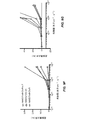
1つの実施形態では、本開示は、Chrimsonタンパク質、より詳しくは、tdTomato(tdT)蛍光タンパク質または緑色蛍光タンパク質(GFP)に融合したChrimsonR(ChrR)と呼ばれるその1つの特別な突然変異体が、Chrimsonタンパク質単独に比べて、光刺激に対する応答がより効果的であることを示す。いくつかの方法の実施形態では、蛍光タンパク質は、所定細胞数に対する融合Chrimsonタンパク質の発現レベル、より詳しくはプラズマ膜でのタンパク質レベルを、Chrimsonタンパク質単独/非融合の発現レベルと比べて増大させる。いくつかの他の方法の実施形態では、蛍光タンパク質は、融合Chrimsonタンパク質のプラズマ膜への細胞トラフィッキングを、Chrimsonタンパク質単独/非融合の細胞トラフィッキングと比べて増大させる。いくつかの方法の実施形態では、融合Chrimsonタンパク質の発現レベルおよび/または細胞トラフィッキングは、Chrimsonタンパク質の向上した溶解度、トラフィッキング、および/またはタンパク質構造を通して増大する。 In one embodiment, the disclosure discloses that one particular variant thereof, called ChrimsonR (ChrR), fused to a Chrimson protein, more specifically a tdTomato (tdT) fluorescent protein or a green fluorescent protein (GFP), is a Chrimson. It shows that the response to light stimuli is more effective than the protein alone. In some embodiments of the method, the fluorescent protein increases the expression level of the fused Chrimson protein relative to a given number of cells, more specifically the protein level on the plasma membrane, as compared to the expression level of the Chrimson protein alone / non-fused. In some other embodiments of the method, the fluorescent protein increases cell trafficking of the fused Chrimson protein to the plasma membrane as compared to cell trafficking of the Chrimson protein alone / non-fused. In some method embodiments, the expression level and / or cell trafficking of the fused Chrimson protein is increased through the improved solubility, trafficking, and / or protein structure of the Chrimson protein.
1つの局面では、本開示は、Chrimsonタンパク質および蛍光タンパク質をコードするポリヌクレオチド配列を包含する。 In one aspect, the disclosure includes polynucleotide sequences encoding Chrisson and fluorescent proteins.
別の局面では、本開示は、蛍光タンパク質に融合したChrimsonタンパク質をコードするポリヌクレオチドを包含する。 In another aspect, the disclosure includes a polynucleotide encoding a Chrisson protein fused to a fluorescent protein.
別の局面では、本開示は、ベクターを含む組成物を包含する。ベクターはポリペプチドをコードするポリヌクレオチド配列を含み、ポリペプチドは少なくとも1つのChrimsonタンパク質および蛍光タンパク質を含む。 In another aspect, the present disclosure includes compositions comprising vectors. The vector comprises a polynucleotide sequence encoding a polypeptide, which comprises at least one Chrimson protein and a fluorescent protein.
さらに別の局面では、本開示は、ポリペプチドをコードするポリヌクレオチド配列を含むベクターを含み、ポリペプチドは蛍光タンパク質に融合したChrimsonタンパク質を含む。 In yet another aspect, the disclosure comprises a vector comprising a polynucleotide sequence encoding a polypeptide, wherein the polypeptide comprises a Chrimson protein fused to a fluorescent protein.
さらに別の局面では、本開示は、対象におけるニューロン介在の障害を処置または防止する方法を包含し、本方法はベクターを含む組成物を細胞(すなわちニューロン)に投与することを含む。ベクターはポリペプチドをコードするポリヌクレオチド配列を含み、ポリペプチドは少なくとも1つのChrimsonタンパク質および蛍光タンパク質を含む。好ましくは、投与組成物のベクターはポリペプチドをコードするポリヌクレオチド配列を含み、ポリペプチドは蛍光タンパク質に融合したChrimsonタンパク質を含む。 In yet another aspect, the disclosure includes a method of treating or preventing neuronal-mediated disorders in a subject, the method comprising administering a composition comprising a vector to a cell (ie, a neuron). The vector comprises a polynucleotide sequence encoding a polypeptide, which comprises at least one Chrimson protein and a fluorescent protein. Preferably, the vector of the dosing composition comprises a polynucleotide sequence encoding a polypeptide, which comprises a Chrimson protein fused to a fluorescent protein.
さらに別の局面では、本開示は、内網膜細胞での光への感受性を回復させる方法を包含する。本方法は、ベクターを含む組成物を細胞に投与することを包含する。ベクターはポリペプチドをコードするポリヌクレオチド配列を含み、ポリペプチドは少なくとも1つのChrimsonタンパク質および蛍光タンパク質を含む。好ましくは、投与組成物のベクターはポリペプチドをコードするポリヌクレオチド配列を含み、ポリペプチドは蛍光タンパク質に融合したChrimsonタンパク質を含む。 In yet another aspect, the present disclosure includes methods of regaining light sensitivity in internal retinal cells. The method comprises administering to the cells a composition comprising a vector. The vector comprises a polynucleotide sequence encoding a polypeptide, which comprises at least one Chrimson protein and a fluorescent protein. Preferably, the vector of the dosing composition comprises a polynucleotide sequence encoding a polypeptide, which comprises a Chrimson protein fused to a fluorescent protein.
異なる局面では、本開示は、対象に視覚を回復させる方法を包含する。本方法は、光覚または光感受性の欠損により視覚を損失した対象を同定すること、ベクターを含む組成物を目?に投与することであって、ベクターはポリペプチドをコードするポリヌクレオチド配列を含み、ポリペプチドは少なくとも1つのChrimsonタンパク質および蛍光タンパク質を含む、組成物を投与すること、ポリペプチドを光により活性化させること、および対象の光感受性を測定することを包含し、ここで光感受性の増大は視覚回復を示す。 In a different aspect, the present disclosure includes methods of restoring vision to a subject. This method identifies subjects who have lost vision due to a lack of photosensitivity or photosensitivity, and eyes the composition containing the vector? The vector comprises a polynucleotide sequence encoding a polypeptide, wherein the polypeptide comprises at least one Chrimson protein and a fluorescent protein, the composition is administered, the polypeptide is activated by light. This includes measuring the photosensitivity of the subject, where increased photosensitivity indicates visual recovery.
別の局面では、本開示は、対象に視覚を回復させる方法であって、本方法は、光覚または光感受性の欠損により視覚を損失した対象を同定すること、ベクターを含む組成物を目に投与することであって、ベクターはポリペプチドをコードするポリヌクレオチド配列を含み、ポリペプチドは蛍光タンパク質に融合した少なくとも1つのChrimsonタンパク質を含む、組成物を投与すること、ポリペプチドを光により活性化させること、および対象の光感受性を測定することを包含し、ここで光感受性の増大は視覚回復を示す。 In another aspect, the present disclosure is a method of restoring vision to a subject, the method of identifying a subject who has lost vision due to a lack of photosensitivity or photosensitivity, and seeing a composition comprising a vector. By administering, the vector comprises a polynucleotide sequence encoding a polypeptide, the polypeptide comprising at least one CRMson protein fused to a fluorescent protein, administering the composition, activating the polypeptide by light. Including letting and measuring the photosensitivity of the subject, where increased photosensitivity indicates visual recovery.
他の局面では、本開示は、対象の網膜変性を処置または防止する方法を包含する。本方法は、光受容体機能の損失による網膜変性を患う対象を同定すること、ベクターを含む組成物を目に投与することであって、ベクターはポリペプチドをコードするポリヌクレオチド配列を含み、ポリペプチドは少なくとも1つのChrimsonタンパク質および蛍光タンパク質を含む、組成物を投与すること、および対象の光感受性を測定することを包含し、ここで光感受性の増大は網膜変性の処置を示す。 In other aspects, the disclosure includes methods of treating or preventing retinal degeneration in a subject. The method is to identify a subject suffering from retinal degeneration due to loss of photoreceptor function, to administer a composition containing a vector to the eye, where the vector contains a polynucleotide sequence encoding a polypeptide and is poly. The peptide comprises administering a composition comprising at least one CRMson protein and a fluorescent protein, and measuring the photosensitivity of the subject, where increased photosensitivity indicates treatment of retinal degeneration.
さらに別の局面では、本開示は、対象の網膜変性を処置または防止する方法を包含し、本方法は、光受容体機能の損失による網膜変性を患う対象を同定すること、ベクターを含む組成物を投与することであって、ベクターはポリペプチドをコードするポリヌクレオチド配列を含み、ポリペプチドは蛍光タンパク質に融合した少なくとも1つのChrimsonタンパク質を含む、組成物を投与すること、および対象の光感受性を測定することを包含し、ここで光感受性の増大は網膜変性の処置を示す。 In yet another aspect, the disclosure includes a method of treating or preventing a subject's retinal degeneration, the method of identifying a subject suffering from retinal degeneration due to loss of photoreceptor function, a composition comprising a vector. The vector comprises a polynucleotide sequence encoding a polypeptide, wherein the polypeptide comprises at least one retinal protein fused to a fluorescent protein, the composition is administered, and the photosensitivity of the subject. Including measuring, where increased photosensitivity indicates treatment of retinal degeneration.
ある局面では、本開示は、ヒトの目の光受容体機能を回復させる方法を包含する。本方法は、有効量のベクターを含む組成物を投与することであって、ベクターはポリペプチドをコードするポリヌクレオチド配列を含み、ポリペプチドは少なくとも1つのChrimsonタンパク質および蛍光タンパク質を含む、組成物を投与することを包含する。 In some aspects, the present disclosure includes methods of restoring photoreceptor function in the human eye. The method is to administer a composition comprising an effective amount of the vector, wherein the vector comprises a polynucleotide sequence encoding a polypeptide, the polypeptide comprising at least one Chrimson protein and a fluorescent protein. Includes administration.
別の局面では、本開示は、ヒトの目の光受容体機能を回復させる方法を包含し、本方法は、有効量のベクターを含む組成物を投与することであって、ベクターはポリペプチドをコードするポリヌクレオチド配列を含み、ポリペプチドは蛍光タンパク質に融合した少なくとも1つのChrimsonタンパク質を含む、組成物を投与することを包含する。 In another aspect, the disclosure includes a method of restoring photoreceptor function in the human eye, the method of administering a composition comprising an effective amount of the vector, wherein the vector comprises a polypeptide. Containing a polynucleotide sequence encoding, the polypeptide comprises administering a composition comprising at least one Chrimson protein fused to a fluorescent protein.
さらに別の局面では、本開示は、電気的に活性の細胞を脱分極する方法を包含する。本方法は、細胞にベクターを含む組成物を投与することであって、ベクターはポリペプチドをコードするポリヌクレオチド配列を含み、ポリペプチドは少なくとも1つのChrimsonタンパク質および蛍光タンパク質を含む、組成物を投与することを包含する。 In yet another aspect, the present disclosure includes methods of depolarizing electrically active cells. The method is to administer a composition comprising a vector to cells, wherein the vector comprises a polynucleotide sequence encoding a polypeptide, the polypeptide comprising at least one Chrimson protein and a fluorescent protein. Including what to do.
さらに別の局面では、本開示は、電気的に活性の細胞を脱分極する方法を包含し、本方法は、細胞にベクターを含む組成物を投与することであって、ベクターはポリペプチドをコードするポリヌクレオチド配列を含み、ポリペプチドは蛍光タンパク質に融合した少なくとも1つのChrimsonタンパク質を含む、組成物を投与することを包含する。 In yet another aspect, the disclosure includes a method of depolarizing an electrically active cell, the method of administering to the cell a composition comprising a vector, wherein the vector encodes a polypeptide. The polypeptide comprises administering a composition comprising at least one Chrimson protein fused to a fluorescent protein.
本開示の方法のいくつかの実施形態では、ベクターはアデノ随伴ウィルス(AAV)ベクターである。本方法のいくつかの実施形態では、ベクターはAAV2.7m8ベクターまたはAAV2ベクターである。いくつかの実施形態では、本方法はさらにCAGプロモーターの使用を包含する。
In some embodiments of the methods of the present disclosure, the vector is an adeno-associated virus (AAV) vector. In some embodiments of the method, the vector is an AAV 2.7 m8 vector or an
いくつかの実施形態では、ベクターは注射により投与され、好ましくは、硝子体内に注射される。 In some embodiments, the vector is administered by injection, preferably intravitreal.
本方法のいくつかの実施形態では、有効量のChrimsonタンパク質は長期にわたって発現される。本方法のいくつかの実施形態では、Chrimsonタンパク質の発現は注射後少なくとも11カ月持続する。本方法のいくつかの実施形態では、Chrimsonタンパク質の発現は注射後少なくとも2カ月持続する。 In some embodiments of the method, an effective amount of Crimson protein is expressed over time. In some embodiments of the method, expression of the Chrimson protein persists for at least 11 months after injection. In some embodiments of the method, expression of the Chrimson protein persists for at least 2 months after injection.
本方法のいくつかの実施形態では、対象は哺乳類である。いくつかの実施形態では、対象はヒトである。いくつかの実施形態では、哺乳類はマウスである。本方法のいくつかの実施形態では、マウスはrd1である。本方法のいくつかの実施形態では、哺乳類はラットである。本方法のいくつかの実施形態では、ラットはP23Hである。本方法のいくつかの実施形態では、哺乳類はヒトまたは非ヒト霊長類である。本方法のいくつかの実施形態では、非ヒト霊長類はカニクイザルである。 In some embodiments of the method, the subject is a mammal. In some embodiments, the subject is a human. In some embodiments, the mammal is a mouse. In some embodiments of the method, the mouse is rd1. In some embodiments of the method, the mammal is a rat. In some embodiments of the method, the rat is P23H. In some embodiments of the method, the mammal is a human or non-human primate. In some embodiments of the method, the non-human primate is a cynomolgus monkey.
以下の開示はまた以下の追加の実施形態を提供する。 The following disclosure also provides the following additional embodiments:
実施形態1は、哺乳類の網膜神経節細胞(RGC)を再活性化する方法であって、蛍光タンパク質に融合した有効量のChrimsonタンパク質を発現するベクターを哺乳類に投与することを包含する方法を提供する。
実施形態2は、対象のニューロン介在障害を処置または防止する方法であって、蛍光タンパク質に融合した有効量のChrimsonタンパク質を発現するベクターを備えた組成物をニューロンに投与することを包含する方法を提供する。
実施形態3は、内網膜細胞での光への感受性を回復させる方法であって、蛍光タンパク質に融合した有効量のChrimsonタンパク質を発現するベクターを備えた組成物を内網膜細胞に投与することを包含する方法を提供する。 The third embodiment is a method for restoring the sensitivity of the inner retinal cells to light, in which the inner retinal cells are administered with a composition comprising a vector expressing an effective amount of the Chrimson protein fused to the fluorescent protein. Provide a method of inclusion.
実施形態4は、対象に視覚を回復させる方法であって、蛍光タンパク質に融合した有効量のChrimsonタンパク質を発現するベクターを備えた組成物を対象に投与することを包含する方法を提供する。
実施形態5は、対象に視覚を回復させる方法であって、光覚または光感受性の欠損により視覚を損失した対象を同定すること、および蛍光タンパク質に融合した有効量のChrimsonタンパク質を発現するベクターを備えた組成物を対象に投与することを包含する方法を提供する。
実施形態6は、対象の網膜変性を処置または防止する方法であって、光受容体機能の損失により網膜変性を患う対象を同定すること、および蛍光タンパク質に融合した有効量のChrimsonタンパク質を発現するベクターを備えた組成物を対象に投与することを包含する方法を提供する。
実施形態7は、ヒトの目の光受容体機能を回復させる方法であって、光覚または光感受性の欠損により視覚を損失した対象を同定すること、および蛍光タンパク質に融合した有効量のChrimsonタンパク質を発現するベクターを備えた組成物を対象に投与することを包含する方法を提供する。 Embodiment 7 is a method of restoring photoreceptor function in the human eye for identifying subjects who have lost vision due to a lack of photosensitivity or photosensitivity, and an effective amount of Chrisson protein fused to a fluorescent protein. Provided are methods comprising administering to a subject a composition comprising a vector expressing.
実施形態8は、電気的に活性の細胞を脱分極する方法であって、蛍光タンパク質に融合した有効量のChrimsonタンパク質を発現するベクターを備えた組成物を細胞に投与することを包含する方法を提供する。
実施形態9は、実施形態1から8のいずれか1つによる方法であって、RGC応答を誘起する光刺激レベルが放射線安全性限界より低い、方法を提供する。 Embodiment 9 provides a method according to any one of embodiments 1-8, wherein the level of photostimulation that induces an RGC response is below the radiation safety limit.
実施形態10は、実施形態1から8のいずれか1つによる方法であって、Chrimsonタンパク質はChrimson88またはChrimsonRである、方法を提供する。
実施形態11は、実施形態10の方法であって、蛍光タンパク質はTd−Tomato(TdT)タンパク質および緑色蛍光タンパク質(GFP)から選択される、方法を提供する。
Embodiment 11 provides the method of
実施形態12は、実施形態11の方法であって、tdTタンパク質に融合したChrimsonタンパク質は、Chrimsonタンパク質単独に比べて光刺激への応答がより効果的である、方法を提供する。 The twelfth embodiment is the method of the eleventh embodiment, which provides a method in which the Chrimson protein fused to the tdT protein is more effective in responding to a light stimulus than the Chrimson protein alone.
実施形態13は、実施形態10の方法であって、蛍光タンパク質は、所定細胞数に対する融合Chrimsonタンパク質の発現レベルを、Chrimsonタンパク質単独の発現レベルに比べて増大させる、方法を提供する。 The thirteenth embodiment is the method of the tenth embodiment, wherein the fluorescent protein provides a method of increasing the expression level of the fused Crimson protein relative to the expression level of the Crimson protein alone with respect to a predetermined number of cells.
実施形態14は、実施形態13の方法であって、融合Chrimsonタンパク質の発現レベルは、Chrimsonタンパク質の向上した溶解度、トラフィッキング、および/またはタンパク質構造を通して増大する、方法を提供する。
実施形態15は、実施形態1から8のいずれか1つの方法であって、ベクターはアデノ随伴ウィルス(AAV)ベクターである、方法を提供する。
実施形態16は、実施形態15の方法であって、AAVベクターはAAV2ベクターおよびAAV2.7m8ベクターから選択される、方法を提供する。 The 16th embodiment is the method of the 15th embodiment and provides a method in which the AAV vector is selected from the AAV2 vector and the AAV2.7m8 vector.
実施形態17は、実施形態16の方法であって、AAVベクターはAAV2.7m8ベクターである、方法を提供する。
Embodiment 17 provides the method of
実施形態18は、実施形態1から8のいずれか1つの方法であって、ベクターはCAGプロモーターを含む、方法を提供する。
実施形態19は、実施形態1から8のいずれか1つの方法であって、ベクターは硝子体内に注射される、方法を提供する。 Embodiment 19 provides a method of any one of embodiments 1-8, wherein the vector is injected intravitreal.
実施形態20は、実施形態1から8のいずれか1つの方法であって、蛍光タンパク質に融合した有効量のChrimsonタンパク質は長期にわたって発現される、方法を提供する。
実施形態21は、実施形態20の方法であって、蛍光タンパク質に融合したChrimsonタンパク質の発現は投与後少なくとも2カ月、または投与後少なくとも11カ月持続する、方法を提供する。 21st embodiment is the method of 20th embodiment, which provides a method in which expression of a Chrisson protein fused to a fluorescent protein lasts for at least 2 months after administration, or at least 11 months after administration.
実施形態22は、実施形態1から21のいずれか1つによるベクターの1つまたはそれ以上を備えた組成物を提供する。 Embodiment 22 provides a composition comprising one or more of the vectors according to any one of embodiments 1-21.
実施形態23は、1つまたはそれ以上のChrimsonタンパク質および1つまたはそれ以上の蛍光タンパク質を融合状態でまたは個別にコードする1つまたはそれ以上のポリヌクレオチドを備えた組成物を提供する。 Embodiment 23 provides a composition comprising one or more polynucleotides encoding one or more Chrimson proteins and one or more fluorescent proteins in a fused state or individually.
実施形態24は、請求項1から21のいずれかの方法の1つまたはそれ以上で使用するための請求項22および23のいずれか1つによる組成物を提供する。
24th embodiment provides a composition according to any one of claims 22 and 23 for use in one or more of the methods of any one of
実施形態25は、哺乳類の網膜神経節細胞(RGC)を再活性化する、対象のニューロン介在障害を処置または防止する、内網膜細胞での光への感受性を回復させる、対象の網膜変性を処置または防止する、光受容体機能を回復させる、および/または電気的に活性の細胞を脱分極するための、請求項22および23の組成物のいずれか1つの使用を規定する。
本開示において、特に明記のない限り、単数の使用は複数を含み、語「a」または[an]は「少なくとも1つ」を意味し、「または」の使用は「および/または」を意味する。さらに、用語「含むこと(including)」は、「含む(includes)」および「含まれる(included)」などの他の形式と共に、制限がない。また、特に明記のない限り、「要素(element)」または「構成要素(component)」などの用語は、1単位を備える要素および構成要素と、1単位より多い単位を備える要素または構成要素との両方を包含する。 In the present disclosure, unless otherwise stated, the use of the singular includes plural, the word "a" or [an] means "at least one", and the use of "or" means "and / or". .. Moreover, the term "included", along with other forms such as "includes" and "included", is unrestricted. Further, unless otherwise specified, terms such as "element" or "component" refer to an element or component having one unit and an element or component having more than one unit. Includes both.
本明細書で用いられる百分率または他の数量と関連して用いるときの用語「約」は、その百分率または他の数量のプラスまたはマイナス10%を意味する。例えば、用語「約80%」は80%プラスまたはマイナス8%を包含することになる。 As used herein in connection with a percentage or other quantity, the term "about" means plus or minus 10% of that percentage or other quantity. For example, the term "about 80%" would include 80% plus or minus 8%.
特許、特許出願、記事、書籍、および論文を含むがこれらに限定されない本出願で言及するすべての文書または文書の一部は、それらの全体がいかなる目的に対しても本明細書において参照によって明示的に援用される。1つまたはそれ以上の援用文献および類似の資料が、ある用語を本出願でのその用語の定義に矛盾する方法で定義している場合は、本出願が優位となる。 All documents or parts of the documents referred to in this application, including but not limited to patents, patent applications, articles, books, and treatises, are express by reference herein in their entirety for any purpose. Is used as a target. If one or more supporting documents and similar materials define a term in a manner that contradicts the definition of that term in this application, this application will prevail.
本明細書で用いる用語「タンパク質」、≪ポリペプチド≫および「ペプチド」は、そうではない指示がない限り、置き換え可能である。 The terms "protein", "polypeptide" and "peptide" as used herein are interchangeable unless otherwise indicated.
本明細書で用いられる用語≪融合タンパク質≫または≪別物に融合したタンパク質≫はタンパク質構築物またはキメラタンパク質のことをいう。これは、追加の化学的リンカー無しにそれぞれのペプチド鎖内のペプチド結合を介して共有結合している2つまたはそれ以上のタンパク質またはその断片を含有する単一のタンパク質分子を意味する。1つのタンパク質はそのN末端またはC末端のいずれかで別のタンパク質に融合することができる。融合タンパク質はさらに遺伝的構造から得られるリンカー部分を備えることができる。 As used herein, the term "fusion protein" or "protein fused to another" refers to a protein construct or chimeric protein. This means a single protein molecule containing two or more proteins or fragments thereof that are covalently attached via peptide bonds within each peptide chain without additional chemical linkers. One protein can be fused to another protein at either its N-terminus or C-terminus. The fusion protein can further comprise a linker moiety obtained from the genetic structure.
本明細書で用いられ、他に指摘がない限り、用語「処置する(treat)」、「処置すること(treating)」、「処置(treatment)」および「治療(therapy)」は、患者が障害、例えばニューロン介在障害または眼疾患を患うときに起こる、該障害の1つまたはそれ以上の徴候または結果の重症度を低減させる行為を意図する。本明細書で用いられ、他に指摘がない限り、用語「防止する(prevent)」、「防止すること(preventing)」および「防止(prevention)」は、患者が障害、例えばニューロン介在障害または眼疾患を患い始める前に起こる、該障害の発現を遅らせ、および/または該障害の重症度を抑えるかまたは低減させる行為を意図する。処置は予防処置、または疾患または症状の診断に続いて施される処置であり得ることは理解されよう。本発明の処置は、障害、疾患、または症状の徴候または特性の低減または除去、または障害、疾患、または症状それ自体を除去し得る。本発明の処置は、障害、疾患、または症状の進行を低減または除去、および、場合によっては疾患、障害、または症状の緩解となり得ることは理解されよう。本発明のいくつかの実施形態では、本発明の1つまたはそれ以上の光活性化イオンチャネルポリペプチドは細胞集団内で発現され、障害または症状を処置する方法において使用され得る。 As used herein, unless otherwise indicated, the terms "treat," "treating," "treatment," and "therapy" are used in patients with disability. It is intended to reduce the severity of one or more signs or consequences of the disorder, eg, when suffering from a neuronal-mediated disorder or eye disease. As used herein, unless otherwise indicated, the terms "prevent", "preventing" and "prevention" refer to a patient with a disorder such as a neuron-mediated disorder or eye. It is intended to delay the onset of the disorder and / or reduce or reduce the severity of the disorder that occurs before the onset of the disease. It will be understood that the treatment can be a prophylactic treatment, or a treatment that follows the diagnosis of the disease or symptom. The treatments of the present invention may reduce or eliminate signs or characteristics of a disorder, disease, or symptom, or eliminate the disorder, disease, or symptom itself. It will be appreciated that the treatments of the present invention may reduce or eliminate the progression of a disorder, disorder, or symptom and, in some cases, relieve the disorder, disorder, or symptom. In some embodiments of the invention, one or more photoactivated ion channel polypeptides of the invention are expressed within a cell population and can be used in methods of treating disorders or symptoms.
本明細書で用いられ、他に特定されない限り、化合物の「治療上有効量」は、ニューロン介在障害または眼疾患の処置または管理において治療的有用性を提供するのに、または障害、例えばニューロン介在障害または眼疾患に関連する1つまたはそれ以上の徴候を遅らせるかまたは最小限にするのに十分な量である。ある化合物の治療上有効量は、その化合物の単独の、または障害、例えばニューロン介在障害または眼疾患の処置または管理において治療的有用性を提供する1つまたはそれ以上の治療および/または治療薬と組み合わせた化合物の量を意味する。用語「治療上有効量」は、ニューロン介在障害または眼疾患を緩和する、眼疾患を改善または低減する、治療全体を改善する、または別の治療薬の治療有効性を向上させる量を包含することができる。 As used herein, unless otherwise specified, a "therapeutically effective amount" of a compound is used to provide therapeutic utility in the treatment or management of neuronal-mediated disorders or eye diseases, or disorders such as neuro-mediated. An amount sufficient to delay or minimize one or more signs associated with the disorder or eye disease. A therapeutically effective amount of a compound is with one or more therapeutic and / or therapeutic agents that provide therapeutic utility in the treatment or management of the compound alone or in a disorder such as a neuromediated disorder or eye disease. Means the amount of compound combined. The term "therapeutically effective amount" includes an amount that alleviates neuronal-mediated disorders or eye diseases, improves or reduces eye diseases, improves overall treatment, or improves the therapeutic efficacy of another therapeutic agent. Can be done.
本明細書で用いられる「患者」または「対象」は、ヒトおよび非ヒト哺乳類、例えば齧歯類、マウス、ラット、非ヒト霊長類、犬および猫などのペット、ならびに例えば羊、牛、馬などの家畜類などであるがこれらに限定されない、本明細書で述べる障害を患っているまたは患いやすい哺乳類有機体を含む。 As used herein, "patient" or "subject" refers to human and non-human mammals such as rodents, mice, rats, non-human primates, pets such as dogs and cats, and eg sheep, cows, horses and the like. Includes, but is not limited to, mammalian organisms suffering from or susceptible to the disorders described herein.
網膜ニューロンの、本発明のChrimsonポリペプチドをコードする核酸(例えばベクター)によるトランスフェクションは、網膜ニューロン、好ましくは双極細胞および/または神経節細胞に感光性膜チャネルを提供する。従って、光刺激により、動物の視覚皮質、本明細書で企図される視覚形態を構成する視覚信号の処理を担う脳の領域への視覚刺激の伝達を測定することが可能である。このような視覚は通常のヒトの視覚の形態とは異なり得、光の感覚と呼ばれ、また「光検出」または「光感知」とも命名され得る。従って、本明細書で用いられる用語「視覚」は、光を刺激として有用に検出する有機体の能力として定義される。「視覚」は以下のこと:(i)光検出または感知、すなわち光が存在するか否かを認識する能力;(ii)光投影、すなわち光刺激が来ている方向を認識する能力;(iii)分解能、すなわち格子または文字標的での異なる輝度レベル(すなわちコントラスト)を検出する能力;および(iv)認知、すなわち標的内の異なるコントラストレベルの参照によって視覚標的の形状を認知する能力を包含するよう企図される。従って、「視覚」は、光、好ましくは赤色光、より好ましくは約365nmから約700nmの間、約530nmから約640nmの間の波長を有する光の存在を単に検出する能力を含み、いくつかの実施形態では、ピーク活性化は約590nmの波長を有する光と接触すると起こり得る。 Transfection of retinal neurons with nucleic acids (eg, vectors) encoding the Chrimson polypeptides of the invention provides photosensitive membrane channels to retinal neurons, preferably bipolar cells and / or ganglion cells. Thus, light stimuli can measure the transmission of visual stimuli to the animal's visual cortex, the region of the brain responsible for processing the visual signals that make up the visual morphology intended herein. Such vision may differ from the normal human visual morphology and may be referred to as the sensation of light and may also be named "photodetection" or "light sensing". Thus, the term "vision" as used herein is defined as the ability of an organism to usefully detect light as a stimulus. "Vision" means: (i) light detection or sensing, that is, the ability to recognize the presence or absence of light; (ii) light projection, that is, the ability to recognize the direction in which a light stimulus is coming; (iii). ) Resolution, i.e. the ability to detect different brightness levels (ie contrasts) at a lattice or text target; and (iv) cognition, i.e. to include the ability to perceive the shape of a visual target by reference to different contrast levels within the target. It is intended. Thus, "vision" includes the ability to simply detect the presence of light, preferably red light, more preferably light having a wavelength between about 365 nm and about 700 nm, and between about 530 nm and about 640 nm. In embodiments, peak activation can occur upon contact with light having a wavelength of about 590 nm.
本明細書で用いられる「機能性誘導体」は、用語が本明細書において接続で用いられようと二者択一で用いられようと関係なく「突然変異体」、「変異体」および「断片」を包含する。好適な変異体は、単一アミノ酸同類置換変異体であるが、例えば2、3、4または5残基の同類置換もまた意図される。いくつかの実施形態では、機能性誘導体は、元のペプチドの全長アミノ酸配列に対して少なくとも70%の相同性、好ましくは少なくとも75%、より好ましくは少なくとも80%の相同性、より好ましくは少なくとも85%の相同性、より好ましくは少なくとも90%の相同性、より好ましくは少なくとも95%の相同性、より好ましくは少なくとも99%の相同性、より好ましくは100%の相同性を有する。相同率は関連するアミノ酸配列の長さに関して決定される。従って、本発明のポリペプチドがより大きなポリペプチド内にある場合は、相同率は、本発明のポリペプチドに対応するポリペプチド部分のみに関して決定されるのであって、より大きなポリペプチド全体の相同率ではない。ポリペプチドに関する「相同率」は、Basic Local Alignment Search Tool(BLAST)エンジンを用いて整列させた少なくとも2つのポリペプチド配列間の同一のアミノ酸の百分率のことをいう。Tatusovaら(1999)ibid参照。BLASTエンジンはNational Center for Biotechnology Information(NCBI)、Bethesda、Md.によって公開されている。特定の実施形態によれば、機能性誘導体は、元のポリペプチドの全長配列に対して少なくとも70%の相同性を有するアミノ酸配列を備えたポリペプチドであり、1つまたはそれ以上の位置での置換だけその親ポリペプチドと異なるのみである。該置換は好ましくは≪同類置換≫または≪セミ同類≫である。加えて、またはこれに代えて、機能性誘導体は、元のポリペプチドの全長アミノ酸配列に対して少なくとも70%の同一性、好ましくは少なくとも75%の同一性、より好ましくは少なくとも80%の同一性、より好ましくは少なくとも85%の同一性、より好ましくは少なくとも90%の同一性、より好ましくは少なくとも95%の同一性、より好ましくは少なくとも99%の同一性、より好ましくは100%の同一性を有する。同一性または相同性を決定する方法は当該分野で既知である。 As used herein, "functional derivative" is "mutant," "mutant," and "fragment," regardless of whether the term is used in the connection or alternatives herein. Including. Suitable variants are single amino acid homologous substitution variants, but homologous substitutions of, for example, 2, 3, 4 or 5 residues are also intended. In some embodiments, the functional derivative has at least 70% homology, preferably at least 75%, more preferably at least 80% homology, more preferably at least 85, to the full-length amino acid sequence of the original peptide. It has% homology, more preferably at least 90% homology, more preferably at least 95% homology, more preferably at least 99% homology, more preferably 100% homology. The homology rate is determined with respect to the length of the associated amino acid sequence. Therefore, when the polypeptide of the invention is within a larger polypeptide, the homology rate is determined only for the polypeptide portion corresponding to the polypeptide of the invention, and the homology rate of the entire larger polypeptide is determined. is not it. "Homology" for a polypeptide refers to the percentage of the same amino acid between at least two polypeptide sequences aligned using the Basic Local Element Search Tool (BLAST) engine. See Tatsusova et al. (1999) ibid. The BLAST engine is described by National Center for Biotechnology Information (NCBI), Bethesda, Md. Published by. According to certain embodiments, the functional derivative is a polypeptide having an amino acid sequence having at least 70% homology to the full length sequence of the original polypeptide, at one or more positions. Only the substitution differs from its parent polypeptide. The substitution is preferably << similar substitution >> or << semi-similar >>. In addition, or instead, the functional derivative has at least 70% identity, preferably at least 75% identity, more preferably at least 80% identity to the full-length amino acid sequence of the original polypeptide. , More preferably at least 85% identity, more preferably at least 90% identity, more preferably at least 95% identity, more preferably at least 99% identity, more preferably 100% identity. Have. Methods of determining identity or homology are known in the art.
本明細書で用いられる用語「同類置換」は一般にタンパク質またはポリペプチドの構造および機能特性を維持するアミノ酸置換のことをいう。このような機能的に等価の(同類置換)ペプチドアミノ酸配列は、ヌクレオチド配列によってコードされるアミノ酸配列内でのアミノ酸残基の追加または置換を含むがこれらに限定されず、これらはサイレント変化となり、かくして機能的に等価の遺伝子産物を産生する。同類アミノ酸置換は、極性、電荷、溶解度、疎水性、親水性、および/または関与残基の両親媒性の類似性に基づいてなされ得る。例えば、非極性(疎水性)アミノ酸はアラニン、ロイシン、イソロイシン、バリン、プロリン、フェニルアラニン、トリプトファン、およびメチオニンを含む;極性中性アミノ酸はグリシン、セリン、トレオニン、システイン、チロシン、アスパラギン、およびグルタミンを含む;正電荷を持つ(塩基性)アミノ酸はアルギニン、リジン、およびヒスチジンを含む;そして負電荷を持つ(酸性)アミノ酸はアスパラギン酸およびグルタミン酸を含む。 As used herein, the term "similar substitution" generally refers to an amino acid substitution that maintains the structural and functional properties of a protein or polypeptide. Such functionally equivalent (similar substitution) peptide amino acid sequences include, but are not limited to, additions or substitutions of amino acid residues within the amino acid sequence encoded by the nucleotide sequence, which are silent changes. Thus, a functionally equivalent gene product is produced. Similar amino acid substitutions can be made on the basis of polarity, charge, solubility, hydrophobicity, hydrophilicity, and / or amphipathic similarity of the involved residues. For example, non-polar (hydrophobic) amino acids include alanine, leucine, isoleucine, valine, proline, phenylalanine, tryptophan, and methionine; polar neutral amino acids include glycine, serine, threonine, cysteine, tyrosine, aspartic acid, and glutamine. Positively charged (basic) amino acids include arginine, lysine, and histidine; and negatively charged (acidic) amino acids include aspartic acid and glutamic acid.
いくつかの局面において本発明は、1つまたはそれ以上の光パルスとの接触によって活性化させることができる光活性化イオンチャネルポリペプチドの細胞での発現であって、細胞の強い脱分極をもたらす発現に関する。本発明の光活性化チャネルポリペプチド、本明細書においては光活性化イオンチャネルともいうが、特定の細胞、組織、および/または有機体にて発現させ、適切な波長の光パルスに応答してインビボ、生体外、およびインビトロで細胞を制御するために用いることができる。 In some aspects, the invention is the cellular expression of a photoactivated ion channel polypeptide that can be activated by contact with one or more light pulses, resulting in strong depolarization of the cell. Regarding expression. The photoactivated channel polypeptides of the invention, also referred to herein as photoactivated ion channels, are expressed in specific cells, tissues, and / or organisms and in response to light pulses of appropriate wavelength. It can be used to control cells in vivo, in vitro, and in vitro.
本明細書で用いられる用語「イオンチャネル」は孔を形成する膜貫通ポリペプチドを意味し、孔は活性化されると開き、膜にわたる孔を通るイオン伝導を可能にする。 As used herein, the term "ion channel" means a transmembrane polypeptide that forms pores, which open when activated, allowing ion conduction through the pores across the membrane.
本発明によれば、光活性化イオンチャネルポリペプチドは、Chrimsonタンパク質またはその機能性誘導体および蛍光タンパク質を備える。 According to the present invention, the photoactivated ion channel polypeptide comprises a Chrimson protein or a functional derivative thereof and a fluorescent protein.
本発明によれば、光活性化イオンチャネルポリペプチドは、蛍光タンパク質に融合したChrimsonタンパク質またはその機能性誘導体を備える。 According to the present invention, the photoactivated ion channel polypeptide comprises a Chrimson protein fused to a fluorescent protein or a functional derivative thereof.
特定の実施形態によれば、該Chrimsonタンパク質は、タンパク質ChR88(本明細書ではChrimson88、配列番号1ともいう)またはその機能性誘導体、およびK176R置換Chrimson88タンパク質(本明細書ではK176R置換を持つChrimson88またはChrimsonR、配列番号2ともいう)よりなる群から選択される。 According to certain embodiments, the Chrimson protein is a protein ChR88 (here, also referred to as Chrimson 88, SEQ ID NO: 1) or a functional derivative thereof, and a K176R-substituted Chrimson 88 protein (here, a Chrimson 88 having a K176R substitution) or It is selected from the group consisting of (CrimsonR, also referred to as SEQ ID NO: 2).
本発明によれば、光活性化イオンチャネルポリペプチドは、(i)タンパク質ChR88(配列番号1)またはその機能性誘導体、および(ii)蛍光タンパク質を備える。 According to the present invention, the photoactivated ion channel polypeptide comprises (i) protein ChR88 (SEQ ID NO: 1) or a functional derivative thereof, and (ii) a fluorescent protein.
好適な実施形態によれば、本発明の光活性化イオンチャネルポリペプチドは、(i)タンパク質ChrimsonR(配列番号2)またはその機能性誘導体、および(ii)蛍光タンパク質を備える。 According to a preferred embodiment, the photoactivated ion channel polypeptide of the present invention comprises (i) protein ChrimsonR (SEQ ID NO: 2) or a functional derivative thereof, and (ii) a fluorescent protein.
特定の実施形態によれば、本発明の光活性化イオンチャネルポリペプチドは、タンパク質ChR88(配列番号1)またはその機能性誘導体および蛍光タンパク質よりなり、両タンパク質は独立したタンパク質として発現される。 According to a particular embodiment, the photoactivated ion channel polypeptide of the invention comprises the protein ChR88 (SEQ ID NO: 1) or a functional derivative thereof and a fluorescent protein, both of which are expressed as independent proteins.
別の実施形態によれば、本発明の光活性化イオンチャネルポリペプチドは、タンパク質ChrimsonR(配列番号2)またはその機能性誘導体および蛍光タンパク質よりなり、両タンパク質は独立したタンパク質として発現される。 According to another embodiment, the photoactivated ion channel polypeptide of the present invention comprises the protein ChrimsonR (SEQ ID NO: 2) or a functional derivative thereof and a fluorescent protein, both of which are expressed as independent proteins.
好適な実施形態によれば、本発明の光活性化イオンチャネルポリペプチドは、蛍光タンパク質に融合したタンパク質ChR88(配列番号1)またはその機能性誘導体よりなる。 According to a preferred embodiment, the photoactivated ion channel polypeptide of the present invention comprises the protein ChR88 (SEQ ID NO: 1) fused to a fluorescent protein or a functional derivative thereof.
より好適な実施形態によれば、本発明の光活性化イオンチャネルポリペプチドは、蛍光タンパク質に融合したタンパク質ChrimsonR(配列番号2)またはその機能性誘導体よりなる。 According to a more preferred embodiment, the photoactivated ion channel polypeptide of the present invention comprises the protein Chrimson R (SEQ ID NO: 2) fused to a fluorescent protein or a functional derivative thereof.
本発明の光活性化イオンチャネルポリペプチドは赤色光、好ましくは約365nmから約700nmの間、約530nmから約640nmの間の波長を有する光との接触によって強く活性化され、いくつかの実施形態では、ピーク活性は約590nmの波長を有する光と接触すると起こり得る。 The photoactivated ion channel polypeptides of the invention are strongly activated by contact with red light, preferably light having a wavelength between about 365 nm and about 700 nm, and between about 530 nm and about 640 nm, in some embodiments. In, peak activity can occur upon contact with light having a wavelength of about 590 nm.
本発明の光活性化イオンチャネルポリペプチドを含む興奮性細胞を活性波長領域の光と接触させることで細胞を強力に脱分極する。本発明の光活性化イオンチャネルポリペプチドを発現する細胞を脱分極するために用いられ得る光の波長の例としては、少なくとも約365nm、385nm、405nm、425nm、445nm、465nm、485nm、505nm、525nm、545nm、565nm、585nm、590nm、605nm、625nm、645nm、665nm、685nm、および700nmの波長を含み、これらの間のすべての波長を含む。いくつかの実施形態では、本発明の光活性化イオンチャネルポリペプチドは590nmにピーク波長感度を有し、660nmまでスパイクを引き出し得る。 Excitatory cells containing the photoactivated ion channel polypeptide of the present invention are strongly depolarized by contacting them with light in the active wavelength region. Examples of wavelengths of light that can be used to depolarize cells expressing the photoactivated ion channel polypeptides of the invention are at least about 365 nm, 385 nm, 405 nm, 425 nm, 445 nm, 465 nm, 485 nm, 505 nm, 525 nm. Includes wavelengths of 545 nm, 565 nm, 585 nm, 590 nm, 605 nm, 625 nm, 645 nm, 665 nm, 685 nm, and 700 nm, and includes all wavelengths in between. In some embodiments, the photoactivated ion channel polypeptides of the invention have a peak wavelength sensitivity at 590 nm and can elicit spikes up to 660 nm.
本発明の光活性化イオンチャネルポリペプチドは、本発明の1つまたはそれ以上の光活性化イオンチャネルポリペプチドが発現される興奮性細胞を脱分極するために用いることができる。いくつかの実施形態では、本発明の光活性化イオンチャネルポリペプチドは、本発明の光活性化イオンチャネルポリペプチドを活性化しない光の波長によって活性化される光活性化イオンチャネルを発現する1つまたはそれ以上の追加の細胞サブ集団もまた含む細胞集団における細胞サブ集団において発現させることができる。 The photoactivated ion channel polypeptides of the invention can be used to depolarize excitatory cells in which one or more of the photoactivated ion channel polypeptides of the invention are expressed. In some embodiments, the photoactivated ion channel polypeptides of the invention express photoactivated ion channels that are activated by a wavelength of light that does not activate the photoactivated ion channel polypeptides of the invention1 One or more additional cell subpopulations can also be expressed in a cell subpopulation in a cell population that also includes.
様々な実施形態において用いることができるペプチドアミノ酸配列は、本明細書で述べる光活性化イオンチャネルポリペプチド(配列番号1または2または5)ならびに機能的に等価のポリペプチドを含む。 Peptide amino acid sequences that can be used in various embodiments include photoactivated ion channel polypeptides (SEQ ID NO: 1 or 2 or 5) as described herein as well as functionally equivalent polypeptides.
このような機能的に等価のペプチドアミノ酸配列(同類置換)は、本発明のアミノ酸配列内のアミノ酸残基の追加または置換を含むがこれらに限定されず、これらはサイレント変化となり、このようにして機能的に等価のポリペプチドを産生する。アミノ酸置換は、極性、電荷、溶解度、疎水性、親水性、および/または関与残基の両親媒性の類似性に基づいてなされ得る。例えば、非極性(疎水性)アミノ酸はアラニン、ロイシン、イソロイシン、バリン、プロリン、フェニルアラニン、トリプトファン、およびメチオニンを含む;極性中性アミノ酸はグリシン、セリン、トレオニン、システイン、チロシン、アスパラギン、およびグルタミンを含む;正電荷を持つ(塩基性)アミノ酸はアルギニン、リジン、およびヒスチジンを含む;そして負電荷を持つ(酸性)アミノ酸はアスパラギン酸およびグルタミン酸を含む。もしくは同類アミノ酸置換はアミノ酸のハイドロパシー指標(hydropathic index)に基づいて行ってもよい。各アミノ酸はその疎水性および電荷特性に基づいてハイドロパシー指標が割り当てられる。それらはイソロイシン(+4.5);バリン(+4.2);ロイシン(+3.8);フェニルアラニン(+2.8);システイン/シスチン(+2.5);メチオニン(+1.9);アラニン(+1.8);グリシン(−0.4);トレオニン(−0.7);セリン(−0.8);トリプトファン(−0.9);チロシン(−1.3);プロリン(−1.6);ヒスチジン(−3.2);グルタミン酸塩(−3.5);グルタミン(−3.5);アスパラギン酸塩(−3.5);アスパラギン(−3.5);リジン(−3.9);およびアルギニン(−4.5)である。タンパク質についての相互的生物学的機能を参照する場合にハイドロパシーアミノ酸指標を用いることは当該分野において了解されている(KyteおよびDoolittle、J.Mol.Biol.157:105−132、1982)。いくつかの例では、いくつかのアミノ酸は、類似のハイドロパシー指標またはスコアを有する他のアミノ酸に置換され依然として類似の生物学的活性を保持し得ることが知られている。ハイドロパシー指標に基づいて変更を行う場合、いくつかの実施形態では、ハイドロパシー指標が±2以内であるアミノ酸の置換が含まれ、一方他の実施形態では、±1以内のアミノ酸置換が含まれ、またさらに別の実施形態では、±0.5以内のアミノ酸置換が含まれる。 Such functionally equivalent peptide amino acid sequences (similar substitutions) include, but are not limited to, additions or substitutions of amino acid residues within the amino acid sequences of the invention, which result in silent changes and thus. Produces a functionally equivalent polypeptide. Amino acid substitutions can be made on the basis of polarity, charge, solubility, hydrophobicity, hydrophilicity, and / or amphipathic similarity of the residues involved. For example, non-polar (hydrophobic) amino acids include alanine, leucine, isoleucine, valine, proline, phenylalanine, tryptophan, and methionine; polar neutral amino acids include glycine, serine, threonine, cysteine, tyrosine, aspartic acid, and glutamine. Positively charged (basic) amino acids include arginine, lysine, and histidine; and negatively charged (acidic) amino acids include aspartic acid and glutamic acid. Alternatively, similar amino acid substitutions may be performed based on the hydropathic index of amino acids. Each amino acid is assigned a hydropathy index based on its hydrophobicity and charge properties. They are isoleucine (+4.5); valine (+4.2); leucine (+3.8); phenylalanine (+2.8); cysteine / cystine (+2.5); methionine (+1.9); alanine (+1. 8); glycine (-0.4); threonine (-0.7); serine (-0.8); tryptophan (-0.9); tyrosine (-1.3); proline (-1.6) Histidine (-3.2); Glutamine (-3.5); Glutamine (-3.5); Aspartate (-3.5); Aspartin (-3.5); Leucine (-3.9) ); And arginine (-4.5). It is understood in the art to use the hydropathic amino acid index when referring to the reciprocal biological function of a protein (Kyte and Doolittle, J. Mol. Biol. 157: 105-132, 1982). In some examples, it is known that some amino acids can be replaced by other amino acids with similar hydropathic indicators or scores and still retain similar biological activity. When making changes based on the hydropathy index, some embodiments include amino acid substitutions with a hydropathy index of ± 2 or less, while other embodiments include amino acid substitutions of ± 1 or less. In yet another embodiment, amino acid substitutions within ± 0.5 are included.
もしくは同類アミノ酸置換は親水性に基づいてなされ得、特に生物学的機能タンパク質またはそれによって作成されたペプチドを免疫学的実施形態で使用することが意図される。いくつかの実施形態では、隣接するアミノ酸の親水性によって決定されるタンパク質の最大局所平均親水性はその免疫原性および抗原性、すなわちタンパク質の生物学的特性と相互に関連する。以下の親水性値はこれらのアミノ酸残基に割り当てられている:アルギニン(+3.0);リジン(+3.0);アスパラギン酸(+3.0±1);グルタミン酸(+3.0±1);セリン(+0.3);アスパラギン(+0.2);グルタミン(+0.2);グリシン(0);トレオニン(−0.4);プロリン(−0.5±1);アラニン(−0.5);ヒスチジン(−0.5);システイン(−1.0);メチオニン(−1.3);バリン(−1.5);ロイシン(−1.8);イソロイシン(−1.8);チロシン(−2.3);フェニルアラニン(−2.5);およびトリプトファン(−3.4)。類似の親水性値に基づいて変更を行う場合、いくつかの実施形態では、親水性値が±2以内であるアミノ酸の置換が含まれ、いくつかの実施形態では、±1以内のものが含まれ、そしていくつかの実施形態では、±0.5以内のものが含まれる。 Alternatively, similar amino acid substitutions can be made on the basis of hydrophilicity, and it is specifically intended to use biologically functional proteins or peptides made thereby in immunological embodiments. In some embodiments, the maximum local average hydrophilicity of a protein, determined by the hydrophilicity of adjacent amino acids, correlates with its immunogenicity and antigenicity, i.e., the biological properties of the protein. The following hydrophilic values are assigned to these amino acid residues: arginine (+3.0); lysine (+3.0); aspartic acid (+3.0 ± 1); glutamine (+3.0 ± 1); Serine (+0.3); Aspartin (+0.2); Glutamine (+0.2); Glycine (0); Threonine (-0.4); Proline (-0.5 ± 1); Alanin (-0.5) ); Histidine (-0.5); cysteine (-1.0); methionine (-1.3); valine (-1.5); leucine (-1.8); isoleucine (-1.8); Tyrosine (-2.3); phenylalanine (-2.5); and tryptophan (-3.4). When making changes based on similar hydrophilicity values, some embodiments include amino acid substitutions with hydrophilicity values within ± 2 and some embodiments include those within ± 1 And, in some embodiments, those within ± 0.5 are included.
1つの好適な実施形態によれば、本発明の光活性化イオンチャネルポリペプチドはChrimsonポリペプチド(例えばタンパク質ChR88またはその機能性誘導体、またはタンパク質ChrimsonRまたはその機能性誘導体)と蛍光タンパク質との間の融合タンパク質である。ポリペプチドまたはペプチド、またはペプチドの切断形または突然変異形が非関連タンパク質、ポリペプチドまたはペプチドに融合される融合タンパク質の使用は、本明細書で述べる核酸および/またはアミノ酸配列をコードする所望のペプチドに基づいて設計することができる。いくつかの実施形態では、融合タンパク質は、発現されている融合タンパク質に選択的に結合する抗体を利用することによって容易に精製され得る。 According to one preferred embodiment, the photoactivated ion channel polypeptide of the invention is between a Chrimson polypeptide (eg, protein ChR88 or a functional derivative thereof, or protein ChrimsonR or a functional derivative thereof) and a fluorescent protein. It is a fusion protein. The use of a polypeptide or peptide, or a fusion protein in which a truncated or mutant form of the peptide is fused to an unrelated protein, polypeptide or peptide, is the desired peptide encoding the nucleic acid and / or amino acid sequence described herein. Can be designed based on. In some embodiments, the fusion protein can be readily purified by utilizing an antibody that selectively binds to the expressed fusion protein.
一般に、本発明の光活性化イオンチャネルポリペプチドが機能するのに必要なレチナールまたはレチナール誘導体は、該チャネルポリペプチドでトランスフェクトさせることになる細胞によって産生される。しかし、本発明によれば、本発明の光活性化イオンチャネルポリペプチドと、例えば3,4−デヒドロレチナール、13−エチルレチナール、9−dm−レチナール、3−ヒドロキシレチナール、4−ヒドロキシレチナール、ナフチルレチナール;3,7,11−トリメチル−ドデカ−2,4,6,8,10−ペンタエナール;3,7−ジメチル−デカ−2,4,6,8−テトラエナール;3,7−ジメチル−オクタ−2,4,6−トリエナール、ならびに6−7−または8−9−または10−11回転ブロックレチナールなどのレチナールまたはレチナール誘導体とを備えたチャネルロドプシンがさらに開示される(WO03084994)。 Generally, the retinal or retinal derivative required for the photoactivated ion channel polypeptide of the invention to function is produced by the cell to be transfected with the channel polypeptide. However, according to the invention, the photoactivated ion channel polypeptides of the invention and, for example, 3,4-dehydroretinal, 13-ethylretinal, 9-dm-retinal, 3-hydroxyretinal, 4-hydroxyretinal, naphthyl Retinal; 3,7,11-trimethyl-dodeca-2,4,6,8,10-pentaneal; 3,7-dimethyl-deca-2,4,6,8-tetraenal; 3,7-dimethyl-octa- Further disclosed is channel rhodopsin with 2,4,6-trienal, and retinal or retinal derivatives such as 6-7- or 8-9- or 10-11 rotation block retinal (WO0308949).
上述の所望のペプチドアミノ酸配列は化学的に合成することができるが(例えば“Proteins:Structures and Molecular Principles”(Creightonら、W.H.Freeman&Company、New York、N.Y.、1984)参照)、大きなポリペプチド配列は、所望のペプチドをコードする核酸配列を含有する核酸を発現するための当該分野で周知の技術を用いて組み換えDNA技術によって有利に産生され得る。このような方法は、ペプチドをコードするヌクレオチド配列および適切な転写および翻訳制御信号を含有する発現ベクターを構築するために用いることができる。これらの方法は、例えば、インビトロ組み換えDNA技法、合成的技法、およびインビボ遺伝子組み換えを含む(例えば“Molecular Cloning,A Laboratory Manual”、上記参照)。もしくは、所望のペプチドをコードするヌクレオチド配列をコードするRNAおよび/またはDNAは、例えば合成器を用いて化学的に合成され得る(例えば“Oligonucleotide Synthesis:A Practical Approach”(Gait編、IRL Press、Oxford,英国、1984)参照)。 Although the desired peptide amino acid sequences described above can be chemically synthesized (see, eg, "Proteins: Proteins and Molecular Principles" (Creighton et al., WH Freeman & Company, New York, NY, 1984)). Large polypeptide sequences can be advantageously produced by recombinant DNA techniques using techniques well known in the art for expressing nucleic acids containing nucleic acid sequences encoding the desired peptides. Such methods can be used to construct expression vectors containing nucleotide sequences encoding peptides and suitable transcriptional and translational control signals. These methods include, for example, in vitro recombinant DNA techniques, synthetic techniques, and in vivo genetic recombination (eg, "Molecular Cloning, A Laboratory Manual", see above). Alternatively, the RNA and / or DNA encoding the nucleotide sequence encoding the desired peptide can be chemically synthesized, for example, using a synthesizer (eg, "Oligonoculotide Synthesis: A Practical Application" (Gait ed., IRL Press, Oxford). , United Kingdom, 1984)).
様々な実施形態で用いることができるペプチドアミノ酸配列は、本明細書で述べる光活性化イオンチャネルポリペプチド(配列番号1または2、5または6)、および機能的に等価のペプチドおよびその機能的誘導体ならびにそれらの機能性断片を含む。実際において、いくつかの実施形態では、特別なヌクレオチド配列によってコードされるいかなる所望のペプチドアミノ酸配列も用いることができ、所望のペプチドアミノ酸配列のすべてまたはいかなる部分でもコードするあらゆるポリペプチド配列の使用である。遺伝暗号の退化性はよく知られており、よって各光活性化チャネルポリペプチドアミノ酸をコードするヌクレオチド配列は、アミノ酸をコードすることができる周知の核酸「トリプレット」コドン、または多くの場合にはコドンを遺伝的に代表する。よって、本明細書で企図されるように、本明細書で述べるチャネルロドプシンペプチドアミノ酸配列は、遺伝暗号によりひとまとめに考えると(例えば“Molecular Cell Biology”、表4−1、109頁(Darnellら編、W.H.Freeman&Company、New York、NY、1986)、このようなアミノ酸配列をコードすることができる核酸配列のすべての様々な置換および組み合わせを遺伝的に代表する。 Peptide amino acid sequences that can be used in various embodiments include the photoactivated ion channel polypeptides described herein (SEQ ID NO: 1 or 2, 5 or 6), and functionally equivalent peptides and functional derivatives thereof. As well as their functional fragments. In practice, in some embodiments, any desired peptide amino acid sequence encoded by a special nucleotide sequence can be used, with the use of any polypeptide sequence encoding all or any portion of the desired peptide amino acid sequence. be. The degenerative properties of the genetic code are well known, so the nucleotide sequence encoding each photoactivated channel polypeptide amino acid is a well-known nucleic acid "triplet" codon capable of encoding an amino acid, or often a codon. Is genetically represented. Thus, as contemplated herein, the channelrhodopsin peptide amino acid sequences described herein are considered collectively by genetic code (eg, "Molecular Cell Biology", Table 4-1 and pp. 109 (Darnell et al.)). , WH Freeman & Company, New York, NY, 1986), genetically representing all various substitutions and combinations of nucleic acid sequences capable of encoding such amino acid sequences.
いくつかの実施形態は、本発明の光活性化イオンチャネルポリペプチドをコードするヌクレオチド配列を備えた単離核酸分子である。いくつかの実施形態では、ヌクレオチド配列は(i)タンパク質ChR88(配列番号1)またはその機能性誘導体、および(ii)蛍光タンパク質を含むポリペプチドをコードする。別の実施形態では、ヌクレオチド配列は、(i)タンパク質ChrimsonR(配列番号2)またはその機能性誘導体、および(ii)蛍光タンパク質を含むポリペプチドをコードする。 Some embodiments are isolated nucleic acid molecules with nucleotide sequences encoding the photoactivated ion channel polypeptides of the invention. In some embodiments, the nucleotide sequence encodes a polypeptide comprising (i) protein ChR88 (SEQ ID NO: 1) or a functional derivative thereof, and (ii) a fluorescent protein. In another embodiment, the nucleotide sequence encodes a polypeptide comprising (i) protein ChrimsonR (SEQ ID NO: 2) or a functional derivative thereof, and (ii) a fluorescent protein.
1つの特別な実施形態によれば、ヌクレオチド配列は、蛍光タンパク質に融合したタンパク質ChR88(配列番号1)またはその機能性誘導体を含むポリペプチドをコードする。好適な実施形態によれば、ヌクレオチド配列は、蛍光タンパク質に融合したタンパク質ChrimsonR(配列番号2)またはその機能性誘導体を含むポリペプチドをコードする。 According to one particular embodiment, the nucleotide sequence encodes a polypeptide comprising protein ChR88 (SEQ ID NO: 1) fused to a fluorescent protein or a functional derivative thereof. According to a preferred embodiment, the nucleotide sequence encodes a polypeptide comprising the protein Chrimson R (SEQ ID NO: 2) fused to a fluorescent protein or a functional derivative thereof.
いくつかの特別な実施形態によれば、本発明の蛍光タンパク質はtdTomato(tdT)蛍光タンパク質および緑色蛍光タンパク質(GFP)から選択される。 According to some special embodiments, the fluorescent protein of the present invention is selected from tdTomato (tdT) fluorescent protein and green fluorescent protein (GFP).
TdTomatoは鮮赤色蛍光タンパク質である(tdTomatoの励起ピーク554nm、発光波長ピーク581nm)(Shaner NCら、Nat Biotechnol、22、1567−1572、2004)。本発明によるtdTomatoをコードするゲノム配列は、合成構築物タンデムダイマー赤色蛍光タンパク質遺伝子、完全cds(GenBank登録番号AY678269)と少なくとも84%の同一性を示す。好適な実施形態によれば、本発明のコードされたtdTomatoタンパク質部分は、配列番号3のアミノ酸配列と同一のアミノ酸の約70%から約75%の間、より好ましくは約75%から約80%の間、より好ましくは約80%から約90%の間、さらにより好ましくは約90%から約99%の間のポリペプチドである。 TdTomato is a bright red fluorescent protein (excitation peak of tdTomato 554 nm, emission wavelength peak 581 nm) (Shaner NC et al., Nat Biotechnol, 22, 1567-1572, 2004). The genomic sequence encoding tdTomato according to the invention exhibits at least 84% identity with the synthetic construct tandem dimer red fluorescent protein gene, complete cds (GenBank registration number AY678269). According to a preferred embodiment, the encoded tdTomato protein portion of the invention is between about 70% and about 75%, more preferably about 75% to about 80% of the amino acids identical to the amino acid sequence of SEQ ID NO: 3. , More preferably between about 80% and about 90%, and even more preferably between about 90% and about 99%.
他の実施形態では、本発明は、配列番号5のアミノ酸配列またはその断片と同一のアミノ酸の約70%から約75%の間、より好ましくは約75%から約80%の間、より好ましくは約80%から約90%の間、さらにより好ましくは約90%から約99%の間のポリペプチドをコードする単離核酸を提供する。 In other embodiments, the present invention comprises between about 70% and about 75%, more preferably between about 75% and about 80%, more preferably between about 70% and about 75% of the amino acids identical to the amino acid sequence of SEQ ID NO: 5 or fragments thereof. Provided are isolated nucleic acids encoding polypeptides between about 80% and about 90%, and even more preferably between about 90% and about 99%.
本発明の核酸は、1つまたはそれ以上の信号配列(例えばエンハンサー、ポリアデニル化信号、追加の制限酵素部位、多重クローニング部位)および/またはプロモーター配列または他のコーディングセグメントまたはそれらの組み合わせを含むがこれらに限定されない追加の配列を含み得る。プロモーターは誘導的または構造的な一般のまたは細胞特異的なプロモーターであり得る。細胞特異的プロモーターの例としては、双極細胞のmGlu6プロモーターがある。いくつかの実施形態は開示した方法のいずれかであり、プロモーターは構造的プロモーターである。いくつかの実施形態は開示した方法のいずれかであり、構造的プロモーターはCMVプロモーターまたはCAGプロモーターを含むがこれらに限定されない(CAGプロモーターは、チキンベータアクチンプロモーター(CBA)およびSV40イントロン挿入に融合したハイブリッドサイトメガロウイルス(CMV)前初期エンハンサーである;Alexopoulouら、BMC Cell Biol.2008;9:2;配列番号8)。いくつかの実施形態は開示した方法のいずれかであり、プロモーターは誘導的および/または細胞特異的プロモーターを含むがこれらに限定されない。プロモーター、ベクター、エンハンサー、ポリアデニル化部位の選択は当業者には型通りの設計事項である。これら要素は文献に適切に記載されておりまた市販されている。 The nucleic acids of the invention include one or more signal sequences (eg enhancers, polyadenylation signals, additional restriction enzyme sites, multiple cloning sites) and / or promoter sequences or other coding segments or combinations thereof. May include additional sequences not limited to. The promoter can be an inducible or structural general or cell-specific promoter. An example of a cell-specific promoter is the retina mGlu6 promoter. Some embodiments are any of the disclosed methods and the promoter is a structural promoter. Some embodiments are any of the disclosed methods, in which structural promoters include, but are not limited to, CMV promoters or CAG promoters (CAG promoters fused to chicken beta actin promoter (CBA) and SV40 intron insertion. Hybrid cytomegalovirus (CMV) pre-early promoter; Alexopoulo et al., BMC Cell Biol. 2008; 9: 2; SEQ ID NO: 8). Some embodiments are any of the disclosed methods, the promoters include, but are not limited to, inducible and / or cell-specific promoters. Selection of promoters, vectors, enhancers and polyadenylation sites is a routine design matter for those skilled in the art. These elements are well documented and commercially available.
いくつかの実施形態では、本発明は、タンパク質またはペプチドであって、そのアミノ酸配列内に本発明の光活性化イオンチャネルポリペプチドまたはその機能的部分または変異体、同定されたもの(例えば配列番号5)などのアミノ酸配列を含むタンパク質またはペプチドをコードする単離核酸セグメントおよび組み換えベクターに関する。 In some embodiments, the invention is a protein or peptide that has been identified within its amino acid sequence, a photoactivated ion channel polypeptide of the invention or a functional portion or variant thereof (eg, SEQ ID NO:). 5) With respect to isolated nucleic acid segments and recombinant vectors encoding proteins or peptides containing amino acid sequences such as.
いくつかの実施形態では、本発明は、アミノ酸配列、配列番号6または配列番号7を含む単離核酸セグメントおよび組み換えベクターに関する。 In some embodiments, the invention relates to an isolated nucleic acid segment and a recombinant vector comprising an amino acid sequence, SEQ ID NO: 6 or SEQ ID NO: 7.
いくつかの実施形態は、(i)配列番号1または配列番号2のアミノ酸を(ii)配列番号3または配列番号4と共にコードするヌクレオチド配列を備えた組み換え核酸である。 Some embodiments are recombinant nucleic acids comprising (i) a nucleotide sequence encoding the amino acid of SEQ ID NO: 1 or SEQ ID NO: 2 with (ii) SEQ ID NO: 3 or SEQ ID NO: 4.
いくつかの好適な実施形態は、配列番号5のアミノ酸またはその断片をコードするヌクレオチド配列を備えた組み換え核酸である。 Some preferred embodiments are recombinant nucleic acids with a nucleotide sequence encoding the amino acid of SEQ ID NO: 5 or a fragment thereof.
いくつかの好適な実施形態は、ヌクレオチド配列、配列番号6または配列番号7を備えた組み換え核酸である。 Some preferred embodiments are recombinant nucleic acids with a nucleotide sequence, SEQ ID NO: 6 or SEQ ID NO: 7.
いくつかの実施形態は、異種プロモーターに操作可能に連結した(i)配列番号1または配列番号2のアミノ酸をコードするヌクレオチド配列、および(ii)異種プロモーターに操作可能に連結した配列番号3または配列番号4のアミノ酸をコードするヌクレオチド配列を備えた組み換え核酸である。 In some embodiments, (i) a nucleotide sequence encoding the amino acid of SEQ ID NO: 1 or SEQ ID NO: 2 operably linked to a heterologous promoter, and (ii) SEQ ID NO: 3 or sequence operably linked to a heterologous promoter. It is a recombinant nucleic acid having a nucleotide sequence encoding the amino acid of No. 4.
いくつかの好適な実施形態は、異種プロモーターに操作可能に連結した配列番号5のアミノ酸またはその断片をコードするヌクレオチド配列を備えた組み換え核酸である。 Some preferred embodiments are recombinant nucleic acids comprising a nucleotide sequence encoding the amino acid of SEQ ID NO: 5 or a fragment thereof operably linked to a heterologous promoter.
いくつかの好適な実施形態は、異種プロモーターに操作可能に連結したヌクレオチド配列、配列番号6または配列番号7を備えた組み換え核酸である。 Some preferred embodiments are recombinant nucleic acids with a nucleotide sequence operably linked to a heterologous promoter, SEQ ID NO: 6 or SEQ ID NO: 7.
いくつかの好適な実施形態は、CAG異種プロモーター(配列番号8)に操作可能に連結したヌクレオチド配列、配列番号6または配列番号7を備えた組み換え核酸である。 Some preferred embodiments are recombinant nucleic acids with a nucleotide sequence, SEQ ID NO: 6 or SEQ ID NO: 7, operably linked to a CAG heterologous promoter (SEQ ID NO: 8).
別の局面によれば、本発明は、上記の光活性化イオンチャネルポリペプチドのいずれかをコードする核酸配列を含む核酸発現ベクターに関する。 According to another aspect, the invention relates to a nucleic acid expression vector comprising a nucleic acid sequence encoding any of the photoactivated ion channel polypeptides described above.
本明細書で用いられる用語「核酸発現ベクター」は、操作可能に連結している別の核酸を異なる遺伝的環境間で輸送することができる核酸分子のことをいう。用語「ベクター」はまた核酸分子を輸送することができるウィルスまたは有機体のことをいう。ベクターの1つのタイプはエピソーム、すなわち過剰染色体複製が可能な核酸分子である。いくつかの有用なベクターは、連結される核酸の自律複製および/または発現が可能なものである。操作可能に連結される遺伝子の発現を指示することが可能なベクターを本明細書では「発現ベクター」という。発現ベクターおよびそれらの使用法は当該分野では周知である。適切な発現ベクターおよびそれらの使用法の非限定な例は本明細書で提示される。好適な実施形態では、ベクターは遺伝子治療、より詳しくはウィルス介在遺伝子導入に適している。遺伝子治療に適したウィルスの例としては、レトロウィルス、アデノウィルス、アデノ随伴ウィルス(AAV)、レンチウィルス、ポックスウィルス(例えばMVA)、アルファウィルス、ヘルペスウィルスがある。しかし、遺伝子治療はさらに、裸のDNA、核酸に関連したリポソームの使用など非ウィルスの方法も包含する。本発明のいくつかの方法で有用なベクターは、光活性化イオンチャネルポリペプチドを分裂細胞および非分裂細胞へと遺伝学的に挿入することができ、また光活性化イオンチャネルポリペプチドをインビボ、インビトロまたは生体外細胞である細胞に挿入することができる。 As used herein, the term "nucleic acid expression vector" refers to a nucleic acid molecule capable of transporting another operably linked nucleic acid between different genetic environments. The term "vector" also refers to a virus or organism capable of transporting nucleic acid molecules. One type of vector is an episome, a nucleic acid molecule capable of hyperchromosomal replication. Some useful vectors are capable of autonomous replication and / or expression of the linked nucleic acid. A vector capable of instructing the expression of an operably linked gene is referred to herein as an "expression vector". Expression vectors and their usage are well known in the art. Suitable expression vectors and non-limiting examples of their use are presented herein. In a preferred embodiment, the vector is suitable for gene therapy, more specifically for virus-mediated gene transfer. Examples of viruses suitable for gene therapy include retrovirus, adenovirus, adeno-associated virus (AAV), lentivirus, poxvirus (eg, MVA), alphavirus, and herpesvirus. However, gene therapy also includes non-viral methods such as the use of naked DNA, nucleic acid-related liposomes. Vectors useful in some of the methods of the invention are capable of genetically inserting photoactivated ion channel polypeptides into dividing and non-dividing cells, and also in vivo, photoactivating ion channel polypeptides. It can be inserted into cells that are in vitro or in vivo cells.
いくつかの実施形態では、本発明の光活性化イオンチャネルポリペプチドのための遺伝子を含む核酸発現ベクターはAAVウィルスベクターの間で選択される。好適な実施形態によれば、該AAVウィルスベクターはAAV2、より好ましくはAAV2−7m8ウィルスベクターである(WO2012/145601)。 In some embodiments, nucleic acid expression vectors containing genes for the photoactivated ion channel polypeptides of the invention are selected among AAV viral vectors. According to a preferred embodiment, the AAV viral vector is AAV2, more preferably AAV2-7m8 viral vector (WO2012 / 145601).
本発明のいくつかの局面は、本発明の光活性化イオンチャネルポリペプチドを用いて、細胞、組織、または対象の障害または症状を処置する方法を含む。本発明の処置法は、障害を処置するために、本発明の光活性化イオンチャネルポリペプチドの治療上有効量をこのような処置が必要な対象に投与することを含み得る。 Some aspects of the invention include methods of treating disorders or symptoms of cells, tissues, or subjects using the photoactivated ion channel polypeptides of the invention. The treatment method of the present invention may include administering a therapeutically effective amount of the photoactivated ion channel polypeptide of the present invention to a subject in need of such treatment in order to treat the disorder.
本発明の光活性化イオンチャネルポリペプチドの投与は、少なくとも1つの本発明の光活性化イオンチャネルポリペプチドの有効量を含む医薬組成物の投与を含み得る。本発明の光活性化イオンチャネルポリペプチドの投与は、細胞を含む医薬組成物の投与を含み得、細胞は本発明の光活性化イオンチャネルを発現する。本発明の光活性化イオンチャネルポリペプチドの投与は、ベクターを含む医薬組成物の有効量の投与を含み得、ベクターは本発明の光活性化イオンチャネルポリペプチドをコードする核酸配列を備え、ベクターの投与は、対象の細胞での光活性化イオンチャネルポリペプチドの発現をもたらす。 Administration of the photoactivated ion channel polypeptide of the invention may comprise administration of a pharmaceutical composition comprising an effective amount of at least one photoactivated ion channel polypeptide of the invention. Administration of the photoactivated ion channel polypeptide of the present invention may comprise administration of a pharmaceutical composition comprising cells, the cells expressing the photoactivated ion channel of the present invention. Administration of the photoactivated ion channel polypeptide of the invention may comprise administration of an effective amount of the pharmaceutical composition comprising the vector, the vector comprising a nucleic acid sequence encoding the photoactivated ion channel polypeptide of the invention, a vector. Administration results in the expression of photoactivated ion channel polypeptides in cells of interest.
いくつかの実施形態では、ニューロン介在障害を処置または防止する方法であって、(a)標的細胞に、該標的細胞で発現可能な本発明の光活性化イオンチャネルポリペプチドをコードする核酸発現ベクターを送達することであって、該ベクターはプロモーター配列に操作可能に連結された本発明の光活性化イオンチャネルポリペプチドをコードするオープンリーディングフレームと、オプションとして転写制御配列とを備えた、送達すること、および(b)該標的細胞に該ベクターを発現させることを包含し、発現した光活性化イオンチャネルポリペプチドは露光されると該標的細胞を活性化させる方法である。 In some embodiments, a method of treating or preventing neuronal-mediated disorders (a) a nucleic acid expression vector encoding a photoactivated ion channel polypeptide of the invention that can be expressed in a target cell in the target cell. The vector comprises an open reading frame encoding a photoactivated ion channel polypeptide of the invention operably linked to a promoter sequence and optionally a transcription control sequence. This is a method that includes (b) expressing the vector in the target cell, and the expressed photoactivated ion channel polypeptide activates the target cell when exposed.
いくつかの実施形態では、発現した光活性化イオンチャネルポリペプチドは、蛍光タンパク質に融合したタンパク質ChR88(配列番号1)またはその機能性誘導体よりなる。 In some embodiments, the expressed photoactivated ion channel polypeptide comprises the protein ChR88 (SEQ ID NO: 1) fused to a fluorescent protein or a functional derivative thereof.
好適な実施形態によれば、発現した光活性化イオンチャネルポリペプチドは、蛍光タンパク質に融合したChrimsonR(配列番号2)またはその機能性誘導体よりなる。 According to a preferred embodiment, the expressed photoactivated ion channel polypeptide comprises Chrimson R (SEQ ID NO: 2) fused to a fluorescent protein or a functional derivative thereof.
好適な実施形態では、発現した光活性化イオンチャネルポリペプチドは、tdTomato(tdT)蛍光タンパク質または緑色蛍光タンパク質(GFP)よりなる群から選択される蛍光タンパク質に融合したタンパク質ChR88(配列番号1)またはその機能性誘導体よりなる。 In a preferred embodiment, the expressed photoactivated ion channel polypeptide is a protein ChR88 (SEQ ID NO: 1) fused to a fluorescent protein selected from the group consisting of tdTomato (tdT) fluorescent protein or green fluorescent protein (GFP) or It consists of its functional derivative.
好適な実施形態によれば、発現した光活性化イオンチャネルポリペプチドは、tdTomato(tdT)蛍光タンパク質(配列番号3)または緑色蛍光タンパク質(GFP)(配列番号4)よりなる群から選択される蛍光タンパク質に融合したタンパク質ChrimsonR(配列番号2)またはその機能性誘導体よりなる。 According to a preferred embodiment, the expressed photoactivated ion channel polypeptide is fluorescent selected from the group consisting of tdTomato (tdT) fluorescent protein (SEQ ID NO: 3) or green fluorescent protein (GFP) (SEQ ID NO: 4). It consists of the protein Fluorescent R (SEQ ID NO: 2) fused to the protein or a functional derivative thereof.
本明細書で用いられ、他に指摘がない限り、本方法および組成物が用いられ得る原因である用語、ニューロン介在障害は、とりわけ神経機能不全、脳の障害、中枢神経系、末梢神経系、神経学的症状、記憶および学習障害、心不整脈、パーキンソン病、眼障害、耳障害、脊髄損傷を含むがこれらに限定されない。 As used herein and unless otherwise indicated, the terms and neuronal-mediated disorders that may cause the methods and compositions to be used include, among other things, neurological dysfunction, brain disorders, central nervous system, peripheral nervous system, etc. Includes, but is not limited to, neurological symptoms, memory and learning disorders, cardiac arrhythmias, Parkinson's disease, eye disorders, ear disorders, and spinal cord injuries.
本明細書で用いられ、他に指摘がない限り、本方法および組成物が用いられ得る原因である用語、眼障害は、眼の前部および後部の両方に影響を及ぼす発生異常を含むがこれらに限定されない。前部障害は、緑内障、白内障、角膜ジストロフィー、円錐角膜を含むがこれらに限定されない。後部障害は、光受容体変性、不全、損失および/または死滅により生じる失明障害を含むがこれらに限定されない。網膜障害は、網膜色素変性(RP)、黄斑変性(MD)、先天性停止性夜盲、加齢黄斑変性および先天性錐体ジストロフィーを含む。 As used herein and unless otherwise indicated, the terms and eye disorders that may cause the methods and compositions to be used include developmental abnormalities that affect both the anterior and posterior parts of the eye. Not limited to. Anterior disorders include, but are not limited to, glaucoma, cataracts, corneal dystrophy, and keratoconus. Posterior disorders include, but are not limited to, blindness disorders caused by photoreceptor degeneration, deficiency, loss and / or death. Retinitis pigmentosa includes retinitis pigmentosa (RP), macular degeneration (MD), congenital arrested night blindness, age-related macular degeneration and congenital pyramidal dystrophy.
本発明のいくつかの実施形態による標的細胞は、興奮性細胞または非興奮性細胞であり得る。好ましくは、本発明の光活性化イオンチャネルポリペプチドが発現し本発明の方法で用いられ得る細胞である。これは原核細胞および真核細胞を含む。標的細胞は哺乳類の細胞を含むがこれに限定されない。本発明の光活性化イオンチャネルポリペプチドが発現し得る細胞の例としては興奮性細胞があり、これは電気信号を作成しこれに応答することができる細胞を含む。 Target cells according to some embodiments of the invention can be excitatory or non-excitatory cells. Preferably, it is a cell in which the photoactivated ion channel polypeptide of the present invention is expressed and can be used in the method of the present invention. It includes prokaryotic and eukaryotic cells. Target cells include, but are not limited to, mammalian cells. Examples of cells in which the photoactivated ion channel polypeptides of the invention can be expressed are excitatory cells, which include cells capable of producing and responding to electrical signals.
本発明による標的細胞の非制限の例としては、神経細胞(ニューロン)、神経系細胞、心臓細胞、循環系細胞、視覚系細胞、聴覚系細胞、分泌細胞(膵臓細胞、副腎髄質細胞、下垂体細胞など)、内分泌細胞、または筋細胞を含む。いくつかの実施形態では、本発明に関連して用いられる標的細胞は、疾患、障害または異常症状を持つと知られていない健全な正常細胞であり得る。いくつかの実施形態では、本発明の方法およびチャネルに関連して用いられる標的細胞は、異常細胞、例えば、変性細胞、神経系病原菌を持った細胞、疾患または症状の細胞モデル、損傷細胞などを含むがこれらに限定されない、障害、疾患、または症状を有すると診断された細胞であり得る。本発明のいくつかの実施形態では、細胞は対照細胞であり得る。 Non-limiting examples of target cells according to the present invention include nerve cells (neurons), nervous system cells, heart cells, circulatory system cells, visual system cells, auditory system cells, secretory cells (pancreatic cells, adrenal medulla cells, pituitary gland). Includes (such as cells), endocrine cells, or muscle cells. In some embodiments, the target cells used in connection with the present invention can be healthy normal cells that are not known to have disease, disorder or abnormal symptoms. In some embodiments, the target cells used in connection with the methods and channels of the invention include abnormal cells such as degenerated cells, cells carrying neuropathic pathogens, cell models of diseases or symptoms, damaged cells, and the like. It can be a cell diagnosed as having a disorder, disease, or symptom, including, but not limited to, these. In some embodiments of the invention, the cell can be a control cell.
1つの特別な実施形態によれば、本発明の光活性化イオンチャネルポリペプチドは、培養物からの細胞、溶液中の細胞、対象から得られる細胞、および/または対象内の細胞(インビボ細胞)で発現され得る。光活性化イオンチャネルは培養細胞、培養組織(例えば脳切片調合液など)および生体対象などで発現および活性化され得る。 According to one particular embodiment, the photoactivated ion channel polypeptides of the invention are cells from culture, cells in solution, cells obtained from a subject, and / or cells within a subject (in vivo cells). Can be expressed in. Photoactivated ion channels can be expressed and activated in cultured cells, cultured tissues (eg, brain section preparations, etc.) and biological subjects.
好適な実施形態では、標的細胞は哺乳類細胞であり電気的興奮性細胞である。好ましくは、これは光受容細胞、網膜桿状体細胞、網膜錐体細胞、網膜神経節細胞(RGC)、アマクリン細胞、双極(biporal)ニューロン、神経節細胞、らせん神経節細胞(SGN)、蝸牛神経核ニューロン、多極ニューロン、顆粒細胞、ニューロン、または海馬細胞である。 In a preferred embodiment, the target cell is a mammalian cell and an electrically excitatory cell. Preferably, it is a photoreceptive cell, a retinal rod cell, a retinal pyramidal cell, a retinal ganglion cell (RGC), an amacrine cell, a bipolar neuron, a ganglion cell, a spiral ganglion cell (SGN), a cochlear nerve. Nuclear neurons, multipolar neurons, granule cells, neurons, or hippocampal cells.
いくつかの実施形態は網膜に光感受性を回復させる方法であって、(a)標的網膜ニューロンに、該標的網膜ニューロンで発現可能な本発明の光活性化イオンチャネルポリペプチドをコードする核酸発現ベクターを送達することであって、該ベクターはプロモーター配列に操作可能に連結された本発明の光活性化イオンチャネルポリペプチドをコードするオープンリーディングフレームと、オプションとして転写制御配列とを含む、送達すること、および(b)該標的網膜ニューロンに該ベクターを発現させることを包含し、発現した光活性化イオンチャネルポリペプチドは該網膜ニューロンを感光性にし、これにより該網膜またはその一部に光感受性を回復させる方法である。 Some embodiments are methods of restoring photosensitivity to the retina: (a) a nucleic acid expression vector encoding a photoactivated ion channel polypeptide of the invention that can be expressed in a target retinal neuron in the target retinal neuron. The vector comprises an open reading frame encoding a photoactivated ion channel polypeptide of the invention operably linked to a promoter sequence and optionally a transcription control sequence. , And (b) including expressing the vector in the target retinal neuron, the expressed photoactivated ion channel polypeptide makes the retinal neuron photosensitive, thereby sensitizing the retina or a portion thereof. It is a way to recover.
1つの実施形態は網膜に光感受性を回復させる方法であり、発現した光活性化イオンチャネルポリペプチドは、蛍光タンパク質に融合したタンパク質ChR88(配列番号1)またはその機能性誘導体よりなる方法である。 One embodiment is a method of restoring photosensitivity to the retina, wherein the expressed photoactivated ion channel polypeptide comprises protein ChR88 (SEQ ID NO: 1) fused to a fluorescent protein or a functional derivative thereof.
1つの好適な実施形態は網膜に光感受性を回復させる方法であり、発現した光活性化イオンチャネルポリペプチドは、蛍光タンパク質に融合したChrimsonR(配列番号2)またはその機能性誘導体よりなる方法である。 One preferred embodiment is a method of restoring photosensitivity to the retina, in which the expressed photoactivated ion channel polypeptide comprises Chrimson R (SEQ ID NO: 2) fused to a fluorescent protein or a functional derivative thereof. ..
1つの好適な実施形態は網膜に光感受性を回復させる方法であり、発現した光活性化イオンチャネルポリペプチドは、tdTomato(tdT)蛍光タンパク質または緑色蛍光タンパク質(GFP)よりなる群から選択される蛍光タンパク質に融合したタンパク質ChR88(配列番号1)またはその機能性誘導体よりなる方法である。 One preferred embodiment is a method of restoring photosensitivity to the retina, the expressed photoactivated ion channel polypeptide being fluorescent selected from the group consisting of tdTomato (tdT) fluorescent protein or green fluorescent protein (GFP). It is a method consisting of protein ChR88 (SEQ ID NO: 1) fused to a protein or a functional derivative thereof.
1つの好適な実施形態は網膜に光感受性を回復させる方法であり、発現した光活性化イオンチャネルポリペプチドは、tdTomato(tdT)蛍光タンパク質(配列番号3)または緑色蛍光タンパク質(GFP)(配列番号4)よりなる群から選択される蛍光タンパク質に融合したChrimsonR(配列番号2)またはその機能性誘導体よりなる方法である。 One preferred embodiment is a method of restoring photosensitivity to the retina, wherein the expressed photoactivated ion channel polypeptide is a tdTomato (tdT) fluorescent protein (SEQ ID NO: 3) or green fluorescent protein (GFP) (SEQ ID NO: 3). 4) A method comprising ChrimsonR (SEQ ID NO: 2) or a functional derivative thereof fused to a fluorescent protein selected from the group consisting of.
いくつかの実施形態は、網膜光受容細胞が変性しつつあるかまたは変性し死滅している視覚損失または失明に悩む対象の網膜に感光性を回復させる方法であって、(a)標的網膜ニューロンに、該標的網膜ニューロンで発現可能な本発明の光活性化イオンチャネルポリペプチドをコードする核酸発現ベクターを送達することであって、該ベクターはプロモーター配列に操作可能に連結された本発明の光活性化イオンチャネルポリペプチドをコードするオープンリーディングフレームと、オプションとして転写制御配列とを含む、送達すること、および(b)該標的網膜ニューロンに該ベクターを発現させることを包含し、発現した光活性化イオンチャネルポリペプチドは該網膜ニューロンを感光性にし、これにより該網膜またはその一部に光感受性を回復させる方法である。 Some embodiments are methods of restoring photosensitivity to the retina of a subject suffering from visual loss or blindness in which retinal photoreceptive cells are degenerating or degeneration and dying, and (a) target retinal neurons. To deliver a nucleic acid expression vector encoding a photoactivated ion channel polypeptide of the invention that can be expressed in the target retinal neuron, the vector being operably linked to the promoter sequence of the light of the invention. Photoactivity expressed, comprising delivering, comprising an open reading frame encoding an activated ion channel polypeptide and optionally a transcriptional regulatory sequence, and (b) expressing the vector in the target retinal neuron. A ion channel polypeptide is a method of sensitizing the retinal neurons, thereby restoring photosensitivity to the retina or a part thereof.
いくつかの実施形態は、網膜光受容細胞が変性しつつあるかまたは変性し死滅している視覚損失または失明に悩む対象の網膜に感光性を回復させる方法であって、発現した光活性化イオンチャネルポリペプチドは、蛍光タンパク質に融合したタンパク質ChR88(配列番号1)またはその機能性誘導体よりなる方法である。 Some embodiments are methods of restoring photosensitivity to the retina of a subject suffering from visual loss or blindness in which retinal photoreceptor cells are degenerating or degenerating and dying, and the expressed photoactivated ions. The channel polypeptide is a method comprising the protein ChR88 (SEQ ID NO: 1) fused to a fluorescent protein or a functional derivative thereof.
いくつかの実施形態は、網膜光受容細胞が変性しつつあるかまたは変性し死滅している視覚損失または失明に悩む対象の網膜に感光性を回復させる方法であって、発現した光活性化イオンチャネルポリペプチドは、蛍光タンパク質に融合したChrimsonR(配列番号2)またはその機能性誘導体よりなる方法である。 Some embodiments are methods of restoring photosensitivity to the retina of a subject suffering from visual loss or blindness in which retinal photoreceptor cells are degenerating or degenerating and dying, and the expressed photoactivated ions. The channel polypeptide is a method consisting of Chrimson R (SEQ ID NO: 2) fused to a fluorescent protein or a functional derivative thereof.
いくつかの好適な実施形態は、網膜光受容細胞が変性しつつあるかまたは変性し死滅している視覚損失または失明に悩む対象の網膜に感光性を回復させる方法であって、発現した光活性化イオンチャネルポリペプチドは、tdTomato(tdT)蛍光タンパク質または緑色蛍光タンパク質(GFP)よりなる群から選択される蛍光タンパク質に融合したタンパク質ChR88(配列番号1)またはその機能性誘導体よりなる方法である。 Some preferred embodiments are methods of restoring photosensitivity to the reticle of a subject suffering from visual loss or blindness in which retinal photoreceptive cells are degenerating or degenerating and dying, and the expressed photoactivity. The chemical ion channel polypeptide is a method comprising protein ChR88 (SEQ ID NO: 1) or a functional derivative thereof fused to a fluorescent protein selected from the group consisting of tdTomato (tdT) fluorescent protein or green fluorescent protein (GFP).
いくつかの好適な実施形態は、網膜光受容細胞が変性しつつあるかまたは変性し死滅している視覚損失または失明に悩む対象の網膜に感光性を回復させる方法であって、発現した光活性化イオンチャネルポリペプチドは、tdTomato(tdT)蛍光タンパク質(配列番号3)または緑色蛍光タンパク質(GFP)(配列番号4)よりなる群から選択される蛍光タンパク質に融合したChrimsonR(配列番号2)またはその機能性誘導体よりなる方法である。 Some preferred embodiments are methods of restoring photosensitivity to the retina of a subject suffering from visual loss or blindness in which retinal photoreceptive cells are degenerating or degenerating and dying, and the expressed photoactivity. The chemical ion channel polypeptide is either Chrimson R (SEQ ID NO: 2) fused to a fluorescent protein selected from the group consisting of tdTomato (tdT) fluorescent protein (SEQ ID NO: 3) or green fluorescent protein (GFP) (SEQ ID NO: 4). It is a method consisting of a functional derivative.
いくつかの実施形態では、神経障害を処置する、または網膜に光感受性を回復させる、または網膜光受容細胞が変性しつつあるかまたは変性し死滅している視覚損失または失明に悩む対象の網膜に光感受性を回復させる方法における標的細胞ニューロンは網膜ニューロンである。 In some embodiments, the retina of a subject suffering from visual loss or blindness, which treats neuropathy or restores photosensitivity to the retina, or whose retinal photoreceptor cells are degenerating or degenerated and dying. The target cell neuron in the method of restoring photosensitivity is a retinal neuron.
いくつかの実施形態は開示した方法のいずれかであり、配列番号5のすべてまたは一部のアミノ酸配列を有する発現した光活性化イオンチャネルポリペプチド、またはコードした光活性化イオンチャネルポリペプチドの生物学的活性を保持するその生物学的活性断片または配列番号5の生物学的に活性の同類アミノ酸置換変異体またはその断片である。 Some embodiments are any of the disclosed methods, the organism of the expressed photoactivated ion channel polypeptide having all or part of the amino acid sequence of SEQ ID NO: 5, or the encoded photoactivated ion channel polypeptide. A biologically active fragment thereof that retains physiologic activity or a biologically active similar amino acid substitution variant of SEQ ID NO: 5 or a fragment thereof.
いくつかの実施形態は開示した方法のいずれかであり、発現した光活性化イオンチャネルポリペプチドは核酸配列、配列番号6によってコードされる。 Some embodiments are any of the disclosed methods, in which the expressed photoactivated ion channel polypeptide is encoded by the nucleic acid sequence, SEQ ID NO: 6.
本発明の別の局面は、非侵襲性経頭蓋および/または経硬膜刺激を行って神経回路を調節するために赤外線(660nm)を使用することである。 Another aspect of the invention is the use of infrared (660 nm) to regulate neural circuits by performing non-invasive transcranial and / or transdural stimulation.
本発明のいくつかの局面の実用動作を、興奮性細胞に本発明の光活性化イオンチャネルポリペプチドを遺伝的に発現させ、細胞を適切な波長の光で照射し、そして光に応答した細胞の急速な脱分極および光の停止時の脱分極からの急速な解除を示すことによって提示した。特定の実現例によっては、本発明の方法はインビボ、生体外、およびインビトロでの細胞機能の光制御が可能である。 Practical actions of some aspects of the invention are performed by genetically expressing excitatory cells with the photoactivated ion channel polypeptides of the invention, irradiating the cells with light of appropriate wavelength, and responding to light. Presented by showing the rapid depolarization of and the rapid release from depolarization at the time of light arrest. Depending on the particular embodiment, the methods of the invention are capable of photocontrolling cell function in vivo, in vitro, and in vitro.
本発明の方法の非制限の例において、本発明の光活性化イオンチャネルポリペプチドおよびその誘導体は、いかなる種類の化学補助物の必要なく、また通常の細胞環境条件およびイオン濃度において哺乳類細胞にて使用される。 In a non-limiting example of the method of the invention, the photoactivated ion channel polypeptides of the invention and their derivatives are in mammalian cells without the need for any kind of chemical supplement and under normal cellular environmental conditions and ion concentrations. used.
本発明の光活性化イオンチャネルポリペプチドは、いかなる種類の化学補助物の必要なく、また通常の細胞環境条件およびイオン濃度において哺乳類細胞での発現および使用に適切であることが分かっている。本発明の光活性化イオンチャネルポリペプチドは、365nmから700nmの範囲の光の波長で活性化、530nmから640nmの範囲の光で最適な活性化、また590nmの波長で最適なピーク活性化を行うことが分かっている。 The photoactivated ion channel polypeptides of the present invention have been found to be suitable for expression and use in mammalian cells without the need for any kind of chemical aid and under normal cellular environmental conditions and ion concentrations. The photoactivated ion channel polypeptide of the present invention activates at a wavelength of light in the range of 365 nm to 700 nm, optimally activates with light in the range of 530 nm to 640 nm, and performs optimal peak activation at a wavelength of 590 nm. I know that.
光活性化イオンチャネルポリペプチドまたは核酸発現ベクターの有効量は、細胞、組織または対象での光活性化イオンチャネルのレベルを対象にとって有益なレベルへと上げる量である。有効量はまた、投与後の兆候の減少など、細胞または対象に及ぼす投与の生理学的効果を評価することによって決定され得る。他のアッセイは当業者には既知であり、処置への応答レベルを測定するために用いられ得る。処置の量は、例えば、投与した光活性化イオンチャネルポリペプチドまたは核酸発現ベクターの量を増減させることによって、光活性化イオンチャネルポリペプチドまたは核酸発現ベクターが投与される治療上の組成を変えることによって、投与経路を変えることによって、投与時間を変えることによって、本発明の光活性化イオンチャネルの活性化量およびパラメータを変えることによってなどにより変更させ得る。有効量は処置される特定の症状、処置される対象の年齢および健康状態、症状の重症度、処置の持続期間、併用療法(あれば)の性質、投与の特定経路、および医療従事者の知識および専門知識内での類似の要因により変動するであろう。例えば、有効量は、光活性化イオンチャネルポリペプチドを発現させることになる対象での細胞の位置および数に依存し得る。有効量はまた、処置される組織の位置にも依存し得る。これらの要因は当業者であれば周知であり、単なる日常の実験で対処することができる。光活性化イオンチャネルポリペプチドのレベルを上げる、および/または対象での光活性化イオンチャネルポリペプチド(単独でまたは他の治療薬との組み合わせで)の活性化の長さまたはタイミングを変えるための最大用量の組成物を使用する、すなわち健全な医療判断による最も高い安全用量または量を使用することが一般に好適である。しかし、患者は医療的な理由、心理的な理由または実質的にいかなる他の理由によりもっと低い用量または許容可能用量を主張するかもしれないことは当業者には理解されよう。 An effective amount of a photoactivated ion channel polypeptide or nucleic acid expression vector is an amount that raises the level of photoactivated ion channels in a cell, tissue or subject to beneficial levels for the subject. Effective amounts can also be determined by assessing the physiological effects of administration on cells or subjects, such as reduced signs after administration. Other assays are known to those of skill in the art and can be used to measure the level of response to treatment. The amount of treatment alters the therapeutic composition in which the photoactivated ion channel polypeptide or nucleic acid expression vector is administered, for example by increasing or decreasing the amount of the administered photoactivated ion channel polypeptide or nucleic acid expression vector. By changing the administration route, by changing the administration time, by changing the activation amount and parameters of the photoactivated ion channel of the present invention, and the like. Effective doses are the specific symptoms being treated, the age and health of the subject being treated, the severity of the symptoms, the duration of the treatment, the nature of the combination therapy (if any), the specific route of administration, and the knowledge of the healthcare professional. And will vary due to similar factors within expertise. For example, the effective amount may depend on the location and number of cells in the subject at which the photoactivated ion channel polypeptide will be expressed. The effective amount may also depend on the location of the tissue to be treated. These factors are well known to those skilled in the art and can be dealt with by mere routine experiments. To raise the level of photoactivated ion channel polypeptides and / or to alter the length or timing of activation of photoactivated ion channel polypeptides (alone or in combination with other therapeutic agents) in a subject. It is generally preferred to use the maximum dose of the composition, i.e. the highest safe dose or amount based on sound medical judgment. However, it will be appreciated by those skilled in the art that patients may claim lower or acceptable doses for medical, psychological or virtually any other reason.
本発明の光活性化イオンチャネルポリペプチド(例えばtdTまたはGFPと融合したChR88またはChrimsonRまたはその誘導体)は、当該分野で既知の方法を用いて投与され得る。いくつかの実施形態では、本発明の光活性化イオンチャネルポリペプチドをコードする核酸が対象に投与され、いくつかの実施形態では、光活性化イオンチャネルポリペプチドが対象に投与される。投与の方法および用量は、特に合併症の場合は、個々の医師または獣医により調整され得る。投与絶対量は、投与用に選択された材料、投与が単一用量か複数用量かどうか、および年齢、健康状態、大きさ、重量、疾患または症状の段階を含む個々の対象パラメータを含む様々な要因に依存することになる。これらの要因は当業者には周知であり、単なる日常の実験で対処することができる。 The photoactivated ion channel polypeptides of the invention (eg, ChR88 or ChrimsonR or derivatives thereof fused with tdT or GFP) can be administered using methods known in the art. In some embodiments, the nucleic acid encoding the photoactivated ion channel polypeptide of the invention is administered to the subject, and in some embodiments, the photoactivated ion channel polypeptide is administered to the subject. Methods and doses of administration may be adjusted by the individual physician or veterinarian, especially for complications. Absolute doses vary from material selected for administration, whether administration is single or multiple doses, and individual subject parameters including age, health, size, weight, disease or symptom stage. It will depend on the factors. These factors are well known to those of skill in the art and can be addressed by mere routine experimentation.
本発明の光活性化イオンチャネルポリペプチドまたは核酸発現ベクターを送達する医薬組成物は単独で、互いに組み合わせて、および/または他の薬物治療とのまたは対象に投与される他の処置法との組み合わせで投与され得る。上記の方法で使用される医薬組成物は好ましくは、光活性化イオンチャネルポリペプチドのレベルを対象への投与に適切な重量または体積単位で所望の応答を生むレベルへと上げる有効量の治療化合物を含む。 The pharmaceutical compositions that deliver the photoactivated ion channel polypeptides or nucleic acid expression vectors of the invention alone, in combination with each other, and / or in combination with other drug therapies or in combination with other treatments administered to a subject. Can be administered at. The pharmaceutical composition used in the above method preferably has an effective amount of therapeutic compound that raises the level of the photoactivated ion channel polypeptide to a level that produces the desired response in weight or volume units suitable for administration to the subject. including.
対象の細胞での光活性化イオンチャネルポリペプチドのレベルを上げるため対象に投与される医薬組成物の用量は、様々なパラメータに従って、特に使用する投与モードおよび対象の状態に従って選択することができる。他の要因としては所望の処置期間が含まれる。対象での応答が適用した初期用量で不十分である場合は、より高用量(または異なるより局所的な送達経路による効果的な高用量)を、患者の忍耐が許す範囲で採用し得る。対象に投与された本発明の光活性化イオンチャネルの活性化の量およびタイミング(例えば光波長、露光の長さなど)もまた、特定の対象での処置の有効性に基づいて調整することができる。対象に投与された光活性化イオンチャネルの照射および活性化のためのパラメータは、当該分野で既知の方法を用いてまた過度の実験を必要とせず決定することができる。 The dose of the pharmaceutical composition administered to the subject to increase the level of the photoactivated ion channel polypeptide in the subject's cells can be selected according to various parameters, especially according to the mode of administration used and the condition of the subject. Other factors include the desired treatment period. If the initial dose applied is not sufficient for the subject's response, higher doses (or effective higher doses by different, more localized delivery routes) may be adopted to the extent patient patience allows. The amount and timing of activation of the photoactivated ion channels of the invention administered to a subject (eg, light wavelength, length of exposure, etc.) may also be adjusted based on the effectiveness of the treatment in a particular subject. can. The parameters for irradiation and activation of the photoactivated ion channels administered to the subject can be determined using methods known in the art and without the need for undue experimentation.
対象の所望の細胞、組織または生体領域での本発明の光活性化イオンチャネルポリペプチドのレベルを上げるため医薬組成物を効果的に送達するために用いることができる様々な投与モードは当業者には既知であろう。このような組成物または本発明の他の医薬化合物を投与する方法は、局所性、静脈内、経口、腔内、髄腔内、滑液嚢内、口腔内、舌下、鼻腔内、経皮、硝子体内、網膜下、皮下、筋肉内および皮内投与であり得る。本発明は本明細書に開示する特定の投与モードによって制限されない。当該分野で標準的な参考文献(例えばRemington‘s Pharmaceutical Sciences、第18版、1990)は、医薬担体での様々な医薬製剤および処方の送達のための投与および処方のモードを提供する。用量、投与スケジュール、投与部位、投与モード(例えば臓器内)などが本明細書に提示したものとは異なる、本発明の治療化合物の投与に有用な他のプロトコルは当業者には既知であろう。 Various modes of administration that can be used to effectively deliver the pharmaceutical composition to raise the level of the photoactivated ion channel polypeptides of the invention in the desired cell, tissue or biological region of interest are available to those of skill in the art. Will be known. Methods of administering such compositions or other pharmaceutical compounds of the invention include topical, intravenous, oral, intraluminal, intrathecal, intravitreal, intraoral, sublingual, intranasal, transdermal, It can be intravitreal, sublingual, subcutaneous, intramuscular and intradermal administration. The present invention is not limited by the particular mode of administration disclosed herein. References standard in the art (eg, Remington's Pharmaceutical Sciences, 18th Edition, 1990) provide modes of administration and formulation for the delivery of various pharmaceutical formulations and formulations on pharmaceutical carriers. Other protocols useful for the administration of therapeutic compounds of the invention, such as dose, dosing schedule, dosing site, dosing mode (eg, intra-organ), etc., differing from those presented herein will be known to those of skill in the art. ..
ヒト以外の哺乳類での光活性化イオンチャネルポリペプチドレベルを上げるための細胞またはベクターの投与;および例えば試験目的または獣医学治療目的のための本発明の光活性化イオンチャネルの投与および使用は、上述の条件と実質的に同じ条件の下で実行される。本発明はヒトおよび動物の両方に適用可能であることは当業者には理解されよう。従って、本発明は、ヒトの治療と同様、畜産用および獣医用医薬で使用されるよう意図される。 Administration of cells or vectors to raise photoactivated ion channel polypeptide levels in non-human mammals; and administration and use of photoactivated ion channels of the invention, eg, for testing or veterinary therapeutic purposes. It is executed under substantially the same conditions as described above. Those skilled in the art will appreciate that the present invention is applicable to both humans and animals. Therefore, the present invention is intended to be used in livestock and veterinary medicine as well as in human treatment.
本発明のいくつかの局面では、本発明の光活性化イオンチャネルポリペプチドを用いる処置の方法は、神経細胞、神経系細胞、ニューロン、心臓細胞、循環系細胞、視覚系細胞、聴覚系細胞、筋細胞、または内分泌細胞などを含むがこれらに限定されない細胞に適用される。 In some aspects of the invention, the methods of treatment with the photoactivated ion channel polypeptides of the invention include nerve cells, neural cells, neurons, heart cells, circulatory cells, visual cells, auditory cells, It is applied to cells including, but not limited to, muscle cells, endocrine cells, and the like.
本発明の方法を用いて処置され得る障害および症状としては、損傷、脳障害、神経学的症状への変性(例えばパーキンソン病、アルツハイマー病、発作、視覚障害、聴覚障害など)を含む。 Disorders and symptoms that can be treated using the methods of the invention include injury, brain damage, degeneration into neurological symptoms (eg, Parkinson's disease, Alzheimer's disease, seizures, visual impairment, hearing loss, etc.).
いくつかの実施形態では、本発明の方法および光活性化イオンチャネルポリペプチドは、視覚系障害の処置、例えば視覚低下または損失を処置するために用いられ得る。本発明の光活性化イオンチャネルポリペプチドまたはこのようなポリペプチドをコードするベクターは、視覚低下または損失に悩む対象に投与され得、発現した光活性化イオンチャネルは視覚系の感光性細胞として機能することができ、これにより対象の視覚機能の獲得を可能にする。 In some embodiments, the methods of the invention and photoactivated ion channel polypeptides can be used to treat visual impairment, such as visual impairment or loss. The photoactivated ion channel polypeptides of the invention or vectors encoding such polypeptides can be administered to subjects suffering from visual impairment or loss, and the expressed photoactivated ion channels function as photosensitive cells of the visual system. This allows the acquisition of the visual function of the subject.
開示した方法および組成物の臨床応用としては、加齢性黄斑変性、糖尿病性網膜症、および網膜色素変性、ならびに光受容体細胞の損失をもたらす他の症状の眼障害遺伝子治療処置のための、網膜のポスト受容体ニューロンでの本発明の光活性化イオンチャネルポリペプチドの導入による視覚回復;電気ペースメーカー装置よりむしろ心臓鼓動リズムを制御するために房室束(ヒス束)の興奮性心筋細胞に組み込まれた本発明の光活性化イオンチャネルポリペプチドを用いることによる心臓機能の制御;パーキンソン病患者のドーパミン関連の運動不全の回復;脊髄損傷後の呼吸の回復;幹細胞の分化の非侵襲性制御、および組織およびネットワーク機能への移植細胞の特異的貢献の評価などの治療への光遺伝学的アプローチを含む(がこれらに限定されない)。 Clinical applications of the disclosed methods and compositions include age-related luteal degeneration, diabetic retinopathy, and retinal pigment degeneration, as well as other symptoms of ocular disorder gene therapeutic treatment that result in loss of photoreceptor cells. Visual recovery by introduction of the photoactivated ion channel polypeptide of the invention in post-receptor neurons of the retina; to excitatory myocardial cells of the atrioventricular bundle (his bundle) to control cardiac beating rhythms rather than an electric pacemaker device. Control of cardiac function by using the integrated photoactivated ion channel polypeptide of the present invention; recovery of dopamine-related dyskinesia in patients with Parkinson's disease; recovery of respiration after spinal cord injury; non-invasive control of stem cell differentiation , And photogenetic approaches to treatment, such as assessing the specific contribution of transplanted cells to tissue and network function, including, but not limited to.
同様に、感覚神経的聴覚損失は聴神経での下流標的の光刺激を通して処置され得る(Hernandezら、2014、J.Clin.Invest、124(3)、1114−1129またはDarrowら、2015、Brain Res.、1599、44−56参照)。特別な実施形態によれば、本発明は、光学的蝸牛移植片の使用によって伝音損失を処置する方法であって、(a)蝸牛に、該蝸牛で発現可能な本発明の光活性化イオンチャネルポリペプチドをコードする核酸発現ベクターを送達することであって、該ベクターはプロモーター配列に操作可能に連結された本発明の光活性化イオンチャネルポリペプチドをコードするオープンリーディングフレームと、オプションとして転写制御配列とを含む、送達すること、(b)該蝸牛に該ベクターを発現させ、発現した光活性化イオンチャネルポリペプチドは該蝸牛を感光性にする、発現させること、および(c)蝸牛移植片をフラッシュと共に使用することを包含する方法である。 Similarly, sensory nerve hearing loss can be treated through light stimulation of downstream targets in the auditory nerve (Hernandez et al., 2014, J. Clin. Invest, 124 (3), 1114-1129 or Darrow et al., 2015, Brain Res. , 1599, 44-56). According to a particular embodiment, the invention is a method of treating sound transmission loss by the use of an optical cochlear implant, wherein (a) the photoactivated ions of the invention that can be expressed in the cochlea in the cochlea. Delivering a nucleic acid expression vector encoding a channel polypeptide, which is optionally transcribed with an open reading frame encoding the photoactivated ion channel polypeptide of the invention operably linked to a promoter sequence. Delivering, including the control sequence, (b) expressing the vector in the cochlea, and the expressed photoactivated ion channel polypeptide sensitizes and expresses the cochlea, and (c) cochlear transplantation. A method that involves using the piece with a flash.
いくつかの実施形態は光学的蝸牛移植片の使用によって伝音損失を処置する方法であって、発現した光活性化イオンチャネルポリペプチドは、蛍光タンパク質に融合したタンパク質ChR88(配列番号1)またはその機能性誘導体よりなる方法である。 In some embodiments, the loss of sound transmission is treated by the use of an optical cochlear implant, wherein the expressed photoactivated ion channel polypeptide is the protein ChR88 (SEQ ID NO: 1) fused to a fluorescent protein or a protein thereof. It is a method consisting of functional derivatives.
いくつかの好適な実施形態は光学的蝸牛移植片の使用によって伝音損失を処置する方法であって、発現した光活性化イオンチャネルポリペプチドは、蛍光タンパク質に融合したChrimsonR(配列番号2)またはその機能性誘導体よりなる方法である。 Some preferred embodiments are methods of treating conductive loss by the use of optical cochlear grafts, in which the expressed photoactivated ion channel polypeptide is a Chrimson R (SEQ ID NO: 2) fused to a fluorescent protein or It is a method consisting of the functional derivative.
いくつかの好適な実施形態は光学的蝸牛移植片の使用によって伝音損失を処置する方法であって、発現した光活性化イオンチャネルポリペプチドは、tdTomato(tdT)蛍光タンパク質または緑色蛍光タンパク質(GFP)よりなる群から選択される蛍光タンパク質に融合したタンパク質ChR88(配列番号1)またはその機能性誘導体よりなる方法である。 Some preferred embodiments are methods of treating sound transmission loss by the use of optical cochlear implants, wherein the expressed photoactivated ion channel polypeptide is a tdTomato (tdT) fluorescent protein or a green fluorescent protein (GFP). ) Is a method comprising the protein ChR88 (SEQ ID NO: 1) fused to a fluorescent protein selected from the group consisting of (SEQ ID NO: 1) or a functional derivative thereof.
いくつかの好適な実施形態は光学的蝸牛移植片の使用によって伝音損失を処置する方法であって、発現した光活性化イオンチャネルポリペプチドは、tdTomato(tdT)蛍光タンパク質(配列番号3)または緑色蛍光タンパク質(GFP)(配列番号4)よりなる群から選択される蛍光タンパク質に融合したChrimsonR(配列番号2)またはその機能性誘導体よりなる方法である。 Some preferred embodiments are methods of treating sound transmission loss by the use of optical cochlear implants, wherein the expressed photoactivated ion channel polypeptide is a tdTomato (tdT) fluorescent protein (SEQ ID NO: 3) or It is a method consisting of Chrimson R (SEQ ID NO: 2) or a functional derivative thereof fused to a fluorescent protein selected from the group consisting of green fluorescent protein (GFP) (SEQ ID NO: 4).
いくつかの局面における本発明は、核酸配列およびポリヌクレオチド配列を調合すること;調合した核酸配列およびポリヌクレオチド配列によってコードされるポリペプチドを細胞および膜に発現させること;細胞および/または膜を適切な光で照射すること;および細胞の急速脱分極および/または光に応答して膜を横断する伝導性の変化、ならびに光の停止時の脱分極からの急速な解除を提示することを包含する。膜を通した電圧および光による細胞脱分極を制御可能に変える能力が提示された。本発明は、インビボ、生体外、およびインビトロで細胞機能の光制御を可能にし、また本発明の光活性化イオンチャネルおよびそれらの使用は、薬剤スクリーニング、処置、および研究応用に対して幅広い適用例を有する。それらのいくつかを本明細書において記載する。 The present invention in some aspects is to formulate nucleic acid sequences and polynucleotide sequences; to express the polypeptide encoded by the formulated nucleic acid sequences and polynucleotide sequences in cells and membranes; to make cells and / or membranes suitable. Irradiation with light; and presenting rapid depolarization of cells and / or changes in conductivity across the membrane in response to light, as well as rapid release from depolarization at rest of light. .. The ability to controlably alter cell depolarization by voltage and light through the membrane was presented. The present invention enables photoregulation of cell function in vivo, in vitro, and in vitro, and the photoactivated ion channels of the present invention and their use thereof have a wide range of applications for drug screening, treatment, and research applications. Has. Some of them are described herein.
本発明の例示的な実現例では、細胞機能を光学的に摂動、調節、または制御する能力は物理的な操作メカニズムに優る多くの利点を提供する。これらの利点は、速度、非侵襲性、およびナノスケールからマクロスケールまでの広大な空間規模に容易にまたがる能力を含む。 In an exemplary embodiment of the invention, the ability to optically perturb, regulate, or control cell function offers many advantages over physical manipulation mechanisms. These advantages include velocity, non-invasiveness, and the ability to easily span vast spatial scales from nanoscale to macroscale.
本発明での試薬の使用(およびこれらが表す分子クラス)は、少なくとも、以前の光活性化イオンチャネルで有用ではない光波長によって活性化される現行のもの、活性化するとゼロカルシウム伝導性を効果的に可能にする光活性化イオンチャネル、および(細胞の多色制御を開く)より古い分子からの様々なスペクトルを可能にする。 The use of reagents (and the molecular classes they represent) in the present invention, at least in the current ones activated by light wavelengths that are not useful in previous photoactivated ion channels, has the effect of zero calcium conductivity when activated. Enables photoactivated ion channels, and various spectra from older molecules (opening the multicolor control of cells).
以下の実施例の項は、様々な実施形態の実施例に関するさらなる詳細を提供する。以下の実施例で開示される技法は本発明者らによって良好に機能すると発見された技法および/または組成物を表すことは当業者には理解されよう。しかし、本発明の精神および範囲から外れることなく、開示する特定の実施形態には多くの変更を行うことができしかも類似のまたは同様の結果を得ることができることは、本開示に照らして、当業者であれば理解されよう。これらの実施例は、本明細書に記載した方法およびシステムの例示であって、本発明の範囲を制限するよう意図されてはいない。このような非制限の実施例は以下に提供する実施例を含むがこれらに限定されない。 The Examples section below provides further details regarding the examples of the various embodiments. It will be appreciated by those skilled in the art that the techniques disclosed in the examples below represent techniques and / or compositions found to work well by the inventors. However, in the light of the present disclosure, it is possible to make many changes to the particular embodiments disclosed and to obtain similar or similar results without departing from the spirit and scope of the present invention. Any trader will understand. These examples are exemplary of the methods and systems described herein and are not intended to limit the scope of the invention. Such non-restrictive examples include, but are not limited to, the examples provided below.
実施例1:rd1およびP23H変性齧歯類モデルでの検証
網膜ジストロフィーは、視覚情報の流れを損なう網膜細胞の不全および変性に関連し、最終的には視覚損失および失明に至る。網膜色素変性(RP)は網膜ジストロフィーの最も一般的なタイプであり、世界で4,000人に1人の視覚損失の原因である。RPは、常染色体優性(症例の30%〜40%)、常染色体劣性(50%〜60%)、またはX連鎖(5%〜15%)として受け継いだ60個より多い遺伝子のいずれかの変性により生じる。
Example 1: Verification in rd1 and P23H degenerated rodent models Retinal dystrophy is associated with retinal cell insufficiency and degeneration that impairs the flow of visual information, ultimately leading to visual loss and blindness. Retinitis pigmentosa (RP) is the most common type of retinal dystrophy and causes visual loss in 1 in 4,000 people worldwide. RP is a degeneration of either more than 60 genes inherited as autosomal dominant (30% -40% of cases), autosomal recessive (50% -60%), or X-linked (5% -15%). Caused by.
RPの最も一般的な形態では、桿状光受容体が先ず変性し錐体が続く。従って、RPの最初の徴候は通常は夜盲症および視野狭窄に至る周辺視野損失である。すべてのRP症状は進行性であり、視野変性のパターンは患者間で異なるが、最終的な結末は失明である。RPの処置はない。 In the most common form of RP, rod-shaped photoreceptors are first denatured, followed by cones. Therefore, the first sign of RP is peripheral vision loss, which usually leads to night blindness and tunnel vision. All RP symptoms are progressive and the pattern of visual field degeneration varies from patient to patient, but the final outcome is blindness. There is no treatment for RP.
RPはいくつかの遺伝子での多数タイプの突然変異から生じ、RPの有意の割合が優性であり、また疾患の経時変化は極めて可変であるため、網膜光遺伝学治療アプローチは潜在的に重要である。この点において、網膜神経節細胞(RGC)は以下の理由で魅力的な標的と思われる:1)RGCは、軸索が直接突出し視覚情報を視覚皮質中枢へと運ぶ発火細胞である、2)RGCは進行した網膜変性を患うRP患者の黄斑部で温存されて残っている、3)RP患者において網膜神経線維層の厚さが減少、増加または正常のいずれかである、4)RGC光遺伝学治療のための臨床基準はOCTおよび走査レーザー偏光分析法を用いて容易に評価することができる。網膜組織の同様の変化に至る光受容体変性は加齢性黄斑変性などのより複雑な網膜疾患で起こっている。 A retinal optogenetic therapeutic approach is potentially important because RP results from multiple types of mutations in several genes, a significant proportion of RP is dominant, and the time course of the disease is highly variable. be. In this regard, retinal ganglion cells (RGCs) appear to be attractive targets for the following reasons: 1) RGCs are firing cells in which the axons directly protrude and carry visual information to the visual cortical center 2) RGC remains preserved in the macular region of RP patients with advanced retinal degeneration 3) Retinal ganglion fiber layer thickness is either reduced, increased or normal in RP patients 4) RGC photoinheritance Clinical criteria for academic treatment can be easily evaluated using OCT and scanning laser polarization analysis. Photoreceptor degeneration leading to similar changes in retinal tissue occurs in more complex retinal diseases such as age-related macular degeneration.
チャネルロドプシン2を用いたRGCの光遺伝学治療は、RPの齧歯類モデルおよび正常サルにおいて光誘起網膜電気活性、視覚誘発電位および視覚機能を提供することを証明している。加えて、RGCは硝子体網膜表面に最も近いため、これらは硝子体内注射によるAAV感染の影響を受けやすく、外科的観点からは主要な利点である。
Optogenetic treatment of RGC with
盲目rd1マウスの視覚回復のために網膜神経節細胞でのチャネルロドプシン2の異所性発現が示された場合、青色波長領域での必要な高励起閾値によって光毒性についての懸念が生じた。
When ectopic expression of
本研究において、我々は、放射線安全性限界は赤色領域の方がかなり高いため、ChrimsonR(ChrR)、赤方偏移オプシンの使用について調査した。ChrimsonRは、ChrimsonまたはChrimson88とも呼ばれる微生物オプシンCnChR1の高度な形態であり、Chlamydomonas Noctigamaから単離された(Klapoetkeら、2014、上記参照)。Chrimson励起スペクトルは以前のチャネルロドプシンに比べて45nmだけ赤方偏移している。ChrimsonRはChrimsonのK176R突然変異体であり、類似の励起スペクトルを示すがtetaoff値はより良好である(15.8ms対21.4ms)。我々は2つの変性モデル:盲目rd1マウスおよび盲目P23Hラットの視力回復のためのChrRの使用について調査した。 In this study, we investigated the use of ChrimsonR (ChrR), a redshift opsin, because the radiation safety limit is significantly higher in the red region. ChrimsonR is an advanced form of the microorganism opsin ChChR1, also called Chlamson or Chrimson88, isolated from Chlamydomonas Noctigama (see Chlamydomonas Noctigama, 2014, supra). The Chrimson excitation spectrum is redshifted by 45 nm compared to the previous channelrhodopsin. ChrimsonR is K176R mutant of Chrimson, but shows the excitation spectra of similar Tetao ff value is better (15.8Ms vs. 21.4ms). We investigated the use of ChrR for visual acuity recovery in two degenerative models: blind rd1 mice and blind P23H rats.
本研究の間、我々はさらにChrRおよび構築物ChrimsonR−tdTomato(ChrR−tdT)の機能的有効性を比較した。 During this study, we further compared the functional efficacy of ChrR and the construct ChrimsonR-tdTomato (ChrR-tdT).
方法(図1):
遺伝子送達
Method (Fig. 1):
Gene delivery
マウス実験に使用したウィルス群 Virus group used in mouse experiments

GS030_NC_PHAR_007研究のためのウィルス懸濁液は、滅菌2mlエッペンドルフ管にてPBS+0.001%Pluronic(登録商標)F68において処方された既製の透明無色の液体とした。ウィルス懸濁液をPBS+0.001%Pluronic(登録商標)F68とのストックウィルス懸濁液からの希釈によって作製した。 The virus suspension for the GS030_NC_PHAR_007 study was a ready-made, clear, colorless liquid formulated in PBS + 0.001% Pluronic® F68 in a sterile 2 ml Eppendorf tube. Virus suspensions were made by dilution from stock virus suspensions with PBS + 0.001% Liquid® F68.
ウィルス懸濁液を使用まで5±3℃で保管した。 The virus suspension was stored at 5 ± 3 ° C. until use.
すべての実験は、National Institutes of Health Guide for Care and Use of Laboratory Animalsに従って行った。プロトコルはLocal Animal Ethics Committeeにより承認され、欧州議会のDirective 2010/63/EUに従って行った。 All experiments were performed according to the National Institutes of Health Guide for Care and Use of Laboratory Animals. The protocol was approved by the Local Animal Ethics Committee and was carried out in accordance with the European Parliament's Directive 2010/63 / EU.
生後4週間のマウスをイソフルレンで麻酔し、硝子体内注射を両方に行った。つまり瞳孔を、トロピカミドを用いて拡大させ、針を用いて強膜の縁近くに孔を開けた。次にハミルトンシリンジを用いて2μlを太い注射器を通して目に送達した。
マウス注射および動物割り当ての詳細
Four-week-old mice were anesthetized with isoflurane and intravitreal injections were given to both. That is, the pupil was dilated with tropicamide and a needle was used to make a hole near the edge of the sclera. 2 μl was then delivered to the eye through a thick syringe using a Hamilton syringe.
Details of mouse injections and animal assignments
網膜製剤
マウスをAAV注射後〜5週間(27日から53日:平均38日)または11カ月でCO2吸入続いて後頸椎脱臼によって犠牲にした。動物眼球を単離し解剖して、網膜を強膜に接着したままに保つ一方角膜およびレンズを除去した。この眼杯をエームズ溶液(Sigma−Aldrich、St Louis、MO)で満たした遮光性容器で保存した。次に網膜片(典型的には網膜の半分)を単離して多電極アレイ記録に使用した。
Retinal props Mice were sacrificed by CO 2 inhalation followed by posterior
MEA記録
多電極アレイ(MEA)記録を生体外マウス網膜から得た。網膜断片をポリリジン(0.1%、Sigma)で予めインキュベートしたセルロース膜上に一晩置いた。極微操作装置上で一度、網膜片を、RGCを電極アレイに向けてMEA(MEA256 100/30 iR−ITO;Multi−Channel Systems、Reutlingen、ドイツ)に対してそっと押し付けた。ChR−tdT構築物により、電極アレイ上の網膜片でのtdTomatoの蛍光を、MEAシステム上に様々な光刺激を送達するために用いられるNikon Eclipse Ti倒立顕微鏡(Nikon、Dusseldorf、ドイツ)上での記録に先立ってチェックした。実験中、網膜を34℃、1〜2ml/分の速度で、95%O2および5%CO2により泡立てたエームズ培地(Sigma−Aldrich、St Louis、MO)で連続して灌流した。選択群IIIの代謝調節型グルタミン酸受容体作用薬L−(+)−2−アミノ−4−ホスホノ酪酸(L−AP4、50μM、Tocris Bioscience、Bristol、英国)を新たに希釈し、記録の10分前に灌流システムを通して灌流した。600nm(±15nm)に設定されSTG2008刺激発生器(MCS)により駆動されるPolychrome V単色光分光器(Olympus、Hamburg、ドイツ)により全分野光刺激を与えた。出力光強度を1.37×10¹⁴から6.78×10¹⁶光子.cm2.秒−1の範囲にわたるよう測定した。各光強度に対して、2秒フラッシュを各刺激間に5秒の間隔を開けて10回繰り返した。我々はまた、多彩色(最大光強度、6.78×10¹⁶光子.cm2.秒−1)を用いて、または600±20nm色フィルターとつながったデジタルマイクロミラー表示装置(DMD、Vialux、解像度1024×768)に投影される蛍光顕微鏡(X−cite、Lumen Dynamics)の光源を用いて、様々な持続時間の刺激への応答を記録した。測定は、網膜のレベルで2×10¹⁷光子.cm2.秒−1の光強度を示した。単一電極活性を、平均スパイク密度関数を用いて刺激の繰り返しにわたって平均化した(20ミリ秒ガウス標準偏差)。次に応答電極を各単一網膜に対して平均化する。
MEA Recordings Multi-electrode array (MEA) recordings were obtained from in vitro mouse retinas. Retinal fragments were placed overnight on a cellulose membrane pre-incubated with polylysine (0.1%, Sigma). Once on the micromanipulator, the piece of retina was gently pressed against the MEA (
免疫組織化学および撮像
組織を室温で4%パラホルムアルデヒドにて30分間固定した。PBS、ウシ血清アルブミン(5%)、Triton(0.5%)およびTween(0.25%)の溶液にて飽和および透過化を1時間室温で行った。一次抗体:1/200tdTomatoと共に希釈飽和溶液(BSA2.5%、Triton0.25%、Tween0.125%)にてインキュベーションを4℃で一晩行った。PBSで20分間洗浄後、組織を室温で1時間二次抗体と共にインキュベートした。さらに5回のPBS洗浄後、組織をベクタシールド中に置き、20Xおよび63X対物レンズを備えた共焦点顕微鏡(Olympus、Tokyo、日本)を用いて撮像した。
Immunohistochemistry and Imaging Tissues were fixed at room temperature with 4% paraformaldehyde for 30 minutes. Saturation and permeabilization with solutions of PBS, bovine serum albumin (5%), Triton (0.5%) and Tween (0.25%) were performed at room temperature for 1 hour. Primary antibody: Incubation was carried out overnight at 4 ° C. in a diluted saturated solution (BSA 2.5%, Triton 0.25%, Tween 0.125%) with 1/200 tdTomato. After washing with PBS for 20 minutes, tissues were incubated with secondary antibody for 1 hour at room temperature. After 5 more PBS washes, the tissue was placed in a vector shield and imaged using a confocal microscope with 20X and 63X objectives (Olympus, Tokyo, Japan).
結果
トランスフェクト細胞の位置特定
ChrR−tdTの注射の5週間後、光遺伝学タンパク質、ChRの発現がtdTomato蛍光により容易に可視となった。その発現は神経節細胞層ならびに視神経円板に存在する大血管に沿って集中しているのが分かった(図2A参照)。
Results Positioning of Transfected Cells Five weeks after injection of ChrR-tdT, expression of the optogenetic protein ChR was readily visible by tdTomato fluorescence. Its expression was found to be concentrated along the macrovascular vessels present in the ganglion cell layer and the optic disc (see FIG. 2A).
MEA記録
集団レベルでのおよび細胞統一性に影響を及ぼすことなくChrRおよびChrR−tdTの有効性を評価するために、多電極アレイシステムによりトランスフェクトRGCを記録した(図2B)。記録の成功率が蛍光レポーター、tdTomatoを含む構築物へと偏るのを避けるために、網膜片を電極アレイ上に乗せた後組織蛍光を調べた(図2B)。加えて、残りの光受容体から生じる潜在的な光応答の阻止(Farberら、1994)を、グルタミンシグナル伝達を妨害することで確実にした(方法の項を参照)。
MEA Recording Transfection RGCs were recorded by a multi-electrode array system to assess the efficacy of ChrR and ChrR-tdT at the population level and without affecting cell integrity (FIG. 2B). Tissue fluorescence was examined after placing a piece of retina on the electrode array to avoid biasing the success rate of recording towards constructs containing the fluorescence reporter, tdTomato (FIG. 2B). In addition, blocking of the potential photoresponse resulting from the remaining photoreceptors (Farber et al., 1994) was ensured by interfering with glutamine signaling (see Methods).
2つの異なる症状に対して、動物を1つまたは2つの目で試験した。十分な数の電極が自発的RGC活性を示したときに記録を認証した(図3A)。この活性電極数は237から101の範囲である。多数の電極から自発的活性を記録する能力は、優れた実験組織状態:1)健全な網膜およびRGC、および2)電極の網膜組織との十分な接触の証である。次に、微生物オプシン、ChrRを活性化させるために視覚刺激を高い光強度で生成した。ChrR−tdTを注射した7個の目のうち6個、およびChrR構築物を注射した6個の目のうち4個において、光誘起応答を記録することができた(図3A〜B)。応答する網膜において、光刺激により電気活性を記録する活性電極のパーセンテージを決定した。これは、ChrR−tdTおよびChrR構築物ではそれぞれ47%および2%に達した(図3A)。これらの結果は、rd1マウスのRGCを感光性細胞に形質転換するためにはChrR−tdTはChrR構築物よりかなり効果的であることを示唆する。 Animals were tested with one or two eyes for two different symptoms. Records were certified when a sufficient number of electrodes showed spontaneous RGC activity (Fig. 3A). The number of active electrodes is in the range of 237 to 101. The ability to record spontaneous activity from multiple electrodes is evidence of excellent experimental tissue conditions: 1) healthy retina and RGC, and 2) sufficient contact of electrodes with retinal tissue. Next, a visual stimulus was generated at high light intensity to activate the microorganism opsin, ChrR. Photoinduced responses could be recorded in 6 of the 7 eyes injected with ChrR-tdT and 4 of the 6 eyes injected with the ChrR construct (FIGS. 3A-B). In the responding retina, the percentage of active electrodes that recorded electrical activity upon photostimulation was determined. This reached 47% and 2% for the ChrR-tdT and ChrR constructs, respectively (FIG. 3A). These results suggest that ChrR-tdT is significantly more effective than the ChrR construct for transforming RGC in rd1 mice into photosensitive cells.
可変光強度への感度
600nmの光フラッシュを網膜組織に1.37×10¹⁴から6.78×10¹⁶光子.cm2.秒−1まで増大する光強度で2秒間当てた。図2CはそれぞれChrR−tdTおよびChrR構築物による記録された応答である。グラフの各ラインは応答電極で記録された活性のプロットを表し、光誘発応答は少なくとも最高光強度に対して記録された。
Sensitivity to variable light intensity A 600 nm light flash was applied to the retinal tissue from 1.37 × 10 14 to 6.78 × 10 16 photons. cm 2 . It was exposed for 2 seconds with a light intensity increasing to -1. FIG. 2C shows the recorded responses by the ChrR-tdT and ChrR constructs, respectively. Each line of the graph represents a plot of activity recorded at the response electrode, and the photoinduced response was recorded for at least the highest light intensity.
これらの図は、ChrR−tdT構築物によって生成された応答(図3C)は最高強度を含めすべての強度でChrRより振幅が有意に大きかったことを明瞭に示す。これらの記録はまた、誘起活性は主に過渡的であり持続振幅に比べてピーク値が高いことを示す。最後に、活性閾値は、2.34×10¹⁵光子.cm2.秒−1で最初の顕著な活性を持つ、ChrR−tdT構築物でより低いと思われる。応答を光刺激による最大追加発火率として測定すると、2.34×10¹⁵光子.cm2.秒−1でChrR−tdT発現網膜での応答の閾値がより低いこと、およびChrR構築物に対する8.82×10¹⁵光子.cm2.秒−1での活性化が確認される(図3C)。これらの観察により、ChrR構築物は、ChrR−tdT構築物によって生成されるものより、より高い強度閾値および所与の強度に対するより低いスパイク周波数で光遺伝学応答を誘起したことが示される。 These figures clearly show that the response generated by the ChrR-tdT construct (FIG. 3C) was significantly larger in amplitude than ChrR at all intensities, including maximum intensity. These records also show that the induced activity is predominantly transient and has a high peak value relative to sustained amplitude. Finally, the activity threshold is 2.34 × 10 15 photons. cm 2 . It appears to be lower in the ChrR-tdT construct, which has the first significant activity at -1. When the response was measured as the maximum additional firing rate due to photostimulation, 2.34 × 10 15 photons. cm 2 . A lower threshold of response in the ChrR-tdT expressing retina at -1 and 8.82 × 10 15 photons for the ChrR construct. cm 2 . Activation in seconds -1 is confirmed (Fig. 3C). These observations indicate that the ChrR construct elicited an optogenetic response at a higher intensity threshold and a lower spike frequency for a given intensity than that produced by the ChrR-tdT construct.
波長感度
ChrimsonRの既知の光感受性を確認するために、ならびに誘発活性がChrimsonR活性のみによることを証明するために、波長全領域(400から650nm、図2C)にわたる光刺激を行った。公表データ(Klapoetkeら、2014)から予想されるように、577〜598nmでピーク発火に到達し、ChrimsonR活性のみに連結した光感受性と一致した。
Wavelength Sensitivity Photostimulation was performed over the entire wavelength region (400 to 650 nm, FIG. 2C) to confirm the known photosensitivity of ChrimsonR and to prove that the evoked activity was solely due to ChrimsonR activity. As expected from published data (Klapoetke et al., 2014), peak firing was reached at 577-598 nm, consistent with photosensitivity linked only to ChrimsonR activity.
発現プロフィール
網膜での発現は多くは神経節細胞層、網膜の最も奥の層の細胞に確認された。ChrR−tdTを発現する細胞のほとんどは、tdTomatoによって標識されるそれらの軸索によって示される網膜神経節細胞(RGC)であった(図4A−C)。ChrR−tdTを発現する細胞の詳しい検討(図4D−E)により、プラズマ膜でまたはその近くでtdTomato蛍光が濃厚であることが明らかになった。このような細胞膜での蛍光の集積はまた比較的弱い発現レベルの細胞で生じた。最後に、我々はChrRに対するポリクローナル抗体を試験する機会を持った(図4)。ChrR抗体標識化により、TdTomato関連蛍光はChrimsonR位置特定にとって良い代理となることが確認された。
Expression profile Expression in the retina was mostly confirmed in cells in the ganglion cell layer and the innermost layer of the retina. Most of the cells expressing ChrR-tdT were retinal ganglion cells (RGCs) indicated by their axons labeled by tdTomato (FIGS. 4A-C). A detailed examination of cells expressing ChrR-tdT (FIG. 4D-E) revealed that tdTomato fluorescence was concentrated at or near the plasma membrane. The accumulation of fluorescence on such cell membranes also occurred in cells with relatively weak expression levels. Finally, we had the opportunity to test polyclonal antibodies against ChrR (Fig. 4). ChrR antibody labeling confirmed that TdTomato-related fluorescence was a good surrogate for ChrimsonR localization.
ChrR−tdTを発現するrd1マウス網膜をウィルスベクター注射(AAV2−7m8−ChrR−tdT)後11カ月で記録すると、RGCは依然として、tdTomato発現の領域で(図5A)光刺激への主要な応答を産生した(図5)。光への感受性は発現1カ月後に記録したものと同様であったが、振幅応答はより低かった(図5C)。これらの低い振幅応答は、光受容体喪失後に生じるRGC変性に帰するものであり、これは網膜色素変性の動物モデルおよび患者で報告されている。最後に、応答の振幅は注射後1カ月で得られた観察と一致する20msで安定期に達していた(図5D)。従って、これらの結果により、ウィルスベクターAAV2−7m8−ChrR−tdTは、盲目rd1動物でのRGCの光応答を駆動するためにChrR−tdTの長く続く発現を誘起することができることが示された。 When the rd1 mouse retina expressing ChrR-tdT was recorded 11 months after viral vector injection (AAV2-7m8-ChrR-tdT), RGC still showed a major response to photostimulation in the region of tdTomato expression (FIG. 5A). Produced (Fig. 5). Light sensitivity was similar to that recorded 1 month after onset, but the amplitude response was lower (Fig. 5C). These low amplitude responses are attributed to RGC degeneration that occurs after photoreceptor loss, which has been reported in animal models and patients with retinitis pigmentosa. Finally, the amplitude of response reached a stable phase at 20 ms, consistent with the observations obtained one month after injection (Fig. 5D). Therefore, these results indicate that the viral vector AAV2-7m8-ChrR-tdT can induce long-lasting expression of ChrR-tdT to drive the optical response of RGC in blind rd1 animals.
光受容体の様々な神経変性モデルでRGCを再活性化する際のChrR−tdT発現の潜在性をさらに示すために、ウィルスベクター(AAV2.7m8−ssCAG−ChrimsonR−tdTomato)をP23Hラットにも硝子体内注射した。MEA記録は、適用した光強度に対してのRGC応答振幅の点からみると同様の結果を与えた(図6)。これらの結果により、光受容体の損失後のRGCの光活性化におけるChrR−TdTに対する重要性が確認された。 To further show the potential for ChrR-tdT expression in reactivating RGC in various neurodegenerative models of photoreceptors, a viral vector (AAV2.7m8-sCAG-ChrimsonR-tdTomato) was applied to P23H rats as well. Intravitally injected. MEA recordings gave similar results in terms of RGC response amplitude to applied light intensity (FIG. 6). These results confirm the importance of RGC to ChrR-TdT in photoactivation of RGC after loss of photoreceptors.
分析
本研究は、網膜変性の2つの異なるモデルの盲目網膜での網膜神経節細胞の再活性化のためのChrRの潜在性を示した。データによれば、ChrR−TdTはChrRよりずっと有力であることが示唆された。ChrR−TdTは安全レベルの光で活性化可能であった。これらの結果により、非ヒト霊長類の網膜でのChrR−TdT発現および機能のさらなる臨床前調査への道が開かれた(以下を参照)。
Analysis This study showed the potential of ChrR for reactivation of retinal ganglion cells in the blind retina of two different models of retinal degeneration. The data suggest that ChrR-TdT is much more powerful than ChrR. ChrR-TdT could be activated with safe levels of light. These results paved the way for further preclinical studies of ChrR-TdT expression and function in the retina of non-human primates (see below).
実施例2:安全放射線限界より下での非ヒト霊長類における網膜神経節細胞集団の活性化
上記の研究では、我々はChrimsonR、赤方偏移オプシンは盲目齧歯類(rd1マウスおよびP23Hラット)での網膜神経節細胞(RGC)の光活性化を誘起することができることを示した。さらに、我々は蛍光タンパク質TdTomatoに融合した拡張形態ChrRは、光に応答する細胞数およびそれらの応答振幅の点からみるとより大きな機能的有効性を提供するように見えることを観察した。齧歯類と対照的に、AAV2は非ヒト霊長類において傍中心窩RGCの環のみを形質導入することは確立されている(Yinら、2011)。AAV2−7m8は、中心窩の環を越えて延び周辺領域での発現の島に至る(Dalkaraら、2013)。AAV2ベクターによる同様のパターンの形質導入はヒトにおいても期待される。
Example 2: Activation of Retinal Ganglion Cell Population in Non-Human Primates Below Safe Radiation Limits In the above study, we found that we were ChrimsonR, redshift opsin was blind rodents (rd1 mouse and P23H rat). It was shown that photoactivation of retinal ganglion cells (RGC) in retinal ganglion cells (RGC) can be induced. Furthermore, we have observed that the extended form ChrR fused to the fluorescent protein TdTomato appears to provide greater functional efficacy in terms of the number of cells responding to light and their response amplitude. In contrast to rodents, AAV2 has been established to transduce only the parafoveal RGC ring in non-human primates (Yin et al., 2011). AAV2-7m8 extends beyond the foveal ring to an island of expression in the peripheral region (Dalkara et al., 2013). Transduction of similar patterns by the AAV2 vector is also expected in humans.
従って、本治療的介入の翻訳潜在性をさらに評価するために、我々はChrRまたは蛍光タンパク質tdTomatoに融合したChrR(ChrR−tdT)の発現を駆動するAAVベクターの硝子体内注射がRGCの直接光活性化を可能にするのに十分な光遺伝学タンパク質発現をもたらすことができるかどうかを、非ヒト霊長類においてここに評価した。 Therefore, to further assess the translational potential of this therapeutic intervention, we report that intravitreal injection of an AAV vector that drives the expression of ChrR (ChrR-tdT) fused to ChrR or the fluorescent protein tdTomato is the direct photoactivity of RGC. It was evaluated here in non-human primates whether it could result in sufficient optogenetic protein expression to allow for chemistry.
方法(図7参照)
霊長類網膜へと遺伝子送達
Method (see Figure 7)
Gene delivery to the primate retina
使用したウィルス群 Virus group used
GS030研究のためのウィルス懸濁液は、滅菌2mlエッペンドルフ管にてPBS+0.001%Pluronic(登録商標)F68において処方された既製の透明無色の液体とした。ウィルス懸濁液をPBS+0.001%Pluronic(登録商標)F68とのストックウィルス懸濁液からの希釈によって作製した。 The virus suspension for the GS030 study was a ready-made, clear, colorless liquid formulated in PBS + 0.001% Pluronic® F68 in a sterile 2 ml Eppendorf tube. Virus suspensions were made by dilution from stock virus suspensions with PBS + 0.001% Liquid® F68.
ウィルス懸濁液を使用まで5±3℃で保管した。 The virus suspension was stored at 5 ± 3 ° C. until use.
霊長類網膜単離および保存
AAV注射の2カ月(±5日)後霊長類は致死量のペントバルビタールを受けた。眼球を取り出して、滅菌20ゲージ針により目に孔を開けた後CO2非依存性培地(ThermoFisher Scientific)での輸送のために密封袋に置いた。次に網膜を単離し、記録に先立って12から36時間インキュベーター内で網膜外植片として保存した。半側中心窩網膜断片を細胞培養インキュベーター内での保存のためにNeurobasal+B27培地のポリカーボネートトランスウェル(Corning)上に移した。
Primate Retina Isolation and Preservation Two months (± 5 days) after AAV injection, primates received a lethal dose of pentobarbital. The eyeball was removed, punctured with a sterile 20 gauge needle and placed in a sealed bag for transport in CO 2 independent medium (Thermo Fisher Scientific). The retina was then isolated and stored as extraretinal implants in an incubator for 12 to 36 hours prior to recording. Hemifoveal retinal fragments were transferred onto a polycarbonate transwell (Corning) of Neurobasal + B27 medium for storage in a cell culture incubator.
MEA記録
多電極アレイ(MEA)記録を生体外半側中心窩網膜から得た。これらの網膜断片をポリリジン(0.1%、Sigma)上に一晩置いた。極微操作装置上で一度、網膜片を電極に向けてMEA(MEA256 100/30 iR−ITO;Multi−Channel Systems、Reutlingen、ドイツ)に対してそっと押し付けた。利用可能な場合はtdTomato蛍光を、MEAシステムの下に載置したNikon Eclipse Ti倒立顕微鏡(Nikon、Dusseldorf、ドイツ)による記録に先立ってチェックした。実験中、網膜を34℃、1〜2ml/分の速度で、95%O2および5%CO2により泡立てたエームズ培地(Sigma−Aldrich、St Louis、MO)で連続して灌流した。AMPA/カイニン酸グルタミン酸受容体作用薬6−シアノ−7−ニトロキノキサリンー2,3−ジオン(CNQX、25μM、Sigma−Aldrich)、NMDAグルタミン酸受容体作用薬[3H]3−(2−カルボキシピペラジン−4−イル)プロピルー1−ホスホン酸(CPP、10μM、Sigma−Aldrich)および選択群IIIの代謝調節型グルタミン酸受容体作用薬L−(+)−2−アミノ−4−ホスホノ酪酸(L−AP4、50μM、Tocris Bioscience、Bristol、英国)を新たに希釈し、記録の10分前に灌流システムを通して灌流した。STG2008刺激発生器(MCS)により駆動されるPolychrome V単色光分光器(Olympus、Hamburg、ドイツ)により全分野光刺激を与えた。出力光強度を1.37×10¹⁴から6.78×10¹⁶光子.cm2.秒−1の範囲にわたるよう測定した。各光強度に対して、10秒の間隔で刺激を2秒間与えて10回繰り返した。400から650nmまで10nmの波長帯域の刺激を2秒間に10nmのステップで10回与えることによって光スペクトル感度を生成した。試験した波長帯域の順番は網膜の順応を避けるためにランダムとした。応答を引き出すために必要な最小時間を明確にするために光刺激は最大光強度で1から2,000ミリ秒の持続期間、5秒ごとに10回の繰り返しで遂行した。
MEA Records Multi-electrode array (MEA) records were obtained from the in vitro hemilateral foveal retina. These retinal fragments were placed on polylysine (0.1%, Sigma) overnight. Once on the micromanipulator, the piece of retina was gently pressed against the MEA (
結果
トランスフェクト細胞の位置特定
AAV2ベクターの硝子体内注射に続く遺伝子送達のついての前の研究では、トランスフェクト細胞が、網膜神経節細胞(RGC)の中心窩領域、特に傍中心窩の環に制約されることが示された(Dalkaraら、2013)。従って、RGCを記録するために網膜を切開して分けるとき、tdTomatoの発現をこの領域により注意を払って網膜内で調べた。中心窩をMEA記録のために二等分に切断した。図8は平に載置した網膜上の傍中心窩環でのtdTomatoを発現する細胞を持つ領域を示し、黒色ドットはMEA記録システムの電極を表す。構築物がtdTomatoを含まないときは、網膜を同様に切開し、黄斑色素の黄色変化を用いたその同定に基づいて、中心窩領域を同様に切開して分けた。
Results Locationation of Transfected Cells In a previous study of gene delivery following intravital injection of the AAV2 vector, transfected cells were constrained to the foveal region of retinal ganglion cells (RGCs), especially the foveal ring. It has been shown to be (Dalkara et al., 2013). Therefore, when the retina was incised and separated to record RGC, the expression of tdTomato was examined intraretinally with more attention in this region. The fovea was cut in half for MEA recording. FIG. 8 shows the region with cells expressing tdTomato in the parafoveal ring on the flat retina, with black dots representing the electrodes of the MEA recording system. When the construct did not contain tdTomato, the retina was similarly incised and the foveal region was similarly incised and separated based on its identification using the yellow change of macular pigment.
MEA記録
大集団レベルでのおよび細胞統一性に影響を及ぼすことなく様々な構築物の有効性を評価するために、多電極アレイシステム(MEA)によりトランスフェクトRGCを記録した。16個の記録したNHP網膜すべてにおいて、傍中心窩RGCからの自発的活性を記録することができた(図8B)。RGCスパイクが自発的に記録された「活性」電極の数は、1つのAAV2.7m8−ChrimsonR実験(40個の活性電極のみ)の例外を除いて、連続して高かった(平均152個の電極)。多数の電極から自発的活性を記録する能力は、優れた実験状態:1)健全な網膜およびRGC、および2)電極の網膜組織との十分な接触の証である。光パルスを網膜に加えると、多くの電極でスパイク活性の増加が測定された(図8A)。これらの電極を応答電極と名付けた。驚くべきことに、光誘発活性を示す細胞の点からみると網膜間に大きな差異があった(図8B)。実際に、AAV2.7m8−ChrR−TdTを注射したすべての網膜(n=4)は応答電極があった一方で、他の群すべては応答電極のない網膜であった(AAV2.7m8−ChrR:1/4、AAV2−ChrR−TdT:2/4、AAV2−ChrR:0/4)。トランスフェクト細胞を位置特定する蛍光マーカーTdTomatoが無い場合には、光応答が測定されないときサンプリング領域を増やすため電極アレイ上で網膜を何度も再配置したということは言及する価値がある。
MEA Recording Transfection RGCs were recorded by a multi-electrode array system (MEA) at the large population level and to assess the effectiveness of various constructs without affecting cell integrity. Spontaneous activity from the parafoveal RGC could be recorded in all 16 recorded NHP retinas (Fig. 8B). The number of "active" electrodes in which RGC spikes were spontaneously recorded was continuously high (average 152 electrodes, with the exception of one AAV 2.7 m8-Crimson R experiment (40 active electrodes only). ). The ability to record spontaneous activity from multiple electrodes is evidence of excellent experimental conditions: 1) healthy retina and RGC, and 2) sufficient contact of the electrodes with retinal tissue. When a light pulse was applied to the retina, an increase in spike activity was measured at many electrodes (Fig. 8A). These electrodes were named response electrodes. Surprisingly, there was a large difference between the retinas in terms of cells exhibiting photoinduced activity (Fig. 8B). In fact, all retinas injected with AAV2.7m8-ChrR-TdT (n = 4) had response electrodes, while all other groups had no response electrodes (AAV2.7m8-ChrR: 1/4, AAV2-ChrR-TdT: 2/4, AAV2-ChrR: 0/4). It is worth mentioning that in the absence of the fluorescent marker TdTomato, which locates the transfected cells, the retina was repeatedly rearranged on the electrode array to increase the sampling region when the photoresponse was not measured.
光感受性
光応答を検出するために、光フラッシュを網膜組織に1.37×10¹⁴から6.78×10¹⁶光子.cm2.秒−1まで上げる光強度により600nmで2秒間当てた。図9AはAAV2.7m8−ChrR−tdTを注射した目からのRGCでの様々な光強度への応答を示す。次いで、これらの光応答は、50ミリ秒ビン幅のスパイク速度で表された。(図9C)これらの光応答は強い持続成分を示すだけではなく、しばしば過渡的成分も示す。図9C〜9Eは、増大する光強度の下での様々な構築物に対するMEA記録した光応答を表す。応答の振幅は光強度の増大と共に増大したが、この最良の構築物を持つ4つの異なる網膜の間であるばらつきが観察された。
Photosensitivity To detect photoresponsiveness, a light flash was applied to the retinal tissue from 1.37 × 10 14 to 6.78 × 10 16 photons. cm 2 . It was applied at 600 nm for 2 seconds depending on the light intensity increased to -1 second. FIG. 9A shows the response to various light intensities at RGC from the eyes injected with AAV2.7m8-ChrR-tdT. These optical responses were then expressed in spike velocities with a width of 50 ms bins. (Fig. 9C) These photoresponses not only exhibit strong sustained components, but often transient components as well. 9C-9E represent MEA-recorded optical responses to various constructs under increasing light intensity. The amplitude of the response increased with increasing light intensity, but variability was observed between the four different retinas with this best construct.
AAV2.7m8−ChrR−tdT構築物により、すべての網膜が光感受性であるというだけではなく、ほとんどの網膜がより高い応答振幅を示した(図9C)。さらに、RGCは他の処置群に比べてより高い光感受性を示した(図9C〜9E)。2個の網膜は2.34×1015光子.cm2.秒−1で光応答のスパイクヒストグラムを示した(図9C)。試験した最も高い光強度では、いくつかの電極でのスパイク周波数は400Hz近くであった。図9F〜9Gは、様々なAAV構築物に対する光強度による光応答の振幅を示すグラフを提供する。曲線は2秒間の刺激中の細胞発火率マイナス自発発火率における平均差を表す。これら2つのグラフは、低い光レベルでの応答振幅をより良好に説明する一方で全範囲の電気応答強度を十分に示すために2つの異なる縦軸目盛で示す。それぞれの応答振幅に従って様々な構築物をランク付けすると、AAV2.7m8−ChrR−tdTでトランスフェクトさせた3個の網膜は、他のトランスフェクト網膜よりずっと感受性が高かった。2個の応答性AAV2−ChrR−tdT網膜のうち、1つは第4位置となり、第2の応答網膜はAAV2.7m8−ChrRを発現する唯一の応答網膜またはAAV2.7m8−ChrR−tdTを発現する第4の網膜と同様のレベルにあった。従って、AAV2.7m8−ChrR−tdTは、多くのより応答性の高い網膜、より高い感受性および一般に最高の電気応答振幅を持つ最も強力な構築物であると思われた。 With the AAV2.7m8-ChrR-tdT construct, not only were all retinas photosensitive, but most retinas showed higher response amplitudes (Fig. 9C). In addition, RGC showed higher photosensitivity than the other treatment groups (FIGS. 9C-9E). The two retinas are 2.34 × 10 15 photons. cm 2 . A spike histogram of the optical response was shown in seconds -1 (Fig. 9C). At the highest light intensity tested, the spike frequencies at some electrodes were close to 400 Hz. 9F-9G provide graphs showing the amplitude of the optical response by light intensity for various AAV constructs. The curve represents the mean difference in cell firing rate minus spontaneous firing rate during stimulation for 2 seconds. These two graphs are shown on two different vertical scales to better illustrate the response amplitude at low light levels while adequately showing the electrical response intensity over the entire range. When the various constructs were ranked according to their respective response amplitudes, the three retinas transfected with AAV2.7m8-ChrR-tdT were much more sensitive than the other transfected retinas. Of the two responsive AAV2-ChrR-tdT retinas, one is in the 4th position and the second responsive retina expresses the only responsive retina expressing AAV2.7m8-ChrR or AAV2.7m8-ChrR-tdT. It was at the same level as the fourth retina. Therefore, AAV2.7m8-ChrR-tdT appeared to be the most potent construct with many more responsive retinas, higher sensitivity and generally the highest electrical response amplitude.
様々な波長での光誘起電気応答を、光遺伝学光応答を示すすべての網膜に対して測定した。この場合、刺激中の発火率を定量化することによって作用スペクトルを確立した。個々の細胞に対して測定した様々な作用スペクトルを平均化するとき、単一網膜の作用スペクトルを得たが、これはちなみに上記のマウスに対して得たものと全く一致した。図8Cは、AAV2.7m8−ChrR−tdTを注射した網膜のスペクトルを示す。活性のピークはChrimsonRの感度のピーク(575nm)に到達する。 Photoinduced electrical responses at various wavelengths were measured for all retinas exhibiting optogenetic optical responses. In this case, the action spectrum was established by quantifying the firing rate during stimulation. When averaging the various spectrum of action measured on individual cells, a single retinal action spectrum was obtained, which, by the way, was exactly the same as that obtained for the above mice. FIG. 8C shows the spectrum of the retina injected with AAV2.7m8-ChrR-tdT. The peak of activity reaches the peak of sensitivity of Chrimson R (575 nm).
可変持続期間刺激
スパイク行動を誘発するために必要な刺激持続期間を決定するために、高い光強度(DMDを光源として使用、1.34×1018光子.cm2.秒−1)で可変持続期間(0.2ミリ秒から2,000ミリ秒)の刺激を加えた。図10は、AAV2.7m8−ChrR−tdTを注射した1個の網膜に対して得られたデータを示す。光応答を、すべての試験持続期間でのすべての応答細胞に対する測定瞬間発火率として示した。刺激中の増大発火率に基づいて活性電極を明確にするために2つの第2刺激を用いた。次にこれらの活性電極のすべてから、より短い刺激への応答を分析して、刺激にわたってまた50msを越えて延びるウィンドウ中のスパイク周波数の増加を調べた。図10A〜Bに見られるように、いくつかの細胞は0.4秒のような短い刺激に対して発火率の増大を示した。応答電極数ならびに瞬間発火率は50msまでのより長い刺激に対して連続して増大した。より長い刺激に対して、応答細胞数が変わらなければ瞬間発火率のピークは低下し始める(図10A)。臨床設定での最良刺激パラメータを明確にするために、我々は2つの重要な要因:所与の刺激持続期間中の活性部位の割合(図10C)、および最初のスパイクまでの平均時間(図10D)を評価した。選択された持続期間は、高速動力(最初のスパイクまでの時間)で十分な数の潜在的に活性の細胞での活性を始動させると予想される。活性部位の割合を、瞬間発火率の4つの異なる閾値(5−20−50−100Hz)に対して明確にした。瞬間発火率が考慮した閾値(自発発火率を差し引いた)より高い場合、電極は活性化したとみなされることになる。図10Cは、追加発火率は1msの刺激に対して60%より多い電極で5Hzを越えたことを示す。100Hzを越える活性レベルを持つ同様の割合の電極(ほぼ70%)を得るためには、10msの刺激が必要である。我々はすべての部位およびすべての持続期間に対して最初のスパイクまでの平均時間を測定することによって分析を完成した。この特定の分析では、自発活性は差し引かなかったし、追加スパイク行動を誘発しないかまたは非常に低いものしか誘発しない短い持続期間で正確な活性化閾値を決定するのは非常に困難となる。長い中央値(〜200ミリ秒)は実際には細胞の低い自発スパイク率(〜5Hz)に対応する(0.2〜1ms、図10D)。より長い刺激持続期間(4〜10ミリ秒)に対しては、最初のスパイクまでの平均値に対する中央値は安定期に達した。これらのデータにより、この特定の光強度では、10msは、応答細胞の半分より多くで高い活性率で高速応答動力を提供するであろうことが示される。従って、これらの特性は、網膜神経節細胞の少なくとも映像速度(video rate)活性化と両立し、よってAAV2.7m8−ChrR−tdTは視覚認知にとって十分な発現を提供するであろうことを示す。
Variable duration Stimulation Variable duration with high light intensity (DMD used as light source, 1.34 x 10 18 photons . Cm 2. sec- 1 ) to determine the duration of stimulation required to induce spike behavior. A period of stimulation (0.2 ms to 2,000 ms) was applied. FIG. 10 shows the data obtained for one retina injected with AAV2.7m8-ChrR-tdT. The photoresponse was shown as the measured instantaneous firing rate for all responding cells over all test durations. Two second stimuli were used to clarify the active electrode based on the increased firing rate during the stimulus. The response to shorter stimuli was then analyzed from all of these active electrodes to examine the increase in spike frequency in the window extending over the stimulus and also over 50 ms. As seen in FIGS. 10A-B, some cells showed an increase in firing rate for short stimuli such as 0.4 seconds. The number of response electrodes as well as the instantaneous firing rate increased continuously for longer stimuli up to 50 ms. For longer stimuli, the peak instantaneous firing rate begins to decline if the number of responding cells does not change (Fig. 10A). To clarify the best stimulation parameters in the clinical setting, we have two key factors: the percentage of active site during a given stimulation duration (Fig. 10C), and the average time to the first spike (Fig. 10D). ) Was evaluated. The selected duration is expected to initiate activity in a sufficient number of potentially active cells with fast power (time to first spike). The percentage of active sites was clarified for four different thresholds of instantaneous firing rate (5-20-50-100 Hz). If the instantaneous firing rate is higher than the considered threshold (minus the spontaneous firing rate), the electrode is considered activated. FIG. 10C shows that the additional firing rate exceeded 5 Hz with electrodes greater than 60% for a 1 ms stimulus. 10 ms stimulation is required to obtain similar proportions of electrodes (approximately 70%) with activity levels above 100 Hz. We completed the analysis by measuring the average time to the first spike for all sites and all durations. Spontaneous activity was not subtracted in this particular analysis, and it is very difficult to determine an accurate activation threshold with a short duration that does not induce additional spike behavior or induces very low ones. The long median (~ 200 ms) actually corresponds to the low spontaneous spike rate (~ 5 Hz) of the cells (0.2-1 ms, FIG. 10D). For longer stimulation durations (4-10 ms), the median relative to the mean up to the first spike reached a stable phase. These data indicate that at this particular light intensity, 10 ms will provide fast response power with high activity rates in more than half of the responding cells. Therefore, these properties are compatible with at least video rate activation of retinal ganglion cells, thus indicating that AAV2.7m8-ChrR-tdT will provide sufficient expression for visual cognition.
分析
3つの構築物(AAV2.7m8−ChrR−tdT、AAV2.7m8−ChrR、およびAAV2−ChrR−tdT)の、硝子体内注射後、光に感応しないRGCを光活性化可能細胞へと変える能力をマカクザルで調査した。
Analysis The ability of the three constructs (AAV2.7m8-ChrR-tdT, AAV2.7m8-ChrR, and AAV2-ChrR-tdT) to transform light-insensitive RGC into photoactivated cells after intravitreal injection. I investigated in.
先ず、我々のデータは、AAV2の硝子体内投与後の傍中心窩環内でのRGCの特定の感染を示す以前の研究結果を再現した。しかし、そしてDalkaraら(2013)に沿えば、感染率は明らかにAAV2.7m8の方が従来のAAV2の場合より強かった。MEAを用いて硝子体内注射の2カ月後、平に載置した網膜でのRGCの600nm光への機能的応答を特徴付けた。結果から、AAV2.7m8−ChrR−tdTが、発現レベルおよび機能的活性の両方に関して、4つの試験した構築物の中で最良の候補であることが明瞭に確立された。これに関して、ChrR−tdTを発現する4個の網膜のうち3個が照射に応答して大光電流および高頻度の発火を生じさせた。AAV2.7m8−ChrRにより処置された4個の網膜のうち1個しか光に応答しなかったため、ChrRのtdTomatoとの融合が光遺伝学タンパク質の機能を著しく向上させることが示される。 First, our data reproduced the results of previous studies showing specific infections of RGC within the parafoveal ring after intravitreal administration of AAV2. However, and according to Dalkara et al. (2013), the infection rate was clearly higher in AAV 2.7 m8 than in conventional AAV2. Two months after intravitreal injection using MEA, the functional response of RGC to 600 nm light in a flat-placed retina was characterized. The results clearly established that AAV2.7m8-ChrR-tdT was the best candidate among the four tested constructs for both expression levels and functional activity. In this regard, 3 out of 4 retinas expressing ChrR-tdT produced high photocurrents and high frequency firing in response to irradiation. Only one of the four retinas treated with AAV2.7m8-ChrR responded to light, indicating that fusion of ChrR with tdTomato significantly enhances the function of optogenetic proteins.
本研究において、我々はChrR−tdT設計のRGCの刺激を誘発するのに必要な光強度範囲を確立した。様々な光強度でのRGC中のChrRによって誘発される光電流の分析により、ChrR活性化および不活性化の動力学についての価値ある情報が得られる。10ミリ秒の刺激で、高速動力による高いスパイク率を生成する多数の応答細胞を回復させることが示された。光遺伝学タンパク質の作用スペクトルが確立され、ChrimsonR−tdTomato構築物の最大応答が約575nm波長であることが示された。総合すれば、これらの結果により、患者の視覚回復のための候補としてAAV2.7m8−ChrR−tdTを選択することが可能である。 In this study, we established the light intensity range required to induce RGC stimulation in the ChrR-tdT design. Analysis of the ChrR-induced photocurrents in the RGC at various light intensities provides valuable information on the kinetics of ChrR activation and inactivation. It has been shown that 10 ms stimulation restores a large number of responding cells that produce high spike rates with fast power. The spectrum of action of the optogenetic protein was established, indicating that the maximum response of the Chrimson R-tdTomato construct is at a wavelength of about 575 nm. Taken together, these results make it possible to select AAV2.7m8-ChrR-tdT as a candidate for visual recovery of the patient.
実施例3:光遺伝学タンパク質ChrimsonRの発現および位置特定における蛍光タンパク質tdTomatoの役割
非ヒト霊長類および網膜色素変性罹患rd1マウスにおいて、AAV2.7m8−CAG―ChrimsonR−tdTomatoはTdTomatoのない同様の構築物(AAV2.7m8−CAG−ChrimsonR)より実質的により有力であった。従って、我々はその基礎をなすメカニズムを理解することを目指した。そのために、単独のまたはtdTomatoと融合したChrimsonRの発現およびトラフィッキングに焦点を当ててHEK293細胞でのインビトロ研究を行った。
Example 3: Role of the fluorescent protein tdTomato in the expression and localization of the optogenetic protein ChrimsonR In non-human primates and retinitis pigmentosa rd1 mice, AAV2.7m8-CAG-ChrimsonR-tdTomato is a similar construct without TdTomato. It was substantially more influential than AAV2.7m8-CAG-ChrimsonR). Therefore, we aimed to understand the underlying mechanism. To that end, in vitro studies were performed on HEK293 cells with a focus on expression and trafficking of CrimsonR alone or fused with tdTomato.
方法
ヒトHEK293細胞を、10%ウシ胎仔血清を追加したDMEM培地の24ウェルプレートに播種した。細胞は10から70%の合流でおよび流路3と20との間で用いた。pssAAV−CAG−ChrimsonR−tdTomato、pssAAV−CAG−ChrimsonRおよびpssAAV−CAG−ChrimsonR―GFPプラスミドの細胞トランスフェクションを、jetPrime(登録商標)をトランスフェクション剤(jetPrime(登録商標)を50μlの緩衝液中の0.5μgのプラスミドDNAに混合)として用いて完成させた。
Method Human HEK293 cells were seeded in 24-well plates of DMEM medium supplemented with 10% fetal bovine serum. Cells were used at 10-70% confluence and between
ChrimsonR、ChrimsonR−tdTomatoおよびChrimsonR−GFP mRNA発現をRT−PCRによって調べ、またアクチンハウスキーピング遺伝子発現を平行して行った。ChrimsonRタンパク質量に対応する蛍光の細胞レベルを免疫化学によって評価した。GenSightに属しまたこれによって提供される抗ChrimsonR抗体を1:1,000の希釈で用いた。免疫蛍光定量のためにAlexafluorに結合する二次抗マウス抗体を用いた。 The expression of ChrimsonR, ChrimsonR-tdTomato and ChrimsonR-GFP mRNA was examined by RT-PCR, and the actin housekeeping gene expression was performed in parallel. Fluorescent cell levels corresponding to the amount of ChrimsonR protein were evaluated by immunochemistry. The anti-ChrimsonR antibody belonging to and provided by GenSigt was used at a dilution of 1: 1,000. A secondary anti-mouse antibody that binds to Alexafluor was used for immunofluorescence quantification.
HEK293T細胞培養
HEK293T(ATCC)(登録商標)CRL−3216TM)細胞を、10%FBS(Invitrogen)、1%ペニシリン/ストレプトマイシン(Invitrogen)を追加したDMEM培地(Invitrogen、Waltham、米国)にて10%と70%との間の合流で維持した。
トランスフェクションおよび感染
細胞のpssAAV−CAG−ChrimsonR−tdTomato(プラスミド479)およびpssAAV−CAG−ChrimsonR(プラスミド480)によるトランスフェクションを、トランスフェクション試薬(http://www.polyplus−transfection.com/products/jetprime/)としてのjetPrime(登録商標)を用いて行った。24ウェルプレートを各ウェルの底にガラスのスライドスリップを置いて用意した。ガラスのスライドスリップはポリ−D−リジンおよびラミニンでコーティングした。HEK293T細胞をこれらの24ウェルプレートに各ウェル100,000細胞の密度でトランスフェクションの1日前に培養した。1μlのjetPrime(登録商標)を0.5μgのプラスミドDNA479または480と50μlの緩衝液中で混合した。51.5μlのトランスフェクションミックスを細胞に加え、トランスフェクションの4〜6時間後培地を変更した。次に細胞を分析に先立ってトランスフェクションの24時間後インキュベートした。
Transfection and transfection of infected cells with pssAAV-CAG-CrimsonR-tdTomato (plasmid 479) and pssAAV-CAG-CrimsonR (plasmid 480) with transfection reagents (http://www.polyplus-transfection.com/produc). This was done using jetPrime® as jetprime /). A 24-well plate was prepared with a glass slide slip on the bottom of each well. Glass slide slips were coated with poly-D-lysine and laminin. HEK293T cells were cultured in these 24-well plates at a density of 100,000 cells in each well one day prior to transfection. 1 μl of jetPrime® was mixed with 0.5 μg of
感染のために、細胞を上述のように用意した(24ウェルプレートに各ウェル約100,000細胞の密度でトランスフェクションの1日前に培養した)。次の日、1ウェルの細胞をトリプシン処理しカウントして、細胞/ウェルの正確な数を決定しMOIを計算した。次に細胞を500,000のMOIでAAV2−7m8−CAG−ChrimsonR−tdTomato(IDV_ロット768)またはAAV2−7m8−CAG−ChrimsonR(IDV_ロット752)により感染させた。24時間感染後の細胞を4%PFAで固定した。 For infection, cells were prepared as described above (cultured in 24-well plates at a density of approximately 100,000 cells in each well 1 day prior to transfection). The next day, 1 well of cells was trypsinized and counted to determine the exact number of cells / wells and calculate the MOI. Cells were then infected with AAV2-7m8-CAG-ChrimsonR-tdTomato (IDV_lot 768) or AAV2-7m8-CAG-ChrimsonR (IDV_lot 752) with a MOI of 500,000. Cells after infection for 24 hours were fixed with 4% PFA.
RT−qPCR
Nucleospin(登録商標)RNAキット(Macherey−Nagel)により細胞溶解物からRNAを抽出した。簡潔に述べれば、与えられた試薬を用いて細胞を溶解させ、溶解物をフィルタリングして細胞残屑を除去した。RNAをシリカ膜に連結させた。汚染DNAを噴霧およびDNAseの作用によって分解した。RNAを洗浄しRNAseを含まない水中で溶出した。RNA濃度および純度を、Nanodropを用いたUV分光分析によってアッセイした。1kbサイズのマーカーの存在下で1%アガロースゲル上に1μgを堆積させRNAの質を評価した。次にRNAを二次DNAse:TURBO(登録商標)DNAseで処置し(反応当たりに2UのTURBO DNAseを添加し次に室温(RT)で20〜30分のインキュベーション)、RT−qPCRのために1ngのRNAを使用した。一般的なオリゴdTプライマーを用いて逆転写を行った。ChrimsonR配列の一部と一致するプライマー(プライマーアクチンフォワード:GCTCTTTTCCAGCCTTCCTT(配列番号9)、プライマーアクチンリバース:CTTCTGCATCCTGTCAGCAA(配列番号10)、プライマーChrimsonRフォワード:ACACCTACAGGCGAGTGCTT(配列番号11)、プライマーChrimsonRリバース:TCCGTAAGAAGGGTCACACC(配列番号12)により特定のqPCRを行った。ハウスキーピング遺伝子をコードするアクチンに対して標準化を行った。関連分析法を用いた(逆転写サンプルの等モル混合物による標準範囲物を調製し1:10の増分で順次希釈した)。標準物の各希釈物を、上記のプライマーとの混合前にqPCRプレート上に3通りで与えた。続いて関連発現分析を行った。RT−qPCRを(2つの96ウェルプレート上で)2度繰り返し、各トランスフェクション状態を3通りに試験した。
RT-qPCR
RNA was extracted from the cell lysate with the Nucleospin® RNA kit (Machery-Nagel). Briefly, cells were lysed with the given reagents and lysates were filtered to remove cell debris. RNA was ligated to a silica membrane. Contaminated DNA was degraded by spraying and the action of DNAse. RNA was washed and eluted in RNAse-free water. RNA concentration and purity were assayed by UV spectroscopy using Nanodrop. RNA quality was evaluated by depositing 1 μg on a 1% agarose gel in the presence of a 1 kb size marker. RNA is then treated with secondary DNAse: TURBO® DNAse (2 U TURBO DNAse added per reaction and then incubation at room temperature (RT) for 20-30 minutes), 1 ng for RT-qPCR. RNA was used. Reverse transcription was performed using a general oligo dT primer. Primers that match part of the ChrimsonR sequence (Primer actin forward: GCTCTTTCCAGCCTTCCTT (SEQ ID NO: 9), Primer actin reverse: CTTCGCATCCTGTCAGCAA (SEQ ID NO: 10), Primer ChrimsonR forward: ACACCTACAGGCGAGTGCTT (SEQ ID NO: 11), Primer CTACTGCAT (SEQ ID NO: 11) Specific qPCR was performed according to No. 12). Standardization was performed on actin encoding the housekeeping gene. A standard range of an equimolar mixture of reverse transcription samples was prepared using the relevant analysis method (1:10). Each dilution of the standard was given in three ways on a qPCR plate prior to mixing with the primers above. Subsequent association expression analysis was performed on RT-qPCR (two). Each transfection condition was tested in three ways, repeating twice (on a 96-well plate).
免疫組織化学
細胞をPBSですすぎ室温で10分間4%のPFAで固定した。遮断緩衝液(1%のTriton X−100、0.5%のTween20および10%のBSA緩衝液を含むPBS)を室温で15分間添加した。次に細胞を、遮断緩衝液(10%のBSA、1%のTriton X−100、0.5%のTween)中で、1:1,000で希釈したChrimsonR(0.59mg/mL)に対して向けたマウスポリクローナル抗体によりRTで2時間インキュベートした。3回のPBS洗浄を行った。次に細胞を、AlexaFluor 488(ロバで生成したA−31571 Thermofisher、希釈1:500)に結合した二次抗マウス抗体によりRTで1時間インキュベートした。実験は3回3通りに行った。
Immunohistochemical cells were rinsed with PBS and fixed with 4% PFA for 10 minutes at room temperature. Blocking buffer (PBS with 1% Triton X-100, 0.5
アレイ走査撮像および定量化
HEK293T細胞を上述のようにトランスフェクトまたは感染させた。ChrimsonRに対する抗体を上述のように処置済みおよび対照ウェルに加えた。細胞核をHoechst核染色により5分間着色し次に洗浄してCellomics Array Scan VTI上で撮像した。画像を、浜松ORCA−ERデジタルカメラを用いて10xズームで遠赤外色および青色チャネルから得た。露光時間を決定するために、標識付きまたは無しのウェルを対照として用いた。取得が完了すると、画像をソフトウェアCellomics Viewで分析した。各パラメータ(閾値化、分割、対象境界)を手動で設定して、自動細胞カウントが細胞の特殊性を反映するのを確実にした。25分野にわたる自動蛍光細胞カウントおよび核カウントを平均化して、各トランスフェクション状態に対する蛍光細胞のパーセンテージを得た。核の数を上まわる蛍光細胞の数を、Graphpadプリズムソフトウェアを用いて蛍光細胞のパーセンテージとしてプロットした。実験は3回行い、各サンプルを2通りで表した。
Array scan imaging and quantification HEK293T cells were transfected or infected as described above. Antibodies to Chrimson R were added to the treated and control wells as described above. Cell nuclei were colored by Hoechst nuclear staining for 5 minutes, then washed and imaged on Cellomics Array Scan VTI. Images were obtained from far infrared and blue channels at 10x zoom using a Hamamatsu ORCA-ER digital camera. Wells with or without labeling were used as controls to determine exposure time. Once the acquisition was complete, the images were analyzed with software Cellomics View. Each parameter (thresholding, division, target boundary) was manually set to ensure that the automatic cell count reflected cell peculiarities. Autofluorescent cell counts and nuclear counts across 25 disciplines were averaged to give a percentage of fluorescent cells for each transfection state. The number of fluorescent cells above the number of nuclei was plotted as a percentage of fluorescent cells using Graphpad prism software. The experiment was performed 3 times and each sample was represented in 2 ways.
共焦点顕微鏡検査
共焦点顕微鏡検査をOlympus FV1000レーザー走査共焦点顕微鏡で行った。励起および発光クロストークを減らすために、画像を1ラインずつ順次獲得し、Nyquist−Shannonサンプリング定理に従って刻み幅を明確にした。最終画像での過飽和ピクセルを最小限にする露光設定を用いた。次に各カバースリップからの12ビット画像をFIJIにより処理し、Z断面を、Z投影機能の下で最大強度を用いて単一プレーン上に投影し、最後に8ビットRGBカラーモードに変換した。実験を状態当たり3通りで3回繰り返した。各カバースリップに対して少なくとも3枚の画像を獲得した。
Confocal microscopy Confocal microscopy was performed with an Olympus FV1000 laser scanning confocal microscope. Images were sequentially acquired line by line to reduce excitation and emission crosstalk, and the step size was clarified according to the Nyquist-Shannon sampling theorem. An exposure setting was used to minimize supersaturated pixels in the final image. The 12-bit images from each coverslip were then processed by FIJI and the Z cross section was projected onto a single plane with maximum intensity under the Z projection function and finally converted to 8-bit RGB color mode. The experiment was repeated 3 times in 3 ways per condition. At least 3 images were acquired for each coverslip.
結果
RT−qPCR
RNAをトランスフェクト細胞から抽出しRT−qPCRを用いて定量した(図11)。面白いことに、我々はChrimsonR−tdTomato(479)に比べてChrimsonR(480)でトランスフェクトさせた細胞内での方がChrimsonR mRNAの量が多いことを検出した。トランスフェクションはChrimsonRおよびChrimsonR−tdTomatoをコードするプラスミド間で類似すると仮定すると、これは、原理上はChrimsonRの方が発現レベルが高いということになる。しかし、細胞内に存在するmRNAの量は直接にタンパク質発現レベルを反映しない。翻訳後のステップが細胞内のタンパク質レベルおよびタンパク質位置特定全体を明確にする。従って、次の実験セットでは、HEK細胞をChrimsonRまたはChrimsonR−tdTomatoでトランスフェクトさせ、タンパク質発現を顕微鏡により追跡した。
Result RT-qPCR
RNA was extracted from transfected cells and quantified using RT-qPCR (FIG. 11). Interestingly, we found that the amount of ChrimsonR mRNA was higher in cells transfected with ChrimsonR (480) than in ChrimsonR-tdTomato (479). Assuming that transfection is similar between the plasmids encoding ChrimsonR and ChrimsonR-tdTomato, this means that in principle, ChrimsonR has higher expression levels. However, the amount of mRNA present in the cell does not directly reflect the protein expression level. Post-translational steps clarify intracellular protein levels and overall protein localization. Therefore, in the next set of experiments, HEK cells were transfected with ChrimsonR or ChrimsonR-tdTomato and protein expression was followed microscopically.
図11は、pssAAV−CAG−ChrimsonR−tdTomato、pssAAV−CAG−ChrimsonRおよびpssAAV−CAG−ChrimsonR−GFPプラスミドに対するRT−PCRの生データを示す。アクチン遺伝子mRNA発現は試験した構築物に拘わらず類似した。ChrimsonR−tdTomatoの発現はChrimsonR単独およびChrimsonR−GFPの一方より僅かに低いと思われる。 FIG. 11 shows raw RT-PCR data for the pssAAV-CAG-CrimsonR-tdTomato, pssAAV-CAG-CrimsonR and pssAAV-CAG-CrimsonR-GFP plasmids. Actin gene mRNA expression was similar regardless of the constructs tested. The expression of ChrimsonR-tdTomato appears to be slightly lower than that of ChrimsonR alone or ChrimsonR-GFP.
これに対して、ChrimsonRタンパク質のレベルは、pssAAV−CAG−ChrimsonRプラスミドよりむしろpssAAV−CAG−ChrimsonR−tdTomatoおよびpssAAV−CAG−ChrimsonR−GFPを用いたときの方がより高かった(図12)。図12Aは、それぞれpssAAV−CAG−ChrimsonR−tdTomatoおよびpssAAV−CAG−ChrimsonRでトランスフェクトさせたHEK293の蛍光画像を示す。細胞核は青色で現れる(DAPI着色)。 In contrast, the levels of the ChrimsonR protein were higher with the pssAAV-CAG-ChrimsonR plasmids rather than with the pssAAV-CAG-ChrimsonR-tdTomato and pssAAV-CAG-ChrimsonR-GFP (FIG. 12). FIG. 12A shows fluorescence images of HEK293 transfected with pssAAV-CAG-ChrimsonR-tdTomato and pssAAV-CAG-ChrimsonR, respectively. Cell nuclei appear blue (DAPI coloring).
図11Bは、分析した50,000個の細胞のうち、ChrimsonRのレベルは、ChrimsonRがtdTomatoまたはGFPに融合したときより高かったことを示す。 FIG. 11B shows that of the 50,000 cells analyzed, the levels of ChrimsonR were higher than when ChrimsonR was fused to tdTomato or GFP.
図12は、HEK293細胞のpssAAV−CAG−ChrimsonR−tdTomato、pssAAV−CAG−ChrimsonRおよびpssAAV−CAG−ChrimsonR−GFPプラスミドによるトランスフェクションを行ったときのChrimsonRタンパク質のレベルを提供する。 FIG. 12 provides levels of ChrimsonR protein when transfected HEK293 cells with the psAAV-CAG-ChrimsonR-tdTomato, psAAV-CAG-ChrimsonR and pssAAV-CAG-ChrimsonR-GFP plasmids.
アレイ走査撮像および定量化
アレイ走査を用いて、細胞の全数(それらの核に基づく)ならびにChrimsonR(480)対ChrimsonR−tdTomato(479)プラスミドでトランスフェクトさせたサンプルの抗ChrimsonR抗体標識の後の蛍光細胞をカウントした。tdTomatoに融合したまたは融合していないChrimsonRを発現する細胞数の間の相違は有意ではなかった(図13)。従って、このカウント方法によれば、tdTomatoの存在または不在に拘わらず同じ数の細胞がChrimsonRにトランスフェクトされ発現した。しかし、蛍光細胞のパーセンテージは蛍光の位置特定についての情報を伝えるものではない。膜で発現したChrimsonRのみが光活性化時の膜電位の変化に至ることができるので、共焦点顕微鏡検査を用いて、我々は次にtdTomatoの存在または不在下でのChrimsonRの細胞内位置特定の相違を調査した。
Array Scan Imaging and Quantification Using array scanning, whole number of cells (based on their nuclei) and fluorescence of samples transfected with the ChrimsonR (480) vs. ChrimsonR-tdTomato (479) plasmid after anti-ChrimsonR antibody labeling. The cells were counted. Differences between the number of cells expressing ChrimsonR fused or unfused with tdTomato were not significant (FIG. 13). Therefore, according to this counting method, the same number of cells were transfected and expressed in Chrimson R regardless of the presence or absence of tdTomato. However, the percentage of fluorescent cells does not convey information about the location of fluorescence. Since only membrane-expressed Chrimson R can lead to changes in membrane potential during photoactivation, using confocal microscopy, we then locate the Chrimson R intracellularly in the presence or absence of tdTomato. Investigated the differences.
共焦点顕微鏡検査
トランスフェクト/感染させた細胞を、材料および方法で述べたようにChrimsonRに対する抗体およびDAPIで標識した。次にカバースリップを載置して共焦点顕微鏡で観察した。同じパラメータを用いて獲得したZスタックを最大投影して、HEK細胞内のChrimsonRの分布の代表画像を得た。これらのデータは、ChrimsonR対ChrimsonR−tdTomatoの細胞内位置特定が有意に異なることを示す。ChrimsonRは、小胞体細網であると思われるものの中の核周辺領域にとどまる(図14および15)。一方ChrimsonR−tdTomatoは核周辺領域での蓄積はなく細胞にわたって広く分布する(図14および15)。注目すべきは、我々はいかなる抗小胞体細網着色も行わなかったが、HEK細胞でのKDEL(配列番号13)などのERマーカーによる着色パターンが類似の領域を標識するために示された(Wuら、Biochem J、464、13−22、2014)。
Confocal Microscopy Transfected / infected cells were labeled with antibodies to ChrimsonR and DAPI as described in Materials and Methods. Next, a coverslip was placed and observed with a confocal microscope. The Z stack obtained using the same parameters was maximally projected to obtain a representative image of the distribution of Chrimson R in HEK cells. These data show that the intracellular localization of ChrimsonR vs. ChrimsonR-tdTomato is significantly different. Chrimson R remains in the perinuclear region within what appears to be the endoplasmic reticulum reticular (FIGS. 14 and 15). On the other hand, ChrimsonR-tdTomato does not accumulate in the peripheral region of the nucleus and is widely distributed throughout cells (FIGS. 14 and 15). Notably, we did not perform any anti-endoplasmic reticulum reticular coloring, but a coloring pattern with ER markers such as KDEL (SEQ ID NO: 13) in HEK cells was shown to label similar regions ((SEQ ID NO: 13). Wu et al., Biochem J, 464, 13-22, 2014).
分析
RT−qPCRによる転写分析により、mRNAレベルは、ChrimsonR発現プラスミド(480)でトランスフェクトさせた細胞は、ChrimsonR−tdTomato発現プラスミド(479)に比べて僅かに高いことが示された。しかし、tdTomatoと融合したまたは融合していないChrimsonRタンパク質を発現する細胞のパーセンテージはトランスフェクション後類似していた。光遺伝子(optogene)の細胞内位置特定の共焦点顕微鏡観察により、ChrimsonR−tdTomatoはChrimsonR単独に比べて異なる細胞分布パターンを有することが示された。ChrimsonR−tdTomatoは細胞内に広く分布した一方で、ChrimsonR単独は本質的には小胞体細網(ER)内に蓄積し、これはERからの解放およびこれに続く膜への挿入において変化を示すかも知れない。tdTomatoは大きな可溶タンパク質である一方ChrimsonRはかなり不溶性のタンパク質である(Shanerら、Nat Methods、2、905−909、2005)。従って、これらのデータにより、tdTomatoは実際に光遺伝学タンパク質の溶解性を改善し、ChrimsonRのC末端に融合タンパク質として含めるとChrimsonRのERからの解放を促進させるかも知れないことが示唆される。
Analysis Transcriptional analysis by RT-qPCR showed that mRNA levels were slightly higher in cells transfected with the ChrimsonR expression plasmid (480) than on the ChrimsonR-tdTomato expression plasmid (479). However, the percentage of cells expressing the ChrimsonR protein fused or unfused with tdTomato was similar after transfection. Intracellular location-specific confocal microscopy of the optogene showed that ChrimsonR-tdTomato had a different cell distribution pattern than ChrimsonR alone. While ChrimsonR-tdTomato is widely distributed intracellularly, ChrimsonR alone accumulates essentially in the endoplasmic reticulum reticular (ER), which exhibits changes in release from the ER and subsequent insertion into the membrane. May. TdTomato is a large soluble protein, while ChrimsonR is a fairly insoluble protein (Shaner et al., Nat Methods, 2, 905-909, 2005). Therefore, these data suggest that tdTomato may actually improve the solubility of optogenetic proteins and promote the release of ChrimsonR from the ER when included as a fusion protein at the C-terminus of ChrimsonR.
以下の配列が本開示で開示される。 The following sequences are disclosed in this disclosure.
Claims (20)
Applications Claiming Priority (3)
| Application Number | Priority Date | Filing Date | Title |
|---|---|---|---|
| US201662329692P | 2016-04-29 | 2016-04-29 | |
| US62/329,692 | 2016-04-29 | ||
| PCT/IB2017/000663 WO2017187272A1 (en) | 2016-04-29 | 2017-04-28 | Optogenetic visual restoration using chrimson |
Publications (3)
| Publication Number | Publication Date |
|---|---|
| JP2019518073A JP2019518073A (en) | 2019-06-27 |
| JP2019518073A5 JP2019518073A5 (en) | 2020-05-07 |
| JP6942789B2 true JP6942789B2 (en) | 2021-09-29 |
Family
ID=59253830
Family Applications (1)
| Application Number | Title | Priority Date | Filing Date |
|---|---|---|---|
| JP2019508309A Active JP6942789B2 (en) | 2016-04-29 | 2017-04-28 | Optogenetic visual recovery with Chrimson |
Country Status (8)
| Country | Link |
|---|---|
| US (2) | US20190269755A1 (en) |
| EP (1) | EP3448411A1 (en) |
| JP (1) | JP6942789B2 (en) |
| KR (1) | KR102466887B1 (en) |
| CN (1) | CN110267673B (en) |
| AU (2) | AU2017256910B2 (en) |
| CA (1) | CA3025975A1 (en) |
| WO (1) | WO2017187272A1 (en) |
Families Citing this family (7)
| Publication number | Priority date | Publication date | Assignee | Title |
|---|---|---|---|---|
| EP4218911A1 (en) | 2016-04-14 | 2023-08-02 | The Trustees of the University of Pennsylvania | Implantable living electrodes and methods for use thereof |
| EP3693060A1 (en) | 2019-02-05 | 2020-08-12 | Gensight Biologics | Method for controlling an optogenetic device using a command law for the radiant power of a light source and associated devices |
| EP3693061A1 (en) | 2019-02-05 | 2020-08-12 | Gensight Biologics | Method for controlling an optogenetic device using filtering and associated devices |
| EP3733139A1 (en) | 2019-05-02 | 2020-11-04 | Gensight Biologics | Viewing apparatus and method for projecting a light signal |
| US20220249864A1 (en) * | 2019-12-26 | 2022-08-11 | Korea University Research And Business Foundation | Method by which brain with alzheimer's disease recovers from brain wave damage by using optogenetics |
| WO2023158839A1 (en) * | 2022-02-18 | 2023-08-24 | The Trustees Of The University Of Pennsylvania | Tissue engineered spinal tracts for functional regeneration after spinal cord injury |
| WO2024033837A1 (en) | 2022-08-11 | 2024-02-15 | Institute Of Molecular And Clinical Ophthalmology Basel (Iob) | Human cone photoreceptor optogenetic constructs |
Family Cites Families (10)
| Publication number | Priority date | Publication date | Assignee | Title |
|---|---|---|---|---|
| DE10216005A1 (en) | 2002-04-11 | 2003-10-30 | Max Planck Gesellschaft | Use of biological photoreceptors as direct light-controlled ion channels |
| EP2465925A1 (en) | 2005-07-22 | 2012-06-20 | The Board Of Trustees Of The Leland | Light-activated cation channel and uses thereof |
| EP3437473A1 (en) * | 2006-05-04 | 2019-02-06 | Wayne State University | Restoration of visual responses by in vivo delivery of rhodopsin nucleic acids |
| EP1891976A1 (en) | 2006-08-23 | 2008-02-27 | Novartis Forschungsstiftung, Zweigniederlassung Friedrich Miescher Institute for Biomedical Research | Use of light sensitive genes |
| JP2011511090A (en) * | 2008-02-07 | 2011-04-07 | セレジーン インコーポレイテッド | Photoreceptor rescue method by intravitreal administration of an expression vector encoding a therapeutic protein |
| KR20100136550A (en) | 2008-04-18 | 2010-12-28 | 노파르티스 포르슝스티프퉁 쯔바이크니덜라쑹 프리드리히 미셔 인스티튜트 포 바이오메디칼 리서치 | Novel therapeutical tools and methods for treating blindness |
| US9193956B2 (en) | 2011-04-22 | 2015-11-24 | The Regents Of The University Of California | Adeno-associated virus virions with variant capsid and methods of use thereof |
| DK2776455T3 (en) * | 2011-11-12 | 2018-02-26 | Massachusetts Inst Technology | CHANNEL RHODOPSINES FOR OPTICAL CELL CONTROL |
| WO2015161308A1 (en) * | 2014-04-18 | 2015-10-22 | Massachusetts Institute Of Technology | Mutant channelrhodopsins with altered ion selectivity |
| US10882892B2 (en) * | 2014-08-05 | 2021-01-05 | Massachusetts Institute Of Technology | Channelrhodopsin variants and uses thereof |
-
2017
- 2017-04-28 KR KR1020187034650A patent/KR102466887B1/en active IP Right Grant
- 2017-04-28 AU AU2017256910A patent/AU2017256910B2/en active Active
- 2017-04-28 JP JP2019508309A patent/JP6942789B2/en active Active
- 2017-04-28 WO PCT/IB2017/000663 patent/WO2017187272A1/en unknown
- 2017-04-28 EP EP17734136.9A patent/EP3448411A1/en active Pending
- 2017-04-28 CA CA3025975A patent/CA3025975A1/en active Pending
- 2017-04-28 CN CN201780041321.8A patent/CN110267673B/en active Active
- 2017-04-28 US US16/097,204 patent/US20190269755A1/en not_active Abandoned
-
2022
- 2022-07-07 AU AU2022204884A patent/AU2022204884A1/en active Pending
-
2023
- 2023-09-05 US US18/461,035 patent/US20240165198A1/en active Pending
Also Published As
| Publication number | Publication date |
|---|---|
| WO2017187272A8 (en) | 2018-12-13 |
| EP3448411A1 (en) | 2019-03-06 |
| US20190269755A1 (en) | 2019-09-05 |
| CA3025975A1 (en) | 2017-11-02 |
| JP2019518073A (en) | 2019-06-27 |
| AU2022204884A1 (en) | 2022-07-28 |
| US20240165198A1 (en) | 2024-05-23 |
| CN110267673A (en) | 2019-09-20 |
| AU2017256910A1 (en) | 2018-11-22 |
| KR102466887B1 (en) | 2022-11-11 |
| AU2017256910B2 (en) | 2022-04-07 |
| KR20190058383A (en) | 2019-05-29 |
| CN110267673B (en) | 2023-05-16 |
| WO2017187272A1 (en) | 2017-11-02 |
Similar Documents
| Publication | Publication Date | Title |
|---|---|---|
| JP6942789B2 (en) | Optogenetic visual recovery with Chrimson | |
| AU2021245195B2 (en) | Identification of Channelopsin-2 (Chop2) mutations and methods of use | |
| RU2758211C2 (en) | Synp161, promoter for specific gene expression in rod photoreceptors | |
| ES2900486T3 (en) | SynP160, a promoter for gene-specific expression in rod photoreceptors | |
| ES2909444T3 (en) | Synp159, a promoter for gene-specific expression in rod photoreceptors | |
| US8754048B2 (en) | Light-receiving channel rhodopsin having improved expression efficiency | |
| ES2886664T3 (en) | SynP162, a promoter for gene-specific expression in rod photoreceptors | |
| ES2881782T3 (en) | Promoter for specific gene expression in retinal endothelial cells. | |
| JP2019194228A (en) | Compositions and methods for controlling pain | |
| AU2017372351B2 (en) | Optogenetic modulation by multi-characteristic opsins for vision restoration and other applications thereof | |
| KR20240132305A (en) | Optogenetic visual restoration using photosensitive Gq-conjugated neuropsin (opsin 5) | |
| NZ727041B2 (en) | Identification of channelrhodopsin-2 (chop2) mutations and methods of use |
Legal Events
| Date | Code | Title | Description |
|---|---|---|---|
| A521 | Request for written amendment filed |
Free format text: JAPANESE INTERMEDIATE CODE: A523 Effective date: 20200327 |
|
| A621 | Written request for application examination |
Free format text: JAPANESE INTERMEDIATE CODE: A621 Effective date: 20200327 |
|
| A131 | Notification of reasons for refusal |
Free format text: JAPANESE INTERMEDIATE CODE: A131 Effective date: 20210413 |
|
| A521 | Request for written amendment filed |
Free format text: JAPANESE INTERMEDIATE CODE: A523 Effective date: 20210713 |
|
| A521 | Request for written amendment filed |
Free format text: JAPANESE INTERMEDIATE CODE: A821 Effective date: 20210715 |
|
| TRDD | Decision of grant or rejection written | ||
| A01 | Written decision to grant a patent or to grant a registration (utility model) |
Free format text: JAPANESE INTERMEDIATE CODE: A01 Effective date: 20210831 |
|
| A61 | First payment of annual fees (during grant procedure) |
Free format text: JAPANESE INTERMEDIATE CODE: A61 Effective date: 20210908 |
|
| R150 | Certificate of patent or registration of utility model |
Ref document number: 6942789 Country of ref document: JP Free format text: JAPANESE INTERMEDIATE CODE: R150 |
|
| R250 | Receipt of annual fees |
Free format text: JAPANESE INTERMEDIATE CODE: R250 |