JP6941620B2 - Blockage removal - Google Patents
Blockage removal Download PDFInfo
- Publication number
- JP6941620B2 JP6941620B2 JP2018547254A JP2018547254A JP6941620B2 JP 6941620 B2 JP6941620 B2 JP 6941620B2 JP 2018547254 A JP2018547254 A JP 2018547254A JP 2018547254 A JP2018547254 A JP 2018547254A JP 6941620 B2 JP6941620 B2 JP 6941620B2
- Authority
- JP
- Japan
- Prior art keywords
- catheter tube
- catheter
- stylet
- food
- distal end
- Prior art date
- Legal status (The legal status is an assumption and is not a legal conclusion. Google has not performed a legal analysis and makes no representation as to the accuracy of the status listed.)
- Active
Links
Images


Classifications
-
- A—HUMAN NECESSITIES
- A61—MEDICAL OR VETERINARY SCIENCE; HYGIENE
- A61B—DIAGNOSIS; SURGERY; IDENTIFICATION
- A61B17/00—Surgical instruments, devices or methods, e.g. tourniquets
- A61B17/50—Instruments, other than pincettes or toothpicks, for removing foreign bodies from the human body
-
- A—HUMAN NECESSITIES
- A61—MEDICAL OR VETERINARY SCIENCE; HYGIENE
- A61B—DIAGNOSIS; SURGERY; IDENTIFICATION
- A61B17/00—Surgical instruments, devices or methods, e.g. tourniquets
- A61B17/22—Implements for squeezing-off ulcers or the like on the inside of inner organs of the body; Implements for scraping-out cavities of body organs, e.g. bones; Calculus removers; Calculus smashing apparatus; Apparatus for removing obstructions in blood vessels, not otherwise provided for
-
- A—HUMAN NECESSITIES
- A61—MEDICAL OR VETERINARY SCIENCE; HYGIENE
- A61B—DIAGNOSIS; SURGERY; IDENTIFICATION
- A61B17/00—Surgical instruments, devices or methods, e.g. tourniquets
- A61B17/32—Surgical cutting instruments
- A61B17/320016—Endoscopic cutting instruments, e.g. arthroscopes, resectoscopes
-
- A—HUMAN NECESSITIES
- A61—MEDICAL OR VETERINARY SCIENCE; HYGIENE
- A61B—DIAGNOSIS; SURGERY; IDENTIFICATION
- A61B17/00—Surgical instruments, devices or methods, e.g. tourniquets
- A61B17/32—Surgical cutting instruments
- A61B17/3205—Excision instruments
- A61B17/32053—Punch like cutting instruments, e.g. using a cylindrical or oval knife
-
- A—HUMAN NECESSITIES
- A61—MEDICAL OR VETERINARY SCIENCE; HYGIENE
- A61B—DIAGNOSIS; SURGERY; IDENTIFICATION
- A61B17/00—Surgical instruments, devices or methods, e.g. tourniquets
- A61B2017/00743—Type of operation; Specification of treatment sites
- A61B2017/00818—Treatment of the gastro-intestinal system
-
- A—HUMAN NECESSITIES
- A61—MEDICAL OR VETERINARY SCIENCE; HYGIENE
- A61B—DIAGNOSIS; SURGERY; IDENTIFICATION
- A61B17/00—Surgical instruments, devices or methods, e.g. tourniquets
- A61B17/22—Implements for squeezing-off ulcers or the like on the inside of inner organs of the body; Implements for scraping-out cavities of body organs, e.g. bones; Calculus removers; Calculus smashing apparatus; Apparatus for removing obstructions in blood vessels, not otherwise provided for
- A61B2017/22079—Implements for squeezing-off ulcers or the like on the inside of inner organs of the body; Implements for scraping-out cavities of body organs, e.g. bones; Calculus removers; Calculus smashing apparatus; Apparatus for removing obstructions in blood vessels, not otherwise provided for with suction of debris
-
- A—HUMAN NECESSITIES
- A61—MEDICAL OR VETERINARY SCIENCE; HYGIENE
- A61B—DIAGNOSIS; SURGERY; IDENTIFICATION
- A61B17/00—Surgical instruments, devices or methods, e.g. tourniquets
- A61B17/22—Implements for squeezing-off ulcers or the like on the inside of inner organs of the body; Implements for scraping-out cavities of body organs, e.g. bones; Calculus removers; Calculus smashing apparatus; Apparatus for removing obstructions in blood vessels, not otherwise provided for
- A61B2017/22094—Implements for squeezing-off ulcers or the like on the inside of inner organs of the body; Implements for scraping-out cavities of body organs, e.g. bones; Calculus removers; Calculus smashing apparatus; Apparatus for removing obstructions in blood vessels, not otherwise provided for for crossing total occlusions, i.e. piercing
-
- A—HUMAN NECESSITIES
- A61—MEDICAL OR VETERINARY SCIENCE; HYGIENE
- A61B—DIAGNOSIS; SURGERY; IDENTIFICATION
- A61B2217/00—General characteristics of surgical instruments
- A61B2217/002—Auxiliary appliance
- A61B2217/005—Auxiliary appliance with suction drainage system
-
- A—HUMAN NECESSITIES
- A61—MEDICAL OR VETERINARY SCIENCE; HYGIENE
- A61B—DIAGNOSIS; SURGERY; IDENTIFICATION
- A61B2218/00—Details of surgical instruments, devices or methods for transferring non-mechanical forms of energy to or from the body
- A61B2218/001—Details of surgical instruments, devices or methods for transferring non-mechanical forms of energy to or from the body having means for irrigation and/or aspiration of substances to and/or from the surgical site
- A61B2218/007—Aspiration
Description
本願は、2016年11月21日にPCT国際特許出願として出願されており、2015年11月30日出願の米国特許仮出願第62/260,873号の優先権を主張するものであり、これにより、その開示内容全体を参照によって本願明細書に引用したものとする。 This application was filed as a PCT international patent application on November 21, 2016, and claims the priority of US Patent Provisional Application No. 62 / 260,873 filed on November 30, 2015. Therefore, the entire contents of the disclosure shall be cited in the specification of the present application by reference.
体内の閉塞は、様々な形態を取ることができる。例えば、食道の食物圧入は、消化器科において最も一般的かつ危険な救急の1つであり、少なくとも13/100,000母集団の年間発生率を有し(Longstreth,GIE;2001)、更には、好酸球性食道炎の近年の増加により、発生率はここ数年上昇してきている(Desai,GIE;2005)。食物圧入は、嚥下した食物の塊が、食道内で詰まった状態になり、胃に自発的に入ることができないときに、起こり得る。これは、嚥下した塊が大きすぎる場合、又は、狭窄部又は輪状部を伴う胃食道逆流、狭窄部又は食道狭窄を伴う好酸球性食道炎などの食道の食物アレルギー、シャッキー輪、食道ウェブ、若しくは食道がんなど、食道管腔を狭める食道の疾患が存在する場合のいずれかのときに起こる。食道の運動性障害は、典型的には圧入をもたらさない。 Occlusions in the body can take various forms. For example, esophageal food intrusion is one of the most common and dangerous first aid in gastroenterology, with an annual incidence of at least 13 / 100,000 population (Longstreth, GIE; 2001), and even more. Due to the recent increase in eosinophilic esophagitis, the incidence has increased in recent years (Desai, GIE; 2005). Food press-fitting can occur when a swallowed mass of food becomes clogged in the esophagus and cannot spontaneously enter the stomach. This is because the swallowed mass is too large, or esophageal food allergies such as gastroesophageal reflux with stenosis or cricoid, esophageal esophagitis with stenosis or esophageal stricture, shacky ring, esophageal web, Alternatively, it occurs either when there is an esophageal disorder that narrows the esophageal lumen, such as esophageal cancer. Motility disorders of the esophagus typically do not result in press-fitting.
ほとんどの圧入は自発的に消えるが、有意な割合(20%)は消えず、閉塞食物を取り除くために緊急の内視鏡による介入を必要とする。食物の除去を伴う緊急内視鏡検査は、吸引性肺炎、出血を伴う食道の裂傷、又は敗血症及び死をもたらし得る食道穿孔を含め、重大な合併症をもたらす可能性があるので、これは危険であり得る。内視鏡による食物圧入除去の合併症発生率は、およそ3〜5%であり、死亡率は不明であるが、いくつかの死亡例が報告されている(Simic,Am J Forensic Med Path;1988)。 Most press-fits disappear spontaneously, but a significant proportion (20%) do not, requiring urgent endoscopic intervention to remove obstructed food. This is dangerous because emergency endoscopy with food removal can lead to serious complications, including aspiration pneumonia, bleeding esophageal lacerations, or esophageal perforation that can lead to sepsis and death. Can be. The complication rate of endoscopic food injection removal is approximately 3-5%, and although the mortality rate is unknown, some deaths have been reported (Simic, Am J Forensic Med Path; 1988). ).
食物圧入は、患者による胸の痛み又は圧迫感の訴えと共に、嚥下不能、嚥下痛、息詰まり感、及び頚又は喉の痛みを急激かつ劇的に呈する。悪心及び嘔吐も一般的であり、患者はまた、吸気性喘鳴音、咳、又は呼気性喘鳴音と共に、気管又は気道の圧迫による呼吸障害も経験し得る。 Food press-fitting presents with aphagia, odynophagia, choking sensation, and neck or sore throat, as well as patient complaints of chest pain or tightness. Nausea and vomiting are also common, and patients may also experience respiratory problems due to tracheal or airway compression, along with stridor, cough, or expiratory wheezing.
圧入を取り除くために使用される内視鏡ツールには様々なものが存在するが、どれも欠点を有しており、他と比べて明らかに優れている現行技術は存在しない。食物は、ときとして、内視鏡の先端部を使用して盲目的に食道内を押されて胃に入れられ得るが、この技術はより遠位の食道の映像なしで行われ、そのため、内視鏡医は、障害物より遠位にある食道がどのような状態になっているか、又はどのような異常が存在するかを知る手段を持たない。この技術は良好に機能するが(Vicari,GIE;2001)、同技術は盲目的であることから、しばしば食道裂傷又は食道穿孔をもたらす可能性がある。このため、多くの内視鏡医は、盲目的に押すことを避けている。摘出用に食物をより小さい断片に破砕するために、「ラットの歯」型設計のもの、スネア、及び可変のワイヤバスケット型設計のものを含めて鉗子が使用され得るが、こうした技術は、手がかかりかつ多くの時間を必要とし、失敗することが多い。 There are a variety of endoscopic tools used to remove press-fitting, but all have their drawbacks and no current technology is clearly superior to the others. Food can sometimes be blindly pushed into the esophagus using the tip of an endoscope and placed into the stomach, but this technique is performed without images of the more distal esophagus, and therefore internal. The endoscopist has no way of knowing what the condition of the esophagus, which is distal to the obstacle, is, or what abnormalities are present. Although this technique works well (Vicari, GIE; 2001), it can often result in esophageal lacerations or perforations due to its blindness. For this reason, many endoscopists avoid blindly pushing. Forceps can be used to crush food into smaller pieces for extraction, including those with a "rat tooth" design, snares, and variable wire basket designs, but these techniques are hand-held. It takes a lot of time and often fails.
他の摘出技術もまた、特に、食物の塊がきつく詰まってなくかつ堅いとき、又は食物が骨若しくは鋭利な面を含む場合に、試され得る。この点で、バスケット、スネア、把持器具、より長いアームを有する「ペリカン」鉗子、ネットなどが、食物を全体で又は断片で除去するために使用され得るが、これらの技術もまたよく失敗し、また摘出を試みているときに断片が下咽頭又は口の中に落ちると、患者は吸引性肺炎になるリスクがある。食物の塊が近位に詰まっている場合、上述の技術のほとんどは失敗するであろうし、試すには危険すぎる。内視鏡を介して食物の塊を効果的に吸引することはできないので、内視鏡吸引は圧入に対して使用することはできず、またやはり、吸引で内視鏡の先端部に塊を保持することに失敗すると、内視鏡が下咽頭又は口を通って引き抜かれるときに、患者は吸引の高いリスクにさらされるであろう。オーバーチューブは、内視鏡挿管を繰り返し行うことが必要な場合に使用され得るが、オーバーチューブは不快なものであり、より深い鎮静を必要とし、またそれ自体が食道裂傷及び食道穿孔のリスクを伴う危険なものである。 Other extraction techniques can also be tried, especially when the food mass is not tightly packed and stiff, or when the food contains bone or sharp surfaces. In this regard, baskets, snares, gripping instruments, "pelican" forceps with longer arms, nets, etc. can be used to remove food in whole or in fragments, but these techniques also often fail. Patients are also at risk of developing aspiration pneumonia if fragments fall into the hypopharynx or mouth while attempting removal. Most of the above techniques will fail and are too dangerous to try if the food mass is clogged proximally. Endoscopic suction cannot be used for press-fitting, as it is not possible to effectively aspirate a mass of food through the endoscope, and again, suction causes a mass at the tip of the endoscope. Failure to hold will expose the patient to a high risk of aspiration as the endoscope is withdrawn through the hypopharyngeal or mouth. Overtubes can be used when repeated endoscopic intubation is required, but overtubes are unpleasant, require deeper sedation, and are themselves at risk of esophageal laceration and perforation. It is a dangerous thing to accompany.
したがって、体内の閉塞を取り除くための機構を提供することが重要である。例えば、食道の食物圧入の領域では、食道に詰まった食物を除去する効果的かつより安全な機構が必要である。 Therefore, it is important to provide a mechanism for removing the obstruction in the body. For example, in the area of food intrusion in the esophagus, an effective and safer mechanism for removing food stuck in the esophagus is needed.
本発明に関連した技術として、特許文献1,2に記載の医療機器が知られている。As a technique related to the present invention, the medical devices described in Patent Documents 1 and 2 are known.
体内の閉塞に対処するシステム及び方法を提供する。 Provide systems and methods for coping with obstruction in the body.
一実施例では、デバイスは、食道内の圧入された食物の塊を取り除くように構成されており、デバイスは、中空内側部と、食物の塊のコアを抜くように構成されている遠位端部及び吸引源と連結されてコアを取り除くように構成されている近位端部と、を有するカテーテルチューブを含む。 In one embodiment, the device is configured to remove a press-fitted food mass in the esophagus, the device is a hollow medial portion and a distal end configured to pull out the core of the food mass. Includes a catheter tube having a portion and a proximal end that is connected to a suction source and is configured to remove the core.
本明細書に記載のデバイス及び方法の実施例は、体内の閉塞の除去に対処する。本明細書に示される実施例のいくつかは、食道内の圧入の除去に関するものであるが、本発明はそのように制限されるものではない。例えば、本明細書に記載の発明は、肺内のものなど、体内の他の閉塞を除去ないしは別の方法で破砕するために使用することもできる。 Examples of devices and methods described herein address the elimination of obstructions within the body. Some of the examples presented herein relate to the removal of press-fitting in the esophagus, but the invention is not so limited. For example, the invention described herein can also be used to remove or otherwise disrupt other obstructions in the body, such as those in the lungs.
本明細書に記載のシステムのいくつかは、吸引のリスクを最小にしながら、食道内における食物の断片の集積を解消することを補助する。システムは、非外傷的な態様で更に設計されており、食道裂傷及び食道穿孔の回避に役立つ。開示した実施形態と整合するこのようなアプローチの1つは、食物圧入の中心部をコア抜きすることを含む。 Some of the systems described herein help eliminate the accumulation of food fragments in the esophagus while minimizing the risk of inhalation. The system is further designed in a non-traumatic manner to help avoid esophageal lacerations and esophageal perforations. One such approach, consistent with the disclosed embodiments, involves core punching the center of the food press-fit.
例えば、一実施形態では、システムは、閉塞の部位に送達される遠位端部を有するカテーテル(例えば、中空)を含む。遠位端部は、閉塞が断片的な態様で容積を削減されるまで、閉塞の部分をコア抜きするために使用される。その後、より小さい容積の閉塞は、食道を自発的に通り抜ける及び/又はより容易に除去されることができる。いくつかの実施例では、カテーテルは、内視鏡又は他の類似のデバイスを介して閉塞部位に送達され得る。 For example, in one embodiment, the system comprises a catheter with a distal end delivered to the site of occlusion (eg, hollow). The distal end is used to core the obstruction until the occlusion is reduced in volume in a fragmentary manner. Subsequent volume obstructions can then spontaneously pass through the esophagus and / or be removed more easily. In some embodiments, the catheter can be delivered to the occlusion site via an endoscope or other similar device.
特定の実施例では、閉塞のコア抜きした部分を除去するために、吸引が提供され得る。吸引は、コア抜きした部分が閉塞の部位から吸引され、カテーテルを通り抜け、廃棄されるように、カテーテルの近位端部に提供され得、それにより、食物吸引のリスクを最小にし、可視化を維持する。 In certain embodiments, suction may be provided to remove the cored portion of the obstruction. Aspiration can be provided to the proximal end of the catheter so that the cored area is aspirated from the site of occlusion, through the catheter and discarded, thereby minimizing the risk of food aspiration and maintaining visibility. do.
特定の実施形態は、食物のコア抜きした部分が閉塞部位から離れる方向に吸引されながらカテーテル内に捕捉された状態になったとしても、同部分を取り除くことが可能である態様を含む。一実施例では、注射器などの圧縮空気源をカテーテルの近位端部に配置することができ、空気をカテーテル内に通して、カテーテルに捕捉された任意の部分を、遠位端部を介して取り除くことができる。 A particular embodiment includes an embodiment in which the cored portion of food can be removed even if it is trapped in the catheter while being aspirated away from the obstruction site. In one embodiment, a compressed air source, such as a syringe, can be placed at the proximal end of the catheter, allowing air to pass through the catheter and any portion captured by the catheter through the distal end. Can be removed.
加えて、スタイレットをカテーテルの内側部に通して、その中に捕捉されている食物の任意の部分を取り除くことができる。スタイレットはまた、閉塞部位へのカテーテルの送達中にカテーテルに剛性を与えることなど、他の機能も果たすことができる。更に、スタイレットは、 In addition, the stylet can be passed through the medial part of the catheter to remove any portion of food trapped therein. The stylet can also perform other functions, such as providing stiffness to the catheter during delivery of the catheter to the site of obstruction. In addition, the stylet
スタイレットを閉塞内に1回又は複数回前進させて、コア抜き及び吸引を行うための巣を作製することなどによって、閉塞の操作を補助するように構成されることができる。次に図1を参照すると、例示的なカテーテル100が示されている。カテーテル100は、一般に閉塞の一部をコア抜きするために使用され得る中空のカテーテルチューブ102を含む。具体的には、カテーテルチューブ102は、閉塞に接触し同閉塞をコア抜きすることを1回以上行うように構成されている遠位端部104を含む。閉塞がカテーテルチューブ102の遠位端部104によってコア抜きされるとき、閉塞が食道を自発的に通り抜ける及び/又は除去されるのに十分なレベルに縮小されるまで、閉塞の容積は削減される。
The stylet can be configured to assist the occlusion operation, such as by advancing the stylet once or multiple times into the occlusion to create a nest for core removal and suction. Next, referring to FIG. 1, an
カテーテルチューブ102は、様々なデバイスに連結されるように構成された近位端部106を含む。例えば、以下に更に説明するように、カテーテルチューブ102の近位端部106は、吸引源に連結されて、コア抜きした食物部分がカテーテルチューブ102を介して吸引及び/又は除去されることができるように、構成されている。別の実施例では、カテーテルチューブ102の近位端部106は、注射器などの加圧空気源に連結されて、カテーテルチューブ102内に詰まった任意のコア抜きした食物が取り除かれることができるように、構成されている。他の構成も可能である。
The
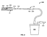
次に図2〜図3を参照すると、カテーテル100は、人の食道204内に位置する閉塞202を除去するように構成された例示的なシステム200内に示されている。この実施例では、閉塞202(一般には食物又は他のデブリであるが、血液又は血塊、粘液などのような他の閉塞であってもよい)は、食道204内に捕捉された状態になっている。
Next, referring to FIGS. 2 to 3, the
示されている実施形態では、カテーテル100は、内視鏡210を用いて閉塞202に送達される。内視鏡210は、概ね中空であり、かつ、カテーテル100が内視鏡210を介して閉塞202に送達されることを可能にする、チャネルを含む。カテーテルチューブ102の遠位端部104が所定の位置につくと、閉塞202が操作されるときに、内視鏡210は、引き抜かれることも、適所にとどまることもできる。
In the embodiment shown, the
カテーテル100のカテーテルチューブ102は、食物を繰り返しコア抜きすることなどによって閉塞202の容積を削減するために、前進させて、それにより、遠位端部104が閉塞202に衝撃を与えるように構成されている。容積が削減されると(図3に示すものなど)、閉塞202は、自然に食道204を通り抜け、人の胃206の中へ入ることができる。
The
例示的な実施形態では、カテーテルチューブ102は、少なくとも半硬質であるが可撓性を有しており、これにより、カテーテルチューブが、内視鏡を通しての送達中に、内視鏡が屈曲及び湾曲するのに応じて屈曲及び/又は湾曲することが可能である。これにより、カテーテルチューブ102は、所望の位置に挿入されるように、より正確に誘導されることができる。
In an exemplary embodiment, the
いくつかの実施例では、カテーテルチューブ102の遠位端部104は、閉塞202のコア抜きを補助するように構成されている。例えば、図4に示すように、カテーテルチューブ102の遠位端部104はテーパ状に形成されている。具体的には、遠位端部104は、カテーテルチューブ102の部分406の内径404よりも小さい内径402を含む。一実施例では、直径の差は、100分の1ミリメートル未満であり得る。他の寸法も可能である。加えて、カテーテルチューブ102の壁部は、図示されているように、遠位端部104まで延びるのに応じて薄くなり得る。
In some embodiments, the
この遠位端部104のテーパ形成により、遠位端部104によって形成された閉塞202のコア410が、カテーテルチューブ102を介してより容易に吸引されるようにすることができる。遠位端部104によって形成されたコアは典型的には部分406の直径よりも小さい直径を有するので、ポアズイユの法則によって示されるとおり、コアは、排出のためにカテーテルチューブ102を介してより容易に吸引されることができる。
The tapering of the
図5に示す別の描写では、カテーテルチューブ102は、より小さい直径を有する遠位端部104における第1の部分502と、より大きい直径を有するカテーテルチューブ102の残部に沿って延在する第2の部分504と、から形成されている。これにより、再び、第1の部分502によって作製される閉塞202のコアの直径をより小さくすることができ、その結果、コアは、カテーテルチューブ102の残部(即ち、第2の部分504)をより容易に通り抜けることができる。
In another depiction shown in FIG. 5, the
いくつかの実施例では、カテーテルチューブ102の遠位端部104の先端部508は、傾斜面取状及び/又は鋸歯状であり得る。先端部508は、閉塞202の破片を塊から削ぎ落として吸引をより良好に補助するように、鋸歯状縁部を含めて複数の形態を取ることができる。先端部508は、閉塞をコア抜きするのを助けることができる。
In some embodiments, the
例えば、図2に示したシステム200を再度参照すると、吸引源は、閉塞202のコアがカテーテルチューブ102を介して除去されることができるように、カテーテル100の近位端部106に適用され得る。具体的には、示されている実施例では、真空ライン220は、カテーテルチューブ102の近位端部106に連結され得る。真空ライン220は、収集キャニスタ222に連結され得、収集キャニスタ222は吸引ライン224に連結されている。吸引ライン224は、病院の真空源など、吸引源に連結されている。この構成では、カテーテルチューブ102によってコア抜きないしは別の方法で取り除かれた閉塞202の断片は、その後すぐに、カテーテルチューブ102に吸い上げられ、真空ライン220を通って、収集キャニスタ222に収集され得る。
For example, referring again to the
上述したように、カテーテルチューブ102内では、閉塞202の1つ以上のコアが詰まった状態になる可能性がある。このようなシナリオでは、詰まっているコアを取り除くために様々なデバイスが使用され得る。
As mentioned above, within the
例えば、次に図6を参照すると、例示的な注射器602は、例えば、吸引ライン嵌合接続部又はルアーロック式接続部を使用してカテーテル100の近位端部106に連結されている。本実施形態では、注射器602は、閉塞202のコア抜き中にカテーテルチューブ102内に空気を送達して、カテーテルチューブ102内にある閉塞202の部分を取り除く及び/又は除去するために使用される典型的な60ccの注射器であり得る。
For example, with reference to FIG. 6, the
この事例では、注射器602のプランジャは、注射器602内の空気をカテーテルチューブ102内に入れ、その中を移動させるように作動する。この空気は、チューブ内の障害物を取り除くために使用され得る。他の構成も可能である。例えば、水のジェットスプレーなど、他の種類の流体を使用して、チューブをきれいにする又は食物を破砕するのを助けるように使用されてもよい。
In this case, the plunger of the
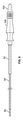
他の事例では、カテーテル100をきれいにするために、異なるデバイスが使用され得る。例えば、次に図7〜図8を参照すると、カテーテルチューブ102の中空内側部を通って嵌合するように寸法が決定されているスタイレット700が示されている。一般に、スタイレット700は、様々な機能を果たすために使用され得る。
In other cases, different devices may be used to clean the
例えば、スタイレット700は、閉塞202への送達中にカテーテル100を補剛するために使用され得る。更に、スタイレット700は、1つ以上のコアが詰まっているときに、カテーテルチューブ102をきれいにするためにカテーテルチューブ102を通って導入され得、プッシャロッドの機能を果たす。最後に、スタイレット700はまた、閉塞202を貫通して、コア抜き及び吸引のための巣を開始するために使用され得る。いくつかの実施例では、スタイレット700はまた、中実又は中空であり得る。
For example, the
この実施例では、スタイレット700は、カテーテル100の近位端部106に係合するように構成されているスタイレットノブ702を更に含む。近位端部106は、近位端部106がスタイレット700のスタイレットノブ702を係合できるようにするルアーテーパを含むように構成され得る。ねじ係合など、他の連結配置を使用することができる。
In this embodiment, the
図8に示すように、スタイレットノブ702は、カテーテルチューブ102の近位端部106に連結されている。この構成では、カテーテル100は、食道204内の所望の位置に送達され得る。そのとき、スタイレットノブ702は、スタイレット700が自由に移動できるように、近位端部106から係合解除され得る。この移動は、介助者がスタイレット700をカテーテルチューブ102内に押し込み、同カテーテルチューブから押し出して、一般には閉塞202を粉砕すること、及び/又は、スタイレット700をカテーテルチューブ102から完全に取り外すことを含み得る。
As shown in FIG. 8, the
スタイレット700がカテーテルチューブ102から取り外されるとき、上述したように、真空ライン220は、吸引のためにカテーテルチューブ102の近位端部106に接続され得る。
When the
図8に示すこの実施例では、カテーテルチューブ102は長さおよそ80.5インチであり、スタイレット700は長さおよそ84インチであるが、例えば、子供にはより短い長さ、大人にはより長い長さなどの、又は様々な長さの内視鏡、気管支鏡、若しくは結腸鏡を収容するための、多くの様々な長さが提供され得る。例示的なカテーテルチューブ102は、外径0.135インチ及び内径0.115インチである。スタイレット700は、外径0.105インチである。他の寸法を使用することもできる。
In this example shown in FIG. 8, the
他の実施形態では、カテーテルチューブ102は、長さ及び直径が可変であり得る。例えば、カテーテルチューブ102の別の実施形態は、外径0.093インチ及び内径0.082であり、任意の内視鏡の作業チャネル内で容易に導入及び摺動することができる。カテーテルチューブ102は、内視鏡を通して延在するのに十分な長さであり、長さは少なくとも120cmであるが、それより長くてもよい。
In other embodiments, the
スタイレット700は、直径が様々であり得るが、好ましい実施形態では、カテーテルチューブ102内で容易に導入及び摺動することができるように、外径0.070インチであり、また、スタイレット700が、カテーテルチューブ102の遠位端部104を越えて延在してカテーテルチューブ102をきれいにし、必要ならば、閉塞202の中に更に延在することができるように、カテーテルチューブ102よりわずかに長い。
The
カテーテルチューブ102は、任意の市販の内視鏡の作業チャネル(生検チャネル)に嵌合するように寸法が決定されている薄肉押出チューブから作製され得る。1つの例示的な材料は、Pebax 7233 SAである。別の可能な材料は、押出等級のPETGであろう。他の可能性は、ナイロン10又はナイロン12など、ポリアミド又は押出等級のナイロン若しくはデルリンであろう。
The
スタイレット700は、同じ又は類似の材料で作製され得る。例えば、カテーテルチューブ102及びスタイレット700は、スタイレット700が、摩擦を最小にしながらカテーテルチューブ102内に嵌合することができるように、同じ材料で作製され得る。しかしながら、他の材料及びそれぞれに異なる材料を使用することができる。
The
上記材料は、食物を取り除くであろうが、食道の壁部に不用意に接触したとしても、食道の壁部に重大な損傷を与えることはないであろう。 The material will remove food, but inadvertent contact with the esophageal wall will not cause significant damage to the esophageal wall.
次に図9〜図17を参照すると、別の例示的なデバイス900が示されている。デバイス900は、近位端部106にある吸引ポート902と、任意の商用内視鏡の生検チャネルを通して前進させられるように設計(例えば、傾斜面取)され、かつ、食道からの除去後にカテーテルチューブ102内に詰まっている食物を取り除くように、スタイレット700を収容することができる、遠位端部104と、を有するカテーテルチューブ102を含む。
Next, referring to FIGS. 9 to 17, another
図9に示すように、カテーテルチューブ102は、食物閉塞に到達するために、食道内に位置する内視鏡の生検チャネルを通って嵌合するように設計されているが、内視鏡に隣接して前進させることもでき、また内視鏡の補助なしで、経口で前進させることもできる。カテーテルチューブ102はまた、内視鏡の屈曲及び操縦に応じて湾曲可能かつ操縦可能であるが、キンクに耐え得る十分な剛性も有する。
As shown in FIG. 9, the
この実施例(図9及び図15を参照されたい)では、Y字の一方のアーム906が取り付けられ、吸引ポート902を形成する、Yフィッティング904が存在し、Y字のもう一方のアーム908はスタイレット700を収容している。
In this embodiment (see FIGS. 9 and 15), there is a Y fitting 904 to which one Y-shaped
スタイレット700を収容するアーム908の近位端部には圧縮封止部910、即ち、ゴム栓も存在し、その結果、スタイレット700がカテーテルチューブ102内にあるときに、近位端部から漏れる空気は最小化され、それにより、真空チューブの吸引及びスタイレットによるクリアランスが同時に起こり得る。圧縮封止部910が緩められると、スタイレット700は、スタイレット700のハンドル912を使用して、カテーテルチューブ102の中へ及び外へ容易に前進させられ得る。圧縮封止部910はまた、スタイレット700をカテーテルチューブ102のシャフトに沿った任意の位置に固定することもできる。
There is also a
この実施例では、キャップ914は、圧縮封止部910を適所に保持するように、アーム908の近位端部916上に螺着されている。スタイレット700をカテーテルチューブ102から取り外すと、圧縮封止部910は、いくつかの実施形態では、近位端部916を閉じるように構成されており、その結果、カテーテルチューブ102及び吸引ポート902を介して吸引ができるようになる。
In this embodiment, the
示されている実施例では、カテーテルチューブ102は、スタイレット700を完全に取り外した状態で機能することができ、スタイレット700はまた、必要に応じて導入され、カテーテルチューブ102内を任意の距離だけ前進させられることもできる。
In the embodiments shown, the
前述の実施形態と同様に、カテーテルチューブ102の遠位端部104は、食物を粉砕し、食物をコア抜きし、食物を削ぎ落とし、及び食物を吸引し得る。カテーテルチューブ102の壁部は、同チューブのより大きい管腔をより良好に収容するように薄くかつ硬質であってよい。スタイレット700は、必要ならばキンクを防止するのを補助するために、カテーテルチューブ102を支持することを補助し得る。したがって、スタイレット700は、吸引チューブをきれいにすることを補助することと、カテーテルチューブ102を補剛するスタイレットとして機能することとの両方を行い得る。
Similar to the embodiments described above, the
多くの代替設計も使用可能である。例えば、図18〜図19に示す別の設計では、スタイレット1800は、スタイレットがカテーテルチューブ内にあるときに吸引をより良好に適応させるようにスタイレットに沿って形成されたスプライン1804と共にスプライン形状1802を有し得る。即ち、スタイレット1800がカテーテルチューブ102内に配置されているときであっても、カテーテルチューブ102を介して吸引が提供されるように、空間1806がスプライン1804間に形成される。他の構成も可能である。
Many alternative designs are also available. For example, in another design shown in FIGS. 18-19, the
次に図20〜図21を参照すると、スタイレット2000の別の実施例が示されている。この実施例では、スタイレット2000は、ワイヤ2002とその端部2006に位置付けられたピストン2004からなる。ピストン2004は、スタイレット2000を、カテーテルチューブ102の中を通して駆動させて食道内の閉塞を係合するように、(モータなどによって)自動的に(及び/又は手動で)断続的に又は規則的な間隔で作動させられ得る。他の構成も可能である。
Next, with reference to FIGS. 20 to 21, another embodiment of
次に図22〜図24を参照すると、別の例示的なデバイス2200が示されている。デバイス2200は、図20〜図21の実施形態と類似しているが、デバイス2200は必ずしも吸引を必要としない点が異なっている。代わりに、デバイス2200は、ハンドル2202及びチューブ2204を含む。ハンドル2202は、方向2208において挿入又は抜き出し方向に(例えば、介助者の指又は親指で)移動され得るアクチュエータ部材2206を含む。
Next, with reference to FIGS. 22-24, another
アクチュエータ部材2206は、チューブ2204を通ってエジェクタピストン2402に至るワイヤ2210に連結されている。エジェクタピストン2402は、チューブ2204の遠位端部2406に形成された空洞部2404内に位置付けられている。チューブ2204の遠位端部2406は開口部2408を形成しており、同開口部は、介助者がハンドル2202及びそれに取り付けられたチューブ2204を移動させるにつれて障害物をコア抜きするないしは別の方法で刻むように寸法が決定されている。これは、例えば、チューブ2204の遠位端部2406によって刻まれ、空洞部2404内に受容される、障害物の断片によって達成される。
The
空洞部2404が充填されると、エジェクタピストン2402が、ワイヤ2210により、空洞部2404の中をチューブ2204の遠位端部2406に向かって移動させられて、開口部2408から食物を排出するように、介助者はアクチュエータ部材2206を移動させ得る。このプロセスは、障害物が取り除かれるまで複数回行われ得る。アクチュエータ部材2206は、後退位置に戻るように付勢され得る、及び/又はエジェクタピストン2402を後退位置に戻すように介助者の指によって反対の方向2208に単純に移動され得る。
When the
いくつかの実施例では、チューブ2204の遠位端部2406は、障害物をより容易にコア抜きするように構成され得る。例えば、遠位端部は、より鋭利になるように薄肉化又は鋸歯状化され得る。他の実施例では、デバイス2200のコア抜き効果を高めるために、ステンレス鋼チップなど、追加の特徴部が、これ(又は本明細書に開示される任意の他の実施形態)の遠位端部2406に付加され得る。
In some embodiments, the
いくつかの実施例では、チューブの内側表面は、その中を障害物のコアが通り抜けることをより容易に可能にするように構成され得る。例えば、チューブの内側表面は、コアの通過を促進し、コアの凝集を阻止するために、低摩擦材料又は潤滑性材料でコーティングされ得る。このような低摩擦材料の実施例としては、ポリビニルピロリドン及びヒアルロン酸が挙げられるが、これらに限定されない。このような材料は、典型的には、熱又は紫外光を用いて接着され得る。カテーテル102の外側表面もまた、内視鏡を通り抜けることを可能にするために、任意追加的に、低摩擦材料でコーティングされ得る。内側表面の異なるテーパ及び/又はチャネリングなど、他の機構も使用され得る。
In some embodiments, the inner surface of the tube may be configured to allow the core of the obstacle to pass through it more easily. For example, the inner surface of the tube can be coated with a low friction material or a lubricating material to facilitate the passage of the core and prevent the core from agglomerating. Examples of such low friction materials include, but are not limited to, polyvinylpyrrolidone and hyaluronic acid. Such materials can typically be adhered using heat or ultraviolet light. The outer surface of the
上述の実施例は、食道内の圧入に言及している。しかしながら、本明細書に記載のシステム及び方法を使用して、他の多くの類似の圧入に対処することができる。 The above examples refer to press-fitting in the esophagus. However, the systems and methods described herein can be used to address many other similar press fits.
例えば、人は食事中にむせることがあり、食物が気管に吸引されて詰まったり、又は肺内、具体的には気管支樹の任意の部分に詰まったりすることもある。また、粘液が気管支樹の任意の場所でトラップされた状態になることもあり、これにより、粘液栓が生じる。これが生じたとき、本明細書に記載の実施形態のうちの1つ以上が、前述の食物又は粘液をコア抜き及び吸引するために使用され得、このために、デバイスを、内視鏡に対立するものである可撓性又は硬質の気管支鏡の作業チャネルを通して配置する。 For example, a person may be swallowed during a meal, and food may be sucked into the trachea and clogged, or clogged in the lungs, specifically any part of the bronchial tree. Mucus can also be trapped anywhere in the bronchial tree, resulting in mucus plugs. When this occurs, one or more of the embodiments described herein can be used to core and aspirate the aforementioned food or mucus, for which the device is opposed to the endoscope. Place through the working channel of a flexible or rigid bronchoscope.
本明細書に記載の実施形態のうちの1つ以上はまた、消化管、具体的には食道、胃、小腸又は大腸における任意の場所で、トラップされた血液又は血塊をコア抜きし、吸引し、及び除去するためにも使用され得る。 One or more of the embodiments described herein also core and aspirate trapped blood or clots anywhere in the gastrointestinal tract, specifically the esophagus, stomach, small intestine or large intestine. , And can also be used to remove.
本明細書に記載の実施形態のうちの1つ以上はまた、肺器官系、即ち、気管又は肺における任意の場所で、即ち、気管支樹における任意の場所で、トラップされた食物、血液若しくは血塊、又は粘液若しく粘液栓をコア抜きし、吸引し、及び除去するためにも使用され得る。 One or more of the embodiments described herein are also trapped food, blood or blood clots anywhere in the pulmonary organ system, i.e., trachea or lung, i.e., anywhere in the bronchial tree. , Or mucus can also be used to core, aspirate, and remove mucus plugs.
本明細書に記載の実施形態のうちの1つ以上はまた、脈管系、即ち、大動脈若しくは大静脈、又は末梢血管系、即ち、末梢動脈若しくは末梢静脈における任意の場所で、血液若しくは血塊、又はアテローム若しくはアテローム性プラークをコア抜き及び除去するためにも使用され得る。石灰化プラークなどのより硬い物質をコア抜きするために、ステンレス鋼チップが吸引用カテーテルの端部に取り付けられ得る。 One or more of the embodiments described herein are also blood or blood clots, anywhere in the vascular system, i.e. the aorta or vena cava, or the peripheral vascular system, i.e. the peripheral arteries or veins. Alternatively, it can also be used to core and remove atheroma or atherosclerotic plaque. A stainless steel tip can be attached to the end of the suction catheter to core harder materials such as calcified plaque.
本明細書に記載の実施形態のうちの1つ以上はまた、心臓又は冠動脈における任意の場所で、血液若しくは血塊、又はアテローム若しくはアテローム性プラークをコア抜き及び除去するためにも使用され得る。石灰化プラークなどのより硬い物質をコア抜きするために、ステンレス鋼チップが吸引用カテーテルの端部に取り付けられ得る。 One or more of the embodiments described herein can also be used to core and remove blood or blood clots, or atheroma or atherogenic plaques, anywhere in the heart or coronary arteries. A stainless steel tip can be attached to the end of the suction catheter to core harder materials such as calcified plaque.
別の実施例では、本明細書に記載の実施形態のうちの1つ以上は、泌尿器系、具体的には、尿管、膀胱及び腎臓から腎石をコア抜き及び吸引するために使用され得る。石灰化した、ストルバイト、シュウ酸塩又は尿酸腎石などのより硬い物質をコア抜きするために、ステンレス鋼チップが吸引用カテーテルの端部に取り付けられ得る。 In another embodiment, one or more of the embodiments described herein can be used to core and aspirate kidney stones from the urinary system, specifically the ureter, bladder and kidneys. .. A stainless steel tip can be attached to the end of the suction catheter to core out calcified, harder substances such as struvite, oxalate or urate nephroliths.
更に別の実施例では、本明細書に記載の実施形態のうちの1つ以上は、胆管支樹(総胆管又は末梢管)に詰まっている胆石又は腫瘍をコア抜き及び除去するために使用され得る。より硬い物質は、吸引用カテーテルの端部にステンレス鋼チップを取り付けることによってコア抜きされ得る。 In yet another embodiment, one or more of the embodiments described herein are used to core and remove gallstones or tumors that are clogged in the bile duct branch (common bile duct or peripheral duct). obtain. Harder material can be cored by attaching a stainless steel tip to the end of the suction catheter.
様々な実施形態が本明細書に記載されているが、実施形態は単なる実施例であり、制限するものとして解釈すべきではない。 Although various embodiments are described herein, the embodiments are merely embodiments and should not be construed as limiting.
Claims (7)
The distal end of the catheter tube is configured to decore the food press-fit multiple times so that the food press-fit core is aspirated out of the esophagus through the catheter tube. The device of claim 1, wherein the dimensions have been determined.
Applications Claiming Priority (3)
| Application Number | Priority Date | Filing Date | Title |
|---|---|---|---|
| US201562260873P | 2015-11-30 | 2015-11-30 | |
| US62/260,873 | 2015-11-30 | ||
| PCT/US2016/063083 WO2017095682A1 (en) | 2015-11-30 | 2016-11-21 | Blockage removal |
Publications (3)
| Publication Number | Publication Date |
|---|---|
| JP2018537249A JP2018537249A (en) | 2018-12-20 |
| JP2018537249A5 JP2018537249A5 (en) | 2020-01-09 |
| JP6941620B2 true JP6941620B2 (en) | 2021-09-29 |
Family
ID=57543189
Family Applications (1)
| Application Number | Title | Priority Date | Filing Date |
|---|---|---|---|
| JP2018547254A Active JP6941620B2 (en) | 2015-11-30 | 2016-11-21 | Blockage removal |
Country Status (9)
| Country | Link |
|---|---|
| US (2) | US10722267B2 (en) |
| EP (1) | EP3383289A1 (en) |
| JP (1) | JP6941620B2 (en) |
| KR (1) | KR20180088853A (en) |
| CN (2) | CN108778164B (en) |
| AU (2) | AU2016362953A1 (en) |
| BR (1) | BR112018011092A2 (en) |
| CA (1) | CA3006739A1 (en) |
| WO (1) | WO2017095682A1 (en) |
Families Citing this family (7)
| Publication number | Priority date | Publication date | Assignee | Title |
|---|---|---|---|---|
| US11141177B2 (en) | 2015-11-30 | 2021-10-12 | Piranha Medical Llc | Blockage clearing devices, systems, and methods |
| EP3383289A1 (en) * | 2015-11-30 | 2018-10-10 | Piranha Medical, LLC | Blockage removal |
| US10881276B2 (en) * | 2016-01-04 | 2021-01-05 | Endovate Llc | Overtube device and method of use |
| CN112533550A (en) * | 2018-07-24 | 2021-03-19 | 半影公司 | Device and method for controlled clot aspiration |
| WO2020092959A1 (en) * | 2018-11-03 | 2020-05-07 | Piranha Medical, LLC | Blockage clearing devices, systems, and methods |
| KR102251873B1 (en) | 2019-02-22 | 2021-05-13 | 배정환 | Esophageal foreign substances removal device |
| WO2023088808A1 (en) * | 2021-11-22 | 2023-05-25 | Koninklijke Philips N.V. | Apparatus with stylet for kink navigation or the like in a catheter lead lumen |
Family Cites Families (70)
| Publication number | Priority date | Publication date | Assignee | Title |
|---|---|---|---|---|
| US4754755A (en) | 1984-05-14 | 1988-07-05 | Husted Royce Hill | Catheter with a rotary blade |
| US4715848A (en) | 1985-04-15 | 1987-12-29 | Beroza Gregory A | Gastro-intestinal lavage system and method |
| EP0310685A1 (en) | 1985-11-22 | 1989-04-12 | Kontron-Holding Ag | Angioplasty catheter |
| US4898575A (en) | 1987-08-31 | 1990-02-06 | Medinnovations, Inc. | Guide wire following tunneling catheter system and method for transluminal arterial atherectomy |
| US5033466A (en) | 1989-02-28 | 1991-07-23 | Weymuller Jr Ernest | Doble-cuffed endotracheal tube |
| US5114399A (en) | 1990-10-01 | 1992-05-19 | Intramed Laboratories | Surgical device |
| US5197949A (en) | 1991-01-22 | 1993-03-30 | Kraivit Angsupanich | Suction irrigation device with a scraper |
| JP2802244B2 (en) | 1994-08-29 | 1998-09-24 | オリンパス光学工業株式会社 | Endoscope sheath |
| US5928218A (en) * | 1994-12-16 | 1999-07-27 | Gelbfish; Gary A. | Medical material removal method and associated instrumentation |
| WO1997018745A2 (en) * | 1995-11-20 | 1997-05-29 | Storz Endoskop Gmbh | Shaving or cutting instrument |
| DE59705394D1 (en) * | 1996-04-25 | 2001-12-20 | Storz Karl Gmbh & Co Kg | SURGICAL INSTRUMENT SYSTEM |
| US5931831A (en) | 1996-07-09 | 1999-08-03 | Linder; Gerald S. | Dual-lumen suction catheter with smaller diameter vent lumen having multiple apertures therein |
| US5921971A (en) | 1996-09-13 | 1999-07-13 | Boston Scientific Corporation | Single operator exchange biliary catheter |
| US5741269A (en) | 1996-11-18 | 1998-04-21 | Mccredy; Doug | Medical vacuum device |
| US5782837A (en) | 1997-04-23 | 1998-07-21 | York; Richard | Esophagus clearing device |
| ATE404123T1 (en) | 1997-11-12 | 2008-08-15 | Genesis Technologies Llc | DEVICE FOR REMOVAL OF OCCLUSIONS IN BIOLOGICAL PASSAGES |
| US6165199A (en) * | 1999-01-12 | 2000-12-26 | Coaxia, Inc. | Medical device for removing thromboembolic material from cerebral arteries and methods of use |
| US6689062B1 (en) | 1999-11-23 | 2004-02-10 | Microaccess Medical Systems, Inc. | Method and apparatus for transesophageal cardiovascular procedures |
| US6361540B1 (en) | 2000-04-06 | 2002-03-26 | Michael W. L. Gauderer | Apparatus for removal of esophageal coins and similarly shaped objects |
| US6986773B1 (en) | 2000-10-11 | 2006-01-17 | Edward Manougian | Human airway clearing tool |
| US20040087936A1 (en) * | 2000-11-16 | 2004-05-06 | Barrx, Inc. | System and method for treating abnormal tissue in an organ having a layered tissue structure |
| US6840909B2 (en) | 2002-03-25 | 2005-01-11 | Acueity, Inc. | Apparatus and method for intraductal cytology |
| JP4667038B2 (en) | 2002-09-06 | 2011-04-06 | シー・アール・バード・インク | Endoscope attachment adapter |
| EP1603452B1 (en) | 2003-03-17 | 2017-05-17 | Covidien LP | Endoscopic tissue removal apparatus |
| US20070208252A1 (en) * | 2004-04-21 | 2007-09-06 | Acclarent, Inc. | Systems and methods for performing image guided procedures within the ear, nose, throat and paranasal sinuses |
| US9101384B2 (en) | 2004-04-21 | 2015-08-11 | Acclarent, Inc. | Devices, systems and methods for diagnosing and treating sinusitis and other disorders of the ears, Nose and/or throat |
| US20060063973A1 (en) | 2004-04-21 | 2006-03-23 | Acclarent, Inc. | Methods and apparatus for treating disorders of the ear, nose and throat |
| US7654997B2 (en) | 2004-04-21 | 2010-02-02 | Acclarent, Inc. | Devices, systems and methods for diagnosing and treating sinusitus and other disorders of the ears, nose and/or throat |
| US8057484B2 (en) | 2004-05-25 | 2011-11-15 | U.S. Endoscopy Group, Inc. | Retrieval device |
| US20060293612A1 (en) | 2004-06-24 | 2006-12-28 | Boston Scientific Scimed, Inc. | Apparatus and method for treating occluded vasculature |
| WO2006037084A1 (en) | 2004-09-28 | 2006-04-06 | Cordis Corporation | Thin film medical device and delivery system |
| US8016785B2 (en) | 2004-12-02 | 2011-09-13 | Chek-Med Systems, Inc. | Gastrojejunal feeding tube |
| US7220253B2 (en) | 2004-12-02 | 2007-05-22 | Chek-Med Systems, Inc. | Gastrojejunal feeding tube |
| CN101703387B (en) | 2005-01-06 | 2013-10-23 | G.I.视频有限公司 | Gastrointestinal tool over guiding element |
| US20070250149A1 (en) * | 2006-04-21 | 2007-10-25 | Abbott Laboratories | Stiffening Support Catheters and Methods for Using the Same |
| US20090018566A1 (en) * | 2006-06-30 | 2009-01-15 | Artheromed, Inc. | Atherectomy devices, systems, and methods |
| EP2068730B1 (en) | 2006-10-04 | 2016-11-23 | Boston Scientific Limited | Interventional catheters |
| US7878983B2 (en) | 2006-10-26 | 2011-02-01 | Wilson-Cook Medical Inc. | Biopsy collection device |
| US20080103508A1 (en) | 2006-11-01 | 2008-05-01 | Ali Serdar Karakurum | Apparatus and method for removal of foreign matter from a patient |
| US9421071B2 (en) | 2006-12-01 | 2016-08-23 | Boston Scientific Scimed, Inc. | Direct drive methods |
| KR100834042B1 (en) | 2007-02-23 | 2008-05-30 | 양건태 | Device for thorn/mucous discharge removal |
| US8573218B2 (en) | 2007-03-07 | 2013-11-05 | Michael John RUTTER | Tracheostomy tube |
| US20080243137A1 (en) | 2007-03-30 | 2008-10-02 | D Angelo David W | System and methods for clearance of obstructions |
| US8591521B2 (en) | 2007-06-08 | 2013-11-26 | United States Endoscopy Group, Inc. | Retrieval device |
| JP5460582B2 (en) | 2007-06-08 | 2014-04-02 | ユー.エス. エンドスコピー グループ, インコーポレイテッド | Collection device |
| US8262645B2 (en) | 2007-11-21 | 2012-09-11 | Actuated Medical, Inc. | Devices for clearing blockages in in-situ artificial lumens |
| US10307340B2 (en) * | 2007-11-21 | 2019-06-04 | Actuated Medical, Inc. | Devices for clearing blockages in artificial and natural lumens |
| US8876838B2 (en) | 2008-03-07 | 2014-11-04 | Kevin Winiarski | Anti-choking device |
| JP2011526529A (en) | 2008-07-02 | 2011-10-13 | アンジオスライド リミテッド | Balloon catheter system and method of use thereof |
| US20100016885A1 (en) | 2008-07-21 | 2010-01-21 | Eidenschink Tracee E J | Device to close openings in body tissue |
| US20100204672A1 (en) * | 2009-02-12 | 2010-08-12 | Penumra, Inc. | System and method for treating ischemic stroke |
| CN201642262U (en) | 2010-02-11 | 2010-11-24 | 吴彩霞 | Mouth cavity and esophagus cleaning device |
| EP2568872B1 (en) | 2010-05-11 | 2017-09-06 | Cook Medical Technologies LLC | Biliary access sheath |
| JP5777936B2 (en) * | 2010-07-16 | 2015-09-09 | テルモ株式会社 | Suction catheter |
| RU2013118358A (en) | 2010-09-22 | 2014-10-27 | Аккларент, Инк. | MEDICAL DEVICE AND METHOD FOR TREATING SINUS HOLE |
| US9730756B2 (en) | 2011-02-24 | 2017-08-15 | Eximo Medical Ltd. | Hybrid catheter for vascular intervention |
| US9549761B2 (en) | 2011-06-17 | 2017-01-24 | Research Institute At Nationwide Children's Hospital | Endoscopic foreign body retrieval |
| US20140309673A1 (en) * | 2011-11-11 | 2014-10-16 | Nathan John Dacuycuy | Devices for removing vessel occlusions |
| US20150057517A1 (en) | 2012-03-27 | 2015-02-26 | University Of Utah Research Foundation | Sample capture device and systems and methods of using same |
| WO2014089028A1 (en) * | 2012-12-04 | 2014-06-12 | Endoclear Llc | Suction cleaning devices, systems and methods |
| JP2014140465A (en) * | 2013-01-23 | 2014-08-07 | Terumo Corp | Balloon catheter for removing solid matter |
| CN105873530B (en) * | 2013-09-18 | 2019-11-08 | 埃克斯爱博凯斯有限公司 | System and method for passing through and treating obstruction |
| CN103932756B (en) * | 2014-03-21 | 2016-07-20 | 吴智群 | A kind of Biochemical analyzer system |
| CN104055561A (en) * | 2014-07-14 | 2014-09-24 | 上海市肺科医院 | Sucked type material taking device for bronchoscope |
| US10149691B2 (en) | 2014-09-19 | 2018-12-11 | Endochoice, Inc. | Method of attaching a mesh to a coated loop member of a surgical snare device |
| US10045758B2 (en) | 2014-11-26 | 2018-08-14 | Visura Technologies, LLC | Apparatus, systems and methods for proper transesophageal echocardiography probe positioning by using camera for ultrasound imaging |
| WO2016210381A1 (en) | 2015-06-26 | 2016-12-29 | Endovate Llc | Endoscope device and method of use |
| EP3383289A1 (en) | 2015-11-30 | 2018-10-10 | Piranha Medical, LLC | Blockage removal |
| US11141177B2 (en) | 2015-11-30 | 2021-10-12 | Piranha Medical Llc | Blockage clearing devices, systems, and methods |
| WO2018222948A1 (en) | 2017-05-31 | 2018-12-06 | Ganz Robert A | Blockage clearing devices, systems, and methods |
-
2016
- 2016-11-21 EP EP16810172.3A patent/EP3383289A1/en not_active Withdrawn
- 2016-11-21 CN CN201680076552.8A patent/CN108778164B/en not_active Expired - Fee Related
- 2016-11-21 BR BR112018011092A patent/BR112018011092A2/en not_active Application Discontinuation
- 2016-11-21 AU AU2016362953A patent/AU2016362953A1/en not_active Abandoned
- 2016-11-21 KR KR1020187018116A patent/KR20180088853A/en unknown
- 2016-11-21 CA CA3006739A patent/CA3006739A1/en not_active Abandoned
- 2016-11-21 CN CN202110420538.8A patent/CN113576632A/en active Pending
- 2016-11-21 JP JP2018547254A patent/JP6941620B2/en active Active
- 2016-11-21 WO PCT/US2016/063083 patent/WO2017095682A1/en active Application Filing
- 2016-11-21 US US15/356,975 patent/US10722267B2/en active Active
-
2020
- 2020-06-12 US US16/900,614 patent/US20200297386A1/en not_active Abandoned
-
2021
- 2021-09-16 AU AU2021232765A patent/AU2021232765A1/en not_active Abandoned
Also Published As
| Publication number | Publication date |
|---|---|
| CN108778164B (en) | 2021-05-11 |
| KR20180088853A (en) | 2018-08-07 |
| BR112018011092A2 (en) | 2018-11-21 |
| AU2016362953A1 (en) | 2018-06-14 |
| US20170150993A1 (en) | 2017-06-01 |
| US20200297386A1 (en) | 2020-09-24 |
| CN113576632A (en) | 2021-11-02 |
| AU2021232765A1 (en) | 2021-10-14 |
| EP3383289A1 (en) | 2018-10-10 |
| CN108778164A (en) | 2018-11-09 |
| JP2018537249A (en) | 2018-12-20 |
| CA3006739A1 (en) | 2017-06-08 |
| US10722267B2 (en) | 2020-07-28 |
| WO2017095682A1 (en) | 2017-06-08 |
Similar Documents
| Publication | Publication Date | Title |
|---|---|---|
| JP6941620B2 (en) | Blockage removal | |
| US10806849B2 (en) | Devices and methods for transnasal irrigation or suctioning of the sinuses | |
| JP2020522310A (en) | Blockage removal device, system and method | |
| US10166369B2 (en) | Devices and method for maxillary sinus lavage | |
| JP6612125B2 (en) | Balloon dilatation catheter system for sinus treatment and irrigation | |
| JP2006087643A (en) | Apparatus for sucking foreign substance from blood vessel | |
| JP2006297094A (en) | Polypectomy device and method of use | |
| Ko et al. | Review of food bolus management | |
| US20210251645A1 (en) | Blockage clearing devices, systems, and methods | |
| WO2010126786A1 (en) | Aspiration catheter with thrombus removing device | |
| US20220023563A1 (en) | Blockage clearing devices, systems, and methods | |
| CN108348713A (en) | The Clean- suction brush of tracheal strips | |
| US9131830B2 (en) | Devices, systems, and methods for removing empyema from a pleural cavity | |
| US20220226016A1 (en) | Material removal from within a patient | |
| US10932762B2 (en) | Biopsy sampling device | |
| US20240108412A1 (en) | Tool guiding device for kidney stone treatment apparatus | |
| US20090287056A1 (en) | Device to facilitate suctioning of fluid during gastrointestinal endoscopy |
Legal Events
| Date | Code | Title | Description |
|---|---|---|---|
| A521 | Request for written amendment filed |
Free format text: JAPANESE INTERMEDIATE CODE: A523 Effective date: 20191118 |
|
| A621 | Written request for application examination |
Free format text: JAPANESE INTERMEDIATE CODE: A621 Effective date: 20191118 |
|
| A977 | Report on retrieval |
Free format text: JAPANESE INTERMEDIATE CODE: A971007 Effective date: 20201217 |
|
| A131 | Notification of reasons for refusal |
Free format text: JAPANESE INTERMEDIATE CODE: A131 Effective date: 20210105 |
|
| A601 | Written request for extension of time |
Free format text: JAPANESE INTERMEDIATE CODE: A601 Effective date: 20210402 |
|
| A521 | Request for written amendment filed |
Free format text: JAPANESE INTERMEDIATE CODE: A523 Effective date: 20210705 |
|
| TRDD | Decision of grant or rejection written | ||
| A01 | Written decision to grant a patent or to grant a registration (utility model) |
Free format text: JAPANESE INTERMEDIATE CODE: A01 Effective date: 20210810 |
|
| A61 | First payment of annual fees (during grant procedure) |
Free format text: JAPANESE INTERMEDIATE CODE: A61 Effective date: 20210906 |
|
| R150 | Certificate of patent or registration of utility model |
Ref document number: 6941620 Country of ref document: JP Free format text: JAPANESE INTERMEDIATE CODE: R150 |