JP6908381B2 - Antibodies to MUC1-C / extracellular domain (MUC1-C / ECD) - Google Patents
Antibodies to MUC1-C / extracellular domain (MUC1-C / ECD) Download PDFInfo
- Publication number
- JP6908381B2 JP6908381B2 JP2016549460A JP2016549460A JP6908381B2 JP 6908381 B2 JP6908381 B2 JP 6908381B2 JP 2016549460 A JP2016549460 A JP 2016549460A JP 2016549460 A JP2016549460 A JP 2016549460A JP 6908381 B2 JP6908381 B2 JP 6908381B2
- Authority
- JP
- Japan
- Prior art keywords
- antibody
- cells
- muc1
- cell
- cancer
- Prior art date
- Legal status (The legal status is an assumption and is not a legal conclusion. Google has not performed a legal analysis and makes no representation as to the accuracy of the status listed.)
- Active
Links
- 210000004027 cell Anatomy 0.000 claims description 500
- 150000007523 nucleic acids Chemical class 0.000 claims description 90
- 108090000765 processed proteins & peptides Proteins 0.000 claims description 83
- 206010028980 Neoplasm Diseases 0.000 claims description 81
- 102000039446 nucleic acids Human genes 0.000 claims description 60
- 108020004707 nucleic acids Proteins 0.000 claims description 60
- 201000011510 cancer Diseases 0.000 claims description 56
- 239000000427 antigen Substances 0.000 claims description 55
- 108091007433 antigens Proteins 0.000 claims description 55
- 102000036639 antigens Human genes 0.000 claims description 55
- 238000011282 treatment Methods 0.000 claims description 54
- 102000004196 processed proteins & peptides Human genes 0.000 claims description 53
- 239000003795 chemical substances by application Substances 0.000 claims description 44
- 150000001413 amino acids Chemical group 0.000 claims description 42
- 239000002246 antineoplastic agent Substances 0.000 claims description 37
- 102000004190 Enzymes Human genes 0.000 claims description 36
- 108090000790 Enzymes Proteins 0.000 claims description 36
- 206010006187 Breast cancer Diseases 0.000 claims description 31
- 206010035226 Plasma cell myeloma Diseases 0.000 claims description 31
- 208000026310 Breast neoplasm Diseases 0.000 claims description 30
- 229920001184 polypeptide Polymers 0.000 claims description 30
- 201000000050 myeloid neoplasm Diseases 0.000 claims description 26
- 210000004881 tumor cell Anatomy 0.000 claims description 26
- 239000003814 drug Substances 0.000 claims description 24
- 238000002965 ELISA Methods 0.000 claims description 22
- 239000002502 liposome Substances 0.000 claims description 21
- 230000009257 reactivity Effects 0.000 claims description 21
- 229940079593 drug Drugs 0.000 claims description 20
- 229940041181 antineoplastic drug Drugs 0.000 claims description 16
- 238000002560 therapeutic procedure Methods 0.000 claims description 16
- 239000012634 fragment Substances 0.000 claims description 15
- 229940127089 cytotoxic agent Drugs 0.000 claims description 11
- 239000003053 toxin Substances 0.000 claims description 11
- 231100000765 toxin Toxicity 0.000 claims description 11
- 206010009944 Colon cancer Diseases 0.000 claims description 9
- 239000008194 pharmaceutical composition Substances 0.000 claims description 9
- 102000004127 Cytokines Human genes 0.000 claims description 8
- 108090000695 Cytokines Proteins 0.000 claims description 8
- FWMNVWWHGCHHJJ-SKKKGAJSSA-N 4-amino-1-[(2r)-6-amino-2-[[(2r)-2-[[(2r)-2-[[(2r)-2-amino-3-phenylpropanoyl]amino]-3-phenylpropanoyl]amino]-4-methylpentanoyl]amino]hexanoyl]piperidine-4-carboxylic acid Chemical compound C([C@H](C(=O)N[C@H](CC(C)C)C(=O)N[C@H](CCCCN)C(=O)N1CCC(N)(CC1)C(O)=O)NC(=O)[C@H](N)CC=1C=CC=CC=1)C1=CC=CC=C1 FWMNVWWHGCHHJJ-SKKKGAJSSA-N 0.000 claims description 7
- 208000000453 Skin Neoplasms Diseases 0.000 claims description 7
- 208000032839 leukemia Diseases 0.000 claims description 7
- 208000020816 lung neoplasm Diseases 0.000 claims description 7
- 201000000849 skin cancer Diseases 0.000 claims description 7
- 206010058467 Lung neoplasm malignant Diseases 0.000 claims description 6
- 206010033128 Ovarian cancer Diseases 0.000 claims description 6
- 206010061535 Ovarian neoplasm Diseases 0.000 claims description 6
- 201000005202 lung cancer Diseases 0.000 claims description 6
- 239000002105 nanoparticle Substances 0.000 claims description 6
- 206010008342 Cervix carcinoma Diseases 0.000 claims description 5
- 108010054477 Immunoglobulin Fab Fragments Proteins 0.000 claims description 5
- 102000001706 Immunoglobulin Fab Fragments Human genes 0.000 claims description 5
- CKLJMWTZIZZHCS-REOHCLBHSA-N L-aspartic acid Chemical compound OC(=O)[C@@H](N)CC(O)=O CKLJMWTZIZZHCS-REOHCLBHSA-N 0.000 claims description 5
- 208000034578 Multiple myelomas Diseases 0.000 claims description 5
- 206010061902 Pancreatic neoplasm Diseases 0.000 claims description 5
- 208000005718 Stomach Neoplasms Diseases 0.000 claims description 5
- 208000024313 Testicular Neoplasms Diseases 0.000 claims description 5
- 206010057644 Testis cancer Diseases 0.000 claims description 5
- 208000006105 Uterine Cervical Neoplasms Diseases 0.000 claims description 5
- 201000010881 cervical cancer Diseases 0.000 claims description 5
- 208000029742 colonic neoplasm Diseases 0.000 claims description 5
- 206010017758 gastric cancer Diseases 0.000 claims description 5
- 238000009169 immunotherapy Methods 0.000 claims description 5
- 230000003834 intracellular effect Effects 0.000 claims description 5
- 201000007270 liver cancer Diseases 0.000 claims description 5
- 208000014018 liver neoplasm Diseases 0.000 claims description 5
- 239000006249 magnetic particle Substances 0.000 claims description 5
- 208000015486 malignant pancreatic neoplasm Diseases 0.000 claims description 5
- 201000002528 pancreatic cancer Diseases 0.000 claims description 5
- 208000008443 pancreatic carcinoma Diseases 0.000 claims description 5
- 201000011549 stomach cancer Diseases 0.000 claims description 5
- 201000003120 testicular cancer Diseases 0.000 claims description 5
- 208000031261 Acute myeloid leukaemia Diseases 0.000 claims description 4
- 208000003174 Brain Neoplasms Diseases 0.000 claims description 4
- 208000000461 Esophageal Neoplasms Diseases 0.000 claims description 4
- 102100034256 Mucin-1 Human genes 0.000 claims description 4
- 208000033776 Myeloid Acute Leukemia Diseases 0.000 claims description 4
- 206010030155 Oesophageal carcinoma Diseases 0.000 claims description 4
- 208000015634 Rectal Neoplasms Diseases 0.000 claims description 4
- 208000002495 Uterine Neoplasms Diseases 0.000 claims description 4
- 235000003704 aspartic acid Nutrition 0.000 claims description 4
- OQFSQFPPLPISGP-UHFFFAOYSA-N beta-carboxyaspartic acid Natural products OC(=O)C(N)C(C(O)=O)C(O)=O OQFSQFPPLPISGP-UHFFFAOYSA-N 0.000 claims description 4
- 238000002512 chemotherapy Methods 0.000 claims description 4
- 201000004101 esophageal cancer Diseases 0.000 claims description 4
- 201000010536 head and neck cancer Diseases 0.000 claims description 4
- 208000014829 head and neck neoplasm Diseases 0.000 claims description 4
- 238000001794 hormone therapy Methods 0.000 claims description 4
- 230000035772 mutation Effects 0.000 claims description 4
- 238000001959 radiotherapy Methods 0.000 claims description 4
- 206010038038 rectal cancer Diseases 0.000 claims description 4
- 201000001275 rectum cancer Diseases 0.000 claims description 4
- 230000002829 reductive effect Effects 0.000 claims description 4
- 208000016691 refractory malignant neoplasm Diseases 0.000 claims description 4
- 206010046766 uterine cancer Diseases 0.000 claims description 4
- 208000032791 BCR-ABL1 positive chronic myelogenous leukemia Diseases 0.000 claims description 3
- 208000010833 Chronic myeloid leukaemia Diseases 0.000 claims description 3
- 208000033761 Myelogenous Chronic BCR-ABL Positive Leukemia Diseases 0.000 claims description 3
- 206010038111 Recurrent cancer Diseases 0.000 claims description 3
- 208000037819 metastatic cancer Diseases 0.000 claims description 3
- 208000011575 metastatic malignant neoplasm Diseases 0.000 claims description 3
- 230000002860 competitive effect Effects 0.000 claims description 2
- 101001133056 Homo sapiens Mucin-1 Proteins 0.000 claims 3
- 229930195712 glutamate Natural products 0.000 claims 1
- WHUUTDBJXJRKMK-VKHMYHEASA-L glutamate group Chemical group N[C@@H](CCC(=O)[O-])C(=O)[O-] WHUUTDBJXJRKMK-VKHMYHEASA-L 0.000 claims 1
- 108090000623 proteins and genes Proteins 0.000 description 244
- 238000000034 method Methods 0.000 description 142
- 102000004169 proteins and genes Human genes 0.000 description 130
- 235000018102 proteins Nutrition 0.000 description 120
- 239000013598 vector Substances 0.000 description 93
- 108010008707 Mucin-1 Proteins 0.000 description 89
- 102000007298 Mucin-1 Human genes 0.000 description 88
- 230000027455 binding Effects 0.000 description 66
- 238000009739 binding Methods 0.000 description 65
- 230000014509 gene expression Effects 0.000 description 61
- 241000701161 unidentified adenovirus Species 0.000 description 61
- 229940027941 immunoglobulin g Drugs 0.000 description 52
- 239000000203 mixture Substances 0.000 description 47
- 235000001014 amino acid Nutrition 0.000 description 45
- 241000700605 Viruses Species 0.000 description 44
- 229940024606 amino acid Drugs 0.000 description 42
- 108020004414 DNA Proteins 0.000 description 41
- 210000001519 tissue Anatomy 0.000 description 39
- 229940088598 enzyme Drugs 0.000 description 34
- 239000012103 Alexa Fluor 488 Substances 0.000 description 33
- 238000004519 manufacturing process Methods 0.000 description 33
- 108091032973 (ribonucleotides)n+m Proteins 0.000 description 32
- 241000699666 Mus <mouse, genus> Species 0.000 description 31
- 239000003446 ligand Substances 0.000 description 31
- 102000005962 receptors Human genes 0.000 description 31
- 108020003175 receptors Proteins 0.000 description 31
- 108091028043 Nucleic acid sequence Proteins 0.000 description 29
- 210000004408 hybridoma Anatomy 0.000 description 29
- 239000003550 marker Substances 0.000 description 28
- 239000000523 sample Substances 0.000 description 28
- WSFSSNUMVMOOMR-UHFFFAOYSA-N Formaldehyde Chemical compound O=C WSFSSNUMVMOOMR-UHFFFAOYSA-N 0.000 description 26
- 238000012546 transfer Methods 0.000 description 25
- 210000001744 T-lymphocyte Anatomy 0.000 description 24
- 208000015181 infectious disease Diseases 0.000 description 24
- 230000015572 biosynthetic process Effects 0.000 description 23
- 239000003623 enhancer Substances 0.000 description 23
- 239000000047 product Substances 0.000 description 23
- 238000006243 chemical reaction Methods 0.000 description 22
- 230000003612 virological effect Effects 0.000 description 22
- 239000000562 conjugate Substances 0.000 description 21
- 238000001514 detection method Methods 0.000 description 21
- LOKCTEFSRHRXRJ-UHFFFAOYSA-I dipotassium trisodium dihydrogen phosphate hydrogen phosphate dichloride Chemical compound P(=O)(O)(O)[O-].[K+].P(=O)(O)([O-])[O-].[Na+].[Na+].[Cl-].[K+].[Cl-].[Na+] LOKCTEFSRHRXRJ-UHFFFAOYSA-I 0.000 description 21
- 230000006870 function Effects 0.000 description 21
- 239000002953 phosphate buffered saline Substances 0.000 description 21
- 238000010186 staining Methods 0.000 description 20
- 210000003719 b-lymphocyte Anatomy 0.000 description 19
- 238000011534 incubation Methods 0.000 description 19
- 239000012528 membrane Substances 0.000 description 19
- 230000001105 regulatory effect Effects 0.000 description 19
- YBJHBAHKTGYVGT-ZKWXMUAHSA-N (+)-Biotin Chemical compound N1C(=O)N[C@@H]2[C@H](CCCCC(=O)O)SC[C@@H]21 YBJHBAHKTGYVGT-ZKWXMUAHSA-N 0.000 description 18
- 238000004806 packaging method and process Methods 0.000 description 18
- -1 radioisotope Proteins 0.000 description 18
- 238000003556 assay Methods 0.000 description 17
- 230000004927 fusion Effects 0.000 description 17
- 238000001727 in vivo Methods 0.000 description 17
- 230000001404 mediated effect Effects 0.000 description 17
- 210000004379 membrane Anatomy 0.000 description 17
- 208000002154 non-small cell lung carcinoma Diseases 0.000 description 17
- 230000001177 retroviral effect Effects 0.000 description 17
- 230000001225 therapeutic effect Effects 0.000 description 17
- 238000013518 transcription Methods 0.000 description 17
- 230000035897 transcription Effects 0.000 description 17
- 239000013603 viral vector Substances 0.000 description 17
- 238000001262 western blot Methods 0.000 description 17
- 241000283707 Capra Species 0.000 description 16
- 238000000338 in vitro Methods 0.000 description 16
- 238000003757 reverse transcription PCR Methods 0.000 description 16
- 210000002845 virion Anatomy 0.000 description 16
- 108010019670 Chimeric Antigen Receptors Proteins 0.000 description 15
- 108060003951 Immunoglobulin Proteins 0.000 description 15
- 241001465754 Metazoa Species 0.000 description 15
- 241000700584 Simplexvirus Species 0.000 description 15
- 102000018358 immunoglobulin Human genes 0.000 description 15
- 230000010076 replication Effects 0.000 description 15
- 241001430294 unidentified retrovirus Species 0.000 description 15
- 230000008901 benefit Effects 0.000 description 14
- 230000008045 co-localization Effects 0.000 description 14
- 238000010166 immunofluorescence Methods 0.000 description 14
- 230000014616 translation Effects 0.000 description 14
- 241000699670 Mus sp. Species 0.000 description 13
- 239000006146 Roswell Park Memorial Institute medium Substances 0.000 description 13
- 230000002950 deficient Effects 0.000 description 13
- 238000001476 gene delivery Methods 0.000 description 13
- 230000012010 growth Effects 0.000 description 13
- 239000000243 solution Substances 0.000 description 13
- 241000894007 species Species 0.000 description 13
- 102100035360 Cerebellar degeneration-related antigen 1 Human genes 0.000 description 12
- 238000003776 cleavage reaction Methods 0.000 description 12
- 238000011161 development Methods 0.000 description 12
- 230000018109 developmental process Effects 0.000 description 12
- 230000000694 effects Effects 0.000 description 12
- 210000001163 endosome Anatomy 0.000 description 12
- 239000013604 expression vector Substances 0.000 description 12
- 230000002458 infectious effect Effects 0.000 description 12
- 230000008488 polyadenylation Effects 0.000 description 12
- 230000002441 reversible effect Effects 0.000 description 12
- 238000012360 testing method Methods 0.000 description 12
- 102100021244 Integral membrane protein GPR180 Human genes 0.000 description 11
- 102100024952 Protein CBFA2T1 Human genes 0.000 description 11
- 125000003275 alpha amino acid group Chemical group 0.000 description 11
- 239000000611 antibody drug conjugate Substances 0.000 description 11
- 229940049595 antibody-drug conjugate Drugs 0.000 description 11
- 230000010261 cell growth Effects 0.000 description 11
- 239000003431 cross linking reagent Substances 0.000 description 11
- 238000000684 flow cytometry Methods 0.000 description 11
- 238000002360 preparation method Methods 0.000 description 11
- 230000007017 scission Effects 0.000 description 11
- 230000009466 transformation Effects 0.000 description 11
- 238000013519 translation Methods 0.000 description 11
- 210000000234 capsid Anatomy 0.000 description 10
- 150000001875 compounds Chemical class 0.000 description 10
- 230000001472 cytotoxic effect Effects 0.000 description 10
- 238000004520 electroporation Methods 0.000 description 10
- 108020001507 fusion proteins Proteins 0.000 description 10
- 102000037865 fusion proteins Human genes 0.000 description 10
- 239000000499 gel Substances 0.000 description 10
- 230000003053 immunization Effects 0.000 description 10
- 238000002649 immunization Methods 0.000 description 10
- 229920001223 polyethylene glycol Polymers 0.000 description 10
- 230000008569 process Effects 0.000 description 10
- 239000000126 substance Substances 0.000 description 10
- 238000001890 transfection Methods 0.000 description 10
- 239000002202 Polyethylene glycol Substances 0.000 description 9
- 229960002685 biotin Drugs 0.000 description 9
- 235000020958 biotin Nutrition 0.000 description 9
- 239000011616 biotin Substances 0.000 description 9
- 210000000170 cell membrane Anatomy 0.000 description 9
- 239000013642 negative control Substances 0.000 description 9
- 238000000746 purification Methods 0.000 description 9
- 108700012359 toxins Proteins 0.000 description 9
- 238000005406 washing Methods 0.000 description 9
- 238000012286 ELISA Assay Methods 0.000 description 8
- 108020004684 Internal Ribosome Entry Sites Proteins 0.000 description 8
- 125000000217 alkyl group Chemical group 0.000 description 8
- 239000011248 coating agent Substances 0.000 description 8
- 238000000576 coating method Methods 0.000 description 8
- 229940127121 immunoconjugate Drugs 0.000 description 8
- 230000001939 inductive effect Effects 0.000 description 8
- 230000002401 inhibitory effect Effects 0.000 description 8
- 238000002372 labelling Methods 0.000 description 8
- 239000002609 medium Substances 0.000 description 8
- 239000002245 particle Substances 0.000 description 8
- 125000001997 phenyl group Chemical group [H]C1=C([H])C([H])=C(*)C([H])=C1[H] 0.000 description 8
- 150000003839 salts Chemical class 0.000 description 8
- 238000012216 screening Methods 0.000 description 8
- 210000002966 serum Anatomy 0.000 description 8
- 239000003981 vehicle Substances 0.000 description 8
- 108091003079 Bovine Serum Albumin Proteins 0.000 description 7
- 102000001301 EGF receptor Human genes 0.000 description 7
- 108060006698 EGF receptor Proteins 0.000 description 7
- 241000282412 Homo Species 0.000 description 7
- 241000713666 Lentivirus Species 0.000 description 7
- 125000000539 amino acid group Chemical group 0.000 description 7
- 230000003321 amplification Effects 0.000 description 7
- 230000004611 cancer cell death Effects 0.000 description 7
- 239000003153 chemical reaction reagent Substances 0.000 description 7
- 231100000433 cytotoxic Toxicity 0.000 description 7
- MHMNJMPURVTYEJ-UHFFFAOYSA-N fluorescein-5-isothiocyanate Chemical compound O1C(=O)C2=CC(N=C=S)=CC=C2C21C1=CC=C(O)C=C1OC1=CC(O)=CC=C21 MHMNJMPURVTYEJ-UHFFFAOYSA-N 0.000 description 7
- 230000028993 immune response Effects 0.000 description 7
- 238000003119 immunoblot Methods 0.000 description 7
- 239000007924 injection Substances 0.000 description 7
- 238000002347 injection Methods 0.000 description 7
- 239000000543 intermediate Substances 0.000 description 7
- 238000013507 mapping Methods 0.000 description 7
- 238000003199 nucleic acid amplification method Methods 0.000 description 7
- 239000002773 nucleotide Substances 0.000 description 7
- 125000003729 nucleotide group Chemical group 0.000 description 7
- 210000003463 organelle Anatomy 0.000 description 7
- 238000012163 sequencing technique Methods 0.000 description 7
- 238000006467 substitution reaction Methods 0.000 description 7
- 239000006228 supernatant Substances 0.000 description 7
- 230000008685 targeting Effects 0.000 description 7
- MFRNYXJJRJQHNW-DEMKXPNLSA-N (2s)-2-[[(2r,3r)-3-methoxy-3-[(2s)-1-[(3r,4s,5s)-3-methoxy-5-methyl-4-[methyl-[(2s)-3-methyl-2-[[(2s)-3-methyl-2-(methylamino)butanoyl]amino]butanoyl]amino]heptanoyl]pyrrolidin-2-yl]-2-methylpropanoyl]amino]-3-phenylpropanoic acid Chemical compound CN[C@@H](C(C)C)C(=O)N[C@@H](C(C)C)C(=O)N(C)[C@@H]([C@@H](C)CC)[C@H](OC)CC(=O)N1CCC[C@H]1[C@H](OC)[C@@H](C)C(=O)N[C@H](C(O)=O)CC1=CC=CC=C1 MFRNYXJJRJQHNW-DEMKXPNLSA-N 0.000 description 6
- 239000012099 Alexa Fluor family Substances 0.000 description 6
- AOJJSUZBOXZQNB-TZSSRYMLSA-N Doxorubicin Chemical compound O([C@H]1C[C@@](O)(CC=2C(O)=C3C(=O)C=4C=CC=C(C=4C(=O)C3=C(O)C=21)OC)C(=O)CO)[C@H]1C[C@H](N)[C@H](O)[C@H](C)O1 AOJJSUZBOXZQNB-TZSSRYMLSA-N 0.000 description 6
- PEDCQBHIVMGVHV-UHFFFAOYSA-N Glycerine Chemical compound OCC(O)CO PEDCQBHIVMGVHV-UHFFFAOYSA-N 0.000 description 6
- DHMQDGOQFOQNFH-UHFFFAOYSA-N Glycine Chemical compound NCC(O)=O DHMQDGOQFOQNFH-UHFFFAOYSA-N 0.000 description 6
- 108010001336 Horseradish Peroxidase Proteins 0.000 description 6
- 102000008394 Immunoglobulin Fragments Human genes 0.000 description 6
- 108010021625 Immunoglobulin Fragments Proteins 0.000 description 6
- 239000000020 Nitrocellulose Substances 0.000 description 6
- 241000710960 Sindbis virus Species 0.000 description 6
- 108010003723 Single-Domain Antibodies Proteins 0.000 description 6
- 241000700618 Vaccinia virus Species 0.000 description 6
- 239000002671 adjuvant Substances 0.000 description 6
- 230000006907 apoptotic process Effects 0.000 description 6
- 229940098773 bovine serum albumin Drugs 0.000 description 6
- 230000000295 complement effect Effects 0.000 description 6
- 230000001086 cytosolic effect Effects 0.000 description 6
- 238000012217 deletion Methods 0.000 description 6
- 230000037430 deletion Effects 0.000 description 6
- 238000010790 dilution Methods 0.000 description 6
- 239000012895 dilution Substances 0.000 description 6
- 208000037265 diseases, disorders, signs and symptoms Diseases 0.000 description 6
- 239000012636 effector Substances 0.000 description 6
- 238000001415 gene therapy Methods 0.000 description 6
- 230000002209 hydrophobic effect Effects 0.000 description 6
- 238000003384 imaging method Methods 0.000 description 6
- 238000003018 immunoassay Methods 0.000 description 6
- 230000002163 immunogen Effects 0.000 description 6
- 238000001990 intravenous administration Methods 0.000 description 6
- 230000004048 modification Effects 0.000 description 6
- 238000012986 modification Methods 0.000 description 6
- 108010059074 monomethylauristatin F Proteins 0.000 description 6
- 229920001220 nitrocellulos Polymers 0.000 description 6
- 244000052769 pathogen Species 0.000 description 6
- 238000012545 processing Methods 0.000 description 6
- 210000000952 spleen Anatomy 0.000 description 6
- 238000003786 synthesis reaction Methods 0.000 description 6
- XLYOFNOQVPJJNP-UHFFFAOYSA-N water Substances O XLYOFNOQVPJJNP-UHFFFAOYSA-N 0.000 description 6
- 241000283690 Bos taurus Species 0.000 description 5
- 230000005778 DNA damage Effects 0.000 description 5
- 231100000277 DNA damage Toxicity 0.000 description 5
- 101150027427 ICP4 gene Proteins 0.000 description 5
- 102100034349 Integrase Human genes 0.000 description 5
- ZDXPYRJPNDTMRX-VKHMYHEASA-N L-glutamine Chemical compound OC(=O)[C@@H](N)CCC(N)=O ZDXPYRJPNDTMRX-VKHMYHEASA-N 0.000 description 5
- 108700026244 Open Reading Frames Proteins 0.000 description 5
- 239000002033 PVDF binder Substances 0.000 description 5
- 239000004698 Polyethylene Substances 0.000 description 5
- 241000700159 Rattus Species 0.000 description 5
- FAPWRFPIFSIZLT-UHFFFAOYSA-M Sodium chloride Chemical compound [Na+].[Cl-] FAPWRFPIFSIZLT-UHFFFAOYSA-M 0.000 description 5
- 108091008874 T cell receptors Proteins 0.000 description 5
- GLNADSQYFUSGOU-GPTZEZBUSA-J Trypan blue Chemical compound [Na+].[Na+].[Na+].[Na+].C1=C(S([O-])(=O)=O)C=C2C=C(S([O-])(=O)=O)C(/N=N/C3=CC=C(C=C3C)C=3C=C(C(=CC=3)\N=N\C=3C(=CC4=CC(=CC(N)=C4C=3O)S([O-])(=O)=O)S([O-])(=O)=O)C)=C(O)C2=C1N GLNADSQYFUSGOU-GPTZEZBUSA-J 0.000 description 5
- 108700005077 Viral Genes Proteins 0.000 description 5
- 230000004913 activation Effects 0.000 description 5
- 238000004458 analytical method Methods 0.000 description 5
- 230000010056 antibody-dependent cellular cytotoxicity Effects 0.000 description 5
- 238000013459 approach Methods 0.000 description 5
- 210000004369 blood Anatomy 0.000 description 5
- 239000008280 blood Substances 0.000 description 5
- 210000004899 c-terminal region Anatomy 0.000 description 5
- 238000004113 cell culture Methods 0.000 description 5
- DQLATGHUWYMOKM-UHFFFAOYSA-L cisplatin Chemical compound N[Pt](N)(Cl)Cl DQLATGHUWYMOKM-UHFFFAOYSA-L 0.000 description 5
- 229960004316 cisplatin Drugs 0.000 description 5
- 238000010367 cloning Methods 0.000 description 5
- 239000002254 cytotoxic agent Substances 0.000 description 5
- 231100000599 cytotoxic agent Toxicity 0.000 description 5
- 239000000975 dye Substances 0.000 description 5
- 210000003527 eukaryotic cell Anatomy 0.000 description 5
- 230000002068 genetic effect Effects 0.000 description 5
- 238000010185 immunofluorescence analysis Methods 0.000 description 5
- 229940072221 immunoglobulins Drugs 0.000 description 5
- 230000005764 inhibitory process Effects 0.000 description 5
- 230000003993 interaction Effects 0.000 description 5
- 238000011068 loading method Methods 0.000 description 5
- 210000003712 lysosome Anatomy 0.000 description 5
- 230000001868 lysosomic effect Effects 0.000 description 5
- 239000000463 material Substances 0.000 description 5
- 238000011275 oncology therapy Methods 0.000 description 5
- 229920002981 polyvinylidene fluoride Polymers 0.000 description 5
- 239000002243 precursor Substances 0.000 description 5
- 238000003127 radioimmunoassay Methods 0.000 description 5
- 238000006722 reduction reaction Methods 0.000 description 5
- 108091008146 restriction endonucleases Proteins 0.000 description 5
- 239000004475 Arginine Substances 0.000 description 4
- 108090001008 Avidin Proteins 0.000 description 4
- 102000015735 Beta-catenin Human genes 0.000 description 4
- 108060000903 Beta-catenin Proteins 0.000 description 4
- 102000017420 CD3 protein, epsilon/gamma/delta subunit Human genes 0.000 description 4
- 108050005493 CD3 protein, epsilon/gamma/delta subunit Proteins 0.000 description 4
- 108091026890 Coding region Proteins 0.000 description 4
- 230000004543 DNA replication Effects 0.000 description 4
- 241000196324 Embryophyta Species 0.000 description 4
- 102000003886 Glycoproteins Human genes 0.000 description 4
- 108090000288 Glycoproteins Proteins 0.000 description 4
- 208000009329 Graft vs Host Disease Diseases 0.000 description 4
- 241001135569 Human adenovirus 5 Species 0.000 description 4
- 241000725303 Human immunodeficiency virus Species 0.000 description 4
- 206010061218 Inflammation Diseases 0.000 description 4
- XEEYBQQBJWHFJM-UHFFFAOYSA-N Iron Chemical compound [Fe] XEEYBQQBJWHFJM-UHFFFAOYSA-N 0.000 description 4
- ONIBWKKTOPOVIA-BYPYZUCNSA-N L-Proline Chemical compound OC(=O)[C@@H]1CCCN1 ONIBWKKTOPOVIA-BYPYZUCNSA-N 0.000 description 4
- FBOZXECLQNJBKD-ZDUSSCGKSA-N L-methotrexate Chemical compound C=1N=C2N=C(N)N=C(N)C2=NC=1CN(C)C1=CC=C(C(=O)N[C@@H](CCC(O)=O)C(O)=O)C=C1 FBOZXECLQNJBKD-ZDUSSCGKSA-N 0.000 description 4
- KDXKERNSBIXSRK-UHFFFAOYSA-N Lysine Natural products NCCCCC(N)C(O)=O KDXKERNSBIXSRK-UHFFFAOYSA-N 0.000 description 4
- 239000004472 Lysine Substances 0.000 description 4
- 102000015728 Mucins Human genes 0.000 description 4
- 108010063954 Mucins Proteins 0.000 description 4
- ONIBWKKTOPOVIA-UHFFFAOYSA-N Proline Natural products OC(=O)C1CCCN1 ONIBWKKTOPOVIA-UHFFFAOYSA-N 0.000 description 4
- 239000004365 Protease Substances 0.000 description 4
- 108010076504 Protein Sorting Signals Proteins 0.000 description 4
- 108010092799 RNA-directed DNA polymerase Proteins 0.000 description 4
- 238000012300 Sequence Analysis Methods 0.000 description 4
- MTCFGRXMJLQNBG-UHFFFAOYSA-N Serine Natural products OCC(N)C(O)=O MTCFGRXMJLQNBG-UHFFFAOYSA-N 0.000 description 4
- 241000713311 Simian immunodeficiency virus Species 0.000 description 4
- 108091027967 Small hairpin RNA Proteins 0.000 description 4
- 108091081024 Start codon Proteins 0.000 description 4
- 108010090804 Streptavidin Chemical class 0.000 description 4
- IEDXPSOJFSVCKU-HOKPPMCLSA-N [4-[[(2S)-5-(carbamoylamino)-2-[[(2S)-2-[6-(2,5-dioxopyrrolidin-1-yl)hexanoylamino]-3-methylbutanoyl]amino]pentanoyl]amino]phenyl]methyl N-[(2S)-1-[[(2S)-1-[[(3R,4S,5S)-1-[(2S)-2-[(1R,2R)-3-[[(1S,2R)-1-hydroxy-1-phenylpropan-2-yl]amino]-1-methoxy-2-methyl-3-oxopropyl]pyrrolidin-1-yl]-3-methoxy-5-methyl-1-oxoheptan-4-yl]-methylamino]-3-methyl-1-oxobutan-2-yl]amino]-3-methyl-1-oxobutan-2-yl]-N-methylcarbamate Chemical compound CC[C@H](C)[C@@H]([C@@H](CC(=O)N1CCC[C@H]1[C@H](OC)[C@@H](C)C(=O)N[C@H](C)[C@@H](O)c1ccccc1)OC)N(C)C(=O)[C@@H](NC(=O)[C@H](C(C)C)N(C)C(=O)OCc1ccc(NC(=O)[C@H](CCCNC(N)=O)NC(=O)[C@@H](NC(=O)CCCCCN2C(=O)CCC2=O)C(C)C)cc1)C(C)C IEDXPSOJFSVCKU-HOKPPMCLSA-N 0.000 description 4
- 239000011543 agarose gel Substances 0.000 description 4
- 230000001093 anti-cancer Effects 0.000 description 4
- ODKSFYDXXFIFQN-UHFFFAOYSA-N arginine Natural products OC(=O)C(N)CCCNC(N)=N ODKSFYDXXFIFQN-UHFFFAOYSA-N 0.000 description 4
- 230000004071 biological effect Effects 0.000 description 4
- 230000000903 blocking effect Effects 0.000 description 4
- 239000001506 calcium phosphate Substances 0.000 description 4
- 229910000389 calcium phosphate Inorganic materials 0.000 description 4
- 235000011010 calcium phosphates Nutrition 0.000 description 4
- 239000002775 capsule Substances 0.000 description 4
- 239000000969 carrier Substances 0.000 description 4
- 238000005119 centrifugation Methods 0.000 description 4
- 210000004978 chinese hamster ovary cell Anatomy 0.000 description 4
- 239000003593 chromogenic compound Substances 0.000 description 4
- 239000002299 complementary DNA Substances 0.000 description 4
- 235000018417 cysteine Nutrition 0.000 description 4
- XUJNEKJLAYXESH-UHFFFAOYSA-N cysteine Natural products SCC(N)C(O)=O XUJNEKJLAYXESH-UHFFFAOYSA-N 0.000 description 4
- 230000003013 cytotoxicity Effects 0.000 description 4
- 231100000135 cytotoxicity Toxicity 0.000 description 4
- 238000004925 denaturation Methods 0.000 description 4
- 230000036425 denaturation Effects 0.000 description 4
- 201000010099 disease Diseases 0.000 description 4
- 210000002472 endoplasmic reticulum Anatomy 0.000 description 4
- 108700004025 env Genes Proteins 0.000 description 4
- 210000002919 epithelial cell Anatomy 0.000 description 4
- 230000007717 exclusion Effects 0.000 description 4
- 239000000284 extract Substances 0.000 description 4
- 238000001502 gel electrophoresis Methods 0.000 description 4
- 208000024908 graft versus host disease Diseases 0.000 description 4
- 210000005260 human cell Anatomy 0.000 description 4
- FDGQSTZJBFJUBT-UHFFFAOYSA-N hypoxanthine Chemical compound O=C1NC=NC2=C1NC=N2 FDGQSTZJBFJUBT-UHFFFAOYSA-N 0.000 description 4
- 229940042743 immune sera Drugs 0.000 description 4
- 230000005847 immunogenicity Effects 0.000 description 4
- 230000001976 improved effect Effects 0.000 description 4
- 230000001965 increasing effect Effects 0.000 description 4
- 230000006698 induction Effects 0.000 description 4
- 230000004054 inflammatory process Effects 0.000 description 4
- 239000003112 inhibitor Substances 0.000 description 4
- 230000010354 integration Effects 0.000 description 4
- 150000002500 ions Chemical class 0.000 description 4
- 230000002147 killing effect Effects 0.000 description 4
- 150000002632 lipids Chemical class 0.000 description 4
- 230000002132 lysosomal effect Effects 0.000 description 4
- 108020004999 messenger RNA Proteins 0.000 description 4
- 229960000485 methotrexate Drugs 0.000 description 4
- 210000002569 neuron Anatomy 0.000 description 4
- 210000004940 nucleus Anatomy 0.000 description 4
- 239000003921 oil Substances 0.000 description 4
- 235000019198 oils Nutrition 0.000 description 4
- 230000037361 pathway Effects 0.000 description 4
- 239000013612 plasmid Substances 0.000 description 4
- 108700004029 pol Genes Proteins 0.000 description 4
- 239000013641 positive control Substances 0.000 description 4
- 210000001938 protoplast Anatomy 0.000 description 4
- RXWNCPJZOCPEPQ-NVWDDTSBSA-N puromycin Chemical compound C1=CC(OC)=CC=C1C[C@H](N)C(=O)N[C@H]1[C@@H](O)[C@H](N2C3=NC=NC(=C3N=C2)N(C)C)O[C@@H]1CO RXWNCPJZOCPEPQ-NVWDDTSBSA-N 0.000 description 4
- 230000009467 reduction Effects 0.000 description 4
- 230000003362 replicative effect Effects 0.000 description 4
- 238000010839 reverse transcription Methods 0.000 description 4
- 230000003248 secreting effect Effects 0.000 description 4
- 239000006152 selective media Substances 0.000 description 4
- 230000011664 signaling Effects 0.000 description 4
- 239000004055 small Interfering RNA Substances 0.000 description 4
- 125000003396 thiol group Chemical group [H]S* 0.000 description 4
- QORWJWZARLRLPR-UHFFFAOYSA-H tricalcium bis(phosphate) Chemical compound [Ca+2].[Ca+2].[Ca+2].[O-]P([O-])([O-])=O.[O-]P([O-])([O-])=O QORWJWZARLRLPR-UHFFFAOYSA-H 0.000 description 4
- 230000029812 viral genome replication Effects 0.000 description 4
- MTCFGRXMJLQNBG-REOHCLBHSA-N (2S)-2-Amino-3-hydroxypropansäure Chemical compound OC[C@H](N)C(O)=O MTCFGRXMJLQNBG-REOHCLBHSA-N 0.000 description 3
- AGNGYMCLFWQVGX-AGFFZDDWSA-N (e)-1-[(2s)-2-amino-2-carboxyethoxy]-2-diazonioethenolate Chemical compound OC(=O)[C@@H](N)CO\C([O-])=C\[N+]#N AGNGYMCLFWQVGX-AGFFZDDWSA-N 0.000 description 3
- TVZGACDUOSZQKY-LBPRGKRZSA-N 4-aminofolic acid Chemical compound C1=NC2=NC(N)=NC(N)=C2N=C1CNC1=CC=C(C(=O)N[C@@H](CCC(O)=O)C(O)=O)C=C1 TVZGACDUOSZQKY-LBPRGKRZSA-N 0.000 description 3
- QTBSBXVTEAMEQO-UHFFFAOYSA-N Acetic acid Chemical compound CC(O)=O QTBSBXVTEAMEQO-UHFFFAOYSA-N 0.000 description 3
- 229920001817 Agar Polymers 0.000 description 3
- 241000894006 Bacteria Species 0.000 description 3
- 102100025064 Cellular tumor antigen p53 Human genes 0.000 description 3
- 108010035563 Chloramphenicol O-acetyltransferase Proteins 0.000 description 3
- 239000004971 Cross linker Substances 0.000 description 3
- 241000702421 Dependoparvovirus Species 0.000 description 3
- 229920002307 Dextran Polymers 0.000 description 3
- 239000006144 Dulbecco’s modified Eagle's medium Substances 0.000 description 3
- 102100038132 Endogenous retrovirus group K member 6 Pro protein Human genes 0.000 description 3
- YQYJSBFKSSDGFO-UHFFFAOYSA-N Epihygromycin Natural products OC1C(O)C(C(=O)C)OC1OC(C(=C1)O)=CC=C1C=C(C)C(=O)NC1C(O)C(O)C2OCOC2C1O YQYJSBFKSSDGFO-UHFFFAOYSA-N 0.000 description 3
- LFQSCWFLJHTTHZ-UHFFFAOYSA-N Ethanol Chemical compound CCO LFQSCWFLJHTTHZ-UHFFFAOYSA-N 0.000 description 3
- GHASVSINZRGABV-UHFFFAOYSA-N Fluorouracil Chemical compound FC1=CNC(=O)NC1=O GHASVSINZRGABV-UHFFFAOYSA-N 0.000 description 3
- 239000004471 Glycine Substances 0.000 description 3
- 101000914514 Homo sapiens T-cell-specific surface glycoprotein CD28 Proteins 0.000 description 3
- 241000701044 Human gammaherpesvirus 4 Species 0.000 description 3
- 108010091358 Hypoxanthine Phosphoribosyltransferase Proteins 0.000 description 3
- 102000009438 IgE Receptors Human genes 0.000 description 3
- 108010073816 IgE Receptors Proteins 0.000 description 3
- QPCDCPDFJACHGM-UHFFFAOYSA-N N,N-bis{2-[bis(carboxymethyl)amino]ethyl}glycine Chemical compound OC(=O)CN(CC(O)=O)CCN(CC(=O)O)CCN(CC(O)=O)CC(O)=O QPCDCPDFJACHGM-UHFFFAOYSA-N 0.000 description 3
- 229930193140 Neomycin Natural products 0.000 description 3
- MUBZPKHOEPUJKR-UHFFFAOYSA-N Oxalic acid Chemical compound OC(=O)C(O)=O MUBZPKHOEPUJKR-UHFFFAOYSA-N 0.000 description 3
- 108091005804 Peptidases Proteins 0.000 description 3
- KWYUFKZDYYNOTN-UHFFFAOYSA-M Potassium hydroxide Chemical compound [OH-].[K+] KWYUFKZDYYNOTN-UHFFFAOYSA-M 0.000 description 3
- DNIAPMSPPWPWGF-UHFFFAOYSA-N Propylene glycol Chemical compound CC(O)CO DNIAPMSPPWPWGF-UHFFFAOYSA-N 0.000 description 3
- 240000004808 Saccharomyces cerevisiae Species 0.000 description 3
- HEMHJVSKTPXQMS-UHFFFAOYSA-M Sodium hydroxide Chemical compound [OH-].[Na+] HEMHJVSKTPXQMS-UHFFFAOYSA-M 0.000 description 3
- 102000016266 T-Cell Antigen Receptors Human genes 0.000 description 3
- 102100027213 T-cell-specific surface glycoprotein CD28 Human genes 0.000 description 3
- 108020004440 Thymidine kinase Proteins 0.000 description 3
- ZMANZCXQSJIPKH-UHFFFAOYSA-N Triethylamine Chemical compound CCN(CC)CC ZMANZCXQSJIPKH-UHFFFAOYSA-N 0.000 description 3
- 102000000852 Tumor Necrosis Factor-alpha Human genes 0.000 description 3
- 206010046865 Vaccinia virus infection Diseases 0.000 description 3
- 108010067390 Viral Proteins Proteins 0.000 description 3
- 230000009471 action Effects 0.000 description 3
- 230000001154 acute effect Effects 0.000 description 3
- 239000008272 agar Substances 0.000 description 3
- 238000000246 agarose gel electrophoresis Methods 0.000 description 3
- 125000003545 alkoxy group Chemical group 0.000 description 3
- 229960003896 aminopterin Drugs 0.000 description 3
- 230000031016 anaphase Effects 0.000 description 3
- 230000000259 anti-tumor effect Effects 0.000 description 3
- 239000012736 aqueous medium Substances 0.000 description 3
- 229950011321 azaserine Drugs 0.000 description 3
- 230000030833 cell death Effects 0.000 description 3
- 239000013592 cell lysate Substances 0.000 description 3
- 230000001413 cellular effect Effects 0.000 description 3
- 230000002759 chromosomal effect Effects 0.000 description 3
- 230000001268 conjugating effect Effects 0.000 description 3
- 238000010276 construction Methods 0.000 description 3
- 238000012258 culturing Methods 0.000 description 3
- 238000005520 cutting process Methods 0.000 description 3
- 125000000151 cysteine group Chemical group N[C@@H](CS)C(=O)* 0.000 description 3
- 230000006378 damage Effects 0.000 description 3
- 230000001419 dependent effect Effects 0.000 description 3
- 238000005516 engineering process Methods 0.000 description 3
- 229960005420 etoposide Drugs 0.000 description 3
- VJJPUSNTGOMMGY-MRVIYFEKSA-N etoposide Chemical compound COC1=C(O)C(OC)=CC([C@@H]2C3=CC=4OCOC=4C=C3[C@@H](O[C@H]3[C@@H]([C@@H](O)[C@@H]4O[C@H](C)OC[C@H]4O3)O)[C@@H]3[C@@H]2C(OC3)=O)=C1 VJJPUSNTGOMMGY-MRVIYFEKSA-N 0.000 description 3
- 229960002949 fluorouracil Drugs 0.000 description 3
- 238000009472 formulation Methods 0.000 description 3
- 125000000524 functional group Chemical group 0.000 description 3
- 108700004026 gag Genes Proteins 0.000 description 3
- ZDXPYRJPNDTMRX-UHFFFAOYSA-N glutamine Natural products OC(=O)C(N)CCC(N)=O ZDXPYRJPNDTMRX-UHFFFAOYSA-N 0.000 description 3
- 239000001963 growth medium Substances 0.000 description 3
- 210000003494 hepatocyte Anatomy 0.000 description 3
- RAXXELZNTBOGNW-UHFFFAOYSA-N imidazole Natural products C1=CNC=N1 RAXXELZNTBOGNW-UHFFFAOYSA-N 0.000 description 3
- 210000002865 immune cell Anatomy 0.000 description 3
- 210000004201 immune sera Anatomy 0.000 description 3
- 210000000987 immune system Anatomy 0.000 description 3
- 238000003364 immunohistochemistry Methods 0.000 description 3
- 238000010348 incorporation Methods 0.000 description 3
- 238000001802 infusion Methods 0.000 description 3
- 238000007912 intraperitoneal administration Methods 0.000 description 3
- 230000004807 localization Effects 0.000 description 3
- 238000012423 maintenance Methods 0.000 description 3
- 230000003211 malignant effect Effects 0.000 description 3
- 210000004962 mammalian cell Anatomy 0.000 description 3
- 239000011159 matrix material Substances 0.000 description 3
- 238000000520 microinjection Methods 0.000 description 3
- 229940051875 mucins Drugs 0.000 description 3
- 229960004927 neomycin Drugs 0.000 description 3
- 230000007935 neutral effect Effects 0.000 description 3
- 230000002018 overexpression Effects 0.000 description 3
- 239000008188 pellet Substances 0.000 description 3
- 229960003330 pentetic acid Drugs 0.000 description 3
- 102000013415 peroxidase activity proteins Human genes 0.000 description 3
- 108040007629 peroxidase activity proteins Proteins 0.000 description 3
- 239000004033 plastic Substances 0.000 description 3
- 229920003023 plastic Polymers 0.000 description 3
- 238000002264 polyacrylamide gel electrophoresis Methods 0.000 description 3
- 229920000136 polysorbate Polymers 0.000 description 3
- NLKNQRATVPKPDG-UHFFFAOYSA-M potassium iodide Chemical compound [K+].[I-] NLKNQRATVPKPDG-UHFFFAOYSA-M 0.000 description 3
- 150000003141 primary amines Chemical group 0.000 description 3
- XOJVVFBFDXDTEG-UHFFFAOYSA-N pristane Chemical compound CC(C)CCCC(C)CCCC(C)CCCC(C)C XOJVVFBFDXDTEG-UHFFFAOYSA-N 0.000 description 3
- 230000035755 proliferation Effects 0.000 description 3
- 235000019419 proteases Nutrition 0.000 description 3
- 238000011002 quantification Methods 0.000 description 3
- 230000005855 radiation Effects 0.000 description 3
- 230000003252 repetitive effect Effects 0.000 description 3
- 230000004044 response Effects 0.000 description 3
- 238000000926 separation method Methods 0.000 description 3
- 229960002930 sirolimus Drugs 0.000 description 3
- 239000011780 sodium chloride Substances 0.000 description 3
- FVAUCKIRQBBSSJ-UHFFFAOYSA-M sodium iodide Chemical compound [Na+].[I-] FVAUCKIRQBBSSJ-UHFFFAOYSA-M 0.000 description 3
- 238000000527 sonication Methods 0.000 description 3
- 125000006850 spacer group Chemical group 0.000 description 3
- 230000009870 specific binding Effects 0.000 description 3
- 238000010561 standard procedure Methods 0.000 description 3
- 238000007920 subcutaneous administration Methods 0.000 description 3
- 230000004083 survival effect Effects 0.000 description 3
- 208000029729 tumor suppressor gene on chromosome 11 Diseases 0.000 description 3
- 241001529453 unidentified herpesvirus Species 0.000 description 3
- 208000007089 vaccinia Diseases 0.000 description 3
- GKSPIZSKQWTXQG-UHFFFAOYSA-N (2,5-dioxopyrrolidin-1-yl) 4-[1-(pyridin-2-yldisulfanyl)ethyl]benzoate Chemical group C=1C=C(C(=O)ON2C(CCC2=O)=O)C=CC=1C(C)SSC1=CC=CC=N1 GKSPIZSKQWTXQG-UHFFFAOYSA-N 0.000 description 2
- VBICKXHEKHSIBG-UHFFFAOYSA-N 1-monostearoylglycerol Chemical compound CCCCCCCCCCCCCCCCCC(=O)OCC(O)CO VBICKXHEKHSIBG-UHFFFAOYSA-N 0.000 description 2
- VGIRNWJSIRVFRT-UHFFFAOYSA-N 2',7'-difluorofluorescein Chemical compound OC(=O)C1=CC=CC=C1C1=C2C=C(F)C(=O)C=C2OC2=CC(O)=C(F)C=C21 VGIRNWJSIRVFRT-UHFFFAOYSA-N 0.000 description 2
- LPMXVESGRSUGHW-UHFFFAOYSA-N Acolongiflorosid K Natural products OC1C(O)C(O)C(C)OC1OC1CC2(O)CCC3C4(O)CCC(C=5COC(=O)C=5)C4(C)CC(O)C3C2(CO)C(O)C1 LPMXVESGRSUGHW-UHFFFAOYSA-N 0.000 description 2
- 229920000936 Agarose Polymers 0.000 description 2
- 102000002260 Alkaline Phosphatase Human genes 0.000 description 2
- 108020004774 Alkaline Phosphatase Proteins 0.000 description 2
- 241000710929 Alphavirus Species 0.000 description 2
- 102100024222 B-lymphocyte antigen CD19 Human genes 0.000 description 2
- 108090000565 Capsid Proteins Proteins 0.000 description 2
- CURLTUGMZLYLDI-UHFFFAOYSA-N Carbon dioxide Chemical compound O=C=O CURLTUGMZLYLDI-UHFFFAOYSA-N 0.000 description 2
- 102100025475 Carcinoembryonic antigen-related cell adhesion molecule 5 Human genes 0.000 description 2
- 208000005623 Carcinogenesis Diseases 0.000 description 2
- 201000009030 Carcinoma Diseases 0.000 description 2
- 102100023321 Ceruloplasmin Human genes 0.000 description 2
- 108020004705 Codon Proteins 0.000 description 2
- 208000003322 Coinfection Diseases 0.000 description 2
- 101710112752 Cytotoxin Proteins 0.000 description 2
- 150000008574 D-amino acids Chemical class 0.000 description 2
- 102000053602 DNA Human genes 0.000 description 2
- 108090000626 DNA-directed RNA polymerases Proteins 0.000 description 2
- 102000004163 DNA-directed RNA polymerases Human genes 0.000 description 2
- 102100024746 Dihydrofolate reductase Human genes 0.000 description 2
- 108010069091 Dystrophin Proteins 0.000 description 2
- 101150029662 E1 gene Proteins 0.000 description 2
- KCXVZYZYPLLWCC-UHFFFAOYSA-N EDTA Chemical compound OC(=O)CN(CC(O)=O)CCN(CC(O)=O)CC(O)=O KCXVZYZYPLLWCC-UHFFFAOYSA-N 0.000 description 2
- 238000012413 Fluorescence activated cell sorting analysis Methods 0.000 description 2
- YLQBMQCUIZJEEH-UHFFFAOYSA-N Furan Chemical compound C=1C=COC=1 YLQBMQCUIZJEEH-UHFFFAOYSA-N 0.000 description 2
- 108700028146 Genetic Enhancer Elements Proteins 0.000 description 2
- WQZGKKKJIJFFOK-GASJEMHNSA-N Glucose Natural products OC[C@H]1OC(O)[C@H](O)[C@@H](O)[C@@H]1O WQZGKKKJIJFFOK-GASJEMHNSA-N 0.000 description 2
- 239000004366 Glucose oxidase Substances 0.000 description 2
- 108010015776 Glucose oxidase Proteins 0.000 description 2
- WHUUTDBJXJRKMK-UHFFFAOYSA-N Glutamic acid Natural products OC(=O)C(N)CCC(O)=O WHUUTDBJXJRKMK-UHFFFAOYSA-N 0.000 description 2
- SXRSQZLOMIGNAQ-UHFFFAOYSA-N Glutaraldehyde Chemical compound O=CCCCC=O SXRSQZLOMIGNAQ-UHFFFAOYSA-N 0.000 description 2
- 244000068988 Glycine max Species 0.000 description 2
- 102100037907 High mobility group protein B1 Human genes 0.000 description 2
- 101710168537 High mobility group protein B1 Proteins 0.000 description 2
- 101000980825 Homo sapiens B-lymphocyte antigen CD19 Proteins 0.000 description 2
- 101000914324 Homo sapiens Carcinoembryonic antigen-related cell adhesion molecule 5 Proteins 0.000 description 2
- 101001042049 Human herpesvirus 1 (strain 17) Transcriptional regulator ICP22 Proteins 0.000 description 2
- 101000999690 Human herpesvirus 2 (strain HG52) E3 ubiquitin ligase ICP22 Proteins 0.000 description 2
- VEXZGXHMUGYJMC-UHFFFAOYSA-N Hydrochloric acid Chemical compound Cl VEXZGXHMUGYJMC-UHFFFAOYSA-N 0.000 description 2
- 102000018251 Hypoxanthine Phosphoribosyltransferase Human genes 0.000 description 2
- UGQMRVRMYYASKQ-UHFFFAOYSA-N Hypoxanthine nucleoside Natural products OC1C(O)C(CO)OC1N1C(NC=NC2=O)=C2N=C1 UGQMRVRMYYASKQ-UHFFFAOYSA-N 0.000 description 2
- 101150102264 IE gene Proteins 0.000 description 2
- QNAYBMKLOCPYGJ-REOHCLBHSA-N L-alanine Chemical compound C[C@H](N)C(O)=O QNAYBMKLOCPYGJ-REOHCLBHSA-N 0.000 description 2
- 150000008575 L-amino acids Chemical class 0.000 description 2
- HNDVDQJCIGZPNO-YFKPBYRVSA-N L-histidine Chemical compound OC(=O)[C@@H](N)CC1=CN=CN1 HNDVDQJCIGZPNO-YFKPBYRVSA-N 0.000 description 2
- AGPKZVBTJJNPAG-WHFBIAKZSA-N L-isoleucine Chemical compound CC[C@H](C)[C@H](N)C(O)=O AGPKZVBTJJNPAG-WHFBIAKZSA-N 0.000 description 2
- ROHFNLRQFUQHCH-YFKPBYRVSA-N L-leucine Chemical compound CC(C)C[C@H](N)C(O)=O ROHFNLRQFUQHCH-YFKPBYRVSA-N 0.000 description 2
- COLNVLDHVKWLRT-QMMMGPOBSA-N L-phenylalanine Chemical compound OC(=O)[C@@H](N)CC1=CC=CC=C1 COLNVLDHVKWLRT-QMMMGPOBSA-N 0.000 description 2
- AYFVYJQAPQTCCC-GBXIJSLDSA-N L-threonine Chemical compound C[C@@H](O)[C@H](N)C(O)=O AYFVYJQAPQTCCC-GBXIJSLDSA-N 0.000 description 2
- KZSNJWFQEVHDMF-BYPYZUCNSA-N L-valine Chemical compound CC(C)[C@H](N)C(O)=O KZSNJWFQEVHDMF-BYPYZUCNSA-N 0.000 description 2
- ROHFNLRQFUQHCH-UHFFFAOYSA-N Leucine Natural products CC(C)CC(N)C(O)=O ROHFNLRQFUQHCH-UHFFFAOYSA-N 0.000 description 2
- 239000000232 Lipid Bilayer Substances 0.000 description 2
- 235000007688 Lycopersicon esculentum Nutrition 0.000 description 2
- 206010027476 Metastases Diseases 0.000 description 2
- 241000713869 Moloney murine leukemia virus Species 0.000 description 2
- 241000714177 Murine leukemia virus Species 0.000 description 2
- 108010021466 Mutant Proteins Proteins 0.000 description 2
- 102000008300 Mutant Proteins Human genes 0.000 description 2
- NWIBSHFKIJFRCO-WUDYKRTCSA-N Mytomycin Chemical compound C1N2C(C(C(C)=C(N)C3=O)=O)=C3[C@@H](COC(N)=O)[C@@]2(OC)[C@@H]2[C@H]1N2 NWIBSHFKIJFRCO-WUDYKRTCSA-N 0.000 description 2
- 108090001074 Nucleocapsid Proteins Proteins 0.000 description 2
- 241001452677 Ogataea methanolica Species 0.000 description 2
- 241000283973 Oryctolagus cuniculus Species 0.000 description 2
- LPMXVESGRSUGHW-GHYGWZAOSA-N Ouabain Natural products O([C@@H]1[C@@H](O)[C@@H](O)[C@@H](O)[C@H](C)O1)[C@H]1C[C@@H](O)[C@@]2(CO)[C@@](O)(C1)CC[C@H]1[C@]3(O)[C@@](C)([C@H](C4=CC(=O)OC4)CC3)C[C@@H](O)[C@H]21 LPMXVESGRSUGHW-GHYGWZAOSA-N 0.000 description 2
- 108010068647 P2 peptide Proteins 0.000 description 2
- 101800004193 Peptide P3 Proteins 0.000 description 2
- NBIIXXVUZAFLBC-UHFFFAOYSA-N Phosphoric acid Chemical compound OP(O)(O)=O NBIIXXVUZAFLBC-UHFFFAOYSA-N 0.000 description 2
- 108010039230 Protein Kinase C-delta Proteins 0.000 description 2
- 102100037340 Protein kinase C delta type Human genes 0.000 description 2
- 239000013614 RNA sample Substances 0.000 description 2
- 239000012980 RPMI-1640 medium Substances 0.000 description 2
- 108010008281 Recombinant Fusion Proteins Proteins 0.000 description 2
- 102000007056 Recombinant Fusion Proteins Human genes 0.000 description 2
- 108060006706 SRC Proteins 0.000 description 2
- 102000001332 SRC Human genes 0.000 description 2
- 108010071390 Serum Albumin Proteins 0.000 description 2
- 102000007562 Serum Albumin Human genes 0.000 description 2
- 240000003768 Solanum lycopersicum Species 0.000 description 2
- 244000166550 Strophanthus gratus Species 0.000 description 2
- 101710172711 Structural protein Proteins 0.000 description 2
- NINIDFKCEFEMDL-UHFFFAOYSA-N Sulfur Chemical compound [S] NINIDFKCEFEMDL-UHFFFAOYSA-N 0.000 description 2
- NKANXQFJJICGDU-QPLCGJKRSA-N Tamoxifen Chemical compound C=1C=CC=CC=1C(/CC)=C(C=1C=CC(OCCN(C)C)=CC=1)/C1=CC=CC=C1 NKANXQFJJICGDU-QPLCGJKRSA-N 0.000 description 2
- GKLVYJBZJHMRIY-OUBTZVSYSA-N Technetium-99 Chemical compound [99Tc] GKLVYJBZJHMRIY-OUBTZVSYSA-N 0.000 description 2
- AYFVYJQAPQTCCC-UHFFFAOYSA-N Threonine Natural products CC(O)C(N)C(O)=O AYFVYJQAPQTCCC-UHFFFAOYSA-N 0.000 description 2
- 239000004473 Threonine Substances 0.000 description 2
- IQFYYKKMVGJFEH-XLPZGREQSA-N Thymidine Chemical compound O=C1NC(=O)C(C)=CN1[C@@H]1O[C@H](CO)[C@@H](O)C1 IQFYYKKMVGJFEH-XLPZGREQSA-N 0.000 description 2
- 102000006601 Thymidine Kinase Human genes 0.000 description 2
- 108700019146 Transgenes Proteins 0.000 description 2
- 235000021307 Triticum Nutrition 0.000 description 2
- 244000098338 Triticum aestivum Species 0.000 description 2
- 108060008682 Tumor Necrosis Factor Proteins 0.000 description 2
- 108060008683 Tumor Necrosis Factor Receptor Proteins 0.000 description 2
- 102100040247 Tumor necrosis factor Human genes 0.000 description 2
- 206010053614 Type III immune complex mediated reaction Diseases 0.000 description 2
- XSQUKJJJFZCRTK-UHFFFAOYSA-N Urea Chemical compound NC(N)=O XSQUKJJJFZCRTK-UHFFFAOYSA-N 0.000 description 2
- 108010046334 Urease Proteins 0.000 description 2
- KZSNJWFQEVHDMF-UHFFFAOYSA-N Valine Natural products CC(C)C(N)C(O)=O KZSNJWFQEVHDMF-UHFFFAOYSA-N 0.000 description 2
- 108010003533 Viral Envelope Proteins Proteins 0.000 description 2
- 208000036142 Viral infection Diseases 0.000 description 2
- 102000013814 Wnt Human genes 0.000 description 2
- 108050003627 Wnt Proteins 0.000 description 2
- 206010052428 Wound Diseases 0.000 description 2
- 208000027418 Wounds and injury Diseases 0.000 description 2
- 240000008042 Zea mays Species 0.000 description 2
- 235000016383 Zea mays subsp huehuetenangensis Nutrition 0.000 description 2
- 235000002017 Zea mays subsp mays Nutrition 0.000 description 2
- 238000002835 absorbance Methods 0.000 description 2
- RJURFGZVJUQBHK-UHFFFAOYSA-N actinomycin D Natural products CC1OC(=O)C(C(C)C)N(C)C(=O)CN(C)C(=O)C2CCCN2C(=O)C(C(C)C)NC(=O)C1NC(=O)C1=C(N)C(=O)C(C)=C2OC(C(C)=CC=C3C(=O)NC4C(=O)NC(C(N5CCCC5C(=O)N(C)CC(=O)N(C)C(C(C)C)C(=O)OC4C)=O)C(C)C)=C3N=C21 RJURFGZVJUQBHK-UHFFFAOYSA-N 0.000 description 2
- 238000001042 affinity chromatography Methods 0.000 description 2
- 230000002776 aggregation Effects 0.000 description 2
- 238000004220 aggregation Methods 0.000 description 2
- 235000004279 alanine Nutrition 0.000 description 2
- 150000001408 amides Chemical class 0.000 description 2
- 230000033115 angiogenesis Effects 0.000 description 2
- 239000003242 anti bacterial agent Substances 0.000 description 2
- 230000001028 anti-proliverative effect Effects 0.000 description 2
- 230000000692 anti-sense effect Effects 0.000 description 2
- 229940088710 antibiotic agent Drugs 0.000 description 2
- 230000004888 barrier function Effects 0.000 description 2
- 239000002585 base Substances 0.000 description 2
- WQZGKKKJIJFFOK-VFUOTHLCSA-N beta-D-glucose Chemical compound OC[C@H]1O[C@@H](O)[C@H](O)[C@@H](O)[C@@H]1O WQZGKKKJIJFFOK-VFUOTHLCSA-N 0.000 description 2
- 230000001588 bifunctional effect Effects 0.000 description 2
- 239000011230 binding agent Substances 0.000 description 2
- 230000033228 biological regulation Effects 0.000 description 2
- 239000012472 biological sample Substances 0.000 description 2
- 229960000074 biopharmaceutical Drugs 0.000 description 2
- 229960003008 blinatumomab Drugs 0.000 description 2
- 108010006025 bovine growth hormone Proteins 0.000 description 2
- 239000000872 buffer Substances 0.000 description 2
- 239000007853 buffer solution Substances 0.000 description 2
- 230000036952 cancer formation Effects 0.000 description 2
- 150000001720 carbohydrates Chemical class 0.000 description 2
- 231100000504 carcinogenesis Toxicity 0.000 description 2
- 230000003197 catalytic effect Effects 0.000 description 2
- 230000032823 cell division Effects 0.000 description 2
- 238000000006 cell growth inhibition assay Methods 0.000 description 2
- 230000004663 cell proliferation Effects 0.000 description 2
- 239000002738 chelating agent Substances 0.000 description 2
- 239000003638 chemical reducing agent Substances 0.000 description 2
- 210000000349 chromosome Anatomy 0.000 description 2
- 230000001684 chronic effect Effects 0.000 description 2
- 229910017052 cobalt Inorganic materials 0.000 description 2
- 239000010941 cobalt Substances 0.000 description 2
- GUTLYIVDDKVIGB-UHFFFAOYSA-N cobalt atom Chemical compound [Co] GUTLYIVDDKVIGB-UHFFFAOYSA-N 0.000 description 2
- 238000002648 combination therapy Methods 0.000 description 2
- 230000009850 completed effect Effects 0.000 description 2
- 210000000805 cytoplasm Anatomy 0.000 description 2
- 210000000172 cytosol Anatomy 0.000 description 2
- 239000002619 cytotoxin Substances 0.000 description 2
- 230000007547 defect Effects 0.000 description 2
- 239000000032 diagnostic agent Substances 0.000 description 2
- 229940039227 diagnostic agent Drugs 0.000 description 2
- 230000029087 digestion Effects 0.000 description 2
- 108020001096 dihydrofolate reductase Proteins 0.000 description 2
- 239000000539 dimer Substances 0.000 description 2
- 238000006471 dimerization reaction Methods 0.000 description 2
- 208000035475 disorder Diseases 0.000 description 2
- 238000009826 distribution Methods 0.000 description 2
- 150000002019 disulfides Chemical class 0.000 description 2
- 229960004679 doxorubicin Drugs 0.000 description 2
- 229960001484 edetic acid Drugs 0.000 description 2
- 238000001378 electrochemiluminescence detection Methods 0.000 description 2
- 230000008030 elimination Effects 0.000 description 2
- 238000003379 elimination reaction Methods 0.000 description 2
- 210000002257 embryonic structure Anatomy 0.000 description 2
- 101150030339 env gene Proteins 0.000 description 2
- 230000002255 enzymatic effect Effects 0.000 description 2
- 150000002148 esters Chemical class 0.000 description 2
- 210000003722 extracellular fluid Anatomy 0.000 description 2
- 239000012091 fetal bovine serum Substances 0.000 description 2
- 230000001605 fetal effect Effects 0.000 description 2
- 235000020243 first infant milk formula Nutrition 0.000 description 2
- XHEFDIBZLJXQHF-UHFFFAOYSA-N fisetin Chemical compound C=1C(O)=CC=C(C(C=2O)=O)C=1OC=2C1=CC=C(O)C(O)=C1 XHEFDIBZLJXQHF-UHFFFAOYSA-N 0.000 description 2
- 229930003944 flavone Natural products 0.000 description 2
- 235000011949 flavones Nutrition 0.000 description 2
- GNBHRKFJIUUOQI-UHFFFAOYSA-N fluorescein Chemical compound O1C(=O)C2=CC=CC=C2C21C1=CC=C(O)C=C1OC1=CC(O)=CC=C21 GNBHRKFJIUUOQI-UHFFFAOYSA-N 0.000 description 2
- 235000013350 formula milk Nutrition 0.000 description 2
- 101150098622 gag gene Proteins 0.000 description 2
- JGBUYEVOKHLFID-UHFFFAOYSA-N gelred Chemical compound [I-].[I-].C=1C(N)=CC=C(C2=CC=C(N)C=C2[N+]=2CCCCCC(=O)NCCCOCCOCCOCCCNC(=O)CCCCC[N+]=3C4=CC(N)=CC=C4C4=CC=C(N)C=C4C=3C=3C=CC=CC=3)C=1C=2C1=CC=CC=C1 JGBUYEVOKHLFID-UHFFFAOYSA-N 0.000 description 2
- 102000034356 gene-regulatory proteins Human genes 0.000 description 2
- 108091006104 gene-regulatory proteins Proteins 0.000 description 2
- 231100000025 genetic toxicology Toxicity 0.000 description 2
- 230000001738 genotoxic effect Effects 0.000 description 2
- 239000008103 glucose Substances 0.000 description 2
- 229940116332 glucose oxidase Drugs 0.000 description 2
- 235000019420 glucose oxidase Nutrition 0.000 description 2
- 235000013922 glutamic acid Nutrition 0.000 description 2
- 239000004220 glutamic acid Substances 0.000 description 2
- RWSXRVCMGQZWBV-WDSKDSINSA-N glutathione Chemical compound OC(=O)[C@@H](N)CCC(=O)N[C@@H](CS)C(=O)NCC(O)=O RWSXRVCMGQZWBV-WDSKDSINSA-N 0.000 description 2
- 125000001188 haloalkyl group Chemical group 0.000 description 2
- 229910052736 halogen Inorganic materials 0.000 description 2
- 150000002367 halogens Chemical class 0.000 description 2
- 238000010438 heat treatment Methods 0.000 description 2
- 229940022353 herceptin Drugs 0.000 description 2
- HNDVDQJCIGZPNO-UHFFFAOYSA-N histidine Natural products OC(=O)C(N)CC1=CN=CN1 HNDVDQJCIGZPNO-UHFFFAOYSA-N 0.000 description 2
- 230000016178 immune complex formation Effects 0.000 description 2
- 239000012642 immune effector Substances 0.000 description 2
- 229940121354 immunomodulator Drugs 0.000 description 2
- APFVFJFRJDLVQX-AHCXROLUSA-N indium-111 Chemical compound [111In] APFVFJFRJDLVQX-AHCXROLUSA-N 0.000 description 2
- 238000003780 insertion Methods 0.000 description 2
- 230000037431 insertion Effects 0.000 description 2
- 238000007918 intramuscular administration Methods 0.000 description 2
- 230000002601 intratumoral effect Effects 0.000 description 2
- 229910052742 iron Inorganic materials 0.000 description 2
- 210000004153 islets of langerhan Anatomy 0.000 description 2
- 238000002955 isolation Methods 0.000 description 2
- AGPKZVBTJJNPAG-UHFFFAOYSA-N isoleucine Natural products CCC(C)C(N)C(O)=O AGPKZVBTJJNPAG-UHFFFAOYSA-N 0.000 description 2
- 229960000310 isoleucine Drugs 0.000 description 2
- QWTDNUCVQCZILF-UHFFFAOYSA-N isopentane Chemical compound CCC(C)C QWTDNUCVQCZILF-UHFFFAOYSA-N 0.000 description 2
- IYRMWMYZSQPJKC-UHFFFAOYSA-N kaempferol Chemical compound C1=CC(O)=CC=C1C1=C(O)C(=O)C2=C(O)C=C(O)C=C2O1 IYRMWMYZSQPJKC-UHFFFAOYSA-N 0.000 description 2
- 108010045069 keyhole-limpet hemocyanin Proteins 0.000 description 2
- 230000003902 lesion Effects 0.000 description 2
- 230000000670 limiting effect Effects 0.000 description 2
- 239000007788 liquid Substances 0.000 description 2
- 230000007774 longterm Effects 0.000 description 2
- 210000004072 lung Anatomy 0.000 description 2
- 210000001165 lymph node Anatomy 0.000 description 2
- 101710130522 mRNA export factor Proteins 0.000 description 2
- 108010026228 mRNA guanylyltransferase Proteins 0.000 description 2
- 235000009973 maize Nutrition 0.000 description 2
- 201000001441 melanoma Diseases 0.000 description 2
- 230000009401 metastasis Effects 0.000 description 2
- 230000000813 microbial effect Effects 0.000 description 2
- 244000005700 microbiome Species 0.000 description 2
- 210000003470 mitochondria Anatomy 0.000 description 2
- 238000002156 mixing Methods 0.000 description 2
- 238000010369 molecular cloning Methods 0.000 description 2
- UXOUKMQIEVGVLY-UHFFFAOYSA-N morin Natural products OC1=CC(O)=CC(C2=C(C(=O)C3=C(O)C=C(O)C=C3O2)O)=C1 UXOUKMQIEVGVLY-UHFFFAOYSA-N 0.000 description 2
- LPMXVESGRSUGHW-HBYQJFLCSA-N ouabain Chemical compound O[C@@H]1[C@H](O)[C@@H](O)[C@H](C)O[C@H]1O[C@@H]1C[C@@]2(O)CC[C@H]3[C@@]4(O)CC[C@H](C=5COC(=O)C=5)[C@@]4(C)C[C@@H](O)[C@@H]3[C@@]2(CO)[C@H](O)C1 LPMXVESGRSUGHW-HBYQJFLCSA-N 0.000 description 2
- 229960003343 ouabain Drugs 0.000 description 2
- 210000001672 ovary Anatomy 0.000 description 2
- 238000007500 overflow downdraw method Methods 0.000 description 2
- 239000007800 oxidant agent Substances 0.000 description 2
- 230000005298 paramagnetic effect Effects 0.000 description 2
- 230000001717 pathogenic effect Effects 0.000 description 2
- 238000002823 phage display Methods 0.000 description 2
- 239000000546 pharmaceutical excipient Substances 0.000 description 2
- CTRLRINCMYICJO-UHFFFAOYSA-N phenyl azide Chemical compound [N-]=[N+]=NC1=CC=CC=C1 CTRLRINCMYICJO-UHFFFAOYSA-N 0.000 description 2
- COLNVLDHVKWLRT-UHFFFAOYSA-N phenylalanine Natural products OC(=O)C(N)CC1=CC=CC=C1 COLNVLDHVKWLRT-UHFFFAOYSA-N 0.000 description 2
- 150000003904 phospholipids Chemical class 0.000 description 2
- 210000002826 placenta Anatomy 0.000 description 2
- 101150088264 pol gene Proteins 0.000 description 2
- 230000003389 potentiating effect Effects 0.000 description 2
- 238000001556 precipitation Methods 0.000 description 2
- 125000002924 primary amino group Chemical group [H]N([H])* 0.000 description 2
- 238000001742 protein purification Methods 0.000 description 2
- 150000003212 purines Chemical class 0.000 description 2
- 229950010131 puromycin Drugs 0.000 description 2
- 230000002285 radioactive effect Effects 0.000 description 2
- ZAHRKKWIAAJSAO-UHFFFAOYSA-N rapamycin Natural products COCC(O)C(=C/C(C)C(=O)CC(OC(=O)C1CCCCN1C(=O)C(=O)C2(O)OC(CC(OC)C(=CC=CC=CC(C)CC(C)C(=O)C)C)CCC2C)C(C)CC3CCC(O)C(C3)OC)C ZAHRKKWIAAJSAO-UHFFFAOYSA-N 0.000 description 2
- 238000003259 recombinant expression Methods 0.000 description 2
- 238000011084 recovery Methods 0.000 description 2
- 230000008439 repair process Effects 0.000 description 2
- 230000000717 retained effect Effects 0.000 description 2
- PYWVYCXTNDRMGF-UHFFFAOYSA-N rhodamine B Chemical compound [Cl-].C=12C=CC(=[N+](CC)CC)C=C2OC2=CC(N(CC)CC)=CC=C2C=1C1=CC=CC=C1C(O)=O PYWVYCXTNDRMGF-UHFFFAOYSA-N 0.000 description 2
- 230000035945 sensitivity Effects 0.000 description 2
- QFJCIRLUMZQUOT-HPLJOQBZSA-N sirolimus Chemical compound C1C[C@@H](O)[C@H](OC)C[C@@H]1C[C@@H](C)[C@H]1OC(=O)[C@@H]2CCCCN2C(=O)C(=O)[C@](O)(O2)[C@H](C)CC[C@H]2C[C@H](OC)/C(C)=C/C=C/C=C/[C@@H](C)C[C@@H](C)C(=O)[C@H](OC)[C@H](O)/C(C)=C/[C@@H](C)C(=O)C1 QFJCIRLUMZQUOT-HPLJOQBZSA-N 0.000 description 2
- 208000000649 small cell carcinoma Diseases 0.000 description 2
- 238000002415 sodium dodecyl sulfate polyacrylamide gel electrophoresis Methods 0.000 description 2
- 239000007787 solid Substances 0.000 description 2
- 210000001082 somatic cell Anatomy 0.000 description 2
- 230000000638 stimulation Effects 0.000 description 2
- 238000003860 storage Methods 0.000 description 2
- UCSJYZPVAKXKNQ-HZYVHMACSA-N streptomycin Chemical compound CN[C@H]1[C@H](O)[C@@H](O)[C@H](CO)O[C@H]1O[C@@H]1[C@](C=O)(O)[C@H](C)O[C@H]1O[C@@H]1[C@@H](NC(N)=N)[C@H](O)[C@@H](NC(N)=N)[C@H](O)[C@H]1O UCSJYZPVAKXKNQ-HZYVHMACSA-N 0.000 description 2
- 239000000758 substrate Substances 0.000 description 2
- 229910052717 sulfur Inorganic materials 0.000 description 2
- 239000011593 sulfur Substances 0.000 description 2
- 238000002626 targeted therapy Methods 0.000 description 2
- 229910052713 technetium Inorganic materials 0.000 description 2
- GKLVYJBZJHMRIY-UHFFFAOYSA-N technetium atom Chemical compound [Tc] GKLVYJBZJHMRIY-UHFFFAOYSA-N 0.000 description 2
- ABZLKHKQJHEPAX-UHFFFAOYSA-N tetramethylrhodamine Chemical compound C=12C=CC(N(C)C)=CC2=[O+]C2=CC(N(C)C)=CC=C2C=1C1=CC=CC=C1C([O-])=O ABZLKHKQJHEPAX-UHFFFAOYSA-N 0.000 description 2
- 229940124597 therapeutic agent Drugs 0.000 description 2
- 230000009258 tissue cross reactivity Effects 0.000 description 2
- 230000017423 tissue regeneration Effects 0.000 description 2
- 230000000699 topical effect Effects 0.000 description 2
- 239000003440 toxic substance Substances 0.000 description 2
- 230000002103 transcriptional effect Effects 0.000 description 2
- 238000010361 transduction Methods 0.000 description 2
- 230000026683 transduction Effects 0.000 description 2
- 238000000844 transformation Methods 0.000 description 2
- 229960000575 trastuzumab Drugs 0.000 description 2
- 241000701447 unidentified baculovirus Species 0.000 description 2
- 238000011144 upstream manufacturing Methods 0.000 description 2
- 239000004474 valine Substances 0.000 description 2
- 230000006648 viral gene expression Effects 0.000 description 2
- 230000009385 viral infection Effects 0.000 description 2
- 239000012130 whole-cell lysate Substances 0.000 description 2
- CJDRUOGAGYHKKD-XMTJACRCSA-N (+)-Ajmaline Natural products O[C@H]1[C@@H](CC)[C@@H]2[C@@H]3[C@H](O)[C@@]45[C@@H](N(C)c6c4cccc6)[C@@H](N1[C@H]3C5)C2 CJDRUOGAGYHKKD-XMTJACRCSA-N 0.000 description 1
- JWDFQMWEFLOOED-UHFFFAOYSA-N (2,5-dioxopyrrolidin-1-yl) 3-(pyridin-2-yldisulfanyl)propanoate Chemical compound O=C1CCC(=O)N1OC(=O)CCSSC1=CC=CC=N1 JWDFQMWEFLOOED-UHFFFAOYSA-N 0.000 description 1
- 102000040650 (ribonucleotides)n+m Human genes 0.000 description 1
- PNDPGZBMCMUPRI-HVTJNCQCSA-N 10043-66-0 Chemical compound [131I][131I] PNDPGZBMCMUPRI-HVTJNCQCSA-N 0.000 description 1
- WUAPFZMCVAUBPE-NJFSPNSNSA-N 188Re Chemical compound [188Re] WUAPFZMCVAUBPE-NJFSPNSNSA-N 0.000 description 1
- QFWCYNPOPKQOKV-UHFFFAOYSA-N 2-(2-amino-3-methoxyphenyl)chromen-4-one Chemical compound COC1=CC=CC(C=2OC3=CC=CC=C3C(=O)C=2)=C1N QFWCYNPOPKQOKV-UHFFFAOYSA-N 0.000 description 1
- SGTNSNPWRIOYBX-UHFFFAOYSA-N 2-(3,4-dimethoxyphenyl)-5-{[2-(3,4-dimethoxyphenyl)ethyl](methyl)amino}-2-(propan-2-yl)pentanenitrile Chemical compound C1=C(OC)C(OC)=CC=C1CCN(C)CCCC(C#N)(C(C)C)C1=CC=C(OC)C(OC)=C1 SGTNSNPWRIOYBX-UHFFFAOYSA-N 0.000 description 1
- MIJDSYMOBYNHOT-UHFFFAOYSA-N 2-(ethylamino)ethanol Chemical compound CCNCCO MIJDSYMOBYNHOT-UHFFFAOYSA-N 0.000 description 1
- WYKHSBAVLOPISI-UHFFFAOYSA-N 2-phenyl-1,3-thiazole Chemical compound C1=CSC(C=2C=CC=CC=2)=N1 WYKHSBAVLOPISI-UHFFFAOYSA-N 0.000 description 1
- ZOOGRGPOEVQQDX-UUOKFMHZSA-N 3',5'-cyclic GMP Chemical compound C([C@H]1O2)OP(O)(=O)O[C@H]1[C@@H](O)[C@@H]2N1C(N=C(NC2=O)N)=C2N=C1 ZOOGRGPOEVQQDX-UUOKFMHZSA-N 0.000 description 1
- DODQJNMQWMSYGS-QPLCGJKRSA-N 4-[(z)-1-[4-[2-(dimethylamino)ethoxy]phenyl]-1-phenylbut-1-en-2-yl]phenol Chemical compound C=1C=C(O)C=CC=1C(/CC)=C(C=1C=CC(OCCN(C)C)=CC=1)/C1=CC=CC=C1 DODQJNMQWMSYGS-QPLCGJKRSA-N 0.000 description 1
- IDLISIVVYLGCKO-UHFFFAOYSA-N 6-carboxy-4',5'-dichloro-2',7'-dimethoxyfluorescein Chemical compound O1C(=O)C2=CC=C(C(O)=O)C=C2C21C1=CC(OC)=C(O)C(Cl)=C1OC1=C2C=C(OC)C(O)=C1Cl IDLISIVVYLGCKO-UHFFFAOYSA-N 0.000 description 1
- BZTDTCNHAFUJOG-UHFFFAOYSA-N 6-carboxyfluorescein Chemical compound C12=CC=C(O)C=C2OC2=CC(O)=CC=C2C11OC(=O)C2=CC=C(C(=O)O)C=C21 BZTDTCNHAFUJOG-UHFFFAOYSA-N 0.000 description 1
- WVKLERKKJXUPIK-UHFFFAOYSA-N 7-phenylmethoxy-4-(trifluoromethyl)chromen-2-one Chemical compound C1=CC=2C(C(F)(F)F)=CC(=O)OC=2C=C1OCC1=CC=CC=C1 WVKLERKKJXUPIK-UHFFFAOYSA-N 0.000 description 1
- LPXQRXLUHJKZIE-UHFFFAOYSA-N 8-azaguanine Chemical compound NC1=NC(O)=C2NN=NC2=N1 LPXQRXLUHJKZIE-UHFFFAOYSA-N 0.000 description 1
- 239000013607 AAV vector Substances 0.000 description 1
- 208000030507 AIDS Diseases 0.000 description 1
- 108010024223 Adenine phosphoribosyltransferase Proteins 0.000 description 1
- 101100524317 Adeno-associated virus 2 (isolate Srivastava/1982) Rep40 gene Proteins 0.000 description 1
- 101100524319 Adeno-associated virus 2 (isolate Srivastava/1982) Rep52 gene Proteins 0.000 description 1
- 101100524321 Adeno-associated virus 2 (isolate Srivastava/1982) Rep68 gene Proteins 0.000 description 1
- 101100524324 Adeno-associated virus 2 (isolate Srivastava/1982) Rep78 gene Proteins 0.000 description 1
- 208000010370 Adenoviridae Infections Diseases 0.000 description 1
- 206010060931 Adenovirus infection Diseases 0.000 description 1
- 108010088751 Albumins Proteins 0.000 description 1
- 102000009027 Albumins Human genes 0.000 description 1
- GUBGYTABKSRVRQ-XLOQQCSPSA-N Alpha-Lactose Chemical compound O[C@@H]1[C@@H](O)[C@@H](O)[C@@H](CO)O[C@H]1O[C@@H]1[C@@H](CO)O[C@H](O)[C@H](O)[C@H]1O GUBGYTABKSRVRQ-XLOQQCSPSA-N 0.000 description 1
- VHUUQVKOLVNVRT-UHFFFAOYSA-N Ammonium hydroxide Chemical compound [NH4+].[OH-] VHUUQVKOLVNVRT-UHFFFAOYSA-N 0.000 description 1
- 208000031295 Animal disease Diseases 0.000 description 1
- 240000003291 Armoracia rusticana Species 0.000 description 1
- 235000011330 Armoracia rusticana Nutrition 0.000 description 1
- 206010003445 Ascites Diseases 0.000 description 1
- DCXYFEDJOCDNAF-UHFFFAOYSA-N Asparagine Natural products OC(=O)C(N)CC(N)=O DCXYFEDJOCDNAF-UHFFFAOYSA-N 0.000 description 1
- 241000271566 Aves Species 0.000 description 1
- DWRXFEITVBNRMK-UHFFFAOYSA-N Beta-D-1-Arabinofuranosylthymine Natural products O=C1NC(=O)C(C)=CN1C1C(O)C(O)C(CO)O1 DWRXFEITVBNRMK-UHFFFAOYSA-N 0.000 description 1
- 206010005003 Bladder cancer Diseases 0.000 description 1
- 206010005949 Bone cancer Diseases 0.000 description 1
- 208000018084 Bone neoplasm Diseases 0.000 description 1
- BTBUEUYNUDRHOZ-UHFFFAOYSA-N Borate Chemical compound [O-]B([O-])[O-] BTBUEUYNUDRHOZ-UHFFFAOYSA-N 0.000 description 1
- 101100263837 Bovine ephemeral fever virus (strain BB7721) beta gene Proteins 0.000 description 1
- 229940124293 CD30 monoclonal antibody Drugs 0.000 description 1
- KLWPJMFMVPTNCC-UHFFFAOYSA-N Camptothecin Natural products CCC1(O)C(=O)OCC2=C1C=C3C4Nc5ccccc5C=C4CN3C2=O KLWPJMFMVPTNCC-UHFFFAOYSA-N 0.000 description 1
- 241000701157 Canine mastadenovirus A Species 0.000 description 1
- 241000282465 Canis Species 0.000 description 1
- 241000282472 Canis lupus familiaris Species 0.000 description 1
- OKTJSMMVPCPJKN-UHFFFAOYSA-N Carbon Chemical compound [C] OKTJSMMVPCPJKN-UHFFFAOYSA-N 0.000 description 1
- 102000014914 Carrier Proteins Human genes 0.000 description 1
- 108010078791 Carrier Proteins Proteins 0.000 description 1
- 102000053642 Catalytic RNA Human genes 0.000 description 1
- 108090000994 Catalytic RNA Proteins 0.000 description 1
- 102000016362 Catenins Human genes 0.000 description 1
- 108010067316 Catenins Proteins 0.000 description 1
- 102000005600 Cathepsins Human genes 0.000 description 1
- 108010084457 Cathepsins Proteins 0.000 description 1
- 241000700198 Cavia Species 0.000 description 1
- 241000282693 Cercopithecidae Species 0.000 description 1
- 229940123150 Chelating agent Drugs 0.000 description 1
- 108091007741 Chimeric antigen receptor T cells Proteins 0.000 description 1
- VEXZGXHMUGYJMC-UHFFFAOYSA-M Chloride anion Chemical compound [Cl-] VEXZGXHMUGYJMC-UHFFFAOYSA-M 0.000 description 1
- ZAMOUSCENKQFHK-UHFFFAOYSA-N Chlorine atom Chemical compound [Cl] ZAMOUSCENKQFHK-UHFFFAOYSA-N 0.000 description 1
- VYZAMTAEIAYCRO-UHFFFAOYSA-N Chromium Chemical compound [Cr] VYZAMTAEIAYCRO-UHFFFAOYSA-N 0.000 description 1
- 101710094648 Coat protein Proteins 0.000 description 1
- 102000008186 Collagen Human genes 0.000 description 1
- 108010035532 Collagen Proteins 0.000 description 1
- 108020004635 Complementary DNA Proteins 0.000 description 1
- 108091035707 Consensus sequence Proteins 0.000 description 1
- 241000699800 Cricetinae Species 0.000 description 1
- 241000699802 Cricetulus griseus Species 0.000 description 1
- JPVYNHNXODAKFH-UHFFFAOYSA-N Cu2+ Chemical compound [Cu+2] JPVYNHNXODAKFH-UHFFFAOYSA-N 0.000 description 1
- 201000003883 Cystic fibrosis Diseases 0.000 description 1
- 101150074155 DHFR gene Proteins 0.000 description 1
- 239000012624 DNA alkylating agent Substances 0.000 description 1
- 239000012623 DNA damaging agent Substances 0.000 description 1
- 238000001712 DNA sequencing Methods 0.000 description 1
- 108700010025 DRD1 Proteins 0.000 description 1
- 108010092160 Dactinomycin Proteins 0.000 description 1
- UBSCDKPKWHYZNX-UHFFFAOYSA-N Demethoxycapillarisin Natural products C1=CC(O)=CC=C1OC1=CC(=O)C2=C(O)C=C(O)C=C2O1 UBSCDKPKWHYZNX-UHFFFAOYSA-N 0.000 description 1
- FEWJPZIEWOKRBE-JCYAYHJZSA-N Dextrotartaric acid Chemical compound OC(=O)[C@H](O)[C@@H](O)C(O)=O FEWJPZIEWOKRBE-JCYAYHJZSA-N 0.000 description 1
- RPNUMPOLZDHAAY-UHFFFAOYSA-N Diethylenetriamine Chemical compound NCCNCCN RPNUMPOLZDHAAY-UHFFFAOYSA-N 0.000 description 1
- 206010059866 Drug resistance Diseases 0.000 description 1
- 206010013801 Duchenne Muscular Dystrophy Diseases 0.000 description 1
- 206010058314 Dysplasia Diseases 0.000 description 1
- 229910052692 Dysprosium Inorganic materials 0.000 description 1
- 102000001039 Dystrophin Human genes 0.000 description 1
- 102100023431 E3 ubiquitin-protein ligase TRIM21 Human genes 0.000 description 1
- 241000710945 Eastern equine encephalitis virus Species 0.000 description 1
- 241000257465 Echinoidea Species 0.000 description 1
- 102100030013 Endoribonuclease Human genes 0.000 description 1
- 101710199605 Endoribonuclease Proteins 0.000 description 1
- 102400000792 Endothelial monocyte-activating polypeptide 2 Human genes 0.000 description 1
- 101100316840 Enterobacteria phage P4 Beta gene Proteins 0.000 description 1
- 101710121417 Envelope glycoprotein Proteins 0.000 description 1
- 101710091045 Envelope protein Proteins 0.000 description 1
- 108010055196 EphA2 Receptor Proteins 0.000 description 1
- 102100030340 Ephrin type-A receptor 2 Human genes 0.000 description 1
- 102000018651 Epithelial Cell Adhesion Molecule Human genes 0.000 description 1
- 108010066687 Epithelial Cell Adhesion Molecule Proteins 0.000 description 1
- 241000283086 Equidae Species 0.000 description 1
- 241000713730 Equine infectious anemia virus Species 0.000 description 1
- 241000588724 Escherichia coli Species 0.000 description 1
- 229940102550 Estrogen receptor antagonist Drugs 0.000 description 1
- 241000206602 Eukaryota Species 0.000 description 1
- 108010087819 Fc receptors Proteins 0.000 description 1
- 102000009109 Fc receptors Human genes 0.000 description 1
- CWYNVVGOOAEACU-UHFFFAOYSA-N Fe2+ Chemical compound [Fe+2] CWYNVVGOOAEACU-UHFFFAOYSA-N 0.000 description 1
- 241000282326 Felis catus Species 0.000 description 1
- 241000724791 Filamentous phage Species 0.000 description 1
- 241000233866 Fungi Species 0.000 description 1
- 108091007911 GSKs Proteins 0.000 description 1
- 229910052688 Gadolinium Inorganic materials 0.000 description 1
- 108010001515 Galectin 4 Proteins 0.000 description 1
- 102100039556 Galectin-4 Human genes 0.000 description 1
- GYHNNYVSQQEPJS-OIOBTWANSA-N Gallium-67 Chemical compound [67Ga] GYHNNYVSQQEPJS-OIOBTWANSA-N 0.000 description 1
- 241000287828 Gallus gallus Species 0.000 description 1
- 208000018522 Gastrointestinal disease Diseases 0.000 description 1
- 206010017993 Gastrointestinal neoplasms Diseases 0.000 description 1
- 108010010803 Gelatin Proteins 0.000 description 1
- JRZJKWGQFNTSRN-UHFFFAOYSA-N Geldanamycin Natural products C1C(C)CC(OC)C(O)C(C)C=C(C)C(OC(N)=O)C(OC)CCC=C(C)C(=O)NC2=CC(=O)C(OC)=C1C2=O JRZJKWGQFNTSRN-UHFFFAOYSA-N 0.000 description 1
- 108700007698 Genetic Terminator Regions Proteins 0.000 description 1
- 241000699694 Gerbillinae Species 0.000 description 1
- 208000032612 Glial tumor Diseases 0.000 description 1
- 206010018338 Glioma Diseases 0.000 description 1
- 108010024636 Glutathione Proteins 0.000 description 1
- 235000010469 Glycine max Nutrition 0.000 description 1
- 108010051975 Glycogen Synthase Kinase 3 beta Proteins 0.000 description 1
- 102000004103 Glycogen Synthase Kinases Human genes 0.000 description 1
- 102100038104 Glycogen synthase kinase-3 beta Human genes 0.000 description 1
- 102000001398 Granzyme Human genes 0.000 description 1
- 108060005986 Granzyme Proteins 0.000 description 1
- 208000031886 HIV Infections Diseases 0.000 description 1
- 101710113864 Heat shock protein 90 Proteins 0.000 description 1
- 208000031220 Hemophilia Diseases 0.000 description 1
- 208000009292 Hemophilia A Diseases 0.000 description 1
- 241000238631 Hexapoda Species 0.000 description 1
- 101710155188 Hexon-interlacing protein Proteins 0.000 description 1
- 229910052689 Holmium Inorganic materials 0.000 description 1
- 101100005713 Homo sapiens CD4 gene Proteins 0.000 description 1
- 101000916489 Homo sapiens Chondroitin sulfate proteoglycan 4 Proteins 0.000 description 1
- 101000685877 Homo sapiens E3 ubiquitin-protein ligase TRIM21 Proteins 0.000 description 1
- 101000840258 Homo sapiens Immunoglobulin J chain Proteins 0.000 description 1
- 101100343328 Homo sapiens LIMK2 gene Proteins 0.000 description 1
- 101100346929 Homo sapiens MUC1 gene Proteins 0.000 description 1
- 101000934338 Homo sapiens Myeloid cell surface antigen CD33 Proteins 0.000 description 1
- 101000829725 Homo sapiens Phospholipid hydroperoxide glutathione peroxidase Proteins 0.000 description 1
- 101001012157 Homo sapiens Receptor tyrosine-protein kinase erbB-2 Proteins 0.000 description 1
- 101001056234 Homo sapiens Sperm mitochondrial-associated cysteine-rich protein Proteins 0.000 description 1
- 101000851376 Homo sapiens Tumor necrosis factor receptor superfamily member 8 Proteins 0.000 description 1
- 240000005979 Hordeum vulgare Species 0.000 description 1
- 235000007340 Hordeum vulgare Nutrition 0.000 description 1
- 241000598171 Human adenovirus sp. Species 0.000 description 1
- 241000701024 Human betaherpesvirus 5 Species 0.000 description 1
- 101100195053 Human herpesvirus 1 (strain 17) RIR1 gene Proteins 0.000 description 1
- 241000713772 Human immunodeficiency virus 1 Species 0.000 description 1
- 241000713340 Human immunodeficiency virus 2 Species 0.000 description 1
- 229920002153 Hydroxypropyl cellulose Polymers 0.000 description 1
- 206010020649 Hyperkeratosis Diseases 0.000 description 1
- 102100029098 Hypoxanthine-guanine phosphoribosyltransferase Human genes 0.000 description 1
- 101150090364 ICP0 gene Proteins 0.000 description 1
- DGAQECJNVWCQMB-PUAWFVPOSA-M Ilexoside XXIX Chemical compound C[C@@H]1CC[C@@]2(CC[C@@]3(C(=CC[C@H]4[C@]3(CC[C@@H]5[C@@]4(CC[C@@H](C5(C)C)OS(=O)(=O)[O-])C)C)[C@@H]2[C@]1(C)O)C)C(=O)O[C@H]6[C@@H]([C@H]([C@@H]([C@H](O6)CO)O)O)O.[Na+] DGAQECJNVWCQMB-PUAWFVPOSA-M 0.000 description 1
- 208000024781 Immune Complex disease Diseases 0.000 description 1
- 108010009817 Immunoglobulin Constant Regions Proteins 0.000 description 1
- 102000009786 Immunoglobulin Constant Regions Human genes 0.000 description 1
- 102100029571 Immunoglobulin J chain Human genes 0.000 description 1
- 206010062016 Immunosuppression Diseases 0.000 description 1
- 241000712431 Influenza A virus Species 0.000 description 1
- 108090001061 Insulin Proteins 0.000 description 1
- 108090001117 Insulin-Like Growth Factor II Proteins 0.000 description 1
- 102100025947 Insulin-like growth factor II Human genes 0.000 description 1
- 108010061833 Integrases Proteins 0.000 description 1
- 108090001005 Interleukin-6 Proteins 0.000 description 1
- 108091092195 Intron Proteins 0.000 description 1
- ZCYVEMRRCGMTRW-AHCXROLUSA-N Iodine-123 Chemical compound [123I] ZCYVEMRRCGMTRW-AHCXROLUSA-N 0.000 description 1
- 208000008839 Kidney Neoplasms Diseases 0.000 description 1
- ODKSFYDXXFIFQN-BYPYZUCNSA-P L-argininium(2+) Chemical compound NC(=[NH2+])NCCC[C@H]([NH3+])C(O)=O ODKSFYDXXFIFQN-BYPYZUCNSA-P 0.000 description 1
- DCXYFEDJOCDNAF-REOHCLBHSA-N L-asparagine Chemical compound OC(=O)[C@@H](N)CC(N)=O DCXYFEDJOCDNAF-REOHCLBHSA-N 0.000 description 1
- WHUUTDBJXJRKMK-VKHMYHEASA-N L-glutamic acid Chemical compound OC(=O)[C@@H](N)CCC(O)=O WHUUTDBJXJRKMK-VKHMYHEASA-N 0.000 description 1
- 229930182816 L-glutamine Natural products 0.000 description 1
- FFEARJCKVFRZRR-BYPYZUCNSA-N L-methionine Chemical compound CSCC[C@H](N)C(O)=O FFEARJCKVFRZRR-BYPYZUCNSA-N 0.000 description 1
- QIVBCDIJIAJPQS-VIFPVBQESA-N L-tryptophane Chemical compound C1=CC=C2C(C[C@H](N)C(O)=O)=CNC2=C1 QIVBCDIJIAJPQS-VIFPVBQESA-N 0.000 description 1
- OUYCCCASQSFEME-QMMMGPOBSA-N L-tyrosine Chemical compound OC(=O)[C@@H](N)CC1=CC=C(O)C=C1 OUYCCCASQSFEME-QMMMGPOBSA-N 0.000 description 1
- 239000005517 L01XE01 - Imatinib Substances 0.000 description 1
- 239000005411 L01XE02 - Gefitinib Substances 0.000 description 1
- 102100038609 Lactoperoxidase Human genes 0.000 description 1
- 108010023244 Lactoperoxidase Proteins 0.000 description 1
- GUBGYTABKSRVRQ-QKKXKWKRSA-N Lactose Natural products OC[C@H]1O[C@@H](O[C@H]2[C@H](O)[C@@H](O)C(O)O[C@@H]2CO)[C@H](O)[C@@H](O)[C@H]1O GUBGYTABKSRVRQ-QKKXKWKRSA-N 0.000 description 1
- 208000032420 Latent Infection Diseases 0.000 description 1
- 241000270322 Lepidosauria Species 0.000 description 1
- 206010025323 Lymphomas Diseases 0.000 description 1
- PEEHTFAAVSWFBL-UHFFFAOYSA-N Maleimide Chemical compound O=C1NC(=O)C=C1 PEEHTFAAVSWFBL-UHFFFAOYSA-N 0.000 description 1
- 241000124008 Mammalia Species 0.000 description 1
- WAEMQWOKJMHJLA-UHFFFAOYSA-N Manganese(2+) Chemical compound [Mn+2] WAEMQWOKJMHJLA-UHFFFAOYSA-N 0.000 description 1
- 102000029749 Microtubule Human genes 0.000 description 1
- 108091022875 Microtubule Proteins 0.000 description 1
- YXOLAZRVSSWPPT-UHFFFAOYSA-N Morin Chemical compound OC1=CC(O)=CC=C1C1=C(O)C(=O)C2=C(O)C=C(O)C=C2O1 YXOLAZRVSSWPPT-UHFFFAOYSA-N 0.000 description 1
- 101150097381 Mtor gene Proteins 0.000 description 1
- 241000711408 Murine respirovirus Species 0.000 description 1
- 208000029578 Muscle disease Diseases 0.000 description 1
- 241000187479 Mycobacterium tuberculosis Species 0.000 description 1
- 102100025243 Myeloid cell surface antigen CD33 Human genes 0.000 description 1
- 208000009525 Myocarditis Diseases 0.000 description 1
- NQTADLQHYWFPDB-UHFFFAOYSA-N N-Hydroxysuccinimide Chemical compound ON1C(=O)CCC1=O NQTADLQHYWFPDB-UHFFFAOYSA-N 0.000 description 1
- GXCLVBGFBYZDAG-UHFFFAOYSA-N N-[2-(1H-indol-3-yl)ethyl]-N-methylprop-2-en-1-amine Chemical compound CN(CCC1=CNC2=C1C=CC=C2)CC=C GXCLVBGFBYZDAG-UHFFFAOYSA-N 0.000 description 1
- NXTVQNIVUKXOIL-UHFFFAOYSA-N N-chlorotoluene-p-sulfonamide Chemical compound CC1=CC=C(S(=O)(=O)NCl)C=C1 NXTVQNIVUKXOIL-UHFFFAOYSA-N 0.000 description 1
- ZDZOTLJHXYCWBA-VCVYQWHSSA-N N-debenzoyl-N-(tert-butoxycarbonyl)-10-deacetyltaxol Chemical compound O([C@H]1[C@H]2[C@@](C([C@H](O)C3=C(C)[C@@H](OC(=O)[C@H](O)[C@@H](NC(=O)OC(C)(C)C)C=4C=CC=CC=4)C[C@]1(O)C3(C)C)=O)(C)[C@@H](O)C[C@H]1OC[C@]12OC(=O)C)C(=O)C1=CC=CC=C1 ZDZOTLJHXYCWBA-VCVYQWHSSA-N 0.000 description 1
- 208000012902 Nervous system disease Diseases 0.000 description 1
- 102400000058 Neuregulin-1 Human genes 0.000 description 1
- 108090000556 Neuregulin-1 Proteins 0.000 description 1
- 206010029260 Neuroblastoma Diseases 0.000 description 1
- 208000025966 Neurological disease Diseases 0.000 description 1
- VEQPNABPJHWNSG-UHFFFAOYSA-N Nickel(2+) Chemical compound [Ni+2] VEQPNABPJHWNSG-UHFFFAOYSA-N 0.000 description 1
- 244000061176 Nicotiana tabacum Species 0.000 description 1
- 235000002637 Nicotiana tabacum Nutrition 0.000 description 1
- 102000006570 Non-Histone Chromosomal Proteins Human genes 0.000 description 1
- 108010008964 Non-Histone Chromosomal Proteins Proteins 0.000 description 1
- 208000015914 Non-Hodgkin lymphomas Diseases 0.000 description 1
- 108091006033 O-glycosylated proteins Proteins 0.000 description 1
- AWZJFZMWSUBJAJ-UHFFFAOYSA-N OG-514 dye Chemical compound OC(=O)CSC1=C(F)C(F)=C(C(O)=O)C(C2=C3C=C(F)C(=O)C=C3OC3=CC(O)=C(F)C=C32)=C1F AWZJFZMWSUBJAJ-UHFFFAOYSA-N 0.000 description 1
- 108700020796 Oncogene Proteins 0.000 description 1
- 240000007594 Oryza sativa Species 0.000 description 1
- 235000007164 Oryza sativa Nutrition 0.000 description 1
- 108010058846 Ovalbumin Proteins 0.000 description 1
- 229910019142 PO4 Inorganic materials 0.000 description 1
- 229930012538 Paclitaxel Natural products 0.000 description 1
- 241000282579 Pan Species 0.000 description 1
- 108090000526 Papain Proteins 0.000 description 1
- 241000282520 Papio Species 0.000 description 1
- 208000018737 Parkinson disease Diseases 0.000 description 1
- 235000019483 Peanut oil Nutrition 0.000 description 1
- 241001494479 Pecora Species 0.000 description 1
- 229930182555 Penicillin Natural products 0.000 description 1
- JGSARLDLIJGVTE-MBNYWOFBSA-N Penicillin G Chemical compound N([C@H]1[C@H]2SC([C@@H](N2C1=O)C(O)=O)(C)C)C(=O)CC1=CC=CC=C1 JGSARLDLIJGVTE-MBNYWOFBSA-N 0.000 description 1
- 102000057297 Pepsin A Human genes 0.000 description 1
- 108090000284 Pepsin A Proteins 0.000 description 1
- 102000007079 Peptide Fragments Human genes 0.000 description 1
- 108010033276 Peptide Fragments Proteins 0.000 description 1
- 108010043958 Peptoids Proteins 0.000 description 1
- 102000004503 Perforin Human genes 0.000 description 1
- 108010056995 Perforin Proteins 0.000 description 1
- KHGNFPUMBJSZSM-UHFFFAOYSA-N Perforine Natural products COC1=C2CCC(O)C(CCC(C)(C)O)(OC)C2=NC2=C1C=CO2 KHGNFPUMBJSZSM-UHFFFAOYSA-N 0.000 description 1
- 102000004160 Phosphoric Monoester Hydrolases Human genes 0.000 description 1
- 108090000608 Phosphoric Monoester Hydrolases Proteins 0.000 description 1
- OAICVXFJPJFONN-UHFFFAOYSA-N Phosphorus Chemical compound [P] OAICVXFJPJFONN-UHFFFAOYSA-N 0.000 description 1
- 241000709664 Picornaviridae Species 0.000 description 1
- 102000037602 Platelet Endothelial Cell Adhesion Molecule-1 Human genes 0.000 description 1
- 108010069381 Platelet Endothelial Cell Adhesion Molecule-1 Proteins 0.000 description 1
- 208000000474 Poliomyelitis Diseases 0.000 description 1
- 241000276498 Pollachius virens Species 0.000 description 1
- 108010076039 Polyproteins Proteins 0.000 description 1
- 239000004793 Polystyrene Substances 0.000 description 1
- 208000006664 Precursor Cell Lymphoblastic Leukemia-Lymphoma Diseases 0.000 description 1
- XBDQKXXYIPTUBI-UHFFFAOYSA-M Propionate Chemical compound CCC([O-])=O XBDQKXXYIPTUBI-UHFFFAOYSA-M 0.000 description 1
- 206010060862 Prostate cancer Diseases 0.000 description 1
- 208000000236 Prostatic Neoplasms Diseases 0.000 description 1
- 102000004245 Proteasome Endopeptidase Complex Human genes 0.000 description 1
- 108090000708 Proteasome Endopeptidase Complex Proteins 0.000 description 1
- 102000003923 Protein Kinase C Human genes 0.000 description 1
- 108090000315 Protein Kinase C Proteins 0.000 description 1
- 101710188315 Protein X Proteins 0.000 description 1
- 241000125945 Protoparvovirus Species 0.000 description 1
- WTKZEGDFNFYCGP-UHFFFAOYSA-N Pyrazole Chemical compound C=1C=NNC=1 WTKZEGDFNFYCGP-UHFFFAOYSA-N 0.000 description 1
- 102000009572 RNA Polymerase II Human genes 0.000 description 1
- 230000006819 RNA synthesis Effects 0.000 description 1
- 238000011530 RNeasy Mini Kit Methods 0.000 description 1
- 102100030086 Receptor tyrosine-protein kinase erbB-2 Human genes 0.000 description 1
- 108020004511 Recombinant DNA Proteins 0.000 description 1
- 206010038389 Renal cancer Diseases 0.000 description 1
- 241001068295 Replication defective viruses Species 0.000 description 1
- 201000000582 Retinoblastoma Diseases 0.000 description 1
- 102000006382 Ribonucleases Human genes 0.000 description 1
- 108010083644 Ribonucleases Proteins 0.000 description 1
- 238000011579 SCID mouse model Methods 0.000 description 1
- 229910052772 Samarium Inorganic materials 0.000 description 1
- BUGBHKTXTAQXES-UHFFFAOYSA-N Selenium Chemical compound [Se] BUGBHKTXTAQXES-UHFFFAOYSA-N 0.000 description 1
- 241000710961 Semliki Forest virus Species 0.000 description 1
- 229920005654 Sephadex Polymers 0.000 description 1
- 239000012507 Sephadex™ Substances 0.000 description 1
- 229920002684 Sepharose Polymers 0.000 description 1
- 101710113029 Serine/threonine-protein kinase Proteins 0.000 description 1
- 102100023085 Serine/threonine-protein kinase mTOR Human genes 0.000 description 1
- VYPSYNLAJGMNEJ-UHFFFAOYSA-N Silicium dioxide Chemical compound O=[Si]=O VYPSYNLAJGMNEJ-UHFFFAOYSA-N 0.000 description 1
- 108020004682 Single-Stranded DNA Proteins 0.000 description 1
- 108020004459 Small interfering RNA Proteins 0.000 description 1
- 239000005708 Sodium hypochlorite Substances 0.000 description 1
- 240000006394 Sorghum bicolor Species 0.000 description 1
- 235000011684 Sorghum saccharatum Nutrition 0.000 description 1
- 102100026503 Sperm mitochondrial-associated cysteine-rich protein Human genes 0.000 description 1
- 229920002472 Starch Polymers 0.000 description 1
- 229930006000 Sucrose Natural products 0.000 description 1
- CZMRCDWAGMRECN-UGDNZRGBSA-N Sucrose Chemical compound O[C@H]1[C@H](O)[C@@H](CO)O[C@@]1(CO)O[C@@H]1[C@H](O)[C@@H](O)[C@H](O)[C@@H](CO)O1 CZMRCDWAGMRECN-UGDNZRGBSA-N 0.000 description 1
- 241000282887 Suidae Species 0.000 description 1
- 102000018679 Tacrolimus Binding Proteins Human genes 0.000 description 1
- 108010027179 Tacrolimus Binding Proteins Proteins 0.000 description 1
- FEWJPZIEWOKRBE-UHFFFAOYSA-N Tartaric acid Natural products [H+].[H+].[O-]C(=O)C(O)C(O)C([O-])=O FEWJPZIEWOKRBE-UHFFFAOYSA-N 0.000 description 1
- 229940123237 Taxane Drugs 0.000 description 1
- 241001116500 Taxus Species 0.000 description 1
- 229910052771 Terbium Inorganic materials 0.000 description 1
- 239000004098 Tetracycline Substances 0.000 description 1
- GAMYVSCDDLXAQW-AOIWZFSPSA-N Thermopsosid Natural products O(C)c1c(O)ccc(C=2Oc3c(c(O)cc(O[C@H]4[C@H](O)[C@@H](O)[C@H](O)[C@H](CO)O4)c3)C(=O)C=2)c1 GAMYVSCDDLXAQW-AOIWZFSPSA-N 0.000 description 1
- ATJFFYVFTNAWJD-UHFFFAOYSA-N Tin Chemical compound [Sn] ATJFFYVFTNAWJD-UHFFFAOYSA-N 0.000 description 1
- 101710120037 Toxin CcdB Proteins 0.000 description 1
- GYDJEQRTZSCIOI-UHFFFAOYSA-N Tranexamic acid Chemical compound NCC1CCC(C(O)=O)CC1 GYDJEQRTZSCIOI-UHFFFAOYSA-N 0.000 description 1
- 108091023040 Transcription factor Proteins 0.000 description 1
- 102000040945 Transcription factor Human genes 0.000 description 1
- 208000003721 Triple Negative Breast Neoplasms Diseases 0.000 description 1
- 102000004142 Trypsin Human genes 0.000 description 1
- 108090000631 Trypsin Proteins 0.000 description 1
- QIVBCDIJIAJPQS-UHFFFAOYSA-N Tryptophan Natural products C1=CC=C2C(CC(N)C(O)=O)=CNC2=C1 QIVBCDIJIAJPQS-UHFFFAOYSA-N 0.000 description 1
- 102000004243 Tubulin Human genes 0.000 description 1
- 108090000704 Tubulin Proteins 0.000 description 1
- 102100022153 Tumor necrosis factor receptor superfamily member 4 Human genes 0.000 description 1
- 101710165473 Tumor necrosis factor receptor superfamily member 4 Proteins 0.000 description 1
- 102100036857 Tumor necrosis factor receptor superfamily member 8 Human genes 0.000 description 1
- 206010054000 Type II hypersensitivity Diseases 0.000 description 1
- 208000007097 Urinary Bladder Neoplasms Diseases 0.000 description 1
- 108020005202 Viral DNA Proteins 0.000 description 1
- 108020000999 Viral RNA Proteins 0.000 description 1
- 108010087302 Viral Structural Proteins Proteins 0.000 description 1
- 241000710951 Western equine encephalitis virus Species 0.000 description 1
- 230000004156 Wnt signaling pathway Effects 0.000 description 1
- 241000269370 Xenopus <genus> Species 0.000 description 1
- 229910052769 Ytterbium Inorganic materials 0.000 description 1
- VWQVUPCCIRVNHF-OUBTZVSYSA-N Yttrium-90 Chemical compound [90Y] VWQVUPCCIRVNHF-OUBTZVSYSA-N 0.000 description 1
- 108010084455 Zeocin Proteins 0.000 description 1
- ZWBTYMGEBZUQTK-PVLSIAFMSA-N [(7S,9E,11S,12R,13S,14R,15R,16R,17S,18S,19E,21Z)-2,15,17,32-tetrahydroxy-11-methoxy-3,7,12,14,16,18,22-heptamethyl-1'-(2-methylpropyl)-6,23-dioxospiro[8,33-dioxa-24,27,29-triazapentacyclo[23.6.1.14,7.05,31.026,30]tritriaconta-1(32),2,4,9,19,21,24,26,30-nonaene-28,4'-piperidine]-13-yl] acetate Chemical compound CO[C@H]1\C=C\O[C@@]2(C)Oc3c(C2=O)c2c4NC5(CCN(CC(C)C)CC5)N=c4c(=NC(=O)\C(C)=C/C=C/[C@H](C)[C@H](O)[C@@H](C)[C@@H](O)[C@@H](C)[C@H](OC(C)=O)[C@@H]1C)c(O)c2c(O)c3C ZWBTYMGEBZUQTK-PVLSIAFMSA-N 0.000 description 1
- PNDPGZBMCMUPRI-XXSWNUTMSA-N [125I][125I] Chemical compound [125I][125I] PNDPGZBMCMUPRI-XXSWNUTMSA-N 0.000 description 1
- 230000002159 abnormal effect Effects 0.000 description 1
- 235000011054 acetic acid Nutrition 0.000 description 1
- 239000002253 acid Substances 0.000 description 1
- 230000002378 acidificating effect Effects 0.000 description 1
- RJURFGZVJUQBHK-IIXSONLDSA-N actinomycin D Chemical compound C[C@H]1OC(=O)[C@H](C(C)C)N(C)C(=O)CN(C)C(=O)[C@@H]2CCCN2C(=O)[C@@H](C(C)C)NC(=O)[C@H]1NC(=O)C1=C(N)C(=O)C(C)=C2OC(C(C)=CC=C3C(=O)N[C@@H]4C(=O)N[C@@H](C(N5CCC[C@H]5C(=O)N(C)CC(=O)N(C)[C@@H](C(C)C)C(=O)O[C@@H]4C)=O)C(C)C)=C3N=C21 RJURFGZVJUQBHK-IIXSONLDSA-N 0.000 description 1
- 230000003213 activating effect Effects 0.000 description 1
- 239000013543 active substance Substances 0.000 description 1
- 208000011589 adenoviridae infectious disease Diseases 0.000 description 1
- 229940009456 adriamycin Drugs 0.000 description 1
- 230000002411 adverse Effects 0.000 description 1
- 239000007801 affinity label Substances 0.000 description 1
- 238000003314 affinity selection Methods 0.000 description 1
- 239000000556 agonist Substances 0.000 description 1
- 239000012996 alamarblue reagent Substances 0.000 description 1
- HAXFWIACAGNFHA-UHFFFAOYSA-N aldrithiol Chemical compound C=1C=CC=NC=1SSC1=CC=CC=N1 HAXFWIACAGNFHA-UHFFFAOYSA-N 0.000 description 1
- SHGAZHPCJJPHSC-YCNIQYBTSA-N all-trans-retinoic acid Chemical compound OC(=O)\C=C(/C)\C=C\C=C(/C)\C=C\C1=C(C)CCCC1(C)C SHGAZHPCJJPHSC-YCNIQYBTSA-N 0.000 description 1
- 101150087698 alpha gene Proteins 0.000 description 1
- WNROFYMDJYEPJX-UHFFFAOYSA-K aluminium hydroxide Chemical compound [OH-].[OH-].[OH-].[Al+3] WNROFYMDJYEPJX-UHFFFAOYSA-K 0.000 description 1
- 229910000147 aluminium phosphate Inorganic materials 0.000 description 1
- 150000001412 amines Chemical class 0.000 description 1
- 125000003277 amino group Chemical group 0.000 description 1
- 229940126575 aminoglycoside Drugs 0.000 description 1
- 125000002344 aminooxy group Chemical group [H]N([H])O[*] 0.000 description 1
- 239000000908 ammonium hydroxide Substances 0.000 description 1
- BFNBIHQBYMNNAN-UHFFFAOYSA-N ammonium sulfate Chemical compound N.N.OS(O)(=O)=O BFNBIHQBYMNNAN-UHFFFAOYSA-N 0.000 description 1
- 229910052921 ammonium sulfate Inorganic materials 0.000 description 1
- 239000001166 ammonium sulphate Substances 0.000 description 1
- 235000011130 ammonium sulphate Nutrition 0.000 description 1
- 208000007502 anemia Diseases 0.000 description 1
- 150000008064 anhydrides Chemical class 0.000 description 1
- 210000004102 animal cell Anatomy 0.000 description 1
- 150000001450 anions Chemical class 0.000 description 1
- 239000005557 antagonist Substances 0.000 description 1
- 229940124650 anti-cancer therapies Drugs 0.000 description 1
- 230000003110 anti-inflammatory effect Effects 0.000 description 1
- 229940044684 anti-microtubule agent Drugs 0.000 description 1
- 230000005875 antibody response Effects 0.000 description 1
- 238000009175 antibody therapy Methods 0.000 description 1
- 230000009227 antibody-mediated cytotoxicity Effects 0.000 description 1
- 210000000628 antibody-producing cell Anatomy 0.000 description 1
- 238000011319 anticancer therapy Methods 0.000 description 1
- 238000011394 anticancer treatment Methods 0.000 description 1
- 229940034982 antineoplastic agent Drugs 0.000 description 1
- KZNIFHPLKGYRTM-UHFFFAOYSA-N apigenin Chemical compound C1=CC(O)=CC=C1C1=CC(=O)C2=C(O)C=C(O)C=C2O1 KZNIFHPLKGYRTM-UHFFFAOYSA-N 0.000 description 1
- XADJWCRESPGUTB-UHFFFAOYSA-N apigenin Natural products C1=CC(O)=CC=C1C1=CC(=O)C2=CC(O)=C(O)C=C2O1 XADJWCRESPGUTB-UHFFFAOYSA-N 0.000 description 1
- 235000008714 apigenin Nutrition 0.000 description 1
- 229940117893 apigenin Drugs 0.000 description 1
- 239000007864 aqueous solution Substances 0.000 description 1
- 206010003246 arthritis Diseases 0.000 description 1
- 210000004507 artificial chromosome Anatomy 0.000 description 1
- 235000009582 asparagine Nutrition 0.000 description 1
- 229960001230 asparagine Drugs 0.000 description 1
- RYXHOMYVWAEKHL-OUBTZVSYSA-N astatine-211 Chemical compound [211At] RYXHOMYVWAEKHL-OUBTZVSYSA-N 0.000 description 1
- QVGXLLKOCUKJST-UHFFFAOYSA-N atomic oxygen Chemical compound [O] QVGXLLKOCUKJST-UHFFFAOYSA-N 0.000 description 1
- 108010044540 auristatin Proteins 0.000 description 1
- 229940120638 avastin Drugs 0.000 description 1
- IVRMZWNICZWHMI-UHFFFAOYSA-N azide group Chemical group [N-]=[N+]=[N-] IVRMZWNICZWHMI-UHFFFAOYSA-N 0.000 description 1
- 230000001580 bacterial effect Effects 0.000 description 1
- 229960000817 bazedoxifene Drugs 0.000 description 1
- UCJGJABZCDBEDK-UHFFFAOYSA-N bazedoxifene Chemical compound C=1C=C(OCCN2CCCCCC2)C=CC=1CN1C2=CC=C(O)C=C2C(C)=C1C1=CC=C(O)C=C1 UCJGJABZCDBEDK-UHFFFAOYSA-N 0.000 description 1
- 239000011324 bead Substances 0.000 description 1
- HFACYLZERDEVSX-UHFFFAOYSA-N benzidine Chemical compound C1=CC(N)=CC=C1C1=CC=C(N)C=C1 HFACYLZERDEVSX-UHFFFAOYSA-N 0.000 description 1
- WPIHMWBQRSAMDE-YCZTVTEBSA-N beta-D-galactosyl-(1->4)-beta-D-galactosyl-N-(pentacosanoyl)sphingosine Chemical compound CCCCCCCCCCCCCCCCCCCCCCCCC(=O)N[C@@H](CO[C@@H]1O[C@H](CO)[C@H](O[C@@H]2O[C@H](CO)[C@H](O)[C@H](O)[C@H]2O)[C@H](O)[C@H]1O)[C@H](O)\C=C\CCCCCCCCCCCCC WPIHMWBQRSAMDE-YCZTVTEBSA-N 0.000 description 1
- IQFYYKKMVGJFEH-UHFFFAOYSA-N beta-L-thymidine Natural products O=C1NC(=O)C(C)=CN1C1OC(CO)C(O)C1 IQFYYKKMVGJFEH-UHFFFAOYSA-N 0.000 description 1
- 239000003012 bilayer membrane Substances 0.000 description 1
- 238000005842 biochemical reaction Methods 0.000 description 1
- 230000008827 biological function Effects 0.000 description 1
- 230000005540 biological transmission Effects 0.000 description 1
- 238000005415 bioluminescence Methods 0.000 description 1
- 230000029918 bioluminescence Effects 0.000 description 1
- 229910052797 bismuth Inorganic materials 0.000 description 1
- JCXGWMGPZLAOME-UHFFFAOYSA-N bismuth atom Chemical compound [Bi] JCXGWMGPZLAOME-UHFFFAOYSA-N 0.000 description 1
- 210000001124 body fluid Anatomy 0.000 description 1
- 239000010839 body fluid Substances 0.000 description 1
- 210000004556 brain Anatomy 0.000 description 1
- 210000000481 breast Anatomy 0.000 description 1
- 201000008275 breast carcinoma Diseases 0.000 description 1
- 229960000455 brentuximab vedotin Drugs 0.000 description 1
- 239000007975 buffered saline Substances 0.000 description 1
- 230000000981 bystander Effects 0.000 description 1
- 238000010805 cDNA synthesis kit Methods 0.000 description 1
- BRPQOXSCLDDYGP-UHFFFAOYSA-N calcium oxide Chemical compound [O-2].[Ca+2] BRPQOXSCLDDYGP-UHFFFAOYSA-N 0.000 description 1
- 239000000292 calcium oxide Substances 0.000 description 1
- ODINCKMPIJJUCX-UHFFFAOYSA-N calcium oxide Inorganic materials [Ca]=O ODINCKMPIJJUCX-UHFFFAOYSA-N 0.000 description 1
- VSJKWCGYPAHWDS-FQEVSTJZSA-N camptothecin Chemical compound C1=CC=C2C=C(CN3C4=CC5=C(C3=O)COC(=O)[C@]5(O)CC)C4=NC2=C1 VSJKWCGYPAHWDS-FQEVSTJZSA-N 0.000 description 1
- 229940127093 camptothecin Drugs 0.000 description 1
- 230000009702 cancer cell proliferation Effects 0.000 description 1
- 239000004202 carbamide Substances 0.000 description 1
- 150000001718 carbodiimides Chemical class 0.000 description 1
- 235000014633 carbohydrates Nutrition 0.000 description 1
- 229910052799 carbon Inorganic materials 0.000 description 1
- 239000001569 carbon dioxide Substances 0.000 description 1
- 229910002092 carbon dioxide Inorganic materials 0.000 description 1
- 229910002091 carbon monoxide Inorganic materials 0.000 description 1
- 239000005018 casein Substances 0.000 description 1
- BECPQYXYKAMYBN-UHFFFAOYSA-N casein, tech. Chemical compound NCCCCC(C(O)=O)N=C(O)C(CC(O)=O)N=C(O)C(CCC(O)=N)N=C(O)C(CC(C)C)N=C(O)C(CCC(O)=O)N=C(O)C(CC(O)=O)N=C(O)C(CCC(O)=O)N=C(O)C(C(C)O)N=C(O)C(CCC(O)=N)N=C(O)C(CCC(O)=N)N=C(O)C(CCC(O)=N)N=C(O)C(CCC(O)=O)N=C(O)C(CCC(O)=O)N=C(O)C(COP(O)(O)=O)N=C(O)C(CCC(O)=N)N=C(O)C(N)CC1=CC=CC=C1 BECPQYXYKAMYBN-UHFFFAOYSA-N 0.000 description 1
- 235000021240 caseins Nutrition 0.000 description 1
- 230000015556 catabolic process Effects 0.000 description 1
- 150000001768 cations Chemical class 0.000 description 1
- 230000001364 causal effect Effects 0.000 description 1
- 238000012832 cell culture technique Methods 0.000 description 1
- 230000007910 cell fusion Effects 0.000 description 1
- 230000004709 cell invasion Effects 0.000 description 1
- 230000022534 cell killing Effects 0.000 description 1
- 230000006037 cell lysis Effects 0.000 description 1
- 230000009087 cell motility Effects 0.000 description 1
- 230000003833 cell viability Effects 0.000 description 1
- 238000003570 cell viability assay Methods 0.000 description 1
- 210000002421 cell wall Anatomy 0.000 description 1
- 108091092328 cellular RNA Proteins 0.000 description 1
- 230000019522 cellular metabolic process Effects 0.000 description 1
- 210000003850 cellular structure Anatomy 0.000 description 1
- 230000002490 cerebral effect Effects 0.000 description 1
- 208000026106 cerebrovascular disease Diseases 0.000 description 1
- 229960005395 cetuximab Drugs 0.000 description 1
- 238000012512 characterization method Methods 0.000 description 1
- 239000013522 chelant Substances 0.000 description 1
- 230000000973 chemotherapeutic effect Effects 0.000 description 1
- 239000000460 chlorine Substances 0.000 description 1
- 229910052801 chlorine Inorganic materials 0.000 description 1
- 210000003763 chloroplast Anatomy 0.000 description 1
- 238000004587 chromatography analysis Methods 0.000 description 1
- 229910052804 chromium Inorganic materials 0.000 description 1
- 239000011651 chromium Substances 0.000 description 1
- BFGKITSFLPAWGI-UHFFFAOYSA-N chromium(3+) Chemical compound [Cr+3] BFGKITSFLPAWGI-UHFFFAOYSA-N 0.000 description 1
- 239000013611 chromosomal DNA Substances 0.000 description 1
- 208000037976 chronic inflammation Diseases 0.000 description 1
- 230000006020 chronic inflammation Effects 0.000 description 1
- 239000007979 citrate buffer Substances 0.000 description 1
- 230000006395 clathrin-mediated endocytosis Effects 0.000 description 1
- GKIRPKYJQBWNGO-OCEACIFDSA-N clomifene Chemical compound C1=CC(OCCN(CC)CC)=CC=C1C(\C=1C=CC=CC=1)=C(\Cl)C1=CC=CC=C1 GKIRPKYJQBWNGO-OCEACIFDSA-N 0.000 description 1
- 239000013599 cloning vector Substances 0.000 description 1
- 238000012761 co-transfection Methods 0.000 description 1
- 239000000701 coagulant Substances 0.000 description 1
- 238000004581 coalescence Methods 0.000 description 1
- XLJKHNWPARRRJB-UHFFFAOYSA-N cobalt(2+) Chemical compound [Co+2] XLJKHNWPARRRJB-UHFFFAOYSA-N 0.000 description 1
- 230000001149 cognitive effect Effects 0.000 description 1
- 229920001436 collagen Polymers 0.000 description 1
- 238000004737 colorimetric analysis Methods 0.000 description 1
- 230000002301 combined effect Effects 0.000 description 1
- 238000012875 competitive assay Methods 0.000 description 1
- 230000004154 complement system Effects 0.000 description 1
- 239000002131 composite material Substances 0.000 description 1
- 238000012790 confirmation Methods 0.000 description 1
- 230000021615 conjugation Effects 0.000 description 1
- 239000000470 constituent Substances 0.000 description 1
- 239000000356 contaminant Substances 0.000 description 1
- 238000011109 contamination Methods 0.000 description 1
- 230000001276 controlling effect Effects 0.000 description 1
- NKLPQNGYXWVELD-UHFFFAOYSA-M coomassie brilliant blue Chemical compound [Na+].C1=CC(OCC)=CC=C1NC1=CC=C(C(=C2C=CC(C=C2)=[N+](CC)CC=2C=C(C=CC=2)S([O-])(=O)=O)C=2C=CC(=CC=2)N(CC)CC=2C=C(C=CC=2)S([O-])(=O)=O)C=C1 NKLPQNGYXWVELD-UHFFFAOYSA-M 0.000 description 1
- RYGMFSIKBFXOCR-AKLPVKDBSA-N copper-67 Chemical compound [67Cu] RYGMFSIKBFXOCR-AKLPVKDBSA-N 0.000 description 1
- 208000029078 coronary artery disease Diseases 0.000 description 1
- 230000000139 costimulatory effect Effects 0.000 description 1
- 230000009260 cross reactivity Effects 0.000 description 1
- 239000012228 culture supernatant Substances 0.000 description 1
- 108010057085 cytokine receptors Proteins 0.000 description 1
- 102000003675 cytokine receptors Human genes 0.000 description 1
- 210000005220 cytoplasmic tail Anatomy 0.000 description 1
- 230000021040 cytoplasmic transport Effects 0.000 description 1
- 238000002784 cytotoxicity assay Methods 0.000 description 1
- 231100000263 cytotoxicity test Toxicity 0.000 description 1
- 229960000640 dactinomycin Drugs 0.000 description 1
- 230000003247 decreasing effect Effects 0.000 description 1
- 230000007123 defense Effects 0.000 description 1
- 230000007812 deficiency Effects 0.000 description 1
- 238000006731 degradation reaction Methods 0.000 description 1
- 230000000593 degrading effect Effects 0.000 description 1
- 238000002716 delivery method Methods 0.000 description 1
- 238000001212 derivatisation Methods 0.000 description 1
- 238000013461 design Methods 0.000 description 1
- 239000003599 detergent Substances 0.000 description 1
- 238000002405 diagnostic procedure Methods 0.000 description 1
- CGMRCMMOCQYHAD-UHFFFAOYSA-J dicalcium hydroxide phosphate Chemical compound [OH-].[Ca++].[Ca++].[O-]P([O-])([O-])=O CGMRCMMOCQYHAD-UHFFFAOYSA-J 0.000 description 1
- 208000010643 digestive system disease Diseases 0.000 description 1
- 238000007865 diluting Methods 0.000 description 1
- 239000003085 diluting agent Substances 0.000 description 1
- AFABGHUZZDYHJO-UHFFFAOYSA-N dimethyl butane Natural products CCCC(C)C AFABGHUZZDYHJO-UHFFFAOYSA-N 0.000 description 1
- 208000037771 disease arising from reactivation of latent virus Diseases 0.000 description 1
- 239000006185 dispersion Substances 0.000 description 1
- 229930004069 diterpene Natural products 0.000 description 1
- 125000000567 diterpene group Chemical group 0.000 description 1
- VSJKWCGYPAHWDS-UHFFFAOYSA-N dl-camptothecin Natural products C1=CC=C2C=C(CN3C4=CC5=C(C3=O)COC(=O)C5(O)CC)C4=NC2=C1 VSJKWCGYPAHWDS-UHFFFAOYSA-N 0.000 description 1
- 229960003668 docetaxel Drugs 0.000 description 1
- 231100000673 dose–response relationship Toxicity 0.000 description 1
- 239000003937 drug carrier Substances 0.000 description 1
- 241001493065 dsRNA viruses Species 0.000 description 1
- KBQHZAAAGSGFKK-UHFFFAOYSA-N dysprosium atom Chemical compound [Dy] KBQHZAAAGSGFKK-UHFFFAOYSA-N 0.000 description 1
- 210000003027 ear inner Anatomy 0.000 description 1
- 238000001962 electrophoresis Methods 0.000 description 1
- 230000012202 endocytosis Effects 0.000 description 1
- 230000007613 environmental effect Effects 0.000 description 1
- 230000006353 environmental stress Effects 0.000 description 1
- 238000001976 enzyme digestion Methods 0.000 description 1
- YJGVMLPVUAXIQN-UHFFFAOYSA-N epipodophyllotoxin Natural products COC1=C(OC)C(OC)=CC(C2C3=CC=4OCOC=4C=C3C(O)C3C2C(OC3)=O)=C1 YJGVMLPVUAXIQN-UHFFFAOYSA-N 0.000 description 1
- 210000005081 epithelial layer Anatomy 0.000 description 1
- 229940082789 erbitux Drugs 0.000 description 1
- JHFPQYFEJICGKC-UHFFFAOYSA-N erbium(3+) Chemical compound [Er+3] JHFPQYFEJICGKC-UHFFFAOYSA-N 0.000 description 1
- 210000000267 erythroid cell Anatomy 0.000 description 1
- 229940011871 estrogen Drugs 0.000 description 1
- 239000000262 estrogen Substances 0.000 description 1
- XJRPTMORGOIMMI-UHFFFAOYSA-N ethyl 2-amino-4-(trifluoromethyl)-1,3-thiazole-5-carboxylate Chemical compound CCOC(=O)C=1SC(N)=NC=1C(F)(F)F XJRPTMORGOIMMI-UHFFFAOYSA-N 0.000 description 1
- 238000011156 evaluation Methods 0.000 description 1
- 230000005284 excitation Effects 0.000 description 1
- 238000002270 exclusion chromatography Methods 0.000 description 1
- 230000001747 exhibiting effect Effects 0.000 description 1
- 238000002474 experimental method Methods 0.000 description 1
- 229960004887 ferric hydroxide Drugs 0.000 description 1
- 239000000835 fiber Substances 0.000 description 1
- 210000002950 fibroblast Anatomy 0.000 description 1
- 102000034240 fibrous proteins Human genes 0.000 description 1
- 108091005899 fibrous proteins Proteins 0.000 description 1
- 238000001914 filtration Methods 0.000 description 1
- 235000011990 fisetin Nutrition 0.000 description 1
- 150000002212 flavone derivatives Chemical class 0.000 description 1
- 150000002213 flavones Chemical class 0.000 description 1
- 235000013312 flour Nutrition 0.000 description 1
- 239000007850 fluorescent dye Substances 0.000 description 1
- 230000003325 follicular Effects 0.000 description 1
- 239000012458 free base Substances 0.000 description 1
- UIWYJDYFSGRHKR-UHFFFAOYSA-N gadolinium atom Chemical compound [Gd] UIWYJDYFSGRHKR-UHFFFAOYSA-N 0.000 description 1
- 101150034785 gamma gene Proteins 0.000 description 1
- 108010074605 gamma-Globulins Proteins 0.000 description 1
- 230000002496 gastric effect Effects 0.000 description 1
- 208000018685 gastrointestinal system disease Diseases 0.000 description 1
- XGALLCVXEZPNRQ-UHFFFAOYSA-N gefitinib Chemical compound C=12C=C(OCCCN3CCOCC3)C(OC)=CC2=NC=NC=1NC1=CC=C(F)C(Cl)=C1 XGALLCVXEZPNRQ-UHFFFAOYSA-N 0.000 description 1
- 238000001641 gel filtration chromatography Methods 0.000 description 1
- 239000008273 gelatin Substances 0.000 description 1
- 229920000159 gelatin Polymers 0.000 description 1
- 235000019322 gelatine Nutrition 0.000 description 1
- 235000011852 gelatine desserts Nutrition 0.000 description 1
- QTQAWLPCGQOSGP-GBTDJJJQSA-N geldanamycin Chemical compound N1C(=O)\C(C)=C/C=C\[C@@H](OC)[C@H](OC(N)=O)\C(C)=C/[C@@H](C)[C@@H](O)[C@H](OC)C[C@@H](C)CC2=C(OC)C(=O)C=C1C2=O QTQAWLPCGQOSGP-GBTDJJJQSA-N 0.000 description 1
- 238000012224 gene deletion Methods 0.000 description 1
- 238000007429 general method Methods 0.000 description 1
- 230000007674 genetic toxicity Effects 0.000 description 1
- 231100000024 genotoxic Toxicity 0.000 description 1
- 229940080856 gleevec Drugs 0.000 description 1
- 229960003180 glutathione Drugs 0.000 description 1
- YQEMORVAKMFKLG-UHFFFAOYSA-N glycerine monostearate Natural products CCCCCCCCCCCCCCCCCC(=O)OC(CO)CO YQEMORVAKMFKLG-UHFFFAOYSA-N 0.000 description 1
- SVUQHVRAGMNPLW-UHFFFAOYSA-N glycerol monostearate Natural products CCCCCCCCCCCCCCCCC(=O)OCC(O)CO SVUQHVRAGMNPLW-UHFFFAOYSA-N 0.000 description 1
- 230000013595 glycosylation Effects 0.000 description 1
- 238000006206 glycosylation reaction Methods 0.000 description 1
- 125000003630 glycyl group Chemical group [H]N([H])C([H])([H])C(*)=O 0.000 description 1
- CBMIPXHVOVTTTL-UHFFFAOYSA-N gold(3+) Chemical compound [Au+3] CBMIPXHVOVTTTL-UHFFFAOYSA-N 0.000 description 1
- 230000035931 haemagglutination Effects 0.000 description 1
- 210000003128 head Anatomy 0.000 description 1
- 230000036541 health Effects 0.000 description 1
- 239000003481 heat shock protein 90 inhibitor Substances 0.000 description 1
- 230000002489 hematologic effect Effects 0.000 description 1
- 238000011134 hematopoietic stem cell transplantation Methods 0.000 description 1
- 206010073071 hepatocellular carcinoma Diseases 0.000 description 1
- 231100000844 hepatocellular carcinoma Toxicity 0.000 description 1
- 239000000833 heterodimer Substances 0.000 description 1
- 231100000086 high toxicity Toxicity 0.000 description 1
- 239000012145 high-salt buffer Substances 0.000 description 1
- KJZYNXUDTRRSPN-UHFFFAOYSA-N holmium atom Chemical compound [Ho] KJZYNXUDTRRSPN-UHFFFAOYSA-N 0.000 description 1
- 102000057860 human MUC1 Human genes 0.000 description 1
- 235000020256 human milk Nutrition 0.000 description 1
- 210000004251 human milk Anatomy 0.000 description 1
- 230000004727 humoral immunity Effects 0.000 description 1
- 150000007857 hydrazones Chemical class 0.000 description 1
- 235000011167 hydrochloric acid Nutrition 0.000 description 1
- 229910052739 hydrogen Inorganic materials 0.000 description 1
- 239000001257 hydrogen Substances 0.000 description 1
- 125000004435 hydrogen atom Chemical class [H]* 0.000 description 1
- 239000001863 hydroxypropyl cellulose Substances 0.000 description 1
- 235000010977 hydroxypropyl cellulose Nutrition 0.000 description 1
- 239000005457 ice water Substances 0.000 description 1
- 239000012216 imaging agent Substances 0.000 description 1
- KTUFNOKKBVMGRW-UHFFFAOYSA-N imatinib Chemical compound C1CN(C)CCN1CC1=CC=C(C(=O)NC=2C=C(NC=3N=C(C=CN=3)C=3C=NC=CC=3)C(C)=CC=2)C=C1 KTUFNOKKBVMGRW-UHFFFAOYSA-N 0.000 description 1
- 238000007654 immersion Methods 0.000 description 1
- 230000001900 immune effect Effects 0.000 description 1
- 230000008076 immune mechanism Effects 0.000 description 1
- 230000036039 immunity Effects 0.000 description 1
- 230000016784 immunoglobulin production Effects 0.000 description 1
- 238000013115 immunohistochemical detection Methods 0.000 description 1
- 230000002055 immunohistochemical effect Effects 0.000 description 1
- 238000013388 immunohistochemistry analysis Methods 0.000 description 1
- 230000001506 immunosuppresive effect Effects 0.000 description 1
- 230000001771 impaired effect Effects 0.000 description 1
- 238000010874 in vitro model Methods 0.000 description 1
- 230000001524 infective effect Effects 0.000 description 1
- 230000036512 infertility Effects 0.000 description 1
- 208000027866 inflammatory disease Diseases 0.000 description 1
- 230000002757 inflammatory effect Effects 0.000 description 1
- 230000028709 inflammatory response Effects 0.000 description 1
- 239000004615 ingredient Substances 0.000 description 1
- 230000000977 initiatory effect Effects 0.000 description 1
- 230000002452 interceptive effect Effects 0.000 description 1
- 230000004068 intracellular signaling Effects 0.000 description 1
- 239000007928 intraperitoneal injection Substances 0.000 description 1
- 238000010253 intravenous injection Methods 0.000 description 1
- 229910052740 iodine Inorganic materials 0.000 description 1
- 238000004255 ion exchange chromatography Methods 0.000 description 1
- 229940084651 iressa Drugs 0.000 description 1
- IEECXTSVVFWGSE-UHFFFAOYSA-M iron(3+);oxygen(2-);hydroxide Chemical compound [OH-].[O-2].[Fe+3] IEECXTSVVFWGSE-UHFFFAOYSA-M 0.000 description 1
- 230000001678 irradiating effect Effects 0.000 description 1
- 238000001155 isoelectric focusing Methods 0.000 description 1
- JJWLVOIRVHMVIS-UHFFFAOYSA-N isopropylamine Chemical compound CC(C)N JJWLVOIRVHMVIS-UHFFFAOYSA-N 0.000 description 1
- 150000002540 isothiocyanates Chemical class 0.000 description 1
- 235000008777 kaempferol Nutrition 0.000 description 1
- MWDZOUNAPSSOEL-UHFFFAOYSA-N kaempferol Natural products OC1=C(C(=O)c2cc(O)cc(O)c2O1)c3ccc(O)cc3 MWDZOUNAPSSOEL-UHFFFAOYSA-N 0.000 description 1
- 201000010982 kidney cancer Diseases 0.000 description 1
- 210000003292 kidney cell Anatomy 0.000 description 1
- 208000017169 kidney disease Diseases 0.000 description 1
- 229940057428 lactoperoxidase Drugs 0.000 description 1
- 239000008101 lactose Substances 0.000 description 1
- CZMAIROVPAYCMU-UHFFFAOYSA-N lanthanum(3+) Chemical compound [La+3] CZMAIROVPAYCMU-UHFFFAOYSA-N 0.000 description 1
- 238000011031 large-scale manufacturing process Methods 0.000 description 1
- 229960002367 lasofoxifene Drugs 0.000 description 1
- GXESHMAMLJKROZ-IAPPQJPRSA-N lasofoxifene Chemical compound C1([C@@H]2[C@@H](C3=CC=C(C=C3CC2)O)C=2C=CC(OCCN3CCCC3)=CC=2)=CC=CC=C1 GXESHMAMLJKROZ-IAPPQJPRSA-N 0.000 description 1
- 238000001638 lipofection Methods 0.000 description 1
- 210000004185 liver Anatomy 0.000 description 1
- 208000019423 liver disease Diseases 0.000 description 1
- 210000004324 lymphatic system Anatomy 0.000 description 1
- 125000003588 lysine group Chemical group [H]N([H])C([H])([H])C([H])([H])C([H])([H])C([H])([H])C([H])(N([H])[H])C(*)=O 0.000 description 1
- 229920002521 macromolecule Polymers 0.000 description 1
- 230000036210 malignancy Effects 0.000 description 1
- 230000035800 maturation Effects 0.000 description 1
- WKPWGQKGSOKKOO-RSFHAFMBSA-N maytansine Chemical class CO[C@@H]([C@@]1(O)C[C@](OC(=O)N1)([C@H]([C@@H]1O[C@@]1(C)[C@@H](OC(=O)[C@H](C)N(C)C(C)=O)CC(=O)N1C)C)[H])\C=C\C=C(C)\CC2=CC(OC)=C(Cl)C1=C2 WKPWGQKGSOKKOO-RSFHAFMBSA-N 0.000 description 1
- 238000005259 measurement Methods 0.000 description 1
- 238000010297 mechanical methods and process Methods 0.000 description 1
- 230000007246 mechanism Effects 0.000 description 1
- 108010000525 member 1 small inducible cytokine subfamily E Proteins 0.000 description 1
- 210000004779 membrane envelope Anatomy 0.000 description 1
- 230000034217 membrane fusion Effects 0.000 description 1
- ANZJBCHSOXCCRQ-FKUXLPTCSA-N mertansine Chemical compound CO[C@@H]([C@@]1(O)C[C@H](OC(=O)N1)[C@@H](C)[C@@H]1O[C@@]1(C)[C@@H](OC(=O)[C@H](C)N(C)C(=O)CCS)CC(=O)N1C)\C=C\C=C(C)\CC2=CC(OC)=C(Cl)C1=C2 ANZJBCHSOXCCRQ-FKUXLPTCSA-N 0.000 description 1
- 230000002503 metabolic effect Effects 0.000 description 1
- 229910052751 metal Inorganic materials 0.000 description 1
- 239000002184 metal Substances 0.000 description 1
- 229910021645 metal ion Inorganic materials 0.000 description 1
- 230000001394 metastastic effect Effects 0.000 description 1
- 206010061289 metastatic neoplasm Diseases 0.000 description 1
- 229930182817 methionine Natural products 0.000 description 1
- DYQNRMCKBFOWKH-UHFFFAOYSA-N methyl 4-hydroxybenzenecarboximidate Chemical group COC(=N)C1=CC=C(O)C=C1 DYQNRMCKBFOWKH-UHFFFAOYSA-N 0.000 description 1
- 125000002496 methyl group Chemical group [H]C([H])([H])* 0.000 description 1
- 230000011987 methylation Effects 0.000 description 1
- 238000007069 methylation reaction Methods 0.000 description 1
- HPNSFSBZBAHARI-UHFFFAOYSA-N micophenolic acid Natural products OC1=C(CC=C(C)CCC(O)=O)C(OC)=C(C)C2=C1C(=O)OC2 HPNSFSBZBAHARI-UHFFFAOYSA-N 0.000 description 1
- 239000011859 microparticle Substances 0.000 description 1
- 210000004688 microtubule Anatomy 0.000 description 1
- 231100000782 microtubule inhibitor Toxicity 0.000 description 1
- 230000005012 migration Effects 0.000 description 1
- 238000013508 migration Methods 0.000 description 1
- 239000002480 mineral oil Substances 0.000 description 1
- 235000010446 mineral oil Nutrition 0.000 description 1
- 229960004857 mitomycin Drugs 0.000 description 1
- 230000011278 mitosis Effects 0.000 description 1
- 102000035118 modified proteins Human genes 0.000 description 1
- 108091005573 modified proteins Proteins 0.000 description 1
- 239000002991 molded plastic Substances 0.000 description 1
- 239000000178 monomer Substances 0.000 description 1
- 108010093470 monomethyl auristatin E Proteins 0.000 description 1
- 235000007708 morin Nutrition 0.000 description 1
- 210000003205 muscle Anatomy 0.000 description 1
- 210000000663 muscle cell Anatomy 0.000 description 1
- 229960000951 mycophenolic acid Drugs 0.000 description 1
- HPNSFSBZBAHARI-RUDMXATFSA-N mycophenolic acid Chemical compound OC1=C(C\C=C(/C)CCC(O)=O)C(OC)=C(C)C2=C1C(=O)OC2 HPNSFSBZBAHARI-RUDMXATFSA-N 0.000 description 1
- 210000001087 myotubule Anatomy 0.000 description 1
- 210000004897 n-terminal region Anatomy 0.000 description 1
- 108700004028 nef Genes Proteins 0.000 description 1
- 230000017095 negative regulation of cell growth Effects 0.000 description 1
- QEFYFXOXNSNQGX-UHFFFAOYSA-N neodymium atom Chemical compound [Nd] QEFYFXOXNSNQGX-UHFFFAOYSA-N 0.000 description 1
- 210000005036 nerve Anatomy 0.000 description 1
- 238000007857 nested PCR Methods 0.000 description 1
- 230000001537 neural effect Effects 0.000 description 1
- 238000006386 neutralization reaction Methods 0.000 description 1
- 230000009871 nonspecific binding Effects 0.000 description 1
- 102000026415 nucleotide binding proteins Human genes 0.000 description 1
- 108091014756 nucleotide binding proteins Proteins 0.000 description 1
- 230000004145 nucleotide salvage Effects 0.000 description 1
- 235000015097 nutrients Nutrition 0.000 description 1
- 210000000287 oocyte Anatomy 0.000 description 1
- 230000014207 opsonization Effects 0.000 description 1
- 230000003287 optical effect Effects 0.000 description 1
- 230000008520 organization Effects 0.000 description 1
- 229940092253 ovalbumin Drugs 0.000 description 1
- 235000006408 oxalic acid Nutrition 0.000 description 1
- 230000036542 oxidative stress Effects 0.000 description 1
- 239000001301 oxygen Substances 0.000 description 1
- 229910052760 oxygen Inorganic materials 0.000 description 1
- 239000006174 pH buffer Substances 0.000 description 1
- VYNDHICBIRRPFP-UHFFFAOYSA-N pacific blue Chemical compound FC1=C(O)C(F)=C2OC(=O)C(C(=O)O)=CC2=C1 VYNDHICBIRRPFP-UHFFFAOYSA-N 0.000 description 1
- 229960001592 paclitaxel Drugs 0.000 description 1
- 210000000496 pancreas Anatomy 0.000 description 1
- 229960001972 panitumumab Drugs 0.000 description 1
- 229940055729 papain Drugs 0.000 description 1
- 235000019834 papain Nutrition 0.000 description 1
- 239000012188 paraffin wax Substances 0.000 description 1
- 230000036961 partial effect Effects 0.000 description 1
- 230000001575 pathological effect Effects 0.000 description 1
- 239000000312 peanut oil Substances 0.000 description 1
- 229920001277 pectin Polymers 0.000 description 1
- 229940049954 penicillin Drugs 0.000 description 1
- 229940111202 pepsin Drugs 0.000 description 1
- 230000007030 peptide scission Effects 0.000 description 1
- 229930192851 perforin Natural products 0.000 description 1
- 230000010412 perfusion Effects 0.000 description 1
- KHIWWQKSHDUIBK-UHFFFAOYSA-N periodic acid Chemical compound OI(=O)(=O)=O KHIWWQKSHDUIBK-UHFFFAOYSA-N 0.000 description 1
- 239000003208 petroleum Substances 0.000 description 1
- 229940124531 pharmaceutical excipient Drugs 0.000 description 1
- 239000000825 pharmaceutical preparation Substances 0.000 description 1
- 230000000144 pharmacologic effect Effects 0.000 description 1
- 238000009520 phase I clinical trial Methods 0.000 description 1
- CWCMIVBLVUHDHK-ZSNHEYEWSA-N phleomycin D1 Chemical compound N([C@H](C(=O)N[C@H](C)[C@@H](O)[C@H](C)C(=O)N[C@@H]([C@H](O)C)C(=O)NCCC=1SC[C@@H](N=1)C=1SC=C(N=1)C(=O)NCCCCNC(N)=N)[C@@H](O[C@H]1[C@H]([C@@H](O)[C@H](O)[C@H](CO)O1)O[C@@H]1[C@H]([C@@H](OC(N)=O)[C@H](O)[C@@H](CO)O1)O)C=1N=CNC=1)C(=O)C1=NC([C@H](CC(N)=O)NC[C@H](N)C(N)=O)=NC(N)=C1C CWCMIVBLVUHDHK-ZSNHEYEWSA-N 0.000 description 1
- NBIIXXVUZAFLBC-UHFFFAOYSA-K phosphate Chemical compound [O-]P([O-])([O-])=O NBIIXXVUZAFLBC-UHFFFAOYSA-K 0.000 description 1
- 239000010452 phosphate Substances 0.000 description 1
- 235000011007 phosphoric acid Nutrition 0.000 description 1
- 229910052698 phosphorus Inorganic materials 0.000 description 1
- 239000011574 phosphorus Substances 0.000 description 1
- 238000006303 photolysis reaction Methods 0.000 description 1
- 230000015843 photosynthesis, light reaction Effects 0.000 description 1
- 230000035790 physiological processes and functions Effects 0.000 description 1
- 239000002504 physiological saline solution Substances 0.000 description 1
- 101150088856 pix gene Proteins 0.000 description 1
- 239000013600 plasmid vector Substances 0.000 description 1
- YJGVMLPVUAXIQN-XVVDYKMHSA-N podophyllotoxin Chemical compound COC1=C(OC)C(OC)=CC([C@@H]2C3=CC=4OCOC=4C=C3[C@H](O)[C@@H]3[C@@H]2C(OC3)=O)=C1 YJGVMLPVUAXIQN-XVVDYKMHSA-N 0.000 description 1
- 229960001237 podophyllotoxin Drugs 0.000 description 1
- YVCVYCSAAZQOJI-UHFFFAOYSA-N podophyllotoxin Natural products COC1=C(O)C(OC)=CC(C2C3=CC=4OCOC=4C=C3C(O)C3C2C(OC3)=O)=C1 YVCVYCSAAZQOJI-UHFFFAOYSA-N 0.000 description 1
- 231100000614 poison Toxicity 0.000 description 1
- 229920000642 polymer Polymers 0.000 description 1
- 238000003752 polymerase chain reaction Methods 0.000 description 1
- 238000006116 polymerization reaction Methods 0.000 description 1
- 102000040430 polynucleotide Human genes 0.000 description 1
- 108091033319 polynucleotide Proteins 0.000 description 1
- 239000002157 polynucleotide Substances 0.000 description 1
- 229920002223 polystyrene Polymers 0.000 description 1
- 230000001323 posttranslational effect Effects 0.000 description 1
- DTBMTXYWRJNBGK-UHFFFAOYSA-L potassium;sodium;phthalate Chemical compound [Na+].[K+].[O-]C(=O)C1=CC=CC=C1C([O-])=O DTBMTXYWRJNBGK-UHFFFAOYSA-L 0.000 description 1
- 239000002244 precipitate Substances 0.000 description 1
- 239000003755 preservative agent Substances 0.000 description 1
- 230000001566 pro-viral effect Effects 0.000 description 1
- 229940002612 prodrug Drugs 0.000 description 1
- 239000000651 prodrug Substances 0.000 description 1
- 230000001737 promoting effect Effects 0.000 description 1
- 230000000644 propagated effect Effects 0.000 description 1
- 230000000069 prophylactic effect Effects 0.000 description 1
- 230000001681 protective effect Effects 0.000 description 1
- 238000000159 protein binding assay Methods 0.000 description 1
- 230000009145 protein modification Effects 0.000 description 1
- 238000001243 protein synthesis Methods 0.000 description 1
- 230000006337 proteolytic cleavage Effects 0.000 description 1
- 208000020016 psychiatric disease Diseases 0.000 description 1
- 239000002213 purine nucleotide Substances 0.000 description 1
- 230000006825 purine synthesis Effects 0.000 description 1
- 150000003230 pyrimidines Chemical class 0.000 description 1
- 239000013608 rAAV vector Substances 0.000 description 1
- 229960004622 raloxifene Drugs 0.000 description 1
- GZUITABIAKMVPG-UHFFFAOYSA-N raloxifene Chemical compound C1=CC(O)=CC=C1C1=C(C(=O)C=2C=CC(OCCN3CCCCC3)=CC=2)C2=CC=C(O)C=C2S1 GZUITABIAKMVPG-UHFFFAOYSA-N 0.000 description 1
- 230000008707 rearrangement Effects 0.000 description 1
- 230000010837 receptor-mediated endocytosis Effects 0.000 description 1
- 238000013322 recombinant adeno-associated virus production system Methods 0.000 description 1
- 238000010188 recombinant method Methods 0.000 description 1
- 230000006798 recombination Effects 0.000 description 1
- 238000005215 recombination Methods 0.000 description 1
- 230000007115 recruitment Effects 0.000 description 1
- 230000000306 recurrent effect Effects 0.000 description 1
- 238000011069 regeneration method Methods 0.000 description 1
- 230000022532 regulation of transcription, DNA-dependent Effects 0.000 description 1
- 238000011160 research Methods 0.000 description 1
- 229930002330 retinoic acid Natural products 0.000 description 1
- 238000004366 reverse phase liquid chromatography Methods 0.000 description 1
- WUAPFZMCVAUBPE-IGMARMGPSA-N rhenium-186 Chemical compound [186Re] WUAPFZMCVAUBPE-IGMARMGPSA-N 0.000 description 1
- 108091092562 ribozyme Proteins 0.000 description 1
- 235000009566 rice Nutrition 0.000 description 1
- 229960000885 rifabutin Drugs 0.000 description 1
- 229960004641 rituximab Drugs 0.000 description 1
- KZUNJOHGWZRPMI-UHFFFAOYSA-N samarium atom Chemical compound [Sm] KZUNJOHGWZRPMI-UHFFFAOYSA-N 0.000 description 1
- 238000005070 sampling Methods 0.000 description 1
- 238000003118 sandwich ELISA Methods 0.000 description 1
- 108010038379 sargramostim Proteins 0.000 description 1
- 238000005204 segregation Methods 0.000 description 1
- 229940095743 selective estrogen receptor modulator Drugs 0.000 description 1
- 239000000333 selective estrogen receptor modulator Substances 0.000 description 1
- 229910052711 selenium Inorganic materials 0.000 description 1
- 239000011669 selenium Substances 0.000 description 1
- 238000002864 sequence alignment Methods 0.000 description 1
- 238000013207 serial dilution Methods 0.000 description 1
- 239000012679 serum free medium Substances 0.000 description 1
- 239000008159 sesame oil Substances 0.000 description 1
- 235000011803 sesame oil Nutrition 0.000 description 1
- 239000013605 shuttle vector Substances 0.000 description 1
- 238000007873 sieving Methods 0.000 description 1
- 230000019491 signal transduction Effects 0.000 description 1
- 239000000741 silica gel Substances 0.000 description 1
- 229910002027 silica gel Inorganic materials 0.000 description 1
- HBMJWWWQQXIZIP-UHFFFAOYSA-N silicon carbide Chemical compound [Si+]#[C-] HBMJWWWQQXIZIP-UHFFFAOYSA-N 0.000 description 1
- 229910010271 silicon carbide Inorganic materials 0.000 description 1
- 238000009097 single-agent therapy Methods 0.000 description 1
- 238000012868 site-directed mutagenesis technique Methods 0.000 description 1
- 235000020183 skimmed milk Nutrition 0.000 description 1
- 150000003384 small molecules Chemical class 0.000 description 1
- 239000011734 sodium Substances 0.000 description 1
- 229910052708 sodium Inorganic materials 0.000 description 1
- SUKJFIGYRHOWBL-UHFFFAOYSA-N sodium hypochlorite Chemical compound [Na+].Cl[O-] SUKJFIGYRHOWBL-UHFFFAOYSA-N 0.000 description 1
- 235000009518 sodium iodide Nutrition 0.000 description 1
- RYYKJJJTJZKILX-UHFFFAOYSA-M sodium octadecanoate Chemical compound [Na+].CCCCCCCCCCCCCCCCCC([O-])=O RYYKJJJTJZKILX-UHFFFAOYSA-M 0.000 description 1
- 239000001488 sodium phosphate Substances 0.000 description 1
- 229910000162 sodium phosphate Inorganic materials 0.000 description 1
- 239000002904 solvent Substances 0.000 description 1
- 108090000586 somatostatin receptor 2 Proteins 0.000 description 1
- 238000001179 sorption measurement Methods 0.000 description 1
- 235000014347 soups Nutrition 0.000 description 1
- 239000003549 soybean oil Substances 0.000 description 1
- 235000012424 soybean oil Nutrition 0.000 description 1
- 230000003595 spectral effect Effects 0.000 description 1
- 235000015096 spirit Nutrition 0.000 description 1
- 210000004988 splenocyte Anatomy 0.000 description 1
- 239000003381 stabilizer Substances 0.000 description 1
- 239000008107 starch Substances 0.000 description 1
- 235000019698 starch Nutrition 0.000 description 1
- 239000007858 starting material Substances 0.000 description 1
- 238000003756 stirring Methods 0.000 description 1
- 229960005322 streptomycin Drugs 0.000 description 1
- 230000035882 stress Effects 0.000 description 1
- 125000001424 substituent group Chemical group 0.000 description 1
- 239000005720 sucrose Substances 0.000 description 1
- 239000013589 supplement Substances 0.000 description 1
- 239000004094 surface-active agent Substances 0.000 description 1
- 238000001356 surgical procedure Methods 0.000 description 1
- 239000000725 suspension Substances 0.000 description 1
- 230000009885 systemic effect Effects 0.000 description 1
- 239000000454 talc Substances 0.000 description 1
- 229910052623 talc Inorganic materials 0.000 description 1
- 229960001603 tamoxifen Drugs 0.000 description 1
- AYUNIORJHRXIBJ-TXHRRWQRSA-N tanespimycin Chemical compound N1C(=O)\C(C)=C\C=C/[C@H](OC)[C@@H](OC(N)=O)\C(C)=C\[C@H](C)[C@@H](O)[C@@H](OC)C[C@H](C)CC2=C(NCC=C)C(=O)C=C1C2=O AYUNIORJHRXIBJ-TXHRRWQRSA-N 0.000 description 1
- 239000011975 tartaric acid Substances 0.000 description 1
- 235000002906 tartaric acid Nutrition 0.000 description 1
- RCINICONZNJXQF-MZXODVADSA-N taxol Chemical compound O([C@@H]1[C@@]2(C[C@@H](C(C)=C(C2(C)C)[C@H](C([C@]2(C)[C@@H](O)C[C@H]3OC[C@]3([C@H]21)OC(C)=O)=O)OC(=O)C)OC(=O)[C@H](O)[C@@H](NC(=O)C=1C=CC=CC=1)C=1C=CC=CC=1)O)C(=O)C1=CC=CC=C1 RCINICONZNJXQF-MZXODVADSA-N 0.000 description 1
- GZCRRIHWUXGPOV-UHFFFAOYSA-N terbium atom Chemical compound [Tb] GZCRRIHWUXGPOV-UHFFFAOYSA-N 0.000 description 1
- 229960002180 tetracycline Drugs 0.000 description 1
- 229930101283 tetracycline Natural products 0.000 description 1
- 235000019364 tetracycline Nutrition 0.000 description 1
- 150000003522 tetracyclines Chemical class 0.000 description 1
- MPLHNVLQVRSVEE-UHFFFAOYSA-N texas red Chemical compound [O-]S(=O)(=O)C1=CC(S(Cl)(=O)=O)=CC=C1C(C1=CC=2CCCN3CCCC(C=23)=C1O1)=C2C1=C(CCC1)C3=[N+]1CCCC3=C2 MPLHNVLQVRSVEE-UHFFFAOYSA-N 0.000 description 1
- 231100001274 therapeutic index Toxicity 0.000 description 1
- 150000003568 thioethers Chemical class 0.000 description 1
- CNHYKKNIIGEXAY-UHFFFAOYSA-N thiolan-2-imine Chemical compound N=C1CCCS1 CNHYKKNIIGEXAY-UHFFFAOYSA-N 0.000 description 1
- 150000003573 thiols Chemical group 0.000 description 1
- 229940104230 thymidine Drugs 0.000 description 1
- 210000001578 tight junction Anatomy 0.000 description 1
- 239000003104 tissue culture media Substances 0.000 description 1
- XFCLJVABOIYOMF-QPLCGJKRSA-N toremifene Chemical compound C1=CC(OCCN(C)C)=CC=C1C(\C=1C=CC=CC=1)=C(\CCCl)C1=CC=CC=C1 XFCLJVABOIYOMF-QPLCGJKRSA-N 0.000 description 1
- 229960005026 toremifene Drugs 0.000 description 1
- 231100000331 toxic Toxicity 0.000 description 1
- 231100000167 toxic agent Toxicity 0.000 description 1
- 230000002588 toxic effect Effects 0.000 description 1
- 230000005026 transcription initiation Effects 0.000 description 1
- 230000009261 transgenic effect Effects 0.000 description 1
- 230000001052 transient effect Effects 0.000 description 1
- 230000032258 transport Effects 0.000 description 1
- 229960001612 trastuzumab emtansine Drugs 0.000 description 1
- 229960001727 tretinoin Drugs 0.000 description 1
- 208000022679 triple-negative breast carcinoma Diseases 0.000 description 1
- RYFMWSXOAZQYPI-UHFFFAOYSA-K trisodium phosphate Chemical compound [Na+].[Na+].[Na+].[O-]P([O-])([O-])=O RYFMWSXOAZQYPI-UHFFFAOYSA-K 0.000 description 1
- 239000012588 trypsin Substances 0.000 description 1
- 102000003298 tumor necrosis factor receptor Human genes 0.000 description 1
- 231100000588 tumorigenic Toxicity 0.000 description 1
- 230000000381 tumorigenic effect Effects 0.000 description 1
- 230000008026 type II hypersensitivity Effects 0.000 description 1
- 230000028063 type III hypersensitivity Effects 0.000 description 1
- 208000025883 type III hypersensitivity disease Diseases 0.000 description 1
- OUYCCCASQSFEME-UHFFFAOYSA-N tyrosine Natural products OC(=O)C(N)CC1=CC=C(O)C=C1 OUYCCCASQSFEME-UHFFFAOYSA-N 0.000 description 1
- 238000009281 ultraviolet germicidal irradiation Methods 0.000 description 1
- 241001515965 unidentified phage Species 0.000 description 1
- 230000003827 upregulation Effects 0.000 description 1
- 201000005112 urinary bladder cancer Diseases 0.000 description 1
- VBEQCZHXXJYVRD-GACYYNSASA-N uroanthelone Chemical compound C([C@@H](C(=O)N[C@H](C(=O)N[C@@H](CS)C(=O)N[C@@H](CC(N)=O)C(=O)N[C@@H](CS)C(=O)N[C@H](C(=O)N[C@@H]([C@@H](C)CC)C(=O)NCC(=O)N[C@@H](CC=1C=CC(O)=CC=1)C(=O)N[C@@H](CO)C(=O)NCC(=O)N[C@@H](CC(O)=O)C(=O)N[C@@H](CCCNC(N)=N)C(=O)N[C@@H](CS)C(=O)N[C@@H](CCC(N)=O)C(=O)N[C@@H]([C@@H](C)O)C(=O)N[C@@H](CCCNC(N)=N)C(=O)N[C@@H](CC(O)=O)C(=O)N[C@@H](CC(C)C)C(=O)N[C@@H](CCCNC(N)=N)C(=O)N[C@@H](CC=1C2=CC=CC=C2NC=1)C(=O)N[C@@H](CC=1C2=CC=CC=C2NC=1)C(=O)N[C@@H](CCC(O)=O)C(=O)N[C@@H](CC(C)C)C(=O)N[C@@H](CCCNC(N)=N)C(O)=O)C(C)C)[C@@H](C)O)NC(=O)[C@H](CO)NC(=O)[C@H](CC(O)=O)NC(=O)[C@H](CC(C)C)NC(=O)[C@H](CO)NC(=O)[C@H](CCC(O)=O)NC(=O)[C@@H](NC(=O)[C@H](CC=1NC=NC=1)NC(=O)[C@H](CCSC)NC(=O)[C@H](CS)NC(=O)[C@@H](NC(=O)CNC(=O)CNC(=O)[C@H](CC(N)=O)NC(=O)[C@H](CC(C)C)NC(=O)[C@H](CS)NC(=O)[C@H](CC=1C=CC(O)=CC=1)NC(=O)CNC(=O)[C@H](CC(O)=O)NC(=O)[C@H](CC=1C=CC(O)=CC=1)NC(=O)[C@H](CO)NC(=O)[C@H](CO)NC(=O)[C@H]1N(CCC1)C(=O)[C@H](CS)NC(=O)CNC(=O)[C@H]1N(CCC1)C(=O)[C@H](CC=1C=CC(O)=CC=1)NC(=O)[C@H](CO)NC(=O)[C@@H](N)CC(N)=O)C(C)C)[C@@H](C)CC)C1=CC=C(O)C=C1 VBEQCZHXXJYVRD-GACYYNSASA-N 0.000 description 1
- 229960005486 vaccine Drugs 0.000 description 1
- 206010046885 vaginal cancer Diseases 0.000 description 1
- 208000013139 vaginal neoplasm Diseases 0.000 description 1
- 229910052720 vanadium Inorganic materials 0.000 description 1
- LEONUFNNVUYDNQ-UHFFFAOYSA-N vanadium atom Chemical compound [V] LEONUFNNVUYDNQ-UHFFFAOYSA-N 0.000 description 1
- 230000002792 vascular Effects 0.000 description 1
- 235000013311 vegetables Nutrition 0.000 description 1
- 229960001722 verapamil Drugs 0.000 description 1
- 108700026220 vif Genes Proteins 0.000 description 1
- 230000007442 viral DNA synthesis Effects 0.000 description 1
- 230000006656 viral protein synthesis Effects 0.000 description 1
- 230000001018 virulence Effects 0.000 description 1
- VHBFFQKBGNRLFZ-UHFFFAOYSA-N vitamin p Natural products O1C2=CC=CC=C2C(=O)C=C1C1=CC=CC=C1 VHBFFQKBGNRLFZ-UHFFFAOYSA-N 0.000 description 1
- 108700026215 vpr Genes Proteins 0.000 description 1
- 108700026222 vpu Genes Proteins 0.000 description 1
- NAWDYIZEMPQZHO-UHFFFAOYSA-N ytterbium Chemical compound [Yb] NAWDYIZEMPQZHO-UHFFFAOYSA-N 0.000 description 1
- SUPVGFZUWFMATN-UHFFFAOYSA-N zelavespib Chemical compound N1=CN=C2N(CCCNC(C)C)C(SC=3C(=CC=4OCOC=4C=3)I)=NC2=C1N SUPVGFZUWFMATN-UHFFFAOYSA-N 0.000 description 1
Images


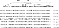

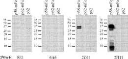

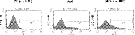


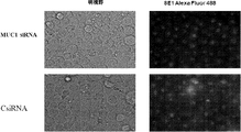


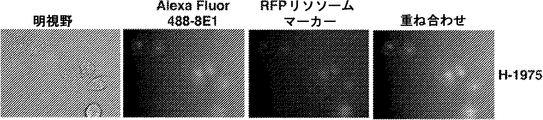
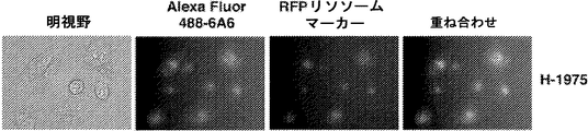
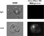


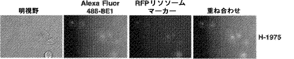


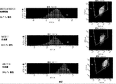


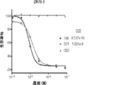


Classifications
-
- C—CHEMISTRY; METALLURGY
- C07—ORGANIC CHEMISTRY
- C07K—PEPTIDES
- C07K16/00—Immunoglobulins [IGs], e.g. monoclonal or polyclonal antibodies
- C07K16/18—Immunoglobulins [IGs], e.g. monoclonal or polyclonal antibodies against material from animals or humans
- C07K16/28—Immunoglobulins [IGs], e.g. monoclonal or polyclonal antibodies against material from animals or humans against receptors, cell surface antigens or cell surface determinants
- C07K16/30—Immunoglobulins [IGs], e.g. monoclonal or polyclonal antibodies against material from animals or humans against receptors, cell surface antigens or cell surface determinants from tumour cells
- C07K16/3076—Immunoglobulins [IGs], e.g. monoclonal or polyclonal antibodies against material from animals or humans against receptors, cell surface antigens or cell surface determinants from tumour cells against structure-related tumour-associated moieties
- C07K16/3092—Immunoglobulins [IGs], e.g. monoclonal or polyclonal antibodies against material from animals or humans against receptors, cell surface antigens or cell surface determinants from tumour cells against structure-related tumour-associated moieties against tumour-associated mucins
-
- A—HUMAN NECESSITIES
- A61—MEDICAL OR VETERINARY SCIENCE; HYGIENE
- A61K—PREPARATIONS FOR MEDICAL, DENTAL OR TOILETRY PURPOSES
- A61K38/00—Medicinal preparations containing peptides
- A61K38/04—Peptides having up to 20 amino acids in a fully defined sequence; Derivatives thereof
- A61K38/08—Peptides having 5 to 11 amino acids
-
- A—HUMAN NECESSITIES
- A61—MEDICAL OR VETERINARY SCIENCE; HYGIENE
- A61K—PREPARATIONS FOR MEDICAL, DENTAL OR TOILETRY PURPOSES
- A61K39/00—Medicinal preparations containing antigens or antibodies
- A61K39/395—Antibodies; Immunoglobulins; Immune serum, e.g. antilymphocytic serum
- A61K39/39533—Antibodies; Immunoglobulins; Immune serum, e.g. antilymphocytic serum against materials from animals
- A61K39/39558—Antibodies; Immunoglobulins; Immune serum, e.g. antilymphocytic serum against materials from animals against tumor tissues, cells, antigens
-
- A—HUMAN NECESSITIES
- A61—MEDICAL OR VETERINARY SCIENCE; HYGIENE
- A61K—PREPARATIONS FOR MEDICAL, DENTAL OR TOILETRY PURPOSES
- A61K45/00—Medicinal preparations containing active ingredients not provided for in groups A61K31/00 - A61K41/00
- A61K45/06—Mixtures of active ingredients without chemical characterisation, e.g. antiphlogistics and cardiaca
-
- A—HUMAN NECESSITIES
- A61—MEDICAL OR VETERINARY SCIENCE; HYGIENE
- A61K—PREPARATIONS FOR MEDICAL, DENTAL OR TOILETRY PURPOSES
- A61K47/00—Medicinal preparations characterised by the non-active ingredients used, e.g. carriers or inert additives; Targeting or modifying agents chemically bound to the active ingredient
- A61K47/50—Medicinal preparations characterised by the non-active ingredients used, e.g. carriers or inert additives; Targeting or modifying agents chemically bound to the active ingredient the non-active ingredient being chemically bound to the active ingredient, e.g. polymer-drug conjugates
- A61K47/51—Medicinal preparations characterised by the non-active ingredients used, e.g. carriers or inert additives; Targeting or modifying agents chemically bound to the active ingredient the non-active ingredient being chemically bound to the active ingredient, e.g. polymer-drug conjugates the non-active ingredient being a modifying agent
- A61K47/68—Medicinal preparations characterised by the non-active ingredients used, e.g. carriers or inert additives; Targeting or modifying agents chemically bound to the active ingredient the non-active ingredient being chemically bound to the active ingredient, e.g. polymer-drug conjugates the non-active ingredient being a modifying agent the modifying agent being an antibody, an immunoglobulin or a fragment thereof, e.g. an Fc-fragment
- A61K47/6801—Drug-antibody or immunoglobulin conjugates defined by the pharmacologically or therapeutically active agent
- A61K47/6803—Drugs conjugated to an antibody or immunoglobulin, e.g. cisplatin-antibody conjugates
- A61K47/68031—Drugs conjugated to an antibody or immunoglobulin, e.g. cisplatin-antibody conjugates the drug being an auristatin
-
- A—HUMAN NECESSITIES
- A61—MEDICAL OR VETERINARY SCIENCE; HYGIENE
- A61K—PREPARATIONS FOR MEDICAL, DENTAL OR TOILETRY PURPOSES
- A61K47/00—Medicinal preparations characterised by the non-active ingredients used, e.g. carriers or inert additives; Targeting or modifying agents chemically bound to the active ingredient
- A61K47/50—Medicinal preparations characterised by the non-active ingredients used, e.g. carriers or inert additives; Targeting or modifying agents chemically bound to the active ingredient the non-active ingredient being chemically bound to the active ingredient, e.g. polymer-drug conjugates
- A61K47/51—Medicinal preparations characterised by the non-active ingredients used, e.g. carriers or inert additives; Targeting or modifying agents chemically bound to the active ingredient the non-active ingredient being chemically bound to the active ingredient, e.g. polymer-drug conjugates the non-active ingredient being a modifying agent
- A61K47/68—Medicinal preparations characterised by the non-active ingredients used, e.g. carriers or inert additives; Targeting or modifying agents chemically bound to the active ingredient the non-active ingredient being chemically bound to the active ingredient, e.g. polymer-drug conjugates the non-active ingredient being a modifying agent the modifying agent being an antibody, an immunoglobulin or a fragment thereof, e.g. an Fc-fragment
- A61K47/6801—Drug-antibody or immunoglobulin conjugates defined by the pharmacologically or therapeutically active agent
- A61K47/6803—Drugs conjugated to an antibody or immunoglobulin, e.g. cisplatin-antibody conjugates
- A61K47/6811—Drugs conjugated to an antibody or immunoglobulin, e.g. cisplatin-antibody conjugates the drug being a protein or peptide, e.g. transferrin or bleomycin
-
- A—HUMAN NECESSITIES
- A61—MEDICAL OR VETERINARY SCIENCE; HYGIENE
- A61K—PREPARATIONS FOR MEDICAL, DENTAL OR TOILETRY PURPOSES
- A61K47/00—Medicinal preparations characterised by the non-active ingredients used, e.g. carriers or inert additives; Targeting or modifying agents chemically bound to the active ingredient
- A61K47/50—Medicinal preparations characterised by the non-active ingredients used, e.g. carriers or inert additives; Targeting or modifying agents chemically bound to the active ingredient the non-active ingredient being chemically bound to the active ingredient, e.g. polymer-drug conjugates
- A61K47/51—Medicinal preparations characterised by the non-active ingredients used, e.g. carriers or inert additives; Targeting or modifying agents chemically bound to the active ingredient the non-active ingredient being chemically bound to the active ingredient, e.g. polymer-drug conjugates the non-active ingredient being a modifying agent
- A61K47/68—Medicinal preparations characterised by the non-active ingredients used, e.g. carriers or inert additives; Targeting or modifying agents chemically bound to the active ingredient the non-active ingredient being chemically bound to the active ingredient, e.g. polymer-drug conjugates the non-active ingredient being a modifying agent the modifying agent being an antibody, an immunoglobulin or a fragment thereof, e.g. an Fc-fragment
- A61K47/6835—Medicinal preparations characterised by the non-active ingredients used, e.g. carriers or inert additives; Targeting or modifying agents chemically bound to the active ingredient the non-active ingredient being chemically bound to the active ingredient, e.g. polymer-drug conjugates the non-active ingredient being a modifying agent the modifying agent being an antibody, an immunoglobulin or a fragment thereof, e.g. an Fc-fragment the modifying agent being an antibody or an immunoglobulin bearing at least one antigen-binding site
- A61K47/6849—Medicinal preparations characterised by the non-active ingredients used, e.g. carriers or inert additives; Targeting or modifying agents chemically bound to the active ingredient the non-active ingredient being chemically bound to the active ingredient, e.g. polymer-drug conjugates the non-active ingredient being a modifying agent the modifying agent being an antibody, an immunoglobulin or a fragment thereof, e.g. an Fc-fragment the modifying agent being an antibody or an immunoglobulin bearing at least one antigen-binding site the antibody targeting a receptor, a cell surface antigen or a cell surface determinant
-
- A—HUMAN NECESSITIES
- A61—MEDICAL OR VETERINARY SCIENCE; HYGIENE
- A61K—PREPARATIONS FOR MEDICAL, DENTAL OR TOILETRY PURPOSES
- A61K47/00—Medicinal preparations characterised by the non-active ingredients used, e.g. carriers or inert additives; Targeting or modifying agents chemically bound to the active ingredient
- A61K47/50—Medicinal preparations characterised by the non-active ingredients used, e.g. carriers or inert additives; Targeting or modifying agents chemically bound to the active ingredient the non-active ingredient being chemically bound to the active ingredient, e.g. polymer-drug conjugates
- A61K47/51—Medicinal preparations characterised by the non-active ingredients used, e.g. carriers or inert additives; Targeting or modifying agents chemically bound to the active ingredient the non-active ingredient being chemically bound to the active ingredient, e.g. polymer-drug conjugates the non-active ingredient being a modifying agent
- A61K47/68—Medicinal preparations characterised by the non-active ingredients used, e.g. carriers or inert additives; Targeting or modifying agents chemically bound to the active ingredient the non-active ingredient being chemically bound to the active ingredient, e.g. polymer-drug conjugates the non-active ingredient being a modifying agent the modifying agent being an antibody, an immunoglobulin or a fragment thereof, e.g. an Fc-fragment
- A61K47/6835—Medicinal preparations characterised by the non-active ingredients used, e.g. carriers or inert additives; Targeting or modifying agents chemically bound to the active ingredient the non-active ingredient being chemically bound to the active ingredient, e.g. polymer-drug conjugates the non-active ingredient being a modifying agent the modifying agent being an antibody, an immunoglobulin or a fragment thereof, e.g. an Fc-fragment the modifying agent being an antibody or an immunoglobulin bearing at least one antigen-binding site
- A61K47/6851—Medicinal preparations characterised by the non-active ingredients used, e.g. carriers or inert additives; Targeting or modifying agents chemically bound to the active ingredient the non-active ingredient being chemically bound to the active ingredient, e.g. polymer-drug conjugates the non-active ingredient being a modifying agent the modifying agent being an antibody, an immunoglobulin or a fragment thereof, e.g. an Fc-fragment the modifying agent being an antibody or an immunoglobulin bearing at least one antigen-binding site the antibody targeting a determinant of a tumour cell
- A61K47/6855—Medicinal preparations characterised by the non-active ingredients used, e.g. carriers or inert additives; Targeting or modifying agents chemically bound to the active ingredient the non-active ingredient being chemically bound to the active ingredient, e.g. polymer-drug conjugates the non-active ingredient being a modifying agent the modifying agent being an antibody, an immunoglobulin or a fragment thereof, e.g. an Fc-fragment the modifying agent being an antibody or an immunoglobulin bearing at least one antigen-binding site the antibody targeting a determinant of a tumour cell the tumour determinant being from breast cancer cell
-
- A—HUMAN NECESSITIES
- A61—MEDICAL OR VETERINARY SCIENCE; HYGIENE
- A61K—PREPARATIONS FOR MEDICAL, DENTAL OR TOILETRY PURPOSES
- A61K47/00—Medicinal preparations characterised by the non-active ingredients used, e.g. carriers or inert additives; Targeting or modifying agents chemically bound to the active ingredient
- A61K47/50—Medicinal preparations characterised by the non-active ingredients used, e.g. carriers or inert additives; Targeting or modifying agents chemically bound to the active ingredient the non-active ingredient being chemically bound to the active ingredient, e.g. polymer-drug conjugates
- A61K47/51—Medicinal preparations characterised by the non-active ingredients used, e.g. carriers or inert additives; Targeting or modifying agents chemically bound to the active ingredient the non-active ingredient being chemically bound to the active ingredient, e.g. polymer-drug conjugates the non-active ingredient being a modifying agent
- A61K47/68—Medicinal preparations characterised by the non-active ingredients used, e.g. carriers or inert additives; Targeting or modifying agents chemically bound to the active ingredient the non-active ingredient being chemically bound to the active ingredient, e.g. polymer-drug conjugates the non-active ingredient being a modifying agent the modifying agent being an antibody, an immunoglobulin or a fragment thereof, e.g. an Fc-fragment
- A61K47/6889—Conjugates wherein the antibody being the modifying agent and wherein the linker, binder or spacer confers particular properties to the conjugates, e.g. peptidic enzyme-labile linkers or acid-labile linkers, providing for an acid-labile immuno conjugate wherein the drug may be released from its antibody conjugated part in an acidic, e.g. tumoural or environment
-
- A—HUMAN NECESSITIES
- A61—MEDICAL OR VETERINARY SCIENCE; HYGIENE
- A61P—SPECIFIC THERAPEUTIC ACTIVITY OF CHEMICAL COMPOUNDS OR MEDICINAL PREPARATIONS
- A61P35/00—Antineoplastic agents
-
- A—HUMAN NECESSITIES
- A61—MEDICAL OR VETERINARY SCIENCE; HYGIENE
- A61P—SPECIFIC THERAPEUTIC ACTIVITY OF CHEMICAL COMPOUNDS OR MEDICINAL PREPARATIONS
- A61P35/00—Antineoplastic agents
- A61P35/02—Antineoplastic agents specific for leukemia
-
- A—HUMAN NECESSITIES
- A61—MEDICAL OR VETERINARY SCIENCE; HYGIENE
- A61P—SPECIFIC THERAPEUTIC ACTIVITY OF CHEMICAL COMPOUNDS OR MEDICINAL PREPARATIONS
- A61P35/00—Antineoplastic agents
- A61P35/04—Antineoplastic agents specific for metastasis
-
- C—CHEMISTRY; METALLURGY
- C07—ORGANIC CHEMISTRY
- C07K—PEPTIDES
- C07K14/00—Peptides having more than 20 amino acids; Gastrins; Somatostatins; Melanotropins; Derivatives thereof
- C07K14/435—Peptides having more than 20 amino acids; Gastrins; Somatostatins; Melanotropins; Derivatives thereof from animals; from humans
- C07K14/705—Receptors; Cell surface antigens; Cell surface determinants
- C07K14/70503—Immunoglobulin superfamily
- C07K14/7051—T-cell receptor (TcR)-CD3 complex
-
- C—CHEMISTRY; METALLURGY
- C07—ORGANIC CHEMISTRY
- C07K—PEPTIDES
- C07K16/00—Immunoglobulins [IGs], e.g. monoclonal or polyclonal antibodies
- C07K16/18—Immunoglobulins [IGs], e.g. monoclonal or polyclonal antibodies against material from animals or humans
- C07K16/28—Immunoglobulins [IGs], e.g. monoclonal or polyclonal antibodies against material from animals or humans against receptors, cell surface antigens or cell surface determinants
- C07K16/2803—Immunoglobulins [IGs], e.g. monoclonal or polyclonal antibodies against material from animals or humans against receptors, cell surface antigens or cell surface determinants against the immunoglobulin superfamily
- C07K16/2809—Immunoglobulins [IGs], e.g. monoclonal or polyclonal antibodies against material from animals or humans against receptors, cell surface antigens or cell surface determinants against the immunoglobulin superfamily against the T-cell receptor (TcR)-CD3 complex
-
- C—CHEMISTRY; METALLURGY
- C07—ORGANIC CHEMISTRY
- C07K—PEPTIDES
- C07K16/00—Immunoglobulins [IGs], e.g. monoclonal or polyclonal antibodies
- C07K16/18—Immunoglobulins [IGs], e.g. monoclonal or polyclonal antibodies against material from animals or humans
- C07K16/28—Immunoglobulins [IGs], e.g. monoclonal or polyclonal antibodies against material from animals or humans against receptors, cell surface antigens or cell surface determinants
- C07K16/30—Immunoglobulins [IGs], e.g. monoclonal or polyclonal antibodies against material from animals or humans against receptors, cell surface antigens or cell surface determinants from tumour cells
- C07K16/3015—Breast
-
- C—CHEMISTRY; METALLURGY
- C07—ORGANIC CHEMISTRY
- C07K—PEPTIDES
- C07K16/00—Immunoglobulins [IGs], e.g. monoclonal or polyclonal antibodies
- C07K16/18—Immunoglobulins [IGs], e.g. monoclonal or polyclonal antibodies against material from animals or humans
- C07K16/28—Immunoglobulins [IGs], e.g. monoclonal or polyclonal antibodies against material from animals or humans against receptors, cell surface antigens or cell surface determinants
- C07K16/30—Immunoglobulins [IGs], e.g. monoclonal or polyclonal antibodies against material from animals or humans against receptors, cell surface antigens or cell surface determinants from tumour cells
- C07K16/3023—Lung
-
- C—CHEMISTRY; METALLURGY
- C07—ORGANIC CHEMISTRY
- C07K—PEPTIDES
- C07K16/00—Immunoglobulins [IGs], e.g. monoclonal or polyclonal antibodies
- C07K16/18—Immunoglobulins [IGs], e.g. monoclonal or polyclonal antibodies against material from animals or humans
- C07K16/28—Immunoglobulins [IGs], e.g. monoclonal or polyclonal antibodies against material from animals or humans against receptors, cell surface antigens or cell surface determinants
- C07K16/30—Immunoglobulins [IGs], e.g. monoclonal or polyclonal antibodies against material from animals or humans against receptors, cell surface antigens or cell surface determinants from tumour cells
- C07K16/3061—Blood cells
-
- A—HUMAN NECESSITIES
- A61—MEDICAL OR VETERINARY SCIENCE; HYGIENE
- A61K—PREPARATIONS FOR MEDICAL, DENTAL OR TOILETRY PURPOSES
- A61K39/00—Medicinal preparations containing antigens or antibodies
- A61K2039/505—Medicinal preparations containing antigens or antibodies comprising antibodies
-
- C—CHEMISTRY; METALLURGY
- C07—ORGANIC CHEMISTRY
- C07K—PEPTIDES
- C07K2317/00—Immunoglobulins specific features
- C07K2317/20—Immunoglobulins specific features characterized by taxonomic origin
- C07K2317/24—Immunoglobulins specific features characterized by taxonomic origin containing regions, domains or residues from different species, e.g. chimeric, humanized or veneered
-
- C—CHEMISTRY; METALLURGY
- C07—ORGANIC CHEMISTRY
- C07K—PEPTIDES
- C07K2317/00—Immunoglobulins specific features
- C07K2317/30—Immunoglobulins specific features characterized by aspects of specificity or valency
- C07K2317/31—Immunoglobulins specific features characterized by aspects of specificity or valency multispecific
-
- C—CHEMISTRY; METALLURGY
- C07—ORGANIC CHEMISTRY
- C07K—PEPTIDES
- C07K2317/00—Immunoglobulins specific features
- C07K2317/30—Immunoglobulins specific features characterized by aspects of specificity or valency
- C07K2317/34—Identification of a linear epitope shorter than 20 amino acid residues or of a conformational epitope defined by amino acid residues
-
- C—CHEMISTRY; METALLURGY
- C07—ORGANIC CHEMISTRY
- C07K—PEPTIDES
- C07K2317/00—Immunoglobulins specific features
- C07K2317/50—Immunoglobulins specific features characterized by immunoglobulin fragments
- C07K2317/56—Immunoglobulins specific features characterized by immunoglobulin fragments variable (Fv) region, i.e. VH and/or VL
- C07K2317/565—Complementarity determining region [CDR]
-
- C—CHEMISTRY; METALLURGY
- C07—ORGANIC CHEMISTRY
- C07K—PEPTIDES
- C07K2317/00—Immunoglobulins specific features
- C07K2317/60—Immunoglobulins specific features characterized by non-natural combinations of immunoglobulin fragments
- C07K2317/62—Immunoglobulins specific features characterized by non-natural combinations of immunoglobulin fragments comprising only variable region components
- C07K2317/622—Single chain antibody (scFv)
-
- C—CHEMISTRY; METALLURGY
- C07—ORGANIC CHEMISTRY
- C07K—PEPTIDES
- C07K2317/00—Immunoglobulins specific features
- C07K2317/70—Immunoglobulins specific features characterized by effect upon binding to a cell or to an antigen
- C07K2317/73—Inducing cell death, e.g. apoptosis, necrosis or inhibition of cell proliferation
-
- C—CHEMISTRY; METALLURGY
- C07—ORGANIC CHEMISTRY
- C07K—PEPTIDES
- C07K2317/00—Immunoglobulins specific features
- C07K2317/70—Immunoglobulins specific features characterized by effect upon binding to a cell or to an antigen
- C07K2317/77—Internalization into the cell
-
- C—CHEMISTRY; METALLURGY
- C07—ORGANIC CHEMISTRY
- C07K—PEPTIDES
- C07K2319/00—Fusion polypeptide
-
- C—CHEMISTRY; METALLURGY
- C07—ORGANIC CHEMISTRY
- C07K—PEPTIDES
- C07K2319/00—Fusion polypeptide
- C07K2319/01—Fusion polypeptide containing a localisation/targetting motif
- C07K2319/02—Fusion polypeptide containing a localisation/targetting motif containing a signal sequence
-
- C—CHEMISTRY; METALLURGY
- C07—ORGANIC CHEMISTRY
- C07K—PEPTIDES
- C07K2319/00—Fusion polypeptide
- C07K2319/01—Fusion polypeptide containing a localisation/targetting motif
- C07K2319/03—Fusion polypeptide containing a localisation/targetting motif containing a transmembrane segment
-
- C—CHEMISTRY; METALLURGY
- C07—ORGANIC CHEMISTRY
- C07K—PEPTIDES
- C07K2319/00—Fusion polypeptide
- C07K2319/32—Fusion polypeptide fusions with soluble part of a cell surface receptor, "decoy receptors"
-
- C—CHEMISTRY; METALLURGY
- C07—ORGANIC CHEMISTRY
- C07K—PEPTIDES
- C07K2319/00—Fusion polypeptide
- C07K2319/33—Fusion polypeptide fusions for targeting to specific cell types, e.g. tissue specific targeting, targeting of a bacterial subspecies
Landscapes
- Health & Medical Sciences (AREA)
- Life Sciences & Earth Sciences (AREA)
- Chemical & Material Sciences (AREA)
- Immunology (AREA)
- Engineering & Computer Science (AREA)
- Bioinformatics & Cheminformatics (AREA)
- Medicinal Chemistry (AREA)
- General Health & Medical Sciences (AREA)
- Pharmacology & Pharmacy (AREA)
- Animal Behavior & Ethology (AREA)
- Public Health (AREA)
- Veterinary Medicine (AREA)
- Organic Chemistry (AREA)
- Epidemiology (AREA)
- Cell Biology (AREA)
- Proteomics, Peptides & Aminoacids (AREA)
- Molecular Biology (AREA)
- Genetics & Genomics (AREA)
- Biochemistry (AREA)
- Biophysics (AREA)
- Gastroenterology & Hepatology (AREA)
- Oncology (AREA)
- Chemical Kinetics & Catalysis (AREA)
- General Chemical & Material Sciences (AREA)
- Nuclear Medicine, Radiotherapy & Molecular Imaging (AREA)
- Hematology (AREA)
- Toxicology (AREA)
- Zoology (AREA)
- Microbiology (AREA)
- Biomedical Technology (AREA)
- Mycology (AREA)
- Peptides Or Proteins (AREA)
- Medicines Containing Antibodies Or Antigens For Use As Internal Diagnostic Agents (AREA)
- Micro-Organisms Or Cultivation Processes Thereof (AREA)
- Medicines That Contain Protein Lipid Enzymes And Other Medicines (AREA)
- Medicinal Preparation (AREA)
- Preparation Of Compounds By Using Micro-Organisms (AREA)
Description
本出願は、2014年1月29日に申請された米国仮出願第61/933,001号に対する優先権の恩典を主張するものであり、その全内容は参照により本明細書によって組み入れられる。 This application claims the benefit of priority to US Provisional Application No. 61 / 933,001 filed January 29, 2014, the entire contents of which are incorporated herein by reference.
40KB(Microsoft Windows(登録商標)で測定されたもの)である「GENUP0032WO_ST25.txt」と名付けられたファイルに含有され、かつ2015年1月29日に作成された配列表は、電子提出によって本明細書とともに申請され、かつ参照により本明細書に組み入れられる。 The sequence listing contained in the file named "GENUP0032WO_ST25.txt", which is 40KB (measured by Microsoft Windows®) and was created on January 29, 2015, is hereby submitted electronically. Applied with the document and incorporated herein by reference.
1.発明の分野
本発明は、概して、医学、腫瘍学、および免疫療法学の分野に関係する。特に、MUC1陽性癌を検出するおよび治療することにおける使用のための免疫試薬の開発に関する。
1. 1. Fields of Invention The present invention generally relates to the fields of medicine, oncology, and immunotherapy. In particular, it relates to the development of immune reagents for use in detecting and treating MUC1-positive cancers.
2.発明の背景
ムチンは、主として上皮細胞によって発現される広範囲にわたってO-グリコシル化されたタンパク質である。分泌型および膜結合型のムチンは、毒素、微生物、および外部環境との界面で生じる他の形態のストレスによって誘導されるダメージから上皮細胞の頂端境界を保護する物理的バリアを形成する。膜貫通型ムチン1(MUC1)は、また、その細胞質ドメインを介して細胞の内部にシグナルを送達し得る。MUC1は、ウニ精子タンパク質-エンテロキナーゼ-アグリン(SEA)ドメインの存在を除いて、他の膜結合型ムチンと配列類似性を有しない(Duraisamy et al., 2006)。それに関して、MUC1は単一ポリペプチドとして翻訳され、次いでSEAドメインにおいて自己切断を受ける(Macao, 2006)。
2. Background of the Invention Mucin is a widely O-glycosylated protein expressed primarily by epithelial cells. Secretory and membrane-bound mucins form a physical barrier that protects the apical border of epithelial cells from damage induced by toxins, microorganisms, and other forms of stress that occur at the interface with the external environment. Transmembrane mucin 1 (MUC1) can also deliver a signal inside the cell via its cytoplasmic domain. MUC1 has no sequence similarity to other membrane-bound mucins except for the presence of the sea urchin sperm protein-enteropeptidase-agrin (SEA) domain (Duraisamy et al., 2006). In that regard, MUC1 is translated as a single polypeptide and then undergoes self-cleavage in the SEA domain (Macao, 2006).
MUC1は、癌におけるその役割のために、本発明者らおよび他者によって広範囲にわたって研究されている。上述のように、ヒトMUC1は、小胞体において単一ポリペプチドとして翻訳されかつN末およびC末のサブユニット(MUC1-NおよびMUC1-C)に切断される、ヘテロ二量体糖タンパク質である(Ligtenberg et al., 1992;Macao et al., 2006;Levitin et al., 2005)。大部分のヒト癌腫において見出されるMUC1の異常な過剰発現(Kufe et al., 1984)は、足場非依存的成長および腫瘍形成能を付与する(Li et al., 2003a;Huang et al., 2003;Schroeder et al., 2004;Huang et al., 2005)。他の研究により、MUC1の過剰発現は、酸化ストレスおよび遺伝毒性抗癌剤によって誘導されるアポトーシスに対する耐性を付与することが実証されている(Yin and Kufe, 2003;Ren et al., 2004;Raina et al., 2004;Yin et al., 2004;Raina et al., 2006;Yin et al., 2007)。 MUC1 has been extensively studied by us and others for its role in cancer. As mentioned above, human MUC1 is a heterodimeric glycoprotein that is translated as a single polypeptide in the endoplasmic reticulum and cleaved into N-terminal and C-terminal subunits (MUC1-N and MUC1-C). (Ligtenberg et al., 1992; Macao et al., 2006; Levitin et al., 2005). Abnormal overexpression of MUC1 found in most human carcinomas (Kufe et al., 1984) confer scaffold-independent growth and tumorigenic potential (Li et al., 2003a; Huang et al., 2003). Schroeder et al., 2004; Huang et al., 2005). Other studies have demonstrated that overexpression of MUC1 confer resistance to oxidative stress and apoptosis induced by genotoxic anticancer agents (Yin and Kufe, 2003; Ren et al., 2004; Raina et al). ., 2004; Yin et al., 2004; Raina et al., 2006; Yin et al., 2007).
拘束型および分泌型のムチンのファミリーは、上皮細胞表面の保護バリアを提供することに機能を果たす。上皮層へのダメージとともに、細胞はヘレグリン誘導性修復プログラムを開始させるため、近隣細胞との間の密着接合が分断されかつ極性が失われる(Vermeer et al., 2003)。MUC1-Nは細胞表面からのシェディングを受け(Abe and Kufe, 1989)、MUC1-Cを細胞の内部への環境ストレスシグナルの変換因子として機能させる。これに関して、MUC1-CはErbB受容体ファミリーのメンバーと細胞表面複合体を形成し、かつMUC1-Cは、ヘレグリン刺激に応答して核に標的化される(Li et al., 2001;Li et al., 2003c)。MUC1-Cは、MUC1細胞質ドメイン(CD)とカテニンファミリーのメンバーとの間の直接相互作用を介して、ErbB受容体およびWntシグナル伝達経路を統合することにおいても機能を果たす(Huang et al., 2005;Li et al., 2003c;Yamamoto et al., 1997;Li et al., 1998;Li et al., 2001;Li and Kufe, 2001)。他の研究により、MUC1-CDは、グリコーゲン合成酵素キナーゼ3β、c-Src、プロテインキナーゼCδ、およびc-Ab1によってリン酸化されることが実証されている(Raina et al., 2006;Li et al., 1998;Li et al., 2001;Ren et al., 2002)。前述の相互作用のいずれかを阻害することは、MUC1関連癌に対する治療的介入の潜在的地点となる。 The family of restrictive and secretory mucins serves to provide a protective barrier on the surface of epithelial cells. With damage to the epithelial layer, cells initiate a heregulin-induced repair program, disrupting tight junctions with neighboring cells and losing polarity (Vermeer et al., 2003). MUC1-N undergoes shedding from the cell surface (Abe and Kufe, 1989), causing MUC1-C to function as a transformant of environmental stress signals into the cell. In this regard, MUC1-C forms a cell surface complex with members of the ErbB receptor family, and MUC1-C is targeted to the nucleus in response to hellegrin stimulation (Li et al., 2001; Li et). al., 2003c). MUC1-C also functions in integrating the ErbB receptor and Wnt signaling pathway through direct interaction between the MUC1 cytoplasmic domain (CD) and members of the catenin family (Huang et al., 2005; Li et al., 2003c; Yamamoto et al., 1997; Li et al., 1998; Li et al., 2001; Li and Kufe, 2001). Other studies have demonstrated that MUC1-CD is phosphorylated by glycogen synthase kinase kinases 3β, c-Src, protein kinase Cδ, and c-Ab1 (Raina et al., 2006; Li et al). ., 1998; Li et al., 2001; Ren et al., 2002). Inhibiting any of the aforementioned interactions is a potential point for therapeutic intervention in MUC1-related cancers.
ゆえに、本発明に従って、配列番号: 2によって規定されるMUC1-C/細胞外ドメイン(MUC1-C/ECD)に選択的に結合する抗体が提供され、該抗体は、
(a)IgG抗体であり;
(b)癌細胞成長を阻害し;
(c)癌細胞死を誘導し;
(d)それぞれ、各々が配列番号: 3、4、および5、もしくは6、7、および8、もしくは27、28、および29、もしくは42、43、および44、もしくは45、46、および47、もしくは48、49、および50と90%もしくはそれを上回る割合の相同性を有するCDR1、CDR2、およびCDR3領域を含む可変重鎖、ならびに各々が配列番号: 9、10、および11、もしくは12、13、および14、もしくは30、31、および32、もしくは51、52、および53、もしくは54、55、および56、もしくは57、58、および59と90%もしくはそれを上回る割合の相同性を有するCDR1、CDR2、およびCDR3領域を含む可変軽鎖を含み;
(e)それぞれ、配列番号: 15、19、23、60、64、もしくは68と80%もしくはそれを上回る割合の相同性を有する可変重鎖、および配列番号: 17、21、25、62、66、70と80%もしくはそれを上回る割合の相同性を有する可変軽鎖を含み;かつ/または
(f)それぞれ、配列番号: 16、20、24、61、65、もしくは69と70%もしくはそれを上回る割合の相同性を有する核酸によってコードされる可変重鎖、および配列番号: 18、22、26、63、67、もしくは71と70%もしくはそれを上回る割合の相同性を有する核酸によってコードされる可変軽鎖を含む。
Therefore, according to the present invention, an antibody that selectively binds to the MUC1-C / extracellular domain (MUC1-C / ECD) defined by SEQ ID NO: 2 is provided.
(A) IgG antibody;
(B) Inhibits cancer cell growth;
(C) Induces cancer cell death;
(D) SEQ ID NOs : 3, 4, and 5, or 6, 7, and 8, or 27, 28, and 29, or 42, 43, and 44, or 45, 46, and 47, respectively, respectively. Variable heavy chains containing the CDR1, CDR2, and CDR3 regions having 90% or more homology with 48, 49, and 50, and each SEQ ID NO : 9, 10, and 11, or 12, 13, And 14, or 30, 31, and 32, or 51, 52, and 53, or 54, 55, and 56, or 57, 58, and 59 and CDR1, CDR2 with 90% or more homology. , And a variable light chain containing the CDR3 region;
(E) Variable heavy chains with 80% or greater homology to SEQ ID NOs : 15, 19, 23, 60, 64, or 68, respectively, and SEQ ID NOs : 17, 21, 25, 62, 66, respectively. , 70 and 80% or more of variable light chains having homology; and / or (f) SEQ ID NO : 16, 20, 24, 61, 65, or 69 and 70% or more, respectively. Variable heavy chains encoded by greater proportions of homology, and SEQ ID NOs : 18, 22, 26, 63, 67, or 71 and encoded by 70% or greater proportions of homology. Includes variable light chain.
重鎖および軽鎖は、それぞれ、配列番号: 15、19、23、60、64、または68と、および17、21、25、62、66、または70と85%、90%、95%、または99%の相同性を有し得る。重鎖および軽鎖は、それぞれ、配列番号: 16、20、24、61、65、または69と、および18、22、26、63、67、または71と85%、90%、95%、または99%の相同性を有する核酸によってコードされ得る。一態様において、抗体は、配列番号: 33、34、または35に結合しない。 Heavy and light chains are SEQ ID NOs : 15, 19, 23, 60, 64, or 68, respectively, and 17, 21, 25, 62, 66, or 70 and 85%, 90%, 95%, or, respectively. Can have 99% homology. Heavy and light chains are SEQ ID NOs : 16, 20, 24, 61, 65, or 69, respectively, and 18, 22, 26, 63, 67, or 71 and 85%, 90%, 95%, or, respectively. It can be encoded by a nucleic acid with 99% homology. In one embodiment, the antibody does not bind to SEQ ID NO : 33, 34, or 35.
抗体は、一本鎖抗体、単一ドメイン抗体、キメラ抗体、またはFabフラグメントであり得る。抗体は、MUC1-C/ECDおよび個別の癌細胞表面抗原に対する特異性を有する組換え抗体であり得る。抗体は、マウス抗体、IgG、ヒト化抗体、またはヒト化IgGであり得る。抗体は、ペプチドタグ、酵素、磁性粒子、発色団、蛍光分子、化学発光分子、または色素などの標識をさらに含み得る。抗体は、光解離性リンカーまたは酵素的に切断されるリンカーを介して該抗体に連結されたものなど、それに連結された抗腫瘍薬物をさらに含み得る。抗腫瘍薬物は、毒素、ラジオアイソトープ、サイトカイン、または酵素であり得る。抗体は、ナノ粒子またはリポソームに抱合され得る。細胞死の誘導には、抗体依存性細胞傷害または補体介在性細胞傷害が含まれ得る。 The antibody can be a single chain antibody, a single domain antibody, a chimeric antibody, or a Fab fragment. The antibody can be a recombinant antibody having specificity for MUC1-C / ECD and individual cancer cell surface antigens. The antibody can be a mouse antibody, IgG, humanized antibody, or humanized IgG. The antibody may further comprise a label such as a peptide tag, enzyme, magnetic particle, chromophore, fluorescent molecule, chemoluminescent molecule, or dye. The antibody may further comprise an antitumor drug linked to the antibody, such as one linked to the antibody via a photodissociable linker or an enzymatically cleaved linker. The antitumor drug can be a toxin, radioisotope, cytokine, or enzyme. The antibody can be conjugated to nanoparticles or liposomes. Induction of cell death can include antibody-dependent cellular cytotoxicity or complement-mediated cytotoxicity.
対象におけるMUC1陽性癌細胞と上記で記載される抗体とを接触させる工程を含む、癌を治療する方法も提供される。MUC1陽性癌細胞は、肺癌細胞、脳腫瘍細胞、頭頸部癌細胞、乳癌細胞、皮膚癌細胞、肝臓癌細胞、膵臓癌細胞、胃癌細胞、結腸癌細胞、直腸癌細胞、子宮癌細胞、子宮頸癌細胞、卵巣癌細胞、精巣癌細胞、皮膚癌細胞、もしくは食道癌細胞などの固形腫瘍細胞であり得る、または急性骨髄性白血病、慢性骨髄性白血病、もしくは多発性骨髄腫などの白血病もしくは骨髄腫であり得る。 Also provided is a method of treating cancer, comprising contacting a MUC1-positive cancer cell in a subject with an antibody described above. MUC1-positive cancer cells include lung cancer cells, brain tumor cells, head and neck cancer cells, breast cancer cells, skin cancer cells, liver cancer cells, pancreatic cancer cells, gastric cancer cells, colon cancer cells, rectal cancer cells, uterine cancer cells, and cervical cancer. Can be solid tumor cells such as cells, ovarian cancer cells, testicular cancer cells, skin cancer cells, or esophageal cancer cells, or in leukemia or myeloma such as acute myeloma, chronic myeloma, or multiple myeloma possible.
方法は、MUC1陽性癌細胞と、化学療法、放射線療法、免疫療法、ホルモン療法、または毒素療法など、第二の抗癌剤または治療とを接触させる工程をさらに含み得る。第二の抗癌剤または治療は、細胞内MUC1機能を阻害し得る。第二の抗癌剤または治療は、第一の作用物質と同時に与えられ得る、または第一の作用物質の前および/もしくは後に与えられ得る。MUC1陽性癌細胞は、転移性癌細胞、多剤(multiply drug)耐性癌細胞、または再発癌細胞であり得る。 The method may further include contacting MUC1-positive cancer cells with a second anti-cancer agent or treatment, such as chemotherapy, radiation therapy, immunotherapy, hormone therapy, or toxin therapy. A second anti-cancer agent or treatment can inhibit intracellular MUC1 function. The second anti-cancer agent or treatment can be given at the same time as the first agent, or before and / or after the first agent. MUC1-positive cancer cells can be metastatic cancer cells, multiply drug resistant cancer cells, or recurrent cancer cells.
抗体は、一本鎖抗体、単一ドメイン抗体、キメラ抗体、またはFabフラグメントであり得る。抗体は、MUC1-C/ECDおよび個別の癌細胞表面抗原に対する特異性を有する組換え抗体であり得る。抗体は、マウス抗体、IgG、ヒト化抗体、またはヒト化IgGであり得る。抗体は、ペプチドタグ、酵素、磁性粒子、発色団、蛍光分子、化学発光分子、または色素などの標識をさらに含み得る。抗体は、光解離性リンカーまたは酵素的に切断されるリンカーを介して該抗体に連結されたものなど、それに連結された抗腫瘍薬物をさらに含み得る。抗腫瘍薬物は、毒素、ラジオアイソトープ、サイトカイン、または酵素であり得る。抗体は、ナノ粒子またはリポソームに抱合され得る。 The antibody can be a single chain antibody, a single domain antibody, a chimeric antibody, or a Fab fragment. The antibody can be a recombinant antibody having specificity for MUC1-C / ECD and individual cancer cell surface antigens. The antibody can be a mouse antibody, IgG, humanized antibody, or humanized IgG. The antibody may further comprise a label such as a peptide tag, enzyme, magnetic particle, chromophore, fluorescent molecule, chemoluminescent molecule, or dye. The antibody may further comprise an antitumor drug linked to the antibody, such as one linked to the antibody via a photodissociable linker or an enzymatically cleaved linker. The antitumor drug can be a toxin, radioisotope, cytokine, or enzyme. The antibody can be conjugated to nanoparticles or liposomes.
(i)配列番号: 2によって規定されるMUC1-C/細胞外ドメイン(MUC1-C/ECD)に選択的に結合する第一の一本鎖抗体であって、該抗体は、
(a)IgG抗体であり;
(b)癌細胞成長を阻害し;
(c)癌細胞死を誘導し;
(d)それぞれ、各々が配列番号: 3、4、および5、もしくは6、7、および8、もしくは27、28、および29、もしくは42、43、および44、もしくは45、46、および47、もしくは48、49、および50と90%もしくはそれを上回る割合の相同性を有するCDR1、CDR2、およびCDR3領域を含む可変重鎖、ならびに各々が配列番号: 9、10、および11、もしくは12、13、および14、もしくは30、31、および32、もしくは51、52、および53、もしくは54、55、および56、もしくは57、58、および59と90%もしくはそれを上回る割合の相同性を有するCDR1、CDR2、およびCDR3領域を含む可変軽鎖を含み;
(e)それぞれ、配列番号: 15、19、23、60、64、もしくは68と80%もしくはそれを上回る割合の相同性を有する可変重鎖、および配列番号: 17、21、25、62、66、70と80%もしくはそれを上回る割合の相同性を有する可変軽鎖を含み;かつ/または
(f)それぞれ、配列番号: 16、20、24、61、65、もしくは69と70%もしくはそれを上回る割合の相同性を有する核酸によってコードされる可変重鎖、および配列番号: 18、22、26、63、67、もしくは71と70%もしくはそれを上回る割合の相同性を有する核酸によってコードされる可変軽鎖を含む、第一の一本鎖抗体;ならびに
(ii)TまたはB細胞に結合する第二の一本鎖抗体
を含む融合タンパク質も提供される。
(I) The first single-chain antibody that selectively binds to the MUC1-C / extracellular domain (MUC1-C / ECD) defined by SEQ ID NO: 2, said antibody.
(A) IgG antibody;
(B) Inhibits cancer cell growth;
(C) Induces cancer cell death;
(D) SEQ ID NOs : 3, 4, and 5, or 6, 7, and 8, or 27, 28, and 29, or 42, 43, and 44, or 45, 46, and 47, respectively, respectively. Variable heavy chains containing the CDR1, CDR2, and CDR3 regions having 90% or more homology with 48, 49, and 50, and each SEQ ID NO : 9, 10, and 11, or 12, 13, And 14, or 30, 31, and 32, or 51, 52, and 53, or 54, 55, and 56, or 57, 58, and 59 and CDR1, CDR2 with 90% or more homology. , And a variable light chain containing the CDR3 region;
(E) Variable heavy chains with 80% or greater homology to SEQ ID NOs : 15, 19, 23, 60, 64, or 68, respectively, and SEQ ID NOs : 17, 21, 25, 62, 66, respectively. , 70 and 80% or more of variable light chains having homology; and / or (f) SEQ ID NOs : 16, 20, 24, 61, 65, or 69 and 70% or more, respectively. Variable heavy chains encoded by nucleic acids with greater proportions of homology, and SEQ ID NOs : 18, 22, 26, 63, 67, or 71 encoded by nucleic acids with 70% or greater proportions of homology. A fusion protein comprising a first single chain antibody, including a variable light chain; and (ii) a second single chain antibody that binds to T or B cells is also provided.
第二の一本鎖抗体は、CD3に、T細胞に、またはB細胞に結合し得る。融合タンパク質は、標識または治療用成分をさらに含み得る。一態様において、第一の一本鎖抗体は、配列番号: 3、4、または5に結合しない。 The second single chain antibody can bind to CD3, T cells, or B cells. The fusion protein may further comprise a labeling or therapeutic ingredient. In one embodiment, the first single chain antibody does not bind to SEQ ID NO : 3, 4, or 5.
別の態様において、
(i)配列番号: 2によって規定されるMUC1-C/細胞外ドメイン(MUC1-C/ECD)に選択的に結合する一本鎖抗体可変領域を含むエクトドメインであって、該抗体は、
(a)IgG抗体であり;
(b)癌細胞成長を阻害し;
(c)癌細胞死を誘導し;
(d)それぞれ、各々が配列番号: 3、4、および5、もしくは6、7、および8、もしくは27、28、および29、もしくは42、43、および44、もしくは45、46、および47、もしくは48、49、および50と90%もしくはそれを上回る割合の相同性を有するCDR1、CDR2、およびCDR3領域を含む可変重鎖、ならびに各々が配列番号: 9、10、および11、もしくは12、13、および14、もしくは30、31、および32、もしくは51、52、および53、もしくは54、55、および56、もしくは57、58、および59と90%もしくはそれを上回る割合の相同性を有するCDR1、CDR2、およびCDR3領域を含む可変軽鎖を含み;
(e)それぞれ、配列番号: 15、19、23、60、64、もしくは68と80%もしくはそれを上回る割合の相同性を有する可変重鎖、および配列番号: 17、21、25、62、66、70と80%もしくはそれを上回る割合の相同性を有する可変軽鎖を含み;かつ/または
(f)それぞれ、配列番号: 16、20、24、61、65、もしくは69と70%もしくはそれを上回る割合の相同性を有する核酸によってコードされる可変重鎖、および配列番号: 18、22、26、63、67、もしくは71と70%もしくはそれを上回る割合の相同性を有する核酸によってコードされる可変軽鎖、そしてそれとともに、該一本鎖抗体可変領域のC末端に付着した柔軟性のあるヒンジを含む、エクトドメイン;
(ii)膜貫通ドメイン;ならびに
(iii)エンドドメインであって、該エンドドメインは、該一本鎖抗体可変領域がMUC1と係合した場合にシグナル変換機能を含む、エンドドメイン
を含むキメラ抗原受容体が提供される。
In another embodiment
(I) An ect domain containing a single-chain antibody variable region that selectively binds to the MUC1-C / extracellular domain (MUC1-C / ECD) defined by SEQ ID NO: 2, said antibody.
(A) IgG antibody;
(B) Inhibits cancer cell growth;
(C) Induces cancer cell death;
(D) SEQ ID NOs : 3, 4, and 5, or 6, 7, and 8, or 27, 28, and 29, or 42, 43, and 44, or 45, 46, and 47, respectively, respectively. Variable heavy chains containing the CDR1, CDR2, and CDR3 regions having 90% or more homology with 48, 49, and 50, and each SEQ ID NO : 9, 10, and 11, or 12, 13, And 14, or 30, 31, and 32, or 51, 52, and 53, or 54, 55, and 56, or 57, 58, and 59 and CDR1, CDR2 with 90% or more homology. , And a variable light chain containing the CDR3 region;
(E) Variable heavy chains with 80% or greater homology to SEQ ID NOs : 15, 19, 23, 60, 64, or 68, respectively, and SEQ ID NOs : 17, 21, 25, 62, 66, respectively. , 70 and 80% or more of variable light chains having homology; and / or (f) SEQ ID NO : 16, 20, 24, 61, 65, or 69 and 70% or more, respectively. Variable heavy chains encoded by nucleic acids with greater proportions of homology, and SEQ ID NOs : 18, 22, 26, 63, 67, or 71 and encoded by nucleic acids having 70% or greater proportions of homology. An ect domain containing a variable light chain and, along with it, a flexible hinge attached to the C-terminal of the single chain antibody variable region;
(Ii) Transmembrane domain; and (iii) End domain, which is a chimeric antigen receptor comprising an end domain, including signal transduction function when the single chain antibody variable region engages with MUC1. The body is provided.
膜貫通およびエンドドメインは、同じ分子に由来し得る。エンドドメインは、CD3-ζドメインまたは高アフィニティーFcεRIを含む。柔軟性のあるヒンジは、CD8αまたはIg由来のものであり得る。このキメラ抗原受容体を発現する細胞も提供される。一態様において、一本鎖可変領域は、配列番号: 3、4、または5に結合しない。 Transmembrane and endodomains can be derived from the same molecule. End domains include the CD3-ζ domain or high affinity FcεRI. The flexible hinge can be from CD8α or Ig. Cells expressing this chimeric antigen receptor are also provided. In one embodiment, the single-stranded variable region does not bind to SEQ ID NO : 3, 4, or 5.
本明細書において記載される任意の方法または組成物を、本明細書において記載される他の任意の方法または組成物に対して実行し得ることが企図される。 It is contemplated that any method or composition described herein may be performed against any other method or composition described herein.
特許請求の範囲および/または本明細書において、「a」または「an」という単語の使用は、「含む(comprising)」という用語と合わせて用いられる場合に「1つ」を意味し得るが、それは、「1つまたは複数」、「少なくとも1つ」、および「1つまたは1つを上回る」という意味とも矛盾しない。「約」という単語は、明記された数のプラスまたはマイナス5%を意味する。 Although the claims and / or the use of the word "a" or "an" in the present specification may mean "one" when used in combination with the term "comprising", It is consistent with the meaning of "one or more," "at least one," and "more than one or more." The word "about" means plus or minus 5% of the stated number.
[本発明1001]
配列番号: 1によって規定されるMUC1-C細胞外ドメイン(MUC1-C/ECD)に選択的に結合する抗体であって、
(a)IgG抗体であり;
(b)癌細胞成長を阻害し;
(c)癌細胞死を誘導し;
(d)それぞれ、各々が配列番号: 3、4、および5、もしくは6、7、および8、もしくは27、28、および29、もしくは42、43、および44、もしくは45、46、および47、もしくは48、49、および50、もしくは76、77、および78と90%もしくはそれを上回る割合の相同性を有するCDR1、CDR2、およびCDR3領域を含む可変重鎖、ならびに各々が配列番号: 9、10、および11、もしくは12、13、および14、もしくは30、31、および32、もしくは51、52、および53、もしくは54、55、および56、もしくは57、58、および59、もしくは79、80、および81と90%もしくはそれを上回る割合の相同性を有するCDR1、CDR2、およびCDR3領域を含む可変軽鎖を含み;
(e)それぞれ、配列番号: 15、19、23、60、64、68、もしくは73と80%もしくはそれを上回る割合の相同性を有する可変重鎖、および配列番号: 17、21、25、62、66、70、もしくは75と80%もしくはそれを上回る割合の相同性を有する可変軽鎖を含み;かつ/または
(f)それぞれ、配列番号: 16、20、24、61、65、69、もしくは72と70%もしくはそれを上回る割合の相同性を有する核酸によってコードされる可変重鎖、および配列番号: 18、22、26、63、67、71、もしくは74と70%もしくはそれを上回る割合の相同性を有する核酸によってコードされる可変軽鎖を含む、抗体。
[本発明1002]
一本鎖抗体である、本発明1001の抗体。
[本発明1003]
単一ドメイン抗体である、本発明1001の抗体。
[本発明1004]
キメラ抗体である、本発明1001の抗体。
[本発明1005]
Fabフラグメントである、本発明1001の抗体。
[本発明1006]
MUC1-C/ECDおよび個別の癌細胞表面抗原に対する特異性を有する組換え抗体である、本発明1001の抗体。
[本発明1007]
マウス抗体である、本発明1001の抗体。
[本発明1008]
マウス抗体がIgGである、本発明1007の抗体。
[本発明1009]
ヒト化抗体である、本発明1001の抗体。
[本発明1010]
ヒト化抗体がIgGである、本発明1009の抗体。
[本発明1011]
標識をさらに含む、本発明1001の抗体。
[本発明1012]
標識が、ペプチドタグ、酵素、磁性粒子、発色団、蛍光分子、化学発光分子、または色素である、本発明1011の抗体。
[本発明1013]
それに連結された抗腫瘍薬物をさらに含む、本発明1001の抗体。
[本発明1014]
抗腫瘍薬物が、光解離性リンカーを介して抗体に連結されている、本発明1013の抗体。
[本発明1015]
抗腫瘍薬物が、酵素的に切断されるリンカーを介して抗体に連結されている、本発明1013の抗体。
[本発明1016]
抗腫瘍薬物が、毒素、ラジオアイソトープ、サイトカイン、または酵素である、本発明1013の抗体。
[本発明1017]
重鎖および軽鎖がそれぞれ、配列番号: 15、19、23、60、64、68、または73と、および17、21、25、62、66、70、または75と85%、90%、95%、または99%の相同性を有する、本発明1001の抗体。
[本発明1018]
重鎖および軽鎖がそれぞれ、配列番号: 14、18、24、61、65、69、または72と、および18、22、26、63、67、71、または74と85%、90%、95%、または99%の相同性を有する核酸によってコードされる、本発明1001の抗体。
[本発明1019]
ナノ粒子またはリポソームに抱合されている、本発明1001の抗体。
[本発明1020]
細胞死の誘導が、抗体依存性細胞傷害または補体介在性細胞傷害を含む、本発明1001の抗体。
[本発明1021]
対象におけるMUC1陽性癌細胞と本発明1001の抗体とを接触させる工程を含む、癌を治療する方法。
[本発明1022]
MUC1陽性癌細胞が固形腫瘍細胞である、本発明1021の方法。
[本発明1023]
固形腫瘍細胞が、肺癌細胞、脳腫瘍細胞、頭頸部癌細胞、乳癌細胞、皮膚癌細胞、肝臓癌細胞、膵臓癌細胞、胃癌細胞、結腸癌細胞、直腸癌細胞、子宮癌細胞、子宮頸癌細胞、卵巣癌細胞、精巣癌細胞、皮膚癌細胞、または食道癌細胞である、本発明1022の方法。
[本発明1024]
MUC1陽性癌細胞が白血病または骨髄腫である、本発明1021の方法。
[本発明1025]
白血病または骨髄腫が、急性骨髄性白血病、慢性骨髄性白血病、または多発性骨髄腫である、本発明1024の方法。
[本発明1026]
MUC1陽性癌細胞と第二の抗癌剤または治療とを接触させる工程をさらに含む、本発明1021の方法。
[本発明1027]
第二の抗癌剤または治療が、化学療法、放射線療法、免疫療法、ホルモン療法、または毒素療法より選択される、本発明1026の方法。
[本発明1028]
第二の抗癌剤または治療が、細胞内MUC1機能を阻害する、本発明1026の方法。
[本発明1029]
第二の抗癌剤または治療が、第一の作用物質と同時に与えられる、本発明1026の方法。
[本発明1030]
第二の抗癌剤または治療が、第一の作用物質の前および/または後に与えられる、本発明1026の方法。
[本発明1031]
MUC1陽性癌細胞が、転移性癌細胞、多剤(multiply drug)耐性癌細胞、または再発癌細胞である、本発明1021の方法。
[本発明1032]
抗体が一本鎖抗体である、本発明1021の方法。
[本発明1033]
抗体が単一ドメイン抗体である、本発明1021の方法。
[本発明1034]
抗体がキメラ抗体である、本発明1021の方法。
[本発明1035]
抗体がFabフラグメントである、本発明1021の方法。
[本発明1036]
抗体が、MUC1-C/ECDおよび個別の癌細胞表面抗原に対する特異性を有する組換え抗体である、本発明1021の方法。
[本発明1037]
抗体がマウス抗体である、本発明1021の方法。
[本発明1038]
マウス抗体がIgGである、本発明1037の方法。
[本発明1039]
抗体がヒト化抗体である、本発明1021の方法。
[本発明1040]
ヒト化抗体がIgGである、本発明1039の方法。
[本発明1041]
抗体が、それに連結された抗腫瘍薬物をさらに含む、本発明1021の方法。
[本発明1042]
抗腫瘍薬物が、光解離性リンカーを介して抗体に連結されている、本発明1041の方法。
[本発明1043]
抗腫瘍薬物が、酵素的に切断されるリンカーを介して抗体に連結されている、本発明1041の方法。
[本発明1044]
抗腫瘍薬物が、毒素、ラジオアイソトープ、サイトカイン、または酵素である、本発明1041の方法。
[本発明1045]
抗体が標識をさらに含む、本発明1021の方法。
[本発明1046]
標識が、ペプチドタグ、酵素、磁性粒子、発色団、蛍光分子、化学発光分子、または色素である、本発明1045の方法。
[本発明1047]
重鎖および軽鎖がそれぞれ、配列番号: 15、19、23、60、64、68、または73と、および17、21、25、62、66、70、または75と85%、90%、95%、または99%の相同性を有する、本発明1021の方法。
[本発明1048]
重鎖および軽鎖がそれぞれ、配列番号: 14、18、24、61、65、69、または72と、および18、22、26、63、67、71、または74と85%、90%、95%、または99%の相同性を有する核酸によってコードされる、本発明1021の方法。
[本発明1049]
抗体がリポソームまたはナノ粒子に抱合されている、本発明1021の方法。
[本発明1050]
抗体が、抗体依存性細胞傷害または補体介在性細胞傷害などによる細胞死の誘導をもたらす(resuts in)、本発明1021の方法。
[本発明1051]
(i)配列番号: 1によって規定されるMUC1-C/細胞外ドメイン(ECD)に選択的に結合する第一の一本鎖抗体であって、
(a)IgG抗体であり;
(b)癌細胞成長を阻害し;
(c)癌細胞死を誘導し;
(d)それぞれ、各々が配列番号: 3、4、および5、もしくは6、7、および8、もしくは27、28、および29、もしくは42、43、および44、もしくは45、46、および47、もしくは48、49、および50、もしくは76、77、および78と90%もしくはそれを上回る割合の相同性を有するCDR1、CDR2、およびCDR3領域を含む可変重鎖、ならびに各々が配列番号: 9、10、および11、もしくは12、13、および14、もしくは30、31、および32、もしくは51、52、および53、もしくは54、55、および56、もしくは57、58、および59、もしくは79、80、および81と90%もしくはそれを上回る割合の相同性を有するCDR1、CDR2、およびCDR3領域を含む可変軽鎖を含み;
(e)それぞれ、配列番号: 15、19、23、60、64、68、もしくは73と80%もしくはそれを上回る割合の相同性を有する可変重鎖、および配列番号: 17、21、25、62、66、70、もしくは75と80%もしくはそれを上回る割合の相同性を有する可変軽鎖を含み;かつ/または
(f)それぞれ、配列番号: 16、20、24、61、65、69、もしくは72と70%もしくはそれを上回る割合の相同性を有する核酸によってコードされる可変重鎖、および配列番号: 18、22、26、63、67、71、もしくは74と70%もしくはそれを上回る割合の相同性を有する核酸によってコードされる可変軽鎖を含む、第一の一本鎖抗体;ならびに
(ii)TまたはB細胞に結合する第二の一本鎖抗体
を含む融合タンパク質。
[本発明1052]
第二の一本鎖抗体がCD3に結合する、本発明1051の融合タンパク質。
[本発明1053]
第二の一本鎖抗体がT細胞に結合する、本発明1051の融合タンパク質。
[本発明1054]
第二の一本鎖抗体がB細胞に結合する、本発明1051の融合タンパク質。
[本発明1055]
融合タンパク質が標識または治療用成分をさらに含む、本発明1051の融合タンパク質。
[本発明1056]
(i)配列番号: 1によって規定されるMUC1-C/細胞外ドメイン(MUC1-C/ECD)に選択的に結合する一本鎖抗体可変領域を含むエクトドメインであって、該抗体が、
(a)IgG抗体であり;
(b)癌細胞成長を阻害し;
(c)癌細胞死を誘導し;
(d)それぞれ、各々が配列番号: 3、4、および5、もしくは6、7、および8、もしくは27、28、および29、もしくは42、43、および44、もしくは45、46、および47、もしくは48、49、および50、もしくは76、77、および78と90%もしくはそれを上回る割合の相同性を有するCDR1、CDR2、およびCDR3領域を含む可変重鎖、ならびに各々が配列番号: 9、10、および11、もしくは12、13、および14、もしくは30、31、および32、もしくは51、52、および53、もしくは54、55、および56、もしくは57、58、および59、もしくは79、80、および81と90%もしくはそれを上回る割合の相同性を有するCDR1、CDR2、およびCDR3領域を含む可変軽鎖を含み;
(e)それぞれ、配列番号: 15、19、23、60、64、68、もしくは73と80%もしくはそれを上回る割合の相同性を有する可変重鎖、および配列番号: 17、21、25、62、66、70、もしくは75と80%もしくはそれを上回る割合の相同性を有する可変軽鎖を含み;かつ/または
(f)それぞれ、配列番号: 16、20、24、61、65、69、もしくは72と70%もしくはそれを上回る割合の相同性を有する核酸によってコードされる可変重鎖、および配列番号: 18、22、26、63、67、71、もしくは74と70%もしくはそれを上回る割合の相同性を有する核酸によってコードされる可変軽鎖、そしてそれとともに、該一本鎖抗体可変領域のC末端に付着した柔軟性のあるヒンジを含む、エクトドメイン;
(ii)膜貫通ドメイン;ならびに
(iii)該一本鎖抗体可変領域がMUC1と係合した場合のシグナル変換機能を含むエンドドメイン
を含むキメラ抗原受容体。
[本発明1057]
膜貫通およびエンドドメインが同じ分子に由来する、本発明1056の受容体。
[本発明1058]
エンドドメインが、CD3-ζドメインまたは高アフィニティーFcεRIを含む、本発明1056の受容体。
[本発明1059]
柔軟性のあるヒンジが、CD8αまたはIg由来のものである、本発明1056の受容体。
[本発明1060]
本発明1056のキメラ抗原受容体を発現する細胞。
[本発明1061]
膜貫通およびエンドドメインが同じ分子に由来する、本発明1060の細胞。
[本発明1062]
エンドドメインが、CD3-ζドメインまたは高アフィニティーFcεRIを含む、本発明1060の細胞。
[本発明1063]
柔軟性のあるヒンジが、CD8αまたはIg由来のものである、本発明1060の細胞。
本発明の他の目的、特質、および利点は、以下の詳細な説明から明らかになるであろう。しかしながら、この詳細な説明から、本発明の精神および範囲の内での様々な変化および改変が当業者に明らかになるであろうため、詳細な説明および具体的な例は、本発明の具体的な態様を示すと同時に、単なる例証として与えられることが理解されるべきである。
[Invention 1001]
An antibody that selectively binds to the MUC1-C extracellular domain (MUC1-C / ECD) defined by SEQ ID NO: 1.
(A) IgG antibody;
(B) Inhibits cancer cell growth;
(C) Induces cancer cell death;
(D) SEQ ID NOs: 3, 4, and 5, or 6, 7, and 8, or 27, 28, and 29, or 42, 43, and 44, or 45, 46, and 47, respectively, respectively. Variable heavy chains containing the CDR1, CDR2, and CDR3 regions having 90% or more homology with 48, 49, and 50, or 76, 77, and 78, and each SEQ ID NO: 9, 10, And 11, or 12, 13, and 14, or 30, 31, and 32, or 51, 52, and 53, or 54, 55, and 56, or 57, 58, and 59, or 79, 80, and 81. Includes variable light chains containing CDR1, CDR2, and CDR3 regions with 90% or greater proportion of homology with.
(E) Variable heavy chains with 80% or greater homology to SEQ ID NOs: 15, 19, 23, 60, 64, 68, or 73, respectively, and SEQ ID NOs: 17, 21, 25, 62, respectively. , 66, 70, or 75 and contains variable light chains with 80% or more homology; and / or
(F) Variable heavy chains encoded by nucleic acids having 70% or more homology to SEQ ID NOs: 16, 20, 24, 61, 65, 69, or 72, respectively, and SEQ ID NOs: 18, An antibody comprising a variable light chain encoded by a nucleic acid having 70% or greater proportion of homology with 22, 26, 63, 67, 71, or 74.
[Invention 1002]
The antibody of the present invention 1001 which is a single chain antibody.
[Invention 1003]
The antibody of the present invention 1001 which is a single domain antibody.
[Invention 1004]
The antibody of the present invention 1001 which is a chimeric antibody.
[Invention 1005]
The antibody of the present invention 1001 which is a Fab fragment.
[Invention 1006]
The antibody of the present invention 1001 which is a recombinant antibody having specificity for MUC1-C / ECD and individual cancer cell surface antigens.
[Invention 1007]
The antibody of the present invention 1001 which is a mouse antibody.
[Invention 1008]
The antibody of the present invention 1007, wherein the mouse antibody is IgG.
[Invention 1009]
The antibody of the present invention 1001 which is a humanized antibody.
[Invention 1010]
The antibody of the present invention 1009, wherein the humanized antibody is IgG.
[Invention 1011]
An antibody of the invention 1001 further comprising a label.
[Invention 1012]
The antibody of the invention 1011, wherein the label is a peptide tag, enzyme, magnetic particle, chromophore, fluorescent molecule, chemoluminescent molecule, or dye.
[Invention 1013]
An antibody of the invention 1001 further comprising an antitumor drug linked thereto.
[Invention 1014]
The antibody of invention 1013, wherein the anti-tumor drug is linked to the antibody via a photodissociative linker.
[Invention 1015]
The antibody of the invention 1013, wherein the anti-tumor drug is linked to the antibody via an enzymatically cleaved linker.
[Invention 1016]
Antibodies of the invention 1013, wherein the antitumor drug is a toxin, radioisotope, cytokine, or enzyme.
[Invention 1017]
Heavy and light chains are SEQ ID NOs: 15, 19, 23, 60, 64, 68, or 73 and 17, 21, 25, 62, 66, 70, or 75 and 85%, 90%, 95, respectively. % Or 99% homology of the antibody of the invention 1001.
[Invention 1018]
Heavy and light chains are SEQ ID NOs: 14, 18, 24, 61, 65, 69, or 72, respectively, and 18, 22, 26, 63, 67, 71, or 74 and 85%, 90%, 95, respectively. The antibody of the invention 1001 encoded by a nucleic acid having% or 99% homology.
[Invention 1019]
An antibody of the invention 1001 that is conjugated to nanoparticles or liposomes.
[Invention 1020]
The antibody of the present invention 1001 whose induction of cell death comprises antibody-dependent cellular cytotoxicity or complement-mediated cytotoxicity.
[Invention 1021]
A method of treating cancer, comprising contacting a MUC1-positive cancer cell in a subject with an antibody of the invention 1001.
[Invention 1022]
The method of 1021 of the present invention, wherein the MUC1-positive cancer cells are solid tumor cells.
[Invention 1023]
Solid tumor cells include lung cancer cells, brain tumor cells, head and neck cancer cells, breast cancer cells, skin cancer cells, liver cancer cells, pancreatic cancer cells, gastric cancer cells, colon cancer cells, rectal cancer cells, uterine cancer cells, and cervical cancer cells. , Ovarian cancer cells, testicular cancer cells, skin cancer cells, or esophageal cancer cells, the method of the present invention 1022.
[1024 of the present invention]
The method of 1021 of the present invention, wherein the MUC1-positive cancer cells are leukemia or myeloma.
[Invention 1025]
The method of 1024 of the present invention, wherein the leukemia or myeloma is acute myelogenous leukemia, chronic myelogenous leukemia, or multiple myeloma.
[Invention 1026]
The method of 1021 of the present invention further comprising contacting MUC1-positive cancer cells with a second anti-cancer agent or treatment.
[Invention 1027]
The method of the invention 1026, wherein the second anti-cancer agent or treatment is selected from chemotherapy, radiation therapy, immunotherapy, hormone therapy, or toxin therapy.
[Invention 1028]
The method of the present invention 1026, wherein a second anti-cancer agent or treatment inhibits intracellular MUC1 function.
[Invention 1029]
The method of the present invention 1026, wherein a second anti-cancer agent or treatment is given at the same time as the first agent.
[Invention 1030]
The method of the present invention 1026, wherein a second anti-cancer agent or treatment is given before and / or after the first agent.
[Invention 1031]
The method of 1021 of the present invention, wherein the MUC1-positive cancer cell is a metastatic cancer cell, a multiply drug resistant cancer cell, or a recurrent cancer cell.
[Invention 1032]
The method of 1021 of the present invention, wherein the antibody is a single chain antibody.
[Invention 1033]
The method of 1021 of the present invention, wherein the antibody is a single domain antibody.
[Invention 1034]
The method of 1021 of the present invention, wherein the antibody is a chimeric antibody.
[Invention 1035]
The method of 1021 of the present invention, wherein the antibody is a Fab fragment.
[Invention 1036]
The method of 1021 of the present invention, wherein the antibody is a recombinant antibody having specificity for MUC1-C / ECD and individual cancer cell surface antigens.
[Invention 1037]
The method of 1021 of the present invention, wherein the antibody is a mouse antibody.
[Invention 1038]
The method of the present invention 1037, wherein the mouse antibody is IgG.
[Invention 1039]
The method of 1021 of the present invention, wherein the antibody is a humanized antibody.
[Invention 1040]
The method of the present invention 1039, wherein the humanized antibody is IgG.
[Invention 1041]
The method of 1021 of the present invention, wherein the antibody further comprises an antitumor drug linked thereto.
[Invention 1042]
The method of 1041 of the present invention, wherein the antitumor drug is linked to the antibody via a photodissociative linker.
[Invention 1043]
The method of 1041 of the present invention, wherein the antitumor drug is linked to the antibody via an enzymatically cleaved linker.
[Invention 1044]
The method of 1041 of the invention, wherein the antitumor drug is a toxin, radioisotope, cytokine, or enzyme.
[Invention 1045]
The method of 1021 of the present invention, wherein the antibody further comprises a label.
[Invention 1046]
The method of the invention 1045, wherein the label is a peptide tag, enzyme, magnetic particle, chromophore, fluorescent molecule, chemoluminescent molecule, or dye.
[Invention 1047]
Heavy and light chains are SEQ ID NOs: 15, 19, 23, 60, 64, 68, or 73 and 17, 21, 25, 62, 66, 70, or 75 and 85%, 90%, 95, respectively. %, Or 99%, the method of 1021 of the present invention.
[Invention 1048]
Heavy and light chains are SEQ ID NOs: 14, 18, 24, 61, 65, 69, or 72, respectively, and 18, 22, 26, 63, 67, 71, or 74 and 85%, 90%, 95, respectively. %, Or the method of 1021 of the invention, encoded by a nucleic acid having 99% homology.
[Invention 1049]
The method of 1021 of the present invention, wherein the antibody is conjugated to liposomes or nanoparticles.
[Invention 1050]
The method of 1021 of the present invention, wherein the antibody results in induction of cell death, such as due to antibody-dependent cellular cytotoxicity or complement-mediated cytotoxicity.
[Invention 1051]
(I) The first single-chain antibody that selectively binds to the MUC1-C / extracellular domain (ECD) defined by SEQ ID NO: 1.
(A) IgG antibody;
(B) Inhibits cancer cell growth;
(C) Induces cancer cell death;
(D) SEQ ID NOs: 3, 4, and 5, or 6, 7, and 8, or 27, 28, and 29, or 42, 43, and 44, or 45, 46, and 47, respectively, respectively. Variable heavy chains containing the CDR1, CDR2, and CDR3 regions having 90% or more homology with 48, 49, and 50, or 76, 77, and 78, and each SEQ ID NO: 9, 10, And 11, or 12, 13, and 14, or 30, 31, and 32, or 51, 52, and 53, or 54, 55, and 56, or 57, 58, and 59, or 79, 80, and 81. Includes variable light chains containing CDR1, CDR2, and CDR3 regions with 90% or greater proportion of homology with.
(E) Variable heavy chains with 80% or greater homology to SEQ ID NOs: 15, 19, 23, 60, 64, 68, or 73, respectively, and SEQ ID NOs: 17, 21, 25, 62, respectively. , 66, 70, or 75 and contains variable light chains with 80% or more homology; and / or
(F) Variable heavy chains encoded by nucleic acids having 70% or more homology to SEQ ID NOs: 16, 20, 24, 61, 65, 69, or 72, respectively, and SEQ ID NOs: 18, First single-chain antibody, including a variable light chain encoded by a nucleic acid having 70% or more homology to 22, 26, 63, 67, 71, or 74;
(Ii) A second single-chain antibody that binds to T or B cells
Fusion protein containing.
[Invention 1052]
A fusion protein of the invention 1051 in which a second single chain antibody binds to CD3.
[Invention 1053]
A fusion protein of the invention 1051 in which a second single chain antibody binds to T cells.
[Invention 1054]
A fusion protein of the invention 1051 in which a second single chain antibody binds to B cells.
[Invention 1055]
The fusion protein of the present invention 1051 in which the fusion protein further comprises a labeling or therapeutic component.
[Invention 1056]
(I) An ect domain containing a single-chain antibody variable region that selectively binds to the MUC1-C / extracellular domain (MUC1-C / ECD) defined by SEQ ID NO: 1, wherein the antibody is:
(A) IgG antibody;
(B) Inhibits cancer cell growth;
(C) Induces cancer cell death;
(D) SEQ ID NOs: 3, 4, and 5, or 6, 7, and 8, or 27, 28, and 29, or 42, 43, and 44, or 45, 46, and 47, respectively, respectively. Variable heavy chains containing the CDR1, CDR2, and CDR3 regions having 90% or more homology with 48, 49, and 50, or 76, 77, and 78, and each SEQ ID NO: 9, 10, And 11, or 12, 13, and 14, or 30, 31, and 32, or 51, 52, and 53, or 54, 55, and 56, or 57, 58, and 59, or 79, 80, and 81. Includes variable light chains containing CDR1, CDR2, and CDR3 regions with 90% or greater proportion of homology with.
(E) Variable heavy chains with 80% or greater homology to SEQ ID NOs: 15, 19, 23, 60, 64, 68, or 73, respectively, and SEQ ID NOs: 17, 21, 25, 62, respectively. , 66, 70, or 75 and contains variable light chains with 80% or more homology; and / or
(F) Variable heavy chains encoded by nucleic acids having 70% or more homology to SEQ ID NOs: 16, 20, 24, 61, 65, 69, or 72, respectively, and SEQ ID NOs: 18, A variable light chain encoded by a nucleic acid having 70% or more homology to 22, 26, 63, 67, 71, or 74 and attached to the C-terminus of the single chain antibody variable region. Ectodomain, including flexible hinges;
(Ii) Transmembrane domain;
(Iii) An end domain containing a signal conversion function when the single-chain antibody variable region engages with MUC1.
Chimeric antigen receptor including.
[Invention 1057]
The receptor of the present invention 1056, from which the transmembrane and endodomains are derived from the same molecule.
[Invention 1058]
Receptor of the present invention 1056, wherein the endodomain comprises a CD3-ζ domain or a high affinity FcεRI.
[Invention 1059]
The receptor of the present invention 1056, wherein the flexible hinge is derived from CD8α or Ig.
[Invention 1060]
A cell expressing the chimeric antigen receptor of the present invention 1056.
[Invention 1061]
Cells of the invention 1060, from which the transmembrane and endodomains are derived from the same molecule.
[Invention 1062]
Cells of the invention 1060, wherein the endodomain comprises the CD3-ζ domain or the high affinity FcεRI.
[Invention 1063]
The cells of the invention 1060, wherein the flexible hinges are from CD8α or Ig.
Other objects, properties, and advantages of the present invention will become apparent from the detailed description below. However, since this detailed description will reveal to those skilled in the art various changes and modifications within the spirit and scope of the invention, the detailed description and specific examples are specific to the present invention. It should be understood that it is given as an example at the same time as showing the above-mentioned aspect.
以下の図面は、本明細書の一部を形成し、かつ本発明のある特定の局面をさらに実証するために含まれる。本発明は、本明細書において提示される具体的な態様についての詳細な説明と組み合わせて、これらの図面のうちの1つまたは複数を参照することにより、よりよく理解され得る。 The following drawings are included to form part of this specification and to further demonstrate certain aspects of the invention. The present invention may be better understood by reference to one or more of these drawings in combination with a detailed description of the specific embodiments presented herein.
(図1)(図1A)MUC1-C/ECDのアミノ酸配列(58aa:配列番号: 2)。(図1B)mFc-リンカー-MUC1-C/ECD-シグナル配列構築物は、CHO-K1細胞において安定して発現した。タンパク質をCHO細胞のスープから精製し、かつそれを用いてマウスに免疫付与する。同じタンパク質を用いて、マウスをブーストした。血清力価を判定し、かつ陽性力価に従って、脾臓を融合させてハイブリドーマを作出した。
(図2)(保留)
(図3)MUC1-C/ECDタンパク質由来の3種の重複ペプチドを合成して、ELISAアッセイを用いた陽性Mabクローンに関する線状エピトープマッピングにおいて用いた。3種のペプチドに関する配列は、

(図4)(上記で記載される)いくつかの点変異体を有するMUC1-C/ECD cDNAを作出した(ECD-WT−配列番号: 2;ECD-L6A−配列番号: 36;ECD-L8A−配列番号: 37;ECD-L6,8A−配列番号: 38;ECD-Q23V−配列番号 39;ECD-Q26V−配列番号: 40;ECD-N36A−配列番号: 41)。CHO-K1細胞にこれらのcDNAのそれぞれを個々にトランスフェクトして、タンパク質を発現させかつ分泌させた。タンパク質を精製し、かつそれを用いて、ELISAアッセイを用いた陽性Mabクローンに関する立体構造的エピトープを規定する。陽性対照は、CHO-K1細胞から発現されかつ精製されたwt mFc-MUC1-C/ECDタンパク質である。
(図5)(保留)
(図6)(図6A)免疫付与の後、マウスを採血し、かつ免疫血清を、MUC1-C/ECDタンパク質を用いた免疫ブロッティングによって解析した。MUC1-C/CDタンパク質を陰性対照として用いた。(図6B)MAbクローン8E1、8F1、2A6、および6A6を、CHO-K1細胞から精製されたmFc-およびhFc-MUC1-C/ECDタンパク質を用いた免疫ブロッティングによって解析した。二次Ab:抗マウスHRP(F(ab)2特異的)(1:5000)。(図6C)MUC1陰性293T細胞、NSCLC H-1975、ならびにMCF-7およびZR-75-1乳癌腫の細胞溶解物を、クローン6A6抗体を用いた免疫ブロッティングによって解析した。
(図7)MAbクローン8E1、6A6、2G11、および2H11を、細菌から産生されかつ精製されたmFc-MUC1-C/ECD(p58-mFc)、MUC1-SEAドメイン(p62-mFc)、およびp62単独のタンパク質を用いた免疫ブロッティングによって解析した。
(図8)MAbクローン8E1、6A6、2G11、および2H11を、細菌から精製されたmFc-MUC1-C/ECD(p58-mFc)、MUC1-SEAドメイン(p62-mFc)、およびp62単独のタンパク質を用いた免疫ブロッティングによって解析した。
(図9)MAbクローン8E1、6A6、2G11、および2H11を、細菌から精製されたmFc-MUC1-C/ECD(p58-mFc)、MUC1-SEAドメイン(p62-mFc)、およびp62単独のタンパク質を用いた免疫ブロッティングによって解析した。
(図10)MAbクローン8E1、6A6、および陽性対照DF3を、MV4-11、MOLM-14(AML);U266、RPMI8226(多発性骨髄腫)、および原発性AML細胞を用いたフローサイトメトリーによって解析した。
(図11)MAbクローン8E1および6A6を、K562/CsiRNAおよびK562/MUC1siRNA CML細胞を用いたフローサイトメトリーによって解析した。
(図12)37℃で3時間、H-1975非小細胞肺癌腫細胞を用いた、FITC標識抗MUC1-C/ECD MAb 8E1の内在化。後期エンドソーム/リソソーム小胞(矢印)におけるキャッピングおよび点状染色。
(図13)37℃で3時間、H-1975非小細胞肺癌腫細胞を用いた、FITC標識抗MUC1-C/ECD MAb 6A6の内在化。後期エンドソーム/リソソーム小胞(矢印)におけるキャッピングおよび点状染色。
(図14)37℃で3時間、ZR-75-1乳癌腫細胞を用いた、Alexa Fluor 488標識抗MUC1-C/ECD抗体6A6の内在化。
(図15)4℃または37℃で3時間、MUC1陰性HEK-293T細胞を用いた、Alexa Fluor 488標識抗MUC1-C/ECD抗体6A6のたとえあったとしてもわずかな染色。
(図16)37℃で3時間、MOLM-14 AML細胞を用いた、Alexa Fluor 488標識抗MUC1-C/ECD抗体8E1の内在化。
(図17)37℃で3時間、K562/MUC1siRNAおよびK562/CsiRNA CML細胞を用いた、Alexa Fluor 488標識抗MUC1-C/ECD抗体8E1の内在化。
(図18)4℃でZR-75-1乳癌腫細胞における、Alexa Fluor 488標識抗MUC1-C/ECD抗体6A6の原形質膜染色、およびRFP初期エンドソームマーカーとのその共局在。
(図19)37℃でZR-75-1乳癌腫細胞における、Alexa Fluor 488標識抗MUC1-C/ECD抗体6A6の内在化、およびRFP-エンドサイト(endocyte)マーカーとのその共局在。
(図20)37℃でH-1975 NSCLC細胞における、Alexa Fluor 488標識抗MUC1-C/ECD抗体6A6の内在化、およびRFP-エンドサイトマーカーとのその共局在(矢印)。
(図21)37℃でH-1975 NSCLC細胞における、Alexa Fluor 488標識抗MUC1-C/ECD抗体8E1の内在化、およびRFP-エンドサイトマーカーとのその共局在(矢印)。
(図22)37℃でH-1975 NSCLC細胞における、Alexa Fluor 488標識抗MUC1-C/ECD抗体8E1の内在化、およびRFP-リソソームマーカーとのその共局在(矢印)。
(図23)37℃でH-1975 NSCLC細胞における、Alexa Fluor 488標識抗MUC1-C/ECD抗体6A6の内在化、およびRFP-リソソームマーカーとのその共局在(矢印)。
(図24)37℃で3時間、マウスNSCLC(KW-814)細胞を用いた、Alexa Fluor 488標識抗MUC1-C/ECD抗体6A6の内在化。
(図25)37℃で3時間、K562 CML細胞を用いた、Alexa Fluor 488標識抗MUC1-C/ECD抗体8E1および6A6の内在化。IgG標識化を陰性対照として用いた。RFP-リソソームIFを陽性対照として用いた。
(図26)37℃における、RFP-エンドサイトマーカーをトランスフェクトされたH-1975 NSCLC細胞の免疫蛍光(右のパネル)。Alexa Fluor 488標識アイソタイプ対照IgG抗体とともにインキュベートした、RFP-エンドサイトマーカーをトランスフェクトされたH-1975細胞の染色(中央のパネル)。
(図27)37℃における、RFP-リソソームマーカーをトランスフェクトされたH-1975 NSCLC細胞の免疫蛍光(右のパネル)。Alexa Fluor 488標識アイソタイプ対照IgG抗体とともにインキュベートした、RFP-リソソームマーカーをトランスフェクトされたH-1975細胞の染色(中央のパネル)。
(図28)37℃でH-1975 NSCLC細胞における、Alexa Fluor 488標識抗MUC1-C/ECD抗体8E1の内在化、およびRFP-リソソームマーカーとのその共局在。
(図29)マウス384免疫付与からの抗体クローンの選択。
(図30)細菌において作製されたMUC1タンパク質との、抗MUC1-C/ECD抗体の相互作用。
(図31)HCT-116/ベクター細胞およびHCT-116/MUC1細胞を、7B8またはマウスIgGとともに30分間インキュベートし、洗浄し、ヤギ抗マウス免疫グロブリン-フルオレセイン(flourescein)抱合型抗体(Santa Cruz Biotechnology)とともにインキュベートし、かつ1%ホルムアルデヒド/PBS中で固定した。反応性を免疫蛍光FACScanによって検出した。結果は、HCT-116/ベクター細胞(MUC1陰性)とは対照的に、MUC1陽性HCT-116/MUC1細胞において、7B8の強力な反応性が見られたことを実証している。
(図32)ZR-75-1乳癌腫細胞を、7B8またはマウスIgGとともに30分間インキュベートし、洗浄し、ヤギ抗マウス免疫グロブリン-フルオレセイン抱合型抗体(Santa Cruz Biotechnology)とともにインキュベートし、かつ1%ホルムアルデヒド/PBS中で固定した。反応性を免疫蛍光FACScanによって検出した。結果は、IgG対照とは対照的に、7B8がZR-75-1細胞と強力に反応したことを実証している。
(図33)ZR-75-1乳癌腫細胞、MCF-7乳癌腫細胞、およびHCT-116/MUC1結腸癌腫細胞を7B8とともに30分間インキュベートし、洗浄し、ヤギ抗マウス免疫グロブリン-フルオレセイン抱合型抗体(Santa Cruz Biotechnology)とともにインキュベートし、かつ1%ホルムアルデヒド/PBS中で固定した。反応性を免疫蛍光FACScanによって検出した。結果は、7B8がこれらすべての細胞タイプと強力に反応したことを実証している。
(図34)MDA-MB-468/CshRNAおよびMDA-MB-468/MUC1shRNA三重乳癌腫細胞、MCF-7 乳癌腫細胞を、IgGまたは7B8のいずれかとともに30分間インキュベートし、洗浄し、ヤギ抗マウス免疫グロブリン-フルオレセイン抱合型抗体(Santa Cruz Biotechnology)とともにインキュベートし、かつ1%ホルムアルデヒド/PBS中で固定した。反応性を免疫蛍光FACScanによって検出した。結果は、MDA-MB-468/MUC1shRNA(MUC1が下方調節された)細胞とは対照的に、MUC1陽性MDA-MB-468/CshRNA細胞において、7B8の強力な反応性が見られたことを実証している。
(図35)ZR-75-1乳癌腫細胞、MCF-7乳癌腫細胞、およびHCT-116/MUC1結腸癌腫細胞を、2B11とともに30分間インキュベートし、洗浄し、ヤギ抗マウス免疫グロブリン-フルオレセイン抱合型抗体(Santa Cruz Biotechnology)とともにインキュベートし、かつ1%ホルムアルデヒド/PBS中で固定した。反応性を免疫蛍光FACScanによって検出した。結果は、2B11が3種すべての細胞タイプと強力に反応したことを実証している。
(図36)ZR-75-1乳癌腫細胞、MCF-7乳癌腫細胞、およびHCT-116/MUC1結腸癌腫細胞を、4G5とともに30分間インキュベートし、洗浄し、ヤギ抗マウス免疫グロブリン-フルオレセイン抱合型抗体(Santa Cruz Biotechnology)とともにインキュベートし、かつ1%ホルムアルデヒド/PBS中で固定した。反応性を免疫蛍光FACScanによって検出した。結果は、4G5が3種すべての細胞タイプと強力に反応したことを実証している。
(図37)HCT116/MUC1野生型細胞およびHCT116/MUC1-CQC→AQA変異体細胞を、7B8とともに30分間インキュベートし、洗浄し、ヤギ抗マウス免疫グロブリン-フルオレセイン抱合型抗体(Santa Cruz Biotechnology)とともにインキュベートし、かつ1%ホルムアルデヒド/PBS中で固定した。反応性を免疫蛍光FACScanによって検出した。結果は、両方の細胞タイプとの7B8の強力な反応性を実証しており、7B8の結合にはMUC1-C/MUC1-Cホモ二量体化を要しないことを示している。
(図38)プレートに抗原をコーティングすることによってELISAアッセイを実施し、ならびに多数の種々の濃度の精製抗体7B8、4G5、および2B11との反応性。結合曲線は、3種すべての抗体との同程度の反応性を実証している。
(図39)7B8、2B11、および4G5クローンからの結果の概要。
(図40)プレートに野生型抗原および種々の変異体タンパク質(LTL→ATA;QFNQ→AFNQおよびQFNA)をコーティングすることによってELISAアッセイを実施し、種々の精製抗体7B8、4G5、および2B11との反応性。変異体タンパク質に対する種々の抗体クローンの感度が、図に記載されている。
(図41)2B11クローンの重鎖および軽鎖由来のCDR領域についての配列解析。
(図42)4G5クローンの重鎖および軽鎖由来のCDR領域についての配列解析。
(図43)7B8クローンの重鎖および軽鎖由来のCDR領域についての配列解析。
(図44)結腸癌腫のFFPE切片における、7B8抗体を用いたIHC解析。
(図45)細胞溶解物全体を用いたウェスタンブロッティングにおける7B8抗体の性能を解析し、かつ結果は、MUC1-Cタンパク質との抗MUC1-C/ECD抗体の特異的反応性を実証している。293T細胞はMUC1を発現せず、それゆえウェスタンブロット解析における陰性。
(図46)2B11、7B8、および4G5抗体を用いて、ELISAアッセイを実施した。結果は、ペプチドのいずれかによる、これら3種の抗体の反応性の阻害がないことを実証しており、エピトープが線状ではないことを示している。
(図47)7B8および3D1クローンの線状エピトープマッピング:重複ペプチド。MUC1-C/ECD領域(58アミノ酸)全体にわたる3種の重複ペプチドを合成した。プレートにMUC1-C/ECD抗原をコーティングし、かつP1、P2、またはP3ペプチドの存在下または非存在下で、7B8または3D1精製抗体とともにインキュベートして、ELISAアッセイを実施した。
(図48)点変異体を用いた、7B8および3D1の立体構造的エピトープマッピング。MUC1-C/ECD領域において、8種の決定的な個々の点変異体を作出し、かつそれぞれのタンパク質を精製した。別個の96ウェルプレートに、これら8種の精製タンパク質をコーティングした。7B8および3D1精製抗体をこれらのプレートのそれぞれとインキュベートし、かつELISAアッセイを実施した。
(図49)7B8および3D1の立体構造的エピトープマッピング。ZR-75-1乳癌腫細胞をATCCから入手し、かつ10%熱不活性化ウシ胎仔血清+抗生物質を有するDMEM中で維持した。細胞を、精製MUC1-C/ECDタンパク質または相当量のPBSのいずれかとともにインキュベートした。インキュベーションの後、7B8または3D1抗体を添加し、かつ細胞をフローサイトメトリー解析のために調製した。DF3抗体を陽性対照として用い、かつ抗MUC1-C/CD抗体(CD1)を陰性対照として用いた。適当な工程の後、細胞をフローによって解析した。
(図50)ハイブリドーマ441.3.3D1.D6.D11.B1.F10のモノクローナル抗体シーケンシング。凍結ハイブリドーマ細胞から全RNAを抽出し、かつ該RNAからcDNAを合成した。次いで、PCRを実施して、抗体の可変領域(重鎖および軽鎖)を増幅し、次いでそれを標準的なクローニングベクター内に別個にクローニングしかつシーケンシングした。正しいVHおよびVLインサートサイズを有する5個の単一コロニーをシーケンシングした。リーダーアミノ酸配列(重鎖)およびリーダーアミノ酸配列(軽鎖)が通常テキストで図に示されており、CDRに下線が引かれ、かつフレームワーク領域は太字である。重鎖=配列番号: 82;軽鎖=配列番号: 83。
(図51)7B8および3D1抗体の抗体-薬物抱合体(ADC)、ならびにこれらの抗体-薬物抱合体の生物学的活性。
(図52)提供されたハイブリドーマ536064-1の全RNAについてのアガロースゲル電気泳動。(図52A)DNA Marker III。(図52B)レーンM、DNA Marker III;レーンR、536064-1の全RNA。
(図53)536064-1のPCR産物についてのアガロースゲル電気泳動。レーンM、DNA Marker III;レーン1、VH 536064-1;レーン2、VL 536064-1。
(Fig. 1) (Fig. 1A) Amino acid sequence of MUC1-C / ECD (58aa: SEQ ID NO : 2). (Fig. 1B) The mFc-linker-MUC1-C / ECD-signal sequence construct was stably expressed in CHO-K1 cells. The protein is purified from CHO cell soup and used to immunize mice. Mice were boosted with the same protein. The serum titer was determined and the spleen was fused according to the positive titer to produce a hybridoma.
(Fig. 2) (pending)
(Fig. 3) Three duplicate peptides derived from the MUC1-C / ECD protein were synthesized and used in linear epitope mapping for positive Mab clones using the ELISA assay. The sequences for the three peptides are:
(Fig. 4) MUC1-C / ECD cDNA with several point variants (described above) was generated (ECD-WT-SEQ ID NO : 2; ECD-L6A- SEQ ID NO : 36; ECD-L8A -SEQ ID NO : 37; ECD-L6,8A- SEQ ID NO : 38; ECD-Q23V- SEQ ID NO: 39; ECD-Q26V- SEQ ID NO : 40; ECD-N36A- SEQ ID NO : 41). CHO-K1 cells were individually transfected with each of these cDNAs to express and secrete the protein. The protein is purified and used to define a conformational epitope for a positive Mab clone using an ELISA assay. A positive control is the wt mFc-MUC1-C / ECD protein expressed and purified from CHO-K1 cells.
(Fig. 5) (pending)
(Fig. 6) (Fig. 6A) After immunization, mice were bled and immune sera were analyzed by immunoblotting with MUC1-C / ECD protein. The MUC1-C / CD protein was used as a negative control. (Fig. 6B) MAb clones 8E1, 8F1, 2A6, and 6A6 were analyzed by immunoblotting with mFc- and hFc-MUC1-C / ECD proteins purified from CHO-K1 cells. Secondary Ab: Anti-mouse HRP (F (ab) 2-specific) (1: 5000). (Fig. 6C) Cell lysates of MUC1-negative 293T cells, NSCLC H-1975, and MCF-7 and ZR-75-1 breast cancer tumors were analyzed by immunoblotting with clonal 6A6 antibody.
(Fig. 7) MAb clones 8E1, 6A6, 2G11, and 2H11 produced and purified from bacteria mFc-MUC1-C / ECD (p58-mFc), MUC1-SEA domain (p62-mFc), and p62 alone. It was analyzed by immunoblotting using the protein of.
(Fig. 8) MAb clones 8E1, 6A6, 2G11, and 2H11 with bacterially purified mFc-MUC1-C / ECD (p58-mFc), MUC1-SEA domain (p62-mFc), and p62 alone proteins. It was analyzed by the immunoblotting used.
(Fig. 9) MAb clones 8E1, 6A6, 2G11, and 2H11 with bacterially purified mFc-MUC1-C / ECD (p58-mFc), MUC1-SEA domain (p62-mFc), and p62 alone proteins. It was analyzed by the immunoblotting used.
(Fig. 10) MAb clones 8E1, 6A6, and positive control DF3 analyzed by flow cytometry using MV4-11, MOLM-14 (AML); U266, RPMI8226 (multiple myeloma), and primary AML cells. did.
(Fig. 11) MAb clones 8E1 and 6A6 were analyzed by flow cytometry using K562 / CsiRNA and K562 / MUC1siRNA CML cells.
(Fig. 12) Internalization of FITC-labeled anti-MUC1-C / ECD MAb 8E1 using H-1975 non-small cell lung cancer cells at 37 ° C. for 3 hours. Capping and punctate staining in anaphase endosome / lysosomal vesicles (arrows).
(Fig. 13) Internalization of FITC-labeled anti-MUC1-C / ECD MAb 6A6 using H-1975 non-small cell lung cancer cells at 37 ° C. for 3 hours. Capping and punctate staining in anaphase endosome / lysosomal vesicles (arrows).
(Fig. 14) Internalization of Alexa Fluor 488-labeled anti-MUC1-C / ECD antibody 6A6 using ZR-75-1 breast cancer cells at 37 ° C. for 3 hours.
(Fig. 15) Slight staining, if any, of Alexa Fluor 488-labeled anti-MUC1-C / ECD antibody 6A6 with MUC1-negative HEK-293T cells at 4 ° C or 37 ° C for 3 hours.
(Fig. 16) Internalization of Alexa Fluor 488-labeled anti-MUC1-C / ECD antibody 8E1 using MOLM-14 AML cells at 37 ° C. for 3 hours.
(Fig. 17) Internalization of Alexa Fluor 488-labeled anti-MUC1-C / ECD antibody 8E1 using K562 / MUC1 siRNA and K562 / CsiRNA CML cells at 37 ° C. for 3 hours.
(Fig. 18) Protoplasmic membrane staining of Alexa Fluor 488-labeled anti-MUC1-C / ECD antibody 6A6 in ZR-75-1 breast cancer cells at 4 ° C. and its co-localization with RFP early endosome markers.
(Fig. 19) Internalization of Alexa Fluor 488-labeled anti-MUC1-C / ECD antibody 6A6 in ZR-75-1 breast cancer cells at 37 ° C. and its co-localization with RFP-endocyte markers.
(Fig. 20) Internalization of Alexa Fluor 488-labeled anti-MUC1-C / ECD antibody 6A6 in H-1975 NSCLC cells at 37 ° C. and its co-localization with RFP-endosite markers (arrows).
(Fig. 21) Internalization of Alexa Fluor 488-labeled anti-MUC1-C / ECD antibody 8E1 in H-1975 NSCLC cells at 37 ° C. and its co-localization with RFP-endosite markers (arrows).
(Fig. 22) Internalization of Alexa Fluor 488-labeled anti-MUC1-C / ECD antibody 8E1 in H-1975 NSCLC cells at 37 ° C. and its co-localization with RFP-lysosomal markers (arrows).
(Fig. 23) Internalization of Alexa Fluor 488-labeled anti-MUC1-C / ECD antibody 6A6 in H-1975 NSCLC cells at 37 ° C. and its co-localization with RFP-lysosomal markers (arrows).
(Fig. 24) Internalization of Alexa Fluor 488-labeled anti-MUC1-C / ECD antibody 6A6 using mouse NSCLC (KW-814) cells at 37 ° C. for 3 hours.
(Fig. 25) Internalization of Alexa Fluor 488-labeled anti-MUC1-C / ECD antibodies 8E1 and 6A6 using K562 CML cells at 37 ° C. for 3 hours. IgG labeling was used as a negative control. RFP-lysosomal IF was used as a positive control.
(Fig. 26) Immunofluorescence of RFP-endosite marker-transfected H-1975 NSCLC cells at 37 ° C. (right panel). Staining of RFP-endosite marker-transfected H-1975 cells incubated with Alexa Fluor 488-labeled isotype-controlled IgG antibody (middle panel).
(Fig. 27) Immunofluorescence of RFP-lysosomal marker-transfected H-1975 NSCLC cells at 37 ° C. (right panel). Staining of RFP-lysosomal marker-transfected H-1975 cells incubated with Alexa Fluor 488-labeled isotype-controlled IgG antibody (center panel).
(Fig. 28) Internalization of Alexa Fluor 488-labeled anti-MUC1-C / ECD antibody 8E1 in H-1975 NSCLC cells at 37 ° C. and its co-localization with RFP-lysosomal markers.
(Fig. 29) Selection of antibody clones from mouse 384 immunizations.
(Fig. 30) Interaction of anti-MUC1-C / ECD antibody with MUC1 protein produced in bacteria.
(Fig. 31) HCT-116 / vector cells and HCT-116 / MUC1 cells are incubated with 7B8 or mouse IgG for 30 minutes, washed, and goat anti-mouse immunoglobulin-fluorescein-conjugated antibody (Santa Cruz Biotechnology). Incubated with and fixed in 1% formaldehyde / PBS. Reactivity was detected by immunofluorescence FAC Scan. The results demonstrate that 7B8 was strongly reactive in MUC1-positive HCT-116 / MUC1 cells as opposed to HCT-116 / vector cells (MUC1-negative).
(Fig. 32) ZR-75-1 Breast cancer cells are incubated with 7B8 or mouse IgG for 30 minutes, washed, incubated with goat anti-mouse immunoglobulin-fluorescein-conjugated antibody (Santa Cruz Biotechnology), and 1% formaldehyde. Fixed in / PBS. Reactivity was detected by immunofluorescence FAC Scan. The results demonstrate that 7B8 reacted strongly with ZR-75-1 cells, as opposed to IgG controls.
(Fig. 33) ZR-75-1 breast cancer cells, MCF-7 breast cancer cells, and HCT-116 / MUC1 colon carcinoma cells were incubated with 7B8 for 30 minutes, washed, and goat anti-mouse immunoglobulin-fluorescein-conjugated antibody. Incubated with (Santa Cruz Biotechnology) and fixed in 1% formaldehyde / PBS. Reactivity was detected by immunofluorescence FAC Scan. The results demonstrate that 7B8 responded strongly to all these cell types.
(Fig. 34) MDA-MB-468 / C shRNA and MDA-MB-468 / MUC1 shRNA triple breast cancer cells, MCF-7 breast cancer cells were incubated with either IgG or 7B8 for 30 minutes, washed, and goat anti-mouse. It was incubated with immunoglobulin-fluorescein-conjugated antibody (Santa Cruz Biotechnology) and fixed in 1% formaldehyde / PBS. Reactivity was detected by immunofluorescence FAC Scan. Results demonstrate strong reactivity of 7B8 in MUC1-positive MDA-MB-468 / C shRNA cells as opposed to MDA-MB-468 / MUC1 shRNA (MUC1 down-regulated) cells. is doing.
(Fig. 35) ZR-75-1 breast cancer cells, MCF-7 breast cancer cells, and HCT-116 / MUC1 colon carcinoma cells were incubated with 2B11 for 30 minutes, washed, and goat anti-mouse immunoglobulin-fluorescein conjugate. Incubated with antibody (Santa Cruz Biotechnology) and fixed in 1% formaldehyde / PBS. Reactivity was detected by immunofluorescence FAC Scan. The results demonstrate that 2B11 responded strongly to all three cell types.
(Fig. 36) ZR-75-1 breast cancer cells, MCF-7 breast cancer cells, and HCT-116 / MUC1 colon carcinoma cells were incubated with 4G5 for 30 minutes, washed, and goat anti-mouse immunoglobulin-fluorescein conjugate. Incubated with antibody (Santa Cruz Biotechnology) and fixed in 1% formaldehyde / PBS. Reactivity was detected by immunofluorescence FAC Scan. The results demonstrate that 4G5 responded strongly to all three cell types.
(Fig. 37) HCT116 / MUC1 wild-type cells and HCT116 / MUC1-CQC → AQA mutant cells are incubated with 7B8 for 30 minutes, washed and incubated with goat anti-mouse immunoglobulin-fluorescein-conjugated antibody (Santa Cruz Biotechnology). And fixed in 1% formaldehyde / PBS. Reactivity was detected by immunofluorescence FAC Scan. The results demonstrate strong reactivity of 7B8 with both cell types and indicate that binding of 7B8 does not require MUC1-C / MUC1-C homodimerization.
(Fig. 38) ELISA assay is performed by coating the plate with antigen and reactivity with numerous varying concentrations of purified antibodies 7B8, 4G5, and 2B11. The binding curve demonstrates comparable reactivity with all three antibodies.
(Figure 39) Summary of results from 7B8, 2B11, and 4G5 clones.
(Fig. 40) ELISA assay was performed by coating plates with wild-type antigen and various mutant proteins (LTL → ATA; QFNQ → AFNQ and QFNA) and reacted with various purified antibodies 7B8, 4G5, and 2B11. sex. The sensitivities of various antibody clones to mutant proteins are shown in the figure.
(Fig. 41) Sequence analysis of CDR regions derived from heavy and light chains of 2B11 clones.
(Fig. 42) Sequence analysis of CDR regions derived from heavy and light chains of 4G5 clones.
(Fig. 43) Sequence analysis of CDR regions derived from heavy and light chains of 7B8 clones.
(Fig. 44) IHC analysis using 7B8 antibody in FFPE sections of colon carcinoma.
(Fig. 45) The performance of the 7B8 antibody in Western blotting using whole cell lysates was analyzed and the results demonstrate the specific reactivity of the anti-MUC1-C / ECD antibody with the MUC1-C protein. 293T cells do not express MUC1 and are therefore negative in Western blot analysis.
(Fig. 46) ELISA assay was performed using 2B11, 7B8, and 4G5 antibodies. The results demonstrate that one of the peptides does not inhibit the reactivity of these three antibodies, indicating that the epitope is not linear.
(Fig. 47) Linear epitope mapping of 7B8 and 3D1 clones: overlapping peptides. Three overlapping peptides were synthesized over the entire MUC1-C / ECD region (58 amino acids). The ELISA assay was performed by coating the plates with MUC1-C / ECD antigen and incubating with 7B8 or 3D1 purified antibody in the presence or absence of P1, P2, or P3 peptides.
(Fig. 48) 3D structural epitope mapping of 7B8 and 3D1 using point mutants. Eight definitive individual point mutants were generated in the MUC1-C / ECD region and each protein was purified. Separate 96-well plates were coated with these eight purified proteins. 7B8 and 3D1 purified antibodies were incubated with each of these plates and an ELISA assay was performed.
(Fig. 49) 3D structural epitope mapping of 7B8 and 3D1. ZR-75-1 Breast cancer cells were obtained from ATCC and maintained in DMEM with 10% heat-inactivated fetal bovine serum + antibiotics. Cells were incubated with either purified MUC1-C / ECD protein or a significant amount of PBS. After incubation, 7B8 or 3D1 antibody was added and cells were prepared for flow cytometric analysis. The DF3 antibody was used as a positive control and the anti-MUC1-C / CD antibody (CD1) was used as a negative control. After the appropriate steps, cells were analyzed by flow.
(Fig. 50) Monoclonal antibody sequencing of hybridoma 441.3.3D1.D6.D11.B1.F10. Total RNA was extracted from frozen hybridoma cells and cDNA was synthesized from the RNA. PCR was then performed to amplify the variable regions of the antibody (heavy and light chains), which were then cloned and sequenced separately into standard cloning vectors. Five single colonies with the correct VH and VL insert sizes were sequenced. The leader amino acid sequence (heavy chain) and leader amino acid sequence (light chain) are usually shown in the text, the CDRs are underlined, and the framework region is in bold. Heavy chain = SEQ ID NO : 82; Light chain = SEQ ID NO : 83.
FIG. 51: Antibody-drug conjugates (ADCs) of 7B8 and 3D1 antibodies, and the biological activity of these antibody-drug conjugates.
(Fig. 52) Agarose gel electrophoresis on the total RNA of the provided hybridoma 536064-1. (Fig. 52A) DNA Marker III. (Fig. 52B) Lane M, DNA Marker III; Lane R, total RNA of 536064-1.
(Fig. 53) Agarose gel electrophoresis on the PCR product of 536064-1. Lane M, DNA Marker III;
例証的な態様の説明
本発明者らは、MUC1-Cタンパク質の外部ドメインのシェディングを受けない58アミノ酸部分に対する抗体を作った。これらの抗体は、MUC1-Cのこの部分に選択的に結合し、かつそのようなものとして、N末領域の切断後のMUC1の活性を遮断する機会を提示することが実証されている。N末MUC1ドメインの切断後でさえ、それらを用いて、MUC1発現癌細胞に治療用搭載物を送達することもできる。本発明のこれらのおよび他の局面は、下記でより詳細に記載される。
Description of Illustrative Embodiments We have developed antibodies against the 58 amino acid moiety that is not shed in the external domain of the MUC1-C protein. These antibodies have been demonstrated to selectively bind to this portion of MUC1-C and, as such, offer an opportunity to block the activity of MUC1 after cleavage of the N-terminal region. They can also be used to deliver therapeutic loadings to MUC1-expressing cancer cells, even after cleavage of the N-terminal MUC1 domain. These and other aspects of the invention are described in more detail below.
I.MUC1
A.構造
MUC1は、正常な分泌上皮細胞の頂端境界に発現されるムチン型糖タンパク質である(Kufe et al., 1984)。MUC1は、小胞体における単一ポリペプチドとしての翻訳および2つのサブユニットへの前駆体の切断の後、ヘテロ二量体を形成する(Ligtenberg et al., 1992)。切断は、自己触媒過程によって仲介され得る(Levitan et al., 2005)。>250kDaのMUC1 N末(MUC1 N-ter、MUC1-N)サブユニットは、高度に保存された変動を有して不完全でありかつO-結合型グリカンによって修飾される、可変数の20アミノ酸縦列反復を含有する(Gendler et al., 1988;Siddiqui et al., 1988)。MUC1-Nは、58アミノ酸の細胞外領域、28アミノ酸の膜貫通ドメイン、および72アミノ酸の細胞質ドメイン(CD)を含む約23kDaのC末サブユニット(MUC1 C-ter、MUC1-C)との二量体化によって細胞表面に拘束される(Merlo et al., 1989)。それは、本発明の抗体が結合するMUC1-C/ECDの58アミノ酸部分(斜体)である。ヒトMUC1-C配列が下記で示されている。
A. Construction
MUC1 is a mucin-type glycoprotein expressed at the apical border of normal secretory epithelial cells (Kufe et al., 1984). MUC1 forms a heterodimer after translation as a single polypeptide in the endoplasmic reticulum and cleavage of the precursor into two subunits (Ligtenberg et al., 1992). Cleavage can be mediated by an autocatalytic process (Levitan et al., 2005). The> 250 kDa MUC1 N-terminal (MUC1 N-ter, MUC1-N) subunit has a variable number of 20 amino acids that are incomplete and modified by O-linked glycans with highly conserved variability. Contains tandem repeats (Gendler et al., 1988; Siddiqui et al., 1988). MUC1-N is a dimer with approximately 23 kDa C-terminal subunits (MUC1 C-ter, MUC1-C) containing an extracellular region of 58 amino acids, a transmembrane domain of 28 amino acids, and a cytoplasmic domain (CD) of 72 amino acids. It is constrained to the cell surface by quantification (Merlo et al., 1989). It is the 58 amino acid portion (italic) of MUC1-C / ECD to which the antibodies of the invention bind. The human MUC1-C sequence is shown below.
太字の配列はCDを表し、かつ下線が引かれた部分は、オリゴマー阻害ペプチドである。癌腫への正常な上皮の形質転換とともに、MUC1は、サイトゾルにおいておよび細胞膜全体にわたって異常に過剰発現される(Kufe et al., 1984;Perey et al., 1992)。細胞膜に関連付けされたMUC1は、クラスリン介在性エンドサイトーシスによってエンドソームに標的化される(Kinlough et al., 2004)。加えて、MUC1-Cは、核(Baldus et al., 2004;Huang et al., 2003;Li et al., 2003a;Li et al., 2003b;Li et al., 2003c;Wei et al., 2005;Wen et al., 2003)およびミトコンドリア(Ren et al., 2004)に標的化されるが、MUC1-Nはそうではない。 The bold sequence represents CD and the underlined part is the oligomeric inhibitory peptide. With normal epithelial transformation into carcinoma, MUC1 is abnormally overexpressed in the cytosol and throughout the cell membrane (Kufe et al., 1984; Perey et al., 1992). MUC1 associated with the cell membrane is targeted to endosomes by clathrin-mediated endocytosis (Kinlough et al., 2004). In addition, MUC1-C is a nucleus (Baldus et al., 2004; Huang et al., 2003; Li et al., 2003a; Li et al., 2003b; Li et al., 2003c; Wei et al., 2005; Wen et al., 2003) and mitochondria (Ren et al., 2004) are targeted, but MUC1-N is not.
B.機能
MUC1-Cは、ErbB受容体ファミリーのメンバー(Li et al., 2001b;Li et al., 2003c;Schroeder et al., 2001)、およびWntエフェクター、β-カテニン(Yamamoto et al., 1997)と相互作用する。上皮成長因子受容体およびc-Srcは、Y-46上でMUC1細胞質ドメイン(MUC1-CD)をリン酸化し、それによってMUC1とβ-カテニンとの結合を増加させる(Li et al., 2001a;Li et al., 2001b)。MUC1とβ-カテニンとの結合は、グリコーゲン合成酵素キナーゼ3βおよびプロテインキナーゼCδによっても調節される(Li et al., 1998;Ren et al., 2002)。MUC1は、核内でβ-カテニンと共局在し(Baldus et al., 2004;Li et al., 2003a;Li et al., 2003c;Wen et al., 2003)、かつWnt標的遺伝子の転写を共活性化する(Huang et al., 2003)。他の研究により、MUC1は、また、p53に直接結合しかつp53標的遺伝子の転写を調節することが示されている(Wei et al., 2005)。留意すべきことに、MUC1-Cの過剰発現は、足場非依存的成長および腫瘍形成能を誘導するのに十分である(Huang et al., 2003;Li et al., 2003b;Ren et al., 2002;Schroeder et al., 2004)。
B. function
MUC1-C is associated with members of the ErbB receptor family (Li et al., 2001b; Li et al., 2003c; Schroeder et al., 2001), and the Wnt effector, β-catenin (Yamamoto et al., 1997). Interact. The epidermal growth factor receptor and c-Src phosphorylate the MUC1 cytoplasmic domain (MUC1-CD) on Y-46, thereby increasing the binding of MUC1 to β-catenin (Li et al., 2001a; Li et al., 2001b). The binding of MUC1 to β-catenin is also regulated by glycogen synthase kinase 3β and protein kinase Cδ (Li et al., 1998; Ren et al., 2002). MUC1 co-localizes with β-catenin in the nucleus (Baldus et al., 2004; Li et al., 2003a; Li et al., 2003c; Wen et al., 2003) and transcription of the Wnt target gene. (Huang et al., 2003). Other studies have shown that MUC1 also binds directly to p53 and regulates transcription of p53 target genes (Wei et al., 2005). It should be noted that overexpression of MUC1-C is sufficient to induce scaffold-independent growth and tumorigenicity (Huang et al., 2003; Li et al., 2003b; Ren et al. , 2002; Schroeder et al., 2004).
II.モノクローナル抗体の産生
A.一般的な方法
MUC1-C/ECDに対する抗体は、当技術分野において周知である標準的な方法によって産生され得る(例えば、Antibodies: A Laboratory Manual, Cold Spring Harbor Laboratory, 1988;米国特許第4,196,265号を参照されたい)。モノクローナル抗体(MAb)を作出するための方法は、一般的に、ポリクローナル抗体を調製するためのものと同じ方針に沿って始まる。これら両方の方法に対する第一の工程は、適当な宿主の免疫付与、または先行する自然感染が理由で免疫がある対象の同定である。当技術分野において周知であるように、免疫付与のための所与の組成物は、その免疫原性の点で変動し得る。したがって、ペプチドまたはポリペプチド免疫原をキャリアに共役させることによって達成され得る、宿主免疫系をブーストすることがしばしば必要である。例示的なかつ好ましいキャリアは、キーホールリンペットヘモシアニン(KLH)およびウシ血清アルブミン(BSA)である。オボアルブミン、マウス血清アルブミン、またはウサギ血清アルブミンなどの他のアルブミンも、キャリアとして用いられ得る。ポリペプチドをキャリアタンパク質に抱合するための手段は当技術分野において周知であり、かつグルタルアルデヒド、m-マレイミドベンゾイル(maleimidobencoyl)-N-ヒドロキシスクシンイミドエステル、カルボジイミド、およびビス-ジアゾ化(biazotized)ベンジジンを含む。また当技術分野において周知であるように、特定の免疫原組成物の免疫原性は、アジュバントとして知られる、免疫応答の非特異的刺激因子の使用によって増強され得る。例示的なかつ好ましいアジュバントには、完全フロイントアジュバント(殺傷された結核菌(Mycobacterium tuberculosis)を含有する、免疫応答の非特異的刺激因子)、不完全フロイントアジュバント、および水酸化アルミニウムアジュバントが含まれる。
II. Monoclonal antibody production
A. General method
Antibodies to MUC1-C / ECD can be produced by standard methods well known in the art (see, eg, Antibodies: A Laboratory Manual, Cold Spring Harbor Laboratory, 1988; US Pat. No. 4,196,265). .. Methods for producing monoclonal antibodies (MAbs) generally begin with the same policies as for preparing polyclonal antibodies. The first step for both of these methods is the identification of an immunized subject due to immunization of a suitable host or prior natural infection. As is well known in the art, a given composition for immunization can vary in terms of its immunogenicity. Therefore, it is often necessary to boost the host immune system, which can be achieved by conjugating peptides or polypeptide immunogens to carriers. Exemplary and preferred carriers are keyhole limpet hemocyanin (KLH) and bovine serum albumin (BSA). Other albumins such as ovalbumin, mouse serum albumin, or rabbit serum albumin can also be used as carriers. Means for conjugating a polypeptide to a carrier protein are well known in the art and include glutaraldehyde, m-maleimidobencoyl-N-hydroxysuccinimide ester, carbodiimide, and biazotized benzidine. include. Also, as is well known in the art, the immunogenicity of certain immunogen compositions can be enhanced by the use of non-specific stimulators of the immune response, known as adjuvants. Exemplary and preferred adjuvants include complete Freund's adjuvant (a non-specific stimulant of the immune response, containing killed Mycobacterium tuberculosis), incomplete Freund's adjuvant, and aluminum hydroxide adjuvant.
ポリクローナル抗体の産生において用いられる免疫原組成物の量は、免疫原の性質ならびに免疫付与に用いられる動物によって変動する。多様な経路を用いて、免疫原を投与することができる(皮下、筋肉内、皮内、静脈内、および腹腔内)。ポリクローナル抗体の産生は、免疫付与後の様々な時点において、免疫付与された動物の血液をサンプリングすることによってモニターされ得る。第二のブースター注入も与えられ得る。適切な力価が達成されるまで、ブーストおよび力価測定の過程を繰り返す。所望のレベルの免疫原性が得られた時点で、免疫付与された動物を採血し得、そして単離されかつ保管された血清および/または該動物を用いて、MAbを作出することができる。 The amount of immunogen composition used in the production of polyclonal antibodies varies depending on the nature of the immunogen and the animal used for immunization. Immunogens can be administered using a variety of routes (subcutaneous, intramuscular, intradermal, intravenous, and intraperitoneal). Production of polyclonal antibodies can be monitored by sampling the blood of immunized animals at various times after immunization. A second booster injection can also be given. Repeat the boost and titer measurement process until the appropriate titer is achieved. Once the desired level of immunogenicity is obtained, the immunized animal can be bleeded and the isolated and stored serum and / or the animal can be used to generate MAb.
免疫付与の後、MAb作出プロトコールにおける使用のために、抗体、具体的にはBリンパ球(B細胞)を産生する潜在力を有する体細胞を選択する。これらの細胞は、生検された脾臓もしくはリンパ節から、または循環血から獲得され得る。次いで、免疫付与された動物由来の抗体産生Bリンパ球と、不死の骨髄腫細胞の細胞、一般的には免疫付与された動物と同じ種のもの、またはヒト細胞もしくはヒト/マウスキメラ細胞とを融合させる。ハイブリドーマ産生融合手順における使用に適した骨髄腫細胞株は、好ましくは抗体非産生性であり、高い融合効率を有し、かつ所望の融合細胞(ハイブリドーマ)のみの成長を支持するある特定の選択培地中でその後成長し得ない状態にする酵素欠陥を有する。 After immunization, somatic cells that have the potential to produce antibodies, specifically B lymphocytes (B cells), are selected for use in the MAb production protocol. These cells can be obtained from the biopsied spleen or lymph nodes, or from circulating blood. Then, antibody-producing B lymphocytes derived from the immunized animal and cells of immortal myeloma cells, generally of the same species as the immunized animal, or human cells or human / mouse chimeric cells. Fuse. A myeloma cell line suitable for use in a hybridoma production fusion procedure is preferably a particular selective medium that is antibody non-producing, has high fusion efficiency, and supports the growth of only the desired fusion cell (hybridoma). It has an enzyme defect that makes it impossible to grow afterwards.
当業者に公知であるように、多数の骨髄腫細胞のうちのいずれか1種が用いられ得る(Goding, pp.65-66, 1986;Campbell, pp.75-83, 1984)。例えば、免疫付与された動物がマウスである場合、P3-X63/Ag8、X63-Ag8.653、NS1/1.Ag 4 1、Sp210-Ag14、FO、NSO/U、MPC-11、MPC11-X45-GTG 1.7、およびS194/5XX0 Bu1を用い得;ラットに関しては、R210.RCY3、Y3-Ag 1.2.3、IR983F、および4B210を用い得;そしてU-266、GM1500-GRG2、LICR-LON-HMy2、およびUC729-6はすべて、ヒト細胞融合に関して有用である。1つの特定のマウス骨髄腫細胞はNS-1骨髄腫細胞株(P3-NS-1-Ag4-1とも称される)であり、それは、細胞株レポジトリー番号GM3573を依頼することによって、NIGMSヒト遺伝子変異体細胞レポジトリー(Human Genetic Mutant Cell Repository)から容易に入手可能である。用いられ得る別のマウス骨髄腫細胞株は、8-アザグアニン耐性マウス骨髄腫SP2/0非産生細胞株である。より最近では、KR12(ATCC CRL-8658);K6H6/B5(ATCC CRL-1823);SHM-D33(ATCC CRL-1668);およびHMMA2.5(Posner et al., 1987)を含めた、ヒトB細胞との使用のためのさらなる融合パートナー株が記載されている。本発明における抗体は、SP2/0株のIL-6分泌性誘導体であるSP2/0/mIL-6細胞株を用いて作出された。 As is known to those of skill in the art, any one of a large number of myeloma cells can be used (Goding, pp.65-66, 1986; Campbell, pp.75-83, 1984). For example, if the immunized animal is a mouse, P3-X63 / Ag8, X63-Ag8.653, NS1 / 1.Ag 41, Sp210-Ag14, FO, NSO / U, MPC-11, MPC11-X45 -GTG 1.7, and S194 / 5XX0 Bu1 can be used; for rats, R210.RCY3, Y3-Ag 1.2.3, IR983F, and 4B210 can be used; and U-266, GM1500-GRG2, LICR-LON-HMy2 , And UC729-6 are all useful for human cell fusion. One particular mouse myeloma cell is the NS-1 myeloma cell line (also referred to as P3-NS-1-Ag4-1), which is the NIGMS human gene by requesting the cell line repository number GM3573. It is readily available from the Human Genetic Mutant Cell Repository. Another mouse myeloma cell line that can be used is an 8-azaguanin resistant mouse myeloma SP2 / 0 non-producing cell line. More recently, human B including KR12 (ATCC CRL-8658); K6H6 / B5 (ATCC CRL-1823); SHM-D33 (ATCC CRL-1668); and HMMA2.5 (Posner et al., 1987). Additional fusion partner strains for use with cells are described. The antibody in the present invention was produced using the SP2 / 0 / mIL-6 cell line, which is an IL-6 secretory derivative of the SP2 / 0 strain.
抗体産生性の脾臓またはリンパ節の細胞と骨髄腫細胞とのハイブリッドを作出するための方法は、通常、体細胞と骨髄腫細胞とを2:1の比率で混合する工程を含むが、とはいえ該比率は、細胞膜の融合を促進する1種または複数種の作用物質(化学的または電気的)の存在下で、それぞれ約20:1〜約1:1に変動し得る。センダイウイルスを用いた融合法がKohler and Milstein(1975;1976)によって記載されており、かつ37%(v/v)PEGなどのポリエチレングリコール(PEG)を用いたものがGefter et al.(1977)によって記載されている。電気的に誘導される融合法の使用も適当である(Goding, pp.71-74, 1986)。 Methods for creating hybrids of antibody-producing spleen or lymph node cells with myeloma cells usually involve mixing somatic cells with myeloma cells in a 2: 1 ratio. No, the ratio can vary from about 20: 1 to about 1: 1 respectively in the presence of one or more agents (chemical or electrical) that promote cell membrane fusion. A fusion method using Sendai virus was described by Kohler and Milstein (1975; 1976), and a method using polyethylene glycol (PEG) such as 37% (v / v) PEG was described by Gefter et al. (1977). Described by. The use of electrically induced fusion methods is also appropriate (Goding, pp. 71-74, 1986).
融合手順は、通常、約1×10⁻⁶〜1×10⁻⁸という低い頻度で、生存能力があるハイブリッドを産生する。しかしながら、生存能力がある融合ハイブリッドは、選択培地中で培養することによって親の非融合(infused)細胞(特に、通常無限に分割し続ける非融合骨髄腫細胞)と区別されるため、これは問題を引き起こさない。選択培地は、一般的に、組織培養培地中でヌクレオチドのデノボ合成を遮断する作用物質を含有するものである。例示的なかつ好ましい作用物質は、アミノプテリン、メトトレキサート、およびアザセリンである。アミノプテリンおよびメトトレキサートは、プリンおよびピリミジンの両方のデノボ合成を遮断し、一方でアザセリンは、プリン合成のみを遮断する。アミノプテリンまたはメトトレキサートが用いられる場合、ヌクレオチドの供給源として、培地にヒポキサンチンおよびチミジンを補充する(HAT培地)。アザセリンが用いられる場合、培地にヒポキサンチンを補充する。B細胞供給源が、エプスタイン・バールウイルス(EBV)を形質転換されたヒトB細胞株である場合、ウアバインを添加して、骨髄腫に融合していないEBV形質転換株を除去する。 Fusion procedures typically produce viable hybrids with a low frequency of approximately 1 × 10 -6 to 1 × 10 -8. However, this is a problem because viable fusion hybrids are distinguished from parental infused cells (especially non-fused myeloma cells, which usually continue to divide indefinitely) by culturing in selective medium. Does not cause. The selective medium generally contains an agent that blocks the de novo synthesis of nucleotides in the tissue culture medium. Exemplary and preferred agents are aminopterin, methotrexate, and azaserine. Aminopterin and methotrexate block both de novo synthesis of purines and pyrimidines, while azaserine blocks only purine synthesis. When aminopterin or methotrexate is used, the medium is supplemented with hypoxanthine and thymidine as a source of nucleotides (HAT medium). If azaserine is used, supplement the medium with hypoxanthine. If the B cell source is a human B cell line transformed with Epstein-Barr virus (EBV), ouabain is added to remove the EBV transformant that is not fused to myeloma.
好ましい選択培地は、HATまたはウアバインを含むHATである。ヌクレオチドサルベージ経路を作動させ得る細胞のみが、HAT培地中で生存することができる。骨髄腫細胞は、サルベージ経路の主要な酵素、例えばヒポキサンチンホスホリボシルトランスフェラーゼ(HPRT)に欠陥があり、かつそれらは生存することができない。B細胞はこの経路を作動させ得るが、それらは培養下で限定された寿命を有し、かつ一般的に約2週間以内で死滅する。したがって、選択培地中で生存し得る細胞のみが、骨髄腫およびB細胞から形成されるそれらのハイブリッドである。本明細書にあるように、融合に用いられるB細胞の供給源がEBV形質転換B細胞の株である場合、EBV形質転換B細胞は薬物殺傷に感受性である一方で、用いられる骨髄腫パートナーはウアバイン耐性であるように選出されるため、ウアバインもハイブリッドの薬物選択に用いられる。 The preferred medium of choice is HAT or HAT containing ouabain. Only cells capable of activating the nucleotide salvage pathway can survive in the HAT medium. Myeloma cells are defective in major enzymes in the salvage pathway, such as hypoxanthine phosphoribosyl transferase (HPRT), and they are unable to survive. B cells can activate this pathway, but they have a limited lifespan in culture and generally die within about 2 weeks. Therefore, the only cells that can survive in the selective medium are myeloma and their hybrids formed from B cells. As described herein, when the source of B cells used for fusion is a strain of EBV-transformed B cells, the EBV-transformed B cells are susceptible to drug killing, while the myeloma partner used is. Since it is selected to be resistant to vavine, vavine is also used for hybrid drug selection.
培養することにより、特異的ハイブリドーマが選択される由来のハイブリドーマの集団が提供される。典型的に、ハイブリドーマの選択は、マイクロタイタープレートにおいて単一クローン希釈によって細胞を培養し、それに続いて(約2〜3週間後に)個々のクローン上清を所望の反応性について試験することによって実施される。アッセイは、放射免疫アッセイ、酵素免疫アッセイ、細胞傷害アッセイ、プラークアッセイ、ドット免疫結合アッセイなど、高感度で簡易でかつ迅速であるべきである。 Culturing provides a population of hybridomas from which specific hybridomas are selected. Hybridoma selection is typically performed by culturing cells by single clone dilution in a microtiter plate, followed by testing individual clone supernatants for the desired reactivity (after approximately 2-3 weeks). Will be done. Assays should be sensitive, simple and rapid, including radioimmunization assays, enzyme immunoassays, cytotoxicity assays, plaque assays, and dot immunoconjugation assays.
次いで、選択されたハイブリドーマを連続希釈しまたはフローサイトメトリー選別によって単一細胞選別し、かつ個々の抗体産生細胞株にクローン化し、次いでクローンを無限に繁殖させて、mAbを提供することができる。MAb産生のために、細胞株は2つの基本的な方式で活用され得る。ハイブリドーマのサンプルを動物(例えば、マウス)(しばしば、腹膜腔内)に注入し得る。任意で、注入前に、動物を炭水化物、とりわけプリスタン(テトラメチルペンタデカン)などの油でプライムする。この方式においてヒトハイブリドーマが用いられる場合、SCIDマウスなどの免疫不全マウスに注入して、腫瘍拒絶を阻止することが最適である。注入された動物は、融合した細胞ハイブリッドによって産生される特異的モノクローナル抗体を分泌する腫瘍を発生させる。次いで、血清または腹水など、動物の体液を採集して、高濃度のMAbを提供することができる。個々の細胞株をインビトロで培養することもでき、そこではMAbは培養培地中に天然に分泌され、そこからそれらは高濃度で容易に獲得され得る。あるいは、ヒトハイブリドーマ細胞株をインビトロで用いて、細胞上清中に免疫グロブリンを産生することができる。細胞株を血清不含培地中での成長に順応させて、高純度のヒトモノクローナル免疫グロブリンを回収し得る能力を最適化することができる。 The selected hybridoma can then be serially diluted or single cell sorted by flow cytometric sorting and cloned into individual antibody-producing cell lines, and then the clones can be propagated indefinitely to provide mAbs. For MAb production, cell lines can be utilized in two basic ways. A sample of hybridoma can be injected into an animal (eg, mouse) (often intraperitoneal cavity). Optionally, prior to infusion, the animal is primed with carbohydrates, especially oils such as pristane (tetramethylpentadecane). When human hybridomas are used in this scheme, it is best to inject them into immunodeficient mice such as SCID mice to prevent tumor rejection. The infused animal develops a tumor that secretes a specific monoclonal antibody produced by the fused cell hybrid. Animal body fluids, such as serum or ascites, can then be collected to provide high concentrations of MAb. Individual cell lines can also be cultured in vitro, where MAbs are naturally secreted into the culture medium, from which they can be easily obtained in high concentrations. Alternatively, human hybridoma cell lines can be used in vitro to produce immunoglobulins in the cell supernatant. The cell line can be adapted to growth in serum-free medium to optimize its ability to recover high-purity human monoclonal immunoglobulins.
所望の場合、いずれかの手段によって産生されたMAbは、濾過、遠心分離、およびFPLCまたはアフィニティークロマトグラフィーなどの様々なクロマトグラフィー法を用いてさらに精製され得る。本発明のモノクローナル抗体のフラグメントは、ペプシンもしくはパパインなどの酵素による消化、および/または化学的還元によるジスルフィド結合の切断によるものを含めた方法によって、精製モノクローナル抗体から獲得され得る。あるいは、本発明によって包含されるモノクローナル抗体フラグメントは、自動ペプチド合成機を用いて合成され得る。 If desired, the MAb produced by either means can be further purified using various chromatographic methods such as filtration, centrifugation, and FPLC or affinity chromatography. Fragments of the monoclonal antibodies of the invention can be obtained from purified monoclonal antibodies by methods including digestion with enzymes such as pepsin or papain and / or cleavage of disulfide bonds by chemical reduction. Alternatively, the monoclonal antibody fragment encapsulated by the present invention can be synthesized using an automated peptide synthesizer.
分子クローニング手法を用いて、モノクローンを作出し得ることも企図される。これに関しては、RNAをハイブリドーマ株から単離し得、そして抗体遺伝子をRT-PCRによって獲得し得かつ免疫グロブリン発現ベクター内にクローニングし得る。あるいは、組み合わせ免疫グロブリンファージミドライブラリーを、細胞株から単離されたRNAから調製し、かつ適当な抗体を発現するファージミドを、ウイルス抗原を用いてふるい分けすることによって選択する。従来のハイブリドーマ技法を上回るこの手法の利点は、およそ104倍もの多くの抗体が単一ラウンドで産生され得かつスクリーニングされ得ること、ならびに適当な抗体を見出す好機をさらに増加させるH鎖およびL鎖の組み合わせによって、新たな特異性が生み出されることである。 It is also envisioned that monoclone can be produced using molecular cloning techniques. In this regard, RNA can be isolated from hybridoma strains, and antibody genes can be obtained by RT-PCR and cloned into immunoglobulin expression vectors. Alternatively, a combined immunoglobulin phagemid library is selected by preparing from RNA isolated from a cell line and sieving phagemids expressing suitable antibodies using viral antigens. The advantage of this approach over conventional hybridoma techniques, that many antibodies also about 104 times may be and screened obtained produced in a single round, and H and L chain further increases the opportunity of finding appropriate antibodies A new peculiarity is created by the combination of.
本発明において有用な抗体の産生を教示する、それぞれが参照により本明細書に組み入れられる他の米国特許には、組み合わせ手法を用いたキメラ抗体の産生を記載している米国特許第5,565,332号;組換え免疫グロブリン調製を記載している米国特許第4,816,567号;および抗体-治療用作用物質抱合体を記載している米国特許第4,867,973号が含まれる。 Other US patents, each of which is incorporated herein by reference, which teaches the production of antibodies useful in the present invention, describe the production of chimeric antibodies using a combination technique; US Pat. No. 5,565,332; Includes US Pat. No. 4,816,567, which describes the preparation of replacement immunoglobulins; and US Pat. No. 4,867,973, which describes antibody-therapeutic agent conjugates.
B.本発明の抗体
本発明に従った抗体は、まず第一に、この場合にはMUC1-C/ECDに対する、特に
![]()
![]()
一態様において、抗体は、免疫グロブリンG(IgG)抗体アイソタイプである。ヒトにおける血清免疫グロブリンのおよそ75%に相当するため、IgGは循環中に見出される最も豊富な抗体アイソタイプである。IgG分子は、形質B細胞によって合成されかつ分泌される。ヒトには、血清中でのそれらの豊富さの順序で名付けられた4つのIgGサブクラス(IgG1、2、3、および4)が存在する(IgG1が最も豊富である)。Fc受容体に対する高いアフィニティーを有するからアフィニティーなしまでの範囲。 In one aspect, the antibody is an immunoglobulin G (IgG) antibody isotype. IgG is the most abundant antibody isotype found in the circulation, as it represents approximately 75% of serum immunoglobulins in humans. IgG molecules are synthesized and secreted by trait B cells. In humans, there are four IgG subclasses (IgG1, 2, 3, and 4) named in the order of their abundance in serum (IgG1 is the most abundant). Range from high affinity to no affinity for Fc receptors.
IgGは、血中および細胞外液中で見出される主要な抗体アイソタイプであり、それが身体組織の感染を制御するのを可能にする。ウイルス、細菌、および真菌に相当する、多くの種類の病原体に結合することによって、IgGは感染から身体を保護する。それは、いくつかの免疫メカニズムを介してこれを行う:IgG介在性の病原体の結合は、凝集を介してそれらの固定化およびまとまった結合を引き起こす;病原体表面のIgGコーティング(オプソニン化として知られる)により、食作用を有する免疫細胞によるそれらの認識および摂取が可能となる;IgGは、病原体除去をもたらす免疫タンパク質産生のカスケードである、補体系の古典的経路を活性化する;IgGは、毒素にも結合しかつ中和する。IgGは、抗体依存性細胞介在性細胞傷害(ADCC)および細胞内抗体介在性タンパク質分解においても重要な役割を果たし、それはTRIM21(ヒトにおける、IgGに対する最大のアフィニティーを有する受容体)に結合して、印付けられたビリオンをサイトゾル内のプロテアソームへ向かわせる。IgGは、II型およびIII型過敏症とも関連している。IgG抗体は、クラススイッチおよび抗体応答の成熟の後に作出され、ゆえに主として二次免疫応答に参加する。IgGはサイズが小さい単量体として分泌され、それが組織に簡単に浸み込むのを可能にする。それは、ヒト胎盤を通じた通過を容易にする受容体を有する唯一のアイソタイプである。母乳中に分泌されるIgAとともに、胎盤を通じて吸収された残りのIgGは、新生児に、それ自身の免疫系が発達する前に液性免疫を提供する。初乳、とりわけウシ初乳は、高いパーセンテージのIgGを含有する。病原体に対する先行免疫を有する個体において、IgGは、抗原刺激の約24〜48時間後に出現する。 IgG is the major antibody isotype found in blood and extracellular fluid, which allows it to control infection of body tissues. IgG protects the body from infection by binding to many types of pathogens, including viruses, bacteria, and fungi. It does this through several immune mechanisms: IgG-mediated binding of pathogens causes their immobilization and cohesive binding through aggregation; IgG coating on the surface of pathogens (known as opsonization). Allows their recognition and uptake by immune cells with feeding action; IgG activates the classical pathway of the complement system, which is a cascade of immune protein production leading to pathogen elimination; IgG becomes a toxin. Also binds and neutralizes. IgG also plays an important role in antibody-dependent cellular cytotoxicity (ADCC) and intracellular antibody-mediated cytotoxicity, which binds to TRIM21, the receptor with the greatest affinity for IgG in humans. , Direct the marked virion to the proteasome in the cytosol. IgG is also associated with type II and type III hypersensitivity. IgG antibodies are produced after class switching and maturation of the antibody response and therefore primarily participate in the secondary immune response. IgG is secreted as a small-sized monomer, which allows it to easily infiltrate tissues. It is the only isotype that has a receptor that facilitates passage through the human placenta. The remaining IgG absorbed through the placenta, along with IgA secreted into breast milk, provides the newborn with humoral immunity before its own immune system develops. First milk, especially bovine first milk, contains a high percentage of IgG. In individuals with prior immunity to the pathogen, IgG appears approximately 24-48 hours after antigen stimulation.
別の局面において、抗体は、それらの結合特異性を決定するそれらの可変配列によって規定され得る。例が下記で提供される。 In another aspect, the antibodies can be defined by their variable sequences that determine their binding specificity. An example is provided below.
(表1)抗体CDR配列
さらに、抗体配列は、下記でより詳細に記述される方法を任意で用いて、上記で提供される配列から変動し得る。例えば、(a)可変領域は、軽鎖の定常ドメインから隔離され得る、(b)アミノ酸は上記で示されるものから変動し得るが、それによって残基の化学的特性に劇的に影響を及ぼすわけではない(いわゆる保存的置換)、(c)アミノ酸は、上記で示されるものから所与のパーセンテージ変動し得る、例えば80%、85%、90%、91%、92%、93%、94%、95%、96%、97%、98%、または99%相同である、という点において、アミノ配列は、上記で示されるものから変動し得る。あるいは、抗体をコードする核酸は、(a)軽鎖の定常ドメインから隔離され得る、(b)上記で示されるものから変動し得るが、それによって、コードされる残基を変化させるわけではない、(c)上記で示されるものから所与のパーセンテージ変動し得る、例えば70%、75%、80%、85%、90%、91%、92%、93%、94%、95%、96%、97%、98%、または99%相同である、または(d)約50℃〜約70℃の温度において約0.02M〜約0.15M NaClによって提供されるものなど、低塩および/もしくは高温の条件によって例示される高いストリンジェンシー条件下でハイブリダイズし得る能力によって、上記で示されるものから変動し得る。 In addition, antibody sequences can vary from the sequences provided above, optionally using the methods described in more detail below. For example, (a) the variable region can be isolated from the constant domain of the light chain, (b) the amino acids can vary from those shown above, which dramatically affects the chemistry of the residue. Not necessarily (so-called conservative substitutions), (c) amino acids can vary by a given percentage from those shown above, eg 80%, 85%, 90%, 91%, 92%, 93%, 94. Amino sequences can vary from those shown above in that they are%, 95%, 96%, 97%, 98%, or 99% homologous. Alternatively, the nucleic acid encoding the antibody can (a) be isolated from the constant domain of the light chain, (b) can vary from those shown above, but does not alter the encoded residue. , (C) Can vary by a given percentage from those shown above, eg 70%, 75%, 80%, 85%, 90%, 91%, 92%, 93%, 94%, 95%, 96. %, 97%, 98%, or 99% homologous, or (d) low salt and / or high temperature, such as those provided by about 0.02M to about 0.15M NaCl at temperatures of about 50 ° C to about 70 ° C. The ability to hybridize under high stringency conditions exemplified by the conditions of can vary from those shown above.
アミノ酸配列において保存的変化を作製する際、アミノ酸の疎水性(hydropathic)指標が考慮され得る。タンパク質に相互作用的な生物学的機能を付与することにおける疎水性アミノ酸指標の重要性は、当技術分野において一般的に理解されている(Kyte and Doolittle, 1982)。アミノ酸の相対的な疎水性特徴は、結果として生じるタンパク質の二次構造に寄与し、それが今度は、タンパク質と他の分子、例えば酵素、基質、受容体、DNA、抗体、抗原などとの相互作用を規定することが認められている。 When making conservative changes in the amino acid sequence, the hydropathic index of the amino acid can be considered. The importance of hydrophobic amino acid indicators in imparting interactive biological functions to proteins is generally understood in the art (Kyte and Doolittle, 1982). The relative hydrophobicity of amino acids contributes to the secondary structure of the resulting protein, which in turn interacts with the protein and other molecules such as enzymes, substrates, receptors, DNA, antibodies, antigens, etc. It is allowed to regulate the action.
類似したアミノ酸の置換は、親水性に基づいて効果的に作製され得ることも当技術分野において理解されている。参照により本明細書に組み入れられる米国特許第4,554,101号は、その近接アミノ酸の親水性によって支配される、タンパク質の最大の局部的平均親水性が、該タンパク質の生物学的特性と相関することを明記している。米国特許第4,554,101号において詳述されているように、以下の親水性値がアミノ酸残基に割り当てられている:塩基性アミノ酸:アルギニン(+3.0)、リジン(+3.0)、およびヒスチジン(−0.5);酸性アミノ酸:アスパラギン酸(+3.0±1)、グルタミン酸(+3.0±1)、アスパラギン(+0.2)、およびグルタミン(+0.2);親水性非イオン性アミノ酸:セリン(+0.3)、アスパラギン(+0.2)、グルタミン(+0.2)、およびスレオニン(−0.4);含硫アミノ酸:システイン(−1.0)およびメチオニン(−1.3);疎水性非芳香族アミノ酸:バリン(−1.5)、ロイシン(−1.8)、イソロイシン(−1.8)、プロリン(−0.5±1)、アラニン(−0.5)、およびグリシン(0);疎水性芳香族アミノ酸:トリプトファン(−3.4)、フェニルアラニン(−2.5)、およびチロシン(−2.3)。 It is also understood in the art that similar amino acid substitutions can be effectively made on the basis of hydrophilicity. U.S. Pat. No. 4,554,101 incorporated herein by reference specifies that the maximum local average hydrophilicity of a protein, governed by the hydrophilicity of its neighboring amino acids, correlates with the biological properties of the protein. is doing. As detailed in US Pat. No. 4,554,101, the following hydrophilic values are assigned to amino acid residues: basic amino acids: arginine (+3.0), lysine (+3.0), and histidine ( -0.5); Acidic amino acids: aspartic acid (+3.0 ± 1), glutamic acid (+3.0 ± 1), asparagine (+0.2), and glutamine (+0.2); hydrophilic nonionic amino acids: serine (+0.2) +0.3), aspartic acid (+0.2), glutamine (+0.2), and threonine (-0.4); sulfur-containing amino acids: cysteine (-1.0) and methionine (-1.3); hydrophobic non-aromatic amino acids: valine (-1.5), leucine (-1.8), isoleucine (-1.8), proline (-0.5 ± 1), alanine (-0.5), and glycine (0); hydrophobic aromatic amino acids: tryptophan (-3.4), phenylalanine (-2.5), and tyrosine (-2.3).
アミノ酸は、同程度の親水性を有する別のものに置換され得、かつ生物学的にまたは免疫学的に改変されたタンパク質を産生し得ると理解されている。そのような変化において、その親水性値が±2以内であるアミノ酸の置換が好ましく、±1以内であるものは特に好ましく、かつ±0.5以内のものはさらにより特に好ましい。 It is understood that an amino acid can be replaced by another with similar hydrophilicity and can produce a biologically or immunologically modified protein. In such changes, amino acid substitutions having a hydrophilicity value of ± 2 or less are preferable, those having a hydrophilicity value of ± 1 or less are particularly preferable, and those having a hydrophilicity value of ± 0.5 or less are even more preferable.
上記で概説されるように、アミノ酸置換は、一般的にアミノ酸側鎖置換基の相対的類似性、例えばそれらの疎水性、親水性、電荷、サイズなどに基づく。前述の様々な特徴を考慮に入れる例示的な置換は、当業者に周知であり、かつアルギニンおよびリジン;グルタミン酸およびアスパラギン酸;セリンおよびスレオニン;グルタミンおよびアスパラギン;ならびにバリン、ロイシン、およびイソロイシンを含む。 As outlined above, amino acid substitutions are generally based on the relative similarity of amino acid side chain substituents, such as their hydrophobicity, hydrophilicity, charge, size and the like. Illustrative substitutions that take into account the various characteristics described above are well known to those skilled in the art and include arginine and lysine; glutamic acid and aspartic acid; serine and threonine; glutamine and aspartin; and valine, leucine, and isoleucine.
C.抗体配列の操作
様々な態様において、発現の改善、交差反応性の改善、オフターゲット結合の軽減、または補体の活性化もしくは免疫細胞(例えば、T細胞)の動員などの1つもしくは複数の天然エフェクター機能の無効化などの多様な理由で、同定された抗体の配列を操作することを選び得る。特に、IgM抗体をIgG抗体に転向し得る。以下は、抗体操作のための関連技法についての一般的記述である。
C. Manipulation of antibody sequences In various aspects, one or more natural ones, such as improved expression, improved cross-reactivity, reduced off-target binding, or activation of complement or recruitment of immune cells (eg, T cells). You may choose to manipulate the sequence of the identified antibody for a variety of reasons, including disabling effector function. In particular, IgM antibodies can be converted to IgG antibodies. The following is a general description of related techniques for antibody manipulation.
ハイブリドーマを培養し得、次いで細胞を溶解し得、かつ全RNAを抽出し得る。RTとともにランダムヘキサマーを用いて、RNAのcDNA複製物を作出し得、次いで、すべてのヒト可変遺伝子配列を増幅することが予想されるPCRプライマーの多重混合物を用いてPCRを実施し得る。PCR産物をpGEM-T Easyベクター内にクローニングし得、次いで標準的なベクター用プライマーを用いた自動DNAシーケンシングによってシーケンスし得る。結合および中和のアッセイを、ハイブリドーマ上清から収集された抗体を用いて実施し得、かつプロテインGカラムを用いたFPLCによって精製し得る。重鎖および軽鎖Fv DNAをクローニングベクターからLonza pConIgG1またはpConK2プラスミドベクター内にサブクローニングすることによって、組換え全長IgG抗体を作出し得、293 Freestyle細胞またはLonza CHO細胞にトランスフェクトし得、そしてCHO細胞上清から収集しかつ精製し得る。 Hybridomas can be cultivated, then cells can be lysed, and total RNA can be extracted. Random hexamers can be used with RT to generate cDNA replicas of RNA, and then PCR can be performed with a multiple mixture of PCR primers that are expected to amplify all human variable gene sequences. The PCR product can be cloned into the pGEM-T Easy vector and then sequenced by automated DNA sequencing using standard vector primers. Binding and neutralization assays can be performed with antibodies collected from hybridoma supernatants and purified by FPLC with a protein G column. Recombinant full-length IgG antibodies can be produced by subcloning heavy and light chain Fv DNA from cloning vectors into Lonza pCon IgG1 or pCon K2 plasmid vectors and transfected into 293 Freestyle or Lonza CHO cells, and CHO cells. It can be collected and purified from the supernatant.
最終的なcGMP製造過程と同じ宿主細胞および細胞培養の過程で産生される抗体の迅速な入手可能性は、過程開発プログラムの継続期間を短縮する可能性を有する。Lonzaは、CHO細胞における少量(最高50g)の抗体の迅速な産生のための、CDACF培地中で成長したプールされたトランスフェクタントを用いた包括的方法を開発している。実際の一過性システムよりもわずかに遅いものの、利点には、より高い産物濃度、ならびに産生細胞株と同じ宿主および過程の使用が含まれる。使い捨てバイオリアクター内でモデル抗体を発現させる、GS-CHOプールの成長および産生力の例:流加培養様式で作動される使い捨てバッグバイオリアクター培養(5Lの運転容量)において、トランスフェクションの9週間以内に、2g/Lの収穫抗体濃度が達成された。 The rapid availability of antibodies produced during the same host cell and cell culture processes as the final cGMP production process has the potential to shorten the duration of the process development program. Lonza is developing a comprehensive method using pooled transfectants grown in CDACF medium for the rapid production of small amounts (up to 50 g) of antibody in CHO cells. Although slightly slower than the actual transient system, the advantages include higher product concentrations, as well as the use of the same host and process as the producing cell line. Examples of growth and productivity of GS-CHO pools expressing model antibodies in disposable bioreactors: Within 9 weeks of transfection in disposable bag bioreactor cultures operated in fed-batch culture mode (5 L operating capacity). In addition, a harvested antibody concentration of 2 g / L was achieved.
pConベクター(商標)は、抗体全体を再発現させるための簡単な方式である。定常領域ベクターは、pEEベクター内にクローニングされた広範な免疫グロブリン定常領域ベクターを与えるベクターのセットである。これらのベクターは、ヒト定常領域を有する全長抗体の簡単な構築およびGS System(商標)の利便性を与える。 The pCon vector ™ is a simple method for reexpressing the entire antibody. A constant region vector is a set of vectors that provides a wide range of immunoglobulin constant region vectors cloned within a pEE vector. These vectors provide the simple construction of a full-length antibody with a human constant region and the convenience of the GS System ™.
抗体分子には、例えばmAbのタンパク質分解による切断によって産生されるフラグメント(F(ab')、F(ab')2など)、または例えば組換え手段により産生可能な一本鎖免疫グロブリンが含まれるであろう。そのような抗体誘導体は一価である。一態様において、そのようなフラグメントを互いに、または他の抗体フラグメントもしくは受容体リガンドと組み合わせて、「キメラ」結合分子を形成することができる。重要なことに、そのようなキメラ分子は、同じ分子の異なるエピトープに結合する能力を有する置換基を含有し得る。 Antibody molecules include, for example, fragments produced by proteolytic cleavage of mAbs (F (ab'), F (ab') 2, etc.), or single-stranded immunoglobulins that can be produced, for example, by recombinant means. Will. Such antibody derivatives are monovalent. In one embodiment, such fragments can be combined with each other or with other antibody fragments or receptor ligands to form "chimeric" binding molecules. Importantly, such chimeric molecules can contain substituents that have the ability to bind to different epitopes of the same molecule.
ヒト療法において用いられる場合にいかなる免疫反応をも減衰させるために、非ヒト宿主において産生された抗体を「ヒト化する」ことが望ましくあり得る。そのようなヒト化抗体は、インビトロまたはインビボの背景で研究され得る。ヒト化抗体は、例えば抗体の免疫原性部分を、対応するが非免疫原性の部分で置き換えることによって産生され得る(すなわち、キメラ抗体)。PCT出願第PCT/US86/02269号;EP出願第184,187号;EP出願第171,496号;EP出願第173,494号;PCT出願第WO 86/01533号;EP出願第125,023号;Sun et al. (1987);Wood et al. (1985);およびShaw et al. (1988)、その参考文献のすべては参照により本明細書に組み入れられる。「ヒト化」キメラ抗体の一般的な概説はMorrison (1985)によって提供されており、それも参照により本明細書に組み入れられる。あるいは、「ヒト化」抗体は、CDRまたはCEA置換によって産生され得る。Jones et al. (1986);Verhoeyen et al. (1988);Beidler et al. (1988)、そのすべては参照により本明細書に組み入れられる。 It may be desirable to "humanize" antibodies produced in a non-human host in order to attenuate any immune response when used in human therapy. Such humanized antibodies can be studied in vitro or in vivo backgrounds. Humanized antibodies can be produced, for example, by replacing the immunogenic portion of the antibody with a corresponding but non-immunogenic portion (ie, a chimeric antibody). PCT Application No. PCT / US86 / 02269; EP Application No. 184,187; EP Application No. 171,496; EP Application No. 173,494; PCT Application No. WO 86/01533; EP Application No. 125,023; Sun et al. (1987) Wood et al. (1985); and Shaw et al. (1988), all of which are incorporated herein by reference. A general overview of "humanized" chimeric antibodies is provided by Morrison (1985), which is also incorporated herein by reference. Alternatively, "humanized" antibodies can be produced by CDR or CEA substitution. Jones et al. (1986); Verhoeyen et al. (1988); Beidler et al. (1988), all of which are incorporated herein by reference.
関連した態様において、抗体は、開示される抗体の誘導体、例えば開示される抗体におけるものと同一のCDR配列を含む抗体(例えば、キメラ、ヒト化、またはCDRグラフト化抗体)である。さらにさらなる態様において、抗体は完全ヒト組換え抗体である。 In a related embodiment, the antibody is a derivative of the disclosed antibody, eg, an antibody comprising the same CDR sequences as in the disclosed antibody (eg, a chimeric, humanized, or CDR grafted antibody). In yet further embodiments, the antibody is a fully human recombinant antibody.
本発明は、アイソタイプ改変も企図する。異なるアイソタイプを有するようにFc領域を改変することによって、異なる機能性が達成され得る。例えば、IgG4に変化させることにより、他のアイソタイプに伴う免疫エフェクター機能を低下させ得る。 The present invention also contemplates isotype modification. Different functionality can be achieved by modifying the Fc region to have different isotypes. For example, changing to IgG 4 can reduce the immune effector function associated with other isotypes.
改変抗体は、標準的な分子生物学的技法による発現またはポリペプチドの化学的合成を含めた、当業者に公知の任意の技法によって作製され得る。組換え発現のための方法が、本文書における他の箇所で対処されている。 Modified antibodies can be made by any technique known to those of skill in the art, including expression by standard molecular biological techniques or chemical synthesis of polypeptides. Methods for recombinant expression are addressed elsewhere in this document.
D.発現
本発明に従った核酸は、任意で他のタンパク質配列に連結された抗体をコードする。本出願において使用するとき、「MUC1-C抗体をコードする核酸」という用語は、全細胞核酸から遊離した単離されている核酸分子を指す。ある特定の態様において、本発明は、本明細書において明示される配列のいずれかによってコードされる抗体に関する。
D. Expression Nucleic acids according to the invention encode antibodies optionally linked to other protein sequences. As used in this application, the term "nucleic acid encoding MUC1-C antibody" refers to an isolated nucleic acid molecule released from whole cell nucleic acid. In certain embodiments, the present invention relates to an antibody encoded by any of the sequences specified herein.
(表2)コドン
本発明のDNAセグメントには、上記で記載される配列の生物学的機能等価のタンパク質およびペプチドをコードするものが含まれる。そのような配列は、核酸配列内、ゆえにコードされるタンパク質内に天然に存在することが知られるコドン冗長性およびアミノ酸機能等価性の結果として生じ得る。あるいは、機能等価のタンパク質またはペプチドは、組換えDNA技術の適用により創出され得、そこでのタンパク質構造の変化は、交換されているアミノ酸の特性についての考慮に基づいて操作され得る。人間によって設計される変化は、部位指向性突然変異誘発技法の適用により導入され得る、または下記で記載されるように、無作為に導入され得かつ後に所望の機能についてスクリーニングされ得る。 The DNA segments of the present invention include those encoding the biofunctionally equivalent proteins and peptides of the sequences described above. Such sequences can result from codon redundancy and amino acid functional equivalence known to be naturally occurring within the nucleic acid sequence and thus within the encoded protein. Alternatively, functionally equivalent proteins or peptides can be created by the application of recombinant DNA technology, where changes in protein structure can be engineered based on considerations of the properties of the amino acids being exchanged. Changes designed by humans can be introduced by applying site-directed mutagenesis techniques, or can be randomly introduced and later screened for the desired function, as described below.
ある特定の態様内において、発現ベクターを採用してMUC1-Cリガンド捕捉体を発現させて、それから発現されるポリペプチドを産生しかつ単離する。他の態様において、発現ベクターを遺伝子療法において用いる。発現は、ベクター内で適当なシグナルが提供されることを要し、それには、宿主細胞において関心対象の遺伝子の発現を推進する、ウイルスおよび哺乳類供給源の両方由来のエンハンサー/プロモーターなど、様々な調節エレメントが含まれる。宿主細胞においてメッセンジャーRNAの安定性および翻訳可能性を最適化するように設計されるエレメントも規定される。薬物選択マーカーの発現をポリペプチドの発現に関連付けるエレメントであるように、産物を発現する永久的な安定した細胞クローンを確立するためのいくつかの優性薬物選択マーカーの使用のための条件も提供される。 Within a particular embodiment, an expression vector is employed to express the MUC1-C ligand trap to produce and isolate the polypeptide expressed from it. In other embodiments, the expression vector is used in gene therapy. Expression requires that the appropriate signal be provided within the vector, including enhancers / promoters from both viral and mammalian sources that promote expression of the gene of interest in the host cell. Includes adjustment elements. Elements designed to optimize the stability and translatability of messenger RNA in the host cell are also defined. Conditions for the use of some dominant drug selection markers to establish permanent stable cell clones expressing the product are also provided, such as the element that correlates the expression of a drug selection marker with the expression of a polypeptide. NS.
本出願を通して、「発現構築物」という用語は、配列をコードする核酸の一部またはすべてが転写され得る遺伝子産物をコードする核酸を含有する任意のタイプの遺伝子構築物を含むことを意味する。転写産物はタンパク質に翻訳され得るが、それは必要であるわけではない。ある特定の態様において、発現には、遺伝子の転写および遺伝子産物へのmRNAの翻訳の両方が含まれる。他の態様において、発現には、関心対象の遺伝子をコードする核酸の転写のみが含まれる。 Throughout this application, the term "expression construct" is meant to include any type of gene construct containing a nucleic acid encoding a gene product in which some or all of the nucleic acid encoding the sequence can be transcribed. Transcripts can be translated into proteins, but that is not necessary. In certain embodiments, expression involves both transcription of the gene and translation of the mRNA into the gene product. In other embodiments, expression includes only transcription of the nucleic acid encoding the gene of interest.
「ベクター」という用語は、それが複製され得る細胞内への導入のために、その中に核酸配列を挿入し得るキャリア核酸分子を指すために用いられる。核酸配列は「外因性」であり得、それは、ベクターが導入されている細胞にとってそれが外来物であること、または配列が、細胞内におけるしかしながら該配列が普通見出されない宿主細胞核酸内の箇所における配列と相同であることを意味する。ベクターには、プラスミド、コスミド、ウイルス(バクテリオファージ、動物ウイルス、および植物ウイルス)、および人工染色体(例えば、YAC)が含まれる。当業者であれば、両方とも参照により本明細書に組み入れられるSambrook et al. (1989)およびAusubel et al. (1994)に記載されている、標準的な組換え技法によりベクターを構築する能力を十分に備えているであろう。 The term "vector" is used to refer to a carrier nucleic acid molecule into which a nucleic acid sequence can be inserted for introduction into a cell in which it can be replicated. The nucleic acid sequence can be "extrinsic", that is, it is foreign to the cell into which the vector has been introduced, or the sequence is in the cell, however, where the sequence is not normally found in the host cell nucleic acid. It means that it is homologous to the sequence in. Vectors include plasmids, cosmids, viruses (bacteriophage, animal viruses, and plant viruses), and artificial chromosomes (eg, YAC). Those skilled in the art will have the ability to construct vectors by standard recombinant techniques, both described in Sambrook et al. (1989) and Ausubel et al. (1994), both incorporated herein by reference. Will be well prepared.
「発現ベクター」という用語は、転写され得る遺伝子産物の少なくとも一部をコードする核酸配列を含有するベクターを指す。ある場合には、RNA分子は、次いでタンパク質、ポリペプチド、またはペプチドに翻訳される。他の場合、例えばアンチセンス分子またはリボザイムの産生において、これらの配列は翻訳されない。発現ベクターは、特定の宿主生物における機能的に連結されたコード配列の転写およびおそらく翻訳に必要な核酸配列を指す、多様な「制御配列」を含有し得る。転写および翻訳を支配する制御配列に加えて、ベクターおよび発現ベクターは、同様に他の機能を果たしかつ下記で記載される核酸配列を含有し得る。 The term "expression vector" refers to a vector containing a nucleic acid sequence that encodes at least a portion of a gene product that can be transcribed. In some cases, RNA molecules are then translated into proteins, polypeptides, or peptides. In other cases, for example in the production of antisense molecules or ribozymes, these sequences are not translated. Expression vectors can contain a variety of "regulatory sequences" that refer to nucleic acid sequences required for transcription and possibly translation of functionally linked coding sequences in a particular host organism. In addition to the regulatory sequences that govern transcription and translation, the vector and expression vector may also perform other functions and contain the nucleic acid sequences described below.
1.調節エレメント
「プロモーター」とは、そこで転写の開始および割合が制御される核酸配列の領域である制御配列である。それは、そこで調節タンパク質および分子がRNAポリメラーゼおよび他の転写因子などに結合し得る遺伝子エレメントを含有し得る。「機能的に位置付けされた」、「機能的に連結した」、「制御下にある」、および「転写制御下にある」という語句は、プロモーターが、核酸配列に関して正しい機能的な位置および/または配向にあって、その配列の転写開始および/または発現を制御することを意味する。プロモーターは、核酸配列の転写活性に関与するシス作用性調節配列を指す「エンハンサー」と合わせて用いられ得るまたは用いられ得ない。
1. 1. A regulatory element "promoter" is a regulatory sequence that is a region of a nucleic acid sequence in which the initiation and rate of transcription is controlled. It may contain genetic elements where regulatory proteins and molecules can bind to RNA polymerase and other transcription factors and the like. The terms "functionally positioned,""functionallylinked,""undercontrol," and "under transcriptional control" mean that the promoter has the correct functional position and / or with respect to the nucleic acid sequence. Orientation means controlling transcription initiation and / or expression of the sequence. Promoters may or may not be used in conjunction with "enhancers" that refer to cis-acting regulatory sequences involved in the transcriptional activity of nucleic acid sequences.
プロモーターは、コードセグメントおよび/またはエクソンの上流に位置する5'非コード配列を単離することによって獲得され得る、遺伝子または配列に天然に伴うものであり得る。そのようなプロモーターは、「内因性」と呼ばれ得る。同様に、エンハンサーは、その配列の下流または上流のいずれかに位置する、核酸配列に天然に伴うものであり得る。あるいは、その天然環境において核酸配列に通常伴わないプロモーターを指す組換えのまたは異種のプロモーターの制御下にコード核酸セグメントを位置付けすることによって、ある特定の利点が得られる。 The promoter can be naturally associated with a gene or sequence that can be obtained by isolating a 5'non-coding sequence located upstream of a coding segment and / or exon. Such promoters may be referred to as "endogenous." Similarly, enhancers can be naturally associated with nucleic acid sequences, located either downstream or upstream of the sequence. Alternatively, certain advantages can be obtained by positioning the coding nucleic acid segment under the control of a recombinant or heterologous promoter that refers to a promoter that is not normally associated with the nucleic acid sequence in its natural environment.
組換えのまたは異種のエンハンサーとは、その天然環境において核酸配列に通常伴わないエンハンサーも指す。そのようなプロモーターまたはエンハンサーには、他の遺伝子のプロモーターまたはエンハンサー、ならびに他の任意の原核細胞、ウイルス細胞、または真核細胞から単離されたプロモーターまたはエンハンサー、ならびに「天然に存在」しない、すなわち異なる転写調節領域の異なるエレメントおよび/または発現を変更する変異を含有するプロモーターまたはエンハンサーが含まれ得る。プロモーターおよびエンハンサーの核酸配列を合成により産生することに加えて、配列は、本明細書において開示される組成物と合わせて、PCR(商標)を含めた組換えクローニングおよび/または核酸増幅技術を用いて産生され得る(それぞれが参照により本明細書に組み入れられる米国特許第4,683,202号、米国特許第5,928,906号を参照されたい)。さらに、ミトコンドリア、葉緑体などの非核細胞小器官内の配列の転写および/または発現を指揮する制御配列が同様に採用され得ることが企図される。 Recombinant or heterologous enhancers also refer to enhancers that are not normally associated with nucleic acid sequences in their natural environment. Such promoters or enhancers include promoters or enhancers of other genes, as well as promoters or enhancers isolated from any other prokaryotic, viral, or eukaryotic cell, as well as those that are not "naturally occurring". It may include promoters or enhancers containing different elements of different transcriptional regulatory regions and / or mutations that alter expression. In addition to producing promoter and enhancer nucleic acid sequences synthetically, the sequences are combined with the compositions disclosed herein using recombinant cloning and / or nucleic acid amplification techniques, including PCR ™. (See US Pat. No. 4,683,202 and US Pat. No. 5,928,906, each of which is incorporated herein by reference). Furthermore, it is contemplated that regulatory sequences that direct transcription and / or expression of sequences within non-nuclear organelles such as mitochondria, chloroplasts, etc. can be similarly employed.
当然、発現のために選出された細胞タイプ、細胞小器官、および生物におけるDNAセグメントの発現を有効に指揮するプロモーターおよび/またはエンハンサーを採用することが重要であろう。分子生物学の技術分野における当業者であれば、一般的に、タンパク質発現のためのプロモーター、エンハンサー、および細胞タイプの組み合わせの使用を知っており、例えば参照により本明細書に組み入れられるSambrook et al. (1989)を参照されたい。採用されるプロモーターは、構成的であり得、組織特異的であり得、誘導性であり得、かつ/または、組換えタンパク質および/またはペプチドの大規模な産生において有利であるなど、導入されたDNAセグメントの高レベルの発現を指揮する適当な条件下で有用であり得る。プロモーターは、異種または内因性であり得る。 Of course, it will be important to employ promoters and / or enhancers that effectively direct the expression of DNA segments in cell types, organelles, and organisms selected for expression. Those skilled in the art of molecular biology are generally aware of the use of promoter, enhancer, and cell type combinations for protein expression, such as Sambrook et al, which is incorporated herein by reference. See. (1989). The promoters employed have been introduced, such as being constitutive, tissue-specific, inducible, and / or advantageous in large-scale production of recombinant proteins and / or peptides. It can be useful under suitable conditions that direct high levels of expression of DNA segments. Promoters can be heterologous or endogenous.
表3は、本発明の背景において、遺伝子の発現を調節するために採用され得るいくつかのエレメント/プロモーターを列挙している。このリストは、発現の促進に関与する考え得るすべてのエレメントについて網羅的であることを意図されるわけではないが、単なるそれらの例示である。表4は、特異的刺激に応答して活性化され得る、核酸配列の領域である誘導性エレメントの例を提供している。 Table 3 lists several elements / promoters that can be employed to regulate gene expression in the context of the present invention. This list is not intended to be exhaustive for all possible elements involved in promoting expression, but is merely an example of them. Table 4 provides examples of inducible elements that are regions of the nucleic acid sequence that can be activated in response to specific stimuli.
(表3)プロモーターおよび/またはエンハンサー
(表4)誘導性エレメント
組織特異的プロモーターまたはエンハンサーの同定、ならびにそれらの活性を特徴付けするアッセイは、当業者に周知である。そのような領域の例には、ヒトLIMK2遺伝子(Nomoto et al. 1999)、ソマトスタチン受容体2遺伝子(Kraus et al., 1998)、マウス精巣上体レチノイン酸結合遺伝子(Lareyre et al., 1999)、ヒトCD4(Zhao-Emonet et al., 1998)、マウスα2(XI)コラーゲン(Tsumaki, et al., 1998)、D1Aドーパミン受容体遺伝子(Lee, et al., 1997)、インスリン様成長因子II(Wu et al., 1997)、ヒト血小板内皮細胞接着分子-1(Almendro et al., 1996)が含まれる。腫瘍特異的プロモーターも、本発明における用途を見出すであろう。いくつかのそのようなプロモーターが、表5に明示されている。
The identification of tissue-specific promoters or enhancers, as well as the assays that characterize their activity, are well known to those of skill in the art. Examples of such regions are the human LIMK2 gene (Nomoto et al. 1999), the
(表5)癌遺伝子療法のための組織特異的プロモーター候補
コード配列の効率的な翻訳には、特異的な開始シグナルも要され得る。これらのシグナルには、ATG開始コドンまたは近接配列が含まれる。ATG開始コドンを含めた外因性の翻訳制御シグナルが提供される必要があり得る。当業者であれば、容易にこれを決定することができかつ必要なシグナルを提供することができるであろう。インサート全体の翻訳を確実にするために、開始コドンは、所望のコード配列のリーディングフレームと「インフレーム」でなければならないことは周知である。外因性の翻訳制御シグナルおよび開始コドンは、天然または合成のいずれかであり得る。発現の効率は、適当な転写エンハンサーエレメントの包含によって増強され得る。 Efficient translation of the coding sequence may also require a specific start signal. These signals include the ATG start codon or proximity sequence. An extrinsic translational control signal, including the ATG start codon, may need to be provided. Those skilled in the art will be able to easily determine this and provide the necessary signals. It is well known that the start codon must be the reading frame and "in frame" of the desired coding sequence to ensure translation of the entire insert. The extrinsic translational control signals and start codons can be either natural or synthetic. Expression efficiency can be enhanced by inclusion of suitable transcription enhancer elements.
2.IRES
本発明のある特定の態様において、内部リボソーム侵入部位(IRES)エレメントを用いて、多重遺伝子、つまりポリシストロン性のメッセージを創出する。IRESエレメントは、5'メチル化キャップ依存的翻訳のリボソームスキャンモデルを迂回し得、かつ内部部位での翻訳を始動し得る(Pelletier and Sonenberg, 1988)。ピコルナウイルスファミリーの2つのメンバー(ポリオおよび脳心筋炎)由来のIRESエレメント(Pelletier and Sonenberg, 1988)、同様に哺乳類メッセージ由来のIRES(Macejak and Sarnow, 1991)が記載されている。IRESエレメントは、異種のオープンリーディングフレームに連結され得る。それぞれがIRESによって分離された多数のオープンリーディングフレームが一緒に転写され得、ポリシストロン性のメッセージが創出される。効率的な翻訳のために、IRESエレメントのおかげで、各オープンリーディングフレームはリボソームに接近可能である。単一メッセージを転写する単一プロモーター/エンハンサーを用いて、多数の遺伝子を効率的に発現させ得る(参照により本明細書に組み入れられる米国特許第5,925,565号および第5,935,819号を参照されたい)。
2. IRES
In certain embodiments of the invention, internal ribosome entry sites (IRES) elements are used to create multiple genes, or polycistronic messages. The IRES element can bypass the ribosome scan model of 5'methylation cap-dependent translation and initiate translation at the internal site (Pelletier and Sonenberg, 1988). IRES elements (Pelletier and Sonenberg, 1988) from two members of the picornavirus family (polio and cerebral myocarditis), as well as IRESs from mammalian messages (Macejak and Sarnow, 1991) are described. IRES elements can be connected to different types of open reading frames. A large number of open reading frames, each separated by an IRES, can be transcribed together, creating a polycistronic message. For efficient translation, each open reading frame is accessible to the ribosome, thanks to the IRES element. A single promoter / enhancer that transcribes a single message can be used to efficiently express a large number of genes (see US Pat. Nos. 5,925,565 and 5,935,819, which are incorporated herein by reference).
3.多目的クローニングサイト
ベクターは、多数の制限酵素部位を含有する核酸領域であるマルチクローニングサイト(MCS)を含み得、そのうちのいずれかを標準的な組換え技術と合せて用いて、ベクターを消化し得る。参照により本明細書に組み入れられるCarbonelli et al., 1999、Levenson et al., 1998、およびCocea, 1997を参照されたい。「制限酵素消化」とは、核酸分子内の特異的位置でのみ機能する酵素による核酸分子の触媒的切断を指す。これらの制限酵素の多くが市販されている。そのような酵素の使用は、当業者によって広く理解されている。頻繁に、MCS内で切る制限酵素を用いてベクターを線状化またはフラグメント化して、外因性配列がベクターにライゲーションされるのを可能にする。「ライゲーション」とは、互いに連続的であっても連続的でなくてもよい2つの核酸フラグメント間でホスホジエステル結合を形成する過程を指す。制限酵素およびライゲーション反応を伴う技法は、組換え技術の技術分野における当業者に周知である。
3. 3. Multipurpose cloning site vectors may contain multicloning sites (MCS), which are nucleic acid regions containing multiple restriction enzyme sites, one of which can be used in combination with standard recombination techniques to digest the vector. .. See Carbonelli et al., 1999, Levenson et al., 1998, and Cocea, 1997, which are incorporated herein by reference. "Restriction enzyme digestion" refers to the catalytic cleavage of a nucleic acid molecule by an enzyme that functions only at a specific position within the nucleic acid molecule. Many of these restriction enzymes are commercially available. The use of such enzymes is widely understood by those of skill in the art. Frequently, the vector is linearized or fragmented with a restriction enzyme that cuts within the MCS, allowing the exogenous sequence to be ligated into the vector. “Ligation” refers to the process of forming a phosphodiester bond between two nucleic acid fragments that may or may not be continuous with each other. Techniques involving restriction enzymes and ligation reactions are well known to those skilled in the art of recombinant technology.
4.スプライシング部位
大部分の転写された真核生物RNA分子は、一次転写産物からイントロンを取り除くRNAスプライシングを受ける。真核生物ゲノム配列を含有するベクターは、タンパク質発現のために、転写産物の適正なプロセシングを確実にする、ドナーおよび/またはアクセプターのスプライシング部位を要し得る(参照により本明細書に組み入れられるChandler et al., 1997を参照されたい)。
Four. Splicing Site Most transcribed eukaryotic RNA molecules undergo RNA splicing, which removes introns from the primary transcript. Vectors containing eukaryotic genome sequences may require donor and / or acceptor splicing sites to ensure proper processing of transcripts for protein expression (Chandler incorporated herein by reference). See et al., 1997).
5.終結シグナル
本発明のベクターまたは構築物は、概して、少なくとも1つの終結シグナルを含む。「終結シグナル」または「ターミネーター」は、RNAポリメラーゼによるRNA転写産物の特異的終結に関与するDNA配列から構成される。ゆえに、ある特定の態様において、RNA転写産物の産生を終わらせる終結シグナルが企図される。望ましいメッセージレベルを達成するために、インビボにおいてターミネーターが必要であり得る。
Five. Termination Signals The vectors or constructs of the invention generally contain at least one termination signal. A "termination signal" or "terminator" is composed of DNA sequences involved in the specific termination of RNA transcripts by RNA polymerase. Therefore, in certain embodiments, termination signals that terminate the production of RNA transcripts are contemplated. Terminators may be needed in vivo to achieve the desired message level.
真核生物システムにおいて、ターミネーター領域は、ポリアデニル化部位を曝露するために、新たな転写産物の部位特異的切断を可能にする特異的DNA配列も含み得る。これは、転写産物の3'末端に約200個のA残基(ポリA)の伸長を付加するように、特化した内因性ポリメラーゼにシグナルを送達する。このポリAテールを修飾されたRNA分子は、より安定でありかつより効率的に翻訳されるように見える。ゆえに、真核生物を伴う他の態様において、ターミネーターは、RNAの切断のためのシグナルを含むことが好ましく、かつターミネーターシグナルは、メッセージのポリアデニル化を促進することがより好ましい。ターミネーターおよび/またはポリアデニル化部位エレメントは、メッセージレベルを増強し、かつ/またはカセットから他の配列までの読み通しを最小限にする働きをし得る。 In eukaryotic systems, the terminator region may also contain specific DNA sequences that allow site-specific cleavage of new transcripts to expose the polyadenylation site. It delivers a signal to a specialized endogenous polymerase to add an extension of about 200 A residues (poly A) to the 3'end of the transcript. This poly-A tail modified RNA molecule appears to be more stable and more efficiently translated. Therefore, in other embodiments involving eukaryotes, the terminator preferably comprises a signal for cleavage of RNA, and the terminator signal more preferably promotes polyadenylation of the message. Terminators and / or polyadenylation site elements can serve to enhance message levels and / or minimize read-through from cassettes to other sequences.
本発明における使用のために企図されるターミネーターには、例えばウシ成長ホルモンターミネーターなどの遺伝子の終結配列、または例えばSV40ターミネーターなどのウイルス終結配列を例えば含むがそれらに限定されない、本明細書において記載されるまたは当業者に公知である任意の公知の転写のターミネーターが含まれる。ある特定の態様において、終結シグナルは、配列の切り取りなどによる、転写可能なまたは翻訳可能な配列の欠如であり得る。 Terminators intended for use in the present invention are described herein including, but not limited to, gene termination sequences such as, for example, bovine growth hormone terminators, or virus termination sequences such as, for example, SV40 terminators. Or any known transfer terminator known to those of skill in the art. In certain embodiments, the termination signal can be a lack of transcribed or translatable sequence, such as by truncation of the sequence.
6.ポリアデニル化シグナル
発現、特に真核生物の発現においては、典型的に、転写産物の適正なポリアデニル化をもたらすポリアデニル化シグナルを含む。ポリアデニル化シグナルの性質は、本発明の実践の成功に決定的ではないと思われ、かつ/または任意のそのような配列が採用され得る。好ましい態様は、好都合でありかつ/または様々な標的細胞において上手く機能することが知られる、SV40ポリアデニル化シグナルおよび/またはウシ成長ホルモンポリアデニル化シグナルを含む。ポリアデニル化は、転写産物の安定性を増加させ得る、または細胞質輸送を促し得る。
6. Polyadenylation signal expression, especially in eukaryotic expression, typically comprises a polyadenylation signal that results in proper polyadenylation of the transcript. The nature of the polyadenylation signal does not appear to be critical to the success of the practice of the present invention, and / or any such sequence may be employed. Preferred embodiments include SV40 polyadenylation signals and / or bovine growth hormone polyadenylation signals that are convenient and / or are known to function well in various target cells. Polyadenylation can increase the stability of the transcript or promote cytoplasmic transport.
7.複製の起点
宿主細胞においてベクターを繁殖させるために、それは、そこで複製が開始される特異的核酸配列である、複製部位の1つまたは複数の起点(しばしば、「ori」と称される)を含有し得る。あるいは、宿主細胞が酵母である場合、自律複製配列(ARS)が採用され得る。
7. 7. Origin of Replication To propagate a vector in a host cell, it contains one or more origins of replication sites (often referred to as "ori"), which are specific nucleic acid sequences at which replication is initiated. Can be done. Alternatively, if the host cell is yeast, an autonomous replication sequence (ARS) can be employed.
8.選択可能なマーカーおよびスクリーニング可能なマーカー
本発明のある特定の態様において、本発明の核酸構築物を含有する細胞は、発現ベクター内にマーカーを含めることによって、インビトロまたはインビボで同定され得る。そのようなマーカーは、発現ベクターを含有する細胞の簡単な同定を可能にする、細胞に同定可能な変化を付与する。一般的に、選択可能なマーカーは、選択を可能にする特性を付与するものである。陽性選択可能なマーカーは、マーカーの存在によりその選択が可能となるものであり、一方で陰性選択可能なマーカーは、その存在によりその選択が阻止されるものである。陽性選択可能なマーカーの例は、薬物耐性マーカーである。
8. Selectable and Screenable Markers In certain embodiments of the invention, cells containing the nucleic acid constructs of the invention can be identified in vitro or in vivo by including the markers within an expression vector. Such markers impart identifiable changes to the cells, allowing easy identification of the cells containing the expression vector. In general, selectable markers impart properties that allow selection. A positively selectable marker is one that can be selected by the presence of the marker, while a negatively selectable marker is one whose selection is blocked by the presence of the marker. An example of a positive selectable marker is a drug resistance marker.
通常、薬物選択マーカーの包含は、形質転換体のクローニングおよび同定に役立ち、例えばネオマイシン、ピューロマイシン、ハイグロマイシン、DHFR、GPT、ゼオシン、およびヒスチジノールに対する耐性を付与する遺伝子は、有用な選択可能なマーカーである。条件の実行に基づいて形質転換体の判別を可能にする表現型を付与するマーカーに加えて、その根拠が比色分析であるGFPなどのスクリーニング可能なマーカーを含めた他のタイプのマーカーも企図される。あるいは、単純ヘルペスウイルスチミジンキナーゼ(tk)またはクロラムフェニコールアセチルトランスフェラーゼ(CAT)などのスクリーニング可能な酵素も利用され得る。当業者であれば、おそらくFACS解析と合わせて、どのように免疫学的マーカーを採用するのかも知っているであろう。用いられるマーカーは、それが、遺伝子産物をコードする核酸と同時に発現され得る限り、重要ではないと思われる。選択可能なマーカーおよびスクリーニング可能なマーカーのさらなる例は、当業者に周知である。 Generally, inclusion of drug selection markers aids in cloning and identification of transformants, such as genes conferring resistance to neomycin, puromycin, hygromycin, DHFR, GPT, zeocin, and histidineol are useful selectable markers. Is. In addition to markers that impart a phenotype that allows the determination of transformants based on the execution of conditions, other types of markers, including screenable markers such as GFP, whose basis is colorimetric analysis, are also contemplated. Will be done. Alternatively, screenable enzymes such as herpes simplex virus thymidine kinase (tk) or chloramphenicol acetyltransferase (CAT) may be utilized. Those skilled in the art will probably also know how to adopt immunological markers in conjunction with FACS analysis. The marker used appears to be insignificant as long as it can be expressed simultaneously with the nucleic acid encoding the gene product. Further examples of selectable and screenable markers are well known to those of skill in the art.
9.ウイルスベクター
ある特定のウイルスベクターが、効率的に細胞に感染しまたは侵入し、宿主細胞ゲノム内に組み込まれ、かつウイルス遺伝子を安定して発現する能力は、多数の異なるウイルスベクターシステムの開発および適用につながっている(Robbins et al., 1998)。エクスビボおよびインビボにおける遺伝子移入のためのベクターとしての使用のために、ウイルスシステムが現在開発されつつある。例えば、癌、嚢胞性線維症、ゴーシェ病、腎疾患、および関節炎などの疾患の治療のために、アデノウイルス、単純ヘルペスウイルス、レトロウイルス、およびアデノ随伴ウイルスベクターが現在評価されつつある(Robbins and Ghivizzani, 1998;Imai et al., 1998;米国特許第5,670,488号)。下記で記載される様々なウイルスベクターは、特定の遺伝子療法適用に応じて、特異的な利点および不利な点を提示する。
9. Viral Vectors The ability of a particular viral vector to efficiently infect or invade cells, integrate into the host cell genome, and stably express viral genes is the ability to develop and apply a number of different viral vector systems. (Robbins et al., 1998). Viral systems are currently being developed for use as vectors for in vivo and in vivo in vivo. For example, adenovirus, herpes simplex virus, retrovirus, and adeno-associated virus vectors are currently being evaluated for the treatment of diseases such as cancer, cystic fibrosis, Gauche disease, renal disease, and arthritis (Robbins and). Ghivizzani, 1998; Imai et al., 1998; US Pat. No. 5,670,488). The various viral vectors described below present specific advantages and disadvantages, depending on the particular gene therapy application.
アデノウイルスベクター
特定の態様において、発現構築物の送達のために、アデノウイルス発現ベクターが企図される。「アデノウイルス発現ベクター」とは、(a)構築物のパッケージングを支持し、かつ(b)その中にクローニングされている組織特異的または細胞特異的な構築物を最終的に発現するのに十分なアデノウイルス配列を含有するそうした構築物を含むことを意味する。
Adenovirus Vectors In certain embodiments, adenovirus expression vectors are contemplated for delivery of expression constructs. An "adenovirus expression vector" is sufficient to (a) support the packaging of the construct and (b) ultimately express the tissue-specific or cell-specific construct cloned therein. It is meant to include such constructs containing adenovirus sequences.
アデノウイルスは、サイズが30〜35kbに及ぶゲノムを有する、線状二本鎖DNAを含む(Reddy et al., 1998;Morrison et al., 1997;Chillon et al., 1999)。本発明に従ったアデノウイルス発現ベクターは、アデノウイルスの遺伝子操作された形態を含む。アデノウイルス遺伝子移入の利点には、非分裂細胞を含めた多種多様な細胞タイプに感染し得る能力、中型サイズのゲノム、操縦の容易さ、高い感染力、および高い力価に成長し得る能力が含まれる(Wilson, 1996)。さらに、アデノウイルスDNAは、他のウイルスベクターに伴う潜在的な遺伝毒性を有することなくエピソーム様式で複製し得るため、宿主細胞のアデノウイルス感染は、染色体への組み込みをもたらさない。また、アデノウイルスは構造的に安定しており(Marienfeld et al., 1999)、かつ広範囲にわたる増幅後にゲノム再編成は検出されていない(Parks et al., 1997;Bett et al., 1993)。 Adenoviruses contain linear double-stranded DNA with a genome ranging in size from 30 to 35 kb (Reddy et al., 1998; Morrison et al., 1997; Chillon et al., 1999). The adenovirus expression vector according to the present invention comprises a genetically engineered form of adenovirus. Benefits of adenovirus gene transfer include the ability to infect a wide variety of cell types, including non-dividing cells, a medium-sized genome, ease of maneuverability, high infectivity, and the ability to grow to high titers. Included (Wilson, 1996). Moreover, adenovirus infection in host cells does not result in chromosomal integration, as adenovirus DNA can replicate in an episomal manner without the potential genetic toxicity associated with other viral vectors. In addition, adenovirus is structurally stable (Marienfeld et al., 1999) and no genomic rearrangement has been detected after extensive amplification (Parks et al., 1997; Bett et al., 1993).
アデノウイルスゲノムの際立った特質は、初期領域(E1、E2、E3、およびE4遺伝子)、中間領域(pIX遺伝子、Iva2遺伝子)、後期領域(L1、L2、L3、L4、およびL5遺伝子)、主要後期プロモーター(MLP)、逆向き末端反復(ITR)、およびΨ配列(Zheng, et al., 1999;Robbins et al., 1998;Graham and Prevec, 1995)である。初期遺伝子E1、E2、E3、およびE4は、感染後にウイルスから発現され、かつウイルス遺伝子発現、細胞遺伝子発現、ウイルス複製、および細胞アポトーシスの阻害を調節するポリペプチドをコードする。さらに、ウイルス感染の間にMLPが活性化され、アデノウイルスのカプシド形成に要されるポリペプチドをコードする後期(L)遺伝子の発現がもたらされる。中間領域は、アデノウイルスカプシドの構成要素をコードする。アデノウイルス逆向き末端反復(ITR;長さが100〜200bp)はシスエレメントであり、かつ複製の起点として機能し、かつウイルスDNA複製に必要である。Ψ配列は、アデノウイルスゲノムのパッケージングに要される。 The distinguishing characteristics of the adenovirus genome are the early region (E1, E2, E3, and E4 genes), the intermediate region (pIX gene, Iva2 gene), the late region (L1, L2, L3, L4, and L5 genes), and the major regions. Late promoter (MLP), reverse end repeat (ITR), and Ψ sequence (Zheng, et al., 1999; Robbins et al., 1998; Graham and Prevec, 1995). The early genes E1, E2, E3, and E4 encode polypeptides that are expressed from the virus after infection and regulate the inhibition of viral gene expression, cellular gene expression, viral replication, and cellular apoptosis. In addition, MLP is activated during viral infection, resulting in the expression of the late (L) gene, which encodes the polypeptide required for adenovirus capsid formation. The intermediate region encodes a component of the adenovirus capsid. Adenovirus reverse terminal repeats (ITR; 100-200 bp in length) are cis elements, serve as origins of replication, and are required for viral DNA replication. The Ψ sequence is required for packaging the adenovirus genome.
遺伝子移入ベクターとしての使用のためのアデノウイルスを作出するためのよく見られる手法は、E2、E3、およびE4プロモーターの誘導に関与するE1遺伝子の欠失(E1−)である(Graham and Prevec, 1995)。その後、E1遺伝子の代わりに1つまたは複数の治療用遺伝子を組換えにより挿入することができ、該治療用遺伝子の発現は、E1プロモーターまたは異種プロモーターによって推進される。次いで、E1ポリペプチドをトランスに提供する「ヘルパー」細胞株(例えば、ヒト胎児腎臓細胞株293)において、E1−複製欠陥ウイルスを増幅する。ゆえに、本発明において、E1コード配列が取り除かれている箇所に形質転換構築物を導入することが好都合であり得る。しかしながら、アデノウイルス配列内での構築物の挿入の箇所は、本発明に決定的ではない。あるいは、E3領域、E4領域の一部分、またはその両方を欠失させ得、遺伝子移入における使用のために、真核細胞において作動可能なプロモーターの制御下にある異種核酸配列を、アデノウイルスゲノム内に挿入する(それぞれ参照により本明細書に具体的に組み入れられる米国特許第5,670,488号;米国特許第5,932,210号)。 Prevalent technique for producing adenovirus for use as a gene transfer vector, E2, E3, and E4 promoters E1 gene deletion that involved in the induction of (E1 -) is (Graham and Prevec, 1995). The therapeutic gene can then be recombinantly inserted in place of the E1 gene, and expression of the therapeutic gene is driven by the E1 promoter or a heterologous promoter. The E1- replication defective virus is then amplified in a "helper" cell line that provides the E1 polypeptide to the trans (eg, human fetal kidney cell line 293). Therefore, in the present invention, it may be convenient to introduce the transformation construct at the site where the E1 coding sequence has been removed. However, the location of the construct insertion within the adenovirus sequence is not definitive to the present invention. Alternatively, a heterologous nucleic acid sequence that can be deleted in the E3 region, part of the E4 region, or both and is under the control of a promoter capable of operating in eukaryotic cells for use in gene transfer into the adenovirus genome. Insert (US Pat. No. 5,670,488, each of which is specifically incorporated herein by reference; US Pat. No. 5,932,210).
アデノウイルスに基づくベクターは、他のベクターシステムを上回るいくつかの固有の利点を与えるものの、それらは、ベクターの免疫原性、組換え遺伝子の挿入のためのサイズ制約、および低レベルの複製によってしばしば制限される。全長ジストロフィン遺伝子および複製に要される末端反復を含み、すべてのオープンリーディングフレームを欠失した組換えアデノウイルスベクターの調製(Haecker et al., 1996)は、上記で言及されるアデノウイルスの欠点に、いくつかの潜在的に有望な利点を与える。該ベクターは、293細胞においてヘルパーウイルスとともに高力価まで成長し、かつmdxマウスにおいて、インビトロでの筋管において、およびインビボでの筋線維においてジストロフィンを効率的に形質導入し得た。ヘルパー依存型ウイルスベクターが下記で記述される。 Although adenovirus-based vectors offer some unique advantages over other vector systems, they are often due to the immunogenicity of the vector, size constraints for recombinant gene insertion, and low levels of replication. Be restricted. Preparation of recombinant adenovirus vectors containing the full-length dystrophin gene and terminal repeats required for replication and lacking all open reading frames (Haecker et al., 1996) suffers from the drawbacks of adenovirus mentioned above. Gives some potentially promising benefits. The vector grew to high titer with helper virus in 293 cells and was able to efficiently transduce dystrophin in mdx mice, in vitro myotubes, and in vivo muscle fibers. Helper-dependent viral vectors are described below.
アデノウイルスベクターを用いることにおける大きな懸案事項は、パッケージング細胞株におけるベクター産生の間または個体の遺伝子療法による治療の間の、複製能を有するウイルスの生成である。複製能を有するウイルスの生成は、意図しないウイルス感染の深刻な脅威、および患者に対する病理学的因果関係をもたらし得る。Armentanoら(1990)は、複製能を有するアデノウイルスの不注意な生成の可能性を除去すると主張した、複製欠損アデノウイルスベクターの調製を記載している(参照により本明細書に具体的に組み入れられる米国特許第5,824,544号)。複製欠損アデノウイルス法は、欠失されたE1領域および再配置されたタンパク質IX遺伝子を含み、ベクターは異種の哺乳類遺伝子を発現する。 A major concern in using adenovirus vectors is the production of replicative viruses during vector production in packaging cell lines or during treatment with individual gene therapy. The production of replicative viruses can pose a serious threat to unintended viral infections and a pathological causal relationship to the patient. Armentano et al. (1990) describe the preparation of replication-deficient adenovirus vectors, which are claimed to eliminate the possibility of inadvertent production of replicative adenovirus (specifically incorporated herein by reference). US Pat. No. 5,824,544). The replication-deficient adenovirus method comprises the deleted E1 region and the rearranged protein IX gene, where the vector expresses a heterologous mammalian gene.
アデノウイルスベクターが複製欠損であるまたは少なくとも条件付きで欠損であるという要件以外、アデノウイルスベクターの性質は、本発明の実践の成功に決定的ではないと思われる。アデノウイルスは、42種の異なる公知の血清型および/またはサブグループA〜Fのいずれかのものであり得る。サブグループCのアデノウイルス5型は、本発明における使用のための条件付き複製欠損アデノウイルスベクターを獲得するための好ましい開始材料である。これは、アデノウイルス5型が、それについての大量の生化学的および遺伝学的な情報が公知であるヒトアデノウイルスであり、かつそれは、ベクターとしてアデノウイルスを採用する大部分の構築に歴史的に用いられてきたためである。
Other than the requirement that the adenoviral vector be replication-deficient or at least conditionally deficient, the properties of the adenoviral vector appear to be inconclusive for the success of the practice of the present invention. Adenovirus can be of any of 42 different known serotypes and / or subgroups A to F. Subgroup
上記で明記されるように、本発明に従った典型的ベクターは、複製欠損でありかつアデノウイルスE1領域を有しない。アデノウイルスの成長および操縦は当業者に公知であり、かつインビトロおよびインビボで幅広い宿主範囲を呈する(米国特許第5,670,488号;米国特許第5,932,210号;米国特許第5,824,544号)。このグループのウイルスは、高い力価で、例えば1mlあたり109〜1011プラーク形成単位で獲得され得、かつそれらは高度に感染性である。アデノウイルスの生活環は、宿主細胞ゲノム内への組み込みを要しない。アデノウイルスベクターによって送達される外来遺伝子はエピソーム性であり、したがって宿主細胞に対する低い遺伝毒性を有する。多くの実験、革新、前臨床研究、および臨床試験が、現在、遺伝子送達ベクターとしてのアデノウイルスの使用のための検討下にある。例えば、肝疾患(Han et al., 1999)、精神疾患(Lesch, 1999)、神経疾患(Smith, 1998;Hermens and Verhaagen, 1998)、冠動脈疾患(Feldman et al., 1996)、筋疾患(Petrof, 1998)、胃腸疾患(Wu, 1998)、ならびに結腸直腸(Fujiwara and Tanaka, 1998;Dorai et al., 1999)、膵臓、膀胱(Irie et al., 1999)、頭頸部(Blackwell et al., 1999)、乳房(Stewart et al., 1999)、肺(Batra et al., 1999)、および卵巣(Vanderkwaak et al., 1999)などの様々な癌に対して、アデノウイルス遺伝子送達に基づく遺伝子療法が開発されつつある。 As specified above, typical vectors according to the invention are replication deficient and do not have an adenovirus E1 region. The growth and maneuvering of adenovirus is known to those of skill in the art and exhibits a wide range of hosts in vitro and in vivo (US Pat. No. 5,670,488; US Pat. No. 5,932,210; US Pat. No. 5,824,544). This group of viruses at high titers, e.g., resulting acquired in 109 to 1011 plaque forming units per 1 ml, and they are highly infective. The adenovirus life cycle does not require integration into the host cell genome. Foreign genes delivered by adenoviral vectors are episomal and therefore have low genotoxicity to host cells. Many experiments, innovations, preclinical studies, and clinical trials are currently under consideration for the use of adenovirus as a gene delivery vector. For example, liver disease (Han et al., 1999), psychiatric disease (Lesch, 1999), neurological disease (Smith, 1998; Hermens and Verhaagen, 1998), coronary artery disease (Feldman et al., 1996), muscle disease (Petrof). , 1998), gastrointestinal disease (Wu, 1998), and colorectal (Fujiwara and Tanaka, 1998; Dorai et al., 1999), pancreas, bladder (Irie et al., 1999), head and neck (Blackwell et al.,, Gene therapy based on adenovirus gene delivery for various cancers such as 1999), breast (Stewart et al., 1999), lung (Batra et al., 1999), and ovary (Vanderkwaak et al., 1999). Is being developed.
レトロウイルスベクター
本発明のある特定の態様において、遺伝子送達のためのレトロウイルスの使用が企図される。レトロウイルスは、RNAゲノムを含むRNAウイルスである。宿主細胞がレトロウイルスによって感染した場合、ゲノムRNAはDNA中間体に逆転写され、それが感染細胞の染色体DNA内に組み込まれる。この組み込まれたDNA中間体は、プロウイルスと呼ばれる。レトロウイルスの特定の利点は、それらが、免疫原性のウイルスタンパク質を発現することなく、宿主DNA内に組み込まれることによって、関心対象の遺伝子(例えば、治療用遺伝子)を分裂細胞に安定して感染させ得ることである。理論上、組み込まれたレトロウイルスは、感染した宿主細胞の寿命の間維持され、関心対象の遺伝子が発現される。
Retroviral Vectors In certain embodiments of the invention, the use of retroviruses for gene delivery is contemplated. Retroviruses are RNA viruses that contain an RNA genome. When a host cell is infected with a retrovirus, genomic RNA is reverse transcribed into DNA intermediates, which integrate into the chromosomal DNA of the infected cell. This integrated DNA intermediate is called a provirus. A particular advantage of retroviruses is that they stabilize genes of interest (eg, therapeutic genes) into dividing cells by being integrated into host DNA without expressing immunogenic viral proteins. It can be infected. In theory, the integrated retrovirus is maintained for the life of the infected host cell and the gene of interest is expressed.
レトロウイルスゲノムおよびプロウイルスDNAは、2つの長い末端反復(LTR)配列によって隣接された3つの遺伝子:gag、pol、およびenvを有する。gag遺伝子は、内部構造(マトリックス、カプシド、およびヌクレオカプシド)タンパク質をコードし;pol遺伝子は、RNA指向性DNAポリメラーゼ(逆転写酵素)をコードし;かつenv遺伝子は、ウイルスエンベロープ糖タンパク質をコードする。5'および3' LTRは、ビリオンRNAの転写およびポリアデニル化を促進する働きをする。LTRは、ウイルス複製に必要な他のすべてのシス作用性配列を含有する。 The retroviral genome and proviral DNA have three genes flanked by two long terminal repeat (LTR) sequences: gag, pol, and env. The gag gene encodes an internal structure (matrix, capsid, and nucleocapsid) protein; the pol gene encodes an RNA-directed DNA polymerase (reverse transcriptase); and the env gene encodes a viral envelope glycoprotein. The 5'and 3'LTRs serve to promote transcription and polyadenylation of virion RNA. The LTR contains all other cis-acting sequences required for viral replication.
本発明の組換えレトロウイルスは、天然ウイルスの構造的な感染性遺伝子の一部が取り除かれかつその代わりに標的細胞に送達される対象となる核酸配列と置き換えられているような方式で遺伝子改変され得る(それぞれが参照により本明細書に組み入れられる米国特許第5,858,744号;米国特許第5,739,018号)。ウイルスによる細胞の感染後、ウイルスはその核酸を該細胞内に注入し、かつレトロウイルス遺伝物質は宿主細胞ゲノム内に組み込まれ得る。移入したレトロウイルス遺伝物質は、次いで、宿主細胞内で転写されかつタンパク質に翻訳される。他のウイルスベクターシステムと同様に、ベクター産生の間または療法の間の、複製能を有するレトロウイルスの生成は大きな懸案事項である。本発明における使用に適したレトロウイルスベクターは、概して、標的細胞に感染し得、それらのRNAゲノムを逆転写し得、かつ逆転写されたDNAを標的細胞ゲノム内に組み込み得るが、標的細胞内で複製して感染性レトロウイルスベクター粒子を産生し得ない欠損レトロウイルスベクターである(例えば、標的細胞内に移入したレトロウイルスゲノムは、ビリオン構造タンパク質をコードする遺伝子であるgag、および/または逆転写酵素をコードする遺伝子であるpolを欠損している)。ゆえに、プロウイルスの転写および感染性ウイルスへの会合は、適当なヘルパーウイルスの存在下において、または混入ヘルパーウイルスの同時産生なしでカプシド形成を可能にする適当な配列を含有する細胞株において生じる。 The recombinant retrovirus of the present invention is genetically modified in such a manner that a part of the structural infectious gene of the natural virus is removed and replaced with the target nucleic acid sequence to be delivered to the target cell instead. (US Pat. No. 5,858,744, each incorporated herein by reference; US Pat. No. 5,739,018). After infection of a cell by the virus, the virus injects its nucleic acid into the cell and the retroviral genetic material can be integrated into the host cell genome. The transferred retroviral genetic material is then transcribed and translated into protein in the host cell. As with other viral vector systems, the production of replicative retroviruses during vector production or therapy is a major concern. Retroviral vectors suitable for use in the present invention can generally infect target cells, reverse-transcribe their RNA genomes, and integrate the reverse-transcribed DNA into the target cell genome, but within the target cell. A defective retroviral vector that cannot replicate and produce infectious retroviral vector particles (eg, the retroviral genome transferred into a target cell is gag, which is a gene encoding a virion structural protein, and / or reverse transcription. It lacks pol, the gene encoding the enzyme). Therefore, transcription of proviruses and association with infectious viruses occur in cell lines containing suitable sequences that allow capsid formation in the presence of suitable helper viruses or without co-production of contaminating helper viruses.
レトロウイルスの成長および維持は、当技術分野において公知である(それぞれが参照により本明細書に具体的に組み入れられる米国特許第5,955,331号;米国特許第5,888,502号)。Nolanらは、異種遺伝子を含む、安定した高力価でヘルパー不含のレトロウイルスの産生を記載している(参照により本明細書に具体的に組み入れられる米国特許第5,830,725号)。両性(amphoteric)または同種指向性(ecotrophic)の宿主範囲を有する、ヘルパー不含組換えレトロウイルスの作出に有用なパッケージング細胞株を構築するための方法、ならびに組換えレトロウイルスを用いて、インビボおよびインビトロにおいて真核細胞内に関心対象の遺伝子を導入する方法が、本発明において企図される(米国特許第5,955,331号)。 The growth and maintenance of retroviruses is known in the art (US Pat. No. 5,955,331; US Pat. No. 5,888,502, each of which is specifically incorporated herein by reference). Nolan et al. Describe the production of a stable, high-titer, helper-free retrovirus containing a heterologous gene (US Pat. No. 5,830,725, specifically incorporated herein by reference). Methods for constructing packaging cell lines useful for the production of helper-free recombinant retroviruses with amphoteric or allotrophic host ranges, as well as in vitro using recombinant retroviruses. And a method of introducing a gene of interest into a eukaryotic cell in vitro is contemplated in the present invention (US Pat. No. 5,955,331).
現在、ベクター介在性遺伝子送達に関するすべての臨床試験の大多数は、マウス白血病ウイルス(MLV)に基づくレトロウイルスベクター遺伝子送達を用いている(Robbins et al., 1998;Miller et al., 1993)。レトロウイルス遺伝子送達の不利な点には、安定した感染のために進行中の細胞分裂を要すること、および大型遺伝子の送達を妨げるコーディング許容量が含まれる。しかしながら、ある特定の非分裂細胞に感染し得るレンチウイルス(例えば、HIV)、サル免疫不全ウイルス(SIV)、およびウマ伝染性貧血ウイルス(EIAV)など、近年のベクターの開発により、遺伝子療法適用のためのレトロウイルスベクターのインビボ使用が潜在的に可能となっている(Amado and Chen, 1999;Klimatcheva et al., 1999;White et al., 1999;Case et al., 1999)。例えば、ニューロン(Miyatake et al., 1999)、膵島(Leibowitz et al., 1999)、および筋細胞(Johnston et al., 1999)などの非分裂細胞に感染するために、HIVに基づくベクターが用いられている。レトロウイルスを介した遺伝子の治療用送達が、現在、炎症性疾患(Moldawer et al., 1999)、AIDS(Amado and Chen, 1999;Engel and Kohn, 1999)、癌(Clay et al., 1999)、脳血管疾患(Weihl et al., 1999)、および血友病(Kay, 1998)などの様々な障害の治療に対して査定されつつある。 Currently, the majority of all clinical trials for vector-mediated gene delivery use retroviral vector gene delivery based on the murine leukemia virus (MLV) (Robbins et al., 1998; Miller et al., 1993). Disadvantages of retroviral gene delivery include the need for ongoing cell division for stable infection and coding allowances that interfere with the delivery of large genes. However, with the recent development of vectors such as lentivirus (eg, HIV), simian immunodeficiency virus (SIV), and horse-borne anemia virus (EIAV), which can infect certain non-dividing cells, gene therapy applications have been applied. In vivo use of retroviral vectors for this has been potentially possible (Amado and Chen, 1999; Klimatcheva et al., 1999; White et al., 1999; Case et al., 1999). For example, HIV-based vectors have been used to infect non-dividing cells such as neurons (Miyatake et al., 1999), pancreatic islets (Leibowitz et al., 1999), and muscle cells (Johnston et al., 1999). Has been done. Therapeutic delivery of genes via retrovirus is currently available in inflammatory diseases (Moldawer et al., 1999), AIDS (Amado and Chen, 1999; Engel and Kohn, 1999), cancer (Clay et al., 1999). , Cerebrovascular disease (Weihl et al., 1999), and hemophilia (Kay, 1998) are being assessed for the treatment of various disorders.
ヘルペスウイルスベクター
単純ヘルペスウイルス(HSV)I型およびII型は、70〜80種の遺伝子をコードする、およそ150kbの二本鎖線状DNAゲノムを含有する。野生型HSVは、細胞に溶解的に(lytically)感染し得、かつある特定の細胞タイプ(例えば、ニューロン)において潜伏を確立し得る。アデノウイルスと同様に、HSVも、筋肉(Yeung et al., 1999)、耳(Derby et al., 1999)、目(Kaufman et al., 1999)、腫瘍(Yoon et al., 1999;Howard et al., 1999)、肺(Kohut et al., 1998)、神経(Garrido et al., 1999;Lachmann and Efstathiou, 1999)、肝臓(Miytake et al., 1999;Kooby et al., 1999)、および膵島(Rabinovitch et al., 1999)を含めた多様な細胞タイプに感染し得る。
Herpesvirus Vectors Herpes simplex virus (HSV) types I and II contain an approximately 150 kb double-stranded linear DNA genome encoding 70-80 genes. Wild-type HSV can lytically infect cells and establish latency in certain cell types (eg, neurons). Like adenovirus, HSV has muscle (Yeung et al., 1999), ear (Derby et al., 1999), eye (Kaufman et al., 1999), tumor (Yoon et al., 1999; Howard et. al., 1999), lung (Kohut et al., 1998), nerve (Garrido et al., 1999; Lachmann and Efstathiou, 1999), liver (Miytake et al., 1999; Kooby et al., 1999), and It can infect a variety of cell types, including pancreatic islets (Rabinovitch et al., 1999).
HSVウイルス遺伝子は、細胞RNAポリメラーゼIIによって転写されかつ一時的に調節され、大体3つの識別可能な相または動力学的クラスにある、転写およびその後の遺伝子産物の合成をもたらす。遺伝子のこれらの相は、前初期(IE)またはα遺伝子、初期(E)またはβ遺伝子、および後期(L)またはγ遺伝子と呼ばれる。新たに感染した細胞の核内へのウイルスのゲノムの到着直後に、IE遺伝子が転写される。これらの遺伝子の効率的な発現は、先行するウイルスタンパク質合成を要しない。IE遺伝子の産物は、転写を活性化しかつウイルスゲノムの残りを調節するのに要される。 The HSV viral gene is transcribed and transiently regulated by cellular RNA polymerase II, resulting in transcription and subsequent synthesis of gene products in roughly three distinguishable phases or kinetic classes. These phases of the gene are called the early (IE) or α gene, the early (E) or β gene, and the late (L) or γ gene. Immediately after the viral genome arrives in the nucleus of newly infected cells, the IE gene is transcribed. Efficient expression of these genes does not require prior viral protein synthesis. The product of the IE gene is required to activate transcription and regulate the rest of the viral genome.
治療用遺伝子送達における使用のために、HSVは複製欠損にされなければならない。複製欠損HSVヘルパーウイルス不含細胞株を作出するためのプロトコールが記載されている(それぞれが参照によりその全体として本明細書に具体的に組み入れられる米国特許第5,879,934号;米国特許第5,851,826号)。α4またはVmw175としても知られる1種のIEタンパク質ICP4は、ウイルス感染性およびIEから後期転写への移行の両方に絶対的に要される。ゆえに、その複雑な多機能の性質およびHSV遺伝子発現の調節における中心的役割のために、ICP4は、典型的にHSV遺伝学的研究の標的とされている。 HSV must be replicate-deficient for use in therapeutic gene delivery. Protocols for creating replication-deficient HSV helper virus-free cell lines are described (US Pat. No. 5,879,934; US Pat. No. 5,851,826, each of which is specifically incorporated herein by reference in its entirety). One IE protein, ICP4, also known as α4 or Vmw175, is absolutely required for both viral infectivity and transfer from IE to anaphase transcription. Therefore, due to its complex multifunctional nature and its central role in the regulation of HSV gene expression, ICP4 is typically the target of HSV genetic studies.
ICP4を欠失したHSVウイルスについての表現型研究により、そのようなウイルスは、遺伝子移入目的に潜在的に有用であろうことが示されている(Krisky et al., 1998a)。それらを遺伝子移入にとって望ましいものにする、ICP4を欠失したウイルスの1つの特性は、それらが、ウイルスDNA合成を指揮するタンパク質ならびにウイルスの構造タンパク質をコードするウイルス遺伝子の発現なしに、他の5種のIE遺伝子:ICP0、ICP6、ICP27、ICP22、およびICP47のみを発現することである(DeLuca et al., 1985)。この特性は、宿主細胞代謝に対する考え得る有害な影響、または遺伝子移入後の免疫応答を最小限に抑えるのに望ましい。さらに、ICP4に加えて、IE遺伝子のICP22およびICP27の欠失は、HSV細胞傷害性の低下を実質的に向上させ、かつ初期および後期ウイルス遺伝子発現を阻止した(Krisky et al., 1998b)。 Phenotypic studies of ICP4-deficient HSV viruses have shown that such viruses may be potentially useful for introgression purposes (Krisky et al., 1998a). One property of ICP4-deficient viruses that makes them desirable for gene transfer is that they are the proteins that direct viral DNA synthesis as well as the other 5 without expression of the viral gene encoding the structural protein of the virus. Species IE genes: expressing only ICP0, ICP6, ICP27, ICP22, and ICP47 (DeLuca et al., 1985). This property is desirable to minimize possible adverse effects on host cell metabolism, or the immune response after introgression. Furthermore, in addition to ICP4, deletions of the IE genes ICP22 and ICP27 substantially improved the reduction of HSV cytotoxicity and blocked early and late viral gene expression (Krisky et al., 1998b).
遺伝子移入におけるHSVの治療的潜在性は、パーキンソン病(Yamada et al., 1999)、網膜芽細胞腫(Hayashi et al., 1999)、脳内腫瘍および皮内腫瘍(Moriuchi et al., 1998)、B細胞悪性腫瘍(Suzuki et al., 1998)、卵巣癌(Wang et al., 1998)、ならびにデュシェンヌ型筋ジストロフィー(Huard et al., 1997)などの疾患に対する、様々なインビトロモデルシステムおよびインビボで実証されている。 The therapeutic potential of HSV in gene transfer is Parkinson's disease (Yamada et al., 1999), retinoblastoma (Hayashi et al., 1999), intracerebral and intradermal tumors (Moriuchi et al., 1998). , B-cell malignancies (Suzuki et al., 1998), ovarian cancer (Wang et al., 1998), and various in vitro model systems and in vivo for diseases such as Duchenne muscular dystrophy (Huard et al., 1997). It has been proven.
アデノ随伴ウイルスベクター
パルボウイルスファミリーのメンバーであるアデノ随伴ウイルス(AAV)は、遺伝子送達治療法にますます用いられつつあるヒトウイルスである。AAVは、他のウイルスシステムには見出されないいくつかの有利な特質を有する。第一に、AAVは、非分裂細胞を含めた広範な宿主細胞に感染し得る。第二に、AAVは、種々の種由来の細胞に感染し得る。第三に、AAVは、いかなるヒトまたは動物の疾患とも関連しておらず、かつ組み込みがあっても、宿主細胞の生物学的特性を変更しないように見える。例えば、ヒト集団の80〜85%がAAVに曝露されていると推定される。最後に、AAVは、広範な物理的および化学的な条件において安定であり、それ自身を産生、保管、および運搬の要件に添える。
Adeno-associated virus vector Adeno-associated virus (AAV), a member of the parvovirus family, is a human virus that is increasingly being used in gene delivery therapies. AAV has some advantageous properties not found in other viral systems. First, AAV can infect a wide range of host cells, including non-dividing cells. Second, AAV can infect cells from a variety of species. Third, AAV does not appear to be associated with any human or animal disease and, in the presence of integration, does not alter the biological properties of the host cell. For example, it is estimated that 80-85% of the human population is exposed to AAV. Finally, AAV is stable under a wide range of physical and chemical conditions and meets its own production, storage, and transport requirements.
AAVゲノムは、4681個のヌクレオチドを含有する線状一本鎖DNA分子である。AAVゲノムは、一般的に、長さがおよそ145bpの逆向き末端反復(ITR)によって各末端で隣接された内部非反復ゲノムを含む。ITRは、DNA複製の起点およびウイルスゲノムに対するパッケージングシグナルとしてを含めた多数の機能を有する。ゲノムの内部非反復部分は、AAVの複製(rep)およびカプシド(cap)遺伝子として知られる、2つの大きなオープンリーディングフレームを含む。repおよびcap遺伝子は、ウイルスが複製しかつウイルスゲノムをビリオン内にパッケージするのを可能にするウイルスタンパク質をコードする。少なくとも4種のウイルスタンパク質のファミリーは、それらの見かけの分子量に従って名付けられた、AAV rep領域のRep78、Rep68、Rep52、およびRep40から発現される。AAV cap領域は、少なくとも3種のタンパク質VP1、VP2、およびVP3をコードする。 The AAV genome is a linear single-stranded DNA molecule containing 4681 nucleotides. AAV genomes generally include an internal non-repetitive genome that is adjacent at each end by a reverse terminal repeat (ITR) approximately 145 bp in length. The ITR has a number of functions, including as a origin of DNA replication and as a packaging signal to the viral genome. The internal non-repetitive part of the genome contains two large open reading frames known as the AAV replication (rep) and capsid (cap) genes. The rep and cap genes encode viral proteins that allow the virus to replicate and package the viral genome within the virion. A family of at least four viral proteins is expressed from Rep78, Rep68, Rep52, and Rep40 in the AAV rep region, named according to their apparent molecular weight. The AAV cap region encodes at least three proteins, VP1, VP2, and VP3.
AAVは、AAVビリオンを形成するために、ヘルパーウイルス(例えば、アデノウイルス、ヘルペスウイルス、またはワクシニア)との共感染を要するヘルパー依存型ウイルスである。ヘルパーウイルスとの共感染の非存在下において、AAVは、ウイルスゲノムが宿主細胞染色体内に挿入された潜伏状態を確立するが、感染性ビリオンは産生されない。ヘルパーウイルスによる後続感染は、組み込まれたゲノムを「レスキュー」し、それが複製しかつそのゲノムを感染性AAVビリオン内にパッケージするのを可能にする。AAVは種々の種由来の細胞に感染し得るものの、ヘルパーウイルスは、宿主細胞と同じ種のものでなければならない(例えば、ヒトAAVは、イヌアデノウイルスを共感染させたイヌ細胞において複製する)。 AAV is a helper-dependent virus that requires co-infection with a helper virus (eg, adenovirus, herpesvirus, or vaccinia) to form an AAV virion. In the absence of co-infection with helper viruses, AAV establishes a latent state in which the viral genome is inserted into the host cell chromosome, but does not produce infectious virions. Subsequent infections with helper viruses "rescue" the integrated genome, allowing it to replicate and package the genome within an infectious AAV virion. Although AAV can infect cells from a variety of species, the helper virus must be of the same species as the host cell (eg, human AAV replicates in canine adenovirus co-infected canine cells). ..
AAVは、AAVゲノムの内部非反復部分を欠失させ、かつITR間に異種遺伝子を挿入することによって、関心対象の遺伝子を送達するように操作されている。異種遺伝子は、標的細胞において遺伝子発現を推進し得る異種プロモーター(構成的、細胞特異的、または誘導性)に機能的に連結され得る。異種遺伝子を含有する感染性組換えAAV(rAAV)を産生するために、適切な産生細胞株に、異種遺伝子を含有するrAAVベクターをトランスフェクトする。AAV repおよびcap遺伝子を、それらそれぞれの内因性プロモーターまたは異種プロモーターの制御下に持つ第二のプラスミドを、産生細胞に同時にトランスフェクトする。最後に、産生細胞にヘルパーウイルスを感染させる。 AAV is engineered to deliver the gene of interest by deleting the internal non-repetitive portion of the AAV genome and inserting a heterologous gene between ITRs. Heterologous genes can be functionally linked to heterologous promoters (constitutive, cell-specific, or inducible) that can drive gene expression in target cells. To produce an infectious recombinant AAV (rAAV) containing a heterologous gene, the appropriate production cell line is transfected with an rAAV vector containing the heterologous gene. A second plasmid carrying the AAV rep and cap genes under the control of their respective endogenous or heterologous promoters is simultaneously transfected into the producing cells. Finally, the producing cells are infected with the helper virus.
いったんこれらの因子が集合すると、あたかもそれが野生型AAVゲノムであるかのように、異種遺伝子は複製されかつパッケージされる。結果として生じるrAAVビリオンを標的細胞に感染させた場合、異種遺伝子は標的細胞に侵入しかつそこで発現される。標的細胞は、repおよびcap遺伝子ならびにアデノウイルスヘルパー遺伝子を欠いているため、rAAVはさらに複製する、パッケージする、または野生型AAVを形成することができない。 Once these factors are assembled, the heterologous gene is replicated and packaged as if it were a wild-type AAV genome. When the resulting rAAV virion infects the target cell, the heterologous gene invades and is expressed there. Because the target cells lack the rep and cap genes as well as the adenovirus helper gene, rAAV cannot further replicate, package, or form wild-type AAV.
しかしながら、ヘルパーウイルスの使用は、いくつかの問題を提示する。第一に、rAAV産生システムにおけるアデノウイルスの使用は、宿主細胞にrAAVおよび感染性アデノウイルスの両方を産生させる。混入する感染性アデノウイルスは、熱処理(56℃1時間)によって不活性化し得る。しかしながら、熱処理は、機能的rAAVビリオンの力価のおよそ50%降下をもたらす。第二に、これらの調製物中には、変動する量のアデノウイルスタンパク質が存在している。例えば、そのようなrAAVビリオン調製物において獲得される全タンパク質のおよそ50%またはそれを上回る割合は、遊離アデノウイルス線維タンパク質である。完全に取り除かれない場合、これらのアデノウイルスタンパク質は、患者から免疫応答を引き出す可能性を有する。第三に、ヘルパーウイルスを採用するAAVベクター産生法は、多量の高力価感染性ヘルパーウイルスの使用および操縦を要し、それは、特にヘルペスウイルスの使用に関して、いくつかの健康上および安全上の懸案事項を提示する。第四に、rAAVビリオン産生細胞におけるヘルパーウイルス粒子の付随産生は、多量の宿主細胞資源をrAAVビリオン産生からそらし、より低いrAAVビリオン収量を潜在的にもたらす。 However, the use of helper viruses presents some problems. First, the use of adenovirus in the rAAV production system causes host cells to produce both rAAV and infectious adenovirus. The contaminating infectious adenovirus can be inactivated by heat treatment (56 ° C. for 1 hour). However, heat treatment results in a approximately 50% reduction in the titer of functional rAAV virions. Second, variable amounts of adenovirus protein are present in these preparations. For example, approximately 50% or more of the total protein obtained in such rAAV virion preparations is free adenovirus fibrous protein. If not completely removed, these adenoviral proteins have the potential to elicit an immune response from the patient. Third, AAV vector production methods that employ helper viruses require the use and maneuvering of large amounts of high titer infectious helper viruses, which may be of some health and safety concern, especially with respect to the use of herpesviruses. Present concerns. Fourth, concomitant production of helper virus particles in rAAV virion-producing cells diverts large host cell resources from rAAV virion production, potentially resulting in lower rAAV virion yields.
レンチウイルスベクター
レンチウイルスは、共通のレトロウイルス遺伝子gag、pol、およびenvに加えて、調節的または構造的な機能を有する他の遺伝子を含有する複合レトロウイルスである。潜伏感染の経過において見られるように、より高い複雑性は、ウイルスがその生活環を変調させるのを可能にする。レンチウイルスの一部の例には、ヒト免疫不全ウイルス:HIV-1、HIV-2、およびサル免疫不全ウイルス:SIVが含まれる。レンチウイルスベクターは、HIV毒性(virulence)遺伝子を多重減衰させることによって作出されており、例えばenv、vif、vpr、vpu、およびnef遺伝子を欠失させ、ベクターを生物学的に安全にする。
Lentiviral Vector Lentivirus is a complex retrovirus that contains the common retroviral genes gag, pol, and env, as well as other genes that have regulatory or structural functions. Higher complexity, as seen in the course of latent infection, allows the virus to modulate its life cycle. Some examples of lentiviruses include human immunodeficiency viruses: HIV-1, HIV-2, and simian immunodeficiency viruses: SIV. Lentiviral vectors are created by multiple attenuation of the HIV virulence gene, eg, deleting the env, vif, vpr, vpu, and nef genes to make the vector biologically safe.
組換えレンチウイルスベクターは、非分裂細胞に感染し得、かつインビボおよびエクスビボの両方における遺伝子移入ならびに核酸配列の発現に用いられ得る。レンチウイルスゲノムおよびプロウイルスDNAは、2つの長い末端反復(LTR)配列によって隣接された、レトロウイルスに見出される3種の遺伝子:gag、pol、およびenvを有する。gag遺伝子は、内部構造(マトリックス、カプシド、およびヌクレオカプシド)タンパク質をコードし;pol遺伝子は、RNA指向性DNAポリメラーゼ(逆転写酵素)、プロテアーゼ、およびインテグラーゼをコードし;かつenv遺伝子は、ウイルスエンベロープ糖タンパク質をコードする。5'および3' LTRは、ビリオンRNAの転写およびポリアデニル化を促進する働きをする。LTRは、ウイルス複製に必要な他のすべてのシス作用性配列を含有する。レンチウイルスは、vif、vpr、tat、rev、vpu、nef、およびvpxを含めた付加的な遺伝子を有する。 Recombinant lentiviral vectors can infect non-dividing cells and can be used for gene transfer and expression of nucleic acid sequences both in vivo and in vivo. The lentiviral genome and provirus DNA have three genes found in retroviruses: gag, pol, and env, flanked by two long terminal repeat (LTR) sequences. The gag gene encodes an internal structure (matrix, capsid, and nucleocapsid) protein; the pol gene encodes RNA-directed DNA polymerase (reverse transcriptase), protease, and integrase; and the env gene encodes the viral envelope. Encodes glycoprotein. The 5'and 3'LTRs serve to promote transcription and polyadenylation of virion RNA. The LTR contains all other cis-acting sequences required for viral replication. Lentiviruses have additional genes including vif, vpr, tat, rev, vpu, nef, and vpx.
5' LTRの近接は、ゲノムの逆転写に(tRNAプライマー結合部位)および粒子内へのウイルスRNAの効率的なカプシド形成に(Psi部位)必要な配列である。カプシド形成(または感染性ビリオン内へのレトロウイルスRNAのパッケージング)に必要な配列がウイルスゲノムから失われている場合、シス欠損はゲノムRNAのカプシド形成を妨げる。しかしながら、結果として生じる変異体は、依然としてすべてのビリオンタンパク質の合成を指揮し得る。 The proximity of the 5'LTR is the sequence required for reverse transcription of the genome (tRNA primer binding site) and for efficient capsid formation of viral RNA into particles (Psi site). If the sequences required for capsid formation (or packaging of retroviral RNA into infectious virions) are lost from the viral genome, cis deficiency interferes with capsid formation of genomic RNA. However, the resulting mutants can still direct the synthesis of all virion proteins.
レンチウイルスベクターは当技術分野において公知であり、Naldini et al., (1996);Zufferey et al., (1997);米国特許第6,013,516号;および第5,994,136号を参照されたい。一般的に、ベクターは、プラスミドに基づくまたはウイルスに基づくものであり、かつ外来核酸を組み入れるための、選択のための、および宿主細胞内への核酸の移入のための必須の配列を運ぶように構成される。関心対象のベクターのgag、pol、およびenv遺伝子も、当技術分野において公知である。ゆえに、選択されたベクター内に関連遺伝子をクローニングし、次いでそれを用いて、関心対象の標的細胞を形質転換する。 Lentiviral vectors are known in the art and see Naldini et al., (1996); Zufferey et al., (1997); US Pat. No. 6,013,516; and 5,994,136. In general, the vector is plasmid-based or virus-based and carries the essential sequences for incorporation, selection, and transfer of the nucleic acid into the host cell for the incorporation of foreign nucleic acids. It is composed. The gag, pol, and env genes of the vector of interest are also known in the art. Therefore, the relevant gene is cloned into the selected vector and then used to transform the target cell of interest.
適切な宿主細胞に、パッケージング機能を持する2種またはそれを上回る種類のベクター、つまりgag、pol、およびenv、ならびにrevおよびtatをトランスフェクトする、非分裂細胞に感染し得る組換えレンチウイルスが、参照により本明細書に組み入れられる米国特許第5,994,136号に記載されている。これは、パッケージング細胞を産出するための、ウイルスgagおよびpol遺伝子をコードする核酸を提供し得る第一のベクター、ならびにウイルスenvをコードする核酸を提供し得る別のベクターを記載している。本発明におけるSTAT-1α遺伝子などの異種遺伝子を提供するベクターをそのパッケージング細胞内に導入することにより、関心対象の外来遺伝子を持する感染性ウイルス粒子を放出する産生細胞が産出される。envは、好ましくは、ヒトおよび他の種の細胞の形質導入を可能にする両種指向性(amphotropic)エンベロープタンパク質である。 Recombinant lentiviruses that can infect non-dividing cells that transfect two or more vectors with packaging function into suitable host cells: gag, pol, and env, as well as rev and tat. Is described in US Pat. No. 5,994,136, which is incorporated herein by reference. It describes a first vector capable of providing nucleic acids encoding the viral gag and pol genes for producing packaging cells, and another vector capable of providing nucleic acids encoding the viral env. By introducing a vector that provides a heterologous gene such as the STAT-1α gene in the present invention into the packaging cell, a producing cell that releases infectious virus particles having a foreign gene of interest is produced. The env is preferably an amphotropic envelope protein that allows transduction of cells of humans and other species.
エンベロープタンパク質と標的化のための抗体または特定のリガンドとの連結によって、組換えウイルスを特定の細胞タイプの受容体に標的化し得る。関心対象の配列(調節領域を含む)を、特異的標的細胞上の受容体に対するリガンドをコードする別の遺伝子とともにウイルスベクター内に挿入することによって、例えばベクターは、ここでは標的特異的である。 Recombinant viruses can be targeted to receptors of specific cell types by linking enveloped proteins with antibodies or specific ligands for targeting. By inserting a sequence of interest (including a regulatory region) into a viral vector along with another gene encoding a ligand for a receptor on a specific target cell, for example, the vector is here target specific.
ウイルスenv核酸配列を提供するベクターを、調節配列、例えばプロモーターまたはエンハンサーと機能的に関連付けする。調節配列は、例えばモロニーマウス白血病ウイルスプロモーター-エンハンサーエレメント、ヒトサイトメガロウイルスエンハンサー、またはワクシニアP7.5プロモーターを含めた、任意の真核生物プロモーターまたはエンハンサーであり得る。ある場合には、モロニーマウス白血病ウイルスプロモーター-エンハンサーエレメントなど、プロモーター-エンハンサーエレメントは、LTR配列の中または近接に位置する。 A vector that provides a viral env nucleic acid sequence is functionally associated with a regulatory sequence, such as a promoter or enhancer. The regulatory sequence can be any eukaryotic promoter or enhancer, including, for example, the Moloney murine leukemia virus promoter-enhancer element, human cytomegalovirus enhancer, or vaccinia P7.5 promoter. In some cases, promoter-enhancer elements, such as the Moloney murine leukemia virus promoter-enhancer element, are located in or near the LTR sequence.
本明細書におけるSTAT-1αをコードするポリヌクレオチド配列など、異種または外来の核酸配列を調節核酸配列に機能的に連結する。好ましくは、異種配列をプロモーターに連結し、キメラ遺伝子をもたらす。異種核酸配列は、また、ウイルスLTRプロモーター-エンハンサーシグナルまたは内部プロモーターのいずれかの制御下にあり得、かつレトロウイルスLTR内の保持されたシグナルは、移入遺伝子の効率的な発現をなおも引き起こし得る。マーカー遺伝子を利用して、ベクターの存在についてアッセイし得、ゆえに感染および組み込みを確認し得る。マーカー遺伝子の存在は、インサートを発現するそうした宿主細胞のみの選択および成長を保証する。典型的な選択遺伝子は、抗生物質および他の毒性物質、例えばヒスチジノール、ピューロマイシン、ハイグロマイシン、ネオマイシン、メトトレキサートなどに対する耐性を付与するタンパク質、および細胞表面マーカーをコードする。 Heterologous or foreign nucleic acid sequences, such as the polynucleotide sequence encoding STAT-1α herein, are functionally linked to the regulatory nucleic acid sequence. Preferably, the heterologous sequence is linked to the promoter to yield the chimeric gene. Heterologous nucleic acid sequences can also be under the control of either the viral LTR promoter-enhancer signal or the internal promoter, and retained signals within the retroviral LTR can still cause efficient expression of the transgene. .. The marker gene can be utilized to assay for the presence of the vector and thus confirm infection and integration. The presence of the marker gene ensures the selection and growth of only those host cells expressing the insert. Typical selection genes encode proteins that confer resistance to antibiotics and other toxic substances such as histidineol, puromycin, hygromycin, neomycin, methotrexate, and cell surface markers.
トランスフェクションまたは感染により、ベクターをパッケージング細胞株内に導入する。パッケージング細胞株は、ベクターゲノムを含有するウイルス粒子を産生する。トランスフェクションまたは感染のための方法は、当業者によって周知である。パッケージング細胞株へのパッケージングベクターおよび移入ベクターの共トランスフェクションの後、組換えウイルスを培養培地から回収し、かつ当業者によって用いられる標準的方法によって力価測定する。ゆえに、パッケージング構築物は、一般的にneo、DHFR、Gln合成酵素、またはADAなどの優性選択可能なマーカーと一緒に、リン酸カルシウムトランスフェクション、リポフェクション、またはエレクトロポレーションによってヒト細胞株内に導入され得、その後に、適当な薬物の存在下における選択およびクローンの単離が続く。選択可能なマーカー遺伝子は、構築物内のパッケージング遺伝子に物理的に連結され得る。 The vector is introduced into the packaging cell line by transfection or infection. The packaging cell line produces viral particles containing the vector genome. Methods for transfection or infection are well known to those of skill in the art. After co-transfection of the packaging and transfer vectors into the packaging cell line, the recombinant virus is recovered from the culture medium and titrated by standard methods used by those of skill in the art. Therefore, packaging constructs can be introduced into human cell lines by calcium phosphate transfection, lipofection, or electroporation, generally with dominant selectable markers such as neo, DHFR, Gln synthase, or ADA. This is followed by selection and isolation of clones in the presence of suitable drugs. The selectable marker gene can be physically linked to the packaging gene within the construct.
Naldiniら(1996)のレンチウイルス移入ベクターは、インビトロで成長を停止させたヒト細胞に感染させるために、および成体ラットの脳内への直接注入後にニューロンに形質導入するために用いられている。ベクターは、インビボでマーカー遺伝子をニューロン内に移入するのに効率的であり、かつ検出可能な病変なしで長期発現が達成された。ベクターの単回注入の10ヶ月後に解析された動物は、移入遺伝子発現の平均レベルの減少を示さず、かつ組織病変または免疫反応の兆候を示さなかった(Blomer et al., 1997)。ゆえに、本発明において、エクスビボにおいて組換えレンチウイルスを感染させた細胞をグラフトし得るもしくは移植し得る、またはインビボにおいて細胞に感染させ得る。 The lentivirus transfer vector of Naldini et al. (1996) has been used to infect human cells that have stopped growing in vitro and to transduce neurons after direct injection into the brain of adult rats. The vector was efficient in transferring the marker gene into neurons in vivo, and long-term expression was achieved without detectable lesions. Animals analyzed 10 months after a single injection of the vector showed no reduction in mean levels of transfer gene expression and no signs of tissue lesions or immune response (Blomer et al., 1997). Therefore, in the present invention, cells infected with recombinant lentivirus can be grafted or transplanted in ex vivo, or cells can be infected in vivo.
他のウイルスベクター
遺伝子送達のためのウイルスベクターの開発および実用性は、絶えず改善しかつ進化しつつある。ポックスウイルス;例えばワクシニアウイルス(Gnant et al., 1999;Gnant et al., 1999)、アルファウイルス;例えばシンドビスウイルス、セムリキ森林ウイルス(Lundstrom, 1999)、レオウイルス(Coffey et al., 1998)、およびインフルエンザAウイルス(Neumann et al., 1999)などの他のウイルスベクターが、本発明における使用のために企図され、かつ標的システムの不可欠な特性に従って選択され得る。
Other Viral Vectors The development and utility of viral vectors for gene delivery is constantly improving and evolving. Poxvirus; eg, vaccinia virus (Gnant et al., 1999; Gnant et al., 1999), alpha virus; eg, Sindbis virus, Semliki Forest virus (Lundstrom, 1999), Leovirus (Coffey et al., 1998), And other viral vectors, such as the Influenza A virus (Neumann et al., 1999), are intended for use in the present invention and can be selected according to the essential properties of the target system.
ある特定の態様において、ワクシニアウイルスベクターが、本発明における使用のために企図される。ワクシニアウイルスは、異種遺伝子を発現するための特に有用な真核生物ウイルスベクターシステムである。例えば、組換えワクシニアウイルスが適正に操作された場合、タンパク質が合成され、プロセシングされ、かつ原形質膜に輸送される。遺伝子送達ベクターとしてのワクシニアウイルスは、ヒト腫瘍細胞に遺伝子、例えばEMAP-II(Gnant et al., 1999)を、内耳に(Derby et al., 1999)、神経膠腫細胞に例えばp53(Timiryasova et al., 1999)を、および様々な哺乳類細胞に例えばP450(米国特許第5,506,138号)を移入することが近年実証されている。ワクシニアウイルスの調製、成長、および操縦は、米国特許第5,849,304号および米国特許第5,506,138号(それぞれ参照により本明細書に具体的に組み入れられる)に記載されている。 In certain embodiments, a vaccinia viral vector is contemplated for use in the present invention. Vaccinia virus is a particularly useful eukaryotic viral vector system for expressing heterologous genes. For example, when recombinant vaccinia virus is properly manipulated, proteins are synthesized, processed, and transported to the plasma membrane. The vaccinia virus as a gene delivery vector has a gene in human tumor cells such as EMAP-II (Gnant et al., 1999), in the inner ear (Derby et al., 1999) and in glioma cells such as p53 (Timiryasova et). al., 1999), and that the transfer of various mammalian cells, for example, P 450 (U.S. Pat. No. 5,506,138) has been demonstrated in recent years. The preparation, growth, and manipulation of vaccinia virus is described in US Pat. No. 5,849,304 and US Pat. No. 5,506,138, each of which is specifically incorporated herein by reference.
他の態様において、シンドビスウイルスベクターが、遺伝子送達における使用のために企図される。シンドビスウイルスは、ベネズエラの西部および東部ウマ脳炎ウイルス(Sawai et al., 1999;Mastrangelo et al., 1999)のような重要な病原体を含む、アルファウイルス属の種である(Garoff and Li, 1998)。インビトロにおいて、シンドビスウイルスは、多様なトリ細胞、哺乳類細胞、爬虫類細胞、および両生類細胞に感染する。シンドビスウイルスのゲノムは、長さが11,703ヌクレオチドの一本鎖RNAの単一分子からなる。ゲノムRNAは感染性であり、5'末端でキャッピングされ、かつ3'末端でポリアデニル化され、そしてmRNAとして働く。ワクシニアウイルス26S mRNAの翻訳は、ウイルスプロテアーゼとおそらく宿主によってコードされるプロテアーゼとの組み合わせによって、同時翻訳的におよび翻訳後に切断されるポリタンパク質を産生して、3種のウイルス構造タンパク質、カプシドタンパク質(C)および2種のエンベロープ糖タンパク質(E1およびPE2、ビリオンE2の前駆体)を与える。 In other embodiments, Sindbis viral vectors are contemplated for use in gene delivery. Sindbis virus is a species of the genus Alphavirus, including important pathogens such as the western and eastern equine encephalitis virus of Venezuela (Sawai et al., 1999; Mastrangelo et al., 1999) (Garoff and Li, 1998). ). In vitro, Sindbis virus infects a variety of avian, mammalian, reptile, and amphibian cells. The Sindbis virus genome consists of a single molecule of single-stranded RNA with a length of 11,703 nucleotides. Genomic RNA is infectious, capped at the 5'end, polyadenylated at the 3'end, and acts as mRNA. Translation of the vaccinia virus 26S mRNA produces a polyprotein that is cleaved simultaneously and after translation by a combination of a viral protease and possibly a protease encoded by the host, resulting in three viral structural proteins, the capsid protein (capsid protein). C) and two envelope glycoproteins (E1 and PE2, precursors of virion E2) are given.
シンドビスウイルスの3つの特質により、それが、異種遺伝子の発現のための有用なベクターであろうことが示唆される。第一に、天然および実験室の両方におけるその広い宿主範囲。第二に、遺伝子発現は宿主細胞の細胞質で生じ、そして迅速かつ効率的である。第三に、RNA合成において温度感受性変異が入手可能であり、それを用いて、感染後の様々な時間において培養物を非許容温度に単に移すことによって、異種コード配列の発現を変調し得る。シンドビスウイルスの成長および維持は、当技術分野において公知である(参照により本明細書に具体的に組み入れられる米国特許第5,217,879号)。 Three properties of Sindbis virus suggest that it may be a useful vector for the expression of heterologous genes. First, its wide host range, both in nature and in the laboratory. Second, gene expression occurs in the cytoplasm of the host cell and is rapid and efficient. Third, temperature-sensitive mutations are available in RNA synthesis that can be used to modulate the expression of heterologous coding sequences by simply transferring the culture to unacceptable temperatures at various times after infection. The growth and maintenance of Sindbis virus is known in the art (US Pat. No. 5,217,879 specifically incorporated herein by reference).
キメラウイルスベクター
キメラまたはハイブリッドウイルスベクターが、治療用遺伝子送達における使用のために開発されつつあり、かつ本発明における使用のために企図される。キメラポックスウイルス/レトロウイルスベクター(Holzer et al., 1999)、アデノウイルス/レトロウイルスベクター(Feng et al., 1997;Bilbao et al., 1997;Caplen et al., 1999)、およびアデノウイルス/アデノ随伴ウイルスベクター(Fisher et al., 1996;米国特許第5,871,982号)が記載されている。
Chimeric Viral Vectors Chimeric or hybrid viral vectors are being developed for use in therapeutic gene delivery and are contemplated for use in the present invention. Chimeric poxvirus / retroviral vector (Holzer et al., 1999), adenovirus / retroviral vector (Feng et al., 1997; Bilbao et al., 1997; Caplen et al., 1999), and adenovirus / adenovirus. A concomitant viral vector (Fisher et al., 1996; US Pat. No. 5,871,982) has been described.
これらの「キメラ」ウイルス遺伝子移入システムは、2種またはそれを上回る種類の親ウイルス種の好ましい特質を活用し得る。例えば、Wilsonらは、下記で記載される、アデノウイルスの一部分、AAV 5'および3' ITR配列、ならびに選択された移入遺伝子を含むキメラベクター構築物を提供している(参照により本明細書に具体的に組み入れられる米国特許第5,871,983号)。 These "chimeric" viral gene transfer systems can take advantage of the preferred properties of two or more parental viral species. For example, Wilson et al. Provided a chimeric vector construct containing a portion of adenovirus, AAV 5'and 3'ITR sequences, and selected transfer genes described below (see herein by reference). US Pat. No. 5,871,983).
アデノウイルス/AAVキメラウイルスは、アデノウイルス核酸配列をシャトルとして用いて、組換えAAV/移入遺伝子ゲノムを標的細胞に送達する。ハイブリッドベクターにおいて採用されるアデノウイルス核酸配列は、ハイブリッドウイルス粒子を産生するためにヘルパーウイルスの使用を要する最小限の配列量から、欠失した遺伝子産物が、選択されたパッケージング細胞によるハイブリッドウイルス産生過程で供給され得る、アデノウイルス遺伝子の選択された欠失のみに及び得る。最低でも、pAdAシャトルベクターにおいて採用されるアデノウイルス核酸配列は、そこから全ウイルス遺伝子が欠失され、かつあらかじめ形成されたカプシド頭部内にアデノウイルスゲノムDNAをパッケージするために要されるそうしたアデノウイルス配列のみを含有する、アデノウイルスゲノム配列である。より具体的には、採用されるアデノウイルス配列は、アデノウイルスのシス作用性5'および3'逆向き末端反復(ITR)配列(複製の起点として機能する)、ならびに天然5'パッケージング/エンハンサードメインであり、それは、線状Adゲノムをパッケージするために必要な配列およびE1プロモーターに対するエンハンサーエレメントを含有する。所望の機能が除去されない限り、アデノウイルス配列は、所望の欠失、置換、または変異を含有するように改変され得る。 Adenovirus / AAV chimeric viruses use the adenovirus nucleic acid sequence as a shuttle to deliver the recombinant AAV / transferred gene genome to target cells. The adenovirus nucleic acid sequence employed in the hybrid vector is a hybrid virus production by packaging cells in which the deleted gene product is deleted from the minimum sequence amount that requires the use of helper virus to produce hybrid virus particles. It can extend only to selected deletions of the adenovirus gene that can be supplied in the process. At a minimum, the adenovirus nucleic acid sequences employed in the pAdA shuttle vector are such adenoviruses that are deprived of all viral genes and are required to package the adenovirus genomic DNA within the preformed capsid head. It is an adenovirus genomic sequence containing only a viral sequence. More specifically, the adenovirus sequences employed are the adenovirus cis-acting 5'and 3'reverse end repeat (ITR) sequences (which serve as the origin of replication), as well as the natural 5'packaging / enhancer. A domain, which contains the sequences required to package the linear Ad genome and enhancer elements for the E1 promoter. Unless the desired function is removed, the adenovirus sequence can be modified to contain the desired deletion, substitution, or mutation.
上記のキメラベクターにおいて有用なAAV配列は、そこからrepおよびcapポリペプチドコード配列が欠失しているウイルス配列である。より具体的には、採用されるAAV配列は、シス作用性5'および3'逆向き末端反復(ITR)配列である。これらのキメラは、宿主細胞への高力価の移入遺伝子送達、および宿主細胞染色体内に移入遺伝子を安定して組み込み得る能力を特徴とする(参照により本明細書に具体的に組み入れられる米国特許第5,871,983号)。ハイブリッドベクター構築物において、AAV配列は、上記で記述される選択されたアデノウイルス配列によって隣接される。5'および3' AAV ITR配列自体は、下記で記載される、選択された移入遺伝子配列および関連付けされた調節エレメントに隣接する。ゆえに、移入遺伝子ならびに隣接5'および3' AAV配列によって形成される配列は、ベクターのアデノウイルス配列における任意の欠失部位に挿入され得る。例えば、AAV配列は、望ましくは、アデノウイルスの欠失したE1a/E1b遺伝子の部位に挿入される。あるいは、AAV配列は、E3欠失、E2a欠失などに挿入され得る。アデノウイルス5' ITR/パッケージング配列および3' ITR配列のみがハイブリッドウイルスにおいて用いられる場合、AAV配列はその間に挿入される。 A useful AAV sequence in the above chimeric vector is a viral sequence from which the rep and cap polypeptide coding sequences have been deleted. More specifically, the AAV sequences employed are cis-acting 5'and 3'reverse end repeat (ITR) sequences. These chimeras are characterized by high titer transfer gene delivery to the host cell and the ability to stably integrate the transfer gene into the host cell chromosome (US patents specifically incorporated herein by reference). No. 5,871,983). In the hybrid vector construct, the AAV sequences are flanked by the selected adenovirus sequences described above. The 5'and 3'AAV ITR sequences themselves are flanked by selected transfer gene sequences and associated regulatory elements described below. Therefore, the transgene and the sequence formed by the adjacent 5'and 3'AAV sequences can be inserted at any deletion site in the vector's adenovirus sequence. For example, the AAV sequence is preferably inserted at the site of the adenovirus-deficient E1a / E1b gene. Alternatively, the AAV sequence can be inserted into an E3 deletion, an E2a deletion, etc. If only the adenovirus 5'ITR / packaging sequence and the 3'ITR sequence are used in the hybrid virus, the AAV sequence is inserted in between.
ベクターの移入遺伝子配列および組換えウイルスは、アデノウイルス配列にとって異種の遺伝子、核酸配列、またはその逆転写産物であり、それは、関心対象のタンパク質、ポリペプチド、またはペプチドフラグメントをコードする。移入遺伝子は、移入遺伝子転写を可能にする様式で調節性構成要素に機能的に連結される。移入遺伝子配列の組成は、結果として生じるハイブリッドベクターを処するであろう用途に依存する。例えば、1つのタイプの移入遺伝子配列には、宿主細胞において所望の遺伝子産物を発現する治療用遺伝子が含まれる。これらの治療用遺伝子または核酸配列は、典型的に、遺伝性もしくは非遺伝性の遺伝子欠損を置き換えるもしくは補正するための、または後成的な障害もしくは疾患を治療するための、インビボまたはエクスビボでの患者における投与および発現のための産物をコードする。 The vector transfer gene sequence and recombinant virus are genes, nucleic acid sequences, or reverse transcripts thereof that are heterologous to the adenovirus sequence, which encodes the protein, polypeptide, or peptide fragment of interest. The transferred gene is functionally linked to the regulatory component in a manner that allows transcription of the transferred gene. The composition of the transferred gene sequence depends on the application for which the resulting hybrid vector will be processed. For example, one type of transferred gene sequence contains a therapeutic gene that expresses the desired gene product in a host cell. These therapeutic genes or nucleic acid sequences are typically in vivo or exvivo for replacing or correcting hereditary or non-hereditary gene defects, or for treating a posterior disorder or disease. Encodes a product for administration and expression in a patient.
10.非ウイルス性形質転換
本発明との使用のための細胞小器官、細胞、組織、または生物の形質転換のための核酸送達のための適切な方法は、本明細書において記載されるまたは当業者に公知であろう、それによって核酸(例えば、DNA)が細胞小器官、細胞、組織、または生物内に導入され得る事実上任意の方法を含むと思われる。そのような方法には、マイクロインジェクション(参照により本明細書に組み入れられるHarland and Weintraub, 1985;米国特許第5,789,215号)を含めた注入によって(それぞれが参照により本明細書に組み入れられる米国特許第5,994,624号、第5,981,274号、第5,945,100号、第5,780,448号、第5,736,524号、第5,702,932号、第5,656,610号、第5,589,466号、および第5,580,859号);エレクトロポレーションによって(参照により本明細書に組み入れられる米国特許第5,384,253号);リン酸カルシウム沈殿によって(Graham and Van Der Eb, 1973;Chen and Okayama, 1987;Rippe et al., 1990);DEAE-デキストラン、それに続いてポリエチレングリコールを用いることによって(Gopal, 1985);直接超音波負荷によって(Fechheimer et al., 1987);リポソーム介在性トランスフェクションによって(Nicolau and Sene, 1982;Fraley et al., 1979;Nicolau et al., 1987;Wong et al., 1980;Kaneda et al., 1989;Kato et al., 1991);微弾丸衝撃によって(microprojectile bombardment)(それぞれが参照により本明細書に組み入れられるPCT出願第WO 94/09699号および第95/06128号;米国特許第5,610,042号、第5,322,783号、第5,563,055号、第5,550,318号、第5,538,877号、および第5,538,880号);炭化ケイ素繊維による撹拌によって(それぞれが参照により本明細書に組み入れられるKaeppler et al., 1990;米国特許第5,302,523号および第5,464,765号);またはプロトプラストのPEG介在性形質転換によって(それぞれが参照により本明細書に組み入れられるOmirulleh et al., 1993;米国特許第4,684,611号および第4,952,500号);乾燥/阻害(desiccation/inhibition)によるDNA取り込みによって(Potrykus et al., 1985)などのDNAの直接送達が含まれるが、それらに限定されるわけではない。これらなどの技法の適用を通じて、細胞小器官、細胞、組織、または生物は、安定してまたは一過性に形質転換され得る。
Ten. Non-viral Transformations Suitable methods for nucleic acid delivery for organelle, cell, tissue, or organism transformations for use with the present invention are described herein or to those skilled in the art. It will be known, thereby including virtually any method by which nucleic acid (eg, DNA) can be introduced into an organelle, cell, tissue, or organism. Such methods include by injection including microinjection (Harland and Weintraub, 1985; US Pat. No. 5,789,215, which is incorporated herein by reference) (US Pat. No. 5,994,624, each of which is incorporated herein by reference). No. 5,981,274, No. 5,945,100, No. 5,780,448, No. 5,736,524, No. 5,702,932, No. 5,656,610, No. 5,589,466, and No. 5,580,859); Patent No. 5,384,253); by calcium phosphate precipitation (Graham and Van Der Eb, 1973; Chen and Okayama, 1987; Rippe et al., 1990); by using DEAE-dextran followed by polyethylene glycol (Gopal, 1985). By direct ultrasonic loading (Fechheimer et al., 1987); by liposome-mediated transfection (Nicolau and Sene, 1982; Fraley et al., 1979; Nicolau et al., 1987; Wong et al., 1980; Kaneda et al., 1989; Kato et al., 1991); microprojectile bombardment (PCT applications WO 94/096999 and 95/06128, respectively incorporated herein by reference; US patents. 5,610,042, 5,322,783, 5,563,055, 5,550,318, 5,538,877, and 5,538,880); by stirring with silicon carbide fibers (each incorporated herein by reference, Kaeppler et al., 1990; US Pat. Nos. 5,302,523 and 5,464,765); or by PEG-mediated transformation of protoplasts (Omirulleh et al., 1993; US Pat. Nos. 4,684,611 and 4,952,500, respectively incorporated herein by reference); / For desiccation / inhibition Direct delivery of DNA, such as by DNA uptake by (Potrykus et al., 1985), but is not limited to them. Through the application of techniques such as these, organelles, cells, tissues, or organisms can be transformed stably or transiently.
注入
ある特定の態様において、核酸は、例えば皮下へ、皮内へ、筋肉内へ、静脈内へ(intervenously)、または腹腔内へのいずれかなど、1回または複数回の注入(すなわち、針による注入)を介して、細胞小器官、細胞、組織、または生物に送達され得る。ワクチンの注入の方法は、当業者に周知である(例えば、生理食塩液を含む組成物の注入)。本発明のさらなる態様は、直接マイクロインジェクションによる核酸の導入を含む。直接マイクロインジェクションは、アフリカツメガエル(Xenopus)卵母細胞内に核酸構築物を導入するために用いられている(Harland and Weintraub, 1985)。
Injection In certain embodiments, the nucleic acid is injected once or multiple times (ie, by needle), either subcutaneously, intradermally, intramuscularly, intervenously, or intraperitoneally. Can be delivered to organelles, cells, tissues, or organisms via injection). Methods of injecting the vaccine are well known to those of skill in the art (eg, infusion of compositions containing saline). Further aspects of the invention include the introduction of nucleic acids by direct microinjection. Direct microinjection has been used to introduce nucleic acid constructs into Xenopus oocytes (Harland and Weintraub, 1985).
エレクトロポレーション
本発明のある特定の態様において、核酸は、エレクトロポレーションを介して、細胞小器官、細胞、組織、または生物内に導入される。エレクトロポレーションは、細胞とDNAとの懸濁物の高電圧放電への曝露を伴う。この方法のいくつかの変種において、ペクチン分解酵素などのある特定の細胞壁分解酵素を採用して、標的レシピエント細胞を、未処理細胞よりも、エレクトロポレーションによる形質転換に対してより感受性にする(参照により本明細書に組み入れられる米国特許第5,384,253号)。あるいは、レシピエント細胞を、機械的創傷によって、形質転換に対してより感受性にし得る。
Electroporation In certain embodiments of the invention, nucleic acids are introduced into organelles, cells, tissues, or organisms via electroporation. Electroporation involves exposure to high voltage discharges of cell-DNA suspensions. Some variants of this method employ certain cell wall-degrading enzymes, such as pectin-degrading enzymes, to make target recipient cells more sensitive to electroporation transformation than untreated cells. (US Pat. No. 5,384,253 incorporated herein by reference). Alternatively, recipient cells can be made more sensitive to transformation by mechanical wounds.
エレクトロポレーションを用いた真核細胞のトランスフェクションは、かなり成功している。この様式で、マウス前Bリンパ球にヒトκ-免疫グロブリン遺伝子がトランスフェクトされており(Potter et al., 1984)、そしてラット肝細胞にクロラムフェニコールアセチルトランスフェラーゼ遺伝子がトランスフェクトされている(Tur-Kaspa et al., 1986)。 Transfection of eukaryotic cells using electroporation has been quite successful. In this manner, pre-mouse B lymphocytes are transfected with the human kappa-immunoglobulin gene (Potter et al., 1984), and rat hepatocytes are transfected with the chloramphenicol acetyltransferase gene (Potter et al., 1984). Tur-Kaspa et al., 1986).
例えば植物細胞などの細胞においてエレクトロポレーションによる形質転換を達成するために、細胞もしくは胚発生カルスの懸濁培養物など、いずれか砕けやすい組織を採用し得る、あるいは、未成熟胚もしくは他の組織化された組織を直接形質転換し得る。この技法において、選出された細胞の細胞壁は、それらをペクチン分解酵素(ペクトリアーゼ)または機械的創傷に制御された様式で曝露することによって、部分的に分解されると考えられる。無傷細胞のエレクトロポレーションによって形質転換されているいくつかの種の例には、トウモロコシ(米国特許第5,384,253号;Rhodes et al., 1995;D'Halluin et al., 1992)、小麦(Zhou et al., 1993)、トマト(Hou and Lin, 1996)、大豆(Christou et al., 1987)、およびタバコ(Lee et al., 1989)が含まれる。 Either fragile tissue, such as a suspended culture of cells or embryonic callus, may be employed to achieve electroporation transformation in cells such as plant cells, or immature embryos or other tissues. The transformed tissue can be transformed directly. In this technique, the cell walls of selected cells are believed to be partially degraded by exposing them to pectic degrading enzymes (pectrianases) or mechanical wounds in a controlled manner. Examples of some species transformed by electroporation of intact cells include maize (US Pat. No. 5,384,253; Rhodes et al., 1995; D'Halluin et al., 1992), wheat (Zhou et. Al., 1993), tomatoes (Hou and Lin, 1996), soybeans (Christou et al., 1987), and tobacco (Lee et al., 1989).
植物細胞のエレクトロポレーションによる形質転換には、プロトプラストも採用し得る(Bates, 1994;Lazzeri, 1995)。例えば、子葉由来プロトプラストのエレクトロポレーションによるトランスジェニック大豆植物の作出が、参照により本明細書に組み入れられる国際特許出願第WO 92/17598号においてDhirおよびWidholmによって記載されている。プロトプラスト形質転換が記載されている種の他の例には、大麦(Lazerri, 1995)、モロコシ(Battraw et al., 1991)、トウモロコシ(Bhattacharjee et al., 1997)、小麦(He et al., 1994)、およびトマト(Tsukada, 1989)が含まれる。 Protoplasts can also be used for electroporation transformation of plant cells (Bates, 1994; Lazzeri, 1995). For example, the production of transgenic soybean plants by electroporation of cotyledon-derived protoplasts is described by Dhir and Widholm in International Patent Application WO 92/17598, which is incorporated herein by reference. Other examples of species for which protoplast transformation has been described include barley (Lazerri, 1995), sorghum (Battraw et al., 1991), maize (Bhattacharjee et al., 1997), wheat (He et al.,,). 1994), and tomatoes (Tsukada, 1989).
リン酸カルシウム。本発明の他の態様において、核酸は、リン酸カルシウム沈殿を用いて細胞に導入される。この技法を用いて、ヒトKB細胞にアデノウイルス5 DNAがトランスフェクトされている(Graham and Van Der Eb, 1973)。またこの様式で、マウスL(A9)、マウスC127、CHO、CV-1、BHK、NIH3T3、およびHeLa細胞にネオマイシンマーカー遺伝子がトランスフェクトされ(Chen and Okayama, 1987)、そしてラット肝細胞に多様なマーカー遺伝子がトランスフェクトされた(Rippe et al., 1990)。
Calcium phosphate. In another aspect of the invention, the nucleic acid is introduced into cells using a calcium phosphate precipitate. Using this technique, human KB cells have been transfected with
DEAE-デキストラン。別の態様において、核酸は、DEAE-デキストラン、それに続いてポリエチレングリコールを用いて細胞内に送達される。この様式で、レポータープラスミドが、マウス骨髄腫および赤白血病細胞内に導入された(Gopal, 1985)。 DEAE-Dextran. In another embodiment, the nucleic acid is delivered intracellularly using DEAE-dextran followed by polyethylene glycol. In this manner, the reporter plasmid was introduced into mouse myeloma and erythroid cells (Gopal, 1985).
超音波処理負荷
本発明の付加的な態様は、直接超音波負荷による核酸の導入を含む。LTK−線維芽細胞に、超音波処理負荷によってチミジンキナーゼ遺伝子がトランスフェクトされている(Fechheimer et al., 1987)。
Sonication Load An additional aspect of the invention comprises the introduction of nucleic acid by direct ultrasonic loading. LTK - fibroblasts have been transfected with the thymidine kinase gene by sonication loading (Fechheimer et al., 1987).
リポソーム介在性トランスフェクション
本発明のさらなる態様において、核酸は、例えばリポソームなどの脂質複合体内に封入され得る。リポソームとは、リン脂質二重層膜および内部の水性媒体を特徴とする小胞性構造物である。多重膜リポソームは、水性媒体によって分離した多数の脂質層を有する。それらは、リン脂質が過度の水溶液中に懸濁された場合に自然発生的に形成される。脂質構成要素は、閉ざされた構造物の形成前に自己再編成を受け、かつ脂質二重層の間に水および溶解した溶質を封入する(Ghosh and Bachhawat, 1991)。Lipofectamine(Gibco BRL)またはSuperfect(Qiagen)と複合した核酸も企図される。
Liposomal-mediated transfection In a further aspect of the invention, the nucleic acid can be encapsulated in a lipid complex, such as a liposome. Liposomes are follicular structures characterized by a phospholipid bilayer membrane and an internal aqueous medium. Multimembrane liposomes have multiple lipid layers separated by an aqueous medium. They form spontaneously when phospholipids are suspended in excess aqueous solution. Lipid components undergo self-reorganization prior to the formation of closed structures and enclose water and dissolved solutes between lipid bilayers (Ghosh and Bachhawat, 1991). Nucleic acids complexed with Lipofectamine (Gibco BRL) or Superfect (Qiagen) are also contemplated.
インビトロにおける外来DNAのリポソーム介在性核酸送達および発現は、非常に成功している(Nicolau and Sene, 1982;Fraley et al., 1979;Nicolau et al., 1987)。培養されたニワトリ胚、HeLa、および肝細胞癌細胞における、外来DNAのリポソーム介在性送達および発現についての実現可能性も実証されている(Wong et al., 1980)。 Liposomal-mediated nucleic acid delivery and expression of foreign DNA in vitro has been very successful (Nicolau and Sene, 1982; Fraley et al., 1979; Nicolau et al., 1987). The feasibility for liposome-mediated delivery and expression of foreign DNA in cultured chicken embryos, HeLa, and hepatocellular carcinoma cells has also been demonstrated (Wong et al., 1980).
本発明のある特定の態様において、リポソームを血球凝集ウイルス(HVJ)と複合させ得る。これは、細胞膜との融合を促し、かつリポソームに封入されたDNAの細胞侵入を促進することが示されている(Kaneda et al., 1989)。他の態様において、リポソームを、核非ヒストン染色体タンパク質(HMG-1)と複合させ得るまたはそれと合わせて採用し得る(Kato et al., 1991)。さらにさらなる態様において、リポソームを、HVJおよびHMG-1の両方と複合させ得るまたはそれとともに採用し得る。他の態様において、送達ビヒクルは、リガンドおよびリポソームを含み得る。 In certain embodiments of the invention, liposomes can be complexed with a hemagglutination virus (HVJ). It has been shown to promote fusion with cell membranes and promote cell invasion of DNA encapsulated in liposomes (Kaneda et al., 1989). In other embodiments, liposomes can be combined with or combined with nuclear non-histone chromosomal proteins (HMG-1) (Kato et al., 1991). In yet further embodiments, liposomes can be combined with or employed with both HVJ and HMG-1. In other embodiments, the delivery vehicle may include a ligand and a liposome.
受容体介在性トランスフェクション
なおさらに、核酸は、受容体介在性送達ビヒクルを介して、標的細胞に送達され得る。これらは、標的細胞において生じているであろう受容体介在性エンドサイトーシスによる巨大分子の選択的取り込みの利点を生かす。様々な受容体の細胞タイプ特異的分布を考慮すると、この送達法は、本発明に別の程度の特異性を付加する。
Receptor-mediated transfection Furthermore, nucleic acids can be delivered to target cells via a receptor-mediated delivery vehicle. They take advantage of the selective uptake of macromolecules by receptor-mediated endocytosis that may occur in target cells. Given the cell type-specific distribution of the various receptors, this delivery method adds another degree of specificity to the present invention.
ある特定の受容体介在性遺伝子標的ビヒクルは、細胞受容体特異的リガンドおよび核酸結合作用物質を含む。他のものは、送達される対象となる核酸が機能的に付着している細胞受容体特異的リガンドを含む。いくつかのリガンドが受容体介在性遺伝子移入に用いられており(Wu and Wu, 1987;Wagner et al., 1990;Perales et al., 1994;Myers、EPO 0273085)、それは該技法の操作性を確立している。別の哺乳類細胞タイプの背景における特異的送達が記載されている(参照により本明細書に組み入れられるWu and Wu, 1993)。本発明のある特定の局面において、リガンドは、標的細胞集団上で特異的に発現される受容体に対応するように選出される。 Certain receptor-mediated gene-targeted vehicles include cell receptor-specific ligands and nucleic acid-binding agents. Others include cell receptor-specific ligands to which the nucleic acid to be delivered is functionally attached. Several ligands have been used for receptor-mediated gene transfer (Wu and Wu, 1987; Wagner et al., 1990; Perales et al., 1994; Myers, EPO 0273085), which provided the operability of the technique. It has been established. Specific delivery in the context of different mammalian cell types has been described (Wu and Wu, 1993, incorporated herein by reference). In certain aspects of the invention, the ligand is selected to correspond to a receptor that is specifically expressed on the target cell population.
他の態様において、細胞特異的核酸標的ビヒクルの核酸送達ビヒクル構成要素は、リポソームと組み合わせた特異的結合リガンドを含み得る。送達される対象となる核酸はリポソーム内に収容され、かつ特異的結合リガンドはリポソーム膜に機能的に組み入れられる。ゆえに、リポソームは、標的細胞の受容体に特異的に結合しかつ内容物を細胞に送達する。そのようなシステムは、例えばEGF受容体の上方調節を呈する細胞への核酸の受容体介在性送達において上皮成長因子(EGF)が用いられるシステムを用いて、機能的であることが示されている。 In other embodiments, the nucleic acid delivery vehicle component of a cell-specific nucleic acid target vehicle may comprise a specific binding ligand in combination with a liposome. The nucleic acid of interest to be delivered is contained within the liposome, and the specific binding ligand is functionally incorporated into the liposome membrane. Therefore, liposomes specifically bind to receptors on target cells and deliver their contents to cells. Such systems have been shown to be functional, for example, using systems in which epidermal growth factor (EGF) is used in receptor-mediated delivery of nucleic acids to cells exhibiting upregulation of EGF receptors. ..
なおさらなる態様において、標的化送達ビヒクルの核酸送達ビヒクル構成要素は、細胞特異的結合を指揮する1種または複数種の脂質または糖タンパク質を好ましくは含むリポソームそれ自体であり得る。例えば、ガラクトース末端アシアロガングリオシド(asialganglioside)であるラクトシル-セラミドがリポソームに組み入れられ、かつ肝細胞によるインスリン遺伝子の取り込みの増加が観察されている(Nicolau et al., 1987)。本発明の組織特異的形質転換構築物は、同様の様式で標的細胞内に特異的に送達され得ることが企図される。 In yet a further embodiment, the nucleic acid delivery vehicle component of the targeted delivery vehicle can be the liposome itself, preferably containing one or more lipids or glycoproteins that direct cell-specific binding. For example, lactosyl-ceramide, a galactose-terminal asialganglioside, has been incorporated into liposomes and increased uptake of insulin genes by hepatocytes has been observed (Nicolau et al., 1987). It is contemplated that the tissue-specific transformation constructs of the present invention can be specifically delivered into target cells in a similar manner.
11.発現システム
上記で記述される組成物の少なくとも一部またはすべてを含む数々の発現システムが存在する。本発明との使用のために、原核生物および/または真核生物に基づくシステムを採用して、核酸配列、またはそれらの同族ポリペプチド、タンパク質、およびペプチドを産生することができる。多くのそのようなシステムは、商業的にかつ広く入手可能である。
11. Expression Systems There are a number of expression systems that include at least some or all of the compositions described above. For use with the present invention, prokaryotic and / or eukaryotic based systems can be employed to produce nucleic acid sequences, or their cognate polypeptides, proteins, and peptides. Many such systems are commercially available and widely available.
両方とも参照により本明細書に組み入れられる米国特許第5,871,986号および第4,879,236号に記載されているもの、ならびに例えばInvitrogen(登録商標)からMaxBac(登録商標)2.0という名でおよびClontech(登録商標)からBacPack(商標)Baculovirus Expression Systemという名で購入され得るものなど、昆虫細胞/バキュロウイルスシステムは、異種核酸セグメントの高レベルのタンパク質発現をもたらし得る。 Both are described in US Pat. Nos. 5,871,986 and 4,879,236, both incorporated herein by reference, as well as, for example, from Invitrogen® to MaxBac® 2.0 and from Clontech®. Insect cell / baculovirus systems, such as those that can be purchased under the name BacPack ™ Baculovirus Expression System, can result in high levels of protein expression in heterologous nucleic acid segments.
発現システムの他の例には、合成エクジソン誘導性受容体を伴うStratagene(登録商標)のComplete Control(商標)Inducible Mammalian Expression System、または大腸菌(E. coli)発現システムであるそのpET Expression Systemが含まれる。誘導性発現システムの別の例がInvitrogen(登録商標)から入手可能であり、それは、全長CMVプロモーターを用いる誘導性哺乳類発現システムである、T-Rex(商標)(テトラサイクリンにより調節される発現)システムを持する。Invitrogen(登録商標)は、ピキア・メタノリカ(Pichia methanolica)Expression Systemと称される酵母発現システムも提供しており、それは、メチロトローフ酵母ピキア・メタノリカにおける組換えタンパク質の高レベルの産生のために設計されている。当業者であれば、核酸配列、またはその同族ポリペプチド、タンパク質、もしくはペプチドを産生する、発現構築物などのベクターをどのように発現させるかを知っているであろう。 Other examples of expression systems include Stratagene®'s Complete Control® Inducible Mammalian Expression System with synthetic ecdysone-inducible receptors, or its pET Expression System, which is an E. coli expression system. Is done. Another example of an inducible expression system is available from Invitrogen®, which is an inducible mammalian expression system using a full-length CMV promoter, the T-Rex® (tetracycline-regulated expression) system. To have. Invitrogen® also offers a yeast expression system called the Pichia methanolica Expression System, which is designed for the production of high levels of recombinant proteins in the methylotrophic yeast Pichia methanolica. ing. One of skill in the art will know how to express a vector such as an expression construct that produces a nucleic acid sequence or its cognate polypeptide, protein, or peptide.
初代哺乳類細胞培養物は、様々な方式で調製され得る。インビトロにある間および発現構築物と接触させている間に細胞を生きた状態に保つために、該細胞は、正しい比率の酸素および二酸化炭素ならびに栄養素との接触を維持し、しかしながら微生物混入から保護されることを確実にすることが必要である。細胞培養技法は、十分に文書化されている。 Primary mammalian cell cultures can be prepared in a variety of ways. To keep cells alive while in vitro and in contact with expression constructs, the cells maintain contact with the correct proportions of oxygen and carbon dioxide and nutrients, but are protected from microbial contamination. It is necessary to ensure that. Cell culture techniques are well documented.
前述のうちの一態様は、タンパク質の産生のための細胞を不死化するために、遺伝子移入の使用を伴う。関心対象のタンパク質に対する遺伝子を、上記で記載されるように適当な宿主細胞内に移入し得、その後に適当な条件下での細胞の培養が続く。事実上任意のポリペプチドに対する遺伝子は、この様式で採用され得る。組換え発現ベクターの作出およびそこに含まれるエレメントは、上記で記述されている。あるいは、産生される対象となるタンパク質は、問題の細胞によって通常合成される内因性タンパク質であり得る。 One aspect of the above involves the use of introgression to immortalize cells for protein production. The gene for the protein of interest can be transferred into a suitable host cell as described above, followed by cell culture under suitable conditions. Genes for virtually any polypeptide can be adopted in this manner. The production of recombinant expression vectors and the elements contained therein are described above. Alternatively, the protein of interest produced can be an endogenous protein normally synthesized by the cell in question.
有用な哺乳類宿主細胞株の例は、VeroおよびHeLa細胞、ならびにチャイニーズハムスター卵巣、W138、BHK、COS-7、293、HepG2、NIH3T3、RIN、およびMDCK細胞の細胞株である。加えて、挿入された配列の発現を変調する、または所望の様式で遺伝子産物を修飾しかつプロセシングする宿主細胞系統が選出され得る。タンパク質産物のそのような修飾(例えば、グリコシル化)およびプロセシング(例えば、切断)は、該タンパク質の機能に重要であり得る。種々の宿主細胞は、タンパク質の翻訳後プロセシングおよび修飾に関して、特徴的かつ特異的なメカニズムを有する。適当な細胞株または宿主システムは、発現した外来タンパク質の正しい修飾およびプロセシングを保証するように選出され得る。 Examples of useful mammalian host cell lines are Vero and HeLa cell lines, as well as Chinese hamster ovary, W138, BHK, COS-7, 293, HepG2, NIH3T3, RIN, and MDCK cell lines. In addition, host cell lines can be elected that modulate the expression of the inserted sequence or modify and process the gene product in a desired manner. Such modifications (eg, glycosylation) and processing (eg, cleavage) of the protein product can be important for the function of the protein. Various host cells have characteristic and specific mechanisms for post-translational processing and modification of proteins. A suitable cell line or host system can be selected to ensure correct modification and processing of the expressed foreign protein.
それぞれtk−、hgprt−、またはaprt−細胞におけるHSVチミジンキナーゼ、ヒポキサンチン-グアニンホスホリボシルトランスフェラーゼ、およびアデニンホスホリボシルトランスフェラーゼ遺伝子を含むがそれらに限定されない、いくつかの選択システムを用い得る。また、耐性を付与するdhfr;ミコフェノール酸に対する耐性を付与するgpt;アミノグリコシドG418に対する耐性を付与するneo;およびハイグロマイシンに対する耐性を付与するhygroの選択の根拠として、代謝拮抗物質耐性を用いることができる。 Several selection systems may be used, including, but not limited to, the HSV thymidine kinase, hypoxanthine-guanine phosphoribosyl transferase, and adenine phosphoribosyl transferase genes in tk-, hgprt-, or aprt- cells, respectively. Metabolic antagonist resistance can also be used as the basis for the selection of dhfr, which confer resistance, gpt, which confer resistance to mycophenolic acid, neo, which confer resistance to aminoglycoside G418, and hygro, which confer resistance to hygromycin. can.
E.精製
ある特定の態様において、本発明の抗体を精製し得る。本明細書において用いられる「精製された」という用語は、他の構成要素から単離可能な組成物を指すことを意図され、タンパク質はその天然に獲得可能な状態と比べて任意の程度精製されている。したがって、精製されたタンパク質とは、それが天然に存在し得る環境から遊離したタンパク質も指す。「実質的に精製された」という用語が用いられる場合、この名称は、タンパク質またはペプチドが、組成物中のタンパク質の約50%、約60%、約70%、約80%、約90%、約95%、またはそれを上回る割合をなすなど、組成物の主要な構成要素を形成している組成物を指す。
E. Purification In certain embodiments, the antibodies of the invention can be purified. As used herein, the term "purified" is intended to refer to a composition that can be isolated from other components, and the protein is purified to any degree relative to its naturally available state. ing. Thus, purified protein also refers to protein released from the environment in which it can exist naturally. When the term "substantially purified" is used, the name is that the protein or peptide is about 50%, about 60%, about 70%, about 80%, about 90% of the protein in the composition. It refers to a composition that forms a major component of the composition, such as about 95% or more.
タンパク質精製技法は、当業者に周知である。これらの技法は、あるレベルにおける、ポリペプチド性および非ポリペプチド性画分に対する、細胞環境の粗分画を伴う。他のタンパク質からポリペプチドを分離した場合、クロマトグラフィーおよび電気泳動による技法を用いて関心対象のポリペプチドをさらに精製して、部分的なまたは完全な精製(または均一性までの精製)を達成し得る。純粋なペプチドの調製に特に適した分析方法は、イオン交換クロマトグラフィー、排除クロマトグラフィー;ポリアクリルアミドゲル電気泳動;等電点集束(isoelectric focusing)である。タンパク質精製のための他の方法には、硫酸アンモニウム、PEG、抗体などを用いた沈殿、または熱変性それに続く遠心分離によるもの;ゲル濾過、逆相、ヒドロキシルアパタイト、およびアフィニティーによるクロマトグラフィー;ならびにそのようなものと他の技法との組み合わせが含まれる。 Protein purification techniques are well known to those of skill in the art. These techniques involve a crude fraction of the cellular environment relative to the polypeptide and non-peptidic fractions at a certain level. When the polypeptide is separated from other proteins, chromatographic and electrophoretic techniques are used to further purify the polypeptide of interest to achieve partial or complete purification (or purification to homogeneity). obtain. Analytical methods particularly suitable for the preparation of pure peptides are ion exchange chromatography, exclusion chromatography; polyacrylamide gel electrophoresis; isoelectric focusing. Other methods for protein purification include precipitation with ammonium sulphate, PEG, antibodies, etc., or thermal denaturation followed by centrifugation; gel filtration, reverse phase, hydroxylapatite, and affinity chromatography; and so on. Includes combinations of proteins and other techniques.
本発明の抗体を精製することにおいて、原核生物または真核生物発現システムでポリペプチドを発現させ、かつ変性条件を用いてタンパク質を抽出することが望ましくあり得る。ポリペプチドは、該ポリペプチドのタグ化部分に結合するアフィニティーカラムを用いて、他の細胞構成要素から精製され得る。当技術分野において一般的に知られるように、様々な精製工程を行う順序は変化し得る、またはある特定の工程は省略され得、かつ実質的に精製されたタンパク質もしくはペプチドの調製にとって適切な方法をなおももたらし得ると思われる。 In purifying the antibodies of the invention, it may be desirable to express the polypeptide in a prokaryotic or eukaryotic expression system and extract the protein using denaturing conditions. The polypeptide can be purified from other cell components using an affinity column that binds to the tagged portion of the polypeptide. As is commonly known in the art, the order in which the various purification steps are performed can vary, or certain steps can be omitted, and methods suitable for the preparation of substantially purified proteins or peptides. Can still be brought about.
一般に、完全抗体は、該抗体のFc部分に結合する作用物質(すなわち、プロテインA)を利用して分画される。あるいは、抗原を用いて、適当な抗体を同時に精製しかつ選択し得る。そのような方法は、カラム、フィルター、またはビーズなどの支持体に結合した選択作用物質をしばしば利用する。抗体を支持体に結合させ、混入物を取り除き(例えば、洗い流し)、かつ条件(塩、熱など)を適用することによって抗体は放出される。 In general, a complete antibody is fractionated using an agent (ie, protein A) that binds to the Fc portion of the antibody. Alternatively, the antigen can be used to simultaneously purify and select the appropriate antibody. Such methods often utilize selective agents bound to supports such as columns, filters, or beads. The antibody is released by binding the antibody to a support, removing contaminants (eg, rinsing), and applying conditions (salt, heat, etc.).
本開示に照らして、タンパク質またはペプチドの精製の程度を定量化するための様々な方法は、当業者に公知であろう。これらには、例えば活性画分の特異的活性を判定すること、またはSDS/PAGE解析によって画分内のポリペプチドの量を査定することが含まれる。画分の純度を査定するための別の方法は、画分の特異的活性を算出し、それを初回抽出物の特異的活性と比較し、かつゆえに純度の程度を算出することである。活性の量を表すために用いられる実際の単位は、当然、精製に続くために選出された特定のアッセイ技法、および発現したタンパク質またはペプチドが検出可能な活性を呈するか否かに依存する。 Various methods for quantifying the degree of purification of a protein or peptide in the light of the present disclosure will be known to those of skill in the art. These include, for example, determining the specific activity of the active fraction, or assessing the amount of polypeptide in the fraction by SDS / PAGE analysis. Another method for assessing the purity of a fraction is to calculate the specific activity of the fraction, compare it with the specific activity of the primary extract, and therefore calculate the degree of purity. The actual unit used to express the amount of activity will, of course, depend on the particular assay technique chosen to follow purification and whether the expressed protein or peptide exhibits detectable activity.
ポリペプチドの移動は、SDS/PAGEの種々の条件によって、ときには大幅に変動し得る(Capaldi et al., 1977)。したがって、異なる電気泳動条件の下で、精製されたまたは部分的に精製された発現産物の見かけの分子量は変動し得ると解されるであろう。 Polypeptide migration can sometimes vary significantly depending on various conditions of SDS / PAGE (Capaldi et al., 1977). Therefore, it will be understood that under different electrophoresis conditions, the apparent molecular weight of the purified or partially purified expression product can vary.
F.一本鎖/単一ドメイン抗体
一本鎖可変フラグメント(scFv)とは、短い(通常、セリン、グリシン)リンカーと一緒に連結された、免疫グロブリンの重鎖および軽鎖の可変領域の融合体である。単一ドメイン抗体としても知られるこのキメラ分子は、定常領域の取り除きおよびリンカーペプチドの導入にもかかわらず、元の免疫グロブリンの特異性を保持する。この改変は、通常、特異性を変更させる。これらの分子は、抗原結合ドメインを単一ペプチドとして発現させることが非常に好都合であるファージディスプレイを容易にするために歴史的に創出された。あるいは、scFvは、ハイブリドーマに由来するサブクローニングされた重鎖および軽鎖から直接創出され得る。単一ドメインまたは一本鎖可変フラグメントは、完全抗体分子に見出される定常Fc領域、ゆえに抗体を精製するために用いられる共通結合部位(例えば、プロテインA/G)を欠いている(一本鎖抗体はFc領域を含む)。プロテインLはκ軽鎖の可変領域と相互作用するため、これらのフラグメントは、しばしばプロテインLを用いて精製/固定化され得る。
F. Single-chain / single-domain antibody Single-chain variable fragments (scFv) are fusions of the heavy and light chain variable regions of an immunoglobulin linked together with a short (usually serine, glycine) linker. be. This chimeric molecule, also known as a single domain antibody, retains the specificity of the original immunoglobulin despite removal of constant regions and introduction of a linker peptide. This modification usually changes the specificity. These molecules have historically been created to facilitate phage display, where it is very convenient to express the antigen-binding domain as a single peptide. Alternatively, scFv can be created directly from subcloned heavy and light chains derived from hybridomas. Single-domain or single-chain variable fragments lack the constant Fc regions found in fully antibody molecules, and thus the common binding sites used to purify antibodies (eg, protein A / G) (single-chain antibodies). Contains the Fc region). Since protein L interacts with the variable region of the kappa light chain, these fragments can often be purified / immobilized with protein L.
柔軟性のあるリンカーは、一般的に、アラニン、セリン、およびグリシンなどのヘリックスおよびターン促進性アミノ酸残基から構成される。しかしながら、他の残基が同様に機能し得る。Tangら(1996)は、タンパク質リンカーライブラリーから一本鎖抗体(scFv)のための調整されたリンカーを迅速に選択する手段として、ファージディスプレイを用いた。重鎖および軽鎖可変ドメインに対する遺伝子が、可変組成の18アミノ酸ポリペプチドをコードするセグメントによって連結された、ランダムリンカーが構築された。scFvレパートリー(およそ5×10⁶個の異なるメンバー)が繊維状ファージ上に提示され、かつハプテンによるアフィニティー選択に供された。選択された変種の集団は、結合活性の有意な増加を呈したが、相当な配列多様性を保持した。その後、1054個の個々の変種をスクリーニングすることにより、可溶性形態で効率的に産生される触媒活性を有するscFvが産出された。配列解析により、VH C末端の2残基後のリンカーにおける保存されたプロリン、ならびに選択された拘束体の唯一の共通の特質としての他の箇所におけるアルギニンおよびプロリンの豊富さが明らかとなった。 Flexible linkers are generally composed of helix and turn-promoting amino acid residues such as alanine, serine, and glycine. However, other residues can function as well. Tang et al. (1996) used phage display as a means of rapidly selecting a tailored linker for a single chain antibody (scFv) from a protein linker library. Random linkers were constructed in which genes for heavy and light chain variable domains were linked by segments encoding variable composition 18 amino acid polypeptides. The scFv repertoire (approximately 5 × 10 6 different members) was presented on filamentous phage and subjected to hapten affinity selection. The population of selected varieties exhibited a significant increase in binding activity, but retained considerable sequence diversity. Screening of 1054 individual variants then yielded scFv with catalytic activity that was efficiently produced in soluble form. Sequence analysis, abundance of V H C-terminal conserved proline at linkers after two residues, as well as selected arginine and proline elsewhere in the only common traits of the constraint was revealed ..
本発明の組換え抗体は、受容体の二量体化または多量体化を可能にする配列または成分も伴い得る。そのような配列にはIgAに由来するものが含まれ、それはJ鎖と連動して多量体の形成を可能にする。別の多量体化ドメインは、Gal4二量体化ドメインである。他の態様において、鎖は、2種の抗体の組み合わせを可能にするビオチン/アビジンなどの作用物質で修飾され得る。 Recombinant antibodies of the invention may also be associated with sequences or components that allow dimerization or multimerization of the receptor. Such sequences include those derived from IgA, which work in conjunction with the J chain to allow the formation of multimers. Another multimerization domain is the Gal4 dimerization domain. In other embodiments, the strand can be modified with an agent such as biotin / avidin that allows the combination of the two antibodies.
別個の態様において、非ペプチド性リンカーまたは化学的単位を用いて受容体軽鎖および重鎖をつなぐことによって、一本鎖抗体を創出することができる。一般的に、軽鎖および重鎖は、個別の細胞において産生され、精製され、かつその後適当な形式で一緒に連結される(すなわち、重鎖のN末端は、適当な化学的ブリッジを介して、軽鎖のC末端に付着する)。 In a separate embodiment, single-chain antibodies can be created by linking receptor light and heavy chains with non-peptidic linkers or chemical units. In general, light and heavy chains are produced in individual cells, purified, and then ligated together in a suitable manner (ie, the N-terminus of the heavy chain is mediated by a suitable chemical bridge. , Attaches to the C-terminus of the light chain).
架橋試薬を用いて、2種の異なる分子、例えば安定剤および凝固剤の官能基を結ぶ分子ブリッジを形成する。しかしながら、同じ類似体の二量体もしくは多量体、または異なる類似体から構成されるヘテロ複合体を創出し得ることが企図される。2種の異なる化合物を段階的様式で連結するために、不要なホモポリマー形成を除去するヘテロ二官能性架橋剤が用いられ得る。 Crosslinking reagents are used to form molecular bridges that connect the functional groups of two different molecules, such as stabilizers and coagulants. However, it is contemplated that it is possible to create dimers or multimers of the same analog, or heterocomplexes composed of different analogs. Heterobifunctional cross-linking agents that remove unwanted homopolymerization can be used to link the two different compounds in a stepwise manner.
例示的なヘテロ二官能性架橋剤は、2種の反応基を含有する:一方は第一級アミン基(例えば、N-ヒドロキシスクシンイミド)と反応し、かつ他方はチオール基(例えば、ピリジルジスルフィド、マレイミド、ハロゲンなど)と反応する。第一級アミン反応基を介して、架橋剤は、一方のタンパク質(例えば、選択された抗体またはフラグメント)のリジン残基と反応し得、かつチオール反応基を介して、すでに第一のタンパク質に結ばれた架橋剤は、他方のタンパク質(例えば、選択作用物質)のシステイン残基(遊離スルフヒドリル基)と反応する。 An exemplary heterobifunctional crosslinker contains two reactive groups: one reacts with a primary amine group (eg, N-hydroxysuccinimide) and the other contains a thiol group (eg, pyridyl disulfide, Reacts with maleimide, halogen, etc.). Through the primary amine reactive group, the cross-linking agent can react with the lysine residue of one protein (eg, the selected antibody or fragment) and, via the thiol reactive group, already into the primary protein. The bound cross-linking agent reacts with the cysteine residue (free sulfhydryl group) of the other protein (eg, selective agent).
血中で適度な安定性を有する架橋剤が採用されることが好ましい。標的化作用物質と治療用/予防用作用物質とを抱合するために上手く採用され得る数々のタイプのジスルフィド結合含有リンカーが公知である。立体障害を受ける(sterically hindered)ジスルフィド結合を含有するリンカーは、インビボでより大きな安定性を与えることが判明し得、作用の部位に到達する前の標的化ペプチドの放出が阻止される。ゆえに、これらのリンカーは、連結剤の1つの群である。 It is preferable to use a cross-linking agent having appropriate stability in blood. A number of types of disulfide bond-containing linkers are known that can be successfully employed to conjugate targeting agents with therapeutic / prophylactic agents. Linkers containing sterically hindered disulfide bonds can be found to provide greater stability in vivo, blocking the release of targeted peptides before reaching the site of action. Therefore, these linkers are a group of ligators.
別の架橋試薬はSMPTであり、それは、近接したベンゼン環およびメチル基によって立体障害を受けるジスルフィド結合を含有する二官能性架橋剤である。ジスルフィド結合の立体障害は、組織中および血中に存在し得るグルタチオンなどのチオレート陰イオンによる攻撃から該結合を保護する機能を果たし、それによって、付着した作用物質の標的部位への送達前の抱合体の切り離しを阻止するのに役立つと思われる。 Another cross-linking reagent is SMPT, which is a bifunctional cross-linking agent containing disulfide bonds that are sterically hindered by adjacent benzene rings and methyl groups. The steric hindrance of disulfide bonds serves to protect the bonds from attack by thiolate anions such as glutathione that may be present in tissues and blood, thereby pre-delivery of attached agents to the target site. It seems to help prevent the separation of coalescence.
SMPT架橋試薬は、他の多くの公知の架橋試薬と同様に、システインのSHまたは第一級アミンなどの官能基(例えば、リジンのεアミノ基)を架橋し得る能力を添える。別の考え得るタイプの架橋剤には、スルホスクシンイミジル-2-(p-アジドサリチルアミド)エチル-1,3'-ジチオプロピオネートなど、切断可能なジスルフィド結合を含有するヘテロ二官能性光反応性フェニルアジドが含まれる。N-ヒドロキシスクシンイミジル基は第一級アミノ基と反応し、かつフェニルアジドは(光分解があると)任意のアミノ酸残基と非選択的に反応する。 SMPT cross-linking reagents, like many other known cross-linking reagents, add the ability to cross-link functional groups such as SH or primary amines of cysteine (eg, the ε-amino group of lysine). Another possible type of crosslinker is a heterobifunctional containing a cleavable disulfide bond, such as sulfosuccinimidyl-2- (p-azidosalitylamide) ethyl-1,3'-dithiopropionate. Includes photoreactive phenyl azide. The N-hydroxysuccinimidyl group reacts with the primary amino group, and phenyl azide reacts non-selectively with any amino acid residue (with photolysis).
障害を受ける架橋剤に加えて、障害を受けないリンカーも本明細書に従って採用され得る。保護されたジスルフィドを含有しないまたは作出しないと思われる他の有用な架橋剤には、SATA、SPDP、および2-イミノチオランが含まれる(Wawrzynczak & Thorpe, 1987)。そのような架橋剤の使用は、当技術分野において十分に理解されている。別の態様は、柔軟性のあるリンカーの使用を伴う。 In addition to the impaired crosslinker, an unimpaired linker can also be employed in accordance with the present specification. Other useful cross-linking agents that do not contain or appear to produce protected disulfides include SATA, SPDP, and 2-iminothiolane (Wawrzynczak & Thorpe, 1987). The use of such cross-linking agents is well understood in the art. Another aspect involves the use of a flexible linker.
米国特許第4,680,338号は、アミン含有ポリマーおよび/またはタンパク質とリガンドとの抱合体を産生するために、とりわけキレート剤、薬物、酵素、検出可能な標識などとの抗体抱合体を形成するために有用な二官能性リンカーを記載している。米国特許第5,141,648号および第5,563,250号は、多様な穏やかな条件下で切断可能である不安定結合を含有する切断可能な抱合体を開示している。このリンカーは、関心対象の作用物質をリンカーに直接結合し得、切断とともに活性作用物質の放出をもたらすという点において特に有用である。特定の使用法には、遊離アミノ基または遊離スルフヒドリル基を、抗体などのタンパク質、または薬物に付加することが含まれる。 U.S. Pat. No. 4,680,338 is useful for producing amine-containing polymers and / or proteins and ligand conjugates, especially for forming antibody conjugates with chelating agents, drugs, enzymes, detectable labels, etc. Bifunctional linkers are described. U.S. Pat. Nos. 5,141,648 and 5,563,250 disclose cleavable conjugates containing unstable bonds that are cleavable under a variety of mild conditions. This linker is particularly useful in that it can bind the agent of interest directly to the linker, resulting in cleavage and release of the active agent. Specific uses include adding a free amino group or a free sulfhydryl group to a protein, such as an antibody, or a drug.
米国特許第5,856,456号は、ポリペプチド構成物質を接続して、融合タンパク質、例えば一本鎖抗体を作製することにおける使用のためのペプチドリンカーを提供している。リンカーは、長さが最高約50個のアミノ酸であり、プロリンが後に続く荷電アミノ酸(好ましくは、アルギニンまたはリジン)の少なくとも1個の発生率を含有し、かつより大きな安定性および凝集の低下を特徴とする。米国特許第5,880,270号は、多様な免疫診断技法および分離技法において有用なアミノオキシ含有リンカーを開示している。 U.S. Pat. No. 5,856,456 provides a peptide linker for use in linking polypeptide constituents to make fusion proteins, such as single chain antibodies. Linkers are up to about 50 amino acids in length, contain at least one incidence of charged amino acids (preferably arginine or lysine) followed by proline, and have greater stability and reduced aggregation. It is a feature. US Pat. No. 5,880,270 discloses an aminooxy-containing linker useful in a variety of immunodiagnostic and isolation techniques.
G.改変抗体
1.CAR
人工T細胞受容体(キメラT細胞受容体、キメラ免疫受容体、キメラ抗原受容体(CAR)としても知られる)は、任意の特異性を免疫エフェクター細胞にグラフトする、操作された受容体である。典型的には、これらの受容体を用いて、レトロウイルスベクターによって促されるそれらのコード配列の移入とともに、モノクローナル抗体の特異性をT細胞にグラフトする。このようにして、多数の癌特異的T細胞を、養子細胞移入のために作出することができる。この手法の第I相臨床試験は、有効性を示している。
G. Modified antibody
1. 1. CAR
Artificial T cell receptors (also known as chimeric T cell receptors, chimeric immunoreceptors, chimeric antigen receptors (CARs)) are engineered receptors that graft arbitrary specificity onto immune effector cells. .. Typically, these receptors are used to graft monoclonal antibody specificity onto T cells, with the transfer of their coding sequences facilitated by retroviral vectors. In this way, a large number of cancer-specific T cells can be produced for adoptive cell transfer. Phase I clinical trials of this approach have shown efficacy.
これらの分子の最もよく見られる形態は、CD3-ζ膜貫通およびエンドドメインに融合された、モノクローナル抗体に由来する一本鎖可変フラグメント(scFv)の融合体である。そのような分子は、その標的のscFvによる認識に応答して、ζシグナルの送信をもたらす。そのような構築物の例は、(ジシアロガングリオシドGD2を認識する)ハイブリドーマ14g2aに由来するscFvの融合体である14g2a-ζである。T細胞がこの分子を発現する場合(通常、オンコレトロウイルスベクターの形質導入によって達成される)、それらは、GD2を発現する標的細胞(例えば、神経芽細胞腫細胞)を認識しかつ殺傷する。悪性B細胞を標的とするために、研究者らは、B系統分子CD19に特異的なキメラ免疫受容体を用いてT細胞の特異性を変えている。 The most common form of these molecules is a fusion of a monoclonal antibody-derived single-chain variable fragment (scFv) fused to a CD3-ζ transmembrane and end domain. Such a molecule results in the transmission of a ζ signal in response to recognition of its target by scFv. An example of such a construct is 14g2a-ζ, a fusion of scFv derived from the hybridoma 14g2a (which recognizes dicialoganglioside GD2). When T cells express this molecule (usually achieved by transduction of an oncoretroviral vector), they recognize and kill target cells expressing GD2 (eg, neuroblastoma cells). To target malignant B cells, researchers are using chimeric immunoreceptors specific for the B-lineage molecule CD19 to alter T cell specificity.
免疫グロブリン重鎖および軽鎖の可変部分を柔軟性のあるリンカーによって融合させて、scFvを形成する。新生タンパク質を小胞体およびその後の表面発現(これが切断される)に向かわせるシグナルペプチドが、このscFvに先行する。柔軟性のあるスペーサーにより、scFvが種々の方向に向くことが可能となり、抗原結合を可能にする。膜貫通ドメインは、細胞内に突き出しかつ所望のシグナルを送信するシグナル伝達エンドドメインの元の分子に通常由来する典型的な疎水性αヘリックスである。 The variable portions of the immunoglobulin heavy and light chains are fused by a flexible linker to form scFv. A signal peptide that directs the nascent protein to the endoplasmic reticulum and subsequent surface expression, which is cleaved, precedes this scFv. The flexible spacer allows scFv to be oriented in different directions, allowing antigen binding. The transmembrane domain is a typical hydrophobic α-helix usually derived from the original molecule of the signaling end domain that projects into the cell and sends the desired signal.
I型タンパク質とは、実際には、その間で膜貫通αヘリックスによって連結された2つのタンパク質である。膜貫通ドメインが貫通する細胞膜脂質二重層は、外部部分(エクトドメイン)から内側部分(エンドドメイン)を隔てる作用をする。一方のタンパク質由来のエクトドメインをもう一方のタンパク質のエンドドメインに付着させることにより、前者の認識を後者のシグナルに結び付ける分子がもたらされることは、それほど驚きではない。 Type I proteins are actually two proteins linked by a transmembrane α-helix between them. The cell membrane lipid bilayer, which the transmembrane domain penetrates, acts to separate the inner part (end domain) from the outer part (ect domain). It is not so surprising that attaching an ect domain from one protein to the end domain of the other protein results in a molecule that links the recognition of the former to the signal of the latter.
エクトドメイン
シグナルペプチドは、新生タンパク質を小胞体に向かわせる。これは、受容体がグリコシル化されかつ細胞膜に固定されるべきである場合には必須である。任意の真核細胞シグナルペプチド配列が、通常見事に働く。一般的に、大部分のアミノ末端構成要素に天然に付着したシグナルペプチドが用いられる(例えば、軽鎖-リンカー-重鎖という配向を有するscFvでは、軽鎖の天然シグナルが用いられる)。
The ectodomain signal peptide directs the nascent protein to the endoplasmic reticulum. This is essential if the receptor should be glycosylated and immobilized on the cell membrane. Any eukaryotic signal peptide sequence usually works well. Generally, a signal peptide naturally attached to most amino-terminal components is used (for example, in scFv having a light chain-linker-heavy chain orientation, the natural signal of the light chain is used).
抗原認識ドメインは、通常scFvである。しかしながら、多くの代替物が存在する。天然T細胞受容体(TCR)αおよびβ一本鎖由来の抗原認識ドメインは、単純なエクトドメイン(例えば、HIV感染細胞を認識するCD4エクトドメイン)、および(サイトカイン受容体を所持する細胞の認識につながる)連結されたサイトカインなどのより外的な認識構成要素を有するものとして記載されている。実際には、所与の標的に高アフィニティーで結合するほぼ何もかもを、抗原認識領域として用いることができる。 The antigen recognition domain is usually scFv. However, there are many alternatives. Antigen recognition domains derived from the native T cell receptor (TCR) α and β single strands are simple ect domains (eg, CD4 ect domains that recognize HIV-infected cells), and (recognition of cells carrying cytokine receptors). It is described as having more external cognitive components such as linked cytokines (leading to). In practice, almost anything that binds to a given target with high affinity can be used as the antigen recognition region.
スペーサー領域は、抗原結合ドメインを膜貫通ドメインに連結させる。それは、抗原結合ドメインが種々の方向に向いて抗原認識を促すのを可能にするのに十分柔軟であるべきである。最も単純な形態は、IgG1由来のヒンジ領域である。代替物には、免疫グロブリンのCH2CH3領域およびCD3の一部分が含まれる。scFvに基づく大部分の構築物に関して、IgG1ヒンジで十分である。しかしながら、最良のスペーサーは、しばしば、経験的に決定される必要がある。 The spacer region links the antigen binding domain to the transmembrane domain. It should be flexible enough to allow the antigen binding domain to be oriented in different directions to facilitate antigen recognition. The simplest form is the hinge region derived from IgG1. Alternatives include the CH 2 CH 3 region of immunoglobulin and a portion of CD3. For most scFv-based constructs, the IgG1 hinge is sufficient. However, the best spacer often needs to be determined empirically.
膜貫通ドメイン
膜貫通ドメインは、膜にわたる疎水性αヘリックスである。一般的に、エンドドメインの最も膜近位の構成要素由来の膜貫通ドメインが用いられる。興味深いことに、CD3-ζ膜貫通ドメインを用いることにより、天然CD3-ζ膜貫通の荷電アスパラギン酸残基の存在に依存している因子である、天然TCR内への人工TCRの組み入れがもたらされ得る。種々の膜貫通ドメインは、種々の受容体安定性をもたらす。CD28膜貫通ドメインは、元気に発現した安定な受容体をもたらす。
Transmembrane Domain A transmembrane domain is a hydrophobic α-helix that spans the membrane. Generally, a transmembrane domain derived from the most proximal component of the end domain is used. Interestingly, the use of the CD3-ζ transmembrane domain results in the incorporation of artificial TCRs into natural TCRs, a factor that depends on the presence of charged aspartic acid residues on the natural CD3-ζ transmembrane. Can be done. Different transmembrane domains result in different receptor stability. The CD28 transmembrane domain provides a well-expressed and stable receptor.
エンドドメイン
これは、受容体の「仕事を果たす先端(business end)」である。抗原認識の後、受容体はクラスター化し、かつシグナルが細胞に送信される。最もよく用いられるエンドドメイン構成要素は、3つのITAMを含有するCD3-ζである。これは、抗原が結合した後、活性化シグナルをT細胞に送信する。CD3-ζは、十分に能力がある活性化シグナルを提供し得るわけではなく、付加的な共刺激シグナル伝達が必要とされる。例えば、キメラCD28およびOX40をCD3-ζとともに用いて、増殖/生存シグナルを送信することができる、または3つすべてを一緒に用いることができる。
End domain This is the "business end" of the receptor. After antigen recognition, the receptors are clustered and signals are sent to the cells. The most commonly used end domain component is CD3-ζ, which contains three ITAMs. It sends an activation signal to T cells after the antigen binds. CD3-ζ cannot provide a fully competent activation signal and requires additional co-stimulation signaling. For example, chimeric CD28 and OX40 can be used with CD3-ζ to transmit proliferation / survival signals, or all three can be used together.
「第一世代」CARは、典型的に、内因性TCRからのシグナルの一次送信因子である、CD3ζ鎖由来の細胞内ドメインを有した。「第二世代」CARは、様々な共刺激タンパク質受容体(例えば、CD28、41BB、ICOS)由来の細胞内シグナル伝達ドメインを、CARの細胞質テールに付加して、T細胞に付加的なシグナルを提供する。前臨床研究により、第二世代のCAR設計は、T細胞の抗腫瘍活性を向上させることが示されている。より最近では、「第三世代」CARは、CD3z-CD28-41BBまたはCD3z-CD28-OX40などの多重シグナル伝達ドメインを組み合わせて、有効性をさらに増大させる。 The "first generation" CAR typically had an intracellular domain derived from the CD3ζ chain, which is the primary transmitter of the signal from the endogenous TCR. "Second-generation" CARs add intracellular signaling domains from various costimulatory protein receptors (eg, CD28, 41BB, ICOS) to the cytoplasmic tail of CARs to provide additional signals to T cells. offer. Preclinical studies have shown that second-generation CAR design enhances the antitumor activity of T cells. More recently, "third generation" CARs combine multiple signaling domains such as CD3z-CD28-41BB or CD3z-CD28-OX40 to further increase efficacy.
CAR修飾されたT細胞は、事実上任意の腫瘍関連抗原を標的とするように操作され得るため、キメラ抗原受容体を発現するT細胞の養子移入は、有望な抗癌治療法である。この手法には、患者特異的な癌療法を多大に改善する大きな可能性がある。患者のT細胞の収集後、細胞は、該患者の腫瘍細胞上の抗原に特異的に向けられたCARを発現するように遺伝子操作され、次いで該患者に注入して戻される。CAR修飾されたT細胞の養子移入は、固有のかつ有望な癌療法であるものの、重大な安全上の懸案事項が存在する。この療法の臨床試験により、健常組織が腫瘍細胞と同じ標的抗原を発現する場合、これらCARの潜在的な毒性作用が明らかとなっており、移植片対宿主病(GVHD)と同程度の結果につながる。この問題に対する潜在的解決策は、修飾されたT細胞内に自殺遺伝子を操作することである。このようにして、GVHDの間に自殺遺伝子を活性化するように設計されたプロドラッグの投与は、自殺遺伝子が活性化されたCAR T細胞におけるアポトーシスを誘発する。この方法は、造血幹細胞移植(HSCT)において安全にかつ有効に用いられている。CAR修飾されたT細胞の養子細胞移入の臨床応用のための自殺遺伝子療法の採択は、全体的な抗腫瘍効力を向上させると同時に、GVHDを緩和する可能性を有する。 Adoption of T cells expressing the chimeric antigen receptor is a promising anti-cancer treatment because CAR-modified T cells can be engineered to target virtually any tumor-related antigen. This approach has great potential to significantly improve patient-specific cancer therapies. After collection of the patient's T cells, the cells are genetically engineered to express CAR specifically directed to the antigen on the patient's tumor cells and then injected back into the patient. Although CAR-modified T cell adoption is a unique and promising cancer therapy, there are significant safety concerns. Clinical trials of this therapy have revealed the potential toxic effects of these CARs when healthy tissues express the same target antigens as tumor cells, with results comparable to graft-versus-host disease (GVHD). Connect. A potential solution to this problem is to manipulate the suicide gene within modified T cells. In this way, administration of a prodrug designed to activate the suicide gene during GVHD induces apoptosis in CAR T cells in which the suicide gene is activated. This method has been used safely and effectively in hematopoietic stem cell transplantation (HSCT). Adoption of suicide gene therapy for the clinical application of CAR-modified T cell adoptive cell transfer has the potential to improve overall antitumor efficacy while alleviating GVHD.
2.ADC
抗体薬物抱合体、つまりADCは、癌を有する人々の治療のための標的療法として設計された、新たなクラスの非常に強力な生物薬学的薬物である。ADCは、不安定結合を有する安定した化学的リンカーを介して、生物学的に活性な細胞傷害性(抗癌)の搭載物または薬物に連結された抗体(mAb全体、または一本鎖可変フラグメント、つまりscFvなどの抗体フラグメント)から構成される複合分子である。抗体薬物抱合体は、生物抱合体(bioconjugate)および免疫抱合体の例である。
2. ADC
Antibody-drug conjugates, or ADCs, are a new class of highly potent biopharmaceutical drugs designed as targeted therapies for the treatment of people with cancer. The ADC is an antibody (whole mAb, or single chain variable fragment) linked to a biologically active cytotoxic (anticancer) onset or drug via a stable chemical linker with unstable binding. , That is, an antibody fragment such as scFv). Antibody drug conjugates are examples of bioconjugates and immunoconjugates.
モノクローナル抗体の一意的な標的化能と細胞傷害性薬物の癌殺傷能力とを組み合わせることによって、抗体-薬物抱合体は、健常組織と罹患組織との間の高感度な判別を可能にする。これは、伝統的な化学療法剤とは対照的に、抗体-薬物抱合体は、健常細胞が重度に影響を受けにくいように、癌細胞を標的としかつ攻撃することを意味する。 By combining the unique targeting ability of monoclonal antibodies with the cancer-killing ability of cytotoxic drugs, antibody-drug conjugates allow for sensitive discrimination between healthy and affected tissues. This means that, in contrast to traditional chemotherapeutic agents, antibody-drug conjugates target and attack cancer cells so that healthy cells are less severely affected.
ADCに基づく抗腫瘍療法の開発において、抗癌薬物(例えば、細胞毒または細胞毒素)を、ある特定の腫瘍マーカー(例えば、理想的には腫瘍細胞の中または上にのみ見出され得るタンパク質;この場合にはMUC1)を特異的に標的とする抗体に共役させる。抗体は、身体内でこれらのタンパク質を追跡して捕らえ、かつそれら自身を癌細胞の表面に付着させる。抗体と標的タンパク質(抗原)との間の生化学的反応は、腫瘍細胞においてシグナルを誘発し、それは次いで、細胞毒素と一緒に該抗体を吸収するまたは内在化する。ADCが内在化した後、細胞傷害性薬物は放出されかつ癌を殺傷する。この標的化により、理想的には、薬物はより低い副作用を有し、かつ他の化学療法剤よりも広い治療域を与える。 In the development of ADC-based antitumor therapy, anticancer drugs (eg, cytotoxic or cytotoxin) can be found only in or on certain tumor markers (eg, ideally tumor cells; In this case, MUC1) is conjugated to a specifically targeted antibody. Antibodies track and capture these proteins in the body and attach themselves to the surface of cancer cells. The biochemical reaction between the antibody and the target protein (antigen) elicits a signal in the tumor cells, which in turn absorbs or internalizes the antibody along with the cytotoxin. After the ADC is internalized, the cytotoxic drug is released and kills the cancer. With this targeting, the drug ideally has lower side effects and provides a wider therapeutic range than other chemotherapeutic agents.
抗体と細胞傷害性(抗癌)作用物質との間の安定した連結は、ADCの決定的な局面である。リンカーは、ジスルフィド、ヒドラゾンもしくはペプチド(切断可能)、またはチオエーテル(切断不能)を含めた化学的モチーフに基づき、かつ標的細胞への細胞傷害性作用物質の分布および送達を制御する。切断可能および切断不能なタイプのリンカーは、前臨床および臨床試験において安全であることが証明されている。ブレンツキシマブ・ベドチンは、合成抗新生物作用物質である、強力かつ非常に有害な抗微小管作用物質のモノメチルアウリスタチンE、つまりMMAEをヒト特異的CD30陽性悪性腫瘍細胞に送達する、酵素感受性切断可能リンカーを含む。その高い毒性が理由で、チューブリンの重合を遮断することによって細胞分裂を阻害するMMAEは、単剤化学療法薬物としては用いられ得ない。しかしながら、抗CD30モノクローナル抗体(cAC10、腫瘍壊死因子つまりTNF受容体の細胞膜タンパク質)に連結されたMMAEの組み合わせは、細胞外液中で安定であり、カテプシンによって切断可能であり、かつ療法にとって安全であることが判明した。他の認可されたADCであるトラスツズマブ・エムタンシンは、安定した切断不能なリンカーによって付着された、メイタンシンの誘導体である微小管形成阻害剤メルタンシン(DM-1)とトラスツズマブ抗体(ハーセプチン(登録商標)/Genentech/Roche)との組み合わせである。 Stable linkage between the antibody and the cytotoxic (anticancer) agent is a crucial aspect of the ADC. Linkers are based on chemical motifs, including disulfides, hydrazone or peptides (cleavable), or thioethers (uncleavable), and control the distribution and delivery of cytotoxic agents to target cells. Cleaveable and non-cleavable types of linkers have proven safe in preclinical and clinical trials. Brentuximab vedotin is an enzyme-sensitive synthetic anti-neoplastic agent that delivers the potent and highly harmful anti-microtubule agent monomethyl auristatin E, MMAE, to human-specific CD30-positive malignant tumor cells. Contains a cleavable linker. Due to its high toxicity, MMAE, which inhibits cell division by blocking the polymerization of tubulin, cannot be used as a monotherapy drug. However, the combination of MMAE linked to an anti-CD30 monoclonal antibody (cAC10, tumor necrosis factor or TNF receptor cell membrane protein) is stable in extracellular fluid, cleaved by cathepsins, and safe for therapy. It turned out to be. Other approved ADCs, trastuzumab emtansine, are a derivative of maitansine, the microtubule formation inhibitor mertansine (DM-1) and trastuzumab antibody (Herceptin® /) attached by a stable, non-cleavable linker. It is a combination with Genentech / Roche).
より優れたかつより安定したリンカーの利用可能性は、化学結合の機能を変化させている。切断可能または切断不能なリンカーのタイプは、細胞傷害性(抗癌)薬物に特異的特性を添える。例えば、切断不能なリンカーは、薬物を細胞内に保つ。結果として、抗体全体、リンカー、および細胞傷害性(抗癌)作用物質は標的癌細胞に入り、そこで抗体はアミノ酸のレベルまで分解される。結果として生じる複合体であるアミノ酸、リンカー、および細胞傷害性作用物質は、ここで、活性を有する薬物になる。対照的に、切断可能なリンカーは癌細胞内の酵素によって触媒され、そこでそれは細胞傷害性作用物質を放出する。相違点は、切断可能なリンカーを介して送達される細胞傷害性搭載物は、標的細胞から脱出し得、かつ「バイスタンダー殺傷」と称される過程において近隣癌細胞を攻撃し得ることである。 The availability of better and more stable linkers is altering the function of chemical bonds. The type of cleavable or non-cleavable linker adds specific properties to cytotoxic (anticancer) drugs. For example, a non-cleavable linker keeps the drug inside the cell. As a result, the entire antibody, linker, and cytotoxic (anticancer) agent enter the target cancer cell, where the antibody is degraded to the level of amino acids. The resulting complex amino acids, linkers, and cytotoxic agents are now active drugs. In contrast, cleavable linkers are catalyzed by enzymes within cancer cells, where they release cytotoxic agents. The difference is that the cytotoxic loading delivered via a cleavable linker can escape from the target cell and attack neighboring cancer cells in a process called "bystander killing". ..
現在開発中の別のタイプの切断可能なリンカーは、細胞傷害性薬物と切断部位との間に余分な分子を付加する。このリンカー技術は、研究者らが、切断動力学を変化させることを心配することなく、より高い柔軟性を有するADCを創出することを可能にする。研究者らは、ペプチドにおけるアミノ酸をシーケンシングする方法であるエドマン分解に基づき、ペプチド切断の新たな方法も開発しつつある。ADCの開発におけるさらなる方向には、安定性指数および治療指数をさらに向上させる部位特異的抱合体(TDC)、ならびにα放散性免疫抱合体および抗体抱合型ナノ粒子の開発も含まれる。 Another type of cleavable linker currently under development adds an extra molecule between the cytotoxic drug and the cleavage site. This linker technology allows researchers to create more flexible ADCs without worrying about changing cutting kinetics. Researchers are also developing new methods of peptide cleavage based on Edman degradation, a method of sequencing amino acids in peptides. Further directions in the development of ADCs include the development of site-specific conjugates (TDCs) that further improve the stability and therapeutic indexes, as well as the development of α-radiating immune conjugates and antibody-conjugated nanoparticles.
3.BitES
二重特異性T細胞エンゲージャー(engager)(BiTE)は、抗癌薬物としての使用について検討されている人工二重特異性モノクローナル抗体のクラスである。それらは、癌細胞に対する宿主の免疫系、より具体的にはT細胞の細胞傷害活性を指揮する。BiTEは、Micromet AGの登録商標である。
3. 3. BitES
Bispecific T cell engagers (BiTEs) are a class of artificial bispecific monoclonal antibodies that are being investigated for use as anti-cancer drugs. They direct the host's immune system against cancer cells, and more specifically the cytotoxic activity of T cells. BiTE is a registered trademark of Micromet AG.
BiTEは、約55キロダルトンの単一ペプチド鎖上にある、種々の抗体の2つの一本鎖可変フラグメント(scFv)、または4種の異なる遺伝子由来のアミノ酸配列からなる融合タンパク質である。scFvの一方は、CD3受容体を介してT細胞に結合し、かつ他方は、腫瘍特異的分子、この場合にはMUC1を介して腫瘍細胞に結合する。 BiTE is a fusion protein consisting of two single-chain variable fragments (scFv) of various antibodies, or amino acid sequences from four different genes, located on a single peptide chain of approximately 55 kilodaltons. One of the scFv binds to T cells via the CD3 receptor, and the other binds to tumor cells via a tumor-specific molecule, in this case MUC1.
他の二重特異性抗体と同様に、かつ普通のモノクローナル抗体とは異なり、BiTEは、T細胞と腫瘍細胞との間に連結を形成する。これは、MHC Iまたは共刺激分子の存在とは関係なく、パーフォリンおよびグランザイムのようなタンパク質を産生することによって、T細胞に腫瘍細胞に対する細胞傷害活性を発揮させる。これらのタンパク質は、腫瘍細胞に入りかつ該細胞のアポトーシスを開始させる。この作用は、腫瘍細胞に対するT細胞攻撃の間に観察される生理学的過程を模倣している。 Like other bispecific antibodies, and unlike ordinary monoclonal antibodies, BiTE forms a link between T cells and tumor cells. It causes T cells to exert cytotoxic activity against tumor cells by producing proteins such as perforin and granzyme, regardless of the presence of MHC I or co-stimulatory molecules. These proteins enter tumor cells and initiate apoptosis in those cells. This effect mimics the physiological processes observed during T cell attack on tumor cells.
2010年7月の時点で臨床試験にあったBiTEには、B細胞上に発現される表面分子であるCD19に向けられた、非ホジキンリンパ腫および急性リンパ芽急性白血病の治療のためのブリナツモマブ(MT103);ならびにEpCAM抗原に向けられた、消化管癌および肺癌の治療のためのMT110が含まれる。 BiTE, which was in clinical trials as of July 2010, included blinatumomab (MT103) for the treatment of non-Hodgkin's lymphoma and acute lymphoblastic acute leukemia directed at CD19, a surface molecule expressed on B cells. ); And MT110 for the treatment of gastrointestinal and lung cancers directed at the EpCAM antigen.
同じ技術を利用すると、黒色腫(MCSP特異的BiTEを用いて)および急性骨髄性白血病(CD33特異的BiTEを用いて)を標的とすることができる。この分野における研究は、現在進行中である。新規な抗癌療法への別の道は、トラスツズマブ(HER2/neuを標的とする)、セツキシマブおよびパニツムマブ(両方ともEGF受容体を標的とする)のような現在用いられている従来的抗体のいくつかを、BiTE手法を用いて再操作することである。CD66eおよびEphA2に対するBiTEが、同様に開発されつつある。 Using the same technique, melanoma (using MCSP-specific BiTE) and acute myeloid leukemia (using CD33-specific BiTE) can be targeted. Research in this area is currently underway. Another path to new anti-cancer therapies is the number of conventional antibodies currently in use, such as trastuzumab (targeting HER2 / neu), cetuximab and panitumumab (both targeting EGF receptors). It is to re-operate using the BiTE method. BiTE for CD66e and EphA2 is being developed as well.
III.癌の薬学的製剤および治療
A.癌
癌は、組織由来の細胞のクローン集団の増生により生じる。発癌と呼ばれる癌の発生は、いくつかの方式でモデル化され得かつ特徴付けされ得る。癌の発生と炎症との間の関連は長い間認識されてきた。炎症応答は、微生物感染に対する宿主防御に関与し、かつまた組織の修復および再生を推進する。相当な証拠により、炎症と、癌を発生させるリスクとの間のつながりが指摘されており、すなわち慢性炎症は異形成につながり得る。
III. Pharmaceuticals and treatments for cancer
A. Cancer Cancer results from the proliferation of clonal populations of tissue-derived cells. The development of cancer, called carcinogenesis, can be modeled and characterized in several ways. The link between cancer development and inflammation has long been recognized. The inflammatory response is involved in host defense against microbial infections and also promotes tissue repair and regeneration. Considerable evidence points to a link between inflammation and the risk of developing cancer, ie chronic inflammation can lead to dysplasia.
本発明の方法を適用し得る癌細胞には、MUC1を発現する、より特にMUC1を過剰発現する、概して任意の細胞が含まれる。適当な癌細胞は、乳癌、肺癌、結腸癌、膵臓癌、腎臓癌、胃癌、肝臓癌、骨癌、血液癌(例えば、白血病またはリンパ腫)、神経組織癌、黒色腫、卵巣癌、精巣癌、前立腺癌、子宮頸癌、膣癌、または膀胱癌の細胞であり得る。加えて、本発明の方法を、広範な種、例えばヒト、非ヒト霊長類(例えば、猿、ヒヒ、またはチンパンジー)、馬、牛、ブタ、ヒツジ、ヤギ、犬、猫、ウサギ、モルモット、アレチネズミ、ハムスター、ラット、およびマウスに適用することができる。癌は、再発性、転移性、かつ/または多剤耐性でもあり得、そして本発明の方法を特にそのような癌に適用して、それらを切除可能な状態にし得、寛解を長引かせ得もしくは再誘導し得、血管新生を阻害し得、転移を阻止し得もしくは限定し得、かつ/または多剤耐性癌を治療し得る。細胞レベルにおいて、これは、癌細胞を殺傷すること、癌細胞成長を阻害すること、またはそうでなければ腫瘍細胞の悪性表現型を逆転させるもしくは低下させることになり得る。 Cancer cells to which the methods of the invention can be applied include generally any cell that expresses MUC1, more particularly overexpressing MUC1. Suitable cancer cells are breast cancer, lung cancer, colon cancer, pancreatic cancer, kidney cancer, gastric cancer, liver cancer, bone cancer, hematological cancer (eg, leukemia or lymphoma), neural tissue cancer, melanoma, ovarian cancer, testicular cancer, It can be a cell of prostate cancer, cervical cancer, vaginal cancer, or bladder cancer. In addition, the methods of the invention apply to a wide range of species such as humans, non-human primates (eg monkeys, baboons, or chimpanzees), horses, cows, pigs, sheep, goats, dogs, cats, rabbits, guinea pigs, gerbils. , Hamsters, rats, and mice. Cancers can be recurrent, metastatic, and / or multidrug resistant, and the methods of the invention can be specifically applied to such cancers to make them resectable and prolong remission or It can re-induce, inhibit angiogenesis, prevent or limit metastasis, and / or treat multidrug-resistant cancers. At the cellular level, this can kill cancer cells, inhibit cancer cell growth, or otherwise reverse or reduce the malignant phenotype of tumor cells.
B.製剤および投与
本発明は、抗MUC1-C抗体を含む薬学的組成物を提供する。具体的な態様において、「薬学的に許容される」という用語は、動物における、より特にヒトにおける使用に関して、連邦政府もしくは州政府の規制機関によって認可された、または米国薬局方もしくは他の一般的に認められている薬局方に列挙されたことを意味する。「キャリア」という用語は、それとともに治療法が施される希釈剤、賦形剤、またはビヒクルを指す。そのような薬学的キャリアは、ピーナッツ油、大豆油、鉱油、ゴマ油など、石油、動物性、植物性、または合成の起源のものを含めた、水および油などの滅菌した液体であり得る。他の適切な薬学的賦形剤には、デンプン、グルコース、ラクトース、スクロース、生理食塩水、ブドウ糖、ゼラチン、麦芽、米、小麦粉、チョーク、シリカゲル、ステアリン酸ナトリウム、グリセロールモノステアレート、タルク、塩化ナトリウム、乾燥スキムミルク、グリセロール、プロピレングリコール、水、エタノールなどが含まれる。
B. Formulation and Administration The present invention provides a pharmaceutical composition comprising an anti-MUC1-C antibody. In a specific embodiment, the term "pharmaceutically acceptable" is approved by the federal or state regulatory body for use in animals, more particularly in humans, or in the United States Pharmacopeia or other general. It means that it was listed in the Pharmacopoeia recognized in. The term "carrier" refers to a diluent, excipient, or vehicle with which a treatment is applied. Such pharmaceutical carriers can be sterile liquids such as water and oil, including those of petroleum, animal, vegetable or synthetic origin, such as peanut oil, soybean oil, mineral oil, sesame oil. Other suitable pharmaceutical excipients include starch, glucose, lactose, sucrose, physiological saline, glucose, gelatin, malt, rice, flour, chalk, silica gel, sodium stearate, glycerol monostearate, talc, chloride. Includes sodium, dried skim milk, glycerol, propylene glycol, water, ethanol and the like.
組成物は、中性または塩の形態として製剤化され得る。薬学的に許容される塩には、塩酸、リン酸、酢酸、シュウ酸、酒石酸などに由来するものなど、陰イオンで形成されたもの、および水酸化ナトリウム、水酸化カリウム、水酸化アンモニウム、水酸化カルシウム、水酸化第二鉄、イソプロピルアミン、トリエチルアミン、2-エチルアミノエタノール、ヒスチジン、プロカインなどに由来するものなど、陽イオンで形成されるものが含まれる。 The composition can be formulated in the form of neutral or salt. Pharmaceutically acceptable salts include those formed by anions such as those derived from hydrochloric acid, phosphoric acid, acetic acid, oxalic acid, tartaric acid, etc., and sodium hydroxide, potassium hydroxide, ammonium hydroxide, water. Includes those formed by cations, such as those derived from calcium oxide, ferric hydroxide, isopropylamine, triethylamine, 2-ethylaminoethanol, histidine, prokine and the like.
本発明の抗体は、古典的な薬学的調製物を含み得る。本発明に従ったこれらの組成物の投与は、標的組織がその経路を介して利用可能である限り、任意の一般的経路を介するものである。これには、経口、経鼻、口腔、直腸、膣内、または局所が含まれる。あるいは、投与は、皮内、皮下、筋肉内、腹腔内、または静脈内注入によるものであり得る。そのような組成物は、通常、上記で記載される薬学的に許容される組成物として投与される。特に関心対象なのは、直接腫瘍内投与、腫瘍の灌流、あるいは例えば局部的もしくは領域的な血管系もしくはリンパ系における、または切除された腫瘍床における腫瘍への局部的もしくは領域的な投与である。 Antibodies of the invention may include classical pharmaceutical preparations. Administration of these compositions in accordance with the present invention is via any common route as long as the target tissue is available via that route. This includes oral, nasal, oral, rectal, intravaginal, or topical. Alternatively, administration may be by intradermal, subcutaneous, intramuscular, intraperitoneal, or intravenous injection. Such compositions are usually administered as the pharmaceutically acceptable compositions described above. Of particular interest are direct intratumoral administration, perfusion of tumors, or localized or regional administration to tumors, eg, in a local or regional vascular or lymphatic system, or in a resected tumor bed.
活性化合物は、非経口的にまたは腹腔内にも投与され得る。遊離塩基または薬理学的に許容される塩としての活性化合物の溶液は、ヒドロキシプロピルセルロースなどの界面活性剤と適切に混合された水中で調製され得る。分散体は、グリセロール、液体ポリエチレングリコール、およびそれらの混合物中で、ならびに油中でも調製され得る。保管および使用についての普通の条件下で、これらの調製物は、微生物の成長を阻止する保存剤を含有する。 The active compound can also be administered parenterally or intraperitoneally. A solution of the active compound as a free base or a pharmacologically acceptable salt can be prepared in water appropriately mixed with a surfactant such as hydroxypropyl cellulose. Dispersions can be prepared in glycerol, liquid polyethylene glycols, and mixtures thereof, as well as in oils. Under normal conditions for storage and use, these preparations contain preservatives that inhibit the growth of microorganisms.
C.組み合わせ療法
本発明の背景において、本明細書において記載される抗MUC1-C抗体は、化学療法もしくは放射線療法による介入または他の治療と合わせて、同様に用いられ得ることも企図される。特に、抗MUC1-C/ECD抗体と、MUC1細胞質ドメインを標的とするペプチドおよび小分子など、MUC1機能の異なる局面を標的とする他の療法とを組み合わせることも有効であると判明し得る。
C. Combination Therapy In the context of the present invention, it is also contemplated that the anti-MUC1-C antibodies described herein can be used similarly in combination with chemotherapy or radiotherapy interventions or other therapies. In particular, it may prove effective to combine anti-MUC1-C / ECD antibodies with other therapies that target different aspects of MUC1 function, such as peptides and small molecules that target the MUC1 cytoplasmic domain.
本発明の方法および組成物を用いて、細胞を殺傷する、細胞成長を阻害する、転移を阻害する、血管新生を阻害する、またはそうでなければ腫瘍細胞の悪性表現型を逆転させるもしくは低下させるために、一般的に、「標的」細胞と、本発明に従った抗MUC1-C抗体および少なくとも1種の他の作用物質とを接触させる。これらの組成物は、細胞を殺傷するまたは細胞の増殖を阻害するのに有効な合わせた量で提供される。この過程は、細胞と、本発明に従った抗MUC1-C抗体および他の作用物質または因子とを同時に接触させる工程を伴い得る。これは、細胞と、両方の作用物質を含む単一の組成物もしくは薬理学的製剤とを接触させることによって、または細胞と、一方の組成物は本発明に従った抗MUC1-C抗体を含みかつ他方は他の作用物質を含む、2つの個別の組成物もしくは製剤とを同時に接触させることによって達成され得る。 The methods and compositions of the invention are used to kill cells, inhibit cell growth, inhibit metastasis, inhibit angiogenesis, or otherwise reverse or reduce the malignant phenotype of tumor cells. For this purpose, the "target" cells are generally contacted with an anti-MUC1-C antibody according to the invention and at least one other agent. These compositions are provided in combined amounts effective in killing cells or inhibiting cell proliferation. This process may involve simultaneous contact of cells with anti-MUC1-C antibodies and other agents or factors according to the invention. This includes contacting the cell with a single composition or pharmacological formulation containing both agents, or with the cell, one composition comprising an anti-MUC1-C antibody according to the present invention. And the other can be achieved by simultaneous contact with two individual compositions or formulations, including other agents.
あるいは、抗MUC1-C抗体療法は、数分間から数週間に及ぶ間隔で、他の作用物質による治療に先行し得るまたはその後に続き得る。他の作用物質および抗MUC1-C抗体を細胞に別個に適用する態様において、一般的に、作用物質および発現構築物が、依然として細胞に対して有利に組み合わせた効果を発揮し得るであろうように、それぞれの送達の時間の間で大幅な期間が期限切れとならなかったことが保証される。そのような場合には、細胞と両方の様態とを互いの約12〜24時間以内に、より好ましくは互いの約6〜12時間以内に接触させ、約12時間のみの遅延時間が最も好ましいことが企図される。しかしながら、ある状況では、治療のための期間を大幅に延ばすことが望ましくあり得、それぞれの投与の間に数日間(2、3、4、5、6、または7)から数週間(1、2、3、4、5、6、7、または8)が経過する。 Alternatively, anti-MUC1-C antibody therapy may precede or follow treatment with other agents at intervals ranging from minutes to weeks. In embodiments where other agents and anti-MUC1-C antibodies are applied separately to the cells, in general, the agents and expression constructs may still exert a beneficially combined effect on the cells. , It is guaranteed that a significant period of time between each delivery time has not expired. In such cases, the cells and both modes are brought into contact with each other within about 12-24 hours, more preferably within about 6-12 hours with each other, with a delay time of only about 12 hours being most preferred. Is intended. However, in some situations it may be desirable to significantly extend the duration of treatment, from days (2, 3, 4, 5, 6, or 7) to weeks (1, 2) between each dose. , 3, 4, 5, 6, 7, or 8) elapses.
抗MUC1抗体または他の作用物質のいずれかの1回を上回る回数の投与が望まれるであろうことも考えられる。下記で例示されるように、本発明に従った抗MUC1-C抗体が「A」でありかつ他の療法が「B」である、様々な組み合わせが採用され得る。
他の組みわせが企図される。再度、細胞殺傷を達成するために、両方の作用物質は、細胞を殺傷するのに有効な合わせた量で細胞に送達される。 Other combinations are planned. Again, to achieve cell killing, both agents are delivered to the cells in combined amounts effective in killing the cells.
癌療法に適した作用物質または因子には、細胞に適用した場合にDNA損傷を誘導する任意の化学的化合物または治療方法が含まれる。そのような作用物質および因子には、照射、マイクロ波、電子放散など、DNA損傷を誘導する放射線および波動が含まれる。「化学療法剤」または「遺伝毒性作用物質」としても記載されている多様な化学的化合物が用いられ得る。これは、局在した腫瘍部位を照射することによって達成され得る;あるいは、対象に治療上有効量の薬学的組成物を投与することによって、腫瘍細胞を作用物質と接触させ得る。 Suitable agents or factors for cancer therapy include any chemical compound or therapeutic method that induces DNA damage when applied to cells. Such agents and factors include radiation and waves that induce DNA damage, such as irradiation, microwaves, and electron emission. A variety of chemical compounds that are also described as "chemotherapeutic agents" or "genetic toxic agents" can be used. This can be achieved by irradiating the localized tumor site; or by administering to the subject a therapeutically effective amount of the pharmaceutical composition, the tumor cells can be contacted with the agent.
様々なクラスの化学療法剤が、本発明との使用のために企図される。例えば、タモキシフェン、4-ヒドロキシタモキシフェン(アフィモキシフェン(Afimoxfene))、フェソロデックス(Falsodex)、ラロキシフェン、バゼドキシフェン、クロミフェン、フェマレル(Femarelle)、ラソフォキシフェン、オルメロキシフェン、およびトレミフェンなどの選択的エストロゲン受容体アンタゴニスト(「SERM」)。 Various classes of chemotherapeutic agents are contemplated for use with the present invention. Selective estrogen such as tamoxifen, 4-hydroxytamoxifen (Afimoxfene), fesolodex, raloxifene, bazedoxifene, clomifen, femarelle, lasofoxifene, olmeroxyphene, and toremifene. Estrogen receptor antagonist (“SERM”).
有用であると企図される化学療法剤には、例えばカンプトテシン、アクチノマイシン-D、マイトマイシン-Cが含まれる。本発明は、シスプラチンとのX線の使用またはエトポシドとのシスプラチンの使用など、放射線に基づくものまたは実際の化合物にかかわらず、1種または複数種のDNA損傷作用物質の組み合わせの使用も包含する。作用物質は、それと上記で記載されるMUC1ペプチドとを組みわせることによって、組み合わせた治療用組成物としてまたはキットとして調製され得かつ用いられ得る。 Chemotherapeutic agents that are intended to be useful include, for example, camptothecin, actinomycin-D, mitomycin-C. The present invention also includes the use of one or more combinations of DNA damaging agents, whether radiation-based or actual compounds, such as the use of X-rays with cisplatin or the use of cisplatin with etoposide. The agent can be prepared and used as a combined therapeutic composition or as a kit by combining it with the MUC1 peptide described above.
ヒートショックタンパク質90は、多くの真核細胞に見出される調節タンパク質である。HSP90阻害剤は、癌の治療において有用であることが示されている。そのような阻害剤には、ゲルダナマイシン、17-(アリルアミノ)-17-デメトキシゲルダナマイシン、PU-H71、およびリファブチンが含まれる。 Heat shock protein 90 is a regulatory protein found in many eukaryotic cells. HSP90 inhibitors have been shown to be useful in the treatment of cancer. Such inhibitors include geldanamycin, 17- (allylamino) -17-demethoxygeldanamycin, PU-H71, and rifabutin.
DNAを直接架橋するまたは付加物を形成する作用物質も想定される。シスプラチンなどの作用物質、および他のDNAアルキル化剤が用いられ得る。シスプラチンは、合計3コースの間、3週間ごとに5日間20mg/m2という、臨床応用において用いられる有効用量で、癌を治療するために広く用いられている。シスプラチンは経口吸収されず、したがって静脈内への、皮下への、腫瘍内への、または腹腔内への注入を介して送達されなければならない。 Agents that directly crosslink DNA or form adducts are also envisioned. Agents such as cisplatin, and other DNA alkylating agents can be used. Cisplatin is widely used to treat cancer in clinically effective doses of 20 mg / m 2 for 5 days every 3 weeks for a total of 3 courses. Cisplatin is not absorbed orally and therefore must be delivered via intravenous, subcutaneous, intratumoral, or intraperitoneal injection.
DNAに損傷を与える作用物質には、DNA複製、有糸分裂、および染色体分離を妨害する化合物も含まれる。そのような化学療法化合物には、ドキソルビシンとしても知られるアドリアマイシン、エトポシド、ベラパミル、ポドフィロトキシンなどが含まれる。新生物の治療のための臨床設定において広く用いられる場合、これらの化合物は、ドキソルビシンに関して21日間隔で25〜75mg/m2から、エトポシドに関する静脈内への35〜50mg/m2に及ぶ用量で静脈内へのボーラス注入により、または静脈内用量の2倍で経口的に投与される。タキサンなどの微小管阻害剤も企図される。これらの分子は、イチイ(Taxus)属の植物によって産生されるジテルペンであり、かつそれらには、パクリタキセルおよびドセタキセルが含まれる。 Agents that damage DNA also include compounds that interfere with DNA replication, mitosis, and chromosomal segregation. Such chemotherapeutic compounds include adriamycin, etoposide, verapamil, podophyllotoxin, also known as doxorubicin. When used widely in the clinical setting for the treatment of neoplasms, these compounds are from 25 to 75 mg / m 2 at 21 day intervals with respect to doxorubicin at a dose ranging 35~50mg / m 2 intravenous about etoposide It is administered by intravenous bolus injection or orally at twice the intravenous dose. Microtubule inhibitors such as taxanes are also contemplated. These molecules are diterpenes produced by plants of the genus Taxus, and they include paclitaxel and docetaxel.
イレッサなどの上皮成長因子受容体阻害剤、FK506結合タンパク質12-ラパマイシン関連タンパク質1(FRAP1)としても知られる、ラパマイシンの哺乳類標的であるmTORは、細胞成長、細胞増殖、細胞運動性、細胞生存、タンパク質合成、および転写を調節するセリン/スレオニンタンパク質キナーゼである。したがって、ラパマイシンおよびその類似体(「ラパログ(rapalog)」)が、本発明に従った癌療法における使用のために企図される。 MTOR, a mammalian target for rapamycin, also known as an epidermal growth factor receptor inhibitor such as Iressa, FK506-binding protein 12-rapamycin-related protein 1 (FRAP1), is a cell growth, cell proliferation, cell motility, cell survival, A serine / threonine protein kinase that regulates protein synthesis and transcription. Therefore, rapamycin and its analogs (“rapalog”) are contemplated for use in cancer therapy according to the present invention.
別の考え得る療法は、全身性炎症に関与するサイトカインであり、かつ急性期反応を刺激するサイトカインのグループのメンバーであるTNF-α(腫瘍壊死因子-α)である。TNFの主要な役割は、免疫細胞の調節にある。TNFは、また、アポトーシス細胞死を誘導し得、炎症を誘導し得、かつ腫瘍形成およびウイルス複製を阻害し得る。 Another possible therapy is TNF-α (tumor necrosis factor-α), which is a cytokine involved in systemic inflammation and is a member of a group of cytokines that stimulate acute reactions. The main role of TNF is in the regulation of immune cells. TNF can also induce apoptotic cell death, induce inflammation, and inhibit tumorigenesis and viral replication.
核酸の前駆体およびサブユニットの合成および忠実度を混乱させる作用物質も、DNA損傷につながる。そのようなものとして、いくつかの核酸前駆体が開発されている。特に有用なのは、広範囲にわたる試験を受けておりかつ容易に入手可能である作用物質である。そのようなものとして、5-フルオロウラシル(5-FU)などの作用物質は、新生組織によって優先的に用いられ、新生細胞への標的化にとってこの作用物質を特に有用にしている。かなり有害であるものの、5-FUは、局所性のものを含めた広範なキャリアに適用可能であるが、しかしながら3〜15mg/kg/日に及ぶ用量での静脈内投与がよく用いられている。 Agents that disrupt the synthesis and fidelity of nucleic acid precursors and subunits also lead to DNA damage. As such, several nucleic acid precursors have been developed. Particularly useful are agents that have been extensively tested and are readily available. As such, agents such as 5-fluorouracil (5-FU) are preferentially used by newborn tissues, making them particularly useful for targeting to newborn cells. Although fairly harmful, 5-FU is applicable to a wide range of carriers, including topical ones, however, intravenous administration at doses ranging from 3 to 15 mg / kg / day is commonly used. ..
DNA損傷を引き起こしかつ広範囲にわたって用いられている他の因子には、γ線、x線として一般に知られているもの、および/または腫瘍細胞へのラジオアイソトープの指向性送達が含まれる。マイクロ波およびUV照射など、他の形態のDNA損傷因子も企図される。これらの因子のすべては、DNAの前駆体、DNAの複製および修復、ならびに染色体の会合および維持に対して広範な損傷DNAをもたらす可能性が最も高い。x線に関する投与量範囲は、長期的期間(3〜4週間)にわたる50〜200レントゲンの1日線量から、2000〜6000レントゲンの単回線量に及ぶ。ラジオアイソトープに関する投与量範囲は幅広く変動し、かつ同位体の半減期、放散される放射線の強度およびタイプ、ならびに新生細胞による取り込みに依存する。 Other factors that cause DNA damage and are widely used include gamma rays, those commonly known as x-rays, and / or directional delivery of radioisotopes to tumor cells. Other forms of DNA damage factors such as microwave and UV irradiation are also contemplated. All of these factors are most likely to result in widespread damaged DNA for DNA precursors, DNA replication and repair, and chromosomal association and maintenance. The dose range for x-rays ranges from daily doses of 50-200 x-rays over a long period of time (3-4 weeks) to single-line doses of 2000-6000 x-rays. The dose range for radioisotopes varies widely and depends on the half-life of isotopes, the intensity and type of emitted radiation, and uptake by newborn cells.
加えて、免疫療法、ホルモン療法、毒素療法、および外科手術が用いられ得ることも企図される。特に、アバスチン、アービタックス、グリベック、ハーセプチン、およびリツキサンなどの標的療法を採用し得る。 In addition, it is also contemplated that immunotherapy, hormonal therapy, toxin therapy, and surgery may be used. In particular, targeted therapies such as Avastin, Erbitux, Gleevec, Herceptin, and Rituxan may be employed.
組み合わせ療法への特に有利な一手法は、MUC1を標的とする第二の作用物質を選択することである。本発明者らによって申請された同時係属中の出願において、少なくとも4個の連続したMUC1残基かつ多くて20個の連続したMUC1残基でありかつCQC配列を含むMUC1ペプチドであって、CQCのアミノ末端システインはそのNH2末端上で、天然MUC-1膜貫通配列に対応する必要はない少なくとも1個のアミノ酸残基によって覆われているMUC1ペプチドを対象に投与する工程を含む、対象におけるMUC1陽性腫瘍細胞を阻害する方法が開示されている。該ペプチドは、少なくとも5個の連続したMUC1残基、少なくとも6個の連続したMUC1残基、少なくとも7個の連続したMUC1残基、少なくとも8個の連続したMUC1残基を含み得、かつ該配列は、より具体的には、
この技術に関するバリエーションが、米国特許出願第13/026,858号に記載されている。その出願では、少なくとも4個の連続したMUC1残基かつ多くて20個の連続したMUC1残基でありかつCQC配列を含むMUC1ペプチドであって、(i)CQCのアミノ末端システインはそのNH2末端上で、天然MUC1膜貫通配列に対応する必要はない少なくとも1個のアミノ酸残基によって覆われており;かつ(ii)該ペプチドは、天然MUC1残基に対応するそれらの正に帯電したアミノ酸残基に加えて、3〜5個の連続した正に帯電したアミノ酸残基を含む、MUC1ペプチドと細胞とを接触させる工程を含む、MUC1陽性癌細胞を阻害する方法が開示されている。MUC1陽性細胞は、肺癌細胞、脳腫瘍細胞、頭頸部癌細胞、乳癌細胞、皮膚癌細胞、肝臓癌細胞、膵臓癌細胞、胃癌細胞、結腸癌細胞、直腸癌細胞、子宮癌細胞、子宮頸癌細胞、卵巣癌細胞、精巣癌細胞、皮膚癌細胞、または食道癌細胞などの固形腫瘍細胞であり得る。MUC1陽性細胞は、急性骨髄性白血病、慢性骨髄性白血病、または多発性骨髄腫などの白血病または骨髄腫の細胞であり得る。該ペプチドは、ステープルドペプチド、環化ペプチド、ペプチド模倣体、またはペプトイドであり得る。該方法は、第二の抗癌剤を該ペプチドの前に、該ペプチドの後に、または該ペプチドと同時に接触させるなど、細胞と第二の抗癌剤とを接触させる工程をさらに含み得る。阻害することには、癌細胞成長、癌細胞増殖を阻害すること、またはアポトーシスなどによって癌細胞死を誘導することが含まれ得る。 Variations on this technique are described in US Patent Application No. 13 / 026,858. In that application, a MUC1 peptide with at least 4 contiguous MUC1 residues and at most 20 contiguous MUC1 residues and containing a CQC sequence, (i) the amino-terminal cysteine of CQC is its NH 2- terminal. Above, it is covered by at least one amino acid residue that does not have to correspond to the native MUC1 transmembrane sequence; and (ii) the peptide is those positively charged amino acid residues corresponding to the native MUC1 residue. A method of inhibiting MUC1-positive cancer cells, comprising contacting the MUC1 peptide with the cell, comprising 3-5 consecutive positively charged amino acid residues in addition to the group has been disclosed. MUC1-positive cells include lung cancer cells, brain tumor cells, head and neck cancer cells, breast cancer cells, skin cancer cells, liver cancer cells, pancreatic cancer cells, gastric cancer cells, colon cancer cells, rectal cancer cells, uterine cancer cells, and cervical cancer cells. , Ovarian cancer cells, testicular cancer cells, skin cancer cells, or solid tumor cells such as esophageal cancer cells. MUC1-positive cells can be leukemic or myeloma cells such as acute myelogenous leukemia, chronic myelogenous leukemia, or multiple myeloma. The peptide can be a staple peptide, a cyclized peptide, a peptide mimetic, or a peptoid. The method may further include contacting the cell with the second anti-cancer agent, such as contacting the second anti-cancer agent before, after, or at the same time as the peptide. Inhibition may include inhibiting cancer cell growth, cancer cell proliferation, or inducing cancer cell death by apoptosis or the like.
本発明者らによって進歩した別の技術(米国特許出願第13/045,033号を参照されたい)は、細胞と
R1およびR3は両方とも=Oではあり得ないという条件付きで、
R1は、H、−OH、=O、置換されているまたは置換されていないアルキル(C1〜8)、アルコキシ(C1〜8)、ハロアルキル(C1〜8)、置換されているフェニルまたは置換されていないフェニルであり、R1が=Oである場合、C7−C8は二重結合であり;
R2は、H、−OH、アルキル(C1〜8)、置換されているフェニル、置換されていないフェニル、フェニル、フェニルチアゾール、イミダゾール、ピラゾール、またはフランであり;
R3は、H、−OH、=O、ハロゲン、ハロアルキル(C1〜8)、置換されているまたは置換されていないアルキル(C1〜8)、置換されているフェニルまたは置換されていないフェニルであり、R3が=Oである場合、C8−C9は二重結合であり;
R4は、Hまたは−OHであり;
R5は、H、−OH、置換されているもしくは置換されていないアルキル(C1〜8)もしくはアルコキシ(C1〜8)、またはR8がアルキル(C1〜8)、エステル、もしくはアミドであるOR8であり;
R6は、H、−OH、置換されているもしくは置換されていないアルキル(C1〜8)もしくはアルコキシ(C1〜8)、またはR8がアルキル(C1〜8)、エステル、もしくはアミドであるOR8であり;かつ
R7は、H、−OH、または置換されているもしくは置換されていないアルキル(C1〜8)である。
Another technique advanced by us (see U.S. Patent Application No. 13 / 045,033) is with cells.
With the proviso that both R 1 and R 3 cannot be = O
R 1 is H, −OH, = O, substituted or unsubstituted alkyl (C 1-8 ), alkoxy (C 1-8 ), haloalkyl (C 1-8 ), substituted phenyl Or if it is unsubstituted phenyl and R 1 is = O, then C 7 − C 8 is a double bond;
R 2 is H, -OH, alkyl (C 1-8 ), substituted phenyl, unsubstituted phenyl, phenyl, phenylthiazole, imidazole, pyrazole, or furan;
R 3 is H, −OH, = O, halogen, haloalkyl (C 1-8 ), substituted or unsubstituted alkyl (C 1-8 ), substituted or unsubstituted phenyl. And if R 3 = O, then C 8- C 9 is a double bond;
R 4 is H or -OH;
R 5 is H, −OH, substituted or unsubstituted alkyl (C 1-8 ) or alkoxy (C 1-8 ), or R 8 is alkyl (C 1-8 ), ester, or amide. Is OR 8 ;
R 6 is H, −OH, substituted or unsubstituted alkyl (C 1-8 ) or alkoxy (C 1-8 ), or R 8 is alkyl (C 1-8 ), ester, or amide. Is OR 8 ; and
R 7 is H, −OH, or substituted or unsubstituted alkyl (C 1-8 ).
R1は、=Oであり得る。R3は、=Oであり得る。フラボンは、モリン、アピゲニン、ケンフェロール、フィセチン、PD98059、7-(ベンジルオキシ)-4-(トリフルオロメチル)-2H-クロメン-2-オン、もしくは7-[(3-オキソブタン-2-イル)オキシ]-4-フェニル-2H-クロメン-2-オン、または前述のもののいずれかの塩である。 R 1 can be = O. R 3 can be = O. Flavones are morin, apigenin, kaempferol, fisetin, PD98059, 7- (benzyloxy) -4- (trifluoromethyl) -2H-chromen-2-one, or 7-[(3-oxobutan-2-yl). Oxy] -4-phenyl-2H-chromen-2-one, or a salt of either of the above.
当業者であれば、「Remington's Pharmaceutical Sciences」、第15編、第33章、特に624〜652ページに方向づけられる。投与量のいくらかの変動は、治療を受けている対象の条件に応じて必然的に生じるであろう。いかなる場合でも、投与に対して責任がある人が、個々の対象に対する適当な用量を決定する。さらに、ヒト投与に関しては、調製物は、FDA生物製剤部(Office of Biologics)の規準によって要求される、無菌性、発熱原性、全般的安全性、および純度の規準を満たすべきである。
Those skilled in the art will be directed to "Remington's Pharmaceutical Sciences,"
IV.抗体抱合体
抗体を少なくとも1種の作用物質に連結して、抗体抱合体を形成し得る。診断用または治療用作用物質としての抗体分子の有効性を増加させるために、少なくとも1種の所望の分子または成分を連結させるまたは共有結合させるまたは複合させることが従来的である。そのような分子または成分は、少なくとも1種のエフェクターまたはレポーター分子であり得るが、それらに限定されるわけではない。エフェクター分子は、所望の活性、例えば免疫抑制/抗炎症を有する分子を含む。そのような分子の非限定的な例は、上記で示されている。そのような分子は、任意で、該分子が標的部位においてまたはその付近で放出されるのを可能にするように設計された切断可能なリンカーを介して付着される。
IV. Antibody conjugates Antibodies can be linked to at least one agonist to form antibody conjugates. In order to increase the effectiveness of an antibody molecule as a diagnostic or therapeutic agent, it is conventional to link, covalently or combine at least one desired molecule or component. Such molecules or components can be, but are not limited to, at least one effector or reporter molecule. Effector molecules include molecules with the desired activity, such as immunosuppression / anti-inflammatory. Non-limiting examples of such molecules are shown above. Such molecules are optionally attached via a cleavable linker designed to allow the molecule to be released at or near the target site.
対照的に、レポーター分子は、アッセイを用いて検出され得る任意の成分として定義される。抗体に抱合されているレポーター分子の非限定的な例には、ビオチンなど、酵素、放射性標識、ハプテン、蛍光標識、リン光性分子、化学発光分子、発色団、光親和性分子、着色粒子、またはリガンドが含まれる。 In contrast, the reporter molecule is defined as any component that can be detected using the assay. Non-limiting examples of reporter molecules conjugated to antibodies include enzymes such as biotin, radioactive labels, haptens, fluorescent labels, phosphorescent molecules, chemical luminescent molecules, chromophores, photoaffinity molecules, colored particles, etc. Or a ligand is included.
抗体抱合体は、一般的に診断用作用物質としての使用に好ましい。抗体診断法は、一般的に、多様な免疫アッセイにおいてなど、インビトロ診断における使用のためのもの、および「抗体指向性イメージング」として一般的に知られる、インビボ診断プロトコールにおける使用のためのもの、という2つのクラスに分けられる。抗体へのそれらの付着のための方法がそうであるように、多くの適当なイメージング用作用物質が当技術分野において公知である(例えば、米国特許第5,021,236号、第4,938,948号、および第4,472,509号を参照されたい)。用いられるイメージング用成分は、常磁性イオン、放射性同位体、蛍光色素、NMRにより検出可能な物質、およびX線イメージング用作用物質であり得る。 Antibody conjugates are generally preferred for use as diagnostic agents. Antibody diagnostic methods are generally referred to as those for use in in vitro diagnostics, such as in a variety of immunoassays, and for use in in vivo diagnostic protocols commonly known as "antibody-directed imaging." Divided into two classes. Many suitable imaging agents are known in the art, as are the methods for their attachment to antibodies (eg, US Pat. Nos. 5,021,236, 4,938,948, and 4,472,509). Please refer to). The imaging components used can be paramagnetic ions, radioisotopes, fluorescent dyes, substances detectable by NMR, and agents for X-ray imaging.
常磁性イオンの場合、例として、クロミウム(III)、マンガン(II)、鉄(III)、鉄(II)、コバルト(II)、ニッケル(II)、銅(II)、ネオジム(III)、サマリウム(III)、イッテルビウム(III)、ガドリニウム(III)、バナジウム(II)、テルビウム(III)、ジスプロシウム(III)、ホルミウム(III)、および/またはエルビウム(III)などのイオンを挙げ得、ガドリニウムが特に好ましい。X線イメージングなど、他の背景において有用であるイオンには、ランタン(III)、金(III)、鉛(II)、およびとりわけビスマス(III)が含まれるが、それらに限定されるわけではない。 In the case of paramagnetic ions, for example, chromium (III), manganese (II), iron (III), iron (II), cobalt (II), nickel (II), copper (II), neodymium (III), samarium. Ions such as (III), ytterbium (III), gadolinium (III), vanadium (II), terbium (III), dysprosium (III), holmium (III), and / or erbium (III) can be mentioned. Especially preferable. Ions that are useful in other backgrounds, such as X-ray imaging, include, but are not limited to, lanthanum (III), gold (III), lead (II), and especially bismuth (III). ..
治療的および/または診断的適用のための放射性同位体の場合、アスタチン211、14炭素、51クロミウム、36塩素、57コバルト、58コバルト、銅67、152Eu、ガリウム67、3水素、ヨウ素123、ヨウ素125、ヨウ素131、インジウム111、59鉄、32リン、レニウム186、レニウム188、75セレン、35硫黄、テクネチウム(technicium)99m、および/またはイットリウム90を挙げ得る。125Iは、ある特定の態様における使用にしばしば好ましく、かつテクネチウム99mおよび/またはインジウム111も、それらの低いエネルギーおよび長期にわたる検出に対する適切性が理由でしばしば好ましい。放射活性により標識されたモノクローナル抗体は、当技術分野において周知の方法に従って産生され得る。例えば、モノクローナル抗体は、ヨウ化ナトリウムおよび/もしくはヨウ化カリウム、ならびに次亜塩素酸ナトリウムなどの化学的酸化剤またはラクトペルオキシダーゼなどの酵素的酸化剤との接触によってヨウ素化され得る。モノクローナル抗体は、配位子交換過程によって、例えばパーテクネテート(pertechnate)をスズ溶液で還元し、還元されたテクネチウムをSephadexカラムにキレートし、かつこのカラムに抗体を適用することによって、テクネチウム99mで標識され得る。あるいは、例えばパーテクネテート、SNCl2などの還元剤、フタル酸ナトリウム-カリウム溶液などのバッファー溶液、および抗体をインキュベートすることによる、直接標識技法が用いられ得る。中間官能基は、抗体にラジオアイソトープを結合させるためにしばしば用いられ、かつ金属イオンとして存在し、ジエチレントリアミン五酢酸(DTPA)またはエチレンジアミン四酢酸(diaminetetracetic acid)(EDTA)である。 For radioisotopes for therapeutic and / or diagnostic applications, astatine 211 , 14 carbon, 51 chromium, 36 chlorine, 57 cobalt, 58 cobalt, copper 67 , 152 Eu, gallium 67 , 3 hydrogen, iodine 123 , Iodine- 125 , iodine- 131 , indium 111 , 59 iron, 32 phosphorus, rhenium- 186 , rhenium- 188 , 75 selenium, 35 sulfur, technetium 99m , and / or yttrium- 90 can be mentioned. 125 I is often preferred for use in certain embodiments, and technetium- 99m and / or indium- 111 are also often preferred due to their low energy and suitability for long-term detection. Radioactively labeled monoclonal antibodies can be produced according to methods well known in the art. For example, monoclonal antibodies can be iodinated by contact with sodium iodide and / or potassium iodide, as well as chemical oxidants such as sodium hypochlorite or enzymatic oxidants such as lactoperoxidase. Monoclonal antibodies are produced at 99 m technetium by a ligand exchange process, for example by reducing pertechnate with a tin solution, chelating the reduced technetium to a Sephadex column, and applying the antibody to this column. Can be labeled. Alternatively, a direct labeling technique can be used, for example by incubating pertecnetate , a reducing agent such as SNCl 2 , a buffer solution such as a sodium-potassium phthalate solution, and an antibody. Intermediate functional groups are often used to attach radioisotopes to antibodies and are present as metal ions, such as diethylenetriamine pentaacetic acid (DTPA) or ethylenediaminetetracetic acid (EDTA).
抱合体としての使用のために企図される蛍光標識の中には、Alexa 350、Alexa 430、AMCA、BODIPY 630/650、BODIPY 650/665、BODIPY-FL、BODIPY-R6G、BODIPY-TMR、BODIPY-TRX、Cascade Blue、Cy3、Cy5、6-FAM、フルオレセインイソチオシアネート、HEX、6-JOE、Oregon Green 488、Oregon Green 500、Oregon Green 514、Pacific Blue、REG、Rhodamine Green、Rhodamine Red、レノグラフィン(Renographin)、ROX、TAMRA、TET、テトラメチルローダミン、および/またはTexas Redが含まれる。
Among the fluorescent labels intended for use as conjugates are Alexa 350, Alexa 430, AMCA, BODIPY 630/650, BODIPY 650/665, BODIPY-FL, BODIPY-R6G, BODIPY-TMR, BODIPY- TRX, Cascade Blue, Cy3, Cy5, 6-FAM, fluorescein isothiocyanate, HEX, 6-JOE,
企図される別のタイプの抗体抱合体は、抗体が二次的結合リガンドに、および/または発色性基質との接触があると着色産物を生成すると考えられる酵素(酵素タグ)に連結されている、主としてインビトロでの使用を意図されるものである。適切な酵素の例には、ウレアーゼ、アルカリホスファターゼ、(ホースラディッシュ)ペルオキシダーゼ(hydrogen peroxidase)、またはグルコースオキシダーゼが含まれる。好ましい二次的結合リガンドは、ビオチンおよびアビジンおよびストレプトアビジン化合物である。そのような標識の使用は当業者に周知であり、かつ例えば米国特許第3,817,837号、第3,850,752号、第3,939,350号、第3,996,345号、第4,277,437号、第4,275,149号、および第4,366,241号に記載されている。 Another type of antibody conjugate intended is linked to an enzyme (enzyme tag) that is believed to produce color products when the antibody is in contact with a secondary binding ligand and / or a chromogenic substrate. , Mainly intended for in vitro use. Examples of suitable enzymes include urease, alkaline phosphatase, (horseradish) peroxidase, or glucose oxidase. Preferred secondary binding ligands are biotin and avidin and streptavidin compounds. The use of such markers is well known to those of skill in the art and is described, for example, in U.S. Pat. Nos. 3,817,837, 3,850,752, 3,939,350, 3,996,345, 4,277,437, 4,275,149, and 4,366,241. There is.
抗体への分子の部位特異的付着についてのさらに別の公知の方法は、抗体と、ハプテンに基づくアフィニティー標識との反応を含む。本質的に、ハプテンに基づくアフィニティー標識は、抗原結合部位におけるアミノ酸と反応し、それによってこの部位を破壊し、かつ特異的抗原反応を遮断する。しかしながら、それは、抗体抱合体による抗原結合の喪失をもたらすため、これは有利であり得るわけではない。 Yet another known method for site-specific attachment of a molecule to an antibody involves the reaction of the antibody with a hapten-based affinity label. In essence, hapten-based affinity labeling reacts with amino acids at the antigen-binding site, thereby disrupting this site and blocking the specific antigen reaction. However, this cannot be advantageous as it results in the loss of antigen binding by antibody conjugates.
アジド基を含有する分子を用いても、低強度の紫外線光によって生成される反応性ナイトレン中間体を介した、タンパク質への共有結合を形成し得る(Potter and Haley, 1983)。特に、プリンヌクレオチドの2-および8-アジド類似体は、粗細胞抽出物中のヌクレオチド結合タンパク質を同定するための部位指向性光プローブとして用いられている(Owens & Haley, 1987;Atherton et al., 1985)。2-および8-アジドヌクレオチドは、精製されたタンパク質のヌクレオチド結合ドメインをマッピングするためにも用いられており(Khatoon et al., 1989;King et al., 1989;Dholakia et al., 1989)、かつ抗体結合作用物質として用いられ得る。 Molecules containing azide groups can also form covalent bonds to proteins via reactive nitrene intermediates produced by low intensity UV light (Potter and Haley, 1983). In particular, 2- and 8-azido analogs of purine nucleotides have been used as site-directed optical probes to identify nucleotide-binding proteins in crude cell extracts (Owens & Haley, 1987; Atherton et al. , 1985). 2- and 8-azidonucleotides have also been used to map the nucleotide binding domains of purified proteins (Khatoon et al., 1989; King et al., 1989; Dholakia et al., 1989). And it can be used as an antibody binding agent.
抗体のその抱合成分への付着または抱合のためのいくつかの方法が、当技術分野において公知である。一部の付着法は、抗体に付着した、例えばジエチレントリアミン五酢酸無水物(DTPA);エチレントリアミン四酢酸;N-クロロ-p-トルエンスルホンアミド;および/またはテトラクロロ-3α-6α-ジフェニルグリコウリル(diphenylglycouril)-3のような有機キレート剤を採用した金属キレート複合体の使用を伴う(米国特許第4,472,509号および第4,938,948号)。モノクローナル抗体は、グルタルアルデヒドまたはペリオデートなどの共役剤の存在下で、酵素とも反応し得る。フルオレセインマーカーとの抱合体は、これらの共役剤の存在下でまたはイソチオシアネートとの反応によって調製される。米国特許第4,938,948号において、乳房腫瘍のイメージングはモノクローナル抗体を用いて達成されており、かつ検出可能なイメージング用成分は、メチル-p-ヒドロキシベンズイミデートまたはN-スクシンイミジル-3-(4-ヒドロキシフェニル)プロピオネートなどのリンカーを用いて抗体に結合されている。 Several methods for attachment or conjugation of an antibody to its conjugate are known in the art. Some attachment methods have adhered to the antibody, such as diethylenetriamine pentaacetic anhydride (DTPA); ethylenetriaminetetraacetic acid; N-chloro-p-toluenesulfonamide; and / or tetrachloro-3α-6α-diphenylglycouryl. With the use of metal chelate complexes that employ organic chelating agents such as (diphenylglycouril) -3 (US Pat. Nos. 4,472,509 and 4,938,948). Monoclonal antibodies can also react with enzymes in the presence of conjugates such as glutaraldehyde or periodate. Conjugates with fluorescein markers are prepared in the presence of these conjugates or by reaction with isothiocyanates. In US Pat. No. 4,938,948, breast tumor imaging has been achieved using monoclonal antibodies, and the detectable imaging component is methyl-p-hydroxybenzimidate or N-succinimidyl-3- (4-hydroxy). It is bound to the antibody using a linker such as phenyl) propionate.
他の態様において、抗体結合(combining)部位を変更しない反応条件を用いて、免疫グロブリンのFc領域においてスルフヒドリル基を選択的に導入することによる、免疫グロブリンの誘導体化が企図される。この方法論に従って産生された抗体抱合体は、向上した寿命、特異性、および感度を呈することが開示されている(参照により本明細書において組み入れられる米国特許第5,196,066号)。レポーターまたはエフェクター分子が、Fc領域における炭水化物残基に抱合されている、エフェクターまたはレポーター分子の部位特異的付着も文献において開示されている(O'Shannessy et al., 1987)。この手法は、診断上および治療上有望な抗体を産生することが報告されており、それは、現在臨床評価中である。 In another embodiment, immunoglobulin derivatization is contemplated by selectively introducing sulfhydryl groups in the Fc region of the immunoglobulin using reaction conditions that do not alter the combining site. Antibody conjugates produced according to this methodology are disclosed to exhibit improved lifespan, specificity, and sensitivity (US Pat. No. 5,196,066, incorporated herein by reference). Site-specific attachment of effector or reporter molecules, in which the reporter or effector molecule is conjugated to a carbohydrate residue in the Fc region, has also been disclosed in the literature (O'Shannessy et al., 1987). This technique has been reported to produce diagnostically and therapeutically promising antibodies, which are currently under clinical evaluation.
V.免疫検出法
なおさらなる態様において、MUC1およびその関連抗原に結合する、それらを精製する、取り出す、定量化する、およびそうでなければ一般的に検出するための免疫検出法が存在する。一部の免疫検出法には、少し挙げるだけでも、酵素結合免疫吸着アッセイ(ELISA)、放射免疫アッセイ(RIA)、免疫放射定量アッセイ、蛍光免疫アッセイ、化学発光アッセイ、生物発光アッセイ、およびウェスタンブロットが含まれる。特に、MUC1-C抗体の検出および定量のための競合アッセイも提供される。様々な有用な免疫検出法の工程は、例えばDoolittle and Ben-Zeev (1999)、Gulbis and Galand (1993)、De Jager et al. (1993)、およびNakamura et al. (1987)などの科学的文献に記載されている。一般的に、免疫結合法は、サンプルを獲得する工程、およびサンプルと本明細書において記述される態様に従った第一の抗体とを、場合によっては免疫複合体の形成を可能にするのに有効な条件下で、接触させる工程を含む。
V. Immunodetection Methods In still further embodiments, there are immunodetection methods for binding to MUC1 and related antigens, purifying, removing, quantifying, and otherwise generally detecting them. Some immunoassays include enzyme-linked immunosorbent assay (ELISA), radioimmunoassay assay (RIA), radioimmunoassay assay, fluorescence immunoassay, chemiluminescence assay, bioluminescence assay, and western blot, to name a few. Is included. In particular, competitive assays for the detection and quantification of MUC1-C antibodies are also provided. Various useful immunodetection steps are described in scientific literature such as Doolittle and Ben-Zeev (1999), Gulbis and Galand (1993), De Jager et al. (1993), and Nakamura et al. (1987). It is described in. In general, immunoconjugation methods allow the steps of obtaining a sample and, in some cases, the formation of an immune complex between the sample and the first antibody according to the embodiments described herein. Including the step of contacting under effective conditions.
免疫複合体(一次免疫複合体)の形成を可能にするのに有効な条件下でかつ十分な期間、選出された生物学的サンプルと抗体とを接触させるとは、一般的に、抗体組成物をサンプルに単に添加し、かつ抗体が、存在しているMUC1と免疫複合体を形成するのに、すなわち存在しているMUC1に結合するのに十分長い期間、混合物をインキュベートするということである。この時間の後、一般的に、組織切片、ELISAプレート、ドットブロット、またはウェスタンブロットなどのサンプル-抗体組成物を洗浄して、任意の非特異的に結合した抗体種を取り除き、一次免疫複合体内に特異的に結合しているそうした抗体のみが検出されるのを可能にする。 Contacting an antibody with an selected biological sample under conditions and for sufficient periods of time to allow the formation of an immune complex (primary immune complex) is generally an antibody composition. Is simply added to the sample and the mixture is incubated for a period of time long enough for the antibody to form an immune complex with the MUC1 present, i.e. to bind to the MUC1 present. After this time, generally, sample-antibody compositions such as tissue sections, ELISA plates, dot blots, or western blots are washed to remove any non-specifically bound antibody species and within the primary immune complex. Allows detection of only those antibodies that are specifically bound to.
一般的に、免疫複合体形成の検出は当技術分野において周知であり、かつ数々の手法の適用を通じて達成され得る。これらの方法は、一般的に、それらの放射性タグ、蛍光性タグ、生物学的タグ、および酵素的タグのうちのいずれかなど、標識またはマーカーの検出に基づく。そのような標識の使用に関する特許には、米国特許第3,817,837号、第3,850,752号、第3,939,350号、第3,996,345号、第4,277,437号、第4,275,149号、および第4,366,241号が含まれる。当然、当技術分野において公知であるように、第二の抗体および/またはビオチン/アビジンリガンド結合編成など、二次的結合リガンドの使用を通じてさらなる利点を見出し得る。 In general, detection of immune complex formation is well known in the art and can be achieved through the application of a number of techniques. These methods are generally based on the detection of labels or markers, such as any of their radioactive, fluorescent, biological, and enzymatic tags. Patents relating to the use of such markers include US Pat. Nos. 3,817,837, 3,850,752, 3,939,350, 3,996,345, 4,277,437, 4,275,149, and 4,366,241. Of course, additional benefits can be found through the use of secondary binding ligands, such as a second antibody and / or biotin / avidin ligand binding organization, as is known in the art.
検出において採用される抗体それ自体を、検出可能な標識に連結し得、次いでこの標識を単に検出し、それによって組成物中の一次免疫複合体の量が判定されるのを可能にする。あるいは、一次免疫複合体内に結合されることになる第一の抗体は、該抗体に対する結合アフィニティーを有する第二の結合リガンドによって検出され得る。これらの場合、第二の結合リガンドは、検出可能な標識に連結され得る。第二の結合リガンドそれ自体がしばしば抗体であり、ゆえにそれは「二次」抗体と称され得る。二次免疫複合体の形成を可能にするのに有効な条件下でかつ十分な期間、一次免疫複合体と、標識された二次的結合リガンドとを接触させる。一般的に、次いで二次免疫複合体を洗浄して、任意の非特異的に結合した標識された二次抗体またはリガンドを取り除き、次いで、二次免疫複合体における残りの標識を検出する。 The antibody itself employed in the detection can be linked to a detectable label, which can then simply be detected, thereby allowing the amount of primary immune complex in the composition to be determined. Alternatively, the first antibody that will be bound into the primary immune complex can be detected by a second binding ligand that has a binding affinity for the antibody. In these cases, the second binding ligand can be linked to a detectable label. The second binding ligand itself is often an antibody, hence it can be referred to as a "secondary" antibody. The primary immune complex is contacted with the labeled secondary binding ligand for a period of time and under conditions effective to allow the formation of the secondary immune complex. Generally, the secondary immune complex is then washed to remove any non-specifically bound labeled secondary antibody or ligand, and then the remaining labels on the secondary immune complex are detected.
さらなる方法には、2工程手法による一次免疫複合体の検出が含まれる。抗体に対する結合アフィニティーを有する抗体など、第二の結合リガンドを用いて、上記で記載されるように二次免疫複合体を形成する。洗浄後、再度免疫複合体(三次免疫複合体)の形成を可能にするのに有効な条件下でかつ十分な期間、二次免疫複合体と、第二の抗体に対する結合アフィニティーを有する第三の結合リガンドまたは抗体とを接触させる。第三のリガンドまたは抗体は検出可能な標識に連結されており、ゆえに、形成された三次免疫複合体の検出が可能となる。このシステムは、これが所望される場合にはシグナル増幅を提供し得る。 Further methods include detection of primary immune complexes by a two-step approach. A second binding ligand, such as an antibody that has binding affinity for the antibody, is used to form a secondary immune complex as described above. After washing, a third having a binding affinity for the second antibody with the secondary immune complex under conditions and for a sufficient period of time to allow the formation of the immune complex (tertiary immune complex) again. Contact with a binding ligand or antibody. The third ligand or antibody is linked to a detectable label, thus allowing detection of the formed tertiary immune complex. The system may provide signal amplification if this is desired.
免疫検出の一方法は、2種の異なる抗体を用いる。第一のビオチン化抗体を用いて、標的抗原を検出し、次いで第二の抗体を用いて、複合したビオチンに付着したビオチンを検出する。その方法では、まず、試験される対象となるサンプルを、第一工程の抗体を含有する溶液中でインキュベートする。標的抗原が存在している場合、抗体の一部は抗原に結合して、ビオチン化抗体/抗原複合体を形成する。次いで、各工程が抗体/抗原複合体にさらなるビオチン部位を付加する、ストレプトアビジン(またはアビジン)、ビオチン化DNA、および/または相補的ビオチン化DNAの一連の溶液中におけるインキュベーションによって、抗体/抗原複合体を増幅する。適切なレベルの増幅が達成されるまで増幅工程を繰り返し、その時点で、ビオチンに対する第二工程の抗体を含有する溶液中でサンプルをインキュベートする。例えば、発色性基質を用いた組織酵素学によって抗体/抗原複合体の存在を検出するために用いられ得る酵素と同様に、この第二工程の抗体を標識する。適切な増幅とともに、肉眼で見える抱合体が産生され得る。 One method of immunodetection uses two different antibodies. The first biotinylated antibody is used to detect the target antigen, and the second antibody is used to detect biotin attached to the complexed biotin. In that method, the sample to be tested is first incubated in a solution containing the antibody of the first step. In the presence of the target antigen, some of the antibodies bind to the antigen to form a biotinylated antibody / antigen complex. The antibody / antigen complex is then incubated in a series of solutions of streptavidin (or avidin), biotinylated DNA, and / or complementary biotinylated DNA, where each step adds an additional biotin site to the antibody / antigen complex. Amplify the body. The amplification step is repeated until the appropriate level of amplification is achieved, at which point the sample is incubated in a solution containing the antibody of the second step against biotin. For example, the antibody in this second step is labeled as well as an enzyme that can be used to detect the presence of an antibody / antigen complex by tissue enzymography with a chromogenic substrate. With proper amplification, macroscopic conjugates can be produced.
免疫検出の別の公知の方法は、免疫PCR(ポリメラーゼ連鎖反応)方法論の利点を生かす。PCR法は、ビオチン化DNAとのインキュベーションに至るまでCantor法と同様であり、しかしながら、多数回のストレプトアビジンおよびビオチン化DNAのインキュベーションを用いる代わりに、DNA/ビオチン/ストレプトアビジン/抗体複合体を、該抗体を放出する低pHまたは高塩のバッファーで洗い流す。次いで、結果として生じる洗浄液を用いて、適当な対照とともに、適切なプライマーを用いたPCR反応を行う。少なくとも理論上は、PCRの膨大な増幅能および特異性を利用して、単一抗原分子を検出することができる。 Another known method of immunodetection takes advantage of the immuno-PCR (polymerase chain reaction) methodology. The PCR method is similar to the Cantor method up to the incubation with biotinylated DNA, however, instead of using multiple streptavidin and biotinylated DNA incubations, the DNA / biotin / streptavidin / antibody complex is used. Rinse with a low pH or high salt buffer that releases the antibody. The resulting wash solution is then used to perform a PCR reaction with the appropriate primers, along with the appropriate controls. At least in theory, the vast amplification power and specificity of PCR can be utilized to detect single antigen molecules.
A.ELISA
免疫アッセイとは、それらの最も単純な意味において、結合アッセイである。ある特定の好ましい免疫アッセイは、当技術分野において公知の、様々なタイプの酵素結合免疫吸着アッセイ(ELISA)および放射免疫アッセイ(RIA)である。組織切片を用いた免疫組織化学的検出も特に有用である。しかしながら、検出はそのような技法に限定されるわけではなく、ウェスタンブロッティング、ドットブロッティング、FACS解析なども用いられ得ることが容易に解されるであろう。
A. ELISA
Immunoassays are, in their simplest sense, binding assays. Certain preferred immunoassays are various types of enzyme-linked immunosorbent assay (ELISA) and radioimmunoassay (RIA) known in the art. Immunohistochemical detection using tissue sections is also particularly useful. However, it will be easily understood that detection is not limited to such techniques, and Western blotting, dot blotting, FACS analysis, etc. may also be used.
1つの例示的なELISAでは、本発明の抗体を、ポリスチレン製マイクロタイタープレート中のウェルなど、タンパク質アフィニティーを呈する選択された表面上に固定化する。次いで、MUC1を含有することが疑われる試験組成物をウェルに添加する。結合させかつ洗浄して、非特異的に結合した免疫複合体を取り除いた後、結合している抗原が検出され得る。検出は、検出可能な標識に連結されている別の抗MUC1-C抗体の添加によって達成され得る。このタイプのELISAは、単純な「サンドイッチELISA」である。検出は、第二の抗MUC1-C抗体の添加、それに続く、該第二の抗体に対する結合アフィニティーを有する第三の抗体の添加によっても達成され得、該第三の抗体は検出可能な標識に連結されている。 In one exemplary ELISA, the antibodies of the invention are immobilized on selected surfaces that exhibit protein affinity, such as wells in polystyrene microtiter plates. The test composition suspected of containing MUC1 is then added to the wells. After binding and washing to remove non-specifically bound immune complexes, the bound antigen can be detected. Detection can be achieved by the addition of another anti-MUC1-C antibody linked to a detectable label. This type of ELISA is a simple "sandwich ELISA". Detection can also be achieved by the addition of a second anti-MUC1-C antibody followed by the addition of a third antibody that has a binding affinity for the second antibody, the third antibody becoming a detectable label. It is connected.
別の例示的なELISAでは、MUC1抗原を含有することが疑われるサンプルをウェル表面に固定化し、次いで抗MUC1-C抗体と接触させる。結合させかつ洗浄して、非特異的に結合した免疫複合体を取り除いた後、結合している抗MUC1-C抗体が検出される。当初の抗MUC1-C抗体が検出可能な標識に連結されている場合、免疫複合体は直接検出され得る。再度、免疫複合体は、第一の抗MUC1-C抗体に対する結合アフィニティーを有する第二の抗体を用いて検出され得、該第二の抗体は検出可能な標識に連結されている。 In another exemplary ELISA, a sample suspected of containing the MUC1 antigen is immobilized on the well surface and then contacted with an anti-MUC1-C antibody. After binding and washing to remove non-specifically bound immune complexes, bound anti-MUC1-C antibody is detected. If the original anti-MUC1-C antibody is linked to a detectable label, the immune complex can be detected directly. Again, the immune complex can be detected using a second antibody that has a binding affinity for the first anti-MUC1-C antibody, the second antibody being linked to a detectable label.
採用される形式に関係なく、ELISAは、コーティング、インキュベーションおよび結合、非特異的に結合した種を取り除く洗浄、ならびに結合している免疫複合体の検出など、ある特定の特質を共通して有する。これらは下記で記載される。 Regardless of the format adopted, ELISAs share certain properties in common, such as coating, incubation and binding, washing to remove non-specifically bound species, and detection of bound immune complexes. These are described below.
プレートに抗原または抗体のいずれかをコーティングすることにおいて、一般的に、プレートのウェルを、抗原または抗体の溶液とともに一晩または指定の時間インキュベートする。次いで、プレートのウェルを洗浄して、不完全に吸着した材料を取り除く。次いで、任意の残りの利用可能なウェルの表面を、試験抗血清に関して抗原的に中性である非特異的なタンパク質で「コーティング」する。これらには、ウシ血清アルブミン(BSA)、カゼイン、または粉ミルクの溶液が含まれる。コーティングは、固定化表面上の非特異的吸着部位のブロッキングを可能にし、ゆえに該表面への抗血清の非特異的結合によって引き起こされるバックグラウンドを低下させる。 In coating the plate with either the antigen or the antibody, the wells of the plate are generally incubated overnight or for a specified time with a solution of the antigen or antibody. The wells of the plate are then washed to remove incompletely adsorbed material. The surface of any remaining available well is then "coated" with a non-specific protein that is antigenically neutral with respect to the test antiserum. These include solutions of bovine serum albumin (BSA), casein, or infant formula. The coating allows blocking of non-specific adsorption sites on the immobilized surface and thus reduces the background caused by the non-specific binding of the antiserum to the surface.
ELISAでは、直接的な手順よりもむしろ、二次的または三次的な検出手段を用いることがおそらくより慣習的である。ゆえに、ウェルへのタンパク質または抗体の結合後、非反応性材料でコーティングしてバックグラウンドを低下させ、かつ洗浄して結合していない材料を取り除き、免疫複合体(抗原/抗体)形成を可能にするのに有効な条件下で、固定化表面と試験される対象となる生物学的サンプルとを接触させる。次いで、免疫複合体の検出は、標識された二次的結合リガンドまたは抗体、および標識された三次抗体または第三の結合リガンドと併用した二次的結合リガンドまたは抗体を要する。 In ELISA, it is probably more customary to use secondary or tertiary detection means rather than direct procedures. Therefore, after binding of the protein or antibody to the wells, it can be coated with a non-reactive material to reduce the background and washed to remove the unbound material, allowing the formation of immune complexes (antigens / antibodies). The immobilized surface is brought into contact with the biological sample to be tested under conditions that are effective for the test. Detection of immune complexes then requires a labeled secondary binding ligand or antibody and a secondary binding ligand or antibody in combination with a labeled tertiary or tertiary binding ligand.
「免疫複合体(抗原/抗体)形成を可能にするのに有効な条件下」とは、該条件が、好ましくは、抗原および/または抗体を、BSA、ウシγグロブリン(BGG)、またはリン酸緩衝生理食塩水(PBS)/Tweenなどの溶液で希釈する工程を含むことを意味する。これらの添加された作用物質も、非特異的バックグラウンドの低下を助ける傾向がある。 "Conditions effective to enable immune complex (antigen / antibody) formation" are those conditions, preferably the antigen and / or antibody, BSA, bovine gamma globulin (BGG), or phosphate. Means to include the step of diluting with a solution such as buffered saline (PBS) / Tween. These added agents also tend to help lower the non-specific background.
「適切な」条件は、インキュベーションが、有効な結合を可能にするのに十分な温度におけるまたは十分な期間のものであることも意味する。インキュベーション工程は、典型的に、好ましくは25℃〜27℃のオーダーにある温度で約1〜2時間から4時間ほどである、または約4℃ほどで一晩であり得る。 The "appropriate" condition also means that the incubation is at a temperature sufficient or for a sufficient period of time to allow effective binding. The incubation step can typically be about 1-2 to 4 hours at temperatures, preferably on the order of 25 ° C to 27 ° C, or overnight at about 4 ° C.
ELISAにおけるすべてのインキュベーション工程の後、接触させた表面を洗浄して、複合していない材料を取り除く。好ましい洗浄手順には、PBS/Tweenまたはホウ酸塩バッファーなどの溶液を用いた洗浄が含まれる。試験サンプルと、もともと結合している材料との間の特異的免疫複合体の形成、およびそれに続く洗浄の後、たとえ微量の免疫複合体の発生率でさえ判定され得る。 After all incubation steps in the ELISA, the contacted surfaces are washed to remove non-composite material. Preferred washing procedures include washing with a solution such as PBS / Tween or borate buffer. After the formation of specific immune complexes between the test sample and the material to which it was originally bound, and subsequent washing, even trace rates of immune complexes can be determined.
検出手段を提供するために、第二または第三の抗体は、検出を可能にする関連付けされた標識を有する。好ましくは、これは、適当な発色性基質とインキュベートすると色の発生をもたらすと考えられる酵素である。ゆえに、例えば、さらなる免疫複合体形成の発生を支援する期間かつ条件下で(例えば、PBS-TweenなどのPBS含有溶液中にて室温で2時間のインキュベーション)、第一および第二の免疫複合体と、ウレアーゼ、グルコースオキシダーゼ、アルカリホスファターゼ、またはペルオキシダーゼ抱合型抗体とを接触させるまたはインキュベートすることが望まれるであろう。 To provide a means of detection, the second or third antibody has an associated label that allows detection. Preferably, it is an enzyme that is believed to result in color development when incubated with a suitable chromogenic substrate. Thus, for example, for periods and conditions that support the development of further immune complex formation (eg, incubation in a PBS-containing solution such as PBS-Tween for 2 hours at room temperature), the first and second immune complexes. It may be desirable to contact or incubate with urease, glucose oxidase, alkaline phosphatase, or peroxidase-conjugated antibody.
標識された抗体とのインキュベーションの後、かつ結合していない材料を取り除く洗浄の後で、標識の量は、例えば尿素、またはブロモクレゾールパープル、または2,2'-アジノ-ジ-(3-エチル-ベンゾチアゾリン(benzthiazoline)-6-スルホン酸(ABTS)、または酵素標識としてペルオキシダーゼの場合にはH2O2など、発色性基質とのインキュベーションによって定量化される。次いで、定量化は、例えば可視スペクトル分光光度計を用いて、生成された色の程度を測定することによって達成される。 After incubation with the labeled antibody and after washing to remove unbound material, the amount of label may be, for example, urea, or bromocresol purple, or 2,2'-azino-di- (3-ethyl). Quantified by incubation with a chromogenic substrate such as -benzothiazoline-6-sulfonic acid (ABTS), or H 2 O 2 in the case of peroxidase as an enzyme label, then the quantification is quantified, for example visible. This is achieved by measuring the degree of color produced using a spectral spectrophotometer.
B.ウェスタンブロット
ウェスタンブロット(あるいは、タンパク質免疫ブロット)とは、組織のホモジネートまたは抽出物の所与のサンプルにおける特異的タンパク質を検出するために用いられる分析技法である。それは、ゲル電気泳動を用いて、ポリペプチドの長さによって(変性条件)、またはタンパク質の3-D構造によって(天然/非変性条件)、天然のまたは変性したタンパク質を分離する。次いで、タンパク質を膜(典型的には、ニトロセルロースまたはPVDF)に転写し、そこでそれらを、標的タンパク質に特異的な抗体を用いてプローブする(検出する)。
B. Western Blot A Western blot (or protein immunoblot) is an analytical technique used to detect specific proteins in a given sample of tissue homogenate or extract. It uses gel electrophoresis to separate natural or denatured proteins by the length of the polypeptide (denaturing conditions) or by the 3-D structure of the protein (natural / non-denaturing conditions). The proteins are then transcribed into a membrane (typically nitrocellulose or PVDF) where they are probed (detected) with an antibody specific for the target protein.
サンプルは、組織全体からまたは細胞培養物から採取され得る。ほとんどの場合、固形組織は、まず、ミキサーを用いて(より大きなサンプル容積に対して)、ホモジナイザーを用いて(より小さな容積)、または超音波処理によって機械的に砕かれる。細胞も、上記の機械的方法のうちの1つによって破砕され得る。しかしながら、細菌、ウイルス、または環境サンプルはタンパク質の供給源であり得、ゆえにウェスタンブロッティングは、細胞研究のみに限られるわけではないことに留意すべきである。いろいろな界面活性剤、塩、およびバッファーを採用して、細胞の溶解を助長し得かつタンパク質を可溶化し得る。プロテーゼ阻害剤およびホスファターゼ阻害剤をしばしば添加して、それ自身の酵素によるサンプルの消化を阻止する。組織の調製をしばしば低温で行って、タンパク質が変性するのを回避する。 Samples can be taken from whole tissue or from cell culture. In most cases, the solid structure is first mechanically ground with a mixer (for a larger sample volume), with a homogenizer (for a smaller volume), or by sonication. Cells can also be disrupted by one of the mechanical methods described above. However, it should be noted that bacterial, viral, or environmental samples can be sources of protein, and therefore Western blotting is not limited to cell studies alone. A variety of detergents, salts, and buffers can be used to facilitate cell lysis and solubilize proteins. Prosthesis and phosphatase inhibitors are often added to prevent the digestion of the sample by its own enzyme. Tissue preparation is often done at low temperatures to avoid protein denaturation.
サンプルのタンパク質は、ゲル電気泳動を用いて分離される。タンパク質の分離は、等電点(pI)、分子量、電荷、またはこれらの因子の組み合わせによるものであり得る。分離の性質は、サンプルの処理およびゲルの性質に依存する。これは、タンパク質を判定する非常に有用な方式である。単一サンプル由来のタンパク質を二次元に広げる二次元(2-D)ゲルを用いることも可能である。タンパク質は、第一の次元で等電点(それらが中性の正味電荷を有するpH)に従って、かつ第二の次元でそれらの分子量に従って分離される。 Sample proteins are separated using gel electrophoresis. Protein separation can be by isoelectric point (pI), molecular weight, charge, or a combination of these factors. The nature of the separation depends on the processing of the sample and the nature of the gel. This is a very useful method for determining proteins. It is also possible to use a two-dimensional (2-D) gel that spreads a protein from a single sample in two dimensions. Proteins are separated according to the isoelectric point (the pH at which they have a neutral net charge) in the first dimension and according to their molecular weight in the second dimension.
タンパク質を抗体検出に利用しやすくするために、それらを、ゲル内から、ニトロセルロースまたはポリビニリデンジフルオリド(PVDF)から作製された膜へ移す。この膜をゲルの上面に置き、かつ積み重ねたろ紙をその上面に置く。積み重ねたもの全体をバッファー溶液中に置き、それは毛細管作用によって紙を上り、タンパク質をそれと一緒にする。タンパク質を転写するための別の方法はエレクトロブロッティングと称され、電流を用いて、ゲルからPVDFまたはニトロセルロース膜へタンパク質を引き抜く。タンパク質は、それらがゲル内で有した組織を維持しつつ、ゲル内から膜上へ動く。このブロッティング過程の結果として、タンパク質は、検出のための薄い表層上でむき出しになる(下記を参照されたい)。それらの非特異的タンパク質結合特性のために(すなわち、すべてのタンパク質に同様に等しく結合する)、両方の品種の膜が選出される。タンパク質結合は、疎水性相互作用、ならびに膜とタンパク質との間の電荷相互作用に基づく。ニトロセルロース膜はPVDFよりも安いが、はるかによりもろく、繰り返しのプローブに十分に耐えるわけではない。ゲルから膜へのタンパク質の転写の均一性および全体的有効性は、膜をクマシーブリリアントブルーまたはポンソーS色素で染色することによってチェックされ得る。いったん転写されると、標識一次抗体、または一次抗体のFc領域に結合する標識プロテインAもしくは標識二次抗体を用いた間接的検出が後に続く非標識一次抗体を用いて、タンパク質を検出する。 To make the proteins more accessible for antibody detection, they are transferred from within the gel to a membrane made from nitrocellulose or polyvinylidene fluoride (PVDF). The film is placed on top of the gel and the stacked filter papers are placed on top of it. The entire stack is placed in a buffer solution, which capillarically climbs the paper and brings the protein together. Another method for transcribing proteins is called electroblotting, which uses an electric current to extract the protein from the gel into a PVDF or nitrocellulose membrane. Proteins move from within the gel onto the membrane while maintaining the tissue they had within the gel. As a result of this blotting process, the protein is exposed on a thin surface for detection (see below). Due to their non-specific protein binding properties (ie, they bind equally equally to all proteins), membranes of both varieties are elected. Protein binding is based on hydrophobic interactions, as well as charge interactions between membranes and proteins. Nitrocellulose membranes are cheaper than PVDF, but much more brittle and do not withstand repeated probes well. The homogeneity and overall efficacy of protein transfer from the gel to the membrane can be checked by staining the membrane with Coomassie Brilliant Blue or Ponso S dye. Once transcribed, the protein is detected using a labeled primary antibody, or an unlabeled primary antibody followed by indirect detection with a labeled protein A or labeled secondary antibody that binds to the Fc region of the primary antibody.
C.免疫組織化学
抗体は、また、免疫組織化学(IHC)による調査のために調製される、新鮮凍結した組織ブロックおよび/またはホルマリン固定しパラフィン包埋した組織ブロックの両方と合わせて用いられ得る。これらの微粒子標本から組織ブロックを調製する方法は、様々な診断因子についての以前のIHC調査において上手く用いられており、かつ当業者に周知である(Brown et al., 1990;Abbondanzo et al., 1990;Allred et al., 1990)。
C. Immunohistochemical antibodies can also be used in combination with both freshly frozen tissue blocks and / or formalin-fixed, paraffin-embedded tissue blocks prepared for examination by immunohistochemistry (IHC). Methods of preparing tissue blocks from these microparticle specimens have been successfully used in previous IHC studies of various diagnostic factors and are well known to those of skill in the art (Brown et al., 1990; Abbondanzo et al., 1990; Allred et al., 1990).
簡潔には、50ngの凍結した「粉砕」組織を、小さなプラスチックカプセル中でリン酸緩衝生理食塩水(PBS)中にて室温で再水和し;粒子を遠心分離によってペレット形成し;粘性のある包埋溶剤(OCT)中にそれらを再懸濁し;カプセルを反転させかつ/もしくは遠心分離によって再度ペレット形成し;-70℃のイソペンタン中で瞬間凍結し;プラスチックカプセルをカットしかつ/もしくは凍結した組織の円柱を取り出し;クリオスタットミクロトームチャック上に組織円柱を取り付け;そして/またはカプセルから25〜50枚の連続切片をカットすることによって、凍結切片を調製し得る。あるいは、凍結組織サンプル全体が、連続切片カットに用いられ得る。 Briefly, 50 ng of frozen "crushed" tissue is rehydrated in small plastic capsules in phosphate buffered saline (PBS) at room temperature; particles are pelleted by centrifugation; viscous. They were resuspended in embedding solvent (OCT); capsules were inverted and / or pelleted again by centrifugation; flash frozen in isopentane at -70 ° C; plastic capsules were cut and / or frozen. Frozen sections can be prepared by removing the tissue column; mounting the tissue column on a cryostat microtome chuck; and / or cutting 25-50 serial sections from the capsule. Alternatively, the entire frozen tissue sample can be used for continuous section cuts.
プラスチック製微量遠心チューブ中での50mgのサンプルの再水和;ペレット形成;10%ホルマリン中に4時間再懸濁して固定;洗浄/ペレット形成;温かい2.5%寒天中に再懸濁;ペレット形成;氷水中で冷却して寒天を硬化する;組織/寒天ブロックをチューブから取り出す;パラフィン中にブロックを浸潤させかつ/もしくは包埋する;そして/または最高50枚の連続永久切片をカットすることを伴う同様の方法によって、永久切片を調製し得る。再度、組織サンプル全体が代用され得る。 Rehydration of 50 mg sample in plastic microcentrifuge tube; Pellet formation; Resuspend and fix in 10% formalin for 4 hours; Wash / Pellet formation; Resuspend in warm 2.5% agar; Pellet formation; Cool in ice water to harden the agar; remove the tissue / agar block from the tube; infiltrate and / or embed the block in paraffin; and / or involve cutting up to 50 continuous permanent sections Permanent sections can be prepared in a similar manner. Again, the entire tissue sample can be substituted.
D.免疫検出キット
なおさらなる態様において、上記で記載される免疫検出法との使用のための免疫検出キットが存在する。ゆえに、免疫検出キットは、適切な容器手段中に、MUC1抗原に結合する第一の抗体、および任意で、免疫検出試薬を含む。
D. Immune Detection Kits In a further embodiment, there are immunodetection kits for use with the immunodetection methods described above. Therefore, the immunodetection kit comprises a first antibody that binds to the MUC1 antigen, and optionally an immunodetection reagent, in a suitable container means.
ある特定の態様において、MUC1-C抗体は、カラムマトリックスおよび/またはマイクロタイタープレートのウェルなど、固体支持体にあらかじめ結合され得る。キットの免疫検出試薬は、所定の抗体と関連付けされたまたは連結したそうした検出可能な標識を含めた、多様な形態のうちのいずれか1つを取り得る。二次的結合リガンドと関連付けされたまたはそれに付着した検出可能な標識も企図される。例示的な二次的リガンドは、第一の抗体に対する結合アフィニティーを有するそうした二次抗体である。 In certain embodiments, the MUC1-C antibody can be pre-bound to a solid support, such as a well in a column matrix and / or a microtiter plate. The kit's immunodetective reagents can take any one of a variety of forms, including such detectable labels associated with or linked to a given antibody. Detectable labels associated with or attached to secondary binding ligands are also contemplated. An exemplary secondary ligand is such a secondary antibody that has a binding affinity for the first antibody.
本キットにおける使用のためのさらなる適切な免疫検出試薬には、第一の抗体に対する結合アフィニティーを有する二次抗体、それとともに、第二の抗体に対する結合アフィニティーを有する第三の抗体を含む2構成要素試薬が含まれ、該第三の抗体は検出可能な標識に連結されている。上記で述べられるように、多数の例示的な標識が当技術分野において公知であり、かつそのようなすべての標識は、本明細書において記述される態様に関連して採用され得る。 Further suitable immunodetective reagents for use in the kit include two components, a secondary antibody having a binding affinity for the first antibody, and a third antibody having a binding affinity for the second antibody. A reagent is included and the third antibody is linked to a detectable label. As mentioned above, a number of exemplary labels are known in the art, and all such labels can be employed in connection with the embodiments described herein.
キットは、検出アッセイのための標準曲線を準備するために用いられ得る、標識されているまたは標識されていないにかかわらない、MUC1抗原の適切に等分された組成物をさらに含み得る。キットは、完全に抱合した形態の、中間体の形態の、またはキットのユーザーによって抱合される対象となる別個の成分としてのいずれかの抗体-標識抱合体を含有し得る。キットの構成要素は、水性媒体状または凍結乾燥形態のいずれかでパッケージされ得る。 The kit may further comprise an appropriately divided composition of the MUC1 antigen, whether labeled or unlabeled, which can be used to prepare a standard curve for the detection assay. The kit may contain either antibody-labeled conjugates in fully conjugated form, in the form of intermediates, or as separate components to be conjugated by the user of the kit. The components of the kit can be packaged in either aqueous medium or lyophilized form.
キットの容器手段には、一般的に、その中に抗体が置かれ得る、または好ましくは適切に等分され得る、少なくとも1種のバイアル、試験管、フラスコ、瓶、シリンジ、または他の容器手段が含まれる。キットは、商業的販売のために密に閉じ込めた状態で抗体、抗原、および他の任意の試薬容器を含有するための手段も含む。そのような容器には、その中に所望のバイアルが保持される、射出成形またはブロー成形されたプラスチック容器が含まれ得る。 The container means of the kit generally includes at least one vial, test tube, flask, bottle, syringe, or other container means in which the antibody can be placed, or preferably appropriately divided. Is included. The kit also includes means for containing antibodies, antigens, and any other reagent container in a tightly confined state for commercial sale. Such a container may include an injection-molded or blow-molded plastic container in which the desired vial is held.
VI.実施例
以下の実施例は、好ましい態様を実証するために含まれる。後に続く実施例において開示される技法は、態様の実践において上手く機能することが本発明者らによって発見された技法に相当し、ゆえにその実践のための好ましい様態をなすと見なされ得ることが、当業者によって解されるべきである。しかしながら、当業者であれば、本開示に照らして、本発明の精神および範囲から逸脱することなく、多くの変化が、開示されている具体的な態様においてもたらされ得、かつ依然として類似したまたは同様の結果を獲得し得ることを解するべきである。
VI. Examples The following examples are included to demonstrate preferred embodiments. It can be considered that the techniques disclosed in subsequent examples correspond to the techniques found by us to work well in the practice of the embodiments and therefore may be considered to form a preferred mode for the practice. It should be understood by those skilled in the art. However, one of ordinary skill in the art, in the light of the present disclosure, without departing from the spirit and scope of the invention, many changes can be made in the specific embodiments disclosed and are still similar or similar. It should be understood that similar results can be obtained.
実施例1−抗体産生およびスクリーニング法
免疫付与および免疫血清の試験
C57B1/6マウスに、マウス免疫グロブリンのFc部分に融合したMUC1-CのECDを含有するタンパク質(MUC1-C/ECD-mFc)を免疫付与した。2匹のマウスに、フロイント完全アジュバント(FCA)と混合した100μgのMUC1-C/ECD-mFcを、かつ3日後に、100μgのPBS中MUC1-C/ECD-mFcを免疫付与した。表6に示されるように、50μgのFCA中MUC1-C/ECD-mFcを50μgのPBS中MUC1-C/ECD-mFcと交互に、3日間ごとに8回、マウスに繰り返してブーストした。最後のブーストは、免疫血清をチェックした後、静脈内に50μgの抗原を用いて実施された。陰性対照として用いられる対象となる免疫前血清を収集した。スケジュールどおりに7回目の注入後に免疫血清を収集し、かつ下記で記載される方法どおりに、血清をウェスタンブロッティングおよびELISAの両方によって試験した。
Example 1-Antibody production and screening method Immunization and immunization serum test
C57B1 / 6 mice were immunized with a protein (MUC1-C / ECD-mFc) containing MUC1-C ECD fused to the Fc portion of mouse immunoglobulin. Two mice were immunized with 100 μg of MUC1-C / ECD-mFc mixed with Freund's complete adjuvant (FCA) and 3 days later with 100 μg of MUC1-C / ECD-mFc in PBS. As shown in Table 6, mice were repeatedly boosted with MUC1-C / ECD-mFc in 50 μg FCA alternating with MUC1-C / ECD-mFc in 50 μg PBS eight times every three days. The final boost was performed intravenously with 50 μg of antigen after checking the immune serum. Pre-immune sera of interest used as a negative control were collected. Immune sera were collected after the 7th infusion on schedule and the sera were tested by both Western blotting and ELISA as described below.
ELISA
1μg/mlのMUC1-C/ECD-hFcまたはMUC1-C/ECD-mFcまたはhFc(陰性対照として)100μlをプレートにコーティングすることによって、ELISAを実施した。ハイブリドーマ上清または免疫血清を、プレートとともに1時間インキュベートすることによって、コーティングされたタンパク質に対してスクリーニングした。HRP(ホースラディッシュペルオキシダーゼ)に抱合した特異的二次抗体(抗マウスIg、F(ab)2特異的)とともにインキュベートすることによって、結合している抗体を検出した。さらに、HRP特異的基質を用いて反応を30分間生じさせ、かつプレートを405nmで読み取った。
ELISA
ELISA was performed by coating the plate with 100 μl of 1 μg / ml MUC1-C / ECD-hFc or MUC1-C / ECD-mFc or hFc (as a negative control). Hybridoma supernatants or immune sera were screened for coated proteins by incubating with plates for 1 hour. Bound antibodies were detected by incubation with specific secondary antibodies (anti-mouse Ig, F (ab) 2-specific) conjugated to HRP (horseradish peroxidase). In addition, the reaction was allowed to occur for 30 minutes with an HRP specific substrate and the plate was read at 405 nm.
ウェスタンブロッティング
細胞溶解物全体を用いたウェスタンブロッティングにおける抗体の性能を下記のとおり実施した。ZR-75-1乳癌腫、MCF-7乳癌腫、H441非小細胞肺癌腫、および293T細胞由来の細胞溶解物全体をSDS-PAGEに供し、かつニトロセルロース膜に転写した。精製されたモノクローナル抗体を膜とともにインキュベートし、かつHRP抱合型ヤギ抗マウスIg、F(ab)2特異的(1:5000希釈;GE Healthcare)、それに続く増強された化学発光(GE Healthcare)を用いて、抗原に結合している抗体を検出した。
Western blotting The performance of the antibody in Western blotting using the entire cell lysate was performed as follows. All ZR-75-1 breast cancer, MCF-7 breast cancer, H441 non-small cell lung cancer, and whole cell lysates derived from 293T cells were subjected to SDS-PAGE and transcribed on nitrocellulose membranes. Purified monoclonal antibody was incubated with the membrane and HRP-conjugated goat anti-mouse Ig, F (ab) 2-specific (1: 5000 dilution; GE Healthcare) followed by enhanced chemiluminescence (GE Healthcare). The antibody bound to the antigen was detected.
(表6)免疫付与のスケジュールおよび詳細
ハイブリドーマのための融合および一次スクリーニング
ELISAから得られた結果に基づき、両方のマウス由来の脾臓を骨髄腫細胞との融合に用いて、ハイブリドーマを作出した。ポリエチレングリコール(PEG)の存在下で、マウス骨髄腫細胞sp2/0-Ag14と脾細胞とを1:3比で混合することによって、脾臓-骨髄腫融合を実施した。融合後の細胞培養を、選択HAT培地中で行った。ハイブリドーマを、組換えMUC-C/ECD-mFcまたはMUC1-C/ECD-hFcを抗原として用いた間接的ELISAまたはウェスタンブロットによって選択し、かつクローンが安定するまで、限界希釈プロトコールによるサブクローニングに供した。マウス1の脾臓から得られた融合クローンを315.1クローンと称し、かつ同様にマウス2由来のものを315.2クローンと称する。他の免疫付与バッチ由来のマウス1の脾臓から得られた融合クローンを384クローンと称する。315.2クローンから、7個の親陽性クローン(3G1、3H7、4A11、4H3、5A4、8E1、および8F1)が同定された。マウス315.1から、6個の親陽性クローン(1A4、1G4、2A6、6D12、6A6、5G6)が選択された。マウス384から、2個の親陽性クローン(2G11、2H11)が選択された。これらのクローンをELISAによって再確認し、かつスクリーニングの間、本発明者らは、hFcタンパク質のみに反応性を示す任意のクローンを排除した、というのも、マウスにMUC1-C/ECD-hFcを抗原として免疫付与したためである。
Fusion and primary screening for hybridomas
Based on the results obtained from ELISA, hybridomas were created using the spleens from both mice for fusion with myeloma cells. Spleen-myeloma fusion was performed by mixing mouse myeloma cells sp2 / 0-Ag14 and splenocytes in a 1: 3 ratio in the presence of polyethylene glycol (PEG). Cell culture after fusion was performed in selective HAT medium. Hybridomas were selected by indirect ELISA or Western blot using recombinant MUC-C / ECD-mFc or MUC1-C / ECD-hFc as antigens and subjected to subcloning with limiting dilution protocols until clones were stable. .. Fusion clones obtained from the spleen of
二次スクリーニング
選択された親クローンを、96ウェルプレートでの限界希釈によるサブクローニングに供し、かつこれらのウェルからの上清をELISAによるさらなるスクリーニングに供した。より高い吸光度値を有するサブクローンをより大きなウェルで拡張させ、かつ上清をELISAによる確認のために選択した。この段階で、各親クローンから最大3個のサブクローンを選出した。これらの選択されたサブクローンを、MUC1-C/ECDタンパク質にのみ反応性を示すが、hFcタンパク質にはそうでないことを確認した。315.1および315.2マウスからの選択されたサブクローン:
哺乳類mFc-MUC1-C/ECDを免疫付与されたマウス384は、2個のさらなるクローン2G11および2H11をもたらした(図29)。これらのクローンは、ウェスタンブロット解析において、細菌により産生されたMUC1-C/ECDタンパク質と陽性反応を示した(図30)。それゆえ、これらのクローンの特徴は、6A6、8E1、2A6、および8F1とは異なる(図30)。 Mice 384 immunized with the mammal mFc-MUC1-C / ECD yielded two additional clones 2G11 and 2H11 (Fig. 29). These clones tested positive with bacterially produced MUC1-C / ECD protein in Western blot analysis (Fig. 30). Therefore, the characteristics of these clones are different from 6A6, 8E1, 2A6, and 8F1 (Fig. 30).
最終クローンの選択
サブクローンは、上記で言及される基準どおりに選出され、かつmAbの産生および精製、またはクローンの純度(単一細胞クローン)および安定性に応じたさらなるサブクローニングのいずれかに進んだ。したがって、安定性に対するサブクローニングの後に、以下のサブクローンが、産生および精製のために最終的に選択された。
抗MUC1-C/ECDモノクローナル抗体の精製
ウシ低IgGを含有する10% FBSを補充したDMEM(Invitrogen, Carlsbad, CA)中でハイブリドーマを成長させた。Akta Xpress FPLCシステム(Amersham Pharmacia, Piscataway, NJ)を用いて、培養上清を、50mMリン酸ナトリウム/300mM NaClで平衡化したプロテインA sepharoseに通した。洗浄後、0.1Mクエン酸塩バッファー(pH3.0)を用いて、抗体を溶出した。溶出された画分を中和し、プールし、PBSに対して透析し、かつAmicon Ultracel 10Kフィルター(Millipore, Billerica, MA)を用いて濃縮した。これらすべての精製された抗体を、Muc1-ECDに対するそれらの反応性について、種々の希釈でELISAにおいて試験した。
Purification of anti-MUC1-C / ECD monoclonal antibody Hybridomas were grown in DMEM (Invitrogen, Carlsbad, CA) supplemented with 10% FBS containing bovine low IgG. Using the Akta Xpress FPLC system (Amersham Pharmacia, Piscataway, NJ), culture supernatants were passed through protein A sepharose equilibrated with 50 mM sodium phosphate / 300 mM NaCl. After washing, the antibody was eluted using 0.1 M citrate buffer (pH 3.0). The eluted fractions were neutralized, pooled, dialyzed against PBS and concentrated using an Amicon Ultracel 10K filter (Millipore, Billerica, MA). All these purified antibodies were tested in ELISA at various dilutions for their reactivity to Muc1-ECD.
抗MUC1-C/ECDの命名
本発明者らは、MUC1-Cタンパク質のECDに向けられた7種の異なるモノクローナル抗体を開発しかつ精製し、それは、それらが精製される由来のサブクローンの名前の後で識別される。しかしながら、単純さの理由で、彼らは、下記で示されるように、それらの親クローンにちなんでそれらを名付けた。
Anti-MUC1-C / ECD Naming We have developed and purified seven different monoclonal antibodies directed to the ECD of the MUC1-C protein, which are the names of the subclones from which they are purified. Identified after. However, for simplicity, they named them after their parent clones, as shown below.
(表7)クローン命名
ウェスタンブロット解析
ウェスタンブロッティングにおける抗体の性能を解析した。MUC1-ECD-mFcおよび/またはMUC1-ECD-hFc(0.5および1μg/レーン)をSDSPAGEに供し、かつ20Vで60分間ニトロセルロース膜に転写した。モノクローナル抗体を膜とともにインキュベートし、かつHRP抱合型ヤギ抗マウスIg、F(ab)2特異的(1:5000希釈;GE Healthcare)、それに続く増強された化学発光(GE Healthcare)を用いて、抗原に結合している抗体を検出した。
Western blotting analysis The performance of the antibody in Western blotting was analyzed. MUC1-ECD-mFc and / or MUC1-ECD-hFc (0.5 and 1 μg / lane) were subjected to SDS PAGE and transferred to a nitrocellulose membrane at 20 V for 60 minutes. The monoclonal antibody is incubated with the membrane and the antigen is used with HRP-conjugated goat anti-mouse Ig, F (ab) 2-specific (1: 5000 dilution; GE Healthcare) followed by enhanced chemiluminescence (GE Healthcare). Antibodies bound to were detected.
フローサイトメトリー解析
抗体が細胞表面MUC1-Cタンパク質に結合し得る能力を、フローサイトメトリーによって解析した。細胞を抗MUC1-C/ECD抗体またはマウスIgGとともに30分間インキュベートし、洗浄し、ヤギ抗マウス免疫グロブリンフルオレセイン抱合型抗体(Santa Cruz Biotechnology)とともにインキュベートし、かつ1%ホルムアルデヒド/PBS中で固定した。反応性を免疫蛍光FACScanによって検出した。反応性を免疫蛍光FACScanによって検出した(表8)。
Flow Cytometry Analysis The ability of the antibody to bind to the cell surface MUC1-C protein was analyzed by flow cytometry. Cells were incubated with anti-MUC1-C / ECD antibody or mouse IgG for 30 minutes, washed, incubated with goat anti-mouse immunoglobulin fluorescein-conjugated antibody (Santa Cruz Biotechnology), and fixed in 1% formaldehyde / PBS. Reactivity was detected by immunofluorescence FAC Scan. Reactivity was detected by immunofluorescence FACScan (Table 8).
(表8)フローサイトメトリーデータ
細胞成長阻害アッセイ/トリパンブルー排除
トリパンブルー排除アッセイ
それらの成長速度に基づく推定数の細胞を、24ウェルプレートに播いた。細胞をウェル内で一晩成長させた後、37℃、5% CO2にて、様々な間隔で(例えば、3日間に1回)、2〜4μg/mlの抗体を培養物に添加しかつ混合した。抗体とのインキュベーションの6日後に、細胞をトリプシン処理によって収穫し、かつ生存細胞をトリパンブルー排除法によって計数した。
Cell Growth Inhibition Assay / Trypan Blue Elimination
Trypan Blue Exclusion Assay An estimated number of cells based on their growth rate was seeded on a 24-well plate. After the cells were grown overnight in the wells, 2-4 μg / ml antibody was added to the culture at 37 ° C., 5% CO 2 at various intervals (eg, once every 3 days). Mixed. After 6 days of incubation with the antibody, cells were harvested by trypsin treatment and viable cells were counted by trypan blue exclusion.
細胞成長阻害アッセイ/アラマーブルー
あるいは、細胞を96ウェルプレートに播いた。一晩の成長後、2倍連続希釈で10μg/mlから始まる様々な濃度の抗体で細胞を処理した。抗体による処理の4〜6日後、アラマーブルー試薬をウェルに添加しかつ3〜5時間インキュベートし、そしてThermomaxプレートリーダー(Molecular Devices)を用いて、プレートを570nm(励起波長)および600nm(発光波長)で読み取ることによって、吸光度を測定した。アッセイにおいて、アラマーブルーの還元パーセンテージは生細胞のパーセンテージに比例し、そしてそれをSoftMax Proソフトウェアを用いて計算しかつ4パラメーター曲線としてプロットした。
Cell Growth Inhibition Assay / Alamar Blue Alternatively, cells were seeded in 96-well plates. After overnight growth, cells were treated with antibodies of varying concentrations starting at 10 μg / ml with 2-fold serial dilutions. After 4-6 days of antibody treatment, Alamar Blue reagent is added to the wells and incubated for 3-5 hours, and using a Thermomax plate reader (Molecular Devices), the plates are 570 nm (excitation wavelength) and 600 nm (emission wavelength). ) Was read to measure the absorbance. In the assay, the reduction percentage of Alamar Blue was proportional to the percentage of living cells, which was calculated using SoftMax Pro software and plotted as a 4-parameter curve.
ZR-75-1乳癌細胞およびH-1975肺癌細胞における抗MUC1-C/ECD MAbの8E1および6A6の細胞内在化
MAb 6A6および8E1をFITCまたはAlexa Fluor 488のいずれかで直接標識し、かつ標識後に、抗体をメーカーの指示に従って精製した。ガラス底培養プレート上で成長させたZR-75-1またはH-1975細胞に対して、免疫蛍光を査定した。FITCに抱合したまたはAlexa Fluor 488に抱合したMAb 6A6または8E1を、4および2μg/mlの濃度で用いた。MUC1陰性HEK-293T細胞を陰性対照として用いた。初めに、表面標識を4℃で60分間行った。表面に結合している抗体の内在化を、培地中での37℃でさらなる3時間のインキュベーションによって開始させた。その後、適当な波長を用いた落射蛍光顕微鏡で生細胞を解析した。
Internalization of 8E1 and 6A6 of anti-MUC1-C / ECD MAb in ZR-75-1 breast cancer cells and H-1975 lung cancer cells
MAb 6A6 and 8E1 were labeled directly with either FITC or
ZR-75-1乳癌腫細胞およびH-1975 NSCLC細胞における、エンドソームマーカーとのMAb 6A6および8E1の共局在
ヒトZR-75-1およびH-1975細胞を、10%熱不活性化ウシ胎仔血清、ストレプトマイシン(100mg/ml)、ペニシリン(100U/ml)、および2Mm L-グルタミンを含有するRPMI 1640培地中で成長させた。標識の48時間前に、細胞を、ガラス底培養プレート内に、RPMI-1640とともにプレートあたり15,000個の細胞で播種した。Alexa Fluor 488に抱合したMAb 6A6または8E1を、4および2μg/mlの濃度で用いた。Alexa Fluor 488標識された非特異的IgG(アイソタイプ対照抗体)を陰性対照として用いた。MUC1陰性HEK-293T細胞をさらなる陰性対照として用いた。MAbとのインキュベーション前に、ZR-75-1またはH-1975細胞にRFP-エンドサイトマーカー(Invitrogen)を16〜18時間トランスフェクトした。適当な時点で、細胞をPBSで洗浄し、かつPBS中0.5% BSAでブロッキングした。Alexa Fluor 488標識されたMAb 8E1または6A6(2μg/ml)を用いて、細胞を4℃で60分間標識した。インキュベーションの後、続いて細胞をPBSで3回洗浄し、かつ膜結合をモニターする4℃または内在化のための37℃のいずれかで、培養培地中にてさらに3時間インキュベートさせた。処理期間の終了時に、細胞をPBSで3回洗浄し、かつ60×油浸対物レンズを用いたNikonデコンボリューション広視野落射蛍光システムを用いて調べ、かつ画像をNIS-elementソフトウェア(Nikon)を用いて取り込んだ。すべての画像を、Image Jソフトウェアを用いて解析した。
Co-localization of MAb 6A6 and 8E1 with endosome markers in ZR-75-1 breast cancer cells and H-1975 NSCLC cells Human ZR-75-1 and H-1975 cells in 10% heat-inactivated bovine fetal serum , Streptomycin (100 mg / ml), penicillin (100 U / ml), and 2 Mm L-glutamine in RPMI 1640 medium. Forty-eight hours prior to labeling, cells were seeded in glass-bottomed culture plates with RPMI-1640 at 15,000 cells per plate. MAb 6A6 or 8E1 conjugated to
実施例2−抗体産生およびスクリーニング結果
ウェスタンブロッティングの結果は、抗MUC1-C/ECD抗体のMUC1-C/ECDタンパク質との特異的反応性、およびMUC1-CDタンパク質(陰性対照)と反応しなかったことを実証した。トリパンブルー排除法によって得られた細胞生存率アッセイが、表9〜27に提示されている。結果は、抗MUC1-C/ECD抗体が、細胞成長阻害に対して個別のかつ選択的な効果を有することをはっきりと実証した。細胞の生存率は、抗体の増加する濃度とともに減少した。しかしながら、抗体濃度が2mg/mlを上回るまで増加した場合、それは生存率に影響を及ぼさなかった。このデータは、トリパンブルー排除法を用いて同時に実施されたアッセイを裏付ける。ある特定のタイプの細胞を阻害することにおける該抗体の選択性は、6D12抗体で処理された場合にはいかなる大きな細胞成長阻害をも示さなかったMCF-7細胞を用いて実証された。
Example 2-Antibody production and screening results Western blotting results showed specific reactivity of anti-MUC1-C / ECD antibody with MUC1-C / ECD protein and no reaction with MUC1-CD protein (negative control). Demonstrated that. The cell viability assay obtained by the trypan blue exclusion method is presented in Tables 9-27. The results clearly demonstrated that the anti-MUC1-C / ECD antibody had individual and selective effects on cell growth inhibition. Cell viability decreased with increasing concentration of antibody. However, when antibody concentration increased above 2 mg / ml, it did not affect survival. This data supports the assay performed simultaneously using the trypan blue exclusion method. The selectivity of the antibody in inhibiting certain types of cells was demonstrated using MCF-7 cells that did not show any significant inhibition of cell growth when treated with the 6D12 antibody.
(表9)
2A6(4ug/ml) 64.2
6A6(4ug/ml) 128.2
(Table 9)
2A6 (4ug / ml) 64.2
6A6 (4ug / ml) 128.2
4μg/mlの抗p59抗体2A6および6A6によるH1975細胞の処理
抗MUC1 CD(4μg/ml)を対照として用いた。8000個の細胞/ウェル/ml RPMIを24ウェルプレートに播いた。抗体による処理を翌日に開始した。細胞を6日間毎日処理した。
Treatment of H1975 cells with 4 μg / ml anti-p59 antibodies 2A6 and 6A6 Anti-MUC1 CD (4 μg / ml) was used as a control. 8000 cells / well / ml RPMI were seeded on a 24-well plate. Treatment with the antibody was started the next day. Cells were treated daily for 6 days.
(表10)
2A6(4ug/ml) 64.2
6A6(4ug/ml) 128.2
(Table 10)
2A6 (4ug / ml) 64.2
6A6 (4ug / ml) 128.2
4μg/mlの抗p59抗体2A6および6A6によるH-1975細胞の処理
8000個の細胞/ウェル/ml RPMIを24ウェルプレートに播いた。抗体による処理を翌日に開始した。細胞を6日間毎日処理した。
Treatment of H-1975 cells with 4 μg / ml anti-p59 antibodies 2A6 and 6A6
8000 cells / well / ml RPMI were seeded on a 24-well plate. Treatment with the antibody was started the next day. Cells were treated daily for 6 days.
(表11)
2A6(4ug/ml) 110.2
6A6(4ug/ml) 121.4
(Table 11)
2A6 (4ug / ml) 110.2
6A6 (4ug / ml) 121.4
4μg/mlの抗p59抗体2A6および6A6による293T細胞の処理
抗MUC1-CD(4μg/ml)を対照として用いた。DMEM中、4500個の細胞/ウェル/mlを24ウェルプレートに播いた。抗体による処理を翌日に開始した。細胞を6日間毎日処理した。
Treatment of 293T cells with 4 μg / ml anti-p59 antibodies 2A6 and 6A6 Anti-MUC1-CD (4 μg / ml) was used as a control. In DMEM, 4500 cells / well / ml were seeded in a 24-well plate. Treatment with the antibody was started the next day. Cells were treated daily for 6 days.
(表12)
2A6(4ug/ml) 128.8
6A6(4ug/ml) 68
(Table 12)
2A6 (4ug / ml) 128.8
6A6 (4ug / ml) 68
4μg/mlの抗p59抗体2A6および6A6によるZR75-1細胞の処理
抗MUC1-CD(4μg/ml)を対照として用いた。10,000個の細胞/ウェル/ml RPMIを24ウェルプレートに播いた。処理を翌日に開始した。細胞を6日間毎日処理した。
Treatment of ZR75-1 cells with 4 μg / ml anti-p59 antibodies 2A6 and 6A6 Anti-MUC1-CD (4 μg / ml) was used as a control. 10,000 cells / well / ml RPMI were seeded on a 24-well plate. Processing started the next day. Cells were treated daily for 6 days.
(表13)
8F1(4ug/ml) 89.4
2A6(4ug/ml) 74.7
6A6(4ug/ml) 110.9
(Table 13)
8F1 (4ug / ml) 89.4
2A6 (4ug / ml) 74.7
6A6 (4ug / ml) 110.9
4μg/mlの抗p59抗体8F1、2A6、および6A6によるZR75-1細胞の処理
抗MUC1-CD 4μg/mlを対照として用いた。10,000個の細胞/ウェル/ml RPMIを24ウェルプレートに播いた。抗体の処理を同じ日(細胞を播種する日)に行った。細胞を7日目に収穫した。
Treatment of ZR75-1 cells with 4 μg / ml anti-p59 antibodies 8F1, 2A6, and 6A6 Anti-MUC1-
(表14)
8F1(4ug/ml) 66.1
2A6(4ug/ml) 80.2
6A6(4ug/ml) 92.2
(Table 14)
8F1 (4ug / ml) 66.1
2A6 (4ug / ml) 80.2
6A6 (4ug / ml) 92.2
4μg/mlの抗p59抗体8F1、2A6、および6A6によるZR75-1細胞の処理
抗MUC1-CD(4μg/ml)を対照として用いた。5000個の細胞/ウェル/ml RPMIを24ウェルプレートに播いた。抗体の処理を(細胞を播種する日にのみ)行った。9日目に収穫した。同じ日。
Treatment of ZR75-1 cells with 4 μg / ml anti-p59 antibodies 8F1, 2A6, and 6A6 Anti-MUC1-CD (4 μg / ml) was used as a control. 5000 cells / well / ml RPMI were seeded on a 24-well plate. Antibody treatment was performed (only on the day the cells were seeded). Harvested on the 9th day. Same day.
(表15)
8F1(2ug/ml) 80.7
8F1(4ug/ml) 90.6
(Table 15)
8F1 (2ug / ml) 80.7
8F1 (4ug / ml) 90.6
2および4μg/mlの抗p59抗体(315.2.8F1.D2.D1)によるH1650細胞の処理
抗MUC1-CDを、アイソタイプ合致対照(IgG1κ)として、2および4μg/mlで用いた。8,000個の細胞/ウェル/800μl RPMIを24ウェルプレートに播いた。処理を6日間実施した。
Treatment of H1650 cells with 2 and 4 μg / ml anti-p59 antibody (315.2.8F1.D2.D1) Anti-MUC1-CD was used as an isotype-matched control (IgG1κ) at 2 and 4 μg / ml. 8,000 cells / well / 800 μl RPMI were seeded on a 24-well plate. The treatment was carried out for 6 days.
(表16)
8F1(4ug/ml) 97.3
(Table 16)
8F1 (4ug / ml) 97.3
4μg/mlの抗p59抗体(315.2.8F1.D2.D1)によるH1975細胞の処理
抗MUC1-CD(4μg/ml)を対照として用いた。8,000個の細胞/ウェル/800μl RPMIを24ウェルプレートに播いた。抗体による処理を翌日に開始した。細胞を6日間毎日処理した。
Treatment of H1975 cells with 4 μg / ml anti-p59 antibody (315.2.8F1.D2.D1) Anti-MUC1-CD (4 μg / ml) was used as a control. 8,000 cells / well / 800 μl RPMI were seeded on a 24-well plate. Treatment with the antibody was started the next day. Cells were treated daily for 6 days.
(表17)
4μg/mlの抗p59抗体(315.2.8F1.D2.D1)によるH1975細胞の処理。抗MUC1-CD(4μg/ml)を対照として用いた。8,000個の細胞/ウェル/800μl RPMIを24ウェルプレートに播いた。抗体による処理を翌日に開始した。細胞を6日間毎日処理した。 Treatment of H1975 cells with 4 μg / ml anti-p59 antibody (315.2.8F1.D2.D1). Anti-MUC1-CD (4 μg / ml) was used as a control. 8,000 cells / well / 800 μl RPMI were seeded on a 24-well plate. Treatment with the antibody was started the next day. Cells were treated daily for 6 days.
(表18)
8F1(4ug/ml) 94.5
(Table 18)
8F1 (4ug / ml) 94.5
4μg/mlの抗p59抗体(315.2.8F1.D2.D1)によるHEK293T細胞の処理
抗MUC1-CD(4μg/ml)を対照として用いた。4,500個の細胞/ウェル/800μl RPMIを24ウェルプレートに播いた。抗体による処理を翌日に開始した。細胞は6日間毎日処理される予定であった。しかしながら、細胞は早く成長し、したがって処理の5日後に収穫された。
Treatment of HEK293T cells with 4 μg / ml anti-p59 antibody (315.2.8F1.D2.D1) Anti-MUC1-CD (4 μg / ml) was used as a control. 4,500 cells / well / 800 μl RPMI were seeded on a 24-well plate. Treatment with the antibody was started the next day. The cells were to be processed daily for 6 days. However, the cells grew fast and were therefore harvested 5 days after treatment.
(表19)
2ug/ml 8F1 55.4
4ug/ml 8F1 53.9
(Table 19)
2ug / ml 8F1 55.4
4ug / ml 8F1 53.9
2および4μg/mlの抗p59抗体(315.2.8F1.D2.D1)によるZR-75.1細胞の処理
10,000個の細胞/ウェル/800μl RPMIを24ウェルプレートに播いた。処理を6日間実施した。
Treatment of ZR-75.1 cells with 2 and 4 μg / ml anti-p59 antibody (315.2.8F1.D2.D1)
10,000 cells / well / 800 μl RPMI were seeded on a 24-well plate. The treatment was carried out for 6 days.
(表20)
8F1(4ug/ml) 63.4
(Table 20)
8F1 (4ug / ml) 63.4
4μg/mlの抗p59抗体(315.2.8F1.D2.D1)によるZR75-1細胞の処理
抗MUC1-CD(4μg/ml)を対照として用いた。10,000個の細胞/ウェル/800ul RPMIを24ウェルプレートに播いた。抗体による処理を翌日に開始した。細胞を6日間毎日処理した。
Treatment of ZR75-1 cells with 4 μg / ml anti-p59 antibody (315.2.8F1.D2.D1) Anti-MUC1-CD (4 μg / ml) was used as a control. 10,000 cells / well / 800 ul RPMI were seeded on a 24-well plate. Treatment with the antibody was started the next day. Cells were treated daily for 6 days.
(表21)表面染色:MM、AML、およびCML細胞株に対する抗体試験(フローサイトメトリー)クローン6A6を用いたフロー
(表22)表面染色:乳癌細胞株および原発性乳癌患者細胞に対する抗体試験(フローサイトメトリー)
(表23)表面染色:慢性骨髄性(Myelogeneous)白血病(CML)細胞株に対する抗体試験(フローサイトメトリー)
(表24)表面染色:抗体試験(フローサイトメトリー)
サンプルを固定し、かつ4℃で4日間保った。
(Table 24) Surface staining: Antibody test (flow cytometry)
(表25)表面染色:抗体試験(フローサイトメトリー)
(表26)
4μg/mlの抗p59抗体2A6および6A6によるH1975細胞の処理
抗MUC1-CD(4μg/ml)を対照として用いた。8000個の細胞/ウェル/ml RPMIを24ウェルプレートに播いた。抗体による処理を翌日に開始した。細胞を6日間毎日処理した。
Treatment of H1975 cells with 4 μg / ml anti-p59 antibodies 2A6 and 6A6 Anti-MUC1-CD (4 μg / ml) was used as a control. 8000 cells / well / ml RPMI were seeded on a 24-well plate. Treatment with the antibody was started the next day. Cells were treated daily for 6 days.
(表27)
5μg/mlの抗p59抗体またはIgM対照抗体によるSKOV3細胞の処理
10,000個の細胞/ウェル/800μl RPMIを24ウェルプレートに播いた。抗体による処理を翌日に開始した。細胞を6日間毎日処理した。
Treatment of SKOV3 cells with 5 μg / ml anti-p59 antibody or IgM control antibody
10,000 cells / well / 800 μl RPMI were seeded on a 24-well plate. Treatment with the antibody was started the next day. Cells were treated daily for 6 days.
FITCまたはAlexa Fluor 488のいずれかに抱合したMAb 6A6および8E1の内在化を、ZR-75-1乳癌腫細胞株またはH-1975 NSCLC細胞株を用いて査定した。対照として、MUC1陰性HEK293T細胞を用いて、結合および内在化についての選択性を規定した。結果は、H-1975 NSCLC細胞を、タグ化抗体と37℃で3時間インキュベートした場合の、FITC標識された6A6および8E1抗体の有意な内在化を実証している(図12および13)。Alexa Fluor 488標識された6A6抗体をZR-75-1乳癌腫細胞において37℃で3時間用いた場合に、同様の結果が得られた(図14)。4℃または37℃のいずれかで3時間インキュベートした場合、いずれの抗体もMUC1陰性HEK-293T細胞には結合しなかった(図15およびデータ示さず)。
Internalization of MAb 6A6 and 8E1 conjugated to either FITC or
本発明者らは、初めに、4℃におけるZR-75-1乳癌腫細胞へのAlexa Fluor 488-6A6 MAbの結合を査定した。免疫蛍光解析により、4℃における原形質膜でのZR-75-1へのMAb 6A6抗体の結合が確認された(図18)。ZR-75-1細胞を4℃でRFPエンドソームマーカーとともにインキュベートしかつ解析した場合に、同様の免疫蛍光表面染色が観察された。さらに、Alexa Fluor 488標識された6A6(緑色)は細胞膜で検出可能であり、そこでそれは、4℃にて初期エンドソームと共局在している(オレンジ色/黄色のエリア)。37℃で3時間のより長いインキュベーションは、MAb 6A6の細胞質への移動をもたらし、そこでそれは、RFPエンドソームマーカーと共局在した。3時間後の37℃における後期エンドソーム内へのAlexa Fluor 488タグ化6A6抗体のこの内在化および局在は、重ね合わせにおけるオレンジ色によって明白であった(白色の矢印、図19)。これらの知見は、蛍光標識された6A6抗体の大部分が、(4℃において)初期エンドサイトーシス区画から抜け出し得、かつ37℃において後期エンドソーム/リソソームと共局在し得たことを示唆している。 We first assessed the binding of Alexa Fluor 488-6A6 MAb to ZR-75-1 breast cancer cells at 4 ° C. Immunofluorescence analysis confirmed the binding of MAb 6A6 antibody to ZR-75-1 on the plasma membrane at 4 ° C (Fig. 18). Similar immunofluorescent surface staining was observed when ZR-75-1 cells were incubated with RFP endosomal markers at 4 ° C and analyzed. In addition, Alexa Fluor 488-labeled 6A6 (green) is detectable on the cell membrane, where it co-localizes with early endosomes at 4 ° C (orange / yellow area). A longer incubation at 37 ° C. for 3 hours resulted in the transfer of MAb 6A6 to the cytoplasm, where it co-localized with the RFP endosomal marker. This internalization and localization of the Alexa Fluor 488-tagged 6A6 antibody into late endosomes at 37 ° C. after 3 hours was evident by the orange color in the overlay (white arrow, Figure 19). These findings suggest that the majority of fluorescently labeled 6A6 antibodies could escape from the early endocytosis compartment (at 4 ° C) and co-localize with late endosomes / lysosomes at 37 ° C. There is.
H-1975非小細胞癌腫細胞における、抗MUC1-C/ECD Mabの内在化およびエンドソームマーカーとの共局在
本発明者らは、次に、H-1975 NSCLC細胞におけるAlexa Fluor 488-6A6および8E1 MAbの内在化を査定した。共局在の調査に関しては、抗MUC1-C/ECD Mab 6A6または8E1とのインキュベーション前に、H-1975細胞に、RFP-エンドサイトマーカーを16〜18時間トランスフェクトした。さらに、対照として、RFP-エンドサイトマーカーをトランスフェクトされたH-1975細胞を、Alexa Fluor 488標識されたアイソタイプ対照抗体(IgG)ととも別個にインキュベートした。免疫蛍光解析により、RFP-エンドサイトマーカーによる細胞のトランスフェクション後にエンドソームの染色が確認された(図26)。重要なことに、結果は、IgGアイソタイプ対照抗体とともにインキュベートした場合、細胞のたとえあったとしてもわずかな染色を実証している(図26)。さらに、免疫蛍光解析により、37℃で3時間インキュベートした場合の、H-1975細胞におけるAlexa Fluor 488標識されたMAb 6A6の内在化が実証された(図20)。37℃でH-1975細胞にRFP-エンドサイトマーカーをトランスフェクトしかつ解析した場合に、同様の染色が観察された。より重要なことに、結果は、37℃において、Alexa Fluor 488標識された6A6がエンドソームとはっきりと共局在したことを実証している。3時間後の37℃における後期エンドソーム内へのAlexa Fluor 488タグ化6A6抗体のこの内在化および局在は、重ね合わせにおけるオレンジ色/黄色によって明白であった(図20)。RFP-エンドサイトマーカーをトランスフェクトされたH-1975細胞を、Alexa Fluor 488標識された抗MUC1-C/ECD 8E1 Mabとともにインキュベートした場合に、同様の結果が得られた(図21)。
Internalization of anti-MUC1-C / ECD Mab and co-localization with endosomal markers in H-1975 non-small cell carcinoma cells We next described Alexa Fluor 488-6A6 and 8E1 in H-1975 NSCLC cells. Assessed the internalization of MAb. For colocalization studies, H-1975 cells were transfected with RFP-endosite markers for 16-18 hours prior to incubation with anti-MUC1-C / ECD Mab 6A6 or 8E1. In addition, as a control, RFP-endosite marker-transfected H-1975 cells were incubated separately with Alexa Fluor 488-labeled isotype control antibody (IgG). Immunofluorescence analysis confirmed endosome staining after cell transfection with RFP-endocyto markers (Fig. 26). Importantly, the results demonstrate slight staining of cells, if any, when incubated with IgG isotype control antibodies (Figure 26). In addition, immunofluorescence analysis demonstrated the internalization of Alexa Fluor 488-labeled MAb 6A6 in H-1975 cells when incubated at 37 ° C for 3 hours (Fig. 20). Similar staining was observed when RFP-endosite markers were transfected and analyzed in H-1975 cells at 37 ° C. More importantly, the results demonstrate that Alexa Fluor 488-labeled 6A6 was clearly co-localized with endosomes at 37 ° C. This internalization and localization of the Alexa Fluor 488-tagged 6A6 antibody into late endosomes at 37 ° C. after 3 hours was evident by the orange / yellow color in the overlay (Figure 20). Similar results were obtained when H-1975 cells transfected with RFP-endosite markers were incubated with Alexa Fluor 488-labeled anti-MUC1-C / ECD 8E1 Mab (Fig. 21).
MUC1陰性HEK-293T細胞において、蛍光標識された6A6の結合を評価することによって、抱合体の特異性をさらに調べた。結果は、HEK-293T細胞をタグ化抗体とともに4℃または37℃で3時間インキュベートした場合、Alexa Fluor 488標識された6A6抗体のたとえあったとしてもわずかな染色を実証している(図26)。さらに、ZR-75-1細胞において、Alexa Fluor 488標識されたアイソタイプ対照(IgG)抗体を4℃または37℃で3時間用いた場合、免疫蛍光は観察されなかった(データ示さず)。 The specificity of the conjugate was further investigated by assessing the binding of fluorescently labeled 6A6 in MUC1-negative HEK-293T cells. The results demonstrate slight staining, if any, of Alexa Fluor 488-labeled 6A6 antibody when HEK-293T cells were incubated with tagged antibody at 4 ° C or 37 ° C for 3 hours (Figure 26). .. In addition, no immunofluorescence was observed in ZR-75-1 cells when Alexa Fluor 488-labeled isotype control (IgG) antibody was used at 4 ° C or 37 ° C for 3 hours (data not shown).
H-1975非小細胞癌腫細胞における、抗MUC1-C/ECD Mabの内在化およびリソソームマーカーとの共局在
本発明者らは、次に、H-1975 NSCLC細胞における、Alexa Fluor 488-6A6および8E1 MAbの内在化、ならびにそれらとリソソームとの共局在を査定した。共局在の調査に関しては、抗MUC1-C/ECD Mab 6A6または8E1とのインキュベーション前に、H-1975細胞に、RFP-リソソームマーカーを16〜18時間トランスフェクトした。さらに、対照として、RFP-リソソームマーカーをトランスフェクトされたH-1975細胞を、Alexa Fluor 488標識されたアイソタイプ対照抗体(IgG)ととも別個にインキュベートした。
Internalization of anti-MUC1-C / ECD Mab in H-1975 non-small cell carcinoma cells and co-localization with lysosomal markers We then described Alexa Fluor 488-6A6 and in H-1975 NSCLC cells. The internalization of 8E1 MAbs and their co-localization with lysosomes were assessed. For colocalization studies, H-1975 cells were transfected with RFP-lysosomal markers for 16-18 hours prior to incubation with anti-MUC1-C / ECD Mab 6A6 or 8E1. In addition, as a control, RFP-lysosomal marker-transfected H-1975 cells were incubated separately from Alexa Fluor 488-labeled isotype control antibody (IgG).
免疫蛍光解析により、RFP-リソソームマーカーによる細胞のトランスフェクション後にリソソームの染色が確認された(図27)。興味深いことに、結果は、IgGアイソタイプ対照抗体とともにインキュベートした場合、細胞のたとえあったとしてもわずかな染色を実証している(図27)。さらに、免疫蛍光解析により、37℃で3時間インキュベートした場合の、H-1975細胞におけるAlexa Fluor 488標識されたMAb 8E1の内在化が実証された(図28)。37℃でH-1975細胞にRFP-リソソームマーカーをトランスフェクトしかつ解析した場合に、同様の染色が観察された。より興味深いことに、結果は、37℃において、Alexa Fluor 488標識された8E1がリソソームとはっきりと共局在したことを実証している。3時間後の37℃におけるリソソーム内へのAlexa Fluor 488タグ化8E1抗体のこの内在化および局在は、重ね合わせにおけるオレンジ色/黄色によって明白であった(図28)。RFP-リソソームマーカーをトランスフェクトされたH-1975細胞を、Alexa Fluor 488標識された抗MUC1-C/ECD 6A6 MAbとともにインキュベートした場合に、同様の結果が得られた(図23)。 Immunofluorescence analysis confirmed lysosomal staining after cell transfection with RFP-lysosomal markers (Fig. 27). Interestingly, the results demonstrate slight staining of cells, if any, when incubated with IgG isotype control antibodies (Fig. 27). In addition, immunofluorescence analysis demonstrated the internalization of Alexa Fluor 488-labeled MAb 8E1 in H-1975 cells when incubated at 37 ° C for 3 hours (Fig. 28). Similar staining was observed when RFP-lysosomal markers were transfected and analyzed in H-1975 cells at 37 ° C. More interestingly, the results demonstrate that at 37 ° C, Alexa Fluor 488-labeled 8E1 was clearly co-localized with lysosomes. This internalization and localization of Alexa Fluor 488-tagged 8E1 antibody into lysosomes at 37 ° C. after 3 hours was evident by the orange / yellow color in the overlay (Fig. 28). Similar results were obtained when H-1975 cells transfected with RFP-lysosomal markers were incubated with Alexa Fluor 488-labeled anti-MUC1-C / ECD 6A6 MAb (Fig. 23).
図3は、MUC1-C/ECD(58aa)由来の3種の重複ペプチドの配列を示している。記載される抗体のすべては、これら3種のペプチドのいずれとも反応し得ない。 FIG. 3 shows the sequences of three overlapping peptides derived from MUC1-C / ECD (58aa). None of the antibodies described can react with any of these three peptides.
最後に、反応性およびアイソタイプに関するELISAデータ、フローデータ(特異性および選択性)、ウェスタンブロット解析(精製タンパク質および細胞溶解物)、免疫蛍光(内在化;エンドソームおよびリソソームとの共局在)、線状および立体構造的エピトープマッピング(重複ECDペプチドおよび多数の単一点変異体を用いた)、ならびに生物学的活性調査(インビトロにおける多数の細胞株)を実行した。多数のIgGクローンが同定されており(6A6、8E1/8F1、2A6、2G11、2H11)、かつ下記でさらに記載される。 Finally, ELISA data on reactivity and isotypes, flow data (specificity and selectivity), Western blot analysis (purified proteins and cell lysates), immunofluorescence (internalization; colocalization with endosomes and lysosomes), line. Phyto- and stereostructural epitope mapping (using duplicate ECD peptides and multiple single-point variants), and biological activity studies (many cell lines in vitro) were performed. Numerous IgG clones have been identified (6A6, 8E1 / 8F1, 2A6, 2G11, 2H11) and are further described below.
7B8および3D1抗体の特徴付け
7B8および3D1クローンの線状エピトープマッピングを、MUC1-C/ECD領域(58個のアミノ酸)全体にわたる3種の重複ペプチドを用いて実施し、合成した。P1、P2、またはP3ペプチドの存在下または非存在下において、7B8または3D1精製抗体を用いてELISAアッセイを実施した。図47に示される結果は、抗原への7B8および3D1の結合は、これら3種の重複ペプチドのいずれによっても阻害されなかったことを実証している。7B8および3D1の立体構造的エピトープマッピングを、MUC1-C/ECD領域における8種の決定的な個々の点変異体を用いて、ELISAによって実施した。結果は、抗体3D1が、MUC1-C/ECDに存在しているD19アミノ酸に対して少なくとも部分的に感度を有することを実証している(図48A〜B)。これらの知見は、D19が、3D1結合のための立体構造的コア/ポケットの一部であることを表す。ZR-75-1乳癌腫細胞を用いて、MUC1-C/ECDとの競合形式での7B8または3D1抗体結合を判定し、かつフローによって解析した。結果は、7B8とは対照的に、細胞とMUC1-C/ECDタンパク質とのインキュベーションが、3D1の結合を完全に無効にすることを実証している(図49)。これらの知見は、7B8および3D1の立体構造的結合エピトープが異なることを表す。ハイブリドーマ3D1に関するシーケンシング結果が図50に示されており、かつ下記でより詳細に記述される。
Characterization of 7B8 and 3D1 antibodies
Linear epitope mapping of 7B8 and 3D1 clones was performed and synthesized using three overlapping peptides across the MUC1-C / ECD region (58 amino acids). ELISA assays were performed with 7B8 or 3D1 purified antibody in the presence or absence of P1, P2, or P3 peptides. The results shown in FIG. 47 demonstrate that binding of 7B8 and 3D1 to the antigen was not inhibited by any of these three overlapping peptides. Three-dimensional epitope mapping of 7B8 and 3D1 was performed by ELISA using eight definitive individual point mutants in the MUC1-C / ECD region. The results demonstrate that antibody 3D1 is at least partially sensitive to the D19 amino acid present in MUC1-C / ECD (FIGS. 48A-B). These findings indicate that D19 is part of a three-dimensional structural core / pocket for 3D1 binding. ZR-75-1 breast cancer cells were used to determine 7B8 or 3D1 antibody binding in a competitive manner with MUC1-C / ECD and analyzed by flow. The results demonstrate that incubation of cells with MUC1-C / ECD protein, in contrast to 7B8, completely abolishes 3D1 binding (Fig. 49). These findings indicate that 7B8 and 3D1 have different conformational binding epitopes. The sequencing results for the hybridoma 3D1 are shown in Figure 50 and are described in more detail below.
切断可能なリンカーのバリン-シトルリン-p-アミノベンジル(Val-Cit-PAB)を介して、2mgのそれぞれのモノクローナル抗体をモノメチルアウリスタチンF(MMAF)に抱合することによって、7B8および3D1の抗体-薬物抱合体(ADC)を作出した。MMAFは、その細胞傷害活性を減衰させる帯電したC末端フェニルアラニンを有する新たなアウリスタチン誘導体である。作出されたADCの構造は、下記で示されるとおりである。
本発明者らは、切断可能なリンカーを用いた7B8-MMAFおよび3D1-MMAF抱合体のインビトロにおける有効性を判定した。7B8および3D1抱合体の抗増殖効果を、複数の培養したMUC1陽性細胞株に対して評価した。重要なことに、切断可能なリンカーを介してMMAFに連結した7B8および3D1は、MMAFに連結したCD1(MUC1-Cの内側細胞質ドメインを標的とする抗体)と比較して、有意な抗増殖活性を呈示した。7B8および3D1抗体抱合体は、それぞれ約860pMおよび1.32nMのIC50値を有して、ZR-75-1乳癌腫細胞において増殖の用量依存的阻害を誘導した(図51A)。MDA-MB-468三重陰性乳癌腫細胞を7B8または3D1 MMAF抱合体で処理した場合に、同様の結果が得られた(図51B)。 We have determined the in vitro efficacy of 7B8-MMAF and 3D1-MMAF conjugates with cleavable linkers. The antiproliferative effect of 7B8 and 3D1 conjugates was evaluated on multiple cultured MUC1-positive cell lines. Importantly, 7B8 and 3D1 linked to MMAF via a cleavable linker have significant antiproliferative activity compared to MMAF-linked CD1 (an antibody that targets the inner cytoplasmic domain of MUC1-C). Was presented. 7B8 and 3D1 antibodies conjugates, each have an IC 50 value of about 860pM and 1.32 nm, induced a dose-dependent inhibition of proliferation in ZR-75-1 breast carcinoma cells (Fig. 51A). Similar results were obtained when MDA-MB-468 triple-negative breast cancer cells were treated with 7B8 or 3D1 MMAF conjugates (Fig. 51B).
実施例3−抗体シーケンシング法および結果
8E1/8F1
Qiagenキットを用いて、ハイブリドーマから全RNAを抽出した。QIAGEN(登録商標)OneStep RT-PCR Kit(カタログ番号210210)を用いた。重鎖および軽鎖に特異的なプライマーセットを用いて、RT-PCRを実施した。各RNAサンプルに対して、可変領域のリーダー配列を網羅する順方向縮重プライマー混合物を用いて、12種の個々の重鎖および11種の軽鎖RT-PCR反応を設定した。逆方向プライマーは、重鎖および軽鎖の定常領域に位置する。プライマー内に制限部位は操作されなかった。
Example 3-Antibody Sequencing Method and Results
8E1 / 8F1
Total RNA was extracted from hybridomas using the Qiagen kit. QIAGEN® OneStep RT-PCR Kit (catalog number 210210) was used. RT-PCR was performed using primer sets specific for heavy and light chains. For each RNA sample, 12 individual heavy chains and 11 light chain RT-PCR reactions were set up with a forward degenerate primer mixture covering the leader sequence of the variable region. Reverse primers are located in the constant regions of heavy and light chains. No restriction site was manipulated within the primer.
反応設定は以下のとおりであった。
5×QIAGEN(登録商標)OneStep RT-PCRバッファー 5.0μl
dNTPミックス(10mMの各dNTPを含有) 0.8μl
プライマーセット 0.5μl
QIAGEN(登録商標)OneStep RT-PCR酵素ミックス 0.8μl
鋳型RNA 2.0μl
RNase不含水 20.0μlまで
総容量 20.0μl
The reaction settings were as follows.
5 × QIAGEN® OneStep RT-PCR Buffer 5.0 μl
dNTP mix (containing 10 mM each dNTP) 0.8 μl
Primer set 0.5 μl
QIAGEN® OneStep RT-PCR Enzyme Mix 0.8 μl
Template RNA 2.0 μl
RNase-free water content up to 20.0 μl Total capacity 20.0 μl
PCR条件は以下のとおりであった。
逆転写:50℃、30分間
初回PCR活性化:95℃、15分間
サイクル:20回サイクルの
94℃、25秒間
54℃、30秒間
72℃、30秒間
最終伸長:72℃、10分間
The PCR conditions were as follows.
Reverse transcription: 50 ° C, 30 minutes Initial PCR activation: 95 ° C, 15 minutes cycle: 20 cycles
94 ° C, 25 seconds
54 ° C, 30 seconds
72 ° C, 30 seconds final elongation: 72 ° C, 10 minutes
第1ラウンドの反応からのRT-PCR産物を、第2ラウンドのPCRにおいてさらに増幅した。抗体可変領域に特異的なセミネストプライマーセットを用いて、12種の個々の重鎖および11種の軽鎖RT-PCR反応を設定した。 The RT-PCR products from the first round of reaction were further amplified in the second round of PCR. Twelve individual heavy chains and 11 light chain RT-PCR reactions were set up using a semi-nested primer set specific for the antibody variable region.
反応設定は以下のとおりであった。
2×PCRミックス 10μl
プライマーセット 2μl
第1ラウンドのPCR産物 8μl
総容量 20μl
The reaction settings were as follows.
2 x
Primer set 2 μl
First
PCR条件は以下のとおりであった。
95℃で5分間の初回変性
25回サイクルの
95℃、25秒間
57℃、30秒間
68℃、30秒間
最終伸長は68℃、10分間である。
The PCR conditions were as follows.
Initial denaturation at 95 ° C for 5 minutes
25 cycles
95 ° C, 25 seconds
57 ℃, 30 seconds
68 ° C. for 30 seconds The final elongation is 68 ° C. for 10 minutes.
PCRが完了した後、PCR反応サンプルをアガロースゲル上でランして、増幅されたDNAフラグメントを可視化した。正しい抗体可変領域DNAフラグメントは、400〜500塩基対のサイズを有するはずである。 After the PCR was completed, the PCR reaction sample was run on an agarose gel to visualize the amplified DNA fragment. The correct antibody variable region DNA fragment should have a size of 400-500 base pairs.
PCR陽性バンドをTOPOによってクローニングし、次いでPCR増幅し、その後にゲル電気泳動およびアガロースゲルからの回収が続く。およそ24個のクローンをシーケンシングし、かつシーケンシングデータを用いてCDR解析を実施し、そして2種の重鎖および1種の軽鎖が同定されかつ下記で示されている。 The PCR-positive band is cloned by TOPO, then PCR-amplified, followed by gel electrophoresis and recovery from the agarose gel. Approximately 24 clones were sequenced and CDR analysis was performed using the sequencing data, and two heavy chains and one light chain were identified and shown below.
重鎖のアミノ酸およびヌクレオチド配列が下記で示されている。
軽鎖のアミノ酸およびヌクレオチド配列が下記で示されている。
6A6。QIAGEN(登録商標)RNeasy Mini Kit(カタログ番号74104)を用いて、ハイブリドーマ細胞から全RNAを抽出した。第1ラウンドのRT-PCR。QIAGEN(登録商標)OneStep RT-PCR Kit(カタログ番号210210)を用いた。重鎖および軽鎖に特異的なプライマーセットを用いて、RT-PCRを実施した。各RNAサンプルに対して、可変領域のリーダー配列を網羅する順方向縮重プライマー混合物を用いて、12種の個々の重鎖および11種の軽鎖RT-PCR反応を設定した。逆方向プライマーは、重鎖および軽鎖の定常領域に位置する。プライマー内に制限部位は操作されなかった。 6A6. Total RNA was extracted from hybridoma cells using the QIAGEN® RNeasy Mini Kit (catalog number 74104). First round RT-PCR. QIAGEN® OneStep RT-PCR Kit (catalog number 210210) was used. RT-PCR was performed using primer sets specific for heavy and light chains. For each RNA sample, 12 individual heavy chains and 11 light chain RT-PCR reactions were set up with a forward degenerate primer mixture covering the leader sequence of the variable region. Reverse primers are located in the constant regions of heavy and light chains. No restriction site was manipulated within the primer.
反応設定は以下のとおりであった。
5×QIAGEN(登録商標)OneStep RT-PCRバッファー 5.0μl
dNTPミックス(10mMの各dNTPを含有) 0.8μl
プライマーセット 0.5μl
QIAGEN(登録商標)OneStep RT-PCR酵素ミックス 0.8μl
鋳型RNA 2.0μl
RNase不含水 10.9μl
総容量 20.0μl
The reaction settings were as follows.
5 × QIAGEN® OneStep RT-PCR Buffer 5.0 μl
dNTP mix (containing 10 mM each dNTP) 0.8 μl
Primer set 0.5 μl
QIAGEN® OneStep RT-PCR Enzyme Mix 0.8 μl
Template RNA 2.0 μl
RNase water-free 10.9 μl
Total capacity 20.0 μl
PCR条件は以下のとおりであった。
逆転写:50℃、30分間
初回PCR活性化:95℃、15分間
20回サイクルの
94℃、25秒間
54℃、30秒間
72℃、30秒間
最終伸長:72℃、10分間
The PCR conditions were as follows.
Reverse transcription: 50 ° C, 30 minutes Initial PCR activation: 95 ° C, 15 minutes
20 cycles
94 ° C, 25 seconds
54 ° C, 30 seconds
72 ° C, 30 seconds final elongation: 72 ° C, 10 minutes
第2ラウンドのセミネストPCR
第1ラウンドの反応からのRT-PCR産物を、第2ラウンドのPCRにおいてさらに増幅した。抗体可変領域に特異的なセミネストプライマーセットを用いて、12種の個々の重鎖および11種の軽鎖RT-PCR反応を設定した。
Second round semi-nested PCR
The RT-PCR products from the first round of reaction were further amplified in the second round of PCR. Twelve individual heavy chains and 11 light chain RT-PCR reactions were set up using a semi-nested primer set specific for the antibody variable region.
反応設定は以下のとおりであった。
2×PCRミックス 10.0μl
プライマーセット 2.0μl
第1ラウンドのPCR産物 8.0μl
総容量 20.0μl
The reaction settings were as follows.
2 x PCR mix 10.0 μl
Primer set 2.0 μl
First round PCR product 8.0 μl
Total capacity 20.0 μl
PCR条件は以下のとおりであった。
初回変性:95℃、5分間
25回サイクルの
95℃、25秒間
57℃、30秒間
68℃、30秒間
最終伸長:68℃、10分間
The PCR conditions were as follows.
Initial denaturation: 95 ° C, 5 minutes
25 cycles
95 ° C, 25 seconds
57 ℃, 30 seconds
68 ° C, 30 seconds final elongation: 68 ° C, 10 minutes
PCRが完了した後、PCR反応サンプルをアガロースゲルで解析して、増幅されたDNAフラグメントを可視化した。正しい抗体可変領域DNAフラグメントは、400〜500塩基対のサイズを有するはずである。PCR陽性バンドをTOPOによってクローニングし、次いでPCR増幅し、その後にゲル電気泳動およびアガロースゲルからの回収が続いた。およそ24個のクローンをシーケンシングし、かつシーケンシングデータを用いてCDR解析を実施した(CDR領域は、ワールドワイドウェブvbase2.orgにおけるVBASE2を用いて規定された)。1種の重鎖および1種の軽鎖が同定された。 After the PCR was completed, the PCR reaction sample was analyzed on an agarose gel to visualize the amplified DNA fragment. The correct antibody variable region DNA fragment should have a size of 400-500 base pairs. The PCR-positive band was cloned by TOPO, then PCR amplified, followed by gel electrophoresis and recovery from the agarose gel. Approximately 24 clones were sequenced and CDR analyzes were performed using the sequencing data (CDR regions were defined using VBASE2 on the World Wide Web vbase2.org). One heavy chain and one light chain were identified.
重鎖のアミノ酸およびヌクレオチド配列が下記で示されている。
軽鎖のアミノ酸およびヌクレオチド配列が下記で示されている。
2H11
重鎖のアミノ酸およびヌクレオチド配列が下記で示されている。
The heavy chain amino acid and nucleotide sequences are shown below.
軽鎖のアミノ酸およびヌクレオチド配列が下記で示されている。
2B11
重鎖のアミノ酸およびヌクレオチド配列が下記で示されている。
The heavy chain amino acid and nucleotide sequences are shown below.
軽鎖のアミノ酸およびヌクレオチド配列が下記で示されている。
4G5
重鎖のアミノ酸およびヌクレオチド配列が下記で示されている。
The heavy chain amino acid and nucleotide sequences are shown below.
軽鎖のアミノ酸およびヌクレオチド配列が下記で示されている。
7B8
重鎖のアミノ酸およびヌクレオチド配列が下記で示されている。
The heavy chain amino acid and nucleotide sequences are shown below.
軽鎖のアミノ酸およびヌクレオチド配列が下記で示されている。
3D1
TRIzol(登録商標)試薬の技術マニュアルに従って、クライアントによって提供された凍結ハイブリドーマ細胞から全RNAを抽出した。全RNAをアガロースゲル電気泳動によって解析した。PrimeScript(商標)1st Strand cDNA Synthesis Kitの技術マニュアルに従って、アイソタイプ特異的アンチセンスプライマーまたはユニバーサルプライマーを用いて、全RNAをcDNAに逆転写した。GenScriptのRACEについての標準作業手順に従って、VHおよびVLの抗体フラグメントを増幅した。
3D1
Total RNA was extracted from frozen hybridoma cells provided by the client according to the TRIzol® Reagent Technical Manual. Total RNA was analyzed by agarose gel electrophoresis. Total RNA was reverse transcribed into cDNA using isotype-specific antisense or universal primers according to the technical manual for the PrimeScript ™ 1st Strand cDNA Synthesis Kit. V H and VL antibody fragments were amplified according to the standard operating procedure for RACE in GenScript.
増幅された抗体フラグメントを、標準的な分子クローニング手順を用いて、標準的なクローニングベクター内に別個にクローニングした。コロニーPCRスクリーニングを実施して、正しいサイズのインサートを有するクローンを同定した。正しいサイズのインサートを有する少なくとも5個の単一コロニーを、各抗体フラグメントについてシーケンシングした。 The amplified antibody fragment was cloned separately into a standard cloning vector using standard molecular cloning procedures. Colony PCR screening was performed to identify clones with the correct size inserts. At least 5 single colonies with the correct size inserts were sequenced for each antibody fragment.
サンプルの単離されたRNAを、1.5%アガロース/GelRed(商標)ゲルで、DNA Marker III(Tiangen、カタログ番号MD103)と並べてランした(図52)。各サンプルの4μlのPCR産物を、1.5%アガロース/GelRed(商標)ゲルで、DNAマーカーのMarker IIIと並べてランした(図53)。PCR産物を精製し、かつさらなる使用まで-20℃で保管した。 The isolated RNA of the sample was run side-by-side with DNA Marker III (Tiangen, Catalog No. MD103) in a 1.5% agarose / GelRed ™ gel (Fig. 52). 4 μl of PCR product from each sample was run side-by-side with the DNA marker Marker III on a 1.5% agarose / GelRed ™ gel (Figure 53). The PCR product was purified and stored at -20 ° C for further use.
正しいVHおよびVLインサートサイズを有する5個の単一コロニーをシーケンシングに送った。5個の異なるクローンのVHおよびVL遺伝子(配列に関しては添付のファイル、および詳細に関しては配列アラインメントを参照されたい)は、ほぼ同一であることが見出された。下記で列挙されるコンセンサス配列は、ハイブリドーマ441.3.3D1.D6.D11.B1.F10によって産生された抗体の配列であると思われる(抗Muc-1)。 Five single colonies with the correct VH and VL insert sizes were sent to sequencing. The VH and VL genes of 5 different clones (see attached file for sequence and sequence alignment for details) were found to be approximately identical. The consensus sequences listed below appear to be the sequences of the antibodies produced by the hybridoma 441.3.3D1.D6.D11.B1.F10 (anti-Muc-1).
重鎖のヌクレオチドおよびアミノ酸配列が下記で示されている。
重鎖のヌクレオチドおよびアミノ酸配列が下記で示されている。
本明細書において開示されかつ主張される組成物および方法のすべては、本開示に照らして、過度の実験なしになされ得かつ遂行され得る。本発明の組成物および方法は、好ましい態様の観点から記載されているものの、本発明の概念、精神、および範囲から逸脱することなく、本明細書において記載される組成物および方法に、ならびに方法の工程または工程の配列において、変動が適用され得ることは当業者に明らかであろう。より具体的には、両方とも化学的かつ生理学的に関連しているある特定の作用物質を、本明細書において記載される作用物質の代わりに置換し得ると同時に、同じまたは同様の結果が達成されるであろうことは明らかであろう。当業者に明らかであるそのようなすべての同様の代用物および改変は、添付の特許請求の範囲によって規定される、本発明の精神、範囲、および概念の内にあると見なされる。 All of the compositions and methods disclosed and claimed herein can be done and performed in the light of the present disclosure without undue experimentation. The compositions and methods of the invention are described in terms of preferred embodiments, but without departing from the concepts, spirits, and scope of the invention, and to the compositions and methods described herein. It will be apparent to those skilled in the art that variations may be applied in the steps or sequences of steps. More specifically, certain agents that are both chemically and physiologically related can be substituted for the agents described herein while achieving the same or similar results. It will be clear that it will be done. All such similar substitutes and modifications apparent to those skilled in the art are considered to be within the spirit, scope and concept of the invention as defined by the appended claims.
VII.参考文献
以下の参考文献は、それらが、本明細書において示されるものに対して補足的である例示的な手順の詳細または他の詳細を提供する程度まで、参照により本明細書に具体的に組み入れられる。
Claims (13)
配列番号: 76、77、および78のCDR1、CDR2、およびCDR3領域を含む可変重鎖、ならびに配列番号: 79、80、および81のCDR1、CDR2、およびCDR3領域を含む可変軽鎖を含み、
酵素結合免疫吸着アッセイ(ELISA)で評価される場合、
(i)配列番号: 2のアミノ酸配列からなるMUC1-C/ECDポリペプチドに対する抗体の反応性が、配列番号: 33、34、及び35によってそれぞれ規定されるペプチドのいずれによっても競合的に阻害されず、かつ
(ii)配列番号: 2のアミノ酸配列からなるMUC1-C/ECDポリペプチドに対する抗体の反応性が、配列番号: 2のアミノ酸配列における19位のアスパラギン酸(D)のグルタミン酸(E)への変異によって部分的に減少する、
抗体。 An IgG antibody that is linked to an antitumor drug and selectively binds to the MUC1-C extracellular domain (MUC1-C / ECD) defined by SEQ ID NO: 2.
Contains variable heavy chains containing CDR1, CDR2, and CDR3 regions of SEQ ID NOs: 76, 77, and 78, and variable light chains containing CDR1, CDR2, and CDR3 regions of SEQ ID NOs: 79, 80, and 81.
When evaluated by enzyme-linked immunosorbent assay (ELISA)
(I) SEQ ID NO: reactivity of antibodies against MUC1-C / ECD polypeptide comprising two amino acid sequences, SEQ ID NO: 33, 34, and thus also competitive in Re Izu peptides defined respectively by 35 And (ii) the reactivity of the antibody against the MUC1-C / ECD polypeptide consisting of the amino acid sequence of SEQ ID NO: 2 is aspartic acid (D) glutamate at position 19 in the amino acid sequence of SEQ ID NO: 2. Partially reduced by mutation to (E),
antibody.
(b)ナノ粒子もしくはリポソームに抱合されている
請求項1記載の抗体。 The antibody of claim 1, further comprising (a) a label; and / or (b) conjugated to nanoparticles or liposomes.
(a)光解離性リンカーもしくは酵素的に切断されるリンカーを介して抗体に連結されている;および/または
(b)毒素、化学療法剤、ラジオアイソトープ、サイトカイン、もしくは酵素である、
請求項1記載の抗体。 Anti-tumor drug,
(A) linked to the antibody via a photodissociable linker or an enzymatically cleaved linker; and / or (b) a toxin, chemotherapeutic agent, radioisotope, cytokine, or enzyme.
The antibody according to claim 1.
(a)それぞれ、配列番号: 73および75と90%もしくはそれ以上の配列同一性を有する;または
(b)それぞれ、配列番号: 72および74と85%もしくはそれ以上の配列同一性を有する核酸によってコードされる、
請求項1記載の抗体。 Heavy and light chains,
(A) with nucleic acids having 90% or more sequence identity with SEQ ID NOs: 73 and 75, respectively; or (b) with nucleic acids having 85% or more sequence identity with SEQ ID NOs: 72 and 74, respectively. Coded,
The antibody according to claim 1.
(a)化学療法、放射線療法、免疫療法、ホルモン療法、もしくは毒素療法より選択される;および/または
(b)細胞内MUC1機能を阻害する、
請求項11記載の薬学的組成物。 A second anticancer agent or treatment is selected from (a) chemotherapy, radiation therapy, immunotherapy, hormone therapy, or toxin therapy; and / or (b) inhibits intracellular MUC1 function.
The pharmaceutical composition according to claim 11.
Priority Applications (1)
| Application Number | Priority Date | Filing Date | Title |
|---|---|---|---|
| JP2021035405A JP7117410B2 (en) | 2014-01-29 | 2021-03-05 | Antibodies against MUC1-C/extracellular domain (MUC1-C/ECD) |
Applications Claiming Priority (3)
| Application Number | Priority Date | Filing Date | Title |
|---|---|---|---|
| US201461933001P | 2014-01-29 | 2014-01-29 | |
| US61/933,001 | 2014-01-29 | ||
| PCT/US2015/013410 WO2015116753A1 (en) | 2014-01-29 | 2015-01-29 | Antibodies against the muc1-c/extracellular domain (muc1-c/ecd) |
Related Child Applications (1)
| Application Number | Title | Priority Date | Filing Date |
|---|---|---|---|
| JP2021035405A Division JP7117410B2 (en) | 2014-01-29 | 2021-03-05 | Antibodies against MUC1-C/extracellular domain (MUC1-C/ECD) |
Publications (3)
| Publication Number | Publication Date |
|---|---|
| JP2017505625A JP2017505625A (en) | 2017-02-23 |
| JP2017505625A5 JP2017505625A5 (en) | 2019-09-05 |
| JP6908381B2 true JP6908381B2 (en) | 2021-07-28 |
Family
ID=52463213
Family Applications (2)
| Application Number | Title | Priority Date | Filing Date |
|---|---|---|---|
| JP2016549460A Active JP6908381B2 (en) | 2014-01-29 | 2015-01-29 | Antibodies to MUC1-C / extracellular domain (MUC1-C / ECD) |
| JP2021035405A Active JP7117410B2 (en) | 2014-01-29 | 2021-03-05 | Antibodies against MUC1-C/extracellular domain (MUC1-C/ECD) |
Family Applications After (1)
| Application Number | Title | Priority Date | Filing Date |
|---|---|---|---|
| JP2021035405A Active JP7117410B2 (en) | 2014-01-29 | 2021-03-05 | Antibodies against MUC1-C/extracellular domain (MUC1-C/ECD) |
Country Status (11)
| Country | Link |
|---|---|
| US (3) | US10059775B2 (en) |
| EP (1) | EP3099719B1 (en) |
| JP (2) | JP6908381B2 (en) |
| KR (1) | KR102448454B1 (en) |
| CN (1) | CN106132992B (en) |
| AU (1) | AU2015211034B2 (en) |
| CA (1) | CA2938111C (en) |
| ES (1) | ES2794088T3 (en) |
| IL (1) | IL246997A0 (en) |
| MX (1) | MX2016009954A (en) |
| WO (1) | WO2015116753A1 (en) |
Cited By (1)
| Publication number | Priority date | Publication date | Assignee | Title |
|---|---|---|---|---|
| JP2021100942A (en) * | 2014-01-29 | 2021-07-08 | デイナ ファーバー キャンサー インスティチュート,インコーポレイテッド | Antibodies against muc1-c/extracellular domain (muc1-c/ecd) |
Families Citing this family (24)
| Publication number | Priority date | Publication date | Assignee | Title |
|---|---|---|---|---|
| US10544231B2 (en) * | 2014-04-16 | 2020-01-28 | INSERM (Institut National de la Santé et de la Recherche Médicale) | Antibodies for the prevention or the treatment of bleeding episodes |
| WO2016130726A1 (en) * | 2015-02-10 | 2016-08-18 | Minerva Biotechnologies Corporation | Humanized anti-muc1* antibodies |
| JP6996728B2 (en) * | 2016-01-07 | 2022-01-17 | 学校法人自治医科大学 | Adeno-associated virus vector for glucose transporter 1 expression |
| GB201610902D0 (en) * | 2016-06-22 | 2016-08-03 | Bergen Teknologioverforing As And Bergenbio As | Anti-Axl Antagonistic Antibodies |
| BR112019000693A2 (en) * | 2016-07-15 | 2019-04-24 | Poseida Therapeutics, Inc. | mucocharge compositions and methods for use |
| JOP20170154B1 (en) * | 2016-08-01 | 2023-03-28 | Omeros Corp | Compositions and methods of inhibiting masp-3 for the treatment of various diseases and disorders |
| US10617773B2 (en) | 2016-08-05 | 2020-04-14 | Dana-Farber Cancer Institute, Inc. | Antibodies against the MUC1-C/extracellular domain (MUC1-C/ECD) |
| AU2017342329A1 (en) * | 2016-10-11 | 2019-05-02 | Minerva Biotechnologies Corporation | Humanized anti-MUC1* antibodies and use of cleavage enzyme |
| EP3538141A4 (en) | 2016-11-10 | 2020-07-15 | Merck Sharp & Dohme Corp. | Ilt3 ligand |
| WO2018174544A2 (en) * | 2017-03-21 | 2018-09-27 | 주식회사 펩트론 | Antibody binding specifically to muc1 and use thereof |
| KR102127421B1 (en) * | 2017-03-21 | 2020-06-26 | 주식회사 펩트론 | Antibody specifically binding to MUC1 and Use thereof |
| AU2018302647A1 (en) * | 2017-07-18 | 2020-02-06 | Kyoto University | Anti-human CCR1 monoclonal antibody |
| CN109336980B (en) * | 2017-07-27 | 2022-04-12 | 上海细胞治疗研究院 | Muc 1-targeted chimeric antigen receptor modified T cell and application thereof |
| GB201806333D0 (en) * | 2018-04-18 | 2018-05-30 | Glaxosmithkline Ip Dev Ltd | Parvovirus vector production |
| SI3794042T1 (en) | 2018-05-18 | 2024-07-31 | Daiichi Sankyo Co., Ltd. | Anti-muc1- exatecan antibody-drug conjugate |
| MX2021002616A (en) * | 2018-09-11 | 2021-05-12 | Washington University St Louis | Anti-trem-2 agonist antibodies. |
| WO2020252472A2 (en) * | 2019-06-14 | 2020-12-17 | Dana-Farber Cancer Institute, Inc. | Antibodies against muc1 and methods of use thereof |
| CA3186181A1 (en) * | 2020-07-16 | 2022-01-20 | Surender Kharbanda | Antibodies against the muc1-c/extracellular domain (muc1-c/ecd) |
| JP2024500847A (en) | 2020-12-18 | 2024-01-10 | センチュリー セラピューティクス,インコーポレイテッド | Chimeric antigen receptor systems with compatible receptor specificities |
| EP4274617A1 (en) * | 2021-01-05 | 2023-11-15 | Musc Foundation for Research Development | An agr2xcd3 bispecific engager for the treatment of cancer |
| AU2022226565A1 (en) * | 2021-02-26 | 2023-08-31 | Teneobio, Inc. | Anti-muc1-c antibodies and car-t structures |
| IL308881A (en) * | 2021-05-28 | 2024-01-01 | Xyone Therapeutics Inc | Multi-specific antibody constructs against the muc1-c/extracellular domain ( muc1-c/ecd) |
| WO2023240135A2 (en) | 2022-06-07 | 2023-12-14 | Actinium Pharmaceuticals, Inc. | Bifunctional chelators and conjugates |
| WO2024159087A1 (en) * | 2023-01-26 | 2024-08-02 | Dana-Farber Cancer Institute, Inc. | Developing a car-engager platform to enhance the functionality and/or persistence of car t cells |
Family Cites Families (79)
| Publication number | Priority date | Publication date | Assignee | Title |
|---|---|---|---|---|
| NL154598B (en) | 1970-11-10 | 1977-09-15 | Organon Nv | PROCEDURE FOR DETERMINING AND DETERMINING LOW MOLECULAR COMPOUNDS AND PROTEINS THAT CAN SPECIFICALLY BIND THESE COMPOUNDS AND TEST PACKAGING. |
| US3817837A (en) | 1971-05-14 | 1974-06-18 | Syva Corp | Enzyme amplification assay |
| US3939350A (en) | 1974-04-29 | 1976-02-17 | Board Of Trustees Of The Leland Stanford Junior University | Fluorescent immunoassay employing total reflection for activation |
| US3996345A (en) | 1974-08-12 | 1976-12-07 | Syva Company | Fluorescence quenching with immunological pairs in immunoassays |
| US4196265A (en) | 1977-06-15 | 1980-04-01 | The Wistar Institute | Method of producing antibodies |
| US4275149A (en) | 1978-11-24 | 1981-06-23 | Syva Company | Macromolecular environment control in specific receptor assays |
| US4277437A (en) | 1978-04-05 | 1981-07-07 | Syva Company | Kit for carrying out chemically induced fluorescence immunoassay |
| US4366241A (en) | 1980-08-07 | 1982-12-28 | Syva Company | Concentrating zone method in heterogeneous immunoassays |
| US4554101A (en) | 1981-01-09 | 1985-11-19 | New York Blood Center, Inc. | Identification and preparation of epitopes on antigens and allergens on the basis of hydrophilicity |
| US4957939A (en) | 1981-07-24 | 1990-09-18 | Schering Aktiengesellschaft | Sterile pharmaceutical compositions of gadolinium chelates useful enhancing NMR imaging |
| NL8200523A (en) | 1982-02-11 | 1983-09-01 | Univ Leiden | METHOD FOR TRANSFORMING IN VITRO PLANT PROTOPLASTS WITH PLASMIDE DNA. |
| US4867973A (en) | 1984-08-31 | 1989-09-19 | Cytogen Corporation | Antibody-therapeutic agent conjugates |
| US4472509A (en) | 1982-06-07 | 1984-09-18 | Gansow Otto A | Metal chelate conjugated monoclonal antibodies |
| US4816567A (en) | 1983-04-08 | 1989-03-28 | Genentech, Inc. | Recombinant immunoglobin preparations |
| US4879236A (en) | 1984-05-16 | 1989-11-07 | The Texas A&M University System | Method for producing a recombinant baculovirus expression vector |
| JPS6147500A (en) | 1984-08-15 | 1986-03-07 | Res Dev Corp Of Japan | Chimera monoclonal antibody and its preparation |
| EP0173494A3 (en) | 1984-08-27 | 1987-11-25 | The Board Of Trustees Of The Leland Stanford Junior University | Chimeric receptors by dna splicing and expression |
| GB8422238D0 (en) | 1984-09-03 | 1984-10-10 | Neuberger M S | Chimeric proteins |
| JPS61134325A (en) | 1984-12-04 | 1986-06-21 | Teijin Ltd | Expression of hybrid antibody gene |
| US4683202A (en) | 1985-03-28 | 1987-07-28 | Cetus Corporation | Process for amplifying nucleic acid sequences |
| US4938948A (en) | 1985-10-07 | 1990-07-03 | Cetus Corporation | Method for imaging breast tumors using labeled monoclonal anti-human breast cancer antibodies |
| US4680338A (en) | 1985-10-17 | 1987-07-14 | Immunomedics, Inc. | Bifunctional linker |
| IL82104A0 (en) | 1986-04-08 | 1987-10-30 | Usa | Recombinant vaccinia virus expressing human retrovirus genes and method for producing htlb-iii envelope proteins |
| EP0273085A1 (en) | 1986-12-29 | 1988-07-06 | IntraCel Corporation | A method for internalizing nucleic acids into eukaryotic cells |
| US5164313A (en) | 1987-06-05 | 1992-11-17 | The United States Of America As Represented By The Secretary Of The Department Of Health And Human Services | Recombinant vaccinia virus encoding cytochromes P-450 |
| US5563250A (en) | 1987-12-02 | 1996-10-08 | Neorx Corporation | Cleavable conjugates for the delivery and release of agents in native form |
| US5141648A (en) | 1987-12-02 | 1992-08-25 | Neorx Corporation | Methods for isolating compounds using cleavable linker bound matrices |
| US4952500A (en) | 1988-02-01 | 1990-08-28 | University Of Georgia Research Foundation, Inc. | Cloning systems for Rhodococcus and related bacteria |
| US5716826A (en) | 1988-03-21 | 1998-02-10 | Chiron Viagene, Inc. | Recombinant retroviruses |
| JP3082204B2 (en) | 1988-09-01 | 2000-08-28 | ホワイトヘッド・インスティチュート・フォー・バイオメディカル・リサーチ | Recombinant retrovirus with an amphotropic and ecotropic host range |
| US5217879A (en) | 1989-01-12 | 1993-06-08 | Washington University | Infectious Sindbis virus vectors |
| US5703055A (en) | 1989-03-21 | 1997-12-30 | Wisconsin Alumni Research Foundation | Generation of antibodies through lipid mediated DNA delivery |
| US5302523A (en) | 1989-06-21 | 1994-04-12 | Zeneca Limited | Transformation of plant cells |
| US5550318A (en) | 1990-04-17 | 1996-08-27 | Dekalb Genetics Corporation | Methods and compositions for the production of stably transformed, fertile monocot plants and cells thereof |
| US7705215B1 (en) | 1990-04-17 | 2010-04-27 | Dekalb Genetics Corporation | Methods and compositions for the production of stably transformed, fertile monocot plants and cells thereof |
| US5322783A (en) | 1989-10-17 | 1994-06-21 | Pioneer Hi-Bred International, Inc. | Soybean transformation by microparticle bombardment |
| US5484956A (en) | 1990-01-22 | 1996-01-16 | Dekalb Genetics Corporation | Fertile transgenic Zea mays plant comprising heterologous DNA encoding Bacillus thuringiensis endotoxin |
| US5670488A (en) | 1992-12-03 | 1997-09-23 | Genzyme Corporation | Adenovirus vector for gene therapy |
| AU7766391A (en) | 1990-04-23 | 1991-11-11 | Corvas International N.V. | Thrombus-specific antibody derivatives |
| JPH049249A (en) | 1990-04-27 | 1992-01-14 | Kusuda:Kk | Facing agent spraying machine |
| US5384253A (en) | 1990-12-28 | 1995-01-24 | Dekalb Genetics Corporation | Genetic transformation of maize cells by electroporation of cells pretreated with pectin degrading enzymes |
| AU1871492A (en) | 1991-03-29 | 1992-11-02 | Board Of Trustees Of The University Of Illinois, The | Production fo transgenic soybean plants |
| AU2515992A (en) | 1991-08-20 | 1993-03-16 | Genpharm International, Inc. | Gene targeting in animal cells using isogenic dna constructs |
| ES2136092T3 (en) | 1991-09-23 | 1999-11-16 | Medical Res Council | PROCEDURES FOR THE PRODUCTION OF HUMANIZED ANTIBODIES. |
| US5610042A (en) | 1991-10-07 | 1997-03-11 | Ciba-Geigy Corporation | Methods for stable transformation of wheat |
| US5702932A (en) | 1992-07-20 | 1997-12-30 | University Of Florida | Microinjection methods to transform arthropods with exogenous DNA |
| HUT70467A (en) | 1992-07-27 | 1995-10-30 | Pioneer Hi Bred Int | An improved method of agrobactenium-mediated transformation of cultvred soyhean cells |
| US5879934A (en) | 1992-07-31 | 1999-03-09 | University Of Pittsburgh Of The Commonwealth System Of Higher Education | Herpes simplex virus strains for gene transfer |
| DE4228457A1 (en) | 1992-08-27 | 1994-04-28 | Beiersdorf Ag | Production of heterodimeric PDGF-AB using a bicistronic vector system in mammalian cells |
| GB9222888D0 (en) | 1992-10-30 | 1992-12-16 | British Tech Group | Tomography |
| WO1994012520A1 (en) | 1992-11-20 | 1994-06-09 | Enzon, Inc. | Linker for linked fusion polypeptides |
| TW442569B (en) | 1993-10-25 | 2001-06-23 | Canji Inc | Recombinant adenoviral vector |
| US5656610A (en) | 1994-06-21 | 1997-08-12 | University Of Southern California | Producing a protein in a mammal by injection of a DNA-sequence into the tongue |
| FR2722208B1 (en) | 1994-07-05 | 1996-10-04 | Inst Nat Sante Rech Med | NEW INTERNAL RIBOSOME ENTRY SITE, VECTOR CONTAINING SAME AND THERAPEUTIC USE |
| ES2202368T3 (en) | 1994-09-08 | 2004-04-01 | Vision 7 Gmbh | RETROVIRAL VECTOR HYBRIDS AND ITS USE FOR GENE TRANSFER. |
| US5871986A (en) | 1994-09-23 | 1999-02-16 | The General Hospital Corporation | Use of a baculovirus to express and exogenous gene in a mammalian cell |
| US5856152A (en) | 1994-10-28 | 1999-01-05 | The Trustees Of The University Of Pennsylvania | Hybrid adenovirus-AAV vector and methods of use therefor |
| US5736524A (en) | 1994-11-14 | 1998-04-07 | Merck & Co.,. Inc. | Polynucleotide tuberculosis vaccine |
| US5707618A (en) | 1995-03-24 | 1998-01-13 | Genzyme Corporation | Adenovirus vectors for gene therapy |
| US5830725A (en) | 1995-04-28 | 1998-11-03 | The Board Of Trustees For The Leland Stanford Junior University | Rapid, stable high-titre production of recombing retrovirus |
| WO1996040662A2 (en) | 1995-06-07 | 1996-12-19 | Cellpro, Incorporated | Aminooxy-containing linker compounds and their application in conjugates |
| US5851826A (en) | 1995-07-26 | 1998-12-22 | Children's Medical Center Corporation | Helper virus-free herpesvirus vector packaging system |
| US6013516A (en) | 1995-10-06 | 2000-01-11 | The Salk Institute For Biological Studies | Vector and method of use for nucleic acid delivery to non-dividing cells |
| US5780448A (en) | 1995-11-07 | 1998-07-14 | Ottawa Civic Hospital Loeb Research | DNA-based vaccination of fish |
| US5928906A (en) | 1996-05-09 | 1999-07-27 | Sequenom, Inc. | Process for direct sequencing during template amplification |
| US5945100A (en) | 1996-07-31 | 1999-08-31 | Fbp Corporation | Tumor delivery vehicles |
| US5739018A (en) | 1996-08-07 | 1998-04-14 | The Regents Of The University Of California | Packaging cell lines for pseudotyped retroviral vectors |
| US5871983A (en) | 1996-09-13 | 1999-02-16 | Eli Lilly And Company | Glucosyltransferase gene GTFE from amycolatopsis orientalis |
| US5981274A (en) | 1996-09-18 | 1999-11-09 | Tyrrell; D. Lorne J. | Recombinant hepatitis virus vectors |
| US5994624A (en) | 1997-10-20 | 1999-11-30 | Cotton Incorporated | In planta method for the production of transgenic plants |
| US5994136A (en) | 1997-12-12 | 1999-11-30 | Cell Genesys, Inc. | Method and means for producing high titer, safe, recombinant lentivirus vectors |
| KR20030068536A (en) * | 2000-09-11 | 2003-08-21 | 다나-파버 캔서 인스티튜트 | Muc1 extracellular domain and cancer treatment compositions and methods derived therefrom |
| EP2322929B1 (en) | 2000-11-27 | 2016-04-06 | Minerva Biotechnologies Corporation | diagnostics, drug screening and treatment for cancer |
| WO2002078598A2 (en) | 2001-03-29 | 2002-10-10 | Ramot University Authority For Applied Research & Industrial Development Ltd. | Peptides and antibodies to muc 1 proteins |
| EP1536814A4 (en) * | 2002-07-03 | 2006-02-15 | Immunogen Inc | Antibodies to non-shed muc1 and muc16, and uses thereof |
| EP4083072A1 (en) | 2008-10-06 | 2022-11-02 | Minerva Biotechnologies Corporation | Muc1* antibodies |
| MX2011004082A (en) | 2008-10-17 | 2011-10-14 | Dana Farber Cancer Inst Inc | Muc-1 cytoplasmic domain peptides as inhibitors of cancer. |
| IT1395574B1 (en) | 2009-09-14 | 2012-10-16 | Guala Dispensing Spa | DISTRIBUTION DEVICE |
| EP3099719B1 (en) | 2014-01-29 | 2020-04-01 | Dana-Farber Cancer Institute, Inc. | Antibodies against the muc1-c/extracellular domain (muc1-c/ecd) |
-
2015
- 2015-01-29 EP EP15703414.1A patent/EP3099719B1/en active Active
- 2015-01-29 CA CA2938111A patent/CA2938111C/en active Active
- 2015-01-29 MX MX2016009954A patent/MX2016009954A/en unknown
- 2015-01-29 KR KR1020167023436A patent/KR102448454B1/en active IP Right Grant
- 2015-01-29 WO PCT/US2015/013410 patent/WO2015116753A1/en active Application Filing
- 2015-01-29 US US15/113,956 patent/US10059775B2/en active Active
- 2015-01-29 JP JP2016549460A patent/JP6908381B2/en active Active
- 2015-01-29 CN CN201580011813.3A patent/CN106132992B/en active Active
- 2015-01-29 ES ES15703414T patent/ES2794088T3/en active Active
- 2015-01-29 AU AU2015211034A patent/AU2015211034B2/en active Active
-
2016
- 2016-07-28 IL IL246997A patent/IL246997A0/en unknown
-
2018
- 2018-07-06 US US16/028,662 patent/US11136410B2/en active Active
-
2021
- 2021-03-05 JP JP2021035405A patent/JP7117410B2/en active Active
- 2021-10-05 US US17/494,577 patent/US20220049014A1/en active Pending
Cited By (2)
| Publication number | Priority date | Publication date | Assignee | Title |
|---|---|---|---|---|
| JP2021100942A (en) * | 2014-01-29 | 2021-07-08 | デイナ ファーバー キャンサー インスティチュート,インコーポレイテッド | Antibodies against muc1-c/extracellular domain (muc1-c/ecd) |
| JP7117410B2 (en) | 2014-01-29 | 2022-08-12 | デイナ ファーバー キャンサー インスティチュート,インコーポレイテッド | Antibodies against MUC1-C/extracellular domain (MUC1-C/ECD) |
Also Published As
| Publication number | Publication date |
|---|---|
| MX2016009954A (en) | 2017-02-23 |
| CA2938111C (en) | 2024-02-06 |
| EP3099719B1 (en) | 2020-04-01 |
| AU2015211034A1 (en) | 2016-08-11 |
| CN106132992A (en) | 2016-11-16 |
| US20180312605A1 (en) | 2018-11-01 |
| AU2015211034B2 (en) | 2020-04-09 |
| KR20160132012A (en) | 2016-11-16 |
| US10059775B2 (en) | 2018-08-28 |
| KR102448454B1 (en) | 2022-09-28 |
| JP2017505625A (en) | 2017-02-23 |
| ES2794088T3 (en) | 2020-11-17 |
| IL246997A0 (en) | 2016-09-29 |
| EP3099719A1 (en) | 2016-12-07 |
| JP7117410B2 (en) | 2022-08-12 |
| US11136410B2 (en) | 2021-10-05 |
| CA2938111A1 (en) | 2015-08-06 |
| WO2015116753A1 (en) | 2015-08-06 |
| JP2021100942A (en) | 2021-07-08 |
| US20220049014A1 (en) | 2022-02-17 |
| US20160340442A1 (en) | 2016-11-24 |
| CN106132992B (en) | 2020-08-07 |
Similar Documents
| Publication | Publication Date | Title |
|---|---|---|
| JP7117410B2 (en) | Antibodies against MUC1-C/extracellular domain (MUC1-C/ECD) | |
| JP2017505625A5 (en) | ||
| JP7436552B2 (en) | Bispecific trivalent antibodies binding to claudin 6 or claudin 18.2 and CD3 for the treatment of claudin-expressing cancer diseases | |
| JP6799101B2 (en) | Agents for treating claudin-expressing cancer diseases | |
| JP7012384B2 (en) | Uses of anti-FAM19A5 antibody for cancer treatment | |
| US10617773B2 (en) | Antibodies against the MUC1-C/extracellular domain (MUC1-C/ECD) | |
| US20230265208A1 (en) | Antibodies against the muc1-c/extracellular domain (muc1-c/ecd) | |
| JP2024522116A (en) | Multispecific antibody constructs directed against MUC1-C/extracellular domain (MUC1-C/ECD) | |
| NZ792363A (en) | Bispecific trivalent antibodies binding to claudin6 or claudin18.2 and cd3 for treatment of claudin expressing cancer diseases |
Legal Events
| Date | Code | Title | Description |
|---|---|---|---|
| A521 | Request for written amendment filed |
Free format text: JAPANESE INTERMEDIATE CODE: A523 Effective date: 20161101 |
|
| A521 | Request for written amendment filed |
Free format text: JAPANESE INTERMEDIATE CODE: A523 Effective date: 20161102 |
|
| A521 | Request for written amendment filed |
Free format text: JAPANESE INTERMEDIATE CODE: A523 Effective date: 20180122 |
|
| A621 | Written request for application examination |
Free format text: JAPANESE INTERMEDIATE CODE: A621 Effective date: 20180122 |
|
| A131 | Notification of reasons for refusal |
Free format text: JAPANESE INTERMEDIATE CODE: A131 Effective date: 20190130 |
|
| A601 | Written request for extension of time |
Free format text: JAPANESE INTERMEDIATE CODE: A601 Effective date: 20190425 |
|
| A521 | Request for written amendment filed |
Free format text: JAPANESE INTERMEDIATE CODE: A523 Effective date: 20190726 |
|
| A524 | Written submission of copy of amendment under article 19 pct |
Free format text: JAPANESE INTERMEDIATE CODE: A524 Effective date: 20190726 |
|
| A131 | Notification of reasons for refusal |
Free format text: JAPANESE INTERMEDIATE CODE: A131 Effective date: 20200108 |
|
| A601 | Written request for extension of time |
Free format text: JAPANESE INTERMEDIATE CODE: A601 Effective date: 20200401 |
|
| A521 | Request for written amendment filed |
Free format text: JAPANESE INTERMEDIATE CODE: A523 Effective date: 20200512 |
|
| A521 | Request for written amendment filed |
Free format text: JAPANESE INTERMEDIATE CODE: A821 Effective date: 20200513 |
|
| A02 | Decision of refusal |
Free format text: JAPANESE INTERMEDIATE CODE: A02 Effective date: 20201109 |
|
| A521 | Request for written amendment filed |
Free format text: JAPANESE INTERMEDIATE CODE: A523 Effective date: 20210305 |
|
| C60 | Trial request (containing other claim documents, opposition documents) |
Free format text: JAPANESE INTERMEDIATE CODE: C60 Effective date: 20210305 |
|
| C11 | Written invitation by the commissioner to file amendments |
Free format text: JAPANESE INTERMEDIATE CODE: C11 Effective date: 20210315 |
|
| A521 | Request for written amendment filed |
Free format text: JAPANESE INTERMEDIATE CODE: A523 Effective date: 20210322 |
|
| A911 | Transfer to examiner for re-examination before appeal (zenchi) |
Free format text: JAPANESE INTERMEDIATE CODE: A911 Effective date: 20210323 |
|
| C21 | Notice of transfer of a case for reconsideration by examiners before appeal proceedings |
Free format text: JAPANESE INTERMEDIATE CODE: C21 Effective date: 20210324 |
|
| TRDD | Decision of grant or rejection written | ||
| A01 | Written decision to grant a patent or to grant a registration (utility model) |
Free format text: JAPANESE INTERMEDIATE CODE: A01 Effective date: 20210602 |
|
| A61 | First payment of annual fees (during grant procedure) |
Free format text: JAPANESE INTERMEDIATE CODE: A61 Effective date: 20210701 |
|
| R150 | Certificate of patent or registration of utility model |
Ref document number: 6908381 Country of ref document: JP Free format text: JAPANESE INTERMEDIATE CODE: R150 |