JP6893406B2 - Testing method for progress of NASH and NAFLD and testing method for inflammatory diseases and cancer - Google Patents
Testing method for progress of NASH and NAFLD and testing method for inflammatory diseases and cancer Download PDFInfo
- Publication number
- JP6893406B2 JP6893406B2 JP2016204978A JP2016204978A JP6893406B2 JP 6893406 B2 JP6893406 B2 JP 6893406B2 JP 2016204978 A JP2016204978 A JP 2016204978A JP 2016204978 A JP2016204978 A JP 2016204978A JP 6893406 B2 JP6893406 B2 JP 6893406B2
- Authority
- JP
- Japan
- Prior art keywords
- mac
- nash
- molecular weight
- concentration
- nafld
- Prior art date
- Legal status (The legal status is an assumption and is not a legal conclusion. Google has not performed a legal analysis and makes no representation as to the accuracy of the status listed.)
- Active
Links
- 206010053219 non-alcoholic steatohepatitis Diseases 0.000 title claims description 61
- 208000008338 non-alcoholic fatty liver disease Diseases 0.000 title description 24
- 206010028980 Neoplasm Diseases 0.000 title description 11
- 208000027866 inflammatory disease Diseases 0.000 title description 11
- 201000011510 cancer Diseases 0.000 title description 8
- 238000012360 testing method Methods 0.000 title description 5
- 238000000034 method Methods 0.000 claims description 41
- 239000004816 latex Substances 0.000 claims description 23
- 229920000126 latex Polymers 0.000 claims description 23
- 239000000523 sample Substances 0.000 claims description 15
- 239000012472 biological sample Substances 0.000 claims description 12
- 238000001514 detection method Methods 0.000 claims description 11
- 238000010998 test method Methods 0.000 claims description 10
- 210000004369 blood Anatomy 0.000 claims description 9
- 239000008280 blood Substances 0.000 claims description 9
- 230000004520 agglutination Effects 0.000 claims description 8
- 208000019425 cirrhosis of liver Diseases 0.000 claims description 7
- 102000004169 proteins and genes Human genes 0.000 claims description 5
- 108090000623 proteins and genes Proteins 0.000 claims description 5
- 239000000126 substance Substances 0.000 claims description 5
- 238000004879 turbidimetry Methods 0.000 claims description 4
- 206010060862 Prostate cancer Diseases 0.000 claims description 3
- 208000000236 Prostatic Neoplasms Diseases 0.000 claims description 3
- 208000006454 hepatitis Diseases 0.000 claims description 3
- 231100000283 hepatitis Toxicity 0.000 claims description 3
- 201000007270 liver cancer Diseases 0.000 claims description 3
- 208000014018 liver neoplasm Diseases 0.000 claims description 3
- 239000000178 monomer Substances 0.000 claims description 3
- 229920000642 polymer Polymers 0.000 claims description 2
- 239000003153 chemical reaction reagent Substances 0.000 description 22
- 210000002966 serum Anatomy 0.000 description 14
- 238000005259 measurement Methods 0.000 description 11
- 238000001262 western blot Methods 0.000 description 8
- 230000000052 comparative effect Effects 0.000 description 7
- 239000000872 buffer Substances 0.000 description 6
- 238000002965 ELISA Methods 0.000 description 5
- PEDCQBHIVMGVHV-UHFFFAOYSA-N Glycerine Chemical compound OCC(O)CO PEDCQBHIVMGVHV-UHFFFAOYSA-N 0.000 description 5
- 210000003494 hepatocyte Anatomy 0.000 description 5
- 239000013049 sediment Substances 0.000 description 5
- 101710197901 Galectin-3-binding protein Proteins 0.000 description 4
- 102100040510 Galectin-3-binding protein Human genes 0.000 description 4
- 241000282414 Homo sapiens Species 0.000 description 4
- 238000002835 absorbance Methods 0.000 description 4
- 230000000903 blocking effect Effects 0.000 description 4
- 238000006243 chemical reaction Methods 0.000 description 4
- 238000001962 electrophoresis Methods 0.000 description 4
- 208000010706 fatty liver disease Diseases 0.000 description 4
- 238000012317 liver biopsy Methods 0.000 description 4
- 238000001426 native polyacrylamide gel electrophoresis Methods 0.000 description 4
- 238000000926 separation method Methods 0.000 description 4
- 208000004930 Fatty Liver Diseases 0.000 description 3
- DHMQDGOQFOQNFH-UHFFFAOYSA-N Glycine Chemical compound NCC(O)=O DHMQDGOQFOQNFH-UHFFFAOYSA-N 0.000 description 3
- 206010019708 Hepatic steatosis Diseases 0.000 description 3
- OKKJLVBELUTLKV-UHFFFAOYSA-N Methanol Chemical compound OC OKKJLVBELUTLKV-UHFFFAOYSA-N 0.000 description 3
- 210000004027 cell Anatomy 0.000 description 3
- 238000003745 diagnosis Methods 0.000 description 3
- 239000000539 dimer Substances 0.000 description 3
- 239000012634 fragment Substances 0.000 description 3
- 239000000499 gel Substances 0.000 description 3
- 210000002381 plasma Anatomy 0.000 description 3
- 239000007787 solid Substances 0.000 description 3
- 231100000240 steatosis hepatitis Toxicity 0.000 description 3
- 206010016654 Fibrosis Diseases 0.000 description 2
- 101000967904 Homo sapiens Galectin-3-binding protein Proteins 0.000 description 2
- 206010061218 Inflammation Diseases 0.000 description 2
- 239000000090 biomarker Substances 0.000 description 2
- 238000010241 blood sampling Methods 0.000 description 2
- UDSAIICHUKSCKT-UHFFFAOYSA-N bromophenol blue Chemical compound C1=C(Br)C(O)=C(Br)C=C1C1(C=2C=C(Br)C(O)=C(Br)C=2)C2=CC=CC=C2S(=O)(=O)O1 UDSAIICHUKSCKT-UHFFFAOYSA-N 0.000 description 2
- 239000007853 buffer solution Substances 0.000 description 2
- 238000011088 calibration curve Methods 0.000 description 2
- 230000007850 degeneration Effects 0.000 description 2
- 238000002405 diagnostic procedure Methods 0.000 description 2
- 238000010586 diagram Methods 0.000 description 2
- 230000004069 differentiation Effects 0.000 description 2
- 201000010099 disease Diseases 0.000 description 2
- 208000037265 diseases, disorders, signs and symptoms Diseases 0.000 description 2
- 235000011187 glycerol Nutrition 0.000 description 2
- 230000001900 immune effect Effects 0.000 description 2
- 238000003018 immunoassay Methods 0.000 description 2
- 238000003317 immunochromatography Methods 0.000 description 2
- 238000001114 immunoprecipitation Methods 0.000 description 2
- 230000004054 inflammatory process Effects 0.000 description 2
- 238000007689 inspection Methods 0.000 description 2
- 239000012528 membrane Substances 0.000 description 2
- 238000013508 migration Methods 0.000 description 2
- 230000005012 migration Effects 0.000 description 2
- 239000008055 phosphate buffer solution Substances 0.000 description 2
- 239000000047 product Substances 0.000 description 2
- 238000011160 research Methods 0.000 description 2
- 239000012898 sample dilution Substances 0.000 description 2
- 239000006228 supernatant Substances 0.000 description 2
- LENZDBCJOHFCAS-UHFFFAOYSA-N tris Chemical compound OCC(N)(CO)CO LENZDBCJOHFCAS-UHFFFAOYSA-N 0.000 description 2
- 238000003260 vortexing Methods 0.000 description 2
- KIUKXJAPPMFGSW-DNGZLQJQSA-N (2S,3S,4S,5R,6R)-6-[(2S,3R,4R,5S,6R)-3-Acetamido-2-[(2S,3S,4R,5R,6R)-6-[(2R,3R,4R,5S,6R)-3-acetamido-2,5-dihydroxy-6-(hydroxymethyl)oxan-4-yl]oxy-2-carboxy-4,5-dihydroxyoxan-3-yl]oxy-5-hydroxy-6-(hydroxymethyl)oxan-4-yl]oxy-3,4,5-trihydroxyoxane-2-carboxylic acid Chemical compound CC(=O)N[C@H]1[C@H](O)O[C@H](CO)[C@@H](O)[C@@H]1O[C@H]1[C@H](O)[C@@H](O)[C@H](O[C@H]2[C@@H]([C@@H](O[C@H]3[C@@H]([C@@H](O)[C@H](O)[C@H](O3)C(O)=O)O)[C@H](O)[C@@H](CO)O2)NC(C)=O)[C@@H](C(O)=O)O1 KIUKXJAPPMFGSW-DNGZLQJQSA-N 0.000 description 1
- 229920000936 Agarose Polymers 0.000 description 1
- 208000007082 Alcoholic Fatty Liver Diseases 0.000 description 1
- 206010003445 Ascites Diseases 0.000 description 1
- 241000283690 Bos taurus Species 0.000 description 1
- 241000283707 Capra Species 0.000 description 1
- 241000282693 Cercopithecidae Species 0.000 description 1
- 241000282994 Cervidae Species 0.000 description 1
- 102000004266 Collagen Type IV Human genes 0.000 description 1
- 108010042086 Collagen Type IV Proteins 0.000 description 1
- 241000283073 Equus caballus Species 0.000 description 1
- LFQSCWFLJHTTHZ-UHFFFAOYSA-N Ethanol Chemical compound CCO LFQSCWFLJHTTHZ-UHFFFAOYSA-N 0.000 description 1
- 206010016262 Fatty liver alcoholic Diseases 0.000 description 1
- 241000282326 Felis catus Species 0.000 description 1
- 102000008857 Ferritin Human genes 0.000 description 1
- 238000008416 Ferritin Methods 0.000 description 1
- 108050000784 Ferritin Proteins 0.000 description 1
- 239000004471 Glycine Substances 0.000 description 1
- 102000003886 Glycoproteins Human genes 0.000 description 1
- 108090000288 Glycoproteins Proteins 0.000 description 1
- 102100033421 Keratin, type I cytoskeletal 18 Human genes 0.000 description 1
- 108010066327 Keratin-18 Proteins 0.000 description 1
- 241000124008 Mammalia Species 0.000 description 1
- 208000001145 Metabolic Syndrome Diseases 0.000 description 1
- 208000008589 Obesity Diseases 0.000 description 1
- 241000283973 Oryctolagus cuniculus Species 0.000 description 1
- 239000002033 PVDF binder Substances 0.000 description 1
- 241001494479 Pecora Species 0.000 description 1
- 241000009328 Perro Species 0.000 description 1
- 229920001213 Polysorbate 20 Polymers 0.000 description 1
- 239000004793 Polystyrene Substances 0.000 description 1
- 206010070834 Sensitisation Diseases 0.000 description 1
- 241000282898 Sus scrofa Species 0.000 description 1
- 235000010724 Wisteria floribunda Nutrition 0.000 description 1
- 201000000690 abdominal obesity-metabolic syndrome Diseases 0.000 description 1
- 230000004523 agglutinating effect Effects 0.000 description 1
- 230000004931 aggregating effect Effects 0.000 description 1
- 208000026594 alcoholic fatty liver disease Diseases 0.000 description 1
- 230000006907 apoptotic process Effects 0.000 description 1
- 238000003149 assay kit Methods 0.000 description 1
- 238000009534 blood test Methods 0.000 description 1
- 210000001124 body fluid Anatomy 0.000 description 1
- 239000010839 body fluid Substances 0.000 description 1
- 238000005251 capillar electrophoresis Methods 0.000 description 1
- 210000001175 cerebrospinal fluid Anatomy 0.000 description 1
- 230000007882 cirrhosis Effects 0.000 description 1
- 239000000084 colloidal system Substances 0.000 description 1
- 238000004737 colorimetric analysis Methods 0.000 description 1
- 238000007796 conventional method Methods 0.000 description 1
- 230000008021 deposition Effects 0.000 description 1
- 238000011161 development Methods 0.000 description 1
- 206010012601 diabetes mellitus Diseases 0.000 description 1
- 239000003085 diluting agent Substances 0.000 description 1
- 239000012895 dilution Substances 0.000 description 1
- 238000010790 dilution Methods 0.000 description 1
- 230000035622 drinking Effects 0.000 description 1
- 238000004043 dyeing Methods 0.000 description 1
- 230000004761 fibrosis Effects 0.000 description 1
- 238000002795 fluorescence method Methods 0.000 description 1
- 238000001502 gel electrophoresis Methods 0.000 description 1
- 230000035931 haemagglutination Effects 0.000 description 1
- 102000056419 human TAA90K Human genes 0.000 description 1
- 229920002674 hyaluronan Polymers 0.000 description 1
- 229960003160 hyaluronic acid Drugs 0.000 description 1
- 230000000951 immunodiffusion Effects 0.000 description 1
- 238000010166 immunofluorescence Methods 0.000 description 1
- 238000012744 immunostaining Methods 0.000 description 1
- 238000001990 intravenous administration Methods 0.000 description 1
- 238000009533 lab test Methods 0.000 description 1
- 238000002372 labelling Methods 0.000 description 1
- 150000002632 lipids Chemical class 0.000 description 1
- 239000007788 liquid Substances 0.000 description 1
- 210000004185 liver Anatomy 0.000 description 1
- 208000019423 liver disease Diseases 0.000 description 1
- 210000005228 liver tissue Anatomy 0.000 description 1
- 239000003550 marker Substances 0.000 description 1
- 238000000691 measurement method Methods 0.000 description 1
- 239000000203 mixture Substances 0.000 description 1
- 230000017074 necrotic cell death Effects 0.000 description 1
- 239000000101 novel biomarker Substances 0.000 description 1
- 235000020824 obesity Nutrition 0.000 description 1
- 239000002245 particle Substances 0.000 description 1
- 238000010827 pathological analysis Methods 0.000 description 1
- 229920002401 polyacrylamide Polymers 0.000 description 1
- 235000010486 polyoxyethylene sorbitan monolaurate Nutrition 0.000 description 1
- 239000000256 polyoxyethylene sorbitan monolaurate Substances 0.000 description 1
- 229920002223 polystyrene Polymers 0.000 description 1
- 229920002981 polyvinylidene fluoride Polymers 0.000 description 1
- 230000000750 progressive effect Effects 0.000 description 1
- 238000003127 radioimmunoassay Methods 0.000 description 1
- 229920005989 resin Polymers 0.000 description 1
- 239000011347 resin Substances 0.000 description 1
- 230000008313 sensitization Effects 0.000 description 1
- 230000001235 sensitizing effect Effects 0.000 description 1
- -1 serum Substances 0.000 description 1
- URGAHOPLAPQHLN-UHFFFAOYSA-N sodium aluminosilicate Chemical group [Na+].[Al+3].[O-][Si]([O-])=O.[O-][Si]([O-])=O URGAHOPLAPQHLN-UHFFFAOYSA-N 0.000 description 1
- 238000002415 sodium dodecyl sulfate polyacrylamide gel electrophoresis Methods 0.000 description 1
- 239000000243 solution Substances 0.000 description 1
- 210000001138 tear Anatomy 0.000 description 1
- 230000001225 therapeutic effect Effects 0.000 description 1
- 210000001519 tissue Anatomy 0.000 description 1
Images


Landscapes
- Investigating Or Analysing Biological Materials (AREA)
Description
本発明は、多量体のガレクチン-3結合蛋白質(Mac−2bp)を用いたNASHやNAFLDの進展度の検査方法及び炎症疾患や癌の検出方法に関する。 The present invention relates to a method for examining the degree of progression of NASH and NAFLD using a multimeric galectin-3 binding protein (Mac-2bp), and a method for detecting inflammatory diseases and cancers.
肝臓の組織中、脂肪滴を伴う肝細胞が30%以上認められる場合、脂肪肝と診断される。脂肪肝はこれまでアルコールの摂取を原因とするものが多かったが、メタボリックシンドローム、肥満、糖尿病といった生活習慣病の患者でも認められるようになってきた。この中でも、飲酒歴はないがアルコール性脂肪肝に類似した脂肪肝がみられる病態が非アルコール性脂肪性肝疾患(nonalcoholic fatty liver disease:NAFLD)と呼ばれている。NAFLDの患者は国内で約1000万人いると推定されている。NAFLDは、組織診断において肝細胞の脂肪沈着のみを認める非アルコール性脂肪肝(nonalcoholic fatty liver:NAFL)と、脂肪化に壊死・炎症や線維化を伴う進行性の非アルコール性脂肪肝炎(nonalcoholic steatohepatitis:NASH)に分けられる。NASHはNAFLDの重症型で患者の10〜20%を占める。治療介入が無い場合、NASH患者は5〜10年で5〜20%の症例で肝硬変へ進行する(非特許文献1)。 Fatty liver is diagnosed when 30% or more of hepatocytes with lipid droplets are found in the liver tissue. Fatty liver has been mostly caused by alcohol intake, but it is now being recognized in patients with lifestyle-related diseases such as metabolic syndrome, obesity, and diabetes. Among these, a condition in which fatty liver similar to alcoholic fatty liver is observed although there is no history of drinking is called non-alcoholic fatty liver disease (NAFLD). It is estimated that there are about 10 million patients with NAFLD in the country. NAFLD includes non-alcoholic fatty liver (NAFL), which shows only fat deposition of hepatic cells in histological diagnosis, and progressive non-alcoholic steatohepatitis, which is accompanied by necrosis / inflammation and fibrosis in fattening. : NASH). NASH is a severe form of NAFLD and accounts for 10-20% of patients. In the absence of therapeutic intervention, NASH patients progress to cirrhosis in 5 to 20% of cases in 5 to 10 years (Non-Patent Document 1).
NASHの診断における標準の診断方法は、肝生検である。病理学的診断では、肝細胞の大滴性脂肪化に加えて、炎症を伴う肝細胞の風船様変性を認めるものをNASHとしている。肝生検は肝臓の一部を採取して観察することから、病理医によって診断結果にばらつきが生じうる。さらに肝生検の費用は高く、侵襲性も高いため、複数回実施することは難しく、全ての症例に肝生検を実施することは不可能である。そのため、非侵襲的な診断法の開発が望まれている。 The standard diagnostic method for diagnosing NASH is liver biopsy. In the pathological diagnosis, NASH is defined as those in which hepatocyte balloon-like degeneration accompanied by inflammation is observed in addition to large drop fattening of hepatocytes. Since a liver biopsy takes a part of the liver and observes it, the diagnosis result may vary depending on the pathologist. In addition, the cost and invasiveness of liver biopsy makes it difficult to perform multiple times and it is not possible to perform liver biopsy in all cases. Therefore, the development of a non-invasive diagnostic method is desired.
現時点ではNASHとNAFLを鑑別できる確立された血液検査マーカーは存在しない。AST、ALT、AST/ALT ratio(AAR)、血清フェリチン、ヒアルロン酸、IV型コラーゲン7S、高感度CRP、HOMA−IRなどの有用性が期待されているが、国内外で、多数例で十分に妥当性が確認されたものは少ない。アポトーシスのマーカーであるサイトケラチン18断片(CK18 fragment)はその有用性が期待されるが、現在のところ一般臨床検査値として普及していない。 At this time, there are no established blood test markers that can distinguish NASH from NAFL. The usefulness of AST, ALT, AST / ALT ratio (AAR), serum ferritin, hyaluronic acid, type IV collagen 7S, high-sensitivity CRP, HOMA-IR, etc. is expected, but many cases are sufficient in Japan and overseas. Few have been confirmed to be valid. Cytokeratin 18 fragment (CK18 fragment), which is a marker of apoptosis, is expected to be useful, but it is not widely used as a general clinical laboratory test value at present.
又、特許文献1には糖蛋白質としてMac−2bpを測定することにより肝疾患の検査が可能ということが記載されている。この検査は、糖鎖変化を有したMac−2bpを測定し、肝線維化進展の診断に利用されている。NASHは肝線維化を有さずとも肝細胞の風船様変性があればNASHと診断されるため、NASHの診断とは相関しない。
Further,
又、非特許文献2には血中Mac−2bp濃度のELISA法での測定が、NASH鑑別のバイオマーカーとしてCK18 fragmentよりも優れているということが記載されている。しかし、平均値では有意な差があるものの、NASHであるのにも関わらずMac−2bp濃度が健常人と同レベルの患者や、Mac−2bp濃度が高値の健常人が存在する。
Further,
本発明は、従来よりも高い精度でNASHを診断できるNASHやNAFLDの進展度の検査方法及び炎症疾患や癌の検出方法を提供することを課題とする。 An object of the present invention is to provide a method for examining the degree of progression of NASH and NAFLD and a method for detecting inflammatory diseases and cancers, which can diagnose NASH with higher accuracy than before.
本発明者等は前記課題を解決すべく鋭意研究の結果、分子量900k以上のMac−2bpを測定する事により、NASHやNAFLDの進展度及び炎症疾患や癌の検出をより明確に診断することを見出し、本発明を完成させるに至った。 As a result of diligent research to solve the above problems, the present inventors have decided to more clearly diagnose the degree of progression of NASH and NAFLD and the detection of inflammatory diseases and cancers by measuring Mac-2bp having a molecular weight of 900 k or more. We have found and completed the present invention.
すなわち本発明は、
(1)生体試料中の分子量900k以上として検出されるMac−2bp濃度を検出することを特徴とするNASHやNAFLDの進展度を判定するための検査方法。
(2)前記生体試料は血清試料又は血漿試料である、前記NASHやNAFLDの進展度を判定するための検査方法。
That is, the present invention
(1) An inspection method for determining the degree of progress of NASH or NAFLD, which comprises detecting the Mac-2bp concentration detected as having a molecular weight of 900 k or more in a biological sample.
(2) A test method for determining the degree of progression of NASH or NAFLD, wherein the biological sample is a serum sample or a plasma sample.
(3)Mac−2bp濃度の検出が生体試料から分子量900k以上として検出されるMac−2bpを分離した後に実施されることを特徴とする前記NASHやNAFLDの進展度を判定するための検査方法。 (3) A test method for determining the degree of progress of NASH or NAFLD, wherein the detection of Mac-2bp concentration is performed after separating Mac-2bp detected as having a molecular weight of 900 k or more from a biological sample.
(4)Mac−2bp濃度の検出を分子量900k以上のMac−2bpに対して特異的かつ選択的な抗体を使用して実施されることを特徴とする前記NASHやNAFLDの進展度を判定するための検査方法。 (4) To determine the degree of progress of the NASH or NAFLD, which is characterized in that the detection of the Mac-2bp concentration is carried out using an antibody specific and selective for Mac-2bp having a molecular weight of 900 k or more. Inspection method.
(5)測定法がラテックス凝集比濁法であることを特徴とする前記NASH及びNAFLDの進展度を判定するための方法。 (5) A method for determining the degree of progress of NASH and NAFLD, wherein the measuring method is a latex agglutination turbidimetry method.
(6)分子量900k以上のMac−2bpが10量体以上のMac−2bpである前記NASH及びNAFLDの進展度を判定するための方法。 (6) A method for determining the degree of progress of the NASH and NAFLD in which Mac-2bp having a molecular weight of 900 k or more is Mac-2bp having a molecular weight of a dimer or more.
(7)分子量900k以上の物質がMac−2bpの単量体や副量体が結合した蛋白である前記NASH及びNAFLDの進展度を判定するための検査方法。 (7) A test method for determining the degree of progress of the NASH and NAFLD, which are proteins in which a substance having a molecular weight of 900 k or more is a protein to which a monomer or a submer of Mac-2bp is bound.
(8)生体試料中の分子量900k以上として検出されるMac−2bp濃度を検出することを特徴とする炎症疾患及び癌を検出する方法。 (8) A method for detecting an inflammatory disease and cancer, which comprises detecting a Mac-2bp concentration detected as having a molecular weight of 900 k or more in a biological sample.
(9)検出する炎症疾患及び癌が肝炎、肝硬変及び肝癌、前立腺癌であることを特徴とする前記炎症疾患及び癌を検出する方法
に関する。
(9) The present invention relates to a method for detecting the inflammatory disease and cancer, which is characterized in that the inflammatory disease and cancer to be detected are hepatitis, liver cirrhosis and liver cancer, and prostate cancer.
分子量900k以上のMac−2bpを測定する事により、NASHやNAFLDの進展度及び炎症疾患や癌の検出をより明確に判定することができる。 By measuring Mac-2bp having a molecular weight of 900 k or more, the degree of progression of NASH and NAFLD and the detection of inflammatory diseases and cancer can be determined more clearly.
生体試料は、対象から排泄、採取した試料をさす。種類は特に限定されないが、例えば、排泄物、腹水、咽頭ぬぐい液、涙液、脳脊髄液、血液、血清、血漿などの体液、組織、細胞などが挙げられる。好ましい生体試料は、血液試料(全血)である。さらに好ましい生体試料は、血清試料又は血漿試料である。より好ましい生体試料は、血清試料である。前記生体試料の採取方法に、特に制限はなく、常法により採取することができる。例えば、血液試料の場合、静脈採血や動脈採血により採取される。また、前記血液試料の採取される部位としても、特に制限はなく、体のどの部位から採取された血液であってもよい。 A biological sample refers to a sample excreted or collected from a subject. The type is not particularly limited, and examples thereof include excrement, ascites, pharyngeal swab, tears, cerebrospinal fluid, blood, serum, body fluids such as plasma, tissues, and cells. A preferred biological sample is a blood sample (whole blood). More preferred biological samples are serum or plasma samples. A more preferred biological sample is a serum sample. The method for collecting the biological sample is not particularly limited, and the biological sample can be collected by a conventional method. For example, in the case of a blood sample, it is collected by intravenous blood sampling or arterial blood sampling. The site from which the blood sample is collected is not particularly limited, and blood may be collected from any part of the body.
本発明において、「対象」とは、哺乳動物(例えば、ヒト、サル、イヌ、ネコ、ウサギ、ウシ、ウマ、ヒツジ、ヤギ、ブタ、シカなど)であれば特に限定されないが、好ましくはヒトである。 In the present invention, the "subject" is not particularly limited as long as it is a mammal (for example, human, monkey, dog, cat, rabbit, cow, horse, sheep, goat, pig, deer, etc.), but is preferably human. is there.
本発明において、分子量900k以上のMac−2bpとは、Mac−2bpの単量体や重合体が生体物質と結合した蛋白質又はMac−2bpのみが重合した蛋白質である。Mac−2bpのみが重合する場合、10量体以上となると分子量が900k以上となる。 In the present invention, Mac-2bp having a molecular weight of 900 k or more is a protein in which a monomer or polymer of Mac-2bp is bound to a biological substance or a protein in which only Mac-2bp is polymerized. When only Mac-2bp is polymerized, the molecular weight becomes 900 k or more when it becomes a dimer or more.
本発明において分子量900k以上のMac−2bp検査の検出方法は、定量可能な方法であればよく、特に限定されない。例えば、免疫学的手法による方法であり、間接蛍光抗体法、二重免疫拡散法、ラジオイムノアッセイ法、ウエスタンブロッティング法、ELISA法、ラテックス法、受身赤血球凝集反応、免疫沈降法、免疫ブロット法やイムノクロマト法など公知の手法で実施することができる。検出の際に使用し得る標識物質、測定機器などは、その目的・手段に適用できるものであれば特に限定されず、公知のものを適用することができる。 In the present invention, the detection method for the Mac-2bp test having a molecular weight of 900 k or more is not particularly limited as long as it is a quantifiable method. For example, it is an immunological method, such as indirect immunofluorescence method, double immunodiffusion method, radioimmunoassay method, Western blotting method, ELISA method, latex method, passive hemagglutination reaction, immunoprecipitation method, immunoprecipitation method and immunochromatography. It can be carried out by a known method such as a method. The labeling substance, measuring device, and the like that can be used for detection are not particularly limited as long as they can be applied to the purpose and means, and known substances can be applied.
本発明の検出手段は、比色法(染色法・着色コロイドや着色ラテックスなど含む)、蛍光法や発光法(化学・生物)などを挙げることができる。 Examples of the detection means of the present invention include a colorimetric method (including a dyeing method / colored colloid and colored latex), a fluorescence method and a light emitting method (chemistry / biology).
分子量900k以上のMac−2bpを分離する手段は、分子量毎に分画できるものであればよく、分子篩機能を有するゲル、膜や樹脂などを挙げることができる。
ポリアクリルアミド(やアガロース)などゲル電気泳動やキャピラリー電気泳動、分離形状はフィルター分離やカラム分離などを挙げることができる。
The means for separating Mac-2bp having a molecular weight of 900 k or more may be any means as long as it can be fractionated for each molecular weight, and examples thereof include gels, membranes and resins having a molecular sieve function.
Gel electrophoresis such as polyacrylamide (or agarose) and capillary electrophoresis, and separation shapes include filter separation and column separation.
分子量900k以上のMac−2bpを分離して本発明の検査方法を実施する場合は、分子量900k以上のMac−2bpを分離後連続的に実施されることが望ましい。例えば、イムノクロマト法の様な方法が例として挙げられる。 When the test method of the present invention is carried out by separating Mac-2bp having a molecular weight of 900 k or more, it is desirable that Mac-2bp having a molecular weight of 900 k or more is continuously carried out after separation. For example, a method such as the immunochromatography method can be mentioned as an example.
分子量900k以上のMac−2bpに対して特異的かつ選択的な抗体は、通常の免疫染色やELISA法で使用可能な抗体から選択しても良い。ポリクローナル抗体やモノクローナル抗体などで良く、例えば、Mac−2bpの糖鎖を認識する抗体であっても良い(例えば特許第5031928号に記載の抗体)。この他、IBL社から入手可能な8A2、11A1、12A1、31A1、47A1、67A1、69A1が挙げられる。本発明においては検出方法に合わせて、1種類又は複数の抗体を用いる。 Antibodies specific and selective for Mac-2bp having a molecular weight of 900 k or more may be selected from antibodies that can be used by ordinary immunostaining or ELISA. It may be a polyclonal antibody, a monoclonal antibody, or the like, and may be, for example, an antibody that recognizes the sugar chain of Mac-2bp (for example, the antibody described in Japanese Patent No. 5031928). In addition, 8A2, 11A1, 12A1, 31A1, 47A1, 67A1, 69A1 available from IBL can be mentioned. In the present invention, one or more antibodies are used according to the detection method.
以下実施例により本発明を更に詳細に説明する。 Hereinafter, the present invention will be described in more detail with reference to Examples.
[参考例1]
ELISAによるMac−2bp濃度測定
株式会社免疫生物研究所(IBL)社製のHuman Mac−2 binding protein(Mac−2bp)Assay Kitを用いて、健常人の血清9例、NASH患者の血清9例のMac−2bp濃度の測定を行った。測定方法は製品データシートに従って行った。
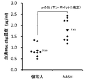
結果を図1に示した。平均値では、健常人が0.86μg/mlに対してNASH患者が1.81μg/mlと有意な増加が認められた。しかし、NASH患者の低値の検体(1.18μg/ml)よりも高い値を示した健常人の検体が2検体(1.28μg/ml、1.34μg/ml)存在した。この結果より、健常人とNASH患者で値の重なりが認められた。
[Reference example 1]
Measurement of Mac-2bp concentration by ELISA Using Human Mac-2 binding protein (Mac-2bp) Assay Kit manufactured by Immunobiological Research Institute Co., Ltd. (IBL), 9 sera of healthy subjects and 9 sera of NASH patients The Mac-2bp concentration was measured. The measurement method was performed according to the product data sheet.
The results are shown in FIG. The average value was 0.86 μg / ml for healthy subjects and 1.81 μg / ml for NASH patients, showing a significant increase. However, there were two samples (1.28 μg / ml, 1.34 μg / ml) of healthy subjects that showed higher values than the low values of NASH patients (1.18 μg / ml). From this result, overlapping values were observed in healthy subjects and NASH patients.
[参考例2]
全自動免疫測定装置によるMac−2結合蛋白糖鎖修飾異性体(M2BPGi)濃度測定
シスメックス株式会社製の肝臓の線維化検査用試薬(HISCL M2BPGi試薬)を用いて、健常人の血清9例NASH患者の血清9例のMac−2bp濃度の測定を行った。
結果を図2に示した。平均値では、健常人が0.56Cut−Off Index(C.O.I.)に対してNASH患者が1.00C.O.I.と増加しているものの統計的な有意な差は認められなかった。
[Reference example 2]
Measurement of Mac-2 binding protein sugar chain modified isomer (M2BPGi) concentration using a fully
The results are shown in FIG. On average, healthy subjects had 0.56 Cut-Off Index (COI) and NASH patients had 1.00 C.I. O. I. However, no statistically significant difference was observed.
[比較例1及び実施例1]ウエスタンブロッティング法を用いた分子量900k以上のMac−2bp測定
NASH鑑別のバイオマーカーである分子量900k以上のMac−2bpについて、Native PAGE電気泳動及びウエスタンブロッティング法を活用し、NASHの検出を実施した例を以下に示す。
[Comparative Example 1 and Example 1] Measurement of Mac-2bp having a molecular weight of 900 k or more using a Western blotting method For Mac-2bp having a molecular weight of 900 k or more, which is a biomarker for NASH differentiation, Native PAGE electrophoresis and Western blotting are utilized. , An example of detecting NASH is shown below.
スタンダードとしてIBL社製のMac−2bpを用いた。 Mac-2bp manufactured by IBL was used as a standard.
まず、以下の条件でNative PAGE電気泳動を行った。
(1)ゲル
4−20% Non−SDS PAGE mini 1mm 8well(TEFCO社製)
(2)電気泳動層
セイフティーセルミニSTC−808(TEFCO社製)
(3)泳動バッファー
0.025Mトリスヒドロキシメチルアミノメタン + 0.2Mグリシン
(4)サンプル希釈液
10%グリセロール + 0.02%ブロムフェノールブルー
(5)サンプル希釈倍率
5倍希釈
(6)泳動条件
18mA定電流 90分間
First, Native PAGE electrophoresis was performed under the following conditions.
(1) Gel 4-20% Non-SDS PAGE mini 1mm 8well (manufactured by TEFCO)
(2) Electrophoretic layer Safety cell mini STC-808 (manufactured by TEFCO)
(3) Migration buffer 0.025M Trishydroxymethylaminomethane + 0.2M glycerin (4)
次に、以下の条件でウエスタンブロッティングを行った。
(1)PVDFメンブレン
Amersham Hybond−P(GE Healthcare社製)
(2)ブロッティングバッファー
20%メタノール + 25mMトリスヒドロキシメチルアミノメタン + 192mMグリシン
(3)ブロッティング条件
65mA定電流 50分間
(4)ブロッキングバッファー
2%ECL Prime Blocking Reagent(GE Healthcare社製)+ 0.1%Tween 20 + リン酸塩緩衝液(PBS)
(5)ブロッキング条件
室温で1時間振盪後、冷蔵1晩
(6)一次抗体反応
Anti−Human Mac−2bp(8A2)Mouse IgG(IBL社製)
(7)二次抗体反応
HRP. Anti−Mouse IgA+IgG+IgM(H+L)(KPL社製)
(8)検出試薬
ECL Prime Solution1 + Solution2(GE Healthcare社製)
(9)検出機器
LAS−1000(富士フィルム社製)
Next, Western blotting was performed under the following conditions.
(1) PVDF Membrane Amersham Hybrid-P (manufactured by GE Healthcare)
(2)
(5) Blocking conditions After shaking at room temperature for 1 hour, refrigerate overnight (6) Primary antibody reaction Anti-Human Mac-2bp (8A2) Mouse IgG (manufactured by IBL)
(7) Secondary antibody reaction HRP. Anti-Mouse IgA + IgG + IgM (H + L) (manufactured by KPL)
(8) Detection reagent ECL Prime Solution1 + Solution2 (manufactured by GE Healthcare)
(9) Detection device LAS-1000 (manufactured by Fuji Film Co., Ltd.)
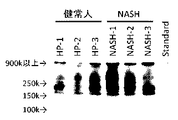
化学発光検出した画像を図3に示した。最上部のバンドを分子量900k以上のMac−2bpとした。画像解析ソフト(CSAnalyzer4,ATTO社製)を用いて、スタンダードを1として総Mac−2bpの濃度と分子量900k以上のMac−2bpの濃度を計算した。総Mac−2bpの濃度の結果を比較例1(図4a)に分子量900k以上のMac−2bpの濃度の結果を実施例1(図4b)に示した。又、3枚のゲルを用いて、健常人の血清9例、NASH患者の血清9例の総Mac−2bpの濃度と分子量900k以上のMac−2bpの濃度の結果を図5に示した。 The image in which chemiluminescence was detected is shown in FIG. The uppermost band was Mac-2bp having a molecular weight of 900 k or more. Using image analysis software (CSAnalizer4, manufactured by ATTO), the concentration of total Mac-2bp and the concentration of Mac-2bp having a molecular weight of 900 k or more were calculated with the standard as 1. The result of the total concentration of Mac-2bp is shown in Comparative Example 1 (FIG. 4a), and the result of the concentration of Mac-2bp having a molecular weight of 900 k or more is shown in Example 1 (FIG. 4b). In addition, the results of the total Mac-2bp concentration and the Mac-2bp concentration of 900 k or more in molecular weight of 9 healthy subjects and 9 NASH patients using 3 gels are shown in FIG.
比較例1(図4a)のように総Mac−2bp濃度では、健常人1例で10を上回り、NASH患者と同程度の値となったが、実施例1(図4b)のように分子量900k以上のMac−2bp濃度で比べると健常人とNASH患者が明確に分かれた。検体数をそれぞれ9例に増やして測定した結果、図5のように平均値においては、総Mac−2bp濃度(比較例)で健常人が7.07に対してNASH患者が17.05と有意な増加が認められた。しかし、NASH患者の低値の検体(7.76)よりも高い値を示した健常人の検体が3検体(8.55、11.46、14.07)存在した。この結果より、健常人とNASH患者で値の重なりが認められた。一方で、分子量900k以上のMac−2bp濃度(実施例)の平均値では健常人が2.09に対してNASH患者が10.96と有意な増加が認められた。又、NASH患者の低値の検体(4.31)よりも健常人の高値の検体(2.99)が低い値を示しており、健常人とNASH患者が明確に分かれた。以上より、実施例1は、比較例1、参考例1及び2に比べて、生体試料中の分子量900k以上のMac−2bp濃度により、健常人とNASH患者を明確に分けられることが明らかであった。 As shown in Comparative Example 1 (FIG. 4a), the total Mac-2bp concentration exceeded 10 in one healthy subject, which was about the same as that of NASH patients, but the molecular weight was 900 k as in Example 1 (FIG. 4b). When compared with the above Mac-2bp concentrations, healthy subjects and NASH patients were clearly separated. As a result of measuring by increasing the number of samples to 9 cases each, the average value as shown in FIG. 5 was significant with a total Mac-2bp concentration (comparative example) of 7.07 for healthy subjects and 17.05 for NASH patients. A significant increase was observed. However, there were 3 samples (8.55, 11.46, 14.07) of healthy subjects that showed higher values than the low values of NASH patients (7.76). From this result, overlapping values were observed in healthy subjects and NASH patients. On the other hand, the average value of the Mac-2bp concentration (Example) having a molecular weight of 900 k or more was 2.09 for healthy subjects and 10.96 for NASH patients, showing a significant increase. In addition, the high-value sample (2.99) of the healthy subject showed a lower value than the low-value sample (4.31) of the NASH patient, and the healthy subject and the NASH patient were clearly separated. From the above, it is clear that in Example 1, healthy subjects and NASH patients can be clearly separated by the Mac-2bp concentration in the biological sample having a molecular weight of 900 k or more, as compared with Comparative Example 1, Reference Examples 1 and 2. It was.
[実施例2]多量体のMac−2bpの比較
分子量の比較を行うために、IBL社製のMac−2bpとR&D SYSTEMS社製のRecombinant Human Galectin−3BP/MAC−2BPを実施例1と同様の方法でNative PAGE電気泳動及びウエスタンブロッティング法で評価した例を以下に示す。
[Example 2] Comparison of multimers Mac-2bp In order to compare molecular weights, Mac-2bp manufactured by IBL and Recombinant Human Galectin-3BP / MAC-2BP manufactured by R & D SYSTEMS were used in the same manner as in Example 1. An example evaluated by Native PAGE electrophoresis and Western blotting by the method is shown below.
化学発光検出した画像を図6に示した。R&D SYSTEMS社製のMac−2bpは主に10量体(約900k)又は12量体(約1080k)を形成しており、重量平均分子量が950kDaである。IBL社製のMac−2bpのバンドも同じ位置で検出されているため、分子量900k以上又は10量体以上のMac−2bpを測定することにより、NASHやNAFLDの進展度をより明確に判定することができる。 The image in which chemiluminescence was detected is shown in FIG. Mac-2bp manufactured by R & D SYSTEMS mainly forms a 10-mer (about 900 k) or a 12-mer (about 1080 k), and has a weight average molecular weight of 950 kDa. Since the band of Mac-2bp manufactured by IBL is also detected at the same position, the degree of progress of NASH and NAFLD can be more clearly determined by measuring Mac-2bp having a molecular weight of 900 k or more or a dimer or more. Can be done.
[実施例3及び比較例2]ラテックス凝集比濁法によるNASH患者及び健常人の血清Mac−2bp値の比較
実施例3−(a)はAnti−Human Mac−2bp(8A2)Mouse IgGを、実施例3−(b)はAnti−Human Mac−2bp(12A1)Mouse IgGを、実施例3−(c)はAnti−Human Mac−2bp(67A1)Mouse IgGを、実施例3−(d)はAnti−Human Mac−2bp(31A1)Mouse IgGを使用してラテックス凝集試薬を調製して、NASH患者及び健常人の血清中のMac−2bp濃度を測定した結果を示す(図7a、7b、7c、7d)。なお、用いた4種の抗Mac−2bp抗体はIBL社製である。
[Example 3 and Comparative Example 2] Comparison of serum Mac-2bp values of NASH patients and healthy subjects by the latex agglutination turbidimetric method Example 3- (a) was carried out using Anti-Human Mac-2bp (8A2) Mouse IgG. Example 3- (b) is Anti-Human Mac-2bp (12A1) Mouse IgG, Example 3- (c) is Anti-Human Mac-2bp (67A1) Mouse IgG, and Example 3- (d) is Anti. -Human Mac-2bp (31A1) Mouse IgG was used to prepare a latex agglutinating reagent, and the results of measuring the Mac-2bp concentration in the sera of NASH patients and healthy subjects are shown (FIGS. 7a, 7b, 7c, 7d). ). The four types of anti-Mac-2bp antibodies used are manufactured by IBL.
ラテックス凝集試薬は以下の様に調製した。
平均粒子径0.3μmのポリスチレンラテックス(固形分10%(W/V))100μLに、感作用緩衝液800μLを添加し、さらに抗Mac−2bp抗体 1mg/mLを100μL添加した。従って感作時には1%(W/V)ラテックス液となる。この溶液を25℃にて1時間振盪器にて110rpmで反応させた。その後、ブロッキング緩衝液を1mL添加し、25℃にて1.5時間振盪器にて反応させた。その後、5℃、15000rpmにて30分間遠心分離した。上清をアスピレータにて除去し、沈渣を得た。その沈渣にラテックス試薬用緩衝液を2mL加え、ボルテックスによりラテックスを分散させた。分散させたラテックス試薬を再び5℃、15000rpmにて30分間遠心分離することにより沈渣を洗浄した。先ほどと同様に上清をアスピレータにて除去し、沈渣を得た。この沈渣にラテックス試薬用緩衝液を2mL加え、ボルテックスにて分散させた後、さらに3mLのラテックス試薬用緩衝液を加えてよく攪拌後に超音波洗浄機にて分散処理を行い、固形分0.2%(W/V)のラテックス試薬とした。このようにして調整したラテックス試薬は4℃にて保存した。
The latex agglutination reagent was prepared as follows.
To 100 μL of polystyrene latex (
測定に使用した機器と測定条件は以下の通りである。
ラテックス試薬によるヒトMac−2bp量の測定は生化学用自動分析装置ビオリス24i Premium(東京貿易機械株式会社)を用いて行った。上記(1)で得られた固形分0.2%(W/V)のラテックス試薬をそのままR2試薬とした。測定条件は以下の通りである。
検体容量 10μL
検体希釈液(R1試薬) 200μL
試薬(R2試薬) 40μL
測定波長 750nm
測定温度 37℃
The equipment and measurement conditions used for the measurement are as follows.
The amount of human Mac-2bp using the latex reagent was measured using the biochemical automatic analyzer Biolis 24i Premium (Tokyo Boeki Holdings Corporation). The latex reagent having a solid content of 0.2% (W / V) obtained in (1) above was used as it was as an R2 reagent. The measurement conditions are as follows.
Specimen diluent (R1 reagent) 200 μL
Reagent (R2 reagent) 40 μL
Measurement wavelength 750 nm
Measurement temperature 37 ° C
R1試薬 200μLを添加し引き続いて検体10μLを添加後、37℃のセル内で5分間反応させる。その後、R2試薬 40μLを添加し約30秒後の吸光度と約360秒後の吸光度の差(△OD750)を測定し、吸光度変化量とした。また、検体の代わりに既知濃度の標準品をもちいて同様の測定を行い、予め検量線を作成しておき、上記検体の吸光度変化量を上記検量線に外挿して、検体中のヒトMac-2bp量を測定した。以上より、実施例3は、比較例2に比べて、健常人とNASH患者を明確に分けられることが明らかであった。 After adding 200 μL of R1 reagent and subsequently adding 10 μL of sample, the reaction is carried out in a cell at 37 ° C. for 5 minutes. Then, 40 μL of the R2 reagent was added, and the difference between the absorbance after about 30 seconds and the absorbance after about 360 seconds (ΔOD750) was measured and used as the amount of change in absorbance. Further, the same measurement is performed using a standard product having a known concentration instead of the sample, a calibration curve is prepared in advance, and the amount of change in the absorbance of the sample is extrapolated to the calibration curve to obtain human Mac- in the sample. The amount of 2 bp was measured. From the above, it was clear that in Example 3, healthy subjects and NASH patients could be clearly separated as compared with Comparative Example 2.
[実施例4]ラテックス凝集比濁法による炎症疾患、癌及び健常人の血清Mac−2bp値の比較
実施例3−(b)で使用した抗体を含むラテックス凝集試薬を用いて、炎症疾患(肝炎、肝硬変)、癌(肝臓癌、前立腺癌)及び健常人の血清中のMac−2bp濃度を測定した結果を示す(図8)。
[Example 4] Comparison of serum Mac-2bp values of inflammatory diseases, cancer and healthy subjects by the latex agglutination turbidimetric method Inflammatory diseases (hepatitis) using the latex agglutination reagent containing the antibody used in Example 3- (b). , Liver cirrhosis), cancer (liver cancer, prostate cancer) and the results of measuring the Mac-2bp concentration in the serum of healthy subjects are shown (FIG. 8).
Claims (7)
A test method for determining the possibility of suffering from hepatitis, liver cirrhosis, liver cancer, or prostate cancer, which comprises detecting a Mac-2bp concentration detected as having a molecular weight of 900 kDa or more in a blood sample.
Priority Applications (1)
| Application Number | Priority Date | Filing Date | Title |
|---|---|---|---|
| JP2016204978A JP6893406B2 (en) | 2016-10-19 | 2016-10-19 | Testing method for progress of NASH and NAFLD and testing method for inflammatory diseases and cancer |
Applications Claiming Priority (1)
| Application Number | Priority Date | Filing Date | Title |
|---|---|---|---|
| JP2016204978A JP6893406B2 (en) | 2016-10-19 | 2016-10-19 | Testing method for progress of NASH and NAFLD and testing method for inflammatory diseases and cancer |
Publications (2)
| Publication Number | Publication Date |
|---|---|
| JP2018066631A JP2018066631A (en) | 2018-04-26 |
| JP6893406B2 true JP6893406B2 (en) | 2021-06-23 |
Family
ID=62086013
Family Applications (1)
| Application Number | Title | Priority Date | Filing Date |
|---|---|---|---|
| JP2016204978A Active JP6893406B2 (en) | 2016-10-19 | 2016-10-19 | Testing method for progress of NASH and NAFLD and testing method for inflammatory diseases and cancer |
Country Status (1)
| Country | Link |
|---|---|
| JP (1) | JP6893406B2 (en) |
Families Citing this family (1)
| Publication number | Priority date | Publication date | Assignee | Title |
|---|---|---|---|---|
| WO2021010349A1 (en) * | 2019-07-12 | 2021-01-21 | 国立大学法人北海道大学 | Development of blood fibrosis marker for non-alcoholic steatohepatitis |
Family Cites Families (5)
| Publication number | Priority date | Publication date | Assignee | Title |
|---|---|---|---|---|
| WO2004039844A1 (en) * | 2002-10-31 | 2004-05-13 | Tss Biotech Inc. | Prostate cancer tumor marker |
| KR101470108B1 (en) * | 2009-07-14 | 2014-12-05 | 도꾸리쯔교세이호진 상교기쥬쯔 소고겡뀨죠 | Method for measurement of glycoprotein, method for detection of hepatic diseases, reagent for quantification of glycoprotein, and sugar chain marker glycoprotein as measure of disease conditions of hepatic diseases |
| CN108957006B (en) * | 2013-03-15 | 2021-07-27 | 私募蛋白质体公司 | Non-alcoholic fatty liver disease (NAFLD) and non-alcoholic steatohepatitis (NASH) biomarkers and uses thereof |
| KR101559101B1 (en) * | 2013-11-28 | 2015-10-12 | 한국기초과학지원연구원 | Polypeptide markers for cancer diagnosis derived from blood sample and methods for the diagnosis of cancers using the same |
| EP3282256A4 (en) * | 2015-04-10 | 2019-03-06 | Social Welfare Organization Saiseikai Imperial Gift Foundation, Inc. | Method for discriminating symptom of hepatic disease |
-
2016
- 2016-10-19 JP JP2016204978A patent/JP6893406B2/en active Active
Also Published As
| Publication number | Publication date |
|---|---|
| JP2018066631A (en) | 2018-04-26 |
Similar Documents
| Publication | Publication Date | Title |
|---|---|---|
| Akiyama et al. | Prevalence of anti-phospholipase A2 receptor antibodies in Japanese patients with membranous nephropathy | |
| NL2001577C2 (en) | Device and method for separating and analyzing blood. | |
| US20120219943A1 (en) | Methods of prognosis and diagnosis in chronic heart failure | |
| JP6666916B2 (en) | Testing methods for kidney disease | |
| TWI704228B (en) | How to check for liver cancer | |
| CN102124340B (en) | IgA nephropathy detection method and detection kit | |
| JP7448831B2 (en) | Methods, biomarkers, reagent kits and devices to assist in the diagnosis of Alzheimer's dementia or mild cognitive impairment | |
| Porcelli et al. | Assessment of a Test for the Screening and Diagnosis of Celiac Disease | |
| CA2983542A1 (en) | A method for predicting the risk of incidence of chronic kidney disease | |
| WO2014027188A1 (en) | Kidney disease biomarker | |
| JP7109441B2 (en) | Diagnostic aid method for determining neurodegenerative disease | |
| CN105717308A (en) | Immunochromatography kit for fast and quantitatively detecting fecal lactoferrin | |
| JP2024019509A (en) | DIAGNOSIS MARKER AND DIAGNOSIS KIT FOR ALZHEIMER'S DISEASE OR PRESYMPTOMATIC ALZHEIMER'S DISEASE, METHOD FOR EVALUATING ACCUMULATION AMOUNT OF AMYLOID β-PROTEIN IN TO BRAIN, AND IN VITRO METHOD FOR ASSISTING IN DETECTION OF ALZHEIMER'S DISEASE OR PRESYMPTOMATIC ALZHEIMER'S DISEASE IN SUBJECT | |
| Craig et al. | Serum C‐reactive protein and S100A12 concentrations in dogs with hepatic disease | |
| JP6893406B2 (en) | Testing method for progress of NASH and NAFLD and testing method for inflammatory diseases and cancer | |
| WO2016129631A1 (en) | Method and kit for detecting kawasaki disease | |
| Vicente-Steijn et al. | Analytical and clinical performance of the fully-automated LIAISONXL calprotectin immunoassay from DiaSorin in IBD patients | |
| Kraemer et al. | Automated Fecal Biomarker Profiling-a Convenient Procedure to Support Diagnosis for Patients with Inflammatory Bowel Diseases. | |
| JP7348604B2 (en) | Methods, biomarkers, reagent kits and devices to aid in the diagnosis of Parkinson's disease | |
| US20100081149A1 (en) | Novel oxidized ldl complex and method for detection thereof | |
| CN110687285B (en) | Diagnostic kit and application of MAK16 in preparation of early diagnosis reagent for systemic lupus erythematosus | |
| US20180095090A1 (en) | Biomarkers for assessment of preeclampsia | |
| US20230055382A1 (en) | Detecting gut barrier dysfunction and/or cirrhosis | |
| CN102959398B (en) | Marker containing HPaR as active ingredient for diagnosing lung cancer | |
| WO2018073618A1 (en) | Methods and kits for diagnosing alcoholic hepatitis |
Legal Events
| Date | Code | Title | Description |
|---|---|---|---|
| A621 | Written request for application examination |
Free format text: JAPANESE INTERMEDIATE CODE: A621 Effective date: 20191016 |
|
| A977 | Report on retrieval |
Free format text: JAPANESE INTERMEDIATE CODE: A971007 Effective date: 20200930 |
|
| A131 | Notification of reasons for refusal |
Free format text: JAPANESE INTERMEDIATE CODE: A131 Effective date: 20201002 |
|
| A521 | Request for written amendment filed |
Free format text: JAPANESE INTERMEDIATE CODE: A523 Effective date: 20201124 |
|
| A02 | Decision of refusal |
Free format text: JAPANESE INTERMEDIATE CODE: A02 Effective date: 20201208 |
|
| A521 | Request for written amendment filed |
Free format text: JAPANESE INTERMEDIATE CODE: A523 Effective date: 20210205 |
|
| C60 | Trial request (containing other claim documents, opposition documents) |
Free format text: JAPANESE INTERMEDIATE CODE: C60 Effective date: 20210205 |
|
| A911 | Transfer to examiner for re-examination before appeal (zenchi) |
Free format text: JAPANESE INTERMEDIATE CODE: A911 Effective date: 20210216 |
|
| C21 | Notice of transfer of a case for reconsideration by examiners before appeal proceedings |
Free format text: JAPANESE INTERMEDIATE CODE: C21 Effective date: 20210217 |
|
| A131 | Notification of reasons for refusal |
Free format text: JAPANESE INTERMEDIATE CODE: A131 Effective date: 20210322 |
|
| A521 | Request for written amendment filed |
Free format text: JAPANESE INTERMEDIATE CODE: A523 Effective date: 20210511 |
|
| TRDD | Decision of grant or rejection written | ||
| A01 | Written decision to grant a patent or to grant a registration (utility model) |
Free format text: JAPANESE INTERMEDIATE CODE: A01 Effective date: 20210524 |
|
| A61 | First payment of annual fees (during grant procedure) |
Free format text: JAPANESE INTERMEDIATE CODE: A61 Effective date: 20210601 |
|
| R150 | Certificate of patent or registration of utility model |
Ref document number: 6893406 Country of ref document: JP Free format text: JAPANESE INTERMEDIATE CODE: R150 |
|
| R250 | Receipt of annual fees |
Free format text: JAPANESE INTERMEDIATE CODE: R250 |