JP6880330B2 - Bone substitute material - Google Patents
Bone substitute material Download PDFInfo
- Publication number
- JP6880330B2 JP6880330B2 JP2020532744A JP2020532744A JP6880330B2 JP 6880330 B2 JP6880330 B2 JP 6880330B2 JP 2020532744 A JP2020532744 A JP 2020532744A JP 2020532744 A JP2020532744 A JP 2020532744A JP 6880330 B2 JP6880330 B2 JP 6880330B2
- Authority
- JP
- Japan
- Prior art keywords
- hap
- cap
- hydroxyapatite
- bone
- calcium phosphate
- Prior art date
- Legal status (The legal status is an assumption and is not a legal conclusion. Google has not performed a legal analysis and makes no representation as to the accuracy of the status listed.)
- Active
Links
Images


Classifications
-
- A—HUMAN NECESSITIES
- A61—MEDICAL OR VETERINARY SCIENCE; HYGIENE
- A61L—METHODS OR APPARATUS FOR STERILISING MATERIALS OR OBJECTS IN GENERAL; DISINFECTION, STERILISATION OR DEODORISATION OF AIR; CHEMICAL ASPECTS OF BANDAGES, DRESSINGS, ABSORBENT PADS OR SURGICAL ARTICLES; MATERIALS FOR BANDAGES, DRESSINGS, ABSORBENT PADS OR SURGICAL ARTICLES
- A61L27/00—Materials for grafts or prostheses or for coating grafts or prostheses
- A61L27/02—Inorganic materials
- A61L27/12—Phosphorus-containing materials, e.g. apatite
-
- A—HUMAN NECESSITIES
- A61—MEDICAL OR VETERINARY SCIENCE; HYGIENE
- A61F—FILTERS IMPLANTABLE INTO BLOOD VESSELS; PROSTHESES; DEVICES PROVIDING PATENCY TO, OR PREVENTING COLLAPSING OF, TUBULAR STRUCTURES OF THE BODY, e.g. STENTS; ORTHOPAEDIC, NURSING OR CONTRACEPTIVE DEVICES; FOMENTATION; TREATMENT OR PROTECTION OF EYES OR EARS; BANDAGES, DRESSINGS OR ABSORBENT PADS; FIRST-AID KITS
- A61F2/00—Filters implantable into blood vessels; Prostheses, i.e. artificial substitutes or replacements for parts of the body; Appliances for connecting them with the body; Devices providing patency to, or preventing collapsing of, tubular structures of the body, e.g. stents
- A61F2/02—Prostheses implantable into the body
- A61F2/28—Bones
-
- A—HUMAN NECESSITIES
- A61—MEDICAL OR VETERINARY SCIENCE; HYGIENE
- A61L—METHODS OR APPARATUS FOR STERILISING MATERIALS OR OBJECTS IN GENERAL; DISINFECTION, STERILISATION OR DEODORISATION OF AIR; CHEMICAL ASPECTS OF BANDAGES, DRESSINGS, ABSORBENT PADS OR SURGICAL ARTICLES; MATERIALS FOR BANDAGES, DRESSINGS, ABSORBENT PADS OR SURGICAL ARTICLES
- A61L24/00—Surgical adhesives or cements; Adhesives for colostomy devices
- A61L24/0047—Composite materials, i.e. containing one material dispersed in a matrix of the same or different material
- A61L24/0052—Composite materials, i.e. containing one material dispersed in a matrix of the same or different material with an inorganic matrix
- A61L24/0063—Phosphorus containing materials, e.g. apatite
-
- A—HUMAN NECESSITIES
- A61—MEDICAL OR VETERINARY SCIENCE; HYGIENE
- A61L—METHODS OR APPARATUS FOR STERILISING MATERIALS OR OBJECTS IN GENERAL; DISINFECTION, STERILISATION OR DEODORISATION OF AIR; CHEMICAL ASPECTS OF BANDAGES, DRESSINGS, ABSORBENT PADS OR SURGICAL ARTICLES; MATERIALS FOR BANDAGES, DRESSINGS, ABSORBENT PADS OR SURGICAL ARTICLES
- A61L27/00—Materials for grafts or prostheses or for coating grafts or prostheses
- A61L27/28—Materials for coating prostheses
- A61L27/30—Inorganic materials
- A61L27/32—Phosphorus-containing materials, e.g. apatite
-
- A—HUMAN NECESSITIES
- A61—MEDICAL OR VETERINARY SCIENCE; HYGIENE
- A61L—METHODS OR APPARATUS FOR STERILISING MATERIALS OR OBJECTS IN GENERAL; DISINFECTION, STERILISATION OR DEODORISATION OF AIR; CHEMICAL ASPECTS OF BANDAGES, DRESSINGS, ABSORBENT PADS OR SURGICAL ARTICLES; MATERIALS FOR BANDAGES, DRESSINGS, ABSORBENT PADS OR SURGICAL ARTICLES
- A61L27/00—Materials for grafts or prostheses or for coating grafts or prostheses
- A61L27/40—Composite materials, i.e. containing one material dispersed in a matrix of the same or different material
- A61L27/42—Composite materials, i.e. containing one material dispersed in a matrix of the same or different material having an inorganic matrix
- A61L27/425—Composite materials, i.e. containing one material dispersed in a matrix of the same or different material having an inorganic matrix of phosphorus containing material, e.g. apatite
-
- A—HUMAN NECESSITIES
- A61—MEDICAL OR VETERINARY SCIENCE; HYGIENE
- A61L—METHODS OR APPARATUS FOR STERILISING MATERIALS OR OBJECTS IN GENERAL; DISINFECTION, STERILISATION OR DEODORISATION OF AIR; CHEMICAL ASPECTS OF BANDAGES, DRESSINGS, ABSORBENT PADS OR SURGICAL ARTICLES; MATERIALS FOR BANDAGES, DRESSINGS, ABSORBENT PADS OR SURGICAL ARTICLES
- A61L27/00—Materials for grafts or prostheses or for coating grafts or prostheses
- A61L27/40—Composite materials, i.e. containing one material dispersed in a matrix of the same or different material
- A61L27/44—Composite materials, i.e. containing one material dispersed in a matrix of the same or different material having a macromolecular matrix
- A61L27/46—Composite materials, i.e. containing one material dispersed in a matrix of the same or different material having a macromolecular matrix with phosphorus-containing inorganic fillers
-
- A—HUMAN NECESSITIES
- A61—MEDICAL OR VETERINARY SCIENCE; HYGIENE
- A61L—METHODS OR APPARATUS FOR STERILISING MATERIALS OR OBJECTS IN GENERAL; DISINFECTION, STERILISATION OR DEODORISATION OF AIR; CHEMICAL ASPECTS OF BANDAGES, DRESSINGS, ABSORBENT PADS OR SURGICAL ARTICLES; MATERIALS FOR BANDAGES, DRESSINGS, ABSORBENT PADS OR SURGICAL ARTICLES
- A61L27/00—Materials for grafts or prostheses or for coating grafts or prostheses
- A61L27/50—Materials characterised by their function or physical properties, e.g. injectable or lubricating compositions, shape-memory materials, surface modified materials
- A61L27/56—Porous materials, e.g. foams or sponges
-
- A—HUMAN NECESSITIES
- A61—MEDICAL OR VETERINARY SCIENCE; HYGIENE
- A61L—METHODS OR APPARATUS FOR STERILISING MATERIALS OR OBJECTS IN GENERAL; DISINFECTION, STERILISATION OR DEODORISATION OF AIR; CHEMICAL ASPECTS OF BANDAGES, DRESSINGS, ABSORBENT PADS OR SURGICAL ARTICLES; MATERIALS FOR BANDAGES, DRESSINGS, ABSORBENT PADS OR SURGICAL ARTICLES
- A61L27/00—Materials for grafts or prostheses or for coating grafts or prostheses
- A61L27/50—Materials characterised by their function or physical properties, e.g. injectable or lubricating compositions, shape-memory materials, surface modified materials
- A61L27/58—Materials at least partially resorbable by the body
-
- B—PERFORMING OPERATIONS; TRANSPORTING
- B33—ADDITIVE MANUFACTURING TECHNOLOGY
- B33Y—ADDITIVE MANUFACTURING, i.e. MANUFACTURING OF THREE-DIMENSIONAL [3-D] OBJECTS BY ADDITIVE DEPOSITION, ADDITIVE AGGLOMERATION OR ADDITIVE LAYERING, e.g. BY 3-D PRINTING, STEREOLITHOGRAPHY OR SELECTIVE LASER SINTERING
- B33Y80/00—Products made by additive manufacturing
-
- A—HUMAN NECESSITIES
- A61—MEDICAL OR VETERINARY SCIENCE; HYGIENE
- A61F—FILTERS IMPLANTABLE INTO BLOOD VESSELS; PROSTHESES; DEVICES PROVIDING PATENCY TO, OR PREVENTING COLLAPSING OF, TUBULAR STRUCTURES OF THE BODY, e.g. STENTS; ORTHOPAEDIC, NURSING OR CONTRACEPTIVE DEVICES; FOMENTATION; TREATMENT OR PROTECTION OF EYES OR EARS; BANDAGES, DRESSINGS OR ABSORBENT PADS; FIRST-AID KITS
- A61F2/00—Filters implantable into blood vessels; Prostheses, i.e. artificial substitutes or replacements for parts of the body; Appliances for connecting them with the body; Devices providing patency to, or preventing collapsing of, tubular structures of the body, e.g. stents
- A61F2/02—Prostheses implantable into the body
- A61F2/28—Bones
- A61F2002/2835—Bone graft implants for filling a bony defect or an endoprosthesis cavity, e.g. by synthetic material or biological material
-
- A—HUMAN NECESSITIES
- A61—MEDICAL OR VETERINARY SCIENCE; HYGIENE
- A61F—FILTERS IMPLANTABLE INTO BLOOD VESSELS; PROSTHESES; DEVICES PROVIDING PATENCY TO, OR PREVENTING COLLAPSING OF, TUBULAR STRUCTURES OF THE BODY, e.g. STENTS; ORTHOPAEDIC, NURSING OR CONTRACEPTIVE DEVICES; FOMENTATION; TREATMENT OR PROTECTION OF EYES OR EARS; BANDAGES, DRESSINGS OR ABSORBENT PADS; FIRST-AID KITS
- A61F2310/00—Prostheses classified in A61F2/28 or A61F2/30 - A61F2/44 being constructed from or coated with a particular material
- A61F2310/00005—The prosthesis being constructed from a particular material
- A61F2310/00179—Ceramics or ceramic-like structures
- A61F2310/00293—Ceramics or ceramic-like structures containing a phosphorus-containing compound, e.g. apatite
-
- A—HUMAN NECESSITIES
- A61—MEDICAL OR VETERINARY SCIENCE; HYGIENE
- A61L—METHODS OR APPARATUS FOR STERILISING MATERIALS OR OBJECTS IN GENERAL; DISINFECTION, STERILISATION OR DEODORISATION OF AIR; CHEMICAL ASPECTS OF BANDAGES, DRESSINGS, ABSORBENT PADS OR SURGICAL ARTICLES; MATERIALS FOR BANDAGES, DRESSINGS, ABSORBENT PADS OR SURGICAL ARTICLES
- A61L2400/00—Materials characterised by their function or physical properties
- A61L2400/12—Nanosized materials, e.g. nanofibres, nanoparticles, nanowires, nanotubes; Nanostructured surfaces
-
- A—HUMAN NECESSITIES
- A61—MEDICAL OR VETERINARY SCIENCE; HYGIENE
- A61L—METHODS OR APPARATUS FOR STERILISING MATERIALS OR OBJECTS IN GENERAL; DISINFECTION, STERILISATION OR DEODORISATION OF AIR; CHEMICAL ASPECTS OF BANDAGES, DRESSINGS, ABSORBENT PADS OR SURGICAL ARTICLES; MATERIALS FOR BANDAGES, DRESSINGS, ABSORBENT PADS OR SURGICAL ARTICLES
- A61L2400/00—Materials characterised by their function or physical properties
- A61L2400/18—Modification of implant surfaces in order to improve biocompatibility, cell growth, fixation of biomolecules, e.g. plasma treatment
-
- A—HUMAN NECESSITIES
- A61—MEDICAL OR VETERINARY SCIENCE; HYGIENE
- A61L—METHODS OR APPARATUS FOR STERILISING MATERIALS OR OBJECTS IN GENERAL; DISINFECTION, STERILISATION OR DEODORISATION OF AIR; CHEMICAL ASPECTS OF BANDAGES, DRESSINGS, ABSORBENT PADS OR SURGICAL ARTICLES; MATERIALS FOR BANDAGES, DRESSINGS, ABSORBENT PADS OR SURGICAL ARTICLES
- A61L2430/00—Materials or treatment for tissue regeneration
- A61L2430/02—Materials or treatment for tissue regeneration for reconstruction of bones; weight-bearing implants
Description
本発明は、均質な粗い外表面を有するリン酸カルシウム/ヒドロキシアパタイト(CAP/HAP)に基づく二層構造を持つ新しい二相性骨代替材料、その材料の製造方法、並びにヒト又は動物の欠損部位における骨形成、骨再生、骨修復及び/又は骨置換を支持するためのインプラント又はプロテーゼとしてのこれらの使用に関する。 The present invention presents a novel biphasic bone replacement material with a bilayer structure based on calcium phosphate / hydroxyapatite (CAP / HAP) with a homogeneous and rough outer surface, a method for producing the material, and bone formation at a human or animal defect site. With respect to their use as implants or prostheses to support bone regeneration, bone repair and / or bone replacement.
骨構造の欠損は、外傷、疾患、及び手術のような様々な状況で発生するが、種々の外科分野における骨欠損を効果的に修復する必要が依然として存在する。 Defects in bone structure occur in a variety of situations such as trauma, disease, and surgery, but there is still a need to effectively repair bone defects in various surgical disciplines.
骨欠損部位の治癒を刺激するために、数多くの天然及び合成の材料及び組成物が使用されてきた。歯周及び顎顔面骨欠損で骨成長を促進する、周知の天然の骨伝導性の骨代替材料は、Geistlich Pharma AGから市販されているGeistlich Bio-Oss(登録商標)である。その材料は、米国特許第5,167,961号に記載されている方法によって自然骨から製造され、自然骨の小柱構造及びナノ結晶構造の保存を可能にして、吸収されないか又は非常にゆっくりと吸収される優れた骨伝導性マトリックスが得られる。 Numerous natural and synthetic materials and compositions have been used to stimulate healing of bone defect sites. A well-known natural bone-conducting bone replacement material that promotes bone growth in periodontal and maxillofacial bone defects is Geistlich Bio-Oss®, commercially available from Geistlich Pharma AG. The material is produced from natural bone by the method described in US Pat. No. 5,167,961 and allows the preservation of the trabecular and nanocrystal structures of natural bone, which is not absorbed or is absorbed very slowly. An excellent bone conduction matrix is obtained.
リン酸三カルシウム/ヒドロキシアパタイト(TCP/HAP)システム及び骨代替材料としてのこれらの使用は、例えば、US-6,338,752に記載されており、リン酸アンモニウムとHAPとの粉末混合物を1200〜1500℃で加熱して、α−TCP/HAPの二相セメントの調製方法を開示している。 Their use as tricalcium phosphate / hydroxyapatite (TCP / HAP) systems and bone replacement materials is described, for example, in US-6,338,752, where a powder mixture of ammonium phosphate and HAP is prepared at 1200-1500 ° C. It discloses a method for preparing a two-phase cement of α-TCP / HAP by heating.
欧州特許EP-285826は、インプラント用の金属及び非金属体上のHAPの層を製造する方法であって、α−TCPの層を適用し、80〜100℃でpH2〜7の水との反応によりα−TCP層をHAPに完全に変換することによる方法を記載している。得られた生成物は、HAPの層で覆われた金属又は非金属体である。 European Patent EP-285826 is a method of producing a layer of HAP on metallic and non-metallic bodies for implants, applying a layer of α-TCP and reacting with water at pH 2-7 at 80-100 ° C. Describes the method by completely converting the α-TCP layer into HAP. The product obtained is a metallic or non-metallic body covered with a layer of HAP.
WO 97/41273は、特にヒドロキシアパタイト(HAP)又は他のリン酸カルシウム(CAP)などの基材を炭酸ヒドロキシアパタイト、即ち、リン酸及び/又はヒドロキシルイオンが重炭酸イオンによって部分的に置換されているヒドロキシアパタイトのコーティングでコーティングする方法であって、(a)50℃未満の温度でカルシウムイオン、リン酸イオン及び重炭酸イオンを含有するpH6.8〜8.0の溶液に基材を浸漬し、(b)基材と接触している溶液の部分をpHが8を超えるまで50〜80℃の温度まで加熱して、(c)基材と工程(b)で得られたアルカリ溶液との接触を維持して、炭酸ヒドロキシアパタイトコーティングを形成し、そして(d)基材を溶液から取り出し、コーティングを乾燥させることを含む方法を記載している。重炭酸イオンは、ヒドロキシアパタイト結晶成長の阻害剤として作用するため、欠損を含み、かつ寸法がかなり小さい、即ち、長さが10〜40nm、幅が3〜10nmの非化学量論的結晶が得られることが開示されている(7ページ、1〜7行目を参照のこと)。 WO 97/41273 is a hydroxyapatite carbonate, especially in which a substrate such as hydroxyapatite (HAP) or other calcium phosphate (CAP) is partially substituted with bicarbonate, that is, phosphate and / or hydroxyl ion is partially substituted with bicarbonate ion. A method of coating with an apatite coating, in which (a) the substrate is immersed in a solution containing calcium ions, phosphate ions and bicarbonate ions at a temperature of less than 50 ° C. and having a pH of 6.8 to 8.0. b) The portion of the solution in contact with the substrate is heated to a temperature of 50-80 ° C until the pH exceeds 8, and (c) the substrate is brought into contact with the alkaline solution obtained in step (b). Methods are described that involve maintaining to form a bicarbonate apatite coating, and (d) removing the substrate from solution and drying the coating. Bicarbonate acts as an inhibitor of hydroxyapatite crystal growth, resulting in non-stoichiometric crystals that contain defects and are fairly small in size, i.e. 10-40 nm long and 3-10 nm wide. (See page 7, lines 1-7).
リン酸カルシウム/ヒドロキシアパタイト(CAP/HAP)システム、特にTCP/HAPシステムの成分は、その熱力学的安定性が異なる。この違いにより、CAP/HAPシステムが哺乳動物、特にヒト患者に埋め込まれると、体液中のTCP及びその他のリン酸カルシウムの溶解度は、HAPの溶解度よりも高い。リン酸カルシウムとHAPの間の溶解度の違いにより、CAP/HAPシステムの不規則な焼結構造が崩壊するが、これは、溶解性の高い方の化合物CAP(例えば、TCP)がHAPよりも速く除去されるためである。高温で生成したCAPとHAPの間の焼結相互接合は、生理環境でのデバイスのより高い溶解度にも大きく貢献しよう。2つの異なるタイプの反応が、このようなセラミックの加速したインビボ分解を支配する:化学溶解及び細胞による生物学的吸収。どちらのプロセスもセラミック材料の溶解を引き起こし、更にはカルシウムイオンの局所的な過飽和を引き起こして、吸着されるカルシウムイオンよりも多くのカルシウムイオンが放出される。カルシウムイオンの自然の平衡は、細胞外マトリックスにも、インプラント周囲の組織にも最早存在しない。カルシウムイオンの過飽和に関する自然のカルシウム平衡の局所的な撹乱は、破骨細胞の活性を上昇させ、ひいてはセラミック材料の制御されない吸収を加速し、特に大量の合成骨代替材料を使用する場合に有害な炎症反応のリスクにつながる。 The components of the calcium phosphate / hydroxyapatite (CAP / HAP) system, especially the TCP / HAP system, differ in their thermodynamic stability. Due to this difference, when the CAP / HAP system is implanted in mammals, especially human patients, the solubility of TCP and other calcium phosphates in body fluids is higher than that of HAP. The difference in solubility between calcium phosphate and HAP disrupts the irregular sintered structure of the CAP / HAP system, which removes the more soluble compound CAP (eg TCP) faster than HAP. Because. Sintered interconnects between CAP and HAP produced at high temperatures will also contribute significantly to the higher solubility of the device in the physiological environment. Two different types of reactions govern the accelerated in vivo degradation of such ceramics: chemical lysis and biological absorption by cells. Both processes cause dissolution of the ceramic material and also cause local supersaturation of calcium ions, releasing more calcium ions than adsorbed calcium ions. The natural equilibrium of calcium ions is no longer present in the extracellular matrix or in the tissue surrounding the implant. Local disturbances in the natural calcium equilibrium with respect to calcium ion supersaturation increase osteoclast activity and thus accelerate uncontrolled absorption of ceramic materials, which is detrimental, especially when large amounts of synthetic bone substitutes are used. Leads to the risk of inflammatory reactions.
骨代替材料のGeistlich Bio-Ossをヒト患者に埋め込むと、自然のカルシウム平衡は実質的に影響を受けず、材料の表面上及びその局所環境内のカルシウムイオンの濃度はほぼ一定のままである。よって材料の生物学的吸収は起こらないか、又は有害な炎症反応のリスクを伴わない非常に遅い速度で進行する。 When the bone replacement material Geistlich Bio-Oss is implanted in human patients, the natural calcium equilibrium is substantially unaffected and the concentration of calcium ions on the surface of the material and within its local environment remains nearly constant. Thus, biological absorption of the material does not occur or proceeds at a very slow rate without the risk of adverse inflammatory reactions.
EP-B1-2445543は、非常に有利なリン酸カルシウム/ヒドロキシアパタイト(CAP/HAP)骨代替材料を開示しているが、これは、骨代替材料のGeistlich Bio-Ossと同様に、体内に設置後、材料の表面上及びその局所環境内のカルシウムイオンの濃度をほぼ一定に維持することができ、よって破骨細胞活性の上昇につながらない。 EP-B1-2445543 discloses a highly advantageous calcium phosphate / hydroxyapatite (CAP / HAP) bone replacement material, which, like the bone replacement material Geistlich Bio-Oss, is used after placement in the body. The concentration of calcium ions on the surface of the material and in its local environment can be maintained almost constant, and thus does not lead to an increase in osteoclast activity.
実際、最適な骨再生に必要な自然のカルシウム平衡は乱されたり破壊されたりしない。更に、自然のカルシウム濃度の平衡は、再生プロセスが完了するまで、骨代替材料によって永続的に支持される。これらの条件が満たされると、破骨細胞活性が上昇しないため、有害な炎症反応のリスクがない。 In fact, the natural calcium equilibrium required for optimal bone regeneration is not disturbed or disrupted. In addition, the natural calcium concentration equilibrium is permanently supported by bone replacement materials until the regeneration process is complete. When these conditions are met, osteoclast activity does not increase and there is no risk of adverse inflammatory reactions.
EP-B1-2445543の発明は、焼結CAPコアと、焼結CAPコアの上に堆積したナノ結晶HAPの少なくとも1つの均質で閉じたエピタキシャル成長層(ここで、エピタキシャル成長ナノ結晶は、ヒト骨塩と同じサイズと形態、即ち、長さ30〜46nm及び幅14〜22nmを有する)とを含む、二相性リン酸カルシウム/ヒドロキシアパタイト(CAP/HAP)骨代替材料に関する。 The invention of EP-B1-2445543 is that the sintered CAP core and at least one homogeneous and closed epitaxial growth layer of nanocrystal HAP deposited on the sintered CAP core (where the epitaxial growth nanocrystals are human bone salts). It relates to a biphasic calcium phosphate / hydroxyapatite (CAP / HAP) bone substitute material, including (having a length of 30-46 nm and a width of 14-22 nm) of the same size and morphology.
焼結CAPコアは、リン酸三カルシウム(TCP)、特にα−TCP(α−Ca3(PO4)2)又はβ−TCP(β−Ca3(PO4)2)、及び/又はリン酸四カルシウム(TTCP)Ca4(PO4)2Oを含んでもよい。 Sintered CAP cores are tricalcium phosphate (TCP), especially α-TCP (α-Ca 3 (PO 4 ) 2 ) or β-TCP (β-Ca 3 (PO 4 ) 2 ), and / or phosphate. It may contain tetracalcium (TTCP) Ca 4 (PO 4 ) 2 O.
頻繁に使用される実施態様では、焼結CAPコアは、本質的にTCPからなり、α−TCPが好ましい。 In a frequently used embodiment, the sintered CAP core consists essentially of TCP, preferably α-TCP.
ナノ結晶HAPのエピタキシャル成長層は、構造的及び化学的に天然のヒト骨塩とほぼ同一である。 The epitaxial growth layer of nanocrystal HAP is structurally and chemically identical to natural human bone mineral.
ナノ結晶HAPのエピタキシャル成長層は一般に、少なくとも15〜50nm、好ましくは少なくとも20〜40nm、更に好ましくは少なくとも25〜35nmの厚さを有する。その最小厚さは、エピタキシャル配向のHAPナノ結晶の1つの層に対応する。 The epitaxial growth layer of nanocrystal HAP generally has a thickness of at least 15-50 nm, preferably at least 20-40 nm, more preferably at least 25-35 nm. Its minimum thickness corresponds to one layer of epitaxially oriented HAP nanocrystals.
ナノ結晶HAPのエピタキシャル成長層は、エピタキシャル配向のHAPナノ結晶の単層又は多層を含むことができる。エピタキシャル配向のHAPナノ結晶のそのような層の数に関連する、ナノ結晶HAPのエピタキシャル成長層の厚さは、身体の負荷が異なる部位におけるインプラント又はプロテーゼとしての骨代替材料の意図される用途により選択されよう。その発明の骨代替材料は確かに、焼結CAPコアをサイズと形態がヒト骨塩に類似したヒドロキシアパタイトへと徐々に変換する生体様システムとしてインビボで機能するように設計されており、その変換速度は、焼結CAPコアによるカルシウム放出の速度に依存し、そして大部分はナノ結晶HAPのエピタキシャル成長層の厚さによって制御される。 The epitaxial growth layer of nanocrystal HAP can include a single layer or multiple layers of epitaxially oriented HAP nanocrystals. The thickness of the epitaxial growth layer of nanocrystal HAP, which is related to the number of such layers of epitaxially oriented HAP nanocrystals, is selected according to the intended use of the bone replacement material as an implant or prosthesis at sites of different body loads. Will be done. The bone replacement material of the invention is indeed designed to function in vivo as a bio-like system that gradually transforms the sintered CAP core into hydroxyapatite, which is similar in size and morphology to human bone mineral. The rate depends on the rate of calcium release by the sintered CAP core and is largely controlled by the thickness of the epitaxial growth layer of the nanocrystalline HAP.
CAP/HAP骨代替材料の特性は、大部分は結晶HAPのエピタキシャル成長層の厚さによって制御される。「特性」という用語は、CAP/HAP骨代替品が一定濃度のカルシウムイオンをインビトロ及びインビボで局所環境に放出する能力を含む。 The properties of the CAP / HAP bone substitute material are largely controlled by the thickness of the epitaxial growth layer of crystalline HAP. The term "property" includes the ability of CAP / HAP bone substitutes to release constant concentrations of calcium ions into the local environment in vitro and in vivo.
ナノ結晶HAPのエピタキシャル成長層の厚さは、焼結CAPコア材料対HAPの比に関連し、前記比は、一般に5:95〜95:5の間、好ましくは10:90〜90:10である。 The thickness of the epitaxial growth layer of nanocrystal HAP is related to the ratio of sintered CAP core material to HAP, which is generally between 5:95 and 95: 5, preferably 10:90 to 90:10. ..
CAP/HAP骨代替材料は、粒子状又は顆粒状であってよく、この粒子又は顆粒は所望のサイズと形状を有する。一般に、粒子又は顆粒は、ほぼ球形であり、そして直径250〜5000μmである。 The CAP / HAP bone substitute material may be in the form of particles or granules, which particles or granules have the desired size and shape. Generally, the particles or granules are approximately spherical and 250-5000 μm in diameter.
CAP/HAP骨代替材料はまた、成形体、例えば、ネジ、釘、ピン、又は特に、股関節、鎖骨、肋骨、下顎骨若しくは頭蓋骨のような、骨性身体部分の輪郭を有する構造であってもよい。そのようなネジ、釘、又はピンは、例えば、膝又は肘の骨に靭帯を固定するための整形外科再建手術において使用されてもよい。骨性身体部分の輪郭を有するそのような構造は、消失又は欠損の骨又は骨部分を置換するためのプロテーゼとして整形外科手術において使用されてもよい。 The CAP / HAP bone replacement material may also be a molded body, such as a structure with contours of a bony body part, such as a screw, nail, pin, or in particular, a hip joint, clavicle, ribs, mandible or skull. Good. Such screws, nails, or pins may be used, for example, in orthopedic reconstructive surgery to secure ligaments to the bones of the knee or elbow. Such structures with contours of bony body parts may be used in orthopedic surgery as prostheses to replace lost or missing bone or bone parts.
EP-B1-2445543のそのCAP/HAP骨代替材料は、以下の工程:
a)焼結CAPコア材料を調製すること、
b)焼結CAPコア材料を10℃と50℃の間の温度で水溶液に浸漬して、CAPからHAPへの変換プロセスを開始し、それによって焼結CAPコア材料表面上にナノ結晶ヒドロキシアパタイトの均質で閉じたエピタキシャル成長層(ここで、エピタキシャル成長ナノ結晶は、ヒト骨塩と同じサイズと形態を有する)が形成されること、
c)HAPの少なくとも1つのナノ結晶層の均質で閉じたコーティングが存在するが、変換プロセスが完全に終わる前の時点で、水溶液から固体材料を分離することによって変換を停止させること、
d)場合により、工程c)からの分離された材料を滅菌すること
を含むプロセスによって得られることが教示されている。
The CAP / HAP bone replacement material for EP-B1-2445543 is described in the following steps:
a) Preparing a sintered CAP core material,
b) The sintered CAP core material is immersed in an aqueous solution at a temperature between 10 ° C and 50 ° C to initiate the CAP to HAP conversion process, thereby causing nanocrystalline hydroxyapatite on the surface of the sintered CAP core material. The formation of a homogeneous and closed epitaxial growth layer, where the epitaxial growth nanocrystals have the same size and morphology as human bone minerals,
c) A homogeneous and closed coating of at least one nanocrystal layer of HAP is present, but the conversion is stopped by separating the solid material from the aqueous solution before the conversion process is complete.
d) It is taught that optionally obtained by a process involving sterilization of the separated material from step c).
焼結CAPコア材料の調製は、最初にリン酸水素カルシウム(CaHPO4)、炭酸カルシウム及び/又は水酸化カルシウムの粉末を混合し、次に適切な温度範囲内で混合物をか焼及び焼結し、これによってバルク焼結CAPコア材料が与えられることを含む、当技術分野において公知の方法によって実施され得る(例えば、Mathew M. et al., 1977, Acta. Cryst. B33: 1325; Dickens B. et al., 1974, J. Solid State Chemistry 10, 232; and Durucan C. et al., 2002, J. Mat. Sci., 37:963を参照のこと)。
To prepare the sintered CAP core material, first mix the powders of calcium hydrogen phosphate (CaHPO 4 ), calcium carbonate and / or calcium hydroxide, then calcin and sinter the mixture within the appropriate temperature range. , Which can be carried out by methods known in the art, including the provision of bulk sintered CAP core material (eg, Mathew M. et al., 1977, Acta. Cryst. B33: 1325; Dickens B. See et al., 1974, J.
よってバルク焼結TCPコア材料を、リン酸水素カルシウム(CaHPO4)、炭酸カルシウム及び/又は水酸化カルシウムの粉末を化学量論比で混合し、混合物を1200〜1450℃の範囲の温度、好ましくは1400℃でか焼及び焼結することによって得てもよい。 Therefore, the bulk sintered TCP core material is mixed with powders of calcium hydrogen phosphate (CaHPO 4 ), calcium carbonate and / or calcium hydroxide in a chemical ratio, and the mixture is mixed at a temperature in the range of 1200 to 1450 ° C., preferably. It may be obtained by baking and sintering at 1400 ° C.
バルク焼結TTCPコア材料もまた、上記プロセスによって得てもよい。 Bulk sintered TTCP core material may also be obtained by the above process.
そのような方法によって調製されたバルク焼結CAP材料は、2〜80体積%の空隙率及び細孔の幅広い分布を有する多孔質であってよい。空隙率パラメーターは、CAP/HAP骨代替材料の意図される用途に応じて選択されよう。 The bulk sintered CAP material prepared by such a method may be porous with a porosity of 2-80% by volume and a wide distribution of pores. Porosity parameters will be selected depending on the intended use of the CAP / HAP bone replacement material.
工程b)に使用される焼結CAPコア材料は、
−上記のとおり調製されたバルク焼結CAPコア材料であるか、
−上記のとおり調製されたバルク焼結CAPコア材料から、破砕、磨砕及び/又は粉砕、並びに篩い分けのような従来の方法を使用して得られた、焼結CAPコア材料の粒子又は顆粒であるか、あるいは
−所望の形状とサイズ、例えば、ネジ、釘、ピン、又は骨性身体部分の輪郭を有する構造を有する焼結CAPコア材料のプリフォームであってもよい。
The sintered CAP core material used in step b) is
-A bulk sintered CAP core material prepared as described above,
-Particles or granules of sintered CAP core material obtained from bulk sintered CAP core material prepared as described above using conventional methods such as crushing, grinding and / or grinding, and sieving. Or-may be a preform of a sintered CAP core material having a structure having the desired shape and size, eg, screws, nails, pins, or contours of bony body parts.
このような任意の所望の形状とサイズのプリフォームを、CNCフライス加工又は3D印刷のような周知のプロトタイピング技術を使用して、上記のとおり調製されたバルク焼結コア材料から得てもよい(例えば、Bartolo P. et al., 2008, Bio-Materials and Prototyping Applications in Medicine, Springer Science New York, ISBN 978-0-387-47682-7; Landers R. et al., 2002, Biomaterials 23(23), 4437; Yeong W.-Y. et al., 2004, Trends in Biotechnology, 22 (12), 643; and Seitz H. et al., 2005, Biomed. Mater. Res. 74B (2), 782を参照のこと)。 Preforms of any desired shape and size as described above may be obtained from the bulk sintered core material prepared as described above using well-known prototyping techniques such as CNC milling or 3D printing. (For example, Bartolo P. et al., 2008, Bio-Materials and Prototyping Applications in Medicine, Springer Science New York, ISBN 978-0-387-47682-7; Landers R. et al., 2002, Biomaterials 23 (23) ), 4437; Yeong W.-Y. Et al., 2004, Trends in Biotechnology, 22 (12), 643; and Seitz H. et al., 2005, Biomed. Mater. Res. 74B (2), 782 See).
工程b)の水溶液は、純水、人工体液又は緩衝液であると教示されている。重要なのは、工程b)の浸漬溶液のpH値がほぼ中性であり、変換プロセス全体を通じて、好ましくは5.5〜9.0のpH範囲内で安定を保つことである。 It is taught that the aqueous solution in step b) is pure water, an artificial body fluid or a buffer solution. Importantly, the pH value of the immersion solution in step b) is approximately neutral and remains stable throughout the conversion process, preferably within the pH range of 5.5-9.0.
「人工体液」という用語は、体液を模倣する任意の溶液のことをいう。好ましくは、人工体液は、血漿のそれと同様のイオン濃度を有する。 The term "artificial fluid" refers to any solution that mimics body fluids. Preferably, the artificial fluid has an ionic concentration similar to that of plasma.
緩衝液は、上記のpH範囲の任意の緩衝液であり得るが、好ましくは、カルシウム、マグネシウム及び/又はナトリウムを含むか含まないリン酸緩衝液である。 The buffer can be any buffer in the pH range described above, but is preferably a phosphate buffer containing or not containing calcium, magnesium and / or sodium.
実施例で使用される緩衝液(実施例4及び5を参照のこと)は、水性リン酸緩衝液である。 The buffer used in the examples (see Examples 4 and 5) is an aqueous phosphate buffer.
工程b)の温度範囲は、一般に10℃〜50℃、好ましくは25〜45℃、更に好ましくは35℃〜40℃である。 The temperature range of step b) is generally 10 ° C to 50 ° C, preferably 25 to 45 ° C, and more preferably 35 ° C to 40 ° C.
浸漬工程b)は、第1段階でCAPコア材料の一次相転移を誘導し、したがってHAPナノ結晶前駆体の核形成を誘導する。第2段階中に、第1段階から得られたHAP前駆体は成長して、閉じた(即ち、完全にコーティングしている)エピタキシャルナノ結晶複合層を確立する。最初のHAPナノ結晶層は、均質で閉じており、かつ焼結CAPコア材料にエピタキシャルに接合されている必要がある。 The immersion step b) induces a primary phase transition of the CAP core material in the first step and thus induces nucleation of the HAP nanocrystal precursor. During the second step, the HAP precursor obtained from the first step grows to establish a closed (ie, fully coated) epitaxial nanocrystal composite layer. The first HAP nanocrystal layer must be homogeneous, closed and epitaxially bonded to the sintered CAP core material.
第3段階中に、新しく形成された二重複合層内で一次相転移が進行して、焼結CAPコア材料(TCP又はTTCP)をナノ結晶HAPに更に変換し得る。相転移のこの第3工程中に、焼結CAPコア材料の一部がナノ結晶HAPに変換されるまで、遅延拡散制御プロセスによって制御可能な時間、カルシウムイオンが放出されよう。HAP層の厚さ、したがってカルシウム放出の速度は、変換時間の変動によって制御され得る。 During the third step, a primary phase transition can proceed within the newly formed double composite layer to further convert the sintered CAP core material (TCP or TTCP) to nanocrystalline HAP. During this third step of the phase transition, calcium ions will be released for a time controllable by the delayed diffusion control process until a portion of the sintered CAP core material is converted to nanocrystalline HAP. The thickness of the HAP layer, and thus the rate of calcium release, can be controlled by variations in conversion time.
適切な厚さのエピタキシャル成長ナノ結晶HAP層は、インビトロで調製されるが、ここでHAPへのCAPの変換は、完了する前に停止させる。 An epitaxially grown nanocrystalline HAP layer of appropriate thickness is prepared in vitro, where the conversion of CAP to HAP is stopped before completion.
CAP/HAP骨代替材料がインビボに設置されると直ぐに、HAPへのCAPの変換プロセスは、体液との接触により再活性化され、骨代替材料は、ヒト骨塩にサイズと形態が類似する新しいヒドロキシアパタイトを形成する生体様システムとして機能しよう。インビボ相変態プロセス中に、輸送されたカルシウムイオンは、骨再生プロセスにとって重要かつ有益である局所カルシウム平衡を支持する局所環境へと放出されよう。 As soon as the CAP / HAP bone substitute material is placed in vivo, the process of converting CAP to HAP is reactivated by contact with body fluids, and the bone substitute material is new in size and morphology similar to human bone mineral. Let's function as a biological system that forms hydroxyapatite. During the in vivo phase transformation process, the transported calcium ions will be released into a local environment that supports local calcium equilibrium, which is important and beneficial for the bone regeneration process.
身体の負荷が異なる部位では骨欠損の再生時間が異なるため、カルシウム放出の速度を制御できることが重要である。これは、ヒドロキシアパタイトのエピタキシャル成長層の厚さを変えることによって達成され得る。 It is important to be able to control the rate of calcium release, as the regeneration time of bone defects differs at sites with different physical loads. This can be achieved by varying the thickness of the hydroxyapatite epitaxial growth layer.
したがって工程c)は非常に重要な工程である。工程b)の水溶液における曝露時間は、所望のHAP層の厚さに基づく。エピタキシャル配向のナノ結晶HAPの少なくとも1つの層が必要である。CAPからHAPへの変換が完了していないことは不可欠である。 Therefore, step c) is a very important step. The exposure time in the aqueous solution of step b) is based on the desired thickness of the HAP layer. At least one layer of epitaxially oriented nanocrystal HAP is required. It is essential that the conversion from CAP to HAP is not complete.
所望の厚さによる適切な曝露時間は、リン酸カルシウム、セメント及びコンクリート化学の分野の当業者に周知の幾つかの熱力学的微分方程式を使用することによって計算され得る。 The appropriate exposure time for the desired thickness can be calculated by using several thermodynamic differential equations well known to those skilled in the art of calcium phosphate, cement and concrete chemistry.
例えば:Pommersheim, J.C.; Clifton, J.R. (1979) Cem. Conc. Res.; 9:765; Pommersheim, J.C.; Clifton, J.R. (1982) Cem. Conc. Res.; 12:765; and Schlussler, K.H. Mcedlov-Petrosjan, O.P.; (1990): Der Baustoff Beton, VEB Verlag Bauwesen, Berlinを参照のこと。 For example: Pommersheim, JC; Clifton, JR (1979) Cem. Conc. Res .; 9: 765; Pommersheim, JC; Clifton, JR (1982) Cem. Conc. Res .; 12: 765; and Schlussler, KH Mcedlov- See Petrosjan, OP; (1990): Der Baustoff Beton, VEB Verlag Bauwesen, Berlin.
上記の微分方程式の解をCAP/HAPシステムに代入すると、CAPのHAPへの相転移及び層の厚さを予測できるため、HAPのエピタキシャル層を安定で再現性あるやり方で調製することができる。 Substituting the solution of the above differential equation into the CAP / HAP system can predict the phase transition of CAP to HAP and the layer thickness, so that the epitaxial layer of HAP can be prepared in a stable and reproducible manner.
工程c)の最後の水溶液からの固体材料の分離は、当技術分野で周知の技術を使用して、濾過、洗浄及び乾燥によって通常実施される。 Separation of the solid material from the final aqueous solution of step c) is usually carried out by filtration, washing and drying using techniques well known in the art.
EP-B1-2445543の実施例(即ち、実施例4[0057]及び実施例5[0058])において、洗浄は、分離した顆粒を精製水で3回洗浄することによって実施され、緩衝液から残留物を取り出す。 In an example of EP-B1-2445543 (ie, Example 4 [0057] and Example 5 [0058]), washing was performed by washing the separated granules with purified water three times and remaining from the buffer. Take out things.
オプションの滅菌工程d)は、ガンマ線照射又はX線照射のような当技術分野で周知の手法によって実施され得る。 The optional sterilization step d) can be performed by techniques well known in the art such as gamma irradiation or X-ray irradiation.
EP-B1-2445543の実施例4及び5に教示されているとおり、工程b)の水溶液用の水性リン酸緩衝剤と精製水とを使用して、工程c)の最後に分離した顆粒を3回洗浄すると、焼結CAPコアと、焼結CAPコアの外表面上に堆積したナノ結晶HAPの閉じたエピタキシャル成長層(ここで、エピタキシャル成長ナノ結晶は、ヒト骨塩と同じサイズと形態、即ち、長さ30〜46nm及び幅14〜22nmを有する)とを含む、二相性リン酸カルシウム/ヒドロキシアパタイト(CAP/HAP)骨代替材料が得られるが、ここで、焼結CAPコアの外表面上に堆積したナノ結晶HAPの閉じたエピタキシャル成長層は、エピタキシャル成長HAPナノ結晶からなる平らな結晶小板の個々の(分離した)クラスターと、平らな結晶小板の個々のクラスターの間の滑らかな領域とを含む、不均質な外表面を有しており、平らな結晶小板の個々のクラスターの間の滑らかな領域が占める外表面の%は、所与の変換条件での変換時間に依存する。 As taught in Examples 4 and 5 of EP-B1-2445543, using the aqueous phosphate buffer for the aqueous solution of step b) and purified water, the granules separated at the end of step c) were 3 After repeated washing, the sintered CAP core and the closed epitaxial growth layer of the nanocrystal HAP deposited on the outer surface of the sintered CAP core (where, the epitaxially grown nanocrystals have the same size and morphology as human bone mineral, that is, the length. Biphasic calcium phosphate / hydroxyapatite (CAP / HAP) bone substitutes are obtained, including (having a width of 30-46 nm and a width of 14-22 nm), where nano-deposited on the outer surface of the sintered CAP core. The closed epitaxial growth layer of crystalline HAP contains non-smooth regions between individual (separated) clusters of flat crystalline platelets consisting of epitaxially grown HAP nanocrystals and individual clusters of flat crystalline platelets. It has a homogeneous outer surface, and the percentage of outer surface occupied by the smooth region between the individual clusters of flat crystalline platelets depends on the conversion time under given conversion conditions.
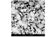
図1a[滑らかな領域が、SEMで測定されるとき外表面全体の約70%に相当する、30分の変換時間を有する、プロトタイプ1(1〜2mm顆粒)のSEM(走査型電子顕微鏡)写真を表す];及び図1b[滑らかな領域が、SEMで測定されるとき外表面全体の約50%に相当する、40分の変換時間を有する、プロトタイプ2(1〜2mm顆粒)のSEM写真を表す]を参照のこと。 FIG. 1a [Scanning electron microscope (SEM) photograph of prototype 1 (1-2 mm granules) with a conversion time of 30 minutes, where the smooth region corresponds to about 70% of the total outer surface when measured by SEM. Represents]; and FIG. 1b [SEM photographs of prototype 2 (1-2 mm granules) with a conversion time of 40 minutes, where smooth areas correspond to about 50% of the total outer surface as measured by SEM. Represent].
WO 2015/009154は、骨誘導能が改善された骨伝導性材料を製造する方法を開示しているが、この方法は、粒子からなる表面トポグラフィーを有する焼結二相性リン酸カルシウム/ヒドロキシアパタイト(CAP/HAP)材料を、2〜4barの圧力下で125℃以上の温度でpHを制御することなく、出発物質の表面上のリン酸カルシウム粒子を直径10〜1500nmのリン酸カルシウム針状物に変えるのに十分な時間、水熱処理に付すことを含む。少なくとも125℃の温度及び少なくとも2barの圧力は、HAPナノ結晶のエピタキシャル成長を可能にする、EP-B1-2445543で使用されている(人体生理学に近い)条件(温度35〜40℃、pH 5.5〜9.0、常圧)からは遠い。これらの針状物はエピタキシャルに成長していないが、コア材料ベースに付着又は堆積し、そして部分的に(通常は40〜90%)後者をコーティングするだけであるが、それによってその比表面積とタンパク質保持力を増加させ、その骨誘導能を高める。 WO 2015/009154 discloses a method for producing bone conductive materials with improved bone inducibility, which method is sintered biphasic calcium phosphate / hydroxyapatite (CAP) with surface topography consisting of particles. / HAP) Sufficient to convert calcium phosphate particles on the surface of the starting material into calcium phosphate needles with a diameter of 10 to 1500 nm without controlling the pH of the material under a pressure of 2 to 4 bar at a temperature of 125 ° C. or higher. Includes time and hydrothermal treatment. Temperatures of at least 125 ° C. and pressures of at least 2 bar are the conditions (close to human physiology) used in EP-B1-2445543 that allow epitaxial growth of HAP nanocrystals (temperature 35-40 ° C., pH 5.5). ~ 9.0, normal pressure). These needles do not grow epitaxially, but adhere or deposit on the core material base and only partially (usually 40-90%) coat the latter, thereby increasing its specific surface area. Increases protein retention and enhances its bone-inducing capacity.
EP-B1-2445543の二相性リン酸カルシウム/ヒドロキシアパタイト(CAP/HAP)骨代替材料の調製において、10〜90%、好ましくは20〜60%のメタノール、エタノール、プロパノール又はブタノールを含むがこれらに限定されない短鎖脂肪族アルコールを、工程b)の水性リン酸緩衝液に加えることにより、焼結CAPコアの外表面上に堆積したナノ結晶HAPの閉じたエピタキシャル成長層の不均質な外表面である、二相性リン酸カルシウム/ヒドロキシアパタイト(CAP/HAP)骨代替材料(平らな結晶小板の個々のクラスターとその間の滑らかな領域とを含む)は、平らな結晶小板の個々の結晶クラスターを伴わない、平らな結晶小板を含む均質な粗い外表面で置き換えられることが今や見い出された。その均質な粗い外表面は一般に、使用される脂肪族アルコールの量に応じて、SEMで測定されるとき、0.2〜20μm、好ましくは0.5〜5μmの個々の小板サイズを持つ小板の連結ネットワークを形成するエピタキシャル成長ナノ結晶のヒドロキシアパタイト小板を含む。 In the preparation of the biphasic calcium phosphate / hydroxyapatite (CAP / HAP) bone substitute material of EP-B1-2445543, it contains, but is not limited to, 10 to 90%, preferably 20 to 60% of methanol, ethanol, propanol or butanol. The heterogeneous outer surface of the closed epitaxial growth layer of nanocrystal HAP deposited on the outer surface of the sintered CAP core by adding short-chain aliphatic alcohol to the aqueous phosphate buffer solution of step b). Compatible Calcium Phosphate / Hydroxyapatite (CAP / HAP) Bone Substitute Material (including individual clusters of flat crystalline plates and smooth areas in between) is flat, without individual crystalline clusters of flat crystalline plates. It has now been found to be replaced by a homogeneous, rough outer surface containing various crystalline platelets. Its homogeneous rough outer surface is generally small, with individual plate sizes of 0.2-20 μm, preferably 0.5-5 μm, as measured by SEM, depending on the amount of aliphatic alcohol used. Includes hydroxyapatite platelets of epitaxially grown nanocrystals that form a connecting network of plates.
ヒト胎児間葉系幹細胞(hMSC)の骨形成分化のインビトロ試験で示されているとおり、インビボ骨形成反応は、平らな結晶小板を含むその均質な粗い外表面を有する二相性リン酸カルシウム/ヒドロキシアパタイト(CAP/HAP)骨代替材料の方が、EP-B1-2445543により教示された、平らな結晶小板の個々のクラスターとその間の滑らかな領域とを含む不均質な外表面を有する、二相性リン酸カルシウム/ヒドロキシアパタイト(CAP/HAP)骨代替材料よりも強い可能性が高い。 As shown in in vitro studies of bone formation differentiation of human fetal mesenchymal stem cells (hMSCs), the in vivo bone formation reaction is a biphasic calcium phosphate / hydroxyapatite with its homogeneous rough outer surface containing flat crystalline platelets. The (CAP / HAP) bone replacement material is biphasic, with an inhomogeneous outer surface containing individual clusters of flat crystalline platelets and smooth areas in between, as taught by EP-B1-2445543. It is likely to be stronger than calcium phosphate / hydroxyapatite (CAP / HAP) bone replacement material.
発明の要約
よって本発明は、焼結CAPコアと、焼結CAPコアの外表面上に堆積したナノ結晶HAPの閉じたエピタキシャル成長層(ここで、エピタキシャル成長ナノ結晶は、ヒト骨塩と同じサイズと形態を有する)とを含む、二相性リン酸カルシウム/ヒドロキシアパタイト(CAP/HAP)骨代替材料に関するが、ここで、焼結CAPコアの外表面上に堆積したナノ結晶HAPの閉じたエピタキシャル成長層は、平らな結晶小板を含む均質な粗い外表面を有する。
Abstract of the Invention According to the present invention, the present invention presents a closed epitaxial growth layer of a sintered CAP core and nanocrystal HAP deposited on the outer surface of the sintered CAP core (where, the epitaxially grown nanocrystals have the same size and morphology as human bone minerals. With respect to biphasic calcium phosphate / hydroxyapatite (CAP / HAP) bone substitutes, including (with), where the closed epitaxial growth layer of nanocrystalline HAP deposited on the outer surface of the sintered CAP core is flat. It has a homogeneous rough outer surface containing crystalline platelets.
その二相性リン酸カルシウム/ヒドロキシアパタイト(CAP/HAP)骨代替材料は、ヒト胎児間葉系幹細胞(hMSC)の骨形成分化の増加を示しており、そしてこれは、インビボ骨形成反応の増強のはっきりした兆候である。 Its biphasic calcium phosphate / hydroxyapatite (CAP / HAP) bone replacement material has shown increased osteogenic differentiation of human fetal mesenchymal stem cells (hMSCs), which manifests itself in enhanced in vivo osteogenic response. It's a sign.
「焼結CAPコアの外表面上に堆積したナノ結晶HAPの閉じたエピタキシャル成長層」という用語は、ナノ結晶HAPのエピタキシャル成長層が、焼結CAPコアの外表面全体を被覆していることを意味する。 The term "closed epitaxial growth layer of nanocrystal HAP deposited on the outer surface of the sintered CAP core" means that the epitaxial growth layer of the nanocrystal HAP covers the entire outer surface of the sintered CAP core. ..
「平らな結晶小板を含む均質な粗い外表面」という用語は、巨視的には、平らな結晶小板に起因する外表面の粗さが、平らな結晶小板の個々の結晶クラスターを伴わずにCAPコアの表面上に統計的に均等に分布していることを意味する。種々の程度の粗さの均質な粗い外表面を持つ、本発明の二相性リン酸カルシウム/ヒドロキシアパタイト(CAP/HAP)骨代替材料のプロトタイプ3〜7のSEM写真を表す、図2を参照のこと。 The term "homogeneous rough outer surface containing flat crystalline plates" macroscopically means that the outer surface roughness due to the flat crystalline plates is accompanied by individual crystalline clusters of the flat crystalline plates. It means that it is statistically evenly distributed on the surface of the CAP core. See FIG. 2, which represents SEM photographs of prototypes 3-7 of the biphasic calcium phosphate / hydroxyapatite (CAP / HAP) bone substitute material of the present invention, which have a homogeneous rough outer surface of varying degrees of roughness.
「平らな結晶小板」という用語は、3つの垂直方向に関して高さ(厚さ)が幅及び長さよりもかなり小さい結晶集合を意味する。このような平らな結晶小板は、図3bにおいてはっきりと見える。 The term "flat crystalline plate" means a set of crystals whose height (thickness) is significantly smaller than their width and length in the three vertical directions. Such flat crystalline platelets are clearly visible in FIG. 3b.
一般に、均質な粗い外表面は、SEMで測定されるとき0.2〜20μmのサイズ(幅と長さ)を持つ小板の連結ネットワークを形成する、エピタキシャル成長ナノ結晶ヒドロキシアパタイト小板を含む。小板のサイズが大きいほど、外表面の粗さが高まる。 Generally, the homogeneous rough outer surface contains epitaxially grown nanocrystalline hydroxyapatite platelets that form a connecting network of strips with a size (width and length) of 0.2-20 μm as measured by SEM. The larger the plate size, the higher the roughness of the outer surface.
好ましくは、均質な粗い外表面は、SEMで測定されるとき0.5〜5μmのサイズを持つ小板の連結ネットワークを形成する、エピタキシャル成長ナノ結晶ヒドロキシアパタイト小板を含む。 Preferably, the homogeneous, rough outer surface comprises epitaxially grown nanocrystalline hydroxyapatite platelets that form a connected network of strips with a size of 0.5-5 μm as measured by SEM.
通常は、その均質な粗い外表面は、水銀圧入ポロシメトリー(MIP)で測定されるとき0.03〜2μmの細孔を含む連結ネットワークを形成する、エピタキシャル成長ヒドロキシアパタイト小板を含む。0.03〜2μmの細孔容積が大きいほど、外表面の粗さが高まる。 Usually, its homogeneous and rough outer surface contains epitaxially grown hydroxyapatite platelets that form a connecting network containing pores of 0.03 to 2 μm as measured by mercury intrusion porosymmetry (MIP). The larger the pore volume of 0.03 to 2 μm, the higher the roughness of the outer surface.
一般に、その均質な粗い外表面は、AFM(原子間力顕微鏡法(Atomic Force Microscopy))で得た50〜400nmの範囲の二乗平均平方根粗度(Rq)及び500〜2000nmの範囲の輪郭の平均最大高さ(Rz)を用い、AFMにより特徴付けられ得る。 In general, its homogeneous and rough outer surface is the root mean square roughness (R q ) in the range of 50 to 400 nm and the contour in the range of 500 to 2000 nm obtained by AFM (Atomic Force Microscopy). The average maximum height ( Rz ) can be used and characterized by AFM.
好ましくは、均質な粗い外表面は、AFMで得た110〜150nmの範囲の二乗平均平方根粗度(Rq)及び550〜750nmの範囲の輪郭の平均最大高さ(Rz)により特徴付けられ得る。 Preferably, the homogeneous rough outer surface is characterized by the root mean square roughness (R q ) in the range 110-150 nm and the average maximum height (R z ) of the contour in the range 550-750 nm obtained by AFM. obtain.
一般に、二相性リン酸カルシウム/ヒドロキシアパタイト(CAP/HAP)骨代替材料中のHAPの割合は、XRDにより測定されるとき1〜90%である。 Generally, the proportion of HAP in biphasic calcium phosphate / hydroxyapatite (CAP / HAP) bone replacement material is 1-90% as measured by XRD.
好ましくは、その割合は、XRDにより測定されるとき1.5〜30%、更に好ましくは2〜15%である。 Preferably, the proportion is 1.5-30%, more preferably 2-15% as measured by XRD.
焼結CAPコアは、リン酸三カルシウム(TCP)、特にα−TCP(α−Ca3(PO4)2)又はβ−TCP(β−Ca3(PO4)2)、及び/又はリン酸四カルシウム(TTCP)Ca4(PO4)2Oを含む。 Sintered CAP cores are tricalcium phosphate (TCP), especially α-TCP (α-Ca 3 (PO 4 ) 2 ) or β-TCP (β-Ca 3 (PO 4 ) 2 ), and / or phosphate. Includes tetracalcium (TTCP) Ca 4 (PO 4 ) 2 O.
頻繁に使用される実施態様では、焼結CAPコアは、本質的にTCPからなり、α−TCPが好ましい。 In a frequently used embodiment, the sintered CAP core consists essentially of TCP, preferably α-TCP.
ナノ結晶HAPのエピタキシャル成長層は、構造的に天然のヒト骨塩とほぼ同一である。 The epitaxial growth layer of nanocrystal HAP is structurally substantially identical to natural human bone mineral.
CAP/HAP骨代替材料は、粒子状又は顆粒状であってよく、この粒子又は顆粒は所望のサイズ及び形状を有する。一般に、粒子又は顆粒は、250〜5000μm、好ましくは1000〜2000μmのサイズを有する。 The CAP / HAP bone substitute material may be in the form of particles or granules, which particles or granules have the desired size and shape. Generally, the particles or granules have a size of 250-5000 μm, preferably 1000-2000 μm.
CAP/HAP骨代替材料はまた、成形体、例えば、ネジ、釘、ピン、又は特に、股関節、鎖骨、肋骨、下顎骨若しくは頭蓋骨のような、骨性身体部分の輪郭を有する構造であってもよい。そのようなネジ、釘又はピンは、例えば、膝又は肘の骨に靭帯を固定するための整形外科再建手術において使用されてもよい。骨性身体部分の輪郭を有するそのような構造は、消失又は欠損の骨又は骨部分を置換するためのプロテーゼとして整形外科手術において使用されてもよい。 The CAP / HAP bone replacement material may also be a molded body, such as a structure with contours of a bony body part, such as a screw, nail, pin, or in particular, a hip joint, clavicle, ribs, mandible or skull. Good. Such screws, nails or pins may be used, for example, in orthopedic reconstructive surgery to secure ligaments to the bones of the knee or elbow. Such structures with contours of bony body parts may be used in orthopedic surgery as prostheses to replace lost or missing bone or bone parts.
本発明はまた、一般に天然又は合成ポリマーを含む、適切なマトリックス中に上記のCAP/HAP骨代替品の粒子又は顆粒を含むパテに関する。一般に、粒子又は顆粒は、250〜5000μm、好ましくは1000〜2000μmのサイズを有する。 The present invention also relates to putties containing particles or granules of the above-mentioned CAP / HAP bone substitutes in a suitable matrix, generally containing natural or synthetic polymers. Generally, the particles or granules have a size of 250-5000 μm, preferably 1000-2000 μm.
本発明は更に、上記のCAP/HAP骨代替材料の調製方法であって、以下の工程:
a)焼結CAPコア材料を調製すること、
b)焼結CAPコア材料を10℃と50℃の間の温度で10〜90%の短鎖脂肪族アルコールを含有する緩衝液に浸漬して、CAPからHAPへの変換プロセスを開始し、それによって焼結CAPコア材料表面上にナノ結晶ヒドロキシアパタイトの閉じたエピタキシャル成長層(ここで、エピタキシャル成長ナノ結晶は、ヒト骨塩と同じサイズと形態を有する)であって、平らな結晶小板を含む均質な外表面を有する、焼結CAPコア材料表面上に形成されるナノ結晶HAPの閉じたエピタキシャル成長層が形成されること、
c)HAPの少なくとも1つのナノ結晶層の閉じたコーティングが存在するが、変換プロセスが完全に終わる前の時点で、水溶液から固体材料を分離することによって変換を停止させること、及び
d)場合により、工程c)からの分離された材料を滅菌すること
を含む方法に関する。
The present invention further comprises the above method for preparing a CAP / HAP bone substitute material, wherein the following steps:
a) Preparing a sintered CAP core material,
b) The sintered CAP core material is immersed in a buffer containing 10-90% short chain aliphatic alcohol at a temperature between 10 ° C and 50 ° C to initiate the CAP to HAP conversion process. Closed epitaxial growth layer of nanocrystalline hydroxyapatite on the surface of the sintered CAP core material by (where the epitaxially grown nanocrystals have the same size and morphology as human bone minerals), homogeneous with flat crystalline platelets. Forming a closed epitaxial growth layer of nanocrystal HAP formed on the surface of the sintered CAP core material, which has an outer surface.
c) There is a closed coating of at least one nanocrystal layer of HAP, but before the conversion process is complete, the conversion is stopped by separating the solid material from the aqueous solution, and d) optionally. , A method comprising sterilizing the separated material from step c).
適切な短鎖脂肪族アルコールは、メタノール、エタノール、プロパノール及びブタノールからなる群より選択され得る。 Suitable short-chain aliphatic alcohols can be selected from the group consisting of methanol, ethanol, propanol and butanol.
好ましくは短鎖脂肪族アルコールは、エタノールである。 Preferably the short-chain aliphatic alcohol is ethanol.
好ましくは、工程b)の緩衝液は、20〜60%、更に好ましくは30〜50%の短鎖脂肪族アルコールを含有する。 Preferably, the buffer solution of step b) contains 20-60%, more preferably 30-50% of short-chain aliphatic alcohol.
焼結CAPコアの外表面上に堆積したナノ結晶HAPの閉じたエピタキシャル成長層の均質な粗い外表面の粗さのパラメーター、特に
−AFMパラメーター:AFMで得た二乗平均平方根粗度(Rq)及び輪郭の平均最大高さ(Rz)、
−SEMで測定されるエピタキシャル成長ナノ結晶ヒドロキシアパタイト小板のサイズ、並びに
−MIPで測定される0.03〜2μmの細孔容積
は、便利には変換溶液の緩衝液中の短鎖脂肪族アルコールの割合を変えることにより調整され得る。
The parameters of the homogeneous rough outer surface roughness of the closed epitaxial growth layer of the nanocrystal HAP deposited on the outer surface of the sintered CAP core, especially the −AFM parameter: the root mean square roughness (R q ) obtained by AFM and Average maximum height of contour (R z ),
The size of the epitaxially grown nanocrystalline hydroxyapatite discs measured by -SEM, as well as the pore volume of 0.03 to 2 μm measured by -MIP, are conveniently of the short chain fatty alcohols in the buffer of the conversion solution. It can be adjusted by changing the ratio.
その割合が高いほど、AFMで得た二乗平均平方根粗度(Rq)及び輪郭の平均最大高さ(Rz)が低く、SEMで測定されるエピタキシャル成長ナノ結晶ヒドロキシアパタイト小板のサイズが小さく、そしてMIPで測定される0.03〜2μmの細孔容積が小さい。 The higher the ratio, the lower the root mean square roughness (R q ) obtained by AFM and the average maximum height (R z ) of the contour, and the smaller the size of the epitaxially grown nanocrystal hydroxyapatite plaque measured by SEM. And the pore volume of 0.03 to 2 μm measured by MIP is small.
10〜90%の短鎖脂肪族アルコールを含有する工程b)の緩衝液は、水性緩衝液を種々の量の短鎖脂肪族アルコールと混合することによって得られる。水性緩衝液は、10〜90%の短鎖脂肪族アルコールを更に含有する工程b)の浸漬溶液のpH値が、ほぼ中性であり、そして変換プロセス全体を通じて、好ましくは5.5〜9.0、更に好ましくは7.0〜8.0のpH範囲内で安定を保つように、選択される。 The buffer solution of step b) containing 10 to 90% of the short chain aliphatic alcohol is obtained by mixing the aqueous buffer solution with various amounts of the short chain aliphatic alcohol. The aqueous buffer further contains 10-90% short chain aliphatic alcohol, the pH value of the dipping solution in step b) is approximately neutral, and is preferably 5.5-9 throughout the conversion process. It is selected to remain stable in the pH range of 0, more preferably 7.0-8.0.
緩衝剤は、上記pH範囲内の任意の緩衝剤であってよいが、好ましくはカルシウム、マグネシウム及び/又はナトリウムを含むか又は含まないリン酸緩衝剤である。 The buffer may be any buffer within the pH range, but is preferably a phosphate buffer containing or not containing calcium, magnesium and / or sodium.
適切な緩衝液は、例えば、7.3〜7.6のpH値を持つリン酸二水素ナトリウム(NaH2PO4)の0.05〜0.3M水溶液である。 A suitable buffer is, for example, a 0.05-0.3 M aqueous solution of sodium dihydrogen phosphate (NaH 2 PO 4) having a pH value of 7.3-7.6.
工程b)の温度範囲は一般に、10℃と50℃の間、好ましくは25℃と45℃の間、更に好ましくは35℃と40℃の間である。 The temperature range of step b) is generally between 10 ° C and 50 ° C, preferably between 25 ° C and 45 ° C, and even more preferably between 35 ° C and 40 ° C.
好ましくは工程b)は、35〜40℃の温度で、20〜60%の短鎖脂肪族アルコールを含有するpH7.0〜8.0のリン酸緩衝液中で行われる。 Preferably, step b) is carried out at a temperature of 35-40 ° C. in a phosphate buffer solution having a pH of 7.0-8.0 containing 20-60% of short-chain aliphatic alcohols.
焼結CAPコア材料の調製は、最初にリン酸水素カルシウム(CaHPO4)、炭酸カルシウム及び/又は水酸化カルシウムの粉末を混合し、次に適切な温度範囲内で混合物をか焼及び焼結し、これによってバルク焼結CAPコア材料が与えられることを含む、当技術分野において公知の方法によって実施され得る(例えば、Mathew M. et al., 1977, Acta. Cryst. B33: 1325; Dickens B. et al., 1974, J. Solid State Chemistry 10, 232; and Durucan C. et al., 2002, J. Mat. Sci., 37:963を参照のこと)。
To prepare the sintered CAP core material, first mix the powders of calcium hydrogen phosphate (CaHPO 4 ), calcium carbonate and / or calcium hydroxide, then calcin and sinter the mixture within the appropriate temperature range. , Which can be carried out by methods known in the art, including the provision of bulk sintered CAP core material (eg, Mathew M. et al., 1977, Acta. Cryst. B33: 1325; Dickens B. See et al., 1974, J.
よってバルク焼結TCPコア材料を、リン酸水素カルシウム(CaHPO4)、炭酸カルシウム及び/又は水酸化カルシウムの粉末を化学量論比で混合し、混合物を1200〜1450℃の範囲の温度、好ましくは約1400℃でか焼及び焼結することによって得てもよい。 Therefore, the bulk sintered TCP core material is mixed with powders of calcium hydrogen phosphate (CaHPO 4 ), calcium carbonate and / or calcium hydroxide in a chemical ratio, and the mixture is mixed at a temperature in the range of 1200 to 1450 ° C., preferably. It may be obtained by baking and sintering at about 1400 ° C.
バルク焼結TTCPコア材料もまた、上記プロセスによって得てもよい。 Bulk sintered TTCP core material may also be obtained by the above process.
そのような方法によって調製されたバルク焼結CAP材料は、2〜80体積%の空隙率及び細孔の幅広い分布を有する多孔質であってよい。空隙率パラメーターは、CAP/HAP骨代替材料の意図される用途に応じて選択されよう。 The bulk sintered CAP material prepared by such a method may be porous with a porosity of 2-80% by volume and a wide distribution of pores. Porosity parameters will be selected depending on the intended use of the CAP / HAP bone replacement material.
工程b)に使用される焼結CAPコア材料は、
−上記のとおり調製されたバルク焼結CAPコア材料であるか、
−上記のとおり調製されたバルク焼結CAPコア材料から、破砕、磨砕及び/又は粉砕、並びに篩い分けのような従来の方法を使用して得られた、焼結CAPコア材料の粒子又は顆粒であるか、あるいは
−所望の形状とサイズ、例えば、ネジ、釘、ピン、又は骨性身体部分の輪郭を有する構造を有する焼結CAPコア材料のプリフォームであってもよい。
The sintered CAP core material used in step b) is
-A bulk sintered CAP core material prepared as described above,
-Particles or granules of sintered CAP core material obtained from bulk sintered CAP core material prepared as described above using conventional methods such as crushing, grinding and / or grinding, and sieving. Or-may be a preform of a sintered CAP core material having a structure having the desired shape and size, eg, screws, nails, pins, or contours of bony body parts.
このような任意の所望の形状とサイズのプリフォームを、CNCフライス加工又は3D印刷のような周知のプロトタイピング技術を使用して、上記のとおり調製されたバルク焼結コア材料から得てもよい(例えば、Bartolo P. et al., 2008, Bio-Materials and Prototyping Applications in Medicine, Springer Science New York, ISBN 978-0-387-47682-7; Landers R. et al., 2002, Biomaterials 23(23), 4437; Yeong W.-Y. et al., 2004, Trends in Biotechnology, 22 (12), 643; and Seitz H. et al., 2005, Biomed. Mater. Res. 74B (2), 782を参照のこと)。 Preforms of any desired shape and size as described above may be obtained from the bulk sintered core material prepared as described above using well-known prototyping techniques such as CNC milling or 3D printing. (For example, Bartolo P. et al., 2008, Bio-Materials and Prototyping Applications in Medicine, Springer Science New York, ISBN 978-0-387-47682-7; Landers R. et al., 2002, Biomaterials 23 (23) ), 4437; Yeong W.-Y. Et al., 2004, Trends in Biotechnology, 22 (12), 643; and Seitz H. et al., 2005, Biomed. Mater. Res. 74B (2), 782 See).
浸漬工程b)は、第1段階でCAPコア材料の一次相転移を誘導し、したがってHAPナノ結晶前駆体の核形成を誘導する。第2段階中に、第1段階から得られたHAP前駆体は成長して、閉じた(即ち、完全にコーティングしている)エピタキシャルナノ結晶複合層を確立する。最初のHAPナノ結晶層は、均質で閉じており、かつ焼結CAPコア材料にエピタキシャルに接合されている必要がある。 The immersion step b) induces a primary phase transition of the CAP core material in the first step and thus induces nucleation of the HAP nanocrystal precursor. During the second step, the HAP precursor obtained from the first step grows to establish a closed (ie, fully coated) epitaxial nanocrystal composite layer. The first HAP nanocrystal layer must be homogeneous, closed and epitaxially bonded to the sintered CAP core material.
第3段階中に、新しく形成された二重複合層内で一次相転移が進行して、焼結CAPコア材料(TCP又はTTCP)をナノ結晶HAPに更に変換することができる。相転移のこの第3工程中に、焼結CAPコア材料の一部がナノ結晶HAPに変換されるまで、遅延拡散制御プロセスによって制御可能な時間、カルシウムイオンが放出されよう。HAP層の厚さ、ひいてはカルシウム放出の速度は、変換時間の変動によって制御され得る。 During the third step, a primary phase transition proceeds within the newly formed double composite layer to further convert the sintered CAP core material (TCP or TTCP) to nanocrystalline HAP. During this third step of the phase transition, calcium ions will be released for a time controllable by the delayed diffusion control process until a portion of the sintered CAP core material is converted to nanocrystalline HAP. The thickness of the HAP layer, and thus the rate of calcium release, can be controlled by variations in conversion time.
適切な厚さのエピタキシャル成長ナノ結晶HAP層は、インビトロで調製されるが、ここでHAPへのCAPの変換は、完了する前に停止させる。 An epitaxially grown nanocrystalline HAP layer of appropriate thickness is prepared in vitro, where the conversion of CAP to HAP is stopped before completion.
CAP/HAP骨代替材料がインビボに設置されると直ぐに、HAPへのCAPの変換プロセスは、体液との接触により再活性化され、骨代替材料は、ヒト骨塩にサイズと形態が類似する新しいヒドロキシアパタイトを形成する生体様システムとして機能しよう。インビボ相変態プロセス中に、輸送されたカルシウムイオンは、骨再生プロセスにとって重要かつ有益である局所カルシウム平衡を支持する局所環境へと放出されよう。 As soon as the CAP / HAP bone substitute material is placed in vivo, the process of converting CAP to HAP is reactivated by contact with body fluids, and the bone substitute material is new in size and morphology similar to human bone mineral. Let's function as a biological system that forms hydroxyapatite. During the in vivo phase transformation process, the transported calcium ions will be released into a local environment that supports local calcium equilibrium, which is important and beneficial for the bone regeneration process.
身体の負荷が異なる部位では骨欠損の再生時間が異なるため、カルシウム放出の速度を制御できることが重要である。これは、ヒドロキシアパタイトのエピタキシャル成長層の厚さを変えることによって達成され得る。 It is important to be able to control the rate of calcium release, as the regeneration time of bone defects differs at sites with different physical loads. This can be achieved by varying the thickness of the hydroxyapatite epitaxial growth layer.
したがって工程c)は非常に重要な工程である。工程b)の水溶液中の曝露時間は、所望のHAP層の厚さに基づく。エピタキシャル配向のナノ結晶HAPの少なくとも1つの層が必要である。CAPからHAPへの変換が完了していないことは不可欠である。 Therefore, step c) is a very important step. The exposure time in aqueous solution of step b) is based on the desired thickness of the HAP layer. At least one layer of epitaxially oriented nanocrystal HAP is required. It is essential that the conversion from CAP to HAP is not complete.
所望の厚さによる適切な曝露時間は、リン酸カルシウム並びにセメント及びコンクリート化学の分野の当業者に周知の幾つかの熱力学的微分方程式を使用することによって計算され得る。 The appropriate exposure time for the desired thickness can be calculated by using calcium phosphate and some thermodynamic differential equations well known to those skilled in the art of cement and concrete chemistry.
例えば、Pommersheim, J.C.; Clifton, J.R. (1979) Cem. Conc. Res.; 9:765; Pommersheim, J.C.; Clifton, J.R. (1982) Cem. Conc. Res.; 12:765; and Schlussler, K.H. Mcedlov-Petrosjan, O.P.; (1990): Der Baustoff Beton, VEB Verlag Bauwesen, Berlinを参照のこと。 For example, Pommersheim, JC; Clifton, JR (1979) Cem. Conc. Res .; 9: 765; Pommersheim, JC; Clifton, JR (1982) Cem. Conc. Res .; 12: 765; and Schlussler, KH Mcedlov- See Petrosjan, OP; (1990): Der Baustoff Beton, VEB Verlag Bauwesen, Berlin.
上記の微分方程式の解をCAP/HAPシステムに代入すると、CAPのHAPへの相転移及び層の厚さを予測できるため、HAPのエピタキシャル層を安定で再現性あるやり方で調製することができる。 Substituting the solution of the above differential equation into the CAP / HAP system can predict the phase transition of CAP to HAP and the layer thickness, so that the epitaxial layer of HAP can be prepared in a stable and reproducible manner.
水溶液からの固体材料の分離は、当技術分野で周知の技術を使用して、濾過及び乾燥によって通常実施される。 Separation of the solid material from the aqueous solution is usually carried out by filtration and drying using techniques well known in the art.
オプションの滅菌工程d)は、ガンマ線照射又はX線照射のような当技術分野で周知の手法によって実施され得る。 The optional sterilization step d) can be performed by techniques well known in the art such as gamma irradiation or X-ray irradiation.
本発明はまた、一般に粒子、パテ又は成形体の形の、ヒト又は動物の欠損部位における骨形成、骨再生、骨修復及び/又は骨置換を支持するためのインプラント又はプロテーゼとしての上記CAP/HAP骨代替材料の使用に関する。 The present invention also relates to the above CAP / HAP as an implant or prosthesis to support bone formation, bone regeneration, bone repair and / or bone replacement in a human or animal defect site, generally in the form of particles, putty or shaped bodies. Regarding the use of bone substitute materials.
本発明はまた、一般に粒子、パテ又は成形体の形の、上記CAP/HAP骨代替材料を移植することによる、ヒト又は動物の欠損部位における骨形成、骨再生及び/又は骨修復を促進する方法に関する。 The present invention also presents a method of promoting bone formation, bone regeneration and / or bone repair at a human or animal defect site by implanting the CAP / HAP bone substitute material, generally in the form of particles, putty or molded body. Regarding.
本発明のCAP/HAP骨代替材料及びその調製方法の利点
平らな結晶小板を含む均質な粗い外表面を有する、本発明の二相性リン酸カルシウム/ヒドロキシアパタイト(CAP/HAP)骨代替材料は、平らな結晶小板の個々のクラスターとその間の滑らかな領域とを含む不均質な外表面を有するEP-B1-2445543により教示された二相性リン酸カルシウム/ヒドロキシアパタイト(CAP/HAP)骨代替材料と比較して、ヒト胎児間葉系幹細胞(hMSC)の骨形成分化の増加、特に分化マーカーであるオステオポンチン(OPN)及びオステオカルシン(OCN)のより高い発現を示す。これは、インビボ骨形成反応の増強のはっきりした兆候である。
Advantages of CAP / HAP Bone Substitute Material of the Present Invention and Method for Preparing The Biphasic Calcium Phosphate Phosphate / Hydroxyapatite (CAP / HAP) Bone Substitute Material of the present invention having a homogeneous rough outer surface including a flat crystalline plate is flat. Compared to the biphasic calcium phosphate / hydroxyapatite (CAP / HAP) bone substitute material taught by EP-B1-2445543, which has an inhomogeneous outer surface containing individual clusters of crystalline granules and smooth areas in between. It shows increased bone formation and differentiation of human fetal mesenchymal stem cells (hMSC), especially higher expression of the differentiation markers osteopontin (OPN) and osteocalcin (OCN). This is a clear sign of enhanced in vivo bone formation response.
これは、R.A. GittensらがBiomaterials 2011 May, 32(13): 3395-3403に発表した結果と一致しているが、この文献は、マイクロ−サブマイクロスケールの粗度と組合せたナノスケール構造の導入により骨芽細胞の分化と局所因子の産生が改善されることを示しており、これにより、インビボでのインプラントのオッセオインテグレーション(osseointegration)の改善の可能性が示唆される。 This is consistent with the results published by RA Gittens et al. In Biomaterials 2011 May, 32 (13): 3395-3403, but this document introduces nanoscale structures in combination with micro-submicroscale roughness. Has been shown to improve osteoblast differentiation and local factor production, suggesting the potential for improved implant osseointegration in vivo.
本発明の二相性リン酸カルシウム/ヒドロキシアパタイト(CAP/HAP)骨代替材料の調製方法によって、焼結CAPコアの外表面上に堆積したナノ結晶HAPの閉じたエピタキシャル成長層の均質な粗い外表面の粗さパラメーターを、特に
−AFMパラメーター:AFMで得た二乗平均平方根粗度(Rq)及び輪郭の平均最大高さ(Rz)を、
−SEMで測定されるエピタキシャル成長ナノ結晶ヒドロキシアパタイト小板のサイズを、並びに
−MIPで測定される0.03〜2μmの細孔容積を
変換溶液の緩衝液中の短鎖脂肪族アルコールの割合を調整することにより、便利に調整することができる。
Homogeneous rough outer surface roughness of the closed epitaxial growth layer of nanocrystal HAP deposited on the outer surface of the sintered CAP core by the method of preparing the biphasic calcium phosphate / hydroxyapatite (CAP / HAP) bone substitute material of the present invention. Parameters, especially −AFM parameter: Root mean square roughness (R q ) and mean maximum height of contour (R z ) obtained by AFM.
-Adjust the size of the epitaxially grown nanocrystalline hydroxyapatite platelets measured by SEM, and the pore volume of 0.03 to 2 μm measured by -MIP to the proportion of short-chain fatty alcohols in the buffer of the solution. By doing so, it can be adjusted conveniently.
その割合が高いほど、AFMで得た二乗平均平方根粗度(Rq)及び輪郭の平均最大高さ(Rz)が低く、SEMで測定されるエピタキシャル成長ナノ結晶ヒドロキシアパタイト小板のサイズが小さく、そしてMIPで測定される0.03〜2μmの細孔容積が小さい。 The higher the ratio, the lower the root mean square roughness (R q ) obtained by AFM and the average maximum height (R z ) of the contour, and the smaller the size of the epitaxially grown nanocrystal hydroxyapatite plaque measured by SEM. And the pore volume of 0.03 to 2 μm measured by MIP is small.
本発明は、本発明の好ましい実施態様の説明例及び添付の図面を参照して、本明細書の以下に更に詳細に記載されよう。
詳細な説明
以下の実施例により、本発明の範囲を限定することなく本発明を説明する。
Detailed Description The present invention will be described with reference to the following examples without limiting the scope of the present invention.
EP-B1-2445543の二相性リン酸カルシウム/ヒドロキシアパタイト(CAP/HAP)骨代替材料の調製。
EP-B1-2445543の実施例1、2及び4と同様に、α−TCPのバルク焼結材料、1.0〜2.0mmの粒径のその多孔質顆粒、及びエピタキシャル成長HAPコーティングを有する変換顆粒を調製した。
実験室用撹拌機を用いて、364g リン酸二カルシウム無水物粉末、136g 炭酸カルシウム粉末、及び220ml 脱イオン水を700rpmで5分間混合した。混合プロセスからのスラリーを、直ちに高温安定なプラチナカップに移した。充填プラチナカップを低温炉に入れた。1時間当たり100℃の加熱速度を使用して、炉を1400℃に加熱した。この温度を12時間保持し、そして炉を1時間当たり500℃の冷却速度で800℃まで冷却し、次に1時間当たり125℃の冷却速度で300℃まで冷却し、最後に炉の切り替えにより室温まで冷却した。バルク焼結材料(相純粋なα−Ca3(PO4)2)を炉及びプラチナカップから取り出した。相純度の制御は、粉末X線回折分析法を用いて実施された。
ジョークラッシャーを使用してバルク生成物を粉砕した(ジョー距離は10〜1mmで変動した)。生成した顆粒を、2mm及び1mmのメッシュ開口を有する篩い機及び篩いインサートを使用して篩い分けした。篩い分け後、顆粒をエタノールで濯いで、顆粒に吸着した微粉末残留物を分離した。多孔質顆粒をキャビネット乾燥機で80℃で1時間乾燥させた。濯ぎ後の粒子表面の清浄度は、走査型電子顕微鏡法を用いる表面観察によって制御された。
0.4mol/l リン酸二水素ナトリウム(NaH2PO4)を蒸留水に溶解して、コーティング及び相変態プロセスに適した緩衝液を調製した。水酸化ナトリウム(NaOH)を使用して、溶液のpHを室温で7.45に調整した。前の段落で生成した顆粒を調製溶液中に浸漬し、温かい水浴(40℃)内でそれぞれ30分間(プロトタイプ1)及び40分間(プロトタイプ2)保存した。浸漬後、顆粒を蒸留水で3回濯ぎ、相変態プロセスを停止させ、緩衝液から残留物を取り出した。多孔質顆粒を、キャビネット乾燥機内で100℃で2時間乾燥させた。
プロトタイプ1及びプロトタイプ2の顆粒に対して、倍率3500×のSEMを実施した。
プロトタイプ1及び2のSEM写真を表す図1a及び1bから明らかなように、顆粒の外表面は、エピタキシャル成長HAPナノ結晶からなる平らな結晶小板の個々の(分離した)クラスターと結晶間の滑らかな領域を含む不均質なものである。
プロトタイプ1及びプロトタイプ2の各々についてSEM写真上で個々のクラスターと滑らかな領域とが占める表面を測定することにより、滑らかな領域がプロトタイプ1では外表面の約70%、プロトタイプ2では外表面の約50%に相当することが判定された。
Preparation of biphasic calcium phosphate / hydroxyapatite (CAP / HAP) bone substitute material for EP-B1-2445543.
Similar to Examples 1, 2 and 4 of EP-B1-2445543, the bulk sintered material of α-TCP, its porous granules with a particle size of 1.0-2.0 mm, and the converted granules with an epitaxially grown HAP coating. Was prepared.
Using a laboratory stirrer, 364 g dicalcium phosphate anhydrous powder, 136 g calcium carbonate powder, and 220 ml deionized water were mixed at 700 rpm for 5 minutes. The slurry from the mixing process was immediately transferred to a hot and stable platinum cup. The filled platinum cup was placed in a low temperature furnace. The furnace was heated to 1400 ° C. using a heating rate of 100 ° C. per hour. This temperature is maintained for 12 hours, and the furnace is cooled to 800 ° C. at a cooling rate of 500 ° C. per hour, then to 300 ° C. at a cooling rate of 125 ° C. per hour, and finally to room temperature by switching the furnace. Cooled down to. The bulk sintered material (phase pure α-Ca 3 (PO 4 ) 2 ) was removed from the furnace and platinum cup. Control of phase purity was performed using powder X-ray diffraction analysis.
Bulk products were ground using a jaw crusher (jaw distance varied from 10 to 1 mm). The resulting granules were screened using a sieving machine with 2 mm and 1 mm mesh openings and a sieving insert. After sieving, the granules were rinsed with ethanol to separate the fine powder residue adsorbed on the granules. The porous granules were dried in a cabinet dryer at 80 ° C. for 1 hour. The cleanliness of the particle surface after rinsing was controlled by surface observation using scanning electron microscopy.
0.4 mol / l sodium dihydrogen phosphate (NaH 2 PO 4 ) was dissolved in distilled water to prepare a buffer suitable for the coating and phase transformation process. Using sodium hydroxide (NaOH), the pH of the solution was adjusted to 7.45 at room temperature. The granules produced in the previous paragraph were immersed in the preparation solution and stored in a warm water bath (40 ° C.) for 30 minutes (prototype 1) and 40 minutes (prototype 2), respectively. After immersion, the granules were rinsed 3 times with distilled water to stop the phase transformation process and remove the residue from the buffer. The porous granules were dried in a cabinet dryer at 100 ° C. for 2 hours.
The granules of
As is apparent from FIGS. 1a and 1b showing the SEM photographs of
By measuring the surface occupied by individual clusters and smooth areas on SEM photographs for each of
本発明の二相性リン酸カルシウム/ヒドロキシアパタイト(CAP/HAP)骨代替材料の調製。
1)骨代替材料の顆粒の調製
上記の実施例1に記載されるとおり、相純粋なα−TCPの1〜2mmサイズの多孔質顆粒を製造した。
相変態及びコーティング工程は、40℃に設定された水浴に入れたガラスフラスコ中で実施された。変換緩衝液は、様々な割合のエタノールと混合したリン酸二水素ナトリウム(NaH2PO4)の水溶液とした。リン酸二水素ナトリウムの水溶液のモル濃度は0.05M〜0.3Mの間で変動し、エタノールの含量は20〜60w/w%の間で変動した。変換溶液のpHは7.3〜7.6の間であった。
ガラスフラスコに変換緩衝液を充填し、α−TCP顆粒を1:40〜1:80(顆粒対変換溶液)の比で加えた。顆粒を変換溶液に40℃で24〜72時間の期間、浸漬した。浸漬後、顆粒を脱イオン水(顆粒対水の比は重量で1:10)で5回濯ぎ、エタノール(99.9%、顆粒対エタノールの比は重量で1:10)で2回濯いで、相変態プロセスを停止させ、緩衝液から残留物を取り出した。多孔質顆粒を、キャビネット乾燥機内で100℃で2時間乾燥させた。
コーティング及び相変態プロセス後の表面形態を、SEMを使用して観察した。
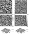
図2は、本発明の骨代替材料のプロトタイプ3(20%エタノール)、プロトタイプ4(30%エタノール)、プロトタイプ5(40%エタノール)、プロトタイプ6(50%エタノール)及びプロトタイプ7(60%エタノール)の倍率3500×のSEM写真を表す。図1a及び1bを図2に対して比較することにより、平らな結晶小板の個々のクラスターとその間の滑らかな領域を有するプロトタイプ1及び2の不均質な外表面が、個々の結晶クラスターのない均質な粗い外表面によって置き換えられることが分かる。均質な粗い外表面は、エピタキシャル成長ヒドロキシアパタイト小板の連結ネットワークでできている。SEM分析で観察されるとおり、個々の小板のサイズは、変換溶液のエタノール含量を増加させると減少し、そして外表面の粗さ又は粗度を減少させる。
図3aは、低倍率(1000×)でのプロトタイプ5(40%エタノール、1〜2mm顆粒)の断面のSEM写真を表す。右下隅は顆粒の外面を示し、顆粒の中心は左上隅寄りに位置する。
図3bは、高倍率(14,000×)でのプロトタイプ5(40%エタノール、1〜2mm顆粒)の断面のSEM写真を表しており、粗い表面の構成単位である個々の平らな結晶小板をはっきりと見ることができる。顆粒の中心の粗い外表面と顆粒の外面の粗い外表面との間に違いはない。
Preparation of biphasic calcium phosphate / hydroxyapatite (CAP / HAP) bone substitute material of the present invention.
1) Preparation of Granules of Bone Substitute Material As described in Example 1 above, porous granules having a size of 1 to 2 mm of phase-pure α-TCP were produced.
The phase transformation and coating steps were performed in a glass flask placed in a water bath set at 40 ° C. The conversion buffer was an aqueous solution of sodium dihydrogen phosphate (NaH 2 PO 4 ) mixed with various proportions of ethanol. The molar concentration of the aqueous solution of sodium dihydrogen phosphate varied between 0.05M and 0.3M, and the ethanol content varied between 20-60w / w%. The pH of the conversion solution was between 7.3 and 7.6.
The glass flask was filled with conversion buffer, and α-TCP granules were added at a ratio of 1:40 to 1:80 (granule-to-conversion solution). The granules were immersed in the conversion solution at 40 ° C. for a period of 24-72 hours. After immersion, the granules are rinsed 5 times with deionized water (granule to water ratio 1:10 by weight) and twice with ethanol (99.9%, granule to ethanol ratio 1:10 by weight). , The phase transformation process was stopped and the residue was removed from the buffer. The porous granules were dried in a cabinet dryer at 100 ° C. for 2 hours.
The surface morphology after the coating and phase transformation process was observed using SEM.
FIG. 2 shows prototype 3 (20% ethanol), prototype 4 (30% ethanol), prototype 5 (40% ethanol), prototype 6 (50% ethanol) and prototype 7 (60% ethanol) of the bone substitute material of the present invention. Represents an SEM photograph with a magnification of 3500 ×. By comparing FIGS. 1a and 1b with respect to FIG. 2, the heterogeneous outer surface of
FIG. 3a represents an SEM photograph of a cross section of Prototype 5 (40% ethanol, 1-2 mm granules) at low magnification (1000 ×). The lower right corner shows the outer surface of the granule, and the center of the granule is located closer to the upper left corner.
FIG. 3b represents an SEM photograph of a cross section of Prototype 5 (40% ethanol, 1-2 mm granules) at high magnification (14,000 x), with individual flat crystalline platelets as the building blocks of the rough surface. Can be clearly seen. There is no difference between the rough outer surface at the center of the granule and the rough outer surface at the outer surface of the granule.
水銀圧入ポロシメトリー(MIP)による細孔径分布の測定
顆粒の細孔径分布は、水銀圧入ポロシメトリー(MIP)を使用して測定された。MIPは、多孔質材料の細孔径分布を測定するために使用される標準的な特性評価手法である。この手法は当技術分野において周知であり、例えば、Gregg, S. J. and Sing, K.S.W., Adsorption, Surface Area and Porosity, 2nd ed., Academic Press Inc. (1982), 173-190に記載されている。
図6は、純粋なα−TCP(実施例1により製造され、そしてプロトタイプ3、5及び7のコア材料)と比較した本発明の骨代替材料のプロトタイプ3、5及び7のMIP図を表す。全ての測定は、1〜2mm顆粒を用いて実施された。
純粋なα−TCP試料は、その表面が滑らかであるため0.03〜2μmの範囲の細孔を持たないことが分かる。本発明の全ての骨代替材料は、エピタキシャル成長ヒドロキシアパタイト小板の連結ネットワークでできている均質な粗い外表面の多孔性のため、0.03〜2μmの範囲の細孔を含有する。0.03〜2μmの範囲のMIP曲線下の面積に対応する粗い外表面の細孔容積は、連結ネットワークの個々の小板サイズに依存する。個々の小板が大きいほど、連結ネットワークに含まれる細孔容積が高値になる。よって、連結ネットワークに含まれる細孔容積は、表面の粗さに直接相関させることができる。MIP図で0.03〜2μmの範囲の細孔容積が高値であるほど、表面の粗さが高くなる。プロトタイプ3は、示されたプロトタイプの0.03〜2μmの範囲で最大の細孔容積(曲線下の面積)を持ち、プロトタイプ5及び7がそれに続く。図2a〜2eでのSEM分析によって、プロトタイプの粗さがプロトタイプ3からプロトタイプ5及び7へと減少していることが確認される。
Measurement of pore size distribution by mercury intrusion porosimeter (MIP) The pore size distribution of granules was measured using mercury intrusion porosimeter (MIP). MIP is a standard characterization method used to measure the pore size distribution of porous materials. This technique is well known in the art and is described, for example, in Gregg, SJ and Sing, KSW, Adsorption, Surface Area and Porosity, 2nd ed., Academic Press Inc. (1982), 173-190.
FIG. 6 represents a MIP diagram of prototypes 3, 5 and 7 of the bone substitute material of the present invention compared to pure α-TCP (manufactured according to Example 1 and the core material of prototypes 3, 5 and 7). All measurements were performed using 1-2 mm granules.
It can be seen that the pure α-TCP sample does not have pores in the range of 0.03 to 2 μm due to its smooth surface. All bone replacement materials of the present invention contain pores in the range of 0.03 to 2 μm due to the porosity of the homogeneous, coarse outer surface made up of a connecting network of epitaxially grown hydroxyapatite platelets. The coarse outer surface pore volume corresponding to the area under the MIP curve in the range of 0.03 to 2 μm depends on the individual plate size of the connecting network. The larger the individual platelets, the higher the pore volume contained in the connecting network. Therefore, the pore volume contained in the connected network can be directly correlated with the surface roughness. The higher the pore volume in the range of 0.03 to 2 μm in the MIP diagram, the higher the surface roughness. Prototype 3 has the largest pore volume (area under the curve) in the range 0.03 to 2 μm of the shown prototype, followed by prototypes 5 and 7. The SEM analysis in FIGS. 2a-2e confirms that the roughness of the prototype is reduced from prototype 3 to prototypes 5 and 7.
2)骨代替材料の無孔質ディスクの調製
上記実施例1に記載されたとおり製造された相純粋なα−TCPの1〜2mmサイズ顆粒を150rpmで20時間遊星ミルで粉砕して、微粉末を得た。微粉末をプレス型に充填し、ハンドプレスで1トンの荷重で圧縮した。グリーン成形体を型から取り出し、高温炉に移した。1時間当たり250℃の加熱速度を用いて、炉を1450℃まで加熱した。この温度を24時間保持し、次に炉を1時間当たり500℃の冷却速度で800℃まで冷却し、次いで1時間当たり150℃の冷却速度で室温まで冷却した。バルク焼結無孔質材料(相純粋なα−Ca3(PO4)2)を炉から取り出した。相純度の制御は粉末X線回折分析法を用いて実施され、表面特性はSEMを用いて分析された。
調製ディスクの相変態及びコーティングは、1)の下に上記されたとおり実施されたが、唯一の違いは、α−TCP対変換溶液の重量比が1〜3.5であったことである。
本発明の骨代替材料のプロトタイプ3a(20%エタノール)及び6a(50%エタノール)は、このように調製された。
コーティング及び相変態プロセス後の表面形態を、SEMを使用して観察した。対応する粗度パラメーターは、原子間力顕微鏡法AFMを使用して測定された。
図4のSEM画像によって、無孔質ディスクの均質な粗い外表面の形態が、実施例2の段落1からの対応するエタノール含量で製造された顆粒の粗い外表面と同一であることが確認される(プロトタイプ3と3a及びプロトタイプ6と6a)。
2) Preparation of non-porous disc as a bone substitute material The 1 to 2 mm size granules of phase pure α-TCP produced as described in Example 1 above are pulverized with a planetary mill at 150 rpm for 20 hours to form a fine powder. Got The fine powder was filled in a press mold and compressed by a hand press with a load of 1 ton. The green molding was removed from the mold and transferred to a high temperature oven. The furnace was heated to 1450 ° C. using a heating rate of 250 ° C. per hour. This temperature was maintained for 24 hours, then the furnace was cooled to 800 ° C. at a cooling rate of 500 ° C. per hour and then to room temperature at a cooling rate of 150 ° C. per hour. The bulk sintered non-porous material (phase pure α-Ca 3 (PO 4 ) 2 ) was removed from the furnace. Phase purity control was performed using powder X-ray diffraction analysis and surface properties were analyzed using SEM.
The phase transformation and coating of the prepared disc was carried out as described above under 1), the only difference being that the weight ratio of the α-TCP to conversion solution was 1-3.
Prototypes 3a (20% ethanol) and 6a (50% ethanol) of the bone substitute material of the present invention were prepared in this way.
The surface morphology after the coating and phase transformation process was observed using SEM. Corresponding roughness parameters were measured using atomic force microscopy AFM.
The SEM image of FIG. 4 confirms that the homogeneous, coarse outer surface morphology of the non-porous disc is identical to the coarse outer surface of the granules produced with the corresponding ethanol content from
原子間力顕微鏡法(AFM)
ナノスケールでの表面測定は、タッピングモードで原子間力顕微鏡法(TT−AFM、AFM Workshop)を使用して評価された。AFM分析は、直径11mm、高さ1mmの無孔質円筒ディスクを使用して、周囲雰囲気下で行われた。190kHzの共振周波数及び最大10nmの先端半径を使用した。各AFM分析は50μm×50μmの面積で実施され、各群3試料が走査された。数値補正を適用することにより、元のデータを水平にして傾きを除去し、二乗平均平方根粗度(Rq)及び輪郭の平均最大高さ(Rz)の平均値をGwyddionソフトウェアを使用して決定した。
表面の同様の表面特性評価法は、例えば、US-2013-0045360-A1に記載されている。
図4は、本発明により調製された無孔質ディスクのプロトタイプ3a(20%エタノール、左側)及び6a(50%エタノール、右側)のAFM写真を表す。プロトタイプ3a及び6aのAFMで得た粗度値を、以下の表1に見い出すことができる。
Atomic Force Microscopy (AFM)
Nanoscale surface measurements were evaluated using atomic force microscopy (TT-AFM, AFM Workshop) in tapping mode. AFM analysis was performed in an ambient atmosphere using a non-porous cylindrical disc with a diameter of 11 mm and a height of 1 mm. A resonance frequency of 190 kHz and a tip radius of up to 10 nm were used. Each AFM analysis was performed on an area of 50 μm × 50 μm and 3 samples in each group were scanned. By applying numerical correction, the original data is leveled and the slope is removed, and the mean square mean square roughness (R q ) and the average maximum height of the contour (R z ) are averaged using Gwyddion software. Decided.
Similar surface characterization methods for surfaces are described, for example, in US-2013-0045360-A1.
FIG. 4 shows AFM photographs of prototypes 3a (20% ethanol, left side) and 6a (50% ethanol, right side) of a non-porous disc prepared according to the present invention. The roughness values obtained by the AFM of prototypes 3a and 6a can be found in Table 1 below.

表1に見られるとおり、エタノール含量を20%から50%へと増加させると、二乗平均平方根粗度(Rq)の平均値は237nmから130nmに減少し、そして輪郭の平均最大高さ(Rz)は1391nmから630nmに減少した。 As can be seen in Table 1, increasing the ethanol content from 20% to 50% reduces the mean square root mean square roughness (R q ) from 237 nm to 130 nm, and the average maximum height of the contour (R). z ) decreased from 1391 nm to 630 nm.
ヒト胎児間葉系幹細胞(hMSC)の骨形成分化のインビトロ試験。
実施例1及び2で調製された骨代替材料プロトタイプが骨形成分化を支持するかどうかを評価するために、妊娠22週後にヒト胎児大腿骨から分離された約200,000個のhMSC(ScienCellから市販:Cat#7500、Lot#6890)をその骨代替材料プロトタイプの顆粒 320mgに播種して、3週間培養した。培養の最初の7日間は、市販のhMSC増殖培地(MSCM培地、Cat#7501、ScienCell)を使用して、細胞増殖を最適に支持した。次の14日間、培地を10%FBS及びペニシリン/ストレプトマイシンを補足したDMEMに交換した。細胞培養培地に追加の骨形成剤を添加しなかった。3週間のhMSC培養の後、全mRNAを単離して、cDNAに転写し、そしてリアルタイム定量PCR(Real Time Quantitative PCR)を実施した。遺伝子発現は、GAPDHをハウスキーピング遺伝子として使用するΔΔCT法(Livak K.J. and Schmittgen T.D., Analysis of relative gene expression data using real time quantitative PCR and the 2-ΔΔCT method, 2001, Methods 25, pp. 402-408を参照のこと)を経て計算された。骨形成分化マーカーであるオステオポンチン(OPN)及びオステオカルシン(OCN)の発現は、実施例1及び2で調製された顆粒形(1〜2mm)の全ての骨代替材料プロトタイプについて測定された。
これらの測定は、実施例1の先行技術の骨代替材料プロトタイプよりも、実施例2の本発明の骨代替材料プロトタイプでは、骨形成分化マーカーのOPN及びOCNの有意に高い発現を示した(図5a〜5bを参照のこと)。
このインビトロ結果に基づいて、本発明の骨代替材料プロトタイプに対する骨形成反応の増強がインビボで予想される。
In vitro study of bone formation and differentiation of human fetal mesenchymal stem cells (hMSCs).
To assess whether the bone replacement material prototypes prepared in Examples 1 and 2 support bone formation differentiation, from approximately 200,000 hMSCs (ScienCell) isolated from human fetal femur after 22 weeks gestation. Commercially available: Cat # 7500, Lot # 6890) was inoculated into 320 mg of the bone substitute material prototype granules and cultured for 3 weeks. For the first 7 days of culture, commercially available hMSC proliferation medium (MSCM medium, Cat # 7501, ScienCell) was used to optimally support cell proliferation. For the next 14 days, the medium was replaced with DMEM supplemented with 10% FBS and penicillin / streptomycin. No additional bone-forming agent was added to the cell culture medium. After 3 weeks of hMSC culture, total mRNA was isolated, transcribed into cDNA, and subjected to real time quantitative PCR. For gene expression, use the ΔΔCT method (Livak KJ and Schmittgen TD, Analysis of relative gene expression data using real time quantitative PCR and the 2-ΔΔCT method, 2001, Methods 25, pp. 402-408) using GAPDH as a housekeeping gene. (See). Expression of the bone formation differentiation markers osteopontin (OPN) and osteocalcin (OCN) was measured for all granular (1-2 mm) bone replacement material prototypes prepared in Examples 1 and 2.
These measurements showed significantly higher expression of OPN and OCN of bone formation and differentiation markers in the bone substitute material prototype of the present invention of Example 2 than in the prior art bone substitute material prototype of Example 1 (Fig.). See 5a-5b).
Based on this in vitro result, enhanced bone formation response to the bone replacement material prototype of the present invention is expected in vivo.
本発明の二相性CAP/HAP骨代替材料のHAPナノ結晶とヒト骨塩との結晶サイズ及び形態の比較。
結晶サイズ分析は、プロトタイプ3の試料及び天然のヒト骨塩にブラッグ法を適用することにより、EP-B1-2445543の場合と同様にX線回折データの精密化を使用して実施された。
よって、本発明の二相性CAP/HAP骨代替材料及びヒト骨塩が同じ形態及び同じ結晶サイズを有することが示された。
以下の表2を参照のこと。
Comparison of crystal size and morphology of HAP nanocrystals and human bone salts of the biphasic CAP / HAP bone substitute material of the present invention.
Crystal size analysis was performed using the refinement of X-ray diffraction data as in EP-B1-2445543 by applying the Bragg method to the prototype 3 sample and natural human bone mineral.
Therefore, it was shown that the biphasic CAP / HAP bone substitute material and human bone salt of the present invention have the same morphology and the same crystal size.
See Table 2 below.
Claims (14)
a)焼結CAPコア材料を調製すること、
b)焼結CAPコア材料を10℃と50℃の間の温度で10〜90%の短鎖脂肪族アルコールを含有する緩衝液に浸漬して、CAPからHAPへの変換プロセスを開始し、それによって焼結CAPコア材料表面上にナノ結晶ヒドロキシアパタイトの閉じたエピタキシャル成長層(ここで、エピタキシャル成長ナノ結晶は、ヒト骨塩と同じサイズと形態を有する)であって、平らな結晶小板を含む均質な粗い外表面を有する、焼結CAPコア材料表面上に形成されるナノ結晶HAPの閉じたエピタキシャル成長層が形成されること、
c)HAPの少なくとも1つのナノ結晶層の閉じたコーティングが存在するが、変換プロセスが完全に終わる前の時点で、水溶液から固体材料を分離することによって変換を停止させること、及び
d)場合により、工程c)からの分離された材料を滅菌すること
を含む方法。 The method for preparing a CAP / HAP bone substitute material according to any one of claims 1 to 9, wherein the following steps:
a) Preparing a sintered CAP core material,
b) The sintered CAP core material is immersed in a buffer containing 10-90% short chain aliphatic alcohol at a temperature between 10 ° C and 50 ° C to initiate the CAP to HAP conversion process. Closed epitaxial growth layer of nanocrystalline hydroxyapatite on the surface of the sintered CAP core material by (where the epitaxially grown nanocrystals have the same size and morphology as human bone minerals), homogeneous with flat crystalline platelets. The formation of a closed epitaxial growth layer of nanocrystal HAP formed on the surface of the sintered CAP core material, which has a rough outer surface.
c) There is a closed coating of at least one nanocrystal layer of HAP, but before the conversion process is complete, the conversion is stopped by separating the solid material from the aqueous solution, and d) optionally. , A method comprising sterilizing the separated material from step c).
Priority Applications (1)
| Application Number | Priority Date | Filing Date | Title |
|---|---|---|---|
| JP2021077751A JP7249376B2 (en) | 2017-12-14 | 2021-04-30 | bone replacement material |
Applications Claiming Priority (3)
| Application Number | Priority Date | Filing Date | Title |
|---|---|---|---|
| EP17207235.7 | 2017-12-14 | ||
| EP17207235 | 2017-12-14 | ||
| PCT/EP2018/084783 WO2019115704A1 (en) | 2017-12-14 | 2018-12-13 | Bone substitute material |
Related Child Applications (1)
| Application Number | Title | Priority Date | Filing Date |
|---|---|---|---|
| JP2021077751A Division JP7249376B2 (en) | 2017-12-14 | 2021-04-30 | bone replacement material |
Publications (2)
| Publication Number | Publication Date |
|---|---|
| JP2021506401A JP2021506401A (en) | 2021-02-22 |
| JP6880330B2 true JP6880330B2 (en) | 2021-06-02 |
Family
ID=60781527
Family Applications (2)
| Application Number | Title | Priority Date | Filing Date |
|---|---|---|---|
| JP2020532744A Active JP6880330B2 (en) | 2017-12-14 | 2018-12-13 | Bone substitute material |
| JP2021077751A Active JP7249376B2 (en) | 2017-12-14 | 2021-04-30 | bone replacement material |
Family Applications After (1)
| Application Number | Title | Priority Date | Filing Date |
|---|---|---|---|
| JP2021077751A Active JP7249376B2 (en) | 2017-12-14 | 2021-04-30 | bone replacement material |
Country Status (18)
| Country | Link |
|---|---|
| US (2) | US11351287B2 (en) |
| EP (1) | EP3544643B1 (en) |
| JP (2) | JP6880330B2 (en) |
| KR (1) | KR102233325B1 (en) |
| CN (1) | CN111465418B (en) |
| AU (1) | AU2018382557A1 (en) |
| BR (1) | BR112020010213B1 (en) |
| CA (1) | CA3083852A1 (en) |
| DK (1) | DK3544643T3 (en) |
| ES (1) | ES2803976T3 (en) |
| HU (1) | HUE050062T2 (en) |
| IL (1) | IL275203B2 (en) |
| MX (1) | MX2020005846A (en) |
| PL (1) | PL3544643T3 (en) |
| PT (1) | PT3544643T (en) |
| SG (1) | SG11202004726YA (en) |
| TW (1) | TW201936216A (en) |
| WO (1) | WO2019115704A1 (en) |
Families Citing this family (3)
| Publication number | Priority date | Publication date | Assignee | Title |
|---|---|---|---|---|
| DK3544643T3 (en) | 2017-12-14 | 2020-07-06 | Geistlich Pharma Ag | BONE SUBSTITUTE MATERIAL |
| CA3140519A1 (en) | 2019-06-14 | 2020-12-17 | Geistlich Pharma Ag | Collagen matrix or granulate blend of bone substitute material |
| DE202021004032U1 (en) | 2020-08-29 | 2022-10-10 | BioScientific Designs d.o.o | A sterile medical device for bone replacement, preparation and use |
Family Cites Families (25)
| Publication number | Priority date | Publication date | Assignee | Title |
|---|---|---|---|---|
| JPS60220056A (en) * | 1984-04-18 | 1985-11-02 | 三菱マテリアル株式会社 | Artificial tooth |
| US4865602A (en) | 1986-11-06 | 1989-09-12 | Collagen Corporation | Gamma irradiation of collagen/mineral mixtures |
| DE3711426A1 (en) | 1987-04-04 | 1988-10-13 | Mtu Muenchen Gmbh | IMPLANT WITH BIOACTIVE COATING |
| GB8813033D0 (en) | 1988-06-02 | 1988-07-06 | Geistlich Soehne Ag | Chemical compound |
| ATE208217T1 (en) | 1992-02-28 | 2001-11-15 | Cohesion Tech Inc | INJECTABLE CERAMIC COMPOUNDS AND METHOD FOR THEIR PRODUCTION AND USE |
| US5763092A (en) | 1993-09-15 | 1998-06-09 | Etex Corporation | Hydroxyapatite coatings and a method of their manufacture |
| IT1288038B1 (en) | 1996-04-30 | 1998-09-10 | Flametal S P A | PROCEDURE FOR THE PREPARATION OF HYDROXYAPATITE COATINGS |
| US6338752B1 (en) | 2000-04-20 | 2002-01-15 | Purzer Pharmaceutical Co., Ltd. | α-TCP/HAP biphasic cement and its preparing process |
| US20030026770A1 (en) | 2001-07-25 | 2003-02-06 | Szymaitis Dennis W. | Periodontal regeneration composition and method of using same |
| US20070026030A1 (en) | 2005-07-27 | 2007-02-01 | Berkeley Advanced Biomaterials, Inc. | Method of preparing rheological materials for bone and cartilage repair |
| ES2729425T3 (en) * | 2006-10-24 | 2019-11-04 | Biomet 3I Llc | Deposition of discrete nanoparticles on a nanostructured surface of an implant |
| CN1974876B (en) * | 2006-11-02 | 2010-07-07 | 华南理工大学 | Bioactive film on titanium metal surface and its sand blasting-micro arc oxidizing compounding process |
| ES2402961T3 (en) * | 2009-06-23 | 2013-05-10 | Geistlich Pharma Ag | Bone replacement material |
| EP2528539A4 (en) | 2010-01-29 | 2016-01-06 | Georgia Tech Res Inst | Surface modification of implant devices |
| CN101850131A (en) * | 2010-04-30 | 2010-10-06 | 武汉理工大学 | Metal implant surface modification method by guiding synostosis with crystal nucleus |
| US9107983B2 (en) | 2010-10-27 | 2015-08-18 | Warsaw Orthopedic, Inc. | Osteoconductive matrices comprising statins |
| PT2654816E (en) | 2010-12-22 | 2015-03-11 | Geistlich Pharma Ag | Bone substitute material |
| DE102011050183A1 (en) * | 2011-05-06 | 2012-11-08 | Aesculap Ag | Bone implant e.g. acetabular graft of hip joint implant, has fine structure formed by number of surface elements that define coarse structure |
| GB201122405D0 (en) | 2011-12-23 | 2012-02-08 | Univ Aberdeen | Calcium phosphate material |
| US9474718B2 (en) | 2013-05-15 | 2016-10-25 | Europesearch S.R.L. | Collagen powder, composition and use |
| NL2011195C2 (en) | 2013-07-18 | 2015-01-21 | Xpand Biotechnology B V | Method for producing an osteoinductive calcium phosphate and products thus obtained. |
| EP2826495A1 (en) | 2013-07-19 | 2015-01-21 | Geistlich Pharma AG | Biomimetic collagen-hydroxyapatite composite material |
| WO2019115700A1 (en) | 2017-12-14 | 2019-06-20 | Geistlich Pharma Ag | Bone substitute material |
| KR102330140B1 (en) | 2017-12-14 | 2021-11-23 | 가이스틀리히 파마 아게 | Dried implant composition and injectable aqueous implant formulation |
| DK3544643T3 (en) | 2017-12-14 | 2020-07-06 | Geistlich Pharma Ag | BONE SUBSTITUTE MATERIAL |
-
2018
- 2018-12-13 DK DK18816063.4T patent/DK3544643T3/en active
- 2018-12-13 AU AU2018382557A patent/AU2018382557A1/en active Pending
- 2018-12-13 ES ES18816063T patent/ES2803976T3/en active Active
- 2018-12-13 WO PCT/EP2018/084783 patent/WO2019115704A1/en unknown
- 2018-12-13 IL IL275203A patent/IL275203B2/en unknown
- 2018-12-13 MX MX2020005846A patent/MX2020005846A/en unknown
- 2018-12-13 CA CA3083852A patent/CA3083852A1/en active Pending
- 2018-12-13 EP EP18816063.4A patent/EP3544643B1/en active Active
- 2018-12-13 PT PT188160634T patent/PT3544643T/en unknown
- 2018-12-13 JP JP2020532744A patent/JP6880330B2/en active Active
- 2018-12-13 BR BR112020010213-0A patent/BR112020010213B1/en active IP Right Grant
- 2018-12-13 TW TW107145009A patent/TW201936216A/en unknown
- 2018-12-13 HU HUE18816063A patent/HUE050062T2/en unknown
- 2018-12-13 KR KR1020207016840A patent/KR102233325B1/en active IP Right Grant
- 2018-12-13 CN CN201880080248.XA patent/CN111465418B/en active Active
- 2018-12-13 SG SG11202004726YA patent/SG11202004726YA/en unknown
- 2018-12-13 PL PL18816063T patent/PL3544643T3/en unknown
- 2018-12-14 US US16/220,331 patent/US11351287B2/en active Active
-
2019
- 2019-09-25 US US16/582,301 patent/US10646615B2/en active Active
-
2021
- 2021-04-30 JP JP2021077751A patent/JP7249376B2/en active Active
Also Published As
| Publication number | Publication date |
|---|---|
| CN111465418B (en) | 2021-07-09 |
| CN111465418A (en) | 2020-07-28 |
| RU2020118930A3 (en) | 2022-02-25 |
| SG11202004726YA (en) | 2020-07-29 |
| AU2018382557A1 (en) | 2020-06-11 |
| ES2803976T3 (en) | 2021-02-01 |
| IL275203B2 (en) | 2023-03-01 |
| EP3544643B1 (en) | 2020-05-13 |
| WO2019115704A1 (en) | 2019-06-20 |
| BR112020010213B1 (en) | 2020-12-22 |
| DK3544643T3 (en) | 2020-07-06 |
| IL275203A (en) | 2020-07-30 |
| JP2021118906A (en) | 2021-08-12 |
| US10646615B2 (en) | 2020-05-12 |
| US11351287B2 (en) | 2022-06-07 |
| CA3083852A1 (en) | 2019-06-20 |
| US20190184059A1 (en) | 2019-06-20 |
| BR112020010213A2 (en) | 2020-08-18 |
| TW201936216A (en) | 2019-09-16 |
| MX2020005846A (en) | 2020-09-07 |
| IL275203B (en) | 2022-11-01 |
| RU2020118930A (en) | 2022-01-14 |
| US20200016293A1 (en) | 2020-01-16 |
| PT3544643T (en) | 2020-07-27 |
| EP3544643A1 (en) | 2019-10-02 |
| PL3544643T3 (en) | 2020-12-28 |
| HUE050062T2 (en) | 2020-11-30 |
| KR102233325B1 (en) | 2021-03-29 |
| KR20200076753A (en) | 2020-06-29 |
| JP2021506401A (en) | 2021-02-22 |
| JP7249376B2 (en) | 2023-03-30 |
Similar Documents
| Publication | Publication Date | Title |
|---|---|---|
| JP7249376B2 (en) | bone replacement material | |
| JP6813716B1 (en) | Bone substitute material | |
| KR102636183B1 (en) | Collagen matrix or granulated blend of bone substitute materials | |
| US11357891B2 (en) | Collagen matrix or granulate blend of bone substitute material | |
| JP7476240B2 (en) | Collagen matrix or granular blend of bone substitute | |
| RU2800886C2 (en) | Bone substitute material | |
| JPWO2020249716A5 (en) |
Legal Events
| Date | Code | Title | Description |
|---|---|---|---|
| A621 | Written request for application examination |
Free format text: JAPANESE INTERMEDIATE CODE: A621 Effective date: 20200812 |
|
| A871 | Explanation of circumstances concerning accelerated examination |
Free format text: JAPANESE INTERMEDIATE CODE: A871 Effective date: 20200827 |
|
| A975 | Report on accelerated examination |
Free format text: JAPANESE INTERMEDIATE CODE: A971005 Effective date: 20201015 |
|
| A131 | Notification of reasons for refusal |
Free format text: JAPANESE INTERMEDIATE CODE: A131 Effective date: 20201208 |
|
| A521 | Request for written amendment filed |
Free format text: JAPANESE INTERMEDIATE CODE: A523 Effective date: 20210308 |
|
| TRDD | Decision of grant or rejection written | ||
| A01 | Written decision to grant a patent or to grant a registration (utility model) |
Free format text: JAPANESE INTERMEDIATE CODE: A01 Effective date: 20210406 |
|
| A61 | First payment of annual fees (during grant procedure) |
Free format text: JAPANESE INTERMEDIATE CODE: A61 Effective date: 20210430 |
|
| R150 | Certificate of patent or registration of utility model |
Ref document number: 6880330 Country of ref document: JP Free format text: JAPANESE INTERMEDIATE CODE: R150 |