JP6879932B2 - EGFR-oriented CAR therapy for glioblastoma - Google Patents
EGFR-oriented CAR therapy for glioblastoma Download PDFInfo
- Publication number
- JP6879932B2 JP6879932B2 JP2017553189A JP2017553189A JP6879932B2 JP 6879932 B2 JP6879932 B2 JP 6879932B2 JP 2017553189 A JP2017553189 A JP 2017553189A JP 2017553189 A JP2017553189 A JP 2017553189A JP 6879932 B2 JP6879932 B2 JP 6879932B2
- Authority
- JP
- Japan
- Prior art keywords
- cells
- egfr
- car
- cell
- cancer
- Prior art date
- Legal status (The legal status is an assumption and is not a legal conclusion. Google has not performed a legal analysis and makes no representation as to the accuracy of the status listed.)
- Active
Links
- 208000005017 glioblastoma Diseases 0.000 title claims description 134
- 238000002560 therapeutic procedure Methods 0.000 title description 3
- 210000004027 cell Anatomy 0.000 claims description 738
- 102000001301 EGF receptor Human genes 0.000 claims description 233
- 108060006698 EGF receptor Proteins 0.000 claims description 232
- 108010019670 Chimeric Antigen Receptors Proteins 0.000 claims description 179
- 206010028980 Neoplasm Diseases 0.000 claims description 168
- 239000013598 vector Substances 0.000 claims description 116
- 238000000034 method Methods 0.000 claims description 110
- 210000000822 natural killer cell Anatomy 0.000 claims description 98
- 241000699670 Mus sp. Species 0.000 claims description 89
- 108090000765 processed proteins & peptides Proteins 0.000 claims description 80
- 108090000623 proteins and genes Proteins 0.000 claims description 75
- 201000011510 cancer Diseases 0.000 claims description 71
- 102000004196 processed proteins & peptides Human genes 0.000 claims description 71
- 210000000130 stem cell Anatomy 0.000 claims description 68
- 238000011282 treatment Methods 0.000 claims description 65
- 229920001184 polypeptide Polymers 0.000 claims description 64
- 210000001744 T-lymphocyte Anatomy 0.000 claims description 57
- 230000011664 signaling Effects 0.000 claims description 52
- 150000007523 nucleic acids Chemical group 0.000 claims description 51
- 102000004169 proteins and genes Human genes 0.000 claims description 51
- 239000000427 antigen Substances 0.000 claims description 50
- 108091007433 antigens Proteins 0.000 claims description 50
- 102000036639 antigens Human genes 0.000 claims description 50
- 150000001413 amino acids Chemical group 0.000 claims description 49
- 206010006187 Breast cancer Diseases 0.000 claims description 47
- 208000026310 Breast neoplasm Diseases 0.000 claims description 45
- 230000027455 binding Effects 0.000 claims description 45
- 239000012634 fragment Substances 0.000 claims description 37
- 210000004556 brain Anatomy 0.000 claims description 36
- 102000039446 nucleic acids Human genes 0.000 claims description 35
- 108020004707 nucleic acids Proteins 0.000 claims description 35
- 101000914514 Homo sapiens T-cell-specific surface glycoprotein CD28 Proteins 0.000 claims description 32
- 102100027213 T-cell-specific surface glycoprotein CD28 Human genes 0.000 claims description 32
- 239000000203 mixture Substances 0.000 claims description 31
- 210000004881 tumor cell Anatomy 0.000 claims description 29
- 208000003174 Brain Neoplasms Diseases 0.000 claims description 28
- 108091028043 Nucleic acid sequence Proteins 0.000 claims description 19
- 230000003834 intracellular effect Effects 0.000 claims description 19
- 238000002347 injection Methods 0.000 claims description 18
- 239000007924 injection Substances 0.000 claims description 18
- 239000003550 marker Substances 0.000 claims description 18
- 239000013612 plasmid Substances 0.000 claims description 18
- 241000700584 Simplexvirus Species 0.000 claims description 17
- 241000282326 Felis catus Species 0.000 claims description 16
- 230000001373 regressive effect Effects 0.000 claims description 14
- 230000001177 retroviral effect Effects 0.000 claims description 14
- 241001529453 unidentified herpesvirus Species 0.000 claims description 14
- 239000013603 viral vector Substances 0.000 claims description 14
- 241000283690 Bos taurus Species 0.000 claims description 11
- 241000282472 Canis lupus familiaris Species 0.000 claims description 11
- 206010059282 Metastases to central nervous system Diseases 0.000 claims description 11
- 241000124008 Mammalia Species 0.000 claims description 10
- 206010027476 Metastases Diseases 0.000 claims description 10
- 230000009401 metastasis Effects 0.000 claims description 10
- 241000282412 Homo Species 0.000 claims description 9
- 102100022153 Tumor necrosis factor receptor superfamily member 4 Human genes 0.000 claims description 9
- 230000002401 inhibitory effect Effects 0.000 claims description 9
- 101710165473 Tumor necrosis factor receptor superfamily member 4 Proteins 0.000 claims description 8
- 230000000139 costimulatory effect Effects 0.000 claims description 8
- 102100027207 CD27 antigen Human genes 0.000 claims description 7
- 241000283086 Equidae Species 0.000 claims description 7
- 101000914511 Homo sapiens CD27 antigen Proteins 0.000 claims description 7
- 210000003958 hematopoietic stem cell Anatomy 0.000 claims description 7
- 210000000265 leukocyte Anatomy 0.000 claims description 7
- 241000700588 Human alphaherpesvirus 1 Species 0.000 claims description 6
- 238000001990 intravenous administration Methods 0.000 claims description 6
- 230000005855 radiation Effects 0.000 claims description 6
- 108010047041 Complementarity Determining Regions Proteins 0.000 claims description 5
- 241000702421 Dependoparvovirus Species 0.000 claims description 5
- 206010055113 Breast cancer metastatic Diseases 0.000 claims description 4
- 241000701074 Human alphaherpesvirus 2 Species 0.000 claims description 4
- 230000010261 cell growth Effects 0.000 claims description 4
- 102000052116 epidermal growth factor receptor activity proteins Human genes 0.000 claims description 4
- 230000000735 allogeneic effect Effects 0.000 claims description 3
- 229940116977 epidermal growth factor Drugs 0.000 claims description 2
- 125000002924 primary amino group Chemical group [H]N([H])* 0.000 claims description 2
- 108700015053 epidermal growth factor receptor activity proteins Proteins 0.000 claims 3
- YOHYSYJDKVYCJI-UHFFFAOYSA-N n-[3-[[6-[3-(trifluoromethyl)anilino]pyrimidin-4-yl]amino]phenyl]cyclopropanecarboxamide Chemical compound FC(F)(F)C1=CC=CC(NC=2N=CN=C(NC=3C=C(NC(=O)C4CC4)C=CC=3)C=2)=C1 YOHYSYJDKVYCJI-UHFFFAOYSA-N 0.000 claims 2
- 102000009465 Growth Factor Receptors Human genes 0.000 claims 1
- 108010009202 Growth Factor Receptors Proteins 0.000 claims 1
- XQFRJNBWHJMXHO-RRKCRQDMSA-N IDUR Chemical compound C1[C@H](O)[C@@H](CO)O[C@H]1N1C(=O)NC(=O)C(I)=C1 XQFRJNBWHJMXHO-RRKCRQDMSA-N 0.000 claims 1
- 230000014509 gene expression Effects 0.000 description 78
- 235000001014 amino acid Nutrition 0.000 description 52
- 229940024606 amino acid Drugs 0.000 description 48
- 235000018102 proteins Nutrition 0.000 description 48
- 241000282414 Homo sapiens Species 0.000 description 40
- 102100025884 Zinc finger protein GLIS2 Human genes 0.000 description 40
- 102000040430 polynucleotide Human genes 0.000 description 40
- 108091033319 polynucleotide Proteins 0.000 description 40
- 239000002157 polynucleotide Substances 0.000 description 40
- 102100037850 Interferon gamma Human genes 0.000 description 34
- 108010074328 Interferon-gamma Proteins 0.000 description 34
- 210000001519 tissue Anatomy 0.000 description 33
- 238000003556 assay Methods 0.000 description 32
- 239000012636 effector Substances 0.000 description 30
- 238000002474 experimental method Methods 0.000 description 24
- 230000004083 survival effect Effects 0.000 description 24
- 241000700605 Viruses Species 0.000 description 23
- 125000003275 alpha amino acid group Chemical group 0.000 description 23
- 230000003013 cytotoxicity Effects 0.000 description 23
- 231100000135 cytotoxicity Toxicity 0.000 description 23
- 241000699666 Mus <mouse, genus> Species 0.000 description 22
- 238000000684 flow cytometry Methods 0.000 description 22
- 201000010099 disease Diseases 0.000 description 21
- 208000037265 diseases, disorders, signs and symptoms Diseases 0.000 description 21
- 238000004519 manufacturing process Methods 0.000 description 21
- 238000007917 intracranial administration Methods 0.000 description 20
- 238000001727 in vivo Methods 0.000 description 19
- 241000713666 Lentivirus Species 0.000 description 18
- 239000006228 supernatant Substances 0.000 description 18
- 208000032612 Glial tumor Diseases 0.000 description 17
- 206010018338 Glioma Diseases 0.000 description 17
- 230000000694 effects Effects 0.000 description 17
- 125000003729 nucleotide group Chemical group 0.000 description 17
- 238000002360 preparation method Methods 0.000 description 17
- 238000002965 ELISA Methods 0.000 description 16
- 210000003527 eukaryotic cell Anatomy 0.000 description 16
- 238000004806 packaging method and process Methods 0.000 description 16
- 101000946843 Homo sapiens T-cell surface glycoprotein CD8 alpha chain Proteins 0.000 description 15
- 102100024952 Protein CBFA2T1 Human genes 0.000 description 15
- 210000002865 immune cell Anatomy 0.000 description 15
- 208000032839 leukemia Diseases 0.000 description 15
- 108091032973 (ribonucleotides)n+m Proteins 0.000 description 14
- 102100034922 T-cell surface glycoprotein CD8 alpha chain Human genes 0.000 description 14
- 230000001472 cytotoxic effect Effects 0.000 description 14
- 238000000338 in vitro Methods 0.000 description 14
- 108020004999 messenger RNA Proteins 0.000 description 14
- 241001430294 unidentified retrovirus Species 0.000 description 14
- 108060001084 Luciferase Proteins 0.000 description 13
- 239000005089 Luciferase Substances 0.000 description 13
- 230000000295 complement effect Effects 0.000 description 13
- 230000006870 function Effects 0.000 description 13
- 239000002773 nucleotide Substances 0.000 description 13
- 238000013459 approach Methods 0.000 description 12
- 230000008827 biological function Effects 0.000 description 12
- 210000004698 lymphocyte Anatomy 0.000 description 12
- 230000028327 secretion Effects 0.000 description 12
- VYZAMTAEIAYCRO-BJUDXGSMSA-N Chromium-51 Chemical compound [51Cr] VYZAMTAEIAYCRO-BJUDXGSMSA-N 0.000 description 11
- 230000004913 activation Effects 0.000 description 11
- 210000004369 blood Anatomy 0.000 description 11
- 239000008280 blood Substances 0.000 description 11
- 210000001185 bone marrow Anatomy 0.000 description 11
- 239000000872 buffer Substances 0.000 description 11
- 238000005516 engineering process Methods 0.000 description 11
- 238000011534 incubation Methods 0.000 description 11
- 208000015181 infectious disease Diseases 0.000 description 11
- 210000000056 organ Anatomy 0.000 description 11
- 239000000523 sample Substances 0.000 description 11
- 230000008685 targeting Effects 0.000 description 11
- 108020004414 DNA Proteins 0.000 description 10
- ZHNUHDYFZUAESO-UHFFFAOYSA-N Formamide Chemical compound NC=O ZHNUHDYFZUAESO-UHFFFAOYSA-N 0.000 description 10
- 241001465754 Metazoa Species 0.000 description 10
- 238000003501 co-culture Methods 0.000 description 10
- 239000003623 enhancer Substances 0.000 description 10
- 230000012010 growth Effects 0.000 description 10
- 238000003364 immunohistochemistry Methods 0.000 description 10
- 239000000463 material Substances 0.000 description 10
- 238000010186 staining Methods 0.000 description 10
- UCSJYZPVAKXKNQ-HZYVHMACSA-N streptomycin Chemical compound CN[C@H]1[C@H](O)[C@@H](O)[C@H](CO)O[C@H]1O[C@@H]1[C@](C=O)(O)[C@H](C)O[C@H]1O[C@@H]1[C@@H](NC(N)=N)[C@H](O)[C@@H](NC(N)=N)[C@H](O)[C@H]1O UCSJYZPVAKXKNQ-HZYVHMACSA-N 0.000 description 10
- 230000001225 therapeutic effect Effects 0.000 description 10
- 230000004614 tumor growth Effects 0.000 description 10
- 241000283707 Capra Species 0.000 description 9
- 108091007741 Chimeric antigen receptor T cells Proteins 0.000 description 9
- 210000003719 b-lymphocyte Anatomy 0.000 description 9
- 238000003384 imaging method Methods 0.000 description 9
- 230000000670 limiting effect Effects 0.000 description 9
- 239000000243 solution Substances 0.000 description 9
- 241000894007 species Species 0.000 description 9
- 208000024891 symptom Diseases 0.000 description 9
- 102100021569 Apoptosis regulator Bcl-2 Human genes 0.000 description 8
- 108091012583 BCL2 Proteins 0.000 description 8
- DHMQDGOQFOQNFH-UHFFFAOYSA-N Glycine Chemical compound NCC(O)=O DHMQDGOQFOQNFH-UHFFFAOYSA-N 0.000 description 8
- 108010029485 Protein Isoforms Proteins 0.000 description 8
- 102000001708 Protein Isoforms Human genes 0.000 description 8
- 108010076504 Protein Sorting Signals Proteins 0.000 description 8
- 238000005415 bioluminescence Methods 0.000 description 8
- 230000029918 bioluminescence Effects 0.000 description 8
- 239000000969 carrier Substances 0.000 description 8
- 239000011651 chromium Substances 0.000 description 8
- 150000001875 compounds Chemical class 0.000 description 8
- 230000009089 cytolysis Effects 0.000 description 8
- 231100000433 cytotoxic Toxicity 0.000 description 8
- 238000012217 deletion Methods 0.000 description 8
- 230000037430 deletion Effects 0.000 description 8
- 238000001943 fluorescence-activated cell sorting Methods 0.000 description 8
- 230000002147 killing effect Effects 0.000 description 8
- 210000004962 mammalian cell Anatomy 0.000 description 8
- 210000001236 prokaryotic cell Anatomy 0.000 description 8
- 238000003757 reverse transcription PCR Methods 0.000 description 8
- 238000012360 testing method Methods 0.000 description 8
- 241000725303 Human immunodeficiency virus Species 0.000 description 7
- 102100034343 Integrase Human genes 0.000 description 7
- 206010035226 Plasma cell myeloma Diseases 0.000 description 7
- 238000006243 chemical reaction Methods 0.000 description 7
- 239000003795 chemical substances by application Substances 0.000 description 7
- 238000009396 hybridization Methods 0.000 description 7
- 238000011081 inoculation Methods 0.000 description 7
- 239000000546 pharmaceutical excipient Substances 0.000 description 7
- 239000002953 phosphate buffered saline Substances 0.000 description 7
- 229920001282 polysaccharide Polymers 0.000 description 7
- -1 polysaccharides (eg Chemical class 0.000 description 7
- 238000010361 transduction Methods 0.000 description 7
- 230000026683 transduction Effects 0.000 description 7
- FBPFZTCFMRRESA-KVTDHHQDSA-N D-Mannitol Chemical compound OC[C@@H](O)[C@@H](O)[C@H](O)[C@H](O)CO FBPFZTCFMRRESA-KVTDHHQDSA-N 0.000 description 6
- 241000283073 Equus caballus Species 0.000 description 6
- 108090000331 Firefly luciferases Proteins 0.000 description 6
- 101000716102 Homo sapiens T-cell surface glycoprotein CD4 Proteins 0.000 description 6
- 206010025323 Lymphomas Diseases 0.000 description 6
- 241000713869 Moloney murine leukemia virus Species 0.000 description 6
- 102100036011 T-cell surface glycoprotein CD4 Human genes 0.000 description 6
- 208000003721 Triple Negative Breast Neoplasms Diseases 0.000 description 6
- 108091093126 WHP Posttrascriptional Response Element Proteins 0.000 description 6
- 238000004113 cell culture Methods 0.000 description 6
- 230000001086 cytosolic effect Effects 0.000 description 6
- 238000001514 detection method Methods 0.000 description 6
- 239000003937 drug carrier Substances 0.000 description 6
- 150000004676 glycans Chemical class 0.000 description 6
- 238000002513 implantation Methods 0.000 description 6
- 238000002955 isolation Methods 0.000 description 6
- 239000002609 medium Substances 0.000 description 6
- 201000000050 myeloid neoplasm Diseases 0.000 description 6
- 230000000174 oncolytic effect Effects 0.000 description 6
- 239000005017 polysaccharide Substances 0.000 description 6
- 230000008569 process Effects 0.000 description 6
- 238000013518 transcription Methods 0.000 description 6
- 230000035897 transcription Effects 0.000 description 6
- 238000001890 transfection Methods 0.000 description 6
- 208000022679 triple-negative breast carcinoma Diseases 0.000 description 6
- 230000003612 virological effect Effects 0.000 description 6
- 241000282465 Canis Species 0.000 description 5
- 102100035360 Cerebellar degeneration-related antigen 1 Human genes 0.000 description 5
- 108091035707 Consensus sequence Proteins 0.000 description 5
- 229930182555 Penicillin Natural products 0.000 description 5
- JGSARLDLIJGVTE-MBNYWOFBSA-N Penicillin G Chemical compound N([C@H]1[C@H]2SC([C@@H](N2C1=O)C(O)=O)(C)C)C(=O)CC1=CC=CC=C1 JGSARLDLIJGVTE-MBNYWOFBSA-N 0.000 description 5
- 108020004511 Recombinant DNA Proteins 0.000 description 5
- 108010004469 allophycocyanin Proteins 0.000 description 5
- 238000004458 analytical method Methods 0.000 description 5
- 210000004102 animal cell Anatomy 0.000 description 5
- 230000000259 anti-tumor effect Effects 0.000 description 5
- 238000002512 chemotherapy Methods 0.000 description 5
- 230000002601 intratumoral effect Effects 0.000 description 5
- 235000010355 mannitol Nutrition 0.000 description 5
- 239000013642 negative control Substances 0.000 description 5
- 210000000440 neutrophil Anatomy 0.000 description 5
- 229940049954 penicillin Drugs 0.000 description 5
- 239000008194 pharmaceutical composition Substances 0.000 description 5
- 239000003755 preservative agent Substances 0.000 description 5
- 239000000047 product Substances 0.000 description 5
- 230000001105 regulatory effect Effects 0.000 description 5
- 210000003625 skull Anatomy 0.000 description 5
- 210000000952 spleen Anatomy 0.000 description 5
- 229960005322 streptomycin Drugs 0.000 description 5
- 238000006467 substitution reaction Methods 0.000 description 5
- YBJHBAHKTGYVGT-ZKWXMUAHSA-N (+)-Biotin Chemical compound N1C(=O)N[C@@H]2[C@H](CCCCC(=O)O)SC[C@@H]21 YBJHBAHKTGYVGT-ZKWXMUAHSA-N 0.000 description 4
- 108090000695 Cytokines Proteins 0.000 description 4
- 102000004127 Cytokines Human genes 0.000 description 4
- IGXWBGJHJZYPQS-SSDOTTSWSA-N D-Luciferin Chemical compound OC(=O)[C@H]1CSC(C=2SC3=CC=C(O)C=C3N=2)=N1 IGXWBGJHJZYPQS-SSDOTTSWSA-N 0.000 description 4
- 239000006144 Dulbecco’s modified Eagle's medium Substances 0.000 description 4
- 241000713800 Feline immunodeficiency virus Species 0.000 description 4
- 241000713813 Gibbon ape leukemia virus Species 0.000 description 4
- 108060003951 Immunoglobulin Proteins 0.000 description 4
- 229930195725 Mannitol Natural products 0.000 description 4
- 206010029260 Neuroblastoma Diseases 0.000 description 4
- 102000015636 Oligopeptides Human genes 0.000 description 4
- 108010038807 Oligopeptides Proteins 0.000 description 4
- 241000233805 Phoenix Species 0.000 description 4
- 108010092799 RNA-directed DNA polymerase Proteins 0.000 description 4
- 241000713311 Simian immunodeficiency virus Species 0.000 description 4
- 229930006000 Sucrose Natural products 0.000 description 4
- CZMRCDWAGMRECN-UGDNZRGBSA-N Sucrose Chemical compound O[C@H]1[C@H](O)[C@@H](CO)O[C@@]1(CO)O[C@@H]1[C@H](O)[C@@H](O)[C@H](O)[C@@H](CO)O1 CZMRCDWAGMRECN-UGDNZRGBSA-N 0.000 description 4
- 239000002671 adjuvant Substances 0.000 description 4
- 239000001506 calcium phosphate Substances 0.000 description 4
- 229910000389 calcium phosphate Inorganic materials 0.000 description 4
- 235000011010 calcium phosphates Nutrition 0.000 description 4
- 150000001720 carbohydrates Chemical class 0.000 description 4
- 235000014633 carbohydrates Nutrition 0.000 description 4
- 238000004140 cleaning Methods 0.000 description 4
- 238000002648 combination therapy Methods 0.000 description 4
- 239000003085 diluting agent Substances 0.000 description 4
- 238000009826 distribution Methods 0.000 description 4
- 108010078428 env Gene Products Proteins 0.000 description 4
- 108700004025 env Genes Proteins 0.000 description 4
- RWSXRVCMGQZWBV-WDSKDSINSA-N glutathione Chemical compound OC(=O)[C@@H](N)CCC(=O)N[C@@H](CS)C(=O)NCC(O)=O RWSXRVCMGQZWBV-WDSKDSINSA-N 0.000 description 4
- PCHJSUWPFVWCPO-UHFFFAOYSA-N gold Chemical compound [Au] PCHJSUWPFVWCPO-UHFFFAOYSA-N 0.000 description 4
- 239000010931 gold Substances 0.000 description 4
- 229910052737 gold Inorganic materials 0.000 description 4
- 210000005260 human cell Anatomy 0.000 description 4
- 102000018358 immunoglobulin Human genes 0.000 description 4
- 229940072221 immunoglobulins Drugs 0.000 description 4
- 238000011503 in vivo imaging Methods 0.000 description 4
- 230000005764 inhibitory process Effects 0.000 description 4
- 230000004068 intracellular signaling Effects 0.000 description 4
- 238000003670 luciferase enzyme activity assay Methods 0.000 description 4
- 239000000594 mannitol Substances 0.000 description 4
- 238000005259 measurement Methods 0.000 description 4
- 230000004048 modification Effects 0.000 description 4
- 238000012986 modification Methods 0.000 description 4
- 238000010369 molecular cloning Methods 0.000 description 4
- 230000010076 replication Effects 0.000 description 4
- 239000007787 solid Substances 0.000 description 4
- 239000000126 substance Substances 0.000 description 4
- 239000005720 sucrose Substances 0.000 description 4
- 235000000346 sugar Nutrition 0.000 description 4
- 150000008163 sugars Chemical class 0.000 description 4
- 230000009885 systemic effect Effects 0.000 description 4
- 231100000057 systemic toxicity Toxicity 0.000 description 4
- 230000009258 tissue cross reactivity Effects 0.000 description 4
- 239000012096 transfection reagent Substances 0.000 description 4
- 238000013519 translation Methods 0.000 description 4
- QORWJWZARLRLPR-UHFFFAOYSA-H tricalcium bis(phosphate) Chemical compound [Ca+2].[Ca+2].[Ca+2].[O-]P([O-])([O-])=O.[O-]P([O-])([O-])=O QORWJWZARLRLPR-UHFFFAOYSA-H 0.000 description 4
- 206010057248 Cell death Diseases 0.000 description 3
- 101710094648 Coat protein Proteins 0.000 description 3
- 229920002307 Dextran Polymers 0.000 description 3
- 238000012286 ELISA Assay Methods 0.000 description 3
- 102000004190 Enzymes Human genes 0.000 description 3
- 108090000790 Enzymes Proteins 0.000 description 3
- WQZGKKKJIJFFOK-GASJEMHNSA-N Glucose Natural products OC[C@H]1OC(O)[C@H](O)[C@@H](O)[C@@H]1O WQZGKKKJIJFFOK-GASJEMHNSA-N 0.000 description 3
- 101000581981 Homo sapiens Neural cell adhesion molecule 1 Proteins 0.000 description 3
- 102000008100 Human Serum Albumin Human genes 0.000 description 3
- 108091006905 Human Serum Albumin Proteins 0.000 description 3
- 241000701024 Human betaherpesvirus 5 Species 0.000 description 3
- 102000008394 Immunoglobulin Fragments Human genes 0.000 description 3
- 108010021625 Immunoglobulin Fragments Proteins 0.000 description 3
- 102100034353 Integrase Human genes 0.000 description 3
- PIWKPBJCKXDKJR-UHFFFAOYSA-N Isoflurane Chemical compound FC(F)OC(Cl)C(F)(F)F PIWKPBJCKXDKJR-UHFFFAOYSA-N 0.000 description 3
- KZSNJWFQEVHDMF-BYPYZUCNSA-N L-valine Chemical compound CC(C)[C@H](N)C(O)=O KZSNJWFQEVHDMF-BYPYZUCNSA-N 0.000 description 3
- 238000000719 MTS assay Methods 0.000 description 3
- 231100000070 MTS assay Toxicity 0.000 description 3
- 102100027347 Neural cell adhesion molecule 1 Human genes 0.000 description 3
- 241000288906 Primates Species 0.000 description 3
- 108091008874 T cell receptors Proteins 0.000 description 3
- KZSNJWFQEVHDMF-UHFFFAOYSA-N Valine Natural products CC(C)C(N)C(O)=O KZSNJWFQEVHDMF-UHFFFAOYSA-N 0.000 description 3
- 241001492404 Woodchuck hepatitis virus Species 0.000 description 3
- VREFGVBLTWBCJP-UHFFFAOYSA-N alprazolam Chemical compound C12=CC(Cl)=CC=C2N2C(C)=NN=C2CN=C1C1=CC=CC=C1 VREFGVBLTWBCJP-UHFFFAOYSA-N 0.000 description 3
- WQZGKKKJIJFFOK-VFUOTHLCSA-N beta-D-glucose Chemical compound OC[C@H]1O[C@@H](O)[C@H](O)[C@@H](O)[C@@H]1O WQZGKKKJIJFFOK-VFUOTHLCSA-N 0.000 description 3
- 230000005540 biological transmission Effects 0.000 description 3
- 230000000903 blocking effect Effects 0.000 description 3
- 230000037396 body weight Effects 0.000 description 3
- 239000000337 buffer salt Substances 0.000 description 3
- 239000007975 buffered saline Substances 0.000 description 3
- 210000004899 c-terminal region Anatomy 0.000 description 3
- 230000020411 cell activation Effects 0.000 description 3
- 210000000170 cell membrane Anatomy 0.000 description 3
- 239000002299 complementary DNA Substances 0.000 description 3
- 230000001461 cytolytic effect Effects 0.000 description 3
- 210000001151 cytotoxic T lymphocyte Anatomy 0.000 description 3
- 230000007402 cytotoxic response Effects 0.000 description 3
- 230000006378 damage Effects 0.000 description 3
- LOKCTEFSRHRXRJ-UHFFFAOYSA-I dipotassium trisodium dihydrogen phosphate hydrogen phosphate dichloride Chemical compound P(=O)(O)(O)[O-].[K+].P(=O)(O)([O-])[O-].[Na+].[Na+].[Cl-].[K+].[Cl-].[Na+] LOKCTEFSRHRXRJ-UHFFFAOYSA-I 0.000 description 3
- 230000008029 eradication Effects 0.000 description 3
- 239000013604 expression vector Substances 0.000 description 3
- 210000004475 gamma-delta t lymphocyte Anatomy 0.000 description 3
- 230000002068 genetic effect Effects 0.000 description 3
- 239000008103 glucose Substances 0.000 description 3
- 210000002443 helper t lymphocyte Anatomy 0.000 description 3
- 230000001900 immune effect Effects 0.000 description 3
- 230000002458 infectious effect Effects 0.000 description 3
- 230000010354 integration Effects 0.000 description 3
- 229960002725 isoflurane Drugs 0.000 description 3
- 230000003902 lesion Effects 0.000 description 3
- 239000003446 ligand Substances 0.000 description 3
- 239000007788 liquid Substances 0.000 description 3
- 210000004185 liver Anatomy 0.000 description 3
- 210000004072 lung Anatomy 0.000 description 3
- 238000002826 magnetic-activated cell sorting Methods 0.000 description 3
- 239000011159 matrix material Substances 0.000 description 3
- 239000012528 membrane Substances 0.000 description 3
- 230000001394 metastastic effect Effects 0.000 description 3
- 206010061289 metastatic neoplasm Diseases 0.000 description 3
- 244000005700 microbiome Species 0.000 description 3
- 210000000581 natural killer T-cell Anatomy 0.000 description 3
- 210000005036 nerve Anatomy 0.000 description 3
- 230000002018 overexpression Effects 0.000 description 3
- 239000002245 particle Substances 0.000 description 3
- 230000037361 pathway Effects 0.000 description 3
- 210000003819 peripheral blood mononuclear cell Anatomy 0.000 description 3
- 230000001124 posttranscriptional effect Effects 0.000 description 3
- 230000035755 proliferation Effects 0.000 description 3
- 230000002035 prolonged effect Effects 0.000 description 3
- 238000000746 purification Methods 0.000 description 3
- 238000011002 quantification Methods 0.000 description 3
- 230000002829 reductive effect Effects 0.000 description 3
- 210000003289 regulatory T cell Anatomy 0.000 description 3
- 230000004044 response Effects 0.000 description 3
- 238000001356 surgical procedure Methods 0.000 description 3
- 239000004474 valine Substances 0.000 description 3
- 239000003981 vehicle Substances 0.000 description 3
- 238000001262 western blot Methods 0.000 description 3
- KDCGOANMDULRCW-UHFFFAOYSA-N 7H-purine Chemical compound N1=CNC2=NC=NC2=C1 KDCGOANMDULRCW-UHFFFAOYSA-N 0.000 description 2
- 208000030507 AIDS Diseases 0.000 description 2
- 239000004475 Arginine Substances 0.000 description 2
- 102100038080 B-cell receptor CD22 Human genes 0.000 description 2
- 241000167854 Bourreria succulenta Species 0.000 description 2
- 241000713704 Bovine immunodeficiency virus Species 0.000 description 2
- 108090000994 Catalytic RNA Proteins 0.000 description 2
- 102000053642 Catalytic RNA Human genes 0.000 description 2
- 102000000844 Cell Surface Receptors Human genes 0.000 description 2
- 108010001857 Cell Surface Receptors Proteins 0.000 description 2
- 108091026890 Coding region Proteins 0.000 description 2
- 241000701022 Cytomegalovirus Species 0.000 description 2
- WQZGKKKJIJFFOK-QTVWNMPRSA-N D-mannopyranose Chemical compound OC[C@H]1OC(O)[C@@H](O)[C@@H](O)[C@@H]1O WQZGKKKJIJFFOK-QTVWNMPRSA-N 0.000 description 2
- 238000007399 DNA isolation Methods 0.000 description 2
- 108010014303 DNA-directed DNA polymerase Proteins 0.000 description 2
- 102000016928 DNA-directed DNA polymerase Human genes 0.000 description 2
- 206010061818 Disease progression Diseases 0.000 description 2
- KCXVZYZYPLLWCC-UHFFFAOYSA-N EDTA Chemical compound OC(=O)CN(CC(O)=O)CCN(CC(O)=O)CC(O)=O KCXVZYZYPLLWCC-UHFFFAOYSA-N 0.000 description 2
- 102100025137 Early activation antigen CD69 Human genes 0.000 description 2
- 101150039808 Egfr gene Proteins 0.000 description 2
- 241000713730 Equine infectious anemia virus Species 0.000 description 2
- 241000206602 Eukaryota Species 0.000 description 2
- 108700024394 Exon Proteins 0.000 description 2
- WSFSSNUMVMOOMR-UHFFFAOYSA-N Formaldehyde Chemical compound O=C WSFSSNUMVMOOMR-UHFFFAOYSA-N 0.000 description 2
- 108700028146 Genetic Enhancer Elements Proteins 0.000 description 2
- 108010024636 Glutathione Proteins 0.000 description 2
- 239000004471 Glycine Substances 0.000 description 2
- 229920000209 Hexadimethrine bromide Polymers 0.000 description 2
- 208000017604 Hodgkin disease Diseases 0.000 description 2
- 208000021519 Hodgkin lymphoma Diseases 0.000 description 2
- 208000010747 Hodgkins lymphoma Diseases 0.000 description 2
- 101000884305 Homo sapiens B-cell receptor CD22 Proteins 0.000 description 2
- 101000934374 Homo sapiens Early activation antigen CD69 Proteins 0.000 description 2
- 101001032602 Homo sapiens Homeobox protein goosecoid Proteins 0.000 description 2
- 108010061833 Integrases Proteins 0.000 description 2
- 108010002350 Interleukin-2 Proteins 0.000 description 2
- ODKSFYDXXFIFQN-BYPYZUCNSA-P L-argininium(2+) Chemical compound NC(=[NH2+])NCCC[C@H]([NH3+])C(O)=O ODKSFYDXXFIFQN-BYPYZUCNSA-P 0.000 description 2
- ROHFNLRQFUQHCH-YFKPBYRVSA-N L-leucine Chemical compound CC(C)C[C@H](N)C(O)=O ROHFNLRQFUQHCH-YFKPBYRVSA-N 0.000 description 2
- COLNVLDHVKWLRT-QMMMGPOBSA-N L-phenylalanine Chemical compound OC(=O)[C@@H](N)CC1=CC=CC=C1 COLNVLDHVKWLRT-QMMMGPOBSA-N 0.000 description 2
- ROHFNLRQFUQHCH-UHFFFAOYSA-N Leucine Natural products CC(C)CC(N)C(O)=O ROHFNLRQFUQHCH-UHFFFAOYSA-N 0.000 description 2
- 102000018697 Membrane Proteins Human genes 0.000 description 2
- 108010052285 Membrane Proteins Proteins 0.000 description 2
- 241000714177 Murine leukemia virus Species 0.000 description 2
- 238000000636 Northern blotting Methods 0.000 description 2
- 238000010222 PCR analysis Methods 0.000 description 2
- 239000012980 RPMI-1640 medium Substances 0.000 description 2
- 108091028664 Ribonucleotide Proteins 0.000 description 2
- 241000714474 Rous sarcoma virus Species 0.000 description 2
- FAPWRFPIFSIZLT-UHFFFAOYSA-M Sodium chloride Chemical compound [Na+].[Cl-] FAPWRFPIFSIZLT-UHFFFAOYSA-M 0.000 description 2
- 238000002105 Southern blotting Methods 0.000 description 2
- 230000024932 T cell mediated immunity Effects 0.000 description 2
- 102000016266 T-Cell Antigen Receptors Human genes 0.000 description 2
- 241000251539 Vertebrata <Metazoa> Species 0.000 description 2
- 241000711975 Vesicular stomatitis virus Species 0.000 description 2
- 230000002159 abnormal effect Effects 0.000 description 2
- 238000007792 addition Methods 0.000 description 2
- WNROFYMDJYEPJX-UHFFFAOYSA-K aluminium hydroxide Chemical compound [OH-].[OH-].[OH-].[Al+3] WNROFYMDJYEPJX-UHFFFAOYSA-K 0.000 description 2
- 208000007502 anemia Diseases 0.000 description 2
- 238000011394 anticancer treatment Methods 0.000 description 2
- 210000000612 antigen-presenting cell Anatomy 0.000 description 2
- 239000003963 antioxidant agent Substances 0.000 description 2
- ODKSFYDXXFIFQN-UHFFFAOYSA-N arginine Natural products OC(=O)C(N)CCCNC(N)=N ODKSFYDXXFIFQN-UHFFFAOYSA-N 0.000 description 2
- 238000003491 array Methods 0.000 description 2
- 239000011324 bead Substances 0.000 description 2
- 230000009286 beneficial effect Effects 0.000 description 2
- 239000011230 binding agent Substances 0.000 description 2
- 230000003115 biocidal effect Effects 0.000 description 2
- 239000012472 biological sample Substances 0.000 description 2
- 230000015572 biosynthetic process Effects 0.000 description 2
- 229960002685 biotin Drugs 0.000 description 2
- 235000020958 biotin Nutrition 0.000 description 2
- 239000011616 biotin Substances 0.000 description 2
- 210000005013 brain tissue Anatomy 0.000 description 2
- 210000000234 capsid Anatomy 0.000 description 2
- 230000004663 cell proliferation Effects 0.000 description 2
- 230000001413 cellular effect Effects 0.000 description 2
- 230000008859 change Effects 0.000 description 2
- 239000002738 chelating agent Substances 0.000 description 2
- 239000012707 chemical precursor Substances 0.000 description 2
- 239000003153 chemical reaction reagent Substances 0.000 description 2
- 235000019693 cherries Nutrition 0.000 description 2
- 229910052804 chromium Inorganic materials 0.000 description 2
- 239000000356 contaminant Substances 0.000 description 2
- 238000007796 conventional method Methods 0.000 description 2
- 238000012258 culturing Methods 0.000 description 2
- 239000008367 deionised water Substances 0.000 description 2
- 229910021641 deionized water Inorganic materials 0.000 description 2
- 210000004443 dendritic cell Anatomy 0.000 description 2
- 239000005547 deoxyribonucleotide Substances 0.000 description 2
- 125000002637 deoxyribonucleotide group Chemical group 0.000 description 2
- 230000001419 dependent effect Effects 0.000 description 2
- 238000003745 diagnosis Methods 0.000 description 2
- 238000010586 diagram Methods 0.000 description 2
- 150000002016 disaccharides Chemical class 0.000 description 2
- 230000005750 disease progression Effects 0.000 description 2
- 238000004090 dissolution Methods 0.000 description 2
- 230000008030 elimination Effects 0.000 description 2
- 238000003379 elimination reaction Methods 0.000 description 2
- 101150030339 env gene Proteins 0.000 description 2
- 210000000222 eosinocyte Anatomy 0.000 description 2
- 230000001747 exhibiting effect Effects 0.000 description 2
- 230000001605 fetal effect Effects 0.000 description 2
- 239000007850 fluorescent dye Substances 0.000 description 2
- 238000000574 gas--solid chromatography Methods 0.000 description 2
- 229960003180 glutathione Drugs 0.000 description 2
- KWIUHFFTVRNATP-UHFFFAOYSA-N glycine betaine Chemical compound C[N+](C)(C)CC([O-])=O KWIUHFFTVRNATP-UHFFFAOYSA-N 0.000 description 2
- 230000004727 humoral immunity Effects 0.000 description 2
- 229910052739 hydrogen Inorganic materials 0.000 description 2
- 239000001257 hydrogen Substances 0.000 description 2
- 230000028993 immune response Effects 0.000 description 2
- 238000009169 immunotherapy Methods 0.000 description 2
- 230000001771 impaired effect Effects 0.000 description 2
- 239000007943 implant Substances 0.000 description 2
- 239000004615 ingredient Substances 0.000 description 2
- 239000003112 inhibitor Substances 0.000 description 2
- 230000000977 initiatory effect Effects 0.000 description 2
- 230000003993 interaction Effects 0.000 description 2
- FZWBNHMXJMCXLU-BLAUPYHCSA-N isomaltotriose Chemical compound O[C@@H]1[C@@H](O)[C@H](O)[C@@H](CO)O[C@@H]1OC[C@@H]1[C@@H](O)[C@H](O)[C@@H](O)[C@@H](OC[C@@H](O)[C@@H](O)[C@H](O)[C@@H](O)C=O)O1 FZWBNHMXJMCXLU-BLAUPYHCSA-N 0.000 description 2
- 210000003292 kidney cell Anatomy 0.000 description 2
- 150000002632 lipids Chemical class 0.000 description 2
- 238000001325 log-rank test Methods 0.000 description 2
- 210000002540 macrophage Anatomy 0.000 description 2
- 238000007431 microscopic evaluation Methods 0.000 description 2
- 238000002156 mixing Methods 0.000 description 2
- 150000002772 monosaccharides Chemical class 0.000 description 2
- 230000007171 neuropathology Effects 0.000 description 2
- 230000007935 neutral effect Effects 0.000 description 2
- 230000003472 neutralizing effect Effects 0.000 description 2
- 229920001542 oligosaccharide Polymers 0.000 description 2
- 150000002482 oligosaccharides Chemical class 0.000 description 2
- 238000001543 one-way ANOVA Methods 0.000 description 2
- 230000003287 optical effect Effects 0.000 description 2
- 210000003463 organelle Anatomy 0.000 description 2
- 239000012188 paraffin wax Substances 0.000 description 2
- 239000008188 pellet Substances 0.000 description 2
- COLNVLDHVKWLRT-UHFFFAOYSA-N phenylalanine Natural products OC(=O)C(N)CC1=CC=CC=C1 COLNVLDHVKWLRT-UHFFFAOYSA-N 0.000 description 2
- 239000013600 plasmid vector Substances 0.000 description 2
- 108700004029 pol Genes Proteins 0.000 description 2
- 230000008488 polyadenylation Effects 0.000 description 2
- 230000029279 positive regulation of transcription, DNA-dependent Effects 0.000 description 2
- 238000004393 prognosis Methods 0.000 description 2
- 238000001959 radiotherapy Methods 0.000 description 2
- 238000011084 recovery Methods 0.000 description 2
- 230000000306 recurrent effect Effects 0.000 description 2
- 230000009467 reduction Effects 0.000 description 2
- 230000000717 retained effect Effects 0.000 description 2
- 230000002441 reversible effect Effects 0.000 description 2
- 239000002336 ribonucleotide Substances 0.000 description 2
- 125000002652 ribonucleotide group Chemical group 0.000 description 2
- 108091092562 ribozyme Proteins 0.000 description 2
- 102220192602 rs145106685 Human genes 0.000 description 2
- 102200033501 rs387907005 Human genes 0.000 description 2
- 150000003839 salts Chemical class 0.000 description 2
- 238000011519 second-line treatment Methods 0.000 description 2
- 230000009870 specific binding Effects 0.000 description 2
- 239000003381 stabilizer Substances 0.000 description 2
- 239000013589 supplement Substances 0.000 description 2
- 238000011200 topical administration Methods 0.000 description 2
- 239000003053 toxin Substances 0.000 description 2
- 231100000765 toxin Toxicity 0.000 description 2
- 108700012359 toxins Proteins 0.000 description 2
- 238000012546 transfer Methods 0.000 description 2
- 241000701161 unidentified adenovirus Species 0.000 description 2
- 238000012762 unpaired Student’s t-test Methods 0.000 description 2
- 230000035899 viability Effects 0.000 description 2
- 238000005406 washing Methods 0.000 description 2
- XLYOFNOQVPJJNP-UHFFFAOYSA-N water Chemical compound O XLYOFNOQVPJJNP-UHFFFAOYSA-N 0.000 description 2
- 238000012447 xenograft mouse model Methods 0.000 description 2
- HDTRYLNUVZCQOY-UHFFFAOYSA-N α-D-glucopyranosyl-α-D-glucopyranoside Natural products OC1C(O)C(O)C(CO)OC1OC1C(O)C(O)C(O)C(CO)O1 HDTRYLNUVZCQOY-UHFFFAOYSA-N 0.000 description 1
- FFILOTSTFMXQJC-QCFYAKGBSA-N (2r,4r,5s,6s)-2-[3-[(2s,3s,4r,6s)-6-[(2s,3r,4r,5s,6r)-5-[(2s,3r,4r,5r,6r)-3-acetamido-4,5-dihydroxy-6-(hydroxymethyl)oxan-2-yl]oxy-2-[(2r,3s,4r,5r,6r)-4,5-dihydroxy-2-(hydroxymethyl)-6-[(e)-3-hydroxy-2-(octadecanoylamino)octadec-4-enoxy]oxan-3-yl]oxy-3-hy Chemical compound O[C@@H]1[C@@H](O)[C@H](OCC(NC(=O)CCCCCCCCCCCCCCCCC)C(O)\C=C\CCCCCCCCCCCCC)O[C@H](CO)[C@H]1O[C@H]1[C@H](O)[C@@H](O[C@]2(O[C@@H]([C@@H](N)[C@H](O)C2)C(O)C(O)CO[C@]2(O[C@@H]([C@@H](N)[C@H](O)C2)C(O)C(O)CO)C(O)=O)C(O)=O)[C@@H](O[C@H]2[C@@H]([C@@H](O)[C@@H](O)[C@@H](CO)O2)NC(C)=O)[C@@H](CO)O1 FFILOTSTFMXQJC-QCFYAKGBSA-N 0.000 description 1
- CZWUESRDTYLNDE-UHFFFAOYSA-N (2z)-2-[(2e,4e,6e)-7-[1-(5-carboxypentyl)-3,3-dimethyl-5-sulfoindol-1-ium-2-yl]hepta-2,4,6-trienylidene]-1-ethyl-3,3-dimethylindole-5-sulfonate Chemical compound CC1(C)C2=CC(S([O-])(=O)=O)=CC=C2N(CC)\C1=C/C=C/C=C/C=C/C1=[N+](CCCCCC(O)=O)C2=CC=C(S(O)(=O)=O)C=C2C1(C)C CZWUESRDTYLNDE-UHFFFAOYSA-N 0.000 description 1
- 102000040650 (ribonucleotides)n+m Human genes 0.000 description 1
- NFGXHKASABOEEW-UHFFFAOYSA-N 1-methylethyl 11-methoxy-3,7,11-trimethyl-2,4-dodecadienoate Chemical compound COC(C)(C)CCCC(C)CC=CC(C)=CC(=O)OC(C)C NFGXHKASABOEEW-UHFFFAOYSA-N 0.000 description 1
- 102000010400 1-phosphatidylinositol-3-kinase activity proteins Human genes 0.000 description 1
- OWEGMIWEEQEYGQ-UHFFFAOYSA-N 100676-05-9 Natural products OC1C(O)C(O)C(CO)OC1OCC1C(O)C(O)C(O)C(OC2C(OC(O)C(O)C2O)CO)O1 OWEGMIWEEQEYGQ-UHFFFAOYSA-N 0.000 description 1
- 125000001917 2,4-dinitrophenyl group Chemical group [H]C1=C([H])C(=C([H])C(=C1*)[N+]([O-])=O)[N+]([O-])=O 0.000 description 1
- YWARNRIBWGHMIS-UHFFFAOYSA-N 2-[3-[2-(4,5-dimethyl-1,3-thiazol-2-yl)-3-(4-sulfophenyl)-1h-tetrazol-5-yl]phenoxy]acetic acid Chemical compound S1C(C)=C(C)N=C1N1N(C=2C=CC(=CC=2)S(O)(=O)=O)N=C(C=2C=C(OCC(O)=O)C=CC=2)N1 YWARNRIBWGHMIS-UHFFFAOYSA-N 0.000 description 1
- KQPKMEYBZUPZGK-UHFFFAOYSA-N 4-[(4-azido-2-nitroanilino)methyl]-5-(hydroxymethyl)-2-methylpyridin-3-ol Chemical compound CC1=NC=C(CO)C(CNC=2C(=CC(=CC=2)N=[N+]=[N-])[N+]([O-])=O)=C1O KQPKMEYBZUPZGK-UHFFFAOYSA-N 0.000 description 1
- LIZDKDDCWIEQIN-UHFFFAOYSA-N 6-[2-[5-(3-ethyl-1,1-dimethyl-6,8-disulfobenzo[e]indol-2-ylidene)penta-1,3-dienyl]-1,1-dimethyl-6,8-disulfobenzo[e]indol-3-ium-3-yl]hexanoate Chemical compound C1=CC2=C(S(O)(=O)=O)C=C(S(O)(=O)=O)C=C2C(C2(C)C)=C1N(CC)\C2=C\C=C\C=C\C1=[N+](CCCCCC([O-])=O)C2=CC=C(C(=CC(=C3)S(O)(=O)=O)S(O)(=O)=O)C3=C2C1(C)C LIZDKDDCWIEQIN-UHFFFAOYSA-N 0.000 description 1
- 102100031126 6-phosphogluconolactonase Human genes 0.000 description 1
- 108010029731 6-phosphogluconolactonase Proteins 0.000 description 1
- 208000024893 Acute lymphoblastic leukemia Diseases 0.000 description 1
- 208000014697 Acute lymphocytic leukaemia Diseases 0.000 description 1
- 101500002116 Agrotis ipsilon Tachykinin-related peptide 6 Proteins 0.000 description 1
- HJCMDXDYPOUFDY-WHFBIAKZSA-N Ala-Gln Chemical compound C[C@H](N)C(=O)N[C@H](C(O)=O)CCC(N)=O HJCMDXDYPOUFDY-WHFBIAKZSA-N 0.000 description 1
- 102000002260 Alkaline Phosphatase Human genes 0.000 description 1
- 108020004774 Alkaline Phosphatase Proteins 0.000 description 1
- 206010073478 Anaplastic large-cell lymphoma Diseases 0.000 description 1
- 244000105975 Antidesma platyphyllum Species 0.000 description 1
- 101710145634 Antigen 1 Proteins 0.000 description 1
- 101100067974 Arabidopsis thaliana POP2 gene Proteins 0.000 description 1
- 206010003445 Ascites Diseases 0.000 description 1
- 108010011485 Aspartame Proteins 0.000 description 1
- 206010003571 Astrocytoma Diseases 0.000 description 1
- 208000023275 Autoimmune disease Diseases 0.000 description 1
- 108091008875 B cell receptors Proteins 0.000 description 1
- 102000016605 B-Cell Activating Factor Human genes 0.000 description 1
- 108010028006 B-Cell Activating Factor Proteins 0.000 description 1
- 208000010839 B-cell chronic lymphocytic leukemia Diseases 0.000 description 1
- NTTIDCCSYIDANP-UHFFFAOYSA-N BCCP Chemical compound BCCP NTTIDCCSYIDANP-UHFFFAOYSA-N 0.000 description 1
- 241000894006 Bacteria Species 0.000 description 1
- 102100026189 Beta-galactosidase Human genes 0.000 description 1
- 101710201279 Biotin carboxyl carrier protein Proteins 0.000 description 1
- 101710180532 Biotin carboxyl carrier protein of acetyl-CoA carboxylase Proteins 0.000 description 1
- 102100038078 CD276 antigen Human genes 0.000 description 1
- 101710185679 CD276 antigen Proteins 0.000 description 1
- 102000017420 CD3 protein, epsilon/gamma/delta subunit Human genes 0.000 description 1
- 108050005493 CD3 protein, epsilon/gamma/delta subunit Proteins 0.000 description 1
- 101150013553 CD40 gene Proteins 0.000 description 1
- 102100035793 CD83 antigen Human genes 0.000 description 1
- 102100037904 CD9 antigen Human genes 0.000 description 1
- 241000713756 Caprine arthritis encephalitis virus Species 0.000 description 1
- 102100025466 Carcinoembryonic antigen-related cell adhesion molecule 3 Human genes 0.000 description 1
- 201000009030 Carcinoma Diseases 0.000 description 1
- 241000251730 Chondrichthyes Species 0.000 description 1
- VYZAMTAEIAYCRO-UHFFFAOYSA-N Chromium Chemical compound [Cr] VYZAMTAEIAYCRO-UHFFFAOYSA-N 0.000 description 1
- 241000254173 Coleoptera Species 0.000 description 1
- 102100031673 Corneodesmosin Human genes 0.000 description 1
- 206010011732 Cyst Diseases 0.000 description 1
- GUBGYTABKSRVRQ-CUHNMECISA-N D-Cellobiose Chemical compound O[C@@H]1[C@@H](O)[C@H](O)[C@@H](CO)O[C@H]1O[C@@H]1[C@@H](CO)OC(O)[C@H](O)[C@H]1O GUBGYTABKSRVRQ-CUHNMECISA-N 0.000 description 1
- FBPFZTCFMRRESA-FSIIMWSLSA-N D-Glucitol Natural products OC[C@H](O)[C@H](O)[C@@H](O)[C@H](O)CO FBPFZTCFMRRESA-FSIIMWSLSA-N 0.000 description 1
- FBPFZTCFMRRESA-JGWLITMVSA-N D-glucitol Chemical compound OC[C@H](O)[C@@H](O)[C@H](O)[C@H](O)CO FBPFZTCFMRRESA-JGWLITMVSA-N 0.000 description 1
- 102000004163 DNA-directed RNA polymerases Human genes 0.000 description 1
- 108090000626 DNA-directed RNA polymerases Proteins 0.000 description 1
- 101100278318 Dictyostelium discoideum dohh-2 gene Proteins 0.000 description 1
- SHIBSTMRCDJXLN-UHFFFAOYSA-N Digoxigenin Natural products C1CC(C2C(C3(C)CCC(O)CC3CC2)CC2O)(O)C2(C)C1C1=CC(=O)OC1 SHIBSTMRCDJXLN-UHFFFAOYSA-N 0.000 description 1
- 102000009024 Epidermal Growth Factor Human genes 0.000 description 1
- 102400001368 Epidermal growth factor Human genes 0.000 description 1
- 101800003838 Epidermal growth factor Proteins 0.000 description 1
- 241000588724 Escherichia coli Species 0.000 description 1
- 102100024785 Fibroblast growth factor 2 Human genes 0.000 description 1
- 108090000379 Fibroblast growth factor 2 Proteins 0.000 description 1
- 229930091371 Fructose Natural products 0.000 description 1
- 239000005715 Fructose Substances 0.000 description 1
- RFSUNEUAIZKAJO-ARQDHWQXSA-N Fructose Chemical compound OC[C@H]1O[C@](O)(CO)[C@@H](O)[C@@H]1O RFSUNEUAIZKAJO-ARQDHWQXSA-N 0.000 description 1
- 108091006027 G proteins Proteins 0.000 description 1
- 102000030782 GTP binding Human genes 0.000 description 1
- 108091000058 GTP-Binding Proteins 0.000 description 1
- 101710177291 Gag polyprotein Proteins 0.000 description 1
- 108010010803 Gelatin Proteins 0.000 description 1
- 101100282052 Gibbon ape leukemia virus gag gene Proteins 0.000 description 1
- 108010018962 Glucosephosphate Dehydrogenase Proteins 0.000 description 1
- WHUUTDBJXJRKMK-UHFFFAOYSA-N Glutamic acid Natural products OC(=O)C(N)CCC(O)=O WHUUTDBJXJRKMK-UHFFFAOYSA-N 0.000 description 1
- BCCRXDTUTZHDEU-VKHMYHEASA-N Gly-Ser Chemical compound NCC(=O)N[C@@H](CO)C(O)=O BCCRXDTUTZHDEU-VKHMYHEASA-N 0.000 description 1
- HTTJABKRGRZYRN-UHFFFAOYSA-N Heparin Chemical compound OC1C(NC(=O)C)C(O)OC(COS(O)(=O)=O)C1OC1C(OS(O)(=O)=O)C(O)C(OC2C(C(OS(O)(=O)=O)C(OC3C(C(O)C(O)C(O3)C(O)=O)OS(O)(=O)=O)C(CO)O2)NS(O)(=O)=O)C(C(O)=O)O1 HTTJABKRGRZYRN-UHFFFAOYSA-N 0.000 description 1
- 241000700721 Hepatitis B virus Species 0.000 description 1
- 102100026122 High affinity immunoglobulin gamma Fc receptor I Human genes 0.000 description 1
- 101000946856 Homo sapiens CD83 antigen Proteins 0.000 description 1
- 101000738354 Homo sapiens CD9 antigen Proteins 0.000 description 1
- 101000914337 Homo sapiens Carcinoembryonic antigen-related cell adhesion molecule 3 Proteins 0.000 description 1
- 101100118549 Homo sapiens EGFR gene Proteins 0.000 description 1
- 101000913074 Homo sapiens High affinity immunoglobulin gamma Fc receptor I Proteins 0.000 description 1
- 101000777628 Homo sapiens Leukocyte antigen CD37 Proteins 0.000 description 1
- 101000917858 Homo sapiens Low affinity immunoglobulin gamma Fc region receptor III-A Proteins 0.000 description 1
- 101000917839 Homo sapiens Low affinity immunoglobulin gamma Fc region receptor III-B Proteins 0.000 description 1
- 101000934338 Homo sapiens Myeloid cell surface antigen CD33 Proteins 0.000 description 1
- 101001111984 Homo sapiens N-acylneuraminate-9-phosphatase Proteins 0.000 description 1
- 101001109503 Homo sapiens NKG2-C type II integral membrane protein Proteins 0.000 description 1
- 101001012157 Homo sapiens Receptor tyrosine-protein kinase erbB-2 Proteins 0.000 description 1
- 101000738771 Homo sapiens Receptor-type tyrosine-protein phosphatase C Proteins 0.000 description 1
- 101000617823 Homo sapiens Solute carrier organic anion transporter family member 6A1 Proteins 0.000 description 1
- 101000934341 Homo sapiens T-cell surface glycoprotein CD5 Proteins 0.000 description 1
- 101000914484 Homo sapiens T-lymphocyte activation antigen CD80 Proteins 0.000 description 1
- 101000851376 Homo sapiens Tumor necrosis factor receptor superfamily member 8 Proteins 0.000 description 1
- 241000598436 Human T-cell lymphotropic virus Species 0.000 description 1
- 235000003332 Ilex aquifolium Nutrition 0.000 description 1
- 241000209027 Ilex aquifolium Species 0.000 description 1
- 101710203526 Integrase Proteins 0.000 description 1
- 108091092195 Intron Proteins 0.000 description 1
- 238000010824 Kaplan-Meier survival analysis Methods 0.000 description 1
- XUJNEKJLAYXESH-REOHCLBHSA-N L-Cysteine Chemical compound SC[C@H](N)C(O)=O XUJNEKJLAYXESH-REOHCLBHSA-N 0.000 description 1
- QNAYBMKLOCPYGJ-REOHCLBHSA-N L-alanine Chemical compound C[C@H](N)C(O)=O QNAYBMKLOCPYGJ-REOHCLBHSA-N 0.000 description 1
- CKLJMWTZIZZHCS-REOHCLBHSA-N L-aspartic acid Chemical compound OC(=O)[C@@H](N)CC(O)=O CKLJMWTZIZZHCS-REOHCLBHSA-N 0.000 description 1
- WHUUTDBJXJRKMK-VKHMYHEASA-N L-glutamic acid Chemical compound OC(=O)[C@@H](N)CCC(O)=O WHUUTDBJXJRKMK-VKHMYHEASA-N 0.000 description 1
- HNDVDQJCIGZPNO-YFKPBYRVSA-N L-histidine Chemical compound OC(=O)[C@@H](N)CC1=CN=CN1 HNDVDQJCIGZPNO-YFKPBYRVSA-N 0.000 description 1
- AGPKZVBTJJNPAG-WHFBIAKZSA-N L-isoleucine Chemical compound CC[C@H](C)[C@H](N)C(O)=O AGPKZVBTJJNPAG-WHFBIAKZSA-N 0.000 description 1
- KDXKERNSBIXSRK-YFKPBYRVSA-N L-lysine Chemical compound NCCCC[C@H](N)C(O)=O KDXKERNSBIXSRK-YFKPBYRVSA-N 0.000 description 1
- FFEARJCKVFRZRR-BYPYZUCNSA-N L-methionine Chemical compound CSCC[C@H](N)C(O)=O FFEARJCKVFRZRR-BYPYZUCNSA-N 0.000 description 1
- QIVBCDIJIAJPQS-VIFPVBQESA-N L-tryptophane Chemical compound C1=CC=C2C(C[C@H](N)C(O)=O)=CNC2=C1 QIVBCDIJIAJPQS-VIFPVBQESA-N 0.000 description 1
- 208000032004 Large-Cell Anaplastic Lymphoma Diseases 0.000 description 1
- 206010024305 Leukaemia monocytic Diseases 0.000 description 1
- 102100031586 Leukocyte antigen CD37 Human genes 0.000 description 1
- 102100029185 Low affinity immunoglobulin gamma Fc region receptor III-B Human genes 0.000 description 1
- 208000031422 Lymphocytic Chronic B-Cell Leukemia Diseases 0.000 description 1
- KDXKERNSBIXSRK-UHFFFAOYSA-N Lysine Natural products NCCCCC(N)C(O)=O KDXKERNSBIXSRK-UHFFFAOYSA-N 0.000 description 1
- 239000004472 Lysine Substances 0.000 description 1
- 108700018351 Major Histocompatibility Complex Proteins 0.000 description 1
- 101710125418 Major capsid protein Proteins 0.000 description 1
- 229920002774 Maltodextrin Polymers 0.000 description 1
- 239000005913 Maltodextrin Substances 0.000 description 1
- GUBGYTABKSRVRQ-PICCSMPSSA-N Maltose Natural products O[C@@H]1[C@@H](O)[C@H](O)[C@@H](CO)O[C@@H]1O[C@@H]1[C@@H](CO)OC(O)[C@H](O)[C@H]1O GUBGYTABKSRVRQ-PICCSMPSSA-N 0.000 description 1
- 101710175625 Maltose/maltodextrin-binding periplasmic protein Proteins 0.000 description 1
- 102100024193 Mitogen-activated protein kinase 1 Human genes 0.000 description 1
- 102000057613 Mucosa-Associated Lymphoid Tissue Lymphoma Translocation 1 Human genes 0.000 description 1
- 108700026676 Mucosa-Associated Lymphoid Tissue Lymphoma Translocation 1 Proteins 0.000 description 1
- 208000034578 Multiple myelomas Diseases 0.000 description 1
- 101000946846 Mus musculus T-cell surface glycoprotein CD8 alpha chain Proteins 0.000 description 1
- 102100038895 Myc proto-oncogene protein Human genes 0.000 description 1
- 101710135898 Myc proto-oncogene protein Proteins 0.000 description 1
- 102100025243 Myeloid cell surface antigen CD33 Human genes 0.000 description 1
- 102100023906 N-acylneuraminate-9-phosphatase Human genes 0.000 description 1
- 230000006051 NK cell activation Effects 0.000 description 1
- 102100022683 NKG2-C type II integral membrane protein Human genes 0.000 description 1
- 108700019961 Neoplasm Genes Proteins 0.000 description 1
- 102000048850 Neoplasm Genes Human genes 0.000 description 1
- 108020004711 Nucleic Acid Probes Proteins 0.000 description 1
- 108090001074 Nucleocapsid Proteins Proteins 0.000 description 1
- 241001195348 Nusa Species 0.000 description 1
- 108091034117 Oligonucleotide Proteins 0.000 description 1
- 241000283973 Oryctolagus cuniculus Species 0.000 description 1
- 206010033128 Ovarian cancer Diseases 0.000 description 1
- 206010061535 Ovarian neoplasm Diseases 0.000 description 1
- 108091007960 PI3Ks Proteins 0.000 description 1
- 241001494479 Pecora Species 0.000 description 1
- NBIIXXVUZAFLBC-UHFFFAOYSA-N Phosphoric acid Chemical compound OP(O)(O)=O NBIIXXVUZAFLBC-UHFFFAOYSA-N 0.000 description 1
- 208000002151 Pleural effusion Diseases 0.000 description 1
- 208000006664 Precursor Cell Lymphoblastic Leukemia-Lymphoma Diseases 0.000 description 1
- 241000713711 Primate lentivirus group Species 0.000 description 1
- 102100040678 Programmed cell death protein 1 Human genes 0.000 description 1
- 101710089372 Programmed cell death protein 1 Proteins 0.000 description 1
- 229940096437 Protein S Drugs 0.000 description 1
- 101800001295 Putative ATP-dependent helicase Proteins 0.000 description 1
- 101800001006 Putative helicase Proteins 0.000 description 1
- CZPWVGJYEJSRLH-UHFFFAOYSA-N Pyrimidine Chemical compound C1=CN=CN=C1 CZPWVGJYEJSRLH-UHFFFAOYSA-N 0.000 description 1
- 102100033810 RAC-alpha serine/threonine-protein kinase Human genes 0.000 description 1
- 108091034057 RNA (poly(A)) Proteins 0.000 description 1
- 108091030071 RNAI Proteins 0.000 description 1
- 238000011530 RNeasy Mini Kit Methods 0.000 description 1
- MUPFEKGTMRGPLJ-RMMQSMQOSA-N Raffinose Natural products O(C[C@H]1[C@@H](O)[C@H](O)[C@@H](O)[C@@H](O[C@@]2(CO)[C@H](O)[C@@H](O)[C@@H](CO)O2)O1)[C@@H]1[C@H](O)[C@@H](O)[C@@H](O)[C@@H](CO)O1 MUPFEKGTMRGPLJ-RMMQSMQOSA-N 0.000 description 1
- 101000946847 Rattus norvegicus T-cell surface glycoprotein CD8 alpha chain Proteins 0.000 description 1
- 102100030086 Receptor tyrosine-protein kinase erbB-2 Human genes 0.000 description 1
- 102100037422 Receptor-type tyrosine-protein phosphatase C Human genes 0.000 description 1
- 102000007056 Recombinant Fusion Proteins Human genes 0.000 description 1
- 108010008281 Recombinant Fusion Proteins Proteins 0.000 description 1
- 206010038997 Retroviral infections Diseases 0.000 description 1
- 239000006146 Roswell Park Memorial Institute medium Substances 0.000 description 1
- 101100123851 Saccharomyces cerevisiae (strain ATCC 204508 / S288c) HER1 gene Proteins 0.000 description 1
- 206010039491 Sarcoma Diseases 0.000 description 1
- 108091058545 Secretory proteins Proteins 0.000 description 1
- 102000040739 Secretory proteins Human genes 0.000 description 1
- 108010071390 Serum Albumin Proteins 0.000 description 1
- 102000007562 Serum Albumin Human genes 0.000 description 1
- 241000713880 Spleen focus-forming virus Species 0.000 description 1
- 229920002472 Starch Polymers 0.000 description 1
- 108010090804 Streptavidin Proteins 0.000 description 1
- 101710172711 Structural protein Proteins 0.000 description 1
- 208000031673 T-Cell Cutaneous Lymphoma Diseases 0.000 description 1
- 208000000389 T-cell leukemia Diseases 0.000 description 1
- 208000028530 T-cell lymphoblastic leukemia/lymphoma Diseases 0.000 description 1
- 102100025244 T-cell surface glycoprotein CD5 Human genes 0.000 description 1
- 102100027222 T-lymphocyte activation antigen CD80 Human genes 0.000 description 1
- 102100036407 Thioredoxin Human genes 0.000 description 1
- GYDJEQRTZSCIOI-UHFFFAOYSA-N Tranexamic acid Chemical compound NCC1CCC(C(O)=O)CC1 GYDJEQRTZSCIOI-UHFFFAOYSA-N 0.000 description 1
- 108091023040 Transcription factor Proteins 0.000 description 1
- 102000040945 Transcription factor Human genes 0.000 description 1
- 101710150448 Transcriptional regulator Myc Proteins 0.000 description 1
- HDTRYLNUVZCQOY-WSWWMNSNSA-N Trehalose Natural products O[C@@H]1[C@@H](O)[C@@H](O)[C@@H](CO)O[C@@H]1O[C@@H]1[C@H](O)[C@@H](O)[C@@H](O)[C@@H](CO)O1 HDTRYLNUVZCQOY-WSWWMNSNSA-N 0.000 description 1
- QIVBCDIJIAJPQS-UHFFFAOYSA-N Tryptophan Natural products C1=CC=C2C(CC(N)C(O)=O)=CNC2=C1 QIVBCDIJIAJPQS-UHFFFAOYSA-N 0.000 description 1
- 102100040245 Tumor necrosis factor receptor superfamily member 5 Human genes 0.000 description 1
- 102100036857 Tumor necrosis factor receptor superfamily member 8 Human genes 0.000 description 1
- 206010045170 Tumour lysis syndrome Diseases 0.000 description 1
- MUPFEKGTMRGPLJ-UHFFFAOYSA-N UNPD196149 Natural products OC1C(O)C(CO)OC1(CO)OC1C(O)C(O)C(O)C(COC2C(C(O)C(O)C(CO)O2)O)O1 MUPFEKGTMRGPLJ-UHFFFAOYSA-N 0.000 description 1
- 108091023045 Untranslated Region Proteins 0.000 description 1
- 108700005077 Viral Genes Proteins 0.000 description 1
- 108010031318 Vitronectin Proteins 0.000 description 1
- 244000195452 Wasabia japonica Species 0.000 description 1
- 235000000760 Wasabia japonica Nutrition 0.000 description 1
- 238000010521 absorption reaction Methods 0.000 description 1
- 239000002253 acid Substances 0.000 description 1
- 239000013543 active substance Substances 0.000 description 1
- 210000005006 adaptive immune system Anatomy 0.000 description 1
- 230000002411 adverse Effects 0.000 description 1
- 239000012615 aggregate Substances 0.000 description 1
- 235000004279 alanine Nutrition 0.000 description 1
- 230000000172 allergic effect Effects 0.000 description 1
- HDTRYLNUVZCQOY-LIZSDCNHSA-N alpha,alpha-trehalose Chemical compound O[C@@H]1[C@@H](O)[C@H](O)[C@@H](CO)O[C@@H]1O[C@@H]1[C@H](O)[C@@H](O)[C@H](O)[C@@H](CO)O1 HDTRYLNUVZCQOY-LIZSDCNHSA-N 0.000 description 1
- WQZGKKKJIJFFOK-PHYPRBDBSA-N alpha-D-galactose Chemical compound OC[C@H]1O[C@H](O)[C@H](O)[C@@H](O)[C@H]1O WQZGKKKJIJFFOK-PHYPRBDBSA-N 0.000 description 1
- 230000004075 alteration Effects 0.000 description 1
- 125000000539 amino acid group Chemical group 0.000 description 1
- 238000004082 amperometric method Methods 0.000 description 1
- 230000003321 amplification Effects 0.000 description 1
- 230000033115 angiogenesis Effects 0.000 description 1
- 230000001093 anti-cancer Effects 0.000 description 1
- 229940124650 anti-cancer therapies Drugs 0.000 description 1
- 230000000692 anti-sense effect Effects 0.000 description 1
- 230000006023 anti-tumor response Effects 0.000 description 1
- 238000011319 anticancer therapy Methods 0.000 description 1
- 230000009831 antigen interaction Effects 0.000 description 1
- 239000002246 antineoplastic agent Substances 0.000 description 1
- 230000006907 apoptotic process Effects 0.000 description 1
- 206010003246 arthritis Diseases 0.000 description 1
- IAOZJIPTCAWIRG-QWRGUYRKSA-N aspartame Chemical compound OC(=O)C[C@H](N)C(=O)N[C@H](C(=O)OC)CC1=CC=CC=C1 IAOZJIPTCAWIRG-QWRGUYRKSA-N 0.000 description 1
- 239000000605 aspartame Substances 0.000 description 1
- 235000010357 aspartame Nutrition 0.000 description 1
- 229960003438 aspartame Drugs 0.000 description 1
- 235000003704 aspartic acid Nutrition 0.000 description 1
- 210000001130 astrocyte Anatomy 0.000 description 1
- 208000010668 atopic eczema Diseases 0.000 description 1
- 230000001580 bacterial effect Effects 0.000 description 1
- 230000008901 benefit Effects 0.000 description 1
- 108010005774 beta-Galactosidase Proteins 0.000 description 1
- OQFSQFPPLPISGP-UHFFFAOYSA-N beta-carboxyaspartic acid Natural products OC(=O)C(N)C(C(O)=O)C(O)=O OQFSQFPPLPISGP-UHFFFAOYSA-N 0.000 description 1
- GUBGYTABKSRVRQ-QUYVBRFLSA-N beta-maltose Chemical compound OC[C@H]1O[C@H](O[C@H]2[C@H](O)[C@@H](O)[C@H](O)O[C@@H]2CO)[C@H](O)[C@@H](O)[C@@H]1O GUBGYTABKSRVRQ-QUYVBRFLSA-N 0.000 description 1
- 229960003237 betaine Drugs 0.000 description 1
- 230000004791 biological behavior Effects 0.000 description 1
- 230000004071 biological effect Effects 0.000 description 1
- 230000031018 biological processes and functions Effects 0.000 description 1
- 238000001574 biopsy Methods 0.000 description 1
- 239000005018 casein Substances 0.000 description 1
- BECPQYXYKAMYBN-UHFFFAOYSA-N casein, tech. Chemical compound NCCCCC(C(O)=O)N=C(O)C(CC(O)=O)N=C(O)C(CCC(O)=N)N=C(O)C(CC(C)C)N=C(O)C(CCC(O)=O)N=C(O)C(CC(O)=O)N=C(O)C(CCC(O)=O)N=C(O)C(C(C)O)N=C(O)C(CCC(O)=N)N=C(O)C(CCC(O)=N)N=C(O)C(CCC(O)=N)N=C(O)C(CCC(O)=O)N=C(O)C(CCC(O)=O)N=C(O)C(COP(O)(O)=O)N=C(O)C(CCC(O)=N)N=C(O)C(N)CC1=CC=CC=C1 BECPQYXYKAMYBN-UHFFFAOYSA-N 0.000 description 1
- 235000021240 caseins Nutrition 0.000 description 1
- 230000015556 catabolic process Effects 0.000 description 1
- 230000022534 cell killing Effects 0.000 description 1
- 230000012292 cell migration Effects 0.000 description 1
- 230000009087 cell motility Effects 0.000 description 1
- 238000002737 cell proliferation kit Methods 0.000 description 1
- 230000003833 cell viability Effects 0.000 description 1
- 238000005119 centrifugation Methods 0.000 description 1
- 238000012512 characterization method Methods 0.000 description 1
- 108700010039 chimeric receptor Proteins 0.000 description 1
- 102000021178 chitin binding proteins Human genes 0.000 description 1
- 108091011157 chitin binding proteins Proteins 0.000 description 1
- 208000032852 chronic lymphocytic leukemia Diseases 0.000 description 1
- 239000007979 citrate buffer Substances 0.000 description 1
- 238000003776 cleavage reaction Methods 0.000 description 1
- 210000001072 colon Anatomy 0.000 description 1
- 208000029742 colonic neoplasm Diseases 0.000 description 1
- 238000007398 colorimetric assay Methods 0.000 description 1
- 238000011284 combination treatment Methods 0.000 description 1
- 239000000470 constituent Substances 0.000 description 1
- 230000006552 constitutive activation Effects 0.000 description 1
- 239000013068 control sample Substances 0.000 description 1
- 210000004748 cultured cell Anatomy 0.000 description 1
- 230000001186 cumulative effect Effects 0.000 description 1
- 201000007241 cutaneous T cell lymphoma Diseases 0.000 description 1
- 208000031513 cyst Diseases 0.000 description 1
- 235000018417 cysteine Nutrition 0.000 description 1
- XUJNEKJLAYXESH-UHFFFAOYSA-N cysteine Natural products SCC(N)C(O)=O XUJNEKJLAYXESH-UHFFFAOYSA-N 0.000 description 1
- 238000004163 cytometry Methods 0.000 description 1
- 210000000805 cytoplasm Anatomy 0.000 description 1
- 229940127089 cytotoxic agent Drugs 0.000 description 1
- 230000034994 death Effects 0.000 description 1
- 230000003247 decreasing effect Effects 0.000 description 1
- 238000006731 degradation reaction Methods 0.000 description 1
- 238000000432 density-gradient centrifugation Methods 0.000 description 1
- 238000001212 derivatisation Methods 0.000 description 1
- 238000011161 development Methods 0.000 description 1
- 230000018109 developmental process Effects 0.000 description 1
- 230000004069 differentiation Effects 0.000 description 1
- QONQRTHLHBTMGP-UHFFFAOYSA-N digitoxigenin Natural products CC12CCC(C3(CCC(O)CC3CC3)C)C3C11OC1CC2C1=CC(=O)OC1 QONQRTHLHBTMGP-UHFFFAOYSA-N 0.000 description 1
- SHIBSTMRCDJXLN-KCZCNTNESA-N digoxigenin Chemical compound C1([C@@H]2[C@@]3([C@@](CC2)(O)[C@H]2[C@@H]([C@@]4(C)CC[C@H](O)C[C@H]4CC2)C[C@H]3O)C)=CC(=O)OC1 SHIBSTMRCDJXLN-KCZCNTNESA-N 0.000 description 1
- 231100000676 disease causative agent Toxicity 0.000 description 1
- 231100000276 dose-dependent cytotoxicity Toxicity 0.000 description 1
- 231100000673 dose–response relationship Toxicity 0.000 description 1
- 230000007783 downstream signaling Effects 0.000 description 1
- 229940079593 drug Drugs 0.000 description 1
- 239000003814 drug Substances 0.000 description 1
- 239000000975 dye Substances 0.000 description 1
- 238000001493 electron microscopy Methods 0.000 description 1
- 206010014599 encephalitis Diseases 0.000 description 1
- 108010048367 enhanced green fluorescent protein Proteins 0.000 description 1
- 230000002255 enzymatic effect Effects 0.000 description 1
- 210000002919 epithelial cell Anatomy 0.000 description 1
- 150000002148 esters Chemical group 0.000 description 1
- RTZKZFJDLAIYFH-UHFFFAOYSA-N ether Chemical group CCOCC RTZKZFJDLAIYFH-UHFFFAOYSA-N 0.000 description 1
- 238000011156 evaluation Methods 0.000 description 1
- 210000004700 fetal blood Anatomy 0.000 description 1
- 239000012530 fluid Substances 0.000 description 1
- MHMNJMPURVTYEJ-UHFFFAOYSA-N fluorescein-5-isothiocyanate Chemical compound O1C(=O)C2=CC(N=C=S)=CC=C2C21C1=CC=C(O)C=C1OC1=CC(O)=CC=C21 MHMNJMPURVTYEJ-UHFFFAOYSA-N 0.000 description 1
- 238000001298 force spectroscopy Methods 0.000 description 1
- 238000009472 formulation Methods 0.000 description 1
- 230000005714 functional activity Effects 0.000 description 1
- 230000002538 fungal effect Effects 0.000 description 1
- 230000004927 fusion Effects 0.000 description 1
- 108020001507 fusion proteins Proteins 0.000 description 1
- 102000037865 fusion proteins Human genes 0.000 description 1
- 230000005021 gait Effects 0.000 description 1
- 229930182830 galactose Natural products 0.000 description 1
- 229920000159 gelatin Polymers 0.000 description 1
- 239000008273 gelatin Substances 0.000 description 1
- 235000019322 gelatine Nutrition 0.000 description 1
- 235000011852 gelatine desserts Nutrition 0.000 description 1
- 238000011223 gene expression profiling Methods 0.000 description 1
- 238000012239 gene modification Methods 0.000 description 1
- 230000009368 gene silencing by RNA Effects 0.000 description 1
- 238000001415 gene therapy Methods 0.000 description 1
- 102000034356 gene-regulatory proteins Human genes 0.000 description 1
- 108091006104 gene-regulatory proteins Proteins 0.000 description 1
- 230000005017 genetic modification Effects 0.000 description 1
- 235000013617 genetically modified food Nutrition 0.000 description 1
- 235000013922 glutamic acid Nutrition 0.000 description 1
- 239000004220 glutamic acid Substances 0.000 description 1
- 208000035474 group of disease Diseases 0.000 description 1
- 235000009424 haa Nutrition 0.000 description 1
- 230000036541 health Effects 0.000 description 1
- 229920000669 heparin Polymers 0.000 description 1
- 229960002897 heparin Drugs 0.000 description 1
- 239000013628 high molecular weight specie Substances 0.000 description 1
- HNDVDQJCIGZPNO-UHFFFAOYSA-N histidine Natural products OC(=O)C(N)CC1=CN=CN1 HNDVDQJCIGZPNO-UHFFFAOYSA-N 0.000 description 1
- 229940088597 hormone Drugs 0.000 description 1
- 239000005556 hormone Substances 0.000 description 1
- 238000011577 humanized mouse model Methods 0.000 description 1
- 210000004408 hybridoma Anatomy 0.000 description 1
- 125000001165 hydrophobic group Chemical group 0.000 description 1
- 210000002861 immature t-cell Anatomy 0.000 description 1
- 210000001822 immobilized cell Anatomy 0.000 description 1
- 108091008915 immune receptors Proteins 0.000 description 1
- 102000027596 immune receptors Human genes 0.000 description 1
- 230000000984 immunochemical effect Effects 0.000 description 1
- 230000003308 immunostimulating effect Effects 0.000 description 1
- 230000001939 inductive effect Effects 0.000 description 1
- 230000008595 infiltration Effects 0.000 description 1
- 238000001764 infiltration Methods 0.000 description 1
- 238000001802 infusion Methods 0.000 description 1
- 238000011221 initial treatment Methods 0.000 description 1
- 210000005007 innate immune system Anatomy 0.000 description 1
- 238000010253 intravenous injection Methods 0.000 description 1
- 230000009545 invasion Effects 0.000 description 1
- AGPKZVBTJJNPAG-UHFFFAOYSA-N isoleucine Natural products CCC(C)C(N)C(O)=O AGPKZVBTJJNPAG-UHFFFAOYSA-N 0.000 description 1
- 229960000310 isoleucine Drugs 0.000 description 1
- 238000002372 labelling Methods 0.000 description 1
- 101150066555 lacZ gene Proteins 0.000 description 1
- 108020001756 ligand binding domains Proteins 0.000 description 1
- 230000007774 longterm Effects 0.000 description 1
- 238000004020 luminiscence type Methods 0.000 description 1
- 210000005265 lung cell Anatomy 0.000 description 1
- 208000037841 lung tumor Diseases 0.000 description 1
- 210000001165 lymph node Anatomy 0.000 description 1
- 239000006166 lysate Substances 0.000 description 1
- 239000012139 lysis buffer Substances 0.000 description 1
- 230000001320 lysogenic effect Effects 0.000 description 1
- 229920002521 macromolecule Polymers 0.000 description 1
- 230000036210 malignancy Effects 0.000 description 1
- 230000003211 malignant effect Effects 0.000 description 1
- 229940035034 maltodextrin Drugs 0.000 description 1
- 210000001806 memory b lymphocyte Anatomy 0.000 description 1
- 210000002901 mesenchymal stem cell Anatomy 0.000 description 1
- 239000002207 metabolite Substances 0.000 description 1
- 229930182817 methionine Natural products 0.000 description 1
- 239000011325 microbead Substances 0.000 description 1
- 230000005012 migration Effects 0.000 description 1
- 238000013508 migration Methods 0.000 description 1
- 201000006894 monocytic leukemia Diseases 0.000 description 1
- 238000010172 mouse model Methods 0.000 description 1
- 229940126619 mouse monoclonal antibody Drugs 0.000 description 1
- 230000035772 mutation Effects 0.000 description 1
- 201000005962 mycosis fungoides Diseases 0.000 description 1
- 210000001178 neural stem cell Anatomy 0.000 description 1
- 238000010899 nucleation Methods 0.000 description 1
- 238000003199 nucleic acid amplification method Methods 0.000 description 1
- 239000002853 nucleic acid probe Substances 0.000 description 1
- 238000012858 packaging process Methods 0.000 description 1
- 244000045947 parasite Species 0.000 description 1
- 238000007911 parenteral administration Methods 0.000 description 1
- 230000036961 partial effect Effects 0.000 description 1
- 230000001575 pathological effect Effects 0.000 description 1
- 210000005259 peripheral blood Anatomy 0.000 description 1
- 239000011886 peripheral blood Substances 0.000 description 1
- 230000035699 permeability Effects 0.000 description 1
- 102000013415 peroxidase activity proteins Human genes 0.000 description 1
- 108040007629 peroxidase activity proteins Proteins 0.000 description 1
- 229940124531 pharmaceutical excipient Drugs 0.000 description 1
- 150000003904 phospholipids Chemical class 0.000 description 1
- 230000000704 physical effect Effects 0.000 description 1
- 238000000053 physical method Methods 0.000 description 1
- 108010089520 pol Gene Products Proteins 0.000 description 1
- 101150088264 pol gene Proteins 0.000 description 1
- 229920001223 polyethylene glycol Polymers 0.000 description 1
- 229920000642 polymer Polymers 0.000 description 1
- 238000003752 polymerase chain reaction Methods 0.000 description 1
- 238000006116 polymerization reaction Methods 0.000 description 1
- 235000020004 porter Nutrition 0.000 description 1
- 239000013641 positive control Substances 0.000 description 1
- 230000002265 prevention Effects 0.000 description 1
- 208000030266 primary brain neoplasm Diseases 0.000 description 1
- 208000025638 primary cutaneous T-cell non-Hodgkin lymphoma Diseases 0.000 description 1
- 230000010469 pro-virus integration Effects 0.000 description 1
- 230000002285 radioactive effect Effects 0.000 description 1
- MUPFEKGTMRGPLJ-ZQSKZDJDSA-N raffinose Chemical compound O[C@H]1[C@H](O)[C@@H](CO)O[C@@]1(CO)O[C@@H]1[C@H](O)[C@@H](O)[C@H](O)[C@@H](CO[C@@H]2[C@@H]([C@@H](O)[C@@H](O)[C@@H](CO)O2)O)O1 MUPFEKGTMRGPLJ-ZQSKZDJDSA-N 0.000 description 1
- 210000003370 receptor cell Anatomy 0.000 description 1
- 102000005962 receptors Human genes 0.000 description 1
- 108020003175 receptors Proteins 0.000 description 1
- 239000013074 reference sample Substances 0.000 description 1
- 230000008929 regeneration Effects 0.000 description 1
- 238000011069 regeneration method Methods 0.000 description 1
- 238000011160 research Methods 0.000 description 1
- 238000002271 resection Methods 0.000 description 1
- PYWVYCXTNDRMGF-UHFFFAOYSA-N rhodamine B Chemical compound [Cl-].C=12C=CC(=[N+](CC)CC)C=C2OC2=CC(N(CC)CC)=CC=C2C=1C1=CC=CC=C1C(O)=O PYWVYCXTNDRMGF-UHFFFAOYSA-N 0.000 description 1
- 235000002020 sage Nutrition 0.000 description 1
- 239000012266 salt solution Substances 0.000 description 1
- 230000007017 scission Effects 0.000 description 1
- 238000012216 screening Methods 0.000 description 1
- 230000003248 secreting effect Effects 0.000 description 1
- 230000035945 sensitivity Effects 0.000 description 1
- 230000019491 signal transduction Effects 0.000 description 1
- 238000009097 single-agent therapy Methods 0.000 description 1
- 239000011780 sodium chloride Substances 0.000 description 1
- 239000002904 solvent Substances 0.000 description 1
- 239000000600 sorbitol Substances 0.000 description 1
- 125000006850 spacer group Chemical group 0.000 description 1
- 230000002269 spontaneous effect Effects 0.000 description 1
- 230000000087 stabilizing effect Effects 0.000 description 1
- 235000019698 starch Nutrition 0.000 description 1
- 239000008107 starch Substances 0.000 description 1
- 238000007619 statistical method Methods 0.000 description 1
- 230000000638 stimulation Effects 0.000 description 1
- 150000005846 sugar alcohols Chemical class 0.000 description 1
- 230000020382 suppression by virus of host antigen processing and presentation of peptide antigen via MHC class I Effects 0.000 description 1
- 238000002198 surface plasmon resonance spectroscopy Methods 0.000 description 1
- 230000002459 sustained effect Effects 0.000 description 1
- 230000002195 synergetic effect Effects 0.000 description 1
- 238000007910 systemic administration Methods 0.000 description 1
- JGVWCANSWKRBCS-UHFFFAOYSA-N tetramethylrhodamine thiocyanate Chemical compound [Cl-].C=12C=CC(N(C)C)=CC2=[O+]C2=CC(N(C)C)=CC=C2C=1C1=CC=C(SC#N)C=C1C(O)=O JGVWCANSWKRBCS-UHFFFAOYSA-N 0.000 description 1
- 150000004044 tetrasaccharides Chemical class 0.000 description 1
- 125000003831 tetrazolyl group Chemical group 0.000 description 1
- MPLHNVLQVRSVEE-UHFFFAOYSA-N texas red Chemical compound [O-]S(=O)(=O)C1=CC(S(Cl)(=O)=O)=CC=C1C(C1=CC=2CCCN3CCCC(C=23)=C1O1)=C2C1=C(CCC1)C3=[N+]1CCCC3=C2 MPLHNVLQVRSVEE-UHFFFAOYSA-N 0.000 description 1
- 108060008226 thioredoxin Proteins 0.000 description 1
- 229940094937 thioredoxin Drugs 0.000 description 1
- 230000002992 thymic effect Effects 0.000 description 1
- 210000001541 thymus gland Anatomy 0.000 description 1
- 238000003354 tissue distribution assay Methods 0.000 description 1
- 230000001988 toxicity Effects 0.000 description 1
- 231100000419 toxicity Toxicity 0.000 description 1
- 239000011573 trace mineral Substances 0.000 description 1
- 235000013619 trace mineral Nutrition 0.000 description 1
- 230000005026 transcription initiation Effects 0.000 description 1
- 230000002103 transcriptional effect Effects 0.000 description 1
- 230000002463 transducing effect Effects 0.000 description 1
- 230000001052 transient effect Effects 0.000 description 1
- 238000003146 transient transfection Methods 0.000 description 1
- 230000007704 transition Effects 0.000 description 1
- 102000035160 transmembrane proteins Human genes 0.000 description 1
- 108091005703 transmembrane proteins Proteins 0.000 description 1
- 238000011269 treatment regimen Methods 0.000 description 1
- 208000010380 tumor lysis syndrome Diseases 0.000 description 1
- 125000001493 tyrosinyl group Chemical group [H]OC1=C([H])C([H])=C(C([H])=C1[H])C([H])([H])C([H])(N([H])[H])C(*)=O 0.000 description 1
- 241000701447 unidentified baculovirus Species 0.000 description 1
- VBEQCZHXXJYVRD-GACYYNSASA-N uroanthelone Chemical compound C([C@@H](C(=O)N[C@H](C(=O)N[C@@H](CS)C(=O)N[C@@H](CC(N)=O)C(=O)N[C@@H](CS)C(=O)N[C@H](C(=O)N[C@@H]([C@@H](C)CC)C(=O)NCC(=O)N[C@@H](CC=1C=CC(O)=CC=1)C(=O)N[C@@H](CO)C(=O)NCC(=O)N[C@@H](CC(O)=O)C(=O)N[C@@H](CCCNC(N)=N)C(=O)N[C@@H](CS)C(=O)N[C@@H](CCC(N)=O)C(=O)N[C@@H]([C@@H](C)O)C(=O)N[C@@H](CCCNC(N)=N)C(=O)N[C@@H](CC(O)=O)C(=O)N[C@@H](CC(C)C)C(=O)N[C@@H](CCCNC(N)=N)C(=O)N[C@@H](CC=1C2=CC=CC=C2NC=1)C(=O)N[C@@H](CC=1C2=CC=CC=C2NC=1)C(=O)N[C@@H](CCC(O)=O)C(=O)N[C@@H](CC(C)C)C(=O)N[C@@H](CCCNC(N)=N)C(O)=O)C(C)C)[C@@H](C)O)NC(=O)[C@H](CO)NC(=O)[C@H](CC(O)=O)NC(=O)[C@H](CC(C)C)NC(=O)[C@H](CO)NC(=O)[C@H](CCC(O)=O)NC(=O)[C@@H](NC(=O)[C@H](CC=1NC=NC=1)NC(=O)[C@H](CCSC)NC(=O)[C@H](CS)NC(=O)[C@@H](NC(=O)CNC(=O)CNC(=O)[C@H](CC(N)=O)NC(=O)[C@H](CC(C)C)NC(=O)[C@H](CS)NC(=O)[C@H](CC=1C=CC(O)=CC=1)NC(=O)CNC(=O)[C@H](CC(O)=O)NC(=O)[C@H](CC=1C=CC(O)=CC=1)NC(=O)[C@H](CO)NC(=O)[C@H](CO)NC(=O)[C@H]1N(CCC1)C(=O)[C@H](CS)NC(=O)CNC(=O)[C@H]1N(CCC1)C(=O)[C@H](CC=1C=CC(O)=CC=1)NC(=O)[C@H](CO)NC(=O)[C@@H](N)CC(N)=O)C(C)C)[C@@H](C)CC)C1=CC=C(O)C=C1 VBEQCZHXXJYVRD-GACYYNSASA-N 0.000 description 1
- OPUPHQHVRPYOTC-UHFFFAOYSA-N vgf3hm1rrf Chemical compound C1=NC(C(=O)C=2C3=CC=CN=2)=C2C3=NC=CC2=C1 OPUPHQHVRPYOTC-UHFFFAOYSA-N 0.000 description 1
- 210000000605 viral structure Anatomy 0.000 description 1
- 238000004832 voltammetry Methods 0.000 description 1
- 230000004580 weight loss Effects 0.000 description 1
Images


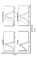


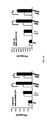


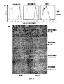


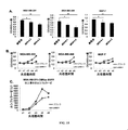


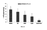
Classifications
-
- A—HUMAN NECESSITIES
- A61—MEDICAL OR VETERINARY SCIENCE; HYGIENE
- A61K—PREPARATIONS FOR MEDICAL, DENTAL OR TOILETRY PURPOSES
- A61K39/00—Medicinal preparations containing antigens or antibodies
- A61K39/395—Antibodies; Immunoglobulins; Immune serum, e.g. antilymphocytic serum
-
- C—CHEMISTRY; METALLURGY
- C07—ORGANIC CHEMISTRY
- C07K—PEPTIDES
- C07K14/00—Peptides having more than 20 amino acids; Gastrins; Somatostatins; Melanotropins; Derivatives thereof
- C07K14/435—Peptides having more than 20 amino acids; Gastrins; Somatostatins; Melanotropins; Derivatives thereof from animals; from humans
- C07K14/705—Receptors; Cell surface antigens; Cell surface determinants
- C07K14/70503—Immunoglobulin superfamily
- C07K14/7051—T-cell receptor (TcR)-CD3 complex
-
- A—HUMAN NECESSITIES
- A61—MEDICAL OR VETERINARY SCIENCE; HYGIENE
- A61K—PREPARATIONS FOR MEDICAL, DENTAL OR TOILETRY PURPOSES
- A61K39/00—Medicinal preparations containing antigens or antibodies
- A61K39/0005—Vertebrate antigens
- A61K39/0011—Cancer antigens
- A61K39/001102—Receptors, cell surface antigens or cell surface determinants
- A61K39/001103—Receptors for growth factors
- A61K39/001104—Epidermal growth factor receptors [EGFR]
-
- A—HUMAN NECESSITIES
- A61—MEDICAL OR VETERINARY SCIENCE; HYGIENE
- A61K—PREPARATIONS FOR MEDICAL, DENTAL OR TOILETRY PURPOSES
- A61K39/00—Medicinal preparations containing antigens or antibodies
- A61K39/46—Cellular immunotherapy
- A61K39/461—Cellular immunotherapy characterised by the cell type used
- A61K39/4613—Natural-killer cells [NK or NK-T]
-
- A—HUMAN NECESSITIES
- A61—MEDICAL OR VETERINARY SCIENCE; HYGIENE
- A61K—PREPARATIONS FOR MEDICAL, DENTAL OR TOILETRY PURPOSES
- A61K39/00—Medicinal preparations containing antigens or antibodies
- A61K39/46—Cellular immunotherapy
- A61K39/463—Cellular immunotherapy characterised by recombinant expression
- A61K39/4631—Chimeric Antigen Receptors [CAR]
-
- A—HUMAN NECESSITIES
- A61—MEDICAL OR VETERINARY SCIENCE; HYGIENE
- A61K—PREPARATIONS FOR MEDICAL, DENTAL OR TOILETRY PURPOSES
- A61K39/00—Medicinal preparations containing antigens or antibodies
- A61K39/46—Cellular immunotherapy
- A61K39/464—Cellular immunotherapy characterised by the antigen targeted or presented
- A61K39/4643—Vertebrate antigens
- A61K39/4644—Cancer antigens
- A61K39/464402—Receptors, cell surface antigens or cell surface determinants
- A61K39/464403—Receptors for growth factors
- A61K39/464404—Epidermal growth factor receptors [EGFR]
-
- A—HUMAN NECESSITIES
- A61—MEDICAL OR VETERINARY SCIENCE; HYGIENE
- A61P—SPECIFIC THERAPEUTIC ACTIVITY OF CHEMICAL COMPOUNDS OR MEDICINAL PREPARATIONS
- A61P35/00—Antineoplastic agents
-
- C—CHEMISTRY; METALLURGY
- C07—ORGANIC CHEMISTRY
- C07K—PEPTIDES
- C07K14/00—Peptides having more than 20 amino acids; Gastrins; Somatostatins; Melanotropins; Derivatives thereof
- C07K14/435—Peptides having more than 20 amino acids; Gastrins; Somatostatins; Melanotropins; Derivatives thereof from animals; from humans
- C07K14/705—Receptors; Cell surface antigens; Cell surface determinants
- C07K14/70503—Immunoglobulin superfamily
- C07K14/70521—CD28, CD152
-
- C—CHEMISTRY; METALLURGY
- C07—ORGANIC CHEMISTRY
- C07K—PEPTIDES
- C07K16/00—Immunoglobulins [IGs], e.g. monoclonal or polyclonal antibodies
- C07K16/18—Immunoglobulins [IGs], e.g. monoclonal or polyclonal antibodies against material from animals or humans
- C07K16/28—Immunoglobulins [IGs], e.g. monoclonal or polyclonal antibodies against material from animals or humans against receptors, cell surface antigens or cell surface determinants
- C07K16/2863—Immunoglobulins [IGs], e.g. monoclonal or polyclonal antibodies against material from animals or humans against receptors, cell surface antigens or cell surface determinants against receptors for growth factors, growth regulators
-
- C—CHEMISTRY; METALLURGY
- C07—ORGANIC CHEMISTRY
- C07K—PEPTIDES
- C07K16/00—Immunoglobulins [IGs], e.g. monoclonal or polyclonal antibodies
- C07K16/18—Immunoglobulins [IGs], e.g. monoclonal or polyclonal antibodies against material from animals or humans
- C07K16/28—Immunoglobulins [IGs], e.g. monoclonal or polyclonal antibodies against material from animals or humans against receptors, cell surface antigens or cell surface determinants
- C07K16/30—Immunoglobulins [IGs], e.g. monoclonal or polyclonal antibodies against material from animals or humans against receptors, cell surface antigens or cell surface determinants from tumour cells
-
- C—CHEMISTRY; METALLURGY
- C12—BIOCHEMISTRY; BEER; SPIRITS; WINE; VINEGAR; MICROBIOLOGY; ENZYMOLOGY; MUTATION OR GENETIC ENGINEERING
- C12N—MICROORGANISMS OR ENZYMES; COMPOSITIONS THEREOF; PROPAGATING, PRESERVING, OR MAINTAINING MICROORGANISMS; MUTATION OR GENETIC ENGINEERING; CULTURE MEDIA
- C12N15/00—Mutation or genetic engineering; DNA or RNA concerning genetic engineering, vectors, e.g. plasmids, or their isolation, preparation or purification; Use of hosts therefor
- C12N15/09—Recombinant DNA-technology
- C12N15/63—Introduction of foreign genetic material using vectors; Vectors; Use of hosts therefor; Regulation of expression
- C12N15/79—Vectors or expression systems specially adapted for eukaryotic hosts
- C12N15/85—Vectors or expression systems specially adapted for eukaryotic hosts for animal cells
- C12N15/86—Viral vectors
-
- C—CHEMISTRY; METALLURGY
- C12—BIOCHEMISTRY; BEER; SPIRITS; WINE; VINEGAR; MICROBIOLOGY; ENZYMOLOGY; MUTATION OR GENETIC ENGINEERING
- C12N—MICROORGANISMS OR ENZYMES; COMPOSITIONS THEREOF; PROPAGATING, PRESERVING, OR MAINTAINING MICROORGANISMS; MUTATION OR GENETIC ENGINEERING; CULTURE MEDIA
- C12N5/00—Undifferentiated human, animal or plant cells, e.g. cell lines; Tissues; Cultivation or maintenance thereof; Culture media therefor
- C12N5/06—Animal cells or tissues; Human cells or tissues
- C12N5/0602—Vertebrate cells
- C12N5/0634—Cells from the blood or the immune system
- C12N5/0636—T lymphocytes
-
- C—CHEMISTRY; METALLURGY
- C12—BIOCHEMISTRY; BEER; SPIRITS; WINE; VINEGAR; MICROBIOLOGY; ENZYMOLOGY; MUTATION OR GENETIC ENGINEERING
- C12N—MICROORGANISMS OR ENZYMES; COMPOSITIONS THEREOF; PROPAGATING, PRESERVING, OR MAINTAINING MICROORGANISMS; MUTATION OR GENETIC ENGINEERING; CULTURE MEDIA
- C12N5/00—Undifferentiated human, animal or plant cells, e.g. cell lines; Tissues; Cultivation or maintenance thereof; Culture media therefor
- C12N5/06—Animal cells or tissues; Human cells or tissues
- C12N5/0602—Vertebrate cells
- C12N5/0634—Cells from the blood or the immune system
- C12N5/0646—Natural killers cells [NK], NKT cells
-
- A—HUMAN NECESSITIES
- A61—MEDICAL OR VETERINARY SCIENCE; HYGIENE
- A61K—PREPARATIONS FOR MEDICAL, DENTAL OR TOILETRY PURPOSES
- A61K39/00—Medicinal preparations containing antigens or antibodies
- A61K2039/51—Medicinal preparations containing antigens or antibodies comprising whole cells, viruses or DNA/RNA
- A61K2039/515—Animal cells
- A61K2039/5156—Animal cells expressing foreign proteins
-
- A—HUMAN NECESSITIES
- A61—MEDICAL OR VETERINARY SCIENCE; HYGIENE
- A61K—PREPARATIONS FOR MEDICAL, DENTAL OR TOILETRY PURPOSES
- A61K39/00—Medicinal preparations containing antigens or antibodies
- A61K2039/51—Medicinal preparations containing antigens or antibodies comprising whole cells, viruses or DNA/RNA
- A61K2039/515—Animal cells
- A61K2039/5158—Antigen-pulsed cells, e.g. T-cells
-
- A—HUMAN NECESSITIES
- A61—MEDICAL OR VETERINARY SCIENCE; HYGIENE
- A61K—PREPARATIONS FOR MEDICAL, DENTAL OR TOILETRY PURPOSES
- A61K39/00—Medicinal preparations containing antigens or antibodies
- A61K2039/80—Vaccine for a specifically defined cancer
-
- C—CHEMISTRY; METALLURGY
- C07—ORGANIC CHEMISTRY
- C07K—PEPTIDES
- C07K2317/00—Immunoglobulins specific features
- C07K2317/60—Immunoglobulins specific features characterized by non-natural combinations of immunoglobulin fragments
- C07K2317/62—Immunoglobulins specific features characterized by non-natural combinations of immunoglobulin fragments comprising only variable region components
- C07K2317/622—Single chain antibody (scFv)
-
- C—CHEMISTRY; METALLURGY
- C07—ORGANIC CHEMISTRY
- C07K—PEPTIDES
- C07K2319/00—Fusion polypeptide
- C07K2319/01—Fusion polypeptide containing a localisation/targetting motif
- C07K2319/03—Fusion polypeptide containing a localisation/targetting motif containing a transmembrane segment
-
- C—CHEMISTRY; METALLURGY
- C07—ORGANIC CHEMISTRY
- C07K—PEPTIDES
- C07K2319/00—Fusion polypeptide
- C07K2319/33—Fusion polypeptide fusions for targeting to specific cell types, e.g. tissue specific targeting, targeting of a bacterial subspecies
-
- C—CHEMISTRY; METALLURGY
- C12—BIOCHEMISTRY; BEER; SPIRITS; WINE; VINEGAR; MICROBIOLOGY; ENZYMOLOGY; MUTATION OR GENETIC ENGINEERING
- C12N—MICROORGANISMS OR ENZYMES; COMPOSITIONS THEREOF; PROPAGATING, PRESERVING, OR MAINTAINING MICROORGANISMS; MUTATION OR GENETIC ENGINEERING; CULTURE MEDIA
- C12N2740/00—Reverse transcribing RNA viruses
- C12N2740/00011—Details
- C12N2740/10011—Retroviridae
- C12N2740/15011—Lentivirus, not HIV, e.g. FIV, SIV
- C12N2740/15041—Use of virus, viral particle or viral elements as a vector
- C12N2740/15043—Use of virus, viral particle or viral elements as a vector viral genome or elements thereof as genetic vector
Description
関連出願への相互参照
本出願は、2015年4月6日に出願された米国仮出願番号第62/143,744号への米国特許法第119条(e)項の下での優先権を主張しており、この仮出願の内容は、その全体が参考として本明細書によって援用される。
Cross-reference to related applications This application gives priority to US Provisional Application No. 62 / 143,744, filed April 6, 2015, under Section 119 (e) of the US Patent Act. Allegedly, the content of this provisional application is incorporated herein by reference in its entirety.
背景
脳癌は、危険であり、処置が困難である。神経膠芽腫(GB)は、最も一般的かつ最も高悪性度の原発性脳腫瘍である。脳の癌は、それが別の起源からの転移の結果である場合にもリスクをもたらす;例えば、乳癌脳転移(BCBM)は、転移性乳癌を有する患者において一般的である。化学療法、放射線照射および外科的切除を行ったとしても、GB患者の全生存期間中央値は、たった14.6ヶ月である(Morgan,R.A.ら(2012)Hum Gene Ther.23:1043−1053)。BCBMも同様に、利用可能な処置に対して非常に抵抗性であり、患者の予後不良を示す。従来の治療は、通常、特異性を欠き、周囲の脳実質および全身組織に対して損傷を引き起こすことがあり、それらの治療の使用を制限する要因となっている(Imperato,J.P.ら(1990)Ann Neurol.28:818−822)。GBに対する免疫ベースの治療は、従来の処置に対する有望な別の選択肢であり、持続可能な抗腫瘍応答をもたらすという長期間の利点を有する可能性とともに局在性の腫瘍細胞と浸潤性の腫瘍細胞の両方を標的化する可能性を有する(Mellman,I.ら(2011)Nature 480:480−489)。上皮成長因子レセプター(EGFR)は、GBを含む様々な腫瘍において重要な役割を果たす。EGFRは、GBにおいて最も頻繁に増幅される遺伝子であるが、正常な脳組織におけるその発現は、検出不可能であるかまたは極端に低い(Parsons,D.W.ら(2008)Science 321:1807−1812;Salomon,D.S.ら(1995)Crit.Rev Oncol Hematol.19:183−232)。EGFRにリガンドが結合すると、レセプターのホモ二量体およびヘテロ二量体が形成され、いくつかの重要なチロシン残基が自己リン酸化して、Ras/Raf/MEK/ERK経路、PLCγ−PKC経路およびPI3K/AKT経路を含むいくつかの細胞内の下流のシグナル伝達経路が活性化し、結果として、細胞の増殖、運動および生存が生じる(Zandi,R.ら(2007)Cell Signal 19:2013−2023)。EGFR増幅腫瘍のおよそ20〜40%は、EGFRバリアントIII変異体(EGFRvIII)を有し、これは、細胞外のリガンド結合ドメインにおけるエキソン2〜7の欠失を含む(Aldape,K.D.ら(2004)Journal of Neuropathology and Experimental Neurology 63:700−707;Biernat,W.ら(2004)Brain Pathol.14:131−136;Fan,Q.W.ら(2013)Cancer Cell 24:438−449;Sugawa,N.ら(1990)Proc Natl Acad Sci.USA 87:8602−8606)。この変異型は、リガンドの非存在下において恒常的な活性化を示すことにより、腫瘍を促進するシグナル伝達経路を活性化する(Ohno,M.ら(2010)Cancer Sci.101:2518−2524)。
Background Brain cancer is dangerous and difficult to treat. Glioblastoma (GB) is the most common and most aggressive primary brain tumor. Brain cancer also poses a risk if it is the result of metastasis from another origin; for example, breast cancer brain metastasis (BCBM) is common in patients with metastatic breast cancer. Even with chemotherapy, radiation, and surgical resection, the median overall survival of GB patients is only 14.6 months (Morgan, RA et al. (2012) Hum Gene Ther. 23: 1043). -1053). BCBM is also highly resistant to available treatments, indicating a poor patient prognosis. Conventional treatments are usually lacking in specificity and can cause damage to the surrounding brain parenchyma and systemic tissues, limiting the use of those treatments (Imperato, JP et al.). (1990) Ann Neurol. 28: 818-822). Immune-based treatment for GB is a promising alternative to conventional treatments, with the long-term benefits of providing a sustainable antitumor response, as well as localized and invasive tumor cells. It has the potential to target both (Mellman, I. et al. (2011) Nature 480: 480-489). Epidermal growth factor receptor (EGFR) plays an important role in various tumors, including GB. EGFR is the most frequently amplified gene in GB, but its expression in normal brain tissue is undetectable or extremely low (Salomons, DW et al. (2008) Science 321: 1807). -1812; Salomon, DS et al. (1995) Crit. Rev Oncol Hematol. 19: 183-232). When the ligand binds to EGFR, receptor homodimers and heterodimers are formed, and some important tyrosine residues are self-phosphorylated to the Ras / Raf / MEK / ERK pathway, PLCγ-PKC pathway. And some intracellular downstream signaling pathways, including the PI3K / AKT pathway, are activated, resulting in cell proliferation, motility and survival (Zandi, R. et al. (2007) Cell Signal 19: 2013-2023). ). Approximately 20-40% of EGFR-amplified tumors carry the EGFR variant III variant (EGFRvIII), which contains a deletion of exons 2-7 in the extracellular ligand-binding domain (Aldape, KD et al.). (2004) Journal of Neuropathology and Experimental Neurology 63: 700-707; Biernat, W. et al. (2004) Brain Pathol. 14: 131-136; Fan, Q.W. et al. (2013) Cancer Cell Sugawa, N. et al. (1990) Proc Natl Acad Sci. USA 87: 8602-8606). This variant activates tumor-promoting signaling pathways by exhibiting constitutive activation in the absence of ligand (Ohno, M. et al. (2010) Cancer Science. 101: 2518-2524). ..
開示の要旨
神経膠芽腫(GB)は、相変わらず最も高悪性度の原発性悪性脳腫瘍である。キメラ抗原レセプター(CAR)によって改変された免疫細胞の養子移入は、有望な抗癌アプローチとして登場したが、CARによって操作されたナチュラルキラー(NK)細胞がGBを処置する潜在的な有用性は、調査されていない。GB患者のおよそ50%由来の腫瘍が、野生型EGFR(wtEGFR)を発現し、より少ない症例においてwtEGFRおよび変異型EGFRvIIIを発現する;しかしながら、以前に報告されたCAR T細胞は、EGFRvIIIを標的化することだけに焦点を当てている。
Abstract of Disclosure Glioblastoma (GB) remains the most aggressive primary malignancies of the brain. Adoption of immune cells modified by chimeric antigen receptor (CAR) has emerged as a promising anti-cancer approach, but the potential usefulness of CAR-engineered natural killer (NK) cells in treating GB is Not investigated. Tumors from approximately 50% of GB patients express wild-type EGFR (wtEGFR) and, in fewer cases, wtEGFR and mutant EGFRvIII; however, previously reported CAR T cells target EGFRvIII. It focuses only on what to do.
本開示の態様は、(a)抗原結合ドメイン;(b)ヒンジドメイン;(c)膜貫通ドメイン;および(d)細胞内ドメインを含むキメラ抗原レセプター(CAR)に関する。本開示は、(a)野生型上皮成長因子レセプター(EGFR)および変異上皮成長因子レセプター(EGFR)(「wtEGFRおよび変異EGFR」)の両方または野生型上皮成長因子レセプターもしくは変異上皮成長因子レセプターを認識した抗EGFR抗体の抗原結合ドメイン;(b)ヒンジドメインポリペプチド;(c)共刺激ポリペプチド;および(d)CD3ゼータシグナル伝達ドメインを含むか、あるいはそれらから本質的になるか、またはなおもさらにはそれらからなる新規CAR、すなわち上皮成長因子レセプター(「EGFR」)キメラ抗原レセプター(CAR)を提供する。1つの態様において、共刺激分子は、細胞内ドメインおよび膜貫通ドメインを含む。非限定的な例としては、CD8、4−1BB共刺激シグナル伝達領域、CD28共刺激分子、OX40、ICOSおよびCD27が挙げられる。1つの態様において、抗EGFR抗体の抗原結合ドメインは、抗EGFR重鎖(HC)可変領域および抗EGFR軽鎖(LC)可変領域を含む。なおもさらなる態様において、EGFR CARはさらに、抗EGFR HC可変領域と抗EGFR LC可変領域との間に配置されたリンカーポリペプチドを含むか、あるいはそれから本質的になるか、またはなおもさらにはそれからなる。そのCARは、検出可能な標識または精製マーカーをさらに含み得る。 Aspects of the present disclosure relate to a chimeric antigen receptor (CAR) comprising (a) an antigen binding domain; (b) a hinge domain; (c) a transmembrane domain; and (d) an intracellular domain. The present disclosure recognizes (a) both wild epidermal growth factor receptor (EGFR) and mutant epidermal growth factor receptor (EGFR) (“wtEGFR and mutant EGFR”) or wild epidermal growth factor receptor or mutant epidermal growth factor receptor. Anti-EGFR antibody antigen-binding domain; (b) hinge domain polypeptide; (c) costimulatory polypeptide; and (d) CD3 zeta signaling domain, or essentially from them, or still Furthermore, a novel CAR composed of them, that is, an epidermal growth factor receptor (“EGFR”) chimeric antigen receptor (CAR) is provided. In one embodiment, the co-stimulator molecule comprises an intracellular domain and a transmembrane domain. Non-limiting examples include CD8, 4-1BB co-stimulation signaling regions, CD28 co-stimulation molecules, OX40, ICOS and CD27. In one embodiment, the antigen binding domain of an anti-EGFR antibody comprises an anti-EGFR heavy chain (HC) variable region and an anti-EGFR light chain (LC) variable region. In still further embodiments, the EGFR CAR further comprises, or becomes essentially, the linker polypeptide located between the anti-EGFR HC variable region and the anti-EGFR LC variable region, or even then. Become. The CAR may further comprise a detectable label or purification marker.
EGFR CARをコードするポリヌクレオチドおよびそれらの相補鎖(complements)ならびに各々の等価物が、さらに本明細書中に開示される。それらのポリヌクレオチドおよび/またはEGFR CARを含むベクターおよび宿主細胞が、本明細書中に提供される。それらのベクターは、プラスミドまたはベクターである。それらの細胞は、原核細胞または真核細胞であり得る。1つの態様において、それらの細胞は、T細胞またはNK細胞である。それらの細胞は拡大され得るので、それらの細胞の拡大された集団が、さらに本明細書中に提供される。それらの細胞は、1つの態様において、T細胞などの細胞の活性化された集団である。 The polynucleotides encoding EGFR CAR and their complements and their respective equivalents are further disclosed herein. Vectors and host cells containing those polynucleotides and / or EGFR CARs are provided herein. Those vectors are plasmids or vectors. Those cells can be prokaryotic or eukaryotic cells. In one embodiment, the cells are T cells or NK cells. Since those cells can be expanded, an expanded population of those cells is further provided herein. Those cells are, in one embodiment, an activated population of cells, such as T cells.
これらの組成物の診断的および治療的な使用が、さらに本明細書中に提供される。例えば、wtEGFRもしくは変異EGFRまたはwtEGFRおよび変異EGFRを細胞表面上に発現している細胞および/または腫瘍あるいはEGFRを発現している幹細胞のうちの1つまたはそれを超えるものの成長を阻害する方法が提供され、その方法は、これらの細胞を、有効量の、本明細書中に記載されるようなEGFR CARを含む単離された細胞と接触させることによって行われる。その細胞は、腫瘍細胞、例えば、神経膠芽腫または癌幹細胞、例えば、神経膠芽腫幹細胞であり得る。その接触は、インビトロまたはインビボであり得る。その方法は、インビトロで行われるとき、新しい治療または併用療法をスクリーニングするのに有用である。その方法は、インビボでは、EGFRを発現する癌、例えば、脳癌、例えば、神経膠芽腫または別の癌(例えば、乳癌)の脳転移に罹患している患者を処置するのに有用である。 Diagnostic and therapeutic uses of these compositions are further provided herein. For example, there is provided a method of inhibiting the growth of one or more of cells expressing wtEGFR or mutant EGFR or wtEGFR and mutant EGFR on the cell surface and / or tumor or stem cells expressing EGFR. The method is performed by contacting these cells with an effective amount of isolated cells containing EGFR CAR as described herein. The cells can be tumor cells, such as glioblastoma or cancer stem cells, such as glioblastoma stem cells. The contact can be in vitro or in vivo. The method is useful for screening new therapies or combination therapies when performed in vitro. The method is useful in vivo to treat patients suffering from brain metastases from cancers that express EGFR, such as brain cancer, such as glioblastoma or another cancer (eg, breast cancer). ..
別の態様において、本開示は、wtEGFRもしくは変異EGFRまたはwtEGFRおよび変異EGFRを細胞表面上に発現している1つまたはそれを超える細胞の成長の阻害を必要とする被験体において、そのような細胞の成長を阻害する方法を提供する。この方法は、有効量の、本明細書中に記載されるようなEGFR CARを含む単離された細胞をその被験体に投与する工程を含むか、あるいはその工程から本質的になるか、またはなおもさらにはその工程からなる。EGFRを発現している細胞は、腫瘍細胞または癌幹細胞、例えば、神経膠芽腫または神経膠芽腫幹細胞であり得る。EGFR CARを発現している単離された細胞は、それらの細胞を投与される被験体にとって自家であり得る。癌の処置を必要とする被験体に、有効量の、本明細書中に記載されるようなEGFR CARを含む単離された細胞を投与することによって、その被験体において癌を処置する方法がなおもさらに提供され、ここで、その癌細胞は、wtEGFRもしくは変異EGFRまたはwtEGFRおよび変異EGFRを発現している。これらの方法の単離された細胞は、NK細胞および/またはT細胞であり得、処置される被験体にとって自家であり得る。さらなる態様において、それらの細胞は、処置している医師によって決定される任意の適切な経路によって投与され、それらには、頭蓋内注射、静脈内投与が含まれるが、これらに限定されない。 In another embodiment, the present disclosure discloses such cells in a subject requiring inhibition of the growth of one or more cells expressing wtEGFR or mutant EGFR or wtEGFR and mutant EGFR on the cell surface. Provides a method of inhibiting the growth of. This method comprises or is essentially a step of administering to the subject an isolated cell containing an effective amount of EGFR CAR as described herein. It still consists of that process. Cells expressing EGFR can be tumor cells or cancer stem cells, such as glioblastoma or glioblastoma stem cells. Isolated cells expressing EGFR CAR can be autologous to the subject to whom they are administered. A method of treating a cancer in a subject in need of treatment of the cancer by administering to the subject an effective amount of an isolated cell containing EGFR CAR as described herein. Still further provided, where the cancer cells express wtEGFR or mutant EGFR or wtEGFR and mutant EGFR. The isolated cells of these methods can be NK cells and / or T cells and can be autologous to the subject being treated. In a further embodiment, the cells are administered by any suitable route determined by the treating physician, including, but not limited to, intracranial injection, intravenous administration.
本明細書中に記載されるようなEGFR CARならびにEGFRタンパク質もしくはそのフラグメントおよび/または変異EGFRタンパク質もしくはそのフラグメントを含む単離された複合体が、なおもさらに提供される。別の態様において、本明細書中に記載されるようなEGFR CARおよびwtEGFRもしくは変異EGFRまたはwtEGFRおよび変異EGFRを発現している細胞を含む単離された複合体が、本明細書中に提供される。 Further provided are EGFR CARs as described herein and an isolated complex comprising an EGFR protein or fragment thereof and / or a mutant EGFR protein or fragment thereof. In another embodiment, an isolated complex comprising cells expressing EGFR CAR and wtEGFR or mutant EGFR or wtEGFR and mutant EGFR as described herein is provided herein. To.
本開示のさらなる態様は、本明細書中に記載されるEGFR CARを発現している細胞(例えば、T細胞またはNK細胞)および腫瘍退縮単純ヘルペスウイルスを含む組成物および併用癌治療に関する。いくつかの実施形態は、腫瘍または癌を処置する方法に関し、ここで、EGFR CARを発現している細胞と腫瘍退縮単純ヘルペスウイルス(oncolytic herepes simplex)の両方が、それを必要としている患者に投与される。いくつかの実施形態において、EGFR CARを発現している細胞は、腫瘍退縮単純ヘルペスウイルスと同時に投与される;他の実施形態において、EGFR CARを発現している細胞は、腫瘍退縮単純ヘルペスウイルスの投与の前または後に投与される。 A further aspect of the disclosure relates to compositions and concomitant cancer treatments comprising cells expressing EGFR CAR described herein (eg, T cells or NK cells) and tumor regressive herpes simplex virus. Some embodiments relate to a method of treating a tumor or cancer, wherein both the cells expressing EGFR CAR and the oncolytic herpes simplex virus are administered to a patient in need of it. Will be done. In some embodiments, cells expressing EGFR CAR are co-administered with tumor-regressive herpes simplex virus; in other embodiments, cells expressing EGFR CAR are tumor-regressing herpes simplex virus. It is administered before or after administration.
1つまたはそれを超える上記組成物を含むキットがさらに提供される。それらを診断的または治療的に使用するための指示書がさらに提供される。
本発明は、例えば、以下の項目を提供する。
(項目1)
(a)野生型上皮成長因子レセプター(EGFR)もしくは変異上皮成長因子レセプター(EGFR)のいずれかまたは野生型上皮成長因子レセプターおよび変異上皮成長因子レセプターの両方(「wtEGFRおよび/または変異EGFR」)を認識した抗EGFR抗体の抗原結合ドメイン;(b)ヒンジドメインポリペプチド;(c)共刺激分子または共刺激ポリペプチド;および(d)CD3ゼータシグナル伝達ドメイン、あるいはそれらの各々の等価物を含む、キメラ抗原レセプター(CAR)。
(項目2)
前記共刺激分子が、4−1BB共刺激シグナル伝達領域、CD28共刺激分子、OX40、ICOSおよびCD27、またはそれらの各々の等価物の群から選択される分子またはポリペプチドを含む、項目1に記載のCAR。
(項目3)
前記抗EGFR抗体の前記抗原結合ドメインが、抗EGFR重鎖(HC)可変領域および抗EGFR軽鎖(LC)可変領域またはそれらの各々の等価物を含む、項目1に記載のCAR。
(項目4)
前記HC可変領域が、

またはその等価物を含む、項目3に記載のCAR。
(項目5)
前記LC可変領域が、
またはその等価物を含む、項目3または4に記載のCAR。
(項目6)
前記抗EGFR HC可変領域と前記抗EGFR LC可変領域との間に配置されたリンカーポリペプチドをさらに含む、項目3に記載のCAR。
(項目7)
前記リンカーポリペプチドが、
![]()
またはその等価物を含む、項目6に記載のCAR。
(項目8)
前記EGFR結合ドメインのアミノ末端にシグナルポリペプチドをさらに含む、項目1〜3のいずれかに記載のCAR。
(項目9)
前記ヒンジポリペプチドが、
![]()
またはその等価物を含む、項目1〜3のいずれかに記載のCAR。
(項目10)
前記CD28共刺激分子が、CD28膜貫通ドメインおよびCD28細胞内ドメインまたはそれらの各々の等価物を含む、項目2に記載のCAR。
(項目11)
前記CARに付着された検出可能なマーカーおよび/または精製マーカーをさらに含む、項目1〜3のいずれかに記載のCAR。
(項目12)
等価なポリペプチドが、前記ポリペプチドと少なくとも80%のアミノ酸同一性であるか、または該ポリペプチドをコードするポリヌクレオチドまたはその相補鎖と少なくとも80%同一のポリヌクレオチドによってコードされるか、または高ストリンジェンシーの条件下で該ポリヌクレオチドまたはその相補鎖にハイブリダイズし、ここで、高ストリンジェンシーの条件は、約25℃〜約37℃のインキュベーション温度;約6×SSC〜約10×SSCのハイブリダイゼーション緩衝液濃度;約0%〜約25%のホルムアミド濃度;および約4×SSC〜約8×SSCの洗浄液を含む、項目1〜3、5または6のいずれか1項に記載のCAR。
(項目13)
項目1〜3、5または6のいずれか1項に記載のCARをコードする、単離された核酸。
(項目14)
項目13に記載の単離された核酸配列を含む、ベクター。
(項目15)
前記ベクターが、プラスミドである、項目14に記載のベクター。
(項目16)
前記ベクターが、ウイルスベクターである、項目14に記載のベクター。
(項目17)
前記ウイルスベクターが、レトロウイルスベクター、レンチウイルスベクター、アデノウイルスベクターおよびアデノ随伴ウイルスベクターからなる群より選択される、項目14に記載のベクター。
(項目18)
前記ベクターが、レンチウイルスベクターである、項目14に記載のベクター。
(項目19)
検出可能な標識および/または精製マーカーをさらに含み、該検出可能な標識は、必要に応じて、天然に存在するポリヌクレオチドではない、項目14〜18のいずれか1項に記載のベクター。
(項目20)
項目1〜3、5または6のいずれか1項に記載のCARを含む、単離された細胞。
(項目21)
項目13に記載の単離された核酸を含む、単離された細胞。
(項目22)
項目14に記載のベクターを含む、単離された細胞。
(項目23)
前記細胞が、原核細胞または真核細胞である、項目20に記載の単離された細胞。
(項目24)
前記細胞が、原核細胞または真核細胞である、項目21に記載の単離された細胞。
(項目25)
前記細胞が、原核細胞または真核細胞である、項目22に記載の単離された細胞。
(項目26)
前記細胞が、真核細胞である、項目23に記載の単離された細胞。
(項目27)
前記細胞が、真核細胞である、項目24に記載の単離された細胞。
(項目28)
前記細胞が、真核細胞である、項目25に記載の単離された細胞。
(項目29)
前記真核細胞が、動物細胞、哺乳動物細胞、ウシ細胞、ネコ細胞、イヌ細胞、マウス細胞、ウマ細胞またはヒト細胞から選択される、項目26に記載の単離された細胞。
(項目30)
前記真核細胞が、動物細胞、哺乳動物細胞、ウシ細胞、ネコ細胞、イヌ細胞、マウス細胞、ウマ細胞またはヒト細胞から選択される、項目27に記載の単離された細胞。
(項目31)
前記真核細胞が、動物細胞、哺乳動物細胞、ウシ細胞、ネコ細胞、イヌ細胞、マウス細胞、ウマ細胞またはヒト細胞から選択される、項目28に記載の単離された細胞。
(項目32)
前記細胞が、T細胞およびナチュラルキラー(NK)細胞の群から選択される、項目29に記載の単離された細胞。
(項目33)
前記細胞が、T細胞およびナチュラルキラー(NK)細胞の群から選択される、項目30に記載の単離された細胞。
(項目34)
前記細胞が、T細胞およびナチュラルキラー(NK)細胞の群から選択される、項目31に記載の単離された細胞。
(項目35)
項目1〜3のいずれか1項に記載のCARならびにEGFRタンパク質もしくはそのフラグメントおよび/または変異EGFRタンパク質もしくはそのフラグメントを含む、単離された複合体。
(項目36)
項目1〜3のいずれか1項に記載のCARならびにwtEGFRおよび/または変異EGFRを発現する細胞を含む、単離された複合体。
(項目37)
キャリアおよび項目1〜3のいずれか1項に記載の1つまたはそれを超えるCARを含む、組成物。
(項目38)
前記キャリアが、薬学的に許容され得るキャリアまたは固体支持体である、項目37に記載の組成物。
(項目39)
項目1〜3のいずれか1項に記載のCARをコードする核酸配列を細胞に導入する工程を含む、EGFR CAR細胞を作製する方法。
(項目40)
前記EGFR CARによって形質導入された細胞を単離する工程をさらに含む、項目39に記載の方法。
(項目41)
前記細胞が、哺乳動物細胞、イヌ細胞、ウシ細胞、マウス細胞、ネコ細胞、ウマ細胞またはヒト細胞からなる群の細胞、および必要に応じてT細胞またはNK細胞である、項目39に記載の方法。
(項目42)
項目40または41に記載の方法によって作製された、単離されたCAR細胞。
(項目43)
EGFRおよび/または変異EGFRを細胞表面上に発現する細胞の成長を阻害する方法であって、該細胞を有効量の項目42に記載の単離された細胞と接触させる工程を含む、方法。
(項目44)
前記細胞が、腫瘍細胞または癌幹細胞である、項目43に記載の方法。
(項目45)
前記腫瘍細胞が、(i)神経膠芽腫もしくは神経膠芽腫幹細胞および/または(ii)乳癌脳転移もしくは乳癌脳転移幹細胞から必要に応じて選択される脳癌由来の細胞である、項目44に記載の方法。
(項目46)
EGFRおよび/または変異EGFRを細胞表面上に発現する細胞の成長の阻害を必要とする被験体において該細胞の成長を阻害する方法であって、該被験体に有効量の項目42に記載の単離された細胞を投与する工程を含む、方法。
(項目47)
前記細胞が、腫瘍細胞または幹細胞である、項目46に記載の方法。
(項目48)
前記腫瘍細胞が、(i)神経膠芽腫もしくは神経膠芽腫幹細胞および/または(ii)乳癌脳転移もしくは乳癌脳転移幹細胞から必要に応じて選択される脳癌由来の細胞である、項目47に記載の方法。
(項目49)
前記単離された細胞が、該細胞を投与される被験体にとって自家である、項目46に記載の方法。
(項目50)
癌の処置を必要とする被験体において癌を処置するための方法であって、ここで、該癌細胞は、EGFRおよび/または変異EGFRを発現し、該方法は、該被験体に有効量の項目42に記載の単離された細胞を投与する工程を含む、方法。
(項目51)
前記単離された細胞が、処置される被験体にとって自家である、項目50に記載の方法。
(項目52)
前記細胞が、頭蓋内注射または静脈内投与によって投与される、項目50に記載の方法。
(項目53)
前記被験体が、哺乳動物、マウス、イヌ、ウシ、マウス、ネコ、ウマまたはヒトの群から選択される、項目50に記載の方法。
(項目54)
被験体が、抗EGFR CAR治療に応答する可能性があるかまたは応答する可能性がないかを判定するための方法であって、該方法は、患者から単離された癌細胞を有効量の抗EGFR抗体および変異EGFR抗体と接触させる工程、該癌細胞に結合した任意の抗体の存在を検出する工程を含み、ここで、該癌細胞に結合した抗体の存在は、該被験体が、該抗EGFR CAR治療に応答する可能性があることを示唆し、該癌細胞に結合した抗体が存在しないことは、該被験体が、該抗EGFR CAR治療に応答する可能性がないことを示唆する、方法。
(項目55)
前記抗EGFR CAR治療に応答する可能性があると判定された被験体に有効量の項目26に記載の細胞を投与する工程をさらに含む、項目54に記載の方法。
(項目56)
前記癌細胞が、(i)神経膠芽腫細胞もしくは神経膠芽腫幹細胞および/または(ii)乳癌転移もしくは乳癌転移幹細胞から必要に応じて選択される脳癌由来の細胞である、項目54または55に記載の方法。
(項目57)
前記被験体が、哺乳動物、ウマ、ネコ、イヌ、ウシ、マウスまたはヒトの群から選択される、項目54または55に記載の方法。
(項目58)
有効量の抗癌治療を行う工程をさらに含む、項目54または55に記載の方法。
(項目59)
前記抗癌治療が、放射線療法、手術または化学療法のうちの1つまたはそれを超えるものを含む、項目58に記載の方法。
(項目60)
前記抗EGFR CAR治療が、一次治療、二次治療、三次治療、四次治療の群から選択される、項目50に記載の方法。
(項目61)
項目1〜3のいずれか1項に記載の1つまたはそれを超えるCARおよび必要に応じて使用の指示書を含む、キット。
(項目62)
腫瘍退縮単純ヘルペスウイルスをさらに含む、項目61に記載のキット。
(項目63)
癌の処置を必要とする被験体において癌を処置するための方法であって、有効量の項目42に記載の単離された細胞および有効量の腫瘍退縮単純ヘルペスウイルスまたはそれらの各々の等価物を被験体に投与する工程を含む、方法。
(項目64)
前記単離された細胞が、NK細胞である、項目63に記載の方法。
(項目65)
前記単離された細胞が、放射線照射された細胞である、項目63または64のいずれか1項に記載の方法。
(項目66)
前記単離された細胞が、1000cGyの線量で放射線照射された細胞である、項目55に記載の方法。
(項目67)
前記腫瘍退縮単純ヘルペスウイルスが、HSV−1であるかまたはHSV−1由来である、項目63に記載の方法。
(項目68)
前記有効量の単離された細胞が、前記有効量の腫瘍退縮単純ヘルペスウイルスの前に投与される、項目63に記載の方法。
(項目69)
前記有効量の腫瘍退縮単純ヘルペスウイルスが、複数の日にわたって投与される、項目68に記載の方法。
(項目70)
前記癌が、脳癌である、項目63に記載の方法。
(項目71)
前記脳癌が、神経膠芽腫または乳癌脳転移から選択される、項目70に記載の方法。
(項目72)
項目42に記載の単離された細胞および使用の指示書ならびに必要に応じて腫瘍退縮ヘルペスウイルスを含む、癌を処置するためのキット。
Further kits are provided that include one or more of the above compositions. Further instructions are provided for their diagnostic or therapeutic use.
The present invention provides, for example, the following items.
(Item 1)
(A) Either wild epidermal growth factor receptor (EGFR) or mutant epidermal growth factor receptor (EGFR) or both wild epidermal growth factor receptor and mutant epidermal growth factor receptor (“wtEGFR and / or mutant EGFR”) Containing the recognized anti-EGFR antibody antigen-binding domain; (b) hinge domain polypeptide; (c) costimulatory molecule or costimulatory polypeptide; and (d) CD3 zeta signaling domain, or their respective equivalents. Chimeric antigen receptor (CAR).
(Item 2)
The first item, wherein the co-stimulating molecule comprises a molecule or polypeptide selected from the group of 4-1BB co-stimulating signaling region, CD28 co-stimulating molecule, OX40, ICOS and CD27, or their respective equivalents. CAR.
(Item 3)
The CAR according to
(Item 4)
The HC variable region
The CAR according to
(Item 5)
The LC variable region
The CAR according to
(Item 6)
The CAR according to
(Item 7)
The linker polypeptide
![]()
6. The CAR according to item 6, which comprises an equivalent thereof.
(Item 8)
The CAR according to any one of
(Item 9)
The hinge polypeptide
![]()
The CAR according to any one of
(Item 10)
The CAR according to
(Item 11)
The CAR according to any one of
(Item 12)
The equivalent polypeptide has at least 80% amino acid identity with the polypeptide, or is encoded by or is high by at least 80% identical to the polynucleotide encoding the polypeptide or its complementary strand. Hybridize to the polynucleotide or its complement under stringency conditions, where high stringency conditions are incubation temperatures of about 25 ° C to about 37 ° C; high of about 6 x SSC to about 10 x SSC. The CAR according to any one of
(Item 13)
An isolated nucleic acid encoding the CAR according to any one of items 1-3, 5 or 6.
(Item 14)
A vector comprising the isolated nucleic acid sequence according to item 13.
(Item 15)
The vector according to
(Item 16)
The vector according to
(Item 17)
(Item 18)
The vector according to
(Item 19)
The vector according to any one of items 14-18, further comprising a detectable label and / or a purified marker, wherein the detectable label is optionally not a naturally occurring polynucleotide.
(Item 20)
An isolated cell comprising the CAR according to any one of items 1-3, 5 or 6.
(Item 21)
An isolated cell comprising the isolated nucleic acid of item 13.
(Item 22)
An isolated cell comprising the vector according to
(Item 23)
The isolated cell according to
(Item 24)
The isolated cell of item 21, wherein the cell is a prokaryotic cell or a eukaryotic cell.
(Item 25)
The isolated cell of item 22, wherein the cell is a prokaryotic cell or a eukaryotic cell.
(Item 26)
The isolated cell according to
(Item 27)
The isolated cell according to
(Item 28)
The isolated cell according to
(Item 29)
(Item 30)
27. The isolated cell of
(Item 31)
28. The isolated cell of item 28, wherein the eukaryotic cell is selected from animal cells, mammalian cells, bovine cells, cat cells, dog cells, mouse cells, horse cells or human cells.
(Item 32)
29. The isolated cell of
(Item 33)
(Item 34)
(Item 35)
An isolated complex comprising the CAR and EGFR protein or fragment thereof and / or mutant EGFR protein or fragment thereof according to any one of items 1-3.
(Item 36)
An isolated complex comprising cells expressing the CAR and wtEGFR and / or mutant EGFR according to any one of items 1-3.
(Item 37)
A composition comprising a carrier and one or more CARs according to any one of items 1-3.
(Item 38)
37. The composition of item 37, wherein the carrier is a pharmaceutically acceptable carrier or solid support.
(Item 39)
A method for producing an EGFR CAR cell, which comprises the step of introducing a nucleic acid sequence encoding the CAR according to any one of
(Item 40)
39. The method of item 39, further comprising the step of isolating the cells transduced by the EGFR CAR.
(Item 41)
39. The method of item 39, wherein the cells are a group of cells consisting of mammalian cells, canine cells, bovine cells, mouse cells, cat cells, horse cells or human cells, and optionally T cells or NK cells. ..
(Item 42)
An isolated CAR cell produced by the method according to
(Item 43)
A method of inhibiting the growth of cells expressing EGFR and / or mutant EGFR on the cell surface, comprising contacting the cells with an effective amount of the isolated cell according to
(Item 44)
43. The method of item 43, wherein the cells are tumor cells or cancer stem cells.
(Item 45)
(Item 46)
The method of inhibiting the growth of a cell expressing EGFR and / or mutant EGFR on the cell surface in a subject requiring inhibition of the growth of the cell, wherein the effective amount for the subject is simply
(Item 47)
46. The method of item 46, wherein the cells are tumor cells or stem cells.
(Item 48)
Item 47, wherein the tumor cells are cells derived from brain cancer, which are optionally selected from (i) glioblastoma or glioblastoma stem cells and / or (ii) breast cancer brain metastases or breast cancer brain metastases stem cells. The method described in.
(Item 49)
46. The method of item 46, wherein the isolated cells are autologous to the subject to whom the cells are administered.
(Item 50)
A method for treating cancer in a subject in need of treatment of the cancer, wherein the cancer cells express EGFR and / or mutant EGFR, the method of which is an effective amount for the subject. A method comprising the step of administering the isolated cells of
(Item 51)
The method of
(Item 52)
50. The method of
(Item 53)
50. The method of
(Item 54)
A method for determining whether a subject is or is not likely to respond to anti-EGFR CAR therapy, the method of which is an effective amount of cancer cells isolated from a patient. It comprises contacting with an anti-EGFR antibody and a mutant EGFR antibody, detecting the presence of any antibody bound to the cancer cell, wherein the presence of the antibody bound to the cancer cell is determined by the subject. It suggests that it may respond to anti-EGFR CAR treatment and that the absence of antibodies bound to the cancer cells suggests that the subject is unlikely to respond to the anti-EGFR CAR treatment. ,Method.
(Item 55)
54. The method of
(Item 56)
(Item 57)
54. The method of
(Item 58)
54. The method of
(Item 59)
58. The method of item 58, wherein the anti-cancer treatment comprises one or more of radiation therapy, surgery or chemotherapy.
(Item 60)
The method of
(Item 61)
A kit comprising one or more CARs according to any one of items 1-3 and instructions for use as needed.
(Item 62)
The kit of item 61, further comprising a tumor regression herpes simplex virus.
(Item 63)
A method for treating cancer in a subject in need of treatment for cancer, wherein an effective amount of the isolated cells and an effective amount of tumor regression herpes simplex virus or their respective equivalents according to
(Item 64)
63. The method of item 63, wherein the isolated cell is an NK cell.
(Item 65)
The method according to any one of items 63 or 64, wherein the isolated cells are irradiated cells.
(Item 66)
55. The method of item 55, wherein the isolated cells are cells irradiated with a dose of 1000 cGy.
(Item 67)
63. The method of item 63, wherein the tumor regressive herpes simplex virus is HSV-1 or is derived from HSV-1.
(Item 68)
63. The method of item 63, wherein the effective amount of isolated cells is administered prior to the effective amount of tumor regressive herpesvirus.
(Item 69)
68. The method of item 68, wherein the effective amount of tumor regressive herpesvirus is administered over multiple days.
(Item 70)
63. The method of item 63, wherein the cancer is a brain cancer.
(Item 71)
The method of item 70, wherein the brain cancer is selected from glioblastoma or breast cancer brain metastases.
(Item 72)
A kit for treating cancer, comprising the isolated cells of
詳細な説明
本開示は、記載される特定の態様に限定されず、したがって当然のことながら変化し得ることが理解されるべきである。本開示の範囲は、添付の請求項によってのみ限定されるので、本明細書中で使用される用語は、単に特定の態様を説明する目的であって、限定を意図していないことも理解されるべきである。
Detailed Description It should be understood that the present disclosure is not limited to the particular aspects described and can therefore change as a matter of course. As the scope of the present disclosure is limited only by the appended claims, it is also understood that the terms used herein are solely for the purpose of describing particular embodiments and are not intended to be limiting. Should be.
別段定義されない限り、本明細書中で使用されるすべての専門用語および科学用語が、本技術が属する分野の当業者が通常理解している意味と同じ意味を有する。本明細書中に記載される方法および材料と類似または等価な任意の方法および材料を本技術の実施または試験において使用できるが、好ましい方法、デバイスおよび材料がここに記載される。本明細書に引用されるすべての技術的刊行物および特許公報は、そのすべてが参照により本明細書中に援用される。本明細書中のいかなる記載も、本技術が、先行発明を理由にそのような開示に先行する権利がないことを自認するものとして解釈されるべきでない。 Unless otherwise defined, all technical and scientific terms used herein have the same meaning as commonly understood by one of ordinary skill in the art to which this technology belongs. Any method and material similar or equivalent to or equivalent to the methods and materials described herein can be used in the practice or testing of the art, but preferred methods, devices and materials are described herein. All technical publications and patent gazettes cited herein are incorporated herein by reference in their entirety. Nothing herein should be construed as admitting that the Technology does not have the right to precede such disclosure on the grounds of prior inventions.
本技術の実施は、別段示されない限り、当該分野の技術の範囲内である、組織培養、免疫学、分子生物学、微生物学、細胞生物学および組換えDNAの従来の手法を用いる。例えば、Sambrook and Russell編(2001)Molecular Cloning:A Laboratory Manual,3rd edition;the series Ausubelら編(2007)Current Protocols in Molecular Biology;the series Methods in Enzymology(Academic Press,Inc.,N.Y.);MacPhersonら(1991)PCR 1:A Practical Approach(IRL Press at Oxford University Press);MacPhersonら(1995)PCR 2:A Practical Approach;Harlow and Lane編(1999)Antibodies,A Laboratory Manual;Freshney(2005)Culture of Animal Cells:A Manual of Basic Technique,5th edition;Gait編(1984)Oligonucleotide Synthesis;米国特許第4,683,195号;Hames and Higgins編(1984)Nucleic Acid Hybridization;Anderson(1999)Nucleic Acid Hybridization;Hames and Higgins編(1984)Transcription and Translation;Immobilized Cells and Enzymes(IRL Press(1986));Perbal(1984)A Practical Guide to Molecular Cloning;Miller and Calos編(1987)Gene Transfer Vectors for Mammalian Cells(Cold Spring Harbor Laboratory);Makrides編(2003)Gene Transfer and Expression in Mammalian Cells;Mayer and Walker編(1987)Immunochemical Methods in Cell and Molecular Biology(Academic Press,London);およびHerzenbergら編(1996)Weir’s Handbook of Experimental Immunologyを参照のこと。 Unless otherwise indicated, the practice of this technique uses conventional techniques of tissue culture, immunology, molecular biology, microbiology, cell biology and recombinant DNA, which are within the scope of the art in the art. For example, Sambrook and Russell ed. (2001) Molecular Cloning: A Laboratory Manual, 3rd edition; the series Ausubel et al. (2007) Molecular Protocols in Molecular Biology. MacPherson et al. (1991) PCR 1: A Practical Aproach (IRL Press at Oxford University Press); MacPherson et al. (1995) PCR 2: A Practical Aproach; Hybrid Culture of Animal Cells: A Manual of Basic Technology, 5th edition; Gait (1984) Polymerase Chain Reaction (US Patent No. 4,683, 195; ; Hames and Higgins eds. (1984) Transcription and Translation; Immobilized Cells and Enzymes (IRL Press (1986)); Perbal (1984) A Practical Guide to Molecular Cloning; Miller and Calos eds. (1987) Gene Transfer Vectors for Mammalian Cells (Cold Spring Harbor Laboratory); Makerides (2003) Gene Transfer and Expression in Molecular Cells; Mayer And Walker ed. (1987) Immunochemical Materials in Cell and Molecular Biology (Academic Press, London); and Herzenberg et al. (1996) Weir's Handbook.
範囲を含むすべての数値の明示、例えば、pH、温度、時間、濃度および分子量は、必要に応じて、1.0または0.1という一定数量ずつ、あるいは+/−15%あるいは10%あるいは5%あるいは2%の変動で(+)または(−)変動する近似値である。必ずしも明示的に述べられないが、すべての数値の明示は、用語「約」が先行すると理解されるべきである。必ずしも明示的に述べられないが、本明細書中に記載される試薬は、単に例示的なものであることおよびそのような試薬の等価物が当該分野で公知であることも理解されるべきである。 All numerical values, including ranges, such as pH, temperature, time, concentration and molecular weight, may be in constant quantities of 1.0 or 0.1, or +/- 15% or 10% or 5 as required. It is an approximate value that fluctuates (+) or (-) with a fluctuation of% or 2%. Although not necessarily explicitly stated, it should be understood that all numerical statements are preceded by the term "about". Although not necessarily explicitly stated, it should also be understood that the reagents described herein are merely exemplary and that equivalents of such reagents are known in the art. is there.
本技術がポリペプチド、タンパク質、ポリヌクレオチドまたは抗体に関するとき、そのようなものの等価物または生物学的等価物は、明示的な列挙なしに、かつ別段意図されない限り、本技術の範囲内であると意図されると推測されるべきである。
定義
When the present technology relates to polypeptides, proteins, polynucleotides or antibodies, the equivalents or biological equivalents of such are within the scope of the present art without explicit enumeration and unless otherwise intended. It should be inferred to be intended.
Definition
明細書および請求項において使用されるとき、単数形「a」、「an」および「the」は、文脈が明らかに他のことを指示しない限り、複数の指示対象を含む。例えば、用語「細胞(a cell)」は、それらの混合物を含む複数の細胞を含む。 As used in the specification and claims, the singular forms "a", "an" and "the" include a plurality of referents unless the context clearly indicates otherwise. For example, the term "cell" includes a plurality of cells containing a mixture thereof.
本明細書中で使用されるとき、用語「動物」とは、例えば、哺乳動物および鳥類を含むカテゴリーである、生存している多細胞の脊椎動物のことを指す。用語「哺乳動物」は、ヒトと非ヒト哺乳動物の両方を含む。 As used herein, the term "animal" refers to a living multicellular vertebrate, eg, a category that includes mammals and birds. The term "mammal" includes both human and non-human mammals.
用語「被験体」、「宿主」、「個体」および「患者」は、ヒト被験体および動物被験体、例えば、ヒト、動物、非ヒト霊長類、イヌ、ネコ、ヒツジ、マウス、ウマおよびウシのことを指すために本明細書中で交換可能に使用される。いくつかの実施形態において、被験体は、ヒトである。 The terms "subject", "host", "individual" and "patient" refer to human and animal subjects such as humans, animals, non-human primates, dogs, cats, sheep, mice, horses and cattle. It is used interchangeably herein to indicate that. In some embodiments, the subject is a human.
本明細書中で使用されるとき、用語「抗体」とは、免疫グロブリンまたは免疫グロブリン様分子のことを集合的に指し、例として、IgA、IgD、IgE、IgGおよびIgM、それらの組み合わせ、および任意の脊椎動物、例えば、哺乳動物(例えば、ヒト、ヤギ、ウサギおよびマウス)ならびに非哺乳動物種(例えば、サメ免疫グロブリン)における免疫応答において産生される同様の分子が挙げられるが、これらに限定されない。別段明確に示されない限り、用語「抗体」は、目的の分子(または高度に類似した目的の分子の群)に特異的に結合するインタクトな免疫グロブリンおよび「抗体フラグメント」または「抗原結合フラグメント」を、他の分子への結合を実質的に排除する程度に含む(例えば、生物学的サンプル中の他の分子に対する結合定数よりも少なくとも103M−1高い、少なくとも104M−1高いまたは少なくとも105M−1高い、目的の分子に対する結合定数を有する抗体および抗体フラグメント)。用語「抗体」は、遺伝的に操作された形態、例えば、キメラ抗体(例えば、ヒト化マウス抗体)、ヘテロ結合体抗体(例えば、二重特異性抗体)も含む。Pierce Catalog and Handbook,1994−1995(Pierce Chemical Co.,Rockford,Ill.);Kuby,J.,Immunology,3rd Ed.,W.H.Freeman & Co.,New York,1997も参照のこと。 As used herein, the term "antibody" collectively refers to immunoglobulins or immunoglobulin-like molecules, such as IgA, IgD, IgE, IgG and IgM, combinations thereof, and. Similar molecules produced in an immune response in any vertebrate, such as mammals (eg, humans, goats, rabbits and mice) and non-mammalian species (eg, shark immunoglobulins), but are limited thereto. Not done. Unless otherwise stated explicitly, the term "antibody" refers to intact immunoglobulins and "antibody fragments" or "antibody-binding fragments" that specifically bind to the molecule of interest (or a group of highly similar molecules of interest). , To the extent that binding to other molecules is substantially eliminated (eg, at least 10 3 M -1 higher, at least 10 4 M -1 higher or at least higher than the binding constant for other molecules in the biological sample. 10 5 M -1 High, antibodies and antibody fragments with binding constants to the molecule of interest). The term "antibody" also includes genetically engineered forms such as chimeric antibodies (eg, humanized mouse antibodies), heteroconjugate antibodies (eg, bispecific antibodies). Pierce Catalog and Handbook, 1994-1995 (Pierce Chemical Co., Rockford, Ill.); Cuba, J. et al. , Immunology, 3 rd Ed. , W. H. Freeman & Co. , New York, 1997.
抗体構造に関して、免疫グロブリンは、ジスルフィド結合によって相互接続された重(H)鎖および軽(L)鎖を有する。軽鎖には、2つのタイプ、ラムダ(λ)およびカッパー(κ)がある。抗体分子の機能活性を決定する5つの主要な重鎖クラス(またはアイソタイプ)がある:IgM、IgD、IgG、IgAおよびIgE。重鎖および軽鎖の各々は、定常領域および可変領域を含む(これらの領域は、「ドメイン」としても知られる)。重鎖可変領域および軽鎖可変領域は一体となって抗原に特異的に結合する。軽鎖可変領域および重鎖可変領域は、「相補性決定領域」または「CDR」とも呼ばれる3つの超可変領域によって中断された「フレームワーク」領域を含む。フレームワーク領域およびCDRの範囲は、定義されている(参照により本明細書に援用される、Kabatら、Sequences of Proteins of Immunological Interest,U.S.Department of Health and Human Services,1991を参照のこと)。Kabatデータベースが、現在、オンラインで維持されている。種々の軽鎖または重鎖のフレームワーク領域の配列は、種内で比較的保存されている。抗体のフレームワーク領域、すなわち、構成している軽鎖と重鎖とが組み合わさったフレームワーク領域は、主としてβシートコンフォメーションをとり、CDRは、βシート構造を接続するループを形成し、場合によっては、βシート構造の一部を形成する。したがって、フレームワーク領域は、鎖間の非共有結合性の相互作用によってCDRを正しい向きに位置決めする骨格を形成するように作用する。 With respect to antibody structure, immunoglobulins have heavy (H) and light (L) chains interconnected by disulfide bonds. There are two types of light chains, lambda (λ) and kappa (κ). There are five major heavy chain classes (or isotypes) that determine the functional activity of antibody molecules: IgM, IgD, IgG, IgA and IgE. Each of the heavy and light chains contains a constant region and a variable region (these regions are also known as "domains"). The heavy chain variable region and the light chain variable region together specifically bind to the antigen. The light chain variable regions and heavy chain variable regions include "framework" regions interrupted by three hypervariable regions, also called "complementarity determining regions" or "CDRs". The scope of the framework area and CDR is defined (see, Kabat et al., Sequences of Products of Immunological Information, US Department of Health and Human Services, 1991, incorporated herein by reference. ). The Kabat database is currently maintained online. The sequences of the various light or heavy chain framework regions are relatively conserved within the species. The framework region of the antibody, that is, the framework region in which the constituent light and heavy chains are combined, mainly takes a β-sheet conformation, and the CDRs form a loop connecting the β-sheet structures, if Some form part of the β-sheet structure. Therefore, the framework region acts to form a scaffold that positions the CDRs in the correct orientation through non-covalent interactions between the strands.
CDRは、主に抗原のエピトープへの結合に関与する。各鎖のCDRは、通常、N末端から順にナンバリングされたCDR1、CDR2およびCDR3と称され、通常、特定のCDRが位置する鎖によって特定される。したがって、VHCDR3は、それが見られる抗体の重鎖の可変ドメインに位置するのに対して、VLCDR1は、それが見られる抗体の軽鎖の可変ドメイン由来のCDR1である。EGFRに結合する抗体は、特異的なVH領域配列およびVL領域配列を有し、ゆえに、特異的なCDR配列を有する。異なる特異性(すなわち、異なる抗原に対して異なる結合部位)を有する抗体は、異なるCDRを有する。CDRは、抗体ごとに異なるが、CDR内の限られた数のアミノ酸位置しか、抗原結合に直接関わらない。CDR内のこれらの位置は、特異性決定残基(SDR)と呼ばれる。 CDRs are primarily involved in the binding of antigens to epitopes. The CDRs of each strand are usually referred to as CDR1, CDR2 and CDR3 numbered in order from the N-terminus, and are usually identified by the strand in which the particular CDR is located. Thus, V H CDR3 is located in the variable domain of the heavy chain of the antibody in which it is found, whereas VL CDR1 is the CDR1 derived from the variable domain of the light chain of the antibody in which it is found. Antibodies which bind to EGFR has a V H region sequences and V L region sequences specific, therefore, having specific CDR sequences. Antibodies with different specificities (ie, different binding sites for different antigens) have different CDRs. The CDRs vary from antibody to antibody, but only a limited number of amino acid positions within the CDRs are directly involved in antigen binding. These positions within the CDR are called specificity determining residues (SDRs).
本明細書中で使用されるとき、用語「抗原」とは、特定の液性免疫または細胞性免疫の生成物(例えば、抗体分子またはT細胞レセプター)によって特異的に結合され得る化合物、組成物または物質のことを指す。抗原は、任意のタイプの分子であり得、それらとしては、例えば、ハプテン、単純な中間代謝産物、糖(例えば、オリゴ糖)、脂質およびホルモン、ならびに高分子、例えば、複合糖質(例えば、多糖類)、リン脂質およびタンパク質が挙げられる。抗原の通常のカテゴリーとしては、ウイルス抗原、細菌抗原、真菌抗原、原虫および他の寄生生物の抗原、腫瘍抗原、自己免疫疾患、アレルギーおよび移植片拒絶反応に関わる抗原、トキシン、ならびに他の多岐にわたる抗原が挙げられるが、これらに限定されない。 As used herein, the term "antigen" is a compound, composition that can be specifically bound by a particular humoral or cell-mediated immunity product (eg, an antibody molecule or T cell receptor). Or it refers to a substance. Antigens can be of any type of molecule, such as haptens, simple intermediate metabolites, sugars (eg, oligosaccharides), lipids and hormones, and macromolecules, such as polysaccharides (eg, polysaccharides). Polysaccharides), phospholipids and proteins. The usual categories of antigens range from viral antigens, bacterial antigens, fungal antigens, protozoa and other parasite antigens, tumor antigens, autoimmune diseases, allergic and implant rejection antigens, toxins, and many others. Antigens, but are not limited to these.
本明細書中で使用されるとき、用語「抗原結合ドメイン」とは、抗原標的に特異的に結合し得る任意のタンパク質ドメインまたはポリペプチドドメインのことを指す。 As used herein, the term "antigen binding domain" refers to any protein or polypeptide domain that can specifically bind to an antigen target.
用語「キメラ抗原レセプター」(CAR)は、本明細書中で使用されるとき、抗原に結合することができる細胞外ドメイン、その細胞外ドメインが由来するポリペプチドと異なるポリペプチドに由来する膜貫通ドメインおよび少なくとも1つの細胞内ドメインを含む融合タンパク質のことを指す。「キメラ抗原レセプター(CAR)」は、時折、「キメラレセプター」、「Tボディ」または「キメラ免疫レセプター(CIR)」と呼ばれる。「抗原に結合することができる細胞外ドメイン」は、ある特定の抗原に結合し得る任意のオリゴペプチドまたはポリペプチドを意味する。「細胞内ドメイン」は、細胞において生物学的プロセスの活性化または阻害を引き起こすようにシグナルを伝達するドメインとして機能すると知られている任意のオリゴペプチドまたはポリペプチドを意味する。「膜貫通ドメイン」は、細胞外ドメインとシグナル伝達ドメインとを連結するように機能し得る、細胞膜にまたがると知られている任意のオリゴペプチドまたはポリペプチドを意味する。キメラ抗原レセプターは、細胞外ドメインと膜貫通ドメインとの間のリンカーとして働く「ヒンジドメイン」を必要に応じて含み得る。各ドメインの構成要素をコードする非限定的な例示的なポリヌクレオチド配列が、本明細書中に開示され、例えば、以下である:
ヒンジドメイン:IgG1重鎖ヒンジ配列:
![]()
Hinge domain: IgG1 heavy chain hinge sequence:
![]()
例示的な各ドメイン構成要素のさらなる実施形態は、上に開示された核酸配列によってコードされるタンパク質と少なくとも70%あるいは少なくとも80%のアミノ酸配列同一性、好ましくは、90%の配列同一性、より好ましくは、少なくとも95%の配列同一性を共有する、類似の生物学的機能を有する他のタンパク質を含む。さらに、そのようなドメインの非限定的な例が、本明細書中に提供される。 Further embodiments of each exemplary domain component are such that at least 70% or at least 80% amino acid sequence identity, preferably 90% sequence identity, with the protein encoded by the nucleic acid sequence disclosed above. Preferably, it comprises other proteins having similar biological functions that share at least 95% sequence identity. In addition, non-limiting examples of such domains are provided herein.
頭字語「EGFR」は、上皮成長因子レセプターを表す。EGFRは、ErbB−1およびHER1としても知られている。EGFRは、細胞表面レセプターの上皮成長因子ファミリーの細胞表面レセプターである。用語「EGFR」とは、この名称に関連する特定のタンパク質フラグメント、および本明細書中に開示されるようなEGFRの任意のアイソフォームと少なくとも70%あるいは少なくとも80%のアミノ酸配列同一性、好ましくは、90%の配列同一性、より好ましくは、少なくとも95%のアミノ酸配列同一性を共有する、類似の生物学的機能を有する他の任意の分子のことも指す。アイソフォーム1は、基準の(canoncial)配列である;ゆえに、以下のすべての位置的情報は、下記に開示されるアミノ酸配列について言及している。
The acronym "EGFR" stands for epidermal growth factor receptor. EGFR is also known as ErbB-1 and HER1. EGFR is a cell surface receptor of the epidermal growth factor family of cell surface receptors. The term "EGFR" refers to a particular protein fragment associated with this name, and at least 70% or at least 80% amino acid sequence identity, preferably at least 80%, with any isoform of EGFR as disclosed herein. Also refers to any other molecule having similar biological functions that shares 90% sequence identity, more preferably at least 95% amino acid sequence identity.
EGFRアイソフォーム1,Uniprot P00533−1:
結合部位としては、745および855位が挙げられるがこれらに限定されない;活性部位としては、837位が挙げられるがこれに限定されない;他の目的の部位としては、1016位が挙げられるがこれに限定されない。EGFRアイソフォーム2(Uniprot P00533−2)は、404〜405位にFLからLSへの置換を有し、406〜1210位の領域を欠失している。EGFRアイソフォーム4(Uniprot P00533−4)は、628位にCからSへの置換を有し、629〜1210位の領域を欠失している。EGFRアイソフォーム3(Uniprot P00533−3)は、下記の配列に基づくと、628〜705位が異なり、706〜1210位の領域を欠失している。 Binding sites include, but are not limited to, positions 745 and 855; active sites include, but are not limited to, positions 837; other sites of interest include, but are not limited to, positions 1016. Not limited. EGFR isoform 2 (Uniprot P00533-2) has a FL to LS substitution at positions 404-405 and lacks the region at positions 406-1210. EGFR isoform 4 (Uniprot P00533-4) has a C to S substitution at position 628 and lacks the region at positions 629-1210. EGFR isoform 3 (Uniprot P00533-3) differs from position 628 to position 705 and lacks the region positions 706 to 1210 based on the sequence below.
EGFRアイソフォーム3,Uniprot P00533−3:
![]()
![]()
EGFRvIIIは、多形神経膠芽腫(GB)を有するかなりの比率の患者において発現されると報告されているEGFRの変異型である。Ganら、205350−5370は、この変異型が、他の腫瘍においても同様に発現されると報告している。用語「変異EGFR」とは、EGFRvIIIまたはこの名称に関連する特定のタンパク質フラグメント、および本明細書中に示されるようなEGFRvIIIまたは本明細書中でさらに定義されるようなその等価物と少なくとも70%あるいは少なくとも80%のアミノ酸配列同一性、好ましくは、90%の配列同一性、より好ましくは、少なくとも95%のアミノ酸配列同一性を共有する、類似の生物学的機能を有する他の任意の分子のことを指し得る。 EGFRvIII is a variant of EGFR that has been reported to be expressed in a significant proportion of patients with polymorphic glioblastoma (GB). Gan et al., 205350-5370, report that this variant is expressed in other tumors as well. The term "mutant EGFR" is at least 70% with EGFRvIII or a particular protein fragment associated with this name, and EGFRvIII as shown herein or its equivalent as further defined herein. Alternatively, any other molecule having a similar biological function that shares at least 80% amino acid sequence identity, preferably 90% sequence identity, more preferably at least 95% amino acid sequence identity. Can point to that.
EGFRvIII,Uniprot P00533[30−297]:
用語「変異EGFR」とは、1つまたはそれを超える以下の変異を有するバリアントを含むがこれらに限定されない、EGFRの任意のアイソフォームの天然バリアントのことを指し得る:98位におけるRからQ、266位におけるPからR、428位におけるGからD、521位におけるRからK、674位におけるVからI、709位におけるEからA、709位におけるEからG、709位におけるEからK、719位におけるGからA、719位におけるGからC、719位におけるGからD、719位におけるGからS、724位におけるGからS、734位におけるEからK、746〜752位におけるELREATSからD、746〜751位におけるELREATからA、746〜750位の欠失、746位の欠失、747〜751位の欠失、747〜749位の欠失、747位におけるLからF、748位におけるRからP、752〜759位の欠失、768位におけるSからI、769位におけるVからM、787位におけるQからR、790位におけるTからM、833位におけるLからV、834位におけるVからL、835位におけるHからL、838位におけるLからV、858位におけるLからM、858位におけるLからR、861位におけるLからQ、873位におけるGからE、962位におけるRからG、988位におけるHからP、1034位におけるLからR、1210位におけるAからVおよび/または任意の1つの特定の位置における異なるアミノ酸の置換もしくは欠失。 The term "mutant EGFR" can refer to a natural variant of any isoform of EGFR, including, but not limited to, a variant having one or more of the following mutations: R to Q, at position 98 P to R at 266th, G to D at 428, R to K at 521, V to I at 674, E to A at 709, E to G at 709, E to K at 709, 719. G to A in the place, G to C in the 719th place, G to D in the 719th place, G to S in the 719th place, G to S in the 724th place, E to K in the 734th place, ELREATS to D in the 746 to 752th place, ELREAT to A at positions 746-751, deletions at positions 746-750, deletions at positions 746, deletions at positions 747-751, deletions at positions 747-749, L-F at positions 747, R at positions 748. To P, deletions at positions 725-759, S to I at position 768, V to M at position 769, Q to R at position 787, T to M at position 790, L to V at position 833, V at position 834. To L, from H to L at 835, from L to V at 838, from L to M at 858, from L to R at 858, from L to Q at 861, from G to E at 873, from R at 962. Substitution or deletion of different amino acids at G, H to P at position 988, L to R at position 1034, A to V at position 1210 and / or any one particular position.
「組成物」は、一般に、活性な作用物質、例えば、化合物または組成物と、天然に存在するまたは天然に存在しない不活性な(例えば、検出可能な作用物質または標識)または活性なキャリア(例えば、アジュバント、希釈剤、結合剤、安定剤、緩衝剤、塩、親油性溶媒、保存剤、アジュバントなど)との組み合わせを意図しており、薬学的に許容され得るキャリアを含む。また、キャリアには、薬学的賦形剤および添加物であるタンパク質、ペプチド、アミノ酸、脂質および炭水化物(例えば、糖(単糖類、二糖類、三糖類、四糖類およびオリゴ糖を含む);誘導体化糖(例えば、アルジトール、アルドン酸、エステル化糖など);および多糖類または糖ポリマー)が含まれ、これらは、単独でまたは組み合わせて存在し得、単独でまたは組み合わせて1〜99.99重量%または容量%を構成する。例示的なタンパク質賦形剤としては、血清アルブミン、例えば、ヒト血清アルブミン(HSA)、組換えヒトアルブミン(rHA)、ゼラチン、カゼインなどが挙げられる。緩衝能としても機能し得る代表的なアミノ酸/抗体構成要素としては、アラニン、アルギニン、グリシン、アルギニン、ベタイン、ヒスチジン、グルタミン酸、アスパラギン酸、システイン、リジン、ロイシン、イソロイシン、バリン、メチオニン、フェニルアラニン、アスパルテームなどが挙げられる。炭水化物賦形剤も、本技術の範囲内と意図されており、それらの例としては、単糖類(例えば、フルクトース、マルトース、ガラクトース、グルコース、D−マンノース、ソルボースなど);二糖類(例えば、ラクトース、スクロース、トレハロース、セロビオースなど);多糖類(例えば、ラフィノース、メレチトース、マルトデキストリン、デキストラン、デンプンなど);およびアルジトール(例えば、マンニトール、キシリトール、マルチトール、ラクチトール、キシリトール ソルビトール(グルシトール)およびミオイノシトール)が挙げられるがこれらに限定されない。 A "composition" generally refers to an active agent, eg, a compound or composition, and a naturally occurring or non-naturally occurring inert (eg, detectable agent or label) or active carrier (eg, eg). , Aggregates, diluents, binders, stabilizers, buffers, salts, lipophilic solvents, preservatives, adjuvants, etc.) and include pharmaceutically acceptable carriers. Carriers also include pharmaceutical excipients and additives such as proteins, peptides, amino acids, lipids and carbohydrates (eg, sugars (including monosaccharides, disaccharides, polysaccharides, tetrasaccharides and oligosaccharides); derivatization. Includes sugars (eg, alditol, aldonic acid, esterified sugars, etc.); and polysaccharides or sugar polymers), which can be present alone or in combination, 1-99.99% by weight alone or in combination. Or make up a volume%. Exemplary protein excipients include serum albumin, such as human serum albumin (HSA), recombinant human albumin (rHA), gelatin, casein and the like. Typical amino acid / antibody components that can also function as buffers include alanine, arginine, glycine, arginine, betaine, histidine, glutamic acid, aspartic acid, cysteine, lysine, leucine, isoleucine, valine, methionine, phenylalanine, aspartame. And so on. Carbohydrate excipients are also intended to be within the scope of the art, examples of which are monosaccharides (eg fructose, maltose, galactose, glucose, D-mannitol, sorbitol, etc.); disaccharides (eg, sucrose). , Sucrose, trehalose, cellobiose, etc.); Polysaccharides (eg, raffinose, meletitos, maltodextrin, dextran, starch, etc.); However, it is not limited to these.
用語「コンセンサス配列」とは、本明細書中で使用されるとき、複数のひと続きの配列をアラインメントすることによって決定され、それらの複数の配列の対応する各位置において優勢なアミノ酸または塩基の選択肢に相当する理想的な配列を定義する、アミノ酸配列または核酸配列のことを指す。それらの複数のひと続きの配列の配列に応じて、それらのひと続きの配列に対するコンセンサス配列は、各配列に0個、1個、数個のまたはそれより多い置換だけ異なり得る。また、それらの複数のひと続きの配列の配列に応じて、それらのひと続きの配列に対して、1つより多いコンセンサス配列が決定され得る。コンセンサス配列の生成は、集中的な数学的解析にかけられた。様々なソフトウェアプログラムを使用して、コンセンサス配列が決定され得る。 The term "consensus sequence", as used herein, is determined by aligning multiple sequences of sequences and is the predominant choice of amino acids or bases at each corresponding position in those sequences. Refers to an amino acid sequence or nucleic acid sequence that defines an ideal sequence corresponding to. Depending on the sequence of their plurality of sequences, the consensus sequence for those sequences can differ by zero, one, several or more substitutions in each sequence. Also, depending on the sequence of those plurality of sequences, more than one consensus sequence can be determined for those sequences. The generation of consensus sequences was subjected to intensive mathematical analysis. A variety of software programs can be used to determine the consensus sequence.
本明細書中で使用されるとき、用語「CD8αヒンジドメイン」とは、この名称に関連する特定のタンパク質フラグメント、および本明細書中に示されるようなCD8αヒンジドメイン配列と少なくとも70%あるいは少なくとも80%のアミノ酸配列同一性、好ましくは、90%の配列同一性、より好ましくは、少なくとも95%の配列同一性を共有する、類似の生物学的機能を有する他の任意の分子のことを指す。ヒト、マウスおよび他の種のCD8αヒンジドメインの配列例は、Pinto,R.D.ら(2006)Vet.Immunol.Immunopathol.110:169−177に提供されている。CD8αヒンジドメインに関連する配列は、Pinto,R.D.ら(2006)Vet.Immunol.Immunopathol.110:169−177に提供されている。そのような配列の非限定的な例としては、以下が挙げられる:
ヒトCD8アルファヒンジドメイン、
![]()
![]()
![]()
Human CD8 alpha hinge domain,
![]()
![]()
![]()
本明細書中で使用されるとき、用語「CD8α膜貫通ドメイン」とは、この名称に関連する特定のタンパク質フラグメント、および本明細書中に示されるようなCD8α膜貫通ドメイン配列と少なくとも70%あるいは少なくとも80%のアミノ酸配列同一性、好ましくは、90%の配列同一性、より好ましくは、少なくとも95%の配列同一性を共有する、類似の生物学的機能を有する他の任意の分子のことを指す。ヒトT細胞表面糖タンパク質CD8アルファ鎖のアミノ酸位置183〜203(NCBI参照配列:NP_001759.3)、またはマウスT細胞表面糖タンパク質CD8アルファ鎖のアミノ酸位置197〜217(NCBI参照配列:NP_001074579.1)、およびラットT細胞表面糖タンパク質CD8アルファ鎖のアミノ酸位置190〜210(NCBI参照配列:NP_113726.1)に関連するフラグメント配列が、CD8α膜貫通ドメインのさらなる配列例を提供する。列挙された各NCBIに関連する配列は、以下のとおり提供される:
本明細書中で使用されるとき、用語「4−1BB共刺激シグナル伝達領域」とは、この名称に関連する特定のタンパク質フラグメント、および本明細書中に示されるような4−1BB共刺激シグナル伝達領域配列と少なくとも70%あるいは少なくとも80%のアミノ酸配列同一性、好ましくは、90%の配列同一性、より好ましくは、少なくとも95%の配列同一性を共有する、類似の生物学的機能を有する他の任意の分子のことを指す。4−1BB共刺激シグナル伝達領域の配列例は、米国特許出願公開第20130266551A1(米国特許出願第13/826,258号として出願)に提供されている。米国特許出願US13/826,258号に開示されている関連の4−1BB共刺激シグナル伝達領域の配列は、以下のとおり開示されている:
4−1BB共刺激シグナル伝達領域:
![]()
4-1BB co-stimulation signaling region:
![]()
本明細書中で使用されるとき、用語「CD28膜貫通ドメイン」とは、この名称に関連する特定のタンパク質フラグメント、および本明細書中に示されるようなCD28膜貫通ドメイン配列と少なくとも70%あるいは少なくとも80%のアミノ酸配列同一性、少なくとも90%の配列同一性、あるいは少なくとも95%の配列同一性を共有する、類似の生物学的機能を有する他の任意の分子のことを指す。GenBankアクセッション番号:XM_006712862.2およびXM_009444056.1に関連するフラグメント配列は、CD28膜貫通ドメインのさらなる非限定的な配列例を提供する。列挙された各アクセッション番号に関連する配列は、本明細書中の上で言及された配列のとおり提供される。 As used herein, the term "CD28 transmembrane domain" refers to a particular protein fragment associated with this name, and at least 70% or more of a CD28 transmembrane domain sequence as shown herein. Refers to any other molecule having a similar biological function that shares at least 80% amino acid sequence identity, at least 90% sequence identity, or at least 95% sequence identity. Fragment sequences associated with GenBank accession numbers: XM_006712862.2 and XM_00944455.6.1 provide additional non-limiting examples of sequences for the CD28 transmembrane domain. The sequences associated with each of the listed accession numbers are provided as the sequences referred to above herein.
本明細書中で使用されるとき、用語「CD28共刺激シグナル伝達領域」とは、この名称に関連する特定のタンパク質フラグメント、および本明細書中に示されるCD28共刺激シグナル伝達領域配列と少なくとも70%あるいは少なくとも80%のアミノ酸配列同一性、好ましくは、90%の配列同一性、より好ましくは、少なくとも95%の配列同一性を共有する、類似の生物学的機能を有する他の任意の分子のことを指す。CD28共刺激領域は、膜貫通ドメインおよび細胞内ドメインを含む。CD28共刺激シグナル伝達ドメインの配列例は、米国特許第5,686,281号;Geiger,T.L.ら、Blood 98:2364−2371(2001);Hombach,A.ら、J Immunol 167:6123−6131(2001);Maher,J.ら、Nat Biotechnol 20:70−75(2002);Haynes,N.M.ら、J Immunol 169:5780−5786(2002);Haynes,N.M.ら、Blood 100:3155−3163(2002)に提供されている。非限定的な例としては、下記のCD28配列の残基114〜220:
本明細書中で使用されるとき、用語「ICOS共刺激シグナル伝達領域」とは、この名称に関連する特定のタンパク質フラグメント、および本明細書中に示されるようなICOS共刺激シグナル伝達領域配列と少なくとも70%あるいは少なくとも80%のアミノ酸配列同一性、好ましくは、90%の配列同一性、より好ましくは、少なくとも95%の配列同一性を共有する、類似の生物学的機能を有する他の任意の分子のことを指す。ICOS共刺激シグナル伝達領域の非限定的な配列例は、米国特許出願公開第2015/0017141A1号に提供されており、例示的なポリヌクレオチド配列を下記に提供する。
ICOS共刺激シグナル伝達領域:
ICOS co-stimulation signaling region:
本明細書中で使用されるとき、用語「OX40共刺激シグナル伝達領域」とは、この名称に関連する特定のタンパク質フラグメント、および本明細書中に示されるようなOX40共刺激シグナル伝達領域配列と少なくとも70%あるいは少なくとも80%のアミノ酸配列同一性、あるいは90%の配列同一性、あるいは少なくとも95%の配列同一性を共有する、類似の生物学的機能を有する他の任意の分子のことを指す。OX40共刺激シグナル伝達領域の非限定的な配列例は、米国特許出願公開第2012/20148552A1号に開示されており、下記に提供される例示的な配列を含む。
OX40共刺激シグナル伝達領域、配列番号72:
OX40 co-stimulation signaling region, SEQ ID NO: 72:
本明細書中で使用されるとき、用語「CD3ゼータシグナル伝達ドメイン」とは、この名称に関連する特定のタンパク質フラグメント、および本明細書中に示されるようなCD3ゼータシグナル伝達ドメイン配列と少なくとも70%あるいは少なくとも80%のアミノ酸配列同一性、好ましくは、90%の配列同一性、より好ましくは、少なくとも95%の配列同一性を共有する、類似の生物学的機能を有する他の任意の分子のことを指す。CD3ゼータシグナル伝達ドメインの配列例は、米国特許出願US13/826,258号に提供されている。CD3ゼータシグナル伝達ドメインに関連する配列は、以下のとおり列挙される:
本明細書中で使用されるとき、用語「B細胞」とは、適応免疫系の液性免疫におけるリンパ球の1種のことを指す。B細胞は主として、抗原相互作用による活性化後に、抗体を産生する、抗原提示細胞として働く、サイトカインを放出する、および記憶B細胞を発生させる働きをする。B細胞は、細胞表面上のB細胞レセプターの存在によって、T細胞などの他のリンパ球と区別される。B細胞は、単離され得るか、または商業的に入手可能な供給源から入手され得る。商業的に入手可能なB細胞株の非限定的な例としては、株AHH−1(ATCC(登録商標)CRL−8146TM)、BC−1(ATCC(登録商標)CRL−2230TM)、BC−2(ATCC(登録商標)CRL−2231TM)、BC−3(ATCC(登録商標)CRL−2277TM)、CA46(ATCC(登録商標)CRL−1648TM)、DG−75[D.G.−75](ATCC(登録商標)CRL−2625TM)、DS−1(ATCC(登録商標)CRL−11102TM)、EB−3[EB3](ATCC(登録商標)CCL−85TM)、Z−138(ATCC #CRL−3001)、DB(ATCC CRL−2289)、Toledo(ATCC CRL−2631)、Pfiffer(ATCC CRL−2632)、SR(ATCC CRL−2262)、JM−1(ATCC CRL−10421)、NFS−5 C−1(ATCC CRL−1693);NFS−70 C10(ATCC CRL−1694)、NFS−25 C−3(ATCC CRL−1695)およびSUP−B15(ATCC CRL−1929)が挙げられる。さらなる例としては、未分化大細胞リンパ腫に由来する細胞株、例えば、DEL、DL−40、FE−PD、JB6、Karpas299、Ki−JK、Mac−2A Ply1、SR−786、SU−DHL−1、−2、−4、−5、−6、−7、−8、−9、−10および−16、DOHH−2、NU−DHL−1、U−937、Granda519、USC−DHL−1、RL;ホジキンリンパ腫に由来する細胞株、例えば、DEV、HD−70、HDLM−2、HD−MyZ、HKB−1、KM−H2、L428、L540、L1236、SBH−1、SUP−HD1、SU/RH−HD−lが挙げられるがこれらに限定されない。そのような商業的に入手可能な細胞株の非限定的な例示的な供給源としては、American Type Culture CollectionすなわちATCC(www.atcc.org/)およびGerman Collection of Microorganisms and Cell Cultures(https://www.dsmz.de/)が挙げられる。 As used herein, the term "B cell" refers to a type of lymphocyte in the humoral immunity of the adaptive immune system. B cells primarily serve to produce antibodies, act as antigen-presenting cells, release cytokines, and generate memory B cells after activation by antigen interaction. B cells are distinguished from other lymphocytes, such as T cells, by the presence of B cell receptors on the cell surface. B cells can be isolated or obtained from commercially available sources. Non-limiting examples of commercially available B cell lines include the strains AHH-1 (ATCC® CRL-8146 TM ), BC-1 ( ATCC® CRL-2230 TM ), BC. -2 (ATCC® CRL-2231 TM ), BC-3 ( ATCC® CRL-2277 TM ), CA46 (ATCC® CRL-1648 TM ), DG-75 [D. G. -75] ( ATCC® CRL-2625 TM ), DS-1 ( ATCC® CRL-11102 TM ), EB-3 [EB3] (ATCC® CCL-85 TM ), Z- 138 (ATCC # CRL-3001), DB (ATCC CRL-2289), Toledo (ATCC CRL-2331), Piffer (ATCC CRL-2632), SR (ATCC CRL-2262), JM-1 (ATCC CRL-10421) , NFS-5 C-1 (ATCC CRL-1693); NFS-70 C10 (ATCC CRL-1694), NFS-25 C-3 (ATCC CRL-1695) and SUP-B15 (ATCC CRL-1929). .. As a further example, cell lines derived from anaplastic large cell lymphoma, such as DEL, DL-40, FE-PD, JB6, Karpas299, Ki-JK, Mac-2A Ply1, SR-786, SU-DHL-1 , -2, -4, -5, -6, -7, -8, -9, -10 and -16, DOHH-2, NU-DHL-1, U-937, Granda 519, USC-DHL-1, RL; cell lines derived from Hodgkin lymphoma, such as DEV, HD-70, HDLM-2, HD-MyZ, HKB-1, KM-H2, L428, L540, L1236, SBH-1, SUP-HD1, SU / RH-HD-l can be mentioned, but is not limited thereto. Non-limiting exemplary sources of such commercially available cell lines include the American Type Culture Collection or ATCC (www.atcc.org/) and the German Collection of Microorganisms and Cell Cultures (https: /). /Www.dsmz.de/).
本明細書中で使用されるとき、用語「T細胞」とは、胸腺において成熟するリンパ球の1種のことを指す。T細胞は、細胞性免疫において重要な役割を果たし、細胞表面上のT細胞レセプターの存在によってB細胞などの他のリンパ球と区別される。T細胞は、単離され得るか、または商業的に入手可能な供給源から入手され得る。「T細胞」には、Tヘルパー細胞(CD4+細胞)、細胞傷害性T細胞(CD8+細胞)、ナチュラルキラーT細胞、制御性T細胞(Treg)およびガンマ−デルタT細胞をはじめとした、CD3を発現しているすべてのタイプの免疫細胞が含まれる。「細胞傷害性細胞」には、CD8+T細胞、ナチュラルキラー(NK)細胞および好中球が含まれ、これらの細胞は、細胞傷害性の応答を媒介することができる。商業的に入手可能なT細胞株の非限定的な例としては、株BCL2(AAA)Jurkat(ATCC(登録商標)CRL−2902TM)、BCL2(S70A)Jurkat(ATCC(登録商標)CRL−2900TM)、BCL2(S87A)Jurkat(ATCC(登録商標)CRL−2901TM)、BCL2 Jurkat(ATCC(登録商標)CRL−2899TM)、Neo Jurkat(ATCC(登録商標)CRL−2898TM)、TALL−104細胞傷害性ヒトT細胞株(ATCC#CRL−11386)が挙げられる。さらなる例としては、成熟したT細胞株、例えば、Deglis、EBT−8、HPB−MLp−W、HUT78、HUT102、Karpas384、Ki225、My−La、Se−Ax、SKW−3、SMZ−1およびT34;および未熟なT細胞株、例えば、ALL−SIL、Be13、CCRF−CEM、CML−T1、DND−41、DU.528、EU−9、HD−Mar、HPB−ALL、H−SB2、HT−1、JK−T1、Jurkat、Karpas45、KE−37、KOPT−K1、K−T1、L−KAW、Loucy、MAT、MOLT−1、MOLT3、MOLT−4、MOLT13、MOLT−16、MT−1、MT−ALL、P12/Ichikawa、Peer、PER0117、PER−255、PF−382、PFI−285、RPMI−8402、ST−4、SUP−T1〜T14、TALL−1、TALL−101、TALL−103/2、TALL−104、TALL−105、TALL−106、TALL−107、TALL−197、TK−6、TLBR−1、−2、−3および−4、CCRF−HSB−2(CCL−120.1)、J.RT3−T3.5(ATCC TIB−153)、J45.01(ATCC CRL−1990)、J.CaM1.6(ATCC CRL−2063)、RS4;11(ATCC CRL−1873)、CCRF−CEM(ATCC CRM−CCL−119);および皮膚T細胞性リンパ腫株、例えば、HuT78(ATCC CRM−TIB−161)、MJ[G11](ATCC CRL−8294)、HuT102(ATCC TIB−162)が挙げられるがこれらに限定されない。K562赤白血病、THP−1単球性白血病、U937リンパ腫、HEL赤白血病、HL60白血病、HMC−1白血病、KG−1白血病、U266ミエローマなどの他の白血病およびリンパ腫に由来する細胞株と同様に、REH、NALL−1、KM−3、L92−221を含むがこれらに限定されないヌル白血病細胞株も、免疫細胞の別の商業的に入手可能な起源である。そのような商業的に入手可能な細胞株の非限定的な例示的な供給源としては、American Type Culture CollectionすなわちATCC(http://www.atcc.org/)およびGerman Collection of Microorganisms and Cell Cultures(https://www.dsmz.de/)が挙げられる。 As used herein, the term "T cell" refers to a type of lymphocyte that matures in the thymus. T cells play an important role in cell-mediated immunity and are distinguished from other lymphocytes such as B cells by the presence of T cell receptors on the cell surface. T cells can be isolated or obtained from commercially available sources. "T cells" include CD3 including T helper cells (CD4 + cells), cytotoxic T cells (CD8 + cells), natural killer T cells, regulatory T cells (Treg) and gamma-delta T cells. Includes all types of immune cells that are expressed. "Cytotoxic cells" include CD8 + T cells, natural killer (NK) cells and neutrophils, which can mediate cytotoxic responses. Non-limiting examples of commercially available T cell lines include the strains BCL2 (AAA) Jurkat (ATCC® CRL-2902 TM ), BCL2 (S70A) Jurkat (ATCC® CRL-2900). TM ), BCL2 (S87A) Jurkat (ATCC® CRL-2901 TM ), BCL2 Jurkat (ATCC® CRL-2899 TM ), Neo Jurkat ( ATCC® CRL-2988 TM ), TALL- 104 cytotoxic human T cell lines (ATCC # CRL-11386) can be mentioned. Further examples include mature T cell lines such as Deglis, EBT-8, HPB-MLp-W, HUT78, HUT102, Karpas384, Ki225, My-La, Se-Ax, SKW-3, SMZ-1 and T34. And immature T cell lines, such as ALL-SIL, Be13, CCRF-CEM, CML-T1, DND-41, DU. 528, EU-9, HD-Mar, HPB-ALL, H-SB2, HT-1, JK-T1, Jurkat, Karpas45, KE-37, KOPT-K1, K-T1, L-KAW, RPMI, MAT, MALT-1, MALT3, MALT-4, MALT13, MALT-16, MT-1, MT-ALL, P12 / Ichikawa, Peer, PER0117, PER-255, PF-382, PFI-285, RPMI-8402, ST- 4, SUP-T1-T14, TALL-1, TALL-101, TALL-103 / 2, TALL-104, TALL-105, TALL-106, TALL-107, TALL-197, TK-6, TLBR-1, -2, -3 and -4, CCRF-HSB-2 (CCL-120.1), J. Mol. RT3-T3.5 (ATCC TIB-153), J45.01 (ATCC CRL-1990), J.M. CaM1.6 (ATCC CRL-2063), RS4; 11 (ATCC CRL-1873), CCRF-CEM (ATCC CRM-CCL-119); and cutaneous T cell lymphoma strains such as HuT78 (ATCC CRM-TIB-161). ), MJ [G11] (ATCC CRL-8294), HuT102 (ATCC TIB-162), but are not limited thereto. Similar to cell lines derived from other leukemias and lymphomas such as K562 leukemia, THP-1 monocytic leukemia, U937 lymphoma, HEL leukemia, HL60 leukemia, HMC-1 leukemia, KG-1 leukemia, U266 myeloma. Null leukemia cell lines, including but not limited to REH, NALL-1, KM-3, L92-221, are also another commercially available source of immune cells. Non-limiting exemplary sources of such commercially available cell lines include the American Type Culture Collection or ATCC (http://www.atcc.org/) and the German Collection of Microorganisms and Cell Cultures. (Https://www.dsmz.de/) can be mentioned.
本明細書中で使用されるとき、ナチュラルキラー細胞としても知られる用語「NK細胞」とは、骨髄を起源とし、自然免疫系において決定的な役割を果たすリンパ球の1種のことを指す。NK細胞は、抗体および細胞表面上の主要組織適合遺伝子複合体の非存在下でさえも、ウイルス感染細胞、腫瘍細胞または他のストレスを受けた細胞に対して迅速な免疫応答をもたらす。NK−92(ATCC,CRL−2407)およびNKL(Robertsonら(1996)24(3):406−414に記載されている)が、NK細胞の具体例である。NK細胞は、単離され得るか、または商業的に入手可能な供給源から入手され得る。市販のNK細胞株の非限定的な例としては、株NK−92(ATCC(登録商標)CRL−2407TM)、NK−92MI(ATCC(登録商標)CRL−2408TM)が挙げられる。さらなる例としては、NK株HANK1、KHYG−1、NKL、NK−YS、NOI−90およびYTが挙げられるがこれらに限定されない。そのような商業的に入手可能な細胞株の非限定的な例示的な供給源としては、American Type Culture CollectionすなわちATCC(http://www.atcc.org/)およびGerman Collection of Microorganisms and Cell Cultures(https://www.dsmz.de/)が挙げられる。 As used herein, the term "NK cell", also known as a natural killer cell, refers to a type of lymphocyte that originates in the bone marrow and plays a decisive role in the innate immune system. NK cells provide a rapid immune response against virus-infected cells, tumor cells or other stressed cells, even in the absence of antibodies and major histocompatibility complex on the cell surface. NK-92 (ATCC, CRL-2407) and NKL (described in Robertson et al. (1996) 24 (3): 406-414) are specific examples of NK cells. NK cells can be isolated or obtained from commercially available sources. Non-limiting examples of commercially available NK cell lines include strains NK-92 ( ATCC® CRL-2407 TM ) and NK-92MI (ATCC® CRL-2408 TM ). Further examples include, but are not limited to, the NK strains HANK1, KHYG-1, NKL, NK-YS, NOI-90 and YT. Non-limiting exemplary sources of such commercially available cell lines include the American Type Culture Collection or ATCC (http://www.atcc.org/) and the German Collection of Microorganisms and Cell Cultures. (Https://www.dsmz.de/) can be mentioned.
本明細書中で使用されるとき、用語「核酸配列」および「ポリヌクレオチド」は、任意の長さの多量体型のリボヌクレオチドまたはデオキシリボヌクレオチドのいずれかのヌクレオチドのことを指すために交換可能に使用される。したがって、この用語には、一本鎖、二本鎖または多重鎖のDNAまたはRNA、ゲノムDNA、cDNA、DNA−RNAハイブリッド、あるいはプリン塩基およびピリミジン塩基あるいは他の天然の、化学的もしくは生化学的に改変された、非天然のまたは誘導体化されたヌクレオチド塩基を含むポリマーが含まれるが、これらに限定されない。 As used herein, the terms "nucleic acid sequence" and "polynucleotide" are used interchangeably to refer to either a multimeric ribonucleotide or a deoxyribonucleotide of any length. Will be done. Thus, the term refers to single-stranded, double-stranded or multi-stranded DNA or RNA, genomic DNA, cDNA, DNA-RNA hybrids, or purine and pyrimidine bases or other natural, chemical or biochemical. Includes, but is not limited to, polymers containing, but not limited to, modified, non-natural or derivatized nucleotide bases.
用語「コードする」とは、この用語が核酸配列に適用されるとき、天然の状態であっても、当業者に周知の方法によって操作されたときであっても、ポリペプチドを「コードする」と言われるポリヌクレオチドであって、転写および/または翻訳されてポリペプチドおよび/またはそのフラグメントに対するmRNAを生成し得るポリヌクレオチドのことを指す。アンチセンス鎖は、そのような核酸の相補鎖であり、コード配列は、それから推定され得る。 The term "encoding" means "encoding" a polypeptide when it is applied to a nucleic acid sequence, whether in its natural state or manipulated by methods well known to those skilled in the art. A polynucleotide referred to as, which can be transcribed and / or translated to produce mRNA for a polypeptide and / or a fragment thereof. The antisense strand is the complementary strand of such nucleic acid and the coding sequence can be deduced from it.
本明細書中で使用されるとき、シグナルペプチドまたはシグナルポリペプチドという用語は、新たに合成された分泌ポリペプチドもしくは分泌タンパク質または膜ポリペプチドもしくは膜タンパク質の通常はN末端に存在するアミノ酸配列を意図している。それは、ポリペプチドに、細胞膜を横断するようにまたは細胞膜に入るように指示するように働き、その後、除去される。そのようなものの例は、当該分野で周知である。非限定的な例は、米国特許第8,853,381号および同第5,958,736号に記載されているものである。 As used herein, the term signal peptide or signal polypeptide is intended to be an amino acid sequence normally present at the N-terminus of a newly synthesized secretory polypeptide or secretory protein or membrane polypeptide or membrane protein. doing. It acts to instruct the polypeptide to cross or enter the cell membrane and is then removed. Examples of such are well known in the art. Non-limiting examples are those described in US Pat. Nos. 8,853,381 and 5,958,736.
本明細書中で使用されるとき、用語「ベクター」とは、種々の宿主間を移動するようにデザインされた(deigned)核酸構築物のことを指し、それらとしては、プラスミド、ウイルス、コスミド、ファージ、BAC、YACなどが挙げられるがこれらに限定されない。いくつかの実施形態において、プラスミドベクターは、商業的に入手可能なベクターから調製され得る。他の実施形態において、ウイルスベクターは、当該分野で公知の手法に従って、バキュロウイルス、レトロウイルス、アデノウイルス、AAVなどから作製され得る。1つの実施形態において、ウイルスベクターは、レンチウイルスベクターである。 As used herein, the term "vector" refers to nucleic acid constructs designed to move between various hosts, such as plasmids, viruses, cosmids, phages. , BAC, YAC, etc., but are not limited thereto. In some embodiments, the plasmid vector can be prepared from a commercially available vector. In other embodiments, the viral vector can be made from baculovirus, retrovirus, adenovirus, AAV and the like according to techniques known in the art. In one embodiment, the viral vector is a lentiviral vector.
用語「プロモーター」とは、本明細書中で使用されるとき、遺伝子などのコード配列の発現を制御する任意の配列のことを指す。プロモーターは、例えば、構成性、誘導性、抑制性または組織特異的であり得る。「プロモーター」は、転写の開始および速度を制御するポリヌクレオチド配列の領域である調節配列である。プロモーターは、RNAポリメラーゼおよび他の転写因子などの調節タンパク質および調節分子が結合し得る遺伝的エレメントを含み得る。 The term "promoter" as used herein refers to any sequence that controls the expression of a coding sequence such as a gene. Promoters can be, for example, constitutive, inducible, inhibitory or tissue specific. A "promoter" is a regulatory sequence that is a region of a polynucleotide sequence that controls transcription initiation and rate. Promoters can include genetic elements to which regulatory proteins and molecules, such as RNA polymerase and other transcription factors, can bind.
本明細書中で使用されるとき、用語「単離された細胞」とは、一般に、組織の他の細胞から実質的に分離された細胞のことを指す。「免疫細胞」には、例えば、骨髄において産生される造血幹細胞(HSC)に由来する白血球(white blood cells)(白血球(leukocytes))、リンパ球(T細胞、B細胞、ナチュラルキラー(NK)細胞)および骨髄由来細胞(好中球、好酸球、好塩基球、単球、マクロファージ、樹状細胞)が含まれる。「T細胞」には、Tヘルパー細胞(CD4+細胞)、細胞傷害性T細胞(CD8+細胞)、ナチュラルキラーT細胞、制御性T細胞(Treg)およびガンマ−デルタT細胞をはじめとした、CD3を発現しているすべてのタイプの免疫細胞が含まれる。「細胞傷害性細胞」には、CD8+T細胞、ナチュラルキラー(NK)細胞および好中球が含まれ、これらの細胞は、細胞傷害性の応答を媒介することができる。 As used herein, the term "isolated cell" generally refers to a cell that is substantially isolated from other cells in a tissue. "Immune cells" include, for example, white blood cells (white blood cells) (leukocytes) and lymphocytes (T cells, B cells, natural killer (NK) cells) derived from hematopoietic stem cells (HSC) produced in the bone marrow. ) And bone marrow-derived cells (neutrophils, eosinocytes, lymphocytes, monospheres, macrophages, dendritic cells). "T cells" include CD3 including T helper cells (CD4 + cells), cytotoxic T cells (CD8 + cells), natural killer T cells, regulatory T cells (Treg) and gamma-delta T cells. Includes all types of immune cells that are expressed. "Cytotoxic cells" include CD8 + T cells, natural killer (NK) cells and neutrophils, which can mediate cytotoxic responses.
本明細書中で使用されるとき、用語「単離された細胞」とは、一般に、組織の他の細胞から実質的に分離された細胞のことを指す。この用語には、原核細胞および真核細胞が含まれる。 As used herein, the term "isolated cell" generally refers to a cell that is substantially isolated from other cells in a tissue. The term includes prokaryotic and eukaryotic cells.
「免疫細胞」には、例えば、骨髄において産生される造血幹細胞(HSC)に由来する白血球(white blood cells)(白血球(leukocytes))、リンパ球(T細胞、B細胞、ナチュラルキラー(NK)細胞)および骨髄由来細胞(好中球、好酸球、好塩基球、単球、マクロファージ、樹状細胞)が含まれる。「T細胞」には、Tヘルパー細胞(CD4+細胞)、細胞傷害性T細胞(CD8+細胞)、ナチュラルキラーT細胞、制御性T細胞(Treg)およびガンマ−デルタT細胞をはじめとした、CD3を発現しているすべてのタイプの免疫細胞が含まれる。「細胞傷害性細胞」には、CD8+T細胞、ナチュラルキラー(NK)細胞および好中球が含まれ、これらの細胞は、細胞傷害性の応答を媒介することができる。 "Immune cells" include, for example, white blood cells (white blood cells) (leukocytes) and lymphocytes (T cells, B cells, natural killer (NK) cells) derived from hematopoietic stem cells (HSC) produced in the bone marrow. ) And bone marrow-derived cells (neutrophils, eosinocytes, lymphocytes, monospheres, macrophages, dendritic cells). "T cells" include CD3 including T helper cells (CD4 + cells), cytotoxic T cells (CD8 + cells), natural killer T cells, regulatory T cells (Treg) and gamma-delta T cells. Includes all types of immune cells that are expressed. "Cytotoxic cells" include CD8 + T cells, natural killer (NK) cells and neutrophils, which can mediate cytotoxic responses.
用語「形質導入する」または「形質導入」とは、この用語がキメラ抗原レセプター細胞の作製に適用されるとき、外来ヌクレオチド配列を細胞に導入するプロセスのことを指す。いくつかの実施形態において、この形質導入は、ベクターを介して行われる。 The term "transduction" or "transduction" refers to the process of introducing a foreign nucleotide sequence into a cell when the term is applied to the production of chimeric antigen receptor cells. In some embodiments, this transduction is done via a vector.
本明細書中で使用されるとき、細胞に関する用語「自家」とは、単離され、再度同じ被験体(レシピエントまたは宿主)に注入される細胞のことを指す。「同種異系」とは、非自家の細胞のことを指す。 As used herein, the term "autologous" for cells refers to cells that have been isolated and injected again into the same subject (recipient or host). "Allogeneic" refers to non-autologous cells.
「有効量」または「効果的な量」とは、哺乳動物または他の被験体を処置するために投与されたとき、疾患に対してそのような処置をもたらすのに十分である、ある作用物質の量または2つもしくはそれを超える作用物質の合計量のことを指す。「有効量」は、そ(れら)の作用物質、疾患およびその重症度、ならびに処置される被験体の年齢、体重などに応じて変動する。 An "effective amount" or "effective amount" is an agent sufficient to provide such treatment for a disease when administered to treat a mammal or other subject. Or the total amount of two or more agents. The "effective amount" will vary depending on their agents, the disease and its severity, as well as the age, weight and the like of the subject being treated.
本明細書中で使用されるとき、用語「癌」とは、他の身体部分に浸潤するまたは広がる潜在能力を有する異常な細胞成長に関わる疾患群のいずれか1つのことを指す。癌は、被験体の身体の種々の部分に影響し得る。癌の非限定的な例としては、乳癌、卵巣癌、白血病、リンパ腫、神経膠芽腫および星状細胞腫が挙げられる。ある特定の実施形態において、脳を起源とする癌および/または他の身体部分から脳に生じる癌の転移が、本開示に関連する。例えば、「神経膠芽腫」(GB)は、アストロサイトから生じる高度に悪性の腫瘍である;これは、細胞の急速な増殖および血管新生が原因であると推測される。「固形腫瘍」は、嚢腫または液体の領域を通常含まない異常な組織の塊である。固形腫瘍は、良性または悪性であり得る。種々のタイプの固形腫瘍が、それらを形成する細胞のタイプにちなんで命名されている。固形腫瘍の例としては、肉腫、癌腫およびリンパ腫が挙げられる。 As used herein, the term "cancer" refers to any one of a group of diseases involved in abnormal cell growth that has the potential to invade or spread to other body parts. Cancer can affect different parts of the subject's body. Non-limiting examples of cancers include breast cancer, ovarian cancer, leukemia, lymphoma, glioblastoma and astrocytoma. In certain embodiments, metastasis of cancer originating from the brain and / or cancer originating from other body parts to the brain is relevant to the present disclosure. For example, "glioblastoma" (GB) is a highly malignant tumor that arises from astrocytes; it is speculated that it is due to rapid cell proliferation and angiogenesis. A "solid tumor" is a mass of abnormal tissue that usually does not contain a cyst or area of fluid. Solid tumors can be benign or malignant. Various types of solid tumors are named after the type of cells that form them. Examples of solid tumors include sarcomas, carcinomas and lymphomas.
本明細書中で使用されるとき、用語「含む」は、組成物および方法が、列挙されるエレメントを含み、他のものを排除しないことを意味すると意図される。「〜から本質的になる」は、組成物および方法を定義するために使用されるとき、意図される用途での組み合わせに対して、任意の本質的に重要な他のエレメントを排除することを意味するものとする。例えば、本明細書中で定義されるようなエレメントから本質的になる組成物は、単離方法および精製方法由来の微量の夾雑物および薬学的に許容され得るキャリア(例えば、リン酸緩衝食塩水、保存剤など)を排除しないであろう。「〜からなる」は、微量元素より多い他の成分を排除することおよび本明細書中に開示される組成物を投与するための実質的な方法工程を排除することを意味するものとする。これらの各移行語によって定義される態様は、本開示の範囲内である。 As used herein, the term "contains" is intended to mean that the composition and method include the listed elements and does not exclude others. "Being essential from" means that when used to define a composition and method, it excludes any other essentially important element for the combination in its intended use. It shall mean. For example, compositions consisting essentially of elements as defined herein are trace amounts of contaminants from isolation and purification methods and pharmaceutically acceptable carriers (eg, phosphate buffered saline). , Preservatives, etc.) will not be excluded. By "consisting of" is meant eliminating other components that are greater than trace elements and eliminating substantial method steps for administering the compositions disclosed herein. The embodiments defined by each of these transition terms are within the scope of the present disclosure.
本明細書中で使用されるとき、用語「検出可能なマーカー」とは、検出可能なシグナルを直接または間接的に生成することができる少なくとも1つのマーカーのことを指す。このマーカーの非網羅的リストとしては、例えば、比色、蛍光、ルミネセンス(例えば、西洋ワサビペルオキシダーゼ、アルカリホスファターゼ、β−ガラクトシダーゼ、グルコース−6−リン酸デヒドロゲナーゼ)、発色団(例えば、蛍光色素、発光色素)、電子顕微鏡法または電気的特性(例えば、導電率、アンペロメトリー、ボルタンメトリー、インピーダンス)によって検出される電子密度を有する基、検出可能な基(例えば、物理的特性および/または化学的特性の検出可能な改変を誘導するのに十分なサイズの分子を有するもの)によって検出可能なシグナルを生成する酵素が挙げられ、そのような検出は、光学的方法(例えば、回折、表面プラズモン共鳴、表面変化(surface variation)、接触角変化)または物理的方法(例えば、原子間力分光法、トンネル効果)または放射性分子(例えば、32P、35Sまたは125I)によって達成され得る。 As used herein, the term "detectable marker" refers to at least one marker that can directly or indirectly generate a detectable signal. A non-exhaustive list of this marker includes, for example, colorimetric, fluorescence, luminescence (eg, Western wasabi peroxidase, alkaline phosphatase, β-galactosidase, glucose-6-phosphate dehydrogenase), chromophore (eg, fluorescent dye, Groups with electron densities detected by luminescent dyes), electron microscopy or electrical properties (eg conductivity, amperometry, voltammetry, impedance), detectable groups (eg physical properties and / or chemicals) Examples include enzymes that produce detectable signals by means of molecules of sufficient size to induce detectable alterations in the properties, such detection by optical methods (eg, diffraction, surface plasmon resonance). , Surface variation, contact angle variation) or physical methods (eg, interatomic force spectroscopy, tunnel effect) or radioactive molecules (eg, 32 P, 35 S or 125 I).
本明細書中で使用されるとき、用語「精製マーカー」とは、精製または識別に有用な少なくとも1つのマーカーのことを指す。このマーカーの非網羅的リストとしては、His、lacZ、GST、マルトース結合タンパク質、NusA、BCCP、c−myc、CaM、FLAG、GFP、YFP、cherry、チオレドキシン、ポリ(NANP)、V5、Snap、HA、キチン結合タンパク質、Softag1、Softag3、StrepまたはS−タンパク質が挙げられる。好適な直接的または間接的な蛍光マーカーとしては、FLAG、GFP、YFP、RFP、dTomato、cherry、Cy3、Cy5、Cy5.5、Cy7、DNP、AMCA、ビオチン、ジゴキシゲニン、Tamra、テキサスレッド、ローダミン、Alexa fluor、FITC、TRITCまたは他の任意の蛍光色素もしくはハプテンが挙げられる。 As used herein, the term "purified marker" refers to at least one marker useful for purification or identification. A non-exhaustive list of this marker includes His, lacZ, GST, Maltose-binding protein, NusA, BCCP, c-myc, CaM, FLAG, GFP, YFP, cherry, thioredoxin, poly (NANP), V5, Snap, HA. , Chitin binding protein, Softag1, Softag3, Strept or S-protein. Suitable direct or indirect fluorescent markers include FLAG, GFP, YFP, RFP, dTomato, cherry, Cy3, Cy5, Cy5.5, Cy7, DNP, AMCA, biotin, digoxigenin, Tamra, Texas Red, Rhodamine, etc. Includes Alexa fluorescent, FITC, TRITC or any other fluorescent dye or hapten.
本明細書中で使用されるとき、用語「発現」とは、ポリヌクレオチドがmRNAに転写されるプロセス、および/または転写されたmRNAが、その後、ペプチド、ポリペプチドもしくはタンパク質に翻訳されるプロセスのことを指す。ポリヌクレオチドが、ゲノムDNAに由来する場合、発現は、真核細胞におけるmRNAのスプライシングを含み得る。遺伝子の発現レベルが、細胞サンプル中または組織サンプル中のmRNAまたはタンパク質の量を計測することによって測定され得る。1つの態様では、1つのサンプル由来のある遺伝子の発現レベルは、コントロールサンプルまたは参照サンプル由来のその遺伝子の発現レベルと直接比較され得る。別の態様では、1つのサンプル由来のある遺伝子の発現レベルは、化合物を投与した後の同じサンプル由来のその遺伝子の発現レベルと直接比較され得る。 As used herein, the term "expression" refers to the process by which a polynucleotide is transcribed into mRNA and / or the process by which the transcribed mRNA is subsequently translated into a peptide, polypeptide or protein. Point to that. If the polynucleotide is derived from genomic DNA, expression can include splicing of mRNA in eukaryotic cells. The expression level of a gene can be measured by measuring the amount of mRNA or protein in a cell sample or tissue sample. In one embodiment, the expression level of a gene from one sample can be directly compared to the expression level of that gene from a control or reference sample. In another aspect, the expression level of a gene from one sample can be directly compared to the expression level of that gene from the same sample after administration of the compound.
本明細書中で使用されるとき、「相同性」もしくは「同一」、パーセント「同一性」または「類似性」は、2つまたはそれを超える核酸配列またはポリペプチド配列の文脈において使用されるとき、同じである2つもしくはそれを超える配列もしくは部分配列について言及しているか、または特定の領域にわたって特定のパーセンテージのヌクレオチドもしくはアミノ酸残基が同じである、例えば、少なくとも60%の同一性、好ましくは、少なくとも65%、70%、75%、80%、85%、90%、91%、92%、93%、94%、95%、96%、97%、98%、99%もしくはそれを超える同一性を有する、2つもしくはそれを超える配列もしくは部分配列について言及している(例えば、本明細書中に記載される抗体をコードするヌクレオチド配列または本明細書中に記載される抗体のアミノ酸配列)。相同性は、比較目的でアラインメントされ得る各配列における位置を比較することによって決定され得る。比較される配列におけるある位置が、同じ塩基またはアミノ酸によって占められているとき、それらの分子は、その位置において相同である。配列間の相同性の程度は、配列が共有する一致の数または相同な位置の数の関数である。アラインメントおよびパーセント相同性または配列同一性は、当該分野で公知のソフトウェアプログラム、例えば、Current Protocols in Molecular Biology(Ausubelら編、1987)Supplement 30,section 7.7.18,Table 7.7.1に記載されているものを用いて決定され得る。好ましくは、デフォルトのパラメータが、アラインメントに使用される。好ましいアラインメントプログラムは、デフォルトのパラメータを用いるBLASTである。特に、好ましいプログラムは、以下のデフォルトのパラメータを用いるBLASTNおよびBLASTPである:遺伝暗号=標準;フィルター=なし;鎖=両方;カットオフ=60;期待値=10;行列=BLOSUM62;説明=50配列;選別=高スコア;データベース=非冗長、GenBank+EMBL+DDBJ+PDB+GenBank CDS翻訳+SwissProtein+SPupdate+PIR。これらのプログラムの詳細は、以下のインターネットアドレス:ncbi.nlm.nih.gov/cgi−bin/BLASTに見られる。用語「相同性」または「同一」、パーセント「同一性」または「類似性」は、試験配列の相補鎖にも言及するか、または試験配列の相補鎖にも適用され得る。それらの用語は、欠失および/または付加を有する配列ならびに置換を有する配列も含む。本明細書中に記載されるように、好ましいアルゴリズムは、ギャップなどを説明し得る。好ましくは、少なくとも約25アミノ酸長もしくはヌクレオチド長の領域にわたって、またはより好ましくは少なくとも50〜100アミノ酸長もしくはヌクレオチド長の領域にわたって、同一性が存在する。「無関係な」または「非相同」配列は、本明細書中に開示される配列のうちの1つと40%未満の同一性あるいは25%未満の同一性を共有する。
As used herein, "homologous" or "identical", percent "identity" or "similarity" when used in the context of two or more nucleic acid or polypeptide sequences. , Two or more sequences or subsequences that are the same, or a particular percentage of nucleotide or amino acid residues are the same over a particular region, eg, at least 60% identity, preferably. , At least 65%, 70%, 75%, 80%, 85%, 90%, 91%, 92%, 93%, 94%, 95%, 96%, 97%, 98%, 99% or more. References are made to two or more sequences or subsequences having the same identity (eg, a nucleotide sequence encoding an antibody described herein or an amino acid sequence of an antibody described herein. ). Homology can be determined by comparing positions in each sequence that can be aligned for comparison purposes. When a position in the sequence being compared is occupied by the same base or amino acid, those molecules are homologous at that position. The degree of homology between arrays is a function of the number of matches or the number of homologous positions that the arrays share. Alignment and percent homology or sequence identity can be found in software programs known in the art, such as Current Protocols in Molecular Biology (Ausube et al., 1987)
句「一次」または「二次」または「三次」とは、患者が受ける処置の順序のことを指す。一次治療レジメンは、最初に行う処置であるのに対して、二次または三次治療は、それぞれ一次治療の後または二次治療の後に行われる。National Cancer Instituteは、一次治療を「ある疾患または症状に対する最初の処置」と定義している。癌を有する患者では、主要な処置は、手術、化学療法、放射線療法またはこれらの治療の組み合わせであり得る。一次治療は、当業者には「主な治療および主な処置」とも称されている。2008年5月1日に最後に訪れたNational Cancer Instituteのウェブサイトwww.cancer.govを参照のこと。一般的には、ある患者が一次治療に対してポジティブな臨床反応もしくは亜臨床(sub−clinical)反応を示さなかったかまたは一次治療が停止されたことを理由に、その後、その患者には化学療法レジメンが行われる。 The phrase "primary" or "secondary" or "tertiary" refers to the sequence of treatments a patient receives. The first-line treatment regimen is the first treatment, whereas the second- or third-line treatment is after the first-line or second-line treatment, respectively. The National Cancer Institute defines first-line treatment as "the first treatment for a disease or condition." In patients with cancer, the primary treatment can be surgery, chemotherapy, radiation therapy or a combination of these treatments. First-line treatment is also referred to by those skilled in the art as "main treatment and main treatment". The website of the National Cancer Institute, which was last visited on May 1, 2008, is available at www. cancer. See gov. In general, a patient does not show a positive clinical or sub-clinical response to the first-line treatment, or the first-line treatment is discontinued, and then chemotherapy is given to the patient. The regimen is done.
1つの態様において、抗体またはそのフラグメントの「等価物」または「生物学的等価物」という用語は、ELISAまたは他の好適な方法によって測定される、抗体がそのエピトープタンパク質またはそのフラグメントに選択的に結合する能力を意味する。生物学的に等価な抗体としては、参照抗体と同じエピトープに結合する、抗体、ペプチド、抗体フラグメント、抗体バリアント、抗体誘導体および抗体模倣物が挙げられるが、これらに限定されない。 In one embodiment, the term "equivalent" or "biological equivalent" of an antibody or fragment thereof is measured by ELISA or other suitable method, the antibody selectively on its epitope protein or fragment thereof. Means the ability to combine. Biologically equivalent antibodies include, but are not limited to, antibodies, peptides, antibody fragments, antibody variants, antibody derivatives and antibody mimetics that bind to the same epitope as the reference antibody.
別段意図されない限り、本開示が、ポリペプチド、タンパク質、ポリヌクレオチドまたは抗体に関するとき、そのようなものの等価物または生物学的等価物は、本開示の範囲内であると意図されることが、明示的な詳説なしに推測されるべきである。本明細書中で使用されるとき、用語「その生物学的等価物」は、参照タンパク質、抗体、ポリペプチドまたは核酸に言及しているとき、「その等価物」と同義であると意図されており、所望の構造または機能性をなおも維持しつつ、最小の相同性を有するものを意図している。明確に本明細書中に列挙されない限り、本明細書中に言及される任意のポリヌクレオチド、ポリペプチドまたはタンパク質は、それらの等価物も含むと企図される。例えば、ある等価物は、少なくとも約70%の相同性もしくは同一性または少なくとも80%の相同性もしくは同一性、あるいは少なくとも約85%あるいは少なくとも約90%あるいは少なくとも約95%あるいは98%のパーセント相同性または同一性を意図し、参照タンパク質、ポリペプチドまたは核酸と実質的に等価な生物学的活性を示す。あるいは、ポリヌクレオチドについて言及しているとき、その等価物は、ストリンジェントな条件下において参照ポリヌクレオチドまたはその相補鎖にハイブリダイズするポリヌクレオチドである。 Unless otherwise intended, it is expressly stated that when this disclosure relates to a polypeptide, protein, polynucleotide or antibody, the equivalent or biological equivalent of such is intended to be within the scope of this disclosure. It should be inferred without a detailed explanation. As used herein, the term "biological equivalent" is intended to be synonymous with "the equivalent" when referring to a reference protein, antibody, polypeptide or nucleic acid. It is intended to have minimal homology while still maintaining the desired structure or functionality. Unless expressly listed herein, any polynucleotide, polypeptide or protein referred to herein is intended to include their equivalents as well. For example, some equivalents have at least about 70% homology or identity or at least 80% homology or identity, or at least about 85% or at least about 90% or at least about 95% or 98% percent homology. Or, intended for identity, exhibiting biological activity substantially equivalent to a reference protein, polypeptide or nucleic acid. Alternatively, when referring to a polynucleotide, its equivalent is a polynucleotide that hybridizes to a reference polynucleotide or its complementary strand under stringent conditions.
別の配列に対してある特定のパーセンテージ(例えば、80%、85%、90%または95%)の「配列同一性」を有するポリヌクレオチドまたはポリヌクレオチド領域(またはポリペプチドもしくはポリペプチド領域)は、アラインメントされたとき、そのパーセンテージの塩基(またはアミノ酸)がそれらの2つの配列を比較したとき同じであることを意味する。アラインメントおよびパーセント相同性または配列同一性は、当該分野で公知のソフトウェアプログラム、例えば、Current Protocols in Molecular Biology(Ausubelら編、1987)Supplement 30,section 7.7.18,Table 7.7.1に記載されているものを用いて決定され得る。好ましくは、デフォルトのパラメータが、アラインメントに使用される。好ましいアラインメントプログラムは、デフォルトのパラメータを用いるBLASTである。特に、好ましいプログラムは、以下のデフォルトのパラメータを用いるBLASTNおよびBLASTPである:遺伝暗号=標準;フィルター=なし;鎖=両方;カットオフ=60;期待値=10;行列=BLOSUM62;説明=50配列;選別=高スコア;データベース=非冗長、GenBank+EMBL+DDBJ+PDB+GenBank CDS翻訳+SwissProtein+SPupdate+PIR。これらのプログラムの詳細は、以下のインターネットアドレス:ncbi.nlm.nih.gov/cgi−bin/BLASTに見られる。
A polynucleotide or polynucleotide region (or polypeptide or polypeptide region) having a certain percentage (eg, 80%, 85%, 90% or 95%) of "sequence identity" with respect to another sequence When aligned, it means that that percentage of nucleotides (or amino acids) are the same when comparing their two sequences. Alignment and percent homology or sequence identity can be found in software programs known in the art, such as Current Protocols in Molecular Biology (Ausube et al., 1987)
「ハイブリダイゼーション」とは、1つまたはそれを超えるポリヌクレオチドが反応して、ヌクレオチド残基の塩基間の水素結合を介して安定化された複合体を形成する反応のことを指す。水素結合は、ワトソン−クリック塩基対形成、フーグスティーン結合によってまたは他の任意の配列特異的様式で生じ得る。その複合体は、二重鎖構造を形成する2本の鎖、多重鎖複合体を形成する3本またはそれを超える鎖、自己ハイブリダイズしている1本の鎖またはこれらの任意の組み合わせを含み得る。ハイブリダイゼーション反応は、PCR反応の開始などのより大きなプロセスまたはリボザイムによるポリヌクレオチドの酵素的切断における工程を構成し得る。 “Hybridization” refers to a reaction in which one or more polynucleotides react to form a stabilized complex via hydrogen bonds between the bases of nucleotide residues. Hydrogen bonds can occur by Watson-Crick base pairing, Hoogsteen binding, or in any other sequence-specific manner. The complex comprises two strands forming a double-stranded structure, three or more strands forming a multi-stranded complex, one self-hybridizing strand or any combination thereof. obtain. Hybridization reactions can constitute larger processes such as initiation of PCR reactions or steps in enzymatic cleavage of polynucleotides by ribozymes.
ストリンジェントなハイブリダイゼーション条件の例としては:約25℃〜約37℃のインキュベーション温度;約6×SSC〜約10×SSCのハイブリダイゼーション緩衝液濃度;約0%〜約25%のホルムアミド濃度;および約4×SSC〜約8×SSCの洗浄液が挙げられる。中程度のハイブリダイゼーション条件の例としては:約40℃〜約50℃のインキュベーション温度;約9×SSC〜約2×SSCの緩衝液濃度;約30%〜約50%のホルムアミド濃度;および約5×SSC〜約2×SSCの洗浄液が挙げられる。高ストリンジェンシー条件の例としては:約55℃〜約68℃のインキュベーション温度;約1×SSC〜約0.1×SSCの緩衝液濃度;約55%〜約75%のホルムアミド濃度;および約1×SSC、0.1×SSCまたは脱イオン水の洗浄液が挙げられる。一般に、ハイブリダイゼーションのインキュベーション時間は、5分〜24時間であり、1回、2回またはそれを超える洗浄工程を伴い、洗浄インキュベーション時間は、約1、2または15分である。SSCは、0.15M NaClおよび15mMクエン酸緩衝液である。他の緩衝系を用いるSSCの等価物も使用され得ると理解される。 Examples of stringent hybridization conditions are: incubation temperature of about 25 ° C to about 37 ° C; hybridization buffer concentration of about 6 x SSC to about 10 x SSC; formamide concentration of about 0% to about 25%; and Cleaning solutions of about 4 × SSC to about 8 × SSC can be mentioned. Examples of moderate hybridization conditions are: incubation temperature of about 40 ° C to about 50 ° C; buffer concentration of about 9 x SSC to about 2 x SSC; formamide concentration of about 30% to about 50%; and about 5 Examples thereof include cleaning solutions of × SSC to about 2 × SSC. Examples of high stringency conditions are: incubation temperature from about 55 ° C to about 68 ° C; buffer concentration from about 1 x SSC to about 0.1 x SSC; formamide concentration from about 55% to about 75%; and about 1 × SSC, 0.1 × SSC or a cleaning solution of deionized water can be mentioned. Generally, the hybridization incubation time is 5 minutes to 24 hours, with one, two or more washing steps, and the washing incubation time is about 1, 2 or 15 minutes. SSC is 0.15M NaCl and 15 mM citrate buffer. It is understood that SSC equivalents using other buffer systems can also be used.
「腫瘍組織タイプに対応する正常細胞」とは、腫瘍組織と同じ組織タイプ由来の正常細胞のことを指す。非限定的な例は、肺腫瘍を有する患者由来の正常な肺細胞または結腸腫瘍を有する患者由来の正常な結腸細胞である。 "Normal cells corresponding to tumor tissue type" refer to normal cells derived from the same tissue type as tumor tissue. Non-limiting examples are normal lung cells from patients with lung tumors or normal colon cells from patients with colon tumors.
用語「単離された」とは、本明細書中で使用されるとき、他の材料を実質的に含まない分子または生物製剤または細胞材料のことを指す。1つの態様において、用語「単離された」とは、天然の起源に存在する他のDNAもしくはRNAまたはタンパク質もしくはポリペプチドまたは細胞もしくは細胞小器官または組織もしくは器官からそれぞれ分離された、核酸(例えば、DNAまたはRNA)またはタンパク質もしくはポリペプチド(例えば、抗体またはその誘導体)または細胞もしくは細胞小器官または組織もしくは器官のことを指す。用語「単離された」とは、組換えDNA法によって生成されるとき、細胞材料、ウイルス材料もしくは培養液を実質的に含まない核酸もしくはペプチド、または化学的に合成されるとき化学前駆体もしくは他の化学物質を実質的に含まない核酸もしくはペプチドのことも指す。さらに、「単離された核酸」は、フラグメントとしては天然に存在せず、天然の状態で見られない、核酸フラグメントを含むと意味される。用語「単離された」は、他の細胞タンパク質から単離されたポリペプチドのことを指すためにも本明細書中で使用され、精製されたポリペプチドと組換えポリペプチドの両方を包含すると意味される。用語「単離された」は、他の細胞または組織から単離された細胞または組織のことを指すためにも本明細書中で使用され、培養された細胞または組織と操作された細胞または組織の両方を包含すると意味される。 The term "isolated" as used herein refers to a molecular or biologic or cellular material that is substantially free of other materials. In one embodiment, the term "isolated" refers to nucleic acids (eg, isolated) isolated from other DNA or RNA or proteins or polypeptides or cells or organelles or tissues or organs of natural origin, respectively. , DNA or RNA) or protein or polypeptide (eg, antibody or derivative thereof) or cell or organelle or tissue or organ. The term "isolated" means a nucleic acid or peptide that, when produced by the recombinant DNA method, is substantially free of cell material, viral material or culture solution, or a chemical precursor or chemical precursor when chemically synthesized. It also refers to nucleic acids or peptides that are substantially free of other chemicals. In addition, "isolated nucleic acid" is meant to include nucleic acid fragments that do not naturally exist as fragments and are not found in their natural state. The term "isolated" is also used herein to refer to polypeptides isolated from other cellular proteins, including both purified and recombinant polypeptides. Means. The term "isolated" is also used herein to refer to a cell or tissue isolated from another cell or tissue and has been engineered with a cultured cell or tissue. It is meant to include both.
本明細書中で使用されるとき、用語「モノクローナル抗体」とは、Bリンパ球の単一クローンまたは単一抗体の軽鎖遺伝子および重鎖遺伝子がトランスフェクトされた細胞によって産生される抗体のことを指す。モノクローナル抗体は、当業者に公知の方法によって、例えば、ミエローマ細胞と免疫脾臓細胞との融合物からハイブリッド抗体形成細胞を形成することによって、作製される。モノクローナル抗体には、ヒト化モノクローナル抗体が含まれる。 As used herein, the term "monoclonal antibody" refers to an antibody produced by cells transfected with a single clone of B lymphocytes or the light and heavy chain genes of a single antibody. Point to. Monoclonal antibodies are made by methods known to those of skill in the art, for example, by forming hybrid antibody-forming cells from a fusion of myeloma cells and immunospleen cells. Monoclonal antibodies include humanized monoclonal antibodies.
用語「タンパク質」、「ペプチド」および「ポリペプチド」は、その最も広い意味で交換可能に使用され、2つまたはそれを超えるサブユニットアミノ酸、アミノ酸アナログまたはペプチド模倣物の化合物のことを指す。それらのサブユニットは、ペプチド結合によって連結され得る。別の態様において、サブユニットは、他の結合、例えば、エステル結合、エーテル結合などによって連結され得る。タンパク質またはペプチドは、少なくとも2つのアミノ酸を含まなければならないが、タンパク質の配列またはペプチドの配列を構成し得るアミノ酸の最大数に制限はない。本明細書中で使用されるとき、用語「アミノ酸」とは、天然アミノ酸および/または非天然アミノ酸もしくは合成アミノ酸のことを指し、それらには、グリシンおよびD光学異性体とL光学異性体の両方、アミノ酸アナログならびにペプチド模倣物が含まれる。 The terms "protein", "peptide" and "polypeptide" are used interchangeably in their broadest sense and refer to compounds of two or more subunit amino acids, amino acid analogs or peptide mimetics. Those subunits can be linked by peptide bonds. In another embodiment, the subunits can be linked by other bonds, such as ester or ether bonds. A protein or peptide must contain at least two amino acids, but there is no limit to the maximum number of amino acids that can constitute a protein sequence or peptide sequence. As used herein, the term "amino acid" refers to natural and / or unnatural or synthetic amino acids, which include both glycine and the D and L optical isomers. , Amino acid analogs as well as peptide mimetics.
用語「ポリヌクレオチド」および「オリゴヌクレオチド」は、交換可能に使用され、任意の長さの多量体型のデオキシリボヌクレオチドもしくはリボヌクレオチドのいずれかまたはそれらのアナログのヌクレオチドのことを指す。ポリヌクレオチドは、任意の3次元構造を有することができ、既知または未知の任意の機能を発揮し得る。以下は、ポリヌクレオチドの非限定的な例である:遺伝子または遺伝子フラグメント(例えば、プローブ、プライマー、ESTまたはSAGEタグ)、エキソン、イントロン、メッセンジャーRNA(mRNA)、転移RNA、リボソームRNA、RNAi、リボザイム、cDNA、組換えポリヌクレオチド、分枝状ポリヌクレオチド、プラスミド、ベクター、任意の配列の単離されたDNA、任意の配列の単離されたRNA、核酸プローブおよびプライマー。ポリヌクレオチドは、メチル化されたヌクレオチドおよびヌクレオチドアナログなどの修飾ヌクレオチドを含み得る。ヌクレオチド構造に対する修飾は、存在する場合、ポリヌクレオチドの組み立ての前または後に付与され得る。ヌクレオチドの配列は、非ヌクレオチド成分によって中断され得る。ポリヌクレオチドは、標識成分との結合体化などによって重合後にさらに修飾され得る。また、この用語は、二本鎖分子と一本鎖分子の両方のことを指す。別段特定されないかまたは要求されない限り、ポリヌクレオチドであるこの技術の任意の態様は、二本鎖型と、二本鎖型を構成すると知られているかまたは予測される2本の相補的な各一本鎖型との両方を包含する。 The terms "polynucleotide" and "oligonucleotide" are used interchangeably and refer to either multimeric deoxyribonucleotides or ribonucleotides of any length or their analog nucleotides. Polynucleotides can have any three-dimensional structure and can perform any known or unknown function. The following are non-limiting examples of polynucleotides: genes or gene fragments (eg, probes, primers, EST or SAGE tags), exons, introns, messenger RNA (mRNA), translocated RNA, ribosome RNA, RNAi, ribozyme. , CDNA, recombinant polynucleotides, branched polynucleotides, plasmids, vectors, isolated DNA of any sequence, isolated RNA of any sequence, nucleic acid probes and primers. Polynucleotides can include methylated nucleotides and modified nucleotides such as nucleotide analogs. Modifications to the nucleotide structure, if present, can be made before or after assembly of the polynucleotide. The sequence of nucleotides can be interrupted by non-nucleotide components. The polynucleotide can be further modified after polymerization, such as by binding to a labeling component. The term also refers to both double-stranded and single-stranded molecules. Unless otherwise specified or required, any aspect of the technique of a polynucleotide is a double-stranded form and two complementary ones known or expected to constitute the double-stranded form. Includes both main chain type.
本明細書中で使用されるとき、用語「精製された」は、絶対的な純度を必要とせず、むしろ、相対的な用語と意図されている。したがって、例えば、精製された核酸、ペプチド、タンパク質、生物学的複合体または他の活性な化合物は、タンパク質または他の夾雑物から全体的または部分的に単離されたものである。一般に、実質的に精製されたペプチド、タンパク質、生物学的複合体、または本開示の範囲内で使用するための他の活性な化合物は、治療的投与用の完全な薬学的製剤における、ペプチド、タンパク質、生物学的複合体または他の活性な化合物と、薬学的キャリア、賦形剤、緩衝剤、吸収促進剤、安定剤、保存剤、アジュバントまたは他の副成分(co−ingredient)との混合または製剤化の前の調製物に存在するすべての高分子種の80%超を構成する。より一般的には、ペプチド、タンパク質、生物学的複合体または他の活性な化合物は、他の製剤成分との混合の前の精製された調製物に存在するすべての高分子種の90%超、しばしば95%超になるように精製される。他の場合、精製された調製物は、本質的に均一であり得、ここで、他の高分子種は、従来の手法では検出不可能である。 As used herein, the term "purified" does not require absolute purity, but is intended as a relative term. Thus, for example, purified nucleic acids, peptides, proteins, biological complexes or other active compounds are wholly or partially isolated from proteins or other contaminants. In general, substantially purified peptides, proteins, biological complexes, or other active compounds for use within the present disclosure are peptides, which are in complete pharmaceutical formulations for therapeutic administration. Mixing of proteins, biological complexes or other active compounds with pharmaceutical carriers, excipients, buffers, absorption enhancers, stabilizers, preservatives, adjuvants or other co-ingredients. Alternatively, it constitutes more than 80% of all high molecular weight species present in the preparation prior to formulation. More generally, peptides, proteins, biological complexes or other active compounds account for more than 90% of all polymeric species present in the purified preparation prior to mixing with other pharmaceutical ingredients. , Often refined to over 95%. In other cases, the purified preparation can be essentially homogeneous, where other polymeric species are undetectable by conventional methods.
本明細書中で使用されるとき、用語「特異的結合」は、少なくとも10−6Mの結合親和性での抗体と抗原との接触を意味する。ある特定の態様において、抗体は、少なくとも約10−7M、好ましくは、10−8M、10−9M、10−10M、10−11Mまたは10−12Mの親和性で結合する。 As used herein, the term "specific binding" means contact of an antibody with an antigen with a binding affinity of at least 10-6 M. In certain embodiments, the antibody binds with an affinity of at least about 10-7 M, preferably 10-8 M, 10-9 M, 10-10 M, 10-11 M or 10-12 M.
本明細書中で使用されるとき、用語「組換えタンパク質」とは、組換えDNA法によって作製されたポリペプチドのことを指し、ここで、一般に、ポリペプチドをコードするDNAは、好適な発現ベクターに挿入され、そしてそれを用いて宿主細胞を形質転換して、異種タンパク質が生成される。 As used herein, the term "recombinant protein" refers to a polypeptide prepared by the recombinant DNA method, wherein, in general, the DNA encoding the polypeptide is suitable expression. It is inserted into a vector and used to transform a host cell to produce a heterologous protein.
本明細書中で使用されるとき、被験体において疾患を「処置する」または被験体における疾患の「処置」とは、(1)疾患にかかりやすい被験体または疾患の症候をまだ示していない被験体においてその症候または疾患の発生を予防すること;(2)疾患を阻害することまたはその発症を阻止すること;または(3)疾患または疾患の症候を回復させるかまたは後退させることを指す。当該分野で理解されているように、「処置」は、有益な結果または所望の結果(臨床結果を含む)を得るためのアプローチである。本技術の目的のために、有益な結果または所望の結果としては、検出可能であるか検出不可能であるかを問わず、1つまたはそれを超える症候の軽減または回復、症状(疾患を含む)の程度の低下、症状(疾患を含む)の安定した(すなわち、悪化しない)状態、症状(疾患を含む)の遅延または緩徐化、症状(疾患を含む)の進行、回復または寛解、過程および緩解(部分的であるか全体的であるかを問わない)のうちの1つまたはそれを超えるものが挙げられ得るが、これらに限定されない。 As used herein, "treating" a disease in a subject or "treating" a disease in a subject is (1) a subject susceptible to the disease or a subject who has not yet shown symptoms of the disease. It refers to preventing the development of the disease or disease in the body; (2) inhibiting the disease or preventing the onset of the disease; or (3) recovering or retreating the symptoms of the disease or disease. As is understood in the art, "treatment" is an approach for obtaining beneficial or desired outcomes (including clinical outcomes). For the purposes of the present technology, beneficial or desired outcomes, whether detectable or undetectable, are alleviation or recovery of one or more symptoms, symptoms (including disease). ) Decreased degree, stable (ie, not exacerbated) condition of symptoms (including disease), delay or slowing of symptoms (including disease), progression, recovery or remission of symptoms (including disease), process and It may include, but is not limited to, one or more of alleviations (whether partial or total).
本明細書中で使用されるとき、細胞、組織または器官に関する用語「過剰発現する」は、コントロール細胞、コントロール組織(issue)または器官において産生される量を超える量までのタンパク質を表現している。過剰発現されるタンパク質は、宿主細胞にとって内在性であり得るか、または宿主細胞にとって外来性であり得る。 As used herein, the term "overexpressing" with respect to a cell, tissue or organ represents up to an amount of protein that exceeds the amount produced in a control cell, control tissue or organ. .. The overexpressed protein can be endogenous to the host cell or exogenous to the host cell.
本明細書中で使用されるとき、用語「リンカー配列」は、1〜10回あるいは約8回あるいは約6回あるいは約5回または4回あるいは3回あるいは2回繰り返され得る、1〜10個またはあるいは8個のアミノ酸あるいは6個のアミノ酸あるいは5個のアミノ酸を含む任意のアミノ酸配列に関する。リンカー配列の非限定的な例は、当該分野で公知であり、例えば、GGGGSGGGGSGGGG;トリペプチドEFM;またはGlu−Phe−Gly−Ala−Gly−Leu−Val−Leu−Gly−Gly−Gln−Phe−Metである。 As used herein, the term "linker sequence" can be repeated 1-10 times or about 8 times or about 6 times or about 5 times or 4 times or 3 times or 2 times 1-10 pieces. Or or with respect to any amino acid sequence containing 8 amino acids or 6 amino acids or 5 amino acids. Non-limiting examples of linker sequences are known in the art, such as GGGGGSGGGGGSGGGGG; tripeptide EFM; or Glu-Phe-Gly-Ala-Gly-Leu-Val-Leu-Gly-Gly-Gln-Phe-. Met.
本明細書中で使用されるとき、用語「エンハンサー」は、本明細書中で使用されるとき、発現される核酸配列に対する位置および向きに関係なく核酸配列の転写を増強するか、改善するかまたは回復させる配列エレメントを表す。エンハンサーは、単一のプロモーターからの転写または同時に1つより多いプロモーターからの転写を高め得る。転写を改善するというこの機能が保持されるかまたは実質的に保持される限り(例えば、野生型の活性、すなわち、完全長配列の活性の少なくとも70%、少なくとも80%、少なくとも90%または少なくとも95%)、野生型エンハンサー配列の任意の切断された、変異されたまたはその他の方法で改変されたバリアントも、上記の定義の範囲内である。 As used herein, the term "enhancer", when used herein, enhances or improves transcription of a nucleic acid sequence regardless of its position and orientation with respect to the expressed nucleic acid sequence. Or represents an array element to be recovered. Enhancers can enhance transcription from a single promoter or at the same time from more than one promoter. As long as this function of improving transcription is retained or substantially retained (eg, wild-type activity, i.e., at least 70%, at least 80%, at least 90%, or at least 95% of full-length sequence activity. %), Any cleaved, mutated or otherwise modified variant of the wild-type enhancer sequence is also within the definition above.
本明細書中で使用されるとき、用語「WPRE」または「ウッドチャック肝炎ウイルス(WHP)転写後調節エレメント」とは、この名称に関連する特定のヌクレオチドフラグメント、および本明細書中に示されるようなWPRE配列と少なくとも70%あるいは少なくとも80%のアミノ酸配列同一性、好ましくは、90%の配列同一性、より好ましくは、少なくとも95%の配列同一性を共有する、類似の生物学的機能を有する他の任意の分子のことを指す。例えば、WPREとは、ウッドチャック肝炎ウイルスゲノム配列(GenBankアクセッション番号J04514)に存在するヒトB型肝炎ウイルス転写後調節エレメント(HBVPRE)に類似の領域のことを指し、このゲノム配列の1093〜1684位の592ヌクレオチドが転写後調節領域に対応する(Journal of Virology,Vol.72,p.5085−5092,1998)。レトロウイルスベクターを用いた解析から、目的の遺伝子の3’末端の非翻訳領域に挿入されたWPREが、産生されるタンパク質の量を5〜8倍増加させることが明らかになった。WPREの導入によってmRNAの分解が抑制されることも報告されている(Journal of Virology,Vol.73,p.2886−2892,1999)。mRNAを安定化することによってアミノ酸翻訳の効率を高めるWPREなどのエレメントも、広い意味においてエンハンサーであると考えられる。 As used herein, the term "WPRE" or "Woodchuck hepatitis virus (WHP) post-transcriptional regulatory element" refers to the specific nucleotide fragment associated with this name, as indicated herein. Has similar biological functions that share at least 70% or at least 80% amino acid sequence identity, preferably 90% sequence identity, more preferably at least 95% sequence identity with a WPRE sequence. Refers to any other molecule. For example, WPRE refers to a region similar to the human hepatitis B virus post-transcriptional regulatory element (HBVPRE) present in the Woodchuck hepatitis virus genomic sequence (GenBank accession number J04514), and 1093 to 1864 of this genomic sequence. The 592 nucleotides at the position correspond to the post-transcriptional regulatory region (Journal of Virology, Vol. 72, p. 5085-5092, 1998). Analysis using a retroviral vector revealed that WPRE inserted into the untranslated region at the 3'end of the gene of interest increased the amount of protein produced 5-8 fold. It has also been reported that the introduction of WPRE suppresses the degradation of mRNA (Journal of Virology, Vol. 73, p. 2886-2892, 1999). Elements such as WPRE, which enhance the efficiency of amino acid translation by stabilizing mRNA, are also considered to be enhancers in a broad sense.
用語「腫瘍退縮」は、ウイルスに関して使用されるとき、癌細胞に優先的に感染し、癌細胞を殺滅するウイルスのことを説明する。「ヘルペスウイルス」とは、単純ヘルペスウイルス(HSV−1およびHSV−2のような)を含むがこれらに限定されない、ヘルペスウイルス科の任意のメンバーのことを指す。非限定的な例示的な腫瘍退縮ヘルペスウイルスおよびその使用が、本明細書中に記載される。例えば、G207、HSV1716、NV1020、タリモジーン・ラハーパレプベック(Talimogene laherparvec)(「T−VEC」または「Oncovex−GMCSF」)はすべて、臨床試験において試験された。Vargheseら(2002)Cancer Gene Therapy.9(12):967−978を参照のこと。
省略形のリスト
CAR:キメラ抗原レセプター
IRES:内部リボソーム侵入部位
MFI:平均蛍光強度
MOI:感染効率
PBMC:末梢血単核球
PBS:リン酸緩衝食塩水
scFv:一本鎖可変フラグメント
GB:神経膠芽腫
BCBM:乳癌脳転移
oHSV:腫瘍退縮単純ヘルペスウイルス
本開示を実施するための形態
キメラ抗原レセプターおよびその使用
組成物
The term "tumor regression" describes a virus that, when used with respect to a virus, preferentially infects and kills cancer cells. "Herpesvirus" refers to any member of the herpesvirus family, including, but not limited to, herpes simplex viruses (such as HSV-1 and HSV-2). Non-limiting exemplary tumor regressive herpesviruses and their use are described herein. For example, G207, HSV1716, NV1020, Talimogene laherparvec (“T-VEC” or “Onkovex-GMCSF”) were all tested in clinical trials. Varghese et al. (2002) Cancer Gene Therapy. 9 (12): 967-978.
List of abbreviations CAR: Chimeric antigen receptor IRES: Internal ribosome invasion site MFI: Average fluorescence intensity MOI: Infection efficiency PBMC: Peripheral blood mononuclear cells PBS: Phosphorate buffered saline scFv: Single-stranded variable fragment GB: Glioblastoma Tumor BCBM: Breast cancer Brain metastasis oHSV: Tumor regression Simple herpesvirus Morphology for carrying out the present disclosure Chimeric antigen receptor and its use composition
本開示は、細胞外ドメインおよび細胞内ドメインを含むかまたはそれらから本質的になる、野生型EGFRおよび/または変異EGFRに結合するキメラ抗原レセプター(CAR)を提供する。細胞外ドメインは、標的特異的結合エレメント(そうでない場合は抗原結合ドメインと称される)を含む。細胞内ドメインまたは細胞質ドメインは、共刺激シグナル伝達領域およびゼータ鎖部分を含む。CARは、必要に応じて、最大300アミノ酸、好ましくは、10〜100アミノ酸、より好ましくは、25〜50アミノ酸のスペーサードメインをさらに含み得る。1つの態様において、本開示は、(a)野生型上皮成長因子レセプター(EGFR)もしくは変異上皮成長因子レセプター(EGFR)のいずれかまたは野生型上皮成長因子レセプターおよび変異上皮成長因子レセプターの両方(「wtEGFRおよび/または変異EGFR」)を認識する抗EGFR抗体の抗原結合ドメイン;(b)ヒンジドメインポリペプチド;(c)共刺激分子または共刺激ポリペプチド;および(d)CD3ゼータシグナル伝達ドメインを含むか、あるいはそれらから本質的になるか、またはなおもさらにはそれらからなるキメラ抗原レセプター(CAR)を提供する。 The present disclosure provides a chimeric antigen receptor (CAR) that binds to wild-type EGFR and / or mutant EGFR, including or consisting essentially of extracellular and intracellular domains. The extracellular domain comprises a target-specific binding element (otherwise referred to as an antigen binding domain). The intracellular domain or cytoplasmic domain comprises a co-stimulation signaling region and a zeta chain portion. The CAR may further comprise a spacer domain of up to 300 amino acids, preferably 10-100 amino acids, more preferably 25-50 amino acids, if desired. In one embodiment, the disclosure describes (a) either the wild epidermal growth factor receptor (EGFR) or the mutant epidermal growth factor receptor (EGFR) or both the wild epidermal growth factor receptor and the mutant epidermal growth factor receptor ("" Includes the antigen-binding domain of an anti-EGFR antibody that recognizes wtEGFR and / or mutant EGFR ”); (b) hinge domain polypeptide; (c) costimulatory molecule or costimulatory polypeptide; and (d) CD3 zeta signaling domain. It provides a chimeric antigen receptor (CAR) that is, or is essentially made up of, or even consists of them.
抗原結合ドメイン。ある特定の態様において、本開示は、野生型EGFRおよび/または変異EGFRに特異的な抗原結合ドメインを含むかあるいはそれから本質的になるかまたはなおもそれからなるCARを提供する。1つの態様において、変異EGFRは、EGFRvIIIである。いくつかの実施形態において、抗原結合ドメインは、wtEGFR抗体および/または変異EGFR抗体の抗原結合ドメインを含むかあるいはそれから本質的になるかまたはなおもそれからなる。さらなる実施形態において、抗原結合ドメインは、抗EGFR抗体の重鎖可変領域および軽鎖可変領域を含むか、あるいはそれらから本質的になるか、またはなおもさらにはそれらからなり、そしてその抗EGFR抗体の重鎖可変領域および軽鎖可変領域は、抗EGFR抗体の抗原結合ドメインを含むかあるいはそれから本質的になるかまたはなおもそれからなる。抗EGFR抗体の軽鎖可変領域および重鎖可変領域は、当該分野で公知であり、上に記載されている。 Antigen binding domain. In certain embodiments, the present disclosure provides a CAR comprising or consisting essentially of, or still consisting of, an antigen binding domain specific for wild-type EGFR and / or mutant EGFR. In one embodiment, the mutant EGFR is EGFRvIII. In some embodiments, the antigen binding domain comprises or consists essentially of, or still consists of, the antigen binding domain of the wtEGFR antibody and / or the mutant EGFR antibody. In a further embodiment, the antigen binding domain comprises, or consists essentially of, the heavy and light chain variable regions of the anti-EGFR antibody, or even consists of them, and the anti-EGFR antibody. The heavy chain variable region and the light chain variable region of the above contain or consist essentially of the antigen-binding domain of the anti-EGFR antibody. Light chain variable regions and heavy chain variable regions of anti-EGFR antibodies are known in the art and are described above.
いくつかの実施形態において、上記抗体の重鎖可変領域は、
上記ポリペプチドまたはその各々の等価物は、その後に、カルボキシ末端においてさらなる50アミノ酸あるいは約40アミノ酸あるいは約30アミノ酸あるいは約20アミノ酸あるいは約10アミノ酸あるいは約5アミノ酸あるいは約4または3または2または1アミノ酸が続き得る。 The polypeptide or its respective equivalent is then further 50 amino acids or about 40 amino acids or about 30 amino acids or about 20 amino acids or about 10 amino acids or about 5 amino acids or about 4 or 3 or 2 or 1 amino acids at the carboxy terminus. Can continue.
他の態様では、LC可変領域は、
上記ポリペプチドまたはそれらの各々の等価物は、その後に、カルボキシ末端においてさらなる50アミノ酸あるいは約40アミノ酸あるいは約30アミノ酸あるいは約20アミノ酸あるいは約10アミノ酸あるいは約5アミノ酸あるいは約4または3または2または1アミノ酸が続き得る。 The polypeptides or their respective equivalents are then further 50 amino acids or about 40 amino acids or about 30 amino acids or about 20 amino acids or about 10 amino acids or about 5 amino acids or about 4 or 3 or 2 or 1 at the carboxy terminus. Amino acids can follow.
その等価物は、CARに対して少なくとも80%のアミノ酸同一性を有するポリペプチドまたはCARをコードするポリヌクレオチドの相補鎖に高ストリンジェンシーの条件下でハイブリダイズするポリヌクレオチドによってコードされるポリペプチドを含み、ここで、高ストリンジェンシーの条件は、約55℃〜約68℃のインキュベーション温度;約1×SSC〜約0.1×SSCの緩衝液濃度;約55%〜約75%のホルムアミド濃度;および約1×SSC、0.1×SSCまたは脱イオン水の洗浄液を含む。 The equivalent is a polypeptide having at least 80% amino acid identity to CAR or a polynucleotide encoded by a polynucleotide that hybridizes to the complementary strand of a polynucleotide encoding CAR under high stringency conditions. Including, where high stringency conditions are: incubation temperature of about 55 ° C to about 68 ° C; buffer concentration of about 1 x SSC to about 0.1 x SSC; formamide concentration of about 55% to about 75%; And contains about 1 × SSC, 0.1 × SSC or cleaning solution of deionized water.
代替の実施形態は、他のEGFR抗体のCDR由来の適切なCDRとともにLC可変領域の1つまたはそれを超えるCDR(例えば、CDR1、CDR2、CDR3)を含む。およびその各々の等価物。したがって、一例として、LC可変領域のCDR1およびCDR2は、別の抗EGFR抗体のLC可変領域のCDR3と組み合わされ得、いくつかの態様では、さらなる50アミノ酸あるいは約40アミノ酸あるいは約30アミノ酸あるいは約20アミノ酸あるいは約10アミノ酸あるいは約5アミノ酸あるいは約4または3または2または1アミノ酸をカルボキシ末端に含み得る。 Alternative embodiments include CDRs of one or more of the LC variable regions (eg, CDR1, CDR2, CDR3) along with suitable CDRs from the CDRs of other EGFR antibodies. And their respective equivalents. Thus, as an example, CDR1 and CDR2 of the LC variable region can be combined with CDR3 of the LC variable region of another anti-EGFR antibody, and in some embodiments an additional 50 amino acids or about 40 amino acids or about 30 amino acids or about 20. Amino acids or about 10 amino acids or about 5 amino acids or about 4 or 3 or 2 or 1 amino acids may be included at the carboxy terminus.
膜貫通ドメイン。膜貫通ドメインは、天然の起源または合成の起源のいずれかに由来し得る。起源が天然である場合、そのドメインは、任意の膜結合タンパク質または膜貫通タンパク質に由来してもよい。本発明において特に有用な膜貫通領域は、CD8、CD28、CD3、CD45、CD4、CD5、CDS、CD9、CD16、CD22、CD33、CD37、CD64、CD80、CD86、CD134、CD137、CD154、TCRに由来し得る。あるいは、膜貫通ドメインは、合成であってもよく、その場合、ロイシンおよびバリンなどの疎水性残基を主に含み得る。好ましくは、合成の膜貫通ドメインの各末端には、フェニルアラニン、トリプトファンおよびバリンのトリプレットが見られる。必要に応じて、短いオリゴペプチドリンカーまたはポリペプチドリンカー、好ましくは、2〜10アミノ酸長が、CARの膜貫通ドメインと細胞質シグナル伝達ドメインとの間に連結を形成し得る。グリシン−セリンのダブレットは、特に安定したリンカーを提供する。1つの態様において、膜貫通ドメインは、CD28膜貫通ドメインであり、そのようなドメインの非限定的な例は、上に記載されている。別の態様において、膜貫通ドメインは、CD8αヒンジドメインに連結されたCD8α膜貫通ドメインである。 Transmembrane domain. The transmembrane domain can be of either natural or synthetic origin. If the origin is natural, the domain may be derived from any membrane-binding or transmembrane protein. Transmembrane regions that are particularly useful in the present invention are derived from CD8, CD28, CD3, CD45, CD4, CD5, CDS, CD9, CD16, CD22, CD33, CD37, CD64, CD80, CD86, CD134, CD137, CD154, TCR. Can be. Alternatively, the transmembrane domain may be synthetic, in which case it may predominantly contain hydrophobic residues such as leucine and valine. Preferably, triplets of phenylalanine, tryptophan and valine are found at each end of the synthetic transmembrane domain. If desired, a short oligopeptide or polypeptide linker, preferably 2-10 amino acids in length, can form a link between the transmembrane domain of the CAR and the cytoplasmic signaling domain. Glycine-serine doublets provide a particularly stable linker. In one embodiment, the transmembrane domain is a CD28 transmembrane domain, and non-limiting examples of such domains are described above. In another embodiment, the transmembrane domain is a CD8α transmembrane domain linked to a CD8α hinge domain.
細胞質ドメイン。CARの細胞質ドメインまたは細胞内シグナル伝達ドメインは、CARが配置された免疫細胞の少なくとも1つの従来のエフェクター機能の活性化に関与する。細胞内シグナル伝達ドメインとは、エフェクター機能のシグナルを伝達し、免疫細胞に対して特定の機能を発揮するように指示するタンパク質の一部分のことを指す。シグナル伝達ドメイン全体を使用してもよいし、シグナル伝達ドメインの切断された部分がエフェクター機能のシグナルを伝達するのに十分である限り、その切断された部分を使用してもよい。TCRおよびコレセプターならびにそれらの誘導体またはバリアントの細胞質の配列が、CARにおいて使用するための細胞内シグナル伝達ドメインとして機能し得る。本発明において特に有用な細胞内シグナル伝達ドメインは、CD8α、CD28、FcR、TCR、CD3、CDS、CD22、CD79a、CD79b、CD66dに由来し得る。TCRを通じて生成されるシグナルは、単独では、T細胞の完全な活性化には不十分であるので、2次シグナルまたは共刺激シグナルも必要とされ得る。したがって、共刺激シグナル伝達分子(CD8α、CD27、CD28、4−IBB(CD137)、OX40、CD30、CD40、PD−1、ICOS、リンパ球機能関連抗原−1(LFA−1)、CD2、CD7、LIGHT、NKG2C、B7−H3またはCD83に特異的に結合するリガンドを含むがこれらに限定されない)の細胞内領域も、CARの細胞質ドメインに含められ得る。1つの態様において、細胞内ドメインは、CD28細胞内ドメインである。別の態様において、細胞内ドメインは、CD8α細胞内ドメインである。 Cytoplasmic domain. The cytoplasmic or intracellular signaling domain of CAR is involved in the activation of at least one conventional effector function of the immune cell in which CAR is located. An intracellular signaling domain refers to a portion of a protein that signals an effector function and directs immune cells to perform a particular function. The entire signaling domain may be used, or the truncated portion of the signaling domain may be used as long as it is sufficient to transmit the signal of the effector function. The cytoplasmic sequences of TCRs and co-receptors and their derivatives or variants can serve as intracellular signaling domains for use in CAR. Intracellular signaling domains that are particularly useful in the present invention can be derived from CD8α, CD28, FcR, TCR, CD3, CDS, CD22, CD79a, CD79b, CD66d. Secondary or co-stimulating signals may also be required, as the signals produced through the TCR alone are not sufficient for complete activation of T cells. Therefore, co-stimulation signaling molecules (CD8α, CD27, CD28, 4-IBB (CD137), OX40, CD30, CD40, PD-1, ICOS, lymphocyte function-related antigen-1 (LFA-1), CD2, CD7, Intracellular regions of (including but not limited to ligands that specifically bind to LIGHT, NKG2C, B7-H3 or CD83) can also be included in the cytoplasmic domain of CAR. In one embodiment, the intracellular domain is the CD28 intracellular domain. In another embodiment, the intracellular domain is the CD8α intracellular domain.
いくつかの実施形態において、キメラ抗原レセプターの細胞活性化部分は、CD8タンパク質、CD28タンパク質、4−1BBタンパク質およびCD3−ゼータタンパク質からなる群より選択される1つまたはそれを超えるタンパク質またはそのフラグメントを含むかあるいはそれらから本質的になるかまたはなおもさらにそれらからなるT細胞シグナル伝達ドメインである。 In some embodiments, the cell-activating portion of the chimeric antigen receptor comprises one or more proteins or fragments thereof selected from the group consisting of CD8 protein, CD28 protein, 4-1BB protein and CD3-zeta protein. A T-cell signaling domain that contains or consists essentially of them or still consists of them.
特定の実施形態において、CARは、EGFRおよび変異EGFRに結合する抗体の抗原結合ドメイン、ヒンジドメイン、共刺激シグナル伝達領域ならびにCD3ゼータシグナル伝達ドメインを含むかあるいはそれらから本質的になるかまたはなおもそれらからなる。さらなる実施形態において、共刺激シグナル伝達領域は、CD28共刺激シグナル伝達領域もしくは4−1BB共刺激シグナル伝達領域のいずれかまたはCD28共刺激シグナル伝達領域および4−1BB共刺激シグナル伝達領域の両方を含む。CARは、シグナルポリペプチドをさらに含み得る。 In certain embodiments, the CAR comprises or is essentially derived from the antigen binding domain, hinge domain, co-stimulating signaling region and CD3 zeta signaling domain of the antibody that binds to EGFR and mutant EGFR. It consists of them. In a further embodiment, the co-stimulation signaling region comprises either the CD28 co-stimulating signaling region or the 4-1BB co-stimulating signaling region or both the CD28 co-stimulating signaling region and the 4-1BB co-stimulating signaling region. .. CAR may further comprise a signal polypeptide.
いくつかの実施形態において、CARは、検出可能なマーカーまたは精製マーカーをさらに含み得る。 In some embodiments, the CAR may further comprise a detectable marker or a purified marker.
EGFR CARならびにEGFRタンパク質もしくはそのフラグメントおよび/または変異EGFRタンパク質もしくはそのフラグメントを含む単離された複合体も本明細書中に提供される。本明細書中に記載されるようなCAR、wtEGFRおよび/または変異EGFRを発現している細胞を含む単離された複合体が、なおもさらに提供される。
ポリヌクレオチドおよび宿主細胞
EGFR CAR and an isolated complex comprising an EGFR protein or fragment thereof and / or a mutant EGFR protein or fragment thereof are also provided herein. An isolated complex comprising cells expressing CAR, wtEGFR and / or mutant EGFR as described herein is still provided.
Polynucleotides and host cells
本開示は、上に記載されたようなEGFR CARをコードする単離された核酸およびこれらの配列の相補鎖も提供する。それらの核酸は、DNA、RNAまたは修飾された核酸であり得る。それらの核酸は、ベクター、例えば、プラスミドまたはウイルスベクター、例えば、レンチウイルスベクター、アデノウイルスベクターまたはアデノ随伴ウイルスベクターに組み込まれ得る。1つの態様において、ベクターは、pCDHベクターである。 The disclosure also provides isolated nucleic acids encoding EGFR CAR as described above and complementary strands of these sequences. The nucleic acids can be DNA, RNA or modified nucleic acids. The nucleic acids can be integrated into a vector such as a plasmid or viral vector such as a lentivirus vector, an adenovirus vector or an adeno-associated virus vector. In one embodiment, the vector is a pCDH vector.
本開示は、本明細書中に記載されるようなEGFR CAR、またはそのCARをコードするポリヌクレオチドもしくはそれらの相補鎖、またはそれらのポリヌクレオチドもしくはそれらの相補鎖を含むベクターを含む単離された宿主細胞(原核細胞または真核細胞)も提供する。原核細胞の非限定的な例としては、細菌、例えば、大腸菌の細胞が挙げられる。真核細胞の非限定的な例としては、T細胞、NK細胞、幹細胞、293T細胞 NK−92細胞およびNKL細胞が挙げられる。それらの細胞は、任意の種、例えば、動物、哺乳動物、ヒト、マウス、ウマ、ウシ、ネコまたはイヌに由来し得る。それらの細胞は、処置される患者から単離され得る。ゆえに、それらは、自家または同種異系であり得る。形質転換された単離された細胞は、診断的または治療的に使用され得る。 The present disclosure has been isolated comprising an EGFR CAR as described herein, or a polynucleotide encoding the CAR or a complement thereof, or a vector comprising the polynucleotide or a complement thereof. Host cells (prokaryotic or eukaryotic cells) are also provided. Non-limiting examples of prokaryotic cells include cells of bacteria, such as E. coli. Non-limiting examples of eukaryotic cells include T cells, NK cells, stem cells, 293T cells, NK-92 cells and NKL cells. The cells can be derived from any species, such as animals, mammals, humans, mice, horses, cows, cats or dogs. Those cells can be isolated from the patient being treated. Therefore, they can be autologous or allogeneic. Transformed isolated cells can be used diagnostically or therapeutically.
単離された核酸は、検出可能なマーカーまたは精製マーカーをさらに含み得る。それらの核酸は、キャリア、例えば、固体支持体または薬学的に許容され得るキャリアと組み合わされ得る。それらは、本明細書中に記載されるようなCARを調製するのに有用である。
CARを調製するためのプロセス
The isolated nucleic acid may further comprise a detectable marker or a purified marker. The nucleic acids can be combined with carriers such as solid supports or pharmaceutically acceptable carriers. They are useful in preparing CARs as described herein.
Process for preparing CAR
本開示の態様は、EGFR CARを含む単離された細胞およびそのような細胞を作製する方法に関する。その細胞は、原核細胞または真核細胞である。1つの態様において、細胞は、T細胞またはNK細胞である。真核細胞は、任意の好ましい種由来の細胞、例えば、動物細胞、哺乳動物細胞(例えば、ヒト、ウマ、ネコまたはイヌ細胞)であり得る。 Aspects of the present disclosure relate to isolated cells containing EGFR CAR and methods of making such cells. The cell is a prokaryotic cell or a eukaryotic cell. In one embodiment, the cell is a T cell or an NK cell. Eukaryotic cells can be cells from any preferred species, such as animal cells, mammalian cells (eg, human, horse, cat or dog cells).
特定の実施形態において、単離された細胞は、野生型EGFRおよび/または変異EGFRを特異的に認識した抗体の抗原結合ドメイン、ヒンジドメイン、共刺激分子(例えば、CD28共刺激シグナル伝達領域および/または4−1BB共刺激シグナル伝達領域)ならびにCD3ゼータシグナル伝達ドメインを含むかあるいはそれから本質的になるかまたはなおもさらにそれからなる外来性CARを含むか、あるいはそれらから本質的になるか、またはなおもさらにはそれらからなる。ある特定の実施形態において、単離された細胞は、T細胞、例えば、動物T細胞、哺乳動物T細胞、ネコT細胞、イヌT細胞またはヒトT細胞である。ある特定の実施形態において、単離された細胞は、NK細胞、例えば、動物NK細胞、哺乳動物NK細胞、ネコNK細胞、イヌNK細胞またはヒトNK細胞である。 In certain embodiments, the isolated cells are antigen-binding domains, hinge domains, co-stimulatory molecules (eg, CD28 co-stimulatory signaling regions and / /) of antibodies that specifically recognize wild-type EGFR and / or mutant EGFR. Or 4-1BB co-stimulation signaling regions) and exogenous CARs that include or still consist of or further consist of the CD3 zeta signaling domain, or are essentially derived from them, or still. Also consists of them. In certain embodiments, the isolated cells are T cells, such as animal T cells, mammalian T cells, cat T cells, canine T cells or human T cells. In certain embodiments, the isolated cells are NK cells, such as animal NK cells, mammalian NK cells, cat NK cells, canine NK cells or human NK cells.
ある特定の実施形態において、EGFR CARを発現している細胞を作製する方法が開示され、その方法は、(i)細胞または単離された細胞の集団に、抗EGFR CARをコードする核酸配列を形質導入する工程を含むかあるいはその工程から本質的になる。その細胞は、本明細書中に記載されるようなウイルスベクターを用いて、または“Nonviral RNA transfection to transiently modify T cell with chimeric antigen receptors for adoptive therapy”と題された、Rietら(2103)Meth.Mol.Biol.969:187−201に記載されている技術を用いて、形質導入され得る。さらなる態様において、その方法は、EGFR結合ドメインを発現する細胞を選択することによってCARを発現する細胞を選択する工程をさらに含む。選択は、それらの細胞の表面上に発現されたEGFR結合ドメインを選択的に認識して、それに結合する抗EGFR抗体を使用することによって達成され得る。いくつかの実施形態において、単離された細胞は、T細胞、動物T細胞、哺乳動物T細胞、ネコT細胞、イヌT細胞またはヒトT細胞であり、それによって、EGFR CAR T細胞が作製される。ある特定の実施形態において、単離された細胞は、NK細胞、例えば、動物NK細胞、哺乳動物NK細胞、ネコNK細胞、イヌNK細胞またはヒトNK細胞であり、それによって、EGFR CAR NK細胞が作製される。 In certain embodiments, methods of making cells expressing EGFR CAR have been disclosed, wherein the method (i) provides a cell or a population of isolated cells with a nucleic acid sequence encoding anti-EGFR CAR. Includes or consists essentially of a step of transducing. The cells can be obtained using a viral vector as described herein, or with "Nonviral RNA transfection to transient morphy T cell with chimeric antigen receptor. Mol. Biol. It can be transduced using the techniques described in 969: 187-201. In a further embodiment, the method further comprises selecting cells expressing CAR by selecting cells expressing the EGFR binding domain. Selection can be accomplished by selectively recognizing the EGFR binding domain expressed on the surface of those cells and using an anti-EGFR antibody that binds to it. In some embodiments, the isolated cells are T cells, animal T cells, mammalian T cells, cat T cells, canine T cells or human T cells, thereby producing EGFR CAR T cells. To. In certain embodiments, the isolated cells are NK cells, such as animal NK cells, mammalian NK cells, cat NK cells, canine NK cells or human NK cells, thereby causing EGFR CAR NK cells. It is made.
T細胞またはNK細胞の起源。本発明のT細胞の拡大および遺伝的改変の前に、T細胞の起源を、被験体または培養物から入手し得る。T細胞は、末梢血単核球、骨髄、リンパ節組織、臍帯血、胸腺組織、感染部位の組織、腹水、胸水、脾臓組織および腫瘍を含む、被験体におけるいくつかの起源から得ることができる。 Origin of T or NK cells. Prior to T cell expansion and genetic modification of the invention, the origin of T cells can be obtained from the subject or culture. T cells can be obtained from several sources in a subject, including peripheral blood mononuclear cells, bone marrow, lymph node tissue, cord blood, thymic tissue, tissue at the site of infection, ascites, pleural effusion, spleen tissue and tumors. ..
妥当な細胞を単離する方法は、当該分野で周知であり、本願に容易に適用できる;例示的な方法は、下記の実施例に記載されている。本開示に対して使用するための単離方法としては、Life Technologies Dynabeads(登録商標)システム;STEMcell Technologies EasySepTM、RoboSepTM、RosetteSepTM、SepMateTM;Miltenyi Biotec MACSTM細胞分離キットならびに他の商業的に入手可能な細胞分離キットおよび細胞単離キットが挙げられるが、これらに限定されない。免疫細胞の特定の部分集団が、ユニークな細胞表面マーカーに特異的な、そのようなキットにおいて利用可能なビーズまたは他の結合剤を使用することによって単離され得る。例えば、MACSTMCD4+およびCD8+マイクロビーズは、CD4+およびCD8+T細胞を単離するために使用され得る。 Methods of isolating valid cells are well known in the art and are readily applicable to the present application; exemplary methods are described in the Examples below. As the isolation method to use for the present disclosure, Life Technologies Dynabeads (TM) system; STEMcell Technologies EasySep TM, RoboSep TM , RosetteSep TM, SepMate TM; Miltenyi Biotec MACS TM cell isolation kit and other commercial Includes, but is not limited to, cell isolation kits and cell isolation kits available in. A particular subpopulation of immune cells can be isolated by using beads or other binders available in such kits that are specific for unique cell surface markers. For example, MACS TM CD4 + and CD8 + microbeads can be used to isolate CD4 + and CD8 + T cells.
あるいは、細胞は、T細胞の場合、株BCL2(AAA)Jurkat(ATCC(登録商標)CRL−2902TM)、BCL2(S70A)Jurkat(ATCC(登録商標)CRL−2900TM)、BCL2(S87A)Jurkat(ATCC(登録商標)CRL−2901TM)、BCL2 Jurkat(ATCC(登録商標)CRL−2899TM)、Neo Jurkat(ATCC(登録商標)CRL−2898TM);およびNK細胞の場合、株NK−92(ATCC(登録商標)CRL−2407TM)、NK−92MI(ATCC(登録商標)CRL−2408TMを含むがこれらに限定されない、商業的に入手可能な細胞培養物を通じて得てもよい。 Alternatively, in the case of T cells, the cells are strains BCL2 (AAA) Jurkat (ATCC® CRL-2902 TM ), BCL2 (S70A) Jurkat (ATCC® CRL-2900 TM ), BCL2 (S87A) Jurkat. (ATCC® CRL-2901 TM ), BCL2 Jurkat (ATCC® CRL-2899 TM ), Neo Jurkat (ATCC® CRL-2998 TM ); and in the case of NK cells, strain NK-92. (ATCC® CRL-2407 TM ), NK-92MI ( including but not limited to ATCC® CRL-2408 TM ) may be obtained through commercially available cell cultures.
ベクター。CARは、ベクターを用いて調製され得る。例示的なベクターの調製および前記ベクターを用いたCARを発現している細胞の作製は、下記の実施例において詳細に論じられる。要約すると、CARをコードする天然の核酸または合成の核酸の発現は一般に、CARポリペプチドをコードする核酸またはその一部をプロモーターに作動可能に連結し、その構築物を発現ベクターに組み込むことによって、達成される。それらのベクターは、真核生物における複製およびインテグレーションに好適であり得る。 vector. CAR can be prepared using a vector. The preparation of exemplary vectors and the production of cells expressing CAR using said vectors will be discussed in detail in the Examples below. In summary, expression of a natural or synthetic nucleic acid encoding a CAR is generally achieved by operably linking the nucleic acid encoding a CAR polypeptide or a portion thereof to a promoter and incorporating its construct into an expression vector. Will be done. Those vectors may be suitable for replication and integration in eukaryotes.
ベクターおよび/または外来性核酸を含む細胞を作製するための方法は、当該分野で周知である。例えば、Sambrookら(2001,Molecular Cloning:A Laboratory Manual,Cold Spring Harbor Laboratory,New York)を参照のこと。外来性核酸を宿主細胞に導入するために使用される方法またはそうでない場合は本発明の阻害剤に細胞を曝露するために使用される方法に関係なく、宿主細胞における組換えDNA配列の存在を確認するために、種々のアッセイが行われ得る。そのようなアッセイとしては、例えば、当業者に周知の「分子生物学的」アッセイ、例えば、サザンブロッティングおよびノーザンブロッティング、RT−PCRおよびPCR;「生化学的」アッセイ、例えば、免疫学的手段(ELlSAおよびウエスタンブロット)または本発明の範囲内に入る、作用物質を特定するための本明細書中に記載されるアッセイによる、例えば、特定のペプチドの有無の検出が挙げられる。 Methods for making cells containing vectors and / or exogenous nucleic acids are well known in the art. See, for example, Sambrook et al. (2001, Molecular Cloning: A Laboratory Manual, Cold Spring Harbor Laboratory, New York). Regardless of the method used to introduce the exogenous nucleic acid into the host cell or otherwise used to expose the cell to the inhibitors of the invention, the presence of the recombinant DNA sequence in the host cell. Various assays can be performed to confirm. Such assays include, for example, "molecular biological" assays well known to those of skill in the art, such as Southern and Northern blotting, RT-PCR and PCR; "biochemical" assays, such as immunological means. ELlSA and Western blots) or assays described herein to identify agents within the scope of the invention, such as the detection of the presence or absence of a particular peptide.
いくつかの実施形態において、単離された核酸配列は、wtEGFRおよび変異EGFRのいずれかまたは両方を認識する抗EGFR抗体の抗原結合ドメイン、CD28共刺激シグナル伝達領域および/または4−1BB共刺激シグナル伝達領域ならびにCD3ゼータシグナル伝達ドメインを含むかあるいはそれらから本質的になるかまたはなおもさらにそれらからなるCARをコードする。特定の実施形態において、単離された核酸配列は、抗EGFR抗体の抗原結合ドメインに続いて、ヒンジ領域、CD28共刺激シグナル伝達領域および/または4−1BB共刺激シグナル伝達領域、それに続いてCD3ゼータシグナル伝達ドメインをコードする配列を含むかあるいはそれらから本質的になるかまたはなおもさらにそれらからなる。さらなる態様において、抗原結合ドメインは、必要に応じてリンカーポリペプチド(polyeptide)によって接続されるHC可変領域およびLC可変領域を含む。 In some embodiments, the isolated nucleic acid sequence is an antigen-binding domain of an anti-EGFR antibody that recognizes either or both of wtEGFR and mutant EGFR, a CD28 co-stimulation signaling region and / or a 4-1BB co-stimulation signal. It encodes a CAR that contains or further consists of a transmission region and a CD3 zeta signaling domain. In certain embodiments, the isolated nucleic acid sequence follows the antigen-binding domain of the anti-EGFR antibody, followed by a hinge region, a CD28 co-stimulation signaling region and / or a 4-1BB co-stimulation signaling region, followed by CD3. Contains or consists essentially of sequences encoding the zeta signaling domain, or even more of them. In a further embodiment, the antigen binding domain comprises an HC variable region and an LC variable region that are optionally connected by a linker polypeptide.
いくつかの実施形態において、単離された核酸は、抗生物質耐性を付与するポリヌクレオチドを含む。 In some embodiments, the isolated nucleic acid comprises a polynucleotide conferring antibiotic resistance.
いくつかの実施形態において、単離された核酸配列は、ベクターに含められる。ある特定の実施形態において、ベクターは、プラスミドである。他の実施形態において、ベクターは、ウイルスベクターである。特定の実施形態において、ベクターは、レンチウイルスベクターである。 In some embodiments, the isolated nucleic acid sequence is included in the vector. In certain embodiments, the vector is a plasmid. In other embodiments, the vector is a viral vector. In certain embodiments, the vector is a lentiviral vector.
例示的なベクターの調製および前記ベクターを用いたCARを発現している細胞の作製は、下記の実施例において詳細に論じられる。要約すると、CARをコードする天然の核酸または合成の核酸の発現は一般に、CARポリペプチドをコードする核酸またはその一部をプロモーターに作動可能に連結し、その構築物を発現ベクターに組み込むことによって、達成される。それらのベクターは、真核生物における複製およびインテグレーションに好適であり得る。ベクターおよび/または外来性核酸を含む細胞を作製するための方法は、当該分野で周知である。例えば、Sambrookら(2001,Molecular Cloning:A Laboratory Manual,Cold Spring Harbor Laboratory,New York)を参照のこと。 The preparation of exemplary vectors and the production of cells expressing CAR using said vectors will be discussed in detail in the Examples below. In summary, expression of a natural or synthetic nucleic acid encoding a CAR is generally achieved by operably linking the nucleic acid encoding a CAR polypeptide or a portion thereof to a promoter and incorporating its construct into an expression vector. Will be done. Those vectors may be suitable for replication and integration in eukaryotes. Methods for making cells containing vectors and / or exogenous nucleic acids are well known in the art. See, for example, Sambrook et al. (2001, Molecular Cloning: A Laboratory Manual, Cold Spring Harbor Laboratory, New York).
1つの態様において、用語「ベクター」は、非分裂細胞および/またはゆっくり分裂する細胞に感染するおよび形質導入する能力ならびに標的細胞のゲノムにインテグレートする能力を保持する組換えベクターを意図している。いくつかの態様において、ベクターは、野生型ウイルスに由来するかまたは野生型ウイルスに基づく。さらなる態様において、ベクターは、野生型レンチウイルスに由来するかまたは野生型レンチウイルスに基づく。そのようなウイルスの例としては、ヒト免疫不全ウイルス(HIV)、ウマ伝染性貧血ウイルス(EIAV)、サル免疫不全ウイルス(SIV)およびネコ免疫不全ウイルス(FIV)が挙げられるが、これらに限定されない。あるいは、マウス白血病ウイルス(MLV)などの他のレトロウイルスが、ベクター骨格のための基礎として使用され得ることが企図される。本開示に係るウイルスベクターは、特定のウイルスの構成要素に限定される必要がないことが明らかであり得る。ウイルスベクターは、2つまたはそれを超える異なるウイルスに由来する構成要素を含み得、合成の構成要素も含み得る。ベクターの構成要素は、所望の特徴、例えば、標的細胞の特異性が得られるように操作され得る。 In one embodiment, the term "vector" is intended a recombinant vector that retains the ability to infect and transduce non-dividing cells and / or slowly dividing cells as well as to integrate into the genome of target cells. In some embodiments, the vector is derived from or based on a wild-type virus. In a further embodiment, the vector is derived from or based on a wild-type lentivirus. Examples of such viruses include, but are not limited to, human immunodeficiency virus (HIV), horse infectious anemia virus (EIAV), simian immunodeficiency virus (SIV) and feline immunodeficiency virus (FIV). .. Alternatively, it is contemplated that other retroviruses, such as the murine leukemia virus (MLV), can be used as the basis for the vector skeleton. It may be clear that the viral vectors according to the present disclosure need not be limited to specific viral components. Viral vectors may contain components derived from two or more different viruses, and may also contain synthetic components. The components of the vector can be engineered to obtain the desired characteristics, eg, specificity of the target cell.
本開示の組換えベクターは、霊長類および非霊長類に由来する。霊長類レンチウイルスの例としては、ヒト後天性免疫不全症候群(AIDS)の原因物質であるヒト免疫不全ウイルス(HIV)およびサル免疫不全ウイルス(SIV)が挙げられる。非霊長類レンチウイルスの群には、原型である「スローウイルス」のビスナ/マエディウイルス(VMV)、ならびに関連のヤギ関節炎脳炎ウイルス(CAEV)、ウマ伝染性貧血ウイルス(EIAV)ならびに最近報告されたネコ免疫不全ウイルス(FIV)およびウシ免疫不全ウイルス(BIV)が含まれる。従来技術の組換えレンチウイルスベクターは、当該分野で公知であり、例えば、参照により本明細書中に援用される、米国特許第6,924,123号;同第7,056,699号;同第7,07,993号;同第7,419,829号および同第7,442,551号を参照のこと。 The recombinant vectors of the present disclosure are derived from primates and non-primates. Examples of primate lentiviruses include human immunodeficiency virus (HIV) and simian immunodeficiency virus (SIV), which are causative agents of human acquired immunodeficiency syndrome (AIDS). The non-primate lentivirus group includes the prototype "slow virus" Bisna / Maedivirus (VMV), as well as related goat arthritis encephalitis virus (CAEV), bovine infectious anemia virus (EIAV), and recently reported. Feline immunodeficiency virus (FIV) and bovine immunodeficiency virus (BIV) are included. Conventional recombinant lentiviral vectors are known in the art and are, for example, incorporated herein by reference, U.S. Pat. Nos. 6,924,123; 7,056,699; See Nos. 7,07,993; Nos. 7,419,829 and 7,442,551.
米国特許第6,924,123号は、ある特定のレトロウイルス配列が標的細胞のゲノムへのインテグレーションを容易にすることを開示している。この特許は、各レトロウイルスゲノムが、ビリオンタンパク質および酵素をコードするgag、polおよびenvと呼ばれる遺伝子を含むことを教示している。これらの遺伝子は、両端において、末端反復配列(LTR)と呼ばれる領域に隣接している。LTRは、プロウイルスのインテグレーションおよび転写に関与する。LTRは、エンハンサー−プロモーター配列としても働く。換言すれば、LTRは、ウイルス遺伝子の発現を制御し得る。レトロウイルスRNAのキャプシド形成は、ウイルスゲノムの5’末端に位置するプサイ配列によって生じる。LTR自体は、全く同じ配列であり、U3、RおよびU5と呼ばれる3つのエレメントに分けられ得る。U3は、そのRNAの3’末端に特有の配列に由来する。Rは、そのRNAの両端において繰り返される配列に由来し、U5は、そのRNAの5’末端に特有の配列に由来する。それらの3つのエレメントのサイズは、異なるレトロウイルスの間で大幅に異なり得る。ウイルスゲノムの場合、ポリ(A)付加(終止)の部位は、右側のLTRにおけるRとU5との境界に存在する。U3は、プロウイルスの転写性調節領域の大部分を含み、それらの領域は、細胞の転写活性化タンパク質および場合によってはウイルスの転写活性化タンパク質に応答するプロモーター配列および複数のエンハンサー配列を含む。 U.S. Pat. No. 6,924,123 discloses that certain retroviral sequences facilitate integration of target cells into the genome. This patent teaches that each retroviral genome contains genes called gag, pol and env that encode virion proteins and enzymes. These genes are flanked by regions called end repeats (LTRs) at both ends. The LTR is involved in provirus integration and transcription. The LTR also acts as an enhancer-promoter sequence. In other words, the LTR can control the expression of viral genes. Capsid formation of retroviral RNA is caused by the psi sequence located at the 5'end of the viral genome. The LTR itself has exactly the same sequence and can be divided into three elements called U3, R and U5. U3 is derived from a sequence specific to the 3'end of its RNA. R is derived from a sequence that is repeated at both ends of the RNA, and U5 is derived from a sequence that is specific to the 5'end of the RNA. The size of those three elements can vary significantly between different retroviruses. In the case of the viral genome, the poly (A) addition (termination) site is at the boundary between R and U5 in the right LTR. U3 comprises most of the transcriptional regulatory regions of the provirus, which include promoter sequences and, in some cases, multiple enhancer sequences that respond to the transcriptional activation protein of the cell and, in some cases, the transcriptional activation protein of the virus.
構造遺伝子gag、polおよびenv自体に関して、gagは、ウイルスの内部構造タンパク質をコードする。Gagタンパク質は、タンパク分解性にプロセシングされて、成熟タンパク質MA(マトリックス)、CA(キャプシド)およびNC(ヌクレオカプシド)になる。pol遺伝子は、ゲノムの複製を媒介する、DNAポリメラーゼ、関連するRNaseHおよびインテグラーゼ(IN)を含む逆転写酵素(RT)をコードする。 With respect to the structural genes gag, pol and env themselves, gag encodes the internal structural protein of the virus. Gag proteins are proteolytically processed into mature proteins MA (matrix), CA (capsid) and NC (nucleocapsid). The pol gene encodes a reverse transcriptase (RT), including DNA polymerase, associated RNase H and integrase (IN), which mediates genomic replication.
ウイルスベクター粒子の産生のために、ベクターRNAゲノムは、宿主細胞において、それをコードしているDNA構築物から発現される。ベクターゲノムによってコードされないそれらの粒子の構成要素は、宿主細胞において発現されるさらなる核酸配列(通常、gag/polおよびenv遺伝子のいずれかまたは両方を含む「パッケージングシステム」)によってトランスで提供される。ウイルスベクター粒子の産生のために必要な配列セットは、一過性のトランスフェクションによって宿主細胞に導入されてもよいし、宿主細胞ゲノムにインテグレートされてもよいし、または方法の組み合わせで提供されてもよい。関連する手法は、当業者に公知である。 For the production of viral vector particles, the vector RNA genome is expressed in the host cell from the DNA construct encoding it. The components of those particles that are not encoded by the vector genome are provided trans by additional nucleic acid sequences expressed in the host cell, usually a "packaging system" containing either or both of the gag / pol and env genes. .. The sequence set required for the production of viral vector particles may be introduced into the host cell by transient transfection, integrated into the host cell genome, or provided in a combination of methods. May be good. Related techniques are known to those of skill in the art.
本開示において使用するためのレトロウイルスベクターとしては、Lentigen Corp.製のInvitrogenのpLentiシリーズバージョン4、6および6.2「ViraPower」システム;City of Hope Research Instituteが研究室で作製し使用するpHIV−7−GFP;Clontech製の「Lenti−X」レンチウイルスベクターpLVX;Sigma−Aldrich製のpLKO.1−puro;Open Biosystems製のpLemiR;およびCharite Medical School,Institute of Virology(CBF),Berlin,Germanyが研究室で作製し使用するpLVが挙げられるが、これらに限定されない。
Examples of the retroviral vector for use in the present disclosure include Lentigen Corp. Invitrogen
外来性核酸を宿主細胞に導入するために使用される方法またはそうでない場合は本開示の阻害剤に細胞を曝露するために使用される方法に関係なく、宿主細胞における組換えDNA配列の存在を確認するために、種々のアッセイが行われ得る。そのようなアッセイとしては、例えば、当業者に周知の「分子生物学的」アッセイ、例えば、サザンブロッティングおよびノーザンブロッティング、RT−PCRおよびPCR;「生化学的」アッセイ、例えば、免疫学的手段(ELlSAおよびウエスタンブロット)または本開示の範囲内に入る作用物質を特定するための本明細書中に記載されるアッセイによる、例えば、特定のペプチドの有無の検出が挙げられる。 Regardless of the method used to introduce the exogenous nucleic acid into the host cell or otherwise used to expose the cell to the inhibitors of the present disclosure, the presence of the recombinant DNA sequence in the host cell. Various assays can be performed to confirm. Such assays include, for example, "molecular biological" assays well known to those of skill in the art, such as Southern and Northern blotting, RT-PCR and PCR; "biochemical" assays, such as immunological means. ELlSA and Western blots) or assays described herein to identify agents that fall within the scope of this disclosure include, for example, detection of the presence or absence of a particular peptide.
パッケージングベクターおよび細胞株。CARは、パッケージングベクターおよび細胞株を用いることによって、レトロウイルスパッケージングシステムにパッケージングされ得る。そのパッケージングプラスミドとしては、レトロウイルスベクター、レンチウイルスベクター、アデノウイルスベクターおよびアデノ随伴ウイルスベクターが挙げられるが、これらに限定されない。パッケージングベクターは、細胞への遺伝物質の送達を容易にするエレメントおよび配列を含む。例えば、レトロウイルス構築物は、複製不能レトロウイルスベクターをパッケージングするために必要なすべてのビリオンタンパク質をトランスでコードする複製不能レトロウイルスゲノムに由来する、複製可能ヘルパーウイルスを生成せずに複製不能レトロウイルスベクターを高力価でパッケージングできるビリオンタンパク質を生成するための、少なくとも1つのレトロウイルスヘルパーDNA配列を含むパッケージングプラスミドである。そのレトロウイルスDNA配列は、そのウイルスのウイルス5’LTRの天然のエンハンサーおよび/またはプロモーターをコードする領域を欠き、ヘルパーゲノムのパッケージングに関与するプサイ機能配列と3’LTRの両方を欠くが、外来のポリアデニル化部位、例えば、SV40ポリアデニル化部位、ならびにウイルスの産生が望まれる細胞型において効率的な転写を指示する外来のエンハンサーおよび/またはプロモーターをコードする。レトロウイルスは、モロニーマウス白血病ウイルス(MMLV)などの白血病ウイルス、ヒト免疫不全ウイルス(HIV)またはテナガザル白血病ウイルス(GALV)である。外来のエンハンサーおよびプロモーターは、ヒトサイトメガロウイルス(HCMV)前初期(IE)エンハンサーおよびプロモーター、モロニーマウス肉腫ウイルス(MMSV)のエンハンサーおよびプロモーター(U3領域)、ラウス肉腫ウイルス(RSV)のU3領域、脾フォーカス形成ウイルス(SFFV)のU3領域、または天然のモロニーマウス白血病ウイルス(MMLV)プロモーターに結合されたHCMV IEエンハンサーであり得る。レトロウイルスパッケージングプラスミドは、プラスミドベースの発現ベクターによってコードされる2つのレトロウイルスヘルパーDNA配列からなり得、例えば、第1のヘルパー配列は、エコトロピックなMMLVまたはGALVのgagおよびpolタンパク質をコードするcDNAを含み、第2のヘルパー配列は、envタンパク質をコードするcDNAを含む。Env遺伝子は、宿主範囲を決定し、ゼノトロピック、アンフォトロピック、エコトロピック、ポリトロピック(ミンクフォーカス形成)または10A1マウス白血病ウイルスのenvタンパク質、またはテナガザル白血病ウイルス(GALV)envタンパク質、ヒト免疫不全ウイルスenv(gp160)タンパク質、水胞性口内炎ウイルス(Vesicular Stomatitus virus)(VSV)Gタンパク質、ヒトT細胞白血病(HTLV)IおよびII型env遺伝子産物、1つまたはそれを超える上述のenv遺伝子の組み合わせに由来するキメラエンベロープ遺伝子または上述のenv遺伝子産物の細胞質および膜貫通をコードするキメラエンベロープ遺伝子、ならびに所望の標的細胞上の特異的な表面分子に対するモノクローナル抗体をコードする遺伝子に由来し得る。 Packaging vector and cell line. CAR can be packaged into a retrovirus packaging system by using packaging vectors and cell lines. Examples of the packaging plasmid include, but are not limited to, retroviral vectors, lentiviral vectors, adenoviral vectors and adeno-associated virus vectors. The packaging vector contains elements and sequences that facilitate delivery of the genetic material to the cell. For example, a retrovirus construct is a non-replicatable retro without producing a replicable helper virus, which is derived from a non-replicatable retrovirus genome that trans-encodes all virion proteins required to package a non-replicatable retroviral vector. A packaging plasmid containing at least one retrovirus helper DNA sequence for producing a virion protein capable of packaging a viral vector at high titer. The retroviral DNA sequence lacks the region encoding the natural enhancer and / or promoter of the viral 5'LTR of the virus, and lacks both the Psai functional sequence involved in the packaging of the helper genome and the 3'LTR, It encodes a foreign polyadenylation site, such as an SV40 polyadenylation site, as well as a foreign enhancer and / or promoter that directs efficient transcription in the cell type in which viral production is desired. Retroviruses are leukemia viruses such as Moloney murine leukemia virus (MMLV), human immunodeficiency virus (HIV) or gibbon ape leukemia virus (GALV). Foreign enhancers and promoters include human cytomegalovirus (HCMV) pre-early (IE) enhancer and promoter, moloney murine leukemia virus (MMSV) enhancer and promoter (U3 region), Rous sarcoma virus (RSV) U3 region, and spleen. It can be the U3 region of focus-forming virus (SFFV), or an HCMV IE enhancer bound to the native Moloney murine leukemia virus (MMLV) promoter. A retrovirus packaging plasmid can consist of two retrovirus helper DNA sequences encoded by a plasmid-based expression vector, for example, the first helper sequence encodes an ecotropic MMLV or GALV gag and pol protein. The second helper sequence contains the cDNA, which comprises the cDNA encoding the env protein. The Env gene determines the host range and is the env protein of xenotropic, amotropic, ecotropic, polytropic (minkfocus formation) or 10A1 mouse leukemia virus, or the gibbon ape leukemia virus (GALV) env protein, human immunodeficiency virus. Derived from the env (gp160) protein, the vesicular stomatitis virus (VSV) G protein, the human T-cell leukemia (HTLV) I and type II env gene products, or a combination of one or more of the above env genes. It can be derived from the chimeric envelope gene or the chimeric envelope gene encoding the cytoplasm and transmembrane of the above env gene product, as well as the gene encoding the monoclonal antibody against a specific surface molecule on the desired target cell.
パッケージングプロセスでは、パッケージングプラスミドおよびEGFRを発現するレトロウイルスベクターを、ウイルスを産生できる哺乳動物細胞(例えば、ヒト胎児腎細胞、例えば、293細胞(ATCC No.CRL1573,ATCC,Rockville,Md.))の第1の集団に一過性にコトランスフェクトして、高力価の組換えレトロウイルス含有上清を生成する。本発明の別の方法では、次いで、この一過性にトランスフェクトされた第1の細胞集団を、哺乳動物の標的細胞、例えば、ヒトリンパ球と共培養して、標的細胞に外来遺伝子を高効率で形質導入する。本発明のなおも別の方法では、上に記載された一過性にトランスフェクトされた第1の細胞集団の上清を、哺乳動物の標的細胞、例えば、ヒトリンパ球または造血幹細胞とともにインキュベートして、標的細胞に外来遺伝子を高効率で形質導入する。 In the packaging process, the packaging plasmid and the retroviral vector expressing EGFR are used in mammalian cells capable of producing the virus (eg, human fetal kidney cells, eg, 293 cells (ATCC No. CRL1573, ATCC, Rockville, Md.)). ) Transiently co-transfect the first population to produce a high titer of recombinant retrovirus-containing supernatant. In another method of the invention, this transiently transfected first cell population is then co-cultured with a mammalian target cell, such as a human lymphocyte, to deliver the foreign gene to the target cell with high efficiency. Transduce with. Yet another method of the invention is to incubate the supernatant of the transiently transfected first cell population described above with mammalian target cells such as human lymphocytes or hematopoietic stem cells. , Transduce foreign genes into target cells with high efficiency.
別の態様において、パッケージングプラスミドは、ウイルスを産生できる哺乳動物細胞(例えば、ヒト胎児腎細胞、例えば、293細胞)の第1の集団において安定に発現される。レトロウイルスベクターまたはレンチウイルスベクターが、選択可能マーカーとのコトランスフェクションまたは偽型ウイルスによる感染のいずれかによって細胞に導入される。両方の場合において、ベクターはインテグレートする。あるいは、ベクターは、エピソームとして維持されるプラスミドとして導入され得る。高力価の組換えレトロウイルス含有上清が生成される。 In another embodiment, the packaging plasmid is stably expressed in a first population of mammalian cells capable of producing the virus (eg, human fetal kidney cells, eg, 293 cells). A retroviral or lentiviral vector is introduced into cells by either cotransfection with a selectable marker or infection with a pseudoviral virus. In both cases, the vector integrates. Alternatively, the vector can be introduced as a plasmid that is maintained as an episome. A high titer of recombinant retrovirus-containing supernatant is produced.
T細胞またはNK細胞の活性化および拡大。望ましいCARを発現するようにT細胞またはNK細胞を遺伝的に改変する前であるか後であるかに関係なく、T細胞またはNK細胞は、米国特許第6,352,694号;同第6,534,055号;同第6,905,680号;同第6,692,964号;同第5,858,358号;同第6,887,466号;同第6,905,681号;同第7,144,575号;同第7,067,318号;同第7,172,869号;同第7,232,566号;同第7,175,843号;同第5,883,223号;同第6,905,874号;同第6,797,514号;同第6,867,041号に記載されている方法などの広く知られている方法を用いて広く活性化され、拡大され得る。妥当な細胞を活性化する方法は、当該分野で周知であり、本願に容易に適合され得る;例示的な方法は、下記の実施例に記載されている。エキソビボにおけるEGFR抗原での刺激は、CARを発現している選択されたT細胞の部分集団を活性化し、拡大し得る。あるいは、それらのT細胞は、EGFR抗原との相互作用によってインビボで活性化され得る。したがって、さらなる態様において、本開示は、EGFR CARを発現している単離された拡大された細胞集団を提供する。 Activation and expansion of T or NK cells. Whether before or after genetically modifying T or NK cells to express the desired CAR, the T or NK cells are described in US Pat. No. 6,352,694; 6. , 534,055; 6,905,680; 6,692,964; 5,858,358; 6,887,466; 6,905,681. No. 7,144,575; No. 7,067,318; No. 7,172,869; No. 7,232,566; No. 7,175,843; No. 5, Widely active using widely known methods such as those described in 883,223; 6,905,874; 6,797,514; 6,867,041. Can be transformed and expanded. Relevant cell activation methods are well known in the art and can be readily adapted to the present application; exemplary methods are described in the Examples below. Stimulation with the EGFR antigen in Exovivo can activate and expand a subpopulation of selected T cells expressing CAR. Alternatively, those T cells can be activated in vivo by interaction with the EGFR antigen. Therefore, in a further aspect, the present disclosure provides an isolated and expanded cell population expressing EGFR CAR.
本開示に関して使用するための単離方法としては、Life Technologies Dynabeads(登録商標)システム活性化および拡大キット;BD Biosciences PhosflowTM活性化キット、Miltenyi Biotec MACSTM活性化/拡大キット、および妥当な細胞の活性化部分に特異的な他の商業的に入手可能な細胞キットが挙げられるが、これらに限定されない。そのようなキットにおいて利用可能なビーズまたは他の作用物質を使用することによって、免疫細胞の特定の部分集団が、活性化または拡大され得る。例えば、α−CD3/α−CD28 Dynabeads(登録商標)が、単離されたT細胞の集団を活性化し、拡大するために使用され得る。 Isolation methods for use with respect to the present disclosure include Life Technologies Dynabeads® system activation and expansion kits; BD Biosciences Phosflow TM activation kit, Miltenyi Biotec MACS TM activation / expansion kit, and valid cell. Other commercially available cell kits specific for the activation moiety include, but are not limited to. By using the beads or other agents available in such kits, certain subpopulations of immune cells can be activated or expanded. For example, α-CD3 / α-CD28 Dynabeads® can be used to activate and expand a population of isolated T cells.
上で開示されたように、キメラ抗原レセプターは、抗原認識部分および細胞活性化部分を含む。本開示の態様は、野生型EGFRおよび変異EGFRに特異的な抗原結合ドメインを含むキメラ抗原レセプター(CAR)に関する。
使用方法
As disclosed above, the chimeric antigen receptor comprises an antigen recognition moiety and a cell activation moiety. Aspects of the present disclosure relate to a chimeric antigen receptor (CAR) comprising an antigen binding domain specific for wild-type EGFR and mutant EGFR.
how to use
治療用途。本開示のCAR T細胞およびNK細胞は、神経膠芽腫(gliobastoma)などのEGFRを発現する腫瘍および癌を処置するために使用され得る。本開示のEGFR CAR細胞は、単独で、または希釈剤、公知の抗癌治療(化学療法薬、手術および/または放射線照射)および/もしくは他の構成要素(例えば、サイトカインまたは免疫賦活性の他の細胞集団)と組み合わせて、投与され得る。本開示の方法は、一次、二次、三次または四次治療であり得る。 Therapeutic use. The CAR T cells and NK cells of the present disclosure can be used to treat tumors and cancers that express EGFR, such as glioblastoma. The EGFR CAR cells of the present disclosure are used alone or as diluents, known anti-cancer therapies (chemotherapeutic agents, surgery and / or irradiation) and / or other components (eg, cytokines or other immunostimulatory components). Can be administered in combination with a cell population). The methods of the present disclosure can be primary, secondary, tertiary or quaternary treatment.
本開示の方法の態様は、腫瘍の成長の阻害を必要とする被験体において腫瘍の成長を阻害するための方法および/またはそれを必要とする癌患者を処置するための方法に関する。いくつかの実施形態において、腫瘍は、固形腫瘍である。いくつかの実施形態において、腫瘍/癌は、神経膠芽腫または神経膠芽腫幹細胞である。ある特定の実施形態において、これらの方法は、被験体または患者に、有効量の本明細書中に記載されるような単離された細胞、例えば、EGFR CARを含む単離された細胞を投与する工程を含むかあるいはその工程から本質的になるかまたはなおもさらにその工程からなる。なおもさらなる実施形態において、単離された細胞は、T細胞またはNK細胞である。いくつかの実施形態において、単離された細胞は、処置される被験体または患者にとって自家である。さらなる態様において、腫瘍は、EGFR抗原を発現し、被験体は、本明細書中に記載される診断などの診断によって治療のために選択された被験体である。さらなる態様において、単離された細胞は、神経膠芽腫または神経膠芽腫幹細胞の成長を阻害するために被験体の脳に直接注射または注入される。 Aspects of the methods of the present disclosure relate to methods for inhibiting tumor growth and / or treating cancer patients in need thereof in a subject who requires inhibition of tumor growth. In some embodiments, the tumor is a solid tumor. In some embodiments, the tumor / cancer is glioblastoma or glioblastoma stem cells. In certain embodiments, these methods administer an effective amount of isolated cells as described herein, eg, EGFR CAR, to a subject or patient. Including or further consisting of, or still consists of, the steps of doing so. Still in further embodiments, the isolated cells are T cells or NK cells. In some embodiments, the isolated cells are autologous to the subject or patient being treated. In a further embodiment, the tumor expresses the EGFR antigen and the subject is a subject selected for treatment by diagnosis, such as the diagnosis described herein. In a further embodiment, the isolated cells are injected or injected directly into the subject's brain to inhibit the growth of glioblastoma or glioblastoma stem cells.
本発明のEGFR CARを含む薬学的組成物は、処置または予防されるべき疾患にとって適切な様式で投与され得る。任意の適切な投与方法、例えば、直接注射および/または静脈内注射などによる全身性の方法が、用いられ得る。投与の量および頻度は、患者の症状ならびに患者の疾患のタイプおよび重症度などの因子によって決定され得るが、適切な投与量は、臨床試験によって決定され得る。腫瘍の成長を阻害する方法は、ヒト、イヌ、ネコ、ウマおよび他の種を含むがこれらに限定されない被験体に適用され得る。 The pharmaceutical composition comprising the EGFR CAR of the present invention can be administered in a manner appropriate for the disease to be treated or prevented. Any suitable method of administration, eg, systemic method by direct injection and / or intravenous injection, can be used. The amount and frequency of administration can be determined by factors such as the patient's symptoms and the type and severity of the patient's disease, but the appropriate dose can be determined by clinical trials. Methods of inhibiting tumor growth can be applied to subjects including, but not limited to, humans, dogs, cats, horses and other species.
1つの実施形態において、本開示は、患者が、EGFR CAR治療に応答する可能性があるかまたは可能性がないかを判定するための方法を提供し、その方法は、その患者から単離された腫瘍サンプルを有効量のEGFR抗体と接触させる工程、およびその腫瘍サンプルに結合した任意の抗体の存在を検出する工程を含むかあるいはそれらから本質的になるかまたはなおもさらにそれらからなり、ここで、その腫瘍サンプルに結合した抗体の存在は、その患者が、EGFR CAR治療に応答する可能性があることを示唆し、その腫瘍サンプルに結合した抗体が存在しないことは、その患者が、EGFR CAR治療に応答する可能性がないことを示唆する。別の実施形態において、その方法は、EGFR CAR治療に応答する可能性があると判定された患者に対して有効量のEGFR CAR治療を行う工程をさらに含む。この方法では、患者は、脳癌に罹患している可能性がある。いくつかの実施形態において、この脳癌は、神経膠芽腫または別の癌(例えば、乳癌)の転移である。1つの態様において、サンプルは、癌を有すると診断された被験体、癌を有すると疑われる被験体または癌を有するリスクがある被験体から得られる。 In one embodiment, the disclosure provides a method for determining whether a patient may or may not respond to EGFR CAR treatment, the method being isolated from that patient. Includes or consists of the steps of contacting the tumor sample with an effective amount of EGFR antibody and detecting the presence of any antibody bound to the tumor sample, or further consisting of them. So, the presence of antibodies bound to the tumor sample suggests that the patient may respond to EGFR CAR treatment, and the absence of antibodies bound to the tumor sample indicates that the patient is EGFR. Suggests that there is no possibility of responding to CAR treatment. In another embodiment, the method further comprises performing an effective amount of EGFR CAR treatment on a patient determined to be likely to respond to EGFR CAR treatment. With this method, the patient may have brain cancer. In some embodiments, the brain cancer is a metastasis of glioblastoma or another cancer (eg, breast cancer). In one embodiment, the sample is obtained from a subject diagnosed with cancer, a subject suspected of having cancer, or a subject at risk of having cancer.
1つの態様において、検出は、免疫組織化学(IHC)、ウエスタンブロッティング、フローサイトメトリーまたはELISAのうちの1つまたはそれを超えるものを含むか、あるいはそれらから本質的になるか、またはなおもさらにはそれらからなる。 In one embodiment, the detection comprises, or is essentially from, one or more of immunohistochemistry (IHC), Western blotting, flow cytometry or ELISA. Consists of them.
1つの態様において、上記方法は、被験体から生物学的サンプルを単離する工程を含むか、あるいはその工程から本質的になるか、またはなおもさらにはその工程からなる。さらなる態様では、T細胞またはNK細胞が、被験体から単離され、EGFR CARをコードする単離された核酸を形質導入され、形質導入された細胞の拡大された集団を、本明細書中に記載されるような方法を用いて、被験体への投与のために調製する。 In one embodiment, the method comprises or consists essentially of the step of isolating a biological sample from a subject, or even comprises that step. In a further embodiment, T cells or NK cells are isolated from the subject, transduced with an isolated nucleic acid encoding EGFR CAR, and an expanded population of transduced cells is described herein. Prepare for administration to a subject using the method as described.
1つの態様において、被験体は、ヒト患者などの哺乳動物である。
組成物およびキャリア
In one embodiment, the subject is a mammal, such as a human patient.
Compositions and carriers
本発明のさらなる態様は、キャリアおよび1つまたはそれを超える生成物、例えば、EGFR CAR、EGFR CARを含む単離された細胞、EGFR CARをコードする単離された核酸もしくはそれらの相補鎖および/または単離された核酸を含むベクターを含む組成物に関する。それらのキャリアは、固体キャリアまたは液体キャリア、例えば、薬学的に許容され得るキャリアであり得る。 A further aspect of the invention is an isolated cell containing a carrier and one or more products such as EGFR CAR, EGFR CAR, an isolated nucleic acid encoding EGFR CAR or a complementary strand thereof and /. Alternatively, the present invention relates to a composition containing a vector containing an isolated nucleic acid. Those carriers can be solid carriers or liquid carriers, eg, pharmaceutically acceptable carriers.
簡潔には、本発明の薬学的組成物は、1つの態様において、本明細書中に記載されるような細胞集団を、1つまたはそれを超える薬学的にまたは生理的に許容され得るキャリア、希釈剤もしくは賦形剤または固体支持体と組み合わせて含む特許請求される組成物のうちのいずれか1つを含むがこれらに限定されない。そのような組成物は、緩衝液(例えば、中性緩衝食塩水、リン酸緩衝食塩水など);炭水化物(例えば、グルコース、マンノース、スクロースまたはデキストラン、マンニトール);タンパク質;ポリペプチドまたはアミノ酸(例えば、グリシン);酸化防止剤;キレート剤(例えば、EDTAまたはグルタチオン);アジュバント(例えば、水酸化アルミニウム);および保存剤を含み得る。本開示の組成物は、経口投与、静脈内投与、局所投与、経腸投与および/または非経口投与のために製剤化され得る。ある特定の実施形態において、本開示の組成物は、静脈内投与のために製剤化される。 Briefly, the pharmaceutical compositions of the present invention, in one embodiment, include one or more pharmaceutically or physiologically acceptable carriers, such as those described herein. It includes, but is not limited to, any one of the claimed compositions, including in combination with diluents or excipients or solid supports. Such compositions include buffers (eg, neutral buffered saline, phosphate buffered saline, etc.); carbohydrates (eg, glucose, mannose, sucrose or dextran, mannitol); proteins; polypeptides or amino acids (eg, eg, mannitol). Glycin); antioxidants; chelating agents (eg, EDTA or glutathione); adjuvants (eg, aluminum hydroxide); and preservatives. The compositions of the present disclosure can be formulated for oral administration, intravenous administration, topical administration, enteral administration and / or parenteral administration. In certain embodiments, the compositions of the present disclosure are formulated for intravenous administration.
簡潔には、本発明の薬学的組成物は、1つまたはそれを超える薬学的にまたは生理的に許容され得るキャリア、希釈剤または賦形剤とともに、本明細書中に記載されるような特許請求される組成物のうちのいずれか1つを含むがこれらに限定されない。そのような組成物は、緩衝液(例えば、中性緩衝食塩水、リン酸緩衝食塩水など);炭水化物(例えば、グルコース、マンノース、スクロースまたはデキストラン、マンニトール);タンパク質;ポリペプチドまたはアミノ酸(例えば、グリシン);酸化防止剤;キレート剤(例えば、EDTAまたはグルタチオン);アジュバント(例えば、水酸化アルミニウム);および保存剤を含み得る。本発明の組成物は、好ましくは、静脈内投与のために製剤化される。 Briefly, the pharmaceutical compositions of the present invention, along with one or more pharmaceutically or physiologically acceptable carriers, diluents or excipients, as described herein. Contains, but is not limited to, any one of the claimed compositions. Such compositions include buffers (eg, neutral buffered saline, phosphate buffered saline, etc.); carbohydrates (eg, glucose, mannose, sucrose or dextran, mannitol); proteins; polypeptides or amino acids (eg, eg, mannitol). Glycin); antioxidants; chelating agents (eg, EDTA or glutathione); adjuvants (eg, aluminum hydroxide); and preservatives. The compositions of the present invention are preferably formulated for intravenous administration.
本発明の薬学的組成物は、処置または予防されるべき疾患にとって適切な様式で投与され得る。投与の量および頻度は、患者の症状ならびに患者の疾患のタイプおよび重症度などの因子によって決定され得るが、適切な投与量は、臨床試験によって決定され得る。
併用療法
The pharmaceutical compositions of the present invention can be administered in a manner appropriate for the disease to be treated or prevented. The amount and frequency of administration can be determined by factors such as the patient's symptoms and the type and severity of the patient's disease, but the appropriate dose can be determined by clinical trials.
Combination therapy
本開示の態様は、本明細書中に開示されるEGFR CARを発現している細胞および腫瘍退縮単純ヘルペスウイルスが関わる併用療法に関する。臨床研究から、腫瘍退縮ヘルペスウイルスは、単独で投与されるとき、限られた有効性で働くことが実証された。本明細書中に提供される実験および開示は、EGFR CARを発現している細胞に対する有望な結果を実証する。本出願人は、EGFR CARを発現している細胞と腫瘍退縮単純ヘルペスウイルスの両方の投与が相乗効果を有することを見出した。理論に拘束されるものではないが、EGFR CARを発現している細胞は、腫瘍退縮ヘルペスウイルスによる感染および破壊のために、癌細胞がより接近可能になる腫瘍組織の構造の破壊を促進し得ると推測される。 Aspects of the present disclosure relate to combination therapies involving cells expressing EGFR CAR and tumor regressive herpes simplex virus disclosed herein. Clinical studies have demonstrated that tumor-regressive herpesviruses work with limited efficacy when administered alone. The experiments and disclosures provided herein demonstrate promising results for cells expressing EGFR CAR. Applicants have found that administration of both EGFR CAR-expressing cells and tumor-regressive herpes simplex virus has a synergistic effect. Without being bound by theory, cells expressing EGFR CAR may promote the destruction of the structure of tumor tissue that makes cancer cells more accessible due to infection and destruction by the tumor regressive herpesvirus. It is presumed.
いくつかの実施形態において、併用療法において使用するために企図されるEGFR CARを発現している細胞は、T細胞またはNK細胞から必要に応じて選択される、単離された免疫細胞である。いくつかの実施形態において、これらのEGFR CARを発現している細胞は、放射線照射される。いくつかの実施形態において、EGFR CARを発現している細胞は、1〜10000cGy、例えば、1cGy、5cGy、10cGy、50cGy、100cGy、500cGy、1000cGy、5000cGyもしくは10000cGy超、または1cGy、5cGy、10cGy、50cGy、100cGy、500cGy、1000cGy、5000cGyもしくは10000cGy未満(less thank)、または約1cGy、5cGy、10cGy、50cGy、100cGy、500cGy、1000cGy、5000cGyもしくは10000cGy(であるがこれらに限定されない)の線量で放射線照射される。いくつかの実施形態において、放射線量は、12、24、36、48、60、72、84、96時間もしくはそれを超える時間を超えるまで、または約12、24、36、48、60、72、84、96時間もしくはそれを超える時間にわたって、EGFR CARを発現している細胞の完全な活性を維持しつつ、それらの細胞が増殖されるように最適化される。いくつかの実施形態において、EGFR CARを発現している細胞は、oHSVを全滅させないまたは排除しないように改変される(放射線照射または他の手段によって)。 In some embodiments, the cells expressing EGFR CAR intended for use in combination therapy are isolated immune cells, optionally selected from T cells or NK cells. In some embodiments, cells expressing these EGFR CARs are irradiated. In some embodiments, cells expressing EGFR CAR are 1 to 10000 cGy, eg, 1 cGy, 5 cGy, 10 cGy, 50 cGy, 100 cGy, 500 cGy, 1000 cGy, 5000 cGy or more than 10000 cGy, or 1 cGy, 5 cGy, 10 cGy, 50 cGy. , 100 cGy, 500 cGy, 1000 cGy, 5000 cGy or less than 10000 cGy (less thank), or about 1 cGy, 5 cGy, 10 cGy, 50 cGy, 100 cGy, 500 cGy, 1000 cGy, 5000 cGy or 10000 cGy (but not limited to these). To. In some embodiments, the radiation dose is 12, 24, 36, 48, 60, 72, 84, 96 hours or more, or about 12, 24, 36, 48, 60, 72, It is optimized for proliferation of cells expressing EGFR CAR while maintaining full activity for 84, 96 hours or more. In some embodiments, cells expressing EGFR CAR are modified (by irradiation or other means) so as not to eradicate or eliminate oHSV.
いくつかの実施形態において、腫瘍退縮単純ヘルペスウイルスは、臨床試験を受けた、本明細書中の上記に開示される1つまたはそれを超える腫瘍退縮ヘルペスウイルスである。いくつかの実施形態において、腫瘍退縮単純ヘルペスウイルスは、HSV−1またはHSV−2に由来する。 In some embodiments, the tumor regressive herpes simplex virus is one or more of the tumor regressive herpesviruses disclosed above disclosed herein that have undergone clinical trials. In some embodiments, the tumor regressive herpes simplex virus is derived from HSV-1 or HSV-2.
いくつかの実施形態において、EGFR CARを発現している細胞は、腫瘍退縮単純ヘルペスウイルスと同時、または腫瘍退縮単純ヘルペスウイルスの前もしくは後に投与される。いくつかの実施形態において、EGFR CARを発現している細胞が、最初に投与される。他の実施形態において、腫瘍退縮単純ヘルペスウイルスが、最初に投与される。いくつかの実施形態において、EGFR CARを発現している細胞は、単回または複数回投与される。いくつかの実施形態において、腫瘍退縮単純ヘルペスウイルスは、単回または複数回投与される。1つの実施形態において、EGFR CARを発現している細胞(vells)が最初に投与された後、1日またはそれを超える日数にわたって、腫瘍退縮単純ヘルペスウイルスが投与される。
キット
In some embodiments, cells expressing EGFR CAR are administered simultaneously with tumor-regressed herpes simplex virus or before or after tumor-regressed herpes simplex virus. In some embodiments, cells expressing EGFR CAR are administered first. In other embodiments, the tumor regressive herpesvirus is first administered. In some embodiments, cells expressing EGFR CAR are administered single or multiple times. In some embodiments, the tumor regressive herpesvirus is administered single or multiple times. In one embodiment, the tumor-regressive herpesvirus virus is administered for a period of one day or more after the cells expressing EGFR CAR are first administered.
kit
本明細書中に記載されるようなEGFR CAR、EGFR CARをコードする単離された核酸もしくはそれらの相補鎖、それらの核酸を含むベクター、本明細書中に記載されるような単離された細胞、または本明細書中に記載されるような単離された複合体のうちの1つまたはそれを超えるものを含むキットが、本明細書中にさらに提供される。 EGFR CAR as described herein, isolated nucleic acids encoding EGFR CAR or their complementary strands, vectors containing those nucleic acids, isolated as described herein. Kits containing cells, or one or more of the isolated complexes as described herein, are further provided herein.
以下の実施例は、本開示を例証することを目的としており、本開示を限定することを目的としていない。 The following examples are intended to illustrate this disclosure and are not intended to limit this disclosure.
実験1−EGFR CARを発現している細胞の作製および特徴付け
細胞培養
ヒトGB細胞株[Gli36デルタEGFR(Gli36dEGFR)、U251およびLN229]およびヒト患者由来GB幹細胞(GB1123、GB30、GB83およびGB326)(Mao,P.ら(2013)Proc Natl Acad Sci.USA 110:8644−8649)を本研究において使用した。GB84V3SL、GB157V3SLおよびGB19も、同じプロトコルを用いてGB患者から確立した(Mao,P.ら(2013)Proc Natl Acad Sci.USA 110:8644−8649)。すべてのGB細胞株、293T細胞およびPhoenix細胞を、10%FBS、ペニシリン(100U/ml)およびストレプトマイシン(100μg/ml)が補充されたDMEM(Invitrogen,Grand Island,NY)中で培養した。GB幹細胞は、bFGF(20ng/ml)、EGF(20ng/ml)、Glutamax(1:100)、B27(1:100)、ヘパリン(5μg/ml)、ペニシリン(100U/ml)およびストレプトマイシン(100μg/ml)が補充されたDMEM−F−12培地中で培養した。上記の抗生物質のストックおよびサイトカインのストックのすべてをInvitrogenから購入した。ヒトNK細胞株NK−92およびNKLを、20%FBS、ペニシリン(100U/ml)、ストレプトマイシン(100μg/ml)および150IU/mL組換えヒト(rh)IL−2(Hoffman−La Roche Inc.,Nutley,NJ)が補充されたRPMI−1640(Invitrogen)中で維持した。
マウス
Experiment 1-Preparation and characterization of cells expressing EGFR CAR Cell culture Human GB cell lines [Gli36delta EGFR (Gli36dEGFR), U251 and LN229] and human patient-derived GB stem cells (GB1123, GB30, GB83 and GB326) ( Mao, P. et al. (2013) Proc Natl Acad Sci. USA 110: 8644-8649) were used in this study. GB84V3SL, GB157V3SL and GB19 were also established from GB patients using the same protocol (Mao, P. et al. (2013) Proc Natl Acad Sci. USA 110: 8644-8649). All GB cell lines, 293T cells and Phoenix cells were cultured in DMEM (Invitrogen, Grand Island, NY) supplemented with 10% FBS, penicillin (100 U / ml) and streptomycin (100 μg / ml). GB stem cells include bFGF (20 ng / ml), EGF (20 ng / ml), Glutamax (1: 100), B27 (1: 100), heparin (5 μg / ml), penicillin (100 U / ml) and streptomycin (100 μg / ml). It was cultured in DMEM-F-12 medium supplemented with ml). All of the above antibiotic stocks and cytokine stocks were purchased from Invitrogen. Human NK cell lines NK-92 and NKL, 20% FBS, penicillin (100 U / ml), streptomycin (100 μg / ml) and 150 IU / mL recombinant human (rh) IL-2 (Hoffman-La Roche Inc., Nutley). , NJ) was maintained in supplemented RPMI-1640 (Invitrogen).
mouse
6〜8週齢のNOD.Cg−PrkdcscidIl2rgtm1Wjl/SzJマウス(NSG)マウスをJackson Laboratories(Bar Harbor,ME)から入手した。すべての動物実験が、The Ohio State University Animal Care and Use Committeeに承認され、それらの方法は、承認されたガイドラインに従って行われた。マウスを、GBの疾患進行について頻繁にモニターし、神経学的障害で瀕死になり、明白に体重減少したとき、屠殺した。
抗EGFR CARレンチウイルス構築物の作製
6-8 weeks old NOD. Cg-Prkdc scid Il2rg tm1Wjl / SzJ mice (NSG) mice were obtained from Jackson Laboratories (Bar Harbor, ME). All animal studies were approved by The Ohio State University Animal Care and Use Committee, and their methods were performed according to approved guidelines. Mice were frequently monitored for disease progression in GB and sacrificed when neurologically impaired moribund and apparent weight loss.
Preparation of anti-EGFR CAR lentivirus construct
抗EGFR一本鎖可変フラグメント(scFv)を、wtEGFRとEGFRvIIIの両方を認識するマウスモノクローナル抗体528を産生するハイブリドーマ細胞株から得た(Hayashi,H.ら(2004)Cancer Immunology,Immunotherapy:CII 53:497−509;Humphrey,P.A.ら(1990)Proc Natl Acad Sci.USA 87:4207−4211)。重鎖(VH)および軽鎖(VL)の可変領域に対するコーディングドメイン配列を別々に増幅し、リンカーを用いて一部重複するPCR反応によってアセンブルした。VH−リンカー−VLフラグメントを、レトロウイルスベクターから切り出されたCD28−CD3ζ部分(Pule,M.A.ら(2005)Mol Ther.12:933−941)にインフレームで組み込んだ。次いで、抗EGFR−scFv−CD28−CD3ζフラグメント全体を、pCDH−CMV−MCS−EF1−copGFP(pCDH,System Biosciences,Mountain View,CA)と命名されたレンチウイルスベクターにライゲートして、pCDH−EGFR−scFv−CD28−CD3ζ(pCDH−EGFR−CAR)構築物を作製した。
レンチウイルスの作製およびNK細胞の形質導入
An anti-EGFR single chain variable fragment (scFv) was obtained from a hybridoma cell line producing a mouse monoclonal antibody 528 that recognizes both wtEGFR and EGFRvIII (Hayashi, H. et al. (2004) Cancer Immunology, Immunotherapy: CII 53: 497-509; Humphrey, PA et al. (1990) Proc Natl Acad Sci. USA 87: 4207-4211). Coding domain sequences for the heavy and light chain (VL) variable regions were separately amplified and assembled using a linker by a partially overlapping PCR reaction. The VH-linker-VL fragment was incorporated in-frame into the CD28-CD3ζ moiety (Pule, MA et al. (2005) Mol Ther. 12: 933-941) excised from the retroviral vector. The entire anti-EGFR-scFv-CD28-CD3ζ fragment was then ligated into a lentiviral vector named pCDH-CMV-MCS-EF1-copGFP (pCDH, Cytomegalovirus, Mountain View, CA) and pCDH-EGFR- A scFv-CD28-CD3ζ (pCDH-EGFR-CAR) construct was made.
Lentivirus preparation and NK cell transduction
NK細胞(NK−92およびNKL)に感染させるためのレンチウイルスを作製するために、293T細胞を、リン酸カルシウムトランスフェクション試薬(Promega,Madison,WI)を用いて、パッケージング構築物pCMV−VSVGおよびpCMV−DR9とともに、上述のpCDH−EGFR−scFv−CD28−CD3ζプラスミドまたはmock pCDHベクターでコトランスフェクトした。トランスフェクションおよび感染手順は、以前に公開されたプロトコル(Chu,J.ら(2014)Leukemia 28:917−927)から改変した。
wtEGFRまたはEGFRvIIIを安定に発現するGB19幹細胞の作製
Packaging constructs pCMV-VSVG and pCMV-, using calcium phosphate transfection reagents (Promega, Madison, WI), 293T cells to generate lentiviruses for infecting NK cells (NK-92 and NKL). Together with DR9, they were cotransfected with the above-mentioned pCDH-EGFR-scFv-CD28-CD3ζ plasmid or mock pCDH vector. Transfection and infection procedures were modified from a previously published protocol (Chu, J. et al. (2014) Leukemia 28: 917-927).
Preparation of GB19 stem cells that stably express wtEGFR or EGFRvIII
Phoenix細胞を、リン酸カルシウムトランスフェクション試薬(Promega,Madison,WI)を用いて、Sara3パッケージングプラスミドとともに、pBABE−wtEGFR(wtEGFR発現構築物)またはpBABE−EGFRvIII(EGFRvIII発現構築物)またはpBABE空ベクターでコトランスフェクトした。トランスフェクションの2日後、感染性の上清を回収して、ポリブレン(8μg/mL)の存在下においてGB19幹細胞に感染させた。レトロウイルスを、GB19幹細胞への感染のために無血清DMEM−F12培地中に産生させ、その後、GFP陽性のGB19−wtEGFRまたはGB19−EGFRvIII細胞を、FACS Aria IIセルソーター(BD Biosciences,San Jose,CA)を用いて選別した。
フローサイトメトリー
Phoenix cells are cotransfected with calcium phosphate transfection reagents (Promega, Madison, WI), along with the Sara3 packaging plasmid, with pBABE-wtEGFR (wtEGFR expression construct) or pBABE-EGFRvIII (EGFRvIII expression construct) or pBABE empty vector. did. Two days after transfection, the infectious supernatant was collected and infected with GB19 stem cells in the presence of polybrene (8 μg / mL). Retroviruses are produced in serum-free DMEM-F12 medium for infection of GB19 stem cells, followed by GFP-positive GB19-wtEGFR or GB19-EGFRvIII cells in FACS Maria II cell sorters (BD Biosciences, San Jose, CA). ) Was used for selection.
Flow cytometry
EGFR−CARのNK細胞表面発現を評価するために、形質導入されたNK細胞を、4%BSAを含むPBSで洗浄し、以前に報告されたように(Chu,J.ら(2014)Leukemia 28:917−927)、ビオチン標識ヤギ抗マウスF(ab’)2ポリクローナル抗体またはアイソタイプコントロールとして通常のポリクローナルヤギIgG抗体(Jackson ImmunoResearch,West Grove,PA)とインキュベートした。次いで、細胞を再度洗浄し、アロフィコシアニン(APC)結合体化ストレプトアビジン(Jackson ImmunoResearch,West Grove,PA)で染色した。GB細胞表面上のwtEGFRおよび/またはEGFRvIIIの発現を測定するために、それらの細胞を、野生型EGFRとその変異型(EGFRvIII)の両方を認識するマウスモノクローナル抗ヒトEGFR(クローンH11,DAKO,Carpinteria,CA)とインキュベートした後、APC結合体化ヤギ抗マウスIgG二次抗体で染色した。
逆転写PCR
To assess NK cell surface expression of EGFR-CAR, transduced NK cells were washed with PBS containing 4% BSA and as previously reported (Chu, J. et al. (2014) Leukemia 28). : 917-927), biotin-labeled goat anti-mouse F (ab') 2 polyclonal antibody or conventional polyclonal goat IgG antibody (Jackson ImmunoResearch, West Grove, PA) as an isotype control. The cells were then washed again and stained with allophycocyanin (APC) -conjugated streptavidin (Jackson ImmunoResearch, West Grove, PA). To measure the expression of wtEGFR and / or EGFRvIII on the surface of GB cells, the cells were subjected to mouse monoclonal anti-human EGFR (clones H11, DAKO, Carpinteria) that recognize both wild-type EGFR and its variants (EGFRvIII). , CA) and then stained with APC-conjugated goat anti-mouse IgG secondary antibody.
Reverse transcription PCR
神経膠腫細胞におけるEGFR mRNAの発現を検出するために、RNAを、RNeasy Mini Kit(Qiagen,Hilden,Germany)を用いてその細胞株から抽出し、NanoDrop(Thermo Fisher,Wilmington,DE)で定量した。M−MLV逆転写酵素(Invitrogen,Grand Island,NY)を用いて逆転写物を生成し、GoTaq(登録商標)Flexi DNAポリメラーゼ(Promega,Madison,WI)を用いてPCRを行った。順方向プライマーは、
![]()
![]()
細胞傷害アッセイ
To detect the expression of EGFR mRNA in glioma cells, RNA was extracted from its cell line using RNeasy Mini Kit (Qiagen, Hilden, Germany) and quantified by NanoDrop (Thermo Fisher, Wilmington, DE). .. Reverse transcriptase was generated using M-MLV reverse transcriptase (Invitrogen, Grand Island, NY) and PCR was performed using GoTaq® Flexi DNA polymerase (Promega, Madison, WI). The forward primer is
![]()
![]()
Cytotoxic assay
標準的な4時間の51Cr放出アッセイを、以前に報告されたように(Yu,J.ら(2010)Blood 115:274−281)行った。簡潔には、標的細胞を、51Crで標識し、96ウェルV底プレートのウェルにおいて37℃で4時間、改変されたNK細胞と様々なエフェクター/標的比(E/T)で共培養した。上清を回収し、液体シンチレーションカクテル(Fisher Scientific,Waltham,MA)を含むシンチレーションバイアルに移し、51Crの放出をBeckman Liquid Scintillation Counter LS−6500において計測した。完全培地中または1%SDS中でインキュベートされた標的細胞を使用して、それぞれ自発的なまたは最大の51Cr放出を測定した。特異的細胞溶解のパーセンテージを、標準的な式:100×(実験による放出のcpm−自発的な放出のcpm)/(最大の放出のcpm−自発的な放出のcpm)を用いて算出した。
IFN−γ放出アッセイ
A standard 4-hour 51 Cr release assay was performed as previously reported (Yu, J. et al. (2010) Blood 115: 274-281). Briefly, target cells were labeled with 51 Cr and co-cultured with modified NK cells at various effector / target ratios (E / T) in wells of 96-well V-bottom plates at 37 ° C. for 4 hours. The supernatant was collected and transferred to a scintillation vial containing a liquid scintillation cocktail (Fisher Scientific, Waltham, MA), and the release of 51 Cr was measured on a Beckman Liquid Scintillation Counter LS-6500. Target cells incubated in complete medium or 1% SDS were used to measure spontaneous or maximum 51 Cr release, respectively. The percentage of specific cytolysis was calculated using the standard formula: 100 × (experimental release cpm-spontaneous release cpm) / (maximum release cpm-spontaneous release cpm).
IFN-γ release assay
1×10⁶個の標的細胞を、同数のNKエフェクター細胞とともに、96ウェルV底プレートにおいて24時間インキュベートした。無細胞上清を、R&D Systems(Minneapolis,MN)製のキットを製造者のプロトコルに従って用いて酵素結合免疫吸着測定法(ELISA)によってIFN−γの分泌についてアッセイした。図面に示されているデータは、同様の結果だった3つの代表的な実験のうちの1つの3つ組のウェルの平均値である。
NSGマウスにおける同所ヒトGB30異種移植片の処置
1 × 10 6 target cells were incubated with the same number of NK effector cells in a 96-well V-bottom plate for 24 hours. Cell-free supernatants were assayed for IFN-γ secretion by enzyme-linked immunosorbent assay (ELISA) using a kit from R & D Systems (Minneapolis, MN) according to the manufacturer's protocol. The data shown in the drawings are the average values of the wells of one of the three representative experiments with similar results.
Treatment of orthotopic human GB30 xenografts in NSG mice
GB30GB幹細胞に、先に記載されたように(He,S.ら(2013)Blood 121:4663−4671)、ホタルルシフェラーゼ(FFL)を発現するPinco−pGL3−luc/GFPウイルスをレトロウイルス的に形質導入した。GFP陽性細胞を、FACS Aria IIセルソーター(BD Biosciences,San Jose,CA)を用いて選別し、「GB30−FFL」細胞と命名した。NSGマウスを麻酔し、定位装置において固定し、ブレグマに対して2mm外側および1mm腹側に3.25mmの深さの穿頭孔を開け、2μlのハンクス緩衝塩溶液(HBSS)中の5×10⁴個のGB30−FFL細胞を植え込んだ。7日目に、5μlのHBSS中の、2×10⁶個のエフェクター細胞(放射線照射されていない)、すなわちEGFR−CARを形質導入されたNK−92細胞(NK−92−EGFR−CAR)またはmockを形質導入されたNK−92細胞(NK−92−EV)をマウスの頭蓋内に注射した。5μlのHBSSのみで処置したマウスをコントロールとして使用した。マウスを毎日モニターし、罹患の徴候を示したら、安楽死させた。GB30−FFL細胞接種の2週間後、D−ルシフェリン(150mg/kg体重;Gold Biotechnology,St.Louis,MO)をマウスの腹腔内に(i.p.)注入し、イソフルランで麻酔し、生体画像化ソフトウェア(PerkinElmer)を備えるインビボイメージングシステム(IVIS−100,PerkinElmer,Waltham,MA)を用いて画像化した。
NSGマウスにおける同所ヒトU251異種移植片の処置
As described above (He, S. et al. (2013) Blood 121: 4663-4671), GB30GB stem cells are retrovirally transduced with the Pinco-pGL3-luc / GFP virus that expresses firefly luciferase (FFL). Introduced. GFP-positive cells were screened using a FACS Maria II cell sorter (BD Biosciences, San Jose, CA) and named "GB30-FFL" cells. NSG mice were anesthetized, fixed in a stereotaxic apparatus, trepanned 2 mm laterally and 1 mm ventral to bregma to a depth of 3.25 mm, and 5 × 10 in 2 μl Hanks buffer salt solution (HBSS). Four GB30-FFL cells were implanted. On
Treatment of orthotopic human U251 xenografts in NSG mice
U251 GB細胞に、先に記載されたように(He,S.ら(2013)Blood 121:4663−4671)、ホタルルシフェラーゼ(FFL)を発現するPinco−pGL3−luc/GFPウイルスをレトロウイルス的に形質導入した。GFP陽性細胞を、FACS Aria IIセルソーター(BD Biosciences,San Jose,CA)を用いて選別し、「U251−FFL」細胞と命名した。NSGマウスを麻酔し、定位装置において固定し、ブレグマに対して2mm外側および1mm腹側に3.25mmの深さの穿頭孔を開け、その穿頭孔を通じて、2μlのハンクス緩衝塩溶液(HBSS)中の105個のU251−FFL細胞を0日目に接種した。10、40、70日目に、5μlのHBSS中の、2×10⁶個のエフェクター細胞、すなわちEGFR−CARを形質導入されたNK−92細胞(NK−92−EGFR−CAR)またはmockを形質導入されたNK−92細胞(NK−92−EV)をマウスの頭蓋内に注射した。5μlのHBSSのみで処置したマウスをコントロールとして使用した。マウスを毎日モニターし、罹患の徴候を示したら、安楽死させた。U251−FFL細胞接種の100日目に、D−ルシフェリン(150mg/kg体重;Gold Biotechnology,St.Louis,MO)をマウスの腹腔内に(i.p.)注入し、イソフルランで麻酔し、生体画像化ソフトウェア(PerkinElmer)を備えるインビボイメージングシステム(IVIS−100,PerkinElmer,Waltham Massachusetts,USA)を用いて画像化した。
NK−92−EGFR−CAR細胞の器官および組織分布アッセイ
Retrovirally in U251 GB cells the Pinco-pGL3-luc / GFP virus expressing firefly luciferase (FFL), as described above (He, S. et al. (2013) Blood 121: 4663-4671). Transduced. GFP-positive cells were screened using a FACS Maria II cell sorter (BD Biosciences, San Jose, CA) and named "U251-FFL" cells. NSG mice were anesthetized, fixed in a stereotaxic apparatus, trepanned 2 mm laterally and 1 mm ventral to bregma with a depth of 3.25 mm, and 2 μl of Hanks buffer salt solution (HBSS) through the burr holes. 10 5 U251-FFL cells in) were inoculated on
NK-92-EGFR-CAR cell organ and tissue distribution assay
2×10⁶個のNK−92 EGFR−CAR細胞を、腫瘍の植え込みの7日後に、3匹のGB30を有するマウスの頭蓋内に注射した。3日後、すべてのマウスを屠殺し、肝臓、肺、脾臓、骨髄、血液および脳を回収した。器官または組織の半分を、DNA単離キット(Invitrogen,Carlsbad,CA)を用いるゲノムDNAの単離のために使用し、EGFR−CAR scFv領域を増幅するためのプライマーを用いてPCRを行った。PCR順方向プライマーは、
![]()
![]()
![]()
![]()
器官または組織の他方の半分を、Percoll(GE Healthcare,Pittsburgh,PA)における密度勾配遠心分離を用いて単核免疫細胞を単離するために使用した。回収された免疫細胞を、V450マウス抗ヒトCD3eおよびAPCマウス抗ヒトCD56抗体(eBioscience,San Diego,CA)で表面染色して、特定の器官または組織におけるNK−92−EGFR−CAR細胞の存在を判定した。
統計解析
The other half of the organ or tissue was used to isolate mononuclear immune cells using density gradient centrifugation in Percoll (GE Healthcare, Pittsburgh, PA). The recovered immune cells are surface-stained with V450 mouse anti-human CD3e and APC mouse anti-human CD56 antibodies (eBioscience, San Diego, CA) to determine the presence of NK-92-EGFR-CAR cells in specific organs or tissues. Judged.
Statistical analysis
正規分布の場合、対応のないスチューデントt検定を用いて、独立した2つの群を連続したエンドポイントについて比較した。独立した3つまたはそれを超える群を比較するときは、1元配置分散分析を用いた。インビボ生物発光強度などの非正規分布のエンドポイントの場合、クラスカル・ワリス検定を用いて、NK−92−EGFR−CAR群の中央値とNK−92−EVおよびHBSS処置群の中央値を比較した。生存データの場合、カプラン・マイヤー曲線をプロットし、ログランク検定を用いて比較した。すべての検定が両側の検定である。多重比較の場合、ボンフェローニ法を用いてP値を調整した。0.05未満のP値を有意であるとみなした。 For a normal distribution, two independent groups were compared for consecutive endpoints using an unpaired Student's t-test. One-way ANOVA was used when comparing three or more independent groups. For nonnormally distributed endpoints such as in vivo bioluminescence intensity, the Clascal Wallis test was used to compare the median of the NK-92-EGFR-CAR group with the median of the NK-92-EV and HBSS treatment groups. .. For survival data, Kaplan-Meier curves were plotted and compared using the log rank test. All tests are two-sided tests. In the case of multiple comparisons, the Bonferroni method was used to adjust the P value. P values less than 0.05 were considered significant.
wtEGFRまたはEGFRvIIIを安定に発現している293T細胞株の作製。Phoenix細胞を、リン酸カルシウムトランスフェクション試薬(Promega,Madison,WI,USA)を用いて、Sara3パッケージングプラスミドとともに、pBABE−wtEGFR(wtEGFR発現構築物)、pBABE−EGFRvIII(EGFRvIII発現構築物)またはpBABE空ベクターでコトランスフェクトした。トランスフェクションの2日後、上清を回収して、ポリブレン(8μg/mL)の存在下において293T細胞に感染させた。GFP陽性細胞を、FACS Aria IIセルソーター(BD Biosciences,San Jose,CA,USA)を用いて選別した。 Preparation of a 293T cell line that stably expresses wtEGFR or EGFRvIII. Phoenix cells with calcium phosphate transfection reagents (Promega, Madison, WI, USA), along with the Sara3 packaging plasmid, in pBABE-wtEGFR (wtEGFR expression construct), pBABE-EGFRvIII (EGFRvIII expression construct) or pBABE empty vector. Transfected. Two days after transfection, the supernatant was collected and infected with 293T cells in the presence of polybrene (8 μg / mL). GFP-positive cells were sorted using a FACS Maria II cell sorter (BD Biosciences, San Jose, CA, USA).
レンチウイルスの作製および初代NK細胞の形質導入。レンチウイルスを、先に報告されたように(Chu,J.ら(2014)Leukemia 28:917−927)作製し、4℃、20,000gで90分間、超遠心機によって濃縮し、1×PBSで再懸濁した。ヒト初代NK細胞を、以前に報告されたように(He,S.ら(2013)Blood 121:4663−4671)、健康ドナーの末梢血ロイコパック(leukopack)(American Red Cross,Columbus,Ohio)から単離し、2000rpmおよび32℃での2時間の連続した3回の遠心分離(遠心分離ごとに静かに再懸濁する)によってレンチウイルス(MOI=2.5)に感染させ、GFP陽性細胞を、FACS Aria IIセルソーター(BD Biosciences,San Jose,CA,USA)を用いて選別した。標準的な4時間の51Cr放出アッセイを行うことにより、EGFR−CARプラスミドまたはコントロールベクターを形質導入された初代NK細胞の、U251およびGli36dEGFR GB細胞株ならびにGB30およびGB157V3SL患者由来GB幹細胞に対する細胞傷害性を評価した。 Preparation of lentivirus and transduction of primary NK cells. Lentiviruses were prepared as previously reported (Chu, J. et al. (2014) Leukemia 28: 917-927) and concentrated by ultracentrifuge at 4 ° C. and 20,000 g for 90 minutes, 1 x PBS. Resuspended in. Human primary NK cells, as previously reported (He, S. et al. (2013) Blood 121: 4663-4671), from healthy donor peripheral blood leukopack (American Red Cross, Columbus, Ohio). Isolate and infect GFP-positive cells with lentivirus (MOI = 2.5) by 3 consecutive centrifuges (gently resuspended after each centrifuge) for 2 hours at 2000 rpm and 32 ° C. Sorting was performed using a FACS Maria II cell sorter (BD Biosciences, San Jose, CA, USA). Cytotoxicity of primary NK cells transduced with the EGFR-CAR plasmid or control vector into U251 and Gli36dEGFR GB cell lines and GB stem cells from GB30 and GB157V3SL patients by performing a standard 4-hour 51 Cr release assay. Was evaluated.
抗体ブロッキングアッセイ。GB30幹細胞およびU251細胞株を、37℃で30分間、10μg/mlのEGFR中和抗体(クローン528,EMD Millipore,Billerica,MA)またはアイソタイプ一致抗体で前処理した。次いで、標準的な4時間の51Cr放出アッセイを行うことにより、NK−92−EGFR−CAR細胞およびコントロール細胞の、528抗体またはIgGで前処理された標的細胞に対する細胞傷害性を評価した。528抗体またはIgGで前処理された1×10⁶個の標的細胞を、同数のNK−92−EGFR−CAR細胞またはコントロール細胞とともに96ウェルV底プレートにおいて24時間インキュベートした後の無細胞上清におけるELISAによって、IFN−γの分泌も定量した。 Antibody blocking assay. GB30 stem cells and U251 cell lines were pretreated with 10 μg / ml EGFR neutralizing antibody (clone 528, EMD Millipore, Billerica, MA) or isotype-matched antibody at 37 ° C. for 30 minutes. The cytotoxicity of NK-92-EGFR-CAR cells and control cells to target cells pretreated with 528 antibody or IgG was then evaluated by performing a standard 4-hour 51Cr release assay. In cell-free supernatant after incubating 1 × 10 6 target cells pretreated with 528 antibody or IgG with the same number of NK-92-EGFR-CAR cells or control cells in a 96-well V-bottom plate for 24 hours. IFN-γ secretion was also quantified by ELISA.
GB30を有するマウスの脳切片のヘマトキシリン−エオシン(H&E)染色。0日目に、NSGマウスの頭蓋内に5×10⁴個のGB30細胞を注射した。7日目に、それらのマウスの頭蓋内に、5μlのHBSS中の2×10⁶個のNK−92−EGFR−CAR細胞を注射した。10日目に、それらのマウスを屠殺し、脳組織を回収し、10%ホルマリンに24時間浸漬し、次いで、脳をパラフィンに包埋し、H&E染色に向けて処理した。
結果
GB細胞株上および患者由来GB幹細胞上のEGFRまたはEGFRvIIIの発現
Hematoxylin-eosin (H & E) staining of brain sections of mice bearing GB30. On
Results Expression of EGFR or EGFRvIII on GB cell lines and patient-derived GB stem cells
GB細胞株および患者由来GB幹細胞のパネル上のwtEGFRまたはEGFRvIIIの表面発現を評価するために、インタクトな細胞を、wtEGFRとEGFRvIIIの両方を認識するEGFR特異的抗体で染色した後、フローサイトメトリー解析を行った。図1Aに示されているように、EGFRが、GB細胞株(Gli36dEGFR、U251およびLN229)、患者由来GB間葉系(MES)幹細胞(GB30、GB83、GB1123およびGB326)およびGB前神経(PN)幹細胞(GB84V3SおよびGB157V3SL)の表面上に発現された。wtEGFRまたはEGFRvIIIの転写物が、これらの細胞において発現されたかをさらに評価するために、本出願人は、特異的プライマーを用いるRT−PCRを行い、wtEGFR mRNAが2つのGB細胞株U251およびLN229において発現されたことを観察した。EGFRvIII mRNAは、GB細胞株Gli36dEGFR、および本出願人がGB患者から作製した6つのGB幹細胞(Mao,P.ら(2013)Proc Natl Acad Sci.USA 110:8644−8649):GB30、GB83、GB1123、GB326、GB84V3SLおよびGB157V3SLにおいて検出可能であった(図1B)。対照的に、1つのGB幹細胞GB19(前神経)ならびに2つのNK細胞株NK−92およびNKLにおけるEGFR発現は、フローサイトメトリー解析またはRT−PCRによって検出不可能だった(図1A、1B)。
EGFR−CARを発現するNK−92およびNKL NK細胞の作製
Flow cytometry analysis after staining intact cells with an EGFR-specific antibody that recognizes both wtEGFR and EGFRvIII to assess surface expression of wtEGFR or EGFRvIII on panels of GB cell lines and patient-derived GB stem cells. Was done. As shown in FIG. 1A, EGFRs are GB cell lines (Gli36dEGFR, U251 and LN229), patient-derived GB mesenchymal (MES) stem cells (GB30, GB83, GB1123 and GB326) and GB anterior nerve (PN). It was expressed on the surface of stem cells (GB84V3S and GB157V3SL). To further assess whether a transcript of wtEGFR or EGFRvIII was expressed in these cells, Applicants performed RT-PCR with specific primers and wtEGFR mRNA in two GB cell lines U251 and LN229. It was observed that it was expressed. The EGFRvIII mRNA is the GB cell line Gli36dEGFR and 6 GB stem cells (Mao, P. et al. (2013) Proc Natl Acad Sci. USA 110: 8644-8649): GB30, GB83, GB1123 produced by Applicants from GB patients. , GB326, GB84V3SL and GB157V3SL (FIG. 1B). In contrast, EGFR expression in one GB stem cell GB19 (anterior nerve) and two NK cell lines NK-92 and NKL was undetectable by flow cytometric analysis or RT-PCR (FIGS. 1A, 1B).
Preparation of NK-92 and NKL NK cells expressing EGFR-CAR
第2世代のEGFR特異的CAR構築物をpCDHレンチウイルスベクター骨格において作製した。この構築物は、シグナルペプチド(SP)、重鎖可変領域(VH)、リンカー、軽鎖可変領域(VL)、ヒンジ、CD28膜貫通および細胞内ドメイン、ならびにCD3ζシグナル伝達部分を順に含む(図2A)。NK−92 EGFR−CAR細胞およびNKL EGFR−CAR細胞を作製するために、それぞれNK−92細胞株およびNKL細胞株にEGFR−CAR構築物を形質導入した。形質導入された細胞を、そのベクターによって発現されたGFPの発現について選別した。形質導入されたNK−92細胞およびNKL細胞上でのEGFR−CARの細胞表面発現を検証するために、抗EGFRのscFv部分を認識するヤギ抗マウスF(ab’)2抗体を用いてフローサイトメトリー解析を行った。図2Bのデータは、空のベクターを形質導入された細胞を超える、EGFR−CARを形質導入されたNK−92およびNKL細胞における細胞表面のEGFR−CAR発現の明白な増加を示し、空のベクターを形質導入された細胞のEGFR−CAR発現は、検出不可能であった。
EGFR+GB細胞株に対する、EGFR−CARによって改変されたNK細胞の細胞傷害性およびIFN−γ産生の有効性
A second generation EGFR-specific CAR construct was made in the pCDH lentiviral vector backbone. This construct contains the signal peptide (SP), heavy chain variable region (VH), linker, light chain variable region (VL), hinge, CD28 transmembrane and intracellular domain, and CD3ζ signaling moiety in sequence (FIG. 2A). .. To generate NK-92 EGFR-CAR cells and NKL EGFR-CAR cells, the EGFR-CAR construct was transduced into the NK-92 and NKL cell lines, respectively. Transduced cells were screened for expression of GFP expressed by the vector. Flow cytos with goat anti-mouse F (ab') 2 antibody that recognizes the scFv portion of anti-EGFR to verify cell surface expression of EGFR-CAR on transduced NK-92 and NKL cells. Cytometric analysis was performed. The data in FIG. 2B show a clear increase in cell surface EGFR-CAR expression in EGFR-CAR transduced NK-92 and NKL cells over cells transduced with an empty vector. EGFR-CAR expression in cells transduced with was undetectable.
Efficacy of EGFR-CAR-modified NK cell cytotoxicity and IFN-γ production against EGFR + GB cell lines
EGFR−CARおよびmockを形質導入されたNK細胞の、GB細胞に対する細胞傷害性を、エフェクター細胞と標的細胞との様々な比における標準的なクロム放出アッセイを用いて評価した。図3A〜3Bは、コントロールNK−92−EV細胞と比較したとき、EGFR+Gli36dEGFR、U251およびLN229細胞に対するNK−92 EGFR−CAR細胞の有意に増強された細胞傷害性を示している(図3A、上のパネル)。同様のデータが、EGFR−CARを形質導入されたNKL細胞株を用いた実験において観察された(図3A、下のパネル)。EGFR−CARを形質導入されたヒト初代NK細胞も、EGFR+Gli36dEGFRおよびU251GB細胞株に対して、コントロール細胞よりも有意に強力な細胞傷害性を示した(図8A)。観察された細胞溶解活性の増強が、IFN−γ分泌の同様の有意な増加を伴うかを判定するために、本出願人は、EGFR−CAR NK−92細胞をEGFR+神経膠腫細胞(Gli36dEGFR、U251およびLN229)細胞と24時間共培養し、IFN−γ産生をELISAによって計測した。図3Bに示されているように、EGFR−CARによって改変されたNK−92またはNKL細胞とmockを形質導入されたNK−92またはNKL細胞の両方が、単独でインキュベートされたとき、低レベルまたは無視できるレベルのIFN−γを自発的に産生した。これらの細胞をEGFR+神経膠腫細胞(Gli36dEGFR、U251およびLN229)とともに培養すると、EGFR−CARを形質導入されたNK−92またはNKL細胞株とmockを形質導入されたNK−92またはNKL細胞株の両方においてIFN−γが誘導され、EGFR−CARによって改変されたNK−92またはNKL細胞によって産生されたIFN−γのレベルは、mockを形質導入されたNK−92またはNKL細胞によって産生されたIFN−γのレベルよりもそれぞれ有意に高かった(図3B)。これらの結果は、上述の細胞傷害性データと一致し、これらを合わせると、EGFR−CARによる改変が、EGFR+神経膠腫細胞に応答するNK細胞のエフェクター機能を有意に高め得ることが示唆される。
GB患者に由来するEGFR+GB幹細胞に対する、EGFR−CARによって改変されたNK細胞の細胞傷害性およびIFN−γの産生の有効性
The cytotoxicity of NGFR-CAR and mock transduced NK cells to GB cells was evaluated using standard chromium release assays at various ratios of effector cells to target cells. 3A-3B show significantly enhanced cytotoxicity of NK-92 EGFR-CAR cells against EGFR + Gli36dEGFR, U251 and LN229 cells when compared to control NK-92-EV cells (FIG. 3A). , Top panel). Similar data were observed in experiments with NKL cell lines transduced with EGFR-CAR (Fig. 3A, lower panel). Human primary NK cells transduced with EGFR-CAR also showed significantly stronger cytotoxicity against EGFR + Gli36dEGFR and U251GB cell lines than control cells (FIG. 8A). To determine if the observed enhancement of cytolytic activity is accompanied by a similar significant increase in IFN-γ secretion, Applicants have EGFR-CAR NK-92 cells EGFR + glioma cells (Gli36dEGFR). , U251 and LN229) cells were co-cultured for 24 hours and IFN-γ production was measured by ELISA. As shown in FIG. 3B, both EGFR-CAR modified NK-92 or NKL cells and mock transduced NK-92 or NKL cells were at low levels or when incubated alone. It spontaneously produced negligible levels of IFN-γ. When these cells were cultured with EGFR + glioma cells (Gli36dEGFR, U251 and LN229), EGFR-CAR-transfected NK-92 or NKL cell lines and mock-transfected NK-92 or NKL cell lines IFN-γ was induced in both, and the level of IFN-γ produced by NK-92 or NKL cells modified by EGFR-CAR was produced by mock-transfected NK-92 or NKL cells. Each was significantly higher than the level of IFN-γ (Fig. 3B). These results are consistent with the cytotoxicity data described above, and together suggest that modification by EGFR-CAR can significantly enhance the effector function of NK cells in response to EGFR + glioma cells. To.
Efficacy of EGFR-CAR-modified NK cell cytotoxicity and IFN-γ production on EGFR + GB stem cells derived from GB patients
本出願人は、次に、EGFR−CARによって改変されたNK−92およびNKL細胞が、内在性のEGFRタンパク質の表面発現を有する患者由来GB幹細胞を溶解する能力を評価した。EGFR−CARを形質導入されたNK−92細胞は、mockを形質導入されたNK細胞と比べて、EGFR+GB間葉系幹細胞(GB1123およびGB30)およびGB前神経幹細胞(GB157V3SLおよびGB84V3SL)を殺滅する有意に増強された能力を示した(図4A、上のパネル)。同様のデータが、EGFR−CARを形質導入されたNKL細胞を用いて繰り返された実験において観察された(図4A、下のパネル)。EGFR−CARを形質導入された初代NK細胞も、EGFRvIIIを発現する患者由来幹細胞GB30およびGB157V3SLに対して、コントロール細胞よりも有意に強力な細胞傷害性を示した(図8B)。同様に、EGFR−CARを形質導入されたNK−92およびNKL細胞は、EGFR+GB幹細胞と共培養したとき、mockを形質導入されたNK−92(図4B、上のパネル)およびNKL細胞(図4B、下のパネル)と比べて、有意に多いIFN−γを産生した。これらの結果から、EGFR−CARによるNK細胞の改変は、未改変NK細胞コントロールと比べて、EGFR+GB幹細胞に対するNK細胞の細胞傷害性およびIFN−γの産生を有意に増強できることが示唆される。
NK−92−EGFR−CAR細胞の増強された細胞傷害性およびIFN−γ産生は、標的細胞上のEGFRの表面発現に依存する
Applicants then evaluated the ability of EGFR-CAR-modified NK-92 and NKL cells to lyse patient-derived GB stem cells with surface expression of the endogenous EGFR protein. EGFR-CAR transduced NK-92 cells kill EGFR + GB mesenchymal stem cells (GB1123 and GB30) and GB pre-neural stem cells (GB157V3SL and GB84V3SL) compared to mock transduced NK cells. It showed a significantly enhanced ability to extinguish (Fig. 4A, upper panel). Similar data were observed in repeated experiments with NKL cells transduced with EGFR-CAR (Fig. 4A, bottom panel). Primary NK cells transduced with EGFR-CAR also showed significantly stronger cytotoxicity against patient-derived stem cells GB30 and GB157V3SL expressing EGFRvIII than control cells (FIG. 8B). Similarly, EGFR-CAR transduced NK-92 and NKL cells, when co-cultured with EGFR + GB stem cells, were mock transduced NK-92 (FIG. 4B, upper panel) and NKL cells (FIG. 4B, upper panel). Compared with FIG. 4B (lower panel), significantly more IFN-γ was produced. These results suggest that modification of NK cells by EGFR-CAR can significantly enhance the cytotoxicity of NK cells to EGFR + GB stem cells and the production of IFN-γ as compared to unmodified NK cell control. ..
Enhanced cytotoxicity and IFN-γ production of NK-92-EGFR-CAR cells depend on surface expression of EGFR on target cells
本出願人は、次に、GB細胞によって引き起こされる、EGFR−CARを形質導入されたNK−92またはNKL細胞における増強された細胞溶解活性およびIFN−γ産生が、細胞表面EGFR抗原の発現に依存するかを調査した。試験されたGB細胞のうち、GMB19幹細胞だけが、EGFRまたはEGFRvIIIをそれらの表面上に発現しなかった。したがって、本出願人は、この細胞株を使用することにより、GB19細胞における強制的なwtEGFRまたはEGFRvIIIの過剰発現が、EGFR−CARを形質導入されたNK−92またはNKL細胞に対するそれらの感度を変化させるのに十分であるかを調べた。この目的のために、本出願人は、レトロウイルス感染によってwtEGFRまたはEGFRvIIIをGB19細胞上に過剰発現させ、フローサイトメトリー解析によって確認した(図5A)。EGFR発現を欠く標的GB19細胞またはmockを形質導入されたNK−92およびNKLエフェクター細胞と比べて、EGFRまたはEGFRvIIIを外因的に(exogeneously)過剰発現するGB19細胞に対するEGFR−CARによって改変されたNK−92およびNKL細胞の細胞傷害活性が有意に上昇した(図5B)。同様に、EGFR−CARを形質導入されたNK−92およびNKL細胞は、EGFRまたはEGFRvIIIを過剰発現しているGB19細胞と共培養したとき、EGFRの過剰発現を欠く標的GB19細胞またはmockを形質導入されたNK−92およびNKLエフェクター細胞と比べて、有意に高レベルのIFN−γを分泌した(図5C)。これらの結果から、NK−92−EGFR−CAR細胞による、wtEGFRまたはEGFRvIIIを発現しているGB細胞の認識および排除の増加が、EGFR依存的様式で生じることが示唆される。また、これらの結果は、293T細胞における強制的なEGFR発現が、空のベクターを形質導入されたNK−92細胞と比べて、NK−92−EGFR−CAR細胞の細胞傷害性およびIFN−γ産生を増加させたことを示す確証的実験と一致した(図9A〜9C)。さらに、本出願人は、GB30およびU251細胞をEGFR中和抗体(scFv起源と同じクローン)で前処理した後、その前処理された腫瘍細胞とmock細胞またはNK−92−EGFR−CAR細胞と共培養した。クロム−51放出アッセイは、EGFR遮断抗体が、アイソタイプ一致コントロール抗体と比べて、NK−92−EGFR−CARにおける細胞傷害性とIFN−γ産生の両方の増強を鈍らせたことを示した(図10)ことから、本出願人がNK−92−EGFR−CAR細胞に対して特定した効果がEGFR依存性であることがさらに確認された。
NK−92−EGFR−CAR細胞は、2つの同所異種移植GBモデルにおいて、GB腫瘍の成長を阻害し、腫瘍を有するマウスの生存を延長する
Applicants then rely on the expression of cell surface EGFR antigen for enhanced lysogenic activity and IFN-γ production in EGFR-CAR transduced NK-92 or NKL cells caused by GB cells. I investigated whether to do it. Of the GB cells tested, only GMB19 stem cells did not express EGFR or EGFRvIII on their surface. Therefore, Applicants used this cell line to force overexpression of wtEGFR or EGFRvIII in GB19 cells to alter their sensitivity to EGFR-CAR transduced NK-92 or NKL cells. I checked if it was enough to make it. To this end, Applicants overexpressed wtEGFR or EGFRvIII on GB19 cells by retroviral infection and confirmed by flow cytometric analysis (FIG. 5A). EGFR-CAR-modified for GB19 cells that exogenously overexpress EGFR or EGFRvIII compared to target GB19 cells lacking EGFR expression or mock-transfected NK-92 and NKL effector cells The cytotoxic activity of 92 and NKL cells was significantly increased (Fig. 5B). Similarly, EGFR-CAR-transfected NK-92 and NKL cells transcultivate target GB19 cells or mock lacking EGFR overexpression when co-cultured with GB19 cells overexpressing EGFR or EGFRvIII. Significantly higher levels of IFN-γ were secreted compared to the NK-92 and NKL effector cells produced (Fig. 5C). These results suggest that increased recognition and elimination of wtEGFR or EGFRvIII-expressing GB cells by NK-92-EGFR-CAR cells occurs in an EGFR-dependent manner. These results also show that forced EGFR expression in 293T cells is cytotoxic and IFN-γ production in NK-92-EGFR-CAR cells compared to NK-92 cells transduced with an empty vector. Consistent with confirmatory experiments showing that increased. (Figs. 9A-9C). In addition, Applicants pretreated GB30 and U251 cells with EGFR neutralizing antibody (the same clone of scFv origin) and then co-treated with the pretreated tumor cells and mock cells or NK-92-EGFR-CAR cells. It was cultured. The chromium-51 release assay showed that the EGFR blocker antibody blunted the enhancement of both cytotoxicity and IFN-γ production in NK-92-EGFR-CAR compared to the isotype-matched control antibody (Figure). 10) From this, it was further confirmed that the effect identified by the applicant on NK-92-EGFR-CAR cells is EGFR-dependent.
NK-92-EGFR-CAR cells inhibit the growth of GB tumors and prolong the survival of tumor-bearing mice in two orthotopic xenograft GB models.
NK−92−EGFR−CAR細胞の潜在的な治療用途にさらに応えるために、本出願人は、それらの抗腫瘍活性をインビボにおいて調べた。本出願人は、ホタルルシフェラーゼをNSGマウスの脳内に発現するように遺伝的に操作された、EGFRvIIIを発現しているGB30神経膠腫幹細胞(GB30−FFL)を頭蓋内に植え込むことによって同所神経膠腫を確立した。ホタルルシフェラーゼの発現によって、インビボ生物発光イメージングを介して腫瘍の成長をモニターすることが可能になった。潜在的な全身毒性を最小限にするために、本出願人は、腫瘍細胞の植え込みの7日後にNK−92−EGFR−CARを腫瘍内に注射した。図6A〜6Bに示されているように、EGFR−CARまたはmockを形質導入されたNK−92細胞を投与されたマウスは、生物発光イメージングによって測定されたとき、HBSSを注射されたマウスと比べて、有意に低い腫瘍成長を示した。しかしながら、重要なことには、腫瘍成長の減少は、mockを形質導入されたNK−92細胞で処置されたマウスより、NK−92−EGFR−CAR細胞で処置されたマウスにおいて有意に大きかった。これらのデータと一致して、NK−92−EGFR−CAR細胞で処置されたマウスは、mockを形質導入されたNK−92細胞またはHBSSで処置されたマウスよりも有意に長く生存した(NK−92−EGFR−CARで処置されたマウスとNK−92−EVで処置されたマウスとの間で38日 対 23日という生存期間中央値、p<0.01;NK−92−EGFR−CARで処置されたマウスとHBSSで処置されたマウスとの間で38日 対 17日という生存期間中央値、p<0.01)(図6C)。wtEGFRを発現しているGB腫瘍に対するNK−92−EGFR−CAR細胞の治療効果にさらに応えるために、本出願人は、wtEGFRを発現しているU251−FFL細胞をNSGマウスの頭蓋内に植え込むことによって同所GBモデルを確立した。本出願人は、腫瘍細胞の植え込みの10日後、40日後および70日後に、NK−92−EGFR−CAR細胞、NK−92−EV細胞、またはビヒクルコントロールとしてHBSSを腫瘍内に注射した。図7A〜7Bに示されているように、EGFR−CAR細胞を腫瘍内に注射されたマウスの腫瘍量は、HBSSまたはNK−92−EV細胞を注入されたマウスと比べて有意に減少した。さらに、NK−92−EGFR−CAR細胞で処置されたマウスは、NK−92−EV細胞またはHBSSを投与されたマウスよりも有意に長く生存した(NK−92−EGFR−CARで処置されたマウスとNK−92−EV細胞で処置されたマウスとの間で187日 対 150日という生存期間中央値、p<0.05;NK−92−EGFR−CARで処置されたマウスとHBSSで処置されたマウスとの間で187日 対 138日という生存期間中央値、p<0.01)(図7C)。まとめると、NK−92−EGFR−CAR細胞は、wtEGFRまたはEGFRvIIIを発現しているGBをインビボにおいて効率的に標的化でき、排除できた。
頭蓋内注射後のNK−92−EGFR−CAR細胞の遊走の評価
To further address the potential therapeutic use of NK-92-EGFR-CAR cells, Applicants have examined their antitumor activity in vivo. Applicants locate by intracranial implantation of GB30 glioma stem cells (GB30-FFL) expressing EGFRvIII, genetically engineered to express firefly luciferase in the brain of NSG mice. Glioma was established. Expression of firefly luciferase has made it possible to monitor tumor growth via in vivo bioluminescence imaging. To minimize potential systemic toxicity, Applicants injected NK-92-EGFR-CAR into the
Evaluation of NK-92-EGFR-CAR cell migration after intracranial injection
NK−92−EGFR−CAR細胞の頭蓋内注射の安全性を評価するために、本出願人は、まず、頭蓋内注射後の細胞の全身分布を解析した。本出願人は、フローサイトメトリー解析およびより高感度のPCRアプローチを試みることにより、GB30を有するマウスへの頭蓋内注射の3日後に回収した種々の器官および組織におけるNK−92−EGFR−CAR細胞の分布を評価した。図6Dに示されているように、CD56+細胞は、脳においてのみ特定することができ、脳内の全免疫浸潤細胞のたった10.2%しか構成していなかった。同様に、PCR解析は、脳以外の試験されたいずれの器官部位においてもEGFR−CARの発現を検出できなかった(図6E)。合わせると、これらのデータから、ヒトGBの同所マウスモデルにおけるNK−92−EGFR−CAR細胞の頭蓋内投与は、エフェクター細胞が頭蓋外に遊走することなく行うことができることが示唆される。本出願人は、次に、NK−92−EGFR−CAR細胞を腫瘍内に注入された、GB30を有するマウスの脳切片のヘマトキシリン−エオシン(H&E)染色を行った。その結果は、NK−92−EGFR−CAR細胞が、腫瘍内にのみ分布することを示した(図11)。
考察
To evaluate the safety of intracranial injection of NK-92-EGFR-CAR cells, Applicants first analyzed the systemic distribution of cells after intracranial injection. Applicants have attempted flow cytometric analysis and a more sensitive PCR approach to collect NK-92-EGFR-CAR cells in various organs and
Consideration
本研究では、本出願人は、ヒトGBを頭蓋内で標的化するためのEGFR−CARによって改変されたヒトNK−92細胞を利用する、GBに対する新規の有望なストラテジーを開発した。GB患者由来の腫瘍は、wtEGFRまたはwtEGFRとEGFRvIIIの両方のいずれかを発現する(Fan,Q.W.ら(2013)Cancer Cell 24:438−449)ことから、この表面タンパク質の両方の形態を標的化することが、より広い用途を有するかまたは1つだけを標的化するよりも有効であることが示唆される。本出願人は、本出願人の同所前臨床モデルにおいて、EGFR−CARによって改変されたNK−92細胞の頭蓋内注射のインビボにおける有効性および安全性も実証した。 In this study, Applicants have developed a novel promising strategy for GB utilizing human NK-92 cells modified by EGFR-CAR to target human GB intracranial. Tumors from GB patients express either wtEGFR or both wtEGFR and EGFRvIII (Fan, QW et al. (2013) Cancer Cell 24: 438-449), thus indicating both forms of this surface protein. It is suggested that targeting has broader uses or is more effective than targeting only one. Applicants also demonstrated the in vivo efficacy and safety of intracranial injection of EGFR-CAR-modified NK-92 cells in Applicant's preclinical model.
CAR T細胞は、難治性慢性リンパ性白血病および急性リンパ芽球性白血病を処置するために効果的に使用されており、これらの高度に薬物抵抗性の腫瘍に対する強力な新しい治療様式である(Porter,D.L.ら(2011)N Engl J Med.365:725−733;Grupp,S.A.ら(2013)N Engl J Med.368:1509−1518;Brentjens,R.J.ら(2013)Sci Transl Med.5:177ra138;Papapetrou,E.P.ら(2011)Nature Biotechnology 29:73−78)。また、いくつかの研究が、GBを処置するためのCAR T細胞の使用を実証した(Morgan,R.A.ら(2012)Hum Gene Ther.23:1043−1053;Ohno,M.ら(2010)Cancer Sci.101:2518−2524;Ahmed,N.ら(2010)Clin Cancer Res.16:474−485)。しかし、これらの研究は、EGFRvIIIの標的化だけに焦点を当てていた。さらに、CAR T細胞は、サイトカイン関連の有害事象および腫瘍溶解症候群を引き起こし得(Grupp,S.A.ら(2013)N Engl J Med.368:1509−1518;Morgan,R.A.ら(2010)Mol Ther.18:843−851)、それによって、かなりの毒性がもたらされ得るか、または患者が死に至り得る。さらに、自家CAR T細胞の作製は、高価かつ時間のかかるアプローチである。したがって、wtEGFRとEGFRvIIIの両方を標的化するCAR NK細胞をGB処置のために利用することが、優良な代替アプローチである。 CAR T cells have been effectively used to treat refractory chronic lymphocytic leukemia and acute lymphoblastic leukemia and are a powerful new mode of treatment for these highly drug-resistant tumors (Porter). , D.L. et al. (2011) N Engl J Med. 365: 725-733; Grupp, SA et al. (2013) N Engl J Med. 368: 1509-1518; ) Sci Transl Med. 5: 177ra138; Papapetrou, EP et al. (2011) Nature Biotechnology 29: 73-78). Several studies have also demonstrated the use of CAR T cells to treat GB (Morgan, RA et al. (2012) Hum Gene Ther. 23: 1043-1053; Ohno, M. et al. (2010). ) Cancer Sci. 101: 2518-2524; Ahmed, N. et al. (2010) Clin Cancer Res. 16: 474-485). However, these studies have focused solely on targeting EGFRvIII. In addition, CAR T cells can cause cytokine-related adverse events and tumor lysis syndrome (Grupp, SA et al. (2013) N Engl J Med. 368: 1509-1518; Morgan, RA et al. (2010). ) Mol Ther. 18: 843-851), which can result in significant toxicity or death of the patient. Moreover, the production of autologous CAR T cells is an expensive and time consuming approach. Therefore, utilizing CAR NK cells targeting both wtEGFR and EGFRvIII for GB treatment is a good alternative approach.
CARで操作されたNK細胞株(例えば、本研究で使用されたNK−92)は、種々のGB患者を広く処置する再生可能な既製品を提供できる可能性を秘めている。本出願人の本研究は、GBに対するCARによって改変されたNK−92細胞の利用を研究した初めての研究である。CARによって改変されたNK−92細胞は、他の癌を効果的に標的化すると示されている。例えば、Esserらは、神経芽細胞腫(NB)細胞上に高レベルで発現されるジシアロガングリオシドGD2に対するCARを安定に発現するようにNK−92細胞を操作した(Esser,R.ら(2012)J Cell Mol Med.16:569−581)。NK−92細胞上のGD2−CARの発現が、親NK−92細胞による殺滅に抵抗性だった、GD2を発現しているNB細胞の特異的排除を増強したことから、NBに対するGD2−CAR NK細胞の潜在的な臨床的有用性が実証された。本出願人は、最近、多発性骨髄腫(MM)細胞を標的化するための、CS1−CARを発現するNK−92細胞を開発し、これらの操作されたNK−92細胞が、MM細胞とインビトロにおいて共培養されたとき、細胞溶解およびIFN−γ産生の増強を示すことができたことを実証した(Chu,J.ら(2014)Leukemia 28:917−927)。重要なことに、高悪性度の同所MM異種移植マウスモデルにおいて、CS1−CARによって改変されたNK−92細胞の養子移入は、ヒトIM9 MM細胞の播種をインビボにおいて効率的に妨害し、また、IM9を有するマウスの全生存時間を有意に延長した(Chu,J.ら(2014)Leukemia 28:917−927)。 CAR-engineered NK cell lines (eg, NK-92 used in this study) have the potential to provide renewable off-the-shelf products that widely treat a variety of GB patients. This is the first study of the applicant to study the use of CAR-modified NK-92 cells for GB. NK-92 cells modified by CAR have been shown to effectively target other cancers. For example, Esser et al. Manipulated NK-92 cells to stably express CAR against disialoganglioside GD2, which is expressed at high levels on neuroblastoma (NB) cells (Esser, R. et al. (2012). ) J Cell Mol Med. 16: 569-581). Expression of GD2-CAR on NK-92 cells enhanced the specific elimination of GD2-expressing NB cells, which were resistant to killing by parental NK-92 cells, and thus GD2-CAR for NB. The potential clinical usefulness of NK cells has been demonstrated. Applicants recently developed CS1-CAR-expressing NK-92 cells for targeting multiple myeloma (MM) cells, and these engineered NK-92 cells were combined with MM cells. It was demonstrated that when co-cultured in vitro, cytolysis and enhanced IFN-γ production could be demonstrated (Chu, J. et al. (2014) Leukemia 28: 917-927). Importantly, in a high-grade orthotopic MM xenograft mouse model, adoption of NK-92 cells modified by CS1-CAR effectively interfered with seeding of human IM9 MM cells in vivo and also. , IM9-bearing mice significantly prolonged overall survival (Chu, J. et al. (2014) Leukemia 28: 917-927).
本出願人の知る限りでは、これは、GB処置のためにwtEGFRとEGFRvIIIの両方を標的化するCAR免疫細胞の可能性を研究した最初の研究である。本出願人は、wtEGFRまたはEGFRvIIIのいずれかを発現しているGB細胞をインビトロにおいて効率的に溶解する、EGFR−CARで操作されたNK−92およびNKL細胞ならびに初代NK細胞を作製した。両方の分子が、神経膠腫発生(gliomagenesis)に寄与する役割を果たす。EGFRvIIIを発現している細胞は、一般に、同じ患者においてwtEGFRを発現している細胞と共存する。Biernatは、EGFR+GBバイオプシーの40%(20個のうち8個)がEGFRvIII陽性であると示したが、たった15%(20個のうち3個)しかEGFRvIII増幅が優勢でなかったことを報告したことから、EGFRvIIIを選択的に標的化することに基づく治療的アプローチとして、限定的な有効性しか達成できなかったことが示唆される(Biernat,W.ら(2004)Brain Pathol.14:131−136)。別の研究では、58個のGB腫瘍のうち、83%(48/58)が、IHCによってwtEGFRを染色した。さらに、これらのサンプルの19%(11/58)が、wtEGFRおよびEGFRvIIIの二重陽性だった(Fan,Q.W.ら(2013)Cancer Cell 24:438−449)。これらの2つの研究は、かなりの部分のGB患者が、wtEGFRを発現している細胞およびEGFRvIIIを発現している細胞を有することを実証している。したがって、wtEGFRまたはEGFRvIIIのいずれかを選択的に標的化してGBを処置する治療的アプローチに対し、ここに記載されたEGFR−CAR NK治療は、wtEGFRとEGFRvIIIの両方を標的化する。上で述べたように、EGFRvIII特異的CAR T細胞は、EGFRvIIIを発現しているGBに対して良好な抗腫瘍活性をインビトロとインビボの両方において示す(Morgan,R.A.ら(2012)Hum Gene Ther.23:1043−1053;Ohno,M.ら(2010)Cancer Sci.101:2518−2524;Sampson,J.H.ら(2014)Clin Cancer Res.20:972−984)。本出願人は、wtEGFRとEGFRvIIIの両方を標的化するアプローチが、wtEGFRを発現している腫瘍細胞だけを有するGB患者だけでなく、wtEGFRを発現している腫瘍細胞とEGFRvIIIを発現している腫瘍細胞の両方を有するGB患者も処置するのに有効であるはずであると考える。この目的のために、本出願人は、wtEGFRとEGFRvIIIの両方を標的化するEGFR−CARを発現するようにNK−92細胞を操作し、wtEGFRまたはEGFRvIIIを発現しているGB細胞をインビトロおよび/またはインビボにおいて効率的に排除できたことを実証した。 To the best of our knowledge, this is the first study to study the potential of CAR immune cells to target both wtEGFR and EGFRvIII for GB treatment. Applicants generated EGFR-CAR-engineered NK-92 and NKL cells as well as primary NK cells that efficiently lyse GB cells expressing either wtEGFR or EGFRvIII in vitro. Both molecules play a role in contributing to gliomagenesis. Cells expressing EGFRvIII generally co-exist with cells expressing wtEGFR in the same patient. Biernat showed that 40% (8 out of 20) of EGFR + GB biopsies were EGFRvIII positive, but only 15% (3 out of 20) reported that EGFRvIII amplification was predominant. This suggests that only limited efficacy could be achieved as a therapeutic approach based on the selective targeting of EGFRvIII (Biernat, W. et al. (2004) Brain Pathol. 14: 131). -136). In another study, of the 58 GB tumors, 83% (48/58) stained wtEGFR with IHC. In addition, 19% (11/58) of these samples were double positive for wtEGFR and EGFRvIII (Fan, QW et al. (2013) Cancer Cell 24: 438-449). These two studies demonstrate that a significant proportion of GB patients have cells expressing wtEGFR and cells expressing EGFRvIII. Therefore, in contrast to the therapeutic approach of selectively targeting either wtEGFR or EGFRvIII to treat GB, the EGFR-CAR NK treatment described herein targets both wtEGFR and EGFRvIII. As mentioned above, EGFRvIII-specific CAR T cells exhibit good antitumor activity against GB expressing EGFRvIII both in vitro and in vivo (Morgan, RA et al. (2012) Hum). Gene Ther. 23: 1043-1053; Ohno, M. et al. (2010) Cancer Sci. 101: 2518-2524; Sampson, JH et al. (2014) Clin Cancer Res. 20: 972-984). Applicants have an approach that targets both wtEGFR and EGFRvIII, not only for GB patients with only wtEGFR-expressing tumor cells, but also for wtEGFR-expressing tumor cells and tumors expressing EGFRvIII. We believe that GB patients with both cells should also be effective in treating. To this end, Applicants have engineered NK-92 cells to express EGFR-CAR that targets both wtEGFR and EGFRvIII, and GB cells expressing wtEGFR or EGFRvIII in vitro and /. Or it was demonstrated that it could be eliminated efficiently in vivo.
神経膠腫細胞上のwtEGFRならびにEGFRvIIIを標的化することの潜在的な難点は、正常な組織上にwtEGFRが存在する点である。EGFRvIIIは、高レベルのwtEGFRを発現し得る正常な上皮細胞上には発現されないので、EGFRvIII特異的CAR T細胞の全身注入は、比較的安全であると考えられている。しかしながら、NK−92−EGFR−CARまたは初代NK細胞の全身投与は、wtEGFRを発現している正常な細胞に対する影響に起因して重大な全身毒性をもたらし得る。このリスクを最小限にするために、本出願人は、NK−92−EGFR−CAR細胞を頭蓋内に注射して、それらの移動を限定したところ、これらのCAR細胞が、脳以外の組織では検出不可能なままであることが実証された。このストラテジーのさらなる裏付けにおいて、最近の研究は、wtEGFRとEGFRvIIIの両方を標的化するEGFR抗体由来のトキシン結合体化scFvの頭蓋内送達によって、wtEGFRを発現しているかまたはwtEGFRとEGFRvIIIとを同時発現している頭蓋内神経膠芽腫異種移植片に対して強い抗腫瘍効果がもたらされることを示した。この局所投与は、明白な全身毒性を引き起こさなかった(Chandramohan,V.ら(2013)Clin Cancer Res.19:4717−4727)。さらに、本出願人のH&E染色も、脳内の植え込まれた腫瘍に注射されたNK−92−EGFR−CAR細胞が、その腫瘍領域に存在することを示した。しかしながら、NK−92は、リンパ腫細胞株であるので、将来の臨床応用では、CARを備えたNK−92細胞は、患者に注入する前に放射線照射されるべきである。いくつかの前臨床試験は、10Gyの放射線照射を受けたNK−92−CAR細胞が、放射線照射されていない細胞と比べて、インビトロおよびインビボにおいて同様の効果を示したことを実証した(Uherek,C.ら(2002)Blood 100:1265−1273;Schonfeld,K.ら(2015)Mol Ther.23:330−338)。あるいは、EGFR−CAR初代NK細胞は、本出願人が示したように、EGFR−CAR初代NK細胞がGB細胞を全滅させるほど強力であると考えられるべきである。重要なことに、膜結合型IL−15(mbIL15)または膜結合型IL−21(mbIL21)を発現する遺伝的に操作された人工抗原提示細胞(aAPC)が、初代NK細胞を首尾良く拡大するために最近使用された(Jordan,C.T.(2009)Cell Stem Cell 4:203−205;Brennan,C.ら(2009)PLoS One 4:e7752)。 A potential difficulty in targeting wtEGFR as well as EGFRvIII on glioma cells is the presence of wtEGFR on normal tissue. Systemic infusion of EGFRvIII-specific CAR T cells is considered to be relatively safe, as EGFRvIII is not expressed on normal epithelial cells capable of expressing high levels of wtEGFR. However, systemic administration of NK-92-EGFR-CAR or primary NK cells can result in significant systemic toxicity due to their effects on normal cells expressing wtEGFR. To minimize this risk, Applicants injected NK-92-EGFR-CAR cells intracranially to limit their migration, and these CAR cells were found in tissues other than the brain. It has been demonstrated that it remains undetectable. In further support of this strategy, recent studies have expressed wtEGFR or co-expressed wtEGFR and EGFRvIII by intracranial delivery of toxin-conjugated scFv from an EGFR antibody that targets both wtEGFR and EGFRvIII. It was shown that a strong antitumor effect is provided for xenografts of intracranial glioblastoma. This topical administration did not cause overt systemic toxicity (Chandramohan, V. et al. (2013) Clin Cancer Res. 19: 4717-4727). In addition, Applicant's H & E staining also showed that NK-92-EGFR-CAR cells injected into the implanted tumor in the brain were present in the tumor area. However, since NK-92 is a lymphoma cell line, in future clinical applications, NK-92 cells with CAR should be irradiated prior to injection into the patient. Several preclinical studies have demonstrated that 10 Gy-irradiated NK-92-CAR cells showed similar effects in vitro and in vivo compared to non-irradiated cells (Uherek, C. et al. (2002) Blood 100: 1265-1273; Schonfeld, K. et al. (2015) Mol Ther. 23: 330-338). Alternatively, the EGFR-CAR primary NK cells should be considered strong enough that the EGFR-CAR primary NK cells annihilate the GB cells, as the applicant has shown. Importantly, genetically engineered artificial antigen-presenting cells (aAPCs) expressing membrane-bound IL-15 (mbIL15) or membrane-bound IL-21 (mbIL21) successfully expand primary NK cells. Recently used for (Jordan, CT (2009) Cell Stem Cell 4: 203-205; Brennan, C. et al. (2009) PLoS One 4: e7752).
重要なことに、本出願人の研究において、本出願人は、試験された患者由来GB幹細胞の大部分の細胞上に内在性EGFRvIIIが高発現していることを観察したことから、それらの細胞は、コントロールベクターを形質導入されたNK−92細胞よりもNK−92−EGFR−CAR細胞による溶解に対してより感受性である。GB幹細胞は、原発性GBの生物学的特徴および病理学的特徴を反映する腫瘍細胞の部分集団であり、自己複製、多系列分化および腫瘍集団全体の再生を起こす能力を保持する(Jordan,C.T.(2009)Cell Stem Cell 4:203−205)。GB幹細胞は、腫瘍のイニシエーション、伝播、再発ならびに化学療法抵抗性および放射線抵抗性に関与すると考えられている。GB幹細胞を標的化することは、GB再発の予防における重要な局面であると考えられている。実際に、本出願人のインビボデータは、NK−92−EGFR−CAR細胞の1回の投与が、EGFRvIIIを発現している患者由来GB幹細胞を植え込まれたマウスの生存時間を有意に延長することを示している。したがって、本出願人は、本出願人の研究が、インビトロと2つの異種移植マウスモデルの両方においてGB細胞およびGB幹細胞を標的化するためにNK−92−EGFR−CAR細胞を使用することが、再発したヒトGBをクリニックにおいて潜在的に処置するのに非常に妥当であることを実証していると考える。 Importantly, in Applicant's study, Applicants observed high expression of endogenous EGFRvIII on the majority of the patient-derived GB stem cells tested, and thus those cells. Is more sensitive to lysis by NK-92-EGFR-CAR cells than NK-92 cells transduced with the control vector. GB stem cells are a subpopulation of tumor cells that reflect the biological and pathological features of primary GB and retain the ability to undergo self-renewal, multilineage differentiation, and regeneration of the entire tumor population (Jordan, C. T. (2009) Cell Stem Cell 4: 203-205). GB stem cells are thought to be involved in tumor initiation, transmission, recurrence and chemotherapy and radiation resistance. Targeting GB stem cells is considered to be an important aspect in the prevention of GB recurrence. In fact, Applicants' in vivo data show that a single dose of NK-92-EGFR-CAR cells significantly prolongs the survival time of mice implanted with patient-derived GB stem cells expressing EGFRvIII. It is shown that. Therefore, Applicants can use NK-92-EGFR-CAR cells to target GB cells and GB stem cells in both in vitro and two xenograft mouse models. We believe that it demonstrates that recurrent human GB is very relevant for potentially treating in the clinic.
GBは、異なる分子サブタイプから構成される。間葉系(MES)GBおよび前神経(PN)GBが、GBの最も重要なサブタイプとして同定された(Mao,P.ら(2013)Proc Natl Acad Sci.USA 110:8644−8649;Brennan,C.ら(2009)PLoS One 4:e7752;Phillips,H.S.ら(2006)Cancer Cell 9:157−173;Verhaak,R.G.ら(2010)Cancer Cell 17:98−110)。本出願人の実験では、すべてのMESおよび本出願人が試験した3つのPN GB幹細胞(GSC)のうちの2つが、フローサイトメトリーによってEGFR発現について陽性だった。PCR解析は、これらのGB幹細胞のすべてがEGFRvIIIを発現する一方で、GB非幹細胞株は、通常wtEGFRだけを発現する(Gli36dEGFRにおけるEGFRvIIIの異所的な過剰発現を除く)ことをさらに実証した。これらの知見と一致して、本出願人のインビトロデータは、EGFR−CAR NK細胞が、PNまたはMES EGFR+GSCとともにインビトロで培養されたとき、IFN−γ分泌および細胞傷害性の増強を示すことを実証している。MES GSCは、インビトロにおいてより高悪性度であり、インビボにおいて直ちに頭蓋内異種移植片を生じることができる(Mao,P.ら(2013)Proc Natl Acad Sci.USA 110:8644−8649)。MES GSCは、PN GSCと比べて放射線照射に顕著な抵抗性も示す(Mao,P.ら(2013)Proc Natl Acad Sci.USA 110:8644−8649)。本出願人のインビボデータは、NK−92−EGFR−CAR細胞が、高悪性度のGB30 MES GSC異種移植モデルにおいて有効性を示すことを実証した。 GB is composed of different molecular subtypes. Mesenchymal (MES) GB and anterior nerve (PN) GB have been identified as the most important subtypes of GB (Mao, P. et al. (2013) Proc Natl Acad Sci. USA 110: 8644-8649; Brennan, C. et al. (2009) PLoS One 4: e7752; Phillips, HS et al. (2006) Cancer Cell 9: 157-173; Verhaak, RG et al. (2010) Cancer Cell 17: 98-110). In Applicant's experiments, all MES and two of the three PN GB stem cells (GSC) tested by Applicant were positive for EGFR expression by flow cytometry. PCR analysis further demonstrated that all of these GB stem cells express EGFRvIII, while GB non-stem cell lines usually express only wtEGFR (except for ectopic overexpression of EGFRvIII in Gli36dEGFR). Consistent with these findings, Applicant's in vitro data show enhanced IFN-γ secretion and cytotoxicity when EGFR-CAR NK cells are cultured in vitro with PN or MES EGFR + GSC. Is demonstrating. MES GSC is more aggressive in vitro and can immediately produce intracranial xenografts in vivo (Mao, P. et al. (2013) Proc Natl Acad Sci. USA 110: 8644-8649). MES GSCs also show significant resistance to radiation compared to PN GSCs (Mao, P. et al. (2013) Proc Natl Acad Sci. USA 110: 8644-8649). Applicant's in vivo data demonstrated that NK-92-EGFR-CAR cells show efficacy in a high-grade GB30 MES GSC xenograft model.
結論として、本出願人は、GBにおけるwtEGFRとEGFRvIIIの両方を標的化するCAR NK細胞の作製に成功した。このEGFR−CARを備えたNK細胞は、GB細胞および/またはそれらの幹細胞をインビトロおよび/またはインビボで効率的かつ特異的に認識し、全滅させる。本出願人の研究は、単独でまたは他のアプローチと併用して局所的に投与され得る、再発したGBを処置するためのEGFR−CARによって改変されたNK−92細胞の臨床応用を裏付ける。
実験2−EGFR CARを発現している細胞とoHSVとの併用
細胞培養
In conclusion, Applicants have succeeded in producing CAR NK cells that target both wtEGFR and EGFRvIII in GB. This EGFR-CAR-equipped NK cell efficiently and specifically recognizes and eradicates GB cells and / or their stem cells in vitro and / or in vivo. Applicants' studies support the clinical application of EGFR-CAR-modified NK-92 cells to treat recurrent GB, which can be administered topically, alone or in combination with other approaches.
Experiment 2-Combination of cells expressing EGFR CAR and oHSV Cell culture
ヒト乳癌細胞株MDA−MB−231、MDA−MB−468およびMCF−7、ならびに293TおよびPhoenix細胞を、10%FBS、ペニシリン(100U/ml)およびストレプトマイシン(100μg/ml)(すべてInvitrogen製)が補充されたDMEM(Invitrogen,Grand Island,NY)中で培養した。ヒトNK細胞株NK−92および初代NK細胞(ColumbusにあるAmerican Red Crossから入手した)を、20%FBS、ペニシリン(100U/ml)、ストレプトマイシン(100μg/ml)および200IU/mL組換えヒト(rh)IL−2(Gold Biotechnology,MO)が補充されたRPMI−1640(Invitrogen)中で維持した。
マウス
Human breast cancer cell lines MDA-MB-231, MDA-MB-468 and MCF-7, as well as 293T and Phoenix cells, with 10% FBS, penicillin (100 U / ml) and streptomycin (100 μg / ml) (all manufactured by Invitrogen). It was cultured in supplemented DMEM (Invitrogen, Grand Island, NY). Human NK cell line NK-92 and primary NK cells (obtained from American Red Cross in Columbus) were prepared with 20% FBS, penicillin (100 U / ml), streptomycin (100 μg / ml) and 200 IU / mL recombinant humans (rh). ) IL-2 (Gold Biotechnology, MO) was maintained in supplemented RPMI-1640 (Invitrogen).
mouse
6〜8週齢のNOD.Cg−PrkdcscidIl2rgtm1Wjl/SzJ(NSG)マウスをJackson Laboratories(Bar Harbor,ME)から入手した。すべての動物実験が、The Ohio State University Animal Care and Use Committeeに承認された。マウスを、疾患進行について毎日モニターし、神経学的障害で瀕死になるか明白に体重減少したとき、屠殺した。
EGFR−CARレンチウイルスの構築物の作製
6-8 weeks old NOD. Cg-Prkdc scid Il2rg tm1Wjl / SzJ (NSG) mice were obtained from Jackson Laboratories (Bar Harbor, ME). All animal studies were approved by The Ohio State University Animal Care and Use Committee. Mice were monitored daily for disease progression and sacrificed when neurologically impaired dying or apparently losing weight.
Preparation of EGFR-CAR lentivirus constructs
抗EGFR一本鎖可変フラグメント(scFv)を、wtEGFRとEGFRvIIIの両方に対して特異的なモノクローナル抗体をコードするDNA配列から得た。Soffiettiら(2002)J.Neurol.249(10):1357−1369を参照のこと。VH−リンカー−VLフラグメントを、レトロウイルスベクターから切り出されたCD28−CD3ζ部分にインフレームで組み込んだ。次いで、抗EGFR−scFv−CD28−CD3ζフラグメント全体を、pCDH−CMV−MCS−EF1−copGFP(System Biosciences,Mountain View,CA)と命名されたレンチウイルスベクターにライゲートして、pCDH−EGFR−scFv−CD28−CD3ζ(pCDH−EGFR−CAR)構築物を作製した。
レンチウイルスの作製およびNK−92細胞の形質導入:
Anti-EGFR single chain variable fragments (scFv) were obtained from DNA sequences encoding monoclonal antibodies specific for both wtEGFR and EGFRvIII. Soffietti et al. (2002) J. Mol. Neurol. 249 (10): See 1357-1369. The VH-linker-VL fragment was incorporated in-frame into the CD28-CD3ζ moiety excised from the retroviral vector. The entire anti-EGFR-scFv-CD28-CD3ζ fragment was then ligated into a lentiviral vector named pCDH-CMV-MCS-EF1-copGFP (Cytomegalovirus, Mountain View, CA) and pCDH-EGFR-scFv-. A CD28-CD3ζ (pCDH-EGFR-CAR) construct was made.
Lentivirus preparation and NK-92 cell transduction:
NK−92細胞に感染させるためのレンチウイルスを作製するために、293T細胞を、リン酸カルシウムトランスフェクション試薬(Promega,Madison,WI)を用いて、パッケージング構築物pCMV−VSVGおよびpCMV−DR9とともに、上述のpCDH−EGFR−scFv−CD28−CD3ζプラスミドまたはmock pCDHベクターでコトランスフェクトした。トランスフェクションおよび感染の手順は、Chuら(2014)Official Journal of the AACR,20(15):3989−4000のプロトコルから改変した。
CBRluc−EGFPを安定に発現するMDA−MB−231細胞の作製:
To generate a lentivirus to infect NK-92 cells, 293T cells were used with calcium phosphate transfection reagents (Promega, Madison, WI), along with the packaging constructs pCMV-VSVG and pCMV-DR9, as described above. It was cotransfected with the pCDH-EGFR-scFv-CD28-CD3ζ plasmid or mock pCDH vector. Transfection and infection procedures were modified from the protocol of Chu et al. (2014) Official Journal of the AACR, 20 (15): 3989-4000.
Preparation of MDA-MB-231 cells that stably express CBRluc-EGFP:
CBRluc−EGFPを安定に発現するMDA−MB−231細胞を、Chuら(2014)のプロトコルに従ってΔU3CBRluc−EGFPベクター(Dr.JF DiPersioからの寛大な寄贈品)によるレトロウイルストランスフェクションによって作製した。次いで、EGFP陽性乳癌細胞を、FACS Aria IIセルソーター(BD Biosciences,San Jose,CA)を用いて選別し、拡大することにより、MDA−MB−231−CBRluc−EGFP細胞を得た。
フローサイトメトリー解析:
MDA-MB-231 cells stably expressing CBRluc-EGFP were generated by retrovirus transfection with the ΔU3CBRluc-EGFP vector (a generous donation from Dr. JF DiPersio) according to the protocol of Chu et al. (2014). EGFP-positive breast cancer cells were then sorted using a FACS Aria II cell sorter (BD Biosciences, San Jose, CA) and expanded to give MDA-MB-231-CBRluc-EGFP cells.
Flow cytometric analysis:
乳癌細胞株の表面上のEGFR発現を測定するために、細胞をマウスモノクローナル抗ヒトEGFR(クローンH11,DAKO)抗体とインキュベートした後、APC結合体化ヤギ抗マウスIgG二次抗体で染色した。EGFR−CARの表面発現を、Chuら(2014)に記載されているようにフローサイトメトリーによって評価した。
ヘマトキシリン−エオシン(HE)染色および免疫組織化学(IHC)アッセイ:
To measure EGFR expression on the surface of breast cancer cell lines, cells were incubated with mouse monoclonal anti-human EGFR (clone H11, DAKO) antibody and then stained with APC-conjugated goat anti-mouse IgG secondary antibody. Surface expression of EGFR-CAR was evaluated by flow cytometry as described by Chu et al. (2014).
Hematoxylin-eosin (HE) staining and immunohistochemistry (IHC) assay:
原発性乳癌と脳転移の両方を有する患者由来の腫瘍組織のパラフィン包埋切片を、IHCのためにHEまたは抗野生型EGFR抗体(1:2000,DAK−H1−WT;Agilent Technologies,Santa Clara,CA)で染色した。自動免疫染色機(BenchMark XT,Ventana Medical Systems,Tucson,AZ)を製造者の指示書に従って使用した。Leicaレーザー共焦点顕微鏡(SP5Wetzlar,Germany)によって切片を可視化し、写真撮影した。
細胞傷害アッセイ
Paraffin-embedded sections of tumor tissue from patients with both primary breast cancer and brain metastases were used for IHC with HE or anti-wild EGFR antibodies (1: 2000, DAK-H1-WT; Agilent Technologies, Santa Clara, It was stained with CA). An automated immunostainer (Benchmark XT, Ventana Medical Systems, Tucson, AZ) was used according to the manufacturer's instructions. Sections were visualized and photographed with a Leica laser confocal microscope (SP5 Wetzlar, Germany).
Cytotoxic assay
標準的な4時間の51Cr放出アッセイを、以前に報告されたように(Yu,J.ら(2010)Blood 115:274−281)行った。特異的細胞溶解のパーセンテージを、標準的な式:100×(実験による放出のcpm−自発的な放出のcpm)/(最大の放出のcpm−自発的な放出のcpm)を用いて算出した。
IFN−γ放出アッセイ
A standard 4-hour 51 Cr release assay was performed as previously reported (Yu, J. et al. (2010) Blood 115: 274-281). The percentage of specific cytolysis was calculated using the standard formula: 100 × (experimental release cpm-spontaneous release cpm) / (maximum release cpm-spontaneous release cpm).
IFN-γ release assay
1×10⁶個の標的細胞を、同数のエフェクター細胞と、96ウェルV底プレートのウェルにおいて24時間インキュベートした。無細胞上清を、R&D Systems(Minneapolis,MN)製のキットを製造者のプロトコルに従って用いて酵素結合免疫吸着測定法(ELISA)によってIFN−γの分泌についてアッセイした。図面に示されているデータは、同様の結果だった3つの代表的な実験のうちの1つからの3つ組のウェルの平均値である。
MTSアッセイ
1 × 10 6 target cells were incubated with the same number of effector cells in the wells of a 96-well V-bottom plate for 24 hours. Cell-free supernatants were assayed for IFN-γ secretion by enzyme-linked immunosorbent assay (ELISA) using a kit from R & D Systems (Minneapolis, MN) according to the manufacturer's protocol. The data shown in the drawings are the average of triple wells from one of three representative experiments with similar results.
MTS assay
乳癌細胞株の細胞(5×10³個)を96ウェル平底培養プレートに播種し、10%FBSを含むDMEM培地中において37℃でインキュベートした。処理の終わりに、迅速なテトラゾリウムベースのMTS(3−(4,5−ジメチルチアゾール−2−イル)−5−(3−カルボキシメトキシフェニル)−2−(4−スルホフェニル)−2H−テトラゾリウム内塩)比色アッセイ(CellTiter96細胞増殖アッセイキット;Promega,Madison,WI)を製造者の指示書に従って用いて細胞生存率を測定した。Hayonら(2003)Leuk.Lymphoma 44(11):1957−1962を参照のこと。すべての実験が、別個の3回において少なくとも3つ組で行われた。
ルシフェラーゼアッセイ
Cells of the breast cancer cell line (5 × 10 3 cells) were seeded on 96-well flat bottom culture plates and incubated in DMEM medium containing 10% FBS at 37 ° C. At the end of the treatment, within the rapid tetrazolium-based MTS (3- (4,5-dimethylthiazole-2-yl) -5- (3-carboxymethoxyphenyl) -2- (4-sulfophenyl) -2H-tetrazolium. Cell viability was measured using a salt) colorimetric assay (CellTiter96 cell proliferation assay kit; Promega, Madison, WI) according to the manufacturer's instructions. Hayon et al. (2003) Leuk. See Lymphoma 44 (11): 1957-1962. All experiments were performed in at least triads in three separate sessions.
Luciferase assay
MDA−MB−231−CBRluc−EGFP細胞(5×10³個)を96ウェル平底培養プレートに播種し、異なる処理とともに10%FBSを含むDMEM培地中において37℃でインキュベートした。種々の時点において、Fuら(2010)PLoS One.5(7):e11867に記載されているように、Dual−Glo Luciferase Assay System(Promega)を使用して、20μLの培養液をルシフェラーゼアッセイのために直接回収した。4日目に、細胞ペレットをPBSで2回すすぎ、次いで、30μLの1×受動溶解緩衝液(Promega)で溶解した。溶解産物を遠心分離(13,000rpm、1分)によってペレットにし、上清を回収してルシフェラーゼ活性を計測した。
NSGマウスにおける乳癌脳浸潤の処置
MDA-MB-231-CBRluc-EGFP cells (5 × 10 3 cells) were seeded on 96-well flat bottom culture plates and incubated at 37 ° C. in DMEM medium containing 10% FBS with different treatments. At various time points, Fu et al. (2010) PLos One. 5 (7): 20 μL of culture was collected directly for the luciferase assay using the Dual-Glo Luciferase Assay System (Promega) as described in e11867. On
Treatment of Breast Cancer Brain Infiltration in NSG Mice
0日目に、NSGマウスを麻酔し、定位装置において固定し、2μLのハンクス緩衝塩溶液(HBSS)中の1×10⁵個のMDA−MB−231−CBRluc−EGFP細胞を、ブレグマに対して2mm右外側および1mm腹側に3.25mmの深さの穿頭孔を開けたマウスの脳に注射した。10日目に、5μlのHBSS中の、2×10⁶個のエフェクター細胞、すなわちEGFR−CARを形質導入されたNK−92細胞(NK−92−EGFR−CAR)または空のベクターを形質導入されたNK−92細胞(NK−92−EV)をマウスの腫瘍内に注射した。oHSV−1のみの群には、5μlのHBSS中の2×10⁵プラーク形成単位(pfu)のoHSV−1(rQNestin34.5)(Kambaraら(2005)Cancer Res.65(7):2832−2839を参照のこと)を腫瘍内に注射した。5μlのHBSSで処置されたマウスをコントロールとして使用した。15日目に、CAR+oHSV−1処置群のマウスの腫瘍内に2×10⁵pfuのoHSV−1を注射した。マウスを毎日モニターし、罹患の徴候を示したら、安楽死させた。MDA−MB−231−CBRluc−EGFP細胞を接種した4週間後、D−ルシフェリン(150mg/kg体重;Gold Biotechnology,St.Louis,MO,USA)をマウスの腹腔内に(i.p.)注入し、イソフルランで麻酔し、生体画像化ソフトウェア(PerkinElmer)を備えるインビボイメージングシステム(IVIS−100,PerkinElmer,Waltham Massachusetts,USA)を用いて画像化した。
統計
On
statistics
データ変換ありまたはなしで正規分布した場合、対応のないスチューデントt検定を用いて、独立した2つの群を、連続したエンドポイントについて比較した。3つまたはそれを超える群を比較するために、1元配置分散分析を用いた。生存データの場合、カプラン・マイヤー解析を用いて生存関数を推定し、ログランク検定を用いて2群間の生存時間を比較した。すべての検定が両側の検定であった。多重比較の場合、ホルム法を用いてP値を調整した。0.05未満のP値を統計学的に有意であるとみなした。
結果
乳癌細胞株ならびに原発性組織および転移性組織におけるEGFRの発現
Two independent groups were compared for consecutive endpoints using an unpaired Student's t-test with or without data conversion. One-way ANOVA was used to compare three or more groups. For survival data, the Kaplan-Meier analysis was used to estimate the survival function and the log rank test was used to compare the survival times between the two groups. All tests were two-sided tests. In the case of multiple comparisons, the Holm method was used to adjust the P-value. P values less than 0.05 were considered statistically significant.
Results Expression of EGFR in breast cancer cell lines and in primary and metastatic tissues
乳癌細胞株におけるEGFRの表面発現を評価するために、細胞をEGFR特異的抗体で染色した後、フローサイトメトリー解析を行った。図12Aに示されているように、EGFRは、MDA−MB−231、MDA−MB−468およびMCF−7細胞株の表面上に発現されたが、レベルは、MCF−7細胞上で明らかにより低かった。次いで、ヘマトキシリン−エオシン(HE)染色によって腫瘍細胞の存在を確認した後、転移性乳癌と診断された2例の患者の原発腫瘍組織および対応する脳転移病変における免疫組織化学(IHC)によってEGFRの発現を評価した(図12B、上の2列)。表面のEGFR発現は、一次病変由来の腫瘍細胞上だけでなく、脳転移由来の病変上にも観察された(図12B、下の2列)。
EGFR−CAR NK−92および初代NK細胞の増強された細胞傷害性およびIFN−γ産生
To evaluate the surface expression of EGFR in breast cancer cell lines, cells were stained with EGFR-specific antibody and then flow cytometric analysis was performed. As shown in FIG. 12A, EGFR was expressed on the surface of MDA-MB-231, MDA-MB-468 and MCF-7 cell lines, but the levels were apparently higher on MCF-7 cells. It was low. Then, after confirming the presence of tumor cells by hematoxylin-eosin (HE) staining, EGFR by immunohistochemistry (IHC) in the primary tumor tissue and corresponding brain metastatic lesions of two patients diagnosed with metastatic breast cancer. Expression was evaluated (Fig. 12B, top two columns). Surface EGFR expression was observed not only on tumor cells from primary lesions, but also on lesions from brain metastases (Fig. 12B, bottom two columns).
Enhanced cytotoxicity and IFN-γ production of EGFR-CAR NK-92 and primary NK cells
本出願人は、第2世代のEGFR−CAR構築物をpCDHレンチウイルス骨格において作製した。この構築物は、シグナルペプチド、EGFR scFv、ヒンジ領域、CD28およびCD3ζを順に含む。NK−92および健康ドナー由来の初代NK細胞に、CARを発現するレンチウイルスを形質導入し、そのベクターによるGFPの発現に基づいて選別した。本出願人は、抗EGFRのscFv部分を認識するヤギ抗マウスF(ab’)2抗体を用いてフローサイトメトリー解析を行った。図13Aは、EGFR−CARを形質導入されたNK−92細胞の表面上のEGFR−CARの発現を示しており、その発現は、NK−92−EV細胞(空のベクターpCDHを形質導入されたNK−92細胞)上では検出不可能だった。次に、本出願人は、EGFR−CARの発現が、NK−92および初代NK細胞に、IFN−γ産生および細胞溶解活性の増強をもたらし得るかを調査した。本出願人は、EGFR−CARを形質導入されたNK−92および初代NK細胞(図18)が、空のベクターを形質導入された対応するエフェクター細胞と比べて、MDA−MB−231細胞またはMDA−MB−468細胞と共培養されたとき、有意に高いレベルのIFN−γを分泌したことを観察した(図13B、14A)。興味深いことに、このIFN−γ分泌の変化は、EGFR発現のレベルがより低いMCF−7細胞を標的として使用したとき、それほど識別可能でなかった。さらに、これらの3つの細胞株と共培養したとき、本出願人は、mockを形質導入されたNK−92エフェクター細胞(図13C〜13E)または初代NK細胞の細胞傷害活性と比べて、それぞれEGFR−CARを形質導入されたNK−92およびドナー由来の初代NK細胞の細胞傷害活性の有意な増加を観察した(図14B〜14D)。エフェクター細胞の活性化を計測するためにCD69表面発現を用いたとき、本出願人は、EGFR発現を有する腫瘍細胞が、EGFR−CARを形質導入されたNK−92細胞を活性化し得、MDA−MB−231およびMDA−MB−468細胞を使用したとき、MCF−7細胞を使用したときよりもより高い活性化であったことも観察した。本出願人は、別のNK細胞活性化マーカーであるCD27の発現も検出したところ、CD27は、EGFR−CAR NK−92細胞の表面上に発現されなかったことを観察した(図19)。
oHSV−1による乳癌細胞株の溶解
Applicants have created a second generation EGFR-CAR construct in the pCDH lentivirus backbone. This construct contains the signal peptide, EGFR scFv, hinge region, CD28 and CD3ζ in that order. CAR-expressing lentivirus was transduced into primary NK cells derived from NK-92 and healthy donors and sorted based on the expression of GFP by the vector. Applicants performed flow cytometric analysis using a goat anti-mouse F (ab') 2 antibody that recognizes the scFv portion of anti-EGFR. FIG. 13A shows the expression of EGFR-CAR on the surface of EGFR-CAR transduced NK-92 cells, the expression of which was transduced into NK-92-EV cells (empty vector pCDH). It was undetectable on NK-92 cells). The Applicant then investigated whether expression of EGFR-CAR could result in enhanced IFN-γ production and cytolytic activity in NK-92 and primary NK cells. Applicants have MDA-MB-231 cells or MDA in which EGFR-CAR-transfected NK-92 and primary NK cells (FIG. 18) are compared to the corresponding effector cells transfected with an empty vector. It was observed that significantly higher levels of IFN-γ were secreted when co-cultured with -MB-468 cells (FIGS. 13B, 14A). Interestingly, this change in IFN-γ secretion was less discernible when using MCF-7 cells with lower levels of EGFR expression as targets. Furthermore, when co-cultured with these three cell lines, Applicants compared the cytotoxic activity of mock-transfected NK-92 effector cells (FIGS. 13C-13E) or primary NK cells with EGFR, respectively. A significant increase in cytotoxic activity of -CAR-transfected NK-92 and donor-derived primary NK cells was observed (FIGS. 14B-14D). When CD69 surface expression was used to measure effector cell activation, Applicants were able to activate EGFR-expressed tumor cells, EGFR-CAR-transfected NK-92 cells, and MDA- It was also observed that when MB-231 and MDA-MB-468 cells were used, the activation was higher than when MCF-7 cells were used. Applicants also detected the expression of another NK cell activation marker, CD27, and observed that CD27 was not expressed on the surface of EGFR-CAR NK-92 cells (FIG. 19).
Dissolution of breast cancer cell lines by oHSV-1
本出願人は、脳に輸送されて転移性脳腫瘍を形成する能力を有する乳癌細胞をoHSV−1のみで溶解し破壊できるかを調査した。図15Aに示されているように、oHSV−1は、48時間の共培養の後、MDA−MB−231、MDA−MB−468およびMCF−7細胞の生存率を用量依存的様式で低下させ、この効果は、種々の時点において観察された(図15B)。顕微鏡解析は、4日間の共培養の後、oHSV−1のみでこれらの乳癌細胞株の細胞を溶解できたことを示した(図20A)。これは、ルシフェラーゼを発現しているMDA−MB−231細胞(MDA−MB−231−CBRluc−EGFP)を用いて確認され、mock感染群と比べて、より高いレベルのルシフェラーゼがoHSV−1感染を受けた群の上清中で検出された(4日目においてP<0.01)(図15C)。一方、oHSV−1は、鏡検によって測定されたとき、EGFR−CAR NK−92エフェクター細胞を溶解しなかったか、またはそのエフェクター細胞のアポトーシスを誘導しなかった(図20B)。
oHSV−1と併用されるEGFR−CAR NK−92細胞は、インビトロにおいて癌細胞のより効率的な全滅をもたらす
Applicants investigated whether breast cancer cells capable of being transported to the brain to form metastatic brain tumors could be lysed and destroyed with oHSV-1 alone. As shown in FIG. 15A, oHSV-1 reduced viability of MDA-MB-231, MDA-MB-468 and MCF-7 cells in a dose-dependent manner after 48 hours of co-culture. This effect was observed at various time points (Fig. 15B). Microscopic analysis showed that after 4 days of co-culture, cells from these breast cancer cell lines could be lysed with oHSV-1 alone (FIG. 20A). This was confirmed using MDA-MB-231 cells expressing luciferase (MDA-MB-231-CBRluc-EGFP), and higher levels of luciferase caused oHSV-1 infection compared to the mock-infected group. It was detected in the supernatant of the receiving group (P <0.01 on day 4) (FIG. 15C). On the other hand, oHSV-1 did not lyse EGFR-CAR NK-92 effector cells or induce apoptosis of those effector cells when measured microscopically (FIG. 20B).
EGFR-CAR NK-92 cells in combination with oHSV-1 result in more efficient eradication of cancer cells in vitro
MDA−MB−231細胞を、EGFR−CAR NK−92細胞のみまたはoHSV−1との併用で処置したとき(EGFR−CAR NK−92細胞で4時間処置した後のoHSV−1処置またはその逆)、MTSアッセイは、MDA−MB−231細胞株が、すべての状況下において効率的に殺滅されたことを示唆した;しかしながら、EGFR−CAR NK−92細胞とoHSV−1との併用は、より効率的な殺滅をもたらした(データ示さず)。次いで、本出願人は、種々の処置後のMDA−MB−231−CBRluc−EGFP細胞の上清中のルシフェラーゼ活性を計測することによって殺滅を評価した。ルシフェラーゼは、急速に分解されたことが見出され(図示せず)、ゆえに、ルシフェラーゼアッセイによって、累積的な殺滅ではなくダイナミックなリアルタイムの殺滅を測定することが可能であった。これに基づいて、本出願人は、EGFR−CAR NK−92細胞のみおよびoHSV−1と併用されたEGFR−CAR NK−92細胞が、oHSV−1のみよりも迅速な溶解を引き起こしたことを観察した(図5A)。4日間共培養した後に残ったMDA−MB−231−CBRluc−EGFP細胞(細胞ペレット)においてルシフェラーゼ活性を計測したとき、本出願人は、EGFR−CAR NK−92細胞のみ、oHSV−1のみ、またはoHSV−1と併用されたEGFR−CAR NK−92細胞のすべてが、MDA−MB−231−CBRluc−EGFP細胞の実質的な殺滅に導き、oHSV−1とEGFR−CAR NK−92細胞との併用は、順序に関係なく、単独療法よりも有効であったことを見出した(図16B)。同様の結果が、鏡検によって観察された(図21)。EGFR−CAR NK−92細胞は、MDA−MB−231細胞のいくつかを急速に破壊したが、これらの細胞のサブセットは、5日後でも元の細胞の形状および完全性をなおも維持した。oHSV−1は、まず標的癌細胞を凝集させ、次いで、それらの細胞を徐々に溶解した(図21)。しかしながら、EGFR−CAR NK−92細胞とoHSV−1との併用は、特にCAR NK−92細胞に続くoHSV−1処置の群において、よりロバストな細胞の殺滅をもたらした(列5、図21)。注目すべきことに、51Cr放出アッセイ(図13C)およびルシフェラーゼデータ(図16B)と一致して、顕微鏡解析は、MDA−MB−231細胞が、NK−92−EV細胞による殺滅に抵抗性であることを実証した。
oHSV−1と併用されたEGFR−CAR NK−92細胞は、頭蓋内モデルにおいてMDA−MB−231腫瘍細胞のより効率的な殺滅をもたらす
When MDA-MB-231 cells are treated with EGFR-CAR NK-92 cells alone or in combination with oHSV-1 (oHSV-1 treatment after treatment with EGFR-CAR NK-92 cells for 4 hours or vice versa) , The MTS assay suggested that the MDA-MB-231 cell line was efficiently killed under all circumstances; however, the combination of EGFR-CAR NK-92 cells with oHSV-1 was more Produced efficient killing (data not shown). Applicants then evaluated killing by measuring luciferase activity in the supernatant of MDA-MB-231-CBRluc-EGFP cells after various treatments. Luciferase was found to be rapidly degraded (not shown), so it was possible to measure dynamic real-time killing rather than cumulative killing by the luciferase assay. Based on this, Applicants observed that EGFR-CAR NK-92 cells alone and EGFR-CAR NK-92 cells in combination with oHSV-1 caused faster lysis than oHSV-1 alone. (Fig. 5A). When the luciferase activity was measured in MDA-MB-231-CBRluc-EGFP cells (cell pellets) remaining after co-culturing for 4 days, Applicants applied for EGFR-CAR NK-92 cells only, oHSV-1 only, or All of the EGFR-CAR NK-92 cells used in combination with oHSV-1 led to the substantial killing of MDA-MB-231-CBRluc-EGFP cells, with oHSV-1 and EGFR-CAR NK-92 cells. The combination was found to be more effective than monotherapy, regardless of order (Fig. 16B). Similar results were observed by microscopic examination (Fig. 21). EGFR-CAR NK-92 cells rapidly disrupted some of the MDA-MB-231 cells, but a subset of these cells still maintained their original cell shape and integrity after 5 days. oHSV-1 first aggregated the target cancer cells and then gradually lysed those cells (FIG. 21). However, the combination of EGFR-CAR NK-92 cells with oHSV-1 resulted in more robust cell killing, especially in the group of oHSV-1 treatments following CAR NK-92 cells (
EGFR-CAR NK-92 cells in combination with oHSV-1 result in more efficient killing of MDA-MB-231 tumor cells in an intracranial model
EGFR−CAR NK−92細胞、oHSV−1のみ、または両方の併用の治療的応用の可能性をさらに裏付けるために、本出願人は、それらの抗腫瘍活性をインビボにおいて調べた。本出願人は、MDA−MB−231−CBRluc−EGFP細胞をNSGマウスの脳内に植え込むことによって、乳癌の頭蓋内モデルを確立した。それらの細胞におけるカブトムシ赤色ルシフェラーゼの発現によって、インビボ生物発光イメージングを介して腫瘍成長をモニターすることが可能になった。潜在的な全身毒性を最小限にするために、本出願人は、腫瘍細胞の植え込み後の10日目に、放射線照射されていないEGFR−CAR NK−92細胞またはoHSV−1を腫瘍内に、およびEGFR−CAR NK−92とoHSV−1との併用群の場合は15日目にoHSV−1を注射した。図17Aおよび図22に示されているように、EGFR−CAR NK−92、oHSV−1またはそれらの組み合わせを投与されたマウスは、mockを形質導入されたNK−92−EVまたはビヒクル(HBSS)を注射されたマウスと比べて、有意に減少した腫瘍成長を有した。重要なことに、腫瘍成長の減少は、EGFR−CAR NK−92のみまたはoHSV−1のみで処置されたマウスよりも、oHSV−1と併用されたEGFR−CAR NK−92で処置されたマウスにおいてより明白だった。これらのデータと一致して、EGFR−CAR NK−92+oHSV−1で処置されたマウスは、oHSV−1のみ(P<0.01)、mockを形質導入されたNK−92(P<0.001)またはHBSS(P<0.001)で処置されたマウスよりも有意に長く生存した一方で、EGFR−CAR NK−92+oHSV−1の群とEGFR−CAR NK−92のみの群との差異は、同じ傾向を示し、有意な閾値の境界にあった(P=0.0757)。EGFR−CAR NK−92とoHSV−1との併用、EGFR−CAR NK−92、oHSV−1、NK−92−EVおよびHBSSに対する5つの群の生存期間中央値は、それぞれ80、61、55、43および42日であった(図17B)。
考察
To further support the therapeutic potential of EGFR-CAR NK-92 cells, oHSV-1 alone, or a combination of both, Applicants examined their antitumor activity in vivo. Applicants have established an intracranial model of breast cancer by implanting MDA-MB-231-CBRluc-EGFP cells in the brain of NSG mice. Expression of the beetle red luciferase in these cells made it possible to monitor tumor growth via in vivo bioluminescence imaging. To minimize potential systemic toxicity, Applicants received unirradiated EGFR-CAR NK-92 cells or oHSV-1 into the
Consideration
これらのデータは、MDA−MB−231細胞を予め接種されたマウスにEGFR−CAR NK−92細胞、oHSV−1またはその両方の組み合わせを腫瘍内投与すると、抗腫瘍効果がもたらされ、それらの組み合わせが、腫瘍成長をより効率的に抑制し、腫瘍を有するマウスを有意に長く生存させたことを実証している。 These data show that intratumoral administration of EGFR-CAR NK-92 cells, oHSV-1 or a combination of both to mice pre-inoculated with MDA-MB-231 cells yielded antitumor effects. The combination demonstrates that tumor growth was suppressed more efficiently and tumor-bearing mice survived significantly longer.
本出願人は、この併用が、癌を標的化するための有効なアプローチであると考える。理論に拘束されるものではないが、本出願人は、この併用アプローチの標的が、全部ではないがほとんどの癌において再発、処置抵抗性および転移に関与する細胞集団である癌幹細胞(CSC)であると推測する。さらに、本出願人は、開示されるアプローチが、不均一な腫瘍集団に適応し得ると結論を下す。 Applicants consider this combination to be an effective approach for targeting cancer. Without being bound by theory, Applicants have targeted cancer stem cells (CSCs), a population of cells involved in recurrence, treatment resistance and metastasis in most, if not all, cancers. I guess there is. In addition, Applicants conclude that the disclosed approach may be applicable to heterogeneous tumor populations.
すなわち、(上記の本明細書中に開示された乳癌の実施例を考慮して)、EGFRを発現している細胞は、EGFR−CAR NK細胞によって標的化されるが、oHSV−1は、EGFR陰性腫瘍細胞も殺滅し得る。乳癌は、EGFR、PRおよびHER2の発現が不均一である。本発明者らの実験において使用された乳癌細胞株は、野生型EGFRを発現するが、それらの各細胞株は、これを異なる程度に発現する。一方、それらは、異なる遺伝子発現プロファイリング[MDA−MB−231およびMDA−MB−468:ER−、PR−、HER2−(トリプルネガティブ);MCF−7:ER+、PR+/−、HER2−]および異なる生物学的挙動を有する。トリプルネガティブ乳癌(TNBC)は、高悪性度の自然経過ならびに転移に対する高い感受性に関連する。TNBCを有する患者は、「従来の」治療標的を欠き、他のタイプの乳癌よりも予後が不良である。実際に、TNBCの生存期間中央値は、たった4.9ヶ月である。oHSVは、一般的なBCBM集団にとって有効であるので、上記の本明細書に記載された併用アプローチは、非TNBCとTNBCの両方のBCBMを標的化するはずであるが、EGFR−CAR NK細胞は、EGFR+集団を標的化する際により有効である。このような結論はさらに、本質的にすべてのタイプの癌に一般化できる。 That is, cells expressing EGFR (in view of the breast cancer examples disclosed herein above) are targeted by EGFR-CAR NK cells, whereas oHSV-1 is EGFR. Negative tumor cells can also be killed. Breast cancer has heterogeneous expression of EGFR, PR and HER2. The breast cancer cell lines used in our experiments express wild-type EGFR, but each of these cell lines expresses this to a different degree. On the other hand, they are different with different gene expression profiling [MDA-MB-231 and MDA-MB-468: ER-, PR-, HER2- (triple negative); MCF-7: ER +, PR +/-, HER2-]. Has biological behavior. Triple-negative breast cancer (TNBC) is associated with a high-grade natural course and high susceptibility to metastasis. Patients with TNBC lack "conventional" therapeutic targets and have a poorer prognosis than other types of breast cancer. In fact, median survival of TNBC is only 4.9 months. Since oHSV is effective for the general BCBM population, the combination approach described herein should target both non-TNBC and TNBC BCBMs, whereas EGFR-CAR NK cells , EGFR + is more effective in targeting populations. Such conclusions can also be generalized to essentially all types of cancer.
これらのデータはさらに、EGFR−CAR NK−92細胞が、迅速に乳癌細胞を標的化し、攻撃し得る一方で、oHSV−1は、ゆっくりであるが絶え間なく癌細胞に感染し、破壊し得ることを示した。EGFR−CAR NK−92細胞は、通常、数時間で標的細胞を認識し、攻撃し得るが、この細胞株は、元々、ホジキンリンパ腫を有さない患者から確立されたので、放射線照射されなければならないため、数日間しか生存できない。放射線照射後の最大48時間、完全な細胞傷害活性を維持しつつ、NK−92細胞の(osf)増殖を抑制するために、1000cGyという放射線量が最適化された。それに対し、oHSVは、効果が長時間持続できるにもかかわらず、標的細胞に侵入し、複製し、腫瘍細胞を破壊するには約4日かかり得る。さらに、CARによって改変されたNK細胞は、腫瘍組織の構造を破壊し得、腫瘍細胞間の結合を減少させ得、癌細胞膜の透過性を高め得るので、oHSV−1と併用したとき、癌細胞におけるウイルスの分布および複製を高め得る。
等価物
These data further indicate that EGFR-CAR NK-92 cells can rapidly target and attack breast cancer cells, while oHSV-1 can slowly but constantly infect and destroy cancer cells. showed that. EGFR-CAR NK-92 cells can usually recognize and attack target cells in a few hours, but this cell line was originally established in patients without Hodgkin lymphoma and must be irradiated. Because it doesn't, it can only survive for a few days. A radiation dose of 1000 cGy was optimized to suppress (osf) proliferation of NK-92 cells while maintaining complete cytotoxic activity for up to 48 hours after irradiation. In contrast, oHSV can take about 4 days to invade, replicate and destroy tumor cells, even though the effect can be sustained for a long time. Furthermore, CAR-modified NK cells can disrupt the structure of tumor tissue, reduce the binding between tumor cells, and increase the permeability of cancer cell membranes, so that when used in combination with oHSV-1, cancer cells Can enhance the distribution and replication of the virus in.
Equivalent
別段定義されない限り、本明細書中で使用されるすべての専門用語および科学用語は、この技術が属する分野の当業者が通常理解している意味と同じ意味を有する。 Unless otherwise defined, all terminology and scientific terms used herein have the same meanings commonly understood by those skilled in the art to which this technology belongs.
本明細書中に例証的に記載された本技術は、本明細書中に具体的に開示されていない任意のエレメント、限定の非存在下において適切に実施され得る。したがって、例えば、用語「含む(comprising)」、「含む(including)」、「含む(containing)」などは、拡張的に無制限に解釈されるものとする。さらに、本明細書中で使用される用語および表現は、説明の用語として使用されているのであって、限定の用語としては使用されておらず、そのような用語および表現の使用には、示されるおよび記載される特徴またはその一部の任意の等価物を排除する意図はなく、様々な改変が、特許請求される本技術の範囲内で可能であることが認識される。 The techniques exemplified herein can be adequately implemented in the absence of any element, limitation not specifically disclosed herein. Thus, for example, the terms "comprising," "inclusion," "contining," and the like are to be interpreted in an extended and unlimited manner. Moreover, the terms and expressions used herein are used as descriptive terms and not as limiting terms, and the use of such terms and expressions is indicated. It is acknowledged that various modifications are possible within the claims of the art, with no intention of excluding any equivalents of the features described and described or any portion thereof.
したがって、本明細書中に提供される材料、方法および例は、好ましい態様の代表であり、例示的なものであり、本技術の範囲に対する限定と意図されていないことが理解されるべきである。 Therefore, it should be understood that the materials, methods and examples provided herein are representative and exemplary of preferred embodiments and are not intended to be limiting to the scope of the art. ..
本技術は、本明細書中で広く一般的に説明された。その一般的な開示の範囲内に入るより狭い種および亜属の分類の各々も、本技術の一部を形成する。これには、削除される材料が本明細書に明確に列挙されているか否かに関係なく、条件付きでの、またはその属から任意の主題を除くネガティブな限定付きでの本技術の一般的な説明が含まれる。 The art has been broadly and generally described herein. Each of the narrower species and subgenus classifications within the scope of its general disclosure also forms part of the art. This includes the general use of the present technology conditionally or with negative limitations excluding any subject from its genus, regardless of whether the material to be deleted is explicitly listed herein. Explanation is included.
さらに、本技術の特徴または態様が、マーカッシュ群で説明されている場合、当業者は、本技術が、そのマーカッシュ群の任意の個別のメンバーまたはメンバーのサブグループでも説明されていると認識する。 Further, if a feature or aspect of the technique is described in a Markush group, one of ordinary skill in the art will recognize that the technique is also described in any individual member or subgroup of members of that Markush group.
本明細書中で言及されたすべての刊行物、特許出願、特許および他の参考文献は、各々が個別に参照により援用されたのと同一程度に、それらの全体が参照により明白に援用される。矛盾する場合、定義を含む本明細書が支配する。 All publications, patent applications, patents and other references mentioned herein are expressly incorporated by reference in their entirety to the same extent that they are individually incorporated by reference. .. In case of conflict, this specification, including the definition, governs.
他の態様は、以下の特許請求の範囲に示される。 Other aspects are shown in the claims below.
Claims (30)
(配列番号24)を含む抗EGFR重鎖(HC)可変領域の3つの相補性決定領域(CDR)と、以下:
(配列番号26)を含む抗EGFR軽鎖(LC)可変領域の3つの相補性決定領域(CDR)とを含む、抗原結合ドメイン;
(b)ヒンジドメインポリペプチド;
(c)4−1BB共刺激シグナル伝達領域、CD28共刺激分子、OX40、ICOSおよびCD27の群から選択される共刺激分子または共刺激ポリペプチド;ならびに
(d)CD3ゼータシグナル伝達ドメイン、
を含む、キメラ抗原レセプター(CAR)。 (A) An antigen-binding domain of an anti-epidermal growth factor receptor (anti-EGFR) antibody that recognizes both a wild-type epidermal growth factor receptor (wtEGFR) and an epidermal growth factor receptor variant III variant (EGFRvIII). The combined domain is as follows:
Three complementarity determining regions (CDRs) of the anti-EGFR heavy chain (HC) variable region containing (SEQ ID NO: 24) and:
Antigen binding domain comprising three complementarity determining regions (CDRs) of anti-EGFR light chain (LC) variable regions comprising (SEQ ID NO: 26);
(B) Hinge domain polypeptide;
(C) 4-1BB co-stimulatory signaling region, CD28 co-stimulatory molecule, co-stimulatory molecule or costimulatory polypeptide selected from the group of OX40, ICOS and CD27; and (d) CD3 zeta signaling domain,
Chimeric Antigen Receptor (CAR), including.
(配列番号24)または配列番号24と少なくとも90%の同一性を有するポリペプチドを含むHC可変領域と、
以下:
(配列番号26)または配列番号26と少なくとも90%の同一性を有するポリペプチドを含むLC可変領域とを含む、請求項1に記載のCAR。 The antigen-binding domain is as follows:
(SEQ ID NO: 24) or an HC variable region containing a polypeptide having at least 90% identity with SEQ ID NO: 24.
Less than:
The CAR of claim 1, comprising (SEQ ID NO: 26) or an LC variable region comprising a polypeptide having at least 90% identity with SEQ ID NO: 26.
(配列番号24)を含むHC可変領域と、
以下:
(配列番号26)を含むLC可変領域とを含む、請求項1または2に記載のCAR。 The antigen-binding domain is as follows:
HC variable region containing (SEQ ID NO: 24) and
Less than:
The CAR according to claim 1 or 2, comprising an LC variable region comprising (SEQ ID NO: 26).
(配列番号22)を含む、かつ/または、前記抗原結合ドメインが、配列番号24からなる前記HC可変領域、配列番号26からなる前記LC可変領域、および、前記抗EGFR HC可変領域と前記抗EGFR LC可変領域との間に配置されたリンカーポリペプチドからなる、請求項1〜4のいずれか1項に記載のCAR。 Further, it contains a linker polypeptide disposed between the anti-EGFR HC variable region and the anti-EGFR LC variable region and / or is disposed between the anti-EGFR HC variable region and the anti-EGFR LC variable region. Amino acid sequence
(SEQ ID NO: 22) and / or the antigen-binding domain comprises the HC variable region consisting of SEQ ID NO: 24, the LC variable region consisting of SEQ ID NO: 26, and the anti-EGFR HC variable region and the anti-EGFR. The CAR according to any one of claims 1 to 4, which comprises a linker polypeptide disposed between the LC variable region.
(配列番号1)の核酸配列によってコードされるポリペプチドを含む、請求項1〜6のいずれか1項に記載のCAR。 The hinge polypeptide
The CAR according to any one of claims 1 to 6, which comprises a polypeptide encoded by the nucleic acid sequence of (SEQ ID NO: 1).
EGFRタンパク質もしくはそのフラグメント、ならびに/または、EGFRvIIIタンパク質もしくはそのフラグメント、ならびに/または、wtEGFRおよび/もしくはEGFRvIIIを発現する細胞
を含む、単離された複合体。 The CAR according to any one of claims 1 to 8, as well as
An isolated complex comprising an EGFR protein or fragment thereof and / or a cell expressing the EGFRvIII protein or fragment thereof and / or wtEGFR and / or EGFRvIII.
a)1つまたはそれより多くの請求項1〜8のいずれか1項に記載のCAR、または、b)1つまたはそれより多くの請求項1〜8のいずれか1項に記載のCARおよび腫瘍退縮単純ヘルペスウイルス
を含む、キット。 It ’s a kit,
a) one or more CARs according to any one of claims 1-8, or b) one or more CARs according to any one of claims 1-8 and A kit containing the tumor regression herpes simplex virus.
Applications Claiming Priority (3)
| Application Number | Priority Date | Filing Date | Title |
|---|---|---|---|
| US201562143744P | 2015-04-06 | 2015-04-06 | |
| US62/143,744 | 2015-04-06 | ||
| PCT/US2016/026057 WO2016164370A1 (en) | 2015-04-06 | 2016-04-05 | Egfr-directed car therapy for glioblastoma |
Related Child Applications (1)
| Application Number | Title | Priority Date | Filing Date |
|---|---|---|---|
| JP2020115454A Division JP2020156522A (en) | 2015-04-06 | 2020-07-03 | Egfr-directed car therapy for glioblastoma |
Publications (3)
| Publication Number | Publication Date |
|---|---|
| JP2018513687A JP2018513687A (en) | 2018-05-31 |
| JP2018513687A5 JP2018513687A5 (en) | 2019-05-16 |
| JP6879932B2 true JP6879932B2 (en) | 2021-06-02 |
Family
ID=57073323
Family Applications (2)
| Application Number | Title | Priority Date | Filing Date |
|---|---|---|---|
| JP2017553189A Active JP6879932B2 (en) | 2015-04-06 | 2016-04-05 | EGFR-oriented CAR therapy for glioblastoma |
| JP2020115454A Pending JP2020156522A (en) | 2015-04-06 | 2020-07-03 | Egfr-directed car therapy for glioblastoma |
Family Applications After (1)
| Application Number | Title | Priority Date | Filing Date |
|---|---|---|---|
| JP2020115454A Pending JP2020156522A (en) | 2015-04-06 | 2020-07-03 | Egfr-directed car therapy for glioblastoma |
Country Status (9)
| Country | Link |
|---|---|
| US (2) | US11045543B2 (en) |
| EP (1) | EP3280437A4 (en) |
| JP (2) | JP6879932B2 (en) |
| KR (1) | KR20180012749A (en) |
| CN (1) | CN108367057B (en) |
| AU (1) | AU2016246457B2 (en) |
| CA (1) | CA2981979A1 (en) |
| HK (1) | HK1250918A1 (en) |
| WO (1) | WO2016164370A1 (en) |
Families Citing this family (13)
| Publication number | Priority date | Publication date | Assignee | Title |
|---|---|---|---|---|
| WO2015174928A1 (en) | 2014-05-15 | 2015-11-19 | National University Of Singapore | Modified natural killer cells and uses thereof |
| CN108261426B (en) * | 2017-01-04 | 2019-04-05 | 杭州康万达医药科技有限公司 | Pharmaceutical composition and its application in the drug for the treatment of tumour and/or cancer |
| MX2019011570A (en) | 2017-03-27 | 2019-11-18 | Nat Univ Singapore | Stimulatory cell lines for ex vivo expansion and activation of natural killer cells. |
| CN107354199A (en) * | 2017-07-04 | 2017-11-17 | 武汉波睿达生物科技有限公司 | The fluorescence quantitative kit that CAR is expressed in a kind of CART cells for detecting targeting CD30 |
| CN107759701B (en) * | 2017-10-27 | 2021-07-02 | 杭州优善生物科技有限公司 | Chimeric antigen receptor, NK cell modified by chimeric antigen receptor, coding DNA, mRNA, expression vector, preparation method and application |
| CN109836497A (en) * | 2017-11-25 | 2019-06-04 | 深圳宾德生物技术有限公司 | A kind of single-chain antibody of targeting EGFR, Chimeric antigen receptor T cell and its preparation method and application |
| CN109568351B (en) * | 2018-11-30 | 2020-02-21 | 武汉波睿达生物科技有限公司 | Treatment of solid tumors using oncolytic virus in combination with CAR-T |
| CN111378046B (en) * | 2018-12-27 | 2024-03-15 | 上海细胞治疗集团有限公司 | Immune effector cell conversion receptor |
| CN110592024A (en) * | 2019-09-12 | 2019-12-20 | 广东食品药品职业学院 | CAR-NK cell preparation method taking tEGFR and CD19 as double targets and application thereof |
| CN110845623B (en) * | 2019-10-11 | 2021-07-09 | 厦门大学 | EGFR specific chimeric antigen receptor and application thereof |
| CN111925448B (en) * | 2020-08-03 | 2022-06-21 | 山东大学 | Preparation method of in vivo-generated CAR-macrophage and application of in vivo-generated CAR-macrophage in tumor immunotherapy |
| KR20230121993A (en) * | 2020-09-10 | 2023-08-22 | 무스탕 바이오, 인크. | Combination therapies involving cancer-targeted CAR-T cells and methods of their use for cancer treatment |
| WO2023034758A1 (en) * | 2021-08-30 | 2023-03-09 | Cytoimmune Therapeutics, Inc. | Methods and compositions for cell expansion |
Family Cites Families (30)
| Publication number | Priority date | Publication date | Assignee | Title |
|---|---|---|---|---|
| PL358215A1 (en) | 2000-03-24 | 2004-08-09 | Micromet Ag | Multifunctional polypeptides comprising a binding site to an epitope of the nkg2d receptor complex |
| AU2003258060B2 (en) * | 2002-03-27 | 2007-07-12 | Baylor College Of Medicine | Potent oncolytic herpes simplex virus for cancer therapy |
| JP3803790B2 (en) | 2003-02-17 | 2006-08-02 | 株式会社東北テクノアーチ | Novel diabody-type bispecific antibody |
| JP2005333993A (en) * | 2005-08-03 | 2005-12-08 | Tohoku Techno Arch Co Ltd | New diabody type bispecific antibody |
| WO2007050495A2 (en) * | 2005-10-26 | 2007-05-03 | Children's Medical Center Corporation | Method to prognose response to anti-egfr therapeutics |
| NZ573646A (en) | 2006-06-12 | 2012-04-27 | Wyeth Llc | Single-chain multivalent binding proteins with effector function |
| EP2069401A4 (en) | 2007-07-31 | 2011-02-23 | Medimmune Llc | Multispecific epitope binding proteins and uses thereof |
| PL3006459T3 (en) | 2008-08-26 | 2022-01-17 | City Of Hope | Method and compositions for enhanced anti-tumor effector functioning of t cells |
| WO2012031744A1 (en) | 2010-09-08 | 2012-03-15 | Chemotherapeutisches Forschungsinstitut | Chimeric antigen receptors with an optimized hinge region |
| PT3214091T (en) | 2010-12-09 | 2019-01-11 | Univ Pennsylvania | Use of chimeric antigen receptor-modified t cells to treat cancer |
| CN103596981B (en) * | 2011-04-08 | 2017-06-16 | 美国卫生和人力服务部 | Anti-epidermal growth factor receptor variant III Chimeric antigen receptors and its purposes for treating cancer |
| MX2014002289A (en) | 2011-08-26 | 2015-03-20 | Merrimack Pharmaceuticals Inc | Tandem fc bispecific antibodies. |
| US9175308B2 (en) | 2011-10-07 | 2015-11-03 | Mie University | Chimeric antigen receptor |
| CA2855746A1 (en) | 2011-11-16 | 2013-05-23 | John Stephen HILL | Methods of treating epidermal growth factor deletion mutant viii related disorders |
| EP3594245A1 (en) | 2012-02-13 | 2020-01-15 | Seattle Children's Hospital d/b/a Seattle Children's Research Institute | Bispecific chimeric antigen receptors and therapeutic uses thereof |
| CA2864688C (en) | 2012-02-22 | 2023-09-05 | The Trustees Of The University Of Pennsylvania | Use of icos-based cars to enhance antitumor activity and car persistence |
| CN103483452B (en) * | 2012-06-12 | 2021-08-13 | 上海细胞治疗集团有限公司 | Dual signal independent chimeric antigen receptors and uses thereof |
| CN103113470B (en) | 2013-02-27 | 2015-04-22 | 四川大学 | Genetically engineered lymphocyte targeting Human EGFR (Epidermal Growth Factor Receptor), preparation method and application of genetically engineered lymphocyte |
| EP2964753B1 (en) * | 2013-03-07 | 2018-04-25 | Baylor College of Medicine | Targeting cd138 in cancer |
| US20140302037A1 (en) | 2013-03-15 | 2014-10-09 | Amgen Inc. | BISPECIFIC-Fc MOLECULES |
| EP2992020B1 (en) | 2013-05-03 | 2020-01-15 | Ohio State Innovation Foundation | Cs1-specific chimeric antigen receptor engineered immune effector cells |
| BR112015030514A2 (en) * | 2013-06-04 | 2017-08-29 | Cytomx Therapeutics Inc | COMPOSITIONS AND METHODS FOR CONJUGATION OF ACTIVABABLE ANTIBODIES |
| JP6734774B2 (en) | 2013-10-15 | 2020-08-05 | ザ スクリプス リサーチ インスティテュート | Peptide chimeric antigen receptor T cell switch and uses thereof |
| CA3183645A1 (en) | 2013-10-25 | 2015-04-30 | Psioxus Therapeutics Limited | Oncolytic adenoviruses armed with heterologous genes |
| ES2939760T3 (en) | 2014-03-15 | 2023-04-26 | Novartis Ag | Cancer treatment using a chimeric receptor for antigens |
| CA2955386A1 (en) | 2014-07-21 | 2016-01-28 | Novartis Ag | Treatment of cancer using humanized anti-bcma chimeric antigen receptor |
| EP3258967A4 (en) | 2015-02-20 | 2018-10-03 | Ohio State Innovation Foundation | Bivalent antibody directed against nkg2d and tumor associated antigens |
| EP3270937A4 (en) | 2015-03-26 | 2018-09-12 | The Trustees Of Dartmouth College | Anti-mica antigen binding fragments, fusion molecules, cells which express and methods of using |
| MA44314A (en) | 2015-11-05 | 2018-09-12 | Juno Therapeutics Inc | CHEMERICAL RECEPTORS CONTAINING TRAF-INDUCING DOMAINS, AND ASSOCIATED COMPOSITIONS AND METHODS |
| EP3504245A4 (en) | 2016-08-23 | 2020-04-22 | The Regents of The University of California | Proteolytically cleavable chimeric polypeptides and methods of use thereof |
-
2016
- 2016-04-05 KR KR1020177031930A patent/KR20180012749A/en not_active Application Discontinuation
- 2016-04-05 CN CN201680025143.5A patent/CN108367057B/en not_active Expired - Fee Related
- 2016-04-05 JP JP2017553189A patent/JP6879932B2/en active Active
- 2016-04-05 WO PCT/US2016/026057 patent/WO2016164370A1/en active Application Filing
- 2016-04-05 US US15/564,166 patent/US11045543B2/en active Active
- 2016-04-05 CA CA2981979A patent/CA2981979A1/en active Pending
- 2016-04-05 EP EP16777143.5A patent/EP3280437A4/en not_active Withdrawn
- 2016-04-05 AU AU2016246457A patent/AU2016246457B2/en not_active Ceased
-
2018
- 2018-08-13 HK HK18110330.8A patent/HK1250918A1/en unknown
-
2020
- 2020-07-03 JP JP2020115454A patent/JP2020156522A/en active Pending
-
2021
- 2021-03-12 US US17/200,322 patent/US20210308261A1/en active Pending
Also Published As
| Publication number | Publication date |
|---|---|
| AU2016246457B2 (en) | 2020-10-15 |
| EP3280437A4 (en) | 2018-09-12 |
| CN108367057A (en) | 2018-08-03 |
| JP2020156522A (en) | 2020-10-01 |
| HK1250918A1 (en) | 2019-01-18 |
| EP3280437A1 (en) | 2018-02-14 |
| AU2016246457A1 (en) | 2017-10-26 |
| CN108367057B (en) | 2022-11-22 |
| US20180117146A1 (en) | 2018-05-03 |
| KR20180012749A (en) | 2018-02-06 |
| WO2016164370A1 (en) | 2016-10-13 |
| JP2018513687A (en) | 2018-05-31 |
| US20210308261A1 (en) | 2021-10-07 |
| US11045543B2 (en) | 2021-06-29 |
| CA2981979A1 (en) | 2016-10-13 |
Similar Documents
| Publication | Publication Date | Title |
|---|---|---|
| JP6879932B2 (en) | EGFR-oriented CAR therapy for glioblastoma | |
| US20210269534A1 (en) | Flt3 directed car cells for immunotherapy | |
| US20210147551A1 (en) | Lym-1 and lym-2 targeted car cell immunotherapy | |
| JP6843062B2 (en) | HLA-G as a novel target for CAR T cell immunotherapy | |
| JP2018518459A (en) | Secreted TNT CAR cell immunotherapy | |
| CN112673093A (en) | Compositions and methods for immunotherapy targeting FLT3, PD-1, and/or PD-L1 | |
| US20200016201A1 (en) | Chimeric antigen receptors and compositions and methods of use thereof | |
| CN112118850A (en) | Bispecific antibody CAR cellular immunotherapy | |
| JP2018518151A (en) | CAR T cells for treating solid tumors expressing B7-H4 | |
| US20220281982A1 (en) | Bispecific antibody car cell immunotherapy | |
| US11667715B2 (en) | Lym-1 and Lym-2 antibody compositions and improved car constructs |
Legal Events
| Date | Code | Title | Description |
|---|---|---|---|
| A711 | Notification of change in applicant |
Free format text: JAPANESE INTERMEDIATE CODE: A711 Effective date: 20190312 |
|
| A521 | Request for written amendment filed |
Free format text: JAPANESE INTERMEDIATE CODE: A821 Effective date: 20190312 |
|
| A521 | Request for written amendment filed |
Free format text: JAPANESE INTERMEDIATE CODE: A523 Effective date: 20190404 |
|
| A621 | Written request for application examination |
Free format text: JAPANESE INTERMEDIATE CODE: A621 Effective date: 20190404 |
|
| A977 | Report on retrieval |
Free format text: JAPANESE INTERMEDIATE CODE: A971007 Effective date: 20200129 |
|
| A131 | Notification of reasons for refusal |
Free format text: JAPANESE INTERMEDIATE CODE: A131 Effective date: 20200203 |
|
| A601 | Written request for extension of time |
Free format text: JAPANESE INTERMEDIATE CODE: A601 Effective date: 20200430 |
|
| A521 | Request for written amendment filed |
Free format text: JAPANESE INTERMEDIATE CODE: A523 Effective date: 20200703 |
|
| A131 | Notification of reasons for refusal |
Free format text: JAPANESE INTERMEDIATE CODE: A131 Effective date: 20201210 |
|
| A521 | Request for written amendment filed |
Free format text: JAPANESE INTERMEDIATE CODE: A523 Effective date: 20210128 |
|
| TRDD | Decision of grant or rejection written | ||
| A01 | Written decision to grant a patent or to grant a registration (utility model) |
Free format text: JAPANESE INTERMEDIATE CODE: A01 Effective date: 20210413 |
|
| A61 | First payment of annual fees (during grant procedure) |
Free format text: JAPANESE INTERMEDIATE CODE: A61 Effective date: 20210430 |
|
| R150 | Certificate of patent or registration of utility model |
Ref document number: 6879932 Country of ref document: JP Free format text: JAPANESE INTERMEDIATE CODE: R150 |