JP6870826B2 - Methods and devices configured to quantify samples from lateral multi-viewpoints - Google Patents
Methods and devices configured to quantify samples from lateral multi-viewpoints Download PDFInfo
- Publication number
- JP6870826B2 JP6870826B2 JP2018539286A JP2018539286A JP6870826B2 JP 6870826 B2 JP6870826 B2 JP 6870826B2 JP 2018539286 A JP2018539286 A JP 2018539286A JP 2018539286 A JP2018539286 A JP 2018539286A JP 6870826 B2 JP6870826 B2 JP 6870826B2
- Authority
- JP
- Japan
- Prior art keywords
- sample
- serum
- plasma
- blood
- gel
- Prior art date
- Legal status (The legal status is an assumption and is not a legal conclusion. Google has not performed a legal analysis and makes no representation as to the accuracy of the status listed.)
- Active
Links
- 238000000034 method Methods 0.000 title claims description 109
- 210000002966 serum Anatomy 0.000 claims description 122
- 239000008280 blood Substances 0.000 claims description 102
- 210000004369 blood Anatomy 0.000 claims description 95
- 238000001228 spectrum Methods 0.000 claims description 44
- 239000007788 liquid Substances 0.000 claims description 36
- 238000007689 inspection Methods 0.000 claims description 24
- 238000000342 Monte Carlo simulation Methods 0.000 claims description 15
- 238000012706 support-vector machine Methods 0.000 claims description 13
- 238000012549 training Methods 0.000 claims description 12
- 230000008569 process Effects 0.000 claims description 10
- 238000003384 imaging method Methods 0.000 claims description 6
- 239000000523 sample Substances 0.000 description 275
- 210000002381 plasma Anatomy 0.000 description 95
- 238000011002 quantification Methods 0.000 description 36
- 238000012360 testing method Methods 0.000 description 26
- 239000012530 fluid Substances 0.000 description 17
- 238000004458 analytical method Methods 0.000 description 16
- 238000012545 processing Methods 0.000 description 16
- 230000011218 segmentation Effects 0.000 description 13
- 238000001514 detection method Methods 0.000 description 12
- 238000011068 loading method Methods 0.000 description 11
- 238000005286 illumination Methods 0.000 description 10
- 238000005119 centrifugation Methods 0.000 description 9
- 238000012512 characterization method Methods 0.000 description 9
- 230000010354 integration Effects 0.000 description 8
- 239000013598 vector Substances 0.000 description 7
- 238000012935 Averaging Methods 0.000 description 5
- 206010018910 Haemolysis Diseases 0.000 description 5
- 239000003086 colorant Substances 0.000 description 5
- 230000008588 hemolysis Effects 0.000 description 5
- 238000002360 preparation method Methods 0.000 description 5
- 230000004044 response Effects 0.000 description 5
- 230000003595 spectral effect Effects 0.000 description 5
- 206010023126 Jaundice Diseases 0.000 description 4
- 239000011159 matrix material Substances 0.000 description 4
- 238000012216 screening Methods 0.000 description 4
- 239000000126 substance Substances 0.000 description 4
- 239000012491 analyte Substances 0.000 description 3
- 238000004422 calculation algorithm Methods 0.000 description 3
- 239000003153 chemical reaction reagent Substances 0.000 description 3
- 230000005484 gravity Effects 0.000 description 3
- 238000000025 interference lithography Methods 0.000 description 3
- 238000005259 measurement Methods 0.000 description 3
- 238000012986 modification Methods 0.000 description 3
- 230000004048 modification Effects 0.000 description 3
- 238000005070 sampling Methods 0.000 description 3
- 238000000926 separation method Methods 0.000 description 3
- 230000032258 transport Effects 0.000 description 3
- 238000010521 absorption reaction Methods 0.000 description 2
- 239000000654 additive Substances 0.000 description 2
- 238000004364 calculation method Methods 0.000 description 2
- 238000013145 classification model Methods 0.000 description 2
- 239000002131 composite material Substances 0.000 description 2
- 238000011109 contamination Methods 0.000 description 2
- 238000000354 decomposition reaction Methods 0.000 description 2
- 238000010586 diagram Methods 0.000 description 2
- 238000005194 fractionation Methods 0.000 description 2
- 238000013507 mapping Methods 0.000 description 2
- 230000007246 mechanism Effects 0.000 description 2
- 238000012544 monitoring process Methods 0.000 description 2
- 238000010606 normalization Methods 0.000 description 2
- 230000003287 optical effect Effects 0.000 description 2
- 238000010200 validation analysis Methods 0.000 description 2
- 102000004190 Enzymes Human genes 0.000 description 1
- 108090000790 Enzymes Proteins 0.000 description 1
- 102000008946 Fibrinogen Human genes 0.000 description 1
- 108010049003 Fibrinogen Proteins 0.000 description 1
- 239000006125 LAS system Substances 0.000 description 1
- VAYOSLLFUXYJDT-RDTXWAMCSA-N Lysergic acid diethylamide Chemical compound C1=CC(C=2[C@H](N(C)C[C@@H](C=2)C(=O)N(CC)CC)C2)=C3C2=CNC3=C1 VAYOSLLFUXYJDT-RDTXWAMCSA-N 0.000 description 1
- 230000003044 adaptive effect Effects 0.000 description 1
- 230000000996 additive effect Effects 0.000 description 1
- 230000002411 adverse Effects 0.000 description 1
- 230000004931 aggregating effect Effects 0.000 description 1
- 230000002776 aggregation Effects 0.000 description 1
- 238000004220 aggregation Methods 0.000 description 1
- 239000000538 analytical sample Substances 0.000 description 1
- 238000012863 analytical testing Methods 0.000 description 1
- 238000003491 array Methods 0.000 description 1
- 238000013528 artificial neural network Methods 0.000 description 1
- 230000004888 barrier function Effects 0.000 description 1
- 230000008901 benefit Effects 0.000 description 1
- 210000000601 blood cell Anatomy 0.000 description 1
- 210000001772 blood platelet Anatomy 0.000 description 1
- 239000000969 carrier Substances 0.000 description 1
- 210000001175 cerebrospinal fluid Anatomy 0.000 description 1
- 230000008859 change Effects 0.000 description 1
- 238000006243 chemical reaction Methods 0.000 description 1
- 238000010224 classification analysis Methods 0.000 description 1
- 238000005345 coagulation Methods 0.000 description 1
- 230000015271 coagulation Effects 0.000 description 1
- 238000004891 communication Methods 0.000 description 1
- 230000003750 conditioning effect Effects 0.000 description 1
- 238000003066 decision tree Methods 0.000 description 1
- 201000010099 disease Diseases 0.000 description 1
- 208000037265 diseases, disorders, signs and symptoms Diseases 0.000 description 1
- 230000009977 dual effect Effects 0.000 description 1
- 238000003708 edge detection Methods 0.000 description 1
- 210000003743 erythrocyte Anatomy 0.000 description 1
- 238000011156 evaluation Methods 0.000 description 1
- 210000003722 extracellular fluid Anatomy 0.000 description 1
- 239000000284 extract Substances 0.000 description 1
- 238000000605 extraction Methods 0.000 description 1
- 229940012952 fibrinogen Drugs 0.000 description 1
- 230000006870 function Effects 0.000 description 1
- 239000011521 glass Substances 0.000 description 1
- 230000006872 improvement Effects 0.000 description 1
- 210000000265 leukocyte Anatomy 0.000 description 1
- 238000007477 logistic regression Methods 0.000 description 1
- 239000000463 material Substances 0.000 description 1
- 238000000491 multivariate analysis Methods 0.000 description 1
- 238000012634 optical imaging Methods 0.000 description 1
- 238000005375 photometry Methods 0.000 description 1
- 239000002244 precipitate Substances 0.000 description 1
- 230000005855 radiation Effects 0.000 description 1
- 238000007637 random forest analysis Methods 0.000 description 1
- 238000000611 regression analysis Methods 0.000 description 1
- 230000008439 repair process Effects 0.000 description 1
- 230000002194 synthesizing effect Effects 0.000 description 1
- 238000012546 transfer Methods 0.000 description 1
- 230000007704 transition Effects 0.000 description 1
- 230000007723 transport mechanism Effects 0.000 description 1
- 230000001960 triggered effect Effects 0.000 description 1
- 210000002700 urine Anatomy 0.000 description 1
- 238000012795 verification Methods 0.000 description 1
- 239000002699 waste material Substances 0.000 description 1
Images


Classifications
-
- G—PHYSICS
- G01—MEASURING; TESTING
- G01N—INVESTIGATING OR ANALYSING MATERIALS BY DETERMINING THEIR CHEMICAL OR PHYSICAL PROPERTIES
- G01N33/00—Investigating or analysing materials by specific methods not covered by groups G01N1/00 - G01N31/00
- G01N33/48—Biological material, e.g. blood, urine; Haemocytometers
- G01N33/483—Physical analysis of biological material
- G01N33/487—Physical analysis of biological material of liquid biological material
- G01N33/49—Blood
- G01N33/491—Blood by separating the blood components
-
- G—PHYSICS
- G01—MEASURING; TESTING
- G01F—MEASURING VOLUME, VOLUME FLOW, MASS FLOW OR LIQUID LEVEL; METERING BY VOLUME
- G01F23/00—Indicating or measuring liquid level or level of fluent solid material, e.g. indicating in terms of volume or indicating by means of an alarm
-
- G—PHYSICS
- G01—MEASURING; TESTING
- G01N—INVESTIGATING OR ANALYSING MATERIALS BY DETERMINING THEIR CHEMICAL OR PHYSICAL PROPERTIES
- G01N21/00—Investigating or analysing materials by the use of optical means, i.e. using sub-millimetre waves, infrared, visible or ultraviolet light
- G01N21/17—Systems in which incident light is modified in accordance with the properties of the material investigated
- G01N21/25—Colour; Spectral properties, i.e. comparison of effect of material on the light at two or more different wavelengths or wavelength bands
- G01N21/31—Investigating relative effect of material at wavelengths characteristic of specific elements or molecules, e.g. atomic absorption spectrometry
-
- G—PHYSICS
- G01—MEASURING; TESTING
- G01N—INVESTIGATING OR ANALYSING MATERIALS BY DETERMINING THEIR CHEMICAL OR PHYSICAL PROPERTIES
- G01N21/00—Investigating or analysing materials by the use of optical means, i.e. using sub-millimetre waves, infrared, visible or ultraviolet light
- G01N21/84—Systems specially adapted for particular applications
- G01N21/88—Investigating the presence of flaws or contamination
- G01N21/90—Investigating the presence of flaws or contamination in a container or its contents
-
- G—PHYSICS
- G01—MEASURING; TESTING
- G01N—INVESTIGATING OR ANALYSING MATERIALS BY DETERMINING THEIR CHEMICAL OR PHYSICAL PROPERTIES
- G01N33/00—Investigating or analysing materials by specific methods not covered by groups G01N1/00 - G01N31/00
- G01N33/48—Biological material, e.g. blood, urine; Haemocytometers
- G01N33/483—Physical analysis of biological material
- G01N33/487—Physical analysis of biological material of liquid biological material
- G01N33/49—Blood
-
- G—PHYSICS
- G01—MEASURING; TESTING
- G01N—INVESTIGATING OR ANALYSING MATERIALS BY DETERMINING THEIR CHEMICAL OR PHYSICAL PROPERTIES
- G01N35/00—Automatic analysis not limited to methods or materials provided for in any single one of groups G01N1/00 - G01N33/00; Handling materials therefor
- G01N35/00584—Control arrangements for automatic analysers
- G01N35/00722—Communications; Identification
- G01N35/00732—Identification of carriers, materials or components in automatic analysers
-
- G—PHYSICS
- G01—MEASURING; TESTING
- G01N—INVESTIGATING OR ANALYSING MATERIALS BY DETERMINING THEIR CHEMICAL OR PHYSICAL PROPERTIES
- G01N35/00—Automatic analysis not limited to methods or materials provided for in any single one of groups G01N1/00 - G01N33/00; Handling materials therefor
- G01N35/02—Automatic analysis not limited to methods or materials provided for in any single one of groups G01N1/00 - G01N33/00; Handling materials therefor using a plurality of sample containers moved by a conveyor system past one or more treatment or analysis stations
- G01N35/04—Details of the conveyor system
-
- G—PHYSICS
- G06—COMPUTING; CALCULATING OR COUNTING
- G06F—ELECTRIC DIGITAL DATA PROCESSING
- G06F18/00—Pattern recognition
- G06F18/20—Analysing
- G06F18/24—Classification techniques
- G06F18/241—Classification techniques relating to the classification model, e.g. parametric or non-parametric approaches
- G06F18/2411—Classification techniques relating to the classification model, e.g. parametric or non-parametric approaches based on the proximity to a decision surface, e.g. support vector machines
-
- G—PHYSICS
- G06—COMPUTING; CALCULATING OR COUNTING
- G06F—ELECTRIC DIGITAL DATA PROCESSING
- G06F18/00—Pattern recognition
- G06F18/20—Analysing
- G06F18/24—Classification techniques
- G06F18/243—Classification techniques relating to the number of classes
- G06F18/2431—Multiple classes
-
- G—PHYSICS
- G06—COMPUTING; CALCULATING OR COUNTING
- G06N—COMPUTING ARRANGEMENTS BASED ON SPECIFIC COMPUTATIONAL MODELS
- G06N7/00—Computing arrangements based on specific mathematical models
- G06N7/08—Computing arrangements based on specific mathematical models using chaos models or non-linear system models
-
- G—PHYSICS
- G06—COMPUTING; CALCULATING OR COUNTING
- G06T—IMAGE DATA PROCESSING OR GENERATION, IN GENERAL
- G06T7/00—Image analysis
- G06T7/0002—Inspection of images, e.g. flaw detection
- G06T7/0012—Biomedical image inspection
-
- G—PHYSICS
- G06—COMPUTING; CALCULATING OR COUNTING
- G06T—IMAGE DATA PROCESSING OR GENERATION, IN GENERAL
- G06T7/00—Image analysis
- G06T7/60—Analysis of geometric attributes
- G06T7/62—Analysis of geometric attributes of area, perimeter, diameter or volume
-
- G—PHYSICS
- G01—MEASURING; TESTING
- G01F—MEASURING VOLUME, VOLUME FLOW, MASS FLOW OR LIQUID LEVEL; METERING BY VOLUME
- G01F22/00—Methods or apparatus for measuring volume of fluids or fluent solid material, not otherwise provided for
-
- G—PHYSICS
- G01—MEASURING; TESTING
- G01F—MEASURING VOLUME, VOLUME FLOW, MASS FLOW OR LIQUID LEVEL; METERING BY VOLUME
- G01F23/00—Indicating or measuring liquid level or level of fluent solid material, e.g. indicating in terms of volume or indicating by means of an alarm
- G01F23/22—Indicating or measuring liquid level or level of fluent solid material, e.g. indicating in terms of volume or indicating by means of an alarm by measuring physical variables, other than linear dimensions, pressure or weight, dependent on the level to be measured, e.g. by difference of heat transfer of steam or water
- G01F23/28—Indicating or measuring liquid level or level of fluent solid material, e.g. indicating in terms of volume or indicating by means of an alarm by measuring physical variables, other than linear dimensions, pressure or weight, dependent on the level to be measured, e.g. by difference of heat transfer of steam or water by measuring the variations of parameters of electromagnetic or acoustic waves applied directly to the liquid or fluent solid material
- G01F23/284—Electromagnetic waves
- G01F23/292—Light, e.g. infrared or ultraviolet
- G01F23/2921—Light, e.g. infrared or ultraviolet for discrete levels
-
- G—PHYSICS
- G01—MEASURING; TESTING
- G01N—INVESTIGATING OR ANALYSING MATERIALS BY DETERMINING THEIR CHEMICAL OR PHYSICAL PROPERTIES
- G01N21/00—Investigating or analysing materials by the use of optical means, i.e. using sub-millimetre waves, infrared, visible or ultraviolet light
- G01N21/17—Systems in which incident light is modified in accordance with the properties of the material investigated
- G01N2021/1765—Method using an image detector and processing of image signal
- G01N2021/177—Detector of the video camera type
- G01N2021/1772—Array detector
-
- G—PHYSICS
- G01—MEASURING; TESTING
- G01N—INVESTIGATING OR ANALYSING MATERIALS BY DETERMINING THEIR CHEMICAL OR PHYSICAL PROPERTIES
- G01N21/00—Investigating or analysing materials by the use of optical means, i.e. using sub-millimetre waves, infrared, visible or ultraviolet light
- G01N21/17—Systems in which incident light is modified in accordance with the properties of the material investigated
- G01N2021/1765—Method using an image detector and processing of image signal
- G01N2021/177—Detector of the video camera type
- G01N2021/1776—Colour camera
-
- G—PHYSICS
- G01—MEASURING; TESTING
- G01N—INVESTIGATING OR ANALYSING MATERIALS BY DETERMINING THEIR CHEMICAL OR PHYSICAL PROPERTIES
- G01N35/00—Automatic analysis not limited to methods or materials provided for in any single one of groups G01N1/00 - G01N33/00; Handling materials therefor
- G01N2035/00465—Separating and mixing arrangements
- G01N2035/00495—Centrifuges
-
- G—PHYSICS
- G01—MEASURING; TESTING
- G01N—INVESTIGATING OR ANALYSING MATERIALS BY DETERMINING THEIR CHEMICAL OR PHYSICAL PROPERTIES
- G01N35/00—Automatic analysis not limited to methods or materials provided for in any single one of groups G01N1/00 - G01N33/00; Handling materials therefor
- G01N35/00584—Control arrangements for automatic analysers
- G01N35/00722—Communications; Identification
- G01N35/00732—Identification of carriers, materials or components in automatic analysers
- G01N2035/00742—Type of codes
- G01N2035/00752—Type of codes bar codes
-
- G—PHYSICS
- G01—MEASURING; TESTING
- G01N—INVESTIGATING OR ANALYSING MATERIALS BY DETERMINING THEIR CHEMICAL OR PHYSICAL PROPERTIES
- G01N35/00—Automatic analysis not limited to methods or materials provided for in any single one of groups G01N1/00 - G01N33/00; Handling materials therefor
- G01N35/02—Automatic analysis not limited to methods or materials provided for in any single one of groups G01N1/00 - G01N33/00; Handling materials therefor using a plurality of sample containers moved by a conveyor system past one or more treatment or analysis stations
- G01N35/04—Details of the conveyor system
- G01N2035/0401—Sample carriers, cuvettes or reaction vessels
- G01N2035/0406—Individual bottles or tubes
-
- G—PHYSICS
- G01—MEASURING; TESTING
- G01N—INVESTIGATING OR ANALYSING MATERIALS BY DETERMINING THEIR CHEMICAL OR PHYSICAL PROPERTIES
- G01N35/00—Automatic analysis not limited to methods or materials provided for in any single one of groups G01N1/00 - G01N33/00; Handling materials therefor
- G01N35/02—Automatic analysis not limited to methods or materials provided for in any single one of groups G01N1/00 - G01N33/00; Handling materials therefor using a plurality of sample containers moved by a conveyor system past one or more treatment or analysis stations
- G01N35/04—Details of the conveyor system
- G01N2035/0439—Rotary sample carriers, i.e. carousels
-
- G—PHYSICS
- G01—MEASURING; TESTING
- G01N—INVESTIGATING OR ANALYSING MATERIALS BY DETERMINING THEIR CHEMICAL OR PHYSICAL PROPERTIES
- G01N35/00—Automatic analysis not limited to methods or materials provided for in any single one of groups G01N1/00 - G01N33/00; Handling materials therefor
- G01N35/0099—Automatic analysis not limited to methods or materials provided for in any single one of groups G01N1/00 - G01N33/00; Handling materials therefor comprising robots or similar manipulators
-
- G—PHYSICS
- G01—MEASURING; TESTING
- G01N—INVESTIGATING OR ANALYSING MATERIALS BY DETERMINING THEIR CHEMICAL OR PHYSICAL PROPERTIES
- G01N35/00—Automatic analysis not limited to methods or materials provided for in any single one of groups G01N1/00 - G01N33/00; Handling materials therefor
- G01N35/02—Automatic analysis not limited to methods or materials provided for in any single one of groups G01N1/00 - G01N33/00; Handling materials therefor using a plurality of sample containers moved by a conveyor system past one or more treatment or analysis stations
-
- G—PHYSICS
- G06—COMPUTING; CALCULATING OR COUNTING
- G06T—IMAGE DATA PROCESSING OR GENERATION, IN GENERAL
- G06T2207/00—Indexing scheme for image analysis or image enhancement
- G06T2207/10—Image acquisition modality
- G06T2207/10016—Video; Image sequence
-
- G—PHYSICS
- G06—COMPUTING; CALCULATING OR COUNTING
- G06T—IMAGE DATA PROCESSING OR GENERATION, IN GENERAL
- G06T2207/00—Indexing scheme for image analysis or image enhancement
- G06T2207/10—Image acquisition modality
- G06T2207/10024—Color image
-
- G—PHYSICS
- G06—COMPUTING; CALCULATING OR COUNTING
- G06T—IMAGE DATA PROCESSING OR GENERATION, IN GENERAL
- G06T2207/00—Indexing scheme for image analysis or image enhancement
- G06T2207/10—Image acquisition modality
- G06T2207/10141—Special mode during image acquisition
- G06T2207/10144—Varying exposure
-
- G—PHYSICS
- G06—COMPUTING; CALCULATING OR COUNTING
- G06T—IMAGE DATA PROCESSING OR GENERATION, IN GENERAL
- G06T2207/00—Indexing scheme for image analysis or image enhancement
- G06T2207/10—Image acquisition modality
- G06T2207/10141—Special mode during image acquisition
- G06T2207/10152—Varying illumination
-
- G—PHYSICS
- G06—COMPUTING; CALCULATING OR COUNTING
- G06T—IMAGE DATA PROCESSING OR GENERATION, IN GENERAL
- G06T2207/00—Indexing scheme for image analysis or image enhancement
- G06T2207/20—Special algorithmic details
- G06T2207/20081—Training; Learning
-
- G—PHYSICS
- G06—COMPUTING; CALCULATING OR COUNTING
- G06T—IMAGE DATA PROCESSING OR GENERATION, IN GENERAL
- G06T2207/00—Indexing scheme for image analysis or image enhancement
- G06T2207/20—Special algorithmic details
- G06T2207/20084—Artificial neural networks [ANN]
-
- G—PHYSICS
- G06—COMPUTING; CALCULATING OR COUNTING
- G06T—IMAGE DATA PROCESSING OR GENERATION, IN GENERAL
- G06T2207/00—Indexing scheme for image analysis or image enhancement
- G06T2207/30—Subject of image; Context of image processing
- G06T2207/30004—Biomedical image processing
- G06T2207/30024—Cell structures in vitro; Tissue sections in vitro
Description
関連出願
本出願は、2016年1月28日に出願された「側方多視点から試料を定量化するように構成された方法及び装置」と題する米国仮特許出願第62/288、362号に対する優先権を主張し、その開示はその全体が参照により本明細書に援用される。
Related Application This application relates to US Provisional Patent Application Nos. 62/288, 362, entitled "Methods and Devices Constructed to Quantify Samples from Lateral Multiviews" filed on January 28, 2016. Priority is claimed and its disclosure is incorporated herein by reference in its entirety.
本発明は、試料を検査するための方法及び装置に関し、より詳細には、試料に含まれる様々な構成要素の量を判定する方法及び装置に関する。 The present invention relates to a method and an apparatus for inspecting a sample, and more particularly to a method and an apparatus for determining the amount of various components contained in a sample.
自動検査システムは、尿、血清、血漿、間質液、脳脊髄液などの試料中の分析物又は他の成分を識別するために、1つ以上の試薬を用いて化学分析又は臨床分析を行うことができる。便宜上及び安全上の理由から、これらの試料は、常に試料容器(例えば、試料採取管)内に収容される。検査反応は、様々な変化を生じ得るものであり、読み取り及び/又は操作して、分析物、又は試料内に含まれ、実施形態によっては患者の疾患状態を示す可能性がある他の成分の濃度を判定する。 The automated testing system performs a chemical or clinical analysis using one or more reagents to identify an analyte or other component in a sample such as urine, serum, plasma, interstitial fluid, cerebrospinal fluid, etc. be able to. For convenience and safety reasons, these samples are always contained in a sample container (eg, sampling tube). The test response can undergo a variety of changes and can be read and / or manipulated to include other components within the analyte, or sample, that, in some embodiments, may indicate the patient's disease state. Determine the concentration.
自動化された検査技術の改良には、実験室自動化システム(LAS)と呼ばれる自動試料調製システムによる分類、バッチ処理、試料構成要素を分離するための試料容器の遠心分離、流体アクセスを容易にするためのキャップ除去などの分析前試料調製及び取り扱い操作における対応する進歩が伴う。LASはまた、様々な操作(例えば、前分析又は分析検査)を行うことができるように、試料容器内の試料を多数の試料処理ステーションに自動的に搬送することができる。 Improvements to automated testing techniques include classification by an automated sample preparation system called the Laboratory Automation System (LAS), batch processing, centrifugation of sample containers to separate sample components, and facilitation of fluid access. Accompanied by corresponding advances in pre-analytical sample preparation and handling operations such as cap removal. The LAS can also automatically transport the sample in the sample container to a number of sample processing stations so that various operations (eg, preanalysis or analytical inspection) can be performed.
LASは、標準のバーコードラベル付き試料容器に収容される多数の異なる、また様々なサイズの試料を処理してもよい。バーコードラベルは、患者の情報や、病院の検査室情報システム(LIS)に入力される他の情報を含む、又はそれに関連付けることができる受託番号を、検査の順番及び他の所望の情報と共に含んでいてもよい。オペレータは、ラベル付き試料容器を、LASシステム上に置くことができ、LASは、例えば、遠心分離、キャップ取り外し、さらに/又は一定分量調製などの分析前操作のために試料容器を自動的に転送可能である。これらはすべて、試料に対して、実際に臨床分析が行われる前、又はLASの一部となりうる1つ以上の分析装置(臨床化学又は化学分析器)による化学分析が行われる前に実施される。 The LAS may process a large number of different and various sizes of samples contained in standard bar code labeled sample containers. The barcode label contains the patient's information and other information entered into the hospital's laboratory information system (LIS), or a accession number that can be associated with it, along with the order of examination and other desired information. You may be. The operator can place the labeled sample container on the LAS system and the LAS will automatically transfer the sample container for pre-analytical operations such as centrifugation, cap removal and / or volumetric preparation. It is possible. All of this is performed on the sample before the actual clinical analysis is performed or before the chemical analysis by one or more analyzers (clinical chemistry or chemical analyzer) that can be part of the LAS. ..
特定の検査では、血清又は血漿部分(遠心分離によって全血液から得られる)を使用することができる。場合によっては、ゲルセパレータを試料容器に添加して、血清又は血漿部分から沈降した血液部分の分離を補助してもよい。遠心分離及びその後のキャップ取り外し処理の後、試料容器は、試料の血清又は血漿部分を試料容器から抽出し、血清又は血漿部分を反応容器(例えば、キュベット)中の1つ以上の試薬と組み合わせることができる適切な分析装置に搬送してもよい。分析測定は、例えば、照会放射のビームを使用して、又は測光若しくは蛍光測定吸収読み取りなどを使用して実行されることが多い。測定値から、終点又は速度又は他の値を判定し、それらから既知の技術を用いて分析物又は他の成分の量を判定することができる。 For certain tests, serum or plasma moieties (obtained from whole blood by centrifugation) can be used. In some cases, a gel separator may be added to the sample container to aid in the separation of the precipitated blood moiety from the serum or plasma moiety. After centrifugation and subsequent cap removal, the sample container extracts the serum or plasma portion of the sample from the sample container and combines the serum or plasma portion with one or more reagents in the reaction vessel (eg, cuvette). May be transported to a suitable analyzer capable of. Analytical measurements are often performed, for example, using a beam of inquiry radiation, or using photometry or fluorescence measurement absorption readings. From the measured values, the endpoint or velocity or other value can be determined, from which the amount of the analyte or other component can be determined using known techniques.
残念なことに、試料中の様々な部分(例えば、沈降した部分、血清又は血漿部分、及びゲルセパレータ(使用する場合))間の境界の判定は、既存の方法を使用して判定することが困難な場合がある。したがって、結果として得られる血清又は血漿部分の体積、又は沈降した部分と血清又は血漿部分の相対量は、判定するのが困難であるか、又は単に判定されないこともある。 Unfortunately, the determination of boundaries between various parts of the sample (eg, precipitated parts, serum or plasma parts, and gel separators (if used)) can be determined using existing methods. It can be difficult. Therefore, the volume of the resulting serum or plasma portion, or the relative amount of the precipitated portion and the serum or plasma portion, may be difficult to determine or simply not determined.
以前は、試料の血清又は血漿部分の気液界面の位置は、深度センサ(例えば、容量性プローブ測定、又はプローブが下降するときの吸引圧力のモニタリング)によって判定してもよい。これに基づいて、プローブ(「ピペット」とも呼ばれる)は、血清又は血漿部分の表面よりも下に所定量下げて、吸引を開始してもよい。しかし、特に複数の検査が指示され患者の試料に指示された場合に、利用可能な血清又は血漿部分の量を知らないと、プローブが汚れる可能性がある。プローブを低く下げ過ぎると、プローブが沈降した血液部分又はゲルセパレータで汚れる可能性があり、プローブを清浄するために先端の交換又は分析装置の停止が必要となることがある。 Previously, the location of the gas-liquid interface of the serum or plasma portion of the sample may be determined by a depth sensor (eg, capacitive probe measurement, or monitoring of suction pressure as the probe descends). Based on this, the probe (also called a "pipette") may be lowered by a predetermined amount below the surface of the serum or plasma portion to initiate aspiration. However, the probe can become contaminated without knowing the amount of serum or plasma portion available, especially when multiple tests are directed and directed to the patient's sample. If the probe is lowered too low, the probe may become contaminated with sedimented blood or gel separator, requiring tip replacement or instrument shutdown to clean the probe.
LA界面の検出には上記の問題があるので、容量式又は圧力式の方法を使用することなく、自動光学検査方法によって試料の様々な部分のサイズを評価することが望ましい。しかしながら、場合によっては、試料容器に直接貼付されたバーコードラベルが、試料を部分的に閉塞して、血清又は血漿部分を視覚的に鮮明に観察することができないこともある。さらに、溶血、黄疸、及び脂肪血を含む試料間にかなり大きな色ずれが存在し、境界線の検出をさらに複雑にする可能性がある。 Due to the above problems in detecting the LA interface, it is desirable to evaluate the size of various parts of the sample by an automated optical inspection method without using a capacitive or pressure method. However, in some cases, the barcode label affixed directly to the sample container may partially occlude the sample, making it impossible to visually clearly observe the serum or plasma portion. In addition, there is a significant color shift between samples containing hemolysis, jaundice, and fatty blood, which can further complicate the detection of borders.
ミラーに付与された特許文献1に記載されているような他のシステムは、試料容器を回転させて、ラベルによって遮られていない視野窓を見つけ出し、次いで、光画像化システムを使用して構成要素の相対量を測定する。しかしながら、そのようなシステムは、自動化し難い可能性がある。 Other systems, such as those described in Patent Document 1 granted to the mirror, rotate the sample container to find a viewing window that is not obstructed by the label, and then use an optical imaging system to component it. Measure the relative amount of. However, such systems can be difficult to automate.
試料中に含まれる種々の構成要素の量を判定する際に遭遇しうる困難のために、各構成要素の体積及び/又は構成要素間の境界の正確な位置を容易に判定するように構成された方法及び装置に対する未充足ニーズがある。この方法及び装置は、分析検査結果を得る速度に著しく悪影響を与えるべきではない、すなわち、LAS上で起こる全体的な検査処理の速度を低下させてはならない。さらに、方法及び装置は、1つ以上のラベルが試料の一部を遮るようなラベル付き試料容器上でも使用可能でなければならない。 Due to the difficulties encountered in determining the amount of various components contained in a sample, the volume of each component and / or the exact location of the boundaries between the components is configured to be easily determined. There is an unsatisfied need for methods and equipment. This method and device should not significantly adversely affect the rate at which analytical test results are obtained, i.e., it should not slow down the overall test process that occurs on LAS. In addition, the methods and equipment must also be usable on labeled sample containers where one or more labels block a portion of the sample.
第1の態様によれば、試料容器内に収容された試料を定量化する方法が提供される。この方法は、試料を提供することと、異なる公称波長を有する複数のスペクトルで、又、複数の異なる露光で試料の画像をキャプチャすることと、複数のスペクトルのそれぞれにおける複数の異なる露光で画像から最適に露光された画素を選択して、複数のスペクトルのそれぞれについて最適に露光された画像データを生成することと、血清又は血漿部分と、沈降した血液部分と、ゲルセパレータを使用する場合はゲルセパレータと、空気と、管と、ラベル、又はキャップのうちの1つ以上を含む様々なクラスの種類に試料を分類することと、空気と血清又は血漿部分の間の気液界面の位置と、ゲルセパレータを使用する場合には、血清又は血漿部分とゲルセパレータの間の血清とゲルの界面の位置と、血清又は血漿部分と沈降した血液部分の間の血清と血液の界面の位置と、ゲルセパレータを使用する場合には、沈降した血液部分とゲルセパレータの間の血液とゲルの界面の位置と、血清又は血漿部分の体積及び/又は深さ、又は沈降した血液部分の体積及び/又は深さの1つ以上を計算することと、を含む。 According to the first aspect, there is provided a method of quantifying the sample contained in the sample container. This method provides a sample, captures an image of the sample at multiple spectra with different nominal wavelengths, and at multiple different exposures, and from images at multiple different exposures in each of the multiple spectra. Select optimally exposed pixels to generate optimally exposed image data for each of multiple spectra, serum or plasma moieties, precipitated blood moieties, and gel if a gel separator is used. Sorting samples into various classes, including separators, air, tubes, labels, or caps, and the location of the gas-liquid interface between air and serum or plasma moieties. When using a gel separator, the position of the interface between the serum and gel between the serum or plasma part and the gel separator, the position of the interface between the serum and blood between the serum or plasma part and the precipitated blood part, and the gel. When using a separator, the location of the blood-gel interface between the precipitated blood portion and the gel separator, and the volume and / or depth of the serum or plasma portion, or the volume and / or depth of the precipitated blood portion. Includes calculating one or more of the plasma.
別の態様によれば、試料を定量化するように構成された品質チェックモジュールが提供される。品質チェックモジュールは、異なる公称波長と複数の露光を有する複数のスペクトルで、又、異なる視点から、試料の画像をキャプチャするように構成された複数のカメラと、複数のスペクトルのそれぞれにおける複数の異なる露光で画像から最適に露光された画素を選択して、複数のスペクトルのそれぞれについて最適に露光された画像データを生成することと、血清又は血漿部分と、沈降した血液部分と、あればゲルセパレータと、空気と、管と、ラベル、又はキャップのうちの1つ以上を含む様々なクラスの種類に試料を分類することと、空気と血清又は血漿部分との間の気液界面の位置と、血清又は血漿部分と沈降した血液部分との間の血清と血液の界面の位置と、血清又は血漿部分と使用する場合はゲルセパレータとの間の血清とゲルの界面の位置と、沈降した血液部分と使用する場合はゲルセパレータとの間の血液とゲルの界面の位置と、血清又は血漿部分の体積及び/又は深さ、又は沈降した血液部分の体積及び/又は深さの1つ以上を判定することにより試料を定量化するように構成され操作可能なコンピュータを含む。 According to another aspect, a quality check module configured to quantify the sample is provided. Quality check modules include multiple cameras configured to capture images of a sample in multiple spectra with different nominal wavelengths and multiple exposures, and from different perspectives, and multiple different in each of the multiple spectra. Select the optimally exposed pixels from the image in the exposure to generate optimally exposed image data for each of the multiple spectra, and the serum or plasma portion, the precipitated blood portion, and the gel separator, if any. And the location of the gas-liquid interface between air and the serum or plasma moiety, and the classification of samples into various classes, including one or more of air, tubes, labels, or caps. The location of the serum-blood interface between the serum or plasma portion and the precipitated blood portion, the location of the serum-gel interface between the serum or plasma portion and the gel separator when used, and the sedimented blood portion. When used, determine the location of the blood-gel interface between the gel separator and one or more of the volume and / or depth of the serum or plasma portion, or the volume and / or depth of the precipitated blood portion. Includes a computer that is configured and operational to quantify the sample.
さらに別の態様では、試料検査装置が提供される。試料検査装置は、トラックと、トラック上の品質チェックモジュールとを含み、品質チェックモジュールは、異なる公称波長を有する複数のスペクトルで、複数の異なる露光で、又、異なる視点から、試料の画像をキャプチャするように構成された複数のカメラと、複数のスペクトルのそれぞれにおける複数の異なる露光で画像から最適に露光された画素を選択して、複数のスペクトルのそれぞれについて最適に露光された画像データを生成することと、血清又は血漿部分と、沈降した血液部分と、ゲルセパレータを使用する場合はゲルセパレータと、空気と、管と、ラベル、又はキャップのうちの1つ以上を含む様々なクラスの種類に試料を分類することと、空気と血清又は血漿部分との間の気液界面の位置と、血清又は血漿部分と沈降した血液部分との間の血清と血液の界面の位置と、ゲルセパレータを使用する場合には、血清又は血漿部分とゲルセパレータとの間の血清とゲルの界面の位置と、ゲルセパレータを使用する場合には、沈降した血液部分とゲルセパレータとの間の血液とゲルの界面の位置と、血清又は血漿部分の体積及び/又は深さ、又は沈降した血液部分の体積及び/又は深さとのうちの1つ以上を判定することによって試料を定量化するように構成され操作可能なコンピュータとを含む。 In yet another aspect, a sample testing device is provided. The sample inspection device includes a track and a quality check module on the track, which captures an image of a sample in multiple spectra with different nominal wavelengths, at multiple different exposures, and from different perspectives. Select the optimally exposed pixels from the image with multiple cameras configured to do so and with multiple different exposures in each of the multiple spectra to generate optimally exposed image data for each of the multiple spectra. Various classes of types including one or more of serum or plasma parts, sedimented blood parts, gel separators if using gel separators, air, tubes, labels, or caps. The location of the gas-liquid interface between the air and the serum or plasma portion, the location of the serum-blood interface between the serum or plasma portion and the precipitated blood portion, and the gel separator. When used, the location of the serum-gel interface between the serum or plasma portion and the gel separator, and when using the gel separator, the blood and gel between the precipitated blood portion and the gel separator. Configured and manipulated to quantify the sample by determining the location of the interface and one or more of the volume and / or depth of the serum or plasma portion, or the volume and / or depth of the precipitated blood portion. Includes possible computers.
別の態様によれば、試料容器内に収容された試料を定量化するモンテカルロシミュレーション法が提供される。この方法は、試料容器に収容された試料を画像化位置に提供することと、試料の画像をキャプチャすることと、少なくとも血清又は血漿部分及び沈降した血液部分を含む試料の領域を判定することと、多変量レベルモデルからのレベル推定仮説を抽出することと、画像空間へレベル推定仮説をマッピングすることと、領域内の信頼性を統合することと、それぞれの領域内の信頼性を最大限にすることと、信頼性を最大限にするレベル推定仮説を選択することとを含む。 According to another aspect, a Monte Carlo simulation method for quantifying a sample contained in a sample container is provided. This method provides the sample contained in the sample container to the imaging position, captures the image of the sample, and determines the region of the sample containing at least the serum or plasma portion and the precipitated blood portion. , Extracting level estimation hypotheses from multivariate level models, mapping level estimation hypotheses to image space, integrating reliability within regions, and maximizing reliability within each region. Includes doing and choosing a level estimation hypothesis that maximizes reliability.
本発明のさらに他の態様、特徴、及び利点は、本発明を実施するために考えられる最良の形態を含む多くの例示的な実施形態及び実施を例示することによって、以下の説明から容易に明らかになるであろう。本発明は、他の異なる実施形態も可能であり、そのいくつかの詳細は、本発明の範囲からすべて逸脱することなく、様々な点で変更してもよい。したがって、図面及び説明は、本質的に例示的であると見なされるべきであり、限定的とみなされるべきではない。本発明は、添付の特許請求の範囲に含まれるすべての変更、均等物、及び代替物を含有するものである。 Yet other aspects, features, and advantages of the invention are readily apparent from the following description by exemplifying many exemplary embodiments and embodiments, including the best possible embodiments for carrying out the invention. Will be. The present invention may also have other different embodiments, some of which may be modified in various ways without departing from the scope of the present invention. Therefore, drawings and descriptions should be considered exemplary in nature and not limiting. The present invention includes all modifications, equivalents, and alternatives included in the appended claims.
以下に説明する図面は、説明のためのものであり、必ずしも一定の縮尺で描かれているわけではない。図面は、決して本発明の範囲を限定するものではない。
第1の広い態様では、本発明の実施形態は、試料の1つ以上の寸法特徴を定量化する方法及び装置を提供する。本明細書で使用される「寸法特徴」は、試料全体の任意の寸法、試料を構成する構成要素の任意の寸法、例えば、血清又は血漿部分の寸法、沈降した血液部分の寸法、ゲルセパレータ(使用する場合)の寸法、並びにこれらの構成要素の1つ以上の体積及び/又は深さ、を意味する。より正確に試料の1つ以上の構成要素の寸法特徴を知ることにより、吸引シーケンスの間に試料容器内のプローブ(「ピペット」とも呼ばれる)の位置決めを適切に誘導して、プローブが、沈降した血液部分又はゲルセパレータ(使用する場合)を吸引することによって目詰まり又は汚染しないようにし、吸引空気を最小にすることができる。さらに、利用可能な血清又は血漿部分の量を正確に判定することができることにより、その部分をより完全に使用することができ、複数の検査が特定の試料に対して指示された場合に、検査を実施するために試料容器に十分な量の血清又は血漿部分が存在することの検証の事前チェックを行うことができる。血清又は血漿部分、及び沈降した血液部分の寸法特徴を正確に知ることにより、それらの間の比を決定することが可能になる。 In a first broad aspect, embodiments of the present invention provide methods and devices for quantifying one or more dimensional features of a sample. As used herein, "dimensional features" are any size of the entire sample, any size of the components that make up the sample, such as the size of the serum or plasma part, the size of the precipitated blood part, the gel separator ( Dimension (when used), as well as one or more volumes and / or depths of these components. By more accurately knowing the dimensional characteristics of one or more components of the sample, the probe settled, properly guiding the positioning of the probe (also called the "pipette") in the sample vessel during the suction sequence. By sucking a blood portion or gel separator (if used), clogging or contamination can be prevented and suction air can be minimized. In addition, the ability to accurately determine the amount of serum or plasma portion available allows that portion to be used more completely and tests when multiple tests are directed for a particular sample. Preliminary checks can be made to verify the presence of a sufficient amount of serum or plasma portion in the sample container to carry out. Accurately knowing the dimensional characteristics of the serum or plasma portion and the precipitated blood portion makes it possible to determine the ratio between them.
本明細書に記載の試料は、採血管のような試料容器に採取することができ、分離(例えば、遠心分離による分別)後の沈降した血液部分、及び血清及び血漿部分を含んでいてもよい。沈降した血液部分は、白血球、赤血球、及び血小板などの血液細胞で構成され、血清又は血漿部分から凝集分離される。沈降した血液部分は、概ね、試料容器の底部に見られる。血清又は血漿部分は、沈降した血液部分の一部以外の血液の液体構成要素である。これは、概ね、沈降した血液部分の上に見られる。血漿及び血清は、凝固成分、主にフィブリノーゲンの含有量が主に異なる。血漿は、凝固していない液体であり、血清は、内因性酵素又は外因性構成要素の影響下で凝固することが可能な血漿を指す。いくつかの試料容器では、ゲルセパレータ(例えば、プラグ)を使用してもよく、これは、分別中に沈降した血液部分と血清又は血漿部分との間に位置させる。これは、2つの部分の間の障壁として機能する。 The samples described herein can be collected in a sample container such as a blood collection tube and may contain a sedimented blood portion after separation (eg, fractionation by centrifugation) and a serum and plasma portion. .. The precipitated blood portion is composed of blood cells such as white blood cells, erythrocytes, and platelets, and is aggregated and separated from the serum or plasma portion. The sedimented blood portion is generally found at the bottom of the sample container. The serum or plasma portion is a liquid component of blood other than a portion of the precipitated blood portion. It is generally found on the settled blood portion. Plasma and serum differ mainly in the content of coagulation components, mainly fibrinogen. Plasma is a non-coagulated liquid and serum refers to plasma that can coagulate under the influence of endogenous enzymes or extrinsic components. For some sample containers, a gel separator (eg, plug) may be used, which is located between the blood portion settled during sorting and the serum or plasma portion. This acts as a barrier between the two parts.
1つ以上の実施形態によれば、試料定量化法は分析前検査法として実施することができる。例えば、1つ以上の実施形態では、試料がインターフェレントの存在について特徴付けられる前に、又は分析装置でルーチン分析に供される前に、試料定量化法を実施することができる。特に、本発明の1つ以上の実施形態は、さらなる分析前検査のための前提条件として試料の寸法定量化を提供する。試料の寸法定量化は、品質チェックモジュールで判定することができる。品質チェックモジュールは、異なる側方視点から試料容器及び含まれた試料の画像を提供するように構成された複数のカメラを含んでいてもよい。 According to one or more embodiments, the sample quantification method can be performed as a pre-analytical test method. For example, in one or more embodiments, the sample quantification method can be performed before the sample is characterized for the presence of an interface or is subjected to routine analysis on an analyzer. In particular, one or more embodiments of the present invention provide sample quantification as a prerequisite for further pre-analytical testing. The quantification of the sample size can be determined by the quality check module. The quality check module may include a plurality of cameras configured to provide images of the sample container and the contained sample from different lateral perspectives.
特に、寸法定量化法は、血清又は血漿部分、及び場合によっては試料の他の構成要素(例えば、沈降した血液部分)の定量化を含み、高ダイナミックレンジ(HDR)画像処理を用いて実施することができる。この方法によれば、血清又は血漿部分の界面(例えば、気液界面、血清と血液の界面、又は血清とゲルとの界面)の位置は、HDR画像処理を用いて非常に正確に判定することができる。 In particular, dimensional quantification methods include quantification of serum or plasma moieties and, in some cases, other components of the sample (eg, precipitated blood moieties) and are performed using high dynamic range (HDR) image processing. be able to. According to this method, the position of the interface of the serum or plasma portion (eg, the interface between gas and liquid, the interface between serum and blood, or the interface between serum and gel) is determined very accurately using HDR image processing. Can be done.
定量化法が試料の物理的寸法特徴を判定した後、生成されたHDRデータセットを使用して、血清又は血漿部分中に任意のアーチファクト(例えば、凝塊、気泡、及び泡)が存在するかなど、試料についてのさらなる情報を判定すること、及び/又は溶血、黄疸、及び/又は脂肪血(以下、「HIL」)のようなインターフェレントの存在を判定することができる。血清又は血漿部分にアーチファクト又は1つ以上のHILが含まれていることが判明した場合、試料にさらなる処理を施すことができる。例えば、識別された凝塊、気泡、又は泡は、別のステーションに運ばれ、オペレータによる凝塊、気泡、又は泡の手動による除去、あるいは、HILのさらなる処理又は特徴付けなどが行われる。このようなさらなる処理の後、いくつかの実施形態では、1つ以上の分析装置上で継続して試料のルーチン分析を行うことができる。プレスクリーニングにより、試料が正常であることが分かった場合、試料をその後、1つ以上の分析装置によるルーチン分析に直接導いてもよい。 After the quantification method has determined the physical dimensional characteristics of the sample, the generated HDR dataset is used to determine if any artifacts (eg, clots, bubbles, and bubbles) are present in the serum or plasma portion. Further information about the sample, such as, and / or the presence of an interface such as hemolysis, jaundice, and / or serum (“HIL”) can be determined. If the serum or plasma portion is found to contain artifacts or one or more HILs, the sample can be further treated. For example, the identified clots, bubbles, or bubbles are transported to another station for manual removal of the clots, bubbles, or bubbles by the operator, or further processing or characterization of the HIL. After such further processing, in some embodiments, routine analysis of the sample can be continued on one or more analyzers. If pre-screening reveals that the sample is normal, the sample may then be directed directly to routine analysis with one or more analyzers.
いくつかの実施形態では、品質チェックモジュールは、寸法定量化法を実施するように構成される。品質チェックモジュールは、トラックが1つ以上の分析装置に試料を搬送する実験室自動化システム(LAS)の一部として設けられてもよく、品質チェックモジュールがトラックに設けられてもよい。特定の実施形態では、品質チェックモジュールは、ローディングステーション又はトラックの他の位置などのトラック上に設けられ、その結果、トラック上で試料を寸法的に定量化することができる。 In some embodiments, the quality check module is configured to perform a dimensional quantification method. The quality check module may be provided as part of a laboratory automation system (LAS) in which the track transports the sample to one or more analyzers, or the quality check module may be provided on the track. In certain embodiments, the quality check module is provided on the track, such as a loading station or other location on the track, so that the sample can be quantified dimensionally on the track.
HDRデータ処理を含む定量化法は、複数の露光(例えば、露光時間)で品質チェックモジュールで複数の画像をキャプチャ(取得)し、異なる公称波長を有する複数のスペクトルによって照明されることを含んでいてもよい。異なる視点から画像をキャプチャするように構成された複数のカメラを使用して画像を取得してもよい。複数のスペクトル(例えば、色)における1つ以上の画像の撮影は、異なる光源を用いて照明することにより達成される。例えば、白色光源、赤色光源、緑色光源、青色光源、近赤外線光源、又は紫外線光源を使用することができる。 Quantification methods involving HDR data processing include capturing (acquiring) multiple images with a quality check module at multiple exposures (eg, exposure times) and illuminating them with multiple spectra with different nominal wavelengths. You may. Images may be acquired using multiple cameras configured to capture images from different perspectives. Capturing one or more images in multiple spectra (eg, colors) is achieved by illuminating with different light sources. For example, a white light source, a red light source, a green light source, a blue light source, a near infrared light source, or an ultraviolet light source can be used.
品質チェックモジュールで、各スペクトル(又は、波長範囲)の複数の露光の画像を取得することができる。例えば、各スペクトル(又は、波長範囲)について、異なる露光で4〜8枚以上の画像を取得することができる。露光は、照明強度及びカメラの特徴に基づいて変化させることができる。 The quality check module can acquire images of multiple exposures for each spectrum (or wavelength range). For example, for each spectrum (or wavelength range), 4 to 8 or more images can be acquired with different exposures. The exposure can be varied based on the illumination intensity and the characteristics of the camera.
本発明の試料寸法定量化法、品質チェックモジュール、及び品質チェックモジュールを含む試料検査システムのさらなる詳細については、本明細書の図1〜図7を参照してさらに説明する。 Further details of the sample quantification method, the quality check module, and the sample inspection system including the quality check module of the present invention will be further described with reference to FIGS. 1 to 7 of the present specification.
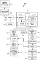
図1は、複数の試料容器102(例えば、試料採取管、図2及び図3参照)を自動的に処理可能な試料検査装置100を示す。試料容器102は、1つ以上の分析装置(例えば、試料検査装置100の周囲に配置される、それぞれ第1、第2、及び第3の分析装置106、108、110)への搬送及びこれらによる分析の前に、ローディングエリア105にある1つ以上のラック104に収容してもよい。より多く、又はより少ない数の分析装置を使用できることは明らかである。分析装置は、臨床化学分析装置及び/又は分析器などの任意の組み合わせであってもよい。試料容器102は、採血管、検査管、サンプルカップ、キュベット、又は概ね透明なガラス若しくはプラスチック容器などの、概ね透明又は半透明の容器であってもよい。他の適切な容器を使用してもよい。
FIG. 1 shows a
典型的には、自動的に処理される試料212(図2及び図3)は、試料容器102内の試料検査装置100に提供される。試料容器102はキャップ214でキャップしてもよい(図2及び図3。あるいは「ストッパ」とも称する)。キャップ214は、異なる形状及び/又は色(例えば、赤色、ロイヤルブルー、ライトブルー、緑色、グレー、黄褐色、黄色、又はこれらの組み合わせ)を有していてもよく、試料容器102がどの検査に使用されるか、試料容器102中に含有される添加剤の種類は何か、等を意味する。他の色を使用してもよい。
Typically, the automatically processed sample 212 (FIGS. 2 and 3) is provided to the
試料容器102のそれぞれには、試料検査装置100の周囲の様々な位置で機械可読の識別情報215(すなわち、指標)、例えば、バーコード、英字、数字、英数字、又はこれらの組み合わせを設けてもよい。識別情報215は、検査室情報システム(LIS)147を介して、患者の識別並びに試料212上で達成する検査、又は、例えば、他の情報を示すことができる、あるいは、それらを相互に関連付けることができる。この識別情報215は概ね、試料容器102に貼付された、又は試料容器102の側面に設けられたラベル218上に設けてもよい。ラベル218は、概して、試料容器102の周囲全体、又は試料容器102の長さ全体に亘って延びているのではない。いくつかの実施形態では、複数のラベル218が貼付されてもよく、わずかに重なっていてもよい。したがって、ラベル218は、試料212の一部の視野を遮ることがあるが、試料212の他の部分は、依然として、ある視点から見ることができる。本発明の実施形態は、特徴付けのための(従来技術のような)試料容器の回転を排除してもよい。いくつかの実施形態では、ラック104は、その上にバーコードのような追加の識別情報を有してもよい。
Each of the
試料212は、管212T内に収容された血清又は血漿部分212SP、及び沈降した血液部分212SBを含んでいてもよい。空気212Aは、血清及び血漿部分212SPの上に設けられ、それらの間の線又は境界を、本明細書では気液界面(LA)として定義する。図2に示すように、血清又は血漿部分212SPと沈降した血液部分212SBとの間の境界線は、本明細書では血清―血液界面(SB)として定義される。空気212Aとキャップ214との間の界面は、本明細書では管―キャップ界面(TC)と称する。血清又は血漿部分212SPの高さは(HSP)であり、血清又は血漿部分212SPの上面から沈降した血液部分212SBの上面まで、すなわち図2のLAからSBまでの高さとして定義する。沈降した血液部分212SBの高さは(HSB)であり、図2のSBで沈降した血液部分212SBの底部から沈降した血液部分212SBの上面までの高さとして定義する。図2のHTOTは、試料212の全高であり、HTOT=HSP+HSBである。
ゲルセパレータ313を使用する場合(図3参照)、血清又は血漿部分212SPの高さは(HSP)であり、血清又は血漿部分212SPの上面LAからゲルセパレータ313の上面SGまでの高さ、すなわち、図3のLAからSGまでの距離として定義する。沈降した血液部分212SBの高さは(HSB)であり、図3の沈降した血液部分212SBの底部からゲルセパレータ313の底部BGまでの高さとして定義する。図3のHTOTは、試料212の全高であり、HTOT=HSP+HSB+ゲルセパレータ313の高さである。いずれの場合も、壁厚はTwであり、外幅はWであるので、試料容器102の内幅はWiである。管の高さ(HT)は、管212Tの最下部からキャップ214の底部までの高さとして定義する。
When the
より詳細には、試料検査装置100は、トラック121を取り付ける又は支持するベース120(例えば、フレーム又は他の構造体)を含んでいてもよい。トラック121は、レール付きトラック(例えば、モノレールトラック又はマルチレールトラック)、コンベヤベルトの集合、コンベヤチェーン、移動可能なプラットフォーム、又は任意の他の適切な種類の搬送機構であってもよい。トラック121は、円形、蛇行状、又は任意の他の適切な形状であってもよい。トラック121は、いくつかの実施形態では、閉じたトラック(例えば、無限トラック)であってもよい。トラック121は、動作中に、個々の試料容器102を、キャリア122内に位置しつつトラック121の周囲に間隔をおいて配置された位置に搬送してもよい。
More specifically, the
キャリア122は、トラック121上に単一の試料容器102を運ぶように構成された受動的で非運動性のパックであってもよく、又は任意で、トラック121周囲を移動し、トラック121の周囲の予めプログラムされた位置で停止するようにプログラムされたリニアモータなどの搭載駆動モータを含む自動キャリアでもよい。キャリア122は、それぞれ、試料容器102を規定の直立位置に保持するように構成されたホルダ122H(図4A〜図4D)を含んでいてもよい。ホルダ122Hは、任意の適した構造体を含んでいてもよく、また試料容器102をキャリア122に固定するが、異なるサイズの試料容器102が収容されるように側方に移動可能又は柔軟性のある複数のフィンガ又は板ばねを含んでいてもよい。いくつかの実施形態では、キャリア122は、そこにステージングされた1つ以上のラック104を有するローディングエリア105から退出してもよい。いくつかの実施形態では、ローディングエリア105は、分析が完了した後に、キャリア122から試料容器102を外すことを許容する二重の機能を果たすことができる。あるいは、トラック121の他の場所に、荷下ろし用レーンを設けてもよい。
The
ロボット124は、ローディングエリア105に設けてもよく、1つ以上のラック104に位置する試料容器102を把持し、試料容器102をトラック121の入力レーン、又はトラック121の他の場所などのような、キャリア122上に搭載するように構成してもよい。ロボット124はまた、分析の完了時にキャリア122から試料容器102を除去するように構成される。ロボット124は、X及びZ、Y及びZ、X、Y及びZ、r及びθ、又はr、θ及びZ運動が可能な1つ以上の(例えば、少なくとも2つの)ロボットアーム又は構成要素を含む。ロボット124は、ガントリーロボット、関節ロボット、R−θロボット、又は他の適切なロボットであってもよく、ロボット124は、試料容器102をピックアップして配置可能なロボットグリッパフィンガを備えていてもよい。
The
トラック121上に搭載されると、キャリア122によって担持された試料容器102は、試料212の分別を行うように構成可能な遠心分離機125(例えば、自動遠心分離機)に進むことができる。試料容器102を運ぶキャリア122を、流入レーン126又は他の適切なロボットによって遠心分離機125に向かって方向転換させてもよい。遠心分離後、試料容器102は流出レーン128を退出するか、あるいはロボットによって除去されて、引き続きトラック121上を進んでもよい。図示された実施形態では、キャリア122内の試料容器102は、次に、図4A及び図4Dを参照して本明細書にさらに記載する品質チェックモジュール130に搬送してもよい。
When mounted on the
品質チェックモジュール130は、試料容器102に収容される試料212の定量化のために構成され、適合される。試料212の定量化は、品質チェックモジュール130で行うことができ、HSP、HSB、HTOTの判定、並びにSB、LA、SG、及び/又はBGの位置の判定を含んでいてもよい。また、品質チェックモジュール130は、試料検査装置100によって処理される試料212に含まれる溶血(H)、黄疸(I)、及び/又は脂肪血(L)の1つ以上など、インターフェレントの存在を判定するように構成してもよい。いくつかの実施形態では、試料212は、品質チェックモジュール130においてアーチファクト(例えば凝塊、気泡、又は泡)の存在について検査してもよい。いくつかの実施形態では、HT、キャップの色、キャップの種類、TC、及び管幅(W)を判定するなど、試料容器102の物理的属性の定量化を品質チェックモジュール130で行うことができる。
The
一旦試料が定量化されると、試料212及び/又は試料容器102は、インターフェレントの存在について、1つ以上のアーチファクトについて、又は試料容器102のさらなる特徴付けについて事前にスクリーニングされてもよく、次いで、試料212を前進させて1つ以上の分析装置(例えば、分析装置106、108、及び/又は110)で分析してから、各試料容器102をローディングエリア105に移動して降ろしてもよい。
Once the sample has been quantified, the
さらに、遠隔ステーション132は、遠隔ステーション132がトラック121に直接リンクされていなくても、自動化された試料検査装置100上に設けてもよい。例えば、独立したロボット133(点線で示す)は、試料212を収容する試料容器102を遠隔ステーション132に担持し、それらを検査/処理の後に戻すことができる。任意で、試料容器102は、手動で取り出して戻すことができる。遠隔ステーション132を使用して溶血レベルなどの特定の構成要素を検査することができ、あるいは、1つ以上の添加物によって脂肪血症レベルを低下させるため、あるいは、例えば、凝塊、気泡、又は泡を除去するためなどのさらなる処理を行ってもよい。他の検査又は処理は、遠隔ステーション132で達成してもよい。さらに、追加のステーション(図示せず)は、トラック121の周りの、キャップ取り外しステーション、一定分量調製などに配置してもよい。
Further, the
試料検査装置100は、トラック121の周囲の1つ以上の位置に多数のセンサ116を含んでいてもよい。センサ116を使用して、試料容器102上に置かれた識別情報215(図2)又は各キャリア122に設けることができる同様の情報(図示せず)を読み取ることによって、トラック121に沿って試料容器102の位置を検出してもよい。いくつかの実施形態では、別体のRFIDチップを各キャリア122に埋め込み、従来のRFIDリーダシステムをトラッキング動作に使用してもよい。位置をトラッキングするための他の手段、例えば近接センサなどを使用してもよい。全てのセンサ116は、各試料容器102の位置を常に適度に知ることができるように、コンピュータ143とインターフェースをとってもよい。
The
遠心分離機125、及び分析装置106、108、110のそれぞれは、概ね、キャリア122をトラック121から除去するように構成されたロボット機構及び/又は流入レーン(例えば、流入レーン126、134、138、144)と、キャリア122をトラック121上に再投入するように構成されたロボット機構及び/又は流出レーン(例えば、流出レーン128、136、141、及び146)を備えていてもよい。
試料検査装置100は、コンピュータ143によって制御してもよく、コンピュータ143は、適切なメモリと適切な調整用電子機器と、ドライバと、様々なシステム構成要素を操作するためのソフトウェアとを有するマイクロプロセッサベースの中央処理装置CPUであってもよい。コンピュータ143は、試料検査装置100のベース120の一部として、又はベース120から分離して収容してもよい。コンピュータ143は、プログラム化された指示によって、キャリア122のローディングエリア105への及びそこからの移動、トラック121の周囲の動き、遠心分離機125への及びそこからの動きを制御することができる。コンピュータ143又は別体のコンピュータは、遠心分離機125の動作、品質チェックモジュール130への及びそこからの動き、並びに品質チェックモジュール130の動作、さらに各分析装置106、108、110への及びそこからの動き、並びに様々なタイプの検査(例えば、化学分析又は臨床化学)を実施するための各分析装置106、108、110の動作を制御することができる。
The
品質チェックモジュール130以外の全てについて、コンピュータ143は、ニューヨーク、タリータウンのシーメンスヘルスケアダイアグノスティックス株式会社(Siemens Healthcare Diagnostics Inc.)によって販売されているディメンション(登録商標)臨床化学分析装置で使用されるものなどのソフトウェア、ファームウェア、及び/又はハードウェアコマンド又は回路に従って試料検査装置100を制御してもよく、このような制御は、コンピュータベースの電気機械制御プログラミングの当業者にとって典型的であるので、本明細書ではこれ以上説明しない。しかしながら、他の適切なシステムを使用して、試料検査装置100を制御してもよい。品質チェックモジュール130の制御は、本明細書で詳細に説明するように、本発明のモデル型法によれば、コンピュータ143によって提供してもよい。
For all but the
本発明の実施形態は、ユーザが様々な制御画面及びステータス表示画面に、容易かつ迅速にアクセス可能とするコンピュータインタフェースモジュール(CIM)145を使用して実施してもよい。これらの制御及びステータス画面で、試料212の調製及び分析に使用される複数の相互に関係する自動化装置のいくつか又はすべての態様を説明することができる。CIM145を採用して、複数の相互に関係する自動化装置の動作状態についての情報を提供してもよく、また、任意の試料212の位置を記述する情報及び試料212上で実行する、又は実行されている検査のステータスを記述する情報を提供してもよい。CIM145は、オペレータと試料検査装置100が相互作用しやすくなるように構成してもよい。よって、CIM145は、オペレータが試薬検査装置100とインターフェースをとるアイコン、スクロールバー、ボックス、及びボタンを含むメニューを表示するように構成された表示画面を含んでいてもよい。
Embodiments of the present invention may be implemented using a computer interface module (CIM) 145 that allows the user to easily and quickly access various control screens and status display screens. These control and status screens can describe some or all aspects of multiple interrelated automation devices used in the preparation and analysis of
図2及び図3には、試料212を含む試料容器102が示されている。図2は、血清又は血漿部分212SP、及び沈降した血液部分212SBを含む試料212をゲルセパレータなしで示す。図3は、血清又は血漿部分212SP、及び沈降した血液部分212SBを含む試料212をゲルセパレータ313付きで示す。本発明の一態様に従って試料212をプレスクリーニングすることにより、血清又は血漿部分212SP、及び/又は沈降した血液部分212SBの相対量だけでなく、LA,SB,及びSGの物理的位置の正確な定量化が可能になる。定量化は、指示された検査を実行するために利用可能な血清又は血漿部分212SPの量が不十分である場合に、試料212が1つ以上の分析装置106、108、110への進行を確実に停止することができる。このようにして、不正確な検査結果を回避することができる。
2 and 3 show a
有利なことに、LA及びSB又はSGの位置を正確に定量化する能力は、空気を吸引する可能性を最小限に抑えるだけでなく、沈降した血液部分212SB、又はゲルセパレータ313(使用する場合)を吸引する可能性も最小限に抑える。したがって、いくつかの実施形態では、分析装置106、108、110で血清又は血漿部分を吸引するために使用する試料吸引プローブの目詰まり及び汚染を回避又は最小化することができる。
Advantageously, the ability to accurately quantify the location of LA and SB or SG not only minimizes the possibility of inhaling air, but also precipitates blood portion 212SB, or gel separator 313 (if used). ) Is also minimized. Therefore, in some embodiments, clogging and contamination of the sample suction probe used to aspirate the serum or plasma portion with the
図4A〜4Bを参照して、品質チェックモジュール130の第1の実施形態を示し、説明する。品質チェックモジュール130は、1つ以上の分析装置106、108、110による分析の前に試料(例えば、血清又は血漿部分212SP、沈降した血液部分212SB、又はその両方の量)を自動的に定量化するように構成してもよい。このようにプレスクリーニングすることにより、正確な吸引プローブの位置決めが可能になり、液体部分(例えば、試料212の血清又は血漿部分212SP)の十分な量(例えば、体積又は深さ)が利用可能であるので、貴重な分析装置リソースの無駄や、空気、沈降した血液部分212SB、又はゲルセパレータ313(使用する場合)の吸引を回避できるという判定ができる。
A first embodiment of the
LA、SB及び/又はSGの物理的位置のうちの1つ以上、及び/又はHSP、HSB、及び/又はHTOTの判定、及び/又は血清又は血漿部分の体積(VSP)又は深さ及び/又は沈降した血液部分(VSB)の体積又は深さが定量化される試料定量化法に加え、品質チェックモジュール130において試料容器102に収容される試料212に対して他の検出方法を行ってもよい。例えば、いくつかの実施形態では、インターフェレント検出法は、インターフェレント(例えば、H、I、及び/又はL)の有無を判定することができる。アーチファクト検出方法は、アーチファクト(例えば凝塊、気泡、又は泡)の有無を判定することができる。さらに、品質チェックモジュール130を使用して、試料容器102を定量化する、すなわち、試料容器102のTC、HT、及び/又はWの位置、及び/又はキャップ214の色及び/又は種類などの試料容器102の特定の物理的寸法特徴を定量化することができる。
One or more of the physical locations of LA, SB and / or SG, and / or determination of HSP, HSB, and / or HTOT, and / or volume (VSP) or depth and / or of the serum or plasma portion. In addition to the sample quantification method in which the volume or depth of the precipitated blood portion (VSB) is quantified, another detection method may be performed on the
ここで、図1、図4A及び図4Bを参照すると、品質チェックモジュール130の第1の実施形態は、複数のカメラ440A〜440Cを含んでいてもよい。3つのカメラ440A〜440Cが示されているが、2つ以上、3つ以上、又はさらに4つ以上のカメラを使用してもよい。カメラ440A〜440Cは、デジタル画像(すなわち、画素化された画像)をキャプチャできる従来のデジタルカメラ、電荷結合素子(CCD)、光検出器のアレイ、1つ以上のCMOSセンサ、などであってもよい。例えば、3つのカメラ440A、440B、440Cが図4Aに示されており、3つの異なる視点からデジタル画像をキャプチャするように構成されている。各カメラ440A、440B、440Cは、一実施形態では約2560画素×694画素、別の実施形態では約1280画素×384画素の画像サイズを有するデジタル画素化画像をキャプチャすることができる装置でもよい。他の画素密度を使用してもよい。各カメラ440A〜440Cは、試料容器102の少なくとも一部、及び試料212の少なくとも一部の側方画像をキャプチャするように構成及び動作可能である。例えば、カメラ440A〜440Cは、ラベル218又はキャップ214の一部及び管212Tの一部をキャプチャしてもよい。最終的に、複数の画像から、試料容器102内の試料212の複合モデルを作成することができる。複合モデルは、いくつかの実施形態では、3Dモデルであってもよく、これを使用して、最終判定を行ってもよく、又は試料212の周囲の個々のカメラによって行われた判定を確認してもよい。
Here, referring to FIGS. 1, 4A and 4B, the first embodiment of the
図示の実施形態では、複数のカメラ440A〜440Cは、試料212の周囲に配置され、複数の視点から側方画像をキャプチャするように構成される。視点は、3つのカメラ440A、440B、440Cが使用されるときに、図示のように互いに約120度など、互いにほぼ等間隔になるように離間されていてもよい。図示のように、カメラ440A〜440Cは、トラック121の周りに配置してもよい。複数のカメラ440A〜440Cの他の配置及び間隔を使用してもよい。このようにして、試料容器102内の試料212の画像は、試料容器102がキャリア122に存在している間に、ただし試料容器102を回転させることなく撮影される。画像はわずかに重なり合うことがある。
In the illustrated embodiment, the plurality of
1つ以上の実施形態では、各カメラ440A〜440Cからの法線ベクトルが交差する点などのような、キャリア122を品質チェックモジュール130内の所定の位置で停止させることができる。いくつかの実施形態では、キャリア122を所定の位置で停止させるためにゲートを設けることにより、良好な品質の画像をキャプチャすることができる。他の実施形態では、キャリア122は、キャリア122を所望の位置にプログラムしたように停止させるように構成されたリニアモータを含んでいてもよい。品質チェックモジュール130にゲートを含む実施形態では、(センサ116などの)1つ以上のセンサを使用して、品質チェックモジュール130でキャリア122の存在を判定してもよい。
In one or more embodiments, the
カメラ440A〜440Cは、画像ウィンドウ、すなわち、試料容器102の予想される位置を含む領域に、近接して向けて、又は焦点を合わせて設けてキャプチャするようにしてもよく、試料容器102は、ビューウィンドウのほぼ中心に配置されるように停止させてもよい。構成されたように、カメラ440A〜440Cは、血清又は血漿部分212SPの一部、沈降した血液部分212SBの一部、キャップ214の一部又は全部、及びおそらくは管212Tの最下部又は参照データを含む画像を撮影することができる。画像内には、1つ以上の参照データが存在してもよい。参照データは、試料212の定量化を助けることができる。参照データは、例えば、TC又は試料容器102の最下部、又は、試料容器102上のいずれかの既知の場所のマークであってもよい。
動作中、各画像は、コンピュータ143が送信可能な通信ライン443A〜443Cに供給されたトリガ信号に応答してトリガされキャプチャされてもよい。キャプチャされた画像の各々は、本明細書で提供される定量化法の1つ以上の実施形態に従って処理してもよい。特に、HDR処理を使用して、試料212を定量化するために画像をキャプチャして処理してもよい。
During operation, each image may be triggered and captured in response to a trigger signal supplied to
より詳細には、異なる公称波長を有する異なるスペクトルによって、異なる視点で照明しながら、試料212(例えば、分別によって分離された試料212)の複数の画像が複数の異なる露光(例えば、露光時間)で品質チェックモジュール130でキャプチャされる。例えば、各カメラ440A〜440Cは、1つ以上のスペクトル(又は、白色光のような1つ以上の波長範囲)において、異なる露光時間で4〜8枚以上の画像を撮ることができる。各スペクトルの異なる露光における画像は、すべてのカメラ440A〜440Cで同時に撮影されてもよい。
More specifically, multiple images of sample 212 (eg,
一実施形態では、異なる色の光源444A〜444Cを用いて試料容器102及び試料212を照明することによって、複数の波長の画像を得ることができる。第1の実施形態では、光源444A〜444Cは、(図4A及び図4Bに示すように)試料容器102を背面照明することができる。任意で、図4C及び図4Dに示すように、光源444D〜444Fは、それぞれのカメラ440A〜440Cの上、下、又は側面に配置されたような試料容器102を前面照明してもよく、又は他の場所に配置してもよい。いくつかの実施形態では、光源444A〜444C又は444D〜444Fと共に、光拡散器及び/又は光フィルタを使用することができる。
In one embodiment, images of a plurality of wavelengths can be obtained by illuminating the
例えば、第1のスペクトルで画像をキャプチャするために、3つの赤色光源(約450nmの公称波長及び約±35nmのスペクトル幅)を使用して、試料212を3つの側方に間隔をあけた位置から照明することができる。光源444A〜444Cによる赤色照明は、異なる露光時間での複数の画像(例えば、4〜8枚以上の画像)が各カメラ440A〜440Cによってキャプチャされるときに発生する可能性がある。いくつかの実施形態では、露光時間は、約1msと256msとの間でよい。例えば、8ms、32ms、128ms、156msの露光時間が使用できる。他の露光時間を使用してもよい。
For example, in order to capture an image in the first spectrum, three red light sources (nominal wavelength of about 450 nm and spectral width of about ± 35 nm) were used to place
各実施形態において、品質チェックモジュール130、130Aは、トラック121を少なくとも部分的に囲む、もしくは覆うことができるハウジング446を含んでいてもよく、試料容器102は、外側照明が最小限となるように画像撮影段階中にハウジング446の内側に位置していてもよい。ハウジング446は、キャリア122がハウジング446の中に出入りできるようにするためのドア446Dを1つ以上含んでいてもよい。いくつかの実施形態では、天井は、ロボットグリッパフィンガを含むロボットによって、試料容器102がキャリア122内に上方から搭載されることを可能にする開口部446Oを含んでいてもよい。前面照明を照明に使用する場合、品質チェックモジュール130Aは、画像コントラストを改善するバックストップ壁447を含んでいてもよい。バックストップ壁447は、試料212の予想される色の範囲以外であれば任意の適切な色でよい。いくつかの実施形態では、黒色の材料を使用してもよい。
In each embodiment, the
赤色に照明された画像が図4A及び図4Bの実施形態でキャプチャされると、赤色光源444A〜444Cは消灯してもよく、次に他のスペクトルの光、例えば、緑色光源444A〜444Cをオンにしてもよい(約±35nmのスペクトル幅の約560nmの公称波長)。そして、異なる露光時間における複数の画像(例えば、4〜8枚以上の画像)を、異なる視点に配置された各カメラ440A〜440Cによってその波長でキャプチャしてもよい。これは、各カメラ440A〜440Cの青色光源444A〜444C(約±35nmのスペクトル幅の約635nmの公称波長)を用いて繰り返すことができる。より大きい又はより小さい波長、又はRGBとは異なる公称波長を使用してもよい。異なる波長光源444A〜444Cは、例えば、交換可能なフィルタ、又は選択的にオン/オフすることができる異なる色の光源のバンクを使用することによって達成することができる。他の手段を使用して異なるスペクトル(着色)照明を生成してもよい。
When the image illuminated in red is captured in the embodiments of FIGS. 4A and 4B, the
任意の実施形態では、図4C及び図4Dに最もよく示すように、例えば、カメラ440A〜440Cに隣接して、すなわち、上方、下方、側方、又はこれらの組み合わせで、ただし試料容器102に対しそれぞれのカメラ440A〜440Cと同じ側にくるように、配置された光源444D、444E、及び444Fを備えることにより、試料容器102は、品質チェックモジュール130Aに前面照明を含むことができる。この任意の実施形態では、カメラ440A〜440Cは、それぞれ、約635nm、560nm、及び450nmのRGBピークを有するデジタルカラーカメラであってもよいが、RGBカラーのそれぞれは、モノクロカメラと組み合わせて上記の実施形態で使用される離散光源に比べて比較的広い波長範囲を有する。
In any embodiment, as best shown in FIGS. 4C and 4D, for example, adjacent to
この任意の実施形態では、光源444D、444E、及び444Fはそれぞれ白色光源であってもよい。例えば、光源444D〜444Fは、約390nm〜約700nmの波長範囲を発光してもよく、複数の側方位置から試料212を照射するために使用してもよい。異なる露光時間(例えば、4〜8回以上の露光)での複数の画像は、各カメラ440A〜440Cによって撮影してもよい。撮影された各白色光画像は、複数の波長でその色成分に分離されて、キャプチャされた画像と複数の波長を提供することができる。例えば、コンピュータ143は、撮影された画像を、約400nm〜約700nmの間の少なくとも3つのキャプチャ波長に分離してもよい。例えば、450nm、560nm、及び635nmのRGB構成要素をそれぞれコンピュータ143によって画像から分離して、マルチスペクトルの、複数回露光されてキャプチャされた画像をキャプチャしてもよい。画像は、ライン443A〜443Cのコンピュータ143からの信号を介して、前述のように撮影することができる。
In this optional embodiment, the
上記の設定のそれぞれについて、それぞれの波長(例えば、R、G、及びB)で複数の露光時間で撮影されたこれらの複数の画像のすべてを迅速に連続して得ることができ、複数の視点からの試料212の画像収集全体が、約2秒未満で得られるようにしてもよい。例えば、3台のカメラ440A、440B、440Cと、RGB光源444A〜444Cを用いた背面照明を使用して、3つの視点において各波長に対して4つの異なる露光画像から、4つの連写画像×3色×3つのカメラ=36画像が得られる。別の例では、カメラ440A、440B、440Cと、白色光源444D〜444Fを用いた前面照明を使用して、3つの視点において4つの異なる露光画像から、4つの画像×3つのカメラ=12画像が得られる。しかし、RGB画像は、カメラ440A〜440Cによって撮影された白色光画像を個々のRGB構成要素に分離することによって、キャプチャされる。よって、分離後に、36枚の画像もキャプチャされる。画像データは、コンピュータ143のメモリに格納され、その後、処理されて試料212を定量化する。
For each of the above settings, all of these multiple images taken at different wavelengths (eg, R, G, and B) at multiple exposure times can be quickly and continuously obtained from multiple viewpoints. The entire image collection of
試料定量化法によれば、画像データの処理は、例えば、スペクトルごとに、そしてカメラ440A〜440Cごとに異なる露光でキャプチャされた複数の画像から最適に露光された画素を選択して、スペクトルごとに(例えば、R、G、及びB)及びカメラ440A〜440Cごとに最適に露光された画像データを生成することを含んでもよい。これは、本明細書では「画像統合」と称することにする。対応する各画素について、各カメラ440A〜440Cからの露光時間ごとの画像に対して、最適な画像強度を示す画素を、異なる露光時間画像それぞれから選択する。最適な画像強度は、例えば、所定の強度(例えば、0〜255のスケールでは180〜254間の強度)の範囲内にある画素として定義してもよい。2つの画像の対応する位置にある2つ以上の画素が最適に露光されると判定された場合、2つのうちのより高い方を選択する。その結果、全ての画素が最適に露光されたカメラ440A〜440Cごとの、複数の統合されたカラー画像データセット(例えば、R、G、B)(例えば、波長(例えばR、G、及びB)及びカメラごとに1つの画像データセットが得られる)。次に、各画素の最適に露光された強度値は露光時間によって正規化され、露光時間に関係なくすべての画素が正規化される。
According to the sample quantification method, the processing of image data is performed, for example, by selecting optimally exposed pixels from a plurality of images captured with different exposures for each spectrum and for each
品質チェックモジュール130、130Aの較正(キャリブレーション)の一部として、試料容器102又はキャリア122のない参照画像を撮影してもよい。このようにして、前景のみを残して各画像データセットから背景を除去することができる。露光時間及び照明条件(R、G、B、又は白色光)ごとの参照画像は、例えば、試料定量化法を実施する前に品質チェックモジュール130、130Aによって撮影してもよい。
As part of the calibration of the
最適に露光された画素を含む各画像データセットに対して、特徴付け処理が行われ、画素を識別する。画素は、液体領域(すなわち、試料212の血清又は血漿部分212SP)として、又は別のクラスに属するように分類することができる。血清又は血漿部分212SPを識別することは、最適に露光された画像データの各画素を分類することに基づくことができる。分類は、複数のトレーニングセットから生成されたマルチクラス分類器(例えば、マルチクラス分類器515)に基づいて行うことができる。マルチクラス分類器515は、例えば、サポートベクトルマシン(SVM)又はランダム判定ツリーを備えることができる。液体領域を判定する他の手段を使用してもよい。
Each image dataset, including the optimally exposed pixels, is characterized and the pixels are identified. Pixels can be classified as a liquid region (ie, serum or plasma portion 212SP of sample 212) or to belong to another class. Identifying the serum or plasma portion 212SP can be based on classifying each pixel of optimally exposed image data. Classification can be performed on the basis of a multi-class classifier (eg, multi-class classifier 515) generated from multiple training sets. The
セグメント化段階での画素の分類を実行するために、異なるスペクトル(例えば、R、G、及びB)でそれぞれのカメラ440A〜440Cで最適に露光された画素のそれぞれについて、第1の統計データを計算してもよい。統計データは、平均値、変動、及び相関値を含み得る第2次までの属性を含んでもよい。特に、共分散マトリックスは、多次元データ全体に亘って計算して識別パターンを表してもよい。
To perform pixel classification at the segmentation stage, first statistical data is provided for each of the pixels optimally exposed by the
一度生成されると、統計データは、マルチクラス分類器515に送られるとともに、そこで操作される。マルチクラス分類器515は、画像内の画素を、1―血清又は血漿部分、2―沈降した血液部分、3―ゲルセパレータ(使用する場合)、4―空気、5―管、6―ラベル、7―キャップなどのような複数のクラスラベルの1つに属するものとして分類することができる。任意で、キャリアを分類してもよい。これにより、液体領域(すなわち、血清及び血漿部分212SP)を構成する画素を識別することができる。
Once generated, the statistical data is sent to and manipulated in the
マルチクラス分類器515は、線形又は非線形である任意の適切な種類の監視分類モデルであってもよい。例えば、マルチクラス分類器515は、線形又はカーネルベースのサポートベクトルマシン(SVM)であってもよい。任意で、マルチクラス分類器515は、適応ブースティング分類器(例えば、AdaBoost、LogitBoostなど)、任意の人工ニューラルネットワーク、ツリーベースの分類器(例えば、決定ツリー、ランダム決定フォレスト)、分類子としてのロジスティック回帰などのブースティング分類器であってもよい。SVMは、例えば試料212の分析で見られるような、液体と非液体との間の分類に特に効果的となり得る。SVMは、データを分析し、パターンを認識する関連する学習アルゴリズムを有する監視された学習モデルである。SVMは、分類及び回帰分析に使用される。
The
マルチクラス分類器515をトレーニングするために複数セットのトレーニング例が使用され、次いで、マルチクラス分類器515上で画像データセットが操作され、各画素が分類される。マルチクラス分類器515は、様々な試料条件、ラベル218による閉塞、血清又は血漿部分212SP、及び沈降した血液部分212SBのレベル、ゲルセパレータ313を含むか否か、などの、試料容器102の多くの例における様々な領域をグラフィックスで輪郭を取ることによってトレーニングされてもよい。500以上もの画像を使用して、マルチクラス分類器515のトレーニングを行ってもよい。各トレーニング画像は、各クラスに属する領域を手動で輪郭を描いて識別し、マルチクラス分類器515に教示してもよい。
Multiple sets of training examples are used to train the
SVMトレーニングアルゴリズムは、任意の新しい教示試料の画素をクラスの1つに割り当てるマルチクラス分類器515を構築する。SVMモデルは、別々のクラスの例が可能な限り広い明確な隙間で分割されるようにマッピングされた空間における点としての例を表す。画像データセットからの新しい画素は、その同じ空間にマッピングされ、マップ上のどこにあるかに基づいて特定のクラスに属することを予測することができる。いくつかの実施形態では、SVMは、カーネルトリック(例えば、カーネルベースのSVM分類器など)と称されるものを使用して非線形分類を効率的に実行し、その入力を高次元の特徴空間に暗黙的にマッピングすることができる。SVM及びツリーベース分類器が特に好ましい。他の種類の分類モデルを使用してもよい。
The SVM training algorithm builds a
その後、血清又は血漿部分212SP、及び/又は沈降した血液部分212SBのクラスであるとみなされるマルチクラス分類器515の結果を用いて、試料212を定量化することができる。試料容器102の幅(W)も判定できる。
1つ以上の実施形態による試料定量化法のフローチャートを図5に示す。まず、キャリア122によって担持された試料212を含む試料容器102を、502での品質チェックモジュール(例えば、品質チェックモジュール130又は130A)に設ける。複数の画像が、504においてキャプチャされる。その複数の画像とは、上述したように、複数の異なる露光で、また、複数の異なるスペクトルで、そして複数の視点で撮影されたマルチスペクトル画像である。定量化のために、品質チェックモジュール130Aの前面照明付きセットアップを使用してもよく、複数の画像をコンピュータ143のメモリに格納してもよい。これらの画像から、背景を508での背景除去フェーズで任意に減算して、計算負荷を低減することができる。背景除去は、先だって510で撮影した参照画像を減算することによって達成できる。
A flowchart of the sample quantification method according to one or more embodiments is shown in FIG. First, a
504における画像キャプチャ及び508における任意の背景除去の後、511においてセグメント化を行うことができる。511でのセグメント化は、512で行われる画像統合処理を含んでいてもよい。512におけるこの画像統合処理中に、スペクトル(R、G、及びB)及びカメラ440A〜440Cごとの様々な露光時間画像が、画素ごとに再検討され、最適に露光された画素を判定する。それぞれの対応する画素位置ごとに、最適に露光された画素のうちの最良のものが選択され、正規化され、各スペクトル及びカメラ400A〜440Cごとに最適に露光された画像データセットに含まれる。したがって、512での画像統合に続いて、各スペクトル(R、G、及びB)及び各カメラ440A〜440Cごとに1つの最適に露光された画像データセットが生成される。HDR処理の使用は、反射及び吸収に関して、画像の細部を強化するよう機能する。これにより定量化がより正確になる。
After image capture at 504 and arbitrary background removal at 508, segmentation can be done at 511. The segmentation at 511 may include the image integration process performed at 512. During this image integration process in 512, the spectra (R, G, and B) and various exposure time images for each of the
512での画像統合に続いて、又は、おそらくそれと並行して、514において統計生成処理を行うことができる。514では、2次平均値まで及び/又は共分散のような各画素について統計が生成される。次いで、最適に露光されたデータセット上の統計データは、マルチクラス分類器515によって操作され、516において画像データセットに存在する画素クラスを識別する。各画素位置について、小さなスーパー画像パッチ(例えば、11×11画素)内でこの統計的記述を抽出する。各スーパー画素パッチは、トレーニングと評価処理において考えられる記述子を提供する。典型的には、分類器はこれらの特徴記述子上で動作し、検査中にトレーニング用の入力クラスラベルと出力クラスラベルを使用する。各画素のクラスラベルを得るためには、画像データセットを適した周知のスキャン技術でスキャンできる。
Statistics generation processing can be performed at 514 following, or perhaps in parallel with, the image integration at 512. At 514, statistics are generated for each pixel up to the quadratic mean and / or covariance. The statistical data on the optimally exposed dataset is then manipulated by the
この511のセグメント化処理から、カメラ440A〜440Cごとに2Dの統合画像データセットの各画素を、516において複数のクラスタイプの1つに属するように分類する。分類の種類としては、例えば、液体(血清又は血漿部分212SP)、沈降した血液部分212SB、ゲルセパレータ313、空気212A、管212T、ラベル218、又は、キャップ214として分類してもよい。この511でのセグメント化情報から、試料212の定量化を判定することができる。
From this segmentation process of 511, each pixel of the 2D integrated image data set is classified for each
例えば、518において、液体領域(例えば、血清又は血漿部分212SP)を識別してもよい。これは、血清又は血漿部分212SPのクラスからのすべての画素をグループ化し、次いで、519において液体(血清又は血漿部分212SP)及び空気212A(すなわち、LA)の間の上側界面の位置を判定する。2Dの統合画像データセットの中で血清又は血漿部分212SPとして分類された最も高い画素の位置を平均化することによって、垂直方向のLAの数値を計算してもよい。実質的な外れ値はすべて拒絶され、平均には使用されない。前もって行った画素空間から機械空間(例えば、mm単位)への較正(キャリブレーション)は、任意の周知の機械空間対画像空間較正技術によって達成することができる。ゲルセパレータ313を使用するかどうかに応じて、定量化法は、その後520において、SB又はSG(ゲルセパレータを使用する場合)の物理的垂直方向の位置を判定してもよい。
For example, at 518, the liquid region (eg, serum or plasma portion 212SP) may be identified. This groups all pixels from the class of serum or plasma portion 212SP and then determines the location of the upper interface between the liquid (serum or plasma portion 212SP) and
血清又は血漿部分212SPとして分類された最も低い画素の位置を平均化することによって、520において、SB又はSGの数値を計算してもよい。ここでも外れ値は無視してよい。LA及びSB又はSGの位置から、血清又は血漿部分(HSP、図2及び図3)の高さは、平均の減算を介して判定することができる。例えば、血清又は血漿部分212SPと識別された画素の垂直方向の積層をカウントすること、画素数を平均化すること、そして、機械空間に変換することなど、高さHSPの計算のための他の手段を使用してもよい。 SB or SG numbers may be calculated at 520 by averaging the positions of the lowest pixels classified as serum or plasma portion 212SP. Again, outliers can be ignored. From the position of LA and SB or SG, the height of the serum or plasma portion (HSP, FIGS. 2 and 3) can be determined via subtraction of the mean. Others for calculating height HSP, such as counting the vertical stacking of pixels identified as serum or plasma portion 212SP, averaging the number of pixels, and converting to mechanical space. Means may be used.
液体領域(例えば、血清又は血漿部分212SP)を定量化することは、526において試料容器102の内部幅(Wi)を判定することをさらに含んでいてもよい。外部幅(W)は、526において、管212Tとして分類された画素を識別し、LAとSB又はSGの間で測定されるような管212Tの外側の側方エッジに位置する対応する画素の位置を減算し、さらに例えば、減算値を平均化することにより、判定してもよい。Wiは壁厚Tw、すなわち、Wi=W−2Twの2倍を減算して、Wから決定することができる。Twは、すべての試料容器102について使用される平均壁厚値であってもよく、外幅W及び高さHTの判定に基づいて判定される管の種類に基づくルックアップテーブルから取得される特定の測定値であってもよい。
Quantifying the liquid region (eg, serum or plasma portion 212SP) may further include determining the internal width (Wi) of the
HSP及びWiから、液体領域VSP(例えば、血清又は血漿部分212SP)の体積は、528において下の式1を使って判定できる。
式1 VSP=HSP×Wi2Pi/4
From HSP and Wi, the volume of liquid region VSP (eg, serum or plasma portion 212SP) can be determined at 528 using Equation 1 below.
Equation 1 VSP = HSP x Wi 2 Pi / 4
沈降した血液部分212SBを定量化するため、沈降した血液部分212SBのクラスに対応する画素は、まず530で識別してもよい。ゲルセパレータ313が存在するかどうかに応じて、沈降した血液部分HSBの高さは、532において、沈降した血液部分212SBの最下部の画素を位置特定し、次にSB又はBGのいずれかを減算することによって判定する。SB又はSGは、520で判定すればよい。SB又はBGの数値は、沈降した血液部分212SBとして分類された最上部の画素の位置を平均化することによって判定してもよい。Wiは、526で判定してもよい。HSB及びWiから、沈降した血液部分212SBの体積は、試料容器の丸まった端部を考慮するために減算された調整因数を含む以下の式2を用いて534において判定してもよい。
式2 VSB=(HSB×Wi2Pi/4)−{1/2Wi2+(Pi/24)Wi3}
In order to quantify the precipitated blood portion 212SB, the pixels corresponding to the class of the precipitated blood portion 212SB may first be identified by 530. Depending on the presence of the
Equation 2 VSB = (HSB x Wi 2 Pi / 4)-{1/2 Wi 2 + (Pi / 24) Wi 3 }
528及び534においてVSP及びVSBが判定されると、536において、液体部分(例えば、血清又は血漿部分212SP)の沈降した血液部分212SBに対する体積比が、体積比={VSP/VSB}×100(%)で計算される。 When VSP and VSB are determined at 528 and 534, at 536, the volume ratio of the liquid portion (eg, serum or plasma portion 212SP) to the precipitated blood portion 212SB is volume ratio = {VSP / VSB} × 100 (%). ) Is calculated.
任意で、液体部分(すなわち、血清又は血漿部分212SP)及び沈降した血液部分212SBの深さレベルは、モンテカルロシミュレーション法を用いて判定することができる。モンテカルロシミュレーション法は、与えられたモデル分布からの反復ランダムサンプリングに基づいて数値解を得る計算アルゴリズムのクラスである。この方法は、多変量レベルモデルから複数のランダム仮説を生成することに依存し、次に、画像キャプチャ段階中に生成された信頼値などの画像測定値で検証/検査される。モンテカルロシミュレーション法は、ゲルセパレータ313が使用されているかどうかに応じて、LA、SB、又はSG及びBGの位置の仮説を導出することを含む。モンテカルロシミュレーション法を適用するためには、ランダムサンプルが描かれるモデル分布が必要である。モデルは、大量のアノテーション付きトレーニング試料でトレーニングされた多変量モデルであってもよい。
Optionally, the depth levels of the liquid portion (ie, serum or plasma portion 212SP) and the precipitated blood portion 212SB can be determined using Monte Carlo simulation methods. Monte Carlo simulation is a class of computational algorithms that obtain numerical solutions based on iterative random sampling from a given model distribution. This method relies on generating multiple random hypotheses from a multivariate level model and is then validated / tested with image measurements such as confidence values generated during the image capture phase. The Monte Carlo simulation method involves deriving a hypothesis of LA, SB, or SG and BG positions, depending on whether the
トレーニング段階中では、ランドマークベースの層状アノテーションが収集され、モデルの推定値がこれらのアノテーションから導出される。生成のため、我々は、画像内のL=(l1、l2)、すなわち気液界面LAと、試料容器102の最下部212B(例えば、先端)の、2つの参照ランドマークの知見を仮定する。これらをランドマーク基準点と呼ぶ。さらに、我々は、血清及び血漿部分212SPとゲル分離器313の間のSG界面のランドマークのアノテーション、及びゲルセパレータ313と沈降した血液部分212SBの間の血液とゲルとの界面BGのランドマークのアノテーションを仮定する。特に、それらはレベルポイントp=(p1,p2)Tとして表すことができる。写真が、重力ベクトルとアラインメントされたカメラのアップベクトル(例えば、カメラ440A)でキャプチャされたと想定すると、ランドマークベースのアノテーションで充分である(これは、典型的には、品質チェックモジュール130、130Aのために提案されたハードウェア構成の場合である)。図2及び図3は、試料画像の潜在的アノテーションを示す。
During the training phase, landmark-based layered annotations are collected and model estimates are derived from these annotations. For generation, we assume the findings of two reference landmarks, L = (l 1 , l 2 ) in the image, i.e. the gas-liquid interface LA and the bottom 212B (eg, tip) of the
1つ以上の実施形態では、この方法は、流体領域の拡張を正準表現に正規化することができる。特に、流体レベルは重力ベクトルに対して垂直であると仮定され、したがって正規化スキームは試料容器102の傾きとは無関係である。さらに、(重力ベクトルに沿って計算された)流体の高さHTは、(2つのランドマーク参照点(l1、l2)に基づいて)この正準表現内で1の値に正規化されると仮定する。この正規化スキームに基づいて、一対のレベルポイントpに対する正規化された比を、アノテーション付きサンプル画像ごとに導出することができる。多変量統計は、P={p1,p2,p3,…,pk}を考慮したアノテーション付きトレーニング画像のセットから導かれる。
In one or more embodiments, this method can normalize the extension of the fluid region to a canonical representation. In particular, the fluid level is assumed to be perpendicular to the gravity vector, so the normalization scheme is independent of the slope of the
モデル化のために、2つのレベルポイントに対して多変量ガウス分布を仮定し、正規化された表現の平均μ及び分散/共分散行列Σを得る:(1)μ=E(P)及び(2)Σ=E[(P−E(P))(P−E(P))T]、ここで、Eは期待値<1である。 For modeling, we assume a multivariate Gaussian distribution for the two level points and obtain the mean μ and variance / covariance matrix Σ of the normalized representation: (1) μ = E (P) and ( 2) Σ = E [(PE (P)) (PE (P)) T ], where E is the expected value <1.
トレーニングデータから抽出された生成モデルは、モンテカルロシミュレーション法の仮説検査の間にランダムサンプルを生成するために使用される。アノテーションは時には時間がかかるので、ランドマークpのモデルは、レシピ又は遠心分離処理からの論理的期待値から合成して生成することができる。 The generative model extracted from the training data is used to generate a random sample during the Monte Carlo simulation hypothesis test. Since annotations are sometimes time consuming, models of landmark p can be generated by synthesizing from recipes or logical expectations from centrifugation.
この方法の検証/検査段階中では、レベルポイントのサンプルを分布モデルからランダムに生成し、よってLA、SB、又はSG及びBGの仮説レベルを含む潜在的な流体層状構造体を合成的に生成することができる。較正ステップから、先端参照点212Bについての知見を得ることができる。気液界面LAは、空気と血清又は血漿部分212SPとの間の遷移から導出される。上部流体レベルHTは、行間の対応が利用可能であるため、単一又は複数の視野から導出されてもよい。トレーニング段階と同様に、流体高さHTは1に正規化され、それにより、正規化された比が導出されて流体層状構造体が生成される。
During the validation / inspection phase of this method, level point samples are randomly generated from the distribution model, thus synthetically generating potential fluid layered structures containing hypothetical levels of LA, SB, or SG and BG. be able to. Knowledge about the
効率的な方法は、2次統計量の分解を用いて多変量ガウスレベルモデルからランダムサンプルを生成するために使用される。例えば、共分散行列Σは、正定値行列を仮定してコレスキー分解を用いて、例えば条件付けを用いてΣ=AATにする。一連の独立した標準正規変分zを生成して、適用することによりモデル化された流体レベル分布からサンプルを生成することができる:x=μ+Az。我々の場合、x=(x1、x2)Tは、流体レベルHTの正規化されたスカラー値からなる2次元ベクトルを表す。我々の場合、x=(x1、x2)Tは、流体レベルHTの正規化されたスカラー値からなる2次元ベクトルを表す。 Efficient methods are used to generate random samples from multivariate Gaussian level models using the decomposition of quadratic statistics. For example, the covariance matrix sigma, using Cholesky decomposition assuming a positive definite matrix, to sigma = AA T, for example, using conditioning. A series of independent standard normal variation z can be generated and applied to generate a sample from the modeled fluid level distribution: x = μ + Az. In our case, x = (x1, x2) T represents a two-dimensional vector consisting of normalized scalar values for the fluid level HT. In our case, x = (x1, x2) T represents a two-dimensional vector consisting of normalized scalar values for the fluid level HT.
xによって定義されるこの構造仮説は、正規化画像領域内の信頼値を集約(例えば、積分)することによって検証される。検証は、領域(血清又は血漿部分212SP、あればゲルセパレータ313、及び沈降した血液部分212SB)について行われる。信頼性は、例えば、閉塞(Occlusion:オクルージョン)の場合に欠落したデータを克服するために平均/分散計算を使用して確実に集約される。上部液位HTと、SB又はSGのレベル点についての最初の仮説との間で、我々は、血清及び血漿部分212SPについて高い応答を見出すと思われる。2つのレベルのランドマークSGとBGとの間に、ゲルセパレータ313に対する高い応答が期待される。下部ランドマークSB又はBGと、参照点(管先端212B)との間において、信頼できる層モデルは、沈降した血液部分212SBに対する高い信頼値を期待する。
This structural hypothesis defined by x is tested by aggregating (eg, integrating) the confidence values in the normalized image region. Verification is performed on the regions (serum or plasma portion 212SP,
最良のフィッティング流体層モデルは、値の標準偏差を最小化することによって、対応する信頼値を最大化する。効率的な計算のため、正規化された流体空間と画像領域との間のリンクを確立するために参照テーブルを生成することができる。さらに、効率的なデータ構造体、すなわち迅速な仮説検査を可能にするための積分画像が使用されてもよい。検証は、単一の画像を含むことができるが、確実性のために複数のカメラ440A〜440Cからのマルチビュー情報を考慮することもできる。行レベルでの対応が分かっているので、統合は信頼性の集約中には簡単である。
The best fitting fluid layer model maximizes the corresponding confidence value by minimizing the standard deviation of the value. For efficient computation, a reference table can be generated to establish the link between the normalized fluid space and the image region. In addition, efficient data structures, i.e. integrated images to allow rapid hypothesis testing, may be used. The validation can include a single image, but multi-view information from
モンテカルロシミュレーション法から、血清又は血漿部分2121SP、ゲルセパレータ313、及び沈降した血液部分212SBに関する最も可能性のある流体レベル構造体、すなわちLA、SB、又はLA、SG及びBGの判定、が見つかる可能性がある。別の高度な検査は、HDR画像を考慮することによって、すなわちこれらの領域におけるスペクトル応答の標準偏差を最小化することによって、仮説の検証を含むことができる。別の洗練された検査方法は、検出された水平流体レベル、すなわちエッジ検出、HDR画像からの抽出、及び画像領域に投影されるランダム仮説、の間の誤差を最小化することなどにより、モンテカルロシミュレーション法によって到達した仮説の検証を含むことができる。基本的な強度計算は、最良の仮説の発見をサポートする。
Monte Carlo simulations may find the most probable fluid level structures for serum or plasma moiety 2121SP,
要約すると、図8を参照して説明したモンテカルロシミュレーション法を使用して、セグメント化を判定し、試料容器102内に収容される試料212を定量化することができる。方法800は、802において、画像化位置441に試料容器102に収容された試料212を提供し、804において、試料212の画像をキャプチャすることを含む。440Aなどの単一のカメラで画像をキャプチャすることができる。
In summary, the Monte Carlo simulation method described with reference to FIG. 8 can be used to determine segmentation and quantify the
方法800は、806において、少なくとも血清又は血漿部分212SP、及び沈降した血液部分212SBを含む試料212の領域を決定することを含む。これは、本明細書に記載のHDR画像化方法によって、又は任意の他の適切なセグメント化法によって達成することができる。
この方法は、810において、多変量レベルモデル812からの描画レベル推定仮説を含む。ブロック810は、ゲルセパレータ313が存在しない場合にはLA及びSBについての仮説、ゲルセパレータ313が存在する場合にはLA、SG、及びSBについての仮説など、多変量レベルモデル812によって生成されるランダムレベル推定仮説を描くことを含む。
This method includes, at 810, a drawing level estimation hypothesis from a
この方法は、814において、レベル推定仮説を画像空間にマッピングすることを含む。例えば、この方法は、参照テーブルを活用してレベル推定仮説を2D画像領域に投影することができる。次に、816において、方法800は、領域内の信頼度を統合する。各領域内で信頼度が最大になるまで、ブロック818とブロック816との間で反復処理が行われる。最後に、820において、各領域の信頼度を最大にするレベル推定仮説が選択される。1つ以上の実施形態におけるモンテカルロ法は、セグメント位置(例えば、LA、及びSB又はSG)を決定するために、又はHDR画像化によって判定されたセグメント位置を検証するために単独で使用されてもよい。
This method involves mapping the level estimation hypothesis to image space at 814. For example, this method can utilize a reference table to project a level estimation hypothesis onto a 2D image region. Next, at 816,
519、520、及び528でセグメント位置(例えば、LA、及びSB又はSG)及びVSPが判定されると、LIS147から受け取ったように、これらの定量化された値を用いて、指示された検査のために十分な流体が存在するかどうかを判定することができる。さらに、519〜520のセグメント位置LA及びSB又はSGを使用して、複数の検査が指示されたときに沈降した血液部分212SB又はゲルセパレータ313がいずれの吸引に対しても吸引されないように、吸引プローブ先端をどこに配置するかを判定することができる。
Once the segment positions (eg LA and SB or SG) and VSP are determined at 519, 520, and 528, these quantified values are used to indicate the indicated test, as received from LIS147. It can be determined if there is sufficient fluid for this. In addition, using segment positions LA and SB or SG from 519 to 520, aspiration is performed so that the settled blood portion 212SB or
品質チェックモジュール130又は130Aにおいて、528で液体領域の体積が計算されると、アーチファクト(例えば、凝塊、気泡、及び/又は泡)の存在は、アーチファクト検出522において1つ以上のアーチファクト分類器で、その液体領域の2Dデータサブセット上で動作することによって判定することができる。アーチファクトの推定量は、利用可能な容積VSPから減算することができるので、アーチファクトが存在するときに液体の利用可能な容積のより良い推定値が提供される。アーチファクト検出方法及び装置は、「試料内のアーチファクトを分類するための方法及び装置」と題した同時係属中及び同時に提出した仮特許出願に記載されている。しかしながら、いくつかの実施形態では、アーチファクト(例えば凝塊、気泡、泡)を試料212から除去し、次に試料212を定量化法に従って再定量することができる。
In the
液体領域が518で識別されると、インターフェレント(例えば、H、I、及び/又はL)の存在は、1つ以上のインターフェレント分類器により、液体領域のデータサブセットを操作することによって判定してもよい。一実施形態では、「試料におけるインターフェレントを検出するための方法及び装置」と題した同時係属中及び同時に提出した仮特許出願に記載されているように、H、I、及びLのそれぞれに別々の分類器を使用することができる。高い指標の溶血、黄疸、又は脂肪血が存在する場合、試料212は、さらなる定量化なしで廃棄してもよいし、再描画して再スクリーニングしてもよい。いくつかの実施形態では、試料212を処理して、脂肪血レベルを低下させ、次いで再スクリーニングすることができる。
When the liquid region is identified at 518, the presence of an interferent (eg, H, I, and / or L) is present by manipulating the data subset of the liquid region with one or more interferant classifiers. You may judge. In one embodiment, H, I, and L, respectively, as described in the co-pending and co-submitted provisional patent applications entitled "Methods and Devices for Detecting Interferents in Samples". Separate classifiers can be used. In the presence of high indicators of hemolysis, jaundice, or fatty blood,
したがって、品質チェックモジュール130、130Aによって実行されるモデルに基づく定量化法500により、試料212の血清又は血漿部分212SP、及び/又は沈降した血液部分212SBの迅速な定量化が可能となることは明らかである。視点及び判定それぞれの最終的な2D結果は、複数の視点に亘って集約され、比較及び/又は平均化されて3Dモデルを提供することができる。
Therefore, it is clear that the model-based
図6では、試料212の定量化は、品質チェックモジュール130、130Aを使用してより広範な方法600によって特徴付けられる、又は分類される多くの品目のうちの1つのみである、より広範な特徴付け方法600のフローチャートを示す。方法600の1つ以上の実施形態によれば、例えば、複数のカメラ(カメラ440Aが示されている)によって画像がキャプチャされる。しかしながら、他のカメラ440B、440Cを使用して、図4A及び図4Cに示されるような他の視点から画像をキャプチャすることができる。カメラ440Aでキャプチャされた画像について説明する処理は、他の視点の他のカメラ440B、440Cについても同じであり、ライン605でのそれらの入力を使用して、最終的な判定、及び/又はさまざまな視点間の違いを解決するために使用する試料212の3Dモデルを生成することができる。
In FIG. 6, the quantification of
カメラ440A及び他のカメラ440B、440Cによってキャプチャされた画像は、上述したように、複数のスペクトル(例えば、RGB)及び複数の露光画像であってもよい。特に、各視点で604Aにおいて使用される光のスペクトルごとに、複数の露光(例えば、4〜8回以上の露光)を行ってもよい。図4Aと図4Bに示すように、各カメラ440A〜440Cの各露光時及び各スペクトルのそれぞれの画像は、モノクロカメラと背面照明光源444A〜444Cを使用して同時に取得することができる。任意で、白色光源444D〜444Fを品質チェックモジュール130Aに使用して、前面照明された複数の露光画像は、カラーカメラを使用して604Bにおいて取得してもよい。背面照明付き品質チェックモジュール130又は前面照明付き品質チェックモジュール130Aのいずれか、又は両方を使用することができる。
The images captured by the
その後、任意の背景除去方法に関して上述したように、画像を任意で508において処理して、参照画像510を使用して背景を除去してもよい。その後、画像をさらに処理して、上述したように511においてセグメント化を判定してもよい。いくつかの実施形態では、604Bからの前面照明カメラ440A〜440C(図4C及び図4D参照)からの画像は、511でのセグメント化の判定に最もよく使用できる。しかしながら、604Bにおいてキャプチャされた画像は、521においてもHILN検出に使用できる。同様に、604Aにおいてキャプチャされた任意の画像は、621でのHILNの特徴付けに最もよく使用される。しかしながら、604Aにおいてキャプチャされた画像は、511でのセグメント化でも使用できる。
The background may then be optionally processed at 508 and the background removed using the
本明細書に記載された方法に従って、500での液体の識別及び定量化は、511でのセグメント化に続いて実施してもよい。500において液体を定量化することは、本明細書に記載するように、LA、SB、又はSG、及び/又はBGの物理的位置のような試料212の特定の物理的寸法特徴の判定、及び/又はHSP、HSB、及び/又はHTOTの判定、及び/又は上述したような、血清又は血漿部分(VSP)の体積又は深さ及び/又は沈降した血液部分(VSB)の体積又は深さなどの判定などを含んでいてもよい。内部幅(Wi)は、627における試料容器の特徴付けから得ることができる。
According to the methods described herein, liquid identification and quantification at 500 may be performed following segmentation at 511. Quantifying the liquid at 500 is the determination of specific physical dimensional features of
検査に利用可能な血清又は血漿部分212SPの実際の体積をさらに正確に測定するために、又は単にアーチファクトの存在にフラグを立てるために、アーチファクト検出方法を522で使用して、凝塊、気泡、又は泡が存在することを識別してもよい。存在する1つ以上のアーチファクトのそれぞれの推定体積は、より良い体積推定値を得るために、522において上記で判定された血清又は血漿部分VSPの推定体積から減算してもよい。その後、アーチファクト分類器を使用して、画像データを処理して、522において血清又は血漿部分212SP中のアーチファクトの有無を判定してもよい。アーチファクト検出522によってアーチファクトであると識別されたこれらの画素は、本明細書に記載される定量化法では無視されてもよく、521でのHILN分類においても無視してよい。アーチファクトの検出により、いくつかの実施形態において修復を開始できる。アーチファクト検出法は、「試料中のアーチファクトを分類するための方法及び装置」と題した同時出願の米国出願に記載されている。
Clumps, bubbles, using the artifact detection method at 522, to more accurately measure the actual volume of serum or plasma portion 212SP available for testing, or simply to flag the presence of artifacts, Alternatively, the presence of bubbles may be identified. The estimated volume of each of the one or more existing artifacts may be subtracted from the estimated volume of serum or plasma partial VSP determined above in 522 to obtain better volume estimates. Image data may then be processed using an artifact classifier to determine the presence or absence of artifacts in serum or plasma portion 212SP at 522. These pixels identified as artifacts by
511でのセグメント化の結果は、バーコードなどの識別情報215を含むラベル218を識別するためにも使用してもよい。バーコードは625において読み取ることができる。従来のバーコード読み取りソフトウェアは、ラベル218が511でのセグメント化で識別されると、使用してもよい。特定の画像が、読み取られるべきバーコードを十分に含まない場合、バーコードは、他のカメラ340B、340Cから得られた他の画像から、又は他の画像と共に読み取ることができる。
The result of the segmentation at 511 may also be used to identify the
試料容器102のさらなる特徴付けは、627でのより広い方法600に従って達成してもよい。529における管の種類、531におけるキャップの種類、及び/又は533におけるキャップの色の特徴付けを3Dモデル635に供給して、各カメラ440A〜440Cからの画像の処理に基づいて同じ特徴付けが達成されたことを検証してもよい。わずかに異なる値が得られた場合、その値は平均化してもよいし、そうでなければ集約してもよい。521におけるHILN分類、500における試料定量化、522におけるアーチファクト検出、及び627における試料容器検出からの出力の全てが3Dモデル635に供給されてもよく、3Dモデル635は、最終意思決定、特徴付け、及び様々なカメラ440A〜440Cからの結果の調和に使用され、モニタ上に表示されるか、又は他の方法で報告することができる。
Further characterization of the
図7は、1つ以上の実施形態による試料容器102の試料212を定量化する方法を示すフローチャートである。方法700は、702において試料容器(例えば、キャップ付き採血管などの試料容器102)に収容された試料(例えば、試料212)を提供することを含む。次に、方法700は、704において試料212を収容する試料容器102の画像を異なる露光(例えば、露光時間)と異なる公称波長を有する複数のスペクトルでキャプチャすることを含む。例えば、いくつかの実施形態では、異なる露光時間で撮影された4〜8回以上の異なる露光が存在してもよいが、各スペクトルに同じ照明条件で撮影してもよい。1つ以上の実施形態では、画像は、白色光を使用し、前面照明を使用してキャプチャされてもよい。他の実施形態では、赤色、青色、及び緑色などの狭帯域光源を有する複数のスペクトルを背面照明光源444A〜444Cとして使用して画像をキャプチャしてもよい。白色光画像は、上述したように、コンピュータ143によってキャプチャされるときにR、G、及びB画像に分解されてもよい。それぞれの場合において、複数のカメラ440A〜440Cによって複数の視点から画像を撮影してもよい。
FIG. 7 is a flowchart showing a method of quantifying the
本方法700は、任意で、706に示すように背景除去を含み、背景を減算して計算負荷を低減してもよい。背景除去は、較正処理の一部として達成することができる。参照画像は、試料容器102の画像と同じ露光時間で撮影してもよいが、試料容器102やキャリア122なしでキャプチャすることができる。
The
この方法700は、710で、複数のスペクトルのそれぞれにおける異なる露光時間で画像から最適に露光された画素を選択して、スペクトルのそれぞれについて最適に露光された画像データを生成することとを含む。特定のスペクトルにおける各画像内の対応する画素位置毎に、(露光不足でも、又は露光しすぎでもない)最良に露光された画素を選択し正規化することができる。最適な露光範囲は、上述したようなものであってもよい。この最適に露光された画素の選択は、画像統合フェーズ(例えば、画像統合512)で行われる。したがって、各カメラのRGBスペクトルのそれぞれについて、最適に露光された画素の2Dデータセットが生成されてもよい。
The
次に、方法700は、試料を、血清又は血漿部分212SP、沈降した血液部分212SB、ゲルセパレータ313、空気212A、管212T、ラベル218、又はキャップ214のうちの1つ以上を含む様々なクラスの種類に分類することを含む。分類は、異なる波長の最適に露光された画素の統計データを計算して統計データを生成し、次いで、最適に露光された画素の統計データをマルチクラス分類器515で操作してデータセット内に存在するクラスを識別することによって達成することができる。
次に、方法700は、空気212Aと血清又は血漿部分212SPの間の気液界面LAの位置と、血清又は血漿部分212SPと沈降した血液部分212SBの間の血清と血液の界面SBの位置と、血清又は血漿部分212SP及びゲルセパレータ313の間の血清とゲルの界面SGの位置と、沈降した血液部分212SBとゲルセパレータ313の間の血液とゲルの界面BGの位置と、血清又は血漿部分212SPの体積VSB及び/又は深さHSPと、沈降した血液部分212SBの体積VSP及び/又は深さHSBとのうちの一つ以上を判定することによって714において試料を定量化することを含む。
Next, the
したがって、前述に基づいて、品質チェックモジュール130,130Aによって実行されるモデルベースの試料定量化法700により、試料212Lの迅速な定量化ができることは明らかである。品質チェックモジュール130は、プレスクリーニングが遠心分離機125での遠心分離の直後に行われるように位置されて図1に示されているが、いくつかの実施形態では、この特性を直接分析装置に含んでいても(例えば、分析装置106,108、及び/又は110)あるいは、その他の場所に含んでいても有利であろう。さらに、いくつかの実施形態では、品質チェックモジュール130をローディングエリア105に配置することができ、ロボット124が試料容器102をキャリア122にロードするとすぐに品質チェックを実施することができるように、ラック104をローディングエリア105にロードする前に遠心分離を行ってもよい。任意で、品質チェックモジュール130Aは、品質チェックモジュール130と同じ位置に設けられてもよい。いくつかの実施形態では、定量専用の1つの品質チェックモジュール130Aを一か所で使用し、HILN検出専用の別の品質チェックモジュール130を異なる場所に設けてもよい。
Therefore, based on the above, it is clear that the model-based
本発明は様々な変更及び代替形態が可能であるが、特定のシステム及び装置の実施形態及びその方法は、図面の例として示されており、本明細書で詳細に説明してきた。しかしながら、開示された特定の装置又は方法に本発明を限定することを意図するものではなく、反対に、本発明の範囲内に入るすべての変更、均等物及び代替物を網羅することを意図するものであることを理解されたい。 Although various modifications and alternatives are possible in the present invention, embodiments of specific systems and devices and methods thereof are shown as examples of drawings and have been described in detail herein. However, it is not intended to limit the invention to the particular devices or methods disclosed, and conversely, to cover all modifications, equivalents and alternatives that fall within the scope of the invention. Please understand that it is a thing.
100 試料検査装置
102 試料容器
104 ラック
105 ローディングエリア
106、108、110 分析装置
121 トラック
122 キャリア
122H ホルダ
124 ロボット
130、130A 品質チェックモジュール
143 コンピュータ
147 LISシステム
212 試料
212SB 沈降した血液部分
212SP 血清又は血漿部分
212T 管
214 キャップ
215 識別情報
218 ラベル
313 ゲルセパレータ
440A〜440C カメラ
444A〜444F 光源
100
Claims (18)
試料を提供し、
異なる公称波長を有する複数のスペクトル及び複数の異なる露光時間で前記試料の画像を取得し、
前記複数のスペクトルのそれぞれにおける前記複数の異なる露光時間の前記画像から、最適に露光された画素を選択し、前記複数のスペクトルのそれぞれについて最適に露光された画像データを生成し、
血清又は血漿部分と、沈降した血液部分と、ゲルセパレータを使用する場合はゲルセパレータと、空気と、管と、ラベル、又はキャップのうちの1つ以上を含む1つ以上のクラスの種類に前記試料を分類し、
前記空気と前記血清又は血漿部分の間の気液界面(LA)の位置と、
前記血清又は血漿部分と前記沈降した血液部分の間の血清と血液の界面(SB)の位置と、
前記ゲルセパレータを使用する場合には、前記血清又は血漿部分と前記ゲルセパレータとの間の血清とゲルの界面(SG)の位置と、
前記ゲルセパレータを使用する場合には、前記沈降した血液部分と前記ゲルセパレータとの間の血液とゲルの界面(BG)の位置と、
血清又は血漿部分の体積又は深さの少なくとも1つと、
前記沈降した血液部分の体積又は深さの少なくとも1つと、
のうちの1つ以上をモンテカルロシミュレーション法を用いて判定することにより前記試料を定量化する
ことを有する方法。 It is a method of quantifying the sample contained in the sample container.
Provide a sample,
Images of the sample were acquired with multiple spectra with different nominal wavelengths and multiple different exposure times.
Optimally exposed pixels are selected from the images having different exposure times in each of the plurality of spectra, and optimally exposed image data is generated for each of the plurality of spectra.
Said to one or more class types including one or more of a serum or plasma portion, a precipitated blood portion, a gel separator if a gel separator is used, air, a tube, a label, or a cap. Classify the sample and
The location of the vapor-liquid interface (LA) between the air and the serum or plasma moiety,
The location of the serum-blood interface (SB) between the serum or plasma moiety and the precipitated blood moiety, and
When the gel separator is used, the position of the serum-gel interface (SG) between the serum or plasma portion and the gel separator, and
When the gel separator is used, the position of the blood-gel interface (BG) between the precipitated blood portion and the gel separator, and
At least one of the volume or depth of serum or plasma portion,
With at least one of the volume or depth of the precipitated blood portion,
A method comprising quantifying the sample by determining one or more of them using a Monte Carlo simulation method.
前記沈降した血液部分と、
前記空気と前記血清又は血漿部分との間の気液界面と、
ゲルセパレータが存在する場合には血清とゲルとの界面又は血清と沈降した血液との界面と、
を識別する請求項1〜7のいずれか1つに記載の方法。 With the serum or plasma portion
The sedimented blood portion and
The gas-liquid interface between the air and the serum or plasma moiety,
If a gel separator is present, the interface between the serum and the gel or the interface between the serum and the precipitated blood,
The method according to any one of claims 1 to 7.
異なる公称波長を有する複数のスペクトル及び複数の露光時間で、かつ異なる視点から、前記試料の画像を取得するように構成された複数のカメラと、
コンピュータであって、
前記複数のスペクトルのそれぞれにおける前記複数の異なる露光時間の前記画像から、最適に露光された画素を選択し、複数のスペクトルのそれぞれに対して最適に露光された画像データを生成し、
血清又は血漿部分と、沈降した血液部分と、ゲルセパレータを使用する場合はゲルセパレータと、空気と、管と、ラベル、又はキャップのうちの1つ以上を含む1つ以上のクラスの種類に前記試料を分類し、
前記空気と前記血清又は血漿部分との間の気液界面(LA)の位置と、
前記血清又は血漿部分と前記沈降した血液部分との間の血清と血液の界面(SB)の位置と、
前記ゲルセパレータを使用する場合には、前記血清又は血漿部分と前記ゲルセパレータとの間の血清とゲルの界面(SG)の位置と、
前記ゲルセパレータを使用する場合には、前記沈降した血液部分と前記ゲルセパレータの間の血液とゲルの界面(BG)の位置と、
前記血清又は血漿部分の体積又は深さの少なくとも1つと、
前記沈降した血液部分の体積又は深さの少なくとも1つと、
のうちの1つ以上をモンテカルロシミュレーション法を用いて判定することにより前記試料を定量化する
ように構成され操作可能なコンピュータと、
を有する品質チェックモジュール。 A quality check module configured to quantify a sample
A plurality of cameras configured to acquire images of the sample from a plurality of spectra having different nominal wavelengths and a plurality of exposure times and from different viewpoints.
It ’s a computer,
Optimally exposed pixels are selected from the images having different exposure times in each of the plurality of spectra, and optimally exposed image data is generated for each of the plurality of spectra.
Said to one or more class types including one or more of a serum or plasma portion, a precipitated blood portion, a gel separator if a gel separator is used, air, a tube, a label, or a cap. Classify the sample and
The location of the vapor-liquid interface (LA) between the air and the serum or plasma moiety,
The location of the serum-blood interface (SB) between the serum or plasma moiety and the precipitated blood moiety, and
When the gel separator is used , the position of the serum-gel interface (SG) between the serum or plasma portion and the gel separator, and
When the gel separator is used , the position of the blood-gel interface (BG) between the precipitated blood portion and the gel separator, and
At least one of the volume or depth of the serum or plasma portion,
With at least one of the volume or depth of the precipitated blood portion,
A computer configured and operable to quantify the sample by determining one or more of them using the Monte Carlo simulation method.
Quality check module with.
トラックと、
前記トラック上の品質チェックモジュールであって、
異なる公称波長を有する複数のスペクトル及び複数の異なる露光時間で、かつ異なる視点から、前記試料の画像を取得するように構成された複数のカメラと、
コンピュータであって、
前記複数のスペクトルのそれぞれにおける前記複数の異なる露光時間の前記画像から、最適に露光された画素を選択し、前記複数のスペクトルのそれぞれに対して最適に露光された画像データを生成し、
血清又は血漿部分と、沈降した血液部分と、ゲルセパレータを使用する場合はゲルセパレータと、空気と、管と、ラベル、又はキャップのうちの1つ以上を含む1つ以上のクラスの種類に前記試料を分類し、
前記空気と前記血清又は血漿部分との間の気液界面(LA)の位置と、
前記血清又は血漿部分と前記沈降した血液部分との間の血清と血液の界面(SB)の位置と、
前記ゲルセパレータを使用する場合は、前記血清又は血漿部分と前記ゲルセパレータとの間の、血清とゲルの界面(SG)の位置と、
前記ゲルセパレータを使用する場合は、前記沈降した血液部分と前記ゲルセパレータとの間の血液とゲルの界面(BG)の位置と、
前記血清又は血漿部分の体積又は深さの少なくとも1つと、
前記沈降した血液部分の体積又は深さの少なくとも1つと、
のうちの1つ以上をモンテカルロシミュレーション法を用いて判定することにより前記試料を定量化する
ように構成され操作可能なコンピュータと、
を有する品質チェックモジュールと、
を有する試料検査装置。 A sample inspection device that processes samples
With the truck
A quality check module on the truck
A plurality of cameras configured to acquire images of the sample from a plurality of spectra having different nominal wavelengths and a plurality of different exposure times and from different viewpoints.
It ’s a computer,
Optimally exposed pixels are selected from the images having a plurality of different exposure times in each of the plurality of spectra, and image data optimally exposed to each of the plurality of spectra is generated.
Said to one or more class types including one or more of a serum or plasma portion, a precipitated blood portion, a gel separator if a gel separator is used, air, a tube, a label, or a cap. Classify the sample and
The location of the vapor-liquid interface (LA) between the air and the serum or plasma moiety,
The location of the serum-blood interface (SB) between the serum or plasma moiety and the precipitated blood moiety, and
When the gel separator is used, the position of the serum-gel interface (SG) between the serum or plasma portion and the gel separator, and
When the gel separator is used, the position of the blood-gel interface (BG) between the precipitated blood portion and the gel separator, and
At least one of the volume or depth of the serum or plasma portion,
With at least one of the volume or depth of the precipitated blood portion,
A computer configured and operable to quantify the sample by determining one or more of them using the Monte Carlo simulation method.
With quality check module,
Sample inspection device with.
前記試料容器に収容された前記試料を画像化位置に提供し、
前記試料の画像を取得し、
少なくとも血清又は血漿部分と沈降した血液部分とを含む前記試料の領域を判定し、
多変量レベルモデルからの前記LA、前記SB、前記SG又は前記BGのレベル推定仮説を抽出し、
画像空間へレベル推定仮説をマッピングし、
前記領域内の信頼性を統合し、
それぞれの前記領域内の信頼性を最大化し、
信頼性を最大化するレベル推定仮説を選択する
ことを有する、請求項1〜15のいずれか1つに記載の方法。 The sample contained in the sample container to a method of quantifying,
Providing the sample contained in the sample container to an imaging position,
Obtain an image of the sample and
The region of the sample containing at least the serum or plasma portion and the precipitated blood portion was determined.
Extracting the level estimation hypothesis of the LA, the SB, the SG or the BG from the multivariate level model,
Map the level estimation hypothesis to the image space and
Integrate reliability within said area
Maximize reliability within each of these areas,
The method of any one of claims 1-15, which comprises selecting a level estimation hypothesis that maximizes reliability.
Applications Claiming Priority (3)
| Application Number | Priority Date | Filing Date | Title |
|---|---|---|---|
| US201662288362P | 2016-01-28 | 2016-01-28 | |
| US62/288,362 | 2016-01-28 | ||
| PCT/US2017/014772 WO2017132166A1 (en) | 2016-01-28 | 2017-01-24 | Methods and apparatus adapted to quantify a specimen from multiple lateral views |
Publications (3)
| Publication Number | Publication Date |
|---|---|
| JP2019504997A JP2019504997A (en) | 2019-02-21 |
| JP2019504997A5 JP2019504997A5 (en) | 2019-10-17 |
| JP6870826B2 true JP6870826B2 (en) | 2021-05-12 |
Family
ID=59398772
Family Applications (1)
| Application Number | Title | Priority Date | Filing Date |
|---|---|---|---|
| JP2018539286A Active JP6870826B2 (en) | 2016-01-28 | 2017-01-24 | Methods and devices configured to quantify samples from lateral multi-viewpoints |
Country Status (6)
| Country | Link |
|---|---|
| US (1) | US11650197B2 (en) |
| EP (1) | EP3408653B1 (en) |
| JP (1) | JP6870826B2 (en) |
| CN (1) | CN108603835B (en) |
| HK (1) | HK1255411A1 (en) |
| WO (1) | WO2017132166A1 (en) |
Families Citing this family (23)
| Publication number | Priority date | Publication date | Assignee | Title |
|---|---|---|---|---|
| CN108139320B (en) * | 2015-02-17 | 2023-12-22 | 西门子医疗保健诊断公司 | Model-based method and device for classifying interferents in a sample |
| WO2017132167A1 (en) * | 2016-01-28 | 2017-08-03 | Siemens Healthcare Diagnostics Inc. | Methods and apparatus adapted to identify a specimen container from multiple lateral views |
| US10816538B2 (en) | 2016-01-28 | 2020-10-27 | Siemens Healthcare Diagnostics Inc. | Methods and apparatus for detecting an interferent in a specimen |
| JP2017219479A (en) * | 2016-06-09 | 2017-12-14 | 住友電気工業株式会社 | Fine particle measuring device and fine particle analytical method |
| CN109791161A (en) | 2016-07-21 | 2019-05-21 | 西门子医疗保健诊断公司 | Automatic Clinical Analyzer system and method |
| US11035870B2 (en) | 2016-07-25 | 2021-06-15 | Siemens Healthcare Diagnostics Inc. | Systems, methods and apparatus for identifying a specimen container cap |
| KR102586862B1 (en) | 2016-10-28 | 2023-10-11 | 베크만 컬터, 인코포레이티드 | Material Readiness Assessment System |
| EP3357842B1 (en) * | 2017-02-03 | 2022-03-23 | Roche Diagnostics GmbH | Laboratory automation system |
| JP7055818B2 (en) | 2017-04-13 | 2022-04-18 | シーメンス・ヘルスケア・ダイアグノスティックス・インコーポレーテッド | HILIN evaluation method and device using convolutional neural network |
| WO2018191295A1 (en) | 2017-04-13 | 2018-10-18 | Siemens Healthcare Diagnostics Inc. | Methods and apparatus for label compensation during specimen characterization |
| EP3610269A4 (en) | 2017-04-13 | 2020-04-22 | Siemens Healthcare Diagnostics Inc. | Methods and apparatus for determining label count during specimen characterization |
| CN110869740A (en) | 2017-07-19 | 2020-03-06 | 美国西门子医学诊断股份有限公司 | Method and apparatus for sample characterization using hyperspectral imaging |
| US11657593B2 (en) | 2017-07-28 | 2023-05-23 | Siemens Healthcare Diagnostics Inc. | Deep learning volume quantifying methods and apparatus |
| JP7324757B2 (en) * | 2018-01-10 | 2023-08-10 | シーメンス・ヘルスケア・ダイアグノスティックス・インコーポレイテッド | Method and Apparatus for Biofluid Specimen Characterization Using Reduced-Training Neural Networks |
| JP7071301B2 (en) * | 2019-01-25 | 2022-05-18 | 株式会社日立ハイテク | Biological sample detector |
| WO2021086725A1 (en) * | 2019-10-31 | 2021-05-06 | Siemens Healthcare Diagnostics Inc. | Apparatus and methods of training models of diagnostic analyzers |
| EP3835737B1 (en) * | 2019-12-10 | 2024-03-20 | Roche Diagnostics GmbH | Method and apparatus for determining a vertical position of a horizontally extending interface between a first component and a second component |
| EP3882849A1 (en) * | 2020-03-16 | 2021-09-22 | F. Hoffmann-La Roche AG | Method for determining characteristics of a sample vessel in an in-vitro diagnostics system, analyzing device, and in-vitro diagnostics system |
| DE102020111254A1 (en) | 2020-04-24 | 2021-10-28 | Krones Aktiengesellschaft | Method and device for checking the filling level of containers |
| US11796377B2 (en) * | 2020-06-24 | 2023-10-24 | Baker Hughes Holdings Llc | Remote contactless liquid container volumetry |
| CN112102289A (en) * | 2020-09-15 | 2020-12-18 | 齐鲁工业大学 | Cell sample centrifugal processing system and method based on machine vision |
| CN116249905A (en) | 2020-09-21 | 2023-06-09 | 豪夫迈·罗氏有限公司 | Method for detecting and reporting operational errors in an in vitro diagnostic system and in vitro diagnostic system |
| EP4273800A1 (en) * | 2022-05-06 | 2023-11-08 | Roche Diagnostics GmbH | Method and laboratory system for determining at least one container information about a laboratory sample container |
Family Cites Families (51)
| Publication number | Priority date | Publication date | Assignee | Title |
|---|---|---|---|---|
| US3905045A (en) * | 1973-06-29 | 1975-09-09 | Control Data Corp | Apparatus for image processing |
| US4100416A (en) * | 1977-03-02 | 1978-07-11 | Block Engineering, Inc. | Serum fluorescence suppression |
| JPH09133687A (en) | 1995-11-13 | 1997-05-20 | Meiji Denki Kogyo Kk | Instrument for measuring quantity of serum in blood-collecting test tube |
| US5764792A (en) | 1996-01-19 | 1998-06-09 | Oncor, Inc. | Method and apparatus for processing images |
| JP2001153625A (en) | 1999-11-24 | 2001-06-08 | Enutekku:Kk | Appearance inspecting method and device for article |
| CA2398278C (en) * | 2000-01-27 | 2012-05-15 | National Research Council Of Canada | Visible-near infrared spectroscopy in burn injury assessment |
| US6587575B1 (en) * | 2001-02-09 | 2003-07-01 | The United States Of America As Represented By The Secretary Of Agriculture | Method and system for contaminant detection during food processing |
| AU2003235627A1 (en) | 2002-01-19 | 2003-07-30 | Pvt Probenverteiltechnik Gmbh | Method and device for the analysis of body fluids |
| JP3733087B2 (en) * | 2002-07-04 | 2006-01-11 | アロカ株式会社 | Sample analyzer |
| WO2004042364A2 (en) * | 2002-11-05 | 2004-05-21 | Therasense, Inc. | Assay device, system and method |
| JP2005345370A (en) * | 2004-06-04 | 2005-12-15 | Aloka Co Ltd | Interface detecting device and method, and volume measuring device and method |
| US7046359B2 (en) * | 2004-06-30 | 2006-05-16 | Chemimage Corporation | System and method for dynamic chemical imaging |
| US7958063B2 (en) | 2004-11-11 | 2011-06-07 | Trustees Of Columbia University In The City Of New York | Methods and systems for identifying and localizing objects based on features of the objects that are mapped to a vector |
| WO2006081547A1 (en) * | 2005-01-27 | 2006-08-03 | Cambridge Research And Instrumentation, Inc. | Classifying image features |
| US20120302892A1 (en) * | 2005-07-25 | 2012-11-29 | Niyom Lue | Portable optical fiber probe-based spectroscopic scanner for rapid cancer diagnosis |
| US7826652B2 (en) | 2006-12-19 | 2010-11-02 | Cytyc Corporation | Method for forming an optimally exposed image of cytological specimen |
| WO2009061377A2 (en) * | 2007-11-06 | 2009-05-14 | Claro Scientific Llc | Detecting microorganisms in blood utilizing physical and chemical changes in blood |
| US9638912B2 (en) * | 2008-03-21 | 2017-05-02 | Abbott Point Of Care, Inc. | Method and apparatus for determining a focal position of an imaging device adapted to image a biologic sample |
| EP2269033B1 (en) | 2008-04-09 | 2016-09-21 | Abbott Point Of Care, Inc. | Method for measuring the area of a sample disposed within an analysis chamber |
| US8170271B2 (en) | 2008-06-25 | 2012-05-01 | Jadak Llc | System and method for test tube and cap identification |
| EP2214011B1 (en) * | 2008-08-01 | 2019-01-02 | Sysmex Corporation | Blood sample analyzing apparatus |
| FI20086189A0 (en) * | 2008-12-12 | 2008-12-12 | Joensuun Yliopisto | Method and apparatus for imaging an object |
| CN101526465B (en) * | 2009-04-22 | 2011-05-25 | 天津大学 | Quick multi-wavelength tissue optical parameter measuring device and trans-construction method |
| AU2010282784C1 (en) * | 2009-08-13 | 2014-03-13 | Siemens Healthcare Diagnostics Inc. | Methods and apparatus for ascertaining interferents and physical dimensions in liquid samples and containers to be analyzed by a clinical analyzer |
| US9743020B2 (en) * | 2010-03-23 | 2017-08-22 | California Institute Of Technology | Super resolution optofluidic microscopes for 2D and 3D imaging |
| BR112013006486A2 (en) * | 2010-09-20 | 2016-07-26 | Lumidigm Inc | machine readable symbols. |
| US9522396B2 (en) | 2010-12-29 | 2016-12-20 | S.D. Sight Diagnostics Ltd. | Apparatus and method for automatic detection of pathogens |
| BR112013018656B1 (en) | 2011-01-21 | 2021-03-02 | Labrador Diagnostics Llc | method for detecting the presence or concentration of an analyte in a sample of fluid contained in a container, and, method of measuring the concentration of analyte in a sample of fluid |
| JP5414707B2 (en) | 2011-01-31 | 2014-02-12 | 株式会社日立ハイテクノロジーズ | Analysis equipment |
| EP2686830B1 (en) | 2011-03-15 | 2015-01-21 | Siemens Healthcare Diagnostics Inc. | Multi-view stereo systems and methods for tube inventory in healthcare diagnostics |
| US9064301B2 (en) * | 2011-04-14 | 2015-06-23 | Abbott Point Of Care, Inc. | Method and apparatus for compressing imaging data of whole blood sample analyses |
| EP3904859B1 (en) * | 2011-04-15 | 2023-08-30 | Roche Diagnostics Hematology, Inc. | System and method for determining a platelet volume for a blood sample, computer program and computer readable medium |
| US9870625B2 (en) * | 2011-07-09 | 2018-01-16 | Gauss Surgical, Inc. | Method for estimating a quantity of a blood component in a fluid receiver and corresponding error |
| US20160249836A1 (en) * | 2012-07-16 | 2016-09-01 | Sandeep Gulati | Sample optical pathlength control using a noninvasive analyzer apparatus and method of use thereof |
| US20150018644A1 (en) * | 2012-07-16 | 2015-01-15 | Sandeep Gulati | Multiplexed pathlength resolved noninvasive analyzer apparatus with non-uniform detector array and method of use thereof |
| US9389229B2 (en) * | 2012-07-18 | 2016-07-12 | Theranos, Inc. | Methods for detecting and measuring aggregation |
| CA2882339C (en) * | 2012-08-20 | 2020-08-18 | Siemens Healthcare Diagnostics Inc. | Ascertaining specimen container characteristics while in transit |
| US10964001B2 (en) * | 2013-01-10 | 2021-03-30 | Akoya Biosciences, Inc. | Multispectral imaging systems and methods |
| JP6143584B2 (en) * | 2013-07-04 | 2017-06-07 | 株式会社日立ハイテクノロジーズ | Detector and biological sample analyzer |
| CN104459098B (en) | 2013-09-23 | 2016-11-23 | 西门子医疗保健诊断公司 | A kind of diagnostic equipment gathering medical science sample image |
| JP6510420B2 (en) * | 2013-11-12 | 2019-05-08 | 株式会社日立ハイテクノロジーズ | Analyte test automation system, biological sample check module, and biological sample check method |
| WO2015109263A2 (en) * | 2014-01-17 | 2015-07-23 | University Of Washington | Biomarkers for detecting and monitoring colon cancer |
| US20170227493A1 (en) * | 2015-05-22 | 2017-08-10 | Bioptic, Inc. | Disposable multi-channel bio-analysis cartridge and capillary electrophoresis system for conducting bio-analysis using same |
| US10527635B1 (en) * | 2015-12-31 | 2020-01-07 | Cerner Innovation, Inc. | Specimen integrity monitoring device for automated blood sample processing systems |
| US10311569B1 (en) * | 2015-12-31 | 2019-06-04 | Cerner Innovation, Inc. | Identifying liquid blood components from sensed data to monitor specimen integrity |
| EP3408646A4 (en) | 2016-01-28 | 2019-03-06 | Siemens Healthcare Diagnostics Inc. | Methods and apparatus for characterizing a specimen container and specimen |
| US10746665B2 (en) | 2016-01-28 | 2020-08-18 | Siemens Healthcare Diagnostics Inc. | Methods and apparatus for classifying an artifact in a specimen |
| CN108770364A (en) | 2016-01-28 | 2018-11-06 | 西门子医疗保健诊断公司 | The method and apparatus that sample container and/or sample are imaged for using multiple exposures |
| US10816538B2 (en) | 2016-01-28 | 2020-10-27 | Siemens Healthcare Diagnostics Inc. | Methods and apparatus for detecting an interferent in a specimen |
| WO2017132167A1 (en) | 2016-01-28 | 2017-08-03 | Siemens Healthcare Diagnostics Inc. | Methods and apparatus adapted to identify a specimen container from multiple lateral views |
| JP6672070B2 (en) * | 2016-05-17 | 2020-03-25 | キヤノン株式会社 | Imaging apparatus using compressed sensing, imaging method, and imaging program |
-
2017
- 2017-01-24 JP JP2018539286A patent/JP6870826B2/en active Active
- 2017-01-24 CN CN201780008694.5A patent/CN108603835B/en active Active
- 2017-01-24 US US16/072,394 patent/US11650197B2/en active Active
- 2017-01-24 EP EP17744777.8A patent/EP3408653B1/en active Active
- 2017-01-24 WO PCT/US2017/014772 patent/WO2017132166A1/en active Application Filing
-
2018
- 2018-11-15 HK HK18114574.5A patent/HK1255411A1/en unknown
Also Published As
| Publication number | Publication date |
|---|---|
| US11650197B2 (en) | 2023-05-16 |
| EP3408653A4 (en) | 2018-12-12 |
| CN108603835A (en) | 2018-09-28 |
| HK1255411A1 (en) | 2019-08-16 |
| WO2017132166A1 (en) | 2017-08-03 |
| JP2019504997A (en) | 2019-02-21 |
| CN108603835B (en) | 2022-06-03 |
| US20190033209A1 (en) | 2019-01-31 |
| EP3408653B1 (en) | 2024-01-17 |
| EP3408653A1 (en) | 2018-12-05 |
Similar Documents
| Publication | Publication Date | Title |
|---|---|---|
| JP6870826B2 (en) | Methods and devices configured to quantify samples from lateral multi-viewpoints | |
| JP6976257B2 (en) | Methods and equipment for multi-view characterization | |
| JP6858243B2 (en) | Systems, methods and equipment for identifying container caps for sample containers | |
| JP6791972B2 (en) | Methods and Devices for Detecting Interferents in Samples | |
| JP6888770B2 (en) | Methods and equipment for classifying artifacts in a sample | |
| JP6919139B2 (en) | Methods and devices configured to identify sample containers from multiple side views | |
| JP6932894B2 (en) | Methods and equipment for imaging sample containers and / or samples using multiple exposures | |
| JP6879366B2 (en) | Methods, devices and quality check modules for detecting hemolysis, jaundice, lipemia, or normality of a sample | |
| JP6896971B2 (en) | Methods and equipment for determining the number of labels during sample characterization | |
| JP7030056B2 (en) | Specimen container and methods and equipment for sample characterization | |
| JP2021510201A (en) | Methods and equipment for characterization of biofluid specimens using less trained neural networks |
Legal Events
| Date | Code | Title | Description |
|---|---|---|---|
| A521 | Request for written amendment filed |
Free format text: JAPANESE INTERMEDIATE CODE: A523 Effective date: 20190903 |
|
| A621 | Written request for application examination |
Free format text: JAPANESE INTERMEDIATE CODE: A621 Effective date: 20190903 |
|
| A131 | Notification of reasons for refusal |
Free format text: JAPANESE INTERMEDIATE CODE: A131 Effective date: 20200721 |
|
| A977 | Report on retrieval |
Free format text: JAPANESE INTERMEDIATE CODE: A971007 Effective date: 20200722 |
|
| A521 | Request for written amendment filed |
Free format text: JAPANESE INTERMEDIATE CODE: A523 Effective date: 20201016 |
|
| TRDD | Decision of grant or rejection written | ||
| A01 | Written decision to grant a patent or to grant a registration (utility model) |
Free format text: JAPANESE INTERMEDIATE CODE: A01 Effective date: 20210316 |
|
| A61 | First payment of annual fees (during grant procedure) |
Free format text: JAPANESE INTERMEDIATE CODE: A61 Effective date: 20210407 |
|
| R150 | Certificate of patent or registration of utility model |
Ref document number: 6870826 Country of ref document: JP Free format text: JAPANESE INTERMEDIATE CODE: R150 |
|
| R250 | Receipt of annual fees |
Free format text: JAPANESE INTERMEDIATE CODE: R250 |