JP6868010B2 - Peptides and peptide combinations for use in immunotherapy for small cell lung cancer and other cancers - Google Patents
Peptides and peptide combinations for use in immunotherapy for small cell lung cancer and other cancers Download PDFInfo
- Publication number
- JP6868010B2 JP6868010B2 JP2018512283A JP2018512283A JP6868010B2 JP 6868010 B2 JP6868010 B2 JP 6868010B2 JP 2018512283 A JP2018512283 A JP 2018512283A JP 2018512283 A JP2018512283 A JP 2018512283A JP 6868010 B2 JP6868010 B2 JP 6868010B2
- Authority
- JP
- Japan
- Prior art keywords
- cancer
- peptide
- cells
- cell
- peptides
- Prior art date
- Legal status (The legal status is an assumption and is not a legal conclusion. Google has not performed a legal analysis and makes no representation as to the accuracy of the status listed.)
- Active
Links
- 108090000765 processed proteins & peptides Proteins 0.000 title claims description 476
- 206010028980 Neoplasm Diseases 0.000 title claims description 288
- 102000004196 processed proteins & peptides Human genes 0.000 title description 254
- 206010041067 Small cell lung cancer Diseases 0.000 title description 72
- 208000000587 small cell lung carcinoma Diseases 0.000 title description 72
- 238000009169 immunotherapy Methods 0.000 title description 14
- 210000004027 cell Anatomy 0.000 claims description 216
- 201000011510 cancer Diseases 0.000 claims description 128
- 210000001744 T-lymphocyte Anatomy 0.000 claims description 112
- 102000016266 T-Cell Antigen Receptors Human genes 0.000 claims description 105
- 108091008874 T cell receptors Proteins 0.000 claims description 104
- 238000000034 method Methods 0.000 claims description 87
- 239000000427 antigen Substances 0.000 claims description 82
- 108091007433 antigens Proteins 0.000 claims description 81
- 102000036639 antigens Human genes 0.000 claims description 81
- 241000282414 Homo sapiens Species 0.000 claims description 70
- 150000007523 nucleic acids Chemical class 0.000 claims description 48
- 102000039446 nucleic acids Human genes 0.000 claims description 43
- 108020004707 nucleic acids Proteins 0.000 claims description 43
- 208000001333 Colorectal Neoplasms Diseases 0.000 claims description 38
- 206010009944 Colon cancer Diseases 0.000 claims description 35
- 208000005718 Stomach Neoplasms Diseases 0.000 claims description 35
- 238000011282 treatment Methods 0.000 claims description 35
- 125000003275 alpha amino acid group Chemical group 0.000 claims description 29
- 208000020816 lung neoplasm Diseases 0.000 claims description 29
- 210000000612 antigen-presenting cell Anatomy 0.000 claims description 28
- 206010017758 gastric cancer Diseases 0.000 claims description 28
- 201000011549 stomach cancer Diseases 0.000 claims description 28
- 239000003795 chemical substances by application Substances 0.000 claims description 27
- 108091054437 MHC class I family Proteins 0.000 claims description 26
- 206010058467 Lung neoplasm malignant Diseases 0.000 claims description 25
- 239000013604 expression vector Substances 0.000 claims description 24
- 206010061535 Ovarian neoplasm Diseases 0.000 claims description 22
- 238000000338 in vitro Methods 0.000 claims description 22
- 201000005202 lung cancer Diseases 0.000 claims description 22
- 150000003839 salts Chemical class 0.000 claims description 22
- 206010033128 Ovarian cancer Diseases 0.000 claims description 21
- 238000004519 manufacturing process Methods 0.000 claims description 20
- 102000043129 MHC class I family Human genes 0.000 claims description 19
- 239000003814 drug Substances 0.000 claims description 19
- 239000003446 ligand Substances 0.000 claims description 19
- 108091023037 Aptamer Proteins 0.000 claims description 17
- 229940079593 drug Drugs 0.000 claims description 14
- 238000003745 diagnosis Methods 0.000 claims description 13
- 208000000461 Esophageal Neoplasms Diseases 0.000 claims description 11
- 102000008949 Histocompatibility Antigens Class I Human genes 0.000 claims description 11
- 206010030155 Oesophageal carcinoma Diseases 0.000 claims description 11
- 239000012636 effector Substances 0.000 claims description 11
- 201000004101 esophageal cancer Diseases 0.000 claims description 11
- 208000002495 Uterine Neoplasms Diseases 0.000 claims description 8
- 206010046766 uterine cancer Diseases 0.000 claims description 8
- 230000003308 immunostimulating effect Effects 0.000 claims description 7
- 208000015634 Rectal Neoplasms Diseases 0.000 claims description 6
- 238000012258 culturing Methods 0.000 claims description 6
- 108020003175 receptors Proteins 0.000 claims description 6
- 206010038038 rectal cancer Diseases 0.000 claims description 6
- 201000001275 rectum cancer Diseases 0.000 claims description 6
- 239000003053 toxin Substances 0.000 claims description 6
- 231100000765 toxin Toxicity 0.000 claims description 6
- 108010088652 Histocompatibility Antigens Class I Proteins 0.000 claims description 4
- 230000003213 activating effect Effects 0.000 claims description 4
- 239000001963 growth medium Substances 0.000 claims description 4
- 210000000822 natural killer cell Anatomy 0.000 claims description 2
- 108090000623 proteins and genes Proteins 0.000 description 109
- 230000014509 gene expression Effects 0.000 description 83
- 108700018351 Major Histocompatibility Complex Proteins 0.000 description 75
- 230000020382 suppression by virus of host antigen processing and presentation of peptide antigen via MHC class I Effects 0.000 description 75
- 210000001519 tissue Anatomy 0.000 description 72
- 230000027455 binding Effects 0.000 description 63
- 102000004169 proteins and genes Human genes 0.000 description 57
- 235000001014 amino acid Nutrition 0.000 description 55
- 229940024606 amino acid Drugs 0.000 description 54
- 229920001184 polypeptide Polymers 0.000 description 54
- 235000018102 proteins Nutrition 0.000 description 54
- 150000001413 amino acids Chemical class 0.000 description 53
- 206010006187 Breast cancer Diseases 0.000 description 47
- 208000026310 Breast neoplasm Diseases 0.000 description 47
- 108020004414 DNA Proteins 0.000 description 42
- 239000000523 sample Substances 0.000 description 39
- 239000013598 vector Substances 0.000 description 34
- 229960005486 vaccine Drugs 0.000 description 33
- 239000000203 mixture Substances 0.000 description 30
- 239000012634 fragment Substances 0.000 description 27
- 208000000236 Prostatic Neoplasms Diseases 0.000 description 25
- 210000004881 tumor cell Anatomy 0.000 description 25
- 206010061902 Pancreatic neoplasm Diseases 0.000 description 24
- 230000000694 effects Effects 0.000 description 23
- 238000006467 substitution reaction Methods 0.000 description 23
- 230000002018 overexpression Effects 0.000 description 22
- 206010060862 Prostate cancer Diseases 0.000 description 21
- 239000002671 adjuvant Substances 0.000 description 21
- 230000006870 function Effects 0.000 description 21
- 125000000539 amino acid group Chemical group 0.000 description 20
- 238000002648 combination therapy Methods 0.000 description 19
- 230000028993 immune response Effects 0.000 description 19
- 208000015486 malignant pancreatic neoplasm Diseases 0.000 description 19
- 201000002528 pancreatic cancer Diseases 0.000 description 19
- 208000008443 pancreatic carcinoma Diseases 0.000 description 19
- 230000011664 signaling Effects 0.000 description 19
- IAZDPXIOMUYVGZ-UHFFFAOYSA-N Dimethylsulphoxide Chemical compound CS(C)=O IAZDPXIOMUYVGZ-UHFFFAOYSA-N 0.000 description 18
- 108091054438 MHC class II family Proteins 0.000 description 18
- 206010073071 hepatocellular carcinoma Diseases 0.000 description 18
- 231100000844 hepatocellular carcinoma Toxicity 0.000 description 18
- 230000001965 increasing effect Effects 0.000 description 18
- 230000001225 therapeutic effect Effects 0.000 description 18
- -1 DELTEX1 Proteins 0.000 description 17
- 206010027476 Metastases Diseases 0.000 description 17
- 210000000481 breast Anatomy 0.000 description 17
- 230000000875 corresponding effect Effects 0.000 description 17
- 230000008595 infiltration Effects 0.000 description 17
- 238000001764 infiltration Methods 0.000 description 17
- 230000001404 mediated effect Effects 0.000 description 17
- 239000008194 pharmaceutical composition Substances 0.000 description 17
- 102000040430 polynucleotide Human genes 0.000 description 17
- 108091033319 polynucleotide Proteins 0.000 description 17
- 239000002157 polynucleotide Substances 0.000 description 17
- 230000004044 response Effects 0.000 description 17
- 230000004083 survival effect Effects 0.000 description 17
- 230000004913 activation Effects 0.000 description 16
- 208000002154 non-small cell lung carcinoma Diseases 0.000 description 16
- 210000002307 prostate Anatomy 0.000 description 16
- 208000029729 tumor suppressor gene on chromosome 11 Diseases 0.000 description 16
- 102100025064 Cellular tumor antigen p53 Human genes 0.000 description 15
- 208000007097 Urinary Bladder Neoplasms Diseases 0.000 description 15
- 208000014018 liver neoplasm Diseases 0.000 description 15
- 230000009401 metastasis Effects 0.000 description 15
- 230000004048 modification Effects 0.000 description 15
- 238000012986 modification Methods 0.000 description 15
- 230000037361 pathway Effects 0.000 description 15
- 108091032973 (ribonucleotides)n+m Proteins 0.000 description 14
- 101000721661 Homo sapiens Cellular tumor antigen p53 Proteins 0.000 description 14
- 101001046870 Homo sapiens Hypoxia-inducible factor 1-alpha Proteins 0.000 description 14
- 102100022875 Hypoxia-inducible factor 1-alpha Human genes 0.000 description 14
- 238000013459 approach Methods 0.000 description 14
- 208000037265 diseases, disorders, signs and symptoms Diseases 0.000 description 14
- 238000010837 poor prognosis Methods 0.000 description 14
- 238000002360 preparation method Methods 0.000 description 14
- 239000000047 product Substances 0.000 description 14
- 102000043131 MHC class II family Human genes 0.000 description 13
- 230000005867 T cell response Effects 0.000 description 13
- 230000006907 apoptotic process Effects 0.000 description 13
- 230000004663 cell proliferation Effects 0.000 description 13
- 230000002074 deregulated effect Effects 0.000 description 13
- 238000001514 detection method Methods 0.000 description 13
- 238000009472 formulation Methods 0.000 description 13
- 230000005847 immunogenicity Effects 0.000 description 13
- 238000001727 in vivo Methods 0.000 description 13
- 230000035772 mutation Effects 0.000 description 13
- 238000012360 testing method Methods 0.000 description 13
- 206010005003 Bladder cancer Diseases 0.000 description 12
- 208000003174 Brain Neoplasms Diseases 0.000 description 12
- 102100033587 DNA topoisomerase 2-alpha Human genes 0.000 description 12
- 108010046308 Type II DNA Topoisomerases Proteins 0.000 description 12
- 230000033228 biological regulation Effects 0.000 description 12
- 238000002512 chemotherapy Methods 0.000 description 12
- 230000003828 downregulation Effects 0.000 description 12
- 210000004072 lung Anatomy 0.000 description 12
- 239000000243 solution Substances 0.000 description 12
- 201000005112 urinary bladder cancer Diseases 0.000 description 12
- 241000699666 Mus <mouse, genus> Species 0.000 description 11
- 240000004808 Saccharomyces cerevisiae Species 0.000 description 11
- 235000014680 Saccharomyces cerevisiae Nutrition 0.000 description 11
- 230000015572 biosynthetic process Effects 0.000 description 11
- 239000000306 component Substances 0.000 description 11
- 238000012217 deletion Methods 0.000 description 11
- 230000037430 deletion Effects 0.000 description 11
- 210000004443 dendritic cell Anatomy 0.000 description 11
- 238000011161 development Methods 0.000 description 11
- 201000010099 disease Diseases 0.000 description 11
- 210000000987 immune system Anatomy 0.000 description 11
- 230000002163 immunogen Effects 0.000 description 11
- 230000001939 inductive effect Effects 0.000 description 11
- 208000032839 leukemia Diseases 0.000 description 11
- 239000002773 nucleotide Substances 0.000 description 11
- 125000003729 nucleotide group Chemical group 0.000 description 11
- 210000001541 thymus gland Anatomy 0.000 description 11
- 206010008342 Cervix carcinoma Diseases 0.000 description 10
- 102100024338 Collagen alpha-3(VI) chain Human genes 0.000 description 10
- 102100039620 Granulocyte-macrophage colony-stimulating factor Human genes 0.000 description 10
- 102100028972 HLA class I histocompatibility antigen, A alpha chain Human genes 0.000 description 10
- 108010075704 HLA-A Antigens Proteins 0.000 description 10
- 101000909506 Homo sapiens Collagen alpha-3(VI) chain Proteins 0.000 description 10
- 108010038807 Oligopeptides Proteins 0.000 description 10
- 102000015636 Oligopeptides Human genes 0.000 description 10
- 208000006105 Uterine Cervical Neoplasms Diseases 0.000 description 10
- 210000004899 c-terminal region Anatomy 0.000 description 10
- 231100000504 carcinogenesis Toxicity 0.000 description 10
- 201000010881 cervical cancer Diseases 0.000 description 10
- 210000001151 cytotoxic T lymphocyte Anatomy 0.000 description 10
- 230000018109 developmental process Effects 0.000 description 10
- 201000007270 liver cancer Diseases 0.000 description 10
- 201000001441 melanoma Diseases 0.000 description 10
- 238000001959 radiotherapy Methods 0.000 description 10
- 230000001105 regulatory effect Effects 0.000 description 10
- 239000000126 substance Substances 0.000 description 10
- 108010017213 Granulocyte-Macrophage Colony-Stimulating Factor Proteins 0.000 description 9
- 108060003951 Immunoglobulin Proteins 0.000 description 9
- 108091028043 Nucleic acid sequence Proteins 0.000 description 9
- 238000004458 analytical method Methods 0.000 description 9
- 239000002585 base Substances 0.000 description 9
- 230000007705 epithelial mesenchymal transition Effects 0.000 description 9
- 102000018358 immunoglobulin Human genes 0.000 description 9
- 238000002347 injection Methods 0.000 description 9
- 239000007924 injection Substances 0.000 description 9
- 102000054765 polymorphisms of proteins Human genes 0.000 description 9
- 238000004393 prognosis Methods 0.000 description 9
- 230000019491 signal transduction Effects 0.000 description 9
- 108700028369 Alleles Proteins 0.000 description 8
- 208000005623 Carcinogenesis Diseases 0.000 description 8
- 241000699670 Mus sp. Species 0.000 description 8
- 206010061309 Neoplasm progression Diseases 0.000 description 8
- 241000700605 Viruses Species 0.000 description 8
- 239000000090 biomarker Substances 0.000 description 8
- 239000011575 calcium Substances 0.000 description 8
- 230000036952 cancer formation Effects 0.000 description 8
- 230000000711 cancerogenic effect Effects 0.000 description 8
- 231100000315 carcinogenic Toxicity 0.000 description 8
- 230000001419 dependent effect Effects 0.000 description 8
- 108020001507 fusion proteins Proteins 0.000 description 8
- 102000037865 fusion proteins Human genes 0.000 description 8
- 210000000232 gallbladder Anatomy 0.000 description 8
- 210000002443 helper t lymphocyte Anatomy 0.000 description 8
- 230000005764 inhibitory process Effects 0.000 description 8
- 210000004185 liver Anatomy 0.000 description 8
- 210000004698 lymphocyte Anatomy 0.000 description 8
- 238000013508 migration Methods 0.000 description 8
- 210000003819 peripheral blood mononuclear cell Anatomy 0.000 description 8
- 230000005751 tumor progression Effects 0.000 description 8
- 108060000903 Beta-catenin Proteins 0.000 description 7
- 102000015735 Beta-catenin Human genes 0.000 description 7
- 206010004593 Bile duct cancer Diseases 0.000 description 7
- 102000004127 Cytokines Human genes 0.000 description 7
- 108090000695 Cytokines Proteins 0.000 description 7
- 102000053602 DNA Human genes 0.000 description 7
- 241000238631 Hexapoda Species 0.000 description 7
- 102000018713 Histocompatibility Antigens Class II Human genes 0.000 description 7
- 101000651893 Homo sapiens Slit homolog 3 protein Proteins 0.000 description 7
- XUJNEKJLAYXESH-REOHCLBHSA-N L-Cysteine Chemical compound SC[C@H](N)C(O)=O XUJNEKJLAYXESH-REOHCLBHSA-N 0.000 description 7
- 108091034117 Oligonucleotide Proteins 0.000 description 7
- 108091008611 Protein Kinase B Proteins 0.000 description 7
- 102100027339 Slit homolog 3 protein Human genes 0.000 description 7
- 102000004887 Transforming Growth Factor beta Human genes 0.000 description 7
- 108090001012 Transforming Growth Factor beta Proteins 0.000 description 7
- 108700025716 Tumor Suppressor Genes Proteins 0.000 description 7
- 102000044209 Tumor Suppressor Genes Human genes 0.000 description 7
- 239000002253 acid Substances 0.000 description 7
- 230000000890 antigenic effect Effects 0.000 description 7
- 208000026900 bile duct neoplasm Diseases 0.000 description 7
- 239000003153 chemical reaction reagent Substances 0.000 description 7
- 208000006990 cholangiocarcinoma Diseases 0.000 description 7
- 239000002299 complementary DNA Substances 0.000 description 7
- 210000001165 lymph node Anatomy 0.000 description 7
- 230000007246 mechanism Effects 0.000 description 7
- 230000008685 targeting Effects 0.000 description 7
- 238000002560 therapeutic procedure Methods 0.000 description 7
- 238000013518 transcription Methods 0.000 description 7
- 230000035897 transcription Effects 0.000 description 7
- 230000003827 upregulation Effects 0.000 description 7
- YBJHBAHKTGYVGT-ZKWXMUAHSA-N (+)-Biotin Chemical compound N1C(=O)N[C@@H]2[C@H](CCCCC(=O)O)SC[C@@H]21 YBJHBAHKTGYVGT-ZKWXMUAHSA-N 0.000 description 6
- QTBSBXVTEAMEQO-UHFFFAOYSA-N Acetic acid Chemical compound CC(O)=O QTBSBXVTEAMEQO-UHFFFAOYSA-N 0.000 description 6
- 241000894006 Bacteria Species 0.000 description 6
- 108091026890 Coding region Proteins 0.000 description 6
- 108010047041 Complementarity Determining Regions Proteins 0.000 description 6
- 241000701022 Cytomegalovirus Species 0.000 description 6
- 101000662909 Homo sapiens T cell receptor beta constant 1 Proteins 0.000 description 6
- 101000662902 Homo sapiens T cell receptor beta constant 2 Proteins 0.000 description 6
- 108010021625 Immunoglobulin Fragments Proteins 0.000 description 6
- 102000008394 Immunoglobulin Fragments Human genes 0.000 description 6
- 208000002030 Merkel cell carcinoma Diseases 0.000 description 6
- 108091007960 PI3Ks Proteins 0.000 description 6
- 102000038030 PI3Ks Human genes 0.000 description 6
- 102100037272 T cell receptor beta constant 1 Human genes 0.000 description 6
- 102100037298 T cell receptor beta constant 2 Human genes 0.000 description 6
- 102100030780 Transcriptional activator Myb Human genes 0.000 description 6
- DTQVDTLACAAQTR-UHFFFAOYSA-N Trifluoroacetic acid Chemical compound OC(=O)C(F)(F)F DTQVDTLACAAQTR-UHFFFAOYSA-N 0.000 description 6
- JLCPHMBAVCMARE-UHFFFAOYSA-N [3-[[3-[[3-[[3-[[3-[[3-[[3-[[3-[[3-[[3-[[3-[[5-(2-amino-6-oxo-1H-purin-9-yl)-3-[[3-[[3-[[3-[[3-[[3-[[5-(2-amino-6-oxo-1H-purin-9-yl)-3-[[5-(2-amino-6-oxo-1H-purin-9-yl)-3-hydroxyoxolan-2-yl]methoxy-hydroxyphosphoryl]oxyoxolan-2-yl]methoxy-hydroxyphosphoryl]oxy-5-(5-methyl-2,4-dioxopyrimidin-1-yl)oxolan-2-yl]methoxy-hydroxyphosphoryl]oxy-5-(6-aminopurin-9-yl)oxolan-2-yl]methoxy-hydroxyphosphoryl]oxy-5-(6-aminopurin-9-yl)oxolan-2-yl]methoxy-hydroxyphosphoryl]oxy-5-(6-aminopurin-9-yl)oxolan-2-yl]methoxy-hydroxyphosphoryl]oxy-5-(6-aminopurin-9-yl)oxolan-2-yl]methoxy-hydroxyphosphoryl]oxyoxolan-2-yl]methoxy-hydroxyphosphoryl]oxy-5-(5-methyl-2,4-dioxopyrimidin-1-yl)oxolan-2-yl]methoxy-hydroxyphosphoryl]oxy-5-(4-amino-2-oxopyrimidin-1-yl)oxolan-2-yl]methoxy-hydroxyphosphoryl]oxy-5-(5-methyl-2,4-dioxopyrimidin-1-yl)oxolan-2-yl]methoxy-hydroxyphosphoryl]oxy-5-(5-methyl-2,4-dioxopyrimidin-1-yl)oxolan-2-yl]methoxy-hydroxyphosphoryl]oxy-5-(6-aminopurin-9-yl)oxolan-2-yl]methoxy-hydroxyphosphoryl]oxy-5-(6-aminopurin-9-yl)oxolan-2-yl]methoxy-hydroxyphosphoryl]oxy-5-(4-amino-2-oxopyrimidin-1-yl)oxolan-2-yl]methoxy-hydroxyphosphoryl]oxy-5-(4-amino-2-oxopyrimidin-1-yl)oxolan-2-yl]methoxy-hydroxyphosphoryl]oxy-5-(4-amino-2-oxopyrimidin-1-yl)oxolan-2-yl]methoxy-hydroxyphosphoryl]oxy-5-(6-aminopurin-9-yl)oxolan-2-yl]methoxy-hydroxyphosphoryl]oxy-5-(4-amino-2-oxopyrimidin-1-yl)oxolan-2-yl]methyl [5-(6-aminopurin-9-yl)-2-(hydroxymethyl)oxolan-3-yl] hydrogen phosphate Polymers Cc1cn(C2CC(OP(O)(=O)OCC3OC(CC3OP(O)(=O)OCC3OC(CC3O)n3cnc4c3nc(N)[nH]c4=O)n3cnc4c3nc(N)[nH]c4=O)C(COP(O)(=O)OC3CC(OC3COP(O)(=O)OC3CC(OC3COP(O)(=O)OC3CC(OC3COP(O)(=O)OC3CC(OC3COP(O)(=O)OC3CC(OC3COP(O)(=O)OC3CC(OC3COP(O)(=O)OC3CC(OC3COP(O)(=O)OC3CC(OC3COP(O)(=O)OC3CC(OC3COP(O)(=O)OC3CC(OC3COP(O)(=O)OC3CC(OC3COP(O)(=O)OC3CC(OC3COP(O)(=O)OC3CC(OC3COP(O)(=O)OC3CC(OC3COP(O)(=O)OC3CC(OC3COP(O)(=O)OC3CC(OC3COP(O)(=O)OC3CC(OC3CO)n3cnc4c(N)ncnc34)n3ccc(N)nc3=O)n3cnc4c(N)ncnc34)n3ccc(N)nc3=O)n3ccc(N)nc3=O)n3ccc(N)nc3=O)n3cnc4c(N)ncnc34)n3cnc4c(N)ncnc34)n3cc(C)c(=O)[nH]c3=O)n3cc(C)c(=O)[nH]c3=O)n3ccc(N)nc3=O)n3cc(C)c(=O)[nH]c3=O)n3cnc4c3nc(N)[nH]c4=O)n3cnc4c(N)ncnc34)n3cnc4c(N)ncnc34)n3cnc4c(N)ncnc34)n3cnc4c(N)ncnc34)O2)c(=O)[nH]c1=O JLCPHMBAVCMARE-UHFFFAOYSA-N 0.000 description 6
- 230000002159 abnormal effect Effects 0.000 description 6
- 210000000941 bile Anatomy 0.000 description 6
- 210000004556 brain Anatomy 0.000 description 6
- 238000006243 chemical reaction Methods 0.000 description 6
- 229960004316 cisplatin Drugs 0.000 description 6
- DQLATGHUWYMOKM-UHFFFAOYSA-L cisplatin Chemical compound N[Pt](N)(Cl)Cl DQLATGHUWYMOKM-UHFFFAOYSA-L 0.000 description 6
- 208000017763 cutaneous neuroendocrine carcinoma Diseases 0.000 description 6
- 235000018417 cysteine Nutrition 0.000 description 6
- XUJNEKJLAYXESH-UHFFFAOYSA-N cysteine Natural products SCC(N)C(O)=O XUJNEKJLAYXESH-UHFFFAOYSA-N 0.000 description 6
- 239000003085 diluting agent Substances 0.000 description 6
- 238000005516 engineering process Methods 0.000 description 6
- 229940011871 estrogen Drugs 0.000 description 6
- 239000000262 estrogen Substances 0.000 description 6
- 230000002496 gastric effect Effects 0.000 description 6
- 230000006607 hypermethylation Effects 0.000 description 6
- 238000004895 liquid chromatography mass spectrometry Methods 0.000 description 6
- 239000003550 marker Substances 0.000 description 6
- 230000005012 migration Effects 0.000 description 6
- 239000013612 plasmid Substances 0.000 description 6
- 230000035755 proliferation Effects 0.000 description 6
- 230000028327 secretion Effects 0.000 description 6
- 210000000130 stem cell Anatomy 0.000 description 6
- 230000000638 stimulation Effects 0.000 description 6
- 239000000439 tumor marker Substances 0.000 description 6
- 208000010839 B-cell chronic lymphocytic leukemia Diseases 0.000 description 5
- CMSMOCZEIVJLDB-UHFFFAOYSA-N Cyclophosphamide Chemical compound ClCCN(CCCl)P1(=O)NCCCO1 CMSMOCZEIVJLDB-UHFFFAOYSA-N 0.000 description 5
- WSFSSNUMVMOOMR-UHFFFAOYSA-N Formaldehyde Chemical compound O=C WSFSSNUMVMOOMR-UHFFFAOYSA-N 0.000 description 5
- 101000817237 Homo sapiens Protein ECT2 Proteins 0.000 description 5
- 208000031422 Lymphocytic Chronic B-Cell Leukemia Diseases 0.000 description 5
- KDXKERNSBIXSRK-UHFFFAOYSA-N Lysine Natural products NCCCCC(N)C(O)=O KDXKERNSBIXSRK-UHFFFAOYSA-N 0.000 description 5
- 241000829100 Macaca mulatta polyomavirus 1 Species 0.000 description 5
- 241001465754 Metazoa Species 0.000 description 5
- 108700011259 MicroRNAs Proteins 0.000 description 5
- 208000032818 Microsatellite Instability Diseases 0.000 description 5
- 102100024193 Mitogen-activated protein kinase 1 Human genes 0.000 description 5
- 102100038895 Myc proto-oncogene protein Human genes 0.000 description 5
- 101710135898 Myc proto-oncogene protein Proteins 0.000 description 5
- 206010029266 Neuroendocrine carcinoma of the skin Diseases 0.000 description 5
- 102100040437 Protein ECT2 Human genes 0.000 description 5
- FAPWRFPIFSIZLT-UHFFFAOYSA-M Sodium chloride Chemical compound [Na+].[Cl-] FAPWRFPIFSIZLT-UHFFFAOYSA-M 0.000 description 5
- 208000000102 Squamous Cell Carcinoma of Head and Neck Diseases 0.000 description 5
- 208000036765 Squamous cell carcinoma of the esophagus Diseases 0.000 description 5
- 108010060818 Toll-Like Receptor 9 Proteins 0.000 description 5
- 102100033117 Toll-like receptor 9 Human genes 0.000 description 5
- 101710150448 Transcriptional regulator Myc Proteins 0.000 description 5
- 108010040002 Tumor Suppressor Proteins Proteins 0.000 description 5
- 102000001742 Tumor Suppressor Proteins Human genes 0.000 description 5
- 239000002246 antineoplastic agent Substances 0.000 description 5
- 230000008901 benefit Effects 0.000 description 5
- 210000004204 blood vessel Anatomy 0.000 description 5
- 210000001185 bone marrow Anatomy 0.000 description 5
- 230000015556 catabolic process Effects 0.000 description 5
- 230000002759 chromosomal effect Effects 0.000 description 5
- 208000032852 chronic lymphocytic leukemia Diseases 0.000 description 5
- 210000001072 colon Anatomy 0.000 description 5
- 208000029742 colonic neoplasm Diseases 0.000 description 5
- 230000000295 complement effect Effects 0.000 description 5
- 150000001875 compounds Chemical class 0.000 description 5
- 229960004397 cyclophosphamide Drugs 0.000 description 5
- 238000006731 degradation reaction Methods 0.000 description 5
- 208000007276 esophageal squamous cell carcinoma Diseases 0.000 description 5
- 210000004602 germ cell Anatomy 0.000 description 5
- 208000005017 glioblastoma Diseases 0.000 description 5
- 230000012010 growth Effects 0.000 description 5
- 238000003384 imaging method Methods 0.000 description 5
- DOUYETYNHWVLEO-UHFFFAOYSA-N imiquimod Chemical compound C1=CC=CC2=C3N(CC(C)C)C=NC3=C(N)N=C21 DOUYETYNHWVLEO-UHFFFAOYSA-N 0.000 description 5
- 230000003993 interaction Effects 0.000 description 5
- 210000003734 kidney Anatomy 0.000 description 5
- 210000002540 macrophage Anatomy 0.000 description 5
- 230000036210 malignancy Effects 0.000 description 5
- 230000003211 malignant effect Effects 0.000 description 5
- 210000004962 mammalian cell Anatomy 0.000 description 5
- 238000004949 mass spectrometry Methods 0.000 description 5
- 210000000056 organ Anatomy 0.000 description 5
- 210000001672 ovary Anatomy 0.000 description 5
- 210000000496 pancreas Anatomy 0.000 description 5
- 238000002823 phage display Methods 0.000 description 5
- 238000012545 processing Methods 0.000 description 5
- 230000005855 radiation Effects 0.000 description 5
- 102000005962 receptors Human genes 0.000 description 5
- 230000009467 reduction Effects 0.000 description 5
- 210000003491 skin Anatomy 0.000 description 5
- 239000011734 sodium Substances 0.000 description 5
- 238000007920 subcutaneous administration Methods 0.000 description 5
- 230000001629 suppression Effects 0.000 description 5
- 238000001356 surgical procedure Methods 0.000 description 5
- 210000001550 testis Anatomy 0.000 description 5
- 108700012359 toxins Proteins 0.000 description 5
- 230000009466 transformation Effects 0.000 description 5
- 102000000905 Cadherin Human genes 0.000 description 4
- 108050007957 Cadherin Proteins 0.000 description 4
- 229910020366 ClO 4 Inorganic materials 0.000 description 4
- 108020004705 Codon Proteins 0.000 description 4
- 102100025524 Cullin-9 Human genes 0.000 description 4
- 108010009540 DNA (Cytosine-5-)-Methyltransferase 1 Proteins 0.000 description 4
- 102100036279 DNA (cytosine-5)-methyltransferase 1 Human genes 0.000 description 4
- 102100022307 DNA polymerase alpha catalytic subunit Human genes 0.000 description 4
- 102100022352 Fanconi anemia core complex-associated protein 24 Human genes 0.000 description 4
- 102100028931 Formin-like protein 2 Human genes 0.000 description 4
- 102100037854 G1/S-specific cyclin-E2 Human genes 0.000 description 4
- 208000032612 Glial tumor Diseases 0.000 description 4
- 206010018338 Glioma Diseases 0.000 description 4
- 101000856395 Homo sapiens Cullin-9 Proteins 0.000 description 4
- 101000902558 Homo sapiens DNA polymerase alpha catalytic subunit Proteins 0.000 description 4
- 101000824568 Homo sapiens Fanconi anemia core complex-associated protein 24 Proteins 0.000 description 4
- 101001059384 Homo sapiens Formin-like protein 2 Proteins 0.000 description 4
- 101000738575 Homo sapiens G1/S-specific cyclin-E2 Proteins 0.000 description 4
- 101000984199 Homo sapiens Leukocyte immunoglobulin-like receptor subfamily A member 4 Proteins 0.000 description 4
- 101001001516 Homo sapiens Phosphatidylinositol 4-kinase alpha Proteins 0.000 description 4
- 101001083769 Homo sapiens Probable helicase with zinc finger domain Proteins 0.000 description 4
- 101000651890 Homo sapiens Slit homolog 2 protein Proteins 0.000 description 4
- 101001026232 Homo sapiens Small conductance calcium-activated potassium channel protein 3 Proteins 0.000 description 4
- VEXZGXHMUGYJMC-UHFFFAOYSA-N Hydrochloric acid Chemical compound Cl VEXZGXHMUGYJMC-UHFFFAOYSA-N 0.000 description 4
- 206010062767 Hypophysitis Diseases 0.000 description 4
- 102000006992 Interferon-alpha Human genes 0.000 description 4
- 108010047761 Interferon-alpha Proteins 0.000 description 4
- OUYCCCASQSFEME-QMMMGPOBSA-N L-tyrosine Chemical compound OC(=O)[C@@H](N)CC1=CC=C(O)C=C1 OUYCCCASQSFEME-QMMMGPOBSA-N 0.000 description 4
- 239000002147 L01XE04 - Sunitinib Substances 0.000 description 4
- 102100025555 Leukocyte immunoglobulin-like receptor subfamily A member 4 Human genes 0.000 description 4
- 239000004472 Lysine Substances 0.000 description 4
- 108010057466 NF-kappa B Proteins 0.000 description 4
- 102000003945 NF-kappa B Human genes 0.000 description 4
- 206010061534 Oesophageal squamous cell carcinoma Diseases 0.000 description 4
- 102100036161 Phosphatidylinositol 4-kinase alpha Human genes 0.000 description 4
- 108010004729 Phycoerythrin Proteins 0.000 description 4
- 206010035226 Plasma cell myeloma Diseases 0.000 description 4
- 102100031018 Probable helicase with zinc finger domain Human genes 0.000 description 4
- 108020004511 Recombinant DNA Proteins 0.000 description 4
- 108010008281 Recombinant Fusion Proteins Proteins 0.000 description 4
- 102000007056 Recombinant Fusion Proteins Human genes 0.000 description 4
- 102100037442 Small conductance calcium-activated potassium channel protein 3 Human genes 0.000 description 4
- 102000002689 Toll-like receptor Human genes 0.000 description 4
- 108020000411 Toll-like receptor Proteins 0.000 description 4
- 102000013814 Wnt Human genes 0.000 description 4
- 108050003627 Wnt Proteins 0.000 description 4
- 210000004100 adrenal gland Anatomy 0.000 description 4
- 230000000735 allogeneic effect Effects 0.000 description 4
- 230000003321 amplification Effects 0.000 description 4
- 230000005975 antitumor immune response Effects 0.000 description 4
- 230000001580 bacterial effect Effects 0.000 description 4
- 210000004369 blood Anatomy 0.000 description 4
- 239000008280 blood Substances 0.000 description 4
- 210000000601 blood cell Anatomy 0.000 description 4
- 125000002915 carbonyl group Chemical group [*:2]C([*:1])=O 0.000 description 4
- 210000000845 cartilage Anatomy 0.000 description 4
- 230000010261 cell growth Effects 0.000 description 4
- 230000012292 cell migration Effects 0.000 description 4
- 210000000349 chromosome Anatomy 0.000 description 4
- 238000010367 cloning Methods 0.000 description 4
- 230000000112 colonic effect Effects 0.000 description 4
- 230000029087 digestion Effects 0.000 description 4
- 210000003238 esophagus Anatomy 0.000 description 4
- 231100000221 frame shift mutation induction Toxicity 0.000 description 4
- 230000037433 frameshift Effects 0.000 description 4
- 230000004927 fusion Effects 0.000 description 4
- 230000002068 genetic effect Effects 0.000 description 4
- 201000010536 head and neck cancer Diseases 0.000 description 4
- 208000014829 head and neck neoplasm Diseases 0.000 description 4
- 230000002209 hydrophobic effect Effects 0.000 description 4
- 229960002751 imiquimod Drugs 0.000 description 4
- 230000006058 immune tolerance Effects 0.000 description 4
- 229940072221 immunoglobulins Drugs 0.000 description 4
- 230000010354 integration Effects 0.000 description 4
- 230000002147 killing effect Effects 0.000 description 4
- 210000002429 large intestine Anatomy 0.000 description 4
- 238000012423 maintenance Methods 0.000 description 4
- 239000000463 material Substances 0.000 description 4
- 230000033607 mismatch repair Effects 0.000 description 4
- 239000000178 monomer Substances 0.000 description 4
- 210000005036 nerve Anatomy 0.000 description 4
- 238000003199 nucleic acid amplification method Methods 0.000 description 4
- 210000004303 peritoneum Anatomy 0.000 description 4
- 210000003635 pituitary gland Anatomy 0.000 description 4
- 230000008569 process Effects 0.000 description 4
- 230000002062 proliferating effect Effects 0.000 description 4
- 238000000746 purification Methods 0.000 description 4
- 238000011002 quantification Methods 0.000 description 4
- 210000003079 salivary gland Anatomy 0.000 description 4
- BNRNXUUZRGQAQC-UHFFFAOYSA-N sildenafil Chemical compound CCCC1=NN(C)C(C(N2)=O)=C1N=C2C(C(=CC=1)OCC)=CC=1S(=O)(=O)N1CCN(C)CC1 BNRNXUUZRGQAQC-UHFFFAOYSA-N 0.000 description 4
- 210000002027 skeletal muscle Anatomy 0.000 description 4
- 210000000813 small intestine Anatomy 0.000 description 4
- 239000007787 solid Substances 0.000 description 4
- 238000003860 storage Methods 0.000 description 4
- ZRKFYGHZFMAOKI-QMGMOQQFSA-N tgfbeta Chemical compound C([C@H](NC(=O)[C@H](C(C)C)NC(=O)CNC(=O)[C@H](CCC(O)=O)NC(=O)[C@H](CCCNC(N)=N)NC(=O)[C@H](CC(N)=O)NC(=O)[C@H](CC(C)C)NC(=O)[C@H]([C@@H](C)O)NC(=O)[C@H](CCC(O)=O)NC(=O)[C@H]([C@@H](C)O)NC(=O)[C@H](CC(C)C)NC(=O)CNC(=O)[C@H](C)NC(=O)[C@H](CO)NC(=O)[C@H](CCC(N)=O)NC(=O)[C@@H](NC(=O)[C@H](C)NC(=O)[C@H](C)NC(=O)[C@@H](NC(=O)[C@H](CC(C)C)NC(=O)[C@@H](N)CCSC)C(C)C)[C@@H](C)CC)C(=O)N[C@@H]([C@@H](C)O)C(=O)N[C@@H](C(C)C)C(=O)N[C@@H](CC=1C=CC=CC=1)C(=O)N[C@@H](C)C(=O)N1[C@@H](CCC1)C(=O)N[C@@H]([C@@H](C)O)C(=O)N[C@@H](CC(N)=O)C(=O)N[C@@H](CCC(O)=O)C(=O)N[C@@H](C)C(=O)N[C@@H](CC=1C=CC=CC=1)C(=O)N[C@@H](CCCNC(N)=N)C(=O)N[C@@H](C)C(=O)N[C@@H](CC(C)C)C(=O)N1[C@@H](CCC1)C(=O)N1[C@@H](CCC1)C(=O)N[C@@H](CCCNC(N)=N)C(=O)N[C@@H](CCC(O)=O)C(=O)N[C@@H](CCCNC(N)=N)C(=O)N[C@@H](CO)C(=O)N[C@@H](CCCNC(N)=N)C(=O)N[C@@H](CC(C)C)C(=O)N[C@@H](CC(C)C)C(O)=O)C1=CC=C(O)C=C1 ZRKFYGHZFMAOKI-QMGMOQQFSA-N 0.000 description 4
- 230000004614 tumor growth Effects 0.000 description 4
- 210000003932 urinary bladder Anatomy 0.000 description 4
- 238000002255 vaccination Methods 0.000 description 4
- GMRQFYUYWCNGIN-ZVUFCXRFSA-N 1,25-dihydroxy vitamin D3 Chemical compound C1([C@@H]2CC[C@@H]([C@]2(CCC1)C)[C@@H](CCCC(C)(C)O)C)=CC=C1C[C@@H](O)C[C@H](O)C1=C GMRQFYUYWCNGIN-ZVUFCXRFSA-N 0.000 description 3
- NHJVRSWLHSJWIN-UHFFFAOYSA-N 2,4,6-trinitrobenzenesulfonic acid Chemical compound OS(=O)(=O)C1=C([N+]([O-])=O)C=C([N+]([O-])=O)C=C1[N+]([O-])=O NHJVRSWLHSJWIN-UHFFFAOYSA-N 0.000 description 3
- 102100026802 72 kDa type IV collagenase Human genes 0.000 description 3
- 102100033811 A-kinase anchor protein 11 Human genes 0.000 description 3
- 102100028186 ATP-binding cassette sub-family C member 5 Human genes 0.000 description 3
- 102100022655 ATP-binding cassette sub-family F member 3 Human genes 0.000 description 3
- WEVYAHXRMPXWCK-UHFFFAOYSA-N Acetonitrile Chemical compound CC#N WEVYAHXRMPXWCK-UHFFFAOYSA-N 0.000 description 3
- 108010085238 Actins Proteins 0.000 description 3
- 208000010507 Adenocarcinoma of Lung Diseases 0.000 description 3
- 102100036799 Adhesion G-protein coupled receptor V1 Human genes 0.000 description 3
- 102100024080 CASP8-associated protein 2 Human genes 0.000 description 3
- 102100029801 Calcium-transporting ATPase type 2C member 1 Human genes 0.000 description 3
- 101800001318 Capsid protein VP4 Proteins 0.000 description 3
- 108010078791 Carrier Proteins Proteins 0.000 description 3
- VEXZGXHMUGYJMC-UHFFFAOYSA-M Chloride anion Chemical compound [Cl-] VEXZGXHMUGYJMC-UHFFFAOYSA-M 0.000 description 3
- 206010053138 Congenital aplastic anaemia Diseases 0.000 description 3
- 230000004544 DNA amplification Effects 0.000 description 3
- RTZKZFJDLAIYFH-UHFFFAOYSA-N Diethyl ether Chemical compound CCOCC RTZKZFJDLAIYFH-UHFFFAOYSA-N 0.000 description 3
- 102100035041 Dimethylaniline monooxygenase [N-oxide-forming] 3 Human genes 0.000 description 3
- 241000255581 Drosophila <fruit fly, genus> Species 0.000 description 3
- 241000588724 Escherichia coli Species 0.000 description 3
- 201000004939 Fanconi anemia Diseases 0.000 description 3
- 102000006354 HLA-DR Antigens Human genes 0.000 description 3
- 108010058597 HLA-DR Antigens Proteins 0.000 description 3
- 241000282412 Homo Species 0.000 description 3
- 101000779390 Homo sapiens A-kinase anchor protein 11 Proteins 0.000 description 3
- 101000986622 Homo sapiens ATP-binding cassette sub-family C member 5 Proteins 0.000 description 3
- 101000823284 Homo sapiens ATP-binding cassette sub-family F member 3 Proteins 0.000 description 3
- 101000928167 Homo sapiens Adhesion G-protein coupled receptor V1 Proteins 0.000 description 3
- 101000910382 Homo sapiens CASP8-associated protein 2 Proteins 0.000 description 3
- 101000728145 Homo sapiens Calcium-transporting ATPase type 2C member 1 Proteins 0.000 description 3
- 101000975428 Homo sapiens Inositol 1,4,5-trisphosphate receptor type 1 Proteins 0.000 description 3
- 101000977768 Homo sapiens Interleukin-1 receptor-associated kinase 3 Proteins 0.000 description 3
- 101001043351 Homo sapiens Lysyl oxidase homolog 4 Proteins 0.000 description 3
- 101001055427 Homo sapiens Mediator of RNA polymerase II transcription subunit 13 Proteins 0.000 description 3
- 101001108862 Homo sapiens Nucleoporin NUP188 Proteins 0.000 description 3
- 101000922137 Homo sapiens Peripheral plasma membrane protein CASK Proteins 0.000 description 3
- 101000919019 Homo sapiens Probable ATP-dependent RNA helicase DDX6 Proteins 0.000 description 3
- 101000877854 Homo sapiens Protein FAM83F Proteins 0.000 description 3
- 101000831286 Homo sapiens Protein timeless homolog Proteins 0.000 description 3
- 101001096541 Homo sapiens Rac GTPase-activating protein 1 Proteins 0.000 description 3
- 101000639763 Homo sapiens Regulator of telomere elongation helicase 1 Proteins 0.000 description 3
- 101001106406 Homo sapiens Rho GTPase-activating protein 1 Proteins 0.000 description 3
- 101001068219 Homo sapiens Serine/threonine-protein phosphatase 4 catalytic subunit Proteins 0.000 description 3
- 101000835995 Homo sapiens Slit homolog 1 protein Proteins 0.000 description 3
- 101000653663 Homo sapiens T-complex protein 1 subunit epsilon Proteins 0.000 description 3
- 101000796022 Homo sapiens Thioredoxin-interacting protein Proteins 0.000 description 3
- 101000723902 Homo sapiens Zinc finger protein 292 Proteins 0.000 description 3
- 206010021143 Hypoxia Diseases 0.000 description 3
- 108010054477 Immunoglobulin Fab Fragments Proteins 0.000 description 3
- 102000001706 Immunoglobulin Fab Fragments Human genes 0.000 description 3
- 102100024039 Inositol 1,4,5-trisphosphate receptor type 1 Human genes 0.000 description 3
- 108010074328 Interferon-gamma Proteins 0.000 description 3
- 102100023530 Interleukin-1 receptor-associated kinase 3 Human genes 0.000 description 3
- 108010065805 Interleukin-12 Proteins 0.000 description 3
- 102000013462 Interleukin-12 Human genes 0.000 description 3
- 108010002350 Interleukin-2 Proteins 0.000 description 3
- 102000000588 Interleukin-2 Human genes 0.000 description 3
- DCXYFEDJOCDNAF-REOHCLBHSA-N L-asparagine Chemical compound OC(=O)[C@@H](N)CC(N)=O DCXYFEDJOCDNAF-REOHCLBHSA-N 0.000 description 3
- ZDXPYRJPNDTMRX-VKHMYHEASA-N L-glutamine Chemical compound OC(=O)[C@@H](N)CCC(N)=O ZDXPYRJPNDTMRX-VKHMYHEASA-N 0.000 description 3
- 102100021968 Lysyl oxidase homolog 4 Human genes 0.000 description 3
- OFOBLEOULBTSOW-UHFFFAOYSA-N Malonic acid Chemical compound OC(=O)CC(O)=O OFOBLEOULBTSOW-UHFFFAOYSA-N 0.000 description 3
- 102100026161 Mediator of RNA polymerase II transcription subunit 13 Human genes 0.000 description 3
- 102000004232 Mitogen-Activated Protein Kinase Kinases Human genes 0.000 description 3
- 108090000744 Mitogen-Activated Protein Kinase Kinases Proteins 0.000 description 3
- 208000034578 Multiple myelomas Diseases 0.000 description 3
- 125000001429 N-terminal alpha-amino-acid group Chemical group 0.000 description 3
- 206010029260 Neuroblastoma Diseases 0.000 description 3
- MWUXSHHQAYIFBG-UHFFFAOYSA-N Nitric oxide Chemical compound O=[N] MWUXSHHQAYIFBG-UHFFFAOYSA-N 0.000 description 3
- 208000015914 Non-Hodgkin lymphomas Diseases 0.000 description 3
- 102100021530 Nucleoporin NUP188 Human genes 0.000 description 3
- MUBZPKHOEPUJKR-UHFFFAOYSA-N Oxalic acid Chemical compound OC(=O)C(O)=O MUBZPKHOEPUJKR-UHFFFAOYSA-N 0.000 description 3
- 108090000526 Papain Proteins 0.000 description 3
- 102100031166 Peripheral plasma membrane protein CASK Human genes 0.000 description 3
- KWYUFKZDYYNOTN-UHFFFAOYSA-M Potassium hydroxide Chemical compound [OH-].[K+] KWYUFKZDYYNOTN-UHFFFAOYSA-M 0.000 description 3
- 102100029480 Probable ATP-dependent RNA helicase DDX6 Human genes 0.000 description 3
- 239000004365 Protease Substances 0.000 description 3
- 102100035448 Protein FAM83F Human genes 0.000 description 3
- 102100024287 Protein timeless homolog Human genes 0.000 description 3
- 102100037414 Rac GTPase-activating protein 1 Human genes 0.000 description 3
- 101710148337 Regulator of G-protein signaling 12 Proteins 0.000 description 3
- 102100034469 Regulator of telomere elongation helicase 1 Human genes 0.000 description 3
- 102100021433 Rho GTPase-activating protein 1 Human genes 0.000 description 3
- 102100027609 Rho-related GTP-binding protein RhoD Human genes 0.000 description 3
- 108091006791 SLCO2A1 Proteins 0.000 description 3
- 108010017324 STAT3 Transcription Factor Proteins 0.000 description 3
- 102100034492 Serine/threonine-protein phosphatase 4 catalytic subunit Human genes 0.000 description 3
- 102100024040 Signal transducer and activator of transcription 3 Human genes 0.000 description 3
- HEMHJVSKTPXQMS-UHFFFAOYSA-M Sodium hydroxide Chemical compound [OH-].[Na+] HEMHJVSKTPXQMS-UHFFFAOYSA-M 0.000 description 3
- 102100027187 Solute carrier organic anion transporter family member 2A1 Human genes 0.000 description 3
- 208000029052 T-cell acute lymphoblastic leukemia Diseases 0.000 description 3
- 102100029886 T-complex protein 1 subunit epsilon Human genes 0.000 description 3
- 102100031344 Thioredoxin-interacting protein Human genes 0.000 description 3
- 108060008682 Tumor Necrosis Factor Proteins 0.000 description 3
- 102000005789 Vascular Endothelial Growth Factors Human genes 0.000 description 3
- 108010019530 Vascular Endothelial Growth Factors Proteins 0.000 description 3
- 241000251539 Vertebrata <Metazoa> Species 0.000 description 3
- 102100028431 Zinc finger protein 292 Human genes 0.000 description 3
- 210000000577 adipose tissue Anatomy 0.000 description 3
- 230000002411 adverse Effects 0.000 description 3
- 150000001408 amides Chemical class 0.000 description 3
- VSRXQHXAPYXROS-UHFFFAOYSA-N azanide;cyclobutane-1,1-dicarboxylic acid;platinum(2+) Chemical compound [NH2-].[NH2-].[Pt+2].OC(=O)C1(C(O)=O)CCC1 VSRXQHXAPYXROS-UHFFFAOYSA-N 0.000 description 3
- 229960000397 bevacizumab Drugs 0.000 description 3
- 238000001574 biopsy Methods 0.000 description 3
- 229960002685 biotin Drugs 0.000 description 3
- 235000020958 biotin Nutrition 0.000 description 3
- 239000011616 biotin Substances 0.000 description 3
- 239000000872 buffer Substances 0.000 description 3
- 238000002619 cancer immunotherapy Methods 0.000 description 3
- 229960004562 carboplatin Drugs 0.000 description 3
- 239000000969 carrier Substances 0.000 description 3
- 230000010307 cell transformation Effects 0.000 description 3
- 230000036755 cellular response Effects 0.000 description 3
- 238000007385 chemical modification Methods 0.000 description 3
- KRKNYBCHXYNGOX-UHFFFAOYSA-N citric acid Chemical compound OC(=O)CC(O)(C(O)=O)CC(O)=O KRKNYBCHXYNGOX-UHFFFAOYSA-N 0.000 description 3
- 230000002596 correlated effect Effects 0.000 description 3
- 125000000151 cysteine group Chemical group N[C@@H](CS)C(=O)* 0.000 description 3
- 229940127089 cytotoxic agent Drugs 0.000 description 3
- 230000002950 deficient Effects 0.000 description 3
- 230000004069 differentiation Effects 0.000 description 3
- 108010057167 dimethylaniline monooxygenase (N-oxide forming) Proteins 0.000 description 3
- 208000035475 disorder Diseases 0.000 description 3
- VJJPUSNTGOMMGY-MRVIYFEKSA-N etoposide Chemical compound COC1=C(O)C(OC)=CC([C@@H]2C3=CC=4OCOC=4C=C3[C@@H](O[C@H]3[C@@H]([C@@H](O)[C@@H]4O[C@H](C)OC[C@H]4O3)O)[C@@H]3[C@@H]2C(OC3)=O)=C1 VJJPUSNTGOMMGY-MRVIYFEKSA-N 0.000 description 3
- 238000002474 experimental method Methods 0.000 description 3
- 230000013595 glycosylation Effects 0.000 description 3
- 239000000833 heterodimer Substances 0.000 description 3
- HNDVDQJCIGZPNO-UHFFFAOYSA-N histidine Natural products OC(=O)C(N)CC1=CN=CN1 HNDVDQJCIGZPNO-UHFFFAOYSA-N 0.000 description 3
- 210000002865 immune cell Anatomy 0.000 description 3
- 230000003053 immunization Effects 0.000 description 3
- 230000016784 immunoglobulin production Effects 0.000 description 3
- 230000002055 immunohistochemical effect Effects 0.000 description 3
- 229960003444 immunosuppressant agent Drugs 0.000 description 3
- 230000001861 immunosuppressant effect Effects 0.000 description 3
- 239000003018 immunosuppressive agent Substances 0.000 description 3
- 238000001802 infusion Methods 0.000 description 3
- 239000003112 inhibitor Substances 0.000 description 3
- 108010028930 invariant chain Proteins 0.000 description 3
- 238000002955 isolation Methods 0.000 description 3
- 210000000265 leukocyte Anatomy 0.000 description 3
- 239000002502 liposome Substances 0.000 description 3
- 239000007788 liquid Substances 0.000 description 3
- 239000012931 lyophilized formulation Substances 0.000 description 3
- 239000011159 matrix material Substances 0.000 description 3
- 230000011987 methylation Effects 0.000 description 3
- 238000007069 methylation reaction Methods 0.000 description 3
- 230000000813 microbial effect Effects 0.000 description 3
- 239000011859 microparticle Substances 0.000 description 3
- 229940055729 papain Drugs 0.000 description 3
- 235000019834 papain Nutrition 0.000 description 3
- 238000010647 peptide synthesis reaction Methods 0.000 description 3
- 239000012071 phase Substances 0.000 description 3
- 230000026731 phosphorylation Effects 0.000 description 3
- 238000006366 phosphorylation reaction Methods 0.000 description 3
- 210000002826 placenta Anatomy 0.000 description 3
- 108700002563 poly ICLC Proteins 0.000 description 3
- 229920001223 polyethylene glycol Polymers 0.000 description 3
- 230000003389 potentiating effect Effects 0.000 description 3
- 125000002924 primary amino group Chemical group [H]N([H])* 0.000 description 3
- 230000000069 prophylactic effect Effects 0.000 description 3
- 230000008707 rearrangement Effects 0.000 description 3
- 230000022983 regulation of cell cycle Effects 0.000 description 3
- 230000010076 replication Effects 0.000 description 3
- 239000011347 resin Substances 0.000 description 3
- 229920005989 resin Polymers 0.000 description 3
- 108091008146 restriction endonucleases Proteins 0.000 description 3
- 230000002441 reversible effect Effects 0.000 description 3
- 239000011780 sodium chloride Substances 0.000 description 3
- 239000007790 solid phase Substances 0.000 description 3
- 238000004611 spectroscopical analysis Methods 0.000 description 3
- 210000000952 spleen Anatomy 0.000 description 3
- 238000011272 standard treatment Methods 0.000 description 3
- 210000002784 stomach Anatomy 0.000 description 3
- WINHZLLDWRZWRT-ATVHPVEESA-N sunitinib Chemical compound CCN(CC)CCNC(=O)C1=C(C)NC(\C=C/2C3=CC(F)=CC=C3NC\2=O)=C1C WINHZLLDWRZWRT-ATVHPVEESA-N 0.000 description 3
- 229960001796 sunitinib Drugs 0.000 description 3
- 238000003786 synthesis reaction Methods 0.000 description 3
- 210000003437 trachea Anatomy 0.000 description 3
- 230000002103 transcriptional effect Effects 0.000 description 3
- OUYCCCASQSFEME-UHFFFAOYSA-N tyrosine Natural products OC(=O)C(N)CC1=CC=C(O)C=C1 OUYCCCASQSFEME-UHFFFAOYSA-N 0.000 description 3
- 210000001635 urinary tract Anatomy 0.000 description 3
- 210000004291 uterus Anatomy 0.000 description 3
- 230000003612 virological effect Effects 0.000 description 3
- XLYOFNOQVPJJNP-UHFFFAOYSA-N water Substances O XLYOFNOQVPJJNP-UHFFFAOYSA-N 0.000 description 3
- MTCFGRXMJLQNBG-REOHCLBHSA-N (2S)-2-Amino-3-hydroxypropansäure Chemical compound OC[C@H](N)C(O)=O MTCFGRXMJLQNBG-REOHCLBHSA-N 0.000 description 2
- DRHZYJAUECRAJM-DWSYSWFDSA-N (2s,3s,4s,5r,6r)-6-[[(3s,4s,4ar,6ar,6bs,8r,8ar,12as,14ar,14br)-8a-[(2s,3r,4s,5r,6r)-3-[(2s,3r,4s,5r,6s)-5-[(2s,3r,4s,5r)-4-[(2s,3r,4r)-3,4-dihydroxy-4-(hydroxymethyl)oxolan-2-yl]oxy-3,5-dihydroxyoxan-2-yl]oxy-3,4-dihydroxy-6-methyloxan-2-yl]oxy-5-[(3s,5s, Chemical compound O([C@H]1[C@H](O)[C@H](O[C@H]([C@@H]1O[C@H]1[C@@H]([C@@H](O)[C@@H](O)[C@@H](CO)O1)O)O[C@H]1CC[C@]2(C)[C@H]3CC=C4[C@@H]5CC(C)(C)CC[C@@]5([C@@H](C[C@@]4(C)[C@]3(C)CC[C@H]2[C@@]1(C=O)C)O)C(=O)O[C@@H]1O[C@H](C)[C@@H]([C@@H]([C@H]1O[C@H]1[C@@H]([C@H](O)[C@@H](O[C@H]2[C@@H]([C@@H](O[C@H]3[C@@H]([C@@](O)(CO)CO3)O)[C@H](O)CO2)O)[C@H](C)O1)O)O)OC(=O)C[C@@H](O)C[C@H](OC(=O)C[C@@H](O)C[C@@H]([C@@H](C)CC)O[C@H]1[C@@H]([C@@H](O)[C@H](CO)O1)O)[C@@H](C)CC)C(O)=O)[C@@H]1OC[C@@H](O)[C@H](O)[C@H]1O DRHZYJAUECRAJM-DWSYSWFDSA-N 0.000 description 2
- MZOFCQQQCNRIBI-VMXHOPILSA-N (3s)-4-[[(2s)-1-[[(2s)-1-[[(1s)-1-carboxy-2-hydroxyethyl]amino]-4-methyl-1-oxopentan-2-yl]amino]-5-(diaminomethylideneamino)-1-oxopentan-2-yl]amino]-3-[[2-[[(2s)-2,6-diaminohexanoyl]amino]acetyl]amino]-4-oxobutanoic acid Chemical compound OC[C@@H](C(O)=O)NC(=O)[C@H](CC(C)C)NC(=O)[C@H](CCCN=C(N)N)NC(=O)[C@H](CC(O)=O)NC(=O)CNC(=O)[C@@H](N)CCCCN MZOFCQQQCNRIBI-VMXHOPILSA-N 0.000 description 2
- VOXZDWNPVJITMN-ZBRFXRBCSA-N 17β-estradiol Chemical compound OC1=CC=C2[C@H]3CC[C@](C)([C@H](CC4)O)[C@@H]4[C@@H]3CCC2=C1 VOXZDWNPVJITMN-ZBRFXRBCSA-N 0.000 description 2
- JVJFIQYAHPMBBX-UHFFFAOYSA-N 4-hydroxynonenal Chemical compound CCCCCC(O)C=CC=O JVJFIQYAHPMBBX-UHFFFAOYSA-N 0.000 description 2
- 102100033936 AP-3 complex subunit beta-1 Human genes 0.000 description 2
- QTBSBXVTEAMEQO-UHFFFAOYSA-M Acetate Chemical compound CC([O-])=O QTBSBXVTEAMEQO-UHFFFAOYSA-M 0.000 description 2
- 102100022900 Actin, cytoplasmic 1 Human genes 0.000 description 2
- 208000024893 Acute lymphoblastic leukemia Diseases 0.000 description 2
- 208000014697 Acute lymphocytic leukaemia Diseases 0.000 description 2
- 102100021569 Apoptosis regulator Bcl-2 Human genes 0.000 description 2
- DCXYFEDJOCDNAF-UHFFFAOYSA-N Asparagine Natural products OC(=O)C(N)CC(N)=O DCXYFEDJOCDNAF-UHFFFAOYSA-N 0.000 description 2
- 102100027314 Beta-2-microglobulin Human genes 0.000 description 2
- 102100035645 Biogenesis of lysosome-related organelles complex 1 subunit 1 Human genes 0.000 description 2
- 206010055113 Breast cancer metastatic Diseases 0.000 description 2
- 210000003359 CD4-positive helper T lymphocyte Anatomy 0.000 description 2
- 102000008203 CTLA-4 Antigen Human genes 0.000 description 2
- 108010021064 CTLA-4 Antigen Proteins 0.000 description 2
- 229940045513 CTLA4 antagonist Drugs 0.000 description 2
- 101100283604 Caenorhabditis elegans pigk-1 gene Proteins 0.000 description 2
- 101100356682 Caenorhabditis elegans rho-1 gene Proteins 0.000 description 2
- 201000009030 Carcinoma Diseases 0.000 description 2
- 108090000397 Caspase 3 Proteins 0.000 description 2
- 102100029855 Caspase-3 Human genes 0.000 description 2
- 102100026550 Caspase-9 Human genes 0.000 description 2
- 108090000566 Caspase-9 Proteins 0.000 description 2
- 102100033487 Cbp/p300-interacting transactivator 4 Human genes 0.000 description 2
- 102100025051 Cell division control protein 42 homolog Human genes 0.000 description 2
- 241000282693 Cercopithecidae Species 0.000 description 2
- 208000037051 Chromosomal Instability Diseases 0.000 description 2
- 108010035532 Collagen Proteins 0.000 description 2
- 102000008186 Collagen Human genes 0.000 description 2
- 102000007644 Colony-Stimulating Factors Human genes 0.000 description 2
- 108010071942 Colony-Stimulating Factors Proteins 0.000 description 2
- 102100030497 Cytochrome c Human genes 0.000 description 2
- 108010075031 Cytochromes c Proteins 0.000 description 2
- 230000005778 DNA damage Effects 0.000 description 2
- 231100000277 DNA damage Toxicity 0.000 description 2
- 230000007067 DNA methylation Effects 0.000 description 2
- 102100024823 DNA polymerase delta subunit 2 Human genes 0.000 description 2
- 229920002307 Dextran Polymers 0.000 description 2
- 102100035347 DmX-like protein 2 Human genes 0.000 description 2
- 206010059866 Drug resistance Diseases 0.000 description 2
- 238000002965 ELISA Methods 0.000 description 2
- 241000196324 Embryophyta Species 0.000 description 2
- 206010014733 Endometrial cancer Diseases 0.000 description 2
- 206010014759 Endometrial neoplasm Diseases 0.000 description 2
- 102100038595 Estrogen receptor Human genes 0.000 description 2
- 101710196141 Estrogen receptor Proteins 0.000 description 2
- 102100030861 Eyes absent homolog 3 Human genes 0.000 description 2
- 102100021262 Frizzled-3 Human genes 0.000 description 2
- VZCYOOQTPOCHFL-OWOJBTEDSA-N Fumaric acid Chemical compound OC(=O)\C=C\C(O)=O VZCYOOQTPOCHFL-OWOJBTEDSA-N 0.000 description 2
- 102100028605 Gamma-tubulin complex component 2 Human genes 0.000 description 2
- WHUUTDBJXJRKMK-UHFFFAOYSA-N Glutamic acid Natural products OC(=O)C(N)CCC(O)=O WHUUTDBJXJRKMK-UHFFFAOYSA-N 0.000 description 2
- AEMRFAOFKBGASW-UHFFFAOYSA-N Glycolic acid Chemical compound OCC(O)=O AEMRFAOFKBGASW-UHFFFAOYSA-N 0.000 description 2
- 102100028976 HLA class I histocompatibility antigen, B alpha chain Human genes 0.000 description 2
- 102100028971 HLA class I histocompatibility antigen, C alpha chain Human genes 0.000 description 2
- 102100028970 HLA class I histocompatibility antigen, alpha chain E Human genes 0.000 description 2
- 108010058607 HLA-B Antigens Proteins 0.000 description 2
- 108010052199 HLA-C Antigens Proteins 0.000 description 2
- 208000008051 Hereditary Nonpolyposis Colorectal Neoplasms Diseases 0.000 description 2
- 108010027412 Histocompatibility Antigens Class II Proteins 0.000 description 2
- 102100038586 Histone demethylase UTY Human genes 0.000 description 2
- 101000627872 Homo sapiens 72 kDa type IV collagenase Proteins 0.000 description 2
- 101000779239 Homo sapiens AP-3 complex subunit beta-1 Proteins 0.000 description 2
- 101000803232 Homo sapiens Biogenesis of lysosome-related organelles complex 1 subunit 1 Proteins 0.000 description 2
- 101000944074 Homo sapiens Cbp/p300-interacting transactivator 4 Proteins 0.000 description 2
- 101000909189 Homo sapiens DNA polymerase delta subunit 2 Proteins 0.000 description 2
- 101001053490 Homo sapiens Dihydropyrimidinase-related protein 4 Proteins 0.000 description 2
- 101000804534 Homo sapiens DmX-like protein 2 Proteins 0.000 description 2
- 101000938441 Homo sapiens Eyes absent homolog 3 Proteins 0.000 description 2
- 101000819458 Homo sapiens Frizzled-3 Proteins 0.000 description 2
- 101001061405 Homo sapiens Frizzled-9 Proteins 0.000 description 2
- 101001058904 Homo sapiens Gamma-tubulin complex component 2 Proteins 0.000 description 2
- 101000986085 Homo sapiens HLA class I histocompatibility antigen, alpha chain E Proteins 0.000 description 2
- 101000808558 Homo sapiens Histone demethylase UTY Proteins 0.000 description 2
- 101001015037 Homo sapiens Integrin beta-7 Proteins 0.000 description 2
- 101001053444 Homo sapiens Iroquois-class homeodomain protein IRX-1 Proteins 0.000 description 2
- 101001053438 Homo sapiens Iroquois-class homeodomain protein IRX-2 Proteins 0.000 description 2
- 101001006909 Homo sapiens Kinetochore-associated protein 1 Proteins 0.000 description 2
- 101000620138 Homo sapiens Ligand of Numb protein X 2 Proteins 0.000 description 2
- 101001024872 Homo sapiens N-acetylgalactosamine kinase Proteins 0.000 description 2
- 101000962041 Homo sapiens Neurobeachin Proteins 0.000 description 2
- 101001108932 Homo sapiens Nuclear pore complex protein Nup155 Proteins 0.000 description 2
- 101000602926 Homo sapiens Nuclear receptor coactivator 1 Proteins 0.000 description 2
- 101001084254 Homo sapiens Peptidyl-tRNA hydrolase 2, mitochondrial Proteins 0.000 description 2
- 101000930001 Homo sapiens Protein DGCR6L Proteins 0.000 description 2
- 101000925087 Homo sapiens Protein EFR3 homolog B Proteins 0.000 description 2
- 101000629807 Homo sapiens RNA-binding protein MEX3A Proteins 0.000 description 2
- 101001012157 Homo sapiens Receptor tyrosine-protein kinase erbB-2 Proteins 0.000 description 2
- 101000579423 Homo sapiens Regulator of nonsense transcripts 1 Proteins 0.000 description 2
- 101000684495 Homo sapiens Sentrin-specific protease 1 Proteins 0.000 description 2
- 101000701396 Homo sapiens Serine/threonine-protein kinase 33 Proteins 0.000 description 2
- 101000798076 Homo sapiens T cell receptor delta constant Proteins 0.000 description 2
- 101000655119 Homo sapiens T-cell leukemia homeobox protein 3 Proteins 0.000 description 2
- 101000737828 Homo sapiens Threonylcarbamoyladenosine tRNA methylthiotransferase Proteins 0.000 description 2
- 101000669402 Homo sapiens Toll-like receptor 7 Proteins 0.000 description 2
- 101000891649 Homo sapiens Transcription elongation factor A protein-like 1 Proteins 0.000 description 2
- 101000596771 Homo sapiens Transcription factor 7-like 2 Proteins 0.000 description 2
- 101000597785 Homo sapiens Tumor necrosis factor receptor superfamily member 6B Proteins 0.000 description 2
- 101000997832 Homo sapiens Tyrosine-protein kinase JAK2 Proteins 0.000 description 2
- 101000607909 Homo sapiens Ubiquitin carboxyl-terminal hydrolase 1 Proteins 0.000 description 2
- 101000955110 Homo sapiens WD repeat-containing protein 36 Proteins 0.000 description 2
- MHAJPDPJQMAIIY-UHFFFAOYSA-N Hydrogen peroxide Chemical compound OO MHAJPDPJQMAIIY-UHFFFAOYSA-N 0.000 description 2
- 102000009786 Immunoglobulin Constant Regions Human genes 0.000 description 2
- 108010009817 Immunoglobulin Constant Regions Proteins 0.000 description 2
- 102000018071 Immunoglobulin Fc Fragments Human genes 0.000 description 2
- 108010091135 Immunoglobulin Fc Fragments Proteins 0.000 description 2
- 102100033016 Integrin beta-7 Human genes 0.000 description 2
- 108090000467 Interferon-beta Proteins 0.000 description 2
- 102000008070 Interferon-gamma Human genes 0.000 description 2
- XEEYBQQBJWHFJM-UHFFFAOYSA-N Iron Chemical compound [Fe] XEEYBQQBJWHFJM-UHFFFAOYSA-N 0.000 description 2
- 102100024435 Iroquois-class homeodomain protein IRX-1 Human genes 0.000 description 2
- 102100024434 Iroquois-class homeodomain protein IRX-2 Human genes 0.000 description 2
- 208000008839 Kidney Neoplasms Diseases 0.000 description 2
- 102100028394 Kinetochore-associated protein 1 Human genes 0.000 description 2
- 150000008575 L-amino acids Chemical class 0.000 description 2
- CKLJMWTZIZZHCS-REOHCLBHSA-N L-aspartic acid Chemical compound OC(=O)[C@@H](N)CC(O)=O CKLJMWTZIZZHCS-REOHCLBHSA-N 0.000 description 2
- WHUUTDBJXJRKMK-VKHMYHEASA-N L-glutamic acid Chemical compound OC(=O)[C@@H](N)CCC(O)=O WHUUTDBJXJRKMK-VKHMYHEASA-N 0.000 description 2
- AYFVYJQAPQTCCC-GBXIJSLDSA-N L-threonine Chemical compound C[C@@H](O)[C@H](N)C(O)=O AYFVYJQAPQTCCC-GBXIJSLDSA-N 0.000 description 2
- 102100022408 Ligand of Numb protein X 2 Human genes 0.000 description 2
- 206010025323 Lymphomas Diseases 0.000 description 2
- 201000005027 Lynch syndrome Diseases 0.000 description 2
- 102000043136 MAP kinase family Human genes 0.000 description 2
- 108091054455 MAP kinase family Proteins 0.000 description 2
- 102100030412 Matrix metalloproteinase-9 Human genes 0.000 description 2
- 208000000172 Medulloblastoma Diseases 0.000 description 2
- 102100028389 Melanoma antigen recognized by T-cells 1 Human genes 0.000 description 2
- AFVFQIVMOAPDHO-UHFFFAOYSA-N Methanesulfonic acid Chemical compound CS(O)(=O)=O AFVFQIVMOAPDHO-UHFFFAOYSA-N 0.000 description 2
- AIJULSRZWUXGPQ-UHFFFAOYSA-N Methylglyoxal Chemical compound CC(=O)C=O AIJULSRZWUXGPQ-UHFFFAOYSA-N 0.000 description 2
- 108091027766 Mir-143 Proteins 0.000 description 2
- 101000596402 Mus musculus Neuronal vesicle trafficking-associated protein 1 Proteins 0.000 description 2
- 101000800539 Mus musculus Translationally-controlled tumor protein Proteins 0.000 description 2
- 102100037774 N-acetylgalactosamine kinase Human genes 0.000 description 2
- PCLIMKBDDGJMGD-UHFFFAOYSA-N N-bromosuccinimide Chemical compound BrN1C(=O)CCC1=O PCLIMKBDDGJMGD-UHFFFAOYSA-N 0.000 description 2
- 102100039234 Neurobeachin Human genes 0.000 description 2
- 102100021512 Nuclear pore complex protein Nup155 Human genes 0.000 description 2
- 102100037223 Nuclear receptor coactivator 1 Human genes 0.000 description 2
- 102100028448 Nuclear receptor subfamily 2 group C member 2 Human genes 0.000 description 2
- 108700026244 Open Reading Frames Proteins 0.000 description 2
- 241000283973 Oryctolagus cuniculus Species 0.000 description 2
- 229930012538 Paclitaxel Natural products 0.000 description 2
- 102000002508 Peptide Elongation Factors Human genes 0.000 description 2
- 108010068204 Peptide Elongation Factors Proteins 0.000 description 2
- 102100030867 Peptidyl-tRNA hydrolase 2, mitochondrial Human genes 0.000 description 2
- 102000004503 Perforin Human genes 0.000 description 2
- 108010056995 Perforin Proteins 0.000 description 2
- KHGNFPUMBJSZSM-UHFFFAOYSA-N Perforine Natural products COC1=C2CCC(O)C(CCC(C)(C)O)(OC)C2=NC2=C1C=CO2 KHGNFPUMBJSZSM-UHFFFAOYSA-N 0.000 description 2
- ISWSIDIOOBJBQZ-UHFFFAOYSA-N Phenol Chemical compound OC1=CC=CC=C1 ISWSIDIOOBJBQZ-UHFFFAOYSA-N 0.000 description 2
- 108091000080 Phosphotransferase Proteins 0.000 description 2
- 208000006664 Precursor Cell Lymphoblastic Leukemia-Lymphoma Diseases 0.000 description 2
- RJKFOVLPORLFTN-LEKSSAKUSA-N Progesterone Chemical compound C1CC2=CC(=O)CC[C@]2(C)[C@@H]2[C@@H]1[C@@H]1CC[C@H](C(=O)C)[C@@]1(C)CC2 RJKFOVLPORLFTN-LEKSSAKUSA-N 0.000 description 2
- 102100035703 Prostatic acid phosphatase Human genes 0.000 description 2
- 102100035761 Protein DGCR6L Human genes 0.000 description 2
- 102100033970 Protein EFR3 homolog B Human genes 0.000 description 2
- 101150058540 RAC1 gene Proteins 0.000 description 2
- 101150111584 RHOA gene Proteins 0.000 description 2
- 102100026875 RNA-binding protein MEX3A Human genes 0.000 description 2
- 102100022122 Ras-related C3 botulinum toxin substrate 1 Human genes 0.000 description 2
- 241000700159 Rattus Species 0.000 description 2
- 102100030086 Receptor tyrosine-protein kinase erbB-2 Human genes 0.000 description 2
- 102100035772 Regulator of G-protein signaling 12 Human genes 0.000 description 2
- 102100028287 Regulator of nonsense transcripts 1 Human genes 0.000 description 2
- 208000006265 Renal cell carcinoma Diseases 0.000 description 2
- 102000012211 Retinoic Acid 4-Hydroxylase Human genes 0.000 description 2
- 108010022037 Retinoic Acid 4-Hydroxylase Proteins 0.000 description 2
- 101150097792 Robo1 gene Proteins 0.000 description 2
- 241000283984 Rodentia Species 0.000 description 2
- 102000005886 STAT4 Transcription Factor Human genes 0.000 description 2
- 108010019992 STAT4 Transcription Factor Proteins 0.000 description 2
- 206010039491 Sarcoma Diseases 0.000 description 2
- 101000781972 Schizosaccharomyces pombe (strain 972 / ATCC 24843) Protein wos2 Proteins 0.000 description 2
- 102100023653 Sentrin-specific protease 1 Human genes 0.000 description 2
- 102100030515 Serine/threonine-protein kinase 33 Human genes 0.000 description 2
- 102100023085 Serine/threonine-protein kinase mTOR Human genes 0.000 description 2
- 108020004682 Single-Stranded DNA Proteins 0.000 description 2
- 208000000453 Skin Neoplasms Diseases 0.000 description 2
- 108020004459 Small interfering RNA Proteins 0.000 description 2
- UIIMBOGNXHQVGW-UHFFFAOYSA-M Sodium bicarbonate Chemical compound [Na+].OC([O-])=O UIIMBOGNXHQVGW-UHFFFAOYSA-M 0.000 description 2
- QAOWNCQODCNURD-UHFFFAOYSA-N Sulfuric acid Chemical compound OS(O)(=O)=O QAOWNCQODCNURD-UHFFFAOYSA-N 0.000 description 2
- 102100032272 T cell receptor delta constant Human genes 0.000 description 2
- 108700042076 T-Cell Receptor alpha Genes Proteins 0.000 description 2
- 108700042077 T-Cell Receptor beta Genes Proteins 0.000 description 2
- 102100032568 T-cell leukemia homeobox protein 3 Human genes 0.000 description 2
- 108010065917 TOR Serine-Threonine Kinases Proteins 0.000 description 2
- NKANXQFJJICGDU-QPLCGJKRSA-N Tamoxifen Chemical compound C=1C=CC=CC=1C(/CC)=C(C=1C=CC(OCCN(C)C)=CC=1)/C1=CC=CC=C1 NKANXQFJJICGDU-QPLCGJKRSA-N 0.000 description 2
- 102100035310 Threonylcarbamoyladenosine tRNA methylthiotransferase Human genes 0.000 description 2
- 102100039390 Toll-like receptor 7 Human genes 0.000 description 2
- 208000006842 Tonsillar Neoplasms Diseases 0.000 description 2
- 101001009610 Toxoplasma gondii Dense granule protein 5 Proteins 0.000 description 2
- DTQVDTLACAAQTR-UHFFFAOYSA-M Trifluoroacetate Chemical compound [O-]C(=O)C(F)(F)F DTQVDTLACAAQTR-UHFFFAOYSA-M 0.000 description 2
- 102100040247 Tumor necrosis factor Human genes 0.000 description 2
- 102100035284 Tumor necrosis factor receptor superfamily member 6B Human genes 0.000 description 2
- 102100033444 Tyrosine-protein kinase JAK2 Human genes 0.000 description 2
- 102100039865 Ubiquitin carboxyl-terminal hydrolase 1 Human genes 0.000 description 2
- 108010067390 Viral Proteins Proteins 0.000 description 2
- 208000036142 Viral infection Diseases 0.000 description 2
- 102100038944 WD repeat-containing protein 36 Human genes 0.000 description 2
- 108700020467 WT1 Proteins 0.000 description 2
- 101150084041 WT1 gene Proteins 0.000 description 2
- 102100022748 Wilms tumor protein Human genes 0.000 description 2
- 150000008043 acidic salts Chemical class 0.000 description 2
- 230000002378 acidificating effect Effects 0.000 description 2
- 150000007513 acids Chemical class 0.000 description 2
- 239000000654 additive Substances 0.000 description 2
- 208000009956 adenocarcinoma Diseases 0.000 description 2
- 125000001931 aliphatic group Chemical group 0.000 description 2
- 150000001447 alkali salts Chemical class 0.000 description 2
- SHGAZHPCJJPHSC-YCNIQYBTSA-N all-trans-retinoic acid Chemical compound OC(=O)\C=C(/C)\C=C\C=C(/C)\C=C\C1=C(C)CCCC1(C)C SHGAZHPCJJPHSC-YCNIQYBTSA-N 0.000 description 2
- 230000004075 alteration Effects 0.000 description 2
- RDOXTESZEPMUJZ-UHFFFAOYSA-N anisole Chemical compound COC1=CC=CC=C1 RDOXTESZEPMUJZ-UHFFFAOYSA-N 0.000 description 2
- 239000005557 antagonist Substances 0.000 description 2
- 230000000259 anti-tumor effect Effects 0.000 description 2
- 230000006023 anti-tumor response Effects 0.000 description 2
- 230000005875 antibody response Effects 0.000 description 2
- 239000007864 aqueous solution Substances 0.000 description 2
- 125000000637 arginyl group Chemical group N[C@@H](CCCNC(N)=N)C(=O)* 0.000 description 2
- 235000009582 asparagine Nutrition 0.000 description 2
- 229960001230 asparagine Drugs 0.000 description 2
- 238000003556 assay Methods 0.000 description 2
- 230000001363 autoimmune Effects 0.000 description 2
- WPYMKLBDIGXBTP-UHFFFAOYSA-N benzoic acid Chemical compound OC(=O)C1=CC=CC=C1 WPYMKLBDIGXBTP-UHFFFAOYSA-N 0.000 description 2
- 108010081355 beta 2-Microglobulin Proteins 0.000 description 2
- 239000008364 bulk solution Substances 0.000 description 2
- LYQFWZFBNBDLEO-UHFFFAOYSA-M caesium bromide Chemical compound [Br-].[Cs+] LYQFWZFBNBDLEO-UHFFFAOYSA-M 0.000 description 2
- AIYUHDOJVYHVIT-UHFFFAOYSA-M caesium chloride Chemical compound [Cl-].[Cs+] AIYUHDOJVYHVIT-UHFFFAOYSA-M 0.000 description 2
- 230000009702 cancer cell proliferation Effects 0.000 description 2
- 229940022399 cancer vaccine Drugs 0.000 description 2
- 230000021235 carbamoylation Effects 0.000 description 2
- 125000003178 carboxy group Chemical group [H]OC(*)=O 0.000 description 2
- 230000024245 cell differentiation Effects 0.000 description 2
- 230000001413 cellular effect Effects 0.000 description 2
- 230000002490 cerebral effect Effects 0.000 description 2
- 238000012512 characterization method Methods 0.000 description 2
- 239000002738 chelating agent Substances 0.000 description 2
- 210000000038 chest Anatomy 0.000 description 2
- 210000004978 chinese hamster ovary cell Anatomy 0.000 description 2
- 229920001436 collagen Polymers 0.000 description 2
- 229940047120 colony stimulating factors Drugs 0.000 description 2
- 238000002591 computed tomography Methods 0.000 description 2
- 230000021615 conjugation Effects 0.000 description 2
- 239000000470 constituent Substances 0.000 description 2
- 230000008602 contraction Effects 0.000 description 2
- 238000007796 conventional method Methods 0.000 description 2
- 239000010949 copper Substances 0.000 description 2
- 238000004132 cross linking Methods 0.000 description 2
- 230000009089 cytolysis Effects 0.000 description 2
- 230000001086 cytosolic effect Effects 0.000 description 2
- 231100000433 cytotoxic Toxicity 0.000 description 2
- 230000001472 cytotoxic effect Effects 0.000 description 2
- 230000034994 death Effects 0.000 description 2
- 239000005547 deoxyribonucleotide Substances 0.000 description 2
- 125000002637 deoxyribonucleotide group Chemical group 0.000 description 2
- XBDQKXXYIPTUBI-UHFFFAOYSA-N dimethylselenoniopropionate Natural products CCC(O)=O XBDQKXXYIPTUBI-UHFFFAOYSA-N 0.000 description 2
- XEYBRNLFEZDVAW-ARSRFYASSA-N dinoprostone Chemical compound CCCCC[C@H](O)\C=C\[C@H]1[C@H](O)CC(=O)[C@@H]1C\C=C/CCCC(O)=O XEYBRNLFEZDVAW-ARSRFYASSA-N 0.000 description 2
- 229960002986 dinoprostone Drugs 0.000 description 2
- OQALFHMKVSJFRR-UHFFFAOYSA-N dityrosine Chemical compound OC(=O)C(N)CC1=CC=C(O)C(C=2C(=CC=C(CC(N)C(O)=O)C=2)O)=C1 OQALFHMKVSJFRR-UHFFFAOYSA-N 0.000 description 2
- 230000001973 epigenetic effect Effects 0.000 description 2
- 229960005420 etoposide Drugs 0.000 description 2
- 238000010195 expression analysis Methods 0.000 description 2
- 238000000684 flow cytometry Methods 0.000 description 2
- 239000007850 fluorescent dye Substances 0.000 description 2
- 238000013467 fragmentation Methods 0.000 description 2
- 238000006062 fragmentation reaction Methods 0.000 description 2
- 238000004108 freeze drying Methods 0.000 description 2
- ZDXPYRJPNDTMRX-UHFFFAOYSA-N glutamine Natural products OC(=O)C(N)CCC(N)=O ZDXPYRJPNDTMRX-UHFFFAOYSA-N 0.000 description 2
- 238000006206 glycosylation reaction Methods 0.000 description 2
- 230000036449 good health Effects 0.000 description 2
- 229920001519 homopolymer Polymers 0.000 description 2
- 210000004408 hybridoma Anatomy 0.000 description 2
- 125000001165 hydrophobic group Chemical group 0.000 description 2
- 230000007954 hypoxia Effects 0.000 description 2
- 239000002955 immunomodulating agent Substances 0.000 description 2
- 230000001024 immunotherapeutic effect Effects 0.000 description 2
- 230000002779 inactivation Effects 0.000 description 2
- 230000006698 induction Effects 0.000 description 2
- 229960003130 interferon gamma Drugs 0.000 description 2
- 108010074108 interleukin-21 Proteins 0.000 description 2
- 238000007918 intramuscular administration Methods 0.000 description 2
- 230000002601 intratumoral effect Effects 0.000 description 2
- 238000001990 intravenous administration Methods 0.000 description 2
- 238000011835 investigation Methods 0.000 description 2
- PGLTVOMIXTUURA-UHFFFAOYSA-N iodoacetamide Chemical compound NC(=O)CI PGLTVOMIXTUURA-UHFFFAOYSA-N 0.000 description 2
- 150000002500 ions Chemical class 0.000 description 2
- JXDYKVIHCLTXOP-UHFFFAOYSA-N isatin Chemical compound C1=CC=C2C(=O)C(=O)NC2=C1 JXDYKVIHCLTXOP-UHFFFAOYSA-N 0.000 description 2
- 238000002372 labelling Methods 0.000 description 2
- 230000003902 lesion Effects 0.000 description 2
- 230000021633 leukocyte mediated immunity Effects 0.000 description 2
- AMXOYNBUYSYVKV-UHFFFAOYSA-M lithium bromide Chemical compound [Li+].[Br-] AMXOYNBUYSYVKV-UHFFFAOYSA-M 0.000 description 2
- KWGKDLIKAYFUFQ-UHFFFAOYSA-M lithium chloride Chemical compound [Li+].[Cl-] KWGKDLIKAYFUFQ-UHFFFAOYSA-M 0.000 description 2
- 201000005249 lung adenocarcinoma Diseases 0.000 description 2
- 125000003588 lysine group Chemical group [H]N([H])C([H])([H])C([H])([H])C([H])([H])C([H])([H])C([H])(N([H])[H])C(*)=O 0.000 description 2
- 230000010534 mechanism of action Effects 0.000 description 2
- 210000002752 melanocyte Anatomy 0.000 description 2
- 108020004999 messenger RNA Proteins 0.000 description 2
- 230000001394 metastastic effect Effects 0.000 description 2
- 206010061289 metastatic neoplasm Diseases 0.000 description 2
- 239000002679 microRNA Substances 0.000 description 2
- 230000011278 mitosis Effects 0.000 description 2
- 210000001616 monocyte Anatomy 0.000 description 2
- 210000004877 mucosa Anatomy 0.000 description 2
- 238000002703 mutagenesis Methods 0.000 description 2
- 231100000350 mutagenesis Toxicity 0.000 description 2
- 208000029974 neurofibrosarcoma Diseases 0.000 description 2
- 230000007935 neutral effect Effects 0.000 description 2
- 238000010606 normalization Methods 0.000 description 2
- 238000011275 oncology therapy Methods 0.000 description 2
- 201000002740 oral squamous cell carcinoma Diseases 0.000 description 2
- 229960001592 paclitaxel Drugs 0.000 description 2
- 201000008129 pancreatic ductal adenocarcinoma Diseases 0.000 description 2
- 239000002245 particle Substances 0.000 description 2
- 229940023041 peptide vaccine Drugs 0.000 description 2
- 229930192851 perforin Natural products 0.000 description 2
- 210000005105 peripheral blood lymphocyte Anatomy 0.000 description 2
- 239000000546 pharmaceutical excipient Substances 0.000 description 2
- OJUGVDODNPJEEC-UHFFFAOYSA-N phenylglyoxal Chemical compound O=CC(=O)C1=CC=CC=C1 OJUGVDODNPJEEC-UHFFFAOYSA-N 0.000 description 2
- 102000020233 phosphotransferase Human genes 0.000 description 2
- BASFCYQUMIYNBI-UHFFFAOYSA-N platinum Chemical compound [Pt] BASFCYQUMIYNBI-UHFFFAOYSA-N 0.000 description 2
- 229940115272 polyinosinic:polycytidylic acid Drugs 0.000 description 2
- 230000034190 positive regulation of NF-kappaB transcription factor activity Effects 0.000 description 2
- 230000004481 post-translational protein modification Effects 0.000 description 2
- 230000001124 posttranscriptional effect Effects 0.000 description 2
- 230000002265 prevention Effects 0.000 description 2
- 230000001737 promoting effect Effects 0.000 description 2
- XEYBRNLFEZDVAW-UHFFFAOYSA-N prostaglandin E2 Natural products CCCCCC(O)C=CC1C(O)CC(=O)C1CC=CCCCC(O)=O XEYBRNLFEZDVAW-UHFFFAOYSA-N 0.000 description 2
- 108010043671 prostatic acid phosphatase Proteins 0.000 description 2
- 125000006239 protecting group Chemical group 0.000 description 2
- 230000009145 protein modification Effects 0.000 description 2
- 239000002516 radical scavenger Substances 0.000 description 2
- 238000011127 radiochemotherapy Methods 0.000 description 2
- 230000006798 recombination Effects 0.000 description 2
- 238000005215 recombination Methods 0.000 description 2
- 230000007115 recruitment Effects 0.000 description 2
- 230000002829 reductive effect Effects 0.000 description 2
- 230000028617 response to DNA damage stimulus Effects 0.000 description 2
- 230000000717 retained effect Effects 0.000 description 2
- 229930002330 retinoic acid Natural products 0.000 description 2
- 230000001177 retroviral effect Effects 0.000 description 2
- 238000004007 reversed phase HPLC Methods 0.000 description 2
- 238000012552 review Methods 0.000 description 2
- 108010033674 rho GTP-Binding Proteins Proteins 0.000 description 2
- YGSDEFSMJLZEOE-UHFFFAOYSA-N salicylic acid Chemical compound OC(=O)C1=CC=CC=C1O YGSDEFSMJLZEOE-UHFFFAOYSA-N 0.000 description 2
- 108010038379 sargramostim Proteins 0.000 description 2
- 229960002530 sargramostim Drugs 0.000 description 2
- 238000012216 screening Methods 0.000 description 2
- 230000003248 secreting effect Effects 0.000 description 2
- 238000012163 sequencing technique Methods 0.000 description 2
- 229960003310 sildenafil Drugs 0.000 description 2
- 201000000849 skin cancer Diseases 0.000 description 2
- JHJLBTNAGRQEKS-UHFFFAOYSA-M sodium bromide Chemical compound [Na+].[Br-] JHJLBTNAGRQEKS-UHFFFAOYSA-M 0.000 description 2
- 230000009870 specific binding Effects 0.000 description 2
- 238000010561 standard procedure Methods 0.000 description 2
- 230000004936 stimulating effect Effects 0.000 description 2
- 238000002626 targeted therapy Methods 0.000 description 2
- RCINICONZNJXQF-MZXODVADSA-N taxol Chemical compound O([C@@H]1[C@@]2(C[C@@H](C(C)=C(C2(C)C)[C@H](C([C@]2(C)[C@@H](O)C[C@H]3OC[C@]3([C@H]21)OC(C)=O)=O)OC(=O)C)OC(=O)[C@H](O)[C@@H](NC(=O)C=1C=CC=CC=1)C=1C=CC=CC=1)O)C(=O)C1=CC=CC=C1 RCINICONZNJXQF-MZXODVADSA-N 0.000 description 2
- 238000010998 test method Methods 0.000 description 2
- 230000002381 testicular Effects 0.000 description 2
- 108091008743 testicular receptors 4 Proteins 0.000 description 2
- JOXIMZWYDAKGHI-UHFFFAOYSA-N toluene-4-sulfonic acid Chemical compound CC1=CC=C(S(O)(=O)=O)C=C1 JOXIMZWYDAKGHI-UHFFFAOYSA-N 0.000 description 2
- VZCYOOQTPOCHFL-UHFFFAOYSA-N trans-butenedioic acid Natural products OC(=O)C=CC(O)=O VZCYOOQTPOCHFL-UHFFFAOYSA-N 0.000 description 2
- 230000001131 transforming effect Effects 0.000 description 2
- 238000013519 translation Methods 0.000 description 2
- GETQZCLCWQTVFV-UHFFFAOYSA-N trimethylamine Chemical compound CN(C)C GETQZCLCWQTVFV-UHFFFAOYSA-N 0.000 description 2
- 108010060175 trypsinogen activation peptide Proteins 0.000 description 2
- 241000701161 unidentified adenovirus Species 0.000 description 2
- 241001430294 unidentified retrovirus Species 0.000 description 2
- 208000024719 uterine cervix neoplasm Diseases 0.000 description 2
- 230000009385 viral infection Effects 0.000 description 2
- 239000000277 virosome Substances 0.000 description 2
- 210000005253 yeast cell Anatomy 0.000 description 2
- QBYIENPQHBMVBV-HFEGYEGKSA-N (2R)-2-hydroxy-2-phenylacetic acid Chemical compound O[C@@H](C(O)=O)c1ccccc1.O[C@@H](C(O)=O)c1ccccc1 QBYIENPQHBMVBV-HFEGYEGKSA-N 0.000 description 1
- FYGDTMLNYKFZSV-URKRLVJHSA-N (2s,3r,4s,5s,6r)-2-[(2r,4r,5r,6s)-4,5-dihydroxy-2-(hydroxymethyl)-6-[(2r,4r,5r,6s)-4,5,6-trihydroxy-2-(hydroxymethyl)oxan-3-yl]oxyoxan-3-yl]oxy-6-(hydroxymethyl)oxane-3,4,5-triol Chemical compound O[C@@H]1[C@@H](O)[C@H](O)[C@@H](CO)O[C@H]1OC1[C@@H](CO)O[C@@H](OC2[C@H](O[C@H](O)[C@H](O)[C@H]2O)CO)[C@H](O)[C@H]1O FYGDTMLNYKFZSV-URKRLVJHSA-N 0.000 description 1
- DMQYDVBIPXAAJA-VHXPQNKSSA-N (3z)-5-[(1-ethylpiperidin-4-yl)amino]-3-[(3-fluorophenyl)-(5-methyl-1h-imidazol-2-yl)methylidene]-1h-indol-2-one Chemical compound C1CN(CC)CCC1NC1=CC=C(NC(=O)\C2=C(/C=3NC=C(C)N=3)C=3C=C(F)C=CC=3)C2=C1 DMQYDVBIPXAAJA-VHXPQNKSSA-N 0.000 description 1
- BJEPYKJPYRNKOW-REOHCLBHSA-N (S)-malic acid Chemical compound OC(=O)[C@@H](O)CC(O)=O BJEPYKJPYRNKOW-REOHCLBHSA-N 0.000 description 1
- 102000040650 (ribonucleotides)n+m Human genes 0.000 description 1
- DHBXNPKRAUYBTH-UHFFFAOYSA-N 1,1-ethanedithiol Chemical compound CC(S)S DHBXNPKRAUYBTH-UHFFFAOYSA-N 0.000 description 1
- ASOKPJOREAFHNY-UHFFFAOYSA-N 1-Hydroxybenzotriazole Chemical compound C1=CC=C2N(O)N=NC2=C1 ASOKPJOREAFHNY-UHFFFAOYSA-N 0.000 description 1
- DURPTKYDGMDSBL-UHFFFAOYSA-N 1-butoxybutane Chemical class CCCCOCCCC DURPTKYDGMDSBL-UHFFFAOYSA-N 0.000 description 1
- LMDZBCPBFSXMTL-UHFFFAOYSA-N 1-ethyl-3-(3-dimethylaminopropyl)carbodiimide Chemical compound CCN=C=NCCCN(C)C LMDZBCPBFSXMTL-UHFFFAOYSA-N 0.000 description 1
- KFDPCYZHENQOBV-UHFFFAOYSA-N 2-(bromomethyl)-4-nitrophenol Chemical compound OC1=CC=C([N+]([O-])=O)C=C1CBr KFDPCYZHENQOBV-UHFFFAOYSA-N 0.000 description 1
- VUCNQOPCYRJCGQ-UHFFFAOYSA-N 2-[4-(hydroxymethyl)phenoxy]acetic acid Chemical class OCC1=CC=C(OCC(O)=O)C=C1 VUCNQOPCYRJCGQ-UHFFFAOYSA-N 0.000 description 1
- IOJUJUOXKXMJNF-UHFFFAOYSA-N 2-acetyloxybenzoic acid [3-(nitrooxymethyl)phenyl] ester Chemical compound CC(=O)OC1=CC=CC=C1C(=O)OC1=CC=CC(CO[N+]([O-])=O)=C1 IOJUJUOXKXMJNF-UHFFFAOYSA-N 0.000 description 1
- IZQAUUVBKYXMET-UHFFFAOYSA-N 2-bromoethanamine Chemical compound NCCBr IZQAUUVBKYXMET-UHFFFAOYSA-N 0.000 description 1
- JQPFYXFVUKHERX-UHFFFAOYSA-N 2-hydroxy-2-cyclohexen-1-one Natural products OC1=CCCCC1=O JQPFYXFVUKHERX-UHFFFAOYSA-N 0.000 description 1
- HXHAJRMTJXHJJZ-UHFFFAOYSA-N 3-[(4-bromo-2,6-difluorophenyl)methoxy]-5-(4-pyrrolidin-1-ylbutylcarbamoylamino)-1,2-thiazole-4-carboxamide Chemical compound S1N=C(OCC=2C(=CC(Br)=CC=2F)F)C(C(=O)N)=C1NC(=O)NCCCCN1CCCC1 HXHAJRMTJXHJJZ-UHFFFAOYSA-N 0.000 description 1
- BMYNFMYTOJXKLE-UHFFFAOYSA-N 3-azaniumyl-2-hydroxypropanoate Chemical compound NCC(O)C(O)=O BMYNFMYTOJXKLE-UHFFFAOYSA-N 0.000 description 1
- WHNPOQXWAMXPTA-UHFFFAOYSA-N 3-methylbut-2-enamide Chemical compound CC(C)=CC(N)=O WHNPOQXWAMXPTA-UHFFFAOYSA-N 0.000 description 1
- 238000011455 3D conformal radiation therapy Methods 0.000 description 1
- XXJWYDDUDKYVKI-UHFFFAOYSA-N 4-[(4-fluoro-2-methyl-1H-indol-5-yl)oxy]-6-methoxy-7-[3-(1-pyrrolidinyl)propoxy]quinazoline Chemical compound COC1=CC2=C(OC=3C(=C4C=C(C)NC4=CC=3)F)N=CN=C2C=C1OCCCN1CCCC1 XXJWYDDUDKYVKI-UHFFFAOYSA-N 0.000 description 1
- STQGQHZAVUOBTE-UHFFFAOYSA-N 7-Cyan-hept-2t-en-4,6-diinsaeure Natural products C1=2C(O)=C3C(=O)C=4C(OC)=CC=CC=4C(=O)C3=C(O)C=2CC(O)(C(C)=O)CC1OC1CC(N)C(O)C(C)O1 STQGQHZAVUOBTE-UHFFFAOYSA-N 0.000 description 1
- 101710151806 72 kDa type IV collagenase Proteins 0.000 description 1
- 101150075835 Abr gene Proteins 0.000 description 1
- 206010069754 Acquired gene mutation Diseases 0.000 description 1
- 102000007469 Actins Human genes 0.000 description 1
- 208000031261 Acute myeloid leukaemia Diseases 0.000 description 1
- 208000036832 Adenocarcinoma of ovary Diseases 0.000 description 1
- 208000003200 Adenoma Diseases 0.000 description 1
- 102100026611 Alpha-1,2-mannosyltransferase ALG9 Human genes 0.000 description 1
- VHUUQVKOLVNVRT-UHFFFAOYSA-N Ammonium hydroxide Chemical compound [NH4+].[OH-] VHUUQVKOLVNVRT-UHFFFAOYSA-N 0.000 description 1
- 102100032187 Androgen receptor Human genes 0.000 description 1
- 108010049777 Ankyrins Proteins 0.000 description 1
- 102000008102 Ankyrins Human genes 0.000 description 1
- 108010033604 Apoptosis Inducing Factor Proteins 0.000 description 1
- 102000007272 Apoptosis Inducing Factor Human genes 0.000 description 1
- 241000272478 Aquila Species 0.000 description 1
- 239000004475 Arginine Substances 0.000 description 1
- 241000228212 Aspergillus Species 0.000 description 1
- 244000186140 Asperula odorata Species 0.000 description 1
- 208000003950 B-cell lymphoma Diseases 0.000 description 1
- 102100021631 B-cell lymphoma 6 protein Human genes 0.000 description 1
- MLDQJTXFUGDVEO-UHFFFAOYSA-N BAY-43-9006 Chemical compound C1=NC(C(=O)NC)=CC(OC=2C=CC(NC(=O)NC=3C=C(C(Cl)=CC=3)C(F)(F)F)=CC=2)=C1 MLDQJTXFUGDVEO-UHFFFAOYSA-N 0.000 description 1
- 108091012583 BCL2 Proteins 0.000 description 1
- 102000017915 BDKRB2 Human genes 0.000 description 1
- BXTVQNYQYUTQAZ-UHFFFAOYSA-N BNPS-skatole Chemical compound N=1C2=CC=CC=C2C(C)(Br)C=1SC1=CC=CC=C1[N+]([O-])=O BXTVQNYQYUTQAZ-UHFFFAOYSA-N 0.000 description 1
- 108700020463 BRCA1 Proteins 0.000 description 1
- 102000036365 BRCA1 Human genes 0.000 description 1
- 101150072950 BRCA1 gene Proteins 0.000 description 1
- 241001289141 Babr Species 0.000 description 1
- 244000063299 Bacillus subtilis Species 0.000 description 1
- 235000014469 Bacillus subtilis Nutrition 0.000 description 1
- 108020000946 Bacterial DNA Proteins 0.000 description 1
- 108020004513 Bacterial RNA Proteins 0.000 description 1
- 102100021663 Baculoviral IAP repeat-containing protein 5 Human genes 0.000 description 1
- 101150008012 Bcl2l1 gene Proteins 0.000 description 1
- 239000005711 Benzoic acid Substances 0.000 description 1
- 229920002498 Beta-glucan Polymers 0.000 description 1
- 108010051118 Bone Marrow Stromal Antigen 2 Proteins 0.000 description 1
- 102100037086 Bone marrow stromal antigen 2 Human genes 0.000 description 1
- 125000001433 C-terminal amino-acid group Chemical group 0.000 description 1
- 102100032955 C2 domain-containing protein 3 Human genes 0.000 description 1
- 210000001266 CD8-positive T-lymphocyte Anatomy 0.000 description 1
- 210000001239 CD8-positive, alpha-beta cytotoxic T lymphocyte Anatomy 0.000 description 1
- 108010083123 CDX2 Transcription Factor Proteins 0.000 description 1
- 102000006277 CDX2 Transcription Factor Human genes 0.000 description 1
- 101150071146 COX2 gene Proteins 0.000 description 1
- 108091011896 CSF1 Proteins 0.000 description 1
- 101100114534 Caenorhabditis elegans ctc-2 gene Proteins 0.000 description 1
- 101100314454 Caenorhabditis elegans tra-1 gene Proteins 0.000 description 1
- 102100025570 Cancer/testis antigen 1 Human genes 0.000 description 1
- 101710167800 Capsid assembly scaffolding protein Proteins 0.000 description 1
- 102100036808 Carboxylesterase 3 Human genes 0.000 description 1
- 208000009458 Carcinoma in Situ Diseases 0.000 description 1
- 102100026548 Caspase-8 Human genes 0.000 description 1
- 108090000538 Caspase-8 Proteins 0.000 description 1
- 102100024649 Cell adhesion molecule 1 Human genes 0.000 description 1
- 101710150820 Cellular tumor antigen p53 Proteins 0.000 description 1
- 206010008805 Chromosomal abnormalities Diseases 0.000 description 1
- 208000031404 Chromosome Aberrations Diseases 0.000 description 1
- 208000036086 Chromosome Duplication Diseases 0.000 description 1
- 108090000317 Chymotrypsin Proteins 0.000 description 1
- 101100007328 Cocos nucifera COS-1 gene Proteins 0.000 description 1
- 244000077995 Coix lacryma jobi Species 0.000 description 1
- 235000007354 Coix lacryma jobi Nutrition 0.000 description 1
- 206010052360 Colorectal adenocarcinoma Diseases 0.000 description 1
- 241000723655 Cowpea mosaic virus Species 0.000 description 1
- 241000699802 Cricetulus griseus Species 0.000 description 1
- JPVYNHNXODAKFH-UHFFFAOYSA-N Cu2+ Chemical compound [Cu+2] JPVYNHNXODAKFH-UHFFFAOYSA-N 0.000 description 1
- 102100038607 Cullin-associated NEDD8-dissociated protein 1 Human genes 0.000 description 1
- 208000037845 Cutaneous squamous cell carcinoma Diseases 0.000 description 1
- 108010058546 Cyclin D1 Proteins 0.000 description 1
- 108010058544 Cyclin D2 Proteins 0.000 description 1
- 102000003909 Cyclin E Human genes 0.000 description 1
- 108090000257 Cyclin E Proteins 0.000 description 1
- 108010025464 Cyclin-Dependent Kinase 4 Proteins 0.000 description 1
- 108010025454 Cyclin-Dependent Kinase 5 Proteins 0.000 description 1
- 102100036252 Cyclin-dependent kinase 4 Human genes 0.000 description 1
- 102100024456 Cyclin-dependent kinase 8 Human genes 0.000 description 1
- 102100026805 Cyclin-dependent-like kinase 5 Human genes 0.000 description 1
- 108010037462 Cyclooxygenase 2 Proteins 0.000 description 1
- 102100037147 Cytoplasmic dynein 2 heavy chain 1 Human genes 0.000 description 1
- 150000008574 D-amino acids Chemical class 0.000 description 1
- YVGGHNCTFXOJCH-UHFFFAOYSA-N DDT Chemical compound C1=CC(Cl)=CC=C1C(C(Cl)(Cl)Cl)C1=CC=C(Cl)C=C1 YVGGHNCTFXOJCH-UHFFFAOYSA-N 0.000 description 1
- 102100024810 DNA (cytosine-5)-methyltransferase 3B Human genes 0.000 description 1
- 101710123222 DNA (cytosine-5)-methyltransferase 3B Proteins 0.000 description 1
- 108091008102 DNA aptamers Proteins 0.000 description 1
- 230000005971 DNA damage repair Effects 0.000 description 1
- 230000004543 DNA replication Effects 0.000 description 1
- 102100030960 DNA replication licensing factor MCM2 Human genes 0.000 description 1
- 230000006820 DNA synthesis Effects 0.000 description 1
- 230000004568 DNA-binding Effects 0.000 description 1
- 108010014303 DNA-directed DNA polymerase Proteins 0.000 description 1
- 102000016928 DNA-directed DNA polymerase Human genes 0.000 description 1
- 108090000626 DNA-directed RNA polymerases Proteins 0.000 description 1
- 102000004163 DNA-directed RNA polymerases Human genes 0.000 description 1
- 101100239628 Danio rerio myca gene Proteins 0.000 description 1
- 206010073135 Dedifferentiated liposarcoma Diseases 0.000 description 1
- 241000702421 Dependoparvovirus Species 0.000 description 1
- FEWJPZIEWOKRBE-JCYAYHJZSA-N Dextrotartaric acid Chemical compound OC(=O)[C@H](O)[C@@H](O)C(O)=O FEWJPZIEWOKRBE-JCYAYHJZSA-N 0.000 description 1
- QSJXEFYPDANLFS-UHFFFAOYSA-N Diacetyl Chemical compound CC(=O)C(C)=O QSJXEFYPDANLFS-UHFFFAOYSA-N 0.000 description 1
- QOSSAOTZNIDXMA-UHFFFAOYSA-N Dicylcohexylcarbodiimide Chemical compound C1CCCCC1N=C=NC1CCCCC1 QOSSAOTZNIDXMA-UHFFFAOYSA-N 0.000 description 1
- 208000002699 Digestive System Neoplasms Diseases 0.000 description 1
- BWGNESOTFCXPMA-UHFFFAOYSA-N Dihydrogen disulfide Chemical compound SS BWGNESOTFCXPMA-UHFFFAOYSA-N 0.000 description 1
- 102100024443 Dihydropyrimidinase-related protein 4 Human genes 0.000 description 1
- 238000012286 ELISA Assay Methods 0.000 description 1
- 101150043548 EYA3 gene Proteins 0.000 description 1
- 102100031780 Endonuclease Human genes 0.000 description 1
- 108010042407 Endonucleases Proteins 0.000 description 1
- 102000005593 Endopeptidases Human genes 0.000 description 1
- 108010059378 Endopeptidases Proteins 0.000 description 1
- 102000004190 Enzymes Human genes 0.000 description 1
- 108090000790 Enzymes Proteins 0.000 description 1
- 241000206602 Eukaryota Species 0.000 description 1
- 208000006168 Ewing Sarcoma Diseases 0.000 description 1
- 108010011459 Exenatide Proteins 0.000 description 1
- 101710105178 F-box/WD repeat-containing protein 7 Proteins 0.000 description 1
- 102100028138 F-box/WD repeat-containing protein 7 Human genes 0.000 description 1
- 102100026693 FAS-associated death domain protein Human genes 0.000 description 1
- 102100034552 Fanconi anemia group M protein Human genes 0.000 description 1
- 206010016654 Fibrosis Diseases 0.000 description 1
- 240000008168 Ficus benjamina Species 0.000 description 1
- 241000233866 Fungi Species 0.000 description 1
- 102000034355 G beta-gamma complex Human genes 0.000 description 1
- 108091006102 G beta-gamma complex Proteins 0.000 description 1
- 102100024165 G1/S-specific cyclin-D1 Human genes 0.000 description 1
- 102100024185 G1/S-specific cyclin-D2 Human genes 0.000 description 1
- 230000020172 G2/M transition checkpoint Effects 0.000 description 1
- 102100033512 GTP:AMP phosphotransferase AK3, mitochondrial Human genes 0.000 description 1
- 229910052688 Gadolinium Inorganic materials 0.000 description 1
- 235000008526 Galium odoratum Nutrition 0.000 description 1
- 208000022072 Gallbladder Neoplasms Diseases 0.000 description 1
- 241001663880 Gammaretrovirus Species 0.000 description 1
- 206010055008 Gastric sarcoma Diseases 0.000 description 1
- 206010017993 Gastrointestinal neoplasms Diseases 0.000 description 1
- 102100035099 General transcription factor 3C polypeptide 5 Human genes 0.000 description 1
- 208000034826 Genetic Predisposition to Disease Diseases 0.000 description 1
- 208000031448 Genomic Instability Diseases 0.000 description 1
- 102000034354 Gi proteins Human genes 0.000 description 1
- 108091006101 Gi proteins Proteins 0.000 description 1
- WQZGKKKJIJFFOK-GASJEMHNSA-N Glucose Chemical compound OC[C@H]1OC(O)[C@H](O)[C@@H](O)[C@@H]1O WQZGKKKJIJFFOK-GASJEMHNSA-N 0.000 description 1
- SXRSQZLOMIGNAQ-UHFFFAOYSA-N Glutaraldehyde Chemical compound O=CCCCC=O SXRSQZLOMIGNAQ-UHFFFAOYSA-N 0.000 description 1
- PEDCQBHIVMGVHV-UHFFFAOYSA-N Glycerine Chemical compound OCC(O)CO PEDCQBHIVMGVHV-UHFFFAOYSA-N 0.000 description 1
- 102000019058 Glycogen Synthase Kinase 3 beta Human genes 0.000 description 1
- 108010051975 Glycogen Synthase Kinase 3 beta Proteins 0.000 description 1
- 102100023179 Glycoprotein endo-alpha-1,2-mannosidase-like protein Human genes 0.000 description 1
- 208000009329 Graft vs Host Disease Diseases 0.000 description 1
- 102000004457 Granulocyte-Macrophage Colony-Stimulating Factor Human genes 0.000 description 1
- 108060005986 Granzyme Proteins 0.000 description 1
- 102000001398 Granzyme Human genes 0.000 description 1
- 101150009006 HIS3 gene Proteins 0.000 description 1
- 206010051922 Hereditary non-polyposis colorectal cancer syndrome Diseases 0.000 description 1
- 208000017095 Hereditary nonpolyposis colon cancer Diseases 0.000 description 1
- 102100023676 Histone-lysine N-methyltransferase SETDB2 Human genes 0.000 description 1
- 102100032804 Histone-lysine N-methyltransferase SMYD3 Human genes 0.000 description 1
- 102100029279 Homeobox protein SIX1 Human genes 0.000 description 1
- 101000717828 Homo sapiens Alpha-1,2-mannosyltransferase ALG9 Proteins 0.000 description 1
- 101000971171 Homo sapiens Apoptosis regulator Bcl-2 Proteins 0.000 description 1
- 101000971234 Homo sapiens B-cell lymphoma 6 protein Proteins 0.000 description 1
- 101000695703 Homo sapiens B2 bradykinin receptor Proteins 0.000 description 1
- 101000867970 Homo sapiens C2 domain-containing protein 3 Proteins 0.000 description 1
- 101000856237 Homo sapiens Cancer/testis antigen 1 Proteins 0.000 description 1
- 101000851624 Homo sapiens Carboxylesterase 3 Proteins 0.000 description 1
- 101000741329 Homo sapiens Cullin-associated NEDD8-dissociated protein 1 Proteins 0.000 description 1
- 101000980937 Homo sapiens Cyclin-dependent kinase 8 Proteins 0.000 description 1
- 101000881344 Homo sapiens Cytoplasmic dynein 2 heavy chain 1 Proteins 0.000 description 1
- 101000927847 Homo sapiens DNA ligase 3 Proteins 0.000 description 1
- 101000583807 Homo sapiens DNA replication licensing factor MCM2 Proteins 0.000 description 1
- 101001018431 Homo sapiens DNA replication licensing factor MCM7 Proteins 0.000 description 1
- 101000911074 Homo sapiens FAS-associated death domain protein Proteins 0.000 description 1
- 101000848187 Homo sapiens Fanconi anemia group M protein Proteins 0.000 description 1
- 101000596761 Homo sapiens General transcription factor 3C polypeptide 5 Proteins 0.000 description 1
- 101001002170 Homo sapiens Glutamine amidotransferase-like class 1 domain-containing protein 3, mitochondrial Proteins 0.000 description 1
- 101000978842 Homo sapiens Glycoprotein endo-alpha-1,2-mannosidase-like protein Proteins 0.000 description 1
- 101000684615 Homo sapiens Histone-lysine N-methyltransferase SETDB2 Proteins 0.000 description 1
- 101000708574 Homo sapiens Histone-lysine N-methyltransferase SMYD3 Proteins 0.000 description 1
- 101000634171 Homo sapiens Homeobox protein SIX1 Proteins 0.000 description 1
- 101001034652 Homo sapiens Insulin-like growth factor 1 receptor Proteins 0.000 description 1
- 101000994369 Homo sapiens Integrin alpha-5 Proteins 0.000 description 1
- 101001010731 Homo sapiens Intraflagellar transport protein 81 homolog Proteins 0.000 description 1
- 101001112162 Homo sapiens Kinetochore protein NDC80 homolog Proteins 0.000 description 1
- 101000590482 Homo sapiens Kinetochore protein Nuf2 Proteins 0.000 description 1
- 101000711455 Homo sapiens Kinetochore protein Spc25 Proteins 0.000 description 1
- 101100499665 Homo sapiens LIG3 gene Proteins 0.000 description 1
- 101000608935 Homo sapiens Leukosialin Proteins 0.000 description 1
- 101001063392 Homo sapiens Lymphocyte function-associated antigen 3 Proteins 0.000 description 1
- 101000963523 Homo sapiens Magnesium transporter MRS2 homolog, mitochondrial Proteins 0.000 description 1
- 101000990902 Homo sapiens Matrix metalloproteinase-9 Proteins 0.000 description 1
- 101000578784 Homo sapiens Melanoma antigen recognized by T-cells 1 Proteins 0.000 description 1
- 101000581272 Homo sapiens Midasin Proteins 0.000 description 1
- 101001018145 Homo sapiens Mitogen-activated protein kinase kinase kinase 3 Proteins 0.000 description 1
- 101000958791 Homo sapiens Mitotic-spindle organizing protein 1 Proteins 0.000 description 1
- 101001133056 Homo sapiens Mucin-1 Proteins 0.000 description 1
- 101001111338 Homo sapiens Neurofilament heavy polypeptide Proteins 0.000 description 1
- 101001128969 Homo sapiens Neuron navigator 1 Proteins 0.000 description 1
- 101000978570 Homo sapiens Noelin Proteins 0.000 description 1
- 101001018109 Homo sapiens Nucleotidyltransferase MB21D2 Proteins 0.000 description 1
- 101000582989 Homo sapiens Phospholipid phosphatase-related protein type 4 Proteins 0.000 description 1
- 101000887199 Homo sapiens Polyamine-transporting ATPase 13A3 Proteins 0.000 description 1
- 101000827703 Homo sapiens Polyphosphoinositide phosphatase Proteins 0.000 description 1
- 101000869861 Homo sapiens Protein DGCR6 Proteins 0.000 description 1
- 101000623849 Homo sapiens Protein MTO1 homolog, mitochondrial Proteins 0.000 description 1
- 101000735473 Homo sapiens Protein mono-ADP-ribosyltransferase TIPARP Proteins 0.000 description 1
- 101001132499 Homo sapiens RPA-related protein RADX Proteins 0.000 description 1
- 101000709305 Homo sapiens Replication protein A 14 kDa subunit Proteins 0.000 description 1
- 101000836849 Homo sapiens Signal-induced proliferation-associated 1-like protein 3 Proteins 0.000 description 1
- 101000824954 Homo sapiens Sorting nexin-2 Proteins 0.000 description 1
- 101000669460 Homo sapiens Toll-like receptor 5 Proteins 0.000 description 1
- 101000800483 Homo sapiens Toll-like receptor 8 Proteins 0.000 description 1
- 101000976959 Homo sapiens Transcription factor 4 Proteins 0.000 description 1
- 101000843556 Homo sapiens Transcription factor HES-1 Proteins 0.000 description 1
- 101000801298 Homo sapiens Transmembrane protein 44 Proteins 0.000 description 1
- 101000636802 Homo sapiens Tumor protein D54 Proteins 0.000 description 1
- 101000613251 Homo sapiens Tumor susceptibility gene 101 protein Proteins 0.000 description 1
- 101000771662 Homo sapiens WD repeat and FYVE domain-containing protein 3 Proteins 0.000 description 1
- 101000666074 Homo sapiens WD repeat-containing protein 7 Proteins 0.000 description 1
- 101000955409 Homo sapiens Xyloside xylosyltransferase 1 Proteins 0.000 description 1
- 101000759188 Homo sapiens Zinc finger FYVE domain-containing protein 16 Proteins 0.000 description 1
- 101000976574 Homo sapiens Zinc finger protein 131 Proteins 0.000 description 1
- 108090000144 Human Proteins Proteins 0.000 description 1
- 102000003839 Human Proteins Human genes 0.000 description 1
- 241000701024 Human betaherpesvirus 5 Species 0.000 description 1
- 206010056305 Hypopharyngeal neoplasm Diseases 0.000 description 1
- 101150101999 IL6 gene Proteins 0.000 description 1
- 229940076838 Immune checkpoint inhibitor Drugs 0.000 description 1
- 108700005091 Immunoglobulin Genes Proteins 0.000 description 1
- 102000012745 Immunoglobulin Subunits Human genes 0.000 description 1
- 108010079585 Immunoglobulin Subunits Proteins 0.000 description 1
- 108010067060 Immunoglobulin Variable Region Proteins 0.000 description 1
- 102000017727 Immunoglobulin Variable Region Human genes 0.000 description 1
- 206010061218 Inflammation Diseases 0.000 description 1
- 102000037984 Inhibitory immune checkpoint proteins Human genes 0.000 description 1
- 108091008026 Inhibitory immune checkpoint proteins Proteins 0.000 description 1
- 102100039688 Insulin-like growth factor 1 receptor Human genes 0.000 description 1
- 102100032817 Integrin alpha-5 Human genes 0.000 description 1
- 102000012355 Integrin beta1 Human genes 0.000 description 1
- 108010022222 Integrin beta1 Proteins 0.000 description 1
- 108010064593 Intercellular Adhesion Molecule-1 Proteins 0.000 description 1
- 102100037877 Intercellular adhesion molecule 1 Human genes 0.000 description 1
- 102100026720 Interferon beta Human genes 0.000 description 1
- 102100037850 Interferon gamma Human genes 0.000 description 1
- 102000003996 Interferon-beta Human genes 0.000 description 1
- 108010002352 Interleukin-1 Proteins 0.000 description 1
- 102000000589 Interleukin-1 Human genes 0.000 description 1
- 102000003812 Interleukin-15 Human genes 0.000 description 1
- 108090000172 Interleukin-15 Proteins 0.000 description 1
- 102100030703 Interleukin-22 Human genes 0.000 description 1
- 102000013264 Interleukin-23 Human genes 0.000 description 1
- 108010065637 Interleukin-23 Proteins 0.000 description 1
- 108090000978 Interleukin-4 Proteins 0.000 description 1
- 102000004388 Interleukin-4 Human genes 0.000 description 1
- 102000004889 Interleukin-6 Human genes 0.000 description 1
- 108090001005 Interleukin-6 Proteins 0.000 description 1
- 108010002586 Interleukin-7 Proteins 0.000 description 1
- 102000000704 Interleukin-7 Human genes 0.000 description 1
- 102000015696 Interleukins Human genes 0.000 description 1
- 108010063738 Interleukins Proteins 0.000 description 1
- 102100030001 Intraflagellar transport protein 81 homolog Human genes 0.000 description 1
- 108091092195 Intron Proteins 0.000 description 1
- 101150069255 KLRC1 gene Proteins 0.000 description 1
- 102100023890 Kinetochore protein NDC80 homolog Human genes 0.000 description 1
- 102100032431 Kinetochore protein Nuf2 Human genes 0.000 description 1
- QNAYBMKLOCPYGJ-REOHCLBHSA-N L-alanine Chemical compound C[C@H](N)C(O)=O QNAYBMKLOCPYGJ-REOHCLBHSA-N 0.000 description 1
- AGPKZVBTJJNPAG-WHFBIAKZSA-N L-isoleucine Chemical compound CC[C@H](C)[C@H](N)C(O)=O AGPKZVBTJJNPAG-WHFBIAKZSA-N 0.000 description 1
- QIVBCDIJIAJPQS-VIFPVBQESA-N L-tryptophane Chemical compound C1=CC=C2C(C[C@H](N)C(O)=O)=CNC2=C1 QIVBCDIJIAJPQS-VIFPVBQESA-N 0.000 description 1
- 239000005517 L01XE01 - Imatinib Substances 0.000 description 1
- 239000005511 L01XE05 - Sorafenib Substances 0.000 description 1
- 239000003798 L01XE11 - Pazopanib Substances 0.000 description 1
- 208000031671 Large B-Cell Diffuse Lymphoma Diseases 0.000 description 1
- 206010023856 Laryngeal squamous cell carcinoma Diseases 0.000 description 1
- 241000713666 Lentivirus Species 0.000 description 1
- ROHFNLRQFUQHCH-UHFFFAOYSA-N Leucine Natural products CC(C)CC(N)C(O)=O ROHFNLRQFUQHCH-UHFFFAOYSA-N 0.000 description 1
- 102100033284 Leucine-rich repeats and immunoglobulin-like domains protein 3 Human genes 0.000 description 1
- 102100039564 Leukosialin Human genes 0.000 description 1
- 229910018119 Li 3 PO 4 Inorganic materials 0.000 description 1
- 229910013684 LiClO 4 Inorganic materials 0.000 description 1
- 229910013553 LiNO Inorganic materials 0.000 description 1
- 102000003960 Ligases Human genes 0.000 description 1
- 108090000364 Ligases Proteins 0.000 description 1
- 208000007433 Lymphatic Metastasis Diseases 0.000 description 1
- 102100030984 Lymphocyte function-associated antigen 3 Human genes 0.000 description 1
- 102000019149 MAP kinase activity proteins Human genes 0.000 description 1
- 108040008097 MAP kinase activity proteins Proteins 0.000 description 1
- 108010010995 MART-1 Antigen Proteins 0.000 description 1
- 101150039798 MYC gene Proteins 0.000 description 1
- 101100404845 Macaca mulatta NKG2A gene Proteins 0.000 description 1
- 102100028123 Macrophage colony-stimulating factor 1 Human genes 0.000 description 1
- 102100039143 Magnesium transporter MRS2 homolog, mitochondrial Human genes 0.000 description 1
- 208000004059 Male Breast Neoplasms Diseases 0.000 description 1
- PEEHTFAAVSWFBL-UHFFFAOYSA-N Maleimide Chemical compound O=C1NC(=O)C=C1 PEEHTFAAVSWFBL-UHFFFAOYSA-N 0.000 description 1
- 206010025654 Malignant melanoma of sites other than skin Diseases 0.000 description 1
- 206010064912 Malignant transformation Diseases 0.000 description 1
- 241000124008 Mammalia Species 0.000 description 1
- 229920000057 Mannan Polymers 0.000 description 1
- 108010015302 Matrix metalloproteinase-9 Proteins 0.000 description 1
- 102100025169 Max-binding protein MNT Human genes 0.000 description 1
- 108010080991 Mediator Complex Proteins 0.000 description 1
- 102000000490 Mediator Complex Human genes 0.000 description 1
- 206010059282 Metastases to central nervous system Diseases 0.000 description 1
- 108060004795 Methyltransferase Proteins 0.000 description 1
- 102000016397 Methyltransferase Human genes 0.000 description 1
- 108091092878 Microsatellite Proteins 0.000 description 1
- 102100027666 Midasin Human genes 0.000 description 1
- 108091080995 Mir-9/mir-79 microRNA precursor family Proteins 0.000 description 1
- 102100033059 Mitogen-activated protein kinase kinase kinase 3 Human genes 0.000 description 1
- 102100025751 Mothers against decapentaplegic homolog 2 Human genes 0.000 description 1
- 101710143123 Mothers against decapentaplegic homolog 2 Proteins 0.000 description 1
- 102100025748 Mothers against decapentaplegic homolog 3 Human genes 0.000 description 1
- 101710143111 Mothers against decapentaplegic homolog 3 Proteins 0.000 description 1
- 208000003445 Mouth Neoplasms Diseases 0.000 description 1
- 102100034256 Mucin-1 Human genes 0.000 description 1
- 101100096028 Mus musculus Smok1 gene Proteins 0.000 description 1
- 108010021466 Mutant Proteins Proteins 0.000 description 1
- 102000008300 Mutant Proteins Human genes 0.000 description 1
- 208000033776 Myeloid Acute Leukemia Diseases 0.000 description 1
- GUVMFDICMFQHSZ-UHFFFAOYSA-N N-(1-aminoethenyl)-1-[4-[[5-(4-amino-5-methyl-2-oxopyrimidin-1-yl)-3-[[5-(4-amino-5-methyl-2-oxopyrimidin-1-yl)-3-[[3-[[3-[[3-[[3-[[3-[[3-[[5-(4-amino-5-methyl-2-oxopyrimidin-1-yl)-3-[[5-(4-amino-5-methyl-2-oxopyrimidin-1-yl)-3-[[5-(4-amino-5-methyl-2-oxopyrimidin-1-yl)-3-[[5-(4-amino-5-methyl-2-oxopyrimidin-1-yl)-3-[[5-(2-amino-6-oxo-1H-purin-9-yl)-3-[[3-[[3-[[5-(2-amino-6-oxo-1H-purin-9-yl)-3-[hydroxy-[[3-[hydroxy-[[3-hydroxy-5-(5-methyl-2,4-dioxopyrimidin-1-yl)oxolan-2-yl]methoxy]phosphinothioyl]oxy-5-(5-methyl-2,4-dioxopyrimidin-1-yl)oxolan-2-yl]methoxy]phosphinothioyl]oxyoxolan-2-yl]methoxy-hydroxyphosphinothioyl]oxy-5-(4-amino-2-oxopyrimidin-1-yl)oxolan-2-yl]methoxy-hydroxyphosphinothioyl]oxy-5-(5-methyl-2,4-dioxopyrimidin-1-yl)oxolan-2-yl]methoxy-hydroxyphosphinothioyl]oxyoxolan-2-yl]methoxy-hydroxyphosphinothioyl]oxyoxolan-2-yl]methoxy-hydroxyphosphinothioyl]oxyoxolan-2-yl]methoxy-hydroxyphosphinothioyl]oxyoxolan-2-yl]methoxy-hydroxyphosphinothioyl]oxyoxolan-2-yl]methoxy-hydroxyphosphinothioyl]oxy-5-(2-amino-6-oxo-1H-purin-9-yl)oxolan-2-yl]methoxy-hydroxyphosphinothioyl]oxy-5-(4-amino-2-oxopyrimidin-1-yl)oxolan-2-yl]methoxy-hydroxyphosphinothioyl]oxy-5-(5-methyl-2,4-dioxopyrimidin-1-yl)oxolan-2-yl]methoxy-hydroxyphosphinothioyl]oxy-5-(2-amino-6-oxo-1H-purin-9-yl)oxolan-2-yl]methoxy-hydroxyphosphinothioyl]oxy-5-(5-methyl-2,4-dioxopyrimidin-1-yl)oxolan-2-yl]methoxy-hydroxyphosphinothioyl]oxy-5-(5-methyl-2,4-dioxopyrimidin-1-yl)oxolan-2-yl]methoxy-hydroxyphosphinothioyl]oxyoxolan-2-yl]methoxy-hydroxyphosphinothioyl]oxyoxolan-2-yl]methoxy-hydroxyphosphinothioyl]oxy-5-[[[2-[[[2-[[[5-(2-amino-6-oxo-1H-purin-9-yl)-2-[[[5-(4-amino-2-oxopyrimidin-1-yl)-2-[[hydroxy-[2-(hydroxymethyl)-5-(5-methyl-2,4-dioxopyrimidin-1-yl)oxolan-3-yl]oxyphosphinothioyl]oxymethyl]oxolan-3-yl]oxy-hydroxyphosphinothioyl]oxymethyl]oxolan-3-yl]oxy-hydroxyphosphinothioyl]oxymethyl]-5-(5-methyl-2,4-dioxopyrimidin-1-yl)oxolan-3-yl]oxy-hydroxyphosphinothioyl]oxymethyl]-5-(4-amino-2-oxopyrimidin-1-yl)oxolan-3-yl]oxy-hydroxyphosphinothioyl]oxymethyl]oxolan-2-yl]-5-methylimidazole-4-carboxamide Chemical compound CC1=C(C(=O)NC(N)=C)N=CN1C1OC(COP(O)(=S)OC2C(OC(C2)N2C(N=C(N)C=C2)=O)COP(O)(=S)OC2C(OC(C2)N2C(NC(=O)C(C)=C2)=O)COP(O)(=S)OC2C(OC(C2)N2C3=C(C(NC(N)=N3)=O)N=C2)COP(O)(=S)OC2C(OC(C2)N2C(N=C(N)C=C2)=O)COP(O)(=S)OC2C(OC(C2)N2C(NC(=O)C(C)=C2)=O)CO)C(OP(O)(=S)OCC2C(CC(O2)N2C(N=C(N)C(C)=C2)=O)OP(O)(=S)OCC2C(CC(O2)N2C(N=C(N)C(C)=C2)=O)OP(O)(=S)OCC2C(CC(O2)N2C(NC(=O)C(C)=C2)=O)OP(O)(=S)OCC2C(CC(O2)N2C(NC(=O)C(C)=C2)=O)OP(O)(=S)OCC2C(CC(O2)N2C3=C(C(NC(N)=N3)=O)N=C2)OP(O)(=S)OCC2C(CC(O2)N2C(NC(=O)C(C)=C2)=O)OP(O)(=S)OCC2C(CC(O2)N2C(N=C(N)C=C2)=O)OP(O)(=S)OCC2C(CC(O2)N2C3=C(C(NC(N)=N3)=O)N=C2)OP(O)(=S)OCC2C(CC(O2)N2C(N=C(N)C(C)=C2)=O)OP(O)(=S)OCC2C(CC(O2)N2C(N=C(N)C(C)=C2)=O)OP(O)(=S)OCC2C(CC(O2)N2C(N=C(N)C(C)=C2)=O)OP(O)(=S)OCC2C(CC(O2)N2C(N=C(N)C(C)=C2)=O)OP(O)(=S)OCC2C(CC(O2)N2C3=C(C(NC(N)=N3)=O)N=C2)OP(O)(=S)OCC2C(CC(O2)N2C(NC(=O)C(C)=C2)=O)OP(O)(=S)OCC2C(CC(O2)N2C(N=C(N)C=C2)=O)OP(O)(=S)OCC2C(CC(O2)N2C3=C(C(NC(N)=N3)=O)N=C2)OP(O)(=S)OCC2C(CC(O2)N2C(NC(=O)C(C)=C2)=O)OP(O)(=S)OCC2C(CC(O2)N2C(NC(=O)C(C)=C2)=O)O)C1 GUVMFDICMFQHSZ-UHFFFAOYSA-N 0.000 description 1
- VIHYIVKEECZGOU-UHFFFAOYSA-N N-acetylimidazole Chemical compound CC(=O)N1C=CN=C1 VIHYIVKEECZGOU-UHFFFAOYSA-N 0.000 description 1
- 108010084333 N-palmitoyl-S-(2,3-bis(palmitoyloxy)propyl)cysteinyl-seryl-lysyl-lysyl-lysyl-lysine Proteins 0.000 description 1
- 102100022682 NKG2-A/NKG2-B type II integral membrane protein Human genes 0.000 description 1
- 208000001894 Nasopharyngeal Neoplasms Diseases 0.000 description 1
- 206010061306 Nasopharyngeal cancer Diseases 0.000 description 1
- 229930193140 Neomycin Natural products 0.000 description 1
- 102100024007 Neurofilament heavy polypeptide Human genes 0.000 description 1
- 102100031225 Neuron navigator 1 Human genes 0.000 description 1
- 244000061176 Nicotiana tabacum Species 0.000 description 1
- 235000002637 Nicotiana tabacum Nutrition 0.000 description 1
- 102100023731 Noelin Human genes 0.000 description 1
- 208000010505 Nose Neoplasms Diseases 0.000 description 1
- 102000007399 Nuclear hormone receptor Human genes 0.000 description 1
- 108020005497 Nuclear hormone receptor Proteins 0.000 description 1
- 108700001237 Nucleic Acid-Based Vaccines Proteins 0.000 description 1
- 102100033052 Nucleotidyltransferase MB21D2 Human genes 0.000 description 1
- 101150035045 OLFM1 gene Proteins 0.000 description 1
- 108020005187 Oligonucleotide Probes Proteins 0.000 description 1
- 206010031112 Oropharyngeal squamous cell carcinoma Diseases 0.000 description 1
- 206010061328 Ovarian epithelial cancer Diseases 0.000 description 1
- 101700056750 PAK1 Proteins 0.000 description 1
- 239000012648 POLY-ICLC Substances 0.000 description 1
- 108010016731 PPAR gamma Proteins 0.000 description 1
- 108010011536 PTEN Phosphohydrolase Proteins 0.000 description 1
- 102000014160 PTEN Phosphohydrolase Human genes 0.000 description 1
- 101150000187 PTGS2 gene Proteins 0.000 description 1
- 241000237988 Patellidae Species 0.000 description 1
- 102000057297 Pepsin A Human genes 0.000 description 1
- 108090000284 Pepsin A Proteins 0.000 description 1
- 208000031839 Peripheral nerve sheath tumour malignant Diseases 0.000 description 1
- 102100038825 Peroxisome proliferator-activated receptor gamma Human genes 0.000 description 1
- 108090000430 Phosphatidylinositol 3-kinases Proteins 0.000 description 1
- 102000003993 Phosphatidylinositol 3-kinases Human genes 0.000 description 1
- 102100030368 Phospholipid phosphatase-related protein type 4 Human genes 0.000 description 1
- 101150023392 Pi4ka gene Proteins 0.000 description 1
- 229920000776 Poly(Adenosine diphosphate-ribose) polymerase Polymers 0.000 description 1
- 239000004952 Polyamide Substances 0.000 description 1
- 102100039916 Polyamine-transporting ATPase 13A3 Human genes 0.000 description 1
- 239000002202 Polyethylene glycol Substances 0.000 description 1
- 102100023591 Polyphosphoinositide phosphatase Human genes 0.000 description 1
- 239000004793 Polystyrene Substances 0.000 description 1
- 208000009052 Precursor T-Cell Lymphoblastic Leukemia-Lymphoma Diseases 0.000 description 1
- 208000017414 Precursor T-cell acute lymphoblastic leukemia Diseases 0.000 description 1
- 102100033237 Pro-epidermal growth factor Human genes 0.000 description 1
- 101710130420 Probable capsid assembly scaffolding protein Proteins 0.000 description 1
- 102100038280 Prostaglandin G/H synthase 2 Human genes 0.000 description 1
- 102100032505 Protein DGCR6 Human genes 0.000 description 1
- 108010029485 Protein Isoforms Proteins 0.000 description 1
- 102000001708 Protein Isoforms Human genes 0.000 description 1
- 102000001253 Protein Kinase Human genes 0.000 description 1
- 102100023083 Protein MTO1 homolog, mitochondrial Human genes 0.000 description 1
- 102100034905 Protein mono-ADP-ribosyltransferase TIPARP Human genes 0.000 description 1
- 108010089507 Proto-Oncogene Proteins c-maf Proteins 0.000 description 1
- 108010087776 Proto-Oncogene Proteins c-myb Proteins 0.000 description 1
- 102000009096 Proto-Oncogene Proteins c-myb Human genes 0.000 description 1
- LCTONWCANYUPML-UHFFFAOYSA-M Pyruvate Chemical compound CC(=O)C([O-])=O LCTONWCANYUPML-UHFFFAOYSA-M 0.000 description 1
- IWYDHOAUDWTVEP-UHFFFAOYSA-N R-2-phenyl-2-hydroxyacetic acid Natural products OC(=O)C(O)C1=CC=CC=C1 IWYDHOAUDWTVEP-UHFFFAOYSA-N 0.000 description 1
- 230000004570 RNA-binding Effects 0.000 description 1
- 108091030071 RNAI Proteins 0.000 description 1
- 102100033967 RPA-related protein RADX Human genes 0.000 description 1
- 206010038389 Renal cancer Diseases 0.000 description 1
- 102100034372 Replication protein A 14 kDa subunit Human genes 0.000 description 1
- 108091027981 Response element Proteins 0.000 description 1
- 101100394989 Rhodopseudomonas palustris (strain ATCC BAA-98 / CGA009) hisI gene Proteins 0.000 description 1
- 108091006263 SLC4A8 Proteins 0.000 description 1
- 108091006272 SLC5A6 Proteins 0.000 description 1
- 101710204410 Scaffold protein Proteins 0.000 description 1
- 206010070834 Sensitisation Diseases 0.000 description 1
- MTCFGRXMJLQNBG-UHFFFAOYSA-N Serine Natural products OCC(N)C(O)=O MTCFGRXMJLQNBG-UHFFFAOYSA-N 0.000 description 1
- 102100031206 Serine/threonine-protein kinase N1 Human genes 0.000 description 1
- 102100023776 Signal peptidase complex subunit 2 Human genes 0.000 description 1
- 102100027099 Signal-induced proliferation-associated 1-like protein 3 Human genes 0.000 description 1
- 102000006633 Sodium-Bicarbonate Symporters Human genes 0.000 description 1
- 102100027046 Sodium-dependent multivitamin transporter Human genes 0.000 description 1
- 102100022378 Sorting nexin-2 Human genes 0.000 description 1
- 108010090804 Streptavidin Proteins 0.000 description 1
- KDYFGRWQOYBRFD-UHFFFAOYSA-N Succinic acid Natural products OC(=O)CCC(O)=O KDYFGRWQOYBRFD-UHFFFAOYSA-N 0.000 description 1
- 108010002687 Survivin Proteins 0.000 description 1
- 108700005078 Synthetic Genes Proteins 0.000 description 1
- 230000037453 T cell priming Effects 0.000 description 1
- 206010042971 T-cell lymphoma Diseases 0.000 description 1
- 208000027585 T-cell non-Hodgkin lymphoma Diseases 0.000 description 1
- 101150095095 TIMELESS gene Proteins 0.000 description 1
- FEWJPZIEWOKRBE-UHFFFAOYSA-N Tartaric acid Natural products [H+].[H+].[O-]C(=O)C(O)C(O)C([O-])=O FEWJPZIEWOKRBE-UHFFFAOYSA-N 0.000 description 1
- 229940123237 Taxane Drugs 0.000 description 1
- 108010017842 Telomerase Proteins 0.000 description 1
- BPEGJWRSRHCHSN-UHFFFAOYSA-N Temozolomide Chemical compound O=C1N(C)N=NC2=C(C(N)=O)N=CN21 BPEGJWRSRHCHSN-UHFFFAOYSA-N 0.000 description 1
- CBPNZQVSJQDFBE-FUXHJELOSA-N Temsirolimus Chemical compound C1C[C@@H](OC(=O)C(C)(CO)CO)[C@H](OC)C[C@@H]1C[C@@H](C)[C@H]1OC(=O)[C@@H]2CCCCN2C(=O)C(=O)[C@](O)(O2)[C@H](C)CC[C@H]2C[C@H](OC)/C(C)=C/C=C/C=C/[C@@H](C)C[C@@H](C)C(=O)[C@H](OC)[C@H](O)/C(C)=C/[C@@H](C)C(=O)C1 CBPNZQVSJQDFBE-FUXHJELOSA-N 0.000 description 1
- NYTOUQBROMCLBJ-UHFFFAOYSA-N Tetranitromethane Chemical compound [O-][N+](=O)C([N+]([O-])=O)([N+]([O-])=O)[N+]([O-])=O NYTOUQBROMCLBJ-UHFFFAOYSA-N 0.000 description 1
- RYYWUUFWQRZTIU-UHFFFAOYSA-N Thiophosphoric acid Chemical group OP(O)(S)=O RYYWUUFWQRZTIU-UHFFFAOYSA-N 0.000 description 1
- AYFVYJQAPQTCCC-UHFFFAOYSA-N Threonine Natural products CC(O)C(N)C(O)=O AYFVYJQAPQTCCC-UHFFFAOYSA-N 0.000 description 1
- 239000004473 Threonine Substances 0.000 description 1
- 208000024770 Thyroid neoplasm Diseases 0.000 description 1
- 102100039357 Toll-like receptor 5 Human genes 0.000 description 1
- 102100033110 Toll-like receptor 8 Human genes 0.000 description 1
- 101150107801 Top2a gene Proteins 0.000 description 1
- 102000040945 Transcription factor Human genes 0.000 description 1
- 108091023040 Transcription factor Proteins 0.000 description 1
- 102100035101 Transcription factor 7-like 2 Human genes 0.000 description 1
- 102100030798 Transcription factor HES-1 Human genes 0.000 description 1
- 108020004566 Transfer RNA Proteins 0.000 description 1
- 102000046299 Transforming Growth Factor beta1 Human genes 0.000 description 1
- 101800002279 Transforming growth factor beta-1 Proteins 0.000 description 1
- 102100033738 Transmembrane protein 44 Human genes 0.000 description 1
- 208000003721 Triple Negative Breast Neoplasms Diseases 0.000 description 1
- 108090000631 Trypsin Proteins 0.000 description 1
- 102000004142 Trypsin Human genes 0.000 description 1
- QIVBCDIJIAJPQS-UHFFFAOYSA-N Tryptophan Natural products C1=CC=C2C(CC(N)C(O)=O)=CNC2=C1 QIVBCDIJIAJPQS-UHFFFAOYSA-N 0.000 description 1
- 102000000852 Tumor Necrosis Factor-alpha Human genes 0.000 description 1
- 102100031988 Tumor necrosis factor ligand superfamily member 6 Human genes 0.000 description 1
- 108050002568 Tumor necrosis factor ligand superfamily member 6 Proteins 0.000 description 1
- 102100040418 Tumor protein D52 Human genes 0.000 description 1
- 101710190247 Tumor protein D52 Proteins 0.000 description 1
- 102100031904 Tumor protein D54 Human genes 0.000 description 1
- 102100040879 Tumor susceptibility gene 101 protein Human genes 0.000 description 1
- 206010054094 Tumour necrosis Diseases 0.000 description 1
- 102100039094 Tyrosinase Human genes 0.000 description 1
- 108060008724 Tyrosinase Proteins 0.000 description 1
- 108090000848 Ubiquitin Proteins 0.000 description 1
- 102000044159 Ubiquitin Human genes 0.000 description 1
- 241000700618 Vaccinia virus Species 0.000 description 1
- 206010046865 Vaccinia virus infection Diseases 0.000 description 1
- 102000009524 Vascular Endothelial Growth Factor A Human genes 0.000 description 1
- 108010073929 Vascular Endothelial Growth Factor A Proteins 0.000 description 1
- 108020005202 Viral DNA Proteins 0.000 description 1
- 108020000999 Viral RNA Proteins 0.000 description 1
- 102100029465 WD repeat and FYVE domain-containing protein 3 Human genes 0.000 description 1
- 102100038088 WD repeat-containing protein 7 Human genes 0.000 description 1
- 101150109862 WNT-5A gene Proteins 0.000 description 1
- 208000008383 Wilms tumor Diseases 0.000 description 1
- 230000004156 Wnt signaling pathway Effects 0.000 description 1
- 108700020483 Wnt-5a Proteins 0.000 description 1
- 102000043366 Wnt-5a Human genes 0.000 description 1
- 241001492404 Woodchuck hepatitis virus Species 0.000 description 1
- 101100459258 Xenopus laevis myc-a gene Proteins 0.000 description 1
- 102100039027 Xyloside xylosyltransferase 1 Human genes 0.000 description 1
- 102100023385 Zinc finger FYVE domain-containing protein 16 Human genes 0.000 description 1
- 102100023571 Zinc finger protein 131 Human genes 0.000 description 1
- 230000001594 aberrant effect Effects 0.000 description 1
- 238000009825 accumulation Methods 0.000 description 1
- 159000000021 acetate salts Chemical class 0.000 description 1
- 150000001242 acetic acid derivatives Chemical class 0.000 description 1
- 125000002777 acetyl group Chemical group [H]C([H])([H])C(*)=O 0.000 description 1
- 238000005903 acid hydrolysis reaction Methods 0.000 description 1
- 239000004480 active ingredient Substances 0.000 description 1
- 239000008186 active pharmaceutical agent Substances 0.000 description 1
- 239000013543 active substance Substances 0.000 description 1
- 230000010933 acylation Effects 0.000 description 1
- 238000005917 acylation reaction Methods 0.000 description 1
- 230000006978 adaptation Effects 0.000 description 1
- 230000003044 adaptive effect Effects 0.000 description 1
- 230000002730 additional effect Effects 0.000 description 1
- 230000000996 additive effect Effects 0.000 description 1
- 208000025084 adenoid cystic breast carcinoma Diseases 0.000 description 1
- 201000008395 adenosquamous carcinoma Diseases 0.000 description 1
- 230000000240 adjuvant effect Effects 0.000 description 1
- 238000011467 adoptive cell therapy Methods 0.000 description 1
- 235000004279 alanine Nutrition 0.000 description 1
- 229940060265 aldara Drugs 0.000 description 1
- 230000029936 alkylation Effects 0.000 description 1
- 238000005804 alkylation reaction Methods 0.000 description 1
- 239000013566 allergen Substances 0.000 description 1
- BJEPYKJPYRNKOW-UHFFFAOYSA-N alpha-hydroxysuccinic acid Natural products OC(=O)C(O)CC(O)=O BJEPYKJPYRNKOW-UHFFFAOYSA-N 0.000 description 1
- 229940037003 alum Drugs 0.000 description 1
- AZDRQVAHHNSJOQ-UHFFFAOYSA-N alumane Chemical class [AlH3] AZDRQVAHHNSJOQ-UHFFFAOYSA-N 0.000 description 1
- 230000009435 amidation Effects 0.000 description 1
- 238000007112 amidation reaction Methods 0.000 description 1
- 125000003368 amide group Chemical group 0.000 description 1
- 150000003862 amino acid derivatives Chemical class 0.000 description 1
- 125000003277 amino group Chemical group 0.000 description 1
- 239000000908 ammonium hydroxide Substances 0.000 description 1
- AVKUERGKIZMTKX-NJBDSQKTSA-N ampicillin Chemical compound C1([C@@H](N)C(=O)N[C@H]2[C@H]3SC([C@@H](N3C2=O)C(O)=O)(C)C)=CC=CC=C1 AVKUERGKIZMTKX-NJBDSQKTSA-N 0.000 description 1
- 229960000723 ampicillin Drugs 0.000 description 1
- 108010080146 androgen receptors Proteins 0.000 description 1
- 239000002870 angiogenesis inducing agent Substances 0.000 description 1
- 239000004037 angiogenesis inhibitor Substances 0.000 description 1
- 150000008064 anhydrides Chemical class 0.000 description 1
- 210000004102 animal cell Anatomy 0.000 description 1
- 230000003042 antagnostic effect Effects 0.000 description 1
- 229940045799 anthracyclines and related substance Drugs 0.000 description 1
- 230000001772 anti-angiogenic effect Effects 0.000 description 1
- 230000003242 anti-anoikis Effects 0.000 description 1
- 230000001093 anti-cancer Effects 0.000 description 1
- 230000000692 anti-sense effect Effects 0.000 description 1
- 230000005809 anti-tumor immunity Effects 0.000 description 1
- 210000000628 antibody-producing cell Anatomy 0.000 description 1
- 230000030741 antigen processing and presentation Effects 0.000 description 1
- 230000014102 antigen processing and presentation of exogenous peptide antigen via MHC class I Effects 0.000 description 1
- 230000008349 antigen-specific humoral response Effects 0.000 description 1
- 229940041181 antineoplastic drug Drugs 0.000 description 1
- 239000008365 aqueous carrier Substances 0.000 description 1
- 239000008346 aqueous phase Substances 0.000 description 1
- ODKSFYDXXFIFQN-UHFFFAOYSA-N arginine Natural products OC(=O)C(N)CCCNC(N)=N ODKSFYDXXFIFQN-UHFFFAOYSA-N 0.000 description 1
- 125000003118 aryl group Chemical group 0.000 description 1
- 235000003704 aspartic acid Nutrition 0.000 description 1
- 239000005441 aurora Substances 0.000 description 1
- 230000006472 autoimmune response Effects 0.000 description 1
- 229940037642 autologous vaccine Drugs 0.000 description 1
- 229940120638 avastin Drugs 0.000 description 1
- 210000003719 b-lymphocyte Anatomy 0.000 description 1
- 210000000227 basophil cell of anterior lobe of hypophysis Anatomy 0.000 description 1
- 230000006399 behavior Effects 0.000 description 1
- 235000010233 benzoic acid Nutrition 0.000 description 1
- OQFSQFPPLPISGP-UHFFFAOYSA-N beta-carboxyaspartic acid Natural products OC(=O)C(N)C(C(O)=O)C(O)=O OQFSQFPPLPISGP-UHFFFAOYSA-N 0.000 description 1
- 230000001588 bifunctional effect Effects 0.000 description 1
- 210000000013 bile duct Anatomy 0.000 description 1
- 239000011230 binding agent Substances 0.000 description 1
- 102000023732 binding proteins Human genes 0.000 description 1
- 108091008324 binding proteins Proteins 0.000 description 1
- 230000000975 bioactive effect Effects 0.000 description 1
- 238000002306 biochemical method Methods 0.000 description 1
- 230000003115 biocidal effect Effects 0.000 description 1
- 230000004071 biological effect Effects 0.000 description 1
- 230000008827 biological function Effects 0.000 description 1
- 238000005422 blasting Methods 0.000 description 1
- 230000037396 body weight Effects 0.000 description 1
- 201000010135 brain oligodendroglioma Diseases 0.000 description 1
- 201000008275 breast carcinoma Diseases 0.000 description 1
- KDYFGRWQOYBRFD-NUQCWPJISA-N butanedioic acid Chemical compound O[14C](=O)CC[14C](O)=O KDYFGRWQOYBRFD-NUQCWPJISA-N 0.000 description 1
- 125000000484 butyl group Chemical group [H]C([*])([H])C([H])([H])C([H])([H])C([H])([H])[H] 0.000 description 1
- 125000004744 butyloxycarbonyl group Chemical group 0.000 description 1
- AXCZMVOFGPJBDE-UHFFFAOYSA-L calcium dihydroxide Chemical compound [OH-].[OH-].[Ca+2] AXCZMVOFGPJBDE-UHFFFAOYSA-L 0.000 description 1
- 239000000920 calcium hydroxide Substances 0.000 description 1
- 229910001861 calcium hydroxide Inorganic materials 0.000 description 1
- 239000001506 calcium phosphate Substances 0.000 description 1
- 229910000389 calcium phosphate Inorganic materials 0.000 description 1
- 235000011010 calcium phosphates Nutrition 0.000 description 1
- 239000003560 cancer drug Substances 0.000 description 1
- 230000005907 cancer growth Effects 0.000 description 1
- 238000009566 cancer vaccine Methods 0.000 description 1
- 238000005251 capillar electrophoresis Methods 0.000 description 1
- 125000002091 cationic group Chemical group 0.000 description 1
- 229920006317 cationic polymer Polymers 0.000 description 1
- 150000001768 cations Chemical class 0.000 description 1
- 229960002412 cediranib Drugs 0.000 description 1
- 230000020411 cell activation Effects 0.000 description 1
- 230000021164 cell adhesion Effects 0.000 description 1
- 230000030833 cell death Effects 0.000 description 1
- 230000011712 cell development Effects 0.000 description 1
- 230000032823 cell division Effects 0.000 description 1
- 230000006037 cell lysis Effects 0.000 description 1
- 239000002771 cell marker Substances 0.000 description 1
- 230000011748 cell maturation Effects 0.000 description 1
- 210000000170 cell membrane Anatomy 0.000 description 1
- 230000009087 cell motility Effects 0.000 description 1
- 230000005859 cell recognition Effects 0.000 description 1
- 238000002659 cell therapy Methods 0.000 description 1
- 230000003833 cell viability Effects 0.000 description 1
- 210000002421 cell wall Anatomy 0.000 description 1
- 230000017455 cell-cell adhesion Effects 0.000 description 1
- 230000008727 cellular glucose uptake Effects 0.000 description 1
- 230000007969 cellular immunity Effects 0.000 description 1
- 230000030570 cellular localization Effects 0.000 description 1
- 210000002230 centromere Anatomy 0.000 description 1
- 210000003679 cervix uteri Anatomy 0.000 description 1
- QUPYHCHUQVNFJW-UHFFFAOYSA-M cesium;thiocyanate Chemical compound [Cs+].[S-]C#N QUPYHCHUQVNFJW-UHFFFAOYSA-M 0.000 description 1
- 230000008859 change Effects 0.000 description 1
- JUFFVKRROAPVBI-PVOYSMBESA-N chembl1210015 Chemical compound C([C@@H](C(=O)N[C@@H]([C@@H](C)CC)C(=O)N[C@@H](CCC(O)=O)C(=O)N[C@@H](CC=1C2=CC=CC=C2NC=1)C(=O)N[C@@H](CC(C)C)C(=O)N[C@@H](CCCCN)C(=O)N[C@@H](CC(=O)N[C@H]1[C@@H]([C@@H](O)[C@H](O[C@H]2[C@@H]([C@@H](O)[C@@H](O)[C@@H](CO[C@]3(O[C@@H](C[C@H](O)[C@H](O)CO)[C@H](NC(C)=O)[C@@H](O)C3)C(O)=O)O2)O)[C@@H](CO)O1)NC(C)=O)C(=O)NCC(=O)NCC(=O)N1[C@@H](CCC1)C(=O)N[C@@H](CO)C(=O)N[C@@H](CO)C(=O)NCC(=O)N[C@@H](C)C(=O)N1[C@@H](CCC1)C(=O)N1[C@@H](CCC1)C(=O)N1[C@@H](CCC1)C(=O)N[C@@H](CO)C(N)=O)NC(=O)[C@H](CC(C)C)NC(=O)[C@H](CCCNC(N)=N)NC(=O)[C@@H](NC(=O)[C@H](C)NC(=O)[C@H](CCC(O)=O)NC(=O)[C@H](CCC(O)=O)NC(=O)[C@H](CCC(O)=O)NC(=O)[C@H](CCSC)NC(=O)[C@H](CCC(N)=O)NC(=O)[C@H](CCCCN)NC(=O)[C@H](CO)NC(=O)[C@H](CC(C)C)NC(=O)[C@H](CC(O)=O)NC(=O)[C@H](CO)NC(=O)[C@@H](NC(=O)[C@H](CC=1C=CC=CC=1)NC(=O)[C@@H](NC(=O)CNC(=O)[C@H](CCC(O)=O)NC(=O)CNC(=O)[C@@H](N)CC=1NC=NC=1)[C@@H](C)O)[C@@H](C)O)C(C)C)C1=CC=CC=C1 JUFFVKRROAPVBI-PVOYSMBESA-N 0.000 description 1
- 230000002925 chemical effect Effects 0.000 description 1
- 125000003636 chemical group Chemical group 0.000 description 1
- 208000023138 childhood B-cell acute lymphoblastic leukemia Diseases 0.000 description 1
- VDQQXEISLMTGAB-UHFFFAOYSA-N chloramine T Chemical compound [Na+].CC1=CC=C(S(=O)(=O)[N-]Cl)C=C1 VDQQXEISLMTGAB-UHFFFAOYSA-N 0.000 description 1
- 150000003841 chloride salts Chemical class 0.000 description 1
- 230000008711 chromosomal rearrangement Effects 0.000 description 1
- 229960002376 chymotrypsin Drugs 0.000 description 1
- 235000015165 citric acid Nutrition 0.000 description 1
- 239000004927 clay Substances 0.000 description 1
- 238000003776 cleavage reaction Methods 0.000 description 1
- 238000011260 co-administration Methods 0.000 description 1
- 230000000052 comparative effect Effects 0.000 description 1
- 230000000536 complexating effect Effects 0.000 description 1
- 239000012141 concentrate Substances 0.000 description 1
- 238000004624 confocal microscopy Methods 0.000 description 1
- 230000001268 conjugating effect Effects 0.000 description 1
- 210000002808 connective tissue Anatomy 0.000 description 1
- 230000010485 coping Effects 0.000 description 1
- 229910001431 copper ion Inorganic materials 0.000 description 1
- 230000000139 costimulatory effect Effects 0.000 description 1
- 239000003431 cross linking reagent Substances 0.000 description 1
- 230000009260 cross reactivity Effects 0.000 description 1
- XLJMAIOERFSOGZ-UHFFFAOYSA-M cyanate Chemical compound [O-]C#N XLJMAIOERFSOGZ-UHFFFAOYSA-M 0.000 description 1
- OILAIQUEIWYQPH-UHFFFAOYSA-N cyclohexane-1,2-dione Chemical compound O=C1CCCCC1=O OILAIQUEIWYQPH-UHFFFAOYSA-N 0.000 description 1
- 230000021953 cytokinesis Effects 0.000 description 1
- 230000002548 cytokinetic effect Effects 0.000 description 1
- 230000003436 cytoskeletal effect Effects 0.000 description 1
- 210000000172 cytosol Anatomy 0.000 description 1
- 238000007405 data analysis Methods 0.000 description 1
- 229960000975 daunorubicin Drugs 0.000 description 1
- STQGQHZAVUOBTE-VGBVRHCVSA-N daunorubicin Chemical compound O([C@H]1C[C@@](O)(CC=2C(O)=C3C(=O)C=4C=CC=C(C=4C(=O)C3=C(O)C=21)OC)C(C)=O)[C@H]1C[C@H](N)[C@H](O)[C@H](C)O1 STQGQHZAVUOBTE-VGBVRHCVSA-N 0.000 description 1
- 230000007812 deficiency Effects 0.000 description 1
- 230000007850 degeneration Effects 0.000 description 1
- 230000017858 demethylation Effects 0.000 description 1
- 238000010520 demethylation reaction Methods 0.000 description 1
- 230000004041 dendritic cell maturation Effects 0.000 description 1
- 229940029030 dendritic cell vaccine Drugs 0.000 description 1
- 108010017271 denileukin diftitox Proteins 0.000 description 1
- 238000010511 deprotection reaction Methods 0.000 description 1
- 230000003831 deregulation Effects 0.000 description 1
- 206010012601 diabetes mellitus Diseases 0.000 description 1
- 125000004386 diacrylate group Chemical group 0.000 description 1
- 238000002405 diagnostic procedure Methods 0.000 description 1
- 230000003205 diastolic effect Effects 0.000 description 1
- 125000004989 dicarbonyl group Chemical group 0.000 description 1
- FFYPMLJYZAEMQB-UHFFFAOYSA-N diethyl pyrocarbonate Chemical compound CCOC(=O)OC(=O)OCC FFYPMLJYZAEMQB-UHFFFAOYSA-N 0.000 description 1
- 206010012818 diffuse large B-cell lymphoma Diseases 0.000 description 1
- 239000000539 dimer Substances 0.000 description 1
- 238000006471 dimerization reaction Methods 0.000 description 1
- 238000009826 distribution Methods 0.000 description 1
- 239000003937 drug carrier Substances 0.000 description 1
- 238000012377 drug delivery Methods 0.000 description 1
- 239000003596 drug target Substances 0.000 description 1
- 230000004064 dysfunction Effects 0.000 description 1
- 210000003162 effector t lymphocyte Anatomy 0.000 description 1
- 229940056913 eftilagimod alfa Drugs 0.000 description 1
- 238000001493 electron microscopy Methods 0.000 description 1
- 238000001962 electrophoresis Methods 0.000 description 1
- 238000004520 electroporation Methods 0.000 description 1
- 238000001437 electrospray ionisation time-of-flight quadrupole detection Methods 0.000 description 1
- 230000012202 endocytosis Effects 0.000 description 1
- 201000003908 endometrial adenocarcinoma Diseases 0.000 description 1
- 230000002357 endometrial effect Effects 0.000 description 1
- 208000029382 endometrium adenocarcinoma Diseases 0.000 description 1
- 210000001163 endosome Anatomy 0.000 description 1
- 108010018033 endothelial PAS domain-containing protein 1 Proteins 0.000 description 1
- 230000002708 enhancing effect Effects 0.000 description 1
- 229940088598 enzyme Drugs 0.000 description 1
- YQGOJNYOYNNSMM-UHFFFAOYSA-N eosin Chemical compound [Na+].OC(=O)C1=CC=CC=C1C1=C2C=C(Br)C(=O)C(Br)=C2OC2=C(Br)C(O)=C(Br)C=C21 YQGOJNYOYNNSMM-UHFFFAOYSA-N 0.000 description 1
- 102000052116 epidermal growth factor receptor activity proteins Human genes 0.000 description 1
- 108700015053 epidermal growth factor receptor activity proteins Proteins 0.000 description 1
- 210000000981 epithelium Anatomy 0.000 description 1
- 229960005309 estradiol Drugs 0.000 description 1
- CCIVGXIOQKPBKL-UHFFFAOYSA-M ethanesulfonate Chemical compound CCS([O-])(=O)=O CCIVGXIOQKPBKL-UHFFFAOYSA-M 0.000 description 1
- 210000003527 eukaryotic cell Anatomy 0.000 description 1
- 230000000763 evoking effect Effects 0.000 description 1
- 229960001519 exenatide Drugs 0.000 description 1
- 210000001808 exosome Anatomy 0.000 description 1
- 239000000284 extract Substances 0.000 description 1
- 238000000605 extraction Methods 0.000 description 1
- 239000003889 eye drop Substances 0.000 description 1
- 229940012356 eye drops Drugs 0.000 description 1
- 230000001605 fetal effect Effects 0.000 description 1
- 210000002950 fibroblast Anatomy 0.000 description 1
- 230000004761 fibrosis Effects 0.000 description 1
- 239000010408 film Substances 0.000 description 1
- 238000001914 filtration Methods 0.000 description 1
- 239000012467 final product Substances 0.000 description 1
- 239000010419 fine particle Substances 0.000 description 1
- 239000000796 flavoring agent Substances 0.000 description 1
- 235000019634 flavors Nutrition 0.000 description 1
- 108700014844 flt3 ligand Proteins 0.000 description 1
- GNBHRKFJIUUOQI-UHFFFAOYSA-N fluorescein Chemical compound O1C(=O)C2=CC=CC=C2C21C1=CC=C(O)C=C1OC1=CC(O)=CC=C21 GNBHRKFJIUUOQI-UHFFFAOYSA-N 0.000 description 1
- 238000002594 fluoroscopy Methods 0.000 description 1
- 239000012458 free base Substances 0.000 description 1
- 239000012520 frozen sample Substances 0.000 description 1
- 239000001530 fumaric acid Substances 0.000 description 1
- 235000011087 fumaric acid Nutrition 0.000 description 1
- 125000000524 functional group Chemical group 0.000 description 1
- UIWYJDYFSGRHKR-UHFFFAOYSA-N gadolinium atom Chemical compound [Gd] UIWYJDYFSGRHKR-UHFFFAOYSA-N 0.000 description 1
- 201000010175 gallbladder cancer Diseases 0.000 description 1
- 230000009950 gastric cancer growth Effects 0.000 description 1
- 238000003500 gene array Methods 0.000 description 1
- 238000003208 gene overexpression Methods 0.000 description 1
- 230000030279 gene silencing Effects 0.000 description 1
- 230000009368 gene silencing by RNA Effects 0.000 description 1
- 238000001415 gene therapy Methods 0.000 description 1
- 238000010448 genetic screening Methods 0.000 description 1
- 239000011521 glass Substances 0.000 description 1
- 239000003862 glucocorticoid Substances 0.000 description 1
- WHUUTDBJXJRKMK-VKHMYHEASA-L glutamate group Chemical group N[C@@H](CCC(=O)[O-])C(=O)[O-] WHUUTDBJXJRKMK-VKHMYHEASA-L 0.000 description 1
- 235000013922 glutamic acid Nutrition 0.000 description 1
- 239000004220 glutamic acid Substances 0.000 description 1
- 125000000291 glutamic acid group Chemical group N[C@@H](CCC(O)=O)C(=O)* 0.000 description 1
- 150000004676 glycans Chemical class 0.000 description 1
- PCHJSUWPFVWCPO-UHFFFAOYSA-N gold Chemical compound [Au] PCHJSUWPFVWCPO-UHFFFAOYSA-N 0.000 description 1
- 229910052737 gold Inorganic materials 0.000 description 1
- 239000010931 gold Substances 0.000 description 1
- 208000024908 graft versus host disease Diseases 0.000 description 1
- 210000003714 granulocyte Anatomy 0.000 description 1
- 238000000227 grinding Methods 0.000 description 1
- 159000000011 group IA salts Chemical class 0.000 description 1
- 201000000459 head and neck squamous cell carcinoma Diseases 0.000 description 1
- 238000010438 heat treatment Methods 0.000 description 1
- 208000019691 hematopoietic and lymphoid cell neoplasm Diseases 0.000 description 1
- 210000003494 hepatocyte Anatomy 0.000 description 1
- 229920000140 heteropolymer Polymers 0.000 description 1
- 238000013537 high throughput screening Methods 0.000 description 1
- 125000000487 histidyl group Chemical group [H]N([H])C(C(=O)O*)C([H])([H])C1=C([H])N([H])C([H])=N1 0.000 description 1
- 238000002744 homologous recombination Methods 0.000 description 1
- 230000006801 homologous recombination Effects 0.000 description 1
- 229940088597 hormone Drugs 0.000 description 1
- 239000005556 hormone Substances 0.000 description 1
- 210000005260 human cell Anatomy 0.000 description 1
- 239000000017 hydrogel Substances 0.000 description 1
- 239000001257 hydrogen Substances 0.000 description 1
- 229910052739 hydrogen Inorganic materials 0.000 description 1
- 238000004191 hydrophobic interaction chromatography Methods 0.000 description 1
- 229920001600 hydrophobic polymer Polymers 0.000 description 1
- 230000001146 hypoxic effect Effects 0.000 description 1
- KTUFNOKKBVMGRW-UHFFFAOYSA-N imatinib Chemical compound C1CN(C)CCN1CC1=CC=C(C(=O)NC=2C=C(NC=3N=C(C=CN=3)C=3C=NC=CC=3)C(C)=CC=2)C=C1 KTUFNOKKBVMGRW-UHFFFAOYSA-N 0.000 description 1
- 229960002411 imatinib Drugs 0.000 description 1
- 230000007124 immune defense Effects 0.000 description 1
- 230000037451 immune surveillance Effects 0.000 description 1
- 239000012274 immune-checkpoint protein inhibitor Substances 0.000 description 1
- 230000036039 immunity Effects 0.000 description 1
- 238000002649 immunization Methods 0.000 description 1
- 238000011532 immunohistochemical staining Methods 0.000 description 1
- 239000000568 immunological adjuvant Substances 0.000 description 1
- 229940121354 immunomodulator Drugs 0.000 description 1
- 230000002584 immunomodulator Effects 0.000 description 1
- 230000000091 immunopotentiator Effects 0.000 description 1
- 230000001506 immunosuppresive effect Effects 0.000 description 1
- 230000002637 immunotoxin Effects 0.000 description 1
- 239000002596 immunotoxin Substances 0.000 description 1
- 231100000608 immunotoxin Toxicity 0.000 description 1
- 229940051026 immunotoxin Drugs 0.000 description 1
- 230000001771 impaired effect Effects 0.000 description 1
- 230000001976 improved effect Effects 0.000 description 1
- 201000004933 in situ carcinoma Diseases 0.000 description 1
- 238000011503 in vivo imaging Methods 0.000 description 1
- 238000011065 in-situ storage Methods 0.000 description 1
- 238000010348 incorporation Methods 0.000 description 1
- 239000000411 inducer Substances 0.000 description 1
- 208000015181 infectious disease Diseases 0.000 description 1
- 230000002757 inflammatory effect Effects 0.000 description 1
- 230000004054 inflammatory process Effects 0.000 description 1
- 230000002401 inhibitory effect Effects 0.000 description 1
- 238000003780 insertion Methods 0.000 description 1
- 230000037431 insertion Effects 0.000 description 1
- 230000003914 insulin secretion Effects 0.000 description 1
- 229940117681 interleukin-12 Drugs 0.000 description 1
- 229940047122 interleukins Drugs 0.000 description 1
- 239000000543 intermediate Substances 0.000 description 1
- 230000004608 intestinal differentiation Effects 0.000 description 1
- 230000000968 intestinal effect Effects 0.000 description 1
- 230000004609 intestinal homeostasis Effects 0.000 description 1
- 230000031146 intracellular signal transduction Effects 0.000 description 1
- 208000020082 intraepithelial neoplasia Diseases 0.000 description 1
- 238000010255 intramuscular injection Methods 0.000 description 1
- 239000007927 intramuscular injection Substances 0.000 description 1
- 238000007912 intraperitoneal administration Methods 0.000 description 1
- 238000010253 intravenous injection Methods 0.000 description 1
- 230000009545 invasion Effects 0.000 description 1
- JDNTWHVOXJZDSN-UHFFFAOYSA-N iodoacetic acid Chemical compound OC(=O)CI JDNTWHVOXJZDSN-UHFFFAOYSA-N 0.000 description 1
- 238000004255 ion exchange chromatography Methods 0.000 description 1
- 229960004768 irinotecan Drugs 0.000 description 1
- UWKQSNNFCGGAFS-XIFFEERXSA-N irinotecan Chemical compound C1=C2C(CC)=C3CN(C(C4=C([C@@](C(=O)OC4)(O)CC)C=4)=O)C=4C3=NC2=CC=C1OC(=O)N(CC1)CCC1N1CCCCC1 UWKQSNNFCGGAFS-XIFFEERXSA-N 0.000 description 1
- 229910052742 iron Inorganic materials 0.000 description 1
- 229960000310 isoleucine Drugs 0.000 description 1
- AGPKZVBTJJNPAG-UHFFFAOYSA-N isoleucine Natural products CCC(C)C(N)C(O)=O AGPKZVBTJJNPAG-UHFFFAOYSA-N 0.000 description 1
- 125000000741 isoleucyl group Chemical group [H]N([H])C(C(C([H])([H])[H])C([H])([H])C([H])([H])[H])C(=O)O* 0.000 description 1
- 201000010982 kidney cancer Diseases 0.000 description 1
- JJTUDXZGHPGLLC-UHFFFAOYSA-N lactide Chemical compound CC1OC(=O)C(C)OC1=O JJTUDXZGHPGLLC-UHFFFAOYSA-N 0.000 description 1
- 229910052747 lanthanoid Inorganic materials 0.000 description 1
- 150000002602 lanthanoids Chemical class 0.000 description 1
- 125000001909 leucine group Chemical class [H]N(*)C(C(*)=O)C([H])([H])C(C([H])([H])[H])C([H])([H])[H] 0.000 description 1
- 230000000670 limiting effect Effects 0.000 description 1
- 208000012987 lip and oral cavity carcinoma Diseases 0.000 description 1
- 239000002960 lipid emulsion Substances 0.000 description 1
- 206010024627 liposarcoma Diseases 0.000 description 1
- 239000012669 liquid formulation Substances 0.000 description 1
- 239000006193 liquid solution Substances 0.000 description 1
- 238000013332 literature search Methods 0.000 description 1
- 210000005228 liver tissue Anatomy 0.000 description 1
- 239000000314 lubricant Substances 0.000 description 1
- 210000005265 lung cell Anatomy 0.000 description 1
- 210000003563 lymphoid tissue Anatomy 0.000 description 1
- 230000015100 lysosomal transport Effects 0.000 description 1
- 108010051618 macrophage stimulatory lipopeptide 2 Proteins 0.000 description 1
- 238000002595 magnetic resonance imaging Methods 0.000 description 1
- 201000003175 male breast cancer Diseases 0.000 description 1
- 208000010907 male breast carcinoma Diseases 0.000 description 1
- VZCYOOQTPOCHFL-UPHRSURJSA-N maleic acid Chemical compound OC(=O)\C=C/C(O)=O VZCYOOQTPOCHFL-UPHRSURJSA-N 0.000 description 1
- 239000011976 maleic acid Substances 0.000 description 1
- 239000001630 malic acid Substances 0.000 description 1
- 235000011090 malic acid Nutrition 0.000 description 1
- 230000036212 malign transformation Effects 0.000 description 1
- 208000006178 malignant mesothelioma Diseases 0.000 description 1
- 201000009020 malignant peripheral nerve sheath tumor Diseases 0.000 description 1
- 201000005282 malignant pleural mesothelioma Diseases 0.000 description 1
- 210000001161 mammalian embryo Anatomy 0.000 description 1
- 229960002510 mandelic acid Drugs 0.000 description 1
- 238000013507 mapping Methods 0.000 description 1
- 201000007924 marginal zone B-cell lymphoma Diseases 0.000 description 1
- 208000021937 marginal zone lymphoma Diseases 0.000 description 1
- 238000001840 matrix-assisted laser desorption--ionisation time-of-flight mass spectrometry Methods 0.000 description 1
- 238000005259 measurement Methods 0.000 description 1
- 230000008099 melanin synthesis Effects 0.000 description 1
- 210000004379 membrane Anatomy 0.000 description 1
- 239000012528 membrane Substances 0.000 description 1
- 230000012241 membrane hyperpolarization Effects 0.000 description 1
- 150000002730 mercury Chemical class 0.000 description 1
- 210000000716 merkel cell Anatomy 0.000 description 1
- 210000002901 mesenchymal stem cell Anatomy 0.000 description 1
- 230000004060 metabolic process Effects 0.000 description 1
- 239000002207 metabolite Substances 0.000 description 1
- 229940098779 methanesulfonic acid Drugs 0.000 description 1
- 125000001360 methionine group Chemical group N[C@@H](CCSC)C(=O)* 0.000 description 1
- UZKWTJUDCOPSNM-UHFFFAOYSA-N methoxybenzene Substances CCCCOC=C UZKWTJUDCOPSNM-UHFFFAOYSA-N 0.000 description 1
- ZCQGVFNHUATAJY-UHFFFAOYSA-N methyl 2-[methyl(prop-2-enoyl)amino]acetate Chemical compound COC(=O)CN(C)C(=O)C=C ZCQGVFNHUATAJY-UHFFFAOYSA-N 0.000 description 1
- CPMDPSXJELVGJG-UHFFFAOYSA-N methyl 2-hydroxy-3-[N-[4-[methyl-[2-(4-methylpiperazin-1-yl)acetyl]amino]phenyl]-C-phenylcarbonimidoyl]-1H-indole-6-carboxylate Chemical compound OC=1NC2=CC(=CC=C2C=1C(=NC1=CC=C(C=C1)N(C(CN1CCN(CC1)C)=O)C)C1=CC=CC=C1)C(=O)OC CPMDPSXJELVGJG-UHFFFAOYSA-N 0.000 description 1
- 108091063796 miR-206 stem-loop Proteins 0.000 description 1
- 108091029119 miR-34a stem-loop Proteins 0.000 description 1
- 238000002493 microarray Methods 0.000 description 1
- 239000011325 microbead Substances 0.000 description 1
- 150000007522 mineralic acids Chemical class 0.000 description 1
- 230000002438 mitochondrial effect Effects 0.000 description 1
- 230000004769 mitochondrial stress Effects 0.000 description 1
- 230000000394 mitotic effect Effects 0.000 description 1
- 239000011259 mixed solution Substances 0.000 description 1
- 238000012544 monitoring process Methods 0.000 description 1
- 230000004899 motility Effects 0.000 description 1
- 229940035036 multi-peptide vaccine Drugs 0.000 description 1
- 201000000050 myeloid neoplasm Diseases 0.000 description 1
- CMWYAOXYQATXSI-UHFFFAOYSA-N n,n-dimethylformamide;piperidine Chemical compound CN(C)C=O.C1CCNCC1 CMWYAOXYQATXSI-UHFFFAOYSA-N 0.000 description 1
- LBWFXVZLPYTWQI-IPOVEDGCSA-N n-[2-(diethylamino)ethyl]-5-[(z)-(5-fluoro-2-oxo-1h-indol-3-ylidene)methyl]-2,4-dimethyl-1h-pyrrole-3-carboxamide;(2s)-2-hydroxybutanedioic acid Chemical compound OC(=O)[C@@H](O)CC(O)=O.CCN(CC)CCNC(=O)C1=C(C)NC(\C=C/2C3=CC(F)=CC=C3NC\2=O)=C1C LBWFXVZLPYTWQI-IPOVEDGCSA-N 0.000 description 1
- YOHYSYJDKVYCJI-UHFFFAOYSA-N n-[3-[[6-[3-(trifluoromethyl)anilino]pyrimidin-4-yl]amino]phenyl]cyclopropanecarboxamide Chemical compound FC(F)(F)C1=CC=CC(NC=2N=CN=C(NC=3C=C(NC(=O)C4CC4)C=CC=3)C=2)=C1 YOHYSYJDKVYCJI-UHFFFAOYSA-N 0.000 description 1
- OHDXDNUPVVYWOV-UHFFFAOYSA-N n-methyl-1-(2-naphthalen-1-ylsulfanylphenyl)methanamine Chemical compound CNCC1=CC=CC=C1SC1=CC=CC2=CC=CC=C12 OHDXDNUPVVYWOV-UHFFFAOYSA-N 0.000 description 1
- 239000002105 nanoparticle Substances 0.000 description 1
- 208000037830 nasal cancer Diseases 0.000 description 1
- 229930014626 natural product Natural products 0.000 description 1
- 230000014399 negative regulation of angiogenesis Effects 0.000 description 1
- 229960004927 neomycin Drugs 0.000 description 1
- 230000009826 neoplastic cell growth Effects 0.000 description 1
- 230000010309 neoplastic transformation Effects 0.000 description 1
- 201000008026 nephroblastoma Diseases 0.000 description 1
- 201000011519 neuroendocrine tumor Diseases 0.000 description 1
- 238000007481 next generation sequencing Methods 0.000 description 1
- FEMOMIGRRWSMCU-UHFFFAOYSA-N ninhydrin Chemical compound C1=CC=C2C(=O)C(O)(O)C(=O)C2=C1 FEMOMIGRRWSMCU-UHFFFAOYSA-N 0.000 description 1
- VXAPDXVBDZRZKP-UHFFFAOYSA-N nitric acid phosphoric acid Chemical compound O[N+]([O-])=O.OP(O)(O)=O VXAPDXVBDZRZKP-UHFFFAOYSA-N 0.000 description 1
- 231100000252 nontoxic Toxicity 0.000 description 1
- 230000003000 nontoxic effect Effects 0.000 description 1
- 238000002414 normal-phase solid-phase extraction Methods 0.000 description 1
- 108020004017 nuclear receptors Proteins 0.000 description 1
- 230000005937 nuclear translocation Effects 0.000 description 1
- 230000000269 nucleophilic effect Effects 0.000 description 1
- 239000007764 o/w emulsion Substances 0.000 description 1
- 239000002751 oligonucleotide probe Substances 0.000 description 1
- 229940100027 ontak Drugs 0.000 description 1
- 150000007524 organic acids Chemical class 0.000 description 1
- 235000005985 organic acids Nutrition 0.000 description 1
- 230000008520 organization Effects 0.000 description 1
- 208000022698 oropharynx squamous cell carcinoma Diseases 0.000 description 1
- 230000002611 ovarian Effects 0.000 description 1
- 208000013371 ovarian adenocarcinoma Diseases 0.000 description 1
- 201000006588 ovary adenocarcinoma Diseases 0.000 description 1
- 235000006408 oxalic acid Nutrition 0.000 description 1
- 230000003647 oxidation Effects 0.000 description 1
- 238000007254 oxidation reaction Methods 0.000 description 1
- 230000036542 oxidative stress Effects 0.000 description 1
- 201000011144 pancreatic adenosquamous carcinoma Diseases 0.000 description 1
- FJKROLUGYXJWQN-UHFFFAOYSA-N papa-hydroxy-benzoic acid Natural products OC(=O)C1=CC=C(O)C=C1 FJKROLUGYXJWQN-UHFFFAOYSA-N 0.000 description 1
- 208000003154 papilloma Diseases 0.000 description 1
- 230000005298 paramagnetic effect Effects 0.000 description 1
- 238000007911 parenteral administration Methods 0.000 description 1
- 230000036961 partial effect Effects 0.000 description 1
- 244000052769 pathogen Species 0.000 description 1
- 230000001717 pathogenic effect Effects 0.000 description 1
- 230000001575 pathological effect Effects 0.000 description 1
- 230000008529 pathological progression Effects 0.000 description 1
- CUIHSIWYWATEQL-UHFFFAOYSA-N pazopanib Chemical compound C1=CC2=C(C)N(C)N=C2C=C1N(C)C(N=1)=CC=NC=1NC1=CC=C(C)C(S(N)(=O)=O)=C1 CUIHSIWYWATEQL-UHFFFAOYSA-N 0.000 description 1
- 229960000639 pazopanib Drugs 0.000 description 1
- 229940111202 pepsin Drugs 0.000 description 1
- 230000016446 peptide cross-linking Effects 0.000 description 1
- 108010011903 peptide receptors Proteins 0.000 description 1
- 229940125667 peptide vaccine candidate Drugs 0.000 description 1
- 210000005259 peripheral blood Anatomy 0.000 description 1
- 239000011886 peripheral blood Substances 0.000 description 1
- 239000005426 pharmaceutical component Substances 0.000 description 1
- 239000000825 pharmaceutical preparation Substances 0.000 description 1
- 229940127557 pharmaceutical product Drugs 0.000 description 1
- 239000013600 plasmid vector Substances 0.000 description 1
- 239000004033 plastic Substances 0.000 description 1
- 229920003023 plastic Polymers 0.000 description 1
- 229910052697 platinum Inorganic materials 0.000 description 1
- 108010056274 polo-like kinase 1 Proteins 0.000 description 1
- 229940115270 poly iclc Drugs 0.000 description 1
- 229920002401 polyacrylamide Polymers 0.000 description 1
- 229920002647 polyamide Polymers 0.000 description 1
- 238000003752 polymerase chain reaction Methods 0.000 description 1
- 229920001282 polysaccharide Polymers 0.000 description 1
- 239000005017 polysaccharide Substances 0.000 description 1
- 229920002223 polystyrene Polymers 0.000 description 1
- 239000013641 positive control Substances 0.000 description 1
- 230000023603 positive regulation of transcription initiation, DNA-dependent Effects 0.000 description 1
- 230000029279 positive regulation of transcription, DNA-dependent Effects 0.000 description 1
- 238000002600 positron emission tomography Methods 0.000 description 1
- GKKCIDNWFBPDBW-UHFFFAOYSA-M potassium cyanate Chemical compound [K]OC#N GKKCIDNWFBPDBW-UHFFFAOYSA-M 0.000 description 1
- ZNNZYHKDIALBAK-UHFFFAOYSA-M potassium thiocyanate Chemical compound [K+].[S-]C#N ZNNZYHKDIALBAK-UHFFFAOYSA-M 0.000 description 1
- 239000002243 precursor Substances 0.000 description 1
- 238000004321 preservation Methods 0.000 description 1
- 239000003755 preservative agent Substances 0.000 description 1
- 230000002335 preservative effect Effects 0.000 description 1
- 230000037452 priming Effects 0.000 description 1
- 239000000186 progesterone Substances 0.000 description 1
- 229960003387 progesterone Drugs 0.000 description 1
- 210000001236 prokaryotic cell Anatomy 0.000 description 1
- 230000002035 prolonged effect Effects 0.000 description 1
- 230000000644 propagated effect Effects 0.000 description 1
- 229940021993 prophylactic vaccine Drugs 0.000 description 1
- 235000019260 propionic acid Nutrition 0.000 description 1
- 238000000159 protein binding assay Methods 0.000 description 1
- 108060006633 protein kinase Proteins 0.000 description 1
- 230000007026 protein scission Effects 0.000 description 1
- 230000016434 protein splicing Effects 0.000 description 1
- 230000017854 proteolysis Effects 0.000 description 1
- 230000006337 proteolytic cleavage Effects 0.000 description 1
- 239000001397 quillaja saponaria molina bark Substances 0.000 description 1
- IUVKMZGDUIUOCP-BTNSXGMBSA-N quinbolone Chemical compound O([C@H]1CC[C@H]2[C@H]3[C@@H]([C@]4(C=CC(=O)C=C4CC3)C)CC[C@@]21C)C1=CCCC1 IUVKMZGDUIUOCP-BTNSXGMBSA-N 0.000 description 1
- 238000007347 radical substitution reaction Methods 0.000 description 1
- 150000003254 radicals Chemical group 0.000 description 1
- 230000002285 radioactive effect Effects 0.000 description 1
- 239000000941 radioactive substance Substances 0.000 description 1
- 239000000376 reactant Substances 0.000 description 1
- 230000009257 reactivity Effects 0.000 description 1
- 238000011084 recovery Methods 0.000 description 1
- 238000001953 recrystallisation Methods 0.000 description 1
- 210000000664 rectum Anatomy 0.000 description 1
- 230000025053 regulation of cell proliferation Effects 0.000 description 1
- 208000015347 renal cell adenocarcinoma Diseases 0.000 description 1
- 230000008439 repair process Effects 0.000 description 1
- 238000011160 research Methods 0.000 description 1
- 201000009410 rhabdomyosarcoma Diseases 0.000 description 1
- PYWVYCXTNDRMGF-UHFFFAOYSA-N rhodamine B Chemical compound [Cl-].C=12C=CC(=[N+](CC)CC)C=C2OC2=CC(N(CC)CC)=CC=C2C=1C1=CC=CC=C1C(O)=O PYWVYCXTNDRMGF-UHFFFAOYSA-N 0.000 description 1
- 229920002477 rna polymer Polymers 0.000 description 1
- 239000011435 rock Substances 0.000 description 1
- 229960004889 salicylic acid Drugs 0.000 description 1
- 201000007416 salivary gland adenoid cystic carcinoma Diseases 0.000 description 1
- 229930182490 saponin Natural products 0.000 description 1
- 150000007949 saponins Chemical class 0.000 description 1
- 238000004621 scanning probe microscopy Methods 0.000 description 1
- 230000007017 scission Effects 0.000 description 1
- 230000008313 sensitization Effects 0.000 description 1
- 238000000926 separation method Methods 0.000 description 1
- 210000002966 serum Anatomy 0.000 description 1
- RMAQACBXLXPBSY-UHFFFAOYSA-N silicic acid Chemical compound O[Si](O)(O)O RMAQACBXLXPBSY-UHFFFAOYSA-N 0.000 description 1
- 229960004029 silicic acid Drugs 0.000 description 1
- 235000012239 silicon dioxide Nutrition 0.000 description 1
- 231100000188 sister chromatid exchange Toxicity 0.000 description 1
- 238000002741 site-directed mutagenesis Methods 0.000 description 1
- 238000001542 size-exclusion chromatography Methods 0.000 description 1
- 229940074386 skatole Drugs 0.000 description 1
- 201000010106 skin squamous cell carcinoma Diseases 0.000 description 1
- 150000003384 small molecules Chemical class 0.000 description 1
- 235000017557 sodium bicarbonate Nutrition 0.000 description 1
- 229910000030 sodium bicarbonate Inorganic materials 0.000 description 1
- VGTPCRGMBIAPIM-UHFFFAOYSA-M sodium thiocyanate Chemical compound [Na+].[S-]C#N VGTPCRGMBIAPIM-UHFFFAOYSA-M 0.000 description 1
- PUZPDOWCWNUUKD-ULWFUOSBSA-M sodium;fluorine-18(1-) Chemical compound [18F-].[Na+] PUZPDOWCWNUUKD-ULWFUOSBSA-M 0.000 description 1
- 238000010532 solid phase synthesis reaction Methods 0.000 description 1
- 239000006104 solid solution Substances 0.000 description 1
- 239000002904 solvent Substances 0.000 description 1
- 230000000392 somatic effect Effects 0.000 description 1
- 230000037439 somatic mutation Effects 0.000 description 1
- 229960003787 sorafenib Drugs 0.000 description 1
- 241000894007 species Species 0.000 description 1
- 230000003595 spectral effect Effects 0.000 description 1
- 238000001228 spectrum Methods 0.000 description 1
- 230000003393 splenic effect Effects 0.000 description 1
- 230000002269 spontaneous effect Effects 0.000 description 1
- 206010041823 squamous cell carcinoma Diseases 0.000 description 1
- 230000000087 stabilizing effect Effects 0.000 description 1
- 230000010473 stable expression Effects 0.000 description 1
- 238000011255 standard chemotherapy Methods 0.000 description 1
- 239000007858 starting material Substances 0.000 description 1
- 230000009221 stress response pathway Effects 0.000 description 1
- 210000002536 stromal cell Anatomy 0.000 description 1
- MLKXDPUZXIRXEP-MFOYZWKCSA-N sulindac Chemical compound CC1=C(CC(O)=O)C2=CC(F)=CC=C2\C1=C/C1=CC=C(S(C)=O)C=C1 MLKXDPUZXIRXEP-MFOYZWKCSA-N 0.000 description 1
- 229960000894 sulindac Drugs 0.000 description 1
- 239000006228 supernatant Substances 0.000 description 1
- 230000008093 supporting effect Effects 0.000 description 1
- 238000013268 sustained release Methods 0.000 description 1
- 239000012730 sustained-release form Substances 0.000 description 1
- 229940034785 sutent Drugs 0.000 description 1
- 230000002195 synergetic effect Effects 0.000 description 1
- 230000009897 systematic effect Effects 0.000 description 1
- 238000011521 systemic chemotherapy Methods 0.000 description 1
- 230000009885 systemic effect Effects 0.000 description 1
- 229940037128 systemic glucocorticoids Drugs 0.000 description 1
- IEHKWSGCTWLXFU-IIBYNOLFSA-N tadalafil Chemical compound C1=C2OCOC2=CC([C@@H]2C3=C([C]4C=CC=CC4=N3)C[C@H]3N2C(=O)CN(C3=O)C)=C1 IEHKWSGCTWLXFU-IIBYNOLFSA-N 0.000 description 1
- 229960000835 tadalafil Drugs 0.000 description 1
- 229960001603 tamoxifen Drugs 0.000 description 1
- 239000011975 tartaric acid Substances 0.000 description 1
- 235000002906 tartaric acid Nutrition 0.000 description 1
- 210000001779 taste bud Anatomy 0.000 description 1
- DKPFODGZWDEEBT-QFIAKTPHSA-N taxane Chemical class C([C@]1(C)CCC[C@@H](C)[C@H]1C1)C[C@H]2[C@H](C)CC[C@@H]1C2(C)C DKPFODGZWDEEBT-QFIAKTPHSA-N 0.000 description 1
- 229960004964 temozolomide Drugs 0.000 description 1
- 229960000235 temsirolimus Drugs 0.000 description 1
- QFJCIRLUMZQUOT-UHFFFAOYSA-N temsirolimus Natural products C1CC(O)C(OC)CC1CC(C)C1OC(=O)C2CCCCN2C(=O)C(=O)C(O)(O2)C(C)CCC2CC(OC)C(C)=CC=CC=CC(C)CC(C)C(=O)C(OC)C(O)C(C)=CC(C)C(=O)C1 QFJCIRLUMZQUOT-UHFFFAOYSA-N 0.000 description 1
- 229940021747 therapeutic vaccine Drugs 0.000 description 1
- 238000004809 thin layer chromatography Methods 0.000 description 1
- 125000003396 thiol group Chemical group [H]S* 0.000 description 1
- 201000002510 thyroid cancer Diseases 0.000 description 1
- 230000000699 topical effect Effects 0.000 description 1
- 229960001479 tosylchloramide sodium Drugs 0.000 description 1
- 230000001988 toxicity Effects 0.000 description 1
- 231100000419 toxicity Toxicity 0.000 description 1
- 108091006107 transcriptional repressors Proteins 0.000 description 1
- 238000012546 transfer Methods 0.000 description 1
- 230000010474 transient expression Effects 0.000 description 1
- 230000005945 translocation Effects 0.000 description 1
- 102000035160 transmembrane proteins Human genes 0.000 description 1
- 108091005703 transmembrane proteins Proteins 0.000 description 1
- QORWJWZARLRLPR-UHFFFAOYSA-H tricalcium bis(phosphate) Chemical compound [Ca+2].[Ca+2].[Ca+2].[O-]P([O-])([O-])=O.[O-]P([O-])([O-])=O QORWJWZARLRLPR-UHFFFAOYSA-H 0.000 description 1
- 208000022679 triple-negative breast carcinoma Diseases 0.000 description 1
- 125000002221 trityl group Chemical group [H]C1=C([H])C([H])=C([H])C([H])=C1C([*])(C1=C(C(=C(C(=C1[H])[H])[H])[H])[H])C1=C([H])C([H])=C([H])C([H])=C1[H] 0.000 description 1
- 239000012588 trypsin Substances 0.000 description 1
- 230000005748 tumor development Effects 0.000 description 1
- 239000000225 tumor suppressor protein Substances 0.000 description 1
- 210000003171 tumor-infiltrating lymphocyte Anatomy 0.000 description 1
- 230000000381 tumorigenic effect Effects 0.000 description 1
- 230000002100 tumorsuppressive effect Effects 0.000 description 1
- 125000001493 tyrosinyl group Chemical group [H]OC1=C([H])C([H])=C(C([H])=C1[H])C([H])([H])C([H])(N([H])[H])C(*)=O 0.000 description 1
- 241000701447 unidentified baculovirus Species 0.000 description 1
- 241001529453 unidentified herpesvirus Species 0.000 description 1
- 239000012646 vaccine adjuvant Substances 0.000 description 1
- 229940124931 vaccine adjuvant Drugs 0.000 description 1
- 208000007089 vaccinia Diseases 0.000 description 1
- 238000007738 vacuum evaporation Methods 0.000 description 1
- 238000010200 validation analysis Methods 0.000 description 1
- 239000003981 vehicle Substances 0.000 description 1
- 230000035899 viability Effects 0.000 description 1
- 210000000605 viral structure Anatomy 0.000 description 1
- 239000013603 viral vector Substances 0.000 description 1
- 238000012800 visualization Methods 0.000 description 1
- 239000007762 w/o emulsion Substances 0.000 description 1
- 239000008215 water for injection Substances 0.000 description 1
- 238000001262 western blot Methods 0.000 description 1
- 238000012070 whole genome sequencing analysis Methods 0.000 description 1
- 230000029663 wound healing Effects 0.000 description 1
Images


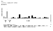


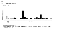
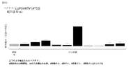


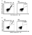

Classifications
-
- C—CHEMISTRY; METALLURGY
- C07—ORGANIC CHEMISTRY
- C07K—PEPTIDES
- C07K14/00—Peptides having more than 20 amino acids; Gastrins; Somatostatins; Melanotropins; Derivatives thereof
- C07K14/435—Peptides having more than 20 amino acids; Gastrins; Somatostatins; Melanotropins; Derivatives thereof from animals; from humans
- C07K14/46—Peptides having more than 20 amino acids; Gastrins; Somatostatins; Melanotropins; Derivatives thereof from animals; from humans from vertebrates
- C07K14/47—Peptides having more than 20 amino acids; Gastrins; Somatostatins; Melanotropins; Derivatives thereof from animals; from humans from vertebrates from mammals
- C07K14/4701—Peptides having more than 20 amino acids; Gastrins; Somatostatins; Melanotropins; Derivatives thereof from animals; from humans from vertebrates from mammals not used
- C07K14/4748—Tumour specific antigens; Tumour rejection antigen precursors [TRAP], e.g. MAGE
-
- A—HUMAN NECESSITIES
- A61—MEDICAL OR VETERINARY SCIENCE; HYGIENE
- A61K—PREPARATIONS FOR MEDICAL, DENTAL OR TOILETRY PURPOSES
- A61K39/00—Medicinal preparations containing antigens or antibodies
- A61K39/0005—Vertebrate antigens
- A61K39/0011—Cancer antigens
-
- A—HUMAN NECESSITIES
- A61—MEDICAL OR VETERINARY SCIENCE; HYGIENE
- A61K—PREPARATIONS FOR MEDICAL, DENTAL OR TOILETRY PURPOSES
- A61K39/00—Medicinal preparations containing antigens or antibodies
- A61K39/46—Cellular immunotherapy
- A61K39/461—Cellular immunotherapy characterised by the cell type used
- A61K39/4611—T-cells, e.g. tumor infiltrating lymphocytes [TIL], lymphokine-activated killer cells [LAK] or regulatory T cells [Treg]
-
- A—HUMAN NECESSITIES
- A61—MEDICAL OR VETERINARY SCIENCE; HYGIENE
- A61K—PREPARATIONS FOR MEDICAL, DENTAL OR TOILETRY PURPOSES
- A61K39/00—Medicinal preparations containing antigens or antibodies
- A61K39/46—Cellular immunotherapy
- A61K39/464—Cellular immunotherapy characterised by the antigen targeted or presented
- A61K39/4643—Vertebrate antigens
- A61K39/4644—Cancer antigens
-
- A—HUMAN NECESSITIES
- A61—MEDICAL OR VETERINARY SCIENCE; HYGIENE
- A61P—SPECIFIC THERAPEUTIC ACTIVITY OF CHEMICAL COMPOUNDS OR MEDICINAL PREPARATIONS
- A61P1/00—Drugs for disorders of the alimentary tract or the digestive system
-
- A—HUMAN NECESSITIES
- A61—MEDICAL OR VETERINARY SCIENCE; HYGIENE
- A61P—SPECIFIC THERAPEUTIC ACTIVITY OF CHEMICAL COMPOUNDS OR MEDICINAL PREPARATIONS
- A61P1/00—Drugs for disorders of the alimentary tract or the digestive system
- A61P1/04—Drugs for disorders of the alimentary tract or the digestive system for ulcers, gastritis or reflux esophagitis, e.g. antacids, inhibitors of acid secretion, mucosal protectants
-
- A—HUMAN NECESSITIES
- A61—MEDICAL OR VETERINARY SCIENCE; HYGIENE
- A61P—SPECIFIC THERAPEUTIC ACTIVITY OF CHEMICAL COMPOUNDS OR MEDICINAL PREPARATIONS
- A61P1/00—Drugs for disorders of the alimentary tract or the digestive system
- A61P1/16—Drugs for disorders of the alimentary tract or the digestive system for liver or gallbladder disorders, e.g. hepatoprotective agents, cholagogues, litholytics
-
- A—HUMAN NECESSITIES
- A61—MEDICAL OR VETERINARY SCIENCE; HYGIENE
- A61P—SPECIFIC THERAPEUTIC ACTIVITY OF CHEMICAL COMPOUNDS OR MEDICINAL PREPARATIONS
- A61P1/00—Drugs for disorders of the alimentary tract or the digestive system
- A61P1/18—Drugs for disorders of the alimentary tract or the digestive system for pancreatic disorders, e.g. pancreatic enzymes
-
- A—HUMAN NECESSITIES
- A61—MEDICAL OR VETERINARY SCIENCE; HYGIENE
- A61P—SPECIFIC THERAPEUTIC ACTIVITY OF CHEMICAL COMPOUNDS OR MEDICINAL PREPARATIONS
- A61P11/00—Drugs for disorders of the respiratory system
-
- A—HUMAN NECESSITIES
- A61—MEDICAL OR VETERINARY SCIENCE; HYGIENE
- A61P—SPECIFIC THERAPEUTIC ACTIVITY OF CHEMICAL COMPOUNDS OR MEDICINAL PREPARATIONS
- A61P13/00—Drugs for disorders of the urinary system
- A61P13/08—Drugs for disorders of the urinary system of the prostate
-
- A—HUMAN NECESSITIES
- A61—MEDICAL OR VETERINARY SCIENCE; HYGIENE
- A61P—SPECIFIC THERAPEUTIC ACTIVITY OF CHEMICAL COMPOUNDS OR MEDICINAL PREPARATIONS
- A61P13/00—Drugs for disorders of the urinary system
- A61P13/10—Drugs for disorders of the urinary system of the bladder
-
- A—HUMAN NECESSITIES
- A61—MEDICAL OR VETERINARY SCIENCE; HYGIENE
- A61P—SPECIFIC THERAPEUTIC ACTIVITY OF CHEMICAL COMPOUNDS OR MEDICINAL PREPARATIONS
- A61P13/00—Drugs for disorders of the urinary system
- A61P13/12—Drugs for disorders of the urinary system of the kidneys
-
- A—HUMAN NECESSITIES
- A61—MEDICAL OR VETERINARY SCIENCE; HYGIENE
- A61P—SPECIFIC THERAPEUTIC ACTIVITY OF CHEMICAL COMPOUNDS OR MEDICINAL PREPARATIONS
- A61P15/00—Drugs for genital or sexual disorders; Contraceptives
-
- A—HUMAN NECESSITIES
- A61—MEDICAL OR VETERINARY SCIENCE; HYGIENE
- A61P—SPECIFIC THERAPEUTIC ACTIVITY OF CHEMICAL COMPOUNDS OR MEDICINAL PREPARATIONS
- A61P17/00—Drugs for dermatological disorders
-
- A—HUMAN NECESSITIES
- A61—MEDICAL OR VETERINARY SCIENCE; HYGIENE
- A61P—SPECIFIC THERAPEUTIC ACTIVITY OF CHEMICAL COMPOUNDS OR MEDICINAL PREPARATIONS
- A61P25/00—Drugs for disorders of the nervous system
-
- A—HUMAN NECESSITIES
- A61—MEDICAL OR VETERINARY SCIENCE; HYGIENE
- A61P—SPECIFIC THERAPEUTIC ACTIVITY OF CHEMICAL COMPOUNDS OR MEDICINAL PREPARATIONS
- A61P35/00—Antineoplastic agents
-
- A—HUMAN NECESSITIES
- A61—MEDICAL OR VETERINARY SCIENCE; HYGIENE
- A61P—SPECIFIC THERAPEUTIC ACTIVITY OF CHEMICAL COMPOUNDS OR MEDICINAL PREPARATIONS
- A61P35/00—Antineoplastic agents
- A61P35/02—Antineoplastic agents specific for leukemia
-
- A—HUMAN NECESSITIES
- A61—MEDICAL OR VETERINARY SCIENCE; HYGIENE
- A61P—SPECIFIC THERAPEUTIC ACTIVITY OF CHEMICAL COMPOUNDS OR MEDICINAL PREPARATIONS
- A61P37/00—Drugs for immunological or allergic disorders
- A61P37/02—Immunomodulators
- A61P37/04—Immunostimulants
-
- A—HUMAN NECESSITIES
- A61—MEDICAL OR VETERINARY SCIENCE; HYGIENE
- A61P—SPECIFIC THERAPEUTIC ACTIVITY OF CHEMICAL COMPOUNDS OR MEDICINAL PREPARATIONS
- A61P43/00—Drugs for specific purposes, not provided for in groups A61P1/00-A61P41/00
-
- C—CHEMISTRY; METALLURGY
- C07—ORGANIC CHEMISTRY
- C07K—PEPTIDES
- C07K14/00—Peptides having more than 20 amino acids; Gastrins; Somatostatins; Melanotropins; Derivatives thereof
- C07K14/435—Peptides having more than 20 amino acids; Gastrins; Somatostatins; Melanotropins; Derivatives thereof from animals; from humans
- C07K14/705—Receptors; Cell surface antigens; Cell surface determinants
- C07K14/70503—Immunoglobulin superfamily
- C07K14/7051—T-cell receptor (TcR)-CD3 complex
-
- C—CHEMISTRY; METALLURGY
- C07—ORGANIC CHEMISTRY
- C07K—PEPTIDES
- C07K14/00—Peptides having more than 20 amino acids; Gastrins; Somatostatins; Melanotropins; Derivatives thereof
- C07K14/435—Peptides having more than 20 amino acids; Gastrins; Somatostatins; Melanotropins; Derivatives thereof from animals; from humans
- C07K14/705—Receptors; Cell surface antigens; Cell surface determinants
- C07K14/70503—Immunoglobulin superfamily
- C07K14/70539—MHC-molecules, e.g. HLA-molecules
-
- C—CHEMISTRY; METALLURGY
- C07—ORGANIC CHEMISTRY
- C07K—PEPTIDES
- C07K16/00—Immunoglobulins [IGs], e.g. monoclonal or polyclonal antibodies
- C07K16/18—Immunoglobulins [IGs], e.g. monoclonal or polyclonal antibodies against material from animals or humans
- C07K16/28—Immunoglobulins [IGs], e.g. monoclonal or polyclonal antibodies against material from animals or humans against receptors, cell surface antigens or cell surface determinants
- C07K16/2803—Immunoglobulins [IGs], e.g. monoclonal or polyclonal antibodies against material from animals or humans against receptors, cell surface antigens or cell surface determinants against the immunoglobulin superfamily
- C07K16/2833—Immunoglobulins [IGs], e.g. monoclonal or polyclonal antibodies against material from animals or humans against receptors, cell surface antigens or cell surface determinants against the immunoglobulin superfamily against MHC-molecules, e.g. HLA-molecules
-
- C—CHEMISTRY; METALLURGY
- C07—ORGANIC CHEMISTRY
- C07K—PEPTIDES
- C07K16/00—Immunoglobulins [IGs], e.g. monoclonal or polyclonal antibodies
- C07K16/18—Immunoglobulins [IGs], e.g. monoclonal or polyclonal antibodies against material from animals or humans
- C07K16/28—Immunoglobulins [IGs], e.g. monoclonal or polyclonal antibodies against material from animals or humans against receptors, cell surface antigens or cell surface determinants
- C07K16/30—Immunoglobulins [IGs], e.g. monoclonal or polyclonal antibodies against material from animals or humans against receptors, cell surface antigens or cell surface determinants from tumour cells
-
- C—CHEMISTRY; METALLURGY
- C07—ORGANIC CHEMISTRY
- C07K—PEPTIDES
- C07K7/00—Peptides having 5 to 20 amino acids in a fully defined sequence; Derivatives thereof
- C07K7/04—Linear peptides containing only normal peptide links
- C07K7/06—Linear peptides containing only normal peptide links having 5 to 11 amino acids
-
- C—CHEMISTRY; METALLURGY
- C12—BIOCHEMISTRY; BEER; SPIRITS; WINE; VINEGAR; MICROBIOLOGY; ENZYMOLOGY; MUTATION OR GENETIC ENGINEERING
- C12N—MICROORGANISMS OR ENZYMES; COMPOSITIONS THEREOF; PROPAGATING, PRESERVING, OR MAINTAINING MICROORGANISMS; MUTATION OR GENETIC ENGINEERING; CULTURE MEDIA
- C12N15/00—Mutation or genetic engineering; DNA or RNA concerning genetic engineering, vectors, e.g. plasmids, or their isolation, preparation or purification; Use of hosts therefor
- C12N15/09—Recombinant DNA-technology
- C12N15/11—DNA or RNA fragments; Modified forms thereof; Non-coding nucleic acids having a biological activity
- C12N15/115—Aptamers, i.e. nucleic acids binding a target molecule specifically and with high affinity without hybridising therewith ; Nucleic acids binding to non-nucleic acids, e.g. aptamers
-
- C—CHEMISTRY; METALLURGY
- C12—BIOCHEMISTRY; BEER; SPIRITS; WINE; VINEGAR; MICROBIOLOGY; ENZYMOLOGY; MUTATION OR GENETIC ENGINEERING
- C12N—MICROORGANISMS OR ENZYMES; COMPOSITIONS THEREOF; PROPAGATING, PRESERVING, OR MAINTAINING MICROORGANISMS; MUTATION OR GENETIC ENGINEERING; CULTURE MEDIA
- C12N15/00—Mutation or genetic engineering; DNA or RNA concerning genetic engineering, vectors, e.g. plasmids, or their isolation, preparation or purification; Use of hosts therefor
- C12N15/09—Recombinant DNA-technology
- C12N15/11—DNA or RNA fragments; Modified forms thereof; Non-coding nucleic acids having a biological activity
- C12N15/62—DNA sequences coding for fusion proteins
-
- C—CHEMISTRY; METALLURGY
- C12—BIOCHEMISTRY; BEER; SPIRITS; WINE; VINEGAR; MICROBIOLOGY; ENZYMOLOGY; MUTATION OR GENETIC ENGINEERING
- C12N—MICROORGANISMS OR ENZYMES; COMPOSITIONS THEREOF; PROPAGATING, PRESERVING, OR MAINTAINING MICROORGANISMS; MUTATION OR GENETIC ENGINEERING; CULTURE MEDIA
- C12N5/00—Undifferentiated human, animal or plant cells, e.g. cell lines; Tissues; Cultivation or maintenance thereof; Culture media therefor
- C12N5/06—Animal cells or tissues; Human cells or tissues
- C12N5/0602—Vertebrate cells
- C12N5/0634—Cells from the blood or the immune system
- C12N5/0636—T lymphocytes
-
- G—PHYSICS
- G01—MEASURING; TESTING
- G01N—INVESTIGATING OR ANALYSING MATERIALS BY DETERMINING THEIR CHEMICAL OR PHYSICAL PROPERTIES
- G01N33/00—Investigating or analysing materials by specific methods not covered by groups G01N1/00 - G01N31/00
- G01N33/48—Biological material, e.g. blood, urine; Haemocytometers
- G01N33/50—Chemical analysis of biological material, e.g. blood, urine; Testing involving biospecific ligand binding methods; Immunological testing
- G01N33/53—Immunoassay; Biospecific binding assay; Materials therefor
- G01N33/574—Immunoassay; Biospecific binding assay; Materials therefor for cancer
- G01N33/57407—Specifically defined cancers
- G01N33/57423—Specifically defined cancers of lung
-
- A—HUMAN NECESSITIES
- A61—MEDICAL OR VETERINARY SCIENCE; HYGIENE
- A61K—PREPARATIONS FOR MEDICAL, DENTAL OR TOILETRY PURPOSES
- A61K39/00—Medicinal preparations containing antigens or antibodies
- A61K2039/51—Medicinal preparations containing antigens or antibodies comprising whole cells, viruses or DNA/RNA
- A61K2039/515—Animal cells
- A61K2039/5158—Antigen-pulsed cells, e.g. T-cells
-
- C—CHEMISTRY; METALLURGY
- C07—ORGANIC CHEMISTRY
- C07K—PEPTIDES
- C07K2319/00—Fusion polypeptide
- C07K2319/40—Fusion polypeptide containing a tag for immunodetection, or an epitope for immunisation
-
- C—CHEMISTRY; METALLURGY
- C12—BIOCHEMISTRY; BEER; SPIRITS; WINE; VINEGAR; MICROBIOLOGY; ENZYMOLOGY; MUTATION OR GENETIC ENGINEERING
- C12N—MICROORGANISMS OR ENZYMES; COMPOSITIONS THEREOF; PROPAGATING, PRESERVING, OR MAINTAINING MICROORGANISMS; MUTATION OR GENETIC ENGINEERING; CULTURE MEDIA
- C12N2310/00—Structure or type of the nucleic acid
- C12N2310/10—Type of nucleic acid
- C12N2310/16—Aptamers
-
- G—PHYSICS
- G01—MEASURING; TESTING
- G01N—INVESTIGATING OR ANALYSING MATERIALS BY DETERMINING THEIR CHEMICAL OR PHYSICAL PROPERTIES
- G01N2800/00—Detection or diagnosis of diseases
- G01N2800/70—Mechanisms involved in disease identification
- G01N2800/7023—(Hyper)proliferation
- G01N2800/7028—Cancer
Description
本発明は、免疫療法において使用するためのペプチド、タンパク質、核酸、および細胞に関する。特に、本発明は、がんの免疫療法に関する。本発明は、単独のまたはその他の腫瘍関連ペプチドと組み合わされた、腫瘍関連T細胞ペプチドエピトープにさらに関し、それは、例えば、抗腫瘍免疫応答を刺激し、または生体外でT細胞を刺激して患者に移入する、ワクチン組成物の活性医薬品成分の役割を果たし得る。主要組織適合性複合体(MHC)の分子と結合しているペプチド、またはペプチドそれ自体もまた、抗体、可溶性T細胞受容体、およびその他の結合分子の標的になり得る。 The present invention relates to peptides, proteins, nucleic acids, and cells for use in immunotherapy. In particular, the present invention relates to immunotherapy for cancer. The present invention further relates to tumor-related T cell peptide epitopes, alone or in combination with other tumor-related peptides, which may, for example, stimulate an antitumor immune response or stimulate T cells in vitro to a patient. Can serve as the active pharmaceutical ingredient of the vaccine composition to be transferred to. Peptides that bind to major histocompatibility complex (MHC) molecules, or the peptides themselves, can also be targets for antibodies, soluble T cell receptors, and other binding molecules.
本発明は、ヒト腫瘍細胞のHLAクラスI分子に由来する、いくつかの新規ペプチド配列およびそれらの変異型に関し、それらは抗腫瘍免疫応答を引き起こすためのワクチン組成物中で、または薬理的/免疫学的活性化合物および細胞の開発のための標的として、使用され得る。 The present invention relates to several novel peptide sequences and variants thereof derived from HLA class I molecules of human tumor cells, which are in vaccine compositions for eliciting an antitumor immune response or pharmacologically / immunologically. It can be used as a target for the development of immunologically active compounds and cells.
小細胞肺がん(SCLC)
小細胞肺がん(SCLC)は、顕微鏡下で観察された際のがん細胞のサイズに応じて命名され、非小細胞肺がん(NSCLC)と区別されなければならない。SCLCは、全ての肺がんの約10%〜15%を占める(American Cancer Society,2015a)。
Small Cell Lung Cancer (SCLC)
Small cell lung cancer (SCLC) must be named according to the size of the cancer cells when observed under a microscope and must be distinguished from non-small cell lung cancer (NSCLC). SCLC accounts for approximately 10% to 15% of all lung cancers (American Cancer Society, 2015a).
双方の肺がん(SCLCおよびNSCLC)は、男性および女性の双方で2番目に頻度の高いがんである。肺がんはがん死亡の主要原因であり、約25%を占める。したがって、毎年結腸がん、乳がん、および前立腺がんを合わせたよりも多くの人々が肺がんで死亡する。さらに、双方の肺がんは、全ての新規がんの約13%(180万例以上)を占める。肺がんは、主に高齢者において発生する。診断時の平均年齢は、約70歳である。全ての症例の2%未満が、45歳未満の人々において診断される。SCLCの治療および予後は、診断されたがん病期に大きく左右される。臨床結果に基づくSCLCの進行度診断の方が、病的進行度診断よりも一般的である。臨床進行度診断は、理学的検査、様々な画像検査、および生検の結果を利用する。American Cancer Societyが提出したデータによると、5年相対生存率は、ステージIで31%、ステージIIで19%、ステージIIIで8%、ステージIVで2%をである。 Both lung cancers (SCLC and NSCLC) are the second most common cancers in both men and women. Lung cancer is the leading cause of cancer death and accounts for about 25%. Therefore, more people die of lung cancer each year than a combination of colon, breast, and prostate cancers. In addition, both lung cancers account for about 13% (more than 1.8 million cases) of all new cancers. Lung cancer mainly occurs in the elderly. The average age at diagnosis is about 70 years. Less than 2% of all cases are diagnosed in people under the age of 45. The treatment and prognosis of SCLC is highly dependent on the stage of cancer diagnosed. Diagnosis of SCLC progression based on clinical results is more common than diagnosis of pathological progression. Laboratory progression diagnostics utilize the results of physical tests, various imaging tests, and biopsies. According to data submitted by the American Cancer Society, 5-year relative survival is 31% for stage I, 19% for stage II, 8% for stage III, and 2% for stage IV.
SCLCの標準的化学療法は、エトポシドまたはイリノテカンのどちらかと、シスプラチンまたはカルボプラチンのどちらかとの組み合わせを使用する治療は4〜6サイクルで与えられる。各サイクルは、1〜3日間の化学療法で開始され、3〜4週間の回復期間がそれに続く。 Standard chemotherapy for SCLC is treatment using either etoposide or irinotecan in combination with either cisplatin or carboplatin in 4-6 cycles. Each cycle begins with 1-3 days of chemotherapy, followed by a recovery period of 3-4 weeks.
SCLCの治療のための標準的な放射線療法は、外照射療法(EBRT)と称され、通常、1日1〜2回、週5日、3〜7週間与えられる。過去数年間に、新たな放射技術が開発されている。新たな技術は、三次元原体放射線療法(3D−CRT)および強度変調放射線療法(IMRT)である。そのどちらも、周囲の健常組織への放射線被曝を低下させることにより、腫瘍に対する放射線負荷のより正確な標的化を可能にする。 Standard radiation therapy for the treatment of SCLC is called external beam radiotherapy (EBRT) and is usually given 1-2 times a day, 5 days a week for 3-7 weeks. In the last few years, new radiation technologies have been developed. New technologies are three-dimensional radiotherapy (3D-CRT) and intensity-modulated radiotherapy (IMRT). Both allow more accurate targeting of radiation load on tumors by reducing radiation exposure to surrounding healthy tissues.
SCLCが、リンパ節またはその他の箇所におけるがんの広がりの形跡がない(20人の患者のうち1人未満)、単一の小さな腫瘍として見いだされるステージIでは、手術と、それに続く化学療法と放射線療法の併用が標準的治療法である。この治療法は、健康状態の良好な患者に限られる選択肢である。ほとんどの場合、診断された時点で、SCLCは既に広がっている。したがって、手術による治療はありそうにない(American Cancer Society,2015a;S3−Leitlinie Lungenkarzinom,2011)。 In stage I, where SCLC is found as a single small tumor with no evidence of cancer spread in the lymph nodes or elsewhere (less than 1 in 20 patients), surgery followed by chemotherapy Combination with radiation therapy is standard treatment. This treatment is an option limited to patients in good health. In most cases, SCLC is already widespread at the time of diagnosis. Therefore, surgical treatment is unlikely (American Cancer Society, 2015a; S3-Leitlinee Lungenkarzinom, 2011).
SCLCが、胸部の片側全体から肺または近くのリンパ節に広がっている限局期(3人の患者のうち1人)では、化学療法と放射線療法の併用、いわゆる並行化学放射線療法が標準的治療法である。この段階では、手術は選択肢でない。標準化学療法薬は、シスプラチンまたはカルボプラチンのどちらかが合わせられた、エトポシド(VP−16)である。化学療法と放射線療法の併用療法は治療上の利点を示したが、単独の化学療法または放射線治療と比較して、重度の副作用もそれに続いた。同時の化学放射線療法に耐えられそうにない患者は、化学療法が標準治療法である。必要に応じて、化学療法に続いて放射線療法が続く(American Cancer Society,2015a;S3−Leitlinie Lungenkarzinom,2011)。 In the localized phase (1 of 3 patients) where SCLC spreads from one side of the chest to the lungs or nearby lymph nodes, a combination of chemotherapy and radiation therapy, so-called parallel chemoradiotherapy, is the standard treatment. Is. Surgery is not an option at this stage. The standard chemotherapeutic agent is etoposide (VP-16), which is a combination of either cisplatin or carboplatin. The combination of chemotherapy and radiation showed therapeutic benefits, but was followed by severe side effects compared to chemotherapy or radiation alone. Chemotherapy is the standard treatment for patients who are unlikely to tolerate concurrent chemoradiotherapy. If necessary, chemotherapy is followed by radiation therapy (American Cancer Society, 2015a; S3-Leitlinee Lungenkarzinom, 2011).
SCLCが、肺、近くリンパ節およびその他の遠隔臓器(骨髄のような)全体にわたり広がっている拡張期では、概してシスプラチンまたはカルボプラチンと併用されるエトポシドである全身化学療法(systematic chemotherapy)と、それに続く胸部への放射線治療が適用される治療法である。(American Cancer Society,2015a;S3−Leitlinie Lungenkarzinom,2011)。 In the diastolic phase, when SCLC is widespread throughout the lungs, nearby lymph nodes and other distant organs (such as bone marrow), systemic chemotherapy, which is an etoposide generally used in combination with cisplatin or carboplatin, followed by Radiation therapy to the chest is applied. (American Cancer Society, 2015a; S3-Leitlinee Lungenkarzinom, 2011).
SCLCは脳に広がることが知られているので、SCLC患者は病期とは独立して、予防的頭蓋照射またはPCIされる頭部への予防的放射線療法を受ける。 Since SCLC is known to spread to the brain, patients with SCLC receive prophylactic cranial radiation or prophylactic radiation therapy to the head to be PCI, independent of stage.
限局期および拡張期で、治療はがんの顕著な縮小をもたらす可能性が高いが、ほとんどの場合、がんはある時点で戻ってくる。化学療法を適用してもがんが増殖し続ける場合、化学療法のタイプの変更が検討される。がんの再発による化学療法の選択は、がん寛解期の長さに左右される。 In localized and diastole, treatment is likely to result in a marked reduction in cancer, but in most cases the cancer will return at some point. If the cancer continues to grow after applying chemotherapy, changing the type of chemotherapy is considered. The choice of chemotherapy for cancer recurrence depends on the length of cancer remission.
SCLCの検出、診断、および治療に関する革新が起こった。早期のがん検出のために、X線でなくCTスキャンを使用することで、肺がんによる死亡リスクが低下することが示された。現今では、SCLCの診断は、蛍光または仮想気管支鏡検査によって支援され得て、リアルタイムの腫瘍イメージング(imagining)は、放射線治療によって実施され得る。ベバシズマブ(Avastin)、スニチニブ(スーテント)、およびニンテダニブ(BIBF1120)のような新規抗血管新生薬は、SCLCの治療において治療効果を有することが示された。(American Cancer Society,2015a)。 Innovations have taken place in the detection, diagnosis, and treatment of SCLC. The use of CT scans instead of x-rays for early cancer detection has been shown to reduce the risk of death from lung cancer. Nowadays, the diagnosis of SCLC can be assisted by fluorescence or virtual bronchoscopy, and real-time tumor imaging can be performed by radiation therapy. New anti-angiogenic agents such as bevacizumab (Avastin), sunitinib (Sutent), and nintedanib (BIBF1120) have been shown to have therapeutic effects in the treatment of SCLC. (American Cancer Society, 2015a).
免疫療法は、過度に調査されたがん療法の分野に相当する。SCLCの治療には、様々なアプローチが点在する。アプローチの1つは、天然ヒト免疫抑制剤であるCTLA−4のブロックを標的とする。CTLA−4の阻害は、がんと戦うために免疫系を増強することを意図する。近年、SCLCの治療のための有望な免疫チェックポイント阻害剤の開発が始まっている。もう一つのアプローチは抗がんワクチンに基づき、それは現在臨床試験においてSCLC治療のために利用できる(American Cancer Society,2015b;National Cancer Institute(NCI),2011)。 Immunotherapy corresponds to the over-examined area of cancer therapy. There are various approaches to the treatment of SCLC. One approach targets the block of CTLA-4, a natural human immunosuppressant. Inhibition of CTLA-4 is intended to enhance the immune system to fight cancer. In recent years, the development of promising immune checkpoint inhibitors for the treatment of SCLC has begun. Another approach is based on anti-cancer vaccines, which are currently available for SCLC treatment in clinical trials (American Cancer Institute, 2015b; National Cancer Institute (NCI), 2011).
がんの治療に伴う重度の副作用および費用を考慮すると、がん全般、そして特に小細胞肺がんの治療に使用し得る要素を同定する必要がある。がんのより良い診断、予後の評価、および治療成功の予測につながる、がん全般、特に小細胞肺がんのためのバイオマーカーに相当する要素を同定する必要性もまたある。 Given the severe side effects and costs associated with treating cancer, it is necessary to identify factors that can be used to treat cancer in general, and especially small cell lung cancer. There is also a need to identify biomarker-equivalent factors for cancer in general, especially small cell lung cancer, that lead to better diagnosis of cancer, assessment of prognosis, and prediction of successful treatment.
がんの免疫療法は、がん細胞を特異的に標的化しながら副作用を最小化する選択肢に相当する。がん免疫療法は、腫瘍関連抗原の存在を利用する。 Immunotherapy for cancer represents an option that minimizes side effects while specifically targeting cancer cells. Cancer immunotherapy takes advantage of the presence of tumor-related antigens.
腫瘍関連抗原(TAA)の現行の分類は、次の主要群を含んでなる:
a)がん精巣抗原:T細胞によって認識され得る初めて同定されたTAAはこのクラスに属し、元々はがん精巣(CT)抗原と称されたが、それは、そのメンバーが組織学的に異なるヒト腫瘍において発現し、正常組織では精巣の精母細胞/精原細胞のみに存在し、時として胎盤に存在するためであった。精巣の細胞は、クラスIおよびII HLA分子を発現しないので、これらの抗原は正常組織のT細胞によって認識され得ず、したがって免疫学的に腫瘍特異的と見なされる。CT抗原の周知の例は、MAGEファミリーメンバーおよびNY−ESO−1である。
b)分化抗原:これらのTAAは、腫瘍と、それから腫瘍が生じる正常組織との間で共有される。既知の分化抗原のほとんどは、黒色腫および正常メラノサイトに見いだされる。これらのメラノサイト系関連タンパク質の多くは、メラニン生合成に関与し、したがって腫瘍特異的でないが、それでもなおがん免疫療法のために広く利用されている。例としては、黒色腫に対するチロシナーゼとMelan−A/MART−1、または前立腺がんに対するPSAが挙げられるが、これに限定されるものではない。
c)過剰発現TAA:広範に発現されるTAAをエンコードする遺伝子は、組織学的に異なる型の腫瘍において検出され、多数の正常組織においても概してより低い発現レベルで検出されている。正常組織によってプロセスされて潜在的に提示さるエピトープの多くは、T細胞認識の閾値レベル未満であり得る一方で、腫瘍細胞におけるそれらの過剰発現は、以前確立された免疫寛容を破壊することにより、抗がん応答を始動し得る。このクラスのTAAの顕著な例は、Her−2/neu、サバイビン、テロメラーゼまたはWT1である。
d)腫瘍特異的抗原:これらのユニークなTAAは、正常な遺伝子(β−カテニン、CDK4など)の変異から生じる。これらの分子変化のいくつかは、腫瘍性形質転換および/または進行に関連する。腫瘍特異的抗原は、通常、正常組織に対する自己免疫反応のリスクなしに、強力な免疫応答を誘導できる。他方、これらのTAAは、ほとんどの場合、その上でそれらが同定されたまさにその腫瘍のみと関係があり、通常は、多くの個々の腫瘍間で共有されない。腫瘍特異的(関連)イソ型を有するタンパク質では、ペプチドの腫瘍特異性(または関連性)はまた、ペプチドが腫瘍(関連)エクソンに由来する場合に生じてもよい。
e)異常な翻訳後修飾から生じるTAA:このようなTAAは、特異的でなく腫瘍において過剰発現もされないタンパク質から生じてもよいが、それでもなお、腫瘍において主に活性である翻訳後プロセスによって腫瘍関連になる。このクラスの例は、腫瘍にMUC1のような新規エピトープをもたらす改変グリコシル化パターン、または腫瘍特異的であってもなくてもよい分解中のタンパク質スプライシングのような事象から生じる。
f)オンコウイルスタンパク質:これらのTAAはウイルスタンパク質であり、それらは発がん過程において重要な役割を果たしてもよく、外来性である(ヒト由来でない)ため、それらはT細胞応答を誘起し得る。このようなタンパク質の例は、子宮頸がんにおいて発現されるヒト乳頭腫16型ウイルスタンパク質E6およびE7である。
The current classification of tumor-related antigens (TAAs) consists of the following major groups:
a) Cancer testis antigen: The first identified TAA that can be recognized by T cells belongs to this class and was originally referred to as the cancer testis (CT) antigen, but it is a human whose members are histologically different. This was because it was expressed in tumors and was present only in testicular spermatogonia / spermatogonia in normal tissues, sometimes in the placenta. Since testicular cells do not express class I and II HLA molecules, these antigens cannot be recognized by normal tissue T cells and are therefore immunologically considered tumor-specific. Well-known examples of CT antigens are MAGE family members and NY-ESO-1.
b) Differentiation antigens: These TAAs are shared between the tumor and the normal tissue from which the tumor arises. Most of the known differentiation antigens are found in melanoma and normal melanocytes. Many of these melanocyte-related proteins are involved in melanin biosynthesis and are therefore not tumor-specific, but are still widely used for cancer immunotherapy. Examples include, but are not limited to, tyrosinase and Melan-A / MART-1 for melanoma, or PSA for prostate cancer.
c) Overexpressed TAA: Genes encoding widely expressed TAA have been detected in histologically different types of tumors and also in many normal tissues at generally lower expression levels. While many of the epitopes that are processed and potentially presented by normal tissues can be below threshold levels for T cell recognition, their overexpression in tumor cells by disrupting previously established immune tolerance. Can initiate an anti-cancer response. Prominent examples of this class of TAA are Her-2 / neu, survivin, telomerase or WT1.
d) Tumor-specific antigens: These unique TAAs result from mutations in normal genes (β-catenin, CDK4, etc.). Some of these molecular changes are associated with neoplastic transformation and / or progression. Tumor-specific antigens can usually induce a strong immune response without the risk of an autoimmune response to normal tissue. On the other hand, these TAAs are most often associated only with the very tumor in which they were identified and are usually not shared among many individual tumors. For proteins with tumor-specific (associative) isotypes, tumor specificity (or association) of the peptide may also occur if the peptide is derived from a tumor (associative) exon.
e) TAA resulting from aberrant post-translational modifications: Such TAA may result from proteins that are not specific and are not overexpressed in tumors, but nonetheless, tumors by post-translational processes that are predominantly active in tumors. Become relevant. Examples of this class result from modified glycosylation patterns that result in novel epitopes such as MUC1 in tumors, or events such as protein splicing during degradation that may or may not be tumor specific.
f) Oncoviral proteins: These TAAs are viral proteins, which may play important roles in the carcinogenic process and are exogenous (not of human origin), so they can elicit a T cell response. Examples of such proteins are human papilloma type 16 viral proteins E6 and E7 expressed in cervical cancer.
T細胞ベースの免疫療法は、主要組織適合性複合体(MHC)の分子によって提示される、腫瘍関連または腫瘍特異的タンパク質由来ペプチドエピトープを標的とする。腫瘍特異的Tリンパ球によって認識される抗原、すなわちそれらのエピトープは、酵素、受容体、転写因子などの全てのタンパク質クラスに由来する分子であり得て、それはそれぞれの腫瘍細胞において発現されて、同一起源の非改変細胞と比較して、通常、上方制御される。 T cell-based immunotherapy targets tumor-related or tumor-specific protein-derived peptide epitopes presented by major histocompatibility complex (MHC) molecules. Antigens recognized by tumor-specific T lymphocytes, ie their epitopes, can be molecules derived from all protein classes such as enzymes, receptors, transcription factors, etc., which are expressed in each tumor cell and It is usually upregulated as compared to unmodified cells of the same origin.
MHC分子には、MHCクラスIおよびMHCクラスIIの2つのクラスがある。MHCクラスI分子はα重鎖およびβ2ミクログロブリンから構成され、MHCクラスII分子はαおよびβ鎖から構成される。それらの三次元立体構造は、ペプチドとの非共有結合相互作用のために使用される、結合溝をもたらす。 There are two classes of MHC molecules, MHC class I and MHC class II. MHC class I molecule is composed of α heavy chain and β2 microglobulin, and MHC class II molecule is composed of α and β chain. Their three-dimensional structure provides a binding groove used for non-covalent interactions with peptides.
MHCクラスI分子は、ほとんどの有核細胞上に見いだされる。それらは、主に内因性タンパク質、欠陥リボソーム産物(DRIP)、およびより大型のペプチドのタンパク質切断から得られる、ペプチドを提示する。しかし、エンドソームコンパートメントまたは外因性起源に由来するペプチドもまた、MHCクラスI分子上に頻繁に見いだされる。この非古典的様式のクラスI提示は、文献中で交差提示と称される(Brossart and Bevan、1997;Rock et al.,1990)。MHCクラスII分子は、大部分はプロフェショナル抗原提示細胞(APC)に見いだされ、例えば、エンドサイトーシス中にAPCに取り込まれて引き続きプロセシングされる、外因性または膜貫通タンパク質のペプチドを主に提示する。 MHC class I molecules are found on most nucleated cells. They present peptides that are primarily derived from protein cleavage of endogenous proteins, defective ribosome products (DRIPs), and larger peptides. However, peptides from the endosome compartment or extrinsic origin are also frequently found on MHC class I molecules. This non-classical form of Class I presentation is referred to in the literature as cross-presentation (Brossart and Bevan, 1997; Rock et al., 1990). MHC class II molecules are predominantly found in professional antigen presenting cells (APCs), eg, peptides of exogenous or transmembrane proteins that are taken up by APCs and subsequently processed during endocytosis. ..
ペプチドとMHCクラスIの複合体が、適切なT細胞受容体(TCR)を有するCD8陽性T細胞によって認識される一方で、ペプチドとMHCクラスII分子の複合体は、適切なTCRを有するCD4陽性ヘルパーT細胞によって認識される。その結果、TCR、ペプチド、およびMHCは、化学量論的に1:1:1の量で存在することが良く知られている。 The peptide-MHC class I complex is recognized by CD8-positive T cells with the appropriate T cell receptor (TCR), while the peptide-MHC class II molecule complex is CD4 positive with the appropriate TCR. Recognized by helper T cells. As a result, it is well known that TCRs, peptides, and MHC are stoichiometrically present in 1: 1: 1 amounts.
CD4陽性ヘルパーT細胞は、CD8陽性細胞傷害性T細胞による、効果的な応答の誘導と維持する上で重要な役割を果たす。腫瘍関連抗原(TAA)に由来するCD4陽性T細胞エピトープの同定は、抗腫瘍免疫応答を始動させる医薬品の開発に非常に重要である(Gnjatic et al.,2003)。腫瘍部位では、Tヘルパー細胞が、細胞毒性T細胞(CTL)親和的サイトカイン環境を維持して(Mortara et al.,2006)、例えば、CTL、ナチュラルキラー(NK)細胞、マクロファージ、および顆粒球などのエフェクター細胞を引きつける(Hwang et al.,2007)。 CD4-positive helper T cells play an important role in inducing and maintaining an effective response by CD8-positive cytotoxic T cells. Identification of CD4-positive T cell epitopes derived from tumor-related antigens (TAAs) is crucial for the development of drugs that initiate an antitumor immune response (Gnjatic et al., 2003). At the tumor site, T helper cells maintain a cytotoxic T cell (CTL) -friendly cytokine environment (Mortara et al., 2006), such as CTL, natural killer (NK) cells, macrophages, and granulocytes. Attracts effector cells of (Hwang et al., 2007).
炎症不在下では、MHCクラスII分子の発現は、免疫系細胞、特に、例えば、単球、単球由来細胞、マクロファージ、樹状細胞などのプロフェショナル抗原提示細胞(APC)に主に限定される。がん患者においては、腫瘍細胞がMHCクラスII分子を発現することが判明している(Dengjel et al.,2006;Dengjel et al.,2006)。伸長された(より長い)本発明のペプチドは、MHCクラスII活性エピトープとして作用し得る。 In the absence of inflammation, expression of MHC class II molecules is predominantly limited to immune system cells, particularly professional antigen presenting cells (APCs) such as monocytes, monocyte-derived cells, macrophages, dendritic cells. Tumor cells have been shown to express MHC class II molecules in cancer patients (Dengjel et al., 2006; Dengjel et al., 2006). The elongated (longer) peptides of the invention can act as MHC class II active epitopes.
MHCクラスIIエピトープによって活性化されたTヘルパー細胞は、抗腫瘍免疫におけるCTLのエフェクター機能を統合するのに重要な役割を果たす。TH1型のTヘルパー細胞応答を始動するTヘルパー細胞エピトープは、それらの細胞表面に腫瘍関連ペプチド/MHC複合体を提示する腫瘍細胞に向けられた細胞傷害機能をはじめとする、CD8陽性キラーT細胞のエフェクター機能を支持する。このようにして腫瘍関連Tヘルパー細胞ペプチドエピトープは、単独で、またはその他の腫瘍関連ペプチドとの組み合わせで、抗腫瘍免疫応答を刺激するワクチン組成物の活性医薬品成分の役割を果たし得る。 T-helper cells activated by MHC class II epitopes play an important role in integrating the effector function of CTL in anti-tumor immunity. T-helper cell epitopes that initiate TH1-type T-helper cell responses are CD8-positive killer T cells, including cytotoxic function directed at tumor cells that present tumor-related peptide / MHC complexes on their cell surface. Supports the effector function of. In this way, the tumor-related T-helper cell peptide epitope can serve as an active pharmaceutical component of a vaccine composition that stimulates an antitumor immune response, either alone or in combination with other tumor-related peptides.
例えば、マウスなどの哺乳類動物モデルにおいて、CD8陽性Tリンパ球の不在下であっても、インターフェロンγ(IFNγ)の分泌による血管新生阻害を通じて腫瘍発現を阻害するには、CD4陽性T細胞で十分であることが示された(Beatty and Paterson,2001;Mumberg et al.,1999)。CD4 T細胞が、直接抗腫瘍エフェクターであるという証拠がある(Braumuller et al.,2013;Tran et al.,2014)。 For example, in mammalian models such as mice, CD4-positive T cells are sufficient to inhibit tumor development through inhibition of angiogenesis by the secretion of interferon gamma (IFNγ), even in the absence of CD8-positive T lymphocytes. It was shown to be (Beatty and Patterson, 2001; Tumorg et al., 1999). There is evidence that CD4 T cells are direct antitumor effectors (Braumuller et al., 2013; Trans et al., 2014).
HLAクラスII分子の構成的発現は、通常、免疫細胞に限定されるので、原発性腫瘍からクラスIIペプチドを直接単離する可能性があり得るとは、これまで考えられなかった。しかし、Dengjel et al.は、いくつかのMHCクラスIIエピトープを腫瘍から直接成功裏に同定した(国際公開第2007/028574号パンフレット、欧州特許第1760088B1号明細書)。 Since the constitutive expression of HLA class II molecules is usually confined to immune cells, it was previously unthinkable that class II peptides could be isolated directly from primary tumors. However, Dengel et al. Has successfully identified several MHC class II epitopes directly from tumors (International Publication No. 2007/028574, European Patent No. 176008B1).
CD8およびCD4依存性の双方のタイプの応答は、抗腫瘍効果に共同して相乗的に寄与するので、CD8+T細胞(リガンド:MHCクラスI分子+ペプチドエピトープ)、またはCD4陽性Tヘルパー細胞(リガンド:MHCクラスII分子+ペプチドエピトープ)のどちらかによって認識される、腫瘍関連抗原の同定および特性解析は、腫瘍ワクチンの開発にとって重要である。 CD8 + T cells (ligand: MHC class I molecule + peptide epitope) or CD4 positive T helper cells (ligand: ligand:) because both CD8 and CD4 dependent types of responses contribute synergistically to antitumor effects. Identification and characterization of tumor-related antigens, recognized by either MHC class II molecules + peptide epitopes), are important for the development of tumor vaccines.
MHCクラスIペプチドが、細胞性免疫応答を始動(惹起)するためには、それはまた、MHC分子に結合しなくてはならない。この過程は、MHC分子の対立遺伝子と、ペプチドのアミノ酸配列の特定の多型性とに依存する。MHCクラスI結合ペプチドは、通常は8〜12アミノ酸残基長であり、通常は、MHC分子の対応する結合溝と相互作用するそれらの配列中に、2つの保存残基(「アンカー」)を含有する。このようにして、各MHC対立遺伝子は、どのペプチドが結合溝と特異的に結合し得るかを決定する、「結合モチーフ」を有する。 In order for an MHC class I peptide to initiate (trigger) a cell-mediated immune response, it must also bind to MHC molecules. This process depends on the allele of the MHC molecule and the specific polymorphism of the amino acid sequence of the peptide. MHC class I binding peptides are usually 8-12 amino acid residue lengths and usually have two conserved residues (“anchors”) in their sequence that interact with the corresponding binding groove of the MHC molecule. contains. In this way, each MHC allele has a "binding motif" that determines which peptide can specifically bind to the binding groove.
MHCクラスI依存免疫反応においては、ペプチドは腫瘍細胞によって発現される特定のMHCクラスI分子に結合できるだけでなく、それらはまた、引き続いて特異的T細胞受容体(TCR)を有するT細胞によって認識されなくてはならない。 In an MHC class I-dependent immune response, not only can peptides bind to specific MHC class I molecules expressed by tumor cells, but they are also subsequently recognized by T cells with specific T cell receptors (TCRs). Must be done.
タンパク質が、Tリンパ球によって腫瘍特異的または腫瘍関連抗原として認識され、治療で利用されるためには、特定の必要条件が満たされなくてはならない。抗原は、主に腫瘍細胞によって発現され、健常組織によって発現されず、または比較的少量発現されるべきである。好ましい実施形態では、ペプチドは、健常組織と比較して、腫瘍細胞によって過剰提示されるべきである。それぞれの抗原は、ある種の腫瘍に存在するだけでなく、高い濃度(すなわち、それぞれのペプチド細胞当たりのコピー数)で存在することもさらに望ましい。腫瘍特異的および腫瘍関連抗原は、例えば、細胞周期調節またはアポトーシス抑制における機能のために、正常細胞から腫瘍細胞への形質転換に直接関与するタンパク質に由来することが多い。さらに、形質転換の直接原因となるタンパク質の下流標的が、上方制御されてもよく、(und)したがって間接的に腫瘍関連であってもよい。このような間接的腫瘍関連抗原もまた、ワクチン接種アプローチの標的であってもよい(Singh−Jasuja et al.,2004)。このようなペプチド(「免疫原性ペプチド」)が、腫瘍関連抗原に由来して、生体外または生体内T細胞応答をもたらすことを確実にするためには、抗原のアミノ酸配列内にエピトープが存在することが必須である。 Certain requirements must be met in order for a protein to be recognized by T lymphocytes as a tumor-specific or tumor-related antigen and used therapeutically. The antigen should be expressed primarily by tumor cells, not by healthy tissue, or in relatively small amounts. In a preferred embodiment, the peptide should be over-presented by the tumor cells as compared to healthy tissue. It is even more desirable that each antigen is present not only in certain tumors, but also in high concentrations (ie, the number of copies per peptide cell). Tumor-specific and tumor-related antigens are often derived from proteins that are directly involved in the transformation of normal cells into tumor cells, for example because of their function in cell cycle regulation or apoptosis suppression. In addition, downstream targets of proteins that are directly responsible for transformation may be upregulated and thus indirectly tumor-related. Such indirect tumor-related antigens may also be targets for vaccination approaches (Singh-Jasuja et al., 2004). Epitopes are present in the amino acid sequence of the antigen to ensure that such peptides (“immunogenic peptides”) are derived from tumor-related antigens and result in in vitro or in vivo T cell responses. It is essential to do.
基本的に、MHC分子に結合できるあらゆるペプチドが、T細胞エピトープとして機能してもよい。生体外または生体内T細胞応答誘導のための必要条件は、対応するTCRを有するT細胞の存在、およびこの特定のエピトープに対する免疫寛容の不在である。 Basically, any peptide that can bind to MHC molecules may function as a T cell epitope. A prerequisite for inducing an in vitro or in vivo T cell response is the presence of T cells with the corresponding TCR and the absence of immune tolerance to this particular epitope.
したがって、TAAは、腫瘍ワクチンをはじめとするが、これに限定されるものではない、T細胞ベースの治療法開発の出発点である。TAAを同定し特性決定する方法は、通常は、患者または健常人から単離され得るT細胞の使用に基づき、またはそれらは、腫瘍と正常組織との間の示差的転写プロファイル、またはペプチド発現パターンの生成に基づく。しかし、腫瘍組織またはヒト腫瘍細胞株において過剰発現され、またはこのような組織または細胞株において選択的に発現される遺伝子の同定は、免疫療法においてこれらの遺伝子から転写される抗原の使用に関する、正確な情報を提供しない。それは、これらの抗原のエピトープの個々の亜集団のみが、このような用途に適するためであり、その理由は、対応するTCRを有するT細胞が存在しなくてはならず、この特定のエピトープに対する免疫寛容が不在または最小でなくてはならないからである。したがって本発明の非常に好ましい実施形態では、それに対する機能性および/または増殖性T細胞が見いだされる、過剰にまたは選択的に提示されるペプチドのみを選択することが、重要である。このような機能性T細胞は、特異的抗原による刺激時にクローン増殖され得て、エフェクター機能を果たすことができるT細胞(「エフェクターT細胞」)と定義される。 Therefore, TAA is a starting point for the development of T cell-based therapies, including but not limited to tumor vaccines. Methods for identifying and characterizing TAAs are usually based on the use of T cells that can be isolated from patients or healthy individuals, or they are differential transcriptional profiles between tumors and normal tissues, or peptide expression patterns. Based on the generation of. However, identification of genes that are overexpressed in tumor tissues or human tumor cell lines, or selectively expressed in such tissues or cell lines, is accurate with respect to the use of antigens transcribed from these genes in immunotherapy. Do not provide any information. This is because only individual subpopulations of the epitopes of these antigens are suitable for such applications, because T cells with the corresponding TCR must be present and for this particular epitope. This is because immune tolerance must be absent or minimal. Therefore, in a highly preferred embodiment of the invention, it is important to select only peptides that are presented in excess or selectively in which functional and / or proliferative T cells are found. Such functional T cells are defined as T cells (“effector T cells”) that can be cloned and proliferated upon stimulation with a specific antigen and can perform effector function.
本発明による特異的TCR(例えば、可溶性TCR)および抗体またはその他の結合分子(スキャフォールド)によってペプチドMHCを標的化する場合、基礎となるペプチドの免疫原性は二次的である。これらの場合には、提示が決定要因である。 When the peptide MHC is targeted by a specific TCR according to the invention (eg, soluble TCR) and an antibody or other binding molecule (scaffold), the immunogenicity of the underlying peptide is secondary. In these cases, presentation is the deciding factor.
本発明の第1の態様では、本発明は、配列番号1〜配列番号126、または配列番号1〜配列番号126と少なくとも77%、好ましくは少なくとも88%相同的な(好ましくは少なくとも77%または少なくとも88%同一の)その変異配列からなる群から選択されるアミノ酸配列を含んでなるペプチドに関し、その中で前記変異型は、MHCと結合し、および/またはT細胞と前記ペプチドまたはその薬学的に許容可能な塩との交差反応を誘導し、その中で前記ペプチドは、基礎となる完全長ポリペプチドでない。 In a first aspect of the invention, the invention is at least 77%, preferably at least 88%, homologous (preferably at least 77% or at least) with SEQ ID NO: 1 to SEQ ID NO: 126, or SEQ ID NO: 1 to SEQ ID NO: 126. With respect to a peptide comprising an amino acid sequence selected from the group consisting of the mutant sequence (88% identical), the variant in which the variant binds to MHC and / or T cells and the peptide or pharmaceutical thereof. It induces cross-reactivity with acceptable salts, in which the peptide is not the underlying full-length polypeptide.
本発明は、配列番号1〜配列番号126、または配列番号1〜配列番号126と少なくとも77%、好ましくは少なくとも88%相同的な(好ましくは少なくとも77%または少なくとも88%同一の)その変異型からなる群から選択される配列を含んでなる、本発明のペプチドにさらに関し、前記ペプチドまたはその変異型は、8〜100、好ましくは8〜30、最も好ましくは8〜14アミノ酸の全長を有する。 The present invention is from a variant thereof that is at least 77%, preferably at least 88% homologous (preferably at least 77% or at least 88% identical) to SEQ ID NO: 1 to SEQ ID NO: 126, or SEQ ID NO: 1 to SEQ ID NO: 126. With respect to the peptides of the invention comprising sequences selected from the group, said peptides or variants thereof have a full length of 8-100, preferably 8-30, most preferably 8-14 amino acids.
続く表は、本発明によるペプチド、それらの各配列番号、およびそれらのペプチドの予測される起源(基礎)遺伝子を示す。表1および表2の全てのペプチドは、HLA−A*02に結合する。表2のペプチドは、誤り率が高い、またはアルゴリズムを使用して計算された、ハイスループットスクリーニングの結果としての大きなリスト中で以前開示されているが、これまでがんとは全く関連付けられていなかった。表3のペプチドは、本発明のその他のペプチドとの組み合わせで有用であってもよい、追加的なペプチドである。表4のペプチドは、それぞれの基礎ポリペプチドの過剰発現または過剰提示を伴う、様々なその他の悪性腫瘍の診断および/または治療においてさらに有用である。 The following table shows the peptides according to the invention, their respective SEQ ID NOs, and the predicted origin (basic) genes of those peptides. All peptides in Tables 1 and 2 bind to HLA-A * 02. The peptides in Table 2 were previously disclosed in a large list as a result of high-throughput screening with high error rates or calculated using algorithms, but have not been associated with cancer at all so far. It was. The peptides in Table 3 are additional peptides that may be useful in combination with other peptides of the invention. The peptides in Table 4 are further useful in the diagnosis and / or treatment of various other malignancies with overexpression or overpresentation of their respective basal polypeptides.

本発明は、さらに、例えば、非小細胞肺がん、小細胞肺がん、腎細胞、脳がん、胃がん、結腸直腸がん、肝細胞がん、膵臓がん、前立腺がん、白血病、乳がん、メルケル細胞がん、黒色腫、卵巣がん、膀胱がん、子宮がん、胆嚢および胆管がん、および食道がんなどの増殖性疾患の治療において使用するための本発明によるペプチドに一般に関する。 The present invention further describes, for example, non-small cell lung cancer, small cell lung cancer, renal cells, brain cancer, gastric cancer, colorectal cancer, hepatocellular carcinoma, pancreatic cancer, prostate cancer, leukemia, breast cancer, merkel cells. The peptides according to the invention for use in the treatment of proliferative disorders such as cancer, melanoma, ovarian cancer, bladder cancer, uterine cancer, bile sac and bile duct cancer, and esophageal cancer are generally relevant.
特に好ましいのは、配列番号1〜配列番号126からなる群から選択される、本発明による単独のまたは組み合わされたペプチドである。より好ましいのは、配列番号1〜配列番号76(表1を参照されたい)からなる群から選択される単独のまたは組み合わせのペプチドと、小細胞肺がん、非小細胞肺がん、小細胞肺がん、腎細胞、脳がん、胃がん、結腸直腸がん、肝細胞がん、膵臓がん、前立腺がん、白血病、乳がん、メルケル細胞がん、黒色腫、卵巣がん、膀胱がん、子宮がん、胆嚢および胆管がんおよび食道がん、および好ましくは小細胞肺がんの免疫療法におけるそれらの使用である。 Particularly preferred are the single or combined peptides according to the invention selected from the group consisting of SEQ ID NOs: 1 to SEQ ID NO: 126. More preferably, a single or combination peptide selected from the group consisting of SEQ ID NOs: 1 to SEQ ID NO: 76 (see Table 1) and small cell lung cancer, non-small cell lung cancer, small cell lung cancer, renal cells , Brain cancer, stomach cancer, colonic rectal cancer, hepatocellular carcinoma, pancreatic cancer, prostate cancer, leukemia, breast cancer, merkel cell carcinoma, melanoma, ovarian cancer, bladder cancer, uterine cancer, bile sac And their use in immunotherapy for bile duct and esophageal cancer, and preferably small cell lung cancer.
なおもより好ましいのは、配列番号7、8、33、39、40、45、47、58、59、73、79、80、81、88、110、111、112、および115からなる群から選択される単独のまたは組み合わせのペプチドと、小細胞肺がん、非小細胞肺がん、小細胞肺がん、腎細胞、脳がん、胃がん、結腸直腸がん、肝細胞がん、膵臓がん、前立腺がん、白血病、乳がん、メルケル細胞がん、黒色腫、卵巣がん、膀胱がん、子宮がん、胆嚢および胆管がんおよび食道がん、および好ましくは小細胞肺がんの免疫療法におけるそれらの使用である。最も好ましいのは、配列番号72のペプチドである。 Even more preferred is selected from the group consisting of SEQ ID NOs: 7, 8, 33, 39, 40, 45, 47, 58, 59, 73, 79, 80, 81, 88, 110, 111, 112, and 115. Small cell lung cancer, non-small cell lung cancer, small cell lung cancer, renal cells, brain cancer, gastric cancer, colonic rectal cancer, hepatocellular carcinoma, pancreatic cancer, prostate cancer, Their use in immunotherapy for leukemia, breast cancer, Merkel cell carcinoma, melanoma, ovarian cancer, bladder cancer, uterine cancer, bile sac and bile duct cancer and esophageal cancer, and preferably small cell lung cancer. Most preferred is the peptide of SEQ ID NO: 72.
以下の表4AおよびBに示されるように、本発明によるペプチドの多くは、その他の腫瘍型上にもまた見られ、したがって、その他の適応症のための免疫療法においても使用され得る。図1および実施例1もまた、参照されたい。 As shown in Tables 4A and B below, many of the peptides according to the invention are also found on other tumor types and can therefore be used in immunotherapy for other indications. See also FIG. 1 and Example 1.
したがって、本発明の別の態様は、好ましい一実施形態では、膵臓がんの併用療法のための、配列番号1、15、16、17、25、27、65、67、69、76、96、107、および110のいずれか1つに記載の本発明によるペプチドの少なくとも1つの使用に関する。 Therefore, another aspect of the invention is, in one preferred embodiment, SEQ ID NOs: 1, 15, 16, 17, 25, 27, 65, 67, 69, 76, 96, for combination therapy of pancreatic cancer. With respect to the use of at least one of the peptides according to the invention according to any one of 107 and 110.
したがって、本発明の別の態様は、好ましい一実施形態では、白血病の併用療法のための、配列番号1、8、13、14、16、17、18、20、21、25、28、30、34、38、43、47、49、52、53、59、64、73、82、83、84、85、86、87、89、91、92、93、94、96、98、99、101、102、105、108、110、111、113、112、116、121、123、124、および126のいずれか1つに記載の本発明によるペプチドの少なくとも1つの使用に関する。 Therefore, another aspect of the invention is, in one preferred embodiment, SEQ ID NOs: 1, 8, 13, 14, 16, 17, 18, 20, 21, 25, 28, 30, for combination therapy of leukemia. 34, 38, 43, 47, 49, 52, 53, 59, 64, 73, 82, 83, 84, 85, 86, 87, 89, 91, 92, 93, 94, 96, 98, 99, 101, With respect to the use of at least one of the peptides according to the invention according to any one of 102, 105, 108, 110, 111, 113, 112, 116, 121, 123, 124, and 126.
したがって、本発明の別の態様は、好ましい一実施形態では、前立腺がんの併用療法のための、配列番号2、12、23、25、31、32、および98のいずれか1つに記載の本発明によるペプチドの少なくとも1つの使用に関する。 Therefore, another aspect of the invention, in a preferred embodiment, is set forth in any one of SEQ ID NOs: 2, 12, 23, 25, 31, 32, and 98 for combination therapy with prostate cancer. With respect to the use of at least one of the peptides according to the invention.
したがって、本発明の別の態様は、好ましい一実施形態では、肝臓がんの併用療法のための、配列番号3、6、11、15、22、25、27、31、32、33、49、69、78、79、81、86、95、96、97、98、107、109、121、および118のいずれか1つに記載の本発明によるペプチドの少なくとも1つの使用に関する。 Therefore, another aspect of the invention is, in one preferred embodiment, SEQ ID NOs: 3, 6, 11, 15, 22, 25, 27, 31, 32, 33, 49, for combination therapy of liver cancer. 69, 78, 79, 81, 86, 95, 96, 97, 98, 107, 109, 121, and 118, relating to the use of at least one of the peptides according to the invention.
したがって、本発明の別の態様は、好ましい一実施形態では、卵巣がんの併用療法のための、配列番号3、8、2532、57、61、67、69、72、76、81、82、83、86、87、88、90、92、93、95、97、98、102、105、107、110、119、121、および124のいずれか1つに記載の本発明によるペプチドの少なくとも1つの使用に関する。 Therefore, another aspect of the invention, in one preferred embodiment, is SEQ ID NO: 3, 8, 2532, 57, 61, 67, 69, 72, 76, 81, 82, for combination therapy with ovarian cancer. At least one of the peptides according to the invention according to any one of 83, 86, 87, 88, 90, 92, 93, 95, 97, 98, 102, 105, 107, 110, 119, 121, and 124. Regarding use.
したがって、本発明の別の態様は、好ましい一実施形態では、子宮がんの併用療法のための、配列番号4、7、19、21、25、31、32、34、58、61、65、68、69、72、77、78、82、83、93、96、97、98、101、106、109、118、119、121、および124のいずれか1つに記載の本発明によるペプチドの少なくとも1つの使用に関する。 Therefore, another aspect of the invention is, in one preferred embodiment, SEQ ID NOs: 4, 7, 19, 21, 25, 31, 32, 34, 58, 61, 65, for combination therapy of uterine cancer. At least one of the peptides according to the invention according to any one of 68, 69, 72, 77, 78, 82, 83, 93, 96, 97, 98, 101, 106, 109, 118, 119, 121, and 124. Regarding one use.
したがって、本発明の別の態様は、好ましい一実施形態では、胃がんの併用療法のための、配列番号6、20、27、77、および89のいずれか1つに記載の本発明によるペプチドの少なくとも1つの使用に関する。 Therefore, another aspect of the invention is, in a preferred embodiment, at least one of the peptides according to the invention according to any one of SEQ ID NOs: 6, 20, 27, 77, and 89 for combination therapy with gastric cancer. Regarding one use.
したがって、本発明の別の態様は、好ましい一実施形態では、大腸または直腸がんの併用療法のための、配列番号6、20、21、24、25、26、30、45、64、68、69、72、78、82、83、84、90、95、96、99、101、107、110、121、および124のいずれか1つに記載の本発明によるペプチドの少なくとも1つの使用に関する。 Therefore, another aspect of the invention, in one preferred embodiment, is SEQ ID NO: 6, 20, 21, 24, 25, 26, 30, 45, 64, 68, for combination therapy of colorectal or rectal cancer. 69, 72, 78, 82, 83, 84, 90, 95, 96, 99, 101, 107, 110, 121, and 124, relating to the use of at least one of the peptides according to the invention.
したがって、本発明の別の態様は、好ましい一実施形態では、乳がんの併用療法のための、配列番号6、7、9、17、19、25、26、27、28、30、31、32、65、69、79、80、82、83、84、85、86、93、94、101、103、106、121、124、および126のいずれか1つに記載の本発明によるペプチドの少なくとも1つの使用に関する。 Therefore, another aspect of the invention is, in one preferred embodiment, SEQ ID NOs: 6, 7, 9, 17, 19, 25, 26, 27, 28, 30, 31, 32, for combination therapy of breast cancer. At least one of the peptides according to the invention according to any one of 65, 69, 79, 80, 82, 83, 84, 85, 86, 93, 94, 101, 103, 106, 121, 124, and 126. Regarding use.
したがって、本発明の別の態様は、好ましい一実施形態では、膀胱がんの併用療法のための、配列番号6、9、13、15、17、20、21、23、25、26、27、34、39、45、53、58、61、66、67、68、69、77、78、80、81、82、83、84、87、88、89、93、94、95、97、99、102、103、109、121、123、および124のいずれか1つに記載の本発明によるペプチドの少なくとも1つの使用に関する。 Therefore, another aspect of the invention is, in one preferred embodiment, SEQ ID NOs: 6, 9, 13, 15, 17, 20, 21, 23, 25, 26, 27, for combination therapy of bladder cancer. 34, 39, 45, 53, 58, 61, 66, 67, 68, 69, 77, 78, 80, 81, 82, 83, 84, 87, 88, 89, 93, 94, 95, 97, 99, With respect to the use of at least one of the peptides according to the invention according to any one of 102, 103, 109, 121, 123, and 124.
したがって、本発明の別の態様は、好ましい一実施形態では、胆嚢がんの併用療法のための、配列番号6、11、15、21、24、25、27、28、30、32、65、66、84、85、86、87、88、91、92、93、95、96、97、98、103、105、107、110、111、118、および121のいずれか1つに記載の本発明によるペプチドの少なくとも1つの使用に関する。 Therefore, another aspect of the invention, in one preferred embodiment, is SEQ ID NO: 6, 11, 15, 21, 24, 25, 27, 28, 30, 32, 65, for combination therapy with gallbladder cancer. The present invention according to any one of 66, 84, 85, 86, 87, 88, 91, 92, 93, 95, 96, 97, 98, 103, 105, 107, 110, 111, 118, and 121. With respect to the use of at least one peptide by.
したがって、本発明の別の態様は、好ましい一実施形態では、胆管がんの併用療法のための、配列番号6、11、15、21、24、25、27、28、30、32、65、66、84、85、86、87、88、91、92、93、95、96、97、98、103、105、107、110、111、118、および121のいずれか1つに記載の本発明によるペプチドの少なくとも1つの使用に関する。 Therefore, another aspect of the invention is, in one preferred embodiment, SEQ ID NOs: 6, 11, 15, 21, 24, 25, 27, 28, 30, 32, 65, for combination therapy with bile duct cancer. The present invention according to any one of 66, 84, 85, 86, 87, 88, 91, 92, 93, 95, 96, 97, 98, 103, 105, 107, 110, 111, 118, and 121. With respect to the use of at least one peptide by.
したがって、本発明の別の態様は、好ましい一実施形態では、脳がんの併用療法のための、配列番号4、7、15、25、26、27、29、30、31、59、61、68、69、79、80、83、86、93、96、97、98、101、および124のいずれか1つに記載の本発明によるペプチドの少なくとも1つの使用に関する。 Therefore, another aspect of the invention is, in one preferred embodiment, SEQ ID NOs: 4, 7, 15, 25, 26, 27, 29, 30, 31, 59, 61, for combination therapy of brain cancer. It relates to the use of at least one of the peptides according to the invention according to any one of 68, 69, 79, 80, 83, 86, 93, 96, 97, 98, 101, and 124.
したがって、本発明の別の態様は、好ましい一実施形態では、NSCLCの併用療法のための、配列番号9、24、25、26、27、33、45、65、67、69、78、79、81、82、86、91、93、94、95、110、121、および124のいずれか1つに記載の本発明によるペプチドの少なくとも1つの使用に関する。 Therefore, another aspect of the invention, in a preferred embodiment, is SEQ ID NO: 9, 24, 25, 26, 27, 33, 45, 65, 67, 69, 78, 79, for combination therapy with NSCLC. With respect to the use of at least one of the peptides according to the invention according to any one of 81, 82, 86, 91, 93, 94, 95, 110, 121, and 124.
したがって、本発明の別の態様は、好ましい一実施形態では、メルケル細胞がんの併用療法のための、配列番号9、80、83、95、97、105、109、および121のいずれか1つに記載の本発明によるペプチドの少なくとも1つの使用に関する。 Therefore, another aspect of the invention is, in one preferred embodiment, any one of SEQ ID NOs: 9, 80, 83, 95, 97, 105, 109, and 121 for combination therapy with Merkel cell carcinoma. With respect to the use of at least one of the peptides according to the invention described in.
したがって、本発明の別の態様は、好ましい一実施形態では、食道がんの併用療法のための、配列番号9、21、25、33、72、79、82、85、86、93、95、107、および110のいずれか1つに記載の本発明によるペプチドの少なくとも1つの使用に関する。 Therefore, another aspect of the invention, in one preferred embodiment, is SEQ ID NO: 9, 21, 25, 33, 72, 79, 82, 85, 86, 93, 95, for combination therapy with esophageal cancer. With respect to the use of at least one of the peptides according to the invention according to any one of 107 and 110.
したがって、本発明の別の態様は、好ましい一実施形態では、黒色腫の併用療法のための、配列番号17、24、25、26、27、30、37、45、58、64、68、80、81、82、83、85、86、87、89、91、93、94、96、99、100、101、102、108、113、および121のいずれか1つに記載の本発明によるペプチドの少なくとも1つの使用に関する。 Therefore, another aspect of the invention, in a preferred embodiment, is SEQ ID NO: 17, 24, 25, 26, 27, 30, 37, 45, 58, 64, 68, 80 for combination therapy with melanoma. , 81, 82, 83, 85, 86, 87, 89, 91, 93, 94, 96, 99, 100, 101, 102, 108, 113, and 121. For at least one use.
したがって、本発明の別の態様は、好ましい一実施形態では、頭頸部扁上皮がんの併用療法のための、配列番号16、20、21、24、25、26、32、58、68、82、83、84、86、87、93、99、105、および121のいずれか1つに記載の本発明によるペプチドの少なくとも1つの使用に関する。 Therefore, another aspect of the invention is, in one preferred embodiment, SEQ ID NOs: 16, 20, 21, 24, 25, 26, 32, 58, 68, 82 for combination therapy of head and neck tonsillar cancer. , 83, 84, 86, 87, 93, 99, 105, and 121 with respect to the use of at least one of the peptides according to the invention.
したがって、本発明の別の態様は、好ましい一実施形態では、NHLの併用療法のための、配列番号14、17、20、25、27、28、30、49、58、64、68、82、83、84、91、92、94、95、96、99、107、121、122、および124のいずれか1つに記載の本発明によるペプチドの少なくとも1つの使用に関する。 Therefore, another aspect of the invention is, in one preferred embodiment, SEQ ID NOs: 14, 17, 20, 25, 27, 28, 30, 49, 58, 64, 68, 82, for NHL combination therapy. It relates to the use of at least one of the peptides according to the invention according to any one of 83, 84, 91, 92, 94, 95, 96, 99, 107, 121, 122, and 124.
したがって、本発明の別の態様は、好ましくは、小細胞肺がん、非小細胞肺がん、小細胞肺がん、腎細胞、脳がん、胃がん、結腸直腸がん、肝細胞がん、膵臓がん、前立腺がん、白血病、乳がん、メルケル細胞がん、黒色腫、卵巣がん、膀胱がん、子宮がん、胆嚢および胆管がん、および食道がんの群から選択される増殖性疾患の併用治療のための、本発明によるペプチドの使用に関する。 Therefore, another aspect of the invention is preferably small cell lung cancer, non-small cell lung cancer, small cell lung cancer, renal cells, brain cancer, gastric cancer, colonic rectal cancer, hepatocellular carcinoma, pancreatic cancer, prostate. Combined treatment of proliferative disorders selected from the group of cancer, leukemia, breast cancer, Merkel cell carcinoma, melanoma, ovarian cancer, bladder cancer, uterine cancer, bile sac and bile duct cancer, and esophageal cancer For the use of peptides according to the invention.
本発明は、ヒト主要組織適合性複合体(MHC)クラスIの分子に結合する能力を有し、または長さ変異型などの伸長形態では、MHCクラスIIに結合する能力を有する、本発明によるペプチドにさらに関する。 According to the present invention, the present invention has the ability to bind to a molecule of human major histocompatibility complex (MHC) class I, or in an elongated form such as a length variant, to bind to MHC class II. Further related to peptides.
本発明は、本発明によるペプチドにさらに関し、前記ペプチドは(それぞれ)配列番号1〜配列番号126に記載のアミノ酸配列からなり、またはそれから本質的になる。 The present invention further relates to the peptides according to the invention, wherein the peptides consist of, or essentially consist of, the amino acid sequences set forth in SEQ ID NOs: 1-1 to 126, respectively.
本発明は、本発明によるペプチドにさらに関し、前記ペプチドは、修飾され、および/または非ペプチド結合を含む。 The present invention further relates to peptides according to the invention, said peptides comprising modified and / or non-peptide bonds.
本発明は、本発明によるペプチドにさらに関し、前記ペプチドは、特にHLA−DR抗原関連不変鎖(Ii)のN末端アミノ酸に融合した、または例えば樹状細胞に対して特異的な抗体などの抗体(またはその配列中)に融合した、融合タンパク質の一部である。 The present invention further relates to peptides according to the invention, wherein the peptide is particularly an antibody such as an antibody fused to the N-terminal amino acid of the HLA-DR antigen-related invariant chain (Ii) or specific for, for example, dendritic cells. Part of a fusion protein fused to (or in its sequence).
本発明は、本発明によるペプチドをエンコードする核酸にさらに関する。本発明は、DNA、cDNA、PNA、RNA、またはそれらの組み合わせである、本発明による核酸にさらに関する。 The present invention further relates to nucleic acids encoding peptides according to the invention. The present invention further relates to nucleic acids according to the invention, which are DNA, cDNA, PNA, RNA, or combinations thereof.
本発明は、本発明による核酸を発現でき、および/または発現する、発現ベクターにさらに関する。 The present invention further relates to expression vectors capable of expressing and / or expressing nucleic acids according to the invention.
本発明は、疾患の治療においてそして医療において、特にがんの治療において使用するための本発明によるペプチド、本発明による核酸または本発明による発現ベクターにさらに関する。 The present invention further relates to peptides according to the invention, nucleic acids according to the invention or expression vectors according to the invention for use in the treatment of diseases and in medicine, especially in the treatment of cancer.
本発明は、本発明によるペプチドに対して、または前記本発明によるペプチドとMHCの複合体に対して特異的な対抗と、それらを製造する方法とにさらに関する。 The present invention further relates to specific opposition to peptides according to the invention, or to complexes of peptides and MHC according to the invention, and methods for producing them.
本発明は、T細胞受容体(TCR)、特に、自己由来または同種異系T細胞に組み込まれた可溶性TCR(sTCR)およびクローン化TCR;これらを製造する方法;ならびに前記TCRを有するまたは前記TCRと交差反応する、NK細胞またはその他の細胞を製造する方法にさらに関する。 The present invention relates to T cell receptors (TCRs), in particular soluble TCRs (sTCRs) and cloned TCRs incorporated into autologous or allogeneic T cells; methods of producing them; and having or said TCRs. Further relates to methods of producing NK cells or other cells that cross-react with.
抗体およびTCRは、本発明によるペプチドの免疫療法用途の追加的な実施形態である。 Antibodies and TCRs are additional embodiments of immunotherapeutic applications of peptides according to the invention.
本発明は、前述のような本発明による核酸または発現ベクターを含んでなる、宿主細胞にさらに関する。本発明は、抗原提示細胞であり、好ましくは樹状細胞である、本発明による宿主細胞にさらに関する。 The present invention further relates to host cells comprising the nucleic acids or expression vectors according to the invention as described above. The present invention further relates to host cells according to the invention, which are antigen presenting cells, preferably dendritic cells.
本発明は、本発明による宿主細胞を培養するステップと、前記宿主細胞またはその培養液からペプチドを単離するステップとを含んでなる、本発明によるペプチドを製造する方法にさらに関する。 The present invention further relates to a method for producing a peptide according to the present invention, which comprises a step of culturing a host cell according to the present invention and a step of isolating the peptide from the host cell or a culture solution thereof.
本発明は、十分な量の抗原を抗原提示細胞に接触させることで、適切な抗原提示細胞または人工抗原提示細胞の表面に発現されるクラスIまたはII MHC分子上に抗原が負荷される、本発明による方法にさらに関する。 INDUSTRIAL APPLICABILITY The present invention comprises contacting an antigen-presenting cell with a sufficient amount of antigen to load the antigen onto a class I or II MHC molecule expressed on the surface of a suitable antigen-presenting cell or artificial antigen-presenting cell. Further relating to the method according to the invention.
本発明は、抗原提示細胞が、配列番号1〜配列番号126を含有する、好ましくは配列番号1〜配列番号76または変異アミノ酸配列を含有する、前記ペプチドを発現する能力がありまたは発現する、発現ベクターを含んでなる、本発明による方法にさらに関する。 In the present invention, antigen-presenting cells are capable or express the peptide, which comprises SEQ ID NO: 1 to SEQ ID NO: 126, preferably SEQ ID NO: 1 to SEQ ID NO: 76 or a mutant amino acid sequence. Further relating to the method according to the invention comprising a vector.
本発明は、本発明による方法によって製造される活性化T細胞にさらに関し、前記T細胞は、本発明によるアミノ酸配列を含んでなるポリペプチドを発現する細胞を選択的に認識する。 The present invention further relates to activated T cells produced by the method according to the invention, wherein the T cells selectively recognize cells expressing a polypeptide comprising the amino acid sequence according to the invention.
本発明は、本発明によって製造されるT細胞の有効数を患者に投与するステップを含んでなる、患者において、本発明による任意のアミノ酸配列を含んでなるポリペプチドを異常に発現する標的細胞を死滅させる方法にさらに関する。 The present invention comprises targeting cells that abnormally express a polypeptide comprising any amino acid sequence according to the present invention in a patient, comprising the step of administering to the patient an effective number of T cells produced by the present invention. More on how to kill.
本発明は、薬剤としてのまたは薬剤の製造における、記載される任意のペプチド、本発明による核酸、本発明による発現ベクター、本発明による細胞、本発明による活性化Tリンパ球、T細胞受容体または抗体またはその他のペプチド−および/またはペプチド−MHC−結合分子の使用にさらに関する。好ましくは、前記薬剤は、がんに対して有効である。 The present invention relates to any peptide described, a nucleic acid according to the invention, an expression vector according to the invention, a cell according to the invention, an activated T lymphocyte according to the invention, a T cell receptor or in the manufacture of a drug or as a drug. Further relating to the use of antibodies or other peptide-and / or peptide-MHC-binding molecules. Preferably, the agent is effective against cancer.
好ましくは、前記薬剤は、可溶性TCRまたは抗体に基づく、細胞療法、ワクチンまたはタンパク質である。 Preferably, the agent is a cell therapy, vaccine or protein based on soluble TCR or antibody.
本発明は、本発明による使用にさらに関し、前記がん細胞は、小細胞肺がん、非小細胞肺がん、小細胞肺がん、腎細胞、脳がん、胃がん、結腸直腸がん、肝細胞がん、膵臓がん、前立腺がん、白血病、乳がん、メルケル細胞がん、黒色腫、卵巣がん、膀胱がん、子宮がん、胆嚢および胆管がんおよび食道がん、および好ましくは小細胞肺がん細胞である。 The present invention further relates to the use according to the present invention, wherein the cancer cells include small cell lung cancer, non-small cell lung cancer, small cell lung cancer, renal cells, brain cancer, gastric cancer, colorectal cancer, hepatocellular carcinoma, and the like. In pancreatic cancer, prostate cancer, leukemia, breast cancer, merkel cell carcinoma, melanoma, ovarian cancer, bladder cancer, uterine cancer, bile sac and bile duct cancer and esophageal cancer, and preferably small cell lung cancer cells is there.
本発明は、がん、好ましくは小細胞肺がんの診断において使用され得る、本明細書で「標的」と称される、本発明によるペプチドをベースとするバイオマーカーにさらに関する。マーカーは、ペプチドそれ自体の過剰提示、または対応遺伝子の過剰発現であり得る。マーカーはまた、好ましくは免疫療法、最も好ましくはバイオマーカーによって同定されるのと同じ標的を標的とする免疫療法である、治療の成功確率を予測するのに使用されてもよい。例えば、抗体または可溶性TCRを使用して腫瘍切片が染色され、MHCと複合体形成した目的ペプチドの存在が検出され得る。 The present invention further relates to peptide-based biomarkers according to the invention, referred to herein as "targets," which can be used in the diagnosis of cancer, preferably small cell lung cancer. The marker can be overpresentation of the peptide itself, or overexpression of the corresponding gene. Markers may also be used to predict the probability of successful treatment, preferably immunotherapy, most preferably immunotherapy targeting the same target identified by the biomarker. For example, tumor sections can be stained with an antibody or soluble TCR to detect the presence of the peptide of interest complexed with MHC.
任意選択的に、抗体は、免疫刺激ドメインまたは毒素などのさらなるエフェクター機能を保有する。 Optionally, the antibody possesses additional effector functions such as immunostimulatory domains or toxins.
本発明はまた、がん治療の文脈におけるこれらの新規標的の使用に関する。追加的ながん性疾患に対する治療的および診断的使用の双方が、本発明によるペプチドの基本的発現産物(ポリペプチド)に関する、以下のより詳細な説明で開示される。 The present invention also relates to the use of these novel targets in the context of cancer treatment. Both therapeutic and diagnostic uses for additional cancerous diseases are disclosed in the following more detailed description of the basic expression products (polypeptides) of the peptides according to the invention.
ABCC5は、例えば、膵臓がん、原発乳がんと比較した乳がん転移、および染色体3qの増幅領域を有する食道がんなどの様々ながん実体で、上方制御される。ABCC5は、マイクロサテライト不安定(MSI)結腸直腸がんにおいて頻繁に変異する(Mohelnikova−Duchonova et al.,2013;Chen et al.、2008;Alhopuro et al.,2012;Mourskaia et al.、012)。ABCC5発現は、エストロゲン代謝によって、ならびにHES1、DELTEX1、およびc−Mycレベルの上昇によって影響を受ける(Larson et al.,2009;Vendrell et al.,2004)。 ABCC5 is upregulated in various cancer entities such as pancreatic cancer, breast cancer metastasis compared to primary breast cancer, and esophageal cancer with an amplified region of chromosome 3q. ABCC5 is frequently mutated in microsatellite unstable (MSI) colorectal cancer (Mohernikova-Duchonova et al., 2013; Chen et al., 2008; Alhopuro et al., 2012; Mourskaia et al., 012). .. ABCC5 expression is affected by estrogen metabolism and by elevated levels of HES1, DELTEX1, and c-Myc (Larson et al., 2009; Vendrell et al., 2004).
ABCF3遺伝子座は、子宮頸がんと相関する頻繁な染色体増加を示す。さらに、ABCF3は、ヒト肝臓がん細胞の増殖を促進する(Choi et al.,2007;Zhou et al.,2013b)。ABCF3は、腫瘍タンパク質D52ファミリーメンバーTPD52L2に結合し、細胞増殖を正に調節する(Zhou et al.,2013b)。 The ABCF3 locus shows frequent chromosomal gains that correlate with cervical cancer. In addition, ABCF3 promotes the growth of human liver cancer cells (Choi et al., 2007; Zhou et al., 2013b). ABCF3 binds to the tumor protein D52 family member TPD52L2 and positively regulates cell proliferation (Zhou et al., 2013b).
髄芽腫腫瘍の欠失マッピングは、ABR遺伝子においても遠位染色体17p13.3配列の喪失を明らかにする(McDonald et al.,1994)。 Deletion mapping of medulloblastoma tumors also reveals a loss of the distal chromosome 17p13.3 sequence in the ABR gene (McDonald et al., 1994).
AK3は、肝細胞がんにおいて下方制御され、B細胞慢性リンパ球性白血病において過剰発現される(Carlucci et al.,2009;Melle et al.,2007)。 AK3 is downregulated in hepatocellular carcinoma and overexpressed in B-cell chronic lymphocytic leukemia (Carlucci et al., 2009; Melle et al., 2007).
AKAP11は頻繁に変化し、口腔腫瘍において顕著に過剰発現され、さらにがん進行に関連する(Garnis et al.,2005)。AKAP11は、GSK−3βの抑制および細胞骨格足場タンパク質との相互作用を通じて、ヒトがん細胞における細胞移動を促進する。AKAP11は、Rb経路に影響を及ぼすことによって、細胞周期調節の変化に寄与する(Logue et al.,2011;Garnis et al.,2005)。 AKAP11 changes frequently, is significantly overexpressed in oral tumors, and is further associated with cancer progression (Garnis et al., 2005). AKAP11 promotes cell migration in human cancer cells through suppression of GSK-3β and interaction with cytoskeletal scaffold proteins. AKAP11 contributes to changes in cell cycle regulation by affecting the Rb pathway (Logue et al., 2011; Garnis et al., 2005).
TGFβ誘発上皮間葉転換(EMT)中におけるALG9の発現は顕著に変化し、がん細胞のN−グリカンプロファイルに影響を及ぼす(Tan et al.,2014)。 ALG9 expression during TGFβ-induced epithelial-mesenchymal transition (EMT) is significantly altered and affects the N-glycan profile of cancer cells (Tan et al., 2014).
AP3B1は、子宮頸部の正常組織と比較して、子宮頸部腫瘍において下方制御される(Petrenko et al.,2006)。AP3B1は、肝細胞および乳がんをはじめとする多くのがん型において脱調節されるマイクロRNA miR−9の標的である(Zhang et al.,2015a;Selcuklu et al.,2012)。 AP3B1 is downregulated in cervical tumors as compared to normal tissue in the cervix (Peterenko et al., 2006). AP3B1 is a target for microRNA miR-9, which is deregulated in many cancer types, including hepatocytes and breast cancer (Zhang et al., 2015a; Seljuklu et al., 2012).
ARHGAP1の発現は、前立腺がんおよび転移性脳がんをはじめとする、様々ながん型において変化する(Davalieva et al.,2015;Zohrabian et al.,2007)。miR−34aによるARHGAP1下方制御は、TGF−β誘導性腫瘍細胞浸潤を抑制する。ARHGAP1は、分離に必要なRho活性化を制限することによって、上皮間葉転換(EMT)に関連する(Ahn et al.,2012;Clay and Halloran,2013)。 Expression of ARHGAP1 varies in a variety of cancer types, including prostate and metastatic brain cancer (Davarieva et al., 2015; Zohrabian et al., 2007). Downregulation of ARHGAP1 by miR-34a suppresses TGF-β-induced tumor cell infiltration. ARHGAP1 is associated with epithelial-mesenchymal transition (EMT) by limiting the Rho activation required for isolation (Ahn et al., 2012; Clay and Halloran, 2013).
ATP13A3の発現は、子宮頸がんにおいて変化する(Bierkens et al.,2013)。 Expression of ATP13A3 is altered in cervical cancer (Bierkens et al., 2013).
ATP2C1は、遺伝座の共通の染色体獲得を通じて、子宮頸がんにおいて過剰発現される。マウスにおけるATP2C1の喪失は、アポトーシスの増加と、成人異型接合体における皮膚および食道の扁平上皮細胞がんに対する遺伝的素因とを引き起こす(Wilting et al.,2008;Okunade et al.,2007)。ATP2C1阻害は、腫瘍進行に重要なタンパク質IGF1Rのプロセシングにおける顕著な変化を生じる(Grice et al.,2010)。 ATP2C1 is overexpressed in cervical cancer through the acquisition of common chromosomes at the loci. Loss of ATP2C1 in mice causes an increase in apoptosis and a genetic predisposition to cutaneous and esophageal squamous cell carcinoma of the skin and esophagus in adult heterozygotes (Wilting et al., 2008; Okunde et al., 2007). Inhibition of ATP2C1 results in significant changes in the processing of the protein IGF1R, which is important for tumor progression (Grice et al., 2010).
BLOC1S1は、正常前立腺組織と比較して、悪性前立腺において下方制御される(Asmann et al.,2002)。BLOC1S1は、そのパートナーであるSNX2およびTSG101との相互作用を通じても、適切なEGFRリソソーム輸送に重要である(Zhang et al.,2014)。 BLOC1S1 is downregulated in malignant prostate as compared to normal prostate tissue (Asmann et al., 2002). BLOC1S1 is also important for proper EGFR lysosomal transport through its interaction with its partners SNX2 and TSG101 (Zhang et al., 2014).
C19orf40(FAAP24とも称される)は、DNA損傷応答において重要な役割を果たす、ファンコーニ貧血(FA)コア複合体の構成成分をコードする(RefSeq、2002)。C19orf40は、FANCMと共に、DNA損傷認識、姉妹染色分体交換の抑制、ならびにDNA損傷修復および複製におけるATR媒介チェックポイント活性化において重要な複合体を構築し、染色体の安定性を保証する。C19orf40の改変は、がん易発性ファンコーニ貧血に関連する(Valeri et al.,2011;Wang et al.,2013D;Ciccia et al.,2007)。 C19orf40 (also referred to as FAAP24) encodes a component of the Fanconi anemia (FA) core complex that plays an important role in the DNA damage response (RefSeq, 2002). C19orf40, along with FANCM, constructs an important complex in DNA damage recognition, suppression of sister chromatid exchange, and ATR-mediated checkpoint activation in DNA damage repair and replication, ensuring chromosomal stability. Modifications of C19orf40 are associated with cancer-prone Fanconi anemia (Valeri et al., 2011; Wang et al., 2013D; Ciccia et al., 2007).
C2CD3は、中咽頭扁平上皮がんに関連することが示された(Wang et al.,2013b)。 C2CD3 has been shown to be associated with oropharyngeal squamous epithelial cancer (Wang et al., 2013b).
CAND1は、前立腺がんおよび肺がんに関連する(Zhai et al.,2014;Salon et al.,2007)。 CAND1 is associated with prostate and lung cancer (Zhai et al., 2014; Salon et al., 2007).
CASKは、胃および結腸直腸がんならびに白血病をはじめとする、様々ながん型において過剰発現される。さらに、CASKは、がん進行および予後不良に関連する(Wei et al.,2014;Zhou et al.,2014c;Al−Lamki et al.,2005)。CASKは、PKA依存性経路を通じて、β細胞インスリン分泌の刺激においてエキセンディン−4によって、および創傷治癒過程においてEカドヘリンと共にNecl−2によって、上方制御される(Giangreco et al.,2009;Zhu et al.,2014)。 CASK is overexpressed in a variety of cancer types, including gastric and colorectal cancer and leukemia. In addition, CASK is associated with cancer progression and poor prognosis (Wei et al., 2014; Zhou et al., 2014c; Al-Lamki et al., 2005). CASK is upregulated by exendin-4 in the stimulation of β-cell insulin secretion and by Necl-2 along with E-cadherin in the process of wound healing through the PKA-dependent pathway (Giangreco et al., 2009; Zhu et al). ., 2014).
CASP8AP2は、急性リンパ芽球性白血病における下方制御および過剰メチル化、早期肝臓がんにおける低メチル化、およびミスマッチ修復欠損性結腸直腸がんにおける頻繁な変異をはじめとがんする、様々ながん型において脱調節される(Park et al.,2002;Chen et al.,2012;Li et al.,2013;Juarez−Velazquez et al.,2014)。CASP8AP2は、転写因子NF−κB、c−Myb、およびMycの標的化の調節に関与する。さらに、CASP8AP2の機能喪失は、腫瘍抑制遺伝子NEFHの発現を誘導する(Hummon et al.,2012;Alm−Kristiansen et al.,2008)。 CASP8AP2 is a variety of cancers, including down-regulation and hypermethylation in acute lymphoblastic leukemia, hypomethylation in early liver cancer, and frequent mutations in mismatch repair-deficient colorectal cancer. It is deregulated in the mold (Park et al., 2002; Chen et al., 2012; Li et al., 2013; Jualez-Velazquez et al., 2014). CASP8AP2 is involved in the regulation of the targeting of the transcription factors NF-κB, c-Myb, and Myc. Furthermore, loss of function of CASP8AP2 induces expression of the tumor suppressor gene NEFH (Hummon et al., 2012; Alm-Christiansen et al., 2008).
CCNE2は、乳がん、肺がん、膵がん、および膀胱がんをはじめとする様々ながん型において過剰発現され、予後マーカーとしてさらに使用され得る(Deng et al.,2013;Payton et al.,2002;Gudas et al.,1999;Chen et al.,2015a;Matsushita et al.,2015;Sieuwerts et al.,2006)。CCNE2は、エストロゲン、PTEN、およびマイクロRNAをはじめとする、異なる要素によって調節される。さらに、CCNE2のレベルの上昇は、異常な有糸分裂、小核および染色体異常を伴うゲノム不安定性を引き起こす(Wu et al.,2009;Caldon et al.,2009;Caldon et al.,2013;Chen et al.,2015a)。 CCNE2 is overexpressed in a variety of cancer types, including breast, lung, pancreatic, and bladder cancers and can be further used as a prognostic marker (Deng et al., 2013; Payton et al., 2002). Gudas et al., 1999; Chen et al., 2015a; Matsushita et al., 2015; Siewerts et al., 2006). CCNE2 is regulated by different factors, including estrogen, PTEN, and microRNA. In addition, elevated levels of CCNE2 cause genomic instability with abnormal mitosis, micronuclei and chromosomal abnormalities (Wu et al., 2009; Caldon et al., 2009; Caldon et al., 2013; Chen). et al., 2015a).
CCT5は、乳がんに関連する(Campone et al.,2008)。CCT5は、副鼻腔腺がんにおいて上方制御されることが示された(Tripodi et al.,2009)。CCT5は、小細胞肺がんにおける全生存期間、胃がんおよび乳がんにおける薬剤耐性、および食道扁平上皮がんにおけるリンパ節転移に関連する(Niu et al.,2012;Ooe et al.,2007;Uchikado et al.,2006;Ludwig et al.,2002)。 CCT5 is associated with breast cancer (Campone et al., 2008). CCT5 has been shown to be upregulated in sinus adenocarcinoma (Tripodi et al., 2009). CCT5 is associated with overall survival in small cell lung cancer, drug resistance in gastric and breast cancers, and lymph node metastasis in esophageal squamous cell carcinoma (Niu et al., 2012; Ooe et al., 2007; Uchikado et al. , 2006; Ludwig et al., 2002).
CDKAL1は頻繁に増幅され、膀胱がんにおいて過剰発現され、CDKAL1の一塩基多型は、例えば、男性における、糖尿病を有する患者における、結腸直腸がんなどのがんリスクに関連する(Sainz et al.,2012;Ma et al.,2014b;Hurst et al.,2008)。 CDKAL1 is frequently amplified and overexpressed in bladder cancer, and single nucleotide polymorphisms in CDKAL1 are associated with cancer risk, such as colorectal cancer, in men with diabetes, for example (Sains et al). , 2012; Ma et al., 2014b; Hurst et al., 2008).
CES3は、ヒトの結腸腫瘍組織において頻繁に上方制御され、大きな個体間変動がある(Sanghani et al.,2003)。 CES3 is frequently upregulated in human colon tumor tissue and has large inter-individual variability (Sanghani et al., 2003).
CITED4は、乳がんおよび希突起神経膠腫瘍をはじめとする様々ながん型における、プロモーターの過剰メチル化によって下方制御されて、予後に関連する(Huang et al.,2011;Tews et al.,2007)。CITED4は、低酸素誘導因子1α(HIF1α)のp300への結合をブロックし、HIF1αトランス活性化および低酸素媒介遺伝子活性化を阻害する(Huang et al.,2011;Fox et al.,2004)。 CITED4 is downregulated by promoter hypermethylation and is prognostic-related in various cancer types, including breast and rare glioma (Huang et al., 2011; Tews et al., 2007). ). CITED4 blocks the binding of hypoxia-inducing factor 1α (HIF1α) to p300 and inhibits HIF1α transactivation and hypoxia-mediated gene activation (Huang et al., 2011; Fox et al., 2004).
COL6A3は、ほとんどの結合組織に見いだされてマトリックス要素の組織化に重要な役割を果たす数珠状フィラメントコラーゲンである、VI型コラーゲンのα3鎖をコードする(RefSeq,2002)。COL6A3は、結腸、膀胱、および前立腺がんにおいて、選択的にスプライスされる。COL6A3の長いイソ型は、がんサンプル中でほぼ排他的に発現され、新しいがんマーカーの役割を果たす可能性がある(Thorsen et al.,2008)。COL6A3は、膵臓導管腺がん組織で高度に発現されて、腫瘍特異的な選択的スプライシングを受ける(Kang et al.,2014)。COL6A3は、高悪性度卵巣がんと相関し、シスプラチン抵抗性に寄与することが実証されている。COL6A3は、胃がん組織で頻繁に過剰発現されることが観察された(Xie et al.,2014)。COL6A3変異は、腫瘍分化およびTNM進行度診断とは無関係に、結腸直腸がんのある患者のより良好な全生存期間を顕著に予測した(Yu et al.,2015b)。COL6A3発現は、膵臓がん、結腸がん、胃がん、粘液性類表皮腫、および卵巣がんにおいて増大することが報告された。エクソン3、4、および6をはじめとするがん関連転写変異型が、結腸がん、膀胱がん、前立腺がん、および膵臓がんにおいて検出された(Arafat et al.,2011;Smith et al.,2009;Yang et al.,2007;Xie et al.,2014;Leivo et al.,2005;Sherman−Baust et al.,2003;Gardina et al.,2006;Thorsen et al.,2008)。卵巣がんにおいては、COL6A3レベルはより高い腫瘍悪性度と相関し、膵臓がんにおいては、COL6A3は適切な診断血清バイオマーカーに相当することが示された(Sherman−Baust et al.,2003;Kang et al.,2014)。 COL6A3 encodes the α3 chain of VI-type collagen, which is a beaded filamentous collagen found in most connective tissues and plays an important role in the organization of matrix elements (RefSeq, 2002). COL6A3 is selectively spliced in colon, bladder, and prostate cancer. The long isoform of COL6A3 is expressed almost exclusively in cancer samples and may serve as a new cancer marker (Thorsen et al., 2008). COL6A3 is highly expressed in pancreatic ductal adenocarcinoma tissue and undergoes tumor-specific alternative splicing (Kang et al., 2014). COL6A3 has been demonstrated to correlate with high-grade ovarian cancer and contribute to cisplatin resistance. COL6A3 was observed to be frequently overexpressed in gastric cancer tissues (Xie et al., 2014). The COL6A3 mutation markedly predicted better overall survival in patients with colorectal cancer, independent of tumor differentiation and TNM progression diagnosis (Yu et al., 2015b). COL6A3 expression has been reported to be increased in pancreatic cancer, colon cancer, gastric cancer, mucinous epidermoid tumor, and ovarian cancer. Cancer-related transcriptional variants, including exxons 3, 4, and 6, were detected in colon, bladder, prostate, and pancreatic cancers (Arafat et al., 2011; Smith et al). , 2009; Yang et al., 2007; Xie et al., 2014; Leivo et al., 2005; Sheroman-Bust et al., 2003; Gardina et al., 2006; Thorsen et al., 2008). In ovarian cancer, COL6A3 levels correlate with higher tumor malignancy, and in pancreatic cancer, COL6A3 has been shown to correspond to a suitable diagnostic serum biomarker (Sherman-Bast et al., 2003; Kang et al., 2014).
シトクロムcのCUL9媒介分解は、ミトコンドリアストレス中におけるアポトーシスを妨げるためのがん細胞のストラテジーである(Gama et al.,2014)。腫瘍抑制因子CUL9はp53に結合し、p53媒介アポトーシスを促進する。CUL9はまた、その機能に不可欠なp53の細胞内局在を制御する重要な調節因子でもある。CUL9の枯渇は、自発性の腫瘍発生をもたらす(Pei et al.,2011;Nikolaev et al.,2003)。 CUL9-mediated degradation of cytochrome c is a cancer cell strategy to prevent apoptosis during mitochondrial stress (Gama et al., 2014). The tumor suppressor CUL9 binds to p53 and promotes p53-mediated apoptosis. CUL9 is also an important regulator of the intracellular localization of p53, which is essential for its function. Depletion of CUL9 results in spontaneous tumorigenesis (Pei et al., 2011; Nikolaev et al., 2003).
CXorf57は、正常な遺伝子転写の破壊をもたらすALV誘発性B細胞リンパ腫における共通のウイルス組み込み部位であり、それが新規腫瘍抑制因子であり得ることが示唆される(Justice et al.,2015)。 CXorf57 is a common viral integration site in ALV-induced B-cell lymphoma that results in disruption of normal gene transcription, suggesting that it may be a novel tumor suppressor (Justice et al., 2015).
CYP26B1の過剰メチル化は、胃がんにおける潜在的な診断マーカーであり、CYB26B1発現は、神経膠腫における悪性度と共に増加する(Campos et al.,2011;Zheng et al.,2011)。CYP26B1による活性代謝物all−trans−レチノイン酸(atRA)のクリアランスは、免疫細胞の分化、成長、および移動の調節に影響を及ぼし、TGF−βによって阻害されてもなお、TNF−αによって増強される(Stevison et al.,2015)。 Overmethylation of CYP26B1 is a potential diagnostic marker in gastric cancer, and CYB26B1 expression increases with malignancy in glioma (Campos et al., 2011; Zheng et al., 2011). The clearance of the active metabolite all-trans-retinoic acid (atRA) by CYP26B1 affects the regulation of immune cell differentiation, growth, and migration, and is enhanced by TNF-α even when inhibited by TGF-β. (Stepison et al., 2015).
DDX6は、結腸直腸腺がん、胃がん、肝細胞がん、結節辺縁帯リンパ腫、神経芽細胞腫、横紋筋肉腫、および肺がん細胞株において過剰発現されることが見いだされた(Akao et al.,1995;Nakagawa et al.,1999;Miyaji et al.,2003;Lin et al.,2008a;Stary et al.,2013;Iio et al.,2013)。結節辺縁帯リンパ腫では、DDX6は、NF−κB非依存的様式で、BCL6およびBCL2の発現を妨げるようである(Stary et al.,2013)。最近の研究により、DDX6が、その宿主遺伝子産物であるNCR143/145RNAの分解を促すことにより、miR−143/145発現を転写後に下方制御することが示されている(Iio et al.,2013)。 DDX6 was found to be overexpressed in colorectal adenocarcinoma, gastric cancer, hepatocellular carcinoma, nodular marginal lymphoma, neuroblastoma, rhabdomyosarcoma, and lung cancer cell lines (Akao et al). , 1995; Nakagawa et al., 1999; Miyaji et al., 2003; Lin et al., 2008a; Stary et al., 2013; Iio et al., 2013). In nodular marginal zone lymphoma, DDX6 appears to interfere with the expression of BCL6 and BCL2 in an NF-κB-independent manner (Stary et al., 2013). Recent studies have shown that DDX6 downregulates miR-143 / 145 expression post-transcriptionally by promoting degradation of its host gene product, NCR143 / 145RNA (Iio et al., 2013). ..
DGCR6は、転移性乳がん細胞において脱調節され、細胞浸潤の可能性のあるメディエーターである(Euer et al.,2002)。 DGCR6 is a mediator that is deregulated in metastatic breast cancer cells and has the potential for cell infiltration (Euer et al., 2002).
DGCR6Lは、PAK/DGCR6L/βアクチン複合体の形成を通じた、ヒト胃がん細胞の移動を調節する(Li et al.,2010b)。 DGCR6L regulates the migration of human gastric cancer cells through the formation of the PAK / DGCR6L / β-actin complex (Li et al., 2010b).
DMXL2は、ER−α陽性乳がんにおいて上方制御されることが示された(Faronato et al.,2015)。DMXL2は、ER−α陽性乳がんの機能性バイオマーカーである(Faronato et al.,2015)。 DMXL2 has been shown to be upregulated in ER-α-positive breast cancer (Faronato et al., 2015). DMXL2 is a functional biomarker for ER-α-positive breast cancer (Faronato et al., 2015).
DNMT1は、がんにおけるDNAメチル化の変化に関連する。さらに、DNMT1は、結腸直腸がん、肺がん、膵臓がん、前立腺がん、および肝臓がんをはじめとする様々ながん実体において過剰発現される。(Xu et al.,2010;He et al.,2011;Feng et al.,2014;Samaei et al.,2014;Bashtrykov and Jeltsch,2015;Zhang et al.,2015D;Saito et al.,2003)。DNMT1の安定性は、Wnt−経路、Il−6/JAK2/STAT3シグナル伝達、およびpRBタンパク質を通じて調節される。さらに、DNMT1活性は、プロモーターの過剰メチル化による、p16INK、p53、およびp21をはじめとするいくつかの腫瘍抑制遺伝子のエピジェネティックなサイレンシングにつながる(Rhee et al.,2002;Shamma et al.,2013;Liu et al.,2015a;Song et al.,2015)。 DNMT1 is associated with changes in DNA methylation in cancer. In addition, DNMT1 is overexpressed in a variety of cancer entities, including colorectal cancer, lung cancer, pancreatic cancer, prostate cancer, and liver cancer. (Xu et al., 2010; He et al., 2011; Feng et al., 2014; Samaei et al., 2014; Bashtrykov and Jelsch, 2015; Zhang et al., 2015D; Saito et al.). The stability of DNMT1 is regulated through the Wnt-pathway, Il-6 / JAK2 / STAT3 signaling, and the pRB protein. In addition, DNMT1 activity leads to epigenetic silencing of several tumor suppressor genes, including p16INK, p53, and p21, due to promoter hypermethylation (Rhee et al., 2002; Shamma et al., 2013; Liu et al., 2015a; Song et al., 2015).
DPYSL4遺伝子は、膠芽細胞腫において頻繁に変異する領域であって多数の腫瘍抑制遺伝子を含む、染色体10q25.2−q26に局在する(Honnorat et al.,1999)。DPYSL4は、DNA損傷に応えて腫瘍抑制因子p53によって直接的に調節される、アポトーシス誘導因子である(Kimura et al.,2011)。 The DPYSL4 gene is localized on chromosome 10q25.2-q26, a region that frequently mutates in glioblastoma and contains a large number of tumor suppressor genes (Honnorat et al., 1999). DPYSL4 is an apoptosis-inducing factor that is directly regulated by the tumor suppressor p53 in response to DNA damage (Kimura et al., 2011).
DYNC2H1は、多形性神経膠芽細胞腫において上方制御されることが示された(Yokota et al.,2006)。 DYNC2H1 has been shown to be upregulated in glioblastoma polymorphism (Yokota et al., 2006).
ECT2は、肺がん、卵巣がん、胃がん、および膵臓がんをはじめとする様々なヒト腫瘍において、腫瘍特異的遺伝子増幅の結果として過剰発現された。ECT2は、細胞増殖、移動、浸潤、および腫瘍原性にとって重要である(Fields and Justilien,2010;Jin et al.,2014)。プロテインキナーゼCιおよびECT2は、MEK/ERKシグナル伝達を通じて、卵巣がんにおける腫瘍開始細胞表現型を活性化する(Wang et al.,2013c)。核ECT2は、Rho GTPアーゼRac1に優先的に結合し、Rac1活性化を通じて細胞形質転換をもたらす一方で、細胞質ECT2は、Rho GTPアーゼRhoAに結合し、RhoA活性化を通じて細胞質分裂溝の形成をもたらす(Su et al.,2011;Huff et al.,2013)。 ECT2 was overexpressed as a result of tumor-specific gene amplification in a variety of human tumors, including lung, ovarian, gastric, and pancreatic cancers. ECT2 is important for cell proliferation, migration, infiltration, and tumorigenicity (Fields and Justilien, 2010; Jin et al., 2014). Protein kinases Cι and ECT2 activate tumor-initiated cell phenotypes in ovarian cancer through MEK / ERK signaling (Wang et al., 2013c). Nuclear ECT2 preferentially binds to Rho GTPase Rac1 and results in cell transformation through Rac1 activation, while cytoplasmic ECT2 binds to Rho GTPase RhoA and results in the formation of cytokinetic sulcus through RhoA activation. (Su et al., 2011; Huff et al., 2013).
EFR3Bの発現の増加は、食道粘膜の扁平上皮異形成症の進行に関連する(Joshi et al.,2006)。EFR3Bの喪失は、遺伝性非ポリポーシス結腸直腸がん(リンチ症候群)において検出される、稀なコピー数変動(CNV)を構成する(Villacis et al.,2016)。 Increased expression of EFR3B is associated with the progression of squamous epithelial dysplasia of the esophageal mucosa (Joshi et al., 2006). Loss of EFR3B constitutes a rare copy number variation (CNV) detected in hereditary nonpolyposis colorectal cancer (Lynch syndrome) (Villacis et al., 2016).
EYA3は、間葉系幹細胞と比較して、ユーイング肉腫腫瘍サンプルおよび細胞株において高度に発現される。他方、EYA3遺伝子の欠失は、特定の膵管腺がんと結びつけられている(Gutierrez et al.,2011;Robin et al.,2012)。最近の研究により、EYA3の過剰発現は、乳がん細胞の増殖、移動、浸潤、および、形質転換の増加をもたらすことが示されている(Pandey et al.,2010)。 EYA3 is highly expressed in Ewing sarcoma tumor samples and cell lines as compared to mesenchymal stem cells. On the other hand, deletion of the EYA3 gene has been associated with specific pancreatic ductal adenocarcinomas (Gutierrez et al., 2011; Robin et al., 2012). Recent studies have shown that overexpression of EYA3 results in increased proliferation, migration, infiltration, and transformation of breast cancer cells (Pandey et al., 2010).
FAM83Fは、食道扁平上皮がんにおいて上方制御される。さらに、FAM83Fは、増殖、移動、および浸潤を阻害するmiR−143の標的である。FAM83Fは、配列類似性83を有するファミリーの一部として、細胞形質転換を駆動する高度に保存されたドメインを含有する(Cipriano et al.,2014;Mao et al.,2016)。 FAM83F is upregulated in esophageal squamous epithelial cancer. In addition, FAM83F is a target for miR-143 that inhibits proliferation, migration, and infiltration. FAM83F contains a highly conserved domain that drives cell transformation as part of a family with sequence similarity 83 (Cipriano et al., 2014; Mao et al., 2016).
FIG4の過剰発現は、非腫瘍原性細胞と比較して、トリプルネガティブ乳がんにおいて見いだされた(Ikonomov et al.,2013)。 Overexpression of FIG4 was found in triple-negative breast cancer compared to non-tumorogenic cells (Ikonomov et al., 2013).
FMNL2は、結腸直腸がんにおいて過剰発現され、浸潤および移動に関連し、様々なマイクロRNAの標的である(Zhu et al.,2008;Lu et al.,2015;Ren et al.,2016)。FMNL2は、Rho/ROCK経路、SRF転写、およびアクチン集合を活性化し、細胞浸潤に影響を及ぼす。さらに、FMNL2は、TGFβ誘発上皮間葉転換と、Smad3およびMAPK/MEKシグナル伝達を通じた細胞浸潤とに関与する。さらに、FMNL2は、β1−インテグリン内部移行を駆動し、それによって浸潤運動性を増加させる(Li et al.,2010c;Wang et al.,2015c;Zeng et al.,2015)。 FMNL2 is overexpressed in colorectal cancer, is associated with infiltration and migration, and is a target for various microRNAs (Zhu et al., 2008; Lu et al., 2015; Ren et al., 2016). FMNL2 activates the Rho / ROCK pathway, SRF transcription, and actin assembly and affects cell infiltration. In addition, FMNL2 is involved in TGFβ-induced epithelial-mesenchymal transition and cell infiltration through Smad3 and MAPK / MEK signaling. In addition, FMNL2 drives the internal translocation of β1-integrin, thereby increasing infiltration motility (Li et al., 2010c; Wang et al., 2015c; Zeng et al., 2015).
FMO3は、喫煙者と非喫煙者のタバコ曝露肺組織、および喫煙者からの腺がんを比較した際に示差的に発現され、したがって肺がんのリスク増加がある喫煙者を同定するために利用されてもよい(Woenckhaus et al.,2006)。FMO3は、ダウノルビシン、イマチニブ、およびスリンダクをはじめとする、様々ながん薬物の有効性および毒性に影響を及ぼす(Thompson et al.,2014;Hisamuddin et al.,2005;Rochat et al.,2008)。FMO3活性は、酸化窒素供与体と、遺伝子の5'隣接領域におけるp53応答要素へのp53の動員とによって、影響を被り得る(Celius et al.,2010;Ryu et al.,2004)。 FMO3 is used to identify smokers who are differentially expressed when comparing tobacco-exposed lung tissue from smokers and nonsmokers, and adenocarcinoma from smokers, and therefore at increased risk of lung cancer. It may be (Woenckhaus et al., 2006). FMO3 affects the efficacy and toxicity of various cancer drugs, including daunorubicin, imatinib, and sulindac (Thompson et al., 2014; Hisamuddin et al., 2005; Rochat et al., 2008). .. FMO3 activity can be affected by nitrogen oxide donors and the recruitment of p53 to p53 response elements in the 5'adjacent region of the gene (Celius et al., 2010; Ryu et al., 2004).
FZD3は、結腸直腸がん、胃がん、および肝臓がんならびに白血病をはじめとする様々ながん型において上方制御され、がん進行にさらに関連する(Wong et al.,2013;Lu et al.,2004;Bengochea et al.,2008)。FZD3受容体によるリガンドWnt5aの結合は、PI3KおよびAktの活性化を促進する(Kawasaki et al.,2007)。 FZD3 is upregulated in various cancer types, including colorectal cancer, gastric cancer, and liver cancer and leukemia, and is further associated with cancer progression (Wong et al., 2013; Lu et al., 2004; Bengochea et al., 2008). Binding of the ligand Wnt5a by the FZD3 receptor promotes activation of PI3K and Akt (Kawasaki et al., 2007).
GALK2は、卵巣がんにおける潜在的な治療標的として、腫瘍内遺伝学的スクリーニングで同定された(Baratta et al.,2015)。GALK2は、RNAi表現型スクリーニングで同定された前立腺がん細胞増殖の調節に関与するキナーゼである(Whitworth et al.,2012)。 GALK2 has been identified by intratumoral genetic screening as a potential therapeutic target in ovarian cancer (Baratta et al., 2015). GALK2 is a kinase identified by RNAi phenotypic screening that is involved in the regulation of prostate cancer cell proliferation (Whiteworth et al., 2012).
GPR98の発現は、正常組織と比較して原発性神経内分泌腫瘍において増加する(Sherman et al.,2013)。GPR98は、多形性膠芽腫の生存に関連する遺伝子の1つであった(Sadeque et al.,2012)。GPR98は、アポトーシスの誘導をもたらす急性リンパ芽球性白血病の治療に使用されるグルココルチコイドによって調節される、転写物を提示する(Rainer et al.,2012)。 Expression of GPR98 is increased in primary neuroendocrine tumors compared to normal tissue (Sherman et al., 2013). GPR98 was one of the genes associated with the survival of glioblastoma polymorphism (Sadeque et al., 2012). GPR98 presents a transcript regulated by glucocorticoids used in the treatment of acute lymphoblastic leukemia that induces apoptosis (Rainer et al., 2012).
GTF3C5は、ヒト卵巣がんにおいて過剰発現される(Winter et al.,2000)。 GTF3C5 is overexpressed in human ovarian cancer (Winter et al., 2000).
HELZは様々なヒトがん細胞において下方制御され、様々なヒトがんにおいて頻繁に消失する染色体バンドに位置する。さらに、HELZはがん細胞の増殖において役割を果たす(Nagai et al.,2003)。HELZは、結腸直腸がん細胞において、様々なマイクロRNAを通じてβカテニン/TCF4活性によって抑制される。HELZは、それ自体がヒトがんにおいて頻繁に過剰発現されるSMYD3と直接的に相互作用して、発がん遺伝子の転写を活性化し、それによって発がんに寄与する(Schepeler et al.,2012;Hamamoto et al.,2004)。 HELZ is located in a chromosomal band that is downregulated in various human cancer cells and frequently disappears in various human cancers. In addition, HELZ plays a role in the growth of cancer cells (Nagai et al., 2003). HELZ is suppressed by β-catenin / TCF4 activity in colorectal cancer cells through various microRNAs. HELZ itself interacts directly with SMYD3, which is frequently overexpressed in human cancers, to activate transcription of carcinogenic genes and thereby contribute to carcinogenesis (Schepeler et al., 2012; Hamamoto et. al., 2004).
HIF1Aは、侵襲性子宮内膜がんにおける腫瘍壊死に関連することが示された。HIF1Aは、この疾患の治療のための潜在的標的であることがさらに記載された(Bredholt et al.,2015)。HIF1Aは、肝がん発症、肉腫転移、および鼻咽頭がんに関連することが示された(Chen et al.,2014;El−Naggar et al.,2015;Li et al.,2015c)。HIF1Aにおける一塩基多型は、手術後の侵襲性肝細胞がん患者の臨床転帰に有意に関連することが示された(Guo et al.,2015)。異常なHIF1A活性は異常なSTAT3活性と共に、悪性末梢神経鞘腫瘍細胞株における腫瘍進行を駆動することが示された。したがって、STAT3/HIF1A/VEGF−Aシグナル伝達軸の阻害は、実行可能な治療ストラテジーとして記載された(Rad et al.,2015)。HIF1Aは、多発性骨髄腫における低酸素駆動薬剤耐性の重要な標的として記載された(Maiso et al.,2015)。HIF1Aは、多発性骨髄腫発病の病期に対応する3つの異なる細胞株において非対称的に発現され、HIF1Aが、多発性骨髄腫の腫瘍発生および転移に関与することが示唆された(Zhao et al.,2014a)。長い非コードHIF1AアンチセンスRNA−2は、非乳頭状明細胞腎臓がんおよび胃がんにおいて上方制御されると記載されており、胃がんにおける腫瘍細胞増殖および予後不良に関連する(Chen et al.,2015D)。HIF1Aを通じたPI3K/AKT/mTOR経路の調節解除は、前立腺がん幹細胞の休止、維持、および生存にとって重要であることが記載された(Marhold et al.,2015)。HIF1Aは、I期肺腺がんの予後徴候である4遺伝子分類指標の1つの遺伝子として記載された(Okayama et al.,2014)。HIF1Aの多型は、アジア人集団における消化管がんに対する感受性の増加に関連することが示された(Xu et al.,2014b)。HIF1Aは、散発性男性乳がんにおける予後マーカーとして記載された(Deb et al.,2014)。 HIF1A has been shown to be associated with tumor necrosis in invasive endometrial cancer. HIF1A has been further described as a potential target for the treatment of this disease (Bredholt et al., 2015). HIF1A has been shown to be associated with liver cancer development, sarcoma metastasis, and nasopharyngeal cancer (Chen et al., 2014; El-Naggar et al., 2015; Li et al., 2015c). Single nucleotide polymorphisms in HIF1A have been shown to be significantly associated with clinical outcomes in patients with invasive hepatocellular carcinoma after surgery (Goo et al., 2015). Abnormal HIF1A activity, along with abnormal STAT3 activity, has been shown to drive tumor progression in malignant peripheral nerve sheath tumor cell lines. Therefore, inhibition of the STAT3 / HIF1A / VEGF-A signaling axis has been described as a viable therapeutic strategy (Rad et al., 2015). HIF1A has been described as an important target for hypoxic-driven drug resistance in multiple myeloma (Maiso et al., 2015). HIF1A was asymmetrically expressed in three different cell lines corresponding to the stage of multiple myeloma onset, suggesting that HIF1A is involved in the tumorigenesis and metastasis of multiple myeloma (Zhao et al). ., 2014a). The long non-coding HIF1A antisense RNA-2 has been described as being upregulated in nonpapillary clear cell kidney cancer and gastric cancer and is associated with tumor cell proliferation and poor prognosis in gastric cancer (Chen et al., 2015D). ). Deregulation of the PI3K / AKT / mTOR pathway through HIF1A has been described as important for prostate cancer stem cell arrest, maintenance, and survival (Marhold et al., 2015). HIF1A has been described as one of the four gene classification indicators, which is a prognostic sign of stage I lung adenocarcinoma (Okayama et al., 2014). HIF1A polymorphisms have been shown to be associated with increased susceptibility to gastrointestinal cancer in the Asian population (Xu et al., 2014b). HIF1A has been described as a prognostic marker in sporadic male breast cancer (Deb et al., 2014).
HLA−Eは、胃がん、結腸直腸がん、肺がん、および皮膚がんをはじめとする様々ながん実体において過剰発現され、過剰発現は予後不良に関連する(Allard et al.,2011;Bossard et al.,2012;Ishigami et al.,2015;Talebian et al.,2016)。HLA−E/NKG2Aは、ナチュラルキラー(NK)細胞媒介性溶解に対する腫瘍細胞の耐性を媒介する(Enqvist et al.,2011;He et al.,2014;Talebian et al.,2016)。 HLA-E is overexpressed in various cancer entities including gastric cancer, colorectal cancer, lung cancer, and skin cancer, and overexpression is associated with poor prognosis (Allard et al., 2011; Bossard et. al., 2012; Ishigami et al., 2015; Talebian et al., 2016). HLA-E / NKG2A mediates the resistance of tumor cells to natural killer (NK) cell-mediated lysis (Enqvist et al., 2011; He et al., 2014; Talebian et al., 2016).
HTTは、乳がんおよび卵巣がんをはじめとする様々ながん型におけるがん予後に関連し、野生型HTTは、転移性乳がんにおいて下方制御される。変異HTTはがんの進行および転移をもたらし、またCAG反復サイズはがん予後の役割を果たすようである(Moreira et al.,2013;Thion et al.,2015;Thion et al.,2016)。p53はHTTの発現を調節し、変異HTT形態はp53およびp53媒介シグナル伝達カスケードを改変し、DNA損傷の緩慢な蓄積をもたらす(Illuzzi et al.,2011;Feng et al.,2006)。 HTT is associated with cancer prognosis in a variety of cancer types, including breast and ovarian cancer, and wild-type HTT is downregulated in metastatic breast cancer. Mutant HTT results in cancer progression and metastasis, and CAG repeat size appears to play a role in cancer prognosis (Moreira et al., 2013; Thion et al., 2015; Thion et al., 2016). p53 regulates HTT expression, and mutant HTT forms modify the p53 and p53-mediated signaling cascades leading to a slow accumulation of DNA damage (Illuzzi et al., 2011; Feng et al., 2006).
肝臓がん、前立腺がん、膵臓がん、乳がん、および脳がんをはじめとする様々ながん型における、IRAK3の下方制御、プロモーターメチル化および/またはSNPは、腫瘍進行および予後不良に関連する(Lee et al.,2009;Rajaraman et al.,2009;Caba et al.,2014;Jain et al.,2014;Kuo et al.,2015;Angulo et al.,2016)。一方で、TGF−βは、腫瘍関連マクロファージ(TAM)においてIRAK3発現を誘導し、IL−12、TNFαおよびIFN−γを分泌することによって、マクロファージの抗腫瘍応答を回避する。他方で、IRAK3は、TLR7誘導性でMEKK3依存性のNF−κB活性化の第2波を媒介し、免疫抑制フィードバックを生じる(Standiford et al.,2011;Zhou et al.,2013a;Jain et al.,2014;del et al.,2005)。 Downward regulation of IRAK3, promoter methylation and / or SNP in various cancer types, including liver, prostate, pancreatic, breast and brain cancers, is associated with tumor progression and poor prognosis (Lee et al., 2009; Rajaraman et al., 2009; Caba et al., 2014; Gain et al., 2014; Kuo et al., 2015; Angulo et al., 2016). On the other hand, TGF-β evades the antitumor response of macrophages by inducing IRAK3 expression in tumor-related macrophages (TAMs) and secreting IL-12, TNFα and IFN-γ. On the other hand, IRAK3 mediates a second wave of TLR7-induced, MEKK3-dependent NF-κB activation, resulting in immunosuppressive feedback (Standiford et al., 2011; Zhou et al., 2013a; Jain et al.). ., 2014; del et al., 2005).
IRX1は、胃がん、口腔がん、頭頸部がん、および膀胱がんをはじめとする様々ながん型において過剰メチル化され、下方制御される。これは、細胞増殖および浸潤にさらに関連する(Marcinkiewicz and Gudas,2014;Kitchen et al.,2016;Guo et al.,2010;Bennett et al.,2008b)。IRX1は、BDKRB2/PAK1を通じて転移形成に影響を及ぼし、TGFβシグナル伝達経路に関与する(Jiang et al.,2011;Bennett et al.,2008b)。 IRX1 is overmethylated and downregulated in a variety of cancer types, including gastric cancer, oral cancer, head and neck cancer, and bladder cancer. This is further related to cell proliferation and infiltration (Marcinkiewicz and Gudas, 2014; Kitchen et al., 2016; Guo et al., 2010; Bennett et al., 2008b). IRX1 affects metastasis formation through BDKRB2 / PAK1 and is involved in the TGFβ signaling pathway (Jiang et al., 2011; Bennett et al., 2008b).
IRX2は、肉腫および乳がんにおける過剰発現および遺伝子増幅をはじめとする、様々ながん型において脱調節される。一方で、肺がんにおける過剰メチル化、および胃がんにおけるヘテロ接合性(LOH)の消失がさらに観察された(Rauch et al.,2012;Kadota et al.,2009;Liu et al.,2014D;Yu et al.,2006;Adamowicz et al.,2006)。IRX2は、PI3K/Akt経路の活性化、ならびにVEGF、MMP2、およびMMP9の上方制御に続く増殖および浸潤の増加を促進する(Liu et al.,2014D;Liu et al.,2015c)。 IRX2 is deregulated in a variety of cancer types, including overexpression and gene amplification in sarcomas and breast cancers. On the other hand, hypermethylation in lung cancer and loss of heterozygotes (LOH) in gastric cancer were further observed (Rauch et al., 2012; Kadota et al., 2009; Liu et al., 2014D; Yu et al. ., 2006; Adamowicz et al., 2006). IRX2 promotes activation of the PI3K / Akt pathway and increased proliferation and infiltration following upregulation of VEGF, MMP2, and MMP9 (Liu et al., 2014D; Liu et al., 2015c).
ITGB7は、白血病、結腸直腸がん、および肝細胞腫がん細胞をはじめとする、様々ながん型において過剰発現される(Ortega et al.,2010;Riches et al.,2014;Liu et al.,2003;Chen et al.,1999)。発がん遺伝子c−Mafは、ITGB7およびサイクリンD2を上方制御し、T細胞リンパ腫細胞の悪性形質転換をもたらす(Morito et al.,2006)。 ITGB7 is overexpressed in a variety of cancer types, including leukemia, colorectal cancer, and hepatocellular carcinoma cancer cells (Ortega et al., 2010; Riches et al., 2014; Liu et al. , 2003; Chen et al., 1999). The carcinogenic gene c-Maf upregulates ITGB7 and cyclin D2, resulting in malignant transformation of T-cell lymphoma cells (Morito et al., 2006).
ITPR1の発現は、タモキシフェン耐性乳がん細胞株において変化することが報告されている(Elias et al.,2015)。研究者らは、腎明細胞がん細胞生存の調節におけるHIF2α/ITPR1軸の役割を想定している。さらに、ITPR1は、乳がんにおける全生存期間と有意に相関した(Messai et al.,2014;Gu et al.,2016)。 Expression of ITPR1 has been reported to be altered in tamoxifen-resistant breast cancer cell lines (Elias et al., 2015). Researchers envision the role of the HIF2α / ITPR1 axis in the regulation of clear renal cell cancer cell survival. In addition, ITPR1 was significantly correlated with overall survival in breast cancer (Messai et al., 2014; Gu et al., 2016).
異常なKCNN3発現は、原形質膜過分極および黒色腫細胞の細胞運動性の増強につながる(Chantome et al.,2009)。KCNN3(TASK−1とも称される)発現は、マウス神経芽細胞腫N2A細胞において17β−エストラジオールによって下方制御され、細胞増殖を改善した(Hao et al.,2014)。KCNN3発現は、乳がん器官型培養物の、生理学的および超生理学的濃度の1,25ジヒドロキシビタミンD(3)への曝露によって、上方制御された(Milani et al.,2013)。KCNN3(K2P3.1とも称される)は、K2P1.1およびK2P12.1と共に、オンラインがんマイクロアレイデータベースOncomine(www.oncomine.org)を使用して調べられた、一連のがんにおいて過剰発現された(Williams et al.,2013)。 Abnormal KCNN3 expression leads to protoplasmic membrane hyperpolarization and enhanced cell motility of melanoma cells (Chantome et al., 2009). KCNN3 (also referred to as TASK-1) expression was downregulated by 17β-estradiol in mouse neuroblastoma N2A cells and improved cell proliferation (Hao et al., 2014). KCNN3 expression was upregulated by exposure of breast cancer organ-type cultures to physiological and hyperphysiological concentrations of 1,25 dihydroxyvitamin D (3) (Miliani et al., 2013). KCNN3 (also referred to as K2P3.1), along with K2P1.1 and K2P12.1., Is overexpressed in a series of cancers examined using the online cancer microarray database Oncomine (www.oncomine.org). (Williams et al., 2013).
KNTC1は、口腔扁平上皮がんにおいて下方制御され、より大きな腫瘍サイズは予後不良と相関する。さらに、マイクロサテライト不安定性を有する胃がんおよび結腸直腸がんにおいて、KNTC1のフレームシフト変異が検出された(Wang et al.,2004;Diniz et al.,2015;Kim et al.,2010)。 KNTC1 is downregulated in oral squamous cell carcinoma and larger tumor sizes correlate with poor prognosis. In addition, frameshift mutations in KNTC1 were detected in gastric and colorectal cancers with microsatellite instability (Wang et al., 2004; Diniz et al., 2015; Kim et al., 2010).
LIG3遺伝子の一塩基多型は、肺がん、結腸直腸がん、食道がん、および膵臓がんをはじめとする、様々ながん型のリスクを改変する(Li et al.,2009;Corral et al.,2013;Hao et al.,2004;Landi et al.,2006)。c−Mycは、LIG3の転写活性化において役割を果たし、白血病における誤りがちな修復の増加をもたらす(Muvarak et al.,2015)。 A single-base polymorphism in the LIG3 gene alters the risk of various cancer types, including lung cancer, colorectal cancer, esophageal cancer, and pancreatic cancer (Li et al., 2009; Corral et al). , 2013; Hao et al., 2004; Landi et al., 2006). c-Myc plays a role in transcriptional activation of LIG3, resulting in increased error-prone repair in leukemia (Muvarak et al., 2015).
LILRA4における一塩基多型は、化学療法に対する応答および全生存期間に関して、慢性リンパ球性白血病を有する患者の予後影響を及ぼす(Sellick et al.,2008)。LILRA4を発現するPDCは、免疫受容体チロシンベースの活性化モチーフ(ITAM)媒介シグナル伝達経路を活性化し、骨髄間質細胞抗原2と相互作用し得て、ウイルス感染中のPDCによる適切なTLR応答(response.by)を保証し、PDC腫瘍のクロストークに関与する可能性が高い。免疫グロブリン様転写体7リガンドは、I型IFNのPDC産生を阻害し、したがってILT7/ILT7L相互作用は、ウイルス感染に続く負のフィードバック、およびがん微小環境におけるPDCの機能障害の機序を提示してもよい(Tsukamoto et al.,2009;Palma et al.,2012)。 Single nucleotide polymorphisms in LILRA4 affect the prognosis of patients with chronic lymphocytic leukemia with respect to response to chemotherapy and overall survival (Sellic et al., 2008). PDCs expressing LILRA4 can activate immunoreceptor tyrosine-based activation motif (ITAM) -mediated signaling pathways and interact with bone marrow stromal cell antigen 2 for proper TLR response by PDC during viral infection. (Response.by) is guaranteed and is likely to be involved in crosstalk of PDC tumors. Immunoglobulin-like transcript 7 ligands inhibit PDC production of type I IFN, so ILT7 / ILT7L interactions provide negative feedback following viral infection and the mechanism of PDC dysfunction in the cancer microenvironment. May be (Tsukamoto et al., 2009; Palma et al., 2012).
LNX2は、びまん性大細胞型B細胞リンパ腫における遺伝的変異型の検出および結腸直腸がんにおける過剰発現を伴う遺伝子増幅をはじめとする、様々ながん型において脱調節される(Kumar et al.,2011;Camps et al.,2013)。LNX2はNotchレベルを上昇させ、転写因子TCF7L2を上方制御し、それによってWntシグナル伝達を活性化する(Camps et al.,2013)。 LNX2 is deregulated in a variety of cancer types, including detection of genetic variants in diffuse large B-cell lymphoma and gene amplification with overexpression in colorectal cancer (Kumar et al. , 2011; Camps et al., 2013). LNX2 raises Notch levels and upregulates the transcription factor TCF7L2, thereby activating Wnt signaling (Camps et al., 2013).
LOXL4は、予後不良に関連する肝臓がんにおける下方制御、および膀胱がんにおける過剰メチル化とそれに続く下方制御をはじめとする、様々ながん型において脱調節される。他方、腫瘍進行、転移、および予後不良に関連する、結腸直腸がんおよび頭頸部がんにおける過剰発現、ならびに胃がんにおける上方制御が観察された(Kim et al.,2009;Li et al.,2015a;Tian et al.,2015;Gorogh et al.,2016;Wu et al.,2007)。TGF−β1は、Ras/ERKシグナル伝達の阻害、MMP2活性の抑制、およびFAK/Src経路の活性化に続いて、LOXL4を誘導する(Li et al.,2015a;Wu et al.,2007;Kim et al.,2008)。 LOXL4 is deregulated in a variety of cancer types, including downregulation in liver cancer associated with poor prognosis, and hypermethylation followed by downregulation in bladder cancer. On the other hand, overexpression in colorectal and head and neck cancers associated with tumor progression, metastasis, and poor prognosis, and upregulation in gastric cancer were observed (Kim et al., 2009; Li et al., 2015a). Tian et al., 2015; Colon et al., 2016; Wu et al., 2007). TGF-β1 induces LOXL4 following inhibition of Ras / ERK signaling, suppression of MMP2 activity, and activation of the FAK / Src pathway (Li et al., 2015a; Wu et al., 2007; Kim). et al., 2008).
LPPR4は、小児B細胞急性リンパ芽球性白血病において、異常なDNAメチル化パターンを示す(Wong et al.,2012;Figueroa et al.,2013)。 LPPR4 exhibits an abnormal DNA methylation pattern in childhood B-cell acute lymphoblastic leukemia (Wong et al., 2012; Figueroa et al., 2013).
MADDの過剰発現は、非小細胞肺がん、肺腺がん、扁平上皮がん、甲状腺がん、乳がん、および卵巣がんをはじめとする、多くのヒト腫瘍型において見いだされている(Subramanian et al.,2009;Li et al.,2011a;Wei et al.,2012;Bi et al.,2013;Turner et al.,2013)。研究者らは、A549細胞におけるMADDレベルの上昇が、アポトーシスを妨げて生存率を増加させる一方で、MADDのノックダウンが、アポトーシスを促進して細胞増殖を減少させることを実証している(Wei et al.,2012;Bi et al.,2013)。さらに、MADD機能は、PTEN−PI3K−Aktシグナル伝達経路によって調節される(Jayarama et al.,2014)。 Overexpression of MADD has been found in many human tumor types, including non-small cell lung cancer, lung adenocarcinoma, squamous cell carcinoma, thyroid cancer, breast cancer, and ovarian cancer (Subramanian et al). , 2009; Li et al., 2011a; Wei et al., 2012; Bi et al., 2013; Turner et al., 2013). Researchers have demonstrated that elevated MADD levels in A549 cells prevent apoptosis and increase viability, while knockdown of MADD promotes apoptosis and reduces cell proliferation (Wei). et al., 2012; Bi et al., 2013). In addition, MADD function is regulated by the PTEN-PI3K-Akt signaling pathway (Jayarama et al., 2014).
MANEALは、例えば、エストロゲンおよびプロゲステロンの状態次第で、乳がん細胞において異常なメチル化を有するがん関連遺伝子である(Li et al.,2010a;Liu et al.,2011)。 MANEAL is a cancer-related gene that has abnormal methylation in breast cancer cells, depending on, for example, estrogen and progesterone status (Li et al., 2010a; Liu et al., 2011).
MB21D2は、小細胞肺がんにおいて変異する(Peifer et al.,2012)。 MB21D2 mutates in small cell lung cancer (Peifer et al., 2012).
MCM2は、早期乳がん、腎細胞がん、食道および喉頭扁平上皮がん、および脳の乏突起膠腫における、増殖および予後の最も感度の高いマーカーであることが示されている(Wharton et al.,2001;Going et al.,2002;Rodins et al.,2002;Gonzalez et al.,2003;Cai et al.,2012;Joshi et al.,2015)。 MCM2 has been shown to be the most sensitive marker of proliferation and prognosis in early-stage breast cancer, renal cell carcinoma, esophageal and laryngeal squamous cell carcinoma, and oligodendroglioma of the brain (Wharton et al. , 2001; Going et al., 2002; Rodins et al., 2002; Gonzalez et al., 2003; Cai et al., 2012; Joshi et al., 2015).
MDN1は候補腫瘍抑制遺伝子であるとされ、ルミナールB型乳がんにおいて変異する(Cornen et al.,2014)。 MDN1 is considered to be a candidate tumor suppressor gene and mutates in Luminal type B breast cancer (Cornen et al., 2014).
MED13は乳がんにおいて頻繁に増幅され、過剰発現は不良な予後に関連する。さらに、MED13のフレームシフト変異が、マイクロサテライト不安定性を有する結腸直腸がんおよび胃がんについて記載されている(Monni et al.,2001;Broude et al.,2015;Jo et al.,2016)。MED13は、CDK8発がん遺伝子をメディエーター複合体に物理的に連結させ、それによってシグナル依存性遺伝子制御における機能に影響を及ぼす(Davis et al.,2013;Clark et al.,2015)。 MED13 is frequently amplified in breast cancer and overexpression is associated with poor prognosis. In addition, frameshift mutations in MED13 have been described for colorectal and gastric cancers with microsatellite instability (Monni et al., 2001; Broade et al., 2015; Jo et al., 2016). MED13 physically links the CDK8 carcinogenic gene to the mediator complex, thereby affecting its function in signal-dependent gene regulation (Davis et al., 2013; Clark et al., 2015).
MEX3Aは、後期再発に関連するウィルムス腫瘍において過剰発現され、遺伝子が増幅される(Krepischi et al.,2016)。MEX3Aは、転写後機構を通じてCDX2を調節し、腸管の分化、極性、および幹細胞に影響を及ぼし、腸管恒常性および発がんに寄与する(Pereira et al.,2013)。 MEX3A is overexpressed in Wilms tumors associated with late recurrence and the gene is amplified (Krepischi et al., 2016). MEX3A regulates CDX2 through post-transcriptional mechanisms, affects intestinal differentiation, polarity, and stem cells, and contributes to intestinal homeostasis and carcinogenesis (Pereira et al., 2013).
MRS2は、多剤耐性胃がん細胞において、細胞成長を促進し、p27を下方制御してサイクリンD1を上方制御する(Chen et al.,2009)する。 MRS2 promotes cell growth in multidrug-resistant gastric cancer cells, downregulates p27 and upregulates cyclin D1 (Chen et al., 2009).
MTO1は、乳がん細胞において上方制御され、発現レベルはプロモーターメチル化状態と逆相関する(Kim et al.,2013)。 MTO1 is upregulated in breast cancer cells and expression levels are inversely correlated with promoter methylation status (Kim et al., 2013).
MYBは、いくつかの変異を通じて、発がん性形質転換タンパク質に変換され得る(Zhou and Ness,2011)。MYBは発がん遺伝子として知られており、シクロオキシゲナーゼ−2、Bcl−2、BclX(L)、およびc−Mycなどの主要標的遺伝子の発現を調節することで、アポトーシス、細胞周期調節、細胞成長/血管新生および細胞接着に関連する(Ramsay et al.,2003;Stenman et al.,2010)。発がん性融合タンパク質MYB−NFIBおよびMYBの過剰発現は、唾液腺および乳房の腺様嚢胞がん、小児びまん性神経膠腫、急性骨髄性白血病、および膵臓がんにおいて見いだされる(Wallrapp et al.,1999;Pattabiraman and Gonda,2013;Nobusawa et al.,2014;Chae et al.,2015;Marchio et al.,2010)。Wntシグナル伝達中のMYBとβカテニンとの間の相乗作用により、MYBは結腸腫瘍発生に関連する(Burgess et al.,2011)。MYBはエストロゲンシグナル伝達の直接標的であるので、ER陽性乳腺腫のために抗MYB療法が検討される(Gonda et al.,2008)。 MYB can be converted to a carcinogenic transformant protein through several mutations (Zhou and Ness, 2011). MYB is known as a carcinogenic gene and regulates the expression of major target genes such as cyclooxygenase-2, Bcl-2, BclX (L), and c-Myc for apoptosis, cell cycle regulation, cell growth / blood vessels. Related to neoplasia and cell adhesion (Ramsay et al., 2003; Stenman et al., 2010). Overexpression of the carcinogenic fusion proteins MYB-NFIB and MYB is found in adenoid cystic carcinoma of the salivary glands and breast, diffuse glioma in children, acute myeloid leukemia, and pancreatic cancer (Wallrapp et al., 1999). Pattabiraman and Gonda, 2013; Novusawa et al., 2014; Chae et al., 2015; Marchio et al., 2010). Due to the synergy between MYB and β-catenin during Wnt signaling, MYB is associated with colon tumorigenesis (Burges et al., 2011). Since MYB is a direct target for estrogen signaling, anti-MYB therapy is considered for ER-positive mammary adenomas (Gonda et al., 2008).
MZT1遺伝子は、推定上の乳がん感受性遺伝子を有する染色体領域、および多様な悪性腫瘍における体細胞欠失の共通部位に位置する(Rozenblum et al.,2002)。 The MZT1 gene is located in the chromosomal region carrying the putative breast cancer susceptibility gene and at the common site of somatic deletion in various malignancies (Rozenblom et al., 2002).
NAV1は、ER+/PR+乳がんにおいて顕著に低メチル化され、神経芽腫において頻繁に変異する(Li et al.,2010a;Lasorsa et al.,2016)。 NAV1 is significantly hypomethylated in ER + / PR + breast cancer and frequently mutates in neuroblastoma (Li et al., 2010a; Lasorsa et al., 2016).
NBEAは、骨髄腫および中咽頭扁平上皮がんをはじめとする様々ながんにおいて不活性化される、一般的な脆弱な部位遺伝子の1つである。NBEAは、胃がんにおいて予後関連性を有する頻繁な変異を示す(Li et al.,2016;O'Neal et al.,2009;Nagoshi et al.,2012;Gao et al.,2014;McAvoy et al.,2007)。 NBEA is one of the common fragile site genes that is inactivated in a variety of cancers, including myeloma and oropharyngeal squamous cell carcinoma. NBEA shows frequent prognostic mutations in gastric cancer (Li et al., 2016; O'Neal et al., 2009; Nagoshi et al., 2012; Gao et al., 2014; McAvoy et al. , 2007).
NCOA1は、乳がん、前立腺がん、および頭頸部がんをはじめとする様々ながん型において頻繁に過剰発現され、がん進行および予後にさらに関連する(Qin et al.,2011;Qin et al.,2014;Pavon et al.,2016;Luef et al.,2016)。NCOA1は、AP1結合部位を通じたCSF1プロモーター活性増強、およびITGA5発現の上方制御によって、乳がん転移を促進する(Qin et al.,2011;Qin et al.,2014)。 NCOA1 is frequently overexpressed in a variety of cancer types, including breast, prostate, and head and neck cancer, and is further associated with cancer progression and prognosis (Qin et al., 2011; Qin et al.). , 2014; Pavon et al., 2016; Luef et al., 2016). NCOA1 promotes breast cancer metastasis by enhancing CSF1 promoter activity through the AP1 binding site and upregulating ITGA5 expression (Qin et al., 2011; Qin et al., 2014).
NR2C2は、前立腺がんおよび肺がんをはじめとする様々ながん型における、浸潤および転移に関連する(Ding et al.,2015;Qiu et al.,2015;Zhang et al.,2015b)。NR2C2は、PPARγおよびRARのようなその他の核内受容体経路とのクロストークを通じて発がんに影響を及ぼし、TGFβ2/p−Smad3シグナル伝達の活性化を通じて浸潤および転移を増加させる(Liu et al.,2014c;Qiu et al.,2015;Lee et al.,2004)。 NR2C2 is associated with infiltration and metastasis in various cancer types, including prostate and lung cancer (Ding et al., 2015; Quiu et al., 2015; Zhang et al., 2015b). NR2C2 affects carcinogenesis through crosstalk with other nuclear receptor pathways such as PPARγ and RAR, and increases infiltration and metastasis through activation of TGFβ2 / p-Smad3 signaling (Liu et al., 2014c; Qiu et al., 2015; Lee et al., 2004).
NUP155は、乳がんの素因に関連する白血球細胞のDNAの潜在的なエピジェネティックバイオマーカーとして記載された(Khakpour et al.,2015)。NUP155は、NUP214−ABL1陽性T細胞急性リンパ芽球性白血病細胞の増殖および生存に厳密に必要とされており、したがって、この疾患における潜在的な薬物標的を構成する(De et al.,2014)。 NUP155 has been described as a potential epigenetic biomarker of leukocyte DNA associated with breast cancer predisposition (Khakpour et al., 2015). NUP155 is strictly required for the proliferation and survival of NUP214-ABL1-positive T cell acute lymphoblastic leukemia cells and therefore constitutes a potential drug target in this disease (De et al., 2014). ..
MAPKを標的とするsiRNAは、子宮頸がん細胞株の増殖を阻害し、NUP188の下方制御をもたらす(Huang et al.,2008;Yuan et al.,2010)。NUP188は、乳がんにおける腫瘍抑制遺伝子BRCA1の標的であるようである(Bennett et al.,2008a)。NUP188は、K線維形成と紡錘体極へのNUMAの動員とを通じた、有糸分裂における染色体アライメントに必要である(Itoh et al.,2013)。 SiRNAs that target MAPK inhibit the growth of cervical cancer cell lines and result in downregulation of NUP188 (Hung et al., 2008; Yuan et al., 2010). NUP188 appears to be the target of the tumor suppressor gene BRCA1 in breast cancer (Bennet et al., 2008a). NUP188 is required for chromosomal alignment in mitosis through K fibrosis and recruitment of NUMA to the mitotic spindle pole (Itoh et al., 2013).
OLFM1は、子宮内膜がんおよび肺腺がんをはじめとする様々ながん型において脱調節される(Wu et al.,2010;Wong et al.,2007)。OLFM1遺伝子の上方制御は、Wntシグナル伝達経路の細胞外インヒビターの活性を抑制し、これは細胞増殖を促進してもよい(Tong et al.,2014)。 OLFM1 is deregulated in a variety of cancer types, including endometrial and lung adenocarcinomas (Wu et al., 2010; Wong et al., 2007). Upregulation of the OLFM1 gene suppresses the activity of extracellular inhibitors of the Wnt signaling pathway, which may promote cell proliferation (Tong et al., 2014).
PI4KAレベルの上昇は、正常肝臓組織との対比で、肝細胞がんにおいて観察された。さらに、PI4KA遺伝子が、膵臓がん細胞株において検出された(Ishikawa et al.,2003;Ilboudo et al.,2014)。より高いPI4KA mRNA濃度を有する肝細胞がんに罹患している患者は、腫瘍再発のリスクがより高く、ならびに疾患特異的生存期間がより短い(Ilboudo et al.,2014)。最近、PI4KAは、髄芽腫細胞株において、細胞増殖と、シスプラチン治療に対する抵抗性とに関与することが確認されている。その他の研究者らは、PI4KAが、膵臓がんにおける浸潤および転移に重要な役割を果たすことを明らかにしている(Ishikawa et al.,2003;Guerreiro et al.,2011)。 Elevated PI4KA levels were observed in hepatocellular carcinoma in contrast to normal liver tissue. In addition, the PI4KA gene was detected in pancreatic cancer cell lines (Ishikawa et al., 2003; Ilboudo et al., 2014). Patients with hepatocellular carcinoma with higher PI4KA mRNA concentrations have a higher risk of tumor recurrence and a shorter disease-specific survival time (Ilbudo et al., 2014). Recently, PI4KA has been identified to be involved in cell proliferation and resistance to cisplatin therapy in medulloblastoma cell lines. Other researchers have shown that PI4KA plays an important role in invasion and metastasis in pancreatic cancer (Ishikawa et al., 2003; Guerreiro et al., 2011).
POLA1は、子宮頸部の腫瘍および高悪性度上皮内病変において上方制御される(Arvanitis and Spandidos,2008)。増加したSIX1発現は、POLA1を上方制御し、子宮頸がんの増殖および成長を促進する。さらに、腫瘍抑制因子マイクロRNA miR−206は、肺がん細胞におけるPOLA1発現を調節する(Liu et al.,2014b;Cui et al.,2013)。 POLA1 is upregulated in cervical tumors and high-grade intraepithelial lesions (Arvantis and Spandidos, 2008). Increased SIX1 expression upregulates POLA1 and promotes the growth and growth of cervical cancer. In addition, the tumor suppressor microRNA miR-206 regulates POLA1 expression in lung cancer cells (Liu et al., 2014b; Cui et al., 2013).
7p上のPOLD2増幅およびRPA3欠失は、卵巣がん患者のDNA安定性およびより長い生存期間と相関する(Sankaranarayanan et al.,2015)。ネットワークモデルのPOLD2および他の6つのランドスケープ遺伝子の発現は、膠芽細胞腫を有する患者の生存期間に関連する(Bredel et al.,2009)。 POLD2 amplification and RPA3 deletion on 7p correlate with DNA stability and longer survival in patients with ovarian cancer (Sankaranarayanan et al., 2015). Expression of the network model POLD2 and six other landscape genes is associated with survival in patients with glioblastoma (Bredel et al., 2009).
PPP4Cは、結腸直腸および膵臓がんなどの様々ながん型において過剰発現され、浸潤、転移、および予後不良にさらに関連する(Weng et al.,2012;Li et al.,2015b)。MEK/ERK経路の阻害は、PP4の一部としてのPPP4Cを活性化し、IKK複合体の不活性化を通じてNF−κBシグナル伝達を増強する一方で、リン酸化Aktは、PPP4C媒介性のMMP−2およびMMP−9の上方制御に必要である(Brechmann et al.,2012;Li et al.,2015b;Yeh et al.,2004)。 PPP4C is overexpressed in various cancer types such as colorectal and pancreatic cancer and is further associated with infiltration, metastasis, and poor prognosis (Weng et al., 2012; Li et al., 2015b). Inhibition of the MEK / ERK pathway activates PPP4C as part of PP4 and enhances NF-κB signaling through inactivation of the IKK complex, while phosphorylated Akt is a PPP4C-mediated MMP-2. And required for upward control of MMP-9 (Brechmann et al., 2012; Li et al., 2015b; Yeh et al., 2004).
PTRH2は、転移に伴う肺がんおよび乳がんにおける下方制御、および腫瘍進行に伴う食道扁平上皮がんおよび卵巣腺がんにおける上方制御をはじめとする、様々ながん型において脱調節される(Karmali et al.,2011;Kim et al.,1998;Hua et al.,2013;Fan et al.,2014;Yao et al.,2014)。ERαへのE2結合によって媒介されるPTRH2の下方制御は、主にPI3K/Akt経路を通じて達成され、抗アノイキス効果をもたらす(Zheng et al.,2014a)。 PTRH2 is deregulated in a variety of cancer types, including down-regulation in lung and breast cancer associated with metastasis and up-regulation in esophageal squamous cell carcinoma and ovarian adenocarcinoma associated with tumor progression (Karmali et al). , 2011; Kim et al., 1998; Hua et al., 2013; Fan et al., 2014; Yao et al., 2014). Downregulation of PTRH2 mediated by E2 binding to ERα is achieved primarily through the PI3K / Akt pathway, resulting in an anti-anoikis effect (Zheng et al., 2014a).
Plk1は、細胞質分裂中にRACGAP1をリン酸化し、Ect2の結合部位を生じて、Rho/ERKシグナル伝達軸の活性化をもたらし、それは転移を促進し得る(Kim et al.,2014;Chen et al.,2015b)。RACGAP1は、結腸直腸がん、乳がん、肝臓がん、および肺がんをはじめとする、様々ながん実体において過剰発現される。RACGAP1は、乳がんおよび肝細胞がんにおける進行および予後不良にさらに関連する(Wang et al.,2011;Pliarchopoulou et al.,2013;Liang et al.,2013;Imaoka et al.,2015)。 Plk1 phosphorylates RACGAP1 during cytokinesis, creating a binding site for Ect2, resulting in activation of the Rho / ERK signaling axis, which can promote metastasis (Kim et al., 2014; Chen et al). ., 2015b). RACGAP1 is overexpressed in a variety of cancer entities, including colorectal cancer, breast cancer, liver cancer, and lung cancer. RACGAP1 is further associated with progression and poor prognosis in breast and hepatocellular carcinoma (Wang et al., 2011; Plyarchophoulou et al., 2013; Liang et al., 2013; Imaoka et al., 2015).
RACGAP1Pの4つの変異型が、結腸直腸がんに高密度に影響された16家族において検出可能であった(DeRycke et al.,2013)。 Four variants of RACGAP1P were detectable in 16 families heavily affected by colorectal cancer (DeRycke et al., 2013).
RGS12は、様々ながん型において脱調節される。RGS12遺伝子における一塩基多型(SNP)は、後期非小細胞肺がんにおける全生存期間に関連し、ナンセンス媒介性崩壊(NMD)耐性フレームシフト変異は、マイクロサテライト不安定性(MSI)の高度結腸直腸がんにさらに関連する(Williams et al.,2010;Dai et al.,2011;Potocnik et al.,2003)。Gbetagammaは、PI3−キナーゼγおよびcSrcを通じて作用し、Galphai1/2/3のチロシンリン酸化とそれに続くRGS12との結合を活性化して、Gaplphaiの急速な不活性化をもたらす(Huang et al.,2014)。 RGS12 is deregulated in various cancer types. Single nucleotide polymorphisms (SNPs) in the RGS12 gene are associated with overall survival in late-stage non-small cell lung cancer, and nonsense-mediated collapse (NMD) -resistant frameshift mutations are associated with microsatellite instability (MSI) in the high colorectal rectum More relevant (Williams et al., 2010; Dai et al., 2011; Potocnik et al., 2003). Gbetagamma acts through PI3-kinases γ and cSrc to activate tyrosine phosphorylation of Galphai 1/2/3 and subsequent binding to RGS12, resulting in rapid inactivation of Gaplphai (Huang et al., 2014). ).
RTEL1における一塩基多型は、神経膠腫などの脳腫瘍のリスクに関連する。RTEL1遺伝子座は、肝細胞がんおよび胃腸管腫瘍をはじめとする複数のヒトがんにおいて頻繁に増幅される(Wu et al.,2012;Zhao et al.,2014b;Adel et al.,2015)。RTEL1は、ATP依存性反応におけるDループ組換え中間体の分解を促進することによって相同組換えと拮抗し、不適切な組換え事象を排除し、それによってゲノム完全性の維持を確実にする(Barber et al.,2008)。 Single nucleotide polymorphisms in RTEL1 are associated with the risk of brain tumors such as glioma. The RTEL1 locus is frequently amplified in multiple human cancers, including hepatocellular carcinoma and gastrointestinal tumors (Wu et al., 2012; Zhao et al., 2014b; Adel et al., 2015). .. RTEL1 antagonizes homologous recombination by facilitating the degradation of D-loop recombination intermediates in ATP-dependent reactions, eliminating inappropriate recombination events and thereby ensuring maintenance of genomic integrity ( Barber et al., 2008).
SENP1発現は、前立腺がん、肝臓がん、および肺がんをはじめとする様々ながん型において上方制御され、がん進行および細胞増殖にさらに関連する(Bawa−Khalfe et al.,2010;Wang et al.,2013a;Burdelski et al.,2015;Zhang et al.,2016)。HGF/c−MetおよびIL−6誘発SENP1は、NFκBシグナル伝達の活性化および上皮間葉転換(EMT)につながり、引き続いて細胞増殖および移動をもたらす(Xu et al.,2015;Zhang et al.,2016)。 SENP1 expression is upregulated in a variety of cancer types, including prostate, liver, and lung cancer, and is further associated with cancer progression and cell proliferation (Bawa-Khalfe et al., 2010; Wang et. al., 2013a; Burdelski et al., 2015; Zhang et al., 2016). HGF / c-Met and IL-6-induced SENP1 lead to activation of NFκB signaling and epithelial-mesenchymal transition (EMT), followed by cell proliferation and migration (Xu et al., 2015; Zhang et al. , 2016).
SETDB2は、予後不良に関連する乳がんにおける欠失、結腸直腸がんにおけるフレームシフト変異、およびがん進行に関連する慢性リンパ球性白血病における染色体領域の欠失をはじめとする、様々ながん型において頻繁に欠失されまたは下方制御される(Mabuchi et al.,2001;Parker et al.,2011;Choi et al.,2014;Liu et al.,2015b)。 SETDB2 is a variety of cancer types, including deletions in breast cancer associated with poor prognosis, frameshift mutations in colorectal cancer, and deletion of chromosomal regions in chronic lymphocytic leukemia associated with cancer progression. Is frequently deleted or downregulated in (Mabuchi et al., 2001; Parker et al., 2011; Choi et al., 2014; Liu et al., 2015b).
SIPA1L3は、正常乳房上皮と腺管上皮内がんとの間で示差的に発現され、細胞増殖の調節に関連していてもよい(Abba et al.,2004)。 SIPA1L3 is differentially expressed between normal breast epithelium and carcinoma in situ of the duct and may be associated with the regulation of cell proliferation (Abba et al., 2004).
NHE(Na−H交換)阻害は、pH変化なしに、SLC4A8誘導塩化物濃度減少を通じたp21の上方制御によって、胃がん細胞の増殖を抑制する(Hosogi et al.,2012)。 NHE (Na-H exchange) inhibition suppresses the growth of gastric cancer cells by upregulation of p21 through SLC4A8-induced chloride concentration reduction without pH change (Hosogi et al., 2012).
ヒト乳がん細胞などのがん細胞におけるSLC5A6のビオチン取り込みは、おそらく高い増殖状態を維持するために正常細胞よりも高く、したがって、抗がん剤送達系の見込みを提供する(Vadlapudi et al.,2013)。 Biotin uptake of SLC5A6 in cancer cells, such as human breast cancer cells, is probably higher than in normal cells to maintain a high proliferative state, thus providing the potential for anti-cancer drug delivery systems (Vadlapudi et al., 2013). ).
SLCO2A1は、頭頸部扁平上皮がんおよび結腸直腸がんにおける下方制御、および膵臓がんおよび肝臓がんにおける過剰発現をはじめとする、様々ながん型において脱調節される(Wlcek et al.,2011;Zolk et al.,2013;Hayset al.,2013;Shang et al.,2015)。Cox−2/PGE2経路の一部としてのSLCO2A1は、特に結腸直腸がん細胞の腫瘍発生において役割を果たす、PGE2分泌に影響を及ぼす。SLCO2A1は、PI3K/Akt/mTOR経路を通じて肺がん細胞の浸潤およびアポトーシスをさらに媒介する(Greenhough et al.,2009;Zhu et al.,2015;Kasai et al.,2016)。 SLCO2A1 is deregulated in a variety of cancer types, including downregulation in head and neck squamous cell carcinoma and colorectal cancer, and overexpression in pancreatic and liver cancers (Wlcek et al., 2011; Zolk et al., 2013; Hayset al., 2013; Shang et al., 2015). SLCO2A1 as part of the Cox-2 / PGE2 pathway affects PGE2 secretion, which plays a particular role in tumorigenesis of colorectal cancer cells. SLCO2A1 further mediates lung cancer cell infiltration and apoptosis through the PI3K / Akt / mTOR pathway (Greenhoug et al., 2009; Zhu et al., 2015; Kasai et al., 2016).
SLIT1は過剰メチル化され、子宮頸がん、脳がん、胃がん、結腸直腸がん、および肝細胞がんをはじめとする、様々ながん型において下方制御される(Zheng et al.,2009;Ghoshal et al.,2010;Kim et al.,2016;Dickinson et al.,2004;Narayan et al.,2006)。SLIT1は、DNAメチルトランスフェラーゼDNMT3Bの標的であり、プロモーターの過剰メチル化をもたらす。SLIT1はまた、WNT/βカテニンシグナル伝達経路の標的でもある(Ghoshal et al.,2010;Katoh and Katoh,2005)。 SLIT1 is overmethylated and downregulated in a variety of cancer types, including cervical cancer, brain cancer, gastric cancer, colorectal cancer, and hepatocellular carcinoma (Zheng et al., 2009). Ghoshal et al., 2010; Kim et al., 2016; Dickinson et al., 2004; Narayan et al., 2006). SLIT1 is a target for the DNA methyltransferase DNMT3B, resulting in hypermethylation of the promoter. SLIT1 is also a target of the WNT / β-catenin signaling pathway (Ghoshal et al., 2010; Katoh and Katoh, 2005).
SLIT2は、乳がんおよび腎臓がんをはじめとする様々な腫瘍実体において下方制御され、腫瘍抑制因子として記載されている。他方、皮膚がんおよび胃がんをはじめとする様々ながん型におけるSLIT2の過剰発現、およびがん進行との関連性も見いだされた。したがって、腫瘍増殖および転移に対するSLIT2/Robo1シグナル伝達軸の影響は、細胞の状況に左右されるようである(Alvarez et al.,2013;Shi et al.,2013;Ma et al.,2014c;Qi et al.,2014;Yuan et al.,2016;Prasad et al.,2008)。SLIT2/Robo1シグナル伝達軸の活性化は、Srcシグナル伝達の活性化、E−カドヘリンの下方制御、およびWnt/β−カテニン経路の誘導をを通じて、腫瘍発生を促進する。さらに、腫瘍抑制効果は、βカテニンおよびPI3Kシグナル伝達経路の調節によって媒介されるようであり、増強されたβカテニン/Eカドヘリン媒介細胞間接着がもたらされる(Zhang et al.,2015c;Prasad et al.,2008)。 SLIT2 is downregulated in various tumor entities including breast and kidney cancers and has been described as a tumor suppressor. On the other hand, an association with SLIT2 overexpression and cancer progression in various cancer types including skin cancer and gastric cancer was also found. Therefore, the effect of the SLIT2 / Robo1 signaling axis on tumor growth and metastasis appears to depend on cellular conditions (Alvarez et al., 2013; Shi et al., 2013; Ma et al., 2014c; Qi. et al., 2014; Yuan et al., 2016; Prasad et al., 2008). Activation of the SLIT2 / Robo1 signaling axis promotes tumorigenesis through activation of Src signaling, downregulation of E-cadherin, and induction of the Wnt / β-catenin pathway. In addition, tumor suppressive effects appear to be mediated by regulation of β-catenin and PI3K signaling pathways, resulting in enhanced β-catenin / E-cadherin-mediated cell-cell adhesion (Zhang et al., 2015c; Prasad et al). ., 2008).
SPC25は、CDCA1、KNTC2、およびAPC24と共に、正常な粘膜と比較した際に、結腸直腸がんおよび胃がんにおいて過剰発現される(Kaneko et al.,2009)。 SPC25, along with CDCA1, KNTC2, and APC24, is overexpressed in colorectal and gastric cancers when compared to normal mucosa (Kaneko et al., 2009).
STAT4は、予後不良に関連する、肝細胞がんおよびリンパ腫における下方制御、および浸潤と関係がある、結腸直腸がんおよび胃がんにおける過剰発現をはじめとする、様々ながん型において脱調節される(Zhou et al.,2014b;Litvinov et al.,2014;Wang et al.,2015a;Cheng et al.,2015)。JAK2/STAT4/パーフォリン経路の上方制御を通じた細胞毒性CD4+T細胞の誘導は、腫瘍細胞増殖を阻害し得る(Zhou et al.,2014a)。 STAT4 is deregulated in a variety of cancer types, including downregulation in hepatocellular carcinoma and lymphoma associated with poor prognosis, and overexpression in colorectal and gastric cancer associated with infiltration. (Zhou et al., 2014b; Lymphinov et al., 2014; Wang et al., 2015a; Cheng et al., 2015). Induction of cytotoxic CD4 + T cells through upregulation of the JAK2 / STAT4 / perforin pathway can inhibit tumor cell proliferation (Zhou et al., 2014a).
STK33は、下咽頭がんおよび肝臓がんにおける過剰発現をはじめとする様々ながん型において脱調節され、ならびに結腸直腸がんにおけるがん進行および過剰メチル化と関連性がある(Moon et al.,2014;Yang et al.,2016;Huang et al.,2015b)。STK33は、p53、カスパーゼ−3、およびE−カドヘリンの抑制を通じて、細胞移動、浸潤、および上皮間葉転換(EMT)を促進し得る(Huang et al.,2015b;Wang et al.,2015b)。 STK33 is deregulated in a variety of cancer types, including overexpression in hypopharyngeal and liver cancers, and is associated with cancer progression and overmethylation in colorectal cancer (Moon et al). , 2014; Yang et al., 2016; Hung et al., 2015b). STK33 can promote cell migration, infiltration, and epithelial-mesenchymal transition (EMT) through suppression of p53, caspase-3, and E-cadherin (Hung et al., 2015b; Wang et al., 2015b).
TATは、下方制御され、過剰メチル化され、肝細胞がんの頻繁に欠失され領域にあり、チトクローム−c放出を促進し、カスパーゼ−9およびPARPを活性化することによって、ミトコンドリア依存的様式のそのアポトーシス促進役割に関連する腫瘍抑制能力を示す(Fu et al.,2010)。 TAT is a mitochondrial-dependent manner that is downregulated, overmethylated, in the frequently deleted region of hepatocellular carcinoma, promotes cytochrome-c release, and activates caspase-9 and PARP. It shows the ability to suppress tumors associated with its apoptosis-promoting role (Fu et al., 2010).
一部の研究者らは、肝細胞がんならびに結腸直腸がん、子宮頸がん、肺がん、および前立腺がんにおけるTIMELESSタンパク質およびmRNAの過剰発現を報告している。他方、別の研究では、肝細胞がんにおけるTIMELESSの下方制御が報告された。さらに、TIMELESS遺伝子の一塩基多型は、前立腺がんのリスクに関連していなかったが、乳がんのリスクと相関していた(Lin et al.,2008b;Fu et al.,2012;Mazzoccoli et al.,2011;Yoshida et al.,2013;Mao et al.,2013;Markt et al.,2015;Elgohary et al.,2015)。肺がんにおいて、TIMELESSのレベルの上昇は芳しくない全生存期間に関連していた(Yoshida et al.,2013)。 Some researchers have reported overexpression of TIMELESS proteins and mRNAs in hepatocellular carcinoma and colorectal cancer, cervical cancer, lung cancer, and prostate cancer. On the other hand, another study reported downregulation of TIMELESS in hepatocellular carcinoma. In addition, single nucleotide polymorphisms in the TIMELESS gene were not associated with prostate cancer risk but were associated with breast cancer risk (Lin et al., 2008b; Fu et al., 2012; Mazzoccoli et al. , 2011; Yoshida et al., 2013; Mao et al., 2013; Markt et al., 2015; Elgohary et al., 2015). In lung cancer, elevated levels of TIMELESS were associated with poor overall survival (Yoshida et al., 2013).
TIPARPは口腔扁平上皮がん進行中に頻繁に増幅され、遺伝子は卵巣がん感受性に関連する遺伝子座に存在する(Goode et al.,2010;Cha et al.,2011)。 TIPARP is frequently amplified during the progression of oral squamous cell carcinoma and the gene is located at a locus associated with ovarian cancer susceptibility (Goode et al., 2010; Cha et al., 2011).
T細胞急性リンパ芽球性白血病に関連するTLX3発現は、ゲノム再配列によって媒介されるが、その予後関連性は検討中である(Ballerini et al.,2008;Ma et al.,2014a;Su et al.,2004)。TLX3再配列T−ALL症例において検出可能なその他の遺伝的病変は、WT1の欠失、およびNotch1、Myc、Jun、およびCyclin Eの分解を媒介するU3−ユビキチンリガーゼであるFBXW7の欠失である(Van et al.,2008)。 TLX3 expression associated with T-cell acute lymphoblastic leukemia is mediated by genomic rearrangements, but its prognostic relevance is under investigation (Ballerini et al., 2008; Ma et al., 2014a; Su et. al., 2004). Other genetic lesions detectable in TLX3 rearranged T-ALL cases are a deletion of WT1 and a deletion of FBXW7, a U3-ubiquitin ligase that mediates the degradation of Notch1, Myc, Jun, and Cyclin E. (Van et al., 2008).
TMEM44は、味蕾の底部に豊富であり、発達上未成熟の味覚細胞に関連する、多重膜貫通タンパク質をコードする(Moyer et al.,2009)。 TMEM44 encodes a multimembrane protein that is abundant at the bottom of the taste bud and is associated with developmentally immature taste cells (Moyer et al., 2009).
TNFRSF6Bは、乳がん、子宮頸がん、膀胱がん、胃がん、および肝臓がんをはじめとする様々ながん型において過剰発現され、腫瘍進行にさらに関連する(Yang et al.,2010;Lin and Hsieh,2011;Jiang et al.,2014;Zheng et al.,2014b;Jiang et al.,2016)。TNFRSF6Bの下方制御は、FADD、カスパーゼ−3、−8、−9の活性化によってFasリガンド媒介アポトーシスを誘発し、ERK1/2リン酸化をさらに減少させる(Zhou et al.,2013c;Zhang et al.,2015E;Hu et al.,2016)。 TNFRSF6B is overexpressed in a variety of cancer types, including breast, cervical, bladder, gastric, and liver cancers and is further associated with tumor progression (Yang et al., 2010; Lin and Hsieh, 2011; Jiang et al., 2014; Zheng et al., 2014b; Jiang et al., 2016). Downregulation of TNFRSF6B induces Fas ligand-mediated apoptosis by activating FADD, caspase-3, -8, -9 and further reduces ERK1 / 2 phosphorylation (Zhou et al., 2013c; Zhang et al. , 2015E; Hu et al., 2016).
TOP2Aは、膵臓の腺扁平上皮がんにおいて上方制御されることが示された(Borazanci et al.,2015)。TOP2Aは、がん細胞において遺伝子コピー数および遺伝子発現レベルの双方で一般的に変化すると記載されており、ヒトがんにおける染色体不安定性において重要な役割を果たしてもよい(Chen et al.,2015c)。その他の遺伝子と並んで、TOP2Aは、悪性末梢神経鞘腫瘍においてゲノム的および分子的に常軌を逸していることが示され、個別化治療標的として極めて有望有望である(Yang and Du,2013)。TOP2Aは、悪性胸膜中皮腫における脱調節遺伝子として記載されており、それはまたシスプラチン耐性にも関連していてもよい(Melaiu et al.,2012)。TOP2Aは、その増幅が乳がんにおけるアントラサイクリンベースの化学療法に対する顕著な応答に関連する、発がん遺伝子として記載された(Zhang and Yu,2011)。TOP2Aの欠失および増幅は、HER−2増幅および原発性乳腫瘍において一般的であり、予後不良に関連することが記載されている(Pritchard et al.,2008;Jarvinen and Liu,2003)。TOP2A遺伝子コピー数はまた、ミスマッチ修復を欠くサブグループと比較して、ミスマッチ修復能力の高い結腸直腸がんサブグループにおいて上昇していることも示されており、これは、ミスマッチ修復能力の高い腫瘍において選択的に生存優位性を提供してもよい(Sonderstrup et al.,2015)。TOP2Aは、十分に分化したおよび脱分化した脂肪肉腫の潜在的標的として記載された(Crago and Singer,2011)。非小細胞肺がんにおけるTOP2A発現は、腫瘍の組織型に関連し、2年間の脳転移なしの生存と逆相関することが示された(Huang et al.,2015a)。TOP2Aは、非小細胞肺がんにおける脳転移を発症するリスクの増加に関連することが示された(Huang et al.,2015a)。TOP2A発現は、手術後にアジュバントタキサンプラチナレジメンを投与された子宮内膜がんを有する患者における、より低い全生存率および無病生存率に関連することが示された(Ito et al.,2016)。TOP2Aは、より浸潤性の表現型に寄与する遺伝子の染色体再配置を誘導することによって、前立腺がんの侵襲性を促進することが示された(Schaefer−Klein et al.,2015)。TOP2Aは、前立腺がん進行に寄与し、治療に対する感受性を付与する様式で、アンドロゲン受容体シグナル伝達経路に関連することが示された(Schaefer−Klein et al.,2015)。 TOP2A has been shown to be upregulated in adenosquamous carcinoma of the pancreas (Borazanci et al., 2015). TOP2A has been described as generally varying in both gene copy count and gene expression level in cancer cells and may play an important role in chromosomal instability in human cancers (Chen et al., 2015c). .. Along with other genes, TOP2A has been shown to be genomically and molecularly eccentric in malignant peripheral nerve sheath tumors and is extremely promising as a personalized therapeutic target (Yang and Du, 2013). TOP2A has been described as a deregulatory gene in malignant pleural mesothelioma, which may also be associated with cisplatin resistance (Melaiu et al., 2012). TOP2A has been described as a carcinogenic gene whose amplification is associated with a prominent response to anthracycline-based chemotherapy in breast cancer (Zhang and Yu, 2011). Deletion and amplification of TOP2A is common in HER-2 amplification and primary breast tumors and has been described to be associated with poor prognosis (Pritchard et al., 2008; Jarvinen and Liu, 2003). TOP2A gene copy counts have also been shown to be elevated in colorectal cancer subgroups with high mismatch repair capacity compared to subgroups lacking mismatch repair, which is a tumor with high mismatch repair capacity. May selectively provide survival advantage in (Sonderstrup et al., 2015). TOP2A has been described as a potential target for fully differentiated and dedifferentiated liposarcoma (Crago and Singer, 2011). TOP2A expression in non-small cell lung cancer has been shown to be associated with tumor histology and inversely correlated with 2-year brain metastasis-free survival (Huang et al., 2015a). TOP2A has been shown to be associated with an increased risk of developing brain metastases in non-small cell lung cancer (Huang et al., 2015a). TOP2A expression has been shown to be associated with lower overall and disease-free survival in patients with endometrial cancer who received the adjuvant taxane platinum regimen after surgery (Ito et al., 2016). TOP2A has been shown to promote invasiveness of prostate cancer by inducing chromosomal rearrangements of genes that contribute to a more invasive phenotype (Schaefer-Klein et al., 2015). TOP2A has been shown to be associated with the androgen receptor signaling pathway in a manner that contributes to prostate cancer progression and imparts therapeutic susceptibility (Schaefer-Klein et al., 2015).
TUBGCP2は、タキソール耐性卵巣がん細胞株において上方制御されることが示され、タキソールに対する非小細胞肺がん細胞株NCI−H1155の感作に関連することが記載された(Huang and Chao,2015)。TUBGCP2は、神経膠芽腫において上方制御されることが示され、そこでは、その過剰発現が、CDK5調節サブユニット関連腫瘍抑制タンパク質3のDNA損傷G2/Mチェックポイント活性に対する阻害効果に拮抗する(Draberova et al.,2015)。 TUBGCP2 has been shown to be upregulated in taxol-resistant ovarian cancer cell lines and has been described to be associated with sensitization of the non-small cell lung cancer cell line NCI-H1155 to taxol (Huang and Chao, 2015). TUBGCP2 has been shown to be upregulated in glioblastoma, where its overexpression antagonizes the inhibitory effect of CDK5 regulatory subunit-related tumor suppressor protein 3 on DNA-damaging G2 / M checkpoint activity ( Draberova et al., 2015).
TXNIPは、乳がん、肺がん、胃がん、および結腸直腸がんをはじめとする多様ながん実体において下方制御され、過剰メチル化される腫瘍抑制遺伝子である。それはさらに、腫瘍の進行および予後に関連する(Zhou et al.,2011;Zhou and Chng,2013;Le et al.,2006)。TXNIPは、ROS産生および酸化ストレスを増加させ、細胞アポトーシス、および細胞グルコース取り込みをはじめとする、多くの細胞内シグナル伝達経路、ならびにc−Myc、p53、HER−2、およびp38MAPK/ERKをとのTXNIPクロストークをもたらす(Zhou and Chng,2013;Suh et al.,2013;Li et al.,2014;Nie et al.,2015;Shen et al.,2015;Hong and Hagen,2015)。 TXNIP is a tumor suppressor gene that is down-regulated and overmethylated in a variety of cancer entities, including breast, lung, gastric, and colorectal cancers. It is further associated with tumor progression and prognosis (Zhou et al., 2011; Zhou and Chng, 2013; Le et al., 2006). TXNIP increases ROS production and oxidative stress, with many intracellular signaling pathways, including cell apoptosis and cellular glucose uptake, as well as c-Myc, p53, HER-2, and p38MAPK / ERK. It results in TXNIP crosstalk (Zhou and Chng, 2013; Shu et al., 2013; Li et al., 2014; Nie et al., 2015; Shen et al., 2015; Hong and Hagen, 2015).
UPF1は、ナンセンス媒介mRNA分解(NMD)機構の一部であり、前立腺がん進行および転移における機能的役割を有してもよい(Yang et al.,2013)。さらにUPF1 RNAサーベイランス遺伝子は、一般に膵臓腺扁平上皮がん中で変異する(Liu et al.,2014a)。 UPF1 is part of a nonsense-mediated mRNA decay (NMD) mechanism and may have a functional role in prostate cancer progression and metastasis (Yang et al., 2013). In addition, the UPF1 RNA surveillance gene is generally mutated in pancreatic adenosquamous carcinoma (Liu et al., 2014a).
USP1は、子宮頸がん、肺がん、および胃がん、黒色腫および肉腫をはじめとする、様々ながん型において過剰発現される(Williams et al.,2011;Garcia−Santisteban et al.,2013;Jung et al.,2016)。PDGFシグナル伝達はUSP1を上方制御し、それは増殖およびがん進行のための必須因子であるDNA結合(ID)タンパク質の阻害因子を脱ユビキチン化し、それによって安定化させる。同じ経路が、場合によっては、染色体重複の制御にも関与する(Wrighton,2011;Mistry et al.,2013;Jung et al.,2016;Rahme et al.,2016)。 USP1 is overexpressed in a variety of cancer types, including cervical cancer, lung cancer, and gastric cancer, melanoma and sarcoma (Williams et al., 2011; Garcia-Santisteban et al., 2013; Jung). et al., 2016). PDGF signaling upregulates USP1 which deubiquitinates and thereby stabilizes inhibitors of DNA binding (ID) proteins, which are essential factors for growth and cancer progression. The same pathway, in some cases, is also involved in the regulation of chromosomal duplication (Writton, 2011; Mistry et al., 2013; Jung et al., 2016; Rahme et al., 2016).
UTYの脱調節発現は、頭頸部がん、前立腺がん、および鼻腔がんをはじめとする、様々ながん型に関連する(Dasari et al.,2001;Lau and Zhang,2000;Sethi et al.,2009;Llorente et al.,2008)。UTYはH3K27ペプチドの脱メチル化を触媒し、それによって転写リプレッサーとして機能してもよい(Walport et al.,2014)。 Deregulatory expression of UTY is associated with a variety of cancer types, including head and neck cancer, prostate cancer, and nasal cancer (Dasari et al., 2001; Lau and Zhang, 2000; Sethi et al. ., 2009; Llonete et al., 2008). UTY may catalyze the demethylation of the H3K27 peptide, thereby acting as a transcriptional repressor (Walport et al., 2014).
乳がん、肺がん、肝臓がん、前立腺がん、および結腸直腸がんをはじめとする様々ながん型におけるVCPの過剰発現は、腫瘍進行および予後不良に関連する(Valle et al.,2011;Yamamoto et al.,2003;Cui et al.,2015;Yamamoto et al.,2004;Tsujimoto et al.,2004)。オーロラBおよびAktキナーゼはVCPをリン酸化し得て、それは次に、p53およびNF−κBのような下流標的(his downstream)を直接調節し得て、それによって細胞増殖および生存率に影響を及ぼす(Valle et al.,2011;He et al.,2015;Vandermoere et al.,2006;Braun and Zischka,2008)。 Overexpression of VCP in various cancer types, including breast, lung, liver, prostate, and colorectal cancers, is associated with tumor progression and poor prognosis (Valle et al., 2011; Yamamoto). et al., 2003; Cui et al., 2015; Yamamoto et al., 2004; Tsujimoto et al., 2004). Aurora B and Akt kinase can phosphorylate VCP, which in turn can directly regulate downstream targets such as p53 and NF-κB, thereby affecting cell proliferation and viability. (Valle et al., 2011; He et al., 2015; Vandermoere et al., 2006; Brown and Zishka, 2008).
WDFY3は、結腸直腸がんにおいて下方制御されることが示された(Piepoli et al.,2012)。 WDFY3 has been shown to be downregulated in colorectal cancer (Piepoli et al., 2012).
WDR36は、結腸直腸がんにおけるいくつかのがん関連マイクロRNAの標的である(Li et al.,2011b)。WDR36機能の喪失(Loos)は、p53ストレス応答経路の活性化、Bax、p53、およびCDNK1AのmRNAの上方制御につながり、アポトーシス細胞死をさらに引き起こす(Gallenberger et al.,2011;Skarie and Link,2008)。 WDR36 is the target of several cancer-related microRNAs in colorectal cancer (Li et al., 2011b). Loss of WDR36 function (Loos) leads to activation of the p53 stress response pathway, upregulation of Bax, p53, and CDNK1A mRNAs, further leading to apoptotic cell death (Gallenberger et al., 2011; Skary and Link, 2008). ).
WDR7発現は、胃がんにおけるコピー数の変化によって脱調節され、多数の悪性細胞株において発現の上昇を示す(Junnila et al.,2010;Sanders et al.,2000)。 WDR7 expression is deregulated by changes in copy number in gastric cancer and shows increased expression in many malignant cell lines (Junnila et al., 2010; Sanders et al., 2000).
ゲノムワイド関連解析は、XXYLT1における遺伝子多型性を同定した。これらの多型性は、非小細胞肺がん発症の感受性遺伝子座であることが提案されている(Zhang et al.,2012)。 Genome-wide association studies have identified gene polymorphisms in XXYLT1. These polymorphisms have been proposed to be susceptibility loci for the development of non-small cell lung cancer (Zhang et al., 2012).
ZFYVE16は、EGFシグナル伝達のリン酸化標的であり、Smad2と相互作用してTGF−βシグナル伝達を促進し得て、それによって細胞成長の調節およびがん細胞増殖において重要な役割を果たす(Chen et al.,2007b;Chen et al.,2007a)。 ZFYVE16 is a phosphorylation target for EGF signaling and can interact with Smad2 to promote TGF-β signaling, thereby playing an important role in cell growth regulation and cancer cell proliferation (Chen et). al., 2007b; Chen et al., 2007a).
SUMO修飾は、エストロゲンシグナル伝達に対するZNF131の悪影響を増強し、その結果、乳がん細胞におけるエストロゲン誘発細胞増殖を減弱する(Oh and Chung,2012)。 SUMO modification enhances the adverse effects of ZNF131 on estrogen signaling and, as a result, diminishes estrogen-induced cell proliferation in breast cancer cells (Oh and Chung, 2012).
ZNF292の変化は、慢性リンパ球性白血病駆動機構の変化として記載された(Puente et al.,2015)。ZNF292は、結腸直腸がんにおける腫瘍抑制遺伝子として記載された(Takeda et al.,2015)。ZNF292は、頭頸部扁上皮がんにおける臨床的関連性がある免疫原性抗原として記載された(Heubeck et al.,2013)。 Changes in ZNF292 have been described as changes in the driving mechanism of chronic lymphocytic leukemia (Puente et al., 2015). ZNF292 has been described as a tumor suppressor gene in colorectal cancer (Takeda et al., 2015). ZNF292 has been described as a clinically relevant immunogenic antigen in head and neck tonsillar cancer (Heubeck et al., 2013).
免疫応答の刺激は、宿主免疫系によって外来性として認識された抗原の存在に依存する。腫瘍関連抗原の存在の発見は、宿主の免疫系を用いて腫瘍増殖に介入する可能性を高めた。免疫系の体液性および細胞性アームの双方を活用する様々な機構が、がん免疫療法のために目下探求されている。 Stimulation of the immune response depends on the presence of antigens recognized as foreign by the host immune system. The discovery of the presence of tumor-related antigens increased the likelihood of using the host's immune system to intervene in tumor growth. Various mechanisms that utilize both the humoral and cellular arms of the immune system are currently being explored for cancer immunotherapy.
細胞性免疫応答の特定の要素は、腫瘍細胞を特異的に認識して破壊できる。腫瘍浸潤性細胞集団からの、または末梢血からのT細胞の単離は、がんに対する自然免疫防御において、このような細胞が重要な役割を果たすことを示唆する。特に、細胞質ゾル内に位置するタンパク質または欠陥リボソーム産物(DRIPS)に由来する、通常は8〜10アミノ酸残基の主要組織適合性複合体(MHC)保有ペプチドのクラスI分子を認識するCD8陽性T細胞が、この応答において重要な役割を果たす。ヒトのMHC分子はまた、ヒト白血球抗原(HLA)とも称される。 Certain elements of the cell-mediated immune response can specifically recognize and destroy tumor cells. Isolation of T cells from tumor-infiltrating cell populations or from peripheral blood suggests that such cells play an important role in the innate immune defense against cancer. In particular, CD8-positive T that recognizes class I molecules of major histocompatibility complex (MHC) -bearing peptides, usually derived from proteins or defective ribosome products (DRIPS) located within the cytosol, usually with 8-10 amino acid residues. Cells play an important role in this response. Human MHC molecules are also referred to as human leukocyte antigen (HLA).
本明細書の用法では、別段の記載がない限り、全ての用語は下述のとおり定義される。 In the usage herein, all terms are defined as described below, unless otherwise stated.
「T細胞応答」という用語は、生体外または生体内でペプチドによって誘導される、エフェクター機能の特異的増殖および活性化を意味する。MHCクラスI拘束性細胞毒性T細胞では、エフェクター機能は、ペプチドパルスされた、ペプチド前駆体パルスされたまたは天然ペプチド提示標的細胞の溶解;好ましくはペプチドによって誘導されるインターフェロン−γ、TNF−α、またはIL−2であるサイトカインの分泌;好ましくはペプチドによって誘導されるグランザイムまたはパーフォリンであるエフェクター分子の分泌;または脱顆粒であってもよい。 The term "T cell response" means the specific proliferation and activation of effector function induced by peptides in vitro or in vivo. In MHC class I constrained cytotoxic T cells, effector function is peptide-pulsed, peptide precursor-pulsed or native peptide-presenting target cell lysis; preferably peptide-induced interferon-γ, TNF-α, Alternatively, it may be the secretion of a cytokine that is IL-2; the secretion of an effector molecule that is preferably a peptide-induced granzyme or perforin; or degranulation.
「ペプチド」という用語は、典型的に、隣接するアミノ酸のα−アミノ基とカルボニル基との間のペプチド結合によって互いに連結する、一連のアミノ酸残基を命名するために、本明細書で使用される。ペプチドは、好ましくは9アミノ酸長であるが、8アミノ酸長程度に短くあり得て、10、11、または12以上に長くあり得て、MHCクラスIIペプチド(本発明のペプチドの伸長された変種)の場合、それらは13、14、15、16、17、18、19または20アミノ酸長以上に長くあり得る。 The term "peptide" is used herein to name a series of amino acid residues that are typically linked together by a peptide bond between the α-amino and carbonyl groups of adjacent amino acids. To. Peptides are preferably 9 amino acids long, but can be as short as 8 amino acids and can be as long as 10, 11, or 12 or more, MHC class II peptides (extended variants of the peptides of the invention). , They can be 13, 14, 15, 16, 17, 18, 19 or longer than 20 amino acids in length.
さらに「ペプチド」という用語は、典型的に、隣接するアミノ酸のα−アミノ基とカルボニル基との間のペプチド結合によって互いに連結する、一連のアミノ酸残基の塩を含むものとする。好ましくは、塩は、例えば、塩化物塩または酢酸塩(トリフルオロ酢酸塩)などの、ペプチドの薬学的に許容可能な塩である。ペプチドは生体内で塩ではないので、本発明によるペプチドの塩は、それらの生体内の状態がペプチドと実質的に異なることに留意すべきである。 Further, the term "peptide" is intended to include salts of a series of amino acid residues that are typically linked to each other by peptide bonds between the α-amino and carbonyl groups of adjacent amino acids. Preferably, the salt is a pharmaceutically acceptable salt of the peptide, for example a chloride salt or an acetate salt (trifluoroacetate). It should be noted that since peptides are not salts in vivo, the salts of peptides according to the invention are substantially different in their in vivo state from peptides.
「ペプチド」という用語は、「オリゴペプチド」もまた含むものとする。「オリゴペプチド」という用語は、典型的に、隣接するアミノ酸のα−アミノ基とカルボニル基との間のペプチド結合によって互いに連結する、一連のアミノ酸残基を命名するために、本明細書で使用される。オリゴペプチドの長さは、その中で正しいエピトープまたはエピトープが保持されれば、本発明には重要でない。オリゴペプチドは、典型的に、約30アミノ酸残基長未満であり、約15アミノ酸長を超える。 The term "peptide" shall also include "oligopeptide". The term "oligopeptide" is used herein to name a series of amino acid residues that are typically linked together by peptide bonds between the α-amino and carbonyl groups of adjacent amino acids. Will be done. The length of the oligopeptide is not important to the present invention as long as the correct epitope or epitope is retained therein. Oligopeptides are typically less than about 30 amino acid residue lengths and more than about 15 amino acid lengths.
「ポリペプチド」という用語は、典型的に、隣接するアミノ酸のα−アミノ基とカルボニル基との間のペプチド結合によって互いに連結する、一連のアミノ酸残基を指す。正しいエピトープが保持されれば、ポリペプチドの長さは本発明にとって重要でない。ペプチドまたはオリゴペプチドという用語とは対照的に、ポリペプチドという用語は、約30を超えるアミノ酸残基を含有する分子を指すことが意図される。 The term "polypeptide" typically refers to a series of amino acid residues that are linked together by a peptide bond between the α-amino and carbonyl groups of adjacent amino acids. The length of the polypeptide is not important to the present invention as long as the correct epitope is retained. In contrast to the term peptide or oligopeptide, the term polypeptide is intended to refer to a molecule containing more than about 30 amino acid residues.
ペプチド、オリゴペプチド、タンパク質またはこのような分子をコードするポリヌクレオチドは、免疫応答を誘導できれば「免疫原性」である(したがって本発明における「免疫原」である)。本発明では、免疫原性は、より具体的には、T細胞応答を誘導する能力と定義される。したがって「免疫原」は、免疫応答を誘導できる分子であり、本発明では、T細胞応答を誘導できる分子である。別の態様では、免疫原は、それに対する特異的抗体またはTCRを生じさせるのに使用される、ペプチド、ペプチドとMHCの複合体、オリゴペプチド、および/またはタンパク質であり得る。 Peptides, oligopeptides, proteins or polynucleotides encoding such molecules are "immunogenic" if they can elicit an immune response (hence the "immunogen" in the present invention). In the present invention, immunogenicity is more specifically defined as the ability to induce a T cell response. Therefore, an "immunogen" is a molecule capable of inducing an immune response, and in the present invention, a molecule capable of inducing a T cell response. In another aspect, the immunogen can be a peptide, a complex of peptide and MHC, an oligopeptide, and / or a protein used to generate a specific antibody or TCR against it.
クラスI T細胞「エピトープ」は、クラスI MHC受容体に結合している短いペプチドを必要とし、三成分複合体(MHCクラスIα鎖、β−2−ミクログロブリン、およびペプチド)を形成し、それは、適切な親和性でMHC/ペプチド複合体に結合する適合T細胞受容体を保有するT細胞によって、認識され得る。MHCクラスI分子に結合するペプチドは、典型的に8〜14アミノ酸長であり、最も典型的には9アミノ酸長である。 Class IT cell "epitope" requires a short peptide that is bound to a class I MHC receptor, forming a tricomponent complex (MHC class Iα chain, β-2-microglobulin, and peptide), which Can be recognized by T cells that carry a compatible T cell receptor that binds to the MHC / peptide complex with appropriate affinity. Peptides that bind to MHC class I molecules are typically 8-14 amino acids long, most typically 9 amino acids long.
ヒトにおいては、MHCクラスI分子(ヒト白血球抗原(HLA)ともまた称されるヒトのMHC分子)をコードする、3つの異なる遺伝子座、HLA−A、HLA−B、およびHLA−Cがある。HLA−A*01、HLA−A*02、およびHLA−B*07は、これらの遺伝子座から発現され得る、異なるMHCクラスI対立遺伝子の例である。
In humans, there are three different loci, HLA-A, HLA-B, and HLA-C, which encode MHC class I molecules (human MHC molecules also also referred to as human leukocyte antigens (HLA)). HLA-A * 01, HLA-
本発明のペプチドは、好ましくは、本明細書に記載される本発明のワクチンに包含される場合、A*02に結合する。ワクチンはまた、汎結合MHCクラスIIペプチドを含んでもよい。したがって、本発明のワクチンを使用して、A*02陽性の患者においてがんを治療し得る一方で、これらのペプチドの汎結合特性のために、MHCクラスIIアロタイプを選択する必要はない。 The peptides of the invention preferably bind to A * 02 when included in the vaccines of the invention described herein. The vaccine may also contain a pan-bound MHC class II peptide. Therefore, while the vaccines of the invention can be used to treat cancer in A * 02 positive patients, it is not necessary to select MHC class II allotypes because of the pan-binding properties of these peptides.
本発明のA*02ペプチドが、例えばA*24などの別の対立遺伝子に結合するペプチドと組み合わされた場合、MHCクラスI対立遺伝子のいずれか単独による対処と比較して、任意の患者集団のより高い割合を治療し得る。大多数の集団では、対立遺伝子のいずれか単独によって、50%未満の患者が対処され得た一方で、HLA−A*24およびHLA−A*02エピトープを含んでなるワクチンは、任意の妥当な集団で、少なくとも60%の患者を治療し得る。具体的には、以下の様々な地域において、これらの対立遺伝子の少なくとも1つが、以下の百分率の患者で陽性である:米国61%、西欧62%、中国75%、韓国77%、日本86%(www.allelefrequencies.netから計算された)。
When the
好ましい実施形態では、「ヌクレオチド配列」という用語は、デオキシリボヌクレオチドのヘテロ重合体を指す。 In a preferred embodiment, the term "nucleotide sequence" refers to a heteropolymer of deoxyribonucleotides.
特定のペプチド、オリゴペプチド、またはポリペプチドをコードするヌクレオチド配列は、天然起源であってもよく、またはそれらは合成的に構築されてもよい。一般に、本発明のペプチド、ポリペプチド、およびタンパク質をエンコードするDNA断片は、cDNAフラグメントと短いオリゴヌクレオチドリンカーから構築され、またはひと続きのオリゴヌクレオチドから構築されて、微生物またはウイルスオペロンに由来する調節因子を含んでなる、組換え転写単位で発現できる合成遺伝子が提供される。 Nucleotide sequences encoding a particular peptide, oligopeptide, or polypeptide may be of natural origin, or they may be constructed synthetically. In general, the DNA fragments encoding the peptides, polypeptides, and proteins of the invention are constructed from cDNA fragments and short oligonucleotide linkers, or from a series of oligonucleotides, and are regulators derived from microbial or viral operons. Provided are synthetic genes that can be expressed in recombinant transcription units, comprising:
本明細書の用法では「ペプチドをコーディング(またはコード)するヌクレオチド」という用語は、配列が、例えば、TCRの製造に有用な樹状細胞または別の細胞株によって発現される生体系と適合性である、人工(人造)開始および停止コドンを含むペプチドをコードする、ヌクレオチド配列を指す。 As used herein, the term "nucleotide encoding (or encoding) a peptide" is used in a sequence that is compatible with a biological system in which the sequence is expressed, for example, by a dendritic cell or another cell line useful for the production of the TCR. Refers to a nucleotide sequence that encodes a peptide containing an artificial (artificial) start and stop codon.
本明細書の用法では、核酸配列への言及は、一本鎖および二本鎖の核酸の双方を含む。したがって、例えば、特異的配列は、文脈上明らかに別の意味が示唆されない限り、このような配列の一本鎖DNA、このような配列とその補体との二本鎖(二本鎖DNA)、およびこのような配列の補体を指す。 As used herein, references to nucleic acid sequences include both single-stranded and double-stranded nucleic acids. Thus, for example, a specific sequence is a single-stranded DNA of such a sequence, a double-stranded DNA of such a sequence and its complement (double-stranded DNA), unless the context clearly suggests a different meaning. , And refers to the complement of such sequences.
「コード領域」という用語は、その天然ゲノム環境内で、遺伝子の発現産物を天然にまたは正常にコードする遺伝子の部分、すなわち、遺伝子の天然発現産物を生体内でコードする領域を指す。 The term "coding region" refers to a portion of a gene that naturally or normally encodes an expression product of a gene within its natural genomic environment, i.e., a region that encodes the naturally occurring expression product of a gene in vivo.
コード領域は、非変異(「正常」)、変異または改変遺伝子に由来し得て、またはDNA合成技術の当業者に周知の方法を使用して実験室で完全に合成された、DNA配列または遺伝子にさえ由来し得る。 The coding region can be derived from a non-mutated (“normal”), mutated or modified gene, or a DNA sequence or gene that has been fully synthesized in the laboratory using methods well known to those skilled in the art of DNA synthesis techniques. Can even be derived from.
「発現産物」という用語は、遺伝子の、そして遺伝コード縮重に起因する同等物をコードし、したがって同一アミノ酸をコードする任意の核酸配列の天然翻訳産物である、ポリペプチドまたはタンパク質を意味する。 The term "expression product" means a polypeptide or protein that encodes an equivalent of a gene and due to genetic code degeneration and is therefore a natural translation of any nucleic acid sequence that encodes the same amino acid.
コード配列に言及する場合、「フラグメント」という用語は、その発現産物が、完全コード領域の発現産物と本質的に同一の生物学的機能または活性を保つ、完全未満のコード領域を含んでなるDNAの部分を意味する。 When referring to a coding sequence, the term "fragment" refers to a DNA whose expression product contains a less than complete coding region that retains essentially the same biological function or activity as the expression product of the complete coding region. Means the part of.
「DNA断片」という用語は、別々のフラグメントの形態の、またはより大型のDNAコンストラクトの構成要素としての、DNAポリマーを指し、それは、実質的に純粋な、すなわち、混入内因性物質を含まない形態で、例えばクローニングベクターを使用した標準生化学的方法によって、断片およびその構成ヌクレオチド配列が同定、操作、および回収できる量または濃度で、少なくとも1回単離されたDNAに由来する。このような断片は、典型的に真核生物遺伝子内に存在する内部非翻訳配列またはイントロンによって中断されていない、読み取り枠の形態で提供される。非翻訳DNA配列は、それがコード領域の操作または発現を妨げない、読み取り枠下流に存在してもよい。 The term "DNA fragment" refers to a DNA polymer in the form of separate fragments or as a component of a larger DNA construct, which is substantially pure, i.e. free of contaminating endogenous substances. The fragment and its constituent nucleotide sequences are derived from the DNA isolated at least once in an amount or concentration that can be identified, manipulated, and recovered, eg, by standard biochemical methods using cloning vectors. Such fragments are provided in the form of a reading frame, typically uninterrupted by internal untranslated sequences or introns present within the eukaryotic gene. The untranslated DNA sequence may reside downstream of the reading frame, as it does not interfere with manipulation or expression of the coding region.
「プライマー」という用語は、短い核酸配列を意味し、それはDNAの1本鎖と対合し得て、DNAポリメラーゼがそこでデオキシリボヌクレオチド鎖合成を開始する、遊離3'−OH末端を提供する。 The term "primer" means a short nucleic acid sequence, which can be paired with a single strand of DNA and provides a free 3'-OH end where the DNA polymerase initiates deoxyribonucleotide strand synthesis.
「プロモーター」という用語は、転写を開始するためのRNAポリメラーゼ結合に関与する、DNAの領域を意味する。 The term "promoter" means a region of DNA involved in RNA polymerase binding to initiate transcription.
「単離」という用語は、物質が、その元の環境(例えば、それが天然起源であれば天然環境)から取り出されていることを意味する。例えば、生きている動物に存在する天然ポリヌクレオチドまたはポリペプチドは単離されていないが、天然システムで共存する物質の一部または全部から分離された同じポリヌクレオチドまたはポリペプチドは、単離されている。このようなポリヌクレオチドはベクターの一部であり得て、および/またはこのようなポリヌクレオチドまたはポリペプチドは組成物の一部であり得るが、このようなベクターまたは組成物がその天然環境の一部でないと言う意味では、なおも単離されている。 The term "isolated" means that a substance has been taken from its original environment (eg, the natural environment if it is of natural origin). For example, natural polynucleotides or polypeptides present in living animals have not been isolated, but the same polynucleotides or polypeptides isolated from some or all of the substances coexisting in the natural system have been isolated. There is. Such polynucleotides can be part of a vector and / or such polynucleotides or polypeptides can be part of a composition, but such a vector or composition is one of its natural environment. It is still isolated in the sense that it is not a part.
本発明によって開示されるポリヌクレオチド、および組換えまたは免疫原性ポリペプチドは、「精製」形態であってもよい。「精製」という用語は、完全に純粋である必要はなく;むしろ、それは相対的定義であることが意図されて、これらの用語が当業者によって理解されるように、高度に精製された調製物、または部分的にのみ精製された調製物を含み得る。例えば、cDNAライブラリーから単離された個々のクローンは、電気泳動的に均一に、従来法で精製されている。少なくとも1桁、好ましくは2または3桁、より好ましくは4または5桁までの、出発原料または天然物質の精製が明示的に検討される。さらに、重量基準で、好ましくは99.999%、または少なくとも99.99%または99.9%;さらに望ましくは99%以上の純度を有する、特許請求されるポリペプチドが明示的に包含される。 The polynucleotides disclosed by the present invention and recombinant or immunogenic polypeptides may be in "purified" form. The term "purified" does not have to be completely pure; rather, it is intended to be a relative definition and highly purified preparations so that these terms are understood by those skilled in the art. , Or only partially purified preparations. For example, individual clones isolated from a cDNA library are electrophoretically and uniformly purified by conventional methods. Purification of starting materials or natural materials of at least 1 digit, preferably 2 or 3 digits, more preferably up to 4 or 5 digits is explicitly considered. Further, by weight, the claimed polypeptide is explicitly included, preferably with a purity of 99.999%, or at least 99.99% or 99.9%; even more preferably 99% or higher.
本発明によって開示される核酸およびポリペプチド発現産物、ならびにこのような核酸および/またはこのようなポリペプチドを含有する発現ベクターは、「富化形態」であってもよい。本明細書の用法では、「濃縮」という用語は、物質濃度が、(例えば)その天然濃度の少なくとも約2、5、10、100、または1000倍であることを意味し、有利には重量基準で0.01%、好ましくは重量基準で少なくとも約0.1%である。重量基準で約0.5%、1%、5%、10%、および20%の富化調製物もまた、検討される。本発明を構成する、配列、コンストラクト、ベクター、クローン、およびその他の物質は、有利には、富化または単離形態であり得る。「活性フラグメント」という用語は、通常は、単独で、または任意選択的に適切なアジュバントと共に、またはベクター中で、例えば、ウサギまたはマウスのようなそしてまたヒトをはじめとする哺乳類などの動物に投与すると免疫応答を生じる(すなわち、免疫原性を有する)ペプチド、ポリペプチドまたは核酸配列のフラグメントを意味し、このような免疫応答は、ヒトなどのレシピエント動物内でT細胞応答を刺激する形態を取る。代案としては、「活性フラグメント」はまた、生体外T細胞応答を誘導するのに使用されてもよい。 Nucleic acid and polypeptide expression products disclosed by the present invention, as well as expression vectors containing such nucleic acids and / or such polypeptides, may be in "enriched form". As used herein, the term "concentrate" means that the concentration of a substance is (eg) at least about 2, 5, 10, 100, or 1000 times its natural concentration, advantageously by weight. 0.01%, preferably at least about 0.1% by weight. Enriched preparations of about 0.5%, 1%, 5%, 10%, and 20% by weight are also considered. The sequences, constructs, vectors, clones, and other substances that make up the invention can advantageously be in enriched or isolated form. The term "active fragment" is usually administered alone or optionally with an appropriate adjuvant, or in a vector to animals such as mammals such as rabbits or mice and also humans. It means a fragment of a peptide, polypeptide or nucleic acid sequence that then gives rise to an immune response (ie, having immunogenicity), such an immune response in a form that stimulates a T cell response in a recipient animal such as a human. take. Alternatively, "active fragments" may also be used to induce in vitro T cell responses.
本明細書の用法では、ポリペプチドとの関連で使用される場合、「部分」、「断片」、および「フラグメント」という用語は、アミノ酸残基などの連続する残基の配列を指し、その配列はより大型の配列のサブセットを形成する。例えば、ポリペプチドが、トリプシンまたはキモトリプシンなどの一般的エンドペプチダーゼのいずれかによって処理されれば、このような処理から得られるオリゴペプチドは、出発ポリペプチドの部分、断片またはフラグメントに相当するであろう。ポリヌクレオチドに関して使用される場合、これらの用語は、いずれかのエンドヌクレアーゼによる前記ポリヌクレオチドの処理によって生じる生成物を指す。 As used herein, the terms "partial," "fragment," and "fragment," as used in the context of a polypeptide, refer to a sequence of contiguous residues, such as amino acid residues, and that sequence. Form a subset of the larger sequence. For example, if the polypeptide is treated with either trypsin or a common endopeptidase such as chymotrypsin, the oligopeptide obtained from such treatment will correspond to a portion, fragment or fragment of the starting polypeptide. .. When used with respect to polynucleotides, these terms refer to the product produced by treatment of the polynucleotide with any endonuclease.
本発明によると、配列に言及する場合、「同一性百分率」または「パーセント同一」という用語は、比較される配列(「比較配列」)と、記載されまたは特許請求される配列(「参照配列」)とのアライメント後に、配列が、特許請求されまたは記載される配列と比較されることを意味する。次に同一性百分率は、次式に従って判定される:
同一性百分率=100[1−(C/R)]
式中、Cは、参照配列と比較される配列との間のアライメント長にわたる、参照配列と比較配列の間の差異の数であり、
(i)比較配列中に対応する整列塩基またはアミノ酸を有しない、参照配列中の各塩基またはアミノ酸、および
(ii)参照配列中の各ギャップ、および
(iii)比較配列中の整列塩基またはアミノ酸と異なる、参照配列中の各整列塩基またはアミノ酸が差異を構成して、
(iiii)アライメントは、整合配列の1位から開始しなくてはならず;
Rは、比較配列とのアライメント長にわたる参照配列中の塩基またはアミノ酸の数であり、参照配列中に生じる任意のギャップもまた、塩基またはアミノ酸として数えられる。
According to the present invention, when referring to sequences, the terms "identity percentage" or "percentage identity" are the sequences being compared ("comparative sequences") and the sequences described or claimed ("reference sequences"). ) Means that the sequence is compared to the claimed or described sequence. The identity percentage is then determined according to the following equation:
Identity Percentage = 100 [1- (C / R)]
In the formula, C is the number of differences between the reference sequence and the comparison sequence over the alignment length between the reference sequence and the sequence being compared.
With (i) each base or amino acid in the reference sequence that does not have a corresponding aligned base or amino acid in the comparison sequence, and (ii) each gap in the reference sequence, and (iii) the aligned base or amino acid in the comparison sequence. Different, each aligned base or amino acid in the reference sequence constitutes a difference,
(Iiii) Alignment must start at position 1 of the matching sequence;
R is the number of bases or amino acids in the reference sequence over the alignment length with the comparison sequence, and any gaps that occur in the reference sequence are also counted as bases or amino acids.
比較配列と、それに対して同一性百分率が上のように計算される参照配列との間に、特定の最小同一性百分率とほぼ同じまたはそれを上回るアライメントが存在すれば、その中に、上記のように計算された同一性百分率が特定の同一性百分率未満であるアライメントが存在したとしても、比較配列は、参照配列との特定の最小同一性百分率を有する。 If there is an alignment between the comparison sequence and the reference sequence for which the identity percentage is calculated as above, which is approximately the same as or greater than the particular minimum identity percentage, then the above The comparison sequence has a particular minimum identity percentage with the reference sequence, even if there are alignments in which the identity percentage calculated as described above is less than a particular identity percentage.
したがって上述したように、本発明は、配列番号1〜配列番号126、または配列番号1〜配列番号126と88%相同的であるその変異型、またはT細胞を前記ペプチドと交差反応させるその変異型からなる群から選択される配列を含んでなる、ペプチドを提供する。本発明のペプチドは、ヒト主要組織適合性複合体(MHC)クラスI分子または前記ペプチドの伸長バージョンをクラスIIに結合する能力を有する。 Thus, as described above, the present invention is the variant of SEQ ID NO: 1 to SEQ ID NO: 126, or 88% homologous to SEQ ID NO: 1 to SEQ ID NO: 126, or the variant of cross-reacting T cells with the peptide. Provided is a peptide comprising a sequence selected from the group consisting of. The peptides of the invention have the ability to bind human major histocompatibility complex (MHC) class I molecules or extended versions of said peptides to class II.
本発明では、「相同的」という用語は,2つのアミノ酸配列、すなわちペプチドまたはポリペプチド配列の配列間の同一性の程度を指す(上の同一性百分率を参照されたい)。前述の「相同性」は、比較される配列にわたり、最適条件下でアライメントされた2つの配列を比較することで判定される。このような配列相同性は、例えばClustalWアルゴリズムを使用してアライメントを作成することで、計算され得る。一般に利用できる配列解析ソフトウェア、より具体的には、Vector NTI、GENETYXまたはその他のツールが、公共データベースによって提供される。 In the present invention, the term "homologous" refers to the degree of identity between two amino acid sequences, ie, a peptide or polypeptide sequence (see Identity Percentage above). The aforementioned "homology" is determined by comparing two sequences aligned under optimal conditions over the sequences being compared. Such sequence homology can be calculated, for example, by creating an alignment using the ClustalW algorithm. Generally available sequence analysis software, more specifically Vector NTI, GENETYX or other tools, are provided by public databases.
当業者は、特定のペプチドの変異型によって誘導されるT細胞が、ペプチドそれ自体と交差反応できるかどうかを評価できるであろう(Appay et al.,2006;Colombetti et al.,2006;Fong et al.,2001;Zaremba et al.,1997)。 One of skill in the art will be able to assess whether T cells induced by a variant of a particular peptide can cross-react with the peptide itself (Appay et al., 2006; Colombetti et al., 2006; Fong et. al., 2001; Zaremba et al., 1997).
所与のアミノ酸配列の「変異型」によって、本発明者らは、ペプチドが、配列番号1〜配列番号126からなる所与のアミノ酸配列からなるペプチドと実質的に同様に、HLA分子となおも結合できるように、(例えば、それらを別の天然アミノ酸残基の側鎖で、またはその他の側鎖で置換することにより)例えば、アミノ酸の1つまたは2つの残基の側鎖が変化することを意味する。例えば、ペプチドは、それがHLA−A*02または−DRなどの適切なMHC分子の結合溝と相互作用して結合する能力を改善せずとも、少なくとも維持するように修飾されてもよく、このようにしてそれは、活性化CTLのTCRに結合する能力を改善せずとも、少なくとも維持する。 Due to the "variant" of a given amino acid sequence, we consider the peptide to be an HLA molecule, much like a peptide consisting of a given amino acid sequence consisting of SEQ ID NOs: 1-1 to 126. Alteration of the side chains of one or two residues of an amino acid, for example (by substituting them with a side chain of another naturally occurring amino acid residue, or with another side chain) so that they can bind. Means. For example, a peptide may be modified to at least maintain it without improving its ability to interact and bind to the binding groove of a suitable MHC molecule such as HLA-A * 02 or -DR. As such, it maintains at least without improving the ability of activated CTLs to bind to TCR.
これらのT細胞は、引き続いて細胞と交差反応して、本発明の態様で定義される同族ペプチドの天然アミノ酸配列を含有するポリペプチドを発現する細胞を殺滅し得る。学術文献およびデータベース(Rammensee et al.,1999;Godkin et al.,1997)から演繹され得るように、HLA結合ペプチドの特定の位置は、典型的に、アンカー残基であり、結合溝を構成するポリペプチド鎖の極性、電気物理的、疎水性、および空間特性によって画定されるHLA受容体の結合モチーフと適合する、コア配列を形成する。したがって、当業者は、既知のアンカー残基を保つことで、配列番号1〜配列番号126に記載されるアミノ酸配列を修飾でき、このような変異型がMHCクラスIまたはII分子に結合する能力を維持するかどうかを判定できるであろう。本発明の変異型は、活性化T細胞のTCRに結合する能力を維持し、それは引き続いて、本発明の態様で定義されるような同族ペプチドの天然アミノ酸配列を含有するポリペプチドを発現する細胞と交差反応して、それを殺滅し得る。 These T cells can subsequently cross-react with the cells to kill cells expressing the polypeptide containing the native amino acid sequence of the cognate peptide defined in aspects of the invention. Specific positions of HLA-binding peptides are typically anchor residues, forming binding grooves, as can be demonstrated from academic literature and databases (Rammensee et al., 1999; Godkin et al., 1997). It forms a core sequence that matches the binding motif of HLA receptors defined by the polarity, electrochemical, hydrophobic, and spatial properties of the polypeptide chain. Thus, one of ordinary skill in the art can modify the amino acid sequences set forth in SEQ ID NOs: 1-1 to 126 by retaining known anchor residues, and the ability of such variants to bind to MHC class I or II molecules. You will be able to determine if you want to keep it. Variants of the invention maintain the ability of activated T cells to bind to TCR, which subsequently expresses a polypeptide containing the native amino acid sequence of a cognate peptide as defined in aspects of the invention. Can cross-react with and kill it.
本明細書で開示される元の(未修飾)ペプチドは、特に明記されない場合は、ペプチド鎖内の異なる、おそらくは選択的な部位における、1つまたは複数の残基の置換によって修飾され得る。好ましくはこれらの置換は、アミノ酸鎖の末端に位置する。このような置換は、保存的性質であってもよく、例えば、疎水性アミノ酸が別の疎水性アミノ酸によって置換されるなど、構造および特徴の類似したアミノ酸によってアミノ酸が置換される。さらにより保存的な置換は、ロイシンのイソロイシンによる置換などの、同一または類似サイズおよび化学的性質のアミノ酸の置換である。天然起源相同タンパク質ファミリーの配列多様性の研究では、特定のアミノ酸置換は、他よりも耐容されることが多く、これらは、元のアミノ酸とその置換物との間のサイズ、電荷、極性、および疎水性の類似性との相関を示すことが多く、これが「保存的置換」の定義の基礎である。 The original (unmodified) peptide disclosed herein can be modified by the substitution of one or more residues at different, perhaps selective sites within the peptide chain, unless otherwise specified. Preferably these substitutions are located at the end of the amino acid chain. Such substitutions may be of conservative nature, where amino acids are replaced by amino acids with similar structures and characteristics, for example, a hydrophobic amino acid is replaced by another hydrophobic amino acid. Even more conservative substitutions are substitutions of amino acids of the same or similar size and chemical properties, such as substitution of leucine with isoleucine. In studies of sequence diversity of naturally occurring homologous protein families, certain amino acid substitutions are often more tolerated than others, and these are the size, charge, polarity, and between the original amino acid and its substitutions. It often correlates with hydrophobic similarity, which is the basis of the definition of "conservative substitution".
保存的置換は、本明細書では、以下の5つのグループの1つの中の交換として定義される:グループ1−小型脂肪族、非極性またはわずかに極性の残基(Ala、Ser、Thr、Pro、Gly);グループ2−極性の負に帯電した残基およびそれらのアミド(Asp、Asn、Glu、Gln);グループ3−極性の正に帯電した残基(His、Arg、Lys);グループ4−大型脂肪族非極性残基(Met、Leu、Ile、Val、Cys);およびグループ5−大型芳香族残基(Phe、Tyr、Trp)。 Conservative substitutions are defined herein as exchanges within one of five groups: Group 1-Small aliphatic, non-polar or slightly polar residues (Ala, Ser, Thr, Pro). , Gly); Group 2-Polar negatively charged residues and their amides (Asp, Asn, Glu, Gln); Group 3-Polar positively charged residues (His, Arg, Lys); Group 4 -Large aliphatic non-polar residues (Met, Leu, Ile, Val, Cys); and Group 5-Large aromatic residues (Phe, Tyr, Trp).
より保存的でない置換は、アラニンのイソロイシン残基による置換などの、類似した特徴を有するがサイズがいくらか異なる別のアミノ酸による置換を伴うかもしれない。高度に非保存的な置換は、極性アミノ酸の、または塩基性アミノ酸の酸性アミノ酸による置換を伴うかもしれない。しかし化学効果は完全に予測可能でなく、過激な置換は単純な化学的原理からは予測できない偶然の効果を生じさせる可能性があるので、このような「過激な」置換は、潜在的に無効であるとして却下され得ない。 Less conservative substitutions may involve substitutions with other amino acids with similar characteristics but somewhat different sizes, such as substitutions with isoleucine residues of alanine. Highly non-conservative substitutions may involve substitutions of polar amino acids or basic amino acids with acidic amino acids. However, such "radical" substitutions are potentially ineffective, as chemical effects are not completely predictable and radical substitutions can produce accidental effects that cannot be predicted by simple chemical principles. It cannot be rejected as being.
もちろんこのような置換には、通常のL−アミノ酸以外の構造体が関与してもよい。したがってD−アミノ酸が、本発明の抗原性ペプチドに通常見いだされるL−アミノ酸を置換するかもしれず、依然として本明細書の開示に包含される。さらに、非標準アミノ酸(すなわち、一般的な天然タンパク質新生アミノ酸以外)もまた置換目的で使用して、本発明による免疫原および免疫原性ポリペプチドが製造されてもよい Of course, structures other than the usual L-amino acids may be involved in such substitutions. Thus, the D-amino acid may replace the L-amino acid commonly found in the antigenic peptides of the invention and is still included in the disclosure herein. In addition, non-standard amino acids (ie, other than common intrinsically disordered amino acids) may also be used for substitution purposes to produce immunogen and immunogenic polypeptides according to the invention.
2つ以上の位置における置換が、以下に定義されるように実質的に同等のまたはそれを超える抗原活性のあるペプチドをもたらすことが判明した場合、これらの置換の組み合わせを試験して、置換の組み合わせが、ペプチドの抗原性に相加または相乗効果をもたらすかどうかが判定される。最大でも、ペプチド内の4つ以上の位置を超えて同時に置換されることはない。 If substitutions at two or more positions are found to result in peptides with substantially equivalent or greater antigenic activity, as defined below, combinations of these substitutions are tested for substitutions. It is determined whether the combination has an additive or synergistic effect on the antigenicity of the peptide. At a maximum, no more than four positions within the peptide can be substituted at the same time.
本明細書で示されるようなアミノ酸配列から本質的になるペプチドは、非修飾ペプチドと比較すると、ヒト主要組織適合性複合体(MHC)クラスIまたはII分子に結合する能力が、実質的に変化したり悪影響を受けたりすることなく交換される、1つまたは2つの非アンカーアミノ酸を有し得る(アンカーモチーフについては下記を参照されたい)。別の実施形態では、本明細書で示されるようなアミノ酸配列から本質的になるペプチドにおいては、ヒト主要組織適合性複合体(MHC)クラスIまたはII分子に結合する能力が非修飾ペプチドと比較して実質的に変化したり悪影響を受けることなく、1つまたは2つのアミノ酸が、それらの保存的交換パートナー(以下を参照されたい)で交換され得る。 Peptides consisting essentially of the amino acid sequences as shown herein have a substantially altered ability to bind to human major histocompatibility complex (MHC) class I or II molecules when compared to unmodified peptides. It may have one or two non-anchor amino acids that are exchanged without being affected or adversely affected (see below for anchor motifs). In another embodiment, in peptides essentially consisting of amino acid sequences as shown herein, the ability to bind human major histocompatibility complex (MHC) class I or II molecules is compared to unmodified peptides. One or two amino acids can be exchanged with their conservative exchange partners (see below) without substantial alteration or adverse effect.
T細胞受容体との相互作用に実質的に寄与しないアミノ酸残基は、その組み込みが、T細胞反応性に実質的に影響を及ぼさず、関連MHCとの結合を排除しない、その他のアミノ酸での置換によって修飾され得る。したがって与えられた但し書きを除いて、本発明のペプチドは、与えられたようなアミノ酸配列またはそれらの部分または変異型を含む、任意のペプチド(本発明者らは、その用語にオリゴペプチドまたはポリペプチドを含める)であってもよい。 Amino acid residues that do not substantially contribute to interaction with the T cell receptor, with other amino acids whose integration does not substantially affect T cell reactivity and do not preclude binding to the associated MHC. Can be modified by substitution. Thus, with the exception of the proviso given, the peptides of the invention are any peptides, including the amino acid sequences or portions or variants thereof as given (the inventors of the invention are oligopeptides or polypeptides in their terminology). Includes).
より長い(伸長された)ペプチドもまた、適切であってもよい。MHCクラスIエピトープは、通常は8〜11アミノ酸長であるが、実際のエピトープを含むより長いペプチドまたはタンパク質から、ペプチドプロセッシングによって作製することが可能である。実際のエピトープ側面に位置する残基は、プロセッシング中に実際のエピトープを曝露させるのに必要なタンパク質分解切断に、実質的に影響を及ぼさない残基であることが好ましい。 Longer (stretched) peptides may also be suitable. MHC class I epitopes are usually 8-11 amino acids long, but can be made by peptide processing from longer peptides or proteins containing the actual epitope. Residues located on the side of the actual epitope are preferably residues that do not substantially affect the proteolytic cleavage required to expose the actual epitope during processing.
本発明のペプチドは、最大4個のアミノ酸によって伸長させ得て、すなわち4:0〜0:4の間のあらゆる組み合わせで、どちらかの末端に1,2、3または4個のアミノ酸が付加され得る。本発明による伸長の組み合わせは、表7にある。 The peptides of the invention can be extended by up to 4 amino acids, i.e. any combination between 4: 0 and 0: 4, with 1, 3 or 4 amino acids added to either end. obtain. The combinations of elongation according to the present invention are shown in Table 7.
伸長/延長のためのアミノ酸は、元のタンパク質配列のペプチドまたは任意のその他のアミノ酸であり得る。伸長を利用して、ペプチドの安定性または溶解度を高め得る。 The amino acid for extension / extension can be a peptide of the original protein sequence or any other amino acid. Elongation can be utilized to increase the stability or solubility of the peptide.
したがって本発明のエピトープは、天然起源腫瘍関連または腫瘍特異的エピトープと同一であってもよく、またはそれらが実質的に同一の抗原活性を有しさえすれば、4つ以下の残基が参照ペプチドと異なるエピトープを含んでもよい。 Thus, the epitopes of the invention may be identical to naturally occurring tumor-related or tumor-specific epitopes, or as long as they have substantially the same antigenic activity, no more than four residues are reference peptides. It may contain an epitope different from that of.
代案の実施形態では、ペプチドは、4つを超えるアミノ酸で、好ましくは最大30アミノ酸の全長まで、片側または両側で伸長される。これは、MHCクラスII結合ペプチドをもたらしてもよい。MHCクラスIIへの結合は、当該技術分野で公知の方法によって試験される得る。 In an alternative embodiment, the peptide is unilaterally or bilaterally extended to a total length of more than 4 amino acids, preferably up to 30 amino acids. This may result in an MHC class II binding peptide. Binding to MHC class II can be tested by methods known in the art.
したがって、本発明は、MHCクラスIエピトープのペプチドおよび変異型を提供し、ペプチドまたは変異型は、8〜100、好ましくは8〜30、最も好ましくは8〜14、すなわち8、9、10、11、12、13、14アミノ酸の全長を有し、伸長されたクラスII結合ペプチドの場合、長さはまた、15、16、17、18、19、20、21または22アミノ酸であり得る。
Accordingly, the present invention provides peptides and variants of MHC class I epitopes, which are 8-100, preferably 8-30, most preferably 8-14,
もちろん、本発明によるペプチドまたは変異型は、ヒト主要組織適合性複合体(MHC)クラスIまたはIIの分子に結合する能力を有する。ペプチドまたは変異型のMHC複合体への結合は、当該技術分野で既知の方法によって試験されてもよい。 Of course, the peptides or variants according to the invention have the ability to bind to human major histocompatibility complex (MHC) class I or II molecules. Binding of peptides or variants to MHC complexes may be tested by methods known in the art.
好ましくは、本発明によるペプチドに特異的なT細胞を置換ペプチドについて試験する場合、置換ペプチドが背景に対して最大溶解増加の半分を達成するペプチド濃度は、約1mM以下、好ましくは約1μM以下、より好ましくは約1nM以下、さらにより好ましくは約100pM以下、最も好ましくは約10pM以下である。置換ペプチドが、2人以上、少なくとも2人、より好ましくは3人の個人からのT細胞によって認識されることもまた好ましい。 Preferably, when testing peptide-specific T cells according to the invention for the substituted peptide, the peptide concentration at which the substituted peptide achieves half the maximum lysis increase relative to the background is about 1 mM or less, preferably about 1 μM or less. It is more preferably about 1 nM or less, even more preferably about 100 pM or less, and most preferably about 10 pM or less. It is also preferred that the substituted peptide be recognized by T cells from two or more, at least two, more preferably three individuals.
本発明の特に好ましい実施形態では、ペプチドは、配列番号に1〜配列番号126に記載のアミノ酸配列からなり、またはそれから本質的になる。 In a particularly preferred embodiment of the invention, the peptide comprises, or consists essentially of, the amino acid sequence set forth in SEQ ID NO: 1 to SEQ ID NO: 126.
「から本質的になる」は、本発明によるペプチドが、配列番号1〜配列番号126のいずれかに記載の配列またはその変異型に加えて、MHC分子エピトープのエピトープとして機能するペプチドの一部を必ずしも構成しない、追加的なNおよび/またはC末端に位置するアミノ酸の一連の配列を含有することを意味するものとする。 "In essence" means that the peptide according to the invention comprises a portion of the peptide that functions as an epitope of an MHC molecular epitope in addition to the sequence set forth in any of SEQ ID NOs: 1 to SEQ ID NO: 126 or a variant thereof. It is meant to contain a series of additional sequences of amino acids located at the N- and / or C-terminus that do not necessarily constitute.
それでもなお、これらの一連の配列は、本発明によるペプチドの細胞への効率的な導入を提供するのに重要であり得る。本発明の一実施形態では、ペプチドは、例えば、NCBI、GenBank受入番号X00497に由来する、HLA−DR抗原関連不変鎖(p33、以下の「Ii」)の80個のN末端アミノ酸を含んでなる、融合タンパク質の一部である。その他の融合物においては、本発明のペプチドは、本明細書に記載されるような抗体、またはその機能的部分に、特に抗体の配列に、前記抗体によって特異的に標的化されるように融合し得て、または例えば、本明細書に記載されるような樹状細胞に対して特異的な抗体に、またはその中に融合し得る。 Nonetheless, these sequences can be important to provide the efficient introduction of peptides according to the invention into cells. In one embodiment of the invention, the peptide comprises, for example, 80 N-terminal amino acids of the HLA-DR antigen-related invariant chain (p33, hereinafter "Ii") derived from NCBI, GenBank accession number X00497. , Is part of the fusion protein. In other fusions, the peptides of the invention are fused to an antibody as described herein, or to a functional portion thereof, particularly to the sequence of the antibody, such that it is specifically targeted by the antibody. It can, or, for example, fuse with or into an antibody specific for dendritic cells as described herein.
さらにペプチドまたは変異型は、より強力な免疫応答を引き起こすために、安定性および/またはMHC分子への結合を改善するようにさらに修飾されてもよい。ペプチド配列のこのような最適化方法は当該技術分野で周知であり、例えば、逆ペプチド結合または非ペプチド結合の導入が挙げられる。 In addition, peptides or variants may be further modified to improve stability and / or binding to MHC molecules in order to elicit a stronger immune response. Such methods of optimizing peptide sequences are well known in the art and include, for example, the introduction of reverse peptide or non-peptide bonds.
逆ペプチド結合においては、アミノ酸残基はペプチド(−CO−NH−)結合によって連結せず、ペプチド結合が逆転する。このようなレトロ−インベルソペプチド模倣剤は、例えば、参照により本明細書に援用される、Meziere et al(1997)(Meziere et al.,1997)に記載されるものなどの当該技術分野で既知の方法を使用して製造されてもよい。このアプローチは、側鎖の方向でなく主鎖に関与する変化を含有する、擬ペプチドの生成を伴う。Meziere et al.(Meziere et al.,1997)は、MHC結合およびTヘルパー細胞応答のために、これらの擬ペプチドが有用であることを示す。CO−NHペプチド結合の代わりにNH−CO結合を含有するレトロインバースペプチドは、タンパク質分解に対してはるかにより高い抵抗性がある。 In the reverse peptide bond, the amino acid residues are not linked by the peptide (-CO-NH-) bond, and the peptide bond is reversed. Such retro-inversopeptide mimetics are known in the art, such as those described in Meziere et al (1997) (Meziere et al., 1997), which are incorporated herein by reference. It may be manufactured using the method of. This approach involves the production of pseudopeptides that contain changes that involve the main chain rather than the direction of the side chain. Meziere et al. (Meziere et al., 1997) show that these pseudopeptides are useful for MHC binding and T-helper cell responses. Retroinverse peptides that contain NH-CO bonds instead of CO-NH peptide bonds are much more resistant to proteolysis.
非ペプチド結合は、例えば、−CH2−NH、−CH2S−、−CH2CH2−、−CH=CH−、−COCH2−、−CH(OH)CH2−、および−CH2SO−である。米国特許第4,897,445号明細書は、標準手順によって合成されるポリペプチド、およびNaCNBH3の存在下でアミノアルデヒドとアミノ酸を反応させることで合成される非ペプチド結合が関与する、ポリペプチド鎖中の非ペプチド結合(−CH2−NH)を固相合成する方法を提供する。 Non-peptide bond, for example, -CH 2 -NH, -CH 2 S -, - CH 2 CH 2 -, - CH = CH -, - COCH 2 -, - CH (OH) CH 2 -, and -CH 2 SO-. U.S. Pat. No. 4,897,445 describes a polypeptide synthesized by standard procedures and a polypeptide involving a non-peptide bond synthesized by reacting an aminoaldehyde with an amino acid in the presence of NaCNBH 3. A method for solid-phase synthesis of a non-peptide bond (-CH 2-NH) in a chain is provided.
上述の配列を含んでなるペプチドは、それらのアミノおよび/またはカルボキシ末端に存在する追加的な化学基と共に合成して、ペプチドの安定性、生物学的利用能、および/または親和性を高めてもよい。例えば、カルボベンゾキシル、ダンシル、またはt−ブチルオキシカルボニル基などの疎水性基が、ペプチドのアミノ末端に付加されてもよい。同様に、アセチル基または9−フルオレニルメトキシ−カルボニル基が、ペプチドのアミノ末端に配置されてもよい。さらに、疎水性基、t−ブチルオキシカルボニル、またはアミド基が、ペプチドのカルボキシ末端に付加されてもよい。 Peptides comprising the sequences described above can be synthesized with additional chemical groups present at their amino and / or carboxy ends to enhance the stability, bioavailability, and / or affinity of the peptides. May be good. For example, a hydrophobic group such as carbobenzoxyl, dancil, or t-butyloxycarbonyl group may be added to the amino terminus of the peptide. Similarly, an acetyl group or a 9-fluorenylmethoxy-carbonyl group may be located at the amino terminus of the peptide. In addition, a hydrophobic group, t-butyloxycarbonyl, or amide group may be added to the carboxy terminus of the peptide.
さらに、本発明のペプチドは、それらの立体配置を改変するように合成されてもよい。例えば、通常のL異性体でなく、ペプチドの1つまたは複数のアミノ酸残基のD異性体が使用されてもよい。なおもさらに、本発明のペプチドのアミノ酸残基の少なくとも1つは、周知の非天然起源アミノ酸残基の1つで置換されてもよい。これらのような変化は、本発明のペプチドの安定性、生物学的利用能および/または結合作用の増加に役立ってもよい。 Furthermore, the peptides of the invention may be synthesized to modify their configuration. For example, the D isomer of one or more amino acid residues of the peptide may be used instead of the usual L isomer. Furthermore, at least one of the amino acid residues of the peptides of the invention may be replaced with one of the well-known non-naturally occurring amino acid residues. Changes such as these may help increase the stability, bioavailability and / or binding action of the peptides of the invention.
同様に、本発明のペプチドまたは変異型は、ペプチド合成の前または後のどちらかに、特定のアミノ酸を反応させることで化学的に修飾されてもよい。このような修飾の例は、当該技術分野で周知であり、例えば、参照により本明細書に援用される、R.Lundblad、Chemical Reagents for Protein Modification,3rd ed.CRC Press,2004(Lundblad,2004)に要約される。アミノ酸の化学修飾としては、これに限定されるものではないが(although without limitation thereto)、アシル化、アミジン化、リジンのピリドキシル化、還元アルキル化、2,4,6−トリニトロベンゼンスルホン酸(TNBS)によるアミノ基のトリニトロベンジル化、システインのシステイン酸への過ギ酸酸化によるカルボキシル基のアミド修飾およびスルフヒドリル修飾、水銀誘導体形成、その他のチオール化合物との混合ジスルフィド形成、マレイミドとの反応、ヨード酢酸またはヨードアセトアミドによるカルボキシメチル化、およびアルカリ性pHでのシアネートによるカルバモイル化による修飾が挙げられるが、これに限定されるものではない(is not limited to)。この点において、当業者は、タンパク質の化学修飾に関するより詳細な手順について、Chapter 15 of Current Protocols In Protein Science,Eds.Coligan etal.(John Wiley and Sons NY 1995−2000)(Coligan et al.,1995)を参照されたい。 Similarly, the peptides or variants of the invention may be chemically modified by reacting specific amino acids either before or after peptide synthesis. Examples of such modifications are well known in the art and are incorporated herein by reference, eg, R. et al. Lundblad, Chemical Reagents for Protein Modification, 3rd ed. It is summarized in CRC Press, 2004 (Lundblad, 2004). Chemical modifications of amino acids include, but are not limited to, acylation, amidation, pyridoxylation of lysine, reduction alkylation, 2,4,6-trinitrobenzene sulfonic acid (TNBS). ) Trinitrobenzylation of amino groups, amide modification and sulfhydryl modification of carboxyl groups by perfirate oxidation of cysteine to cysteine acid, mercury derivative formation, mixed disulfide formation with other thiol compounds, reaction with maleimide, iodoacetic acid Alternatively, modification by carboxymethylation with iodoacetamide and carbamoylation with cyanate at alkaline pH is not limited to this (is not limited to). In this regard, one of ordinary skill in the art can describe more detailed procedures for chemical modification of proteins in Chapter 15 of Current Proteins In Protein Science, Eds. Coligan et al. (John Wiley and Sons NY 1995-2000) (Coligan et al., 1995).
簡単に述べると、例えば、タンパク質中のアルギニル残基の修飾は、付加体を形成するためのフェニルグリオキサール、2,3−ブタンジオン、および1,2−シクロヘキサンジオンなどの隣接するジカルボニル化合物の反応に基づくことが多い。別の例は、メチルグリオキサールとアルギニン残基の反応である。システインは、リジンおよびヒスチジンなどのその他の求核性部位の同時の修飾なしに修飾され得る。その結果、システイン修飾のために多数の試薬が利用可能である。Sigma−Aldrichなどの会社のウェブサイト(http://www.sigma−aldrich.com)が、特定の試薬に関する情報を提供する。 Briefly, for example, modification of arginyl residues in proteins involves the reaction of adjacent dicarbonyl compounds such as phenylglyoxal, 2,3-butandione, and 1,2-cyclohexanedione to form adducts. Often based. Another example is the reaction of methylglyoxal with arginine residues. Cysteine can be modified without simultaneous modification of other nucleophilic sites such as lysine and histidine. As a result, numerous reagents are available for cysteine modification. The websites of companies such as Sigma-Aldrich (http://www.sigma-aldrich.com) provide information on specific reagents.
タンパク質中のジスルフィド結合の選択的還元もまた、一般的である。ジスルフィド結合は、生物医薬品の加熱処理中に形成されて酸化され得る。ウッドワード試薬Kを使用して、特定のグルタミン酸残基が修飾されてもよい。N−(3−(ジメチルアミノ)プロピル)−N'−エチルカルボジイミドを利用して、リジン残基とグルタミン酸残基との間に分子内架橋が形成され得る。例えば、ジエチルピロ炭酸は、タンパク質中のヒスチジル残基修飾のための試薬である。ヒスチジンはまた、4−ヒドロキシ−2−ノネナールを使用して修飾され得る。リジン残基およびその他のα−アミノ基の反応物は、例えば、ペプチドの表面への結合またはタンパク質/ペプチド架橋で有用である。リジンはポリ(エチレン)グリコールの付着部位であり、タンパク質のグリコシル化の主要な修飾部位である。タンパク質中のメチオニン残基は、例えば、ヨードアセトアミド、ブロモエチルアミン、およびクロラミンTによって修飾され得る。 Selective reduction of disulfide bonds in proteins is also common. Disulfide bonds can be formed and oxidized during the heat treatment of biopharmacy. Certain glutamate residues may be modified using Woodward Reagent K. Utilizing N- (3- (dimethylamino) propyl) -N'-ethylcarbodiimide, an intramolecular crosslink can be formed between the lysine residue and the glutamic acid residue. For example, diethylpyrocarbonate is a reagent for modifying histidyl residues in proteins. Histidine can also be modified using 4-hydroxy-2-nonenal. Lysine residues and other α-amino group reactants are useful, for example, in binding the peptide to the surface or protein / peptide cross-linking. Lysine is the attachment site for poly (ethylene) glycol and is the major modification site for protein glycosylation. Methionine residues in proteins can be modified, for example, with iodoacetamide, bromoethylamine, and chloramine-T.
テトラニトロメタンおよびN−アセチルイミダゾールを使用して、チロシル残基が修飾され得る。ジチロシンの形成を通じた架橋は、過酸化水素/銅イオンによって達成され得る。 Tyrosine residues can be modified using tetranitromethane and N-acetylimidazole. Cross-linking through the formation of dityrosine can be achieved by hydrogen peroxide / copper ions.
トリプトファンの修飾に関する最近の研究では、N−ブロモサクシニミド、臭化2−ヒドロキシ−5−ニトロベンジルまたは3−ブロモ−3−メチル−2−(2−ニトロフェニルメルカプト)−3H−インドール(BPNS−スカトール)が使用されている。 Recent studies on the modification of tryptophan include N-bromosuccinimide, 2-hydroxy-5-nitrobenzyl bromide or 3-bromo-3-methyl-2- (2-nitrophenyl mercapto) -3H-indole (BPNS). -Skatole) is used.
PEGによる治療用タンパク質およびペプチドの成功裏の修飾が、循環半減期の延長に関連することが多い一方で、タンパク質と、グルタルアルデヒド、ポリエチレングリコールジアクリレート、およびホルムアルデヒドとの架橋は、ハイドロゲル調製のために使用される。免疫療法のためのアレルゲンの化学修飾は、カリウムシアネートでのカルバミル化によって達成されることが多い。 Successful modification of therapeutic proteins and peptides with PEG is often associated with prolonged circulating half-life, while cross-linking of proteins with glutaraldehyde, polyethylene glycol diacrylate, and formaldehyde is a hydrogel preparation. Used for. Chemical modification of allergens for immunotherapy is often achieved by carbamylation with potassium cyanate.
ペプチドが修飾されまたは非ペプチド結合を含む、ペプチドまたは変異型は、本発明の好ましい実施形態である。 Peptides or variants in which the peptides are modified or contain non-peptide bonds are preferred embodiments of the invention.
本発明の別の実施形態は、非天然ペプチドに関し、前記ペプチドは、配列番号1〜配列番号141に記載のアミノ酸配列からなり、またはそれから本質的になり、薬学的に許容可能な塩として合成的に生産される(例えば、合成される)。ペプチドを合成的に生産する方法は、当該技術分野で周知である。生体内で生成されるペプチドは塩でないため、本発明によるペプチドの塩は、ペプチドの生体内での状態と実質的に異なる。ペプチドの非天然塩形態は、特に、ペプチドを含んでなる医薬組成物、例えば、本明細書で開示されるペプチドワクチンなどの文脈で、ペプチドの溶解性を媒介する。治療される対象にペプチドを効率的に提供するためには、ペプチドの十分で少なくとも実質的な溶解性が必要である。好ましくは、塩はペプチドの薬学的に許容可能な塩である。本発明によるこれらの塩としては、ア二オンとしてのPO4 3−、SO4 2−、CH3COO−、Cl−、Br−、NO3 −、ClO4 −、I−、SCN−、およびカチオンとしてのNH4 +、Rb+、K+、Na+、Cs+、Li+、Zn2 +、Mg2 +、Ca2 +、Mn2 +、Cu2 +およびBa2 +を含んでなるホフマイスター系列の塩類などのアルカリ塩およびアルカリ土類塩類が挙げられる。特に塩類は、(NH4)3PO4、(NH4)2HPO4、(NH4)H2PO4、(NH4)2SO4、NH4CH3COO、NH4Cl、NH4Br、NH4NO3、NH4CIO4、NH4I、NH4SCN、Rb3PO4、Rb2HPO4、RbH2PO4、Rb2SO4、Rb4CH3COO、Rb4Cl、Rb4Br、Rb4NO3、Rb4CIO4、Rb4I、Rb4SCN、K3PO4、K2HPO4、KH2PO4、K2SO4、KCH3COO、KCl、KBr、KNO3、KClO4、KI、KSCN、Na3PO4、Na2HPO4、NaH2PO4、Na2SO4、NaCH3COO、NaCl、NaBr、NaNO3、NaCIO4、NaI、NaSCN、ZnCI2Cs3PO4、Cs2HPO4、CsH2PO4、Cs2SO4、CsCH3COO、CsCl、CsBr、CsNO3、CsCIO4、CsI、CsSCN、Li3PO4、Li2HPO4、LiH2PO4、Li2SO4、LiCH3COO、LiCl、LiBr、LiNO3、LiClO4、LiI、LiSCN、Cu2SO4、Mg3PO4)2、Mg2HPO4、Mg(H2PO4)2、Mg2SO4、Mg(CH3COO)2、MgCl2、MgBr2、Mg(NO3)2、Mg(ClO4)2、MgI2、Mg(SCN)2、MnCl2、Ca3(PO4),、Ca2HPO4、Ca(H2PO4)2、CaSO4、Ca(CH3COO)2、CaCl2、CaBr2、Ca(NO3)2、Ca(ClO4)2、CaI2、Ca(SCN)2、Ba3(PO4)2、Ba2HPO4、Ba(H2PO4)2、BaSO4、Ba(CH3COO)2、BaCl2、BaBr2、Ba(NO3)2、Ba(ClO4)2、BaI2、およびBa(SCN)2から選択される。特に好ましいのは、例えば、塩化物塩または酢酸塩(トリフルオロ酢酸塩)などのNH酢酸塩、MgCl2、KH2PO4、Na2SO4、KCl、NaCl、およびCaCl2である。 Another embodiment of the invention relates to an unnatural peptide, wherein the peptide comprises, or consists essentially of, the amino acid sequence set forth in SEQ ID NOs: 1 to SEQ ID NO: 141 and is synthetic as a pharmaceutically acceptable salt. Produced in (eg, synthesized). Methods for synthetically producing peptides are well known in the art. Since the peptide produced in vivo is not a salt, the salt of the peptide according to the present invention is substantially different from the state of the peptide in vivo. The unnatural salt form of a peptide mediates the solubility of the peptide, especially in the context of pharmaceutical compositions comprising the peptide, such as the peptide vaccines disclosed herein. Sufficient and at least substantial solubility of the peptide is required to efficiently deliver the peptide to the subject to be treated. Preferably, the salt is a pharmaceutically acceptable salt of the peptide. These salts according to the invention, PO 4 3- as A two-one, SO 4 2-, CH 3 COO -, Cl -, Br -, NO 3 -, ClO 4 -, I -, SCN -, and NH 4 + as cations, Rb +, K +, Na +, Cs +, Li +, Zn 2 +, Mg 2 +, Ca 2 +, Mn 2 +, Cu 2 + and Ba 2 + a comprising at Hof Alkaline salts such as Meister series salts and alkaline earth salts can be mentioned. In particular, salts are (NH 4 ) 3 PO 4 , (NH 4 ) 2 HPO 4 , (NH 4 ) H 2 PO 4 , (NH 4 ) 2 SO 4 , NH 4 CH 3 COO, NH 4 Cl, NH 4 Br. , NH 4 NO 3 , NH 4 CIO 4 , NH 4 I, NH 4 SCN, Rb 3 PO 4 , Rb 2 HPO 4 , RbH 2 PO 4 , Rb 2 SO 4 , Rb 4 CH 3 COO, Rb 4 Cl, Rb 4 Br, Rb 4 NO 3 , Rb 4 CIO 4 , Rb 4 I, Rb 4 SCN, K 3 PO 4 , K 2 HPO 4 , KH 2 PO 4 , K 2 SO 4 , KCH 3 COO, KCl, KBr, KNO 3 , KClO 4 , KI, KSCN, Na 3 PO 4 , Na 2 HPO 4 , NaH 2 PO 4 , Na 2 SO 4 , NaCH 3 COO, NaCl, NaBr, NaNO 3 , NaCIO 4 , NaI, NaSCN, ZnCI 2 Cs 3 PO 4, Cs 2 HPO 4 , CsH 2 PO 4, Cs 2 SO 4, CsCH 3 COO, CsCl, CsBr, CsNO 3, CsCIO 4, CsI, CsSCN, Li 3 PO 4, Li 2 HPO 4, LiH 2 PO 4 , Li 2 SO 4 , LiCH 3 COO, LiCl, LiBr, LiNO 3 , LiClO 4 , LiI, LiSCN, Cu 2 SO 4 , Mg 3 PO 4 ) 2 , Mg 2 HPO 4 , Mg (H 2 PO 4 ) 2 , Mg 2 SO 4 , Mg (CH 3 COO) 2 , MgCl 2 , MgBr 2 , Mg (NO 3 ) 2 , Mg (ClO 4 ) 2 , MgI 2 , Mg (SCN) 2 , MnCl 2 , Ca 3 (PO) 4 ) ,, Ca 2 HPO 4 , Ca (H 2 PO 4 ) 2 , CaSO 4 , Ca (CH 3 COO) 2 , CaCl 2 , CaBr 2 , Ca (NO 3 ) 2 , Ca (ClO 4 ) 2 , CaI 2 , Ca (SCN) 2 , Ba 3 (PO 4 ) 2 , Ba 2 HPO 4 , Ba (H 2 PO 4 ) 2 , BaSO 4 , Ba (CH 3 COO) 2 , BaCl 2 , BaBr 2 , Ba (NO) 3 ) 2 , Ba (ClO 4 ) 2 , BaI 2 , and Ba (SCN) 2 . Particularly preferred are, for example, NH acetates such as chloride or acetate (trifluoroacetate), MgCl 2 , KH 2 PO 4 , Na 2 SO 4 , KCl, NaCl, and CaCl 2 .
一般に、ペプチドおよび変異型(少なくともアミノ酸残基間にペプチド結合を含有するもの)は、Lukas et al.(Lukas et al.,1981)によって、そしてその中で引用される参考文献によって開示される、Fmoc−ポリアミド様式の固相ペプチド合成によって合成されてもよい。一時的なN−アミノ基保護は、9−フルオレニルメチルオキシカルボニル(Fmoc)基によってもたらされる。この高度に塩基不安定性の保護基の反復性切断は、N,N−ジメチルホルムアミド中の20%ピペリジンを使用して実施される。側鎖官能基は、それらのブチルエーテル(セリン、スレオニン、およびチロシンの場合)、ブチルエステル(グルタミン酸およびアスパラギン酸の場合)、ブチルオキシカルボニル誘導体(リジンおよびヒスチジンの場合)、トリチル誘導体(システインの場合)、および4−メトキシ−2,3,6−トリメチルベンゼンスルホニル誘導体(アルギニンの場合)として保護されてもよい。グルタミンまたはアスパラギンがC末端残基である場合、側鎖アミド官能基を保護するために、4,4'−ジメトキシベンズヒドリル基が活用される。固相担体は、ジメチルアクリルアミド(主鎖単量体)、ビスアクリロイルエチレンジアミン(架橋剤)、およびアクリロイルサルコシンメチルエステル(機能化因子)の3つの単量体から構成される、ポリジメチル−アクリルアミドポリマーをベースとする。使用されるペプチド−対−樹脂の切断可能な結合因子は、酸不安定性4−ヒドロキシメチル−フェノキシ酢酸誘導体である。逆転N,N−ジシクロヘキシル−カルボジイミド/1ヒドロキシベンゾトリアゾール媒介共役手順を使用して付加されるアスパラギンおよびグルタミンを除いて、全てのアミノ酸誘導体は、それらのあらかじめ形成された対称的な無水物誘導体として付加される。全ての共役および脱保護反応は、ニンヒドリン、トリニトロベンゼンスルホン酸またはイサチン(isotin)試験手順を使用してモニターされる。合成完了時に、ペプチドは樹脂担体から切断され、同時に、50%スカベンジャー混合物を含有する95%トリフルオロ酢酸での処理によって、側鎖保護基が除去される。一般に使用されるスカベンジャーとしては、エタンジチオール、フェノール、アニソール、および水が挙げられ、正確な選択は、合成されるペプチドの構成アミノ酸に左右される。ペプチドの合成のための固相法と溶液相法の組み合わせもまた、可能である(例えば、(Bruckdorfer et al.,2004)、およびその中で引用される参考文献を参照されたい)。 In general, peptides and variants (those containing peptide bonds between at least amino acid residues) are described in Lucas et al. It may be synthesized by Fmoc-polyamide mode solid phase peptide synthesis disclosed by (Lukas et al., 1981) and by the references cited therein. Temporary N-amino group protection is provided by the 9-fluorenylmethyloxycarbonyl (Fmoc) group. Repeated cleavage of this highly base-unstable protecting group is performed using 20% piperidine in N, N-dimethylformamide. Side chain functional groups are their butyl ethers (for serine, threonine, and tyrosine), butyl esters (for glutamic acid and aspartic acid), butyloxycarbonyl derivatives (for lysine and histidine), trityl derivatives (for cysteine). , And may be protected as a 4-methoxy-2,3,6-trimethylbenzenesulfonyl derivative (in the case of arginine). When glutamine or asparagine is the C-terminal residue, a 4,4'-dimethoxybenzhydryl group is utilized to protect the side chain amide functional group. The solid phase carrier is a polydimethyl-acrylamide polymer composed of three monomers, dimethylacrylamide (main chain monomer), bisacrylloylethylenediamine (crosslinking agent), and acryloylsarcosine methyl ester (functionalizing factor). Use as a base. The cleaving binding factor for the peptide-pair-resin used is an acid-labile 4-hydroxymethyl-phenoxyacetic acid derivative. All amino acid derivatives are added as their preformed symmetric anhydride derivatives, with the exception of asparagine and glutamine, which are added using the inverted N, N-dicyclohexyl-carbodiimide / 1 hydroxybenzotriazole-mediated conjugation procedure. Will be done. All conjugation and deprotection reactions are monitored using ninhydrin, trinitrobenzene sulfonic acid or isatin test procedures. Upon completion of synthesis, the peptide is cleaved from the resin carrier and at the same time side chain protecting groups are removed by treatment with 95% trifluoroacetic acid containing a 50% scavenger mixture. Commonly used scavengers include ethanedithiol, phenol, anisole, and water, and the exact choice depends on the constituent amino acids of the peptide being synthesized. A combination of solid phase and solution phase methods for the synthesis of peptides is also possible (see, eg, (Blackdorfer et al., 2004), and references cited therein).
トリフルオロ酢酸は、真空蒸発によって除去され、引き続くジエチルエーテルを用いた磨砕は、粗製ペプチドをもたらす。存在するあらゆるスカベンジャーは、単純な抽出手順によって除去され、それは水相の凍結乾燥に際して、スカベンジャーを含まない粗製ペプチドを与える。ペプチド合成のための試薬は、通常、例えば、Calbiochem−Novabiochem(Nottingham,UK)から入手できる。 Trifluoroacetic acid is removed by vacuum evaporation and subsequent grinding with diethyl ether results in a crude peptide. Any scavenger present is removed by a simple extraction procedure, which gives the scavenger-free crude peptide upon lyophilization of the aqueous phase. Reagents for peptide synthesis are usually available, for example, from Calbiochem-Novabiochem (Nottingham, UK).
精製は、再結晶化、サイズ排除クロマトグラフィー、イオン交換クロマトグラフィー、疎水性相互作用クロマトグラフィー、および(通常は)例えば、アセトニトリル/水勾配分離を使用した逆相高速液体クロマトグラフィーなどの技術の任意の1つまたは組み合わせによって実施されてもよい。 Purification is optional in techniques such as recrystallization, size exclusion chromatography, ion exchange chromatography, hydrophobic interaction chromatography, and (usually) reverse phase high performance liquid chromatography using acetonitrile / water gradient separation. It may be carried out by one or a combination of.
ペプチドの分析は、薄層クロマトグラフィー、電気泳動法、特にキャピラリー電気泳動法、固相抽出(CSPE)、逆相高速液体クロマトグラフィー、酸加水分解後のアミノ酸分析を使用して、高速原子衝撃(FAB)質量分光分析によって、ならびにMALDIおよびESI−Q−TOF質量分光分析によって、実施されてもよい。 Peptide analysis uses thin layer chromatography, electrophoresis, especially capillary electrophoresis, solid phase extraction (CSPE), reverse phase high performance liquid chromatography, and amino acid analysis after acid hydrolysis. It may be performed by FAB) mass spectroscopic analysis and by MALDI and ESI-Q-TOF mass spectroscopic analysis.
過剰提示ペプチドを選択するために、中央値サンプル提示ならびに反復試験変動を示す、提示プロファイルが計算される。プロファイルは、目的腫瘍実体のサンプルを正常なサンプルのベースラインに並置させる。次に、線形混合効果モデルのp値を計算し(Pinheiro et al.,2015)、偽発見率によって複数試験について補正することで(Benjamini and Hochberg,1995)、これらの各プロファイルが過剰提示スコアに統合され得る(実施例1、図1参照)。 To select overpresented peptides, presentation profiles are calculated showing median sample presentation as well as repeat test variability. The profile juxtaposes a sample of the target tumor entity to the baseline of a normal sample. Then, by calculating the p-values of the linear mixed-effects model (Pinhero et al., 2015) and correcting for multiple tests by false discovery rate (Benjamini and Hochberg, 1995), each of these profiles becomes an overpresented score. Can be integrated (see Example 1, FIG. 1).
質量分析によるHLAリガンドの同定と相対的定量化のために、衝撃凍結サンプルからのHLA分子が精製されて、HLA関連ペプチドが単離された。単離ペプチドが分離され、オンラインナノエレクトロスプレーイオン化(nanoESI)液体クロマトグラフィー質量分析(LC−MS)実験によって配が同定された。その結果生じたペプチド配列は、小細胞肺がんサンプル(N=19A*02陽性サンプル)から記録された天然腫瘍関連ペプチド(TUMAP)の断片化パターンと、同一配列の対応する合成標準ペプチドの断片化パターンとの比較によって、確認された。ペプチドは、原発性腫瘍のHLA分子のリガンドとして直接、同定されたので、これらの結果は、14人の小細胞肺がん患者から入手された原発性がん組織上における、同定されたペプチドの天然プロセッシングおよび提示の直接的証拠を提供する。 For identification and relative quantification of HLA ligands by mass spectrometry, HLA molecules from shock-frozen samples were purified and HLA-related peptides were isolated. The isolated peptides were isolated and identified by online nanoelectrospray ionization (nanoESI) liquid chromatography-mass spectrometry (LC-MS) experiments. The resulting peptide sequence is the fragmentation pattern of the natural tumor-related peptide (TUMAP) recorded from the small cell lung cancer sample (N = 19A * 02 positive sample) and the fragmentation pattern of the corresponding synthetic standard peptide of the same sequence. It was confirmed by comparison with. Since the peptide was directly identified as a ligand for the HLA molecule of the primary tumor, these results show the natural processing of the identified peptide on primary cancer tissue obtained from 14 small cell lung cancer patients. And provide direct evidence of presentation.
発見パイプラインXPRESIDENT(登録商標)v2.1(例えば、その内容全体が参照により本明細書に援用される、米国特許第2013−0096016号明細書を参照されたい)は、いくつかの異なる非がん性組織および臓器と比較した、がん組織上のHLA拘束性ペプチドレベルの直接相対定量化に基づく、妥当な過剰提示ペプチドワクチン候補の同定と選択ができるようにする。これは、独自仕様のデータ解析パイプラインで処理された獲得LC−MSデータを使用して、配列同定のためのアルゴリズム、スペクトルクラスタリング、イオン計数、滞留時間アライメント、電荷状態のデコンボリューション、および正規化を組み合わせる、無標識示差定量化の開発によって達成された。 The discovery pipeline XPRESIDENT® v2.1 (see, eg, US Pat. No. 2013-096016, the entire contents of which are incorporated herein by reference) has several different nonconformities. Allows identification and selection of valid over-presented peptide vaccine candidates based on direct relative quantification of HLA-binding peptide levels on cancer tissue compared to atrophic tissue and organs. It uses acquired LC-MS data processed in a proprietary data analysis pipeline for sequence identification algorithms, spectrum clustering, ion counting, residence time alignment, charge state deconvolution, and normalization. Achieved by the development of unlabeled differential quantification, which combines.
各ペプチドおよびサンプルの誤差推定値を含む、提示レベルが確立された。腫瘍組織上で排他的に提示されるペプチド、および腫瘍において過剰提示されるペプチドが、非がん性の組織および臓器との比較で同定されている。 Presentation levels were established, including error estimates for each peptide and sample. Peptides that are exclusively presented on tumor tissue and peptides that are over-presented in tumor have been identified by comparison with non-cancerous tissues and organs.
小細胞肺がん組織サンプルからのHLAペプチド複合体は精製されてHLA結合ペプチドが単離され、LC−MSによって分析された(実施例を参照されたい)。本出願に含まれる全てのTUMAPは、原発性小細胞肺がんサンプルに対するこのアプローチを用いて同定され、原発性小細胞肺がんに対するそれらの提示が確認された。 The HLA peptide complex from the small cell lung cancer tissue sample was purified to isolate the HLA-binding peptide and analyzed by LC-MS (see Examples). All TUMAPs included in this application have been identified using this approach for primary small cell lung cancer samples, confirming their presentation for primary small cell lung cancer.
複数の小細胞肺がんおよび正常組織上で同定されたTUMAPは、無標識LC−MSデータのイオン計数を使用して定量化された。方法は、ペプチドのLC−MSシグナル面積が、サンプル中のその存在量と相関すると仮定する。様々なLC−MS実験におけるペプチドの全ての定量的シグナルは、中心傾向に基づいて正規化され、サンプルあたりで平均化されて、提示プロファイルと称される棒グラフにマージされた。提示プロファイルは、タンパク質データベース検索、スペクトルクラスタリング、電荷状態デコンボリューション(除電)、および滞留時間アライメントおよび正規化のような、異なる解析法を統合する。 TUMAPs identified on multiple small cell lung cancers and normal tissues were quantified using ion counting of unlabeled LC-MS data. The method assumes that the LC-MS signal area of the peptide correlates with its abundance in the sample. All quantitative signals of peptides in various LC-MS experiments were normalized based on central tendency, averaged per sample, and merged into a bar graph called the presentation profile. The presentation profile integrates different analytical methods such as protein database retrieval, spectral clustering, charge state deconvolution, and residence time alignment and normalization.
ペプチドの過剰提示に加えて、基礎となる遺伝子のmRNA発現が試験された。mRNAデータは、正常組織およびがん組織のRNASeq分析を通して得られた(実施例2、図3参照)。正常組織データのさらなる出典は、約3000の正常組織サンプルに由来する、公的に利用可能なRNA発現データのデータベースであった(Lonsdale,2013)。それがコードするmRNAが、がん組織では高度に発現されるが、生命維持に必要な正常組織では非常に低いかまたは存在しない、タンパク質に由来するペプチドが、好ましくは本発明に包含される。 In addition to peptide overpresentation, mRNA expression of the underlying gene was tested. mRNA data was obtained through RNASeq analysis of normal and cancerous tissues (see Example 2, Figure 3). A further source of normal tissue data was a database of publicly available RNA expression data from approximately 3000 normal tissue samples (Lonsdale, 2013). Peptides derived from proteins in which the mRNA it encodes is highly expressed in cancer tissues but very low or absent in normal tissues required for life support are preferably included in the present invention.
本発明は、本発明のペプチドを過剰にまたは排他的に提示する、好ましくは小細胞肺がんである、がん/腫瘍を治療するのに有用なペプチドを提供する。これらのペプチドは、原発性ヒト小細胞肺がんサンプル上で、HLA分子によって天然に提示されることが、質量分析法によって示された。 The present invention provides peptides that present the peptides of the invention in excess or exclusively and are useful in treating cancer / tumor, preferably small cell lung cancer. Mass spectrometry has shown that these peptides are naturally presented by HLA molecules on primary human small cell lung cancer samples.
それにペプチドが由来する起源遺伝子/タンパク質(「完全長タンパク質」または「基礎タンパク質」とも称される)の多くは、正常組織と比較してがんにおいて高度に過剰発現されることが示されて、起源遺伝子の高度な腫瘍関連性が実証され、「正常組織」は、本発明との関連で、健康な肺細胞またはその他の正常組織細胞のどちらかを意味するものとする(実施例2を参照されたい)。さらに、ペプチド自体は、腫瘍組織上では強く過剰提示されるが、正常組織上では過剰提示されず、「腫瘍組織」は、本発明との関連で、小細胞肺がんに罹患している患者に由来するサンプルを意味するものとする(実施例1を参照されたい)。 Many of the genes / proteins of origin from which peptides are derived (also referred to as "full-length proteins" or "basal proteins") have been shown to be highly overexpressed in cancer compared to normal tissues. A high degree of tumor association of the gene of origin has been demonstrated and "normal tissue" shall mean either healthy lung cells or other normal tissue cells in the context of the present invention (see Example 2). I want to be). Furthermore, the peptide itself is strongly overpresented on tumor tissue but not on normal tissue, and the "tumor tissue" is derived from a patient suffering from small cell lung cancer in the context of the present invention. (See Example 1).
HLA結合ペプチドは、免疫系、特にTリンパ球によって認識され得る。T細胞は、例えば誘導ペプチドを提示する小細胞肺がん細胞などの、認識されたHLA/ペプチド複合体を提示する細胞を破壊し得る。 HLA-binding peptides can be recognized by the immune system, especially T lymphocytes. T cells can destroy cells that present a recognized HLA / peptide complex, such as small cell lung cancer cells that present an inducible peptide.
本発明のペプチドは、T細胞応答を刺激でき、および/または過剰提示されることが示されおり、したがって本発明に従って、抗体および/または可溶性TCRなどのTCRの製造のために使用され得る(実施例3、実施例4を参照されたい)。さらに、ペプチドは、それぞれのMHCと複合体化した場合に、本発明による抗体および/またはTCR、特にTCR製造のためにも使用され得る。それぞれの方法は当業者に良く知られており、それぞれの参考文献にもまた見られる。したがって本発明のペプチドは、それによって腫瘍細胞が破壊され得る、患者における免疫応答を生じさせるのに有用である。患者における免疫応答は、理想的には免疫原性を増強する薬剤(すなわちアジュバント)との組み合わせで、記載されるペプチド、または適切な前駆体(例えば伸長ペプチド、タンパク質、またはこれらのペプチドをコードする核酸)を患者に直接投与することで、誘導され得る。本発明の標的ペプチドは、正常組織上では同等のコピー数で提示されないので、このような治療的ワクチン接種から生じる免疫応答は、腫瘍細胞に対して高度に特異的であることが予測され得て、患者の正常細胞に対する望まれない自己免疫反応のリスクを防止する。 The peptides of the invention have been shown to be capable of stimulating T cell responses and / or being over-presented and can therefore be used for the production of TCRs such as antibodies and / or soluble TCRs according to the invention (implemented). See Example 3 and Example 4). In addition, peptides can also be used for the production of antibodies and / or TCRs according to the invention, especially TCRs, when complexed with their respective MHCs. Each method is well known to those of skill in the art and can also be found in their respective references. Thus, the peptides of the invention are useful in generating an immune response in a patient in which tumor cells can be destroyed. The immune response in a patient, ideally in combination with an immunogenicity-enhancing agent (ie, an adjuvant), encodes the peptide described, or a suitable precursor (eg, an elongated peptide, protein, or these peptides). It can be induced by administering the nucleic acid) directly to the patient. Since the target peptides of the invention are not presented in comparable numbers on normal tissues, the immune response resulting from such therapeutic vaccination can be predicted to be highly specific for tumor cells. Prevents the risk of unwanted autoimmune reactions to the patient's normal cells.
本明細書は、鎖およびaβ鎖(「α/βTCR」)を含んでなるT細胞受容体(TCR)にさらに関する。MHC分子によって提示された際に、TCRおよび抗体に結合できるペプチドもまた提供される。本明細書はまた、本明細書のTCRおよびペプチドを発現するための核酸、ベクター、および宿主細胞;そしてそれを使用する方法にも関する。 The present specification further relates to the T cell receptor (TCR), which comprises a chain and an aβ chain (“α / βTCR”). Peptides that can bind to TCRs and antibodies when presented by MHC molecules are also provided. The specification also relates to nucleic acids, vectors, and host cells for expressing the TCRs and peptides herein; and methods of using them.
「T細胞受容体」(TCRと略記される)という用語は、αポリペプチド鎖(α鎖)およびaβポリペプチド鎖(β鎖)を含んでなるヘテロ二量体分子を指し、ヘテロ二量体受容体は、HLA分子によって提示されるペプチド抗原と結合できる。本用語は、いわゆるγ/δTCRもまた含む。 The term "T cell receptor" (abbreviated as TCR) refers to a heterodimer molecule comprising an α-polypeptide chain (α-chain) and an aβ polypeptide chain (β-chain). The receptor can bind to the peptide antigen presented by the HLA molecule. The term also includes the so-called γ / δTCR.
一実施形態では、本明細書は、本明細書に記載されるようなTCRを製造する方法を提供し、方法は、TCRの発現を促進するのに適した条件下でTCRを発現できる、宿主細胞を培養するステップを含んでなる。 In one embodiment, the specification provides a method of producing a TCR as described herein, the method capable of expressing the TCR under conditions suitable for promoting the expression of the TCR, a host. It comprises the step of culturing the cells.
別の態様における説明は、十分な量の抗原を抗原提示細胞に接触させることで、適切な抗原提示細胞または人工抗原提示細胞の表面に発現されるクラスIまたはII MHC分子上に抗原が負荷され、または抗原/クラスIまたはII MHC複合体モノマーを四量体化することで、クラスIまたはII MHC四量体上に抗原が負荷される、本明細書に記載の方法に関する。 In another embodiment, contacting an antigen-presenting cell with a sufficient amount of antigen loads the antigen onto a class I or II MHC molecule expressed on the surface of the appropriate antigen-presenting cell or artificial antigen-presenting cell. , Or the method described herein in which an antigen is loaded onto a class I or II MHC tetramer by tetramerizing the antigen / class I or II MHC complex monomer.
α/βTCRのαおよびβ鎖、そしてγ/δTCRのγおよびδ鎖は、一般にそれぞれ2つの「領域」、すなわち可変および定常領域を有すると見なされる。可変領域は、可変領域(V)の連結と、連結領域(J)とからなる。可変領域はまた、リーダー領域(L)を含んでもよい。βおよびδ鎖はまた、多様性領域(D)を含んでもよい。αおよびβ定常領域はまた、αおよびβ鎖を細胞膜に固着させるC末端膜貫通(TM)領域を含んでもよい。 The α and β chains of α / βTCR and the γ and δ chains of γ / δTCR are generally considered to have two “regions”, namely variable and constant regions, respectively. The variable region includes a connection of the variable region (V) and a connection region (J). The variable region may also include a leader region (L). The β and delta chains may also include the diversity region (D). The α and β constant regions may also include a C-terminal transmembrane (TM) region that anchors the α and β chains to the cell membrane.
γ/δTCRに関して、「TCRγ可変領域」という用語は、本明細書の用法ではリーダー領域(L)のないTCRγV(TRGV)領域とTCRγJ(TRGJ)領域との連結を指し、TCRγ定常領域という用語は、細胞外TRGC領域を指し、またはC末端切断型TRGC配列を指す同様に「TCRδ可変領域」という用語は、リーダー領域(L)のないTCRδV(TRDV)領域とTCRδD/J(TRDD/TRDJ)領域との連結を指し、「TCRδ定常領域」という用語は、細胞外TRDC領域を指し、またはC末端切断型TRDC配列を指す。 With respect to γ / δTCR, the term “TCRγ variable region” refers to the connection between the TCRγV (TRGV) region and the TCRγJ (TRGJ) region without a leader region (L) in the usage herein, and the term TCRγ constant region is used. , Refers to the extracellular TRGC region, or refers to the C-terminal truncated TRGC sequence. Similarly, the term "TCRδ variable region" refers to the TCRδV (TRDV) region and the TCRδD / J (TRDD / TRDJ) region without the leader region (L). The term "TCRδ constant region" refers to the extracellular TRDC region or refers to the C-terminal truncated TRDC sequence.
本明細書のTCRは、好ましくは、約100μM以下、約50μM以下、約25μM以下、または約10μM以下の結合親和性(KD)で、ペプチド−HLA分子複合体に結合する。より好ましいのは、約1μM以下、約100nM以下、約50nM以下、約25nM以下の結合親和性を有する、高親和性TCRである。本発明のTCRの好ましい結合親和性範囲の非限定的例としては、約1nM〜約10nM;約10nM〜約20nM;約20nM〜約30nM;約30nM〜約40nM;約40nM〜約50nM;約50nM〜約60nM;約60nM〜約70nM;約70nM〜約80nM;約80nM〜約90nM;および約90nM〜約100nMが挙げられる。 The TCRs herein preferably bind to the peptide-HLA molecular complex with a binding affinity (KD) of about 100 μM or less, about 50 μM or less, about 25 μM or less, or about 10 μM or less. More preferred is a high affinity TCR having a binding affinity of about 1 μM or less, about 100 nM or less, about 50 nM or less, and about 25 nM or less. Non-limiting examples of preferred binding affinity ranges for the TCRs of the invention are about 1 nM to about 10 nM; about 10 nM to about 20 nM; about 20 nM to about 30 nM; about 30 nM to about 40 nM; about 40 nM to about 50 nM; about 50 nM. Approximately 60 nM; about 60 nM to about 70 nM; about 70 nM to about 80 nM; about 80 nM to about 90 nM; and about 90 nM to about 100 nM.
本明細書の用法では、本明細書のTCRとの関連で、「特異的結合」およびそれらの文法的変種は、ペプチド−HLA分子複合体に対して、100μM以下の結合親和性(KD)を有するTCRを意味するために使用される。 In the usage herein, in the context of the TCRs herein, "specific binding" and their grammatical variants have a binding affinity (KD) of 100 μM or less for the peptide-HLA molecular complex. Used to mean having TCR.
本明細書のα/βヘテロ二量体TCRは、それらの定常領域の間に導入された、ジスルフィド結合を有してもよい。このタイプの好ましいTCRとしては、TRAC定常領域配列とTRBC1またはTRBC2定常領域配列とを有するものが挙げられるが、ただし、TRACのThr48およびTRBC1またはTRBC2のSer57は、システイン残基によって置換されており、前記システインは、TCRのTRAC定常領域配列とTRBC1またはTRBC2定常領域配列との間に、ジスルフィド結合を形成する。 The α / β heterodimer TCRs herein may have disulfide bonds introduced between their constant regions. Preferred TCRs of this type include those having a TRAC constant region sequence and a TRBC1 or TRBC2 constant region sequence, provided that Thr48 of TRAC and Ser57 of TRBC1 or TRBC2 are replaced by cysteine residues. The cysteine forms a disulfide bond between the TRAC constant region sequence of TCR and the TRBC1 or TRBC2 constant region sequence.
上述の導入された鎖間結合の存在下または不在下で、本明細書のα/βヘテロ二量体TCRは、TRAC定常領域配列とTRBC1またはTRBC2定常領域配列とを有してもよく、TCRのTRAC定常領域配列と、TRBC1またはTRBC2定常領域配列とが、TRACのエクソン2のCys4と、TRBC1またはTRBC2のエクソン2のCys2との間の天然ジスルフィド結合によって連結されてもよい。 In the presence or absence of the introduced interchain bonds described above, the α / β heterodimer TCR herein may have a TRAC constant region sequence and a TRBC1 or TRBC2 constant region sequence, TCR. The TRAC constant region sequence and the TRBC1 or TRBC2 constant region sequence may be linked by a natural disulfide bond between Cys4 of Exxon 2 of TRAC and Cys2 of Exxon 2 of TRBC1 or TRBC2.
本明細書のTCRは、放射性核種、フルオロフォア、およびビオチンからなる群から選択される、検出可能な標識を含んでなってもよい。本明細書のTCRは、放射性核種、化学療法剤、または毒素などの治療的活性薬剤にコンジュゲートされてもよい。 The TCR herein may include a detectable label selected from the group consisting of radionuclides, fluorophores, and biotin. The TCRs herein may be conjugated to therapeutically active agents such as radionuclides, chemotherapeutic agents, or toxins.
一実施形態では、α鎖に少なくとも1つの変異を有し、および/またはβ鎖に少なくとも1つの変異を有する本明細書のTCRは、非変異TCRと比較して修飾されたグリコシル化を有する。 In one embodiment, the TCR herein having at least one mutation in the α chain and / or at least one mutation in the β chain has modified glycosylation as compared to a non-mutated TCR.
一実施形態では、TCRα鎖および/またはTCRβ鎖に少なくとも1つの変異を含んでなるTCRは、ペプチド−HLA分子複合体に対して、非変異TCRα鎖および/または非変異TCRβ鎖を含んでなるTCRの少なくとも2倍の結合親和性および/または結合半減期を有する。腫瘍特異的TCRの親和性増強とその利用は、最適TCR親和性のウィンドウの存在に依存する。このようなウィンドウの存在は、HLA−A2拘束性病原体に対して特異的なTCRが、HLA−A2拘束性腫瘍関連自己抗原に対して特異的なTCRと比較して、一般に約10分の1のKD値を有するという観察に基づく。腫瘍抗原は免疫原性である可能性を有するが、腫瘍は個人自身の細胞から生じるので、改変された翻訳プロセッシングのある変異型タンパク質またはタンパク質のみが、免疫系によって異質と見なされることが今や知られている。上方制御されまたは過剰発現される抗原(いわゆる自己抗原)は、腫瘍に対する機能性免疫応答を必ずしも誘導しない。これらの抗原に対して高度に反応性のTCRを発現するT細胞は、中枢性免疫寛容として知られている過程、すなわち自己抗原に対する低親和性TCRを有するT細胞のみが残留する過程によって、胸腺において負選択される。したがって、発明によるペプチドに対する本明細書のTCRまたは変異型の親和性は、当該技術分野で周知の方法によって増大され得る。 In one embodiment, a TCR comprising at least one mutation in the TCRα chain and / or TCRβ chain comprises a non-mutated TCRα chain and / or a non-mutated TCRβ chain relative to the peptide-HLA molecular complex. Has at least 2-fold binding affinity and / or binding half-life of. Tumor-specific TCR affinity enhancement and its utilization depend on the presence of a window of optimal TCR affinity. The presence of such a window is generally about one-tenth that of a TCR specific for an HLA-A2-restrictive pathogen compared to a TCR specific for an HLA-A2-restrictive tumor-related autoantigen. Based on the observation that it has a KD value of. Tumor antigens can be immunogenic, but because tumors originate from the individual's own cells, it is now known that only mutant proteins or proteins with modified translational processing are considered foreign by the immune system. Has been done. Upregulated or overexpressed antigens (so-called self-antigens) do not necessarily induce a functional immune response against the tumor. T cells that express TCRs that are highly reactive to these antigens undergo a process known as central immune tolerance, in which only T cells with low affinity TCRs to self-antigens remain in the thymus. Is negatively selected in. Therefore, the affinity of the TCR or variant herein for peptides according to the invention can be increased by methods well known in the art.
本明細書は、本明細書に従ってTCRを同定し単離する方法にさらに関し、前記方法は、HLA−A*02陰性健常ドナーからのPBMCをA2/ペプチドモノマーと共にインキュベートするステップと、PBMCを四量体フィコエリトリン(PE)と共にインキュベートするステップと、高結合活性T細胞を蛍光活性化細胞選別(FACS)Calibur分析によって単離するステップとを含んでなる。 The present specification further relates to a method of identifying and isolating TCRs according to the present specification, wherein the method comprises incubating PBMCs from healthy HLA-A * 02 negative donors with A2 / peptide monomers and 4 PBMCs. It comprises the step of incubating with the monomeric phycoerythrin (PE) and the step of isolating highly bound active T cells by fluorescence activated cell selection (FACS) Calibur analysis.
本明細書は、本明細書に従ってTCRを同定し単離する方法にさらに関し、前記方法は、そのT細胞がマウスTCR欠損を補償する多様なヒトTCRレパートリーを発現する、全ヒトTCRαβ遺伝子遺伝子座(1.1および0.7Mb)を有する遺伝子組換えマウスを得るステップと、マウスを目的ペプチドで免疫化するステップと、四量体フィコエリトリン(PE)を有する遺伝子組換えマウスから得られたPBMCをインキュベートするステップと、高結合活性T細胞を蛍光活性化細胞選別(FACS)Calibur分析によって単離するステップとを含んでなる。 The present specification further relates to a method of identifying and isolating a TCR according to the present specification, wherein the T cell expresses a diverse human TCR repertoire that compensates for a mouse TCR deficiency, the entire human TCRαβ gene locus. A step of obtaining a recombinant mouse having (1.1 and 0.7 Mb), a step of immunizing the mouse with the target peptide, and a PBMC obtained from the recombinant mouse having a tetramer phycoerythrin (PE). It comprises the step of incubating and the step of isolating highly bound active T cells by fluorescence activated cell selection (FACS) Calibur analysis.
一態様では、本明細書のTCRを発現するT細胞を得るために、本明細書のTCR−αおよび/またはTCR−β鎖をコードする核酸が、γレトロウイルスまたはレンチウイルスなどの発現ベクターにクローン化される。組換えウイルスが生成され、次に、抗原特異性および機能性結合活性などの機能について試験される。次に、最終生成物のアリコートを使用して、標的T細胞集団(一般に患者のPBMCから精製される)が形質導入され、それは患者への輸液前に増殖される。別の態様では、本明細書のTCRを発現するT細胞を得るために、例えば、生体外転写システムなどの当該技術分野で公知の技術によって、TCR RNAが合成される。次に生体外で合成されたTCR RNAは、健常ドナーから得られた原発性CD8+T細胞内に電気穿孔によって導入され、腫瘍特異的TCR−αおよび/またはTCR−β鎖が再発現される。 In one aspect, in order to obtain T cells expressing the TCRs herein, the nucleic acids encoding the TCR-α and / or TCR-β chains herein are expressed in expression vectors such as gammaretrovirus or lentivirus. It will be cloned. Recombinant viruses are generated and then tested for functions such as antigen specificity and functional binding activity. The final product, aliquots, is then transduced into a target T cell population (generally purified from the patient's PBMC), which is propagated prior to infusion into the patient. In another aspect, TCR RNA is synthesized by techniques known in the art, such as in vitro transcription systems, to obtain T cells expressing the TCR herein. The TCR RNA synthesized in vitro is then introduced into primary CD8 + T cells obtained from a healthy donor by electroporation to reexpress tumor-specific TCR-α and / or TCR-β chains.
発現を増加させるために、本明細書のTCRをコードする核酸は、レトロウイルス長末端反復(LTR)、サイトメガロウイルス(CMV)、マウス幹細胞ウイルス(MSCV)U3、ホスホグリセリン酸キナーゼ(PGK)、β−アクチン、ユビキチン、およびシミアンウイルス40(SV40)/CD43複合プロモーター、伸長因子(EF)−1a、および脾臓フォーカス形成ウイルス(SFFV)プロモーターなどの強力なプロモーターと作動可能に連結されてもよい。好ましい実施形態では、プロモーターは、発現される核酸に対して異種である。強力なプロモーターに加えて、本明細書のTCR発現カセットは、レンチウイルスコンストラクトの核転座を促進する、中央ポリプリントラクト(cPPT)(Follenzi et al.,2000)、およびRNA安定性を増大させることで導入遺伝子発現のレベルを高める、ウッドチャック肝炎ウイルス転写後調節因子(wPRE)(Zufferey et al.,1999)をはじめとする導入遺伝子発現を高め得る追加的な要素を含有してもよい。 To increase expression, the nucleic acids encoding TCR herein are: Retrovirus Long End Repeat (LTR), Cytomegalovirus (CMV), Mouse Stem Cell Virus (MSCV) U3, Phosphorglycerate Kinase (PGK), It may be operably linked to strong promoters such as β-actin, ubiquitin, and the Simian virus 40 (SV40) / CD43 complex promoter, elongation factor (EF) -1a, and splenic focus-forming virus (SFFV) promoter. In a preferred embodiment, the promoter is heterologous to the nucleic acid being expressed. In addition to a strong promoter, the TCR expression cassettes herein increase central polyprint tract (cPPT) (Follenzi et al., 2000), which promotes nuclear translocation of lentiviral constructs, and RNA stability. It may contain additional elements that can increase the expression of the introduced gene, such as Woodchuck hepatitis virus post-transcriptional regulator (wPRE) (Zuffery et al., 1999), which increases the level of the introduced gene expression.
本発明のTCRのαおよびβ鎖は、別々のベクターにある核酸によってコードされてもよく、または同一ベクターにあるポリヌクレオチドによってコードされてもよい。 The α and β chains of the TCR of the present invention may be encoded by nucleic acids in separate vectors, or by polynucleotides in the same vector.
高レベルのTCR表面発現の達成には、導入されたTCRのTCR−αおよびTCR−β鎖の双方が、高レベルで転写される必要がある。これを行うために、本明細書のTCR−αおよびTCR−β鎖は、この障害を克服できることが示されている、単一ベクター内のバイシストロニックコンストラクトにクローン化されてもよい。TCR−αおよびTCR−β鎖は、翻訳中に2つのタンパク質に分かれて等モル比のTCR−αおよびTCR−β鎖の生成を確実にする単一転写物から生成されるので、TCR−α鎖とTCR−β鎖との間のウイルス配列内リボソーム進入部位の使用は、双方の鎖の協調発現をもたらす(Schmitt et al.2009)。 To achieve high levels of TCR surface expression, both the TCR-α and TCR-β chains of the introduced TCR need to be transcribed at high levels. To do this, the TCR-α and TCR-β chains herein may be cloned into bicistronic constructs within a single vector that have been shown to be able to overcome this obstacle. Since the TCR-α and TCR-β chains are generated from a single transcript that splits into two proteins during translation to ensure the formation of equimolar ratios of TCR-α and TCR-β chains, TCR-α The use of ribosome entry sites within the viral sequence between the strands and the TCR-β strand results in coordinated expression of both strands (Schmitt et al. 2009).
本明細書のTCRをコードする核酸はコドン最適化されて、宿主細胞からの発現が増加されてもよい。遺伝コードの重複は、いくつかのアミノ酸が2つ以上のコドンによってコードされるようにするが、特定のコドンは、適合tRNAの相対可用性ならびにその他の要因のために、他のものよりも「最適」でない(Gustafsson et al.,2004)。各アミノ酸が、哺乳類遺伝子発現のための最適コドンによってコードされるように、TCR−αおよびTCR−β遺伝子配列を修飾すること、ならびにmRNA不安定モチーフまたは潜在的なスプライス部位を除去することは、TCR−αおよびTCR−β遺伝子発現を有意に高めることが示されている(Scholten et al.,2006)。 The nucleic acids encoding TCR herein may be codon-optimized for increased expression from host cells. Duplication of the genetic code allows some amino acids to be encoded by more than one codon, but certain codons are more "optimal" than others due to the relative availability of compatible tRNAs as well as other factors. (Gustafsson et al., 2004). Modifying the TCR-α and TCR-β gene sequences so that each amino acid is encoded by the optimal codon for mammalian gene expression, and removing mRNA instability motifs or potential splice sites It has been shown to significantly enhance TCR-α and TCR-β gene expression (Scholten et al., 2006).
さらに、導入TCR鎖と内因性TCR鎖との間の誤対合は、重大な自己免疫リスクをもたらす特異性の獲得を引き起こすこともある。例えば、混合TCR二量体の形成は、適切に対合するTCR複合体を形成するために利用できるCD3分子の数を減少させてもよく、ひいては導入TCRを発現する細胞の機能性結合活性を有意に低下させ得る(Kuball et al.,2007)。 In addition, mismatching between the introduced TCR chain and the endogenous TCR chain can lead to the acquisition of specificity that poses a significant autoimmune risk. For example, the formation of a mixed TCR dimer may reduce the number of CD3 molecules available to form a properly paired TCR complex, thus reducing the functional binding activity of cells expressing the introduced TCR. It can be significantly reduced (Kuball et al., 2007).
誤対合を減少させるために、本明細書の導入TCR鎖のC末端領域は、鎖間親和性を高める一方で、導入鎖が内因性TCRと対形成する能力を低下させるために、修飾されてもよい。これらのストラテジーとしては、ヒトTCR−αおよびTCR−βのC末端領域をそれらのマウス対応物(マウス化C末端領域)で置換する;導入TCRのTCR−αおよびTCR−β鎖の双方に第2のシステイン残基を導入することで、C末端領域に第2の鎖間ジスルフィド結合を生成する(システイン修飾);TCR−αおよびTCR−β鎖C末端領域内の相互作用残基を交換する(「ノブ・イン・ホール」);そしてTCR−αおよびTCR−β鎖の可変領域をCD3ζに直接融合させる(CD3ζ融合)ことが挙げられる。(Schmitt et al.2009)。 To reduce mispair, the C-terminal region of the introduced TCR strands herein is modified to increase interchain affinity while reducing the ability of the introduced strand to pair with the endogenous TCR. You may. These strategies replace the C-terminal regions of human TCR-α and TCR-β with their mouse counterparts (mousized C-terminal regions); the first on both the TCR-α and TCR-β chains of the introduced TCR. Introducing 2 cysteine residues creates a second interchain disulfide bond in the C-terminal region (cysteine modification); exchanging interacting residues within the TCR-α and TCR-β chain C-terminal regions ("Knob in Hall"); and the variable regions of the TCR-α and TCR-β chains are directly fused to CD3ζ (CD3ζ fusion). (Schmitt et al. 2009).
一実施形態では、宿主細胞は、本細書のTCRを発現するように遺伝子操作される。好ましい実施形態では、宿主細胞は、ヒトT細胞またはT細胞前駆細胞である。いくつかの実施形態では、T細胞またはT細胞前駆細胞は、がん患者から得られる。その他の実施形態では、T細胞またはT細胞前駆細胞は、健常ドナーから得られる。本明細書の宿主細胞は、治療される患者に関して、同種異系または自己由来であり得る。一実施形態では、宿主は、α/βTCRを発現するように形質転換されたγ/δT細胞である。 In one embodiment, the host cell is genetically engineered to express the TCR of this specification. In a preferred embodiment, the host cell is a human T cell or T cell progenitor cell. In some embodiments, T cells or T cell progenitor cells are obtained from cancer patients. In other embodiments, T cells or T cell progenitor cells are obtained from healthy donors. The host cells herein can be allogeneic or autologous with respect to the patient being treated. In one embodiment, the host is a γ / δ T cell transformed to express α / β TCR.
「医薬組成物」は、医学的状況においてヒトへの投与に適する組成物である。好ましくは、医薬組成物は無菌であり、GMPガイドラインに準拠して製造される。 A "pharmaceutical composition" is a composition suitable for administration to humans in a medical setting. Preferably, the pharmaceutical composition is sterile and manufactured in accordance with GMP guidelines.
医薬組成物は、遊離形態または薬学的に許容可能な塩の形態のどちらかのペプチドを含んでなる(上記もまた参照されたい)。本明細書の用法では、「薬学的に許容可能な塩」は、開示されたペプチドの誘導体を指し、ペプチドは、薬剤の酸性または塩基性塩を生成することで修飾される。例えば、酸性塩は、適切な酸との反応を伴って、遊離塩基から調製される(典型的に、薬剤の中性形態が中性NH2基を有する)。酸性塩を調製するための適切な酸としては、例えば、酢酸、プロピオン酸、グリコール酸、ピルビン酸、シュウ酸、リンゴ酸、マロン酸、コハク酸、マレイン酸、フマル酸、酒石酸、クエン酸、安息香酸、ケイ皮酸、マンデル酸、メタンスルホン酸、エタンスルホン酸、p−トルエンスルホン酸、サリチル酸などの有機酸、ならびに例えば、塩酸、臭化水素酸、硫酸、硝酸リン酸などの無機酸の双方が挙げられる。逆に、ペプチド上に存在してもよい酸部分の塩基性塩の調製物は、水酸化ナトリウム、水酸化カリウム、水酸化アンモニウム、水酸化カルシウム、トリメチルアミンなどの薬学的に許容可能な塩基を使用して調製される。 The pharmaceutical composition comprises a peptide in either the free form or in the form of a pharmaceutically acceptable salt (see also above). As used herein, "pharmaceutically acceptable salt" refers to a derivative of the disclosed peptide, which is modified by producing an acidic or basic salt of the agent. For example, the acid salt is prepared from the free base with reaction with the appropriate acid (typically the neutral form of the drug has two neutral NHs). Suitable acids for preparing acidic salts include, for example, acetic acid, propionic acid, glycolic acid, pyruvate, oxalic acid, malic acid, malonic acid, succinic acid, maleic acid, fumaric acid, tartaric acid, citric acid, benzoic acid. Both organic acids such as acids, silicic acid, mandelic acid, methanesulfonic acid, ethanesulfonic acid, p-toluenesulfonic acid and salicylic acid, as well as inorganic acids such as hydrochloric acid, hydrobromic acid, sulfuric acid and phosphoric acid nitrate. Can be mentioned. Conversely, preparations of the basic salt of the acid moiety that may be present on the peptide use pharmaceutically acceptable bases such as sodium hydroxide, potassium hydroxide, ammonium hydroxide, calcium hydroxide, trimethylamine and the like. Is prepared.
特に好ましい一実施形態では、医薬組成物は、酢酸(酢酸塩)、トリフルオロ酢酸または塩酸(塩化物)の塩としてのペプチドを含んでなる。 In one particularly preferred embodiment, the pharmaceutical composition comprises a peptide as a salt of acetic acid (acetate), trifluoroacetic acid or hydrochloric acid (chloride).
好ましくは、本発明の薬剤は、ワクチンなどの免疫療法剤である。それは、患者に直接、罹患臓器に、または全身的に、i.d.、i.m.、s.c.、i.p.、およびi.v.投与され、または生体外で患者またはヒト細胞株に由来する細胞に適用されて、それが引き続いて患者に投与され、または生体外で使用されて患者に由来する免疫細胞の亜集団が選択され、次にそれが患者に再投与されてもよい。核酸が、生体外で細胞に投与される場合、インターロイキン2などの免疫刺激サイトカインを同時発現させるように、細胞を形質移入することが有用であってもよい。ペプチドは、実質的に純粋であり、または免疫刺激アジュバント(下記参照)と組み合わされ、または免疫賦活性サイトカインと組み合わせて使用され、または例えば、リポソームなどの適切な送達系によって投与されてもよい。ペプチドはまた、キーホールリンペットヘモシニアン(KLH)またはマンナンなどの適切な担体に共役されてもよい(国際公開第95/18145号パンフレットおよび(Longenecker et al.,1993)を参照されたい)。ペプチドはまた、標識されてもよく、融合タンパク質であってもよく、またはハイブリッド分子であってもよい。その配列が本発明に記載されるペプチドは、CD4またはCD8 T細胞を刺激することが予測される。しかし、CD8 T細胞の刺激は、CD4 Tヘルパー細胞によって提供される援助の存在下で、より効率的である。したがって、CD8 T細胞を刺激するMHCクラスIエピトープでは、ハイブリッド分子の融合パートナーまたはセクションは、適切にはCD4陽性T細胞を刺激するエピトープを提供する。CD4およびCD8刺激エピトープは、当該技術分野で周知であり、本発明で同定されたものが挙げられる。 Preferably, the agent of the invention is an immunotherapeutic agent such as a vaccine. It can be applied directly to the patient, to the affected organ, or systemically, i. d. , I. m. , S. c. , I. p. , And i. v. A subpopulation of patient-derived immune cells that are administered or applied in vitro to cells derived from a patient or human cell line, which are subsequently administered to the patient or used in vitro, are selected. It may then be re-administered to the patient. When the nucleic acid is administered to cells in vitro, it may be useful to transfect the cells so that they co-express immunostimulatory cytokines such as interleukin 2. The peptide may be substantially pure, or used in combination with an immunostimulatory adjuvant (see below), or in combination with immunostimulatory cytokines, or may be administered by a suitable delivery system, such as, for example, liposomes. Peptides may also be conjugated to a suitable carrier such as Keyhole Limpet Hemosinian (KLH) or Mannan (see WO 95/18145 and (Longenecker et al., 1993)). .. The peptide may also be labeled, a fusion protein, or a hybrid molecule. Peptides whose sequences are described in the present invention are expected to stimulate CD4 or CD8 T cells. However, stimulation of CD8 T cells is more efficient in the presence of assistance provided by CD4 T helper cells. Thus, in MHC class I epitopes that stimulate CD8 T cells, the fusion partner or section of the hybrid molecule provides an epitope that appropriately stimulates CD4-positive T cells. CD4 and CD8 stimulating epitopes are well known in the art and include those identified in the present invention.
一態様では、ワクチンは、配列番号1〜配列番号126に記載されるアミノ酸配列を有する少なくとも1つのペプチドと、少なくとも1つの追加的なペプチド、好ましくは2〜50、より好ましくは2〜25、なおもより好ましくは2〜20、最も好ましくは2、3、4、5、6、7、8、9、10、11、12、13、14、15、16、17または18個のペプチドとを含んでなる。ペプチドは、1つまたは複数の特異的TAAから誘導されてもよく、MHCクラスI分子に結合してもよい。 In one aspect, the vaccine comprises at least one peptide having the amino acid sequence set forth in SEQ ID NO: 1 to SEQ ID NO: 126 and at least one additional peptide, preferably 2-50, more preferably 2-25. More preferably 2 to 20, most preferably 2, 3, 4, 5, 6, 7, 8, 9, 10, 11, 12, 13, 14, 15, 16, 17 or 18 peptides. It consists of. The peptide may be derived from one or more specific TAAs or may bind to MHC class I molecules.
本発明のさらなる態様は、本発明のペプチドまたはペプチド変異型をエンコードする核酸(例えばポリヌクレオチド)を提供する。ポリヌクレオチドは、それがペプチドをコードしさえすれば、例えば、単鎖および/または二本鎖のいずれかのDNA、cDNA、PNA、RNAまたはそれらの組み合わせであってもよく、または例えばホスホロチオエート主鎖を有するポリヌクレオチドなどのポリヌクレオチドの未変性または安定化形態であってもよく、それはイントロンを含有してもまたはしなくてもよい。もちろん、天然起源ペプチド結合によって連結する天然アミノ酸残基を含有するペプチドのみが、ポリヌクレオチドによってエンコードされ得る。本発明のなおもさらなる態様は、本発明によるポリペプチドを発現できる発現ベクターを提供する。 A further aspect of the invention provides a nucleic acid (eg, a polynucleotide) that encodes a peptide or peptide variant of the invention. The polynucleotide may be, for example, either single-stranded and / or double-stranded DNA, cDNA, PNA, RNA or a combination thereof, as long as it encodes a peptide, or, for example, a phosphorothioate backbone. It may be an unmodified or stabilized form of a polynucleotide, such as a polynucleotide having, which may or may not contain an intron. Of course, only peptides containing naturally occurring amino acid residues linked by naturally occurring peptide bonds can be encoded by the polynucleotide. Yet a further aspect of the invention provides an expression vector capable of expressing the polypeptide according to the invention.
例えば相補的付着端を通じて、ポリヌクレオチド、特にDNAをベクターに連結する、多様な方法が開発されている。例えば、ベクターDNAに挿入されるDNA断片に、相補的ホモポリマー配列が付加され得る。次に、相補的ホモポリマー尾部間の水素結合によって、ベクターおよびDNA断片が連結されて組換えDNA分子が形成する。 Various methods have been developed for linking polynucleotides, especially DNA, to vectors, for example through complementary attachment ends. For example, a complementary homopolymer sequence can be added to a DNA fragment inserted into a vector DNA. Hydrogen bonds between the complementary homopolymer tails then ligate the vector and DNA fragment to form a recombinant DNA molecule.
1つまたは複数の制限酵素認識部位を含有する合成リンカーは、DNA断片をベクターに連結する代替え方法を提供する。多様な制限エンドヌクレアーゼ部位を含有する合成リンカーは、International Biotechnologies Inc.New Haven,CN,USAをはじめとするいくつかの供給元から商業的に入手できる。 Synthetic linkers containing one or more restriction enzyme recognition sites provide an alternative method of linking a DNA fragment to a vector. Synthetic linkers containing various restriction endonuclease sites are available from International Biotechnology Inc. It is commercially available from several sources, including New Haven, CN, and USA.
本発明のポリペプチドをコードするDNAを修飾する望ましい方法は、Saiki RK,et al.(Saiki et al.,1988)で開示されるようなポリメラーゼ連鎖反応を用いる。この方法は、例えば、適切な制限酵素認識部位を改変することで、DNAを適切なベクターに導入するために使用されてもよく、またはそれは、当該技術分野で既知のその他の有用な様式でDNAを修飾するために使用されてもよい。ウイルスベクターを使用するのであれば、ポックスウイルスまたはアデノウイルスベクターが好ましい。 A desirable method of modifying the DNA encoding the polypeptide of the invention is described in Saiki RK, et al. A polymerase chain reaction as disclosed in (Saiki et al., 1988) is used. This method may be used to introduce DNA into the appropriate vector, for example by modifying the appropriate restriction enzyme recognition site, or it may be in other useful fashion known in the art. May be used to modify. If a viral vector is used, a poxvirus or adenovirus vector is preferred.
次にDNA(またはレトロウイルスベクターの場合はRNA)を適切な宿主において発現させ、本発明のペプチドまたは変異型を含んでなるポリペプチドが製造されてもよい。このようにして、本明細書に含まれる教示を考慮して適切に修正された既知の技術に従って、本発明のペプチドまたは変異型をコードするDNAを使用して、発現ベクターが構築されてもよく、次にそれを使用して、本発明のポリペプチドの発現および製造のために、適切な宿主細胞が形質転換される。このような技術としては、例えば、米国特許第4,440,859号明細書、米国特許第4,530,901号明細書、米国特許第4,582,800号明細書、米国特許第4,677,063号明細書、米国特許第4,678,751号明細書、米国特許第4,704,362号明細書、米国特許第4,710,463号明細書、米国特許第4,757,006号明細書、米国特許第4,766,075号明細書、および米国特許第4,810,648号明細書で開示されるものが挙げられる。 DNA (or RNA in the case of a retroviral vector) may then be expressed in a suitable host to produce a polypeptide comprising the peptide or variant of the invention. In this way, expression vectors may be constructed using DNA encoding the peptides or variants of the invention according to known techniques appropriately modified in light of the teachings contained herein. Then, using it, suitable host cells are transformed for the expression and production of the polypeptides of the invention. Such techniques include, for example, US Pat. No. 4,440,859, US Pat. No. 4,530,901, US Pat. No. 4,582,800, US Pat. No. 4, 677,063, US Pat. No. 4,678,751, US Pat. No. 4,704,362, US Pat. No. 4,710,463, US Pat. No. 4,757, 006, those disclosed in US Pat. No. 4,766,075, and US Pat. No. 4,810,648.
本発明の化合物を構成するポリペプチドをエンコードするDNA(またはレトロウイルスベクターの場合はRNA)は、適切な宿主への導入のために、多種多様なその他のDNA配列に連結されてもよい。コンパニオンDNAは、宿主の性質、DNAの宿主への導入様式、およびエピソームの維持または組み込みが所望されるかどうかに左右される。 The DNA encoding the polypeptides that make up the compounds of the invention (or RNA in the case of retroviral vectors) may be linked to a wide variety of other DNA sequences for introduction into a suitable host. Companion DNA depends on the nature of the host, the mode of introduction of the DNA into the host, and whether maintenance or integration of the episome is desired.
一般に、DNAは、発現のための適切な方向および正しい読み枠で、プラスミドなどの発現ベクターに挿入される。必要ならば、DNAは、所望の宿主によって認識される適切な転写および翻訳調節制御ヌクレオチド配列に連結されてもよいが、このような制御は、一般に発現ベクター中で利用できる。次に、標準的な技術を通じて、ベクターが宿主に導入される。一般に、全ての宿主がベクターによって形質転換されるわけではない。したがって、形質転換された宿主細胞を選択することが必要になる。一選択技術は、抗生物質耐性などの形質転換細胞内で選択可能な形質をコードする、任意の必要な制御因子を有するDNA配列を発現ベクター内に組み込むことを伴う。 In general, DNA is inserted into an expression vector, such as a plasmid, in the proper direction for expression and in the correct reading frame. If desired, the DNA may be linked to a suitable transcriptional and translational regulatory regulatory nucleotide sequence recognized by the desired host, but such regulation is generally available in expression vectors. The vector is then introduced into the host through standard techniques. In general, not all hosts are transformed with vectors. Therefore, it is necessary to select transformed host cells. One-selection technique involves incorporating into an expression vector a DNA sequence having any required regulator that encodes a selectable trait within transformed cells, such as antibiotic resistance.
代案としては、このような選択可能な形質の遺伝子は、所望の宿主細胞を同時形質転換するのに使用される、別のベクター上にあり得る。 Alternatively, genes with such selectable traits can be on another vector used to co-transform the desired host cell.
次に、本明細書で開示される教示を考慮して、当業者に知られている適切な条件下で十分な時間にわたり、本発明の組換えDNAによって形質転換された宿主細胞が培養されてポリペプチドが発現され、次にそれが回収され得る。 The host cells transformed with the recombinant DNA of the invention are then cultured for a sufficient period of time under suitable conditions known to those of skill in the art, taking into account the teachings disclosed herein. The polypeptide is expressed and then it can be recovered.
細菌(例えば大腸菌(E.coli)およびバチルス・サブチリス(Bacillus subtilis)、酵母(例えばサッカロミセス・セレビシエ(Saccharomyces cerevisiae)、糸状菌(例えばアスペルギルス属(Aspergillus))、植物細胞、動物細胞、および昆虫細胞をはじめとする多数の発現系が知られている。好ましくは、発現系は、ATCC Cell Biology Collectionから入手できるCHO細胞などの哺乳類細胞であり得る。 Bacteria (eg E. coli and Bacillus subtilis), yeast (eg Saccharomyces cerevisiae), filamentous fungi (eg Aspergillus), plant cells, animal cells, and insects A large number of expression systems are known, including, preferably the expression system can be mammalian cells such as CHO cells available from ATCC Cell Biologic Collection.
構成的発現のための典型的な哺乳類細胞ベクタープラスミドは、適切なポリA尾部と、ネオマイシンなどの耐性マーカーとを有する、CMVまたはSV40プロモーターを含んでなる。一例は、Pharmacia,Piscataway,NJ,USAから入手できるpSVLである。誘導性哺乳類発現ベクターの一例であるpMSGもまた、Pharmaciaから入手できる。有用な酵母プラスミドベクターは、pRS403−406およびpRS413−416であり、通常、Stratagene Cloning Systems,La Jolla,CA 92037,USAから入手できる。プラスミドpRS403、pRS404、pRS405、およびpRS406は、酵母組み込みプラスミド(YIps)であり、酵母の選択可能なマーカーHIS3、TRP1、LEU2、およびURA3が組み込まれている。プラスミドpRS413−416は、酵母セントロメアプラスミド(Ycps)である。CMVプロモーターベースのベクター(例えばSigma−Aldrich製)は、一過性または安定性発現、細胞質内発現または分泌、およびFRAG、3xFLAG、c−mycまたはMATの様々な組み合わせでのN末端またはC末端標識付けを提供する。これらの融合タンパク質は、組換えタンパク質を検出、精製、および分析できるようにする。二重標識融合物は、検出に融通性を与える。 A typical mammalian cell vector plasmid for constitutive expression comprises a CMV or SV40 promoter with a suitable polyA tail and resistance markers such as neomycin. One example is pSVL available from Pharmacia, Piscataway, NJ, USA. An example of an inducible mammalian expression vector, pMSG, is also available from Pharmacia. Useful yeast plasmid vectors are pRS403-406 and pRS413-416, usually available from Stratagene Cloning Systems, La Jolla, CA 92037, USA. The plasmids pRS403, pRS404, pRS405, and pRS406 are yeast integration plasmids (Yips) that incorporate the yeast selectable markers HIS3, TRP1, LEU2, and URA3. The plasmid pRS413-416 is a yeast centromere plasmid (Ycps). CMV promoter-based vectors (eg, manufactured by Sigma-Aldrich) are transient or stable expression, cytoplasmic expression or secretion, and N-terminal or C-terminal labeling with various combinations of FRAG, 3xFLAG, c-myc or MAT. Provide an attachment. These fusion proteins enable the detection, purification, and analysis of recombinant proteins. The double-labeled fusion provides flexibility in detection.
強力なヒトサイトメガロウイルス(CMV)プロモーター調節領域は、COS細胞において、構成タンパク質発現レベルを1mg/L程度の高さに駆動する。効力がより低い細胞株では、タンパク質レベルは、典型的に約0.1mg/Lである。SV40複製起点の存在は、SV40複製許容COS細胞における高レベルのDNA複製をもたらす。CMVベクターは、例えば、細菌細胞におけるpMB1(pBR322の誘導体)複製起点、細菌におけるアンピシリン耐性選択のためのb−ラクタマーゼ遺伝子、hGHポリA、およびf1起点を含有し得る。プレプロトリプシンリーダー(PPT)配列を含有するベクターは、抗FRAG抗体、樹脂、およびプレートを使用した精製のために、培養液中へのFRAG融合タンパク質分泌を誘導し得る。多様な宿主細胞において使用するためのその他のベクターおよび発現系が、当該技術分野で周知である。 The potent human cytomegalovirus (CMV) promoter regulatory region drives constitutive protein expression levels to as high as 1 mg / L in COS cells. In less potent cell lines, protein levels are typically about 0.1 mg / L. The presence of the SV40 origin of replication results in high levels of DNA replication in SV40 replication-acceptable COS cells. The CMV vector may contain, for example, a pMB1 (derivative of pBR322) origin of replication in bacterial cells, a b-lactamase gene for ampicillin resistance selection in bacteria, hGH poly A, and an f1 origin. Vectors containing preprotrypsin leader (PPT) sequences can induce FRAG fusion protein secretion into culture for purification using anti-FRAG antibodies, resins, and plates. Other vectors and expression systems for use in a variety of host cells are well known in the art.
別の実施形態では、本発明の2つ以上のペプチドまたはペプチド変異型がコードされ、したがって順次発現される(「数珠玉構造」コンストラクトに類似する)。その際に、ペプチドまたはペプチド変異型は、例えばLLLLLLなどの一続きのリンカーアミノ酸によって、共に連結または融合されてもよく、またはそれらの間のいかなる追加的なペプチドもなしに連結されてもよい。これらのコンストラクトはまた、がん療法のために使用され得て、MHC IとMHC IIの双方が関与する免疫応答を誘導してもよい。 In another embodiment, two or more peptides or peptide variants of the invention are encoded and thus expressed sequentially (similar to the "adlay structure" construct). In doing so, the peptides or peptide variants may be linked or fused together by a series of linker amino acids, such as LLLLLL, or may be linked without any additional peptides between them. These constructs can also be used for cancer therapy and may induce an immune response involving both MHC I and MHC II.
本発明はまた、本発明のポリヌクレオチドベクターコンストラクトで形質転換された宿主細胞にも関する。宿主細胞は、原核または真核生物のどちらかであり得る。細菌細胞は、いくつかの状況では、好ましい原核宿主細胞であってもよく、典型的には、例えば、Bethesda Research Laboratories Inc.,Bethesda,MD,USAから入手できる大腸菌(E.coli)DH5株、および米国微生物系統保存機関(ATCC)Rockville,MD,USAから入手できるRR1(ATCC番号31343)などの大腸菌(E.coli)株である。好ましい真核宿主細胞としては、酵母、昆虫、および哺乳類細胞、好ましくはマウス、ラット、サルまたはヒト線維芽および結腸細胞株に由来するものなどの脊椎動物細胞が挙げられる。酵母宿主細胞としては、Stratagene Cloning Systems,La Jolla,CA 92037,USAから一般に入手できる、YPH499、YPH500、およびYPH501が挙げられる。好ましい哺乳類宿主細胞としては、ATCCからCCL61として入手できるチャイニーズハムスター卵巣(CHO)細胞、ATCCからCRL1658として入手できるNIH Swissマウス胚細胞NIH/3T3、ATCCからCRL1650として入手できるサル腎臓由来COS−1細胞、およびヒト胎児由来腎細胞である293細胞が挙げられる。好ましい昆虫細胞は、バキュロウイルス発現ベクターで形質移入され得るSf9細胞である。発現のための適切な宿主細胞の選択に関する概説は、例えば、Paulina BalbasおよびArgelia Lorenceの教科書、"Methods in Molecular Biology Recombinant Gene Expression,Reviews and Protocols,"Part One,Second Edition,ISBN 978−1−58829−262−9、および当業者に知られているその他の文献にある。 The present invention also relates to host cells transformed with the polynucleotide vector construct of the present invention. The host cell can be either a prokaryote or a eukaryote. Bacterial cells may be preferred prokaryotic host cells in some situations, typically, for example, Bethesda Research Laboratories Inc. E. coli DH5 strains available from Bethesda, MD, USA, and E. coli strains such as RR1 (ATCC number 31343) available from the American Microbial Strain Preservation Agency (ATCC) Rockville, MD, USA. Is. Preferred eukaryotic host cells include vertebrate cells such as those derived from yeast, insect, and mammalian cells, preferably mouse, rat, monkey or human fibroblasts and colon cell lines. Yeast host cells include YPH499, YPH500, and YPH501, commonly available from Stratagene Cloning Systems, La Jolla, CA 92037, USA. Preferred mammalian host cells include Chinese hamster ovary (CHO) cells available as CCL61 from ATCC, NIH Swiss mouse embryo cells NIH / 3T3 available as CRL1658 from ATCC, and monkey kidney-derived COS-1 cells available as CRL1650 from ATCC. And 293 cells, which are human fetal-derived renal cells. A preferred insect cell is an Sf9 cell that can be transfected with a baculovirus expression vector. An overview of the selection of suitable host cells for expression can be found, for example, in the Paulina Balbas and Argelia Lorance textbooks, "Methods in Molecular Biology Recombinant Gene Expression, Review, Onlines, -262-9, and other literature known to those of skill in the art.
本発明のDNAコンストラクトによる適切な細胞宿主の形質転換は、典型的に使用されるベクターのタイプに左右される周知の方法によって達成される。原核宿主細胞の形質転換に関しては、例えば、Cohen et al.(Cohen et al.,1972)および(Green and Sambrook,2012)を参照されたい。酵母細胞の形質転換は、Sherman et al.(Sherman et al.,1986)に記載される。Beggs(Beggs,1978)の方法もまた有用である。脊椎動物細胞に関しては、このような細胞を形質移入するのに有用な、例えば、リン酸カルシウムおよびDEAE−デキストランまたはリポソーム製剤などの試薬が、Stratagene Cloning Systems,or Life Technologies Inc.,Gaithersburg,MD 20877,USAから入手できる。電気穿孔もまた、細胞を形質転換および/または形質移入するのに有用であり、酵母細胞、細菌細胞、昆虫細胞、および脊椎動物細胞を形質転換する技術分野で周知である。 Transformation of a suitable cell host with the DNA construct of the present invention is accomplished by a well-known method that depends on the type of vector typically used. Regarding the transformation of prokaryotic host cells, for example, Cohen et al. (Cohen et al., 1972) and (Green and Sambrook, 2012). Yeast cell transformation was performed by Sherman et al. (Sherman et al., 1986). The method of Beggs (Beggs, 1978) is also useful. For vertebrate cells, reagents useful for transfecting such cells, such as calcium phosphate and DEAE-dextran or liposome formulations, are available from Stratagene Cloning Systems, or Life Technologies Inc. , Gaithersburg, MD 20877, USA. Electroperforation is also useful for transforming and / or transfecting cells and is well known in the art of transforming yeast cells, bacterial cells, insect cells, and vertebrate cells.
成功裏に形質転換された細胞、すなわち本発明のDNAコンストラクトを含有する細胞は、PCRなどの周知の技術によって同定され得る。代案としては、抗体を使用して、上清中のタンパク質の存在が検出され得る。 Successfully transformed cells, i.e. cells containing the DNA construct of the present invention, can be identified by well-known techniques such as PCR. Alternatively, antibodies can be used to detect the presence of proteins in the supernatant.
例えば、細菌、酵母、および昆虫細胞などの本発明の特定の宿主細胞は、本発明のペプチドの調製において有用であることが理解されるであろう。しかしその他の宿主細胞が、特定の治療法において有用であってもよい。例えば、樹状細胞などの抗原提示細胞は、それらが適切なMHC分子中に負荷されてもよいように、本発明のペプチドを発現するために有用に使用されてもよい。したがって、本発明は、本発明による核酸または発現ベクターを含んでなる宿主細胞を提供する。 It will be appreciated that certain host cells of the invention, such as bacteria, yeast, and insect cells, are useful in the preparation of the peptides of the invention. However, other host cells may be useful in a particular treatment. For example, antigen-presenting cells, such as dendritic cells, may be usefully used to express the peptides of the invention such that they may be loaded into the appropriate MHC molecule. Therefore, the present invention provides a host cell comprising the nucleic acid or expression vector according to the present invention.
好ましい実施形態では、宿主細胞は、抗原提示細胞、特に樹状細胞または抗原提示細胞である。前立腺酸性ホスファターゼ(PAP)を含有する組換え融合タンパク質が負荷されたAPCは、無症候性または微小症候性転移性HRPCを治療するために、米国食品医薬品局(FDA)によって2010年4月20日に認可された(シプロイセルT)(Rini et al.,2006;Small et al.,2006)。 In a preferred embodiment, the host cell is an antigen-presenting cell, particularly a dendritic cell or an antigen-presenting cell. APC loaded with a recombinant fusion protein containing prostatic acid phosphatase (PAP) was administered by the US Food and Drug Administration (FDA) on April 20, 2010 to treat asymptomatic or microsymptomatic metastatic HRPC. (Ciploisel T) (Rini et al., 2006; Small et al., 2006).
本発明のさらなる態様は、宿主細胞を培養するステップと、宿主細胞またはその培養液からペプチドを単離するステップとを含んでなる、ペプチドまたはその変異型を製造する方法を提供する。 A further aspect of the invention provides a method of producing a peptide or variant thereof comprising culturing a host cell and isolating the peptide from the host cell or culture medium thereof.
別の実施形態では、本発明のペプチド、核酸または発現ベクターは、医療において使用される。例えば、ペプチドまたはその変異型は、静脈内(i.v.)注射、皮下(s.c.)注射、皮内(i.d.)注射、腹腔内(i.p.)注射、筋肉内(i.m.)注射のために調合されてもよい。ペプチド注射の好ましい方法としては、s.c.、i.d.、i.p.、i.m.、およびi.v.が挙げられる。DNA注射の好ましい方法としては、i.d.、i.m.、s.c.、i.p.、およびi.v.が挙げられる。例えば、50μg〜1.5mg、好ましくは125μg〜500μgのペプチドまたはDNAの用量が投与されてもよく、それぞれのペプチドまたはDNAに左右される。この範囲の用量は、以前の治験で成功裏に使用された(Walter et al.,2012)。 In another embodiment, the peptides, nucleic acids or expression vectors of the invention are used in medicine. For example, peptides or variants thereof are intravenous (iv) injection, subcutaneous (sc) injection, intradermal (id) injection, intraperitoneal (ip) injection, intramuscular injection. (Im) May be formulated for injection. Preferred methods for peptide injection include s. c. , I. d. , I. p. , I. m. , And i. v. Can be mentioned. The preferred method of DNA injection is i. d. , I. m. , S. c. , I. p. , And i. v. Can be mentioned. For example, a dose of 50 μg to 1.5 mg, preferably 125 μg to 500 μg of peptide or DNA may be administered, depending on the respective peptide or DNA. Dose in this range was used successfully in previous clinical trials (Walter et al., 2012).
活性ワクチン接種のために使用されるポリヌクレオチドは、実質的に純粋であってもよく、または適切なベクターまたは送達系に含有されてもよい。核酸は、DNA、cDNA、PNA、RNAまたはそれらの組み合わせであってもよい。このような核酸をデザインして導入する方法は、当該技術分野で周知である。概説は、例えば、Teufel et al.(Teufel et al.,2005)によって提供される。ポリヌクレオチドワクチンは調製が容易であるが、免疫応答誘導におけるこれらのベクターの作用機序は、完全には分かっていない。適切なベクターおよび送達系としては、アデノウイルス、ワクシニアウイルス、レトロウイルス、ヘルペスウイルス、アデノ随伴ウイルス、または2つ以上のウイルスの構成要素を含有するハイブリッドベースのシステムなどのウイルスDNAおよび/またはRNAが挙げられる。非ウイルス送達系としては、カチオン性脂質およびカチオン性ポリマーが挙げられ、DNA送達技術分野において周知である。「遺伝子銃」などを介した、物理的送達もまた使用されてもよい。核酸によってコードされるペプチド(単数)またはペプチド(複数)は、例えば、上述のように、それぞれの逆CDRのT細胞を刺激する、エピトープとの融合タンパク質であってもよい。 The polynucleotide used for active vaccination may be substantially pure or may be contained in a suitable vector or delivery system. The nucleic acid may be DNA, cDNA, PNA, RNA or a combination thereof. Methods of designing and introducing such nucleic acids are well known in the art. For an overview, see, for example, Teufel et al. (Teufel et al., 2005). Although polynucleotide vaccines are easy to prepare, the mechanism of action of these vectors in inducing an immune response is not completely understood. Suitable vectors and delivery systems include viral DNA and / or RNA such as adenovirus, vaccinia virus, retrovirus, herpesvirus, adeno-associated virus, or hybrid-based systems containing two or more viral components. Can be mentioned. Non-viral delivery systems include cationic lipids and cationic polymers, which are well known in the field of DNA delivery technology. Physical delivery, such as through a "gene gun", may also be used. The peptide (s) or peptide (s) encoded by the nucleic acid may be, for example, a fusion protein with an epitope that stimulates T cells in their respective reverse CDRs, as described above.
本発明の薬剤は、1つまたは複数のアジュバントもまた含んでもよい。アジュバントは、免疫応答(例えば、CD8陽性T細胞およびヘルパーT(TH)細胞によって媒介される抗原に対する免疫応答を非特異的に促進または増強する物質であり、したがって本発明の薬剤中で有用であると見なされる。適切なアジュバントとしては、1018 ISS、アルミニウム塩、AMPLIVAX(登録商標)、AS15、BCG、CP−870,893、CpG7909、CyaA、dSLIM、フラジェリンまたはフラジェリン由来TLR5リガンド、FLT3リガンド、GM−CSF、IC30、IC31、イミキモド(ALDARA(登録商標))、レシキモド、ImuFact IMP321、IL−2やL−13やIL−21などのインターロイキン、インターフェロン−αまたは−βまたはそれらのPEG化誘導体、ISパッチ、ISS、ISCOMATRIX、ISCOM、JuvImmune(登録商標)、LipoVac、MALP2、MF59、モノホスホリルリピドA、モンタニドIMS1312、モンタニドISA206、モンタニドISA50V、モンタニドISA−51、油中水型および水中油型エマルション、OK−432、OM−174、OM−197−MP−EC、ONTAK、OspA、PepTel(登録商標)ベクター系、ポリ(ラクチドコグリコリド)[PLG]ベースおよびデキストラン微粒子、タラクトフェリンSRL172、ビロソームおよびその他のウイルス様粒子、YF−17D、VEGFトラップ、R848、β−グルカン、Pam3Cys、サポニンに由来するAquila's QS21 stimulon、マイコバクテリア抽出物および合成細菌細胞壁模倣体、およびRibi's DetoxまたはQuilまたはSuperfosなどのその他の独自仕様の補助剤が挙げられるが、これに限定されるものではない。フロイントまたはGM−CSFなどのアジュバントが好ましい。樹状細胞およびそれらの調製物に対して特異的な、いくつかの免疫学的アジュバント(例えばMF59)が、以前記載されている(Allison and Krummel,1995)。サイトカインもまた使用されてもよい。数種のサイトカインは、樹状細胞のリンパ組織(例えばTNF−)への移動に影響を与えること、Tリンパ球(例えば、GM−CSF、IL−1、およびIL−4)のための効率的な抗原提示細胞への樹状細胞の成熟を加速すること(その内容全体が参照により本明細書に具体的に援用される、米国特許第5,849,589号明細書)、および免疫増強剤(例えば、IL−12、IL−15、IL−23、IL−7、IFN−α、IFN−β)として作用することと、直接関連付けられている(Gabrilovich et al.,1996)。 The agents of the invention may also include one or more adjuvants. An adjuvant is a substance that non-specifically promotes or enhances an immune response against an antigen mediated by CD8-positive T cells and helper T (TH) cells, and is therefore useful in the agents of the invention. Suitable adjuvants include 1018 ISS, aluminum salt, AMPLIVAX®, AS15, BCG, CP-870,893, CpG7909, CyaA, dSLIM, flaggerin or flaggerin-derived TLR5 ligand, FLT3 ligand, GM- CSF, IC30, IC31, Imikimod (ALDARA®), Reshikimod, ImuFact IMP321, Interleukins such as IL-2, L-13 and IL-21, Interferon-α or -β or PEGylated derivatives thereof, IS Patch, ISS, ISCOMATRIX, ISCOM, JuvImmune®, LipoVac, MALP2, MF59, Monophosphoryl Lipide A, Montanide IMS1312, Montanide ISA206, Montanide ISA50V, Montanide ISA-51, Water-in-oil and oil-in-water emulsion, O -432, OM-174, OM-197-MP-EC, ONTAK, Ospa, PepTel® vector system, poly (lactide coglycolide) [PLG] -based and dextran microparticles, talactoferline SRL172, virosomes and other viruses Like particles, YF-17D, VEGF traps, R848, β-glucan, Pam3Cys, Saponin-derived Aquila's QS21 stimulon, mycobacterial extracts and synthetic bacterial cell wall mimetics, and Ribi's Detox or Quil or Superphos. Other proprietary auxiliaries include, but are not limited to, adjuvants such as Freund or GM-CSF. Some specific for dendritic cells and their preparations. Immunological adjuvants (eg, MF59) have been previously described (Allison and Krummel, 1995). Antigens may also be used. Some cytokines are directed to the lymphoid tissues of dendritic cells (eg, TNF-). Tree to efficient antigen-presenting cells for T lymphocytes (eg, GM-CSF, IL-1, and IL-4) that affect the migration of Accelerate cell maturation (US Pat. No. 5,849,589, the entire contents of which are specifically incorporated herein by reference), and immunopotentiators (eg, IL-12, It is directly associated with acting as IL-15, IL-23, IL-7, IFN-α, IFN-β) (Gabrilovich et al. , 1996).
CpG免疫賦活性オリゴヌクレオチドもまた、ワクチン環境において、アジュバント効果を増強することが報告されている。理論により拘束されることなく、CpGオリゴヌクレオチドは、Toll様受容体(TLR)、主にTLR9を通じた、内在的(非適応性)免疫系の活性化によって作用する。CpG誘導性TLR9活性化は、ペプチドまたはタンパク質抗原、生きたまたは死滅ウイルス、樹状細胞ワクチン、自己細胞ワクチン、そして予防的および治療的ワクチンの双方における多糖コンジュゲートをはじめとする多種多様な抗原に対する、抗原特異的体液性および細胞性応答を増強する。より重要なことには、それは樹状細胞の成熟と分化を増強し、CD4 T細胞援助の不在下であってさえも、TH1細胞の活性化の促進、および強力な細胞傷害性Tリンパ球(CTL)生成をもたらす。TLR9刺激によって誘導されるTH1バイアスは、通常はTH2バイアスを促進するミョウバンまたは不完全フロイントアジュバント(IFA)などのワクチンアジュバント存在下であってさえも、維持される。CpGオリゴヌクレオチドは、その他のアジュバントと調合されまたは同時投与された際に、または微粒子、ナノ粒子、脂質エマルションなどの配合物、または類似配合物中で、なおもより高いアジュバント活性を示し、それは、抗原が比較的弱い場合、強力な応答を誘導するのに特に必要である。それらは免疫応答もまた加速し、いくつかの実験では、CpGなしのワクチン総量と同等の抗体応答で、抗原用量のほぼ2桁分の低減を可能にする(Krieg,2006)。米国特許第6,406,705B1号明細書は、抗原特異的免疫応答を誘導するためのCpGオリゴヌクレオチド、非核酸アジュバント、および抗原の併用を記載する。CpG TLR9拮抗薬は、本発明の医薬組成物の好ましい構成要素である、Mologen(Berlin,Germany)製のdSLIM(二重ステムループ免疫調節剤)である。RNA結合TLR7、TLR8および/またはTLR9などのその他のTLR結合分子もまた、使用されてもよい。 CpG immunostimulatory oligonucleotides have also been reported to enhance the adjuvant effect in a vaccine environment. Without being bound by theory, CpG oligonucleotides act by activating the intrinsic (non-adaptive) immune system through Toll-like receptors (TLRs), primarily TLR9. CpG-induced TLR9 activation is against a wide variety of antigens, including polysaccharide conjugates in peptide or protein antigens, live or dead viruses, dendritic cell vaccines, autologous vaccines, and prophylactic and therapeutic vaccines. , Enhances antigen-specific humoral and cellular responses. More importantly, it enhances dendritic cell maturation and differentiation, promotes TH1 cell activation, even in the absence of CD4 T cell assistance, and potent cytotoxic T lymphocytes ( CTL) results in production. The TH1 bias induced by TLR9 stimulation is usually maintained even in the presence of vaccine adjuvants such as alum or incomplete Freund's adjuvant (IFA), which promotes TH2 bias. CpG oligonucleotides still exhibit higher adjuvant activity when formulated or co-administered with other adjuvants, or in formulations such as microparticles, nanoparticles, lipid emulsions, or similar formulations. When the antigen is relatively weak, it is especially needed to induce a strong response. They also accelerate the immune response, allowing in some experiments an antibody response comparable to the total vaccine dose without CpG, resulting in an almost double-digit reduction in antigen dose (Krieg, 2006). U.S. Pat. No. 6,406,705B1 describes the combination of CpG oligonucleotides, non-nucleic acid adjuvants, and antigens to induce an antigen-specific immune response. The CpG TLR9 antagonist is a dSLIM (double stem-loop immunomodulator) manufactured by Mologen (Berlin, Germany), which is a preferred component of the pharmaceutical composition of the present invention. Other TLR-binding molecules such as RNA-binding TLR7, TLR8 and / or TLR9 may also be used.
有用なアジュバントその他の例としては、化学修飾CpG(例えば、CpR、Idera);ポリ(I:C)などのdsRNAアナログおよびそれらの誘導体(例えばAmpliGen(登録商標)、Hiltonol(登録商標)、ポリ(ICLC)、ポリ(IC−R)、ポリ(I:C12U)、非CpG細菌DNAまたはRNA;ならびにシクロホスファミド、スニチニブ、ベバシズマブ(登録商標)、セレブレックス、NCX−4016、シルデナフィル、タダラフィル、バルデナフィル、ソラフェニブ、テモゾロマイド、テムシロリムス、XL−999、CP−547632、パゾパニブ、VEGF Trap、ZD2171、AZD2171、抗CTLA4などの免疫活性小型分子および抗体;免疫系の重要な構造体を標的にするその他の抗体(例えば、抗CD40、抗TGFβ、抗TNFα受容体);SC58175が挙げられるが、これに限定されるものではなく、これらは治療的におよび/またはアジュバントとして作用してもよい。本発明の文脈で有用なアジュバントおよび添加剤の量と濃度は、過度の実験を実施することなく、当業者によって容易に判定され得る。 Useful adjuvants and other examples include chemically modified CpG (eg, CpR, Idera); dsRNA analogs such as poly (I: C) and derivatives thereof (eg, AmpliGen®, Hiltonol®, poly (eg. ICLC), poly (IC-R), poly (I: C12U), non-CpG bacterial DNA or RNA; as well as cyclophosphamide, sunitinib, bevacizumab®, Celelex, NCX-4016, sildenafil, tadalafil, valdenafil , Sorafenib, Temozolomide, Temsirolimus, XL-999, CP-547632, Pazopanib, VEGF Trap, ZD2171, AZD2171, Anti-CTLA4 and other immunoactive small molecules and antibodies; other antibodies that target important structures of the immune system ( Examples include, but are not limited to, anti-CD40, anti-TGFβ, anti-TNFα receptor); SC58175, which may act therapeutically and / or as an adjuvant in the context of the present invention. The amounts and concentrations of useful adjuvants and additives can be easily determined by those skilled in the art without undue experimentation.
好ましいアジュバントは、抗CD40、イミキモド、レシキモド、GM−CSF、シクロホスファミド、スニチニブ、ベバシズマブ、インターフェロンα、CpGオリゴヌクレオチドおよび誘導体、ポリ(I:C)および誘導体、RNA、シルデナフィル、およびPLGまたはビロソーム微粒子調合物である。 Preferred adjuvants are anti-CD40, imiquimod, reshikimod, GM-CSF, cyclophosphamide, sunitinib, bevacizumab, interferon α, CpG oligonucleotides and derivatives, poly (I: C) and derivatives, RNA, sildenafil, and PLG or virosomes. It is a fine particle formulation.
本発明による薬剤組成物の好ましい実施形態では、アジュバントは、顆粒球マクロファージコロニー刺激因子(GM−CSF、サルグラモスチム)、シクロホスファミド、イミキモド、レシキモド、およびインターフェロンαなどのコロニー刺激因子からなる群から選択される。 In a preferred embodiment of the pharmaceutical composition according to the invention, the adjuvant comprises a group consisting of colony stimulating factors such as granulocyte macrophage colony stimulating factor (GM-CSF, sargramostim), cyclophosphamide, imiquimod, reshikimod, and interferon α. Be selected.
本発明による医薬組成物の好ましい実施形態では、アジュバントは、顆粒球マクロファージコロニー刺激因子(GM−CSF、サルグラモスチム)、シクロホスファミド、イミキモド、およびレシキモドなどのコロニー刺激因子からなる群から選択される。本発明による薬剤組成物の好ましい実施形態では、アジュバントは、シクロホスファミド、イミキモドまたはレシキモドである。なおもより好ましいアジュバントは、Montanide IMS 1312、Montanide ISA 20、Montanide ISA 50V、Montanide ISA−51、poly−ICLC(Hiltonol(登録商標))、および抗CD40mABまたはそれらの組み合わせである。
In a preferred embodiment of the pharmaceutical composition according to the invention, the adjuvant is selected from the group consisting of colony stimulating factors such as granulocyte macrophage colony stimulating factor (GM-CSF, sargramostim), cyclophosphamide, imiquimod, and reshikimod. .. In a preferred embodiment of the pharmaceutical composition according to the invention, the adjuvant is cyclophosphamide, imiquimod or reshikimod. Still more preferred adjuvants are Montandide IMS 1312,
この組成物は、皮下、皮内、筋肉内などの非経口投与、または経口投与のために使用される。このためには、ペプチドおよび任意選択的にその他の分子が、薬学的に許容可能な、好ましくは水性担体に溶解され、または懸濁される。さらに組成物は、緩衝液、結合剤、ブラスチング剤、希釈剤、風味、潤滑剤などの賦形剤を含有し得る。ペプチドはまた、サイトカインなどの免疫刺激物質と共に投与され得る。このような組成物中で使用され得る賦形剤の詳細な一覧は、例えば、A.Kibbe,Handbook of Pharmaceutical Excipients(Kibbe,2000)から採用され得る。組成物は、腺腫様またはがん性疾患の阻止、予防法および/または治療法のために使用され得る。例示的調合物は、例えば、欧州特許第2112253号明細書にある。 This composition is used for parenteral administration, such as subcutaneous, intradermal, intramuscular, etc., or for oral administration. To this end, the peptide and optionally other molecules are dissolved or suspended in a pharmaceutically acceptable, preferably aqueous carrier. In addition, the composition may contain excipients such as buffers, binders, blasting agents, diluents, flavors, lubricants and the like. Peptides can also be administered with immunostimulatory substances such as cytokines. A detailed list of excipients that can be used in such compositions is described, for example, in A.I. It can be adopted from Kibbe, Handbook of Pharmaceutical Expiients (Kibbe, 2000). The composition can be used for the prevention, prevention and / or treatment of adenomatous or cancerous diseases. An exemplary formulation is found, for example, in European Patent No. 211253.
本発明によるワクチンによって引き起こされる免疫応答は、異なる細胞分裂期および異なる発生段階のがんを攻撃することを理解することが重要である。さらに、異なるがん関連シグナル伝達経路が攻撃される。これは、1つまたは少数の標的のみに対処して、攻撃に対する腫瘍の容易な適応(腫瘍エスケープ)を引き起こすこともある、ワクチンに優る利点である。さらに個々の腫瘍の全てが、同一パターンの抗原を発現するとは限らない。したがって、いくつかの腫瘍関連ペプチドの組み合わせによって、ありとあらゆる腫瘍が標的の少なくとも一部を有することが確実になる。組成物は、それぞれの腫瘍が抗原のいくつかを発現することを予期して設計され、腫瘍の増殖と維持に必要ないくつかの独立した経路をカバーする。したがって、ワクチンは、より大きな患者集団のために、容易に「出来合」で使用され得る。これは、ワクチンで治療される患者の予備選択が、HLAタイピングに限定され得て、抗原発現に関する任意の追加的なバイオマーカーアセスメントを必要としないことを意味するが、いくつかの標的が誘導免疫応答によって同時に攻撃されることはなおも確実であり、これは有効性にとって重要である(Banchereau et al.,2001;Walter et al.,2012)。 It is important to understand that the immune response evoked by the vaccine according to the invention attacks cancers at different stages of cell division and development. In addition, different cancer-related signaling pathways are attacked. This is an advantage over vaccines that may address only one or a few targets and cause easy adaptation of the tumor to attack (tumor escape). Moreover, not all individual tumors express the same pattern of antigens. Therefore, the combination of several tumor-related peptides ensures that every tumor has at least a portion of its target. The composition is designed with the expectation that each tumor will express some of the antigens and covers several independent pathways required for tumor growth and maintenance. Therefore, vaccines can easily be used "ready" for a larger patient population. This means that the preliminary selection of patients treated with the vaccine can be limited to HLA typing and does not require any additional biomarker assessment of antigen expression, but some targets are induced immunity. It is still certain that the response will be attacked at the same time, which is important for efficacy (Vaccineau et al., 2001; Walter et al., 2012).
本明細書の用法では、「スキャフォールド」という用語は、(例えば、抗原性)決定因子に特異的に結合する分子を指す。一実施形態では、スキャフォールドはまた、それが付着する実体(例えば、(第2の)抗原結合部分)を例えば、抗原決定基(例えば本出願書に記載のペプチドとMHCの複合体)を有する特異的腫瘍細胞または腫瘍間質などの型標的部位に誘導できる。別の実施形態では、キャフォールドは、例えば、T細胞受容体複合体抗原などのその標的抗原を通じて、シグナル伝達を活性化できる。スキャフォールドとしては、抗体およびそれらのフラグメント、抗体重鎖可変領域および抗体軽鎖可変領域を含んでなる抗体の抗原結合ドメイン、少なくとも1つのアンキリンリピートモチーフと単一ドメイン抗原結合(SDAB)分子とを含んでなる結合タンパク質、アプタマー、(可溶性)TCR、および同種または自己由来T細胞などの(改変)細胞が挙げられるが、これに限定されるものではない。分子が標的に結合するスキャフォールドであるかどうかを評価するために、結合アッセイが実施され得る。 As used herein, the term "scaffold" refers to a molecule that specifically binds to a (eg, antigenic) determinant. In one embodiment, the scaffold also has the entity to which it adheres (eg, the (second) antigen binding moiety), eg, an antigenic determinant (eg, a complex of peptide and MHC described in this application). It can be directed to specific tumor cells or type target sites such as tumor stroma. In another embodiment, the cafold can activate signal transduction through its target antigen, for example, a T cell receptor complex antigen. Scaffolds include antibodies and fragments thereof, antibody antigen-binding domains comprising antibody heavy chain variable regions and antibody light chain variable regions, at least one ankyrin repeat motif and single domain antigen binding (SDAB) molecules. Contains, but are not limited to, binding proteins, aptamers, (soluble) TCRs, and (modified) cells such as allogeneic or autologous T cells. A binding assay can be performed to assess whether the molecule is a scaffold that binds to the target.
「特異的」結合は、特異的標的を保有する細胞を殺滅できる活性分子を装備したスキャフォールドが、特異的標的がないがその他のペプチド−MHC複合体を提示する別の細胞を殺滅できない程度に、スキャフォールドがその他の天然ペプチド−MHC−複合体よりもさらに良好に、目的ペプチド−MHC−複合体に結合することを意味する。交差反応性ペプチド−MHCのペプチドが天然に存在せず、すなわち、ヒトHLA−ペプチドームに由来しない場合、その他のペプチド−MHC複合体への結合は無関係である。標的細胞死滅を評価する試験は、当該技術分野で周知である。それらは、非改変ペプチド−MHC提示を有する標的細胞(初代細胞または細胞株)、または天然に存在するペプチド−MHCレベルに達するようにペプチドを負荷された細胞を使用して、実施されるべきである。 "Specific" binding does not allow a scaffold equipped with an active molecule capable of killing cells carrying a specific target to kill another cell that does not have a specific target but presents another peptide-MHC complex. To some extent, it means that the scaffold binds to the peptide-MHC-complex of interest even better than other native peptide-MHC-complexes. When the cross-reactive peptide-MHC peptide is not naturally present, i.e., not derived from the human HLA-peptideome, binding to other peptide-MHC complexes is irrelevant. Tests for assessing target cell death are well known in the art. They should be performed using target cells (primary cells or cell lines) with unmodified peptide-MHC presentation, or cells loaded with peptides to reach naturally occurring peptide-MHC levels. is there.
各スキャフォールドは標識を含んでなり得て、それは、標識によって提供されるシグナルの存在または不在を判定することで、結合スキャフォールドが検出され得ることを提供する。例えば、スキャフォールドは、蛍光染料または任意のその他の適用可能な細胞マーカー分子で標識され得る。このようなマーカー分子は、当該技術分野で周知である。例えば、蛍光染料によって提供される蛍光標識は、蛍光またはレーザー走査顕微鏡またはフローサイトメトリーによる、結合アプタマーの視覚化を提供し得る。 Each scaffold can include a label, which provides that a bound scaffold can be detected by determining the presence or absence of the signal provided by the label. For example, the scaffold can be labeled with fluorescent dye or any other applicable cell marker molecule. Such marker molecules are well known in the art. For example, fluorescent labels provided by fluorescent dyes can provide visualization of bound aptamers by fluorescence or laser scanning microscopy or flow cytometry.
各スキャフォールドは、例えば、IL−21、抗CD3、抗CD28などの第2の活性分子にコンジュゲートされ得る。 Each scaffold can be conjugated to a second active molecule such as IL-21, anti-CD3, anti-CD28.
ポリペプチドスキャフォールドに関するさらなる情報については、例えば国際公開第2014/071978A1号パンフレットの背景セクション、およびその中で引用された参考文献を参照されたい。 For more information on polypeptide scaffolds, see, for example, the background section of Pamphlet International Publication No. 2014/071978A1 and the references cited therein.
本発明は、アプタマーにさらに関する。アプタマー(例えば、国際公開第2014/191359号パンフレット、およびその中で引用される文献を参照されたい)は、短い一本鎖核酸分子であり、それは、所定の三次元構造に折り畳まれて、特異的標的構造体を認識し得る。それらは、標的療法を開発するための適切な代案のようであった。アプタマーは、高い親和性および特異性で、多様な複合体標的と選択的に結合することが示されている。 The present invention further relates to aptamers. Aptamers (see, eg, WO 2014/191359, and the references cited therein) are short single-stranded nucleic acid molecules that are folded into a given three-dimensional structure and are singular. Can recognize the target structure. They seemed to be a good alternative for developing targeted therapies. Aptamers have been shown to selectively bind to a variety of complex targets with high affinity and specificity.
細胞表面に位置する分子を認識するアプタマーは、過去10年内に同定されており、診断および治療的アプローチを開発する手段を提供する。アプタマーは、毒性および免疫原性がほぼ皆無であることが示されているので、それらは生物医学的用途のための有望な候補である。確かに、例えば、前立腺特異的膜抗原認識アプタマーなどのアプタマーは、標的療法のために成功裏に用いられており、異種移植片生体内モデルにおいて機能できることが示されている。さらに、特異的腫瘍細胞株を認識するアプタマーが同定されている。 Aptamers that recognize molecules located on the cell surface have been identified within the last decade and provide a means of developing diagnostic and therapeutic approaches. Aptamers have been shown to be nearly non-toxic and immunogenic, making them promising candidates for biomedical applications. Indeed, aptamers, such as prostate-specific membrane antigen-recognizing aptamers, have been successfully used for targeted therapies and have been shown to be able to function in xenograft in vivo models. In addition, aptamers that recognize specific tumor cell lines have been identified.
DNAアプタマーは、様々ながん細胞、特に固形腫瘍に由来するものに対して広域スペクトル認識特性を示す一方で、非腫瘍発生性および主要健常細胞を認識しないように選択され得る。同定されたアプタマーが、特異的腫瘍サブタイプを認識するだけでなく、むしろ一連の腫瘍と相互作用する場合、これは、アプタマーをいわゆる広域スペクトル診断薬および治療薬として応用可能にする。 DNA aptamers can be selected to exhibit broad-spectrum recognition characteristics for a variety of cancer cells, especially those derived from solid tumors, while not recognizing nontumorogenic and major healthy cells. If the identified aptamers not only recognize specific tumor subtypes, but rather interact with a series of tumors, this makes the aptamers applicable as so-called broad-spectrum diagnostics and therapeutics.
さらに、フローサイトメトリーによる細胞結合挙動の調査は、アプタマーがナノモル濃度範囲内の非常に良好な見かけの親和性を見せたことを示した。 In addition, investigation of cell binding behavior by flow cytometry showed that aptamers showed very good apparent affinity within the nanomolar concentration range.
アプタマーは、診断および治療目的で有用である。さらに、アプタマーの一部は腫瘍細胞に取り込まれ、したがって腫瘍細胞内へのsiRNAなどの抗がん剤の標的化送達のための分子ビヒクルとして、機能し得ることが示され得た。 Aptamers are useful for diagnostic and therapeutic purposes. Furthermore, it could be shown that some of the aptamers are taken up by tumor cells and thus can function as molecular vehicles for targeted delivery of anti-cancer agents such as siRNA into tumor cells.
アプタマーは、細胞SELEX(試験管内進化法)技術を使用して、細胞および組織などの複合体標的に対して、および本発明による配列番号1〜配列番号126のいずれかに記載の配列とMHC分子とを含んでなり、好ましくはそれからなるペプチド複合体などに対して、選択され得る。 Aptamers use cell SELEX (Systematic Evolution) technology against complex targets such as cells and tissues, and the sequences and MHC molecules described in any of SEQ ID NOs: 1 to 126 according to the invention. And can be selected, preferably for peptide complexes and the like.
本発明のペプチドを使用して、MHC/ペプチド複合体に対する特異的抗体が生成され、開発され得る。これらは、毒素または放射性物質を患部組織に標的化する治療法のために、使用され得る。これらの抗体の別の用途は、PETなどのイメージング目的の放射性核種の患部組織への標的化であり得る。この用途は、小規模な転移の検出、または病的組織の大きさと正確な位置確認の判定を助け得る。 Specific antibodies to the MHC / peptide complex can be generated and developed using the peptides of the invention. These can be used for therapeutic methods that target toxins or radioactive substances in the affected tissue. Another use of these antibodies could be the targeting of radionuclides for imaging purposes, such as PET, to affected tissue. This application can help detect small metastases, or determine the size and accurate location of diseased tissue.
したがってHLA拘束性抗原と複合体化した、ヒト主要組織適合性複合体(MHC)クラスIまたはIIと特異的に結合する、組換え抗体を製造する方法を提供することが、本発明のさらなる態様であり、方法は、前記ヒト主要組織適合性複合体(MHC)クラスIまたはIIを発現する細胞を含んでなる、遺伝子操作された非ヒト哺乳類を前記HLA拘束性抗原と複合体化した可溶性形態のMHCクラスIまたはII分子によって免疫化するステップと;mRNA分子を前記非ヒト哺乳類の抗体産生細胞から単離するステップと;前記mRNA分子によってコードされるタンパク質分子を提示する、ファージディスプレイライブラリーを作製するステップと;少なくとも1つのファージを前記ファージディスプレイライブラリーから単離するステップとを含んでなり、前記少なくとも1つのファージは、前記HLA拘束性抗原と複合体化した前記ヒト主要組織適合性複合体(MHC)クラスIまたはIIと特異的に結合する、前記抗体を提示する。 Therefore, it is a further aspect of the invention to provide a method of producing a recombinant antibody that specifically binds to a human major histocompatibility complex (MHC) class I or II complexed with an HLA-binding antigen. The method is a soluble form of a genetically engineered non-human mammal complexed with the HLA-binding antigen, comprising cells expressing the human major histocompatibility complex (MHC) class I or II. A phage display library that presents the protein molecule encoded by the mRNA molecule; with the step of immunizing with the MHC class I or II molecule of the disease; and the step of isolating the mRNA molecule from the antibody-producing cells of the non-human mammal. The step of making; the step of isolating at least one phage from the phage display library comprises the step of isolating the at least one phage from the human major histocompatibility complex complexed with the HLA-binding antigen. The antibody is presented that specifically binds to body (MHC) class I or II.
HLA拘束性抗原と複合体化したヒト主要組織適合性複合体(MHC)クラスIまたはIIと特異的に結合する抗体を提供することも、本発明のさらなる態様であり、その中で抗体は、好ましくは、ポリクローナル抗体、モノクローナル抗体、二重特異性抗体および/またはキメラ抗体である。 It is also a further aspect of the invention to provide an antibody that specifically binds to a human major tissue compatible complex (MHC) class I or II complexed with an HLA-binding antigen, wherein the antibody. Preferably, it is a polyclonal antibody, a monoclonal antibody, a bispecific antibody and / or a chimeric antibody.
このような抗体および一本鎖クラスI主要組織適合性複合体を製造するそれぞれの方法、ならびにこれらの抗体を製造するためのその他のツールは、本発明の目的で、その内容全体が参照により全て明示的に援用される、国際公開第03/068201号パンフレット、国際公開第2004/084798号パンフレット、国際公開第01/72768号パンフレット、国際公開第03/070752号パンフレット、および文献(Cohen et al.,2003a;Cohen et al.,2003b;Denkberg et al.,2003)で開示される。 The respective methods for producing such antibodies and single-stranded Class I major histocompatibility complex, as well as other tools for producing these antibodies, are all by reference in their entirety for the purposes of the present invention. Explicitly incorporated, International Publication No. 03/068201, International Publication No. 2004/084798, International Publication No. 01/72768, International Publication No. 03/070752, and Literature (Cohen et al. , 2003a; Cohen et al., 2003b; Denkberg et al., 2003).
好ましくは、抗体は,20ナノモル濃度未満、好ましくは10ナノモル濃度未満の結合親和性で複合体に結合し、それは本発明の文脈で「特異的」とも見なされる。 Preferably, the antibody binds to the complex with a binding affinity of less than 20 nanomolars, preferably less than 10 nanomolars, which is also considered "specific" in the context of the present invention.
本発明は、配列番号1〜配列番号126からなる群から選択される配列、または配列番号1〜配列番号126と少なくとも88%相同的な(好ましくは同一の)その変異型を含んでなるペプチド、またはT細胞を前記ペプチドと交差反応させるその変異型に関し、前記ペプチドは、基礎となる完全長ポリペプチドでない。 The present invention comprises a sequence selected from the group consisting of SEQ ID NOs: 1 to SEQ ID NO: 126, or a peptide comprising a variant thereof that is at least 88% homologous (preferably the same) as SEQ ID NO: 1 to SEQ ID NO: 126. Or with respect to its variant of cross-reacting T cells with the peptide, the peptide is not the underlying full-length polypeptide.
本発明は、配列番号1〜配列番号126からなる群から選択される配列、または、配列番号1〜配列番号126と少なくとも88%相同的な(好ましくは同一の)その変異型を含んでなるペプチドにさらに関し、前記ペプチドまたは変異型は、8〜100、好ましくは8〜30、最も好ましくは8〜14アミノ酸の全長を有する。 The present invention comprises a sequence selected from the group consisting of SEQ ID NOs: 1 to SEQ ID NO: 126, or a peptide comprising a variant thereof that is at least 88% homologous (preferably the same) as SEQ ID NO: 1 to SEQ ID NO: 126. Furthermore, said peptides or variants have a full length of 8-100, preferably 8-30, most preferably 8-14 amino acids.
本発明は、ヒト主要組織適合性複合体(MHC)クラスIまたはIIの分子に結合する能力を有する、本発明によるペプチドにさらに関する。 The present invention further relates to peptides according to the invention, which have the ability to bind to human major histocompatibility complex (MHC) class I or II molecules.
本発明は、ペプチドが、配列番号1〜配列番号126に記載のアミノ酸配列からなり、またはそれから本質的になる、本発明によるペプチドにさらに関する。 The present invention further relates to peptides according to the invention, wherein the peptide comprises or consists essentially of the amino acid sequences set forth in SEQ ID NOs: 1-1 to 126.
本発明は、ペプチドが(化学的に)修飾された、および/または非ペプチド結合を含む、本発明によるペプチドにさらに関する。 The present invention further relates to peptides according to the invention, wherein the peptide is (chemically) modified and / or contains non-peptide bonds.
本発明は、本発明によるペプチドにさらに関し、ペプチドは、融合タンパク質の一部であり、特にHLA−DR抗原関連不変鎖(Ii)のN末端アミノ酸を含んでなり、またはペプチドは、例えば樹状細胞特異的抗体などの抗体に(またその中に)融合する。 The invention further relates to peptides according to the invention, wherein the peptide is part of a fusion protein, particularly comprising an N-terminal amino acid of an HLA-DR antigen-related invariant chain (Ii), or the peptide is, for example, dendritic. Fuse to (and within) antibodies such as cell-specific antibodies.
本発明は、本発明によるペプチドをエンコードする核酸にさらに関するが、ただしペプチドは完全(完全長)ヒトタンパク質でない。 The present invention further relates to nucleic acids encoding peptides according to the invention, but peptides are not fully (full length) human proteins.
本発明は、DNA、cDNA、PNA、RNAまたはそれらの組み合わせである、本発明による核酸にさらに関する。 The present invention further relates to nucleic acids according to the invention, which are DNA, cDNA, PNA, RNA or combinations thereof.
本発明は、本発明による核酸を発現できる、発現ベクターにさらに関する。 The present invention further relates to an expression vector capable of expressing the nucleic acid according to the present invention.
本発明は、医療で、特に小細胞肺がんの治療で使用するための、本発明によるペプチド、本発明による核酸、または本発明による発現ベクターにさらに関する。 The present invention further relates to peptides according to the invention, nucleic acids according to the invention, or expression vectors according to the invention for use in medicine, especially in the treatment of small cell lung cancer.
本発明は、本発明による核酸または本発明による発現ベクターを含んでなる、宿主細胞にさらに関する。 The present invention further relates to a host cell comprising a nucleic acid according to the invention or an expression vector according to the invention.
本発明は、抗原提示細胞、好ましくは樹状細胞である、本発明による宿主細胞にさらに関する。 The present invention further relates to host cells according to the invention, which are antigen presenting cells, preferably dendritic cells.
本発明は、本発明による宿主細胞を培養するステップと、宿主細胞および/またはその培養液からペプチドを単離するステップとを含んでなる、本発明によるペプチドを製造する方法にさらに関する。 The present invention further relates to a method for producing a peptide according to the invention, comprising culturing a host cell according to the invention and isolating the peptide from the host cell and / or the culture medium thereof.
本発明は、十分な量の抗原を抗原提示細胞に接触させることで、適切な抗原提示細胞の表面に発現されるクラスIまたはII MHC分子上に、抗原が負荷される、本発明による方法にさらに関する。 The present invention relates to a method according to the invention, wherein the antigen is loaded onto a class I or II MHC molecule expressed on the surface of a suitable antigen presenting cell by contacting the antigen presenting cell with a sufficient amount of antigen. Further about.
本発明は、抗原提示細胞が、配列番号1〜配列番号126または前記異アミノ酸配列を含有する、前記ペプチドを発現できる、発現ベクターを含んでなる、本発明による方法にさらに関する。 The present invention further relates to a method according to the invention, wherein the antigen presenting cell comprises an expression vector containing SEQ ID NO: 1 to SEQ ID NO: 126 or the heteroamino acid sequence capable of expressing the peptide.
本発明は、本発明による方法によって製造される活性化T細胞にさらに関し、前記T細胞は、本発明によるアミノ酸配列を含んでなるポリペプチドを異常に発現する細胞を選択的に認識する。 The present invention further relates to activated T cells produced by the method according to the present invention, wherein the T cells selectively recognize cells that abnormally express a polypeptide comprising the amino acid sequence according to the present invention.
本発明は、本発明によるT細胞の有効数を患者に投与するステップを含んでなる、患者において、本発明による任意のアミノ酸配列を含んでなるポリペプチドを異常に発現する標的細胞を死滅させる方法にさらに関する。 The present invention comprises the step of administering to a patient an effective number of T cells according to the present invention, wherein the method of killing a target cell that abnormally expresses a polypeptide comprising an arbitrary amino acid sequence according to the present invention in a patient. Regarding the invention.
本発明は、記載される任意のペプチド、本発明による核酸、本発明による発現ベクター、本発明による細胞、または本発明による活性化細胞傷害性Tリンパ球の、薬剤としての、または薬剤の製造における、使用にさらに関する。本発明は、薬剤ががんに対して有効である、本発明による使用にさらに関する。 The present invention relates to any peptide described, a nucleic acid according to the invention, an expression vector according to the invention, a cell according to the invention, or an activated cytotoxic T lymphocyte according to the invention, as a drug or in the manufacture of a drug. , Further regarding use. The present invention further relates to use according to the present invention, wherein the agent is effective against cancer.
本発明は、薬剤がワクチンである、本発明による使用にさらに関する。本発明は、薬剤ががんに対して有効である、本発明による使用にさらに関する。 The present invention further relates to use according to the present invention, wherein the agent is a vaccine. The present invention further relates to use according to the present invention, wherein the agent is effective against cancer.
本発明は、発明による使用にさらに関し、前記がん細胞は、小細胞肺がん細胞であり、または、非小細胞肺がん、小細胞肺がん、腎細胞、脳がん、胃がん、結腸直腸がん、肝細胞がん、膵臓がん、前立腺がん、白血病、乳がん、メルケル細胞がん、黒色腫、卵巣がん、膀胱がん、子宮がん、胆嚢および胆管がん、および食道がんなどのその他の固形または血液学的腫瘍細胞である。 The present invention further relates to use according to the invention, wherein the cancer cells are small cell lung cancer cells or non-small cell lung cancer, small cell lung cancer, renal cells, brain cancer, gastric cancer, colonic rectal cancer, liver. Others such as cell cancer, pancreatic cancer, prostate cancer, leukemia, breast cancer, merkel cell cancer, melanoma, ovarian cancer, bladder cancer, uterine cancer, bile sac and bile duct cancer, and esophageal cancer Solid or hematological tumor cells.
本発明は、小細胞肺がんの診断および/または予後診断において使用され得る、本明細書で「標的」と称される、本発明によるペプチドベースの特定の標識タンパク質およびバイオマーカーにさらに関する。本発明はまた、がん治療のためのこれらの新規標的の使用に関する。 The present invention further relates to specific peptide-based labeling proteins and biomarkers according to the invention, referred to herein as "targets," which can be used in the diagnosis and / or prognosis of small cell lung cancer. The present invention also relates to the use of these novel targets for the treatment of cancer.
「抗体(単数)」または「抗体(複数)」という用語は、本明細書では広義に使用され、ポリクローナルおよびモノクローナル抗体の双方を含む。無処理または「完全」免疫グロブリン分子に加えて、「抗体」という用語には、本発明による所望の特性(例えば、小細胞肺がんマーカー(ポリ)ペプチドの特異的結合、がんマーカー遺伝子を増大レベルで発現する小細胞肺がん細胞への毒素の送達、および/または小細胞肺がんマーカーポリペプチドの活性阻害)のいずれかを示しさえすれば、フラグメント(例えば、CDRs、Fv、Fab、およびFcフラグメント)、またはこれらの免疫グロブリン分子および免疫グロブリン分子ヒト化バージョンのポリマーもまた含まれる。 The term "antibody" or "antibody" is used broadly herein and includes both polyclonal and monoclonal antibodies. In addition to untreated or "complete" immunoglobulin molecules, the term "antibody" refers to the desired properties according to the invention (eg, specific binding of small cell lung cancer marker (poly) peptides, increased levels of cancer marker genes. Fragments (eg, CDRs, Fv, Fab, and Fc fragments), as long as they indicate either delivery of the toxin to the small cell lung cancer cells expressed in, and / or inhibition of activity of the small cell lung cancer marker polypeptide. Alternatively, these immunoglobulin molecules and humanized versions of the immunoglobulin molecules are also included.
可能な場合は常に、本発明の抗体は、商業的供給元から購入されてもよい。また本発明の抗体は、周知の方法を使用して生成されてもよい。当業者は、本発明の抗体を生成するために、完全長小細胞肺がんマーカーポリペプチドまたはそのフラグメントのどちらを使用してもよいことを理解するであろう。本発明の抗体を製造するために使用されるポリペプチドは、天然原料から部分的にまたは完全に精製されてもよく、または組換えDNA技術を使用して製造されてもよい。 Wherever possible, the antibodies of the invention may be purchased from commercial sources. The antibody of the present invention may also be produced using a well-known method. Those skilled in the art will appreciate that either full-length small cell lung cancer marker polypeptides or fragments thereof may be used to generate the antibodies of the invention. The polypeptides used to produce the antibodies of the invention may be partially or completely purified from natural sources, or may be produced using recombinant DNA technology.
例えば、配列番号1〜配列番号126ポリペプチドに記載のペプチドなどの本発明によるペプチドをコードするcDNA、またはその変異型またはフラグメントが、原核細胞(例えば、細菌)または真核細胞(例えば、酵母、昆虫、または哺乳類細胞)で発現され得て、その後、組換えタンパク質が精製されて、本発明による抗体を生成するために使用される、小細胞肺がんマーカーポリペプチドに特異的に結合する、モノクローナルまたはポリクローナル抗体製剤を生成するために使用され得る。 For example, a cDNA encoding a peptide according to the invention, such as the peptide set forth in SEQ ID NOs: 1 to SEQ ID NO: 126, or a variant or fragment thereof is a prokaryotic cell (eg, a bacterium) or a eukaryotic cell (eg, a yeast, etc.). A monoclonal or monoclonal or recombinant protein that can be expressed in an insect, or mammalian cell) and then the recombinant protein is purified to specifically bind to a small cell lung cancer marker polypeptide used to generate an antibody according to the invention. It can be used to produce polyclonal antibody preparations.
当業者は、モノクローナルまたはポリクローナル抗体の2つ以上の異なるセットの生成が、その目的の用途(例えば、ELISA、免疫組織化学的検査、生体内イメージング、免疫毒素療法)に必要な特異性および親和性を有する抗体を得る可能性を最大化することを理解するであろう。抗体は、それに対して抗体が使用される目的に従って、既知の方法によりそれらの所望の活性について試験された(例えば、ELISA、免疫組織化学的検査、免疫療法など;抗体の生成および試験のさらなるガイダンスについては、例えば、Greenfield,2014(Greenfield,2014)を参照されたい。例えば、抗体は、ELISAアッセイ、ウエスタンブロット、ホルマリン固定がんまたは冷凍組織切片の免疫組織化学染色で試験されてもよい。それらの最初の生体外特性解析後、治療または生体内診断用途を意図した抗体が、既知の臨床試験法によって試験される。 Those skilled in the art will find that the generation of two or more different sets of monoclonal or polyclonal antibodies is the specificity and affinity required for their intended use (eg, ELISA, immunohistochemical testing, in vivo imaging, immunotoxin therapy). You will understand that it maximizes the likelihood of obtaining antibodies with. Antibodies were tested for their desired activity by known methods according to the purpose for which the antibody was used (eg, ELISA, immunohistochemical testing, immunotherapy, etc .; further guidance on antibody production and testing. For example, see Greenfield, 2014 (Greenfield, 2014). For example, antibodies may be tested by ELISA assay, Western blot, immunohistochemical staining of formalin-fixed cancer or frozen tissue sections. After the initial in vitro characterization of the antibody, antibodies intended for therapeutic or in vivo diagnostic use are tested by known clinical test methods.
「モノクローナル抗体」という用語は、本明細書の用法では、実質的に均質な抗体集団から得られる抗体を指し;すなわち、集団を構成する個々の抗体は、微量で存在してもよい可能な自然発生変異以外は同一である。本明細書では、「モノクローナル抗体」は、それらが所望の拮抗活性を示しさえすれば、その中で重鎖および/または軽鎖の一部が、特定の種に由来しまたは特定の抗体クラスまたはサブクラスに属する抗体中の対応する配列と同一または相同的である一方、鎖の残部は、別の種に由来しまたは別の抗体クラスまたはサブクラスに属する抗体中の対応する配列と同一または相同的である、「キメラ」抗体、ならびにこのような抗体のフラグメントを特に含む(その内容全体が本明細書に援用される、米国特許第4,816,567号明細書)。 The term "monoclonal antibody" as used herein refers to an antibody obtained from a substantially homogeneous population of antibodies; i.e., the individual antibodies that make up the population may be present in trace amounts. It is the same except for the developmental mutation. As used herein, "monoclonal antibodies" are those in which the heavy chain and / or part of the light chain is derived from a particular species or of a particular antibody class or as long as they exhibit the desired antagonistic activity. The rest of the chain is identical or homologous to the corresponding sequence in an antibody belonging to another antibody class or subclass, while the rest of the chain is identical or homologous to the corresponding sequence in an antibody belonging to a subclass. Certain "chimeric" antibodies, as well as fragments of such antibodies, are specifically included (US Pat. No. 4,816,567, the entire contents of which are incorporated herein by reference).
本発明のモノクローナル抗体は、ハイブリドーマ法を使用して調製されてもよい。ハイブリドーマ法においては、マウスまたはその他の適切な宿主動物が免疫剤によって典型的に免疫化されて、免疫剤と特異的に結合する抗体を産生するまたは産生できるリンパ球を生じさせる。代案としては、リンパ球は、生体外で免疫化されてもよい。 The monoclonal antibody of the present invention may be prepared using the hybridoma method. In hybridoma methods, mice or other suitable host animals are typically immunized with an immunosuppressant to produce lymphocytes that produce or can produce antibodies that specifically bind to the immunosuppressant. Alternatively, lymphocytes may be immunized in vitro.
モノクローナル抗体はまた、米国特許第4,816,567号明細書に記載されるものなどの組換えDNA法によって製造されるものであってもよい。本発明のモノクローナル抗体をコードするDNAは、従来の手順を使用して、容易に単離および配列決定され得る(例えば、マウス抗体の重鎖および軽鎖をコードする遺伝子と特異的に結合できる、オリゴヌクレオチドプローブの使用によって)。 Monoclonal antibodies may also be produced by recombinant DNA methods such as those described in US Pat. No. 4,816,567. The DNA encoding the monoclonal antibody of the invention can be readily isolated and sequenced using conventional procedures (eg, it can specifically bind to genes encoding the heavy and light chains of mouse antibodies. By the use of oligonucleotide probes).
インビトロ法もまた、一価の抗体を調製するのに適する。抗体フラグメント、特にFabフラグメントを作製するための抗体の消化は、当該技術分野で既知の通例の技術を使用して達成され得る。例えば、消化は、パパインを使用して実施され得る。パパイン消化の例は、国際公開第94/29348号パンフレットおよび米国特許第4,342,566号明細書に記載される。抗体のパパイン消化は、それぞれ単一抗原結合部位を有するFabフラグメントと称される2つの同一の抗原結合フラグメントと、残りのFcフラグメントとを典型的に生じる。ペプシン処理は、F(ab')2フラグメントおよびpFc'フラグメントをもたらす。 The in vitro method is also suitable for preparing monovalent antibodies. Digestion of antibodies to make antibody fragments, in particular Fab fragments, can be accomplished using conventional techniques known in the art. For example, digestion can be performed using papain. Examples of papain digestion are described in WO 94/29348 and US Pat. No. 4,342,566. Papain digestion of an antibody typically results in two identical antigen-binding fragments, each referred to as a Fab fragment, with a single antigen-binding site and the remaining Fc fragment. Pepsin treatment results in F (ab') 2 and pFc' fragments.
抗体フラグメントは、その他の配列に付着するかどうかに関わりなく、フラグメントの活性が非修飾抗体または抗体フラグメントと比較して顕著に変化せずまたは損なわれないという条件で、特定領域または特定アミノ酸残基の挿入、欠失、置換、またはその他の選択された修飾もまた含み得る。これらの修飾は、ジスルフィド結合できるアミノ酸の除去/付加、そのバイオ寿命増大、その分泌特性改変などのいくつかの追加的な特性を提供し得る。いずれにしても、抗体フラグメントは、結合活性、結合領域における結合調節などの生理活性特性を有しなくてはならない。抗体の機能性または活性領域は、タンパク質の特定領域の変異誘発と、それに続く発現と、発現したポリペプチドの試験によって同定されてもよい。このような方法は、当該技術分野の熟練した実務家には容易に分かり、抗体フラグメントをエンコードする核酸の部位特異的変異誘発を含み得る。 An antibody fragment, whether attached to any other sequence or not, is a specific region or amino acid residue provided that the activity of the fragment is not significantly altered or impaired as compared to an unmodified antibody or antibody fragment. Insertions, deletions, substitutions, or other selected modifications of. These modifications may provide several additional properties such as removal / addition of disulfide-bondable amino acids, increased biolifespan, and altered secretory properties. In any case, the antibody fragment must have bioactive properties such as binding activity, binding regulation in the binding region. Functional or active regions of an antibody may be identified by mutagenesis of specific regions of the protein followed by expression and testing of the expressed polypeptide. Such methods are readily apparent to skilled practitioners in the art and may include site-directed mutagenesis of the nucleic acid encoding the antibody fragment.
本発明の抗体は、ヒト化抗体またはヒト抗体をさらに含んでなってもよい。非ヒト(例えばマウス)抗体などのヒト化形態は、非ヒト免疫グロブリンに由来する最小配列を含有する、キメラ免疫グロブリン、免疫グロブリン鎖またはそのフラグメント(抗体のFv、Fab、Fab'またはその他の抗原結合部分配列など)である。ヒト化抗体としては、その中でレシピエントの相補性決定領域(CDR)からの残基が、所望の特異性、親和性、および能力を有する、マウス、ラットまたはウサギなどの非ヒト生物種(ドナー抗体)のCDRからの残基によって置換される、ヒト免疫グロブリン(レシピエント抗体)が挙げられる。場合によっては、ヒト免疫グロブリンのFvフレームワーク(FR)残基は、対応する非ヒト残基によって置換される。ヒト化抗体はまた、レシピエント抗体または移入CDRまたはフレームワーク配列のどちらにも見いだされない、残基を含んでなってもよい。一般に、ヒト化抗体は、少なくとも1つおよび典型的に2つの可変領域の実質的に全てを含んでなり、その中では、CDR領域の全てまたは実質的に全てが、非ヒト免疫グロブリンのものに対応し、FR領域の全てまたは実質的に全てが、ヒト免疫グロブリン共通配列のものである。ヒト化抗体は、至適には、免疫グロブリン定常領域(Fc)、典型的にヒト免疫グロブリン定常領域の少なくとも一部もまた含んでなる。 The antibody of the present invention may further comprise a humanized antibody or a human antibody. Humanized forms, such as non-human (eg, mouse) antibodies, are chimeric immunoglobulins, immunoglobulin chains or fragments thereof (Fv, Fab, Fab'or other antigens of the antibody, containing the smallest sequences derived from non-human immunoglobulins. Combined subsequence, etc.). As humanized antibodies, non-human species such as mice, rats or rabbits in which residues from the recipient's complementarity determining regions (CDRs) have the desired specificity, affinity, and ability. Human immunoglobulins (recipient antibodies) that are replaced by residues from the CDRs of the donor antibody). In some cases, the Fv framework (FR) residues of human immunoglobulin are replaced by the corresponding non-human residues. Humanized antibodies may also contain residues that are not found in either the recipient antibody or the transferred CDR or framework sequence. In general, humanized antibodies consist of substantially all of at least one and typically two variable regions, in which all or substantially all of the CDR regions are those of non-human immunoglobulins. Correspondingly, all or substantially all of the FR regions are of human immunoglobulin common sequences. Humanized antibodies optimally also comprise at least a portion of an immunoglobulin constant region (Fc), typically a human immunoglobulin constant region.
非ヒト抗体をヒト化する方法は、当該技術分野で周知である。通常、ヒト化抗体は、非ヒト起源から導入された、1つまたは複数のアミノ酸残基を有する。これらの非ヒトアミノ酸残基は、しばしば「移入」残基と称され、それは典型的に「移入」可変領域から得られる。ヒト化は、齧歯類CDR(複数)またはCDR(単数)配列を対応するヒト抗体配列によって置換することで、基本的に実施され得る。したがって、このような「ヒト化」抗体は、キメラ抗体(米国特許第4,816,567号明細書)であり、その中では、実質的に非損傷ヒト可変領域未満が、非ヒト生物種からの対応する配列によって置換されている。実際には、ヒト化抗体は典型的にヒト抗体であり、その中ではいくつかのCDR残基と、おそらくはいくつかのFR残基とが、齧歯類抗体中の類似部位からの残基によって置換される。 Methods of humanizing non-human antibodies are well known in the art. Usually, humanized antibodies have one or more amino acid residues introduced from non-human origin. These non-human amino acid residues are often referred to as "incorporated" residues, which are typically obtained from the "introduced" variable region. Humanization can be essentially performed by replacing the rodent CDR (plural) or CDR (singular) sequence with the corresponding human antibody sequence. Thus, such "humanized" antibodies are chimeric antibodies (US Pat. No. 4,816,567), in which substantially less than the intact human variable region is from non-human species. Replaced by the corresponding sequence of. In practice, humanized antibodies are typically human antibodies, in which some CDR residues and possibly some FR residues are due to residues from similar sites in the rodent antibody. Will be replaced.
免疫化に際して、内因性免疫グロブリン生成不在下で、ヒト抗体の完全レパートリーを産生できる遺伝子組換え動物(例えばマウス)を用い得る。例えば、キメラおよび生殖細胞系変異マウスにおける、抗体重鎖連結領域遺伝子のホモ接合型欠失が、内因性抗体生成の完全阻害をもたらすことが記載されている。このような生殖細胞系変異マウスにおけるヒト生殖細胞系免疫グロブリン遺伝子アレイの転写は、抗原チャレンジに際してヒト抗体の産生をもたらす。ヒト抗体はまた、ファージディスプレイライブラリー中でも産生され得る。 For immunization, genetically modified animals (eg, mice) capable of producing a complete repertoire of human antibodies in the absence of endogenous immunoglobulin production can be used. For example, homozygous deletions of antibody heavy chain linking region genes in chimeric and germline mutant mice have been described to result in complete inhibition of endogenous antibody production. Transcription of the human germline immunoglobulin gene array in such germline mutant mice results in the production of human antibodies upon antigen challenge. Human antibodies can also be produced in the phage display library.
本発明の抗体は、好ましくは薬学的に許容できる担体中で、対象に投与される。典型的に、製剤中で適当量の薬理的に許容可能な塩が使用されて、製剤を等張にする。薬理的に許容可能な担体の例としては、生理食塩水、リンゲル液、およびデキストロース溶液が挙げられる。溶液のpHは、好ましくは約5〜約8、より好ましくは約7〜約7.5である。さらなる担体としては、抗体を含有する固体疎水性ポリマーの半透性マトリックス徐放性製剤が挙げられ、そのマトリックスは、例えば、フィルム、リポソームまたは微粒子などの造形品の形態である。当業者には、例えば、投与される抗体の投与経路と濃度次第で、特定の担体がより好ましくあってもよいことが明らかであろう。 The antibodies of the invention are administered to the subject, preferably in a pharmaceutically acceptable carrier. Typically, an appropriate amount of a pharmacologically acceptable salt is used in the formulation to make the formulation isotonic. Examples of pharmacologically acceptable carriers include saline, Ringer's solution, and dextrose solution. The pH of the solution is preferably from about 5 to about 8, more preferably from about 7 to about 7.5. Further carriers include semipermeable matrix sustained release formulations of solid hydrophobic polymers containing antibodies, the matrix in the form of shaped articles such as, for example, films, liposomes or microparticles. It will be apparent to those skilled in the art that certain carriers may be more preferred, for example, depending on the route and concentration of antibody administered.
抗体は、注射(例えば、静脈内、腹腔内、皮下、筋肉内)によって、またはその有効形態での血流への送達を確実にする輸液などのその他の方法によって、対象、患者、または細胞に投与され得る。抗体はまた、腫瘍内または腫瘍周囲経路によって投与されて、局所性ならびに全身性の治療効果を発揮してもよい。局所注射または静脈注射が好ましい。 Antibodies are delivered to a subject, patient, or cell by injection (eg, intravenously, intraperitoneally, subcutaneously, intramuscularly) or by other methods such as infusions that ensure delivery to the bloodstream in its effective form. Can be administered. Antibodies may also be administered by intratumoral or peritumor routes to exert topical and systemic therapeutic effects. Local or intravenous injections are preferred.
抗体を投与するための有効投与量およびスケジュールは、経験的に判定されてもよく、このような測定の実施は、当該技術分野の技術範囲内である。当業者は、投与しなくてはならない抗体用量が、例えば、抗体を投与される対象、投与経路、使用される特定の抗体型、および投与されるその他の薬剤次第で変動することを理解するであろう。単独使用される抗体の典型的な1日量は、上述の要素次第で、1日あたり約1(μg/kg〜最大100mg/kg体重またはそれ以上の範囲に及ぶかもしれない。好ましくは小細胞肺がんを治療するための抗体投与に続いて、治療用抗体の効力が、熟練した実務家に良く知られている様々な方法で評価され得る。例えば、標準腫瘍イメージング技術を使用して、治療を受ける対象におけるがんの大きさ、数、および/または分布がモニターされてもよい。抗体投与不在下で起こるであろう疾患経過と比較して、腫瘍成長を停止させ、腫瘍収縮をもたらし、および/または新規腫瘍の発生を予防する、治療的に投与された抗体は、がん治療のための有効な抗体である。 Effective doses and schedules for administering the antibody may be determined empirically, and the implementation of such measurements is within the skill of the art. Those skilled in the art will understand that the antibody dose that must be administered will vary, for example, depending on the subject to whom the antibody is administered, the route of administration, the particular antibody type used, and other agents administered. There will be. Typical daily doses of antibodies used alone may range from about 1 (μg / kg to up to 100 mg / kg body weight or more) per day, preferably small cells, depending on the factors mentioned above. Following antibody administration to treat lung cancer, the efficacy of therapeutic antibodies can be assessed in a variety of ways well known to skilled practitioners, eg, using standard tumor imaging techniques. The size, number, and / or distribution of cancer in the subject may be monitored. It arrests tumor growth, results in tumor contraction, and results in tumor contraction compared to the course of disease that would occur in the absence of antibody administration. / Or a therapeutically administered antibody that prevents the development of new tumors is an effective antibody for the treatment of cancer.
特異的ペプチド−MHC複合体を認識する可溶性T細胞受容体(sTCR)を製造する方法を提供することもまた、本発明のさらなる態様である。このような可溶性T細胞受容体は、特異的T細胞クローンから生成され得て、それらの親和性は、相補性決定領域を標的とする変異誘発によって増加させ得る。T細胞受容体の選択目的で、ファージディスプレイが利用され得る(米国特許第2010/0113300号明細書、(Liddy et al.,2012))。ファージディスプレイ中に、そして薬剤として実用する際に、T細胞受容体を安定化させる目的で、例えば、非天然ジスルフィド結合、その他の共有結合(一本鎖T細胞受容体)、または二量体化ドメインによって、αおよびβ鎖を連結させ得る(Boulter et al.,2003;Card et al.,2004;Willcox et al.,1999)。T細胞受容体は、標的細胞上で特定機能を発揮させるために、毒素、薬剤、サイトカイン(例えば、米国特許第2013/0115191号明細書を参照されたい)、抗CD3ドメインのようなエフェクター細胞動員ドメインなどに、連結させ得る。さらにそれは、養子免疫伝達のために使用されるT細胞において発現され得た。さらなる情報は、国際公開第2004/033685A1号パンフレットおよび国際公開第2004/074322A1号パンフレットにある。TCRの組み合わせは、国際公開第2012/056407A1号パンフレットに記載される。さらなる製造法は、国際公開第2013/057586A1号パンフレットで開示される。 It is also a further aspect of the invention to provide a method of producing a soluble T cell receptor (sTCR) that recognizes a specific peptide-MHC complex. Such soluble T cell receptors can be generated from specific T cell clones and their affinity can be increased by mutagenesis targeting complementarity determining regions. Phage displays can be used for the purpose of selecting T cell receptors (US Pat. No. 2010/0113300, (Lidy et al., 2012)). For the purpose of stabilizing T cell receptors during phage display and when used as a drug, for example, unnatural disulfide bonds, other covalent bonds (single-stranded T cell receptors), or dimerization. Depending on the domain, α and β chains can be linked (Boulter et al., 2003; Card et al., 2004; Willcox et al., 1999). T cell receptors mobilize effector cells such as toxins, drugs, cytokines (see, eg, US Pat. No. 2013/0115191), anti-CD3 domains to exert specific functions on target cells. It can be linked to a domain or the like. Furthermore, it could be expressed in T cells used for adoptive immunotransmission. Further information can be found in International Publication No. 2004/033685A1 Pamphlet and International Publication No. 2004/074322A1 Pamphlet. The combination of TCRs is described in International Publication No. 2012-05647A1 Pamphlet. Further manufacturing methods are disclosed in International Publication No. 2013/057586A1 Pamphlet.
さらに本発明のペプチドおよび/またはTCRまたは抗体またはその他の結合分子を使用して、病理学者の生検サンプルに基づくがん診断を確認し得る。 In addition, the peptides and / or TCRs or antibodies or other binding molecules of the invention can be used to confirm a pathologist's biopsy sample-based cancer diagnosis.
抗体またはTCRはまた、生体内診断アッセイのために使用されてもよい。通常、抗体は、腫瘍が位置確認され得るように、免疫シンチグラフィー(immunoscintiography)を使用して、放射性ヌクレオチド(111In、99Tc、14C、131I、3H、32Pまたは35Sなど)で標識される。一実施形態では、抗体またはそれらのフラグメントは、上述のタンパク質からなる群から選択されるタンパク質の2つ以上の標的の細胞外ドメインに結合し、親和性(Kd)は1×10μM未満である。 Antibodies or TCRs may also be used for in vivo diagnostic assays. Usually, the antibody uses radioactive nucleotides (111 In, 99 Tc, 14 C, 131 I, 3 H, 32 P or 35 S, etc.) using immunoscintigraphy so that the tumor can be located. Marked with. In one embodiment, the antibody or fragment thereof binds to the extracellular domain of two or more targets of a protein selected from the group consisting of the proteins described above and has an affinity (Kd) of less than 1 × 10 μM.
診断用の抗体は、様々なイメージング法による検出に適するプローブで標識されてもよい。プローブの検出方法としては、蛍光、光学、共焦点および電子顕微鏡検査;磁気共鳴画像法および分光法;蛍光透視法、コンピュータ断層撮影および陽電子放射型断層撮影法が挙げられるが、これに限定されるものではない。適切なプローブとしては、フルオレセイン、ローダミン、エオジンおよびその他のフルオロフォア、放射性同位体、金、ガドリニウムおよびその他のランタニド、常磁性鉄、フッ素18およびその他の陽電子放出放射性核種が挙げられるが、これに限定されるものではない。さらに、プローブは二官能価または多官能価であってもよく、列挙される方法の2つ以上によって検出可能であってもよい。これらの抗体は、前記プローブで直接または間接的に標識されてもよい。特に十分に技術分野で承認されている、プローブの抗体への付着としては、プローブの共有結合、プローブの抗体への組み込み、およびプローブ結合のためのキレート化合物の共有結合が挙げられる。免疫組織化学的検査では、疾患組織サンプルは、新鮮または冷凍であってもよく、またはパラフィン包埋されてホルマリンなどの保存料で固定されてもよい。サンプルを含有する固定または包埋切片は、標識一次抗体および二次抗体と接触されて、抗体を使用して原位置タンパク質発現が検出される。 Diagnostic antibodies may be labeled with probes suitable for detection by various imaging methods. Probe detection methods include, but are limited to, fluorescence, optics, confocal and electron microscopy; magnetic resonance imaging and spectroscopy; fluorescence fluoroscopy, computed tomography and positron emission tomography. It's not a thing. Suitable probes include, but are limited to, fluorescein, rhodamine, eosin and other fluorophores, radioisotopes, gold, gadolinium and other lanthanides, paramagnetic iron, fluorine-18 and other positron emitting radionuclides. It is not something that is done. In addition, the probe may be bifunctional or polyfunctional and may be detectable by two or more of the listed methods. These antibodies may be labeled directly or indirectly with the probe. Adhesion of the probe to the antibody, which is particularly well-approved in the art, includes covalent attachment of the probe, incorporation of the probe into the antibody, and covalent attachment of a chelating compound for probe binding. For immunohistochemical examination, diseased tissue samples may be fresh or frozen, or paraffin-embedded and fixed with a preservative such as formalin. Fixed or embedded sections containing the sample are contacted with labeled primary and secondary antibodies and in-situ protein expression is detected using the antibodies.
本発明の別の態様は、活性化T細胞を製造するインビトロ法を含み、方法は、生体外T細胞を、適切な抗原提示細胞の表面に発現される抗原負荷ヒトMHC分子に、T細胞を抗原特異的様式で活性化するのに十分な時間にわたり接触させるステップを含んでなり、抗原は本発明によるペプチドである。好ましくは、抗原提示細胞と共に、十分な量の抗原が使用される。 Another aspect of the invention comprises an in vitro method for producing activated T cells, wherein the method comprises in vitro T cells to be expressed in an antigen-loaded human MHC molecule expressed on the surface of a suitable antigen presenting cell. The antigen is a peptide according to the invention, comprising the step of contacting for a time sufficient to activate in an antigen-specific manner. Preferably, a sufficient amount of antigen is used with the antigen presenting cells.
好ましくは、哺乳類細胞は、TAPペプチド輸送体のレベルまたは機能が皆無でありまたは低下している。TAPペプチド輸送体が欠如している適切な細胞としては、T2、RMA−S、およびショウジョウバエ細胞が挙げられる。TAPは、抗原プロセシングに関連する輸送体である。 Preferably, the mammalian cells have no or reduced levels or function of the TAP peptide transporter. Suitable cells lacking the TAP peptide transporter include T2, RMA-S, and Drosophila cells. TAP is a transporter associated with antigen processing.
ヒトペプチド負荷欠乏細胞系T2は、カタログ番号CRL1992の下に、米国微生物系統保存機関、12301 Parklawn Drive,Rockville,Maryland 20852,USAから入手でき;ショウジョウバエ細胞株Schneider株2は、カタログ番号CRL19863の下にATCCから入手でき;マウスRMA−S細胞株はLjunggren et al.(Ljunggren and Karre,1985)に記載される。 Human peptide-loaded cell line T2 is available under Catalog No. CRL1992 from the American Microbial Strain Conservation Agency, 12301 Parklawn Drive, Rockville, Maryland 20852, USA; Drosophila cell line Schneider Strain 2 is under Catalog No. CRL1963. Available from ATCC; mouse RMA-S cell lines are available from Ljunggren et al. (Ljunggren and Karre, 1985).
好ましくは、移入前に、宿主細胞は、MHCクラスI分子を実質的に発現しない。刺激因子細胞が、B7.1、B7.2、ICAM−1、およびLFA3のいずれかなどのT細胞のための共刺激シグナルを提供するのに重要な分子を発現することもまた好ましい。多数のMHCクラスI分子および共刺激因子分子の核酸配列は、GenBankおよびEMBLデータベースから公的に入手可能である。 Preferably, prior to transfer, the host cell substantially does not express MHC class I molecules. It is also preferred that the stimulator cells express molecules important for providing co-stimulation signals for T cells such as B7.1, B7.2, ICAM-1, and LFA3. Nucleic acid sequences of numerous MHC class I and co-stimulatory molecules are publicly available from the GenBank and EMBL databases.
MHCクラスIエピトープが抗原として使用される場合、T細胞はCD8陽性T細胞である。 When the MHC class I epitope is used as an antigen, the T cells are CD8 positive T cells.
抗原提示細胞が、このようなエピトープを発現するために形質移入される場合、好ましくは、細胞は、配列番号1〜配列番号126、またはその変異アミノ酸配列を含有するペプチドを発現する能力がある発現ベクターを含んでなる。 When an antigen-presenting cell is transfected to express such an epitope, preferably the cell is capable of expressing a peptide containing SEQ ID NO: 1 to SEQ ID NO: 126, or a mutant amino acid sequence thereof. Containing a vector.
生体外でT細胞を製造するために、その他のいくつかの方法が使用されてもよい。例えば、自己由来腫瘍浸潤性リンパ球が、CTLを生成するために使用され得る。Plebanski et al.(Plebanski et al.,1995)は、T細胞の調製において、自己由来末梢血リンパ球(PLB)を利用した。さらに、樹状細胞をペプチドまたはポリペプチドでパルス処理する、または組換えウイルスで感染させることによる、自己由来T細胞の製造も可能である。B細胞もまた、自己由来T細胞の製造において使用され得る。さらに、ペプチドまたはポリペプチドでパルス処理された、または組換えウイルスで感染されたマクロファージが、自己CTLの調製において使用されてもよい。S.Walter et al.(Walter et al.,2003)は、人工抗原提示細胞(aAPC)を使用したT細胞の生体外プライミングを記載し、それはまた、選択されたペプチドに対するT細胞を製造するための適切な方法でもある。本発明では、ビオチン:ストレプトアビジン生化学によって、あらかじめ形成されたMHC:ペプチド複合体を表面ポリスチレン粒子(ミクロビーズ)に共役することで、aAPCが生成された。このシステムは、aAPC上のMHC密度の正確な調節を可能にし、それは、血液サンプルから高効率で、高または低結合活性の抗原特異的T細胞応答を選択的に引き起こすことを可能にする。MHC:ペプチド複合体の他に、aAPCは、それらの表面に共役する、抗CD28抗体のような共刺激活性を有するその他のタンパク質を保有すべきである。さらに、このようなaAPCベースのシステムは、例えばサイトカイン様インターロイキン12などの適切な可溶性因子の付加を要することが多い。
Several other methods may be used to produce T cells in vitro. For example, autologous tumor infiltrating lymphocytes can be used to generate CTL. Plebanski et al. (Plebanski et al., 1995) utilized autologous peripheral blood lymphocytes (PLBs) in the preparation of T cells. In addition, autologous T cells can also be produced by pulsing dendritic cells with peptides or polypeptides or by infecting them with a recombinant virus. B cells can also be used in the production of autologous T cells. In addition, macrophages pulsed with peptides or polypeptides or infected with recombinant virus may be used in the preparation of self-CTL. S. Walter et al. (Walter et al., 2003) describes in vitro priming of T cells using artificial antigen presenting cells (aAPC), which is also a suitable method for producing T cells for selected peptides. .. In the present invention, aAPC was produced by conjugating a preformed MHC: peptide complex to surface polystyrene particles (microbeads) by biotin: streptavidin biochemistry. This system allows accurate regulation of MHC density on aAPC, which allows highly efficient, high or low binding activity antigen-specific T cell responses to be selectively elicited from blood samples. In addition to MHC: peptide complexes, aAPC should carry other proteins with costimulatory activity, such as anti-CD28 antibodies, that are conjugated to their surface. Moreover, such aAPC-based systems often require the addition of suitable soluble factors, such as cytokine-
同種異系細胞はまた、T細胞の調製において使用されてもよく、方法は、参照により本明細書に援用される、国際公開第97/26328号パンフレットで詳述される。例えば、ショウジョウバエ細胞およびT2細胞に加えて、その他の細胞を使用して、CHO細胞、バキュロウイルス感染昆虫細胞、細菌、酵母、ワクシニア感染標的細胞などの抗原を提示してもよい。さらに植物ウイルスが使用されてもよい(例えば、外来性ペプチド提示のための高収率システムとしてのササゲモザイクウイルス開発を記載するPorta et al.(Porta et al.,1994を参照されたい)。 Allogeneic cells may also be used in the preparation of T cells, the method being detailed in WO 97/26328, incorporated herein by reference. For example, in addition to Drosophila cells and T2 cells, other cells may be used to present antigens such as CHO cells, baculovirus-infected insect cells, bacteria, yeast, and vaccinia infection target cells. In addition, plant viruses may be used (see, eg, Porta et al., 1994, which describes the development of cowpea mosaic virus as a high yield system for exogenous peptide presentation).
本発明のペプチドに向けられた活性化T細胞は、治療法において有用である。したがって、本発明のさらなる態様は、前述の本発明の方法によって入手可能な活性化T細胞を提供する。 Activated T cells directed at the peptides of the invention are useful in therapeutic methods. Therefore, a further aspect of the invention provides activated T cells available by the methods of the invention described above.
上記方法によって製造される活性化T細胞は、配列番号1〜配列番号126のアミノ酸配列を含んでなるポリペプチドを異常に発現する細胞を選択的に認識する。 Activated T cells produced by the above method selectively recognize cells that abnormally express a polypeptide comprising the amino acid sequences of SEQ ID NO: 1 to SEQ ID NO: 126.
好ましくは、T細胞は、そのTCRを通じた、HLA/ペプチド複合体(例えば結合)との相互作用によって、細胞を認識する。T細胞は、その標的細胞が、本発明のアミノ酸配列を含んでなるポリペプチドを異常に発現する患者において、標的細胞を死滅させる方法で有用であり、患者には有効数の活性化T細胞が投与される。患者に投与されるT細胞は、患者に由来して、上述のように活性化されてもよい(すなわち、それらは自己T細胞である)。代案としては、T細胞は、患者でなく別の個人に由来する。もちろん、個人が健常人であれば、それが好ましい。「健常人」によって、本発明者らは、個人が概して健康良好であり、好ましくは有能な免疫系を有して、より好ましくは容易に検査され検出され得るいかなる疾患にも罹患していないことを意味する。 Preferably, the T cell recognizes the cell by interacting with the HLA / peptide complex (eg, binding) through its TCR. T cells are useful in a method of killing a target cell in a patient in which the target cell abnormally expresses a polypeptide comprising the amino acid sequence of the present invention, and the patient has an effective number of activated T cells. Be administered. The T cells administered to the patient may be derived from the patient and activated as described above (ie, they are autologous T cells). Alternatively, T cells come from another individual rather than the patient. Of course, if the individual is a healthy person, that is preferable. By "healthy individuals", the inventors are generally in good health, preferably having a competent immune system, and more preferably not suffering from any disease that can be easily tested and detected. Means that.
生体内で、本発明によるCD8陽性T細胞の標的細胞は、(時にMHCクラスIIを発現する)腫瘍細胞であり得て、および/または(時にMHCクラスIIもまた発現する;(Dengjel et al.,2006))腫瘍(腫瘍細胞)周囲の間質細胞であり得る。 In vivo, the target cells of CD8-positive T cells according to the invention can be tumor cells (sometimes expressing MHC class II) and / or (sometimes also expressing MHC class II; (Dengjell et al.). , 2006)) Can be stromal cells around the tumor (tumor cell).
本発明のT細胞は、治療用組成物の活性成分として使用されてもよい。したがって、本発明は、その標的細胞が、本発明のアミノ酸配列を含んでなるポリペプチドを異常に発現する患者において、標的細胞を死滅させる方法もまた提供し、方法は、上で定義されるようなT細胞の有効数を患者に投与するステップを含んでなる。 The T cells of the present invention may be used as an active ingredient in therapeutic compositions. Accordingly, the invention also provides a method of killing a target cell in a patient in which the target cell abnormally expresses a polypeptide comprising the amino acid sequence of the invention, as defined above. It comprises the step of administering to the patient an effective number of T cells.
「異常に発現される」によって、本発明者らは、正常組織における発現レベルと比較して、ポリペプチドが過剰発現されること、または腫瘍がそれに由来する組織においては遺伝子がサイレントであるが、腫瘍においてはそれが発現されることもまた意味する。「過剰発現」によって、本発明者らは、ポリペプチドが、正常組織に存在するレベルの少なくとも1.2倍のレベルで;好ましくは正常組織に存在するレベルの少なくとも2倍、より好ましくは少なくとも5倍または10倍のレベルで存在することを意味する。 By "abnormally expressed," we find that the polypeptide is overexpressed compared to the level of expression in normal tissue, or that the gene is silent in the tissue from which the tumor is derived. It also means that it is expressed in tumors. By "overexpression," we see that the polypeptide is at a level at least 1.2 times the level present in normal tissue; preferably at least twice, more preferably at least 5 times the level present in normal tissue. It means that it exists at a level of 2 times or 10 times.
T細胞は、例えば上で記載されるものなどの当該技術分野で公知の方法によって得られてもよい。 T cells may be obtained by methods known in the art, such as those described above.
T細胞のこのいわゆる養子免疫伝達のためのプロトコルは、当該技術分野で周知である。レビューは、Gattioni et al.およびMorgan et al.(Gattinoni et al.,2006;Morgan et al.,2006)にある。 Protocols for this so-called adoptive immunotransmission of T cells are well known in the art. The review is from Gattioni et al. And Morgan et al. (Gattinoni et al., 2006; Morgan et al., 2006).
本発明の別の態様は、その核酸がクローン化されて、好ましくはT細胞である宿主細胞に導入されるT細胞受容体を生成するための、MHCと複合体形成するペプチドの使用を含む。次に、この遺伝子操作T細胞は、がん治療のために患者に移入され得る。 Another aspect of the invention comprises the use of a peptide complexing with MHC to generate a T cell receptor in which the nucleic acid is cloned and introduced into a host cell, which is preferably a T cell. The genetically engineered T cells can then be transferred to the patient for cancer treatment.
本発明の任意の分子、すなわちペプチド、核酸、抗体、発現ベクター、細胞、活性化T細胞、T細胞受容体またはそれをエンコードする核酸は、免疫応答を逃れた細胞によって特徴付けられる障害の治療に有用である。したがって本発明の任意の分子は、薬剤として、または薬剤の製造において使用されてもよい。分子は、単独で、または本発明のその他の分子または既知の分子との組み合わせで、使用されてもよい。 Any molecule of the invention, namely peptides, nucleic acids, antibodies, expression vectors, cells, activated T cells, T cell receptors or nucleic acids encoding them, can be used to treat disorders characterized by cells that escape the immune response. It is useful. Thus, any molecule of the invention may be used as a drug or in the manufacture of a drug. Molecules may be used alone or in combination with other molecules of the invention or known molecules.
本発明は、
(a)溶液中のまたは凍結乾燥形態の上述の医薬組成物を含有する容器;
(b)任意選択的に、凍結乾燥製剤のための希釈剤または再構成溶液を含有する第2の容器;および
(c)任意選択的に、(i)溶液の使用、または(ii)凍結乾燥製剤の再構成および/または使用のための取扱説明書
を含んでなるキットをさらに目的とする。
The present invention
(A) A container containing the above-mentioned pharmaceutical composition in solution or in lyophilized form;
(B) Optionally a second container containing a diluent or reconstitution solution for the lyophilized formulation; and (c) optionally, (i) use of the solution, or (ii) lyophilization. A kit comprising instructions for reconstitution and / or use of the pharmaceutical product is further intended.
キットは、(iii)緩衝液、(iv)希釈剤、(v)濾過、(vi)針、または(v)シリンジの1つまたは複数をさらに含んでなってもよい。容器は、好ましくは、ボトル、バイアル、シリンジまたは試験管であり;それは、多回使用容器であってもよい。医薬組成物は、好ましくは凍結乾燥される。 The kit may further comprise one or more of (iii) buffer, (iv) diluent, (v) filtration, (vi) needle, or (v) syringe. The container is preferably a bottle, vial, syringe or test tube; it may be a multi-use container. The pharmaceutical composition is preferably lyophilized.
本発明のキットは、好ましくは、適切な容器内の本発明の凍結乾燥製剤と、その再構成および/または使用のための取扱説明書とを含んでなる。適切な容器としては、例えば、ボトル、バイアル(例えば二重チャンバーバイアル)、シリンジ(二重チャンバーシリンジなど)、および試験管が挙げられる。容器は、ガラスまたはプラスチックなどの多様な材料から形成されてもよい。好ましくは、キットおよび/または容器は、容器上の、または容器に付随する、取扱説明を含み、それは再構成および/または使用上の指示を示す。例えば、ラベルは、凍結乾燥製剤が、上述されるようなペプチド濃度に再構成されることを表示してもよい。ラベルは、製剤が皮下投与に有用であり、または皮下投与用であることをさらに表示してもよい。 The kit of the present invention preferably comprises the lyophilized formulation of the present invention in a suitable container and instructions for its reconstitution and / or use. Suitable containers include, for example, bottles, vials (eg, double chamber vials), syringes (such as double chamber syringes), and test tubes. The container may be made of a variety of materials such as glass or plastic. Preferably, the kit and / or container comprises instructions on or accompanying the container, which indicate reconstitution and / or instructions for use. For example, the label may indicate that the lyophilized formulation is reconstituted to the peptide concentration as described above. The label may further indicate that the formulation is useful for subcutaneous administration or is for subcutaneous administration.
製剤を収容する容器は、多回使用バイアルであってもよく、それは再構成製剤の反復投与(例えば2〜6回の投与)を可能にする。キットは、適切な希釈剤(例えば、炭酸水素ナトリウム溶液)を含んでなる、第2の容器をさらに含んでなってもよい。 The container containing the formulation may be a multi-use vial, which allows repeated administration of the reconstituted formulation (eg, 2-6 doses). The kit may further include a second container, which comprises a suitable diluent (eg, sodium bicarbonate solution).
希釈剤と凍結乾燥製剤の混合時に、再構成製剤中の最終ペプチド濃度は、好ましくは少なくとも0.15mg/mL/ペプチド(=75μg)であり、好ましくは3mg/mL/ペプチド(=1500μg)以下である。キットは、その他の緩衝液、希釈剤、フィルター、針、シリンジ、および取扱説明が掲載されるパッケージインサートをはじめとする商業的および使用者観点から望ましい、その他の物品をさらに含んでもよい。 When the diluent and the lyophilized preparation are mixed, the final peptide concentration in the reconstituted preparation is preferably at least 0.15 mg / mL / peptide (= 75 μg), preferably 3 mg / mL / peptide (= 1500 μg) or less. is there. The kit may further include other items desirable from a commercial and user standpoint, including other buffers, diluents, filters, needles, syringes, and package inserts with instructions.
本発明のキットは、その他の構成要素(例えば、その他の化合物またはこれらのその他の化合物の医薬組成物)が添加されたまたは添加されない、本発明による医薬組成物製剤を含有する単回容器を有してもよく、または各構成要素のための別個の容器を有してもよい。 The kit of the present invention comprises a single container containing the pharmaceutical composition formulation according to the invention with or without other components (eg, other compounds or pharmaceutical compositions of these other compounds). It may be used or it may have a separate container for each component.
好ましくは、本発明のキットは、第2の化合物(アジュバント(例えばGM−CSF)、化学療法剤、天然物、ホルモンまたは拮抗薬、抗血管新生因子または阻害剤、アポトーシス誘導剤またはキレート剤など)またはその医薬組成物の同時投与と合わせて使用するためにパッケージされた、本発明の製剤を含む。キットの構成要素は、あらかじめ混合されてもよく、または各構成要素は、患者への投与前に別個の異なる容器内にあってもよい。キットの構成要素は、1つまたは複数の液体溶液、好ましくは水溶液、より好ましくは無菌水溶液中で、提供されてもよい。またキットの構成要素は、固体として提供されてもよく、それは、好ましくは別の異なる容器内に提供される、適切な溶媒の添加によって液体に変換されてもよい。 Preferably, the kits of the invention include a second compound (such as an adjuvant (eg, GM-CSF), chemotherapeutic agent, natural product, hormone or antagonist, anti-angiogenic factor or inhibitor, apoptosis inducer or chelating agent). Or include the formulations of the invention packaged for use with co-administration of the pharmaceutical composition. The components of the kit may be premixed, or each component may be in a separate and separate container prior to administration to the patient. The components of the kit may be provided in one or more liquid solutions, preferably aqueous solutions, more preferably sterile aqueous solutions. The components of the kit may also be provided as a solid, which may be converted to a liquid by the addition of a suitable solvent, preferably provided in another different container.
治療用キットの容器は、バイアル、試験管、フラスコ、ボトル、シリンジ、または固体または液体を封入するその他のあらゆる手段であってもよい。通常,2つ以上の構成要素がある場合、キットは、第2のバイアルまたはその他の容器を含有して、別々の投薬を可能にする。キットは、薬学的に許容可能な液体のための別の容器もまた、含有してもよい。好ましくは、治療用キットは、装置(例えば、1本または複数本の針、シリンジ、点眼器、ピペットなど)を含有して、本キットの構成要素である本発明の作用物質の投与を可能にする。 The container of the therapeutic kit may be a vial, test tube, flask, bottle, syringe, or any other means of encapsulating a solid or liquid. Usually, when there are two or more components, the kit contains a second vial or other container to allow separate dosing. The kit may also contain another container for a pharmaceutically acceptable liquid. Preferably, the therapeutic kit contains a device (eg, one or more needles, syringes, eye drops, pipettes, etc.) to allow administration of the agents of the invention that are components of the kit. To do.
本製剤は、経口(腸内)、経鼻、眼、皮下、皮内、筋肉内、静脈内または経皮などの任意の許容できる手段によるペプチド投与に適するものである。好ましくは、投与はs.c.であり、最も好ましくはi.d.投与であり、輸液ポンプによってもよい。 This preparation is suitable for peptide administration by any acceptable means such as oral (intestinal), nasal, ocular, subcutaneous, intradermal, intramuscular, intravenous or transdermal. Preferably, the administration is s. c. Most preferably i. d. It is an administration and may be by an infusion pump.
本発明のペプチドは小細胞肺がんから単離されたので、本発明の薬剤は、好ましくは小細胞肺がんを治療するために使用される。 Since the peptides of the invention have been isolated from small cell lung cancer, the agents of the invention are preferably used to treat small cell lung cancer.
本発明は、予備スクリーニングTUMAPの貯蔵庫から選択される少なくとも1つのペプチドを含んでなる、医薬組成物を製造するステップを含んでなる、個々の患者のための個別化医薬品を製造する方法にさらに関し、医薬組成物中で使用される少なくとも1つのペプチドは、個々の患者における適切さについて選択される。一実施形態では、医薬組成物はワクチンである。方法はまた、TCR単離などの下流用途、または可溶性抗体、およびその他の治療選択肢のためのT細胞クローンを製造するためにも適応され得る。 The present invention further relates to a method of producing a personalized drug for an individual patient, comprising the step of producing a pharmaceutical composition comprising at least one peptide selected from the reservoir of preliminary screening TUMAP. , At least one peptide used in the pharmaceutical composition is selected for suitability in an individual patient. In one embodiment, the pharmaceutical composition is a vaccine. The method can also be adapted for downstream applications such as TCR isolation, or for producing T cell clones for soluble antibodies and other therapeutic options.
「個別化医薬品」は、積極的個別化がんワクチンおよび自己由来患者組織を使用した養子細胞療法をはじめとするこのような個々の患者の治療のためにのみ使用される、一個人の患者のために特に調整された治療法を意味するものとする。 "Personalized medicines" are for an individual patient and are used only for the treatment of such individual patients, including aggressive personalized cancer vaccines and adoptive cell therapy with autologous patient tissue. It shall mean a particularly coordinated treatment.
本明細書の用法では、「貯蔵庫」という用語は、特定の腫瘍型における免疫原性および/または過剰提示について予備スクリーニングされている、一群のまたは一組のペプチドを指すものとする。「貯蔵庫」という用語は、ワクチンに含まれる特定のペプチドが、予備製造されて物理的設備内で貯蔵されることを暗示することは意図されないが、その可能性も検討される。ペプチドは、製造される各個別化ワクチンのために新規に製造されてもよく、または予備製造されて貯蔵されてもよいことが、明示的に検討される。貯蔵庫(例えば、データベースの形態)は、多様なHLA−AHLA−BおよびHLA−C対立遺伝子を有する小細胞肺がん患者の腫瘍組織において高度に過剰発現される、腫瘍関連ペプチドから構成される。それは、MHCクラスIおよびMHCクラスIIペプチドまたは伸長MHCクラスIペプチドを含有してもよい。いくつかの小細胞肺がん組織から採取された腫瘍関連ペプチドに加えて、貯蔵庫は、HLA−A*02およびHLA−A*24標識ペプチドを含有してもよい。これらのペプチドは、TUMAPによって誘導されるT細胞免疫の規模を定量的に比較できるようにし、したがって抗腫瘍応答を引き起こすワクチンの能力について、重要な結論が導かれるようにする。第2に、それらは、患者において、「自己」抗原に由来するTUMAPに対するいかなるワクチン誘導T細胞応答も観察されない症例において、「非自己」抗原に由来する重要な陽性対照ペプチドとして機能する。第3に、それは、患者の免疫能力状態に関する結論が導かれるようにしてもよい。
As used herein, the term "reservoir" refers to a group or set of peptides that have been pre-screened for immunogenicity and / or overpresentation in a particular tumor type. The term "reservoir" is not intended to imply that certain peptides contained in the vaccine are pre-made and stored in physical equipment, but the possibility is also considered. It is explicitly considered that the peptides may be newly produced or pre-produced and stored for each individualized vaccine produced. The reservoir (eg, the form of the database) is composed of tumor-related peptides that are highly overexpressed in the tumor tissue of patients with small cell lung cancer who carry a variety of HLA-AHLA-B and HLA-C alleles. It may contain MHC class I and MHC class II peptides or extended MHC class I peptides. In addition to tumor-related peptides taken from some small cell lung cancer tissue, the reservoir may contain HLA-
貯蔵庫のためのTUMAPは、遺伝子発現解析、質量分析、およびT細胞免疫学(XPresident(登録商標))を組み合わせた、統合ゲノム機能解析アプローチを使用して同定される。アプローチは、高い割合の腫瘍上に真に存在するが、正常組織上では発現されず、または最小限にのみ発現されるTUMAPだけが、さらなる分析のために選択されることを保証する。最初のペプチド選択のために、患者に由来する小細胞肺がんサンプルおよび健常ドナーに由来する血液を段階的アプローチで分析した:
1.悪性物質からのHLAリガンドを質量分析法によって同定した
2.ゲノム規模メッセンジャーリボ核酸(mRNA)発現解析を使用して、一連の正常器官および組織と比較して悪性組織(小細胞肺がん)中の遺伝子過剰発現を同定した
3.同定されたHLAリガンドを遺伝子発現データと比較した。好ましくは、ステップ2で検出されたような選択的に発現されまたは過剰発現される遺伝子によってコードされる、腫瘍組織上で過剰提示されまたは選択的に提示されるペプチドが、多重ペプチドワクチンのための適切なTUMAP候補と見なされた。
4.同定されたペプチドのTUMAPとしての妥当性を支持する追加的な証拠を同定するために、文献調査を実施した
5.mRNAレベルでの過剰発現の関連性をステップ3からの選択されたTUMAPの腫瘍組織上における再検出と、健常組織における検出の欠如(またはまれな)検出によって確認した。
6.選択されたペプチドによる生体内T細胞応答の誘導が可能かどうかを評価するために、健常ドナーならびに小細胞肺がん患者からのヒトT細胞を使用して、生体外免疫原性アッセイを実施した。
TUMAPs for storage are identified using an integrated genomic function analysis approach that combines gene expression analysis, mass spectrometry, and T cell immunology (XPresident®). The approach ensures that only TUMAP, which is truly present on a high proportion of tumors but is not or minimally expressed on normal tissue, is selected for further analysis. For initial peptide selection, small cell lung cancer samples from patients and blood from healthy donors were analyzed in a stepwise approach:
1. 1. HLA ligands from malignant substances were identified by mass spectrometry. 2. Genome-scale messenger ribonucleic acid (mRNA) expression analysis was used to identify gene overexpression in malignant tissues (small cell lung cancer) compared to a series of normal organs and tissues. The identified HLA ligands were compared to gene expression data. Preferably, the peptide over-presented or selectively presented on the tumor tissue, encoded by the selectively expressed or over-expressed gene as detected in step 2, is for the multipeptide vaccine. It was considered a suitable TUMAP candidate.
4. A literature search was conducted to identify additional evidence supporting the TUMAP validity of the identified peptides. The association of overexpression at the mRNA level was confirmed by the re-detection of selected TUMAPs from step 3 on tumor tissue and the lack (or rare) detection of detection in healthy tissue.
6. In vitro immunogenicity assays were performed using human T cells from healthy donors and patients with small cell lung cancer to assess whether the selected peptides could induce in vivo T cell responses.
一態様では、貯蔵庫に含める前に、ペプチドが免疫原性について予備スクリーニングされる。制限を意図しない一例として、貯蔵庫に包含されるペプチドの免疫原性は、ペプチド/MHC複合体および抗CD28抗体が負荷された人工抗原提示細胞による、健常ドナーからのCD8+T細胞の反復刺激を通じた、生体外T細胞プライミングを含んでなる方法によって判定される。 In one aspect, the peptide is pre-screened for immunogenicity prior to inclusion in storage. As an unintended example, the immunogenicity of the peptides contained in the reservoir was through repeated stimulation of CD8 + T cells from healthy donors by artificial antigen-presenting cells loaded with the peptide / MHC complex and anti-CD28 antibody. Determined by a method comprising in vitro T cell priming.
この方法は、稀ながんに、そして稀な発現プロファイルを有する患者にとって、好ましい。一定組成を有する多重ペプチド混合物とは対照的に、現在開発されている貯蔵庫は、腫瘍における抗原の実際の発現とワクチンとの顕著により高いマッチングを可能にする。多標的アプローチでは、各患者のために、選択された単一のまたは組み合わされた数種の「既製」ペプチドが利用される。理論上は、例えば50個の抗原性ペプチドのライブラリーからの5個の異なる抗原性ペプチドの選択に基づくアプローチは、それだけでおよそ1700万個の可能な医薬品(DP)組成物をもたらす。 This method is preferred for rare cancers and for patients with rare expression profiles. In contrast to the multipeptide mixture with a constant composition, the currently developed reservoir allows a significantly better match between the actual expression of the antigen in the tumor and the vaccine. The multi-target approach utilizes several selected single or combined "off-the-shelf" peptides for each patient. In theory, an approach based on the selection of 5 different antigenic peptides, for example from a library of 50 antigenic peptides, would in itself yield approximately 17 million possible pharmaceutical (DP) compositions.
一態様では、ペプチドは、本明細書に記載される、または以下のような本発明による方法に基づく、個々の患者に対するそれらの適切さに基づいて、ワクチンへの包含のために選択される。 In one aspect, peptides are selected for inclusion in a vaccine based on their suitability for individual patients as described herein or based on the methods according to the invention, such as:
HLA表現型、トランスクリプトミクスおよびペプチドミクスデータが、患者の腫瘍材料および血液サンプルから収集されて、「貯蔵庫」および患者に特有の(すなわち変異)TUMAPを含有する、各患者に対して最も適切なペプチドが同定される。患者の腫瘍において選択的にまたは過剰発現されて、可能であれば、患者の個々のPBMCと共に試験すると、強力な生体外免疫原性を示すペプチドが選択される。 The most appropriate for each patient, HLA phenotype, transcriptomics and peptide mix data are collected from the patient's tumor material and blood samples and contain a "reservoir" and a patient-specific (ie, mutant) TUMAP. The peptide is identified. Peptides that are selectively or overexpressed in a patient's tumor and, if possible, tested with the patient's individual PBMCs to exhibit strong in vitro immunogenicity are selected.
好ましくは、ワクチンに含まれるペプチドは、(a)個々の患者からの腫瘍サンプルによって提示される腫瘍関連ペプチド(TUMAP)を同定するステップと;(b)(a)で同定されたペプチドを上述のペプチド貯蔵庫(データベース)と比較するステップと;(c)少なくとも1つのペプチドを患者において同定された腫瘍関連ペプチドに関連がある貯蔵庫(データベース)から選択するステップとを含んでなる方法によって同定される。例えば、腫瘍サンプルによって提示されるTUMAPは、(a1)前記腫瘍サンプルからの発現データを前記腫瘍サンプルの組織型に対応する正常組織サンプルからの発現データと比較して、前記腫瘍サンプルにおいて過剰発現されまたは異常に発現されるタンパク質を同定するステップと;(a2)発現データを腫瘍サンプル中のMHCクラスIおよび/またはクラスII分子と結合しているMHCリガンドの配列と相関させて、腫瘍によって過剰発現されまたは異常に発現されるタンパク質に由来するMHCリガンドを同定するステップとによって同定される。好ましくは、MHCリガンドの配列は、腫瘍サンプルから単離されたMHC分子から結合ペプチドを溶出させて、溶出したリガンドを配列決定することで同定される。好ましくは、腫瘍サンプルおよび正常組織は、同一患者から入手される。 Preferably, the peptides included in the vaccine include (a) the steps of identifying the tumor-related peptide (TUMAP) presented by the tumor sample from an individual patient; (b) the peptide identified in (a) above. It is identified by a method comprising comparing with a peptide reservoir (database); (c) selecting at least one peptide from the reservoir (database) associated with the tumor-related peptide identified in the patient. For example, the TUMAP presented by the tumor sample is (a1) overexpressed in the tumor sample by comparing the expression data from the tumor sample with the expression data from the normal tissue sample corresponding to the histological type of the tumor sample. Or with the step of identifying abnormally expressed proteins; (a2) Overexpression by the tumor by correlating the expression data with the sequence of MHC ligands bound to MHC class I and / or class II molecules in the tumor sample. It is identified by the step of identifying MHC ligands derived from proteins that are or are abnormally expressed. Preferably, the sequence of the MHC ligand is identified by eluting the binding peptide from the MHC molecule isolated from the tumor sample and sequencing the eluted ligand. Preferably, the tumor sample and normal tissue are obtained from the same patient.
貯蔵庫(データベース)モデルを使用してペプチドを選択するのに加えて、またはその代案として、TUMAPを患者において新規に同定し、次に、ワクチンに含めてもよい。一実施例として、(a1)前記腫瘍サンプルからの発現データを前記腫瘍サンプルの組織型に対応する正常組織サンプルからの発現データと比較して、前記腫瘍サンプルにおいて過剰発現されまたは異常に発現されるタンパク質を同定するステップと;(a2)発現データを腫瘍サンプル中のMHCクラスIおよび/またはクラスII分子と結合しているMHCリガンドの配列と相関させて、腫瘍によって過剰発現されまたは異常に発現されるタンパク質に由来するMHCリガンドを同定するステップとによって、候補TUMAPが患者において同定されてもよい。別の実施例として、個々の患者からの正常な対応組織と比較して、腫瘍サンプルに特有の変異を含有するタンパク質が同定されてもよく、特異的に変異を標的とするTUMAPが同定され得る。例えば、腫瘍のゲノム、および対応する正常組織のゲノムは、全ゲノム配列決定によって配列決定され得る。遺伝子のタンパク質コード領域における非同義の変異を発見するために、ゲノムDNAおよびRNAが腫瘍組織から抽出され、正常な非変異ゲノム生殖細胞系DNAが末梢血単核細胞(PBMC)から抽出される。適用されたNGSアプローチは、タンパク質コード領域の再配列決定(エクソーム再配列決定)に限定される。この目的で、供給業者が提供する標的富化キットを使用して、ヒトサンプルからのエクソンDNAが捕捉され、例えばHiSeq2000(Illumina)による配列決定がそれに続く。それに加えて、遺伝子発現の直接定量化と、変異遺伝子が患者の腫瘍において発現されることの妥当性評価とのために、腫瘍mRNAが配列決定される。結果として得られる数百万の配列読み取りは、ソフトウェアアルゴリズムを通じて処理される。出力一覧は、変異および遺伝子発現を含有する。PBMC由来生殖細胞系の多様性と比較することで腫瘍特異的体細胞変異が判定され、優先順位がつけられる。次に、新規に同定されたペプチドは、貯蔵庫について上述した免疫原性について試験され得て、適切な免疫原性を保持する候補TUMAPが、ワクチンへの包含のために選択される。 In addition to or as an alternative to selecting peptides using a storage (database) model, TUMAP may be newly identified in the patient and then included in the vaccine. As an example, (a1) the expression data from the tumor sample is compared with the expression data from the normal tissue sample corresponding to the histological type of the tumor sample, and is overexpressed or abnormally expressed in the tumor sample. Steps to identify proteins; (a2) Correlation of expression data with sequences of MHC ligands bound to MHC class I and / or class II molecules in tumor samples, overexpressed or abnormally expressed by the tumor Candidate TUMAPs may be identified in a patient by the step of identifying an MHC ligand derived from a protein. As another example, proteins containing mutations specific to tumor samples may be identified as compared to normal corresponding tissues from individual patients, and TUMAPs that specifically target the mutations may be identified. .. For example, the tumor genome, and the corresponding normal tissue genome, can be sequenced by whole genome sequencing. To find non-synonymous mutations in the protein coding region of the gene, genomic DNA and RNA are extracted from tumor tissue and normal non-mutated genomic germline DNA is extracted from peripheral blood mononuclear cells (PBMCs). The NGS approach applied is limited to the rearrangement of protein coding regions (exosome rearrangement). For this purpose, exon DNA from human samples is captured using a supplier-provided target enrichment kit, followed by sequencing, for example by HiSeq2000 (Illumina). In addition, tumor mRNA is sequenced for direct quantification of gene expression and validation of the expression of the mutant gene in the patient's tumor. The resulting millions of sequence reads are processed through software algorithms. The output list contains mutations and gene expression. Tumor-specific somatic mutations are determined and prioritized by comparison with the diversity of PBMC-derived germline. The newly identified peptides can then be tested for the immunogenicity described above for storage and candidate TUMAPs that retain the appropriate immunogenicity are selected for inclusion in the vaccine.
例示的一実施形態では、ワクチンに包含されるペプチドは、(a)個々の患者からの腫瘍サンプルによって提示される腫瘍関連ペプチド(TUMAP)を上述の方法(方法)によって同定するステップと;(b)a)で同定されたペプチドを対応する正常組織との比較で腫瘍における免疫原性および過剰提示について予備選別されたペプチドの貯蔵庫と比較するステップと;(c)少なくとも1つのペプチドを患者において同定された腫瘍関連ペプチドに関連がある貯蔵庫から選択するステップと;(d)任意選択的に、(a)で新規に同定された少なくとも1つのペプチドを選択して、その免疫原性を確認するステップとによって同定される。 In one exemplary embodiment, the peptides included in the vaccine are (a) a step of identifying a tumor-related peptide (TUMAP) presented by a tumor sample from an individual patient by the method described above; (b). () A step comparing the peptide identified in a) with a reservoir of preselected peptides for immunogenicity and overpresentation in the tumor by comparison with the corresponding normal tissue; (c) identifying at least one peptide in the patient. A step of selecting from the reservoirs associated with the tumor-related peptide that has been identified; (d) optionally, (a) a step of selecting at least one peptide newly identified and confirming its immunogenicity. Identified by.
例示的一実施形態では、ワクチンに包含されるペプチドは、(a)個々の患者からの腫瘍サンプルによって提示される腫瘍関連ペプチド(TUMAP)を同定するステップと;(b)(a)で新規に同定された少なくとも1つのペプチドを選択して、その免疫原性を確認するステップとによって同定される。 In one exemplary embodiment, the peptides included in the vaccine include (a) the steps to identify the tumor-related peptide (TUMAP) presented by the tumor sample from an individual patient; (b) and (a) novel. It is identified by a step of selecting at least one peptide identified and confirming its immunogenicity.
ひとたび個別化ペプチドベースのワクチンのためのペプチドを選択したら、ワクチンを製造する。ワクチンは、好ましくは、約33%DMSOなどの20〜40%DMSO、好ましくは約30〜35%DMSOに溶解された、個々のペプチドからなる液体製剤である。 Once the peptide for the personalized peptide-based vaccine is selected, the vaccine is manufactured. The vaccine is preferably a liquid formulation consisting of individual peptides dissolved in 20-40% DMSO, preferably about 30-35% DMSO, such as about 33% DMSO.
製品に包含される各ペプチドをDMSOに溶解する。単一ペプチド溶液の濃度は、製品に包含されるペプチド数に応じて選択しなくてはならない。単一ペプチドDMSO溶液を等量で混合し、ペプチド当たり約2.5mg/mlの濃度で、製品に包含される全てのペプチドを含有する溶液を得る。次に、混合溶液を注射用水で1:3に希釈して、33%DMSO中でペプチド当たり0.826mg/mlの濃度を得る。希釈溶液を0.22μmの無菌フィルターを通して濾過する。最終バルク溶液を得る。 Each peptide included in the product is dissolved in DMSO. The concentration of the single peptide solution should be selected according to the number of peptides contained in the product. Equal amounts of single peptide DMSO solutions are mixed to give a solution containing all the peptides included in the product at a concentration of about 2.5 mg / ml per peptide. The mixed solution is then diluted 1: 3 with water for injection to give a concentration of 0.826 mg / ml per peptide in 33% DMSO. The diluted solution is filtered through a 0.22 μm sterile filter. Obtain the final bulk solution.
最終バルク溶液をバイアルに充填して、使用時まで−20℃で保存する。1本のバイアルは、0.578mgの各ペプチドを含有する700μLの溶液を含有する。この内、500μL(ペプチド当たりおよそ400μg)を皮内注射のために適用する。 Fill the vial with the final bulk solution and store at -20 ° C until use. One vial contains 700 μL of solution containing 0.578 mg of each peptide. Of this, 500 μL (approximately 400 μg per peptide) is applied for intradermal injection.
がんを治療するために有用であるのに加えて、本発明のペプチドは、診断法としてもまた有用である。ペプチドは小細胞肺がん細胞から生成されたので、そしてこれらのペプチドは正常組織には存在せずまたはより低レベルで存在すると判定されたので、これらのペプチドを利用してがんの存在を診断し得る。 In addition to being useful for treating cancer, the peptides of the invention are also useful as diagnostic methods. Peptides were produced from small cell lung cancer cells, and these peptides were determined to be absent or present at lower levels in normal tissues, so these peptides were used to diagnose the presence of cancer. obtain.
特許請求されるペプチドの血液サンプル中の組織生検上の存在は、がん診断において病理学者を補佐し得る。抗体、質量分析法またはその他の当該技術分野で公知の方法の手段による特定のペプチドの検出は、組織サンプルが悪性、または炎症性または概して病的であり、または小細胞肺がんのためのバイオマーカーとして利用され得ることを病理学者に告げ得る。ペプチド基の存在は、病的組織の分類または下位分類を可能にし得る。 The presence of the patented peptide on a tissue biopsy in a blood sample can assist a pathologist in diagnosing cancer. Detection of certain peptides by means of antibodies, mass spectrometry or other methods known in the art is as a biomarker for tissue samples that are malignant, inflammatory or generally pathological, or for small cell lung cancer. It can tell a pathologist that it can be used. The presence of peptide groups may allow classification or subclassification of diseased tissues.
患部組織検体上のペプチドの検出は、特にTリンパ球が作用機序に関与することが知られておりまたは予測される場合に、免疫系が関与する治療法の利点を判定できるようにする。MHC発現の喪失は、それによって感染悪性細胞が免疫監視を逃れる、十分に説明された機序である。したがってペプチドの存在は、この機序が、分析した細胞によって活用されていないことを示す。 Detection of peptides on affected tissue specimens allows the benefits of treatments involving the immune system to be determined, especially when T lymphocytes are known or predicted to be involved in the mechanism of action. Loss of MHC expression is a well-explained mechanism by which infected malignancies escape immune surveillance. Therefore, the presence of peptides indicates that this mechanism is not utilized by the cells analyzed.
本発明のペプチドは、ペプチドまたはMHC分子と複合体化したペプチドに対するT細胞応答または抗体応答などの、これらのペプチドに対するリンパ球応答を分析するのに使用されるかもしれない。これらのリンパ球応答は、さらなる治療段階を決定するための予後マーカーとして使用され得る。これらの応答はまた、例えば、タンパク質、核酸、自己材料のワクチン接種や、リンパ球の養子免疫伝達などの異なる手段によるリンパ球応答の誘導を目指す、免疫療法アプローチにおける代理応答マーカーとして使用され得る。遺伝子治療の設定では、副作用の評価において、ペプチドに対するリンパ球応答が考慮され得る。リンパ球応答のモニタリングはまた、例えば移植片対宿主病および宿主対移植片病の検出など、移植治療の経過観察検査のための有益な手段かもしれない。 The peptides of the invention may be used to analyze lymphocyte responses to these peptides, such as T cell or antibody responses to peptides complexed with peptides or MHC molecules. These lymphocyte responses can be used as prognostic markers to determine further stages of treatment. These responses can also be used as surrogate response markers in immunotherapeutic approaches aimed at inducing lymphocyte responses by different means, such as vaccination of proteins, nucleic acids, self-materials and adoptive immunotransmission of lymphocytes. In the gene therapy setting, the lymphocyte response to the peptide may be considered in assessing side effects. Monitoring of lymphocyte response may also be a useful tool for follow-up of transplant therapy, such as detection of graft-versus-host disease and host-versus-graft disease.
本発明をここで、その好ましい実施形態を描写する以下の実施例において、添付図面を参照して説明するが、それでもなお、それらには限定されないものとする。本発明の目的で、本明細書で引用される全ての参考文献は、その内容全体が参照により援用される。 The present invention will be described herein with reference to the accompanying drawings in the following examples that describe preferred embodiments thereof, but are not limited thereto. For the purposes of the present invention, all references cited herein are incorporated by reference in their entirety.
実施例1
細胞表面に提示される腫瘍関連ペプチドの同定および定量化
組織サンプル
患者の腫瘍組織は、Asterand(Detroit,MI,USA & Royston,Herts,UK);Bio−Options Inc.(Brea,CA,USA);ProteoGenex Inc.(Culver City,CA,USA);Tissue Solutions Ltd(Glasgow,UK)から入手された。
Example 1
Identification and Quantification of Tumor-Related Peptides Presented on Cell Surface Tissue Samples Patient tumor tissues are described in Asterand (Detroit, MI, USA & Royston, Herts, UK); Bio-Options Inc. (Brea, CA, USA); ProteoGenex Inc. (Culver City, CA, USA); obtained from Tissue Solutions Ltd (Glasgow, UK).
正常組織は、Asterand(Detroit,MI,USA & Royston,Herts,UK);Bio−Options Inc.(Brea,CA,USA);BioServe(Beltsville,MD,USA);Capital BioScience Inc.(Rockville,MD,USA);Geneticist Inc.(Glendale,CA,USA);Kyoto Prefectural University of Medicine(KPUM)(Kyoto,Japan);ProteoGenex Inc.(Culver City,CA,USA);Tissue Solutions Ltd(Glasgow,UK);University Hospital GenDeva(Geneva,Switzerland);University Hospital Heidelberg(Heidelberg,Germany);University Hospital Munich(Munich,Germany);University Hospital Tubingen(Tubingen,Germany)から入手された。全ての患者の告知に基づく同意書は、外科手術または検死解剖前に得られた。組織は切除の直後に衝撃凍結されて、TUMAPの単離まで−70℃未満で保存された。 Normal tissues are Asterand (Detroit, MI, USA & Royston, Herts, UK); Bio-Options Inc. (Brea, CA, USA); BioService (Beltsville, MD, USA); Capital BioScience Inc. (Rockville, MD, USA); Geneticist Inc. (Glendale, CA, USA); Kyoto Prefectural University of Medicine (KPUM) (Kyoto, Japan); ProteoGenex Inc. (Culver City, CA, USA); Tissue Solutions Ltd (Glasgow, UK); University Hospital GenDeva (Geneva, Switzerland); University Hospital Heidelberg (Heidelberg, Germany); University Hospital Munich (Munich, Germany); University Hospital Tubingen (Tubingen , Germany). Consent based on all patient notices was obtained prior to surgery or autopsy. Tissues were shock-frozen immediately after excision and stored below −70 ° C. until isolation of TUMAP.
組織サンプルからのHLAペプチドの単離
衝撃凍結組織サンプルからのHLAペプチド貯留は、わずかに修正されたプロトコル(Falk et al.,1991;Seeger et al.,1999)に従って、HLA−A*02−特異的抗体BB7.2、HLA−A、−B、−C特異的抗体W6/32、CNBr活性化セファロース、酸処理、および限外濾過を使用して、免疫沈殿によって固形組織から得られた。
Isolation of HLA Peptides from Tissue Samples HLA peptide retention from shock-frozen tissue samples is HLA-A * 02-specific according to a slightly modified protocol (Falk et al., 1991; Seeger et al., 1999). Antibodies BB7.2, HLA-A, -B, -C-specific antibodies W6 / 32, CNBr-activated Sepharose, acid treatment, and ultrafiltration were used to obtain from solid tissue by immunoprecipitation.
質量分析
得られたHLAペプチド貯留は、逆相クロマトグラフィー(nanoAcquity UPL C system,Waters)によってそれらの疎水性に従って分離し、ESI源を装着したLTQ−velosおよびfusion hybrid質量分光計(ThermoElectron)内で溶出ペプチドを分析した。ペプチド貯留は、毎分400nLの流速を適用して、1.7μm C18逆相材料(Waters)で充填された分析用融合シリカマイクロキャピラリーカラム(75μm内径×250mm)上に直接挿入した。引き続いて、毎分300nLの流速で10%から33%へのBの二段階180分間二成分勾配を用いて、ペプチドを分離した。勾配は、溶媒A(水中の0.1%ギ酸)および溶媒B(アセトニトリル中の0.1%ギ酸)から構成された。nanoESI源への導入には、金被覆ガラス毛管(PicoTip,New Objective)を使用した。LTQ−Orbitrap質量分光計は、TOP5ストラテジーを使用してデータ依存モードで操作した。手短に述べると、Orbitrap(R=30000)内の高質量精度の完全スキャンでスキャンサイクルを開始し、これもまたOrbitrap(R=7500)内の5種の最も豊富な前駆イオンのMS/MSスキャンがそれに続き、以前選択されたイオンは動的に排除された。タンデム質量スペクトルは、SEQUESTおよび追加的な手動調節によって解釈した。同定されたペプチド配列は、生成された天然ペプチド断片化パターンと、配列が同一の合成参照ペプチドの断片化パターンとの比較によって確認した。
Mass Spectrometry The resulting HLA peptide reservoirs were separated according to their hydrophobicity by reverse phase chromatography (nanoAcquity UPLC system, Waters) and placed in an LTQ-velos and fusion hybrid mass spectrometer equipped with an ESI source. The eluted peptides were analyzed. Peptide reservoirs were inserted directly onto an analytical fusion silica microcapillary column (75 μm inner diameter x 250 mm) packed with 1.7 μm C18 reverse phase material (Waters) at a flow rate of 400 nL / min. Subsequently, peptides were separated using a two-step 180-minute two-component gradient of B from 10% to 33% at a flow rate of 300 nL / min. The gradient consisted of solvent A (0.1% formic acid in water) and solvent B (0.1% formic acid in acetonitrile). Gold-coated glass capillaries (PicoTip, New Objective) were used for introduction into the nanoESI source. The LTQ-Orbitrap mass spectrometer was operated in data-dependent mode using the TOP5 strategy. Briefly, the scan cycle begins with a high mass precision complete scan in Orbitrap (R = 30000), which is also an MS / MS scan of the five most abundant precursor ions in Orbitrap (R = 7500). Followed by, the previously selected ions were dynamically eliminated. The tandem mass spectrum was interpreted by SEQUEST and additional manual adjustment. The identified peptide sequences were confirmed by comparing the generated native peptide fragmentation pattern with the fragmentation pattern of synthetic reference peptides of the same sequence.
イオン計数によって、すなわちLC−MS特性の抽出と解析(Mueller et al.,2007)によって、無標識相対LC−MS定量化を実施した。この方法は、ペプチドのLC−MSシグナル面積がサンプル中のその存在量と相関すると仮定する。抽出された特性は、電荷状態デコンボリューションと滞留時間アライメント(Mueller et al.,2008;Sturm et al.,2008)によってさらに処理した。。最終的に、全てのLC−MS特性を配列同定結果と相互参照して、異なるサンプルの定量的データと、組織からペプチドへの提示プロファイルとを組み合わせた。定量的データは、技術的および生物学的反復試験内の変動を考慮した中心傾向に従って、二段法で正規化された。このようにして、それぞれの同定されたペプチドが定量的データに関連付けられ得て、サンプルと組織との間の相対定量化ができるようになる。さらに、ペプチド候補について得られた全ての定量的データを手動で検査し、データ整合性を保証して自動解析の確度を確認した。各ペプチドについて提示プロファイルを計算し、平均サンプル提示ならびに反復試験変動を示した。プロファイルは、小細胞肺がんサンプルを正常組織サンプルのベースラインに並置する。例示的過剰提示ペプチドの提示プロファイルは、図1に示される。代表的ペプチドの提示スコアは、表8に示される。 Unlabeled relative LC-MS quantification was performed by ion counting, ie by extraction and analysis of LC-MS properties (Mueller et al., 2007). This method assumes that the LC-MS signal area of the peptide correlates with its abundance in the sample. The extracted properties were further processed by charge state deconvolution and residence time alignment (Mueller et al., 2008; Sturm et al., 2008). .. Finally, all LC-MS properties were cross-referenced with the sequence identification results to combine the quantitative data of the different samples with the tissue-to-peptide presentation profile. Quantitative data were normalized in a two-step manner according to central tendency, taking into account variations within technical and biological repeat trials. In this way, each identified peptide can be associated with quantitative data, allowing relative quantification between the sample and the tissue. In addition, all quantitative data obtained for peptide candidates were manually inspected to ensure data integrity and confirm the accuracy of automated analysis. Presentation profiles were calculated for each peptide and showed mean sample presentation and repeat test variability. The profile juxtaposes the small cell lung cancer sample to the baseline of the normal tissue sample. The presentation profile of the exemplary overpresented peptide is shown in FIG. The presentation scores of representative peptides are shown in Table 8.
実施例2
本発明のペプチドをコードする遺伝子発現プロファイリング
正常細胞と比較した腫瘍細胞上のペプチドの過剰提示または特異的提示は、免疫療法におけるその有用性にとって十分であり、いくつかのペプチドは、それらの起源タンパク質が正常組織にもまた存在するにもかかわらず、腫瘍特異的である。それでもなお、mRNA発現プロファイリングは、免疫療法のためのペプチド標的の選択において、安全性のレベルを高めることができる。特に、アフィニティ成熟TCRなどの安全性リスクが高い治療の選択肢では、理想的な標的ペプチドは、腫瘍に特有で正常組織上には見いだされないタンパク質に由来する。
Example 2
Gene expression profiling encoding the peptides of the invention Overpresentation or specific presentation of peptides on tumor cells compared to normal cells is sufficient for their usefulness in immunotherapy, and some peptides are proteins of their origin. Is tumor-specific, even though it is also present in normal tissues. Nonetheless, mRNA expression profiling can increase the level of safety in the selection of peptide targets for immunotherapy. In particular, for high-safety-risk therapeutic options such as affinity matured TCR, the ideal target peptide is derived from a protein that is unique to the tumor and is not found in normal tissues.
RNA起源および調製
外科的に除去された組織標本は、告知に基づく同意書が各患者から入手された後に、上述の通り提供された(実施例1を参照されたい)。腫瘍組織標本を手術直後にスナップ凍結し、その後、液体窒素下で乳鉢と乳棒を用いて均質化した。TRI試薬(Ambion,Darmstadt,Germany)を使用して、これらのサンプルから全RNAを調製し、RNeasy(QIAGEN,Hilden,Germany)による精製がそれに続き;どちらの方法も製造業者のプロトコルに従って実施した。
RNA Origin and Preparation Surgically removed tissue specimens were provided as described above after a notice-based consent form was obtained from each patient (see Example 1). Tumor tissue specimens were snap-frozen immediately after surgery and then homogenized under liquid nitrogen using a mortar and pestle. Total RNA was prepared from these samples using TRI reagents (Ambion, Darmstadt, Germany), followed by purification with RNeasy (QIAGEN, Hilden, Germany); both methods were performed according to the manufacturer's protocol.
RNASeq実験のための健常ヒト組織からの全RNAは、Asterand(Detroit,MI,USA & Royston,Herts,UK);Bio−Options Inc.(Brea,CA,USA);Geneticist Inc.(Glendale,CA,USA);ProteoGenex Inc.(Culver City,CA,USA);Tissue Solutions Ltd(Glasgow,UK)から入手された。 Total RNA from healthy human tissue for RNASeq experiments is available from Asterand (Detroit, MI, USA & Royston, Herts, UK); Bio-Options Inc. (Brea, CA, USA); Geneticist Inc. (Glendale, CA, USA); ProteoGenex Inc. (Culver City, CA, USA); obtained from Tissue Solutions Ltd (Glasgow, UK).
RNASeq実験のための腫瘍組織からの全RNAは、Asterand(Detroit,MI,USA & Royston,Herts,UK);BioCat GmbH(Heidelberg,Germany);BioServe(Beltsville,MD,USA);Geneticist Inc.(Glendale,CA,USA);Istituto Nazionale Tumori"Pascale"(Naples,Italy);ProteoGenex Inc.(Culver City,CA,USA);University Hospital Heidelberg(Heidelberg,Germany)から入手された。 Total RNA from tumor tissue for RNASeq experiments is Asterand (Detroit, MI, USA & Royston, Herts, UK); BioCat GmbH (Heidelberg, Germany); BioService (Beltsville, MD, USA); (Glendale, CA, USA); Istituto Nazionale Tumori "Pascale" (Naples, Italy); ProteoGenex Inc. (Culver City, CA, USA); obtained from University Hospital Heidelberg (Heidelberg, Germany).
全てのRNAサンプルの品質および量は、RNA 6000 Pico LabChipキット(Agilent)を使用して、Agilent 2100 Bioanalyzer(Agilent,Waldbronn,Germany)上で評価した。 The quality and quantity of all RNA samples were evaluated on an Agilent 2100 Bioanalyzer (Agilent, Waldbronn, Evaluation) using the RNA 6000 Pico LabChip kit (Agilent).
RNAseq実験
腫瘍および正常組織RNAサンプルの遺伝子発現解析は、CeGaT(Tubingen,Germany)によって、次世代配列決定(RNAseq)によって実施した。簡単に述べると、配列決定ライブラリーは、RNA断片化、cDNA転換、および配列決定アダプターの付加を含む、Illumina HiSeq v4試薬キットを使用して、販売業者(Illumina Inc.,San Diego,CA,USA)のプロトコルに従って作製される。複数のサンプルに由来するライブラリーは等モル混合され、Illumina HiSeq 2500配列決定装置上で、製造会社の使用説明書に従って配列決定され、50bpのシングルエンドリードが生成された。処理された読み取りは、STARソフトウェアを使用して、ヒトゲノム(GRCh38)にマッピングされる。発現データは、ensembl配列データベース(Ensembl77)の注釈に基づいて、RPKM(100万個のマッピングされた読み取り当たりキロベース当たり読み取り、ソフトウェアCufflinksによって生成される)として転写物レベルで、そしてエクソンレベルで(全読み取り、ソフトウェアBedtoolsによって生成される)提供される。エクソン読み取りは、エクソン長さおよびアライメントサイズについて正規化されて、RPKM値が得られる。小細胞肺がんにおいて高度に過剰発現され、または排他的に発現される本発明の起源遺伝子の代表的発現プロファイルは、図2に示される。さらなる例示的遺伝子の発現スコアは、表9に示される。
RNAseq Experiments Gene expression analysis of tumor and normal tissue RNA samples was performed by CeGaT (Tubingen, Germany) and by next-generation sequencing (RNAseq). Briefly, sequencing libraries use Illumina HiSeq v4 reagent kits, including RNA fragmentation, cDNA conversion, and addition of sequencing adapters, and distributors (Illumina Inc., San Diego, CA, USA). ) Is manufactured according to the protocol. Libraries from multiple samples were equimolarally mixed and sequenced on an Illumina HiSeq 2500 sequencing device according to the manufacturer's instructions to produce a 50 bp single-ended read. The processed reads are mapped to the human genome (GRCh38) using STAR software. Expression data are at the transcript level and at the exon level as RPKM (reads per kilobase per million mapped reads, generated by software Cufflinks), based on annotations from the ensembl sequence database (Ensembl77). Fully read (generated by software Bedtools) provided. Exon readings are normalized for exon length and alignment size to give RPKM values. A representative expression profile of the gene of origin of the invention, which is highly overexpressed or exclusively expressed in small cell lung cancer, is shown in FIG. Expression scores for additional exemplary genes are shown in Table 9.
実施例3
MHCクラスI提示ペプチドの生体外免疫原性
本発明のTUMAPの免疫原性に関する情報を得るために、本発明者らは、ペプチド/MHC複合体および抗CD28抗体を負荷した人工抗原提示細胞(aAPC)によるCD8+T細胞の反復刺激に基づく、生体外T細胞プライミングアッセイを用いて研究を実施した。このようにして、本発明者らは、本発明のHLA−A*0201拘束性TUMAPの免疫原性を示し得て、これらのペプチドが、それに対するCD8+前駆T細胞がヒトに存在する、T細胞エピトープであることを実証した(表10)。
Example 3
In Vitro Immunogenicity of MHC Class I Presenting Peptides To obtain information on the immunogenicity of TUMAPs of the invention, we present artificial antigen presenting cells (aAPC) loaded with peptide / MHC complexes and anti-CD28 antibodies. ) Was used to perform the study using an in vitro T cell priming assay based on repeated stimulation of CD8 + T cells. In this way, we can demonstrate the immunogenicity of the HLA-A * 0201-binding TUMAP of the present invention, and these peptides are T cells in which CD8 + precursor T cells are present in humans. It was demonstrated to be an epitope (Table 10).
CD8+T細胞の生体外プライミング
ペプチドMHC複合体(pMHC)および抗CD28抗体を負荷した、人工抗原提示細胞による生体外刺激を実施するために、本発明者らは、最初に、告知に基づく同意後に、University clinics Mannheim,Germanyから得られた健常ドナーのCD8ミクロビーズ(Miltenyi Biotec,Bergisch−Gladbach,Germany)を使用した正の選択を通じて、新鮮HLA−A*02白血球除去生成物からCD8+T細胞を単離した。
In vitro priming of CD8 + T cells To perform in vitro stimulation with artificial antigen-presenting cells loaded with peptide MHC complexes (pMHC) and anti-CD28 antibodies, we first, after consent based on the announcement, Fresh HLA-A * 02 leukocyte-removed products isolated from fresh HLA-A * 02 leukocyte-removed products through positive selection using CD8 microbeads (Miltenyi Biotec, Bergisch-Gradbach, Germany) from healthy donors obtained from Universality clinics Mannheim, Germany. ..
PBMCおよび単離CD8+リンパ球またはPBMCは、10%熱不活性化ヒトAB血清(PAN−Biotech,Aidenbach,Germany)、100U/mlペニシリン/100μg/mlストレプトマイシン(Cambrex,Cologne,Germany)、1mMピルビン酸ナトリウム(CC Pro,Oberdorla,Germany),20μg/mlゲンタマイシン(Cambrex)を添加した、RPMI−Glutamax(Invitrogen,Karlsruhe,Germany)からなるT細胞培地(TCM)中で、使用時まで培養した。2.5ng/mlのIL−7(PromoCell,Heidelberg,Germany)および10U/mlのIL−2(Novartis Pharma,Nurnberg,Germany)もまた、この段階でTCMに添加した。 PBMCs and isolated CD8 + lymphocytes or PBMCs are 10% heat-inactivated human AB serum (PAN-Biotech, Aidenbach, Germany), 100 U / ml penicillin / 100 μg / ml streptomycin (Cambrex, Collogne, Germany), 1 mM pyruvate. The cells were cultured in T cell medium (TCM) consisting of RPMI-Glutamax (Invitrogen, Karlsruhe, Germany) supplemented with sodium (CC Pro, Oberdora, Germany) and 20 μg / ml gentamycin (Cambrex) until use. 2.5 ng / ml IL-7 (PromoCell, Heidelberg, Germany) and 10 U / ml IL-2 (Novartis Pharma, Nuremberg, Germany) were also added to the TCM at this stage.
pMHC/抗CD28被覆ビーズの生成、T細胞刺激、および読み取りは、高度に定義された生体外システム内で、刺激条件当たり4種の異なるpMHC分子と、読み取り条件当たり8種の異なるpMHC分子を使用して実施した。 Generation, T cell stimulation, and reading of pMHC / anti-CD28 coated beads uses 4 different pMHC molecules per stimulation condition and 8 different pMHC molecules per reading condition within a highly defined in vitro system. And carried out.
製造会社(Perbio,Bonn,Germany)が推奨する通りにスルホ−N−ヒドロキシスクシンイミドビオチンを使用して、精製共刺激マウスIgG2a抗ヒトCD28 Ab9.3(Jung et al.,1987)を化学的にビオチン化した。使用されたビーズは、直径5.6μmのストレプトアビジン被覆ポリスチレン粒子(Bangs Laboratories,Illinois,USA)であった。 Purified co-stimulated mouse IgG2a anti-human CD28 Ab9.3 (Jung et al., 1987) was chemically biotin using sulfo-N-hydroxysuccinimide biotin as recommended by the manufacturer (Perbio, Bonn, Germany). Changed. The beads used were streptavidin-coated polystyrene particles (Bangs Laboratories, Illinois, USA) with a diameter of 5.6 μm.
陽性および陰性対照刺激のために使用されたpMHCは、それぞれ、A*0201/MLA−001(修飾Melan−A/MART−1に由来するペプチドELAGIGILTV(配列番号142))およびA*0201/DDX5−001(DDX5に由来するYLLPAIVHI、配列番号143)であった。 The pMHCs used for the positive and negative control stimuli were A * 0201 / MLA-001 (peptide ELAGIGILTV (SEQ ID NO: 142) derived from modified Melan-A / MART-1) and A * 0201 / DDX5-, respectively. It was 001 (YLLPAIVHI derived from DDX5, SEQ ID NO: 143).
4×12.5ngの異なるビオチンpMHCの存在下で、800,000個のビーズ/200μlを96ウェルプレート内で被覆し、洗浄して、引き続いて200μlの容量中で600ngのビオチン抗CD28を添加した。5ng/mlのIL−12(PromoCell)を添加した200μlのTCM中で、1×106のCD8+T細胞を2×105個の洗浄被覆ビーズと、37℃で3日間にわたり同時インキュベートすることで、96ウェルプレート内で刺激を開始した。次に80U/mlのIL−2を添加した新鮮TCMで培地の半分を交換し、37℃で4日間にわたり培養を継続した。この刺激サイクルを合計3回実施した。条件当たり8種の異なるpMHC分子を使用したpMHC多量体読み取りでは、5種の異なる蛍光色素への共役を包含するわずかな修正を加えて、以前記載されたような(Andersen et al.,2012)二次元コンビナトリアルコーディングアプローチを使用した。最後に、Live/dead近赤外染料(Invitrogen,Karlsruhe,Germany)、CD8−FITC抗体クローンSK1(BD,Heidelberg,Germany)、および蛍光性pMHC多量体による細胞の染色によって多量体解析を実施した。解析では、適切なレーザーおよびフィルターを装着したBD LSRII SORP血球計数器を使用した。ペプチド特異的細胞を全CD8+細胞の百分率として計算した。FlowJoソフトウェア(Tree Star,Oregon,USA)を使用して、多量体解析の評価を実施した。陰性対照刺激と比較することで、特異的多量体+CD8+リンパ球の生体外初回刺激を検出した。1人の健常ドナーの少なくとも1つの評価可能生体外刺激ウェルが、生体外刺激後に、特異的CD8+T細胞株を含有することが判明したら、所与の抗原の免疫原性が検出された(すなわちこのウェルは、CD8+T細胞内に少なくとも1%の特異的多量体+を含有し、特異的多量体+細胞の百分率は、陰性対照刺激の中央値の少なくとも10倍であった)。 In the presence of 4 × 12.5 ng of different biotin pMHC, 800,000 beads / 200 μl were coated in a 96-well plate, washed and subsequently added 600 ng of biotin anti-CD28 in a volume of 200 μl. .. In 200 μl TCM supplemented with 5 ng / ml IL-12 (PromoCell), 1 × 10 6 CD8 + T cells were co-incubated with 2 × 10 5 wash-coated beads at 37 ° C. for 3 days. Stimulation was initiated in a 96-well plate. Half of the medium was then replaced with fresh TCM supplemented with 80 U / ml IL-2 and culture was continued at 37 ° C. for 4 days. This stimulation cycle was performed a total of 3 times. In pMHC multimer readings using 8 different pMHC molecules per condition, with minor modifications involving conjugation to 5 different fluorescent dyes, as previously described (Andersen et al., 2012). A two-dimensional combinatorial coding approach was used. Finally, multimeric analysis was performed by staining cells with Live / dead near-infrared dyes (Invitrogen, Karlsruhe, Germany), CD8-FITC antibody clone SK1 (BD, Heidelberg, Germany), and fluorescent pMHC multimers. The analysis used a BD LSRII SORP blood cell counter equipped with a suitable laser and filter. Peptide-specific cells were calculated as a percentage of total CD8 + cells. Evaluation of multimer analysis was performed using FlowJo software (Tree Star, Oregon, USA). In vitro initial stimulation of specific multimers + CD8 + lymphocytes was detected by comparison with negative control stimulation. The immunogenicity of a given antigen was detected if at least one evaluable in vitro stimulation well of a healthy donor was found to contain a specific CD8 + T cell line after in vitro stimulation (ie, this). Wells contained at least 1% specific multimer + in CD8 + T cells, and the percentage of specific multimer + cells was at least 10 times the median negative control stimulus).
小細胞肺がんペプチドの生体外免疫原性
HLAクラスIペプチドを試験するために、ペプチド特異的T細胞株の生成によって生体外免疫原性が実証され得た。本発明の2種のペプチドの、TUMAP特異的多量体染色後の例示的フローサイトメトリー結果は、対応する陰性対照と共に図3に示される。本発明からの4種のペプチドの結果は、表10に要約される。
In vitro immunogenicity of small cell lung cancer peptides To test HLA class I peptides, the generation of peptide-specific T cell lines could demonstrate in vitro immunogenicity. Illustrative flow cytometric results of the two peptides of the invention after TUMAP-specific multimer staining are shown in FIG. 3 with corresponding negative controls. The results of the four peptides from the present invention are summarized in Table 10.
実施例4
ペプチドの合成
Fmocストラテジーを使用した標準的な十分に確立された固相ペプチド合成を使用して、全てのペプチドを合成した。個々のペプチドのアイデンティティーおよび純度は、質量分析および分析用RP−HPLCによって判定された。ペプチドは、純度>50%の白色から灰白色の凍結乾燥物(トリフルオロ酢酸塩)として得られた。全てのTUMAPは、好ましくはトリフルオロ酢酸塩または酢酸塩として投与され、その他の塩形態もまた可能である。
Example 4
Peptide Synthesis All peptides were synthesized using standard, well-established solid phase peptide synthesis using the Fmoc strategy. The identity and purity of the individual peptides were determined by mass spectrometry and analytical RP-HPLC. The peptide was obtained as a white to grayish white lyophilized product (trifluoroacetate) with a purity of> 50%. All TUMAPs are preferably administered as trifluoroacetate or acetate, and other salt forms are also possible.
実施例5
MHC結合アッセイ
本発明によるT細胞ベースの治療法のための候補ペプチドを、それらのMHC結合能力(親和性)についてさらに試験した。個々のペプチド−MHC複合体は、UVリガンド交換によって生成され、UV感受性ペプチドはUV照射に際して切断されて、分析される目的ペプチドで交換された。ペプチド受容性MHC分子と効果的に結合して安定化し得るペプチド候補のみが、MHC複合体の分離を防止する。交換反応の収率を判定するために、安定化MHC複合体の軽鎖(β2m)の検出に基づくELISAを実施した。アッセイは、Rodenko et al.(Rodenko et al.,2006)に一般的に記載されるようにして実施した。
Example 5
MHC Binding Assays Candidate peptides for T cell-based therapies according to the invention were further tested for their MHC binding capacity (affinity). Individual peptide-MHC complexes were produced by UV ligand exchange, and UV sensitive peptides were cleaved upon UV irradiation and replaced with the peptide of interest to be analyzed. Only peptide candidates that can effectively bind and stabilize peptide-accepting MHC molecules prevent separation of MHC complexes. To determine the yield of the exchange reaction, an ELISA based on the detection of the light chain (β2m) of the stabilized MHC complex was performed. The assay was performed by Rodenko et al. (Rodonko et al., 2006).
96ウェルMAXISorpプレート(NUNC)をPBS中の2μg/mlストレプトアビジンにより室温で一晩被覆して4回洗浄し、ブロック緩衝液を含有する2%BSA中で37℃で1時間ブロックした。再折りたたみされたHLA−A*020102:01/MLA−001単量体が、15〜500ng/mlの範囲をカバーする標準物質の役割を果たした。UV交換反応のペプチド−MHC単量体をブロック緩衝液で100倍に希釈した。サンプルを37℃で1時間インキュベートして4回洗浄し、2ug/mlのHRP共役結合抗β2mと共に37℃で1時間インキュベートして再度洗浄し、NH2SO4で停止させたTMB溶液で検出した。吸収は、450nmで測定された。抗体またはそれらのフラグメント、および/またはT細胞受容体またはそれらのフラグメントの生成および製造のためには、高い交換収率(好ましくは50%より高い、最も好ましくは75%より高い)を示す候補ペプチドが、MHC分子に対する十分な結合活性を示してMHC複合体の分離を防止することから、一般に好ましい。 A 96-well MAXISorp plate (NUNC) was coated overnight with 2 μg / ml streptavidin in PBS at room temperature, washed 4 times and blocked at 37 ° C. for 1 hour in 2% BSA containing block buffer. The refolded HLA-A * 020102: 01 / MLA-001 monomer served as a reference material covering the range of 15-500 ng / ml. The UV exchange reaction peptide-MHC monomer was diluted 100-fold with block buffer. Samples were incubated at 37 ° C. for 1 hour and washed 4 times, incubated with 2 ug / ml HRP-conjugated anti-β 2 m for 1 hour at 37 ° C. and washed again, and detected in TMB solution stopped at NH 2 SO 4. .. Absorption was measured at 450 nm. Candidate peptides showing high exchange yields (preferably higher than 50%, most preferably higher than 75%) for the production and production of antibodies or fragments thereof, and / or T cell receptors or fragments thereof. However, it is generally preferable because it exhibits sufficient binding activity to the MHC molecule and prevents the separation of the MHC complex.
参考文献一覧
Abba, M. C. et al., Breast Cancer Res 6 (2004): R499-R513
Adamowicz, M. et al., Genes Chromosomes.Cancer 45 (2006): 829-838
Adel, Fahmideh M. et al., Carcinogenesis 36 (2015): 876-882
Ahn, Y. H. et al., J Clin Invest 122 (2012): 3170-3183
Akao, Y. et al., Cancer Res 55 (1995): 3444-3449
Al-Lamki, Z. et al., Pediatr.Hematol.Oncol 22 (2005): 629-643
Alhopuro, P. et al., Int.J Cancer 130 (2012): 1558-1566
Allard, M. et al., PLoS.One. 6 (2011): e21118
Allison, J. P. et al., Science 270 (1995): 932-933
Alm-Kristiansen, A. H. et al., Oncogene 27 (2008): 4644-4656
Alvarez, C. et al., Mol.Carcinog 52 (2013): 475-487
American Cancer Society, (2015a), www.cancer.org
American Cancer Society, (2015b), www.cancer.org
Andersen, R. S. et al., Nat.Protoc. 7 (2012): 891-902
Angulo, J. C. et al., J Urol. 195 (2016): 619-626
Appay, V. et al., Eur.J Immunol. 36 (2006): 1805-1814
Arafat, H. et al., Surgery 150 (2011): 306-315
Arvanitis, D. A. et al., Oncol Rep. 20 (2008): 751-760
Asmann, Y. W. et al., Cancer Res 62 (2002): 3308-3314
Balamurugan, K. et al., Am.J Physiol Gastrointest.Liver Physiol 285 (2003): G73-G77
Ballerini, P. et al., Haematologica 93 (2008): 1658-1665
Banchereau, J. et al., Cell 106 (2001): 271-274
Baratta, M. G. et al., Proc.Natl.Acad.Sci.U.S.A 112 (2015): 232-237
Barber, L. J. et al., Cell 135 (2008): 261-271
Bashtrykov, P. et al., Cell Cycle 14 (2015): 5
Bawa-Khalfe, T. et al., J Biol Chem 285 (2010): 25859-25866
Beatty, G. et al., J Immunol 166 (2001): 2276-2282
Beggs, J. D., Nature 275 (1978): 104-109
Bengochea, A. et al., Br.J Cancer 99 (2008): 143-150
Benjamini, Y. et al., Journal of the Royal Statistical Society.Series B (Methodological), Vol.57 (1995): 289-300
Bennett, C. B. et al., PLoS.One. 3 (2008a): e1448
Bennett, K. L. et al., Cancer Res 68 (2008b): 4494-4499
Bhogaraju, S. et al., Science 341 (2013): 1009-1012
Bi, W. et al., Oncol Rep. 29 (2013): 1533-1539
Bierkens, M. et al., Genes Chromosomes.Cancer 52 (2013): 56-68
Bojjireddy, N. et al., J Cell Sci. (2014)
Borazanci, E. et al., World J Gastrointest.Oncol 7 (2015): 132-140
Bossard, C. et al., Int.J Cancer 131 (2012): 855-863
Boulter, J. M. et al., Protein Eng 16 (2003): 707-711
Braumuller, H. et al., Nature (2013)
Braun, R. J. et al., Biochim.Biophys.Acta 1783 (2008): 1418-1435
Brechmann, M. et al., Immunity. 37 (2012): 697-708
Bredel, M. et al., JAMA 302 (2009): 261-275
Bredholt, G. et al., Oncotarget. 6 (2015): 39676-39691
Brossart, P. et al., Blood 90 (1997): 1594-1599
Broude, E. V. et al., Curr.Cancer Drug Targets. 15 (2015): 739-749
Bruckdorfer, T. et al., Curr.Pharm.Biotechnol. 5 (2004): 29-43
Buchet-Poyau, K. et al., Nucleic Acids Res 35 (2007): 1289-1300
Burdelski, C. et al., BMC.Cancer 15 (2015): 538
Burgess, A. W. et al., Exp.Cell Res 317 (2011): 2748-2758
Caba, O. et al., Dig.Dis.Sci. 59 (2014): 2714-2720
Cai, K. et al., Lin.Chung Er.Bi Yan.Hou Tou.Jing.Wai Ke.Za Zhi. 26 (2012): 425-428
Caldon, C. E. et al., Cell Cycle 12 (2013): 606-617
Caldon, C. E. et al., Mol.Cell Biol 29 (2009): 4623-4639
Campone, M. et al., Breast Cancer Res Treat. 109 (2008): 491-501
Campos, B. et al., Am.J Pathol. 178 (2011): 1953-1964
Camps, J. et al., Cancer Res 73 (2013): 2003-2013
Card, K. F. et al., Cancer Immunol Immunother. 53 (2004): 345-357
Carlsen, E. O. et al., Am.J Med Genet.A 167A (2015): 1890-1896
Carlucci, F. et al., Biomed.Pharmacother. 63 (2009): 663-671
Celius, T. et al., Toxicol.Appl.Pharmacol. 247 (2010): 60-69
Cha, J. D. et al., Oral Surg.Oral Med Oral Pathol.Oral Radiol.Endod. 111 (2011): 594-607
Chae, Y. K. et al., Oncotarget. 6 (2015): 37117-37134
Chanock, S. J. et al., Hum.Immunol. 65 (2004): 1211-1223
Chantome, A. et al., Exp.Cell Res 315 (2009): 3620-3630
Chen, D. et al., Cancer Lett. 362 (2015a): 208-217
Chen, H. et al., J Clin Immunol. 19 (1999): 186-193
Chen, J. et al., Int.J Cancer 122 (2008): 2249-2254
Chen, J. et al., J Hepatol. 62 (2015b): 1287-1295
Chen, T. et al., Oncogene 34 (2015c): 4019-4031
Chen, W. M. et al., Dig.Dis.Sci. 60 (2015d): 1655-1662
Chen, Y. et al., Med.Oncol 31 (2014): 304
Chen, Y. et al., Proteomics. 7 (2007a): 2384-2397
Chen, Y. et al., Cancer Biol Ther. 8 (2009): 607-614
Chen, Y. G. et al., J Biol Chem 282 (2007b): 9688-9695
Chen, Y. L. et al., Biochem.Biophys.Res Commun. 425 (2012): 290-296
Cheng, J. M. et al., J Biol Regul.Homeost.Agents 29 (2015): 85-92
Choi, Y. J. et al., Hum.Pathol. 45 (2014): 1674-1681
Choi, Y. W. et al., Int.J Gynecol.Cancer 17 (2007): 687-696
Chuang, T. H. et al., Proc.Natl.Acad.Sci.U.S.A 92 (1995): 10282-10286
Ciccia, A. et al., Mol.Cell 25 (2007): 331-343
Cipriano, R. et al., Mol.Cancer Res 12 (2014): 1156-1165
Clark, A. D. et al., Crit Rev Biochem.Mol.Biol 50 (2015): 393-426
Claro da, Silva T. et al., Mol Aspects Med. 34 (2013): 252-269
Clay, M. R. et al., Development 140 (2013): 3198-3209
Cohen, C. J. et al., J Mol Recognit. 16 (2003a): 324-332
Cohen, C. J. et al., J Immunol 170 (2003b): 4349-4361
Coligan, J. E. et al., Current Protocols in Protein Science (1995)
Colombetti, S. et al., J Immunol. 176 (2006): 2730-2738
Cornen, S. et al., PLoS.One. 9 (2014): e81843
Corral, R. et al., PLoS.One. 8 (2013): e71211
Crago, A. M. et al., Curr.Opin.Oncol 23 (2011): 373-378
Cui, Y. et al., Tumour.Biol 36 (2015): 9919-9927
Cui, Y. et al., Biosci.Trends 7 (2013): 259-263
Cunnick, J. M. et al., Mol.Cell Biol 29 (2009): 5742-5750
Dai, J. et al., PLoS.One. 6 (2011): e21120
Dasari, V. K. et al., J Urol. 165 (2001): 1335-1341
Davalieva, K. et al., Prostate 75 (2015): 1586-1600
Davis, M. A. et al., Genes Dev. 27 (2013): 151-156
De, Keersmaecker K. et al., Haematologica 99 (2014): 85-93
Dean, M. et al., Genome Res 11 (2001): 1156-1166
Deb, S. et al., Mod.Pathol. 27 (2014): 1223-1230
del, Fresno C. et al., J Immunol. 174 (2005): 3032-3040
Deng, J. et al., PLoS.One. 8 (2013): e76450
Dengjel, J. et al., Clin Cancer Res 12 (2006): 4163-4170
Denkberg, G. et al., J Immunol 171 (2003): 2197-2207
DeRycke, M. S. et al., Cancer Epidemiol.Biomarkers Prev. 22 (2013): 1239-1251
Dhanoa, B. S. et al., Hum.Genomics 7 (2013): 13
Di, K. et al., Oncogene 32 (2013): 5038-5047
Dickinson, R. E. et al., Br.J Cancer 91 (2004): 2071-2078
Ding, X. et al., Int.J Cancer 136 (2015): 955-964
Diniz, M. G. et al., Tumour.Biol (2015)
Draberova, E. et al., J Neuropathol.Exp.Neurol. 74 (2015): 723-742
El-Naggar, A. M. et al., Cancer Cell 27 (2015): 682-697
Elgohary, N. et al., Int.J Oncol 46 (2015): 597-606
Elias, D. et al., Oncogene 34 (2015): 1919-1927
Enqvist, M. et al., J Immunol. 187 (2011): 3546-3554
Euer, N. et al., Anticancer Res 22 (2002): 733-740
Falk, K. et al., Nature 351 (1991): 290-296
Fan, T. et al., Tumour.Biol 35 (2014): 519-527
Faronato, M. et al., Oncotarget. (2015)
Feng, Y. et al., Zhonghua Yi.Xue.Za Zhi. 94 (2014): 596-598
Feng, Z. et al., Oncogene 25 (2006): 1-7
Fernandes, C. F. et al., Biochem.Biophys.Res Commun. 361 (2007): 26-32
Fields, A. P. et al., Adv.Enzyme Regul. 50 (2010): 190-200
Figueroa, M. E. et al., J Clin Invest 123 (2013): 3099-3111
Fong, L. et al., Proc.Natl.Acad.Sci.U.S.A 98 (2001): 8809-8814
Fox, S. B. et al., Cancer Res 64 (2004): 6075-6081
Francavilla, C. et al., Mol.Cell 51 (2013): 707-722
Fu, A. et al., Mol.Carcinog 51 (2012): 923-929
Fu, L. et al., Hepatology 51 (2010): 1624-1634
Fujita, T. et al., Cancer Sci. 104 (2013): 214-222
Gabrilovich, D. I. et al., Nat Med. 2 (1996): 1096-1103
Gallenberger, M. et al., Hum.Mol.Genet. 20 (2011): 422-435
Gama, V. et al., Sci.Signal. 7 (2014): ra67
Gao, G. et al., Genes Chromosomes.Cancer 53 (2014): 392-401
Garcia-Santisteban, I. et al., Mol.Cancer 12 (2013): 91
Gardina, P. J. et al., BMC.Genomics 7 (2006): 325
Garnis, C. et al., Int.J Cancer 116 (2005): 813-819
Gattinoni, L. et al., Nat Rev.Immunol 6 (2006): 383-393
Ghosal, A. et al., Biochim.Biophys.Acta 1808 (2011): 2073-2080
Ghoshal, K. et al., PLoS.One. 5 (2010): e10338
Giangreco, A. et al., Development 136 (2009): 3505-3514
Gnjatic, S. et al., Proc Natl.Acad.Sci.U.S.A 100 (2003): 8862-8867
Godkin, A. et al., Int.Immunol 9 (1997): 905-911
Going, J. J. et al., Gut 50 (2002): 373-377
Gomez-Ferreria, M. A. et al., J Cell Sci. 125 (2012): 3745-3751
Gonda, T. J. et al., Expert.Opin.Biol Ther. 8 (2008): 713-717
Gonzalez, M. A. et al., J Clin Oncol 21 (2003): 4306-4313
Goode, E. L. et al., Nat Genet. 42 (2010): 874-879
Gorogh, T. et al., Int.J Cancer 138 (2016): 2529-2538
Green, M. R. et al., Molecular Cloning, A Laboratory Manual 4th (2012)
Greenfield, E. A., Antibodies: A Laboratory Manual 2nd (2014)
Greenhough, A. et al., Carcinogenesis 30 (2009): 377-386
Grice, D. M. et al., J Biol Chem 285 (2010): 37458-37466
Gu, Y. et al., Mol.Carcinog 55 (2016): 292-299
Gudas, J. M. et al., Mol.Cell Biol 19 (1999): 612-622
Guerreiro, A. S. et al., Mol.Cancer Res 9 (2011): 925-935
Guo, X. et al., Sci.Rep. 5 (2015): 11846
Guo, X. et al., Oncogene 29 (2010): 3908-3920
Gutierrez, M. L. et al., PLoS.One. 6 (2011): e22315
Hailemariam, T. K. et al., Arterioscler.Thromb.Vasc.Biol 28 (2008): 1519-1526
Hamamoto, R. et al., Nat Cell Biol 6 (2004): 731-740
Hao, B. et al., Cancer Res 64 (2004): 4378-4384
Hao, X. et al., J Membr.Biol. 247 (2014): 273-279
Hays, A. et al., Pharm.Res 30 (2013): 2260-2269
He, J. Y. et al., Tumour.Biol 36 (2015): 3895-3902
He, S. et al., PLoS.One. 6 (2011): e27684
He, W. et al., J Proteome.Res 13 (2014): 2272-2281
Heese, K. et al., Eur.J Neurosci. 15 (2002): 79-86
Heubeck, B. et al., Eur.J Cancer 49 (2013): e1-e7
Hisamuddin, I. M. et al., Cancer Epidemiol.Biomarkers Prev. 14 (2005): 2366-2369
Hong, S. Y. et al., Biochem.Biophys.Res Commun. 465 (2015): 838-844
Honnorat, J. et al., Eur.J Neurosci. 11 (1999): 4226-4232
Hosogi, S. et al., Cell Physiol Biochem. 30 (2012): 1241-1253
Hsieh, Y. J. et al., Mol.Cell Biol 19 (1999): 4944-4952
Hu, R. et al., Oncol Lett. 11 (2016): 1835-1840
Hua, W. et al., Neoplasma 60 (2013): 143-150
Huang, C. et al., Cell Biol Int. 32 (2008): 1081-1090
Huang, H. et al., Int.J Clin Exp.Pathol. 8 (2015a): 11537-11542
Huang, J. et al., Am.J Physiol Gastrointest.Liver Physiol 306 (2014): G802-G810
Huang, K. T. et al., Breast Cancer Res Treat. 130 (2011): 319-329
Huang, L. et al., BMC.Cancer 15 (2015b): 13
Huang, S. L. et al., Cancers (Basel) 7 (2015): 1052-1071
Huff, L. P. et al., Genes Cancer 4 (2013): 460-475
Hummon, A. B. et al., Mol.Cancer 11 (2012): 1
Hurst, C. D. et al., Oncogene 27 (2008): 2716-2727
Hwang, M. L. et al., J Immunol. 179 (2007): 5829-5838
Iio, A. et al., Biochim.Biophys.Acta 1829 (2013): 1102-1110
Ikonomov, O. C. et al., Biochem.Biophys.Res Commun. 440 (2013): 342-347
Ilboudo, A. et al., BMC.Cancer 14 (2014): 7
Illuzzi, J. L. et al., J Mol.Neurosci. 45 (2011): 256-268
Imaoka, H. et al., Carcinogenesis 36 (2015): 346-354
Ishigami, S. et al., Anticancer Res 35 (2015): 2279-2285
Ishikawa, S. et al., J Exp.Clin Cancer Res 22 (2003): 299-306
Ito, F. et al., Int.J Gynecol.Cancer 26 (2016): 325-330
Itoh, G. et al., Cancer Sci. 104 (2013): 871-879
Jain, A. et al., Front Immunol. 5 (2014): 553
Jarvinen, T. A. et al., Cytopathology 14 (2003): 309-313
Jayarama, S. et al., J Cell Biochem. 115 (2014): 261-270
Jiang, J. et al., Oncogene 30 (2011): 4498-4508
Jiang, M. et al., Med Sci.Monit. 22 (2016): 1850-1857
Jiang, Y. Q. et al., Asian Pac.J Cancer Prev. 15 (2014): 9137-9142
Jin, Y. et al., Int.J Clin Exp.Pathol. 7 (2014): 8724-8731
Jo, Y. S. et al., Pathol.Oncol Res (2016)
Jones, M. H. et al., Genomics 63 (2000): 40-45
Joshi, N. et al., Cancer Res 66 (2006): 6851-6860
Joshi, S. et al., BMC.Cancer 15 (2015): 546
Juarez-Velazquez, R. et al., Leuk.Lymphoma 55 (2014): 2305-2311
Jung, D. J. et al., Mol.Cell Biol 22 (2002): 5203-5211
Jung, G. et al., Proc Natl Acad Sci U S A 84 (1987): 4611-4615
Jung, J. K. et al., Cell Cycle 15 (2016): 584-592
Junnila, S. et al., BMC.Cancer 10 (2010): 73
Justice, J. F. et al., MBio. 6 (2015): e01863-15
Kadota, M. et al., Cancer Res 69 (2009): 7357-7365
Kaneko, N. et al., Biochem.Biophys.Res.Commun. 390 (2009): 1235-1240
Kang, C. Y. et al., J Gastrointest.Surg. 18 (2014): 7-15
Karmali, P. P. et al., PLoS.One. 6 (2011): e23840
Kasai, T. et al., Exp.Cell Res 341 (2016): 123-131
Katoh, Y. et al., Oncol Rep. 14 (2005): 1351-1355
Kawasaki, A. et al., Cell Signal. 19 (2007): 2498-2506
Khakpour, G. et al., Tumour.Biol 36 (2015): 4905-4912
Khanobdee, K. et al., Mol.Vis. 10 (2004): 933-942
Kibbe, A. H., Handbook of Pharmaceutical Excipients rd (2000)
Kim, D. J. et al., Biochem.Biophys.Res Commun. 373 (2008): 521-527
Kim, H. et al., Cell Cycle 13 (2014): 2952-2961
Kim, M. et al., Int.J Oncol 48 (2016): 2497-2507
Kim, S. K. et al., Oncogene 16 (1998): 89-93
Kim, T. W. et al., BMC.Cancer 13 (2013): 502
Kim, Y. et al., Oncol Rep. 22 (2009): 799-804
Kim, Y. R. et al., Tumori 96 (2010): 1004-1009
Kimura, J. et al., Int.J Cancer 128 (2011): 1524-1531
Kitchen, M. O. et al., Epigenetics. 11 (2016): 237-246
Krepischi, A. C. et al., Mol.Cytogenet. 9 (2016): 20
Krieg, A. M., Nat Rev.Drug Discov. 5 (2006): 471-484
Kumar, V. et al., J Hum.Genet. 56 (2011): 436-439
Kuo, C. C. et al., World J Gastroenterol. 21 (2015): 3960-3969
Landi, S. et al., Cancer Res 66 (2006): 11062-11069
Larson, Gedman A. et al., Leukemia 23 (2009): 1417-1425
Lasorsa, V. A. et al., Oncotarget. 7 (2016): 21840-21852
Lau, Y. F. et al., Mol.Carcinog 27 (2000): 308-321
Le, Jan S. et al., FEBS Lett. 580 (2006): 3395-3400
Lee, J. Y. et al., Carcinogenesis 30 (2009): 1528-1531
Lee, Y. F. et al., Biochem.Biophys.Res Commun. 323 (2004): 876-883
Leivo, I. et al., Cancer Genet.Cytogenet. 156 (2005): 104-113
Li, D. et al., Clin Cancer Res 15 (2009): 740-746
Li, L. et al., Hum.Mol.Genet. 19 (2010a): 4273-4277
Li, L. C. et al., Am.J Obstet.Gynecol. 205 (2011a): 362-25
Li, R. K. et al., J Cancer Res Clin Oncol 141 (2015a): 269-281
Li, W. et al., Curr.Cancer Drug Targets. 14 (2014): 348-356
Li, X. et al., BMC.Med Genomics 4 (2011b): 44
Li, X. et al., Int.J Biochem.Cell Biol 42 (2010b): 70-79
Li, X. et al., Mol.Cancer 14 (2015b): 95
Li, X. et al., Cancer Res (2016)
Li, Y. et al., Cell Rep. 12 (2015c): 388-395
Li, Y. et al., Mol.Cancer Res 8 (2010c): 1579-1590
Li, Z. G. et al., Leuk.Res 37 (2013): 1287-1293
Liang, Y. et al., Genes Chromosomes.Cancer 52 (2013): 305-315
Lin, F. et al., Cancer Biol Ther. 7 (2008a): 1669-1676
Lin, W. W. et al., Biochem.Pharmacol. 81 (2011): 838-847
Lin, Y. M. et al., Mol.Carcinog 47 (2008b): 925-933
Litvinov, I. V. et al., Cell Cycle 13 (2014): 2975-2982
Liu, C. et al., Nat Med. 20 (2014a): 596-598
Liu, C. C. et al., Int.J Cancer 136 (2015a): 547-559
Liu, D. et al., Int.J Oncol. 45 (2014b): 1232-1240
Liu, H. et al., BMC.Syst.Biol 5 (2011): 158
Liu, J. et al., Cell Cycle 11 (2012): 2643-2649
Liu, L. et al., Oncotarget. 6 (2015b): 2466-2482
Liu, L. X. et al., World J Gastroenterol. 9 (2003): 683-687
Liu, S. et al., Endocr.Relat Cancer 21 (2014c): R279-R300
Liu, T. et al., Mol.Med Rep. 12 (2015c): 4346-4351
Liu, T. et al., Mol.Med Rep. 10 (2014d): 169-174
Ljunggren, H. G. et al., J Exp.Med. 162 (1985): 1745-1759
Llorente, J. L. et al., Acta Otorrinolaringol.Esp. 59 (2008): 151-158
Logue, J. S. et al., J Biol Chem 286 (2011): 39269-39281
Longenecker, B. M. et al., Ann N.Y.Acad.Sci. 690 (1993): 276-291
Lonsdale, J., Nat.Genet. 45 (2013): 580-585
Lopez, J. et al., Sci.Signal. 7 (2014): e17
Lu, D. et al., Proc.Natl.Acad.Sci.U.S.A 101 (2004): 3118-3123
Lu, G. et al., Exp.Mol.Pathol. 99 (2015): 173-179
Ludwig, A. et al., Anticancer Res 22 (2002): 3213-3221
Luef, B. et al., Endocr.Relat Cancer (2016)
Lukas, T. J. et al., Proc.Natl.Acad.Sci.U.S.A 78 (1981): 2791-2795
Lundblad, R. L., Chemical Reagents for Protein Modification 3rd (2004)
Ma, J. et al., Tumour.Biol 35 (2014a): 8439-8443
Ma, R. C. et al., Diabetes Res Clin Pract. 103 (2014b): 328-337
Ma, W. J. et al., Med Oncol 31 (2014c): 768
Mabuchi, H. et al., Cancer Res 61 (2001): 2870-2877
Maiso, P. et al., Cancer Res 75 (2015): 2071-2082
Mao, Y. et al., BMC.Cancer 13 (2013): 498
Mao, Y. et al., Tumour.Biol (2016)
Marchio, C. et al., J Clin Pathol. 63 (2010): 220-228
Marcinkiewicz, K. M. et al., Exp.Cell Res 320 (2014): 128-143
Marhold, M. et al., Mol.Cancer Res 13 (2015): 556-564
Markt, S. C. et al., Cancer Causes Control 26 (2015): 25-33
Masuda, H. et al., Mol.Biol Cell 24 (2013): 2894-2906
Masuda, H. et al., Mol.Biol Cell 27 (2016): 1753-1763
Matsushita, R. et al., Br.J Cancer 113 (2015): 282-289
Mazzoccoli, G. et al., Chronobiol.Int. 28 (2011): 841-851
McAvoy, S. et al., Cytogenet.Genome Res 118 (2007): 260-269
McDonald, J. D. et al., Genomics 23 (1994): 229-232
Mehraj, V. et al., FEMS Immunol.Med Microbiol. 64 (2012): 98-100
Melaiu, O. et al., Mutat.Res 750 (2012): 132-140
Melle, C. et al., J Proteome.Res 6 (2007): 306-315
Mereniuk, T. R. et al., Mol.Cancer Ther. 12 (2013): 2135-2144
Mereniuk, T. R. et al., Cancer Res 72 (2012): 5934-5944
Messai, Y. et al., Cancer Res 74 (2014): 6820-6832
Meziere, C. et al., J Immunol 159 (1997): 3230-3237
Milani, C. et al., BMC.Cancer 13 (2013): 119
Mistry, H. et al., Mol.Cancer Ther. 12 (2013): 2651-2662
Miyaji, K. et al., J Viral Hepat. 10 (2003): 241-248
Mohelnikova-Duchonova, B. et al., Pancreas 42 (2013): 707-716
Monni, O. et al., Proc.Natl.Acad.Sci.U.S.A 98 (2001): 5711-5716
Moon, J. W. et al., J Exp.Clin Cancer Res. 33 (2014): 4
Moreira, Sousa C. et al., EMBO Mol.Med 5 (2013): 309-325
Morgan, R. A. et al., Science 314 (2006): 126-129
Mori, M. et al., Transplantation 64 (1997): 1017-1027
Morito, N. et al., Cancer Res 66 (2006): 812-819
Mortara, L. et al., Clin Cancer Res. 12 (2006): 3435-3443
Mourskaia, A. A. et al., Breast Cancer Res 14 (2012): R149
Moyer, B. D. et al., PLoS.One. 4 (2009): e7682
Mueller, L. N. et al., J Proteome.Res 7 (2008): 51-61
Mueller, L. N. et al., Proteomics. 7 (2007): 3470-3480
Mujica, A. O. et al., FEBS J 272 (2005): 4884-4898
Mumberg, D. et al., Proc.Natl.Acad.Sci.U.S.A 96 (1999): 8633-8638
Muvarak, N. et al., Mol.Cancer Res 13 (2015): 699-712
Nagai, H. et al., Cancer Lett. 193 (2003): 41-47
Nagoshi, H. et al., Cancer Res 72 (2012): 4954-4962
Nakada, S. et al., EMBO Rep. 9 (2008): 1019-1026
Nakagawa, Y. et al., Br.J Cancer 80 (1999): 914-917
Narayan, G. et al., Mol.Cancer 5 (2006): 16
National Cancer Institute (NCI), (19-1-2011), http://www.cancer.gov/cancertopics/wyntk/kidney/page3
Nayak, D. et al., Oncotarget. 6 (2015): 34342-34357
Nie, W. et al., Oncotarget. 6 (2015): 3003-3012
Nikolaev, A. Y. et al., Cell 112 (2003): 29-40
Niu, N. et al., BMC.Cancer 12 (2012): 422
Nobusawa, S. et al., Brain Tumor Pathol. 31 (2014): 229-233
O'Neal, J. et al., Exp.Hematol. 37 (2009): 234-244
Oh, Y. et al., J Biol.Chem 287 (2012): 17517-17529
Okayama, H. et al., Cancer Epidemiol.Biomarkers Prev. 23 (2014): 2884-2894
Okunade, G. W. et al., J Biol Chem 282 (2007): 26517-26527
Ooe, A. et al., Breast Cancer Res Treat. 101 (2007): 305-315
Ortega, P. et al., Int.J Oncol 36 (2010): 1209-1215
Palma, G. et al., Biochim.Biophys.Acta 1826 (2012): 407-414
Pandey, R. N. et al., Oncogene 29 (2010): 3715-3722
Park, J. et al., Cancer Res 62 (2002): 1284-1288
Park, T. J. et al., Nat Genet. 38 (2006): 303-311
Parker, H. et al., Leukemia 25 (2011): 489-497
Pattabiraman, D. R. et al., Leukemia 27 (2013): 269-277
Pavon, M. A. et al., Head Neck 38 Suppl 1 (2016): E1392-E1403
Payton, M. et al., Oncogene 21 (2002): 8529-8534
Pei, X. H. et al., Cancer Res 71 (2011): 2969-2977
Peifer, M. et al., Nat Genet. 44 (2012): 1104-1110
Pereira, B. et al., Nucleic Acids Res 41 (2013): 3986-3999
Petrenko, A. A. et al., Biochemistry (Mosc.) 71 (2006): 1153-1160
Piepoli, A. et al., Exp.Biol Med.(Maywood.) 237 (2012): 1123-1128
Pinheiro, J. et al., nlme: Linear and Nonlinear Mixed Effects Models (http://CRAN.R-project.org/packe=nlme) (2015)
Piskacek, M. et al., J Cell Mol.Med 13 (2009): 693-700
Plebanski, M. et al., Eur.J Immunol 25 (1995): 1783-1787
Pliarchopoulou, K. et al., Cancer Chemother.Pharmacol. 71 (2013): 245-255
Porta, C. et al., Virology 202 (1994): 949-955
Potocnik, U. et al., Genes Chromosomes.Cancer 36 (2003): 48-56
Prasad, A. et al., J Biol Chem 283 (2008): 26624-26633
Pritchard, K. I. et al., J Clin Oncol 26 (2008): 736-744
Puente, X. S. et al., Nature 526 (2015): 519-524
Qi, C. et al., Lab Invest 94 (2014): 766-776
Qin, L. et al., Cancer Res 71 (2011): 1742-1751
Qin, L. et al., Cancer Res 74 (2014): 3477-3488
Qiu, X. et al., Oncotarget. 6 (2015): 15397-15409
Rad, E. et al., Mol.Cancer Res 13 (2015): 1149-1160
Rahme, G. J. et al., Cancer Res 76 (2016): 2964-2976
Rainer, J. et al., Mol.Endocrinol. 26 (2012): 178-193
Rajaraman, P. et al., Cancer Epidemiol.Biomarkers Prev. 18 (2009): 1651-1658
Rammensee, H. et al., Immunogenetics 50 (1999): 213-219
Ramsay, R. G. et al., Expert.Opin.Ther.Targets. 7 (2003): 235-248
Rauch, T. A. et al., Tumour.Biol 33 (2012): 287-296
RefSeq, The NCBI handbook [Internet], Chapter 18, (2002), http://www.ncbi.nlm.nih.gov/books/NBK21091/
Ren, X. L. et al., J Cancer Res Clin Oncol 142 (2016): 581-592
Rhee, I. et al., Nature 416 (2002): 552-556
Riches, J. C. et al., Blood 123 (2014): 4101-4110
Rini, B. I. et al., Cancer 107 (2006): 67-74
Robin, T. P. et al., Mol.Cancer Res 10 (2012): 1098-1108
Rochat, B. et al., Biopharm.Drug Dispos. 29 (2008): 103-118
Rock, K. L. et al., Science 249 (1990): 918-921
Rodenko, B. et al., Nat Protoc. 1 (2006): 1120-1132
Rodins, K. et al., Clin Cancer Res 8 (2002): 1075-1081
Rozenblum, E. et al., Hum.Genet. 110 (2002): 111-121
Ryu, S. D. et al., Life Sci. 75 (2004): 2559-2572
S3-Leitlinie Lungenkarzinom, 020/007, (2011)
Sadasivam, S. et al., Genes Dev. 26 (2012): 474-489
Sadeque, A. et al., BMC.Med.Genomics 5 (2012): 59
Saiki, R. K. et al., Science 239 (1988): 487-491
Sainz, J. et al., J Clin Endocrinol.Metab 97 (2012): E845-E851
Saito, Y. et al., Int.J Cancer 105 (2003): 527-532
Salon, C. et al., J Pathol. 213 (2007): 303-310
Samaei, N. M. et al., J Biomed.Sci. 21 (2014): 73
Sanders, S. et al., Cytogenet.Cell Genet. 88 (2000): 324-325
Sanghani, S. P. et al., Clin Cancer Res 9 (2003): 4983-4991
Sankaranarayanan, P. et al., PLoS.One. 10 (2015): e0121396
Schaefer-Klein, J. L. et al., PLoS.One. 10 (2015): e0142327
Schepeler, T. et al., Oncogene 31 (2012): 2750-2760
Schioth, H. B. et al., Mol.Aspects Med 34 (2013): 571-585
Seeger, F. H. et al., Immunogenetics 49 (1999): 571-576
Selcuklu, S. D. et al., J Biol Chem 287 (2012): 29516-29528
Sellick, G. S. et al., Blood 111 (2008): 1625-1633
Sethi, S. et al., Diagn.Mol.Pathol. 18 (2009): 81-87
Shamma, A. et al., Mol.Cell Biol 33 (2013): 3113-3124
Shang, S. et al., Zhonghua Wei Chang Wai Ke.Za Zhi. 18 (2015): 277-281
Shen, L. et al., Proc.Natl.Acad.Sci.U.S.A 112 (2015): 5425-5430
Sherman, F. et al., Laboratory Course Manual for Methods in Yeast Genetics (1986)
Sherman, S. K. et al., Surgery 154 (2013): 1206-1213
Sherman-Baust, C. A. et al., Cancer Cell 3 (2003): 377-386
Shi, R. et al., Oncol Rep. 30 (2013): 1883-1889
Sieuwerts, A. M. et al., Clin Cancer Res 12 (2006): 3319-3328
Singh-Jasuja, H. et al., Cancer Immunol.Immunother. 53 (2004): 187-195
Skarie, J. M. et al., Hum.Mol.Genet. 17 (2008): 2474-2485
Small, E. J. et al., J Clin Oncol. 24 (2006): 3089-3094
Smith, M. J. et al., Br.J Cancer 100 (2009): 1452-1464
Sonderstrup, I. M. et al., Mol.Oncol 9 (2015): 1207-1217
Song, J. et al., Mol.Cancer Res 13 (2015): 969-981
Standiford, T. J. et al., Oncogene 30 (2011): 2475-2484
Stary, S. et al., Genes Chromosomes.Cancer 52 (2013): 33-43
Stenman, G. et al., Cell Cycle 9 (2010): 2986-2995
Stevison, F. et al., Adv.Pharmacol. 74 (2015): 373-412
Strittmatter, L. et al., Hum.Mol.Genet. 23 (2014): 2313-2323
Sturm, M. et al., BMC.Bioinformatics. 9 (2008): 163
Su, K. C. et al., Dev.Cell 21 (2011): 1104-1115
Su, X. Y. et al., Genes Chromosomes.Cancer 41 (2004): 243-249
Subramanian, M. et al., J Clin Endocrinol.Metab 94 (2009): 1467-1471
Suh, H. W. et al., Biochem.Biophys.Res Commun. 438 (2013): 264-269
Tafesse, F. G. et al., J Biol Chem 282 (2007): 17537-17547
Takeda, H. et al., Nat Genet. 47 (2015): 142-150
Takeda, Y. et al., Glycobiology 24 (2014): 344-350
Talebian, Yazdi M. et al., Oncotarget. 7 (2016): 3477-3488
Tan, Z. et al., J Proteome.Res 13 (2014): 2783-2795
Teufel, R. et al., Cell Mol Life Sci. 62 (2005): 1755-1762
Tews, B. et al., Oncogene 26 (2007): 5010-5016
Thion, M. S. et al., J Natl.Cancer Inst. 107 (2015)
Thion, M. S. et al., Eur.J Hum.Genet. (2016)
Thompson, P. et al., Cancer Chemother.Pharmacol. 74 (2014): 831-838
Thorsen, K. et al., Mol Cell Proteomics. 7 (2008): 1214-1224
Tian, M. et al., Int.J Clin Exp.Pathol. 8 (2015): 3892-3900
Tillement, V. et al., Mol.Cancer 8 (2009): 10
Tong, D. L. et al., PLoS.One. 9 (2014): e102483
Tran, T. T. et al., Photochem.Photobiol. 90 (2014): 1136-1143
Tripodi, D. et al., BMC.Med.Genomics 2 (2009): 65
Tsujimoto, Y. et al., Clin Cancer Res 10 (2004): 3007-3012
Tsukamoto, N. et al., Clin Cancer Res 15 (2009): 5733-5743
Turner, A. et al., PLoS.One. 8 (2013): e56817
Uchikado, Y. et al., Int.J Oncol 29 (2006): 1337-1347
Vadlapudi, A. D. et al., Int.J Pharm. 441 (2013): 535-543
Valeri, A. et al., Clin Transl.Oncol 13 (2011): 215-221
Valle, C. W. et al., PLoS.One. 6 (2011): e29073
Van, Vlierberghe P. et al., Leukemia 22 (2008): 762-770
Vandermoere, F. et al., J Biol Chem 281 (2006): 14307-14313
Vendrell, J. A. et al., J Mol.Endocrinol. 32 (2004): 397-414
Villacis, R. A. et al., Int.J Cancer 138 (2016): 1928-1935
Wallrapp, C. et al., Ann.Oncol 10 Suppl 4 (1999): 64-68
Walport, L. J. et al., J Biol Chem 289 (2014): 18302-18313
Walter, S. et al., J Immunol 171 (2003): 4974-4978
Walter, S. et al., Nat Med. 18 (2012): 1254-1261
Wang, G. et al., World J Gastroenterol. 21 (2015a): 3983-3993
Wang, P. et al., Acta Biochim.Biophys.Sin.(Shanghai) 47 (2015b): 214-223
Wang, R. T. et al., Exp.Ther.Med 6 (2013a): 1054-1058
Wang, S. M. et al., Clin Cancer Res 17 (2011): 6040-6051
Wang, V. W. et al., Head Neck 35 (2013b): 831-835
Wang, Y. et al., Dev.Cell 34 (2015c): 475-483
Wang, Y. et al., Mol.Cancer Res 11 (2013c): 1624-1635
Wang, Y. et al., Mol.Cell 49 (2013d): 997-1009
Wang, Z. et al., Cancer Res 64 (2004): 2998-3001
Wei, J. L. et al., Tumour.Biol 35 (2014): 9185-9194
Wei, Y. P. et al., Xi.Bao.Yu Fen.Zi.Mian.Yi.Xue.Za Zhi. 28 (2012): 354-357
Weitzdoerfer, R. et al., J Neural Transm.Suppl (2001): 95-107
Weng, S. et al., Cancer Epidemiol.Biomarkers Prev. 21 (2012): 1336-1343
Wharton, S. B. et al., Neuropathol.Appl.Neurobiol. 27 (2001): 305-313
Wheeler, H. E. et al., PLoS.Genet. 5 (2009): e1000685
Whitworth, H. et al., PLoS.One. 7 (2012): e38950
Willcox, B. E. et al., Protein Sci. 8 (1999): 2418-2423
Williams, D. S. et al., PLoS.One. 5 (2010): e16012
Williams, S. et al., PLoS.One. 8 (2013): e74589
Williams, S. A. et al., Cell 146 (2011): 918-930
Wilting, S. M. et al., Genes Chromosomes.Cancer 47 (2008): 890-905
Winter, A. G. et al., Proc.Natl.Acad.Sci.U.S.A 97 (2000): 12619-12624
Wlcek, K. et al., Cancer Biol Ther. 11 (2011): 801-811
Woenckhaus, M. et al., J Pathol. 210 (2006): 192-204
Wong, K. et al., Curr.Opin.Genet.Dev. 12 (2002): 583-591
Wong, N. C. et al., Epigenetics. 7 (2012): 535-541
Wong, S. C. et al., PLoS.One. 8 (2013): e79481
Wong, Y. F. et al., Oncogene 26 (2007): 1971-1982
Wrighton, K. H., Nat Rev Cancer 11 (2011): 757
Wu, G. et al., Cancer Res 67 (2007): 4123-4129
Wu, L. et al., Clin Cancer Res 16 (2010): 3760-3768
Wu, W. et al., Nature 400 (1999): 331-336
Wu, X. et al., Transgenic Res 21 (2012): 1109-1115
Wu, Z. et al., Neoplasia. 11 (2009): 66-76
Xie, X. et al., Oncol Lett. 7 (2014): 1537-1543
Xu, H. et al., Zhongguo Fei.Ai.Za Zhi. 13 (2010): 856-860
Xu, J. et al., Psychiatry Res 220 (2014a): 1131-1137
Xu, J. et al., Biochem.Biophys.Res Commun. 460 (2015): 409-415
Xu, J. et al., Genet.Mol.Res 13 (2014b): 5732-5744
Yamamoto, S. et al., Clin Cancer Res 10 (2004): 651-657
Yamamoto, S. et al., J Clin Oncol 21 (2003): 447-452
Yanagiya, A. et al., Mol.Cell 46 (2012): 847-858
Yang, C. et al., Virchows Arch. 463 (2013): 379-390
Yang, J. et al., Surg.Oncol 22 (2013): e53-e57
Yang, M. et al., Ups.J Med Sci. 115 (2010): 232-237
Yang, S. et al., Biochim.Biophys.Acta 1772 (2007): 1033-1040
Yang, T. et al., Gut 65 (2016): 124-133
Yao, X. et al., PLoS.One. 9 (2014): e101564
Yeh, P. Y. et al., J Biol Chem 279 (2004): 26143-26148
Yokota, T. et al., Acta Neuropathol. 111 (2006): 29-38
Yoshida, K. et al., Cancer Sci. 104 (2013): 171-177
Yu, H. et al., Nat Chem Biol 11 (2015a): 847-854
Yu, J. et al., Gut 64 (2015b): 636-645
Yu, Y. Y. et al., Zhonghua Zhong.Liu Za Zhi. 28 (2006): 84-87
Yuan, J. Y. et al., Oncol Lett. 1 (2010): 649-655
Yuan, M. et al., Int.J Oncol (2016)
Zaremba, S. et al., Cancer Res. 57 (1997): 4570-4577
Zeng, H. et al., Dev.Biol 339 (2010): 418-428
Zeng, Y. et al., Cancer Sci. 106 (2015): 1385-1393
Zhai, W. et al., Eur.Rev Med.Pharmacol.Sci. 18 (2014): 1354-1360
Zhang, A. et al., J Biol Chem 289 (2014): 29180-29194
Zhang, J. et al., Oncotarget. 6 (2015a): 42040-42052
Zhang, L. et al., Lung Cancer 89 (2015b): 320-328
Zhang, Q. Q. et al., Oncotarget. 6 (2015c): 3123-3135
Zhang, W. et al., Mol.Med Rep. 12 (2015d): 141-146
Zhang, W. et al., Tumour.Biol 37 (2016): 7741-7748
Zhang, W. et al., Int.J Mol.Sci. 12 (2011): 5672-5683
Zhang, X. et al., J Cell Sci. 122 (2009): 2240-2251
Zhang, Y. et al., Gene 497 (2012): 93-97
Zhang, Y. et al., Biochem.Biophys.Res Commun. 463 (2015e): 1144-1151
Zhao, H. et al., Gene 548 (2014a): 234-243
Zhao, W. et al., Tumour.Biol 35 (2014b): 5259-5266
Zheng, D. et al., Zhonghua Gan Zang.Bing.Za Zhi. 17 (2009): 198-202
Zheng, J. et al., DNA Cell Biol 33 (2014a): 847-853
Zheng, X. F. et al., Hepatogastroenterology 61 (2014b): 880-884
Zheng, Y. et al., Clin Biochem. 44 (2011): 1405-1411
Zhou, H. et al., EMBO J 32 (2013a): 583-596
Zhou, J. et al., Mitochondrion. 13 (2013): 163-169
Zhou, J. et al., Mol.Biol Rep. 40 (2013b): 5759-5767
Zhou, J. et al., Int.J Mol.Med 32 (2013c): 653-660
Zhou, J. et al., Asian Pac.J Cancer Prev. 15 (2014a): 2439-2445
Zhou, J. et al., Int.J Biochem.Cell Biol 43 (2011): 1668-1673
Zhou, X. et al., Cell Physiol Biochem. 33 (2014b): 1003-1012
Zhou, X. et al., Oncotarget. 5 (2014c): 11631-11640
Zhou, Y. et al., Front Biosci.(Landmark.Ed) 16 (2011): 1109-1131
Zhu, H. et al., J Cell Sci. 122 (2009): 2750-2759
Zhu, Q. et al., Int.J Clin Exp.Pathol. 8 (2015): 9175-9181
Zhu, X. L. et al., Nan.Fang Yi.Ke.Da.Xue.Xue.Bao. 28 (2008): 1775-1778
Zhu, Z. Q. et al., Metabolism 63 (2014): 120-126
Zohrabian, V. M. et al., Oncol Rep. 18 (2007): 321-328
Zolk, O. et al., Am.J Pathol. 182 (2013): 234-243
List of references
Abba, MC et al., Breast Cancer Res 6 (2004): R499-R513
Adamowicz, M. et al., Genes Chromosomes.Cancer 45 (2006): 829-838
Adel, Fahmideh M. et al., Carcinogenesis 36 (2015): 876-882
Ahn, YH et al., J Clin Invest 122 (2012): 3170-3183
Akao, Y. et al., Cancer Res 55 (1995): 3444-3449
Al-Lamki, Z. et al., Pediatr. Hematol. Oncol 22 (2005): 629-643
Alhopuro, P. et al., Int.J Cancer 130 (2012): 1558-1566
Allard, M. et al., PLoS.One. 6 (2011): e21118
Allison, JP et al., Science 270 (1995): 932-933
Alm-Kristiansen, AH et al., Oncogene 27 (2008): 4644-4656
Alvarez, C. et al., Mol. Carcinog 52 (2013): 475-487
American Cancer Society, (2015a), www.cancer.org
American Cancer Society, (2015b), www.cancer.org
Andersen, RS et al., Nat.Protoc. 7 (2012): 891-902
Angulo, JC et al., J Urol. 195 (2016): 619-626
Appay, V. et al., Eur.J Immunol. 36 (2006): 1805-1814
Arafat, H. et al., Surgery 150 (2011): 306-315
Arvanitis, DA et al., Oncol Rep. 20 (2008): 751-760
Asmann, YW et al., Cancer Res 62 (2002): 3308-3314
Balamurugan, K. et al., Am.J Physiol Gastrointest.Liver Physiol 285 (2003): G73-G77
Ballerini, P. et al., Haematologica 93 (2008): 1658-1665
Banchereau, J. et al., Cell 106 (2001): 271-274
Baratta, MG et al., Proc.Natl.Acad.Sci.USA 112 (2015): 232-237
Barber, LJ et al., Cell 135 (2008): 261-271
Bashtrykov, P. et al., Cell Cycle 14 (2015): 5
Bawa-Khalfe, T. et al., J Biol Chem 285 (2010): 25859-25866
Beatty, G. et al., J Immunol 166 (2001): 2276-2282
Beggs, JD, Nature 275 (1978): 104-109
Bengochea, A. et al., Br.J Cancer 99 (2008): 143-150
Benjamini, Y. et al., Journal of the Royal Statistical Society.Series B (Methodological), Vol.57 (1995): 289-300
Bennett, CB et al., PLoS.One. 3 (2008a): e1448
Bennett, KL et al., Cancer Res 68 (2008b): 4494-4499
Bhogaraju, S. et al., Science 341 (2013): 1009-1012
Bi, W. et al., Oncol Rep. 29 (2013): 1533-1539
Bierkens, M. et al., Genes Chromosomes.Cancer 52 (2013): 56-68
Bojjireddy, N. et al., J Cell Sci. (2014)
Borazanci, E. et al., World J Gastrointest.Oncol 7 (2015): 132-140
Bossard, C. et al., Int.J Cancer 131 (2012): 855-863
Boulter, JM et al., Protein Eng 16 (2003): 707-711
Braumuller, H. et al., Nature (2013)
Braun, RJ et al., Biochim.Biophys.Acta 1783 (2008): 1418-1435
Brechmann, M. et al., Immunity. 37 (2012): 697-708
Bredel, M. et al., JAMA 302 (2009): 261-275
Bredholt, G. et al., Oncotarget. 6 (2015): 39676-39691
Brossart, P. et al., Blood 90 (1997): 1594-1599
Broude, EV et al., Curr. Cancer Drug Targets. 15 (2015): 739-749
Bruckdorfer, T. et al., Curr. Pharm. Biotechnol. 5 (2004): 29-43
Buchet-Poyau, K. et al., Nucleic Acids Res 35 (2007): 1289-1300
Burdelski, C. et al., BMC.Cancer 15 (2015): 538
Burgess, AW et al., Exp.Cell Res 317 (2011): 2748-2758
Caba, O. et al., Dig.Dis.Sci. 59 (2014): 2714-2720
Cai, K. et al., Lin.Chung Er.Bi Yan.Hou Tou.Jing.Wai Ke.Za Zhi. 26 (2012): 425-428
Caldon, CE et al., Cell Cycle 12 (2013): 606-617
Caldon, CE et al., Mol.Cell Biol 29 (2009): 4623-4639
Campone, M. et al., Breast Cancer Res Treat. 109 (2008): 491-501
Campos, B. et al., Am.J Pathol. 178 (2011): 1953-1964
Camps, J. et al., Cancer Res 73 (2013): 2003-2013
Card, KF et al., Cancer Immunol Immunother. 53 (2004): 345-357
Carlsen, EO et al., Am.J Med Genet.A 167A (2015): 1890-1896
Carlucci, F. et al., Biomed. Pharmacaother. 63 (2009): 663-671
Celius, T. et al., Toxicol.Appl.Pharmacol. 247 (2010): 60-69
Cha, JD et al., Oral Surg. Oral Med Oral Pathol. Oral Radiol. Endod. 111 (2011): 594-607
Chae, YK et al., Oncotarget. 6 (2015): 37117-37134
Chanock, SJ et al., Hum.Immunol. 65 (2004): 1211-1223
Chantome, A. et al., Exp.Cell Res 315 (2009): 3620-3630
Chen, D. et al., Cancer Lett. 362 (2015a): 208-217
Chen, H. et al., J Clin Immunol. 19 (1999): 186-193
Chen, J. et al., Int.J Cancer 122 (2008): 2249-2254
Chen, J. et al., J Hepatol. 62 (2015b): 1287-1295
Chen, T. et al., Oncogene 34 (2015c): 4019-4031
Chen, WM et al., Dig.Dis.Sci. 60 (2015d): 1655-1662
Chen, Y. et al., Med.Oncol 31 (2014): 304
Chen, Y. et al., Proteomics. 7 (2007a): 2384-2397
Chen, Y. et al., Cancer Biol Ther. 8 (2009): 607-614
Chen, YG et al., J Biol Chem 282 (2007b): 9688-9695
Chen, YL et al., Biochem.Biophys.Res Commun. 425 (2012): 290-296
Cheng, JM et al., J Biol Regul.Homeost.Agents 29 (2015): 85-92
Choi, YJ et al., Hum.Pathol. 45 (2014): 1674-1681
Choi, YW et al., Int.J Gynecol.Cancer 17 (2007): 687-696
Chuang, TH et al., Proc.Natl.Acad.Sci.USA 92 (1995): 10282-10286
Ciccia, A. et al., Mol.Cell 25 (2007): 331-343
Cipriano, R. et al., Mol.Cancer Res 12 (2014): 1156-1165
Clark, AD et al., Crit Rev Biochem.Mol.Biol 50 (2015): 393-426
Claro da, Silva T. et al., Mol Aspects Med. 34 (2013): 252-269
Clay, MR et al., Development 140 (2013): 3198-3209
Cohen, CJ et al., J Mol Recognit. 16 (2003a): 324-332
Cohen, CJ et al., J Immunol 170 (2003b): 4349-4361
Coligan, JE et al., Current Protocols in Protein Science (1995)
Colombetti, S. et al., J Immunol. 176 (2006): 2730-2738
Cornen, S. et al., PLoS.One. 9 (2014): e81843
Corral, R. et al., PLoS.One. 8 (2013): e71211
Crago, AM et al., Curr.Opin.Oncol 23 (2011): 373-378
Cui, Y. et al., Tumor.Biol 36 (2015): 9919-9927
Cui, Y. et al., Biosci.Trends 7 (2013): 259-263
Cunnick, JM et al., Mol.Cell Biol 29 (2009): 5742-5750
Dai, J. et al., PLoS.One. 6 (2011): e21120
Dasari, VK et al., J Urol. 165 (2001): 1335-1341
Davalieva, K. et al., Prostate 75 (2015): 1586-1600
Davis, MA et al., Genes Dev. 27 (2013): 151-156
De, Keersmaecker K. et al., Haematologica 99 (2014): 85-93
Dean, M. et al., Genome Res 11 (2001): 1156-1166
Deb, S. et al., Mod.Pathol. 27 (2014): 1223-1230
del, Fresno C. et al., J Immunol. 174 (2005): 3032-3040
Deng, J. et al., PLoS.One. 8 (2013): e76450
Dengjel, J. et al., Clin Cancer Res 12 (2006): 4163-4170
Denkberg, G. et al., J Immunol 171 (2003): 2197-2207
DeRycke, MS et al., Cancer Epidemiol. Biomarkers Prev. 22 (2013): 1239-1251
Dhanoa, BS et al., Hum.Genomics 7 (2013): 13
Di, K. et al., Oncogene 32 (2013): 5038-5047
Dickinson, RE et al., Br.J Cancer 91 (2004): 2071-2078
Ding, X. et al., Int.J Cancer 136 (2015): 955-964
Diniz, MG et al., Tumor.Biol (2015)
Draberova, E. et al., J Neuropathol.Exp.Neurol. 74 (2015): 723-742
El-Naggar, AM et al., Cancer Cell 27 (2015): 682-697
Elgohary, N. et al., Int.J Oncol 46 (2015): 597-606
Elias, D. et al., Oncogene 34 (2015): 1919-1927
Enqvist, M. et al., J Immunol. 187 (2011): 3546-3554
Euer, N. et al., Anticancer Res 22 (2002): 733-740
Falk, K. et al., Nature 351 (1991): 290-296
Fan, T. et al., Tumor.Biol 35 (2014): 519-527
Faronato, M. et al., Oncotarget. (2015)
Feng, Y. et al., Zhonghua Yi.Xue.Za Zhi. 94 (2014): 596-598
Feng, Z. et al., Oncogene 25 (2006): 1-7
Fernandes, CF et al., Biochem.Biophys.Res Commun. 361 (2007): 26-32
Fields, AP et al., Adv. Enzyme Regul. 50 (2010): 190-200
Figueroa, ME et al., J Clin Invest 123 (2013): 3099-3111
Fong, L. et al., Proc.Natl.Acad.Sci.USA 98 (2001): 8809-8814
Fox, SB et al., Cancer Res 64 (2004): 6075-6081
Francavilla, C. et al., Mol.Cell 51 (2013): 707-722
Fu, A. et al., Mol. Carcinog 51 (2012): 923-929
Fu, L. et al., Hepatology 51 (2010): 1624-1634
Fujita, T. et al., Cancer Sci. 104 (2013): 214-222
Gabrilovich, DI et al., Nat Med. 2 (1996): 1096-1103
Gallenberger, M. et al., Hum. Mol.Genet. 20 (2011): 422-435
Gama, V. et al., Sci.Signal. 7 (2014): ra67
Gao, G. et al., Genes Chromosomes. Cancer 53 (2014): 392-401
Garcia-Santisteban, I. et al., Mol.Cancer 12 (2013): 91
Gardina, PJ et al., BMC.Genomics 7 (2006): 325
Garnis, C. et al., Int.J Cancer 116 (2005): 813-819
Gattinoni, L. et al., Nat Rev. Immunol 6 (2006): 383-393
Ghosal, A. et al., Biochim.Biophys.Acta 1808 (2011): 2073-2080
Ghoshal, K. et al., PLoS.One. 5 (2010): e10338
Giangreco, A. et al., Development 136 (2009): 3505-3514
Gnjatic, S. et al., Proc Natl.Acad.Sci.USA 100 (2003): 8862-8867
Godkin, A. et al., Int.Immunol 9 (1997): 905-911
Going, JJ et al., Gut 50 (2002): 373-377
Gomez-Ferreria, MA et al., J Cell Sci. 125 (2012): 3745-3751
Gonda, TJ et al., Expert.Opin.Biol Ther. 8 (2008): 713-717
Gonzalez, MA et al., J Clin Oncol 21 (2003): 4306-4313
Goode, EL et al., Nat Genet. 42 (2010): 874-879
Gorogh, T. et al., Int.J Cancer 138 (2016): 2529-2538
Green, MR et al., Molecular Cloning, A Laboratory Manual 4th (2012)
Greenfield, EA, Antibodies: A Laboratory Manual 2nd (2014)
Greenhough, A. et al., Carcinogenesis 30 (2009): 377-386
Grice, DM et al., J Biol Chem 285 (2010): 37458-37466
Gu, Y. et al., Mol. Carcinog 55 (2016): 292-299
Gudas, JM et al., Mol.Cell Biol 19 (1999): 612-622
Guerreiro, AS et al., Mol.Cancer Res 9 (2011): 925-935
Guo, X. et al., Sci.Rep. 5 (2015): 11846
Guo, X. et al., Oncogene 29 (2010): 3908-3920
Gutierrez, ML et al., PLoS.One. 6 (2011): e22315
Hailemariam, TK et al., Arterioscler. Thromb.Vasc.Biol 28 (2008): 1519-1526
Hamamoto, R. et al., Nat Cell Biol 6 (2004): 731-740
Hao, B. et al., Cancer Res 64 (2004): 4378-4384
Hao, X. et al., J Membr.Biol. 247 (2014): 273-279
Hays, A. et al., Pharm.Res 30 (2013): 2260-2269
He, JY et al., Tumor.Biol 36 (2015): 3895-3902
He, S. et al., PLoS.One. 6 (2011): e27684
He, W. et al., J Proteome.Res 13 (2014): 2272-2281
Heese, K. et al., Eur.J Neurosci. 15 (2002): 79-86
Heubeck, B. et al., Eur.J Cancer 49 (2013): e1-e7
Hisamuddin, IM et al., Cancer Epidemiol. Biomarkers Prev. 14 (2005): 2366-2369
Hong, SY et al., Biochem.Biophys.Res Commun. 465 (2015): 838-844
Honnorat, J. et al., Eur.J Neurosci. 11 (1999): 4226-4232
Hosogi, S. et al., Cell Physiol Biochem. 30 (2012): 1241-1253
Hsieh, YJ et al., Mol.Cell Biol 19 (1999): 4944-4952
Hu, R. et al., Oncol Lett. 11 (2016): 1835-1840
Hua, W. et al., Neoplasma 60 (2013): 143-150
Huang, C. et al., Cell Biol Int. 32 (2008): 1081-1090
Huang, H. et al., Int.J Clin Exp.Pathol. 8 (2015a): 11537-11542
Huang, J. et al., Am.J Physiol Gastrointest.Liver Physiol 306 (2014): G802-G810
Huang, KT et al., Breast Cancer Res Treat. 130 (2011): 319-329
Huang, L. et al., BMC.Cancer 15 (2015b): 13
Huang, SL et al., Cancers (Basel) 7 (2015): 1052-1071
Huff, LP et al., Genes Cancer 4 (2013): 460-475
Hummon, AB et al., Mol.Cancer 11 (2012): 1
Hurst, CD et al., Oncogene 27 (2008): 2716-2727
Hwang, ML et al., J Immunol. 179 (2007): 5829-5838
Iio, A. et al., Biochim.Biophys.Acta 1829 (2013): 1102-1110
Ikonomov, OC et al., Biochem.Biophys.Res Commun. 440 (2013): 342-347
Ilboudo, A. et al., BMC.Cancer 14 (2014): 7
Illuzzi, JL et al., J Mol. Neurosci. 45 (2011): 256-268
Imaoka, H. et al., Carcinogenesis 36 (2015): 346-354
Ishigami, S. et al., Anticancer Res 35 (2015): 2279-2285
Ishikawa, S. et al., J Exp. Clin Cancer Res 22 (2003): 299-306
Ito, F. et al., Int.J Gynecol.Cancer 26 (2016): 325-330
Itoh, G. et al., Cancer Sci. 104 (2013): 871-879
Jain, A. et al., Front Immunol. 5 (2014): 553
Jarvinen, TA et al., Cytopathology 14 (2003): 309-313
Jayarama, S. et al., J Cell Biochem. 115 (2014): 261-270
Jiang, J. et al., Oncogene 30 (2011): 4498-4508
Jiang, M. et al., Med Sci.Monit. 22 (2016): 1850-1857
Jiang, YQ et al., Asian Pac.J Cancer Prev. 15 (2014): 9137-9142
Jin, Y. et al., Int.J Clin Exp.Pathol. 7 (2014): 8724-8731
Jo, YS et al., Pathol.Oncol Res (2016)
Jones, MH et al., Genomics 63 (2000): 40-45
Joshi, N. et al., Cancer Res 66 (2006): 6851-6860
Joshi, S. et al., BMC.Cancer 15 (2015): 546
Juarez-Velazquez, R. et al., Leuk. Lymphoma 55 (2014): 2305-2311
Jung, DJ et al., Mol.Cell Biol 22 (2002): 5203-5211
Jung, G. et al., Proc Natl Acad Sci USA 84 (1987): 4611-4615
Jung, JK et al., Cell Cycle 15 (2016): 584-592
Junnila, S. et al., BMC.Cancer 10 (2010): 73
Justice, JF et al., MBio. 6 (2015): e01863-15
Kadota, M. et al., Cancer Res 69 (2009): 7357-7365
Kaneko, N. et al., Biochem.Biophys.Res.Commun. 390 (2009): 1235-1240
Kang, CY et al., J Gastrointest.Surg. 18 (2014): 7-15
Karmali, PP et al., PLoS.One. 6 (2011): e23840
Kasai, T. et al., Exp.Cell Res 341 (2016): 123-131
Katoh, Y. et al., Oncol Rep. 14 (2005): 1351-1355
Kawasaki, A. et al., Cell Signal. 19 (2007): 2498-2506
Khakpour, G. et al., Tumor.Biol 36 (2015): 4905-4912
Khanobdee, K. et al., Mol.Vis. 10 (2004): 933-942
Kibbe, AH, Handbook of Pharmaceutical Excipients rd (2000)
Kim, DJ et al., Biochem.Biophys.Res Commun. 373 (2008): 521-527
Kim, H. et al., Cell Cycle 13 (2014): 2952-2961
Kim, M. et al., Int.J Oncol 48 (2016): 2497-2507
Kim, SK et al., Oncogene 16 (1998): 89-93
Kim, TW et al., BMC.Cancer 13 (2013): 502
Kim, Y. et al., Oncol Rep. 22 (2009): 799-804
Kim, YR et al., Tumori 96 (2010): 1004-1009
Kimura, J. et al., Int.J Cancer 128 (2011): 1524-1531
Kitchen, MO et al., Epigenetics. 11 (2016): 237-246
Krepischi, AC et al., Mol.Cytogenet. 9 (2016): 20
Krieg, AM, Nat Rev. Drug Discov. 5 (2006): 471-484
Kumar, V. et al., J Hum.Genet. 56 (2011): 436-439
Kuo, CC et al., World J Gastroenterol. 21 (2015): 3960-3969
Landi, S. et al., Cancer Res 66 (2006): 11062-11069
Larson, Gedman A. et al., Leukemia 23 (2009): 1417-1425
Lasorsa, VA et al., Oncotarget. 7 (2016): 21840-21852
Lau, YF et al., Mol.Carcinog 27 (2000): 308-321
Le, Jan S. et al., FEBS Lett. 580 (2006): 3395-3400
Lee, JY et al., Carcinogenesis 30 (2009): 1528-1531
Lee, YF et al., Biochem.Biophys.Res Commun. 323 (2004): 876-883
Leivo, I. et al., Cancer Genet. Cytogenet. 156 (2005): 104-113
Li, D. et al., Clin Cancer Res 15 (2009): 740-746
Li, L. et al., Hum.Mol.Genet. 19 (2010a): 4273-4277
Li, LC et al., Am.J Obstet. Gynecol. 205 (2011a): 362-25
Li, RK et al., J Cancer Res Clin Oncol 141 (2015a): 269-281
Li, W. et al., Curr. Cancer Drug Targets. 14 (2014): 348-356
Li, X. et al., BMC. Med Genomics 4 (2011b): 44
Li, X. et al., Int.J Biochem.Cell Biol 42 (2010b): 70-79
Li, X. et al., Mol.Cancer 14 (2015b): 95
Li, X. et al., Cancer Res (2016)
Li, Y. et al., Cell Rep. 12 (2015c): 388-395
Li, Y. et al., Mol.Cancer Res 8 (2010c): 1579-1590
Li, ZG et al., Leuk.Res 37 (2013): 1287-1293
Liang, Y. et al., Genes Chromosomes.Cancer 52 (2013): 305-315
Lin, F. et al., Cancer Biol Ther. 7 (2008a): 1669-1676
Lin, WW et al., Biochem.Pharmacol. 81 (2011): 838-847
Lin, YM et al., Mol. Carcinog 47 (2008b): 925-933
Litvinov, IV et al., Cell Cycle 13 (2014): 2975-2982
Liu, C. et al., Nat Med. 20 (2014a): 596-598
Liu, CC et al., Int.J Cancer 136 (2015a): 547-559
Liu, D. et al., Int.J Oncol. 45 (2014b): 1232-1240
Liu, H. et al., BMC.Syst.Biol 5 (2011): 158
Liu, J. et al., Cell Cycle 11 (2012): 2643-2649
Liu, L. et al., Oncotarget. 6 (2015b): 2466-2482
Liu, LX et al., World J Gastroenterol. 9 (2003): 683-687
Liu, S. et al., Endocr. Relat Cancer 21 (2014c): R279-R300
Liu, T. et al., Mol.Med Rep. 12 (2015c): 4346-4351
Liu, T. et al., Mol.Med Rep. 10 (2014d): 169-174
Ljunggren, HG et al., J Exp.Med. 162 (1985): 1745-1759
Llorente, JL et al., Acta Otorrinolaringol.Esp. 59 (2008): 151-158
Logue, JS et al., J Biol Chem 286 (2011): 39269-39281
Longenecker, BM et al., Ann NYAcad.Sci. 690 (1993): 276-291
Lonsdale, J., Nat.Genet. 45 (2013): 580-585
Lopez, J. et al., Sci.Signal. 7 (2014): e17
Lu, D. et al., Proc.Natl.Acad.Sci.USA 101 (2004): 3118-3123
Lu, G. et al., Exp. Mol.Pathol. 99 (2015): 173-179
Ludwig, A. et al., Anticancer Res 22 (2002): 3213-3221
Luef, B. et al., Endocr. Relat Cancer (2016)
Lukas, TJ et al., Proc.Natl.Acad.Sci.USA 78 (1981): 2791-2795
Lundblad, RL, Chemical Reagents for Protein Modification 3rd (2004)
Ma, J. et al., Tumor.Biol 35 (2014a): 8439-8443
Ma, RC et al., Diabetes Res Clin Pract. 103 (2014b): 328-337
Ma, WJ et al., Med Oncol 31 (2014c): 768
Mabuchi, H. et al., Cancer Res 61 (2001): 2870-2877
Maiso, P. et al., Cancer Res 75 (2015): 2071-2082
Mao, Y. et al., BMC.Cancer 13 (2013): 498
Mao, Y. et al., Tumor.Biol (2016)
Marchio, C. et al., J Clin Pathol. 63 (2010): 220-228
Marcinkiewicz, KM et al., Exp.Cell Res 320 (2014): 128-143
Marhold, M. et al., Mol.Cancer Res 13 (2015): 556-564
Markt, SC et al., Cancer Causes Control 26 (2015): 25-33
Masuda, H. et al., Mol.Biol Cell 24 (2013): 2894-2906
Masuda, H. et al., Mol.Biol Cell 27 (2016): 1753-1763
Matsushita, R. et al., Br.J Cancer 113 (2015): 282-289
Mazzoccoli, G. et al., Chronobiol.Int. 28 (2011): 841-851
McAvoy, S. et al., Cytogenet. Genome Res 118 (2007): 260-269
McDonald, JD et al., Genomics 23 (1994): 229-232
Mehraj, V. et al., FEMS Immunol.Med Microbiol. 64 (2012): 98-100
Melaiu, O. et al., Mutat.Res 750 (2012): 132-140
Melle, C. et al., J Proteome.Res 6 (2007): 306-315
Mereniuk, TR et al., Mol.Cancer Ther. 12 (2013): 2135-2144
Mereniuk, TR et al., Cancer Res 72 (2012): 5934-5944
Messai, Y. et al., Cancer Res 74 (2014): 6820-6832
Meziere, C. et al., J Immunol 159 (1997): 3230-3237
Milani, C. et al., BMC.Cancer 13 (2013): 119
Mistry, H. et al., Mol.Cancer Ther. 12 (2013): 2651-2662
Miyaji, K. et al., J Viral Hepat. 10 (2003): 241-248
Mohelnikova-Duchonova, B. et al., Pancreas 42 (2013): 707-716
Monni, O. et al., Proc.Natl.Acad.Sci.USA 98 (2001): 5711-5716
Moon, JW et al., J Exp. Clin Cancer Res. 33 (2014): 4
Moreira, Sousa C. et al., EMBO Mol.Med 5 (2013): 309-325
Morgan, RA et al., Science 314 (2006): 126-129
Mori, M. et al., Transplantation 64 (1997): 1017-1027
Morito, N. et al., Cancer Res 66 (2006): 812-819
Mortara, L. et al., Clin Cancer Res. 12 (2006): 3435-3443
Mourskaia, AA et al., Breast Cancer Res 14 (2012): R149
Moyer, BD et al., PLoS.One. 4 (2009): e7682
Mueller, LN et al., J Proteome.Res 7 (2008): 51-61
Mueller, LN et al., Proteomics. 7 (2007): 3470-3480
Mujica, AO et al., FEBS J 272 (2005): 4884-4898
Mumberg, D. et al., Proc.Natl.Acad.Sci.USA 96 (1999): 8633-8638
Muvarak, N. et al., Mol.Cancer Res 13 (2015): 699-712
Nagai, H. et al., Cancer Lett. 193 (2003): 41-47
Nagoshi, H. et al., Cancer Res 72 (2012): 4954-4962
Nakada, S. et al., EMBO Rep. 9 (2008): 1019-1026
Nakagawa, Y. et al., Br.J Cancer 80 (1999): 914-917
Narayan, G. et al., Mol.Cancer 5 (2006): 16
National Cancer Institute (NCI), (19-1-2011), http://www.cancer.gov/cancertopics/wyntk/kidney/page3
Nayak, D. et al., Oncotarget. 6 (2015): 34342-34357
Nie, W. et al., Oncotarget. 6 (2015): 3003-3012
Nikolaev, AY et al., Cell 112 (2003): 29-40
Niu, N. et al., BMC.Cancer 12 (2012): 422
Nobusawa, S. et al., Brain Tumor Pathol. 31 (2014): 229-233
O'Neal, J. et al., Exp. Hematol. 37 (2009): 234-244
Oh, Y. et al., J Biol.Chem 287 (2012): 17517-17529
Okayama, H. et al., Cancer Epidemiol. Biomarkers Prev. 23 (2014): 2884-2894
Okunade, GW et al., J Biol Chem 282 (2007): 26517-26527
Ooe, A. et al., Breast Cancer Res Treat. 101 (2007): 305-315
Ortega, P. et al., Int.J Oncol 36 (2010): 1209-1215
Palma, G. et al., Biochim.Biophys.Acta 1826 (2012): 407-414
Pandey, RN et al., Oncogene 29 (2010): 3715-3722
Park, J. et al., Cancer Res 62 (2002): 1284-1288
Park, TJ et al., Nat Genet. 38 (2006): 303-311
Parker, H. et al., Leukemia 25 (2011): 489-497
Pattabiraman, DR et al., Leukemia 27 (2013): 269-277
Pavon, MA et al., Head Neck 38 Suppl 1 (2016): E1392-E1403
Payton, M. et al., Oncogene 21 (2002): 8529-8534
Pei, XH et al., Cancer Res 71 (2011): 2969-2977
Peifer, M. et al., Nat Genet. 44 (2012): 1104-1110
Pereira, B. et al., Nucleic Acids Res 41 (2013): 3986-3999
Petrenko, AA et al., Biochemistry (Mosc.) 71 (2006): 1153-1160
Piepoli, A. et al., Exp. Biol Med. (Maywood.) 237 (2012): 1123-1128
Pinheiro, J. et al., Nlme: Linear and Nonlinear Mixed Effects Models (http://CRAN.R-project.org/packe=nlme) (2015)
Piskacek, M. et al., J Cell Mol.Med 13 (2009): 693-700
Plebanski, M. et al., Eur.J Immunol 25 (1995): 1783-1787
Pliarchopoulou, K. et al., Cancer Chemother. Pharmacol. 71 (2013): 245-255
Porta, C. et al., Virology 202 (1994): 949-955
Potocnik, U. et al., Genes Chromosomes. Cancer 36 (2003): 48-56
Prasad, A. et al., J Biol Chem 283 (2008): 26624-26633
Pritchard, KI et al., J Clin Oncol 26 (2008): 736-744
Puente, XS et al., Nature 526 (2015): 519-524
Qi, C. et al., Lab Invest 94 (2014): 766-776
Qin, L. et al., Cancer Res 71 (2011): 1742-1751
Qin, L. et al., Cancer Res 74 (2014): 3477-3488
Qiu, X. et al., Oncotarget. 6 (2015): 15397-15409
Rad, E. et al., Mol.Cancer Res 13 (2015): 1149-1160
Rahme, GJ et al., Cancer Res 76 (2016): 2964-2976
Rainer, J. et al., Mol. Endocrinol. 26 (2012): 178-193
Rajaraman, P. et al., Cancer Epidemiol. Biomarkers Prev. 18 (2009): 1651-1658
Rammensee, H. et al., Immunogenetics 50 (1999): 213-219
Ramsay, RG et al., Expert.Opin.Ther.Targets. 7 (2003): 235-248
Rauch, TA et al., Tumor.Biol 33 (2012): 287-296
RefSeq, The NCBI handbook [Internet],
Ren, XL et al., J Cancer Res Clin Oncol 142 (2016): 581-592
Rhee, I. et al., Nature 416 (2002): 552-556
Riches, JC et al., Blood 123 (2014): 4101-4110
Rini, BI et al., Cancer 107 (2006): 67-74
Robin, TP et al., Mol.Cancer Res 10 (2012): 1098-1108
Rochat, B. et al., Biopharm. Drug Dispos. 29 (2008): 103-118
Rock, KL et al., Science 249 (1990): 918-921
Rodenko, B. et al., Nat Protoc. 1 (2006): 1120-1132
Rodins, K. et al., Clin Cancer Res 8 (2002): 1075-1081
Rozenblum, E. et al., Hum.Genet. 110 (2002): 111-121
Ryu, SD et al., Life Sci. 75 (2004): 2559-2572
S3-Leitlinie Lungenkarzinom, 020/007, (2011)
Sadasivam, S. et al., Genes Dev. 26 (2012): 474-489
Sadeque, A. et al., BMC.Med.Genomics 5 (2012): 59
Saiki, RK et al., Science 239 (1988): 487-491
Sainz, J. et al., J Clin Endocrinol.Metab 97 (2012): E845-E851
Saito, Y. et al., Int.J Cancer 105 (2003): 527-532
Salon, C. et al., J Pathol. 213 (2007): 303-310
Samaei, NM et al., J Biomed.Sci. 21 (2014): 73
Sanders, S. et al., Cytogenet. Cell Genet. 88 (2000): 324-325
Sanghani, SP et al., Clin Cancer Res 9 (2003): 4983-4991
Sankaranarayanan, P. et al., PLoS.One. 10 (2015): e0121396
Schaefer-Klein, JL et al., PLoS.One. 10 (2015): e0142327
Schepeler, T. et al., Oncogene 31 (2012): 2750-2760
Schioth, HB et al., Mol.Aspects Med 34 (2013): 571-585
Seeger, FH et al., Immunogenetics 49 (1999): 571-576
Selcuklu, SD et al., J Biol Chem 287 (2012): 29516-29528
Sellick, GS et al., Blood 111 (2008): 1625-1633
Sethi, S. et al., Diagn. Mol. Pathol. 18 (2009): 81-87
Shamma, A. et al., Mol.Cell Biol 33 (2013): 3113-3124
Shang, S. et al., Zhonghua Wei Chang Wai Ke.Za Zhi. 18 (2015): 277-281
Shen, L. et al., Proc.Natl.Acad.Sci.USA 112 (2015): 5425-5430
Sherman, F. et al., Laboratory Course Manual for Methods in Yeast Genetics (1986)
Sherman, SK et al., Surgery 154 (2013): 1206-1213
Sherman-Baust, CA et al., Cancer Cell 3 (2003): 377-386
Shi, R. et al., Oncol Rep. 30 (2013): 1883-1889
Sieuwerts, AM et al., Clin Cancer Res 12 (2006): 3319-3328
Singh-Jasuja, H. et al., Cancer Immunol.Immunother. 53 (2004): 187-195
Skarie, JM et al., Hum.Mol.Genet. 17 (2008): 2474-2485
Small, EJ et al., J Clin Oncol. 24 (2006): 3089-3094
Smith, MJ et al., Br.J Cancer 100 (2009): 1452-1464
Sonderstrup, IM et al., Mol.Oncol 9 (2015): 1207-1217
Song, J. et al., Mol.Cancer Res 13 (2015): 969-981
Standiford, TJ et al., Oncogene 30 (2011): 2475-2484
Stary, S. et al., Genes Chromosomes.Cancer 52 (2013): 33-43
Stenman, G. et al., Cell Cycle 9 (2010): 2986-2995
Stevison, F. et al., Adv.Pharmacol. 74 (2015): 373-412
Strittmatter, L. et al., Hum.Mol.Genet. 23 (2014): 2313-2323
Sturm, M. et al., BMC.Bioinformatics. 9 (2008): 163
Su, KC et al., Dev.Cell 21 (2011): 1104-1115
Su, XY et al., Genes Chromosomes.Cancer 41 (2004): 243-249
Subramanian, M. et al., J Clin Endocrinol.Metab 94 (2009): 1467-1471
Suh, HW et al., Biochem.Biophys.Res Commun. 438 (2013): 264-269
Tafesse, FG et al., J Biol Chem 282 (2007): 17537-17547
Takeda, H. et al., Nat Genet. 47 (2015): 142-150
Takeda, Y. et al., Glycobiology 24 (2014): 344-350
Talebian, Yazdi M. et al., Oncotarget. 7 (2016): 3477-3488
Tan, Z. et al., J Proteome.Res 13 (2014): 2783-2795
Teufel, R. et al., Cell Mol Life Sci. 62 (2005): 1755-1762
Tews, B. et al., Oncogene 26 (2007): 5010-5016
Thion, MS et al., J Natl. Cancer Inst. 107 (2015)
Thion, MS et al., Eur.J Hum.Genet. (2016)
Thompson, P. et al., Cancer Chemother. Pharmacol. 74 (2014): 831-838
Thorsen, K. et al., Mol Cell Proteomics. 7 (2008): 1214-1224
Tian, M. et al., Int.J Clin Exp.Pathol. 8 (2015): 3892-3900
Tillement, V. et al., Mol.Cancer 8 (2009): 10
Tong, DL et al., PLoS.One. 9 (2014): e102483
Tran, TT et al., Photochem.Photobiol. 90 (2014): 1136-1143
Tripodi, D. et al., BMC.Med.Genomics 2 (2009): 65
Tsujimoto, Y. et al., Clin Cancer Res 10 (2004): 3007-3012
Tsukamoto, N. et al., Clin Cancer Res 15 (2009): 5733-5743
Turner, A. et al., PLoS.One. 8 (2013): e56817
Uchikado, Y. et al., Int.J Oncol 29 (2006): 1337-1347
Vadlapudi, AD et al., Int.J Pharm. 441 (2013): 535-543
Valeri, A. et al., Clin Transl.Oncol 13 (2011): 215-221
Valle, CW et al., PLoS.One. 6 (2011): e29073
Van, Vlierberghe P. et al., Leukemia 22 (2008): 762-770
Vandermoere, F. et al., J Biol Chem 281 (2006): 14307-14313
Vendrell, JA et al., J Mol. Endocrinol. 32 (2004): 397-414
Villacis, RA et al., Int.J Cancer 138 (2016): 1928-1935
Wallrapp, C. et al.,
Walport, LJ et al., J Biol Chem 289 (2014): 18302-18313
Walter, S. et al., J Immunol 171 (2003): 4974-4978
Walter, S. et al., Nat Med. 18 (2012): 1254-1261
Wang, G. et al., World J Gastroenterol. 21 (2015a): 3983-3993
Wang, P. et al., Acta Biochim. Biophys. Sin. (Shanghai) 47 (2015b): 214-223
Wang, RT et al., Exp.Ther.Med 6 (2013a): 1054-1058
Wang, SM et al., Clin Cancer Res 17 (2011): 6040-6051
Wang, VW et al., Head Neck 35 (2013b): 831-835
Wang, Y. et al., Dev.Cell 34 (2015c): 475-483
Wang, Y. et al., Mol.Cancer Res 11 (2013c): 1624-1635
Wang, Y. et al., Mol.Cell 49 (2013d): 997-1009
Wang, Z. et al., Cancer Res 64 (2004): 2998-3001
Wei, JL et al., Tumor.Biol 35 (2014): 9185-9194
Wei, YP et al., Xi.Bao.Yu Fen.Zi.Mian.Yi.Xue.Za Zhi. 28 (2012): 354-357
Weitzdoerfer, R. et al., J Neural Transm. Suppl (2001): 95-107
Weng, S. et al., Cancer Epidemiol. Biomarkers Prev. 21 (2012): 1336-1343
Wharton, SB et al., Neuropathol.Appl.Neurobiol. 27 (2001): 305-313
Wheeler, HE et al., PLoS.Genet. 5 (2009): e1000685
Whitworth, H. et al., PLoS.One. 7 (2012): e38950
Willcox, BE et al., Protein Sci. 8 (1999): 2418-2423
Williams, DS et al., PLoS.One. 5 (2010): e16012
Williams, S. et al., PLoS.One. 8 (2013): e74589
Williams, SA et al., Cell 146 (2011): 918-930
Wilting, SM et al., Genes Chromosomes. Cancer 47 (2008): 890-905
Winter, AG et al., Proc.Natl.Acad.Sci.USA 97 (2000): 12619-12624
Wlcek, K. et al., Cancer Biol Ther. 11 (2011): 801-811
Woenckhaus, M. et al., J Pathol. 210 (2006): 192-204
Wong, K. et al., Curr.Opin.Genet.Dev. 12 (2002): 583-591
Wong, NC et al., Epigenetics. 7 (2012): 535-541
Wong, SC et al., PLoS.One. 8 (2013): e79481
Wong, YF et al., Oncogene 26 (2007): 1971-1982
Wrighton, KH, Nat Rev Cancer 11 (2011): 757
Wu, G. et al., Cancer Res 67 (2007): 4123-4129
Wu, L. et al., Clin Cancer Res 16 (2010): 3760-3768
Wu, W. et al., Nature 400 (1999): 331-336
Wu, X. et al., Transgenic Res 21 (2012): 1109-1115
Wu, Z. et al., Neoplasia. 11 (2009): 66-76
Xie, X. et al., Oncol Lett. 7 (2014): 1537-1543
Xu, H. et al., Zhongguo Fei.Ai.Za Zhi. 13 (2010): 856-860
Xu, J. et al., Psychiatry Res 220 (2014a): 1131-1137
Xu, J. et al., Biochem.Biophys.Res Commun. 460 (2015): 409-415
Xu, J. et al., Genet.Mol.Res 13 (2014b): 5732-5744
Yamamoto, S. et al., Clin Cancer Res 10 (2004): 651-657
Yamamoto, S. et al., J Clin Oncol 21 (2003): 447-452
Yanagiya, A. et al., Mol.Cell 46 (2012): 847-858
Yang, C. et al., Virchows Arch. 463 (2013): 379-390
Yang, J. et al., Surg.Oncol 22 (2013): e53-e57
Yang, M. et al., Ups.J Med Sci. 115 (2010): 232-237
Yang, S. et al., Biochim.Biophys.Acta 1772 (2007): 1033-1040
Yang, T. et al., Gut 65 (2016): 124-133
Yao, X. et al., PLoS.One. 9 (2014): e101564
Yeh, PY et al., J Biol Chem 279 (2004): 26143-26148
Yokota, T. et al., Acta Neuropathol. 111 (2006): 29-38
Yoshida, K. et al., Cancer Sci. 104 (2013): 171-177
Yu, H. et al., Nat Chem Biol 11 (2015a): 847-854
Yu, J. et al., Gut 64 (2015b): 636-645
Yu, YY et al., Zhonghua Zhong.Liu Za Zhi. 28 (2006): 84-87
Yuan, JY et al., Oncol Lett. 1 (2010): 649-655
Yuan, M. et al., Int.J Oncol (2016)
Zaremba, S. et al., Cancer Res. 57 (1997): 4570-4577
Zeng, H. et al., Dev.Biol 339 (2010): 418-428
Zeng, Y. et al., Cancer Sci. 106 (2015): 1385-1393
Zhai, W. et al., Eur.Rev Med.Pharmacol.Sci. 18 (2014): 1354-1360
Zhang, A. et al., J Biol Chem 289 (2014): 29180-29194
Zhang, J. et al., Oncotarget. 6 (2015a): 42040-42052
Zhang, L. et al., Lung Cancer 89 (2015b): 320-328
Zhang, QQ et al., Oncotarget. 6 (2015c): 3123-3135
Zhang, W. et al., Mol.Med Rep. 12 (2015d): 141-146
Zhang, W. et al., Tumor.Biol 37 (2016): 7741-7748
Zhang, W. et al., Int.J Mol.Sci. 12 (2011): 5672-5683
Zhang, X. et al., J Cell Sci. 122 (2009): 2240-2251
Zhang, Y. et al., Gene 497 (2012): 93-97
Zhang, Y. et al., Biochem.Biophys.Res Commun. 463 (2015e): 1144-1151
Zhao, H. et al., Gene 548 (2014a): 234-243
Zhao, W. et al., Tumor.Biol 35 (2014b): 5259-5266
Zheng, D. et al., Zhonghua Gan Zang.Bing.Za Zhi. 17 (2009): 198-202
Zheng, J. et al., DNA Cell Biol 33 (2014a): 847-853
Zheng, XF et al., Hepatogastroenterology 61 (2014b): 880-884
Zheng, Y. et al., Clin Biochem. 44 (2011): 1405-1411
Zhou, H. et al., EMBO J 32 (2013a): 583-596
Zhou, J. et al., Mitochondrion. 13 (2013): 163-169
Zhou, J. et al., Mol.Biol Rep. 40 (2013b): 5759-5767
Zhou, J. et al., Int.J Mol.Med 32 (2013c): 653-660
Zhou, J. et al., Asian Pac.J Cancer Prev. 15 (2014a): 2439-2445
Zhou, J. et al., Int.J Biochem.Cell Biol 43 (2011): 1668-1673
Zhou, X. et al., Cell Physiol Biochem. 33 (2014b): 1003-1012
Zhou, X. et al., Oncotarget. 5 (2014c): 11631-11640
Zhou, Y. et al., Front Biosci. (Landmark.Ed) 16 (2011): 1109-1131
Zhu, H. et al., J Cell Sci. 122 (2009): 2750-2759
Zhu, Q. et al., Int.J Clin Exp.Pathol. 8 (2015): 9175-9181
Zhu, XL et al., Nan.Fang Yi.Ke.Da.Xue.Xue.Bao. 28 (2008): 1775-1778
Zhu, ZQ et al., Metabolism 63 (2014): 120-126
Zohrabian, VM et al., Oncol Rep. 18 (2007): 321-328
Zolk, O. et al., Am.J Pathol. 182 (2013): 234-243
Claims (15)
Priority Applications (3)
| Application Number | Priority Date | Filing Date | Title |
|---|---|---|---|
| JP2021047884A JP7344919B2 (en) | 2015-10-05 | 2021-03-22 | Peptides and peptide combinations for use in immunotherapy against small cell lung cancer and other cancers |
| JP2021047883A JP7344918B2 (en) | 2015-10-05 | 2021-03-22 | Peptides and peptide combinations for use in immunotherapy against small cell lung cancer and other cancers |
| JP2021047882A JP7344917B2 (en) | 2015-10-05 | 2021-03-22 | Peptides and peptide combinations for use in immunotherapy against small cell lung cancer and other cancers |
Applications Claiming Priority (5)
| Application Number | Priority Date | Filing Date | Title |
|---|---|---|---|
| US201562237091P | 2015-10-05 | 2015-10-05 | |
| US62/237,091 | 2015-10-05 | ||
| GB1517538.3 | 2015-10-05 | ||
| GBGB1517538.3A GB201517538D0 (en) | 2015-10-05 | 2015-10-05 | Novel peptides and combination of peptides for use in immunotherapy against small cell lung cancer and other cancers |
| PCT/EP2016/073416 WO2017060169A1 (en) | 2015-10-05 | 2016-09-30 | Peptides and combination of peptides for use in immunotherapy against small cell lung cancer and other cancers |
Related Child Applications (3)
| Application Number | Title | Priority Date | Filing Date |
|---|---|---|---|
| JP2021047882A Division JP7344917B2 (en) | 2015-10-05 | 2021-03-22 | Peptides and peptide combinations for use in immunotherapy against small cell lung cancer and other cancers |
| JP2021047883A Division JP7344918B2 (en) | 2015-10-05 | 2021-03-22 | Peptides and peptide combinations for use in immunotherapy against small cell lung cancer and other cancers |
| JP2021047884A Division JP7344919B2 (en) | 2015-10-05 | 2021-03-22 | Peptides and peptide combinations for use in immunotherapy against small cell lung cancer and other cancers |
Publications (3)
| Publication Number | Publication Date |
|---|---|
| JP2018536383A JP2018536383A (en) | 2018-12-13 |
| JP2018536383A5 JP2018536383A5 (en) | 2019-06-13 |
| JP6868010B2 true JP6868010B2 (en) | 2021-05-12 |
Family
ID=54606079
Family Applications (4)
| Application Number | Title | Priority Date | Filing Date |
|---|---|---|---|
| JP2018512283A Active JP6868010B2 (en) | 2015-10-05 | 2016-09-30 | Peptides and peptide combinations for use in immunotherapy for small cell lung cancer and other cancers |
| JP2021047884A Active JP7344919B2 (en) | 2015-10-05 | 2021-03-22 | Peptides and peptide combinations for use in immunotherapy against small cell lung cancer and other cancers |
| JP2021047883A Active JP7344918B2 (en) | 2015-10-05 | 2021-03-22 | Peptides and peptide combinations for use in immunotherapy against small cell lung cancer and other cancers |
| JP2021047882A Active JP7344917B2 (en) | 2015-10-05 | 2021-03-22 | Peptides and peptide combinations for use in immunotherapy against small cell lung cancer and other cancers |
Family Applications After (3)
| Application Number | Title | Priority Date | Filing Date |
|---|---|---|---|
| JP2021047884A Active JP7344919B2 (en) | 2015-10-05 | 2021-03-22 | Peptides and peptide combinations for use in immunotherapy against small cell lung cancer and other cancers |
| JP2021047883A Active JP7344918B2 (en) | 2015-10-05 | 2021-03-22 | Peptides and peptide combinations for use in immunotherapy against small cell lung cancer and other cancers |
| JP2021047882A Active JP7344917B2 (en) | 2015-10-05 | 2021-03-22 | Peptides and peptide combinations for use in immunotherapy against small cell lung cancer and other cancers |
Country Status (18)
| Country | Link |
|---|---|
| US (5) | US10253077B2 (en) |
| EP (1) | EP3359560A1 (en) |
| JP (4) | JP6868010B2 (en) |
| KR (1) | KR20180055826A (en) |
| CN (1) | CN108699119A (en) |
| AU (3) | AU2016335177B2 (en) |
| CA (1) | CA3001074A1 (en) |
| CL (1) | CL2018000780A1 (en) |
| CO (1) | CO2018003659A2 (en) |
| CR (3) | CR20200516A (en) |
| EA (1) | EA201890635A1 (en) |
| GB (1) | GB201517538D0 (en) |
| MA (2) | MA45424A (en) |
| MX (1) | MX2018004140A (en) |
| PE (1) | PE20181799A1 (en) |
| SG (2) | SG10202003124TA (en) |
| TW (1) | TWI776245B (en) |
| WO (1) | WO2017060169A1 (en) |
Families Citing this family (41)
| Publication number | Priority date | Publication date | Assignee | Title |
|---|---|---|---|---|
| WO2021173560A1 (en) | 2020-02-24 | 2021-09-02 | Immatics US, Inc. | Methods for expanding t cells for the treatment of cancer and related malignancies |
| GB201517538D0 (en) * | 2015-10-05 | 2015-11-18 | Immatics Biotechnologies Gmbh | Novel peptides and combination of peptides for use in immunotherapy against small cell lung cancer and other cancers |
| MY198087A (en) * | 2015-10-05 | 2023-07-31 | Immatics Biotechnologies Gmbh | Peptides and combination of peptides for use in immunotherapy against small cell lung cancer and other cancers |
| JP7075125B2 (en) | 2016-05-25 | 2022-05-25 | イマティクス バイオテクノロジーズ ゲーエムベーハー | A novel peptide, peptide combination as a target and for use in immunotherapy for gallbladder cancer and cholangiocarcinoma and other cancers |
| GB201609193D0 (en) | 2016-05-25 | 2016-07-06 | Immatics Biotechnologies Gmbh | Novel peptides, combination of peptides as targets for use in immunotherapy against gallbladder cancer and cholangiocarcinoma and other cancers |
| WO2018089688A1 (en) | 2016-11-09 | 2018-05-17 | Jinjun Shi | Restoration of tumor suppression using mrna-based delivery system |
| DE102016123893A1 (en) | 2016-12-08 | 2018-06-14 | Immatics Biotechnologies Gmbh | T cell receptors with improved binding |
| EP4317432A3 (en) | 2016-12-08 | 2024-04-17 | Immatics Biotechnologies GmbH | T cell receptors with improved pairing |
| WO2019007974A1 (en) | 2017-07-07 | 2019-01-10 | Immatics Biotechnologies Gmbh | Novel peptides and combination of peptides for use in immunotherapy against lung cancer, including nsclc, sclc and other cancers |
| AU2018297578C1 (en) * | 2017-07-07 | 2022-06-09 | Immatics Biotechnologies Gmbh | Novel peptides and combination of peptides for use in immunotherapy against lung cancer, including NSCLC, SCLC and other cancers |
| LT3652215T (en) | 2017-07-14 | 2021-05-25 | Immatics Biotechnologies Gmbh | Improved dual specificity polypeptide molecule |
| EP3668893A4 (en) * | 2017-08-16 | 2021-08-04 | Dragonfly Therapeutics, Inc. | Proteins binding nkg2d, cd16, and egfr, hla-e ccr4, or pd-l1 |
| DE102017127984B4 (en) | 2017-11-27 | 2019-12-05 | Immatics US, Inc. | Method for the propagation and activation of γδ T cells |
| WO2019157298A1 (en) | 2018-02-09 | 2019-08-15 | Immatics US, Inc. | Methods for manufacturing t cells |
| DE102018107224A1 (en) | 2018-02-21 | 2019-08-22 | Immatics Biotechnologies Gmbh | Peptides and combinations of peptides of non-canonical origin for use in immunotherapy against various cancers |
| WO2019204576A1 (en) * | 2018-04-19 | 2019-10-24 | The University Of Chicago | Methods and kits for diagnosis and triage of patients with colorectal liver metastases |
| US10925947B2 (en) * | 2018-06-29 | 2021-02-23 | Immatics Biotechnologies Gmbh | A*03 restricted peptides for use in immunotherapy against cancers and related methods |
| CN109485721A (en) * | 2018-11-23 | 2019-03-19 | 杜学明 | A method of obtaining tumor specific T cells receptor |
| EP3894561A4 (en) * | 2018-12-11 | 2022-12-14 | The Brigham and Women's Hospital, Inc. | Methods for treating cancer |
| WO2020191172A1 (en) | 2019-03-19 | 2020-09-24 | Immatics US, Inc. | Cd28 t cell cultures, compositions, and methods of using thereof |
| CN110398584B (en) * | 2019-05-23 | 2023-01-24 | 广东药科大学 | Application of serum Slit2 as colorectal cancer diagnosis and treatment and metastasis monitoring marker |
| MA56036A (en) | 2019-05-27 | 2022-04-06 | Immatics Us Inc | VIRAL VECTORS AND THEIR USE IN ADOPTIVE CELL THERAPY |
| WO2020245326A1 (en) | 2019-06-06 | 2020-12-10 | Immatics Biotechnologies Gmbh | Sorting with counter selection using sequence similar peptides |
| MX2022000046A (en) * | 2019-06-25 | 2022-02-24 | Univ Montreal | Novel tumor-specific antigens for ovarian cancer and uses thereof. |
| US20210032370A1 (en) | 2019-08-02 | 2021-02-04 | Immatics Biotechnologies Gmbh | Recruiting agent further binding an mhc molecule |
| DE102020106710A1 (en) | 2020-03-11 | 2021-09-16 | Immatics US, Inc. | WPRE MUTANT CONSTRUCTS, COMPOSITIONS, AND RELATED PROCEDURES |
| DE102020111571A1 (en) | 2020-03-11 | 2021-09-16 | Immatics US, Inc. | WPRE MUTANT CONSTRUCTS, COMPOSITIONS, AND RELATED PROCEDURES |
| TW202227616A (en) | 2020-08-21 | 2022-07-16 | 美商英麥提克斯股份有限公司 | Methods for isolating cd8+ selected t cells |
| EP4196586A1 (en) | 2020-10-08 | 2023-06-21 | Albert-Ludwigs-Universität Freiburg | Casp8ap2 antagonists for use in the prevention or treatment of cancer |
| US20240024439A1 (en) * | 2020-12-07 | 2024-01-25 | Iogenetics, Llc | Administration of anti-tumor vaccines |
| JP2024502034A (en) | 2020-12-31 | 2024-01-17 | イマティクス ユーエス,アイエヌシー. | CD8 polypeptides, compositions, and methods of using them |
| JP2024516699A (en) | 2021-05-05 | 2024-04-16 | イマティクス バイオテクノロジーズ ゲーエムベーハー | BMA031 antigen-binding polypeptide |
| WO2023025851A1 (en) | 2021-08-24 | 2023-03-02 | Immatics US, Inc. | Selection of immune cells using peptide mhc complexes generated by conditional ligand exchange |
| US20230089392A1 (en) | 2021-09-20 | 2023-03-23 | Immatics US, Inc. | Monocyte depletion of t cells populations for t-cell therapy |
| WO2023081925A1 (en) | 2021-11-08 | 2023-05-11 | Immatics Biotechnologies Gmbh | Adoptive cell therapy combination treatment and compositions thereof |
| WO2023192820A2 (en) * | 2022-03-30 | 2023-10-05 | Iogenetics, Llc | Tumor-associated antigens in brain tumors |
| US20230348548A1 (en) | 2022-04-28 | 2023-11-02 | Immatics US, Inc. | Membrane-bound il-15, cd8 polypeptides, cells, compositions, and methods of using thereof |
| WO2023212691A1 (en) | 2022-04-28 | 2023-11-02 | Immatics US, Inc. | DOMINANT NEGATIVE TGFβ RECEPTOR POLYPEPTIDES, CD8 POLYPEPTIDES, CELLS, COMPOSITIONS, AND METHODS OF USING THEREOF |
| WO2023212655A1 (en) | 2022-04-28 | 2023-11-02 | Immatics US, Inc. | Il-12 polypeptides, il-15 polypeptides, il-18 polypeptides, cd8 polypeptides, compositions, and methods of using thereof |
| WO2023215825A1 (en) | 2022-05-05 | 2023-11-09 | Immatics US, Inc. | Methods for improving t cell efficacy |
| CN117384859B (en) * | 2023-12-13 | 2024-03-22 | 北京翊博生物集团有限公司 | Preparation method and application of exosome from dendritic cells |
Family Cites Families (12)
| Publication number | Priority date | Publication date | Assignee | Title |
|---|---|---|---|---|
| US20040005561A1 (en) * | 2000-03-01 | 2004-01-08 | Corixa Corporation | Compositions and methods for the detection, diagnosis and therapy of hematological malignancies |
| US7919467B2 (en) * | 2000-12-04 | 2011-04-05 | Immunotope, Inc. | Cytotoxic T-lymphocyte-inducing immunogens for prevention, treatment, and diagnosis of cancer |
| US20060045881A1 (en) * | 2004-08-26 | 2006-03-02 | Board Of Regents, The University Of Texas System | Anti-cancer vaccines |
| EP1760089B1 (en) | 2005-09-05 | 2009-08-19 | Immatics Biotechnologies GmbH | Tumor-associated peptides binding to human leukocyte antigen (HLA) class I or II molecules and related anti-cancer vaccine |
| EP2502628B1 (en) * | 2006-06-23 | 2016-12-14 | Alethia Biotherapeutics Inc. | Polynucleotides and polypeptide sequences involved in cancer |
| TWI538685B (en) * | 2010-04-02 | 2016-06-21 | 腫瘤療法 科學股份有限公司 | Ect2 peptides and vaccines including the same |
| GB201006360D0 (en) * | 2010-04-16 | 2010-06-02 | Immatics Biotechnologies Gmbh | Method for differentially quantifying naturally processed HLA-restricted peptides for cancer, autoimmune and infectious diseases immunotherapy development |
| GB201009222D0 (en) | 2010-06-02 | 2010-07-21 | Immatics Biotechnologies Gmbh | Improved cancer therapy based on tumour associated antigens derived from cyclin D1 |
| CN103458930B (en) * | 2011-02-01 | 2019-10-15 | 健玛保 | For the human antibody and antibody-drug conjugates of CD74 |
| US20120302503A1 (en) * | 2011-05-23 | 2012-11-29 | AML Therapeutics, LLC | PEPTIDES FOR PREVENTING OR TREATING A DISEASE OR DISORDER ASSOCIATED WITH CBP OR p300 MISREGULATION, AND METHODS FOR USE AND IDENTIFICATION THEREOF |
| TWI777195B (en) | 2013-08-05 | 2022-09-11 | 德商伊瑪提克斯生物科技有限公司 | Novel peptides, cells, and their use against several tumors, methods for production thereof and pharmaceutical composition comprising the same |
| GB201517538D0 (en) * | 2015-10-05 | 2015-11-18 | Immatics Biotechnologies Gmbh | Novel peptides and combination of peptides for use in immunotherapy against small cell lung cancer and other cancers |
-
2015
- 2015-10-05 GB GBGB1517538.3A patent/GB201517538D0/en not_active Ceased
-
2016
- 2016-09-30 PE PE2018000322A patent/PE20181799A1/en unknown
- 2016-09-30 JP JP2018512283A patent/JP6868010B2/en active Active
- 2016-09-30 SG SG10202003124TA patent/SG10202003124TA/en unknown
- 2016-09-30 MA MA045424A patent/MA45424A/en unknown
- 2016-09-30 CR CR20200516A patent/CR20200516A/en unknown
- 2016-09-30 CR CR20200515A patent/CR20200515A/en unknown
- 2016-09-30 MA MA42021A patent/MA42021A1/en unknown
- 2016-09-30 EP EP16774689.0A patent/EP3359560A1/en active Pending
- 2016-09-30 CN CN201680057635.2A patent/CN108699119A/en active Pending
- 2016-09-30 CA CA3001074A patent/CA3001074A1/en active Pending
- 2016-09-30 US US15/281,537 patent/US10253077B2/en active Active
- 2016-09-30 CR CR20180191A patent/CR20180191A/en unknown
- 2016-09-30 KR KR1020187007972A patent/KR20180055826A/en not_active Application Discontinuation
- 2016-09-30 WO PCT/EP2016/073416 patent/WO2017060169A1/en active Application Filing
- 2016-09-30 SG SG11201801344SA patent/SG11201801344SA/en unknown
- 2016-09-30 AU AU2016335177A patent/AU2016335177B2/en active Active
- 2016-09-30 MX MX2018004140A patent/MX2018004140A/en unknown
- 2016-09-30 EA EA201890635A patent/EA201890635A1/en unknown
- 2016-10-05 TW TW109133819A patent/TWI776245B/en active
-
2018
- 2018-03-26 CL CL2018000780A patent/CL2018000780A1/en unknown
- 2018-04-05 CO CONC2018/0003659A patent/CO2018003659A2/en unknown
- 2018-12-27 US US16/233,284 patent/US10377802B2/en active Active
- 2018-12-31 US US16/237,284 patent/US10597432B2/en active Active
- 2018-12-31 US US16/237,279 patent/US10487126B2/en active Active
-
2019
- 2019-10-02 US US16/591,323 patent/US10654904B2/en active Active
-
2021
- 2021-02-19 AU AU2021201107A patent/AU2021201107A1/en not_active Abandoned
- 2021-02-19 AU AU2021201108A patent/AU2021201108A1/en not_active Abandoned
- 2021-03-22 JP JP2021047884A patent/JP7344919B2/en active Active
- 2021-03-22 JP JP2021047883A patent/JP7344918B2/en active Active
- 2021-03-22 JP JP2021047882A patent/JP7344917B2/en active Active
Also Published As
Similar Documents
| Publication | Publication Date | Title |
|---|---|---|
| JP7344918B2 (en) | Peptides and peptide combinations for use in immunotherapy against small cell lung cancer and other cancers | |
| US20220242925A1 (en) | Novel peptides and combination of peptides for use in immunotherapy against small cell lung cancer and other cancers | |
| JP2021045150A (en) | Novel peptides and combination of peptides for use in immunotherapy against aml and other cancers | |
| JP7075125B2 (en) | A novel peptide, peptide combination as a target and for use in immunotherapy for gallbladder cancer and cholangiocarcinoma and other cancers |
Legal Events
| Date | Code | Title | Description |
|---|---|---|---|
| A521 | Request for written amendment filed |
Free format text: JAPANESE INTERMEDIATE CODE: A523 Effective date: 20190509 |
|
| A621 | Written request for application examination |
Free format text: JAPANESE INTERMEDIATE CODE: A621 Effective date: 20190509 |
|
| A131 | Notification of reasons for refusal |
Free format text: JAPANESE INTERMEDIATE CODE: A131 Effective date: 20200601 |
|
| A521 | Request for written amendment filed |
Free format text: JAPANESE INTERMEDIATE CODE: A523 Effective date: 20200814 |
|
| A131 | Notification of reasons for refusal |
Free format text: JAPANESE INTERMEDIATE CODE: A131 Effective date: 20201215 |
|
| A521 | Request for written amendment filed |
Free format text: JAPANESE INTERMEDIATE CODE: A523 Effective date: 20210301 |
|
| TRDD | Decision of grant or rejection written | ||
| A01 | Written decision to grant a patent or to grant a registration (utility model) |
Free format text: JAPANESE INTERMEDIATE CODE: A01 Effective date: 20210311 |
|
| A61 | First payment of annual fees (during grant procedure) |
Free format text: JAPANESE INTERMEDIATE CODE: A61 Effective date: 20210409 |
|
| R150 | Certificate of patent or registration of utility model |
Ref document number: 6868010 Country of ref document: JP Free format text: JAPANESE INTERMEDIATE CODE: R150 |
|
| R250 | Receipt of annual fees |
Free format text: JAPANESE INTERMEDIATE CODE: R250 |