JP6865160B2 - Activation of bone marrow infiltrating lymphocytes in hypoxic conditions alternating with normal oxygen conditions - Google Patents
Activation of bone marrow infiltrating lymphocytes in hypoxic conditions alternating with normal oxygen conditions Download PDFInfo
- Publication number
- JP6865160B2 JP6865160B2 JP2017512293A JP2017512293A JP6865160B2 JP 6865160 B2 JP6865160 B2 JP 6865160B2 JP 2017512293 A JP2017512293 A JP 2017512293A JP 2017512293 A JP2017512293 A JP 2017512293A JP 6865160 B2 JP6865160 B2 JP 6865160B2
- Authority
- JP
- Japan
- Prior art keywords
- bone marrow
- days
- environment
- infiltrating lymphocytes
- mil
- Prior art date
- Legal status (The legal status is an assumption and is not a legal conclusion. Google has not performed a legal analysis and makes no representation as to the accuracy of the status listed.)
- Active
Links
- 229910052760 oxygen Inorganic materials 0.000 title claims description 160
- 239000001301 oxygen Substances 0.000 title claims description 156
- QVGXLLKOCUKJST-UHFFFAOYSA-N atomic oxygen Chemical compound [O] QVGXLLKOCUKJST-UHFFFAOYSA-N 0.000 title claims description 155
- 206010021143 Hypoxia Diseases 0.000 title claims description 121
- 230000001146 hypoxic effect Effects 0.000 title claims description 113
- 210000001185 bone marrow Anatomy 0.000 title claims description 80
- 210000004698 lymphocyte Anatomy 0.000 title claims description 63
- 230000004913 activation Effects 0.000 title description 6
- 238000000034 method Methods 0.000 claims description 103
- 239000000203 mixture Substances 0.000 claims description 87
- 206010028980 Neoplasm Diseases 0.000 claims description 60
- 201000011510 cancer Diseases 0.000 claims description 23
- 108010002350 Interleukin-2 Proteins 0.000 claims description 14
- 239000011324 bead Substances 0.000 claims description 13
- 238000012258 culturing Methods 0.000 claims description 13
- 206010035226 Plasma cell myeloma Diseases 0.000 claims description 11
- 208000034578 Multiple myelomas Diseases 0.000 claims description 7
- 206010006187 Breast cancer Diseases 0.000 claims description 4
- 208000026310 Breast neoplasm Diseases 0.000 claims description 4
- 238000004519 manufacturing process Methods 0.000 claims description 4
- 206010058467 Lung neoplasm malignant Diseases 0.000 claims description 3
- 201000005202 lung cancer Diseases 0.000 claims description 3
- 208000020816 lung neoplasm Diseases 0.000 claims description 3
- 210000000988 bone and bone Anatomy 0.000 claims 6
- 230000008595 infiltration Effects 0.000 claims 3
- 238000001764 infiltration Methods 0.000 claims 3
- 201000005787 hematologic cancer Diseases 0.000 claims 2
- 208000024200 hematopoietic and lymphoid system neoplasm Diseases 0.000 claims 2
- 210000004027 cell Anatomy 0.000 description 94
- 239000013255 MILs Substances 0.000 description 71
- 230000003321 amplification Effects 0.000 description 54
- 238000003199 nucleic acid amplification method Methods 0.000 description 54
- 210000005105 peripheral blood lymphocyte Anatomy 0.000 description 44
- 102000017420 CD3 protein, epsilon/gamma/delta subunit Human genes 0.000 description 42
- 210000001744 T-lymphocyte Anatomy 0.000 description 38
- 102100034922 T-cell surface glycoprotein CD8 alpha chain Human genes 0.000 description 36
- 102100031650 C-X-C chemokine receptor type 4 Human genes 0.000 description 25
- 101000922348 Homo sapiens C-X-C chemokine receptor type 4 Proteins 0.000 description 25
- 238000000684 flow cytometry Methods 0.000 description 22
- 102100036011 T-cell surface glycoprotein CD4 Human genes 0.000 description 21
- 239000000427 antigen Substances 0.000 description 10
- 102000036639 antigens Human genes 0.000 description 10
- 108091007433 antigens Proteins 0.000 description 10
- 230000012010 growth Effects 0.000 description 10
- 102000000588 Interleukin-2 Human genes 0.000 description 9
- 238000001727 in vivo Methods 0.000 description 8
- 239000002609 medium Substances 0.000 description 8
- 102000008070 Interferon-gamma Human genes 0.000 description 7
- 108010074328 Interferon-gamma Proteins 0.000 description 7
- 230000007954 hypoxia Effects 0.000 description 7
- 229960003130 interferon gamma Drugs 0.000 description 7
- 210000003171 tumor-infiltrating lymphocyte Anatomy 0.000 description 7
- 238000002474 experimental method Methods 0.000 description 6
- 230000001105 regulatory effect Effects 0.000 description 6
- 230000004083 survival effect Effects 0.000 description 6
- 102000004127 Cytokines Human genes 0.000 description 5
- 108090000695 Cytokines Proteins 0.000 description 5
- 101000914514 Homo sapiens T-cell-specific surface glycoprotein CD28 Proteins 0.000 description 5
- 102100027213 T-cell-specific surface glycoprotein CD28 Human genes 0.000 description 5
- 230000008901 benefit Effects 0.000 description 5
- 238000002659 cell therapy Methods 0.000 description 5
- 238000011534 incubation Methods 0.000 description 5
- 208000002250 Hematologic Neoplasms Diseases 0.000 description 4
- 238000000338 in vitro Methods 0.000 description 4
- 201000000050 myeloid neoplasm Diseases 0.000 description 4
- 238000002360 preparation method Methods 0.000 description 4
- 230000035755 proliferation Effects 0.000 description 4
- 239000011534 wash buffer Substances 0.000 description 4
- 101150106931 IFNG gene Proteins 0.000 description 3
- 230000003213 activating effect Effects 0.000 description 3
- 238000002512 chemotherapy Methods 0.000 description 3
- 239000007850 fluorescent dye Substances 0.000 description 3
- 239000007789 gas Substances 0.000 description 3
- 230000004044 response Effects 0.000 description 3
- 238000002560 therapeutic procedure Methods 0.000 description 3
- 238000002054 transplantation Methods 0.000 description 3
- 238000011282 treatment Methods 0.000 description 3
- 230000003827 upregulation Effects 0.000 description 3
- IJGRMHOSHXDMSA-UHFFFAOYSA-N Atomic nitrogen Chemical compound N#N IJGRMHOSHXDMSA-UHFFFAOYSA-N 0.000 description 2
- 108010019670 Chimeric Antigen Receptors Proteins 0.000 description 2
- 206010025327 Lymphopenia Diseases 0.000 description 2
- 206010027480 Metastatic malignant melanoma Diseases 0.000 description 2
- GQPLMRYTRLFLPF-UHFFFAOYSA-N Nitrous Oxide Chemical compound [O-][N+]#N GQPLMRYTRLFLPF-UHFFFAOYSA-N 0.000 description 2
- 230000000259 anti-tumor effect Effects 0.000 description 2
- -1 antibodies Substances 0.000 description 2
- 210000000612 antigen-presenting cell Anatomy 0.000 description 2
- 230000006907 apoptotic process Effects 0.000 description 2
- 210000002798 bone marrow cell Anatomy 0.000 description 2
- 230000001472 cytotoxic effect Effects 0.000 description 2
- 201000010099 disease Diseases 0.000 description 2
- 208000037265 diseases, disorders, signs and symptoms Diseases 0.000 description 2
- 239000000975 dye Substances 0.000 description 2
- 230000036039 immunity Effects 0.000 description 2
- 238000002347 injection Methods 0.000 description 2
- 239000007924 injection Substances 0.000 description 2
- 230000003993 interaction Effects 0.000 description 2
- 231100001023 lymphopenia Toxicity 0.000 description 2
- 239000006166 lysate Substances 0.000 description 2
- 208000021039 metastatic melanoma Diseases 0.000 description 2
- 230000001400 myeloablative effect Effects 0.000 description 2
- 230000002093 peripheral effect Effects 0.000 description 2
- 210000004180 plasmocyte Anatomy 0.000 description 2
- 102000004169 proteins and genes Human genes 0.000 description 2
- 108090000623 proteins and genes Proteins 0.000 description 2
- 238000011476 stem cell transplantation Methods 0.000 description 2
- 210000001519 tissue Anatomy 0.000 description 2
- 239000003104 tissue culture media Substances 0.000 description 2
- 102100023990 60S ribosomal protein L17 Human genes 0.000 description 1
- 208000031261 Acute myeloid leukaemia Diseases 0.000 description 1
- 102100027207 CD27 antigen Human genes 0.000 description 1
- 108010021064 CTLA-4 Antigen Proteins 0.000 description 1
- 229940045513 CTLA4 antagonist Drugs 0.000 description 1
- 102100039498 Cytotoxic T-lymphocyte protein 4 Human genes 0.000 description 1
- 206010011968 Decreased immune responsiveness Diseases 0.000 description 1
- KCXVZYZYPLLWCC-UHFFFAOYSA-N EDTA Chemical compound OC(=O)CN(CC(O)=O)CCN(CC(O)=O)CC(O)=O KCXVZYZYPLLWCC-UHFFFAOYSA-N 0.000 description 1
- 239000012981 Hank's balanced salt solution Substances 0.000 description 1
- 101000914511 Homo sapiens CD27 antigen Proteins 0.000 description 1
- 101000998146 Homo sapiens Interleukin-17A Proteins 0.000 description 1
- 101001018097 Homo sapiens L-selectin Proteins 0.000 description 1
- 101000611023 Homo sapiens Tumor necrosis factor receptor superfamily member 6 Proteins 0.000 description 1
- 102100033461 Interleukin-17A Human genes 0.000 description 1
- 108010002586 Interleukin-7 Proteins 0.000 description 1
- 102100033467 L-selectin Human genes 0.000 description 1
- 206010025323 Lymphomas Diseases 0.000 description 1
- 208000033776 Myeloid Acute Leukemia Diseases 0.000 description 1
- 101710089372 Programmed cell death protein 1 Proteins 0.000 description 1
- 102100040403 Tumor necrosis factor receptor superfamily member 6 Human genes 0.000 description 1
- 230000000735 allogeneic effect Effects 0.000 description 1
- 238000004458 analytical method Methods 0.000 description 1
- 230000001446 anti-myeloma Effects 0.000 description 1
- 230000005809 anti-tumor immunity Effects 0.000 description 1
- 230000030741 antigen processing and presentation Effects 0.000 description 1
- 238000002617 apheresis Methods 0.000 description 1
- 239000000090 biomarker Substances 0.000 description 1
- 239000000872 buffer Substances 0.000 description 1
- 208000035269 cancer or benign tumor Diseases 0.000 description 1
- 210000004970 cd4 cell Anatomy 0.000 description 1
- 238000004113 cell culture Methods 0.000 description 1
- 230000010261 cell growth Effects 0.000 description 1
- 210000000170 cell membrane Anatomy 0.000 description 1
- 230000003021 clonogenic effect Effects 0.000 description 1
- 230000002596 correlated effect Effects 0.000 description 1
- 230000000875 corresponding effect Effects 0.000 description 1
- 230000002349 favourable effect Effects 0.000 description 1
- 238000001943 fluorescence-activated cell sorting Methods 0.000 description 1
- 238000011010 flushing procedure Methods 0.000 description 1
- 238000009472 formulation Methods 0.000 description 1
- 238000003306 harvesting Methods 0.000 description 1
- 230000028993 immune response Effects 0.000 description 1
- 210000000987 immune system Anatomy 0.000 description 1
- 230000006058 immune tolerance Effects 0.000 description 1
- 238000009169 immunotherapy Methods 0.000 description 1
- 238000001802 infusion Methods 0.000 description 1
- 230000010354 integration Effects 0.000 description 1
- 238000010212 intracellular staining Methods 0.000 description 1
- 230000007774 longterm Effects 0.000 description 1
- 210000004324 lymphatic system Anatomy 0.000 description 1
- 239000013642 negative control Substances 0.000 description 1
- 229910052757 nitrogen Inorganic materials 0.000 description 1
- 239000001272 nitrous oxide Substances 0.000 description 1
- 230000005298 paramagnetic effect Effects 0.000 description 1
- 239000013610 patient sample Substances 0.000 description 1
- 230000002688 persistence Effects 0.000 description 1
- 230000002085 persistent effect Effects 0.000 description 1
- 238000011084 recovery Methods 0.000 description 1
- 230000002441 reversible effect Effects 0.000 description 1
- 239000000523 sample Substances 0.000 description 1
- 210000002966 serum Anatomy 0.000 description 1
- 239000000243 solution Substances 0.000 description 1
- 238000010186 staining Methods 0.000 description 1
- 230000003068 static effect Effects 0.000 description 1
- 210000000130 stem cell Anatomy 0.000 description 1
- 230000000638 stimulation Effects 0.000 description 1
- 210000002536 stromal cell Anatomy 0.000 description 1
- 230000002459 sustained effect Effects 0.000 description 1
- 230000001225 therapeutic effect Effects 0.000 description 1
- 210000004881 tumor cell Anatomy 0.000 description 1
- 229960005486 vaccine Drugs 0.000 description 1
Images


Classifications
-
- A—HUMAN NECESSITIES
- A61—MEDICAL OR VETERINARY SCIENCE; HYGIENE
- A61K—PREPARATIONS FOR MEDICAL, DENTAL OR TOILETRY PURPOSES
- A61K35/00—Medicinal preparations containing materials or reaction products thereof with undetermined constitution
- A61K35/12—Materials from mammals; Compositions comprising non-specified tissues or cells; Compositions comprising non-embryonic stem cells; Genetically modified cells
- A61K35/14—Blood; Artificial blood
- A61K35/17—Lymphocytes; B-cells; T-cells; Natural killer cells; Interferon-activated or cytokine-activated lymphocytes
-
- A—HUMAN NECESSITIES
- A61—MEDICAL OR VETERINARY SCIENCE; HYGIENE
- A61K—PREPARATIONS FOR MEDICAL, DENTAL OR TOILETRY PURPOSES
- A61K39/00—Medicinal preparations containing antigens or antibodies
- A61K39/46—Cellular immunotherapy
- A61K39/461—Cellular immunotherapy characterised by the cell type used
- A61K39/4611—T-cells, e.g. tumor infiltrating lymphocytes [TIL], lymphokine-activated killer cells [LAK] or regulatory T cells [Treg]
-
- A—HUMAN NECESSITIES
- A61—MEDICAL OR VETERINARY SCIENCE; HYGIENE
- A61K—PREPARATIONS FOR MEDICAL, DENTAL OR TOILETRY PURPOSES
- A61K39/00—Medicinal preparations containing antigens or antibodies
- A61K39/46—Cellular immunotherapy
- A61K39/464—Cellular immunotherapy characterised by the antigen targeted or presented
- A61K39/4643—Vertebrate antigens
- A61K39/4644—Cancer antigens
-
- A—HUMAN NECESSITIES
- A61—MEDICAL OR VETERINARY SCIENCE; HYGIENE
- A61P—SPECIFIC THERAPEUTIC ACTIVITY OF CHEMICAL COMPOUNDS OR MEDICINAL PREPARATIONS
- A61P35/00—Antineoplastic agents
-
- C—CHEMISTRY; METALLURGY
- C12—BIOCHEMISTRY; BEER; SPIRITS; WINE; VINEGAR; MICROBIOLOGY; ENZYMOLOGY; MUTATION OR GENETIC ENGINEERING
- C12N—MICROORGANISMS OR ENZYMES; COMPOSITIONS THEREOF; PROPAGATING, PRESERVING, OR MAINTAINING MICROORGANISMS; MUTATION OR GENETIC ENGINEERING; CULTURE MEDIA
- C12N5/00—Undifferentiated human, animal or plant cells, e.g. cell lines; Tissues; Cultivation or maintenance thereof; Culture media therefor
- C12N5/06—Animal cells or tissues; Human cells or tissues
- C12N5/0602—Vertebrate cells
- C12N5/0634—Cells from the blood or the immune system
- C12N5/0636—T lymphocytes
-
- C—CHEMISTRY; METALLURGY
- C12—BIOCHEMISTRY; BEER; SPIRITS; WINE; VINEGAR; MICROBIOLOGY; ENZYMOLOGY; MUTATION OR GENETIC ENGINEERING
- C12N—MICROORGANISMS OR ENZYMES; COMPOSITIONS THEREOF; PROPAGATING, PRESERVING, OR MAINTAINING MICROORGANISMS; MUTATION OR GENETIC ENGINEERING; CULTURE MEDIA
- C12N5/00—Undifferentiated human, animal or plant cells, e.g. cell lines; Tissues; Cultivation or maintenance thereof; Culture media therefor
- C12N5/06—Animal cells or tissues; Human cells or tissues
- C12N5/0602—Vertebrate cells
- C12N5/0652—Cells of skeletal and connective tissues; Mesenchyme
- C12N5/0669—Bone marrow stromal cells; Whole bone marrow
-
- A—HUMAN NECESSITIES
- A61—MEDICAL OR VETERINARY SCIENCE; HYGIENE
- A61K—PREPARATIONS FOR MEDICAL, DENTAL OR TOILETRY PURPOSES
- A61K35/00—Medicinal preparations containing materials or reaction products thereof with undetermined constitution
- A61K35/12—Materials from mammals; Compositions comprising non-specified tissues or cells; Compositions comprising non-embryonic stem cells; Genetically modified cells
- A61K2035/124—Materials from mammals; Compositions comprising non-specified tissues or cells; Compositions comprising non-embryonic stem cells; Genetically modified cells the cells being hematopoietic, bone marrow derived or blood cells
-
- C—CHEMISTRY; METALLURGY
- C12—BIOCHEMISTRY; BEER; SPIRITS; WINE; VINEGAR; MICROBIOLOGY; ENZYMOLOGY; MUTATION OR GENETIC ENGINEERING
- C12N—MICROORGANISMS OR ENZYMES; COMPOSITIONS THEREOF; PROPAGATING, PRESERVING, OR MAINTAINING MICROORGANISMS; MUTATION OR GENETIC ENGINEERING; CULTURE MEDIA
- C12N2500/00—Specific components of cell culture medium
- C12N2500/02—Atmosphere, e.g. low oxygen conditions
-
- C—CHEMISTRY; METALLURGY
- C12—BIOCHEMISTRY; BEER; SPIRITS; WINE; VINEGAR; MICROBIOLOGY; ENZYMOLOGY; MUTATION OR GENETIC ENGINEERING
- C12N—MICROORGANISMS OR ENZYMES; COMPOSITIONS THEREOF; PROPAGATING, PRESERVING, OR MAINTAINING MICROORGANISMS; MUTATION OR GENETIC ENGINEERING; CULTURE MEDIA
- C12N2501/00—Active agents used in cell culture processes, e.g. differentation
- C12N2501/20—Cytokines; Chemokines
- C12N2501/23—Interleukins [IL]
- C12N2501/2302—Interleukin-2 (IL-2)
-
- C—CHEMISTRY; METALLURGY
- C12—BIOCHEMISTRY; BEER; SPIRITS; WINE; VINEGAR; MICROBIOLOGY; ENZYMOLOGY; MUTATION OR GENETIC ENGINEERING
- C12N—MICROORGANISMS OR ENZYMES; COMPOSITIONS THEREOF; PROPAGATING, PRESERVING, OR MAINTAINING MICROORGANISMS; MUTATION OR GENETIC ENGINEERING; CULTURE MEDIA
- C12N2501/00—Active agents used in cell culture processes, e.g. differentation
- C12N2501/998—Proteins not provided for elsewhere
Landscapes
- Health & Medical Sciences (AREA)
- Life Sciences & Earth Sciences (AREA)
- Engineering & Computer Science (AREA)
- Chemical & Material Sciences (AREA)
- Biomedical Technology (AREA)
- Immunology (AREA)
- Organic Chemistry (AREA)
- General Health & Medical Sciences (AREA)
- Zoology (AREA)
- Biotechnology (AREA)
- Cell Biology (AREA)
- Wood Science & Technology (AREA)
- Bioinformatics & Cheminformatics (AREA)
- Genetics & Genomics (AREA)
- Microbiology (AREA)
- Medicinal Chemistry (AREA)
- Pharmacology & Pharmacy (AREA)
- Animal Behavior & Ethology (AREA)
- Public Health (AREA)
- Veterinary Medicine (AREA)
- Hematology (AREA)
- General Engineering & Computer Science (AREA)
- Biochemistry (AREA)
- Epidemiology (AREA)
- Mycology (AREA)
- Developmental Biology & Embryology (AREA)
- Rheumatology (AREA)
- Chemical Kinetics & Catalysis (AREA)
- General Chemical & Material Sciences (AREA)
- Nuclear Medicine, Radiotherapy & Molecular Imaging (AREA)
- Oncology (AREA)
- Virology (AREA)
- Medicines Containing Material From Animals Or Micro-Organisms (AREA)
- Micro-Organisms Or Cultivation Processes Thereof (AREA)
Description
優先権主張
この出願は、2014年9月4日に出願された米国仮特許出願第62/045,782号および2015年6月29日に出願された米国仮特許出願第62/186,040号(これらの各々は、それらの全体が参考として本明細書に援用される)に対する優先権を主張する。
Priority claim This application is a US provisional patent application No. 62 / 045,782 filed on September 4, 2014 and a US provisional patent application No. 62 / 186,040 filed on June 29, 2015. Priority is claimed for (each of these is incorporated herein by reference in its entirety).
骨髄破壊的化学療法は、長期治癒のエビデンスは極めて少ないにもかかわらず多発性骨髄腫を含む多くの血液悪性疾患に対して受け入れられている療法である。しかしながら、骨髄破壊的療法はまた、免疫ベースの療法を追加するための理想的な基盤をもたらす。特に、高用量化学療法の結果として生じるリンパ球減少は、恒常的リンパ球増加を促進し、免疫寛容を生じる抗原提示細胞(APC)を排除し、養子T細胞療法に対しより好適な環境をもたらすサイトカイン放出を誘発する。免疫系が高用量化学療法の臨床的な利益に寄与する可能性があるという間接的なエビデンスは、結果として自家幹細胞移植を受けている骨髄腫、リンパ腫および急性骨髄性白血病の患者の臨床転帰が改善される早期のリンパ系回復により示された。さらに、骨髄腫の転帰のこうした改善は、アフェレーシス産物から注入された自家リンパ球の用量と直接相関した。まとめると、これらのデータは、抗腫瘍免疫が測定可能な臨床的利益を有する可能性があるという仮説を裏付け、現在利用可能な療法の効果を増すためにそのような免疫をどのように利用するかという問題を提起した。 Myeloablative chemotherapy is the accepted therapy for many hematological malignancies, including multiple myeloma, despite very little evidence of long-term cure. However, myeloablative therapy also provides an ideal basis for adding immune-based therapies. In particular, lymphopenia resulting from high-dose chemotherapy promotes constitutive lymphopenia, eliminates antigen-presenting cells (APCs) that produce immune tolerance, and provides a more favorable environment for adoptive T cell therapy. Induces cytokine release. Indirect evidence that the immune system may contribute to the clinical benefits of high-dose chemotherapy results in clinical outcomes in patients with myeloma, lymphoma, and acute myeloid leukemia undergoing autologous stem cell transplantation. It was shown by an improved early lymphatic system recovery. In addition, these improvements in myeloma outcome were directly correlated with the dose of autologous lymphocytes injected from the apheresis product. Taken together, these data support the hypothesis that anti-tumor immunity may have measurable clinical benefits and how to utilize such immunity to increase the effectiveness of currently available therapies. I raised the question.
養子T細胞療法(ACT)により測定可能な疾患を根絶する能力には、T細胞が、適切に活性化され、十分な数で存在し、相当な抗腫瘍活性を有し、腫瘍部位に向かい、遭遇時に腫瘍を有効に死滅させ、長い期間持続することが必要とされる。抗CD3およびCD28が結合した常磁性ビーズを含む任意の技術によるT細胞の刺激は、アネルギー(寛容)状態を有効に反転し、活性化T細胞をもたらし、その数を著しく増幅することができる。ビーズに結合した抗CD3およびCD28は直接的で確固としたin vitroにおけるT細胞の増幅をもたらすが、このアプローチの主な制約は、腫瘍特異的T細胞を濃縮することなく全T細胞レパートリーを非特異的に刺激することである。ACTの腫瘍特異性を増加させるための方策の1つは、より大きな内因性の腫瘍特異性を有するT細胞集団を使用することである。そのように濃縮することが転移性黒色腫由来の腫瘍浸潤リンパ球(TIL)を使用したACTのかなりの抗腫瘍活性の理由である。しかしながら、TILは転移性黒色腫の患者の一部にしか存在せず、そのうち、採取可能な腫瘍を有する患者の60〜70%でしかTILの調製がうまくできず、このことがそのようなアプローチの一般的適用性を制限する。骨髄は多発性骨髄腫などの多くの血液悪性疾患の腫瘍微小環境であるため、骨髄浸潤リンパ球(MIL)を、こうした特定のがんに対する腫瘍特異的T細胞療法をもたらすために利用することができよう。TILとは対照的に、MILはすべての患者にあり、単純なベッドサイド手技により得られ、すべての患者において速やかに増幅させることができる。 The ability of adoptive T cell therapy (ACT) to eradicate disease measurable is that T cells are properly activated, present in sufficient numbers, have considerable antitumor activity, and head towards the tumor site. It is necessary to effectively kill the tumor at the time of encounter and to sustain it for a long period of time. Stimulation of T cells by any technique, including paramagnetic beads with anti-CD3 and CD28 bound, can effectively reverse the anergy (tolerant) state, resulting in activated T cells, the number of which can be significantly amplified. Bead-bound anti-CD3 and CD28 result in direct and robust T cell amplification in vitro, but the main limitation of this approach is non-concentration of tumor-specific T cells without enriching the entire T cell repertoire. It is to stimulate specifically. One of the strategies for increasing the tumor specificity of ACT is to use a T cell population with greater intrinsic tumor specificity. Such enrichment is the reason for the significant antitumor activity of ACT using tumor infiltrating lymphocytes (TIL) from metastatic melanoma. However, TIL is present in only some patients with metastatic melanoma, of which only 60-70% of patients with retrievable tumors have successfully prepared TIL, which is such an approach. Limit the general applicability of. Because bone marrow is the tumor microenvironment for many hematological malignancies such as multiple myeloma, bone marrow infiltrating lymphocytes (MIL) can be used to provide tumor-specific T cell therapy for these specific cancers. Let's do it. In contrast to TIL, MIL is present in all patients and can be obtained by a simple bedside procedure and can be rapidly amplified in all patients.
血液悪性疾患では、骨髄は、疾患の部位であるだけでなく、固有の微小環境でもある。固形腫瘍でも、MIL中でメモリー細胞またはエフェクター−メモリーT細胞が濃縮され得るというエビデンスがある。骨髄内の免疫成分は、初期の乳癌のホストの腫瘍特異的T細胞ならびにワクチン刺激T細胞の両方に関する抗原経験T細胞の貯蔵部である。骨髄では、メモリーCD4細胞はIL−7発現間質細胞との相互作用により維持され、CD8細胞は抗原発現の持続性および有効な抗原提示により維持される。したがって、この状況において増加したMILの腫瘍特異性は、抗原の源としての腫瘍の存在による可能性が高く、それらの持続性は間質要素、サイトカインならびにこの環境において有効な抗原提示が可能な抗原提示細胞との特有の免疫相互作用により維持される。 In hematological malignancies, the bone marrow is not only the site of the disease, but also an inherent microenvironment. Even in solid tumors, there is evidence that memory cells or effector-memory T cells can be enriched in MIL. The immune component in the bone marrow is a reservoir of antigen-experienced T cells for both tumor-specific T cells as well as vaccine-stimulated T cells in early-stage breast cancer hosts. In the bone marrow, memory CD4 cells are maintained by interaction with IL-7-expressing stromal cells, and CD8 cells are maintained by persistent antigen expression and effective antigen presentation. Therefore, the increased tumor specificity of MIL in this situation is likely due to the presence of the tumor as a source of antigen, and their persistence is interstitial elements, cytokines and antigens capable of presenting effective antigens in this environment. It is maintained by a unique immune interaction with the presenting cells.
ex−vivoで活性化されたMILは、養子T細胞療法に必須のいくつかの特性を有する。活性化時に、これらは、末梢血リンパ球の対応するものと比較して顕著な腫瘍特異性を示し、成熟した多発性骨髄腫プラズマ細胞ならびにそれらのクローン原性前駆細胞の両方に存在する広い範囲の抗原を標的とし、多発性骨髄腫プラズマ細胞を有効に死滅させる。TILと同様に、MILは末梢リンパ球よりも高い内因性のポリクローナル抗原特異性を有する。TILとは対照的に、MILはすべての患者にあり、より多くの免疫応答微小環境から得られる。したがって、MILは、骨髄が関わる血液悪性疾患のためのACTに対する新規で有望な腫瘍特異的アプローチとなる。 Ex-vivo-activated MIL has several properties essential for adoptive T cell therapy. Upon activation, they show significant tumor specificity compared to their corresponding peripheral blood lymphocytes and are present in a wide range of mature multiple myeloma plasma cells as well as their clonogenic progenitor cells. Targets the antigen of multiple myeloma and effectively kills plasma cells. Like TIL, MIL has higher endogenous polyclonal antigen specificity than peripheral lymphocytes. In contrast to TIL, MIL is present in all patients and is obtained from more immune response microenvironments. Therefore, MIL is a novel and promising tumor-specific approach to ACT for bone marrow-related hematological malignancies.
一部の態様では、本発明は、骨髄浸潤リンパ球(「MIL」)を含む組成物に関する。組成物は、CD3を発現するMILの集団を含んでもよい。例えば、組成物中の細胞の少なくとも約40%は、CD3を発現するMILの集団由来のMILであってもよい。例えば、組成物は、MILを含んでもよく、フローサイトメトリーゲートによって決定される場合に細胞の40%がCD3を発現してもよい。よって、組成物中の細胞の少なくとも約40%は、CD3を発現するMILの集団由来であろう。組成物は、インターフェロンガンマ(「IFNγ」)を発現するMILの集団を含んでもよい。例えば、組成物中の細胞の少なくとも約2%は、IFNγを発現するMILの集団由来のMILであってもよい。組成物は、CXCR4を発現するMILの集団を含んでもよい。例えば、組成物中の細胞の少なくとも約98%は、CXCR4を発現するMILの集団由来のMILであってもよい。組成物は、CD4を発現するMILの集団を含んでもよい。組成物は、CD8を発現するMILの集団を含んでもよい。組成物は、4−1BBを発現するMILの集団を含んでもよい。例えば、組成物中の細胞の少なくとも約21%は、4−1BBを発現するMILの集団由来のMILであってもよい。 In some aspects, the invention relates to a composition comprising bone marrow infiltrating lymphocytes (“MIL”). The composition may include a population of MILs expressing CD3. For example, at least about 40% of the cells in the composition may be MILs from a population of MILs expressing CD3. For example, the composition may include MIL and 40% of cells may express CD3 as determined by the flow cytometric gate. Thus, at least about 40% of the cells in the composition will be from a population of MIL expressing CD3. The composition may include a population of MILs expressing interferon gamma (“IFNγ”). For example, at least about 2% of the cells in the composition may be MIL from a population of MIL expressing IFNγ. The composition may include a population of MILs expressing CXCR4. For example, at least about 98% of the cells in the composition may be MILs from a population of MILs expressing CXCR4. The composition may include a population of MIL expressing CD4. The composition may include a population of MILs expressing CD8. The composition may comprise a population of MIL expressing 4-1BB. For example, at least about 21% of the cells in the composition may be MIL from a population of MIL expressing 4-1BB.
一部の態様では、本発明は、骨髄浸潤リンパ球(「MIL」)を活性化するための方法であって、21%未満の酸素を含む環境においてMILをインキュベートするステップを含む方法に関する。 In some aspects, the invention relates to a method for activating bone marrow infiltrating lymphocytes (“MIL”), comprising the step of incubating the MIL in an environment containing less than 21% oxygen.
一部の態様では、本発明は、被験体のがんを処置するための方法に関する。本方法は、被験体にMILを含む組成物を投与するステップを含んでもよい。一部の実施形態では、本方法は、被験体から骨髄浸潤リンパ球(「MIL」)を取り出すステップおよび21%未満の酸素を含む環境においてMILをインキュベートし、それにより活性化MILを生じさせるステップを含む。
本発明は、例えば、以下を提供する。
(項目1)
骨髄浸潤リンパ球(「MIL」)を含む組成物であって、
前記組成物は、CD3を発現するMILの集団を含み、
前記組成物中の細胞の少なくとも約40%は、CD3を発現するMILの前記集団由来のMILである、組成物。
(項目2)
前記組成物は、インターフェロンガンマ(「IFNγ」)を発現するMILの集団を含み、
前記組成物中の細胞の少なくとも約2%は、IFNγを発現するMILの前記集団由来のMILである、項目1に記載の組成物。
(項目3)
CD3を発現するMILの前記集団は、インターフェロンガンマ(「IFNγ」)を発現する複数のMILを含み、
前記組成物中の細胞の少なくとも約2%は、IFNγを発現する前記複数のMIL由来のMILである、項目1に記載の組成物。
(項目4)
前記組成物は、CXCR4を発現するMILの集団を含み、
前記組成物中の細胞の少なくとも約98%は、CXCR4を発現するMILの前記集団由来のMILである、先行する項目のいずれか一項に記載の組成物。
(項目5)
CD4を発現するMILの集団を含む、先行する項目のいずれか一項に記載の組成物。
(項目6)
CD4を発現するMILの前記集団は、CXCR4を発現する複数のMILを含み、
前記組成物中の細胞の少なくとも約98%は、CXCR4を発現する前記複数のMIL由来のMILである、項目5に記載の組成物。
(項目7)
CD4を発現するMILの前記集団は、4−1BBを発現する複数のMILを含み、
前記組成物中の細胞の少なくとも約21%は、4−1BBを発現する前記複数のMIL由来のMILである、項目5または6に記載の組成物。
(項目8)
CD8を発現するMILの集団を含む、先行する項目のいずれか一項に記載の組成物。
(項目9)
CD8を発現するMILの前記集団は、CXCR4を発現する複数のMILを含み、
前記組成物中の細胞の少なくとも約98%は、CXCR4を発現する前記複数のMIL由来のMILである、項目8に記載の組成物。
(項目10)
CD8を発現するMILの前記集団は、4−1BBを発現する複数のMILを含み、
前記組成物中の細胞の少なくとも約21%は、4−1BBを発現する前記複数のMIL由来のMILである、項目8または9に記載の組成物。
(項目11)
前記組成物は、4−1BBを発現するMILの集団を含み、
前記組成物中の細胞の少なくとも約21%は、4−1BBを発現するMILの前記集団由来のMILである、項目1〜6、8および9のいずれか一項に記載の組成物。
(項目12)
先行する項目のいずれか一項に記載の組成物を生成するための方法であって、21%未満の酸素を含む環境においてMILをインキュベートするステップを含む、方法。
(項目13)
骨髄浸潤リンパ球(「MIL」)を活性化するための方法であって、21%未満の酸素を含む環境においてMILをインキュベートするステップを含む、方法。
(項目14)
被験体のがんを処置するための方法であって、
前記被験体から骨髄浸潤リンパ球(「MIL」)を取り出すステップ、
21%未満の酸素を含む環境において前記MILをインキュベートし、それにより活性化MILを生じさせるステップ、および
前記活性化MILを前記被験体に投与するステップ
を含む、方法。
(項目15)
約1%の酸素から約7%の酸素を含む環境においてMILをインキュベートするステップを含む、項目12から14のいずれか一項に記載の方法。
(項目16)
前記MILは、21%未満の酸素を含む環境において少なくとも約24時間インキュベートされる、項目12から15のいずれか一項に記載の方法。
(項目17)
前記MILは、21%未満の酸素を含む環境において約1日間から約20日間インキュベートされる、項目16に記載の方法。
(項目18)
前記MILは、21%未満の酸素を含む環境において約3日間から約14日間インキュベートされる、項目17に記載の方法。
(項目19)
前記MILを、約21%の酸素を含む環境においてインキュベートするステップをさらに含む、項目12から18のいずれか一項に記載の方法。
(項目20)
前記MILは、約21%の酸素を含む環境において約4日間インキュベートされる、項目19に記載の方法。
(項目21)
被験体のがんを処置するための方法であって、前記被験体に項目1から11のいずれか一項に記載の組成物を投与するステップを含む、方法。
(項目22)
骨髄浸潤リンパ球(MIL)を調製するための方法であって、
MILをin vitroで低酸素条件下において第1の期間にわたり増殖させるステップ、および
低酸素増殖に続いて前記MILを正常酸素条件下において第2の期間にわたり増殖させるステップを含む、方法。
(項目23)
前記低酸素条件は、21%未満の酸素である、項目22に記載の方法。
(項目24)
前記低酸素条件は、約1%の酸素から約7%の酸素である、項目23に記載の方法。
(項目25)
前記低酸素条件は、約1%から約3%の酸素である、項目24に記載の方法。
(項目26)
前記低酸素条件は、約2%の酸素である、項目25に記載の方法。
(項目27)
前記第1の期間は、約1日間から約20日間である、項目22から26のいずれか一項に記載の方法。
(項目28)
前記第1の期間は、約3日間から約14日間である、項目27に記載の方法。
(項目29)
前記第1の期間は、約3日間である、項目28に記載の方法。
(項目30)
前記正常酸素条件は、約21%の酸素である、項目22から29のいずれか一項に記載の方法。
(項目31)
前記第2の期間は、約3から約7日間である、項目22から30のいずれか一項に記載の方法。
(項目32)
前記第2の期間は、約4日間である、項目31に記載の方法。
(項目33)
前記第1の期間および前記第2の期間は、約3日間から約24日間である、項目22から32のいずれか一項に記載の方法。
(項目34)
前記第1の期間および前記第2の期間は、約3から約10日間である、項目33に記載の方法。
(項目35)
骨髄浸潤リンパ球(MIL)製剤であって、項目22から34のいずれか一項に記載の方法によって調製されるMILを含む、製剤。
(項目36)
低酸素条件下、in vitroにおいて増殖させた治療上有効な骨髄浸潤リンパ球(MIL)製剤。
(項目37)
患者のがんを処置する方法であって、前記患者に項目35または36に記載の製剤を投与するステップを含む、方法。
(項目38)
患者のがんを処置する方法であって、
骨髄浸潤リンパ球(MIL)を前記患者から取り出すステップ、
前記MILを低酸素条件下において増殖させるステップ、
前記MILを正常酸素条件下において増殖させるステップ、および
前記MILを前記患者に投与するステップ
を含む、方法。
In some aspects, the invention relates to a method for treating a subject's cancer. The method may include administering to the subject a composition comprising MIL. In some embodiments, the method involves removing bone marrow infiltrating lymphocytes (“MIL”) from a subject and incubating the MIL in an environment containing less than 21% oxygen, thereby producing an activated MIL. including.
The present invention provides, for example,:
(Item 1)
A composition comprising bone marrow infiltrating lymphocytes (“MIL”)
The composition comprises a population of MIL expressing CD3.
A composition in which at least about 40% of the cells in the composition are MILs from the population of MILs expressing CD3.
(Item 2)
The composition comprises a population of MIL expressing interferon gamma (“IFNγ”).
The composition of
(Item 3)
The population of MILs expressing CD3 comprises multiple MILs expressing interferon gamma (“IFNγ”).
The composition according to
(Item 4)
The composition comprises a population of MIL expressing CXCR4.
The composition according to any one of the preceding items, wherein at least about 98% of the cells in the composition are MILs from the population of MILs expressing CXCR4.
(Item 5)
The composition according to any one of the preceding items, comprising a population of MIL expressing CD4.
(Item 6)
The population of MILs expressing CD4 comprises multiple MILs expressing CXCR4.
The composition of
(Item 7)
The population of MILs expressing CD4 comprises a plurality of MILs expressing 4-1BB.
The composition according to
(Item 8)
The composition according to any one of the preceding items, comprising a population of MIL expressing CD8.
(Item 9)
The population of MILs expressing CD8 comprises multiple MILs expressing CXCR4.
8. The composition of
(Item 10)
The population of MILs expressing CD8 comprises a plurality of MILs expressing 4-1BB.
The composition according to
(Item 11)
The composition comprises a population of MIL expressing 4-1BB.
The composition according to any one of
(Item 12)
A method for producing the composition according to any one of the preceding items, comprising the step of incubating the MIL in an environment containing less than 21% oxygen.
(Item 13)
A method for activating bone marrow infiltrating lymphocytes (“MIL”), comprising the step of incubating the MIL in an environment containing less than 21% oxygen.
(Item 14)
A method for treating a subject's cancer,
The step of removing bone marrow infiltrating lymphocytes (“MIL”) from the subject,
The step of incubating the MIL in an environment containing less than 21% oxygen, thereby producing an activated MIL, and
The step of administering the activated MIL to the subject
Including methods.
(Item 15)
The method of any one of items 12-14, comprising the step of incubating the MIL in an environment containing from about 1% oxygen to about 7% oxygen.
(Item 16)
The method of any one of items 12-15, wherein the MIL is incubated for at least about 24 hours in an environment containing less than 21% oxygen.
(Item 17)
The method of item 16, wherein the MIL is incubated for about 1 to about 20 days in an environment containing less than 21% oxygen.
(Item 18)
17. The method of item 17, wherein the MIL is incubated for about 3 to about 14 days in an environment containing less than 21% oxygen.
(Item 19)
The method of any one of items 12-18, further comprising the step of incubating the MIL in an environment containing about 21% oxygen.
(Item 20)
19. The method of
(Item 21)
A method for treating a subject's cancer, comprising the step of administering to the subject the composition according to any one of items 1-11.
(Item 22)
A method for preparing bone marrow infiltrating lymphocytes (MIL),
The step of growing MIL in vitro under hypoxic conditions over the first period, and
A method comprising hypoxic growth followed by the step of growing the MIL over a second period under normal oxygen conditions.
(Item 23)
22. The method of item 22, wherein the hypoxic condition is less than 21% oxygen.
(Item 24)
23. The method of item 23, wherein the hypoxic condition is from about 1% oxygen to about 7% oxygen.
(Item 25)
24. The method of item 24, wherein the hypoxic condition is about 1% to about 3% oxygen.
(Item 26)
25. The method of item 25, wherein the hypoxic condition is about 2% oxygen.
(Item 27)
The method according to any one of items 22 to 26, wherein the first period is from about 1 day to about 20 days.
(Item 28)
27. The method of item 27, wherein the first period is from about 3 days to about 14 days.
(Item 29)
28. The method of
(Item 30)
The method according to any one of items 22 to 29, wherein the normal oxygen condition is about 21% oxygen.
(Item 31)
The method according to any one of items 22 to 30, wherein the second period is about 3 to about 7 days.
(Item 32)
31. The method of
(Item 33)
The method according to any one of items 22 to 32, wherein the first period and the second period are from about 3 days to about 24 days.
(Item 34)
33. The method of
(Item 35)
A preparation comprising a bone marrow infiltrating lymphocyte (MIL) preparation, which is prepared by the method according to any one of items 22 to 34.
(Item 36)
A therapeutically effective bone marrow infiltrating lymphocyte (MIL) preparation grown in vitro under hypoxic conditions.
(Item 37)
A method of treating a patient's cancer, comprising the step of administering to the patient the formulation according to
(Item 38)
A way to treat a patient's cancer
Steps to remove bone marrow infiltrating lymphocytes (MIL) from the patient,
The step of growing the MIL under hypoxic conditions,
The step of growing the MIL under normal oxygen conditions, and
The step of administering the MIL to the patient
Including methods.
養子T細胞療法を用いて達成する主な目標は、最も多くの腫瘍特異的T細胞を増殖させる能力であり、それらの細胞はその後再注入されるとまたin vivoにおいて増幅し、長い期間持続することになる。一部の態様では、本発明は、骨髄浸潤リンパ球(「MIL」)の固有の特性を活用するT細胞増幅に対する新規のアプローチに関する。特に、MILは、末梢リンパ球(PBL)とは著しく異なる。例えば、MILは、より容易に増幅され、PBLよりも大きな程度に活性化マーカーをアップレギュレートし、より偏ったVβレパートリーを維持し、骨髄へ移動し、最も重要なことに、著しく高い腫瘍特異性を有する。MIL抗骨髄腫免疫は、臨床反応と直接相関するが、in vivoにおけるT細胞増幅または持続的な臨床反応は、これまで注入後には観察されなかった。 The main goal achieved with adoptive T cell therapy is the ability to proliferate the most tumor-specific T cells, which are then amplified in vivo and persist for long periods of time when reinjected. It will be. In some aspects, the invention relates to a novel approach to T cell amplification that takes advantage of the unique properties of bone marrow infiltrating lymphocytes (“MIL”). In particular, MIL is significantly different from peripheral lymphocytes (PBL). For example, MIL is more easily amplified, upregulates activation markers to a greater extent than PBL, maintains a more biased Vβ repertoire, migrates to the bone marrow, and most importantly, significantly higher tumor specificity. Has sex. Although MIL antimyeloma immunity correlates directly with clinical response, no T cell amplification or sustained clinical response in vivo has been previously observed after infusion.
約1%の酸素から約7%の酸素または約1%の酸素から約3%の酸素、例えば、2%の酸素(低酸素条件)など21%未満の酸素のO2レベルにおいてMILを培養することにより、正常酸素条件においてのみ培養する場合と比較して全般的な細胞の増幅ならびに腫瘍細胞を認識するその能力の両方が増す。このように、一部の実施形態では、本発明は、以下の1つまたは複数を含む治療的使用のためのMILの調製のための方法に関する。患者から骨髄が採取されてもよい。採取された骨髄は、例えば、腫瘍特異的なMILを作り出すために凍結されてもよく、またはすぐに使用されてもよい。骨髄が凍結される場合、それは好ましくはインキュベーションの前に解凍される。骨髄は、MILを精製するために当業者に公知の方法により処理されてもよい。MILは例えば、抗CD3/CD28ビーズなどのビーズを用いて活性化することができる。溶液中のビーズ対細胞の比は変化してもよく、一部の好適な実施形態では、比は3対1である。同様に、MILは、例えば、抗CD3/CD28ビーズの非存在下、1つまたは複数の抗体、抗原および/またはサイトカインの存在下において増幅させてもよい。例えば、MILに添加されるビーズ、抗体、抗原および/またはサイトカインの量を調整するために、採取された骨髄の細胞数が決定されてもよい。一部の実施形態では、MILは、特に、細胞を回収するよう設計されたビーズを使用して捕捉される。 Cultivate MIL at O 2 levels of about 1% to about 7% oxygen or about 1% to about 3% oxygen, for example less than 21% oxygen, such as 2% oxygen (hypoxic conditions). This increases both general cell amplification and its ability to recognize tumor cells compared to culturing only under normal oxygen conditions. Thus, in some embodiments, the invention relates to a method for the preparation of a MIL for therapeutic use, including one or more of the following: Bone marrow may be collected from the patient. The harvested bone marrow may be frozen, for example, to produce a tumor-specific MIL, or may be used immediately. If the bone marrow is frozen, it is preferably thawed prior to incubation. Bone marrow may be treated by methods known to those of skill in the art to purify MIL. MIL can be activated with beads such as anti-CD3 / CD28 beads, for example. The bead-to-cell ratio in solution may vary, with some preferred embodiments having a ratio of 3: 1. Similarly, MIL may be amplified, for example, in the absence of anti-CD3 / CD28 beads and in the presence of one or more antibodies, antigens and / or cytokines. For example, the number of bone marrow cells collected may be determined to regulate the amount of beads, antibodies, antigens and / or cytokines added to the MIL. In some embodiments, the MIL is captured using beads specifically designed to collect cells.
採取されたMILは、優先的には低酸素環境において、例えば、第1の期間、増殖させる。一部の実施形態では、MILは組織培養バッグ中、2%のAB血清および200UのIL2を補充したX−VIVO(商標)15培地に入れられてもよい。他の培養条件および要素は、当業者によって認識されるとおりに使用することができることが予想される。MILは、約1%から約7%の酸素(低酸素、低酸素条件)、好ましくは、約2%の酸素などの約1%から約3%の酸素の環境において約3から約10日間、例えば、4日間などの約3から約20日間(すなわち、第1の期間)増殖させてもよい。低酸素環境は、例えば、細胞が増殖させる容器に亜酸化窒素を加えることによって作り出すことができる。一部の実施形態では、低酸素環境は、低圧チャンバーを利用することによって作り出してもよい。低酸素増殖後に、MILは、正常酸素環境、例えば、21%の酸素において増殖させてもよい。一部の好適な実施形態では、MILは、正常酸素条件においてさらに約3から約7日間(すなわち、第2の期間)増殖させる(例えば、約3から約10日間の増殖などの合計約3から約27日間の増殖)。増殖させた細胞は、その後、患者(例えば、患者もしくは同種異系のレシピエントのいずれか)に投与されてもよく、または将来的な使用のために保存されてもよい。
The harvested MIL is preferentially grown in a hypoxic environment, eg, for a first period of time. In some embodiments, the MIL may be placed in a tissue culture bag in
患者の骨髄から採取され、本明細書に記載の方法に従って、すなわち、低酸素条件下において第1の期間、続いて正常酸素条件下において第2の期間、処理されたMILは、予想外にも同じ手順に供された末梢血リンパ球(PBL)よりも良好に働く。下に示すとおりMILの増強された能力としては、in vitroおよびin vivoの両方における著しい増幅、4−1BBなどの生物学的マーカーの発現の向上および腫瘍特異性の増加が挙げられる。 MILs taken from the patient's bone marrow and processed according to the methods described herein, i.e., for a first period under hypoxic conditions and subsequently for a second period under normal oxygen conditions, are unexpectedly It works better than peripheral blood lymphocytes (PBL) that have been subjected to the same procedure. As shown below, enhanced capacity of MIL includes marked amplification in both in vitro and in vivo, increased expression of biological markers such as 4-1BB, and increased tumor specificity.
当業者は、本発明の手順が、骨髄腫、肺癌および乳癌を含む多くの異なる種類のがんを処置するために利用することができることを認識するであろう。骨髄はセントラルメモリー細胞の貯蔵部であるため、あらゆる種類のがんからの腫瘍特異的T細胞が患者の骨髄で見つかった。本明細書中で開示されるとおり、低酸素培養条件は、増幅ならびに腫瘍特異性の両方を増加させるため、このアプローチは、広い範囲のがん患者からのMILを増殖させるために使用することができる。一部の好適な実施形態では、患者のMILが、採取され、低酸素条件において第1の期間、例えば、約1日間から約20日間増幅させた後、正常酸素条件において第2の期間、例えば、約3日から約7日間増幅させる。細胞は、その後、処置のために患者に提供されてもよく、または将来的な使用のために保存されてもよい。 Those skilled in the art will recognize that the procedures of the present invention can be utilized to treat many different types of cancer, including myeloma, lung cancer and breast cancer. Because the bone marrow is a reservoir of central memory cells, tumor-specific T cells from all types of cancer have been found in the patient's bone marrow. As disclosed herein, hypoxic culture conditions increase both amplification and tumor specificity, so this approach can be used to grow MIL from a wide range of cancer patients. it can. In some preferred embodiments, the patient's MIL is harvested and amplified in hypoxic conditions for a first period, eg, about 1 to about 20 days, and then in normal oxygen conditions for a second period, eg. , Amplify for about 3 to about 7 days. The cells may then be provided to the patient for treatment or may be stored for future use.
一部の態様では、本発明は、低酸素環境におけるMILの増幅が、注入後にin vivoにおけるT細胞増幅を可能にするという発見に関する。特に、MILを2%O2(低酸素)において3日間、続いて21%O2(正常酸素)に切り替えて増殖させた結果、腫瘍特異性がほぼ10倍高くなった(図4)。まとめると、これらのデータは、そのような増殖条件が、同じ供給源から得た腫瘍特異的MILの絶対数を、正常酸素条件下においてのみ増殖させたMILと比較して増加させることができることを示唆している。 In some aspects, the invention relates to the discovery that amplification of MIL in a hypoxic environment allows T cell amplification in vivo after injection. In particular, as a result of MIL being grown at 2% O 2 (hypoxia) for 3 days followed by switching to 21% O 2 (normal oxygen), tumor specificity was almost 10-fold higher (Fig. 4). Taken together, these data indicate that such growth conditions can increase the absolute number of tumor-specific MILs obtained from the same source compared to MILs grown only under normal oxygen conditions. Suggests.
すべての実験は、患者のサンプルからのMIL産物を使用して実施した。図3に示されるとおり、低酸素条件における完全な規模の臨床的MIL産物の増幅は、劇的にT細胞数を増加させた。正常酸素条件を用いた場合、7日を過ぎてMILを増幅させるのが困難であったことに留意すべきである。この実験では、7日目の増幅は、低酸素での119倍に対して正常酸素では36.3倍であった。さらに、低酸素条件下において増殖させた細胞は、12日目まで増幅し続け、11日目に総計220倍の増幅に達した。
All experiments were performed using MIL products from patient samples. As shown in FIG. 3, full-scale clinical MIL product amplification under hypoxic conditions dramatically increased T cell numbers. It should be noted that it was difficult to amplify the MIL after 7 days when using normal oxygen conditions. In this experiment, the amplification on day 7 was 36.3 times with normal oxygen compared to 119 times with hypoxia. Furthermore, the cells grown under hypoxic conditions continued to amplify until
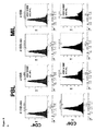
さらに良好な増幅およびさらに高い腫瘍特異性を得ることに加えて、T細胞の生存を最大にするために増殖条件を最適化した。4−1BBの発現が、多くのこれらの特性の重要なレギュレーターであることが示された。これは、T細胞増幅を制御し、アポトーシスを低減し、CD8細胞の細胞傷害活性を増大し、生存を向上させることができる。まとめると、活性化MIL上の4−1BB発現は、生存および腫瘍特異性を高める重要なレギュレーターである可能性がある。したがって、MIL上の4−1BB発現を調べ、さまざまな条件において増殖させたPBLのものと比較した。図8に示したとおり、ベースラインの4−1BBの発現は、MILでPBLでよりも高かった(18.2%対8.1%)。興味深いことに、正常酸素におけるT細胞増幅は、両集団でその発現を低下させた(MIL 10.7%、PBL 2.8%)一方、低酸素における増幅は、4−1BB発現をMILで著しく増加させ(43.4%)、PBLでその発現を完全に阻害した(0%)。これらのデータは、PBLとMILの間の顕著な差をさらにまた強調し、4−1BBのアップレギュレーションが、単に低酸素増殖条件だけでなくさらなる要因に依存することをさらに示す。 In addition to obtaining better amplification and higher tumor specificity, growth conditions were optimized to maximize T cell survival. Expression of 4-1BB has been shown to be an important regulator of many of these properties. It can control T cell amplification, reduce apoptosis, increase cytotoxic activity of CD8 cells, and improve survival. In summary, 4-1BB expression on activated MIL may be an important regulator of enhanced survival and tumor specificity. Therefore, 4-1BB expression on MIL was examined and compared to that of PBL grown under various conditions. As shown in FIG. 8, baseline 4-1BB expression was higher in MIL than in PBL (18.2% vs. 8.1%). Interestingly, T cell amplification in normal oxygen reduced its expression in both populations (MIL 10.7%, PBL 2.8%), while amplification in hypoxia significantly reduced 4-1BB expression in MIL. It was increased (43.4%) and completely inhibited its expression in PBL (0%). These data further highlight the significant differences between PBL and MIL, further indicating that the upregulation of 4-1BB depends not only on hypoxic growth conditions but also on additional factors.
これらの培養条件は臨床試験に採用され、低酸素増殖条件は、全体のT細胞増幅を平均7.9E9から1.8E10に増加させた。さらに、低酸素増殖条件はまた、in vivoにおけるT細胞増幅も観察することができた(図6は、すべての患者に関する60日目までの総リンパ球数を示す)。
These culture conditions were adopted in clinical trials, and hypoxic growth conditions increased overall T cell amplification from an average of 7.9E9 to 1.8E10. In addition, hypoxic growth conditions could also be observed for T cell amplification in vivo (Fig. 6 shows total lymphocyte counts by
一部の態様では、本発明は、骨髄浸潤リンパ球(「MIL」)を含む組成物に関する。MILは、活性化MILであってもよい。 In some aspects, the invention relates to a composition comprising bone marrow infiltrating lymphocytes (“MIL”). The MIL may be an activated MIL.
好適な実施形態では、組成物は、CD3を発現するMILの集団を含み、すなわち、CD3を発現するMILの集団の各細胞が、例えば、フローサイトメトリーによって検出される場合に、CD3を発現する骨髄浸潤リンパ球である。例えば、組成物中の細胞の少なくとも約40%、例えば、組成物中の細胞の少なくとも約45%、50%、55%、60%、65%、70%、75%、80%、85%、86%、87%、88%またはさらに少なくとも約89%は、CD3を発現するMILの集団由来のMILであってもよい。好適な一実施形態では、組成物中の細胞の少なくとも約80%は、CD3を発現するMILの集団由来のMILであってもよい。一部の実施形態では、組成物中の細胞の約40%から約100%、例えば、組成物中の細胞の約45%から約100%、約50%から約100%、約55%から約100%、約60%から約100%、約65%から約100%、約70%から約100%、約75%から約100%、約80%から約100%、約85%から約100%、約86%から約100%、約87%から約100%、約88%から約100%またはさらに約89%から約100%は、CD3を発現するMILの集団由来のMILであってもよい。一部の実施形態では、組成物は、例えば、フローサイトメトリーによって検出される場合に、CD3を発現しないMILの集団または低レベル、すなわち、CD3を発現するMILの集団由来のMILの発現レベルと比較して低レベルのCD3を発現するMILの集団のいずれかを含む。 In a preferred embodiment, the composition comprises a population of MIL expressing CD3, i.e., expressing CD3 when each cell of the population of MIL expressing CD3 is detected, for example, by flow cytometry. Bone marrow infiltrating lymphocytes. For example, at least about 40% of the cells in the composition, eg, at least about 45%, 50%, 55%, 60%, 65%, 70%, 75%, 80%, 85% of the cells in the composition. 86%, 87%, 88% or even at least about 89% may be MILs from a population of MILs expressing CD3. In one preferred embodiment, at least about 80% of the cells in the composition may be MILs from a population of MILs expressing CD3. In some embodiments, about 40% to about 100% of the cells in the composition, eg, about 45% to about 100%, about 50% to about 100%, about 55% to about 55% of the cells in the composition. 100%, about 60% to about 100%, about 65% to about 100%, about 70% to about 100%, about 75% to about 100%, about 80% to about 100%, about 85% to about 100% , About 86% to about 100%, about 87% to about 100%, about 88% to about 100%, or even about 89% to about 100% may be MILs from a population of MILs expressing CD3. .. In some embodiments, the composition is, for example, with a population or low level of MIL that does not express CD3, ie, the expression level of MIL from a population of MILs that express CD3, when detected by flow cytometry. Includes any of the populations of MIL that express relatively low levels of CD3.
一部の実施形態では、組成物は、インターフェロンガンマ(「IFNγ」)を発現するMILの集団を含み、すなわち、IFNγを発現するMILの集団の各細胞が、例えば、フローサイトメトリーによって検出される場合に、IFNγを発現する骨髄浸潤リンパ球である。例えば、組成物中の細胞の少なくとも約2%、例えば、組成物中の細胞の少なくとも約2%、3%、4%、5%、6%、7%、8%、9%、10%、11%、12%、13%、14%、15%、16%、17%またはさらに少なくとも約18%は、IFNγを発現するMILの集団由来のMILであってもよい。一部の実施形態では、組成物中の細胞の約2%から約100%、例えば、組成物中の細胞の約2%から約100%、約3%から約100%、約4%から約100%、約5%から約100%、約6%から約100%、約7%から約100%、約8%から約100%、約9%から約100%、約10%から約100%、約11%から約100%、約12%から約100%、約13%から約100%、約14%から約100%、約15%から約100%、約16%から約100%、約17%から約100%またはさらに約18%から約100%は、IFNγを発現するMILの集団由来のMILであってもよい。一部の実施形態では、組成物は、例えば、フローサイトメトリーによって検出される場合に、IFNγを発現しないMILの集団または低レベル、すなわち、IFNγを発現するMILの集団由来のMILの発現レベルと比較して低レベルのIFNγを発現するMILの集団のいずれかを含む。 In some embodiments, the composition comprises a population of MIL expressing interferon gamma (“IFNγ”), i.e., each cell of the population of MIL expressing IFNγ is detected, for example, by flow cytometry. In some cases, bone marrow infiltrating lymphocytes expressing IFNγ. For example, at least about 2% of the cells in the composition, eg, at least about 2%, 3%, 4%, 5%, 6%, 7%, 8%, 9%, 10% of the cells in the composition. 11%, 12%, 13%, 14%, 15%, 16%, 17% or even at least about 18% may be MILs from a population of MILs expressing IFNγ. In some embodiments, about 2% to about 100% of the cells in the composition, eg, about 2% to about 100%, about 3% to about 100%, about 4% to about 4% of the cells in the composition. 100%, about 5% to about 100%, about 6% to about 100%, about 7% to about 100%, about 8% to about 100%, about 9% to about 100%, about 10% to about 100% , About 11% to about 100%, about 12% to about 100%, about 13% to about 100%, about 14% to about 100%, about 15% to about 100%, about 16% to about 100%, about 17% to about 100% or even about 18% to about 100% may be MILs from a population of MILs expressing IFNγ. In some embodiments, the composition is expressed, for example, with a population or low level of MIL that does not express IFNγ, ie, a population of MIL that expresses IFNγ, when detected by flow cytometry. Includes any of the populations of MIL that express relatively low levels of IFNγ.
一部の実施形態では、組成物は、CXCR4を発現するMILの集団を含み、すなわち、CXCR4を発現するMILの集団の各細胞が、例えば、フローサイトメトリーによって検出される場合に、CXCR4を発現する骨髄浸潤リンパ球である。例えば、組成物中の細胞の少なくとも約98%、例えば、組成物中の細胞の少なくとも約98.1%、98.2%、98.3%、98.4%、98.5%、98.6%、98.7%、98.8%、98.9%、99.0%、99.1%、99.2%、99.3%、99.4%、99.5%、99.6%またはさらに少なくとも約99.7%は、CXCR4を発現するMILの集団由来のMILであってもよい。一部の実施形態では、組成物中の細胞の約98%から約100%、例えば、組成物中の細胞の少なくとも約98.1%から約100%、約98.2%から約100%、約98.3%から約100%、約98.4%から約100%、約98.5%から約100%、約98.6%から約100%、約98.7%から約100%、約98.8%から約100%、約98.9%から約100%、約99.0%から約100%、約99.1%から約100%、約99.2%から約100%、約99.3%から約100%、約99.4%から約100%、約99.5%から約100%、約99.6%から約100%またはさらに約99.7%から約100%は、CXCR4を発現するMILの集団由来のMILであってもよい。一部の実施形態では、組成物は、例えば、フローサイトメトリーによって検出される場合に、CXCR4を発現しないMILの集団または低レベル、すなわち、CXCR4を発現するMILの集団由来のMILの発現レベルと比較して低レベルのCXCR4を発現するMILの集団のいずれかを含む。 In some embodiments, the composition comprises a population of MIL expressing CXCR4, i.e., expressing CXCR4 when each cell of the population of MIL expressing CXCR4 is detected, for example, by flow cytometry. Bone marrow infiltrating lymphocytes. For example, at least about 98% of the cells in the composition, eg, at least about 98.1%, 98.2%, 98.3%, 98.4%, 98.5%, 98. 6%, 98.7%, 98.8%, 98.9%, 99.0%, 99.1%, 99.2%, 99.3%, 99.4%, 99.5%, 99.4%. 6% or even at least about 99.7% may be MILs from a population of MILs expressing CXCR4. In some embodiments, from about 98% to about 100% of the cells in the composition, for example, at least about 98.1% to about 100%, about 98.2% to about 100% of the cells in the composition. About 98.3% to about 100%, about 98.4% to about 100%, about 98.5% to about 100%, about 98.6% to about 100%, about 98.7% to about 100%, About 98.8% to about 100%, about 98.9% to about 100%, about 99.0% to about 100%, about 99.1% to about 100%, about 99.2% to about 100%, About 99.3% to about 100%, about 99.4% to about 100%, about 99.5% to about 100%, about 99.6% to about 100%, or even about 99.7% to about 100% May be a MIL derived from a population of MILs expressing CXCR4. In some embodiments, the composition is expressed, for example, with a population or low level of MIL that does not express CXCR4, i.e., a population of MIL that expresses CXCR4, when detected by flow cytometry. Includes any of the populations of MIL that express relatively low levels of CXCR4.
一部の実施形態では、組成物は、CD4を発現するMILの集団を含む。CD4を発現するMILの集団は、CXCR4を発現する複数のMILを含んでもよい。 In some embodiments, the composition comprises a population of MIL expressing CD4. The population of MILs expressing CD4 may include multiple MILs expressing CXCR4.
CD4を発現するMILの集団は、4−1BBを発現する複数のMILを含んでもよい。例えば、組成物中の細胞の少なくとも約21%、例えば、組成物中の細胞の少なくとも約22%、23%、24%、25%、26%、27%、28%、29%、30%、31%、32%、33%、34%、35%、36%、37%、38%、39%、40%、41%、42%またはさらに少なくとも約43%は、4−1BBを発現する複数のMIL由来のMILであってもよい。一部の実施形態では、組成物中の細胞の約21%から約100%、例えば、組成物中の細胞の約22%から約100%、約23%から約100%、約24%から約100%、約25%から約100%、約26%から約100%、約27%から約100%、約28%から約100%、約29%から約100%、約30%から約100%、約31%から約100%、約32%から約100%、約33%から約100%、約34%から約100%、約35%から約100%、約36%から約100%、約37%から約100%、約38%から約100%、約39%から約100%、約40%から約100%、約41%から約100%、約42%から約100%またはさらに約43%から約100%は、4−1BBを発現する複数のMIL由来のMILであってもよい。 A population of MILs expressing CD4 may include multiple MILs expressing 4-1BB. For example, at least about 21% of the cells in the composition, eg, at least about 22%, 23%, 24%, 25%, 26%, 27%, 28%, 29%, 30% of the cells in the composition. 31%, 32%, 33%, 34%, 35%, 36%, 37%, 38%, 39%, 40%, 41%, 42% or even at least about 43% express 4-1BB. It may be a MIL derived from MIL. In some embodiments, about 21% to about 100% of the cells in the composition, eg, about 22% to about 100%, about 23% to about 100%, about 24% to about 24% of the cells in the composition. 100%, about 25% to about 100%, about 26% to about 100%, about 27% to about 100%, about 28% to about 100%, about 29% to about 100%, about 30% to about 100% , About 31% to about 100%, about 32% to about 100%, about 33% to about 100%, about 34% to about 100%, about 35% to about 100%, about 36% to about 100%, about 37% to about 100%, about 38% to about 100%, about 39% to about 100%, about 40% to about 100%, about 41% to about 100%, about 42% to about 100%, or even about 43 % To about 100% may be MILs derived from multiple MILs expressing 4-1BB.
組成物は、CD8を発現するMILの集団を含んでもよい。CD8を発現するMILの集団は、CXCR4を発現する複数のMILを含んでもよい。 The composition may include a population of MILs expressing CD8. The population of MILs expressing CD8 may include multiple MILs expressing CXCR4.
CD8を発現するMILの集団は、4−1BBを発現する複数のMILを含んでもよい。例えば、組成物中の細胞の少なくとも約21%、例えば、組成物中の細胞の少なくとも約8%、9%、10%、11%、12%、13%、14%、15%、16%、17%、18%、19%、20%またはさらに少なくとも約21%は、4−1BBを発現する複数のMIL由来のMILであってもよい。一部の実施形態では、組成物中の細胞の約2%から約100%、例えば、組成物中の細胞の約8%から約100%、約9%から約100%、約10%から約100%、約11%から約100%、約12%から約100%、約13%から約100%、約14%から約100%、約15%から約100%、約16%から約100%、約17%から約100%、約18%から約100%、約19%から約100%、約20%から約100%またはさらに約21%から約100%は、4−1BBを発現する複数のMIL由来のMILであってもよい。 A population of MILs expressing CD8 may include multiple MILs expressing 4-1BB. For example, at least about 21% of the cells in the composition, eg, at least about 8%, 9%, 10%, 11%, 12%, 13%, 14%, 15%, 16% of the cells in the composition. 17%, 18%, 19%, 20% or even at least about 21% may be MILs from multiple MILs expressing 4-1BB. In some embodiments, about 2% to about 100% of the cells in the composition, eg, about 8% to about 100%, about 9% to about 100%, about 10% to about 10% of the cells in the composition. 100%, about 11% to about 100%, about 12% to about 100%, about 13% to about 100%, about 14% to about 100%, about 15% to about 100%, about 16% to about 100% , About 17% to about 100%, about 18% to about 100%, about 19% to about 100%, about 20% to about 100%, or even about 21% to about 100%, which express 4-1BB. It may be a MIL derived from MIL.
一部の実施形態では、組成物は、4−1BBを発現するMILの集団を含む。例えば、組成物中の細胞の少なくとも約21%、例えば、組成物中の細胞の少なくとも約22%、23%、24%、25%、26%、27%、28%、29%、30%、31%、32%、33%、34%、35%、36%、37%、38%、39%、40%、41%、42%またはさらに少なくとも約43%は、4−1BBを発現するMILの集団由来のMILであってもよい。一部の実施形態では、組成物中の細胞の約21%から100%、例えば、組成物中の細胞の約22%から約100%、約23%から約100%、約24%から約100%、約25%から約100%、約26%から約100%、約27%から約100%、約28%から約100%、約29%から約100%、約30%から約100%、約31%から約100%、約32%から約100%、約33%から約100%、約34%から約100%、約35%から約100%、約36%から約100%、約37%から約100%、約38%から約100%、約39%から約100%、約40%から約100%、約41%から約100%、約42%から約100%またはさらに約43%から約100%は、4−1BBを発現するMILの集団由来のMILであってもよい。一部の実施形態では、組成物は、例えば、フローサイトメトリーによって検出される場合に、4−1BBを発現しないMILの集団または低レベル、すなわち、4−1BBを発現するMILの集団由来のMILの発現レベルと比較して低レベルの4−1BBを発現するMILの集団のいずれかを含む。 In some embodiments, the composition comprises a population of MIL expressing 4-1BB. For example, at least about 21% of the cells in the composition, eg, at least about 22%, 23%, 24%, 25%, 26%, 27%, 28%, 29%, 30% of the cells in the composition. 31%, 32%, 33%, 34%, 35%, 36%, 37%, 38%, 39%, 40%, 41%, 42% or even at least about 43% are MILs expressing 4-1BB. It may be a MIL derived from the population of. In some embodiments, about 21% to 100% of the cells in the composition, eg, about 22% to about 100%, about 23% to about 100%, about 24% to about 100 of the cells in the composition. %, About 25% to about 100%, about 26% to about 100%, about 27% to about 100%, about 28% to about 100%, about 29% to about 100%, about 30% to about 100%, About 31% to about 100%, about 32% to about 100%, about 33% to about 100%, about 34% to about 100%, about 35% to about 100%, about 36% to about 100%, about 37 % To about 100%, about 38% to about 100%, about 39% to about 100%, about 40% to about 100%, about 41% to about 100%, about 42% to about 100%, or even about 43% From about 100% may be MIL from a population of MIL expressing 4-1BB. In some embodiments, the composition is a MIL derived from, for example, a population of MILs that do not express 4-1BB or a low level, i.e., a population of MILs that express 4-1BB, when detected by flow cytometry. Includes any of the populations of MIL that express low levels of 4-1BB compared to their expression levels.
一部の態様では、本発明は、被験体のがんを予防または処置するための方法であって、被験体に本明細書に記載される組成物のいずれか1つを投与することを含む方法に関する。好適な実施形態では、本方法は、被験体に治療有効量の本明細書に記載される組成物のいずれか1つを投与することを含む。好適な実施形態では、本方法は、被験体に治療有効量のMIL、例えば、本明細書に記載される活性化MILを投与することを含む。被験体は、新生物、例えば、がんを有し得る。例えば、被験体は、多発性骨髄腫を有し得る。被験体は、ヒト被験体であってもよい。 In some aspects, the invention is a method for preventing or treating a subject's cancer, comprising administering to the subject any one of the compositions described herein. Regarding the method. In a preferred embodiment, the method comprises administering to the subject a therapeutically effective amount of any one of the compositions described herein. In a preferred embodiment, the method comprises administering to the subject a therapeutically effective amount of MIL, eg, an activated MIL as described herein. The subject may have a neoplasm, such as cancer. For example, a subject may have multiple myeloma. The subject may be a human subject.
一部の態様では、本発明は、本明細書に記載される組成物を生成するための方法であって、低酸素環境においてMILをインキュベートすることを含む方法に関する。一部の態様では、本発明は、骨髄浸潤リンパ球(「MIL」)を活性化するための方法であって、低酸素環境においてMILをインキュベートすることを含む方法に関する。 In some aspects, the invention relates to a method for producing the compositions described herein, comprising incubating the MIL in a hypoxic environment. In some aspects, the invention relates to a method for activating bone marrow infiltrating lymphocytes (“MIL”), comprising incubating the MIL in a hypoxic environment.
一部の態様では、本発明は、被験体のがんを処置するための方法に関する。本方法は、被験体から骨髄浸潤リンパ球(「MIL」)を取り出すこと、低酸素環境においてMILをインキュベートすることにより、活性化MILを生じさせることおよび活性化MILを被験体に投与することを含んでもよい。 In some aspects, the invention relates to a method for treating a subject's cancer. The method involves removing bone marrow infiltrating lymphocytes (“MIL”) from a subject, incubating the MIL in a hypoxic environment to generate activated MIL, and administering the activated MIL to the subject. It may be included.
低酸素環境は、約21%未満の酸素、例えば、約20%、19%、18%、17%、16%、15%、14%、13%、12%、11%、10%、9%、8%、7%、6%、5%、4%未満または約3%未満の酸素を含んでもよい。例えば、低酸素環境は、約0%の酸素から約20%の酸素、例えば、約0%の酸素から約19%の酸素、約0%の酸素から約18%の酸素、約0%の酸素から約17%の酸素、約0%の酸素から約16%の酸素、約0%の酸素から約15%の酸素、約0%の酸素から約14%の酸素、約0%の酸素から約13%の酸素、約0%の酸素から約12%の酸素、約0%の酸素から約11%の酸素、約0%の酸素から約10%の酸素、約0%の酸素から約9%の酸素、約0%の酸素から約8%の酸素、約0%の酸素から約7%の酸素、約0%の酸素から約6%の酸素、約0%の酸素から約5%の酸素、約0%の酸素から約4%の酸素または約0%の酸素から約3%の酸素を含んでもよい。好適な実施形態では、低酸素環境は、約1%から約7%の酸素を含む。低酸素環境は、約20%、19%、18%、17%、16%、15%、14%、13%、12%、11%、10%、9%、8%、7%、6%、5%、4%、3%、2%、1%または約0%の酸素を含んでもよい。好適な実施形態では、低酸素環境は、約7%、6%、5%、4%、3%、2%または1%の酸素を含む。 A hypoxic environment is less than about 21% oxygen, such as about 20%, 19%, 18%, 17%, 16%, 15%, 14%, 13%, 12%, 11%, 10%, 9%. , 8%, 7%, 6%, 5%, less than 4% or less than about 3% oxygen. For example, a low oxygen environment can be from about 0% oxygen to about 20% oxygen, such as about 0% oxygen to about 19% oxygen, about 0% oxygen to about 18% oxygen, about 0% oxygen. From about 17% oxygen, from about 0% oxygen to about 16% oxygen, from about 0% oxygen to about 15% oxygen, from about 0% oxygen to about 14% oxygen, from about 0% oxygen to about 13% oxygen, about 0% oxygen to about 12% oxygen, about 0% oxygen to about 11% oxygen, about 0% oxygen to about 10% oxygen, about 0% oxygen to about 9% Oxygen, about 0% oxygen to about 8% oxygen, about 0% oxygen to about 7% oxygen, about 0% oxygen to about 6% oxygen, about 0% oxygen to about 5% oxygen , It may contain from about 0% oxygen to about 4% oxygen or from about 0% oxygen to about 3% oxygen. In a preferred embodiment, the hypoxic environment comprises from about 1% to about 7% oxygen. Hypoxic environment is about 20%, 19%, 18%, 17%, 16%, 15%, 14%, 13%, 12%, 11%, 10%, 9%, 8%, 7%, 6%. It may contain 5%, 4%, 3%, 2%, 1% or about 0% oxygen. In a preferred embodiment, the hypoxic environment comprises about 7%, 6%, 5%, 4%, 3%, 2% or 1% oxygen.
低酸素環境においてMILをインキュベートすることは、例えば、組織培養培地において少なくとも約1時間、例えば、少なくとも約12時間、18時間、24時間、30時間、36時間、42時間、48時間、60時間、3日間、4日間、5日間、6日間、7日間、8日間、9日間、10日間、11日間、12日間、13日間またはさらに少なくとも約14日間、MILをインキュベートすることを含んでもよい。インキュベートすることは、約1時間から約30日間、例えば、約1日間から約20日間、約1日間から約14日間または約1日間から約12日間、MILをインキュベートすることを含んでもよい。一部の好適な実施形態では、低酸素環境においてMILをインキュベートすることは、低酸素環境においてMILを約2日間から約5日間インキュベートすることを含む。本方法は、低酸素環境においてMILを約1日間、2日間、3日間、4日間、5日間、6日間、7日間、8日間、9日間、10日間、11日間、12日間、13日間または14日間インキュベートすることを含んでもよい。一部の好適な実施形態では、本方法は、低酸素環境においてMILを約3日間インキュベートすることを含む。 Incubating MIL in a hypoxic environment is, for example, in tissue culture medium for at least about 1 hour, eg, at least about 12 hours, 18 hours, 24 hours, 30 hours, 36 hours, 42 hours, 48 hours, 60 hours, Incubating the MIL for 3 days, 4 days, 5 days, 6 days, 7 days, 8 days, 9 days, 10 days, 11 days, 12 days, 13 days or at least about 14 days may be included. Incubating may include incubating the MIL for about 1 hour to about 30 days, eg, about 1 to about 20 days, about 1 to about 14 days, or about 1 to about 12 days. In some preferred embodiments, incubating the MIL in a hypoxic environment comprises incubating the MIL in a hypoxic environment for about 2 to about 5 days. This method uses MIL for about 1 day, 2 days, 3 days, 4 days, 5 days, 6 days, 7 days, 8 days, 9 days, 10 days, 11 days, 12 days, 13 days or Incubation for 14 days may be included. In some preferred embodiments, the method comprises incubating the MIL in a hypoxic environment for about 3 days.
好適な実施形態では、本方法は、例えば、低酸素環境においてMILをインキュベートした後に正常酸素環境においてMILをインキュベートすることをさらに含む。 In a preferred embodiment, the method further comprises incubating the MIL in a hypoxic environment followed by incubating the MIL in a normal oxygen environment, for example.
正常酸素環境は、少なくとも約21%の酸素を含んでもよい。正常酸素環境は、約5%の酸素から約30%の酸素、例えば、約10%の酸素から約30%の酸素、約15%の酸素から約25%の酸素、約18%の酸素から約24%の酸素、約19%の酸素から約23%の酸素または約20%の酸素から約22%の酸素を含んでもよい。一部の実施形態では、正常酸素環境は約21%の酸素を含む。 The normal oxygen environment may contain at least about 21% oxygen. The normal oxygen environment is from about 5% oxygen to about 30% oxygen, for example from about 10% oxygen to about 30% oxygen, from about 15% oxygen to about 25% oxygen, from about 18% oxygen to about. It may contain 24% oxygen, about 19% oxygen to about 23% oxygen or about 20% oxygen to about 22% oxygen. In some embodiments, the normal oxygen environment contains about 21% oxygen.
正常酸素環境においてMILをインキュベートすることは、例えば、組織培養培地において少なくとも約1時間、例えば、少なくとも約12時間、18時間、24時間、30時間、36時間、42時間、48時間、60時間、3日間、4日間、5日間、6日間、7日間、8日間、9日間、10日間、11日間、12日間、13日間またはさらに少なくとも約14日間、MILをインキュベートすることを含んでもよい。インキュベートすることは、約1時間から約30日間、例えば、約1日間から約20日間、約1日間から約14日間、約1日間から約12日間または約2日間から約12日間、MILをインキュベートすることを含んでもよい。 Incubating MIL in a normal oxygen environment is, for example, in tissue culture medium for at least about 1 hour, eg, at least about 12 hours, 18 hours, 24 hours, 30 hours, 36 hours, 42 hours, 48 hours, 60 hours, Incubating the MIL for 3 days, 4 days, 5 days, 6 days, 7 days, 8 days, 9 days, 10 days, 11 days, 12 days, 13 days or at least about 14 days may be included. Incubating MIL is about 1 hour to about 30 days, eg, about 1 to about 20 days, about 1 to about 14 days, about 1 to about 12 days, or about 2 to about 12 days. May include doing.
(実施例1.低酸素環境および正常酸素環境におけるT細胞の活性化および増幅)
フローサイトメトリーを使用して骨髄(BM)T細胞数を求める。抗CD3/抗CD28ビーズを所定の比率(ビーズ:CD3細胞)で所定の濃度の組換え型ヒトサイトカインを含む培地に添加する。細胞をプレート、フラスコまたはバッグに播く。低酸素チャンバーまたは細胞培養バッグのいずれかに95%窒素および5%CO2ガス混合物を3分間フラッシュすることによって低酸素条件を達成する。その後、容器をこのガス混合物で30秒間満たす。これにより容器中が2%またはそれ未満のO2ガスに達する。細胞を37Cで3日間またはそれ超にわたり培養し、低酸素空気を放出し、正常酸素(21%大気酸素)レベルに替える。
(Example 1. Activation and amplification of T cells in a hypoxic environment and a normal oxygen environment)
Bone marrow (BM) T cell count is determined using flow cytometry. Anti-CD3 / anti-CD28 beads are added to a medium containing a predetermined concentration of recombinant human cytokines in a predetermined ratio (beads: CD3 cells). Seed the cells in a plate, flask or bag. Hypoxic conditions are achieved by flushing the 95% nitrogen and 5% CO 2 gas mixture into either the hypoxic chamber or the cell culture bag for 3 minutes. The container is then filled with this gas mixture for 30 seconds. This reaches 2% or less of O 2 gas in the container. Cells are cultured in 37C for 3 days or more to release hypoxic air and replace with normal oxygen (21% atmospheric oxygen) levels.
(実施例2.細胞型の表現型決定)
所望の決定のために蛍光色素コンジュゲート抗体で細胞を染色する。蛍光色素に直接コンジュゲートしたCD3、CD4、CD8、CXCR4、41BB、CD27、CD28、CTLA−4、PD−1、CD45RO、CD62L、CD95、IFNg、IL17、生/死色素(live/dead dye)および/または目的の他の抗体を適切なアイソタイプ対照とともに使用する。簡単にいえば、プレートまたはチューブ中の1×106またはそれ未満の細胞をFACSバッファー(1×HBSS/2%FBS/0.5%EDTA/0.5%NaAzide)または類似の洗浄バッファーにより遠心分離機において回転させることによって洗浄する。洗浄バッファーを除去し、抗体およびアイソタイプ対照を所定の濃度で添加する。細胞を7〜30分の間、室温または4℃で染色する。細胞を2回洗浄バッファーで洗浄し、最小限の洗浄バッファーに再懸濁する。その後、細胞を利用されている蛍光色素に対して適切に補償および準備されたフローサイトメーターに流す。各サンプルに対して10,000またはそれ超の数の事象を収集する。FACS解析ソフトウェアを利用してデータを解析する。バックグラウンド除去のために蛍光色素標識細胞をアイソタイプ対照と比較する。データを%陽性−%バックグラウンドとしてグラフ化する。
(Example 2. Phenotyping of cell type)
Cells are stained with fluorescent dye conjugate antibody for the desired determination. CD3, CD4, CD8, CXCR4, 41BB, CD27, CD28, CTLA-4, PD-1, CD45RO, CD62L, CD95, IFNg, IL17, live / dead dye and / Or use the other antibody of interest with the appropriate isotype control. Briefly, centrifuged 1 × 10 6 or less cells in the plate or tube by FACS buffer (1 × HBSS / 2% FBS / 0.5% EDTA / 0.5% NaAzide) or similar washing buffer Wash by rotating in a separator. The wash buffer is removed and the antibody and isotype control are added at a given concentration. Cells are stained at room temperature or 4 ° C. for 7-30 minutes. The cells are washed twice with wash buffer and resuspended in minimal wash buffer. The cells are then flowed through a flow cytometer appropriately compensated and prepared for the fluorescent dye being utilized. Collect 10,000 or more events for each sample. Analyze the data using FACS analysis software. Fluorescent dye-labeled cells are compared to isotype controls for background removal. Graph the data as a% positive-% background.
(実施例3.増幅倍率(fold expansion)の決定)
増幅の開始時に骨髄細胞を数える。フローサイトメトリーを利用してCD3+細胞のパーセンテージを求める。CD3+MILの総数を、細胞の総数にCD3のパーセンテージ=培養物中のCD3+MILの総数を掛けることによって求める。培養の最終日に細胞を回収し、計数する(手作業および自動セルカウンターの両方)。CD3+のパーセンテージを求める。培養の最終日のCD3+細胞の総数を、総細胞数にCD+のパーセンテージ=回収されたCD3+MILの総数を掛けることによって求める。総増幅倍率=培養の最終日に回収されたCD3+MILの総数÷培養の初日のCD3+MILの総数。
(Example 3. Determining the amplification factor (fold expansion))
Bone marrow cells are counted at the onset of amplification. Use flow cytometry to determine the percentage of CD3 + cells. The total number of CD3 + MIL is calculated by multiplying the total number of cells by the percentage of CD3 = the total number of CD3 + MIL in the culture. Cells are harvested and counted on the last day of culture (both manual and automatic cell counters). Find the percentage of CD3 +. The total number of CD3 + cells on the last day of culture is determined by multiplying the total number of cells by the percentage of CD + = the total number of recovered CD3 + MILs. Total amplification factor = total number of CD3 + MIL collected on the last day of culture ÷ total number of CD3 + MIL on the first day of culture.
(実施例4.腫瘍特異性)
製造業者のプロトコールに従ってMILをCFSEまたは類似の細胞膜組み込み色素(cell membrane integration dye)で標識する。培地単独、陰性対照(関連のないタンパク質もしくはライセート)により、または目的のタンパク質もしくはライセートにより自家BMをパルスする。その後、CFSE標識細胞をパルスした自家BMとともに2〜7日間共培養する。組織培養プレートまたはフラスコから細胞を回収した後、細胞外をCD3によりおよび細胞内をIFNgにより染色する。CFSEが低く(分裂した細胞)、IFNgを産生しているCD3+細胞に対するゲーティングによって腫瘍特異性の解析を決定する。
(Example 4. Tumor specificity)
MIL is labeled with CFSE or a similar cell membrane integration dye according to the manufacturer's protocol. Pulse autologous BM with medium alone, with a negative control (unrelated protein or lysate), or with the protein or lysate of interest. The CFSE-labeled cells are then co-cultured with pulsed autologous BM for 2-7 days. After harvesting the cells from the tissue culture plate or flask, extracellular staining with CD3 and intracellular staining with IFNg. Tumor specificity analysis is determined by gating to CD3 + cells that have low CFSE (divided cells) and are producing IFNg.
(実施例5.低酸素環境および正常酸素環境におけるT細胞の活性化および増幅)
MILを2%O2(低酸素)において3日間増殖させ、続いて21%O2(正常酸素)に切り替えてIL−2の存在下または非存在下においてさらに5日間、増殖させた。図9に示されるとおり、低酸素に続いて正常酸素において増殖させると、正常酸素条件においてのみ増殖させたMILと比較して増幅がほぼ10倍増加した。図4に示されるとおり、腫瘍特異性も著しく向上した。10日目に、低酸素条件において増殖させたMILの25.1%とは対照的に正常酸素条件の低CFSE細胞の4%が腫瘍特異的であった。まとめると、これらのデータは、これらの増殖条件が活性化時に腫瘍特異的なものの絶対数を増加させることができることを示唆している。
(Example 5. Activation and amplification of T cells in a hypoxic environment and a normal oxygen environment)
MIL was grown in 2% O 2 (hypoxia) for 3 days, then switched to 21% O 2 (normal oxygen) and grown in the presence or absence of IL-2 for an additional 5 days. As shown in FIG. 9, when grown in normal oxygen following hypoxia, amplification was increased approximately 10-fold compared to MIL grown only under normal oxygen conditions. As shown in FIG. 4, the tumor specificity was also significantly improved. On
先行する段落の実験を小サンプルに対して行った。最初の臨床試験の患者からのMIL産物もこの方法を使用して増幅させた。図3に示されるとおり、これらの条件におけるMILの増幅は、劇的にT細胞数を増加させた。正常酸素条件における増殖は、7日を超えるとほとんどMILを増幅できなかった。この実験では、低酸素条件の119倍とは対照的に、7日目の増幅倍率は、正常酸素条件において36.3倍であった。さらに、細胞は、12日目まで増幅し続け、死に始める前の11日目に総計220倍の増幅に達した。
Experiments in the preceding paragraph were performed on small samples. MIL products from patients in the first clinical trial were also amplified using this method. As shown in FIG. 3, amplification of MIL under these conditions dramatically increased the number of T cells. Proliferation under normal oxygen conditions could hardly amplify MIL beyond 7 days. In this experiment, the amplification factor on day 7 was 36.3 times under normal oxygen conditions, as opposed to 119 times under hypoxic conditions. In addition, cells continued to amplify until
4−1BBの発現が、多くのこれらの特性の重要なレギュレーターであることが示された。これは、T細胞増幅を制御し、アポトーシスを低減し、CD8細胞の細胞傷害活性を増大し、生存を高めることができる。さらに、HIF1αは、抗原に駆動されたT細胞の生存を制御する。4−1BBを発現するベクターを用いたキメラ抗原受容体(CAR)改変T細胞は、in vivoにおける著しい増幅を示した。まとめると、活性化MIL上の4−1BB発現は、生存および腫瘍特異性を増加させる重要なレギュレーターである可能性がある。本明細書中で開示される方法の際だった利点の1つは、4−1BB発現を向上させるためにMILを改変する必要がないことである。この利点は、MILまたはPBLを正常酸素条件または低酸素条件のいずれかにおいて増殖させた図10に示されている。MILにおける4−1BBのベースライン発現を評価し、PBLと比較した。示されるとおり、4−1BB発現は、PBLよりもMILにおいて高かった(18.2%対8.1%)。興味深いことに、正常酸素におけるT細胞増幅は、両集団ともにその発現が低下した(MIL 10.7%、PBL 2.8%)一方で、低酸素における増幅は、4−1BB発現をMILで著しく増加させ(43.4%)、PBLでその発現を完全に阻害した(0%)。これらのデータは、PBLとMILの間の顕著な差をさらにまた強調し、4−1BBのアップレギュレーションが単に低酸素増殖条件だけでなくさらなる要因に依存することをさらに示す。さらに重要なことに、4−1BBの予想外のアップレギュレーションは、本発明の方法が、低酸素的に増殖させたMILを使用した患者の処置において顕著な差をもたらすことを示す。同様の結果がCD8細胞によっても観察された(図8)。 Expression of 4-1BB has been shown to be an important regulator of many of these properties. It can control T cell amplification, reduce apoptosis, increase cytotoxic activity of CD8 cells, and enhance survival. In addition, HIF1α regulates the survival of antigen-driven T cells. Chimeric antigen receptor (CAR) -modified T cells using a vector expressing 4-1BB showed significant amplification in vivo. Taken together, 4-1BB expression on activated MIL may be an important regulator of increased survival and tumor specificity. One of the outstanding advantages of the methods disclosed herein is that MIL does not need to be modified to improve 4-1BB expression. This advantage is shown in FIG. 10 where MIL or PBL was grown under either normal or hypoxic conditions. Baseline expression of 4-1BB in MIL was evaluated and compared to PBL. As shown, 4-1BB expression was higher in MIL than in PBL (18.2% vs. 8.1%). Interestingly, T cell amplification in normal oxygen reduced its expression in both populations (MIL 10.7%, PBL 2.8%), while amplification in hypoxia markedly increased 4-1BB expression in MIL. It was increased (43.4%) and completely inhibited its expression in PBL (0%). These data further highlight the significant differences between PBL and MIL, further indicating that the upregulation of 4-1BB depends not only on hypoxic growth conditions but also on additional factors. More importantly, the unexpected upregulation of 4-1BB indicates that the methods of the invention make a significant difference in the treatment of patients with hypoxic-grown MIL. Similar results were observed with CD8 cells (Fig. 8).
図11は、臨床処置のための投薬の結果を示す。J0770では、MILを静置培養で正常酸素条件において増殖させた。J0997では、MILを波動(WAVE)中、正常酸素条件において増殖させた。J1343では、MILを低酸素条件において3日間、増殖させ、続いて正常酸素条件において増殖させた。図6では、自家幹細胞移植後の3つの実験に関するリンパ球絶対数をグラフ化している。J1343は無作為試験であり、その患者は移植後に注入された低酸素MILを受容したか、またはMILを注入されなかったかのいずれかであった。 FIG. 11 shows the results of dosing for clinical treatment. In J0770, MIL was grown in static culture under normal oxygen conditions. In J0997, MIL was grown under normal oxygen conditions during wave motion (WAVE). In J1343, MIL was grown under hypoxic conditions for 3 days, followed by normal oxygen conditions. FIG. 6 graphs the absolute lymphocyte counts for the three experiments after autologous stem cell transplantation. J1343 was a randomized trial in which the patient either accepted the hypoxic MIL injected after transplantation or was not injected with MIL.
図11に示されるとおり、本方法の増殖条件は、全体のT細胞増幅を平均7.9E9から1.8E10に増加させた。さらに、これは図6に示されるとおり初めてin vivoにおけるT細胞増幅を示し、これは患者の処置における方法の有効性に直接関係する。グラフに第1のセットの患者に関する60日目までの総リンパ球数を示す。
As shown in FIG. 11, the growth conditions of the method increased overall T cell amplification from an average of 7.9E9 to 1.8E10. Moreover, it shows T cell amplification in vivo for the first time as shown in FIG. 6, which is directly related to the effectiveness of the method in treating patients. The graph shows the total lymphocyte count up to
均等物
当業者は、慣用的な実験を使用するだけで、本明細書に記載される本発明の特定の実施形態の多くの均等物を認識する、または確認することができるだろう。そのような均等物は、添付の特許請求の範囲に包含されることが意図される。
Equivalents One of ordinary skill in the art will be able to recognize or confirm many of the equivalents of the particular embodiments of the invention described herein by using conventional experiments. Such equivalents are intended to be included in the appended claims.
Claims (39)
低酸素条件で活性化された骨髄浸潤リンパ球の前記集団の60〜100%は、CD3を発現し、
前記集団の少なくとも21%は、4−1BBを発現し、前記方法が、
(a)低酸素環境において、骨髄試料を培養して、活性化された骨髄浸潤リンパ球を産生するステップ;および、
(b)正常酸素環境において、前記活性化された骨髄浸潤リンパ球を培養して、前記組成物を産生するステップ
を含む、方法。 A method for producing a composition containing a population of bone marrow infiltrating lymphocytes activated under hypoxic conditions and amplified under normal oxygen conditions in ex vivo.
60-100% of the population of bone marrow infiltrating lymphocytes activated under hypoxic conditions expresses CD3.
At least 21% of the population expressed 4-1BB, according to the method.
(A) The step of culturing a bone marrow sample in a hypoxic environment to produce activated bone marrow infiltrating lymphocytes; and
(B) A step of culturing the activated bone marrow infiltrating lymphocytes in a normal oxygen environment to produce the composition.
Including methods .
ステップ(b)において、前記活性化された骨髄浸潤リンパ球が、正常酸素環境において、IL−2の存在下で培養され、前記組成物を産生する、
請求項1〜5のいずれか一項に記載の方法。 In step (a), the bone marrow sample, in 1-3% of the hypoxic environment of oxygen, cultured with anti-CD3 and anti-CD28 antibodies, to produce an activated bone marrow-infiltrating lymphocytes; and,
In step (b), the activated bone marrow infiltrated lymphocytes in normal oxygen environment, it is cultured in the presence of IL-2, to produce said composition,
The method according to any one of claims 1 to 5.
(a)1%〜3%の酸素の低酸素環境において、がんを有する被験体から得られる骨髄試料を抗CD3抗体および抗CD28抗体と共に培養して、低酸素条件で活性化された骨髄浸潤リンパ球を産生するステップ;および、
(b)正常酸素環境において、IL−2の存在下で、前記低酸素条件で活性化された骨髄浸潤リンパ球を培養して、前記治療用活性化骨髄浸潤リンパ球を産生するステップ
を含む、方法。 A method for making a composition for treating a subject's cancer, which comprises therapeutically activated bone marrow infiltrating lymphocytes, wherein the method is described below.
(A) Bone marrow infiltration activated under hypoxic conditions by culturing bone marrow samples obtained from subjects with cancer with anti-CD3 antibody and anti-CD28 antibody in a hypoxic environment of 1% to 3% oxygen. Steps to produce lymphocytes; and
(B) The step of culturing the bone marrow infiltrating lymphocytes activated under the hypoxic conditions in the presence of IL-2 in a normal oxygen environment to produce the therapeutically activated bone marrow infiltrating lymphocytes . Method .
(a)1%〜3%の酸素の低酸素環境において、骨髄試料を抗CD3抗体および抗CD28抗体と共に培養して、低酸素条件で活性化された骨髄浸潤リンパ球を産生するステップ;および、
(b)正常酸素環境において、IL−2の存在下で、前記低酸素条件で活性化された骨髄浸潤リンパ球を培養して、前記治療用活性化骨髄浸潤リンパ球を産生するステップを含む、方法。 A method for preparing therapeutically activated bone marrow infiltrating lymphocytes.
(A) A step of culturing a bone marrow sample with an anti-CD3 antibody and an anti-CD28 antibody in a hypoxic environment of 1% to 3% oxygen to produce bone marrow infiltrating lymphocytes activated under hypoxic conditions;
(B) The step of culturing the bone marrow infiltrating lymphocytes activated under the hypoxic conditions in the presence of IL-2 in a normal oxygen environment to produce the therapeutically activated bone marrow infiltrating lymphocytes. Method.
(a)1%〜2%の酸素の低酸素環境において、がんを有する被験体から得られた骨髄試料を抗CD3/抗CD28ビーズと共に2〜5日間培養して、低酸素条件で活性化された骨髄浸潤リンパ球を産生するステップ;および、
(b)21%の酸素の正常酸素環境において、IL−2の存在下で、前記低酸素条件で活性化された骨髄浸潤リンパ球を2〜12日間培養して、前記治療用活性化骨髄浸潤リンパ球を産生するステップ
を含む、方法。 A method for preparing therapeutically activated bone marrow infiltrating lymphocytes.
(A) Bone marrow samples obtained from subjects with cancer in a hypoxic environment of 1% to 2% oxygen were cultured with anti-CD3 / anti-CD28 beads for 2-5 days and activated under hypoxic conditions. Steps to produce bone marrow infiltrating lymphocytes;
(B) The therapeutically activated bone marrow infiltration is performed by culturing bone marrow infiltrating lymphocytes activated under the hypoxic conditions for 2 to 12 days in the presence of IL-2 in a normal oxygen environment of 21% oxygen. A method comprising the step of producing lymphocytes.
(a)1%〜2%の酸素の低酸素環境において、がんを有する前記被験体から得られた骨髄試料を抗CD3/抗CD28ビーズと共に2〜5日間培養して、低酸素条件で活性化された骨髄浸潤リンパ球を産生するステップ;および、
(b)21%の酸素の正常酸素環境において、IL−2の存在下で、前記低酸素条件で活性化された骨髄浸潤リンパ球を2〜12日間培養して、前記治療用活性化骨髄浸潤リンパ球を産生するステップ
を含む、方法。 A method of making a composition for treating cancer in a subject, comprising therapeutically activated bone marrow infiltrating lymphocytes, wherein the method is described below:
(A) In a hypoxic environment of 1% to 2% oxygen, bone marrow samples obtained from the subject having cancer are cultured with anti-CD3 / anti-CD28 beads for 2 to 5 days and activated under hypoxic conditions. Steps to produce converted bone marrow infiltrating lymphocytes; and
(B) The therapeutically activated bone marrow infiltration is performed by culturing bone marrow infiltrating lymphocytes activated under the hypoxic conditions for 2 to 12 days in the presence of IL-2 in a normal oxygen environment of 21% oxygen. A method comprising the step of producing lymphocytes.
Priority Applications (1)
| Application Number | Priority Date | Filing Date | Title |
|---|---|---|---|
| JP2021063957A JP7197213B2 (en) | 2014-09-04 | 2021-04-05 | Activation of bone marrow-infiltrating lymphocytes in hypoxic conditions alternating with normoxic conditions |
Applications Claiming Priority (5)
| Application Number | Priority Date | Filing Date | Title |
|---|---|---|---|
| US201462045782P | 2014-09-04 | 2014-09-04 | |
| US62/045,782 | 2014-09-04 | ||
| US201562186040P | 2015-06-29 | 2015-06-29 | |
| US62/186,040 | 2015-06-29 | ||
| PCT/US2015/048536 WO2016037054A1 (en) | 2014-09-04 | 2015-09-04 | Activation of marrow infiltrating lymphocytes in hypoxic alternating with normoxic conditions |
Related Child Applications (1)
| Application Number | Title | Priority Date | Filing Date |
|---|---|---|---|
| JP2021063957A Division JP7197213B2 (en) | 2014-09-04 | 2021-04-05 | Activation of bone marrow-infiltrating lymphocytes in hypoxic conditions alternating with normoxic conditions |
Publications (3)
| Publication Number | Publication Date |
|---|---|
| JP2017527286A JP2017527286A (en) | 2017-09-21 |
| JP2017527286A5 JP2017527286A5 (en) | 2018-10-11 |
| JP6865160B2 true JP6865160B2 (en) | 2021-04-28 |
Family
ID=55440392
Family Applications (2)
| Application Number | Title | Priority Date | Filing Date |
|---|---|---|---|
| JP2017512293A Active JP6865160B2 (en) | 2014-09-04 | 2015-09-04 | Activation of bone marrow infiltrating lymphocytes in hypoxic conditions alternating with normal oxygen conditions |
| JP2021063957A Active JP7197213B2 (en) | 2014-09-04 | 2021-04-05 | Activation of bone marrow-infiltrating lymphocytes in hypoxic conditions alternating with normoxic conditions |
Family Applications After (1)
| Application Number | Title | Priority Date | Filing Date |
|---|---|---|---|
| JP2021063957A Active JP7197213B2 (en) | 2014-09-04 | 2021-04-05 | Activation of bone marrow-infiltrating lymphocytes in hypoxic conditions alternating with normoxic conditions |
Country Status (13)
| Country | Link |
|---|---|
| US (7) | US9687510B2 (en) |
| EP (2) | EP3922254A1 (en) |
| JP (2) | JP6865160B2 (en) |
| KR (1) | KR20170067751A (en) |
| CN (1) | CN107109364A (en) |
| AU (2) | AU2015311761B2 (en) |
| CA (1) | CA2959994C (en) |
| DK (1) | DK3188740T3 (en) |
| ES (1) | ES2877108T3 (en) |
| IL (2) | IL250746B (en) |
| MX (1) | MX2017002852A (en) |
| PT (1) | PT3188740T (en) |
| WO (1) | WO2016037054A1 (en) |
Families Citing this family (16)
| Publication number | Priority date | Publication date | Assignee | Title |
|---|---|---|---|---|
| WO2015157636A1 (en) | 2014-04-10 | 2015-10-15 | H. Lee Moffitt Cancer Center And Research Institute, Inc. | Enhanced expansion of tumor-infiltrating lymphocytes for adoptive cell therapy |
| JP6865160B2 (en) * | 2014-09-04 | 2021-04-28 | ザ・ジョンズ・ホプキンス・ユニバーシティー | Activation of bone marrow infiltrating lymphocytes in hypoxic conditions alternating with normal oxygen conditions |
| CA2991040A1 (en) | 2015-06-29 | 2017-01-05 | The Johns Hopkins University | Immune checkpoint chimeric receptors therapy |
| MA42902A (en) * | 2015-07-08 | 2018-05-16 | Univ Johns Hopkins | MARINAL-INFILTRATING LYMPHOCYTES (MIL) AS A SOURCE OF T-LYMPHOCYTES FOR CHEMERIC ANTIGEN RECEPTOR (CAR) THERAPY |
| TWI788307B (en) | 2016-10-31 | 2023-01-01 | 美商艾歐凡斯生物治療公司 | Engineered artificial antigen presenting cells for tumor infiltrating lymphocyte expansion |
| BR112019009925A2 (en) | 2016-11-17 | 2019-10-08 | Iovance Biotherapeutics Inc | method for preparing remnant tumor infiltrating lymphocytes for adoptive t-cell therapy, and method for treating a cancer. |
| US20190330306A1 (en) * | 2016-12-22 | 2019-10-31 | Windmil Therapeutics, Inc. | Compositions and Methods for Modulating the Immune System |
| EP3565586A1 (en) | 2017-01-06 | 2019-11-13 | Iovance Biotherapeutics, Inc. | Expansion of tumor infiltrating lymphocytes with potassium channel agonists and therapeutic uses thereof |
| US11254913B1 (en) | 2017-03-29 | 2022-02-22 | Iovance Biotherapeutics, Inc. | Processes for production of tumor infiltrating lymphocytes and uses of same in immunotherapy |
| US20210000876A1 (en) * | 2018-03-22 | 2021-01-07 | Windmil Therapeutics, Inc. | Prostate Cancer Specific Marrow Infiltrating Lymphocytes and Uses Thereof |
| KR20210088627A (en) * | 2018-11-05 | 2021-07-14 | 윈드밀 테라퓨틱스, 인크. | Bone marrow infiltrating lymphocytes with increased clonogenic capacity and uses thereof |
| MX2021006399A (en) * | 2018-11-30 | 2021-07-15 | Windmil Therapeutics Inc | MARROW INFILTRATING LYMPHOCYTES (MILs) EXPRESSING CHIMERIC ANTIGEN RECEPTORS (CAR), METHOD OF MANUFACTURING SAME, AND METHOD OF USING IN THERAPY. |
| CN114072158A (en) * | 2019-03-22 | 2022-02-18 | 温德弥尔治疗公司 | Lung cancer specific marrow infiltrating lymphocyte and its use |
| WO2021173948A1 (en) * | 2020-02-28 | 2021-09-02 | Windmil Therapeutics, Inc. | Methods for enriching marrow infiltrating lymphocytes ("mils"), compositions containing enriched mils, and methods of using enriched mils |
| WO2022066872A1 (en) * | 2020-09-24 | 2022-03-31 | Windmil Therapeutics, Inc. | Marrow infiltrating lymphocytes (mils) expressing t cell receptors, methods of manufacturing same, and method of using in therapy |
| WO2023201369A1 (en) | 2022-04-15 | 2023-10-19 | Iovance Biotherapeutics, Inc. | Til expansion processes using specific cytokine combinations and/or akti treatment |
Family Cites Families (12)
| Publication number | Priority date | Publication date | Assignee | Title |
|---|---|---|---|---|
| AU3243600A (en) | 1999-02-23 | 2000-09-14 | Kathleen S. Carswell | Improved method of culturing t cells |
| US6811788B2 (en) * | 2000-01-19 | 2004-11-02 | Baofa Yu | Combinations and methods for treating neoplasms |
| US20100178299A1 (en) * | 2007-02-13 | 2010-07-15 | Northeastern University | Methods and compositions for improving immune responses |
| EP2364152A4 (en) * | 2008-11-03 | 2013-02-13 | Univ Johns Hopkins | Methods for preparation and use of marrow infiltrating lymphocytes (mils) |
| EP2764114A4 (en) | 2011-10-03 | 2015-05-06 | Univ Los Andes | Method for monitoring, diagnosis and/or prognosis of hypoxia related disorders using nfat5 |
| US20140377240A1 (en) | 2012-01-17 | 2014-12-25 | Northeastern University | Methods and compositions for expanding immunosuppressive t regulatory cells in vitro and uses thereof |
| JP2014090678A (en) * | 2012-11-01 | 2014-05-19 | Ito En Ltd | Packed vegetable juice and/or fruit juice containing beverage, manufacturing method thereof, and method for preventing taste deterioration of packed vegetable juice and/or fruit juice containing beverage |
| PT2925329T (en) * | 2012-11-27 | 2021-03-29 | Ivan Marques Borrello | Use of post-transplant cyclophosphamide treated allogeneic marrow infiltrating lymphocytes to augment anti-tumor immunity |
| JP6865160B2 (en) | 2014-09-04 | 2021-04-28 | ザ・ジョンズ・ホプキンス・ユニバーシティー | Activation of bone marrow infiltrating lymphocytes in hypoxic conditions alternating with normal oxygen conditions |
| US20190330306A1 (en) * | 2016-12-22 | 2019-10-31 | Windmil Therapeutics, Inc. | Compositions and Methods for Modulating the Immune System |
| US20210000876A1 (en) * | 2018-03-22 | 2021-01-07 | Windmil Therapeutics, Inc. | Prostate Cancer Specific Marrow Infiltrating Lymphocytes and Uses Thereof |
| MX2021006399A (en) * | 2018-11-30 | 2021-07-15 | Windmil Therapeutics Inc | MARROW INFILTRATING LYMPHOCYTES (MILs) EXPRESSING CHIMERIC ANTIGEN RECEPTORS (CAR), METHOD OF MANUFACTURING SAME, AND METHOD OF USING IN THERAPY. |
-
2015
- 2015-09-04 JP JP2017512293A patent/JP6865160B2/en active Active
- 2015-09-04 EP EP21167891.7A patent/EP3922254A1/en not_active Withdrawn
- 2015-09-04 ES ES15838358T patent/ES2877108T3/en active Active
- 2015-09-04 CN CN201580059448.3A patent/CN107109364A/en active Pending
- 2015-09-04 US US15/112,353 patent/US9687510B2/en active Active
- 2015-09-04 KR KR1020177009084A patent/KR20170067751A/en active IP Right Grant
- 2015-09-04 EP EP15838358.8A patent/EP3188740B1/en active Active
- 2015-09-04 DK DK15838358.8T patent/DK3188740T3/en active
- 2015-09-04 PT PT158383588T patent/PT3188740T/en unknown
- 2015-09-04 CA CA2959994A patent/CA2959994C/en active Active
- 2015-09-04 MX MX2017002852A patent/MX2017002852A/en unknown
- 2015-09-04 AU AU2015311761A patent/AU2015311761B2/en active Active
- 2015-09-04 WO PCT/US2015/048536 patent/WO2016037054A1/en active Application Filing
-
2017
- 2017-02-23 IL IL250746A patent/IL250746B/en active IP Right Grant
- 2017-05-26 US US15/606,766 patent/US10406178B2/en active Active
- 2017-05-26 US US15/606,951 patent/US10172887B2/en active Active
-
2018
- 2018-12-03 US US16/207,864 patent/US11160830B2/en active Active
-
2019
- 2019-07-30 US US16/526,656 patent/US11406666B2/en active Active
-
2020
- 2020-07-28 IL IL276347A patent/IL276347B/en unknown
-
2021
- 2021-02-02 AU AU2021200648A patent/AU2021200648A1/en active Pending
- 2021-04-05 JP JP2021063957A patent/JP7197213B2/en active Active
- 2021-09-30 US US17/490,691 patent/US20220016172A1/en active Pending
-
2022
- 2022-07-14 US US17/864,546 patent/US20230000919A1/en active Pending
Also Published As
Similar Documents
| Publication | Publication Date | Title |
|---|---|---|
| JP6865160B2 (en) | Activation of bone marrow infiltrating lymphocytes in hypoxic conditions alternating with normal oxygen conditions | |
| Kim et al. | Distinct dendritic cell subsets dictate the fate decision between effector and memory CD8+ T cell differentiation by a CD24-dependent mechanism | |
| Childs et al. | Bringing natural killer cells to the clinic: ex vivo manipulation | |
| Davis et al. | Interleukin-7 permits Th1/Tc1 maturation and promotes ex vivo expansion of cord blood T cells: a critical step toward adoptive immunotherapy after cord blood transplantation | |
| Kang et al. | Anti-CD137 suppresses tumor growth by blocking reverse signaling by CD137 ligand | |
| US20030134415A1 (en) | Th1 cell adoptive immunotherapy | |
| KR20220031615A (en) | Method for producing T cells by direct sorting and composition thereof | |
| KR20120091012A (en) | Process for production of natural killer cells | |
| Fang et al. | Advances in NK cell production | |
| JP2021516666A (en) | Prostate cancer-specific medullary infiltrative lymphocytes and their use | |
| TW202128988A (en) | Method of expanding natural killercells in culture, method of increasingcytotoxicity of natural killer cells , method of treating a subject, compositions , and cell composition thereof | |
| Damodharan et al. | Analysis of ex vivo expanded and activated clinical-grade human NK cells after cryopreservation | |
| US20030134341A1 (en) | Th1 cell adoptive immunotherapy | |
| US20030194395A1 (en) | Th1 cell adoptive immunotherapy | |
| Roshandel et al. | NK cell therapy in relapsed refractory multiple myeloma | |
| JP6782503B1 (en) | A method for producing a cell composition for treating cancer, a cell composition for treating cancer produced thereby, and a method for treating cancer using the cell composition for treating cancer. | |
| KR20230137392A (en) | Highly Potent M-CENK Cells and Methods | |
| WO2024013702A2 (en) | Methods for expanding t cell populations | |
| TW202043465A (en) | Cd28 t cell cultures, compositions, and methods of using thereof |
Legal Events
| Date | Code | Title | Description |
|---|---|---|---|
| A521 | Request for written amendment filed |
Free format text: JAPANESE INTERMEDIATE CODE: A523 Effective date: 20180829 |
|
| A621 | Written request for application examination |
Free format text: JAPANESE INTERMEDIATE CODE: A621 Effective date: 20180829 |
|
| A977 | Report on retrieval |
Free format text: JAPANESE INTERMEDIATE CODE: A971007 Effective date: 20190626 |
|
| A131 | Notification of reasons for refusal |
Free format text: JAPANESE INTERMEDIATE CODE: A131 Effective date: 20190806 |
|
| A601 | Written request for extension of time |
Free format text: JAPANESE INTERMEDIATE CODE: A601 Effective date: 20191101 |
|
| A601 | Written request for extension of time |
Free format text: JAPANESE INTERMEDIATE CODE: A601 Effective date: 20200106 |
|
| A521 | Request for written amendment filed |
Free format text: JAPANESE INTERMEDIATE CODE: A523 Effective date: 20200122 |
|
| A131 | Notification of reasons for refusal |
Free format text: JAPANESE INTERMEDIATE CODE: A131 Effective date: 20200626 |
|
| A601 | Written request for extension of time |
Free format text: JAPANESE INTERMEDIATE CODE: A601 Effective date: 20200925 |
|
| A521 | Request for written amendment filed |
Free format text: JAPANESE INTERMEDIATE CODE: A523 Effective date: 20201110 |
|
| TRDD | Decision of grant or rejection written | ||
| A01 | Written decision to grant a patent or to grant a registration (utility model) |
Free format text: JAPANESE INTERMEDIATE CODE: A01 Effective date: 20210305 |
|
| A61 | First payment of annual fees (during grant procedure) |
Free format text: JAPANESE INTERMEDIATE CODE: A61 Effective date: 20210405 |
|
| R150 | Certificate of patent or registration of utility model |
Ref document number: 6865160 Country of ref document: JP Free format text: JAPANESE INTERMEDIATE CODE: R150 |
|
| R250 | Receipt of annual fees |
Free format text: JAPANESE INTERMEDIATE CODE: R250 |