JP6857666B2 - Magnetic susceptibility mapping of moving objects - Google Patents
Magnetic susceptibility mapping of moving objects Download PDFInfo
- Publication number
- JP6857666B2 JP6857666B2 JP2018546603A JP2018546603A JP6857666B2 JP 6857666 B2 JP6857666 B2 JP 6857666B2 JP 2018546603 A JP2018546603 A JP 2018546603A JP 2018546603 A JP2018546603 A JP 2018546603A JP 6857666 B2 JP6857666 B2 JP 6857666B2
- Authority
- JP
- Japan
- Prior art keywords
- susceptibility
- magnetic resonance
- resonance imaging
- image
- data
- Prior art date
- Legal status (The legal status is an assumption and is not a legal conclusion. Google has not performed a legal analysis and makes no representation as to the accuracy of the status listed.)
- Active
Links
Images

Classifications
-
- G—PHYSICS
- G01—MEASURING; TESTING
- G01R—MEASURING ELECTRIC VARIABLES; MEASURING MAGNETIC VARIABLES
- G01R33/00—Arrangements or instruments for measuring magnetic variables
- G01R33/20—Arrangements or instruments for measuring magnetic variables involving magnetic resonance
- G01R33/44—Arrangements or instruments for measuring magnetic variables involving magnetic resonance using nuclear magnetic resonance [NMR]
- G01R33/48—NMR imaging systems
- G01R33/54—Signal processing systems, e.g. using pulse sequences ; Generation or control of pulse sequences; Operator console
- G01R33/56—Image enhancement or correction, e.g. subtraction or averaging techniques, e.g. improvement of signal-to-noise ratio and resolution
- G01R33/563—Image enhancement or correction, e.g. subtraction or averaging techniques, e.g. improvement of signal-to-noise ratio and resolution of moving material, e.g. flow contrast angiography
-
- G—PHYSICS
- G01—MEASURING; TESTING
- G01R—MEASURING ELECTRIC VARIABLES; MEASURING MAGNETIC VARIABLES
- G01R33/00—Arrangements or instruments for measuring magnetic variables
- G01R33/20—Arrangements or instruments for measuring magnetic variables involving magnetic resonance
- G01R33/44—Arrangements or instruments for measuring magnetic variables involving magnetic resonance using nuclear magnetic resonance [NMR]
- G01R33/443—Assessment of an electric or a magnetic field, e.g. spatial mapping, determination of a B0 drift or dosimetry
-
- G—PHYSICS
- G06—COMPUTING; CALCULATING OR COUNTING
- G06T—IMAGE DATA PROCESSING OR GENERATION, IN GENERAL
- G06T11/00—2D [Two Dimensional] image generation
- G06T11/003—Reconstruction from projections, e.g. tomography
- G06T11/006—Inverse problem, transformation from projection-space into object-space, e.g. transform methods, back-projection, algebraic methods
-
- G—PHYSICS
- G06—COMPUTING; CALCULATING OR COUNTING
- G06T—IMAGE DATA PROCESSING OR GENERATION, IN GENERAL
- G06T7/00—Image analysis
- G06T7/20—Analysis of motion
-
- G—PHYSICS
- G06—COMPUTING; CALCULATING OR COUNTING
- G06T—IMAGE DATA PROCESSING OR GENERATION, IN GENERAL
- G06T7/00—Image analysis
- G06T7/70—Determining position or orientation of objects or cameras
-
- G—PHYSICS
- G01—MEASURING; TESTING
- G01R—MEASURING ELECTRIC VARIABLES; MEASURING MAGNETIC VARIABLES
- G01R33/00—Arrangements or instruments for measuring magnetic variables
- G01R33/20—Arrangements or instruments for measuring magnetic variables involving magnetic resonance
- G01R33/24—Arrangements or instruments for measuring magnetic variables involving magnetic resonance for measuring direction or magnitude of magnetic fields or magnetic flux
- G01R33/243—Spatial mapping of the polarizing magnetic field
-
- G—PHYSICS
- G01—MEASURING; TESTING
- G01R—MEASURING ELECTRIC VARIABLES; MEASURING MAGNETIC VARIABLES
- G01R33/00—Arrangements or instruments for measuring magnetic variables
- G01R33/20—Arrangements or instruments for measuring magnetic variables involving magnetic resonance
- G01R33/44—Arrangements or instruments for measuring magnetic variables involving magnetic resonance using nuclear magnetic resonance [NMR]
- G01R33/48—NMR imaging systems
- G01R33/54—Signal processing systems, e.g. using pulse sequences ; Generation or control of pulse sequences; Operator console
- G01R33/56—Image enhancement or correction, e.g. subtraction or averaging techniques, e.g. improvement of signal-to-noise ratio and resolution
- G01R33/565—Correction of image distortions, e.g. due to magnetic field inhomogeneities
- G01R33/56509—Correction of image distortions, e.g. due to magnetic field inhomogeneities due to motion, displacement or flow, e.g. gradient moment nulling
-
- G—PHYSICS
- G01—MEASURING; TESTING
- G01R—MEASURING ELECTRIC VARIABLES; MEASURING MAGNETIC VARIABLES
- G01R33/00—Arrangements or instruments for measuring magnetic variables
- G01R33/20—Arrangements or instruments for measuring magnetic variables involving magnetic resonance
- G01R33/44—Arrangements or instruments for measuring magnetic variables involving magnetic resonance using nuclear magnetic resonance [NMR]
- G01R33/48—NMR imaging systems
- G01R33/54—Signal processing systems, e.g. using pulse sequences ; Generation or control of pulse sequences; Operator console
- G01R33/56—Image enhancement or correction, e.g. subtraction or averaging techniques, e.g. improvement of signal-to-noise ratio and resolution
- G01R33/565—Correction of image distortions, e.g. due to magnetic field inhomogeneities
- G01R33/56536—Correction of image distortions, e.g. due to magnetic field inhomogeneities due to magnetic susceptibility variations
-
- G—PHYSICS
- G06—COMPUTING; CALCULATING OR COUNTING
- G06T—IMAGE DATA PROCESSING OR GENERATION, IN GENERAL
- G06T2207/00—Indexing scheme for image analysis or image enhancement
- G06T2207/10—Image acquisition modality
- G06T2207/10072—Tomographic images
- G06T2207/10088—Magnetic resonance imaging [MRI]
Description
本発明は、磁気共鳴イメージングシステムであって、対象からデータを取得する磁気共鳴イメージング装置と対象の画像を生成する画像生成器とを有する磁気共鳴イメージングシステムに関する。本発明は更に、対象の画像を生成する対応する方法に関する。 The present invention relates to a magnetic resonance imaging system, which includes a magnetic resonance imaging device that acquires data from an object and an image generator that generates an image of the object. The present invention further relates to a corresponding method of generating an image of interest.
定量的磁化率マッピング(Quantitative Susceptibility Mapping、QSM)は、従来の感受性加重イメージングと異なる、磁気共鳴イメージング(MRI)の新しいコントラストメカニズムを提供する。よって、定量的磁化率マッピング(QSM)は、脳構造、特に脳の鉄及びミエリンを調べるための有望な技法としてすでに知られている。QSMのボクセル強度は、基礎をなす組織の見かけ磁化率χに線形比例する。磁化率χは、よく理解された変換を通じてMRIの観察された位相シフトにマッピングされることができるが、逆問題、すなわち位相からのχの評価は、順変換のフーリエ空間における円錐表面上においてゼロであるため不良設定である。それゆえ、χの逆問題は、付加の正則化から利益を得、これは一般に、不良設定問題を解く又はオーバーフィッティングを防ぐために、付加の情報を導入することを伴うアプローチである。 Quantitative Susceptibility Mapping (QSM) provides a new contrast mechanism for magnetic resonance imaging (MRI) that differs from traditional sensitivity-weighted imaging. Therefore, quantitative susceptibility mapping (QSM) is already known as a promising technique for examining brain structure, especially iron and myelin in the brain. The voxel intensity of QSM is linearly proportional to the apparent magnetic susceptibility χ of the underlying tissue. The magnetic susceptibility χ can be mapped to the observed phase shift of the MRI through a well-understood transform, but the inverse problem, the evaluation of χ from the phase, is zero on the conical surface in the forward transform Fourier space. Therefore, it is a bad setting. Therefore, the inverse problem of χ benefits from the regularization of additions, which is generally an approach that involves introducing additional information to solve a well-posed problem or prevent overfitting.
米国特許第2012/0321162A1号公報は、磁気共鳴イメージングシステムを示しており、磁気共鳴イメージングシステムは、人間の脳構造のような対象からデータを取得する磁気共鳴イメージング装置と、QSMを適用して対象の画像を生成する画像生成器と、を有する。 U.S. Pat. No. 2012/0321162A1 shows a magnetic resonance imaging system, which applies a magnetic resonance imaging device that acquires data from an object such as a human brain structure and a QSM. It has an image generator that generates an image of the above.
その一方で、胎児のイメージングは、競合するモダリティと比較して子宮の胎児の改善された診断可能性のために磁気共鳴イメージング(MRI)の今後の応用である。これに関連して、例えば拡散テンソルイメージング(DTI)又はリラクソメトリ(relaxometry)のようないくつかのMRコントラストが成功裏に取得されている。学術論文S. Jiang et al. "Diffusion Tensor Imaging (DTI) of the Brain in Moving Subjects: Application to In-Utero Fetal and Ex-Utero Studies" Magnetic Resonance in Medicine 62:645 -655 (2009)は、子宮内の胎児の脳の発達をマッピングする拡散テンソルイメージングの使用について述べている。 On the other hand, fetal imaging is a future application of magnetic resonance imaging (MRI) due to the improved diagnostic potential of the fetal uterus compared to competing modality. In this regard, some MR contrasts, such as diffusion tensor imaging (DTI) or relaxometry, have been successfully acquired. Academic paper S. Jiang et al. "Diffusion Tensor Imaging (DTI) of the Brain in Moving Subjects: Application to In-Utero Fetal and Ex-Utero Studies" Magnetic Resonance in Medicine 62: 645 -655 (2009) It describes the use of diffusion tensor imaging to map the development of the fetal brain.
本発明の目的は、動く対象の画像を生成するのに適したMRIシステム及び対応するMRI方法を提供することである。 An object of the present invention is to provide an MRI system and a corresponding MRI method suitable for generating an image of a moving object.
この目的は、独立請求項の特徴によって達成される。従属請求項は、本発明の有利な実施形態を詳述する。 This object is achieved by the characteristics of the independent claims. The dependent claims detail the advantageous embodiments of the present invention.
本発明に記載のさまざまな実施形態によれば、磁気共鳴イメージングシステムは、動く対象からデータを取得するための磁気共鳴イメージング装置(MRI装置)と、前記動く対象の画像を生成するための画像生成器と、を有し、磁気共鳴イメージング装置は、対象の動きを利用して、磁化方向B0に対する対象のそれぞれ異なる位置(又は向き)において、該対象からデータを取得するように構成され、画像生成器は、(i)個々のデータ取得の最中、前記対象の位置及び/又は向きを決定し、(ii)取得されたデータから位相画像を再構成し、(iii)再構成された位相画像に基づいて(定量的)磁化率マップを生成する、ように構成される。概して、画像生成器は、プロセッサ、メモリ、及びプロセッサ上で実行するように構成されるアプリケーションのような通常のコンポーネントを有するコンピューティング装置を有する。多くの場合、画像生成器は、磁化率マップに基づいて、動く対象の画像を生成するように構成される。原則的に、任意の3D勾配エコーシーケンスが、データ取得のために使用されることができる。実際、やや長いエコー時間を有する高解像度イメージングが、十分な磁化率効果を得るために好ましい。生成された磁化率マップは、対象の3次元磁化率分布を提供する。本発明の実施形態による磁気共鳴イメージングシステムは、QSMの逆問題の不良設定性を解消するために、対象のそれぞれ異なる位置を使用する。QSMの逆問題は、k空間の特定の領域における不良設定であり、この場合、双極子再構成カーネルはゼロである。この問題を解くために、幾つかの(画像ベースの)正則化アプローチが提案されている。1つのアプローチは、「複数向きサンプリングを使用した磁化率の計算(calculation of susceptibility using multiple orientation sampling)」(COSMOS)方法であり、例えばT. Liu et al.: "Calculation of susceptibility through multiple orientation sampling (OSMOS): a method for conditioning the inverse problem from measured magnetic field map to susceptibility source image in MRI"; Magn Reson Med. 2009 Jan; 61(1): 196-204 Lに記載されている。 According to the various embodiments described in the present invention, the magnetic resonance imaging system comprises a magnetic resonance imaging device (MRI device) for acquiring data from a moving object and an image generation for generating an image of the moving object. The magnetic resonance imaging apparatus is configured to use the movement of the object to acquire data from the object at different positions (or orientations) of the object with respect to the magnetization direction B0, and to generate an image. The device (i) determines the position and / or orientation of the target during individual data acquisition, (ii) reconstructs a phase image from the acquired data, and (iii) reconstructs the phase image. It is configured to generate a (quantitative) magnetization factor map based on. In general, an image generator has a processor, memory, and a computing device with normal components such as applications configured to run on the processor. Image generators are often configured to generate an image of a moving object based on a magnetic susceptibility map. In principle, any 3D gradient echo sequence can be used for data acquisition. In fact, high resolution imaging with a slightly longer echo time is preferred to obtain a sufficient susceptibility effect. The generated susceptibility map provides the three-dimensional susceptibility distribution of interest. The magnetic resonance imaging system according to the embodiment of the present invention uses different positions of objects in order to eliminate the defect setting property of the inverse problem of QSM. The inverse problem of QSM is the bad setting in a specific area of k-space, in which case the dipole reconstruct kernel is zero. Several (image-based) regularization approaches have been proposed to solve this problem. One approach is the "calculation of susceptibility using multiple orientation sampling" (COSMOS) method, eg T. Liu et al .: "Calculation of susceptibility through multiple orientation sampling (" OSMOS): a method for conditioning the inverse problem from measured magnetic field map to susceptibility source image in MRI "; Magn Reson Med. 2009 Jan; 61 (1): 196-204 L.
このアプローチは、磁化率を定量的にマッピングするについてロバスト且つ正確である。画像生成器は、これらのアプローチの1つを実現するために構成される。しかしながら、QSMの逆問題の不良設定性に対処する理想的な方法は、B0に関して関心のある対象を回転させながら反復的に測定することである。誤解を避けるために言及すると、複数の向きのサンプリングを使用した磁化率の計算(calculation of susceptibility using multiple orientation sampling)(COSMOS)は、磁化方向B0に対する対象のさまざまな異なる向きを利用するものとして解釈されるべきである。Liu他によって開示される方法の他の態様は、ここに開示される方法の実施にとって重要ではない。例えば、ここに開示される方法は、シングルエコー取得によって実施されることができる。マルチエコー取得は必要でない。 This approach is robust and accurate for quantitatively mapping magnetic susceptibility. The image generator is configured to implement one of these approaches. However, the ideal way to deal with ill-posed of inverse problem of QSM is to repeatedly measure while rotating the object of interest with respect to B 0. For the avoidance of doubt, the calculation of susceptibility using multiple orientation sampling (COSMOS) takes advantage of the various different orientations of the object with respect to the magnetization direction B 0. Should be interpreted. Other aspects of the method disclosed by Liu et al. Are not important to the practice of the methods disclosed herein. For example, the method disclosed herein can be performed by single echo acquisition. Multi-echo acquisition is not required.
この技術に基づくMRIコントラスト生成及びQSM技術の基本は、上述した米国特許第2012/0321162A1号公報に述べられている(例えば、位相と磁化率の間のフーリエ関係、k空間分割による磁化率マッピング、適応的な位相ラップに対し非感受性のバックグラウンド除去、及びマルチエコーインターリーブド画像取得)。 The basics of MRI contrast generation and QSM technology based on this technology are described in the above-mentioned US Patent No. 2012/0321162A1 (for example, Fourier relationship between phase and magnetic susceptibility, magnetic susceptibility mapping by k-space division, Background removal insensitive to adaptive phase wrap, and multi-echo interleaved imaging).
概して、対象である患者/人は、COSMOS計算のためにスキャンごとに位置を変えるように促され、それは「外側から影響を与えられる」前記対象の移動をもたらす。この場合、動く対象は、好適には外部(例えばそれ自体によって)から影響を受けずに動く対象である。ほとんど影響され得ない動きを行う対象は、特に胎児又は前記胎児の一部、好適には前記胎児の脳である。(大人の)患者の場合、B0に対する回転のレンジは、一般に磁気共鳴イメージング装置によって制限される。しかしながら、胎児の脳イメージングの場合、胎児の自然な動きが、欠けているデータを完成させるために利用されることができる。本発明の本態様を適用するための妊娠の最適なフェーズは、子宮内で自由に回転する胎児の能力(すなわち、好ましい妊娠期間のより早期のフェーズ)と胎児の脳の成熟(すなわち、妊娠期間の後半のフェーズが好ましい)との間のトレードオフである。 In general, the patient / person of interest is urged to reposition each scan for COSMOS calculation, which results in the movement of the subject being "externally influenced". In this case, the moving object is preferably an object that moves unaffected by the outside (eg, by itself). The subject of movement that is largely unaffected is, in particular, the foetation or part of the foetation, preferably the foetation's brain. For (adult) patients, the range of rotation for B 0 is generally limited by a magnetic resonance imaging device. However, in the case of fetal brain imaging, the natural movement of the fetal can be utilized to complete the missing data. The optimal phases of pregnancy for applying this aspect of the invention are the ability of the fetal to rotate freely in the womb (ie, the earlier phase of the preferred gestation period) and the maturation of the fetal brain (ie, the gestation period). The latter half of the phase is preferable).
動きは、QSMの逆問題の不良設定性を解消するために利用されることができるが、他方で、動きは、それが対象の特定の向きにおける画像取得の最中に生じる場合、画像品質に負の影響を与えることがある。しかしながら、幸いなことに、この問題に対処するために開発された多くの技法が、MRI分野において知られている。このような技法の例は、動きが無視されることができるまでイメージングを加速すること、及び/又は、リアルタイムに動きを検出し、影響を及ぼされるk空間ラインをスキップすること、及び/又は、リアルタイムに動きを検出し、動き状態に従ってk空間ラインのある適切なグループ化を実施することである。 Motion can be used to eliminate the bad settability of the inverse problem of QSM, while motion is associated with image quality if it occurs during image acquisition in a particular orientation of the subject. May have a negative impact. Fortunately, however, many techniques developed to address this issue are known in the field of MRI. Examples of such techniques are accelerating imaging until motion can be ignored and / or detecting motion in real time and skipping affected k-space lines and / or. It is to detect motion in real time and perform appropriate grouping with k-space lines according to the motion state.
外側からほとんど影響され得ない動きの別の例は、心臓によって与えられる。心臓周期の最中の心筋(又はhその一部)のさまざまな異なる回転位置は、胎動と同様に本発明において記述されるQSM再構成のために利用されることができる。心臓周期の最中、心臓はその形状を変化させる。従って、本発明の実施形態によれば、心臓の特定の部分は、心臓周期の最中、個々の異なる時点から識別され、マッピングされる。生成される磁化率マップは、心臓の部分の中の特定の識別された部分を含む。 Another example of movement that is largely unaffected from the outside is given by the heart. Various different rotational positions of the myocardium (or part of it) during the cardiac cycle can be utilized for the QSM reconstruction described in the present invention as well as fetal movement. During the heart cycle, the heart changes its shape. Therefore, according to embodiments of the present invention, specific parts of the heart are identified and mapped from different points in time during the cardiac cycle. The magnetic susceptibility map generated includes certain identified parts of the heart.
本発明の一実施形態によれば、動く対象は、好適には、液体及び/又は固体の物質によって包囲される。対象の期待される平均磁化率は、空気の磁化率よりも、前記包囲物質の期待される平均磁化率と同じである。 According to one embodiment of the invention, the moving object is preferably surrounded by a liquid and / or solid substance. The expected average magnetic susceptibility of the subject is the same as the expected average magnetic susceptibility of the surrounding material, rather than the magnetic susceptibility of air.
本発明の好適な実施形態によれば、画像生成器は、対象が予め決められた厚さの液体及び/又は固体の物質(特にヒト組織)によって完全に包囲されているかどうかを判定するように構成される。 According to a preferred embodiment of the invention, the image generator will determine if the subject is completely enclosed by a predetermined thickness of liquid and / or solid material (particularly human tissue). It is composed.
本発明の他の好適な実施形態によれば、画像生成器は、対象の期待される平均磁化率と周囲物質の期待される平均磁化率との間の磁化率の差が予め決められた最大の磁化率差を下回るかどうかを決定するように構成される。 According to another preferred embodiment of the invention, the image generator has a predetermined maximum difference in magnetic susceptibility between the expected average magnetic susceptibility of the subject and the expected average magnetic susceptibility of the surrounding material. It is configured to determine whether it is less than the magnetic susceptibility difference of.
本発明の更に別の好適な実施形態によれば、画像生成器は、取得されたデータから位相画像を再構成する際に対象の局所的磁化率ソースに関連しない位相コンポーネントを除去するためにバックグラウンドフィールド除去を実施するように構成される。好適には、バックグラウンドフィールド除去は、球面調和関数に基づく。 According to yet another preferred embodiment of the invention, the image generator backs up to remove phase components that are not related to the local magnetic susceptibility source of interest when reconstructing a phase image from the acquired data. It is configured to perform ground field removal. Preferably, the background field removal is based on the spherical harmonics.
本発明の更に別の好適な実施形態によれば、画像生成器は、ナビゲータ又は別の標準MR動き検出技法を使用して、動く対象の位置及び/又は向きを決定するように構成される。ナビゲータのような標準のMR動き検出方法は、胎児の位置及び向きを追跡するために適用されることができる。 According to yet another preferred embodiment of the invention, the image generator is configured to determine the position and / or orientation of a moving object using a navigator or another standard MR motion detection technique. Standard MR motion detection methods, such as navigators, can be applied to track the position and orientation of the foetation.
イメージングは、例えば、個別のスライスの高解像度のアーチファクトフリーな画像が収集されることを可能にする高速シングルショットシーケンスを使用して、実施されることができる。その後、シングルショット画像のスタックは、各々の向きについて、スライスからボリュームへの再構成により脳の首尾一貫したボリュメトリック表現を提供するために、事後取得としてアラインし直されることができる。 Imaging can be performed, for example, using a high-speed single-shot sequence that allows high-resolution, artifact-free images of individual slices to be collected. The stack of single-shot images can then be realigned as a post-acquisition to provide a coherent volumetric representation of the brain with slice-to-volume reconstruction for each orientation.
本発明のさまざまな実施形態によれば、動く対象の画像を生成する方法は、以下のステップを有する: According to various embodiments of the present invention, the method of generating an image of a moving object has the following steps:
ステップ1:磁気共鳴イメージング装置を使用して対象からデータを取得するステップ。データは、対象の動きを利用して、磁化方向B0に対する前記対象の異なる位置(向き)において取得される; Step 1: Obtaining data from the subject using a magnetic resonance imaging device. Data, using the motion of the object, acquired at different positions of the object relative to the magnetization direction B 0 (orientation);
ステップ2:個々のデータ取得の最中、前記対象の位置及び/又は向きを決定するステップ; Step 2: During the acquisition of individual data, the step of determining the position and / or orientation of the target;
ステップ3:取得されたデータから位相画像を再構成するステップ;及び Step 3: Reconstruct the phase image from the acquired data; and
ステップ4:再構成された位相画像に基づいて磁化率マップを生成するステップ。対象の個々の異なる位置及び/又は向きが、QSMの逆問題の不良設定性を解消するために使用され、すなわち、対象(14)の個々の異なる位置及び/又は向きを使用して逆問題の前記不良設定性を解消するために適当なアプローチを実施することによって、磁化率マップを生成する。 Step 4: Generate a magnetic susceptibility map based on the reconstructed phase image. The individual different positions and / or orientations of the subject are used to eliminate the bad configurability of the inverse problem of the QSM, i.e., the individual different positions and / or orientations of the subject (14) are used to eliminate the inverse problem of the inverse problem. A magnetic susceptibility map is generated by implementing an appropriate approach to eliminate the defect settability.
多くの場合、方法は、前記磁化率マップに基づいて、(特定のパースペクティブから)動く対象の実際の画像を生成する更なるステップ5を有する。 Often, the method has an additional step 5 of generating an actual image of a moving object (from a particular perspective) based on the magnetic susceptibility map.
本発明の好適な実施形態によれば、決定するステップ2は、更に、対象が予め決められた厚さの液体及び/又は固体の物質によって完全に包囲されているかどうか決定することを含む。
According to a preferred embodiment of the invention,
本発明の別の好適な実施形態によれば、決定するステップ2は、更に、対象の期待される平均磁化率と包囲している物質の期待される平均磁化率との間の磁化率差が予め決められた最大の磁化率差を下回るかどうか決定することを含む。
According to another preferred embodiment of the invention,
本発明の更に別の好適な実施形態によれば、方法の再構成するステップ3は、対象の局所的磁化率ソースに関連しない位相コンポーネントを除去するためにバックグラウンドフィールド除去を含む。好適には、バックグラウンドフィールド除去は、球面調和関数に基づく。 According to yet another preferred embodiment of the invention, step 3 of reconstructing the method includes background field removal to remove phase components that are not related to the local magnetic susceptibility source of interest. Preferably, the background field removal is based on the spherical harmonics.
本発明の別の好適な実施形態によれば、画像生成器が、対象が予め決められた厚さの液体及び/又は固体の物質によって完全に包囲されていると決定する場合、バックグラウンドフィールド除去ステップの努力が低減され、又はバックグラウンドフィールド除去がスキップされる。 According to another preferred embodiment of the invention, background field removal if the image generator determines that the subject is completely enclosed by a liquid and / or solid substance of a predetermined thickness. Step effort is reduced or background field removal is skipped.
標準QSM再構成における重要なステップは、不所望の位相コンポーネント、すなわち、診断上重要な発信元とは関係ない位相コンポーネント、の除去である。主要な不所望の位相コンポーネントは、空気/組織の境界から生じる。この空気/組織の境界までの距離は、大人の脳よりも胎児のほうが非常に大きい。児頭を囲む羊膜液は、小さい磁化率不連続性のみを与え、それゆえ、不所望の位相コンポーネントを減少させ、バックグラウンドフィールド除去の問題を軽減する。最適のケース(最適な胎児位置)では、バックグラウンドフィールド除去は、完全にスキップされることができる。他のケース(最適でない胎児位置)では、胎児の脳は、バックグラウンドフィールドを無視するにはあまりに、外気に近く又は母体の肺に近い。しかしながら、これらの場合、球面調和関数に基づくバックグラウンドフィールド除去が十分でありえ、かかる除去は、SHARP(Sophisticated Harmonic Artifact Reduction on Phase data、位相データにおける洗練された高調波アーチファクト低減)又はPDF(Projection onto Dipole Fields、双極子場への投影)のようなバックグラウンドフィールド除去のための従来の方法より安定しており且つ高速である。 An important step in standard QSM reconstruction is the removal of unwanted phase components, that is, phase components that are not related to the diagnostically important source. The major undesired phase component arises from the air / tissue boundary. The distance to this air / tissue boundary is much greater in the foetation than in the adult brain. The amniotic fluid surrounding the fetal head provides only a small magnetic susceptibility discontinuity, thus reducing unwanted phase components and alleviating the problem of background field removal. In the optimal case (optimal fetal position), background field removal can be skipped altogether. In other cases (non-optimal fetal position), the fetal brain is too close to the open air or close to the maternal lungs to ignore the background field. However, in these cases, background field removal based on spherical harmonics may be sufficient, such removal as SHARP (Sophisticated Harmonic Artifact Reduction on Phase data) or PDF (Projection onto). It is more stable and faster than traditional methods for background field removal such as Dipole Fields).
本発明の更に別の好適な実施形態によれば、ナビゲータ又は別の標準のMR動き検出技法が、動く対象の位置及び/又は向きを決定するために使用される。 According to yet another preferred embodiment of the invention, a navigator or another standard MR motion detection technique is used to determine the position and / or orientation of the moving object.
本発明は更に、磁気共鳴イメージングシステムのコンピューティング装置上で上述した方法を実行するためのコンピュータプログラム製品に関連する。 The present invention further relates to a computer program product for performing the methods described above on a computing device of a magnetic resonance imaging system.
本発明のこれらの及び他の見地は、以下に記述される実施形態から明らかになり、それらを参照して説明される。 These and other aspects of the invention will become apparent from the embodiments described below and will be described with reference to them.
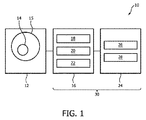
図1は、本発明の実施形態により、磁気共鳴イメージング(MRI)システム10を示すブロック図である。図1を参照して、システム10は、MRI装置12を有する。MRI装置12は、液体及び/又は固体な物質15によって完全に包囲された動く対象14の画像をスキャンし、収集するように構成されることができる。イメージングされる対象の例は、胎児の脳組織、腎臓組織、肝臓組織、心臓組織及び任意の他の身体組織を含むが、これに限定されるものではない。MRIシステム10は更に、コンピューティング装置16を有する。このコンピューティング装置16は、プロセッサ18、メモリ20、及びプロセッサ18上で実行されるように構成される物体インタラクションアプリケーション22を有することができる。図示されるMRIシステム10は更に、例えば、ディスプレイ26上に画像を表示し、キーボード28のようなユーザ入力装置を通じてユーザ入力を受け取るように構成される画像生成器のようなユーザインタフェース24を有する。コンピューティング装置16及びユーザインタフェース24は一緒に、システム10の画像生成器30を形成する。
FIG. 1 is a block diagram showing a magnetic resonance imaging (MRI)
図2は、動く対象14の磁気共鳴イメージングのための対応するプロシージャのフローチャートを示す。プロシージャは、開始ポイントSから始まり、5つのプロセスステップ(S1−S5)を受け、終了ポイントEにおいて終了する。
FIG. 2 shows a flow chart of the corresponding procedure for magnetic resonance imaging of the moving
ステップS1において、磁気共鳴イメージング装置12は、好適には、子宮内の胎児の脳である対象14から、データを取得するために使用される。データは、MRI装置12によって生成される磁界の磁化方向B0に対する前記対象14のさまざまな異なる位置(向き)において取得される。位置の少なくとも幾つかの間の動きが、対象14自身の動きによって引き起こされる。この動きは、外側から影響を及ぼされない。
In step S1, the magnetic
ステップS2において、個々のデータ取得中の前記対象14の位置及び/又は向きが、ナビゲータ又は別の標準MR動き検出技法を用いて決定される。加えて、ステップS2において、対象14が、予め決められた厚さの液体及び/又は固体の物質15(すなわち、ヒト組織)によって完全に包囲されているかどうかが決定される。更に、この包囲する組織の期待される平均磁化率が、ターゲット器官の期待される平均磁化率に対し差を有するかどうか、その差が予め決められた最大磁化率差を下回るかどうかが、決定される(例えば、胎児脳と羊膜液の間の期待される平均磁化率の差が、予め決められた最大磁化率差を下回るが、胎児脳と母体肺組織の間の期待される平均磁化率の差が予め決められた最大磁化率差を上回る)。 In step S2, the position and / or orientation of the subject 14 during individual data acquisition is determined using a navigator or another standard MR motion detection technique. In addition, in step S2 it is determined whether the subject 14 is completely enclosed by a liquid and / or solid substance 15 (ie, human tissue) of a predetermined thickness. Furthermore, it is determined whether the expected average magnetic susceptibility of this surrounding tissue has a difference from the expected average magnetic susceptibility of the target organ, and whether the difference is less than a predetermined maximum magnetic susceptibility difference. (For example, the expected average magnetic susceptibility difference between the fetal brain and the sheep membrane fluid is less than the predetermined maximum magnetic susceptibility difference, but the expected average magnetic susceptibility between the fetal brain and the maternal lung tissue. The difference exceeds the predetermined maximum magnetic susceptibility difference).
ステップS3において、取得されたデータから、位相画像が再構成される;再構成するステップS3は、対象の局所的磁化率ソースに関連しない位相コンポーネントを除去するためのバックグラウンドフィールド除去を含むことができる;特に、対象14が予め決められた厚さの物質によって完全に包囲されていると画像生成器30が決定する場合、バックグラウンドフィールド除去の努力が低減され、又はバックグラウンドフィールド除去がスキップされる。
In step S3, the phase image is reconstructed from the acquired data; the reconstructing step S3 may include background field removal to remove phase components that are not related to the local magnetic susceptibility source of interest. Yes; especially if the
ステップS4において、再構成された位相画像に基づいて、磁化率マップが生成される;及び In step S4, a magnetic susceptibility map is generated based on the reconstructed phase image;
ステップS5において、磁化率マップに基づいて、動く対象14の画像が生成される。対象のさまざまな異なる位置及び/又は向きを使用して、COSMOSアプローチに従って計算を実施することによってQSMの逆問題の不良設定性が解消される。
In step S5, an image of the moving
本発明は、図面及び上述の記述において詳しく図示され記述されているが、このような図示及び記述は、制限的なものではなく、説明的又は例示的なものとして考えられるべきである。本発明は、開示される実施形態に制限されない。開示される実施形態に対する他の変更例は、図面、開示及び添付の請求項の検討から、請求項に記載される本発明を実施する際に当業者によって理解され、実現されることができる。請求項において、「有する、含む(comprising)」という語は、他の構成要素又はステップを除外せず、不定冠詞「a」又は「an」は、複数性を除外しない。特定の手段が相互に異なる従属請求項に列挙されているという単なる事実は、これらの手段の組み合わせが有利に使用されることができないことを示さない。請求項における任意の参照符号は、請求項の範囲を制限するものとして解釈されるべきでない。 Although the present invention has been illustrated and described in detail in the drawings and the above description, such illustrations and descriptions should be considered as explanatory or exemplary rather than restrictive. The present invention is not limited to the disclosed embodiments. Other modifications to the disclosed embodiments can be understood and realized by those skilled in the art in carrying out the invention described in the claims, from the drawings, disclosure and review of the accompanying claims. In the claims, the word "comprising" does not exclude other components or steps, and the indefinite article "a" or "an" does not exclude pluralities. The mere fact that certain means are listed in different dependent claims does not indicate that a combination of these means cannot be used in an advantageous manner. Any reference code in the claims should not be construed as limiting the scope of the claims.
Claims (15)
動く対象からデータを取得するための磁気共鳴イメージング装置であって、前記対象は、胎児又は胎児の一部のようなそれ自身で動いている、磁気共鳴イメージング装置と、
前記動く対象の画像を生成する画像生成器と、
を有し、
前記磁気共鳴イメージング装置は、前記対象の自然な動きを利用して、磁化方向B0に対する前記対象の個々の異なる向きにおいて前記対象からデータを取得するように構成され、
前記画像生成器は、
個々のデータ取得中、前記対象の位置及び/又は向きを決定し、前記対象の前記自然な動きを利用して、欠けているデータを完成し;
前記個々の異なる向きについて、前記取得されたデータから位相画像を再構成し;
再構成された位相画像に基づいて磁化率マップを生成する処理であって、複数の向きのサンプリングを使用して磁化率を計算するアプローチ又は前記対象の個々の異なる位置及び/又は向きを使用する任意の他のアプローチに従って計算を実施して、前記対象の決定された位置及び/又は向きを使用して、定量的磁化率マッピングの逆問題の不良設定性を解消する、処理を行う、
磁気共鳴イメージングシステム。 Magnetic resonance imaging system
A magnetic resonance imaging device for acquiring data from a moving object, wherein the object is a magnetic resonance imaging device that is moving on its own, such as a foetation or a part of a foetation.
An image generator that generates an image of the moving object,
Have,
It said magnetic resonance imaging apparatus, by utilizing the natural movement of the object, is configured to acquire data from the subject in each different orientation of the object relative to the magnetization direction B 0,
The image generator
During individual data acquisition, the position and / or orientation of the object is determined and the natural movement of the object is used to complete the missing data;
A phase image is reconstructed from the acquired data for each of the different orientations;
The process of generating a magnetic susceptibility map based on a reconstructed phase image, using an approach to calculate the susceptibility using sampling of multiple orientations or the individual different positions and / or orientations of the object. Perform calculations according to any other approach and use the determined position and / or orientation of the subject to eliminate the bad settability of the inverse problem of quantitative susceptibility mapping.
Magnetic resonance imaging system.
磁気共鳴イメージング装置を使用して前記対象からデータを取得するステップであって、前記データは、前記対象の自然な動きを利用して、磁化方向B0に対する前記対象の個々の異なる位置において取得される、ステップと、
個々のデータ取得中、前記対象の前記位置及び/又は向きを決定し、前記対象の自然な動きを利用して、欠けているデータを完成させるステップと、
前記取得されたデータから位相画像を再構成するステップと、
前記再構成された位相画像に基づいて磁化率マップを生成するステップであって、前記対象の決定される位置及び/又は向きは、「複数の向きのサンプリングを使用する磁化率計算」アプローチ又は任意の他のアプローチに従う計算を実施するために使用され、前記対象の個々の異なる位置及び/又は向きを使用して、定量的磁化率マッピングの逆問題の不良設定性を解消する、ステップと、
を有する方法。 A method of magnetic resonance imaging of a moving object, wherein the object moves itself like a foetation.
A step of obtaining data using a magnetic resonance imaging apparatus from the target, the data, using the natural movement of the object, acquired at each different position of the object relative to the magnetization direction B 0 Steps and
During the acquisition of individual data, the steps of determining the position and / or orientation of the object and utilizing the natural movement of the object to complete the missing data.
The step of reconstructing the phase image from the acquired data and
The step of generating a susceptibility map based on the reconstructed phase image, wherein the determined position and / or orientation of the object is a "susceptibility calculation using sampling of multiple orientations" approach or optional. Steps and steps that are used to perform calculations that follow other approaches and use the individual different positions and / or orientations of the subject to eliminate the inverse problem of quantitative susceptibility mapping.
Method to have.
Applications Claiming Priority (3)
| Application Number | Priority Date | Filing Date | Title |
|---|---|---|---|
| EP16160198 | 2016-03-14 | ||
| EP16160198.4 | 2016-03-14 | ||
| PCT/EP2017/055884 WO2017157872A1 (en) | 2016-03-14 | 2017-03-14 | Susceptibility mapping of a moving subject |
Publications (2)
| Publication Number | Publication Date |
|---|---|
| JP2019508156A JP2019508156A (en) | 2019-03-28 |
| JP6857666B2 true JP6857666B2 (en) | 2021-04-14 |
Family
ID=55527489
Family Applications (1)
| Application Number | Title | Priority Date | Filing Date |
|---|---|---|---|
| JP2018546603A Active JP6857666B2 (en) | 2016-03-14 | 2017-03-14 | Magnetic susceptibility mapping of moving objects |
Country Status (5)
| Country | Link |
|---|---|
| US (1) | US10852381B2 (en) |
| EP (1) | EP3430418B1 (en) |
| JP (1) | JP6857666B2 (en) |
| CN (1) | CN108780134B (en) |
| WO (1) | WO2017157872A1 (en) |
Families Citing this family (4)
| Publication number | Priority date | Publication date | Assignee | Title |
|---|---|---|---|---|
| US11372066B2 (en) * | 2018-02-07 | 2022-06-28 | The Medical College Of Wisconsin, Inc. | Multi-resolution quantitative susceptibility mapping with magnetic resonance imaging |
| US20230181041A1 (en) * | 2020-04-27 | 2023-06-15 | Children's Medical Center Corporation | Fetal cardiac mri using self-gating with a cartesian k-space trajectory |
| US11360179B2 (en) | 2020-10-29 | 2022-06-14 | The Mitre Corporation | Systems and methods for estimating magnetic susceptibility through continuous motion in an MRI scanner |
| CN112505598B (en) * | 2020-12-10 | 2022-01-07 | 上海交通大学 | Quantitative magnetic susceptibility imaging reconstruction method and system, storage medium and terminal |
Family Cites Families (14)
| Publication number | Priority date | Publication date | Assignee | Title |
|---|---|---|---|---|
| DE10056874C2 (en) | 2000-11-16 | 2003-02-06 | Siemens Ag | Method for operating a magnetic resonance device in which changes in position are detected by means of orbital navigator echoes |
| US20080042647A1 (en) * | 2004-11-30 | 2008-02-21 | Hamano Life Science Research Foundation | Magnetic Resonance Imaging Method and Magnetic Resonance Imaging Apparatus |
| JP5178521B2 (en) | 2005-09-29 | 2013-04-10 | コーニンクレッカ フィリップス エレクトロニクス エヌ ヴィ | System and method for acquiring magnetic resonance imaging (MRI) data |
| US7567081B2 (en) * | 2007-05-03 | 2009-07-28 | University Of Basel | Magnetic resonance non-balanced-SSFP method for the detection and imaging of susceptibility related magnetic field distortions |
| CN102077108B (en) * | 2008-04-28 | 2015-02-25 | 康奈尔大学 | Tool for accurate quantification of magnet susceptibility in molecular MRI |
| US8605969B2 (en) * | 2010-04-06 | 2013-12-10 | Siemens Corporation | Method and system for multiple object detection by sequential Monte Carlo and hierarchical detection network |
| US9285449B2 (en) | 2011-06-15 | 2016-03-15 | Chunlei Liu | Systems and methods for imaging and quantifying tissue magnetism with magnetic resonance imaging |
| WO2013054718A1 (en) * | 2011-10-12 | 2013-04-18 | 株式会社日立製作所 | Magnetic resonance imaging device, and method for generating magnetic susceptibility enhanced image |
| EP2900835A4 (en) * | 2012-09-27 | 2016-05-11 | Population Diagnotics Inc | Methods and compositions for screening and treating developmental disorders |
| US20140103929A1 (en) * | 2012-10-13 | 2014-04-17 | Chunlei Liu | Systems and methods for susceptibility tensor imaging in the p-space |
| WO2014154544A1 (en) | 2013-03-25 | 2014-10-02 | Fatnav Ekonomisk Förening | Real-time motion correction for mri using fat navigators |
| US10203387B2 (en) * | 2013-06-06 | 2019-02-12 | Koninklijke Philips N.V. | MR imaging with enhanced susceptibility contrast |
| EP3068483A1 (en) * | 2013-11-14 | 2016-09-21 | Boston Scientific Neuromodulation Corporation | Systems, methods, and visualization tools for stimulation and sensing of neural systems with system-level interaction models |
| KR20160054360A (en) * | 2014-11-06 | 2016-05-16 | 삼성전자주식회사 | Imaging apparatus and imaging method |
-
2017
- 2017-03-14 CN CN201780017498.4A patent/CN108780134B/en not_active Expired - Fee Related
- 2017-03-14 US US16/082,983 patent/US10852381B2/en active Active
- 2017-03-14 EP EP17709709.4A patent/EP3430418B1/en active Active
- 2017-03-14 JP JP2018546603A patent/JP6857666B2/en active Active
- 2017-03-14 WO PCT/EP2017/055884 patent/WO2017157872A1/en active Application Filing
Also Published As
| Publication number | Publication date |
|---|---|
| CN108780134B (en) | 2021-02-19 |
| EP3430418A1 (en) | 2019-01-23 |
| US20190033411A1 (en) | 2019-01-31 |
| JP2019508156A (en) | 2019-03-28 |
| WO2017157872A1 (en) | 2017-09-21 |
| EP3430418B1 (en) | 2022-07-06 |
| CN108780134A (en) | 2018-11-09 |
| US10852381B2 (en) | 2020-12-01 |
Similar Documents
| Publication | Publication Date | Title |
|---|---|---|
| Tamada et al. | Motion artifact reduction using a convolutional neural network for dynamic contrast enhanced MR imaging of the liver | |
| US9766316B2 (en) | Magnetic resonance imaging device and quantitative susceptibility mapping method | |
| Setarehdan et al. | Advanced algorithmic approaches to medical image segmentation: state-of-the-art applications in cardiology, neurology, mammography and pathology | |
| JP6857666B2 (en) | Magnetic susceptibility mapping of moving objects | |
| US8441257B2 (en) | Time resolved spin labeled MRI cineangiography | |
| Asbach et al. | Magnetic resonance cholangiopancreatography using a free-breathing T2-weighted turbo spin-echo sequence with navigator-triggered prospective acquisition correction | |
| Hoerr et al. | Cardiac-respiratory self-gated cine ultra-short echo time (UTE) cardiovascular magnetic resonance for assessment of functional cardiac parameters at high magnetic fields | |
| US20210239863A1 (en) | Super resolution in positron emission tomography imaging using ultrafast ultrasound imaging | |
| JP2014511742A (en) | Method and apparatus for collecting MR data | |
| JP2017080349A (en) | Magnetic resonance imaging apparatus and medical image processing apparatus | |
| Fu et al. | Motion tracking and strain map computation for quasi-static magnetic resonance elastography | |
| WO2022213666A1 (en) | Joint k-space and image-space reconstruction imaging method and device | |
| Christodoulou et al. | Improved subspace estimation for low-rank model-based accelerated cardiac imaging | |
| Stroud et al. | Correcting versus resolving respiratory motion in free-breathing whole-heart MRA: a comparison in patients with thoracic aortic disease | |
| Lugauer et al. | Accelerating multi-echo water-fat MRI with a joint locally low-rank and spatial sparsity-promoting reconstruction | |
| Zhang et al. | 3D self‐gated cardiac cine imaging at 3 Tesla using stack‐of‐stars bSSFP with tiny golden angles and compressed sensing | |
| JP6730995B2 (en) | Method and system for generating MR image of moving object in environment | |
| US10818047B2 (en) | Iterative reconstruction of quantitative magnetic resonance images | |
| Liu et al. | Fast, free-breathing, in vivo fetal imaging using time-resolved 3D MRI technique: preliminary results | |
| JP5738120B2 (en) | Non-contrast angiogram reconstruction method and magnetic resonance imaging apparatus | |
| Cüneyitoğlu et al. | Single-image Bayesian Restoration and Multi-image Super-resolution Restoration for B-mode Ultrasound Using an Accurate System Model Involving Correlated Nature of the Speckle Noise. | |
| Bassett | Exploration of Material Property Variation when Measured at Small Scales | |
| Derikx | Comparison of In Vivo Intramuscular Fat Quantification Techniques in MRI | |
| Lo | Rapid Quantitative Body Magnetic Resonance Imaging | |
| Wong et al. | Interactive two-dimensional fresh blood imaging: a feasibility study |
Legal Events
| Date | Code | Title | Description |
|---|---|---|---|
| A621 | Written request for application examination |
Free format text: JAPANESE INTERMEDIATE CODE: A621 Effective date: 20200309 |
|
| A977 | Report on retrieval |
Free format text: JAPANESE INTERMEDIATE CODE: A971007 Effective date: 20210204 |
|
| TRDD | Decision of grant or rejection written | ||
| A01 | Written decision to grant a patent or to grant a registration (utility model) |
Free format text: JAPANESE INTERMEDIATE CODE: A01 Effective date: 20210225 |
|
| A61 | First payment of annual fees (during grant procedure) |
Free format text: JAPANESE INTERMEDIATE CODE: A61 Effective date: 20210322 |
|
| R150 | Certificate of patent or registration of utility model |
Ref document number: 6857666 Country of ref document: JP Free format text: JAPANESE INTERMEDIATE CODE: R150 |