JP6857028B2 - Compositions and methods for immunotherapy - Google Patents
Compositions and methods for immunotherapy Download PDFInfo
- Publication number
- JP6857028B2 JP6857028B2 JP2016515328A JP2016515328A JP6857028B2 JP 6857028 B2 JP6857028 B2 JP 6857028B2 JP 2016515328 A JP2016515328 A JP 2016515328A JP 2016515328 A JP2016515328 A JP 2016515328A JP 6857028 B2 JP6857028 B2 JP 6857028B2
- Authority
- JP
- Japan
- Prior art keywords
- cells
- antigen
- cell
- icar
- receptor
- Prior art date
- Legal status (The legal status is an assumption and is not a legal conclusion. Google has not performed a legal analysis and makes no representation as to the accuracy of the status listed.)
- Active
Links
- 238000000034 method Methods 0.000 title claims description 157
- 239000000203 mixture Substances 0.000 title claims description 59
- 238000009169 immunotherapy Methods 0.000 title description 3
- 210000004027 cell Anatomy 0.000 claims description 409
- 210000001744 T-lymphocyte Anatomy 0.000 claims description 312
- 239000000427 antigen Substances 0.000 claims description 271
- 108091007433 antigens Proteins 0.000 claims description 270
- 102000036639 antigens Human genes 0.000 claims description 270
- 108090000765 processed proteins & peptides Proteins 0.000 claims description 191
- 102000004196 processed proteins & peptides Human genes 0.000 claims description 184
- 229920001184 polypeptide Polymers 0.000 claims description 183
- 206010028980 Neoplasm Diseases 0.000 claims description 173
- 108010019670 Chimeric Antigen Receptors Proteins 0.000 claims description 144
- 102000005962 receptors Human genes 0.000 claims description 102
- 108020003175 receptors Proteins 0.000 claims description 102
- 230000002401 inhibitory effect Effects 0.000 claims description 76
- 101710089372 Programmed cell death protein 1 Proteins 0.000 claims description 74
- 102100024222 B-lymphocyte antigen CD19 Human genes 0.000 claims description 56
- 101000980825 Homo sapiens B-lymphocyte antigen CD19 Proteins 0.000 claims description 56
- 229920001481 poly(stearyl methacrylate) Polymers 0.000 claims description 56
- 102100041003 Glutamate carboxypeptidase 2 Human genes 0.000 claims description 54
- 101000892862 Homo sapiens Glutamate carboxypeptidase 2 Proteins 0.000 claims description 54
- 239000003446 ligand Substances 0.000 claims description 54
- 150000007523 nucleic acids Chemical class 0.000 claims description 52
- 210000001519 tissue Anatomy 0.000 claims description 51
- 108010021064 CTLA-4 Antigen Proteins 0.000 claims description 47
- 239000013598 vector Substances 0.000 claims description 47
- 108091008874 T cell receptors Proteins 0.000 claims description 42
- 102000016266 T-Cell Antigen Receptors Human genes 0.000 claims description 41
- -1 EGP-40 Proteins 0.000 claims description 38
- 102000039446 nucleic acids Human genes 0.000 claims description 36
- 108020004707 nucleic acids Proteins 0.000 claims description 36
- 208000009329 Graft vs Host Disease Diseases 0.000 claims description 35
- 230000000735 allogeneic effect Effects 0.000 claims description 35
- 208000024908 graft versus host disease Diseases 0.000 claims description 35
- 208000037265 diseases, disorders, signs and symptoms Diseases 0.000 claims description 32
- 201000011510 cancer Diseases 0.000 claims description 31
- 230000004068 intracellular signaling Effects 0.000 claims description 27
- 208000035269 cancer or benign tumor Diseases 0.000 claims description 26
- 201000010099 disease Diseases 0.000 claims description 25
- 102100036011 T-cell surface glycoprotein CD4 Human genes 0.000 claims description 21
- 241000701022 Cytomegalovirus Species 0.000 claims description 18
- 108091028043 Nucleic acid sequence Proteins 0.000 claims description 16
- 208000032839 leukemia Diseases 0.000 claims description 16
- 210000000265 leukocyte Anatomy 0.000 claims description 16
- 230000004044 response Effects 0.000 claims description 15
- 102100025475 Carcinoembryonic antigen-related cell adhesion molecule 5 Human genes 0.000 claims description 14
- 108010022366 Carcinoembryonic Antigen Proteins 0.000 claims description 13
- 102100031940 Epithelial cell adhesion molecule Human genes 0.000 claims description 13
- 101000934338 Homo sapiens Myeloid cell surface antigen CD33 Proteins 0.000 claims description 13
- 102100025243 Myeloid cell surface antigen CD33 Human genes 0.000 claims description 13
- 230000003213 activating effect Effects 0.000 claims description 13
- 206010039491 Sarcoma Diseases 0.000 claims description 12
- OVBPIULPVIDEAO-LBPRGKRZSA-N folic acid Chemical compound C=1N=C2NC(N)=NC(=O)C2=NC=1CNC1=CC=C(C(=O)N[C@@H](CCC(O)=O)C(O)=O)C=C1 OVBPIULPVIDEAO-LBPRGKRZSA-N 0.000 claims description 12
- 102100031585 ADP-ribosyl cyclase/cyclic ADP-ribose hydrolase 1 Human genes 0.000 claims description 11
- 101000777636 Homo sapiens ADP-ribosyl cyclase/cyclic ADP-ribose hydrolase 1 Proteins 0.000 claims description 11
- 230000001605 fetal effect Effects 0.000 claims description 11
- 102100039498 Cytotoxic T-lymphocyte protein 4 Human genes 0.000 claims description 10
- 108091008042 inhibitory receptors Proteins 0.000 claims description 10
- 102000003735 Mesothelin Human genes 0.000 claims description 9
- 108090000015 Mesothelin Proteins 0.000 claims description 9
- 102000003729 Neprilysin Human genes 0.000 claims description 9
- 108090000028 Neprilysin Proteins 0.000 claims description 9
- 208000031261 Acute myeloid leukaemia Diseases 0.000 claims description 8
- 101000581981 Homo sapiens Neural cell adhesion molecule 1 Proteins 0.000 claims description 8
- 102100027347 Neural cell adhesion molecule 1 Human genes 0.000 claims description 8
- 206010060862 Prostate cancer Diseases 0.000 claims description 8
- 208000000236 Prostatic Neoplasms Diseases 0.000 claims description 8
- 239000008194 pharmaceutical composition Substances 0.000 claims description 8
- 208000026310 Breast neoplasm Diseases 0.000 claims description 7
- 108010009685 Cholinergic Receptors Proteins 0.000 claims description 7
- 108010066687 Epithelial Cell Adhesion Molecule Proteins 0.000 claims description 7
- 101000920667 Homo sapiens Epithelial cell adhesion molecule Proteins 0.000 claims description 7
- 102100020793 Interleukin-13 receptor subunit alpha-2 Human genes 0.000 claims description 7
- 101710112634 Interleukin-13 receptor subunit alpha-2 Proteins 0.000 claims description 7
- 206010033128 Ovarian cancer Diseases 0.000 claims description 7
- 206010061535 Ovarian neoplasm Diseases 0.000 claims description 7
- 102100036735 Prostate stem cell antigen Human genes 0.000 claims description 7
- 102100033177 Vascular endothelial growth factor receptor 2 Human genes 0.000 claims description 7
- 102000034337 acetylcholine receptors Human genes 0.000 claims description 7
- 230000030833 cell death Effects 0.000 claims description 7
- 201000005787 hematologic cancer Diseases 0.000 claims description 7
- 208000024200 hematopoietic and lymphoid system neoplasm Diseases 0.000 claims description 7
- 238000004519 manufacturing process Methods 0.000 claims description 7
- 208000010839 B-cell chronic lymphocytic leukemia Diseases 0.000 claims description 6
- 102100038080 B-cell receptor CD22 Human genes 0.000 claims description 6
- 102100022005 B-lymphocyte antigen CD20 Human genes 0.000 claims description 6
- 206010006187 Breast cancer Diseases 0.000 claims description 6
- 102100032912 CD44 antigen Human genes 0.000 claims description 6
- 102100025570 Cancer/testis antigen 1 Human genes 0.000 claims description 6
- 102100030595 HLA class II histocompatibility antigen gamma chain Human genes 0.000 claims description 6
- 102100031573 Hematopoietic progenitor cell antigen CD34 Human genes 0.000 claims description 6
- 101000884305 Homo sapiens B-cell receptor CD22 Proteins 0.000 claims description 6
- 101000897405 Homo sapiens B-lymphocyte antigen CD20 Proteins 0.000 claims description 6
- 101000868273 Homo sapiens CD44 antigen Proteins 0.000 claims description 6
- 101000856237 Homo sapiens Cancer/testis antigen 1 Proteins 0.000 claims description 6
- 101001082627 Homo sapiens HLA class II histocompatibility antigen gamma chain Proteins 0.000 claims description 6
- 101000777663 Homo sapiens Hematopoietic progenitor cell antigen CD34 Proteins 0.000 claims description 6
- 101000994365 Homo sapiens Integrin alpha-6 Proteins 0.000 claims description 6
- 101001078143 Homo sapiens Integrin alpha-IIb Proteins 0.000 claims description 6
- 101000998120 Homo sapiens Interleukin-3 receptor subunit alpha Proteins 0.000 claims description 6
- 101001109501 Homo sapiens NKG2-D type II integral membrane protein Proteins 0.000 claims description 6
- 101000610551 Homo sapiens Prominin-1 Proteins 0.000 claims description 6
- 101000874179 Homo sapiens Syndecan-1 Proteins 0.000 claims description 6
- 101000914496 Homo sapiens T-cell antigen CD7 Proteins 0.000 claims description 6
- 101000851376 Homo sapiens Tumor necrosis factor receptor superfamily member 8 Proteins 0.000 claims description 6
- 102100032816 Integrin alpha-6 Human genes 0.000 claims description 6
- 102100025306 Integrin alpha-IIb Human genes 0.000 claims description 6
- 102100033493 Interleukin-3 receptor subunit alpha Human genes 0.000 claims description 6
- 206010058467 Lung neoplasm malignant Diseases 0.000 claims description 6
- 101100346932 Mus musculus Muc1 gene Proteins 0.000 claims description 6
- OVBPIULPVIDEAO-UHFFFAOYSA-N N-Pteroyl-L-glutaminsaeure Natural products C=1N=C2NC(N)=NC(=O)C2=NC=1CNC1=CC=C(C(=O)NC(CCC(O)=O)C(O)=O)C=C1 OVBPIULPVIDEAO-UHFFFAOYSA-N 0.000 claims description 6
- 102100022680 NKG2-D type II integral membrane protein Human genes 0.000 claims description 6
- 102100040120 Prominin-1 Human genes 0.000 claims description 6
- 102100035721 Syndecan-1 Human genes 0.000 claims description 6
- 102100027208 T-cell antigen CD7 Human genes 0.000 claims description 6
- 102100036857 Tumor necrosis factor receptor superfamily member 8 Human genes 0.000 claims description 6
- 229960000304 folic acid Drugs 0.000 claims description 6
- 235000019152 folic acid Nutrition 0.000 claims description 6
- 239000011724 folic acid Substances 0.000 claims description 6
- 201000005202 lung cancer Diseases 0.000 claims description 6
- 208000020816 lung neoplasm Diseases 0.000 claims description 6
- 101150047061 tag-72 gene Proteins 0.000 claims description 6
- 206010055113 Breast cancer metastatic Diseases 0.000 claims description 5
- 101001103039 Homo sapiens Inactive tyrosine-protein kinase transmembrane receptor ROR1 Proteins 0.000 claims description 5
- 101001103036 Homo sapiens Nuclear receptor ROR-alpha Proteins 0.000 claims description 5
- 102100039615 Inactive tyrosine-protein kinase transmembrane receptor ROR1 Human genes 0.000 claims description 5
- 102000017578 LAG3 Human genes 0.000 claims description 5
- 208000031422 Lymphocytic Chronic B-Cell Leukemia Diseases 0.000 claims description 5
- 108010012255 Neural Cell Adhesion Molecule L1 Proteins 0.000 claims description 5
- 102100024964 Neural cell adhesion molecule L1 Human genes 0.000 claims description 5
- 206010061902 Pancreatic neoplasm Diseases 0.000 claims description 5
- 208000006664 Precursor Cell Lymphoblastic Leukemia-Lymphoma Diseases 0.000 claims description 5
- 208000032852 chronic lymphocytic leukemia Diseases 0.000 claims description 5
- 208000015486 malignant pancreatic neoplasm Diseases 0.000 claims description 5
- 201000002528 pancreatic cancer Diseases 0.000 claims description 5
- 208000008443 pancreatic carcinoma Diseases 0.000 claims description 5
- 239000000546 pharmaceutical excipient Substances 0.000 claims description 5
- 230000004083 survival effect Effects 0.000 claims description 5
- 208000004736 B-Cell Leukemia Diseases 0.000 claims description 3
- 229940045513 CTLA4 antagonist Drugs 0.000 claims description 3
- 208000002250 Hematologic Neoplasms Diseases 0.000 claims description 3
- 101000934341 Homo sapiens T-cell surface glycoprotein CD5 Proteins 0.000 claims description 3
- 208000015914 Non-Hodgkin lymphomas Diseases 0.000 claims description 3
- 102100025244 T-cell surface glycoprotein CD5 Human genes 0.000 claims description 3
- 208000003747 lymphoid leukemia Diseases 0.000 claims description 3
- 230000001737 promoting effect Effects 0.000 claims description 3
- 208000024893 Acute lymphoblastic leukemia Diseases 0.000 claims description 2
- 208000014697 Acute lymphocytic leukaemia Diseases 0.000 claims description 2
- 206010066476 Haematological malignancy Diseases 0.000 claims description 2
- 239000002253 acid Substances 0.000 claims description 2
- 101710160107 Outer membrane protein A Proteins 0.000 claims 8
- 230000003259 immunoinhibitory effect Effects 0.000 claims 4
- 102100029822 B- and T-lymphocyte attenuator Human genes 0.000 claims 2
- 101000864344 Homo sapiens B- and T-lymphocyte attenuator Proteins 0.000 claims 2
- 101150030213 Lag3 gene Proteins 0.000 claims 2
- 102100023990 60S ribosomal protein L17 Human genes 0.000 claims 1
- 108700012439 CA9 Proteins 0.000 claims 1
- 102100024423 Carbonic anhydrase 9 Human genes 0.000 claims 1
- 102000018651 Epithelial Cell Adhesion Molecule Human genes 0.000 claims 1
- 101000914324 Homo sapiens Carcinoembryonic antigen-related cell adhesion molecule 5 Proteins 0.000 claims 1
- 101000914321 Homo sapiens Carcinoembryonic antigen-related cell adhesion molecule 7 Proteins 0.000 claims 1
- 101000617725 Homo sapiens Pregnancy-specific beta-1-glycoprotein 2 Proteins 0.000 claims 1
- 101001136592 Homo sapiens Prostate stem cell antigen Proteins 0.000 claims 1
- 101001012157 Homo sapiens Receptor tyrosine-protein kinase erbB-2 Proteins 0.000 claims 1
- 102100030086 Receptor tyrosine-protein kinase erbB-2 Human genes 0.000 claims 1
- 230000028993 immune response Effects 0.000 description 119
- 150000001413 amino acids Chemical class 0.000 description 86
- 102100040678 Programmed cell death protein 1 Human genes 0.000 description 81
- 230000014509 gene expression Effects 0.000 description 75
- 239000005090 green fluorescent protein Substances 0.000 description 52
- 102000004127 Cytokines Human genes 0.000 description 45
- 108090000695 Cytokines Proteins 0.000 description 45
- 102000013816 Cytotoxic T-lymphocyte antigen 4 Human genes 0.000 description 45
- 229940024606 amino acid Drugs 0.000 description 44
- 235000001014 amino acid Nutrition 0.000 description 43
- 238000011282 treatment Methods 0.000 description 40
- 238000000692 Student's t-test Methods 0.000 description 39
- 230000015572 biosynthetic process Effects 0.000 description 39
- 108090000623 proteins and genes Proteins 0.000 description 39
- 238000002474 experimental method Methods 0.000 description 37
- 238000003556 assay Methods 0.000 description 36
- 239000011324 bead Substances 0.000 description 36
- 238000005755 formation reaction Methods 0.000 description 34
- 101001137987 Homo sapiens Lymphocyte activation gene 3 protein Proteins 0.000 description 33
- 230000000694 effects Effects 0.000 description 32
- 102000004169 proteins and genes Human genes 0.000 description 32
- 238000004458 analytical method Methods 0.000 description 30
- 238000000684 flow cytometry Methods 0.000 description 30
- 102100020862 Lymphocyte activation gene 3 protein Human genes 0.000 description 29
- FAPWRFPIFSIZLT-UHFFFAOYSA-M Sodium chloride Chemical compound [Na+].[Cl-] FAPWRFPIFSIZLT-UHFFFAOYSA-M 0.000 description 28
- 238000010361 transduction Methods 0.000 description 28
- 101000946843 Homo sapiens T-cell surface glycoprotein CD8 alpha chain Proteins 0.000 description 27
- 102100034922 T-cell surface glycoprotein CD8 alpha chain Human genes 0.000 description 27
- 210000001151 cytotoxic T lymphocyte Anatomy 0.000 description 27
- 230000026683 transduction Effects 0.000 description 27
- 230000004913 activation Effects 0.000 description 26
- 210000002950 fibroblast Anatomy 0.000 description 26
- 238000012353 t test Methods 0.000 description 26
- 101000914514 Homo sapiens T-cell-specific surface glycoprotein CD28 Proteins 0.000 description 24
- 102100027213 T-cell-specific surface glycoprotein CD28 Human genes 0.000 description 24
- 235000018102 proteins Nutrition 0.000 description 24
- 241000699670 Mus sp. Species 0.000 description 23
- 241000700605 Viruses Species 0.000 description 23
- 238000013459 approach Methods 0.000 description 23
- 239000012228 culture supernatant Substances 0.000 description 23
- 230000006870 function Effects 0.000 description 23
- 230000003834 intracellular effect Effects 0.000 description 23
- 230000034994 death Effects 0.000 description 22
- 210000004698 lymphocyte Anatomy 0.000 description 21
- 101000716102 Homo sapiens T-cell surface glycoprotein CD4 Proteins 0.000 description 20
- 230000001717 pathogenic effect Effects 0.000 description 20
- 230000001225 therapeutic effect Effects 0.000 description 20
- 238000000338 in vitro Methods 0.000 description 19
- 208000015181 infectious disease Diseases 0.000 description 19
- 244000052769 pathogen Species 0.000 description 19
- 230000028327 secretion Effects 0.000 description 19
- 210000004881 tumor cell Anatomy 0.000 description 19
- 239000012634 fragment Substances 0.000 description 18
- 238000004113 cell culture Methods 0.000 description 17
- 238000001727 in vivo Methods 0.000 description 16
- 210000000056 organ Anatomy 0.000 description 16
- 108091033319 polynucleotide Proteins 0.000 description 16
- 102000040430 polynucleotide Human genes 0.000 description 16
- 239000002157 polynucleotide Substances 0.000 description 16
- 230000035755 proliferation Effects 0.000 description 16
- 230000007705 epithelial mesenchymal transition Effects 0.000 description 15
- 238000011002 quantification Methods 0.000 description 15
- 230000001105 regulatory effect Effects 0.000 description 15
- 239000011780 sodium chloride Substances 0.000 description 15
- 108010002350 Interleukin-2 Proteins 0.000 description 14
- 102000000588 Interleukin-2 Human genes 0.000 description 14
- 241000699666 Mus <mouse, genus> Species 0.000 description 14
- 239000003814 drug Substances 0.000 description 14
- 230000001613 neoplastic effect Effects 0.000 description 14
- 238000013105 post hoc analysis Methods 0.000 description 14
- 108060001084 Luciferase Proteins 0.000 description 13
- 239000005089 Luciferase Substances 0.000 description 13
- 108060008682 Tumor Necrosis Factor Proteins 0.000 description 13
- 102000000852 Tumor Necrosis Factor-alpha Human genes 0.000 description 13
- 210000001185 bone marrow Anatomy 0.000 description 13
- 210000002865 immune cell Anatomy 0.000 description 13
- 230000001177 retroviral effect Effects 0.000 description 13
- 102000017420 CD3 protein, epsilon/gamma/delta subunit Human genes 0.000 description 12
- 108050005493 CD3 protein, epsilon/gamma/delta subunit Proteins 0.000 description 12
- 201000009030 Carcinoma Diseases 0.000 description 12
- 208000009956 adenocarcinoma Diseases 0.000 description 12
- 238000000540 analysis of variance Methods 0.000 description 12
- 210000000987 immune system Anatomy 0.000 description 12
- 230000000670 limiting effect Effects 0.000 description 12
- 230000001404 mediated effect Effects 0.000 description 12
- 210000000822 natural killer cell Anatomy 0.000 description 12
- 102000000905 Cadherin Human genes 0.000 description 11
- 108050007957 Cadherin Proteins 0.000 description 11
- 108060003951 Immunoglobulin Proteins 0.000 description 11
- 210000004369 blood Anatomy 0.000 description 11
- 239000008280 blood Substances 0.000 description 11
- 230000008859 change Effects 0.000 description 11
- 238000006243 chemical reaction Methods 0.000 description 11
- 102000018358 immunoglobulin Human genes 0.000 description 11
- 239000001509 sodium citrate Substances 0.000 description 11
- 230000000638 stimulation Effects 0.000 description 11
- 239000000126 substance Substances 0.000 description 11
- HRXKRNGNAMMEHJ-UHFFFAOYSA-K trisodium citrate Chemical compound [Na+].[Na+].[Na+].[O-]C(=O)CC(O)(CC([O-])=O)C([O-])=O HRXKRNGNAMMEHJ-UHFFFAOYSA-K 0.000 description 11
- 229940038773 trisodium citrate Drugs 0.000 description 11
- ZHNUHDYFZUAESO-UHFFFAOYSA-N Formamide Chemical compound NC=O ZHNUHDYFZUAESO-UHFFFAOYSA-N 0.000 description 10
- 108010017213 Granulocyte-Macrophage Colony-Stimulating Factor Proteins 0.000 description 10
- 102100039620 Granulocyte-macrophage colony-stimulating factor Human genes 0.000 description 10
- 230000010261 cell growth Effects 0.000 description 10
- 208000002491 severe combined immunodeficiency Diseases 0.000 description 10
- 230000019491 signal transduction Effects 0.000 description 10
- 102000011782 Keratins Human genes 0.000 description 9
- 108010076876 Keratins Proteins 0.000 description 9
- 229940079593 drug Drugs 0.000 description 9
- 239000012636 effector Substances 0.000 description 9
- 230000026731 phosphorylation Effects 0.000 description 9
- 238000006366 phosphorylation reaction Methods 0.000 description 9
- 210000000952 spleen Anatomy 0.000 description 9
- 238000000551 statistical hypothesis test Methods 0.000 description 9
- 230000003612 virological effect Effects 0.000 description 9
- 108020004635 Complementary DNA Proteins 0.000 description 8
- 101000889276 Homo sapiens Cytotoxic T-lymphocyte protein 4 Proteins 0.000 description 8
- 102100026742 Opioid-binding protein/cell adhesion molecule Human genes 0.000 description 8
- 101710096745 Opioid-binding protein/cell adhesion molecule Proteins 0.000 description 8
- DBMJMQXJHONAFJ-UHFFFAOYSA-M Sodium laurylsulphate Chemical compound [Na+].CCCCCCCCCCCCOS([O-])(=O)=O DBMJMQXJHONAFJ-UHFFFAOYSA-M 0.000 description 8
- 230000005867 T cell response Effects 0.000 description 8
- 210000003719 b-lymphocyte Anatomy 0.000 description 8
- 238000010804 cDNA synthesis Methods 0.000 description 8
- 238000002659 cell therapy Methods 0.000 description 8
- 239000002299 complementary DNA Substances 0.000 description 8
- 238000009396 hybridization Methods 0.000 description 8
- 230000001965 increasing effect Effects 0.000 description 8
- 238000002347 injection Methods 0.000 description 8
- 239000007924 injection Substances 0.000 description 8
- 210000004185 liver Anatomy 0.000 description 8
- 210000004072 lung Anatomy 0.000 description 8
- 230000002265 prevention Effects 0.000 description 8
- 230000009257 reactivity Effects 0.000 description 8
- 230000002441 reversible effect Effects 0.000 description 8
- 238000010186 staining Methods 0.000 description 8
- 230000004936 stimulating effect Effects 0.000 description 8
- 208000023275 Autoimmune disease Diseases 0.000 description 7
- 241000283707 Capra Species 0.000 description 7
- 206010010356 Congenital anomaly Diseases 0.000 description 7
- 206010035226 Plasma cell myeloma Diseases 0.000 description 7
- 230000006044 T cell activation Effects 0.000 description 7
- 230000006052 T cell proliferation Effects 0.000 description 7
- 230000009286 beneficial effect Effects 0.000 description 7
- 238000013461 design Methods 0.000 description 7
- 208000035475 disorder Diseases 0.000 description 7
- 230000001506 immunosuppresive effect Effects 0.000 description 7
- 230000001939 inductive effect Effects 0.000 description 7
- 238000001802 infusion Methods 0.000 description 7
- 230000005764 inhibitory process Effects 0.000 description 7
- 239000007788 liquid Substances 0.000 description 7
- 239000002609 medium Substances 0.000 description 7
- 210000003819 peripheral blood mononuclear cell Anatomy 0.000 description 7
- 230000002829 reductive effect Effects 0.000 description 7
- RJBDSRWGVYNDHL-XNJNKMBASA-N (2S,4R,5S,6S)-2-[(2S,3R,4R,5S,6R)-5-[(2S,3R,4R,5R,6R)-3-acetamido-4,5-dihydroxy-6-(hydroxymethyl)oxan-2-yl]oxy-2-[(2R,3S,4R,5R,6R)-4,5-dihydroxy-2-(hydroxymethyl)-6-[(E,2R,3S)-3-hydroxy-2-(octadecanoylamino)octadec-4-enoxy]oxan-3-yl]oxy-3-hydroxy-6-(hydroxymethyl)oxan-4-yl]oxy-5-amino-6-[(1S,2R)-2-[(2S,4R,5S,6S)-5-amino-2-carboxy-4-hydroxy-6-[(1R,2R)-1,2,3-trihydroxypropyl]oxan-2-yl]oxy-1,3-dihydroxypropyl]-4-hydroxyoxane-2-carboxylic acid Chemical compound CCCCCCCCCCCCCCCCCC(=O)N[C@H](CO[C@@H]1O[C@H](CO)[C@@H](O[C@@H]2O[C@H](CO)[C@H](O[C@@H]3O[C@H](CO)[C@H](O)[C@H](O)[C@H]3NC(C)=O)[C@H](O[C@@]3(C[C@@H](O)[C@H](N)[C@H](O3)[C@H](O)[C@@H](CO)O[C@@]3(C[C@@H](O)[C@H](N)[C@H](O3)[C@H](O)[C@H](O)CO)C(O)=O)C(O)=O)[C@H]2O)[C@H](O)[C@H]1O)[C@@H](O)\C=C\CCCCCCCCCCCCC RJBDSRWGVYNDHL-XNJNKMBASA-N 0.000 description 6
- 108091032973 (ribonucleotides)n+m Proteins 0.000 description 6
- 108091003079 Bovine Serum Albumin Proteins 0.000 description 6
- 101000914484 Homo sapiens T-lymphocyte activation antigen CD80 Proteins 0.000 description 6
- 241000725303 Human immunodeficiency virus Species 0.000 description 6
- 101710120463 Prostate stem cell antigen Proteins 0.000 description 6
- 241000194017 Streptococcus Species 0.000 description 6
- 102100027222 T-lymphocyte activation antigen CD80 Human genes 0.000 description 6
- 108010053099 Vascular Endothelial Growth Factor Receptor-2 Proteins 0.000 description 6
- 102100022748 Wilms tumor protein Human genes 0.000 description 6
- 101710127857 Wilms tumor protein Proteins 0.000 description 6
- 239000000654 additive Substances 0.000 description 6
- 238000011316 allogeneic transplantation Methods 0.000 description 6
- 230000008901 benefit Effects 0.000 description 6
- 150000001720 carbohydrates Chemical class 0.000 description 6
- 235000014633 carbohydrates Nutrition 0.000 description 6
- 230000009260 cross reactivity Effects 0.000 description 6
- 238000001514 detection method Methods 0.000 description 6
- 230000008030 elimination Effects 0.000 description 6
- 238000003379 elimination reaction Methods 0.000 description 6
- 230000012010 growth Effects 0.000 description 6
- 230000006698 induction Effects 0.000 description 6
- 210000004692 intercellular junction Anatomy 0.000 description 6
- 230000002147 killing effect Effects 0.000 description 6
- 239000006166 lysate Substances 0.000 description 6
- 239000000463 material Substances 0.000 description 6
- 239000011159 matrix material Substances 0.000 description 6
- 201000000050 myeloid neoplasm Diseases 0.000 description 6
- 210000005105 peripheral blood lymphocyte Anatomy 0.000 description 6
- 230000008569 process Effects 0.000 description 6
- 238000000926 separation method Methods 0.000 description 6
- 125000006850 spacer group Chemical group 0.000 description 6
- 210000000130 stem cell Anatomy 0.000 description 6
- 239000006228 supernatant Substances 0.000 description 6
- 208000024891 symptom Diseases 0.000 description 6
- 238000002560 therapeutic procedure Methods 0.000 description 6
- 102000006306 Antigen Receptors Human genes 0.000 description 5
- 108010083359 Antigen Receptors Proteins 0.000 description 5
- 108020004414 DNA Proteins 0.000 description 5
- OUYCCCASQSFEME-QMMMGPOBSA-N L-tyrosine Chemical compound OC(=O)[C@@H](N)CC1=CC=C(O)C=C1 OUYCCCASQSFEME-QMMMGPOBSA-N 0.000 description 5
- 206010061309 Neoplasm progression Diseases 0.000 description 5
- 108010029485 Protein Isoforms Proteins 0.000 description 5
- 102000001708 Protein Isoforms Human genes 0.000 description 5
- 230000002411 adverse Effects 0.000 description 5
- SRHNADOZAAWYLV-XLMUYGLTSA-N alpha-L-Fucp-(1->2)-beta-D-Galp-(1->4)-[alpha-L-Fucp-(1->3)]-beta-D-GlcpNAc Chemical compound O[C@H]1[C@H](O)[C@H](O)[C@H](C)O[C@H]1O[C@H]1[C@H](O[C@H]2[C@@H]([C@@H](NC(C)=O)[C@H](O)O[C@@H]2CO)O[C@H]2[C@H]([C@H](O)[C@H](O)[C@H](C)O2)O)O[C@H](CO)[C@H](O)[C@@H]1O SRHNADOZAAWYLV-XLMUYGLTSA-N 0.000 description 5
- 230000003915 cell function Effects 0.000 description 5
- 230000001413 cellular effect Effects 0.000 description 5
- 230000001276 controlling effect Effects 0.000 description 5
- 230000001472 cytotoxic effect Effects 0.000 description 5
- 230000004069 differentiation Effects 0.000 description 5
- 239000003623 enhancer Substances 0.000 description 5
- 210000000981 epithelium Anatomy 0.000 description 5
- 102000036215 folic acid binding proteins Human genes 0.000 description 5
- 108091011001 folic acid binding proteins Proteins 0.000 description 5
- 230000007774 longterm Effects 0.000 description 5
- 230000036210 malignancy Effects 0.000 description 5
- 230000004048 modification Effects 0.000 description 5
- 238000012986 modification Methods 0.000 description 5
- 210000003289 regulatory T cell Anatomy 0.000 description 5
- 230000011664 signaling Effects 0.000 description 5
- 210000003491 skin Anatomy 0.000 description 5
- 238000003786 synthesis reaction Methods 0.000 description 5
- 210000001541 thymus gland Anatomy 0.000 description 5
- 230000001988 toxicity Effects 0.000 description 5
- 231100000419 toxicity Toxicity 0.000 description 5
- 230000005751 tumor progression Effects 0.000 description 5
- OUYCCCASQSFEME-UHFFFAOYSA-N tyrosine Natural products OC(=O)C(N)CC1=CC=C(O)C=C1 OUYCCCASQSFEME-UHFFFAOYSA-N 0.000 description 5
- 241001529453 unidentified herpesvirus Species 0.000 description 5
- 241001430294 unidentified retrovirus Species 0.000 description 5
- 230000009385 viral infection Effects 0.000 description 5
- 238000005406 washing Methods 0.000 description 5
- 241000059559 Agriotes sordidus Species 0.000 description 4
- 108010074708 B7-H1 Antigen Proteins 0.000 description 4
- 102000014914 Carrier Proteins Human genes 0.000 description 4
- 102000004190 Enzymes Human genes 0.000 description 4
- 108090000790 Enzymes Proteins 0.000 description 4
- DHMQDGOQFOQNFH-UHFFFAOYSA-N Glycine Chemical compound NCC(O)=O DHMQDGOQFOQNFH-UHFFFAOYSA-N 0.000 description 4
- 206010061598 Immunodeficiency Diseases 0.000 description 4
- 206010062016 Immunosuppression Diseases 0.000 description 4
- ZDXPYRJPNDTMRX-VKHMYHEASA-N L-glutamine Chemical compound OC(=O)[C@@H](N)CCC(N)=O ZDXPYRJPNDTMRX-VKHMYHEASA-N 0.000 description 4
- 206010025323 Lymphomas Diseases 0.000 description 4
- 206010027476 Metastases Diseases 0.000 description 4
- 101710163270 Nuclease Proteins 0.000 description 4
- 102100024216 Programmed cell death 1 ligand 1 Human genes 0.000 description 4
- 239000006146 Roswell Park Memorial Institute medium Substances 0.000 description 4
- 206010043276 Teratoma Diseases 0.000 description 4
- 206010054094 Tumour necrosis Diseases 0.000 description 4
- 102100033019 Tyrosine-protein phosphatase non-receptor type 11 Human genes 0.000 description 4
- 101710116241 Tyrosine-protein phosphatase non-receptor type 11 Proteins 0.000 description 4
- 208000036142 Viral infection Diseases 0.000 description 4
- 230000001154 acute effect Effects 0.000 description 4
- 210000000612 antigen-presenting cell Anatomy 0.000 description 4
- 230000000890 antigenic effect Effects 0.000 description 4
- 108091008324 binding proteins Proteins 0.000 description 4
- 230000033228 biological regulation Effects 0.000 description 4
- 230000008499 blood brain barrier function Effects 0.000 description 4
- 210000001218 blood-brain barrier Anatomy 0.000 description 4
- 238000010322 bone marrow transplantation Methods 0.000 description 4
- 238000002619 cancer immunotherapy Methods 0.000 description 4
- 239000003795 chemical substances by application Substances 0.000 description 4
- 230000003013 cytotoxicity Effects 0.000 description 4
- 231100000135 cytotoxicity Toxicity 0.000 description 4
- 230000006378 damage Effects 0.000 description 4
- 210000004443 dendritic cell Anatomy 0.000 description 4
- 210000001671 embryonic stem cell Anatomy 0.000 description 4
- 229940088598 enzyme Drugs 0.000 description 4
- 239000012091 fetal bovine serum Substances 0.000 description 4
- 238000001943 fluorescence-activated cell sorting Methods 0.000 description 4
- 230000004907 flux Effects 0.000 description 4
- 230000004927 fusion Effects 0.000 description 4
- 210000001035 gastrointestinal tract Anatomy 0.000 description 4
- 210000002216 heart Anatomy 0.000 description 4
- 210000003958 hematopoietic stem cell Anatomy 0.000 description 4
- 208000026278 immune system disease Diseases 0.000 description 4
- 230000006872 improvement Effects 0.000 description 4
- 201000001441 melanoma Diseases 0.000 description 4
- 210000004379 membrane Anatomy 0.000 description 4
- 239000012528 membrane Substances 0.000 description 4
- 230000009401 metastasis Effects 0.000 description 4
- 230000001575 pathological effect Effects 0.000 description 4
- 210000005259 peripheral blood Anatomy 0.000 description 4
- 239000011886 peripheral blood Substances 0.000 description 4
- 238000002360 preparation method Methods 0.000 description 4
- 210000002307 prostate Anatomy 0.000 description 4
- 230000009467 reduction Effects 0.000 description 4
- 150000003839 salts Chemical class 0.000 description 4
- 239000000243 solution Substances 0.000 description 4
- UCSJYZPVAKXKNQ-HZYVHMACSA-N streptomycin Chemical compound CN[C@H]1[C@H](O)[C@@H](O)[C@H](CO)O[C@H]1O[C@@H]1[C@](C=O)(O)[C@H](C)O[C@H]1O[C@@H]1[C@@H](NC(N)=N)[C@H](O)[C@@H](NC(N)=N)[C@H](O)[C@H]1O UCSJYZPVAKXKNQ-HZYVHMACSA-N 0.000 description 4
- 230000009885 systemic effect Effects 0.000 description 4
- 230000008685 targeting Effects 0.000 description 4
- 210000003171 tumor-infiltrating lymphocyte Anatomy 0.000 description 4
- 239000013603 viral vector Substances 0.000 description 4
- 206010001052 Acute respiratory distress syndrome Diseases 0.000 description 3
- 108010031480 Artificial Receptors Proteins 0.000 description 3
- 241000894006 Bacteria Species 0.000 description 3
- 206010004593 Bile duct cancer Diseases 0.000 description 3
- 241000167854 Bourreria succulenta Species 0.000 description 3
- 102100032937 CD40 ligand Human genes 0.000 description 3
- 102000016289 Cell Adhesion Molecules Human genes 0.000 description 3
- 108010067225 Cell Adhesion Molecules Proteins 0.000 description 3
- 206010009944 Colon cancer Diseases 0.000 description 3
- 208000035473 Communicable disease Diseases 0.000 description 3
- WQZGKKKJIJFFOK-GASJEMHNSA-N Glucose Natural products OC[C@H]1OC(O)[C@H](O)[C@@H](O)[C@@H]1O WQZGKKKJIJFFOK-GASJEMHNSA-N 0.000 description 3
- PEDCQBHIVMGVHV-UHFFFAOYSA-N Glycerine Chemical compound OCC(O)CO PEDCQBHIVMGVHV-UHFFFAOYSA-N 0.000 description 3
- 102000003886 Glycoproteins Human genes 0.000 description 3
- 108090000288 Glycoproteins Proteins 0.000 description 3
- 108010043121 Green Fluorescent Proteins Proteins 0.000 description 3
- 102000004144 Green Fluorescent Proteins Human genes 0.000 description 3
- 102000006354 HLA-DR Antigens Human genes 0.000 description 3
- 108010058597 HLA-DR Antigens Proteins 0.000 description 3
- 101000800116 Homo sapiens Thy-1 membrane glycoprotein Proteins 0.000 description 3
- 108020004684 Internal Ribosome Entry Sites Proteins 0.000 description 3
- 229930182816 L-glutamine Natural products 0.000 description 3
- 241001465754 Metazoa Species 0.000 description 3
- 102100038082 Natural killer cell receptor 2B4 Human genes 0.000 description 3
- 229930182555 Penicillin Natural products 0.000 description 3
- JGSARLDLIJGVTE-MBNYWOFBSA-N Penicillin G Chemical compound N([C@H]1[C@H]2SC([C@@H](N2C1=O)C(O)=O)(C)C)C(=O)CC1=CC=CC=C1 JGSARLDLIJGVTE-MBNYWOFBSA-N 0.000 description 3
- DNIAPMSPPWPWGF-UHFFFAOYSA-N Propylene glycol Chemical compound CC(O)CO DNIAPMSPPWPWGF-UHFFFAOYSA-N 0.000 description 3
- 108020004511 Recombinant DNA Proteins 0.000 description 3
- 241000242739 Renilla Species 0.000 description 3
- 206010041067 Small cell lung cancer Diseases 0.000 description 3
- 102100033523 Thy-1 membrane glycoprotein Human genes 0.000 description 3
- 102100036922 Tumor necrosis factor ligand superfamily member 13B Human genes 0.000 description 3
- 102100028785 Tumor necrosis factor receptor superfamily member 14 Human genes 0.000 description 3
- 241000021375 Xenogenes Species 0.000 description 3
- 108010017070 Zinc Finger Nucleases Proteins 0.000 description 3
- 230000000996 additive effect Effects 0.000 description 3
- 125000000539 amino acid group Chemical group 0.000 description 3
- 230000006023 anti-tumor response Effects 0.000 description 3
- WQZGKKKJIJFFOK-VFUOTHLCSA-N beta-D-glucose Chemical compound OC[C@H]1O[C@@H](O)[C@H](O)[C@@H](O)[C@@H]1O WQZGKKKJIJFFOK-VFUOTHLCSA-N 0.000 description 3
- 208000026900 bile duct neoplasm Diseases 0.000 description 3
- 230000004071 biological effect Effects 0.000 description 3
- 210000004556 brain Anatomy 0.000 description 3
- 239000000969 carrier Substances 0.000 description 3
- 230000022534 cell killing Effects 0.000 description 3
- 235000019693 cherries Nutrition 0.000 description 3
- 208000006990 cholangiocarcinoma Diseases 0.000 description 3
- 208000029742 colonic neoplasm Diseases 0.000 description 3
- 230000016396 cytokine production Effects 0.000 description 3
- 231100000433 cytotoxic Toxicity 0.000 description 3
- 230000007423 decrease Effects 0.000 description 3
- 238000012217 deletion Methods 0.000 description 3
- 230000037430 deletion Effects 0.000 description 3
- 230000001419 dependent effect Effects 0.000 description 3
- 238000011161 development Methods 0.000 description 3
- 230000018109 developmental process Effects 0.000 description 3
- 230000003828 downregulation Effects 0.000 description 3
- 239000012645 endogenous antigen Substances 0.000 description 3
- 238000005516 engineering process Methods 0.000 description 3
- 239000000499 gel Substances 0.000 description 3
- 230000005965 immune activity Effects 0.000 description 3
- 230000010354 integration Effects 0.000 description 3
- 230000003993 interaction Effects 0.000 description 3
- 210000003734 kidney Anatomy 0.000 description 3
- 201000007270 liver cancer Diseases 0.000 description 3
- 208000014018 liver neoplasm Diseases 0.000 description 3
- 210000001165 lymph node Anatomy 0.000 description 3
- 239000003550 marker Substances 0.000 description 3
- 230000007246 mechanism Effects 0.000 description 3
- 238000010172 mouse model Methods 0.000 description 3
- 208000010125 myocardial infarction Diseases 0.000 description 3
- 210000005170 neoplastic cell Anatomy 0.000 description 3
- 230000036961 partial effect Effects 0.000 description 3
- 239000002245 particle Substances 0.000 description 3
- 230000037361 pathway Effects 0.000 description 3
- 229940049954 penicillin Drugs 0.000 description 3
- 230000004481 post-translational protein modification Effects 0.000 description 3
- 230000000069 prophylactic effect Effects 0.000 description 3
- 238000011160 research Methods 0.000 description 3
- 239000000523 sample Substances 0.000 description 3
- 208000000587 small cell lung carcinoma Diseases 0.000 description 3
- 238000006467 substitution reaction Methods 0.000 description 3
- 239000000725 suspension Substances 0.000 description 3
- 229940124597 therapeutic agent Drugs 0.000 description 3
- 239000002562 thickening agent Substances 0.000 description 3
- 238000002287 time-lapse microscopy Methods 0.000 description 3
- 238000012546 transfer Methods 0.000 description 3
- 241000712461 unidentified influenza virus Species 0.000 description 3
- 210000003708 urethra Anatomy 0.000 description 3
- 239000003981 vehicle Substances 0.000 description 3
- 238000001262 western blot Methods 0.000 description 3
- MTCFGRXMJLQNBG-REOHCLBHSA-N (2S)-2-Amino-3-hydroxypropansäure Chemical compound OC[C@H](N)C(O)=O MTCFGRXMJLQNBG-REOHCLBHSA-N 0.000 description 2
- KBTLDMSFADPKFJ-UHFFFAOYSA-N 2-phenyl-1H-indole-3,4-dicarboximidamide Chemical compound N1C2=CC=CC(C(N)=N)=C2C(C(=N)N)=C1C1=CC=CC=C1 KBTLDMSFADPKFJ-UHFFFAOYSA-N 0.000 description 2
- HVCOBJNICQPDBP-UHFFFAOYSA-N 3-[3-[3,5-dihydroxy-6-methyl-4-(3,4,5-trihydroxy-6-methyloxan-2-yl)oxyoxan-2-yl]oxydecanoyloxy]decanoic acid;hydrate Chemical compound O.OC1C(OC(CC(=O)OC(CCCCCCC)CC(O)=O)CCCCCCC)OC(C)C(O)C1OC1C(O)C(O)C(O)C(C)O1 HVCOBJNICQPDBP-UHFFFAOYSA-N 0.000 description 2
- 108010082808 4-1BB Ligand Proteins 0.000 description 2
- 102000002627 4-1BB Ligand Human genes 0.000 description 2
- 206010000830 Acute leukaemia Diseases 0.000 description 2
- 206010000871 Acute monocytic leukaemia Diseases 0.000 description 2
- 201000003076 Angiosarcoma Diseases 0.000 description 2
- 239000004475 Arginine Substances 0.000 description 2
- 208000005440 Basal Cell Neoplasms Diseases 0.000 description 2
- 206010004146 Basal cell carcinoma Diseases 0.000 description 2
- 206010005003 Bladder cancer Diseases 0.000 description 2
- 108010029697 CD40 Ligand Proteins 0.000 description 2
- 102100025221 CD70 antigen Human genes 0.000 description 2
- 101000741929 Caenorhabditis elegans Serine/threonine-protein phosphatase 2A catalytic subunit Proteins 0.000 description 2
- 102000004039 Caspase-9 Human genes 0.000 description 2
- 108090000566 Caspase-9 Proteins 0.000 description 2
- 208000005243 Chondrosarcoma Diseases 0.000 description 2
- 206010011968 Decreased immune responsiveness Diseases 0.000 description 2
- 206010061818 Disease progression Diseases 0.000 description 2
- 206010058314 Dysplasia Diseases 0.000 description 2
- 238000002965 ELISA Methods 0.000 description 2
- 241000709661 Enterovirus Species 0.000 description 2
- 208000006168 Ewing Sarcoma Diseases 0.000 description 2
- 102000018233 Fibroblast Growth Factor Human genes 0.000 description 2
- 108050007372 Fibroblast Growth Factor Proteins 0.000 description 2
- 201000008808 Fibrosarcoma Diseases 0.000 description 2
- 208000032612 Glial tumor Diseases 0.000 description 2
- 206010018338 Glioma Diseases 0.000 description 2
- 239000004471 Glycine Substances 0.000 description 2
- 229930186217 Glycolipid Natural products 0.000 description 2
- 208000001258 Hemangiosarcoma Diseases 0.000 description 2
- 241000700721 Hepatitis B virus Species 0.000 description 2
- 208000017604 Hodgkin disease Diseases 0.000 description 2
- 208000010747 Hodgkins lymphoma Diseases 0.000 description 2
- 101001018097 Homo sapiens L-selectin Proteins 0.000 description 2
- 101001005728 Homo sapiens Melanoma-associated antigen 1 Proteins 0.000 description 2
- 101000764263 Homo sapiens Tumor necrosis factor ligand superfamily member 4 Proteins 0.000 description 2
- 101000617285 Homo sapiens Tyrosine-protein phosphatase non-receptor type 6 Proteins 0.000 description 2
- 108010065805 Interleukin-12 Proteins 0.000 description 2
- 102000013462 Interleukin-12 Human genes 0.000 description 2
- 102000003812 Interleukin-15 Human genes 0.000 description 2
- 108090000172 Interleukin-15 Proteins 0.000 description 2
- 102100030704 Interleukin-21 Human genes 0.000 description 2
- CKLJMWTZIZZHCS-REOHCLBHSA-N L-aspartic acid Chemical compound OC(=O)[C@@H](N)CC(O)=O CKLJMWTZIZZHCS-REOHCLBHSA-N 0.000 description 2
- 102100033467 L-selectin Human genes 0.000 description 2
- AYFVYJQAPQTCCC-GBXIJSLDSA-N L-threonine Chemical compound C[C@@H](O)[C@H](N)C(O)=O AYFVYJQAPQTCCC-GBXIJSLDSA-N 0.000 description 2
- 241000829100 Macaca mulatta polyomavirus 1 Species 0.000 description 2
- 102100025050 Melanoma-associated antigen 1 Human genes 0.000 description 2
- 108010061593 Member 14 Tumor Necrosis Factor Receptors Proteins 0.000 description 2
- 101100407308 Mus musculus Pdcd1lg2 gene Proteins 0.000 description 2
- 208000033776 Myeloid Acute Leukemia Diseases 0.000 description 2
- 201000004458 Myoma Diseases 0.000 description 2
- 101710141230 Natural killer cell receptor 2B4 Proteins 0.000 description 2
- 206010029260 Neuroblastoma Diseases 0.000 description 2
- 102000004473 OX40 Ligand Human genes 0.000 description 2
- 108010042215 OX40 Ligand Proteins 0.000 description 2
- 201000010133 Oligodendroglioma Diseases 0.000 description 2
- 108091007960 PI3Ks Proteins 0.000 description 2
- 102000038030 PI3Ks Human genes 0.000 description 2
- 241000606860 Pasteurella Species 0.000 description 2
- ISWSIDIOOBJBQZ-UHFFFAOYSA-N Phenol Chemical compound OC1=CC=CC=C1 ISWSIDIOOBJBQZ-UHFFFAOYSA-N 0.000 description 2
- 235000008331 Pinus X rigitaeda Nutrition 0.000 description 2
- 235000011613 Pinus brutia Nutrition 0.000 description 2
- 241000018646 Pinus brutia Species 0.000 description 2
- 239000002202 Polyethylene glycol Substances 0.000 description 2
- 108700030875 Programmed Cell Death 1 Ligand 2 Proteins 0.000 description 2
- 102100024213 Programmed cell death 1 ligand 2 Human genes 0.000 description 2
- 208000006265 Renal cell carcinoma Diseases 0.000 description 2
- MTCFGRXMJLQNBG-UHFFFAOYSA-N Serine Natural products OCC(N)C(O)=O MTCFGRXMJLQNBG-UHFFFAOYSA-N 0.000 description 2
- 241000700584 Simplexvirus Species 0.000 description 2
- 208000021712 Soft tissue sarcoma Diseases 0.000 description 2
- 230000024932 T cell mediated immunity Effects 0.000 description 2
- 102000046283 TNF-Related Apoptosis-Inducing Ligand Human genes 0.000 description 2
- 208000024313 Testicular Neoplasms Diseases 0.000 description 2
- AYFVYJQAPQTCCC-UHFFFAOYSA-N Threonine Natural products CC(O)C(N)C(O)=O AYFVYJQAPQTCCC-UHFFFAOYSA-N 0.000 description 2
- 239000004473 Threonine Substances 0.000 description 2
- 102000006601 Thymidine Kinase Human genes 0.000 description 2
- 108020004440 Thymidine kinase Proteins 0.000 description 2
- 108091023040 Transcription factor Proteins 0.000 description 2
- 102000040945 Transcription factor Human genes 0.000 description 2
- 241000589886 Treponema Species 0.000 description 2
- 101710097160 Tumor necrosis factor ligand superfamily member 10 Proteins 0.000 description 2
- 102100026890 Tumor necrosis factor ligand superfamily member 4 Human genes 0.000 description 2
- 102100032100 Tumor necrosis factor ligand superfamily member 8 Human genes 0.000 description 2
- 102100021657 Tyrosine-protein phosphatase non-receptor type 6 Human genes 0.000 description 2
- 208000007097 Urinary Bladder Neoplasms Diseases 0.000 description 2
- 241000700618 Vaccinia virus Species 0.000 description 2
- 208000008383 Wilms tumor Diseases 0.000 description 2
- 208000024340 acute graft versus host disease Diseases 0.000 description 2
- 230000000844 anti-bacterial effect Effects 0.000 description 2
- 230000000118 anti-neoplastic effect Effects 0.000 description 2
- 230000006907 apoptotic process Effects 0.000 description 2
- ODKSFYDXXFIFQN-UHFFFAOYSA-N arginine Natural products OC(=O)C(N)CCCNC(N)=N ODKSFYDXXFIFQN-UHFFFAOYSA-N 0.000 description 2
- 235000003704 aspartic acid Nutrition 0.000 description 2
- OQFSQFPPLPISGP-UHFFFAOYSA-N beta-carboxyaspartic acid Natural products OC(=O)C(N)C(C(O)=O)C(O)=O OQFSQFPPLPISGP-UHFFFAOYSA-N 0.000 description 2
- 230000008238 biochemical pathway Effects 0.000 description 2
- 230000029918 bioluminescence Effects 0.000 description 2
- 238000005415 bioluminescence Methods 0.000 description 2
- 201000000053 blastoma Diseases 0.000 description 2
- 210000000988 bone and bone Anatomy 0.000 description 2
- 229940098773 bovine serum albumin Drugs 0.000 description 2
- 210000004413 cardiac myocyte Anatomy 0.000 description 2
- 210000000170 cell membrane Anatomy 0.000 description 2
- 238000012512 characterization method Methods 0.000 description 2
- 239000003153 chemical reaction reagent Substances 0.000 description 2
- OSASVXMJTNOKOY-UHFFFAOYSA-N chlorobutanol Chemical compound CC(C)(O)C(Cl)(Cl)Cl OSASVXMJTNOKOY-UHFFFAOYSA-N 0.000 description 2
- 208000017760 chronic graft versus host disease Diseases 0.000 description 2
- 208000024207 chronic leukemia Diseases 0.000 description 2
- 230000000295 complement effect Effects 0.000 description 2
- 206010052015 cytokine release syndrome Diseases 0.000 description 2
- 210000005220 cytoplasmic tail Anatomy 0.000 description 2
- 229940127089 cytotoxic agent Drugs 0.000 description 2
- 231100000599 cytotoxic agent Toxicity 0.000 description 2
- 239000002254 cytotoxic agent Substances 0.000 description 2
- 230000001627 detrimental effect Effects 0.000 description 2
- 239000008121 dextrose Substances 0.000 description 2
- 239000003085 diluting agent Substances 0.000 description 2
- LOKCTEFSRHRXRJ-UHFFFAOYSA-I dipotassium trisodium dihydrogen phosphate hydrogen phosphate dichloride Chemical compound P(=O)(O)(O)[O-].[K+].P(=O)(O)([O-])[O-].[Na+].[Na+].[Cl-].[K+].[Cl-].[Na+] LOKCTEFSRHRXRJ-UHFFFAOYSA-I 0.000 description 2
- 230000005750 disease progression Effects 0.000 description 2
- 239000002552 dosage form Substances 0.000 description 2
- 238000004520 electroporation Methods 0.000 description 2
- 201000008184 embryoma Diseases 0.000 description 2
- 239000000839 emulsion Substances 0.000 description 2
- 206010014599 encephalitis Diseases 0.000 description 2
- 230000003511 endothelial effect Effects 0.000 description 2
- 208000010932 epithelial neoplasm Diseases 0.000 description 2
- 230000008029 eradication Effects 0.000 description 2
- 229940126864 fibroblast growth factor Drugs 0.000 description 2
- 230000005714 functional activity Effects 0.000 description 2
- 238000002825 functional assay Methods 0.000 description 2
- 230000005021 gait Effects 0.000 description 2
- 238000012239 gene modification Methods 0.000 description 2
- 238000001415 gene therapy Methods 0.000 description 2
- 230000002068 genetic effect Effects 0.000 description 2
- 230000005017 genetic modification Effects 0.000 description 2
- 235000013617 genetically modified food Nutrition 0.000 description 2
- 208000005017 glioblastoma Diseases 0.000 description 2
- 239000003862 glucocorticoid Substances 0.000 description 2
- 230000013595 glycosylation Effects 0.000 description 2
- 238000006206 glycosylation reaction Methods 0.000 description 2
- 239000001963 growth medium Substances 0.000 description 2
- 201000002222 hemangioblastoma Diseases 0.000 description 2
- 239000000833 heterodimer Substances 0.000 description 2
- 230000002519 immonomodulatory effect Effects 0.000 description 2
- 230000008073 immune recognition Effects 0.000 description 2
- 230000036039 immunity Effects 0.000 description 2
- 230000002163 immunogen Effects 0.000 description 2
- 229940072221 immunoglobulins Drugs 0.000 description 2
- 239000002955 immunomodulating agent Substances 0.000 description 2
- 230000002584 immunomodulator Effects 0.000 description 2
- 229940121354 immunomodulator Drugs 0.000 description 2
- 239000003018 immunosuppressive agent Substances 0.000 description 2
- 229940124589 immunosuppressive drug Drugs 0.000 description 2
- 230000001771 impaired effect Effects 0.000 description 2
- 230000002779 inactivation Effects 0.000 description 2
- 238000011534 incubation Methods 0.000 description 2
- 230000008595 infiltration Effects 0.000 description 2
- 238000001764 infiltration Methods 0.000 description 2
- 210000005007 innate immune system Anatomy 0.000 description 2
- 108010074108 interleukin-21 Proteins 0.000 description 2
- 230000009545 invasion Effects 0.000 description 2
- 238000011835 investigation Methods 0.000 description 2
- 238000002955 isolation Methods 0.000 description 2
- 231100000636 lethal dose Toxicity 0.000 description 2
- 230000021633 leukocyte mediated immunity Effects 0.000 description 2
- 206010024627 liposarcoma Diseases 0.000 description 2
- 238000010859 live-cell imaging Methods 0.000 description 2
- 230000001926 lymphatic effect Effects 0.000 description 2
- 210000002540 macrophage Anatomy 0.000 description 2
- 108020004999 messenger RNA Proteins 0.000 description 2
- 206010061289 metastatic neoplasm Diseases 0.000 description 2
- 229920000609 methyl cellulose Polymers 0.000 description 2
- 239000001923 methylcellulose Substances 0.000 description 2
- 235000010981 methylcellulose Nutrition 0.000 description 2
- 230000005012 migration Effects 0.000 description 2
- 238000013508 migration Methods 0.000 description 2
- 238000010369 molecular cloning Methods 0.000 description 2
- 229940126619 mouse monoclonal antibody Drugs 0.000 description 2
- 208000025113 myeloid leukemia Diseases 0.000 description 2
- 201000008026 nephroblastoma Diseases 0.000 description 2
- 210000005036 nerve Anatomy 0.000 description 2
- 210000004882 non-tumor cell Anatomy 0.000 description 2
- 230000009701 normal cell proliferation Effects 0.000 description 2
- 238000010899 nucleation Methods 0.000 description 2
- 201000008968 osteosarcoma Diseases 0.000 description 2
- 208000004019 papillary adenocarcinoma Diseases 0.000 description 2
- 230000002093 peripheral effect Effects 0.000 description 2
- 208000023974 pharynx neoplasm Diseases 0.000 description 2
- 239000002953 phosphate buffered saline Substances 0.000 description 2
- 230000001766 physiological effect Effects 0.000 description 2
- 239000013612 plasmid Substances 0.000 description 2
- 229920001223 polyethylene glycol Polymers 0.000 description 2
- 230000003389 potentiating effect Effects 0.000 description 2
- 239000003755 preservative agent Substances 0.000 description 2
- 230000000750 progressive effect Effects 0.000 description 2
- 230000000770 proinflammatory effect Effects 0.000 description 2
- 230000002062 proliferating effect Effects 0.000 description 2
- XJMOSONTPMZWPB-UHFFFAOYSA-M propidium iodide Chemical compound [I-].[I-].C12=CC(N)=CC=C2C2=CC=C(N)C=C2[N+](CCC[N+](C)(CC)CC)=C1C1=CC=CC=C1 XJMOSONTPMZWPB-UHFFFAOYSA-M 0.000 description 2
- 230000020978 protein processing Effects 0.000 description 2
- 230000004043 responsiveness Effects 0.000 description 2
- 230000002207 retinal effect Effects 0.000 description 2
- 201000008407 sebaceous adenocarcinoma Diseases 0.000 description 2
- 230000003248 secreting effect Effects 0.000 description 2
- 239000002904 solvent Substances 0.000 description 2
- 241000894007 species Species 0.000 description 2
- 206010062261 spinal cord neoplasm Diseases 0.000 description 2
- 229960005322 streptomycin Drugs 0.000 description 2
- 238000007920 subcutaneous administration Methods 0.000 description 2
- 230000001629 suppression Effects 0.000 description 2
- 230000002459 sustained effect Effects 0.000 description 2
- 210000004243 sweat Anatomy 0.000 description 2
- 201000008753 synovium neoplasm Diseases 0.000 description 2
- 201000003120 testicular cancer Diseases 0.000 description 2
- 230000002103 transcriptional effect Effects 0.000 description 2
- 238000002054 transplantation Methods 0.000 description 2
- 241000701161 unidentified adenovirus Species 0.000 description 2
- 210000003932 urinary bladder Anatomy 0.000 description 2
- 201000005112 urinary bladder cancer Diseases 0.000 description 2
- 206010046766 uterine cancer Diseases 0.000 description 2
- 208000024523 vestibulocochlear nerve neoplasm Diseases 0.000 description 2
- XLYOFNOQVPJJNP-UHFFFAOYSA-N water Substances O XLYOFNOQVPJJNP-UHFFFAOYSA-N 0.000 description 2
- BFSVOASYOCHEOV-UHFFFAOYSA-N 2-diethylaminoethanol Chemical compound CCN(CC)CCO BFSVOASYOCHEOV-UHFFFAOYSA-N 0.000 description 1
- FWBHETKCLVMNFS-UHFFFAOYSA-N 4',6-Diamino-2-phenylindol Chemical compound C1=CC(C(=N)N)=CC=C1C1=CC2=CC=C(C(N)=N)C=C2N1 FWBHETKCLVMNFS-UHFFFAOYSA-N 0.000 description 1
- FWMNVWWHGCHHJJ-SKKKGAJSSA-N 4-amino-1-[(2r)-6-amino-2-[[(2r)-2-[[(2r)-2-[[(2r)-2-amino-3-phenylpropanoyl]amino]-3-phenylpropanoyl]amino]-4-methylpentanoyl]amino]hexanoyl]piperidine-4-carboxylic acid Chemical compound C([C@H](C(=O)N[C@H](CC(C)C)C(=O)N[C@H](CCCCN)C(=O)N1CCC(N)(CC1)C(O)=O)NC(=O)[C@H](N)CC=1C=CC=CC=1)C1=CC=CC=C1 FWMNVWWHGCHHJJ-SKKKGAJSSA-N 0.000 description 1
- 102100031315 AP-2 complex subunit mu Human genes 0.000 description 1
- NIXOWILDQLNWCW-UHFFFAOYSA-N Acrylic acid Chemical compound OC(=O)C=C NIXOWILDQLNWCW-UHFFFAOYSA-N 0.000 description 1
- 241000186046 Actinomyces Species 0.000 description 1
- 206010048998 Acute phase reaction Diseases 0.000 description 1
- 208000010507 Adenocarcinoma of Lung Diseases 0.000 description 1
- 208000036832 Adenocarcinoma of ovary Diseases 0.000 description 1
- 206010001197 Adenocarcinoma of the cervix Diseases 0.000 description 1
- 208000034246 Adenocarcinoma of the cervix uteri Diseases 0.000 description 1
- 206010052747 Adenocarcinoma pancreas Diseases 0.000 description 1
- 241000701242 Adenoviridae Species 0.000 description 1
- 206010067484 Adverse reaction Diseases 0.000 description 1
- 108010032595 Antibody Binding Sites Proteins 0.000 description 1
- 241000712892 Arenaviridae Species 0.000 description 1
- 241000972773 Aulopiformes Species 0.000 description 1
- 108010028006 B-Cell Activating Factor Proteins 0.000 description 1
- 208000003950 B-cell lymphoma Diseases 0.000 description 1
- 208000032791 BCR-ABL1 positive chronic myelogenous leukemia Diseases 0.000 description 1
- 241000193738 Bacillus anthracis Species 0.000 description 1
- 208000035143 Bacterial infection Diseases 0.000 description 1
- 206010061692 Benign muscle neoplasm Diseases 0.000 description 1
- 241000702628 Birnaviridae Species 0.000 description 1
- 241000701822 Bovine papillomavirus Species 0.000 description 1
- 201000011057 Breast sarcoma Diseases 0.000 description 1
- 238000003737 Bright-Glo Luciferase Assay System Methods 0.000 description 1
- 102100036301 C-C chemokine receptor type 7 Human genes 0.000 description 1
- 108010046080 CD27 Ligand Proteins 0.000 description 1
- 102100036008 CD48 antigen Human genes 0.000 description 1
- 210000001239 CD8-positive, alpha-beta cytotoxic T lymphocyte Anatomy 0.000 description 1
- 241000589994 Campylobacter sp. Species 0.000 description 1
- 108090000565 Capsid Proteins Proteins 0.000 description 1
- BVKZGUZCCUSVTD-UHFFFAOYSA-L Carbonate Chemical compound [O-]C([O-])=O BVKZGUZCCUSVTD-UHFFFAOYSA-L 0.000 description 1
- 229920002134 Carboxymethyl cellulose Polymers 0.000 description 1
- 102000000844 Cell Surface Receptors Human genes 0.000 description 1
- 108010001857 Cell Surface Receptors Proteins 0.000 description 1
- 102100023321 Ceruloplasmin Human genes 0.000 description 1
- 206010008342 Cervix carcinoma Diseases 0.000 description 1
- 208000010833 Chronic myeloid leukaemia Diseases 0.000 description 1
- 241000193403 Clostridium Species 0.000 description 1
- 241000193449 Clostridium tetani Species 0.000 description 1
- 102000029816 Collagenase Human genes 0.000 description 1
- 108060005980 Collagenase Proteins 0.000 description 1
- 206010055114 Colon cancer metastatic Diseases 0.000 description 1
- 102000007644 Colony-Stimulating Factors Human genes 0.000 description 1
- 108010071942 Colony-Stimulating Factors Proteins 0.000 description 1
- 206010010071 Coma Diseases 0.000 description 1
- 241000711573 Coronaviridae Species 0.000 description 1
- 241000186216 Corynebacterium Species 0.000 description 1
- 241000186249 Corynebacterium sp. Species 0.000 description 1
- 241000709687 Coxsackievirus Species 0.000 description 1
- 206010050685 Cytokine storm Diseases 0.000 description 1
- IGXWBGJHJZYPQS-SSDOTTSWSA-N D-Luciferin Chemical compound OC(=O)[C@H]1CSC(C=2SC3=CC=C(O)C=C3N=2)=N1 IGXWBGJHJZYPQS-SSDOTTSWSA-N 0.000 description 1
- 150000008574 D-amino acids Chemical class 0.000 description 1
- 101710088194 Dehydrogenase Proteins 0.000 description 1
- 241000725619 Dengue virus Species 0.000 description 1
- 241000702421 Dependoparvovirus Species 0.000 description 1
- 229920002307 Dextran Polymers 0.000 description 1
- 208000010772 Dog disease Diseases 0.000 description 1
- 241001115402 Ebolavirus Species 0.000 description 1
- 241001466953 Echovirus Species 0.000 description 1
- 241000194032 Enterococcus faecalis Species 0.000 description 1
- 241001495410 Enterococcus sp. Species 0.000 description 1
- 241000991587 Enterovirus C Species 0.000 description 1
- 241000588724 Escherichia coli Species 0.000 description 1
- 241000483002 Euproctis similis Species 0.000 description 1
- 108700024394 Exon Proteins 0.000 description 1
- 240000008620 Fagopyrum esculentum Species 0.000 description 1
- 235000009419 Fagopyrum esculentum Nutrition 0.000 description 1
- 102000015212 Fas Ligand Protein Human genes 0.000 description 1
- 108010039471 Fas Ligand Protein Proteins 0.000 description 1
- 206010017533 Fungal infection Diseases 0.000 description 1
- 241000233866 Fungi Species 0.000 description 1
- 241000605986 Fusobacterium nucleatum Species 0.000 description 1
- 208000005577 Gastroenteritis Diseases 0.000 description 1
- 108010010803 Gelatin Proteins 0.000 description 1
- 208000034826 Genetic Predisposition to Disease Diseases 0.000 description 1
- 241000713813 Gibbon ape leukemia virus Species 0.000 description 1
- 206010018364 Glomerulonephritis Diseases 0.000 description 1
- WHUUTDBJXJRKMK-UHFFFAOYSA-N Glutamic acid Natural products OC(=O)C(N)CCC(O)=O WHUUTDBJXJRKMK-UHFFFAOYSA-N 0.000 description 1
- 108010017080 Granulocyte Colony-Stimulating Factor Proteins 0.000 description 1
- 102000004269 Granulocyte Colony-Stimulating Factor Human genes 0.000 description 1
- 208000031886 HIV Infections Diseases 0.000 description 1
- 241000606768 Haemophilus influenzae Species 0.000 description 1
- 206010061192 Haemorrhagic fever Diseases 0.000 description 1
- 241000590002 Helicobacter pylori Species 0.000 description 1
- 241000700739 Hepadnaviridae Species 0.000 description 1
- 208000005176 Hepatitis C Diseases 0.000 description 1
- 208000005331 Hepatitis D Diseases 0.000 description 1
- 206010019786 Hepatitis non-A non-B Diseases 0.000 description 1
- 241000709721 Hepatovirus A Species 0.000 description 1
- 241000700586 Herpesviridae Species 0.000 description 1
- 101000796047 Homo sapiens AP-2 complex subunit mu Proteins 0.000 description 1
- 101000716065 Homo sapiens C-C chemokine receptor type 7 Proteins 0.000 description 1
- 101000716130 Homo sapiens CD48 antigen Proteins 0.000 description 1
- 101000934356 Homo sapiens CD70 antigen Proteins 0.000 description 1
- 101000962526 Homo sapiens Hyaluronidase-2 Proteins 0.000 description 1
- 101001057504 Homo sapiens Interferon-stimulated gene 20 kDa protein Proteins 0.000 description 1
- 101001055157 Homo sapiens Interleukin-15 Proteins 0.000 description 1
- 101001055144 Homo sapiens Interleukin-2 receptor subunit alpha Proteins 0.000 description 1
- 101000868279 Homo sapiens Leukocyte surface antigen CD47 Proteins 0.000 description 1
- 101000692455 Homo sapiens Platelet-derived growth factor receptor beta Proteins 0.000 description 1
- 101000738771 Homo sapiens Receptor-type tyrosine-protein phosphatase C Proteins 0.000 description 1
- 101000785887 Homo sapiens Serine/threonine-protein phosphatase 2A 56 kDa regulatory subunit alpha isoform Proteins 0.000 description 1
- 101100207070 Homo sapiens TNFSF8 gene Proteins 0.000 description 1
- 101000648507 Homo sapiens Tumor necrosis factor receptor superfamily member 14 Proteins 0.000 description 1
- 101000955999 Homo sapiens V-set domain-containing T-cell activation inhibitor 1 Proteins 0.000 description 1
- 241000701024 Human betaherpesvirus 5 Species 0.000 description 1
- 241000713772 Human immunodeficiency virus 1 Species 0.000 description 1
- 102100039285 Hyaluronidase-2 Human genes 0.000 description 1
- 229920002153 Hydroxypropyl cellulose Polymers 0.000 description 1
- 208000019758 Hypergammaglobulinemia Diseases 0.000 description 1
- 102000026633 IL6 Human genes 0.000 description 1
- DGAQECJNVWCQMB-PUAWFVPOSA-M Ilexoside XXIX Chemical compound C[C@@H]1CC[C@@]2(CC[C@@]3(C(=CC[C@H]4[C@]3(CC[C@@H]5[C@@]4(CC[C@@H](C5(C)C)OS(=O)(=O)[O-])C)C)[C@@H]2[C@]1(C)O)C)C(=O)O[C@H]6[C@@H]([C@H]([C@@H]([C@H](O6)CO)O)O)O.[Na+] DGAQECJNVWCQMB-PUAWFVPOSA-M 0.000 description 1
- 208000029462 Immunodeficiency disease Diseases 0.000 description 1
- 108010054477 Immunoglobulin Fab Fragments Proteins 0.000 description 1
- 102000001706 Immunoglobulin Fab Fragments Human genes 0.000 description 1
- 102000018071 Immunoglobulin Fc Fragments Human genes 0.000 description 1
- 108010091135 Immunoglobulin Fc Fragments Proteins 0.000 description 1
- 206010061218 Inflammation Diseases 0.000 description 1
- 102000008070 Interferon-gamma Human genes 0.000 description 1
- 108010074328 Interferon-gamma Proteins 0.000 description 1
- 102100027268 Interferon-stimulated gene 20 kDa protein Human genes 0.000 description 1
- 102000014150 Interferons Human genes 0.000 description 1
- 108010050904 Interferons Proteins 0.000 description 1
- 102000003815 Interleukin-11 Human genes 0.000 description 1
- 108090000177 Interleukin-11 Proteins 0.000 description 1
- 108010002386 Interleukin-3 Proteins 0.000 description 1
- 102000000646 Interleukin-3 Human genes 0.000 description 1
- 108090001005 Interleukin-6 Proteins 0.000 description 1
- 102000000704 Interleukin-7 Human genes 0.000 description 1
- 108010002586 Interleukin-7 Proteins 0.000 description 1
- 102000015696 Interleukins Human genes 0.000 description 1
- 108010063738 Interleukins Proteins 0.000 description 1
- 208000005016 Intestinal Neoplasms Diseases 0.000 description 1
- 108091092195 Intron Proteins 0.000 description 1
- 229920001202 Inulin Polymers 0.000 description 1
- 208000008839 Kidney Neoplasms Diseases 0.000 description 1
- 241000588915 Klebsiella aerogenes Species 0.000 description 1
- 241000588747 Klebsiella pneumoniae Species 0.000 description 1
- ONIBWKKTOPOVIA-BYPYZUCNSA-N L-Proline Chemical compound OC(=O)[C@@H]1CCCN1 ONIBWKKTOPOVIA-BYPYZUCNSA-N 0.000 description 1
- 150000008575 L-amino acids Chemical class 0.000 description 1
- ODKSFYDXXFIFQN-BYPYZUCNSA-P L-argininium(2+) Chemical compound NC(=[NH2+])NCCC[C@H]([NH3+])C(O)=O ODKSFYDXXFIFQN-BYPYZUCNSA-P 0.000 description 1
- WHUUTDBJXJRKMK-VKHMYHEASA-N L-glutamic acid Chemical compound OC(=O)[C@@H](N)CCC(O)=O WHUUTDBJXJRKMK-VKHMYHEASA-N 0.000 description 1
- AGPKZVBTJJNPAG-WHFBIAKZSA-N L-isoleucine Chemical compound CC[C@H](C)[C@H](N)C(O)=O AGPKZVBTJJNPAG-WHFBIAKZSA-N 0.000 description 1
- ROHFNLRQFUQHCH-YFKPBYRVSA-N L-leucine Chemical compound CC(C)C[C@H](N)C(O)=O ROHFNLRQFUQHCH-YFKPBYRVSA-N 0.000 description 1
- COLNVLDHVKWLRT-QMMMGPOBSA-N L-phenylalanine Chemical compound OC(=O)[C@@H](N)CC1=CC=CC=C1 COLNVLDHVKWLRT-QMMMGPOBSA-N 0.000 description 1
- KZSNJWFQEVHDMF-BYPYZUCNSA-N L-valine Chemical compound CC(C)[C@H](N)C(O)=O KZSNJWFQEVHDMF-BYPYZUCNSA-N 0.000 description 1
- 206010023774 Large cell lung cancer Diseases 0.000 description 1
- 241000589248 Legionella Species 0.000 description 1
- 208000007764 Legionnaires' Disease Diseases 0.000 description 1
- 241000713666 Lentivirus Species 0.000 description 1
- 241000589902 Leptospira Species 0.000 description 1
- ROHFNLRQFUQHCH-UHFFFAOYSA-N Leucine Natural products CC(C)CC(N)C(O)=O ROHFNLRQFUQHCH-UHFFFAOYSA-N 0.000 description 1
- 102100032913 Leukocyte surface antigen CD47 Human genes 0.000 description 1
- 241000186779 Listeria monocytogenes Species 0.000 description 1
- 206010025219 Lymphangioma Diseases 0.000 description 1
- 102000004083 Lymphotoxin-alpha Human genes 0.000 description 1
- 108090000542 Lymphotoxin-alpha Proteins 0.000 description 1
- KDXKERNSBIXSRK-UHFFFAOYSA-N Lysine Natural products NCCCCC(N)C(O)=O KDXKERNSBIXSRK-UHFFFAOYSA-N 0.000 description 1
- 239000004472 Lysine Substances 0.000 description 1
- 101710175243 Major antigen Proteins 0.000 description 1
- 241000124008 Mammalia Species 0.000 description 1
- 241000712079 Measles morbillivirus Species 0.000 description 1
- 102000018697 Membrane Proteins Human genes 0.000 description 1
- 108010052285 Membrane Proteins Proteins 0.000 description 1
- 208000015021 Meningeal Neoplasms Diseases 0.000 description 1
- 206010050513 Metastatic renal cell carcinoma Diseases 0.000 description 1
- 208000034578 Multiple myelomas Diseases 0.000 description 1
- 241000711386 Mumps virus Species 0.000 description 1
- 101100207071 Mus musculus Tnfsf8 gene Proteins 0.000 description 1
- 241000187484 Mycobacterium gordonae Species 0.000 description 1
- 208000031888 Mycoses Diseases 0.000 description 1
- 201000003793 Myelodysplastic syndrome Diseases 0.000 description 1
- 208000033761 Myelogenous Chronic BCR-ABL Positive Leukemia Diseases 0.000 description 1
- 108010057466 NF-kappa B Proteins 0.000 description 1
- 102000003945 NF-kappa B Human genes 0.000 description 1
- 241000588652 Neisseria gonorrhoeae Species 0.000 description 1
- 241000588650 Neisseria meningitidis Species 0.000 description 1
- 208000033383 Neuroendocrine tumor of pancreas Diseases 0.000 description 1
- 208000005890 Neuroma Diseases 0.000 description 1
- 206010029350 Neurotoxicity Diseases 0.000 description 1
- 102000007607 Non-Receptor Type 11 Protein Tyrosine Phosphatase Human genes 0.000 description 1
- 108010032107 Non-Receptor Type 11 Protein Tyrosine Phosphatase Proteins 0.000 description 1
- 241000714209 Norwalk virus Species 0.000 description 1
- 108091005461 Nucleic proteins Proteins 0.000 description 1
- 208000001388 Opportunistic Infections Diseases 0.000 description 1
- 241000702259 Orbivirus Species 0.000 description 1
- 241000712464 Orthomyxoviridae Species 0.000 description 1
- 206010061328 Ovarian epithelial cancer Diseases 0.000 description 1
- 241000711504 Paramyxoviridae Species 0.000 description 1
- 208000002606 Paramyxoviridae Infections Diseases 0.000 description 1
- 208000030852 Parasitic disease Diseases 0.000 description 1
- 102000010292 Peptide Elongation Factor 1 Human genes 0.000 description 1
- 108010077524 Peptide Elongation Factor 1 Proteins 0.000 description 1
- 208000009565 Pharyngeal Neoplasms Diseases 0.000 description 1
- 206010034811 Pharyngeal cancer Diseases 0.000 description 1
- 102000004160 Phosphoric Monoester Hydrolases Human genes 0.000 description 1
- 108090000608 Phosphoric Monoester Hydrolases Proteins 0.000 description 1
- 241000709664 Picornaviridae Species 0.000 description 1
- 208000007452 Plasmacytoma Diseases 0.000 description 1
- 102100026547 Platelet-derived growth factor receptor beta Human genes 0.000 description 1
- 241000700625 Poxviridae Species 0.000 description 1
- ONIBWKKTOPOVIA-UHFFFAOYSA-N Proline Natural products OC(=O)C1CCCN1 ONIBWKKTOPOVIA-UHFFFAOYSA-N 0.000 description 1
- 102000001253 Protein Kinase Human genes 0.000 description 1
- 108010076504 Protein Sorting Signals Proteins 0.000 description 1
- 102000004022 Protein-Tyrosine Kinases Human genes 0.000 description 1
- 108090000412 Protein-Tyrosine Kinases Proteins 0.000 description 1
- 108010026552 Proteome Proteins 0.000 description 1
- 208000010362 Protozoan Infections Diseases 0.000 description 1
- 241000589516 Pseudomonas Species 0.000 description 1
- 239000012979 RPMI medium Substances 0.000 description 1
- 208000034541 Rare lymphatic malformation Diseases 0.000 description 1
- 102100037422 Receptor-type tyrosine-protein phosphatase C Human genes 0.000 description 1
- 206010038389 Renal cancer Diseases 0.000 description 1
- 241000702247 Reoviridae Species 0.000 description 1
- 108700008625 Reporter Genes Proteins 0.000 description 1
- 208000013616 Respiratory Distress Syndrome Diseases 0.000 description 1
- 241000712907 Retroviridae Species 0.000 description 1
- 241000711931 Rhabdoviridae Species 0.000 description 1
- 241000606701 Rickettsia Species 0.000 description 1
- 241000283984 Rodentia Species 0.000 description 1
- 241000702670 Rotavirus Species 0.000 description 1
- 241000607142 Salmonella Species 0.000 description 1
- 102100026282 Serine/threonine-protein phosphatase 2A 56 kDa regulatory subunit alpha isoform Human genes 0.000 description 1
- 208000019802 Sexually transmitted disease Diseases 0.000 description 1
- 208000000453 Skin Neoplasms Diseases 0.000 description 1
- 229920002125 Sokalan® Polymers 0.000 description 1
- 208000034254 Squamous cell carcinoma of the cervix uteri Diseases 0.000 description 1
- 241000191940 Staphylococcus Species 0.000 description 1
- 241000191967 Staphylococcus aureus Species 0.000 description 1
- 235000019892 Stellar Nutrition 0.000 description 1
- 208000005718 Stomach Neoplasms Diseases 0.000 description 1
- 108010090804 Streptavidin Proteins 0.000 description 1
- 241001478880 Streptobacillus moniliformis Species 0.000 description 1
- 241000193985 Streptococcus agalactiae Species 0.000 description 1
- 241000193998 Streptococcus pneumoniae Species 0.000 description 1
- 241000193996 Streptococcus pyogenes Species 0.000 description 1
- 229940100514 Syk tyrosine kinase inhibitor Drugs 0.000 description 1
- 208000018359 Systemic autoimmune disease Diseases 0.000 description 1
- 206010042971 T-cell lymphoma Diseases 0.000 description 1
- 208000027585 T-cell non-Hodgkin lymphoma Diseases 0.000 description 1
- 108010017842 Telomerase Proteins 0.000 description 1
- 206010057644 Testis cancer Diseases 0.000 description 1
- 210000000447 Th1 cell Anatomy 0.000 description 1
- 210000004241 Th2 cell Anatomy 0.000 description 1
- 241000710924 Togaviridae Species 0.000 description 1
- 206010044221 Toxic encephalopathy Diseases 0.000 description 1
- 101710195626 Transcriptional activator protein Proteins 0.000 description 1
- 101800000385 Transmembrane protein Proteins 0.000 description 1
- 102000008579 Transposases Human genes 0.000 description 1
- 108010020764 Transposases Proteins 0.000 description 1
- 101000980463 Treponema pallidum (strain Nichols) Chaperonin GroEL Proteins 0.000 description 1
- 102000012883 Tumor Necrosis Factor Ligand Superfamily Member 14 Human genes 0.000 description 1
- 108010065158 Tumor Necrosis Factor Ligand Superfamily Member 14 Proteins 0.000 description 1
- 108060008683 Tumor Necrosis Factor Receptor Proteins 0.000 description 1
- 102000001742 Tumor Suppressor Proteins Human genes 0.000 description 1
- 108010040002 Tumor Suppressor Proteins Proteins 0.000 description 1
- 102100022153 Tumor necrosis factor receptor superfamily member 4 Human genes 0.000 description 1
- 101710165473 Tumor necrosis factor receptor superfamily member 4 Proteins 0.000 description 1
- 108091005956 Type II transmembrane proteins Proteins 0.000 description 1
- 208000006105 Uterine Cervical Neoplasms Diseases 0.000 description 1
- 208000002495 Uterine Neoplasms Diseases 0.000 description 1
- 102100038929 V-set domain-containing T-cell activation inhibitor 1 Human genes 0.000 description 1
- KZSNJWFQEVHDMF-UHFFFAOYSA-N Valine Natural products CC(C)C(N)C(O)=O KZSNJWFQEVHDMF-UHFFFAOYSA-N 0.000 description 1
- 108010073929 Vascular Endothelial Growth Factor A Proteins 0.000 description 1
- 102000005789 Vascular Endothelial Growth Factors Human genes 0.000 description 1
- 108010019530 Vascular Endothelial Growth Factors Proteins 0.000 description 1
- 241000251539 Vertebrata <Metazoa> Species 0.000 description 1
- 208000033559 Waldenström macroglobulinemia Diseases 0.000 description 1
- 241000710772 Yellow fever virus Species 0.000 description 1
- 238000010521 absorption reaction Methods 0.000 description 1
- 230000021736 acetylation Effects 0.000 description 1
- 238000006640 acetylation reaction Methods 0.000 description 1
- 230000009471 action Effects 0.000 description 1
- 239000004480 active ingredient Substances 0.000 description 1
- 230000004658 acute-phase response Effects 0.000 description 1
- 210000005006 adaptive immune system Anatomy 0.000 description 1
- 108700010877 adenoviridae proteins Proteins 0.000 description 1
- 201000000028 adult respiratory distress syndrome Diseases 0.000 description 1
- 230000006838 adverse reaction Effects 0.000 description 1
- 238000001042 affinity chromatography Methods 0.000 description 1
- 229940037003 alum Drugs 0.000 description 1
- WNROFYMDJYEPJX-UHFFFAOYSA-K aluminium hydroxide Chemical compound [OH-].[OH-].[OH-].[Al+3] WNROFYMDJYEPJX-UHFFFAOYSA-K 0.000 description 1
- 239000003708 ampul Substances 0.000 description 1
- 210000004102 animal cell Anatomy 0.000 description 1
- 238000010171 animal model Methods 0.000 description 1
- 230000003042 antagnostic effect Effects 0.000 description 1
- 239000003242 anti bacterial agent Substances 0.000 description 1
- 230000002424 anti-apoptotic effect Effects 0.000 description 1
- 229940121375 antifungal agent Drugs 0.000 description 1
- 239000003429 antifungal agent Substances 0.000 description 1
- 102000025171 antigen binding proteins Human genes 0.000 description 1
- 108091000831 antigen binding proteins Proteins 0.000 description 1
- 239000003963 antioxidant agent Substances 0.000 description 1
- 230000009118 appropriate response Effects 0.000 description 1
- 239000007864 aqueous solution Substances 0.000 description 1
- 238000000149 argon plasma sintering Methods 0.000 description 1
- 206010003246 arthritis Diseases 0.000 description 1
- 244000309743 astrovirus Species 0.000 description 1
- 229940065181 bacillus anthracis Drugs 0.000 description 1
- 208000022362 bacterial infectious disease Diseases 0.000 description 1
- 230000004888 barrier function Effects 0.000 description 1
- 230000006399 behavior Effects 0.000 description 1
- 150000001576 beta-amino acids Chemical class 0.000 description 1
- 210000000941 bile Anatomy 0.000 description 1
- 238000012742 biochemical analysis Methods 0.000 description 1
- 230000008827 biological function Effects 0.000 description 1
- 239000012472 biological sample Substances 0.000 description 1
- 230000005540 biological transmission Effects 0.000 description 1
- 238000001574 biopsy Methods 0.000 description 1
- 230000000903 blocking effect Effects 0.000 description 1
- 210000000601 blood cell Anatomy 0.000 description 1
- 210000002798 bone marrow cell Anatomy 0.000 description 1
- KGBXLFKZBHKPEV-UHFFFAOYSA-N boric acid Chemical compound OB(O)O KGBXLFKZBHKPEV-UHFFFAOYSA-N 0.000 description 1
- 239000004327 boric acid Substances 0.000 description 1
- 210000000481 breast Anatomy 0.000 description 1
- 239000000872 buffer Substances 0.000 description 1
- 239000006172 buffering agent Substances 0.000 description 1
- 230000000981 bystander Effects 0.000 description 1
- 239000001506 calcium phosphate Substances 0.000 description 1
- 229910000389 calcium phosphate Inorganic materials 0.000 description 1
- 235000011010 calcium phosphates Nutrition 0.000 description 1
- 229960001631 carbomer Drugs 0.000 description 1
- 229910052799 carbon Inorganic materials 0.000 description 1
- 125000004432 carbon atom Chemical group C* 0.000 description 1
- 239000001768 carboxy methyl cellulose Substances 0.000 description 1
- 235000010948 carboxy methyl cellulose Nutrition 0.000 description 1
- 230000021523 carboxylation Effects 0.000 description 1
- 238000006473 carboxylation reaction Methods 0.000 description 1
- 239000008112 carboxymethyl-cellulose Substances 0.000 description 1
- 229940105329 carboxymethylcellulose Drugs 0.000 description 1
- 210000000845 cartilage Anatomy 0.000 description 1
- 230000015556 catabolic process Effects 0.000 description 1
- 230000003197 catalytic effect Effects 0.000 description 1
- 230000021164 cell adhesion Effects 0.000 description 1
- 230000032823 cell division Effects 0.000 description 1
- 230000005859 cell recognition Effects 0.000 description 1
- 239000006285 cell suspension Substances 0.000 description 1
- 230000036755 cellular response Effects 0.000 description 1
- 230000007541 cellular toxicity Effects 0.000 description 1
- 238000005119 centrifugation Methods 0.000 description 1
- 201000006662 cervical adenocarcinoma Diseases 0.000 description 1
- 201000010881 cervical cancer Diseases 0.000 description 1
- 201000006612 cervical squamous cell carcinoma Diseases 0.000 description 1
- 239000002738 chelating agent Substances 0.000 description 1
- 238000009614 chemical analysis method Methods 0.000 description 1
- 238000007385 chemical modification Methods 0.000 description 1
- 239000012707 chemical precursor Substances 0.000 description 1
- 238000002512 chemotherapy Methods 0.000 description 1
- 108700010039 chimeric receptor Proteins 0.000 description 1
- 229960004926 chlorobutanol Drugs 0.000 description 1
- 210000004252 chorionic villi Anatomy 0.000 description 1
- 230000002759 chromosomal effect Effects 0.000 description 1
- 210000000349 chromosome Anatomy 0.000 description 1
- 208000016350 chronic hepatitis B virus infection Diseases 0.000 description 1
- 238000004140 cleaning Methods 0.000 description 1
- 238000010367 cloning Methods 0.000 description 1
- 238000003501 co-culture Methods 0.000 description 1
- 229960002424 collagenase Drugs 0.000 description 1
- 229940047120 colony stimulating factors Drugs 0.000 description 1
- 239000003086 colorant Substances 0.000 description 1
- 230000000052 comparative effect Effects 0.000 description 1
- 238000002591 computed tomography Methods 0.000 description 1
- 230000021615 conjugation Effects 0.000 description 1
- 239000003246 corticosteroid Substances 0.000 description 1
- 230000000139 costimulatory effect Effects 0.000 description 1
- 238000004132 cross linking Methods 0.000 description 1
- 208000002445 cystadenocarcinoma Diseases 0.000 description 1
- 238000004163 cytometry Methods 0.000 description 1
- 230000001086 cytosolic effect Effects 0.000 description 1
- 239000000824 cytostatic agent Substances 0.000 description 1
- 230000001085 cytostatic effect Effects 0.000 description 1
- 238000002784 cytotoxicity assay Methods 0.000 description 1
- 231100000263 cytotoxicity test Toxicity 0.000 description 1
- 238000013016 damping Methods 0.000 description 1
- 230000003247 decreasing effect Effects 0.000 description 1
- 230000002950 deficient Effects 0.000 description 1
- 230000006735 deficit Effects 0.000 description 1
- 238000006731 degradation reaction Methods 0.000 description 1
- 238000000432 density-gradient centrifugation Methods 0.000 description 1
- 230000030609 dephosphorylation Effects 0.000 description 1
- 238000006209 dephosphorylation reaction Methods 0.000 description 1
- 238000001212 derivatisation Methods 0.000 description 1
- 239000003599 detergent Substances 0.000 description 1
- 206010012601 diabetes mellitus Diseases 0.000 description 1
- 238000010586 diagram Methods 0.000 description 1
- 238000006471 dimerization reaction Methods 0.000 description 1
- 230000003467 diminishing effect Effects 0.000 description 1
- 206010013023 diphtheria Diseases 0.000 description 1
- 108010007093 dispase Proteins 0.000 description 1
- 239000002270 dispersing agent Substances 0.000 description 1
- 239000006185 dispersion Substances 0.000 description 1
- 239000002612 dispersion medium Substances 0.000 description 1
- 238000009826 distribution Methods 0.000 description 1
- 230000007783 downstream signaling Effects 0.000 description 1
- 239000003937 drug carrier Substances 0.000 description 1
- 235000013399 edible fruits Nutrition 0.000 description 1
- 238000001962 electrophoresis Methods 0.000 description 1
- 239000003995 emulsifying agent Substances 0.000 description 1
- 210000002472 endoplasmic reticulum Anatomy 0.000 description 1
- 108010048367 enhanced green fluorescent protein Proteins 0.000 description 1
- 230000002708 enhancing effect Effects 0.000 description 1
- 229940092559 enterobacter aerogenes Drugs 0.000 description 1
- 230000008508 epithelial proliferation Effects 0.000 description 1
- 210000003743 erythrocyte Anatomy 0.000 description 1
- BEFDCLMNVWHSGT-UHFFFAOYSA-N ethenylcyclopentane Chemical compound C=CC1CCCC1 BEFDCLMNVWHSGT-UHFFFAOYSA-N 0.000 description 1
- 238000001704 evaporation Methods 0.000 description 1
- 230000008020 evaporation Effects 0.000 description 1
- 230000005713 exacerbation Effects 0.000 description 1
- 210000004700 fetal blood Anatomy 0.000 description 1
- 210000003754 fetus Anatomy 0.000 description 1
- 239000012997 ficoll-paque Substances 0.000 description 1
- 239000000796 flavoring agent Substances 0.000 description 1
- 238000000799 fluorescence microscopy Methods 0.000 description 1
- 239000011888 foil Substances 0.000 description 1
- 235000013355 food flavoring agent Nutrition 0.000 description 1
- 238000009472 formulation Methods 0.000 description 1
- 239000012737 fresh medium Substances 0.000 description 1
- 125000000524 functional group Chemical group 0.000 description 1
- 108020001507 fusion proteins Proteins 0.000 description 1
- 102000037865 fusion proteins Human genes 0.000 description 1
- 229940044627 gamma-interferon Drugs 0.000 description 1
- 150000002270 gangliosides Chemical class 0.000 description 1
- 206010017758 gastric cancer Diseases 0.000 description 1
- 230000002496 gastric effect Effects 0.000 description 1
- 239000008273 gelatin Substances 0.000 description 1
- 229920000159 gelatin Polymers 0.000 description 1
- 235000019322 gelatine Nutrition 0.000 description 1
- 235000011852 gelatine desserts Nutrition 0.000 description 1
- 239000003349 gelling agent Substances 0.000 description 1
- 238000010353 genetic engineering Methods 0.000 description 1
- 230000007614 genetic variation Effects 0.000 description 1
- 239000011521 glass Substances 0.000 description 1
- 239000008103 glucose Substances 0.000 description 1
- 239000004220 glutamic acid Substances 0.000 description 1
- 235000013922 glutamic acid Nutrition 0.000 description 1
- ZDXPYRJPNDTMRX-UHFFFAOYSA-N glutamine Natural products OC(=O)C(N)CCC(N)=O ZDXPYRJPNDTMRX-UHFFFAOYSA-N 0.000 description 1
- 235000004554 glutamine Nutrition 0.000 description 1
- 210000002288 golgi apparatus Anatomy 0.000 description 1
- 238000009499 grossing Methods 0.000 description 1
- 239000003102 growth factor Substances 0.000 description 1
- 239000003966 growth inhibitor Substances 0.000 description 1
- 229940047650 haemophilus influenzae Drugs 0.000 description 1
- 230000003394 haemopoietic effect Effects 0.000 description 1
- 230000035876 healing Effects 0.000 description 1
- 238000011134 hematopoietic stem cell transplantation Methods 0.000 description 1
- 208000029570 hepatitis D virus infection Diseases 0.000 description 1
- 238000004128 high performance liquid chromatography Methods 0.000 description 1
- 208000010726 hind limb paralysis Diseases 0.000 description 1
- 102000056003 human IL15 Human genes 0.000 description 1
- 210000005260 human cell Anatomy 0.000 description 1
- 125000001165 hydrophobic group Chemical group 0.000 description 1
- 239000001863 hydroxypropyl cellulose Substances 0.000 description 1
- 235000010977 hydroxypropyl cellulose Nutrition 0.000 description 1
- 229940071676 hydroxypropylcellulose Drugs 0.000 description 1
- 238000003384 imaging method Methods 0.000 description 1
- 230000006450 immune cell response Effects 0.000 description 1
- 230000007813 immunodeficiency Effects 0.000 description 1
- 230000005847 immunogenicity Effects 0.000 description 1
- 230000016784 immunoglobulin production Effects 0.000 description 1
- 230000020287 immunological synapse formation Effects 0.000 description 1
- 230000003308 immunostimulating effect Effects 0.000 description 1
- 230000001024 immunotherapeutic effect Effects 0.000 description 1
- 239000012535 impurity Substances 0.000 description 1
- 239000000411 inducer Substances 0.000 description 1
- 230000002458 infectious effect Effects 0.000 description 1
- 230000036512 infertility Effects 0.000 description 1
- 230000004054 inflammatory process Effects 0.000 description 1
- 239000003112 inhibitor Substances 0.000 description 1
- 230000000977 initiatory effect Effects 0.000 description 1
- 230000015788 innate immune response Effects 0.000 description 1
- 238000003780 insertion Methods 0.000 description 1
- 230000037431 insertion Effects 0.000 description 1
- 230000002452 interceptive effect Effects 0.000 description 1
- 229940079322 interferon Drugs 0.000 description 1
- 230000004073 interleukin-2 production Effects 0.000 description 1
- 229940047122 interleukins Drugs 0.000 description 1
- 201000002313 intestinal cancer Diseases 0.000 description 1
- 210000000936 intestine Anatomy 0.000 description 1
- 230000031146 intracellular signal transduction Effects 0.000 description 1
- 238000007912 intraperitoneal administration Methods 0.000 description 1
- 238000001990 intravenous administration Methods 0.000 description 1
- 238000010253 intravenous injection Methods 0.000 description 1
- JYJIGFIDKWBXDU-MNNPPOADSA-N inulin Chemical compound O[C@H]1[C@H](O)[C@@H](CO)O[C@@]1(CO)OC[C@]1(OC[C@]2(OC[C@]3(OC[C@]4(OC[C@]5(OC[C@]6(OC[C@]7(OC[C@]8(OC[C@]9(OC[C@]%10(OC[C@]%11(OC[C@]%12(OC[C@]%13(OC[C@]%14(OC[C@]%15(OC[C@]%16(OC[C@]%17(OC[C@]%18(OC[C@]%19(OC[C@]%20(OC[C@]%21(OC[C@]%22(OC[C@]%23(OC[C@]%24(OC[C@]%25(OC[C@]%26(OC[C@]%27(OC[C@]%28(OC[C@]%29(OC[C@]%30(OC[C@]%31(OC[C@]%32(OC[C@]%33(OC[C@]%34(OC[C@]%35(OC[C@]%36(O[C@@H]%37[C@@H]([C@@H](O)[C@H](O)[C@@H](CO)O%37)O)[C@H]([C@H](O)[C@@H](CO)O%36)O)[C@H]([C@H](O)[C@@H](CO)O%35)O)[C@H]([C@H](O)[C@@H](CO)O%34)O)[C@H]([C@H](O)[C@@H](CO)O%33)O)[C@H]([C@H](O)[C@@H](CO)O%32)O)[C@H]([C@H](O)[C@@H](CO)O%31)O)[C@H]([C@H](O)[C@@H](CO)O%30)O)[C@H]([C@H](O)[C@@H](CO)O%29)O)[C@H]([C@H](O)[C@@H](CO)O%28)O)[C@H]([C@H](O)[C@@H](CO)O%27)O)[C@H]([C@H](O)[C@@H](CO)O%26)O)[C@H]([C@H](O)[C@@H](CO)O%25)O)[C@H]([C@H](O)[C@@H](CO)O%24)O)[C@H]([C@H](O)[C@@H](CO)O%23)O)[C@H]([C@H](O)[C@@H](CO)O%22)O)[C@H]([C@H](O)[C@@H](CO)O%21)O)[C@H]([C@H](O)[C@@H](CO)O%20)O)[C@H]([C@H](O)[C@@H](CO)O%19)O)[C@H]([C@H](O)[C@@H](CO)O%18)O)[C@H]([C@H](O)[C@@H](CO)O%17)O)[C@H]([C@H](O)[C@@H](CO)O%16)O)[C@H]([C@H](O)[C@@H](CO)O%15)O)[C@H]([C@H](O)[C@@H](CO)O%14)O)[C@H]([C@H](O)[C@@H](CO)O%13)O)[C@H]([C@H](O)[C@@H](CO)O%12)O)[C@H]([C@H](O)[C@@H](CO)O%11)O)[C@H]([C@H](O)[C@@H](CO)O%10)O)[C@H]([C@H](O)[C@@H](CO)O9)O)[C@H]([C@H](O)[C@@H](CO)O8)O)[C@H]([C@H](O)[C@@H](CO)O7)O)[C@H]([C@H](O)[C@@H](CO)O6)O)[C@H]([C@H](O)[C@@H](CO)O5)O)[C@H]([C@H](O)[C@@H](CO)O4)O)[C@H]([C@H](O)[C@@H](CO)O3)O)[C@H]([C@H](O)[C@@H](CO)O2)O)[C@@H](O)[C@H](O)[C@@H](CO)O1 JYJIGFIDKWBXDU-MNNPPOADSA-N 0.000 description 1
- 229940029339 inulin Drugs 0.000 description 1
- 230000007794 irritation Effects 0.000 description 1
- AGPKZVBTJJNPAG-UHFFFAOYSA-N isoleucine Natural products CCC(C)C(N)C(O)=O AGPKZVBTJJNPAG-UHFFFAOYSA-N 0.000 description 1
- 229960000310 isoleucine Drugs 0.000 description 1
- 201000010982 kidney cancer Diseases 0.000 description 1
- 230000001665 lethal effect Effects 0.000 description 1
- 150000002632 lipids Chemical class 0.000 description 1
- 238000001638 lipofection Methods 0.000 description 1
- 239000002502 liposome Substances 0.000 description 1
- 239000008297 liquid dosage form Substances 0.000 description 1
- 238000003670 luciferase enzyme activity assay Methods 0.000 description 1
- 201000009546 lung large cell carcinoma Diseases 0.000 description 1
- 208000037829 lymphangioendotheliosarcoma Diseases 0.000 description 1
- 230000000492 lymphangiogenic effect Effects 0.000 description 1
- 208000012804 lymphangiosarcoma Diseases 0.000 description 1
- 238000007885 magnetic separation Methods 0.000 description 1
- 210000004962 mammalian cell Anatomy 0.000 description 1
- 108010082117 matrigel Proteins 0.000 description 1
- 230000035800 maturation Effects 0.000 description 1
- 238000005259 measurement Methods 0.000 description 1
- 230000004060 metabolic process Effects 0.000 description 1
- 229910052751 metal Inorganic materials 0.000 description 1
- 239000002184 metal Substances 0.000 description 1
- 230000001394 metastastic effect Effects 0.000 description 1
- 208000037843 metastatic solid tumor Diseases 0.000 description 1
- 230000000813 microbial effect Effects 0.000 description 1
- 238000000520 microinjection Methods 0.000 description 1
- 230000003278 mimic effect Effects 0.000 description 1
- 238000002156 mixing Methods 0.000 description 1
- 238000001823 molecular biology technique Methods 0.000 description 1
- 210000001616 monocyte Anatomy 0.000 description 1
- 239000000178 monomer Substances 0.000 description 1
- 238000007491 morphometric analysis Methods 0.000 description 1
- 230000004899 motility Effects 0.000 description 1
- 231100000219 mutagenic Toxicity 0.000 description 1
- 230000003505 mutagenic effect Effects 0.000 description 1
- 230000035772 mutation Effects 0.000 description 1
- QCOXCILKVHKOGO-UHFFFAOYSA-N n-(2-nitramidoethyl)nitramide Chemical compound [O-][N+](=O)NCCN[N+]([O-])=O QCOXCILKVHKOGO-UHFFFAOYSA-N 0.000 description 1
- 239000013642 negative control Substances 0.000 description 1
- 208000025189 neoplasm of testis Diseases 0.000 description 1
- 210000000944 nerve tissue Anatomy 0.000 description 1
- 210000003757 neuroblast Anatomy 0.000 description 1
- 210000004498 neuroglial cell Anatomy 0.000 description 1
- 230000000926 neurological effect Effects 0.000 description 1
- 230000007135 neurotoxicity Effects 0.000 description 1
- 231100000228 neurotoxicity Toxicity 0.000 description 1
- 230000037311 normal skin Effects 0.000 description 1
- 230000009437 off-target effect Effects 0.000 description 1
- 230000003287 optical effect Effects 0.000 description 1
- 239000003960 organic solvent Substances 0.000 description 1
- 230000008520 organization Effects 0.000 description 1
- 230000003204 osmotic effect Effects 0.000 description 1
- 208000013371 ovarian adenocarcinoma Diseases 0.000 description 1
- 208000011932 ovarian sarcoma Diseases 0.000 description 1
- 210000001672 ovary Anatomy 0.000 description 1
- 201000006588 ovary adenocarcinoma Diseases 0.000 description 1
- 210000003101 oviduct Anatomy 0.000 description 1
- 239000006179 pH buffering agent Substances 0.000 description 1
- 238000004806 packaging method and process Methods 0.000 description 1
- 238000002559 palpation Methods 0.000 description 1
- 210000000496 pancreas Anatomy 0.000 description 1
- 201000002094 pancreatic adenocarcinoma Diseases 0.000 description 1
- 238000004091 panning Methods 0.000 description 1
- 244000045947 parasite Species 0.000 description 1
- 230000036281 parasite infection Effects 0.000 description 1
- 238000007911 parenteral administration Methods 0.000 description 1
- 230000007170 pathology Effects 0.000 description 1
- 108010011903 peptide receptors Proteins 0.000 description 1
- 102000014187 peptide receptors Human genes 0.000 description 1
- 230000010412 perfusion Effects 0.000 description 1
- 229960003742 phenol Drugs 0.000 description 1
- COLNVLDHVKWLRT-UHFFFAOYSA-N phenylalanine Natural products OC(=O)C(N)CC1=CC=CC=C1 COLNVLDHVKWLRT-UHFFFAOYSA-N 0.000 description 1
- 230000004962 physiological condition Effects 0.000 description 1
- 230000035479 physiological effects, processes and functions Effects 0.000 description 1
- 230000008288 physiological mechanism Effects 0.000 description 1
- 230000007180 physiological regulation Effects 0.000 description 1
- 210000002826 placenta Anatomy 0.000 description 1
- 210000004180 plasmocyte Anatomy 0.000 description 1
- 239000004033 plastic Substances 0.000 description 1
- 229920003023 plastic Polymers 0.000 description 1
- 210000001778 pluripotent stem cell Anatomy 0.000 description 1
- 238000002264 polyacrylamide gel electrophoresis Methods 0.000 description 1
- 229920000656 polylysine Polymers 0.000 description 1
- 229920005862 polyol Polymers 0.000 description 1
- 150000003077 polyols Chemical class 0.000 description 1
- 210000004986 primary T-cell Anatomy 0.000 description 1
- 208000029340 primitive neuroectodermal tumor Diseases 0.000 description 1
- 238000012545 processing Methods 0.000 description 1
- 238000004393 prognosis Methods 0.000 description 1
- 229940021993 prophylactic vaccine Drugs 0.000 description 1
- 238000000159 protein binding assay Methods 0.000 description 1
- 230000004853 protein function Effects 0.000 description 1
- 108060006633 protein kinase Proteins 0.000 description 1
- 210000001938 protoplast Anatomy 0.000 description 1
- 238000000746 purification Methods 0.000 description 1
- 238000001959 radiotherapy Methods 0.000 description 1
- 238000002708 random mutagenesis Methods 0.000 description 1
- 230000006798 recombination Effects 0.000 description 1
- 238000005215 recombination Methods 0.000 description 1
- 238000011084 recovery Methods 0.000 description 1
- 230000000306 recurrent effect Effects 0.000 description 1
- 230000008929 regeneration Effects 0.000 description 1
- 238000011069 regeneration method Methods 0.000 description 1
- 208000015347 renal cell adenocarcinoma Diseases 0.000 description 1
- 230000010076 replication Effects 0.000 description 1
- 238000002271 resection Methods 0.000 description 1
- 230000000241 respiratory effect Effects 0.000 description 1
- 230000000717 retained effect Effects 0.000 description 1
- 108010056030 retronectin Proteins 0.000 description 1
- 238000010839 reverse transcription Methods 0.000 description 1
- 238000012552 review Methods 0.000 description 1
- 235000019515 salmon Nutrition 0.000 description 1
- 238000010187 selection method Methods 0.000 description 1
- 238000013207 serial dilution Methods 0.000 description 1
- 210000002966 serum Anatomy 0.000 description 1
- 108091006024 signal transducing proteins Proteins 0.000 description 1
- 102000034285 signal transducing proteins Human genes 0.000 description 1
- 238000002741 site-directed mutagenesis Methods 0.000 description 1
- 210000002027 skeletal muscle Anatomy 0.000 description 1
- 201000000849 skin cancer Diseases 0.000 description 1
- 208000010485 smooth muscle tumor Diseases 0.000 description 1
- 229910052708 sodium Inorganic materials 0.000 description 1
- 239000011734 sodium Substances 0.000 description 1
- HELHAJAZNSDZJO-OLXYHTOASA-L sodium L-tartrate Chemical compound [Na+].[Na+].[O-]C(=O)[C@H](O)[C@@H](O)C([O-])=O HELHAJAZNSDZJO-OLXYHTOASA-L 0.000 description 1
- 229910001415 sodium ion Inorganic materials 0.000 description 1
- 239000001433 sodium tartrate Substances 0.000 description 1
- 229960002167 sodium tartrate Drugs 0.000 description 1
- 235000011004 sodium tartrates Nutrition 0.000 description 1
- 239000007787 solid Substances 0.000 description 1
- 239000008247 solid mixture Substances 0.000 description 1
- 210000001082 somatic cell Anatomy 0.000 description 1
- 230000000392 somatic effect Effects 0.000 description 1
- 239000004334 sorbic acid Substances 0.000 description 1
- 229940075582 sorbic acid Drugs 0.000 description 1
- 235000010199 sorbic acid Nutrition 0.000 description 1
- 210000000278 spinal cord Anatomy 0.000 description 1
- 210000004500 stellate cell Anatomy 0.000 description 1
- 239000008223 sterile water Substances 0.000 description 1
- 210000002784 stomach Anatomy 0.000 description 1
- 201000011549 stomach cancer Diseases 0.000 description 1
- 208000003265 stomatitis Diseases 0.000 description 1
- 229940031000 streptococcus pneumoniae Drugs 0.000 description 1
- 229940076156 streptococcus pyogenes Drugs 0.000 description 1
- 238000001356 surgical procedure Methods 0.000 description 1
- 239000012730 sustained-release form Substances 0.000 description 1
- 230000002194 synthesizing effect Effects 0.000 description 1
- 238000007910 systemic administration Methods 0.000 description 1
- 229940037128 systemic glucocorticoids Drugs 0.000 description 1
- 230000002381 testicular Effects 0.000 description 1
- 238000012360 testing method Methods 0.000 description 1
- 210000001550 testis Anatomy 0.000 description 1
- 229940021747 therapeutic vaccine Drugs 0.000 description 1
- 230000009258 tissue cross reactivity Effects 0.000 description 1
- 231100000331 toxic Toxicity 0.000 description 1
- 231100000167 toxic agent Toxicity 0.000 description 1
- 230000002588 toxic effect Effects 0.000 description 1
- 239000003440 toxic substance Substances 0.000 description 1
- 210000003437 trachea Anatomy 0.000 description 1
- 238000013518 transcription Methods 0.000 description 1
- 230000035897 transcription Effects 0.000 description 1
- 230000002463 transducing effect Effects 0.000 description 1
- 238000001890 transfection Methods 0.000 description 1
- 230000010474 transient expression Effects 0.000 description 1
- 230000007704 transition Effects 0.000 description 1
- 238000011269 treatment regimen Methods 0.000 description 1
- QORWJWZARLRLPR-UHFFFAOYSA-H tricalcium bis(phosphate) Chemical compound [Ca+2].[Ca+2].[Ca+2].[O-]P([O-])([O-])=O.[O-]P([O-])([O-])=O QORWJWZARLRLPR-UHFFFAOYSA-H 0.000 description 1
- 102000003298 tumor necrosis factor receptor Human genes 0.000 description 1
- 241000724775 unclassified viruses Species 0.000 description 1
- 230000003827 upregulation Effects 0.000 description 1
- 210000004291 uterus Anatomy 0.000 description 1
- 210000001215 vagina Anatomy 0.000 description 1
- 239000004474 valine Substances 0.000 description 1
- 210000005166 vasculature Anatomy 0.000 description 1
- 230000035899 viability Effects 0.000 description 1
- 230000008957 viral persistence Effects 0.000 description 1
- 230000003313 weakening effect Effects 0.000 description 1
- 239000000080 wetting agent Substances 0.000 description 1
- 239000000230 xanthan gum Substances 0.000 description 1
- 229920001285 xanthan gum Polymers 0.000 description 1
- 235000010493 xanthan gum Nutrition 0.000 description 1
- 229940082509 xanthan gum Drugs 0.000 description 1
- 229940051021 yellow-fever virus Drugs 0.000 description 1
Images


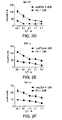


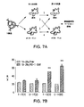


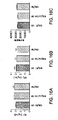


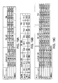


Classifications
-
- C—CHEMISTRY; METALLURGY
- C12—BIOCHEMISTRY; BEER; SPIRITS; WINE; VINEGAR; MICROBIOLOGY; ENZYMOLOGY; MUTATION OR GENETIC ENGINEERING
- C12N—MICROORGANISMS OR ENZYMES; COMPOSITIONS THEREOF; PROPAGATING, PRESERVING, OR MAINTAINING MICROORGANISMS; MUTATION OR GENETIC ENGINEERING; CULTURE MEDIA
- C12N5/00—Undifferentiated human, animal or plant cells, e.g. cell lines; Tissues; Cultivation or maintenance thereof; Culture media therefor
- C12N5/06—Animal cells or tissues; Human cells or tissues
- C12N5/0602—Vertebrate cells
- C12N5/0634—Cells from the blood or the immune system
- C12N5/0636—T lymphocytes
-
- C—CHEMISTRY; METALLURGY
- C07—ORGANIC CHEMISTRY
- C07K—PEPTIDES
- C07K16/00—Immunoglobulins [IGs], e.g. monoclonal or polyclonal antibodies
- C07K16/40—Immunoglobulins [IGs], e.g. monoclonal or polyclonal antibodies against enzymes
-
- A—HUMAN NECESSITIES
- A61—MEDICAL OR VETERINARY SCIENCE; HYGIENE
- A61K—PREPARATIONS FOR MEDICAL, DENTAL OR TOILETRY PURPOSES
- A61K2239/00—Indexing codes associated with cellular immunotherapy of group A61K39/46
- A61K2239/10—Indexing codes associated with cellular immunotherapy of group A61K39/46 characterized by the structure of the chimeric antigen receptor [CAR]
- A61K2239/22—Intracellular domain
-
- A—HUMAN NECESSITIES
- A61—MEDICAL OR VETERINARY SCIENCE; HYGIENE
- A61K—PREPARATIONS FOR MEDICAL, DENTAL OR TOILETRY PURPOSES
- A61K2239/00—Indexing codes associated with cellular immunotherapy of group A61K39/46
- A61K2239/26—Universal/off- the- shelf cellular immunotherapy; Allogenic cells or means to avoid rejection
-
- A—HUMAN NECESSITIES
- A61—MEDICAL OR VETERINARY SCIENCE; HYGIENE
- A61K—PREPARATIONS FOR MEDICAL, DENTAL OR TOILETRY PURPOSES
- A61K2239/00—Indexing codes associated with cellular immunotherapy of group A61K39/46
- A61K2239/31—Indexing codes associated with cellular immunotherapy of group A61K39/46 characterized by the route of administration
-
- A—HUMAN NECESSITIES
- A61—MEDICAL OR VETERINARY SCIENCE; HYGIENE
- A61K—PREPARATIONS FOR MEDICAL, DENTAL OR TOILETRY PURPOSES
- A61K2239/00—Indexing codes associated with cellular immunotherapy of group A61K39/46
- A61K2239/38—Indexing codes associated with cellular immunotherapy of group A61K39/46 characterised by the dose, timing or administration schedule
-
- A—HUMAN NECESSITIES
- A61—MEDICAL OR VETERINARY SCIENCE; HYGIENE
- A61K—PREPARATIONS FOR MEDICAL, DENTAL OR TOILETRY PURPOSES
- A61K35/00—Medicinal preparations containing materials or reaction products thereof with undetermined constitution
- A61K35/12—Materials from mammals; Compositions comprising non-specified tissues or cells; Compositions comprising non-embryonic stem cells; Genetically modified cells
- A61K35/14—Blood; Artificial blood
- A61K35/17—Lymphocytes; B-cells; T-cells; Natural killer cells; Interferon-activated or cytokine-activated lymphocytes
-
- A—HUMAN NECESSITIES
- A61—MEDICAL OR VETERINARY SCIENCE; HYGIENE
- A61K—PREPARATIONS FOR MEDICAL, DENTAL OR TOILETRY PURPOSES
- A61K39/00—Medicinal preparations containing antigens or antibodies
- A61K39/0005—Vertebrate antigens
- A61K39/0011—Cancer antigens
-
- A—HUMAN NECESSITIES
- A61—MEDICAL OR VETERINARY SCIENCE; HYGIENE
- A61K—PREPARATIONS FOR MEDICAL, DENTAL OR TOILETRY PURPOSES
- A61K39/00—Medicinal preparations containing antigens or antibodies
- A61K39/0005—Vertebrate antigens
- A61K39/0011—Cancer antigens
- A61K39/001102—Receptors, cell surface antigens or cell surface determinants
- A61K39/001103—Receptors for growth factors
- A61K39/001106—Her-2/neu/ErbB2, Her-3/ErbB3 or Her 4/ErbB4
-
- A—HUMAN NECESSITIES
- A61—MEDICAL OR VETERINARY SCIENCE; HYGIENE
- A61K—PREPARATIONS FOR MEDICAL, DENTAL OR TOILETRY PURPOSES
- A61K39/00—Medicinal preparations containing antigens or antibodies
- A61K39/0005—Vertebrate antigens
- A61K39/0011—Cancer antigens
- A61K39/001102—Receptors, cell surface antigens or cell surface determinants
- A61K39/001103—Receptors for growth factors
- A61K39/001109—Vascular endothelial growth factor receptors [VEGFR]
-
- A—HUMAN NECESSITIES
- A61—MEDICAL OR VETERINARY SCIENCE; HYGIENE
- A61K—PREPARATIONS FOR MEDICAL, DENTAL OR TOILETRY PURPOSES
- A61K39/00—Medicinal preparations containing antigens or antibodies
- A61K39/0005—Vertebrate antigens
- A61K39/0011—Cancer antigens
- A61K39/001102—Receptors, cell surface antigens or cell surface determinants
- A61K39/001111—Immunoglobulin superfamily
- A61K39/001112—CD19 or B4
-
- A—HUMAN NECESSITIES
- A61—MEDICAL OR VETERINARY SCIENCE; HYGIENE
- A61K—PREPARATIONS FOR MEDICAL, DENTAL OR TOILETRY PURPOSES
- A61K39/00—Medicinal preparations containing antigens or antibodies
- A61K39/0005—Vertebrate antigens
- A61K39/0011—Cancer antigens
- A61K39/001102—Receptors, cell surface antigens or cell surface determinants
- A61K39/001111—Immunoglobulin superfamily
- A61K39/001113—CD22, BL-CAM, siglec-2 or sialic acid- binding Ig-related lectin 2
-
- A—HUMAN NECESSITIES
- A61—MEDICAL OR VETERINARY SCIENCE; HYGIENE
- A61K—PREPARATIONS FOR MEDICAL, DENTAL OR TOILETRY PURPOSES
- A61K39/00—Medicinal preparations containing antigens or antibodies
- A61K39/0005—Vertebrate antigens
- A61K39/0011—Cancer antigens
- A61K39/001102—Receptors, cell surface antigens or cell surface determinants
- A61K39/001111—Immunoglobulin superfamily
- A61K39/001114—CD74, Ii, MHC class II invariant chain or MHC class II gamma chain
-
- A—HUMAN NECESSITIES
- A61—MEDICAL OR VETERINARY SCIENCE; HYGIENE
- A61K—PREPARATIONS FOR MEDICAL, DENTAL OR TOILETRY PURPOSES
- A61K39/00—Medicinal preparations containing antigens or antibodies
- A61K39/0005—Vertebrate antigens
- A61K39/0011—Cancer antigens
- A61K39/001102—Receptors, cell surface antigens or cell surface determinants
- A61K39/001116—Receptors for cytokines
- A61K39/001117—Receptors for tumor necrosis factors [TNF], e.g. lymphotoxin receptor [LTR] or CD30
-
- A—HUMAN NECESSITIES
- A61—MEDICAL OR VETERINARY SCIENCE; HYGIENE
- A61K—PREPARATIONS FOR MEDICAL, DENTAL OR TOILETRY PURPOSES
- A61K39/00—Medicinal preparations containing antigens or antibodies
- A61K39/0005—Vertebrate antigens
- A61K39/0011—Cancer antigens
- A61K39/001102—Receptors, cell surface antigens or cell surface determinants
- A61K39/001116—Receptors for cytokines
- A61K39/001119—Receptors for interleukins [IL]
-
- A—HUMAN NECESSITIES
- A61—MEDICAL OR VETERINARY SCIENCE; HYGIENE
- A61K—PREPARATIONS FOR MEDICAL, DENTAL OR TOILETRY PURPOSES
- A61K39/00—Medicinal preparations containing antigens or antibodies
- A61K39/0005—Vertebrate antigens
- A61K39/0011—Cancer antigens
- A61K39/001102—Receptors, cell surface antigens or cell surface determinants
- A61K39/001124—CD20
-
- A—HUMAN NECESSITIES
- A61—MEDICAL OR VETERINARY SCIENCE; HYGIENE
- A61K—PREPARATIONS FOR MEDICAL, DENTAL OR TOILETRY PURPOSES
- A61K39/00—Medicinal preparations containing antigens or antibodies
- A61K39/0005—Vertebrate antigens
- A61K39/0011—Cancer antigens
- A61K39/001102—Receptors, cell surface antigens or cell surface determinants
- A61K39/001126—CD38 not IgG
-
- A—HUMAN NECESSITIES
- A61—MEDICAL OR VETERINARY SCIENCE; HYGIENE
- A61K—PREPARATIONS FOR MEDICAL, DENTAL OR TOILETRY PURPOSES
- A61K39/00—Medicinal preparations containing antigens or antibodies
- A61K39/0005—Vertebrate antigens
- A61K39/0011—Cancer antigens
- A61K39/001102—Receptors, cell surface antigens or cell surface determinants
- A61K39/001128—CD44 not IgG
-
- A—HUMAN NECESSITIES
- A61—MEDICAL OR VETERINARY SCIENCE; HYGIENE
- A61K—PREPARATIONS FOR MEDICAL, DENTAL OR TOILETRY PURPOSES
- A61K39/00—Medicinal preparations containing antigens or antibodies
- A61K39/0005—Vertebrate antigens
- A61K39/0011—Cancer antigens
- A61K39/001102—Receptors, cell surface antigens or cell surface determinants
- A61K39/001129—Molecules with a "CD" designation not provided for elsewhere
-
- A—HUMAN NECESSITIES
- A61—MEDICAL OR VETERINARY SCIENCE; HYGIENE
- A61K—PREPARATIONS FOR MEDICAL, DENTAL OR TOILETRY PURPOSES
- A61K39/00—Medicinal preparations containing antigens or antibodies
- A61K39/0005—Vertebrate antigens
- A61K39/0011—Cancer antigens
- A61K39/001152—Transcription factors, e.g. SOX or c-MYC
- A61K39/001153—Wilms tumor 1 [WT1]
-
- A—HUMAN NECESSITIES
- A61—MEDICAL OR VETERINARY SCIENCE; HYGIENE
- A61K—PREPARATIONS FOR MEDICAL, DENTAL OR TOILETRY PURPOSES
- A61K39/00—Medicinal preparations containing antigens or antibodies
- A61K39/0005—Vertebrate antigens
- A61K39/0011—Cancer antigens
- A61K39/001154—Enzymes
- A61K39/001157—Telomerase or TERT [telomerase reverse transcriptase]
-
- A—HUMAN NECESSITIES
- A61—MEDICAL OR VETERINARY SCIENCE; HYGIENE
- A61K—PREPARATIONS FOR MEDICAL, DENTAL OR TOILETRY PURPOSES
- A61K39/00—Medicinal preparations containing antigens or antibodies
- A61K39/0005—Vertebrate antigens
- A61K39/0011—Cancer antigens
- A61K39/001166—Adhesion molecules, e.g. NRCAM, EpCAM or cadherins
- A61K39/001168—Mesothelin [MSLN]
-
- A—HUMAN NECESSITIES
- A61—MEDICAL OR VETERINARY SCIENCE; HYGIENE
- A61K—PREPARATIONS FOR MEDICAL, DENTAL OR TOILETRY PURPOSES
- A61K39/00—Medicinal preparations containing antigens or antibodies
- A61K39/0005—Vertebrate antigens
- A61K39/0011—Cancer antigens
- A61K39/001169—Tumor associated carbohydrates
- A61K39/00117—Mucins, e.g. MUC-1
-
- A—HUMAN NECESSITIES
- A61—MEDICAL OR VETERINARY SCIENCE; HYGIENE
- A61K—PREPARATIONS FOR MEDICAL, DENTAL OR TOILETRY PURPOSES
- A61K39/00—Medicinal preparations containing antigens or antibodies
- A61K39/0005—Vertebrate antigens
- A61K39/0011—Cancer antigens
- A61K39/001169—Tumor associated carbohydrates
- A61K39/001171—Gangliosides, e.g. GM2, GD2 or GD3
-
- A—HUMAN NECESSITIES
- A61—MEDICAL OR VETERINARY SCIENCE; HYGIENE
- A61K—PREPARATIONS FOR MEDICAL, DENTAL OR TOILETRY PURPOSES
- A61K39/00—Medicinal preparations containing antigens or antibodies
- A61K39/0005—Vertebrate antigens
- A61K39/0011—Cancer antigens
- A61K39/00118—Cancer antigens from embryonic or fetal origin
- A61K39/001182—Carcinoembryonic antigen [CEA]
-
- A—HUMAN NECESSITIES
- A61—MEDICAL OR VETERINARY SCIENCE; HYGIENE
- A61K—PREPARATIONS FOR MEDICAL, DENTAL OR TOILETRY PURPOSES
- A61K39/00—Medicinal preparations containing antigens or antibodies
- A61K39/0005—Vertebrate antigens
- A61K39/0011—Cancer antigens
- A61K39/001184—Cancer testis antigens, e.g. SSX, BAGE, GAGE or SAGE
- A61K39/001186—MAGE
-
- A—HUMAN NECESSITIES
- A61—MEDICAL OR VETERINARY SCIENCE; HYGIENE
- A61K—PREPARATIONS FOR MEDICAL, DENTAL OR TOILETRY PURPOSES
- A61K39/00—Medicinal preparations containing antigens or antibodies
- A61K39/0005—Vertebrate antigens
- A61K39/0011—Cancer antigens
- A61K39/001184—Cancer testis antigens, e.g. SSX, BAGE, GAGE or SAGE
- A61K39/001188—NY-ESO
-
- A—HUMAN NECESSITIES
- A61—MEDICAL OR VETERINARY SCIENCE; HYGIENE
- A61K—PREPARATIONS FOR MEDICAL, DENTAL OR TOILETRY PURPOSES
- A61K39/00—Medicinal preparations containing antigens or antibodies
- A61K39/0005—Vertebrate antigens
- A61K39/0011—Cancer antigens
- A61K39/001193—Prostate associated antigens e.g. Prostate stem cell antigen [PSCA]; Prostate carcinoma tumor antigen [PCTA]; PAP or PSGR
- A61K39/001195—Prostate specific membrane antigen [PSMA]
-
- A—HUMAN NECESSITIES
- A61—MEDICAL OR VETERINARY SCIENCE; HYGIENE
- A61K—PREPARATIONS FOR MEDICAL, DENTAL OR TOILETRY PURPOSES
- A61K39/00—Medicinal preparations containing antigens or antibodies
- A61K39/46—Cellular immunotherapy
- A61K39/461—Cellular immunotherapy characterised by the cell type used
- A61K39/4611—T-cells, e.g. tumor infiltrating lymphocytes [TIL], lymphokine-activated killer cells [LAK] or regulatory T cells [Treg]
-
- A—HUMAN NECESSITIES
- A61—MEDICAL OR VETERINARY SCIENCE; HYGIENE
- A61K—PREPARATIONS FOR MEDICAL, DENTAL OR TOILETRY PURPOSES
- A61K39/00—Medicinal preparations containing antigens or antibodies
- A61K39/46—Cellular immunotherapy
- A61K39/462—Cellular immunotherapy characterized by the effect or the function of the cells
- A61K39/4621—Cellular immunotherapy characterized by the effect or the function of the cells immunosuppressive or immunotolerising
-
- A—HUMAN NECESSITIES
- A61—MEDICAL OR VETERINARY SCIENCE; HYGIENE
- A61K—PREPARATIONS FOR MEDICAL, DENTAL OR TOILETRY PURPOSES
- A61K39/00—Medicinal preparations containing antigens or antibodies
- A61K39/46—Cellular immunotherapy
- A61K39/463—Cellular immunotherapy characterised by recombinant expression
- A61K39/4631—Chimeric Antigen Receptors [CAR]
-
- A—HUMAN NECESSITIES
- A61—MEDICAL OR VETERINARY SCIENCE; HYGIENE
- A61K—PREPARATIONS FOR MEDICAL, DENTAL OR TOILETRY PURPOSES
- A61K39/00—Medicinal preparations containing antigens or antibodies
- A61K39/46—Cellular immunotherapy
- A61K39/463—Cellular immunotherapy characterised by recombinant expression
- A61K39/4636—Immune checkpoint inhibitors
-
- A—HUMAN NECESSITIES
- A61—MEDICAL OR VETERINARY SCIENCE; HYGIENE
- A61K—PREPARATIONS FOR MEDICAL, DENTAL OR TOILETRY PURPOSES
- A61K39/00—Medicinal preparations containing antigens or antibodies
- A61K39/46—Cellular immunotherapy
- A61K39/464—Cellular immunotherapy characterised by the antigen targeted or presented
- A61K39/4643—Vertebrate antigens
- A61K39/46431—Contraceptive or sex hormones
-
- A—HUMAN NECESSITIES
- A61—MEDICAL OR VETERINARY SCIENCE; HYGIENE
- A61K—PREPARATIONS FOR MEDICAL, DENTAL OR TOILETRY PURPOSES
- A61K39/00—Medicinal preparations containing antigens or antibodies
- A61K39/46—Cellular immunotherapy
- A61K39/464—Cellular immunotherapy characterised by the antigen targeted or presented
- A61K39/4643—Vertebrate antigens
- A61K39/46434—Antigens related to induction of tolerance to non-self
-
- A—HUMAN NECESSITIES
- A61—MEDICAL OR VETERINARY SCIENCE; HYGIENE
- A61K—PREPARATIONS FOR MEDICAL, DENTAL OR TOILETRY PURPOSES
- A61K39/00—Medicinal preparations containing antigens or antibodies
- A61K39/46—Cellular immunotherapy
- A61K39/464—Cellular immunotherapy characterised by the antigen targeted or presented
- A61K39/4643—Vertebrate antigens
- A61K39/4644—Cancer antigens
- A61K39/464402—Receptors, cell surface antigens or cell surface determinants
- A61K39/464411—Immunoglobulin superfamily
- A61K39/464412—CD19 or B4
-
- A—HUMAN NECESSITIES
- A61—MEDICAL OR VETERINARY SCIENCE; HYGIENE
- A61K—PREPARATIONS FOR MEDICAL, DENTAL OR TOILETRY PURPOSES
- A61K39/00—Medicinal preparations containing antigens or antibodies
- A61K39/46—Cellular immunotherapy
- A61K39/464—Cellular immunotherapy characterised by the antigen targeted or presented
- A61K39/4643—Vertebrate antigens
- A61K39/4644—Cancer antigens
- A61K39/464493—Prostate associated antigens e.g. Prostate stem cell antigen [PSCA]; Prostate carcinoma tumor antigen [PCTA]; Prostatic acid phosphatase [PAP]; Prostate-specific G-protein-coupled receptor [PSGR]
- A61K39/464495—Prostate specific membrane antigen [PSMA]
-
- A—HUMAN NECESSITIES
- A61—MEDICAL OR VETERINARY SCIENCE; HYGIENE
- A61P—SPECIFIC THERAPEUTIC ACTIVITY OF CHEMICAL COMPOUNDS OR MEDICINAL PREPARATIONS
- A61P1/00—Drugs for disorders of the alimentary tract or the digestive system
- A61P1/18—Drugs for disorders of the alimentary tract or the digestive system for pancreatic disorders, e.g. pancreatic enzymes
-
- A—HUMAN NECESSITIES
- A61—MEDICAL OR VETERINARY SCIENCE; HYGIENE
- A61P—SPECIFIC THERAPEUTIC ACTIVITY OF CHEMICAL COMPOUNDS OR MEDICINAL PREPARATIONS
- A61P11/00—Drugs for disorders of the respiratory system
-
- A—HUMAN NECESSITIES
- A61—MEDICAL OR VETERINARY SCIENCE; HYGIENE
- A61P—SPECIFIC THERAPEUTIC ACTIVITY OF CHEMICAL COMPOUNDS OR MEDICINAL PREPARATIONS
- A61P13/00—Drugs for disorders of the urinary system
- A61P13/08—Drugs for disorders of the urinary system of the prostate
-
- A—HUMAN NECESSITIES
- A61—MEDICAL OR VETERINARY SCIENCE; HYGIENE
- A61P—SPECIFIC THERAPEUTIC ACTIVITY OF CHEMICAL COMPOUNDS OR MEDICINAL PREPARATIONS
- A61P15/00—Drugs for genital or sexual disorders; Contraceptives
-
- A—HUMAN NECESSITIES
- A61—MEDICAL OR VETERINARY SCIENCE; HYGIENE
- A61P—SPECIFIC THERAPEUTIC ACTIVITY OF CHEMICAL COMPOUNDS OR MEDICINAL PREPARATIONS
- A61P19/00—Drugs for skeletal disorders
-
- A—HUMAN NECESSITIES
- A61—MEDICAL OR VETERINARY SCIENCE; HYGIENE
- A61P—SPECIFIC THERAPEUTIC ACTIVITY OF CHEMICAL COMPOUNDS OR MEDICINAL PREPARATIONS
- A61P21/00—Drugs for disorders of the muscular or neuromuscular system
-
- A—HUMAN NECESSITIES
- A61—MEDICAL OR VETERINARY SCIENCE; HYGIENE
- A61P—SPECIFIC THERAPEUTIC ACTIVITY OF CHEMICAL COMPOUNDS OR MEDICINAL PREPARATIONS
- A61P31/00—Antiinfectives, i.e. antibiotics, antiseptics, chemotherapeutics
-
- A—HUMAN NECESSITIES
- A61—MEDICAL OR VETERINARY SCIENCE; HYGIENE
- A61P—SPECIFIC THERAPEUTIC ACTIVITY OF CHEMICAL COMPOUNDS OR MEDICINAL PREPARATIONS
- A61P35/00—Antineoplastic agents
-
- A—HUMAN NECESSITIES
- A61—MEDICAL OR VETERINARY SCIENCE; HYGIENE
- A61P—SPECIFIC THERAPEUTIC ACTIVITY OF CHEMICAL COMPOUNDS OR MEDICINAL PREPARATIONS
- A61P35/00—Antineoplastic agents
- A61P35/02—Antineoplastic agents specific for leukemia
-
- A—HUMAN NECESSITIES
- A61—MEDICAL OR VETERINARY SCIENCE; HYGIENE
- A61P—SPECIFIC THERAPEUTIC ACTIVITY OF CHEMICAL COMPOUNDS OR MEDICINAL PREPARATIONS
- A61P37/00—Drugs for immunological or allergic disorders
- A61P37/02—Immunomodulators
-
- A—HUMAN NECESSITIES
- A61—MEDICAL OR VETERINARY SCIENCE; HYGIENE
- A61P—SPECIFIC THERAPEUTIC ACTIVITY OF CHEMICAL COMPOUNDS OR MEDICINAL PREPARATIONS
- A61P37/00—Drugs for immunological or allergic disorders
- A61P37/02—Immunomodulators
- A61P37/04—Immunostimulants
-
- A—HUMAN NECESSITIES
- A61—MEDICAL OR VETERINARY SCIENCE; HYGIENE
- A61P—SPECIFIC THERAPEUTIC ACTIVITY OF CHEMICAL COMPOUNDS OR MEDICINAL PREPARATIONS
- A61P37/00—Drugs for immunological or allergic disorders
- A61P37/02—Immunomodulators
- A61P37/06—Immunosuppressants, e.g. drugs for graft rejection
-
- A—HUMAN NECESSITIES
- A61—MEDICAL OR VETERINARY SCIENCE; HYGIENE
- A61P—SPECIFIC THERAPEUTIC ACTIVITY OF CHEMICAL COMPOUNDS OR MEDICINAL PREPARATIONS
- A61P43/00—Drugs for specific purposes, not provided for in groups A61P1/00-A61P41/00
-
- C—CHEMISTRY; METALLURGY
- C07—ORGANIC CHEMISTRY
- C07K—PEPTIDES
- C07K14/00—Peptides having more than 20 amino acids; Gastrins; Somatostatins; Melanotropins; Derivatives thereof
- C07K14/435—Peptides having more than 20 amino acids; Gastrins; Somatostatins; Melanotropins; Derivatives thereof from animals; from humans
- C07K14/705—Receptors; Cell surface antigens; Cell surface determinants
- C07K14/70503—Immunoglobulin superfamily
- C07K14/7051—T-cell receptor (TcR)-CD3 complex
-
- C—CHEMISTRY; METALLURGY
- C07—ORGANIC CHEMISTRY
- C07K—PEPTIDES
- C07K14/00—Peptides having more than 20 amino acids; Gastrins; Somatostatins; Melanotropins; Derivatives thereof
- C07K14/435—Peptides having more than 20 amino acids; Gastrins; Somatostatins; Melanotropins; Derivatives thereof from animals; from humans
- C07K14/705—Receptors; Cell surface antigens; Cell surface determinants
- C07K14/70503—Immunoglobulin superfamily
- C07K14/70514—CD4
-
- C—CHEMISTRY; METALLURGY
- C07—ORGANIC CHEMISTRY
- C07K—PEPTIDES
- C07K14/00—Peptides having more than 20 amino acids; Gastrins; Somatostatins; Melanotropins; Derivatives thereof
- C07K14/435—Peptides having more than 20 amino acids; Gastrins; Somatostatins; Melanotropins; Derivatives thereof from animals; from humans
- C07K14/705—Receptors; Cell surface antigens; Cell surface determinants
- C07K14/70503—Immunoglobulin superfamily
- C07K14/70517—CD8
-
- C—CHEMISTRY; METALLURGY
- C07—ORGANIC CHEMISTRY
- C07K—PEPTIDES
- C07K14/00—Peptides having more than 20 amino acids; Gastrins; Somatostatins; Melanotropins; Derivatives thereof
- C07K14/435—Peptides having more than 20 amino acids; Gastrins; Somatostatins; Melanotropins; Derivatives thereof from animals; from humans
- C07K14/705—Receptors; Cell surface antigens; Cell surface determinants
- C07K14/70503—Immunoglobulin superfamily
- C07K14/70521—CD28, CD152
-
- C—CHEMISTRY; METALLURGY
- C07—ORGANIC CHEMISTRY
- C07K—PEPTIDES
- C07K16/00—Immunoglobulins [IGs], e.g. monoclonal or polyclonal antibodies
- C07K16/18—Immunoglobulins [IGs], e.g. monoclonal or polyclonal antibodies against material from animals or humans
- C07K16/28—Immunoglobulins [IGs], e.g. monoclonal or polyclonal antibodies against material from animals or humans against receptors, cell surface antigens or cell surface determinants
- C07K16/2803—Immunoglobulins [IGs], e.g. monoclonal or polyclonal antibodies against material from animals or humans against receptors, cell surface antigens or cell surface determinants against the immunoglobulin superfamily
-
- C—CHEMISTRY; METALLURGY
- C07—ORGANIC CHEMISTRY
- C07K—PEPTIDES
- C07K16/00—Immunoglobulins [IGs], e.g. monoclonal or polyclonal antibodies
- C07K16/18—Immunoglobulins [IGs], e.g. monoclonal or polyclonal antibodies against material from animals or humans
- C07K16/28—Immunoglobulins [IGs], e.g. monoclonal or polyclonal antibodies against material from animals or humans against receptors, cell surface antigens or cell surface determinants
- C07K16/30—Immunoglobulins [IGs], e.g. monoclonal or polyclonal antibodies against material from animals or humans against receptors, cell surface antigens or cell surface determinants from tumour cells
-
- C—CHEMISTRY; METALLURGY
- C12—BIOCHEMISTRY; BEER; SPIRITS; WINE; VINEGAR; MICROBIOLOGY; ENZYMOLOGY; MUTATION OR GENETIC ENGINEERING
- C12N—MICROORGANISMS OR ENZYMES; COMPOSITIONS THEREOF; PROPAGATING, PRESERVING, OR MAINTAINING MICROORGANISMS; MUTATION OR GENETIC ENGINEERING; CULTURE MEDIA
- C12N5/00—Undifferentiated human, animal or plant cells, e.g. cell lines; Tissues; Cultivation or maintenance thereof; Culture media therefor
- C12N5/06—Animal cells or tissues; Human cells or tissues
- C12N5/0602—Vertebrate cells
- C12N5/0634—Cells from the blood or the immune system
- C12N5/0636—T lymphocytes
- C12N5/0638—Cytotoxic T lymphocytes [CTL] or lymphokine activated killer cells [LAK]
-
- A—HUMAN NECESSITIES
- A61—MEDICAL OR VETERINARY SCIENCE; HYGIENE
- A61K—PREPARATIONS FOR MEDICAL, DENTAL OR TOILETRY PURPOSES
- A61K39/00—Medicinal preparations containing antigens or antibodies
- A61K2039/51—Medicinal preparations containing antigens or antibodies comprising whole cells, viruses or DNA/RNA
- A61K2039/515—Animal cells
- A61K2039/5156—Animal cells expressing foreign proteins
-
- A—HUMAN NECESSITIES
- A61—MEDICAL OR VETERINARY SCIENCE; HYGIENE
- A61K—PREPARATIONS FOR MEDICAL, DENTAL OR TOILETRY PURPOSES
- A61K39/00—Medicinal preparations containing antigens or antibodies
- A61K2039/51—Medicinal preparations containing antigens or antibodies comprising whole cells, viruses or DNA/RNA
- A61K2039/515—Animal cells
- A61K2039/5158—Antigen-pulsed cells, e.g. T-cells
-
- A—HUMAN NECESSITIES
- A61—MEDICAL OR VETERINARY SCIENCE; HYGIENE
- A61K—PREPARATIONS FOR MEDICAL, DENTAL OR TOILETRY PURPOSES
- A61K2121/00—Preparations for use in therapy
-
- A—HUMAN NECESSITIES
- A61—MEDICAL OR VETERINARY SCIENCE; HYGIENE
- A61K—PREPARATIONS FOR MEDICAL, DENTAL OR TOILETRY PURPOSES
- A61K2300/00—Mixtures or combinations of active ingredients, wherein at least one active ingredient is fully defined in groups A61K31/00 - A61K41/00
-
- C—CHEMISTRY; METALLURGY
- C07—ORGANIC CHEMISTRY
- C07K—PEPTIDES
- C07K2317/00—Immunoglobulins specific features
- C07K2317/60—Immunoglobulins specific features characterized by non-natural combinations of immunoglobulin fragments
- C07K2317/62—Immunoglobulins specific features characterized by non-natural combinations of immunoglobulin fragments comprising only variable region components
- C07K2317/622—Single chain antibody (scFv)
-
- C—CHEMISTRY; METALLURGY
- C07—ORGANIC CHEMISTRY
- C07K—PEPTIDES
- C07K2319/00—Fusion polypeptide
- C07K2319/01—Fusion polypeptide containing a localisation/targetting motif
- C07K2319/02—Fusion polypeptide containing a localisation/targetting motif containing a signal sequence
-
- C—CHEMISTRY; METALLURGY
- C07—ORGANIC CHEMISTRY
- C07K—PEPTIDES
- C07K2319/00—Fusion polypeptide
- C07K2319/30—Non-immunoglobulin-derived peptide or protein having an immunoglobulin constant or Fc region, or a fragment thereof, attached thereto
-
- C—CHEMISTRY; METALLURGY
- C12—BIOCHEMISTRY; BEER; SPIRITS; WINE; VINEGAR; MICROBIOLOGY; ENZYMOLOGY; MUTATION OR GENETIC ENGINEERING
- C12N—MICROORGANISMS OR ENZYMES; COMPOSITIONS THEREOF; PROPAGATING, PRESERVING, OR MAINTAINING MICROORGANISMS; MUTATION OR GENETIC ENGINEERING; CULTURE MEDIA
- C12N2510/00—Genetically modified cells
Landscapes
- Health & Medical Sciences (AREA)
- Life Sciences & Earth Sciences (AREA)
- Chemical & Material Sciences (AREA)
- Immunology (AREA)
- General Health & Medical Sciences (AREA)
- Medicinal Chemistry (AREA)
- Veterinary Medicine (AREA)
- Animal Behavior & Ethology (AREA)
- Public Health (AREA)
- Pharmacology & Pharmacy (AREA)
- Cell Biology (AREA)
- Organic Chemistry (AREA)
- Microbiology (AREA)
- Epidemiology (AREA)
- Mycology (AREA)
- Engineering & Computer Science (AREA)
- Oncology (AREA)
- Bioinformatics & Cheminformatics (AREA)
- Genetics & Genomics (AREA)
- Zoology (AREA)
- Biomedical Technology (AREA)
- Chemical Kinetics & Catalysis (AREA)
- General Chemical & Material Sciences (AREA)
- Nuclear Medicine, Radiotherapy & Molecular Imaging (AREA)
- Biochemistry (AREA)
- Biotechnology (AREA)
- Wood Science & Technology (AREA)
- Molecular Biology (AREA)
- Biophysics (AREA)
- Proteomics, Peptides & Aminoacids (AREA)
- Hematology (AREA)
- Toxicology (AREA)
- Gastroenterology & Hepatology (AREA)
- Developmental Biology & Embryology (AREA)
- General Engineering & Computer Science (AREA)
- Reproductive Health (AREA)
- Endocrinology (AREA)
- Physical Education & Sports Medicine (AREA)
- Gynecology & Obstetrics (AREA)
- Pregnancy & Childbirth (AREA)
Description
(優先権の主張)
本出願は、2013年3月15日に出願された米国仮特許出願第61/802,118号の優先権を主張し、当該出願の内容はその全体が本明細書に参考として援用される。
(Priority claim)
This application claims the priority of US Provisional Patent Application No. 61 / 802,118 filed on March 15, 2013, the entire contents of which are incorporated herein by reference in its entirety.
(導入)
本発明は、第1の抗原と結合抗原と結合する抗原認識レセプターおよび第2の抗原と結合する阻害性キメラ抗原レセプター(iCAR)を含む免疫応答細胞を提供し、ここで上記第1の抗原への上記抗原認識レセプターの結合は、上記免疫応答細胞を活性化し、上記第2の抗原への上記iCARの結合は、上記免疫応答細胞を阻害する。本発明はまた、上記免疫応答細胞を生産するための方法、および上記免疫応答細胞を使用することによって癌を処置するための方法を提供する。
(Introduction)
The present invention provides immune-responsive cells comprising an antigen-recognizing receptor that binds to a first antigen and an antigen-binding antigen and an inhibitory chimeric antigen receptor (iCAR) that binds to a second antigen, and here to the first antigen. The binding of the antigen-recognizing receptor in the above activates the immune-responsive cells, and the binding of the iCAR to the second antigen inhibits the immune-responsive cells. The present invention also provides a method for producing the immune-responsive cells and a method for treating cancer by using the immune-responsive cells.
(発明の背景)
T細胞ベースの治療は、骨髄移植および臓器移植、癌免疫療法、ウイルス感染、ならびに自己免疫疾患において治癒の可能性を有する。しかし、主要な処置関連合併症は、ドナーリンパ球注入もしくは自己由来の標的化T細胞療法における「オンターゲット・オフ腫瘍反応性(on−target, off−tumor reactivity)」後の移植片対宿主病(GVHD)におけるような、正常な組織に対する意図しないT細胞反応性から生じる。
(Background of invention)
T cell-based therapies have curative potential in bone marrow and organ transplants, cancer immunotherapy, viral infections, and autoimmune diseases. However, the major treatment-related complication is graft-versus-host disease after "on-target, off-tumor reactivity" in donor lymphocyte infusion or autologous targeted T cell therapy. It results from unintended T cell reactivity to normal tissue, such as in (GVHD).
同種異系骨髄移植(BMT;全世界で1年に25,000症例)におけるドナーリンパ球注入(DLI)の使用は、ある種の患者部分セットにおいて有意な治癒の増加を生じた。近年、DLIは、転移性腎細胞癌、乳癌、結腸癌、および卵巣癌を有する患者に対する有効な治療を提供することが示されており、治験が転移性固形腫瘍を処置するために進行中である。DLIの重要な効力(血液の悪性腫瘍の状況で移植片対白血病(GVL)といわれる)は、いくつかのグループが、GVHD誘発の低下が達成されなければDLIの現在のスキームが生存を改善できないと結論づけるほどまでに、急性および慢性の移植片対宿主病(GVHD)の両方の誘発によって制限される(40%を超える割合)。 The use of donor lymphocyte infusion (DLI) in allogeneic bone marrow transplantation (BMT; 25,000 cases worldwide per year) resulted in a significant increase in cure in certain patient subsets. In recent years, DLI has been shown to provide effective treatment for patients with metastatic renal cell carcinoma, breast cancer, colon cancer, and ovarian cancer, and clinical trials are underway to treat metastatic solid tumors. is there. An important efficacy of DLI (called graft-versus-leukemia (GVL) in the context of malignant tumors of the blood) is that some groups cannot improve survival with the current scheme of DLI unless a reduction in GVHD induction is achieved. By the time we conclude, it is limited by the induction of both acute and chronic graft-versus-host disease (GVHD) (> 40%).
同様に、癌免疫療法治験は、TCRおよびCARがそうされたT細胞に由来する「オンターゲットであるが、オフ組織(on−target but off−tissue)」の有害事象(例えば、抗CD19 CARを発現する細胞で処置した慢性リンパ性白血病(CLL)患者におけるB細胞形成不全、肺上皮に対する抗ERBB2 CAR T細胞交叉反応性に由来する胎児急性呼吸窮迫症候群(ARDS)、およびMage−A3 TCRで処置した黒色腫および骨髄腫患者における心筋壊死による死亡が挙げられる)を報告した。 Similarly, cancer immunotherapy trials have TCR and CAR derived from such T cells as "on-target but off-tissue" adverse events (eg, anti-CD19 CAR). Treated with B-cell dysplasia in patients with chronic lymphocytic leukemia (CLL) treated with expressing cells, fetal acute respiratory distress syndrome (ARDS) resulting from anti-ERBB2 CAR T cell cross-reactivity to lung epithelium, and Mage-A3 TCR Death from myocardial necrosis in patients with melanoma and myeloma) was reported.
非特異的免疫抑制およびT細胞排除は、治療利益を捨てて重症の二次合併症を引き起こすという代償を払って、望ましくないT細胞応答を制御する現在唯一の手段である。それらは、重症かつときおり御しがたい、従って、別の点で効果的な細胞処置の使用を制限し得る症状の出現に依拠する。 Non-specific immunosuppression and T cell elimination are currently the only means of controlling unwanted T cell responses at the cost of abandoning therapeutic benefits and causing severe secondary complications. They rely on the appearance of symptoms that are severe and sometimes uncontrollable, and thus may otherwise limit the use of effective cellular treatments.
細胞性の副作用という帰結を防止するストラテジーは、実際に必要とされている。現在のアプローチでは、望ましくない副作用を制御することにおける利点としてT細胞療法のより高次の複雑性を利用できない。よって、新規な治療ストラテジーが切迫して必要とされる。 Strategies that prevent the consequences of cellular side effects are actually needed. Current approaches do not take advantage of the higher complexity of T cell therapy as an advantage in controlling unwanted side effects. Therefore, new treatment strategies are urgently needed.
(発明の要旨)
本発明は、一般に、免疫細胞活性化活性を有する抗原結合レセプター(例えば、CARもしくはTCR)および上記免疫応答細胞の免疫活性を選択的に低下もしくは除去する阻害性キメラ抗原レセプター(iCAR)を発現する免疫応答細胞(例えば、T細胞、ナチュラルキラー(NK)細胞、細胞傷害性Tリンパ球(CTL)、および制御性T細胞)を提供する。従って、上記免疫応答細胞のオフターゲット効果は、低下される。いくつかの実施形態において、免疫活性の低下は、可逆的である。よって、本発明は、新生物形成、感染性疾患、および他の病状の処置のためにこのような免疫応答細胞を使用するための方法を提供する。
(Gist of the invention)
The present invention generally expresses an antigen-binding receptor having immune cell activating activity (eg, CAR or TCR) and an inhibitory chimeric antigen receptor (iCAR) that selectively reduces or eliminates the immune activity of the immune response cells. Immune response cells (eg, T cells, natural killer (NK) cells, cytotoxic T lymphocytes (CTL), and regulatory T cells) are provided. Therefore, the off-target effect of the immune response cells is reduced. In some embodiments, the reduction in immune activity is reversible. Accordingly, the present invention provides methods for using such immune-responsive cells for the treatment of neoplasm formation, infectious diseases, and other medical conditions.
一局面において、本発明は、単離された免疫応答細胞を提供し、上記免疫応答細胞は、第1の抗原と結合し、ここで上記結合が上記免疫応答細胞を活性化する抗原認識レセプター、および第2の抗原と結合し、ここで上記結合が上記免疫応答細胞を阻害する阻害性キメラ抗原レセプターを含む。 In one aspect, the invention provides an isolated immune response cell, an antigen recognition receptor that binds to a first antigen, where the binding activates the immune response cell. And an inhibitory chimeric antigen receptor that binds to a second antigen, where the binding inhibits the immune response cells.
別の局面において、本発明は、被験体における腫瘍量を低下させるための方法を提供し、上記方法は、有効量の免疫応答細胞を投与し、それによって、上記被験体における腫瘍細胞死を誘発する工程を包含し、上記免疫応答細胞は、第1の抗原と結合し、ここで上記結合が上記免疫応答細胞を活性化する抗原認識レセプター、および第2の抗原と結合し、ここで上記結合が上記免疫応答細胞を阻害する阻害性キメラ抗原レセプターを含む。上記方法は、非腫瘍細胞と比較して、腫瘍細胞を選択的に標的とし得る。いくつかの実施形態において、上記方法は、腫瘍細胞の数を減少させる。いくつかの実施形態において、上記方法は、腫瘍サイズを低下させる。他の実施形態において、上記方法は、上記被験体における腫瘍を根絶する。 In another aspect, the invention provides a method for reducing tumor volume in a subject, wherein an effective amount of immune response cells is administered, thereby inducing tumor cell death in the subject. The immune response cell binds to a first antigen, where the binding binds to an antigen recognition receptor that activates the immune response cell, and a second antigen, where the binding is described. Includes an inhibitory chimeric antigen receptor that inhibits the immune response cells. The above method may selectively target tumor cells as compared to non-tumor cells. In some embodiments, the method reduces the number of tumor cells. In some embodiments, the method reduces tumor size. In another embodiment, the method eradicates a tumor in the subject.
別の局面において、本発明は、新生物形成を有する被験体の生存を増大させるための方法を提供し、上記方法は、有効量の免疫応答細胞を投与し、それによって、上記被験体における新生物形成を処置もしくは予防する工程を包含し、上記免疫応答細胞は、第1の抗原と結合し、ここで上記結合が上記免疫応答細胞を活性化する抗原認識レセプター、および第2の抗原と結合し、ここで上記結合が上記免疫応答細胞を阻害する阻害性キメラ抗原レセプターを含む。ある種の実施形態において、上記新生物形成は、血液の癌、B細胞白血病、多発性骨髄腫、リンパ芽球性白血病(ALL)、慢性リンパ性白血病、非ホジキンリンパ腫、卵巣癌、前立腺癌、膵臓癌、肺癌、乳癌、肉腫、ならびに急性骨髄性白血病(AML)からなる群より選択される。 In another aspect, the invention provides a method for increasing the survival of a subject with neoplastic formation, wherein an effective amount of immune response cells is administered, thereby novel in the subject. Including the step of treating or preventing bioformation, the immune response cell binds to a first antigen, where the binding binds to an antigen recognition receptor that activates the immune response cell, and a second antigen. And here the binding comprises an inhibitory chimeric antigen receptor that inhibits the immune response cells. In certain embodiments, the neoplastic formation is blood cancer, B-cell leukemia, multiple myeloid leukemia, lymphoblastic leukemia (ALL), chronic lymphocytic leukemia, non-Hodgkin's lymphoma, ovarian cancer, prostate cancer, It is selected from the group consisting of pancreatic cancer, lung cancer, breast cancer, sarcoma, and acute myeloid leukemia (AML).
別の局面において、本発明は、第1の抗原と結合する抗原認識レセプターを含む抗原特異的免疫応答細胞を生産するための方法を提供し、上記方法は、上記免疫応答細胞に、第2の抗原と結合し、ここで上記結合が上記免疫応答細胞を阻害する阻害性キメラ抗原レセプターをコードする核酸配列を導入する工程を包含する。 In another aspect, the invention provides a method for producing an antigen-specific immune response cell comprising an antigen recognition receptor that binds to a first antigen, the method comprising the immune response cell with a second. It comprises the step of introducing a nucleic acid sequence that binds to an antigen, wherein the binding encodes an inhibitory chimeric antigen receptor that inhibits the immune response cell.
別の局面において、本発明は、抗原と結合する細胞外ドメイン;上記細胞外ドメインに作動可能に連結された膜貫通ドメイン;および細胞内シグナル伝達を活性化して免疫応答を低下させる細胞内ドメインであって、上記膜貫通ドメインに作動可能に連結されている、細胞内ドメインを含む、阻害性キメラ抗原レセプター(iCAR)を提供する。いくつかの実施形態において、上記細胞内シグナル伝達ドメインは、CTLA−4ポリペプチド、PD−1ポリペプチド、LAG−3ポリペプチド、2B4ポリペプチド、およびBTLAポリペプチドからなる群より選択される。ある種の実施形態において、上記膜貫通ドメインは、CD4ポリペプチド、CD8ポリペプチド、CTLA−4ポリペプチド、PD−1ポリペプチド、LAG−3ポリペプチド、2B4ポリペプチド、およびBTLAポリペプチドからなる群より選択される。さらに、上記阻害性キメラ抗原レセプター(iCAR)は、Fab、scFv、リガンド、特異的リガンド、もしくは多価リガンドをさらに含み得る。いくつかの実施形態において、上記iCARへの抗原の結合は、免疫応答を低下させる上記細胞内シグナル伝達ドメインを活性化する。 In another aspect, the invention is an extracellular domain that binds to an antigen; a transmembrane domain that is operably linked to the extracellular domain; and an intracellular domain that activates intracellular signaling and reduces the immune response. It provides an inhibitory chimeric antigen receptor (iCAR) that comprises an intracellular domain that is operably linked to the transmembrane domain. In some embodiments, the intracellular signaling domain is selected from the group consisting of CTLA-4 polypeptide, PD-1 polypeptide, LAG-3 polypeptide, 2B4 polypeptide, and BTLA polypeptide. In certain embodiments, the transmembrane domain comprises a group consisting of a CD4 polypeptide, a CD8 polypeptide, a CTLA-4 polypeptide, a PD-1 polypeptide, a LAG-3 polypeptide, a 2B4 polypeptide, and a BTLA polypeptide. Will be selected. In addition, the inhibitory chimeric antigen receptor (iCAR) may further comprise a Fab, scFv, ligand, specific ligand, or multivalent ligand. In some embodiments, binding of the antigen to the iCAR activates the intracellular signaling domain that reduces the immune response.
関連する局面において、本発明は、抗原と結合する阻害性キメラ抗原レセプターをコードする核酸配列を提供し、ここで上記結合は、免疫応答を低下させる細胞内シグナル伝達ドメインを活性化する。 In a related aspect, the invention provides a nucleic acid sequence encoding an inhibitory chimeric antigen receptor that binds to an antigen, where the binding activates an intracellular signaling domain that reduces an immune response.
別の関連する局面において、本発明は、抗原と結合する阻害性キメラ抗原レセプターをコードする核酸配列を含むベクターを提供し、ここで上記結合は、免疫応答を低下させる細胞内シグナル伝達ドメインを活性化する。 In another related aspect, the invention provides a vector comprising a nucleic acid sequence encoding an inhibitory chimeric antigen receptor that binds to an antigen, wherein the binding activates an intracellular signaling domain that reduces an immune response. To become.
関連する局面において、本発明は、有効量の本発明の免疫応答細胞を薬学的に受容可能な賦形剤中に含む、(新生物形成もしくは病原体感染の処置のための)薬学的組成物を提供する。 In a related aspect, the invention comprises a pharmaceutical composition (for the treatment of neoplasm formation or pathogen infection) comprising an effective amount of the immune response cells of the invention in a pharmaceutically acceptable excipient. provide.
別の局面において、本発明は、新生物形成、病原体感染、自己免疫障害、もしくは同種異系移植の処置のためのキットを提供し、上記キットは、第1の抗原と結合し、上記免疫応答細胞を活性化する抗原認識レセプター、および第2の抗原と結合し、上記免疫応答細胞を阻害する阻害性キメラ抗原レセプターを含む免疫応答細胞を含む。種々の実施形態において、上記キットは、新生物形成、病原体感染、自己免疫障害、もしくは同種異系移植を有する被験体を処置するために上記細胞を使用するための書面による説明書をさらに含む。 In another aspect, the invention provides a kit for the treatment of neoplasm formation, pathogen infection, autoimmune disorders, or allogeneic transplantation, wherein the kit binds to a first antigen and the immune response. Includes an antigen-recognizing receptor that activates cells and an immune-responsive cell that comprises an inhibitory chimeric antigen receptor that binds to a second antigen and inhibits the immune-responsive cell. In various embodiments, the kit further comprises written instructions for using the cells to treat a subject having neoplasm formation, pathogen infection, autoimmune disorder, or allogeneic transplantation.
別の局面において、本発明は、被験体における移植片対白血病応答もしくは移植片対腫瘍応答を調節するための方法を提供し、上記方法は、上記被験体に対して同種異系の免疫応答細胞の有効量を投与し、それによって上記被験体における移植片対白血病応答もしくは移植片対腫瘍応答を調節する工程を包含し、上記免疫応答細胞は、抗原と結合し、ここで上記結合が上記同種異系の免疫応答細胞を阻害する阻害性キメラ抗原レセプターを含む。ある種の実施形態において、上記被験体は、転移性乳癌、血液の悪性腫瘍、もしくは固形腫瘍を有し、上記ヒト白血球抗原(HLA)は、HLA−Iである。ある種の実施形態において、上記被験体は、上皮間葉転換(EMT)を既に受けた腫瘍を有し、上記抗原は、上皮間葉転換(EMT)抗原、E−カドヘリン、およびサイトケラチンのうちの1種以上である。種々の実施形態において、上記阻害性キメラ抗原レセプターおよび上記抗原の結合は、上記抗原を含む細胞における細胞死を減少させる。上記方法は、上記被験体における移植片対宿主病(GVHD)、もしくはその症状を低下させ得る。 In another aspect, the invention provides a method for regulating an antigen-to-leukemic or implant-to-tumor response in a subject, wherein the method provides allogeneic immune response cells to the subject. To include the step of administering an effective amount of, thereby regulating a transplant-to-leukemia or transplant-to-tumor response in the subject, the immune response cells bind to the antigen, where the binding is allogeneic. Includes an inhibitory chimeric antigen receptor that inhibits allogeneic immune response cells. In certain embodiments, the subject has metastatic breast cancer, a malignant tumor of blood, or a solid tumor, and the human leukocyte antigen (HLA) is HLA-I. In certain embodiments, the subject has a tumor that has already undergone epithelial-mesenchymal transition (EMT), and the antigens are among the epithelial-mesenchymal transition (EMT) antigens, E-cadherin, and cytokeratin. It is one or more of. In various embodiments, binding of the inhibitory chimeric antigen receptor and the antigen reduces cell death in cells containing the antigen. The method can reduce graft-versus-host disease (GVHD) in the subject, or its symptoms.
本明細書で詳述される局面の種々の実施形態において、上記第1の抗原もしくは上記抗原認識レセプターの抗原は、腫瘍抗原もしくは病原体抗原である。特定の実施形態において、上記抗原認識レセプターの抗原は、以下からなる群より選択される1種以上の腫瘍抗原である:CD19、CAIX、CEA、CD5、CD7、CD10、CD20、CD22、CD30、CD33、CD34、CD38、CD41、CD44、CD49f、CD56、CD74、CD123、CD133、CD138、サイトメガロウイルス(CMV)感染細胞抗原、EGP−2、EGP−40、EpCAM、erb−B2,3,4、F8P、胎児型アセチルコリンレセプター、葉酸レセプター−a、GD2、GD3、HER−2、hTERT、IL−13R−α2、κ−軽鎖、KDR、LeY、L1細胞接着分子、MAGE−A1、メソテリン、Muc−1、Muc−16、NKG2Dリガンド、NY−ESO−1、癌胎児抗原(h5T4)、PSCA、PSMA、ROR1、TAG−72、VEGF−R2、およびWT−1。 In various embodiments of the aspects detailed herein, the first antigen or the antigen of the antigen recognition receptor is a tumor antigen or a pathogen antigen. In certain embodiments, the antigen of the antigen recognition receptor is one or more tumor antigens selected from the group consisting of: CD19, CAIX, CEA, CD5, CD7, CD10, CD20, CD22, CD30, CD33. , CD34, CD38, CD41, CD44, CD49f, CD56, CD74, CD123, CD133, CD138, cytomegalovirus (CMV) infected cell antigen, EGP-2, EGP-40, EpCAM, erb-B2,3,4, F8P , Fetal acetylcholine receptor, folic acid receptor-a, GD2, GD3, HER-2, hTERT, IL-13R-α2, κ-light chain, KDR, LeY, L1 cell adhesion molecule, MAGE-A1, mesothelin, Muc-1 , Muc-16, NKG2D ligand, NY-ESO-1, carcinoembryonic antigen (h5T4), PSCA, PSMA, ROR1, TAG-72, VEGF-R2, and WT-1.
本明細書で詳述される局面の種々の実施形態において、上記第2の抗原もしくは上記阻害性キメラ抗原レセプターの抗原は、CD33、CD38、ヒト白血球抗原(HLA)、器官特異的抗原、血液脳関門特異的抗原、上皮間葉転換(EMT)抗原、E−カドヘリン、サイトケラチン、オピオイド結合タンパク質/細胞接着分子(OPCML)、HYLA2、結腸直腸癌欠失(Deleted in Colorectal Carcinoma)(DCC)、足場/マトリクス付着領域結合タンパク質1(SMAR1)、細胞表面炭水化物、もしくはムチンタイプO−グリカンである。本明細書で詳述される局面の種々の実施形態において、上記iCARへの抗原の結合は、免疫応答を低下させる上記細胞内シグナル伝達ドメインを活性化する。 In various embodiments of the aspects detailed herein, the second antigen or the antigen of the inhibitory chimeric antigen receptor is CD33, CD38, human leukocyte antigen (HLA), organ-specific antigen, blood brain. Barrier-specific antigens, epithelial mesenchymal (EMT) antigens, E-cadherin, cytokeratin, opioid-binding protein / cell adhesion molecule (OPCML), HYLA2, Deleteed in Correctal Carcinoma (DCC), scaffolding / Matrix adhesion region binding protein 1 (SMAR1), cell surface carbohydrate, or mutin type O-glycan. In various embodiments of the aspects detailed herein, binding of the antigen to the iCAR activates the intracellular signaling domain that reduces the immune response.
本明細書で詳述される局面の種々の実施形態において、上記阻害性キメラ抗原レセプターは、組換え発現される。本明細書で詳述される局面の種々の実施形態において、上記阻害性キメラ抗原レセプターは、ベクターから発現される。本明細書で詳述される局面の種々の実施形態において、上記阻害性キメラ抗原レセプター(iCAR)は、CTLA−4ポリペプチド、PD−1ポリペプチド、LAG−3ポリペプチド、2B4ポリペプチド、およびBTLAポリペプチドからなる群より選択される細胞内シグナル伝達ドメインを含む。本明細書で詳述される局面の種々の実施形態において、上記阻害性キメラ抗原レセプター(iCAR)は、CD4ポリペプチド、CD8ポリペプチド、CTLA−4ポリペプチド、PD−1ポリペプチド、LAG−3ポリペプチド、2B4ポリペプチド、およびBTLAポリペプチドからなる群より選択される膜貫通ドメインを含む。本明細書で詳述される局面の種々の実施形態において、上記阻害性キメラ抗原レセプター(iCAR)は、Fab、scFv、リガンド、特異的リガンド、もしくは多価リガンドを含む。ある種の実施形態において、上記細胞は、19−28z、P28z、M28z、および56−28zからなる群より選択される組換えもしくは内因性の抗原レセプターを発現する。 In various embodiments of the aspects detailed herein, the inhibitory chimeric antigen receptor is recombinantly expressed. In various embodiments of the aspects detailed herein, the inhibitory chimeric antigen receptor is expressed from a vector. In various embodiments of the aspects detailed herein, the inhibitory chimeric antigen receptor (iCAR) is a CTLA-4 polypeptide, PD-1 polypeptide, LAG-3 polypeptide, 2B4 polypeptide, and It contains an intracellular signaling domain selected from the group consisting of BTLA polypeptides. In various embodiments of the aspects detailed herein, the inhibitory chimeric antigen receptor (iCAR) is a CD4 polypeptide, CD8 polypeptide, CTLA-4 polypeptide, PD-1 polypeptide, LAG-3. It contains a transmembrane domain selected from the group consisting of polypeptides, 2B4 polypeptides, and BTLA polypeptides. In various embodiments of the aspects detailed herein, the inhibitory chimeric antigen receptor (iCAR) comprises a Fab, scFv, ligand, specific ligand, or polyvalent ligand. In certain embodiments, the cells express recombinant or endogenous antigen receptors selected from the group consisting of 19-28z, P28z, M28z, and 56-28z.
本明細書で詳述される局面の種々の実施形態において、上記抗原認識レセプターは、T細胞レセプター(TCR)もしくはキメラ抗原レセプター(CAR)である。本明細書で詳述される局面の種々の実施形態において、抗原認識レセプターは、外因性もしくは内因性である。本明細書で詳述される局面の種々の実施形態において、抗原認識レセプターは、組換え発現される。本明細書で詳述される局面の種々の実施形態において、上記抗原認識レセプターは、ベクターから発現される。本明細書で詳述される局面のうちのいずれかの種々の実施形態において、同種異系細胞は、内因性T細胞レセプターを有する。 In various embodiments of the aspects detailed herein, the antigen recognition receptor is a T cell receptor (TCR) or chimeric antigen receptor (CAR). In various embodiments of the aspects detailed herein, the antigen recognition receptor is exogenous or endogenous. In various embodiments of the aspects detailed herein, the antigen recognition receptor is recombinantly expressed. In various embodiments of the aspects detailed herein, the antigen recognition receptor is expressed from a vector. In various embodiments of any of the aspects detailed herein, allogeneic cells have endogenous T cell receptors.
本明細書で詳述される局面の種々の実施形態において、上記細胞は、T細胞、ナチュラルキラー(NK)細胞、細胞傷害性Tリンパ球(CTL)、制御性T細胞、ヒト胚性幹細胞、先天性免疫系の細胞、およびリンパ球へ分化し得る多能性幹細胞のうちの1種以上である。いくつかの実施形態において、上記免疫応答細胞は、自己由来である。他の実施形態において、上記免疫応答細胞は、非自己由来である。
特定の実施形態では、例えば以下が提供される:
(項目1)
1)第1の抗原と結合する抗原認識レセプターであって、ここで該結合は、該免疫応答細胞を活性化する、抗原認識レセプター、および
2)第2の抗原と結合する阻害性キメラ抗原レセプターであって、ここで該結合は、該免疫応答細胞を阻害する、阻害性キメラ抗原レセプター、
を含む、単離された免疫応答細胞。
(項目2)
前記第1の抗原は、腫瘍抗原もしくは病原体抗原である、項目1に記載の単離された免疫応答細胞。
(項目3)
前記第2の抗原は、CD33、CD38、ヒト白血球抗原(HLA)、上皮間葉転換(FMT)抗原、器官特異的抗原、血液脳関門特異的抗原、E−カドヘリン、サイトケラチン、オピオイド結合タンパク質/細胞接着分子(OPCML)、HYLA2、結腸直腸癌欠失(Deleted in Colorectal Carcinoma)(DCC)、足場/マトリクス付着領域結合タンパク質1(SMAR1)、細胞表面炭水化物、ムチンタイプO−グリカンである、項目1に記載の単離された免疫応答細胞。
(項目4)
前記阻害性キメラ抗原レセプターは、組換え発現される、項目1〜3のいずれか1項に記載の単離された免疫応答細胞。
(項目5)
前記阻害性キメラ抗原レセプターは、ベクターから発現される、項目1〜4のいずれか1項に記載の単離された免疫応答細胞。
(項目6)
前記抗原認識レセプターは、T細胞レセプター(TCR)もしくはキメラ抗原レセプター(CAR)である、項目1〜5のいずれか1項に記載の単離された免疫応答細胞。
(項目7)
前記抗原認識レセプターは、外因性もしくは内因性である、項目1〜6のいずれか1項に記載の単離された免疫応答細胞。
(項目8)
前記抗原認識レセプターは、組換え発現される、項目1〜7のいずれか1項に記載の単離された免疫応答細胞。
(項目9)
前記抗原認識レセプターは、ベクターから発現される、項目1〜8のいずれか1項に記載の単離された免疫応答細胞。
(項目10)
前記細胞は、T細胞、ナチュラルキラー(NK)細胞、細胞傷害性Tリンパ球(CTL)、制御性T細胞、ヒト胚性幹細胞、先天性免疫系の細胞、およびリンパ球へ分化し得る多能性幹細胞からなる群より選択される、項目1〜9のいずれか1項に記載の単離された免疫応答細胞。
(項目11)
前記免疫応答細胞は、自己由来である、項目1〜10のいずれか1項に記載の単離された免疫応答細胞。
(項目12)
前記免疫応答細胞は、非自己由来である、項目1〜10のいずれか1項に記載の単離された免疫応答細胞。
(項目13)
前記第1の抗原は、CD19、CAIX、CEA、CDS、CD7、CD10、CD20、CD22、CD30、CD33、CD34、CD38、CD41、CD44、CD49f、CD56、CD74、CD123、CD133、CD138、サイトメガロウイルス(CMV)感染細胞抗原、EGP−2、EGP−40、EpCAM、erb−B2,3,4、FBP、胎児型アセチルコリンレセプター、葉酸レセプター−α、GD2、GD3、HER−2、hTERT、IL−13R−α2、κ−軽鎖、KDR、LeY、L1細胞接着分子、MAGE−A1、メソテリン、Muc−1、Muc−16、NKG2Dリガンド、NY−ESO−1、癌胎児抗原(h5T4)、PSCA、PSMA、ROR1、TAG−72、VEGF−R2、およびWT−1からなる群より選択される腫瘍抗原である、項目1〜12のいずれか1項に記載の単離された免疫応答細胞。
(項目14)
前記阻害性キメラ抗原レセプター(iCAR)は、CTLA−4ポリペプチド、PD−1ポリペプチド、LAG−3ポリペプチド、2B4ポリペプチド、およびBTLAポリペプチドからなる群より選択される細胞内シグナル伝達ドメインを含む、項目1〜13のいずれか1項に記載の単離された免疫応答細胞。
(項目15)
前記阻害性キメラ抗原レセプター(iCAR)は、CD4ポリペプチド、CD8ポリペプチド、CTLA−4ポリペプチド、PD−lポリペプチド、LAG−3ポリペプチド、2B4ポリペプチド、およびBTLAポリペプチドからなる群より選択される膜貫通ドメインをさらに含む、項目14に記載の単離された免疫応答細胞。
(項目16)
前記阻害性キメラ抗原レセプター(iCAR)は、Fab、scFv、リガンド、特異的リガンド、もしくは多価リガンドをさらに含む、項目1〜15のいずれか1項に記載の単離された免疫応答細胞。
(項目17)
前記細胞は、19−28z、P28z、M28z、および56−28zからなる群より選択される組換えもしくは内因性の抗原レセプターを発現する、項目1〜16のいずれか1項に記載の単離された免疫応答細胞。
(項目18)
被験体における腫瘍量を低減するための方法であって、該方法は、有効量免疫応答細胞を投与し、それによって該被験体における腫瘍細胞死を誘発する工程を包含し、該免疫応答細胞は、第1の抗原と結合し、ここで該結合が該免疫応答細胞を活性化させる抗原認識レセプター、および第2の抗原と結合し、ここで該結合が該免疫細胞を阻害する阻害性キメラ抗原レセプターを含む、方法。
(項目19)
非腫瘍細胞と比較して、腫瘍細胞を選択的に標的とする、項目18に記載の方法。
(項目20)
腫瘍細胞の数を減少させる、項目18に記載の方法。
(項目21)
腫瘍サイズを低下させる、項目18に記載の方法。
(項目22)
前記被験体における前記腫瘍を根絶する、項目18に記載の方法。
(項目23)
新生物形成を有する被験体の生存を増進するための方法であって、該方法は、有効量の免疫応答細胞を投与し、それによって、該被験体における新生物形成を処置もしくは防止する工程を包含し、該免疫応答細胞は、第1の抗原と結合し、ここで該結合は該免疫応答細胞を活性化する抗原認識レセプター、および第2の抗原と結合し、ここで該結合は該免疫応答細胞を阻害する阻害性キメラ抗原レセプターを含む、方法。
(項目24)
前記新生物形成は、血液の癌、B細胞白血病、多発性骨髄腫、リンパ芽球性白血病(ALL)、慢性リンパ性白血病、非ホジキンリンパ腫、卵巣癌、前立腺癌、膵臓癌、肺癌、乳癌、および肉腫、急性骨髄性白血病(AML)からなる群より選択される、項目23に記載の方法。
(項目25)
前記第1の抗原は、腫瘍抗原もしくは病原体抗原である、項目17〜23のいずれか1項に記載の方法。
(項目26)
前記第2の抗原は、CD33、CD38、ヒト白血球抗原(HLA)、上皮間葉転換(EMT)抗原、器官特異的抗原、血液脳関門特異的抗原、E−カドヘリン、サイトケラチン、オピオイド結合タンパク質/細胞接着分子(OPCML)、HYLA2、結腸直腸癌欠失(Deleted in Colorectal Carcinoma)(DCC)、足場/マトリクス付着領域結合タンパク質1(SMAR1)、細胞表面炭水化物、もしくはムチンタイプO−グリカンである、項目18〜25のいずれか1項に記載の方法。
(項目27)
前記阻害性キメラ抗原レセプターは、組換え発現される、項目18〜25のいずれか1項に記載の方法。
(項目28)
前記阻害性キメラ抗原レセプターは、ベクターから発現される、項目18〜27のいずれか1項に記載の方法。
(項目29)
前記抗原認識レセプターは、T細胞レセプター(TCR)もしくはキメラ抗原レセプター(CAR)である、項目18〜28のいずれか1項に記載の方法。
(項目30)
前記抗原認識レセプターは、外因性もしくは内因性である、項目18〜29のいずれか1項に記載の方法。
(項目31)
前記抗原認識レセプターは、組換え発現される、項目18〜30のいずれか1項に記載の方法。
(項目32)
前記抗原認識レセプターは、ベクターから発現される、項目18〜31のいずれか1項に記載の方法。
(項目33)
前記細胞は、T細胞、ナチュラルキラー(NK)細胞、細胞傷害性Tリンパ球(CTL)、制御性T細胞、ヒト胚性幹細胞、先天性免疫系の細胞、およびリンパ球へ分化し得る多能性幹細胞からなる群より選択される、項目18〜32のいずれか1項に記載の方法。
(項目34)
前記免疫応答細胞は、自己由来である、項目18〜33のいずれか1項に記載の方法。
(項目35)
前記免疫応答細胞は、非自己由来である、項目18〜33のいずれか1項に記載の方法。
(項目36)
前記第1の抗原が、CD19、CAIX、CEA、CDS、CD7、CD10、CD20、CD22、CD30、CD33、CD34、CD38、CD41、CD44、CD49f、CD56、CD74、CD123、CD133、CD138、サイトメガロウイルス(CMV)感染細胞抗原、EGP−2、EGP−40、EpCAM、erb−B2,3,4、FBP、胎児型アセチルコリンレセプター、葉酸レセプター−α、GD2、GD3、HER−2、hTERT、IL−13R−α2、κ−軽鎖、KDR、LeY、L1細胞接着分子、MAGE−A1、メソテリン、Muc−1、Muc−16、NKG2Dリガンド、NY−ESO−1、癌胎児抗原(h5T4)、PSCA、PSMA、ROR1、TAG−72、VEGF−R2、およびWT−1からなる群より選択される腫瘍抗原である、項目18〜35のいずれか1項に記載の方法。
(項目37)
前記阻害性キメラ抗原レセプター(iCAR)は、CTLA−4ポリペプチド、PD−1ポリペプチド、LAG−3ポリペプチド、2B4ポリペプチド、およびBTLAポリペプチドからなる群より選択される細胞内シグナル伝達ドメインを含む、項目18〜36のいずれか1項に記載の方法。
(項目38)
前記阻害性キメラ抗原レセプター(iCAR)は、CD4ポリペプチド、CD8ポリペプチド、CTLA−4ポリペプチド、PD−1ポリペプチド、LAG−3ポリペプチド、2B4ポリペプチド、およびBTLAポリペプチドからなる群より選択される膜貫通ドメインをさらに含む、項目37に記載の方法。
(項目39)
前記阻害性キメラ抗原レセプター(iCAR)は、Fab、scFv、リガンド、特異的リガンド、もしくは多価リガンドをさらに含む、項目18〜38のいずれか1項に記載の方法。
(項目40)
前記方法は、前記被験体において腫瘍量を低下させるかもしくは根絶する、項目18〜39のいずれか1項に記載の方法。
(項目41)
前記方法は、前記被験体における移植片対宿主病(GVHD)、もしくはその症状を低下させる、項目18〜40のいずれか1項に記載の方法。
(項目42)
第1の抗原と結合する抗原認識レセプターを含む抗原特異的免疫応答細胞を生産するための方法であって、該方法は、第2の抗原と結合し、ここで該結合が該免疫応答細胞を阻害する阻害性キメラ抗原レセプターをコードする核酸配列を該免疫応答細胞に導入する工程を包含する、方法。
(項目43)
前記第1の抗原は、腫瘍抗原もしくは病原体抗原である、項目42に記載の方法。
(項目44)
前記第2の抗原は、CD33、CD38、ヒト白血球抗原(HLA)、上皮間葉転換(EMT)抗原、器官特異的抗原、血液脳関門特異的抗原、E−カドヘリン、サイトケラチン、オピオイド結合タンパク質/細胞接着分子(OPCML)、HYLA2、結腸直腸癌欠失(Deleted in Colorectal Carcinoma)(DCC)、足場/マトリクス付着領域結合タンパク質1(SMAR1)、細胞表面炭水化物、もしくはムチンタイプO−グリカンである、項目42または43に記載の方法。
(項目45)
前記阻害性キメラ抗原レセプターは、組換え発現される、項目42〜44のいずれか1項に記載の方法。
(項目46)
前記阻害性キメラ抗原レセプターは、ベクターから発現される、項目42〜45のいずれか1項に記載の方法。
(項目47)
前記抗原認識レセプターは、T細胞レセプター(TCR)もしくはキメラ抗原レセプター(CAR)である、項目42〜46のいずれか1項に記載の方法。
(項目48)
前記抗原認識レセプターは、外因性もしくは内因性である、項目42〜47のいずれか1項に記載の方法。
(項目49)
前記抗原認識レセプターは、組換え発現される、項目42〜48のいずれか1項に記載の方法。
(項目50)
前記抗原認識レセプターは、ベクターから発現される、項目42〜49のいずれか1項に記載の方法。
(項目51)
前記細胞は、T細胞、ナチュラルキラー(NK)細胞、細胞傷害性Tリンパ球(CTL)、制御性T細胞、ヒト胚性幹細胞、先天性免疫系の細胞、およびリンパ球へ分化し得る多能性幹細胞からなる群より選択される、項目42〜50のいずれか1項に記載の方法。
(項目52)
前記免疫応答細胞は、自己由来である、項目42〜51のいずれか1項に記載の方法。
(項目53)
前記免疫応答細胞は、自己由来である、項目42〜51のいずれか1項に記載の方法。
(項目54)
前記第1の抗原が、CD19、CAIX、CEA、CD4、CD7、CD10、CD20、CD22、CD30、CD33、CD34、CD38、CD41、CD44、CD49f、CD56、CD74、CD123、CD133、CD138、サイトメガロウイルス(CMV)感染細胞抗原、EGP−2、EGP−40、EpCAM、erb−B2,3,4、FBP、胎児型アセチルコリンレセプター、葉酸レセプター−α、GD2、GD3、HER−2、hTERT、IL−13R−α2、κ−軽鎖、KDR、LeY、L1細胞接着分子、MAGE−A1、メソテリン、Muc−1、Muc−16、NKG2Dリガンド、NY−ESO−1、癌胎児抗原(h5T4)、PSCA、PSMA、ROR1、TAG−72、VEGF−R2、およびWT−1からなる群より選択される腫瘍抗原である、、項目42〜53のいずれか1項に記載の方法。
(項目55)
前記阻害性キメラ抗原レセプター(iCAR)は、CTLA−4ポリペプチド、PD−1ポリペプチド、LAG−3ポリペプチド、2B4ポリペプチド、およびBTLAポリペプチドからなる群より選択される細胞内シグナル伝達ドメインを含む、項目42〜54のいずれか1項に記載の方法。
(項目56)
前記阻害性キメラ抗原レセプター(iCAR)は、CTLA−4ポリペプチド、PD−1ポリペプチド、LAG−3ポリペプチド、2B4ポリペプチド、およびBTLAポリペプチドからなる群より選択される膜貫通ドメインをさらに含む、項目55に記載の方法。
(項目57)
前記阻害性キメラ抗原レセプター(iCAR)は、Fab、scFv、リガンド、特異的リガンド、もしくは多価リガンドをさらに含む、項目42〜56のいずれか1項に記載の方法。
(項目58)
(i)抗原と結合する細胞外ドメイン;
(ii)該細胞外ドメインに作動可能に連結された膜貫通ドメイン;および
(iii)細胞内シグナル伝達を活性化して、免疫応答を低下させる細胞内ドメインであって、該細胞内ドメインは、該膜貫通ドメインに作動可能に連結されている、細胞内ドメイン、
を含む、阻害性キメラ抗原レセプター。
(項目59)
前記細胞内シグナル伝達ドメインは、CTLA−4ポリペプチド、PD−1ポリペプチド、LAG−3ポリペプチド、2B4ポリペプチド、およびBTLAポリペプチドからなる群より選択される、項目58に記載の阻害性キメラ抗原レセプター(iCAR)。
(項目60)
前記膜貫通ドメインは、CD4ポリペプチド、CD8ポリペプチド、CTLA−4ポリペプチド、PD−1ポリペプチド、LAG−3ポリペプチド、2B4ポリペプチド、およびBTLAポリペプチドからなる群より選択される、項目59に記載の阻害性キメラ抗原レセプター(iCAR)。
(項目61)
Fab、scFv、リガンド、特異的リガンド、もしくは多価リガンドをさらに含む、項目58〜60のいずれか1項に記載の阻害性キメラ抗原レセプター(iCAR)。
(項目62)
前記iCARへの前記抗原の結合は、前記免疫応答を低下させる細胞内シグナル伝達ドメインを活性化する、項目58〜61のいずれか1項に記載の阻害性キメラ抗原レセプター(iCAR)。
(項目63)
前記抗原と結合する阻害性キメラ抗原レセプターをコードする核酸配列であって、ここで該結合は、免疫応答を低下させる細胞内シグナル伝達ドメインを活性化する、核酸配列。
(項目64)
抗原と結合する阻害性キメラ抗原レセプターをコードする核酸配列を含むベクターであって、ここで該結合は、免疫応答を低下させる細胞内シグナル伝達ドメインを活性化する、ベクター。
(項目65)
項目1〜17のいずれか1項に記載の免疫応答細胞の有効量を、薬学的に受容可能な賦形剤中に含む、薬学的組成物。
(項目66)
新生物形成の処置のための薬学的組成物であって、項目1〜17のいずれか1項に記載の腫瘍抗原特異的T細胞の有効量を、薬学的に受容可能な賦形剤中に含む、薬学的組成物。
(項目67)
新生物形成、病原体感染、自己免疫障害、もしくは同種異系移植の処置のためのキットであって、該キットは、免疫応答細胞を含み、該免疫応答細胞は、第1の抗原と結合し、該免疫応答細胞を活性化する抗原認識レセプター、および第2の抗原と結合し、該免疫応答細胞を阻害する阻害性キメラ抗原レセプターを含む、キット。
(項目68)
前記キットは、新生物形成、病原体感染、自己免疫障害、もしくは同種異系移植を有する被験体を処置するために該細胞を使用するための書面による説明書をさらに含む、項目67に記載のキット。
(項目69)
被験体における移植片対白血病応答もしくは移植片対腫瘍応答を調節するための方法であって、該方法は、該被験体に同種異系の免疫応答細胞の有効量を投与し、それによって、該被験体における移植片対白血病応答もしくは移植片対腫瘍応答を調節する工程を包含し、該免疫応答細胞は、抗原と結合し、ここで該結合が該同種異系免疫応答細胞を阻害する阻害性キメラ抗原レセプターを含む、方法。
(項目70)
前記同種異系細胞は、内因性T細胞レセプターを含む、項目69に記載の方法。
(項目71)
前記抗原は、CD33、CD38、ヒト白血球抗原(HLA)、上皮間葉転換(EMT)抗原、器官特異的抗原、血液脳関門特異的抗原、E−カドヘリン、サイトケラチン、オピオイド結合タンパク質/細胞接着分子(OPCML)、HYLA2、結腸直腸癌欠失(Deleted in Colorectal Carcinoma)(DCC)、足場/マトリクス付着領域結合タンパク質1(SMAR1)、細胞表面炭水化物、もしくはムチンタイプO−グリカンである、項目69または70に記載の方法。
(項目72)
前記被験体は、転移性乳癌、血液系悪性腫瘍、もしくは固形腫瘍を有し、前記ヒト白血球抗原(HLA)は、HLA−Iである、項目71に記載の方法。
(項目73)
前記被験体は、上皮間葉転換(EMT)を既に受けた腫瘍を有し、前記抗原は、上皮間葉転換(EMT)抗原、E−カドヘリン、およびサイトケラチンのうちの1種以上である、項目71に記載の方法。
(項目74)
前記阻害性キメラ抗原レセプターおよび前記抗原の結合は、前記抗原を含む細胞の細胞死を減少させる、項目69〜73のいずれか1項に記載の方法。
(項目75)
前記方法は、前記被験体における移植片対宿主病(GVHD)、もしくはその症状を低下させる、項目69〜74のいずれか1項に記載の方法。
In various embodiments of the aspects detailed herein, the cells are T cells, natural killer (NK) cells, cytotoxic T lymphocytes (CTL), regulatory T cells, human embryonic stem cells, and the like. One or more of the cells of the congenital immune system and pluripotent stem cells that can differentiate into lymphocytes. In some embodiments, the immune response cells are autologous. In other embodiments, the immune response cells are non-autologous.
In certain embodiments, for example, the following is provided:
(Item 1)
1) An antigen-recognizing receptor that binds to a first antigen, wherein the binding activates the immune-responsive cell, and an antigen-recognizing receptor.
2) An inhibitory chimeric antigen receptor that binds to a second antigen, wherein the binding inhibits the immune response cell.
Isolated immune response cells, including.
(Item 2)
The isolated immune response cell according to
(Item 3)
The second antigens are CD33, CD38, human leukocyte antigen (HLA), epithelial mesenchymal conversion (FMT) antigen, organ-specific antigen, blood-brain barrier-specific antigen, E-cadherin, cytokeratin, opioid-binding protein / Cell Adhesion Mole (OPCML), HYLA2, Deleteed in Correctal Carcinoma (DCC), Scaffold / Matrix Adhesion Region Binding Protein 1 (SMAR1), Cell Surface Carbohydrate, Mucin Type O-Glycan,
(Item 4)
The isolated immune response cell according to any one of
(Item 5)
The isolated immune response cell according to any one of
(Item 6)
The isolated immune response cell according to any one of
(Item 7)
The isolated immune response cell according to any one of
(Item 8)
The isolated immune response cell according to any one of
(Item 9)
The isolated immune response cell according to any one of
(Item 10)
The cells are pluripotent enough to differentiate into T cells, natural killer (NK) cells, cytotoxic T lymphocytes (CTLs), regulatory T cells, human embryonic stem cells, cells of the congenital immune system, and lymphocytes. The isolated immune response cell according to any one of
(Item 11)
The isolated immune response cell according to any one of
(Item 12)
The isolated immune response cell according to any one of
(Item 13)
The first antigens are CD19, CAIX, CEA, CDS, CD7, CD10, CD20, CD22, CD30, CD33, CD34, CD38, CD41, CD44, CD49f, CD56, CD74, CD123, CD133, CD138, cytomegalovirus. (CMV) Infected cell antigen, EGP-2, EGP-40, EpCAM, erb-B2,3,4, FBP, fetal acetylcholine receptor, folic acid receptor-α, GD2, GD3, HER-2, hTERT, IL-13R -Α2, κ-light chain, KDR, LeY, L1 cell adhesion molecule, MAGE-A1, mesothelin, Muc-1, Muc-16, NKG2D ligand, NY-ESO-1, carcinoembryonic antigen (h5T4), PSCA, PSMA The isolated immune response cell according to any one of
(Item 14)
The inhibitory chimeric antigen receptor (iCAR) comprises an intracellular signaling domain selected from the group consisting of CTLA-4 polypeptide, PD-1 polypeptide, LAG-3 polypeptide, 2B4 polypeptide, and BTLA polypeptide. The isolated immunoreceptor cell according to any one of
(Item 15)
The inhibitory chimeric antigen receptor (iCAR) is selected from the group consisting of CD4 polypeptide, CD8 polypeptide, CTLA-4 polypeptide, PD-l polypeptide, LAG-3 polypeptide, 2B4 polypeptide, and BTLA polypeptide. The isolated immunoreceptor cell of
(Item 16)
The isolated immune response cell according to any one of
(Item 17)
The isolated cell according to any one of
(Item 18)
A method for reducing tumor mass in a subject, the method comprising the step of administering an effective amount of immune response cells thereby inducing tumor cell death in the subject, wherein the immune response cells are: , An antigen recognition receptor that binds to a first antigen, where the binding activates the immune response cell, and an inhibitory chimeric antigen that binds to a second antigen, where the binding inhibits the immune cell. A method comprising a receptor.
(Item 19)
18. The method of
(Item 20)
The method of
(Item 21)
The method of
(Item 22)
18. The method of
(Item 23)
A method for promoting the survival of a subject having neoplastic formation, wherein the method administers an effective amount of immune response cells, thereby treating or preventing neoplastic formation in the subject. Including, the immune-responsive cell binds to a first antigen, where the binding binds to an antigen-recognizing receptor that activates the immune-responsive cell, and a second antigen, where the binding is the immunity. A method comprising an inhibitory chimeric antigen receptor that inhibits a response cell.
(Item 24)
The neoplasm formation is blood cancer, B-cell leukemia, multiple myeloid leukemia, lymphoblastic leukemia (ALL), chronic lymphocytic leukemia, non-Hodgkin's lymphoma, ovarian cancer, prostate cancer, pancreatic cancer, lung cancer, breast cancer, 23. The method of item 23, selected from the group consisting of sarcoma and acute myeloid leukemia (AML).
(Item 25)
The method according to any one of items 17 to 23, wherein the first antigen is a tumor antigen or a pathogen antigen.
(Item 26)
The second antigens are CD33, CD38, human leukocyte antigen (HLA), epithelial mesenchymal conversion (EMT) antigen, organ-specific antigen, blood-brain barrier-specific antigen, E-cadherin, cytokeratin, opioid-binding protein / Items that are cell adhesion molecule (OPCML), HYLA2, Deleteed in Correctal Carcinoma (DCC), scaffold / matrix antigen binding protein 1 (SMAR1), cell surface carbohydrates, or mucin type O-glycans. The method according to any one of 18 to 25.
(Item 27)
The method according to any one of
(Item 28)
The method according to any one of
(Item 29)
The method according to any one of
(Item 30)
The method according to any one of
(Item 31)
The method according to any one of
(Item 32)
The method according to any one of
(Item 33)
The cells are pluripotent enough to differentiate into T cells, natural killer (NK) cells, cytotoxic T lymphocytes (CTLs), regulatory T cells, human embryonic stem cells, cells of the congenital immune system, and lymphocytes. The method according to any one of
(Item 34)
The method according to any one of
(Item 35)
The method according to any one of
(Item 36)
The first antigen is CD19, CAIX, CEA, CDS, CD7, CD10, CD20, CD22, CD30, CD33, CD34, CD38, CD41, CD44, CD49f, CD56, CD74, CD123, CD133, CD138, cytomegalovirus. (CMV) Infected cell antigen, EGP-2, EGP-40, EpCAM, erb-B2,3,4, FBP, fetal acetylcholine receptor, folic acid receptor-α, GD2, GD3, HER-2, hTERT, IL-13R -Α2, κ-light chain, KDR, LeY, L1 cell adhesion molecule, MAGE-A1, mesothelin, Muc-1, Muc-16, NKG2D ligand, NY-ESO-1, carcinoembryonic antigen (h5T4), PSCA, PSMA The method according to any one of
(Item 37)
The inhibitory chimeric antigen receptor (iCAR) comprises an intracellular signaling domain selected from the group consisting of CTLA-4 polypeptide, PD-1 polypeptide, LAG-3 polypeptide, 2B4 polypeptide, and BTLA polypeptide. The method according to any one of
(Item 38)
The inhibitory chimeric antigen receptor (iCAR) is selected from the group consisting of CD4 polypeptide, CD8 polypeptide, CTLA-4 polypeptide, PD-1 polypeptide, LAG-3 polypeptide, 2B4 polypeptide, and BTLA polypeptide. 37. The method of item 37, further comprising a transmembrane domain to be subjected to.
(Item 39)
The method of any one of items 18-38, wherein the inhibitory chimeric antigen receptor (iCAR) further comprises a Fab, scFv, ligand, specific ligand, or multivalent ligand.
(Item 40)
The method according to any one of
(Item 41)
The method according to any one of
(Item 42)
A method for producing an antigen-specific immune-responsive cell containing an antigen-recognizing receptor that binds to a first antigen, wherein the method binds to a second antigen, where the binding binds the immune-responsive cell. A method comprising the step of introducing a nucleic acid sequence encoding an inhibitory chimeric antigen receptor into the immune-responsive cell.
(Item 43)
42. The method of item 42, wherein the first antigen is a tumor antigen or a pathogen antigen.
(Item 44)
The second antigens are CD33, CD38, human leukocyte antigen (HLA), epithelial mesenchymal conversion (EMT) antigen, organ-specific antigen, blood-brain barrier-specific antigen, E-cadherin, cytokeratin, opioid-binding protein / Items that are Cell Adhesion Molecules (OPCML), HYLA2, Deleteed in Correctal Carcinoma (DCC), Scaffold / Matrix Adhesion Region Binding Protein 1 (SMAR1), Cell Surface Carbohydrates, or Mutin Type O-Glycans. 42 or 43.
(Item 45)
The method according to any one of items 42 to 44, wherein the inhibitory chimeric antigen receptor is recombinantly expressed.
(Item 46)
The method according to any one of items 42 to 45, wherein the inhibitory chimeric antigen receptor is expressed from a vector.
(Item 47)
The method according to any one of items 42 to 46, wherein the antigen recognition receptor is a T cell receptor (TCR) or a chimeric antigen receptor (CAR).
(Item 48)
The method according to any one of items 42 to 47, wherein the antigen recognition receptor is exogenous or endogenous.
(Item 49)
The method according to any one of items 42 to 48, wherein the antigen recognition receptor is recombinantly expressed.
(Item 50)
The method according to any one of items 42 to 49, wherein the antigen recognition receptor is expressed from a vector.
(Item 51)
The cells are pluripotent enough to differentiate into T cells, natural killer (NK) cells, cytotoxic T lymphocytes (CTLs), regulatory T cells, human embryonic stem cells, cells of the congenital immune system, and lymphocytes. The method according to any one of items 42 to 50, which is selected from the group consisting of sex stem cells.
(Item 52)
The method according to any one of items 42 to 51, wherein the immune response cell is self-derived.
(Item 53)
The method according to any one of items 42 to 51, wherein the immune response cell is self-derived.
(Item 54)
The first antigen is CD19, CAIX, CEA, CD4, CD7, CD10, CD20, CD22, CD30, CD33, CD34, CD38, CD41, CD44, CD49f, CD56, CD74, CD123, CD133, CD138, cytomegalovirus. (CMV) Infected cell antigen, EGP-2, EGP-40, EpCAM, erb-B2,3,4, FBP, fetal acetylcholine receptor, folic acid receptor-α, GD2, GD3, HER-2, hTERT, IL-13R -Α2, κ-light chain, KDR, LeY, L1 cell adhesion molecule, MAGE-A1, mesothelin, Muc-1, Muc-16, NKG2D ligand, NY-ESO-1, carcinoembryonic antigen (h5T4), PSCA, PSMA , ROR1, TAG-72, VEGF-R2, and WT-1, the method of any one of items 42-53, which is a tumor antigen selected from the group.
(Item 55)
The inhibitory chimeric antigen receptor (iCAR) comprises an intracellular signaling domain selected from the group consisting of CTLA-4 polypeptide, PD-1 polypeptide, LAG-3 polypeptide, 2B4 polypeptide, and BTLA polypeptide. The method according to any one of items 42 to 54, which comprises.
(Item 56)
The inhibitory chimeric antigen receptor (iCAR) further comprises a transmembrane domain selected from the group consisting of CTLA-4 polypeptide, PD-1 polypeptide, LAG-3 polypeptide, 2B4 polypeptide, and BTLA polypeptide. , Item 55.
(Item 57)
The method of any one of items 42-56, wherein the inhibitory chimeric antigen receptor (iCAR) further comprises a Fab, scFv, ligand, specific ligand, or multivalent ligand.
(Item 58)
(I) Extracellular domain that binds to an antigen;
(Ii) A transmembrane domain operably linked to the extracellular domain; and
(Iii) An intracellular domain that activates intracellular signal transduction and reduces an immune response, wherein the intracellular domain is operably linked to the transmembrane domain.
Inhibitory chimeric antigen receptor, including.
(Item 59)
58. The inhibitory chimera of item 58, wherein the intracellular signaling domain is selected from the group consisting of CTLA-4 polypeptide, PD-1 polypeptide, LAG-3 polypeptide, 2B4 polypeptide, and BTLA polypeptide. Peptide receptor (iCAR).
(Item 60)
The transmembrane domain is selected from the group consisting of CD4 polypeptide, CD8 polypeptide, CTLA-4 polypeptide, PD-1 polypeptide, LAG-3 polypeptide, 2B4 polypeptide, and BTLA polypeptide, item 59. The inhibitory chimeric antigen receptor (iCAR) according to.
(Item 61)
The inhibitory chimeric antigen receptor (iCAR) according to any one of items 58 to 60, further comprising Fab, scFv, a ligand, a specific ligand, or a multivalent ligand.
(Item 62)
The inhibitory chimeric antigen receptor (iCAR) according to any one of items 58 to 61, wherein binding of the antigen to the iCAR activates an intracellular signaling domain that reduces the immune response.
(Item 63)
A nucleic acid sequence encoding an inhibitory chimeric antigen receptor that binds to the antigen, wherein the binding activates an intracellular signaling domain that reduces an immune response.
(Item 64)
A vector comprising a nucleic acid sequence encoding an inhibitory chimeric antigen receptor that binds to an antigen, wherein the binding activates an intracellular signaling domain that reduces an immune response.
(Item 65)
A pharmaceutical composition comprising an effective amount of the immune-responsive cell according to any one of
(Item 66)
An effective amount of a tumor antigen-specific T cell according to any one of
(Item 67)
A kit for the treatment of neoplasm formation, pathogen infection, autoimmune disorders, or allogeneic transplantation, the kit comprising immune response cells, which bind to a first antigen and A kit comprising an antigen-recognizing receptor that activates the immune-responsive cells and an inhibitory chimeric antigen-receptor that binds to a second antigen and inhibits the immune-responsive cells.
(Item 68)
67. The kit of item 67, wherein said kit further comprises written instructions for using the cell to treat a subject having neoplastic formation, pathogen infection, autoimmune disorder, or allogeneic transplantation. ..
(Item 69)
A method for regulating a graft-versus-leukemia or graft-versus-tumor response in a subject, wherein the subject is administered an effective amount of an allogeneic immune response cell, thereby the subject. It comprises the step of regulating a graft-versus-leukemia or graft-versus tumor response in a subject, wherein the immune response cell binds to an antigen, where the binding inhibits the allogeneic immune response cell. A method comprising a chimeric antigen receptor.
(Item 70)
69. The method of item 69, wherein the allogeneic cell comprises an endogenous T cell receptor.
(Item 71)
The antigens are CD33, CD38, human leukocyte antigen (HLA), epithelial mesenchymal conversion (EMT) antigen, organ-specific antigen, blood-brain barrier-specific antigen, E-cadherin, cytokeratin, opioid-binding protein / cell adhesion molecule. (OPCML), HYLA2, Deleteed in Correctal Carcinoma (DCC), Scaffold / Matrix Adhesion Region Binding Protein 1 (SMAR1), Cell Surface Carbohydrate, or Mutin Type O-Glycan, Item 69 or 70 The method described in.
(Item 72)
The method of item 71, wherein the subject has metastatic breast cancer, a hematological malignancy, or a solid tumor and the human leukocyte antigen (HLA) is HLA-I.
(Item 73)
The subject has a tumor that has already undergone epithelial-mesenchymal transition (EMT), and the antigen is one or more of the epithelial-mesenchymal transition (EMT) antigen, E-cadherin, and cytokeratin. The method according to item 71.
(Item 74)
The method according to any one of items 69 to 73, wherein the binding of the inhibitory chimeric antigen receptor and the antigen reduces cell death of cells containing the antigen.
(Item 75)
The method according to any one of items 69 to 74, wherein the method reduces graft-versus-host disease (GVHD) in the subject, or a symptomatology thereof.
(定義)
別段定義されなければ、本明細書で使用される全ての技術用語および科学用語は、本発明が属する分野の当業者によって一般に理解される意味を有する。以下の参考文献は、本発明において使用される用語の多くの一般的定義を当業者に提供する: Singleton et al.、Dictionary of Microbiology and Molecular Biology (2nd ed. 1994); The Cam bridge Dictionary of Science and Technology (Walker ed.、1988); The Glossary of Genetics、5th Ed.、R. Rieger et al. (eds.)、Springer Verlag (1991 );およびHale & Marham、The Harper Collins Dictionary of Biology (1991)。本明細書で使用される場合、以下の用語は、別段特定されなければ、以下に与えられた意味を有する。
(Definition)
Unless otherwise defined, all technical and scientific terms used herein have meanings commonly understood by those skilled in the art to which the present invention belongs. The following references provide those skilled in the art with many general definitions of terms used in the present invention: Singleton et al. , Dictionary of Microbiology and Molecular Biology (2nd ed. 1994); The Cam Bridge Dictionary of Science, Science and Technology and Technology (Walker). , R. Rieger et al. (Eds.), Springer Verlag (1991); and Hale & Maham, The Harper Collins Dictionary of Biology (1991). As used herein, the following terms have the meanings given below, unless otherwise specified.
「免疫応答細胞を活性化する」とは、免疫応答の開始を生じる細胞におけるシグナル伝達の誘発もしくはタンパク質発現の変化を意味する。例えば、CD3鎖クラスターがリガンド結合および免疫レセプターチロシンベースの阻害モチーフ(immunoreceptor tyrosine−based inhibition motifs)(ITAM)に応答すると、シグナル伝達カスケードが生じる。ある種の実施形態において、内因性TCRもしくは外因性CARが抗原と結合する場合、結合したレセプター付近の多くの分子(例えば、CD4もしくはCD8、CD3γ/δ/ε/ζなど)のクラスター形成を含む免疫シナプスの形成が起こる。膜結合したシグナル伝達分子のこのクラスター形成は、CD3鎖内に含まれるITAMモチーフがリン酸化されることを可能にする。このリン酸化は、翻って、T細胞活性化経路を開始し、最終的に、転写因子(例えば、NF−κBおよびAP−1)を活性化する。これら転写因子は、上記T細胞の全体的な遺伝子発現を誘発して、T細胞媒介性免疫応答を開始するために、主要調節因子T細胞タンパク質の増大および発現のためにIL−2生成を増大させる。「免疫応答細胞を刺激する」とは、強くかつ持続した免疫応答を生じるシグナルを意味する。種々の実施形態において、これは、免疫細胞(例えば、T細胞)活性化後に起こるか、またはCD28、CD137(4−1BB)、OX40、およびICOSが挙げられるが、これらに限定されないレセプターを通じて同時に媒介される。特定の理論に拘束されないが、複数の刺激シグナルを受容することは、強くかつ長期間のT細胞媒介性免疫応答を展開するために重要である。これら刺激シグナルを受容しなければ、T細胞は、迅速に阻害され抗原に非応答性になる。これら共刺激シグナルの効果は変動し、部分的に理解されてはいるものの、それらは一般に、完全かつ持続した根絶のために抗原に強く応答する、長く生きている、増殖性の、および抗アポトーシス性のT細胞を生成するために、遺伝子発現の増大を生じる。 "Activating an immune response cell" means inducing signal transduction or altering protein expression in a cell that results in the initiation of an immune response. For example, when a CD3 chain cluster responds to ligand binding and immunoreceptor tyrosine-based inhibition motifs (ITAM), a signaling cascade occurs. In certain embodiments, when an endogenous TCR or exogenous CAR binds to an antigen, it involves clustering of many molecules (eg, CD4 or CD8, CD3γ / δ / ε / ζ, etc.) near the bound receptor. The formation of immunological synapses occurs. This clustering of membrane-bound signaling molecules allows the ITAM motif contained within the CD3 chain to be phosphorylated. This phosphorylation, in turn, initiates the T cell activation pathway and ultimately activates transcription factors (eg, NF-κB and AP-1). These transcription factors increase IL-2 production due to the increase and expression of the major regulator T cell protein in order to induce the overall gene expression of the T cells and initiate a T cell-mediated immune response. Let me. By "stimulating immune response cells" is meant a signal that produces a strong and sustained immune response. In various embodiments, this occurs after activation of immune cells (eg, T cells) or is simultaneously mediated through receptors including, but not limited to, CD28, CD137 (4-1BB), OX40, and ICOS. Will be done. Without being bound by a particular theory, accepting multiple stimulus signals is important for developing a strong and long-term T cell-mediated immune response. Failure to accept these stimulus signals causes T cells to be rapidly inhibited and become non-responsive to the antigen. Although the effects of these co-stimulatory signals are variable and partially understood, they generally respond strongly to antigens for complete and sustained eradication, long-lived, proliferative, and anti-apoptotic. Increased gene expression occurs to produce sex T cells.
用語「抗原認識レセプター」とは、本明細書で使用される場合、抗原結合に応じて免疫細胞(例えば、T細胞)を活性化し得るレセプターをいう。例示的抗原認識レセプターは、天然のもしくは内因性のT細胞レセプター、細胞に導入される外因性T細胞レセプターおよび/または主要抗原結合ドメインが免疫細胞(例えば、T細胞)を活性化し得る細胞内シグナル伝達ドメインに融合されているキメラ抗原レセプターであり得る。 The term "antigen recognition receptor" as used herein refers to a receptor capable of activating immune cells (eg, T cells) in response to antigen binding. An exemplary antigen-recognizing receptor is an intracellular signal in which a natural or endogenous T cell receptor, an exogenous T cell receptor introduced into a cell and / or a major antigen-binding domain can activate an immune cell (eg, a T cell). It can be a chimeric antigen receptor fused to the transduction domain.
本明細書で使用される場合、用語「抗体」とは、無傷の抗体分子のみならず、免疫原結合能を保持する抗体分子の配列もまた意味する。このようなフラグメントはまた、当該分野で周知であり、インビトロおよびインビボの両方で適切に使用される。よって、本明細書で使用される場合、用語「抗体」とは、無傷の抗体分子のみならず、周知の活性フラグメントF(ab’)2、およびFabもまた意味する。F(ab’)2、および無傷の抗体のFcフラグメントを欠いているFabフラグメントは、循環からより迅速に消失し、無傷の抗体の非特異的組織結合がより少なくなり得る(Wahl et al.、J Nucl. Med. 24:316−325 (1983)。本発明の抗体は、完全な天然の抗体、二重特異的抗体;キメラ抗体;Fab、Fab’、単鎖V領域フラグメント(scFv)、融合ポリペプチド、および慣例に従わない抗体を含む。 As used herein, the term "antibody" means not only an intact antibody molecule, but also a sequence of antibody molecules that retain immunogen-binding ability. Such fragments are also well known in the art and are suitable for use both in vitro and in vivo. Thus, as used herein, the term "antibody" means not only intact antibody molecules, but also the well-known active fragments F (ab') 2 and Fab. Fab fragments lacking F (ab') 2 and the Fc fragment of the intact antibody may disappear from the circulation more quickly and have less non-specific tissue binding of the intact antibody (Wahl et al.,. J Nucl. Med. 24: 316-325 (1983). The antibodies of the invention are fully native antibodies, bispecific antibodies; chimeric antibodies; Fab, Fab', single chain V region fragment (scFv), fusion. Includes polypeptides and non-conventional antibodies.
「アフィニティー」とは、結合強度の尺度を意味する。理論には拘束されないが、アフィニティーは、抗体結合部位と抗原決定基との間での立体化学的フィットの緊密さ、それらの間の接触の面積、および荷電した基および疎水性基の分布に依存する。アフィニティーはまた、用語「アビディティー」を含み、この用語は、可逆的複合体の形成後の抗原−抗体結合の強さをいう。抗原に対する抗体のアフィニティーを計算するための方法は、当該分野で公知であり、上記方法は、結合実験を使用して、アフィニティーを計算することを包含する。機能的アッセイ(例えば、フローサイトメトリーアッセイ)における抗体活性はまた、抗体アフィニティーを反映する。抗体およびアフィニティーは、表現型として特徴付けられ得、機能的アッセイ(例えば、フローサイトメトリーアッセイ)を使用して比較され得る。 "Affinity" means a measure of bond strength. Without being bound by theory, affinity depends on the tightness of the stereochemical fit between the antibody binding site and the antigenic determinants, the area of contact between them, and the distribution of charged and hydrophobic groups. To do. Affinity also includes the term "avidity", which refers to the strength of antigen-antibody binding after the formation of a reversible complex. Methods for calculating the affinity of an antibody for an antigen are known in the art, and the method comprises using a binding experiment to calculate the affinity. Antibody activity in functional assays (eg, flow cytometry assays) also reflects antibody affinity. Antibodies and affinities can be characterized as phenotypes and compared using functional assays (eg, flow cytometry assays).
用語「キメラ抗原レセプター」もしくは「CAR」とは、本明細書で使用される場合、免疫細胞を活性化もしくは刺激し得る細胞内シグナル伝達ドメインに融合されている抗原結合ドメインをいう。最も一般的には、上記CARの細胞外結合ドメインは、マウスモノクローナル抗体もしくはヒト化モノクローナル抗体の重鎖可変領域および軽鎖可変領域の融合に由来する単鎖可変フラグメント(scFv)から構成される。あるいは、scFvは、Fabに由来するものが使用され得る(抗体に由来する代わりに、例えば、Fabライブラリーから得られる)。種々の実施形態において、このscFvは、膜貫通ドメインに融合され、次いで、細胞内シグナル伝達ドメインに融合される。「第1世代」CARは、抗原結合の際にCD3ζシグナルを提供するのみのものを含み、「第2世代」CARは、共刺激(例えば、CD28もしくはCD137)および活性化(CD3ζ)の両方を提供するものを含む。「第3世代」CARは、複数の共刺激(例えば、CD28およびCD137)および活性化(CD3ζ)を提供するものを含む。種々の実施形態において、上記CARは、上記抗原に対して高アフィニティーもしくはアビディティーを有するように選択される。 As used herein, the term "chimeric antigen receptor" or "CAR" refers to an antigen-binding domain that is fused to an intracellular signaling domain that can activate or stimulate immune cells. Most commonly, the extracellular binding domain of the CAR is composed of a single chain variable fragment (scFv) derived from the fusion of the heavy and light chain variable regions of a mouse monoclonal antibody or humanized monoclonal antibody. Alternatively, scFv can be derived from Fab (instead of being derived from antibody, it is obtained, for example, from the Fab library). In various embodiments, the scFv is fused to the transmembrane domain and then to the intracellular signaling domain. "First generation" CARs include those that only provide the CD3ζ signal upon antigen binding, and "Second Generation" CARs both co-stimulate (eg, CD28 or CD137) and activate (CD3ζ). Including what you provide. "Third generation" CARs include those that provide multiple co-stimulations (eg, CD28 and CD137) and activation (CD3ζ). In various embodiments, the CAR is selected to have high affinity or avidity for the antigen.
用語「阻害性キメラ抗原レセプター」もしくは「iCAR」とは、本明細書で使用される場合、免疫細胞の免疫活性を阻害もしくは抑制し得る細胞内シグナル伝達ドメインに融合されている抗原結合ドメインをいう。iCARは、免疫細胞阻害能力を有し、免疫細胞活性化能力を有するレセプターであるCARとは異なりかつ区別可能である。例えば、CARは、CD3ζを含むので、活性化レセプターである。iCARは、活性化ドメインを含まない。従って、iCARは、CARとは異なり、CARの下位グループではない。ある種の実施形態において、上記抗原結合ドメインは、膜貫通ドメイン、および免疫阻害レセプターの細胞内ドメインに融合される。上記iCARの膜貫通ドメインは、CD8ポリペプチド、CD4ポリペプチド、CTLA−4ポリペプチド、PD−1ポリペプチド、LAG−3ポリペプチド、2B4ポリペプチド、もしくはBTLAポリペプチドであり得る。上記iCARの細胞内ドメインは、CTLA−4ポリペプチド、PD−1ポリペプチド、LAG−3ポリペプチド、2B4ポリペプチド、もしくはBTLAポリペプチドであり得る。種々の実施形態において、上記iCARの細胞外結合ドメインは、マウスモノクローナル抗体もしくはヒト化モノクローナル抗体の重鎖可変領域および軽鎖可変領域に由来する単鎖可変フラグメント(scFv)から構成される。ある種の実施形態において、上記scFVは、前立腺特異的膜抗原(PSMA)に対して特異的なscFVである。あるいは、scFvは、Fabに由来するものが使用され得る(抗体に由来する代わりに、例えば、Fabライブラリーから得られる)。iCARは、抗原認識の際にT細胞機能を特異的に阻害する免疫抑制活性を有する。 The term "inhibitory chimeric antigen receptor" or "iCAR" as used herein refers to an antigen-binding domain that is fused to an intracellular signaling domain that can inhibit or suppress the immune activity of immune cells. .. iCAR is different and distinguishable from CAR, which is a receptor that has the ability to inhibit immune cells and has the ability to activate immune cells. For example, CAR is an activating receptor because it contains CD3ζ. iCAR does not include an activation domain. Therefore, iCAR, unlike CAR, is not a subgroup of CAR. In certain embodiments, the antigen-binding domain is fused to a transmembrane domain and an intracellular domain of an immune-inhibiting receptor. The transmembrane domain of the iCAR can be a CD8 polypeptide, a CD4 polypeptide, a CTLA-4 polypeptide, a PD-1 polypeptide, a LAG-3 polypeptide, a 2B4 polypeptide, or a BTLA polypeptide. The intracellular domain of iCAR can be a CTLA-4 polypeptide, a PD-1 polypeptide, a LAG-3 polypeptide, a 2B4 polypeptide, or a BTLA polypeptide. In various embodiments, the extracellular binding domain of iCAR is composed of a heavy chain variable region and a single chain variable fragment (scFv) derived from the heavy chain variable region and the light chain variable region of a mouse monoclonal antibody or a humanized monoclonal antibody. In certain embodiments, the scFV is a scFV specific for a prostate-specific membrane antigen (PSMA). Alternatively, scFv can be derived from Fab (instead of being derived from antibody, it is obtained, for example, from the Fab library). iCAR has an immunosuppressive activity that specifically inhibits T cell function during antigen recognition.
「免疫応答細胞を阻害する」もしくは「免疫応答細胞を抑制する」とは、免疫応答の抑制(例えば、サイトカイン生成の減少)を生じる細胞におけるシグナル伝達の誘発もしくはタンパク質発現の変化を意味する。好ましい実施形態において、免疫応答細胞の阻害もしくは抑制は、選択的および/もしくは可逆的である。 "Inhibiting immune-responsive cells" or "suppressing immune-responsive cells" means inducing signal transduction or altering protein expression in cells that cause suppression of immune response (eg, decreased cytokine production). In a preferred embodiment, inhibition or suppression of immune response cells is selective and / or reversible.
用語「免疫抑制活性」とは、免疫応答の低下を生じる細胞(例えば、活性化免疫応答細胞)におけるシグナル伝達の誘発もしくはタンパク質発現の変化を意味する。免疫応答を抑制もしくは低下させることが公知のポリペプチドとしては、CTLA−4ポリペプチド、PD−1ポリペプチド、LAG−3ポリペプチド、2B4ポリペプチド、もしくはBTLAポリペプチドが挙げられるが、これらに限定されない(例えば、それらの対応するリガンドへの結合による)。最低でも、iCARシグナル伝達は、このような分子の細胞内部分を使用し、機能的iCARを生成するために必要な場合、上記分子の膜貫通ドメインおよび/もしくは細胞外ドメインの部分を含み得る。用語「免疫刺激活性」とは、免疫応答の増大を生じる細胞(例えば、活性化免疫応答細胞)におけるシグナル伝達の誘発もしくはタンパク質発現の変化を意味する。免疫刺激活性は、炎症促進活性を含み得る。それらの結合によって免疫応答を刺激もしくは増大させることが公知のポリペプチドとしては、CD28、OX−40、4−1BB、およびそれらの対応するリガンド(B7−1、B7−2、OX−40L、および4−1BBLが挙げられる)が挙げられる。このようなポリペプチドは、腫瘍微小環境に存在し、新生物細胞への免疫応答を活性化する。種々の実施形態において、炎症促進性ポリペプチドおよび/もしくはそれらのリガンドを促進、刺激もしくは作動する(agonize)ことは、上記免疫応答細胞の免疫応答を増強する。 The term "immunosuppressive activity" means the induction of signal transduction or a change in protein expression in cells that result in a reduced immune response (eg, activated immune response cells). Polypeptides known to suppress or reduce the immune response include, but are limited to, CTLA-4 polypeptide, PD-1 polypeptide, LAG-3 polypeptide, 2B4 polypeptide, or BTLA polypeptide. Not (eg, by binding to their corresponding ligands). At a minimum, iCAR signaling can use the intracellular portion of such a molecule and include a portion of the transmembrane domain and / or extracellular domain of the molecule, if necessary to generate a functional iCAR. The term "immune-stimulating activity" means the induction of signal transduction or altered protein expression in cells that result in an increased immune response (eg, activated immune-responsive cells). Immunostimulatory activity may include pro-inflammatory activity. Polypeptides known to stimulate or enhance the immune response by their binding include CD28, OX-40, 4-1BB, and their corresponding ligands (B7-1, B7-2, OX-40L, and 4-1BBL can be mentioned). Such polypeptides are present in the tumor microenvironment and activate the immune response to neoplastic cells. In various embodiments, promoting, stimulating or activating pro-inflammatory polypeptides and / or their ligands enhances the immune response of the immune-responsive cells.
「CD3ζポリペプチド」とは、活性化活性もしくは刺激活性を有するNCBI Reference No: NP_932170もしくはそのフラグメントと少なくとも85%、90%、95%、96%、97%、98%、99%もしくは100%同一性を有するタンパク質を意味する。例示的CD3ζは、以下に提供される[配列番号1]。

「CD3ζ核酸分子」とは、CD3ζポリペプチドをコードするポリヌクレオチドを意味する。 By "CD3ζ nucleic acid molecule" is meant a polynucleotide encoding a CD3ζ polypeptide.
「CD28ポリペプチド」とは、刺激活性を有するNCBI Reference No: NP_006130もしくはそのフラグメントと少なくとも85%、90%、95%、96%、97%、98%、99%もしくは100%同一性を有するタンパク質を意味する。例示的CD28は、以下に提供される[配列番号2]。
「CD28核酸分子」とは、CD28ポリペプチドをコードするポリヌクレオチドを意味する。 By "CD28 nucleic acid molecule" is meant a polynucleotide encoding a CD28 polypeptide.
「19−28z」とは、以下に提供される配列[配列番号3]と少なくとも85%、90%、95%、96%、97%、98%、99%もしくは100%同一性を有し、アミノ酸1〜18でリーダー配列を含み、CD19に結合し得るタンパク質を意味する。
19−28zポリペプチド(リーダー配列を含む)をコードする例示的核酸配列は、以下に提供される[配列番号4]。
本発明の方法において有用な核酸分子は、本発明のポリペプチドもしくはそのフラグメントをコードする任意の核酸分子を含む。このような核酸分子は、内因性核酸配列と100%同一である必要はないが、代表的には、実質的同一性を示す。内因性配列に対して「実質的同一性」を有するポリヌクレオチドは、代表的には、2本鎖核酸分子のうちの少なくとも一方の鎖とハイブリダイズし得る。「ハイブリダイズする」とは、種々のストリンジェンシー条件下で相補的ポリヌクレオチド配列(例えば、本明細書で記載される遺伝子)もしくはその一部の間で2本鎖分子を形成するように対になることを意味する(例えば、Wahl、G. M. and S. L. Berger (1987) Methods Enzymol. 152:399; Kimmel、A. R. (1987) Methods Enzymol. 152:507を参照のこと)。 Nucleic acid molecules useful in the methods of the invention include any nucleic acid molecule encoding the polypeptide of the invention or fragments thereof. Such nucleic acid molecules do not have to be 100% identical to the endogenous nucleic acid sequence, but typically exhibit substantial identity. A polynucleotide having "substantial identity" to an endogenous sequence can typically hybridize to at least one strand of a double-stranded nucleic acid molecule. "Hybridize" is paired to form a double-stranded molecule between complementary polynucleotide sequences (eg, genes described herein) or parts thereof under various stringency conditions. (See, eg, Wahl, GM and S. L. Berger (1987) Methods Enzymol. 152: 399; Kimmel, AR (1987) Methods Enzymol. 152: 507). ..
例えば、ストリンジェントな塩濃度は、通常、約750mMのNaClおよび75mM クエン酸三ナトリウム未満、好ましくは、約500mM NaClおよび50mM クエン酸三ナトリウム未満、およびより好ましくは、約250mM NaClおよび25mM クエン酸三ナトリウム未満である。低ストリンジェンシーハイブリダイゼーションは、有機溶媒(例えば、ホルムアミド)の非存在下で得られ得る一方で、高ストリンジェンシーハイブリダイゼーションは、少なくとも約35% ホルムアミド、およびより好ましくは、少なくとも約50% ホルムアミドの存在下で得られ得る。ストリンジェントな温度条件は、通常、少なくとも約30℃、より好ましくは、少なくとも約37℃、および最も好ましくは、少なくとも約42℃の温度を含む。さらなるパラメーター(例えば、ハイブリダイゼーション時間、界面活性剤(例えば、ドデシル硫酸ナトリウム(SDS))の濃度、およびキャリアDNAを含めるか排除するか)を変化させることは、当業者に周知である。ストリンジェンシーの種々のレベルは、必要に応じてこれら種々の条件を組み合わせることによって達成される。好ましい実施形態において、ハイブリダイゼーションは、30℃において、750mM NaCl、75mM クエン酸三ナトリウム、および1% SDS中で起こる。より好ましい実施形態において、ハイブリダイゼーションは、37℃において、500mM NaCl、50mM クエン酸三ナトリウム、1% SDS、35% ホルムアミド、および100μg/ml 変性サケ精子DNA(ssDNA)中で起こる。最も好ましい実施形態において、ハイブリダイゼーションは、42℃において、250mM NaCl、25mM クエン酸三ナトリウム、1% SDS、50% ホルムアミド、および200μg/ml ssDNA中で起こる。これら条件に関する有用なバリエーションは、当業者に容易に明らかである。 For example, stringent salt concentrations are typically about 750 mM NaCl and less than 75 mM trisodium citrate, preferably about 500 mM NaCl and less than 50 mM trisodium citrate, and more preferably about 250 mM NaCl and 25 mM trisodium citrate. Less than sodium. Low stringency hybridization can be obtained in the absence of an organic solvent (eg, formamide), while high stringency hybridization is in the presence of at least about 35% formamide, and more preferably at least about 50% formamide. Can be obtained below. Stringent temperature conditions typically include temperatures of at least about 30 ° C., more preferably at least about 37 ° C., and most preferably at least about 42 ° C. It is well known to those skilled in the art to change additional parameters (eg, hybridization time, concentration of detergent (eg, sodium dodecyl sulfate (SDS)), and whether to include or exclude carrier DNA). Different levels of stringency are achieved by combining these different conditions as needed. In a preferred embodiment, hybridization occurs at 30 ° C. in 750 mM NaCl, 75 mM trisodium citrate, and 1% SDS. In a more preferred embodiment, hybridization occurs at 37 ° C. in 500 mM NaCl, 50 mM trisodium citrate, 1% SDS, 35% formamide, and 100 μg / ml denatured salmon sperm DNA (ssDNA). In the most preferred embodiment, hybridization occurs at 42 ° C. in 250 mM NaCl, 25 mM trisodium citrate, 1% SDS, 50% formamide, and 200 μg / ml ssDNA. Useful variations on these conditions will be readily apparent to those of skill in the art.
大部分の適用に関しては、ハイブリダイゼーション後の洗浄工程はまた、ストリンジェンシーで変動する。洗浄ストリンジェンシー条件は、塩濃度によって、および温度によって規定され得る。上記のように、洗浄ストリンジェンシーは、塩濃度を低下させることによって、もしくは温度を上昇させることによって、増大され得る。例えば、洗浄工程に関するストリンジェントな塩濃度は、好ましくは、約30mM NaClおよび3mM クエン酸三ナトリウム未満、および最も好ましくは、約15mM NaClおよび1.5mM クエン酸三ナトリウム未満である。洗浄工程に関するストリンジェントな温度条件は、通常、少なくとも約25℃、より好ましくは、少なくとも約42℃、およびさらにより好ましくは、少なくとも約68℃の温度を含む。好ましい実施形態において、洗浄工程は、25℃において、30mM NaCl、3mM クエン酸三ナトリウム、および0.1% SDS中で起こる。より好ましい実施形態において、洗浄工程は、42℃において、15mM NaCl、1.5mM クエン酸三ナトリウム、および0.1% SDS中で起こる。より好ましい実施形態において、洗浄工程は、68℃において、15mM NaCl、1.5mM クエン酸三ナトリウム、および0.1% SDS中で起こる。これら条件に関するさらなるバリエーションは、当業者に容易に明らかである。ハイブリダイゼーション技術は、当業者に周知であり、例えば、Benton and Davis (Science 196:180、1977); Grunstein and Hogness (Proc. Natl. Acad. Sci.、USA 72:3961、1975); Ausubel et al. (Current Protocols in Molecular Biology、Wiley Interscience、New York、2001 ); Berger and Kimmel (Guide to Molecular Cloning Techniques、1987、Academic Press、New York);およびSam brook et al.、Molecular Cloning: A Laboratory Manual、Cold Spring Harbor Laboratory Press、New Yorkに記載されている。 For most applications, the post-hybridization wash step also varies with stringency. Wash stringency conditions can be defined by salt concentration and by temperature. As mentioned above, wash stringency can be increased by lowering the salt concentration or by raising the temperature. For example, stringent salt concentrations for the washing process are preferably less than about 30 mM NaCl and 3 mM trisodium citrate, and most preferably less than about 15 mM NaCl and less than 1.5 mM trisodium citrate. Stringent temperature conditions for the cleaning process typically include temperatures of at least about 25 ° C, more preferably at least about 42 ° C, and even more preferably at least about 68 ° C. In a preferred embodiment, the washing step takes place at 25 ° C. in 30 mM NaCl, 3 mM trisodium citrate, and 0.1% SDS. In a more preferred embodiment, the washing step takes place at 42 ° C. in 15 mM NaCl, 1.5 mM trisodium citrate, and 0.1% SDS. In a more preferred embodiment, the washing step takes place at 68 ° C. in 15 mM NaCl, 1.5 mM trisodium citrate, and 0.1% SDS. Further variations on these conditions will be readily apparent to those skilled in the art. Hybridization techniques are well known to those of skill in the art and are described, for example, in Benton and Davis (Science 196: 180, 1977); Grunstein and Hogness (Proc. Natl. Acad. Sci., USA 72: 3961, 1975); .. (Current Protocols in Molecular Biology, Wiley Interscience, New York, 2001); Berger and Kimmel (Guide to Molecular Biology); , Molecular Cloning: A Laboratory Manual, Cold Spring Harbor Laboratory Press, New York.
「実質的に同一」とは、参照アミノ酸配列(例えば、本明細書で記載されるアミノ酸配列のうちのいずれか1つ)もしくは核酸配列(例えば、本明細書で記載される核酸配列のうちのいずれか1つ)と少なくとも50%同一性を示すポリペプチドもしくは核酸分子を意味する。好ましくは、このような配列は、比較のために使用される配列に対してアミノ酸レベルもしくは核酸レベルで、少なくとも60%、より好ましくは、80%もしくは85%、およびより好ましくは、90%、95%もしくはさらには99%同一である。 "Substantially identical" means a reference amino acid sequence (eg, any one of the amino acid sequences described herein) or a nucleic acid sequence (eg, a nucleic acid sequence described herein). It means a polypeptide or nucleic acid molecule that exhibits at least 50% identity with any one). Preferably, such sequences are at least 60%, more preferably 80% or 85%, and more preferably 90%, 95%, at the amino acid or nucleic acid level, relative to the sequences used for comparison. % Or even 99% identical.
配列同一性は、代表的には、配列分析ソフトウェア(例えば、Sequence Analysis Software Package of the Genetics Computer Group、University of Wisconsin Biotechnology Center、1710 University Avenue、Madison、Wis. 53705、BLAST、BESTFIT、GAP、もしくはPILEUP/PRETTYBOXプログラム)を使用して測定される。このようなソフトウェアは、相同性の程度を種々の置換、欠失、および/もしくは他の改変に割り当てることによって、同一もしくは類似の配列を適合させる。保存的置換は、代表的には、以下の群内での置換を含む:グリシン、アルギニン;バリン、イソロイシン、ロイシン;アスパラギン酸、グルタミン酸、アスパラギン、グルタミン;セリン、スレオニン;リジン、アルギニン;およびフェニルアラニン、チロシン。同一性の程度を決定することへの例示的アプローチにおいて、BLASTプログラムは、密接に関連した配列を示す、e−3〜e−100の間の確率スコアとともに使用され得る。 Sequence identity is typically determined by sequence analysis software (eg, Sequence Analysis Software Package of the Genetics Computer Group, University of WisconsinUniPeri / PRETTYBOX program). Such software adapts identical or similar sequences by assigning degrees of homology to various substitutions, deletions, and / or other modifications. Conservative substitutions typically include substitutions within the following groups: glycine, arginine; valine, isoleucine, leucine; aspartic acid, glutamic acid, aspartic acid, glutamine; serine, threonine; lysine, arginine; and phenylalanine, Tyrosine. In an exemplary approach to determining the degree of identity, the BLAST program can be used with a probability score between e-3 and e-100, which shows closely related sequences.
「アナログ」とは、参照ポリペプチドもしくは核酸分子の機能を有する構造的に関連したポリペプチドもしくは核酸分子を意味する。 "Analog" means a structurally related polypeptide or nucleic acid molecule having the function of a reference polypeptide or nucleic acid molecule.
用語「リガンド」とは、本明細書で使用される場合、レセプターに結合する分子をいう。特に、上記リガンドは、別の細胞上のレセプターを結合し、細胞間認識および/もしくは相互作用を可能にする。 The term "ligand" as used herein refers to a molecule that binds to a receptor. In particular, the ligand binds a receptor on another cell, allowing intercellular recognition and / or interaction.
用語「構成的発現」とは、本明細書で使用される場合、全ての生理学的条件下での発現をいう。 The term "constitutive expression" as used herein refers to expression under all physiological conditions.
「疾患」とは、細胞、組織、もしくは器官の正常な機能に損害を与えるかもしくは妨害する任意の状態もしくは障害を意味する。疾患の例としては、細胞の新生物形成もしくは病原体感染が挙げられる。 "Disease" means any condition or disorder that impairs or interferes with the normal functioning of cells, tissues, or organs. Examples of diseases include cell neoplasm formation or pathogen infection.
「有効量」とは、治療効果を有するために十分な量を意味する。一実施形態において、「有効量」とは、新生物形成の継続する増殖、成長、もしくは転移(例えば、新生物の浸潤もしくは移動)を停止、改善もしくは阻害するために十分な量である。 "Effective amount" means an amount sufficient to have a therapeutic effect. In one embodiment, the "effective amount" is an amount sufficient to stop, ameliorate or inhibit the continued growth, growth, or metastasis (eg, neoplasm infiltration or migration) of neoplasm formation.
「内因性」とは、細胞もしくは組織において通常発現される核酸分子もしくはポリペプチドを意味する。 By "endogenous" is meant a nucleic acid molecule or polypeptide normally expressed in a cell or tissue.
「寛容を強化する(enforcing tolerance)」とは、移植した器官もしくは組織を標的とする自己反応細胞もしくは免疫応答細胞の活性を妨げることを意味する。 By "enforcement of tolerance" is meant interfering with the activity of autoreactive or immune response cells targeting the transplanted organ or tissue.
「外因性」とは、細胞中に内因的に存在しないか、または過剰発現される場合に得られる機能的効果を達成するために十分なレベルで存在する、核酸分子もしくはポリペプチドを意味する。用語「外因性」は、従って、細胞中で発現される任意の組換え核酸分子もしくはポリペプチド(例えば、外来の、異種の、および過剰発現される核酸分子およびポリペプチド)を包含する。 By "exogenous" is meant a nucleic acid molecule or polypeptide that is not present endogenously in the cell or is present at a level sufficient to achieve the functional effect obtained if overexpressed. The term "exogenous" thus includes any recombinant nucleic acid molecule or polypeptide that is expressed in the cell (eg, foreign, heterologous, and overexpressed nucleic acid molecules and polypeptides).
「異種核酸分子もしくはポリペプチド」とは、細胞または細胞から得られるサンプル中に通常は存在しない核酸分子(例えば、acDNA、DNAもしくはRNA分子)またはポリペプチドを意味する。この核酸は、別の生物に由来し得るか、またはそれは、例えば、細胞もしくはサンプル中で通常発現されないmRNA分子であり得る。 By "heterologous nucleic acid molecule or polypeptide" is meant a nucleic acid molecule (eg, a cDNA, DNA or RNA molecule) or polypeptide that is not normally present in the cell or sample obtained from the cell. This nucleic acid can be derived from another organism, or it can be, for example, an mRNA molecule that is not normally expressed in cells or samples.
「免疫応答細胞」とは、免疫応答において機能する細胞、またはその先祖もしくは子孫を意味する。 "Immune response cell" means a cell that functions in an immune response, or its ancestors or progeny.
「増大する」とは、少なくとも5%程度正に変化することを意味する。変化は、5%、10%、25%、30%、50%、75%程度、もしくはさらには100%程度であり得る。 By "increasing" is meant a positive change of at least about 5%. The change can be as much as 5%, 10%, 25%, 30%, 50%, 75%, or even 100%.
「単離された細胞」とは、細胞に天然に付随する分子成分および/もしくは細胞成分から分離されている細胞を意味する。 By "isolated cell" is meant a cell that has been isolated from the molecular and / or cellular components naturally associated with the cell.
用語「単離された」、「精製された」、もしくは「生物学的に純粋」とは、物質がその天然の状態で見出されるように通常は上記物質に付随する成分から、種々の程度離れている物質を意味する。「単離する」とは、元の供給源もしくは環境からのある程度の分離を示す。「精製する」とは、単離より高度の分離の程度を示す。「精製された」もしくは「生物学的に純粋な」タンパク質は、いかなる不純物も、上記タンパク質の生物学的特性に本質的に影響を及ぼさず、他の有害な結果を引き起こさないように、他の物質から十分に離れている。すなわち、本発明の核酸もしくはペプチドは、これが、組換えDNA技術によって生成される場合には細胞物質、ウイルス物質、もしくは培養培地を、または化学合成される場合には化学前駆体もしくは他の化学物質を実質的に含まなければ、精製されている。純度および均質性は、代表的には、分析化学技術、例えば、ポリアクリルアミド・ゲル電気泳動もしくは高速液体クラマトグラフィーを使用して決定される。用語「精製された」とは、核酸もしくはタンパク質が電気泳動ゲル中で本質的に1本のバンドを生じることを示し得る。改変(例えば、リン酸化もしくはグリコシル化)に供され得るタンパク質に関しては、種々の改変が、別個に精製され得る異なる単離されたタンパク質を生じ得る。 The terms "isolated," "purified," or "biologically pure" are to varying degrees away from the components normally associated with such substances as they are found in their natural state. Means the substance that is. "Isolate" refers to some degree of separation from the original source or environment. "Purifying" refers to a degree of separation higher than isolation. A "purified" or "biologically pure" protein is such that no impurities inherently affect the biological properties of the protein and cause other harmful consequences. Well away from the substance. That is, the nucleic acid or peptide of the invention is a cellular substance, viral substance, or culture medium if it is produced by recombinant DNA technology, or a chemical precursor or other chemical if it is chemically synthesized. If it does not contain substantially, it is purified. Purity and homogeneity are typically determined using analytical chemistry techniques such as polyacrylamide gel electrophoresis or high performance liquid chromatography. The term "purified" can indicate that a nucleic acid or protein essentially produces a single band in an electrophoresis gel. With respect to proteins that can be subjected to modifications (eg, phosphorylation or glycosylation), various modifications can result in different isolated proteins that can be purified separately.
用語「腫瘍抗原結合ドメイン」とは、本明細書で使用される場合、特定の抗原決定基もしくは腫瘍に存在する抗原決定基のセットを特異的に結合し得るドメインをいう。 As used herein, the term "tumor antigen binding domain" refers to a domain that can specifically bind a particular antigenic determinant or a set of antigenic determinants present in a tumor.
「薬剤を得る」におけるような用語「得る」とは、上記薬剤(または示された物質もしくは材料)を購入するか、合成するか、または別の手段で獲得することを含むことが意図される。 The term "obtaining", such as in "obtaining a drug", is intended to include purchasing, synthesizing, or otherwise obtaining the above drug (or the substance or material indicated). ..
「リンカー」とは、本明細書で使用される場合、2個以上のポリペプチドもしくは核酸を、これらが互いに接続されるように共有結合する官能基(例えば、化学物質もしくはポリペプチド)を意味するものとする。本明細書で使用される場合、「ペプチドリンカー」とは、2個のタンパク質を一緒に繋ぐ(例えば、VHドメインおよびVLドメインを繋ぐ)ために使用される1個以上のアミノ酸をいう。本発明において使用される例示的なリンカー配列は、GGGGSGGGGSGGGGS[配列番号10]である。 As used herein, "linker" means a functional group (eg, a chemical or polypeptide) that covalently binds two or more polypeptides or nucleic acids so that they are linked together. It shall be. As used herein, "peptide linker" refers to one or more amino acids used to connect two proteins together (eg, to connect a DH domain and a VL domain). An exemplary linker sequence used in the present invention is GGGGGSGGGGGSGGGGS [SEQ ID NO: 10].
「調節する」とは、正にもしくは負に変化させることを意味する。例示的な調節は、1%、2%、5%、10%、25%、50%、75%、もしくは100%の変化を含む。 "Adjusting" means changing positively or negatively. Exemplary adjustments include 1%, 2%, 5%, 10%, 25%, 50%, 75%, or 100% changes.
「新生物形成」とは、細胞もしくは組織の病的な増殖およびその後の他の組織もしくは器官への移動もしくは侵襲によって特徴付けられる疾患を意味する。新生物形成の成長は、代表的には、制御されず、進行性であり、正常細胞の増殖を誘発しないかまたは正常細胞の増殖の中止を引き起こす条件下で起こる。新生物形成は、種々の細胞タイプ、組織、もしくは器官(膀胱、骨、脳、乳房、軟骨、グリア、食道、卵管、胆嚢、心臓、腸、腎臓、肝臓、肺、リンパ節、神経組織、卵巣、膵臓、前立腺、骨格筋、皮膚、脊髄、脾臓、胃、精巣、胸腺、甲状腺、気管、尿生殖路、尿管、尿道、子宮、および膣からなる群より選択される器官、またはこれらの組織もしくは細胞タイプが挙げられるが、これらに限定されない)に影響し得る。新生物形成は、癌(例えば、肉腫、癌腫、もしくは形質細胞腫(形質細胞の悪性腫瘍))を含む。本発明が使用され得る例示的新生物としては、以下が挙げられるが、これらに限定されない:白血病(例えば、急性白血病、急性リンパ性白血病、急性骨髄性白血病、急性骨髄芽球性白血病、急性前骨髄球性白血病、急性骨髄単球性白血病、急性単球性白血病、急性赤白血病、慢性白血病、慢性骨髄性白血病、慢性リンパ性白血病)、真性多血症、リンパ腫(ホジキン病、非ホジキン病)、ワルデンストレーム高ガンマグロブリン血症、H鎖病、および固形腫瘍、例えば、肉腫および癌腫(例えば、線維肉腫、粘液肉腫、脂肪肉腫、軟骨肉腫、骨肉腫、脊索腫、血管肉腫、内皮肉腫(endotheliosarcoma)、リンパ管肉腫、リンパ管内皮肉腫(lymphangioendotheliosarcoma)、滑膜腫、中皮腫、ユーイング腫瘍、平滑筋肉腫、横紋筋肉腫、結腸癌、膵臓癌、乳癌、卵巣癌、前立腺癌、扁平上皮癌、基底細胞癌、腺癌、汗腺癌、脂腺癌、乳頭状癌、乳頭状腺癌、嚢胞腺癌、髄様癌、気管支原性癌、腎細胞癌、肝癌、胆管癌、絨毛癌、精上皮腫、胎生期癌、ウィルムス腫瘍、子宮頸癌、子宮癌、精巣癌、肺癌、小細胞肺癌、膀胱癌、上皮癌、神経膠腫、星状細胞腫、髄芽腫、頭蓋咽頭腫、上衣腫、松果体腫瘍、血管芽腫、聴神経腫瘍、希突起神経腫、シュワン細胞腫、髄膜腫、黒色腫、神経芽腫、および網膜芽腫。 "Neoplasm formation" means a disease characterized by pathological proliferation of cells or tissues and subsequent migration or invasion to other tissues or organs. Neoplasmic growth typically occurs under conditions that are uncontrolled, progressive, and do not induce normal cell proliferation or cause normal cell proliferation to cease. Neoplasm formation involves various cell types, tissues, or organs (bladder, bone, brain, breast, cartilage, glia, urethra, oviduct, bile sac, heart, intestine, kidney, liver, lung, lymph nodes, nerve tissue, Organs selected from the group consisting of ovaries, pancreas, prostate, skeletal muscle, skin, spinal cord, spleen, stomach, testis, thymus, thymus, trachea, urethral tract, urethra, urethra, uterus, and vagina, or these It can affect tissue or cell type, but not limited to these). Neoplasm formation includes cancers (eg, sarcomas, carcinomas, or plasmacytomas (malignant tumors of plasma cells)). Illustrative neoplasms for which the present invention may be used include, but are not limited to: leukemias (eg, acute leukemia, acute lymphocytic leukemia, acute myeloid leukemia, acute myeloblastic leukemia, preacute preacute). Myeloid leukemia, acute myeloid monocytic leukemia, acute monocytic leukemia, acute red leukemia, chronic leukemia, chronic myeloid leukemia, chronic lymphocytic leukemia), true polyemia, lymphoma (Hodgkin's disease, non-Hodgkin's disease) , Waldenstreme hypergamma globulinemia, H chain disease, and solid tumors, such as sarcoma and cancer (eg, fibrosarcoma, mucinosarcoma, liposarcoma, chondrosarcoma, osteosarcoma, spinal cord tumor, angiosarcoma, endothelial sarcoma) (Endotheliosarcoma), lymphangioma, lymphangioendotheliosarcoma, synovial tumor, mesenteric tumor, Ewing tumor, smooth muscle tumor, horizontal pattern myoma, colon cancer, pancreatic cancer, breast cancer, ovarian cancer, prostate cancer, Squamous epithelial cancer, basal cell cancer, adenocarcinoma, sweat adenocarcinoma, sebaceous adenocarcinoma, papillary cancer, papillary adenocarcinoma, cystic adenocarcinoma, medullary cancer, bronchiogenic cancer, renal cell carcinoma, liver cancer, bile duct cancer, villi Cancer, sperm epithelioma, embryonic cancer, Wilms tumor, cervical cancer, uterine cancer, testicular cancer, lung cancer, small cell lung cancer, bladder cancer, epithelial cancer, glioma, stellate cell tumor, myeloma, cranial pharynx Tumors, lining tumors, pine pulp tumors, hemangioblastomas, acoustic nerve tumors, dilute neuromas, Schwan cell tumors, meningeal tumors, melanomas, neuroblastomas, and retinal blastomas.
「作動可能に連結された」とは、本明細書で使用される場合、生体分子と関連する生物学的機能、活性、および/もしくは構造が、少なくとも保持されるようにされている2種以上の生体分子の連結を意味する。ポリペプチドに関して、上記用語は、2個以上のポリペプチドの連結が、各ポリペプチド成分のそれぞれの個々の活性のうちの少なくともいくつかを保持する融合ポリペプチドを生じることを意味する。上記2個以上のポリペプチドは、直接もしくはリンカーを介して連結され得る。核酸に関して、上記用語は、第1のポリヌクレオチドが、適切な分子(例えば、転写アクチベータータンパク質)が第2のポリヌクレオチドに結合される場合に、上記第1のポリヌクレオチドの転写を指向する上記第2のポリヌクレオチドに隣接して配置されることを意味する。 By "operably linked", as used herein, two or more species in which at least two or more biological functions, activities, and / or structures associated with a biomolecule are to be retained. Means the connection of biomolecules. With respect to a polypeptide, the above term means that the ligation of two or more polypeptides results in a fusion polypeptide that retains at least some of the respective individual activities of each polypeptide component. The two or more polypeptides can be linked directly or via a linker. With respect to nucleic acids, the term refers to the above when the first polynucleotide directs the transcription of the first polynucleotide when the appropriate molecule (eg, a transcriptional activator protein) is attached to the second polynucleotide. It means that it is placed adjacent to the second polynucleotide.
「病原体」とは、疾患を引き起こし得るウイルス、細菌、真菌、寄生生物もしくは原生動物を意味する。 "Pathogen" means a virus, bacterium, fungus, parasite or protozoa that can cause disease.
例示的ウイルスとしては、以下が挙げられるが、これらに限定されない:Retroviridae(例えば、ヒト免疫不全ウイルス(例えば、HIV−1(HDTV−III、LAVEもしくはHTLV−III/LAV、もしくはHIV−IIIともいわれる));および他の分離株(例えば、HIV−LP);Picornaviridae(例えば、ポリオウイルス、A型肝炎ウイルス;エンテロウイルス、ヒトコクサッキーウイルス、ライノウイルス、エコーウイルス);Calciviridae(例えば、胃腸炎を引き起こす株);Togaviridae(例えば、ウマ脳炎ウイルス、風疹ウイルス);Flaviridae(例えば、デングウイルス、脳炎ウイルス、黄熱ウイルス);Coronoviridae(例えば、コロナウイルス);Rhabdoviridae(例えば、水疱性口内炎ウイルス、狂犬病ウイルス);Filoviridae(例えば、エボラウイルス);Paramyxoviridae(例えば、パラインフルエンザウイルス、ムンプスウイルス、麻疹ウイルス、呼吸器合抱体ウイルス);Orthomyxoviridae(例えば、インフルエンザウイルス);Bungaviridae(例えば、ハンタンウイルス、ブニヤウイルス(bunga virus)、フレボウイルスおよびナイロウイルス);Arena viridae(出血熱ウイルス);Reoviridae(例えば、レオウイルス、オルビウイルスおよびロタウイルス);Birnaviridae;Hepadnaviridae(B型肝炎ウイルス);Parvovirida(パルボウイルス);Papovaviridae(パピローマウイルス、ポリオーマウイルス);Adenoviridae(大部分のアデノウイルス);Herpesviridae(単純ヘルペスウイルス(HSV)1および2、水痘帯状疱疹ウイルス、サイトメガロウイルス(CMV)、ヘルペスウイルス);Poxviridae(天然痘ウイルス、ワクシニアウイルス、ポックスウイルス);およびlridoviridae(例えば、アフリカブタコレラウイルス);ならびに未分類のウイルス(例えば、デルタ型肝炎の因子(B型肝炎ウイルスの欠損性サテライトであると考えられる)、非A非B型肝炎の因子(クラス1=内部伝播(internally transmitted);クラス2=非経口伝播(すなわち、C型肝炎));ノーウォークウイルスおよび関連ウイルス、およびアストロウイルス。
Exemplary viruses include, but are not limited to: Retroviridae (eg, also referred to as human immunodeficiency virus (eg, HIV-1 (HDTV-III, LAVE or HTLV-III / LAV, or HIV-III)). )); And other isolates (eg, HIV-LP); Picornaviridae (eg, poliovirus, hepatitis A virus; enterovirus, human coxsackie virus, rhinovirus, echovirus); Calciviridae (eg, strains that cause gastroenteritis). Togaviridae (eg, horse encephalitis virus, ruin virus); Flaviridae (eg, dengue virus, encephalitis virus, yellow fever virus); Coronoviridae (eg, coronavirus); Rhabdoviridae (eg, bullous stomatitis virus, mad dog disease virus); For example, Ebola virus); Paramyxoviridae (eg, parainfluenza virus, mumps virus, measles virus, respiratory conjugate virus); Orthomyxoviridae (eg, influenza virus); Bungaviridae (eg, huntan virus, bunga virus) Virus and Nyrovirus); Arena viridae (hemorrhagic fever virus); Reoviridae (eg, leovirus, orbivirus and rotavirus); Birnaviridae; Hepadnaviridae (hepatitis B virus); Omavirus); Adenoviridae (most adenoviruses); Herpesviridae (simple herpesviruses (HSV) 1 and 2, varicella herpes virus, cytomegalovirus (CMV), herpesvirus); Poxviridae (natural pox virus, vaccinia virus, Poxvirus); and lidoviridae (eg, African porcine virus); and unclassified viruses (eg, delta hepatitis factor (believed to be a deficient satellite of hepatitis B virus), non-A non-B hepatitis Factor (
例示的細菌としては、以下が挙げられるが、これらに限定されない:Pasteurella、Staphylococci、Streptococcus、Escherichia coli、Pseudomonas種、およびSalmonella種。感染性細菌の具体例としては、以下が挙げられるが、これらに限定されない:Helicobacter pyloris、Borelia burgdoiferi、Legionella pneumophilia、Mycobacteria sps (例えば、M. tuberculosis、M. avium、M. intracellulare、M. kansaii、M. gordonae)、Staphylococcus aureus、Neisseria gonorrhoeae 、Neisseria meningitidis、Listeria monocytogenes、Streptococcuspyogenes (A群Streptococcus)、Streptococcus agalactiae (B群Streptococcus)、Streptococcus (viridans group)、Streptococcus faecalis、Streptococcus hovis、Streptococcus (anaerobic sps.)、Streptococcus pneumoniae、pathogenic Campylobacter sp.、Enterococcus sp.、Haemophilus influenzae、Bacillus antracis、corynebacterium diphtheriae、corynebacterium sp.、Erysipelothrix rhusiopathiae、Clostridium perfringers、Clostridium tetani、Enterobacter aerogenes、Klebsiella pneumoniae、Pasturella multocida、Bacteroides sp.、Fusobacterium nucleatum、Streptobacillus moniliformis、Treponema pallidium、Treponema pertenue、Leptospira、Rickettsia、およびActinomyces israelli。 Exemplary bacteria include, but are not limited to: Pasteurella, Staphylococcus, Streptococcus, Escherichia coli, Pseudomonas, and Salmonella. Specific examples of infectious bacteria include, but are not limited to: Helicobacter pyloris, Borelia burgdoferi, Legionella pneumophilia, Mycobacteria sps (eg, M. tubercula s. M. gordonae), Staphylococcus aureus, Neisseria gonorrhoeae, Neisseria meningitidis, Listeria monocytogenes, Streptococcuspyogenes (A group Streptococcus), Streptococcus agalactiae (B group Streptococcus), Streptococcus (viridans group), Streptococcus faecalis, Streptococcus hovis, Streptococcus (anaerobic sps.) , Streptococcus pneumoniae, pathogenic Campylobacter sp. , Enterococcus sp. , Haemophilus influenzae, Bacillus anthracis, corynebacterium diphtheria, corynebacterium sp. , Erysiperotrix rhusiopathie, Clostridium perfrings, Clostridium tetani, Enterobacter aerogenes, Klebsiella pneumoniae, Pasteurella multoc , Fusobacterium nucleatum, Streptobacillus moniliformis, Treponema pallidium, Treponema pallidium, Leptospira, Rickettsia, and Actinomyces.
「レセプター」とは、1種以上のリガンドを選択的に結合する細胞膜に存在するポリペプチドもしくはその部分を意味する。 By "receptor" is meant a polypeptide or portion thereof present on the cell membrane that selectively binds one or more ligands.
「低下、減少」とは、少なくとも5%程度負に変化させることを意味する。変化は、5%、10%、25%、30%、50%、75%程度、もしくはさらには100%程度であり得る。 "Decrease, decrease" means to change negatively by at least about 5%. The change can be as much as 5%, 10%, 25%, 30%, 50%, 75%, or even 100%.
「認識する」とは、標的を選択的に結合することを意味する。ウイルスを認識するT細胞は、代表的には、ウイルスによって発現される抗原と結合するレセプターを発現する。 "Recognizing" means selectively binding targets. Virus-recognizing T cells typically express receptors that bind to the antigen expressed by the virus.
「参照」もしくは「コントロール」とは、比較標準を意味する。例えば、CARおよびiCARを発現する細胞の免疫応答は、CARのみを発現する対応する細胞の免疫応答と比較され得る。 By "reference" or "control" is meant a comparison standard. For example, the immune response of cells expressing CAR and iCAR can be compared to the immune response of corresponding cells expressing CAR only.
本明細書で使用される場合、用語「単鎖可変フラグメント」もしくは「scFv」とは、VH::VLヘテロダイマーを形成するために共有結合される免疫グロブリンの重鎖(VH)および軽鎖(VL)の可変領域の融合タンパク質である。重鎖(VH)および軽鎖(VL)は、直接結合されるか、またはペプチドコードリンカー(例えば、10個、15個、20個、25個のアミノ酸)によって結合されるかのいずれかであり、VHのN末端とVLのC末端とが、またはVHのC末端とVLのN末端とが繋がれる。上記リンカーは、通常、可撓性のためにグリシンが、ならびに溶解度のためにセリンもしくはスレオニンが豊富である。定常領域の除去およびリンカーの導入にもかかわらず、scFvタンパク質は、元の免疫グロブリンの特異性を保持する。単鎖Fvポリペプチド抗体は、Huston、et al. (Proc. Nat. Acad. Sci. USA、85:5879−5883、1988)によって記載されるように、VHコード配列およびVLコード配列を含む核酸から発現され得る。米国特許第5,091,513号、同第5,132,405号および同第4,956,778号;ならびに米国特許公開第20050196754号および同第20050196754号もまた参照のこと。 As used herein, the term "single chain variable fragment" or "scFv" refers to the heavy and light chains (VH) and light chains of immunoglobulins that are covalently linked to form the VH :: VL heterodimer. It is a fusion protein of the variable region of VL). The heavy and light chains (VH) and light chains (VL) are either directly linked or linked by peptide coding linkers (eg, 10, 15, 20, 25 amino acids). , The N-terminus of VH and the C-terminus of VL, or the C-terminus of VH and the N-terminus of VL are connected. The linkers are usually rich in glycine for flexibility and serine or threonine for solubility. Despite the removal of constant regions and the introduction of linkers, the scFv protein retains the specificity of the original immunoglobulin. Single-chain Fv polypeptide antibodies are available from Huston, et al. It can be expressed from nucleic acids containing VH-encoding sequences and VL-encoding sequences, as described by (Proc. Nat. Acad. Sci. USA, 85: 5879-5883, 1988). See also U.S. Pat. Nos. 5,091,513, 5,132,405 and 4,965,778; and U.S. Patent Publications 20050196754 and 20050196754.
「分泌(された)」とは、小胞体、ゴルジ装置を通る分泌経路を介して、および細胞形質膜に一時的に融合する小胞として細胞から放出され、細胞の外にタンパク質を放出されるポリペプチドを意味する。 "Secreted" is released from the cell via the endoplasmic reticulum, the secretory pathway through the Golgi apparatus, and as vesicles that temporarily fuse to the cell membrane, releasing proteins outside the cell. Means a polypeptide.
「シグナル配列」もしくは「リーダー配列」とは、分泌経路に入るように指向する、新たに合成されたタンパク質のN末端に存在するペプチド配列(5個、10個、15個、20個、25個、30個のアミノ酸長)を意味する。 A "signal sequence" or "leader sequence" is a peptide sequence (5, 10, 15, 20, 25) present at the N-terminus of a newly synthesized protein that is directed to enter the secretory pathway. , 30 amino acids length).
「可溶性」とは、水性環境(例えば、膜結合されていない)に自由に拡散できるポリペプチドを意味する。 By "soluble" is meant a polypeptide that is free to diffuse into an aqueous environment (eg, not membrane bound).
「特異的に結合する」とは、目的の生物学的分子(例えば、ポリペプチド)を認識および結合するが、本発明のポリペプチドを天然に含むサンプル(例えば、生物学的サンプル)中の他の分子を実質的に認識および結合しないポリペプチドもしくはそのフラグメントを意味する。 "Specifically binding" means recognizing and binding a biological molecule of interest (eg, a polypeptide), but other in a sample that naturally contains the polypeptide of the invention (eg, a biological sample). Means a polypeptide or fragment thereof that does not substantially recognize and bind to the molecule of.
用語「腫瘍抗原」とは、本明細書で使用される場合、正常もしくは非新生物細胞と比較して、腫瘍細胞で特有にもしくは差次的に発現される抗原(例えば、ポリペプチド、糖タンパク質、もしくは糖脂質)をいう。本発明に関して、腫瘍抗原は、抗原認識レセプターによって認識され得る(例えば、CD19、Muc−1)か、またはレセプター−リガンド結合を介して免疫応答を抑制し得る(例えば、CD47、PD−L1/L2、87.112)、腫瘍によって発現される任意のポリペプチドを含む。 The term "tumor antigen" as used herein refers to an antigen that is specifically or differentially expressed in a tumor cell as compared to a normal or non-neoplastic cell (eg, a polypeptide, glycoprotein). , Or glycolipid). For the present invention, tumor antigens can be recognized by antigen-recognizing receptors (eg, CD19, Muc-1) or can suppress immune responses via receptor-ligand binding (eg, CD47, PD-L1 / L2). , 87.112), including any polypeptide expressed by the tumor.
「組織抗原」とは、腫瘍細胞と比較して、正常もしくは非新生物の細胞もしくは組織で特有にもしくは差次的に発現される抗原(例えば、ポリペプチドもしくは糖タンパク質もしくは糖脂質)を意味する。 "Tissue antigen" means an antigen (eg, a polypeptide or glycoprotein or glycolipid) that is specifically or differentially expressed in normal or non-neoplastic cells or tissues as compared to tumor cells. ..
「ウイルス抗原」とは、免疫応答を誘発し得る、ウイルスによって発現されるポリペプチドを意味する。 By "viral antigen" is meant a polypeptide expressed by a virus that can elicit an immune response.
用語「comprises(含む、包含する)」、「comprising(含む、包含する)」は、米国特許法でそれらに与えられた広い意味を有すると解釈され、「includes(含む、包含する)」、「including(含む、包含する)」などを意味し得る。 The terms "comprises" and "comprising" are interpreted to have the broader meaning given to them in US patent law, "inclusions" and "comprising". It can mean "inclusion" and the like.
本明細書で使用される場合、「処置」とは、処置されている個体もしくは細胞の疾患過程を変化させようとの試みにおける臨床介入をいい、予防のために、または臨床病理の過程の間のいずれかに行われ得る。処置の治療効果としては、以下が挙げられるが、これらに限定されない:疾患の発生もしくは再発の防止、症状の緩和、上記疾患の任意の直接的もしくは間接的な病的転帰の減少、転移の防止、疾患進行の速度の低下、疾患状態の改善もしくは軽減、ならびに緩解もしくは予後の改善。疾患もしくは障害の進行を防止することによって、処置は、罹患した被験体もしくは診断された被験体、または障害を有すると疑われる被験体の上記障害に起因する悪化を防止し得るが、同様に、処置は、上記障害のリスクがあるか、もしくは上記障害を有すると疑われている被験体における上記障害の始まりまたは上記障害の症状を防止し得る。 As used herein, "treatment" refers to clinical intervention in an attempt to alter the disease process of an individual or cell being treated, either prophylactically or during the course of clinical pathology. Can be done in any of. The therapeutic effects of the treatment include, but are not limited to: prevention of the onset or recurrence of the disease, alleviation of symptoms, reduction of any direct or indirect pathological outcome of the above diseases, prevention of metastasis. , Slow rate of disease progression, improvement or alleviation of disease condition, and improvement of remission or prognosis. By preventing the progression of the disease or disorder, the procedure can prevent the exacerbation of the affected or diagnosed subject, or the subject suspected of having the disorder, due to the above-mentioned disorder, but also similarly. Treatment can prevent the onset of the disorder or the symptoms of the disorder in a subject at risk of or suspected of having the disorder.
用語「被験体」とは、本明細書で使用される場合、脊椎動物、好ましくは、哺乳動物、より好ましくは、ヒトをいう。 The term "subject" as used herein refers to a vertebrate, preferably a mammal, more preferably a human.
用語「免疫システムが損なわれている」とは、本明細書で使用される場合、免疫不全を有する被験体をいう。上記被験体は、日和見感染、健康な免疫系を有する個体では疾患を通常引き起こさないが、十分に機能しないかもしくは抑制された免疫系を有する人々には影響を及ぼし得る生物によって引き起こされる感染症に対して非常に攻撃されやすい。 The term "immune system is impaired" as used herein refers to a subject with immunodeficiency. The above subjects are opportunistic infections, infections caused by organisms that normally do not cause disease in individuals with a healthy immune system, but can affect people with a poorly functioning or suppressed immune system. On the other hand, it is very vulnerable to attack.
本発明の他の局面は、以下の開示に記載され、本発明の範囲内にある。 Other aspects of the invention are described in the disclosure below and are within the scope of the invention.
以下の詳細な説明は、例示によって与えられるが、本発明を記載される具体的実施形態に限定するとは意図されず、添付の図面に関連して記載され得る。 The following detailed description is given by way of illustration, but is not intended to limit the invention to the specific embodiments described and may be described in connection with the accompanying drawings.
(発明の詳細な説明)
本発明は、一般に、抗原認識レセプター(例えば、TCRもしくはCAR)および免疫応答細胞の免役活性を選択的に低減するかもしくは排除する阻害性キメラ抗原レセプター(iCAR)の組み合わせを少なくとも発現する、遺伝子改変免疫応答細胞(例えば、T細胞(CD4、CD8、記憶、ナイーブ、エフェクター、T−regなどのような全てのサブセットを含む)、先天性免疫系の細胞、ナチュラルキラー(NK)細胞、細胞傷害性Tリンパ球(CTL)細胞)を含む細胞、ならびに「オフターゲット」免疫応答の低減が望ましい新生物形成および他の病状の処置のためにこのような細胞を使用する方法を提供する。本発明は、少なくとも部分的に、標的抗原(例えば、本明細書で示されるとおりのPSMA)を結合する阻害性キメラ抗原レセプター(iCARs)が、免疫反応性細胞を選択的に阻害および抑制するために有用であるという発見に基づく。特に、本発明のiCARは、免疫反応性細胞の免疫応答の活性化を減少もしくは妨げる。本アプローチは、腫瘍根絶の選択的免疫原性を提供すると同時に、非腫瘍組織に上記免疫応答を与えない。よって、抗原認識レセプターおよびiCARを発現するT細胞は、従来の養子T細胞療法を超える顕著な利点を示す。
(Detailed description of the invention)
The present invention generally expresses at least a combination of an antigen-recognizing receptor (eg, TCR or CAR) and an inhibitory chimeric antigen receptor (iCAR) that selectively reduces or eliminates the immunity of immune-responsive cells. Immune-responsive cells (eg, T cells (including all subsets such as CD4, CD8, memory, naive, effector, T-reg, etc.), congenital immune system cells, natural killer (NK) cells, cytotoxic Provided are cells containing T lymphocytes (CTL) cells), as well as methods of using such cells for the treatment of neoplasm formation and other medical conditions for which a reduction in the "off-target" immune response is desired. In the present invention, at least in part, inhibitory chimeric antigen receptors (iCARs) that bind a target antigen (eg, PSMA as shown herein) selectively inhibit and suppress immune-reactive cells. Based on the discovery that it is useful for. In particular, the iCAR of the present invention reduces or prevents the activation of the immune response of immune-reactive cells. This approach provides selective immunogenicity for tumor eradication while not providing the non-tumor tissue with the immune response. Thus, T cells expressing antigen recognition receptors and iCAR show significant advantages over conventional adopted T cell therapies.
癌を処置するためのドナー白血球の幅広い使用は、移植片対腫瘍効果(GVT)の治療効力を移植片対宿主病(GVHD)の潜在的に致命的な効果から区別できないことによって妨げられている。2つの一般的アプローチが、T細胞療法副作用を制御するために使用されてきた。第1は、免疫抑制薬の使用であり、これらは、細胞分裂をブロックすることによって非特異的に作用し(細胞増殖抑制剤)、免疫応答を広く制限する(グルココルチコイド、イムノフィリンなど)か、またはクリアランス/死滅のためにT細胞を標的とする(抗体)。強力ではあるものの、これらアプローチは、治療上機能するT細胞を区別するという観点では非特異的であり、有害副作用を引き起こす。さらに、これらの全ての薬物は、顕著な長期の二次的副作用(易感染性、心臓、腎臓、および神経的な損傷)を引き起こす。 The widespread use of donor leukocytes to treat cancer is hampered by the inability to distinguish the therapeutic efficacy of graft-versus-tumor effect (GVT) from the potentially lethal effect of graft-versus-host disease (GVHD). .. Two common approaches have been used to control the side effects of T cell therapy. The first is the use of immunosuppressive drugs, which act non-specifically by blocking cell division (cell growth inhibitors) and broadly limit the immune response (glucocorticoids, immunophyllin, etc.). , Or target T cells for clearance / death (antibodies). Although potent, these approaches are non-specific in terms of distinguishing therapeutically functional T cells and cause adverse side effects. In addition, all these drugs cause significant long-term secondary side effects (susceptibility to infection, heart, kidney, and neurological damage).
第2のアプローチは、T細胞を自殺遺伝子/死滅スイッチで操作する。これらは、いったん適切な合図をもたらした後に、上記操作された細胞を死滅させる遺伝的アプローチである。それらのうちのいくつかは、毒性薬剤(例えば、単純ヘルペスウイルスチミジンキナーゼ(HSV−tk))の選択的酵素メタボライザーの導入に基づく。さらに、ダイマー化の特異的化学誘発因子を使用して活性化される誘発性カスパーゼ−9タンパク質の使用は、活発に誘発される広い細胞死に対する有望なアプローチであった。これらアプローチの主な制限は、それらが全ての標的細胞において細胞死を誘発する(それによって、有益な細胞を排除する)ことである。従来の免疫抑制と同様に、それらは通常、実施前に症状の出現(従って、患者に対して起こり得る永続的な損傷)を要する。 The second approach manipulates T cells with a suicide gene / death switch. These are genetic approaches that kill the manipulated cells once they have given the proper cues. Some of them are based on the introduction of selective enzyme metabolizers of toxic agents (eg, herpes simplex virus thymidine kinase (HSV-tk)). In addition, the use of inducible caspase-9 proteins activated using dimerization-specific chemical inducers has been a promising approach to actively induced broad cell death. The main limitation of these approaches is that they induce cell death in all target cells, thereby eliminating beneficial cells. As with traditional immunosuppression, they usually require the appearance of symptoms (and thus possible permanent damage to the patient) prior to implementation.
対照的に、本明細書で記載されるiCARストラテジーは、T細胞作用を選択的にフィルターにかけ、オフターゲット部位における活性を制限すると同時に、意図された標的に対して治療的機能を与えない。本明細書で示されるように、iCARは、iPS由来ヒト線維芽細胞を使用して新規なインビトロモデルおよびインビボモデルにおいて「宿主」組織を攻撃することから、ヒトアロ反応性T細胞を阻害し得た。GVHD標的組織(例えば、骨髄系統に関するCD33もしくは器官特異的抗原)に存在するが、標的化悪性腫瘍には存在しない特有の表面抗原は、GVHDをGVTから区別するためのiCAR標的の候補である。同様に、本明細書で記載される結果は、iCAR媒介性阻害が、CAR操作されたT細胞の「オンターゲットであるがオフターゲット」効果を成功裡に制限することを示し、その例としては、CD19特異的CARを発現するT細胞で処置した白血病患者におけるB細胞形成不全(Kalos et al.、Science translational medicine、2011. 3(95): 95ra73)、肺上皮に対する抗ERBB2 CART細胞交叉反応性から生じると考えられる胎児急性呼吸窮迫症候群(ARDS)(Morgan et al.、Mol. Ther.、2010. 18(4): 843−51)、およびMage−A3 TCRで処置した黒色腫および骨髄腫患者における心筋壊死による死亡(June、Update on Immunotherapy Trials for HIV and Cancer、in Recombinant DNA Advisory Committee、2012)が挙げられる。心筋細胞もしくは肺上皮で発現されるが、腫瘍細胞には存在しない表面抗原を認識すれば、iCARは、Mage−A3 TCRもしくは抗ERBB2 CAR交叉反応性から保護するために潜在的に使用され得るので、別の点で有望な治療剤を復活させる。あるいは、多くの腫瘍は、HLA分子を活発にダウンレギュレートして、免疫認識から逃れるので、HLA標的化iCARは、いくつかの組織に対する同時保護を提供する能力を有する。 In contrast, the iCAR strategies described herein selectively filter T cell action, limiting activity at off-target sites while not providing therapeutic function to the intended target. As shown herein, iCAR could inhibit human alloactive T cells by attacking "host" tissues in novel in vitro and in vivo models using iPS-derived human fibroblasts. .. Specific surface antigens that are present in GVHD target tissues (eg, CD33 or organ-specific antigens for bone marrow lineage) but not in targeted malignancies are candidates for iCAR targets to distinguish GVHD from GVT. Similarly, the results described herein show that iCAR-mediated inhibition successfully limits the "on-target but off-target" effect of CAR-engineered T cells, for example. , B cell hypoplasia in leukemia patients treated with T cells expressing CD19-specific CAR (Kalos et al., Science transnational medicine, 2011.3 (95): 95ra73), anti-ERBB2 CART cell cross-reactivity to lung epithelium Patients with fetal acute respiratory distress syndrome (ARDS) (Morgan et al., Mol. Ther., 2010. 18 (4): 843-51), and leukemia and myeloma treated with Mage-A3 TCR. Death due to myocardial necrosis in (June, Updated on Immunotherapeutic Trials for HIV and Cancer, in Recombinant DNA Advisory Committee, 2012). Recognizing surface antigens that are expressed in cardiomyocytes or lung epithelium but not present in tumor cells, iCAR can potentially be used to protect against Mage-A3 TCR or anti-ERBB2 CAR cross-reactivity. , Revive a promising therapeutic agent in another respect. Alternatively, HLA-targeted iCAR has the ability to provide simultaneous protection against several tissues, as many tumors actively down-regulate HLA molecules and escape immune recognition.
iCARの臨床上の利用可能性の重要な要件は、iCARの事前シグナル伝達にも関わらず、T細胞機能性を維持することである。興味深いことには、iCAR形質導入T細胞は、阻害性抗原への曝露後に、標的抗原に対する応答をなお高めることが見出された。この可逆性は、転写状態ではなくシグナル伝達分子のリン酸化状態が、細胞傷害性のような迅速な機能的応答を制御するナチュラルキラー細胞の挙動を摸倣する。抗PD−1抗体および抗CTLA4抗体は、T細胞応答を一時的に調節する能力をさらに議論して、アネルギー化したかもしくは疲弊したT細胞の損なわれた機能を改善し得る。さらに、TCR複合体に対するPD−1およびCTLA−4効果の生化学的分析から、リン酸化状態、下流のキナーゼ、およびアポトーシスではなく運動性に依存することが示された。インビトロの結果もインビボの結果も、オフターゲット細胞に応じた阻害と継続した治療機能性を示したが、細胞のうちのいくらかがアネルギー化され得るという可能性がある。T細胞で機能することに加えて、CTLA4およびPD−1は、B細胞、マクロファージ、および樹状細胞でも機能する。従って、iCARは、他の免疫現象を操作する可能性もある。 An important requirement for the clinical availability of iCAR is to maintain T cell functionality despite iCAR pre-signal transduction. Interestingly, iCAR transduced T cells were found to still enhance their response to the target antigen after exposure to the inhibitory antigen. This reversibility mimics the behavior of natural killer cells in which the phosphorylated state of signaling molecules, rather than the transcriptional state, controls rapid functional responses such as cytotoxicity. Anti-PD-1 and anti-CTLA4 antibodies can further discuss the ability to temporarily regulate T cell responses and ameliorate impaired function of anergized or exhausted T cells. In addition, biochemical analysis of PD-1 and CTLA-4 effects on the TCR complex showed that it was dependent on motility rather than phosphorylation, downstream kinases, and apoptosis. Both in vitro and in vivo results showed inhibition and continued therapeutic functionality depending on the off-target cells, but it is possible that some of the cells could be anenergized. In addition to functioning on T cells, CTLA4 and PD-1 also function on B cells, macrophages, and dendritic cells. Therefore, iCAR may also manipulate other immune phenomena.
本発明のiCARは、例えば、iCARに抗原(例えば患者に注入された組換えPSMA−Ig)を導入することによって全身性のサイトカインストームもしくは免疫細胞応答を制限するための減衰ツール(dampening tool)として使用され得る。上記PSMA−Igは、iCARを結合および活性化し得、従って、T細胞活性化を一時的に阻害し得る。これは、サイトカインストームを引き起こす循環スパイラルを一時的に壊し得、全身性の副作用を制限してT細胞を活性化させ得る。上記iCARは、iCARを架橋することによって示されるように、この機能性を有する。 The iCAR of the present invention can be used, for example, as a damping tool for limiting a systemic cytokine storm or immune cell response by introducing an antigen (eg, recombinant PSMA-Ig injected into a patient) into the iCAR. Can be used. The PSMA-Ig can bind and activate iCAR and thus temporarily inhibit T cell activation. It can temporarily disrupt the circulating spiral that causes cytokine storms, limit systemic side effects and activate T cells. The iCAR has this functionality as shown by cross-linking the iCAR.
本発明は、生理的機構を利用して、T細胞副作用を制限する。このアプローチは、天然に存在するT細胞反応性の制限を摸倣し、従って、貴重な治療上生存可能な細胞の排除を要しない。本アプローチは、T細胞を指示して、所望の機能のみを発揮させる合成レセプターを使用することによって、薬物として細胞の多面的な機能性を利用する。結論として、抗原特異的阻害性レセプターは、特異的細胞表面抗原の結合の際にT細胞増殖、サイトカイン分泌、および細胞傷害性を成功裡に制限し、従って、正常な組織に対する保護を付与すると同時に、重要なTCRもしくはCAR媒介性の治療機能を保持する。従って、iCARは、自己由来の状況でも同種異系の状況でもより安全でかつより抗過程なT細胞療法を確立する新規なストラテジーを提供する。 The present invention utilizes a physiological mechanism to limit T cell side effects. This approach mimics the limitation of naturally occurring T cell reactivity and therefore does not require the elimination of valuable therapeutically viable cells. This approach utilizes the multifaceted functionality of cells as a drug by using synthetic receptors that direct T cells to exert only the desired function. In conclusion, antigen-specific inhibitory receptors successfully limit T cell proliferation, cytokine secretion, and cytotoxicity upon binding of specific cell surface antigens, thus conferring protection on normal tissues. Retains important TCR or CAR-mediated therapeutic functions. Therefore, iCAR provides a novel strategy to establish safer and more anti-process T cell therapies in both autologous and allogeneic situations.
移植片対白血病効果(GVL)および移植片対宿主病(GVHD)
同種異系骨髄移植物の最初の使用以来、白血病の根治はドナー由来免疫応答に依存性であると認識されてきた。移植片対白血病効果(GVL)は、同種異系骨髄移植(BMT)後のドナーリンパ球注入の成功後にさらに解明および認識された。ドナーリンパ球注入(DLI)は、悪性腫瘍に関する養子免疫療法の最も確立され拡がっている用途である。不運なことに、DLI後の罹病率および死亡率の主な原因は、移植片対宿主病(GVHD)の発生である。その発生および重症度の低下は、固形腫瘍および血液腫瘍におけるDLIのより幅広い潜在的に治癒力のある使用への重大な限界である。上記iCARストラテジーは、GVLの有益な効果をGVHDの危険な転帰から区別することによって、この問題を解決することを目的とする。GVHDは、皮膚、肝臓、および腸管、標的悪性腫瘍に存在し得ない微量のalia抗原(minor alia−antigens)のような特有の抗原を有する部位に主に影響を及ぼす。このようなことが起こると、iCARがGVLおよびGVHDの効果を区別する、あり得る役割が裏付けられる。
Graft-versus-leukemia effect (GVL) and graft-versus-host disease (GVHD)
Since the first use of allogeneic bone marrow transplants, the cure for leukemia has been recognized as dependent on donor-derived immune responses. The graft-versus-leukemia effect (GVL) was further elucidated and recognized after successful donor lymphocyte infusion after allogeneic bone marrow transplantation (BMT). Donor lymphocyte infusion (DLI) is the most established and widespread use of adoptive immunotherapy for malignancies. Unfortunately, the main cause of morbidity and mortality after DLI is the development of graft-versus-host disease (GVHD). Its development and reduced severity are significant limitations for the broader and potentially curative use of DLI in solid tumors and hematological tumors. The iCAR strategy aims to solve this problem by distinguishing the beneficial effects of GVL from the dangerous outcomes of GVHD. GVHD mainly affects sites with specific antigens such as minor alia-antigens that cannot be present in the skin, liver, and intestinal tract, target malignancies. When this happens, iCAR supports a possible role in distinguishing between the effects of GVL and GVHD.
養子治療において、再標的化T細胞は、いくつかの悪性腫瘍において潜在的な治癒力のある役割を果たすことが示された。なお、この変形する傾向のあるアプローチは、その交叉反応性およびその後の重要な正常組織(心臓、肺)に対する毒性が原因で制限されている。従って、iCARストラテジーが計画している、T細胞の治療機能を減衰することなくT細胞反応性を制御する方法を開発する重大な必要性がある心筋細胞もしくは肺上皮で発現されるが、腫瘍細胞には存在しない表面抗原を認識すれば、iCARは、Mage−A3 TCRもしくは抗ERBB2 CAR交叉反応性から保護するために潜在的に使用され得、従って、別の方法で有望な治療剤を復活させ得る。あるいは、多くの腫瘍はHLA分子を活発にダウンレギュレートして、免疫認識から逃れるので、HLA標的化iCARは、いくつかの組織に対する同時保護を潜在的に提供し得る。 In adoption therapy, retargeted T cells have been shown to play a potentially curative role in some malignancies. Note that this deformable approach is limited due to its cross-reactivity and subsequent toxicity to important normal tissues (heart, lungs). Therefore, it is expressed in cardiomyocytes or lung epithelium, but tumor cells, for which there is a significant need to develop a method for controlling T cell responsiveness without diminishing the therapeutic function of T cells, as planned by the iCAR strategy. Recognizing surface antigens that are not present in, iCAR can potentially be used to protect against Mage-A3 TCR or anti-ERBB2 CAR cross-reactivity, thus resurrecting promising therapeutic agents in other ways. obtain. Alternatively, HLA-targeted iCAR can potentially provide simultaneous protection for several tissues, as many tumors actively down-regulate HLA molecules and escape immune recognition.
一実施形態において、同種異系リンパ球(ある程度の免疫ミスマッチがある)は、HLA−I(転移性乳癌(極めて活発なHLA−Iダウンレギュレーションを有する癌のタイプ)の処置のために種々の組織で普遍的に発現される抗原)を標的化するiCARを発現するように操作される。上記患者に、上記細胞が注入される。上記iCARは、HLA−1を発現する全ての正常なもしくは非新生物組織を保護する一方で、上記腫瘍は、陰性のもしくは極めて低いHLA−I発現に起因して排除される。
In one embodiment, the allogeneic lymphocytes (to some extent are immune mismatch) is, H LA-I Various for the treatment of (metastatic breast cancer (extremely vigorous HLA-I type of cancer with a down-regulation) It is engineered to express an iCAR that targets an antigen that is universally expressed in a tissue. The cells are injected into the patient. The iCAR protects all normal or non-neoplastic tissues expressing HLA-1, while the tumors are eliminated due to negative or very low HLA-I expression.
別の実施形態において、患者は、血液悪性腫瘍の処置のために、または固形腫瘍の補助処置としてHSCTを受ける。上記患者は、再発するかもしくは残っている疾患を有し、それは、HLA−I陰性であるかもしくはダウンレギュレートされていると分析される。ドナーリンパ球は、iCAR標的化HLA−Iで操作される。上記患者に、上記細胞が注入される。上記iCARは、HLA−Iを発現する全ての正常なもしくは非新生物組織を保護する一方で、上記腫瘍は、陰性のもしくは極めて低いHLA−I発現に起因して排除される。 In another embodiment, the patient undergoes HSCT for the treatment of hematological malignancies or as an adjunct to solid tumors. The patient has a recurrent or residual disease, which is analyzed to be HLA-I negative or down-regulated. Donor lymphocytes are engineered with iCAR-targeted HLA-I. The cells are injected into the patient. The iCAR protects all normal or non-neoplastic tissues expressing HLA-I, while the tumor is eliminated due to negative or very low HLA-I expression.
さらに別の実施形態において、患者は、皮膚、肝臓、もしくは消化管細胞に関連しない部位に生じる腫瘍(GVHD関連死の腫瘍部位)を有する。ドナーリンパ球細胞は、皮膚、肝臓、または消化管もしくは3種全てに発現される抗原を標的とするiCARで操作される。例えば、上記腫瘍が上皮間葉転換(EMT)を受けた場合、腫瘍進行および転移性腫瘍とともに見出されるように、E−カドヘリンおよびサイトケラチンは、このプロセスの一部としてダウンレギュレートされることが示された。E−カドヘリンは、正常な皮膚、肝臓、および消化管において高度に発現される(Tsuchiya et al.、Arch. Histol. Cytol.、69(2): 135−145(2006))。従って、E−カドヘリンに対するiCARを発現するドナーリンパ球は、GVL様式で上記腫瘍に対して反応するが、皮膚、肝臓、もしくは消化管を攻撃するそれらの能力が制限される。 In yet another embodiment, the patient has a tumor (a tumor site of GVHD-related death) that develops in a site that is not associated with skin, liver, or gastrointestinal cells. Donor lymphocyte cells are engineered in iCAR, which targets antigens expressed in the skin, liver, or gastrointestinal tract or all three species. For example, if the tumor undergoes epithelial-mesenchymal transition (EMT), E-cadherin and cytokeratin may be down-regulated as part of this process, as found with tumor progression and metastatic tumors. Shown. E-cadherin is highly expressed in normal skin, liver, and gastrointestinal tract (Tsuchiya et al., Arch. Histor. System, 69 (2): 135-145 (2006)). Thus, donor lymphocytes expressing iCAR for E-cadherin respond to the tumor in a GVL manner, but their ability to attack the skin, liver, or gastrointestinal tract is limited.
iCARおよびCARに関する標的抗原の選択
本発明は、免疫阻害性レセプター(上記iCAR)のシグナル伝達ドメインを利用する合成レセプターを使用することによって、T細胞もしくは他の免疫調節細胞の応答を制御するための方法および新規な試薬セットを提供する。上記iCARに関する適切な抗原は、腫瘍における天然のバリエーションに起因して、個別化医薬アプローチをときおり利用する。同時に、iCARの使用およびタイプに依存して、抗原のいくつかの潜在的「クラス」が、同時にいくつかの組織に対して保護を提供する能力を有する。これらとしては、以下が挙げられる:(1)腫瘍によってダウンレギュレートされるが、正常組織によってはダウンレギュレートされない、普遍的に発現される免疫原性抗原(例えば、ヒト白血球抗原(HLA))。(2)腫瘍進行、特に、転移性表現型の達成中にダウンレギュレートされるが、ある種の正常組織においては維持される抗原。このような抗原としては、以下が挙げられる:(3)細胞表面EMT抗原(例えば、E−カドヘリンおよびサイトケラチン);(4)細胞表面腫瘍サプレッサー抗原(例えば、OPCML(Cui et al.、PLoS ONE. 2008; 3(8): e2990));および(6)他の類似の抗原(例えば、HYAL2、DCC、SMAR1など)。OPCML−v1は、全ての正常な成体および胎児の組織(胎盤および末梢血単核細胞を除く)において種々のレベルで広く発現される。(7)細胞表面炭水化物、脂質、および翻訳後修飾(例えば、ムチン型O−グリカン(コア3 O−グリカン)(Lee and Fukuda、Methods Enzymol. 2010;479:143−54; Suzuki−Anekoji et al.、J Bioi Chem. 2011 Sep 16;286(37):32824−33; Bao and Fukuda、Methods Enzymol. 20 l 0;479:387−96)。(8)さらには、腫瘍において破壊される多くの他のプロセス(代謝、アポトーシス、移動(trafficking)、分化など)があり、各々、表面抗原のダウンレギュレーションをもたらし、これらのうちのいずれも、潜在的なiCAR抗原標的として使用され得る。
Selection of Target Antigens for iCAR and CAR The present invention is for controlling the response of T cells or other immunomodulatory cells by using synthetic receptors that utilize the signaling domains of immune inhibitory receptors (iCARs). A method and a novel set of reagents are provided. Appropriate antigens for iCAR sometimes utilize personalized pharmaceutical approaches due to natural variations in tumors. At the same time, depending on the use and type of iCAR, some potential "classes" of antigen have the ability to provide protection for some tissues at the same time. These include: (1) Universally expressed immunogenic antigens that are down-regulated by tumors but not by normal tissues (eg, human leukocyte antigen (HLA)). .. (2) Antigens that are down-regulated during tumor progression, especially the achievement of a metastatic phenotype, but are maintained in certain normal tissues. Such antigens include: (3) cell surface EMT antigens (eg, E-cadherin and cytokeratin); (4) cell surface tumor suppressor antigens (eg, OPCML (Cui et al., PLoS ONE)). 2008; 3 (8): e2990)); and (6) other similar antigens (eg, HYAL2, DCC, SMAR1, etc.). OPCML-v1 is widely expressed at various levels in all normal adult and fetal tissues (except placenta and peripheral blood mononuclear cells). (7) Cell surface carbohydrates, lipids, and post-translational modifications (eg, mucin-type O-glycans (core 3 O-glycans) (Lee and Fukuda, Methods Enzymol. 2010; 479: 143-54; Suzuki-Anekoji et al.). , J Bioi Chem. 2011
一般に、本発明は、各患者に対して採用され得る個別のアプローチを提供する。本明細書で記載されるように、上記iCAR抗原は、アルゴリズムプロセスを通じて選択され得、その後、医師は、上記患者の腫瘍に合わせられた特異的レセプターを注文し得る。次いで、このレセプターは、刺激レセプター(すなわち、TCRもしくはCARもしくはGVLシグナル)のソバに導入されて、選択組織に対する保護を提供する。 In general, the present invention provides an individual approach that can be adopted for each patient. As described herein, the iCAR antigen can be selected through an algorithmic process, after which the physician can order a specific receptor tailored to the patient's tumor. This receptor is then introduced into the buckwheat of the stimulating receptor (ie, TCR or CAR or GVL signal) to provide protection against selected tissue.
上記iCARが、活性化CARと同じ抗原と結合し得ることに注意することは、重要である。例えば、抗原が腫瘍組織と正常組織との間でそれらの発現がバイナリーである(すなわち、一方には完全に存在せず、他方には存在する)ことが見出されていない状況があり得る。なお、腫瘍状の抗原が正常組織より遙かに高いレベルで発現される場合、上記刺激性CARおよび上記iCARレセプターの両方が、その同じ抗原を標的とし得る。腫瘍の場合には、非常に多くの抗原が存在するので、上記刺激性CARは、優位を占め、腫瘍を排除させる。正常組織の場合、抗原発現レベルは低いので、上記iCARは、適切な阻害を提供し得る。これは、両方/もしくは単一の上記標的抗原のレベル、および上記刺激性および上記iCARのレベルが、免疫応答細胞の転帰応答(outcome response)に影響を及ぼし得るので真実である(図23)。さらに、各レセプターのアフィニティーを変化させることは、上記応答を調節および微調整するために使用され得る。 It is important to note that the iCAR can bind to the same antigen as the activated CAR. For example, there may be situations where antigens have not been found to be binary in their expression between tumor tissue and normal tissue (ie, not completely present on one side and present on the other). It should be noted that both the stimulating CAR and the iCAR receptor can target the same antigen if the neoplastic antigen is expressed at much higher levels than in normal tissue. In the case of tumors, there are so many antigens that the stimulating CAR dominates and eliminates the tumor. For normal tissues, the level of antigen expression is low, so the iCAR can provide adequate inhibition. This is true because the level of both / or a single target antigen, and the level of irritation and iCAR, can affect the outcome response of immune-responsive cells (FIG. 23). In addition, altering the affinity of each receptor can be used to regulate and fine-tune the response.
標的組織とオフターゲット組織との間の抗原不均衡は、発現の絶対的な非存在ではなく、主に発現のレベルにおける差異で制限され得る。さらに、変動性の発現レベルは、いくつかの組織が、iCARを使用する保護に関する1つの共通抗原とともに示される場合に予期され得る。1つのこのような例は、細胞タイプの大部分で広く見出されるが、しばしば異なる発現レベルを伴うHLA分子の発現である。興味深いことには、HLA分子のダウンレギュレーションは、主な腫瘍機構がT細胞免疫応答から逃れることを表す。このような筋書きでは、悪性腫瘍でダウンレギュレートされるHLA分子に対してiCARで操作されるDLI T細胞は、種々の組織タイプのGVHDからの広い保護を提供し得ると同時に、GVL効果を維持する。 Antigen imbalances between target and off-target tissues can be limited primarily by differences in the level of expression rather than the absolute absence of expression. In addition, variability expression levels can be expected when several tissues are shown with one common antigen for protection using iCAR. One such example is the expression of HLA molecules, which are widely found in most cell types, but often with different expression levels. Interestingly, down-regulation of HLA molecules indicates that the major tumor mechanism escapes the T cell immune response. In such a scenario, iCAR-engineered DLI T cells against HLA molecules down-regulated in malignancies can provide broad protection from various tissue types of GVHD while maintaining GVL effects. To do.
このような抗原選択の組み合わせの可能性は、利用可能な抗体、腫瘍表面抗原プロファイリング、および公知の組織特異的抗原によって制限されるのみである。 The potential for such combinations of antigen selection is limited only by available antibodies, tumor surface antigen profiling, and known tissue-specific antigens.
細胞傷害性T−リンパ球抗原4(CTLA−4)
CTLA−4は、活性化T細胞によって発現される阻害性レセプターであり、これは、その対応するリガンド(それぞれ、CD80およびCD86; B7−lおよびB7−2)によって結合される場合に、活性化T細胞阻害もしくはアネルギーを媒介する。前臨床研究および臨床研究の両方において、全身性の抗体注入によるCTLA−4ブロックは、内因性抗腫瘍応答を増強したが、臨床状況では、顕著な予見できなかった毒性を有した。
Cytotoxic T-lymphocyte antigen 4 (CTLA-4)
CTLA-4 is an inhibitory receptor expressed by activated T cells, which is activated when bound by its corresponding ligands (CD80 and CD86; B7-l and B7-2, respectively). Mediates T cell inhibition or anergy. In both preclinical and clinical studies, CTLA-4 block by systemic antibody infusion enhanced the endogenous antitumor response, but had significant unpredictable toxicity in the clinical setting.
CTLA−4は、細胞外Vドメイン、膜貫通ドメイン、および細胞質テールを含む。異なるアイソフォームをコードする選択的スプライシング改変体が、特徴付けられた。膜結合型アイソフォームは、ジスルフィド結合によって相互連結されたホモダイマーとして機能する一方で、可溶性アイソフォームは、モノマーとして機能する。上記細胞内ドメインは、本質的な触媒活性を有さず、PI3K、PP2AおよびSHP−2を結合し得る1個のYVKMモチーフおよびSH3含有タンパク質を結合し得る1個のプロリンリッチモチーフを含むという点で、CD28のものに類似である。T細胞応答を阻害することにおけるCTLA−4の1つの役割は、TCRの近位にあるシグナル伝達タンパク質(例えば、CD3およびLAT)のSHP−2およびPP2A脱リン酸化を直接媒介するようである。CTLA−4はまた、CD80/86結合に関してCD28との競合を間接的に介してシグナル伝達に影響を及ぼし得る。CTLA−4はまた、PI3K、CD80、AP2M1、およびPPP2R5Aと結合および/もしくは相互作用することが示された。 CTLA-4 includes an extracellular V domain, a transmembrane domain, and a cytoplasmic tail. Alternative splicing variants encoding different isoforms have been characterized. Membrane-bound isoforms function as homodimers interconnected by disulfide bonds, while soluble isoforms function as monomers. The intracellular domain has no intrinsic catalytic activity and contains one YVKM motif capable of binding PI3K, PP2A and SHP-2 and one proline-rich motif capable of binding SH3-containing proteins. And is similar to that of CD28. One role of CTLA-4 in inhibiting the T cell response appears to directly mediate SHP-2 and PP2A dephosphorylation of signaling proteins (eg, CD3 and LAT) located proximal to the TCR. CTLA-4 can also affect signal transduction through indirect competition with CD28 for CD80 / 86 binding. CTLA-4 has also been shown to bind and / or interact with PI3K, CD80, AP2M1 and PPP2R5A.
CTLA−4は、配列番号5に示されるとおりのアミノ酸配列を有し得る。
本発明によれば、CTLA−4ポリペプチドは、配列番号5と少なくとも85%、90%、95%、96%、97%、98%、99%もしくは100%相同であるアミノ酸配列を有し得る(本発明中の相同性は、BLASTもしくはFASTAのような標準ソフトウェアを使用して決定され得る)。非限定的実施形態において、CTLA−4ポリペプチドは、少なくとも20個、もしくは少なくとも30個、もしくは少なくとも40個、もしくは少なくとも50個、および最大222個までのアミノ酸の長さである配列番号5の連続部分であるアミノ酸配列を有し得る。代わりにもしくはさらに、非限定的な種々の実施形態において、上記CTLA−4ポリペプチドは、配列番号5のアミノ酸1〜223、1〜50、50〜100、100〜140、141〜161、162〜182、183〜223、141〜223、162〜223、もしくは183〜223のアミノ酸配列を有する。一実施形態において、上記CTLA−4ポリペプチドは、配列番号5のアミノ酸183〜223のアミノ酸配列を有する。ある種の実施形態において、上記iCARの細胞内シグナル伝達ドメインは、配列番号5のアミノ酸183〜223のアミノ酸配列を有するCTLA−4ポリペプチドを含む。ある種の実施形態において、上記iCARの膜貫通ドメインは、配列番号5のアミノ酸162〜182のアミノ酸配列を有するCTLA−4ポリペプチドを含む。 According to the invention, the CTLA-4 polypeptide may have an amino acid sequence that is at least 85%, 90%, 95%, 96%, 97%, 98%, 99% or 100% homologous to SEQ ID NO: 5. (The homology in the present invention can be determined using standard software such as BLAST or FASTA). In a non-limiting embodiment, the CTLA-4 polypeptide is a contiguous sequence of SEQ ID NOs: 5, which is a length of at least 20, or at least 30, or at least 40, or at least 50, and up to 222 amino acids. It may have a partial amino acid sequence. Alternatively or further, in various non-limiting embodiments, the CTLA-4 polypeptide comprises amino acids 1-223, 1-50, 50-100, 100-140, 141-161, 162-of SEQ ID NO: 5. It has an amino acid sequence of 182, 183 to 223, 141 to 223, 162 to 223, or 183 to 223. In one embodiment, the CTLA-4 polypeptide has the amino acid sequence of amino acids 183 to 223 of SEQ ID NO: 5. In certain embodiments, the intracellular signaling domain of iCAR comprises a CTLA-4 polypeptide having the amino acid sequences of amino acids 183 to 223 of SEQ ID NO: 5. In certain embodiments, the transmembrane domain of iCAR comprises a CTLA-4 polypeptide having the amino acid sequence of amino acids 162-182 of SEQ ID NO: 5.
本発明によれば、「CTLA−4核酸分子」とは、CTLA−4ポリペプチドをコードするポリヌクレオチドをいう。 According to the present invention, the "CTLA-4 nucleic acid molecule" refers to a polynucleotide encoding a CTLA-4 polypeptide.
プログラムされた細胞死タンパク質1(PD−1)
PD−1は、内因性マクロファージおよび樹状細胞で発現されるその対応するリガンドPD−L1およびPD−L2との結合の際に、活性化T細胞の負の免疫調節因子である。PD−1は、268アミノ酸のタイプI膜タンパク質である。PD−1は、2つのリガンド、PD−L1およびPD−L2を有し、これらは、B7ファミリーのメンバーである。上記タンパク質の構造は、細胞外IgVドメイン、次に、膜貫通領域および細胞内テールを含む。上記細胞内テールは、PD−1がTCRシグナルを負に調節する、イムノレセプターチロシンベースの阻害性モチーフおよびイムノレセプターチロシンベースのスイッチモチーフに位置する2個のリン酸化部位を含む。SHP−IおよびSHP−2ホスファターゼは、リガンド結合の際にPD−1の細胞質テールに結合する。PD−L1のアップレギュレーションは、腫瘍細胞が宿主免疫系を巧みにかわし得る1つの機構である。前臨床試験および臨床試験において、拮抗抗体によるPD−1ブロックは、宿主の内因性免疫系を通じて媒介される抗腫瘍応答を誘発した。
Programmed Cell Death Protein 1 (PD-1)
PD-1 is a negative immunomodulatory factor for activated T cells upon binding to endogenous macrophages and their corresponding ligands PD-L1 and PD-L2 expressed in dendritic cells. PD-1 is a 268 amino acid type I membrane protein. PD-1 has two ligands, PD-L1 and PD-L2, which are members of the B7 family. The structure of the protein comprises an extracellular IgV domain, then a transmembrane region and an intracellular tail. The intracellular tail contains two phosphorylation sites located at the immunoreceptor tyrosine-based inhibitory motif and the immunoreceptor tyrosine-based switch motif, where PD-1 negatively regulates the TCR signal. SHP-I and SHP-2 phosphatases bind to the cytoplasmic tail of PD-1 during ligand binding. PD-L1 upregulation is a mechanism by which tumor cells can skillfully dodge the host immune system. In preclinical and clinical trials, PD-1 block with antagonistic antibodies elicited an antitumor response mediated through the host's endogenous immune system.
PD−1は、配列番号6に示されるとおりのアミノ酸配列を有し得る。
本発明によれば、PD−1ポリペプチドは、配列番号6と少なくとも85%、90%、95%、96%、97%、98%、99%もしくは100%相同であるアミノ酸配列を有し得る。非限定的実施形態において、PD−1ポリペプチドは、少なくとも20個、もしくは少なくとも30個、もしくは少なくとも40個、もしくは少なくとも50個、および最大287個までのアミノ酸の長さである配列番号6の連続部分であるアミノ酸配列を有し得る。代わりにもしくはさらに、非限定的な種々の実施形態において、PD−1ポリペプチドは、配列番号6のアミノ酸1〜288、1〜50、50〜100、100〜144、145〜170、171〜191、192〜288、145〜288、171〜288、もしくは192〜288のアミノ酸配列を有する。一実施形態において、上記PD−1ポリペプチドは、配列番号6のアミノ酸192〜288のアミノ酸配列を有する。ある種の実施形態において、上記iCARの細胞内シグナル伝達ドメインは、配列番号6のアミノ酸192〜288のアミノ酸配列を有するPD−1ポリペプチドを含む。ある種の実施形態において、上記iCARの膜貫通ドメインは、配列番号6のアミノ酸171〜191のアミノ酸配列を有するPD−1ポリペプチドを含む。 According to the present invention, the PD-1 polypeptide may have an amino acid sequence that is at least 85%, 90%, 95%, 96%, 97%, 98%, 99% or 100% homologous to SEQ ID NO: 6. .. In a non-limiting embodiment, the PD-1 polypeptide is a contiguous sequence of SEQ ID NO: 6, which is at least 20, or at least 30, or at least 40, or at least 50, and up to 287 amino acid lengths. It may have a partial amino acid sequence. Alternatively or further, in various non-limiting embodiments, the PD-1 polypeptide comprises amino acids 1-288, 1-50, 50-100, 100-144, 145-170, 171-191 of SEQ ID NO: 6. , 192-288, 145-288, 171-288, or 192-288. In one embodiment, the PD-1 polypeptide has the amino acid sequence of amino acids 192-288 of SEQ ID NO: 6. In certain embodiments, the intracellular signaling domain of iCAR comprises a PD-1 polypeptide having the amino acid sequence of amino acids 192-288 of SEQ ID NO: 6. In certain embodiments, the transmembrane domain of iCAR comprises a PD-1 polypeptide having the amino acid sequence of amino acids 171-191 of SEQ ID NO: 6.
本発明によれば、「PD−1核酸分子」とは、PD−1ポリペプチドをコードするポリヌクレオチドをいう。 According to the present invention, the "PD-1 nucleic acid molecule" refers to a polynucleotide encoding a PD-1 polypeptide.
リンパ球活性化遺伝子3(LAG−3)
リンパ球活性化タンパク質3(LAG−3)は、免疫細胞の負の免疫調節因子である。LAG−3は、免疫グロブリン(lg)スーパーファミリーに属し、4個の細胞外Ig様ドメインを含む。上記LAG3遺伝子は、8個のエキソンを含む。配列データ、エキソン/イントロン組織化、および染色体位置は全て、LAG3とCD4との密接な関係を示す。LAG3はまた、CD223(CD分類223(cluster of differentiation 223))と称された。
Lymphocyte activation gene 3 (LAG-3)
Lymphocyte activating protein 3 (LAG-3) is a negative immunomodulator of immune cells. LAG-3 belongs to the immunoglobulin (lg) superfamily and contains four extracellular Ig-like domains. The LAG3 gene contains eight exons. Sequence data, exon / intron organization, and chromosomal location all show a close relationship between LAG3 and CD4. LAG3 was also referred to as CD223 (CD classification 223).
LAG−3は、配列番号7に示されるとおりのアミノ酸配列を有し得る。
本発明によれば、LAG−3ポリペプチドは、配列番号7と少なくとも85%、90%、95%、96%、97%、98%、99%もしくは100%相同であるアミノ酸配列を有し得る。非限定的実施形態において、LAG−3ポリペプチドは、少なくとも20個、もしくは少なくとも30個、もしくは少なくとも40個、もしくは少なくとも50個、および最大524個までのアミノ酸の長さである配列番号7の連続部分であるアミノ酸配列を有し得る。代わりにもしくはさらに、非限定的な種々の実施形態において、LAG−3ポリペプチドは、配列番号7のアミノ酸1〜525、1〜50、50〜100、100〜150、150〜200、200〜250、250〜300、300〜350、350〜400、400〜420、421〜450、451〜471、472〜525、421〜525、451〜525、もしくは472〜525のアミノ酸配列を有する。一実施形態において、上記LAG−3ポリペプチドは、配列番号7のアミノ酸472〜525のアミノ酸配列を有する。ある種の実施形態において、上記iCARの細胞内シグナル伝達ドメインは、配列番号7のアミノ酸472〜525のアミノ酸配列を有するLAG−3ポリペプチドを含む。ある種の実施形態において、上記iCARの膜貫通ドメインは、配列番号7のアミノ酸451〜471のアミノ酸配列を有するLAG−3ポリペプチドを含む。 According to the present invention, the LAG-3 polypeptide may have an amino acid sequence that is at least 85%, 90%, 95%, 96%, 97%, 98%, 99% or 100% homologous to SEQ ID NO: 7. .. In a non-limiting embodiment, the LAG-3 polypeptide is a contiguous sequence of SEQ ID NO: 7, which is at least 20, or at least 30, or at least 40, or at least 50, and up to 524 amino acid lengths. It may have a partial amino acid sequence. Alternatively or further, in various non-limiting embodiments, the LAG-3 polypeptide comprises amino acids 1-525, 1-50, 50-100, 100-150, 150-200, 200-250 of SEQ ID NO: 7. , 250-300, 300-350, 350-400, 400-420, 421-450, 451-471, 472-525, 421-525, 451-525, or 472-525. In one embodiment, the LAG-3 polypeptide has the amino acid sequence of amino acids 472-525 of SEQ ID NO: 7. In certain embodiments, the intracellular signaling domain of iCAR comprises a LAG-3 polypeptide having the amino acid sequence of amino acids 472-525 of SEQ ID NO: 7. In certain embodiments, the transmembrane domain of iCAR comprises a LAG-3 polypeptide having the amino acid sequence of amino acids 451-471 of SEQ ID NO: 7.
本発明によれば、「LAG−3核酸分子」とは、LAG−3ポリペプチドをコードするポリヌクレオチドをいう。 According to the present invention, the "LAG-3 nucleic acid molecule" refers to a polynucleotide encoding a LAG-3 polypeptide.
ナチュラルキラー細胞レセプター2B4(2B4)
ナチュラルキラー細胞レセプター2B4(2B4)は、NK細胞およびT細胞のサブセットに対する非MHC拘束細胞死滅を媒介する。今日まで、2B4の機能は、未だ調査中であるが、2B4−Sアイソフォームは、活性化レセプターであると考えられ、2B4−Lアイソフォームは、免疫細胞の負の免疫調節因子であると考えられている。2B4は、その高アフィニティーリガンドであるCD48を結合する際に結合状態になる。2B4は、チロシンベースのスイッチモチーフ(上記タンパク質を種々のホスファターゼと会合させる分子スイッチを含む。2B4は、CD244(CD分類244)とも称された。
Natural killer cell receptor 2B4 (2B4)
Natural killer cell receptor 2B4 (2B4) mediates non-MHC-restrained cell killing on a subset of NK cells and T cells. To date, the function of 2B4 is still under investigation, but the 2B4-S isoform is considered to be an activating receptor and the 2B4-L isoform is considered to be a negative immunomodulator of immune cells. Has been done. 2B4 enters a binding state when it binds its high affinity ligand, CD48. 2B4 includes a tyrosine-based switch motif (including a molecular switch that associates the protein with various phosphatases. 2B4 was also referred to as CD244 (CD classification 244).
2B4は、配列番号8に示されるとおりのアミノ酸配列を有し得る。
本発明によれば、2B4ポリペプチドは、配列番号8と少なくとも85%、90%、95%、96%、97%、98%、99%もしくは100%相同であるアミノ酸配列を有し得る。非限定的実施形態において、2B4ポリペプチドは、少なくとも20個、もしくは少なくとも30個、もしくは少なくとも40個、もしくは少なくとも50個、および最大369個までのアミノ酸の長さである配列番号8の連続部分であるアミノ酸配列を有し得る。代わりにもしくはさらに、非限定的な種々の実施形態において、2B4ポリペプチドは、配列番号8のアミノ酸1〜370、1〜50、50〜100、100〜150、150〜215、216〜229、230〜250、251〜370、216〜370、230〜370、もしくは251〜370のアミノ酸配列を有する。一実施形態において、上記2B4ポリペプチドは、配列番号8のアミノ酸251〜370のアミノ酸配列を有する。ある種の実施形態において、上記iCARの細胞内シグナル伝達ドメインは、配列番号8のアミノ酸251〜370のアミノ酸配列を有する2B4ポリペプチドを含む。ある種の実施形態において、上記iCARの膜貫通ドメインは、配列番号8のアミノ酸230〜250のアミノ酸配列を有する2B4ポリペプチドを含む。 According to the present invention, the 2B4 polypeptide can have an amino acid sequence that is at least 85%, 90%, 95%, 96%, 97%, 98%, 99% or 100% homologous to SEQ ID NO: 8. In a non-limiting embodiment, the 2B4 polypeptide is at least 20, or at least 30, or at least 40, or at least 50, and in the contiguous portion of SEQ ID NO: 8, which is the length of up to 369 amino acids. It may have an amino acid sequence. Alternatively or further, in various non-limiting embodiments, the 2B4 polypeptide comprises amino acids 1-370, 1-50, 50-100, 100-150, 150-215, 216-229, 230 of SEQ ID NO: 8. It has an amino acid sequence of ~ 250, 251-370, 216-370, 230-370, or 251-370. In one embodiment, the 2B4 polypeptide has the amino acid sequence of amino acids 251 to 370 of SEQ ID NO: 8. In certain embodiments, the intracellular signaling domain of iCAR comprises a 2B4 polypeptide having the amino acid sequence of amino acids 251 to 370 of SEQ ID NO: 8. In certain embodiments, the transmembrane domain of iCAR comprises a 2B4 polypeptide having the amino acid sequence of amino acids 230-250 of SEQ ID NO: 8.
本発明によれば、「2B4核酸分子」とは、2B4ポリペプチドをコードするポリヌクレオチドをいう。 According to the present invention, the "2B4 nucleic acid molecule" refers to a polynucleotide encoding a 2B4 polypeptide.
Bリンパ球およびTリンパ球減衰因子(attenuator)(BTLA)
Bリンパ球およびTリンパ球減衰因子(BTLA)発現は、T細胞の活性化の間に誘発され、BTLAは、Th1細胞で発現されたままであるが、Th2細胞ではそうではない。PD1およびCTLA4のように、BTLAは、B7ホモログであるB7H4と相互作用する。しかし、PD−1およびCTLA−4とは異なり、BTLAは、細胞表面レセプターのB7ファミリーだけでなく、腫瘍壊死ファミリーレセプター(TNF−R)との相互作用を介してT細胞阻害を示す。BTLAは、腫瘍壊死因子(レセプター)スーパーファミリーのリガンドであるメンバー14(TNFRSF14)(ヘルペスウイルス侵入媒介因子(herpes virus entry mediator)(HVEM)としても公知)である。BTLA−HVEM複合体は、T細胞免疫応答を負に調節する。BTLA活性化は、ヒトCD8+癌特異的T細胞の機能を阻害することが示された。BTLAはまた、CD272(分化クラスター(cluster of differentiation)272)と称された。
B lymphocytes and T lymphocyte attenuator (BTLA)
B lymphocyte and T lymphocyte attenuating factor (BTLA) expression is induced during T cell activation, and BTLA remains expressed in Th1 cells, but not in Th2 cells. Like PD1 and CTLA4, BTLA interacts with the B7 homolog, B7H4. However, unlike PD-1 and CTLA-4, BTLA exhibits T cell inhibition through interaction with the tumor necrosis family receptor (TNF-R) as well as the B7 family of cell surface receptors. BTLA is a member 14 (TNFRSF14) (also known as herpes virus entry mediator) (HVEM), which is a ligand for the tumor necrosis factor (receptor) superfamily. The BTLA-HVEM complex negatively regulates the T cell immune response. BTLA activation has been shown to inhibit the function of human CD8 + cancer-specific T cells. BTLA was also referred to as CD272 (cluster of differentiation 272).
BTLAは、配列番号9に示されるとおりのアミノ酸配列を有し得る。
本発明によれば、BTLAポリペプチドは、配列番号9と少なくとも85%、90%、95%、96%、97%、98%、99%もしくは100%相同であるアミノ酸配列を有し得る。非限定的実施形態において、BTLAポリペプチドは、少なくとも20個、もしくは少なくとも30個、もしくは少なくとも40個、もしくは少なくとも50個、および最大288個までのアミノ酸の長さである配列番号9の連続部分であるアミノ酸配列を有し得る。代わりにもしくはさらに、非限定的な種々の実施形態において、BTLAポリペプチドは、配列番号9のアミノ酸1〜289、1〜50、50〜100、100〜134、135〜157、158〜178、179〜289、135〜289、158〜289、もしくは179〜289のアミノ酸配列を有する。一実施形態において、上記BTLAポリペプチドは、配列番号9のアミノ酸179〜289のアミノ酸配列を有する。ある種の実施形態において、上記iCARの細胞内シグナル伝達ドメインは、配列番号9のアミノ酸179〜289のアミノ酸配列を有するBTLAポリペプチドを含む。ある種の実施形態において、上記iCARの膜貫通ドメインは、配列番号9のアミノ酸158〜178のアミノ酸配列を有するBTLAポリペプチドを含む。 According to the present invention, the BTLA polypeptide can have an amino acid sequence that is at least 85%, 90%, 95%, 96%, 97%, 98%, 99% or 100% homologous to SEQ ID NO: 9. In a non-limiting embodiment, the BTLA polypeptide is a contiguous portion of SEQ ID NO: 9, which is at least 20, or at least 30, or at least 40, or at least 50, and up to 288 amino acid lengths. It may have an amino acid sequence. Alternatively or further, in various non-limiting embodiments, the BTLA polypeptide comprises amino acids 1-289, 1-50, 50-100, 100-134, 135-157, 158-178, 179 of SEQ ID NO: 9. It has an amino acid sequence of ~ 289, 135-289, 158-289, or 179-289. In one embodiment, the BTLA polypeptide has the amino acid sequence of amino acids 179-289 of SEQ ID NO: 9. In certain embodiments, the intracellular signaling domain of iCAR comprises a BTLA polypeptide having the amino acid sequence of amino acids 179-289 of SEQ ID NO: 9. In certain embodiments, the transmembrane domain of iCAR comprises a BTLA polypeptide having the amino acid sequence of amino acids 158-178 of SEQ ID NO: 9.
本発明によれば、「BTLA核酸分子」とは、BTLAポリペプチドをコードするポリヌクレオチドをいう。 According to the present invention, the "BTLA nucleic acid molecule" refers to a polynucleotide encoding a BTLA polypeptide.
造血細胞系統
哺乳動物造血(血液)細胞は、多様な範囲の生理学的活性を提供する。造血細胞は、リンパ系統、骨髄系統および赤血球系統へと分かれる。リンパ系統(B細胞、T細胞およびナチュラルキラー(NK)細胞を含む)は、抗体の生成、細胞性免疫系の調節、血中の外来因子の検出、宿主にとって外来の細胞の検出などを提供する。用語「T細胞」とは、本明細書で使用される場合、胸腺で成熟し、細胞媒介性免疫を主に担うリンパ球をいう。T細胞は、適応免疫系に関与する。用語「ナチュラルキラー(NK)細胞」とは、本明細書で使用される場合、細胞媒介性免疫の一部であり、先天的な免疫応答の間に作用するリンパ球をいう。それらは、標的細胞に対してそれらの細胞傷害性効果を発揮するために、事前の活性化を要しない。細胞傷害性T細胞(CTLもしくはキラーT細胞)は、感染した体細胞もしくは腫瘍細胞の死滅を誘発し得るTリンパ球のサブセットである。
Hematopoietic Cell Lineage Mammalian hematopoietic (blood) cells provide a diverse range of physiological activities. Hematopoietic cells are divided into lymphatic, bone marrow and red blood cell lines. Lymphatic lines (including B cells, T cells and natural killer (NK) cells) provide antibody production, regulation of the cell-mediated immune system, detection of foreign factors in the blood, detection of foreign cells for the host, etc. .. As used herein, the term "T cell" refers to a lymphocyte that matures in the thymus and is primarily responsible for cell-mediated immunity. T cells are involved in the adaptive immune system. The term "natural killer (NK) cell" as used herein refers to a lymphocyte that is part of cell-mediated immunity and acts during an innate immune response. They do not require prior activation to exert their cytotoxic effect on target cells. Cytotoxic T cells (CTL or killer T cells) are a subset of T lymphocytes that can induce the death of infected somatic or tumor cells.
本発明の方法において使用するための細胞
本発明は、免疫応答細胞を活性化する抗原認識レセプター(例えば、TCR、CAR)および阻害性キメラ抗原レセプター(iCAR)の組み合わせを発現する細胞、ならびに増強された免疫応答を要する疾患の処置のためにこのような細胞を使用するための方法を提供する。1つのアプローチにおいて、腫瘍抗原特異的T細胞(CD4、CD8、記憶、ナイーブ、エフェクター、T−regなどを含む全てのサブセット)、先天性免疫系の細胞、NK細胞、CTL細胞もしくは他の免疫応答細胞は、新生物形成の処置もしくは予防のために、非腫瘍組織の抗原と結合するiCARを発現させるために使用される。例えば、CD19を認識するキメラ抗原レセプター19−28zを発現するT細胞は、CD33を結合するiCARを発現するT細胞において共発現される。このような細胞は、血液の癌(例えば、白血病、リンパ腫、および骨髄腫)の処置もしくは予防のために、必要なヒト被験体に投与される。別のアプローチにおいて、ウイルス抗原特異的T細胞、NK細胞、CTL細胞は、ウイルス疾患の処置のために使用され得る。上記細胞は、組換えもしくは内因性の抗原レセプターを発現し得、これらは、CD19に対して特異的である19−28z、PSMAに対して特異的であるP28z、メソテリンに対して特異的であるM28z、もしくはCD56に対して特異的である56−28zであり得る。
Cells for use in the methods of the invention The invention presents cells expressing a combination of antigen recognition receptors (eg, TCR, CAR) that activate immune response cells and inhibitory chimeric antigen receptors (iCAR), as well as enhanced cells. Provided are methods for using such cells for the treatment of diseases requiring an immune response. In one approach, tumor antigen-specific T cells (all subsets including CD4, CD8, memory, naive, effectors, T-reg, etc.), cells of the congenital immune system, NK cells, CTL cells or other immune responses. Cells are used to express iCAR that binds antigens in non-tumor tissues for the treatment or prevention of neoplastic formation. For example, T cells expressing the chimeric antigen receptor 19-28z that recognizes CD19 are co-expressed in T cells expressing iCAR that binds CD33. Such cells are administered to the required human subject for the treatment or prevention of blood cancers (eg, leukemia, lymphoma, and myeloma). In another approach, viral antigen-specific T cells, NK cells, CTL cells can be used for the treatment of viral diseases. The cells can express recombinant or endogenous antigen receptors, which are specific for CD19-specific 19-28z, PSMA-specific P28z, and mesothelin. It can be M28z, or 56-28z, which is specific for CD56.
患者自身のT細胞は、キメラ抗原レセプター(CAR)といわれる人工T細胞レセプターをコードする遺伝子の導入を通じて腫瘍を標的化するように遺伝子改変され得る。CARは、免疫応答を活性化するCARおよび免疫応答を抑制するiCARを含む。 Patients' own T cells can be genetically modified to target tumors through the introduction of genes encoding artificial T cell receptors called chimeric antigen receptors (CARs). CAR includes CAR that activates the immune response and iCAR that suppresses the immune response.
腫瘍抗原特異的Tリンパ球(およびNK細胞)
本発明の方法において使用され得る腫瘍抗原特異的ヒトリンパ球のタイプとしては、以下が挙げられるが、これらに限定されない:キメラ抗原レセプター(CAR)を発現するように遺伝子改変された末梢ドナーリンパ球(Sadelain、M.、et al. 2003 Nat Rev Cancer 3:35−45)、aおよびヘテロダイマーを含む全長腫瘍抗原認識T細胞レセプター複合体を発現するように遺伝子改変された末梢ドナーリンパ球(Morgan、R.A.、et al. 2006 Science 314:126−129)、腫瘍生検において腫瘍浸潤リンパ球(TIL)から得られたリンパ球培養物(Panelli、M.C.、et al. 2000 J Immunol 164:495−504; Panelli、M.C.、et al. 2000 J Immunol 164:4382−4392)、および人工抗原提示細胞(AAPC)もしくはパルスした(pulsed)樹状細胞を使用する選択的にインビトロで増大させた抗原特異的末梢血白血球(Dupont、J.、et al. 2005 Cancer Res 65:5417−5427; Papanicolaou、G.A.、et al. 2003 Blood 102:2498−2505)。上記T細胞は、自己由来の、非自己由来の(例えば、同種異系)もしくはインビトロで操作された前駆細胞もしくは幹細胞に由来するものであり得る。T細胞は、末梢血リンパ球(PBL)、もしくは腫瘍浸潤リンパ球(TIL)で一般に行われるように大量に(in bolk)調製されてもよいし、T細胞は、例えば、CD4、CD8、CD62Lを使用することによって精製されてもよい。
Tumor antigen-specific T lymphocytes (and NK cells)
Types of tumor antigen-specific human lymphocytes that can be used in the methods of the invention include, but are not limited to: peripheral donor lymphocytes that have been genetically modified to express a chimeric antigen receptor (CAR). Peripheral donor lymphocytes (Morgan,) genetically modified to express a full-length tumor antigen-recognizing T-cell receptor complex containing Sadalein, M., et al. 2003 Nat Rev Cancer 3: 35-45), a and a heterodimer. RA, et al. 2006 Science 314: 126-129), lymphocyte cultures (Panelli, MC, et al. 2000 J Immunol) obtained from tumor-infiltrating lymphocytes (TIL) on tumor biopsy. 164: 495-504; Panelli, MC, et al. 2000 J Immunol 164: 4382-4392), and selectively in vitro using artificial antigen-presenting cells (AAPC) or pulsed dendritic cells. Antigen-specific peripheral blood lymphocytes increased in (Dupont, J., et al. 2005 Cancer Res 65: 5417-5427; Papanicolaou, GA, et al. 2003 Blood 102: 2498-2505). The T cells can be of autologous, non-autologous (eg, allogeneic) or in vitro engineered progenitor or stem cells. T cells may be prepared in bulk as is commonly done with peripheral blood lymphocytes (PBL), or tumor infiltrating lymphocytes (TIL), and T cells are, for example, CD4, CD8, CD62L. May be purified by using.
任意の適切な腫瘍抗原(抗原性ペプチド)は、本明細書で記載される腫瘍関連実施形態における使用に適している。免疫応答活性化抗原の供給源としては、癌タンパク質が挙げられるが、これらに限定されない。上記抗原は、ペプチドとして、または無傷なタンパク質もしくはその一部として発現され得る。上記無傷なタンパク質もしくはその一部は、天然であってもよいし、変異誘発されていてもよい。適切な免疫応答活性化抗原としては、前立腺特異的膜抗原(PSMA)および前立腺幹細胞抗原(PCSA)が挙げられる。 Any suitable tumor antigen (antigenic peptide) is suitable for use in the tumor-related embodiments described herein. Sources of immune response-activating antigens include, but are not limited to, cancer proteins. The antigen can be expressed as a peptide or as an intact protein or part thereof. The intact protein or a portion thereof may be natural or mutagenic. Suitable immune response activating antigens include prostate-specific membrane antigens (PSMA) and prostate stem cell antigens (PCSA).
ウイルス抗原特異的Tリンパ球(およびNK細胞)
病原体感染もしくは他の感染性疾患の処置において、例えば、免疫無防備状態の被験体において使用するのに適した抗原としては、以下が挙げられるが、これらに限定されない:サイトメガロウイルス(CMV)、エプスタイン・バーウイルス(EBV)、ヒト免疫不全ウイルス(HIV)、およびインフルエンザウイルスに存在するウイルス抗原。
Viral antigen-specific T lymphocytes (and NK cells)
Suitable antigens for use in the treatment of pathogen infections or other infectious diseases, for example, in immunocompromised subjects include, but are not limited to: Cytomegalovirus (CMV), Epstein. -Viral antigens present in barvirus (EBV), human immunodeficiency virus (HIV), and influenza virus.
CTLの未精製供給源は、当該分野で公知のもののいずれかであり得る(例えば、骨髄、胎児、新生児もしくは成体または他の造血細胞源、例えば、胎児肝臓、末梢血もしくは臍帯血)。種々の技術が、上記細胞を分離するために使用され得る。例えば、負の選択法は、非CTLを最初に取り出し得る。mAbは、特定の細胞系統および/もしくは正の選択もしくは負の選択の両方に関する分化のステージと関連するマーカーを同定するために特に有用である。 The unpurified source of CTL can be any of those known in the art (eg, bone marrow, fetal, neonatal or adult or other hematopoietic cell sources, such as fetal liver, peripheral blood or cord blood). Various techniques can be used to isolate the cells. For example, the negative selection method may extract the non-CTL first. mAbs are particularly useful for identifying markers associated with a particular cell lineage and / or stage of differentiation for both positive and negative selection.
最終的に分化した細胞の大きな割合が、比較的粗い分離によって最初に取り出され得る。例えば、磁性ビーズ分離は、多数の関連性のない細胞を取り出すために最初に使用され得る。好ましくは、全造血細胞のうちの少なくとも約80%、通常は少なくとも70%が、細胞単離の前に取り出される。 A large percentage of finally differentiated cells can be first removed by relatively coarse separation. For example, magnetic bead separation can first be used to extract a large number of unrelated cells. Preferably, at least about 80%, usually at least 70%, of all hematopoietic cells are removed prior to cell isolation.
分離手順としては、以下が挙げられるが、これらに限定されない:密度勾配遠心分離;リセット(resetting);細胞密度を変える粒子への結合;抗体被覆磁性ビーズでの磁性分離;アフィニティークロマトグラフィー;補体および細胞毒が挙げられるが、これらに限定されない、mAbに結合したもしくはmAbと関連して使用される細胞傷害性薬剤;ならびに固体マトリクス(例えば、プレート、チップ)に結合させた抗体でのパニング、傾瀉(elutriation)または任意の他の従来技術。 Separation procedures include, but are not limited to: density gradient centrifugation; resetting; binding to particles that alter cell density; magnetic separation with antibody-coated magnetic beads; affinity chromatography; complement. And cytotoxic agents, including, but not limited to, cytotoxic agents used in association with or in association with mAbs; and panning with antibodies attached to a solid matrix (eg, plate, chip). Eligation or any other prior art.
分離および分析のための技術としては、以下が挙げられるが、これらに限定されない:フローサイトメトリー(これは、種々の程度の精製を有し得る)、例えば、複数のカラーチャネル、低角度および鈍角の光散乱検出チャネル、インピーダンスチャネル。 Techniques for separation and analysis include, but are not limited to: flow cytometry, which can have varying degrees of purification, eg, multiple color channels, low angles and obtuse angles. Light scattering detection channel, impedance channel.
上記細胞は、死細胞と会合する色素(例えば、ヨウ化プロピジウム(PI))を使用することによって、死細胞に対して選択され得る。好ましくは、上記細胞は、2% ウシ胎仔血清(FCS)もしくは0.2% ウシ血清アルブミン(BSA)を含む媒体、または任意の他の適切な、好ましくは無菌の等張性媒体の中に集められる。 The cells can be selected for dead cells by using a dye that associates with dead cells (eg, propidium iodide (PI)). Preferably, the cells are collected in a medium containing 2% fetal bovine serum (FCS) or 0.2% bovine serum albumin (BSA), or in any other suitable, preferably sterile isotonic medium. Be done.
よって、本発明は一般に、免疫応答細胞(例えば、第1の抗原と結合し、免疫応答細胞を活性化するレセプター、および第2の抗原と結合し、免疫応答細胞を阻害するレセプターを含むウイルス特異的もしくは腫瘍特異的T細胞)を提供する。 Thus, the present invention is generally virus-specific, including immune-responsive cells (eg, receptors that bind to a first antigen and activate immune-responsive cells, and receptors that bind to a second antigen and inhibit immune-responsive cells. Target or tumor-specific T cells).
ベクター
免疫応答細胞(例えば、T細胞、CTL細胞、NK細胞)の遺伝子改変は、実質的に均一な細胞組成物を組換えDNAもしくはRNA構築物で形質導入することによって達成され得る。好ましくは、レトロウイルスベクター(γレトロウイルスベクターもしくはレンチウイルスベクターのいずれか)は、上記DNAもしくはRNA構築物を宿主細胞ゲノムの中に導入するために使用される。例えば、抗原(例えば、腫瘍抗原、またはその改変体もしくはフラグメント)を結合するレセプターをコードするポリヌクレオチドは、レトロウイルスベクターにクローニングされ得、発現は、その内因性プロモーターから、レトロウイルス長末端反復から、または選択的内部プロモーター(alternative internal promoter)から駆動され得る。非ウイルスベクターもしくはRNAも同様に使用され得る。ランダム染色体組み込み、もしくは標的化組み込み(例えば、ヌクレアーゼ、転写活性化因子様エフェクターヌクレアーゼ(TALENs)、ジンクフィンガーヌクレアーゼ(ZFNs)、および/もしくはclustered regularly interspaced short palindromic repeats(CRISPRs)を使用する)、または導入遺伝子発現(例えば、天然のもしくは化学改変されたRNAを使用する)が使用され得る。
Genetic modification of vector immune-responsive cells (eg, T cells, CTL cells, NK cells) can be achieved by transducing a substantially homogeneous cell composition with a recombinant DNA or RNA construct. Preferably, a retroviral vector (either a γ retroviral vector or a lentiviral vector) is used to introduce the DNA or RNA construct into the host cell genome. For example, a polynucleotide encoding a receptor that binds an antigen (eg, a tumor antigen, or a variant or fragment thereof) can be cloned into a retroviral vector, expression from its endogenous promoter, from retroviral long-term repeats. , Or can be driven by a selective internal promoter. Non-viral vectors or RNA can be used as well. Random chromosome integration, or targeted integration (eg, using nucleases, transcriptional activator-like effector nucleases (TALENs), zinc finger nucleases (ZFNs), and / or clustered regulatory interspaced short palindromic reactions (CRISPRs)) or introduction. Gene expression (eg, using natural or chemically modified RNA) can be used.
腫瘍もしくはウイルス抗原特異的細胞を提供する細胞の最初の遺伝子改変のために、レトロウイルスベクターは、一般に、形質導入のために使用されるが、任意の他の適切なウイルスベクターもしくは非ウイルス送達システムが使用され得る。上記細胞を遺伝子改変して、少なくとも2つの共刺激リガンドを含む抗原提示複合体を含む細胞を提供するために、レトロウイルス遺伝子移入(形質導入)は、同様に有効であると判明している。レトロウイルスベクターおよび適切なパッケージング株の組み合わせもまた適切であり、ここでキャプシドタンパク質は、ヒト細胞に感染させるために機能する。種々の広宿主性ウイルス生成細胞株は公知である(PA12(Miller、et al. (1985) Mol. Cell. Biol. 5:431−437); PA317(Miller、et al. (1986) Mol. Cell. Biol. 6:2895−2902);およびCRIP(Danos、et al. (1988) Proc. Natl. Acad. Sci. USA 85:6460−6464)が挙げられるが、これらに限定されない)。非広宿主性粒子もまた適切である(例えば、VSVG、RD114もしくはGALVエンベロープおよび当該分野で公知の任意の他のもので偽型化された粒子)。 For the initial genetic modification of cells that provide tumors or viral antigen-specific cells, retroviral vectors are commonly used for transduction, but any other suitable viral vector or non-viral delivery system. Can be used. Retroviral gene transfer (transduction) has been found to be similarly effective for genetically modifying the cells to provide cells containing an antigen-presenting complex containing at least two costimulatory ligands. A combination of a retroviral vector and a suitable packaging strain is also suitable, where the capsid protein functions to infect human cells. Various broad-host virus-producing cell lines are known (PA12 (Miller, et al. (1985) Mol. Cell. Biol. 5: 431-437); PA317 (Miller, et al. (1986) Mol. Cell). Biol. 6: 2895-2902); and CRIP (Danos, et al. (1988) Proc. Natl. Acad. Sci. USA 85: 6460-6464), but not limited to these). Non-broadhosting particles are also suitable (eg, particles imitated with VSVG, RD114 or GALV envelopes and any other known in the art).
考えられる形質導入法はまた、上記細胞とプロデューサー細胞との直接共培養(例えば、Bregni、et al. (1992) Blood 80:1418−1422の方法による)、または適切な増殖因子およびポリカチオンありもしくはなしでのウイルス上清のみもしくは濃縮されたベクターストックとの培養(例えば、Xu、et al. (1994) Exp. Hemat. 22:223−230;およびHughes、et al. (1992) J. Clin. Invest. 89:1817の方法による)を含む。 Possible transduction methods are also direct co-culture of the cells with the producer cells (eg, by the method of Bregni, et al. (1992) Blood 80: 1418-1422), or with suitable growth factors and polycations. Culture without virus supernatant only or with concentrated vector stock (eg, Xu, et al. (1994) Exp. Hemat. 22: 223-230; and Huges, et al. (1992) J. Clin. Invest. 89: by the method of 1817).
形質導入するウイルスベクターは、免疫応答細胞において共刺激リガンドを発現するために使用され得る。好ましくは、その選択されたベクターは、高い感染効率、ならびに安定な組み込みおよび発現を示す(例えば、Cayouette et al.、Human Gene Therapy 8:423−430、1997; Kido et al.、Current Eye Research 15:833−844、1996; Bloomer et al.、Journal of Virology 71:6641−6649、1997; Naldini et al.、Science 272:263 267、1996;およびMiyoshi et al.、Proc. Natl. Acad. Sci. U.S.A. 94:10319、1997を参照のこと)。使用され得る他のウイルスベクターとしては、以下が挙げられる:例えば、アデノウイルス、レンチウイルス、およびアデノ随伴ウイルスのベクター、ワクシニアウイルス、ウシパピローマウイルス、もしくはヘルペスウイルス(例えば、エプスタイン・バーウイルス)(例えば、the vectors of Miller、Human Gene Therapy 15−14、1990; Friedman、Science 244:1275−1281、1989; Eglitis et al.、BioTechniques 6:608−614、1988; Tolstoshev et al.、Current Opinion in Biotechnology 1:55−61、1990; Sharp、The Lancet 337:1277−1278、1991; Cornetta et al.、Nucleic Acid Research and Molecular Biology 36:311−322、1987; Anderson、Science 226:401−409、1984; Moen、Blood Cells 17:407−416、1991; Miller et al.、Biotechnology 7:980−990、1989; Le Gal La Salle et al.、Science 259:988−990、1993;およびJohnson、Chest 107:77S− 83S、1995のベクターもまた参照のこと)。レトロウイルスベクターは、特に十分に開発されており、臨床状況において使用されてきた(Rosenberg et al.、N. Engl. J. Med 323:370、1990; Anderson et al.、米国特許第5,399,346号)。 The transduced viral vector can be used to express a co-stimulating ligand in immune-responsive cells. Preferably, the selected vector exhibits high infection efficiency, as well as stable integration and expression (eg, Cayouette et al., Human Gene Therapy 8: 423-430, 1997; Kido et al., Current Eye Research 15). : 833-844, 1996; Bloomer et al., Journal of Virology 71: 6641-6649, 1997; Naldini et al., Science 272: 263 267, 1996; and Miyoshi et al. USA 94: 10319, 1997). Other viral vectors that may be used include: eg, adenovirus, lentivirus, and adeno-associated virus vectors, vaccinia virus, bovine papillomavirus, or herpesvirus (eg, Epstein barvirus) (eg, Epstein bar virus) , The vector of Miller, Human Gene Therapy 15-14, 1990; Friedman, Science 244: 1275-1281, 1989; Eglitis et al., BioTechniques 6: 608-61, 1988. : 55-61, 1990; Sharp, The Ranchet 337: 1277-1278, 1991; Cornetta et al., Nuclear Acid Research and Molecular Biology 36: 311-322, 1987; Adeno , Blood Cells 17: 407-416, 1991; Miller et al., Biotechnology 7: 980-990, 1989; Le Gal La Sale et al., Science 259: 988-990, 1993; and Johnson. See also vector 83S, 1995). Retroviral vectors have been particularly well developed and used in clinical settings (Rosenberg et al., N. Engl. J. Med 323: 370, 1990; Anderson et al., U.S. Pat. No. 5,399. , 346).
非ウイルスアプローチはまた、細胞中でのタンパク質の発現のために使用され得る。例えば、核酸分子は、リポフェクション(Feigner et al.、Proc. Natl. Acad. Sci. U.S.A. 84:7413、1987; Ono et al.、Neuroscience Letters 17:259、1990; Brigham et al.、Am. J. Med. Sci. 298:278、1989; Staubinger et al.、Methods in Enzymology 101:512、1983)、アシアロオロソムコイド−ポリリジン結合体化(Wu et al.、Journal of Biological Chemistry 263:14621 、1988; Wu et al.、Journal of Biological Chemistry 264:16985、1989)の存在下で核酸を投与することによって、または手術条件下でのマイクロインジェクション(Wolff et al.、Science 247:1465、1990)によって、細胞に導入され得る。遺伝子移入のための他の非ウイルス手段としては、インビトロでリン酸カルシウム、DEAEデキストラン、エレクトロポレーション、およびプロトプラスト融合を使用するトランスフェクションが挙げられる。リポソームはまた、DNAを細胞に送達するために潜在的に有益であり得る。被験体の罹患した組織への正常な遺伝子の移植はまた、正常な核酸を培養可能な細胞タイプ(例えば、自己由来のもしくは異種由来の初代細胞もしくはその子孫)にエキソビボで移入することによって達成され得、その後、上記細胞(もしくはその子孫)は、標的とした組織に注射されるか、または全身に注射される。組換えレセプターはまた、トランスポザーゼもしくは標的化ヌクレアーゼ(例えば、ジンクフィンガーヌクレアーゼ、メガヌクレアーゼ、もしくはTALEヌクレアーゼ)を使用して導かれ得るか、または得られ得る。一過性の発現は、RNAエレクトロポレーションによって得られ得る。 Non-viral approaches can also be used for protein expression in cells. For example, the nucleic acid molecule is Lipofection (Feigner et al., Proc. Natl. Acad. Sci. USA 84: 7413, 1987; Ono et al., Neuroscience Letters 17: 259, 1990; Brigham et al. , Am. J. Med. Sci. 298: 278, 1989; Staubinger et al., Methods in Nucleic Acid 101: 512, 1983), Asialoolosomcoid-polylysine conjugate (Wu et al., Neuroscience). 14621, 1988; Wu et al., Journal of Biochemical Chemistry 264: 16985, 1989) or by microinjection under surgical conditions (Wolff et al., Science 247: 1465, 1990). ) Can be introduced into cells. Other non-viral means for introgression include transfection using calcium phosphate, DEAE dextran, electroporation, and protoplast fusion in vitro. Liposomes can also be potentially beneficial for delivering DNA to cells. Transplantation of the normal gene into the affected tissue of the subject is also achieved by exobibo transfer of the normal nucleic acid into a culturable cell type (eg, autologous or heterologous primary cells or their progeny). Obtained, then the cells (or their progeny) are injected into the targeted tissue or systemically. Recombinant receptors can also be derived or obtained using transposases or targeting nucleases (eg, zinc finger nucleases, meganucleases, or TALE nucleases). Transient expression can be obtained by RNA electroporation.
ポリヌクレオチド治療法における使用のためのeDNA発現は、任意の適切なプロモーター(例えば、ヒトサイトメガロウイルス(CMV)プロモーター、シミアンウイルス40(SV40)プロモーター、もしくはメタロチオネインプロモーター)から指向され得、任意の適切な哺乳動物調節エレメントもしくはイントロン(例えば、伸長因子1αエンハンサー/プロモーター/イントロン構造)によって調節され得る。例えば、望ましい場合、特定の細胞タイプにおいて遺伝子発現を優先的に指向することが公知のエンハンサーは、核酸の発現を指向するために使用され得る。使用されるエンハンサーとしては、組織特異的エンハンサーもしくは細胞特異的エンハンサーとして特徴付けられるものが挙げられ得るが、これらに限定されない。あるいは、ゲノムクローンを治療用構築物として使用する場合、調節は、コグネイト調節配列によって、もしくは望ましい場合、異種供給源に由来する調節配列(上記のプロモーターもしくは調節エレメントのうちのいずれかを含む)によって、媒介され得る。 EDNA expression for use in polynucleotide therapy can be directed from any suitable promoter (eg, human cytomegalovirus (CMV) promoter, Simianvirus 40 (SV40) promoter, or metallothioneine promoter) and any suitable. It can be regulated by various mammalian regulatory elements or introns (eg, elongation factor 1α enhancer / promoter / intron structure). For example, if desired, enhancers known to preferentially direct gene expression in a particular cell type can be used to direct nucleic acid expression. Enhancers used may include, but are not limited to, those characterized as tissue-specific enhancers or cell-specific enhancers. Alternatively, when the genomic clone is used as a therapeutic construct, the regulation is by a cognate regulatory sequence or, if desired, by a regulatory sequence from a heterologous source, including any of the promoters or regulatory elements described above. Can be mediated.
得られた細胞は、改変されていない細胞に類似の条件下で増殖させられ得、それによって、上記改変された細胞が増大させられ得、種々の目的のために使用され得る。 The resulting cells can be grown under conditions similar to unmodified cells, whereby the modified cells can be expanded and used for a variety of purposes.
ポリペプチドおよびアナログ
αCD19、αPSMA、CD28、CD3ζ、CTLA−4、PD−1、および19−28zポリペプチドまたはこれらの抗新生物活性を増強する(例えば、ヒト化モノクローナル抗体)か、または免疫応答細胞において発現される場合に、細胞傷害性活性を阻害する(例えば、iCAR)方法で改変されているそれらのフラグメントもまた、本発明に含まれる。本発明は、配列の中に変化を生じさせることによって、アミノ酸配列もしくは核酸配列を最適化するための方法を提供する。このような変化としては、ある種の変異、欠失、挿入、または翻訳後修飾が挙げられる。本発明はさらに、本発明の任意の天然に存在するポリペプチドのアナログを含む。アナログは、本発明の天然に存在するポリペプチドから、アミノ酸配列の差異によって、翻訳後修飾によって、またはその両方によって異なり得る。本発明のアナログは、一般に、本発明の天然に存在するアミノ酸配列のうちの全てもしくは一部と少なくとも85%、90%、91%、92%、93%、94%、95%,96%,97%、98%、99%もしくはより高い同一性を示す。配列比較の長さは、少なくとも5個、10個、15個もしくは20個のアミノ酸残基、好ましくは、少なくとも25個、50個、もしくは75個のアミノ酸残基、およびより好ましくは、100個より多くのアミノ酸残基である。繰り返すと、同一性の程度を決定する例示的なアプローチにおいて、密接に関連した配列を示すe−3〜e−100の間の確率スコアでBLASTプログラムが使用され得る。改変としては、ポリペプチドのインビボおよびインビトロでの化学的誘導体化、例えば、アセチル化、カルボキシル化、リン酸化、もしくはグリコシル化が挙げられ;このような改変は、ポリペプチド合成もしくはプロセシングの間に、または単離された改変酵素での処理の後に、起こり得る。アナログはまた、本発明の天然に存在するポリペプチドから、一次配列における変化によって異なり得る。これらとしては、天然および誘発性(例えば、照射もしくはエタンメチルスルフェートへの曝露によるか、またはSambrook、Fritsch and Maniatis、Molecular Cloning: A Laboratory Manual (2d ed.)、CSH Press、1989、もしくはAusubel et al.,前出によって記載されるとおりの部位特異的変異誘発による、ランダム変異誘発から生じる)の両方の遺伝子改変体が挙げられる。L−アミノ酸以外の残基、例えば、D−アミノ酸もしくは天然に存在しないかもしくは合成のアミノ酸(例えば、βアミノ酸もしくはγアミノ酸)を含む、環化ペプチド、分子、およびアナログもまた、含まれる。
Polypeptides and analogs αCD19, αPSMA, CD28, CD3ζ, CTLA-4, PD-1, and 19-28z polypeptides or their anti-neoplastic activities (eg, humanized monoclonal antibodies) or immune-responsive cells Fragments thereof that have been modified in a manner that inhibits cytotoxic activity (eg, iCAR) when expressed in are also included in the invention. The present invention provides a method for optimizing an amino acid sequence or nucleic acid sequence by causing changes in the sequence. Such changes include certain mutations, deletions, insertions, or post-translational modifications. The invention further includes analogs of any naturally occurring polypeptide of the invention. Analogs can differ from the naturally occurring polypeptides of the invention by differences in amino acid sequences, by post-translational modifications, or both. The analogs of the invention are generally at least 85%, 90%, 91%, 92%, 93%, 94%, 95%, 96%, with all or part of the naturally occurring amino acid sequences of the invention. Shows 97%, 98%, 99% or higher identity. The length of the sequence comparison is greater than at least 5, 10, 15 or 20 amino acid residues, preferably at least 25, 50, or 75 amino acid residues, and more preferably 100. Many amino acid residues. Repeatedly, in an exemplary approach to determining the degree of identity, a BLAST program can be used with a probability score between e-3 and e-100 showing closely related sequences. Modifications include in vivo and in vitro chemical derivatization of polypeptides, such as acetylation, carboxylation, phosphorylation, or glycosylation; such modifications are made during polypeptide synthesis or processing. Alternatively, it can occur after treatment with an isolated modifying enzyme. The analogs can also vary from the naturally occurring polypeptides of the invention by changes in the primary sequence. These include natural and inducible (eg, by irradiation or exposure to ethanemethylsulfate, or Sambrook, Fritsch and Maniatis, Molecular Cloning: A Laboratory Manual (2d ed.), CSH Press, 1989, or Ausb. Al., Both genetic variants resulting from random mutagenesis by site-directed mutagenesis as described above. Also included are cyclized peptides, molecules, and analogs that contain residues other than L-amino acids, such as D-amino acids or naturally occurring or synthetic amino acids (eg, β-amino acids or γ-amino acids).
全長ポリペプチドに加えて、本発明はまた、本発明のポリペプチドもしくはペプチドドメインのうちのいずれか1つのフラグメントを提供する。本明細書で使用される場合、用語「フラグメント」とは、少なくとも5個、10個、13個、もしくは15個のアミノ酸を意味する。他の実施形態において、フラグメントは、少なくとも20個の連続したアミノ酸、少なくとも30個の連続したアミノ酸、もしくは少なくとも50個の連続したアミノ酸、および他の実施形態において、少なくとも60〜80個、100個、200個、300個もしくはより多くの連続したアミノ酸である。本発明のフラグメントは、当業者に公知の方法によって生成され得るか、または通常のタンパク質プロセシング(例えば、生物学的活性にとって必要とされない発生期ポリペプチドからのアミノ酸の除去もしくは選択的mRNAスプライシングもしくは選択的タンパク質プロセシング事象によるアミノ酸の除去)から生じ得る。 In addition to full-length polypeptides, the invention also provides fragments of any one of the polypeptides or peptide domains of the invention. As used herein, the term "fragment" means at least 5, 10, 13, or 15 amino acids. In other embodiments, the fragment is at least 20 contiguous amino acids, at least 30 contiguous amino acids, or at least 50 contiguous amino acids, and in other embodiments at least 60-80, 100. 200, 300 or more contiguous amino acids. Fragments of the invention can be produced by methods known to those of skill in the art or by conventional protein processing (eg, removal or selective mRNA splicing or selection of amino acids from developing polypeptides that are not required for biological activity. It can result from the removal of amino acids by a protein processing event).
非タンパク質アナログは、本発明のタンパク質の機能的活性を摸倣するように設計された化学構造を有する。このようなアナログは、本発明の方法に従って投与される。このようなアナログは、元のポリペプチドの生理学的活性を超え得る。アナログ設計の方法は、当該分野で周知であり、アナログの合成は、得られるアナログが、免疫応答細胞において発現される場合にその元のポリペプチドの抗新生物活性を増大させるように化学的構造を改変することによって、このような方法に従って行われ得る。これら化学的改変としては、代わりのR基に置換すること、および参照ポリペプチドの特定の炭素原子における飽和の程度を変化させることが挙げられるが、これらに限定されない。好ましくは、上記タンパク質アナログは、インビボでの分解に比較的抵抗性であり、投与の際により長期の治療効果を生じる。機能的活性を測定するためのアッセイとしては、以下の実施例に記載されるものが挙げられるが、これらに限定されない。 Non-protein analogs have a chemical structure designed to mimic the functional activity of the proteins of the invention. Such analogs are administered according to the methods of the invention. Such analogs can exceed the physiological activity of the original polypeptide. Methods of analog design are well known in the art, and analog synthesis is a chemical structure such that the resulting analog increases the anti-neoplastic activity of the original polypeptide when expressed in immune-responsive cells. Can be done according to such a method by modifying. These chemical modifications include, but are not limited to, substituting for an alternative R group and varying the degree of saturation at a particular carbon atom of the reference polypeptide. Preferably, the protein analogs are relatively resistant to degradation in vivo, producing a longer therapeutic effect upon administration. Assays for measuring functional activity include, but are not limited to, those described in the Examples below.
共刺激リガンド
少なくとも1種の共刺激リガンドとの相互作用は、免疫細胞(例えば、T細胞)の完全な活性化にとって重要な非抗原特異的シグナルを提供する。共刺激リガンドとしては、腫瘍壊死因子(TNF)リガンド、サイトカイン(例えば、IL−2、IL−12、IL−15もしくはIL21)、および免疫グロブリン(Ig)スーパーファミリーリガンドが挙げられるが、これらに限定されない。腫瘍壊死因子(TNF)は、全身炎症に関与するサイトカインであり、急性期反応を刺激する。その主な役割は、免疫細胞の調節にある。腫瘍壊死因子(TNF)リガンドは、多くの共通する特徴を共有する。上記リガンドの大部分は、短い細胞質セグメントおよび比較的長い細胞外領域を含むタイプII膜貫通型タンパク質(細胞外C末端)として合成される。TNFリガンドとしては、以下が挙げられるが、これらに限定されない:神経増殖因子(NGF)、CD40L(CD40L)/CD154、CDI137L/4−1BBL、腫瘍壊死因子α(TNFα)、CD134L/OX40L/CD252、CD27L/CD70、Fasリガンド(FasL)、CD30L/CD153、腫瘍壊死因子β(TNFβ)/リンホトキシンα(LTα)、リンホトキシンβ(LTβ)、CD257/B細胞活性化因子(BAFF)/Biys/THANK/Tall−1、グルココルチコイド誘発性TNFレセプターリガンド(GITRL)、およびTNF関連アポトーシス誘発リガンド(TRAIL)、LIGHT(TNFSF14)。免疫グロブリン(Ig)スーパーファミリーは、細胞の認識、結合もしくは接着プロセスに関与する細胞表面タンパク質および可溶性タンパク質の大きなグループである。これらタンパク質は、免疫グロブリンと構造特徴を共有する。すなわち、それらは、免疫グロブリンドメイン(折りたたみ)を有する。免疫グロブリンスーパーファミリーリガンドとしては、CD80、CD86およびICOS(ともに、CD28のリガンド)が挙げられるが、これらに限定されない。
Co-stimulatory ligand Interaction with at least one co-stimulatory ligand provides a non-antigen-specific signal that is important for the complete activation of immune cells (eg, T cells). Co-stimulatory ligands include, but are limited to, tumor necrosis factor (TNF) ligands, cytokines (eg, IL-2, IL-12, IL-15 or IL21), and immunoglobulin (Ig) superfamily ligands. Not done. Tumor necrosis factor (TNF) is a cytokine involved in systemic inflammation and stimulates the acute phase response. Its main role is in the regulation of immune cells. Tumor necrosis factor (TNF) ligands share many common characteristics. Most of the ligands are synthesized as type II transmembrane proteins (extracellular C-terminus) containing short cytoplasmic segments and relatively long extracellular regions. TNF ligands include, but are not limited to: nerve proliferating factor (NGF), CD40L (CD40L) / CD154, CDI137L / 4-1BBL, tumor necrosis factor α (TNFα), CD134L / OX40L / CD252, CD27L / CD70, Fas ligand (FasL), CD30L / CD153, tumor necrosis factor β (TNFβ) / phosphotoxin α (LTα), phosphotoxin β (LTβ), CD257 / B cell activating factor (BAFF) / Biys / THANK / Tall -1, glucocorticoid-induced TNF receptor ligand (GITRL), and TNF-related apoptosis-inducing ligand (TRAIL), LIGHT (TNFSF14). The immunoglobulin (Ig) superfamily is a large group of cell surface and soluble proteins involved in cell recognition, binding or adhesion processes. These proteins share structural characteristics with immunoglobulins. That is, they have an immunoglobulin domain (folding). Immunoglobulin superfamily ligands include, but are not limited to, CD80, CD86 and ICOS (both are ligands for CD28).
投与
本発明の遺伝子改変された免疫応答細胞(例えば、T細胞、先天性免疫系の細胞、NK細胞、CTL細胞、もしくはそれらの子孫)を含む組成物は、新生物形成、病原体感染、もしくは感染性疾患の処置のために、被験体に全身にもしくは直接提供され得る。一実施形態において、本発明の細胞は、目的の器官(例えば、新生物形成に冒されている器官)へと直接注射される。あるいは、遺伝子改変された免疫応答細胞を含む組成物は、上記目的の器官へと、例えば、循環系(例えば、腫瘍血管系)への投与によって、間接的に提供される。増殖および分化薬剤は、上記細胞の投与の前、その間、もしくはその後に提供されて、T細胞、先天性免疫系の細胞、NK細胞、もしくはCTL細胞のインビボもしくはインビトロでの生成を増大し得る。
Administration Compositions comprising genetically modified immune response cells of the invention (eg, T cells, cells of the congenital immune system, NK cells, CTL cells, or their progeny) can be neoplastic, pathogen-infected, or infected. It can be delivered systemically or directly to the subject for the treatment of sexually transmitted diseases. In one embodiment, the cells of the invention are injected directly into an organ of interest (eg, an organ affected by neoplasm formation). Alternatively, the composition comprising the genetically modified immune response cells is indirectly provided to the organ of interest, eg, by administration to the circulatory system (eg, tumor vasculature). Proliferation and differentiation agents may be provided before, during, or after administration of the cells to increase the in vivo or in vitro production of T cells, cells of the innate immune system, NK cells, or CTL cells.
上記改変された細胞は、任意の生理学的に受容可能なビヒクル中で、通常は静脈内に投与され得るが、それらはまた、骨もしくは他の都合の良い部位に導入され得、ここで上記細胞は、再生および分化に適切な部位を見出し得る(例えば、胸腺)。通常、少なくとも1×105細胞が投与され、最終的には、1×1010以上に達する。本発明の遺伝子改変された免疫応答細胞は、精製された細胞集団を含み得る。当業者は、種々の周知の方法(例えば、蛍光活性化セルソーティング(FACS))を使用して、集団中の遺伝子改変された免疫応答細胞のパーセンテージを容易に決定し得る。遺伝子改変された免疫応答細胞を含む集団中の純度の好ましい範囲は、約50〜約55%、約55〜約60%、および約65〜約70%である。より好ましくは、上記純度は、約70〜約75%、約75〜約80%、約80〜約85%であり;そしてさらにより好ましくは、上記純度は、約85〜約90%、約90〜約95%、および約95〜約100%である。投与量は、当業者によって容易に調節され得る(例えば、純度の低下は、投与量の増大を要し得る)。上記細胞は、注射、カテーテルなどによって導入され得る。望ましい場合、インターロイキン(例えば、IL−2、IL−3、IL6、IL−11、IL7、IL12、IL15、IL21、ならびに他のオンターロイキン)、コロニー刺激因子(例えば、G−CSF、M−CSFおよびGM−CSF)、インターフェロン(例えば、γインターフェロン)、およびエリスロポエチンが挙げられるが、これらに限定されない因子もまた、含まれ得る。 The modified cells can be administered intravenously, usually in any physiologically acceptable vehicle, but they can also be introduced into bone or other convenient site, where the cells. Can find suitable sites for regeneration and differentiation (eg, thymus). Usually, at least 1 × 10 5 cells are administered, eventually reaching 1 × 10 10 or more. The genetically modified immune response cells of the present invention may comprise a purified cell population. One of skill in the art can readily determine the percentage of genetically modified immune-responsive cells in a population using a variety of well-known methods (eg, fluorescence activated cell sorting (FACS)). The preferred range of purity in the population containing the genetically modified immune response cells is about 50-about 55%, about 55-about 60%, and about 65-about 70%. More preferably, the purity is about 70-about 75%, about 75-about 80%, about 80-about 85%; and even more preferably, the purity is about 85-about 90%, about 90. ~ About 95%, and about 95 ~ about 100%. The dose can be easily adjusted by one of ordinary skill in the art (eg, a decrease in purity may require an increase in dose). The cells can be introduced by injection, catheter or the like. If desired, interleukins (eg, IL-2, IL-3, IL6, IL-11, IL7, IL12, IL15, IL21, and other onterleukins), colony stimulating factors (eg, G-CSF, M- Factors may also be included, including, but not limited to, CSF and GM-CSF), interferon (eg, γ interferon), and erythropoetin.
本発明の組成物は、遺伝子改変された免疫応答細胞もしくはそれらの子孫および薬学的に受容可能なキャリアを含む薬学的組成物を包含する。投与は、自己由来のもしくは異種由来であり得る。例えば、免疫応答細胞、もしくは子孫は、ある被験体から得られ得、同じ被験体もしくは異なる適合性の被験体に投与され得る。本発明の末梢血由来免疫応答細胞もしくはそれらの子孫(例えば、インビボ、エキソビボもしくはインビトロ由来の)は、カテーテル投与、全身投与、局所注射、静脈内注射、もしくは非経口投与を含め、局所注射を介して投与され得る。本発明の治療的組成物(例えば、遺伝子改変された免疫応答細胞を含む薬学的組成物)を投与する場合、それは一般に、単位投与注射形態(液剤、懸濁物、エマルジョン)中に製剤化される。 The compositions of the present invention include pharmaceutical compositions comprising genetically modified immune response cells or their progeny and pharmaceutically acceptable carriers. Administration can be autologous or heterologous. For example, immune response cells, or progeny, can be obtained from a subject and administered to the same subject or subjects of different compatibility. Peripheral blood-derived immune response cells of the invention or their progeny (eg, from in vivo, ex vivo or in vitro) are administered via local injection, including catheter administration, systemic administration, local injection, intravenous injection, or parenteral administration. Can be administered. When administering a therapeutic composition of the invention (eg, a pharmaceutical composition comprising genetically modified immune-responsive cells), it is generally formulated in a unit-administered injection form (solution, suspension, emulsion). To.
製剤
遺伝子改変された免疫応答細胞を含む本発明の組成物は、選択されたpHに緩衝化され得る滅菌液体調製物(例えば、等張性水性溶液、懸濁物、エマルジョン、分散物、もしくは粘性組成物)として、都合の良いように提供され得る。液体調製物は通常、ゲル、他の粘性組成物、および固体組成物より調製しやすい。さらに、液体組成物は、投与する(特に注射による)には、幾分より都合がよい。粘性組成物は、他方で、特定の組織とのより長い接触期間を提供するために適切な粘性範囲内で製剤化され得る。液体もしくは粘性の組成物は、溶媒もしくは分散媒(例えば、水、生理食塩水、リン酸緩衝化生理食塩水、ポリオール(例えば、グリセロール、プロピレングリコール、液体ポリエチレングリコールなど)、およびこれらの適切な混合物を含む)であり得るキャリアを含み得る。
Formulations The compositions of the invention comprising genetically modified immune response cells are sterile liquid preparations (eg, isotonic aqueous solutions, suspensions, emulsions, dispersions, or viscosities) that can be buffered to a selected pH. As a composition), it may be provided as convenient. Liquid preparations are usually easier to prepare than gels, other viscous compositions, and solid compositions. Moreover, the liquid composition is somewhat more convenient to administer (especially by injection). Viscous compositions, on the other hand, can be formulated within a suitable viscous range to provide a longer contact period with a particular tissue. Liquid or viscous compositions are solvents or dispersion media (eg, water, saline, phosphate buffered saline, polyols (eg, glycerol, propylene glycol, liquid polyethylene glycol, etc.), and suitable mixtures thereof. Can include carriers that can be).
滅菌注射用溶液は、必要量の適切な溶媒の中に、望ましい場合には種々の量の他の成分とともに、本発明を実施するにあたって利用される遺伝子改変された免疫応答細胞を組み込むことによって調製され得る。このような組成物は、適切なキャリア、希釈剤、もしくは賦形剤(例えば、滅菌水、生理食塩水、グルコース、デキストロースなど)と混合した状態にあり得る。上記組成物はまた、凍結乾燥され得る。上記組成物は、投与経路および所望の調製物に依存して、補助物質(例えば、湿潤剤、分散剤、もしくは乳化剤(例えば、メチルセルロース)、pH緩衝化剤、ゲル化剤もしくは粘性増強添加剤、保存剤、矯味矯臭剤、着色剤など)を含み得る。標準テキスト、例えば、「REMINGTON’S PHARMACEUTICAL SCIENCE」、17th edition、1985(本明細書に参考として援用される)が、過度の実験なく適切な調製物を調製するために調べられ得る。 Sterile injectable solutions are prepared by incorporating the genetically modified immune response cells utilized in carrying out the present invention in the required amount of suitable solvent, preferably with various amounts of other components. Can be done. Such compositions can be mixed with suitable carriers, diluents, or excipients (eg, sterile water, saline, glucose, dextrose, etc.). The composition can also be lyophilized. Depending on the route of administration and the desired preparation, the composition may be an adjunct (eg, wetting agent, dispersant, or emulsifier (eg, methylcellulose), pH buffering agent, gelling agent or viscosity-enhancing additive, etc. Preservatives, flavoring agents, colorants, etc.) may be included. Standard texts, such as "REMINGTON'S PHARMACEUTICAL SCIENCE", 17th edition, 1985 (incorporated herein by reference) can be examined to prepare suitable preparations without undue experimentation.
上記組成物の安定性および滅菌性を増強する種々の添加剤(抗菌性保存剤、抗酸化剤、キレート化剤、および緩衝化剤が挙げられる)が、添加され得る。微生物活動の防止は、種々の抗菌剤および抗真菌剤、例えば、パラベン、クロロブタノール、フェノール、ソルビン酸などによって保証され得る。薬学的な注射用形態の長期の吸収は、球腫を遅らせる薬剤、例えば、水酸化アルミニウム(alum)、イヌリン(inurn)モノステアレートおよびゼラチンの使用によってもたらされ得る。しかし、本発明によれば、使用される任意のビヒクル、希釈剤もしくは添加剤が、遺伝子改変された免疫応答細胞もしくはそれらの子孫と適合性でなければならない。 Various additives that enhance the stability and sterility of the composition, including antibacterial preservatives, antioxidants, chelating agents, and buffering agents, can be added. Prevention of microbial activity can be assured by various antibacterial and antifungal agents such as parabens, chlorobutanol, phenol, sorbic acid and the like. Long-term absorption of the pharmaceutical injectable form can be provided by the use of agents that delay bulboma, such as aluminum hydroxide (alum), inulin monostearate and gelatin. However, according to the present invention, any vehicle, diluent or additive used must be compatible with the genetically modified immune response cells or their progeny.
上記組成物は等張性であり得る。すなわち、それらは、血液もしくは涙液と同じ浸透圧を有し得る。本発明の組成物の所望の等張性は、塩化ナトリウム、もしくは他の薬学的に受容可能な薬剤(例えば、デキストロース、ホウ酸、酒石酸ナトリウム、ポリエチレングリコールまたは他の無機溶質もしくは有機溶質)を使用して達成され得る。塩化ナトリウムは、特にナトリウムイオンを含む緩衝液にとって好ましい。 The composition can be isotonic. That is, they may have the same osmotic pressure as blood or tears. The desired isotonicity of the compositions of the present invention uses sodium chloride, or other pharmaceutically acceptable agents (eg, dextrose, boric acid, sodium tartrate, polyethylene glycol or other inorganic or organic solutes). Can be achieved. Sodium chloride is particularly preferred for buffers containing sodium ions.
上記組成物の粘性は、望ましい場合、薬学的に受容可能な濃化剤を使用して選択したレベルで維持され得る。メチルセルロースは、容易にかつ経済的に入手可能であり、扱いやすいことから好ましい。他の適切な濃化剤としては、例えば、キサンタンガム、カルボキシメチルセルロース、ヒドロキシプロピルセルロース、カルボマーなどが挙げられる。上記濃化剤の好ましい濃度は、選択される薬剤に依存する。重要な点は、選択された粘性を達成する量を使用することである。明らかに、適切なキャリアおよび他の添加剤の選択は、正確な投与経路および特定の投与形態(例えば、液体投与形態(例えば、上記組成物が溶液、懸濁物、ゲルもしくは別の液体形態(例えば、徐放性形態もしくは液体充填形態(liquid−filled form)へと製剤化される予定か否か)の性質に依存する。 The viscosity of the composition can be maintained at selected levels using a pharmaceutically acceptable thickening agent, if desired. Methyl cellulose is preferred because it is easily and economically available and easy to handle. Other suitable thickeners include, for example, xanthan gum, carboxymethyl cellulose, hydroxypropyl cellulose, carbomer and the like. The preferred concentration of the thickener depends on the agent selected. The important point is to use the amount that achieves the selected viscosity. Obviously, the choice of the appropriate carrier and other additives is the exact route of administration and specific dosage form (eg, liquid dosage form (eg, the composition is a solution, suspension, gel or another liquid form). For example, it depends on the nature of the sustained release form or whether it will be formulated into a liquid-filled form.
当業者は、上記組成物の成分が化学的に不活性であり、本発明に記載されるとおりの遺伝子改変された免疫応答細胞の生存性もしくは効力に影響を与えないように選択されるべきであることを認識する。これは、化学および薬学の原理に熟練した者にとって何ら問題を提示せず、または問題は、標準テキストを参照することによって、もしくは単純な実験(過度な実験を要しない)によって、本開示および本明細書で引用される文書から容易に回避され得る。 One of ordinary skill in the art should be selected so that the components of the composition are chemically inert and do not affect the viability or efficacy of the genetically modified immune response cells as described in the present invention. Recognize that there is. This presents no problems for those skilled in the principles of chemistry and pharmacy, or problems are presented and published by reference to standard texts or by simple experiments (without undue experimentation). It can be easily avoided from the documents cited in the specification.
本発明の遺伝子改変された免疫応答細胞の治療的使用に関する1つの懸念は、最適な効果を達成するために必要な細胞の量である。投与される予定の細胞の量は、処置されている被験体によって変動する。一実施形態において、104〜1010の間、105〜109の間もしくは106〜108の間の本発明の遺伝子改変された免疫応答細胞が、ヒト被験体に投与される。より有効な細胞が、さらに小数で投与されてもよい。いくつかの実施形態において、少なくとも約1×10⁸、2×10⁸、3×10⁸、4×10⁸、および5×10⁸の本発明の遺伝子改変された免疫応答細胞は、ヒト被験体に投与される。何が有効用量であるかの正確な決定は、各被験体に特有の因子(それらの大きさ、年齢、性別、体重、および特定の被験体の状態が挙げられる)に基づき得る。投与量は、本開示および当該分野の知識から、当業者によって容易に確認され得る。 One concern with the therapeutic use of genetically modified immune-responsive cells of the invention is the amount of cells required to achieve optimal effects. The amount of cells to be administered will vary depending on the subject being treated. In one embodiment, between 10 4 to 10 10, 10 5 -10 9 genetically modified immune response cells of the present invention during or between 10 6 to 10 8 is administered to a human subject. More effective cells may be administered in smaller numbers. In some embodiments, at least about 1 × 10 8 , 2 × 10 8 , 3 × 10 8 , 4 × 10 8 and 5 × 10 8 genetically modified immune response cells of the invention are human subjects. Is administered to. The exact determination of what is the effective dose can be based on factors specific to each subject, including their size, age, gender, weight, and the condition of a particular subject. Dosages can be readily confirmed by one of ordinary skill in the art from the present disclosure and knowledge in the art.
当業者は、本発明の組成物中の、および本発明の方法において投与される予定の細胞および選択肢的な添加剤、ビヒクル、および/もしくはキャリアの量を容易に決定し得る。代表的には、任意の添加剤(活性な細胞および/もしくは薬剤に加えて)は、リン酸緩衝化生理食塩水中に0.001〜50%(重量)溶液の量で存在し、上記活性成分は、マイクログラムもしくはミリグラム程度(例えば、約0.0001〜約5重量%、好ましくは、約0.0001〜約1重量%、さらにより好ましくは、約0.0001〜約0.05重量%もしくは約0.001〜約20重量%、好ましくは、約0.01〜約10重量%、およびさらにより好ましくは、約0.05〜約5重量%)で存在する。当然のことながら、動物もしくはヒトに投与される予定の任意の組成物に関して、および任意の特定の投与方法に関して、そのために:毒性(例えば、適切な動物モデル(例えば、マウスのような齧歯類)において致死用量(LD)およびLD50を決定する);ならびに上記組成物の投与量、その中の成分の濃度および上記組成物を投与するタイミング(これは、適切な応答を誘発する)を決定することは、好ましい。このような決定は、当業者の知識、本開示および本明細書で引用される文書から過度な実験を要しない。そして逐次的な投与の時間は、過度な実験なしに確認され得る。 One of ordinary skill in the art can readily determine the amount of cells and optional additives, vehicles, and / or carriers to be administered in the compositions of the invention and in the methods of the invention. Typically, any additive (in addition to active cells and / or the drug) is present in phosphate buffered saline in an amount of 0.001-50% (weight) solution, said active ingredient. Is about micrograms or milligrams (eg, about 0.0001 to about 5% by weight, preferably about 0.0001 to about 1% by weight, even more preferably about 0.0001 to about 0.05% by weight or It is present at about 0.001 to about 20% by weight, preferably from about 0.01 to about 10% by weight, and even more preferably from about 0.05 to about 5% by weight). Not surprisingly, with respect to any composition to be administered to an animal or human, and with respect to any particular method of administration, for that purpose: toxicity (eg, a suitable animal model (eg, rodents such as mice). ); Determines the lethal dose (LD) and LD50); as well as the dose of the composition, the concentration of the components therein and the timing of administration of the composition, which elicits an appropriate response. That is preferable. Such a decision does not require undue experimentation from the knowledge of one of ordinary skill in the art, the present disclosure and the documents cited herein. And the time of sequential administration can be confirmed without undue experimentation.
処置方法
本明細書で提供されるのは、被験体において新生物形成を処置するための方法である。被験体(例えば、免疫無防備状態のヒト被験体)において病原体感染もしくは他の感染性疾患を処置するための方法もまた、本明細書で企図される。上記方法は、本発明のT細胞、先天性免疫系の細胞、NK細胞、もしくはCTL細胞を、望ましい効果、既存の状態の緩和もしくは再発の防止を達成するために有効な量で投与する工程を包含する。処置に関して、投与される量は、望ましい効果を生じるにあたって有効な量である。有効量は、1回の投与もしくは一連の投与の中で提供され得る。有効量は、ボーラスでもしくは連続灌流によって提供され得る。
Treatment Methods Provided herein are methods for treating neoplasm formation in a subject. Methods for treating pathogen infections or other infectious diseases in a subject (eg, an immunocompromised human subject) are also contemplated herein. The method comprises administering the T cells of the invention, cells of the innate immune system, NK cells, or CTL cells in an amount effective to achieve the desired effect, alleviation of an existing condition or prevention of recurrence. Include. For treatment, the dose administered is an amount effective in producing the desired effect. Effective amounts may be provided in a single dose or in a series of doses. Effective amounts may be provided by bolus or by continuous perfusion.
「有効量」(もしくは「治療上有効な量」)は、処置の際に有益なもしくは所望の臨床的結果をもたらすために十分な量である。有効量は、1以上の用量で被験体に投与され得る。処置に関して、有効量は、上記疾患の進行を和らげる、改善する(ameliorate)、安定化する、改善する(reverse)もしくは遅らせるか、またはさもなければ上記疾患の病理学的転帰を軽減するために十分な量である。上記有効量は、一般には、症例毎を基本に医師によって決定され、当業者の技術範囲内である。有効量を達成するために適切な投与量を決定する場合に、いくつかの要因が代表的には考慮される。これら要因としては、被験体の年齢、性別および体重、処置されている疾患の状態、上記状態の重症度ならびに投与される抗原結合フラグメントの形態および有効濃度が挙げられる。 An "effective amount" (or "therapeutically effective amount") is an amount sufficient to produce beneficial or desired clinical outcomes during treatment. The effective amount can be administered to the subject in one or more doses. With respect to treatment, an effective amount is sufficient to alleviate, ameliote, stabilize, reverse or delay the progression of the disease, or otherwise reduce the pathological outcome of the disease. Amount. The above effective amount is generally determined by a doctor on a case-by-case basis and is within the technical scope of those skilled in the art. Several factors are typically considered when determining the appropriate dosage to achieve an effective dose. These factors include the subject's age, gender and weight, the condition of the disease being treated, the severity of the condition and the morphology and effective concentration of the antigen-binding fragment administered.
抗原特異的T細胞を使用する養子免疫療法のために、106〜1010の範囲(例えば、109)の細胞用量が、代表的には注入される。上記遺伝子改変された細胞を上記宿主に投与し、その後分化した際に、上記特定の抗原に対して特異的に指向されるT細胞が誘発される。T細胞の「誘発」は、例えば、欠失もしくはアネルギーによる、抗原特異的T細胞の不活性化を含み得る。不活性化は、自己免疫障害におけるように寛容性を確立もしくは再確立するために特に有用である。上記改変された細胞は、当該分野で公知の任意の方法によって投与され得る(静脈内、皮下、節内、腫瘍内、鞘内、胸腔内、腹腔内および胸腺に直接、が挙げられるが、これらに限定されない)。 For adoptive immunotherapy using antigen-specific T cells, cell dose of 10 6 to 10 10 range (e.g., 10 9) is injected typically. When the genetically modified cells are administered to the host and then differentiated, T cells that are specifically directed to the specific antigen are induced. "Induction" of T cells can include, for example, inactivation of antigen-specific T cells by deletion or anergy. Inactivation is particularly useful for establishing or reestablishing tolerance as in autoimmune disorders. The modified cells can be administered by any method known in the art (including intravenous, subcutaneous, intranodes, intratumor, intrasheath, intrathoracic, intraperitoneal and directly into the thymus). Not limited to).
治療法
本発明は、必要な被験体において免疫応答を増大させるための方法を提供する。一実施形態において、本発明は、被験体において新生物形成を処置もしくは予防するための方法を提供する。本発明は、従来の治療介入に反応しない血液の癌(例えば、白血病、リンパ腫、および骨髄腫)または卵巣癌を有する被験体の処置のために特に有用な治療を提供する。治療に適切なヒト被験体は、代表的には、臨床基準によって区別され得る2つの処置群を含む。「進行した疾患」もしくは「高腫瘍量」を有する被験体は、臨床的に測定可能な腫瘍を有する被験体である。臨床的に測定可能な腫瘍は、腫瘍の塊に基づいて(例えば、触診、CATスキャン、ソノグラム、マンモグラムもしくはX線によって;それら自体にある陽性の生化学的もしくは組織病理学的マーカーは、この集団を同定するためには不十分である)検出され得るものである。本発明において具現化される薬学的組成物は、それら状態を和らげる目的で、抗腫瘍応答を誘発するためにこれら被験体に投与される。理想的には、結果として腫瘍塊の縮小が起こるが、任意の臨床的改善は利益を与える。臨床的改善としては、進行のリスクもしくは速度の低下、または上記腫瘍の病理学的転帰の低減が挙げられる。
Therapeutic Methods The present invention provides methods for increasing an immune response in a required subject. In one embodiment, the invention provides a method for treating or preventing neoplasm formation in a subject. The present invention provides a particularly useful treatment for the treatment of subjects with blood cancers (eg, leukemia, lymphoma, and myeloma) or ovarian cancer that do not respond to conventional therapeutic interventions. Suitable human subjects for treatment typically include two treatment groups that can be distinguished by clinical criteria. A subject with "advanced disease" or "high tumor burden" is a subject with a clinically measurable tumor. Clinically measurable tumors are based on tumor mass (eg, by palpation, CAT scan, sonogram, mammogram or x-ray; positive biochemical or histopathological markers in themselves are in this population. (Insufficient to identify) can be detected. The pharmaceutical compositions embodied in the present invention are administered to these subjects to elicit an antitumor response for the purpose of relieving their condition. Ideally, the result is a shrinkage of the tumor mass, but any clinical improvement benefits. Clinical improvements include reduced risk or rate of progression, or reduced pathological outcome of the tumor.
適切な被験体の第2の群は、当該分野で「補助群」として公知である。これらは、新生物形成の履歴を有したことがあるが、別の治療態様に応答性であった個体である。以前の治療としては、外科的切除、放射線療法、および古典的な化学療法受けたことがあり得るが、これらに限定されない。結果として、これら個体は、臨床的に測定可能な腫瘍を有しない。しかし、彼らは、元の腫瘍部位付近でもしくは転移によってのいずれかであっても、上記疾患の進行のリスクがあると疑われる。この群は、高リスク個体および低リスク個体へとさらに下位分類され得る。この下位分類は、最初の処置の前後に認められた特徴に基づいて行われる。これら特徴は、臨床分やで公知であり、各異なる新生物形成に関して適切に定義される。高リスク亜群に代表的な特徴は、腫瘍が隣接する組織に侵襲してしまったか、またはリンパ節への侵襲を示すものである。 A second group of suitable subjects is known in the art as the "auxiliary group". These are individuals who have had a history of neoplasm formation but have been responsive to another mode of treatment. Previous treatments may have received, but are not limited to, surgical resection, radiation therapy, and classical chemotherapy. As a result, these individuals do not have clinically measurable tumors. However, they are suspected of being at risk of developing the disease, either near the original tumor site or by metastasis. This group can be further subdivided into high-risk and low-risk individuals. This subclassification is based on the characteristics found before and after the first treatment. These characteristics are well known in the clinical setting and are well defined for each different neoplasm formation. A typical feature of the high-risk subgroup is that the tumor has invaded adjacent tissue or indicates invasion of lymph nodes.
別の群は、新生物形成の遺伝的素因を有するが、新生物形成の臨床徴候をこれまで明示したことがない。例えば、乳癌と関連する遺伝的変異に関して試験陽性であるが、未だ妊娠可能な年齢である女性は、予防的手術を行うのが適切になるまでに、新生物形成の発生を予防的に妨げる処置において本明細書で記載される抗原結合フラグメントのうちの1種以上を受けようとし得る。 Another group has a genetic predisposition to neoplasm formation, but has never manifested clinical signs of neoplasm formation. For example, women who are test-positive for genetic variation associated with breast cancer but are still of childbearing age can be treated prophylactically to prevent the development of neoplasms before it is appropriate to perform prophylactic surgery. May be sought to receive one or more of the antigen-binding fragments described herein.
以下の新生物形成のうちのいずれか:神経膠芽腫、黒色腫、神経芽腫、腺癌、神経膠腫、軟組織肉腫、および種々の癌腫(前立腺癌および小細胞肺癌が挙げられる)を有するヒト新生物形成被験体は、特に適切な被験体である。適切な癌腫としては、腫瘍学分野で公知のいずれかのものがさらに挙げられる(星状細胞腫、線維肉腫、粘液肉腫、脂肪肉腫、乏突起神経膠腫、上衣腫、髄芽腫、未分化神経外胚葉性腫瘍(PNET)、軟骨肉腫、骨原性肉腫、膵管腺癌、小細胞肺癌および大細胞肺癌(small and large cell lung adenocarcinomas)、脊索腫、血管肉腫、内皮肉腫(endotheliosarcoma)、扁平上皮癌、細気管支肺胞癌(bronchoalveolarcarcinoma)、上皮性腺癌、およびこれらの肝転移、リンパ管肉腫、リンパ管内皮肉腫(lymphangioendotheliosarcoma)、肝癌、胆管癌、滑膜腫、中皮腫、ユーイング腫瘍、横紋筋肉腫、結腸癌、基底細胞癌、汗腺癌、乳頭状癌、脂腺癌、乳頭状腺癌、嚢胞腺癌、髄様癌、気管支原性癌、腎細胞癌、胆管癌、絨毛癌、精上皮腫、胎生期癌、ウィルムス腫瘍、精巣腫瘍、髄芽腫、頭蓋咽頭腫、上衣腫、松果体腫瘍、血管芽腫、聴神経腫瘍、乏突起神経膠腫、髄膜腫、神経芽腫、網膜芽腫、白血病、多発性骨髄腫、ワルデンシュトレーム・マクログロブリン血症、およびH鎖病、乳房腫瘍(例えば、管癌および小葉癌)、子宮頸部の扁平上皮癌および腺癌、子宮および卵巣の上皮癌、前立腺癌、膀胱の移行扁平上皮癌、B細胞およびT細胞リンパ腫(リンパ節型およびびまん型)形質細胞腫、急性および慢性の白血病、悪性黒色腫、軟組織肉腫および平滑筋肉腫が挙げられるが、これらに限定されない)。 Has one of the following neoplastic formations: glioblastoma, melanoma, neuroblastoma, adenocarcinoma, glioma, soft tissue sarcoma, and various carcinomas (including prostate and small cell lung cancer) Human neoplastic subjects are particularly suitable subjects. Suitable cancers include any of those known in the field of oncology (stellar cell tumor, fibrosarcoma, mucosarcoma, liposarcoma, oligodendroglioma, lining tumor, adenocarcinoma, undifferentiated). Neuroendoblastal tumor (PNET), chondrosarcoma, osteogenic sarcoma, pancreatic adenocarcinoma, small cell lung cancer and large cell lung cancer (small and large cell lung adenocarcinomas), spinal cord tumor, angiosarcoma, endothelial sarcoma, tonsillar coma Epithelial cancer, bronchial adenocarcinoma, epithelial adenocarcinoma, and their liver metastasis, lymphangiosarcoma, lymphangiogenic sarcoma, liver cancer, bile duct cancer, synovial tumor, mesopharyngeal tumor, Ewing tumor, Lateral sarcoma, colon cancer, basal cell cancer, sweat adenocarcinoma, papillary cancer, sebaceous adenocarcinoma, papillary adenocarcinoma, cyst adenocarcinoma, medullary cancer, bronchiogenic cancer, renal cell cancer, bile duct cancer, chorionic villus cancer , Sperm epithelioma, embryonic cancer, Wilms tumor, testicular tumor, adenocarcinoma, cranio-pharyngeal tumor, lining tumor, pine fruit tumor, hemangioblastoma, acoustic nerve tumor, oligodendroglioma, sarcoma, neuroblast Tumors, retinal blastoma, leukemia, multiple myeloma, Waldenström macroglobulinemia, and H-chain disease, breast tumors (eg, tube and lobular cancer), cervical squamous cell carcinoma and adenocarcinoma , Uterine and ovarian adenocarcinoma, prostate cancer, bladder transition squamous adenocarcinoma, B-cell and T-cell lymphoma (lymph node and diffuse) adenocarcinoma, acute and chronic leukemia, malignant sarcoma, soft tissue sarcoma and smoothing Myomas include, but are not limited to).
上記被験体は、進行した疾患形態を有し得、その場合、処置目的は、疾患進行の緩和もしくは改善、および/または副作用の改善を含み得る。上記被験体は、上記状態の履歴を有し得、そのために、彼らは既に処置されており、その場合、治療目的は、代表的には、再発のリスクの低下もしくは遅延を含む。 The subject may have an advanced form of the disease, in which case the treatment objectives may include alleviation or amelioration of the disease progression and / or amelioration of side effects. The subjects may have a history of the conditions so that they have already been treated, in which case the therapeutic objectives typically include reducing or delaying the risk of recurrence.
よって、本発明は、被験体において新生物形成を処置もしくは予防するための方法を提供し、上記方法は、腫瘍抗原と結合し、免疫応答細胞を活性化するレセプター(例えば、TCR、CAR)および標的抗原と結合し、上記免疫応答細胞を抑制する阻害性キメラ抗原レセプター(iCAR)をコードするベクターを含む、有効量の免疫応答細胞を投与する工程を包含する。腫瘍抗原と結合し、上記免疫応答細胞を活性化するレセプター(例えば、TCR、CAR)および標的抗原と結合し、上記免疫応答細胞を抑制する阻害性キメラ抗原レセプター(iCAR)をコードするベクターの細胞表面発現の結果として、養子移入されたヒトT細胞もしくはNK細胞は、選択的細胞傷害性活性が与えられる。 Accordingly, the present invention provides methods for treating or preventing neoplastic formation in a subject, wherein the method binds to a tumor antigen and activates immune-responsive cells (eg, TCR, CAR) and It comprises the step of administering an effective amount of immune response cells, comprising a vector encoding an inhibitory chimeric antigen receptor (iCAR) that binds to the target antigen and suppresses the immune response cells. Cells of a vector encoding a receptor that binds to a tumor antigen and activates the immune response cells (eg, TCR, CAR) and an inhibitory chimeric antigen receptor (iCAR) that binds to the target antigen and suppresses the immune response cells. As a result of surface expression, adopted human T cells or NK cells are endowed with selective cytotoxic activity.
一実施形態において、上記新生物形成は、血液の癌(例えば、白血病、リンパ腫、および骨髄腫)、卵巣癌、肉腫、および急性骨髄性白血病(AML)、前立腺癌、乳癌、膀胱癌、脳の癌、結腸癌、腸の癌、肝臓癌、肺癌、膵臓癌、前立腺癌、皮膚癌、胃癌、神経膠芽腫、および咽頭癌からなる群より選択される。別の実施形態において、上記腫瘍抗原は、以下のうちの1以上である:炭酸脱水素酵素IX(CAIX)、癌胎児性抗原(CEA)、CD5、CD7、CD10、CD19、CD20、CD22、CD30、CD33、CD34、CD38、CD41、CD44、CD49f、CD56、CD74、CD123、CD133、CD138、サイトメガロウイルス(CMV)感染細胞の抗原(例えば、細胞表面抗原)、上皮糖タンパク質2(EGP 2)、上皮糖タンパク質−40(EGP−40)、上皮細胞接着分子(EpCAM)、レセプターチロシンプロテインキナーゼerb-B2,3,4、葉酸結合タンパク質(FBP)、胎児型アセチルコリンレセプター(AChR)、葉酸レセプター−a、ガングリオシドG2(GD2)、ガングリオシドG3(GD3)、ヒト上皮増殖因子レセプター2(HER−2)、ヒトテロメラーゼ逆転写酵素(hTERT)、インターロイキン−13レセプターサブユニットα−2(IL−13Rα2)、κ−軽鎖、キナーゼインサートドメインレセプター(KDR)、ルイスY(LeY)、Ll細胞接着分子(LlCAM)、黒色腫抗原ファミリーA1(MAGE−AI)、ムチン16(Muc−16)、ムチン1(Muc−1)、メソテリン(MSLN)、NKG2Dリガンド、癌精巣抗原NY−ESO−1、癌胎児抗原(h5T4)、前立腺幹細胞抗原(PSCA)、前立腺特異的膜抗原(PSMA)、腫瘍関連糖タンパク質72(TAG−72)、血管内皮増殖因子R2(VEGF-R2)、もしくはウィルムス腫瘍タンパク質(WT−1)。 In one embodiment, the neoplastic formation is of blood cancers (eg, leukemia, lymphoma, and myeloma), ovarian cancer, sarcoma, and acute myeloid leukemia (AML), prostate cancer, breast cancer, bladder cancer, brain. It is selected from the group consisting of cancer, colon cancer, intestinal cancer, liver cancer, lung cancer, pancreatic cancer, prostate cancer, skin cancer, gastric cancer, glioblastoma, and pharyngeal cancer. In another embodiment, the tumor antigen is one or more of the following: carbonate dehydrogenase IX (CAIX), carcinoembryonic antigen (CEA), CD5, CD7, CD10, CD19, CD20, CD22, CD30. , CD33, CD34, CD38, CD41, CD44, CD49f, CD56, CD74, CD123, CD133, CD138, antigens of cytomegalovirus (CMV) -infected cells (eg, cell surface antigens), epithelial glycoprotein 2 (EGP 2), Epithelial glycoprotein-40 (EGP-40), epithelial cell adhesion molecule (EpCAM), receptor tyrosine protein kinase erb-B2,3,4, folic acid binding protein (FBP), fetal acetylcholine receptor (AChR), folic acid receptor-a , Ganglioside G2 (GD2), Ganglioside G3 (GD3), Human Epithelial Proliferation Factor Receptor 2 (HER-2), Human Telomerase Reverse Transcription Enzyme (hTERT), Interleukin-13 Receptor Subunit α-2 (IL-13Rα2), κ-Light chain, kinase insert domain receptor (KDR), Lewis Y (LeY), Ll cell adhesion molecule (LlCAM), melanoma antigen family A1 (MAGE-AI), mutin 16 (Muc-16), mutin 1 (Muc) -1), mesothelin (MSLN), NKG2D ligand, carcinoembryonic antigen NY-ESO-1, carcinoembryonic antigen (h5T4), prostate stem cell antigen (PSCA), prostate-specific membrane antigen (PSMA), tumor-related glycoprotein 72 ( TAG-72), vascular endothelial growth factor R2 (VEGF-R2), or Wilms tumor protein (WT-1).
他の実施形態において、本発明は、病原体感染(例えば、ウイルス感染、細菌感染、真菌感染、寄生生物感染、もしくは原生動物感染)を有する患者を処置するための方法を提供する。本発明は、免疫無防備状態の被験体において免疫応答を増強するために特に有用である。本発明の方法を使用する処置に反応しやすい例示的なウイルス感染としては、サイトメガロウイルス(CMV)、エプスタイン・バーウイルス(EBV)、ヒト免疫不全ウイルス(HIV)、およびインフルエンザウイルスの感染が挙げられるが、これらに限定されない。よって、本発明は、被験体において病原体感染を処置もしくは予防するための方法を提供し、上記方法は、有効量の、本明細書で記載されるとおりの免疫応答細胞を投与する工程を包含する。 In other embodiments, the invention provides methods for treating patients with pathogen infections (eg, viral infections, bacterial infections, fungal infections, parasite infections, or protozoan infections). The present invention is particularly useful for enhancing the immune response in immunocompromised subjects. Illustrative viral infections that are susceptible to treatments using the methods of the invention include cytomegalovirus (CMV), Epstein barvirus (EBV), human immunodeficiency virus (HIV), and influenza virus infections. However, it is not limited to these. Accordingly, the present invention provides a method for treating or preventing a pathogen infection in a subject, the method comprising administering an effective amount of immune-responsive cells as described herein. ..
キット
本発明は、新生物形成、病原体感染、免疫障害もしくは同種異系移植の処置もしくは予防のためのキットを提供する。一実施形態において、上記キットは、活性化抗原レセプターおよび阻害性キメラ抗原レセプター(iCAR)を含む免疫応答細胞の有効量を単位投与形態で含む治療用もしくは予防用の組成物を含む。いくつかの実施形態において、上記キットは、治療用もしくは予防用のワクチンを含む滅菌容器を含み;このような容器は、ボックス、アンプル、ボトル、バイアル、チューブ、バッグ、パウチ、ブリスターパック、もしくは当該分野で公知の他の適切な容器形態であり得る。このような容器は、プラスチック、ガラス、ラミネート紙、金属ホイル、もしくは医薬を保持するために適した他の材料から作製され得る。
Kits The present invention provides kits for the treatment or prevention of neoplasm formation, pathogen infections, immune disorders or allogeneic transplants. In one embodiment, the kit comprises a therapeutic or prophylactic composition comprising an effective amount of an immune response cell containing an activating antigen receptor and an inhibitory chimeric antigen receptor (iCAR) in a unit dosage form. In some embodiments, the kit comprises a sterile container containing a therapeutic or prophylactic vaccine; such container is a box, ampoule, bottle, vial, tube, bag, pouch, blister pack, or such container. It can be another suitable container form known in the art. Such containers may be made of plastic, glass, laminated paper, metal foil, or other material suitable for holding pharmaceuticals.
望ましい場合、上記免疫応答細胞は、新生物形成、病原体感染、免疫障害もしくは同種異系移植を有するかまたは発生させるリスクのある被験体に、上記細胞を投与するための説明書と一緒に提供される。上記説明書は、一般に、新生物形成、病原体感染、免疫障害もしくは同種異系移植の処置もしくは予防のための組成物の使用についての情報を含む。他の実施形態において、上記説明書は、以下のうちの少なくとも1つを含む:治療剤の説明;投与スケジュールおよび新生物形成、病原体感染、免疫障害もしくは同種異系移植またはこれらの症状の処置もしくは予防のための投与;予防措置;警告;適応症;禁忌(counter−indication);過剰投与情報;有害反応;動物薬理学;臨床研究;および/または参考文献。上記説明書は、容器(存在する場合)に直接印刷され得るか、あるいは容器に貼付されるラベルとしてもしくは別個のシート、パンフレット、カードまたは上記容器の中にもしくは容器と一緒に供給されるフォルダとして印刷され得る。 If desired, the immune response cells are provided with instructions for administering the cells to a subject who has or is at risk of developing neoplasm formation, pathogen infection, immune impairment or allogeneic transplantation. To. The above instructions generally include information about the use of compositions for the treatment or prevention of neoplasm formation, pathogen infections, immune disorders or allogeneic transplants. In other embodiments, the instructions include at least one of the following: description of the therapeutic agent; dosing schedule and neoplasm formation, pathogen infection, immune disorders or allogeneic transplantation or treatment of these symptoms or Prophylactic administration; precautionary measures; warnings; indications; counter-indications; overdose information; adverse reactions; animal pharmacology; clinical studies; and / or references. The instructions may be printed directly on the container (if any), or as a label affixed to the container or as a separate sheet, pamphlet, card or folder supplied in or with the container. Can be printed.
本発明の実施は、別段示されなければ、分子生物学(組み換え技術を含む)、微生物学、細胞生物学、生化学および免疫学の従来技術(これらは、当業者の十分に技術範囲内である)を使用する。このような技術は、文献(例えば、「Molecular Cloning: A Laboratory Manual」、second edition (Sambrook、1989); 「Oligonucleotide Synthesis」 (Gait、1984); 「Animal Cell Culture」 (Freshney、1987); 「Methods in Enzymology」 「Handbook of Experimental Immunology」 (Weir、I 996); 「Gene Transfer Vectors for Mammalian Cells」 (Miller and Calos、1987); 「Current Protocols in Molecular Biology」 (Ausubel、1987); 「PCR: The Polymerase Chain Reaction」、(Mullis、1994); 「Current Protocols in Immunology」 (Coligan、1991))の中で十分に説明されている。これら技術は、本発明のポリヌクレオチドおよびポリペプチドの生成に適用可能であり、よって、本発明の作製および実施にあたって考慮され得る。特定の実施形態のために特に有用な技術は、以下の節において考察される。 Unless otherwise indicated, the practice of the present invention is the prior art of molecular biology (including recombination techniques), microbiology, cell biology, biochemistry and immunology, which are well within the skill of those skilled in the art. Yes) is used. Such techniques have been described in the literature (eg, "Polycular Cloning: A Laboratory Manual", second edition (Sambrook, 1989); "Oligonucleonucleotide Synthesis" (Gait, 1984); "Animal Cell Cell Cell" (GAit, 1984); in Enzymology, "" Handbook of Experimental Immunology "(Weir, I 996);" Gene Transfer Vectors for Mammalian Cells "(Miller and Calos, 1987);" Current Protocols in Molecular Biology "(Ausubel, 1987);" PCR: The Polymerase It is fully described in "Chain Reaction", (Mullis, 1994); "Current Protocols in Immunology" (Coligan, 1991)). These techniques are applicable to the production of polynucleotides and polypeptides of the invention and can therefore be considered in the fabrication and practice of the invention. Techniques that are particularly useful for a particular embodiment are discussed in the following sections.
以下の実施例は、当業者に本発明のアッセイ、スクリーニング、および治療法をどのように作製および使用するかの完全な開示および説明を提供するために示され、本発明者らが彼らの発明として解釈するものの範囲を限定するとは意図されない。 The following examples are set to provide those skilled in the art with complete disclosure and description of how the assays, screens, and therapies of the invention are made and used, and the inventors of the invention describe their invention. It is not intended to limit the scope of what is interpreted as.
実施例1.阻害性キメラ抗原レセプター(iCAR)はオフターゲット応答を回避する。 Example 1. The inhibitory chimeric antigen receptor (iCAR) avoids off-target responses.
概要
T細胞療法は、いくつかの癌の処置に関する長期の効力および根治の可能性を示した。しかし、それらの使用は、ドナーリンパ球注入後の移植片対宿主病において認められるように、バイスタンダー組織への損傷、またはいくらかの操作されたT細胞療法において被る「オンターゲット、オフ腫瘍」毒性によって制限される。非特異的免疫抑制および可逆性のT細胞除去は、このような有害な応答を制御するための現在唯一の手段であるが、治療利益を捨てるまたは二次的な合併症を引き起こすという犠牲を払っている。免疫阻害性レセプターの生理学的パラダイムに基づいて、抗原特異的阻害性キメラ抗原レセプター(iCAR)を、T細胞応答を先制的に抑えるように設計した。その結果は以下に示されるとおり、CTLA−4ベースのもしくはPD−1ベースのiCARが、サイトカイン分泌、細胞傷害性、および内因性T細胞レセプターもしくは活性化キメラレセプターを通じて誘発される増殖を選択的に制限し得ることを示す。上記iCARの最初の効果は一時的であるので、T細胞がそれらの活性化レセプターによって認識される抗原とその後遭遇する際に機能することを可能にする。このようにして、iCARは、不適切なT細胞特異性の転帰を処置するというよりむしろ予防するためのダイナミックな自己調節性安全スイッチを提供する。
Overview T cell therapy has shown long-term efficacy and curative potential for the treatment of some cancers. However, their use is "on-target, off-tumor" toxicity suffered in graft-versus-host disease after donor lymphocyte injection, damage to bystander tissue, or in some manipulated T cell therapy. Limited by. Non-specific immunosuppression and reversible T cell depletion are currently the only means of controlling such adverse responses, but at the expense of abandoning therapeutic benefits or causing secondary complications. ing. Based on the physiological paradigm of immune-inhibiting receptors, antigen-specific inhibitory chimeric antigen receptors (iCARs) were designed to preemptively suppress T cell responses. The results show that CTLA-4-based or PD-1-based iCAR selectively induces cytokine secretion, cytotoxicity, and proliferation induced through endogenous T cell receptors or activated chimeric receptors. Indicates that it can be restricted. Since the first effect of iCAR is temporary, it allows T cells to function in subsequent encounters with antigens recognized by their activating receptors. In this way, iCAR provides a dynamic self-regulating safety switch for preventing rather than treating improper T cell specific outcomes.
導入
T細胞療法は、骨髄および臓器移植、癌免疫療法、ウイルス感染、ならびに自己免疫疾患において臨床効力を示した(1−6)。不運なことに、T細胞は、有害な副作用にも関わり得る。「オンターゲットであるがオフ腫瘍」の有害事象は、T細胞レセプター(TCR)操作したT細胞およびキメラ抗原レセプター(CAR)操作したT細胞の両方を使用する癌免疫療法臨床試験において報告された。これらとしては、抗CD19 CARを発現するT細胞で処置した慢性リンパ性白血病患者におけるB細胞形成不全(7−9)、肺上皮に対する交叉反応性から生じると考えられる抗ERBB2 CAR T細胞注入後の胎児急性呼吸窮迫症候群(10)、および癌精巣抗原を認識するTCRで処置した患者が被った心筋壊死もしくは神経毒性に由来するTCR誘発性の死亡(11−13)が挙げられる。同様に、同種異系の骨髄移植におけるドナーリンパ球注入(DLI)の治癒力の獲得は、急性および慢性両方の移植片対宿主病(GVHD)および骨髄形成不全の誘発によって妨げられる(14)。移植片対腫瘍(GVT)の有益な効果をGVHDから区別するストラテジーは、今日まで成功が制限されている(15)。
Introduced T cell therapy has shown clinical efficacy in bone marrow and organ transplantation, cancer immunotherapy, viral infections, and autoimmune diseases (1-6). Unfortunately, T cells can also be involved in harmful side effects. "On-target but off-tumor" adverse events have been reported in cancer immunotherapy clinical trials using both T cell receptor (TCR) -engineered T cells and chimeric antigen receptor (CAR) -engineered T cells. These include B cell dysplasia (7-9) in patients with chronic lymphocytic leukemia treated with T cells expressing anti-CD19 CAR, after anti-ERBB2 CAR T cell injection, which is thought to result from cross-reactivity to the lung epithelium. Fetal acute respiratory distress syndrome (10) and TCR-induced death due to myocardial necrosis or neurotoxicity suffered by patients treated with TCRs that recognize cancer testicular antigens (11-13). Similarly, the acquisition of healing power of donor lymphocyte infusion (DLI) in allogeneic bone marrow transplants is hampered by the induction of both acute and chronic graft-versus-host disease (GVHD) and myeloid dysplasia (14). Strategies that distinguish the beneficial effects of graft-versus-tumor (GVT) from GVHD have been limited in success to date (15).
T細胞媒介性毒性を抑制する現在のアプローチは、T細胞に対して細胞増殖抑制性効果もしくは細胞傷害性効果を発揮して、免疫応答を抑制する免疫抑制レジメン(例えば、高用量コルチコステロイド治療)の使用に依拠している(16)。有効ではあるものの、このアプローチは、有益なT細胞機能と有害なT細胞機能との間を識別できない。さらに、免疫抑制薬は、実質的な二次的副作用(例えば、易感染性、ならびに心臓、腎臓、および神経の損傷)を引き起こす(14)。自殺遺伝子操作ストラテジー(これは、毒性因子(例えば、単純ヘルペスウイルスチミジンキナーゼ(17)もしくは誘発性カスパーゼ−9(18))の選択的酵素メタボライザーを使用し得る)、またはT細胞へと操作された異所性エピトープを標的とする抗体媒介性除去ストラテジー(19,20)はまた、それらの治療効力からも見境なく、T細胞を除去する。さらに、これらアプローチは、それらが有害な副作用を観察した後に実施されるので反作用的(reactive)である。望ましくないT細胞反応性を防止するストラテジーは、よって、非常に望まれる。 Current approaches to suppress T cell-mediated toxicity are immunosuppressive regimens that suppress the immune response by exerting a cytostatic or cytotoxic effect on T cells (eg, high-dose corticosteroid therapy). ) Relies on the use of (16). Although effective, this approach cannot distinguish between beneficial and detrimental T cell functions. In addition, immunosuppressive drugs cause substantial secondary side effects, such as susceptibility to infection and damage to the heart, kidneys, and nerves (14). Manipulated into suicide gene manipulation strategies (which can use selective enzyme metabolizers of toxic factors (eg, herpes simplex virus thymidine kinase (17) or inducible caspase-9 (18))), or T cells. Antibody-mediated elimination strategies (19, 20) that target ectopic epitopes also eliminate T cells indiscriminately from their therapeutic efficacy. Moreover, these approaches are reactive as they are performed after observing adverse side effects. Strategies to prevent unwanted T cell reactivity are therefore highly desirable.
T細胞活性化の生理学的調節は、免疫阻害性レセプターを含むいくつかの機構(これらは、T細胞応答を弱らせるかもしくは終わらせるにあたって中心的な役割を果たす)によって達成される(21,22)。阻害性レセプターは、免疫応答を次第に減らすためにT細胞刺激の間にアップレギュレートされ得るか、または活性化閾値を調節するために基礎発現され得る。従って、阻害性レセプターCTLA−4を欠損するマウスは、広範囲に及ぶT細胞活性化および増殖を示し、最終的には、活性化T細胞の浸潤を伴う重症の全身性自己免疫疾患で死に至る(23)。同様に、PD−1(活性化T細胞上で特異的に発現される別の阻害性レセプター)の喪失は、C57BL/6マウスにおおいて進行性の関節炎および糸球体腎炎を、ならびに非肥満性糖尿病(NOD)マウスにおいて加速した膵島炎を引き起こす(24,25)。これらレセプターの調節およびそれらの下流のシグナル伝達経路は、T細胞機能に対して実質的な影響を有する。T細胞刺激の間のCTLA−4もしくはPD−1のインビトロでのライゲーションは、活性化、サイトカイン放出、および増殖をブロックする(26)。顕著なことには、抗CTLA−4抗体および抗PD−1抗体は、黒色腫、肺癌および腎癌を有するいくらかの患者において抗T細胞応答を抑制解除スルことによって臨床上の有望性を示した(22,27,28)。CTLA−4およびPD−1両方のブロックはまた、慢性B型肝炎およびHIV感染における免疫機能不全およびウイルス持続性を改善するために活発に調査されている最中である(29,30)。しかし、非特異的免疫抑制と同様に、抗体媒介性阻害性レセプターチェックポイントブロックは、抗原特異的ではないので、有益なT細胞集団と有害なT細胞集団との間を識別しない。 Physiological regulation of T cell activation is achieved by several mechanisms, including immune-inhibiting receptors, which play a central role in weakening or terminating T cell responses (21, 22). Inhibitor receptors can be upregulated during T cell stimulation to progressively reduce the immune response, or can be basally expressed to regulate activation thresholds. Thus, mice lacking the inhibitory receptor CTLA-4 exhibit extensive T cell activation and proliferation and eventually die of severe systemic autoimmune disease with infiltration of activated T cells ( 23). Similarly, loss of PD-1 (another inhibitory receptor specifically expressed on activated T cells) causes progressive arthritis and glomerulonephritis in C57BL / 6 mice, as well as non-obese. Causes accelerated isletitis in diabetic (NOD) mice (24,25). The regulation of these receptors and their downstream signaling pathways have substantial effects on T cell function. In vitro ligation of CTLA-4 or PD-1 during T cell stimulation blocks activation, cytokine release, and proliferation (26). Notably, anti-CTLA-4 and anti-PD-1 antibodies have shown clinical promise by unsuppressing the anti-T cell response in some patients with melanoma, lung and renal cancer. (22, 27, 28). Blocks of both CTLA-4 and PD-1 are also under active investigation to improve immune dysfunction and viral persistence in chronic hepatitis B and HIV infection (29,30). However, as with non-specific immunosuppression, antibody-mediated inhibitory receptor checkpoint blocks are not antigen-specific and therefore do not distinguish between beneficial and detrimental T cell populations.
遺伝子操作ストラテジーは、天然のT細胞阻害生理学を制御・利用し、抗原選択的様式でT細胞応答を調節するために使用された。表面抗原認識ドメインと強力な急性阻害性シグナル伝達ドメインとを併せ持って、活性化レセプターの同時の結合にも拘わらずT細胞応答性を制限する阻害性キメラ抗原レセプター(CAR(iCAR))を設計した(図1A)。以下で示したように、ヒト初代T細胞において、PD−1ベースのおよびCTLA−4ベースのiCARは、重要なTCRもしくは活性化CAR機能を可逆的に制限し、従って、インビトロおよびインビボで、標的細胞とオフターゲット細胞との間の識別を可能にする。 Genetic engineering strategies have been used to control and utilize natural T cell inhibitory physiology and to regulate T cell responses in an antigen-selective manner. We designed an inhibitory chimeric antigen receptor (CAR (iCAR)) that combines a surface antigen recognition domain and a potent acute inhibitory signaling domain to limit T cell responsiveness despite simultaneous binding of activating receptors. (Fig. 1A). As shown below, in human primary T cells, PD-1-based and CTLA-4-based iCARs reversibly limit important TCR or activated CAR functions and are therefore targeted in vitro and in vivo. Allows discrimination between cells and off-target cells.
材料および方法
研究設計
本研究の目的は、抗原依存性かつ可逆性の様式で標的細胞に対するT細胞毒性を制限し得る合成レセプターを作ることであった。CTLA−4もしくはPD−1の細胞内テールおよびscFv標的化ドメイン(PSMAに対する)を使用する2つのこのようなレセプターを設計し、それらが(i)TCR駆動T細胞機能性もしくは(ii)CAR駆動T細胞機能性を、インビトロおよびインビボでブロックし得るか否かを分析した。インビトロでは、(i)細胞傷害性、(ii)サイトカイン分泌、および(iii)T細胞増殖を分析することに焦点を当てた。インビボ実験では、iCARがライブイメージングおよびエンドポイント分析(マウスの未処置群によって示される)を使用して細胞標的を保護する統合能力を分析した。実験手順は、Institutional Animal Care and Use Committee of Memorial Sloan−Kettering Cancer Center(MSKCC)によって承認を受けた。上記実験の全般的設計は、T細胞(iCARもしくはコントロールPdelレセプターを発現する)を標的細胞(PSMAを発現するかもしくは欠いている)に曝し、常に内部コントロールの存在下でiCAR機能をインターロゲートしようとする群を比較することであった。iCARを欠いているT細胞は、T細胞をiCARもしくはiCAR/CAR二重陽性(レポーター遺伝子を使用する)であるようにソートすることによって、結果に混合を引き起こすことから制限された。各実験を、種々のドナーT細胞を使用して複数回行った(T細胞は決してプールしなかった)。大部分の場合、代表的実験(サンプル複製は3より大きい)を使用するデータを、形質導入効率およびソーティング効率、ドナー関連変動性、ならびにE/T比に起因する差異のような混乱させる変数を回避するために示した。
Materials and Methods Research Design The purpose of this study was to create synthetic receptors that could limit T cell toxicity to target cells in an antigen-dependent and reversible manner. Two such receptors using the intracellular tail of CTLA-4 or PD-1 and the scFv targeting domain (for PSMA) have been designed and they are (i) TCR driven T cell functional or (ii) CAR driven. We analyzed whether T cell functionality could be blocked in vitro and in vivo. In vitro, we focused on analyzing (i) cytotoxicity, (ii) cytokine secretion, and (iii) T cell proliferation. In vivo experiments, iCAR used live imaging and endpoint analysis (indicated by the untreated group of mice) to analyze the integrated ability to protect cell targets. The experimental procedure was approved by the Instrumental Animal Care and Use Committee of Memorial Sloan-Kettering Center Center (MSKCC). The overall design of the above experiment is to expose T cells (expressing iCAR or control Pdel receptors) to target cells (expressing or lacking PSMA) and always interlogate iCAR function in the presence of internal controls. It was to compare the groups to try. T cells lacking iCAR were restricted from causing mixing in the results by sorting the T cells to be iCAR or iCAR / CAR double positive (using a reporter gene). Each experiment was performed multiple times using different donor T cells (T cells were never pooled). In most cases, data using representative experiments (sample replication is greater than 3) are confused with variables such as transduction and sorting efficiencies, donor-related volatility, and differences due to E / T ratios. Shown to avoid.
阻害性キメラ抗原レセプター(iCAR)設計
各阻害性キメラ抗原レセプター(iCAR)を、2つのアプローチを使用するUniProt配列註釈とともに設計した。第1に、商業的遺伝子合成もしくはcDNAを使用して、各レセプターの細胞内ドメインを、以前に記載されたPz1レセプター(Stephan et al.、Nature medicine、2007. 13(12): 1440−9)のCD28/CD3ζドメインの代わりにクローニングし、従って、CD8膜貫通ドメインおよびヒンジドメインを利用した。CD8ポリペプチドは、以下に示されるとおりのアミノ酸配列を有し得る:
上記CD8膜貫通ドメインおよびヒンジドメインを、配列番号11のアミノ酸137〜209のアミノ酸配列を有し得る。代わりにもしくはさらに、CD4膜貫通ドメインおよびヒンジドメインも使用し得、そして試験した。iCAR構築物の核酸配列およびアミノ酸配列を、付録Aに提供する。あるいは、上記膜貫通ドメインおよび最大で上記第1の註釈をつけた細胞外位相的ドメイン(topological domain)までのアミノ酸(PD−1に関してはアミノ酸145〜288:CTLA4に関してはアミノ酸161〜223)を、各レセプターの内因性ヒンジ領域を利用するように含めた。これら構築物を、PSMA scFvの後ろにP28zベクターにクローニングした。上記2つのアプローチによって生じたレセプターの間で、有意な機能的差異は認められなかった。さらに、いかなる標的化ドメインをも欠いているが、各レセプターの膜貫通部分および細胞内部分を保持する、各iCARのバージョンを作った。コントロールPdelレセプターを、P−28zのCD28/CD3ζドメインを削除することによって設計した(34)。iCARは、CAR(これらの全ては、iCARとは全く対照的に、T細胞活性化を誘発する)から明確に区別されるべきである。PSMA scFvの核酸配列は、以下に提供される:
PSMA scFvのアミノ酸配列は、以下に提供される:
レセプター(例えば、PD−1、CTLA−4、2B4、LAG−3およびBTLA−4)をまた、CD19標的scFVで試験した。CD19 scFVの核酸配列は、以下に提供される:
CD19 scFVのアミノ酸配列は、以下に提供される:
上記PSMA標的scFVおよび上記CD19標的scFVは、適切な構造的考察が認められる限りにおいて、iCARのモジュラー性質に起因して交換され得る。 The PSMA-targeted scFV and the CD19-targeted scFV can be exchanged due to the modular nature of iCAR, as long as appropriate structural considerations are found.
結合体化アッセイ、ウェスタンブロットおよびGAM染色 Binding assay, Western blot and GAM staining
各iCARの細胞表面発現を、以前に記載されたヤギ抗マウス染色プロトコル(Markley et al.、Blood、2010. 115(17): 3508−19)を使用して分析した。細胞結合体化アッセイを、以前に記載されたように行った(Burshtyn and Davidson、Natural killer cell conjugate assay using two−color flow cytometry. Methods in molecular biology、2010. 612: 89−96)。簡潔には、EL4細胞もしくはEL4−PSMA細胞を、親油性DiD色素(Invitrogen)で標識し、1:1比でT細胞とFACSチューブ中で混合し、37℃において5分間インキュベートし、フローサイトメトリーで分析した。ウェスタンブロット分析を、標準プロトコルを使用してBio−rad Mini−PROTEAN Tetraシステムで行った。CTLA−4の細胞内テールを、CTLA−4末端を認識するポリクローナル抗体C−19(Santa Cruz Biotechnology)を使用して検出した。 Cell surface expression of each iCAR was analyzed using the previously described goat anti-mouse staining protocol (Markley et al., Blood, 2010. 115 (17): 3508-19). The cell junction assay was performed as previously described (Burshtyn and Davidson, Natural killer cell cell conjugate assay using two-color flow cytometry. Cytometry. Metas-in-mol. Briefly, EL4 or EL4-PSMA cells are labeled with lipophilic DiD dye (Invitrogen), mixed with T cells in a 1: 1 ratio in a FACS tube, incubated at 37 ° C. for 5 minutes, and flow cytometry. Analyzed in. Western blot analysis was performed on the Bio-rad Mini-PROTEAN Tera system using standard protocols. The intracellular tail of CTLA-4 was detected using a polyclonal antibody C-19 (Santa Cruz Biotechnology) that recognizes the CTLA-4 end.
レトロウイルスベクターおよびウイルス生成
SFGオンコレトロウイルスベクターをコードするプラスミドを、標準的分子生物学技術を使用して調製した。19−28z−IRES−LNGFR、CD19、PSMA、GFP、mCherryおよびコメツキムシルシフェラーゼ(CBL)ベクターの合成が記載されている(Markley et al.、Blood、2010. 115(17): 3508−19; Stephan et al.、Nature medicine、2007. 13(12): 1440−9; Brentjens et al.、Clin. Cancer Res.、2007. 13(18:1): 5426−35)。レトロウイルスプロデューサーを、プラスミドでトランスフェクトしたH29細胞上清から調製した(Stephan et al.、Nature medicine、2007. 13(12): 1440−9)。
Retrovirus Vectors and Viral Generation SFG Onco plasmids encoding retrovirus vectors were prepared using standard molecular biology techniques. Synthesis of 19-28z-IRES-LNGFR, CD19, PSMA, GFP, mCherry and click beetle luciferase (CBL) vectors has been described (Markley et al., Blood, 2010. 115 (17): 3508-19; Stephan et. al., Nature Medicine, 2007. 13 (12): 1440-9; Brentjens et al., Clin. Cancer Res., 2007. 13 (18: 1): 5426-35). Retrovirus producers were prepared from plasmid-transfected H29 cell supernatants (Stephan et al., Nature Medicine, 2007. 13 (12): 1440-9).
細胞株
EL4−CD19、EL4−PSMAならびに人工抗原提示細胞(AAPC) NIH3T3−CD19およびNIH3T3−PSMAが記載されている(Gade (2005); Stephan et al.、Nature medicine、2007. 13(12): 1440−9; Maher et al.、Nature Biotechnology、2002. 20(1): 70−75; Markley et al.、Blood、2010. 115(17): 3508−19; Brentjens et al.、Clin. Cancer Res.、2007. 13(18:1): 5426−35)。NIH3T3−CD19−PSMA、NIH3T3−CD19−mCherry、NIH3T3−CD19−GFP、およびNIH3T3−CD19−CBLならびにNALM/6−CBLおよびNALM/6−PSMA−CBLは、H29プロデューサー細胞のそれぞれのレトロウイルス上清での形質導入後に得た。細胞株の全ての比較群を、MoFloソーターを使用して、CD19、GFP、もしくはmCherryの同等の発現についてソートした。
Cell lines EL4-CD19, EL4-PSMA and artificial antigen-presenting cells (AAPC) NIH3T3-CD19 and NIH3T3-PSMA have been described (Gade (2005); Stephan et al., Nature Medicine, 2007. 13 (12): 1440-9; Maher et al., Nature Biotechnology, 2002. 20 (1): 70-75; Markley et al., Blood, 2010. 115 (17): 3508-19; Brentens et al., Clin. , 2007. 13 (18: 1): 5426-35). NIH3T3-CD19-PSMA, NIH3T3-CD19-mCherry, NIH3T3-CD19-GFP, and NIH3T3-CD19-CBL and NALM / 6-CBL and NALM / 6-PSMA-CBL are the respective retroviral supernatants of H29 producer cells. Obtained after transduction in. All comparative groups of cell lines were sorted for equivalent expression of CD19, GFP, or mCherry using a MoFlo sorter.
末梢血白血球(PBL)回収およびレトロウイルス形質導入
末梢血を、Memorial Sloan−Kettering Cancer Center(MSKCC)治験審査委員会によって承認されたプロトコルの下で、インフォームド・コンセントの後に健康ドナーから得た。PBLを、Ficoll−Paqueを使用して単離し、フィトヘマグルチニン(PHA)で48時間活性化した。活性化T細胞を、レトロネクチン被覆(Takara)レトロウイルスベクター結合プレート中での遠心分離によって3連続日目に形質導入した。細胞に、3日毎に、20UのIL−2を補充したRPMI培地を供給した。形質導入の10日後に、増強されたGFP(iCARをマークする)およびLNGFR(19−28zをマークする)に基づくFACS選択を使用して、MoFloソーターで陽性細胞を単離した。ソート後分析を行って、両方のレポーターの同等の発現を確保した。
Peripheral blood leukocyte (PBL) recovery and retrovirus transduction Peripheral blood was obtained from healthy donors after informed consent under a protocol approved by the Memorial Sloan-Kettering Cancer Center (MSKCC) Review Board. .. PBL was isolated using Ficoll-Paque and activated with phytohaemagglutinin (PHA) for 48 hours. Activated T cells were transduced on
iPS由来線維芽細胞の生成
末梢血リンパ球をPHAで活性化し、レトロウイルス上清(f−citrine−P2A−Myc−E2A−Sox2およびf−vexGFP−P2A−Okt4−T2A-Klf4)で形質導入し、24時間後にMEFフィーダー細胞上にプレートした(Themeli (2013))。培地を、形質導入後5日目に、線維芽細胞増殖因子(FGF)(8ng/ml)を含むヒトES培地に交換し、その後、半分の培地を毎日交換した。T−iPSコロニーは、形質導入の約22〜25日後に出現した。皮下異種移植片奇形腫アッセイを、T−iPS−1.10細胞株を使用して行った。3ヶ月間で、奇形腫を取り出し、100U/mlのコラゲナーゼ(Invitrogen)および2U/mlのディスパーゼ(Invitrogen)と37℃で2時間処理して、単一の細胞懸濁物を得た。上記細胞を、HLA−ABC陽性細胞に関してソートし、1% L−グルタミン、1% ペニシリン、1% ストレプトアビジンおよび10% ウシ胎仔血清(FBS)を補充したRPMI中での培養の1週間後、それらは、iPS−fibを再現性高く自発的に生じた。
Generation of iPS-derived fibroblasts Peripheral blood lymphocytes were activated with PHA and transduced with retrovirus supernatants (f-citrine-P2A-Myc-E2A-Sox2 and f-vexGFP-P2A-Okt4-T2A-Klf4). , 24 hours later plated on MEF feeder cells (Themeli (2013)). Medium was replaced with human ES medium containing fibroblast growth factor (FGF) (8 ng / ml) 5 days after transduction, after which half of the medium was replaced daily. T-iPS colonies appeared approximately 22-25 days after transduction. Subcutaneous xenograft teratoma assay was performed using the T-iPS-1.10 cell line. For 3 months, the teratoma was removed and treated with 100 U / ml collagenase (Invitrogen) and 2 U / ml dispase (Invitrogen) at 37 ° C. for 2 hours to give a single cell suspension. The cells were sorted for HLA-ABC positive cells and after 1 week of culture in RPMI supplemented with 1% L-glutamine, 1% penicillin, 1% streptavidin and 10% fetal bovine serum (FBS), they. Spontaneously produced iPS-fib with high reproducibility.
フローサイトメトリー
全てのフローサイトメトリー分析を、LSRIIサイトメーター(BD Biosciences)で行い、FlowJoソフトウェア,Ver.9.6(TreeStar)で分析した。抗ヒトLNGFR、CD45、CD140b、CD10、HLA−ABC、HLA−DR、CD80、CD86、およびCD62Lを、BD Biosciencesから得た;抗ヒトCD4、CD8、CD3、CD19、CD90、および4′,6−ジアミジノ−2−フェニルインドール(DAPI)を、Invitrogenから得た;抗ヒトPSMAを、Medical & Biological Laboratoriesから得た;抗ヒトCCR7をR&Dから得た;抗ヒトFoxp3(236A/E7)、およびFoxp3アイソタイプをeBioscienceから得た。
Flow Cytometry All flow cytometry analyzes were performed on the LSRII cytometer (BD Biosciences) by FlowJo Software, Ver. Analysis was performed at 9.6 (TreeStar). Anti-human LNGFR, CD45, CD140b, CD10, HLA-ABC, HLA-DR, CD80, CD86, and CD62L were obtained from BD Biosciences; anti-human CD4, CD8, CD3, CD19, CD90, and 4', 6- Diamidino-2-phenylindole (DAPI) was obtained from Invitrogen; anti-human PSMA was obtained from Medical & Biological Laboratories; anti-human CCR7 was obtained from R &D; anti-human Foxp3 (236A / E7), and Foxp3 isotypes. Was obtained from eBioscience.
インビトロT細胞アッセイ
増殖、エフェクターサイトカイン生成アッセイ、および細胞傷害性アッセイに関して一般には、ソートした精製T細胞の連続希釈物を、96ウェル平底プレート(プレートの外側ウェルは、エバポレーションの効果を最小限にするために培地のみを含む)中に、それぞれのAAPC(40〜50Gyで照射し、3×10⁴/ウェルで24時間前に播種した)に播種した。iPS−線維芽細胞は、標的として使用した場合には照射しなかった。新鮮な培地を3〜4日ごとにまたは培地の色が変化したときに添加した。サイトカイン生成を、酵素結合イムノソルベントアッセイ(ELISA)キット(eBioscience)およびLuminexアッセイ(Invitrogen)のいずれかによって、製造業者の説明書に従ってテキスト中で述べられているとおりに定量した。T細胞数を、全ウェルを集めることによって、LSR IIフローサイトメーター(BD)上の生存細胞数(DAPI)およびCountBrightビーズ(Invitrogen)を使用して計算した。全てのインビトロ培養実験を、1% L−グルタミン、1% ペニシリン、1% ストレプトマイシンおよび10% FBSを補充したRPMI中で行った。明確に述べられなければ、いかなるときも外因性サイトカインを投与しなかった。
In vitro T cell assays For proliferation, effector cytokine production assays, and cytotoxicity assays, generally a serial dilution of sorted purified T cells, 96-well flat-bottomed plate (outer wells of the plate minimize the effect of evaporation). Each AAPC (irradiated at 40-50 Gy and seeded at 3 × 10 4 / well 24 hours ago) was seeded in (containing medium only). iPS-fibroblasts were not irradiated when used as a target. Fresh medium was added every 3-4 days or when the color of the medium changed. Cytokine production was quantified by either an enzyme-linked immunosorbent assay (ELISA) kit (eBioscience) or a Luminex assay (Invitrogen) as described in the text according to the manufacturer's instructions. T cell counts were calculated using viable cell counts (DAPI) on LSR II flow cytometers (BDs) and Count Bright beads (Invitrogen) by collecting all wells. All in vitro culture experiments were performed in RPMI supplemented with 1% L-glutamine, 1% penicillin, 1% streptomycin and 10% FBS. Unless explicitly stated, no exogenous cytokines were administered at any time.
ルシフェラーゼCTLアッセイ
読み取り値としてバイオルミネッセンスを使用する細胞傷害性Tリンパ球(CTL)アッセイを、以前に記載されるように行った(Fu et al.、PloS one、2010. 5(7): e11867)。簡潔には、全てのインビトロルシフェラーゼアッセイを、Bright−Glo Luciferase Assay System(Promega)および96ウェルOptical Bottom Black Microplates(Nunc)で行い、製造業者のプロトコルに従って僅かに調節して行った。全ての標的細胞を、CBLとGFPレポーターを発現するように操作して、同等の発現レベルを確保した。培養培地を、1ウェルあたり50μlまで除去し、調製したルシフェラーゼ試薬のうちの50μlを96ウェルプレートの各ウェルに添加し、そのプレートを5分間インキュベートして、上記細胞を完全に溶解させた。IVIS Imaging System 100 Series(Xenogen)で測定を行った。Living Imageソフトウェアバージョン2.6(Xenogen)を使用して、光子放出強度を定量した。
Luciferase CTL Assay A cytotoxic T lymphocyte (CTL) assay using bioluminescence as a reading was performed as previously described (Fu et al., PloS one, 2010.5 (7): e11867). .. Briefly, all in vitro luciferase assays were performed on the Bright-Glo Luciferase Assay System (Promega) and 96-well Optical Bottom Black Microplates (Nunc), with slight adjustments according to the manufacturer's protocol. All target cells were engineered to express CBL and GFP reporters to ensure comparable expression levels. Culture medium was removed to 50 μl per well, 50 μl of the prepared luciferase reagent was added to each well of a 96-well plate, and the plate was incubated for 5 minutes to completely lyse the cells. Measurements were performed on
CTLの微速度顕微鏡検査および蛍光顕微鏡検査
全ての顕微鏡撮影法を、ライブイメージングシステムを装備したZeiss AxioVert 200Mを使用して行った。微速度撮影動画を獲得し、MetaMorphソフトウェア(Molecular Devices)のMulti−Dimensional Acquisitionを使用して編集した。CTL実験に関しては、mCherry陽性AAPCのシグナルを、MetaMorphの中のIntegrated Morphometric Analysis機能を使用して定量した。
All microscopic procedures for time-lapse and fluorescence microscopy of CTLs were performed using a Zeiss AxioVert 200M equipped with a live imaging system. Time-lapse video was acquired and edited using the Multi-Dimensional Acquisition of Mergers Digital Devices. For CTL experiments, the signal for mCherry-positive AAPC was quantified using the Integrated Morphometric Analysis function within MetaMorph.
moDCおよび刺激
単球由来樹状細胞を、T−iPS細胞と同じドナーからMo−DC Generation Tool Box(Miltenyi)を使用して生成した。上記moDCに、未成熟ステージ(5〜6日目)において、6回の凍結融解サイクルを通じて生成したiPS−Fib由来の溶解物を24時間添加した。上記DCの成熟を、CD80、CD86およびHLA−DRのフローサイトメトリーによって確認した。以前に記載されるように刺激を行った(Yuan et al.、Journal of immunology、2005. 174(2): 758−66)。簡潔には、第1回の刺激を、1:30 T細胞/moDC比を使用して、第2回を、1:10〜1:30比を使用して行った。1% L−グルタミン、1% ペニシリン、1% ストレプトマイシン、10% ヒトAB血清(CellGro)および5ng/ml ヒトIL−15(R&D Systems)を補充したRPMIを、使用した。3日目に、20U/mlのIL−2を添加した。
MoDC and stimulated monocyte-derived dendritic cells were generated from the same donors as T-iPS cells using the Mo-DC Generation Tool Box (Miltenyi). To the moDC, a lysate derived from iPS-Fib produced through 6 freeze-thaw cycles was added for 24 hours in the immature stage (5th to 6th days). The maturation of the DC was confirmed by flow cytometry of CD80, CD86 and HLA-DR. Stimulation was performed as previously described (Yuan et al., Journal of immunology, 2005. 174 (2): 758-66). Briefly, the first stimulation was performed using a 1:30 T cell / moDC ratio and the second stimulation was performed using a 1: 10 to 1:30 ratio. RPMI supplemented with 1% L-glutamine, 1% penicillin, 1% streptomycin, 10% human AB serum (CellGro) and 5 ng / ml human IL-15 (R & D Systems) was used. On
プロテオームプロファイラーアレイ
T細胞を、E/T比 4:1において60分間、AAPCに曝し、洗浄し、溶解させ、ヒトホスホイムノレセプターアレイ上で(100μg)を、製造業者(R&DSystems)のプロトコルに従ってインキュベートした。全てのブロットを、露出レベルを標準化するために同じx線フィルム上でのケミルミネッセンスを使用して検出した。スキャンしたx線フィルム画像を、画像分析ソフトウェアで分析した。全てのピクセル密度を、内部pYコントロールで各アレイに対して正規化した。
Proteome profiler array T cells were exposed to AAPC for 60 minutes at an E / T ratio of 4: 1, washed, lysed and incubated (100 μg) on a human phosphoimunoreceptor array according to the manufacturer's (R & D Systems) protocol. .. All blots were detected using chemilluminescence on the same x-ray film to standardize exposure levels. The scanned x-ray film image was analyzed with image analysis software. All pixel densities were normalized for each array with an internal pY control.
マウスモデルおよび定量的バイオルミネッセンス
NALM/6研究のために、6〜12週齢の雄性NOD/SCID/γc −マウス(Jackson Laboratory)に、5×10⁵腫瘍細胞(単一の腫瘍もしくは腫瘍混合型の実験に関して同じ用量)を静脈内接種した。NALM/6細胞を、CBLとGFPレポーターを発現するように操作した。4日後、3×105のソートしたT細胞を静脈内に注入した;細胞用量は、ソート後分析によって確認されるとおり、%GFP+ 19−28z+に基づいた。21日間でマウスを屠殺した(T細胞コントロールは、後肢麻痺を示さない)。iPS−fib研究のために、6〜12週齢の雄性NOD/SCID/γc(ヌル)マウスに、氷冷RPMIおよびMatrigel混合物(BD Biosciences)の1:1混合物中に調製した1×10⁶細胞を腹腔内接種した。8日後に、5×10⁵の2回moDCで刺激したGFPソートT細胞を、腹腔内に注入した;細胞用量は、ソート後分析によって確認されるとおり、パーセントGFP+に基づいた。さらに、インビトロルシフェラーゼCTLアッセイを行って、標的としてiPS−Fibを使用して、全ての群において同等の同種異系反応性を確立した。両方のモデルにおいて、D−ルシフェリン(Xenogen,腹腔内に150mg/kg)を、コメツキムシルシフェラーゼの基質として使用し、バイオルミネッセンス画像を、IVIS 100 Imaging Systemで集めた。Living Imageソフトウェアバージョン2.6を使用して、以前に記載された(Markley et al.、Blood、2010. 115(17): 3508−19)ように、バイオルミネッセンス画像化データセットを獲得および定量した。マウスを、Memorial Sloan−Kettering Cancer Center(MSKCC)の組織ガイドラインに従ってケアした。
5 × 10 5 tumor cells (single tumor or tumor mixture) in 6-12 week old male NOD / SCID / γ c - mice (Jackson Laboratory) for mouse model and quantitative bioluminescence NALM / 6 studies The same dose) was given intravenously for the type experiment. NALM / 6 cells were engineered to express CBL and GFP reporters. After 4 days, injected into 3 × 10 5 sorted T cells intravenously; cell dose, as confirmed by sorting later analysis, based on the% GFP + 19-28z +. Mice were sacrificed for 21 days (T cell control showed no hindlimb paralysis). 1 × 10 6 prepared in a 1: 1 mixture of ice-cold RPMI and Matrigel mixture (BD Biosciences) in 6-12 week old male NOD / SCID / γ c (null) mice for iPS-fib studies. The cells were intraperitoneally inoculated. After 8 days, the GFP sorting T cells stimulated with two moDC of 5 × 10 5, were injected intraperitoneally; cell dose, as confirmed by sorting later analysis, based on the percentage GFP +. In addition, an in vitro luciferase CTL assay was performed and iPS-Fib was used as a target to establish equivalent allogeneic reactivity in all groups. In both models, D-luciferin (Xenogen, intraperitoneal 150 mg / kg) was used as a substrate for click beetle sylcipherase and bioluminescence images were collected on the
統計法
データを、本文中で述べられるように、平均±標準偏差/平均の標準誤差として提示する。結果を、本文中で述べられるように、不対のスチューデントt検定(両側)によって、もしくはANOVAによって分析した。統計的有意性をp<0.05として定義した。ペアワイズ多重比較を、Holm−Sidak法で多重比較のために較正した多重t検定を使用して行った。全ての正確なP値を提供する。全ての統計分析を、Prismソフトウェアバージョン6.0(GraphPad)で行った。
Statistical data are presented as mean ± standard deviation / mean standard error, as described in the text. Results were analyzed by unpaired Student's t-test (both sides) or by ANOVA, as described in the text. Statistical significance was defined as p <0.05. Pairwise multiplex comparisons were performed using a multiplet t-test calibrated for multiple comparisons by the Holm-Sidak method. All accurate P-values are provided. All statistical analyzes were performed with Prism software version 6.0 (GraphPad).
結果
1.iCARは、初代ヒトT細胞の細胞表面でよく発現される
特定の理論に拘束されないが、膜貫通領域を通じて免疫阻害性レセプター(CTLA−4、PD−1、LAG−3、2B4、もしくはBTLA)のシグナル伝達ドメインに融合した抗原に対して特異的な単鎖可変フラグメント(scFv)もしくはFabは、抗原認識に際して特異的にT細胞機能を阻害すると仮定した。これらレセプターは、免疫細胞阻害能力を有するのでiCARといわれ、特定の抗原と結合し、CAR(免疫細胞活性化能力を有するレセプターを記載するために使用される用語)とは異なるキメラレセプターである。
result
1. 1. iCAR is not bound by a particular theory that is well expressed on the cell surface of primary human T cells, but is immune-inhibiting receptors (CTLA-4, PD-1, LAG-3, 2B4, Alternatively, it was hypothesized that a single chain variable fragment (scFv) or Fab specific for an antigen fused to the signaling domain of BTLA) specifically inhibits T cell function during antigen recognition. These receptors are called iCAR because they have the ability to inhibit immune cells, and are chimeric receptors that bind to a specific antigen and are different from CAR (a term used to describe a receptor having the ability to activate immune cells).
ヒト前立腺特異的膜抗原(PSMA)に対して特異的なscFvを、モデル表面抗原として使用した(31)。このscFvは、広く研究されており、前立腺癌の免疫療法に関してフェーズ1治験で調査中である(32)。PSMAは、転移性前立腺癌において過剰発現されているが、正常な腎臓、肝臓、結腸、および脳星状細胞においても見出される(33)。CTLA−4、PD−1、LAG−3、2B4、およびBTLA細胞内ドメインをそれぞれ有する、PSMAに対して特異的な5種の異なるiCAR(iCAR−Pといわれる)を、生成した。コントロールレセプター、Pdelを生成した。これは、標的化scFvおよび膜貫通ドメインのみを有するが、細胞質ドメインを欠いていた(図(1B)。各iCARを、バイシストロン性レトロウイルスベクターへと、IRES−GFPレセプターモジュールとともにクローニングした(図1B)。末梢血単核細胞からのヒト初代T細胞の形質導入の際に、上記CTLA4−iCARは、細胞表面で十分に発現されており、PD−1−iCAR−PおよびPdelは、P28zレセプター(34)(臨床試験で現在使用されているCD28/CD3ζベースの二重シグナル伝達PSMA特異的レセプター)に類似のレベルで細胞表面で発現された(図1Cおよび図1D)。ホスホアレイを使用して、上記PD1−iCARがレセプター結合の際にシグナルを伝達しているか否かを調べた。リン酸化の状態を、3T3−S標的に対して3t3−D標的への曝露の際に、RnDヒトホスホイムノレセプターアレイを使用して分析した。19−28z−P−PD1細胞を、3T3 WT、3T3−S、もしくは3T3−D細胞のいずれかとともにインキュベートした。レセプター結合を2時間行い、次いで、ホスホ状態の細胞溶解物を分析した。3T3−Dに曝露した細胞は、SHP−1、SHP−2、および2B4(全てPD1シグナル伝達の下流の標的)のリン酸化に有意な増大を有し、重要なことには、3t3−Sへの曝露の場合には、全3種が、シグナル強度の低下を示した(図18)。CTLA4−iCARの場合には、ウェスタンブロットおよび細胞内フローサイトメトリーによって強い細胞内発現が認められたが、細胞表面発現は制限されていた(図1C、図1Dおよび図9A〜C)。表面発現を、Y165G変異CTLA4−テール(細胞表面移動に重要な残基)を使用して回復させて、mutCTLA4−iCARを構築した(図1C、図1Dおよび図9A〜C)。この知見は、休止T細胞において構成的にインターナライズされ、エンドサイトーシスアダプター複合体AP−2との、そのチロシンモチーフYVKM(35)を介する活性化を通じて分解されるCTLA−4の生理学的移動と一致する。実際に、T細胞活性化後の細胞表面への上記CTLA−4−iCAR−Pのアップレギュレーションが見出され(図9C)、チロシンモチーフY165G変異を使用して構成的表面発現を回復させて、mutCTLA−4−iCAR−Pを構築した。これは、休止細胞において細胞表面発現を示した(図1C)。iCARによるPSMA認識は、細胞結合体化アッセイを使用して示された。ここでiCAR発現T細胞はPSMAを発現するマウス胸腺腫EL4細胞を結合した(図10A)。
ScFv specific for human prostate-specific membrane antigen (PSMA) was used as the model surface antigen (31). This scFv has been extensively studied and is under investigation in
2.iCARは、抗原拘束様式でTCR応答を制限する
自己標的化T細胞に対するそれらの使用に加えて、iCARは、移植後ドナーリンパ球注入においてGVHDの予防のために潜在的有用性を有する。従って、iCARの効力を、アロ反応性(移植片拒絶、GVHDおよび治療上の移植片対腫瘍(GVT)応答の根底にある強力な免疫応答)の状況において、形質転換されていない代理の正常組織を保護することにおいて評価した。さらに、内因性TCR駆動初代ヒトT細胞応答に対するiCARの効果を研究するために、標的としての樹状細胞(DC)に同系の抗原提示細胞および線維芽細胞を刺激する場合に同種異系DCを使用するアロ反応性モデル(図11A)を、確立した(図11A)。このモデルにおいて、上記iCAR操作したT細胞もしくはPdel操作したT細胞を、内因性T細胞レセプター(TCR)の極めて強力な刺激因子である単球由来樹状細胞(moDC)で刺激し。次いで、線維芽細胞がPSMA抗原を発現するか否かに対して評価した。反復の皮膚生検を必要とせずに上記DCに対して同系の再補充可能な線維芽細胞を得るために、人工多能性幹細胞(iPSC)を確立した;安定な線維芽細胞株(iPS−fibといわれる)を、iPSCから得た(図11B〜D)。上記iPS−fibは、複製的な老化および接触阻害を示し、容易に形質導入、継代、およびNOD/重症複合免疫不全(SCID)/γc −マウスにおいて移植でき、上記マウスの中でそれらは、腫瘍を形成することなく、数週間残った。iPS−fibに対して内因性TCR特異性を有する強力なアロ反応性T細胞を獲得するために、moDCに上記同系iPS−fibに由来する溶解物を適用した。この刺激培養系は、いくつかのT細胞ドナーから強い細胞傷害性およびCD4駆動応答およびCD8駆動応答の両方を生じるサイトカイン分泌を刺激した(図12A〜C)。
2. iCARs Limit TCR Responses in an Antigen-Restricted Mode In addition to their use on self-targeting T cells, iCARs have potential utility for the prevention of GVHD in post-transplant donor lymphocyte infusions. Have. Therefore, the potency of iCAR, in the context of alloresponsiveness (the strong immune response underlying graft rejection, GVHD and therapeutic graft-versus-tumor (GVT) response), is a surrogate normal tissue that has not been transformed. Was evaluated in protecting. In addition, to study the effect of iCAR on endogenous TCR-driven primary human T cell responses, allogeneic DCs were used when stimulating allogeneic antigen-presenting cells and fibroblasts on target dendritic cells (DCs). The alloreactivity model used (FIG. 11A) was established (FIG. 11A). In this model, the iCAR-manipulated T cells or Pdel-manipulated T cells were stimulated with monocyte-derived dendritic cells (moDC), which are extremely potent stimulators of the endogenous T cell receptor (TCR). Then, it was evaluated whether or not the fibroblasts expressed the PSMA antigen. Induced pluripotent stem cells (iPSCs) were established to obtain allogeneic refillable fibroblasts for the DCs without the need for repeated skin biopsies; stable fibroblast lines (iPS-). Fib) was obtained from iPSC (FIGS. 11B-D). The iPS-fibs exhibit replicative aging and contact inhibition and can be easily transduced, passaged, and transplanted in NOD / Severe Combined Immunodeficiency (SCID) / γ c - mice, among which they Remained for several weeks without forming a tumor. In order to acquire strong allo-reactive T cells having endogenous TCR specificity for iPS-fib, a lysate derived from the same syngeneic iPS-fib was applied to moDC. This stimulated culture system stimulated cytokine secretion from several T cell donors that produced both strong cytotoxicity and CD4 and CD8 driven responses (FIGS. 12A-C).
上記iCARがPSMA+細胞に対するアロ反応性を制限する能力を調べるために、2回の適用したmoDCで刺激したiCAR発現T細胞もしくはPdel発現T細胞を、ソートし、次いで、iPS−fibもしくはPSMAを発現するiPS−fibとともに同時インキュベートした(図13A)(36)。T細胞の全ての群は、効率的にiPS−fibを死滅させた。これは、同種異系の細胞傷害性(図2Aおよび2B)を示すが、上記iCAR陽性T細胞は、iPS−fib−PSMA+細胞を死滅させるそれらの能力を有意に阻害した(図2C)。PD−1ベースのiCARを発現するT細胞による細胞傷害性は、低いエフェクター 対 標的(E/T)比において最大で95%までの程度低下した。細胞傷害性が迅速に起こり、他のT細胞応答と比較して低い活性か閾値を有するので、サイトカイン分泌もまた分析した。上記PD−1 iCARは、サイトカイン分泌のより強い阻害を生じた(79〜88%低下)のに対して、上記mutCTLA−4 iCARは、55〜71%低下を誘発した(図2D〜F)。これら結果は、iCARが、抗原依存性様式で反応性を制限し得ることを示唆した。 To examine the ability of iCAR to limit alloreactivity to PSMA + cells, iCAR-expressing T cells or Pdel-expressing T cells stimulated with two applied moDCs were sorted, followed by iPS-fib or PSMA. Co-incubated with the expressed iPS-fib (FIG. 13A) (36). All groups of T cells efficiently killed iPS-fibs. Although it exhibits allogeneic cytotoxicity (FIGS. 2A and 2B), the iCAR-positive T cells significantly inhibited their ability to kill iPS-fib-PSMA + cells (FIG. 2C). Cytotoxicity by T cells expressing PD-1-based iCAR was reduced by up to 95% at low effector-to-target (E / T) ratios. Cytokine secretion was also analyzed because cytotoxicity occurs rapidly and has a low activity or threshold compared to other T cell responses. The PD-1 iCAR produced a stronger inhibition of cytokine secretion (79-88% reduction), whereas the mutCTLA-4 iCAR induced a 55-71% reduction (FIGS. 2D-F). These results suggested that iCAR could limit reactivity in an antigen-dependent manner.
3.iCARは、化学量論的様式で機能する
上記PD−1 iCAR−Pが、その発現のレベルもしくは上記標的抗原の発現レベルに依存して、差次的阻害レベルを提供し得るか否かを、調べた。PD−1 iCAR−PもしくはPdelの高レベルもしくは低レベルの発現に関して、刺激したT細胞をソートし、iPS−fib−PSMAに曝した(図14A)。T細胞死滅、サイトカイン放出、および発現されたiCARのレベルの間に、化学量論的関係性を見出した。上記PD−1 iCAR−Pの発現の低レベルに関してソートしたT細胞は、最大でE/T比1:1までで50%阻害を提供し得るに過ぎなかったが、PD−1 iCAR−P発現の高レベルは、最大でE/T比8:1までで80%阻害を可能にし、16:1においてさらに50%阻害を可能にした(図3Aおよび図3B)。上記iCAR抗原発現レベルの影響を調べるために、iPS−fibを、高いもしくは低いPSMA発現に関してソートし、ソートしたPD−1 iCAR−P T細胞に曝した(図14B)。高PSMA発現を有するiPS−fibは、ある範囲のE/T比(1:1〜4:1)にわたって、PD−1 iCAR−P T細胞の死滅およびサイトカイン分泌のうちの少なくとも80%を阻害したのに対して、低PSMA発現を有するiPS−fibは、同レベルの阻害を提供しなかった(図3Cおよび図3D)。
3. 3. iCAR Functions in a Stoichiometric Mode Can the PD-1 iCAR-P provide differential inhibition levels depending on its level of expression or the level of expression of the target antigen? I checked whether it was. Stimulated T cells were sorted for high or low level expression of PD-1 iCAR-P or Pdel and exposed to iPS-fib-PSMA (FIG. 14A). A stoichiometric relationship was found between T cell death, cytokine release, and the level of iCAR expressed. T cells sorted for the low levels of PD-1 iCAR-P expression described above could only provide 50% inhibition up to an E / T ratio of 1: 1 but PD-1 iCAR-P expression. High levels allowed 80% inhibition up to an E / T ratio of 8: 1 and an additional 50% inhibition at 16: 1 (FIGS. 3A and 3B). To investigate the effect of iCAR antigen expression levels, iPS-fibs were sorted for high or low PSMA expression and exposed to sorted PD-1 iCAR-PT cells (FIG. 14B). The iPS-fib with high PSMA expression inhibited at least 80% of PD-1 iCAR-PT cell death and cytokine secretion over a range of E / T ratios (1: 1-4: 1). In contrast, iPS-fibs with low PSMA expression did not provide the same level of inhibition (FIGS. 3C and 3D).
4.iCARは、インビボで同種異系応答を制限する
iCARが、インビボでT細胞媒介性の除去から組織を保護し得るか否かを調べるために、iPS−fib−PSMA+細胞(これはまた、CBLを発現した)を、NOD/SCID/γc−マウスに腹腔内注射した(図14B)。上記細胞は、バイオルミネッセンス画像化(BLI)によってモニターされ得る小結節を樹立した。1×106 iPS−fib−PSMA+細胞を注射して5日後に、上記マウスを、5×10⁵ moDC刺激したPdel発現T細胞もしくはPD−1−iCAR−P発現T細胞で処置した。上記Pdel群は、BLIシグナルの有意な減少(7〜22倍)とともにiPS−fib−PSMA+細胞を排除したのに対して、上記PD−1−iCAR−P群は、T細胞で処置しなかったコントロールマウスに類似して、BLIで小結節を一掃できなかった(図4Aおよび図4B)。これら結果は、iCARが、インビボでTCR駆動応答を抗原特異的様式で制限し得るという証拠を提供する。
4. iCAR limits allogeneic responses in vivo To investigate whether iCAR can protect tissues from T cell-mediated elimination in vivo, iPS-fib-PSMA + cells (which are also CBLs). Was intraperitoneally injected into NOD / SCID / γc - mice (FIG. 14B). The cells established nodules that could be monitored by bioluminescence imaging (BLI). Five days after injection of 1 × 10 6 iPS-fib-PSMA + cells, the mice were treated with 5 × 10 5 moDC stimulated Pdel-expressing T cells or PD-1-iCAR-P-expressing T cells. The Pdel group eliminated iPS-fib-PSMA + cells with a significant reduction in BLI signal (7-22 fold), whereas the PD-1-iCAR-P group was not treated with T cells. Similar to control mice, BLI was unable to clear nodules (FIGS. 4A and 4B). These results provide evidence that iCAR can limit the TCR-driven response in vivo in an antigen-specific manner.
5.iCARは、活性化キメラ抗原レセプターを阻害し得る
活性化CARの調節に対するiCARの効果を研究するために、19−28z(広範囲に特徴付けられた、臨床試験で現在使用されている第2世代CAR)を使用した;19−28zは、CD19抗原に応答して活性化およびCD28共刺激を提供する(9,34)。初代T細胞を、19−28z CARおよび上記iCAR−Pレセプターで形質導入し、二重発現に関してソートし、それぞれ、以前に報告された、標的組織およびオフターゲット組織をまねて、CD19もしくはCD19とPSMAの両方を発現する人工抗原提示細胞(AAPC)に播種した(図13Bおよび図15A)。上記19−28zキメラ抗原レセプター(CAR)(広く特徴付けられた、現在臨床試験中の第2世代CAR)を利用して、CD19抗原に応じて活性化および共刺激シグナルを提供した(Brentjens et al.、Blood、2011. 118(18): 4817−28; Maher et al.、Nature Biotechnology、2002. 20(1): 70−75)。Although コントロール群由来のT細胞(19−28z単独もしくは19−28z/Pdel)は、両方のAAPCに対して類似のサイトカイン分泌を示したが、上記iCAR発現T細胞は、オンターゲット細胞と比較して、オフターゲット細胞に曝した場合に、サイトカイン分泌の顕著な低下を示した(図5Aおよび図15B)。PD−1 iCAR−Pは、サイトカインレベルの最も強い低下を生じた(71〜89%)のに対して、mutCTLA−4 iCAR−Pは、より小さな低下を誘発し(48〜67%)、LAG−3−、BTLA−、および2B4 iCAR−Pは、30%阻害を誘発した。
5. iCAR is currently used in clinical trials 19-28z (widely characterized, to study the effect of iCAR on the regulation of activated chimeric antigen receptors that can inhibit activated chimeric antigen receptors. Second generation CAR) was used; 19-28z provides activation and CD28 co-stimulation in response to the CD19 antigen (9,34). Primary T cells were transduced with 19-28z CAR and the iCAR-P receptor described above, sorted for dual expression and mimicking previously reported target and off-target tissues, respectively, CD19 or CD19 and PSMA. The cells were seeded on artificial antigen-presenting cells (AAPC) expressing both of the above (FIGS. 13B and 15A). The 19-28z chimeric antigen receptor (CAR) (widely characterized, second generation CAR currently in clinical trials) was utilized to provide activation and co-stimulation signals in response to the CD19 antigen (Brentgens et al). , Blood, 2011. 118 (18): 4817-28; Maher et al., Nature Biotechnology, 2002. 20 (1): 70-75). T cells from the Although control group (19-28z alone or 19-28z / Pdel) showed similar cytokine secretion to both AAPCs, whereas the iCAR-expressing T cells were compared to on-target cells. , Showed a marked reduction in cytokine secretion when exposed to off-target cells (FIGS. 5A and 15B). PD-1 iCAR-P produced the strongest reduction in cytokine levels (71-89%), whereas mutCTLA-4 iCAR-P induced a smaller reduction (48-67%), LAG. -3-, BTLA-, and 2B4 iCAR-P induced 30% inhibition.
19−28zは、CD19発現AAPCによって誘発される、強力な増殖シグナルを提供する。19−28z/Pdel T細胞は、いずれのAAPCに対しても同様に増殖したが、mutCTLA−4もしくはPD−1 iCARを発現するT細胞は、オフターゲットAAPCの存在下で低下した蓄積を示し、上記PD−1 iCAR−Pは、第2のAAPC刺激後のT細胞蓄積において蓄積90%低下を引き起こした(図5B、図5Cおよび図15C)。上記iCARがT細胞サイトカイン分泌および増殖をブロックする能力を示したので、iCARを、細胞傷害性に対するそれらの効果について評価したところ、それは、迅速に起こり、他のT細胞機能より低い活性化閾値を有する。この共培養系において、mCherryをさらに発現するように改変したこれらAAPCの運命の定量的顕微鏡検査を行った(図5D)。38時間以内に、19−28z/iCAR−Pおよびコントロール二重陽性T細胞の全ての群が、標的細胞を溶解した(図5E)。CD19+PSMA+ オフターゲット細胞に曝された場合、上記mutCTLA−4ベースのおよびPD−1ベースのiCARは、それぞれ、細胞傷害性において67%および91%の低下を引き起こした(図5F)。PD−1の場合には、AAPCは、5日間残ったのに対して、mutCTLA−4 iCARの効果は、より制限されていた(図5D)。従って、上記PD−1ベースのiCARを、さらにインビボで評価するために選択した。 19-28z provides a strong proliferative signal elicited by CD19-expressing AAPC. 19-28z / Pdel T cells proliferated similarly to any AAPC, but T cells expressing mutCTLA-4 or PD-1 iCAR showed reduced accumulation in the presence of off-target AAPC. The PD-1 iCAR-P caused a 90% reduction in T cell accumulation after second AAPC stimulation (FIGS. 5B, 5C and 15C). Since iCAR showed the ability to block T cell cytokine secretion and proliferation, iCAR was evaluated for their effect on cytotoxicity and it occurred rapidly and had a lower activation threshold than other T cell functions. Have. In this co-culture system, quantitative microscopy of the fate of these AAPCs modified to further express mCherry was performed (FIG. 5D). Within 38 hours, all groups of 19-28z / iCAR-P and control double-positive T cells lysed target cells (FIG. 5E). When exposed to CD19 + PSMA + off-target cells, the mutCTLA-4 based and PD-1-based iCARs caused 67% and 91% reductions in cytotoxicity, respectively (FIG. 5F). In the case of PD-1, AAPC remained for 5 days, whereas the effect of mutCTLA-4 iCAR was more limited (Fig. 5D). Therefore, the PD-1-based iCAR was selected for further evaluation in vivo.
インビボでのPD−1 iCARの機能を評価するために、NALM/6、CD19+ B細胞白血病細胞株(PSMAあり)を評価し、治療的T細胞応答を、以前に確立された異種移植片NOD/SCID/γc −マウスモデル(Markley et al.、Blood、2010. 115(17): 3508−19; Brentjens et al.、Clin. Cancer Res.、2007. 13(18:1): 5426−35)において、NALM/6およびNALM/6−PSMA細胞に対して比較した(図15D)。全身腫瘍注入の5日後に、上記マウスを、単一用量の3×105 19−28z/PD−1−iCARPソートした二重陽性T細胞で処置した。腫瘍量のバイオルミネッセント画像化(BLI)は、NALM/6(オンターゲット)とは対照的に、NALM/6−PSMA(オフターゲット)の根絶において有意差(3倍〜10倍減少)を示した(図6Aおよび図6B)。最初に骨髄に制限されたが、NALM/6白血病は、最終的には脾臓に侵襲し、その重量は、疾患負荷の後期指数を提供する。19−28z/PD−1−iCAR−P T細胞での処置後に、NALM/6−PSMAマウスは、上記コントロール「T細胞なし」群の脾臓重量において有意差を示さなかったが、NALM/6を有するマウスの脾臓重量は、1/2.6であった(図6C)。フローサイトメトリー分析から、NALM/6−PSMA群とは対照的に、脾臓および骨髄中のNALM/6細胞数の低下が確認された(図6Dおよび図6E)。並行して、T細胞のより多くの残留が、NALM/6−PSMA群よりNALM/6群において認められた(図6Dおよび図6F)。これら知見は、PD−1ベースのiCARが、「オフターゲット」NALM/6−PSMA細胞のインビボでの排除を選択的に妨げると同時に、「オンターゲット」NALM/6細胞に対する治療応答を進めさせることを確立した。 To assess the function of PD-1 iCAR in vivo, NALM / 6, CD19 + B-cell leukemia cell lines (with PSMA) were evaluated and therapeutic T cell responses were evaluated in previously established xenograft NODs. / SCID / γ c - mouse model (Markley et al., Blood, 2010. 115 (17): 3508-19; Brentgens et al., Clin. Cancer Res., 2007. 13 (18: 1): 5426-35 ), Compared to NALM / 6 and NALM / 6-PSMA cells (Fig. 15D). Five days after systemic tumor infusion, the mice were treated with a single dose of 3 × 10 5 19-28z / PD-1-i CARP sorted double positive T cells. Bioluminescent imaging (BLI) of tumor volume significantly (3-fold to 10-fold reduction) in eradication of NALM / 6-PSMA (off-target) as opposed to NALM / 6 (on-target). Shown (FIGS. 6A and 6B). Although initially restricted to the bone marrow, NALM / 6 leukemia eventually invades the spleen, the weight of which provides a late index of disease burden. After treatment with 19-28z / PD-1-iCAR-PT cells, NALM / 6-PSMA mice showed no significant difference in spleen weight in the control "without T cells" group, but NALM / 6 The spleen weight of the mice with them was 1 / 2.6 (Fig. 6C). Flow cytometric analysis confirmed a decrease in the number of NALM / 6 cells in the spleen and bone marrow, as opposed to the NALM / 6-PSMA group (FIGS. 6D and 6E). In parallel, more T cell residues were observed in the NALM / 6 group than in the NALM / 6-PSMA group (FIGS. 6D and 6F). These findings indicate that PD-1-based iCAR selectively impedes in vivo elimination of "off-target" NALM / 6-PSMA cells while advancing the therapeutic response to "on-target" NALM / 6 cells. Was established.
6.iCARは、一時的かつ可逆的様式で機能する
iCARの臨床的有用性に関する魅力的な局面は、機能的可逆性、すなわち、阻害性オフターゲット組織と以前接触した後のT細胞機能性の再出現である。iCARによる有効なPSMA認識は、iCAR発現T細胞とPSMAを発現するEL4細胞との結合退形成によって示されたが、野生型EL4細胞では示されなかった(図16FA)。iCARの構成的発現は、コントロールT細胞と比較してT細胞の増殖能(CD3/CD28ビーズもしくはDC活性化後)、サイトカイン分泌、もしくは表面マーカー発現を損なわなかった(図16A〜Eおよび図16G−I)。iCAR媒介性阻害の一時的特徴を評価するために、標的細胞およびオフターゲット細胞による逐次的T細胞刺激を、4種の異なるシーケンス(sequences)で死滅、サイトカイン分泌、および増殖の能力を分析するために設定した。19−28z/Pdel T細胞もしくは19−28z/PD−1−iCAR−P T細胞を、第1の刺激として、標的(CD19+)もしくはオフターゲット(CD19+PSMA+)AAPCのいずれかに曝し、続いて、第2の刺激でいずれかのAAPCに曝した(図7A)。上記第2の刺激の際に、両方のT細胞群は、上記第1の刺激標的とは無関係に等しく標的細胞を死滅させた(図7B)。このことは、上記第1の刺激においてオフターゲット細胞に曝した上記19−28z/iCAR−P T細胞が、標的細胞を死滅させ、上記第2の刺激の間に両方の刺激において標的細胞に曝されたT細胞を増殖させたことを裏付ける。19−28z/Pdelを発現するコントロールT細胞は、同じ条件下で低下した機能性を示さなかった。さらに、上記第1の刺激に対して活性化したT細胞は、上記第2の刺激に対して上記オフターゲットAAPCによって提示されたiCARリガンドに曝された際になお阻害され得た。このことは、iCARが、活性化T細胞を調節し得たことを示唆する。両方の刺激に対してオフターゲット細胞に曝されたT細胞は、上記第2の曝露に対してそれらの死滅能力のより大きな阻害を有することもまた観察された(図7Cおよび7D)。
6. iCAR functions in a transient and reversible manner An attractive aspect of iCAR's clinical utility is functional reversibility, i.e., reappearance of T cell functionality after prior contact with inhibitory off-target tissue. Is. Effective PSMA recognition by iCAR was demonstrated by binding anaplasia of iCAR-expressing T cells and PSMA-expressing EL4 cells, but not in wild-type EL4 cells (FIG. 16FA). Constitutive expression of iCAR did not impair T cell proliferation (after CD3 / CD28 beads or DC activation), cytokine secretion, or surface marker expression compared to control T cells (FIGS. 16A-E and 16G). -I). To evaluate the transient characteristics of iCAR-mediated inhibition, sequential T cell stimulation by target and off-target cells to analyze the ability of killing, cytokine secretion, and proliferation in four different sequences. Set to. 19-28z / Pdel T cells or 19-28z / PD-1-iCAR-PT cells were exposed to either target (CD19 + ) or off-target (CD19 + PSMA +) AAPC as a primary stimulus. Subsequently, they were exposed to either AAPC with a second stimulus (Fig. 7A). During the second stimulation, both T cell groups killed the target cells equally independently of the first stimulation target (FIG. 7B). This means that the 19-28z / iCAR-PT cells exposed to the off-target cells in the first stimulus kill the target cells and are exposed to the target cells in both stimuli during the second stimulus. It confirms that the T cells were proliferated. Control T cells expressing 19-28z / Pdel did not show reduced functionality under the same conditions. Furthermore, T cells activated to the first stimulus could still be inhibited when exposed to the iCAR ligand presented by the off-target AAPC to the second stimulus. This suggests that iCAR could regulate activated T cells. It was also observed that T cells exposed to off-target cells to both stimuli had a greater inhibition of their killing ability against the second exposure described above (FIGS. 7C and 7D).
これら機能の知見を確証したので、本発明者らは、上記PD−1 iCAR、19−28z/Pdel、および19−28z/PD−1−iCAR−Pの二重陽性T細胞が、調節性SHP−1およびSHP−2ホスファターゼを差次的にリン酸化することを見出した(図17A〜C)。CD19+標的AAPCへの曝露は、脱リン酸化および結果として生じる、T細胞活性化の際のSHP−1/2の抑制効果のブロックを示す以前の研究(39,40)と一致して、CD19を欠いているAAPCへの曝露後に認められる、規定レベルと比較してより低いSHP−1およびSHP−2のリン酸化レベルを示した。対照的に、CD19およびPSMAを発現するオフターゲットAAPCへの曝露後に、SHP−1およびSHP−2は、増大したリン酸化レベルを示した。これは、上記PD−1 iCARが、上記内因性PD−1分子と同じ生化学的経路を増員することを裏付ける。 Having confirmed the findings of these functions, we found that the PD-1 iCAR, 19-28z / Pdel, and 19-28z / PD-1-iCAR-P double-positive T cells were regulated SHP. They have found that they selectively phosphorylate -1 and SHP-2 phosphatases (FIGS. 17A-C). Exposure to CD19 + target AAPC is consistent with previous studies (39,40) showing a block of dephosphorylation and the consequent inhibitory effect of SHP-1 / 2 on T cell activation. It showed lower levels of phosphorylation of SHP-1 and SHP-2 compared to the prescribed levels, which were observed after exposure to AAPC lacking. In contrast, after exposure to off-target AAPC expressing CD19 and PSMA, SHP-1 and SHP-2 showed increased phosphorylation levels. This confirms that the PD-1 iCAR recruits the same biochemical pathways as the endogenous PD-1 molecule.
7.iCARおよびCAR二重発現T細胞は、インビトロおよびインビボで標的を識別する
上記PD−1ベースのiCARを発現するT細胞が、同じ生物内の標的細胞の存在下で、オフターゲット細胞を防御することによって、インビトロで、および特にインビボで、標的細胞間を識別し得るか否かを、評価した。この筋書きは、GFP+CD19+標的AAPCおよびmCherry+CD19+PSMA+ オフターゲットAAPCを1:1比で混合するインビトロ共培養系において最初に扱った。微速度顕微鏡検査法を、19−28z/Pdel T細胞もしくは19−28z/iCAR−P T細胞の効果を分析するために行った。上記標的細胞および上記オフターゲット細胞の両方が、19−28z/Pdel T細胞によって類似の速度で排除されたが、上記19−28z/iCAR−P T細胞は、上記標的細胞を優先的に排除すると同時に、オフターゲット細胞を残した(図8A)。上記AAPCのコメツキムシルシフェラーゼ(CBL)形質導入バージョンでの三目並べ方式の実験を使用して、この選択性を定量した。38時間で、上記19−28z/iCAR−P T細胞は、上記標的AAPCのうちの大部分(85%)を排除したが、上記オフターゲット細胞はそのうちの僅か(10%)しか排除されず、微速度顕微鏡検査法の結果を確証した(図8B)。
7. iCAR and CAR double-expressing T cells identify targets in vitro and in vivo. T cells expressing the PD-1-based iCAR are off-target cells in the presence of target cells in the same organism. The ability to discriminate between target cells was evaluated in vitro, and especially in vivo, by protecting against. This scenario was first treated in an in vitro co-culture system where GFP + CD19 + target AAPC and mCherry + CD19 + PSMA + off-target AAPC were mixed in a 1: 1 ratio. Time-lapse microscopy was performed to analyze the effects of 19-28z / Pdel T cells or 19-28z / iCAR-PT cells. Both the target cells and the off-target cells were eliminated by 19-28z / Pdel T cells at similar rates, whereas the 19-28z / iCAR-PT cells preferentially eliminate the target cells. At the same time, off-target cells were left (Fig. 8A). This selectivity was quantified using a tic-tac-toe experiment with the click beetle luciferase (CBL) transduced version of AAPC. At 38 hours, the 19-28z / iCAR-PT cells eliminated most (85%) of the target AAPCs, while only a few (10%) of the off-target cells were eliminated. The results of the time-lapse microscopic examination were confirmed (Fig. 8B).
同じ選択性がインビボで達成され得るか否かを分析するために、NOD/SCID/γc −マウスに、NALM/6腫瘍細胞およびNALM/6−PSMA腫瘍細胞の混合物を注射し、これら動物を、19−28z形質導入T細胞もしくは19−28z/iCAR−P形質導入T細胞で処置した。屠殺した際に、19−28z T細胞で処置したマウスは、19−28z/iCAR−P T細胞で処置したマウスと比較して、脾臓および骨髄中のPSMA+細胞数の3倍減少を示した(図8Cおよび8D)。よって、上記iCAR処置群は、脾臓重量において3.3倍の増大および全体的な腫瘍量の増大を有した(図8E)。これら実験は、標的細胞およびオフターゲット細胞の混合物の存在下で、iCARは、インビボおよびインビトロの両方で、標的細胞の拒絶を止めることなく、オフターゲット細胞を選択的に防御し得ることを示す。 To analyze whether the same selectivity could be achieved in vivo, NOD / SCID / γ c - mice were injected with a mixture of NALM / 6-tumor cells and NALM / 6-PSMA tumor cells and these animals were injected. , 19-28z transduced T cells or 19-28z / iCAR-P transduced T cells. Mice treated with 19-28z T cells at sacrifice showed a 3-fold reduction in PSMA + cell count in spleen and bone marrow compared to mice treated with 19-28z / iCAR-PT cells ( 8C and 8D). Thus, the iCAR-treated group had a 3.3-fold increase in spleen weight and an overall increase in tumor mass (FIG. 8E). These experiments show that in the presence of a mixture of target and off-target cells, iCAR can selectively protect off-target cells both in vivo and in vitro without stopping the rejection of the target cells.
考察
この実施例において、T細胞の特異性を制限するために遺伝的アプローチがとられたところ、T細胞は、腫瘍特異的免疫療法を送達するための内因性の標的化機構を有するように操作され得ることが示された。抗原認識ドメインを免疫阻害性レセプターCTLA−4およびPD−1のシグナル伝達ドメインと成功裡に組み合わせて、T細胞細胞傷害性の抗原特異的抑制、サイトカイン放出、および増殖を達成した。この概念実証研究は、T細胞機能をオフターゲット部位では制限し、従って、免疫応答を意図しない標的組織から反らすストラテジーとしてのiCARの能力を示す。
Discussion In this example, where a genetic approach was taken to limit the specificity of T cells, the T cells were engineered to have an endogenous targeting mechanism for delivering tumor-specific immunotherapy. It has been shown that it can be done. The antigen recognition domain was successfully combined with the signaling domains of the immune-inhibiting receptors CTLA-4 and PD-1 to achieve T cell cytotoxic antigen-specific suppression, cytokine release, and proliferation. This proof-of-concept study demonstrates the ability of iCAR as a strategy to limit T cell function at off-target sites and thus deflect the immune response from unintended target tissues.
上記iCARストラテジーの難題は、3つの重大な特性に依拠する。その第1は、iCARの基礎発現が、抗原の非存在下でT細胞機能を阻害しないことである。内因性CTLA−4もしくはPD−1シグナル伝達は、それらの効果を発揮するためにそれぞれのリガンドの存在を要する。同様に、本明細書で記載されるiCARの発現は、基本的なT細胞機能に影響を及ぼすとは見出されなかった。T細胞サブセットに制限される他の阻害性レセプターは、T細胞応答の調節を微細に調整するために協力して作用し得る(21,22)。LAG−3、2B4、およびBTLAのようなレセプター、ならびにこれらの組み合わせ(例えば、複数の組み合わされた細胞質ドメインを有する単一の第2世代iCARとして)は、さらなる調査を正当化する。 The challenges of the iCAR strategy above rely on three key characteristics. First, the basal expression of iCAR does not inhibit T cell function in the absence of antigen. Endogenous CTLA-4 or PD-1 signaling requires the presence of their respective ligands to exert their effects. Similarly, the expression of iCAR described herein was not found to affect basic T cell function. Other inhibitory receptors restricted to the T cell subset may work together to fine-tune the regulation of T cell responses (21,22). Receptors such as LAG-3, 2B4, and BTLA, as well as combinations thereof (eg, as a single second generation iCAR with multiple combined cytoplasmic domains) justify further investigation.
第2の重要な特性は、iCARの事前の結合にも拘わらず、T細胞機能性の維持である。iCAR形質導入T細胞は、阻害性抗原への事前の曝露後に、標的抗原に対する応答をなお高め得ることが見出された。この可逆性は、ナチュラルキラー細胞の挙動を思わせ、ここでは転写の変化ではなくシグナル伝達分子のリン酸化状態が、迅速な機能的応答(例えば、細胞傷害性)を制御する(41)。抗PD−1抗体および抗CTLA−4抗体は、T細胞応答を一時的に調節する能力を繰り返し議論するが、アネルギー化したかもしくは疲弊したT細胞の損なわれた機能を改善し得る(22)。さらに、TCR複合体に対するPD−1およびCTLA−4効果の生化学的分析は、リン酸化状態、下流のキナーゼ、およびアポトーシスではなく運動性に依存する(40,42−44)。インビトロおよびインビボ両方での結果は、オフターゲット細胞に応じた阻害と持続した治療的機能性を示すが、上記細胞のうちのいくらかが、時間を経てアネルギー状態になり得る可能性はそれでもある(42)。最終的には、いくつかのT細胞がそれらの標的に即座に遭遇して、それを排除するのに対して、他のT細胞は、阻害精細胞に最初に遭遇すると推測されるのがT細胞注入である。オフターゲット細胞に反復して遭遇するT細胞は増殖しない(治療応答を進めさせると同時に、正常組織に対する免疫による攻撃を減らすことを目的とするiCARストラテジーの満足いく結果)が考えられる。上記注入したT細胞集団の全体的な増殖は、あるT細胞は増殖を受ける一方で、他方は抑制されて、恐らく、時間を経て全ての注入されたT細胞の消失を生じる、クローンレベルで起こるこれら種々の経路を統合する。実験条件下では、十分なT細胞が、3週間にわたって残って、標的とした腫瘍を排除した。このような環境下では、第2のもしくは第3のT細胞注入物が、必要であれば、注入され得る。これは、他のところで考察されるように臨床的には有利であり得る(9)。オフターゲット組織を保護すると同時に、腫瘍排除を進めさせる手段としての、アネルギーおよびクローン性排除の結果としての誘発は、有害な反応がそれ自体、T細胞排除が誘発される前に発現されなければならず、治療応答の終結も生じる自殺遺伝子ストラテジーとは対照的であるはずである。 A second important property is the maintenance of T cell functionality despite the prior binding of iCAR. It was found that iCAR transduced T cells could still enhance their response to the target antigen after prior exposure to the inhibitory antigen. This reversibility is reminiscent of the behavior of natural killer cells, where the phosphorylated state of signaling molecules, rather than transcriptional changes, controls rapid functional responses (eg, cytotoxicity) (41). Anti-PD-1 and anti-CTLA-4 antibodies repeatedly discuss the ability to temporarily regulate T cell responses, but can ameliorate impaired function of anergized or exhausted T cells (22). .. In addition, biochemical analysis of PD-1 and CTLA-4 effects on the TCR complex depends on motility rather than phosphorylation, downstream kinases, and apoptosis (40, 42-44). Results both in vitro and in vivo show inhibition and sustained therapeutic functionality depending on the off-target cells, but it is still possible that some of the cells may become anenery over time (42). ). Ultimately, it is speculated that some T cells immediately encounter their targets and eliminate them, whereas other T cells are presumed to encounter inhibitory sperm cells first. Cell injection. It is possible that T cells that repeatedly encounter off-target cells do not proliferate (a satisfactory result of the iCAR strategy aimed at advancing the therapeutic response and at the same time reducing immune attack on normal tissue). Overall proliferation of the injected T cell population occurs at the clonal level, where one T cell undergoes proliferation while the other is suppressed, presumably resulting in the disappearance of all injected T cells over time. Integrate these various routes. Under experimental conditions, sufficient T cells remained for 3 weeks to eliminate targeted tumors. Under such circumstances, a second or third T cell injectate can be infused, if desired. This can be clinically advantageous as discussed elsewhere (9). Induction as a result of anergy and clonal elimination as a means of protecting off-target tissue and at the same time promoting tumor elimination must be manifested before the deleterious reaction itself is induced to induce T cell elimination. It should be in contrast to the suicide gene strategy, which also results in termination of the therapeutic response.
上記iCAR媒介性免疫応答は、癌および慢性感染の処置のためのドナーリンパ球注入後に、移植片対宿主病を制御するために有用である(特に、制限された毒性とともに、DLIの有益な特性を可能にする)。さらに、iCARは、癌および慢性感染の処置のための操作された養子T細胞に由来する組織毒性からのターゲットオフの制御に有用である。これは、養子T細胞療法後の意図しない心臓もしくは肺の認識のような受容できない毒性プロフィールを有する有望な治療剤を復活させる可能性を高める。従って、iCARは、自己由来および同種異系両方の状況での安全かつ効果的なT細胞両方を確立する新規なストラテジーを提供する。 The iCAR-mediated immune response described above is useful for controlling graft-versus-host disease after donor lymphocyte infusion for the treatment of cancer and chronic infections (particularly, with limited toxicity, the beneficial properties of DLI). To enable). In addition, iCAR is useful in controlling target-off from tissue toxicity derived from engineered adopted T cells for the treatment of cancer and chronic infections. This increases the likelihood of resurrecting promising therapeutic agents with an unacceptable toxicity profile such as unintended heart or lung recognition after adoption T cell therapy. Therefore, iCAR provides a novel strategy to establish both safe and effective T cells in both autologous and allogeneic situations.
第3に、上記iCARアプローチは、抗原特異的であるので、腫瘍に存在しないかもしくはダウンレギュレートされるが、オフターゲット組織によって発現される組織特異的標的抗原を同定する能力を要する。この問題は、腫瘍抗原に関する研究として未だ広く調査されていないが、Protein Atlasデータベースのような活動は、全てのヒト組織の「サーフェソーム(surfaceome)」を特徴付けている最中である(45)。1つのストラテジーは、腫瘍細胞でダウンレギュレートされる表面抗原の広いクラスを使用することである。一例は、実質的に全ての細胞タイプで見出されるが、腫瘍がT細胞免疫応答から逃れる機構として、腫瘍でダウンレギュレートされるヒト白血球抗原(HLA)分子によって表される(46)。従って、腫瘍でダウンレギュレートされる宿主HLA分子に対するiCARを発現する同種異系のT細胞は、GVT効果を選択的に促進し得る。上記iCARアプローチは、GVT応答を損なうことなく、GVHD標的組織を防御する手段として、DLIの状況で非常に重要であり得る。類似のストラテジーに使いやすい(−amiable)抗原の別のクラスは、細胞表面腫瘍サプレッサー抗原(例えば、OPCML、HYAL2、DCC、およびSMAR1(47−49))を含む。OPCML−v1は、例えば、全ての正常成体組織および胎児組織において広く発現されるが、リンパ腫ならびに乳癌および前立腺癌においてダウンレギュレートされる。細胞表面炭水化物、脂質、および翻訳後修飾(例えば、ムチンタイプO−グリカン(コア3 O−グリカン)はまた、腫瘍によってダウンレギュレートされることが見出された(50)。別の候補となる標的は、E−カドヘリンであり、これは、正常皮膚、肝臓、および消化管で高度に発現され、GVHDの主な標的(51)であるが、上皮間葉転移を受けて、腫瘍進行および転移を示す腫瘍細胞によってダウンレギュレートされる(52)。 Third, the iCAR approach is antigen-specific and therefore absent or down-regulated in the tumor, but requires the ability to identify tissue-specific target antigens expressed by off-target tissues. Although this issue has not yet been extensively investigated as a study of tumor antigens, activities such as the Protein Atlas database are in the process of characterizing the "surfaceome" of all human tissues (45). One strategy is to use a broad class of surface antigens that are down-regulated in tumor cells. One example, found in virtually all cell types, is represented by a human leukocyte antigen (HLA) molecule that is down-regulated in the tumor as a mechanism by which the tumor escapes the T cell immune response (46). Thus, allogeneic T cells expressing iCAR against host HLA molecules that are down-regulated in the tumor can selectively promote the GVT effect. The iCAR approach can be very important in the context of DLI as a means of protecting GVHD target tissue without compromising the GVT response. Another class of -amiable antigens for similar strategies includes cell surface tumor suppressor antigens (eg, OPCML, HYAL2, DCC, and SMAR1 (47-49)). OPCML-v1 is widely expressed, for example, in all normal adult and fetal tissues, but is down-regulated in lymphoma and breast and prostate cancer. Cell surface carbohydrates, lipids, and post-translational modifications (eg, mucin-type O-glycans (core 3 O-glycans)) have also been found to be down-regulated by tumors (50), which is another candidate. The target is E-cadherin, which is highly expressed in normal skin, liver, and gastrointestinal tract and is the primary target of GVHD (51), but undergoes epithelial mesenchymal metastasis, tumor progression and metastasis. It is down-regulated by tumor cells that show (52).
この研究の主な制限は、強い臨床的に関連性のあるヒト「正常組織」モデル、特に、ヒト細胞、ヒト抗原、ならびにヒトTCR、CARおよびiCARの利用を可能にするモデルの利用可能性がないことである。ヒトT細胞、ヒト標的抗原、およびヒトiCARを使用してアロ反応性反応を駆動するために、同じドナーに由来するDCと合わせたiPS細胞を樹立することによって、このギャップを埋めようと試みられてきた。単に、HLAミスマッチ同種異系T細胞と、iPSもしくはiPS−fib細胞とを共インキュベートすることでは、アロ反応性は生じなかった。同系DCの使用は、強力なアロ反応性を生じるために重要であった。このアロ反応性の性質は、定義されなかったので、ブロックされた応答がGVHDに関与する機構に無関係であることは考えられる。 The main limitation of this study is the availability of strongly clinically relevant human "normal tissue" models, in particular human cells, human antigens, and models that allow the availability of human TCR, CAR and iCAR. There is no such thing. Attempts have been made to fill this gap by establishing iPS cells combined with DCs from the same donor to drive alloreactive reactions using human T cells, human target antigens, and human iCARs. I came. Simply co-incubating HLA mismatched allogeneic T cells with iPS or iPS-fib cells did not result in alloreactivity. The use of syngeneic DCs was important to produce strong alloreactivity. Since this allo-reactive property was undefined, it is possible that the blocked response is independent of the mechanisms involved in GVHD.
上記iCARの発現レベルが重要であることは示された。活性化レセプターもしくは抗原の高い発現レベルおよび/またはiCARもしくはiCAR標的化抗原の低い発現の状況において、十分なブロックは達成できなかった。分析の大部分において、上記iCARは、T細胞機能を低下させたが、これを排除しなかった(希に、何らかのアッセイにおいて90%阻害を超えることはあった)。上記iCARストラテジーを臨床状況で適用するにあたって、あらゆるiCARの機能性は、レセプターアフィニティー、レセプター発現レベル(すなわち、プロモーター強度)、および一部はそれらの発現レベルに基づく適切な標的抗原の選択に基づいて、最適化される必要がある。これらはまた、オフターゲット部位での阻害を達成するために活性化レセプターに対して釣り合いをとる必要がある。CAR標的化治療の場合、最適化されたCAR/iCAR比は、注意深いベクター設計を通じて達成され得る。 It was shown that the expression level of iCAR is important. Sufficient blocking could not be achieved in the context of high expression levels of the activating receptor or antigen and / or low expression of the iCAR or iCAR targeting antigen. For most of the analysis, the iCAR reduced T cell function but did not eliminate it (rarely, it could exceed 90% inhibition in some assays). In applying the iCAR strategy in clinical settings, the functionality of any iCAR is based on receptor affinity, receptor expression levels (ie, promoter strength), and in part the selection of appropriate target antigens based on their expression levels. , Needs to be optimized. They also need to be balanced against activating receptors to achieve inhibition at off-target sites. For CAR-targeted therapy, optimized CAR / iCAR ratios can be achieved through careful vector design.
結論として、抗原特異的阻害性レセプターが、特定の細胞表面抗原に結合した際にT細胞増殖、サイトカイン分泌、および細胞傷害性を成功裡に再指向し得、従って、T細胞毒性をある組織から反らすと同時に、その同じ抗原を発現する別の組織に対する重大なエフェクター機能を保持するという概念実証が提供された。これは、TCRもしくはCARのいずれかによって媒介される応答で示された。このアプローチは、意図しない標的組織への損傷を防止、もしくは少なくとも低下させるので、受容できない毒性が発生した後に、治療的T細胞を不可逆的に排除する必要性を未然に防ぐ。それは、T細胞をガイドおよび教育して、有益な機能のみを発揮する合成レセプターを使用することによって、薬物として細胞の多面的な機能性を利用するパラダイムシフトアプローチである。この動的な安全性スイッチは、自己由来T細胞および同種異系T細胞の治療の範囲において有用な適用を見出し得る。 In conclusion, antigen-specific inhibitory receptors can successfully redirect T cell proliferation, cytokine secretion, and cytotoxicity when bound to specific cell surface antigens, thus causing T cell toxicity from certain tissues. At the same time as warping, a proof of concept was provided that it retains significant effector function for another tissue expressing that same antigen. This was indicated by a response mediated by either TCR or CAR. This approach prevents, or at least reduces, unintended damage to the target tissue, thus preventing the need to irreversibly eliminate therapeutic T cells after unacceptable toxicity occurs. It is a paradigm shift approach that utilizes the multifaceted functionality of cells as a drug by guiding and educating T cells and using synthetic receptors that exert only beneficial functions. This dynamic safety switch may find useful applications in the therapeutic range of autologous and allogeneic T cells.
参考文献
本発明の実施形態 Embodiment of the present invention
前述の説明から、本明細書で記載される発明を種々の使用法および条件に適合させるために、バリエーションおよび改変が、本明細書で記載される発明に対して行われ得ることは、明らかである。このような実施形態はまた、以下の請求項の範囲内である。 From the above description, it is clear that variations and modifications may be made to the inventions described herein in order to adapt the inventions described herein to various uses and conditions. is there. Such embodiments are also within the scope of the following claims.
本明細書中の変数の任意の定義の中の要素の列挙の記載は、任意の単数形の要素もしくは列挙された要素の組み合わせ(もしくは部分組み合わせ)としてのその変数の定義を含む。本明細書中の実施形態の記載は、その実施形態を、任意の単一の実施形態としてまたは任意の他の実施形態もしくはその一部との組み合わせにおいて含む。 The description of the enumeration of elements in any definition of a variable herein includes the definition of that variable as any singular element or a combination (or subcombination) of the enumerated elements. The description of an embodiment herein includes the embodiment as any single embodiment or in combination with any other embodiment or a portion thereof.
本願の主題のうちのいくつかは、米国特許出願第12/593,751号(35 U.S.C. §371に従って、米国仮特許出願第60/921,144号(2007年3月30日出願)の利益を主張する国際特許出願番号PCT/US2008/004251(2010年3月8日出願)の米国の国内段階の出願である)に関連し得る。それらの開示は、それら全体において本明細書に参考として援用される。 Some of the subject matter of this application is in accordance with US Patent Application No. 12 / 573,751 (35 USC §371), US Provisional Patent Application No. 60 / 921,144 (March 30, 2007). It may be related to the US national stage application of International Patent Application No. PCT / US2008 / 004251 (filed March 8, 2010) claiming the interests of the application). Those disclosures are incorporated herein by reference in their entirety.
本明細書で言及される全ての特許および刊行物は、各独立した特許および刊行物が、援用されることが具体的かつ個々に示されるのと同程度、本明細書に参考として援用される。
Claims (25)
1)第1の抗原と結合する抗原認識レセプターであって、ここで該第1の抗原への該抗原認識レセプターの結合は、該T細胞を活性化する、抗原認識レセプター、および
2)a)免疫阻害性レセプターの細胞内シグナル伝達ドメインを含み、b)第2の抗原と結合する阻害性キメラ抗原レセプター(iCAR)であって、ここで該第2の抗原への該iCARの結合は、該T細胞を阻害し、ここで、該第2の抗原は、移植片対宿主病(GVHD)標的組織に存在するが標的化悪性腫瘍には存在しない細胞表面抗原であり、該第2の抗原はヒト白血球抗原(HLA)である、阻害性キメラ抗原レセプター、
を含む、T細胞。 T cells
1) An antigen recognition receptor that binds to a first antigen, wherein the binding of the antigen recognition receptor to the first antigen activates the T cell, and 2) a) a). An inhibitory chimeric antigen receptor (iCAR) that comprises the intracellular signaling domain of an immunoinhibitory receptor and b) binds to a second antigen, wherein the binding of the iCAR to the second antigen is said to be said. It inhibits T cells, where the second antigen is a cell surface antigen that is present in the implant-to-host disease (GVHD) target tissue but not in the targeted malignant tumor, and the second antigen is Inhibitive chimeric antigen receptor, which is a human leukocyte antigen (HLA),
T cells, including.
(b)前記抗原認識レセプターは、内因性である、もしくは
(c)前記抗原認識レセプターは、組換え発現される、請求項1〜5のいずれか1項に記載のT細胞。 (A) The antigen recognition receptor is exogenous.
The T cell according to any one of claims 1 to 5, wherein (b) the antigen-recognizing receptor is endogenous, or (c) the antigen-recognizing receptor is recombinantly expressed.
第1の抗原と結合する抗原認識レセプターをコードする配列を含む核酸をT細胞に導入する工程であって、ここで、該第1の抗原への該抗原認識レセプターの結合は、該T細胞を活性化する、工程、および
a)免疫阻害性レセプターの細胞内シグナル伝達ドメインを含み、b)移植片対宿主病(GVHD)標的組織に存在するが標的化悪性腫瘍には存在しない細胞表面抗原である第2の抗原と結合する阻害性キメラ抗原レセプター(iCAR)をコードする配列を含む核酸を該T細胞に導入する工程であって、ここで該第2の抗原はヒト白血球抗原(HLA)であり、該第2の抗原への該iCARの結合は、該T細胞を阻害する、工程
を包含する、方法。 A method for producing T cells, the method of which is
A step of introducing a nucleic acid containing a sequence encoding an antigen recognition receptor that binds to a first antigen into a T cell, wherein binding of the antigen recognition receptor to the first antigen causes the T cell. Activating steps, and a) a cell surface antigen that contains an intracellular signaling domain of an immunoinhibitory receptor and b) is present in a transplant-to-host disease (GVHD) target tissue but not in a targeted malignant tumor. A step of introducing a nucleic acid containing a sequence encoding an inhibitory chimeric antigen receptor (iCAR) that binds to a second antigen into the T cell, wherein the second antigen is a human leukocyte antigen (HLA). A method comprising the step of inhibiting the T cell, the binding of the iCAR to the second antigen.
(a)第1の抗原に結合するキメラ抗原レセプター(CAR)をコードする核酸配列であって、ここで該第1の抗原への該CARの結合は、該ベクターを含むT細胞を活性化する、核酸配列、ならびに(b)i)免疫阻害性レセプターの細胞内シグナル伝達ドメインを含み、ii)移植片対宿主病(GVHD)標的組織に存在するが標的化悪性腫瘍には存在しない細胞表面抗原である第2の抗原に結合する阻害性キメラ抗原レセプター(iCAR)をコードする核酸配列であって、ここで該第2の抗原はヒト白血球抗原(HLA)であり、該第2の抗原への該iCARの結合は、該ベクターを含むT細胞を阻害する、核酸配列、を含むベクター。 It ’s a vector,
(A) A nucleic acid sequence encoding a chimeric antigen receptor (CAR) that binds to a first antigen, where binding of the CAR to the first antigen activates T cells containing the vector. , Nucleic acid sequences, and (b) i) Cell surface antigens that contain the intracellular signaling domain of immunoinhibitory receptors and ii) are present in implant-to-host disease (GVHD) target tissues but not in targeted malignant tumors A nucleic acid sequence encoding an inhibitory chimeric antigen receptor (iCAR) that binds to a second antigen, wherein the second antigen is a human leukocyte antigen (HLA), to the second antigen. The iCAR binding is a vector comprising a nucleic acid sequence, which inhibits T cells containing the vector.
(a)第1の抗原に結合するキメラ抗原レセプター(CAR)をコードする配列を含む核酸、および
(b)i)免疫阻害性レセプターの細胞内シグナル伝達ドメインを含み、ii)移植片対宿主病(GVHD)標的組織に存在するが標的化悪性腫瘍には存在しない細胞表面抗原である第2の抗原に結合する阻害性キメラ抗原レセプター(iCAR)をコードする配列を含む核酸であって、第2の抗原はヒト白血球抗原(HLA)である核酸、
を含み、
該第1の抗原への該CARの結合は、該組成物を含み該CAR及び該iCARを発現するT細胞を活性化する、および、
該第2の抗原への該iCARの結合は、該T細胞を阻害する、ことを特徴とする組成物。 It is a composition, and the composition is
(A) comprises the intracellular signaling domain of a nuclear acid, and (b) i) immune inhibiting receptor comprises a sequence encoding a chimeric antigen receptors (CAR) that binds to the first antigen, ii) graft versus host A nucleic acid comprising a sequence encoding an inhibitory chimeric antigen receptor (iCAR) that binds to a second antigen, a cell surface antigen that is present in the disease (GVHD) target tissue but not in the targeted malignant tumor . Antigen 2 is a nucleic acid that is a human leukocyte antigen (HLA),
Only including,
Binding of the CAR to the first antigen activates T cells containing the composition and expressing the CAR and the iCAR, and
A composition characterized in that binding of the iCAR to the second antigen inhibits the T cell.
The composition according to any one of claims 21 to 24, wherein the composition reduces graft-versus-host disease (GVHD) in the subject, or a symptomatology thereof.
Applications Claiming Priority (3)
| Application Number | Priority Date | Filing Date | Title |
|---|---|---|---|
| US201361802118P | 2013-03-15 | 2013-03-15 | |
| US61/802,118 | 2013-03-15 | ||
| PCT/US2014/030671 WO2015142314A1 (en) | 2013-03-15 | 2014-03-17 | Compositions and methods for immunotherapy |
Related Child Applications (2)
| Application Number | Title | Priority Date | Filing Date |
|---|---|---|---|
| JP2018174681A Division JP2018196396A (en) | 2013-03-15 | 2018-09-19 | Composition and method for immunotherapy |
| JP2021001277A Division JP2021052800A (en) | 2013-03-15 | 2021-01-07 | Composition and method for immunotherapy |
Publications (3)
| Publication Number | Publication Date |
|---|---|
| JP2017503472A JP2017503472A (en) | 2017-02-02 |
| JP2017503472A5 JP2017503472A5 (en) | 2021-02-18 |
| JP6857028B2 true JP6857028B2 (en) | 2021-04-14 |
Family
ID=50513511
Family Applications (5)
| Application Number | Title | Priority Date | Filing Date |
|---|---|---|---|
| JP2016515328A Active JP6857028B2 (en) | 2013-03-15 | 2014-03-17 | Compositions and methods for immunotherapy |
| JP2018174681A Withdrawn JP2018196396A (en) | 2013-03-15 | 2018-09-19 | Composition and method for immunotherapy |
| JP2020077143A Active JP7076493B2 (en) | 2013-03-15 | 2020-04-24 | Compositions and Methods for Immunotherapy |
| JP2021001277A Withdrawn JP2021052800A (en) | 2013-03-15 | 2021-01-07 | Composition and method for immunotherapy |
| JP2022167507A Withdrawn JP2022186837A (en) | 2013-03-15 | 2022-10-19 | Composition and method for immunotherapy |
Family Applications After (4)
| Application Number | Title | Priority Date | Filing Date |
|---|---|---|---|
| JP2018174681A Withdrawn JP2018196396A (en) | 2013-03-15 | 2018-09-19 | Composition and method for immunotherapy |
| JP2020077143A Active JP7076493B2 (en) | 2013-03-15 | 2020-04-24 | Compositions and Methods for Immunotherapy |
| JP2021001277A Withdrawn JP2021052800A (en) | 2013-03-15 | 2021-01-07 | Composition and method for immunotherapy |
| JP2022167507A Withdrawn JP2022186837A (en) | 2013-03-15 | 2022-10-19 | Composition and method for immunotherapy |
Country Status (18)
| Country | Link |
|---|---|
| US (2) | US11267901B2 (en) |
| EP (2) | EP4098275A1 (en) |
| JP (5) | JP6857028B2 (en) |
| KR (3) | KR102466666B1 (en) |
| CN (1) | CN105246504A (en) |
| AU (2) | AU2014386824B2 (en) |
| BR (1) | BR112015023679A2 (en) |
| CA (1) | CA2904265C (en) |
| HK (1) | HK1220387A1 (en) |
| IL (2) | IL300508A (en) |
| MX (2) | MX2015013104A (en) |
| MY (1) | MY195733A (en) |
| NZ (1) | NZ712693A (en) |
| PH (1) | PH12015502271B1 (en) |
| RU (1) | RU2729118C2 (en) |
| SA (1) | SA515361180B1 (en) |
| SG (1) | SG11201507688VA (en) |
| WO (1) | WO2015142314A1 (en) |
Families Citing this family (89)
| Publication number | Priority date | Publication date | Assignee | Title |
|---|---|---|---|---|
| EP2948544A4 (en) | 2013-01-28 | 2016-08-03 | St Jude Childrens Res Hospital | A chimeric receptor with nkg2d specificity for use in cell therapy against cancer and infectious disease |
| KR102466666B1 (en) * | 2013-03-15 | 2022-11-15 | 메모리얼 슬로안 케터링 캔서 센터 | Compositions and methods for immunotherapy |
| DK2981607T3 (en) * | 2013-04-03 | 2020-11-16 | Memorial Sloan Kettering Cancer Center | EFFICIENT GENERATION OF TUMOR-TARGETED T-CELLS DERIVED FROM PLURIPOTENT STEM CELLS |
| KR101962524B1 (en) | 2013-11-21 | 2019-07-17 | 유씨엘 비즈니스 피엘씨 | Cell |
| SG11201609118RA (en) | 2014-05-15 | 2016-11-29 | Univ Singapore | Modified natural killer cells and uses thereof |
| US11072644B2 (en) | 2014-11-12 | 2021-07-27 | Allogene Therapeutics, Inc. | Inhibitory chimeric antigen receptors |
| WO2016090190A1 (en) | 2014-12-03 | 2016-06-09 | Juno Therapeutics, Inc. | Methods and compositions for adoptive cell therapy |
| ES2976197T3 (en) | 2014-12-24 | 2024-07-26 | Autolus Ltd | Cell |
| US10626372B1 (en) | 2015-01-26 | 2020-04-21 | Fate Therapeutics, Inc. | Methods and compositions for inducing hematopoietic cell differentiation |
| WO2016179319A1 (en) * | 2015-05-04 | 2016-11-10 | Cellerant Therapeutics, Inc. | Chimeric antigen receptors with ctla4 signal transduction domains |
| PL3298033T5 (en) | 2015-05-18 | 2023-10-30 | TCR2 Therapeutics Inc. | Compositions and medical uses for tcr reprogramming using fusion proteins |
| GB201509413D0 (en) * | 2015-06-01 | 2015-07-15 | Ucl Business Plc | Fusion protein |
| EP3344284A1 (en) | 2015-09-04 | 2018-07-11 | Memorial Sloan Kettering Cancer Center | Immune cell compositions and methods of use |
| EP3371314B1 (en) | 2015-11-04 | 2023-07-05 | Fate Therapeutics, Inc. | Genomic engineering of pluripotent cells |
| JP7534070B2 (en) | 2015-11-04 | 2024-08-14 | フェイト セラピューティクス,インコーポレイテッド | Methods and compositions for inducing hematopoietic cell differentiation - Patents.com |
| AU2016366226B2 (en) | 2015-12-09 | 2023-06-01 | Memorial Sloan Kettering Cancer Center | Immune cell compositions and methods of using same |
| WO2017132279A1 (en) | 2016-01-25 | 2017-08-03 | Genentech, Inc. | Methods for assaying t-cell dependent bispecific antibodies |
| CN105567640A (en) * | 2016-01-27 | 2016-05-11 | 苏州佰通生物科技有限公司 | Chimeric antigen receptor adipose-derived stem cell and preparation method thereof |
| WO2017173403A1 (en) * | 2016-03-31 | 2017-10-05 | University Of Southern California | A highly sensitive and specific luciferase based reporter assay for antigen detection |
| EP4201422A1 (en) | 2016-04-22 | 2023-06-28 | CRAGE medical Co., Limited | Compositions and methods of cellular immunotherapy |
| US11478508B2 (en) * | 2016-05-25 | 2022-10-25 | The Council Of The Queensland Institute Of Medical Research | Methods of immunotherapy |
| CN105950561A (en) * | 2016-05-26 | 2016-09-21 | 江苏杰晟生物科技有限公司 | Products and preparation method of double chimeric antigen receptor gene modified T lymphocyte targeting breast cancer stem cells |
| EP4011381A1 (en) * | 2016-06-03 | 2022-06-15 | Memorial Sloan-Kettering Cancer Center | Adoptive cell therapies as early treatment options |
| CN109715668A (en) | 2016-08-02 | 2019-05-03 | T细胞受体治疗公司 | For using fusion protein to carry out the composition and method of TCR reprogramming |
| GB201614162D0 (en) * | 2016-08-18 | 2016-10-05 | Polytherics Ltd | Antibodies, uses thereof and conjugates thereof |
| CN109863242A (en) | 2016-08-30 | 2019-06-07 | 纪念斯隆-凯特林癌症中心 | For treating the immunocyte composition and application method of virus infection and other infection |
| EP3519564B1 (en) * | 2016-09-28 | 2023-11-15 | Gavish-Galilee Bio Applications Ltd | A universal platform for car therapy targeting a novel antigenic signature of cancer |
| ES2875959T3 (en) | 2016-10-07 | 2021-11-11 | Tcr2 Therapeutics Inc | Compositions and methods for T-cell receptor reprogramming using fusion proteins |
| CN107988164B (en) | 2016-10-26 | 2020-07-07 | 阿思科力(苏州)生物科技有限公司 | PD-1CAR NK-92 cell and preparation method and application thereof |
| EP3315511A1 (en) * | 2016-10-29 | 2018-05-02 | Miltenyi Biotec GmbH | Adapter chimeric antigen receptor expressing cells for targeting of multiple antigens |
| US11851491B2 (en) | 2016-11-22 | 2023-12-26 | TCR2 Therapeutics Inc. | Compositions and methods for TCR reprogramming using fusion proteins |
| CN108204958A (en) * | 2016-12-19 | 2018-06-26 | 伊缪泰普有限公司 | binding assay |
| WO2018115146A1 (en) * | 2016-12-21 | 2018-06-28 | Danmarks Tekniske Universitet | Antigen presenting scaffolds for immune-cell manipulation |
| CN110121505B (en) * | 2016-12-28 | 2023-08-01 | 株式会社绿十字细胞治疗 | Chimeric antigen receptor and natural killer cell expressing same |
| US20200010803A1 (en) * | 2017-03-08 | 2020-01-09 | Memorial Sloan Kettering Cancer Center | Immune cell compositions and methods of use |
| BR112019020001A2 (en) | 2017-03-27 | 2020-04-28 | Nat Univ Singapore | stimulating cell lines for ex vivo expansion and activation of natural killer cells |
| WO2018183385A1 (en) | 2017-03-27 | 2018-10-04 | National University Of Singapore | Truncated nkg2d chimeric receptors and uses thereof in natural killer cell immunotherapy |
| CN107034237B (en) * | 2017-03-31 | 2018-10-16 | 北京呈诺医学科技有限公司 | A kind of CAR-NK cells and the preparation method and application thereof |
| KR20240059648A (en) * | 2017-04-19 | 2024-05-07 | 더 보드 오브 리젠츠 오브 더 유니버시티 오브 텍사스 시스템 | Immune cells expressing engineered antigen receptors |
| GB201707779D0 (en) * | 2017-05-15 | 2017-06-28 | Autolus Ltd | Cell |
| CA3064597A1 (en) * | 2017-06-02 | 2018-12-06 | Juno Therapeutics, Inc. | Articles of manufacture and methods related to toxicity associated with cell therapy |
| EP3688155B1 (en) * | 2017-09-28 | 2023-01-04 | Immpact-Bio Ltd. | A universal platform for preparing an inhibitory chimeric antigen receptor (icar) |
| CN109593721B (en) * | 2017-09-30 | 2022-11-01 | 亘喜生物科技(上海)有限公司 | Engineered immune cells targeting human mesothelin with suicide gene switch |
| GB201717524D0 (en) | 2017-10-25 | 2017-12-06 | Autolus Ltd | Vectors |
| US11649294B2 (en) | 2017-11-14 | 2023-05-16 | GC Cell Corporation | Anti-HER2 antibody or antigen-binding fragment thereof, and chimeric antigen receptor comprising same |
| EP3720883B1 (en) | 2017-12-05 | 2023-04-19 | The Medical Research, Infrastructure, And Health Services Fund Of The Tel Aviv Medical Center | T-cells comprising anti-cd38 and anti-cd138 chimeric antigen receptors and uses thereof |
| GB201721802D0 (en) * | 2017-12-22 | 2018-02-07 | Almac Discovery Ltd | Ror1-specific antigen binding molecules |
| KR20200106051A (en) * | 2017-12-29 | 2020-09-10 | 메모리얼 슬로안 케터링 캔서 센터 | Improved chimeric antigen receptor and uses thereof |
| CN109321530B (en) * | 2018-02-12 | 2021-03-12 | 华东师范大学 | Safe chimeric antigen receptor T cell and application thereof |
| CN111954527A (en) * | 2018-04-09 | 2020-11-17 | 梅约医学教育与研究基金会 | Methods and materials for treating graft versus host disease |
| CN110526984A (en) * | 2018-05-25 | 2019-12-03 | 深圳宾德生物技术有限公司 | It is a kind of to target the Chimeric antigen receptor of PSMA, Chimeric antigen receptor T cell and its preparation method and application |
| CN110526976A (en) * | 2018-05-25 | 2019-12-03 | 深圳宾德生物技术有限公司 | It is a kind of to target the single-chain antibody of PSMA, Chimeric antigen receptor T cell and its preparation method and application |
| WO2019242632A1 (en) * | 2018-06-19 | 2019-12-26 | 1§1Nanjing Legend Biotech Co., Ltd. | Engineered cells and uses thereof |
| TW202016144A (en) * | 2018-06-21 | 2020-05-01 | 日商第一三共股份有限公司 | Compositions including cd3 antigen binding fragments and uses thereof |
| WO2020069508A1 (en) * | 2018-09-28 | 2020-04-02 | Memorial Sloan-Kettering Cancer Center | Immunoresponsive cells expressing dominant negative fas and uses thereof |
| WO2020065406A2 (en) | 2018-09-28 | 2020-04-02 | Immpact-Bio Ltd. | Methods for identifying activating antigen receptor (acar)/inhibitory chimeric antigen receptor (icar) pairs for use in cancer therapies |
| CN109517799B (en) * | 2018-11-30 | 2022-07-26 | 北京美康基免生物科技有限公司 | CD19 and CD22 based double chimeric antigen receptor gene modified immune cell and application thereof |
| CA3124444A1 (en) | 2019-01-23 | 2020-07-30 | Miltenyi Biotec B.V. & Co. KG | A combination of compositions for elimination and enhanced engraftment of hematopoietic stem cells in the bone marrow of a subject |
| BR112021017537A2 (en) | 2019-03-05 | 2021-12-14 | Nkarta Inc | Chimeric antigen receptors targeting cd19 and their uses in immunotherapy |
| CN114007642A (en) | 2019-04-30 | 2022-02-01 | 森迪生物科学公司 | Chimeric receptors and methods of use thereof |
| CN110592014A (en) * | 2019-08-14 | 2019-12-20 | 广东美赛尔细胞生物科技有限公司 | Method for continuously removing feeder cells in vitro and in vivo without irradiation in NK cell therapy |
| EP4045066A4 (en) * | 2019-10-18 | 2024-03-20 | Board of Regents, The University of Texas System | Hla-restricted vcx/y peptides and t cell receptors and use thereof |
| EP4073103A1 (en) | 2019-12-11 | 2022-10-19 | A2 Biotherapeutics, Inc. | Lilrb1-based chimeric antigen receptor |
| WO2021156277A1 (en) | 2020-02-04 | 2021-08-12 | Miltenyi Biotec B.V. & Co. KG | Immune cell expressing adapter chimeric antigen receptor for sensing soluble antigens |
| CN115397845A (en) * | 2020-02-20 | 2022-11-25 | 森迪生物科学公司 | Inhibitory chimeric receptor architectures |
| CN111249314B (en) * | 2020-03-03 | 2022-03-29 | 深圳未知君生物科技有限公司 | Role of human symbiotic flora in improving tumor immunotherapy response |
| JP2023525910A (en) * | 2020-05-20 | 2023-06-19 | ミルテニイ ビオテック ベー.ファー. ウント コー.カーゲー | Compositions and methods for treating cancers expressing CD90 and CD326 |
| US20210371490A1 (en) | 2020-05-28 | 2021-12-02 | Miltenyi Biotec B.V. & Co. KG | Chimeric antigen receptor with a spacer comprising C2-set Ig-like domains |
| WO2022007784A1 (en) * | 2020-07-06 | 2022-01-13 | Nanjing Legend Biotech Co., Ltd. | Methods of reducing graft rejection of allogeneic cell therapy |
| WO2022015955A1 (en) | 2020-07-16 | 2022-01-20 | Umoja Biopharma, Inc. | Gated adapter targeting receptor |
| WO2022037323A1 (en) * | 2020-08-19 | 2022-02-24 | 上海易慕峰生物科技有限公司 | Humanized antibody, chimeric antigen receptor, nucleic acid, vector, cell and use |
| JP2023538115A (en) | 2020-08-20 | 2023-09-06 | エー2 バイオセラピューティクス, インコーポレイテッド | Compositions and methods for treating CEACAM-positive cancers |
| RS65802B1 (en) | 2020-08-20 | 2024-08-30 | A2 Biotherapeutics Inc | Compositions and methods for treating egfr positive cancers |
| CA3188862A1 (en) | 2020-08-20 | 2022-02-24 | Carl Alexander Kamb | Compositions and methods for treating mesothelin positive cancers |
| US20220090013A1 (en) * | 2020-09-21 | 2022-03-24 | A2 Biotherapeutics, Inc. | Engineering immune cells based on antigen levels |
| US20230405047A1 (en) | 2020-11-09 | 2023-12-21 | Miltenyi Biotec B.V. & Co. KG | Methods and compositions for eliminating engineered immune cells |
| CN114569708B (en) * | 2020-12-02 | 2023-07-04 | 四川大学华西医院 | Application of NKG2D CAR-immunocyte in anti-aging |
| US11661459B2 (en) | 2020-12-03 | 2023-05-30 | Century Therapeutics, Inc. | Artificial cell death polypeptide for chimeric antigen receptor and uses thereof |
| EP4255454A2 (en) * | 2020-12-07 | 2023-10-11 | The Johns Hopkins University | Methods of nk cell engineering to enhance tumor targeting |
| EP4263600A1 (en) | 2020-12-18 | 2023-10-25 | Century Therapeutics, Inc. | Chimeric antigen receptor systems with adaptable receptor specificity |
| CA3207958A1 (en) | 2021-02-16 | 2022-08-25 | Julyun OH | Compositions and methods for treating her2 positive cancers |
| AU2022245215A1 (en) * | 2021-03-22 | 2023-10-12 | Memorial Hospital For Cancer And Allied Diseases | Cd38 chimeric co-stimulating receptor and uses thereof |
| EP4413031A1 (en) | 2021-10-06 | 2024-08-14 | Miltenyi Biotec B.V. & Co. KG | Method for targeted gene insertion into immune cells |
| WO2023109901A1 (en) | 2021-12-17 | 2023-06-22 | Shanghai Henlius Biotech, Inc. | Anti-ox40 antibodies and methods of use |
| CN118488965A (en) | 2021-12-17 | 2024-08-13 | 上海复宏汉霖生物技术股份有限公司 | Anti-OX 40 antibodies, multispecific antibodies, and methods of use thereof |
| AU2023240941A1 (en) | 2022-03-25 | 2024-09-19 | Shanghai Henlius Biologics Co., Ltd. | Anti-msln antibodies and methods of use |
| WO2024078995A1 (en) | 2022-10-15 | 2024-04-18 | Miltenyi Biotec B.V. & Co. KG | Transduction of gammadelta t cells with pseudotyped retroviral vectors |
| CN116785458A (en) * | 2023-02-14 | 2023-09-22 | 青岛大学 | Application of cynoglossus semilaevis disease-resistant protein SHP-1 gene |
| CN116376844B (en) * | 2023-05-31 | 2023-08-11 | 四川大学华西医院 | HER2 positive tumor targeting CAR-T containing SHP2 SH2 domain, preparation method and application |
Family Cites Families (18)
| Publication number | Priority date | Publication date | Assignee | Title |
|---|---|---|---|---|
| US1975A (en) | 1841-02-12 | Manner of constructing corn-shellers | ||
| US5132405A (en) | 1987-05-21 | 1992-07-21 | Creative Biomolecules, Inc. | Biosynthetic antibody binding sites |
| US5091513A (en) | 1987-05-21 | 1992-02-25 | Creative Biomolecules, Inc. | Biosynthetic antibody binding sites |
| JPS6412935A (en) | 1987-07-02 | 1989-01-17 | Mitsubishi Electric Corp | Constant-speed travel device for vehicle |
| US5399346A (en) | 1989-06-14 | 1995-03-21 | The United States Of America As Represented By The Department Of Health And Human Services | Gene therapy |
| US5712149A (en) | 1995-02-03 | 1998-01-27 | Cell Genesys, Inc. | Chimeric receptor molecules for delivery of co-stimulatory signals |
| US6436703B1 (en) | 2000-03-31 | 2002-08-20 | Hyseq, Inc. | Nucleic acids and polypeptides |
| WO2008038587A1 (en) | 2006-09-25 | 2008-04-03 | National University Corporation Nagoya University | Cd20-negatively converted b cell malignant lymphocyte cell line and utilization of the same |
| WO2008095141A2 (en) * | 2007-01-31 | 2008-08-07 | Yeda Research And Development Co. Ltd. | Redirected, genetically-engineered t regulatory cells and their use in suppression of autoimmune and inflammatory disease |
| DK2537416T3 (en) * | 2007-03-30 | 2015-01-05 | Sloan Kettering Inst Cancer | Constitutive expression of costimulatory ligands ON adoptively transferred T-LYMPHOCYTES |
| DK2649086T3 (en) | 2010-12-09 | 2017-09-18 | Univ Pennsylvania | USING CHEMICAL ANTIGEN RECEPTOR-MODIFIED T-CELLS TO TREAT CANCER |
| US10981969B2 (en) * | 2011-07-29 | 2021-04-20 | The Trustees Of The University Of Pennsylvania | Switch costimulatory receptors |
| WO2014055657A1 (en) * | 2012-10-05 | 2014-04-10 | The Trustees Of The University Of Pennsylvania | Use of a trans-signaling approach in chimeric antigen receptors |
| AU2013204922B2 (en) * | 2012-12-20 | 2015-05-14 | Celgene Corporation | Chimeric antigen receptors |
| CA2904014C (en) | 2013-02-06 | 2021-06-15 | Anthrogenesis Corporation | Modified t lymphocytes having improved specificity |
| BR112015020516A2 (en) * | 2013-02-26 | 2017-10-10 | Memorial Sloan Kettering Cancer Center | immunotherapy compositions and methods |
| KR102466666B1 (en) * | 2013-03-15 | 2022-11-15 | 메모리얼 슬로안 케터링 캔서 센터 | Compositions and methods for immunotherapy |
| WO2014145252A2 (en) * | 2013-03-15 | 2014-09-18 | Milone Michael C | Targeting cytotoxic cells with chimeric receptors for adoptive immunotherapy |
-
2014
- 2014-03-17 KR KR1020227002101A patent/KR102466666B1/en active IP Right Grant
- 2014-03-17 EP EP21213658.4A patent/EP4098275A1/en active Pending
- 2014-03-17 RU RU2015143207A patent/RU2729118C2/en active
- 2014-03-17 WO PCT/US2014/030671 patent/WO2015142314A1/en active Application Filing
- 2014-03-17 NZ NZ712693A patent/NZ712693A/en unknown
- 2014-03-17 AU AU2014386824A patent/AU2014386824B2/en active Active
- 2014-03-17 IL IL300508A patent/IL300508A/en unknown
- 2014-03-17 CN CN201480027699.9A patent/CN105246504A/en active Pending
- 2014-03-17 JP JP2016515328A patent/JP6857028B2/en active Active
- 2014-03-17 IL IL311390A patent/IL311390A/en unknown
- 2014-03-17 CA CA2904265A patent/CA2904265C/en active Active
- 2014-03-17 KR KR1020157029137A patent/KR20150128997A/en not_active IP Right Cessation
- 2014-03-17 MX MX2015013104A patent/MX2015013104A/en unknown
- 2014-03-17 SG SG11201507688VA patent/SG11201507688VA/en unknown
- 2014-03-17 KR KR1020227039192A patent/KR20220156663A/en active Application Filing
- 2014-03-17 BR BR112015023679A patent/BR112015023679A2/en not_active Application Discontinuation
- 2014-03-17 MY MYPI2015002377A patent/MY195733A/en unknown
- 2014-03-17 EP EP14718296.8A patent/EP2968492B1/en active Active
-
2015
- 2015-09-11 US US14/851,983 patent/US11267901B2/en active Active
- 2015-09-15 SA SA515361180A patent/SA515361180B1/en unknown
- 2015-09-15 PH PH12015502271A patent/PH12015502271B1/en unknown
- 2015-09-15 MX MX2019014008A patent/MX2019014008A/en unknown
-
2016
- 2016-07-19 HK HK16108530.2A patent/HK1220387A1/en unknown
-
2018
- 2018-09-19 JP JP2018174681A patent/JP2018196396A/en not_active Withdrawn
-
2019
- 2019-08-06 AU AU2019213338A patent/AU2019213338A1/en not_active Abandoned
-
2020
- 2020-04-24 JP JP2020077143A patent/JP7076493B2/en active Active
-
2021
- 2021-01-07 JP JP2021001277A patent/JP2021052800A/en not_active Withdrawn
-
2022
- 2022-01-24 US US17/583,117 patent/US20220162340A1/en active Pending
- 2022-10-19 JP JP2022167507A patent/JP2022186837A/en not_active Withdrawn
Also Published As
Similar Documents
| Publication | Publication Date | Title |
|---|---|---|
| JP7076493B2 (en) | Compositions and Methods for Immunotherapy | |
| JP6963051B2 (en) | Compositions and methods for immunotherapy | |
| US20210214415A1 (en) | Immunoresponsive cells expressing dominant negative fas and uses thereof | |
| IL311391A (en) | Denatured milk proteins and methods of making them |
Legal Events
| Date | Code | Title | Description |
|---|---|---|---|
| A521 | Request for written amendment filed |
Free format text: JAPANESE INTERMEDIATE CODE: A523 Effective date: 20170315 |
|
| A621 | Written request for application examination |
Free format text: JAPANESE INTERMEDIATE CODE: A621 Effective date: 20170315 |
|
| A131 | Notification of reasons for refusal |
Free format text: JAPANESE INTERMEDIATE CODE: A131 Effective date: 20180329 |
|
| A601 | Written request for extension of time |
Free format text: JAPANESE INTERMEDIATE CODE: A601 Effective date: 20180628 |
|
| A601 | Written request for extension of time |
Free format text: JAPANESE INTERMEDIATE CODE: A601 Effective date: 20180828 |
|
| A521 | Request for written amendment filed |
Free format text: JAPANESE INTERMEDIATE CODE: A523 Effective date: 20180919 |
|
| A521 | Request for written amendment filed |
Free format text: JAPANESE INTERMEDIATE CODE: A821 Effective date: 20180919 |
|
| A02 | Decision of refusal |
Free format text: JAPANESE INTERMEDIATE CODE: A02 Effective date: 20190221 |
|
| A521 | Request for written amendment filed |
Free format text: JAPANESE INTERMEDIATE CODE: A523 Effective date: 20190621 |
|
| C60 | Trial request (containing other claim documents, opposition documents) |
Free format text: JAPANESE INTERMEDIATE CODE: C60 Effective date: 20190621 |
|
| A911 | Transfer to examiner for re-examination before appeal (zenchi) |
Free format text: JAPANESE INTERMEDIATE CODE: A911 Effective date: 20190701 |
|
| C21 | Notice of transfer of a case for reconsideration by examiners before appeal proceedings |
Free format text: JAPANESE INTERMEDIATE CODE: C21 Effective date: 20190702 |
|
| A912 | Re-examination (zenchi) completed and case transferred to appeal board |
Free format text: JAPANESE INTERMEDIATE CODE: A912 Effective date: 20190823 |
|
| C211 | Notice of termination of reconsideration by examiners before appeal proceedings |
Free format text: JAPANESE INTERMEDIATE CODE: C211 Effective date: 20190827 |
|
| C22 | Notice of designation (change) of administrative judge |
Free format text: JAPANESE INTERMEDIATE CODE: C22 Effective date: 20200326 |
|
| C22 | Notice of designation (change) of administrative judge |
Free format text: JAPANESE INTERMEDIATE CODE: C22 Effective date: 20200402 |
|
| C13 | Notice of reasons for refusal |
Free format text: JAPANESE INTERMEDIATE CODE: C13 Effective date: 20200520 |
|
| C19 | Decision taken to dismiss amendment |
Free format text: JAPANESE INTERMEDIATE CODE: C19 Effective date: 20200603 |
|
| C30A | Notification sent |
Free format text: JAPANESE INTERMEDIATE CODE: C3012 Effective date: 20200603 |
|
| A521 | Request for written amendment filed |
Free format text: JAPANESE INTERMEDIATE CODE: A523 Effective date: 20200820 |
|
| C13 | Notice of reasons for refusal |
Free format text: JAPANESE INTERMEDIATE CODE: C13 Effective date: 20200910 |
|
| A601 | Written request for extension of time |
Free format text: JAPANESE INTERMEDIATE CODE: A601 Effective date: 20201207 |
|
| A524 | Written submission of copy of amendment under article 19 pct |
Free format text: JAPANESE INTERMEDIATE CODE: A524 Effective date: 20210107 |
|
| C302 | Record of communication |
Free format text: JAPANESE INTERMEDIATE CODE: C302 Effective date: 20210121 |
|
| C23 | Notice of termination of proceedings |
Free format text: JAPANESE INTERMEDIATE CODE: C23 Effective date: 20210122 |
|
| C03 | Trial/appeal decision taken |
Free format text: JAPANESE INTERMEDIATE CODE: C03 Effective date: 20210218 |
|
| C30A | Notification sent |
Free format text: JAPANESE INTERMEDIATE CODE: C3012 Effective date: 20210218 |
|
| A61 | First payment of annual fees (during grant procedure) |
Free format text: JAPANESE INTERMEDIATE CODE: A61 Effective date: 20210319 |
|
| R150 | Certificate of patent or registration of utility model |
Ref document number: 6857028 Country of ref document: JP Free format text: JAPANESE INTERMEDIATE CODE: R150 |
|
| R250 | Receipt of annual fees |
Free format text: JAPANESE INTERMEDIATE CODE: R250 |