JP6820339B2 - Ophthalmic hypotonic incision and related equipment and systems - Google Patents
Ophthalmic hypotonic incision and related equipment and systems Download PDFInfo
- Publication number
- JP6820339B2 JP6820339B2 JP2018529282A JP2018529282A JP6820339B2 JP 6820339 B2 JP6820339 B2 JP 6820339B2 JP 2018529282 A JP2018529282 A JP 2018529282A JP 2018529282 A JP2018529282 A JP 2018529282A JP 6820339 B2 JP6820339 B2 JP 6820339B2
- Authority
- JP
- Japan
- Prior art keywords
- pattern
- eye
- cuts
- incision
- tissue
- Prior art date
- Legal status (The legal status is an assumption and is not a legal conclusion. Google has not performed a legal analysis and makes no representation as to the accuracy of the status listed.)
- Active
Links
- 210000004087 cornea Anatomy 0.000 claims description 31
- 238000001356 surgical procedure Methods 0.000 claims description 17
- 230000002093 peripheral effect Effects 0.000 claims description 2
- 230000005855 radiation Effects 0.000 claims description 2
- 210000001519 tissue Anatomy 0.000 description 105
- 238000000034 method Methods 0.000 description 25
- 201000009310 astigmatism Diseases 0.000 description 12
- 210000003786 sclera Anatomy 0.000 description 4
- 238000005452 bending Methods 0.000 description 2
- 210000002808 connective tissue Anatomy 0.000 description 2
- 230000000694 effects Effects 0.000 description 2
- 238000005259 measurement Methods 0.000 description 2
- 238000000465 moulding Methods 0.000 description 2
- 206010047571 Visual impairment Diseases 0.000 description 1
- 230000005856 abnormality Effects 0.000 description 1
- 230000003444 anaesthetic effect Effects 0.000 description 1
- 210000003484 anatomy Anatomy 0.000 description 1
- 230000001010 compromised effect Effects 0.000 description 1
- 230000023753 dehiscence Effects 0.000 description 1
- 238000005474 detonation Methods 0.000 description 1
- 238000010586 diagram Methods 0.000 description 1
- 230000006870 function Effects 0.000 description 1
- 208000015181 infectious disease Diseases 0.000 description 1
- 210000001232 limbus corneae Anatomy 0.000 description 1
- 230000014759 maintenance of location Effects 0.000 description 1
- 208000029257 vision disease Diseases 0.000 description 1
- 230000004393 visual impairment Effects 0.000 description 1
Images


Classifications
-
- A—HUMAN NECESSITIES
- A61—MEDICAL OR VETERINARY SCIENCE; HYGIENE
- A61F—FILTERS IMPLANTABLE INTO BLOOD VESSELS; PROSTHESES; DEVICES PROVIDING PATENCY TO, OR PREVENTING COLLAPSING OF, TUBULAR STRUCTURES OF THE BODY, e.g. STENTS; ORTHOPAEDIC, NURSING OR CONTRACEPTIVE DEVICES; FOMENTATION; TREATMENT OR PROTECTION OF EYES OR EARS; BANDAGES, DRESSINGS OR ABSORBENT PADS; FIRST-AID KITS
- A61F9/00—Methods or devices for treatment of the eyes; Devices for putting-in contact lenses; Devices to correct squinting; Apparatus to guide the blind; Protective devices for the eyes, carried on the body or in the hand
- A61F9/007—Methods or devices for eye surgery
- A61F9/008—Methods or devices for eye surgery using laser
- A61F9/00802—Methods or devices for eye surgery using laser for photoablation
- A61F9/00804—Refractive treatments
-
- A—HUMAN NECESSITIES
- A61—MEDICAL OR VETERINARY SCIENCE; HYGIENE
- A61F—FILTERS IMPLANTABLE INTO BLOOD VESSELS; PROSTHESES; DEVICES PROVIDING PATENCY TO, OR PREVENTING COLLAPSING OF, TUBULAR STRUCTURES OF THE BODY, e.g. STENTS; ORTHOPAEDIC, NURSING OR CONTRACEPTIVE DEVICES; FOMENTATION; TREATMENT OR PROTECTION OF EYES OR EARS; BANDAGES, DRESSINGS OR ABSORBENT PADS; FIRST-AID KITS
- A61F9/00—Methods or devices for treatment of the eyes; Devices for putting-in contact lenses; Devices to correct squinting; Apparatus to guide the blind; Protective devices for the eyes, carried on the body or in the hand
- A61F9/007—Methods or devices for eye surgery
- A61F9/008—Methods or devices for eye surgery using laser
- A61F9/00825—Methods or devices for eye surgery using laser for photodisruption
- A61F9/00827—Refractive correction, e.g. lenticle
-
- A—HUMAN NECESSITIES
- A61—MEDICAL OR VETERINARY SCIENCE; HYGIENE
- A61F—FILTERS IMPLANTABLE INTO BLOOD VESSELS; PROSTHESES; DEVICES PROVIDING PATENCY TO, OR PREVENTING COLLAPSING OF, TUBULAR STRUCTURES OF THE BODY, e.g. STENTS; ORTHOPAEDIC, NURSING OR CONTRACEPTIVE DEVICES; FOMENTATION; TREATMENT OR PROTECTION OF EYES OR EARS; BANDAGES, DRESSINGS OR ABSORBENT PADS; FIRST-AID KITS
- A61F9/00—Methods or devices for treatment of the eyes; Devices for putting-in contact lenses; Devices to correct squinting; Apparatus to guide the blind; Protective devices for the eyes, carried on the body or in the hand
- A61F9/007—Methods or devices for eye surgery
- A61F9/008—Methods or devices for eye surgery using laser
- A61F2009/00861—Methods or devices for eye surgery using laser adapted for treatment at a particular location
- A61F2009/00872—Cornea
-
- A—HUMAN NECESSITIES
- A61—MEDICAL OR VETERINARY SCIENCE; HYGIENE
- A61F—FILTERS IMPLANTABLE INTO BLOOD VESSELS; PROSTHESES; DEVICES PROVIDING PATENCY TO, OR PREVENTING COLLAPSING OF, TUBULAR STRUCTURES OF THE BODY, e.g. STENTS; ORTHOPAEDIC, NURSING OR CONTRACEPTIVE DEVICES; FOMENTATION; TREATMENT OR PROTECTION OF EYES OR EARS; BANDAGES, DRESSINGS OR ABSORBENT PADS; FIRST-AID KITS
- A61F9/00—Methods or devices for treatment of the eyes; Devices for putting-in contact lenses; Devices to correct squinting; Apparatus to guide the blind; Protective devices for the eyes, carried on the body or in the hand
- A61F9/007—Methods or devices for eye surgery
- A61F9/008—Methods or devices for eye surgery using laser
- A61F2009/00897—Scanning mechanisms or algorithms
Description
本明細書に開示する実施形態は、眼の手術を行う方法およびシステムに関連し得る。より詳細には、本明細書に記載する実施形態は、視覚障害を矯正するために患者の眼を切開することに関連し得る。 The embodiments disclosed herein may relate to methods and systems for performing eye surgery. More specifically, the embodiments described herein may relate to making an incision in the patient's eye to correct the visual impairment.
眼科マイクロ手術処置では、患者の眼のさまざまな組織を精密に切断および/または除去することが必要であり得る。これらの処置のいくつかは、角膜の曲率の円柱異常として定義される角膜乱視を矯正するために行われ得る。これらの処置は、通常、眼の角膜輪部および角膜において、まとめて弧状切開と呼ばれる、角膜輪部減張切開(LRI)および乱視矯正角膜切開(AK)として知られる切開を行うことを含む。これらの切目は、正確に配置されると、角膜輪部および角膜において組織を減張し、それにより角膜曲率を矯正するのに役立ち得る。LRI処置は、通常、眼の角膜輪部および角膜領域において眼組織の深さの80%〜90%を通して貫通する長い弧状切開部を使用する。これらの処置は、切開部の配置に高水準の精度を必要とする場合があり、かつわずかな範囲の乱視を矯正するのみであり得る。 Ophthalmic microsurgical procedures may require precise cutting and / or removal of various tissues in the patient's eye. Some of these procedures can be performed to correct corneal astigmatism, which is defined as a columnar anomaly of corneal curvature. These procedures usually involve making an incision in the corneal ring and cornea of the eye, collectively referred to as an arcuate incision, known as a corneal ring detonation incision (LRI) and an astigmatism correction corneal incision (AK). These cuts, when placed correctly, can help to reduce tissue in the corneal limbus and cornea, thereby correcting the corneal curvature. LRI procedures typically use a long arcuate incision that penetrates through 80% to 90% of the depth of the ocular tissue in the corneal ring and corneal region of the eye. These procedures may require a high level of accuracy in the placement of the incision and may only correct a small range of astigmatism.
従って、眼組織の強度および完全性を維持しながら、より広範囲の乱視のより精密な眼科手術および矯正を可能にする改善された装置、システムおよび方法が依然として必要とされている。 Therefore, there is still a need for improved devices, systems and methods that enable more precise eye surgery and correction of wider astigmatism while maintaining the strength and integrity of the eye tissue.
提示される解決法は、組織層を通して部分的にのみ延在する切開部のパターンを眼組織に作成する独特な解決法により、依然として対処されていない医学的必要性を満たす。それらのパターンは、間隔を空けて配置されかつ線分および弧に沿って位置合わせされる切開部を含むことができる。さらに、パターンは、眼組織の前面および後面の両方において始まり得る。これらの切開部パターンは、眼組織の全体的な強度および完全性を維持しながら、眼組織のより優れた弾性とともにより精密な成形を提供することができる。 The solution presented meets a medical need that has not yet been addressed by a unique solution that creates a pattern of incisions in the ocular tissue that extends only partially through the panniculus. The patterns can include incisions that are spaced apart and aligned along lines and arcs. In addition, the pattern can begin on both the anterior and posterior surfaces of the ocular tissue. These incision patterns can provide more precise molding with better elasticity of the eye tissue while maintaining the overall strength and integrity of the eye tissue.
いくつかの実施形態と一貫して、眼科手術用レーザシステムを提供することができる。眼科手術用レーザシステムは、レーザ源を含むことができる。レーザ源は、レーザビームを発生させるように構成され得る。眼科手術用レーザシステムは、走査送達システムも含むことができる。走査送達システムは、眼標的領域にレーザビームを向けることと、眼の眼標的領域において走査パターンに沿ってレーザビームを走査することとを行うように構成され得る。眼科手術用レーザシステムは、走査送達システムと通信するシステムコントローラも含むことができる。システムコントローラは、眼標的領域において眼組織を減張するための切目のパターンをもたらすために、走査パターンに沿ってレーザビームを走査するように走査送達システムを制御するように構成され得る。切目のパターンの各切目は、眼組織を通して部分的にのみ延在することができる。 Consistent with some embodiments, a laser system for ophthalmic surgery can be provided. Laser systems for ophthalmic surgery can include a laser source. The laser source may be configured to generate a laser beam. Laser systems for ophthalmic surgery can also include scanning delivery systems. The scanning delivery system may be configured to direct the laser beam to the eye target area and to scan the laser beam along a scanning pattern in the eye target area of the eye. Laser systems for ophthalmic surgery can also include system controllers that communicate with scanning delivery systems. The system controller may be configured to control the scanning delivery system to scan the laser beam along the scanning pattern in order to provide a pattern of cuts for tensioning the eye tissue in the ocular target area. Each cut in the pattern of cuts can only partially extend through the ocular tissue.
いくつかの実施形態と一貫して、眼科手術処置を行う方法を提供することができる。本方法は、レーザ源によってレーザビームを発生させるステップと、眼標的領域にレーザビームを向けるステップと、眼標的領域における眼組織を減張するための切目のパターンをもたらすために、眼標的領域内で走査パターンに沿ってレーザビームを走査するステップとを含むことができる。切目のパターンの各切目は、眼組織を通して部分的にのみ延在することができる。 Consistent with some embodiments, it is possible to provide a method of performing an eye surgery procedure. The method is within the eye target area to provide a step of generating a laser beam by a laser source, a step of directing the laser beam to the eye target area, and a pattern of cuts to reduce the eye tissue in the eye target area. Can include a step of scanning the laser beam along the scanning pattern. Each cut in the pattern of cuts can only partially extend through the ocular tissue.
本開示のさらなる態様、特徴および利点は、以下の詳細な説明から明らかになるであろう。 Further aspects, features and advantages of the present disclosure will become apparent from the detailed description below.
図面において同じ符号を有する要素は、同じまたは同様の機能を有する。 Elements having the same reference numerals in the drawings have the same or similar functions.
以下の説明において、いくつかの実施形態について説明する具体的な詳細が示され得る。しかしながら、これらの具体的な詳細のいくつかまたはすべてなしに、開示する実施形態が実施され得ることが当業者に明らかになるであろう。本明細書では、具体的かつ/または例示的であるが、ただし限定しない実施形態が提示され得る。当業者であれば、本明細書に具体的に記載されていないが、他の材料が本開示の範囲および趣旨内にあり得ることを理解するであろう。 In the following description, specific details may be provided that describe some embodiments. However, it will be apparent to those skilled in the art that the disclosed embodiments may be implemented without some or all of these specific details. As used herein, embodiments may be presented that are specific and / or exemplary, but not limited to. Those skilled in the art will appreciate that other materials, which are not specifically described herein, may be within the scope and intent of this disclosure.
本開示は、組織を機械的に変形させるかまたはその弾性特性を変更し、それにより眼の形状およびその屈折特性を変更するために眼組織を切開する装置、システムおよび方法について記載する。たとえば、患者の眼の強膜、角膜または角膜輪部における切開部により、眼組織が角膜の曲率の変化を促進するように伸張することを可能にすることができる。さらに、異なる形状または深さの切開部が多数の方法で眼組織を成形し得る。いくつかのタイプの切開部により、眼組織の伸張の範囲を、その強度を損なうことなく増大させることができる。乱視等、眼の形状の異常は、切開部または切開部の組を使用して矯正することができる。 The present disclosure describes devices, systems and methods of incising an eye tissue to mechanically deform or modify its elastic properties, thereby altering the shape of the eye and its refractive properties. For example, an incision in the sclera, cornea or corneal ring of the patient's eye can allow the eye tissue to stretch to facilitate changes in the curvature of the cornea. In addition, incisions of different shapes or depths can form ocular tissue in multiple ways. Several types of incisions can increase the extent of extension of the eye tissue without compromising its strength. Eye shape abnormalities, such as astigmatism, can be corrected using an incision or pair of incisions.
本開示の装置、システムおよび方法は、多数の利点を提供する。たとえば、以下の通りである。 The devices, systems and methods of the present disclosure offer a number of advantages. For example:
(1)本開示は、所望の形状を達成するために必要な眼組織内への切込み深さを小さくすることができる。眼組織の80%〜90%以上を二分する既存の方法は、下にある組織に損傷をもたらし、感染の可能性を増大させ、眼組織に対して切開部裂開をもたらし得る。本開示の切開部の組は、乱視を矯正するために眼組織のより浅い深さで適用することができる。組織を通して部分的に切断する切開部のパターンは、広範囲の乱視を矯正するのに十分であり得る。 (1) The present disclosure can reduce the depth of cut into the ocular tissue required to achieve the desired shape. Existing methods that bisect 80% to 90% or more of the ocular tissue can cause damage to the underlying tissue, increase the likelihood of infection, and result in incision dehiscence in the ocular tissue. The set of incisions of the present disclosure can be applied at a shallower depth of ocular tissue to correct astigmatism. A pattern of incisions that partially cut through the tissue may be sufficient to correct widespread astigmatism.
(2)本開示は、眼組織のより優れた強度保持を可能にすることができる。特に、切開部パターンは、切開部の周囲に配置された未切断眼組織を含むことができる。この未切断組織は、手術組織領域全体に強度および完全性を与えることができる。眼組織層を通してより浅い深さで部分的にのみ切断することも、眼組織に対する強度および完全性の増大を促進することができる。 (2) The present disclosure can enable better retention of strength of eye tissue. In particular, the incision pattern can include uncut ocular tissue placed around the incision. This uncut tissue can impart strength and integrity to the entire surgical tissue area. Cutting only partially through the ocular tissue layer to a shallower depth can also promote increased strength and integrity to the ocular tissue.
(3)本開示により、以前の方法よりも広範囲の乱視を治療することができる。特に、切開部の組は、組織強度を損なうことなく眼組織の広い領域を減張するように設計することができる。切開部の組はまた、眼の異なる領域に切開部を含むことができ、角膜乱視のより優れた矯正を可能にする。 (3) The present disclosure can treat a wider range of astigmatism than previous methods. In particular, the incision set can be designed to reduce a large area of ocular tissue without compromising tissue strength. The set of incisions can also include the incisions in different areas of the eye, allowing for better correction of corneal astigmatism.
(4)本開示は、生物測定学を行う際に必要な精度がより低い一方、矯正的結果をもたらすことができる。既存の方法は、切断前に切開部位に沿った角膜の厚さの正確な測定に依存し得る。計算違いにより、処置が有効でなくなり、下にある眼組織が損傷することになり得る。本開示は、より低侵襲な処置による眼組織の減張の増大を可能にすることにより、角膜計算におけるより大きい誤差範囲を可能にすることができる。 (4) The present disclosure can provide corrective results, while the accuracy required when performing biometrics is lower. Existing methods may rely on accurate measurement of corneal thickness along the incision site prior to incision. Miscalculations can make the procedure ineffective and damage the underlying eye tissue. The present disclosure can allow for a larger margin of error in corneal calculations by allowing increased tension in the eye tissue with less invasive procedures.
(5)本開示は、眼の手術を行う際に必要な精度がより低い一方、矯正的結果をもたらすことができる。特に、既存の方法は、所望の結果を達成するために、切開部の正確な配置とともに正確な切開部長さに依存し得る。誤った切開部を訂正する処置は、複雑であり得、追加の健康的リスクを伴い得る。本開示は、切開部の配置においてより大きい誤差範囲を可能にすることができる。特に、切開部の組の集合的な効果を、複数の小さい切開部の形状、数、深さおよび配置を変更することによって有効に微調整することができる。 (5) The present disclosure can provide corrective results while the accuracy required when performing eye surgery is lower. In particular, existing methods may rely on the exact length of the incision as well as the exact placement of the incision to achieve the desired result. The procedure for correcting an incorrect incision can be complex and can carry additional health risks. The present disclosure can allow for a larger margin of error in the placement of the incision. In particular, the collective effect of the set of incisions can be effectively fine-tuned by changing the shape, number, depth and arrangement of the plurality of small incisions.
(6)本開示は、より予測可能な屈折結果をもたらすことができる。特に、本開示は、特定の角膜の曲率に対する切開部パターンのより完全なカスタマイズを可能にすることにより、より精密な手術処置を可能にすることができる。より大きい乱視に対処するために、さまざまな切開部パターンも使用することができる。さらに、本開示は、手術処置を行う際の変動に対するより高い耐性を可能にすることができる。 (6) The present disclosure can provide more predictable refraction results. In particular, the present disclosure allows for more precise surgical procedures by allowing for more complete customization of the incision pattern for a particular corneal curvature. Various incision patterns can also be used to cope with greater astigmatism. Moreover, the present disclosure can allow for greater resistance to fluctuations in performing surgical procedures.
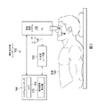
図1は、眼科手術用レーザシステム100を示す。眼科手術用レーザシステム100は、レーザビーム140を発生するように構成された手術用レーザ源130を含むことができる。眼科手術用レーザシステム100は、患者120の眼110の眼標的領域にレーザビーム140を向けるように構成された走査送達システム150も含むことができる。さらに、走査送達システム150は、眼110の眼標的領域の走査パターンに沿ってレーザビーム140を走査するように構成され得る。
FIG. 1 shows a
眼科手術用レーザシステム100は、システムコントローラ160も含むことができる。システムコントローラ160は、眼標的領域において眼組織300を減張するための切目または切開部250のパターンをもたらすために、走査パターンに沿ってレーザビーム140を走査するように走査送達システム150を制御するように構成され得る。パターンのいくつかは、眼組織300を通って部分的に延在する切開部250を含む。切開部250は、多くの異なる深さ、角度および向きを有することができる。図4〜図19は、例示的な切開部250を示し得る。
The
システムコントローラ160は、メモリ180および制御信号発生器170を含むことができる。メモリ180は、走査送達システム150を制御する命令セットを格納するように構成され得る。場合により、制御信号発生器170は、格納された命令セットに対応する制御信号を走査送達システム150に出力するように構成され得る。特に、制御信号は、走査送達システム150に対して走査パターンに沿ってレーザビーム140を走査するように命令することができる。
The
図2は、従来技術による眼の処置による患者120の眼110の正面図を示す。本開示の目的に対して関連する眼110の領域は、角膜210、角膜輪部220、強膜230、水晶体240および/または眼110の他の好適な解剖学的構造を含むことができる。概して、既存の角膜輪部減張および乱視矯正角膜切開処置は、眼110の角膜輪部220における単一の弧状切開部200、または横方向に対向する切開部の対を含み得る。
FIG. 2 shows a front view of the
図3に示すように、既存の処置における切開部200は、眼組織300の層を略完全に貫通し得る。しかしながら、こうした切開部200では、眼組織300に切開部200を正確に配置し、切開部200の場所において組織300の厚さを正確に確認し、切開部200の屈折結果を予測するために高精度の測定が必要であり得る。さらに、単一の切開部200を使用することにより、可能な屈折矯正の量が非常に制限され得る。また、切開部200が眼組織300内にそのように深く入るため、眼組織300の完全性が損なわれ得、望ましくない結果に至り得る。
As shown in FIG. 3, the
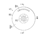
図4は、眼110の正面図で見た本開示の例を示す。この場合、1つまたは複数の線分または弧を形成する切開部250の組を角膜210または角膜輪部220の眼組織300に作成することができる。しかしながら、本開示は、眼の強膜230および角膜210とともに、水晶体240および結合組織を含む眼組織300の他の層において切開部250を作成することを企図する。この切開部250の組は、平行な線の組、切取メッシュ、切取格子または互い違いの切目250のパターンの形態をとることができる。切開部250の組は、1つ、2つ、3つ、4つまたはそれより多くの個々の切開部を含むことができる。
FIG. 4 shows an example of the present disclosure as viewed from the front view of the
図4に示すように、切開部250は、角膜210の周囲に同心円状に延在する弧を形成することができる。特に、1つまたは複数の弧の各々は、各切開部250の周囲に未切断眼組織を残して、互いに間隔を空けて配置された複数の切開部250によって画定することができる。弧は、互いに対して互い違いであるかまたはかみ合うことができる。特に、1つの弧の切開部250を別の弧の切開部250に少なくとも部分的に位置合わせすることができる。角膜210の中心から始まる放射線は、両弧の切開部250を通過することができる。切開部250のパターンの外側境界間に位置する未切断眼組織の上で線分をトレースすることができるように、切開部250間に十分な空間を配置することができる。
As shown in FIG. 4, the
切開部250の1つまたは複数は、角膜210に対して、他の切開部250と異なる半径、角度、深さ、長さ、間隔等で延在することができる。切開部250は、垂直以外のさまざまな角度で眼組織300内に延在することも可能である。たとえば、切開部250は、眼組織300の前面460に対して垂直に延在することができる。切開部250はまた、垂線からさまざまな角度の大きさで作成することができる。場合により、垂線から約10度〜約20度、垂線から約20度〜約45度、垂線から約45度〜約80度、ならびに/またはより大きいおよび小さい両方の他の好適な値で切開部250を作成することができる。さらに、切開部のパターンは、異なる角度で眼組織300に入る個々の切開部250を含むことができる。いずれの場合も、所望の乱視矯正に基づいて切開部250の特定のパラメータを選択することができる。
One or more of the
図5は、眼110の角膜210、角膜輪部220または強膜230に作成することができる切開部250を示す、患者120の眼110の断面図を示す。切開部250は、眼組織300を通って部分的に延在することができる。概して、角膜210におけるまたはその近くの眼組織300の厚さは、約600μmであり得る。切開部250の深さ260は、組織厚さの10%〜80%、厚さの20%〜75%、厚さの30%〜70%、厚さの40%〜60%、ならびに/またはより大きいおよび小さい両方の他の好適な値であり得る。場合により、これらの割合は、それぞれ約60μm〜480μm、120μm〜450μm、180μm〜420μmおよび240μm〜360μm、ならびに/またはより大きいおよび小さい両方の他の好適な値に対応することができる。有利には、切開部250の深さ260は、従来技術による方法より小さくすることができる。切開部250に対して選択される深さ260は、角膜210に対する形状の変化とともに、眼組織300の眼標的領域の強度に影響を与え得る。
FIG. 5 shows a cross-sectional view of the
図6および図7は、眼組織300の領域を示す。図6は、いくつかの切開部250を含む減張された眼組織300の領域を示す。切開部250自体は、図示するような直線状線分、弧状線分、多角形、楕円形、他の好適な形状および/またはそれらの組合せを含む任意の好適な断面形状を有することができる。場合により、切開部250は、1つまたは複数の他の切開部250と平行に配置することができる。たとえば、図6では、2つの切開部250は互いに平行に配置されている。図示するように、2つの切開部250は、両方ともそれらの間を横切る線に対して垂直とすることができ、それは、それらが互いに平行であることを示す。互いに平行な軸に沿って、切開部250の異なるサブセットを配置することができる。たとえば、図6の左側の3つの切開部250は、中間の3つの切開部250および右側の3つの切開部に対して平行であり得る。
6 and 7 show regions of
切開部250は、複数の切開部250を含む1つまたは複数の線分または弧を形成することができる。線分または弧は、互いに等しく間隔を空けて配置するか、または異なる距離だけ分離することができる。線分または弧は、場合により互いに対して平行であり得る。線分または弧を形成する切開部250は、未切断眼組織300によって囲まれ得る。特に、切開部250は、未切断眼組織のメッシュ構造をもたらすことができる。こうしたメッシュ構造により、角膜210の形状の大きい変動を可能にしながら、標的領域における眼組織300に強度を与えることができる。メッシュ構造の境界は、眼標的領域の中心部分、および(たとえば、図6の側部境界として画定される)眼標的領域の周縁部分とともに、(たとえば、図6の上部境界および下部境界によって画定される)切開部パターンの2つの外側境界であり得る。図6の例では、切開部250は、眼組織300を通過する水平線が2つ以上の切開部250を通過するように互い違いであり、かつ/または十分な深さであり得る。
The
図7は、応力下での切開部250を含む眼組織300の領域を示す。この応力は、眼110の内圧によってもたらされ得、外部圧力源によって倍加され得る。未切断眼組織のメッシュ構造は、この例では、切開部250の周囲の結合組織として見ることができる。
FIG. 7 shows a region of
切開部250は、眼組織300の弾性特性を変更することができる。図7の例では、角膜210に対して接線方向の構成で垂直切開部250を配置することができる。特に、接線方向(この例では垂直方向)における眼組織300の弾性特性は、半径方向(この場合には水平方向)における眼組織300の弾性特性と異なる程度に変更することができる。
The
図7の例では、眼組織300の弾性は、接線方向におけるよりも半径方向において切開部250によって大きくされ得る。半径方向における弾性は、接線方向における弾性よりも1.5:1以上、2:1以上、3:1以上、ならびに/またはより大きいおよび小さい両方の他の好適な値の倍率で増大することができる。逆に、角膜210に対して半径方向の構成で切開部250を配置することにより、接線方向における眼組織300の弾性を半径方向の弾性と比較して大きくすることができる。
In the example of FIG. 7, the elasticity of the
図8および図9は、眼110の前面460および後面470の両方に作成された切開部250を示す。前面460および後面470は、図12に関連してより詳細に見ることができる。本開示のレーザシステム100は、さまざまな深さ260、270で眼組織300を切断するように構成され得る。さらに、1つまたは複数の切開部250は、眼組織300の後面470に起点を有することができる。これらの切開部250は、後面470から深さ270まで延出するように構成され得る。
8 and 9
場合により、切開部250は、眼組織300の前面460および後面470の両方から同じ切開部パターンで始まることができる。さらに、切開部250は、眼組織300の前面460および後面470から交互に始まることができる。さらに、切開部250は、眼組織300の領域の内部構造において(たとえば、前面460と後面470との間で)始まり、いずれの面460、470も通り抜けることなく眼組織300を通って延在することができる。
Optionally, the
切開部250の交互の構成は、図10〜図12にさらに示され得る。図10は、眼組織300の後面470から始まっている切開部250の中心の線を示す一方、切開部250の外側の線は、眼組織300の前面460から始まっている。図10は、前面460を示し得る。後面470から始まっている切開部250の中心の線は、切開部250が眼組織300を通って完全に延在しないことを示すように想像線で示すことができる。図11は、応力下での眼組織300における切開部250の同じ構成を示す。
The alternating configuration of the
図12は、応力下での眼組織300における交互の切開部250の組の断面図を示す。ここで、切開部250は、眼組織300の領域の前面460からの始まりと、後面470からの始まりとの間で交互になっている。切開部250の深さ260、270は、眼組織300の弾性特性に対して直接の影響を与えることができる。場合により、眼組織300における切開部250の交互のパターンにより、組織300は、応力下で横方向に曲がることができ、他の切開部パターンにより、組織300は、単一の面460、470に沿って伸張することができる。この曲げ特徴により、組織300への切断が深すぎることにより組織強度を損なうことなく、応力下で眼組織300のより大きい伸長を可能にすることができる。
FIG. 12 shows a cross-sectional view of a set of alternating
曲げ特徴は、軸430、440、450によって示され得る。軸430、440、450は、眼組織300の層に対して垂直であり得る。図12の例を参照すると、軸430、440、450は、応力下で眼組織300において切開部250が作成された場所でトレースすることができる。軸430、440、450は、眼組織300が切開部250を含まない場合、平行であり得る。軸430、440、450は、眼組織300が応力下にありかつ切開部250を含む場合、互いに対して斜めにまたは非平行に延在することができる。
Bending features can be indicated by
軸430、440、450間の角度α、βは、切開部深さ260、270または応力が増大するに従って増大し得る。特に、角度αは、その真下の切開部250の深さ260が増大するに従って増大し得る。角度αは、隣接する切開部250がより大きい深さ270で作成されるか、または組織300に対してより大きい応力が与えられる結果としても増大し得る。同様に、角度βは、その真上の切開部250の深さ270が増大するに従い、かつ隣接する切開部250の深さ260に応じて大きくなり得る。角度α、βの大きさは、横方向における眼組織300の弾性に関連し得る。角度α、βは、約0度〜約25度、約5度〜約15度、もしくは約10度〜約20度、ならびに/またはより大きいおよび小さい両方の他の好適な値であり得る。
The angles α, β between the
図13〜図16は、切開部パターンのいくつかの例を示す。図13では、切開部250の中心の線は、眼組織300の層の前面460に作成することができる一方、切開部250の外側の線は、後面470に作成することができる。
13 to 16 show some examples of incision patterns. In FIG. 13, the central line of the
図14は、3つの線分を含む切開部250のパターンを示す。各線分は、眼組織300の長さに沿って前面460および後面470から交互に始まっている切開部250を含むことができる。後面470の切開部250を合わせてグループにすることができ、前面460の切開部250を合わせてグループにすることができる。
FIG. 14 shows a pattern of the
図15は、切開部250の対角線が前面460または後面470のいずれかにおいて共通の起点を共有する、切開部250の別の交互パターンを示す。
FIG. 15 shows another alternating pattern of
図16は、前面460の切開部250がZ字型パターンを形成することができる、切開部250のパターンを示す。前面460の切開部250は、後面470の切開部250によって囲まれ得る。
FIG. 16 shows a pattern of
図17および図18は、他の例示的な切開部パターンを示す。図17は、角膜210の一方の面に配置された切開部250の1つまたは複数の弧を含む切開部パターンを示す。角膜210の反対側に同様のパターンを配置することができる。角膜210の周囲において図17に示す2つの組間に切開部250の第3の組または第4の組を配置することができる。
17 and 18 show other exemplary incision patterns. FIG. 17 shows an incision pattern containing one or more arcs of the
図18は、他の切開部パターンを示す。眼110の右側において、切開部250は、1つまたは複数の弧を形成することができる。1つまたは複数の弧は、角膜210の周囲に同心円状に配置することができる。眼110の左上において、角膜210に対して接線方向に配置された線分で切開部250を作成することができる。眼110の左下において、角膜210から半径方向に延在する線分で切開部250を配置することができる。図17および図18に示す例示的なパターンのすべてが、上述したように、眼組織300の前面460または後面470で始まる切開部250を含むことができる。本開示は、平行線、楕円形線分および多角形パターンを含む線分の他の向きおよび形状を企図することができる。
FIG. 18 shows another incision pattern. On the right side of the
線分を形成する切開部250の数とともに、所与の切開部パターンにおける別個の線分の数を変更することができる。切開部パターン内で線分または弧を形成することができる切開部250の数は、2切開部〜20切開部、2切開部〜10切開部、2切開部〜6切開部、ならびに/またはより大きいおよび小さい両方の他の好適な値を含むことができる。単一の線分または弧が切開部パターンを形成することができるが、他の場合、2つ、3つ、4つまたは5つの線分または弧が切開部パターンを形成することができる。単一切開部パターンにおいて、上記線および形状の組合せも可能であり得る。
Along with the number of
図19は、別の例示的な切開部パターンを示す。この場合、直線状軸320に中心を置く切開部250の中心の線は、弧状軸310、330を有する2本の弧状線によって囲まれ得る。弧状線の一方を角膜210に対して凸状位置で配置することができる一方、他方を角膜210に対して同心または略同心とすることができる。さらに、外側の線の1つまたは複数の軸310、330は、内側の線の軸320と交差することができる。さまざまな弧状線の導入により、角膜210のより正確な成形を可能にすることができる。
FIG. 19 shows another exemplary incision pattern. In this case, the central line of the
本開示は、眼科手術処置を行う方法も含む。たとえば、レーザ源130を用いることにより、レーザビーム140を発生させることができる。眼標的領域にこのビーム140を向けることができる。眼標的領域において眼組織300を減張するための切開部のパターンをもたらすために、眼標的領域内で走査パターンに沿ってレーザビーム140を走査することができる。場合により、切開部250は、眼組織300を通して部分的にのみ延在する。上で説明したように、切開部のパターンは、平行線の1つもしくは複数の組、切断メッシュ、切断格子もしくは互い違いの切開部のパターン、またはそれらの組合せを含むことができる。さらに、切開部250は、眼組織300の前面460または後面470のいずれからも始まることができる。
The disclosure also includes methods of performing eye surgery procedures. For example, the
本明細書に記載する実施形態は、眼組織において切開を行う装置、システムおよび方法を提供することができる。上述した例は、限定するものではなく本質的に例示的なものであり得る。当業者であれば、本開示の範囲内にあるように意図される開示されている実施形態と一貫する他のシステムを容易に考案することができる。従って、本出願は、以下の特許請求の範囲によってのみ限定され得る。 The embodiments described herein can provide devices, systems and methods for making incisions in ocular tissue. The examples described above may be exemplary in nature without limitation. One of ordinary skill in the art can readily devise other systems consistent with the disclosed embodiments intended to be within the scope of the present disclosure. Therefore, this application may be limited only by the following claims.
Claims (14)
レーザビームを発生させるように構成されたレーザ源と、
走査送達システムであって、
眼標的領域に前記レーザビームを向けることと、
眼の前記眼標的領域において走査パターンに沿って前記レーザビームを走査することと
を行うように構成された走査送達システムと、
前記走査送達システムと通信するシステムコントローラであって、前記眼標的領域において眼組織を減張するための切目のパターンをもたらすために、前記走査パターンに沿って前記レーザビームを走査するように前記走査送達システムを制御するように構成され、前記切目のパターンの各切目は、前記眼組織を通して部分的にのみ延在する、システムコントローラと、を含んでおり
前記眼組織は、前面および後面を有する層を含み、
前記切目のパターンの少なくとも1つの切目は、前記後面から始まり、
前記切目のパターンは、前記眼組織の前記前面および前記後面から交互に始まる切目を含む、
眼科手術用レーザシステム。 A laser system for eye surgery
With a laser source configured to generate a laser beam,
Scanning delivery system
Aiming the laser beam at the eye target area
A scanning delivery system configured to scan the laser beam along a scanning pattern in the eye target area of the eye.
A system controller that communicates with the scan delivery system and scans the laser beam along the scan pattern to provide a pattern of cuts to detonate the ocular tissue in the ocular target region. Each cut in the pattern of cuts is configured to control the delivery system and includes a system controller , which extends only partially through the ocular tissue.
The ocular tissue comprises a layer having anterior and posterior surfaces.
At least one notch in the notch pattern begins at the posterior surface and
The pattern of cuts comprises cuts that alternate from the anterior surface and the posterior surface of the eye tissue.
Laser system for eye surgery.
前記走査送達システムを制御する命令セットを格納するように構成されたメモリと、
前記格納された命令セットに対応する制御信号を前記走査送達システムに出力するように構成された制御信号発生器であって、前記制御信号は、前記走査送達システムに対して前記走査パターンに沿って前記レーザビームを走査するように命令する、制御信号発生器と
を含む、請求項1に記載のシステム。 The system controller
A memory configured to store an instruction set that controls the scanning delivery system, and
A control signal generator configured to output a control signal corresponding to the stored instruction set to the scan delivery system, the control signal to the scan delivery system along the scan pattern. The system of claim 1, comprising a control signal generator that commands the laser beam to be scanned.
前記切目のパターンの前記切目の各々は、前記眼組織の前記前面と前記後面との間の厚さの約10%〜約80%だけ前記眼組織を通って延在する、請求項1に記載のシステム。 The ocular tissue comprises a layer having anterior and posterior surfaces.
The first aspect of the invention, wherein each of the cuts in the pattern of the cut extends through the eye tissue by about 10% to about 80% of the thickness between the front surface and the back surface of the eye tissue. System.
前記眼の角膜の第1の側における第1の切目の組と、
前記第1の側とは反対側の前記角膜の第2の側における第2の切目の組と
を含む、請求項1に記載のシステム。 The pattern of the cut is
With a set of first cuts on the first side of the cornea of the eye,
The system of claim 1, comprising a set of second cuts on the second side of the cornea opposite to the first side.
以上 The system of claim 1, wherein the pattern of cuts comprises one or more lines extending radially outward from the cornea of the eye.
that's all
Applications Claiming Priority (3)
| Application Number | Priority Date | Filing Date | Title |
|---|---|---|---|
| US14/973,117 | 2015-12-17 | ||
| US14/973,117 US10383767B2 (en) | 2015-12-17 | 2015-12-17 | Ophthalmic relaxing incisions and associated devices, systems, and methods |
| PCT/IB2016/057563 WO2017103774A1 (en) | 2015-12-17 | 2016-12-13 | Ophthalmic relaxing incisions and associated devices and systems |
Publications (3)
| Publication Number | Publication Date |
|---|---|
| JP2018537202A JP2018537202A (en) | 2018-12-20 |
| JP2018537202A5 JP2018537202A5 (en) | 2019-09-12 |
| JP6820339B2 true JP6820339B2 (en) | 2021-01-27 |
Family
ID=57614413
Family Applications (1)
| Application Number | Title | Priority Date | Filing Date |
|---|---|---|---|
| JP2018529282A Active JP6820339B2 (en) | 2015-12-17 | 2016-12-13 | Ophthalmic hypotonic incision and related equipment and systems |
Country Status (8)
| Country | Link |
|---|---|
| US (1) | US10383767B2 (en) |
| EP (1) | EP3370661B1 (en) |
| JP (1) | JP6820339B2 (en) |
| CN (1) | CN108366877B (en) |
| AU (1) | AU2016372821B2 (en) |
| CA (1) | CA3006548C (en) |
| ES (1) | ES2859123T3 (en) |
| WO (1) | WO2017103774A1 (en) |
Families Citing this family (1)
| Publication number | Priority date | Publication date | Assignee | Title |
|---|---|---|---|---|
| AU2017322492A1 (en) * | 2016-09-08 | 2019-03-21 | Aleyegn Technologies Llc | Glaucoma treatment methods and apparatus |
Family Cites Families (14)
| Publication number | Priority date | Publication date | Assignee | Title |
|---|---|---|---|---|
| US6325792B1 (en) | 1991-11-06 | 2001-12-04 | Casimir A. Swinger | Ophthalmic surgical laser and method |
| US5549632A (en) * | 1992-10-26 | 1996-08-27 | Novatec Laser Systems, Inc. | Method and apparatus for ophthalmic surgery |
| US20080312675A1 (en) | 2007-06-18 | 2008-12-18 | Advanced Medical Optics, Inc. | System and method for calculating limbal relaxing incisions |
| US20100324542A1 (en) | 2007-11-02 | 2010-12-23 | Kurtz Ronald M | Method to Guide a Cataract Procedure by Corneal Imaging |
| JP2011502585A (en) | 2007-11-02 | 2011-01-27 | アルコン レンゼックス, インコーポレーテッド | Methods and apparatus for improving post-operative eye optical performance |
| US20110022037A1 (en) | 2009-01-06 | 2011-01-27 | Bille Josef F | System and Method for Minimizing the Side Effects of Refractive Corrections Using Line or Dot Cuts for Incisions |
| US8663208B2 (en) | 2009-02-09 | 2014-03-04 | Amo Development, Llc | System and method for intrastromal refractive correction |
| US8758332B2 (en) * | 2009-07-24 | 2014-06-24 | Lensar, Inc. | Laser system and method for performing and sealing corneal incisions in the eye |
| US8679100B2 (en) | 2009-07-29 | 2014-03-25 | Alcon Lensx, Inc. | Optical system with multiple scanners for ophthalmic surgical laser |
| US8506559B2 (en) | 2009-11-16 | 2013-08-13 | Alcon Lensx, Inc. | Variable stage optical system for ophthalmic surgical laser |
| US10143589B2 (en) | 2011-02-22 | 2018-12-04 | Anita Nevyas-Wallace | Method and apparatus for making improved surgical incisions in corrective eye surgery |
| US10463541B2 (en) * | 2011-03-25 | 2019-11-05 | Lensar, Inc. | System and method for correcting astigmatism using multiple paired arcuate laser generated corneal incisions |
| EP3434234A1 (en) * | 2011-03-25 | 2019-01-30 | LensAR, Inc. | System and method for measuring and correcting astigmatism using laser generated corneal incisions |
| US9044304B2 (en) | 2011-12-23 | 2015-06-02 | Alcon Lensx, Inc. | Patient interface with variable applanation |
-
2015
- 2015-12-17 US US14/973,117 patent/US10383767B2/en active Active
-
2016
- 2016-12-13 ES ES16819183T patent/ES2859123T3/en active Active
- 2016-12-13 CA CA3006548A patent/CA3006548C/en active Active
- 2016-12-13 CN CN201680073716.1A patent/CN108366877B/en active Active
- 2016-12-13 WO PCT/IB2016/057563 patent/WO2017103774A1/en active Application Filing
- 2016-12-13 JP JP2018529282A patent/JP6820339B2/en active Active
- 2016-12-13 EP EP16819183.1A patent/EP3370661B1/en active Active
- 2016-12-13 AU AU2016372821A patent/AU2016372821B2/en active Active
Also Published As
| Publication number | Publication date |
|---|---|
| CA3006548C (en) | 2024-01-02 |
| WO2017103774A1 (en) | 2017-06-22 |
| JP2018537202A (en) | 2018-12-20 |
| EP3370661B1 (en) | 2021-02-17 |
| US10383767B2 (en) | 2019-08-20 |
| ES2859123T3 (en) | 2021-10-01 |
| CA3006548A1 (en) | 2017-06-22 |
| AU2016372821B2 (en) | 2021-04-01 |
| EP3370661A1 (en) | 2018-09-12 |
| US20170172801A1 (en) | 2017-06-22 |
| AU2016372821A1 (en) | 2018-06-07 |
| CN108366877A (en) | 2018-08-03 |
| CN108366877B (en) | 2021-05-04 |
Similar Documents
| Publication | Publication Date | Title |
|---|---|---|
| US7717908B2 (en) | Method patterns for intrastromal refractive surgery | |
| US20200170840A1 (en) | System and method for correcting astigmatism using multiple paired arcuate laser generated corneal incisions | |
| ES2618348T3 (en) | Laser unit for intrastromal refractive surgery | |
| US20140058365A1 (en) | System and Method for Using Compensating Incisions in Intrastromal Refractive Surgery | |
| US9877868B2 (en) | Apparatus for dissecting an eye for the introduction of a photosensitizer | |
| JP6820339B2 (en) | Ophthalmic hypotonic incision and related equipment and systems | |
| MX2013006532A (en) | Device and method for cutting the cornea of a human eye by means of cuts using focused pulsed laser radiation. | |
| US6409718B1 (en) | Device and method for correcting astigmatism by laser ablation | |
| EP2608753A2 (en) | System and method for minimizing the side effects of refractive corrections using line or dot cuts for incisions | |
| US8409179B2 (en) | System for performing intrastromal refractive surgery | |
| US9737437B2 (en) | Method and apparatus for making improved surgical incisions in corrective eye surgery | |
| US20070055220A1 (en) | Methods and systems for treating presbyopia via laser ablation | |
| JP2018537202A5 (en) | ||
| WO2014025336A1 (en) | Method and apparatus for making improved surgical incisions in corrective eye surgery |
Legal Events
| Date | Code | Title | Description |
|---|---|---|---|
| A521 | Request for written amendment filed |
Free format text: JAPANESE INTERMEDIATE CODE: A523 Effective date: 20190731 |
|
| A621 | Written request for application examination |
Free format text: JAPANESE INTERMEDIATE CODE: A621 Effective date: 20190731 |
|
| A711 | Notification of change in applicant |
Free format text: JAPANESE INTERMEDIATE CODE: A711 Effective date: 20191227 |
|
| RD03 | Notification of appointment of power of attorney |
Free format text: JAPANESE INTERMEDIATE CODE: A7423 Effective date: 20200121 |
|
| RD04 | Notification of resignation of power of attorney |
Free format text: JAPANESE INTERMEDIATE CODE: A7424 Effective date: 20200212 |
|
| A977 | Report on retrieval |
Free format text: JAPANESE INTERMEDIATE CODE: A971007 Effective date: 20200716 |
|
| A131 | Notification of reasons for refusal |
Free format text: JAPANESE INTERMEDIATE CODE: A131 Effective date: 20200728 |
|
| A521 | Request for written amendment filed |
Free format text: JAPANESE INTERMEDIATE CODE: A523 Effective date: 20201015 |
|
| TRDD | Decision of grant or rejection written | ||
| A01 | Written decision to grant a patent or to grant a registration (utility model) |
Free format text: JAPANESE INTERMEDIATE CODE: A01 Effective date: 20201201 |
|
| A61 | First payment of annual fees (during grant procedure) |
Free format text: JAPANESE INTERMEDIATE CODE: A61 Effective date: 20210104 |
|
| R150 | Certificate of patent or registration of utility model |
Ref document number: 6820339 Country of ref document: JP Free format text: JAPANESE INTERMEDIATE CODE: R150 |
|
| R250 | Receipt of annual fees |
Free format text: JAPANESE INTERMEDIATE CODE: R250 |