JP6657224B2 - Ureter Detection Using Imaging by Wavelength Band Selection - Google Patents
Ureter Detection Using Imaging by Wavelength Band Selection Download PDFInfo
- Publication number
- JP6657224B2 JP6657224B2 JP2017532121A JP2017532121A JP6657224B2 JP 6657224 B2 JP6657224 B2 JP 6657224B2 JP 2017532121 A JP2017532121 A JP 2017532121A JP 2017532121 A JP2017532121 A JP 2017532121A JP 6657224 B2 JP6657224 B2 JP 6657224B2
- Authority
- JP
- Japan
- Prior art keywords
- ureter
- tissue
- ureteral
- surgical site
- surgical
- Prior art date
- Legal status (The legal status is an assumption and is not a legal conclusion. Google has not performed a legal analysis and makes no representation as to the accuracy of the status listed.)
- Active
Links
- 210000000626 ureter Anatomy 0.000 title claims description 118
- 238000001514 detection method Methods 0.000 title description 8
- 238000003384 imaging method Methods 0.000 title description 5
- 230000003595 spectral effect Effects 0.000 claims description 13
- 210000001635 urinary tract Anatomy 0.000 claims description 11
- 238000001356 surgical procedure Methods 0.000 claims description 10
- 210000002700 urine Anatomy 0.000 claims description 7
- 210000001519 tissue Anatomy 0.000 description 75
- 238000000034 method Methods 0.000 description 25
- 238000001228 spectrum Methods 0.000 description 24
- 239000000975 dye Substances 0.000 description 20
- 238000005286 illumination Methods 0.000 description 20
- 238000012545 processing Methods 0.000 description 14
- 230000008569 process Effects 0.000 description 10
- QPFYXYFORQJZEC-FOCLMDBBSA-N Phenazopyridine Chemical group NC1=NC(N)=CC=C1\N=N\C1=CC=CC=C1 QPFYXYFORQJZEC-FOCLMDBBSA-N 0.000 description 8
- 229960001181 phenazopyridine Drugs 0.000 description 8
- 230000002285 radioactive effect Effects 0.000 description 8
- 239000011159 matrix material Substances 0.000 description 5
- 238000001444 catalytic combustion detection Methods 0.000 description 4
- 238000004590 computer program Methods 0.000 description 4
- 238000010586 diagram Methods 0.000 description 4
- 239000000243 solution Substances 0.000 description 4
- 238000013459 approach Methods 0.000 description 3
- 238000012937 correction Methods 0.000 description 3
- 230000006378 damage Effects 0.000 description 3
- 210000003734 kidney Anatomy 0.000 description 3
- 230000007246 mechanism Effects 0.000 description 3
- 230000005855 radiation Effects 0.000 description 3
- 230000009466 transformation Effects 0.000 description 3
- RBTBFTRPCNLSDE-UHFFFAOYSA-N 3,7-bis(dimethylamino)phenothiazin-5-ium Chemical compound C1=CC(N(C)C)=CC2=[S+]C3=CC(N(C)C)=CC=C3N=C21 RBTBFTRPCNLSDE-UHFFFAOYSA-N 0.000 description 2
- FAPWRFPIFSIZLT-UHFFFAOYSA-M Sodium chloride Chemical compound [Na+].[Cl-] FAPWRFPIFSIZLT-UHFFFAOYSA-M 0.000 description 2
- 230000008859 change Effects 0.000 description 2
- 238000006243 chemical reaction Methods 0.000 description 2
- 239000000835 fiber Substances 0.000 description 2
- 230000006870 function Effects 0.000 description 2
- MOFVSTNWEDAEEK-UHFFFAOYSA-M indocyanine green Chemical compound [Na+].[O-]S(=O)(=O)CCCCN1C2=CC=C3C=CC=CC3=C2C(C)(C)C1=CC=CC=CC=CC1=[N+](CCCCS([O-])(=O)=O)C2=CC=C(C=CC=C3)C3=C2C1(C)C MOFVSTNWEDAEEK-UHFFFAOYSA-M 0.000 description 2
- 229960004657 indocyanine green Drugs 0.000 description 2
- 238000002347 injection Methods 0.000 description 2
- 239000007924 injection Substances 0.000 description 2
- 229960000907 methylthioninium chloride Drugs 0.000 description 2
- 210000004197 pelvis Anatomy 0.000 description 2
- 239000011780 sodium chloride Substances 0.000 description 2
- 239000007787 solid Substances 0.000 description 2
- 210000003708 urethra Anatomy 0.000 description 2
- UJABSZITRMATFL-UHFFFAOYSA-N 2-methyl-5-phenylfuran-3-carbonyl chloride Chemical compound ClC(=O)C1=C(C)OC(C=2C=CC=CC=2)=C1 UJABSZITRMATFL-UHFFFAOYSA-N 0.000 description 1
- 241000282412 Homo Species 0.000 description 1
- 208000027418 Wounds and injury Diseases 0.000 description 1
- 238000010521 absorption reaction Methods 0.000 description 1
- 230000000202 analgesic effect Effects 0.000 description 1
- 230000008901 benefit Effects 0.000 description 1
- 210000004204 blood vessel Anatomy 0.000 description 1
- 150000001732 carboxylic acid derivatives Chemical group 0.000 description 1
- 239000003086 colorant Substances 0.000 description 1
- 230000009977 dual effect Effects 0.000 description 1
- 230000005284 excitation Effects 0.000 description 1
- -1 for example Substances 0.000 description 1
- 238000003331 infrared imaging Methods 0.000 description 1
- 208000014674 injury Diseases 0.000 description 1
- 238000005259 measurement Methods 0.000 description 1
- 210000003205 muscle Anatomy 0.000 description 1
- 230000003287 optical effect Effects 0.000 description 1
- 229960003799 phenazopyridine hydrochloride Drugs 0.000 description 1
- 239000000049 pigment Substances 0.000 description 1
- 238000002310 reflectometry Methods 0.000 description 1
- 239000000523 sample Substances 0.000 description 1
- 238000001931 thermography Methods 0.000 description 1
- 238000012546 transfer Methods 0.000 description 1
- 238000000844 transformation Methods 0.000 description 1
- 230000002485 urinary effect Effects 0.000 description 1
- 210000003462 vein Anatomy 0.000 description 1
Images


Classifications
-
- A—HUMAN NECESSITIES
- A61—MEDICAL OR VETERINARY SCIENCE; HYGIENE
- A61B—DIAGNOSIS; SURGERY; IDENTIFICATION
- A61B90/00—Instruments, implements or accessories specially adapted for surgery or diagnosis and not covered by any of the groups A61B1/00 - A61B50/00, e.g. for luxation treatment or for protecting wound edges
- A61B90/36—Image-producing devices or illumination devices not otherwise provided for
-
- A—HUMAN NECESSITIES
- A61—MEDICAL OR VETERINARY SCIENCE; HYGIENE
- A61B—DIAGNOSIS; SURGERY; IDENTIFICATION
- A61B1/00—Instruments for performing medical examinations of the interior of cavities or tubes of the body by visual or photographical inspection, e.g. endoscopes; Illuminating arrangements therefor
- A61B1/00002—Operational features of endoscopes
- A61B1/00011—Operational features of endoscopes characterised by signal transmission
-
- A—HUMAN NECESSITIES
- A61—MEDICAL OR VETERINARY SCIENCE; HYGIENE
- A61B—DIAGNOSIS; SURGERY; IDENTIFICATION
- A61B34/00—Computer-aided surgery; Manipulators or robots specially adapted for use in surgery
- A61B34/30—Surgical robots
-
- A—HUMAN NECESSITIES
- A61—MEDICAL OR VETERINARY SCIENCE; HYGIENE
- A61B—DIAGNOSIS; SURGERY; IDENTIFICATION
- A61B6/00—Apparatus or devices for radiation diagnosis; Apparatus or devices for radiation diagnosis combined with radiation therapy equipment
- A61B6/40—Arrangements for generating radiation specially adapted for radiation diagnosis
- A61B6/4057—Arrangements for generating radiation specially adapted for radiation diagnosis by using radiation sources located in the interior of the body
-
- A—HUMAN NECESSITIES
- A61—MEDICAL OR VETERINARY SCIENCE; HYGIENE
- A61B—DIAGNOSIS; SURGERY; IDENTIFICATION
- A61B90/00—Instruments, implements or accessories specially adapted for surgery or diagnosis and not covered by any of the groups A61B1/00 - A61B50/00, e.g. for luxation treatment or for protecting wound edges
- A61B90/36—Image-producing devices or illumination devices not otherwise provided for
- A61B90/37—Surgical systems with images on a monitor during operation
-
- C—CHEMISTRY; METALLURGY
- C09—DYES; PAINTS; POLISHES; NATURAL RESINS; ADHESIVES; COMPOSITIONS NOT OTHERWISE PROVIDED FOR; APPLICATIONS OF MATERIALS NOT OTHERWISE PROVIDED FOR
- C09B—ORGANIC DYES OR CLOSELY-RELATED COMPOUNDS FOR PRODUCING DYES, e.g. PIGMENTS; MORDANTS; LAKES
- C09B29/00—Monoazo dyes prepared by diazotising and coupling
- C09B29/34—Monoazo dyes prepared by diazotising and coupling from other coupling components
- C09B29/36—Monoazo dyes prepared by diazotising and coupling from other coupling components from heterocyclic compounds
- C09B29/3604—Monoazo dyes prepared by diazotising and coupling from other coupling components from heterocyclic compounds containing only a nitrogen as heteroatom
- C09B29/3617—Monoazo dyes prepared by diazotising and coupling from other coupling components from heterocyclic compounds containing only a nitrogen as heteroatom containing a six-membered heterocyclic with only one nitrogen as heteroatom
- C09B29/3621—Monoazo dyes prepared by diazotising and coupling from other coupling components from heterocyclic compounds containing only a nitrogen as heteroatom containing a six-membered heterocyclic with only one nitrogen as heteroatom from a pyridine ring
-
- A—HUMAN NECESSITIES
- A61—MEDICAL OR VETERINARY SCIENCE; HYGIENE
- A61B—DIAGNOSIS; SURGERY; IDENTIFICATION
- A61B90/00—Instruments, implements or accessories specially adapted for surgery or diagnosis and not covered by any of the groups A61B1/00 - A61B50/00, e.g. for luxation treatment or for protecting wound edges
- A61B90/30—Devices for illuminating a surgical field, the devices having an interrelation with other surgical devices or with a surgical procedure
- A61B2090/306—Devices for illuminating a surgical field, the devices having an interrelation with other surgical devices or with a surgical procedure using optical fibres
-
- A—HUMAN NECESSITIES
- A61—MEDICAL OR VETERINARY SCIENCE; HYGIENE
- A61B—DIAGNOSIS; SURGERY; IDENTIFICATION
- A61B90/00—Instruments, implements or accessories specially adapted for surgery or diagnosis and not covered by any of the groups A61B1/00 - A61B50/00, e.g. for luxation treatment or for protecting wound edges
- A61B90/30—Devices for illuminating a surgical field, the devices having an interrelation with other surgical devices or with a surgical procedure
- A61B2090/309—Devices for illuminating a surgical field, the devices having an interrelation with other surgical devices or with a surgical procedure using white LEDs
-
- A—HUMAN NECESSITIES
- A61—MEDICAL OR VETERINARY SCIENCE; HYGIENE
- A61B—DIAGNOSIS; SURGERY; IDENTIFICATION
- A61B90/00—Instruments, implements or accessories specially adapted for surgery or diagnosis and not covered by any of the groups A61B1/00 - A61B50/00, e.g. for luxation treatment or for protecting wound edges
- A61B90/36—Image-producing devices or illumination devices not otherwise provided for
- A61B90/37—Surgical systems with images on a monitor during operation
- A61B2090/371—Surgical systems with images on a monitor during operation with simultaneous use of two cameras
-
- A—HUMAN NECESSITIES
- A61—MEDICAL OR VETERINARY SCIENCE; HYGIENE
- A61B—DIAGNOSIS; SURGERY; IDENTIFICATION
- A61B90/00—Instruments, implements or accessories specially adapted for surgery or diagnosis and not covered by any of the groups A61B1/00 - A61B50/00, e.g. for luxation treatment or for protecting wound edges
- A61B90/36—Image-producing devices or illumination devices not otherwise provided for
- A61B90/37—Surgical systems with images on a monitor during operation
- A61B2090/373—Surgical systems with images on a monitor during operation using light, e.g. by using optical scanners
Landscapes
- Health & Medical Sciences (AREA)
- Life Sciences & Earth Sciences (AREA)
- Surgery (AREA)
- Engineering & Computer Science (AREA)
- Nuclear Medicine, Radiotherapy & Molecular Imaging (AREA)
- Medical Informatics (AREA)
- Veterinary Medicine (AREA)
- Animal Behavior & Ethology (AREA)
- General Health & Medical Sciences (AREA)
- Public Health (AREA)
- Biomedical Technology (AREA)
- Heart & Thoracic Surgery (AREA)
- Molecular Biology (AREA)
- Pathology (AREA)
- Radiology & Medical Imaging (AREA)
- Physics & Mathematics (AREA)
- Optics & Photonics (AREA)
- Biophysics (AREA)
- Oral & Maxillofacial Surgery (AREA)
- Robotics (AREA)
- Gynecology & Obstetrics (AREA)
- High Energy & Nuclear Physics (AREA)
- Chemical & Material Sciences (AREA)
- Organic Chemistry (AREA)
- Endoscopes (AREA)
Description
本特許出願は、2014年12月16日に出願された、”URETER DETECTION USING WAVEBAND-SELECTIVE IMAGING”という標題の米国特許仮出願第62/092,651号に対する優先権、及びその出願日の利益を主張するものであり、この文献は、その全体が参照により本明細書に組み込まれる。 This patent application claims the priority of, and benefit from, the filing date of US Provisional Application No. 62 / 092,651, filed December 16, 2014, entitled "URETER DETECTION USING WAVEBAND-SELECTIVE IMAGING". Claims, which is incorporated herein by reference in its entirety.
本発明は、概して、外科的処置に使用される画像化技術に関し、より具体的には、外科的処置中の尿管の検出及び画像化に関する。 The present invention relates generally to imaging techniques used in surgical procedures, and more particularly, to detecting and imaging the ureter during a surgical procedure.
尿管は、尿を腎臓から膀胱まで運ぶ細い管である。尿管の壁にある筋肉は、継続的に引き締め、弛緩させ、尿を腎臓から離れさせる方向に下向きに押し出す。約10秒〜15秒毎に、少量の尿が、尿管から膀胱内に排出される。 The ureter is a thin tube that carries urine from the kidney to the bladder. The muscle in the wall of the ureter continually tightens, relaxes, and pushes urine downwards away from the kidney. About every 10-15 seconds, a small amount of urine is expelled from the ureter into the bladder.
尿管への損傷は、骨盤又は結腸直腸領域を伴う手術に関連する有害事象である。手術中の尿管への損傷を防ぐために、外科医が尿管の位置を特定するのを補助するいくつかの異なる技術が、試みられている。例えば、外科医は、泌尿器科医を呼んで、カメラ付きの小さな内視鏡を尿道に通し、ワイヤを2つの尿管のそれぞれに入れることができる。あるいはまた、照明付きのステントを尿道を通して膀胱まで挿入して、尿管にアクセスすることができる。しかしながら、これら両方のアプローチは、良性の外科的処置の大部分において臨床ワークフローに支障をきたし、場合によっては泌尿器科医が利用できないこともある。 Injury to the ureter is an adverse event associated with surgery involving the pelvis or colorectal area. Several different techniques have been attempted to assist surgeons in locating the ureter to prevent damage to the ureter during surgery. For example, a surgeon can call a urologist to pass a small endoscope with a camera through the urethra and insert wires into each of the two ureters. Alternatively, an illuminated stent can be inserted through the urethra into the bladder to access the ureter. However, both of these approaches have hindered clinical workflow in most benign surgical procedures, and in some cases may not be available to urologists.
尿管の位置を決定する別の技術では、少量の放射性化学色素(TC99-DTPA)が、患者の腕の静脈を介して注射される。放射性化学色素は体を巡って尿中に排出されるため、放射性化学色素は尿管を通過する。尿管は、放射線を感知する手持ち式プローブによって検出される。 In another technique for determining the location of the ureter, a small amount of a radiochemical dye (TC99-DTPA) is injected via a vein in a patient's arm. Because the radiochemical dye is excreted throughout the body into the urine, the radiochemical dye passes through the ureter. The ureter is detected by a hand-held probe that senses radiation.
尿管の位置を特定する別の技術では、赤外線照明を用いて尿管を画像化するために、近赤外(NIR)フルオロフォア(fluorophore)の静脈(IV)注射又はカテーテルに基づく逆行(retrograde)注射が使用される。組織の周囲に埋め込まれる場合であっても尿管を視覚化することができ、不可視光を用いてリアルタイムで損傷を評価できることが報告されている。Eiichi Tanakaらの、”Real-Time
Intraoperative Ureteral Guidance Using Near-Infrared Fluorescence,” J. Urol.
178(5), pgs.2197-2201(2007)には、インドシアニングリーン(ICG)、及びLI-COR(Lincoln, NE)からのCW800-CA、カルボン酸形態であるIRDye(登録商標) CW800 NIR色素をNIRフルオロフォアとしてとして使用することが記載されている。Aya Matsui、M.D.らの、”Real-Time Near-Infrared
Fluorescence-Guided Identification of the Ureters using Methylene Blue,”
Surgery, 148(1) pgs.78-86(2010)には、NIRフルオロフォアとしてメチレンブルーを使用することが記載されている。
Another technique for locating the ureter involves venous (IV) injection of near-infrared (NIR) fluorophore or catheter-based retrograde to image the ureter using infrared illumination. ) Injection is used. It has been reported that the ureter can be visualized even when implanted around tissue, and damage can be evaluated in real time using invisible light. Eiichi Tanaka's “Real-Time”
Intraoperative Ureteral Guidance Using Near-Infrared Fluorescence, ”J. Urol.
178 (5), pgs. 2197-2201 (2007) include indocyanine green (ICG) and CW800-CA from LI-COR (Lincoln, NE), IRDye® CW800 NIR in carboxylic acid form The use of dyes as NIR fluorophores is described. Aya Matsui, MD and others, “Real-Time Near-Infrared
Fluorescence-Guided Identification of the Ureters using Methylene Blue, ”
Surgery, 148 (1) pgs. 78-86 (2010) describes the use of methylene blue as the NIR fluorophore.
赤外線サーモグラフィーを使用して尿管を位置特定する別の手法がある。手術野内の洗浄剤として室温の生理食塩水を使用し、手術野全体を一時的に冷却した。手術野が特異的に再加熱されると、血管等の構造物は、急速に再加熱され、赤外線画像の暗い背景に対して白線として現れた。この同じ概念の第2の応用は、上部尿路系を室温の生理食塩水で満たすことを含んでいた。骨盤及び尿管は、赤外線画像では白く現れるより温度の高い背景に対して黒く見える。Jeffrey A.
Cadeddu, M.D.らの、"Laparoscopic
Infrared Imaging," Journal of Endourology, Vol.15, No.1, pgs.111-116(2001)を参照されたい。
There is another approach to localizing the ureter using infrared thermography. Room temperature saline was used as a cleanser in the surgical field and the entire surgical field was temporarily cooled. When the surgical field was specifically reheated, structures such as blood vessels rapidly reheated and appeared as white lines against the dark background of the infrared image. A second application of this same concept involved filling the upper urinary tract with saline at room temperature. The pelvis and ureter appear black against a hotter background than appear white in the infrared image. Jeffrey A.
"Laparoscopic, from Cadeddu, MD et al.
See Infrared Imaging, "Journal of Endourology, Vol. 15, No. 1, pgs. 111-116 (2001).
フルオロフォアの導入、温度差の生成、尿管への物体の導入、又は放射性色素の導入を必要とする、尿管の位置特定に使用される既知の技術とは異なり、尿管による及び尿管の周りの組織による光の選択的な反射を使用して、尿管を安全且つ効率的に画像化する。こうして、カテーテル照明を必要とせずに、又は例えば外因性フルオロフォア又は放射性色素の投与を必要とせずに、内因性コントラストを使用して尿管を視覚化する。 Unlike known techniques used to locate the ureter, which require the introduction of a fluorophore, the generation of a temperature difference, the introduction of an object into the ureter, or the introduction of a radioactive dye, by ureter and by ureter The selective reflection of light by the surrounding tissue is used to safely and efficiently image the ureter. Thus, endogenous contrast is used to visualize the ureter without the need for catheter illumination or, for example, the administration of exogenous fluorophores or radioactive dyes.
一態様では、複数の手術部位のシーンが取り込まれる。複数の手術部位のシーンのそれぞれは、異なる光スペクトルを有する反射光から取り込まれる。一態様では、複数の手術部位のシーンが、略同時に、すなわち、シーンの取込みに関連するタイミング及び光学許容誤差内で同時に取り込まれる一方、別の態様では、複数の手術部位のシーンは順次に取り込まれる。 In one aspect, multiple surgical site scenes are captured. Each of the multiple surgical site scenes is captured from reflected light having a different light spectrum. In one aspect, multiple surgical site scenes are captured substantially simultaneously, i.e., within the timing and optical tolerances associated with capturing the scene, while in another aspect, multiple surgical site scenes are captured sequentially. It is.
取り込まれた複数の手術部位のシーンを解析して、取り込まれた手術部位のシーン内の尿管組織を識別する。取り込まれた手術部位のシーンに基づいて、手術部位のシーンが、表示装置上に表示される。表示された手術部位のシーンは、尿管組織が強調されているので、尿管組織は外科医によって容易に判別される。 The captured plurality of surgical site scenes are analyzed to identify ureteral tissue in the captured surgical site scene. The scene of the surgical site is displayed on the display device based on the captured scene of the surgical site. Since the ureteral tissue is emphasized in the displayed scene of the surgical site, the ureteral tissue is easily identified by the surgeon.
一態様では、手術部位は、複数の異なる光スペクトルで照射される。例えば、手術部位は、複数の異なる光スペクトルのうちの少なくとも2つで順次照射される。一態様では、複数の光スペクトルは、450nm〜580nmの範囲の波長の光スペクトル、640nm〜750nmの範囲の波長の光スペクトル、及び900nm〜1080nmの範囲の波長の光スペクトルを含む。 In one aspect, the surgical site is illuminated with a plurality of different light spectra. For example, a surgical site is sequentially illuminated with at least two of a plurality of different light spectra. In one aspect, the plurality of light spectra includes a light spectrum having a wavelength in a range of 450 nm to 580 nm, a light spectrum having a wavelength in a range of 640 nm to 750 nm, and a light spectrum having a wavelength in a range of 900 nm to 1080 nm.
一態様では、尿管組織を識別するために取り込まれた複数の手術部位のシーンの解析は、最初に、取り込まれた複数の手術部位のシーンのそれぞれの手術部位のシーン内の位置を尿管信号に変換する。解析は、次に、尿管信号がその位置における尿管組織を示すかどうかを判定する。例えば、尿管信号は閾値と比較される。あるいはまた、尿管信号と非尿管信号との比が閾値と比較される。 In one aspect, analysis of the captured plurality of surgical site scenes to identify ureteral tissue first comprises determining a position in the respective surgical site scene of the captured plurality of surgical site scenes in the ureter. Convert to a signal. The analysis then determines whether the ureteral signal indicates ureteral tissue at that location. For example, the ureteral signal is compared to a threshold. Alternatively, the ratio of the ureteral signal to the non-ureteral signal is compared to a threshold.
さらに別の態様では、手術部位のシーンを取り込む前に、尿管組織を色素で処理する。この色素は、フルオロフォア及び放射性色素とは異なり、例えば、色素はフェナゾピリジンである。 In yet another aspect, ureteral tissue is treated with a dye prior to capturing a scene of the surgical site. This dye is different from fluorophores and radioactive dyes, for example, the dye is phenazopyridine.
これらの方法を実施するために、手術システムは画像取込システムを含む。画像取込システムは、複数の手術部位のシーンを取り込むように構成される。複数の手術部位のシーンにおける各手術部位のシーンは、複数の波長帯における異なる波長帯の反射光から取り込まれる。手術システムは、画像取込システムに結合された尿管解析モジュールも含む。尿管解析モジュールは、取り込まれた手術部位のシーン内の尿管組織を識別するように構成される。一態様では、手術システムは、照明器も含む。照明器は、複数の波長帯のそれぞれで手術部位を照明するように構成される。手術システムの表示装置は、手術部位のシーンを受信し且つそのシーン内の尿管組織を識別する情報を知らせる尿道解析モジュールに結合される。表示装置は、尿管を強調表示した状態で手術部位のシーンを表示するように構成される。 To perform these methods, the surgical system includes an image capture system. The image capture system is configured to capture a plurality of surgical site scenes. The scene of each surgical site in the plurality of surgical site scenes is captured from reflected light of different wavelength bands in the plurality of wavelength bands. The surgical system also includes a ureteral analysis module coupled to the image capture system. The ureter analysis module is configured to identify ureteral tissue in the captured surgical site scene. In one aspect, the surgical system also includes an illuminator. The illuminator is configured to illuminate the surgical site in each of the plurality of wavelength bands. The display of the surgical system is coupled to a urethral analysis module that receives a scene of the surgical site and provides information identifying ureteral tissue in the scene. The display device is configured to display a scene of a surgical site with the ureter highlighted.
図面において、参照符号の最初の桁字は、その参照符号を有する要素が最初に現れた図を示す。
フルオロフォアの導入、温度差の生成、尿管への物体の導入、又は放射性色素の導入を必要とする、尿管の位置特定するために使用される既知の技術とは異なり、尿管解析モジュール135は、尿管によって反射された反射光を使用して尿管の位置特定を行う。尿管による及び尿管の周囲の組織による光の選択的反射を使用して、尿管を安全且つ効率的に画像化する。こうして、カテーテルを照明することなく、又は例えば外因性フルオロフォア又は放射性色素の投与の必要なしに、内因性コントラストを使用して尿管を視覚化する。
In the drawings, the first digit of a reference number identifies the figure in which the element with the reference number first appears.
Unlike the known techniques used to locate the ureter, which require the introduction of a fluorophore, the generation of a temperature difference, the introduction of an object into the ureter, or the introduction of a radioactive dye, a
図1は、尿管解析モジュール135を含む手術システム100、例えばダビンチ(登録商標)手術システムの高レベル概略図である(ダビンチ(登録商標)は、カリフォルニア州サニーベールのIntuitive
Surgical, Inc.の登録商標である。)。この例では、外科医は、外科医コンソール114でマスターコントロールを使用して、ロボットマニピュレータアーム113に取り付けられ、次にカート110に取り付けられる内視鏡112を遠隔操作する。外科医は、カート110に結合された1つ又は複数の手術用器具も遠隔操作する。ダビンチ(登録商標)手術システムに関連する他の部品、ケーブル等があるが、これらは、本開示からの逸脱を避けるために図1には示していない。
FIG. 1 is a high-level schematic diagram of a
Surgical, Inc. is a registered trademark. ). In this example, the surgeon uses master controls at the
低侵襲性手術システムに関する更なる情報は、例えば、(2007年6月23日に出願され、低侵襲性手術システムを開示する)米国特許出願第11/762,165号、(2001年10月5日に出願され、遠隔操作手術システム用のアームカートを開示する)米国特許第6,837,883号、及び(2001年12月28日に出願され、手術用ロボットツール、データアーキテクチャ及び使用を開示する)米国特許第6,331,181号に確認することができ、これらの文献は全て参照により本明細書に組み込まれる。遠隔操作される低侵襲性手術システムの使用は、例示に過ぎず、本発明をこの特定のシステムに限定することを意図するものではない。本開示に鑑みて、本明細書に記載の態様は、本明細書に記載の態様を実施するのに必要な要素を含むロボット手術システム又は他の手術システムに組み込むことができる。 Additional information regarding minimally invasive surgical systems is available, for example, in U.S. Patent Application Serial No. 11 / 762,165 (filed June 23, 2007, which discloses minimally invasive surgical systems), (October 5, 2001). U.S. Patent No. 6,837,883, filed on Dec. 1, 2001, and discloses a surgical robotic tool, data architecture and use, filed on Dec. 28, 2001, which discloses an arm cart for a remotely operated surgical system. No. 6,331,181), all of which are incorporated herein by reference. The use of a teleoperated minimally invasive surgical system is exemplary only and is not intended to limit the invention to this particular system. In view of the present disclosure, aspects described herein can be incorporated into a robotic or other surgical system that includes the elements necessary to implement the aspects described herein.
一態様では、内視鏡112は立体内視鏡であり、患者111内の手術部位103からの光、例えば反射光及び/又は蛍光を画像取込システム120に送る2つの光チャネルを含む。手術部位103は、尿管と、尿管の周りに一般に見られる組織とを含む。
In one aspect, endoscope 112 is a stereoscopic endoscope and includes two light channels that transmit light from
尿管によって反射された光を含む、手術部位103内の組織によって反射された光は、手術部位のシーンとして取り込まれる。取り込まれる手術部位の各シーンは、尿管組織の画像と非尿管組織の画像とを含む。取り込まれる手術部位の各シーンは、データフレーム121に含められる。一態様では、複数のフレームが取り込まれ、手術部位103を照明する複数の波長帯のそれぞれについて1つのフレームが取り込まれる。
Light reflected by tissue within the
立体内視鏡の場合に、手術部位のシーンは、左シーンと右シーンとの2つのシーンを含む。左セットと右セットの2つのセットのデータフレームが画像取込システム120によって取り込まれる。この2つのセットは、尿管解析モジュール135の尿管検出モジュール136によって処理され、左尿管エンハンス画像及び右尿管エンハンス画像が作成され、それら画像は、外科医制御コンソール114内の立体表示ユニットに送られる。左尿管エンハンス画像及び右尿管エンハンス画像は、尿管エンハンス画像140に含められる。
In the case of a stereoscopic endoscope, a scene of a surgical site includes two scenes, a left scene and a right scene. Two sets of data frames, a left set and a right set, are captured by the
左尿管エンハンス画像及び右尿管エンハンス画像は、外科医制御コンソール114(時には外科医コンソール114又は単にコンソール114と呼ばれる)内の立体表示ユニット上に提示され、尿管が強調表示される手術部位103の三次元シーンが作成される。上述したように、尿管の識別は、尿管を取り囲む組織の反射率と尿管の反射率との間の差を利用して行われる。特別なフルオロフォア、放射性色素、温度差、又は尿管に挿入される物体は必要ない。むしろ、尿管解析モジュール135は、尿管のスペクトル反射率と、尿管を取り囲む組織(非尿管組織)のスペクトル反射率との間の差を使用して、手術部位103内の尿管の位置を識別し表示するように構成される。
The left ureteral enhancement image and the right ureteral enhancement image are presented on a stereoscopic display unit in the surgeon control console 114 (sometimes referred to as the
図2は、尿管のスペクトル反射率201(実線)、すなわち尿管スペクトル特性201だけでなく、尿管組織に近接する非尿管組織のスペクトル反射率202(破線)、すなわち非尿管スペクトル特性202を示す。視覚的には、尿管スペクトル特性201と非尿管スペクトル特性202との間の色差を人間が検出することは非常に困難であるが、図2に示されるように差が存在する。
FIG. 2 shows not only the
3つの別々の波長帯、例えば3つの別個の光スペクトルは、尿管組織と非尿管組織との間に明確な反射率の差を有する。3つの別々の波長帯は、450〜580ナノメートル(nm)(低波長帯)、640〜750nm(中波長帯)、及び900〜1080nm(高波長帯)である。時には、これらの波長帯はスペクトルと呼ばれる。 Three separate wavelength bands, eg, three separate light spectra, have a distinct reflectance difference between ureteral and non-ureteral tissue. The three separate wavelength bands are 450-580 nanometers (nm) (low wavelength band), 640-750 nm (middle wavelength band), and 900-1080 nm (high wavelength band). Sometimes these wavelength bands are called spectra.
低波長帯(450〜580nm)は可視光スペクトル内にあり、中波長帯(640〜750nm)は部分的に可視光スペクトル内にあり(可視光スペクトルは、400nm〜700nmの間であると言われている。)、これらの波長帯の反射可視光は、外科医コンソール114に表示されるシーンにおいて外科医によって区別可能な色差をもたらさない。反射率差に関連する色差は、僅かな強度差としてより多く観察され、外科医コンソール114に表示される手術部位のシーンで典型的な複雑な照明及び3次元シーン構造において、人間の目によって小さい強度差を検出することは困難である。
The low wavelength band (450-580 nm) is in the visible light spectrum, and the middle wavelength band (640-750 nm) is partially in the visible light spectrum (the visible light spectrum is said to be between 400-700 nm). ), The reflected visible light in these wavelength bands does not provide a color difference that is discernible by the surgeon in the scene displayed on the
中波長帯(640〜750nm)の他の部分(700〜750nm)及び高波長帯(900〜1080nm)は、電磁スペクトルの近赤外線(NIR)部分にある。近赤外光は人間によっては検出できない。しかしながら、尿管検出モジュール136は、これらの3つの波長帯の全てのスペクトル反射率の差を検出し、それによって、画像取込システム120によって取り込まれるシーン内の尿管組織に対応するピクセル及びそのシーン内の非尿管組織に対応するピクセルを識別することができる。
The other part (700-750 nm) of the middle wavelength band (640-750 nm) and the high wavelength band (900-1080 nm) are in the near infrared (NIR) part of the electromagnetic spectrum. Near infrared light cannot be detected by humans. However, the
一態様では、シーン内の尿管組織に対応するピクセルは、強調表示され、例えば手術部位のシーンでは典型的に観察されない色を有するように偽色され(false-colored)、結果として生じるシーンが、外科医コンソール114に表示される。強調表示された尿管によって、外科医が外科的処置中に尿管の位置を容易に区別することを可能にする。
In one aspect, pixels corresponding to ureteral tissue in the scene are highlighted and false-colored, for example, to have a color not typically observed in a surgical site scene, and the resulting scene may be false-colored. Is displayed on the
一態様では、尿管解析モジュール135は、少なくとも2つの異なる光スペクトルで尿管を含む組織を照明するように照明システム125を構成する。選択される光スペクトルは、尿管組織と非尿管組織との間の反射率の差に基づくものである。各スペクトルは、図2に示されるように、尿管を取り囲む組織によって及び尿管によって異なるように反射される。別の態様では、画像取込システム120においてフィルタを使用して、尿管組織と非尿管組織との間の反射率の差を有する異なる光スペクトルに対応するフレームを取り込む。
In one aspect, the
立体内視鏡及び立体視ディスプレイの使用は、例示に過ぎず、限定することを意図するものではない。本明細書で説明する態様は、モノスコピック内視鏡及び/又は通常の表示ユニット等の立体的な機構を含まないシステムに適用することができる。 The use of stereoscopic endoscopes and stereoscopic displays is merely exemplary and is not intended to be limiting. The embodiments described herein can be applied to a system that does not include a three-dimensional mechanism such as a monoscopic endoscope and / or a normal display unit.
図3は、図1の手術システム100の一例の態様のより詳細な図である。手術システム300の実施形態では、照明器310が立体内視鏡301に結合される。照明器310は、少なくとも白色光源を含み、オプションとして、1つ又は複数の赤外線照明源を含んでもよい。
FIG. 3 is a more detailed diagram of an example aspect of the
照明器310は、手術部位303を照明するために、立体視内視鏡301の少なくとも1つの照明チャネルと組み合わせて使用される。代替的に及び一般性を失うことなく、照明器310は、内視鏡301の先端チップで又は先端チップの近くで照明源と交換してもよい。このような先端チップ照明は、例えば、発光ダイオード(LED)、又は他の照明源によって提供してもよい。
一例では、照明器310は、手術部位303を照明する白色光照明を提供する。いくつかの実装形態では、照明器310は、他のタイプの照明、例えば、非可視光だけでなく、白色光を形成する可視光成分のサブセットも提供する。
In one example,
照明器310からの光は、照明器310を内視鏡301内の照明チャネルに結合する照明チャネル316に導かれる。立体内視鏡301内の照明チャネルは、光を手術部位303に導く。照明チャネルは、例えば、光ファイバー束、単一の剛性又は可撓性ロッド、又は光ファイバーを用いて実装される。
Light from
一態様では、各画像取込ユニット320R、320Lは、手術部位303から反射された光を取り込む画像取込センサ321L、321Rを含む。各画像取込センサ321L、321Rは、例えば、それぞれが異なる可視色成分を取り込む多重CCD;特定の可視色成分等を取り込む異なる領域のCCDを有する単一のCCD;3チップCCDセンサ;カラーフィルタアレイを有する単一のCMOSイメージセンサ;3−CMOSカラーイメージセンサアセンブリであってもよい。
In one aspect, each
各画像取込センサ321L、321Rの実装にもかかわらず、各画像取込ユニット320R、320L、ひいては各画像取込センサ321R、321Lは、複数のデータフレーム322L、322Rを取り込む。一態様では、複数のデータフレームは、手術部位を照明する複数の波長帯の波長帯毎のフレームを含む。複数の波長帯の各波長帯について取り込まれるデータフレームは、尿管組織及び非尿管組織を含む手術部位のシーンを含む。
Despite the implementation of each
画像取込ユニット320Lは、左カメラ制御ユニット(CCU)330Lを介して外科医コンソール350内の立体表示ユニット351に結合される。画像取込ユニット320Rは、右カメラ制御ユニット(CCU)330Rを介して外科医コンソール350内の立体表示ユニット351に結合される。カメラ制御ユニット330L、330Rは、利得を制御し、画像取込を制御し、尿管検出モジュール136との間でフレーム転送を制御等するシステム処理モジュール362から信号を受信する。システム処理モジュール362は、システム300内のビジョンシステム制御装置を含む様々な制御装置を表す。カメラ制御ユニット330L、330Rは、別個のユニットであってもよく、又は単一のデュアル制御ユニット内で組み合わせてもよい。また、尿管検出モジュール136は、カメラ制御ユニット330L、330Rに実装してもよい。
表示モード選択スイッチ352は、選択された表示モードをシステム処理モジュール362に送る信号をユーザインターフェイス361に提供する。システム処理モジュール362内の様々なビジョンシステム制御装置は、所望の照明を生成するように照明器310を構成し、所望のデータを取得するために左右のカメラ制御ユニット330L及び330Rを構成し、及び立体表示ユニット351において必要な画像を外科医に提示するように、取得したフレームを処理するために必要な他の要素を構成する。この態様では、外科医コンソールに表示されるシーンについて議論されているが、手術室又は他の場所に位置する他のモニタにシーンを表示することもできる。
The display
図3に示されるように、ユーザインターフェイス361、システム処理モジュール362、及び尿管解析モジュール135は、説明目的のために中央制御装置360としてグループ化される。中央制御装置360は、典型的には、シーンの色を、システム処理モジュール362によって決定された新たな所望の色バランスに変換する色補正モジュールも含む。オプションの画像処理モジュール340は、中央制御装置360から信号を受信し、外科医コンソール350内の立体表示ユニット351に表示する前に色補正モジュールからのシーンを処理する。色補正モジュール及びオプションの画像処理モジュール340は、既知の手術システムのモジュールと等価であるので、さらに詳細には検討しない。
As shown in FIG. 3, the
単一チャネル内の単一のビデオストリームに関連する処理について以下で説明する。ただし、この処理は、外科医コンソール350内の立体表示ユニット351に設けられた左右両方のチャンネルの映像ストリームに適用される。また、従来技術の立体視処理に相当する立体視処理は、尿管解析モジュール135によって処理され且つ生成されたシーンに対して行われる。立体視処理は既知であるので、詳細には検討しない。
The processing associated with a single video stream in a single channel is described below. However, this processing is applied to the video streams of both the left and right channels provided in the
通常の動作、すなわち、手術部位のカラー画像が立体表示ユニット351上に提示される場合に、画像取込センサ321Rは、複数のシーンを取り込み、手術システム300のカラーチャネル毎に1つ取り込む。尿管解析モードの別の態様では、適切なバンドパスフィルタが画像取込センサ321Rにおいて使用され、複数のシーンが取り込まれ、上記の低、中、及び高波長帯域毎に1つ取り込まれる。複数のバンドパスフィルタの各々は、尿管組織と非尿管組織とでは異なって反射される複数の波長帯のうちの1つを通過させる。尿管解析モードのこの態様では、照明器310は、少なくとも白色光源と赤外線照明源とを含む。
In normal operation, ie, when a color image of the surgical site is presented on the
尿管解析モードの別の態様では、手術部位303は、複数の照明源312によって同時に照明され、複数の照明源のそれぞれは、異なる光スペクトルを提供する。異なる光スペクトルの各々は、尿管組織及び非尿管組織によって異なるように反射される。この態様では、画像取込センサは、異なる光スペクトル毎にシーンを取り込む、すなわち、複数のシーンが略同時に取り込まれる、すなわち、画像取込に関して手術システムに関連するタイミング及び光路許容値内で同時に取り込まれる。
In another aspect of the ureteral analysis mode, the
この例では、複数の照明源312が使用されるが、これは例示に過ぎず、限定することを意図するものではない。あるいはまた、複数のフィルタ318を使用して、光源311からの光をフィルタ処理し、異なる波長帯の光のそれぞれを手術部位303に提供することができる。
In this example, a plurality of
以下の構成の各々は、尿管組織の位置を解析するためのフレームを提供することに関して同等である:(1)低、中、及び高波長帯域を通過させるように設計された3つのカラーフィルタを有するカメラ、及び3つ全ての波長帯に亘ってエネルギーを放出する広帯域光、(2)3つの波長帯全てに感受性を示す1つのセンサのみを有するカメラ、及びカメラによって順次にオンされ、1つずつ検出される、これら3の波長帯のそれぞれにおいてエネルギーを放出する3つの別々の狭帯域光、(3)(1)及び(2)のいくつかの組合せである。 Each of the following configurations is equivalent in providing a frame for analyzing the location of ureteral tissue: (1) three color filters designed to pass low, medium, and high wavelength bands A broadband light emitting energy over all three wavelength bands, (2) a camera having only one sensor sensitive to all three wavelength bands, and Some combinations of three separate narrowband lights, (3) (1) and (2), emitting energy in each of these three wavelength bands, detected one by one.
従って、使用される技術にかかわらず、この例では、3つのフレーム、すなわち複数のフレームが取り込まれる。尿管組織及び非尿管組織によって異なって反射される異なる波長帯毎に1つのフレームが取り込まれる。取り込まれた3つのフレームのうちの1つだけを解析して、そのフレーム内の尿管組織を識別する。しかしながら、いくつかの状況では、非尿管組織又は手術用器具からの鏡面性の反射及び/又は組織の深さ変動により、尿管組織の単一フレームの識別が問題になることがある。従って、一態様では、少なくとも2つのフレームを使用して、取り込まれた手術部位のシーン内の尿管組織を識別する。 Thus, regardless of the technique used, in this example, three frames, or multiple frames, are captured. One frame is captured for each different wavelength band that is reflected differently by ureteral and non-ureteral tissue. Only one of the three captured frames is analyzed to identify ureteral tissue in that frame. However, in some situations, discrimination of a single frame of ureteral tissue may be problematic due to specular reflections from non-ureteral tissue or surgical instruments and / or tissue depth variations. Thus, in one aspect, at least two frames are used to identify ureteral tissue in the captured surgical site scene.
フレーム毎の又はフレームの組合せにおける取り込まれた手術部位のシーン内の同じ位置のピクセルが解析され、その位置のピクセルが尿管組織を示すかどうかが判定される。異なるフレーム内の同じ位置にあるピクセルが尿管組織を示す場合に、その手術部位のシーン内の位置は尿管組織であると識別され、そうでなければ尿管組織以外の組織であると識別される。取り込まれたフレーム内の全てのピクセルが解析されると、手術部位のシーン内の尿管組織の位置が分かる。 Pixels at the same location in the scene of the captured surgical site, frame by frame or in a combination of frames, are analyzed to determine whether the pixel at that location is indicative of ureteral tissue. If a pixel at the same location in a different frame indicates ureteral tissue, the location in the scene at the surgical site is identified as ureteral tissue, otherwise it is identified as non-ureteral tissue. Is done. Once all pixels in the captured frame have been analyzed, the location of the ureteral tissue in the surgical site scene is known.
一態様では、3つのフレームで取り込まれたシーンが結合され、グレースケール画像として表示され、尿管組織の位置は偽色され、例えば緑色に着色され、表示するために立体ディスプレイ351上のグレースケール画像と重ね合わされる。
In one aspect, the scenes captured in the three frames are combined and displayed as a grayscale image, where the position of the ureteral tissue is falsely colored, eg, green, and grayscale on the
複数の波長帯のそれぞれから反射された光の複数のフレームがどの様にして得られるかにかかわらず、一態様では、複数のフレーム内の情報を、フレームのそれぞれの位置で尿管信号と非尿管信号とに変換する必要がある。以下の説明では、取り込まれた複数のフレームのそれぞれの位置を解析して、複数のフレーム内のその位置のピクセルが尿管信号又は非尿管信号を表すかどうかを判定する。これは、説明目的に過ぎず、限定することを意図するものではない。実際には、フレーム内の位置のグループ又はフレーム全体が、1つの変換で処理され、グループ又はフレーム内の各位置に対して尿管信号又は非尿管信号の判定が行われる。 Regardless of how multiple frames of light reflected from each of the multiple wavelength bands are obtained, in one aspect, the information in the multiple frames is combined with the ureteral signal at each location in the frame. It must be converted to a ureteral signal. In the following description, the position of each of the captured frames is analyzed to determine whether the pixel at that position in the frames represents a ureteral signal or a non-ureteral signal. This is for illustrative purposes only and is not intended to be limiting. In practice, a group of positions within a frame, or the entire frame, is processed in a single transform, and a ureteral or non-ureteral signal determination is made for each position within the group or frame.
また、参照される位置は、取り込まれたフレーム内の絶対位置とは対照的に、手術部位のシーン内の位置である。例えば、手術部位のシーン内の位置(x,y)は、第1及び第2の波長帯について取り込まれたフレーム内の位置(x,y)であってもよく、及び第3の波長帯について取り込まれたフレーム内の位置(x+1,y−1)であってもよい。これは、3つの波長帯について取り込まれたフレームが互いに自動的に空間的に位置合わせされるかどうかに依存する。3つのフレームが互いに空間的に位置合わせされるように取り込まれる場合に、3つの異なるフレーム内の位置同士の間の関係は固定され、分かる。取り込まれたフレームが互いに空間的に位置合わせされた画像取込ユニットの例が、(2014年3月18日に付与された、”Image
Capture Unit in A Surgical Instrument”を開示する)米国特許第8,672,838号に提示されており、この文献は、参照により本明細書に組み込まれる。
Also, the referenced position is the position of the surgical site in the scene, as opposed to the absolute position in the captured frame. For example, the position (x, y) in the scene of the surgical site may be the position (x, y) in the frame captured for the first and second wavelength bands, and for the third wavelength band. The position (x + 1, y-1) in the captured frame may be used. This depends on whether the captured frames for the three wavelength bands are automatically spatially aligned with each other. When three frames are captured so as to be spatially aligned with each other, the relationship between positions in three different frames is fixed and known. An example of an image capture unit in which the captured frames are spatially aligned with respect to each other is described in “Image, given March 18, 2014,
No. 8,672,838 (disclosed "Capture Unit in a Surgical Instrument"), which is incorporated herein by reference.
フレームが互いに空間的に位置合わせされるようにフレームが取り込まれない場合に、3つの取り込まれるフレームは、手術部位のシーン内の同じ位置が各フレームから処理されるように互いに空間的に位置合わせされる。空間的な位置合せは既知であるので、詳細には検討しない。例えば、Maintz and
Viergever, “An Overview of Medical Image Registration Methods,” In Symposium of
Medical Image Registration methods, (1996)を参照されたい。そして、この文献は参照により本明細書に組み込まれる。また、Sotirasらの、”Deformable Medical
Image Registration: A Survey,” IEEE Trans Med Imaging, 32(7), pp.1153-1190,
(July 2013)も参照されたい。そして、この文献も参照により本明細書に組み込まれる。
If the frames are not captured so that the frames are spatially aligned with each other, the three captured frames will be spatially aligned with each other such that the same position in the scene at the surgical site is processed from each frame. Is done. Spatial registration is known and will not be discussed in detail. For example, Maintz and
Viergever, “An Overview of Medical Image Registration Methods,” In Symposium of
See Medical Image Registration methods, (1996). And this document is incorporated herein by reference. Sotiras et al., “Deformable Medical
Image Registration: A Survey, ”IEEE Trans Med Imaging, 32 (7), pp.1153-1190,
(July 2013). This document is also incorporated herein by reference.
尿管検出モジュール136の一態様では、波長帯から尿管信号への変換Cが波長帯信号Bに適用され、手術部位のシーンの各位置での尿管ベースの信号Dを得る。例えば、

ここで、帯域1、帯域2、及び帯域3は、それぞれ、低、中、及び高波長帯で取り込まれた手術部位のシーンを表す波長帯域信号であり、
UB1、UB2、及びUB3は、複数の波長帯域信号(帯域1、帯域2、及び帯域3)を尿管信号(ureter)に変換する第1の複数の重みであり、
NB1、NB2、NB3は、複数の波長帯域信号(帯域1、帯域2、及び帯域3)を非尿管信号(non-ureter)に変換する第2の複数の重みである。
In one aspect of the
Here, the
UB1, UB2, and UB3 are a first plurality of weights for converting a plurality of wavelength band signals (
NB1, NB2, and NB3 are a second plurality of weights for converting a plurality of wavelength band signals (
波長帯域信号(帯域1、帯域2、及び帯域3)を尿管信号(ureter)及び非尿管信号(non-ureter)に変換した後に、尿管信号及び非尿管信号が解析され、カメラが各位置で尿管組織又は非尿管組織に向いている可能性が高いかどうかが判定される。
After converting the wavelength band signals (
一態様では、カメラが尿管組織又は非尿管組織に向いているかどうかを判定するために、閾値処理が行われる。例えば、尿管信号(ureter)が閾値と所定の関係を有するとき、尿管信号(ureter)が尿管組織からのものであるとみなされるように、経験的閾値が決定される。経験的閾値は、例えば、いくつかの異なる照明源から尿管組織を照明し、上述したように尿管組織の画像を取り込み、今述べた変換を実行することによって決定され、異なる尿管源のそれぞれについて尿管信号(ureter)を決定する。一態様では、最も小さい尿管信号(ureter)が閾値として採用される。 In one aspect, thresholding is performed to determine whether the camera is facing ureteral or non-ureteral tissue. For example, an empirical threshold is determined such that when the ureter signal (ureter) has a predetermined relationship with the threshold, the ureter signal (ureter) is considered to be from ureteral tissue. The empirical threshold may be determined, for example, by illuminating ureteral tissue from several different illumination sources, capturing an image of ureteral tissue as described above, and performing the transformation just described, A ureter signal is determined for each. In one aspect, the smallest ureter signal is taken as the threshold.
別の態様では、閾値処理は、フレーム内の各位置で尿管信号(ureter)を非尿管信号(non-ureter)で除算する、すなわち、2つの信号の比を形成し、この比を経験的閾値と比較する。例えば、尿管信号(ureter)がある程度一定であるか、非尿管信号(non-ureter)よりも数倍大きいかどうかを判定する。再度、閾値は、上記のプロセスにおいて尿管組織及び非尿管組織を含むいくつかのサンプルを使用して、尿管信号(ureter)及び非尿管信号(non-ureter)を得ることによって決定される。これらの信号を使用して、閾値を決定する。 In another aspect, the thresholding divides the ureteral signal (ureter) by the non-ureteral signal (non-ureter) at each location in the frame, ie, forms a ratio of the two signals and experiences this ratio. To the target threshold. For example, it is determined whether the ureteral signal (ureter) is constant to some extent or whether it is several times larger than the non-ureteral signal (non-ureter). Again, the threshold is determined by using several samples, including ureteral and non-ureteral tissue, in the above process to obtain a ureteral signal and a non-ureteral signal. You. These signals are used to determine a threshold.
閾値処理によって、尿管信号(ureter)からノイズ及び偽陽性を除去する。ノイズのために、尿管信号(ureter)は、尿管が存在しない場合であってもゼロよりわずかに高い。閾値を使用することによって、このノイズを除去して偽陽性を排除することを可能する。閾値処理によって、フレーム内の位置を尿管組織を表すものとして外科医に表示する前に、尿管検出に関する信頼性を決定することも可能にする。高いレベルの信頼性が望まれる場合に、高い閾値が使用される。偽陽性の表示が許容される場合に、閾値はより低く設定される。閾値の大きさは、フレーム内の位置を尿管組織であるとしてラベル付けするために必要な尿管信号(ureter)のレベルを決定するので、実際には、尿管が実際にフレーム内のその位置にあるというシステムの信頼水準が閾値となる。 Thresholding removes noise and false positives from the ureter signal. Due to noise, the ureter signal is slightly higher than zero even when no ureter is present. The use of a threshold makes it possible to remove this noise and eliminate false positives. Thresholding also allows the reliability of ureter detection to be determined before displaying the position in the frame to the surgeon as representing ureteral tissue. If a high level of reliability is desired, a high threshold is used. The threshold is set lower if false positive indications are allowed. In fact, the magnitude of the threshold determines the level of ureteral signal (ureter) required to label a position in the frame as ureteral tissue, so that in practice the ureter is actually The confidence level of the system at the position is the threshold.
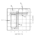
さらに別の態様では、尿管信号(ureter)対非尿管信号(non-ureter)が二次元空間上にマッピングされ、カメラが尿路組織に向いている可能性が高いこの空間の領域が検出される。図4は、2つの波長帯について2次元空間がどの様に決定されるかの例である。図4では、中波長帯域信号が、x軸に沿ってプロットされ、高波長帯域信号が、y軸に沿ってプロットされる。一例に過ぎないが、中波長帯は、700nmの波長を有するとみなされ、高波長帯は、約960nmの波長を有するとみなされる。中波長帯の図2を使用する場合に、非尿管信号(non-ureter)は、破線の垂直線401の周りに散在し、尿管信号(ureter)尿管は、実線の垂直線402の周りに散在する。高波長帯の場合に、非尿管信号(non-ureter)は、破線の水平線403の周りに散在し、及び尿管信号(ureter)は、実線の水平線404の周りに散在する。
In yet another embodiment, the ureteral signal (ureter) versus the non-ureteral signal (non-ureter) is mapped onto a two-dimensional space, and a region of this space where the camera is likely to be facing urinary tract tissue is detected. Is done. FIG. 4 is an example of how a two-dimensional space is determined for two wavelength bands. In FIG. 4, the middle wavelength band signal is plotted along the x-axis and the high wavelength band signal is plotted along the y-axis. By way of example only, the middle wavelength band is considered to have a wavelength of 700 nm, and the high wavelength band is considered to have a wavelength of about 960 nm. When using FIG. 2 in the mid-wavelength band, the non-ureter signals are scattered around the dashed
従って、領域411内に入る信号は非尿管信号(non-ureter)であり、領域410内に入る信号は尿管信号(ureter)である。実際には、2つの実際の波長帯を用いて多数の異なる尿管サンプルの画像が取り込まれ、上述した変換を用いて得られた非尿管信号(non-ureter)及び尿管信号(ureter)がプロットされる。領域410及び411が規定され、尿管信号(ureter)の領域を区切る線が決定される。次に、臨床的設定において、所与の位置における尿管信号(ureter)が領域410内に入る場合に、その位置は、尿管組織を有するものとしてマークされ、そうでない場合に、その位置は、非尿管組織を有するとみなされる。
Therefore, the signal entering the
フレームが処理され、カメラの視野内の領域が尿管であると判定された後に、その領域は、その領域がシーンの残りの領域と区別されて外科医に表示される。尿管が手術部位の残りのシーンと区別されるように、この情報を表示する複数の方法がある。1つの方法は、尿管組織の上に色をオーバーレイして、尿管が存在することを外科医に警告することである。もう1つの方法は、ディスプレイの尿管組織の色を上述した偽色で著しく変えることであり、それによって、外科医は、尿管組織を周囲の組織とは異なるものと認識する。さらに別の方法は、尿管組織の上に警告サインを表示して、尿管の存在を外科医に知らせることである。これらの場合の全てにおいて、一態様では、オーバーレイ又は色の変化等は、外科医によって制御され得る。すなわち、外科医は、尿管の強調表示をオン又はオフにすることができ、又は尿管を視覚化するために適用されるカラーシフトを増減することができる。 After the frame has been processed and an area within the camera's field of view has been determined to be the ureter, the area is displayed to the surgeon, distinguishing that area from the rest of the scene. There are several ways to display this information so that the ureter is distinguished from the rest of the surgical site scene. One method is to overlay a color on the ureteral tissue to alert the surgeon that a ureter is present. Another method is to significantly change the color of the ureteral tissue of the display with the false color described above, so that the surgeon recognizes the ureteral tissue as distinct from the surrounding tissue. Yet another method is to display a warning sign on the ureteral tissue to inform the surgeon of the presence of the ureter. In all of these cases, in one aspect, the overlay or color change, etc., can be controlled by the surgeon. That is, the surgeon can turn ureter highlighting on or off, or increase or decrease the color shift applied to visualize the ureter.
いくつかの態様では、上述した変換Cは使用されず、閾値処理は、波長帯域信号を使用して波長帯域空間に直接的に適用され、その位置が尿管組織であるかどうかを判定する。別の態様では、変換Cを適用することに由来する尿管信号は、尿管の有無を直接的に表示するために使用される。 In some aspects, the transform C described above is not used, and thresholding is applied directly to the wavelength band space using the wavelength band signal to determine if the location is ureteral tissue. In another aspect, the ureteral signal from applying transformation C is used to directly indicate the presence or absence of a ureter.
尿管信号の変換Cに対する波長帯域の第1の複数の重みUB1、UB2、UB3、及び第2の複数の重みNB1、NB2、NB3は、複数の方法で決定することができる。例えば、第1及び第2の複数の重みは、最初に、尿管及び非尿管組織によって直接的に反射された波長帯域信号を実験的に測定することによって;カメラ/照明システム及び尿管及び非尿管組織の反射率をモデル化することによって;又は実験的測定及びモデリングのいくつかの組合せによって決定することができる。これらのアプローチのそれぞれは、尿管組織に相関する一組の波長帯信号、及び非尿管組織に相関する一組の波長帯信号を生成する。 The first plurality of weights UB1, UB2, UB3 and the second plurality of weights NB1, NB2, NB3 of the wavelength band for the conversion C of the ureteral signal can be determined in a number of ways. For example, the first and second plurality of weights may be determined by first experimentally measuring wavelength band signals directly reflected by ureteral and non-ureteral tissue; camera / illumination system and ureter and It can be determined by modeling the reflectance of non-ureteral tissue; or by some combination of experimental measurements and modeling. Each of these approaches produces a set of waveband signals that correlate to ureteral tissue and a set of waveband signals that correlate to non-ureteral tissue.
第1及び第2の複数の重みは、尿管組織に相関する一組の波長帯域信号と、非尿管組織に相関する一組の波長帯域信号とを組み合わせて、誤差を最小にする行列式を解くことによって決定される。例えば、
ここで、DUは、尿管信号を選択するための行列であり、
BUは、N個の尿管サンプルについて3つの波長帯のそれぞれで取り込まれるフレームの行列であり、
DNは、非尿管信号を選択するための行列であり、
BNは、M個の非尿管サンプルについて3つの波長帯のそれぞれで取り込まれるフレームの行列であり、
pinvは、疑似逆行列である。
The first and second plurality of weights are determinants that combine a set of wavelength band signals that correlate with ureteral tissue and a set of wavelength band signals that correlate with non-ureteral tissue to minimize errors. Is determined by solving For example,
Here, DU is a matrix for selecting a ureteral signal,
BU is the matrix of frames captured at each of the three wavelength bands for the N ureter samples;
DN is a matrix for selecting non-ureteral signals,
BN is a matrix of frames captured at each of the three wavelength bands for the M non-ureteral samples,
pinv is a pseudo inverse matrix.
疑似逆行列は、当業者には知られている。ここで使用するのに適した擬似逆行列は、Moore-Penrose擬似逆行列と呼ばれる。Moore-Penrose擬似逆行列の一般的な使用法は、固有の解法がない線形方程式の系に対して、最適合する最小二乗解を計算することである。Moore-Penrose擬似逆行列のもう一つの使用法は、線形方程式の系に対する最小(ユークリッド)ノルム解を見つけることである。一態様では、最適合する最小二乗解が使用される。 Pseudo inverse matrices are known to those skilled in the art. A pseudoinverse suitable for use herein is called a Moore-Penrose pseudoinverse. A common use of the Moore-Penrose pseudo-inverse is to compute the best-fit least-squares solution for a system of linear equations without an inherent solution. Another use of the Moore-Penrose pseudoinverse is to find the minimum (Euclidean) norm solution for a system of linear equations. In one aspect, the best-fit least-squares solution is used.
上記の例では、複数の異なる光スペクトルが手術部位を照射し、手術部位のシーンを含むフレームが、異なる光スペクトルのそれぞれで取り込まれ、複数のフレームが取り込まれる。複数のフレームを解析して手術部位のシーンの位置を尿管組織として分類する。別の態様では、この同じ方法が使用されるが、患者は手術前に色素を服用する。色素は、フルオロフォア及び放射性色素の両方とは異なる。フルオロフォアは、励起波長スペクトルを有する光を吸収し、発光波長スペクトルを有する光を放出する。このプロセスは、入射光の反射とは対照的に、フルオロフォアによる入射光の吸収と、フルオロフォアによる新たな光の放出である。放射性色素は、入射光を必要とせずに、測定される放射線を放出する。この態様で使用される色素は、新しい光も放射線も放出しないが、その代わりに、色素は入射光の反射に影響を及ぼす。 In the above example, a plurality of different light spectra illuminate the surgical site, frames containing scenes of the surgical site are captured at each of the different light spectra, and a plurality of frames are captured. By analyzing a plurality of frames, the position of the scene at the surgical site is classified as ureteral tissue. In another embodiment, this same method is used, but the patient takes the dye prior to surgery. Dyes are different from both fluorophores and radioactive dyes. Fluorophores absorb light having an excitation wavelength spectrum and emit light having an emission wavelength spectrum. This process is the absorption of the incident light by the fluorophore and the emission of new light by the fluorophore, as opposed to the reflection of the incident light. Radioactive dyes emit the radiation to be measured without the need for incident light. The dye used in this manner emits no new light or radiation, but instead the dye affects the reflection of the incident light.
このような色素の一例は、時にはフェナゾピリジン塩酸塩(Phenazopyridine
Hydochoride)の形態で使用されるフェナゾピリジンである。フェナゾピリジンは、尿路鎮痛剤として作用する色素である。フェナゾピリジンは、典型的には患者に経口的に投与され、腎臓によって尿中に直接的に排出される。こうして、尿及び尿管の壁の両方が、尿管の光吸収特性を変化させるフェナゾピリジンによって染色され、従って尿管によって反射された光の少なくとも一部を変化させる。従って、一態様では、フェナゾピリジンが投与され、フェナゾピリジンで処理された尿管組織によって反射される波長帯が決定される。これらの波長帯は、上述した解析に使用される。
One example of such a dye is sometimes called phenazopyridine hydrochloride (Phenazopyridine
Hydochoride) is a phenazopyridine used in the form of Phenazopyridine is a pigment that acts as a urinary analgesic. Phenazopyridine is typically administered orally to patients and is excreted directly into the urine by the kidneys. Thus, both urine and the wall of the ureter are stained by phenazopyridine, which alters the light absorbing properties of the ureter, thus altering at least a portion of the light reflected by the ureter. Thus, in one aspect, phenazopyridine is administered and the wavelength band reflected by phenazopyridine-treated ureteral tissue is determined. These wavelength bands are used for the analysis described above.
本明細書で説明される様々なモジュールは、プロセッサ131、ハードウェア、ファームウェア、又はこれらの3つの任意の組合せで実行されるソフトウェアによって実装され得る。モジュールがプロセッサ上で実行されるソフトウェアとして実装される場合に、ソフトウェアは、コンピュータ可読命令としてメモリ132に記憶され、このコンピュータ可読命令は、プロセッサ131上で実行される。メモリの全部又は一部は、プロセッサがメモリに結合される限り、プロセッサとは物理的に異なる位置にあってもよい。メモリは、揮発性メモリ、不揮発性メモリ、又はこれら2つの任意の組合せを指す。
The various modules described herein may be implemented by software running on the
また、様々なモジュールの機能は、本明細書で説明されるように、1つのユニットによって実行してもよく、又は異なるコンポーネント間で分割してもよく、各機能は、ハードウェア、プロセッサ上で実行されるソフトウェア、及びファームウェアで実装してもよい。異なるコンポーネント間で分割される場合に、コンポーネントは、一箇所に集中させてもよく、又は分散処理目的のためにシステム300に亘って分散させてもよい。様々なモジュールの実行により、様々なモジュール及び制御装置360について上述した処理を実行する方法が得られる。
Also, the functions of the various modules may be performed by a single unit, as described herein, or may be split between different components, with each function being implemented on hardware, on a processor. It may be implemented by software to be executed and firmware. When split between different components, the components may be centralized or distributed across
こうして、プロセッサは、プロセッサによって実行される命令を含むメモリに結合される。これは、コンピュータシステム内で、或いは、モデム及びアナログライン、又はデジタルインターフェイス及びデジタルキャリアラインを介して他のコンピュータに接続することによって達成することができる。 Thus, a processor is coupled to a memory that contains instructions for execution by the processor. This can be accomplished in a computer system or by connecting to another computer via a modem and analog lines, or a digital interface and digital carrier line.
ここで、コンピュータプログラム製品は、本明細書に記載したプロセスの一部又は全てに必要なコンピュータ可読コードを記憶するように構成される、又はこれらのプロセスの一部又は全てのコンピュータ可読コードが格納されるコンピュータ可読媒体を含む。コンピュータプログラム製品のいくつかの例は、CD−ROMディスク、DVDディスク、フラッシュメモリ、ROMカード、フロッピー(登録商標)ディスク、磁気テープ、コンピュータハードドライブ、ネットワーク上のサーバ、及びネットワークを介して送信されるコンピュータ可読プログラムコードを表す信号である。非一時的な有形のコンピュータプログラム製品は、プロセスの一部又は全てのコンピュータ可読命令を記憶するように構成される、又はプロセスの一部又は全てのコンピュータ可読命令が格納される有形のコンピュータ可読媒体を含む。非一時的な有形のコンピュータプログラム製品は、CD−ROMディスク、DVDディスク、フラッシュメモリ、ROMカード、フロッピー(登録商標)ディスク、磁気テープ、コンピュータハードドライブ、及び他の物理的記憶媒体である。 Here, the computer program product is configured to store computer readable code required for some or all of the processes described herein, or store some or all of the computer readable code for these processes. Computer-readable media. Some examples of computer program products are transmitted via CD-ROM disks, DVD disks, flash memory, ROM cards, floppy disks, magnetic tape, computer hard drives, servers on networks, and networks. Is a signal representing computer readable program code. The non-transitory tangible computer program product is configured to store some or all computer readable instructions of a process, or a tangible computer readable medium on which some or all computer readable instructions of a process are stored including. Non-transitory tangible computer program products are CD-ROM disks, DVD disks, flash memory, ROM cards, floppy disks, magnetic tape, computer hard drives, and other physical storage media.
本開示に鑑みて、本明細書で説明したプロセスの一部又は全てに使用される命令は、ユーザにとって関心のあるオペレーティングシステム及びコンピュータプログラム言語を使用して広範なコンピュータシステム構成で実装することができる。 In view of the present disclosure, the instructions used for some or all of the processes described herein may be implemented in a wide variety of computer system configurations using operating systems and computer programming languages that are of interest to the user. it can.
本発明の態様及び実施形態を示す上述した詳細な説明及び添付の図面は、限定するものと解釈すべきではなく、特許請求の範囲が、保護される発明を規定する。本明細書及び特許請求の範囲の精神及び範囲から逸脱することなく、種々の機械的な、組成的な、構造的な、電気的な、及び操作上の変更を行うことができる。いくつかの例では、周知の回路、構造、及び技術は、本発明を不明瞭にすることを避けるために詳細に示していないか、又は説明していない。 The above detailed description and the accompanying drawings, which illustrate aspects and embodiments of the present invention, are not to be construed as limiting, and the claims define the protected invention. Various mechanical, compositional, structural, electrical, and operational changes may be made without departing from the spirit and scope of the specification and claims. In some instances, well-known circuits, structures, and techniques have not been shown or described in detail so as not to obscure the present invention.
また、この詳細な説明の用語は、本発明を限定することを意図していない。例えば、「〜の下に(beneath)」、「〜より下の(below)」、「〜の下方の(lower)」、「〜より上の(above)」、「〜の上方の(upper)」、「基端の(proximal)」、「先端の(distal)」等の空間に関連する用語は、図に示される1つの要素又は機構に対して別の要素又は機構との関係を説明するために使用される。これらの空間に関連する用語は、図面に示される位置及び向きに加えて、使用又は操作中の装置の異なる位置(すなわち、配置)及び向き(すなわち、回転位置)を包含することを意図している。 Also, the terms of this detailed description are not intended to limit the invention. For example, "beneath", "below", "lower", "above", "upper" Terms related to space, such as "", "proximal", "distal", describe the relationship of one element or mechanism shown in the figures to another element or mechanism Used for These space-related terms are intended to encompass different positions (ie, arrangements) and orientations (ie, rotational positions) of the device in use or operation in addition to the positions and orientations shown in the figures. I have.
例えば図面内の装置をひっくり返した場合に、他の要素又は機構「より下の(below)」又は「の下に(beneath)」として説明された要素は、次に、他の要素又は機構「より上の(above)」又は「の上に(over)」となる。従って、例示的な用語「〜より下の(below)」は、「〜より上の(above)」及び「〜より下の(below)」両方の位置及び向きを包含することができる。その装置は、他の方法で向き合わせ(90度回転又は他の向きに)してもよく、本明細書で使用される空間に関連する説明は、それに応じて解釈される。 For example, if the device in the drawing is turned over, other elements or features described below as `` below '' or `` beneath '' will in turn be replaced by other elements or features `` "Above" or "over". Thus, the exemplary term “below” can encompass both positions and orientations of “above” and “below”. The device may be oriented (rotated 90 degrees or other orientations) in other ways, and the space-related descriptions used herein will be construed accordingly.
同様に、様々な軸線に沿った及びこの軸線周りの運動の説明は、装置の様々な特別な位置及び向きを含む。単数形「1つの(a, an)」及び「その(the)」は、文脈が他に指示しない限り、複数形も含むことを意図している。用語「備える、有する、含む(comprises, comprising)」、「含む、有する(including)」等は、説明した特徴、ステップ、操作、要素、及び/又は構成要素の存在を特定するが、1つ以上の他の特徴、ステップ、操作、要素、構成要素、及び/又はグループの存在又は追加を排除するものではない。 Similarly, description of movement along and about various axes includes various specific positions and orientations of the device. The singular forms “a” and “the” are intended to include the plural unless the context otherwise dictates. The term “comprises, comprising,” “including,” etc., identifies one or more of the described feature, step, operation, element, and / or component. Does not exclude the presence or addition of other features, steps, operations, elements, components, and / or groups.
「結合した」として説明した構成要素は、電気的に又は機械的に直接的に結合されるか、又は1つ又は複数の中間要素を介して間接的に結合してもよい。本開示に鑑みて、向上したディスプレイシステムに関して説明した操作のいずれか又は任意の組合せで使用される命令は、ユーザにとって関心のあるオペレーティングシステム及びコンピュータプログラム言語を使用して広範なコンピュータシステム構成で実装することができる。 Components described as "coupled" may be directly coupled electrically or mechanically, or may be coupled indirectly via one or more intermediate components. In view of the present disclosure, the instructions used in any or any combination of the operations described with respect to the enhanced display system may be implemented in a wide variety of computer system configurations using operating systems and computer programming languages of interest to a user. can do.
全ての実施例及び説明の参照は、非限定的であり、特許請求の範囲を本明細書で説明した特定の実装態様や実施形態及びその等価物に限定するために使用すべきではない。見出しは、単に形式のためであり、1つの見出しの下のテキストは、相互参照することができ、すなわち1つ以上の見出しの下のテキストに適用することができるので、主題をあらゆる方法で制限するように使用すべきではない。最後に、本開示に鑑みて、一態様又は実施形態に関連して説明した特定の特徴は、図面に特に示されておらず又は本文中に記載されていない場合であっても、本発明の開示された他の態様又は実施形態に適用することができる。
References to all examples and descriptions are non-limiting and should not be used to limit the claims to the particular implementations or embodiments described herein and their equivalents. Headings are purely for form, and the text under one heading can be cross-referenced, that is, applied to text under one or more headings, thus limiting the subject matter in any way. Should not be used to. Finally, in view of the present disclosure, certain features described in connection with one aspect or embodiment are not intended to be particularly illustrated in the drawings or described herein. It can be applied to other disclosed aspects or embodiments.
Claims (6)
複数の手術部位のシーンを取り込むように構成された画像取込システムであって、該画像取込システムは、複数の波長帯のうちの異なる波長帯の反射光からの前記複数の手術部位のシーン内の各手術部位のシーンを取り込むようにさらに構成され、前記複数の波長帯の各波長帯は、尿管組織及び該尿管組織の周りの組織によって反射される、画像取込システムと、
前記画像取込システムに結合される尿管解析モジュールであって、該尿管解析モジュールは、
前記複数の波長帯のうちの各波長帯で前記尿管組織のスペクトル反射率と前記尿管組織の周りの前記組織のスペクトル反射率との間の差を検出し、
前記尿管組織の前記スペクトル反射率と前記尿管組織の周りの前記組織の前記スペクトル反射率との間の前記差に基づいて、前記取り込まれた手術部位のシーン内の前記尿管組織を識別するように構成される、尿管解析モジュールと、
前記尿管解析モジュールに結合される表示装置であって、前記尿管組織を強調表示した状態で手術部位のシーンを表示するように構成される、表示装置と、を有する、
手術システム。 A surgical system, wherein the surgical system comprises:
A configured image capture system to capture a scene of a plurality of surgical site, the image capture system, of the plurality of surgical site from the reflected light of different wavelength bands of the plurality of wavelength bands scene is further configured to write useless take scenes of the surgical site within the respective wavelength bands of the plurality of wavelength bands are anti Isa is by the tissue surrounding the urinary tract tissue and urine tract tissue, the image capture system When,
A ureter analysis module coupled to the image capture system, wherein the ureter analysis module comprises:
Detecting the difference between the spectral reflectance of the ureteral tissue and the spectral reflectance of the tissue around the ureteral tissue in each wavelength band of the plurality of wavelength bands,
Based on the difference between the tissue the spectral reflectance around the urinary tract tissue and the spectral reflectance of the urinary tract tissue, identifying the urinary tract tissue in the scene of the captured surgical site configured to, a urinary tract analysis module,
A display device coupled to the ureter analysis module, having a configured, display device to display a scene of surgical site while highlighting the urinary tract tissue,
Surgery system.
前記取り込まれた複数の手術部位のシーンのそれぞれを含むデータのフレームを尿管信号に変換し、
前記尿管信号が、前記取り込まれた手術部位のシーンの位置における尿管組織を示すかどうかを判定する、ことにより前記尿管組織を識別するように構成される、請求項1又は2に記載の手術システム。 Before Kinyo tube analysis module,
Converting a frame of data including each of the captured scenes of the plurality of surgical sites into a ureteral signal,
The ureter signal, determines whether to show urinary tract tissue in the scene of the position of the captured surgical site, Ru is configured for identifying the ureter tissue by, according to claim 1 or 2 Surgery system.
450nm〜580nmの範囲を有する第1の波長帯、A first wavelength band having a range of 450 nm to 580 nm;
640nm〜750nmの範囲を有する第2の波長帯、及びA second wavelength band having a range of 640 nm to 750 nm, and
900nm〜1080nmの範囲を有する第3の波長帯のうちの少なくとも2つを含む、請求項1乃至5のいずれか一項に記載の手術システム。The surgical system according to any one of claims 1 to 5, comprising at least two of the third wavelength bands having a range from 900 nm to 1080 nm.
Applications Claiming Priority (3)
| Application Number | Priority Date | Filing Date | Title |
|---|---|---|---|
| US201462092651P | 2014-12-16 | 2014-12-16 | |
| US62/092,651 | 2014-12-16 | ||
| PCT/US2015/065561 WO2016100214A1 (en) | 2014-12-16 | 2015-12-14 | Ureter detection using waveband-selective imaging |
Related Child Applications (1)
| Application Number | Title | Priority Date | Filing Date |
|---|---|---|---|
| JP2020018050A Division JP7086121B2 (en) | 2014-12-16 | 2020-02-05 | Ureter detection using imaging by wavelength band selection |
Publications (3)
| Publication Number | Publication Date |
|---|---|
| JP2018500095A JP2018500095A (en) | 2018-01-11 |
| JP2018500095A5 JP2018500095A5 (en) | 2019-01-24 |
| JP6657224B2 true JP6657224B2 (en) | 2020-03-04 |
Family
ID=56127432
Family Applications (2)
| Application Number | Title | Priority Date | Filing Date |
|---|---|---|---|
| JP2017532121A Active JP6657224B2 (en) | 2014-12-16 | 2015-12-14 | Ureter Detection Using Imaging by Wavelength Band Selection |
| JP2020018050A Active JP7086121B2 (en) | 2014-12-16 | 2020-02-05 | Ureter detection using imaging by wavelength band selection |
Family Applications After (1)
| Application Number | Title | Priority Date | Filing Date |
|---|---|---|---|
| JP2020018050A Active JP7086121B2 (en) | 2014-12-16 | 2020-02-05 | Ureter detection using imaging by wavelength band selection |
Country Status (6)
| Country | Link |
|---|---|
| US (3) | US10588711B2 (en) |
| EP (3) | EP3232975B1 (en) |
| JP (2) | JP6657224B2 (en) |
| KR (1) | KR102476063B1 (en) |
| CN (2) | CN106999249B (en) |
| WO (1) | WO2016100214A1 (en) |
Families Citing this family (140)
| Publication number | Priority date | Publication date | Assignee | Title |
|---|---|---|---|---|
| US9962066B2 (en) | 2005-12-30 | 2018-05-08 | Intuitive Surgical Operations, Inc. | Methods and apparatus to shape flexible entry guides for minimally invasive surgery |
| US7930065B2 (en) | 2005-12-30 | 2011-04-19 | Intuitive Surgical Operations, Inc. | Robotic surgery system including position sensors using fiber bragg gratings |
| CN104688327B (en) | 2006-06-13 | 2017-06-09 | 直观外科手术操作公司 | Minimally invasive surgery system |
| US10433917B2 (en) * | 2009-05-29 | 2019-10-08 | Jack Wade | System and method for enhanced data analysis with video enabled software tools for medical environments |
| US11871901B2 (en) | 2012-05-20 | 2024-01-16 | Cilag Gmbh International | Method for situational awareness for surgical network or surgical network connected device capable of adjusting function based on a sensed situation or usage |
| US11504192B2 (en) | 2014-10-30 | 2022-11-22 | Cilag Gmbh International | Method of hub communication with surgical instrument systems |
| EP3232975B1 (en) | 2014-12-16 | 2020-07-15 | Intuitive Surgical Operations, Inc. | Ureter detection using waveband-selective imaging |
| EP3262454A4 (en) | 2015-02-23 | 2019-02-27 | Li-Cor, Inc. | Fluorescence biopsy specimen imager and methods |
| EP3869184A1 (en) | 2015-06-26 | 2021-08-25 | Li-Cor, Inc. | Fluorescence biopsy specimen imager and methods |
| US10489964B2 (en) | 2016-04-21 | 2019-11-26 | Li-Cor, Inc. | Multimodality multi-axis 3-D imaging with X-ray |
| US10278586B2 (en) | 2016-06-23 | 2019-05-07 | Li-Cor, Inc. | Complementary color flashing for multichannel image presentation |
| EP3545488A1 (en) | 2016-11-23 | 2019-10-02 | Li-Cor, Inc. | Motion-adaptive interactive imaging method |
| EP3616158A1 (en) | 2017-04-25 | 2020-03-04 | Li-Cor, Inc. | Top-down and rotational side view biopsy specimen imager and methods |
| WO2019060733A2 (en) | 2017-09-22 | 2019-03-28 | Intuitive Surgical Operations, Inc. | Enhancing visible differences between different tissues in computer-assisted tele-operated surgery |
| US11801098B2 (en) | 2017-10-30 | 2023-10-31 | Cilag Gmbh International | Method of hub communication with surgical instrument systems |
| US11291510B2 (en) | 2017-10-30 | 2022-04-05 | Cilag Gmbh International | Method of hub communication with surgical instrument systems |
| US11564756B2 (en) | 2017-10-30 | 2023-01-31 | Cilag Gmbh International | Method of hub communication with surgical instrument systems |
| US11759224B2 (en) | 2017-10-30 | 2023-09-19 | Cilag Gmbh International | Surgical instrument systems comprising handle arrangements |
| US11911045B2 (en) | 2017-10-30 | 2024-02-27 | Cllag GmbH International | Method for operating a powered articulating multi-clip applier |
| US11413042B2 (en) | 2017-10-30 | 2022-08-16 | Cilag Gmbh International | Clip applier comprising a reciprocating clip advancing member |
| US11229436B2 (en) | 2017-10-30 | 2022-01-25 | Cilag Gmbh International | Surgical system comprising a surgical tool and a surgical hub |
| US11311342B2 (en) | 2017-10-30 | 2022-04-26 | Cilag Gmbh International | Method for communicating with surgical instrument systems |
| US11317919B2 (en) | 2017-10-30 | 2022-05-03 | Cilag Gmbh International | Clip applier comprising a clip crimping system |
| US11510741B2 (en) | 2017-10-30 | 2022-11-29 | Cilag Gmbh International | Method for producing a surgical instrument comprising a smart electrical system |
| US12026900B2 (en) * | 2017-12-27 | 2024-07-02 | Cllag GmbH International | Hyperspectral imaging in a light deficient environment |
| US11540855B2 (en) | 2017-12-28 | 2023-01-03 | Cilag Gmbh International | Controlling activation of an ultrasonic surgical instrument according to the presence of tissue |
| US10758310B2 (en) | 2017-12-28 | 2020-09-01 | Ethicon Llc | Wireless pairing of a surgical device with another device within a sterile surgical field based on the usage and situational awareness of devices |
| US11818052B2 (en) | 2017-12-28 | 2023-11-14 | Cilag Gmbh International | Surgical network determination of prioritization of communication, interaction, or processing based on system or device needs |
| US11291495B2 (en) | 2017-12-28 | 2022-04-05 | Cilag Gmbh International | Interruption of energy due to inadvertent capacitive coupling |
| US11324557B2 (en) | 2017-12-28 | 2022-05-10 | Cilag Gmbh International | Surgical instrument with a sensing array |
| US11410259B2 (en) | 2017-12-28 | 2022-08-09 | Cilag Gmbh International | Adaptive control program updates for surgical devices |
| US11304699B2 (en) | 2017-12-28 | 2022-04-19 | Cilag Gmbh International | Method for adaptive control schemes for surgical network control and interaction |
| US11234756B2 (en) | 2017-12-28 | 2022-02-01 | Cilag Gmbh International | Powered surgical tool with predefined adjustable control algorithm for controlling end effector parameter |
| US11576677B2 (en) | 2017-12-28 | 2023-02-14 | Cilag Gmbh International | Method of hub communication, processing, display, and cloud analytics |
| US11096693B2 (en) | 2017-12-28 | 2021-08-24 | Cilag Gmbh International | Adjustment of staple height of at least one row of staples based on the sensed tissue thickness or force in closing |
| US11832899B2 (en) | 2017-12-28 | 2023-12-05 | Cilag Gmbh International | Surgical systems with autonomously adjustable control programs |
| US11672605B2 (en) | 2017-12-28 | 2023-06-13 | Cilag Gmbh International | Sterile field interactive control displays |
| US11257589B2 (en) | 2017-12-28 | 2022-02-22 | Cilag Gmbh International | Real-time analysis of comprehensive cost of all instrumentation used in surgery utilizing data fluidity to track instruments through stocking and in-house processes |
| US11304720B2 (en) | 2017-12-28 | 2022-04-19 | Cilag Gmbh International | Activation of energy devices |
| US11464535B2 (en) | 2017-12-28 | 2022-10-11 | Cilag Gmbh International | Detection of end effector emersion in liquid |
| US11832840B2 (en) | 2017-12-28 | 2023-12-05 | Cilag Gmbh International | Surgical instrument having a flexible circuit |
| US11844579B2 (en) | 2017-12-28 | 2023-12-19 | Cilag Gmbh International | Adjustments based on airborne particle properties |
| US11179175B2 (en) | 2017-12-28 | 2021-11-23 | Cilag Gmbh International | Controlling an ultrasonic surgical instrument according to tissue location |
| US11571234B2 (en) | 2017-12-28 | 2023-02-07 | Cilag Gmbh International | Temperature control of ultrasonic end effector and control system therefor |
| US11864728B2 (en) * | 2017-12-28 | 2024-01-09 | Cilag Gmbh International | Characterization of tissue irregularities through the use of mono-chromatic light refractivity |
| US11304763B2 (en) | 2017-12-28 | 2022-04-19 | Cilag Gmbh International | Image capturing of the areas outside the abdomen to improve placement and control of a surgical device in use |
| US11317937B2 (en) | 2018-03-08 | 2022-05-03 | Cilag Gmbh International | Determining the state of an ultrasonic end effector |
| US11969216B2 (en) | 2017-12-28 | 2024-04-30 | Cilag Gmbh International | Surgical network recommendations from real time analysis of procedure variables against a baseline highlighting differences from the optimal solution |
| US11424027B2 (en) | 2017-12-28 | 2022-08-23 | Cilag Gmbh International | Method for operating surgical instrument systems |
| US11253315B2 (en) | 2017-12-28 | 2022-02-22 | Cilag Gmbh International | Increasing radio frequency to create pad-less monopolar loop |
| US11419667B2 (en) | 2017-12-28 | 2022-08-23 | Cilag Gmbh International | Ultrasonic energy device which varies pressure applied by clamp arm to provide threshold control pressure at a cut progression location |
| US11559307B2 (en) | 2017-12-28 | 2023-01-24 | Cilag Gmbh International | Method of robotic hub communication, detection, and control |
| US11045591B2 (en) | 2017-12-28 | 2021-06-29 | Cilag Gmbh International | Dual in-series large and small droplet filters |
| US10892995B2 (en) | 2017-12-28 | 2021-01-12 | Ethicon Llc | Surgical network determination of prioritization of communication, interaction, or processing based on system or device needs |
| US11179208B2 (en) | 2017-12-28 | 2021-11-23 | Cilag Gmbh International | Cloud-based medical analytics for security and authentication trends and reactive measures |
| US11744604B2 (en) | 2017-12-28 | 2023-09-05 | Cilag Gmbh International | Surgical instrument with a hardware-only control circuit |
| US11446052B2 (en) | 2017-12-28 | 2022-09-20 | Cilag Gmbh International | Variation of radio frequency and ultrasonic power level in cooperation with varying clamp arm pressure to achieve predefined heat flux or power applied to tissue |
| US11602393B2 (en) | 2017-12-28 | 2023-03-14 | Cilag Gmbh International | Surgical evacuation sensing and generator control |
| US11696760B2 (en) | 2017-12-28 | 2023-07-11 | Cilag Gmbh International | Safety systems for smart powered surgical stapling |
| US10966791B2 (en) | 2017-12-28 | 2021-04-06 | Ethicon Llc | Cloud-based medical analytics for medical facility segmented individualization of instrument function |
| US11376002B2 (en) | 2017-12-28 | 2022-07-05 | Cilag Gmbh International | Surgical instrument cartridge sensor assemblies |
| US11284936B2 (en) | 2017-12-28 | 2022-03-29 | Cilag Gmbh International | Surgical instrument having a flexible electrode |
| US11666331B2 (en) | 2017-12-28 | 2023-06-06 | Cilag Gmbh International | Systems for detecting proximity of surgical end effector to cancerous tissue |
| US11202570B2 (en) | 2017-12-28 | 2021-12-21 | Cilag Gmbh International | Communication hub and storage device for storing parameters and status of a surgical device to be shared with cloud based analytics systems |
| US11166772B2 (en) | 2017-12-28 | 2021-11-09 | Cilag Gmbh International | Surgical hub coordination of control and communication of operating room devices |
| US11896443B2 (en) | 2017-12-28 | 2024-02-13 | Cilag Gmbh International | Control of a surgical system through a surgical barrier |
| US12035890B2 (en) | 2017-12-28 | 2024-07-16 | Cilag Gmbh International | Method of sensing particulate from smoke evacuated from a patient, adjusting the pump speed based on the sensed information, and communicating the functional parameters of the system to the hub |
| US11056244B2 (en) | 2017-12-28 | 2021-07-06 | Cilag Gmbh International | Automated data scaling, alignment, and organizing based on predefined parameters within surgical networks |
| US11771487B2 (en) | 2017-12-28 | 2023-10-03 | Cilag Gmbh International | Mechanisms for controlling different electromechanical systems of an electrosurgical instrument |
| US11529187B2 (en) | 2017-12-28 | 2022-12-20 | Cilag Gmbh International | Surgical evacuation sensor arrangements |
| US11132462B2 (en) | 2017-12-28 | 2021-09-28 | Cilag Gmbh International | Data stripping method to interrogate patient records and create anonymized record |
| US20190201139A1 (en) | 2017-12-28 | 2019-07-04 | Ethicon Llc | Communication arrangements for robot-assisted surgical platforms |
| US11786251B2 (en) | 2017-12-28 | 2023-10-17 | Cilag Gmbh International | Method for adaptive control schemes for surgical network control and interaction |
| US11389164B2 (en) | 2017-12-28 | 2022-07-19 | Cilag Gmbh International | Method of using reinforced flexible circuits with multiple sensors to optimize performance of radio frequency devices |
| US11051876B2 (en) | 2017-12-28 | 2021-07-06 | Cilag Gmbh International | Surgical evacuation flow paths |
| US11998193B2 (en) | 2017-12-28 | 2024-06-04 | Cilag Gmbh International | Method for usage of the shroud as an aspect of sensing or controlling a powered surgical device, and a control algorithm to adjust its default operation |
| US11857152B2 (en) | 2017-12-28 | 2024-01-02 | Cilag Gmbh International | Surgical hub spatial awareness to determine devices in operating theater |
| US11589888B2 (en) | 2017-12-28 | 2023-02-28 | Cilag Gmbh International | Method for controlling smart energy devices |
| US11937769B2 (en) | 2017-12-28 | 2024-03-26 | Cilag Gmbh International | Method of hub communication, processing, storage and display |
| US11109866B2 (en) | 2017-12-28 | 2021-09-07 | Cilag Gmbh International | Method for circular stapler control algorithm adjustment based on situational awareness |
| US11659023B2 (en) | 2017-12-28 | 2023-05-23 | Cilag Gmbh International | Method of hub communication |
| US11903601B2 (en) | 2017-12-28 | 2024-02-20 | Cilag Gmbh International | Surgical instrument comprising a plurality of drive systems |
| US11423007B2 (en) | 2017-12-28 | 2022-08-23 | Cilag Gmbh International | Adjustment of device control programs based on stratified contextual data in addition to the data |
| US11464559B2 (en) | 2017-12-28 | 2022-10-11 | Cilag Gmbh International | Estimating state of ultrasonic end effector and control system therefor |
| US11100631B2 (en) | 2017-12-28 | 2021-08-24 | Cilag Gmbh International | Use of laser light and red-green-blue coloration to determine properties of back scattered light |
| US11160605B2 (en) | 2017-12-28 | 2021-11-02 | Cilag Gmbh International | Surgical evacuation sensing and motor control |
| US11266468B2 (en) | 2017-12-28 | 2022-03-08 | Cilag Gmbh International | Cooperative utilization of data derived from secondary sources by intelligent surgical hubs |
| US11069012B2 (en) | 2017-12-28 | 2021-07-20 | Cilag Gmbh International | Interactive surgical systems with condition handling of devices and data capabilities |
| US11308075B2 (en) | 2017-12-28 | 2022-04-19 | Cilag Gmbh International | Surgical network, instrument, and cloud responses based on validation of received dataset and authentication of its source and integrity |
| US11304745B2 (en) | 2017-12-28 | 2022-04-19 | Cilag Gmbh International | Surgical evacuation sensing and display |
| US11419630B2 (en) | 2017-12-28 | 2022-08-23 | Cilag Gmbh International | Surgical system distributed processing |
| US11147607B2 (en) | 2017-12-28 | 2021-10-19 | Cilag Gmbh International | Bipolar combination device that automatically adjusts pressure based on energy modality |
| US10987178B2 (en) | 2017-12-28 | 2021-04-27 | Ethicon Llc | Surgical hub control arrangements |
| US10943454B2 (en) | 2017-12-28 | 2021-03-09 | Ethicon Llc | Detection and escalation of security responses of surgical instruments to increasing severity threats |
| US11786245B2 (en) | 2017-12-28 | 2023-10-17 | Cilag Gmbh International | Surgical systems with prioritized data transmission capabilities |
| US12062442B2 (en) | 2017-12-28 | 2024-08-13 | Cilag Gmbh International | Method for operating surgical instrument systems |
| US11559308B2 (en) | 2017-12-28 | 2023-01-24 | Cilag Gmbh International | Method for smart energy device infrastructure |
| US11633237B2 (en) | 2017-12-28 | 2023-04-25 | Cilag Gmbh International | Usage and technique analysis of surgeon / staff performance against a baseline to optimize device utilization and performance for both current and future procedures |
| US11273001B2 (en) | 2017-12-28 | 2022-03-15 | Cilag Gmbh International | Surgical hub and modular device response adjustment based on situational awareness |
| US11432885B2 (en) | 2017-12-28 | 2022-09-06 | Cilag Gmbh International | Sensing arrangements for robot-assisted surgical platforms |
| US11278281B2 (en) | 2017-12-28 | 2022-03-22 | Cilag Gmbh International | Interactive surgical system |
| US11311306B2 (en) | 2017-12-28 | 2022-04-26 | Cilag Gmbh International | Surgical systems for detecting end effector tissue distribution irregularities |
| US11678881B2 (en) | 2017-12-28 | 2023-06-20 | Cilag Gmbh International | Spatial awareness of surgical hubs in operating rooms |
| US20190206569A1 (en) | 2017-12-28 | 2019-07-04 | Ethicon Llc | Method of cloud based data analytics for use with the hub |
| US11896322B2 (en) | 2017-12-28 | 2024-02-13 | Cilag Gmbh International | Sensing the patient position and contact utilizing the mono-polar return pad electrode to provide situational awareness to the hub |
| US11364075B2 (en) | 2017-12-28 | 2022-06-21 | Cilag Gmbh International | Radio frequency energy device for delivering combined electrical signals |
| US11969142B2 (en) | 2017-12-28 | 2024-04-30 | Cilag Gmbh International | Method of compressing tissue within a stapling device and simultaneously displaying the location of the tissue within the jaws |
| US11213359B2 (en) | 2017-12-28 | 2022-01-04 | Cilag Gmbh International | Controllers for robot-assisted surgical platforms |
| US11076921B2 (en) | 2017-12-28 | 2021-08-03 | Cilag Gmbh International | Adaptive control program updates for surgical hubs |
| US11259830B2 (en) | 2018-03-08 | 2022-03-01 | Cilag Gmbh International | Methods for controlling temperature in ultrasonic device |
| US11701162B2 (en) | 2018-03-08 | 2023-07-18 | Cilag Gmbh International | Smart blade application for reusable and disposable devices |
| US11389188B2 (en) | 2018-03-08 | 2022-07-19 | Cilag Gmbh International | Start temperature of blade |
| US11278280B2 (en) | 2018-03-28 | 2022-03-22 | Cilag Gmbh International | Surgical instrument comprising a jaw closure lockout |
| US10973520B2 (en) | 2018-03-28 | 2021-04-13 | Ethicon Llc | Surgical staple cartridge with firing member driven camming assembly that has an onboard tissue cutting feature |
| US11096688B2 (en) | 2018-03-28 | 2021-08-24 | Cilag Gmbh International | Rotary driven firing members with different anvil and channel engagement features |
| US11219453B2 (en) | 2018-03-28 | 2022-01-11 | Cilag Gmbh International | Surgical stapling devices with cartridge compatible closure and firing lockout arrangements |
| US11406382B2 (en) | 2018-03-28 | 2022-08-09 | Cilag Gmbh International | Staple cartridge comprising a lockout key configured to lift a firing member |
| US11090047B2 (en) | 2018-03-28 | 2021-08-17 | Cilag Gmbh International | Surgical instrument comprising an adaptive control system |
| US11207067B2 (en) | 2018-03-28 | 2021-12-28 | Cilag Gmbh International | Surgical stapling device with separate rotary driven closure and firing systems and firing member that engages both jaws while firing |
| US11471156B2 (en) | 2018-03-28 | 2022-10-18 | Cilag Gmbh International | Surgical stapling devices with improved rotary driven closure systems |
| US11259806B2 (en) | 2018-03-28 | 2022-03-01 | Cilag Gmbh International | Surgical stapling devices with features for blocking advancement of a camming assembly of an incompatible cartridge installed therein |
| WO2020014031A1 (en) * | 2018-07-12 | 2020-01-16 | Intuitive Surgical Operations, Inc. | Multiple band pass bayer pattern filter |
| KR20210100144A (en) | 2018-12-05 | 2021-08-13 | 인튜어티브 서지컬 오퍼레이션즈 인코포레이티드 | Determination of chromophore concentration in tissue |
| US11369377B2 (en) | 2019-02-19 | 2022-06-28 | Cilag Gmbh International | Surgical stapling assembly with cartridge based retainer configured to unlock a firing lockout |
| US11291445B2 (en) | 2019-02-19 | 2022-04-05 | Cilag Gmbh International | Surgical staple cartridges with integral authentication keys |
| US11751872B2 (en) | 2019-02-19 | 2023-09-12 | Cilag Gmbh International | Insertable deactivator element for surgical stapler lockouts |
| US11317915B2 (en) | 2019-02-19 | 2022-05-03 | Cilag Gmbh International | Universal cartridge based key feature that unlocks multiple lockout arrangements in different surgical staplers |
| US11357503B2 (en) | 2019-02-19 | 2022-06-14 | Cilag Gmbh International | Staple cartridge retainers with frangible retention features and methods of using same |
| US11589819B2 (en) | 2019-06-20 | 2023-02-28 | Cilag Gmbh International | Offset illumination of a scene using multiple emitters in a laser mapping imaging system |
| US11012599B2 (en) | 2019-06-20 | 2021-05-18 | Ethicon Llc | Hyperspectral imaging in a light deficient environment |
| US11122967B2 (en) | 2019-06-20 | 2021-09-21 | Cilag Gmbh International | Driving light emissions according to a jitter specification in a fluorescence imaging system |
| US11754500B2 (en) | 2019-06-20 | 2023-09-12 | Cilag Gmbh International | Minimizing image sensor input/output in a pulsed fluorescence imaging system |
| US11389066B2 (en) | 2019-06-20 | 2022-07-19 | Cilag Gmbh International | Noise aware edge enhancement in a pulsed hyperspectral, fluorescence, and laser mapping imaging system |
| US11284783B2 (en) | 2019-06-20 | 2022-03-29 | Cilag Gmbh International | Controlling integral energy of a laser pulse in a hyperspectral imaging system |
| US12013496B2 (en) | 2019-06-20 | 2024-06-18 | Cilag Gmbh International | Noise aware edge enhancement in a pulsed laser mapping imaging system |
| USD964564S1 (en) | 2019-06-25 | 2022-09-20 | Cilag Gmbh International | Surgical staple cartridge retainer with a closure system authentication key |
| USD952144S1 (en) | 2019-06-25 | 2022-05-17 | Cilag Gmbh International | Surgical staple cartridge retainer with firing system authentication key |
| USD950728S1 (en) | 2019-06-25 | 2022-05-03 | Cilag Gmbh International | Surgical staple cartridge |
| GB2593740B (en) * | 2020-03-31 | 2024-08-14 | Cmr Surgical Ltd | Control system of a surgical robot |
| US20230366821A1 (en) * | 2020-09-17 | 2023-11-16 | Cytoveris Inc. | Multi-Spectral Imager for UV-Excited Tissue Autofluorescence Mapping |
Family Cites Families (16)
| Publication number | Priority date | Publication date | Assignee | Title |
|---|---|---|---|---|
| US5408996A (en) | 1993-03-25 | 1995-04-25 | Salb; Jesse | System and method for localization of malignant tissue |
| US5590660A (en) | 1994-03-28 | 1997-01-07 | Xillix Technologies Corp. | Apparatus and method for imaging diseased tissue using integrated autofluorescence |
| US6331181B1 (en) | 1998-12-08 | 2001-12-18 | Intuitive Surgical, Inc. | Surgical robotic tools, data architecture, and use |
| US6659939B2 (en) | 1998-11-20 | 2003-12-09 | Intuitive Surgical, Inc. | Cooperative minimally invasive telesurgical system |
| US6869430B2 (en) | 2000-03-31 | 2005-03-22 | Rita Medical Systems, Inc. | Tissue biopsy and treatment apparatus and method |
| JP2006509573A (en) | 2002-12-13 | 2006-03-23 | イエトメド リミテッド | Optical inspection method and apparatus particularly useful for distinguishing between tumor and normal tissue in real time during surgery |
| US20070027362A1 (en) * | 2005-07-27 | 2007-02-01 | Olympus Medical Systems Corp. | Infrared observation system |
| US8280496B2 (en) * | 2007-12-13 | 2012-10-02 | Boston Scientific Scimed, Inc. | Extended spectral sensitivity endoscope system and method of using the same |
| US20110059023A1 (en) * | 2008-03-19 | 2011-03-10 | Tunnell James W | Narrowband imaging using near-infrared absorbing nanoparticles |
| JP5160276B2 (en) | 2008-03-24 | 2013-03-13 | 富士フイルム株式会社 | Image display method and apparatus |
| JP5220961B2 (en) | 2011-01-17 | 2013-06-26 | オリンパスメディカルシステムズ株式会社 | In vivo observation apparatus and capsule endoscope apparatus |
| US8672838B2 (en) | 2011-08-12 | 2014-03-18 | Intuitive Surgical Operations, Inc. | Image capture unit in a surgical instrument |
| WO2013177061A1 (en) * | 2012-05-21 | 2013-11-28 | The Regents Of The University Of Colorado, A Body Corporate | Three-dimensional mapping and therapy of prostate cancer |
| US9220570B2 (en) * | 2012-06-29 | 2015-12-29 | Children's National Medical Center | Automated surgical and interventional procedures |
| JP2014128423A (en) | 2012-12-28 | 2014-07-10 | Olympus Medical Systems Corp | Endoscope system |
| EP3232975B1 (en) | 2014-12-16 | 2020-07-15 | Intuitive Surgical Operations, Inc. | Ureter detection using waveband-selective imaging |
-
2015
- 2015-12-14 EP EP15870805.7A patent/EP3232975B1/en active Active
- 2015-12-14 EP EP23173115.9A patent/EP4226887A1/en active Pending
- 2015-12-14 KR KR1020177016473A patent/KR102476063B1/en active IP Right Grant
- 2015-12-14 CN CN201580068079.4A patent/CN106999249B/en active Active
- 2015-12-14 JP JP2017532121A patent/JP6657224B2/en active Active
- 2015-12-14 CN CN202011081990.8A patent/CN112057169B/en active Active
- 2015-12-14 EP EP20180104.0A patent/EP3730086B1/en active Active
- 2015-12-14 WO PCT/US2015/065561 patent/WO2016100214A1/en active Application Filing
- 2015-12-14 US US15/536,210 patent/US10588711B2/en active Active
-
2020
- 2020-01-29 US US16/775,806 patent/US11389267B2/en active Active
- 2020-02-05 JP JP2020018050A patent/JP7086121B2/en active Active
-
2022
- 2022-06-14 US US17/840,130 patent/US12016738B2/en active Active
Also Published As
| Publication number | Publication date |
|---|---|
| JP2018500095A (en) | 2018-01-11 |
| KR102476063B1 (en) | 2022-12-12 |
| WO2016100214A1 (en) | 2016-06-23 |
| EP4226887A1 (en) | 2023-08-16 |
| CN112057169A (en) | 2020-12-11 |
| US20170360526A1 (en) | 2017-12-21 |
| US20220304769A1 (en) | 2022-09-29 |
| US11389267B2 (en) | 2022-07-19 |
| JP2020073083A (en) | 2020-05-14 |
| EP3232975A4 (en) | 2018-07-25 |
| EP3232975A1 (en) | 2017-10-25 |
| CN112057169B (en) | 2024-09-06 |
| KR20170094231A (en) | 2017-08-17 |
| EP3232975B1 (en) | 2020-07-15 |
| JP7086121B2 (en) | 2022-06-17 |
| CN106999249A (en) | 2017-08-01 |
| US12016738B2 (en) | 2024-06-25 |
| US20200179079A1 (en) | 2020-06-11 |
| EP3730086A1 (en) | 2020-10-28 |
| EP3730086B1 (en) | 2023-06-28 |
| US10588711B2 (en) | 2020-03-17 |
| CN106999249B (en) | 2020-10-23 |
Similar Documents
| Publication | Publication Date | Title |
|---|---|---|
| JP7086121B2 (en) | Ureter detection using imaging by wavelength band selection | |
| US20240180404A1 (en) | Simultaneous white light and hyperspectral light imaging systems | |
| US9906739B2 (en) | Image pickup device and image pickup method | |
| CN111295127B (en) | Examination support device, endoscope device, and recording medium | |
| US20110301418A1 (en) | Catheter Based 3-D Defocusing Imaging | |
| CN107427202B (en) | Device, system and method for illuminating a structure of interest inside a human or animal body | |
| US20090242797A1 (en) | System and method for multi-mode optical imaging | |
| US20090244521A1 (en) | System and method for multi-mode optical imaging | |
| WO2019220801A1 (en) | Endoscope image processing device, endoscope image processing method, and program | |
| US11398033B2 (en) | Diagnosis support system, endoscope system, processor, and diagnosis support method | |
| US20220007942A1 (en) | Medical imaging systems and methods that facilitate use of different fluorescence imaging agents | |
| KR102112229B1 (en) | Endoscope apparatus capable of visualizing both visible light and near-infrared light |
Legal Events
| Date | Code | Title | Description |
|---|---|---|---|
| A521 | Request for written amendment filed |
Free format text: JAPANESE INTERMEDIATE CODE: A523 Effective date: 20181204 |
|
| A621 | Written request for application examination |
Free format text: JAPANESE INTERMEDIATE CODE: A621 Effective date: 20181204 |
|
| A131 | Notification of reasons for refusal |
Free format text: JAPANESE INTERMEDIATE CODE: A131 Effective date: 20190917 |
|
| A977 | Report on retrieval |
Free format text: JAPANESE INTERMEDIATE CODE: A971007 Effective date: 20190918 |
|
| A521 | Request for written amendment filed |
Free format text: JAPANESE INTERMEDIATE CODE: A523 Effective date: 20191202 |
|
| TRDD | Decision of grant or rejection written | ||
| A01 | Written decision to grant a patent or to grant a registration (utility model) |
Free format text: JAPANESE INTERMEDIATE CODE: A01 Effective date: 20200114 |
|
| A61 | First payment of annual fees (during grant procedure) |
Free format text: JAPANESE INTERMEDIATE CODE: A61 Effective date: 20200205 |
|
| R150 | Certificate of patent or registration of utility model |
Ref document number: 6657224 Country of ref document: JP Free format text: JAPANESE INTERMEDIATE CODE: R150 |
|
| R250 | Receipt of annual fees |
Free format text: JAPANESE INTERMEDIATE CODE: R250 |
|
| R250 | Receipt of annual fees |
Free format text: JAPANESE INTERMEDIATE CODE: R250 |