JP6585825B2 - Processing apparatus and method for processing electromyogram signals related to respiratory activity - Google Patents
Processing apparatus and method for processing electromyogram signals related to respiratory activity Download PDFInfo
- Publication number
- JP6585825B2 JP6585825B2 JP2018506593A JP2018506593A JP6585825B2 JP 6585825 B2 JP6585825 B2 JP 6585825B2 JP 2018506593 A JP2018506593 A JP 2018506593A JP 2018506593 A JP2018506593 A JP 2018506593A JP 6585825 B2 JP6585825 B2 JP 6585825B2
- Authority
- JP
- Japan
- Prior art keywords
- signal
- electromyogram
- muscle
- electromyogram signal
- electrodes
- Prior art date
- Legal status (The legal status is an assumption and is not a legal conclusion. Google has not performed a legal analysis and makes no representation as to the accuracy of the status listed.)
- Active
Links
Images


Classifications
-
- A—HUMAN NECESSITIES
- A61—MEDICAL OR VETERINARY SCIENCE; HYGIENE
- A61B—DIAGNOSIS; SURGERY; IDENTIFICATION
- A61B5/00—Measuring for diagnostic purposes; Identification of persons
- A61B5/24—Detecting, measuring or recording bioelectric or biomagnetic signals of the body or parts thereof
- A61B5/316—Modalities, i.e. specific diagnostic methods
- A61B5/389—Electromyography [EMG]
-
- A—HUMAN NECESSITIES
- A61—MEDICAL OR VETERINARY SCIENCE; HYGIENE
- A61B—DIAGNOSIS; SURGERY; IDENTIFICATION
- A61B5/00—Measuring for diagnostic purposes; Identification of persons
- A61B5/08—Detecting, measuring or recording devices for evaluating the respiratory organs
-
- A—HUMAN NECESSITIES
- A61—MEDICAL OR VETERINARY SCIENCE; HYGIENE
- A61B—DIAGNOSIS; SURGERY; IDENTIFICATION
- A61B5/00—Measuring for diagnostic purposes; Identification of persons
- A61B5/68—Arrangements of detecting, measuring or recording means, e.g. sensors, in relation to patient
- A61B5/6801—Arrangements of detecting, measuring or recording means, e.g. sensors, in relation to patient specially adapted to be attached to or worn on the body surface
- A61B5/6813—Specially adapted to be attached to a specific body part
- A61B5/6823—Trunk, e.g., chest, back, abdomen, hip
-
- A—HUMAN NECESSITIES
- A61—MEDICAL OR VETERINARY SCIENCE; HYGIENE
- A61B—DIAGNOSIS; SURGERY; IDENTIFICATION
- A61B5/00—Measuring for diagnostic purposes; Identification of persons
- A61B5/68—Arrangements of detecting, measuring or recording means, e.g. sensors, in relation to patient
- A61B5/6801—Arrangements of detecting, measuring or recording means, e.g. sensors, in relation to patient specially adapted to be attached to or worn on the body surface
- A61B5/683—Means for maintaining contact with the body
- A61B5/6832—Means for maintaining contact with the body using adhesives
- A61B5/6833—Adhesive patches
-
- A—HUMAN NECESSITIES
- A61—MEDICAL OR VETERINARY SCIENCE; HYGIENE
- A61B—DIAGNOSIS; SURGERY; IDENTIFICATION
- A61B5/00—Measuring for diagnostic purposes; Identification of persons
- A61B5/72—Signal processing specially adapted for physiological signals or for diagnostic purposes
- A61B5/7203—Signal processing specially adapted for physiological signals or for diagnostic purposes for noise prevention, reduction or removal
- A61B5/7207—Signal processing specially adapted for physiological signals or for diagnostic purposes for noise prevention, reduction or removal of noise induced by motion artifacts
- A61B5/721—Signal processing specially adapted for physiological signals or for diagnostic purposes for noise prevention, reduction or removal of noise induced by motion artifacts using a separate sensor to detect motion or using motion information derived from signals other than the physiological signal to be measured
-
- A—HUMAN NECESSITIES
- A61—MEDICAL OR VETERINARY SCIENCE; HYGIENE
- A61B—DIAGNOSIS; SURGERY; IDENTIFICATION
- A61B5/00—Measuring for diagnostic purposes; Identification of persons
- A61B5/72—Signal processing specially adapted for physiological signals or for diagnostic purposes
- A61B5/7235—Details of waveform analysis
- A61B5/7246—Details of waveform analysis using correlation, e.g. template matching or determination of similarity
-
- A—HUMAN NECESSITIES
- A61—MEDICAL OR VETERINARY SCIENCE; HYGIENE
- A61B—DIAGNOSIS; SURGERY; IDENTIFICATION
- A61B5/00—Measuring for diagnostic purposes; Identification of persons
- A61B5/08—Detecting, measuring or recording devices for evaluating the respiratory organs
- A61B5/0816—Measuring devices for examining respiratory frequency
-
- A—HUMAN NECESSITIES
- A61—MEDICAL OR VETERINARY SCIENCE; HYGIENE
- A61M—DEVICES FOR INTRODUCING MEDIA INTO, OR ONTO, THE BODY; DEVICES FOR TRANSDUCING BODY MEDIA OR FOR TAKING MEDIA FROM THE BODY; DEVICES FOR PRODUCING OR ENDING SLEEP OR STUPOR
- A61M2230/00—Measuring parameters of the user
- A61M2230/40—Respiratory characteristics
Description
本発明は、医療技術の分野に関係し、特に、被験者の胸部上の肋間筋から得られる表面筋電図測定値に基づいて、慢性閉塞性肺疾患(COPD)の患者における呼吸活動を決定することに関する。本発明は、筋電図信号を処理するための処理装置及び方法、並びに前記方法を実行するための対応するコンピュータプログラムにも関する。 The present invention relates to the field of medical technology, and in particular, determines respiratory activity in patients with chronic obstructive pulmonary disease (COPD) based on surface electromyogram measurements obtained from intercostal muscles on the subject's chest. About that. The invention also relates to a processing device and method for processing an electromyogram signal and a corresponding computer program for carrying out said method.
筋電図(EMG)は、筋肉の活動を決定するための技術である。筋電図測定システムは、筋細胞が電気的又は神経学的に活性化されるとき、これら筋細胞により発生する電位を検出する。差分測定(differential measurement)のための電極又は対の電極は、標的筋肉に当てられ、この筋肉の活動を示す電圧信号を得る。 Electromyogram (EMG) is a technique for determining muscle activity. The electromyogram measurement system detects the potential generated by myocytes when they are activated electrically or neurologically. An electrode for differential measurement or a pair of electrodes is applied to the target muscle to obtain a voltage signal indicative of the activity of this muscle.
信号は、筋肉内で直接測定される(侵襲性EMG)又は筋肉より上の皮膚上で測定される(表面EMG)かの何れかである。侵襲性EMGに対しては、ニードル電極が関心のある筋組織内に直接挿入される。表面EMGに対しては、非侵襲性技術として、電極が被験者の皮膚に当てられる。 The signal is either measured directly in the muscle (invasive EMG) or on the skin above the muscle (surface EMG). For invasive EMG, the needle electrode is inserted directly into the muscle tissue of interest. For surface EMG, an electrode is applied to the subject's skin as a non-invasive technique.
表面EMG技術は、筋組織により発生する非常に小さな電気変動の検出、処理及び記録を含む。これらの信号は、たった数マイクロボルト(μV)の大きさしかないほど低く、従って、測定システムから又は人間の身体自身の何れかによる他のより有力なノイズ発生源からの干渉を受けることになる。 Surface EMG technology involves the detection, processing and recording of very small electrical fluctuations caused by muscle tissue. These signals are so low that they are only a few microvolts (μV) in magnitude, and thus will be subject to interference from other more prominent noise sources, either by the measurement system or by the human body itself. .
表面EMGの1つの応用は、慢性閉塞性肺疾患(COPD)又は他の呼吸器疾患の患者の神経呼吸ドライブ(NRD)を評価するのに使用される、胸部の傍胸骨部(parasternal region)における呼吸活動を決定することである。呼吸活動は、傍胸骨肋間筋(parasternal intercostal muscle)からの筋電図測定値を用いて計算される。 One application of surface EMG is in the parasternal region of the chest, used to assess neurorespiratory drive (NRD) in patients with chronic obstructive pulmonary disease (COPD) or other respiratory diseases. To determine respiratory activity. Respiratory activity is calculated using electromyogram measurements from the parasternal intercostal muscle.
国際特許出願公開番号WO 2013/045920 A1は、対応する患者観察方法及び観察装置を開示している。神経呼吸ドライブ(NRD)は、第2肋間腔の傍胸骨の筋電図の測定値を得ることにより測定される。信号は、従来の電極及び増幅器を使用して取得される。EMG信号を改善するために、フィルタリングアルゴリズムを適用して、EMG信号から筋電図(EMG)アーチファクトを取り除くことが提案されている。ベースラインノイズを取り除くために、生の信号にハイパスフィルターが適用され、及び呼吸アーチファクトを取り除くために、12Hzから20Hzまでの間の追加のバンドパスフィルタリングが行われる。 International Patent Application Publication No. WO 2013/045920 A1 discloses a corresponding patient observation method and observation apparatus. Neurorespiratory drive (NRD) is measured by obtaining a parasternal electromyogram measurement of the second intercostal space. The signal is acquired using conventional electrodes and amplifiers. In order to improve the EMG signal, it has been proposed to apply a filtering algorithm to remove electromyogram (EMG) artifacts from the EMG signal. A high pass filter is applied to the raw signal to remove baseline noise, and additional band pass filtering between 12 and 20 Hz is performed to remove respiratory artifacts.
Murphy他著、"Neural respiratory drive as a physiological biomarker to monitor change during acute exacerbations of COPD", p. 602-608, Thorax 2011も第2肋間腔の傍胸骨の筋電図(EMG)の活動を測定することにより、神経呼吸ドライブ(NRD)を観察することを教えている。解析は、二乗平均平方根(RMS)のEMG信号を使用している。呼吸の吸気相と呼気相とを特定するために、鼻カニューレは、差圧トランスデューサ(Validyne DP45, Validyne, Northridge, California USA)に接続される。 Murphy et al., “Neural respiratory drive as a physiological biomarker to monitor change during acute exacerbations of COPD”, p. 602-608, Thorax 2011 also measures parasternal electromyogram (EMG) activity in the second intercostal space. To teach the observation of nerve respiratory drive (NRD). The analysis uses the root mean square (RMS) EMG signal. To identify the inspiratory and expiratory phases of breathing, the nasal cannula is connected to a differential pressure transducer (Validyne DP45, Validyne, Northridge, California USA).
Murphy他著はさらに、他の胸壁の筋肉からの汚染を取り除けないことを教えている。従って、患者の位置及び電極の位置は、データの取得中、第2肋間腔の傍胸骨筋の吸気EMG信号への寄与を最大する、及び他の筋肉の活動を最小にするように注意深く観察される。その上、Murphy他著は、ニードル電極の技術が傍胸骨筋の活動を分離するのに使用されることを教えている。 Murphy et al. Further teaches that contamination from other chest wall muscles cannot be removed. Thus, the patient position and electrode position are carefully observed during data acquisition to maximize the contribution of the second intercostal parasternal muscle to the inspiratory EMG signal and minimize the activity of other muscles. The In addition, Murphy et al. Teach that needle electrode technology is used to isolate parasternal muscle activity.
米国特許出願公開第US 2012/0095742 A2は、機能的な周期的活動をリアルタイムで検出するための信号を処理するためのシステム及び方法を開示している。患者の上における取得手段、例えばEMG電極の位置は、2つの取得手段からの2つの信号間の相関スコアに基づいて確認される。 US Patent Application Publication No. US 2012/0095742 A2 discloses a system and method for processing signals for real-time detection of functional periodic activity. The position of the acquisition means, eg EMG electrode, on the patient is confirmed based on the correlation score between the two signals from the two acquisition means.
それに対応して、国際特許出願公開番号WO 2015/089668 A1は、患者の吸気筋の活動を確認するための方法及びシステム、並びにこれらを使用している機械的換気システムを開示している。 Correspondingly, International Patent Application Publication No. WO 2015/089668 A1 discloses a method and system for confirming the activity of a patient's inspiratory muscle, and a mechanical ventilation system using them.
米国特許番号US 6,588,423 B1は、筋電活動に応じて換気補助を始動させるための方法及び装置を開示している。論理トリガー回路は、筋電信号、呼吸流量信号及び呼吸圧信号に関連して換気補助を始動させ、患者の呼吸を支援する。筋電信号の大きさは、所与のしきい値と比較され、この筋電信号の大きさがこのしきい値よりも高いとき、換気補助が始動する。 US Patent No. US 6,588,423 B1 discloses a method and apparatus for triggering ventilatory assistance in response to myoelectric activity. The logic trigger circuit activates ventilatory assistance in conjunction with the myoelectric signal, respiratory flow signal and respiratory pressure signal to assist the patient in breathing. The magnitude of the EMG signal is compared to a given threshold, and ventilation assistance is triggered when the EMG signal magnitude is higher than this threshold.
本発明の目的は、筋電図測定システムをさらに改善することである。特に、呼吸するために補助筋(accessory muscle)の使用が増えているCOPD患者の呼吸過程(breathing process)をより詳細に解析することが有利である。 The object of the present invention is to further improve the electromyogram measurement system. In particular, it is advantageous to analyze in more detail the breathing process of COPD patients who are increasingly using accessor muscles to breathe.
本発明の第1の態様において、少なくとも1つの他の筋肉の活動の最中に、呼吸努力の測定に関係する、人間又は動物の身体おける標的筋肉の活動を示す筋電図信号を処理するための処理装置が示される。この処理装置は、
−前記身体上の第1の場所において前記標的筋肉及び前記他の筋肉から得られる第1の筋電図信号を受信するために第1のインタフェース、
−前記身体上の第2の場所において前記他の筋肉から得られる第2の筋電図信号を受信するための第2のインタフェース、並びに
−前記第1の筋電図信号と前記第2の筋電図信号とに基づいて類似性信号を決定するための解析ユニット
を有し、前記解析ユニットはさらに、
−前記第1及び第2の筋電図信号から得られる前記類似性信号が第1の既定のしきい値より下にある場合、呼吸相を吸気相と決定する、及び/又は
−前記第1及び第2の筋電図信号から得られる前記類似性信号が第2の既定のしきい値を超えている場合、呼吸相を呼気相と決定する
ように構成される。
In a first aspect of the invention, during processing of at least one other muscle activity, for processing an electromyographic signal indicative of target muscle activity in a human or animal body related to measurement of respiratory effort. Is shown. This processor is
A first interface for receiving a first electromyogram signal obtained from the target muscle and the other muscle at a first location on the body;
A second interface for receiving a second electromyogram signal obtained from the other muscle at a second location on the body; and the first electromyogram signal and the second muscle An analysis unit for determining a similarity signal based on the electrogram signal, the analysis unit further comprising:
Determining a respiratory phase as an inspiratory phase if the similarity signal obtained from the first and second electromyogram signals is below a first predetermined threshold, and / or the first And if the similarity signal obtained from the second electromyogram signal exceeds a second predetermined threshold, the respiratory phase is configured to be the expiration phase.
本発明の他の態様において、少なくとも1つの他の筋肉の活動の最中に、呼吸努力の測定に関係する、人間又は動物の身体における標的筋肉の活動を示す筋電図信号を処理するための方法が示される。この方法は、
−前記身体上の第1の場所において前記標的筋肉及び前記他の筋肉から得られる第1の筋電図信号を受信するステップ、
−前記身体上の第2の場所において前記他の筋肉から得られる第2の筋電図信号を受信するステップ、
−前記第1の筋電図信号と前記第2の筋電図信号とに基づいて類似性信号を決定するステップ、並びに
−前記第1及び第2の筋電図信号から得られる前記類似性信号が第1の既定のしきい値より下にある場合、呼吸相を吸気相と決定するステップ、及び/又は
−前記第1及び第2の筋電図信号から得られる前記類似性信号が第2の既定のしきい値を超えている場合、呼吸相を呼気相と決定するステップ
を有する。
In another aspect of the invention, for processing an electromyographic signal indicative of target muscle activity in a human or animal body related to measurement of respiratory effort during at least one other muscle activity. A method is shown. This method
Receiving a first electromyogram signal obtained from the target muscle and the other muscles at a first location on the body;
-Receiving a second electromyogram signal obtained from the other muscle at a second location on the body;
Determining a similarity signal based on the first and second electromyogram signals, and the similarity signal obtained from the first and second electromyogram signals. Determining a respiratory phase as an inspiratory phase and / or the similarity signal obtained from the first and second electromyogram signals is a second Determining a respiratory phase as the expiratory phase.
本発明の他の態様において、少なくとも1つの他の筋肉の活動の最中に、呼吸努力の測定に関係する、人間又は動物の身体における標的筋肉の活動を示す筋電図信号を処理するための他の処理装置が示される。この処理装置は、
−前記身体上の第1の場所において前記標的筋肉及び前記他の筋肉から得られる第1の筋電図信号を受信するために第1のインタフェース、
−前記身体上の第2の場所において前記他の筋肉から得られる第2の筋電図信号を受信するための第2のインタフェース、
−前記第1の筋電図信号と前記第2の筋電図信号とに基づいて類似性信号を決定するための解析ユニット
を有し、前記解析ユニットはさらに、前記類似性信号に基づいて、呼吸相を示す呼吸相信号を供給するように構成される、及び前記処理装置はさらに、前記呼吸相信号及び前記第1の筋電図信号に基づいて呼吸努力を決定するように構成される。
In another aspect of the invention, for processing an electromyographic signal indicative of target muscle activity in a human or animal body related to measurement of respiratory effort during at least one other muscle activity. Other processing devices are shown. This processor is
A first interface for receiving a first electromyogram signal obtained from the target muscle and the other muscle at a first location on the body;
A second interface for receiving a second electromyogram signal obtained from the other muscle at a second location on the body;
-An analysis unit for determining a similarity signal based on the first electromyogram signal and the second electromyogram signal, the analysis unit further comprising, based on the similarity signal, Configured to provide a respiratory phase signal indicative of a respiratory phase, and the processor is further configured to determine respiratory effort based on the respiratory phase signal and the first electromyogram signal.
本発明の他の対応において、少なくとも1つの他の筋肉の活動の最中に、呼吸努力の測定に関係する、人間又は動物の身体における標的筋肉の活動を示す筋電図信号を処理するための方法が示される。この方法は、
−前記身体上の第1の場所において前記標的筋肉及び前記他の筋肉から得られる第1の筋電図信号を受信するステップ、
−前記身体上の第2の場所において前記他の筋肉から得られる第2の筋電図信号を受信するステップ
−前記第1の筋電図信号と前記第2の筋電図信号とに基づいて類似性信号を決定するステップ、
−前記類似性信号に基づいて、呼吸相を示す呼吸相信号を供給するステップ、及び
−前記呼吸相信号及び前記第1の筋電図信号に基づいて呼吸努力を決定するステップ
を有する。
In another aspect of the invention, during processing of at least one other muscle activity, for processing an electromyographic signal indicative of target muscle activity in the human or animal body related to measurement of respiratory effort. A method is shown. This method
Receiving a first electromyogram signal obtained from the target muscle and the other muscles at a first location on the body;
-Receiving a second electromyogram signal obtained from the other muscle at a second location on the body-based on the first electromyogram signal and the second electromyogram signal Determining a similarity signal;
Providing a respiratory phase signal indicative of a respiratory phase based on the similarity signal, and determining a respiratory effort based on the respiratory phase signal and the first electromyogram signal.
本発明の他の態様において、少なくとも1つの他の筋肉の活動の最中に、呼吸努力の測定に関係する、人間又は動物の身体における標的筋肉の活動を示す筋電図信号を処理するための他の方法が示される。この方法は、
−第1肋間腔において前記標的筋肉及び前記他の筋肉から得られる第1の筋電図信号を受信するステップ、
−第2肋間腔において前記他の筋肉から得られる第2の筋電図信号を受信するステップ、並びに
−前記第1の筋電図信号及び前記第2の筋電図信号に基づいて類似性信号を決定するステップ
を有する。
In another aspect of the invention, for processing an electromyographic signal indicative of target muscle activity in a human or animal body related to measurement of respiratory effort during at least one other muscle activity. Other methods are shown. This method
Receiving a first electromyogram signal obtained from the target muscle and the other muscles in a first intercostal space;
Receiving a second electromyogram signal obtained from the other muscle in a second intercostal space; and a similarity signal based on the first electromyogram signal and the second electromyogram signal Determining a step.
本発明の他の態様において、上述した処理装置の1つを有する筋電図測定装置を有する筋電図測定システムが示される。このシステムはさらに、第1の筋電図信号を得るための第1の対の電極と、第2の筋電図信号を得るための第2の対の電極とを有し、前記第1の対の電極は、前記処理装置の第1のインタフェースに前記第1の筋電図信号を供給するために、筋電図測定装置の第1の入力部に接続可能であり、前記第2の対の電極は、前記処理装置の第2のインタフェースに前記第2の筋電図信号を供給するために、筋電図測定装置の第2の入力部に接続可能である。 In another aspect of the present invention, an electromyogram measurement system having an electromyogram measurement apparatus having one of the processing devices described above is shown. The system further comprises a first pair of electrodes for obtaining a first electromyogram signal and a second pair of electrodes for obtaining a second electromyogram signal, A pair of electrodes is connectable to a first input of an electromyogram measuring device to supply the first electromyogram signal to a first interface of the processing device, and the second pair The electrode can be connected to a second input of the electromyogram measuring device to supply the second electromyogram signal to a second interface of the processing device.
本発明の他の態様において、コンピュータプログラムがコンピュータ上で実行されるとき、前記コンピュータに本明細書に開示される方法の1つにあるステップを実行させるためのプログラムコード手段を有するコンピュータプログラム、並びに処理装置により実施されるとき、本明細書に開示される方法の1つを行うコンピュータプログラム製品を記憶している非一時的なコンピュータ読み取り可能な記録媒体を提供する。 In another aspect of the present invention, a computer program having program code means for causing the computer to perform steps in one of the methods disclosed herein when the computer program is executed on a computer, and When implemented by a processing apparatus, a non-transitory computer readable recording medium is provided that stores a computer program product that performs one of the methods disclosed herein.
本発明の好ましい実施例は、従属請求項に規定される。請求される方法は、請求される装置と及び従属請求項に規定されるのと類似及び/又は同一の好ましい実施例を持つと理解されるべきである。 Preferred embodiments of the invention are defined in the dependent claims. It is to be understood that the claimed method has similar and / or identical preferred embodiments as defined in the claimed apparatus and dependent claims.
本明細書に示される解決法は、筋電図測定システムをさらに改善するための可能性を提供する。特に、提案される解決法の態様は、特に、呼吸するために補助筋の使用が増えているCOPD患者の呼吸過程をより詳細に解析する可能性を提供する。 The solution presented here offers the possibility to further improve the electromyogram measurement system. In particular, the proposed solution aspect offers the possibility to analyze in more detail the breathing process of COPD patients, in particular who are increasingly using auxiliary muscles to breathe.
従来技術は、第2肋間腔で得られる筋電図信号を測定すること、すなわち、第2助軟骨(second costal cartilgae)と第3助軟骨との間にあり、肋骨を前に延ばす傍胸骨筋の測定を提案している。しかしながら、表面EMG測定は、所望する傍胸骨肋間部の活動だけでなく、実際は傍胸骨部に置かれる少なくとも3つの主な筋肉、すなわち傍胸骨肋間筋(特に吸気中に関与する内肋間筋の軟骨間部(interchondral part))、胸横筋(transversus thoracis muscle)(呼気の補助筋)及び大胸筋の混合活動の測定値を供給する。大胸筋は、例えば姿勢の変化のような胴体及び腕の運動中に激しく活性化されることができ、これはEMG測定における動きアーチファクトをもたらす。上手くリラックスしていない場合、例えば姿勢を維持するのに過剰に使用される場合、大胸筋は、前記測定値において望まない筋緊張性質の活動も生じさせる。強制的な吸気中に採用される場合、場合により、前記測定値において相性質の活動も生じさせる。 The prior art measures the electromyographic signal obtained in the second intercostal space, ie the parasternal muscle that is between the second costal cartilgae and the third auxiliary cartilage and extends the ribs forward. Proposed measurement. However, surface EMG measurements are not only based on the desired parasternal intercostal activity, but in fact at least three major muscles placed in the parasternal region, namely the parasternal intercostal muscle (especially the intercostal cartilage involved during inspiration) Provides measurements of mixed activity in the interchondral part, transversus thoracis muscle (breathing auxiliary muscle), and greater pectoral muscle. The pectoralis major muscle can be vigorously activated during torso and arm movements, such as posture changes, which results in motion artifacts in EMG measurements. When not well relaxed, for example when used excessively to maintain posture, the pectoralis muscle also causes unwanted muscle tone activity in the measurements. When employed during forced inspiration, it may also cause phase-related activity in the measurements.
従来技術(Murphy他著)は、患者に静止及びリラックスを保つように指示し、それが大胸筋の寄与を減らし、さらに侵襲性のニードル電極の技術が傍胸骨筋の活動を分離するのに使用されることを教えている。侵襲性のEMGは、信号品質に関して素晴らしい結果を与える一方、ニードルを被験者の身体内に加えることは、非常に不便であり、特に在宅医療の場には実用的ではない。 The prior art (Murphy et al.) Directs the patient to remain stationary and relaxed, which reduces the contribution of the greater pectoral muscle, and the invasive needle electrode technique separates the activity of the parasternal muscle. Teach you to be used. While invasive EMG gives great results in terms of signal quality, it is very inconvenient to add a needle into the subject's body and is not practical, especially in home care settings.
発明者は、従来の筋電図信号に加え、身体上の第2の場所において第2の筋電図信号がで得られるという別の方法に従っている。第1の筋電図信号は、標的筋肉及び他の筋肉から得られるのに対し、第2の筋電図信号は、標的筋肉から信号を供給しない身体上の第2の場所における前記他の筋肉から得られる。言い換えると、第2の筋電図信号は、他の筋肉から得られるが、第1の筋電図信号の標的筋肉からは得られない。容認されるクロストークによる、標的筋肉による残留する寄与があることに注意すべきである。第2の筋電図信号の測定値を取り入れることにより、異なる筋肉の活動、特に標的筋肉としての傍胸骨肋間筋の活動、及び他の筋肉としての胸横筋の活動が区別されることができる。 The inventor follows another method in which a second electromyogram signal is obtained at a second location on the body in addition to a conventional electromyogram signal. The first electromyogram signal is obtained from the target muscle and other muscles, while the second electromyogram signal is the other muscle at a second location on the body that does not supply the signal from the target muscle. Obtained from. In other words, the second electromyogram signal is obtained from other muscles, but not from the target muscle of the first electromyogram signal. It should be noted that there is a residual contribution by the target muscle due to acceptable crosstalk. By taking measurements of the second electromyogram signal, it is possible to distinguish between different muscle activities, in particular the activity of the parasternal intercostal muscle as the target muscle and the activity of the transthoracic muscle as the other muscle.
本発明は、ある肋間筋又は肋間筋部、例えば第2肋間腔の傍胸骨筋の神経及びその結果として活動は、例えば第3肋間腔の傍胸骨筋とは無関係であるもう1つの肋間筋又は肋間筋部、若しくは第2肋間腔におけるもう1つの肋間筋又は肋間筋部の活動とは本質的に無関係であるという認識に基づいている。第2肋間腔の傍胸骨筋及び第3肋間腔の傍胸骨筋は、健康な被験者において、呼吸を補助するために同期して収縮するが、COPD患者はさらに一層収縮するので、一目見ただけで、そのような無関係性が直観で分かるものではない。それにもかかわらず、運動単位活動電位は本質的に無関係であり、これらは無作為に/任意で重ねられることが分かっている。 The invention relates to an intercostal or intercostal muscle, such as a second intercostal parasternal muscle and, consequently, an intercostal muscle that is independent of, for example, the third intercostal parasternal muscle or It is based on the recognition that it is essentially independent of the activity of the intercostal muscle, or another intercostal or intercostal muscle in the second intercostal space. The parasternal muscle of the second intercostal space and the parasternal muscle of the third intercostal space contract synchronously to assist breathing in healthy subjects, but COPD patients contract even more, so at first glance Such an irrelevance is not intuitively understood. Nevertheless, motor unit action potentials are essentially irrelevant, and they have been found to be randomly / arbitrarily superimposed.
しかしながら、胸横筋及び大胸筋のような他の筋肉は、胸骨に沿って複数の肋間腔にわたり広がっている。結果として、少なくとも1つの肋間筋が標的筋肉として第1の場所でのみ測定され、2つの場所に広がる少なくとも1つの他の筋肉が両方の場所で測定されるような、身体上の少なくとも2つの場所において筋肉の活動が測定される場合、これら2つの測定値の類似性は、類似性信号として時間と共に決定される。標的筋肉だけが活動している又は主に標的筋肉が活動している場合、身体上の第1の場所において標的筋肉及び他の筋肉から得られる第1の筋電図信号と、他の筋肉から得られる第2の筋電図信号との間の相関性は低く、しかしながら、他の筋肉が活動している場合、前記第1及び第2の筋電図信号間の相関性は高くなる。追加の筋電図信号は故に、異なる筋肉の活動の種類を区別するための追加の情報を提供する。類似性は、呼吸相を示す及び/又は緊張筋の活動を示すことができる。例えば、傍胸骨肋間筋の活動は、胸横筋の活動と区別されることができる。このことに基づいて、吸気活動と呼気活動とを区別することが可能である。ある実施例において、内肋間筋は、呼気の筋肉と見なされることができ、外肋間筋は、吸気の筋肉と見なされることができる。もう1つの例として、傍胸骨肋間筋の活動は、大胸筋の活動と区別されることができる。このことに基づいて、相の活動と緊張性の活動とを区別することができる。 However, other muscles such as transthoracic and great pectoral muscles extend across the intercostal space along the sternum. As a result, at least two locations on the body where at least one intercostal muscle is measured only at the first location as the target muscle and at least one other muscle that extends to the two locations is measured at both locations If muscle activity is measured at, the similarity of these two measurements is determined over time as a similarity signal. When only the target muscle is active, or mainly when the target muscle is active, the first electromyogram signal obtained from the target muscle and other muscles at the first location on the body and the other muscles The correlation between the obtained second electromyogram signals is low, however, when other muscles are active, the correlation between the first and second electromyogram signals is high. The additional electromyogram signal thus provides additional information for distinguishing between different muscle activity types. Similarity can indicate the respiratory phase and / or the activity of the tendon muscle. For example, parasternal intercostal muscle activity can be distinguished from transthoracic muscle activity. Based on this, it is possible to distinguish between inspiratory activity and expiratory activity. In some embodiments, the internal intercostal muscle can be considered an exhalation muscle and the external intercostal muscle can be considered an inspiratory muscle. As another example, parasternal intercostal activity can be distinguished from greater pectoral muscle activity. Based on this, it is possible to distinguish between phase activities and tension activities.
従って、解析ユニットは、第1及び第2の筋電図信号から得られる類似性信号が第1の既定のしきい値より低い場合、呼吸相を吸気相と決定する、及び/又は第1及び第2の筋電図信号から得られる類似性信号が第2の既定のしきい値を超えている場合、呼吸相を呼気相と決定するように構成される。第1の既定のしきい値及び第2の既定のしきい値は同じ又は異なることができる。肋間筋に起因する吸気努力は、例えば第1の場所として第2肋間腔及び第2の場所として第3肋間腔で得られる、夫々の筋肉の別個の神経支配による実質的に無相関又は独立した信号を供給することが分かっている。筋電図信号は故に、類似性信号の低い値として示される低相関性を示す。胸横筋の活動に起因する呼気努力は、この胸横筋が第2及び第3肋間腔の下で伸びているので、相関した信号を生じさせる。筋電図信号は故に、類似性信号の高い値として示される高相関性を示す。従って、吸気努力と呼気努力との区別が可能である。 Accordingly, the analysis unit determines the respiratory phase as the inspiratory phase if the similarity signal derived from the first and second electromyogram signals is lower than the first predetermined threshold, and / or the first and If the similarity signal derived from the second electromyogram signal exceeds a second predetermined threshold, the respiratory phase is configured to be the expiration phase. The first predefined threshold and the second predefined threshold can be the same or different. The inspiratory effort due to the intercostal muscle is substantially uncorrelated or independent due to the separate innervation of each muscle, for example obtained in the second intercostal space as the first location and the third intercostal space as the second location It is known to supply a signal. The electromyogram signal thus exhibits a low correlation, which is indicated as a low value of the similarity signal. Expiratory effort due to transthoracic muscle activity produces a correlated signal because the transthoracic muscle extends under the second and third intercostal spaces. The electromyogram signal therefore shows a high correlation, which is shown as a high value of the similarity signal. Therefore, it is possible to distinguish between inspiratory effort and expiratory effort.
本明細書に用いられるように、"筋電図信号"という言葉は、筋肉の活動を示す電位又は電圧信号を指している。筋電図信号は、基準電位に対して1つの電極のシングルエンド(single ended)の測定値として得られる。しかしながら、有利なことに、一対のEMG電極、特に表面EMG電極を用いて差分測定が行われる。特に、表面EMG測定にとって、筋電図信号は、1つ以上の下層の筋肉の活動電位の重ね合わせを示すことができる。 As used herein, the term “electromyogram signal” refers to a potential or voltage signal indicative of muscle activity. The electromyogram signal is obtained as a single ended measurement of one electrode with respect to a reference potential. However, advantageously, differential measurements are performed using a pair of EMG electrodes, particularly surface EMG electrodes. In particular, for surface EMG measurements, the electromyogram signal can indicate a superposition of the action potentials of one or more underlying muscles.
本明細書に用いられるように、"筋肉"という言葉は、単一の筋肉を指しているが、同じ筋肉の種類に属する1組の筋肉又は同じ機能を取り組んでいる1組の筋肉を指すこともできる。例えば、肋間筋を参照するとき、これは、胸骨の近くにある肋間筋、例えば傍胸骨筋を含むことができるが、しかし、胸骨から離れている肋間筋、例えば外肋間筋又は内肋間筋の骨間部も含むことができる。さらに、肋間筋は、第2肋間腔に置かれるが、第3肋間腔に置かれることもできる。簡潔にするために、肋間筋を参照する。 As used herein, the term “muscle” refers to a single muscle, but refers to a set of muscles that belong to the same muscle type or that perform the same function. You can also. For example, when referring to intercostal muscles, this can include intercostal muscles that are near the sternum, such as parasternal muscles, but that are distant from the sternum, such as external or internal intercostal muscles. An interosseous portion can also be included. Further, the intercostal muscle is placed in the second intercostal space, but can also be placed in the third intercostal space. For simplicity, refer to the intercostal muscle.
本明細書に用いられるように、"類似性信号"という言葉は、第1の筋電図信号と第2の筋電図信号との類似性を示す信号を指している。この類似性信号は、第1の筋電図信号がどのくらい第2の筋電図信号に関連しているかを示し、例えスケーリング係数があったとしても、これは符号反転(sign-flip)を含むことができる。このような類似性は、例えば第1及び第2の筋電図信号間における相互相関又はコヒーレンス、特にスペクトルコヒーレンスを用いて計算されることができる。これにより、第1及び第2の筋電図信号間の関係の解析は、時間と共に行われることができる。 As used herein, the term “similarity signal” refers to a signal that indicates the similarity between a first electromyogram signal and a second electromyogram signal. This similarity signal indicates how much the first EMG signal is related to the second EMG signal, even if there is a scaling factor, this includes a sign-flip be able to. Such similarity can be calculated using, for example, cross-correlation or coherence between the first and second electromyogram signals, in particular spectral coherence. Thereby, the analysis of the relationship between the first and second electromyogram signals can be performed over time.
本明細書に用いられるように、"呼吸努力"という言葉は、換気の要求又は発現、すなわち呼吸の欲求を維持するために、例えばCOPDのような呼吸疾患で苦しむ患者により特に行われなければならない努力を指している。特に、呼吸努力は、呼吸を補助するための呼吸筋の活動、例えば傍胸骨肋間筋又は胸横筋の活動を表している。呼吸筋の活動は、筋肉の緊張性活動とは対照的に、相の活動とも呼ばれる。 As used herein, the term “respiratory effort” must be specifically performed by patients suffering from a respiratory disorder, such as COPD, in order to maintain the demand or onset of ventilation, ie, the desire to breathe. Refers to effort. In particular, respiratory effort refers to respiratory muscle activity to assist breathing, such as parasternal intercostal or transthoracic muscle activity. Respiratory muscle activity is also referred to as phase activity, as opposed to muscle tonic activity.
ある実施例において、標的筋肉は、肋間筋、特に傍胸骨肋間筋とすることができる。他の筋肉は、胸横筋又は大胸筋とすることができる。傍胸骨肋間筋は、吸気を補助するために活性化され得ることが分かっている。傍胸骨内肋間筋の活動レベルは故に、吸気中の呼吸努力の強度を示している。肋間筋の活動の評価は、COPD患者の日々の悪化又は改善を示すインジケータとしても役立つことができる。他方、三角胸横筋(triangularis sterni muscle)とも呼ばれる胸横筋は、受動的ではない呼気のために活性化され得る。呼気流の制限を経験しているCOPD患者は、吸気相の前に、できるだけ多くの空気を患者の肺から押し出したいので、強制的な呼気のために胸横筋が採用される。次いで、大胸筋は、姿勢の維持と同じく、被験者の胴体及び/又は腕の動きに関係し、本質的に呼吸には関係していない。有利なことに、患者は、動きアーチファクトを避ける及び測定される緊張性活動を出来るだけ低く保つために、測定中、静止したままでいる及び筋肉をリラックスすることが指示される。例えば加速度計からの追加の感覚測定値は、動きアーチファクトを特定するのにも使用され、そのような場合、例えば呼吸努力の評価測定を省略するのに使用することもできる。場合により、大胸筋は、強制的な吸気中に関与することもある。 In certain embodiments, the target muscle can be an intercostal muscle, particularly a parasternal intercostal muscle. The other muscle can be the transthoracic muscle or the great pectoral muscle. It has been found that the parasternal intercostal muscle can be activated to assist inspiration. The activity level of the parasternal intercostal muscle is therefore indicative of the intensity of respiratory effort during inspiration. Assessment of intercostal muscle activity can also serve as an indicator of daily deterioration or improvement in COPD patients. On the other hand, the transthoracic muscle, also called the triangularis sterni muscle, can be activated for non-passive exhalation. Because COPD patients experiencing expiratory flow restriction want to push as much air out of the patient's lungs as possible prior to the inspiratory phase, the transthoracic muscle is employed for forced exhalation. The pectoralis muscle is then related to the movement of the subject's torso and / or arms, as well as maintaining posture, and is essentially not related to breathing. Advantageously, the patient is instructed to remain stationary and relax the muscles during the measurement in order to avoid motion artifacts and keep the measured tonic activity as low as possible. For example, additional sensory measurements from accelerometers can also be used to identify motion artifacts, and in such cases can be used, for example, to omit respiratory effort assessment measurements. In some cases, the greater pectoral muscle may be involved during forced inspiration.
ある実施例において、類似性信号は、第1の筋電図信号及び第2の筋電図信号の(相互)相関又はスペクトルコヒーレンスを示している。第1の筋電図信号及び第2の筋電図信号の類似性は、呼吸周期の異なる相の間、異なる筋肉が関与しているので、被験者の呼吸相を示すことができる。健康な被験者の安静時において、呼吸は主に横隔膜により行われる。呼吸疾患、例えばCOPDの患者において、吸気は、段々と傍胸骨肋間筋により及び追加の補助筋により補助される。第2肋間腔にある肋間筋及び第3肋間腔にある肋間筋は別々に神経支配されるので、対応する活動電位は、第1肋間腔で得られる第1の筋電図信号及び第3肋間腔で得られる第2の筋電図測定値に対し実質的に独立している。他方、第2及び第3肋間腔を超えて伸びる、胸横筋とも呼ばれる、下層の三角胸横筋は、第2肋間筋における第1の筋電図信号及び第3肋間腔における第2の筋電図信号の両方に存在している相関する信号を生じる。故に、第1及び第2の筋電図信号は、夫々の肋間筋が吸気中に活動しているとき、類似ではない(相関されない)のに対し、胸横筋が受動的ではない呼気中に活動しているとき、第1及び第2の筋電図信号は類似又は相関される。 In one embodiment, the similarity signal indicates the (cross) correlation or spectral coherence of the first electromyogram signal and the second electromyogram signal. The similarity between the first electromyogram signal and the second electromyogram signal can indicate the respiratory phase of the subject since different muscles are involved during different phases of the respiratory cycle. When a healthy subject is at rest, breathing is mainly performed by the diaphragm. In patients with respiratory illness, such as COPD, inspiration is gradually assisted by the parasternal intercostal muscles and by additional auxiliary muscles. Since the intercostal muscles in the second intercostal space and the intercostal muscles in the third intercostal space are separately innervated, the corresponding action potential is determined by the first electromyogram signal and the third intercostal space obtained in the first intercostal space. Substantially independent of the second electromyogram measurement obtained in the cavity. On the other hand, the underlying triangular thoracic muscle, also called the transthoracic muscle, that extends beyond the second and third intercostal spaces is the first electromyogram signal in the second intercostal muscle and the second electromyogram in the third intercostal space. This produces a correlated signal that is present in both of the signals. Thus, the first and second electromyogram signals are not similar (not correlated) when the respective intercostal muscles are active during inspiration, while the transthoracic muscles are active during exhalation when they are not passive When doing so, the first and second electromyogram signals are similar or correlated.
ある実施例において、第1の筋電図信号及び第2の筋電図信号の少なくとも一方は、肋間腔で得られる。有利なことに、第1の筋電図信号は、肋間腔の1つで得られ、第2の筋電図信号は、もう1つの肋間腔で得られる。 In certain embodiments, at least one of the first electromyogram signal and the second electromyogram signal is obtained in the intercostal space. Advantageously, the first electromyogram signal is obtained in one of the intercostal spaces and the second electromyogram signal is obtained in the other intercostal space.
ある実施例において、第1の筋電図信号は、第2肋間腔で得られる、及び/又は第2の筋電図信号は、第3肋間腔で得られる。第2肋間腔は、第2助軟骨(second costal cartilage)又は肋骨と第3助軟骨又は肋骨との間にある肋間腔を指している。第3肋間腔は、第3助軟骨又は肋骨と第4助軟骨又は肋骨との間にある肋間腔を指している。故に、第1の筋電図信号は、下層の胸横筋と同じく、第2助軟骨と第3助軟骨との間にある傍胸骨肋間筋の信号読取値を供給するのに対し、第2の筋電図信号は、下層の胸横筋と同じく、第3助軟骨と第4助軟骨との間にある助軟骨肋間筋の信号読取値を供給する。故に、異なる肋間筋は、第1及び第2の筋電図信号に寄与するのに対し、同じ胸横筋又は筋肉の集合は、第1及び第2の筋電図信号に寄与している。 In certain embodiments, the first electromyogram signal is obtained in the second intercostal space and / or the second electromyogram signal is obtained in the third intercostal space. The second intercostal space refers to the second costal cartilage or intercostal space between the ribs and the third auxiliary cartilage or ribs. The third intercostal space refers to the intercostal space between the third and fourth cartilage or ribs. Thus, the first electromyographic signal provides the signal readings of the parasternal intercostal muscle between the second and third auxiliary cartilage, as well as the underlying transthoracic muscle, The electromyogram signal provides the signal readings of the intercostal intercostal muscles between the third and fourth auxiliary cartilage, similar to the underlying transthoracic muscle. Thus, different intercostal muscles contribute to the first and second electromyogram signals, while the same transthoracic muscle or muscle set contributes to the first and second electromyogram signals.
ある実施例において、第1の筋電図信号は、身体上の第1の場所における他の筋肉及び標的筋肉の第1のインスタンス(instance)から得られることができる、並びに第2の筋電図信号は、身体上の第2の場所における前記他の筋肉及び標的筋肉の第2のインスタンスから得られる。標的筋肉は、例えば傍胸骨肋間筋のような筋肉の集合を指している。標的筋肉のインスタンスは、これらの筋肉が見つけられる別々の場所を指している。例えば、標的筋肉の第1のインスタンスは、第2肋間腔の傍胸骨筋、すなわち第2肋間腔にある傍胸骨肋間筋であるのに対し、標的筋肉の第2のインスタンスは、第3肋間筋の傍胸骨筋、すなわち第3肋間腔にある傍胸骨肋間筋である。しかしながら、第1及び第2の筋電図信号は、例えば胸横筋のような同じ他の筋肉又は筋肉の集合とすることができる。他の例において、標的筋肉の第1のインスタンスは、第2肋間腔の傍胸骨筋、すなわち第2肋間腔にある胸骨の近くに置かれる肋間筋とすることができるのに対し、標的筋肉の第2のインスタンスは、第2肋間腔に置かれるが、胸骨に近くない、すなわち胸骨から離れているもう1つの筋肉又は筋肉部位、例えば軟骨間部分を除く内肋骨若しくは外肋骨とすることができる。この実施例において、他の筋肉は、これらの場所にわたり広がり、第1及び第2の場所の両方で測定される大胸筋とすることができる。大胸筋の活動は、第1の筋電図信号及び第2の筋電図信号の両方に存在している。この筋肉の活動は、第1及び第2の場所に対する相関信号をもたらすのに対し、傍胸骨肋間筋の活動は、第1及び第2の場所に対する無相関信号をもたらすと仮定される。故に、大胸筋は姿勢を維持する又は姿勢を変えるために採用されているかどうか、類似性信号が身体の動きを含む緊張性/姿勢の活動を示すことができる。大胸筋が例として用いられているが、本開示はそれに限定されない。 In certain embodiments, a first electromyogram signal can be obtained from a first instance of other muscles and a target muscle at a first location on the body, and a second electromyogram. A signal is obtained from a second instance of the other muscle and the target muscle at a second location on the body. Target muscle refers to a collection of muscles such as parasternal intercostals. Target muscle instances refer to separate locations where these muscles can be found. For example, the first instance of the target muscle is the parasternal muscle of the second intercostal space, ie, the parasternal intercostal muscle in the second intercostal space, while the second instance of the target muscle is the third intercostal muscle. The parasternal muscle of the intercostal space in the third intercostal space. However, the first and second electromyogram signals can be the same other muscles or collections of muscles, such as transthoracic muscles, for example. In other examples, the first instance of the target muscle can be the parasternal muscle of the second intercostal space, ie, the intercostal muscle placed near the sternum in the second intercostal space, whereas The second instance may be another muscle or muscle site that is placed in the second intercostal space but not close to the sternum, i.e., away from the sternum, e.g., the internal or external rib excluding the interchondral portion . In this example, the other muscle can be the pectoralis major muscle that extends across these locations and is measured at both the first and second locations. Greater pectoral muscle activity is present in both the first electromyogram signal and the second electromyogram signal. This muscle activity is assumed to provide a correlation signal for the first and second locations, whereas parasternal intercostal muscle activity is assumed to provide an uncorrelated signal for the first and second locations. Thus, whether the greater pectoral muscle is employed to maintain posture or change posture, similarity signals can indicate tonic / postural activity including body movements. The great pectoral muscle is used as an example, but the present disclosure is not limited thereto.
ある実施例において、解析ユニットは、類似性信号に基づいて、呼吸相を決定するように構成される。 In certain embodiments, the analysis unit is configured to determine a respiratory phase based on the similarity signal.
ある実施例において、解析ユニットはさらに、類似性信号に基づいて、呼吸相を示す呼吸相信号を供給するように構成される。処理装置はさらに、呼吸相信号及び第1の筋電図信号に基づいて、呼吸努力を決定するように構成される。この実施例の利点は、決定された呼吸相が後続する信号処理ステージに供給され得ることである。他の利点は、呼吸周期の異なる相に対する呼吸努力が決定され得ることである。それにより、患者の呼吸問題はより詳細に解析されることができ、これは後の治療にとって有益である。 In certain embodiments, the analysis unit is further configured to provide a respiratory phase signal indicative of a respiratory phase based on the similarity signal. The processing device is further configured to determine a respiratory effort based on the respiratory phase signal and the first electromyogram signal. The advantage of this embodiment is that the determined respiratory phase can be supplied to the subsequent signal processing stage. Another advantage is that respiratory effort for different phases of the respiratory cycle can be determined. Thereby, the patient's breathing problem can be analyzed in more detail, which is beneficial for later treatment.
有利なことに、前記処理装置は、吸気相中の吸気努力を決定する、及び/又は呼気相中の呼気努力を決定するように構成される。他の改良版において、処理装置は、吸気相中の第1の筋電図信号の信号エネルギーに基づいて吸気努力を決定する、及び/又は呼気相中の第1の筋電図信号の信号エネルギーに基づいて呼気努力を決定するように構成される。前記決定は、EMG信号のRMS(二乗平均平方根)値に基づいている。例えば、第2の筋電図信号も肋間腔における測定場所から生じているならば、対応する解析は、第2の筋電図信号に基づいて行われることも可能である。この実施例の利点は、患者の吸気及び/又は呼気努力は、例えばこれらの値を患者の悪化の状態を示すしきい値と比較するために数値化されることができる。 Advantageously, the processing device is configured to determine inspiratory effort during the inspiratory phase and / or to determine expiratory effort during the expiratory phase. In another refinement, the processor determines inspiratory effort based on the signal energy of the first electromyogram signal during the inspiration phase and / or the signal energy of the first electromyogram signal during the expiration phase Configured to determine exhalation effort based on The determination is based on the RMS (root mean square) value of the EMG signal. For example, if the second electromyogram signal also originates from the measurement location in the intercostal space, the corresponding analysis can be performed based on the second electromyogram signal. An advantage of this embodiment is that the patient's inspiration and / or expiration effort can be quantified, for example, to compare these values to a threshold indicative of the patient's worsening condition.
ある実施例において、処理装置は、類似性信号に基づいて、他の筋肉の活動を第1の筋電図信号から取り除くように構成される。有利なことに、例えば緊張性活動の場合、例えば他の筋肉として大胸筋の活動に起因する第1及び第2の筋電図信号の高い相関性により示される、姿勢を維持する又は姿勢を変えることが存在しているかで、他の筋肉の活動が取り除かれることができる。最も基本的な構成において、他の筋肉の活動は、第1の筋電図信号の期間を無効化することにより取り除かれることができる。例えば、第1の筋電図信号は、他の筋肉の時間局在化した活動が類似性信号に基づいて決定されるとき、ゼロに設定されることができる。故に、第1の筋電図信号の乱れていない信号部分が他の信号処理に使用されることができる。 In certain embodiments, the processing device is configured to remove other muscle activity from the first electromyogram signal based on the similarity signal. Advantageously, for example in the case of tonic activity, the posture is maintained or is indicated by the high correlation of the first and second electromyogram signals due to the activity of the greater pectoral muscles as other muscles, for example. Depending on whether there is a change, other muscle activity can be removed. In the most basic configuration, other muscle activity can be removed by disabling the period of the first electromyogram signal. For example, the first electromyogram signal can be set to zero when the time-localized activity of other muscles is determined based on the similarity signal. Thus, the undisturbed signal portion of the first EMG signal can be used for other signal processing.
ある実施例において、処理装置は、元々は第1の筋電図信号及び第2の筋電図信号の両方に存在している従属的信号を第1の筋電図信号から取り除くように構成される。この実施例の利点は、例えば姿勢を維持するために(弛緩されない)大胸筋により生じる相当な緊張性活動である寄与が削除され得ることである。大胸筋に起因する電圧信号は、下層の肋間筋よりもかなり大きな振幅を持つEMG読取値を提供すると推測される。この方法の利点は、別の方法で隠された信号が示されることである。有利なことに、この方法は、別の信号を得るために、第2の筋電図信号が単に第1の筋電図信号から差し引かれる単なる差分測定に勝る。適応フィルターは、第2の筋電図信号を適応フィルターの基準信号として使用することにより、必要及び不要な信号の混合から不要な信号を取り除くのに使用されることができる。適応ノイズキャンセリングの原理は、B. Widrow他著、"Adaptive Noise Cancelling: Principles and Applications", Proc. IEEE, Vol.63, 1975, pp. 1692-1716により説明される。 In certain embodiments, the processing device is configured to remove dependent signals originally present in both the first electromyogram signal and the second electromyogram signal from the first electromyogram signal. The An advantage of this embodiment is that contributions that are considerable tonic activity caused by, for example, the greater pectoral muscle (not relaxed) to maintain posture can be eliminated. It is assumed that the voltage signal due to the greater pectoral muscle provides an EMG reading with a much greater amplitude than the underlying intercostal muscles. The advantage of this method is that otherwise hidden signals are shown. Advantageously, this method is superior to a simple difference measurement in which the second electromyogram signal is simply subtracted from the first electromyogram signal to obtain another signal. The adaptive filter can be used to remove unwanted signals from a mixture of necessary and unwanted signals by using the second electromyogram signal as a reference signal for the adaptive filter. The principle of adaptive noise cancellation is described by B. Widrow et al., “Adaptive Noise Cancelling: Principles and Applications”, Proc. IEEE, Vol. 63, 1975, pp. 1692-1716.
本発明のさらにもう1つの態様において、上述した処理装置と共に使用するための電極パッチが示される。この電極パッチは、胸部上の少なくとも2つの異なる場所に、表面筋電図測定のための少なくとも2つの組の電極を有し、ここで第1の組の電極は、胸骨を除く、第1肋間腔で第1の筋電図信号を測定するための第1の対の電極を有する、及び第2の組の電極は、胸骨を除く、第2肋間腔で第2の筋電図信号を測定するための第2の対の電極を有する。有利なことに、第1の対の電極は、第1電極及び第2電極を有し、第2の対の電極は、第3電極及び第4電極を有する。電極パッチの第1電極及び第2電極は、第2肋間腔、すなわち胸骨の両側にある第2及び第3助軟骨の間において第1の筋電図信号を測定するために配されるのに対し、電極パッチの第3電極及び第4電極は、第3肋間腔、すなわち胸骨の両側にある第3及び第4助軟骨の間において第2の筋電図信号を測定するために配される。他の例において、電極パッチの第1電極及び第2電極は、胸骨を除く、第2肋間腔、すなわち胸骨の両側の第2及び第3助軟骨間において第1の筋電図信号を測定するために配されるのに対し、電極パッチの第3電極及び第4電極は、第2肋間腔、すなわち胸骨の両側の第2及び第3助軟骨の間であるが、胸骨から離れて、第2の筋電図信号を測定するために配される。これは、大胸筋の活動を決定するのに有利である。 In yet another aspect of the present invention, an electrode patch for use with the processing apparatus described above is shown. The electrode patch has at least two sets of electrodes for surface electromyography measurements in at least two different locations on the chest, wherein the first set of electrodes excludes the sternum, the first intercostal A first pair of electrodes for measuring a first electromyogram signal in the cavity, and a second set of electrodes, measuring the second electromyogram signal in the second intercostal space, excluding the sternum Having a second pair of electrodes. Advantageously, the first pair of electrodes comprises a first electrode and a second electrode, and the second pair of electrodes comprises a third electrode and a fourth electrode. The first and second electrodes of the electrode patch are arranged to measure the first electromyographic signal between the second intercostal space, ie the second and third auxiliary cartilage on both sides of the sternum. In contrast, the third and fourth electrodes of the electrode patch are arranged to measure a second electromyographic signal between the third intercostal space, ie the third and fourth auxiliary cartilage on both sides of the sternum. . In another example, the first and second electrodes of the electrode patch measure the first electromyographic signal between the second intercostal space, ie, the second and third auxiliary cartilage on both sides of the sternum, excluding the sternum. Whereas the third and fourth electrodes of the electrode patch are between the second intercostal space, i.e. the second and third auxiliary cartilage on both sides of the sternum, but away from the sternum, Arranged to measure two EMG signals. This is advantageous in determining greater pectoral muscle activity.
単一パッチにおける電極のこの特定の配列の利点は、このパッチが電極の配置を容易にすることである。幾何学的寸法に関する患者の多様性に関して、電極の場所の配列は、解剖学的特徴に関して規定される。互いに対する電極の既定の配置も電極を誤った場所に置く危険性を減らす。これにより、電極の配置は、高度に訓練を受けた医療専門家により、必ずしも行われる必要はなく、例えば家庭医療設備においてユーザにより行われることもできる。 The advantage of this particular arrangement of electrodes in a single patch is that this patch facilitates electrode placement. With regard to patient diversity with respect to geometric dimensions, the arrangement of electrode locations is defined in terms of anatomical features. The predetermined placement of the electrodes relative to each other also reduces the risk of placing the electrodes in the wrong place. Thereby, the placement of the electrodes does not necessarily have to be performed by a highly trained medical professional, but can also be performed by the user, for example in a home medical facility.
本発明のこれら及び他の態様は、以下に説明される実施例から明らかであり、これら実施例を参照して説明される。
図1は、被験者の身体に適用される筋電図測定システムの実施例を示す。この筋電図測定システムは、図1において参照番号1を用いて全体として示されている。
FIG. 1 shows an embodiment of an electromyogram measurement system applied to the body of a subject. This electromyogram measurement system is shown as a whole with
本実施例における筋電図測定システムは、筋電図信号を処理するため、特に、少なくとも1つの他の筋肉の活動の最中に、傍胸骨肋間筋の活動による呼吸努力の測定に関係する、人間又は動物の身体における標的筋肉の活動を示す筋電図信号を処理するための処理装置20を有する筋電図測定装置10を有する。
The electromyogram measurement system in this example relates to the measurement of respiratory effort due to the activity of the parasternal intercostal muscles, particularly during the activity of at least one other muscle, to process the electromyogram signal. It has an
処理装置20は、身体上の第1の場所において前記標的筋肉及び前記他の筋肉から得られる第1の筋電図信号21を受信するための第1のインタフェース23、身体上の第2の場所において前記標的筋肉及び前記他の筋肉のもう1つの場合(instance)から得られる第2の筋電図信号22を受信するための第2のインタフェース24、並びに前記第1の筋電図信号21及び前記第2の筋電図信号22に基づいて類似性信号を決定するための解析ユニットを有する。限定ではない本実施例において、前記標的筋肉は傍胸骨肋間筋であり、前記他の筋肉は胸横筋であり、身体上の第1の場所は、第2肋骨102と第3肋骨103との間にある第2肋間腔105(図5)であり、及び身体上の第2の場所は、第3肋骨103と第4肋骨104との間にある第3肋間腔106(図5)である。
The
少なくとも1つの他の筋肉の活動の最中に、呼吸努力の測定に関係する、人間又は動物の身体における標的筋肉の活動を示す筋電図信号を処理するための対応する方法が図3に示されている。第1のステップS1において、身体上の第1の場所において標的筋肉及び他の筋肉から得られる第1の筋電図信号が受信される。第2のステップS2において、身体上の第2の場所において前記他の筋肉から得られる第2の筋電図信号が受信される。第3のステップS3において、前記第1の筋電図信号及び前記第2の筋電図信号に基づいて類似性信号が決定される。任意の第4のステップS4において、前記類似性信号に基づいて呼吸相が決定される。例えば、第1及び第2の筋電図信号から得られる類似性信号が第1の既定のしきい値より下である場合、呼吸相は吸気相として決定されることができる、及び/又は第1及び第2の筋電図信号から得られる類似性信号が第2の既定のしきい値を超えている場合、呼吸相は呼気相として決定されることができる。 A corresponding method for processing an electromyographic signal indicative of the activity of a target muscle in the human or animal body related to the measurement of respiratory effort during at least one other muscle activity is shown in FIG. Has been. In a first step S1, a first electromyogram signal obtained from a target muscle and other muscles at a first location on the body is received. In a second step S2, a second electromyogram signal obtained from the other muscle at a second location on the body is received. In a third step S3, a similarity signal is determined based on the first electromyogram signal and the second electromyogram signal. In an optional fourth step S4, a respiratory phase is determined based on the similarity signal. For example, if the similarity signal obtained from the first and second electromyogram signals is below a first predetermined threshold, the respiratory phase can be determined as an inspiratory phase and / or If the similarity signal obtained from the first and second electromyogram signals exceeds a second predetermined threshold, the respiratory phase can be determined as the expiratory phase.
筋電図測定システム1はさらに、第1の筋電図信号21を得るための第1の対の電極3A、3B、及び第2の筋電図信号22を得るための第2の対の電極4A、4Bを有し、前記第1の対の電極は、処理装置20の第1のインタフェース23に第1の筋電図信号を供給するために、筋電図測定装置10の第1の入力部11に接続可能であり、前記第2の対の電極は、処理装置20の第2のインタフェース24に第2の筋電図信号を供給するために、筋電図測定装置10の第2の入力部12に接続可能である。
The
これら第1及び第2の対の電極3A、3B、4A、4Bは、被験者100の胸部上の皮膚に当てるための電極パッチ2により設けられる。故に、電極パッチ2は、被験者の胸部上の、ここでは被験者100の胸骨に沿った少なくとも2つの異なる場所における、差分表面筋電図測定のための2つの組の電極3A、3B及び4A、4Bを有する。第1の組の電極は故に、第1肋間腔、ここでは第2助軟骨(costal cartilage)又は肋骨102と第3肋骨103との間にある肋間腔において第1の筋電図信号21を測定するための第1の対の電極3A、3Bを有する。第1の対の電極の第1電極3A及び第2電極3Bは、対の電極リード線5A,5Bを介して筋電図測定装置10の第1の入力部11に接続される。それに対応して、前記第2の対の電極の第3電極4A及び第4電極4Bは、対の電極リード線6A、6Bを介して筋電図測定装置10の第2の入力部12に接続される。
These first and second pairs of
筋電図測定装置10はさらに、被験者100の身体上の電気的に中性の場所、特に骨隆起(bony prominence)に基準電極又は接地電極とも呼ばれる任意の別の電極8に接続するための接地インタフェース16も任意で有することができる。
The
筋電図測定装置はさらに、出力ユニット18も有する。この出力ユニット18は例えば、表示ユニットのようなヒューマンマシンインタフェース(HMI)とすることも可能であり、又は例えば病院情報システム(HIS)、電子健康記録(EHR)、処理装置20の信号処理の結果を供給するため及び/又は筋電図信号を視覚化するためのコンピュータ若しくはスマートフォンのような他の装置と有線又はワイヤレス通信するための通信インタフェースとすることも可能である。
The electromyogram measurement apparatus further includes an
慢性閉塞性肺疾患(COPD)及び他の呼吸器疾患を患う患者において、例えば第2肋骨102と第3肋骨103との間にある第2肋間腔に位置決められる電極を用いた、表面EMGから測定される傍胸骨筋の活動の評価は、呼吸筋の負荷と呼吸筋の能力との間におけるバランスのインジケータとして、患者の呼吸努力の強度、タイミング及び期間を推定するのに有用である。COPD患者においてしばしば観察される筋肉の負荷と筋肉の能力との間におけるアンバランスさは、幾つかの影響により生じている。例えば、呼吸筋のポンプ負荷は、増大する抵抗、弾性及びしきい負荷により増大する。筋肉の能力は、肺の過膨張及び横隔膜麻痺(functional diaphragm weakness)により低下する。第2肋間腔において傍胸骨で有利に測定され、吸気中に起こるRMS EMGは、神経呼吸ドライブ(NRD)に関係していることが分かっている。COPD患者において、急性増悪時に観察されるような肺の過膨張を増大させる場合、呼吸筋の負荷と能力との間におけるバランスの変化が存在し、これは、NRDの増大に至る低い能力と高い負荷とにより反映される。
Measured from surface EMG in patients suffering from chronic obstructive pulmonary disease (COPD) and other respiratory diseases, for example using electrodes positioned in the second intercostal space between the
図1に示されるように、第2肋間腔105における呼吸筋の活動は、胸骨101の両側、すなわち傍胸骨の場所に対称的に置かれる2つのEMG電極3A、3Bを介して測定される。これら2つの電極は、単一のEMG電極パッチ2に取り付けられることができ、このパッチは、電極の配置も容易にする。第2肋間腔105における電極は主に、吸気中の傍胸骨筋の活性化による吸気呼吸努力を測定することが分かっている。
As shown in FIG. 1, respiratory muscle activity in the second
2つのEMG電極3A、3Bを介して、第2肋間腔105において測定される、吸気中の傍胸骨筋の筋電図(EMG)の最大出力の測定(measure)は、何日にもわたり何回もの測定が行われるとき、COPD患者の毎日の悪化/改善のインジケータとして、及び退院後の再入院の予測因子としても使用され得ることもさらに分かっている。例えば1分のような既定の時間にわたり平均化される最大吸気のRMS EMG活動である、RMS EMGにおける最大吸気が使用されることができる。毎日の電極配置の相違、皮下脂肪組織の厚さの違い、及び大胸筋の厚さの違いを調整するために、いわゆる"スニフ(sniff)"の読み取り値を用いて正規化が行われる。スニフの読み取り中、患者は、較正のために(患者が行うのと同じ強さの)素早い強制的な吸気スニフとして1つ以上のスニフ演習を行うよう指示される。
The measurement of the maximum output of the parasternal muscle electromyogram (EMG) during inspiration, measured in the second
故に、そのようなEMGベースの測定は、患者の呼吸機能及び呼吸補助の安全、並びに早期退院の標的マーカーとして使用されることができる。患者が例えば1ヶ月のような既定の時間期間内に同じ問題で病院に再入院する場合、病院は患者の余分な費用を支払わなければならないので、これは米国及び英国の病院にとって特に適切である。 Thus, such EMG-based measurements can be used as a target marker for patient respiratory function and respiratory assistance safety and early discharge. This is particularly appropriate for US and UK hospitals because the patient must pay the patient's extra expense if the patient is readmitted to the hospital with the same problem within a predetermined time period, such as one month. .
第2肋間腔におけるEMG信号の測定は、心臓活動が混入されることも分かっている。故に、心臓活動による混入を取り除くための既知の技術、すなわちEEG(心電図)除去技術は、心臓活動のないEMGを示すだけの信号を得るのに利用される。筋電図測定装置10は、例えば動きアーチファクトが起きる場合の測定を避けるために、動き信号を受信するための他のインタフェース17を任意で有することができる。動き信号は、任意の加速度計を用いて得られる。任意で、筋電図測定装置が患者の身体に取り付けられる場合、加速度計は筋電図測定装置10に含められることができる。その代わりに、加速度計がEMG電極パッチに含められることができる。
Measurement of the EMG signal in the second intercostal space has also been shown to contain cardiac activity. Thus, a known technique for removing contamination due to cardiac activity, ie EEG (electrocardiogram) removal technique, is used to obtain a signal that is only indicative of EMG without cardiac activity. The
図1の筋電図測定装置10を再び参照すると、入力部11は、電極パッチ2の電極3A、3Bの電極リード線5A、5Bに接続するための第1コネクタ13A及び第2コネクタ13Bを有する。それにより、これら電極は、例えば差動増幅器14、特にその出力部に差分筋電図信号21を供給する高インピーダンスの差動増幅器に接続される。それに応じて、第2の筋電図信号22を供給するための筋電図測定装置10の第2の入力部12が構成される。
Referring again to the
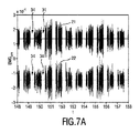
図2は、生の筋電図信号31、RMS筋電図信号32及び呼吸流量33からなる例示的なグラフを例示している。
FIG. 2 illustrates an exemplary graph consisting of a
図2の一番上のグラフは、傍胸骨のEMG測定値EMGparaの生のEMG信号31を示す。縦軸は、EMG電圧(μV)を示し、一方横軸は、時間t(秒)を示す。描かれる信号のトレースは、被験者100の第2傍胸骨肋間腔105に配される1つの対のEMG電極3A、3Bを用いて、差分EMG測定値として得られる第1の筋電図信号21を示す。
The top graph of FIG. 2 shows the
図2の真ん中のグラフは、二乗平均平方根(RMS)筋電図信号32を示す。縦軸は、一番上のグラフに対応するRMS値を示し、一方横軸は再び時間t(秒)を示す。傍胸骨測定に対し、この値は、Murphy他により説明されるようなRMSparaとしても呼ばれる。
The middle graph of FIG. 2 shows the root mean square (RMS)
図2の一番下のグラフは、呼吸活動に対する基準として、鼻/口カニューレを用いて測定された差圧信号Q 33のグラフを示す。縦軸は、圧力信号Qを示し、一方横軸は時間t(秒)を示す。 The bottom graph of FIG. 2 shows a graph of differential pressure signal Q33 measured using a nasal / oral cannula as a reference for respiratory activity. The vertical axis represents the pressure signal Q, while the horizontal axis represents time t (seconds).
図2の一番上のグラフに示されるように、生のEMG信号は、呼吸が原因による影響だけでなく、参照番号34により例示的に示されるような心臓活動からのクロストークも有する。そのような心臓活動は、ECG除去技術により取り除かれることができる。ECG除去は、処理装置20による処理ステップとして有利に行なわれることができる。
As shown in the top graph of FIG. 2, the raw EMG signal has not only the effects due to respiration, but also crosstalk from cardiac activity as exemplarily shown by
ここで図2の一番下のグラフを参照すると、全呼吸周期がTで示される。この呼吸周期はさらに、呼気が生じている第1の期間P1と、吸気が生じているP2及びP3を含む第2の期間とに分割されることができる。 Referring now to the bottom graph of FIG. 2, the total respiratory cycle is indicated by T. This respiratory cycle can be further divided into a first period P1 in which exhalation occurs and a second period including P2 and P3 in which inspiration occurs.
図2の下2つのグラフから分かるように、RMSpara32及びQ信号33は、呼気相P1並びに吸気相P2及びP3の間、完全に同期した特性を示していない。
As can be seen from the lower two graphs of FIG. 2, the
測定はCOPD患者に関係している。傍胸骨肋間筋による吸気努力に加え、トレース31、32に示されるように呼気努力も測定される。特に、呼気相P1中、RMSpara信号32において何らかの筋肉活動が高まっていることが分かる。さらに、図2は、実際の吸気相P2及びP3より前にEMG活動が既に大幅に増大していることを示している。これはP4により示される。吸気筋の採用(recruitement)と解釈されるものは、鼻における実際の圧力降下及びまさに吸気を示す空気流の生成の前に始まる。第1の考えられる説明は、COPD患者は、空気流を生成することができる前に、固有の呼気終末陽圧(EPPE)に打ち勝たなければならないことである。しかしながら、P1中のRMSpara信号32においてエネルギーの増大が示すように見えるので、RMSpara信号32において強制的な呼気活動の重ね合わせを持つ、寄与の混合が存在している。故に、第2肋間腔105に置かれる単一の対の電極3A、3BからのEMG測定値31に基づいて吸気と呼気とが簡単に区別され得ない。
Measurements are relevant for COPD patients. In addition to the inspiratory effort by the parasternal intercostal muscle, the expiratory effort is also measured as shown in
言い換えると、第1の筋電図信号31は、第2肋間腔の傍胸骨筋105の活動を表すだけでなく、異なる筋肉の活動の混合、特に、全吸気中で活性である傍胸骨筋の吸気相の活動と、受動的ではない呼気中に活性化される可能性がある胸横筋である呼気相の活動と、姿勢の変化、姿勢の維持等の間に活性化される例えば大胸筋の緊張性活動との混合を実際に供給している。大胸筋の寄与は、この筋肉は自主的に制御され得るので、患者に静止したままでいる及びできるだけリラックスするように指示することにより減らすことができる。
In other words, the
故に、(a)吸気の呼吸努力を捕えるために、吸気相P2及びP3と呼気相P1とを区別する、並びに(b)例えば緊張性又は相の活動のようなEMG活動の発生源及び性質を決定する必要がある。各筋肉は特定の生理的機能を持っているので、異なる種類の筋肉間、特に傍胸骨の場所におけるEMG信号からの関連情報を引き出すために、胸横筋の活動に対する傍胸骨筋の活動を区別する必要がある。それにより、特にCOPD患者又は他の慢性疾患に対する呼吸機能の1つ以上の標的マーカーが決定されることができる。 Hence, (a) distinguish between inspiratory phases P2 and P3 and expiratory phase P1 in order to capture inspiratory respiratory effort, and (b) the source and nature of EMG activity such as tonicity or phase activity. It is necessary to decide. Since each muscle has a specific physiological function, it differentiates parasternal muscle activity from transthoracic muscle activity to derive relevant information from EMG signals between different types of muscles, particularly at the location of the parasternal bone There is a need. Thereby, one or more target markers of respiratory function, particularly for COPD patients or other chronic diseases can be determined.
EMG活動は明らかに傍胸骨肋間筋に起因していると考えるために、従来技術は、傍胸骨筋の活動を分離させる(isolate)ためにニードル電極技術の使用を提案している。従来技術とは対照的に、発明者は、第2の筋電図信号22の使用を提案している。第2の筋電図信号は、呼吸過程において関与する異なる筋肉の活動の種類を区別するのに使用されることができることは分かっている。
The prior art suggests the use of needle electrode technology to isolate parasternal muscle activity, since EMG activity is apparently due to parasternal intercostal muscles. In contrast to the prior art, the inventors have proposed the use of a
図1を再び参照すると、ある実施例において、第1の対の表面EMG電極3A、3Bに加え、第2の筋電図信号22を得るための第2の対の表面電極4A、4Bが設けられる。EMG装置10の処理機器20は故に、身体100上の第2の場所106において得られる第2の筋電図信号22を受信するための第2のインタフェース24を有する。
Referring again to FIG. 1, in one embodiment, in addition to the first pair of
所与の例において、第2の対のEMG電極4A、4Bは、第3肋骨103と第4肋骨104との間にある第3肋間腔106に位置決められる。一見すると、第3の肋間腔106が気呼吸努力中に関与する傍胸骨肋間筋も含んでいるため、第2の筋電図信号22は、第1の筋電図信号21に似た動きをすると思われる。しかしながら、第3肋骨103から第4肋骨104に伸びている傍胸骨筋は、第2肋骨102から第3肋骨103に伸びている、第2肋間腔105にある傍胸骨肋間筋に比べ、独立して神経支配されることが分かっている。
In the given example, the second pair of
図4は、第2の肋間腔105における身体100の胸部の解剖学的な概略図を身体の横断面で示す。図4は、胸骨101及び脊椎111の骨構造を示す。肋間筋に関し、図4は、外肋間筋121、内肋間筋122及び最内肋間筋123を示し、これらは合わせて肋間筋120と呼ばれる。各々別の肋間腔にある傍胸骨筋の特性は、これら傍胸骨筋が夫々の肋間神経124により別々に神経支配されることである。故に、発明者は、この事実が関与する異なる筋肉を区別するのに利用され得ると認識した。各夫々の肋間腔にある肋間筋は別々に神経支配されているという事実により、測定される電気活動が傍胸骨肋間筋だけから生じているとき、対応する第1及び第2の筋電図信号は相関関係がない信号を示す。図4に示される解剖学的な概略図において、胸横筋は参照番号126で示され、大胸筋は参照番号128で示される。
FIG. 4 shows a anatomical schematic view of the chest of the
図4はさらに、第1の対の電極の第1電極3Aが第1の筋電図信号を得るための表面電極として身体の皮膚上に当てられている電極パッチ2の例示的な実施例を示す。第1の筋電図信号は、図4に見られるような下層の筋肉、すなわち大胸筋128、肋間筋120及び胸横筋126の活動を示していることが容易に考えられる。故に、第1の筋電図信号は、第2肋間腔105におけるEMG信号から簡単に分離され得ないこれらの信号の重ね合わせを有する。
FIG. 4 further illustrates an exemplary embodiment of the
図5は、体の内側から見たような身体100の胸郭の解剖学的な概略図を示す。第1の筋電図信号21を得るための第1の対の電極3A、3B及び第2の筋電図信号22を得るための第2の対の電極4A、4Bの場所の画像が丸で示されている。図5から分かるように、大胸筋128の寄与はないと仮定すると、第1の筋電図信号21は、第2肋骨102と第3肋骨103との間にある第2肋間腔105からの肋間筋120Aの寄与を有する。それに対応じて、第2の筋電図信号22は、第3肋骨103と第4肋骨104との間にある第3肋間腔106からの肋間筋120Bの寄与を有するが、第2肋間腔からではない。対称的に、胸横筋126は、両方の対の電極の電極3A、4A及び3B、4Bにわたって広がっている。故に、第2肋間腔105及び第3肋間腔106における2つの差分測定は、胸横筋126の活動を示す相関信号を示す。
FIG. 5 shows an anatomical schematic view of the thorax of the
胸横筋の一番上の筋束は、胸骨101の下部から第2肋骨102まで(略)垂直に伸びている。胸横筋は、肋間神経により神経支配される傍胸骨筋と全く同じである。
The uppermost muscle bundle of the transthoracic muscle extends (substantially) vertically from the lower part of the
図6、7A及び7Bは、第1及び第2の筋電図信号21、22の例示的なグラフを示している。解析ユニット20は、第1の筋電図信号21及び第2の筋電図信号22を受信し、これらの信号に基づいて、これら2つの信号の類似性を類似信号26の形式で決定する。それにより、解析ユニット20は、上に説明されるように、傍胸骨肋間筋からの吸気筋肉と胸横筋からの呼気筋肉とを区別することができる。
6, 7A and 7B show exemplary graphs of the first and second electromyogram signals 21,22. The
図6は、上記の類似性の解析が、吸気活動から咳、すなわち強制的な呼気活動を区別するのに使用される一例を示す。咳は、胸横筋により生じることが知られている。図6は、最大の咳活動62そして、咳活動の減衰63が続く、咳活動の始まり61を示す。実線は、第1の差分測定信号として第1の対の電極3A、3Bを用いて得られる第1の筋電図信号21に対応し、破線は、第2の差分測定信号として第2の対の電極4A、4Bを用いて得られる第2の筋電図信号22に対応する。見られるように、咳の最大活動中、特に低周波数において2つの信号は高い相関性がある。他方、咳の始まり及び減衰は低い相関性を示す。
FIG. 6 shows an example where the above similarity analysis is used to distinguish cough, ie forced expiratory activity, from inspiratory activity. Cough is known to be caused by the transthoracic muscle. FIG. 6 shows the cough activity beginning 61 followed by a
図7A及び7Bは、安静呼吸中の第1の筋電図信号21及び第2の筋電図信号22のトレースを示す。より良好なグラフ表示のために、図7A及び7Bにおける信号をよりはっきりと区別するために、夫々の信号に垂直オフセットが加えられている。しかしながら、両方の信号は、ゼロ平均のベースライン値を用いて考察されるべきである。図7Aは、ゲーティング(gating)によりECGが取り除かれた後の信号を示し、ここで心臓活動中の信号部は、参照番号34により示されるように取り除かれている。故に、心臓活動を示すQRS群34は取り除かれている。図7Bにおいて、図7Aの特定の選択範囲は示されている。
7A and 7B show traces of the
図8を参照すると、本例において、解析ユニット20は、第1の筋電図信号21と第2の筋電図信号22との間におけるコヒーレンスを計算するように構成される。図8において、縦軸は、コヒーレンス因子Cを示し、一方横軸は周波数fを示す。咳信号及び安静呼吸信号の両方に対し計算されたコヒーレンスが図8に示され、ここで参照番号81で示される曲線は、咳事象62に対応し、一方、低いコヒーレンスの曲線82は、安静呼吸の期間に対応する。
Referring to FIG. 8, in this example, the
図8から分かるように、咳をしている間、対の電極3A、3Bからの信号間の相関性、及び対の電極4A、4Bからの信号間の相関性は、低周波数に存在するだけでなく、高周波数にも多くの相関性を示している。
As can be seen from FIG. 8, during coughing, the correlation between the signals from the pair of
図1を再び参照すると、処理装置20はさらに、任意の第2の解析ユニット27を有してもよい。第2の解析ユニット27は、前記解析ユニットから類似性信号26を受信する。第2の解析ユニット27は、この類似性信号26に基づいて、呼吸相を決定するように構成される。第2の解析ユニットは、第1及び第2の筋電図信号から得られた類似性信号が第1の既定のしきい値よりも低い場合、呼吸相を吸気相と決定する、及び/又は第1及び第2の筋電図信号から得られた類似性信号が第2の既定のしきい値を超えている場合、呼吸相を呼気相と決定するように構成される。
Referring back to FIG. 1, the
第2の解析ユニット27はさらに、類似性信号に基づいて、呼吸相を示す呼吸相信号を供給するように構成され、及び処理装置は故に、前記呼吸相信号と前記第1の筋電図信号とに基づいて呼吸努力を決定するようにさらに構成されてもよい。特に、処理装置は、吸気相中の第1の筋電図信号の信号エネルギーに基づいて吸気努力を決定する、及び/又は呼気相中の第1の筋電図信号の信号エネルギーに基づいて呼気努力を決定するように構成されてもよい。
The
第1の筋電図信号及び第2の筋電図信号に基づいて類似性信号を決定するのに加えて又はその代わりに、処理装置は、第1の筋電図信号及び第2の筋電図信号の両方に元々存在している依存的信号(dependent signal)を第1の筋電図信号から取り除くために構成されてもよい。故に、きれいにされた第1の筋電図信号が供給される。例えば、このきれいにされた第1の筋電図信号は、第2の肋間腔105における傍胸骨肋間筋の活動を示し、ここで筋電図信号は、大胸筋の活動の活性化から除去される。
In addition to or in lieu of determining the similarity signal based on the first electromyogram signal and the second electromyogram signal, the processing device may include the first electromyogram signal and the second electromyogram signal. It may be configured to remove the dependent signal originally present in both of the graphical signals from the first EMG signal. Thus, a cleaned first electromyogram signal is provided. For example, the cleaned first electromyogram signal indicates parasternal intercostal activity in the second
図9は、他のEMG電極構成を用いた解剖学的な概略図を示す。第1の筋電図信号21を得るための第1の対の電極3A、3B、及び第2の筋電図信号22を得るための第2の対の電極4A、4Bの場所の画像は、丸で示されている。この実施例において、第1の対の電極の第1電極3A及び第2電極3Bは、第1の筋電図信号21を得るために、第2肋骨102と第3肋骨103との間にある第2肋間腔105において胸骨に近い両側で傍胸骨に当てられる。第2の対の電極の第3電極4A及び第4電極4Bも、第2の筋電図信号22を得るために、第2肋間腔105において胸骨の両側であるが、胸骨から離れて当てられる。
FIG. 9 shows an anatomical schematic using another EMG electrode configuration. Images of the location of the first pair of
この実施例において、第2の傍胸骨肋間筋120は、標的筋肉であるのに対し、大胸筋128は他の筋肉である。第1及び第2の対の電極は、夫々の標的筋肉及び他の筋肉に応じて、別々の場所に当てられることもできる。例えば、第2の対の電極の第3電極及び/又は第4電極は、これら電極が前記他の筋肉、ここでは大胸筋からEMG信号の寄与を受けている限り、別の場所、例えば別の肋間腔に当てられることもできる。図9に示されるように、第2の対の電極は、小胸筋129からの寄与を測定してもよい。従って、小胸筋は任意で評価されることもできる。
In this example, the second
大胸筋128は、第1及び第2の対の電極にわたり広がっているので、大胸筋の活動は、第1の筋電図信号21及び第2の筋電図信号22の両方に見られる。故に、第1の筋電図信号21と第2の筋電図信号22との類似性を示す類似性信号26は、大胸筋の活動を示すことがある。大胸筋は通例、身体を動かしている間は活動している。上手くリラックスできない場合、大胸筋は何らかの緊張性活動も生じている。故に、類似性信号は身体の動き及び/又は緊張性活動を示すことがある。
Since the
有利なことに、そのような身体の動きの検出と一致する第1の筋電図信号の期間は処分される。故に、例えば類似性信号が既定のしきい値を超えている場合、神経呼吸ドライブ又は呼吸努力は評価されない。図1を参照すると、処理装置20は、類似性信号26を受信する、及びこの類似性信号26に基づいて、第1の筋電図信号21から前記他の筋肉の活動を取り除くように構成される第2の解析ユニット27を有する。処理装置20は、前記他の筋肉の活動が検出されるとき、第1の筋電図信号の信号部を取り除いてもよい。代替例において、検出後に較正又は補償が適用されることができる。
Advantageously, the period of the first electromyogram signal that coincides with the detection of such body movement is discarded. Thus, for example, if the similarity signal exceeds a predetermined threshold, neurorespiratory drive or respiratory effort is not evaluated. Referring to FIG. 1, the
他の実施例において、図1の筋電図測定装置10は、電極パッチ2に直接付けられることができる。代わりに、筋電図測定装置10は、この装置上でクリックされ、その後、胸骨101の中心であり、第2及び第3肋間腔105、106の高さで患者に取り付けられる4つの(使い捨ての)電極を含んでもよい。
In another embodiment, the
有利なことに、例えば呼吸努力又はNRD値のような生理学的パラメータ、及び/又は咳の努力は、連続して観察される、及び出力ユニット18により例えば患者モニター又はスマートフォンのようなハブに有線又はワイヤレスで送られることができる。これらに基づいて、臨床的な評価が行われることができる。任意で、生理学的パラメータが患者の起こり得る危険及び/又は悪化を特定するしきい値を超える場合、例えば出力ユニット18によりスマートアラームが生成されることができる。
Advantageously, physiological parameters, such as respiratory effort or NRD values, and / or cough effort, for example, are observed continuously and wired by a
要するに、筋電図信号を処理するのに有利な方法が示されている。ここで開示される技術的な解決法は、筋電図測定システムをさらに向上させるための可能性を提供している。特に、提案した解決法の態様は、呼吸過程を、特に傍胸骨肋間筋を含む呼吸筋、及び例えば呼吸を補助するための胸横筋のような補助筋の使用が増えているCOPD患者の呼吸過程をより詳細に解析するための可能性を提供する。 In short, an advantageous method for processing an electromyogram signal is shown. The technical solution disclosed here offers the possibility to further improve the electromyogram measurement system. In particular, the proposed solution aspect is that the respiratory process, especially the respiratory process of COPD patients with increased use of respiratory muscles, including parasternal intercostal muscles, and auxiliary muscles such as transthoracic muscles to assist breathing, for example. Provides the possibility to analyze the in more detail.
他の態様は、以下の項に説明される。 Other aspects are described in the following sections.
第1項:少なくとも1つの他の筋肉(126、128)の活動の最中に、呼吸努力の測定に関係する、人間又は動物の身体(100)における標的筋肉(120)の活動を示す筋電図信号を処理するための処理装置(20)は、前記身体(100)上の第1の場所において前記標的筋肉(120)及び前記他の筋肉(126、128)から得られる第1の筋電図信号(21)を受信するための第1のインタフェース(23)、前記身体(100)上の第2の場所において前記他の筋肉(126、128)から得られる第2の筋電図信号(22)を受信するための第2のインタフェース(24)、及び前記第1の筋電図信号(21)と前記第2の筋電図信号(22)とに基づいて類似性信号(26)を決定するための解析ユニット、を有する。 Item 1: Myoelectrics indicative of the activity of the target muscle (120) in the human or animal body (100) related to the measurement of respiratory effort during the activity of at least one other muscle (126, 128). A processing device (20) for processing graphical signals comprises a first myoelectric signal obtained from the target muscle (120) and the other muscles (126, 128) at a first location on the body (100). A first interface (23) for receiving a graphical signal (21), a second electromyographic signal (126, 128) obtained from the other muscle (126, 128) at a second location on the body (100) 22) and a second interface (24) for receiving the similarity signal (26) based on the first electromyogram signal (21) and the second electromyogram signal (22). An analysis unit for determining.
第2項:前記標的筋肉(120)は、傍胸骨肋間筋である及び/又は前記他の筋肉は、胸横筋(126)及び大胸筋(128)の少なくとも一方である、第1項に記載の処理装置。 Item 2: The target muscle (120) is a parasternal intercostal muscle and / or the other muscle is at least one of a transthoracic muscle (126) and a greater pectoral muscle (128). Processing equipment.
第3項:前記類似性信号(26)は、第1の筋電図信号(21)と第2の筋電図信号(22)との相関性又はスペクトルコヒーレンスを示す、第1項に記載の処理装置。 Item 3: The similarity signal (26) is indicative of the correlation or spectral coherence between the first electromyogram signal (21) and the second electromyogram signal (22). Processing equipment.
第4項:前記第1の筋電図信号(21)及び前記第2の筋電図信号(22)の少なくとも一方は、肋間腔(105、106)で得られる、第1項に記載の処理装置。
Item 4: The process according to
第5項:前記第1の筋電図信号(21)は、前記第2肋間腔(105)で得られる、及び/又は前記第2の筋電図信号(22)は、前記第3肋間腔(106)で得られる、第1項に記載の処理装置。 Item 5: The first electromyogram signal (21) is obtained in the second intercostal space (105) and / or the second electromyogram signal (22) is obtained in the third intercostal space (106) The processing apparatus according to item (1).
第6項:前記第1の筋電図信号(21)は、前記身体上の第1の場所における前記他の筋肉(126、128)及び前記標的筋肉(120A)の第1のインスタンスから得られる、及び前記第2の筋電図信号(22)は、前記身体上の第2の場所における前記他の筋肉(126、128)及び前記標的筋肉(120B)の第2のインスタンスから得られる、第1項に記載の処理装置。
Item 6: The first electromyogram signal (21) is obtained from a first instance of the other muscle (126, 128) and the target muscle (120A) at a first location on the body. And the second electromyogram signal (22) is obtained from a second instance of the other muscle (126, 128) and the target muscle (120B) at a second location on the body, The processing apparatus according to
第7項:前記解析ユニット(25)は、前記類似性信号(26)に基づいて、呼吸相を決定するよう構成される、第1項に記載の処理装置。
Item 7: The processing device according to
第8項:前記解析ユニット(25)は、前記第1及び第2の筋電図信号(21、22)から得られる前記類似性信号(26)が第1の既定のしきい値より下にある場合、呼吸相を吸気相(P2、P3)と決定する、及び/又は前記第1及び第2の筋電図信号(21、22)から得られる前記類似性信号(26)が第2の既定のしきい値を超えている場合、呼吸相を呼気相(P1)と決定するように構成される、第1項に記載の処理装置。
Item 8: The analysis unit (25) determines that the similarity signal (26) obtained from the first and second electromyogram signals (21, 22) is below a first predetermined threshold. In some cases, the respiratory phase is determined as the inspiratory phase (P2, P3) and / or the similarity signal (26) obtained from the first and second electromyogram signals (21, 22) is a second The processing apparatus of
第9項:前記解析ユニット(25)はさらに、前記類似性信号(26)に基づいて、前記呼吸相を示す呼吸相信号を供給するように構成される、並びに前記処理装置(20)はさらに、前記呼吸相信号及び前記第1の筋電図信号に基づいて呼吸努力を決定するように構成される、第7項に記載の処理装置。 Item 9: The analysis unit (25) is further configured to provide a respiratory phase signal indicative of the respiratory phase based on the similarity signal (26), and the processing device (20) further The processing device of claim 7, configured to determine respiratory effort based on the respiratory phase signal and the first electromyogram signal.
第10項:吸気相中の前記第1の筋電図信号(21)の信号エネルギーに基づいて、吸気努力を決定する、及び/又は呼気相中の前記第1の筋電図信号(21)の信号エネルギーに基づいて、呼気努力を決定するように構成される、第9項に記載の処理装置 Item 10: Determine inspiratory effort based on the signal energy of the first electromyogram signal (21) during the inspiratory phase and / or the first electromyogram signal (21) during the expiratory phase The processing apparatus of claim 9, wherein the processing apparatus is configured to determine exhalation effort based on the signal energy of
第11項:前記処理装置は、前記類似性信号(26)に基づいて、前記他の筋肉(126、128)の活動を前記第1の筋電図信号(21)から取り除くように構成される、第1項に記載の処理装置。
Item 11: The processing device is configured to remove the activity of the other muscle (126, 128) from the first electromyogram signal (21) based on the similarity signal (26) The processing apparatus according to
第12項:第1項に記載の処理装置(20)と共に使用するための電極パッチにおいて、前記電極パッチは、胸部上の少なくとも2つの異なる場所において表面筋電図測定のための少なくとも2つの組の電極(3A、3B、4A、4B)を有し、第1の組の電極は、胸骨(101)を除く、第1の肋間腔(105)において第1の筋電図信号(21)を測定するために配される第1の対の電極(3A、3B)を有する、及び第2の組の電極は、胸骨(101)を除く、第2の肋間腔(106)において第2の筋電図信号(22)を測定するために配される第2の対の電極(4A、4B)を有する、電極パッチ。
Item 12: An electrode patch for use with the processing device (20) according to
第13項:第1項に記載の処理装置を有する筋電図測定装置(10)、前記第1の筋電図信号(21)を得るための第1の対の電極(3A、3B)、及び第2の筋電図信号(22)を得るための第2の対の電極(4A、4B)を有する筋電図測定システム(1)であり、前記第1の対の電極は、前記処理装置(20)の第1のインタフェース(23)に前記第1の筋電図信号(21)を供給するために、前記筋電図測定装置(10)の第1の入力部(11)に接続可能であり、前記第2の対の電極は、前記処理装置(20)の第2のインタフェース(24)に前記第2の筋電図信号(22)を供給するために、前記筋電図測定装置(10)の第2の入力部(24)に接続可能である、筋電図測定システム。
Item 13: an electromyogram measuring device (10) having the processing device according to
第14項:少なくとも1つの他の筋肉(126、128)の活動の最中に、呼吸努力の測定に関係する、人間又は動物の身体(100)における標的筋肉(120)の活動を示す筋電図信号を処理するための方法において、前記方法は、前記身体(100)上の第1の場所において前記標的筋肉(120)及び前記他の筋肉(126、128)から得られる第1の筋電図信号(21)を受信するステップ、前記身体(100)上の第2の場所において前記他の筋肉(126、128)から得られる第2の筋電図信号(22)を受信するステップ、並びに前記第1の筋電図信号(21)と前記第2の筋電図信号(22)とに基づいて類似性信号(26)を決定するステップ、を有する方法。 Item 14: EMG indicative of the activity of the target muscle (120) in the human or animal body (100) related to the measurement of respiratory effort during the activity of at least one other muscle (126, 128) In a method for processing graphical signals, the method includes a first myoelectric signal obtained from the target muscle (120) and the other muscles (126, 128) at a first location on the body (100). Receiving a graphical signal (21), receiving a second electromyographic signal (22) obtained from the other muscle (126, 128) at a second location on the body (100), and Determining a similarity signal (26) based on the first electromyogram signal (21) and the second electromyogram signal (22).
第15項:コンピュータプログラムがコンピュータ上で実行されるとき、前記コンピュータに第14項に記載の方法のステップを実行させるためのプログラムコード手段を有するコンピュータプログラム。
Item 15: A computer program comprising program code means for causing the computer to execute the steps of the method of
本発明は、図面及び上記記載において詳細に例示及び説明されている一方、そのような例示及び説明は、説明的又は例示的であり、限定的ではない、つまり、本発明は開示される実施例に限定されないと考えるべきである。開示される実施例に対する他の変形例は、図面、本開示及び付随する特許請求の範囲を検討することにより、請求される本発明を実施する当業者により理解される及び生じることができる。 While the invention has been illustrated and described in detail in the drawings and foregoing description, such illustration and description are illustrative or exemplary and not restrictive, that is, the invention is disclosed in an embodiment. You should think that it is not limited to. Other variations to the disclosed embodiments can be understood and occured by those skilled in the art practicing the claimed invention, upon review of the drawings, the present disclosure, and the appended claims.
請求項において、"有する"という言葉は、他の要素又はステップを排除するものではなく、複数あることを述べなくても、それらが複数あることを排除するものではない。単一の要素又は他のユニットが請求項に列挙される幾つかのアイテムの機能を果たしてもよい。ある方法は互いに異なる従属請求項に挙げられているという単なる事実は、これらの方法の組み合わせが有利に使用されることができないことを示してはいない。 In the claims, the word “comprising” does not exclude other elements or steps, and it does not exclude the presence of a plurality of them, whether or not there are a plurality. A single element or other unit may fulfill the functions of several items recited in the claims. The mere fact that certain methods are recited in mutually different dependent claims does not indicate that a combination of these methods cannot be used to advantage.
コンピュータプログラムは、例えば他のハードウェアと一緒に若しくはその一部として供給される光学記憶媒体又はソリッドステート媒体のような適切な媒体上に記憶/配布されてもよいが、例えばインターネット又は他の有線若しくはワイヤレスの電話通信システムを介して他の形式で配布されてもよい。 The computer program may be stored / distributed on a suitable medium, such as an optical storage medium or solid state medium supplied together with or as part of other hardware, for example the Internet or other wired Alternatively, it may be distributed in other forms via a wireless telephone communication system.
請求項における如何なる参照符号もその範囲を限定すると考えるべきではない。 Any reference signs in the claims should not be construed as limiting the scope.
Claims (14)
前記身体上の第1の場所において前記標的筋肉及び前記他の筋肉から得られる第1の筋電図信号を受信するための第1のインタフェース、
前記身体上の第2の場所において前記他の筋肉から得られる第2の筋電図信号を受信するための第2のインタフェース、及び
前記第1の筋電図信号と前記第2の筋電図信号との間の類似性を類似性信号として決定するための解析ユニット
を有し、前記解析ユニットはさらに、
前記類似性信号が第1の既定のしきい値より低い場合、呼吸相を吸気相と決定する、及び/又は
前記類似性信号が第2の既定のしきい値を超えている場合、呼吸相を呼気相と決定する
ように構成される処理装置。 A processing device for processing an electromyographic signal indicative of target muscle activity in a human or animal body related to measurement of respiratory effort during at least one other muscle activity, the processing device comprising: ,
A first interface for receiving a first electromyogram signal obtained from the target muscle and the other muscle at a first location on the body;
A second interface for receiving a second electromyogram signal obtained from the other muscle at a second location on the body; and the first electromyogram signal and the second electromyogram An analysis unit for determining the similarity between the signals as a similarity signal, the analysis unit further comprising:
If the similarity signal is lower than a first predetermined threshold, determine the respiratory phase as an inspiratory phase, and / or if the similarity signal exceeds a second predetermined threshold, A processing device configured to determine an expiratory phase.
前記第2の筋電図信号は、前記身体上の前記第2の場所において前記他の筋肉及び前記標的筋肉の第2のインスタンスから得られる、
請求項1に記載の処理装置。 The first electromyogram signal is obtained from a first instance of the other muscle and the target muscle at the first location on the body, and the second electromyogram signal is obtained from the body Obtained from the second instance of the other muscle and the target muscle at the second location above,
The processing apparatus according to claim 1.
前記処理装置はさらに、前記呼吸相信号及び前記第1の筋電図信号に基づいて、呼吸努力を決定するように構成される、
請求項1に記載の処理装置。 The analysis unit is further configured to provide a respiratory phase signal indicative of a respiratory phase based on the similarity signal, and the processing device further includes the respiratory phase signal and the first electromyogram signal Configured to determine respiratory effort based on
The processing apparatus according to claim 1.
胸部上の少なくとも2つの異なる場所において表面筋電図測定のための少なくとも2つの組の電極を有する電極パッチ
を有する筋電図測定システムにおいて、
前記2つの組の電極の第1の組は、胸骨を除く、第1の肋間腔において第1の筋電図信号を測定するために配される第1の対の電極を有する、及び
前記2つの組の電極の第2の組は、胸骨を除く、第2の肋間腔において第2の筋電図信号を測定するために配される第2の対の電極を有する
筋電図測定システム。 An electromyogram measurement device comprising the processing device according to claim 1 and an electromyogram measurement comprising an electrode patch having at least two sets of electrodes for surface electromyogram measurement in at least two different locations on the chest In the system,
The first set of two sets of electrodes has a first pair of electrodes arranged to measure a first electromyogram signal in a first intercostal space, excluding the sternum; and An electromyography system having a second pair of electrodes arranged to measure a second electromyogram signal in a second intercostal space, the second set of two sets of electrodes excluding the sternum.
前記第1の筋電図信号を得るための第1の対の電極であり、前記処理装置の前記第1のインタフェースに前記第1の筋電図信号を供給するために、前記筋電図測定装置の第1の入力部に接続可能である、第1の対の電極、及び
前記第2の筋電図信号を得るための第2の対の電極であり、前記処理装置の前記第2のインタフェースに前記第2の筋電図信号を供給するために、前記筋電図測定装置の第2の入力部に接続可能である、第2の対の電極
を有する、筋電図測定システム。 An electromyogram measuring apparatus comprising the processing apparatus according to claim 1;
A first pair of electrodes for obtaining the first electromyogram signal, the electromyogram measurement for supplying the first electromyogram signal to the first interface of the processing device; A first pair of electrodes connectable to a first input of the device, and a second pair of electrodes for obtaining the second electromyogram signal, the second pair of electrodes of the processing device An electromyogram measurement system comprising a second pair of electrodes connectable to a second input of the electromyogram measurement device to supply the second electromyogram signal to an interface.
前記身体上の第1の場所において前記標的筋肉及び前記他の筋肉から得られる第1の筋電図信号を受信するステップ、
前記身体上の第2の場所において前記他の筋肉から得られる第2の筋電図信号を受信するステップ、
前記第1の筋電図信号と前記第2の筋電図信号との間の類似性を類似性信号として決定するステップ、並びに
前記類似性信号が第1の既定のしきい値より低い場合、呼吸相を吸気相と決定する、及び/又は
前記類似性信号が第2の既定のしきい値を超えている場合、呼吸相を呼気相と決定するステップ
を有する作動方法。 During at least one other muscle activity, related to the measurement of respiratory effort, in the operation method of a computer for processing the EMG signal indicating the activity of the target muscle in the body of a human or animal, the actuating The method is
Receiving a first electromyogram signal obtained from the target muscle and the other muscle at a first location on the body;
Receiving a second electromyographic signal obtained from the other muscle at a second location on the body;
Determining the similarity between the first electromyogram signal and the second electromyogram signal as a similarity signal, and if the similarity signal is lower than a first predetermined threshold, An operating method comprising: determining a respiratory phase as an inspiratory phase and / or determining a respiratory phase as an expiratory phase if the similarity signal exceeds a second predetermined threshold.
前記呼吸相信号及び前記第1の筋電図信号に基づいて呼吸努力を決定するステップ
をさらに有する請求項12に記載の作動方法。 13. The method further comprising: providing a respiratory phase signal indicative of the respiratory phase based on the similarity signal; and determining a respiratory effort based on the respiratory phase signal and the first electromyogram signal. The operating method described in 1.
Applications Claiming Priority (3)
| Application Number | Priority Date | Filing Date | Title |
|---|---|---|---|
| EP15180614 | 2015-08-11 | ||
| EP15180614.8 | 2015-08-11 | ||
| PCT/EP2016/068261 WO2017025363A1 (en) | 2015-08-11 | 2016-08-01 | Apparatus and method for processing electromyography signals related to respiratory activity |
Publications (3)
| Publication Number | Publication Date |
|---|---|
| JP2018527069A JP2018527069A (en) | 2018-09-20 |
| JP2018527069A5 JP2018527069A5 (en) | 2019-06-27 |
| JP6585825B2 true JP6585825B2 (en) | 2019-10-02 |
Family
ID=53794152
Family Applications (1)
| Application Number | Title | Priority Date | Filing Date |
|---|---|---|---|
| JP2018506593A Active JP6585825B2 (en) | 2015-08-11 | 2016-08-01 | Processing apparatus and method for processing electromyogram signals related to respiratory activity |
Country Status (5)
| Country | Link |
|---|---|
| US (1) | US10945628B2 (en) |
| EP (1) | EP3334338B1 (en) |
| JP (1) | JP6585825B2 (en) |
| CN (1) | CN108135535B (en) |
| WO (1) | WO2017025363A1 (en) |
Families Citing this family (17)
| Publication number | Priority date | Publication date | Assignee | Title |
|---|---|---|---|---|
| US10561568B1 (en) | 2014-06-19 | 2020-02-18 | Lockheed Martin Corporation | Exoskeleton system providing for a load transfer when a user is standing and kneeling |
| US10548800B1 (en) | 2015-06-18 | 2020-02-04 | Lockheed Martin Corporation | Exoskeleton pelvic link having hip joint and inguinal joint |
| US10518404B2 (en) | 2015-07-17 | 2019-12-31 | Lockheed Martin Corporation | Variable force exoskeleton hip joint |
| US10912346B1 (en) | 2015-11-24 | 2021-02-09 | Lockheed Martin Corporation | Exoskeleton boot and lower link |
| DE102015015296A1 (en) | 2015-11-30 | 2017-06-01 | Drägerwerk AG & Co. KGaA | Apparatus and method for providing data signals indicative of muscle activities relevant to inspiratory and expiratory effort of a patient |
| AU2016418245B2 (en) * | 2016-08-01 | 2020-07-16 | Med-El Elektromedizinische Geraete Gmbh | Respiratory triggered parasternal electromyographic recording in neurostimulators |
| EP3381354A1 (en) * | 2017-03-31 | 2018-10-03 | Koninklijke Philips N.V. | Methods and system for processing an emg signal |
| CA3073504A1 (en) | 2017-08-30 | 2019-03-07 | Lockheed Martin Corporation | Automatic sensor selection |
| US11523765B2 (en) * | 2018-09-05 | 2022-12-13 | The University Of Chicago | Neurophysiological biomarkers for neurodegenerative disorders |
| US20220022802A1 (en) * | 2018-10-05 | 2022-01-27 | Nippon Telegraph And Telephone Corporation | Myoelectric potential processing device, myoelectric potential processing method and myoelectric potential processing program |
| EP3701876A1 (en) * | 2019-02-27 | 2020-09-02 | Koninklijke Philips N.V. | Acoustic sensing apparatus and method |
| WO2020196323A1 (en) * | 2019-03-22 | 2020-10-01 | 謙 松田 | Program, information processing method, and information processing device |
| CN109999433A (en) * | 2019-04-02 | 2019-07-12 | 上海大学 | Personalized diaphragm rehabilitation training system based on surface myoelectric |
| CN110464944B (en) * | 2019-08-26 | 2022-04-19 | 深圳创达云睿智能科技有限公司 | Control method and device of breathing machine and breathing machine |
| CN112043270B (en) * | 2020-09-07 | 2023-03-10 | 深圳数联天下智能科技有限公司 | Electromyographic electrode position determining method, electromyographic signal acquisition device and electromyographic signal acquisition equipment |
| WO2023274931A1 (en) * | 2021-06-29 | 2023-01-05 | Koninklijke Philips N.V. | Sniff detection and artifact distinction from electromyography and accelerometer signals |
| WO2023117995A1 (en) * | 2021-12-20 | 2023-06-29 | Koninklijke Philips N.V. | Systems and methods for ventilator management |
Family Cites Families (20)
| Publication number | Priority date | Publication date | Assignee | Title |
|---|---|---|---|---|
| US5800470A (en) | 1994-01-07 | 1998-09-01 | Medtronic, Inc. | Respiratory muscle electromyographic rate responsive pacemaker |
| WO1998041146A1 (en) * | 1997-03-17 | 1998-09-24 | Nims, Inc. | Means for analyzing breath waveforms as to their neuromuscular respiratory implications |
| US6004312A (en) | 1997-04-15 | 1999-12-21 | Paraspinal Diagnostic Corporation | Computerized EMG diagnostic system |
| US6588423B1 (en) * | 1998-02-27 | 2003-07-08 | Universite De Montreal | Method and device responsive to myoelectrical activity for triggering ventilatory support |
| CA2239673A1 (en) | 1998-06-04 | 1999-12-04 | Christer Sinderby | Automatic adjustment of applied levels of ventilatory support and extrinsic peep by closed-loop control of neuro-ventilatory efficiency |
| US8467876B2 (en) * | 2003-10-15 | 2013-06-18 | Rmx, Llc | Breathing disorder detection and therapy delivery device and method |
| US8036750B2 (en) | 2005-06-13 | 2011-10-11 | Cardiac Pacemakers, Inc. | System for neural control of respiration |
| DE102007062214C5 (en) * | 2007-12-21 | 2017-12-21 | Drägerwerk AG & Co. KGaA | Method for automatically controlling a respiratory system and associated ventilator |
| US8280498B2 (en) * | 2008-11-13 | 2012-10-02 | Maquet Critical Care Ab | Method, ventilator and control unit for determining a position of an esophageal catheter |
| FR2943902B1 (en) * | 2009-04-07 | 2011-06-10 | Assist Publ Hopitaux De Paris | SYSTEM AND METHOD FOR SIGNAL PROCESSING FOR DETECTION OF REAL - TIME CYCLIC FUNCTIONAL ACTIVITY. |
| EP2683296A1 (en) | 2011-03-11 | 2014-01-15 | Koninklijke Philips N.V. | Monitoring apparatus for monitoring a physiological signal. |
| US8961185B2 (en) * | 2011-08-19 | 2015-02-24 | Pulson, Inc. | System and method for reliably coordinating musculoskeletal and cardiovascular hemodynamics |
| US20130116520A1 (en) | 2011-09-01 | 2013-05-09 | Masoud Roham | Single and multi node, semi-disposable wearable medical electronic patches for bio-signal monitoring and robust feature extraction |
| GB201116860D0 (en) | 2011-09-30 | 2011-11-09 | Guy S And St Thomas Nhs Foundation Trust | Patent monitoring method and monitoring device |
| TW201427646A (en) * | 2013-01-02 | 2014-07-16 | Ind Tech Res Inst | Electrode patch module and chronic pain detection method and apparatus thereof |
| KR20150139865A (en) | 2013-03-15 | 2015-12-14 | 잔소르스 엘엘씨 | Health monitoring, surveillance and anomaly detection |
| EP3048972B1 (en) | 2013-09-27 | 2022-11-09 | Koninklijke Philips N.V. | Processing apparatus, processing method and system for processing a physiological signal |
| US10517528B2 (en) * | 2013-12-18 | 2019-12-31 | St. Michael's Hospital | Method and system for validating inspiratory muscle activity of a patient, and mechanical ventilation system using the same |
| US20160100808A1 (en) * | 2014-10-08 | 2016-04-14 | Keivan Anbarani | System and method for respiratory system assessment |
| WO2016120074A1 (en) | 2015-01-28 | 2016-08-04 | Koninklijke Philips N.V. | Device and method for determining and/or monitoring the respiratory effort of a subject |
-
2016
- 2016-08-01 US US15/752,147 patent/US10945628B2/en active Active
- 2016-08-01 WO PCT/EP2016/068261 patent/WO2017025363A1/en active Application Filing
- 2016-08-01 CN CN201680059232.1A patent/CN108135535B/en active Active
- 2016-08-01 JP JP2018506593A patent/JP6585825B2/en active Active
- 2016-08-01 EP EP16745123.6A patent/EP3334338B1/en active Active
Also Published As
| Publication number | Publication date |
|---|---|
| EP3334338A1 (en) | 2018-06-20 |
| US20180235503A1 (en) | 2018-08-23 |
| JP2018527069A (en) | 2018-09-20 |
| US10945628B2 (en) | 2021-03-16 |
| CN108135535B (en) | 2021-01-05 |
| CN108135535A (en) | 2018-06-08 |
| EP3334338B1 (en) | 2019-07-17 |
| WO2017025363A1 (en) | 2017-02-16 |
Similar Documents
| Publication | Publication Date | Title |
|---|---|---|
| JP6585825B2 (en) | Processing apparatus and method for processing electromyogram signals related to respiratory activity | |
| JP6827935B2 (en) | Devices and methods for determining and / or monitoring a subject's respiratory effort | |
| Duiverman et al. | Reproducibility and responsiveness of a noninvasive EMG technique of the respiratory muscles in COPD patients and in healthy subjects | |
| Sweeney et al. | Artifact removal in physiological signals—Practices and possibilities | |
| KR101656611B1 (en) | Method for obtaining oxygen desaturation index using unconstrained measurement of bio-signals | |
| EP2575608B1 (en) | Detector for identifying physiological artifacts from physiological signals and method | |
| JP5175834B2 (en) | Heart rate record gated by breathing | |
| Estrada et al. | Improvement in neural respiratory drive estimation from diaphragm electromyographic signals using fixed sample entropy | |
| JP2020503990A (en) | Apparatus and method for monitoring video of subject, and video monitoring system | |
| KR20180056196A (en) | Method and apparatus for measuring sleep apnea | |
| US20220022809A1 (en) | Systems and methods to detect and treat obstructive sleep apnea and upper airway obstruction | |
| Berkebile et al. | Towards estimation of tidal volume and respiratory timings via wearable-patch-based impedance pneumography in ambulatory settings | |
| JP5632570B1 (en) | Biological signal measurement system, apparatus, method, and program thereof | |
| JP2018520729A (en) | Surface electromyography system, recorder and method | |
| JP2022068177A (en) | Non-invasive respiratory monitoring | |
| Estrada et al. | EMG-derived respiration signal using the fixed sample entropy during an inspiratory load protocol | |
| Fedotov et al. | Motion artifacts reduction in wearable respiratory monitoring device | |
| Estrada et al. | Evaluating respiratory muscle activity using a wireless sensor platform | |
| JP2016022293A (en) | Device and method for monitoring biological information, and program for the same | |
| US20230233089A1 (en) | Multi-sensor mems system and machine-learned analysis method for hypertrophic cardiomyopathy estimation | |
| Lu | Cardiorespiratory Monitoring by Microphone via Tracheal Sounds in the Context of Implanted Phrenic Nerve Stimulation | |
| Estrada-Petrocelli et al. | Neural Respiratory Drive Estimation in Respiratory sEMG with Cardiac Arrhythmias | |
| JP6512406B2 (en) | Biological information monitoring system, device, method and program thereof | |
| Fidler | Determining the ideal electrode configuration for continuous in-hospital ECG monitoring | |
| Risdal et al. | Using thoracic impedance for identification of return of spontaneous circulation during resuscitation |
Legal Events
| Date | Code | Title | Description |
|---|---|---|---|
| A521 | Request for written amendment filed |
Free format text: JAPANESE INTERMEDIATE CODE: A523 Effective date: 20190523 |
|
| A621 | Written request for application examination |
Free format text: JAPANESE INTERMEDIATE CODE: A621 Effective date: 20190523 |
|
| A871 | Explanation of circumstances concerning accelerated examination |
Free format text: JAPANESE INTERMEDIATE CODE: A871 Effective date: 20190523 |
|
| A975 | Report on accelerated examination |
Free format text: JAPANESE INTERMEDIATE CODE: A971005 Effective date: 20190704 |
|
| A131 | Notification of reasons for refusal |
Free format text: JAPANESE INTERMEDIATE CODE: A131 Effective date: 20190711 |
|
| A521 | Request for written amendment filed |
Free format text: JAPANESE INTERMEDIATE CODE: A523 Effective date: 20190717 |
|
| TRDD | Decision of grant or rejection written | ||
| A01 | Written decision to grant a patent or to grant a registration (utility model) |
Free format text: JAPANESE INTERMEDIATE CODE: A01 Effective date: 20190903 |
|
| A61 | First payment of annual fees (during grant procedure) |
Free format text: JAPANESE INTERMEDIATE CODE: A61 Effective date: 20190905 |
|
| R150 | Certificate of patent or registration of utility model |
Ref document number: 6585825 Country of ref document: JP Free format text: JAPANESE INTERMEDIATE CODE: R150 |
|
| R250 | Receipt of annual fees |
Free format text: JAPANESE INTERMEDIATE CODE: R250 |
|
| R250 | Receipt of annual fees |
Free format text: JAPANESE INTERMEDIATE CODE: R250 |