JP6550046B2 - Methods and systems for radiation therapy treatment planning based on uncertainty - Google Patents
Methods and systems for radiation therapy treatment planning based on uncertainty Download PDFInfo
- Publication number
- JP6550046B2 JP6550046B2 JP2016525950A JP2016525950A JP6550046B2 JP 6550046 B2 JP6550046 B2 JP 6550046B2 JP 2016525950 A JP2016525950 A JP 2016525950A JP 2016525950 A JP2016525950 A JP 2016525950A JP 6550046 B2 JP6550046 B2 JP 6550046B2
- Authority
- JP
- Japan
- Prior art keywords
- uncertainty
- region
- interest
- probability
- target
- Prior art date
- Legal status (The legal status is an assumption and is not a legal conclusion. Google has not performed a legal analysis and makes no representation as to the accuracy of the status listed.)
- Active
Links
Images


Classifications
-
- A—HUMAN NECESSITIES
- A61—MEDICAL OR VETERINARY SCIENCE; HYGIENE
- A61N—ELECTROTHERAPY; MAGNETOTHERAPY; RADIATION THERAPY; ULTRASOUND THERAPY
- A61N5/00—Radiation therapy
- A61N5/10—X-ray therapy; Gamma-ray therapy; Particle-irradiation therapy
- A61N5/103—Treatment planning systems
- A61N5/1031—Treatment planning systems using a specific method of dose optimization
-
- A—HUMAN NECESSITIES
- A61—MEDICAL OR VETERINARY SCIENCE; HYGIENE
- A61N—ELECTROTHERAPY; MAGNETOTHERAPY; RADIATION THERAPY; ULTRASOUND THERAPY
- A61N5/00—Radiation therapy
- A61N5/10—X-ray therapy; Gamma-ray therapy; Particle-irradiation therapy
- A61N5/103—Treatment planning systems
- A61N5/1039—Treatment planning systems using functional images, e.g. PET or MRI
-
- G—PHYSICS
- G06—COMPUTING; CALCULATING OR COUNTING
- G06T—IMAGE DATA PROCESSING OR GENERATION, IN GENERAL
- G06T7/00—Image analysis
- G06T7/10—Segmentation; Edge detection
- G06T7/11—Region-based segmentation
-
- G—PHYSICS
- G06—COMPUTING; CALCULATING OR COUNTING
- G06T—IMAGE DATA PROCESSING OR GENERATION, IN GENERAL
- G06T7/00—Image analysis
- G06T7/10—Segmentation; Edge detection
- G06T7/13—Edge detection
-
- G—PHYSICS
- G06—COMPUTING; CALCULATING OR COUNTING
- G06T—IMAGE DATA PROCESSING OR GENERATION, IN GENERAL
- G06T2207/00—Indexing scheme for image analysis or image enhancement
- G06T2207/10—Image acquisition modality
- G06T2207/10072—Tomographic images
- G06T2207/10081—Computed x-ray tomography [CT]
-
- G—PHYSICS
- G06—COMPUTING; CALCULATING OR COUNTING
- G06T—IMAGE DATA PROCESSING OR GENERATION, IN GENERAL
- G06T2207/00—Indexing scheme for image analysis or image enhancement
- G06T2207/30—Subject of image; Context of image processing
- G06T2207/30004—Biomedical image processing
- G06T2207/30096—Tumor; Lesion
Description
本発明は放射線療法の分野に関し、特に放射線療法治療計画に関する。 The present invention relates to the field of radiation therapy, and in particular to radiation therapy treatment planning.
通常、放射線療法での目標は、周囲の正常組織を可能な限り残しながら標的(例えば腫瘍)に十分に高い線量を照射することにある。特に、標的に近い敏感な器官に対する線量を最小限に抑えることが重要である。例えば治療計画システム(TPS)を用いて作成される治療計画は、これらの治療目標を達成するために各放射線療法セッションをどのように行うべきかを定める。より具体的には、逆方向治療計画では、所望の線量に最も近く一致する対象の体内における線量分布を作成する治療パラメータセットを見つけるために最適化アルゴリズムが用いられる。 Typically, the goal in radiation therapy is to deliver a sufficiently high dose to the target (eg, a tumor), leaving as much surrounding normal tissue as possible. In particular, it is important to minimize the dose to sensitive organs close to the target. For example, a treatment plan created using a treatment planning system (TPS) defines how each radiation therapy session should be performed to achieve these treatment goals. More specifically, in reverse treatment planning, an optimization algorithm is used to find a set of treatment parameters that produce a dose distribution in the body of the subject that most closely matches the desired dose.
放射線療法治療計画は3次元CT画像などの医用画像に基づいている。治療計画の基礎として利用するために、これらの画像を領域分割しなければならない。画像の領域分割とは、画像内の異なる内部構造または他の関心領域(ROI)を定義または再構築するプロセスを指す。これらは、例えば画像内で識別可能な特定の内臓であってもよい。領域分割されたROIは、治療計画システムのユーザのために表示可能にし、かつ場合によっては操作も可能にするために、3次元画像内にソリッドオブジェクトまたは半透明オブジェクトとして表されることが多い。 Radiation therapy treatment planning is based on medical images such as three-dimensional CT images. These images must be segmented in order to be used as a basis for treatment planning. Region segmentation of an image refers to the process of defining or reconstructing different internal structures or other regions of interest (ROI) in the image. These may be, for example, specific internal organs which can be identified in the image. The segmented ROIs are often represented as solid or translucent objects in three-dimensional images in order to be displayable for the user of the treatment planning system and possibly also for manipulation.
放射線治療計画の分野では、関心領域は、例えば標的体積またはリスク臓器(OAR)であり得る。CTスライス内に輪郭を描くためのツールなどの各種ツールを用いてROIを画像内に手作業で描写し、領域分割してもよい。あるいは、自動もしくは半自動化方法を使用することができる。例えば、そのような方法は既に領域分割された構造を含む構造モデルまたはアトラスを用いることができ、それらを新しいまだ領域分割されていない医用画像に転写し、患者の幾何学形状に一致させるために自動的に適合させる。次いで、そのような自動的に領域分割された構造は手作業で評価および承認されるか修正される。 In the field of radiation treatment planning, the region of interest may be, for example, a target volume or a risk organ (OAR). The ROI may be manually drawn and segmented into the image using various tools such as tools for outlining in CT slices. Alternatively, automated or semi-automated methods can be used. For example, such methods may use structural models or atlas that already contain segmented structures, to transfer them into a new, not yet segmented medical image, to match the patient's geometry Automatically fit. Such automatically segmented structures are then manually evaluated and approved or corrected.
高品質な治療計画を得るためには、正確に領域分割されたROIが極めて重要である。それにも関わらず、描写されたROIの輪郭はその領域の真の位置に一致する程度に関して若干の不確かさが常に存在する。この不確かさの程度はROIの輪郭の異なる部分では異なることがある。例えば、低コントラスト領域(すなわち周囲組織の密度がROIの密度と同様である領域)に位置しているROIの輪郭のある部分の真の位置は、高コントラストにより画像内でROI境界を周囲組織と容易に区別できる部分と比較してより不確かな場合がある。 Accurately segmented ROIs are crucial for obtaining high quality treatment plans. Nevertheless, there is always some uncertainty about the extent to which the contour of the depicted ROI matches the true position of the region. The degree of this uncertainty may be different at different parts of the contour of the ROI. For example, the true position of the contoured portion of the ROI located in a low contrast region (ie a region where the density of the surrounding tissue is similar to the density of the ROI) causes the ROI border to be It may be more uncertain than easily distinguishable parts.
輪郭の不確かさに加えて、指定された領域が実際に疾患を含むか否かの不確かさ、または領域内の組織の生物学的反応に関する不確かさなどの、関心領域の定義に関する他の不確かさを腫瘍学者によって特定することがある。 In addition to the uncertainty of the contour, other uncertainties regarding the definition of the area of interest, such as the uncertainty of whether or not the specified area actually contains the disease, or the uncertainty regarding the biological response of the tissue within the area May be identified by an oncologist.
腫瘍および他の構造の定義において観察者の間に大きなばらつきがあるため、1人の腫瘍学者によって定義される領域の体積および他の特性は、別の腫瘍学者によって定義されるものと著しく異なることがある。これらの差は必ずしも腫瘍学者の能力または経験のレベルが異なることが原因ではなく、例えば、不十分な画像品質(すなわち画像品質が低いとROIの正確な定義は不可能である)などの他の因子によるものが多い。 Due to the large variability among the observers in the definition of tumors and other structures, the volume and other characteristics of the area defined by one oncologist differ significantly from those defined by another oncologist There is. These differences are not necessarily due to different levels of oncologists' ability or experience, for example, poor image quality (ie poor image quality makes accurate definition of ROI impossible) Mostly due to factors.
臓器の位置および動き、患者のセットアップ誤差などに関する不確かさは、従来からROIにマージンを付けることで対処されている。その結果、治療計画は拡大された体積に基づくことになり、標的の適切な線量照射および/またはリスク臓器の十分な温存が保証される。しかし、これは必要以上に高い線量が健康な組織に照射される治療計画に繋がる恐れのある粗野な方法である。放射線療法治療計画の最適化の間に患者のセットアップおよび臓器の動きに関する不確かさを考慮するための確率的アプローチに基づくより高度な方法も提案されている。通常そのような方法では、例えば標的体積の異なる動きによって定義される複数のある程度確実なシナリオの検討を行う。 Uncertainties regarding organ position and movement, patient setup errors, etc. are conventionally addressed by margining the ROI. As a result, the treatment plan will be based on the expanded volume, ensuring adequate dose irradiation of the target and / or adequate preservation of the risk organ. However, this is a crude method that can lead to a treatment plan in which higher doses are applied to healthy tissue. More sophisticated methods based on probabilistic approaches have also been proposed to account for patient setup uncertainty and organ motion uncertainty during radiation therapy treatment plan optimization. Such methods typically consider a number of reasonably robust scenarios, defined, for example, by different movements of the target volume.
しかし、治療計画中に適切な方法で考慮されない不確かさに関する多くのパラメータがなお存在する。 However, there are still many parameters of uncertainty that are not considered in an appropriate manner during treatment planning.
本発明の目的は、上記欠点を克服するか少なくとも軽減することにあり、特に、より最適な治療計画を作成することができる治療計画システムを提供することにある。 The object of the present invention is to overcome or at least alleviate the above-mentioned drawbacks, and in particular to provide a treatment planning system with which a more optimal treatment plan can be created.
本発明の一態様によれば、対象の内部画像に基づいて前記対象のために放射線治療計画を作成する方法であって、コンピュータで実行され、かつ According to one aspect of the invention, there is provided a method of creating a radiation treatment plan for a subject based on an internal image of the subject, the method being implemented on a computer and
− 対象の画像を検索する工程と、-Searching for an image of interest;
− 前記画像内の関心領域に関する少なくとも1つの不確かさ尺度を検索する工程であって、前記少なくとも1つの不確かさ尺度は前記関心領域の定義に関する不確かさの程度を反映していることを特徴とする工程と、Searching for at least one uncertainty measure for the region of interest in the image, wherein the at least one uncertainty measure reflects the degree of uncertainty for the definition of the region of interest Process,
− 前記関心領域または前記関心領域の一部を治療するか否かを決定すること含む、少なくとも部分的に前記少なくとも1つの不確かさ尺度に基づいて放射線治療計画を作成する工程と、Creating a radiation treatment plan based at least in part on the at least one uncertainty measure, including determining whether to treat the region of interest or a portion of the region of interest;
を含む方法が提供される。Methods are provided.
本発明の別の態様によれば、コンピュータプログラム製品が提供される。好ましくは、本コンピュータプログラム製品は、コンピュータで実行されると、コンピュータに放射線治療計画を作成するための方法を実行させるコンピュータ可読命令を含む。 According to another aspect of the present invention, a computer program product is provided. Preferably, the computer program product comprises computer readable instructions which, when executed on a computer, cause the computer to carry out a method for creating a radiation treatment plan.
本発明のさらに別の態様によれば、コンピュータシステムが提供される。好ましくは、本コンピュータシステムは、放射線治療計画を作成するためのコンピュータ可読命令を含むコンピュータプログラムをそこに格納する少なくとも1つのメモリに接続された処理装置であって、コンピュータ可読命令を実行するように構成された処理装置を含む。 According to yet another aspect of the present invention, a computer system is provided. Preferably, the computer system is a processing unit connected to at least one memory storing a computer program comprising computer readable instructions for creating a radiation treatment plan, for executing the computer readable instructions Includes a configured processing device.
従って、本発明は、ROI定義の不確かさに関する情報を治療計画プロセスに組み込むことにより上に定義した目標を達成する。これにより、関連するROI定義の不確かさを考慮して全ての必要なトレードオフを評価することができるため、本治療計画システムはより最適な治療計画を決定することができる。 Thus, the present invention achieves the above defined goals by incorporating information about the uncertainty of the ROI definition into the treatment planning process. This allows the treatment planning system to determine a more optimal treatment plan, as it is possible to evaluate all necessary trade-offs taking into account the uncertainty of the associated ROI definition.
いくつかの実施形態によれば、不確かさ尺度とは、関心領域の輪郭または輪郭の一部の位置に関する不確かさを指す。従って、かなりの大きさになり得ることが多いROI描写に関する不確かさを、本治療計画の最適化方法に組み込む。不確かさ尺度は、関心領域の通常の輪郭幅に対してより大きな幅を有する輪郭または輪郭の一部によって定義することができる。そのため、不確かさ尺度は、好適なツールを用いて容易に定義することができ、かつ/または画像内に明確に視覚化することができる。 According to some embodiments, the uncertainty measure refers to the uncertainty regarding the position of the contour or part of the contour of the region of interest. Therefore, the uncertainty regarding the ROI delineation, which can be of considerable magnitude, is incorporated into the method of optimization of the treatment plan. The uncertainty measure may be defined by the contour or a portion of the contour having a larger width relative to the normal contour width of the region of interest. As such, the uncertainty measure can be easily defined using suitable tools and / or clearly visualized in the image.
いくつかの実施形態によれば、不確かさ尺度とは、前記関心領域内の組織の特性に関する不確かさを指す。従って、放射線治療にとって重要な組織の特性に関する不確かさを、本治療計画の最適化方法に組み込む。そのような特性は、例えば当該領域内の組織の種類に関するものであってもよい。いくつかの実施形態によれば、当該組織の特性は、腫瘍細胞の存在または不存在に関する。そのため、より最適な治療計画を決定するために、ある領域に存在している疾患の確率(すなわちリスク)を利用することができる。いくつかの実施形態によれば、当該組織の特性は生物学的反応に関する。そのため、生物学的反応における不確かさ、すなわち腫瘍細胞の存在または治療による放射線が原因で生じる特定の患者悪影響(またはその欠如)の確率を推定(またはいくつのかの他の方法で特定)し、本治療計画プロセスで利用することができる。 According to some embodiments, the uncertainty measure refers to the uncertainty regarding the property of the tissue in the region of interest. Therefore, the uncertainty regarding the characteristics of the tissue that are important for radiation treatment is incorporated into the optimization method of the treatment plan. Such characteristics may, for example, relate to the type of tissue in the area. According to some embodiments, the property of the tissue relates to the presence or absence of tumor cells. Therefore, the probability (i.e., the risk) of the disease existing in a certain area can be used to determine a more optimal treatment plan. According to some embodiments, the property of the tissue relates to a biological response. Therefore, we estimate (or identify in some other way) the uncertainty in the biological response, ie the probability of a specific patient adverse effect (or lack thereof) caused by the presence of tumor cells or radiation from treatment. It can be used in the treatment planning process.
いくつかの実施形態によれば、放射線治療計画を作成する工程は、関心領域または関心領域の一部を治療するか否かを決定すること含む。従って、治療計画は、ある標的領域または標的領域の一部のどこを全く治療しないかを決定することができる。これは、様々な領域特有の不確かさ(例えば疾患の存在および/または期待される生物学的反応に関する不確かさ)を治療計画システムへの入力として使用し、かつ許容不可能な放射線に誘発されるある特定の悪影響のリスクが特定の標的領域または標的領域の一部を治療しないことに伴うリスクをどこで超えるかの決定の結果であってもよい。 According to some embodiments, creating a radiation treatment plan includes determining whether to treat the region of interest or a portion of the region of interest. Thus, a treatment plan can determine where in the target area or part of the target area to treat at all. It uses various domain-specific uncertainties (e.g., the presence of disease and / or uncertainty regarding the expected biological response) as input to the treatment planning system and is triggered by unacceptable radiation It may be the result of a determination of where the risk of a particular adverse effect exceeds the risk associated with not treating a particular target area or portion of a target area.
いくつかの実施形態によれば、不確かさ尺度は、専門家の入力、画像データ、測定値、統計データおよびシミュレーションのうちの1つ以上に基づいている。従って、不確かさは必ずしも専門家によって推定および入力される必要はなく、他の方法で決定することができる。しかし、自動的に決定される不確かさ尺度は一般に、治療計画プロセスでの使用前に専門家によって評価および承認される。 According to some embodiments, the uncertainty measure is based on one or more of expert input, image data, measurements, statistical data and simulations. Thus, the uncertainty does not necessarily have to be estimated and input by the expert, but can be determined in other ways. However, uncertainty scales that are automatically determined are generally evaluated and approved by experts prior to use in the treatment planning process.
いくつかの実施形態によれば、第1の関心領域、第1の関心領域に関して定義された第1の治療目的もしくは制約、および第1の治療目的もしくは制約に少なくとも部分的に相反する第2の治療目的もしくは制約に関する不確かさ尺度に少なくとも部分的に基づいて、放射線治療計画を作成する。そのため、治療計画は相反する治療目標間の妥協に基づいており、そこでは治療目標のうちの少なくとも1つに関連する領域の定義に関する指定された不確かさが考慮される。従って、領域定義の不確かさを考慮し、かつ相反する治療目標を考慮してトレードオフを評価する。 According to some embodiments, a first region of interest, a first therapeutic purpose or constraint defined with respect to the first region of interest, and a second at least partially opposing the first therapeutic purpose or constraint A radiation treatment plan is developed based at least in part on the uncertainty measure for the treatment purpose or constraint. As such, the treatment plan is based on a compromise between opposing treatment goals, where the specified uncertainty regarding the definition of the area associated with at least one of the treatment goals is taken into account. Therefore, the trade-offs are evaluated taking into account the uncertainty of the domain definition and taking into account contradictory treatment goals.
いくつかの実施形態によれば、放射線治療計画を作成する工程は、関心領域のために定められた1つ以上の臨床的目標を満たす確率を最大化する工程を含み、前記確率は少なくとも部分的に不確かさ尺度によって決まる。放射線治療のための臨床的目標は通常、臨床医、例えば腫瘍学者によって定められる。これらの目標を満たす確率を最大化することを目指す治療計画プロセスを用いることにより、任意に定義された不確かさ尺度を効率的に本プロセスに組み込み、その結果、腫瘍学者によって処方された全ての目標を満たすのに可能な限り高い確率を有する治療計画が得られる。代わりまたは追加として、1つ以上のROIに関するいくつかの臨床的目標を満たす確率に対して制約を課すことができる。従って、本治療計画プロセスでは、これらの制約に従って他の治療目的を代わりに最適化することができる。例えば、特定の目標を満たすある最小の許容可能な確率(ここでは、確率は定義されたROI定義の不確かさによって決まる)を定める制約を用いた場合、本治療計画システムは、他の治療目的に関して考え得る限りで最良の治療計画であるが最小の許容可能な確率に対して予め定められた制約をなお満たす治療計画を決定することができる。 According to some embodiments, creating a radiation treatment plan comprises maximizing the probability of meeting one or more clinical goals defined for the region of interest, said probability being at least partially Depends on the uncertainty scale. The clinical goals for radiation therapy are usually determined by the clinician, eg an oncologist. By using a treatment planning process that aims to maximize the probability of meeting these goals, any randomly defined measure of uncertainty can be efficiently incorporated into the process, resulting in all goals prescribed by the oncologist. A treatment plan with the highest possible probability of meeting Alternatively or additionally, constraints can be imposed on the probability of meeting some clinical goals for one or more ROIs. Thus, in the present treatment planning process, other therapeutic goals may instead be optimized according to these constraints. For example, with a constraint that defines a certain minimum acceptable probability of meeting a particular goal (where the probability is determined by the uncertainty of the defined ROI definition), the treatment planning system may be used for other treatment purposes. A treatment plan can be determined which still meets the predefined constraints for the best possible treatment plan but with the lowest acceptable probability.
本発明のさらなる態様は、その詳細な説明および添付の図面を考察することにより明らかになるであろう。これらは単に好ましい実施形態を例示するためのものであって、本発明を限定するものとして解釈されるべきではない。 Further aspects of the present invention will become apparent upon consideration of the detailed description and the accompanying drawings. These are merely to illustrate preferred embodiments and should not be construed as limitations on the present invention.
図2〜図5には、診断用患者画像の2次元断面(すなわちCTスキャンの単一スライスの表示)が示されている。これは単に例示のためのものであって本発明の理解を容易にするためのものであり、通常、診断用画像は対象の3次元表示を定義する多くのスライスを含むことを強調しておく。従って、図に示されている関心領域は、患者の3D画像において定義されている体積の断面図とみなすことができる。患者の3D表示は、線量の計算のために複数のボクセルに離散化することができる。 2 to 5 show a two-dimensional cross-section of a diagnostic patient image (ie a representation of a single slice of a CT scan). This is for illustration only and to facilitate the understanding of the present invention, and it is emphasized that the diagnostic image usually comprises a number of slices defining a three dimensional representation of the object . Thus, the region of interest shown in the figure can be regarded as a cross-sectional view of the volume defined in the 3D image of the patient. The 3D representation of the patient can be discretized into multiple voxels for dose calculation.
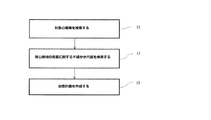
図1は、本発明に係る方法の異なる工程を示すフローチャートである。 FIG. 1 is a flow chart showing the different steps of the method according to the invention.
工程11では、対象の内部画像(すなわち体の内面を示す画像)を検索する。これはコンピュータ断層撮影(CT)スキャンからの3次元画像であってもよいが、例えば磁気共鳴画像法(MRI)などの他の画像診断法または画像診断法の組み合わせも可能である。
At
工程12では、画像内の関心領域の定義に関するいくつかの態様に関する少なくとも1つの不確かさ尺度を検索する。例えば、不確かさ尺度は、疾患の存在、生物学的反応またはROI輪郭の位置に関するものであってもよい。一例として、不確かさ尺度は可能な輪郭位置の範囲を定めることができる。不確かさ尺度は、例えば描写プロセス中に例えば腫瘍学者が手作業で予め定めてもよく、かつ/または逆方向治療計画プロセス中のフィードバックとして入力してもよい。
At
工程13では、不確かさ尺度のうちの1つ以上を考慮して治療計画を作成する。以下に記載するように、不確かさ尺度に加えて線量または生物学に基づく治療目的もしくは制約などの治療計画を最適化するための複数の他のパラメータを用いる治療計画システムを用いて、治療計画を作成する。
At
光子、プロトンまたは電子を含む任意の種類の画像診断法を用いる任意の種類の放射線治療装置での使用のために治療計画を最適化することができる。治療計画は、強度変調放射線治療(IMRT)計画または、例えば3次元原体照射(3DCRT)計画または強度変調回転照射(VMAT)計画などの任意の他の放射線治療計画であってもよい。 The treatment plan can be optimized for use in any type of radiation therapy device using any type of imaging modality that includes photons, protons or electrons. The treatment plan may be an intensity modulated radiation therapy (IMRT) plan or any other radiotherapy plan such as, for example, a three-dimensional conformal radiation (3 DCRT) plan or an intensity modulated rotational irradiation (VMAT) plan.
逆方向治療計画における一般的な手法は、多くの場合、特定の計画制約に従って、全ての最適化関数からなる目的関数を最小化(最大化)することである。目的関数は全ての最適化関数fiの加重和、すなわち

ボクセル「j」を含むROIに関する最適化関数「fi」の単純な例は、
目的関数を最適化して治療計画に到達させる場合、様々な異なる最適化技術を用いてもよい。例えば、逐次二次計画アルゴリズムに基づく方法などの勾配法または焼きなまし法などの発見的方法を使用することができる。最適化は、その後の機械パラメータへの変換を必要とするフルエンスに基づくもの、または機械パラメータが直接最適化される直接機械パラメータ最適化(DMPO)に基づくもの、あるいはそれらの両方の組み合わせであってもよい。最適化を用いる従来の逆方向治療計画は当該技術分野でよく知られているため、本明細書ではさらに詳細に説明しない。 A variety of different optimization techniques may be used when optimizing the objective function to reach the treatment plan. For example, heuristics such as gradient or annealing such as those based on sequential quadratic programming algorithms can be used. The optimization may be based on fluence requiring subsequent conversion to machine parameters, or on direct machine parameter optimization (DMPO) where machine parameters are directly optimized, or a combination of both. It is also good. Conventional reverse treatment plans using optimization are well known in the art and will not be described in further detail herein.
不確かさ尺度の本治療計画プロセスへの組み込みは、様々な異なる方法で行うことができ、最初に図3および図4を参照しながらこれについて以下に説明する。図1に示す本発明の方法を用いて最適化される治療計画は、異なる領域に関連する不確かさ尺度とこれらの領域を照射(または温存)するためのコストの両方(すなわち必要なトレードオフの程度)によって決まる。 The incorporation of the uncertainty measure into the present treatment planning process can be done in a variety of different ways, which will first be described with reference to FIGS. 3 and 4 below. The treatment plan optimized using the method of the present invention shown in FIG. 1 is both an uncertainty measure associated with different areas and the cost for irradiating (or sparing) these areas (ie the necessary trade-offs) It depends on the degree).
図2は、標的輪郭21を有する標的体積Tの2D表示を示す。不確かさ尺度は標的輪郭21の特定の部分に割り当てられており、この場合、内側境界23および外側境界24を有する拡大された「近似」輪郭22が得られる。そのような近似輪郭は、標的境界の真の位置は不確かであるが、(少なくとも高い確率で)近似輪郭22によって覆われている領域内のどこかにあることを示している。従って、近似輪郭は標的の定義の不確かさを反映している。近似輪郭22の幅などの特性は描写手順の間にユーザ、例えば腫瘍学者が定めることができ、あるいは他の方法で、例えば以下にさらに説明するように画像情報に基づいて自動的に定めることができる。
FIG. 2 shows a 2D representation of a target volume T with a
近似輪郭22の内側境界23は標的輪郭の残りの部分21と1つにまとめられている場合、可能な最も小さい標的領域を画定する。それに応じて、近似輪郭22の外側境界24は標的輪郭の残りの部分21と1つにまとめられている場合、可能な最も大きな標的領域を画定する。疾患が当該領域全体に存在する可能性があるため、達成可能であれば線量をより大きな標的領域に照射すべきである。しかし、許容不可能な方法でリスク臓器への線量を増加させない限りより大きな標的領域を照射できないのであれば、標的領域の範囲を妥協しなければならないこともある。従って、近似輪郭22によって画定される領域は、最初に標的範囲において必要な犠牲を払わなければならない場所を示す。例えば、近似輪郭によって画定される領域の内側半分のみを照射することができれば、標的の真の輪郭が位置する場所によっては、全ての疾患を治癒させる見込みは(低いが)まだある。
The
従って、不確かさに関する情報すなわち近似輪郭の拡大を最適化のための入力として使用することにより、以前は治療計画システムに利用可能ではなかったさらなる情報に基づいて治療計画を決定することができる。 Thus, by using information about uncertainty, ie, the expansion of the approximate contour, as an input for optimization, a treatment plan can be determined based on additional information not previously available to the treatment planning system.
図3は、標的領域T1、T2、T3およびリスク臓器「O」を含む対象の2D画像表示を示す。標的領域T1はこの例では臨床的標的体積(CTV)を表し、ここでは輪郭の一部の正確な位置は不確かである。不確かさの程度は図2に示す標的に対する方法と同じ方法で、すなわちその中のどこかに真の標的境界が位置する内側輪郭境界33および外側輪郭境界34を有する幅広い近似輪郭32を用いて定められる。CTV輪郭のある部分の位置に関して不確かさの程度を定める不確かさ尺度は、CTV境界に近い領域における疾患の存在に関する不確かさ、すなわち特定の領域またはボクセルが疾患を含む確率とみなすことができる。T1の内側輪郭境界33によって画定される最も小さい可能なCTV標的を非常に高い(すなわち100%に近い)腫瘍細胞存在確率を有する領域R1とみなし、標的T1の外側輪郭境界34の外側領域を非常に低い(すなわち0%に近い)腫瘍細胞存在確率を有する領域とみなせば、腫瘍存在確率は近似輪郭領域32内で0〜1に変化するものとみなすことができる。従って、近似輪郭領域32内の各ボクセルに、近似輪郭境界に対するボクセル位置に従って特定の疾患存在確率を与えることができる。近似輪郭領域を所定量の領域に分けることができ、そこでは近似輪郭の内側境界33および外側境界34までの距離に従って各領域に特定の疾患存在確率が割り当てられる。図3に示す実施形態によれば、標的T1の近似輪郭領域は2つの領域R2、R3に分けられており、そこでは内側領域R2および外側領域R3にそれぞれ60%および20%の疾患存在確率が割り当てられている。近似輪郭を異なる疾患存在確率を有する別々の領域としてモデル化することにより、以下にさらに説明するように、不確かさ情報を治療計画の最適化に容易に組み込むことができる。
FIG. 3 shows a 2D image display of a subject including target areas T1, T2, T3 and risk organ “O”. The target area T1 represents a clinical target volume (CTV) in this example, where the exact position of part of the contour is uncertain. The degree of uncertainty is determined in the same way as for the target shown in FIG. 2, ie using a wide
関心領域の定義に関する不確かさ尺度は、例えば組織の種類に関する不確かさなどの当該領域内の組織の特性および/または生物学的反応に関する不確かさに基づき、領域に直接割り当てることができる。例えば、疾患(例えば腫瘍細胞)の存在の確率は、転移が疑われる領域(すなわち疾患の可能性はあるが確認されていない領域)などの特定の領域に割り当てることができる。代わりまたは追加として、期待される生物学的反応における不確かさに基づく確率を領域に割り当ててもよい。例えば、そのような確率は、疾患を含む標的領域について定める場合、疾患により生じるある指定された患者悪影響の推定されるリスクであってもよく、例えば治療しなければ患者が生存しないリスクであってもよい。それに応じて、リスク臓器について定める場合、生物学的反応に関する不確かさは、指定された量の放射線量を受けた結果生じるある特定の悪影響の推定されるリスクに関するものであってもよい。 Uncertainty measures related to the definition of a region of interest can be assigned directly to the region based on, for example, the uncertainty of tissue properties and / or biological responses within the region, such as uncertainty regarding the type of tissue. For example, the probability of the presence of a disease (eg, a tumor cell) can be assigned to a particular area, such as the area suspected of being metastasized (ie, the area that has a probable but not confirmed disease). Alternatively or additionally, a probability based on the uncertainty in the expected biological response may be assigned to the region. For example, such a probability may be the estimated risk of a given patient adverse effect caused by the disease when defining for a target area that includes the disease, eg the risk that the patient will not survive without treatment. It is also good. Accordingly, when defining for risk organs, the uncertainty regarding the biological response may relate to the estimated risk of certain adverse effects resulting from receiving a specified amount of radiation dose.
組織の特性に関する不確かさ尺度は、腫瘍学者による推測などの専門家の所見あるいは、例えば生検もしくはPETスキャンに基づく測定値またはコンピューターシミュレーションおよび/または他の患者からの統計データなどに基づいていてもよい。 Uncertainty measures regarding tissue characteristics may also be based on expert findings such as oncologist speculation or measurements such as measurements based on biopsies or PET scans or computer simulations and / or statistical data from other patients Good.
図3では、特定の腫瘍存在確率が標的領域T2およびT3に割り当てられており、この場合これらは転移が存在し得る領域を表す。この例によれば、図に示されているように、標的領域T2は10%の疾患存在確率を有し、標的領域T3は30%の疾患存在確率を有し、それらの確率は例えば腫瘍学者によって推定される。従って、図に示されている確率は、それぞれの領域内の少なくとも1つボクセルが疾患を含む確率を表す。他の例示的な一実施形態では、領域T2およびT3において疾患の存在が確認されていると仮定し、領域T2およびT3の不確かさ尺度は期待される生物学的反応に基づいていてもよい。従って、そのような一実施形態によれば、領域T2およびT3に対して定められる確率は、当該領域が治療されなければ患者が生存できないなどのある望ましくない患者への影響の確率であってもよい。従って、一例として、領域T2に対して定められている不確かさ尺度は、領域T2が治療されない場合であっても90%の患者が生存する確率に対応する。 In FIG. 3, specific tumor presence probabilities are assigned to target areas T2 and T3, where they represent areas where metastasis may be present. According to this example, as shown in the figure, the target area T2 has a disease presence probability of 10%, the target area T3 has a disease existence probability of 30%, which probabilities are for example Estimated by Thus, the probabilities shown in the figure represent the probability that at least one voxel in each region contains a disease. In another exemplary embodiment, assuming that the presence of the disease has been confirmed in regions T2 and T3, the uncertainty measure of regions T2 and T3 may be based on the expected biological response. Thus, according to such an embodiment, the probability defined for the regions T2 and T3 is the probability of an effect on some undesirable patient, such as the patient can not survive unless the region is treated. Good. Thus, by way of example, the uncertainty measure defined for the region T2 corresponds to the probability that 90% of patients will survive even if the region T2 is not treated.
図3に関する以下の例では、領域T2およびT3に対して定められている確率は疾患の存在の確率として表されているが、当然のことながら、これらの種類の確率はちょうど同じように上記生物学的反応に関する(すなわち腫瘍細胞の存在または不存在を特に考慮していない)不確かさを反映していてもよい。 In the following example relating to FIG. 3, the probabilities defined for the regions T2 and T3 are expressed as the probability of the presence of the disease, but it goes without saying that the probabilities of these types are the same as just described. It may reflect uncertainty related to the chemical response (ie not taking into account the presence or absence of tumor cells).
また、リスク臓器「O」の輪郭は、比較的より幅広い近似輪郭セグメント35によって画定された実際の位置が不確かである部分を有する。標的T1の不確かな領域における腫瘍存在の確率を考慮するのと同様に、リスク臓器「O」の近似輪郭35は、実際にリスク臓器特有の組織細胞を含むボクセルの確率を反映している。従って、リスク臓器「O」の近似輪郭領域35内のボクセルにはボクセルが実際にOAR組織を含む確率が割り当てられている。図3に示す単純化された例では、単一の一定の確率(40%)を用いて不確かさがモデル化されているため、1つの領域R4は100%の確率を有し、別の領域R5は40%の確率を有する。
Also, the contour of the risk organ "O" has a portion where the actual position defined by the relatively wider
この例における不確かさ尺度は、疾患の存在またはOAR組織の存在の確率の形態で治療計画システムへの入力として使用する。領域特有の確率を組み込む明らかな方法の1つは、最適化においてより「確かな」領域内のボクセルに対してより高い重みが与えられるように、最適化アルゴリズムで使用される対応する領域またはボクセルセット特有の重要度重みを定めることである。しかし、そのような手法は一般に適していない。例えば、標的領域を考慮すると、そのような目的関数に基づいて最適化される治療計画では、線量レベルは低いがなお不確かな領域の全ての部分に線量が照射される。腫瘍制御を達成するために疾患を含む全ての部分がある量の線量を受けなければならないため、これは一般に満足が得られる結果ではない。すなわち、臨床的目標を満たすことができる標的の全ての部分を処方線量で照射しなければならない。従って、疾患の存在に関して不確かさが存在し、かつ他の重要な治療目的を妨害しない限り臨床的目標を満たすことができない標的の部分に対しては、むしろ線量を全く照射すべきではない。従って、不確かさ情報を組み込むための他の一般により有利な方法について以下に記載する。 The uncertainty scale in this example is used as an input to the treatment planning system in the form of the presence of the disease or the probability of the presence of the OAR tissue. One obvious way to incorporate region-specific probabilities is to use the corresponding regions or voxels used in the optimization algorithm so that the optimization gives higher weights to voxels in the more "certain" regions in the optimization. It is to define a set-specific importance weight. However, such an approach is generally not suitable. For example, given a target area, in a treatment plan optimized based on such an objective function, all parts of the area where the dose level is low but still uncertain are irradiated. This is generally not a satisfactory result, as all parts, including the disease, must receive a certain amount of dose to achieve tumor control. That is, all parts of the target that can meet the clinical goals must be irradiated with the prescribed dose. Thus, the dose should not be irradiated at all for portions of the target that can not meet the clinical goals unless there is uncertainty as to the presence of the disease and that it does not interfere with other important therapeutic goals. Accordingly, other generally more advantageous methods for incorporating uncertainty information are described below.
単純な例としては、異なる指定された不確かさ尺度を含む領域の異なる組み合わせが別々のシナリオで検討される複数の異なる治療計画を決定することができる。例えば、標的内の照射される領域に関する不確かさ尺度、および例えばリスク臓器温存に関する様々な他の相反する最適化目的もしくは制約の実現を考慮したそれぞれの領域における標的目的(例えば処方最小線量に関する目的)の実現に基づいて、異なる治療計画候補を評価することができる。従って、一例として、リスク臓器に対する指定された線量寛容レベルを超えることなく腫瘍制御を達成する最も高い確率が得られる計画(不確かさ尺度に基づいて決定される計画)が自動的に選択される。 As a simple example, different treatment plans can be determined in which different combinations of regions including different designated uncertainty measures are considered in different scenarios. For example, an uncertainty measure on the area to be irradiated in the target, and a target purpose in each area, for example taking into account the realization of various other conflicting optimization goals or constraints on risk organ preservation (eg goals on prescribed minimum dose) Different treatment plan candidates can be evaluated based on the realization of Thus, as an example, a plan (plan determined based on the uncertainty scale) that automatically provides the highest probability of achieving tumor control without exceeding the specified dose tolerance level for the risk organ is automatically selected.
以下では、図3および図4を参照しながら、ROI定義の不確かさを本治療計画プロセスに組み込む方法に関する3つの実施例についてより詳細に説明する。 In the following, with reference to FIGS. 3 and 4, three examples of how to incorporate the uncertainty of the ROI definition into the treatment planning process will be described in more detail.
実施例1:別々の領域
疾患を含む確率p1、...、pnを有する領域R1、...、Rnを含むROIを検討する。j<iの場合に領域Rjで臨床的目標を満たす前に領域Riで臨床的目標を満たす利点はないという意味で、i>1の各領域Riは全てがj<iの領域Rjに依存している。
Example 1: Probability p 1 ,. . . , P n with regions R 1 ,. . . , Consider an ROI that includes R n . j <in the sense that there is no advantage to meet the clinical goals in region R i before meeting the clinical goals in region R j in the case of i, i> all the regions R i is the 1 j <areas of i R It depends on j .
図3の標的T1では、これらの領域はR1、R2およびR3であり、ここでは、R1は100%の疾患を含む確率を有する領域であり、R2は60%の疾患を含む確率を有する領域であり、R3は20%の疾患を含む確率を有する領域である。従って、p1=1、p2=0.6およびp3=0.2である。領域間の依存は、標的T1に対して臨床的目標があれば、最初に領域R1において、次いで領域R1∪R2において、そして次いで領域R1∪R2∪R3においてその目標を満たす努力をすべきであることを意味している。これは、その目標が標的T1に50Gyの線量を照射することであれば、領域R1∪R2が50Gyを受けない限り領域R3に50Gyを照射する利点はなく、領域R1が50Gyを受けない限り領域R2に50Gyを照射する利点がないこと意味している。当業者には明らかなように、他の実施形態では、異なる領域を相互に独立したしたものとみなすことができる。 For target T1 in FIG. 3, these regions are R 1 , R 2 and R 3 , where R 1 is the region with 100% disease coverage and R 2 contains 60% disease R 3 is a region with probability, R 3 is a region with a probability of containing 20% disease. Thus, p 1 = 1, p 2 = 0.6 and p 3 = 0.2. Dependence between the regions, if there is clinical target for the target T1, the first region R 1, and then in the region R 1 ∪R 2, and then meet its target in the region R 1 ∪R 2 ∪R 3 It means that you should make an effort. This long that the target is irradiated with a dose of 50Gy targets T1, no advantage region R 1 ∪R 2 irradiates the 50Gy in the region R 3 unless subjected to 50Gy, region R 1 is a 50Gy which means that there is no advantage to irradiate the 50Gy in the region R 2 unless subjected. As would be apparent to one skilled in the art, in other embodiments, different regions can be considered independent of one another.
依存的領域R1、...、Rnを有する所与のROIにおける臨床的目標(例えば標的領域に対する最小線量)を満たす確率は、以下に従って計算することができる。
領域R1、R1∪R2、R1∪R2∪R3、...、
Region R 1 , R 1 ∪R 2 , R 1 ∪R 2 ∪R 3 ,. . . ,
従って、一例として図3を参照すると、臨床的目標が標的T1の領域R1において満たされるが、領域R1∪R2において満たされない(従って、領域R1∪R2∪R3でも満たされない)ものと仮定する。故に、R1∪R2は臨床的目標が満たされない最初の領域であり、これは標的T1に対して臨床的目標が満たされない確率がp2=0.6であることを意味する。さらに、全ての標的T1、T2およびT3は、図3に示すように全てが相互に独立している。一例として、臨床的目標が標的T2に対して満たされないが、標的T3に対して満たされるとものと仮定する。故に、標的T2の臨床的目標が満たされない確率は0.1であり(標的T2に疾患が存在する確率は10%であるため)、標的T3の臨床的目標が満たされない確率は0である。故に、全ての腫瘍を治癒させる総確率は(1−0.6)×(1−0.1)×(1−0)=0.36である。 Thus, referring to FIG. 3 as an example, a clinical target is satisfied in region R 1 of target T 1 but not in region R 1 ∪ R 2 (and thus not even in region R 1 ∪ R 2 ∪ R 3 ) Assume that Hence, R 1 ∪ R 2 is the first area where the clinical goal is not met, which means that the probability of not meeting the clinical goal for target T 1 is p 2 = 0.6. Furthermore, all targets T1, T2 and T3 are all independent of one another as shown in FIG. As an example, assume that the clinical goal is not met for target T2, but it is met for target T3. Thus, the probability that the clinical target of target T2 is not met is 0.1 (as the probability of the disease being present in target T2 is 10%), the probability that the clinical target of target T3 is not met is zero. Thus, the total probability of healing all the tumors is (1−0.6) × (1−0.1) × (1−0) = 0.36.
同様に、図3の領域Oなどの様々なOARのためにその目標を満たす確率を計算することができる。 Similarly, the probability of meeting that goal for various OARs, such as region O of FIG. 3, can be calculated.
次いで、最適化により、これらの確率に関して最も良好に行う計画を見出さなければならない。例えば、全ての確率の積(すなわち全ての臨床的目標を満たす確率)を最大化するために努力することができる。あるいは、1つ以上の確率に制約を課すことができ、いくつかの他の目標(例えば、最大線量、モニタ単位(MU)数などに関する治療目的など)を最適化することができる。 Then, through optimization, we have to find the best plan for these probabilities. For example, one may strive to maximize the product of all probabilities (ie the probability of meeting all clinical goals). Alternatively, constraints can be imposed on one or more probabilities, and some other goals (e.g., maximum dose, therapeutic goals for monitor unit (MU) count, etc.) can be optimized.
上記のように最適化を用いる場合、トレードオフはそれぞれの領域の不確かさ尺度ならびに異なる領域の線量照射/温存を達成するコストによって決まる。一例として、図3を参照すると、標的領域T1の領域R1ならびに標的領域T3において臨床的目標を満たす(上に例示されているように標的の臨床的目標を満たす確率「0.36」が得られる)と共に、OAR「O」の領域R4およびR5の両方に対して臨床的目標を満たすことができる(すなわち、OARの臨床的目標を満たす確率「1」が得られる)ものと仮定する。これにより全ての臨床的目標を満たす確率「0.36×1=0.36」が得られる。ここで、OARの臨床的目標が領域R4内で満たされるが領域R5内で満たされない(OAR臨床的目標を満たす確率「1−0.4=0.6」が得られる)場合、領域R1および標的T3だけでなく領域R2内でも標的臨床的目標を満たすことができる(標的臨床的目標を満たす確率「(1−0.2)×(1−0.1)×(1−0)=0.72」が得られる)ものと仮定する。これにより全ての臨床的目標を満たす確率「0.72×0.6=0.432」が得られる。 When using optimization as described above, the trade-off depends on the uncertainty measure of each region as well as the cost of achieving dose exposure / preservation of different regions. As an example, referring to FIG. 3, obtained probability "0.36" to meet the clinical objective of the target as illustrated satisfying clinical goals (to the top in the region R 1 and the target region T3 of the target region T1 Assuming that the clinical goal can be met for both regions R 4 and R 5 of OAR “O” (ie, a probability “1” is obtained to meet the clinical goal of OAR) . This gives the probability "0.36 x 1 = 0.36" to meet all clinical goals. Here, if the clinical goal of the OAR is met within the region R 4 but not within the region R 5 (a probability “1-0.4 = 0.6” is obtained that meets the OAR clinical goal), the region The target clinical goal can be met not only in R 1 and target T 3 but also in the region R 2 (probability of meeting the target clinical goal “(1-0.2) × (1−0.1) × (1− 0) = 0.72 ”is obtained. This gives the probability of "0.72 x 0.6 = 0.432" to meet all clinical goals.
従って、線量が領域R1、R2およびT3に対して処方されるが領域R3およびT2に対して処方されない治療計画を用いて全ての臨床的目標を満たす確率を最大化させるが、これは、リスク臓器「O」を温存するという点で非常に高価になるという理由から、標的領域R3およびT2に対して線量を計画することは有利でないということを示している。 Thus, using a treatment plan in which doses are prescribed for regions R 1 , R 2 and T 3 but not for regions R 3 and T 2 maximizes the probability of meeting all clinical goals. , because it becomes very expensive in terms of sparing the organs at risk "O", to plan the dose to a target area R 3 and T2 indicates that no advantageous.
全ての利用可能な不確かさ情報を考慮することにより、治療計画システムは、より良好な治療計画が得られ、例えば、正常組織を損なうことなく総腫瘍照射を達成する最も高い可能な確率が得られる、特定の状況で最も有利なトレードオフを特定する。 By considering all available uncertainty information, the treatment planning system can get a better treatment plan, eg the highest possible probability of achieving total tumor irradiation without harming normal tissue Identify the most advantageous trade-offs in a particular situation.
実施例2:連続的かつ非階層的領域
その中の各点が区間(0,1]において疾患を含む所定の確率を有する標的領域Rを検討する。最適化の目的は疾患を治癒させる確率を最大化することである。
Example 2: Continuous and Non-hierarchical Region Consider a target region R with a predetermined probability that each point in it contains a disease in the interval (0, 1) The purpose of optimization is the probability of curing the disease It is to maximize.
ボクセルごとに分離可能な目標(例えば、各ボクセルに対する線量は60Gy超にすべきであるという目標)のために、疾患を有する全てのボクセルにおいてその目標を満たす(疾患を有する全てのボクセルに60Gy超の線量を照射する)確率を以下のように計算することができる。
最適化の目標は、この確率を最大化する線量を照射することであり、これは、セットI内のボクセルが疾患を含む可能な限り低い確率が存在するような方法で線量を照射する場合と同じである。 The goal of optimization is to deliver a dose that maximizes this probability, as in the case where the voxels in set I have a dose as low as possible including disease. It is the same.
繋がった標的体積を治療することが望ましい場合もある。これは、治療される標的体積が常に繋がった体積であるような治療される体積の形状に対する制約の導入により達成することができる。 It may be desirable to treat linked target volumes. This can be achieved by the introduction of constraints on the shape of the treated volume such that the target volume to be treated is always a connected volume.
図4Aは、標的の定義の不確かさを反映した、疾患が存在することが知られている内側領域41および外側の「不確かな」領域42を含む標的体積の2D画像表示を示す。従って、領域42は、上記実施例と同様に、例えば専門家によって定められる近似輪郭に一致することができる。内側領域41の内側のボクセルは100%の疾患を含む確率を有するものとみなし、領域41および42の外側のボクセルは0%の疾患を含む確率を有するものとみなす。従って、外側領域42内の各ボクセルは0〜1の範囲の疾患を含む確率を有する。ボクセルに対する確率は、外側領域42内のボクセルの位置によって決まってもよい。例えば、その確率は線形に、あるいは内側領域41の表面に対する法線43に沿って外側領域42の内側境界にあるボクセルにおける「1」から外側領域42の外側境界にあるボクセルにおける「0」まで、指数関数的に減少することができる。外側領域42内の変化する確率を多くの他の方法で、例えば専門家によってなされる推測に従って定めることができることは明らかである。例えば、異なるように連続的に変化する確率を有する複数の領域を定めることができる。
FIG. 4A shows a 2D image representation of the target volume including the
上記方法において方程式(3)を用いて、治療される標的体積を最大化する。図4Bは、そのような方法を用いることにより得られた例示的な標的体積44を示す。図から分かるように、治療される体積は、若干の不規則性を有する可能性がある。しかし、実施例1に関して上に記載した同様の理由で、標的の中心により近い隣接する領域を治療しない場合に離れた領域を治療することは通常は有益ではないため、標的体積の形状に対して制約を課すと有利であり得る。 Equation (3) is used in the above method to maximize the target volume to be treated. FIG. 4B shows an exemplary target volume 44 obtained by using such a method. As can be seen from the figure, the volume to be treated may have some irregularities. However, for the same reasons described above with respect to Example 1, it is usually not beneficial to treat a remote area if not treating an adjacent area closer to the center of the target, as opposed to the shape of the target volume. It may be advantageous to impose constraints.
実施例3:分離不可能な目標
上記のとおり、各ボクセルが区間(0、1]において疾患を含む所定の確率を有する領域R(その領域が標的に関する場合)を検討し、あるいは、その領域がOARに関する場合は領域特有のある他の組織の種類を検討する。以下では、領域Rは標的領域に関するものとして例示されている。従って、ボクセルに割り当てられた確率は、真の標的体積がボクセルを含む確率を反映しており、従って、その確率は疾患を含むことが知られている領域内のボクセルにおける100%から、疾患を含まないことが知られている領域内のボクセルにおける0%まで減少する。全ての可能な標的体積および、これらのそれぞれについて、それが真の標的体積である対応する確率(これらはボクセルへの離散化によりゼロ以外である)を検討する。100%の確率を有するボクセルはこれらの標的体積の全てに含まれている。計画があれば、「S」を臨床的目標が満たされる標的体積全体にわたる指数セットとし、かつ「pi」を標的体積「iはSの要素である」が真の標的体積である確率とする。故に、最適化の目標は、真の標的体積における臨床的目標を満たす確率を最大化することであり、これは
これは、真の標的体積を含むことが可能な限りほぼ確実である標的体積セットを指数付けする領域「S」が得られる線量分布を見つけることによって達成される。この手法は、ボクセルごとに分離不可能な目標を用いる場合、例えばDVHに基づく最適化関数(すなわちX%のROIが少なくともYのGyを受けることを要求する目標)を用いる場合に適用可能である。 This is achieved by finding a dose distribution that yields an area "S" that indexes the set of target volumes that is as near certain as possible to include the true target volume. This approach is applicable when using an inseparable target for each voxel, for example when using a DVH based optimization function (ie a target that requires an X% ROI to receive at least Y Gy). .
上記全ての実施形態はROI定義の不確かさを本治療計画プロセスに組み込む方法の単なる例であり、当業者には明らかなように多くの他の方法が想定される。 All of the above embodiments are merely examples of how to incorporate the uncertainty of the ROI definition into the present treatment planning process, and many other methods are envisioned as would be apparent to one skilled in the art.
上述のように、ROI定義の不確かさを様々な異なる方法で定めることができる。例えば、これも上に説明したように、手作業による領域分割または自動領域分割手順の間または後に、例えば専門家ユーザ(例えば腫瘍学者)によって小領域および対応する不確かさ尺度を手作業で定義、修正および/または承認することができる。例えば、異なる程度の不確かさ(例えば異なる輪郭幅)を示す輪郭セグメントを定めるためのツールを含む任意の好適なユーザインタフェースを用いて、当該領域および/または対応する不確かさ尺度を画像内に直接定義することができる。 As mentioned above, the uncertainty of the ROI definition can be defined in a variety of different ways. For example, manually defining sub-regions and corresponding uncertainty measures, eg by an expert user (eg oncologist), during or after a manual segmentation or automatic segmentation procedure, as also described above, It can be modified and / or approved. For example, define the region and / or the corresponding uncertainty measure directly in the image using any suitable user interface including, for example, a tool for defining contour segments exhibiting different degrees of uncertainty (eg different contour widths) can do.
当該ユーザは、例えば輪郭セグメントの各側における信頼区間を定める不確かさの数値尺度を特定の輪郭の一部に割り当てることもできる。例えば専門家によって割り当てられる様々な種類の不確かさ数値尺度を場合により画像情報と組み合わせて、上に記載し、かつ図3に示す小領域に対応する複数の小領域を画定するため、あるいは上に記載し、かつ図4Aに例示する特定の領域内の連続的に変化する不確かさを定めるために使用することができる。 The user may, for example, also assign numerical measures of uncertainty defining confidence intervals on each side of the contour segment to part of a particular contour. For example, in order to define a plurality of sub-regions corresponding to the sub-regions described above, and possibly in combination with the image information, various types of uncertainty numerical measures assigned by experts, or It can be used to define continuously changing uncertainty within the specific area described and illustrated in FIG. 4A.
図3に示す近似輪郭領域を、治療計画のためのより良好な基礎を提供するために、異なる割り当てられた確率を有するより小さい小領域のさらにより大きなセットに分けることができる(但し、本治療計画プロセスの計算負荷をより高めてしまう)。領域の数および対応する割り当てられた確率を、所定の判断基準に従って自動的に決定することができる。例えば、固定された所定数の小領域を使用することができ、あるいは小領域の数を近似輪郭セグメントの拡大(例えば幅)によって決めてもよい。次いで、特定のボクセルの不確かさ尺度を、所定の判断基準ならびにボクセルから近似輪郭の内側および外側境界までの相対距離によって決める。あるいは、画像強度データの分析に基づく方法などの、小領域および対応する不確かさ尺度を定めるための他の方法を用いてもよい。例えば、画定された輪郭の全ての部分にそれぞれの輪郭部分に近い領域におけるコントラストに基づく不確かさ尺度を割り当てることができる。従って、低コントラスト領域内の輪郭セグメントには比較的より大きな不確かさが自動的に割り当てられる。そのような自動的に割り当てられた不確かさ尺度は、専門家によって評価および承認されるか、あるいは必要であれば修正されると有利である。 The approximate contour area shown in FIG. 3 can be divided into an even larger set of smaller sub-areas with different assigned probabilities to provide a better basis for treatment planning (but this treatment) Further increase the computational load of the planning process). The number of regions and the corresponding assigned probabilities may be determined automatically according to predetermined criteria. For example, a fixed predetermined number of subregions may be used, or the number of subregions may be determined by the enlargement (e.g. width) of the approximate contour segment. The uncertainty measure of a particular voxel is then determined by the predetermined criteria and the relative distance from the voxel to the inner and outer boundaries of the approximate contour. Alternatively, other methods may be used to define small regions and corresponding uncertainty measures, such as methods based on analysis of image intensity data. For example, all parts of the defined contour can be assigned an uncertainty measure based on the contrast in the area close to the respective contour part. Thus, contour segments in the low contrast region are automatically assigned a relatively greater uncertainty. Such automatically assigned uncertainty measures are advantageously evaluated and approved by the expert or, if necessary, corrected.
不確かさ尺度は、例えばROIに関する要素の異なる大きさ、色、透明度などを用いる様々な方法で画像内に視覚化してもよい。例えば指定された領域が疾患を含むか否かに関する不確かさ情報は、測定値(例えば、生検またはPETスキャンなどに基づく)、コンピューターシミュレーション、他の患者からの統計データから、あるいは任意の他の方法で決定することもできる。 The uncertainty measure may be visualized in the image in various ways, for example using different sizes, colors, transparency etc of the elements with respect to the ROI. For example, uncertainty information as to whether a designated area contains a disease may be from measurements (eg, based on a biopsy or PET scan, etc.), computer simulations, statistical data from other patients, or any other It can also be determined by the method.
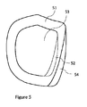
本発明に係る方法は、セットアップの不確かさを考慮するための確率的アプローチに基づく方法などの、よりロバストな治療計画を作成するために以前に使用したあらゆる任意の方法と組み合わせることができる。さらに、安全性マージンを用いて拡大された領域などの任意の領域に対して本発明の方法を用いることができる。生じ得るセットアップの不確かさまたは治療中の標的の動きを補償するために、通常は標的体積にマージンを付ける(「計画標的体積」(PTV)を作製する)。例えば、特定の不確かさ尺度を臨床的標的体積(CTV)の領域に割り当て、かつCTVに均一なマージンを付けてPTVを定義する場合、同じ不確かさ尺度をPTVの対応する領域に割り当て、かつ上記治療計画プロセスで使用することができる。図5は、CTV53の近似輪郭セグメント52がPTVの対応する近似輪郭セグメント54に転写される場合のそのようなPTV51を示す。
The method according to the invention can be combined with any arbitrary method previously used to create a more robust treatment plan, such as a method based on a probabilistic approach to take into account set-up uncertainty. Furthermore, the method of the present invention can be used for any area, such as the area expanded with a security margin. In order to compensate for possible setup uncertainty or target movement during treatment, the target volume is usually margined (to create a "planned target volume" (PTV)). For example, if a particular uncertainty measure is assigned to a region of clinical target volume (CTV), and CTV is defined with uniform margins to define PTV, the same uncertainty measure is assigned to the corresponding region of PTV and It can be used in the treatment planning process. FIG. 5 shows such a
図6は本発明に係るコンピュータシステム61の例を概略的に示す。本システムは、メモリ63に接続された処理装置62を備える。さらに、本システムは、表示装置64(例えば定義されたROIおよび対応する不確かさ尺度を有する患者画像、グラフィカルユーザインタフェース、および治療計画に関連する他の情報を表示する)、データ入力装置65(例えば、キーボード、マウスまたはデータ入力に適した任意の他の装置)ならびにデータ読み取り/書き込み装置66(例えば、光学式ドライブ、USBインタフェース、またはデータの読み取り/書き込みに適した任意の他の装置)を備えていてもよい。処理装置62は、1つ以上の中央処理装置(CPU)または、例えば1つ以上のグラフィックス処理装置(GPU)に基づく任意の種類のパラレル処理装置システムなどの任意の種類の処理装置であってもよい。メモリ63は、例えばハードドライブなどの情報を格納および検索するのに適した任意の種類の揮発性もしくは不揮発性メモリであってもよい。メモリ63は、コンピュータプログラム67をそこに格納している。コンピュータプログラム67は、コンピュータ可読命令を処理装置62に転送し、かつそれにより実行することができる、不確かさに基づく最適化を行うためのコンピュータ可読命令を含む。処理装置62によって実行されると、コンピュータ可読命令は、ROIの定義における不確かさに基づいて治療計画を決定するために図1に示す方法を行う。決定された治療計画は、患者画像、ROI、不確かさ尺度および任意の他の治療計画関連情報と共にメモリ63に格納することができる。コンピュータプログラム67をメモリ63にロードし、かつ/または異なる計算システムに転送することができるように、コンピュータプログラム67も非一時的コンピュータ可読媒体68、例えば、USBドライブ、CD−ROMなどの光学データキャリアまたは任意の他の好適な携帯可能な情報格納装置に格納することができる。図6を参照して説明したシステムは単なる例であり、本発明に係るコンピュータシステムは必ずしも例示されている構成要素を全て備えているわけではなく、かつ/または図示されていない他の構成要素を備えていてもよい。
FIG. 6 schematically shows an example of a
多くの例示的な実施形態を参照しながら本発明について説明してきた。当然のことながら、これらの実施形態は本発明の原理および用途の単なる例示である。従って、当然のことながら、例示的な実施形態に対して数多くの修飾をなすことができ、かつ添付の特許請求の範囲によって定められる本発明の趣旨および範囲を逸脱することなく他の構成を考案することができる。 The invention has been described with reference to a number of exemplary embodiments. It should be understood that these embodiments are merely illustrative of the principles and applications of the present invention. Accordingly, it should be understood that numerous modifications may be made to the illustrative embodiments, and other configurations may be devised without departing from the spirit and scope of the present invention as defined by the appended claims. can do.
Claims (8)
− 対象の画像を検索する工程(11)と、
− 前記画像内の関心領域に関する少なくとも1つの不確かさ尺度を検索する工程であって、前記少なくとも1つの不確かさ尺度は前記関心領域の定義に関する不確かさの程度を反映していることを特徴とする工程(12)と、
− 前記関心領域または前記関心領域の一部を治療するか否かを決定すること含む、少なくとも部分的に前記少なくとも1つの不確かさ尺度に基づいて放射線治療計画を作成する工程(13)と、を包含し、
前記不確かさ尺度とは前記関心領域内の組織の特性に関する不確かさを指すことを特徴とし、かつ前記関心領域内の組織の特性は腫瘍細胞の存在または不存在に関するものであることを特徴とする、
方法。 A method of preparing a radiation treatment plan for the subject based on an internal image of the subject, the device processing the following steps based on the computer program (67) stored in the memory (63). Is the method to perform, said method being:
A step (11) of searching for an image of interest;
Searching for at least one uncertainty measure for the region of interest in the image, wherein the at least one uncertainty measure reflects the degree of uncertainty for the definition of the region of interest Step (12),
Creating (13) a radiation treatment plan based at least in part on the at least one uncertainty measure, comprising determining whether to treat the region of interest or a portion of the region of interest. Include
The uncertainty scale is characterized by referring to uncertainty regarding the property of the tissue in the area of interest, and the property of the tissue in the area of interest is related to the presence or absence of tumor cells ,
Method.
− 専門家の入力、
− 画像データ、
− 測定値、
− 統計データ、
− シミュレーション
のうちの1つ以上に基づいていることを特徴とする、請求項1または請求項2に記載の方法。 The uncertainty scale is
-Expert input,
-Image data,
-Measured value,
-Statistical data,
Method according to claim 1 or 2, characterized in that it is based on one or more of the simulations.
The processor (62) connected to the memory (63) storing therein the computer program (67) containing computer readable instructions, the computer readable instructions according to claims 1-6. A computer system (61) comprising the processing unit (62) configured to perform the method according to any one of the preceding claims.
Applications Claiming Priority (3)
| Application Number | Priority Date | Filing Date | Title |
|---|---|---|---|
| EP13194947.1 | 2013-11-28 | ||
| EP13194947.1A EP2878338B1 (en) | 2013-11-28 | 2013-11-28 | Method and system for uncertainty based radiotherapy treatment planning |
| PCT/SE2014/051147 WO2015080647A1 (en) | 2013-11-28 | 2014-10-03 | Method and system for uncertainty based radiotherapy treatment planning |
Publications (2)
| Publication Number | Publication Date |
|---|---|
| JP2017514532A JP2017514532A (en) | 2017-06-08 |
| JP6550046B2 true JP6550046B2 (en) | 2019-07-24 |
Family
ID=49752967
Family Applications (1)
| Application Number | Title | Priority Date | Filing Date |
|---|---|---|---|
| JP2016525950A Active JP6550046B2 (en) | 2013-11-28 | 2014-10-03 | Methods and systems for radiation therapy treatment planning based on uncertainty |
Country Status (5)
| Country | Link |
|---|---|
| US (1) | US10300300B2 (en) |
| EP (1) | EP2878338B1 (en) |
| JP (1) | JP6550046B2 (en) |
| CN (1) | CN106163612B (en) |
| WO (1) | WO2015080647A1 (en) |
Families Citing this family (14)
| Publication number | Priority date | Publication date | Assignee | Title |
|---|---|---|---|---|
| US10460448B2 (en) * | 2012-02-14 | 2019-10-29 | Koninklijke Philips N.V. | Method for quantification of uncertainty of contours in manual and auto segmenting algorithms |
| EP3307387B1 (en) * | 2015-06-12 | 2018-12-05 | Koninklijke Philips N.V. | Dose planning system |
| EP3228356B1 (en) | 2016-04-07 | 2018-03-14 | RaySearch Laboratories AB | Method, computer program and system for optimizing a radiotherapy treatment plan |
| EP3228357B1 (en) * | 2016-04-08 | 2021-03-31 | RaySearch Laboratories AB | Method, computer program product and computer system for radiotherapy treatment planning |
| JP6565120B2 (en) | 2016-09-23 | 2019-08-28 | 住友重機械工業株式会社 | Neutron capture therapy system and treatment planning system for neutron capture therapy |
| CN107392897B (en) * | 2017-07-17 | 2021-02-02 | 上海联影医疗科技股份有限公司 | Organ contour acquisition method, imaging apparatus, radiotherapy planning system, and storage medium |
| EP3453427A1 (en) * | 2017-09-12 | 2019-03-13 | RaySearch Laboratories AB | Evaluation of arcs for a radiation treatment plan |
| EP3498335A1 (en) * | 2017-12-18 | 2019-06-19 | Koninklijke Philips N.V. | Evaluation of an anatomic structure with respect to a dose distribution in radiation therapy planning |
| EP3530319A1 (en) * | 2018-02-21 | 2019-08-28 | Elekta Limited | Methods for inverse planning |
| EP3660741B1 (en) * | 2018-11-29 | 2022-05-04 | Koninklijke Philips N.V. | Feature identification in medical imaging |
| EP3682945B1 (en) | 2019-01-21 | 2021-03-10 | RaySearch Laboratories AB | Method and system of evaluating a radiation therapy treatment plan |
| EP3750595B1 (en) * | 2019-06-11 | 2023-10-25 | RaySearch Laboratories AB | Method and system for robust radiotherapy treatment planning for biological uncertainties |
| CN110706780B (en) * | 2019-10-16 | 2023-05-26 | 上海联影医疗科技股份有限公司 | Radiotherapy plan generation system and storage medium |
| US11701066B2 (en) * | 2020-01-17 | 2023-07-18 | Ping An Technology (Shenzhen) Co., Ltd. | Device and method for detecting clinically important objects in medical images with distance-based decision stratification |
Family Cites Families (15)
| Publication number | Priority date | Publication date | Assignee | Title |
|---|---|---|---|---|
| JP4664489B2 (en) * | 2000-12-22 | 2011-04-06 | 株式会社東芝 | Radiation therapy planning system |
| US6735277B2 (en) * | 2002-05-23 | 2004-05-11 | Koninklijke Philips Electronics N.V. | Inverse planning for intensity-modulated radiotherapy |
| US7206377B2 (en) | 2003-01-27 | 2007-04-17 | Siemens Medical Solutions Usa, Inc. | Predictive organ dynamics database and code |
| US7804935B2 (en) | 2003-08-28 | 2010-09-28 | Henry Ford Health System | Fuzzy logic guided inverse treatment planning |
| JP4429839B2 (en) * | 2004-08-06 | 2010-03-10 | 株式会社日立製作所 | Radiotherapy planning apparatus and radiotherapy planning method |
| US7844560B2 (en) * | 2006-04-17 | 2010-11-30 | Siemens Medical Solutions Usa, Inc. | Personalized prognosis modeling in medical treatment planning |
| CN101663069A (en) * | 2007-03-30 | 2010-03-03 | 皇家飞利浦电子股份有限公司 | Improved treatment plan evaluation in radiotherapy by stochastic analysis of delineation uncertainty |
| US8577115B2 (en) * | 2008-03-04 | 2013-11-05 | Tomotherapy Incorporated | Method and system for improved image segmentation |
| US8812240B2 (en) * | 2008-03-13 | 2014-08-19 | Siemens Medical Solutions Usa, Inc. | Dose distribution modeling by region from functional imaging |
| EP2513863A1 (en) * | 2009-12-16 | 2012-10-24 | Koninklijke Philips Electronics N.V. | Use of collection of plans to develop new optimization objectives |
| EP2580698A1 (en) * | 2010-06-11 | 2013-04-17 | Koninklijke Philips Electronics N.V. | Simultaneous multi-modality inverse optimization for radiotherapy treatment planning |
| WO2012012768A1 (en) * | 2010-07-23 | 2012-01-26 | Tomotherapy Incorporated | System and method for identifying an anatomical organ in a patient |
| US8693630B2 (en) * | 2011-01-10 | 2014-04-08 | Varian Medical Systems International Ag | Method and apparatus pertaining to radiation-therapy treatment-plan optimization |
| JP2013132489A (en) * | 2011-12-27 | 2013-07-08 | Hitachi Ltd | Therapy planning apparatus and particle beam therapy system |
| EP2808057B1 (en) * | 2013-05-31 | 2016-02-03 | RaySearch Laboratories AB | Method and system for robust radiotherapy treatment planning |
-
2013
- 2013-11-28 EP EP13194947.1A patent/EP2878338B1/en active Active
-
2014
- 2014-10-03 US US15/039,670 patent/US10300300B2/en active Active
- 2014-10-03 JP JP2016525950A patent/JP6550046B2/en active Active
- 2014-10-03 WO PCT/SE2014/051147 patent/WO2015080647A1/en active Application Filing
- 2014-10-03 CN CN201480064982.9A patent/CN106163612B/en active Active
Also Published As
| Publication number | Publication date |
|---|---|
| US20170209712A1 (en) | 2017-07-27 |
| CN106163612A (en) | 2016-11-23 |
| CN106163612B (en) | 2020-11-03 |
| US10300300B2 (en) | 2019-05-28 |
| WO2015080647A1 (en) | 2015-06-04 |
| JP2017514532A (en) | 2017-06-08 |
| EP2878338A1 (en) | 2015-06-03 |
| EP2878338B1 (en) | 2018-04-11 |
Similar Documents
| Publication | Publication Date | Title |
|---|---|---|
| JP6550046B2 (en) | Methods and systems for radiation therapy treatment planning based on uncertainty | |
| US10765888B2 (en) | System and method for automatic treatment planning | |
| CN109069858B (en) | Radiotherapy system and computer readable storage device | |
| EP2808057B1 (en) | Method and system for robust radiotherapy treatment planning | |
| JP5805757B2 (en) | System and method for estimating radiation dose and manipulating the estimated radiation dose | |
| Birkner et al. | Adapting inverse planning to patient and organ geometrical variation: algorithm and implementation | |
| CN105592887B (en) | 3D information is used as input to predict achievable dosage distribution | |
| KR20150135354A (en) | Systems and methods for isotopic source external beam radiotherapy | |
| van de Schoot et al. | Dosimetric advantages of proton therapy compared with photon therapy using an adaptive strategy in cervical cancer | |
| CN116391234A (en) | Machine learning optimization of fluence maps for radiation therapy | |
| CN113677396B (en) | Method and system for evaluating radiation therapy treatment plans | |
| EP3347093A1 (en) | Knowledge-based spatial dose metrics and methods to generate beam orientations in radiotherapy | |
| WO2015083064A1 (en) | Method and system for dose calculation based on continuous material indexing | |
| Guidi et al. | Expert system classifier for adaptive radiation therapy in prostate cancer | |
| CN116785601A (en) | Method and system for robust radiotherapy treatment planning with dose mapping uncertainty | |
| Vazquez et al. | A deep learning-based approach for statistical robustness evaluation in proton therapy treatment planning: a feasibility study | |
| Eriksson | Scenario dose prediction for robust automated treatment planning in radiation therapy | |
| CN113990443A (en) | Method and system for determining dose distribution | |
| Simoni et al. | CT Texture Analysis Effect of Neoadjuvant Stereotactic Body Radiation Therapy (SBRT) in Borderline Resectable and Locally Advanced Pancreatic Cancer | |
| Wong et al. | A design of a DICOM‐RT‐based tool box for nonrigid 4D dose calculation |
Legal Events
| Date | Code | Title | Description |
|---|---|---|---|
| A621 | Written request for application examination |
Free format text: JAPANESE INTERMEDIATE CODE: A621 Effective date: 20170829 |
|
| A977 | Report on retrieval |
Free format text: JAPANESE INTERMEDIATE CODE: A971007 Effective date: 20180718 |
|
| A131 | Notification of reasons for refusal |
Free format text: JAPANESE INTERMEDIATE CODE: A131 Effective date: 20180807 |
|
| A521 | Request for written amendment filed |
Free format text: JAPANESE INTERMEDIATE CODE: A523 Effective date: 20181105 |
|
| A131 | Notification of reasons for refusal |
Free format text: JAPANESE INTERMEDIATE CODE: A131 Effective date: 20190129 |
|
| A521 | Request for written amendment filed |
Free format text: JAPANESE INTERMEDIATE CODE: A523 Effective date: 20190402 |
|
| TRDD | Decision of grant or rejection written | ||
| A01 | Written decision to grant a patent or to grant a registration (utility model) |
Free format text: JAPANESE INTERMEDIATE CODE: A01 Effective date: 20190604 |
|
| A61 | First payment of annual fees (during grant procedure) |
Free format text: JAPANESE INTERMEDIATE CODE: A61 Effective date: 20190628 |
|
| R150 | Certificate of patent or registration of utility model |
Ref document number: 6550046 Country of ref document: JP Free format text: JAPANESE INTERMEDIATE CODE: R150 |
|
| R250 | Receipt of annual fees |
Free format text: JAPANESE INTERMEDIATE CODE: R250 |
|
| R250 | Receipt of annual fees |
Free format text: JAPANESE INTERMEDIATE CODE: R250 |