JP6466912B2 - Apparatus and method for determining thorax and abdominal respiratory signals from image data - Google Patents
Apparatus and method for determining thorax and abdominal respiratory signals from image data Download PDFInfo
- Publication number
- JP6466912B2 JP6466912B2 JP2016507077A JP2016507077A JP6466912B2 JP 6466912 B2 JP6466912 B2 JP 6466912B2 JP 2016507077 A JP2016507077 A JP 2016507077A JP 2016507077 A JP2016507077 A JP 2016507077A JP 6466912 B2 JP6466912 B2 JP 6466912B2
- Authority
- JP
- Japan
- Prior art keywords
- signal
- respiratory
- image
- determined
- subject
- Prior art date
- Legal status (The legal status is an assumption and is not a legal conclusion. Google has not performed a legal analysis and makes no representation as to the accuracy of the status listed.)
- Expired - Fee Related
Links
Images


Classifications
-
- A—HUMAN NECESSITIES
- A61—MEDICAL OR VETERINARY SCIENCE; HYGIENE
- A61B—DIAGNOSIS; SURGERY; IDENTIFICATION
- A61B5/00—Measuring for diagnostic purposes; Identification of persons
- A61B5/08—Detecting, measuring or recording devices for evaluating the respiratory organs
- A61B5/0816—Measuring devices for examining respiratory frequency
-
- A—HUMAN NECESSITIES
- A61—MEDICAL OR VETERINARY SCIENCE; HYGIENE
- A61B—DIAGNOSIS; SURGERY; IDENTIFICATION
- A61B5/00—Measuring for diagnostic purposes; Identification of persons
- A61B5/103—Detecting, measuring or recording devices for testing the shape, pattern, colour, size or movement of the body or parts thereof, for diagnostic purposes
- A61B5/11—Measuring movement of the entire body or parts thereof, e.g. head or hand tremor, mobility of a limb
- A61B5/1126—Measuring movement of the entire body or parts thereof, e.g. head or hand tremor, mobility of a limb using a particular sensing technique
- A61B5/1128—Measuring movement of the entire body or parts thereof, e.g. head or hand tremor, mobility of a limb using a particular sensing technique using image analysis
-
- A—HUMAN NECESSITIES
- A61—MEDICAL OR VETERINARY SCIENCE; HYGIENE
- A61B—DIAGNOSIS; SURGERY; IDENTIFICATION
- A61B5/00—Measuring for diagnostic purposes; Identification of persons
- A61B5/103—Detecting, measuring or recording devices for testing the shape, pattern, colour, size or movement of the body or parts thereof, for diagnostic purposes
- A61B5/11—Measuring movement of the entire body or parts thereof, e.g. head or hand tremor, mobility of a limb
- A61B5/113—Measuring movement of the entire body or parts thereof, e.g. head or hand tremor, mobility of a limb occurring during breathing
- A61B5/1135—Measuring movement of the entire body or parts thereof, e.g. head or hand tremor, mobility of a limb occurring during breathing by monitoring thoracic expansion
-
- G—PHYSICS
- G06—COMPUTING; CALCULATING OR COUNTING
- G06T—IMAGE DATA PROCESSING OR GENERATION, IN GENERAL
- G06T7/00—Image analysis
- G06T7/0002—Inspection of images, e.g. flaw detection
- G06T7/0012—Biomedical image inspection
-
- G—PHYSICS
- G06—COMPUTING; CALCULATING OR COUNTING
- G06T—IMAGE DATA PROCESSING OR GENERATION, IN GENERAL
- G06T7/00—Image analysis
- G06T7/20—Analysis of motion
-
- G—PHYSICS
- G06—COMPUTING; CALCULATING OR COUNTING
- G06T—IMAGE DATA PROCESSING OR GENERATION, IN GENERAL
- G06T2207/00—Indexing scheme for image analysis or image enhancement
- G06T2207/30—Subject of image; Context of image processing
- G06T2207/30004—Biomedical image processing
- G06T2207/30076—Plethysmography
Description
本発明は、対象から呼吸信号を決定する装置及び方法に関連する。そこでは、撮像野において対象から決定される画像データが受信され、呼吸信号が、画像データにおいて決定される運動パターンに基づき決定される。 The present invention relates to an apparatus and method for determining a respiratory signal from a subject. There, image data determined from an object is received in an imaging field, and a respiratory signal is determined based on an exercise pattern determined in the image data.
対象又は患者のバイタル信号及び特に対象の呼吸レートは、例えばビデオカメラといったコンタクトレスのセンサを用いて、リモートでモニタされることができる。パターン検出を用いて画像データから呼吸レートを決定する一般の方法は、例えばWO2012/140531A1号から知られる。測定される対象はカメラの撮像野において自由に配置されることができ、かつバイタルサインが得られる関連領域はカメラの撮像野において自由に配置されることができるので、対象及び関連領域は、所望のバイタルサイン情報、例えば対象の呼吸レートの抽出のため検出及び規定されなければならない。更に、バイタルサイン情報に関して直説的な及び非直説的な異なる運動パターンが、バイタルサイン情報の正確な遠隔測定のために、識別及び区別されなければならない。 The subject's or patient's vital signals and in particular the subject's respiration rate can be monitored remotely using contactless sensors such as video cameras. A general method for determining the respiration rate from image data using pattern detection is known, for example, from WO 2012/1405 3 1A1. Since the object to be measured can be freely placed in the imaging field of the camera, and the related area where the vital sign is obtained can be freely placed in the imaging field of the camera, the object and the related area are Must be detected and defined for the extraction of vital sign information such as the respiratory rate of the subject. In addition, different movement patterns that are straightforward and non-open-minded with respect to vital sign information must be identified and differentiated for accurate telemetry of vital sign information.
関心領域の従来の識別は一般に、人間、例えば顔若しくは胸部の検出に基づかれるか、又は背景セグメント化を用いて行われる。人間の識別に関して、及び例えば関心領域からパルス又は呼吸レートといったバイタルサインを遠隔画像検出測定に基づき測定することに関して、US2009/0141124A1号は、測定される対象の一部を表す関心領域を選択するため、赤外線ビデオセグメントの輪郭を検出することを提案する。 Conventional identification of a region of interest is generally based on the detection of a human, eg, face or chest, or using background segmentation. With respect to human identification and for measuring vital signs such as pulses or respiration rate from a region of interest based on remote image detection measurements, US2009 / 0141124 A 1 selects a region of interest that represents a part of the object to be measured. In order to do so, we propose to detect the contour of infrared video segment.
更に、WO2012/093320A2号は、対象からバイタルサイン情報を検出する、特に対象からのフォトプレチスモグラフィ信号を検出するビデオ検出デバイスを開示する。そこでは、撮像野においてバイタルサイン情報を自動的に決定するため、この場合対象の皮膚である関心領域を選択するべく、映像データは異なるブロックに分けられる。 Furthermore, WO 2012 / 09320A2 discloses a video detection device for detecting vital sign information from a subject, in particular for detecting a photoplethysmographic signal from a subject. In this case, vital sign information is automatically determined in the imaging field. In this case, the video data is divided into different blocks in order to select a region of interest that is the target skin.
呼吸を測定する従来の方法は、インダクティブ・プレチスモグラフィである。この場合、対象の胴周りに巻かれるワイヤを配置することで、胸部又は腹部断面領域における変化を測定する呼吸バンドにより、呼吸が検出される。胸部及び腹部呼吸を区別するために、典型的に2つの呼吸バンドが使用される。対象の呼吸を正確に測定し、特別な損傷又は不随を識別するため、胸部及び腹部呼吸の独立した測定が必要である。 A conventional method for measuring respiration is inductive plethysmography. In this case, by arranging a wire wound around the torso of the subject, respiration is detected by a respiration band that measures changes in the chest or abdominal cross-sectional area. Two breathing bands are typically used to distinguish between chest and abdominal breathing. Independent measurements of chest and abdominal breathing are required to accurately measure the subject's breathing and identify special injuries or involuntaries.
対象から呼吸信号を測定する既知の方法の不利な点は、1つの呼吸信号だけが対象からリモートで決定されることができ,この場合粗い呼吸分析だけが可能である点、又は、対象からの異なる呼吸信号を正確に測定するシステムが、接触測定センサの使用が原因でユーザにとって不快である点にある。 The disadvantage of known methods of measuring respiratory signals from a subject is that only one respiratory signal can be determined remotely from the subject, in which case only a coarse respiratory analysis is possible, or from the subject A system that accurately measures different respiratory signals is uncomfortable for the user due to the use of contact measurement sensors.
本発明の目的は、ユーザにとってより正確でより快適に対象からの呼吸信号を決定する改良された装置及び対応する改良された方法を提供することである。 It is an object of the present invention to provide an improved apparatus and corresponding improved method for determining a respiratory signal from a subject more accurately and comfortably for a user.
本発明の一つの側面によれば、対象から呼吸信号を決定する装置が提供され、この装置は、
撮像野において対象から決定される画像データを受信する受信ユニットと、
上記画像データを評価する処理ユニットであって、運動パターンに基づき上記撮像野の複数の異なる領域から上記対象のバイタルサイン情報に対応する複数の異なる交流信号を決定するよう構成される、処理ユニットと、
上記異なる交流信号を評価し、上記撮像野の上記異なる領域から決定される上記異なる交流信号に基づき、上記対象から複数の異なる呼吸信号を決定する評価ユニットとを有する。
According to one aspect of the invention, an apparatus for determining a respiratory signal from a subject is provided, the apparatus comprising:
A receiving unit for receiving image data determined from a target in an imaging field;
A processing unit for evaluating the image data, the processing unit configured to determine a plurality of different AC signals corresponding to the vital sign information of the object from a plurality of different regions of the imaging field based on a motion pattern; ,
And an evaluation unit that evaluates the different alternating signals and determines a plurality of different respiratory signals from the object based on the different alternating signals determined from the different regions of the imaging field.
本発明の別の側面によれば、対象から呼吸信号を決定する方法が提供され、この方法は、
撮像野において上記対象から決定される画像データを受信するステップと、
上記画像データを評価するステップと、
運動パターンに基づき、上記撮像野の複数の異なる領域から上記対象のバイタルサイン情報に対応する複数の異なる交流信号を決定するステップと、
上記異なる交流信号を評価するステップと、
上記撮像野の異なる領域から決定される上記異なる交流信号に基づき、上記対象から複数の異なる呼吸信号を決定するステップとを有する。
According to another aspect of the invention, a method for determining a respiratory signal from a subject is provided, the method comprising:
Receiving image data determined from the object in an imaging field;
Evaluating the image data;
Determining a plurality of different alternating signals corresponding to the target vital sign information from a plurality of different regions of the imaging field based on a motion pattern;
Evaluating the different AC signals;
Determining a plurality of different respiratory signals from the object based on the different alternating signals determined from different regions of the imaging field.
本発明の更に別の側面によれば、コンピュータで実行されるとき、コンピュータに本発明による方法のステップを実行させるプログラムコード手段を有するコンピュータプログラムが提供される。 According to yet another aspect of the present invention there is provided a computer program comprising program code means for causing a computer to execute the steps of the method according to the present invention when executed on a computer.
本発明は、コンタクトレスの測定に基づき、1つの対象から異なる呼吸信号を測定し、改良された正確な呼吸測定を提供するという着想に基づかれる。これは、コンタクトレスの測定によってユーザとっては快適である。異なる呼吸信号は、測定される対象からキャプチャされる画像データから決定される運動パターンに基づき決定される。この場合、撮像野における異なる領域の運動パターンが、異なる呼吸信号を決定するために用いられる。こうして、対象の呼吸に対応する対象の異なる部分の運動が、独立して決定されることができる。その結果、例えば胸部及び腹部呼吸が独立して決定されることができ、ユーザにとって快適である。そして、全体の呼吸情報が、低い技術的な負担で決定されることができる。異なる呼吸信号に基づき、追加的な診断が実行されることができる。その結果、バイタルサイン検出がより正確になる。 The present invention is based on the idea of measuring different respiratory signals from one subject and providing improved and accurate respiratory measurements based on contactless measurements. This is comfortable for the user due to the contactless measurement. Different respiratory signals are determined based on motion patterns determined from image data captured from the object being measured. In this case, motion patterns of different areas in the imaging field are used to determine different respiratory signals. In this way, the movement of different parts of the subject corresponding to the subject's breathing can be determined independently. As a result, for example, chest and abdominal breathing can be determined independently, which is comfortable for the user. And the whole respiratory information can be determined with a low technical burden. Based on the different respiratory signals, additional diagnostics can be performed. As a result, vital sign detection becomes more accurate.
本発明の好ましい実施形態は、従属項において規定される。請求項に記載の方法が、請求項に記載の装置及び従属項に記載されるデバイスと類似する及び/又は同一の好ましい実施形態を持つ点を理解されたい。 Preferred embodiments of the invention are defined in the dependent claims. It should be understood that the claimed method has similar and / or identical preferred embodiments to the claimed device and the device described in the dependent claims.
好ましい実施形態において、上記処理ユニットが、上記画像データにおける複数の画像セクションを規定し、運動パターン検出に基づき、各々の上記画像セクションから上記バイタルサイン情報に対応する1つの交流信号を決定するよう構成される。これは、低い技術的な負担で全体の撮像野からバイタルサイン情報を識別することを可能にする。 In a preferred embodiment, the processing unit defines a plurality of image sections in the image data and is configured to determine one AC signal corresponding to the vital sign information from each of the image sections based on motion pattern detection. Is done. This makes it possible to identify vital sign information from the entire imaging field with low technical burden.
好ましい実施形態において、上記処理ユニットが、上記画像データにおける画像セクションのアレイとして上記異なる画像セクションを規定するよう構成される。これは、対象の異なるバイタルサイン情報を決定するため、全体の画像データを分析して、全体の撮像野を分析するシンプルなソリューションである。 In a preferred embodiment, the processing unit is configured to define the different image sections as an array of image sections in the image data. This is a simple solution that analyzes the entire image field by analyzing the entire image data in order to determine vital sign information for different objects.
好ましい実施形態において、この装置は、上記異なる画像セクションから決定される上記交流信号のスペクトルパラメータを決定する周波数分析ユニットと、上記異なる呼吸信号を決定するため、上記異なる領域として上記スペクトルパラメータに基づき、異なる画像セクションを選択する選択ユニットとを更に有する。これは、バイタルサイン情報が得られることができる撮像野において異なる関心領域を決定することを信頼性高く可能にする。 In a preferred embodiment, the apparatus comprises a frequency analysis unit for determining spectral parameters of the alternating signal determined from the different image sections, and based on the spectral parameters as the different regions for determining the different respiratory signals, And a selection unit for selecting different image sections. This makes it possible to reliably determine different regions of interest in the imaging field from which vital sign information can be obtained.
好ましい実施形態において、上記異なる画像セクションから決定されるスペクトルパラメータが、上記交流信号のスペクトルエネルギーである。これは、バイタルサイン情報と妨害信号及びノイズとを高い信頼性で区別することを可能にする。 In a preferred embodiment, the spectral parameter determined from the different image sections is the spectral energy of the alternating signal. This makes it possible to reliably distinguish vital sign information from jamming signals and noise.
好ましい実施形態において、上記交流信号の所定の周波数帯域の上記スペクトルエネルギーが閾値レベルを超える場合、上記選択ユニットが、上記画像セクションを選択するよう構成される。これは、低い技術的な負担でスペクトルパラメータを分析することを可能にする。 In a preferred embodiment, the selection unit is configured to select the image section when the spectral energy of a predetermined frequency band of the alternating signal exceeds a threshold level. This makes it possible to analyze the spectral parameters with a low technical burden.
好ましい実施形態において、上記異なる呼吸信号が、上記対象の異なる部分から得られる運動ベクトルに基づき決定される。対象の異なる部分から得られる運動ベクトルを用いて、例えば胸部及び腹部呼吸に対応する異なる呼吸信号が決定されることができる。 In a preferred embodiment, the different respiratory signals are determined based on motion vectors obtained from different parts of the subject. Using motion vectors obtained from different parts of the subject, different respiratory signals, for example corresponding to chest and abdominal breathing, can be determined.
好ましい実施形態において、上記異なる呼吸信号が、異なる波形を持つ時間依存の交流信号である。これは、シンプルな呼吸レートに加えて対象から追加的な診断情報を決定することを可能にする。 In a preferred embodiment, the different respiratory signals are time-dependent alternating signals with different waveforms. This makes it possible to determine additional diagnostic information from the subject in addition to a simple respiratory rate.
好ましい実施形態において、上記異なる呼吸信号が、互いに対するフェーズシフトを持つ時間依存信号である。これは、追加的な診断情報を決定するため、対象の異なる呼吸信号を区別することを可能にする。 In a preferred embodiment, the different respiratory signals are time-dependent signals with phase shifts relative to each other. This makes it possible to distinguish different respiratory signals of the subject in order to determine additional diagnostic information.
好ましい実施形態において、上記評価ユニットが、上記対象からの追加的な呼吸情報として上記異なる呼吸信号の信号差を決定するよう構成される。これは、追加的な診断に関して呼吸レートを越える追加的な呼吸情報を自動的に決定するソリューションである。 In a preferred embodiment, the evaluation unit is configured to determine a signal difference between the different respiratory signals as additional respiratory information from the subject. This is a solution that automatically determines additional respiratory information that exceeds the respiratory rate for additional diagnostics.
好ましい実施形態において、上記評価ユニットが、上記異なる呼吸信号の上記フェーズシフトを決定し、上記決定されたフェーズシフトを考慮して、上記異なる呼吸を1つの一般的な呼吸信号へと結合するよう構成される。これは、増加した正確さ及びより高い信頼性を持つ単一の呼吸信号を決定することを可能にする。 In a preferred embodiment, the evaluation unit is configured to determine the phase shift of the different respiratory signals and combine the different breaths into one general respiratory signal in view of the determined phase shift. Is done. This makes it possible to determine a single respiratory signal with increased accuracy and higher reliability.
更に好ましい実施形態では、上記評価ユニットが、上記対象の空間的な呼吸マップを提供するため、上記異なる画像セクションから得られる上記異なる呼吸信号に基づき、呼吸信号のアレイを決定するよう構成される。これは、追加的な診断可能性を提供するため、対象から全体の呼吸情報を決定することを可能にする。 In a further preferred embodiment, the evaluation unit is arranged to determine an array of respiratory signals based on the different respiratory signals obtained from the different image sections in order to provide a spatial respiratory map of the object. This makes it possible to determine overall respiratory information from the subject to provide additional diagnostic possibilities.
更に好ましい実施形態では、上記選択ユニットが、各々の上記選択された異なる画像セクションに対する重み係数を決定するよう構成され、上記評価ユニットは、上記個別の重み係数を用いて重み付けされる上記選択された画像セクションの上記交流信号に基づき、上記異なる呼吸信号を決定するよう構成される。これは、決定された呼吸信号の正確さを増加させるため、交流信号の信号強度を考慮することを可能にする。なぜなら、妨害信号又は雑音が多い信号は、高い強さを持つ信号より考慮されないからである。 In a further preferred embodiment, the selection unit is configured to determine a weighting factor for each of the selected different image sections, and the evaluation unit is the selected unit that is weighted with the individual weighting factor. Based on the alternating signal in the image section, the different respiratory signal is configured to be determined. This makes it possible to consider the signal strength of the alternating signal in order to increase the accuracy of the determined respiratory signal. This is because a jamming signal or a noisy signal is not considered more than a signal with high strength.
上記選択ユニットが、定期的に上記選択を実行するよう構成され、上記選択された画像セクションの各々に関する上記重み係数は、上記個別の画像セクションの選択の周波数に基づき決定される場合、更に好ましい。これは、信号強度及び重み係数を低い技術的な負担で決定することを可能にする。 It is further preferred if the selection unit is arranged to perform the selection periodically and the weighting factor for each of the selected image sections is determined based on the frequency of selection of the individual image sections. This makes it possible to determine the signal strength and the weighting factor with a low technical burden.
上述したように、本発明は、測定される対象を含む撮像野から決定される画像データを用いて、コンタクトレスな遠隔測定に基づき1つの対象から異なるバイタルサイン情報を決定するという可能性を提供する。交流信号が撮像野の異なる領域から決定される運動パターンに基づき決定されるので、対象の異なる部分、例えば胸郭及び腹部からの呼吸信号は、呼吸検出の正確さを増加させるため、及び対象の呼吸から追加的な情報を決定するため、異なる呼吸技術に対応して決定されることができる。こうして、追加的な診断が実行されることができ、呼吸の検出は、より高い信頼性を持ち、より正確であり、コンタクトレスの測定に基づき快適に決定されることができる。 As described above, the present invention provides the possibility of determining different vital sign information from one object based on contactless telemetry using image data determined from an imaging field containing the object to be measured. To do. Since AC signals are determined based on motion patterns determined from different areas of the imaging field, respiratory signals from different parts of the subject, such as the rib cage and abdomen, increase the accuracy of respiratory detection and Can be determined corresponding to different breathing techniques. In this way, additional diagnostics can be performed, and respiration detection can be more reliable, more accurate, and comfortably determined based on contactless measurements.
本発明のこれらの及び他の態様が、以下に説明される実施形態より明らとなり、これらの実施形態を参照して説明されることになる。 These and other aspects of the invention will be apparent from and will be elucidated with reference to the embodiments described hereinafter.
図1は、対象12から呼吸信号を決定する符号10により一般に表される装置の概略的な図面を示す。対象12、例えばベッドに寝ている患者が、支持部14上で休息している。対象の頭部16は通常、対象12の呼吸に関する非直説的な部分である。この場合、胸部18又は胸郭18及び子宮20又は腹部20が、対象12の呼吸に関する直説的な部分である。一般の課題は、胸部及び腹部呼吸に対応する異なる呼吸信号が、独立に、及び低い技術的な負担でコンタクトレスで正確に測定されることができない点にある。通常、単に呼吸レート又は心拍は、一般にカメラシステム又はリモートシステムを用いて検出される。
FIG. 1 shows a schematic drawing of an apparatus generally represented by reference numeral 10 for determining a respiratory signal from a subject 12. A subject 12, for example a patient sleeping in bed, is resting on the
装置10は、例えば対象12の画像フレームを記録するのに使用されることができるモノクロのカメラといった画像検出デバイス22に接続される。画像フレームは、対象12により放出又は反射される電磁放射24から得られることができる。画像データ、例えば画像フレームのシーケンスから画像情報を抽出するため、画像検出デバイス22は、インタフェース28を介して画像処理ユニット30に接続される。画像検出デバイス22は、装置10の部分もよいか、又は外部カメラ22でもよい。その結果、画像データ26は単に、一般に画像データ26を装置10に提供するため、インタフェース28に提供されるだけである。
The apparatus 10 is connected to an
画像検出デバイス22は、少なくとも電磁放射24のスペクトル要素に属する画像をキャプチャするよう構成される。画像検出デバイス22は、測定される対象12を含む撮像野からキャプチャされる連続的な画像データ又は画像フレームの離散的なシーケンスを提供することができる。
The
画像処理ユニット30は、インタフェース28を介して画像検出デバイス22から画像データ26を受信し、一般に画像データ26を評価し、例えば対象12の呼吸の直説的な部分として胸郭18及び腹部20といった対象12の異なる関心領域を検出するよう構成される。例えば胸郭18及び/又は腹部20といった関心領域を検出するため、画像処理ユニット30は、撮像野のセクション又は領域においてキャプチャされた画像を分割し、関心領域を決定するために画像セクションを別々に評価するよう構成される。画像処理ユニット30は、キャプチャされた画像を画像セクションに分割し、呼吸の直説的な部分として対象12の胸郭領域18及び/又は腹部領域20の運動を含む撮像野における対象の運動に対応する異なるセクションから、運動ベクトルを検出する。運動ベクトルは、画像セクションにおけるパターン検出を用いて、又は、画像セクションにおけるエッジ検出を用いて決定される。エッジ又はパターン検出のため、キャプチャされた画像フレームから運動ベクトルを得る方法は、例えばWO2012/140531A1号により開示される。
The
撮像処理ユニット30は、分析ユニット32に接続される。画像処理ユニット30は、画像セクションの各々からの運動ベクトルに基づき交流信号を決定し、分析ユニット32に交流信号を提供する。
The
分析ユニット32は、以下に詳細に説明される分析ユニット32に含まれる周波数分析ユニットを用いて、交流信号の各々のスペクトルパラメータを決定する。画像データ26におけるセクションの各々のスペクトルパラメータは、分析ユニット32の一部である選択ユニットにより分析される。選択ユニットは、呼吸信号に対応すると思われる交流信号が得られる画像データのそれらのセクションを選択する。選択ユニットは、個別のスペクトルパラメータに基づきセクションを選択する。スペクトルパラメータは、交流信号の各々の周波数スペクトル又はスペクトルエネルギー分布である。対象の呼吸信号が特性スペクトルエネルギー分布又は特性周波数を持つので、選択ユニットは、対象12の呼吸信号を含むセクションを選択することができる。従って、選択ユニットは、異なる呼吸信号を決定するため、画像データ26において対象12の胸郭18及び/又は腹部20を識別する。
The
選択ユニットは、以下に説明されるように周波数分析に基づき、異なる画像の各々に対する重み係数も決定する。重み要素は一般に、各画像セクションがどのくらい頻繁に選択されるかに依存する。こうして、重み係数は、交流信号の信号強度に対応する係数を表す。その結果、画像セクションの各々からの個別の交流信号が、信号品質に基づき考慮されることができる。 The selection unit also determines weighting factors for each of the different images based on frequency analysis as described below. The weight factor generally depends on how often each image section is selected. Thus, the weighting coefficient represents a coefficient corresponding to the signal strength of the AC signal. As a result, individual alternating signals from each of the image sections can be considered based on signal quality.
分析ユニット32は、評価ユニット34に接続され、対象12の呼吸に対応する呼吸信号を決定する評価ユニット34に交流信号を提供する。評価ユニット34は、異なる画像セクションから決定される交流信号及び異なる画像セクションに関する個別の重み係数を分析ユニット32から受信し、交流信号、重み係数及び交流信号が得られる異なる領域に基づき、異なる呼吸信号を算出する。こうして、呼吸信号は、画像データ26に基づき算出されて、完全にコンタクトレスに決定されることができる。この場合、呼吸信号は、異なる部分、例えば対象12の胸郭18及び腹部20と独立して得られることができる。
The
そのように算出された呼吸信号は、測定された呼吸信号を連続的に又は頻繁に表示するため、ディスプレイ36に提供されることができる。
The respiratory signal so calculated can be provided on the
こうして、胸部及び腹筋呼吸は、完全にコンタクトレスに、及び互いに独立して決定されることができる。その結果、呼吸測定は、より正確になり、例えば脊髄損傷又は隔膜の不随といった追加的な損傷を診断するため、対象12の呼吸から追加的な情報が得られることができる。 Thus, chest and abdominal muscle breathing can be determined completely contactless and independent of each other. As a result, respiration measurements are more accurate and additional information can be obtained from the breathing of the subject 12 to diagnose additional damage, such as spinal cord injury or diaphragm incompetence.
図2は、対象12の呼吸の遠隔測定を表すため、対象12の概略的な図を示す。対象12は、呼吸が原因で第1の直説的な部分18(胸郭18)及び第2の直説的な部分20(腹部20)の特徴的な運動を経験する。呼吸するとき、肺の拡張及び収縮は、2つの直説的な部分18、20のわずかな運動をもたらす。即ち、胸郭18及び腹部20の上昇及び下降である。通常、胸郭18及び腹部20は、交互態様において上昇及び下降する。その結果、腹部20が下降する間、胸郭18が上昇する。逆も真である。
FIG. 2 shows a schematic diagram of the subject 12 to represent telemetry of the subject 12 respiration.
矢印40により示される時間にわたり、直説的な部分18、20は、参照符号18a、20b及び18cにより示される縮小された位置と符号20a、18b及び20cにより示される拡張された(extracted)位置との間で移動される。基本的に、運動パターンに基づき、例えば、呼吸レート又は呼吸レート変動性又は呼吸ボリュームが、キャプチャされた画像シーケンスにおけるパターン又はエッジ検出を用いて評価されることができる。直説的な部分18、20が時間にわたり脈動している間、非直説的な部分として頭部16は実質的に静止したままである。胸郭18及び腹部20が、呼吸に関する直説的な部分としての例であり、例えば対象12のより低い肋骨での運動といった追加的な呼吸信号を決定するため、対象12の他の部分も検出されることができる点を理解されたい。
Over the time indicated by
確実に、頭部16も、時間にわたり多様な運動を経験する。しかしながら、これらの運動は、胸郭18又は腹部20の周期的な脈動に対応せず、周波数分析ユニットを用いて区別されることができる。
Certainly, the
図3は、例えば個別の画像セクションにおけるフレーム又はエッジ検出に基づき決定されることができる異なる画像セクションの運動パターンから及び/又は運動ベクトルから得られる交流信号のタイミング図を示す。交流信号は一般に、S(t)により表される。この特定のケースにおいて交流信号Sは、個別の直説的な部分18、20から受信される画像データに対応する画像セクションから得られる、対象12の胸郭18又は腹部20の運動に対応する。交流信号Sは、胸部18又は腹部20の運動、即ち対象12の呼吸に対応する特徴的な変動を示す。交流信号Sは、呼吸に重畳される高周波ノイズも示す。
FIG. 3 shows a timing diagram of alternating signals obtained from motion patterns of different image sections and / or from motion vectors, which can be determined for example based on frame or edge detection in individual image sections. An AC signal is generally represented by S (t). In this particular case, the AC signal S corresponds to the movement of the
交流信号Sは、撮像野の画像セクションの各々から得られる。この場合、複数の画像セクションは、例えば呼吸レートといったバイタルサイン情報を有し、多くの画像セクションは、対象12のバイタルサイン情報に関連付けられない妨害信号、又は高周波ノイズを含む他の交流信号を有することができる。バイタルサイン情報が得られることができるそれらの画像セクションを識別するため、分析ユニット32は、交流信号Sの周波数分析を実行する周波数分析デバイスを有する。周波数分析は、交流信号Sをフィルタリングすることにより、及び/又はフーリエ変換、特に交流信号Sの高速フーリエ変換(FFT)を実行することにより好ましくは実行される。以下に説明されるように、対象12の呼吸に対応するバイタルサイン情報を含む画像セクションを識別するため、交流信号Sから、周波数スペクトルが得られる。
An AC signal S is obtained from each of the image sections of the imaging field. In this case, multiple image sections have vital sign information, eg, respiratory rate, and many image sections have interference signals that are not associated with the vital sign information of
図4は、F(f)により一般に表される図3に示される交流信号Sの周波数スペクトルを示す。周波数スペクトルFは、低周波数帯域、この特定の場合においては0及び1ヘルツの間の大きな周波数要素を示す。これは通常、1ヘルツより高くない成人の呼吸レートに対応し、1分あたり60呼吸である。所定の周波数帯域、例えば成人に対して1ヘルツ及び乳児に対して2ヘルツより高い周波数要素は、通常画像データ26における妨害信号である、又は交流信号Sのノイズに対応する。交流信号Sの品質を特徴付けるため、交流信号Sのスペクトルエネルギーが決定され、所定の周波数帯域における交流信号Sのスペクトルエネルギーが、所定の閾値レベルを超えるか、又は第2の周波数帯域、例えば全体の周波数スペクトルと比較したスペクトルエネルギーのパーセンテージを超える場合、画像セクションが、バイタルサイン情報を含む画像セクションとして規定される。例えば、0及び1又は2ヘルツの間のスペクトルエネルギーが、所定の閾値レベルより大きい場合、例えば、交流信号Sの全スペクトルエネルギーの50%より大きい、又はスペクトルの所定の範囲より大きい、例えば2...3Hz、3...4Hz等である場合、画像セクションが、バイタルサイン情報を含む画像セクションとして規定される。スペクトルエネルギーに基づき、画像セクションが、以下に説明されるように関心領域を決定して、異なる呼吸信号を決定するため、撮像野において選択される。
FIG. 4 shows the frequency spectrum of the AC signal S shown in FIG. 3 that is generally represented by F (f). The frequency spectrum F shows a large frequency component in the low frequency band, in this particular case between 0 and 1 hertz. This usually corresponds to an adult respiratory rate not higher than 1 Hertz, which is 60 breaths per minute. A frequency component of a predetermined frequency band, for example, higher than 1 Hz for an adult and 2 Hz for an infant, is a disturbance signal in the
図5は、検出された画像データ26に基づく対象12からの異なる呼吸信号の検出を説明するため、撮像野からの概略的な画像を示す。図5に示される画像検出デバイス22により検出される撮像野は、符号42により一般に表される。画像検出デバイス22によりキャプチャされる撮像野42を表す画像フレーム44は、この場合測定される人間である対象12を示す。画像フレーム44において、格子46は、異なる部分において画像フレーム44を分割し、撮像野42において異なる領域を区別するため、及び撮像野42において異なる運動ベクトルを決定するため、画像セクション48を規定する。関心領域、即ち対象12の胸郭18及び腹部20を決定するため、運動パターンが、画像フレーム44の画像セクション48の各々から得られ、交流信号Sは、上述したように画像セクション48の各々の運動パターンから決定される運動ベクトルから決定される。運動ベクトルは、異なる画像セクションにおけるパターン検出又はエッジ検出により決定される。上述したように周波数分析に基づき、異なる画像セクション48の運動パターンが撮像野42において対象12の呼吸信号に対応するかどうか、又は、運動パターンが妨害信号若しくはノイズであるかどうかが決定される。運動パターンが呼吸信号を含むかどうかの決定は、スペクトルパラメータ及び/又はスペクトルエネルギーに基づき実行され、例えば周波数帯域におけるスペクトルエネルギーが、個別の交流信号の全体のスペクトルエネルギーの特定のパーセンテージより大きいかどうかに基づき実行される。
FIG. 5 shows a schematic image from the imaging field to illustrate the detection of different respiratory signals from the subject 12 based on the detected
画像セクション48の各々に関して決定されるこれらのデータに基づき、選択ユニットは、呼吸信号を含むそれらの画像セクションを選択し、それらの選択された画像セクション48を関心領域に結合することができる。これは、図5において符号50により一般に表される。図5に示される関心領域50は、胸郭18及び腹部20に対応する2つの直説的な部分18、20を有する。特定の実施形態において、分析ユニット32は、対象12の異なる直説的な部分18、20から異なる交流信号を決定するため、互いに分離されることができる異なる関心領域を決定することができる。
Based on these data determined for each of the
関心領域50の画像セクション48から得られる異なる交流信号Sに基づき、評価ユニット34は、胸郭18及び腹部20の呼吸運動に対応する異なる呼吸信号を決定する。分析ユニット32、特に分析ユニット32の選択ユニットが、信号品質に基づき異なるセクション48の交流信号Sを重み付けするため、関心領域50の選択された画像セクション48の各々に対する重み係数を決定する。分析ユニット32により決定される重み係数は、個別の画像セクションが選択ユニットによりどのくらい頻繁に選択されるかの頻度に基づき算出されることができる。言い換えると、選択された画像セクション48としてより頻繁に選択されるそれらの画像セクション48からの交流信号Sは、個別の呼吸信号を算出するためにより大きな重みを与えられ、あまり頻繁に選択されない画像セクション48は、より少ない重みを与えられる。
Based on the different alternating signals S obtained from the
同一又は対応する波形を有する交流信号Sが、(評価ユニット32により)単一の呼吸信号へと結合される。なぜなら、これらの交流信号Sは、同じ直説的な部分18、20から得られると考えられるからである。異なるセクション48からの交流信号がより大きい差、例えばフェーズシフトを持つ場合、それらの交流信号Sは、異なる直説的な部分18、20から得られると考えられ、1つの呼吸信号へと直接結合されることはない。結合ステップは、評価ユニット32により実行される。
The alternating signals S having the same or corresponding waveforms are combined (by the evaluation unit 32) into a single respiratory signal. This is because these AC signals S are considered to be obtained from the same
画像セクション48の各々の呼吸信号を別々に決定し、評価ユニット34を用いて対象12及び関心領域50の空間的な呼吸マップを決定することも可能である。
It is also possible to determine the respiration signal of each of the
図6aは、対象12の異なる部分から運動ベクトル検出によりコンタクトレスに得られる3つの異なる呼吸信号R1、R2及びR3を有するタイミング図を示す。そこから呼吸信号R1、R2、R3が得られる対象12の領域が、図6b、c及びdのキャプチャされた画像において概略的に示される。 FIG. 6a shows a timing diagram with three different respiratory signals R1, R2 and R3 obtained from different parts of the subject 12 in a contactless manner by motion vector detection. The region of the subject 12 from which the breathing signals R1, R2, R3 are obtained is schematically shown in the captured images of FIGS. 6b, c and d.
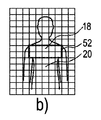
第1の呼吸信号R1は、図6bに示されるように胸郭18又は胸部18を含む関心52の領域から決定される。第2の呼吸信号R2は、図6cに示されるように対象12の腹部20又は子宮20を含む関心領域54から得られる。第2の呼吸信号R2は、胸郭18の呼吸信号R1に対してフェーズシフトされる。第3の呼吸信号R3は、図6dに示されるように対象12の胸郭18及び腹部20の間の関心領域56から決定される。第3の呼吸信号R3は、第2の呼吸信号R2の腹式呼吸に対応する呼吸を示す。しかしながら、第3の呼吸信号R3は、より幅広いピーク形状を持つ。なぜなら、この中間部から得られる交流信号は、胸郭18及び腹部20のような信号強度を持たないからである。
The first respiratory signal R1 is determined from the region of
呼吸信号R1、R2、R3は、異なる時点t1、t2での個別の直説的な部分18、20の運動に対応するそれらのピークを持ち、Δt1及びΔt2により示されるように互いに対してフェーズシフトされる。フェーズシフトΔt1、Δt2は、対象12の呼吸が原因で、胸郭18及び腹部20の交流運動に対応する。それゆえに、異なる呼吸信号R1、R2、R3が、装置10を用いてそコンタクトレスにリモートで独立に得られることができ、フェーズシフトΔt1、Δt2といった追加的な情報が、遠隔測定から決定されることができる。
The respiratory signals R1, R2, R3 have their peaks corresponding to the movements of the individual
フェーズシフトΔt1、Δt2といった追加的な情報に基づき、対象12の特定の損傷を決定するため、追加的な診断が実行されることができる。
Based on additional information such as phase shifts
特定の実施形態において、呼吸信号R1、R2、R3のフェーズシフトΔt1、Δt2が決定され、直説的な部分18、20の異なる領域関心52、54、56から得られる異なる呼吸信号R1、R2、R3を結合することにより、一般の呼吸信号が決定される。この場合、フェーズシフトが考慮され、信号が結合される前に呼吸信号R1、R2、R3が同相であるよう、信号が個別にシフトされる。この結合を用いて、たとえ単一の呼吸信号R1、R2、R3が低い信号強度を持つとしても、信頼性が高い呼吸信号が決定されることができる。
In a particular embodiment, the phase shifts Δt1, Δt2 of the respiratory signals R1, R2, R3 are determined and different respiratory signals R1, R2, obtained from different
本発明のシンプルな実施形態において、画像データは、格子46の異なる行に基づき評価される。この場合、最高信号強度を持つ行の各々の1つの画像セクション48の交流信号Sが選択され、個別の呼吸信号R1、R2、R3が、1つの選択された画像セクション48に基づき、各々の行に関して決定される。これは、呼吸信号R1、R2、R3を決定する、装置10の技術的な負担及び算出時間を減らすことができる。
In a simple embodiment of the invention, the image data is evaluated based on different rows of the
図7は、対象12から呼吸信号を検出するための方法ステップを説明するブロック図を示す。この方法は、符号60により一般に表される。方法60は、ステップ62で始まる。ステップ64において、画像フレーム44が、画像検出デバイス22を用いて検出される。ステップ66において、画像フレーム44又は画像データ26が、インタフェース28を介して画像処理ユニット30に提供され、パターン検出又はエッジ検出を用いて画像処理ユニット30により評価される。運動ベクトルが、上述したように画像セクション48の各々に関して決定される。ステップ68において、運動ベクトルに基づき、対応する交流信号Sが、画像セクション48の各々に関して算出される。ステップ70において、交流信号Sが、分析ユニット32に提供され、分析ユニット32は、交流信号Sを分析する。分析ステップ70は、フィルタユニットを用いて交流信号のフィルタリングを有する。ステップ72において、選択ユニットは、対象12の呼吸信号を含むそれらの画像セクション48を選択し、関心領域50が決定される。ステップ74において、評価ユニット34は、分析ユニット32から受信される交流信号Sを評価し、異なる直説的な部分18、20から対象12の異なる呼吸信号R1、R2、R3を決定する。
FIG. 7 shows a block diagram illustrating method steps for detecting a respiratory signal from the subject 12. This method is generally represented by the
ステップ76において、異なる呼吸信号R1、R2、R3が、ディスプレイ36を用いて表示される。
In
ステップ78において、方法60は終わる。こうして、方法60は、異なる直説的な部分18、20の運動検出に基づき、1つの対象12から異なる呼吸信号R1、R2、R3を決定することができる。
In
本発明が図面及び前述の説明において詳細に図示され及び説明されたが、斯かる図示及び説明は、説明的又は例示的であると考えられ、本発明を限定するものではない。本発明は、開示された実施形態に限定されるものではない。図面、開示及び添付された請求項の研究から、開示された実施形態に対する他の変形が、請求項に記載の本発明を実施する当業者により理解され、実行されることができる。 While the invention has been illustrated and described in detail in the drawings and foregoing description, such illustration and description are to be considered illustrative or exemplary and not restrictive. The invention is not limited to the disclosed embodiments. From studying the drawings, disclosure and appended claims, other variations to the disclosed embodiments can be understood and implemented by those skilled in the art practicing the claimed invention.
請求項において、単語「有する」は他の要素又はステップを除外するものではなく、不定冠詞「a」又は「an」は複数性を除外するものではない。単一の要素又は他のユニットが、請求項に記載される複数のアイテムの機能を満たすことができる。特定の手段が相互に異なる従属項に記載されるという単なる事実は、これらの手段の組み合わせが有利に使用されることができないことを意味するものではない。 In the claims, the word “comprising” does not exclude other elements or steps, and the indefinite article “a” or “an” does not exclude a plurality. A single element or other unit may fulfill the functions of several items recited in the claims. The mere fact that certain measures are recited in mutually different dependent claims does not indicate that a combination of these measured cannot be used to advantage.
コンピュータプログラムは、他のハードウェアと共に又はその一部として供給される光学的記憶媒体又は固体媒体といった適切な媒体において格納/配布されることができるが、インターネット又は他の有線若しくは無線通信システムを介してといった他の形式で配布されることもできる。 The computer program can be stored / distributed in suitable media, such as optical storage media or solid media supplied with or as part of other hardware, but via the Internet or other wired or wireless communication systems. It can also be distributed in other formats.
請求項における任意の参照符号は、発明の範囲を限定するものとして解釈されるべきではない。 Any reference signs in the claims should not be construed as limiting the scope.
Claims (8)
撮像野において対象から決定される画像データを受信する受信ユニットと、
前記画像データを複数の画像セクションに分割し、各画像セクションから前記対象の運動に対応する交流信号を決定する処理ユニットと、
前記各画像セクションから決定される前記交流信号に対し周波数分析を行う周波数分析ユニットと、
前記周波数分析に基づき、前記各画像セクションからの前記交流信号が呼吸運動に対応する呼吸信号を含むかを決定し、前記呼吸信号を含むと決定された前記交流信号に係る画像セクションを選択する選択ユニットと、
前記選択された画像セクションに対応する交流信号のフェーズシフトに基づき、前記選択された画像セクションに対応する交流信号が前記対象の異なる部分からの呼吸信号であるかを評価する評価ユニットとを有する、装置。 An apparatus for determining a respiratory signal from a subject,
A receiving unit for receiving image data determined from a target in an imaging field;
A processing unit that divides the image data into a plurality of image sections and determines an alternating signal corresponding to the motion of the object from each image section ;
A frequency analysis unit for performing frequency analysis on the AC signal determined from each image section;
Selection based on the frequency analysis to determine whether the alternating signal from each image section includes a respiratory signal corresponding to a respiratory motion and to select an image section associated with the alternating signal determined to include the respiratory signal Unit,
An evaluation unit that evaluates based on a phase shift of the alternating signal corresponding to the selected image section whether the alternating signal corresponding to the selected image section is a respiratory signal from a different part of the object ; apparatus.
撮像野において前記対象から決定される画像データを受信するステップと、
前記画像データを複数の画像セクションに分割し、各画像セクションから前記対象の運動に対応する交流信号を決定するステップと、
前記各画像セクションから決定される前記交流信号に対し周波数分析を行うステップと、
前記周波数分析に基づき、前記各画像セクションからの前記交流信号が呼吸運動に対応する呼吸信号を含むかを決定し、前記呼吸信号を含むと決定された前記交流信号に係る画像セクションを選択するステップと、
前記選択された画像セクションに対応する交流信号のフェーズシフトに基づき、前記選択された画像セクションに対応する交流信号が前記対象の異なる部分からの呼吸信号であるかを評価するステップとを有する、方法。 In a method for determining a respiratory signal from a subject,
Receiving image data determined from the object in an imaging field;
Dividing the image data into a plurality of image sections and determining an alternating signal corresponding to the motion of the object from each image section;
Performing frequency analysis on the alternating signal determined from each image section;
Based on the frequency analysis, determining whether the alternating signal from each image section includes a respiratory signal corresponding to a respiratory motion, and selecting an image section associated with the alternating signal determined to include the respiratory signal. When,
Evaluating whether the alternating signal corresponding to the selected image section is a respiratory signal from a different part of the object based on a phase shift of the alternating signal corresponding to the selected image section. .
Applications Claiming Priority (5)
| Application Number | Priority Date | Filing Date | Title |
|---|---|---|---|
| US201361809964P | 2013-04-09 | 2013-04-09 | |
| EP13162887 | 2013-04-09 | ||
| EP13162887.7 | 2013-04-09 | ||
| US61/809,964 | 2013-04-09 | ||
| PCT/IB2014/059888 WO2014167432A1 (en) | 2013-04-09 | 2014-03-17 | Apparatus and method for determining thorax and abdomen respiration signals from image data |
Publications (3)
| Publication Number | Publication Date |
|---|---|
| JP2016518191A JP2016518191A (en) | 2016-06-23 |
| JP2016518191A5 JP2016518191A5 (en) | 2017-04-13 |
| JP6466912B2 true JP6466912B2 (en) | 2019-02-06 |
Family
ID=48092739
Family Applications (1)
| Application Number | Title | Priority Date | Filing Date |
|---|---|---|---|
| JP2016507077A Expired - Fee Related JP6466912B2 (en) | 2013-04-09 | 2014-03-17 | Apparatus and method for determining thorax and abdominal respiratory signals from image data |
Country Status (8)
| Country | Link |
|---|---|
| US (1) | US20140303503A1 (en) |
| EP (1) | EP2983588A1 (en) |
| JP (1) | JP6466912B2 (en) |
| CN (1) | CN105101875A (en) |
| CA (1) | CA2908932A1 (en) |
| MX (1) | MX2015014078A (en) |
| RU (1) | RU2691006C2 (en) |
| WO (1) | WO2014167432A1 (en) |
Families Citing this family (9)
| Publication number | Priority date | Publication date | Assignee | Title |
|---|---|---|---|---|
| EP2772884A1 (en) | 2013-02-28 | 2014-09-03 | Koninklijke Philips N.V. | Apparatus and method for detecting subjects on the basis of vital signs |
| BR112015031882A2 (en) * | 2014-05-07 | 2017-07-25 | Koninklijke Philips Nv | A device for extracting physiological information indicative of at least one vital signal from an individual from the detected electromagnetic radiation transmitted through or reflected from an individual. Method for extracting physiological information indicative of at least one vital signal from an individual from the detected electromagnetic radiation transmitted through or reflected from an individual. of an individual, system for extracting physiological information indicative of at least one vital signal from an individual of the detected electromagnetic radiation transmitted through or reflected from an individual, and, computer program |
| CN104305996A (en) * | 2014-10-15 | 2015-01-28 | 大连现代医疗设备科技有限公司 | Image recognition technology-based respiration monitoring method and system |
| JP6565468B2 (en) * | 2015-08-18 | 2019-08-28 | ノーリツプレシジョン株式会社 | Respiration detection device, respiration detection method, and respiration detection program |
| US10019883B2 (en) | 2016-01-21 | 2018-07-10 | Htc Corporation | Method for monitoring breathing activity, electronic device, and computer-readable storage medium using the same |
| WO2018033640A1 (en) * | 2016-08-18 | 2018-02-22 | Koninklijke Philips N.V. | A sensor system and method for determining a breathing type |
| US11234640B2 (en) * | 2017-06-28 | 2022-02-01 | The Nemours Foundation | Non-invasive pulmonary function assessment and treatment of respiratory fatigue |
| CN112932457B (en) * | 2021-01-26 | 2022-11-25 | 四川大学 | Respiratory system health monitoring device |
| WO2022227044A1 (en) * | 2021-04-30 | 2022-11-03 | 深圳市爱贝宝移动互联科技有限公司 | Camera-based adaptive multi-scale respiration monitoring method |
Family Cites Families (10)
| Publication number | Priority date | Publication date | Assignee | Title |
|---|---|---|---|---|
| JP3764949B2 (en) * | 2003-06-09 | 2006-04-12 | 住友大阪セメント株式会社 | Condition analysis device |
| JP3782815B2 (en) * | 2004-02-04 | 2006-06-07 | 住友大阪セメント株式会社 | Respiratory analyzer |
| US8886298B2 (en) * | 2004-03-01 | 2014-11-11 | Microsoft Corporation | Recall device |
| US8149273B2 (en) | 2007-11-30 | 2012-04-03 | Fuji Xerox Co., Ltd. | System and methods for vital sign estimation from passive thermal video |
| US20100152600A1 (en) * | 2008-04-03 | 2010-06-17 | Kai Sensors, Inc. | Non-contact physiologic motion sensors and methods for use |
| CN102046076A (en) * | 2008-04-03 | 2011-05-04 | Kai医药公司 | Non-contact physiologic motion sensors and methods for use |
| WO2011029136A1 (en) * | 2009-09-11 | 2011-03-17 | Compumedics Medical Innovation Pty Ltd | Respiratory inductive plethysmography band |
| RU2597994C2 (en) | 2011-01-05 | 2016-09-20 | Конинклейке Филипс Электроникс Н.В. | Video coding and decoding devices and methods of significant for ppg data saving |
| US20120226152A1 (en) * | 2011-03-03 | 2012-09-06 | Porikli Fatih M | Tumor Tracking System and Method for Radiotherapy |
| CN103503024B (en) * | 2011-04-14 | 2016-10-05 | 皇家飞利浦有限公司 | For from the equipment of feature signal extraction information and method |
-
2014
- 2014-03-17 MX MX2015014078A patent/MX2015014078A/en unknown
- 2014-03-17 JP JP2016507077A patent/JP6466912B2/en not_active Expired - Fee Related
- 2014-03-17 WO PCT/IB2014/059888 patent/WO2014167432A1/en active Application Filing
- 2014-03-17 CA CA2908932A patent/CA2908932A1/en not_active Abandoned
- 2014-03-17 RU RU2015147903A patent/RU2691006C2/en not_active IP Right Cessation
- 2014-03-17 EP EP14716023.8A patent/EP2983588A1/en not_active Withdrawn
- 2014-03-17 CN CN201480020426.1A patent/CN105101875A/en active Pending
- 2014-03-28 US US14/228,349 patent/US20140303503A1/en not_active Abandoned
Also Published As
| Publication number | Publication date |
|---|---|
| RU2015147903A (en) | 2017-05-11 |
| JP2016518191A (en) | 2016-06-23 |
| RU2015147903A3 (en) | 2018-03-19 |
| US20140303503A1 (en) | 2014-10-09 |
| CN105101875A (en) | 2015-11-25 |
| CA2908932A1 (en) | 2014-10-16 |
| WO2014167432A1 (en) | 2014-10-16 |
| EP2983588A1 (en) | 2016-02-17 |
| MX2015014078A (en) | 2015-12-11 |
| RU2691006C2 (en) | 2019-06-07 |
Similar Documents
| Publication | Publication Date | Title |
|---|---|---|
| JP6466912B2 (en) | Apparatus and method for determining thorax and abdominal respiratory signals from image data | |
| JP6389834B2 (en) | Apparatus and method for determining vital signs from a subject | |
| RU2691928C2 (en) | Apparatus and method for determining vital sign information from subject | |
| JP6472086B2 (en) | Device for acquiring respiratory information of a target | |
| EP3206563B1 (en) | Device and method for detecting vital sign information of a subject | |
| JP6487340B2 (en) | Apparatus, method and computer program for detecting a subject based on vital signs | |
| US10292623B2 (en) | Apparatus and method for determining a respiration volume signal from image data |
Legal Events
| Date | Code | Title | Description |
|---|---|---|---|
| RD04 | Notification of resignation of power of attorney |
Free format text: JAPANESE INTERMEDIATE CODE: A7424 Effective date: 20170214 |
|
| A521 | Request for written amendment filed |
Free format text: JAPANESE INTERMEDIATE CODE: A523 Effective date: 20170309 |
|
| A621 | Written request for application examination |
Free format text: JAPANESE INTERMEDIATE CODE: A621 Effective date: 20170309 |
|
| A977 | Report on retrieval |
Free format text: JAPANESE INTERMEDIATE CODE: A971007 Effective date: 20180220 |
|
| A131 | Notification of reasons for refusal |
Free format text: JAPANESE INTERMEDIATE CODE: A131 Effective date: 20180329 |
|
| A601 | Written request for extension of time |
Free format text: JAPANESE INTERMEDIATE CODE: A601 Effective date: 20180625 |
|
| A521 | Request for written amendment filed |
Free format text: JAPANESE INTERMEDIATE CODE: A523 Effective date: 20180706 |
|
| TRDD | Decision of grant or rejection written | ||
| A01 | Written decision to grant a patent or to grant a registration (utility model) |
Free format text: JAPANESE INTERMEDIATE CODE: A01 Effective date: 20190108 |
|
| A61 | First payment of annual fees (during grant procedure) |
Free format text: JAPANESE INTERMEDIATE CODE: A61 Effective date: 20190110 |
|
| R150 | Certificate of patent or registration of utility model |
Ref document number: 6466912 Country of ref document: JP Free format text: JAPANESE INTERMEDIATE CODE: R150 |
|
| LAPS | Cancellation because of no payment of annual fees |