JP6464267B2 - Reliability analyzer for automatic external defibrillator (AED) with two ECG analysis algorithms - Google Patents
Reliability analyzer for automatic external defibrillator (AED) with two ECG analysis algorithms Download PDFInfo
- Publication number
- JP6464267B2 JP6464267B2 JP2017530278A JP2017530278A JP6464267B2 JP 6464267 B2 JP6464267 B2 JP 6464267B2 JP 2017530278 A JP2017530278 A JP 2017530278A JP 2017530278 A JP2017530278 A JP 2017530278A JP 6464267 B2 JP6464267 B2 JP 6464267B2
- Authority
- JP
- Japan
- Prior art keywords
- cpr
- ecg
- shock
- aed
- electrotherapy
- Prior art date
- Legal status (The legal status is an assumption and is not a legal conclusion. Google has not performed a legal analysis and makes no representation as to the accuracy of the status listed.)
- Active
Links
- 238000004458 analytical method Methods 0.000 title claims description 148
- 238000002680 cardiopulmonary resuscitation Methods 0.000 claims description 312
- 230000035939 shock Effects 0.000 claims description 267
- 238000000034 method Methods 0.000 claims description 147
- 238000001827 electrotherapy Methods 0.000 claims description 95
- 230000033764 rhythmic process Effects 0.000 claims description 93
- 239000000872 buffer Substances 0.000 claims description 69
- 230000006835 compression Effects 0.000 claims description 65
- 238000007906 compression Methods 0.000 claims description 65
- 230000000007 visual effect Effects 0.000 claims description 45
- 230000000747 cardiac effect Effects 0.000 claims description 40
- 230000004044 response Effects 0.000 claims description 22
- 238000004891 communication Methods 0.000 claims description 4
- 230000006378 damage Effects 0.000 claims description 4
- 238000004590 computer program Methods 0.000 claims 9
- 208000003663 ventricular fibrillation Diseases 0.000 description 61
- 230000006870 function Effects 0.000 description 17
- 230000008569 process Effects 0.000 description 15
- 230000035945 sensitivity Effects 0.000 description 14
- 238000001914 filtration Methods 0.000 description 11
- 238000002560 therapeutic procedure Methods 0.000 description 11
- 230000009286 beneficial effect Effects 0.000 description 10
- 238000001514 detection method Methods 0.000 description 9
- 208000010496 Heart Arrest Diseases 0.000 description 8
- 238000002360 preparation method Methods 0.000 description 8
- 230000009471 action Effects 0.000 description 7
- 238000010586 diagram Methods 0.000 description 7
- 238000011282 treatment Methods 0.000 description 7
- 230000000694 effects Effects 0.000 description 6
- 238000004146 energy storage Methods 0.000 description 6
- 230000003044 adaptive effect Effects 0.000 description 5
- 230000008901 benefit Effects 0.000 description 5
- 230000008859 change Effects 0.000 description 5
- 238000012790 confirmation Methods 0.000 description 5
- 238000003825 pressing Methods 0.000 description 5
- 206010047302 ventricular tachycardia Diseases 0.000 description 5
- 238000013459 approach Methods 0.000 description 4
- 206010003119 arrhythmia Diseases 0.000 description 4
- 230000006399 behavior Effects 0.000 description 4
- 238000012544 monitoring process Methods 0.000 description 4
- 230000004913 activation Effects 0.000 description 3
- 230000006793 arrhythmia Effects 0.000 description 3
- 230000004397 blinking Effects 0.000 description 3
- 230000003139 buffering effect Effects 0.000 description 3
- 230000004087 circulation Effects 0.000 description 3
- 230000007423 decrease Effects 0.000 description 3
- 230000033001 locomotion Effects 0.000 description 3
- 238000012545 processing Methods 0.000 description 3
- 230000001681 protective effect Effects 0.000 description 3
- 238000004364 calculation method Methods 0.000 description 2
- 230000001186 cumulative effect Effects 0.000 description 2
- 230000001934 delay Effects 0.000 description 2
- 230000003111 delayed effect Effects 0.000 description 2
- 230000002452 interceptive effect Effects 0.000 description 2
- 230000000750 progressive effect Effects 0.000 description 2
- 238000005070 sampling Methods 0.000 description 2
- 230000002269 spontaneous effect Effects 0.000 description 2
- 101001053302 Homo sapiens Serine protease inhibitor Kazal-type 7 Proteins 0.000 description 1
- 235000009413 Ratibida columnifera Nutrition 0.000 description 1
- 241000510442 Ratibida peduncularis Species 0.000 description 1
- 102100024376 Serine protease inhibitor Kazal-type 7 Human genes 0.000 description 1
- 230000003213 activating effect Effects 0.000 description 1
- 230000017531 blood circulation Effects 0.000 description 1
- 230000036772 blood pressure Effects 0.000 description 1
- 238000011437 continuous method Methods 0.000 description 1
- 230000001419 dependent effect Effects 0.000 description 1
- 238000002565 electrocardiography Methods 0.000 description 1
- 238000005516 engineering process Methods 0.000 description 1
- 230000004217 heart function Effects 0.000 description 1
- 238000012804 iterative process Methods 0.000 description 1
- 230000007257 malfunction Effects 0.000 description 1
- 230000028161 membrane depolarization Effects 0.000 description 1
- 238000012986 modification Methods 0.000 description 1
- 230000004048 modification Effects 0.000 description 1
- 230000004118 muscle contraction Effects 0.000 description 1
- 238000011017 operating method Methods 0.000 description 1
- 230000010355 oscillation Effects 0.000 description 1
- 230000037361 pathway Effects 0.000 description 1
- 230000010287 polarization Effects 0.000 description 1
- 238000000926 separation method Methods 0.000 description 1
- 238000001228 spectrum Methods 0.000 description 1
- 238000012360 testing method Methods 0.000 description 1
- 238000012549 training Methods 0.000 description 1
- 238000011269 treatment regimen Methods 0.000 description 1
- 238000010200 validation analysis Methods 0.000 description 1
Images


Classifications
-
- A—HUMAN NECESSITIES
- A61—MEDICAL OR VETERINARY SCIENCE; HYGIENE
- A61B—DIAGNOSIS; SURGERY; IDENTIFICATION
- A61B5/00—Measuring for diagnostic purposes; Identification of persons
- A61B5/24—Detecting, measuring or recording bioelectric or biomagnetic signals of the body or parts thereof
- A61B5/316—Modalities, i.e. specific diagnostic methods
- A61B5/318—Heart-related electrical modalities, e.g. electrocardiography [ECG]
- A61B5/346—Analysis of electrocardiograms
- A61B5/347—Detecting the frequency distribution of signals
-
- A—HUMAN NECESSITIES
- A61—MEDICAL OR VETERINARY SCIENCE; HYGIENE
- A61B—DIAGNOSIS; SURGERY; IDENTIFICATION
- A61B5/00—Measuring for diagnostic purposes; Identification of persons
- A61B5/24—Detecting, measuring or recording bioelectric or biomagnetic signals of the body or parts thereof
- A61B5/316—Modalities, i.e. specific diagnostic methods
- A61B5/318—Heart-related electrical modalities, e.g. electrocardiography [ECG]
- A61B5/346—Analysis of electrocardiograms
- A61B5/349—Detecting specific parameters of the electrocardiograph cycle
- A61B5/361—Detecting fibrillation
-
- A—HUMAN NECESSITIES
- A61—MEDICAL OR VETERINARY SCIENCE; HYGIENE
- A61B—DIAGNOSIS; SURGERY; IDENTIFICATION
- A61B5/00—Measuring for diagnostic purposes; Identification of persons
- A61B5/48—Other medical applications
- A61B5/4836—Diagnosis combined with treatment in closed-loop systems or methods
-
- A—HUMAN NECESSITIES
- A61—MEDICAL OR VETERINARY SCIENCE; HYGIENE
- A61B—DIAGNOSIS; SURGERY; IDENTIFICATION
- A61B5/00—Measuring for diagnostic purposes; Identification of persons
- A61B5/72—Signal processing specially adapted for physiological signals or for diagnostic purposes
-
- A—HUMAN NECESSITIES
- A61—MEDICAL OR VETERINARY SCIENCE; HYGIENE
- A61B—DIAGNOSIS; SURGERY; IDENTIFICATION
- A61B5/00—Measuring for diagnostic purposes; Identification of persons
- A61B5/72—Signal processing specially adapted for physiological signals or for diagnostic purposes
- A61B5/7203—Signal processing specially adapted for physiological signals or for diagnostic purposes for noise prevention, reduction or removal
- A61B5/7217—Signal processing specially adapted for physiological signals or for diagnostic purposes for noise prevention, reduction or removal of noise originating from a therapeutic or surgical apparatus, e.g. from a pacemaker
-
- A—HUMAN NECESSITIES
- A61—MEDICAL OR VETERINARY SCIENCE; HYGIENE
- A61N—ELECTROTHERAPY; MAGNETOTHERAPY; RADIATION THERAPY; ULTRASOUND THERAPY
- A61N1/00—Electrotherapy; Circuits therefor
- A61N1/18—Applying electric currents by contact electrodes
- A61N1/32—Applying electric currents by contact electrodes alternating or intermittent currents
- A61N1/38—Applying electric currents by contact electrodes alternating or intermittent currents for producing shock effects
- A61N1/39—Heart defibrillators
- A61N1/3904—External heart defibrillators [EHD]
-
- A—HUMAN NECESSITIES
- A61—MEDICAL OR VETERINARY SCIENCE; HYGIENE
- A61N—ELECTROTHERAPY; MAGNETOTHERAPY; RADIATION THERAPY; ULTRASOUND THERAPY
- A61N1/00—Electrotherapy; Circuits therefor
- A61N1/18—Applying electric currents by contact electrodes
- A61N1/32—Applying electric currents by contact electrodes alternating or intermittent currents
- A61N1/38—Applying electric currents by contact electrodes alternating or intermittent currents for producing shock effects
- A61N1/39—Heart defibrillators
- A61N1/3925—Monitoring; Protecting
-
- A—HUMAN NECESSITIES
- A61—MEDICAL OR VETERINARY SCIENCE; HYGIENE
- A61N—ELECTROTHERAPY; MAGNETOTHERAPY; RADIATION THERAPY; ULTRASOUND THERAPY
- A61N1/00—Electrotherapy; Circuits therefor
- A61N1/18—Applying electric currents by contact electrodes
- A61N1/32—Applying electric currents by contact electrodes alternating or intermittent currents
- A61N1/38—Applying electric currents by contact electrodes alternating or intermittent currents for producing shock effects
- A61N1/39—Heart defibrillators
- A61N1/3987—Heart defibrillators characterised by the timing or triggering of the shock
-
- A—HUMAN NECESSITIES
- A61—MEDICAL OR VETERINARY SCIENCE; HYGIENE
- A61N—ELECTROTHERAPY; MAGNETOTHERAPY; RADIATION THERAPY; ULTRASOUND THERAPY
- A61N1/00—Electrotherapy; Circuits therefor
- A61N1/18—Applying electric currents by contact electrodes
- A61N1/32—Applying electric currents by contact electrodes alternating or intermittent currents
- A61N1/38—Applying electric currents by contact electrodes alternating or intermittent currents for producing shock effects
- A61N1/39—Heart defibrillators
- A61N1/3993—User interfaces for automatic external defibrillators
-
- A—HUMAN NECESSITIES
- A61—MEDICAL OR VETERINARY SCIENCE; HYGIENE
- A61B—DIAGNOSIS; SURGERY; IDENTIFICATION
- A61B5/00—Measuring for diagnostic purposes; Identification of persons
- A61B5/24—Detecting, measuring or recording bioelectric or biomagnetic signals of the body or parts thereof
- A61B5/316—Modalities, i.e. specific diagnostic methods
- A61B5/318—Heart-related electrical modalities, e.g. electrocardiography [ECG]
- A61B5/346—Analysis of electrocardiograms
- A61B5/349—Detecting specific parameters of the electrocardiograph cycle
- A61B5/363—Detecting tachycardia or bradycardia
Description
本発明は、心停止患者を処置するための、心肺蘇生(CPR: cardiopulmonary resuscitation)および除細動電気療法からなる処置方式を必要とする患者のための改善された装置および方法に関する。 The present invention relates to an improved apparatus and method for patients requiring a treatment regime consisting of cardiopulmonary resuscitation (CPR) and defibrillation electrotherapy to treat patients with cardiac arrest.
除細動器は、心室細動(VF: ventricular fibrillation)または自発循環を伴わない心室頻拍(VT: ventricular tachycardia)のような不整脈を経験している患者において正常なリズムおよび収縮機能を回復するために、心臓に高電圧インパルスを送達する。除細動器には、手動除細動器および自動体外式除細動器(AED: automated external defibrillator)を含むいくつかのクラスがある。AEDは自動的に心電図(ECG)リズムを解析して除細動が必要かどうかを判断できるという点で手動除細動器と異なっている。ショックが必要とされていると判断したのち、AEDは電気療法ショックを送達するためにアーミングし、次いで、AEDはユーザーに、ショック・ボタンを押して除細動ショックを送達するよう助言する。このようにして動作するAEDは半自動と呼ばれる。全自動AEDは何らユーザー入力なしに除細動ショックを送達する。全自動AEDは一般に、用語における混乱を減らすために全自動除細動器と呼ばれている。 Defibrillators restore normal rhythm and systolic function in patients experiencing arrhythmias such as ventricular fibrillation (VF) or ventricular tachycardia (VT) without spontaneous circulation In order to deliver high voltage impulses to the heart. There are several classes of defibrillators including manual defibrillators and automated external defibrillators (AEDs). AED differs from manual defibrillators in that it can automatically analyze the electrocardiogram (ECG) rhythm to determine if defibrillation is necessary. After determining that a shock is needed, the AED will arm to deliver the electrotherapy shock, and then the AED will advise the user to push the shock button to deliver the defibrillation shock. An AED that operates in this way is called semi-automatic. Fully automatic AED delivers defibrillation shocks without any user input. Fully automatic AED is commonly called a fully automatic defibrillator to reduce confusion in terminology.
図1は、心停止を患う患者4を蘇生するためにユーザー2によって除細動器1が適用されているところの図である。除細動器1は、第一対応者によって使用されることができるAEDまたは全自動除細動器の形でありうる。除細動器1は、パラメディックまたは他の高度な訓練を受けた医療人員による使用のための手動除細動器の形であってもよい。患者の心臓からのECG信号を取得するために、ユーザー2によって患者4の胸部にまたがって二つ以上の電極6があてがわれる。すると、除細動器1は、ショック解析アルゴリズムを用いて不整脈の兆候を求めてECG信号を解析する。VFまたは灌流のない心室頻脈(VT)のようなショック可能なリズムが検出される場合にのみ、除細動器1は高電圧ショックを送達するためにアーミングする。除細動器1は聴覚的または視覚的なプロンプトを介して、ショックが助言されることをユーザー2に合図する。すると、ユーザー2は除細動器1のショック・ボタンを押して除細動ショックを送達する。
FIG. 1 shows a
VF開始後に(CPRおよび除細動により)素速く循環が回復できるほど、患者がその事象を生き延びる可能性が高くなることは十分に確立されている。この理由により、図1に示されるような多くのAEDは、CPRおよび除細動ショックのプログラムされたシーケンスを通じてユーザーを案内するための可聴、聴覚的および視覚的な促しを含むユーザー・インターフェースをも組み込んでいる。ユーザー・インターフェースは、CPR圧迫を適正に加えるための詳細な聴覚的なプロンプト、圧迫の適正なレートをユーザーに案内するための可聴メトロノーム、事象の状態および進行を示す視覚的表示、アナンシエーター、点滅光などを含みうる。シーケンスは、その地の医療当局によって確立されたプロトコルに従って装置に事前プログラムされている。 It is well established that the faster circulation can be restored (by CPR and defibrillation) after VF onset, the greater the chance that the patient will survive the event. For this reason, many AEDs, such as those shown in FIG. 1, also have a user interface that includes audible, audio and visual prompts to guide the user through a programmed sequence of CPR and defibrillation shocks. Incorporated. User interface includes detailed audible prompts to properly apply CPR compression, audible metronome to guide the user to the appropriate rate of compression, visual indication of event status and progress, annunciator, flashing May include light and the like. The sequence is pre-programmed into the device according to a protocol established by the local medical authority.
根底にある心臓リズムを処置するために除細動ショックが適切であるかどうかを決定するために患者のECGを自動的に解析するいくつかのECGアルゴリズムがある。一つのそのようなアルゴリズムは、ここに参照によって組み込まれる、本願と同じ被譲渡者に譲渡された特許文献1によって概括的に記述されている。記載されるアルゴリズムは、米国マサチューセッツ州アンドーヴァーのコーニンクレッカ・フィリップスN.V.によって製造されるハートスタートTM FR3 AEDのようなAEDにおいて現在用いられている患者解析システム(PAS: Patient Analysis System)アルゴリズムに関する。 There are several ECG algorithms that automatically analyze a patient's ECG to determine if a defibrillation shock is appropriate to treat the underlying heart rhythm. One such algorithm is generally described by U.S. Patent No. 6,053,097, assigned to the same assignee as the present application, incorporated herein by reference. The algorithm described relates to a Patient Analysis System (PAS) algorithm currently used in AEDs such as the HeartStart TM FR3 AED manufactured by Corningclekka Philips NV, Andover, Massachusetts, USA.
だがショック可能な条件を判別するためのPASおよび他のECGアルゴリズムは比較的ノイズのないECG信号を要求する。すべての既存のプロトコル・シーケンスは、CPRがECGにアーチファクトを引き起こし、それがVFが生起しているときにVFをマスクしたり、VFが生起していないときにVFのように見えたりすることがあるので、解析中にはCPRの休止を必要とする。前者の条件は、解析の感度の望ましくない低下を引き起こし、後者の条件は解析の特異性の望ましくない低下を引き起こす。結果として、CPRおよび除細動のすべての既存のプロトコルは、除細動器が安全、有用かつ患者にとって効果的であるのに十分な正確さをもってECGを解析することを許容するために、周期的な、少なくとも数秒の「手を放す」期間を必要とする。 But PAS and other ECG algorithms for determining shockable conditions require a relatively noise-free ECG signal. All existing protocol sequences can cause CPR to cause an artifact in the ECG that masks the VF when the VF is occurring or looks like a VF when the VF is not occurring There is a need to pause CPR during analysis. The former condition causes an undesirable decrease in the sensitivity of the analysis, and the latter condition causes an undesirable decrease in the specificity of the analysis. As a result, all existing protocols for CPR and defibrillation are cycled to allow the defibrillator to analyze the ECG with sufficient accuracy to be safe, useful and effective for the patient. Requires at least a few seconds of "release".
ECG解析のためにCPRを中断する必要性からいくつかの問題が生じる。CPR圧迫の中断は、ほんの数秒間であっても、蘇生成功の可能性を下げうることが示されている。このように、除細動ショックの送達に先立つECG解析のためのCPRの必要とされる休止は、成功裏の患者転帰の可能性を下げることがある。また、ショックの成功を評価するための、除細動後にCPRを再開することの遅延も、患者の転帰に影響しうる。 Several problems arise from the need to interrupt CPR for ECG analysis. It has been shown that interruption of CPR compression can reduce the chances of a successful resuscitation in just a few seconds. Thus, the required cessation of CPR for ECG analysis prior to delivery of a defibrillation shock may reduce the likelihood of successful patient outcome. Also, delay in resuming CPR after defibrillation to assess shock success can also affect patient outcome.
この問題に対するいくつかの従来技術の解決策が開発されているが、みな遅延量の短縮に向けられている。たとえば、ある解決策は、適応フィルタリングの使用によりECG信号からのCPRノイズ・アーチファクトを除去するというものである。ここに参照によって組み込まれる、本願と同じ被譲渡者に譲渡された特許文献2はそのような適応フィルタリング方法を記述している。 Several prior art solutions to this problem have been developed, but all are aimed at reducing the amount of delay. For example, one solution is to remove CPR noise artifacts from the ECG signal by using adaptive filtering. U.S. Pat. No. 6,057,096, incorporated herein by reference and assigned to the same assignee as the present application, describes such an adaptive filtering method.
CPRノイズ・アーチファクトがあるときにECGを解析するためのもう一つの代替的な手法は、ECGデータ・ストリームのウェーブレット変換解析に関する。この手法の一つの例が、ここに参照によって組み込まれる特許文献3によって記載されている。特許文献3は、ウェーブレット変換解析を使って信号を心臓およびCPRに関係した信号に分解することを記載している。この手法のもう一つの例は、Coultらによって「Systems and Methods for Analyzing Electrocardiograms to Detect Ventricular Fibrillation」と題する国際特許出願第PCT/US2012/045292号において採用されている。ここでは、心電図信号が、解析されてショック可能またはショック可能でないECGに階層分けされる前に、モルレ(Morlet)、マイヤース(Myers)またはメキシカンハット・ウェーブレットのようなウェーブレットによって問い合わせされる。 Another alternative approach for analyzing ECG in the presence of CPR noise artifacts relates to wavelet transform analysis of ECG data streams. One example of this approach is described by US Pat. Japanese Patent Application Laid-Open No. H10-228561 describes using wavelet transform analysis to decompose a signal into signals related to the heart and CPR. Another example of this technique is adopted by Coult et al. In International Patent Application No. PCT / US2012 / 045292 entitled “Systems and Methods for Analyzing Electrocardiograms to Detect Ventricular Fibrillation”. Here, the ECG signals are interrogated by wavelets such as Morlet, Myers, or Mexican Hat wavelets before being analyzed and stratified into shockable or non-shockable ECGs.
残念ながら、ECG解析技法の多くは、「偽陽性」ショック判断を避けつつ、CPRノイズ・アーチファクトがあるときに信頼できるようショック可能なリズムを判別するために必要な精度を欠いている。これらの技法はまた、ライン・ノイズのような外的な電気ノイズも受けやすく、採用されていない。 Unfortunately, many ECG analysis techniques lack the accuracy necessary to discriminate shockable rhythms reliably in the presence of CPR noise artifacts while avoiding “false positive” shock decisions. These techniques are also susceptible to external electrical noise such as line noise and are not employed.
これらの理由により、ショック可能なリズムを正確に判別するために必要とされる「手を放す」ECG時間を短縮するために他の解決策が開発されている。やはりここに参照によって組み込まれる、本願と同じ被譲渡者に上とされた特許文献4は、より迅速なショック判断に到達するために時間重複するECGデータ・バッファを使うそのような一つの技法を記述している。残念ながら、これらの従来技術の解決策は、遅延時間を短縮することに資するのみであり、遅延時間を完全になくすものではない。 For these reasons, other solutions have been developed to reduce the “release” ECG time required to accurately determine the shockable rhythm. U.S. Pat. No. 6,057,046, which is also incorporated herein by reference and is assigned to the same assignee as this application, describes one such technique that uses a time-overlapping ECG data buffer to arrive at a quicker shock decision. It is described. Unfortunately, these prior art solutions only contribute to reducing the delay time and do not completely eliminate the delay time.
CPRからのアーチファクト・ノイズがあるときに現状ではECGを解析できないことから生じるもう一つの問題は、再細動の問題である。うまく除細動された、すなわち整った心臓リズムまたは心静止(asystole)に戻った患者の一部は、数秒ないし数分後に再びVFになる。これらの患者の一部は、現在のところECG解析が不可能な固定長のCPR期間の間に再細動を起こす。結果として、現在のところ、CPR期間の終わりにプロトコルの手を放す解析期間を待つほかは、再細動に対処する処置はない。再細動の処置におけるこの遅延は、患者の転帰にとって最適でない可能性が高い。 Another problem that arises from the inability to analyze ECG at present when there is artifact noise from CPR is the problem of refibrillation. Some of the patients who have been successfully defibrillated, i.e. returned to a well-organized heart rhythm or asystole, will become VF again after a few seconds or minutes. Some of these patients undergo refibrillation during fixed-length CPR periods where ECG analysis is currently impossible. As a result, there is currently no action to address refibrillation other than waiting for the analysis period to let go of the protocol at the end of the CPR period. This delay in the treatment of refibrillation is likely not optimal for patient outcome.
CPR中の再細動の問題に対する、CPR中の心臓「バイタリティー」の指標に関わる一つの解決策が提案されている。一つのそのような指標は、CPRの間に決定されるいわゆる「自発循環の戻り確率」(pROSC: probability of Return of Spontaneous Circulation)スコアであり、これはここに参照によって組み込まれる、"Defibrillator with Dynamic Ongoing CPR Protocol"と題する米国特許出願第13/881,380号においてJorgensonらによって記述されている。 A solution to the problem of refibrillation during CPR has been proposed that involves an indicator of the heart “vitality” during CPR. One such indicator is the so-called “probability of return of spontaneous circulation” (pROSC) score determined during CPR, which is incorporated herein by reference, “Defibrillator with Dynamic U.S. Patent Application No. 13 / 881,380 entitled "Ongoing CPR Protocol".
VFを予測するもう一つの指標は、いわゆる振幅スペクトル面積(AMSA: Amplitude Spectrum Area)スコアであり、これは"Treatment Guidance Based on Victim Circulatory Status and Prior Shock Outcome"と題する米国特許出願第14/211,681号においてQuanらによって記載されている。しかしながら、これらのアプローチは、除細動目的のためにECG解析を実行するためにCPRが中断されるべきかどうかの指示を提供するだけである。このように、これらの解決策によってさらなる遅延が導入されることがある。 Another indicator for predicting VF is the so-called Amplitude Spectrum Area (AMSA) score, which is US Patent Application No. 14 / 211,681 entitled "Treatment Guidance Based on Victim Circulatory Status and Prior Shock Outcome". In Quan et al. However, these approaches only provide an indication of whether CPR should be interrupted to perform an ECG analysis for defibrillation purposes. Thus, additional delay may be introduced by these solutions.
発明者は、従来技術によって与えられる限界を認識し、CPRノイズ・アーチファクトが存在するときにECGを解析するための技法であって、ショック可能なリズムの堅牢かつ信頼できる指示を提供するものが必要とされていると判断した。必要とされている技法は、CPRと除細動との間の遅延をなくすとともに、再細動が起こった後に素速くそれを処置するために十分な感度および特異性をもつ必要がある。技法は、心臓救急の際にリアルタイムで使われるポータブルな医療装置に組み込まれることができるよう計算効率がよい必要がある。本発明者は、そのような技法を開発した。 The inventor needs to be aware of the limitations imposed by the prior art and to analyze ECG when CPR noise artifacts are present, providing a robust and reliable indication of shockable rhythms It was judged that it was. The required technique needs to have sufficient sensitivity and specificity to eliminate the delay between CPR and defibrillation and to treat it quickly after refibrillation occurs. The technique needs to be computationally efficient so that it can be incorporated into a portable medical device that is used in real time during a cardiac emergency. The inventor has developed such a technique.
本発明の原理によれば、心停止のための処置を改善する医療装置および方法が記述される。具体的には、本装置は、たとえCPRの間に典型的に経験されるノイズ・アーチファクトが存在するときであっても電気療法によって処置可能な心臓不整脈を正確に識別できるECG解析アルゴリズムを組み込むモニターまたは除細動器である。そのようなアルゴリズムは、電気療法のより効率的かつ効果的な送達を許し、同時に、中断を減らすことによってCPRの効果性を高める。本装置は、判別された心臓リズムにおける信頼レベルを決定し、信頼度が低い場合にショック判断基準を調整する信頼度解析器回路をも含む。 In accordance with the principles of the present invention, medical devices and methods are described that improve treatment for cardiac arrest. Specifically, the device incorporates an ECG analysis algorithm that can accurately identify cardiac arrhythmias that can be treated by electrotherapy even when noise artifacts typically experienced during CPR are present. Or a defibrillator. Such an algorithm allows more efficient and effective delivery of electrotherapy, while at the same time increasing the effectiveness of CPR by reducing interruptions. The apparatus also includes a confidence analyzer circuit that determines a confidence level in the determined cardiac rhythm and adjusts the shock criteria if the confidence is low.
改善された方法は、ECG解析の間に信頼レベル計算段階をも含み、信頼度が低い場合にショック判断基準を調整する。 The improved method also includes a confidence level calculation stage during ECG analysis, and adjusts shock criteria when confidence is low.
このように、本装置および方法は、より正確なショック判断を提供するCPRおよび除細動ショックの組み合わせを使う処置のための、特にCPR関係の信号ノイズ・アーチファクトが存在するときに行なわれるECG解析のための備えを提供する。 Thus, the present apparatus and method provides an ECG analysis for procedures using a combination of CPR and defibrillation shocks that provide more accurate shock determination, particularly when CPR-related signal noise artifacts are present. Provides provision for.
本発明の原理によれば、CPRの間に使うためのAEDが記述される。該AEDは、ECG信号の入力と、聴覚的指示出力および視覚的ディスプレイの少なくとも一方を有するユーザー・インターフェースと、前記入力と通信し、前記入力からCRP関係の信号ノイズ・アーチファクトが存在するときにショック可能な心臓リズムを判別するよう動作可能なECG解析器と、判別されたショック可能な心臓リズムにおける信頼レベルを決定するよう動作可能な信頼度解析器とを有する。プロセッサが、前記ユーザー・インターフェース、前記ECG解析器および前記信頼度解析器と通信する。プロセッサは、信頼レベルが所定の信頼閾値未満である場合にショック判断基準を調整し、調整されたショック判断基準に基づいて電気療法送達回路によってショックが送達されるべきであることを判断し、ショック送達判断に応答して電気療法送達回路をアーミングするためのソフトウェア命令を実行するよう動作可能である。AEDは、ショック判断基準の調整を、たとえば、ショック送達判断を最終決定する前にECG解析器によって要求されるショック可能な心臓リズム判別の最小回数を三回から四回に増すことによって行なってもよい。あるいはまた、AEDは、ショック判断基準の調整を、まずAEDユーザー・インターフェースを介してCPRを停止するようユーザーに促し、次いで第二の(PAS)ECG解析器に基づいてショックが送達されるべきであると確認することによって行なってもよい。促すことは、進行中のCPR期間中にまたはその終結時にすぐ行なわれてもよい。これらの期間中に、信頼レベルの指示がユーザー・インターフェースによって提供されてもよい。 In accordance with the principles of the present invention, an AED for use during CPR is described. The AED communicates with an input of an ECG signal and / or a user interface having at least one of an audible indication output and a visual display, and shocks when CRP related signal noise artifacts are present from the input. An ECG analyzer operable to determine a possible heart rhythm and a confidence analyzer operable to determine a confidence level in the determined shockable heart rhythm. A processor communicates with the user interface, the ECG analyzer, and the reliability analyzer. The processor adjusts the shock criteria if the confidence level is below a predetermined confidence threshold, determines that the shock should be delivered by the electrotherapy delivery circuit based on the adjusted shock criteria, Operative to execute software instructions for arming the electrotherapy delivery circuit in response to the delivery decision. AED can also be used to adjust shock criteria, for example, by increasing the minimum number of shockable heart rhythm discrimination required by the ECG analyzer from three to four before finalizing the shock delivery decision. Good. Alternatively, the AED should prompt the user to adjust the shock criteria first via the AED user interface to stop CPR and then deliver the shock based on a second (PAS) ECG analyzer. You may carry out by confirming that there exists. The prompting may take place immediately during or at the end of the ongoing CPR period. During these periods, an indication of trust level may be provided by the user interface.
また、本発明の原理によれば、CPRの適用中の除細動器からの電気療法出力を制御する方法が記述される。本方法は、患者と電気的に接触している二つ以上の外部電極からECG信号データ・ストリームを受領する段階であって、前記ECG信号データは、CPR圧迫ノイズ・アーチファクトからの破損によって特徴付けられる心臓信号を含む、段階と;ECG信号データ・ストリーム・バッファを取得する段階であって、該バッファは所定の時間セグメントに対応する、段階とを含む。本方法は、第一のECG解析アルゴリズムを用いてショック可能な心臓リズムを判別するよう前記ECG信号データ・ストリーム・バッファを解析し、判別されたショック可能な心臓リズムの信頼レベルを計算することによって続けられる。本方法は、次いで、信頼レベルが所定の信頼閾値未満である場合に、ショック判断基準を調整する。最後に、本方法は、解析する段階および調整する段階に基づいて電気療法送達回路によってショックが送達されるべきであると判断し、判断する段階に応答して電気療法送達回路をアーミングする。 Also in accordance with the principles of the present invention, a method for controlling electrotherapy output from a defibrillator during application of CPR is described. The method includes receiving an ECG signal data stream from two or more external electrodes that are in electrical contact with a patient, wherein the ECG signal data is characterized by corruption from CPR compression noise artifacts. Obtaining an ECG signal data stream buffer, the buffer corresponding to a predetermined time segment. The method comprises analyzing the ECG signal data stream buffer to determine shockable heart rhythm using a first ECG analysis algorithm and calculating a confidence level of the determined shockable heart rhythm. You can continue. The method then adjusts the shock criteria if the confidence level is below a predetermined confidence threshold. Finally, the method determines that the shock should be delivered by the electrotherapy delivery circuit based on the analyzing and adjusting steps and arming the electrotherapy delivery circuit in response to the determining step.
本発明の方法における代替的な調整する段階は、確認するショック判断を提供するために、解析する段階の最小数を3から4に増すことまたは異なるECG解析アルゴリズムに切り換えることを含む。前記異なるECG解析アルゴリズムの最適な実行のためには、CPRを止めるようユーザーに促す段階が必要とされることがある。 Alternative tuning steps in the method of the present invention include increasing the minimum number of steps to analyze from 3 to 4 or switching to a different ECG analysis algorithm to provide a confirmed shock decision. For optimal execution of the different ECG analysis algorithms, a step that prompts the user to stop CPR may be required.
最適化不整脈認識技術(ART)(Optimized Arrhythmia Recognition Technology)と呼ばれる本発明のショック助言アルゴリズムは、概括的には上述したウェーブレット変換解析の原理をECG信号のストリームに適用するが、代わりに、ウェーブレット変換を一連の固定周波数帯域通過フィルタで置き換える。帯域通過フィルタの組は好ましくは、伝統的なモルレ・ウェーブレットを生成するために使われるガウス窓のような形状の周波数窓をもつよう構築される。 The shock advisory algorithm of the present invention, called Optimized Arrhythmia Recognition Technology (ART), generally applies the principle of wavelet transform analysis described above to a stream of ECG signals, but instead of wavelet transform Is replaced with a series of fixed frequency bandpass filters. The set of bandpass filters is preferably constructed to have a frequency window shaped like a Gaussian window that is used to generate traditional Morre wavelets.
ARTアルゴリズムは、破損している可能性のあるECG信号の、比較的高周波数の成分を選択的に通過させることによって、CPRアーチファクトに関係したノイズを抑制する。ARTは、CPRおよび整った心臓リズムは約1ないし2Hzの同様の反復レートで生起することがある一方、典型的なCPRノイズはその信号中に比較的少数の高周波数成分をもつ、すなわち、信号は丸められた波形となる傾向があるという、発明者の認識に基づく。心臓活動は、単一サイクルにわたる心臓の急速な分極および脱分極のため、比較的多数の高周波数成分をもつ傾向がある。ARTによって捕捉され、解析されるのは、これらの高周波数成分である。 The ART algorithm suppresses noise associated with CPR artifacts by selectively passing relatively high frequency components of a potentially corrupted ECG signal. ART, while CPR and well-organized cardiac rhythms can occur at similar repetition rates of about 1 to 2 Hz, typical CPR noise has a relatively small number of high frequency components in its signal, i.e. the signal Is based on the inventor's recognition that tends to be a rounded waveform. Cardiac activity tends to have a relatively large number of high frequency components due to rapid polarization and depolarization of the heart over a single cycle. It is these high frequency components that are captured and analyzed by ART.
ここで、図面に目を転じると、図2aは、CPR圧迫からのノイズ・アーチファクトが存在するときにECGを解析するための本発明のARTアルゴリズム200のプロセス・フロー実施形態を示している。段階202では、本方法はまず、ECG信号を、好ましくは、患者の皮膚と電気的に接触して配置されている二つ以上の電極から受領する。ECG信号は時間変動する電圧であり、その源は患者の心臓および可能性としては患者に加えられるCPR圧迫によって誘起される電圧である。信号は、患者の身動きまたは動き、外部電気ノイズなどといった、患者の外部の他のアーチファクト信号をも含むことがある。ECG信号は好ましくは、信号データのストリームにデジタル化される。
Turning now to the drawings, FIG. 2a illustrates a process flow embodiment of the
フィルタリング段階206において、デジタル化されたECG信号ストリームはARTフィルタリング・アルゴリズムを通じて処理される。ここで、信号ストリーム中の各データ点は、第一ないし第四の並列なフィルタリング段階206′、206″、206′′′、206′′′′において、それぞれ異なる帯域通過特性をもつ第一ないし第四の並列なフィルタの組を通じてフィルタリングされる。各フィルタは好ましくは有限インパルス応答フィルタである。フィルタの数および各フィルタの帯域通過特性は、本発明の範囲内でいくらか異なることができる。
In the
ARTフィルタのある好ましい構成306は次のようなものであり、図3に示されている。四つの基本フィルタが採用されてもよく、それらは概括的には図2aの対応するフィルタ段階206に当てはまる。FLATS 306′と呼ばれる一つおよびCLAS1 306″と呼ばれる別の一つは、ECG信号の、より高い周波数成分を通過させる傾向があり、1)心室細動を不全収縮(asystolic)リズムから区別する;2)心室細動を整った心臓活動から区別する;3)心室細動を不全収縮リズムおよび整った心臓活動から区別するための特徴を呈してもよい。FLATS 306′およびCLAS1 306″はいずれも、CPRアーチファクトに関連する周波数でのデータを減衰させる傾向があり、それらの出力は、CPR圧迫ノイズ信号から分離されている心臓情報のものである。図3の例解・例示的実施形態で見て取れるように、FLATS 306′は約35Hzの中心周波数をもち、CLAS1 306″は約25Hzの中心周波数をもつ。CLAS5 306′′′′は電波周波数(RF: radio frequency)ノイズを拒否するよう構成されている。CLAS4 306′′′は、ある種のアーチファクトによって引き起こされる、たとえば輸送(transportation)、筋収縮、電波周波数干渉などに起因するVFの偽陽性指示を拒否するために有用な、より低い周波数成分を通過させるよう構成されてもよい。
One
好ましい構成では、デジタル化されたECG信号入力は、四つのフィルタリングされたECG信号ストリーム出力を生じる。 In a preferred configuration, the digitized ECG signal input yields four filtered ECG signal stream outputs.
図4から見て取れるように、フィルタリングされた信号には多くの振動が存在する。よって、バッファには多くの0およびほぼ0のサンプルがある。これらの効果を除去するために、該データに対して追加的な包絡フィルタが任意的に適用されてもよい。局在化した0および非0を除去するためである。図4は、CLAS1 フィルタ306″の振動する出力402に対する効果および任意的な包絡フィルタリング段階405を示している。
As can be seen from FIG. 4, there are many oscillations in the filtered signal. Thus, there are many zero and almost zero samples in the buffer. To remove these effects, an additional envelope filter may optionally be applied to the data. This is to remove localized 0s and non-zeros. FIG. 4 illustrates the effect of the
バッファリング段階204では、フィルタリングされたECG信号データの各ストリームは逐次的な時間セグメント、すなわちバッファECG1 ECG2 … ECGiにセグメント分割される。ある好ましい構成は、3.5秒の長さの重複しない隣り合うバッファである。一つのサンプリング・レートは250サンプル毎秒であり、これはバッファ当たりECGの875サンプルと等価である。時間セグメント長およびサンプリング・レートはあらかじめ決定されており、本発明の範囲内で変わりうる。各バッファからのデータ点のそれぞれは、入力および基礎になるフィルタに依存した値をもつ。CLAS1についてのフィルタリングされたECGバッファ・データ・セットの例が図4に示されている。
In the
バッファリング段階204がフィルタリング段階206の後に行なわれることが好ましく、有利である。バッファリングに先立ってフィルタリングすることにより、本方法は、各バッファのエッジにおけるフィルタ過渡成分を避ける。こうしなければ、本方法は、より長い、重なり合うバッファを必要とすることになろう。それはより長い解析時間を必要とし、患者の転帰に対する緩慢な効果も付随する。
It is preferred and advantageous that the
段階208では、フィルタリングされたECGバッファのそれぞれにおけるデータが閾値と比較される。スコアと呼ばれる、そのフィルタリングされたECGバッファについての閾値以内にはいるデータ点の数が、次いで、解析段階210によって使うために計算される。むろん、データ点の数に対するいかなる数学的な等価物、たとえば割合または比率が、この方法段階の範囲内で代用されてもよい。この例解の目的のために、FLATSフィルタについてのフィルタリングされたECGバッファについてのスコアがFLATSスコアと示されている。CLAS1についてのフィルタリングされたECGバッファについてのスコアはCLASスコアと示されている。よって、図2aは、閾値比較段階が、並列なフィルタリング段階のそれぞれについての閾値比較、すなわち第一ないし第四の並列な閾値比較段階208′、208″、208′′′、208′′′′を含むことを示している。
In
フィルタリングされたECGバッファ・スコアのそれぞれについての閾値には、いくつかの仕方で到達できる。それらの決定は、本発明の範囲内にはいる。閾値は固定、たとえばあらかじめ決定されたものであってもよく、あるいは適応的であってもよく、たとえば特定のバッファにおけるデータ点全部の平均値に基づいて計算される。たとえば、FLATSバッファ・データ・セットは、固定した閾値に対してスコア付けされてもよく、CLASバッファ・データ・セットは適応的な閾値に対してスコア付けされてもよい。 The threshold for each filtered ECG buffer score can be reached in several ways. Those decisions are within the scope of the present invention. The threshold value may be fixed, eg, predetermined, or adaptive, for example, calculated based on an average value of all data points in a particular buffer. For example, the FLATS buffer data set may be scored against a fixed threshold and the CLAS buffer data set may be scored against an adaptive threshold.
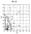
解析する段階210は、フィルタリングされたECGバッファ・スコアを所定の判断面と比較することによって始まる。CPR破損ノイズをもつECG信号データのデータベースを使って構築される判断面は、バッファ・スコアの所与の組が「VF」を示すか「未決」すなわちVF以外を示すかを定義する。CLASおよびFLATS次元での判断面の一例が図5に示されている。その例では、判断面510は、CLASスコアの一つとFLATSスコアの対応する諸対から構築される。判断面510内にはいるスコア対はVF条件を示す。判断面510の外部になるスコア対は未決条件を示す。より正確なVF判定を作り出すために、所望に応じて追加的なフィルタリングされたECGバッファについての閾値を使って、判断面の追加的な次元が加えられてもよい。ここでは二つの次元のみが示されているが、他のCLASスコアも含む判断面について三つ以上の次元が使われてもよい。
The parsing
解析する段階210は、VFかVF以外かを判定するために、特定の心臓信号特性を表わす二つ以上のバッファ・スコアを判断面と比較することによって進行する。図5に示した例については、CLAS/FLATSスコアの例示的な対が520に示されており、これはVFを指示する。判断面510の外、たとえば上および/または右にくる値対530は、未決、すなわちVF以外の条件を示す。
Analyzing
それぞれのもとの時間セグメント分割されたECGバッファは、このように、「ショックが助言される」、すなわちVFに対応するまたは「未決」、すなわち「VF以外」に対応するとして示されることができる。ひとたびECGバッファがショックが助言されるか未決として判定されると、ARTは捕捉する段階、得る段階、フィルタリングする段階および解析する段階を、「次のECGバッファを選択」段階212において示されるように、時間シーケンスにおける次のECGバッファについて繰り返す。繰り返すプロセスは、それぞれの新たなバッファをそれまでの諸バッファと組み合わせてVFの存否の全体的な連続的な判定を生成する追加的な方法を可能にする。
Each original time segmented ECG buffer can thus be shown as “shock advised”, ie corresponding to VF or “undecided”, ie corresponding to “non-VF” . Once the ECG buffer has been determined that a shock is advised or pending, ART will indicate the steps to capture, obtain, filter and analyze as shown in the “Select Next ECG Buffer”
上記の方法は、CPRの適用の間にショック決定を安全に行なうのに十分な正確さをもって、「手を放す」時間の間の解析のさらなる確認を必要とすることなく、VFを識別することが示されている。CPRで汚染されたECGの単一のバッファについてのVFに対するARTの感度は、70%を超えることが実証されている。すなわち、ARTは真のVFの生起を70%より多く検出する。同様に、ARTの特異性は、ECGの単一のバッファについて95%を超えることが実証されている。すなわち、「VF以外」の生起の95%より多くから、偽陽性VF指示を生成しない。 The above method identifies VF with sufficient accuracy to safely make a shock decision during the application of CPR, without the need for further confirmation of the analysis during the “release” time. It is shown. The sensitivity of ART to VF for a single buffer of ECG contaminated with CPR has been demonstrated to exceed 70%. That is, ART detects more than 70% of true VF occurrences. Similarly, the specificity of ART has been demonstrated to exceed 95% for a single buffer of ECG. That is, no false positive VF indication is generated from more than 95% of occurrences of “non-VF”.
「静かな」期間の間のART性能が既存のPASアルゴリズムで実証済みの性能に近づくことも注意しておいてもよいであろう。CPRアーチファクトで汚染されていないECGデータでのVFに対するARTの感度は、同様のデータに対するPASの約94%に比べ、80%を超える。「クリーンな」ECGのバッファでの偽VFに対するARTおよびPASの特異性はほぼ同一である。 It may also be noted that the ART performance during the “quiet” period approaches that demonstrated by existing PAS algorithms. The sensitivity of ART to VF in ECG data uncontaminated with CPR artifacts is over 80% compared to about 94% of PAS for similar data. The specificity of ART and PAS for pseudo-VF in a “clean” ECG buffer is nearly identical.
ここで図2bに目を転じると、方法が続いている。本方法のある好ましい実施形態は、上述した段階202〜212を、以下の数段落で述べる段階とは別個のDSPのようなプロセッサで実行されるものとして含む。そのような構成は、各ECGバッファが、ECG信号ストリームからの分類データのストリームのみを主として必要とするショック判断および制御プロセッサとは比較的独立に、順に解析され、VFまたは「未決」として分類されることを許容する。本方法のもう一つの好ましい実施形態は、処理の複数のコンポーネントへのさらなる分離を含む。たとえば、段階202におけるECG信号入力のデジタル化はASICのようなフロントエンド・チップにおいて扱われることができ、デジタル化されたECG信号ストリームをフィルタリングして複数の別個のフィルタリングされたストリームにするためにデジタル・ストリームはDSPに入力されることができる。次いで、さらにもう一つのプロセッサがそれらのフィルタリングされたストリームを、最終的な分類、判断および応答処理機能のために受領する。これらの機能について以下の段落で述べる。
Turning now to FIG. 2b, the method continues. One preferred embodiment of the method includes the steps 202-212 described above as being performed on a processor such as a DSP separate from the steps described in the following paragraphs. Such a configuration allows each ECG buffer to be parsed in turn and classified as VF or “pending”, relatively independent of the shock decision and control processor, which mainly requires only a stream of classified data from the ECG signal stream. Is allowed. Another preferred embodiment of the method includes further separation of processing into multiple components. For example, the digitization of the ECG signal input in
解析する段階210においてECGバッファからVFが判別される場合、すなわち「ショックが助言される」帰結の場合には、基礎になるECGリズムは一般に、ショック可能な心臓リズムであると想定される。だが、VF判別に対する最適な応答は、単に、電気療法を提供するよう基礎になる装置を準備することではないことがある。その代わり、進行中の心臓救助を不相応に乱さない何らかの仕方で、確認判定を得る、あるいは判定をユーザーに伝達することが好ましいことがある。こうして、別個の判断する段階214がこれらの目的のために保証され、図2bでは解析する段階210から入力を受けるものとして示されている。そのような状況の例は以下の段落において与えられる。
If the VF is determined from the ECG buffer in the analyzing
ARTは、数分の長さのCPR期間の間に複数のECGバッファを逐次的に解析するので、VFの進行中の患者条件に対する累積された感度は高まる。すなわち、真のVF条件を検出する可能性が高まる。だが、累積された特異性は低下することも予期される。すなわち、「未決」条件をVFと間違える可能性が高まる。この比較的長い時間期間にわたって全体的な方法の特異性を受け入れ可能なレベルに維持するために、時間的に連続的なECGデータ・バッファでのVF/未決判断からショック判断をするために最適な複数バッファ規則が開発されうる。のちの第二の所定の時間セグメントのECGバッファの繰り返される第二の解析する段階210が判断する段階214に提供される。すると、判断する段階214はその最終判断を、さらに第二の解析する段階にも基づかせる。
ART sequentially analyzes multiple ECG buffers during a few minutes long CPR period, thus increasing the cumulative sensitivity to ongoing patient conditions of VF. That is, the possibility of detecting a true VF condition increases. But the accumulated specificity is also expected to decline. That is, there is a higher possibility that the “undecided” condition is mistaken for VF. Ideal for making shock decisions from VF / pending decisions in temporally continuous ECG data buffers to maintain an overall method specificity at an acceptable level over this relatively long time period Multiple buffer rules can be developed. A second second analyzing
たとえば、解析する段階210は、三つの時間的に連続するECGバッファがVFを示す場合にのみ心臓リズムがショック可能であると判定してもよい。そうでない場合には、解析する段階はショック可能でないリズムを示す。これらの規則のもとで、ARTは、CPRの長い期間にわたる>95%の特異性を維持する一方、感度が>70%に留まることが示されている。いくつかの場合には、感度は95%を超えることができ、特異性は98%を超えることができる。そのような性能は、CPR期間の間にショック判断をするために受け入れ可能である。まとめると、判断する段階214は本質的には、VF/未決ECGバッファの進行するストリームを受け取るところ、段階214は基礎になる装置が除細動ショックの送達に動作可能に進むべきであるという最終決定のために前記規則を適用する。
For example, the analyzing
ディスプレイ上での視覚的グラフィックまたはテキスト・メッセージ、光信号または微妙な可聴信号のような、表示する段階215が、前記決定に際してすぐ開始されてもよい。好ましくは、表示する段階215は、装置が電気療法を送達するために完全に用意ができる前にすでに、ただしショック送達のために装置の準備ができるまではCPR圧迫を続けることからユーザーの気を散らさない邪魔にならない仕方で、提供される。他方、アーミングが完了するまでショック決定についてユーザーに全く情報を提供しないことが好ましいいくつかの動作モードがある。一部の一般ユーザーは、装置がショックを送達するために準備しつつあるという単なる指標でも、CPR圧迫を提供することから無用に気を散らされたり、驚いたりすることがある。
A
判断する段階214からのショック可能なリズムが存在し、電気療法が提供されるべきであるとの判定に応答して、アーミング段階216が始まる。アーミング(arming)段階216は、患者を除細動するために十分なエネルギーをもって高電圧充電回路を充電することからなっていてもよい。アーミング段階216は、アーミング段階が始まったという可聴なおよび/または視覚的なインジケーターを、ショック送達のために準備完了に向けての進行に関する何らかの指標とともに、含んでいてもよい(段階217)。たとえば、視覚的ディスプレイ700上での動的な棒グラフの印720が、高電圧回路の増大する充電状態に対応する棒グラフの進行する充填を示してもよい。ディスプレイ700上のテキスト・メッセージ710も充電が進行中であることを示していてもよい。充電状態ディスプレイ上に、進行インジケーターと同時に、ECG表示730が表示されてもよい。図7は、そのようなディスプレイ700のある例示的実施形態を示している。可聴進行インジケーターが、周波数が上昇し、完全充電状態に達したときに止まる連続トーンを含むことができる。
In response to a determination that there is a shockable rhythm from the determining
アーミング段階216の完了時には、電気療法装置はショックを送達するために完全に準備ができている。アーミング後、電気療法の送達のためにCPRを止めるようユーザー・プロンプト219を自動的に発する段階が行なわれることが好ましい。スピーカー830からの可聴プロンプト、点灯したまたは点滅するショック・ボタン・ライト820および/またはディスプレイ指標802が、ショック送達のためにCPRを止めるようユーザーに合図するために使われうる。ユーザー・インターフェース818上でのこれらのインジケーターの例について、図8を参照。AEDの場合、プロンプトは、ショックを送達するためにショック・ボタン892を押すようユーザーに指示してもよい。全自動除細動器の場合には、やはり段階219で、プロンプト発生後、すぐに自動的にショックが送達されうる。ユーザーが電気絶縁手袋または他のそのような保護ギアを用いている場合には、段階219における「CPRを止める」ようにとの促しは任意的には完全に省略されてもよい。
Upon completion of the
いくつかの状況では、段階219においてCPRを止めるようにとのユーザー・プロンプトを発するのを、ある最小量のCPRが提供されるまで遅らせることが望ましいことがある。たとえば、ショックを送達する前に少なくとも30秒の中断されないCPRを実施することが望ましいことがある。そのような最低限のCPR時間を保証するために、本発明の方法に任意的な遅延段階218が組み込まれてもよい。
In some situations, it may be desirable to delay the user prompt to stop CPR in
電気療法の送達後すぐに、ユーザーは、段階222においてCPRを再開するよう自動的に促されてもよい。装置は任意的に、段階220において、電気療法の送達を検出できるようにされていてもよい。送達の検出は、出て行く電流、ボタン押下などの感知によって得られる。次いで、本方法は、心臓救助の状態に応じて、捕捉する段階、得る段階、フィルタリングする段階および解析する段階に戻る。
Immediately after delivery of electrotherapy, the user may be automatically prompted to resume CPR at step 222. The device may optionally be adapted to detect the delivery of electrotherapy at
上記の方法段階は、CPRが、電気療法を送達する瞬間まで続けられ、その後すぐにCPRを再開することを許容する。その結果、心臓救助の間の「手で触れている」時間の割合が増大し、その結果、全体的な処置の有効性が改善される。「手を放して」のECG解析を待つアイドル時間を本質的になくすことができ、それによりCPRを休止すると実に素速く起こる血圧および血流の喪失を回避できる。これらの恩恵は、本方法がCPR期間中のVFへの復帰を処置できることとともに、実現できる。再細動が起こった場合、本方法は単にVFを検出し、進行中のCPR圧迫の途中で電気療法のための準備をする。 The method steps described above allow CPR to continue until the moment of delivering electrotherapy, and then resume CPR immediately. As a result, the percentage of “hand-in” time during cardiac rescue increases, resulting in improved overall treatment effectiveness. There is essentially no idle time waiting for a “go-to-go” ECG analysis, thereby avoiding the loss of blood pressure and blood flow that happens very quickly when CPR is paused. These benefits can be realized along with the method being able to treat the return to VF during CPR. If refibrillation occurs, the method simply detects VF and prepares for electrotherapy in the middle of ongoing CPR compression.
本発明の方法によって他の利点が与えられる。発明者は、ウェーブレットの代わりにフィルタを使うことは、VFについて解析するために必要とされる計算負荷をいくらか軽減し、電源線ノイズまたは同様の高周波数ノイズによる干渉をより効果的に抑制することを発見した。このように、上記方法段階の大半は、ECG信号ストリームを受領し、該ストリームを処理し、次いで連続的な、時間整列された、変換されたECGデータ・ストリームを出力するよう構成された単一のデジタル信号プロセッサ(DSP)において達成できる。DSPは、AEDにおいて最終的なショック判断および送達シーケンスを制御する第二のプロセッサと並列に動作することができる。また、一連のフィルタは、DCオフセット、50Hzおよび60Hzの外部電源線ノイズによって誘起される信号の、より堅牢な拒否をも提供するよう簡単に調整されることができる。 Other advantages are provided by the method of the present invention. Inventors use filters instead of wavelets to reduce some of the computational load required to analyze for VF and to more effectively suppress interference from power line noise or similar high frequency noise. I found Thus, most of the method steps described above are a single configured to receive an ECG signal stream, process the stream, and then output a continuous, time aligned, transformed ECG data stream. Of digital signal processors (DSPs). The DSP can operate in parallel with a second processor that controls the final shock decision and delivery sequence in the AED. The series of filters can also be easily adjusted to provide a more robust rejection of signals induced by DC offset, 50 Hz and 60 Hz external power line noise.
上記の方法は、体外式除細動器のような医療装置において実装できる。図6は、本発明の実施形態に基づく体外式除細動器10の機能的なブロック図である。除細動器10は、CPRを含む心臓救助の間に使うために意図されているAEDとして構成されている。それは、小さな物理的サイズ、軽い重量および高度なトレーニング・レベルのないまたはまれにしか除細動器10を使わない人員によって操作されることのできる比較的単純なユーザー・インターフェースのために設計される。本発明の本実施形態は、AEDでの応用に関して記述されるが、他の実施形態は異なる型の除細動器、たとえば手動除細動器、全自動除細動器およびパラメディックもしくは臨床除細動器/モニターにおける応用を含む。
The above method can be implemented in a medical device such as an external defibrillator. FIG. 6 is a functional block diagram of an
除細動器10は、たとえば患者に接続されている二つ以上の電極16からECG信号の入力12を受け取る。ECGフロントエンド回路14は、コネクタ・プラグおよびソケットなどを介して入力12と電気的に連通している。ECGフロントエンド回路14は、患者の心臓によって生成された電気的ECG信号を増幅し、バッファリングし、フィルタリングし、任意的にデジタル化して、デジタル化されたECGサンプルのストリームを生成するよう動作する。デジタル化されたECGサンプルはコントローラ30に提供される。コントローラ30は、DSPおよびARMプロセッサを組み合わせるプロセッサであってもよい。一つの例示的なコントローラは、テキサス・インスツルメンツ・インコーポレイテッド社によって製造されるアプリケーション・プロセッサのファミリーである。本装置のある実施形態では、DSPは、ARTプロトコルのもとでの先述したフィルタリングのすべてを実施し、次いで、フィルタリングされたECGデータの複数のストリームをARMプロセッサに渡す。ARMは、デジタル化されたECG信号データのストリームを、所定の時間に対応する諸セグメント(諸バッファ)にバッファリングする。ARMは、VF、ショック可能なVTまたは他のショック可能なリズムを検出するために、フィルタリングされたECGデータに対して転帰分析(outcomes analysis)を実行する。本発明によれば、ARMは、患者に最も有益な処置方式を決定するために転帰分析を使う。DSPおよびARMのこれらのコントローラ30部分は、上記の方法段階202ないし222において述べたECG解析器32として一緒に動作する。むろん、本発明の範囲は特定のDSP/ARM構成に限定されない。上記および下記の機能は、等価に、単一のプロセッサにおいて実装されるか、あるいは複数のプロセッサの間で分散されうる。
The
ECG解析器32は、約70%より大きい感度および約90%より大きい特異性をもってCPRに関係した信号ノイズ・アーチファクトが存在するときにショック可能なリズムを判別することができる解析アルゴリズムを組み込む。ECG解析器の精度は、CPR圧迫ノイズが存在するときに入力信号の心臓状態を安全かつ効果的に評価するのに十分である。一つのそのような解析アルゴリズムは先述したARTである。
The
ECG解析器32が、除細動ショックの必要性を示す、処置方式の上記決定と組み合わされたショック可能なリズムを判別する場合、プロセッサ34は、ECG解析器32の出力に応答して、HV(高電圧)充電回路60に、ショックを送達するための準備としてHVエネルギー蓄積源70を充電するよう信号を送る。HVエネルギー蓄積源70が完全に充電されたら、プロセッサ34は、CPR圧迫を与えるタスクから電気療法を送達するタスクにユーザーの注意を向け直すために、ユーザー・インターフェース818(図8)上のショック・ボタン92に、点滅を開始するよう指令する。
If the
より詳細に後述するように、プロセッサ34は、ショック可能な心臓リズムの検出に際してすぐに、すなわち連続動作モードで、除細動ショックのための前記準備を開始し、装置がアーミングされたらすぐに電気療法のためにCPR圧迫を中断するよう指示を発することができる。あるいはまた、プロセッサ34は、CPR圧迫の所定の期間の終わりに先立って除細動ショックのための準備を開始することができ、前記所定の期間の終わりと同時に電気療法の即時の送達を指示することができる。この後者のモードはスケジュール・モードと呼ばれる。
As will be described in more detail below, the
連続モードでもスケジュール・モードでも、プロセッサ34は、CPRを止め、除細動ショックを送達するためにショック・ボタンを押すよう聴覚的プロンプトを発するようユーザー・インターフェース18を制御する。これらのプロンプトは、CPRを止めることとショック・ボタンを押すことの間の遅延が最小にされるよう、一緒に、素速く発されるべきである。ユーザー・インターフェース18は同様に、プロセッサ34が除細動ショックが送達されたことを、たとえばボタン押下、HV蓄積回路からの電流などを感知することによって感知したらできるだけ早く、オーディオ・スピーカー20を介して、CPRを再開するよう聴覚的プロンプトを発するべきである。対応する視覚的プロンプトが、上記聴覚的プロンプトと同時に発されてもよい。
In both continuous and scheduled modes, the
ユーザーがユーザー・インターフェース818上のショック・ボタン92を押すとき、除細動ショックがHVエネルギー蓄積源70からショック送達回路80を通じて送達される。ある好ましい実施形態では、ショック送達回路80は、AEDの出力を介して、生のECG信号を受信するのと同じ電極16に電気的に接続される。
A defibrillation shock is delivered from the HV
プロセッサ34は、装置におけるユーザー・インターフェース(UI)出力機能の制御をも提供する。ユーザー・インターフェース18は、心臓救助プロトコルの進行を通じてユーザーを案内するための主要な手段であり、よって聴覚的な指示出力および視覚的ディスプレイの少なくとも一方を含む。特に、ユーザー・インターフェース18は、救助の状態、救助において取られるべき次のステップについての指示に関し、あるいは判別されたショック可能な心臓リズムに応じた指示に関して、ユーザーに聴覚的な言葉またはシグナルのプロンプトを発するためのオーディオ・スピーカー20を有していてもよい。ユーザー・インターフェース18は、ビーパー24を介して可聴な情報を伝達してもよい。ユーザー・インターフェース18はまた、ディスプレイ22上で視覚的テキストまたはグラフィックな表示を提供してもよい。ユーザー・インターフェース18、押すべきボタンまたはグラフィックに隣接して点灯しうる点滅光LED 26を介して視覚的な情報を提供してもよい。好ましくは、プロセッサ34は、これらの手がかりのそれぞれが、ユーザーの所望される応答を最適化する仕方で提供されるように、ユーザー・インターフェースを制御する。同じ情報に関する可聴な手がかりおよび視覚的な手がかりは、一方または他方の手がかりが所望される応答を損ないうる場合には、同時に発される必要はない。たとえば、プロセッサ34は、何らの命令も発する前に、HV蓄積源をアーミングされた状態に完全充電するよう充電回路を制御してもよい。あるいはまた、プロセッサ34は、スピーカー20上で関係する聴覚的な指示を発する前に、視覚的ディスプレイ22上でショック可能な心臓リズムの判別を示すようユーザー・インターフェースを駆動してもよい。再び図7を参照するに、プロセッサ34は、スピーカー20上で関係した聴覚的指示を発する前に、HV充電回路の状態を指示するようユーザー・インターフェースを駆動してもよい。
The
コントローラ30を動作させるためのソフトウェア命令が、オンボード・メモリ40に配置されている。不揮発性メモリ内の命令は、ARTアルゴリズムのためのアルゴリズム、PASのためのアルゴリズム、CPR圧迫を与えるための期間を含むCPR救助プロトコルのための指示、複数のユーザー種別のためのUI構成などを含んでいてもよい。揮発性メモリが、装置自己試験のソフトウェアで具現された記録、装置動作データおよび救助イベントのオーディオおよび視覚的記録を含んでいてもよい。
Software instructions for operating the
図6に示される除細動器の他の任意的な特徴は、さまざまなボタン(たとえば「電源投入」、「ショック」)から信号を受け取るシステム・モニタ・コントローラを含み、ビーパーおよびLED光のための信号を提供する。ボタンおよびセンサーの状態変化が、通信インターフェースを通じてプロセッサ34に伝送し返される。この特徴は、ボタン作動による覚醒感知および準備完了状態出力をもつ非常に低電力の待機動作を可能にする。
Other optional features of the defibrillator shown in FIG. 6 include a system monitor controller that receives signals from various buttons (eg, “power on”, “shock”) for beeper and LED light Provide a signal. Button and sensor state changes are transmitted back to the
図8は、図6の機能的なブロック図のユーザー・インターフェース18に概括的に対応するAED 800の外側表面上のユーザー・インターフェース818の構造的な実施形態を示している。ユーザー・インターフェース818は、心臓救助の状態に関するグラフィックなおよびテキストの情報を提供する視覚的ディスプレイ802を含んでいてもよい。ユーザー・インターフェース818は、聴覚的および可聴プロンプトを発するスピーカー830をも含んでいてもよい。LED 840が、準備完了または誤動作についての光に基づくシグナルを提供してもよい。ユーザー・インターフェース818は、救助の状態または装置の構成設定に依存して機能が変化する、第一、第二および第三の構成設定可能なボタン854、856、858をも含んでいてもよい。構成設定可能なボタンの機能はさらに、視覚的ディスプレイ802上に表示されるコンテキスト依存ラベル804、806、808によって示されてもよい。たとえば、装置が高度な動作モードのために構成設定されている場合には、ディスプレイ802は、隣接する構成設定可能なボタン854が「解析」ボタン94として構成設定されていることを示してもよい。解析ボタン94は、進行中の救助プロトコルを打ち切るよう動作してもよい。打ち切りは、CPR期間をただちに休止し、除細動器を、電気療法をすぐ送達するために準備する。解析ボタン94およびその機能の実施形態はのちにより詳細に述べる。
FIG. 8 illustrates a structural embodiment of the
本発明の好ましい実施形態は、CPR救助プロトコルにおいて動作する除細動器10を有する。該動作は、CPR圧迫を提供することと電気療法を送達することとの間の、装置によって引き起こされる遅延がなくされることを特徴とする。この結果を達成するために、CPR圧迫によって誘起される動きに関係した信号ノイズが存在するときでも不相応な偽警報なしにショック可能な心臓リズムを正確に判別できる、上記のようなECG解析アルゴリズムが組み込まれる。ARTはそのようなアルゴリズムである。ARTは、CPR圧迫が適用されている間の、ショック可能な心臓リズムのバックグラウンド検出、HV蓄積回路の充電および装置のアーミングを許容する。すると、除細動器は、CPR圧迫の休止と同時にショックを送達する準備ができている。
A preferred embodiment of the present invention has a
〈本発明の方法および装置によって可能にされる動作モード〉
上記のような除細動器は、いくつかの異なる動作モードのいずれかで構成設定されることができる。これら新規の動作モードは、本発明の解析方法の結果として可能になる。これらの動作モードは、本発明の装置において本発明の方法を採用するときに生じうるさまざまな新たな問題に対処する。
<Modes of operation enabled by the method and apparatus of the invention>
A defibrillator as described above can be configured in any of several different modes of operation. These new modes of operation are possible as a result of the analysis method of the present invention. These modes of operation address a variety of new issues that can arise when employing the method of the present invention in the apparatus of the present invention.
各動作モードは、除細動器メモリ40に事前にロードされてもよい。心臓救助の前に、装置の管理者またはユーザーが、装置セットアップの間に所望されるモードを選択することができる。特定のモードは、地元の救助プロトコルおよび/またはその場所の医療監督者の選好に従って選択される。
Each operating mode may be preloaded into the
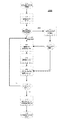
〈連続CPR動作モード〉
図9は、連続CPR救助動作モード900のある実施形態を示している。除細動器が連続モードに構成設定されているとき、ARTがVFを検出しプロセッサがショック決定をするときはいつも、そのプロセッサは常に除細動ショックを開始する。以下の記述のコンテキストでは、用語「連続」(continuous)は、ショック可能なリズムが検出されるときはいつも除細動療法をすぐ適用するということを意味するものとみなされる。この特定の動作モードは、「CPRを通じた解析カスタム」モードと称されてもよい。
<Continuous CPR operation mode>
FIG. 9 illustrates an embodiment of a continuous CPR
段階902で連続CPR救助動作モードにはいり、ここでARTアルゴリズムがECGバッファのストリームを評価することを開始する。CPR圧迫はこの時点で進行中であってもよいが、このモードに必須ではない。プロセッサは、段階904でショック判断を決定し、「ショックが助言される」条件を判別したら、プロセッサは、電気療法を送達するために除細動器の準備を開始する。よって、本方法は図2bの段階215ないし222で述べたのと同様に進行する。
Step 902 enters the continuous CPR rescue mode of operation, where the ART algorithm begins to evaluate the stream of ECG buffers. CPR compression may be ongoing at this point, but is not required for this mode. Once the processor determines a shock decision at
該判別に際してすぐに、ショック妥当表示段階915が開始されてもよい。たとえば、ディスプレイ上での視覚的なグラフィックまたはテキスト・メッセージ、光シグナルまたは微妙な可聴シグナルによる。好ましくは、ショック妥当表示段階915は、装置が電気療法を送達するための準備が完全にできる前に、ただし、装置がショック送達のために準備完了するまでは継続中のCPR圧迫からユーザーの注意を逸らさない邪魔にならない仕方で、設けられる。他方、アーミングが完了するまでショック決定についてユーザーに全く情報を与えないことが好ましいことがありうるいくつかの動作モードがある。一部の一般ユーザーは、装置がショックを送達するために準備しつつあるという単なる指標でも、CPR圧迫を提供することから無用に気を散らされたり、驚いたりすることがある。
Immediately upon the determination, the shock
判断する段階904からのショック可能な心臓リズムが存在し、電気療法が提供されるべきであるとの判定に応答して、アーミング段階916が始まる。アーミング(arming)段階916は、患者を除細動するために十分なエネルギーをもって高電圧充電回路を充電することからなっていてもよい。アーミング段階916は、アーミング段階が始まったという可聴なおよび/または視覚的なインジケーターを、ショック送達のための準備完了に向けての進行に関する何らかの指示とともに、アーミング進行表示段階917において含んでいてもよい。たとえば、視覚的ディスプレイ700上での動的な棒グラフの印720が、高電圧回路の増大する充電状態に対応する棒グラフの漸進的な充填を示してもよい。ディスプレイ700上のテキスト・メッセージ710も充電が進行中であることを示していてもよい。充電状態ディスプレイ上に、進行インジケーターと同時に、ECGディスプレイ730が表示されてもよい。図7は、そのようなディスプレイ700のある例示的実施形態を示している。
Responsive to determining that there is a shockable heart rhythm from the determining
アーミング段階916の完了時には、電気療法装置はショックを送達するために完全に準備ができている。アーミング完了後すぐに、電気療法の送達のためにCPRを止めるようユーザー・プロンプト919を自動的に発する段階が行なわれることが好ましい。スピーカー830からの可聴プロンプト、点灯したまたは点滅するショック・ボタン・ライト820および/またはディスプレイ指標802が、即時のショック送達のためにCPRを止めるようユーザーに合図するために使われうる。ユーザー・インターフェース818上でのこれらのインジケーターの例について、図8を参照。AEDの場合、プロンプトは、ショックを送達するためにショック・ボタン892を押すようユーザーに指示してもよい。全自動除細動器の場合には、やはり段階919で、プロンプト発生後すぐに自動的にショックが送達されうる。全自動AEDは、電極インピーダンス監視またはCPRに関係した信号ノイズ・アーチファクトの不在を判別する解析アルゴリズムの使用といった方法を使って、オペレーターが患者に触れていないときを判別して、自動的にしかるべくショックを送達してもよい。ユーザーが電気絶縁手袋または他のそのような保護ギアを用いている場合には、段階919における「CPRを止める」ようにとの促しは任意的には完全に省略されてもよい。
Upon completion of the
電気療法の送達後すぐに、ユーザーは、段階922においてCPRを再開するよう促されるべきである。手を放す時間を最小にするためである。装置は任意的に、段階920において、電気療法の送達を検出できるようにされていてもよい。送達の検出は、出て行く電流、ボタン押下などの感知によって得られる。
Immediately after delivery of electrotherapy, the user should be prompted to resume CPR at
ショック・セットの完了について検査する任意的な段階924が、段階922後、ショック判断段階904に戻る前に、実行されてもよい。ショック・セット(shock set)とは、連続CPR救助動作モードの一つの期間内に送達される所定の回数の電気療法ショックである。所定の回数は、その場所での選好に従って医療管理者によって設定されてもよい。ショック・セット内のショックの好ましい数は3である。
An
ショック・セット完了検査段階924が、ショック・セットが完了したと判定する場合、本方法は、終了段階926において、連続CPR救助動作モードを抜ける。そうでない場合には、本方法は、連続モード終了判断段階906に進む。
If the shock set
判断段階906は、連続動作モードが所定の時間に達したかどうかを判定する。所定の時間は、1分または2分であってもよく、あるいはその場所での選好に従って医療管理者によって他の所望される時間に設定されてもよい。その時間に達していれば、本方法は終了段階926において連続モードを抜ける。そうでない場合には、本方法は、次のECGバッファ(単数または複数)の継続した解析のためにショック判断段階904に戻る。ループは、ショック・セットが完了するか、連続モード期間が完了するかのどちらかまで続けられる。
患者が電気療法に応答する場合、あるいは電気療法を全く必要としない場合には、連続モードで動作するAEDは静かにバックグラウンドで解析を行ない、患者をチェックするまたはCPRを継続するよう適切な案内を定期的に提供する。AEDショック送達回路は不必要に充電されることは決してなく、よってバッテリー電力を節約し、動作時間を延ばす。このモードは、飛行中に非常に長い継続時間の心臓救助が時に経験される商業航空機での使用の際に、特に有益でありうる。 If the patient responds to electrotherapy or does not require any electrotherapy, the AED operating in continuous mode will silently analyze in the background and provide appropriate guidance to check the patient or continue CPR. Provide regularly. The AED shock delivery circuit will never be unnecessarily charged, thus conserving battery power and extending operating time. This mode can be particularly beneficial when used on commercial aircraft, where very long duration cardiac rescue is sometimes experienced during flight.
図12は、連続CPR救助動作モードの間に与えられる情報出力の図解を与えている。タイムライン1200は、心臓救助における時間を表わす横軸に沿って三つの行を含んでいる。上の行1210は装置の現在状態を示す。中間の行1220は、現在状態での装置によって発される可聴プロンプトを示す。下の行1230は、現在状態での装置ユーザー・インターフェース上に示されるディスプレイを示す。
FIG. 12 provides an illustration of the information output provided during the continuous CPR rescue operation mode.
配備状態1212での救助の始まりにおいて、電極がまだ配備されていないことがありうる。「パッドをあてがう」との可聴プロンプト1222および視覚的ディスプレイ1232が、この状態において同時に与えられることが好ましい。ユーザーに、この必要なアクションを実行することを強調して指示するためである。
At the beginning of the rescue in the deployed
電極が配備されたのち、装置は、ECG信号を受信していることを感知し、「CPR中の解析」状態1214にはいる。この状態では、効果的なCPRを提供することにおいてユーザーを支援するために、オーディオ指示およびタイミング信号1224が、任意的な表示情報1234とともに、与えられる。この時間の間、ECG解析器およびショック判別プロセッサは動作している。
After the electrodes are deployed, the device senses that an ECG signal is being received and enters the “analysis during CPR”
装置がショック可能な心臓リズムを検出する場合、状態は充電およびアーミング状態1216にはいる。だが、従来技術の装置とは異なり、本発明の装置は、ショックが助言されることおよび装置が療法を送達するために準備しつつあることの可聴な警告を全く与えないか、あるいはかすかに与えるだけである。その代わり、CPR状態1226でのCPR関係の指示が続く。この機能は、心臓救助に関してほとんど事前の経験のない一般ユーザーにとって特に有用である。ショックが助言されるという可聴プロンプトを控えることにより、装置は、ショックを受けることについて心配するかもしれない一般ユーザーが早まってCPR圧迫をやめることを防ぐ。代わりに、充電状態を示すために充電表示状態1236において、邪魔にならない表示メッセージが提供されてもよい。図12で見て取れるように、進行中のCPRおよび装置充電状態は、テキストでまたはグラフィックにまたは何らかの組み合わせにおいてそこに表示されうる。
If the device detects a shockable heart rhythm, the state is in the charge and arming
状態1217において装置がアーミングされてショックを送達する準備ができているときにのみ、「ショックを送達」可聴プロンプト1227において、ユーザーに対して可聴プロンプトが発される。このプロンプトと同時に、状態1240においてショック・ボタンが点灯または点滅して、ユーザーの注意をボタンに引きつける。「患者から離れて、今ショック・ボタンを押してください」のような可聴な指示がこの状態において伝達される。
An audible prompt is issued to the user at the “deliver shock” audible prompt 1227 only when the device is armed and ready to deliver a shock in
状態1217においてショック・ボタンが押されたのち、ショック後状態1218において、救助がすぐに再開される。「CPRを再開」するための可聴なプロンプト1228が、ユーザーに圧迫を再開するよう指示する1238での適切な表示とともに、ショック送達後、実際上可能な限り早く発される。次いで、別のショック可能なリズムが検出されるまで、あるいは検出される場合に、救助は状態1214にループで戻る。
After the shock button is pressed in
〈スケジュール動作モード〉
スケジュールCPR動作モードは、従来技術のAEDのユーザーにはおなじみのものに見えるが、実際には有意に異なる仕方で機能する。従来技術のAEDとは異なり、スケジュールCPR動作モードにおいて機能するAEDは、CPRの間にもECGを解析している。だがこのスケジュールCPR動作モードでは、AEDは、基礎になる感知された心臓リズムに関わりなく、CPRを中止するようプロンプトを発することを控える。所定の、中断されないCPR期間が行なわれた後にはじめて、装置はユーザーにCPRを止めてショックを送達するよう促す。AEDは、固定期間の終わりと同時に装置がショックを送達する準備ができているように、ショック可能なリズムの検出に際して、すぐに、あるいは期間の終わりの前の適切な時間に、電気療法のために装置を準備する。この準備は、CPR圧迫の間のノイズおよび混乱を減らすために、好ましくはバックグラウンドで行なわれる。以下の記述のコンテキストにおいて、用語「スケジュール(された)」は、たとえ所定の期間の間にショック可能なリズムが検出されたとしても該所定の期間の終わりまで除細動療法の適用を延期することを意味するものとみなされることができる。このモードは「CPRを通じた解析オン」とも称される。
<Schedule operation mode>
The scheduled CPR mode of operation looks familiar to users of prior art AEDs, but in practice it functions in a significantly different manner. Unlike prior art AEDs, AEDs that function in the scheduled CPR mode of operation also analyze ECG during CPR. But in this scheduled CPR mode of operation, the AED refrains from prompting to stop CPR regardless of the underlying sensed heart rhythm. Only after a predetermined, uninterrupted CPR period has occurred will the device prompt the user to stop CPR and deliver a shock. The AED is used for electrotherapy immediately upon detection of a shockable rhythm, or at an appropriate time before the end of the period, so that the device is ready to deliver a shock at the end of the fixed period. Prepare the equipment. This preparation is preferably done in the background to reduce noise and confusion during CPR compression. In the context of the following description, the term “scheduled” defers the application of defibrillation therapy until the end of the predetermined period even if a shockable rhythm is detected during the predetermined period Can be taken to mean. This mode is also referred to as “analysis on via CPR”.
図10は、スケジュールCPR救助動作モード1000のある実施形態を示している。除細動器がスケジュール・モードで構成設定されているとき、そのプロセッサは、ARTがVFを検出し、プロセッサがショック判断をしたのち、除細動ショックの開始を遅らせる。電気療法を送達するための装置のアーミングは、中断できないCPRの所定の期間の終わり近くまで遅らされる。
FIG. 10 illustrates an embodiment of a scheduled CPR
段階1002でスケジュールCPR救助動作モードにはいり、ここで先述したように、ARTアルゴリズムがECGバッファのストリームを評価することを開始した。AEDはこの時点では、CPR圧迫を加えるよう視覚的および聴覚的ユーザー・プロンプトをユーザー・インターフェースを介して提供していてもよいが、この初期条件はこのモードに必須ではない。
ECGバッファのART評価は、該ART評価からなされるショック判断とは区別されうる。たとえば、このスケジュールCPR救助モードでは、段階1002における「未決」または「ショックが助言される」の個々のECGバッファ評価は、スケジュール・モード期間の最後の部分まで、療法送達目的のために度外視されてもよい。あるいはまた、これらの評価は累積されて、のちに、判断をするための期間において使われてもよい。
The ECG buffer ART assessment can be distinguished from shock decisions made from the ART assessment. For example, in this scheduled CPR rescue mode, the “undecided” or “shock advised” individual ECG buffer assessments in
プロセッサは、段階1004においてショック判断を決定する。段階1004が「ショックが助言される」条件を判別したら、プロセッサは、電気療法を送達するために除細動器を準備するプロセスを開始する。 The processor determines a shock decision at step 1004. Once step 1004 determines a “shock advised” condition, the processor begins the process of preparing the defibrillator to deliver electrotherapy.
該判別に際してすぐに、ショック妥当表示段階1015が開始されてもよい。たとえば、ディスプレイ上での視覚的なグラフィックまたはテキスト・メッセージ、光シグナルまたは非常にかすかな可聴シグナルによる。好ましくは、ショック妥当表示段階1015は、装置が電気療法を送達するための準備が完全にできる前に、ただし、装置がショック送達のために準備完了するまでは継続中のCPR圧迫からユーザーの注意を逸らさない邪魔にならない仕方で、設けられる。他方、アーミングが完了するまでショック決定についてユーザーに全く情報を与えないことが好ましいことがありうるいくつかの動作モードがある。一部の一般ユーザーは、装置がショックを送達するために準備しつつあるという単なる指標でも、CPR圧迫を提供することから無用に気を散らされたり、驚いたりすることがあるからである。
Immediately upon the determination, the shock
判断する段階1004からのショック可能な心臓リズムが存在し、電気療法が提供されるべきであるとの判定に応答して、アーミング段階1016が始まる。アーミング(arming)段階1016は、患者を除細動するために十分なエネルギーをもって高電圧充電回路を充電することからなっていてもよい。アーミング段階1016は、アーミング段階が始まったという可聴なおよび/または視覚的なインジケーターを、ショック送達のための準備完了に向けての進行に関する何らかの指標とともに、アーミング進行表示段階1017において含んでいてもよい。たとえば、視覚的ディスプレイ700上での動的な棒グラフの印720が、高電圧回路の増大する充電状態に対応する棒グラフの漸進的な充填を示してもよい。ディスプレイ700上のテキスト・メッセージ710も充電が進行中であることを示していてもよい。充電状態ディスプレイ上に、進行インジケーターと同時に、ECGディスプレイ730が表示されてもよい。図7は、そのようなディスプレイ700のある例示的実施形態を示している。
Responsive to determining that there is a shockable heart rhythm from the determining step 1004 and electrotherapy should be provided, the
アーミング段階1016の開始は、CPRの前記所定の、中断されない期間の終わり近くに装置が完全にアーミングされた状態に達するようなタイミングにされてもよい。これは、意図しないショックがCPR圧迫の提供者に与えられる可能性を減らす。アーミングがいつ始まるかによらず、アーミング段階1016の完了時には、電気療法装置はショックを送達するために完全に準備ができており、その時点で前記指示を発する。
The start of the
アーミング後に遅延段階1018が完了されるべきである。遅延段階1018は、電気療法の何らかの可能な送達の前に、完全な、中断されないCPR期間があることを保証する、スケジュール・モードにはいってからの所定の時間期間である。所定の時間は1分または2分であってもよく、あるいはその場所での選好に従って医療管理者によって任意の所望される時間に設定されてもよい。ある好ましい時間期間は2分だが、30秒以上の範囲内でありうる。
The
遅延段階1018の完了後、電気療法の送達のためにCPRを止めるようユーザー・プロンプト1019を自動的に発する段階が行なわれる。スピーカー830からの可聴プロンプト、点灯したまたは点滅するショック・ボタン・ライト820および/またはディスプレイ指標802が、ショック送達のためにCPRを止めるようユーザーに合図するために使われうる。ユーザー・インターフェース818上でのこれらのインジケーターの例について、図8を参照。AEDの場合、プロンプトは、ショックを送達するためにショック・ボタン892を押すようユーザーに指示してもよい。全自動除細動器の場合には、やはり段階1019で、プロンプト発生後すぐに自動的にショックが送達されてもよい。ユーザーが電気絶縁手袋または他のそのような保護ギアを用いている場合には、段階1019における「CPRを止める」ようにとの促しは任意的には完全に省略されてもよい。
After completion of the
電気療法の送達後すぐに、ユーザーは、段階1022においてCPRを再開するよう促されるべきである。手を放す時間を最小にするためである。装置は任意的に、段階1020において、電気療法の送達を検出できるようにされていてもよい。送達の検出は、出て行く電流、ボタン押下などの感知によって得られる。段階1020は、段階1022において再開プロンプトを生成するために用いられてもよい。他方、段階1020が療法の期待される送達がないことを検出する場合には、装置は、プロンプトを繰り返すことによって、あるいはショックがまだ送達されておらずすぐにCPRが再開されるべきであることを示す異なるプロンプト(図示せず)を発することによって、応答することができる。次いで、段階1026において、方法はスケジュールCPR救助動作モードを抜ける。
Immediately after delivery of electrotherapy, the user should be prompted to resume CPR at
ARTアルゴリズムがECGが未決であると判定する場合には、判断段階1004および終了判定段階1006によって形成されるループにおいて、ショックが助言されるとの判断を求めて、相続くECGバッファを評価することを続ける。終了判定段階1006は単に、解析に戻る前に、中断されないCPRの前記所定の期間が完了したかどうかを判定する。段階1006が該期間が完了したと判定すれば、本方法は段階1026においてスケジュールCPR救助動作モードを抜ける。段階1006における中断されないCPRの所定の期間は、段階1018における期間と同じであっても、それより短い継続時間であってもよい。
If the ART algorithm determines that the ECG is pending, evaluate the successive ECG buffers for the determination that a shock is advised in the loop formed by the decision stage 1004 and the
スケジュール・モードについての上記の方法により、電気療法に応答する、あるいは電気療法を全く必要としない患者については、スケジュール・モードで動作するAEDは静かにバックグラウンドで解析を行ない、CPRを継続するよう適切な案内を定期的に提供する。AEDショック送達回路は不必要に充電されることは決してなく、よってバッテリー電力を節約し、動作時間を延ばす。このモードも、商業航空機での使用の際に、特に有益でありうる。 Using the above method for schedule mode, for patients who respond to electrotherapy or do not require electrotherapy at all, AEDs operating in schedule mode should be silently analyzed in the background and continue CPR. Provide appropriate guidance regularly. The AED shock delivery circuit will never be unnecessarily charged, thus conserving battery power and extending operating time. This mode can also be particularly beneficial when used on commercial aircraft.
既存の心臓救助プロトコルは、CPR完了後に少なくとも、短い確認解析およびHV充電時間を必要とする。従来技術装置において必要になるCPRとショックとの間の遅延がなければ、スケジュール・モードAEDはより効果的な処置を提供する。スケジュール動作モードの諸段階は、図2bの段階214〜222の繰り返されるサイクルと見ることができる。図2aの諸解析段階は常にバックグラウンドで行なわれる。発されるユーザー・プロンプト段階219は、CPR圧迫が連続的な所定の固定時間期間にわたってCPR圧迫が提供されるまで、常に遅延段階218において遅らされる。
Existing cardiac rescue protocols require at least a short confirmation analysis and HV charging time after completion of CPR. If there is no delay between CPR and shock required in prior art devices, the schedule mode AED provides a more effective treatment. The stages of the schedule operating mode can be viewed as a repeated cycle of stages 214-222 in FIG. 2b. The analysis steps of FIG. 2a are always performed in the background. The user
スケジュール・モードのAEDは、できるだけ迅速にVF条件を処置することに比べて心臓救助において中断されないCPRの高い割合に重きを置く医療管理者にとって望ましいことがありうる。CPRの固定された期間は、救助の間の一貫したルーチンに重きを置き、たとえば疲労を防止するために救助の間に役割を交代する対応者にもよく知られる。しかしながら、一貫したルーチンは、再細動を起こす患者に対する電気療法を遅らせる可能性があるという代償を伴う。 Scheduled mode AEDs may be desirable for medical managers who focus on a high proportion of CPR that is not interrupted in cardiac rescue compared to treating VF conditions as quickly as possible. The fixed period of CPR emphasizes a consistent routine during rescue and is well known to responders who change roles during rescue to prevent fatigue, for example. However, a consistent routine comes at the price of potentially delaying electrotherapy for patients with refibrillation.
スケジュール・モードでは、AEDは、CPRルーチンの一貫性および「フロー」を維持するために、視覚的指示とは異なる仕方で聴覚的指示および通知を発してもよい。AEDはたとえば、ショック判断および充電状態を視覚的にのみ伝達してもよい。よって、救助者は、気を散らす「ショック」という単語を含みうる可聴プロンプトによって無用に注意を逸らされることがない。CPR期間の終わりが近づいてはじめて、AEDは、ショック可能な条件が検出され、電気療法の送達準備ができているという案内を発してもよい。次いで、CPR期間の終わりに、AEDは「SPRを止めて今ショックを送達してください」という聴覚的および視覚的指示を発し、同時にショック・ボタン892を点滅させてもよい。
In the scheduled mode, the AED may emit audible instructions and notifications in a manner different from the visual instructions in order to maintain CPR routine consistency and “flow”. The AED may, for example, communicate the shock determination and state of charge only visually. Thus, the rescuer is not unnecessarily distracted by audible prompts that may include the distracting word “shock”. Only when the end of the CPR period is approaching, the AED may provide an indication that a shockable condition has been detected and is ready for electrotherapy delivery. Then, at the end of the CPR period, the AED may give an audible and visual indication "Stop SPR and deliver the shock now" and simultaneously flash the
図13は、スケジュールCPR救助動作モードの間に与えられる情報出力の図解を与えている。タイムライン1300は、心臓救助における時間を表わす横軸に沿って三つの行を含んでいる。上の行1310は装置の現在状態を示す。中間の行1320は、現在状態での装置によって発される可聴プロンプトを示す。下の行1330は、現在状態での装置ユーザー・インターフェース上に示されるディスプレイを示す。 FIG. 13 provides an illustration of the information output provided during the scheduled CPR rescue operation mode. Timeline 1300 includes three rows along the horizontal axis representing time in cardiac rescue. The top row 1310 shows the current state of the device. Middle row 1320 shows an audible prompt issued by the device in the current state. The bottom row 1330 shows the display shown on the device user interface in the current state.
スケジュールCPR救助動作モードでの救助状態ならびに可聴および視覚的プロンプトは、概括的に連続モードについて図12で上記した同様の要素に対応する。だが、スケジュールCPR救助モードの性質に合う、一つの有意な装置がある。装置が充電およびアーミング状態1216においてショックが送達されるべきであると判定し、その後送達のために準備する場合、中断できないCPR期間1350が満了するまでは、ショックが送達されるべきであることを示すさらなる可聴または表示されるプロンプトは与えられない。期間1350の始まりは、状態1214におけるCPRのそのセッションの始まりと一致し、2分など所定の時間、継続してもよい。中断できないCPR期間1350の満了後にはじめて、装置は、状態1217においてショックを送達するために可聴および視覚的プロンプトを発しはじめる。
Rescue status and audible and visual prompts in the scheduled CPR rescue mode of operation generally correspond to similar elements described above in FIG. 12 for the continuous mode. But there is one significant device that fits the nature of the scheduled CPR rescue mode. If the device determines that a shock should be delivered in the charge and arming
〈諸ショック・セットを用いた組み合わされた連続モードとスケジュール・モード〉
AEDは、心臓救助の過程を通じてCPR圧迫に対する電気療法機会の割合を変えるプロトコルにおいて、連続モードとスケジュール・モードを組み合わせてもよい。プロトコルに対する患者の応答が、異なる動作モードへのシフトに影響してもよい。たとえば、患者が電気療法に応答しない場合には、連続モードで動作しているAEDは、十分な中断されないCPR圧迫時間を許容していないことがあり、よってAEDはその代わりに自動的にスケジュール・モードにシフトしてもよい。患者が繰り返し再細動を経験する場合には、当該条件をより迅速に処置するためにAEDが連続動作モードを維持するまたは連続動作モードに切り換えることが望ましいことがある。
<Combined continuous mode and schedule mode using various shock sets>
AED may combine continuous and schedule modes in a protocol that changes the rate of electrotherapy opportunities for CPR compression throughout the course of cardiac rescue. Patient response to the protocol may affect the shift to different modes of operation. For example, if a patient does not respond to electrotherapy, an AED operating in continuous mode may not allow sufficient uninterrupted CPR compression time, so the AED will automatically You may shift to the mode. If the patient experiences repeated refibrillation, it may be desirable for the AED to maintain or switch to a continuous mode of operation to treat the condition more quickly.
諸ショック・セットを用いた組み合わされたCPR救助プロトコル1100動作方法が図11に記載されている。CPR適用の間に電気療法を提供するための組み合わされた方法は、連続CPR救助プロトコルの間に送達される所定回数のショックの完了後に、連続CPR救助プロトコルからスケジュールCPR救助プロトコルにプロトコルを自動的にシフトさせる段階1107を含む。みな単一の連続CPR救助プロトコル期間内に送達されるショックの所定のグループは、ショック・セットと呼ばれる。この組み合わされたモードの方法は、ある種の条件が満たされた後にスケジュール・モードから連続モードに自動復帰することを含んでいてもよい。
A combined
組み合わされたモードは、エントリー段階1102で始まる。これは一般に、二つ以上の外部電極、プロセッサ、ユーザー・インターフェースおよびショック送達回路を有する除細動器を提供することを含むと解される。エントリー段階1102は、装置が配備され作動され、その電極が患者に取り付けられるときに始まる。除細動器は、ユーザーにより操作されるショック・ボタンをもつ半自動AEDの一つであってもよく、あるいは電気療法の自動送達を有する全自動AEDであってもよい。
The combined mode begins at
AEDは、段階1102において最初に作動されたときにいくつかのスタートアップ・プロトコルまたは動作モードの一つを提供するよう構成されていてもよい。スタートアップ・プロトコルは、ECG解析がすぐに実施される「ショック優先」プロトコルであってもよい。ショック可能なリズムが存在する場合には、除細動器は即座のショックのためにアーミングする。電気療法が送達された後、装置はその救助プロトコルを進める。あるいはまた、スタートアップ・プロトコルまたは動作モードは、基礎になるECGリズムに関わりなく、中断できないCPRの初期期間を通じてAEDがユーザーを案内する「CPR優先」であってもよい。この第二のCPR優先のスタートアップ・プロトコルは、初期化CPRモード段階1104において示されている。段階1104では、先述した装置ユーザー・インターフェースを介して、CPR圧迫を加えるよう、ユーザー・プロンプトが自動的に発される。
The AED may be configured to provide one of several startup protocols or modes of operation when first activated in
ユーザーが段階1104のプロンプトに適正に従ってCPR圧迫を加える場合、電極から装置によって受領されるECG信号は、CPR圧迫ノイズ・アーチファクトからの破損によって特徴付けられる。ARTのような上述したアルゴリズムがこの受領されたECG信号を解析して、ショック可能な心臓リズムが存在するかどうかを判断する。
If the user applies CPR compression according to the prompt in
初期化段階1104は任意的に、装置がCPR圧迫提供以外の何らかの案内を提供するまでに、所定の時間期間、あるいは感知された圧迫の等価な数を含んでいてもよい。何らかの電気療法の送達の前には、20から30秒の間または30回の圧迫といった短い初期期間が一部の患者に対して有益であると考えられる。初期化段階1104を抜けると初期ECGショック判断段階1106に進む。
The
初期ECGショック判断段階1106も、初期化段階1104に関係している任意的な段階である。段階1106は、複数のCPR救助モードのうちのどれが次に使用されるかを決定しうる初期ショック判断を提供する。たとえば、段階1106での初期ショック判断が「未決」である場合、さらなる何らかの電気療法の前にCPR圧迫の通常の固定した継続時間を開始することが好ましいことがありうる。この方法段階は、図11では、スケジュールCPR救助プロトコル段階1000に進む破線で示されている。だが、段階1106における初期ショック判断が「ショックが助言される」である場合には、本方法は、段階900によって示されるように、直接、連続CPR救助プロトコルに進む。
The initial ECG
組み合わされた方法1100は段階900において続けられる。ここで、装置は連続CPR救助動作モードで動作することを開始する。本方法は、連続モードについて先述したのと同様に動作する。ここで、解析する段階においてショック可能な心臓リズムの判断に応答して、プロセッサは、電気療法を送達するためにショック送達回路をアーミングし、送達のためにCPRを止めるようユーザー・インターフェースを介してすぐに指示を発する。そして先述したように、連続モード方法段階900は、ショック送達回路が当該段階900内で送達される所定回数のショックの所定の電気療法ショック・セットを完了した後、自動的に終了する。あるいはまた、先述したように、段階900は、解析する段階においてショック可能な心臓リズムの判別がないことが所定の時間にわたって持続した場合に終了する。こうして、前記所定の時間またはショック送達回路が前記所定回数の電気療法ショックを送達することのうちの早いほうに応答して、終了が発生する。そして先述したように、代替的な終了は、感知された所定回数のCPR圧迫に応答して発生してもよい。終了後、方法1100は、自動シフト段階1107において、自動的に、連続モードでの動作から、段階1000でのスケジュールCPR救助動作モードでの動作にシフトする。
The combined
方法1100は、段階1000において、先述したスケジュール動作モードに従って動作する。ここで、解析する段階におけるショック可能な心臓リズムの判断に応答して、装置プロセッサは、電気療法を送達するためにショック送達回路をアーミングする。中断できないCPRの所定の期間が経過した後、プロセッサは、送達のためにCPRを止めるようユーザー・インターフェースを介して指示を発する。所定の期間の完了後、スケジュール・モード1000は終了して、ショック・セット完了判断段階1108に進む。
The
方法1100は、先の段階900で完了したショック・セットの累積回数を追跡している。この回数は必ずしも、段階900において連続モードに出入りした回数とは対応しない。段階900は、ショック・セットの完了ではなく所定の時間期間の満了に起因して終了することもあるからである。終了が満了によって引き起こされる場合には、たとえば、段階900内のショック・カウンタがリセットされる。こうして、連続モードが始まるたびに、終了するためには、もう一つのフルのショック・セットまたは前記所定の時間の満了が必要になる。
The
ショック・セット完了判断段階1108は、スケジュールCPRプロトコルから抜けた後に方法1100が連続CPR救助プロトコルに復帰するか否かを制御する。所定数のショック・セットが完了していない限り、復帰が行なわれる。これは、ショック・セットの完了に起因して連続モード段階900を終了した回数に対応する。復帰する場合、段階900および1000が繰り返される。段階1108によって可能にされるサイクルは、所定数のショック・セットが完了するまで繰り返される。ショック・セットの好ましい数は3であり、1ないし7の範囲でありうる。
The shock set
連続モードとスケジュール・モードの間でのこのサイクルは、再細動を起こす患者のように、救助の早期において迅速な電気療法を必要とする患者に有益である。だがこのサイクルは、シーケンスにおいて後にはショックとショックの間に中断されないフルのCPR期間を設ける心臓救助シーケンスに発展することをも可能にする。こうして、迅速な電気療法に応答しなかった再細動患者は、完全な期間のCPRを受け始める。 This cycle between continuous mode and schedule mode is beneficial for patients who require rapid electrotherapy early in the rescue, such as patients who undergo refibrillation. But this cycle also allows the sequence to evolve into a cardiac rescue sequence that provides a full CPR period that is not subsequently interrupted between shocks. Thus, refibrillation patients who have not responded to rapid electrotherapy begin to receive a full period of CPR.
ショック・セットの所定回数が完了した場合には、中断段階1108はさらなる復帰を中断する。本方法は代わりに、段階1110において終末(terminal)スケジュールCPR救助プロトコルに進む。段階1110では、すべてのその後の電気療法ショックは、中断できないCPRの区間と区間の間、すなわち中断できないCPRの各所定の期間後にのみ生起する。CPR救助が完了したとき、方法1100は、終了段階1126において抜けることによって終了する。この段階は、オン・オフ・ボタンにおいて手動で装置をオフにすることによって開始されてもよい。
If the predetermined number of shock sets has been completed, the
〈連続法およびスケジュール法をインターリーブするための装置〉
上記の図6および図8に示されるAEDのような装置は、CPRを電気療法とインターリーブするために上記の諸方法のいずれかに従って動作してもよい。AEDは好ましくは、ECG信号入力12、ユーザー・インターフェース18、ECG解析器32およびメモリ40と通信して心臓救助の実施においてユーザーに指示案内を提供するプロセッサ34によって制御される。
<Device for interleaving continuous method and schedule method>
A device such as the AED shown in FIGS. 6 and 8 above may operate according to any of the methods described above to interleave CPR with electrotherapy. The AED is preferably controlled by a
プロセッサ34は特に、AEDを、従来技術のシーケンスよりも患者にとって有益な、連続CPR救助動作モードおよびスケジュールCPR救助動作モードのシーケンスにおいて動作させる。連続CPR救助動作モードで動作しているとき、ECG解析器がショック可能な心臓リズムを判断する場合、プロセッサは、電気療法を送達するためにショック送達回路をアーミングし、次いですぐに、送達のためにCPRを止めるようユーザー・インターフェースを介して指示を発する。AEDプロセッサは、「手を放す」時間を最小にするために、電気療法の送達を感知したらすぐに、CPRを再開するようユーザー・インターフェースを介してすぐ指示を発する。スケジュールCPR救助動作モードで動作しているとき、ECG解析器がショック可能な心臓リズムを判断する場合、プロセッサは電気療法を送達するためにショック送達回路をアーミングする。このアーミングは、該判断に際してすぐ行なわれるか、あるいは該期間の終わりに完全にアーミングされるのが間に合うよう充電を開始する。2分など、中断できないCPRの所定の期間後、プロセッサは、送達のためにCPRを止めるようユーザー・インターフェースを介して指示を発する。
In particular, the
プロセッサ34は、ショック送達回路が所定の電気療法ショック・セットを完了することにも応答し、その後、プロセッサは連続CPR救助動作モードからスケジュールCPR救助動作モードに自動的にシフトする。
The
AEDは、それぞれの電気療法ショック・セットが、連続CPR救助動作モードの単一のインスタンス内に送達される所定数のショックを含むよう構成される。ある好ましい実施形態では、AEDは、各ショック・セットにおいて2ないし5回のショックを設定するようプログラム可能であってもよい。 The AED is configured such that each electrotherapy shock set includes a predetermined number of shocks delivered within a single instance of a continuous CPR rescue mode of operation. In certain preferred embodiments, the AED may be programmable to set 2 to 5 shocks in each shock set.
プロセッサ34はさらに、スケジュールCPR動作モードの一つまたは複数のインスタンス後に、AED動作モードを自動的にスケジュール・モードから連続モードに復帰させるよう動作可能であってもよい。こうして、連続モードとスケジュール・モードの間で巡回するモードのシーケンスが確立できる。ある好ましいプロトコルは、ショック送達回路が所定数のショック・セットを完了した後、プロセッサがさらなる復帰を中止するというものである。その場合、AEDはスケジュール・モードに留まり、CPRの区間と区間の間にのみ電気療法ショックを提供する。ある好ましい実施形態では、AEDは、1ないし7のショック・セットが完了した後、さらなる復帰を中止するようプログラム可能であってもよい。AEDは、ショック・セットの数を無限大に設定するようプログラム可能であってもよく、その場合、サイクルは装置がオフにされるまで続けられる。
The
AEDプロセッサ動作の任意的な実施形態は、「未決」判定が所定の時間にわたって持続する場合に、プロセッサが、自動的に連続CPR救助プロトコルからスケジュールCPR救助プロトコルにシフトするというものである。この動作は、一般に、図11に示されるように段階1104、1106のようなAED動作の始まり近くで生起する。そのような判定が持続するのでない場合、プロセッサは、上記の方法に従って連続モードからスケジュール・モードにシフトする。
An optional embodiment of AED processor operation is that the processor automatically shifts from a continuous CPR rescue protocol to a scheduled CPR rescue protocol if the “pending” decision persists for a predetermined time. This operation generally occurs near the beginning of an AED operation such as
AEDのもう一つの実施形態は、経過時間の代わりにCPR圧迫の感知された回数パラメータを使う。CPR圧迫の感知された回数は、一つまたは複数の源から得られてもよい。電極ノイズ・アーチファクト信号または共通モード電流(CMC: common mode current)が使われてもよい。米国マサチューセッツ州アンドーヴァーのフィリップス・エレクトロニクス・ノースアメリカによって製造されるQ-CPR装置のような外部CPR感知装置または他の同様のセンサーが使われてもよい。 Another embodiment of AED uses a perceived frequency parameter of CPR compression instead of elapsed time. The perceived number of CPR compressions may be obtained from one or more sources. Electrode noise artifact signals or common mode current (CMC) may be used. An external CPR sensing device such as a Q-CPR device manufactured by Philips Electronics North America, Andover, Massachusetts, USA or other similar sensors may be used.
上記のようなAEDおよびその動作は、半自動装置または全自動装置において具現されうる。半自動AEDはもちろん、ユーザーが操作するショック・ボタン92を含み、よって適宜該ショック・ボタンを押すよう対応する指示および表示を含むべきである。全自動AEDは、ショック・ボタンに関するものは何も含まないが、切迫したショックについてユーザーに明瞭に通知し、必要ならユーザーに患者から離れたままでいるよう指示する、わずかに異なる指示のセットを具現することになる。 The AED and its operation as described above can be implemented in a semi-automatic device or a fully automatic device. The semi-automatic AED should, of course, include a shock button 92 that is operated by the user, and thus should include a corresponding instruction and display to press the shock button as appropriate. Fully automatic AED does not include anything related to shock buttons, but embodies a slightly different set of instructions that clearly notify the user of an imminent shock and instruct the user to stay away from the patient if necessary Will do.
〈ARTおよびPASのような二つのECG解析アルゴリズムを使う方法〉
本発明者は、たいていの患者は心停止救急の際、ショック可能なリズムを決してもたず、よってどんなECG解析アルゴリズムも長い時間期間にわたって「ショックが助言される」判定を与えることなく動作することがあることを認識するに至った。だが、本発明者は、上述したARTアルゴリズムはPASほどは、ショック可能な心臓リズムの検出に敏感ではないことをも認識している。よって、ARTはCPRの際に、「真の陽性の」ショック可能なリズムを見逃す可能性がより高い。また、ARTの「未決」(undecided)判定は、「ショックが助言されない」(NSA: no shock advised)と「不確定」(indeterminate)ECGとの間の区別をしない。これらの理由により、CPR圧迫の期間中、ECG解析を異なるECGアルゴリズムを用いて定期的に確認することが重要になることがある。
<Method using two ECG analysis algorithms such as ART and PAS>
The inventor found that most patients never have a shockable rhythm during a cardiac arrest emergency, so that any ECG analysis algorithm works without giving a “shock advised” decision over a long period of time. It came to recognize that there is. However, the present inventor has also recognized that the ART algorithm described above is not as sensitive to the detection of shockable heart rhythm as PAS. Thus, ART is more likely to miss a “true positive” shockable rhythm during CPR. Also, ART's “undecided” determination does not distinguish between “no shock advised” (NSA) and “indeterminate” ECG. For these reasons, it may be important to regularly check ECG analysis using different ECG algorithms during CPR compression.
この問題への一つの解決策は、単に、救助の間定期的にPAS確認解析を使う。だがこの解決策は、全体的な手を放す時間を不必要に増大させうるので、最適ではない。そこで、本発明者は、確認のためにPASが使用されることができるが、できるだけ低頻度で、手を放す時間が患者に対して最小限の害をもつ状況でのみ、使われるべきであることを認識するに至った。そのような状況は、たとえば、CPR圧迫の終わりまたはCPR圧迫のための他の仕方でスケジュールされた期間においてでありうる。 One solution to this problem is simply to use PAS validation analysis periodically during rescue. But this solution is not optimal because it can unnecessarily increase the overall time to let go. Thus, the present inventor can use PAS for confirmation, but should be used as often as possible and only in situations where the time to let go has minimal harm to the patient. I came to recognize that. Such a situation can be, for example, at the end of CPR compression or at a time period otherwise scheduled for CPR compression.
図14は、確認解析のためにCPR圧迫を無用に中断することによって呈される問題を軽減するそのような方法解決策を示している。図14は、図11と同様である。だが図14は、第一のECG解析アルゴリズムおよび第二のECG解析アルゴリズムの両方を使うよう修正される。第一のECG解析アルゴリズムは、先述したARTアルゴリズム200によって例示される。これは、CPRに関係した信号ノイズ・アーチファクトが存在するときに使うために特に好適である。第二のECG解析アルゴリズムは、既存のPASアルゴリズムによって例示される。これは、CPRに関係した信号ノイズ・アーチファクトがないときに使うのに特に好適である。
FIG. 14 illustrates such a method solution that alleviates the problems presented by interrupting CPR compression unnecessarily for confirmation analysis. FIG. 14 is similar to FIG. However, FIG. 14 is modified to use both the first ECG analysis algorithm and the second ECG analysis algorithm. The first ECG analysis algorithm is exemplified by the
図11の方法と同様に、図14の図解は、CPRの適用中に電気療法を提供するための方法1400を含む。本方法は、ECG信号入力12、ショック送達回路80およびユーザー・インターフェース18を有する除細動器1において段階1102において有効にされる。本装置および方法はまた、二つの異なるECG解析アルゴリズムを利用する。ARTのような第一のアルゴリズムは、CPRに関係した信号ノイズ・アーチファクトが存在する間にECG信号から「ショックが助言される」(SA: shock advised)および「未決」の一方を判別するよう動作可能である。PASのような第二のアルゴリズムは、CPRに関係した信号ノイズ・アーチファクトがないときにECG信号からSAおよび「ショックが助言されない」(NSA: No Shock Advised)の一方をより特定的に判別するよう動作可能である。除細動器は段階1102において、電極のようなECG信号入力12が取り付けられ、よってECG解析を開始する準備ができていることを感知する。
Similar to the method of FIG. 11, the illustration of FIG. 14 includes a
図14は、ショック可能な心臓リズムが存在するかどうかを判定するために第一の期間中に第一のECG解析アルゴリズムを用いてECG信号を解析することによって段階1104に進む。この期間中、除細動器がスケジュールCPR救助動作モードにおいてCPR案内指示を提供していることが好ましい。SA判定の場合、除細動器は、段階1104の終わりにショックを送達するよう準備する。さらに、本方法は、ECG信号がSAまたは「未決」判定のどちらを示すかに基づく判断段階1406に進む。判定のための好ましい点は、第一の期間の終わりであるが、判定は該期間にわたる諸SAの平均またはカウントなどに基づくこともできる。段階1102および1104の他の側面は図11に関してすでに上記している。
FIG. 14 proceeds to step 1104 by analyzing the ECG signal using a first ECG analysis algorithm during a first time period to determine if there is a shockable heart rhythm. During this period, the defibrillator preferably provides CPR guidance instructions in the scheduled CPR rescue operation mode. For SA determination, the defibrillator prepares to deliver a shock at the end of
判断段階1406の第一の期間の間にSAが判別される場合、CPR救助手順の残りの段階も図11の方法で述べたものに対応する。特に、SA判定後、連続CPR 900およびスケジュールCPR 1000からなる第二の、相続く諸期間の間に、第一のART ECG解析アルゴリズムを使って心臓リズムが判定される。その後のSA判定は、除細動器に、CPR期間の型に応じて、ショックのためにアーミングさせ、CPR/ショック送達指示を発させる。先述したように、連続CPR動作モードからスケジュールCPR動作モードにシフトするためにショック・セットが用いられてもよい。こうして、除細動器から、最適化されたカスタマイズされた救助プロトコルが出力される。
If SA is determined during the first period of decision stage 1406, the remaining stages of the CPR rescue procedure also correspond to those described in the method of FIG. In particular, after the SA determination, cardiac rhythm is determined using a first ART ECG analysis algorithm during a second, successive period of
段階1406がSA判定以外の何らかの判定をする場合にのみ、第二のECG解析アルゴリズムが用いられる。段階1406において「未決」判定が発生する場合、本方法は自動的に、段階1407において、第一から第二のアルゴリズムに切り換わる。
The second ECG analysis algorithm is used only if stage 1406 makes some determination other than SA determination. If a “pending” determination occurs at step 1406, the method automatically switches from the first to the second algorithm at
切り換え段階1407後、本方法は、PAS判断段階1410においてECG信号を解析するために第二のECG解析アルゴリズム(PAS)を用いる。好ましくは、除細動器はこの段階において、PASアルゴリズムが低ノイズ環境で効果的に解析できるよう、「CPRを止めてください」および/または「患者に触れないでください」のようなユーザー・プロンプトを発する。PAS判断段階1410の二つの可能な帰結は、SAまたは「ショックが助言されない」(NSA)である。PASは「アーチファクト」判断を発してもよいが、これは本発明のためのトピックではなく、これ以上は論じない。
After the
PAS判断段階1410におけるSAの判定は、ECGはイベントの始まりまたは始まり付近において、すなわち段階1102において、ショック可能なリズムとして現われたが、第一のアルゴリズムがそれを感知し損なったことを示す。この段階でのSA判定には好ましくは、すぐにアーミングし、電気療法を送達することが続く。
The SA determination at
SAを呈するECGリズムが、救助のより早期におけるより多くの電気療法から裨益しうることを示唆する証拠がある。よって、段階1410におけるPASによるSA判定は、ショック可能なリズムの検出後に迅速に電気療法を送達する連続CPR救助プロトコル900において第一のECG解析アルゴリズムに戻る自動切り換えをも引き起こす。次いで、連続CPR救助プロトコル900は先述したように機能する。
There is evidence to suggest that ECG rhythms that present SA can benefit from more electrotherapy in the earlier stages of rescue. Thus, the SA determination by PAS in
だが、段階1410におけるNSAの判定は、呈されたECGがショック可能ではないことを示す。そのような患者は、救助における早期のより多くのCPR圧迫から裨益しうる。よって、段階1410におけるPASによるNSA判定は、より大きな相対量のCPR時間を与えるスケジュールPCR救助プロトコル1000において第一のECG解析アルゴリズムに戻る自動切り換えを引き起こす。次いで、スケジュールCPR救助プロトコル1000および心臓救助方法の残りは先述したように機能する。
However, the NSA determination at
第二のECG解析アルゴリズムPASが動作する各期間の継続時間ができるだけ短いことが好ましい。その解析の間は救助者が「手を放す」という最適でない要件のためである。典型的なPAS解析期間は、約10秒未満であるが、たった4秒であってもよい。この継続時間は、たいていの場合、第一のARTアルゴリズムを使うCPRの連続モードまたはスケジュール・モードの継続時間よりも短い。PAS期間の頻度も、同じ理由によりできるだけ低いことが好ましい。そこで、方法段階は、そうすることが必要であるときにのみ「手を放す」PAS解析への切り換えを要求する。 The duration of each period in which the second ECG analysis algorithm PAS operates is preferably as short as possible. This is due to the sub-optimal requirement that the rescuer “release his hands” during the analysis. A typical PAS analysis period is less than about 10 seconds, but may be as little as 4 seconds. This duration is often shorter than the duration of CPR continuous mode or schedule mode using the first ART algorithm. The frequency of the PAS period is preferably as low as possible for the same reason. Thus, the method step requires a switch to “release” PAS analysis only when it is necessary to do so.
本発明の方法の代替的な、より詳細なビューが図15に示されている。図15の方法は、CPRの初期ARTアルゴリズム期間1504後でも、CPRに対する中断を最小限にしてどのように電気療法が与えられるかをより明瞭に示している。段階1102で除細動器を作動させ、電極をあてがったのち、段階1504における初期化期間がすぐにはじまる。これは、CPR圧迫を加えるためのプロンプトを発し、最初のECG解析アルゴリズムを使うことを含む。段階1504は好ましくは、ARTリズム判別に関わりなく中断できないCPRをもつスケジュールされたCPR救助動作モードである。段階1504は一層好ましくは、約20〜30秒、あるいは最小約30回のCPR胸部圧迫を加えるのに十分な時間という、比較的短い継続時間である。本方法は、その回数の胸部圧迫またはその時間の長さを感知してもよく、その後、判断段階1506においてARTリズム判別が最終化される。こうして、段階1504は、すべての患者に、救助の開始における中断されない胸部圧迫のいくらかの期間の恩恵を与える。
An alternative, more detailed view of the method of the present invention is shown in FIG. The method of FIG. 15 shows more clearly how electrotherapy is delivered even after the initial
判断段階1506においてSAが示される場合には、本方法はすぐに、療法ショックの送達を許容する電気療法段階1507のためのアーミングにはいる。段階1507に続いて、本方法は、第二の期間、連続CPRモードの救助プロトコル900にはいり、これは先述したように進行する。第二の期間900の継続時間は約2分であってもよいが、装置作動に先立って構成設定可能であってもよい。次いで、本方法は連続モード終末(terminal)判断段階1509に進む。
If SA is indicated in
段階1509においてSA判定が存在すれば、本方法は図11の方法について先述したように進行する。本方法は、療法ショックの送達を許容する電気療法段階1511のためのアーミングにはいる。次いで、第一のARTアルゴリズムでの動作モードは自動的に、段階1000でのスケジュールCPR救助動作モードに切り換わる。段階1000は、バックグラウンドでECGリズムを解析しつつCPR指示をもってユーザーにプロンプトし、SA判別からのいかなるアクションをも当該期間の終わりまで延期することによって、先述したように進行する。スケジュールされた期間1000は約2分の継続時間であってもよい。
If there is an SA determination at
段階1000の終わりに、すなわち判断段階1519においてSA判別が存在する場合、本方法は、療法ショックの送達を許容する電気療法段階1521のためのアーミングにはいる。ショックが送達されたのち、検査段階1108において諸ショック・セットがまだ完了していなければ、本方法は連続モード段階900にループで戻ってもよい。諸ショック・セットが完了であれば、本方法は、段階1110の終末(terminal)スケジュールCPR救助プロトコルに切り換わり、段階1126での救助の終わりまでそこに留まる。
If there is an SA discrimination at the end of
図14および図15によって、第一のARTアルゴリズムによってSA状態が判別されることができる限り、本方法は第二のPAS解析の必要なしに進行することが見て取れる。こうして、本方法は、PASのために必要とされる「手を放す」時間を最小にする。 14 and 15, it can be seen that as long as the SA state can be determined by the first ART algorithm, the method proceeds without the need for a second PAS analysis. Thus, the method minimizes the “release” time required for PAS.
しかしながら、第一のARTアルゴリズムがその代わりに判断段階1506、1509、1519のいずれかにおいて「未決」状態を判別する場合には、本方法はそれぞれの段階1520、1530、1540において自動的にさらなる判定のために第二のPASアルゴリズムに切り換わる。段階1520、1530、1540は「手を放す」指示を発し、次いで、ECGを解析する。「手を放す」時間を最小にするために、これらのPAS期間は、10秒以下の短い継続時間であってもよい。
However, if the first ART algorithm instead determines a “pending” state at any of decision stages 1506, 1509, 1519, the method automatically makes further decisions at each
図15で見て取れるように、PAS解析判定のいずれかがSAであれば、判断段階1522、1532、1542はすぐに本方法を、それぞれの出発点において、すなわち段階1506、段階1509または段階1519後のところで、第一のARTアルゴリズム・シーケンスに復帰させる。この経路の理由は、一般にPAS解析よりもART解析のほうが、全体的な手を放す時間の短縮のため、好ましいということである。こうして、本方法は可能なときはいつでもARTに戻る切り換えをする。
As can be seen in FIG. 15, if any of the PAS analysis decisions is SA, decision steps 1522, 1532, 1542 immediately use the method at each starting point, ie after
また、図15に見られるように、PAS解析判定のいずれかがNSAであれば、本方法は自動的に、段階1000でスケジュールCPR動作モードで動作している第一のARTアルゴリズムに戻るシフトをする。この経路の理由は、ECGがショック可能でないリズムを呈していることをPASが確認したので、そのような患者については、中断されないCPRの期間がより有益であるということである。
Also, as seen in FIG. 15, if any of the PAS analysis decisions are NSA, the method automatically shifts back to the first ART algorithm operating in the scheduled CPR mode of operation at
NSAという任意のPAS判定後の任意的な段階1523が、現在のショック・セットを完了されたとして設定する。このように、この任意的な段階は本方法1500を、段階1110での終末の(terminal)恒久的な(permanent)スケジュールCPR救助動作モードへの段階1108におけるシフトに近づける。この理由は、スケジュール・モードでのショックに対するCPRの、より高い割合への結果的なシフトが、救助の早期にどこかでNSA ECGリズムを示す患者には、より有益であることがありうるという発明者の発見である。
An
〈連続動作モードおよびスケジュール動作モードでのPASおよびARTアルゴリズムをインターリーブする装置〉
上記の図6および図8に示されるAEDのような装置は、二つの異なるECG解析アルゴリズムを組み込みつつCPRを電気療法とインターリーブする上記の諸方法のいずれかに従って動作しうる。AEDは好ましくは、プロセッサ34およびECG解析器32を含むコントローラ30によって制御される。コントローラ30は、心臓救助の実施においてユーザーに指示案内を提供するために、ECG信号入力12、ユーザー・インターフェース18およびメモリ40と通信してもよい。コントローラ309は、電気療法出力を与えるショック送達回路80とも制御通信している。
<Device that interleaves PAS and ART algorithms in continuous operation mode and schedule operation mode>
A device such as the AED shown in FIGS. 6 and 8 above may operate according to any of the above methods for interleaving CPR with electrotherapy while incorporating two different ECG analysis algorithms. The AED is preferably controlled by a
メモリ40は、入力からのCPRに関係した信号ノイズ・アーチファクトが存在するときにECG信号から「ショックが助言される」(SA)および「未決」の判定の一方を決定するよう動作可能である第一のECG解析アルゴリズムと、入力からのCPRに関係した信号ノイズ・アーチファクトがないときにECG信号からSAおよび「ショックが助言されない」(NSA)の判定の一方を決定するよう動作可能である第二のECG解析アルゴリズムとの両方に関係した命令を記憶している。メモリ40は、CPR圧迫を提供するための少なくとも二つの期間を含むCPR救助プロトコルに関係した命令をも記憶している。
The
コントローラ30は特に、先述したような連続CPR救助動作モードおよびスケジュールCPR救助動作モードのシーケンスにおいてAEDを動作させる。さらに、コントローラ30は、ユーザー・インターフェース18を介して案内を発し、第一および第二のECG解析アルゴリズムのいずれかからのSA判定に応答して電気療法を送達するためにショック送達回路80を自動的に準備する。最後に、第一のECG解析アルゴリズムは、CPR関係ノイズがある期間の間はショック可能なECGリズムに対するより低い感度をもつことがあるので、コントローラ30はさらに、第一のECG解析アルゴリズムかSA判定以外の何らかの判定を決定する期間の一つの終わりにおいて、常に第一から第二のECG解析アルゴリズムに自動的に切り換えるようさらに動作可能である。こうして、心停止患者のために最適でない「手を放す」時間を要求する第二のECG解析アルゴリズムの使用は、必要なときにのみ使用される。
In particular, the
AEDの他の装置挙動側面は、先述した諸方法を反映する。たとえば、第一から第二のECGアルゴリズムに切り換えたのちにSAが判定される場合には、AEDコントローラは自動的に第一のアルゴリズムに、連続CPR救助動作モードに戻る切り換えをしてもよい。他方、第一から第二のECG解析アルゴリズムに自動的に切り換えたのちにNSAが判定される場合には、AEDコントローラは自動的に第一のアルゴリズムに、スケジュールCPR救助動作モードに戻る切り換えをしてもよい。 Other device behavior aspects of AED reflect the methods described above. For example, if SA is determined after switching from the first to the second ECG algorithm, the AED controller may automatically switch back to the continuous CPR rescue operation mode to the first algorithm. On the other hand, if NSA is determined after automatically switching from the first to the second ECG analysis algorithm, the AED controller automatically switches back to the scheduled CPR rescue operation mode to the first algorithm. May be.
第二のECG解析アルゴリズムは、10秒未満でECGリズムを特徴付けることのできるPASアルゴリズムであってもよい。よって、PASが動作する各期間の継続時間はそれより長くないべきである。 The second ECG analysis algorithm may be a PAS algorithm that can characterize the ECG rhythm in less than 10 seconds. Therefore, the duration of each period in which the PAS operates should not be longer than that.
AEDは、AEDを作動させた直後に生起する初期化期間を機能的に含んでいてもよく、ECG信号が受領される。初期化期間は、第一のECG解析アルゴリズムを使うスケジュールCPR救助動作モードを含む。スケジュールCPR救助動作モードは、判定に関わりなく中断されないCPRの所定の期間を提供する。初期化期間の長さは、その後の救助プロトコル期間に比べて比較的短くてもよい。たとえば、初期化期間は、感知されたある回数のCPR圧迫で終了してもよい。ここで、感知される回数は約30であってもよく、既存のCPRプロトコルは30秒未満に完了するであろう。あるいはまた、初期化期間は、約20から30秒の間の継続時間にあらかじめ決定されていてもよい。 The AED may functionally include an initialization period that occurs immediately after activating the AED, and an ECG signal is received. The initialization period includes a scheduled CPR rescue operation mode that uses the first ECG analysis algorithm. The scheduled CPR rescue mode of operation provides a predetermined period of CPR that is uninterrupted regardless of determination. The length of the initialization period may be relatively short compared to the subsequent rescue protocol period. For example, the initialization period may end with a sensed number of CPR compressions. Here, the number of times perceived may be about 30, and the existing CPR protocol will be completed in less than 30 seconds. Alternatively, the initialization period may be predetermined at a duration between about 20 to 30 seconds.
上記のようなAEDおよびその動作は、半自動装置または全自動装置のいずれで具現されてもよい。半自動AEDはもちろん、ユーザーが操作するショック・ボタン92を含み、よって適宜ショック・ボタンを押すようにという対応する指示および表示を含むべきである。全自動AEDは、ショック・ボタンに関するものは何も含まないが、切迫したショックについてユーザーに明瞭に通知し、必要ならユーザーに患者から離れたままでいるよう指示する、わずかに異なる指示のセットを具現することになる。 The AED and its operation as described above may be implemented by either a semi-automatic device or a fully automatic device. The semi-automatic AED should, of course, include a shock button 92 that is operated by the user and thus should include a corresponding instruction and display to press the shock button accordingly. Fully automatic AED does not include anything related to shock buttons, but embodies a slightly different set of instructions that clearly notify the user of an imminent shock and instruct the user to stay away from the patient if necessary Will do.
〈CPRを打ち切るための解析ボタン〉
経験のあるユーザーが、迅速に別の動作モードにはいるため、特により迅速に除細動ショックを与えるために、進行中のAEDプロトコルを打ち切ることを所望しうる状況がありうる。本発明は、そうするための単一のボタンを提供することによってかかる打ち切りアクションを簡単にする。AEDは、基礎になるECG解析に基づいて、患者に最も有益な、ボタン押下への応答を自動的に選択する。
<Analyze button to abort CPR>
There may be situations where an experienced user may want to abort an ongoing AED protocol, particularly in order to more quickly deliver a defibrillation shock, as it enters another mode of operation. The present invention simplifies such an abort action by providing a single button for doing so. AED automatically selects the response to a button press that is most beneficial to the patient based on the underlying ECG analysis.
除細動器(AED)および除細動器を使うための方法は、異なる除細動器関係の機能をすぐに実行するために、進行中のおよび他の仕方で中断できないCPR圧迫を打ち切る、ユーザーが作動させるボタンを組み込んでいる。打ち切りボタンは、先述したスケジュールCRP救助動作モードにおいて特に有用である。こうして、動作はユーザーにとってより簡単であり、プロトコルに追随する誤りの可能性を減らし、イベントの間の混乱によって引き起こされる遅延を最小にする。 Defibrillators (AEDs) and methods for using defibrillators break down ongoing and otherwise uninterrupted CPR compressions to quickly perform different defibrillator-related functions, Incorporates a user activated button. The abort button is particularly useful in the aforementioned scheduled CRP rescue operation mode. Thus, the operation is simpler for the user, reducing the possibility of errors following the protocol and minimizing the delay caused by confusion between events.
打ち切り機能をもつある例示的AEDは、ショック可能な心臓リズムに対する異なる感度をもつ二つの異なるECG解析アルゴリズムを使ってもよい。打ち切りボタンの押下は、第一のECG解析アルゴリズムから、より高い感度をもつ第二のECG解析アルゴリズムに自動的にシフトしてもよい。ボタンは、基礎になるショック可能な心臓リズムがすでに検出されている場合には、電気療法のための即座の準備のために、進行中の解析およびCPRの打ち切りをも許容してもよい。 An exemplary AED with censoring capability may use two different ECG analysis algorithms with different sensitivities to shockable heart rhythms. Pressing the abort button may automatically shift from the first ECG analysis algorithm to a second ECG analysis algorithm with higher sensitivity. The button may also allow ongoing analysis and CPR censorship for immediate preparation for electrotherapy if the underlying shockable heart rhythm is already detected.
本AEDおよび方法は、たとえ打ち切りボタンが作動させられるときでも、CPR圧迫期間と電気療法の間の手を放す時間を減らす。例として、基礎になるECG解析がショック可能なリズムを示す場合、AEDは、打ち切りボタン上で「充電」または「解析」を示しつつ、バックグラウンドで療法のために充電しつつあることがありうる。こうして、ユーザーが打ち切りボタンを押す場合、AEDはすぐに電気療法を送達するための準備ができていることがありうる。 The AED and method reduce the time to release hands between the CPR compression period and electrotherapy even when the abort button is activated. As an example, if the underlying ECG analysis shows a shockable rhythm, the AED may be charging for therapy in the background, showing “charge” or “analysis” on the abort button . Thus, if the user presses the abort button, the AED may be ready to deliver electrotherapy immediately.
先述したような制御機能をもつAEDは、打ち切りボタンの感知される押下に対するその応答を、患者ECGの現在状態に基づいて変化させる。AEDが基礎になるECGがショック可能であると判定する場合、AEDは、そのボタンについてのコンテキスト依存ラベルを「充電」に変えてもよい。AEDが打ち切りボタンが押されたことを感知するとき、AEDはすぐに除細動ショックのために充電する。ECGがショック可能でない場合には、ボタン・ラベルはその代わりに「解析」と見えてもよい。同じボタンを押すことで、AEDは、既存の状態を確認するためにすぐに第一のARTアルゴリズムから第二のPASアルゴリズムに切り換える。あるいはまた、感知された打ち切りボタンの押下は、すぐに、現在のARTアルゴリズム解析の感度を高めるために、AEDに、「患者から離れていてください」というプロンプトを発させてもよい。 An AED having a control function as described above changes its response to a sensed press of the abort button based on the current state of the patient ECG. If the AED determines that the underlying ECG is shockable, the AED may change the context sensitive label for that button to “Charge”. When the AED senses that the abort button has been pressed, the AED immediately charges for a defibrillation shock. If the ECG is not shockable, the button label may instead appear as “Analyze”. By pressing the same button, the AED immediately switches from the first ART algorithm to the second PAS algorithm to confirm the existing state. Alternatively, a sensed abort button press may immediately prompt the AED to “stay away from the patient” to increase the sensitivity of the current ART algorithm analysis.
図16に示された方法段階は、図17のaないしdに示される具体的な視覚的表示および打ち切りボタンをも定期的に参照することによって、よりよく理解されうる。図17のa、b、c、dは、視覚的ディスプレイ802のようなAED視覚的ディスプレイに対応するさまざまなグラフィック表示1706、1714、1718、1728を示している。図17のa〜dのそれぞれは、共通の一般的配置を共有する。一つまたは複数の案内および情報メッセージが上のバナー領域に表示される。CPR進行バーのような進行バーが上のバナー領域に隣接して位置されてもよい。表示の中心には、進行中のECGトレース、あるいは電極やCPRのための胸部に置く手を配置するための案内グラフィックを示すための領域がある。表示の下の部分は好ましくは、コンテキスト依存ラベル804、806によって例示される、除細動器の特定の動作状態および基礎になるECG解析に基づいて変化しうるコンテキスト依存ラベルを含む。
The method steps shown in FIG. 16 can be better understood by periodically referring also to the specific visual indications and censoring buttons shown in FIGS. 17a, b, c, d show various
ある好ましい実施形態では、入力ボタン854、856は、表示1706、1714、1718、1728にすぐ隣接して、それぞれコンテキスト依存ラベル804、706の隣に配される。ある代替的な実施形態では、視覚的ディスプレイ802はタッチ感応性ディスプレイであってもよく、入力ボタン854、856が事実上、それぞれのコンテキスト依存ラベル804、806の下になる。
In one preferred embodiment,
ここで図16のフローチャートに目を転じると、即座の電気療法を提供する目的のためにCPRを打ち切るための例示的な方法1600が示されている。本方法は、本方法を実行するよう協調してはたらく諸機能をもつ除細動器の提供段階1602で始まる。つまり、除細動器は、ECG信号入力12、入力ボタン854および視覚的ディスプレイ802を含むユーザー・インターフェース818、ショック送達回路80および入力からのCPR関係の信号ノイズ・アーチファクトが存在するときのECG信号からショック可能な心臓リズムを判別するよう動作可能である第一のECG解析アルゴリズムを含む。AED 800のような除細動器は、提供される装置のほんの一例である。
Turning now to the flowchart of FIG. 16, an
AED 800は、使用に先立って事前構成設定されるいくつかの異なる動作モード構成設定を含んでいてもよい。これらのモードのいずれかまたは全部が、AEDメモリ40内に維持されていてもよい。例示的な動作モードは、高度モード、CPR優先モードおよび半自動モードである。各動作モードは、打ち切りボタンが現われうる状況についてわずかに異なることがある。
The
高度モードは、AEDがECG解析アルゴリズムリズム解析およびショック送達のためのアーミングをいつ開始するかに対し、対応者に、より多くの制御を許すプロトコルである。高度モードはたとえば、「解析」および/または「充電」オプション・ボタンを、プロトコルの間の特定の期間において提供するよう構成されていてもよい。「解析」オプション・ボタンを押すと、PASを用いて即座の手を放した解析を開始してもよい。「充電」ボタンを押すことは、手を放した解析、高電圧エネルギー蓄積源70の充電およびショック送達の一つまたは複数を許してもよい。
Advanced mode is a protocol that allows responders more control over when AED initiates the ECG analysis algorithm rhythm analysis and arming for shock delivery. The advanced mode may be configured, for example, to provide “analyze” and / or “charge” option buttons at specific times during the protocol. Pressing the “Analyze” option button may initiate an immediate analysis using PAS. Pressing the “charge” button may allow one or more of a let go analysis, charging of the high voltage
AED 800が作動し、ECG信号入力12を受領することを開始した後、AEDは、任意的な初期期間1604の間に第一のECG解析アルゴリズム(ART)を用いてECG信号を解析することを開始する。初期期間1604は、好ましくは、先述した、中断できないCPR圧迫動作モードで動作する段階1104と同様である。だが、この短い初期期間1604ののち、すぐにCPR圧迫を終えてECG解析に進むためまたは電気療法のためのアーミングのために打ち切りボタンがアクティブになってもよい。期間1604において打ち切りボタンを有効にする理由は、AEDの到達および作動に先立って十分なCPRが提供されていることを操作者が認識する状況を許容するためである。
After the
初期期間1604の間の視覚的ディスプレイは好ましくは、図17のaに示したように「解析‐未決」画面1706に対応する。AEDは、打ち切りボタン854に隣接して「解析」のコンテキスト依存ラベルを表示する。操作者が解析のために初期圧迫期間を打ち切ることを望む場合、打ち切りボタン854を押す。AEDがボタン押下を感知すると、すぐに「患者から離れていてください」というユーザー・プロンプトを発し、第二のPAS ECG解析アルゴリズムを使ってECG解析を開始する。この時間の間、AEDは図17のcからの「解析‐離れている」画面1728を表示してもよい。
The visual display during the initial period 1604 preferably corresponds to an “analysis-pending”
ラベル段階1606が任意的な段階1604に続く。ラベル段階1606は、ART解析期間1608の開始において前に解析されたECGに対応する初期のコンテキスト依存ラベルを設定する。好ましくは、AEDは、プロトコルにおける次の諸段階を確立するために、「解析‐未決」画面1706を表示する。
A
ラベル段階1606に第一の解析期間段階1608が続く。段階1608は、装置が、第一の(ART)ECG解析アルゴリズムを用いて、好ましくはCPRの中断できないスケジュール・モードで、ECG信号を解析することを含む。このように、段階1608は、除細動器が、CPR圧迫を継続するようにとの可聴および/または視覚的なプロンプトを発することを含む。この期間の間の解析されたECG信号は、判断段階1610に示されるように「未決」または「ショックが助言される」〔ショック妥当〕のいずれかである。この第一の解析期間にはまた、除細動器コントローラ30は、入力ボタン854の作動をモニタリングすることを開始する。
The
図16で見て取れるように、方法1600における次の諸段階は、基礎になる解析されたECG信号に依存する。段階1608、1610における判断が「ショックが助言される」である場合、方法の左分枝が進む。AEDは、表示段階1612において、表示1706の上の部分を、指令テキストを示すよう変更してもよい。図17のbの「打ち切り利用可能‐ショック可能なリズム」画面1714に示されるような、「ショックが助言される」のような情報メッセージおよび/または「解析ボタンを押してください」の案内メッセージが現われてもよい。あるいはまた、図17のdの「打ち切り利用可能‐充電」画面1718に示されるような、「充電ボタンを押してください」の案内メッセージが現われてもよい。この段階1612における可聴な案内も可能であるが、単なる視覚的な案内ほど好ましくはない。CPR圧迫を与えるというタスクから不相応に気を散らすことを防ぐためである。あるいはまた、段階1612は、打ち切りボタンがアクティブであるという可聴な指示を発することを含んでいてもよい。
As can be seen in FIG. 16, the next steps in the
段階1610における「ショックが助言される」判断は、コンテキスト依存ラベル変更段階1614においてコンテキスト依存ラベル804の変更を開始してもよい。コンテキスト依存ラベル804は、「解析」表示から「充電」表示に変更されてもよい。あるいはまた、「充電」コンテキスト依存ラベル/ボタン組み合わせが、図17のbに見られるように、第二の構成設定可能なボタン856に隣接する第二のコンテキスト依存ラベル806において「解析」表示とともに表示されることができる。次いで、AEDは、バックグラウンド充電段階1616においてHVエネルギー蓄積源70のバックグラウンド充電を開始してもよい。
The “shock advised” determination at
解析されたECG信号がショック可能なリズムを示すと、AEDは、感知段階1618において打ち切りボタンの作動をモニタリングする。作動が発生しなければ、方法1600は、当該期間の間の継続したモニタリングのために、単に解析段階1608にループで戻る。
If the analyzed ECG signal indicates a shockable rhythm, the AED monitors the operation of the abort button in the
感知段階1618においてAEDが打ち切りボタン作動を感知する場合、AEDはすぐにアーミングされたショック送達状態に進む。HVエネルギー蓄積源70の充電は、必要なら充電段階1620において完了され、ショック・ボタンはアーミング段階1622においてアーミングされる。ユーザーを案内し、ユーザーに情報提供するために、その過程で、適切な視覚的および可聴なプロンプトが与えられる。
If the AED senses an abort button activation at the
一部のユーザーは、CPR圧迫が与えられているときはいつもHV回路のバックグラウンド充電を省略することを好む。よって、AEDは、バックグラウンド充電段階1616を省略するよう事前構成されることができる。この構成では、AEDが感知段階1618でボタン作動を感知する場合、AEDはすぐに充電段階1620におけるショック送達回路を充電する動作状態に進む。次いで、AEDはアーミング段階1622においてショックのためにアーミングする。
Some users prefer to skip background charging of the HV circuit whenever CPR compression is given. Thus, the AED can be preconfigured to omit the
終了段階1624は、AEDがアーミングされた後に本方法を終了する。終了段階1624に続いて、段階1608にループで戻る、異なるプロトコルにはいるなどといった他の方法が進行してもよい。
The
本方法の右分枝は、段階1608、1610における判断が「未決」である場合に進行する。「未決」は、ショック可能以外のリズムの判別である。これは、ショック可能でないリズムおよび不確定なリズムを含む。第一のECG解析アルゴリズムは、特にCPR関係の信号ノイズが存在するとき、ショック可能とショック可能でない心臓リズムの間の区別をすることができないこともあり、よって、不確定な「未決」判断を返すことになる。AEDは好ましくは「解析‐未決」視覚的表示1706、「解析」コンテキスト依存ラベル804を表示し、この状態で打ち切りボタン854の感知される押下をアクティブにモニタリングする。打ち切りボタン感知段階1626は、操作者のさらなるプロンプトなしに、打ち切りボタンの感知された押下をアクティブにモニタリングする。感知段階1626において、感知された押下がないと、継続したモニタリングのために、単にプロセスを解析段階1608に戻してループすることが見て取れる。
The right branch of the method proceeds if the determinations at
AEDが感知段階1626において打ち切りボタン押下を感知すると、本方法はすぐに進行中のCPR圧迫プロトコルを中断し、患者から離れているよう視覚的および可聴プロンプトを与える。図17のcの「解析‐離れている」画面1728は、さらなるECG解析のために「患者から離れていてください」という対応する案内とともにプロンプト段階1630において表示されてもよい。好ましくは、プロンプト段階1630において可聴プロンプトも発される。
If the AED senses an abort button press in the sensing phase 1626, the method immediately interrupts the ongoing CPR compression protocol and gives a visual and audible prompt to leave the patient. The “Analyze-Away”
ある好ましい実施形態では、AEDは、第二のECG解析アルゴリズム(PAS)を設けられる。解析段階1628において、段階1630からの「離れていてください」というプロンプトが発された後、第二のECG解析アルゴリズムはECGを解析して、心臓リズムがショック可能であるかショック可能でないかを判定する。PASが「ショックが助言される」と判別する、すなわちショック可能な心臓リズムを判別する場合、本方法は、充電/アーミング段階1634において、すぐに電気療法を送達するためにショック送達回路を充電およびアーミングすることを自動的に開始する。終了段階1636は、AEDがアーミングされた後に本方法を終了する。終了段階1636に続いて、解析段階1608にループで戻ること、追加的なショックのためのアーミング、異なるプロトコルにはいることなどといった他の方法が進行してもよい。
In one preferred embodiment, the AED is provided with a second ECG analysis algorithm (PAS). In
PASが解析段階1628、1632において「ショックが助言されない」と判定する場合、AEDはその結果および対応する案内をユーザーに、プロンプト段階1638において伝達する。好ましくは、CPRを再開するよう可聴および視覚的指示が与えられる。ECG解析も、解析段階1608において、第一のECG解析アルゴリズムを使って再開する。
If the PAS determines that “shock is not advised” in the analysis stages 1628, 1632, the AED communicates the result and the corresponding guidance to the user in the
方法1600の右分枝についての代替的な実施形態は、プロンプト段階1630における「離れていてください」プロンプト後に第一のECG解析アルゴリズムの使用を継続することである。CPRノイズ信号成分がなくなった後には、静かな期間の間の第一のアルゴリズムの向上した感度により、ショック可能なリズムの検出が許容されうる。こうして、解析段階1628は、第二のECGアルゴリズムではなく第一のECGアルゴリズムとともに使われてもよい。その後の段階1632、1634、1636、1638は、この実施形態のもとで、先の述べたものと同様であってもよい。
An alternative embodiment for the right branch of
先述した要素をもつAEDは、打ち切りボタンを有する上記の方法を採用してもよい。よって、AEDは、必然的に、入力打ち切りボタン854の感知された作動および基礎になる解析されたECG信号の両方に応答して除細動器の動作段階を設定するよう動作可能であるコントローラ30を含む。
An AED having the above-described elements may employ the above method having an abort button. Thus, the AED inevitably is operable to set the defibrillator operating phase in response to both the sensed actuation of the
「解析」オプション・ボタンの感知される押下へのAED応答は、装置の構成設定に依存していくらか変わってもよい。たとえば、表1のチャートは、さまざまな種別の構成設定および基礎になるECG状態の間のボタンの機能を示している。 The AED response to a sensed press of the “Analysis” option button may vary somewhat depending on the configuration settings of the device. For example, the chart in Table 1 shows the function of the buttons during various types of configuration settings and the underlying ECG state.

〈発明の追加的実施形態――信頼度解析器〉
図18は、図5で先に記述したARTアルゴリズムについての信頼インジケーターの概念的な実施形態を示している。判断面510は、境界の外の「未決」ゾーンから境界内のVFまたは「ショックが助言される」ゾーンを画定する。低信頼度のショック可能リズムの領域512が、判断面510および所定の信頼閾値の境界511によって定義されるものとして示されている。この領域内のショック可能な値対521は、VFのART判断だが信頼度が低いことを例示する。対照的に、VFを示す値対520は、高い信頼度でのVFのART判断を例示する。のちにわかるように、ショック可能なVFのこれらの条件は、本方法および装置挙動において異なる仕方で扱われる。
<Additional Embodiment of Invention—Reliability Analyzer>
FIG. 18 shows a conceptual embodiment of a confidence indicator for the ART algorithm previously described in FIG.
図18には、「未決」判断を示すCLAS/FLATS値対も示されている。値対530は、判断面510から遠く、よって高い信頼度である可能性が高い。値対531は判断面510に近接し、よって、低い信頼度である可能性が高い。だが、本発明のある実施形態では、「偽陰性」ART判断からの患者への追加的なリスクが非常に低いので、これらの対の間で装置挙動または方法の相違は生じない。図18には二つの次元のみが示されているが、追加的な次元が判断面510を定義してもよいことは理解される。
FIG. 18 also shows a CLAS / FLATS value pair indicating “undecided” determination. The
図19は、CPRの間にARTおよび信頼度解析を使うECG解析のための方法1900を示している。CPR中のART解析段階1902は概括的には、ECG信号202を受領することから解析段階210までの図2aの段階を包含し、ECG信号データ・ストリーム・バッファを得る段階204を含む。解析段階210は段階204からの各ECGデータ・ストリーム・バッファを未決の「ショック可能でない」リズム1905、またはVFの「ショック可能」な心臓リズム1903、1904に分割する。
FIG. 19 shows a method 1900 for ECG analysis using ART and confidence analysis during CPR. The
解析段階210と並行して、信頼レベル計算段階210′がある。信頼レベル段階210′はさらに、各データ対を、たとえばCLAS/FLATS対521の低いショック可能な心臓リズム1904またはたとえばCLAS/FLATS対520の高信頼度の心臓リズム1903として同定する。信頼度を推定する多くの方法が当技術分野において知られており、図18の示される方法はほんの一例である。
In parallel with the
ショック判断の信頼レベルが所定の閾値未満である、たとえば低信頼度領域512内である場合にのみ、ショック判断基準が基準調整段階214′において調整される。調整は、調整段階1907にも示されている。基準調整のある実施形態は、ショック判断がなされることができる前に、ショック可能であると解析されるべき逐次的なECGデータ・ストリーム・バッファの閾値数を増やすことである。たとえば、閾値数は、前記した3つのショック判定されたバッファから4つに増やされてもよい。基準調整のもう一つの実施形態は、ノイズのない環境において解析することであってもよい。こうして、この実施形態は、第二の解析に先立ってCPRを止めるようユーザー・インターフェースを介してユーザー・プロンプトを発する段階をさらに含んでいてもよい。このプロンプトは、低信頼度のショック可能なリズムの判別に際してすぐ、あるいはCPR期間の終わりに生起することができる。基準調整段階の第三の実施形態は、そのECGバッファおよびその後の諸バッファの解析を確認するために、ARTアルゴリズムを用いた解析から第二のアルゴリズム、たとえばPASに切り換えることであってもよい。段階1907での修正された基準を使った確認するアクションは、より高い信頼度のショック判断または確認ショック判断を、ショック可能リズム状態/段階1903に提供する。
Only when the confidence level of the shock determination is less than a predetermined threshold, eg, within the
ショック可能判断を処理するための方法は、次いで、先述したように進行する。ショック判断段階214は、電気療法送達回路を作動させ、ここで、前記電気療法回路は、自動的にアーミングされ、ショック送達を案内するために適切なユーザー・プロンプトが発される。この段階は、アーミングおよび送達段階1916によってまとめられる。
The method for processing a shockable decision then proceeds as described above. The
図19は、ARTでの非ショック可能リズム判定についてのプロセスおよび装置挙動も示している。ある好ましい実施形態では、低信頼度および高信頼度両方の判断が等しく扱われる。さらなる解析のために一時停止することなく、ショック基準を修正することなく、段階1906においてCPRは継続される。だが、任意的な実施形態が、低信頼度のショック判断と同様の低信頼度の不確定な判断、たとえば不確定なCLAS/FLATS対531を扱い、段階1907は、これらの低信頼度の判断を改善するためにも基準を調整することになる。
FIG. 19 also shows the process and device behavior for non-shockable rhythm determination in ART. In a preferred embodiment, both low and high confidence decisions are treated equally. CPR continues at
ここで図2、図18および図19を参照するに、第一のアルゴリズム(ART)を使う先述したAED 10は、信頼度解析器回路32′を含むよう修正される。AED入力12、ユーザー・インターフェース18およびECG解析器32は先述したとおりである。信頼度解析器回路32′は、ARTアルゴリズムを使うときのショック/ショックなし判断の信頼度を決定する。ECG解析器32の一部として示されているが、信頼度解析器回路32′は、プロセッサ34の一部としてまたはスタンドアローン回路として構成されることができる。
Referring now to FIGS. 2, 18 and 19, the previously described
信頼度解析器32′をもつAED 10は、図19の先述した方法に従って機能する。信頼度解析器32′が、ショック可能な心臓リズムの信頼レベルが低い、すなわち所定の信頼閾値未満であると判定する場合、AEDプロセッサ34は、段階1907について述べた実施形態に従ってショック判断基準を調整するためのソフトウェア命令を実行する。次いで、プロセッサ34は、調整されたショック判断基準に基づいて、ショックが送達されるべきであると判断し、応答して、AED電気療法送達回路をアーミングすることに進む。
The
プロセッサ34は追加的および任意的に、先述したように「CPRを止める」ようユーザー・プロンプトを発するようユーザー・インターフェースを制御して、また、その時点のECG解析決定における信頼レベルの指示を表示またはプロンプトするようユーザー・インターフェースを制御してもよい。これは、ユーザーに、解析回路出力が正確であり有用であるという追加的な保証を提供する。
The
〈発明の追加的実施形態〉
もう一つの実施形態は、感知されるECGの進行を追跡し、感知されるECGにおける変化に基づいてその後のプロトコルを変更するAEDである。本AEDは、二次性VF、難治性VFおよび再細動の間の区別をし、それぞれの場合について異なる療法案内/応答を提供する。二次性VFの例では、AEDは、追加的なCPRを許容するために、あるいはVリズム・スコアがより高いショック成功確率を示すまで、除細動ショックのためのアーミングを遅らせる。難治性VFの例では、AEDは、その場でCPRおよびショックの不成功な方式を続けるのではなく、患者を高度な心臓治療センターに輸送することを助言してもよい。再細動の例では、AEDは、CPRを中断してできるだけ早くショックを与えるよう助言してもよい。
<Additional Embodiment of Invention>
Another embodiment is an AED that tracks the progress of the sensed ECG and changes subsequent protocols based on changes in the sensed ECG. The AED makes a distinction between secondary refractory VF, refractory VF and refibrillation and provides different therapy guidance / responses for each case. In the secondary VF example, AED delays arming for defibrillation shocks to allow additional CPR or until the V rhythm score shows a higher probability of shock success. In the case of refractory VF, the AED may advise that the patient be transported to an advanced heart treatment center rather than continuing the unsuccessful manner of CPR and shock on the spot. In the example of refibrillation, the AED may advise to interrupt CPR and shock as soon as possible.
もう一つの実施形態は、CPRが実際に進行中である(または進行中でない)かどうかを示し、次いでその状態に依存して二つのアルゴリズムのどちらが実行されるべきかを決定するために経胸腔インピーダンス・チャネルを使う解析オプションをもつAEDである。AEDがCPRを検出する場合、ART解析を使う。AEDが進行中のCPRを検出しない場合には、PAS解析に切り換える。この機能は、圧迫の有無に依存し、状態にとって最良の感度および特異性をもつアルゴリズムが使われることを保証する。アルゴリズム間のこの自動化された切り換えは、標準的なCPRプロトコルに従わないかもしれない一般の対応者および職業的な対応者にとって特に有益でありうる。 Another embodiment shows whether CPR is actually in progress (or not in progress) and then transthoracic to determine which of the two algorithms should be performed depending on its state An AED with an analysis option that uses an impedance channel. If AED detects CPR, use ART analysis. Switch to PAS analysis if AED does not detect ongoing CPR. This function depends on the presence or absence of compression and ensures that the algorithm with the best sensitivity and specificity for the condition is used. This automated switching between algorithms can be particularly beneficial for general and professional responders who may not follow the standard CPR protocol.
上記の装置、方法およびディスプレイに対する追加的な修正が本発明の範囲内に包含される。たとえば、記載される発明の目的を充足するユーザー・インターフェース表示および聴覚的インジケーターのさまざまな構成が請求項の範囲内にはいる。 Additional modifications to the above devices, methods and displays are included within the scope of the present invention. For example, various configurations of user interface displays and auditory indicators that meet the objectives of the described invention are within the scope of the claims.
Claims (15)
心電図(ECG)信号の入力と;
聴覚的指示出力および視覚的ディスプレイの少なくとも一方を有するユーザー・インターフェースと;
前記入力と通信し、前記入力から心肺蘇生関係の信号ノイズ・アーチファクトが存在するときにショック可能な心臓リズムを判別するよう構成された心電図解析器と;
判別されたショック可能な心臓リズムにおける信頼レベルを値の諸対から決定するよう構成された信頼解析器であって、値の対は第一のスコアおよび第二のスコアを含み、 前記第一のスコアは第一の中心周波数でフィルタリングされた前記心電図信号からの心電図バッファからのスコアからなり、前記第二のスコアは前記第一の中心周波数とは異なる第二の中心周波数で並列にフィルタリングされた同じ心電図バッファからの第二のスコアからなり、前記心電図バッファに入れられた第一および第二のスコアは、心肺蘇生破損ノイズをもつ心電図信号データのデータベースを使って構築されている所定の判断面と比較される、信頼解析器と;
前記ユーザー・インターフェース、前記心電図解析器および前記信頼解析器と通信するプロセッサとを有しており、前記プロセッサは、前記信頼レベルが所定の信頼閾値未満である場合にショック判断基準を調整し、調整されたショック判断基準に基づいて電気療法送達回路によってショックが送達されるべきであることを判断し、ショック送達判断に応答して電気療法送達回路をアーミングするためのソフトウェア命令を実行するよう構成されている、
AED。 An automatic external defibrillator (AED) for use during cardiopulmonary resuscitation (CPR):
Input of electrocardiogram (ECG) signal;
A user interface having at least one of an audible indication output and a visual display;
An electrocardiogram analyzer configured to communicate with the input and to determine a shockable heart rhythm when there is a signal noise artifact associated with cardiopulmonary resuscitation from the input;
A confidence analyzer configured to determine a confidence level in the determined shockable heart rhythm from the value pairs, the value pair including a first score and a second score, wherein the first score The score consists of a score from an ECG buffer from the ECG signal filtered at a first center frequency, and the second score is filtered in parallel at a second center frequency different from the first center frequency. The first and second scores placed in the electrocardiogram buffer comprising a second score from the same electrocardiogram buffer, the predetermined judgment plane being constructed using a database of ECG signal data having cardiopulmonary resuscitation damage noise With a confidence analyzer compared to;
A processor in communication with the user interface, the electrocardiogram analyzer, and the confidence analyzer, wherein the processor adjusts and adjusts shock criteria when the confidence level is less than a predetermined confidence threshold. Configured to execute a software instruction to arm the electrotherapy delivery circuit in response to the shock delivery decision and determine that the shock should be delivered by the electrotherapy delivery circuit based on the received shock criteria. ing,
AED.
患者と電気的に接触している二つ以上の外部電極からECG信号データ・ストリームを受領する段階であって、前記ECG信号データは、CPR圧迫ノイズ・アーチファクトからの破損によって特徴付けられる心臓信号を含む、段階と;
ECG信号データ・ストリーム・バッファを取得する段階であって、該バッファは所定の時間セグメントに対応する、段階と;
第一のECG解析アルゴリズムを用いてショック可能な心臓リズムを判別するよう前記ECG信号データ・ストリーム・バッファを解析する段階と;
判別されたショック可能な心臓リズムの信頼レベルを値の諸対から計算する段階であって、値の対は第一のスコアおよび第二のスコアを含み、前記第一のスコアは第一の中心周波数でフィルタリングされた前記ECG信号からのECGバッファからのスコアからなり、前記第二のスコアは前記第一の中心周波数とは異なる第二の中心周波数で並列にフィルタリングされた同じECGバッファからの第二のスコアからなり、前記心電図バッファに入れられた第一および第二のスコアは、心肺蘇生破損ノイズをもつ心電図信号データのデータベースを使って構築されている所定の判断面と比較される、段階と;
前記信頼レベルが所定の信頼閾値未満である場合に、ショック判断基準を調整する段階と;
前記解析する段階および前記調整する段階に基づいて、電気療法送達回路によってショックが送達されるべきであると判断する段階と;
前記判断する段階に応答して電気療法送達回路をアーミングする段階とを含む、
コンピュータ・プログラム。 8. A method for controlling electrotherapy output from a defibrillator during application of cardiopulmonary resuscitation (CPR) to an automatic external defibrillator (AED) according to any one of claims 1-7. A computer program for causing the method to:
Receiving an ECG signal data stream from two or more external electrodes in electrical contact with a patient, the ECG signal data comprising a cardiac signal characterized by damage from CPR compression noise artifacts. Including, stages;
Obtaining an ECG signal data stream buffer, the buffer corresponding to a predetermined time segment;
Analyzing the ECG signal data stream buffer to determine a shockable heart rhythm using a first ECG analysis algorithm;
Calculating a confidence level of the determined shockable heart rhythm from the value pairs, the value pair including a first score and a second score, wherein the first score is a first center A score from an ECG buffer from the ECG signal filtered by frequency, wherein the second score is a second from the same ECG buffer filtered in parallel at a second center frequency different from the first center frequency. The first and second scores placed in the ECG buffer are compared with a predetermined decision plane constructed using a database of ECG signal data having cardiopulmonary resuscitation damage noise When;
Adjusting the shock criteria if the confidence level is less than a predetermined confidence threshold;
Determining that a shock should be delivered by an electrotherapy delivery circuit based on the analyzing and adjusting steps;
Arming the electrotherapy delivery circuit in response to the determining step;
Computer program.
CPR圧迫ノイズ・アーチファクトからの破損のない心臓信号を含む第二のECG信号データ・ストリームを受領する段階と;
第二のECG信号データ・ストリーム・バッファを取得する段階であって、該バッファは第二の所定の時間セグメントに対応する、段階と;
前記第二のECG解析アルゴリズムを用いてショック可能な心臓リズムを判別するよう前記第二のECG信号データ・ストリーム・バッファを解析する段階とを含み、
電気療法送達回路によってショックが送達されるべきであると判断する前記段階がさらに、前記第二のECG信号データを解析する段階に基づく、
請求項8記載のコンピュータ・プログラム。 The adjusting step further includes switching from using the first ECG analysis algorithm to using a second ECG analysis algorithm, and following the analyzing step:
Receiving a second ECG signal data stream containing an intact heart signal from CPR compression noise artifact;
Obtaining a second ECG signal data stream buffer, the buffer corresponding to a second predetermined time segment;
Analyzing the second ECG signal data stream buffer to determine a shockable heart rhythm using the second ECG analysis algorithm;
Determining that a shock should be delivered by an electrotherapy delivery circuit is further based on analyzing the second ECG signal data;
The computer program according to claim 8.
Applications Claiming Priority (3)
| Application Number | Priority Date | Filing Date | Title |
|---|---|---|---|
| US201462090950P | 2014-12-12 | 2014-12-12 | |
| US62/090,950 | 2014-12-12 | ||
| PCT/IB2015/059460 WO2016092480A1 (en) | 2014-12-12 | 2015-12-09 | Confidence analyzer for an automated external defibrillator (aed) with dual ecg analysis algorithms |
Publications (3)
| Publication Number | Publication Date |
|---|---|
| JP2017536912A JP2017536912A (en) | 2017-12-14 |
| JP2017536912A5 JP2017536912A5 (en) | 2018-08-23 |
| JP6464267B2 true JP6464267B2 (en) | 2019-02-06 |
Family
ID=55066694
Family Applications (1)
| Application Number | Title | Priority Date | Filing Date |
|---|---|---|---|
| JP2017530278A Active JP6464267B2 (en) | 2014-12-12 | 2015-12-09 | Reliability analyzer for automatic external defibrillator (AED) with two ECG analysis algorithms |
Country Status (7)
| Country | Link |
|---|---|
| US (1) | US10406374B2 (en) |
| EP (1) | EP3229897B1 (en) |
| JP (1) | JP6464267B2 (en) |
| CN (1) | CN107106855B (en) |
| BR (1) | BR112017012065A2 (en) |
| RU (1) | RU2017124895A (en) |
| WO (1) | WO2016092480A1 (en) |
Families Citing this family (19)
| Publication number | Priority date | Publication date | Assignee | Title |
|---|---|---|---|---|
| US8948859B2 (en) | 2012-06-11 | 2015-02-03 | Zoll Medical Corporation | Resuscitation enhancements |
| WO2014152520A1 (en) | 2013-03-14 | 2014-09-25 | Zoll Medical Corporation | Shock determination based on prior shocks |
| US9782093B2 (en) | 2014-03-14 | 2017-10-10 | Zoll Medical Corporation | Vector-based shock indication |
| WO2015191761A1 (en) | 2014-06-10 | 2015-12-17 | Zoll Medical Corporation | Determining initial treatments from spectral data |
| JP6727204B2 (en) | 2014-12-12 | 2020-07-22 | コーニンクレッカ フィリップス エヌ ヴェKoninklijke Philips N.V. | Analysis of automatic external defibrillator (AED) using dual ECG analysis algorithm Optional button |
| BR112017012063A2 (en) | 2014-12-12 | 2018-01-16 | Koninklijke Philips N.V. | method for automatically switching between a first ecg analysis algorithm and a second ecg analysis algorithm in a defibrillator, and automatic external defibrillator for use during cardiopulmonary resuscitation |
| JP6464267B2 (en) | 2014-12-12 | 2019-02-06 | コーニンクレッカ フィリップス エヌ ヴェKoninklijke Philips N.V. | Reliability analyzer for automatic external defibrillator (AED) with two ECG analysis algorithms |
| RU2017125786A (en) | 2014-12-18 | 2019-01-18 | Конинклейке Филипс Н.В. | DEFIBRILLATOR WITH SCHEDULED AND CONTINUOUS OPERATION MODES |
| WO2016097938A1 (en) * | 2014-12-18 | 2016-06-23 | Koninklijke Philips N.V. | Apparatus for monitoring a cardiac rhythm during cpr |
| US11324443B2 (en) | 2015-03-26 | 2022-05-10 | Zoll Medical Corporation | Amplitude spectrum area considerations for an external medical monitoring and treatment device |
| US11179293B2 (en) | 2017-07-28 | 2021-11-23 | Stryker Corporation | Patient support system with chest compression system and harness assembly with sensor system |
| US10737105B2 (en) * | 2017-10-02 | 2020-08-11 | Avive Solutions, Inc. | Modular defibrillator architecture |
| GB2569646A (en) * | 2017-12-22 | 2019-06-26 | Heartsine Tech Limited | Guide system and method for indicating a defibrillator activator |
| US10716949B2 (en) | 2018-03-19 | 2020-07-21 | Zoll Medical Corporation | Changing cardiac shock delivery parameters based on a transform value |
| WO2020056028A1 (en) | 2018-09-14 | 2020-03-19 | Avive Solutions, Inc. | Shockable heart rhythm classifier for defibrillators |
| US20220193433A1 (en) * | 2020-12-23 | 2022-06-23 | Stryker Corporation | Detecting and addressing irregular motion to improve defibrillation shock recommendations |
| US11633613B1 (en) | 2022-04-04 | 2023-04-25 | Altrix Medical, Inc. | Compact AED with external electrocardiogram |
| US11794025B1 (en) | 2022-04-04 | 2023-10-24 | Altrix Medical, Inc. | Compact AED with integrated CPR coaching |
| EP4353306A1 (en) * | 2022-10-12 | 2024-04-17 | Stryker European Operations Limited | Defibrillator for assessing a likelihood of patient refibrillation |
Family Cites Families (54)
| Publication number | Priority date | Publication date | Assignee | Title |
|---|---|---|---|---|
| DE69829358T2 (en) | 1997-11-06 | 2006-04-06 | Koninklijke Philips Electronics N.V. | EXTERNAL DEFIBRILLATOR WITH CPR INDICATIONS AND WITH ACLS INDICATIONS |
| US5957856A (en) | 1998-09-25 | 1999-09-28 | Institute Of Critical Care Medicine | Method and system for predicting the immediate success of a defibrillatory shock during cardiac arrest |
| US20060058848A1 (en) | 1998-11-20 | 2006-03-16 | Medtronic Emergency Response Systems, Inc. | AED with user inputs in response to prompts |
| WO2001082099A1 (en) | 1999-05-01 | 2001-11-01 | The Court Of Napier University | Method of analysis of medical signals |
| US20050131465A1 (en) * | 2000-02-04 | 2005-06-16 | Freeman Gary A. | Integrated resuscitation |
| US7463922B1 (en) | 2000-07-13 | 2008-12-09 | Koninklijke Philips Electronics, N.V. | Circuit and method for analyzing a patient's heart function using overlapping analysis windows |
| US6754528B2 (en) | 2001-11-21 | 2004-06-22 | Cameraon Health, Inc. | Apparatus and method of arrhythmia detection in a subcutaneous implantable cardioverter/defibrillator |
| US6553257B2 (en) | 2001-03-13 | 2003-04-22 | Koninklijke Philips Electronics N.V. | Interactive method of performing cardipulmonary resuscitation with minimal delay to defibrillation shocks |
| US6671547B2 (en) | 2001-06-13 | 2003-12-30 | Koninklijke Philips Electronics N.V. | Adaptive analysis method for an electrotherapy device and apparatus |
| US6917830B2 (en) | 2001-10-29 | 2005-07-12 | Cardiac Pacemakers, Inc. | Method and system for noise measurement in an implantable cardiac device |
| US6898462B2 (en) | 2002-05-08 | 2005-05-24 | Koninklijke Philips Electronics N.V. | Defibrillator/monitor with patient specific treatment algorithms |
| CN1723057A (en) * | 2002-12-13 | 2006-01-18 | 皇家飞利浦电子股份有限公司 | External defibrillator with shock activated by cessation of precordial compressions |
| US6961612B2 (en) | 2003-02-19 | 2005-11-01 | Zoll Medical Corporation | CPR sensitive ECG analysis in an automatic external defibrillator |
| US20050101889A1 (en) * | 2003-11-06 | 2005-05-12 | Freeman Gary A. | Using chest velocity to process physiological signals to remove chest compression artifacts |
| US7565194B2 (en) | 2004-05-12 | 2009-07-21 | Zoll Medical Corporation | ECG rhythm advisory method |
| WO2005112749A1 (en) | 2004-05-12 | 2005-12-01 | Zoll Medical Corporation | Ecg rhythm advisory method |
| US7822471B2 (en) * | 2004-07-30 | 2010-10-26 | Access Cardiosystems, Inc. | Method and apparatus for detecting artifact signals in the electrocardiogram of a patient caused by CPR and/or patient motion using patient impedance |
| JP2008508976A (en) * | 2004-08-09 | 2008-03-27 | コーニンクレッカ フィリップス エレクトロニクス エヌ ヴィ | An external defibrillator for delivering defibrillation shocks based on electrocardiogram signals acquired prior to performing cardiopulmonary resuscitation |
| US7937146B2 (en) | 2005-01-26 | 2011-05-03 | Physio-Control, Inc. | Defibrillator with overridable CPR-first protocol |
| US20060229679A1 (en) | 2005-03-31 | 2006-10-12 | Joo Tae H | External defibrillator and a method of determining when to use the external defibrillator |
| US20080208070A1 (en) | 2005-06-23 | 2008-08-28 | Koninklijke Philips Electronics N.V. | Defibrillator with Automatic Shock First/Cpr First Algorithm |
| JP5237796B2 (en) | 2005-06-24 | 2013-07-17 | コーニンクレッカ フィリップス エレクトロニクス エヌ ヴィ | AED with mandatory suspension for CPR |
| RU2393887C2 (en) | 2005-12-16 | 2010-07-10 | Конинклейке Филипс Электроникс, Н.В. | Automatic external defibrillator |
| US20080176199A1 (en) | 2007-01-11 | 2008-07-24 | Physio-Control, Inc. | Prompting System For CPR Delivery |
| RU2492849C2 (en) * | 2007-12-19 | 2013-09-20 | Конинклейке Филипс Электроникс Н.В. | System and method for automatic cardiopulmanory resuscitation |
| JP5148992B2 (en) | 2007-12-27 | 2013-02-20 | 芳裕 山口 | ECG waveform processing system and program |
| EP2172245B1 (en) | 2008-10-03 | 2017-04-12 | Schiller Medical S.A.S. | Apparatus for defibrillation delivery decision |
| US8509881B2 (en) * | 2009-11-03 | 2013-08-13 | Cardiac Science Corporation | True ECG measurement during cardio pulmonary resuscitation by adaptive piecewise stitching algorithm |
| EP3978069A1 (en) * | 2010-02-12 | 2022-04-06 | Zoll Medical Corporation | Defibrillator charging |
| US8725253B2 (en) * | 2010-02-12 | 2014-05-13 | Zoll Medical Corporation | Defibrillator display including CPR depth information |
| CA2789321A1 (en) * | 2010-02-12 | 2011-08-18 | Zoll Medical Corporation | Defibrillator display |
| US9308383B2 (en) * | 2010-03-12 | 2016-04-12 | Schiller Medical S.A.S. | Method, apparatus and computer program for defibrillation delivery decision |
| EP2635345B1 (en) | 2010-11-03 | 2016-12-14 | Koninklijke Philips N.V. | Defibrillator with dynamic ongoing cpr protocol |
| US8825154B2 (en) | 2011-03-23 | 2014-09-02 | Koninklijke Philips N.V. | Defibrillator with protocol for selectively interrupting CPR |
| US20140207012A1 (en) | 2011-06-30 | 2014-07-24 | Univeristy Of Washington Through Its Center For Commercialization | Systems and methods for analyzing electrocardiograms to detect ventricular fibrillation |
| WO2013032597A1 (en) | 2011-08-26 | 2013-03-07 | Zoll Medical Corporation | Defibrillator including light sensor |
| US8903498B2 (en) * | 2012-03-27 | 2014-12-02 | Physio-Control, Inc. | System and method for electrocardiogram analysis and optimization of cardiopulmonary resuscitation and therapy delivery |
| US9084545B2 (en) * | 2012-05-03 | 2015-07-21 | Physio-Control, Inc. | Filter mechanism for removing ECG artifact from mechanical chest compressions |
| US9126055B2 (en) | 2012-04-20 | 2015-09-08 | Cardiac Science Corporation | AED faster time to shock method and device |
| US20130282068A1 (en) | 2012-04-20 | 2013-10-24 | Mustafa H. Sagiroglu | Aed treatment recommendation method and device |
| WO2013179234A1 (en) * | 2012-06-01 | 2013-12-05 | Koninklijke Philips N.V. | Method and apparatus for analyzing cardiac rhythm during cpr |
| US20140066799A1 (en) | 2012-09-04 | 2014-03-06 | University Of Washington Through Its Center For Commercialization | Schema for using dynamic color and pattern backgrounds for electrocardiogram displays and associated systems and methods |
| US9204845B2 (en) | 2012-09-24 | 2015-12-08 | Physio-Control, Inc. | Filtering patient signal also for ventilation artifacts |
| JP6505964B2 (en) | 2012-10-05 | 2019-04-24 | 日本光電工業株式会社 | Control method of defibrillator with electrocardiogram analysis function and defibrillator |
| WO2014141056A2 (en) | 2013-03-12 | 2014-09-18 | Koninklijke Philips N.V. | Defibrillator and method using limb leads for artifact free ecg |
| WO2014141080A1 (en) * | 2013-03-13 | 2014-09-18 | Koninklijke Philips N.V. | Method and apparatus for scoring the reliability of shock advisory during cardiopulmonary resuscitation |
| WO2014152597A1 (en) | 2013-03-14 | 2014-09-25 | Zoll Medical Corporation | Treatment guidance based on victim circulatory status and prior shock outcome |
| CN106061552B (en) | 2014-01-02 | 2019-12-31 | 皇家飞利浦有限公司 | Defibrillator and controller thereof for consistent monitoring of ECG shock advisory decisions |
| JP6457197B2 (en) | 2014-04-21 | 2019-01-23 | 片岡物産株式会社 | Simple filter |
| JP6464267B2 (en) | 2014-12-12 | 2019-02-06 | コーニンクレッカ フィリップス エヌ ヴェKoninklijke Philips N.V. | Reliability analyzer for automatic external defibrillator (AED) with two ECG analysis algorithms |
| BR112017012063A2 (en) | 2014-12-12 | 2018-01-16 | Koninklijke Philips N.V. | method for automatically switching between a first ecg analysis algorithm and a second ecg analysis algorithm in a defibrillator, and automatic external defibrillator for use during cardiopulmonary resuscitation |
| JP6727204B2 (en) | 2014-12-12 | 2020-07-22 | コーニンクレッカ フィリップス エヌ ヴェKoninklijke Philips N.V. | Analysis of automatic external defibrillator (AED) using dual ECG analysis algorithm Optional button |
| WO2016097938A1 (en) | 2014-12-18 | 2016-06-23 | Koninklijke Philips N.V. | Apparatus for monitoring a cardiac rhythm during cpr |
| RU2017125786A (en) | 2014-12-18 | 2019-01-18 | Конинклейке Филипс Н.В. | DEFIBRILLATOR WITH SCHEDULED AND CONTINUOUS OPERATION MODES |
-
2015
- 2015-12-09 JP JP2017530278A patent/JP6464267B2/en active Active
- 2015-12-09 WO PCT/IB2015/059460 patent/WO2016092480A1/en active Application Filing
- 2015-12-09 US US15/529,723 patent/US10406374B2/en active Active
- 2015-12-09 CN CN201580067741.4A patent/CN107106855B/en active Active
- 2015-12-09 RU RU2017124895A patent/RU2017124895A/en not_active Application Discontinuation
- 2015-12-09 BR BR112017012065A patent/BR112017012065A2/en not_active Application Discontinuation
- 2015-12-09 EP EP15817560.4A patent/EP3229897B1/en active Active
Also Published As
| Publication number | Publication date |
|---|---|
| WO2016092480A1 (en) | 2016-06-16 |
| EP3229897A1 (en) | 2017-10-18 |
| RU2017124895A (en) | 2019-01-15 |
| CN107106855B (en) | 2020-12-29 |
| BR112017012065A2 (en) | 2018-01-16 |
| JP2017536912A (en) | 2017-12-14 |
| US20170361120A1 (en) | 2017-12-21 |
| EP3229897B1 (en) | 2018-12-05 |
| RU2017124895A3 (en) | 2019-06-25 |
| US10406374B2 (en) | 2019-09-10 |
| CN107106855A (en) | 2017-08-29 |
Similar Documents
| Publication | Publication Date | Title |
|---|---|---|
| JP6464267B2 (en) | Reliability analyzer for automatic external defibrillator (AED) with two ECG analysis algorithms | |
| US11273314B2 (en) | Apparatus for monitoring a cardiac rhythm during CPR | |
| US11197631B2 (en) | Automated external defibrillator (AED) with dual ECG analysis algorithms | |
| JP6727204B2 (en) | Analysis of automatic external defibrillator (AED) using dual ECG analysis algorithm Optional button | |
| JP6633635B2 (en) | Defibrillator with scheduled and continuous modes of operation | |
| JP6553753B2 (en) | Device for canceling shock decisions in an automatic external defibrillator | |
| JP2019509835A (en) | Automatic external defibrillator with shortened pause for rhythm analysis |
Legal Events
| Date | Code | Title | Description |
|---|---|---|---|
| A521 | Request for written amendment filed |
Free format text: JAPANESE INTERMEDIATE CODE: A523 Effective date: 20170613 |
|
| A521 | Request for written amendment filed |
Free format text: JAPANESE INTERMEDIATE CODE: A523 Effective date: 20180710 |
|
| A621 | Written request for application examination |
Free format text: JAPANESE INTERMEDIATE CODE: A621 Effective date: 20180710 |
|
| A871 | Explanation of circumstances concerning accelerated examination |
Free format text: JAPANESE INTERMEDIATE CODE: A871 Effective date: 20180710 |
|
| A975 | Report on accelerated examination |
Free format text: JAPANESE INTERMEDIATE CODE: A971005 Effective date: 20180731 |
|
| A131 | Notification of reasons for refusal |
Free format text: JAPANESE INTERMEDIATE CODE: A131 Effective date: 20180807 |
|
| A521 | Request for written amendment filed |
Free format text: JAPANESE INTERMEDIATE CODE: A523 Effective date: 20181023 |
|
| TRDD | Decision of grant or rejection written | ||
| A01 | Written decision to grant a patent or to grant a registration (utility model) |
Free format text: JAPANESE INTERMEDIATE CODE: A01 Effective date: 20181211 |
|
| A61 | First payment of annual fees (during grant procedure) |
Free format text: JAPANESE INTERMEDIATE CODE: A61 Effective date: 20190107 |
|
| R150 | Certificate of patent or registration of utility model |
Ref document number: 6464267 Country of ref document: JP Free format text: JAPANESE INTERMEDIATE CODE: R150 |
|
| R250 | Receipt of annual fees |
Free format text: JAPANESE INTERMEDIATE CODE: R250 |
|
| R250 | Receipt of annual fees |
Free format text: JAPANESE INTERMEDIATE CODE: R250 |
|
| R250 | Receipt of annual fees |
Free format text: JAPANESE INTERMEDIATE CODE: R250 |