JP6317756B2 - Reduce local artifacts with fewer side effects - Google Patents
Reduce local artifacts with fewer side effects Download PDFInfo
- Publication number
- JP6317756B2 JP6317756B2 JP2015546113A JP2015546113A JP6317756B2 JP 6317756 B2 JP6317756 B2 JP 6317756B2 JP 2015546113 A JP2015546113 A JP 2015546113A JP 2015546113 A JP2015546113 A JP 2015546113A JP 6317756 B2 JP6317756 B2 JP 6317756B2
- Authority
- JP
- Japan
- Prior art keywords
- image
- artifact
- space data
- motion
- space
- Prior art date
- Legal status (The legal status is an assumption and is not a legal conclusion. Google has not performed a legal analysis and makes no representation as to the accuracy of the status listed.)
- Active
Links
- 230000000694 effects Effects 0.000 title description 6
- 238000000034 method Methods 0.000 claims description 26
- 230000036961 partial effect Effects 0.000 claims description 14
- 230000035945 sensitivity Effects 0.000 claims description 13
- 230000009467 reduction Effects 0.000 claims description 10
- 230000006735 deficit Effects 0.000 claims description 9
- 238000012545 processing Methods 0.000 claims description 6
- 230000007547 defect Effects 0.000 claims description 5
- 238000002059 diagnostic imaging Methods 0.000 claims description 5
- 230000002829 reductive effect Effects 0.000 claims description 5
- 244000299461 Theobroma cacao Species 0.000 claims 2
- 235000009470 Theobroma cacao Nutrition 0.000 claims 2
- 238000001514 detection method Methods 0.000 description 11
- 230000006870 function Effects 0.000 description 7
- 230000008901 benefit Effects 0.000 description 6
- 238000003384 imaging method Methods 0.000 description 5
- 230000010349 pulsation Effects 0.000 description 4
- 230000004424 eye movement Effects 0.000 description 3
- 238000010606 normalization Methods 0.000 description 3
- 238000013459 approach Methods 0.000 description 2
- 230000009021 linear effect Effects 0.000 description 2
- 238000002595 magnetic resonance imaging Methods 0.000 description 2
- 230000008569 process Effects 0.000 description 2
- 230000003068 static effect Effects 0.000 description 2
- 238000012800 visualization Methods 0.000 description 2
- 230000003466 anti-cipated effect Effects 0.000 description 1
- 230000002238 attenuated effect Effects 0.000 description 1
- 210000000988 bone and bone Anatomy 0.000 description 1
- 210000005252 bulbus oculi Anatomy 0.000 description 1
- 238000004891 communication Methods 0.000 description 1
- 210000003128 head Anatomy 0.000 description 1
- 230000005865 ionizing radiation Effects 0.000 description 1
- 230000000670 limiting effect Effects 0.000 description 1
- 239000004973 liquid crystal related substance Substances 0.000 description 1
- 238000005259 measurement Methods 0.000 description 1
- 230000002503 metabolic effect Effects 0.000 description 1
- 238000012986 modification Methods 0.000 description 1
- 230000004048 modification Effects 0.000 description 1
- 230000003287 optical effect Effects 0.000 description 1
- 238000005192 partition Methods 0.000 description 1
- 239000004065 semiconductor Substances 0.000 description 1
- 210000003813 thumb Anatomy 0.000 description 1
- 238000013519 translation Methods 0.000 description 1
- 230000000007 visual effect Effects 0.000 description 1
Images


Classifications
-
- G—PHYSICS
- G06—COMPUTING; CALCULATING OR COUNTING
- G06T—IMAGE DATA PROCESSING OR GENERATION, IN GENERAL
- G06T11/00—2D [Two Dimensional] image generation
- G06T11/003—Reconstruction from projections, e.g. tomography
- G06T11/008—Specific post-processing after tomographic reconstruction, e.g. voxelisation, metal artifact correction
-
- G—PHYSICS
- G01—MEASURING; TESTING
- G01R—MEASURING ELECTRIC VARIABLES; MEASURING MAGNETIC VARIABLES
- G01R33/00—Arrangements or instruments for measuring magnetic variables
- G01R33/20—Arrangements or instruments for measuring magnetic variables involving magnetic resonance
- G01R33/44—Arrangements or instruments for measuring magnetic variables involving magnetic resonance using nuclear magnetic resonance [NMR]
- G01R33/48—NMR imaging systems
- G01R33/54—Signal processing systems, e.g. using pulse sequences ; Generation or control of pulse sequences; Operator console
- G01R33/56—Image enhancement or correction, e.g. subtraction or averaging techniques, e.g. improvement of signal-to-noise ratio and resolution
- G01R33/565—Correction of image distortions, e.g. due to magnetic field inhomogeneities
- G01R33/56509—Correction of image distortions, e.g. due to magnetic field inhomogeneities due to motion, displacement or flow, e.g. gradient moment nulling
-
- G—PHYSICS
- G01—MEASURING; TESTING
- G01R—MEASURING ELECTRIC VARIABLES; MEASURING MAGNETIC VARIABLES
- G01R33/00—Arrangements or instruments for measuring magnetic variables
- G01R33/20—Arrangements or instruments for measuring magnetic variables involving magnetic resonance
- G01R33/44—Arrangements or instruments for measuring magnetic variables involving magnetic resonance using nuclear magnetic resonance [NMR]
- G01R33/48—NMR imaging systems
- G01R33/54—Signal processing systems, e.g. using pulse sequences ; Generation or control of pulse sequences; Operator console
- G01R33/56—Image enhancement or correction, e.g. subtraction or averaging techniques, e.g. improvement of signal-to-noise ratio and resolution
- G01R33/561—Image enhancement or correction, e.g. subtraction or averaging techniques, e.g. improvement of signal-to-noise ratio and resolution by reduction of the scanning time, i.e. fast acquiring systems, e.g. using echo-planar pulse sequences
- G01R33/5611—Parallel magnetic resonance imaging, e.g. sensitivity encoding [SENSE], simultaneous acquisition of spatial harmonics [SMASH], unaliasing by Fourier encoding of the overlaps using the temporal dimension [UNFOLD], k-t-broad-use linear acquisition speed-up technique [k-t-BLAST], k-t-SENSE
-
- G—PHYSICS
- G06—COMPUTING; CALCULATING OR COUNTING
- G06T—IMAGE DATA PROCESSING OR GENERATION, IN GENERAL
- G06T2207/00—Indexing scheme for image analysis or image enhancement
- G06T2207/10—Image acquisition modality
- G06T2207/10004—Still image; Photographic image
-
- G—PHYSICS
- G06—COMPUTING; CALCULATING OR COUNTING
- G06T—IMAGE DATA PROCESSING OR GENERATION, IN GENERAL
- G06T2207/00—Indexing scheme for image analysis or image enhancement
- G06T2207/10—Image acquisition modality
- G06T2207/10072—Tomographic images
- G06T2207/10088—Magnetic resonance imaging [MRI]
Landscapes
- Physics & Mathematics (AREA)
- Engineering & Computer Science (AREA)
- General Physics & Mathematics (AREA)
- Health & Medical Sciences (AREA)
- General Health & Medical Sciences (AREA)
- Nuclear Medicine, Radiotherapy & Molecular Imaging (AREA)
- Radiology & Medical Imaging (AREA)
- Signal Processing (AREA)
- High Energy & Nuclear Physics (AREA)
- Condensed Matter Physics & Semiconductors (AREA)
- Theoretical Computer Science (AREA)
- Magnetic Resonance Imaging Apparatus (AREA)
Description
以下の説明は、一般に医療画像に関する。本出願の具体的な適用は、磁気共鳴撮像、画像の再構築及び非剛性運動のアーチファクトの低減に関連し、これに特に関連して説明されることになる。しかしながら、本発明の適用は、他の利用シナリオにおいても見られることがあり、必ずしも上述の適用に限られないことが理解されよう。 The following description relates generally to medical images. Specific applications of the present application will be described with particular reference to magnetic resonance imaging, image reconstruction, and reduction of non-rigid motion artifacts. However, it will be appreciated that the application of the present invention may be found in other usage scenarios and is not necessarily limited to the applications described above.
磁気共鳴(MR)画像法は、被験体(subject)の詳細な解剖学的情報及び代謝情報を提供する。MR画像法は、電離放射線を伴わず、被験体の細胞内で磁気共鳴を励起させることにより機能する。磁気共鳴は、典型的に水平又は垂直に方向付けられる、静的な主磁場B0内で生じる。無線周波数(RF)パルスを印加して共鳴を励起する。勾配磁場が静磁場にわたって印加され、被験体内の共鳴をフォーカスして操作する。局所コイルは、身体の近くの弱い磁気共鳴減衰RF信号を受信し、受信した信号をレシーバに再送する。受信したRF磁場の磁場方向は、主磁場(B0)の磁場方向に対して直交する。受信したRF又は磁気共鳴(MR)データは、空間周波数のk空間又はメモリへと受け取られる。k空間内のMRデータは、1つ又は複数の画像へと再構築される。 Magnetic resonance (MR) imaging provides detailed anatomical and metabolic information of a subject. MR imaging works by exciting magnetic resonance in a subject's cells without ionizing radiation. Magnetic resonance is typically oriented horizontally or vertically, resulting in a static main magnetic field B within 0. A radio frequency (RF) pulse is applied to excite the resonance. A gradient magnetic field is applied across the static magnetic field to focus and manipulate the resonance within the subject. The local coil receives a weak magnetic resonance attenuated RF signal near the body and retransmits the received signal to the receiver. The magnetic field direction of the received RF magnetic field is orthogonal to the magnetic field direction of the main magnetic field (B 0 ). Received RF or magnetic resonance (MR) data is received into k-space or memory at a spatial frequency. MR data in k-space is reconstructed into one or more images.
画像化処理の間、受信したMRデータは、動きアーチファクトの影響を受けやすい。動きは、点頭のような剛性運動と、眼球運動のような非剛性運動とに分類される。剛性運動は、MRデータを適切に再配向するよう、骨のような身体の剛性部分を使用する技術によって補償され得る。例えば回転角度と並進距離(translation distance)を使用して点頭を補償することができる。しかしながら、眼球の回転を含む眼球運動や、顔をしかめること(frowning)を含む皮膚の動き、嚥下を含む顎運動等のうち非剛性運動の部分は、そのままである。非剛性運動は、空間的に局所化されるアーチファクトを生じる可能性がある。非剛性運動では、画像のほとんどが高い信号対雑音比(SNR)の良好な画像品質を有するが、画像の一部の部分が動きアーチファクトを含む。 During the imaging process, the received MR data is susceptible to motion artifacts. The movement is classified into a rigid movement such as a point head and a non-rigid movement such as an eye movement. Rigid motion can be compensated by techniques that use rigid parts of the body, such as bone, to properly redirect MR data. For example, the tip can be compensated using a rotation angle and a translation distance. However, and eye movement including rotation of the eyeball, the movement of the skin comprising frown (frowning), non-rigid motion of the portion of the jaw movement and the like including a lower aspiration is intact. Non-rigid motion can produce spatially localized artifacts. In non-rigid motion, most of the images have good image quality with high signal-to-noise ratio (SNR), but some parts of the image contain motion artifacts.
1つのアプローチは、単純に画像化シーケンスを再実行することであるが、これは貴重な臨床時間を使用する。別のアプローチは、動き欠陥(motion defect)を含むk空間データの部分を除外し、次いで動きのないk空間を使用して画像を再構築することである。データコンボリューション及び結合演算法(COCOA:data convolution and combination operation algorithm)のようなアルゴリズムを使用して、k空間における動きを検出し、k空間の動きを含む部分を除外する。感度エンコーディング(SENSE:sensitivity encoding)のようなアルゴリズムを使用して、部分的なk空間の再構築を実行し、画像にすることができる。しかしながら、部分的なk空間の再構築は典型的に、k空間の中央部の高い低減係数(reduction factor)とデータ損失に起因して、低い画像品質を生じる。その結果として得られるのは、動きのない画像であるが、低いSNRを含む。 One approach is to simply re-run the imaging sequence, but this uses valuable clinical time. Another approach is to exclude portions of k-space data that contain motion defects and then reconstruct the image using k-space without motion. An algorithm such as data convolution and combination operation algorithm (COCOA) is used to detect motion in k-space and exclude portions that include k-space motion. An algorithm such as sensitivity encoding (SENSE) can be used to perform partial k-space reconstruction to produce an image. However, partial k-space reconstruction typically results in low image quality due to the high reduction factor and data loss in the middle of k-space. The result is a motionless image, but with a low SNR.
部分的なk空間の再構築がもたらす1つの結果は、画像のエイリアシングである。k空間の一部分の欠損と低下したSNRに起因して、動きアーチファクトの減少が起こることと同様に、画像のエイリアシングがk空間の一部分を除去した結果として生じる。 One result of partial k-space reconstruction is image aliasing. Similar to the reduction in motion artifacts due to missing portions of k-space and reduced SNR, image aliasing occurs as a result of removing portions of k-space.
以下では、上記で言及した問題及び他の問題に対処する、副作用の少ない局所アーチファクト低減のための新たな改善された方法を開示する。 In the following, a new and improved method for reducing local artifacts with fewer side effects is disclosed that addresses the problems mentioned above and others.
一態様によると、医療画像化システムは、メモリと、1つ又は複数のプロセッサとを備える。メモリは、磁気共鳴k空間データを格納し、この磁気共鳴データは非剛性運動欠損(non-rigid motion defects)を含む。1つ又は複数のプロセッサは、磁気共鳴データから、高い信号対雑音比と動きアーチファクトを含む第1の画像を再構築するように構成される。1つ又は複数のプロセッサは更に、非剛性剛性欠損を含むk空間の部分を検出して除外し、k空間の除外されてない部分と第1の画像とから、第2の画像を再構築するように構成される。 According to one aspect, a medical imaging system comprises a memory and one or more processors. The memory stores magnetic resonance k-space data, which includes non-rigid motion defects. The one or more processors are configured to reconstruct a first image including a high signal to noise ratio and motion artifacts from the magnetic resonance data. The one or more processors further detects and excludes a portion of k-space that includes a non-rigid stiffness defect and reconstructs a second image from the unexcluded portion of k-space and the first image. Configured as follows.
別の態様によると、医療画像化の方法は、非剛性運動欠損を含む磁気共鳴k空間データを受信するステップを含む。第1の画像が、非剛性運動欠損を含む磁気共鳴データから再構築される。非剛性運動欠損を含むk空間の部分が検出され、除外される。第2の画像が、k空間の除外されていない部分と第1の画像とに基づいて再構築される。 According to another aspect, a method of medical imaging includes receiving magnetic resonance k-space data including non-rigid motion deficits. A first image is reconstructed from magnetic resonance data including non-rigid motion deficits. The part of k-space containing non-rigid motion deficits is detected and excluded. A second image is reconstructed based on the unexcluded portion of k-space and the first image.
別の態様によると、医療画像化システムは、メモリと、1つ又は複数のプロセッサと、ディスプレイを備える。メモリは、受信した磁気共鳴k空間データを格納し、この磁気共鳴データは非剛性運動欠損を含む。1つ又は複数のプロセッサは、磁気共鳴データから、動きアーチファクトを含む第1の画像を再構築するように構成される。1つ又は複数のプロセッサは更に、k空間データの前記動きアーチファクトのない部分と、第1の画像と、正規化推定とから、第2の画像を再構築するように構成され、該再構築は、正規化される感度エンコーディング(SENSE)を使用する。ディスプレイは第2の画像を表示する。 According to another aspect, a medical imaging system comprises a memory, one or more processors, and a display. The memory stores the received magnetic resonance k-space data, which includes non-rigid motion deficits. The one or more processors are configured to reconstruct a first image including motion artifacts from the magnetic resonance data. The one or more processors are further configured to reconstruct a second image from the motion artifact free portion of the k-space data, the first image, and the normalized estimate, the reconstruction being , Using normalized sensitivity encoding (SENSE). The display displays the second image.
1つの利点は、高い信号対雑音比であり、かつ非剛性の動きアーチファクトが低減される、画像の再構築であることである。 One advantage is the reconstruction of the image with a high signal-to-noise ratio and reduced non-rigid motion artifacts.
別の利点は、画像のエイリアシングを伴わない画像の再構築であることである。 Another advantage is that the image is reconstructed without image aliasing.
別の利点は、追加のデータ取得を必要としないアーチファクトの低減にある。 Another advantage resides in reducing artifacts that do not require additional data acquisition.
別の利点は、画像を再構築する際に、事前情報を明示的又は暗黙的に使用することにある。 Another advantage resides in using prior information explicitly or implicitly when reconstructing the image.
別の利点は、取得及び再構築における既存のハードウェア及びソフトウェアの再使用にある。 Another advantage resides in the reuse of existing hardware and software in acquisition and reconstruction.
以下の詳細な説明を読み、理解すると、更なる利点が当業者には認識されるであろう。 Further advantages will be appreciated to those of ordinary skill in the art upon reading and understanding the following detailed description.
本発明は、様々なコンポーネント及びコンポーネントの配置並びに様々なステップ及びステップの配置の形式をとることがある。図面は、単に好ましい実施形態を例示するものでああり、本発明を限定するものとして解釈されるべきではない。 The invention may take form in various components and arrangements of components, and in various steps and arrangements of steps. The drawings are merely illustrative of the preferred embodiments and are not to be construed as limiting the invention.
図1を参照すると、副作用の少ない局所アーチファクト低減の実施形態がフローチャート化されている。ステップ2において、磁気共鳴(MR)又はk空間データが取得され、格納される。MRデータは、完全なk空間4のサンプルを含み、非剛性運動を含む。ステップ6において、完全なk空間のサンプルから画像8が再構築される。完全にサンプルされたk空間からの再構築される画像8は、高い信号対雑音比(SNR)を有するが、1つ又は複数の局所動きアーチファクトを有する画像を生じる。
Referring to FIG. 1, an embodiment of local artifact reduction with fewer side effects is flowcharted. In step 2, magnetic resonance (MR) or k-space data is acquired and stored. The MR data includes a complete k-space 4 sample and includes non-rigid motion. In
ステップ10において、やはり完全なk空間4のサンプルから、k空間の一部分において動きが検出され、この動きを有する部分が除外される。k空間の除外されない部分は、アーチファクトのない部分的k空間12を形成する。動きは、k空間内において、データコンボリューション及び結合演算法(COCOA)や一般化自動較正部分並列法(GRAPPA:generalized auto-calibrating partially parallel algorithm)等のような技術を使用して検出され得る。k空間データのうち動きを含む部分は除外される。
In
ステップ14において、事前情報でガイドされる再構築により、動きアーチファクトのない部分的k空間12と、完全なk空間のサンプルからの動きアーチファクトのある最初の再構築画像8とから、高いSNPで動きのない画像16を再構築する。一実施形態において、再構築14は、事前情報で正規化されるSENSE法(sensitivity encoding)のような技術を使用して実行される。正規化推定(regularization estimate)は、最初の再構築画像8を使用して推定される。正規化推定は、プリスキャン及び/又は他の事前情報から生成されるコイル感度マップ18を使用して修正され得る。再構築は、修正されたコイル感度マップ20も生成することができる。事前情報は、再構築における制約を提供する。例えば動きアーチファクトのないk空間データ12と、アーチファクトのない部分を有する高いSNRの画像16とを用いて、連立一次方程式が、正規化を伴うSENSE法の一部として作成される。k空間ラインのセットは、関数f(x1,x2,x3,…xn)=viにより画像空間内のボクセルを生成する。ここで、xjはk空間のラインであり、viは、この関数に基づいて、最初の高SNRの画像8の再構築されたボクセルである。既知の動きアーチファクトのないk空間の複数のラインxと、最初の再構築された画像からの既知の幾つかの数のボクセルvとにより、正規化推定又は関数fにおける定数を提供する一連の一次方程式が構築される。再構築された画像16は、最初の高SNRの画像8に基づき高いSNRを含むが、アーチファクトのないk空間データ12により局所アーチファクトがない。
In
図2は、動きが検出されて部分的に除外されたk空間の例示的な画像である。この画像は、図1に関連するアーチファクトのない部分的k空間12を表す。暗い色の帯部分22は、動きが検出されて除外されたk空間のラインを表す。暗くないライン24は、動きが検出されないk空間のラインを表しており、このラインは、アーチファクトのないk空間データとして使用される。
FIG. 2 is an exemplary image of k-space from which motion was detected and partially excluded. This image represents a partial k-
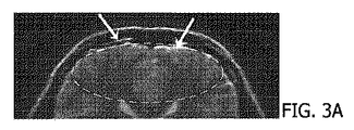
図3Aは、図1に関連する最初の再構築画像8のような、高SNRと脈動アーチファクトを有する例示の再構築画像である。脈動アーチファクトのようなアーチファクトは、点線内のエリアに現れる。局所化されたアーチファクトは、線条(striation)として現れる。局所アーチファクトの低減は、図3Dにおいてみられる。図3Dは、図3Aの画像とアーチファクトのないk空間とを使用して再構築される画像16を示している。画像16は、高いSNRと、脈動のような除去される非剛性運動を含む。線条又は明暗のラインは、この画像内にはない。図3Cは、元の完全なk空間を使用するCOCOA法のような画像再構築の他の技術との比較を提供する。アーチファクト及び/又はアーチファクトのエイリアスが存在することに留意されたい。図3Bは、比較のために、アーチファクトのないk空間のデータのみを使用する画像の再構築及び部分並列画像再構築を提供する。
FIG. 3A is an exemplary reconstructed image having a high SNR and pulsation artifacts, such as the initial
図4は、副作用の少ない局所動きアーチファクト低減の別の方法をフローチャート化している。MRデータが、ステップ2において受け取られ、完全なサンプルのk空間4として格納される。k空間4内のMRデータは、眼球運動、皮膚の動き、顎運動等のような非剛性運動を含む。動きアーチファクトのある完全なk空間4から、ステップ6において、高SNRの画像8が再構築される。画像8は局所動きアーチファクトを含むが、高SNRである。ステップ10において、完全なk空間が、動きについて分析される。動きは、COCOA法、GRAPPA法等のような技術を使用して検出される。k空間のうち、動きが検出された部分が除外される。残りの部分、すなわち除外されなかった部分は、アーチファクトのないk空間12を形成する。アーチファクトのないk空間データを、別個のメモリに格納することができ、あるいは直接参照することもできる。アーチファクトのないk空間は、図2に関連して示され、説明されている。
FIG. 4 is a flowchart of another method for reducing local motion artifact with fewer side effects. MR data is received in step 2 and stored as a complete sample k-space 4. MR data in k-space 4 includes non-rigid movements such as eye movements, skin movements, jaw movements and the like. From the complete k-space 4 with motion artifacts, a
ステップ26において、画像28が、アーチファクトのない部分的k空間データ12から、あるいはCOCOA法のように元の完全なk空間を使用する画像再構築の他の技術により再構築される。画像28は、動きアーチファクトが低減又は除去されているが、低いSNRを有する。再構築は、図3B又は図3Cに関連して示されるような、部分並列画像(PPI)再構築技術を使用して実行される。
In
ステップ30において、画像28内の1つ又は複数の動きアーチファクトの位置が識別される。例えばPPIを使用して再構築される画像28と、完全なk空間4を使用して再構築される画像8との間の差分マップ32が生成される。差分マップは、高勾配によりアーチファクトの位置を示す。差分マップは、同じ元のk空間からの両画像から構築され、したがって本質的に登録される。
In
ステップ14において、事前情報でガイドされる再構築により、完全なk空間で再構築された画像8を含む事前情報を使用して、アーチファクトのない高解像度の画像16を再構築する。事前情報は、差分マップ又は動きアーチファクトの位置32の他の指示を含むことができ、アーチファクトの位置、コイル感度マップ及び他の事前情報を提供する。例えば差分マップを使用して、正規化推定を修正することができる。これは、動きがなく、かつ高SNRを含むように、完全なk空間で再構築された画像8のうち検出された動きを含まない部分が更に、動きのないk空間のラインと既知の生成されるボクセルとの間のエラーを最小にするためである。あるいは、再構築は、画像内の検出された動きの位置を、動きアーチファクトのないk空間のデータの再構築部分と置換することができる。ステップ14はコイル感度マップを修正することができる。
In
図5は、局所アーチファクト低減システム34の実施形態を図式的に示す。システム34は、垂直又は水平ボアMRスキャナのような磁気共鳴スキャナ36を含む。シーケンスコントローラ38は、無線周波数(RF)トランスミッタ40及び勾配コントローラ42を制御する。RFトランスミッタ及び勾配コントローラは、MRスキャナ36のRFコイル及び勾配コイルにより、被験体内の共鳴を励起して操作する。MRスキャナ36は、被験体のMRデータをRFレシーバ44に伝送し、RFレシーバ44が、MRデータを復調して、完全なk空間データ4としてメモリ45に格納する。
FIG. 5 schematically illustrates an embodiment of a local
システム34は、完全なk空間データ4から画像8を再構築する完全k空間再構築モジュール46を含む。システム34は、k空間アーチファクト検出モジュール48、部分並列画像(PPI)再構築モジュール50、画像アーチファクト検出モジュール52及び事前情報ガイド再構築モジュール54を含む。k空間アーチファクト検出モジュール48は、完全なk空間4における動きを検出し、k空間のうち検出された動きを含む部分を除外する。k空間アーチファクト検出モジュールは、k空間の除外されていない部分12を別個のメモリ又はメモリ区分に格納することができる。k空間アーチファクト検出モジュールは、COCOA法のような技術を使用して、動きを検出してk空間の一部を除外する。画像アーチファクト検出モジュール52は、差分マップ32を用いるなどして、画像8内の動きの位置を検出する。画像アーチファクト検出モジュールは、完全なk空間画像8と部分的k空間画像28との間の差分マップを生成することと、動きアーチファクトを示す高勾配値の部分を識別することを含む。事前情報ガイド再構築モジュール54は、正規化を用いるSENSE法のような並列撮像技術を使用して、アーチファクトのない部分的k空間12及び完全なk空間の再構築画像8から、低減された動きアーチファクトと高いSNRを含む、画像16を再構築する。完全なk空間の再構築画像8は、正規化推定を提供する。事前情報ガイド再構築モジュール54は、コイル感度マップ18とアーチファクトの位置32を含み、正規化推定を更に精査することができる。事前情報ガイド再構築モジュール54は、再構築プロセスの出力としてコイル感度マップを修正することができる。
The
画像は、ワークステーション58のディスプレイデバイス56上に表示される。ワークステーション58は、電子プロセッサ又は電子処理デバイス60と、画像、メニュー、パネル及びユーザコントロールを表示するディスプレイ56と、医療関係者の選択を入力する少なくとも1つの入力デバイス62を含む。ワークステーション58は、デスクトップコンピュータ、ラップトップ、タブレット、モバイルコンピューティングデバイス、スマートフォン等とすることができる。入力デバイスは、キーボード、マウス、マイクロフォン等とすることができる。
The image is displayed on the
様々なモジュール46、48、50、52、54は、電子プロセッサ又は電子データ処理デバイス60によって、ネットワークによりワークステーション58に動作可能に接続されるネットワークベースのサーバコンピュータによって適切に具現化される。さらに、開示される再構築、k空間の動き検出及び画像検出技術は、開示される再構築及び動き検出技術を実行するよう、電子データ処理デバイスによって読取可能であり、かつ電子データ処理デバイスによって実行可能な命令(例えばソフトウェア)を格納する、非一時的記憶媒体を使用して適切に実装される。
The
「コンピュータ読取可能記憶媒体」は、本明細書で使用されるとき、コンピューティングデバイスのプロセッサによって実行可能な命令を格納し得る任意の有形の記憶媒体を含む。コンピュータ読取可能記憶媒体は、コンピュータ読取可能な非一時的記憶媒体と呼ばれることがある。一部の実施形態において、コンピュータ読取可能記憶媒体は、コンピューティングデバイスのプロセッサによってアクセス可能なデータを格納することもできる。コンピュータ読取可能記憶媒体の例は、これらに限られないが、フロッピー(登録商標)ディスク、磁気ハードディスクドライブ、半導体ハードディスク、フラッシュメモリ、USBサムデバイス、ランダムアクセスメモリ(RAM)、読み取り専用メモリ(ROM)、光ディスク、磁気光学ディスク、プロセッサのレジスタファイルを含む。コンピュータ読取可能記憶媒体という用語は、コンピュータデバイスによりネットワーク又は通信リンクを介してアクセス可能な様々なタイプの記録媒体を指すこともある。例えばデータは、モデム上で、インターネット上で、あるいはローカルエリアネットワーク上で取得され得る。コンピュータ読取可能記憶媒体への言及は、潜在的には複数のコンピュータ読取可能記憶媒体であるように解釈されるべきである。1つ又は複数のプログラムの様々な実行可能コンポーネントが、異なる位置に格納されてよい。コンピュータ読取可能記憶媒体は、例えば同じコンピュータシステム内の複数のコンピュータ読取可能媒体であってもよい。また、コンピュータ読取可能記憶媒体は、複数のコンピュータシステム又はコンピューティングデバイスにわたって分散されるコンピュータ読取可能記憶媒体であってもよい。 “Computer-readable storage medium” as used herein includes any tangible storage medium that can store instructions executable by a processor of a computing device. A computer-readable storage medium may be referred to as a computer-readable non-transitory storage medium. In some embodiments, the computer readable storage medium may also store data accessible by the processor of the computing device. Examples of computer readable storage media include, but are not limited to, floppy disks, magnetic hard disk drives, semiconductor hard disks, flash memory, USB thumb devices, random access memory (RAM), read only memory (ROM). , Optical disk, magneto-optical disk, and processor register file. The term computer readable storage media may refer to various types of recording media that can be accessed by a computing device over a network or communication link. For example, data can be obtained on a modem, on the Internet, or on a local area network. References to computer readable storage media should be construed as potentially multiple computer readable storage media. Various executable components of one or more programs may be stored in different locations. The computer readable storage medium may be a plurality of computer readable media within the same computer system, for example. The computer readable storage medium may also be a computer readable storage medium distributed over a plurality of computer systems or computing devices.
「コンピュータストレージ」又は「ストレージ」は、コンピュータ読取可能記憶媒体の例である。コンピュータストレージは、任意の不揮発性コンピュータ読取可能記憶媒体である。ストレージは、例えば同じコンピュータシステム又はコンピューティングデバイス内の複数のストレージデバイスであっても、複数のコンピュータシステム又はコンピューティングデバイスにわたって分散される複数のストレージであってよく、かつ/あるいはクラウドベースのコンピューティングストレージデバイスに含まれてもよい。 “Computer storage” or “storage” is an example of a computer-readable storage medium. Computer storage is any non-volatile computer-readable storage medium. The storage may be multiple storage distributed across multiple computer systems or computing devices and / or cloud-based computing, for example multiple storage devices within the same computer system or computing device. It may be included in the storage device.
「コンピュータメモリ」又は「メモリ」は、コンピュータ読取可能記憶媒体の例である。コンピュータメモリは、プロセッサに対して直接アクセス可能な任意のメモリである。 “Computer memory” or “memory” is an example of a computer-readable storage medium. Computer memory is any memory that is directly accessible to the processor.
「プロセッサ」は、本明細書で使用されるとき、プログラム又はマシン実行可能命令を実行することができる電子的コンポーネントを含む。「プロセッサ」を備えるコンピューティングデバイスへの言及は、潜在的に、1つより多くのプロセッサ又はプロセッシングコアを含むものとして解釈されるべきである。プロセッサは、例えばマルチコアプロセッサであってよい。プロセッサは、単一のコンピュータシステム内又は複数のコンピュータ間で分散されるプロセッサの集合を指すこともある。また、コンピューティングデバイスという用語も、各コンピューティングデバイスが1つ又は複数のプロセッサを備えるコンピューティングデバイスの集合又はネットワークを潜在的に指すように解釈されるべきである。 A “processor” as used herein includes electronic components that can execute a program or machine-executable instructions. Reference to a computing device comprising a “processor” should be construed as potentially including more than one processor or processing core. The processor may be a multi-core processor, for example. A processor may also refer to a collection of processors distributed within a single computer system or among multiple computers. The term computing device should also be construed to potentially refer to a collection or network of computing devices, each computing device comprising one or more processors.
「ディスプレイ」又は「ディスプレイデバイス」は、本明細書で使用されるとき、画像又はデータを表示するのに適合された出力デバイス又はユーザインタフェースデバイスを含む。ディスプレイは、ビジュアル、オーディオ及び/又は触覚データを出力することがある。ディスプレイの例には、コンピュータモニタ、テレビ画面、タッチ画面、触覚電気的ディスプレイ、ブラウン管(CRT)、フラットパネルディスプレイ、発光ダイオード(LED)ディスプレイ、プラズマディスプレイパネル(PDP)、液晶ディスプレイ(LCD)等が含まれるが、これらには限定されない。 A “display” or “display device” as used herein includes an output device or user interface device adapted to display an image or data. The display may output visual, audio and / or haptic data. Examples of displays include computer monitors, television screens, touch screens, tactile electrical displays, cathode ray tubes (CRT), flat panel displays, light emitting diode (LED) displays, plasma display panels (PDP), liquid crystal displays (LCD), etc. Including, but not limited to.
磁気共鳴(MR)データは、本明細書では、磁気共鳴画像スキャンの間に磁気共鳴装置のアンテナによる原子スピンによって放出される無線周波数信号の記録された測定値として定義される。磁気共鳴画像法(MRI)の画像は、本明細書では、磁気共鳴画像データ内に含まれる、解剖学的データの再構築される2次元又は3次元での可視化として定義される。この可視化は、コンピュータを使用して行うことが可能である。 Magnetic resonance (MR) data is defined herein as a recorded measurement of radio frequency signals emitted by atomic spins by an antenna of a magnetic resonance apparatus during a magnetic resonance image scan. Magnetic resonance imaging (MRI) images are defined herein as reconstructed two-dimensional or three-dimensional visualizations of anatomical data contained within magnetic resonance image data. This visualization can be performed using a computer.
本明細書で説明される特定の要素又はコンポーネントは、ハードウェア、ソフトウェア、ファームウェア又はその組み合わせにより適切に実装される機能性を有することが認識されよう。加えて、本明細書において、共に組み込まれるものとして説明される特定の要素は、適切な環境において、スタンドアロンの要素であってもよく、あるいは他の方法で分割されてもよいことが認識されよう。同様に、特定の要素によって実行されるように説明される複数の特定の機能が、それぞれ独立に個々の機能を実行するよう動作する複数の別個の要素によって実行されもよく、あるいは特定の個々の機能を分割して、共同で動作する複数の別個の要素によって実行してもよい。あるいは、相互に別個のものとして他に説明及び/又は図示されている一部の要素又はコンポーネントを、適切な場合に、物理的又は機能的に組み合わせてもよい。 It will be appreciated that certain elements or components described herein have functionality that is suitably implemented by hardware, software, firmware, or a combination thereof. In addition, it will be appreciated that certain elements described herein as being incorporated together may be stand-alone elements or may be otherwise partitioned in an appropriate environment. . Similarly, a plurality of specific functions described to be performed by a particular element may be performed by a plurality of separate elements that each operate independently to perform the individual function, or a specific individual The function may be divided and performed by a plurality of separate elements operating together. Alternatively, some elements or components otherwise described and / or illustrated as separate from one another may be combined physically or functionally where appropriate.
すなわち、上記に開示される及び他の様々な特徴及び機能あるいはその代替形態は、所望に応じて多くの他の異なるシステム又はアプリケーションに組み合わされてよく、また現在予期又は認識されていない様々な代替形態、修正形態、変形形態又は改良形態が後に当業者によって構成されてもよく、これらの様々な形態も同様に以下の請求項によって含まれるように意図されていることが認識されよう。 That is, the various features and functions disclosed above and various alternatives thereof may be combined into many other different systems or applications as desired, and various alternatives not currently anticipated or recognized. It will be appreciated that forms, modifications, variations or improvements may later be configured by those skilled in the art, and these various forms are also intended to be encompassed by the following claims.
Claims (13)
磁気共鳴k空間データを格納するメモリであって、前記k空間データが非剛性運動欠損を含む、メモリと;
1つ又は複数のプロセッサであって:
前記k空間データから、高い信号対雑音比及び動きアーチファクトを含む第1の画像を再構築し、
前記k空間データのうち非剛性運動欠損を含む部分を検出して除外し、
部分並列再構築を使用して、前記k空間データのうちの除外されてない部分及び前記第1の画像から第2の画像を再構築することであって、前記第1の画像に対して低減された動きアーチファクト及びより低い信号対ノイズ比を含む第2の画像を再構築し、
前記第1及び第2の画像から、動きアーチファクトの位置を特定し、
前記第1の画像、前記動きアーチファクトの特定された位置及び前記k空間データの前記除外されていない部分を使用して、アーチファクト低減又はアーチファクトのない画像を再構築する
ように構成される、1つ又は複数のプロセッサと;
を備える、システム。 In medical imaging systems:
A memory for storing magnetic resonance k-space data, wherein the k-space data includes non-rigid motion deficits;
One or more processors, including:
Reconstructing a first image comprising a high signal-to-noise ratio and motion artifacts from the k-space data;
Detecting and excluding a portion including the non-rigid motion defect in the k-space data;
Using partial parallel reconstruction to reconstruct a second image from an unexcluded portion of the k-space data and the first image, wherein the second image is reduced relative to the first image Reconstructing a second image containing the generated motion artifacts and a lower signal-to-noise ratio;
Identifying the position of the motion artifact from the first and second images;
One configured to use the first image, the identified location of the motion artifact and the non-excluded portion of the k-space data to reconstruct an artifact-reduced or artifact-free image. Or with multiple processors;
A system comprising:
前記アーチファクト低減又はアーチファクトのない画像を再構築することは、前記差分マップ及びコイル感度を使用することを含む、
請求項1に記載のシステム。 Determining the position of the motion artifact includes generating a difference map from the first and second images;
Reconstructing the artifact reduction or artifact-free image comprises using the difference map and coil sensitivity;
The system of claim 1.
磁気共鳴k空間データを格納するメモリであって、前記k空間データが非剛性運動欠損を含む、メモリと;
1つ又は複数のプロセッサであって:
前記k空間データから、高い信号対雑音比及び少なくとも1つの動きアーチファクトを含む第1の画像を再構築し、
前記k空間データのうち非剛性運動欠損を含む部分を検出して除外し、
前記k空間データの除外されていない部分から、部分並列再構築を使用して第3の画像を再構築し、
前記第1又は第3の画像における少なくとも1つの動きアーチファクトの位置を検出し、
前記k空間データの前記除外されていない部分、前記第1の画像及び前記少なくとも1つの動きアーチファクトの前記検出された位置から、第2の画像を再構築する、
ように構成される、1つ又は複数のプロセッサと;
を備える、システム。 In medical imaging systems:
A memory for storing magnetic resonance k-space data, wherein the k-space data includes non-rigid motion deficits;
One or more processors, including:
Reconstructing a first image comprising a high signal-to-noise ratio and at least one motion artifact from the k-space data;
Detecting and excluding a portion including the non-rigid motion defect in the k-space data;
Reconstructing a third image from the unexcluded portion of the k-space data using partial parallel reconstruction;
Detecting the position of at least one motion artifact in the first or third image;
Reconstructing a second image from the non-excluded portion of the k-space data, the first image and the detected position of the at least one motion artifact;
One or more processors configured as described above;
A system comprising:
ように構成される、請求項3に記載のシステム。 The system of claim 3, wherein the one or more processors are configured to reconstruct the second image using a coil sensitivity map.
高勾配値により前記少なくとも1つの動きアーチファクトを示す、前記第3の画像と前記第1の画像との間の差分マップを生成する
ように構成される、請求項3に記載のシステム。 The one or more processors further includes:
The system of claim 3, configured to generate a difference map between the third image and the first image that indicates the at least one motion artifact with a high gradient value.
前記少なくとも1つの動きアーチファクトの前記位置に基づいて前記コイル感度マップを修正する
ように構成される、請求項4に記載のシステム。 The one or more processors further includes:
The system of claim 4, configured to modify the coil sensitivity map based on the position of the at least one motion artifact.
データコンボリューション及び結合演算法(COCOA)を使用して前記k空間データの前記部分を検出して除外する
ように構成される、請求項3に記載のシステム。 The one or more processors are:
The system of claim 3, configured to detect and exclude the portion of the k-space data using data convolution and join arithmetic (COCOA).
非剛性運動欠損を含む前記k空間データから第1の画像を再構築するステップと、
前記k空間データのうち非剛性運動欠損を含む部分を検出して除外するステップと、
前記k空間データの除外されていない部分から、部分並列再構築を使用して第3の画像を再構築するステップと、
前記第1又は第3の画像における少なくとも1つのアーチファクトの位置を検出するステップと、
前記k空間データの前記除外されていない部分、前記第1の画像及び前記少なくとも1つのアーチファクトの前記検出された位置から、第2の画像を再構築するステップと、
を備える、方法。 Receiving magnetic resonance k-space data including non-rigid motion deficits;
Reconstructing a first image from the k-space data including non-rigid motion deficits;
Detecting and excluding a portion including non-rigid motion deficits in the k-space data;
Reconstructing a third image from a non-excluded portion of the k-space data using partial parallel reconstruction;
Detecting the position of at least one artifact in the first or third image;
Reconstructing a second image from the non-excluded portion of the k-space data, the first image and the detected location of the at least one artifact;
A method comprising:
前記少なくとも1つのアーチファクトを示す高勾配値を有する、前記第3の画像と前記第1の画像との間の差分マップを生成するステップ
を含む、請求項8に記載の方法。 Detecting the position of the at least one artifact comprises:
9. The method of claim 8, comprising generating a difference map between the third image and the first image having a high slope value indicative of the at least one artifact.
を更に含む、請求項8に記載の方法。 The method of claim 8, further comprising: modifying a coil sensitivity map based on the location of the at least one artifact.
請求項8に記載の方法。 Detecting and excluding the portion of the k-space data is performed using data convolution and join arithmetic (COCOA);
The method of claim 8.
Applications Claiming Priority (3)
| Application Number | Priority Date | Filing Date | Title |
|---|---|---|---|
| US201261733945P | 2012-12-06 | 2012-12-06 | |
| US61/733,945 | 2012-12-06 | ||
| PCT/IB2013/059489 WO2014087270A2 (en) | 2012-12-06 | 2013-10-21 | Local artifact reduction with insignificant side effects |
Publications (3)
| Publication Number | Publication Date |
|---|---|
| JP2015536746A JP2015536746A (en) | 2015-12-24 |
| JP2015536746A5 JP2015536746A5 (en) | 2017-05-25 |
| JP6317756B2 true JP6317756B2 (en) | 2018-04-25 |
Family
ID=49943420
Family Applications (1)
| Application Number | Title | Priority Date | Filing Date |
|---|---|---|---|
| JP2015546113A Active JP6317756B2 (en) | 2012-12-06 | 2013-10-21 | Reduce local artifacts with fewer side effects |
Country Status (5)
| Country | Link |
|---|---|
| US (1) | US9710937B2 (en) |
| EP (1) | EP2929361A2 (en) |
| JP (1) | JP6317756B2 (en) |
| CN (1) | CN104838279B (en) |
| WO (1) | WO2014087270A2 (en) |
Families Citing this family (11)
| Publication number | Priority date | Publication date | Assignee | Title |
|---|---|---|---|---|
| US9797974B2 (en) * | 2013-01-30 | 2017-10-24 | The Board Of Trustees Of The Leland Stanford Junior University | Nonrigid motion correction in 3D using autofocusing with localized linear translations |
| US10794979B2 (en) | 2015-12-03 | 2020-10-06 | Koninklijke Philips N.V. | Removal of image artifacts in sense-MRI |
| RU2730431C2 (en) * | 2015-12-03 | 2020-08-21 | Конинклейке Филипс Н.В. | Removal of image artifacts at sense-visualization |
| EP3447520A1 (en) * | 2017-08-22 | 2019-02-27 | Koninklijke Philips N.V. | Data-driven correction of phase depending artefacts in a magnetic resonance imaging system |
| US10677873B2 (en) * | 2017-08-29 | 2020-06-09 | General Electric Company | System and method for correcting an artifact within magnetic resonance data |
| EP3511726A1 (en) * | 2018-01-12 | 2019-07-17 | Koninklijke Philips N.V. | Mri method for determining a magnetic field map from a b0 reference scan and a wassr scan |
| US11484279B2 (en) | 2018-09-24 | 2022-11-01 | Siemens Medical Solutions Usa, Inc. | Systems to assess projection data inconsistency |
| US11835612B2 (en) | 2019-03-12 | 2023-12-05 | University Of Cincinnati | System and method for motion correction of magnetic resonance image |
| EP3719526A1 (en) * | 2019-04-03 | 2020-10-07 | Siemens Healthcare GmbH | Magnetic resonance imaging and motion detection |
| EP3770625B1 (en) * | 2019-07-24 | 2024-06-26 | Siemens Healthineers AG | Mri using compressed sensing with improved regularization parameter |
| US12125127B2 (en) * | 2021-11-29 | 2024-10-22 | Canon Medical Systems Corporation | Advanced signal combination for improved overlapping image correction |
Family Cites Families (21)
| Publication number | Priority date | Publication date | Assignee | Title |
|---|---|---|---|---|
| US6771067B2 (en) * | 2001-04-03 | 2004-08-03 | The United States Of America As Represented By The Department Of Health And Human Services | Ghost artifact cancellation using phased array processing |
| US7283859B2 (en) * | 2001-04-20 | 2007-10-16 | Brigham And Womens' Hospital, Inc. | Artifact suppression in dynamic magnetic resonance imaging |
| WO2003032816A2 (en) * | 2001-10-19 | 2003-04-24 | The Government Of The United States Of America As Represented By The Secretary Of The Department Of Health And Human Services | A method and apparatus to improve an mri image |
| JP4364789B2 (en) | 2002-05-13 | 2009-11-18 | コーニンクレッカ フィリップス エレクトロニクス エヌ ヴィ | Magnetic resonance imaging method using accelerated data acquisition |
| CN1867835B (en) * | 2003-10-13 | 2010-06-16 | 皇家飞利浦电子股份有限公司 | System and method for magnetic resonance imaging |
| DE10353342B4 (en) * | 2003-11-14 | 2008-07-17 | Siemens Ag | Improved MRI imaging based on conventional PPA reconstruction techniques |
| US9201129B2 (en) * | 2006-09-13 | 2015-12-01 | Kabushiki Kaisha Toshiba | Magnetic-resonance image diagnostic apparatus and method of controlling the same |
| US7903858B2 (en) | 2006-11-03 | 2011-03-08 | Siemens Aktiengesellschaft | Practical image reconstruction for magnetic resonance imaging |
| DE102007028660B3 (en) * | 2007-06-21 | 2009-01-29 | Siemens Ag | Method for correcting motion artifacts when taking MR images |
| BRPI0821804A2 (en) | 2007-12-20 | 2015-06-16 | Wisconsin Alumni Res Found | Method for reconstructing an image of an individual |
| CN102362191A (en) * | 2009-03-25 | 2012-02-22 | 皇家飞利浦电子股份有限公司 | Magnetic resonance partially parallel imaging (PPI) with motion corrected coil sensitivities |
| CN102362192A (en) * | 2009-03-25 | 2012-02-22 | 皇家飞利浦电子股份有限公司 | Motion detection and correction in magnetic resonance imaging for rigid, nonrigid, translational, rotational, and through-plane motion |
| DE102010029281A1 (en) | 2010-05-25 | 2011-12-01 | Siemens Aktiengesellschaft | Method and image reconstruction device for the reconstruction of image data |
| US8811694B2 (en) | 2010-09-30 | 2014-08-19 | University Of Utah Research Foundation | Intrinsic detection of motion in segmented sequences |
| CN103430038A (en) * | 2011-03-17 | 2013-12-04 | 皇家飞利浦有限公司 | A MRI method of faster channel-by-channel reconstruction without image degradation |
| US8948534B2 (en) * | 2011-04-22 | 2015-02-03 | Kabushiki Kaisha Toshiba | MRI Gibbs' ringing filtering with edge-protection |
| WO2013103791A1 (en) * | 2012-01-06 | 2013-07-11 | Cincinnati Children's Hospital Medical Center | Correlation imaging for multi-scan mri with multi-channel data acquisition |
| US9146293B2 (en) * | 2012-02-27 | 2015-09-29 | Ohio State Innovation Foundation | Methods and apparatus for accurate characterization of signal coil receiver sensitivity in magnetic resonance imaging (MRI) |
| US8768034B2 (en) * | 2012-03-20 | 2014-07-01 | Siemens Medical Solutions Usa, Inc. | Motion compensated MR imaging system |
| EP2858559B1 (en) * | 2012-06-28 | 2021-01-20 | Duke University | Multi-shot scan protocols for high-resolution mri incorporating multiplexed sensitivity-encoding (muse) |
| US9202272B2 (en) * | 2012-09-20 | 2015-12-01 | Beth Israel Deaconess Medical Center, Inc. (Bidmc, Inc.) | Method and apparatus for image enhancement in magnetic resonance imaging using motion corrupted data |
-
2013
- 2013-10-21 JP JP2015546113A patent/JP6317756B2/en active Active
- 2013-10-21 CN CN201380063775.7A patent/CN104838279B/en not_active Expired - Fee Related
- 2013-10-21 WO PCT/IB2013/059489 patent/WO2014087270A2/en active Application Filing
- 2013-10-21 US US14/648,271 patent/US9710937B2/en active Active
- 2013-10-21 EP EP13818801.6A patent/EP2929361A2/en not_active Withdrawn
Also Published As
| Publication number | Publication date |
|---|---|
| US20150302616A1 (en) | 2015-10-22 |
| WO2014087270A3 (en) | 2014-10-30 |
| CN104838279B (en) | 2018-03-30 |
| CN104838279A (en) | 2015-08-12 |
| US9710937B2 (en) | 2017-07-18 |
| WO2014087270A2 (en) | 2014-06-12 |
| JP2015536746A (en) | 2015-12-24 |
| EP2929361A2 (en) | 2015-10-14 |
Similar Documents
| Publication | Publication Date | Title |
|---|---|---|
| JP6317756B2 (en) | Reduce local artifacts with fewer side effects | |
| US10698062B2 (en) | Diffusion MRI method for generating a synthetic diffusion image at a high B-value | |
| US10180482B2 (en) | Channel by channel artifact reduction in parallel MRI | |
| WO2007124444A2 (en) | Method and system for parallel reconstruction in the k-space domain for application in imaging systems | |
| JP6041356B2 (en) | Image analysis apparatus, operation method of image analysis apparatus, and image analysis program | |
| US10788559B2 (en) | Motion detection with multi element radio frequency antennas | |
| CN112204411B (en) | Motion detection in CEST magnetic resonance imaging | |
| CN105579861B (en) | The correction for attenuation based on MR in the PET/MR imaging with DIXON pulse train | |
| EP3391069A1 (en) | Segmentation of quantitative susceptibility mapping magnetic resonance images | |
| JP2001309903A (en) | Automatic correction of mri projection imaging | |
| US20160349346A1 (en) | Intrinsic navigation from velocity-encoding gradients in phase-contrast mri | |
| EP2888600B1 (en) | Adaptive keyhole compression for dynamic contrast-enhanced mri | |
| US10215820B2 (en) | Differentiating tissues with MR imaging | |
| US10302726B2 (en) | Image reconstruction for MRI using multiplexed sensitivity encoding | |
| EP3916417A1 (en) | Correction of magnetic resonance images using multiple magnetic resonance imaging system configurations | |
| CN113892149A (en) | Method for motion artifact detection | |
| RU2764643C2 (en) | Data stream-controlled correction of phase-dependent artifacts in magnetic resonance imaging system | |
| US20160146918A1 (en) | Corrected magnetic resonance imaging using coil sensitivities | |
| JP7493671B2 (en) | Image intensity correction in magnetic resonance imaging. | |
| US12125127B2 (en) | Advanced signal combination for improved overlapping image correction | |
| CN118103721A (en) | Motion correction using low resolution magnetic resonance images | |
| Shimron et al. | Accelerating Low-field MRI: Compressed Sensing and AI for fast noise-robust imaging | |
| KR20140013409A (en) | Method for setting field of view in magnetic resonance imaging diagnosis apparatus and apparatus thereto |
Legal Events
| Date | Code | Title | Description |
|---|---|---|---|
| A621 | Written request for application examination |
Free format text: JAPANESE INTERMEDIATE CODE: A621 Effective date: 20161018 |
|
| A521 | Request for written amendment filed |
Free format text: JAPANESE INTERMEDIATE CODE: A523 Effective date: 20170406 |
|
| A871 | Explanation of circumstances concerning accelerated examination |
Free format text: JAPANESE INTERMEDIATE CODE: A871 Effective date: 20170406 |
|
| A975 | Report on accelerated examination |
Free format text: JAPANESE INTERMEDIATE CODE: A971005 Effective date: 20170602 |
|
| A131 | Notification of reasons for refusal |
Free format text: JAPANESE INTERMEDIATE CODE: A131 Effective date: 20170613 |
|
| A521 | Request for written amendment filed |
Free format text: JAPANESE INTERMEDIATE CODE: A523 Effective date: 20170907 |
|
| A131 | Notification of reasons for refusal |
Free format text: JAPANESE INTERMEDIATE CODE: A131 Effective date: 20171107 |
|
| A521 | Request for written amendment filed |
Free format text: JAPANESE INTERMEDIATE CODE: A523 Effective date: 20180129 |
|
| TRDD | Decision of grant or rejection written | ||
| A01 | Written decision to grant a patent or to grant a registration (utility model) |
Free format text: JAPANESE INTERMEDIATE CODE: A01 Effective date: 20180306 |
|
| A61 | First payment of annual fees (during grant procedure) |
Free format text: JAPANESE INTERMEDIATE CODE: A61 Effective date: 20180330 |
|
| R150 | Certificate of patent or registration of utility model |
Ref document number: 6317756 Country of ref document: JP Free format text: JAPANESE INTERMEDIATE CODE: R150 |