JP6279590B2 - Methods and kits for measuring analytes in acidic conditions - Google Patents
Methods and kits for measuring analytes in acidic conditions Download PDFInfo
- Publication number
- JP6279590B2 JP6279590B2 JP2015535610A JP2015535610A JP6279590B2 JP 6279590 B2 JP6279590 B2 JP 6279590B2 JP 2015535610 A JP2015535610 A JP 2015535610A JP 2015535610 A JP2015535610 A JP 2015535610A JP 6279590 B2 JP6279590 B2 JP 6279590B2
- Authority
- JP
- Japan
- Prior art keywords
- analyte
- acidic
- protein
- ligand
- complex
- Prior art date
- Legal status (The legal status is an assumption and is not a legal conclusion. Google has not performed a legal analysis and makes no representation as to the accuracy of the status listed.)
- Active
Links
- 238000000034 method Methods 0.000 title claims description 55
- 230000002378 acidificating effect Effects 0.000 title claims description 38
- 102000004169 proteins and genes Human genes 0.000 claims description 88
- 108090000623 proteins and genes Proteins 0.000 claims description 88
- 239000012491 analyte Substances 0.000 claims description 63
- 239000003446 ligand Substances 0.000 claims description 35
- 239000003153 chemical reaction reagent Substances 0.000 claims description 33
- 239000000872 buffer Substances 0.000 claims description 31
- 230000027455 binding Effects 0.000 claims description 22
- 238000003018 immunoassay Methods 0.000 claims description 20
- 238000001514 detection method Methods 0.000 claims description 13
- 239000007788 liquid Substances 0.000 claims description 13
- 239000007787 solid Substances 0.000 claims description 6
- 239000007790 solid phase Substances 0.000 claims description 5
- 108010090804 Streptavidin Proteins 0.000 claims description 4
- 108090001008 Avidin Proteins 0.000 claims description 2
- 230000000536 complexating effect Effects 0.000 claims description 2
- 230000004044 response Effects 0.000 claims description 2
- 239000012515 MabSelect SuRe Substances 0.000 description 52
- 229940126622 therapeutic monoclonal antibody Drugs 0.000 description 49
- 239000000523 sample Substances 0.000 description 46
- 238000010494 dissociation reaction Methods 0.000 description 30
- 230000005593 dissociations Effects 0.000 description 30
- 238000003556 assay Methods 0.000 description 24
- 238000004458 analytical method Methods 0.000 description 20
- 229940048921 humira Drugs 0.000 description 14
- 239000002253 acid Substances 0.000 description 12
- 230000003993 interaction Effects 0.000 description 10
- 238000011002 quantification Methods 0.000 description 10
- 238000002156 mixing Methods 0.000 description 9
- 108060003951 Immunoglobulin Proteins 0.000 description 8
- 102000018358 immunoglobulin Human genes 0.000 description 8
- 230000008569 process Effects 0.000 description 8
- 231100000673 dose–response relationship Toxicity 0.000 description 7
- 238000005259 measurement Methods 0.000 description 7
- 238000011282 treatment Methods 0.000 description 7
- 238000002474 experimental method Methods 0.000 description 6
- 238000000746 purification Methods 0.000 description 6
- 238000003908 quality control method Methods 0.000 description 6
- 239000011347 resin Substances 0.000 description 6
- 229920005989 resin Polymers 0.000 description 6
- 241000287828 Gallus gallus Species 0.000 description 5
- 230000000694 effects Effects 0.000 description 5
- 239000012634 fragment Substances 0.000 description 5
- 230000007935 neutral effect Effects 0.000 description 5
- YBJHBAHKTGYVGT-ZKWXMUAHSA-N (+)-Biotin Chemical compound N1C(=O)N[C@@H]2[C@H](CCCCC(=O)O)SC[C@@H]21 YBJHBAHKTGYVGT-ZKWXMUAHSA-N 0.000 description 4
- 108090000790 Enzymes Proteins 0.000 description 4
- 102000004190 Enzymes Human genes 0.000 description 4
- 206010028980 Neoplasm Diseases 0.000 description 4
- 230000015572 biosynthetic process Effects 0.000 description 4
- 201000011510 cancer Diseases 0.000 description 4
- 238000004113 cell culture Methods 0.000 description 4
- 238000007865 diluting Methods 0.000 description 4
- 239000003814 drug Substances 0.000 description 4
- 229940022353 herceptin Drugs 0.000 description 4
- 239000012535 impurity Substances 0.000 description 4
- 239000000203 mixture Substances 0.000 description 4
- 241000894007 species Species 0.000 description 4
- 108010088160 Staphylococcal Protein A Proteins 0.000 description 3
- 241000191967 Staphylococcus aureus Species 0.000 description 3
- 229960002685 biotin Drugs 0.000 description 3
- 239000011616 biotin Substances 0.000 description 3
- 239000007979 citrate buffer Substances 0.000 description 3
- 238000004925 denaturation Methods 0.000 description 3
- 230000036425 denaturation Effects 0.000 description 3
- 238000010790 dilution Methods 0.000 description 3
- 239000012895 dilution Substances 0.000 description 3
- 238000005516 engineering process Methods 0.000 description 3
- 238000010438 heat treatment Methods 0.000 description 3
- 229940013982 octagam Drugs 0.000 description 3
- 238000002198 surface plasmon resonance spectroscopy Methods 0.000 description 3
- 230000001225 therapeutic effect Effects 0.000 description 3
- 238000005406 washing Methods 0.000 description 3
- QZTKDVCDBIDYMD-UHFFFAOYSA-N 2,2'-[(2-amino-2-oxoethyl)imino]diacetic acid Chemical compound NC(=O)CN(CC(O)=O)CC(O)=O QZTKDVCDBIDYMD-UHFFFAOYSA-N 0.000 description 2
- 239000007988 ADA buffer Substances 0.000 description 2
- 102000013455 Amyloid beta-Peptides Human genes 0.000 description 2
- 108010090849 Amyloid beta-Peptides Proteins 0.000 description 2
- 241000191940 Staphylococcus Species 0.000 description 2
- 102000013394 Troponin I Human genes 0.000 description 2
- 108010065729 Troponin I Proteins 0.000 description 2
- 238000005411 Van der Waals force Methods 0.000 description 2
- 238000010306 acid treatment Methods 0.000 description 2
- 238000001042 affinity chromatography Methods 0.000 description 2
- 239000000538 analytical sample Substances 0.000 description 2
- 239000000427 antigen Substances 0.000 description 2
- 102000036639 antigens Human genes 0.000 description 2
- 108091007433 antigens Proteins 0.000 description 2
- 230000004888 barrier function Effects 0.000 description 2
- 230000008901 benefit Effects 0.000 description 2
- 235000020958 biotin Nutrition 0.000 description 2
- 239000007853 buffer solution Substances 0.000 description 2
- 238000006243 chemical reaction Methods 0.000 description 2
- 230000009918 complex formation Effects 0.000 description 2
- 229940079593 drug Drugs 0.000 description 2
- 230000006870 function Effects 0.000 description 2
- 230000002209 hydrophobic effect Effects 0.000 description 2
- 229940072221 immunoglobulins Drugs 0.000 description 2
- 230000003834 intracellular effect Effects 0.000 description 2
- 230000007246 mechanism Effects 0.000 description 2
- MYWUZJCMWCOHBA-VIFPVBQESA-N methamphetamine Chemical compound CN[C@@H](C)CC1=CC=CC=C1 MYWUZJCMWCOHBA-VIFPVBQESA-N 0.000 description 2
- 239000000178 monomer Substances 0.000 description 2
- 229940126619 mouse monoclonal antibody Drugs 0.000 description 2
- 239000002245 particle Substances 0.000 description 2
- 238000002360 preparation method Methods 0.000 description 2
- 238000012545 processing Methods 0.000 description 2
- 238000011084 recovery Methods 0.000 description 2
- 230000001105 regulatory effect Effects 0.000 description 2
- JJGWLCLUQNFDIS-GTSONSFRSA-M sodium;1-[6-[5-[(3as,4s,6ar)-2-oxo-1,3,3a,4,6,6a-hexahydrothieno[3,4-d]imidazol-4-yl]pentanoylamino]hexanoyloxy]-2,5-dioxopyrrolidine-3-sulfonate Chemical compound [Na+].O=C1C(S(=O)(=O)[O-])CC(=O)N1OC(=O)CCCCCNC(=O)CCCC[C@H]1[C@H]2NC(=O)N[C@H]2CS1 JJGWLCLUQNFDIS-GTSONSFRSA-M 0.000 description 2
- 229940124597 therapeutic agent Drugs 0.000 description 2
- 238000011144 upstream manufacturing Methods 0.000 description 2
- 239000011534 wash buffer Substances 0.000 description 2
- YBHYYFYQHRADCQ-UHFFFAOYSA-N 2-aminoacetic acid;2-hydroxypropane-1,2,3-tricarboxylic acid Chemical compound NCC(O)=O.OC(=O)CC(O)(C(O)=O)CC(O)=O YBHYYFYQHRADCQ-UHFFFAOYSA-N 0.000 description 1
- IVLXQGJVBGMLRR-UHFFFAOYSA-N 2-aminoacetic acid;hydron;chloride Chemical compound Cl.NCC(O)=O IVLXQGJVBGMLRR-UHFFFAOYSA-N 0.000 description 1
- ZKRFOXLVOKTUTA-KQYNXXCUSA-N 9-(5-phosphoribofuranosyl)-6-mercaptopurine Chemical compound O[C@@H]1[C@H](O)[C@@H](COP(O)(O)=O)O[C@H]1N1C(NC=NC2=S)=C2N=C1 ZKRFOXLVOKTUTA-KQYNXXCUSA-N 0.000 description 1
- 229920000936 Agarose Polymers 0.000 description 1
- 239000012099 Alexa Fluor family Substances 0.000 description 1
- 206010002199 Anaphylactic shock Diseases 0.000 description 1
- 206010013710 Drug interaction Diseases 0.000 description 1
- LFQSCWFLJHTTHZ-UHFFFAOYSA-N Ethanol Chemical compound CCO LFQSCWFLJHTTHZ-UHFFFAOYSA-N 0.000 description 1
- 108010008177 Fd immunoglobulins Proteins 0.000 description 1
- 101000669513 Homo sapiens Metalloproteinase inhibitor 1 Proteins 0.000 description 1
- 102000002274 Matrix Metalloproteinases Human genes 0.000 description 1
- 108010000684 Matrix Metalloproteinases Proteins 0.000 description 1
- 102000005741 Metalloproteases Human genes 0.000 description 1
- 108010006035 Metalloproteases Proteins 0.000 description 1
- 102100039364 Metalloproteinase inhibitor 1 Human genes 0.000 description 1
- 241001465754 Metazoa Species 0.000 description 1
- 102000007474 Multiprotein Complexes Human genes 0.000 description 1
- 108010085220 Multiprotein Complexes Proteins 0.000 description 1
- 102000008300 Mutant Proteins Human genes 0.000 description 1
- 108010021466 Mutant Proteins Proteins 0.000 description 1
- CZMRCDWAGMRECN-UGDNZRGBSA-N Sucrose Chemical compound O[C@H]1[C@H](O)[C@@H](CO)O[C@@]1(CO)O[C@@H]1[C@H](O)[C@@H](O)[C@H](O)[C@@H](CO)O1 CZMRCDWAGMRECN-UGDNZRGBSA-N 0.000 description 1
- 229930006000 Sucrose Natural products 0.000 description 1
- 101710120037 Toxin CcdB Proteins 0.000 description 1
- 238000001261 affinity purification Methods 0.000 description 1
- 239000003513 alkali Substances 0.000 description 1
- 230000003321 amplification Effects 0.000 description 1
- 238000002820 assay format Methods 0.000 description 1
- 230000001580 bacterial effect Effects 0.000 description 1
- 239000011324 bead Substances 0.000 description 1
- 230000004071 biological effect Effects 0.000 description 1
- 210000004027 cell Anatomy 0.000 description 1
- 230000005779 cell damage Effects 0.000 description 1
- 208000037887 cell injury Diseases 0.000 description 1
- 239000012501 chromatography medium Substances 0.000 description 1
- 238000012875 competitive assay Methods 0.000 description 1
- 230000024203 complement activation Effects 0.000 description 1
- 238000010668 complexation reaction Methods 0.000 description 1
- 150000001875 compounds Chemical class 0.000 description 1
- 238000011109 contamination Methods 0.000 description 1
- 239000013078 crystal Substances 0.000 description 1
- 230000001419 dependent effect Effects 0.000 description 1
- 238000010586 diagram Methods 0.000 description 1
- 239000000539 dimer Substances 0.000 description 1
- 208000037265 diseases, disorders, signs and symptoms Diseases 0.000 description 1
- 208000035475 disorder Diseases 0.000 description 1
- 238000006073 displacement reaction Methods 0.000 description 1
- 239000002532 enzyme inhibitor Substances 0.000 description 1
- 229940125532 enzyme inhibitor Drugs 0.000 description 1
- 238000009472 formulation Methods 0.000 description 1
- 238000005194 fractionation Methods 0.000 description 1
- 238000002523 gelfiltration Methods 0.000 description 1
- PCHJSUWPFVWCPO-UHFFFAOYSA-N gold Chemical compound [Au] PCHJSUWPFVWCPO-UHFFFAOYSA-N 0.000 description 1
- 239000010931 gold Substances 0.000 description 1
- 229910052737 gold Inorganic materials 0.000 description 1
- 210000002064 heart cell Anatomy 0.000 description 1
- 238000003505 heat denaturation Methods 0.000 description 1
- 230000013632 homeostatic process Effects 0.000 description 1
- 230000028993 immune response Effects 0.000 description 1
- 210000000987 immune system Anatomy 0.000 description 1
- 230000000984 immunochemical effect Effects 0.000 description 1
- 208000015181 infectious disease Diseases 0.000 description 1
- 208000027866 inflammatory disease Diseases 0.000 description 1
- 239000003112 inhibitor Substances 0.000 description 1
- 230000005764 inhibitory process Effects 0.000 description 1
- 238000001990 intravenous administration Methods 0.000 description 1
- 238000004255 ion exchange chromatography Methods 0.000 description 1
- 238000011898 label-free detection Methods 0.000 description 1
- 238000002386 leaching Methods 0.000 description 1
- 108010026228 mRNA guanylyltransferase Proteins 0.000 description 1
- 239000012516 mab select resin Substances 0.000 description 1
- 238000004519 manufacturing process Methods 0.000 description 1
- 239000000463 material Substances 0.000 description 1
- 238000012986 modification Methods 0.000 description 1
- 230000004048 modification Effects 0.000 description 1
- 238000006386 neutralization reaction Methods 0.000 description 1
- 230000003472 neutralizing effect Effects 0.000 description 1
- 230000009871 nonspecific binding Effects 0.000 description 1
- 238000003199 nucleic acid amplification method Methods 0.000 description 1
- 230000020477 pH reduction Effects 0.000 description 1
- 238000012856 packing Methods 0.000 description 1
- 230000037361 pathway Effects 0.000 description 1
- 239000013610 patient sample Substances 0.000 description 1
- 230000004962 physiological condition Effects 0.000 description 1
- 229920001184 polypeptide Polymers 0.000 description 1
- 239000002244 precipitate Substances 0.000 description 1
- 108090000765 processed proteins & peptides Proteins 0.000 description 1
- 102000004196 processed proteins & peptides Human genes 0.000 description 1
- 239000000047 product Substances 0.000 description 1
- 230000006916 protein interaction Effects 0.000 description 1
- 230000017854 proteolysis Effects 0.000 description 1
- 239000013014 purified material Substances 0.000 description 1
- 230000036647 reaction Effects 0.000 description 1
- 238000010188 recombinant method Methods 0.000 description 1
- 238000005215 recombination Methods 0.000 description 1
- 230000006798 recombination Effects 0.000 description 1
- 238000011160 research Methods 0.000 description 1
- 239000012898 sample dilution Substances 0.000 description 1
- 238000012216 screening Methods 0.000 description 1
- 230000035945 sensitivity Effects 0.000 description 1
- 239000000243 solution Substances 0.000 description 1
- 230000009870 specific binding Effects 0.000 description 1
- 239000011550 stock solution Substances 0.000 description 1
- 239000000126 substance Substances 0.000 description 1
- 239000005720 sucrose Substances 0.000 description 1
- 238000012932 thermodynamic analysis Methods 0.000 description 1
- 238000006257 total synthesis reaction Methods 0.000 description 1
- 239000013638 trimer Substances 0.000 description 1
- 210000004881 tumor cell Anatomy 0.000 description 1
Images


Classifications
-
- G—PHYSICS
- G01—MEASURING; TESTING
- G01N—INVESTIGATING OR ANALYSING MATERIALS BY DETERMINING THEIR CHEMICAL OR PHYSICAL PROPERTIES
- G01N33/00—Investigating or analysing materials by specific methods not covered by groups G01N1/00 - G01N31/00
- G01N33/48—Biological material, e.g. blood, urine; Haemocytometers
- G01N33/50—Chemical analysis of biological material, e.g. blood, urine; Testing involving biospecific ligand binding methods; Immunological testing
- G01N33/53—Immunoassay; Biospecific binding assay; Materials therefor
- G01N33/543—Immunoassay; Biospecific binding assay; Materials therefor with an insoluble carrier for immobilising immunochemicals
- G01N33/54393—Improving reaction conditions or stability, e.g. by coating or irradiation of surface, by reduction of non-specific binding, by promotion of specific binding
-
- G—PHYSICS
- G01—MEASURING; TESTING
- G01N—INVESTIGATING OR ANALYSING MATERIALS BY DETERMINING THEIR CHEMICAL OR PHYSICAL PROPERTIES
- G01N33/00—Investigating or analysing materials by specific methods not covered by groups G01N1/00 - G01N31/00
- G01N33/48—Biological material, e.g. blood, urine; Haemocytometers
- G01N33/50—Chemical analysis of biological material, e.g. blood, urine; Testing involving biospecific ligand binding methods; Immunological testing
- G01N33/53—Immunoassay; Biospecific binding assay; Materials therefor
- G01N33/5306—Improving reaction conditions, e.g. reduction of non-specific binding, promotion of specific binding
-
- G—PHYSICS
- G01—MEASURING; TESTING
- G01N—INVESTIGATING OR ANALYSING MATERIALS BY DETERMINING THEIR CHEMICAL OR PHYSICAL PROPERTIES
- G01N33/00—Investigating or analysing materials by specific methods not covered by groups G01N1/00 - G01N31/00
- G01N33/48—Biological material, e.g. blood, urine; Haemocytometers
- G01N33/50—Chemical analysis of biological material, e.g. blood, urine; Testing involving biospecific ligand binding methods; Immunological testing
- G01N33/53—Immunoassay; Biospecific binding assay; Materials therefor
- G01N33/536—Immunoassay; Biospecific binding assay; Materials therefor with immune complex formed in liquid phase
- G01N33/537—Immunoassay; Biospecific binding assay; Materials therefor with immune complex formed in liquid phase with separation of immune complex from unbound antigen or antibody
- G01N33/5375—Immunoassay; Biospecific binding assay; Materials therefor with immune complex formed in liquid phase with separation of immune complex from unbound antigen or antibody by changing the physical or chemical properties of the medium or immunochemicals, e.g. temperature, density, pH, partitioning
-
- G—PHYSICS
- G01—MEASURING; TESTING
- G01N—INVESTIGATING OR ANALYSING MATERIALS BY DETERMINING THEIR CHEMICAL OR PHYSICAL PROPERTIES
- G01N2333/00—Assays involving biological materials from specific organisms or of a specific nature
- G01N2333/195—Assays involving biological materials from specific organisms or of a specific nature from bacteria
Description
本発明の分野
本発明は、アナライトが少なくとも一部複合体形態で、典型的には免疫複合体として存在し得る液体サンプル中の当該アナライトの総濃度の測定に関する。より具体的には、本発明は、予め形成されたアナライト複合体がアナライト測定前に解離しているアッセイ方法および当該方法を行うためのキットに関する。
The present invention relates to the determination of the total concentration of an analyte in a liquid sample in which the analyte may be present at least in part complex form, typically as an immune complex. More specifically, the present invention relates to an assay method in which a preformed analyte complex is dissociated before analyte measurement, and a kit for performing the method.
本発明の背景
イムノアッセイを確実に行うためには、選択された抗体によって規定された、標的分子またはアナライト上の選択されたエピトープへの無制限の接近が、アナライトの定量的測定に必要である。生理的条件下で互いに相互作用できる2種以上の異なるタンパク質が複合体を形成しているならば、この2種の相互作用物の相互作用の性質および濃度に依存して、低濃度の成分が一部そのカウンターパートと複合体の形態をとる。このことは、アッセイに使用される幾つかのエピトープが複合体内に隠れ得るために、特に低濃度のカウンターパートの定量化において不都合であると証明されるかもしれない。
Background of the Invention In order to reliably perform an immunoassay, unlimited access to a selected epitope on a target molecule or analyte defined by a selected antibody is required for quantitative measurement of the analyte. . If two or more different proteins that can interact with each other under physiological conditions form a complex, depending on the nature and concentration of the interaction of the two interactants, low concentrations of components Partly in the form of a complex with its counterpart. This may prove inconvenient, especially in the quantification of low concentration counterparts, since some epitopes used in the assay may be hidden within the complex.
或るタイプのタンパク質は、アッセイに必須の決定的なエピトープがタンパク質凝集体で十分に接近できないホモ多量体、例えば細線維化タンパク質を形成し得る。モノマーのエピトープ数が限られているならば、これは、多量体化が起こる傾向があるときモノマータンパク質濃度を低く見積もる一因となり得る(1)。 Certain types of proteins can form homomultimers, such as fibrillated proteins, where critical epitopes essential for the assay are not sufficiently accessible with protein aggregates. If the number of epitopes on the monomer is limited, this can contribute to underestimating monomer protein concentrations when multimerization tends to occur (1).
恒常性を保存するために、タンパク質複合体は、活性な酵素とその阻害因子の間で予め決められた比で形成される可能性があり、酵素の正確な測定を複雑にする。これにより、イムノアッセイで特定のエピトープが接近不可能となり、それ故に、イムノアッセイの濃度見積もりが不正確となり得る(2)。 In order to preserve homeostasis, protein complexes can be formed at a predetermined ratio between the active enzyme and its inhibitors, complicating accurate measurement of the enzyme. This renders certain epitopes inaccessible in the immunoassay, and therefore the immunoassay concentration estimate may be inaccurate (2).
例えば心臓細胞(3)または腫瘍細胞(4)の細胞損傷による長時間に亘る細胞内タンパク質の断続的な放出は、細胞内タンパク質に対する免疫応答を生じ得る。このことは、後の段階で、標的分子と自己抗体からなる免疫複合体の形成に寄与し得る。免疫系の増幅特性を考えると、抗体は、標的タンパク質よりもはるかに高い相対濃度で形成され得て、標的タンパク質の正確な定量化を複雑にする免疫複合体の形成をもたらし得る。 For example, intermittent release of intracellular proteins over time due to cell damage of cardiac cells (3) or tumor cells (4) can result in an immune response against the intracellular proteins. This can contribute to the formation of an immune complex consisting of the target molecule and autoantibodies at a later stage. Given the amplification properties of the immune system, antibodies can be formed at much higher relative concentrations than the target protein, resulting in the formation of immune complexes that complicate accurate quantification of the target protein.
上記の例は、標的アナライトの定量化が、標的アナライトの真の値から著しく外れる可能性がある状況、しばしば真の濃度を非常に低く見積もる状況を表す。 The above examples represent situations where the quantification of the target analyte can deviate significantly from the true value of the target analyte, often underestimating the true concentration very low.
生化学的精製工程においても、同様の現象が起こり得る。近年、全く新しい治療薬群である組換えモノクローナル抗体が、炎症性疾患、癌および感染などの様々な障害の処置に導入されている(5)。最初の治療用モノクローナル抗体の多くは、細胞培養物から、アフィニティークロマトグラフィー、イオン交換クロマトグラフィーおよび場合によりゲル濾過を用いた連続的精製工程によって精製される(6)。非常に一般的には、アフィニティー精製工程は、IgGとスタフィロコッカス・アウレウス(Staphylococcus aureus)由来のプロテインAの相互作用に基づく。適当な樹脂に固定化されたプロテインAは、モノクローナル抗体を含む細胞培養物に対する捕捉試薬として使用される。この工程は、望ましい分子を富化するのに非常に効率が良い上に、細胞培養物由来の混入物を著しく減らせる。 Similar phenomena can occur in biochemical purification processes. In recent years, recombinant monoclonal antibodies, a completely new group of therapeutic agents, have been introduced for the treatment of various disorders such as inflammatory diseases, cancer and infection (5). Many of the initial therapeutic monoclonal antibodies are purified from cell cultures by sequential purification steps using affinity chromatography, ion exchange chromatography and optionally gel filtration (6). Very generally, the affinity purification process is based on the interaction of IgG and protein A from Staphylococcus aureus. Protein A immobilized on a suitable resin is used as a capture reagent for cell cultures containing monoclonal antibodies. This process is very efficient in enriching the desired molecule and significantly reduces contamination from the cell culture.
残念ながら、精製に使用したリガンドは、この工程の間に樹脂から滲出し、最後には、精製された物質中の不純物となり得る。滲出は、細胞培養物由来の成分によるリガンドのタンパク質分解などの使用された解離条件、さらには使用された樹脂の性質、固定化化学およびアフィニティー樹脂の製造に係わる他の局面の結果として起こり得る。また、相互作用物間の生体分子特異的な相互作用に関与する力は、幾らかリガンドの滲出に寄与し得る。どの特異的メカニズムが関与するかに関係なく、リガンドは、アフィニティー樹脂で精製される製品に混入し得る。不純物リガンドの特異的な生物学的性質に依存して、これらの原理によって精製された生物学的に活性な不純物を含む可能性のある治療用タンパク質の投与は、アレルギー性ショックまたは補体活性化などの処置のリスクプロファイルを増大させる望まない副作用を誘発し得る。 Unfortunately, the ligand used in the purification can leach out of the resin during this process and eventually become an impurity in the purified material. Exudation can occur as a result of dissociation conditions used, such as proteolysis of the ligand by components from the cell culture, as well as other aspects related to the nature of the resin used, immobilization chemistry and affinity resin. Also, the forces involved in biomolecule-specific interactions between interactants can contribute to some leaching of the ligand. Regardless of which specific mechanism is involved, the ligand can be incorporated into products that are purified with affinity resins. Depending on the specific biological properties of the impurity ligand, the administration of therapeutic proteins that may contain biologically active impurities purified by these principles may result in allergic shock or complement activation. Undesirable side effects that increase the risk profile of treatments such as
スタフィロコッカス属によって生産される天然プロテインAは、2種の原理的に異なる方法で免疫グロブリンと相互作用する:
・ヒトIgGのFc部分が関与する古典的相互作用(7)。
・免疫グロブリンクラス(8)と無関係に、重鎖の可変ドメインのVHIII(9)群に属する免疫グロブリンが関与する他の相互作用。
Natural protein A produced by Staphylococcus interacts with immunoglobulins in two fundamentally different ways:
A classical interaction involving the Fc portion of human IgG (7).
-Other interactions involving immunoglobulins belonging to group V H III (9) of the variable domain of the heavy chain, regardless of immunoglobulin class (8).
天然プロテインAは、5個の免疫グロブリン結合ドメインを有し(10)、その各々はそれぞれIgGのFcγ部分およびFab部分と独立して相互作用できる。これは、IgGとプロテインAの間の多くの相互作用の可能性を生じ、等モルの比率で沈殿物を形成しさえする(7)。しかし、相互作用物の比率が極めて異なる条件下では、複合体形成において可能性のある相互作用の幾つかが関与して不均一な複合体を形成する可能性もある。 Native protein A has five immunoglobulin binding domains (10), each of which can interact independently with the Fcγ and Fab portions of IgG, respectively. This creates many potential interactions between IgG and protein A and even forms precipitates at equimolar ratios (7). However, under conditions where the ratio of interactants is very different, some of the possible interactions in complex formation may be involved to form a heterogeneous complex.
天然プロテインAは組換え法を使用して修飾されている(11)。例として、天然のスタフィロコッカスのプロテインAまたは同分子の組換え型である多量体型フラグメントZ(11)、または、反復定置洗浄手順における安定性を改善するために、アルカリ耐性に関して修飾されたプロテインA由来分子(クロマトグラフィー媒体 MabSelect SuRe(商標)のアガロース上に固定化)であるMabSelect SuRe(商標) リガンド(GE Healthcare Life Sciences, Uppsala, Sweden)(12)を、精製工程でリガンドとして使用する場合である。このため、精製手順の間に、連続精製工程の間の緩衝液条件がプロテインAとIgGの複合体形成を可能にするpHに至ると、天然プロテインAまたはその組換え類縁体がそれぞれ樹脂から滲出し、溶出したIgGと複合体を形成する。中性pHでIgGに関してプロテインAの量を定量する試み(ppmで表す)は、プロテインA上の関連するエピトープへの制限された接近に大きく影響される可能性がある。これにより、プロテインAの真の濃度が低く見積もられる可能がある。患者が高濃度すぎる滲出プロテインAに曝されることを回避するために、これらのレベルは12〜14ppm未満であるべきである(13)。 Natural protein A has been modified using recombinant methods (11). As an example, native Staphylococcus protein A or multimeric fragment Z (11), which is a recombinant form of the same molecule, or a protein modified with respect to alkali resistance to improve stability in repeated in-place washing procedures When using a MabSelect SuRe ™ ligand (GE Healthcare Life Sciences, Uppsala, Sweden) (12), which is an A-derived molecule (immobilized on agarose in the chromatography medium MabSelect SuRe ™) as a ligand in the purification process. It is. Thus, during the purification procedure, natural protein A or its recombinant analogs are leached from the resin, respectively, when the buffer conditions during the continuous purification step reach a pH that allows complex formation of protein A and IgG. To form a complex with the eluted IgG. Attempts to quantify the amount of protein A with respect to IgG at neutral pH (expressed in ppm) can be greatly affected by limited access to related epitopes on protein A. This can underestimate the true concentration of protein A. These levels should be less than 12-14 ppm to avoid exposing the patient to too high levels of exudated protein A (13).
2つの異なる原理が、プロテインA−IgG複合体を破壊してプロテインAを定量化のために接近可能とするために適用される:
・変性工程を助ける化合物の存在下で、プロテインAの定量化に使用されるサンプル中に存在するIgG成分の熱変性(14)。プロテインAはこのような処理による変性に耐えると考えられる。複合体のIgG成分が変性すると、本工程は、正確な定量化のためにプロテインA部分を放出する(15)。
・予め形成された複合体を解離するためのサンプルの酸処理、および、酸条件下でのイムノアッセイの実施(16;WO 91/10911)。ここで、イムノアッセイに使用される免疫試薬は、選択された酸条件に耐える必要がある。最適には、選択されたpHは、一方でプロテインAとIgGの複合体を定量的に解離し(すなわちプロテインAをFcおよび/またはFab領域から解離する)、他方でアッセイがなお機能すべきであるが、この組み合わせは実現が困難であることが証明されている。
Two different principles apply to destroy the protein A-IgG complex and make protein A accessible for quantification:
Thermal denaturation of the IgG component present in the sample used for protein A quantification in the presence of compounds that aid in the denaturation process (14). Protein A is believed to be resistant to denaturation by such treatment. When the IgG component of the complex is denatured, the process releases the protein A moiety for accurate quantification (15).
• Acid treatment of the sample to dissociate the pre-formed complex and perform immunoassay under acid conditions (16; WO 91/10911). Here, the immunoreagent used in the immunoassay must withstand the selected acid conditions. Optimally, the chosen pH should quantitatively dissociate the protein A and IgG complex on the one hand (ie, dissociate protein A from the Fc and / or Fab region), while the assay should still function. However, this combination has proven difficult to implement.
多くの場合では、熱変性は実行可能でない。例としては、アッセイが回転可能なコンパクトディスク(CD)に提供される微小流体構造で行われる、Gyrolab(商標) system (Gyros AB, Uppsala, Sweden)によって例示されるタイプの分析系を使用する場合である。第1に、系中で利用可能な加熱装置が存在しないため、サンプルの熱処理をCDおよび装置の外側で行わなければならない。第2に、CD内熱処理は、CDに組み込まれた重要な機能を破壊する可能性があり、また、微小流体をベースとするアッセイ原理に不適合なタンパク質凝集体を生じる可能性がある。CDの外側で加熱を行うとき、適切に予防措置をしない限り、微小構造が詰まるリスクのあるタンパク質粒子が形成し得る。 In many cases, heat denaturation is not feasible. As an example, when using an analysis system of the type illustrated by the Gyrolab ™ system (Gyros AB, Uppsala, Sweden), where the assay is performed on a microfluidic structure provided on a rotatable compact disc (CD) It is. First, since there is no heating device available in the system, the sample must be heat treated outside the CD and the device. Secondly, intra-CD heat treatment can destroy important functions built into the CD and can result in protein aggregates that are incompatible with microfluidic-based assay principles. When heating outside the CD, protein particles at risk of clogging the microstructure can form unless appropriate precautions are taken.
WO 2008/033073 A1は、アナライトが少なくとも一部アナライト結合種との複合体として存在する、液体サンプル中のアナライトの総濃度を測定する方法を開示している。本方法は、a) サンプルを、アナライトとアナライト結合種の結合親和性を実質的に全てのアナライト複合体を解離させ、実質的に全てのアナライトを遊離形で提供するのに十分な程減らす条件に付し、b) サンプルを、アナライトとアナライト結合種の結合親和性を回復させる条件に付し、そして、c) 結合親和性が回復した直後でありかつアナライトの何らかの実質的な複合体再形成が起こる前に、サンプル中の遊離アナライトの濃度を測定する工程を含む。一つの態様において、本方法は、無標識検出を使用するフロー系、例えば表面プラズモン共鳴法(SPR)で行われる。 WO 2008/033073 A1 discloses a method for measuring the total concentration of an analyte in a liquid sample in which the analyte is present at least in part as a complex with an analyte binding species. The method provides: a) a sample sufficient to dissociate substantially all of the analyte complex and provide substantially all of the analyte in free form with the binding affinity of the analyte and the analyte binding species. Subject to conditions to reduce so much, b) subject the sample to conditions that restore the binding affinity of the analyte and the analyte binding species, and c) immediately after the binding affinity is restored and Measuring the concentration of free analyte in the sample before substantial complex re-formation occurs. In one embodiment, the method is performed in a flow system that uses label-free detection, such as surface plasmon resonance (SPR).
WO 2009/022001 A1は、治療薬に対する抗薬物抗体(ADA)を検出するための表面プラズモン共鳴法に基づく方法を開示する。分析する患者のサンプル中の薬物の存在下での薬物相互干渉作用は、サンプルを酸性(pH 2.5または3)にし、次いで分析前にサンプルを中性にすることによって克服される。 WO 2009/022001 A1 discloses a method based on the surface plasmon resonance method for detecting anti-drug antibodies (ADA) against therapeutic agents. Drug interaction in the presence of the drug in the patient sample to be analyzed is overcome by making the sample acidic (pH 2.5 or 3) and then neutralizing the sample prior to analysis.
複合体形態のアナライトを含むサンプル中の総アナライトを定量化する方法であって、酸処理による複合体の解離に基づき、多様なアナライトと捕捉試薬、特に抗体に対して一般に機能的である方法を提供することが、本発明の目的である。 A method of quantifying the total analyte in a sample containing analytes in complex form, which is generally functional for a variety of analytes and capture reagents, especially antibodies, based on complex dissociation by acid treatment. It is an object of the present invention to provide a method.
本発明の概要
上記の目的は、一方で液体サンプル中の予め形成された複合体(例えばプロテインA−IgG複合体)を解離し、他方でイムノアッセイを行うための別々のpHで、すなわち、複合体の再形成が大部分阻止され、かつ大量の複合体形成成分の存在下でさえ、アナライトの用量応答を得るのに十分な程、捕捉分子、典型的には抗体が活性であるpHを使用する、改善された方法によって達成される。
SUMMARY OF THE INVENTION The above objective is to dissociate preformed complexes (eg, protein A-IgG complexes) in a liquid sample on the one hand, and on the other hand, at separate pH for performing an immunoassay, ie, the complex. Use a pH at which the capture molecule, typically an antibody, is active enough to obtain an analyte dose response, even in the presence of large amounts of complexing components This is achieved by an improved method.
一つの局面において、本発明は、それ故に、液体サンプル中のアナライトをアナライトに特異的に結合できるリガンドと結合させることを含むイムノアッセイによって定量的に測定する方法であって、該アナライトが少なくとも一部アナライト複合体として存在し、
a) 存在するアナライト複合体を少なくとも実質的に解離し、実質的に全てのアナライトを遊離形で提供するためにサンプルを第1の酸性pHに付し、
b) 第1の酸性pHを、複合体の再形成を(少なくとも大部分)阻止するが、アナライトのリガンドへの結合を可能とする第2の酸性pHまで上げ、
c) サンプル中のアナライトを定量的に測定するために、アナライトのリガンドへの結合を測定する
工程を含む方法を提供する。
In one aspect, the present invention is therefore a method for quantitatively measuring by an immunoassay comprising binding an analyte in a liquid sample with a ligand capable of specifically binding to the analyte, wherein the analyte comprises Exists at least in part as an analyte complex,
a) subjecting the sample to a first acidic pH to at least substantially dissociate any analyte complex present and provide substantially all of the analyte in free form;
b) raising the first acidic pH to a second acidic pH that prevents (at least in large part) the re-formation of the complex but allows the analyte to bind to the ligand;
c) providing a method comprising measuring the binding of the analyte to the ligand to quantitatively measure the analyte in the sample.
用語“アナライト複合体”は、本明細書で用いられるとき、特異的結合種および非特異的結合種との複合体を含み、また、アナライトの多量体、例えば二量体または三量体も含む。 The term “analyte complex” as used herein includes complexes with specific and non-specific binding species, and also includes multimers of analytes, such as dimers or trimers. Including.
リガンドは、例えば、抗体であってよい。用語“抗体”は、本明細書で用いられるとき、広い意味で解釈され、天然であっても、一部もしくは全合成されていてもよい免疫グロブリンをいい、Fab抗原結合フラグメント、一価フラグメントおよび二価フラグメントを含む活性フラグメントも含む。また、用語は、免疫グロブリン結合ドメインと相同の結合ドメインを有するあらゆるタンパク質を包含する。このようなタンパク質は、天然ソースに由来しても、一部もしくは全合成により製造されてもよい。例示的な抗体は、免疫グロブリンアイソタイプ、および、Fab、Fab'、F(ab')2、scFv、Fv、dAbおよびFdフラグメントである。
典型的には、リガンドは、固体支持体に固定化される。
The ligand can be, for example, an antibody. The term “antibody”, as used herein, is interpreted in a broad sense and refers to an immunoglobulin that may be natural, partially or fully synthesized, and includes Fab antigen-binding fragments, monovalent fragments, and Active fragments including bivalent fragments are also included. The term also encompasses any protein having a binding domain that is homologous to an immunoglobulin binding domain. Such proteins may be derived from natural sources or produced in part or by total synthesis. Exemplary antibodies are immunoglobulin isotypes and Fab, Fab ′, F (ab ′) 2, scFv, Fv, dAb and Fd fragments.
Typically, the ligand is immobilized on a solid support.
一つの態様において、アナライトは、プロテインA、プロテインG、プロテインA/G、プロテインLまたはそれらの誘導体(天然変異体および組換えで生産されたタンパク質またはポリペプチドを含む)から選択され、サンプルはIgGを含む。 In one embodiment, the analyte is selected from Protein A, Protein G, Protein A / G, Protein L or derivatives thereof (including natural variants and recombinantly produced proteins or polypeptides) and the sample is Contains IgG.
他の態様において、第1の酸性pHは約1.5〜約3.2(特に1.5〜3.2)の範囲から選択され、第2の酸性pHは約2.7〜約4.5(特に2.7〜4.5)、より好ましくは約2.8〜約4.5(特に2.8〜4.5)の範囲から選択される。第2の酸性pHは、例えば、約3.0〜約4.5(特に3.0〜4.5)の範囲から選択される。 In other embodiments, the first acidic pH is selected from the range of about 1.5 to about 3.2 (especially 1.5 to 3.2) and the second acidic pH is about 2.7 to about 4. 5 (especially 2.7 to 4.5), more preferably about 2.8 to about 4.5 (especially 2.8 to 4.5). The second acidic pH is, for example, selected from the range of about 3.0 to about 4.5 (particularly 3.0 to 4.5).
一つの態様において、第1の酸性pHは約2.3〜約2.5(特に2.3〜2.5)の範囲から選択され、および/または、第2の酸性pHは約2.8〜約3.2(特に2.8〜3.2)の範囲から選択される。あるいは、第2の酸性pHは、約3.3〜約3.5(特に3.3〜3.5)または約3.0〜約3.2(特に3.0〜3.2)の範囲から選択される。 In one embodiment, the first acidic pH is selected from the range of about 2.3 to about 2.5 (particularly 2.3 to 2.5), and / or the second acidic pH is about 2.8. To about 3.2 (especially 2.8 to 3.2). Alternatively, the second acidic pH ranges from about 3.3 to about 3.5 (particularly 3.3 to 3.5) or from about 3.0 to about 3.2 (particularly 3.0 to 3.2). Selected from.
本方法は、好都合には、微小流体系で行われ得る。 The method can conveniently be performed in a microfluidic system.
他の局面において、本発明は、液体サンプル中に少なくとも一部複合体形態で存在するアナライトのイムノアッセイを行うためのキットであって、
・アナライトに結合できる検出試薬、
・好ましくは約1.5〜約3.2の範囲のpHを有する第1の酸性緩衝液、
・第1の酸性緩衝液より高いpHを有する、好ましくは約2.7〜約4.5の範囲のpHを有する第2の酸性緩衝液
を含むキットを提供する。
In another aspect, the present invention provides a kit for performing an immunoassay of an analyte present in a liquid sample at least partially in complex form,
A detection reagent that can bind to the analyte,
A first acidic buffer, preferably having a pH in the range of about 1.5 to about 3.2;
Providing a kit comprising a second acidic buffer having a pH higher than the first acidic buffer, preferably having a pH in the range of about 2.7 to about 4.5;
一つのキットの態様において、アナライトは固相に固定化されたリガンドに結合でき、キットはさらに固相に結合できるアナライト用捕捉試薬を含む。
好ましくは、捕捉試薬はビオチン化され、リガンドはアビジンまたはストレプトアビジンである。
他の好ましい態様を従属項に示す。
In one kit embodiment, the analyte can bind to a ligand immobilized on a solid phase, and the kit further comprises an analyte capture reagent that can bind to the solid phase.
Preferably, the capture reagent is biotinylated and the ligand is avidin or streptavidin.
Other preferred embodiments are indicated in the dependent claims.
本発明のより完全な理解および本発明のさらなる特徴および利点は、次の詳細な説明および図面の記載によって得られる。
簡潔にするため、用語“MabSelect SuRe(商標) リガンド”は、下でしばしば“MabSelect SuRe”という。
A more complete understanding of the present invention, as well as further features and advantages of the present invention, will be obtained by reference to the following detailed description and drawings.
For brevity, the term “MabSelect SuRe ™ ligand” is often referred to below as “MabSelect SuRe”.
本発明の詳細な説明
上記のとおり、本発明は、サンプル中の予め形成された複合体を解離するためおよびイムノアッセイを行うための別々の酸性pHを使用する原理に基づいており、より具体的には、最初に、複合体を効率的に解離するために相対的に低いpHを使用し、続いて、複合体の復元は大部分阻止されるが、捕捉試薬(典型的には抗体)は、定量化するアナライトを効率的に捕捉するのに十分活性である高目の酸性pHでアッセイを行うことによるものである。
Detailed Description of the Invention As noted above, the present invention is based on the principle of using separate acidic pH to dissociate preformed complexes in a sample and to perform an immunoassay, and more specifically Initially uses a relatively low pH to efficiently dissociate the complex, followed by the majority of complex reversion, while capture reagents (typically antibodies) By performing the assay at a higher acidic pH that is sufficiently active to efficiently capture the analyte to be quantified.
本方法は、多種多様なアッセイ系およびアッセイフォーマットを用いて行い得る。
好ましくは、固定化されたアナライト特異的リガンドを有する固体支持体表面を含む不均一アッセイ系を、固体支持体表面に結合したアナライト(サンドイッチアッセイを含む直接的アッセイまたはディスプレイスメントアッセイ)または検出可能なアナライトアナログ(競合アッセイ)の何れかの量を、直接的または間接的に検出することによってアナライト濃度を測定するために使用する。固体支持体表面は、それ自体当技術分野で知られている多様な形状を有してよく、例えば、典型的に微小流体チャネルまたはキャビティーで提供される充填床の粒子であっても、キュベットまたはウェルの、例えばマイクロウェルまたはフローセルまたはチャネルなどの表面領域であってもよい。
The method can be performed using a wide variety of assay systems and assay formats.
Preferably, a heterogeneous assay system comprising a solid support surface with an immobilized analyte-specific ligand is analyzed with an analyte (direct assay or displacement assay including a sandwich assay) or detection bound to the solid support surface. Any amount of possible analyte analog (competitive assay) is used to measure the analyte concentration by detecting directly or indirectly. The solid support surface may have a variety of shapes known per se in the art, for example, cuvettes, even particles of packed beds typically provided in microfluidic channels or cavities. Or it may be a surface region of a well, such as a microwell or flow cell or channel.
本発明の方法は、一般的に、多種多様なアナライトおよびアナライト複合体に適用可能であるが、以下では、主に液体サンプル中のIgG存在下でのプロテインAおよびプロテインA誘導体の定量に関して、および、微小流体系、特に上記Gyrolab(商標) プラットホームにおけるアッセイの実施に関して記載する。 The method of the invention is generally applicable to a wide variety of analytes and analyte complexes, but in the following, mainly for the quantification of protein A and protein A derivatives in the presence of IgG in a liquid sample. And the performance of the assay in a microfluidic system, particularly the Gyrolab ™ platform described above.
本発明の方法によって測定することが考慮される他のアナライトの、少なくとも一部複合体形態で存在する場合の例は、次に示すものを含む:
・トロポニンIの分析においてIgG自己抗体から解離するトロポニンI
・癌をスクリーニングするとき、抗体の測定における癌抗原に対する自己抗体の解離
・アミロイドβを測定するとき、アミロイドβのホモマー(凝集体および原線維)の解離
・メタロプロテアーゼ/TIMP阻害の解離などの、免疫化学的方法による酵素の測定における酵素/酵素阻害剤の解離。
Examples of other analytes contemplated to be measured by the method of the present invention, when present in at least partly in complex form, include the following:
Troponin I dissociates from IgG autoantibodies in the analysis of troponin I
・ When screening for cancer, dissociation of autoantibodies against cancer antigen in antibody measurement ・ When measuring amyloid β, dissociation of homomers (aggregates and fibrils) of amyloid β, dissociation of metalloprotease / TIMP inhibition, etc. Dissociation of enzyme / enzyme inhibitor in measurement of enzyme by immunochemical method.
背景の章で記載された通り、複合体解離のために選択された酸性pHを使用すること、および、同じpHでイムノアッセイを行うことが以前に提案された。本発明者らの経験では、少なくとも現在利用可能なアッセイに使用される免疫試薬で、解離および定量化のために1つの選択されたpHを使用して、予め形成されたプロテインA−IgG複合体の非効率的解離が定量に与える影響を完全に除くことは難しい。 It was previously proposed to use an acidic pH selected for complex dissociation and to perform an immunoassay at the same pH as described in the background section. In our experience, preformed protein A-IgG complexes using one selected pH for dissociation and quantification, at least with the immunoreagents used in currently available assays. It is difficult to completely eliminate the effects of inefficient dissociation of sucrose on quantification.
しかし、幾つかの場合では、例えば真の分析状況とかなりかけ離れた状況であるIgGおよびプロテインAの溶液を混合前に酸性にした場合、イムノアッセイのために選択されたpHは、複合体の形成の阻止に有効であることも示された。しかし、この観察は、一方では予め形成された複合体を解離するのに必要とされる条件、、他方では複合体の再形成を阻止するのに必要とされる条件に対する“ヒステリシス効果”を示す基本的生化学的原理と一致している(17)。したがって、新しい複合体の形成を阻止するよりも、予め形成された複合体を解離することに、より多くのエネルギーをかける。 However, in some cases, for example, if the IgG and protein A solutions, which are far away from the true analytical situation, are acidified prior to mixing, the pH selected for the immunoassay will depend on the formation of the complex. It has also been shown to be effective in stopping. However, this observation shows a “hysteresis effect” on the one hand necessary to dissociate the pre-formed complex, and on the other hand to prevent complex re-formation. Consistent with basic biochemical principles (17). Thus, more energy is spent on dissociating the pre-formed complex than preventing the formation of a new complex.
本発明は、上記観察を実践に移すことに基づいており、すなわち、第1の低pHで複合体を定量的に解離し、使用される抗体の機能的性質と適合しているが複合体の再形成を(少なくとも実質的な程度で)阻止するよう選択された第2の高目の酸性pHでアッセイを行うことは魅力的であろう。3.5のような弱酸性pHでのイムノアッセイの実施でも、大部分の抗体でなお非常に厳しいことは重視すべきである。本発明は、天然の親和性を表す分子と、カウンターパートの少なくとも1種がイムノアッセイを使用した定量化の対象である分子の間の相互作用で見られるヒステリシスを利用する。 The present invention is based on putting the above observations into practice, i.e. quantitatively dissociating the complex at the first low pH, which is compatible with the functional properties of the antibody used, but of the complex. It would be attractive to perform the assay at a second higher acidic pH selected to prevent (at least to a substantial extent) reformation. It should be emphasized that even immunoassays performed at weakly acidic pH such as 3.5 are still very severe with most antibodies. The present invention takes advantage of the hysteresis seen in the interaction between molecules that exhibit natural affinity and at least one of the counterparts that is the subject of quantification using an immunoassay.
本発明の方法を、ここで、Gyrolab(商標) イムノアッセイプラットホーム(Gyros AB, Uppsala, Sweden)を使用する状況で記載する。Gyrolab(商標) 系またはワークステーションは、複数の微小流体構造を有するコンパクトディスク(CD)を使用する。このタイプの微小流体分析法についてのより詳細な情報のために、例えば、WO 99/058245、WO 02/074438 A2、WO 02/075312 A1、WO 03/018198 A1、WO 2004/083108 A1およびWO 2004/083109 A1 (これらの関連する開示はここに引用することによって組み込まれる)を参照し得る。 The method of the present invention will now be described in the context of using a Gyrolab ™ immunoassay platform (Gyros AB, Uppsala, Sweden). The Gyrolab ™ system or workstation uses a compact disc (CD) with multiple microfluidic structures. For more detailed information on this type of microfluidic analysis, see, for example, WO 99/058245, WO 02/074438 A2, WO 02/075312 A1, WO 03/018198 A1, WO 2004/083108 A1 and WO 2004 / 083109 A1 (the relevant disclosures of which are incorporated herein by reference).
図1は、2個の液体注入口(1)、2個の容量規定ユニット(2)、オーバーフローチャネル(3)、混合チャンバー(4)、強制フィンガーバルブ(5)、および、反応が起こり、リガンド、ここでは典型的にはビオチン化捕捉抗体と結合するストレプトアビジン(ストレプトアビジン−ビオチン相互作用は、本方法で使用される酸性pH条件で安定である)と結合したビーズを含む場所である捕捉カラム(6)を含む、Gyrolab(商標) CDの微小構造の1つであるCDMX1を図示している。疎水性バリア(示さず)が、混合チャンバー(4)を捕捉カラム(6)から隔てる。イムノアッセイを行うための試薬および緩衝液を左注入口(1)に注入し、サンプルとサンプル前処理用試薬を右注入口(1)に注入する。混合チャンバー(4)は捕捉カラム(6)の上流に位置し、すなわち、CDの回転によって、液体を混合チャンバー(4)から捕捉カラム(6)に流す。 FIG. 1 shows two liquid inlets (1), two volume regulating units (2), an overflow channel (3), a mixing chamber (4), a forced finger valve (5), and a reaction takes place Here, the capture column is typically a place containing beads bound to streptavidin (streptavidin-biotin interaction is stable at the acidic pH conditions used in the method) that binds biotinylated capture antibody CDMX1, which is one of the microstructures of Gyrolab ™ CD, including (6) is illustrated. A hydrophobic barrier (not shown) separates the mixing chamber (4) from the capture column (6). Reagents and buffers for immunoassay are injected into the left inlet (1), and the sample and sample pretreatment reagent are injected into the right inlet (1). The mixing chamber (4) is located upstream of the capture column (6), i.e. the liquid is caused to flow from the mixing chamber (4) to the capture column (6) by rotation of the CD.
一定量のサンプルおよびサンプルの前処理を目的とした緩衝液を、CD内の容量規定後、連続して、少しずつ、典型的には200nlずつ、混合チャンバー(4)に加えることができる。原理上、微小流体原理と適合するどんなタイプの液体も加えることができる。始めにサンプルを注入し、その後、適切な緩衝能を有する選択された酸性緩衝液と混合することによって、混合物のpHを劇的に変化させ、予め形成されたプロテインAおよびIgGの複合体を効率よく解離する。次の工程において、中性pH側のpHであるが複合体の再形成を完全に阻止するpHであってイムノアッセイが許容できるpHまでpHを上げる目的で他の緩衝液を加える。典型的に、効率のよい解離を目的とする緩衝液は最終1.5〜3.2のpHを生じさせるものであるのに対して、分析用サンプルを調製することを意図した緩衝液は、相互作用物の性質、相互作用物の濃度およびアッセイに用いられる試薬の酸性pHに対する耐容性に依存して、最終2.7〜4.5、より具体的には2.8〜4.5、例えば3.0〜4.5のpHを生じさせるものである。これらの原理は、図2に概略的に図示されており、この図は、プロテインA/IgG複合体の解離およびプロテインA−IgG複合体の再結合を阻止する弱酸性pHでの遊離プロテインAの分析のための例示的なpH範囲を示す。 A fixed amount of sample and a buffer intended for sample pretreatment can be added to the mixing chamber (4) in small portions, typically 200 nl, continuously, after volume definition in the CD. In principle, any type of liquid that is compatible with the microfluidic principle can be added. By first injecting the sample and then mixing with a selected acidic buffer with the appropriate buffer capacity, the pH of the mixture can be dramatically changed, and the preformed protein A and IgG complex can be made efficient. Dissociates well. In the next step, another buffer solution is added for the purpose of raising the pH to a pH that is neutral pH side but completely prevents the complex from being re-formed and acceptable to the immunoassay. Typically, buffers intended for efficient dissociation will yield a final pH of 1.5 to 3.2, whereas buffers intended to prepare analytical samples are: Depending on the nature of the interactant, the concentration of the interactant, and the tolerance of the reagents used in the assay to the acidic pH, a final 2.7-4.5, more specifically 2.8-4.5, For example, a pH of 3.0 to 4.5 is generated. These principles are schematically illustrated in FIG. 2, which shows the free protein A at mildly acidic pH that prevents dissociation of protein A / IgG complex and recombination of protein A-IgG complex. An exemplary pH range for analysis is shown.
酸性緩衝液添加の解離効果は、通常非常に迅速である。プロテインA−IgG系では、最終2.5のpHとなった1〜5分後、解離は定量的であるようである。サンプルのpHをアッセイのためのランニング条件に調節する次の工程もまた非常に迅速である。 The dissociation effect of adding acidic buffer is usually very rapid. In the protein A-IgG system, the dissociation appears to be quantitative 1-5 minutes after a final pH of 2.5. The next step of adjusting the pH of the sample to the running conditions for the assay is also very rapid.
分析工程は、混合チャンバー(4)を捕捉カラム(6)(図1)から隔てる疎水性バリアの抵抗を克服するためにCDの回転速度を上げることによって開始される。捕捉カラムは、適切な捕捉抗体で機能付与され、瞬間的なプロテインA−IgG複合体の再形成も全て阻止するために、捕捉カラムを、サンプルと同じ緩衝液組成物で前もって洗浄する必要があり得る。サンプルが捕捉カラムを通して処理されたら、この段階ではカラム上に捕捉されたプロテインAと、処理中に利用される微小流体経路に存在する残留IgGの間のプロテインA−IgG複合体の再形成を阻止するためにサンプルと同じpHの酸緩衝液で2〜4回洗浄する必要があり得る。最終的に、検出試薬の添加前に、サンドイッチイムノアッセイの形成を助けるために、捕捉カラムのpHを中性まで上げる。検出前に必要なカラムの洗浄によって、本工程は終わりとなる。 The analysis process is initiated by increasing the rotational speed of the CD to overcome the resistance of the hydrophobic barrier separating the mixing chamber (4) from the capture column (6) (FIG. 1). The capture column must be pre-washed with the same buffer composition as the sample in order to be functionalized with the appropriate capture antibody and to prevent any instantaneous protein A-IgG complex re-formation. obtain. Once the sample has been processed through the capture column, this stage prevents the re-formation of protein A-IgG complexes between protein A captured on the column and residual IgG present in the microfluidic pathway utilized during processing. May need to be washed 2-4 times with an acid buffer at the same pH as the sample. Finally, prior to the addition of detection reagent, the pH of the capture column is raised to neutral to help form a sandwich immunoassay. This step ends with the necessary column washing prior to detection.
実験部
材料および試薬の調製
捕捉抗体
ポリクローナルニワトリ抗プロテインA抗体を、Cygnus Technologies, Southport, NC, U.S.A. (www.cygnustechnologies.com)から購入した。抗体の一定量を、EZ−結合スルホNHS−LC−ビオチン(21338, Thermo Scientific, Rockford, IL, USA - www.piercenet.com)を用いて、製造者の指示書に従ってビオチンで標識した。Rexxip(商標) ADA 緩衝液(Gyros AB, Uppsala, Sweden)を用いた。
Experimental department
Preparation of materials and reagents
Capture antibody Polyclonal chicken anti-protein A antibody was purchased from Cygnus Technologies, Southport, NC, USA (www.cygnustechnologies.com). An aliquot of the antibody was labeled with biotin using EZ-conjugated sulfo NHS-LC-biotin (21338, Thermo Scientific, Rockford, IL, USA-www.piercenet.com) according to the manufacturer's instructions. Rexxip ™ ADA buffer (Gyros AB, Uppsala, Sweden) was used.
プロテインAに対するビオチン化マウスモノクローナル抗体を、Sigma-Aldrich, St. Louis, MO, U.S.A. (カタログ番号 B3150; www.sigmaaldrich.com)から購入した。 Biotinylated mouse monoclonal antibody against protein A was purchased from Sigma-Aldrich, St. Louis, MO, U.S.A. (Catalog No. B3150; www.sigmaaldrich.com).
プロテインAに対するものであり、低pH条件を維持するよう設計された特製(proprietary)ポリクローナル抗体を得た。抗体のアリコートを、EZ−結合スルホNHS−LC−ビオチン(21338, Thermo Scientific)を用いてビオチンで標識した。 Proprietary polyclonal antibodies directed against protein A and designed to maintain low pH conditions were obtained. An aliquot of the antibody was labeled with biotin using EZ-conjugated sulfo NHS-LC-biotin (21338, Thermo Scientific).
検出抗体
“捕捉抗体”の表題の下に、上に記載した抗プロテインA抗体(Cygnus Technologies)および特製抗プロテインA抗体の一定量を、それぞれ、Alexa Fluor(商標) 647 (A20186, Life Technologies, Carlsbad, CA, U.S.A.)を用いて、製造者の指示書に従って、フルオロフォアで標識した。Rexxip(商標) ADA 緩衝液(Gyros AB, Uppsala, Sweden)を用いた。
Under the title of the detection antibody “capture antibody”, an aliquot of the above-mentioned anti-protein A antibody (Cygnus Technologies) and a special anti-protein A antibody were respectively analyzed with Alexa Fluor ™ 647 (A20186, Life Technologies, Carlsbad). , CA, USA) with a fluorophore according to the manufacturer's instructions. Rexxip ™ ADA buffer (Gyros AB, Uppsala, Sweden) was used.
IgG
静脈内投与用ポリクローナルヒトIgG(hIgG)であるOctagam(商標)(Octapharma AB, Stockholm Sweden) 50mg/mlを処方に基づき薬局から購入した。この製剤をアルコール分画によって精製し、決してプロテインAまたは何らかのプロテインA誘導体と接触させなかった。
Humira(商標) (治療用抗体, Abbott Laboratories, Abbott Park, Illinois, USAが販売)を処方に基づき薬局から購入した。
Herceptin(商標) (治療用抗体, F. Hoffmann-La Roche Ltd, Basel, Switzerlandが販売)を処方に基づき薬局から購入した。
IgG
Octagam ™ (Octapharma AB, Stockholm Sweden) 50 mg / ml, a polyclonal human IgG (hIgG) for intravenous administration, was purchased from a pharmacy based on the prescription. This formulation was purified by alcohol fractionation and never contacted with protein A or any protein A derivative.
Humira ™ (therapeutic antibody, sold by Abbott Laboratories, Abbott Park, Illinois, USA) was purchased from a pharmacy based on the prescription.
Herceptin ™ (therapeutic antibody, sold by F. Hoffmann-La Roche Ltd, Basel, Switzerland) was purchased from a pharmacy based on the prescription.
緩衝液
緩衝液を、固形化学物質から、適切な緩衝能およびpHで調製した。
Buffer buffer was prepared from solid chemicals with appropriate buffer capacity and pH.
プロテインA
プロテインA (天然, 17-0872-05)およびその誘導体(MabSelect SuRe(商標) リガンド, 28-4018-60)を、GE Healthcare Life Sciences, Uppsala, Sweden (www.gelifesciences.com)から購入した。
Protein A
Protein A (natural, 17-0872-05) and its derivatives (MabSelect SuRe ™ ligand, 28-4018-60) were purchased from GE Healthcare Life Sciences, Uppsala, Sweden (www.gelifesciences.com).
CD
CDMX1(P0020026)は、“Gyrolab ADA CD”ともいい、Gyros AB, Uppsala, Sweden (www.gyros.com)から得た。カラム充填は(15μm)ストレプトアビジン誘導Dynospheres(商標) (Invitrogen Dynal A.S., Oslo, Norway)であった。
CD
CDMX1 (P0020026), also referred to as “Gyrolab ADA CD”, was obtained from Gyros AB, Uppsala, Sweden (www.gyros.com). Column packing was (15 μm) streptavidin-derived Dynospheres ™ (Invitrogen Dynal AS, Oslo, Norway).
サンプルの調製
PBS(pH 7.4)中5mg/mlのポリクローナルIgGでプロテインAを希釈して、プロテインAとIgGの複合体を形成させることによって、標準曲線サンプルを調製した。
クオリティーコントロール(QC)サンプルを、5mg/mlのポリクローナルまたはモノクローナルIgGの存在下で、既知濃度のプロテインAで別々の希釈で調製した。
Sample Preparation A standard curve sample was prepared by diluting protein A with 5 mg / ml polyclonal IgG in PBS (pH 7.4) to form a complex of protein A and IgG.
Quality control (QC) samples were prepared in separate dilutions with known concentrations of protein A in the presence of 5 mg / ml polyclonal or monoclonal IgG.
Gyrolab(商標) 方法
分析前サンプルの自動化酸解離方法をCDMX1で開発した。この5mg/mlのIgG存在下でのMabSelect SuRe(商標) リガンドの自動化された酸解離および分析方法は図3に記載されており、2つのパネルは、捕捉カラムと分析前サンプルでの処理を示している。W=カラム洗浄、C=捕捉試薬、S=サンプル、1=酸解離1、2=酸解離2、SA=サンプルの捕捉カラムへの適用、および、D=検出試薬。矢印は、異なる処理工程がどのように連結されるかを示す。
Gyrolab ™ Method An automated acid dissociation method for samples prior to analysis was developed with CDMX1. The automated acid dissociation and analysis method of this MabSelect SuRe ™ ligand in the presence of 5 mg / ml IgG is described in FIG. 3 and the two panels show the treatment with the capture column and the pre-analytical sample. ing. W = column wash, C = capture reagent, S = sample, 1 =
実験は、プロテインAのために3種の異なる捕捉抗体、すなわちそれぞれ市販ニワトリポリクローナル抗体、市販マウスモノクローナル抗体および特製ポリクローナル抗体、サンプル中多様な濃度のIgGおよびプロテインAまたはプロテインA誘導体(MabSelect SuRe)、ならびに捕捉カラムの多様な洗浄処理を使用する以外、基本的に図3に概説した通りに行った。結果を下に表す。 The experiment was performed for three different capture antibodies for protein A: commercial chicken polyclonal antibody, commercial mouse monoclonal antibody and special polyclonal antibody, respectively, various concentrations of IgG and protein A or protein A derivative (MabSelect SuRe) in the sample, And basically as outlined in FIG. 3, except that various washing treatments of the capture column were used. Results are presented below.
実験
下に記載する通り、CDMX1を用いて上に概説した通りに行った実験は、一方でサンプル中の予め形成されたプロテインA−IgG複合体を解離し、他方で複合体の再形成が大部分阻止され、かつ5mg/mlのIgGの存在下でMabSelect SuRe(商標) リガンドの用量応答を得るのに十分捕捉抗体が活性であるイムノアッセイを行うための、別々のpHを使用する原理を証明する。
As described below under the experiment, experiments performed as outlined above with CDMX1 dissociate on the one hand the preformed protein A-IgG complex in the sample and on the other hand there is a large reconstitution of the complex. Demonstrate the principle of using separate pH to perform an immunoassay where the capture antibody is active enough to obtain a dose response of MabSelect SuRe ™ ligand in the presence of partially blocked and 5 mg / ml IgG .
得られたデータは、プロテインA誘導体MabSelect SuReが、5mg/mlのIgG濃度で、ppm未満のレベルで測定できることを示す。従って、MabSelect SuReに使用されるアッセイは約1ng/mlから上に及び、約0.2ppm(w/w)の感度を生じた。 The data obtained shows that the protein A derivative MabSelect SuRe can be measured at a level of less than ppm at an IgG concentration of 5 mg / ml. Thus, the assay used for MabSelect SuRe ranged from about 1 ng / ml up and yielded a sensitivity of about 0.2 ppm (w / w).
3種の異なる捕捉抗体を用いた上記の実験を記載する。
捕捉抗体および検出抗体としてニワトリポリクローナル抗プロテインA抗体(pH 2.5で解離、pH 3.5で捕捉)
MabSelect SuRe
早い段階で、MabSelect SuRe(商標) リガンドと比べて大過剰のIgGの存在によるある小さな残留効果が、回収結果を多少悪化させる兆候があった。このことは、図4の曲線1および2の比較により見られる。図4は、MabSelect SuRe(商標) リガンドのみ(1)、および、本方法について記載された通りにpH 2.5およびpH 3.5で処理した後のMabselect SuRe+5mg/mlのhIgG(2)を含む標準曲線サンプルのアッセイの用量応答関係を図示している。対照として、5mg/mlのhIgGを含むMabSelect SuReの標準曲線サンプルをpH 2.5で解離させるが、アッセイをpH 8.0での中和後に行い(3)、最後に、5mg/mlのhIgGでのMabSelect SuReの標準曲線(用量応答曲線)サンプルは解離をさせず、アッセイをpH 7.4で行った(4)。
The above experiment using three different capture antibodies is described.
Chicken polyclonal anti-protein A antibody as a capture antibody and detection antibody (dissociated at pH 2.5, captured at pH 3.5)
MabSelect SuRe
Early on, there were indications that a small residual effect due to the presence of a large excess of IgG compared to MabSelect SuRe ™ ligand slightly exacerbated the recovery results. This can be seen by comparing
上で認識された課題は、MabSelect SuRe 標準にIgGを組み込むことによって解決される可能性があることが分かった。これを試したとき、逸脱した回収値は、下の図5および表1に示す通り、試験した大部分のMabSelect SuRe/IgG比で期待されたレベルまで戻った。 It has been found that the problems recognized above may be solved by incorporating IgG into the MabSelect SuRe standard. When this was attempted, deviated recovery values returned to the expected levels for most of the MabSelect SuRe / IgG ratios tested, as shown in FIG. 5 and Table 1 below.
図5は、5mg/mlのhIgGの存在下でのMabSelect SuReの標準曲線を示す。捕捉カラムを捕捉工程の前にグリシン−クエン酸緩衝液(pH 3.5)を使用して洗浄し、さらに、カラムを2回洗浄し、PBSを使用してカラムを中和して、分析工程を終了させた。 FIG. 5 shows a standard curve of MabSelect SuRe in the presence of 5 mg / ml hIgG. The capture column is washed prior to the capture step using glycine-citrate buffer (pH 3.5), the column is further washed twice, and the column is neutralized using PBS for the analysis step Was terminated.
表1は、図5の標準曲線を使用したときのQCサンプルの平均バイアスを示す。

さらに、捕捉工程の前の捕捉カラムの別の酸性化を避け、かつ捕捉後最初の2回の洗浄時は中性pHを使用するよう本方法を僅かに修正した後、下の図6および表2に示す通り、平均バイアスが僅かに改善した。 In addition, after slightly modifying the method to avoid another acidification of the capture column prior to the capture step and to use a neutral pH during the first two washes after capture, see Figure 6 and Table below. As shown in Fig. 2, the average bias improved slightly.
図6は、5mg/mlのhIgGの存在下でのMabSelect SuReについての用量応答曲線を示す。捕捉カラムを全工程の間PBS(pH 7.4)中に維持した。 FIG. 6 shows a dose response curve for MabSelect SuRe in the presence of 5 mg / ml hIgG. The capture column was maintained in PBS (pH 7.4) during the entire process.
表2は、図6の標準曲線を使用したときのQCサンプルの平均バイアスを示す。
捕捉抗体としてマウスモノクローナル抗プロテインA抗体および検出抗体としてニワトリポリクローナル抗プロテインA抗体(pH 2.3で解離、pH 3.3で捕捉)
天然プロテインA
また、上と同じ原理の手順を用いて天然プロテインAを分析する可能性を、評価した。この場合は、天然プロテインAを含む標準曲線サンプルを、5mg/mlのポリクローナルヒトIgGの存在下での30〜0.12ng/mlの範囲で調製した。解離工程をpH 2.3で行い、分析工程をpH 3.3で行った。他の全ての局面で MabSelect SuRe リガンドの分析に関する原理と同じ原理で行った。この実験のデータを下の図7および表3に示す。
Mouse monoclonal anti-protein A antibody as capture antibody and chicken polyclonal anti-protein A antibody as detection antibody (dissociated at pH 2.3, captured at pH 3.3)
Natural protein A
The possibility of analyzing native protein A using the same principle procedure as above was also evaluated. In this case, a standard curve sample containing native protein A was prepared in the range of 30-0.12 ng / ml in the presence of 5 mg / ml polyclonal human IgG. The dissociation step was performed at pH 2.3 and the analysis step was performed at pH 3.3. In all other aspects, the principle was the same as that of MabSelect SuRe ligand analysis. The data for this experiment is shown in FIG. 7 and Table 3 below.
図7は、5mg/mlのポリクローナルヒトIgGの存在下での天然プロテインAについての用量応答曲線を示す。プロテインA−IgG複合体の解離のために選択されたpHは2.3であり、分析のために選択されたpHは3.3であった。 FIG. 7 shows a dose response curve for native protein A in the presence of 5 mg / ml polyclonal human IgG. The pH selected for dissociation of the protein A-IgG complex was 2.3 and the pH selected for analysis was 3.3.
表3は、アッセイにおいて解離にpH 2.3を、分析にpH 3.3を用いた、5mg/mlのポリクローナルヒトIgGの存在下で種々の濃度の天然プロテインAを含むQCサンプルの分析を示す。これから分かるように、2.5ng/mlを越えるプロテインA濃度で、バイアスは±20%以内である。
捕捉抗体および検出抗体として特製ポリクローナル抗プロテインA抗体(pH 2.5で解離、pH 2.8で捕捉)
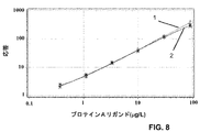
MabSelect SuRe
上に記載されたものと基本的に対応する実験を、CDMX1において、市販ニワトリポリクローナル抗体の代わりに特製捕捉ポリクローナル抗体を用いて、図3に示した手順、すなわちpH 2.5およびpH 2.8で処理するなどを行った。
Special polyclonal anti-protein A antibody as capture and detection antibody (dissociated at pH 2.5, captured at pH 2.8)
MabSelect SuRe
Experiments basically corresponding to those described above were performed in CDMX1 using the custom capture polyclonal antibody instead of the commercially available chicken polyclonal antibody, the procedure shown in FIG. 3, ie pH 2.5 and pH 2.8. And so on.
図8は、(1) プロテインA、および、(2) 5mg/ml濃度のヒトポリクローナルIgG(Octagam(商標))の存在下でのMabSelect SuReについての標準曲線の重ね合わせたチャートを示す。 FIG. 8 shows an overlaid chart of standard curves for MabSelect SuRe in the presence of (1) protein A and (2) human polyclonal IgG (Octagam ™) at a concentration of 5 mg / ml.
図9は、(1) 緩衝液のみ中のMabSelect SuRe、(2) 10mg/mlのHumira(商標)(治療用モノクローナル抗体)中のMabSelect SuRe、(3) 5mg/mlのHumira(商標)中のMabSelect SuRe、および、(4) 2mg/mlのHumira(商標)中のMabSelect SuReについての標準曲線の重ね合わせたチャートを示す。 FIG. 9 shows (1) MabSelect SuRe in buffer only, (2) MabSelect SuRe in 10 mg / ml Humira ™ (therapeutic monoclonal antibody), (3) in 5 mg / ml Humira ™. Figure 2 shows a superimposed chart of standard curves for MabSelect SuRe and (4) MabSelect SuRe in 2 mg / ml Humira ™.
図9の標準曲線を用いて、異なる濃度のHumira(商標)で調製した異なる濃度のMabSelect SuReを含むQCサンプルを、CDMX1において、MabSelect SuRe濃度について分析した。予測濃度に関する平均バイアスを決定した。結果を下の表4に示す。
図10は、(1) 緩衝液のみ、(2) 5mg/mlのHerceptin(商標)の存在下、および、(3) 5mg/mlのHumira(商標)の存在下での残留MabSelect SuReについての標準曲線の重ね合わせたチャートを示す。 Figure 10 shows the standard for residual MabSelect SuRe in (1) buffer only, (2) in the presence of 5 mg / ml Herceptin ™, and (3) in the presence of 5 mg / ml Humira ™. A chart in which curves are superimposed is shown.
図10の標準曲線を用いて5mg/mlの組換え抗体(それぞれHerceptin(商標)およびHumira(商標))を含むサンプル中の残留MabSelect SuReを定量化した。結果を下の表5に表す。
サンプル中の未知濃度のMabSelect SuReの測定
残留MabSelect SuReが混入したIgG含有(Humira(商標))サンプルを提供した。最初にサンプルを5g/Lの濃度に希釈することによって標準化した。次いで、pH 2.5での酸解離およびpH 2.8での捕捉を含む上で概説した手順を用いて、サンプルをプロテインAについて分析した。用量応答 対 MabSelect SuReの濃度についての標準曲線を、図11に示した通り作成した。次いで、元のサンプル中の残留MabSelect SuReの濃度を標準曲線を用いて決定し、希釈因子に対して補正することによって計算した。デュプリケートの測定の精度(CV%)を記載する。結果を下の表6に示す。
Measurement of MabSelect SuRe at unknown concentration in the sample An IgG-containing (Humira ™) sample contaminated with residual MabSelect SuRe was provided. Samples were first normalized by diluting to a concentration of 5 g / L. Samples were then analyzed for protein A using the procedure outlined above, including acid dissociation at pH 2.5 and capture at pH 2.8. A standard curve for dose response versus MabSelect SuRe concentration was generated as shown in FIG. The concentration of residual MabSelect SuRe in the original sample was then determined using a standard curve and calculated by correcting for the dilution factor. Describe the accuracy (CV%) of the duplicate measurement. The results are shown in Table 6 below.
結論
上に示した通り、MabSelect SuRe(商標) リガンド、すなわち免疫グロブリンの親和性クロマトグラフィーからの潜在的浸出液が高濃度のIgGの存在下で正確に定量できる完全自動化微小流体手順が、成功裏に実行された。
In conclusion , as shown above, a fully automated microfluidic procedure that can accurately quantify potential leachates from affinity chromatography of MabSelect SuRe ™ ligand, an immunoglobulin, in the presence of high concentrations of IgG has been successfully implemented. It has been executed.
さらに、その手順を、標準化された方法で異なるpHの異なる緩衝液でサンプルを予め処理できる混合チャンバーを捕捉カラムの上流に有する微小流体構造を含むCD内で行うことができることが証明された。 Furthermore, it has been demonstrated that the procedure can be performed in a CD containing a microfluidic structure having a mixing chamber upstream of the capture column that can pre-process samples with different buffers of different pH in a standardized manner.
手順は、特定のセットアップに依存して、平均約1時間かかる。
検出できるMabSelect SuRe(商標) リガンドの相対濃度は、5mg/mlのIgG(w/w)で、0.2〜0.5ppmの範囲であり、不純物の規制許容レベル(13)よりはるかに小さい相対濃度である。
また、主な解離および分析手順は、5mg/mlのポリクローナルヒトIgG中の天然プロテインAに適合している。
The procedure takes an average of about 1 hour, depending on the specific setup.
The relative concentration of MabSelect SuRe ™ ligand that can be detected is 5 mg / ml IgG (w / w), in the range of 0.2 to 0.5 ppm, much less than the regulatory tolerance level for impurities (13). Concentration.
The main dissociation and analysis procedure is also compatible with native protein A in 5 mg / ml polyclonal human IgG.
キットの組成−残留プロテインAについてのアッセイ
IgG存在下で残留プロテインA(またはMabSelect SuRe)の分析を行うための典型的なキットは、下記の試薬A〜Iを含む。試薬A、BおよびCをそれぞれ希釈試薬G、HおよびIで希釈することを意図したストック溶液として提供する。キット全体は、240個のデータ点(48個/CD)を得る5個のGyrolab(商標) ADA CD (Gyros AB)に十分な量の9種の異なるタイプの液体からなる。
試薬A: 捕捉試薬, ビオチン化抗プロテインA抗体, 625μg/ml
試薬B: 検出試薬, フルオロフォア標識抗プロテインA抗体, 200nM
試薬C: 天然のプロテインA, 1000μg/L
試薬D: 酸解離緩衝液1, 0.25Mのグリシン−HCl(pH 2.5)
試薬E: 酸解離緩衝液2, 0.1Mのクエン酸緩衝液(pH 3.4)
試薬F: 酸性洗浄緩衝液, 1部の試薬Dを一部の試薬Eと混合
試薬G(2バイアル): 中性洗浄緩衝液および捕捉試薬A希釈用の緩衝液
試薬H(2バイアル): サンプル希釈用のサンプル希釈緩衝液, Rexxip(商標) ADA (P0020027, Gyros AB)
試薬I: 希釈検出試薬B(0.5ml)のための検出抗体緩衝液, Rexxip(商標) F (P0004825, Gyros AB)
Kit Composition-Assay for Residual Protein A A typical kit for performing analysis of residual protein A (or MabSelect SuRe) in the presence of IgG comprises the following reagents A-I. Reagents A, B and C are provided as stock solutions intended to be diluted with diluting reagents G, H and I, respectively. The entire kit consists of 9 different types of liquids in sufficient quantities for 5 Gyrolab ™ ADA CDs (Gyros AB) to obtain 240 data points (48 / CD).
Reagent A: Capture reagent, biotinylated anti-protein A antibody, 625 μg / ml
Reagent B: Detection reagent, fluorophore-labeled anti-protein A antibody, 200 nM
Reagent C: Natural protein A, 1000 μg / L
Reagent D:
Reagent E:
Reagent F: Acid wash buffer, 1 part reagent D with some reagent E and mixed reagent G (2 vials): Neutral wash buffer and buffer reagent H for diluting capture reagent A (2 vials): Sample Sample dilution buffer for dilution, Rexxip ™ ADA (P0020027, Gyros AB)
Reagent I: Detection antibody buffer for diluted detection reagent B (0.5 ml), Rexxip ™ F (P0004825, Gyros AB)
サンプル用量が200nlであるとき、200nlの0.1Mのクエン酸緩衝液(pH 3.4)を200nlのサンプルおよび200nlの酸解離緩衝液1(pH 2.5)の混合物に加え、最終pH 2.8となったことに留意する。 When the sample dose is 200 nl, 200 nl 0.1 M citrate buffer (pH 3.4) is added to the mixture of 200 nl sample and 200 nl acid dissociation buffer 1 (pH 2.5) to give a final pH of 2 Note that it was .8.
本発明は、上記好ましい態様に限定されない。様々な代替物、修正および等価物を使用してよい。従って、上記態様は、請求の範囲によって規定される本発明の範囲を限定するものととるべきではない。 The present invention is not limited to the above preferred embodiments. Various alternatives, modifications and equivalents may be used. Accordingly, the above aspects should not be taken as limiting the scope of the invention which is defined by the claims.
引用文献
1. Randall Slemmon J., Meredith J., Guss V., Andreasson U., Andreasen N., Zetterberg H., Blennow K. Measurement of Ab1-42 in cerebrospinal fluid is influenced by matrix effects. J. Neurochem. 212, 325-333, 2012.
2. Murphy G., Nagase H. Progress in matrix metalloproteinase research. Mol. Aspects. Med. 29, 290-308, 2008.
3. Lindahl B., Venge P., Eggers KM., Gedeborg R., Ristiniemi N., Wittfooth S., Pettersson K. Autoantibodies to cardiac troponin in acute coronary syndromes. Clin. Chim. Acta. 411, 1793-1798, 2010.
4. Reuschenbach M., von Knebel Doeberitz M., Wentzensen N. A systematic review of humoral immune responses against tumor antigens. Cancer Immunol. Immunther. 58, 1535-1544, 2009.
5. Saurabh Aggarwal. What's fueling the biotech engine - 2010 to 2011. Nat. Biotechnol. 29, 1083-1089 doi:10.1038/nbt.2060, 2012.
6. Chon JH., Zarbis-Papastoitsis G. Advances in the production and downsteam processing of antibodies. New Biotechnology, 28, 458-463, 2011.
7. Forsgren A, Sjoequist J. "Protein A" from Staphylococcus aureus. I. Pseudo-immune reaction with g-globulin. J. Immunol. 97, 822-827,1966.
8. Inganaes M. Comparison of mechanisms of interaction between protein A from Staphylococcus aureus and human monoclonal IgG, IgA and IgM in relation to the classical Fcγ and alternative F(ab')2ε protein A interactions. Scand. J. Immunol. 13(4), 343-52, 1981.
9. Starovasnik MA, O'Connel MP, Fairbrother WJ, Kelley RF. Antibody variable region binding by Staphylococcal protein A: Thermodynamic analysis and location of the Fv binding site on the E-domain. Protein Science 8, 1423-1431, 1999.
10. Nilsson B., Moks T., Jansson B., Abrahamsen L., Elmblad A., Holmgren E., Henrichson C., Jones TA., Uhlen M. A synthetic IgG-binding domain based on staphylococcal protein A. Protein Eng. 1, 107-113, 1987.
11. Jansson B., Uhlen M., Nygren P-Å. All individual domains of staphylococcal protein A show Fab binding. FEMS Immunology and Medical Microbiology. 20, 69-78, 1998.
12. Hober S., Johansson HJ. Mutant protein. US patent application publication US 2006/0194950 A1.
13. FDC Reports, The Gold Sheet, 38, 1-31, 2004.
14. Steindl F., Armbruster C., Hahn R., Armbruster C., Katinger HWD. A simple method to qauntify staphylococcal protein A in the presence of human or animal IgG in various samples. J. Immunol. Meth. 235, 61-69, 2000.
15. Zhu-Shimoni J., Gunawan F., Thomas A., Vanderlaan M., Stults J. Trace level analysis of leached protein A in bioprocess samples without interference from large excess of rhMab IgG. J. Immunol. Meth. 341, 59-67, 2009.
16. Berglund A, Inganaes M. Method for determining certain bacterial polypeptides and antibodies directed against them. US Patent No. 4,752,571, 1988.
17. van Oss CJ, Absolom DR., Grossberg AL., Neumann AW. Repulsive van der Waals Forces. I. Complete Dissociation of Antigen-Antibody Complexes by Means of Negative van der Waals Forces. Immunol. Comm., 8, 11-29, 1979.
Cited
2. Murphy G., Nagase H. Progress in matrix metalloproteinase research. Mol. Aspects. Med. 29, 290-308, 2008.
3. Lindahl B., Venge P., Eggers KM., Gedeborg R., Ristiniemi N., Wittfooth S., Pettersson K. Autoantibodies to cardiac troponin in acute coronary syndromes. Clin. Chim. Acta. 411, 1793-1798, 2010.
4. Reuschenbach M., von Knebel Doeberitz M., Wentzensen N. A systematic review of humoral immune responses against tumor antigens. Cancer Immunol. Immunther. 58, 1535-1544, 2009.
5. Saurabh Aggarwal. What's fueling the biotech engine-2010 to 2011. Nat. Biotechnol. 29, 1083-1089 doi: 10.1038 / nbt.2060, 2012.
6. Chon JH., Zarbis-Papastoitsis G. Advances in the production and downsteam processing of antibodies. New Biotechnology, 28, 458-463, 2011.
7. Forsgren A, Sjoequist J. “Protein A” from Staphylococcus aureus. I. Pseudo-immune reaction with g-globulin. J. Immunol. 97, 822-827, 1966.
8. Inganaes M. Comparison of mechanisms of interaction between protein A from Staphylococcus aureus and human monoclonal IgG, IgA and IgM in relation to the classical Fcγ and alternative F (ab ') 2 ε protein A interactions. Scand. J. Immunol. 13 (4), 343-52, 1981.
9. Starovasnik MA, O'Connel MP, Fairbrother WJ, Kelley RF. Antibody variable region binding by Staphylococcal protein A: Thermodynamic analysis and location of the Fv binding site on the E-domain. Protein Science 8, 1423-1431, 1999.
10. Nilsson B., Moks T., Jansson B., Abrahamsen L., Elmblad A., Holmgren E., Henrichson C., Jones TA., Uhlen M. A synthetic IgG-binding domain based on staphylococcal protein A. Protein Eng. 1, 107-113, 1987.
11. Jansson B., Uhlen M., Nygren P-Å. All individual domains of staphylococcal protein A show Fab binding. FEMS Immunology and Medical Microbiology. 20, 69-78, 1998.
12. Hober S., Johansson HJ. Mutant protein. US patent application publication US 2006/0194950 A1.
13. FDC Reports, The Gold Sheet, 38, 1-31, 2004.
14. Steindl F., Armbruster C., Hahn R., Armbruster C., Katinger HWD. A simple method to qauntify staphylococcal protein A in the presence of human or animal IgG in various samples. J. Immunol. Meth. 235, 61 -69, 2000.
15. Zhu-Shimoni J., Gunawan F., Thomas A., Vanderlaan M., Stults J. Trace level analysis of leached protein A in bioprocess samples without interference from large excess of rhMab IgG. J. Immunol. Meth. 341, 59-67, 2009.
16. Berglund A, Inganaes M. Method for determining certain bacterial crystals and antibodies directed against them.US Patent No. 4,752,571, 1988.
17. Van Oss CJ, Absolom DR., Grossberg AL., Neumann AW. Repulsive van der Waals Forces. I. Complete Dissociation of Antigen-Antibody Complexes by Means of Negative van der Waals Forces. Immunol. Comm., 8, 11- 29, 1979.
Claims (12)
a) 存在するアナライト複合体を少なくとも実質的に解離し、実質的に全てのアナライトを遊離形で提供するためにサンプルを約1.5〜約3.2の範囲の第1の酸性pHに付し、
b) 第1の酸性pHを、複合体の再形成を阻止するがアナライトのリガンドへの結合が可能である約2.7〜約4.5の範囲の第2の酸性pHまで上げ、
c) サンプル中の該アナライトを定量的に測定するために、アナライトのリガンドへの結合を測定する
工程を含み、アナライトがプロテインAまたはその誘導体であり、サンプルがIgGを含む、方法。 A method for quantitatively measuring an analyte in a liquid sample by an immunoassay comprising binding to a ligand capable of specifically binding to the analyte, wherein the analyte is present at least partially as an analyte complex. ,
a) A first acidic pH in the range of about 1.5 to about 3.2 to at least substantially dissociate the analyte complex present and provide substantially all of the analyte in free form. Attached to
b) raising the first acidic pH to a second acidic pH in the range of about 2.7 to about 4.5 that prevents complex re-formation but allows binding of the analyte to the ligand;
c) A method comprising measuring the binding of an analyte to a ligand to quantitatively measure the analyte in a sample, wherein the analyte is protein A or a derivative thereof and the sample comprises IgG.
アナライトに結合できる検出試薬、
約1.5〜約3.2の範囲のpHを有する第1の酸性緩衝液、
約2.7〜約4.5の範囲のpHを有する第2の酸性緩衝液
を含み、
アナライトがプロテインAであり、
第1の酸性緩衝液は、存在するアナライト複合体を少なくとも実質的に解離し、実質的に全てのアナライトを遊離形で提供するためにサンプルを約1.5〜約3.2の範囲の第1の酸性pHに付すために用いられ、
第2の酸性緩衝液は、第1の酸性pHを、複合体の再形成を阻止するが、アナライト用捕捉試薬としてのリガンドへのアナライトの結合が可能である約2.7〜約4.5の範囲の第2の酸性pHまで上げるために用いられる、
キット。 A kit for performing an immunoassay of an analyte present in a liquid sample at least partially in complex form,
Detection reagent capable of binding to the analyte,
A first acidic buffer having a pH in the range of about 1.5 to about 3.2;
A second acidic buffer having a pH in the range of about 2.7 to about 4.5;
Analyte Ri protein A Der,
The first acidic buffer at least substantially dissociates the analyte complex present and provides the sample in the range of about 1.5 to about 3.2 to provide substantially all of the analyte in free form. Used to subject to a first acidic pH of
The second acidic buffer prevents the complex from re-forming the first acidic pH, but allows binding of the analyte to the ligand as a capture reagent for the analyte. Used to raise to a second acidic pH in the range of .5.
kit.
Applications Claiming Priority (5)
| Application Number | Priority Date | Filing Date | Title |
|---|---|---|---|
| SE1251116 | 2012-10-03 | ||
| SE1251116-8 | 2012-10-03 | ||
| SE1350373-5 | 2013-03-25 | ||
| SE1350373 | 2013-03-25 | ||
| PCT/SE2013/051161 WO2014055025A1 (en) | 2012-10-03 | 2013-10-03 | Method and kit for analyte determination at acidic conditions |
Publications (3)
| Publication Number | Publication Date |
|---|---|
| JP2015531487A JP2015531487A (en) | 2015-11-02 |
| JP2015531487A5 JP2015531487A5 (en) | 2016-10-20 |
| JP6279590B2 true JP6279590B2 (en) | 2018-02-14 |
Family
ID=50435248
Family Applications (1)
| Application Number | Title | Priority Date | Filing Date |
|---|---|---|---|
| JP2015535610A Active JP6279590B2 (en) | 2012-10-03 | 2013-10-03 | Methods and kits for measuring analytes in acidic conditions |
Country Status (5)
| Country | Link |
|---|---|
| US (1) | US10036745B2 (en) |
| EP (1) | EP2904393B1 (en) |
| JP (1) | JP6279590B2 (en) |
| CN (1) | CN104718453B (en) |
| WO (1) | WO2014055025A1 (en) |
Families Citing this family (7)
| Publication number | Priority date | Publication date | Assignee | Title |
|---|---|---|---|---|
| EP3929584A1 (en) * | 2016-09-06 | 2021-12-29 | Fujirebio Inc. | Method for measuring thyroglobulin |
| EP3511713B1 (en) * | 2016-09-06 | 2021-10-27 | Fujirebio Inc. | Tumor marker measurement method and measurement reagent |
| EP3514539A4 (en) * | 2016-09-13 | 2020-04-01 | Fujirebio Inc. | Cardiac troponin assay method and assay reagent |
| KR102551444B1 (en) * | 2017-11-29 | 2023-07-06 | 에프. 호프만-라 로슈 아게 | Anti-drug antibody assay with suppressed target interference |
| EP3830579A1 (en) * | 2018-08-03 | 2021-06-09 | Bristol-Myers Squibb Company | Methods for detecting anti-drug antibodies |
| CN113960318A (en) * | 2021-09-28 | 2022-01-21 | 苏州赛分科技股份有限公司 | Enzyme-linked immunosorbent assay (ELISA) -based recombinant protein A determination method and kit |
| US20240027432A1 (en) * | 2022-07-13 | 2024-01-25 | Regeneron Pharmaceuticals, Inc. | Mild acid immunoassays for detection of analytes |
Family Cites Families (24)
| Publication number | Priority date | Publication date | Assignee | Title |
|---|---|---|---|---|
| SE448190B (en) | 1985-06-03 | 1987-01-26 | Pharmacia Ab | SET TO DETERMINE FC PART BINDING Bacterial POLYPEPTIDES |
| US4703001A (en) * | 1985-10-23 | 1987-10-27 | Synbiotics, Corporation | Immunoassay for the detection of serum analytes using pH dependent chastropic acids |
| WO1991010911A1 (en) | 1990-01-19 | 1991-07-25 | Repligen Corporation | Immunoassay of protein a under acidic conditions |
| US5314804A (en) * | 1992-03-24 | 1994-05-24 | Serim Research Corporation | Test for Helicobacter pylori |
| GB9809943D0 (en) | 1998-05-08 | 1998-07-08 | Amersham Pharm Biotech Ab | Microfluidic device |
| WO2002074438A2 (en) | 2001-03-19 | 2002-09-26 | Gyros Ab | Structural units that define fluidic functions |
| CA2441206A1 (en) | 2001-03-19 | 2002-09-26 | Gyros Ab | Characterization of reaction variables |
| EP2281633A1 (en) | 2001-08-28 | 2011-02-09 | Gyros Patent Ab | Retaining microfluidic microcavity and other microfluidic structures |
| US20040009178A1 (en) * | 2002-02-11 | 2004-01-15 | Bowdish Katherine S. | Immunotherapeutics for biodefense |
| SE0200943D0 (en) | 2002-03-25 | 2002-03-25 | Amersham Biosciences Ab | Mutant protein |
| JP4115728B2 (en) * | 2002-03-26 | 2008-07-09 | デンカ生研株式会社 | Composition for flow-through type inspection method, kit and inspection method using the same |
| JP4423189B2 (en) * | 2002-05-31 | 2010-03-03 | ユィロス・パテント・アクチボラグ | Detection device based on surface plasmon resonance |
| WO2004083108A1 (en) | 2003-03-23 | 2004-09-30 | Gyros Patent Ab | Preloaded microscale devices |
| SE0300822D0 (en) | 2003-03-23 | 2003-03-23 | Gyros Ab | A collection of Micro Scale Devices |
| CN101097217A (en) * | 2006-06-29 | 2008-01-02 | 王珊珊 | Technology for enhancing blood serum specificity growth factor immunologic assay sensitivity |
| CN101583872A (en) * | 2006-09-14 | 2009-11-18 | 通用电气健康护理生物科学股份公司 | A method of determining analyte concentration |
| WO2008033073A1 (en) | 2006-09-14 | 2008-03-20 | Ge Healthcare Bio-Sciences Ab | A method of determining analyte concentration |
| US20080125454A1 (en) * | 2006-10-05 | 2008-05-29 | Wyeth | Methods and Compositions for Treatment of Scleritis and Related Disorders |
| WO2009022001A1 (en) * | 2007-08-16 | 2009-02-19 | Novartis Ag | Improvement of drug tolerance in immunogenicity testing |
| CA2720478A1 (en) | 2008-04-03 | 2009-10-08 | F. Hoffmann-La Roche Ag | Pegylated insulin-like-growth-factor assay |
| CN102209897B (en) | 2008-09-11 | 2015-04-29 | 巴斯德研究院 | Monitoring and inhibiting human immunodeficiency virus infection by modulating hmgb1 dependent triggering of hiv-1 replication and persistence |
| US8546149B2 (en) | 2010-08-27 | 2013-10-01 | Intervet Inc. | Potency test for vaccine formulations |
| ES2530175T3 (en) | 2011-02-17 | 2015-02-26 | Nestec S.A. | Tests for the detection of autoantibodies against anti-TNF drugs |
| WO2013121368A2 (en) | 2012-02-15 | 2013-08-22 | Biocon Limited | A process for detection and optional quantification of an analyte |
-
2013
- 2013-10-03 EP EP13843833.8A patent/EP2904393B1/en active Active
- 2013-10-03 JP JP2015535610A patent/JP6279590B2/en active Active
- 2013-10-03 CN CN201380051645.1A patent/CN104718453B/en active Active
- 2013-10-03 US US14/433,276 patent/US10036745B2/en active Active
- 2013-10-03 WO PCT/SE2013/051161 patent/WO2014055025A1/en active Application Filing
Also Published As
| Publication number | Publication date |
|---|---|
| EP2904393B1 (en) | 2018-11-28 |
| CN104718453B (en) | 2017-03-08 |
| EP2904393A1 (en) | 2015-08-12 |
| US20150268238A1 (en) | 2015-09-24 |
| EP2904393A4 (en) | 2016-05-04 |
| JP2015531487A (en) | 2015-11-02 |
| WO2014055025A1 (en) | 2014-04-10 |
| CN104718453A (en) | 2015-06-17 |
| US10036745B2 (en) | 2018-07-31 |
Similar Documents
| Publication | Publication Date | Title |
|---|---|---|
| JP6279590B2 (en) | Methods and kits for measuring analytes in acidic conditions | |
| Zhang et al. | High performance affinity chromatography and related separation methods for the analysis of biological and pharmaceutical agents | |
| JP7399096B2 (en) | Assays to detect neurodegeneration | |
| CA3003776A1 (en) | A method for quantifying anti-tnf antibodies | |
| JP7382071B2 (en) | How to measure glycated hemoglobin (%) | |
| Kangwa et al. | An engineered Staphylococcal Protein A based ligand: Production, characterization and potential application for the capture of Immunoglobulin and Fc-fusion proteins | |
| US11085931B2 (en) | Universal assay for determining the quantity of TNFα inhibitory drugs and their corresponding anti-drug-antibodies | |
| CN109891240A (en) | The detection method of apolipoprotein E | |
| JP2023533806A (en) | A blood-based assay for detecting tauopathies or amyloidogenic diseases | |
| WO2017188312A1 (en) | Assay method for anti-drug antibodies | |
| JP4554356B2 (en) | Sandwich assays and kits | |
| JP2009508087A (en) | Monoclonal antibody reagent | |
| Mortezai et al. | Combining lectin affinity chromatography and immunodepletion–a novel method for the enrichment of disease-specific glycoproteins in human plasma | |
| JP6571660B2 (en) | Antibody analysis | |
| Subramanian et al. | Interaction of immunoglobulin G with N, N, N′, N′-ethylenediaminetetramethylenephosphonic acid-modified zirconia | |
| EP3177312B1 (en) | Method and kit for detecting bacterial infection | |
| JP5448424B2 (en) | Reagent for measuring protein containing Fc of human IgG | |
| JP5231954B2 (en) | Albumin measuring reagent | |
| Pihlblad | Development and comparison of bioanalytical methods to measure free analyte | |
| JP4753366B2 (en) | % CDT quantification method | |
| JPWO2017033846A1 (en) | Immunoassay method and immunity test kit | |
| WO2022076459A1 (en) | High-throughput method for lp(a)-cholesterol quantitation | |
| EP3956665A1 (en) | Methods and systems for detecting antibodies by surface plasmon resonance | |
| Kärnhall | New SPR based assays for plasma protein titer determination | |
| JP2004132816A (en) | Method for determining blood anticoagulation factor antibody by enzyme-linked immunosorbent assay |
Legal Events
| Date | Code | Title | Description |
|---|---|---|---|
| A521 | Request for written amendment filed |
Free format text: JAPANESE INTERMEDIATE CODE: A523 Effective date: 20160902 |
|
| A621 | Written request for application examination |
Free format text: JAPANESE INTERMEDIATE CODE: A621 Effective date: 20160902 |
|
| A977 | Report on retrieval |
Free format text: JAPANESE INTERMEDIATE CODE: A971007 Effective date: 20170621 |
|
| A131 | Notification of reasons for refusal |
Free format text: JAPANESE INTERMEDIATE CODE: A131 Effective date: 20170627 |
|
| A521 | Request for written amendment filed |
Free format text: JAPANESE INTERMEDIATE CODE: A523 Effective date: 20170901 |
|
| TRDD | Decision of grant or rejection written | ||
| A01 | Written decision to grant a patent or to grant a registration (utility model) |
Free format text: JAPANESE INTERMEDIATE CODE: A01 Effective date: 20180109 |
|
| A61 | First payment of annual fees (during grant procedure) |
Free format text: JAPANESE INTERMEDIATE CODE: A61 Effective date: 20180117 |
|
| R150 | Certificate of patent or registration of utility model |
Ref document number: 6279590 Country of ref document: JP Free format text: JAPANESE INTERMEDIATE CODE: R150 |
|
| R250 | Receipt of annual fees |
Free format text: JAPANESE INTERMEDIATE CODE: R250 |
|
| R250 | Receipt of annual fees |
Free format text: JAPANESE INTERMEDIATE CODE: R250 |
|
| R250 | Receipt of annual fees |
Free format text: JAPANESE INTERMEDIATE CODE: R250 |
|
| R250 | Receipt of annual fees |
Free format text: JAPANESE INTERMEDIATE CODE: R250 |