JP6203747B2 - Method for detecting free binding partners of multispecific binders - Google Patents
Method for detecting free binding partners of multispecific binders Download PDFInfo
- Publication number
- JP6203747B2 JP6203747B2 JP2014547933A JP2014547933A JP6203747B2 JP 6203747 B2 JP6203747 B2 JP 6203747B2 JP 2014547933 A JP2014547933 A JP 2014547933A JP 2014547933 A JP2014547933 A JP 2014547933A JP 6203747 B2 JP6203747 B2 JP 6203747B2
- Authority
- JP
- Japan
- Prior art keywords
- antibody
- antigen
- multispecific
- sample
- binding specificity
- Prior art date
- Legal status (The legal status is an assumption and is not a legal conclusion. Google has not performed a legal analysis and makes no representation as to the accuracy of the status listed.)
- Active
Links
Images


Classifications
-
- G—PHYSICS
- G01—MEASURING; TESTING
- G01N—INVESTIGATING OR ANALYSING MATERIALS BY DETERMINING THEIR CHEMICAL OR PHYSICAL PROPERTIES
- G01N33/00—Investigating or analysing materials by specific methods not covered by groups G01N1/00 - G01N31/00
- G01N33/48—Biological material, e.g. blood, urine; Haemocytometers
- G01N33/50—Chemical analysis of biological material, e.g. blood, urine; Testing involving biospecific ligand binding methods; Immunological testing
- G01N33/53—Immunoassay; Biospecific binding assay; Materials therefor
- G01N33/543—Immunoassay; Biospecific binding assay; Materials therefor with an insoluble carrier for immobilising immunochemicals
- G01N33/54306—Solid-phase reaction mechanisms
-
- G—PHYSICS
- G01—MEASURING; TESTING
- G01N—INVESTIGATING OR ANALYSING MATERIALS BY DETERMINING THEIR CHEMICAL OR PHYSICAL PROPERTIES
- G01N33/00—Investigating or analysing materials by specific methods not covered by groups G01N1/00 - G01N31/00
- G01N33/48—Biological material, e.g. blood, urine; Haemocytometers
- G01N33/50—Chemical analysis of biological material, e.g. blood, urine; Testing involving biospecific ligand binding methods; Immunological testing
- G01N33/53—Immunoassay; Biospecific binding assay; Materials therefor
- G01N33/543—Immunoassay; Biospecific binding assay; Materials therefor with an insoluble carrier for immobilising immunochemicals
-
- G—PHYSICS
- G01—MEASURING; TESTING
- G01N—INVESTIGATING OR ANALYSING MATERIALS BY DETERMINING THEIR CHEMICAL OR PHYSICAL PROPERTIES
- G01N33/00—Investigating or analysing materials by specific methods not covered by groups G01N1/00 - G01N31/00
- G01N33/48—Biological material, e.g. blood, urine; Haemocytometers
- G01N33/50—Chemical analysis of biological material, e.g. blood, urine; Testing involving biospecific ligand binding methods; Immunological testing
- G01N33/53—Immunoassay; Biospecific binding assay; Materials therefor
- G01N33/5306—Improving reaction conditions, e.g. reduction of non-specific binding, promotion of specific binding
-
- G—PHYSICS
- G01—MEASURING; TESTING
- G01N—INVESTIGATING OR ANALYSING MATERIALS BY DETERMINING THEIR CHEMICAL OR PHYSICAL PROPERTIES
- G01N33/00—Investigating or analysing materials by specific methods not covered by groups G01N1/00 - G01N31/00
- G01N33/48—Biological material, e.g. blood, urine; Haemocytometers
- G01N33/50—Chemical analysis of biological material, e.g. blood, urine; Testing involving biospecific ligand binding methods; Immunological testing
- G01N33/53—Immunoassay; Biospecific binding assay; Materials therefor
- G01N33/543—Immunoassay; Biospecific binding assay; Materials therefor with an insoluble carrier for immobilising immunochemicals
- G01N33/54393—Improving reaction conditions or stability, e.g. by coating or irradiation of surface, by reduction of non-specific binding, by promotion of specific binding
Landscapes
- Health & Medical Sciences (AREA)
- Life Sciences & Earth Sciences (AREA)
- Immunology (AREA)
- Engineering & Computer Science (AREA)
- Chemical & Material Sciences (AREA)
- Hematology (AREA)
- Urology & Nephrology (AREA)
- Biomedical Technology (AREA)
- Molecular Biology (AREA)
- Microbiology (AREA)
- Cell Biology (AREA)
- Biotechnology (AREA)
- Food Science & Technology (AREA)
- Medicinal Chemistry (AREA)
- Physics & Mathematics (AREA)
- Analytical Chemistry (AREA)
- Biochemistry (AREA)
- General Health & Medical Sciences (AREA)
- General Physics & Mathematics (AREA)
- Pathology (AREA)
- Chemical Kinetics & Catalysis (AREA)
- Peptides Or Proteins (AREA)
Description
本発明は、サンプル中の多特異性結合体によって特異的に結合され得る、遊離の、すなわち複合体化していない結合パートナーの検出方法であって、多特異性結合体に結合した結合パートナーが遊離結合パートナーの検出前にサンプルから除去(deplete)される、方法、に関する。 The present invention is a method for detecting free, ie uncomplexed, binding partners that can be specifically bound by a multispecific binder in a sample, wherein the binding partner bound to the multispecific binder is free. The method is depleted from the sample prior to detection of the binding partner.
発明の背景
抗体を用いる標準的な固相免疫アッセイは、固相に吸着/固定化された抗体(捕捉抗体)、抗原および酵素または検出可能な標識にコンジュゲートされた抗原の別のエピトープに対する抗体(トレーサー抗体)の間で複合体を形成させるものである。このアッセイでは、固相/捕捉抗体/抗原/トレーサー抗体のサンドイッチが形成される。このサンドイッチによって触媒される反応においては、とりわけ、抗体にコンジュゲートされた酵素の活性が、インキュベート培地中の抗原濃度に比例する。抗イディオタイプ抗体アッセイは、例えば、US 5,219,730(特許文献1); WO 87/002778(特許文献2); EP 0 139 389(特許文献3); およびEP 0 170 302(特許文献4)で言及されている。Wadhwa, M.,ら(J. Immunol. Methods 278 (2003) 1-17)(非特許文献1)は、生物学的治療によって誘導される望ましくない抗体の検出、測定および特徴づけの戦略を報告している。抗イディオタイプ抗体を製造する方法は、EP 1 917 854(特許文献5)で報告されている。
BACKGROUND OF THE INVENTION Standard solid-phase immunoassays using antibodies include antibodies adsorbed / immobilized on a solid phase (capture antibody), antigens and antibodies against another epitope of the antigen conjugated to an enzyme or detectable label. A complex is formed between (tracer antibody). In this assay, a solid phase / capture antibody / antigen / tracer antibody sandwich is formed. In the reaction catalyzed by this sandwich, among other things, the activity of the enzyme conjugated to the antibody is proportional to the antigen concentration in the incubation medium. Anti-idiotype antibody assays are mentioned, for example, in US 5,219,730 (Patent Document 1); WO 87/002778 (Patent Document 2);
Chen, Y.-P.,ら(Clin. Vac. Immunol. 14 (2007) 720-725)(非特許文献2)は、ヒト赤血球およびB型肝炎ウイルス表面抗原の両方に対する2特異性ダイアボディ(diabody)よって媒介される凝集アッセイによるB型肝炎ウイルス表面抗原の高速検出を報告している。癌治療用のヒト化2特異性モノクローナル抗体の特徴づけは、Bruynck, A.,ら(J. Cancer 67 (1993) 436-440)(非特許文献3)によって報告されている。WO 2006/096697(特許文献6)では、タンパク質および抗体治療の2価性を決定する方法が報告されている。 Chen, Y.-P., et al. (Clin. Vac. Immunol. 14 (2007) 720-725) (2) is a bispecific diabody against both human erythrocytes and hepatitis B virus surface antigens ( report rapid detection of hepatitis B virus surface antigen by an aggregation assay mediated by diabody). Characterization of humanized bispecific monoclonal antibodies for cancer treatment has been reported by Bruynck, A., et al. (J. Cancer 67 (1993) 436-440) (Non-patent Document 3). WO 2006/096697 (Patent Document 6) reports a method for determining the bivalency of protein and antibody therapy.
発明の要旨
本明細書には、多特異性結合体の少なくとも1つの結合特異性によって特異的に結合され得る、遊離の、すなわち複合体化されていない結合パートナーの存在の検出および/または量の決定のための方法が報告されている。遊離結合パートナーの存在または量の決定前に、多特異性結合体によって特異的に結合された結合パートナーを、すなわち結合パートナー・多特異性結合体複合体を、サンプルから除去することが有利であることが見出された。本明細書に報告される方法にしたがい、多特異性結合体の除去は、多特異性結合体の1つの結合特異性に特異的に結合する単特異性結合体と共にサンプルをインキュベートすることによって達成され、ここで、単特異性結合体は、決定対象の結合パートナーに結合しない多特異性結合体の結合特異性に特異的に結合する(図2を参照のこと)。
SUMMARY OF THE INVENTION The present specification includes detection and / or amount detection of the presence of a free, ie non-complexed binding partner, that can be specifically bound by at least one binding specificity of a multispecific binder. Methods for making decisions are reported. Prior to determining the presence or amount of free binding partner, it is advantageous to remove from the sample the binding partner specifically bound by the multispecific conjugate, ie the binding partner-multispecific conjugate complex. It was found. In accordance with the methods reported herein, removal of multispecific binders is accomplished by incubating the sample with a monospecific binder that specifically binds to one binding specificity of the multispecific binder. Where the monospecific binder specifically binds to the binding specificity of a multispecific binder that does not bind to the binding partner to be determined (see FIG. 2).
本明細書に報告される1つの局面は、多特異性結合体の第1の結合特異性によって特異的に結合され得る結合パートナー(抗原、標的、分析物)の存在および/または量のインビトロ決定方法であって、多特異性結合体に結合された結合パートナーが、多特異性結合体の第2の結合特異性に特異的に結合する単特異性結合体と共にサンプルをインキュベートすることによって、結合パートナーの検出前に除去される、方法、である。 One aspect reported herein is the in vitro determination of the presence and / or amount of a binding partner (antigen, target, analyte) that can be specifically bound by the first binding specificity of the multispecific binder. A method wherein a binding partner bound to a multispecific binder binds by incubating the sample with a monospecific binder that specifically binds to the second binding specificity of the multispecific binder. A method that is removed prior to partner detection.
1つの態様において、検出される結合パートナーは、複合体化されていない結合パートナーまたは遊離結合パートナーである。 In one embodiment, the binding partner to be detected is an uncomplexed binding partner or a free binding partner.
したがって、本明細書に報告される1つの局面は、多特異性結合体の結合パートナーの存在および/または量の決定のためのインビトロ方法であって、結合パートナーが、多特異性結合体の第1の結合特異性によって特異的に結合され得るものであり、
- 結合パートナーおよび多特異性結合体を含むサンプルを、多特異性結合体の第1の結合特異性とは異なる第2の結合特異性に特異的に結合する単特異性結合体と共にインキュベートする工程、
を含む、方法、である。
Accordingly, one aspect reported herein is an in vitro method for determining the presence and / or amount of a binding partner of a multispecific binder, wherein the binding partner is the first of the multispecific binder. Can be specifically bound by the binding specificity of 1.
Incubating a sample comprising a binding partner and a multispecific binder with a monospecific binder that specifically binds to a second binding specificity that is different from the first binding specificity of the multispecific binder. ,
Including a method.
1つの態様において、この方法は、
- 結合パートナーおよび多特異性結合体を含むサンプルを、多特異性結合体の第1の結合特異性とは異なる第2の結合特異性に特異的に結合する単特異性結合体と共にインキュベートする工程、ならびに
- 多特異性結合体除去サンプル中の結合パートナーの量を決定する工程、
を含む。
In one embodiment, the method comprises:
Incubating a sample comprising a binding partner and a multispecific binder with a monospecific binder that specifically binds to a second binding specificity that is different from the first binding specificity of the multispecific binder. And
-Determining the amount of binding partner in the multispecific binder removal sample;
including.
1つの態様において、この方法は、
- 結合パートナーおよび多特異性結合体を含むサンプルを、多特異性結合体の第1の結合特異性とは異なる第2の結合特異性に特異的に結合する単特異性結合体と共にインキュベートする工程、
- 遊離結合パートナーの存在または量の決定前に、単特異性結合体・多特異性結合体複合体をサンプルから除去する工程、ならびに
- 多特異性結合体除去サンプル中の結合パートナーの量を決定する工程、
を含む。
In one embodiment, the method comprises:
Incubating a sample comprising a binding partner and a multispecific binder with a monospecific binder that specifically binds to a second binding specificity that is different from the first binding specificity of the multispecific binder. ,
-Removing the monospecific / multispecific conjugate complex from the sample prior to determining the presence or amount of free binding partner, and
-Determining the amount of binding partner in the multispecific binder removal sample;
including.
多特異性結合体の第2の結合特異性に特異的に結合する単特異性結合体とのインキュベートにより、多特異性結合体がサンプルから取り除かれる。それに伴い、結合パートナー・多特異性結合体複合体もサンプルから取り除かれる。 Incubation with a monospecific conjugate that specifically binds to the second binding specificity of the multispecific conjugate removes the multispecific conjugate from the sample. Accordingly, the binding partner / multispecific conjugate complex is also removed from the sample.
1つの態様において、多特異性結合体は、抗体、抗体もしくは抗体フラグメントおよび非抗体ポリペプチドを含む融合ポリペプチド、抗体もしくは抗体フラグメントおよび可溶性受容体を含む融合ポリペプチド、または抗体もしくは抗体フラグメントおよびペプチド性結合分子を含む融合ポリペプチドから選択される。 In one embodiment, the multispecific conjugate is a fusion polypeptide comprising an antibody, antibody or antibody fragment and a non-antibody polypeptide, a fusion polypeptide comprising an antibody or antibody fragment and a soluble receptor, or an antibody or antibody fragment and peptide. Selected from fusion polypeptides comprising sex binding molecules.
1つの態様において、多特異性結合体は、抗体である。1つの態様において、抗体は、2特異性抗体または3特異性抗体または4特異性抗体または5特異性抗体または6特異性抗体である。1つの態様において、抗体は、2特異性抗体である。 In one embodiment, the multispecific conjugate is an antibody. In one embodiment, the antibody is a bispecific antibody or a trispecific antibody or a 4-specific antibody or a 5-specific antibody or a 6-specific antibody. In one embodiment, the antibody is a bispecific antibody.
1つの態様において、単特異性結合体は、抗イディオタイプ抗体である。 In one embodiment, the monospecific conjugate is an anti-idiotype antibody.
1つの態様において、結合特異性は、結合部位または抗体重鎖可変ドメインと抗体軽鎖可変ドメインとの対である。 In one embodiment, the binding specificity is a binding site or pair of antibody heavy chain variable domain and antibody light chain variable domain.
1つの態様において、抗イディオタイプ抗体は、固相に結合される。 In one embodiment, the anti-idiotype antibody is bound to a solid phase.
1つの態様において、抗イディオタイプ抗体はビオチニル化され、そして固相はストレプトアビジンコーティングされる。1つの態様において、固相は、ストレプトアビジンコーティングされた常磁性ビーズまたはストレプトアビジンコーティングされたセファロースビーズである。 In one embodiment, the anti-idiotype antibody is biotinylated and the solid phase is streptavidin coated. In one embodiment, the solid phase is streptavidin-coated paramagnetic beads or streptavidin-coated sepharose beads.
本明細書に報告される1つの局面は、免疫アッセイを用いる、サンプル中の多特異性結合体の結合パートナーの存在および/または量の免疫学的決定方法であって、多特異性結合体が、結合パートナーの決定前にサンプルから除去される、方法、である。 One aspect reported herein is a method of immunological determination of the presence and / or amount of a binding partner of a multispecific binder in a sample using an immunoassay, wherein the multispecific binder is A method that is removed from the sample prior to determination of the binding partner.
本明細書に報告されるすべての局面の1つの態様において、結合パートナーは、遊離結合パートナー、すなわち、多特異性結合体によって結合または複合体化されていない結合パートナーである。 In one embodiment of all aspects reported herein, the binding partner is a free binding partner, ie, a binding partner that is not bound or complexed by a multispecific binder.
1つの態様において、抗イディオタイプ抗体は、多特異性結合体に対するビオチニル化抗イディオタイプ抗体であり、ストレプトアビジンを介して固相にコンジュゲートされる。 In one embodiment, the anti-idiotype antibody is a biotinylated anti-idiotype antibody to the multispecific conjugate and is conjugated to the solid phase via streptavidin.
本明細書に報告される方法の1つの態様において、抗イディオタイプ抗体は、固相にコンジュゲートされる抗体部位に関して異なる少なくとも2つの抗イディオタイプ抗体を含む混合物である。 In one embodiment of the methods reported herein, the anti-idiotype antibody is a mixture comprising at least two anti-idiotype antibodies that differ with respect to the antibody site conjugated to the solid phase.
1つの態様において、抗体とそのコンジュゲートパートナーとのコンジュゲートは、薬物抗体のアミノ酸骨格のN末端および/もしくはε-アミノ基(リシン)、異なるリシンのε-アミノ基、カルボキシ官能基、スルフヒドリル官能基、ヒドロキシル官能基および/もしくはフェノール官能基、ならびに/または薬物抗体の炭水化物構造の糖アルコール基を介した化学結合によって行われる。 In one embodiment, the conjugate of the antibody and its conjugate partner comprises an N-terminus of the drug antibody amino acid backbone and / or an ε-amino group (lysine), an ε-amino group of a different lysine, a carboxy function, a sulfhydryl function This is done by chemical conjugation via a group, a hydroxyl functional group and / or a phenol functional group and / or a sugar alcohol group of the carbohydrate structure of the drug antibody.
1つの態様において、抗イディオタイプ抗体混合物は、少なくとも2つの異なるアミノ基を介して固相にコンジュゲートされた抗イディオタイプ抗体を含む。そのような異なるアミノ基を介したカップリングは、第1工程における、化学保護剤によるε-アミノ基の一部のアシル化によって、例えばシトラコニル化(citraconylation)によって、行われ得る。第2工程において、残りのアミノ基を介してコンジュゲートが行われる。その後にシトラコニル化が取り除かれ、そして抗体が残りの遊離アミノ基を介して固相にコンジュゲートされる、すなわち、得られた抗体が、シトラコニル化によって保護されなくなったアミノ基を介して固相にコンジュゲートされる。適当な化学保護剤は、保護されていない側鎖アミンにおいて結合を形成し、それはN末端におけるそれらの結合よりも安定性が低く、かつN末端におけるそれらの結合と異なる。多くのそのような化学保護剤が公知である(例えば、EP 0 651 761を参照のこと)。1つの態様において、化学保護剤は、マレイン酸無水物またはシトラコニル酸(citraconylic acid)無水物等の環状ジカルボン酸無水物を含む。
In one embodiment, the anti-idiotype antibody mixture comprises an anti-idiotype antibody conjugated to the solid phase via at least two different amino groups. Such coupling via different amino groups can be carried out by acylation of a part of the ε-amino group with a chemical protecting agent in the first step, for example by citraconylation. In the second step, conjugation takes place via the remaining amino group. Thereafter, citraconylation is removed and the antibody is conjugated to the solid phase via the remaining free amino groups, i.e., the resulting antibody is brought to the solid phase via amino groups that are no longer protected by citraconylation. Conjugated. Suitable chemical protecting agents form bonds at unprotected side chain amines, which are less stable than their linkage at the N-terminus and differ from their linkage at the N-terminus. Many such chemical protectants are known (see, for example,
1つの態様において、抗イディオタイプ抗体は、受動的吸着によって固相にコンジュゲートされる。受動的吸着は、例えば、"Solid Phases in Immunoassay" (1996) 205-225においてButler, J.E.,により、ならびに"Immunoassays" (1996) Academic Press (San Diego)においてDiamandis, E.P.,およびChristopoulos, T.K.(編者)により、記載されている。 In one embodiment, the anti-idiotype antibody is conjugated to the solid phase by passive adsorption. Passive adsorption is described, for example, by Butler, JE, in “Solid Phases in Immunoassay” (1996) 205-225, and Diamandis, EP, and Christopoulos, TK (editor) in “Immunoassays” (1996) Academic Press (San Diego). ).
1つの態様において、抗イディオタイプ抗体は、特異的な結合対を介してコンジュゲート(固定化)される。そのような結合対(第1の要素/第2の要素)は、1つの態様において、ストレプトアビジンまたはアビジン/ビオチン、抗体/抗原(例えば、Hermanson, G.T., et al., Bioconjugate Techniques, Academic Press (1996)を参照のこと)、レクチン/多糖類、ステロイド/ステロイド結合タンパク質、ホルモン/ホルモン受容体、酵素/基質、IgG/プロテインAおよび/またはG等から選択される。1つの態様において、抗イディオタイプ抗体はビオチンにコンジュゲートされ、そして固定化は固定化されたアビジンまたはストレプトアビジンを介して行われる。 In one embodiment, the anti-idiotype antibody is conjugated (immobilized) via a specific binding pair. Such binding pairs (first element / second element), in one embodiment, are streptavidin or avidin / biotin, antibody / antigen (eg, Hermanson, GT, et al., Bioconjugate Techniques, Academic Press ( 1996)), lectins / polysaccharides, steroids / steroid binding proteins, hormones / hormone receptors, enzymes / substrates, IgG / protein A and / or G and the like. In one embodiment, the anti-idiotype antibody is conjugated to biotin, and immobilization is performed via immobilized avidin or streptavidin.
本明細書に報告される1つの局面は、サンプル中の多特異性抗体の抗原の存在および/または量の決定のためのインビトロ方法であって、検出される抗原が、多特異性抗体の第1の結合特異性によって特異的に結合され得るものであり、
- 多特異性抗体、多特異性抗体に結合した抗原および遊離抗原を含むサンプルを、多特異性抗体の第1の結合特異性とは異なる第2の結合特異性に特異的に結合する抗イディオタイプ抗体と共にインキュベートする工程、
を含む、方法、である。
One aspect reported herein is an in vitro method for determining the presence and / or amount of an antigen of a multispecific antibody in a sample, wherein the detected antigen is the first of the multispecific antibody. Can be specifically bound by the binding specificity of 1.
-An anti-idio that specifically binds a sample containing a multispecific antibody, an antigen bound to the multispecific antibody and a free antigen to a second binding specificity that is different from the first binding specificity of the multispecific antibody. Incubating with a type antibody,
Including a method.
1つの態様において、この方法は、
- 抗原および多特異性抗体を含むサンプルを、多特異性抗体の第1の結合特異性とは異なる第2の結合特異性に特異的に結合する抗イディオタイプ抗体と共にインキュベートする工程、ならびに
- 多特異性抗体除去サンプル中の抗原の量を決定する工程、
を含む。
In one embodiment, the method comprises:
Incubating a sample comprising an antigen and a multispecific antibody with an anti-idiotype antibody that specifically binds to a second binding specificity that is different from the first binding specificity of the multispecific antibody; and
-Determining the amount of antigen in the multispecific antibody-removed sample;
including.
1つの態様において、この方法は、
- 抗原および多特異性抗体を含むサンプルを、多特異性抗体の第1の結合特異性とは異なる第2の結合特異性に特異的に結合する抗イディオタイプ抗体と共にインキュベートする工程、
- 遊離抗原の存在または量の決定前に、抗イディオタイプ抗体・多特異性抗体複合体をサンプルから除去する工程、ならびに
- 多特異性抗体除去サンプル中の抗原の量を決定する工程、
を含む。
In one embodiment, the method comprises:
Incubating a sample comprising an antigen and a multispecific antibody with an anti-idiotype antibody that specifically binds to a second binding specificity that is different from the first binding specificity of the multispecific antibody;
-Removing the anti-idiotype antibody-multispecific antibody complex from the sample prior to determining the presence or amount of free antigen, and
-Determining the amount of antigen in the multispecific antibody-removed sample;
including.
多特異性抗体の第2の結合特異性に特異的に結合する抗イディオタイプ抗体とのインキュベートにより、多特異性抗体がサンプルから取り除かれる。それに伴い、抗原・多特異性抗体複合体もサンプルから取り除かれる。 Incubation with an anti-idiotype antibody that specifically binds to the second binding specificity of the multispecific antibody removes the multispecific antibody from the sample. Along with this, the antigen / multispecific antibody complex is also removed from the sample.
1つの態様において、サンプルは、多特異性抗体、遊離抗原および多特異性抗体・抗原複合体を含み、検出は多特異性抗体の遊離抗原の検出である。 In one embodiment, the sample comprises a multispecific antibody, a free antigen and a multispecific antibody-antigen complex, and the detection is detection of the free antigen of the multispecific antibody.
1つの態様において、抗イディオタイプ抗体は、常磁性ビーズにコンジュゲートされる。 In one embodiment, the anti-idiotype antibody is conjugated to paramagnetic beads.
1つの態様において、抗イディオタイプ抗体は、固相にコンジュゲートされる。 In one embodiment, the anti-idiotype antibody is conjugated to a solid phase.
1つの態様において、抗イディオタイプ抗体はビオチニル化され、そして固相はストレプトアビジンコーティングされる。1つの態様において、固相は、ストレプトアビジンコーティングされた常磁性ビーズまたはストレプトアビジンコーティングされたセファロースビーズである。 In one embodiment, the anti-idiotype antibody is biotinylated and the solid phase is streptavidin coated. In one embodiment, the solid phase is streptavidin-coated paramagnetic beads or streptavidin-coated sepharose beads.
1つの態様において、抗イディオタイプ抗体は、多特異性抗体の第2の結合特異性に対して105 l/mol*sまたはそれ以上の会合定数kaを有する。 In one embodiment, anti-idiotypic antibodies have 10 5 l / mol * s or more association constant k a for a second binding specificity of the multispecific antibody.
1つの態様において、抗イディオタイプ抗体は、多特異性抗体の第2の結合特異性に対する結合に関して5*10-8 mol/lまたはそれ以下のKD値を有する。 In one embodiment, the anti-idiotype antibody has a K D value of 5 * 10 −8 mol / l or less for binding to the second binding specificity of the multispecific antibody.
1つの態様において、結合特異性は、結合部位である。1つの態様において、結合部位は、抗体重鎖可変ドメインと抗体軽鎖可変ドメインとの対である。 In one embodiment, the binding specificity is a binding site. In one embodiment, the binding site is a pair of antibody heavy chain variable domain and antibody light chain variable domain.
1つの態様において、抗イディオタイプ抗体とインキュベートすることは、約10分間〜約36時間である。 In one embodiment, incubating with the anti-idiotype antibody is from about 10 minutes to about 36 hours.
1つの態様において、サンプルは、約2μg/ml〜約15μg/mlの多特異性抗体濃度に調節される。 In one embodiment, the sample is adjusted to a multispecific antibody concentration of about 2 μg / ml to about 15 μg / ml.
1つの態様において、サンプルは、約1ng/ml〜約250ng/mlの総抗原濃度に調節される。 In one embodiment, the sample is adjusted to a total antigen concentration of about 1 ng / ml to about 250 ng / ml.
1つの態様において、この方法は、以下の工程、
- 多特異性抗体、多特異性抗体に結合した抗原および遊離抗原を含むサンプルを、多特異性抗体の第1の結合特異性とは異なる第2の結合特異性に特異的に結合する抗イディオタイプ抗体と共にインキュベートし、抗イディオタイプ抗体・多特異性抗体複合体を形成させる工程、ならびに
- 抗イディオタイプ抗体・多特異性抗体複合体をサンプルから取り除く工程、
を含む。
In one embodiment, the method comprises the following steps:
-An anti-idio that specifically binds a sample containing a multispecific antibody, an antigen bound to the multispecific antibody and a free antigen to a second binding specificity that is different from the first binding specificity of the multispecific antibody. Incubating with a type antibody to form an anti-idiotype antibody-multispecific antibody complex, and
-Removing the anti-idiotype antibody / multispecific antibody complex from the sample;
including.
1つの態様において、抗イディオタイプ抗体・多特異性抗体複合体は、抗イディオタイプ抗体・多特異性抗体複合体および抗イディオタイプ抗体・多特異性抗体・抗原複合体の混合物である。 In one embodiment, the anti-idiotype antibody / multispecific antibody complex is an anti-idiotype antibody / multispecific antibody complex and a mixture of anti-idiotype antibody / multispecific antibody / antigen complex.
1つの態様において、この方法は、以下の工程、
- 抗原および多特異性抗体を含むサンプルを、多特異性抗体の第1の結合特異性とは異なる第2の結合特異性に特異的に結合する抗イディオタイプ抗体と共にインキュベートし、抗イディオタイプ抗体・多特異性抗体複合体を形成させる工程、
- 抗イディオタイプ抗体・多特異性抗体複合体をサンプルから取り除く工程、ならびに
- 多特異性抗体除去サンプル中の抗原の量を決定する工程、
を含む。
In one embodiment, the method comprises the following steps:
-Incubating a sample containing the antigen and multispecific antibody with an anti-idiotype antibody that specifically binds to a second binding specificity that is different from the first binding specificity of the multispecific antibody; A step of forming a multispecific antibody complex,
-Removing the anti-idiotype antibody / multispecific antibody complex from the sample, and
-Determining the amount of antigen in the multispecific antibody-removed sample;
including.
1つの態様において、抗原の量の決定は、以下の工程、
- 多特異性抗体除去サンプルを、抗原に特異的に結合する捕捉抗体と共にインキュベートし、捕捉抗体・抗原複合体を形成させる工程、および
- 形成された捕捉抗体・抗原複合体をサンプル中の抗原の量と相関させる工程、
を含む。
In one embodiment, determining the amount of antigen comprises the following steps:
-Incubating the multispecific antibody-removed sample with a capture antibody that specifically binds to the antigen to form a capture antibody-antigen complex; and
-Correlating the formed capture antibody / antigen complex with the amount of antigen in the sample,
including.
1つの態様において、抗原の量の決定は、以下の工程、
- 多特異性抗体除去サンプルを、抗原に特異的に結合する捕捉抗体と共にインキュベートし、捕捉抗体・抗原複合体を形成させる工程、
- 捕捉抗体・抗原複合体をトレーサー抗体と共にインキュベートする工程であって、それによって、捕捉抗体およびトレーサー抗体が抗原上の重複しないエピトープに結合する、工程、および
- 形成された捕捉抗体・抗原・トレーサー抗体複合体をサンプル中の抗原の量と相関させる工程、
を含む。
In one embodiment, determining the amount of antigen comprises the following steps:
-Incubating the multispecific antibody-removed sample with a capture antibody that specifically binds to the antigen to form a capture antibody-antigen complex;
-Incubating the capture antibody-antigen complex with the tracer antibody, whereby the capture antibody and the tracer antibody bind to non-overlapping epitopes on the antigen; and
-Correlating the formed capture antibody / antigen / tracer antibody complex with the amount of antigen in the sample,
including.
1つの態様において、抗原の量の決定は、以下の工程、
- 多特異性抗体除去サンプルを、抗原に特異的に結合する捕捉抗体と共にインキュベートし、捕捉抗体・抗原複合体を形成させる工程、
- 捕捉抗体・抗原複合体をトレーサー抗体と共にインキュベートする工程であって、それによって、捕捉抗体およびトレーサー抗体が抗原上の重複しないエピトープに結合する、工程、
- 捕捉抗体・抗原・トレーサー抗体複合体を、検出可能な標識を含む検出抗体と共にインキュベートする工程であって、それによって、検出抗体がトレーサー抗体の可変ドメインの外側のエピトープにおいてトレーサー抗体に特異的に結合する、工程、および
- 形成された捕捉抗体・抗原・トレーサー抗体複合体をサンプル中の抗原の量と相関させる工程、
を含む。
In one embodiment, determining the amount of antigen comprises the following steps:
-Incubating the multispecific antibody-removed sample with a capture antibody that specifically binds to the antigen to form a capture antibody-antigen complex;
-Incubating the capture antibody-antigen complex with the tracer antibody, whereby the capture antibody and the tracer antibody bind to non-overlapping epitopes on the antigen;
-Incubating the capture antibody-antigen-tracer antibody complex with a detection antibody comprising a detectable label, whereby the detection antibody is specific to the tracer antibody at an epitope outside the variable domain of the tracer antibody. Combine, process, and
-Correlating the formed capture antibody / antigen / tracer antibody complex with the amount of antigen in the sample,
including.
1つの態様において、多特異性抗体は、第1の抗原または抗原上の第1のエピトープに特異的に結合する第1の結合特異性を有し、かつ第2の抗原または抗原上の第2のエピトープに特異的に結合する第2の結合特異性を有する2特異性抗体である。 In one embodiment, the multispecific antibody has a first binding specificity that specifically binds to a first antigen or a first epitope on an antigen and a second on a second antigen or antigen. A bispecific antibody having a second binding specificity that specifically binds to the epitope of.
本明細書に報告される1つの局面は、多特異性抗体の第2の結合特異性に結合した抗原をサンプルから除去するための、多特異性抗体の第1の結合特異性に特異的に結合する抗イディオタイプ抗体の使用、である。 One aspect reported herein is specifically directed to the first binding specificity of the multispecific antibody to remove antigens bound to the second binding specificity of the multispecific antibody from the sample. Use of an anti-idiotype antibody that binds.
発明の詳細な説明
本明細書には、前臨床および臨床サンプル中の多特異性結合体、例えば2特異性抗体/薬物の「遊離結合パートナー」を検出するための、サンプルの前処理のインビトロ方法が報告されている。
DETAILED DESCRIPTION OF THE INVENTION In vitro methods of sample pretreatment for detecting multispecific binders in preclinical and clinical samples, such as “free binding partners” of bispecific antibodies / drugs, are described herein. Has been reported.
遊離結合パートナーの検出前に多特異性結合体がサンプルから除去されなければならないことが見出された。 It has been found that the multispecific binder must be removed from the sample prior to detection of the free binding partner.
本明細書には、治療用多特異性抗体の結合特異性に特異的に結合する抗イディオタイプ抗体の、該治療用多特異性抗体の第2の異なる結合特異性によって結合され得るが結合されていない抗原のレベルの決定における使用、が報告されている。多特異性抗体および多特異性抗体・検出対象抗原の複合体をサンプルから除去するために、抗イディオタイプ抗体が使用される。 Included herein is an anti-idiotype antibody that specifically binds to the binding specificity of a therapeutic multispecific antibody, which can be bound by a second different binding specificity of the therapeutic multispecific antibody. Use in determining the level of non-antigens has been reported. An anti-idiotype antibody is used to remove the multispecific antibody and the multispecific antibody / antigen to be detected complex from the sample.
したがって、本明細書には、多特異性結合体の第1の結合特異性によって特異的に結合され得る多特異性結合体の遊離結合パートナー(抗原、標的、分析物)の決定のためのインビトロ方法であって、多特異性結合体が、多特異性結合体の第1の結合特異性とは異なる第2の結合特異性に特異的に結合する単特異性結合体と共にサンプルをインキュベートすることによって、遊離結合パートナーの決定前にサンプルから除去され、それと共に、多特異性結合体および多特異性結合体・結合パートナー複合体がサンプルから除去される、方法、が報告されている。 Thus, herein, in vitro for the determination of a free binding partner (antigen, target, analyte) of a multispecific conjugate that can be specifically bound by the first binding specificity of the multispecific conjugate. A method, wherein the multispecific conjugate is incubated with a monospecific conjugate that specifically binds to a second binding specificity that is different from the first binding specificity of the multispecific conjugate. By which the multispecific binder and the multispecific binder-binding partner complex are removed from the sample prior to determination of the free binding partner.
総抗原、2特異性抗体に結合した抗原および遊離抗原の決定は、治療用抗体を用いる治療のモニタリングに役立つ。総抗原は、遊離抗原および2特異性抗体に結合した抗原の和である。 Determination of total antigen, antigen bound to bispecific antibody and free antigen is useful for monitoring therapy with therapeutic antibodies. Total antigen is the sum of free antigen and antigen bound to the bispecific antibody.
以下では、本明細書に報告される方法が、多特異性結合体の態様として複数の抗原または同一抗原上の複数のエピトープに特異的に結合する多特異性抗体を、そして結合パートナーの態様として多特異性抗体の1つの結合特異性によって特異的に結合される抗原を用いて例証されている。 In the following, the methods reported herein include multispecific antibodies that specifically bind to multiple antigens or multiple epitopes on the same antigen as multispecific conjugate embodiments, and as binding partner embodiments. Illustrated with antigens that are specifically bound by one binding specificity of a multispecific antibody.
「抗体」という用語は、本明細書において最も広義の意味で使用され、所望の抗原結合活性を示す限り、モノクローナル抗体、ポリクローナル抗体、多特異性抗体(例えば、2特異性抗体)および抗体フラグメントを含むがこれらに限定されない様々な抗体構造を包含する。 The term “antibody” is used herein in the broadest sense and includes monoclonal antibodies, polyclonal antibodies, multispecific antibodies (eg, bispecific antibodies) and antibody fragments as long as they exhibit the desired antigen binding activity. A variety of antibody structures are included, including but not limited to.
特定の態様において、抗体は、多特異性抗体、例えば2特異性抗体である。多特異性抗体は、少なくとも2つの異なる部位に対する結合特異性を有するモノクローナル抗体である。特定の態様において、結合特異性の1つは、第1の抗原に対するものであり、他は、異なる第2の抗原に対するものである。特定の態様において、2特異性抗体は、同一抗原の2つの異なるエピトープに結合し得る。2特異性抗体は、全長抗体または抗体フラグメントとして調製され得る。1つの態様において、抗体は、第1および第2の抗原に特異的に結合する2特異性抗体である。1つの態様において、2特異性抗体は、i)第1の抗原または抗原上の第1のエピトープに特異的に結合する第1の結合特異性、およびii)第2の抗原または同一抗原上の第2のエピトープに特異的に結合する第2の結合特異性、を有する。1つの態様において、同一抗原上の第2のエピトープは、重複しないエピトープである。 In certain embodiments, the antibody is a multispecific antibody, such as a bispecific antibody. Multispecific antibodies are monoclonal antibodies that have binding specificities for at least two different sites. In certain embodiments, one of the binding specificities is for a first antigen and the other is for a different second antigen. In certain embodiments, a bispecific antibody can bind to two different epitopes of the same antigen. Bispecific antibodies can be prepared as full length antibodies or antibody fragments. In one embodiment, the antibody is a bispecific antibody that specifically binds to the first and second antigens. In one embodiment, the bispecific antibody is i) a first binding specificity that specifically binds to a first antigen or a first epitope on an antigen, and ii) a second antigen or on the same antigen. Having a second binding specificity that specifically binds to a second epitope. In one embodiment, the second epitope on the same antigen is a non-overlapping epitope.
多特異性抗体は、WO 2009/080251、WO 2009/080252、WO 2009/080253、WO 2009/080254、WO 2010/112193、WO 2010/115589、WO 2010/136172、WO 2012/145792またはWO 2010/145793に記載されている。 Multispecific antibodies may be WO 2009/080251, WO 2009/080252, WO 2009/080253, WO 2009/080254, WO 2010/112193, WO 2010/115589, WO 2010/136172, WO 2012/145792 or WO 2010/145793. It is described in.
「抗体フラグメント」は、インタクトな抗体が結合する抗原に結合するインタクトな抗体の一部分を含むインタクトな抗体ではない分子を表す。抗体フラグメントの例は、Fv、Fab、Fab'、Fab'-SH、F(ab')2;ダイアボディ;リニア抗体;単鎖抗体分子(例えば、scFv);および抗体フラグメントから形成される多特異性抗体を含むがこれらに限定されない。 “Antibody fragment” refers to a molecule that is not an intact antibody, comprising a portion of an intact antibody that binds to an antigen to which the intact antibody binds. Examples of antibody fragments are Fv, Fab, Fab ′, Fab′-SH, F (ab ′) 2 ; diabodies; linear antibodies; single chain antibody molecules (eg, scFv); and multispecifics formed from antibody fragments Including, but not limited to, sex antibodies.
抗体の「クラス」は、その重鎖が保有する定常ドメインまたは定常領域のタイプを表す。抗体にはIgA、IgD、IgE、IgGおよびIgMという5つの大クラスがあり、これらのいくつかはサブクラス(アイソタイプ)、例えばIgG1、IgG2、IgG3、IgG4、IgA1およびIgA2にさらに分類され得る。免疫グロブリンの異なるクラスに対応する重鎖定常ドメインは、それぞれ、α、δ、ε、γおよびμと呼ばれる。 The “class” of an antibody represents the type of constant domain or constant region possessed by its heavy chain. There are five major classes of antibodies, IgA, IgD, IgE, IgG and IgM, some of which are further subclasses (isotypes) such as IgG 1 , IgG 2 , IgG 3 , IgG 4 , IgA 1 and IgA 2 Can be classified. The heavy chain constant domains that correspond to the different classes of immunoglobulins are called α, δ, ε, γ, and μ, respectively.
「遊離抗原」という用語は、抗体の結合特異性によって特異的に結合され得るが、その時点ではこの結合特異性に結合されていない抗原を意味する。1つの態様において、遊離抗原は、抗体に結合していない抗原または抗体と複合体化していない抗原である。 The term “free antigen” means an antigen that can be specifically bound by the binding specificity of an antibody, but is not bound to this binding specificity at that time. In one embodiment, the free antigen is an antigen that is not bound to an antibody or is not complexed with an antibody.
「Fc領域」という用語は、本明細書において、その定常領域の少なくとも一部分を含む免疫グロブリン重鎖のC末端領域を定義するために使用される。この用語は、ネイティブ配列のFc領域および変種のFc領域を含む。1つの態様において、ヒトIgG重鎖のFc領域は、Cys226からまたはPro230から重鎖のカルボキシル末端まで延びている。しかし、Fc領域のC末端リシン(Lys447)は、存在する場合と存在しない場合がある。それ以外のことが本明細書に示されていない限り、Fc領域または定常領域におけるアミノ酸残基の番号は、Kabat, E.A. et al., Sequences of Proteins of Immunological Interest, 5th ed., Public Health Service, National Institutes of Health, Bethesda, MD (1991), NIH Publication 91-3242に記載される、EUインデックスとも呼ばれる、EU番号体系にしたがうものである。 The term “Fc region” is used herein to define the C-terminal region of an immunoglobulin heavy chain that includes at least a portion of its constant region. The term includes native sequence Fc regions and variant Fc regions. In one embodiment, the Fc region of a human IgG heavy chain extends from Cys226 or from Pro230 to the heavy chain carboxyl terminus. However, the C-terminal lysine (Lys447) of the Fc region may or may not be present. Unless otherwise indicated herein, the number of amino acid residues in the Fc region or constant region is Kabat, EA et al., Sequences of Proteins of Immunological Interest, 5th ed., Public Health Service, It follows the EU numbering system, also called the EU index, described in National Institutes of Health, Bethesda, MD (1991), NIH Publication 91-3242.
「フレームワーク」または「FR」は、超可変領域(HVR)の残基以外の可変ドメインの残基を表す。可変ドメインのFRは、通常、FR1、FR2、FR3およびFR4という4つのFRドメインからなっている。したがって、HVRおよびFRの配列は、通常、VH(またはVL)において以下の順で見られる:FR1-H1(L1)-FR2-H2(L2)-FR3-H3(L3)-FR4。 “Framework” or “FR” refers to residues of the variable domain other than the hypervariable region (HVR) residues. The FR of a variable domain usually consists of four FR domains, FR1, FR2, FR3 and FR4. Thus, HVR and FR sequences are usually found in the following order in VH (or VL): FR1-H1 (L1) -FR2-H2 (L2) -FR3-H3 (L3) -FR4.
「ヒト抗体」は、ヒトもしくはヒト細胞によって産生されるまたはヒト抗体レパートリーもしくはその他のヒト抗体コード配列を利用する非ヒト供給源から得られる抗体のそれに対応するアミノ酸配列を保有するものである。このヒト抗体の定義は、特に、非ヒト抗原結合残基を含むヒト化抗体を除外する。 A “human antibody” is one which possesses the amino acid sequence corresponding to that of an antibody produced by a human or human cell or obtained from a non-human source utilizing a human antibody repertoire or other human antibody coding sequence. This definition of a human antibody specifically excludes a humanized antibody comprising a non-human antigen binding residue.
「ヒト化」抗体は、非ヒトHVR由来のアミノ酸残基およびヒトFR由来のアミノ酸残基を含むキメラ抗体を表す。特定の態様において、ヒト化抗体は、HVR(例えば、CDR)のすべてまたは実質的にすべてが非ヒト抗体のそれらに対応し、FRのすべてまたは実質的にすべてがヒト抗体のそれらに対応する、少なくとも1つ、典型的には2つの可変ドメインの実質的にすべてを含むであろう。ヒト化抗体は、任意で、ヒト抗体由来の抗体定常領域の少なくとも一部分を含み得る。抗体、例えば非ヒト抗体の「ヒト化形態」は、ヒト化処理が行われた抗体を表す。 A “humanized” antibody refers to a chimeric antibody comprising amino acid residues from non-human HVR and amino acid residues from human FR. In certain embodiments, humanized antibodies have all or substantially all of the HVRs (eg, CDRs) corresponding to those of non-human antibodies, and all or substantially all of the FRs correspond to those of human antibodies, It will contain at least one, typically substantially all of the two variable domains. A humanized antibody optionally can comprise at least a portion of an antibody constant region derived from a human antibody. A “humanized form” of an antibody, eg, a non-human antibody, refers to an antibody that has been humanized.
「超可変領域」または「HVR」という用語は、本明細書で使用される場合、配列に関して超可変性がありおよび/または構造的に定義されたループ(「超可変ループ」)を形成する抗体可変ドメインの各領域を表す。通常、ネイティブの4鎖抗体は、VHに3つ(H1、H2、H3)およびVLに3つ(L1、L2、L3)の6つのHVRを含む。HVRは通常、超可変ループ由来および/または「相補性決定領域」(CDR)由来のアミノ酸残基を含み、後者は最も高い配列可変性を有するおよび/または抗原認識に関与するものである。例示的な超可変ループは、アミノ酸残基26〜32(L1)、50〜52(L2)、91〜96(L3)、26〜32(H1)、53〜55(H2)および96〜101(H3)で見られる(Chothia, C. and Lesk, A.M., J. Mol. Biol. 196 (1987) 901-917)。例示的なCDR(CDR-L1、CDR-L2、CDR-L3、CDR-H1、CDR-H2およびCDR-H3)は、L1のアミノ酸残基24〜34、L2の50〜56、L3の89〜97、H1の31〜35B、H2の50〜65およびH3の95〜102で見られる(Kabat, E.A. et al., Sequences of Proteins of Immunological Interest, 5th ed. Public Health Service, National Institutes of Health, Bethesda, MD (1991), NIH Publication 91-3242)。VHのCDR1を除いて、CDRは通常、超可変ループを形成するアミノ酸残基を含む。CDRはまた、抗原に接触する残基である「特異性決定残基」または「SDR」を含む。SDRは、簡易CDR(abbreviated CDR)またはa-CDRと呼ばれるCDRの領域に含まれる。例示的なa-CDR(a-CDR-L1、a-CDR-L2、a-CDR-L3、a-CDR-H1、a-CDR-H2およびa-CDR-H3)は、L1のアミノ酸残基31〜34、L2の50〜55、L3の89〜96、H1の31〜35B、H2の50〜58およびH3の95〜102で見られる(Almagro, J.C. and Fransson, J., Front. Biosci. 13 (2008) 1619-1633)。それ以外のことが示されていない限り、HVR残基および可変ドメインのその他の残基(例えば、FR残基)は、本明細書において、Kabat et al.,前記にしたがい番号付けされる。 The term “hypervariable region” or “HVR”, as used herein, is an antibody that is hypervariable with respect to sequence and / or forms a structurally defined loop (“hypervariable loop”). Represents each region of the variable domain. Typically, a native 4-chain antibody contains 6 HVRs, 3 in VH (H1, H2, H3) and 3 in VL (L1, L2, L3). HVRs usually contain amino acid residues from hypervariable loops and / or from “complementarity determining regions” (CDRs), the latter having the highest sequence variability and / or involved in antigen recognition. Exemplary hypervariable loops are amino acid residues 26-32 (L1), 50-52 (L2), 91-96 (L3), 26-32 (H1), 53-55 (H2), and 96-101 ( H3) (Chothia, C. and Lesk, AM, J. Mol. Biol. 196 (1987) 901-917). Exemplary CDRs (CDR-L1, CDR-L2, CDR-L3, CDR-H1, CDR-H2, and CDR-H3) are L1-amino acid residues 24-34, L2 50-56, L3 89- 97, H1 31-35B, H2 50-65 and H3 95-102 (Kabat, EA et al., Sequences of Proteins of Immunological Interest, 5th ed. Public Health Service, National Institutes of Health, Bethesda , MD (1991), NIH Publication 91-3242). With the exception of VH CDR1, CDRs usually contain amino acid residues that form a hypervariable loop. CDRs also include “specificity determining residues” or “SDRs” that are residues that contact antigen. The SDR is contained in a CDR region called a simple CDR (abbreviated CDR) or a-CDR. Exemplary a-CDRs (a-CDR-L1, a-CDR-L2, a-CDR-L3, a-CDR-H1, a-CDR-H2 and a-CDR-H3) are amino acid residues of L1 31-34, L2 50-55, L3 89-96, H1 31-35B, H2 50-58 and H3 95-102 (Almagro, JC and Fransson, J., Front. Biosci. 13 (2008) 1619-1633). Unless indicated otherwise, HVR residues and other residues of variable domains (eg, FR residues) are numbered herein according to Kabat et al., Supra.
本明細書で使用される「モノクローナル抗体」という用語は、実質的に均質な抗体の集団から得られる抗体を表す、すなわち、その集団を形成する個々の抗体は、例えば自然発生する変異を含むまたはモノクローナル抗体調製物の製造時に生じる起こり得る変種抗体、例えば通常微量で存在する変種、を除いて、同一であるおよび/または同一エピトープに結合する。典型的に異なる決定基(エピトープ)に対する異なる抗体を含むポリクローナル抗体調製物とは対照的に、モノクローナル抗体調製物の各モノクローナル抗体は、抗原上の単一の決定基に対するものである。したがって、「モノクローナル」という修飾語は、実質的に均質な抗体集団から得られるという抗体の特徴を示しており、何らかの特定の方法による抗体の製造を必要とするものと解釈されるべきではない。例えば、本発明にしたがい使用されるモノクローナル抗体は、ハイブリドーマ法、組み換えDNA法、ファージディスプレイ法およびヒト免疫グロブリン遺伝子座のすべてまたは一部分を含むトランスジェニック動物を用いる方法を含むがこれらに限定されない様々な技術によって作製され得、そのような方法およびその他の例示的なモノクローナル抗体作製法は本明細書に記載されている。 The term “monoclonal antibody” as used herein refers to an antibody obtained from a population of substantially homogeneous antibodies, ie, the individual antibodies forming that population contain, for example, naturally occurring mutations or It binds to the same and / or the same epitope except for possible variant antibodies that occur during the production of monoclonal antibody preparations, such as variants that are usually present in trace amounts. In contrast to polyclonal antibody preparations that typically contain different antibodies directed against different determinants (epitopes), each monoclonal antibody of the monoclonal antibody preparation is directed against a single determinant on the antigen. Thus, the modifier “monoclonal” indicates the character of the antibody as being obtained from a substantially homogeneous antibody population, and is not to be construed as requiring production of the antibody by any particular method. For example, monoclonal antibodies used in accordance with the present invention include various methods including, but not limited to, hybridoma methods, recombinant DNA methods, phage display methods, and methods using transgenic animals containing all or part of human immunoglobulin loci. Such methods and other exemplary monoclonal antibody production methods can be made by techniques and are described herein.
「可変領域」または「可変ドメイン」という用語は、抗原への抗体の結合に関与する抗体の重鎖または軽鎖のドメインを表す。ネイティブ抗体の重鎖および軽鎖の可変ドメイン(それぞれ、VHおよびVL)は、通常、類似の構造を有し、各ドメインは4つの保存されたフレームワーク領域(FR)および3つの超可変領域(HVR)を含む(例えば、Kindt, T.J. et al. Kuby Immunology, 6th ed., W.H. Freeman and Co., N.Y. (2007)の91ページを参照のこと)。抗原結合特異性を与えるのに単一のVHまたはVLドメインで十分であり得る。さらに、特定の抗原に結合する抗体は、その抗原に結合する抗体由来のVHまたはVLドメインをそれぞれ相補的なVLまたはVHドメインのライブラリをスクリーニングするために用いて、単離され得る(例えば、Portolano, S. et al., J. Immunol. 150 (1993) 880-887; Clackson, T. et al., Nature 352 (1991) 624-628を参照のこと)。 The term “variable region” or “variable domain” refers to the heavy or light chain domain of an antibody involved in binding of the antibody to an antigen. The heavy and light chain variable domains of native antibodies (VH and VL, respectively) usually have a similar structure, with each domain having four conserved framework regions (FR) and three hypervariable regions ( HVR) (see, for example, page 91 of Kindt, TJ et al. Kuby Immunology, 6th ed., WH Freeman and Co., NY (2007)). A single VH or VL domain may be sufficient to confer antigen binding specificity. In addition, antibodies that bind to a particular antigen can be isolated using a VH or VL domain from an antibody that binds that antigen, respectively, to screen a library of complementary VL or VH domains (eg, Portolano S. et al., J. Immunol. 150 (1993) 880-887; Clackson, T. et al., Nature 352 (1991) 624-628).
「抗イディオタイプ抗体」という用語は、結合特異性、例えば親抗体の結合部位、に特異的に結合する、すなわち、例えば親抗体の抗原結合部位に対する、抗体を表す。1つの態様において、抗イディオタイプ抗体は、親抗体のCDRの1つまたは複数に特異的に結合する。1つの態様において、親抗体は、治療用抗体である。1つの態様において、親抗体は、多特異性抗体である。1つの態様において、親抗体は、2特異性抗体である。 The term “anti-idiotype antibody” refers to an antibody that specifically binds to a binding specificity, eg, a binding site of a parent antibody, ie, eg, to an antigen binding site of a parent antibody. In one embodiment, the anti-idiotype antibody specifically binds to one or more of the CDRs of the parent antibody. In one embodiment, the parent antibody is a therapeutic antibody. In one embodiment, the parent antibody is a multispecific antibody. In one embodiment, the parent antibody is a bispecific antibody.
2つのエピトープは、問題のエピトープを20〜50nMの濃度とし、エピトープ重複を検出したい抗体を100nMの濃度とする、固定化された抗体および可溶性の抗原、またはその逆、を用いる表面プラズモン共鳴(SPR)アッセイによって50%またはそれ以上、1つの態様においては75%またはそれ以上のシグナル減少が検出される場合、重複している。あるいは、同一抗原に結合する2つの抗体のエピトープ重複を競合試験系の下で決定する方法を使用することができる。この目的で、例えば、組み換え抗原エピトープを発現する細胞を用いる細胞ベースの酵素免疫アッセイ(ELISA)の下で、固定化された抗原への結合に関してエピトープ重複を検出したい抗体が他の抗体と競合するかどうかが試験される。この目的で、固定化された抗原は、標識された形式の抗体および過剰なエピトープ重複を決定対象の抗体と共にインキュベートされる。結合した標識の検出によって、エピトープ重複を容易に確認することができる。同一濃度下での70%超、1つの態様においては80%超のシグナル減少、またはより高濃度、1つの例では105倍過剰の、既知抗体と称される、エピトープ重複を検出したい抗体の下での80%超、1つの態様においては90%超の置換が、決定され、次いで、エピトープの同一性または重複性が存在することおよび両方の抗体が同一抗原上の同一または重複するエピトープに結合することが決定される。 The two epitopes are surface plasmon resonance (SPR) using immobilized antibody and soluble antigen, or vice versa, with the epitope of interest at a concentration of 20-50 nM and the antibody at which epitope duplication is desired to be detected at a concentration of 100 nM. ) Duplication if the assay detects 50% or more, in one embodiment 75% or more signal reduction. Alternatively, a method can be used in which the epitope overlap of two antibodies that bind to the same antigen is determined under a competitive test system. To this end, for example, antibodies that wish to detect epitope duplication for binding to immobilized antigen compete with other antibodies under a cell-based enzyme immunoassay (ELISA) using cells expressing recombinant antigenic epitopes. To be tested. For this purpose, the immobilized antigen is incubated with the labeled form of the antibody and the antibody whose excess epitope duplication is to be determined. Epitope duplication can be easily confirmed by detection of bound label. Over 70% of the signal under the same concentration, in one embodiment over 80% signal reduction, or higher, in one example, a 10 5 fold excess of the antibody that you want to detect epitope duplication, called a known antibody Substitution greater than 80%, in one embodiment greater than 90%, is determined and then there is epitope identity or redundancy and both antibodies are on the same or overlapping epitopes on the same antigen It is decided to combine.
異なる免疫アッセイの原理が、例えば、Hage, D.S.(Anal. Chem. 71 (1999) 294R-304R)によって記載されている。Lu, B.,ら(Analyst 121 (1996) 29R-32R)は、免疫アッセイにおいて使用する抗体の有向固定化(oriented immobilization)を報告している。アビジン・ビオチンを介した免疫アッセイが、例えば、Methods Enzymol. 184 (1990) 467-469において、Wilchek, M.,およびBayer, E.A.,によって報告されている。 The principle of different immunoassays is described, for example, by Hage, D.S. (Anal. Chem. 71 (1999) 294R-304R). Lu, B., et al. (Analyst 121 (1996) 29R-32R) report directed immobilization of antibodies for use in immunoassays. An avidin-biotin-mediated immunoassay has been reported by Wilchek, M., and Bayer, E.A., for example, in Methods Enzymol. 184 (1990) 467-469.
モノクローナル抗体およびそれらの定常ドメインは、タンパク質として、結合パートナー、例えば表面、タンパク質、ポリマー(例えば、PEG、セルロースもしくはポリスチロール)、酵素または多くの結合対、とカップリングする多くの反応性側鎖を含んでいる。抗体の化学反応基は、例えば、アミノ基(リシン、アルファ・アミノ基)、チオール基(シスチン、システインおよびメチオニン)、カルボン酸基(アスパラギン酸、グルタミン酸)および糖アルコール基である。そのような方法は、例えば、"Bioconjugation", MacMillan Ref. Ltd. 1999, pp.50-100において、Aslam M.,およびDent, A.,によって記載されている。 Monoclonal antibodies and their constant domains have many reactive side chains that couple as proteins with binding partners, such as surfaces, proteins, polymers (eg, PEG, cellulose or polystyrene), enzymes or many binding pairs. Contains. The chemically reactive groups of antibodies are, for example, amino groups (lysine, alpha amino group), thiol groups (cystine, cysteine and methionine), carboxylic acid groups (aspartic acid, glutamic acid) and sugar alcohol groups. Such a method is described, for example, in “Bioconjugation”, MacMillan Ref. Ltd. 1999, pp. 50-100 by Aslam M., and Dent, A.,.
タンパク質の最も一般的な反応基の1つは、アミノ酸リシンの脂肪族ε-アミンである。概ね、ほぼすべての抗体が、豊富にリシンを含んでいる。リシンのアミンは、pH 8.0より上でほど良い求核体であり(pKa = 9.18)、したがって様々な試薬と容易かつ手際よく反応して安定な結合を形成する。アミン反応性試薬は、主としてリシンおよびタンパク質のα-アミノ基と反応する。反応性エステル、特にN-ヒドロキシ-スクシンイミド(NHS)エステルは、アミン基の修飾に最も広く利用されている試薬である。水性環境下での反応の至適pHは、pH 8.0〜9.0である。イソチオシアネートは、アミン修飾試薬であり、タンパク質とチオ尿素結合を形成する。それらは水溶液(pH 9.0〜9.5が最適)中でタンパク質アミンと反応する。アルデヒドは、穏やかな水性条件下で、脂肪族および芳香族アミン、ヒドラジンおよびヒドラジドと反応し、イミン中間体(シッフ塩基)を形成する。シッフ塩基は、穏やかまたは強い還元剤(例えば、水素化ホウ素ナトリウムまたはシアノ水素化ホウ素ナトリウム)によって選択的に還元され、安定なアルキルアミン結合を生じ得る。アミンの修飾に使用されている他の試薬は、酸無水物である。例えば、ジエチレントリアミン五酢酸無水物(DTPA)は、2つのアミン反応性無水物基を含む2官能性キレート化剤である。それはタンパク質のN末端およびε-アミン基と反応して、アミド連結を形成することができる。無水物の環は、配位錯体内で金属に緊密に結合することができる多価の金属キレート化アームを形成するよう開環する。 One of the most common reactive groups of proteins is the aliphatic ε-amine of the amino acid lysine. Almost all antibodies are rich in lysine. Amine of a lysine is the better nucleophiles above the pH 8.0 (pK a = 9.18) , thus forming a stable bond to easily and cleanly react with various reagents. Amine-reactive reagents react primarily with lysine and the α-amino group of proteins. Reactive esters, particularly N-hydroxy-succinimide (NHS) esters, are the most widely used reagents for modification of amine groups. The optimum pH for the reaction in an aqueous environment is pH 8.0 to 9.0. Isothiocyanate is an amine modifying reagent and forms thiourea bonds with proteins. They react with protein amines in aqueous solution (optimally pH 9.0-9.5). Aldehydes react with aliphatic and aromatic amines, hydrazine and hydrazide under mild aqueous conditions to form imine intermediates (Schiff bases). The Schiff base can be selectively reduced with a mild or strong reducing agent (eg, sodium borohydride or sodium cyanoborohydride) to produce a stable alkylamine linkage. Another reagent that has been used to modify amines is an acid anhydride. For example, diethylenetriaminepentaacetic anhydride (DTPA) is a bifunctional chelating agent that contains two amine-reactive anhydride groups. It can react with the N-terminus of proteins and ε-amine groups to form amide linkages. The anhydride ring opens to form a multivalent metal chelating arm that can bind tightly to the metal within the coordination complex.
抗体内の別の一般的な反応基は、硫黄含有アミノ酸シスチンおよびその還元産物システイン(またはハーフシスチン)のチオール残基である。システインは、アミンよりも求核性でありそして一般にタンパク質内で最も反応性の高い官能基である遊離チオール基を含んでいる。チオールは一般に、中性pHで反応性であり、したがってアミンの存在下で選択的に他の分子にカップリングさせることができる。遊離スルフヒドリル基は比較的反応性が高いので、これらの基を有するタンパク質は、しばしば、ジスルフィド基またはジスルフィド結合のようなそれらの酸化形態にあるそれらと共に存在する。そのようなタンパク質において、反応性の遊離チオールの生成には、試薬、例えばジチオトレイトール(DTT)によるジスルフィド結合の還元が必要とされる。チオール反応性試薬は、タンパク質上のチオール基とカップリングしてチオエーテルカップリング産物を形成し得るものである。これらの試薬は、弱酸性〜中性pHで迅速に反応し、したがって、アミン基の存在下で選択的に反応させることができる。文献は、反応性アミノ基を介した複数のスルフヒドリル基の効率的導入方法を提供するための、様々なチオール化架橋試薬、例えばトラウト試薬(2-イミノチオラン)、スクシンイミジル(アセチルチオ)アセテート(SATA)およびスルホスクシンイミジル6-[3-(2-ピリジルジチオ)プロピオンアミド]ヘキサノエート(Sulfo-LC-SPDP)の使用を報告している。ハロアセチル誘導体、例えばヨードアセトアミドは、チオエーテル結合を形成し、これもチオール修飾試薬である。さらなる有用な試薬は、マレイミドである。マレイミドとチオール反応性試薬の反応は、本質的にヨードアセトアミドと同じである。マレイミドは、弱酸性〜中性pHで迅速に反応する。 Another common reactive group within antibodies is the thiol residue of the sulfur containing amino acid cystine and its reduced product cysteine (or half cystine). Cysteine is more nucleophilic than amines and generally contains a free thiol group, the most reactive functional group in proteins. Thiols are generally reactive at neutral pH and can therefore be selectively coupled to other molecules in the presence of amines. Because free sulfhydryl groups are relatively reactive, proteins with these groups are often present with those in their oxidized form, such as disulfide groups or disulfide bonds. In such proteins, the production of reactive free thiols requires reduction of disulfide bonds with a reagent such as dithiothreitol (DTT). A thiol-reactive reagent is one that can couple with a thiol group on a protein to form a thioether coupling product. These reagents react rapidly at mildly acidic to neutral pH and can thus be reacted selectively in the presence of amine groups. The literature describes various thiolated cross-linking reagents, such as trout reagent (2-iminothiolane), succinimidyl (acetylthio) acetate (SATA) and to provide an efficient method of introducing multiple sulfhydryl groups via reactive amino groups. Reports the use of sulfosuccinimidyl 6- [3- (2-pyridyldithio) propionamide] hexanoate (Sulfo-LC-SPDP). Haloacetyl derivatives, such as iodoacetamide, form thioether bonds, which are also thiol modifying reagents. A further useful reagent is maleimide. The reaction of maleimide with a thiol reactive reagent is essentially the same as iodoacetamide. Maleimide reacts rapidly at mildly acidic to neutral pH.
抗体内の別の一般的な反応性基は、カルボン酸である。タンパク質は、そのC末端の位置ならびにアスパラギン酸およびグルタミン酸の側鎖の中にカルボン酸基を含んでいる。カルボン酸は水中での反応性が比較的低いため、通常、タンパク質およびその他の生体分子の選択的な修飾のためにこれらの基を使用することは困難である。これを行う際、カルボン酸基は通常、水溶性のカルボジイミドを使用することによって反応性エステルに変換され、そして求核試薬、例えばアミン、ヒドラジドまたはヒドラジンと反応させられる。アミン含有試薬は、より塩基性の高いリシンのε-アミンの存在下で活性化されたカルボン酸と選択的に反応し、安定なアミド結合を形成するために、弱塩基性であるべきである。タンパク質の架橋は、pHが8.0より上のときに起こり得る。 Another common reactive group within antibodies is carboxylic acid. The protein contains a carboxylic acid group in its C-terminal position and in the side chains of aspartic acid and glutamic acid. Since carboxylic acids are relatively inactive in water, it is usually difficult to use these groups for selective modification of proteins and other biomolecules. In doing this, the carboxylic acid group is usually converted to a reactive ester by using a water soluble carbodiimide and reacted with a nucleophile such as an amine, hydrazide or hydrazine. Amine-containing reagents should be weakly basic in order to selectively react with the activated carboxylic acid in the presence of the more basic lysine ε-amine to form a stable amide bond. . Protein cross-linking can occur when the pH is above 8.0.
過ヨウ素酸ナトリウムは、抗体に付加された炭水化物部分の中の糖のアルコール部分をアルデヒドに酸化させるために使用され得る。各アルデヒド基は、カルボン酸に関して記載されるように、アミン、ヒドラジドまたはヒドラジンと反応させられ得る。炭水化物部分は主として抗体の結晶化フラグメント(Fc)領域で見出されるので、コンジュゲートは、抗原結合部位から離れた炭水化物の部位特異的修飾を通じて達成され得る。シッフ塩基中間体が形成され、これが、シアノ水素化ホウ素ナトリウム(穏やかかつ選択的な)または水素化ホウ素ナトリウム(強い)水溶性還元剤を用いた中間体の還元を通じてアルキルアミンに還元され得る。 Sodium periodate can be used to oxidize the alcohol portion of the sugar in the carbohydrate portion attached to the antibody to an aldehyde. Each aldehyde group can be reacted with an amine, hydrazide or hydrazine as described for carboxylic acids. Since the carbohydrate moiety is found primarily in the crystallized fragment (Fc) region of the antibody, conjugation can be achieved through site-specific modification of the carbohydrate away from the antigen binding site. A Schiff base intermediate is formed, which can be reduced to an alkylamine through reduction of the intermediate using sodium cyanoborohydride (mild and selective) or sodium borohydride (strong) water-soluble reducing agent.
「サンプル」という用語は、生物または以前に生物であったもの由来の任意の多くの物質を含むがこれらに限定されない。そのような生物は、ヒト、マウス、サル、ラット、ウサギおよびその他の動物を含むがこれらに限定されない。1つの態様において、サンプルは、サル、特にカニクイザル、またはウサギまたはマウスまたはラットから得られる。そのような物質は、1つの態様において、臨床実務において最も広く使用されているサンプル源である、個体由来の全血、血清または血漿を含むがこれらに限定されない。 The term “sample” includes, but is not limited to, any number of substances derived from an organism or something that was previously an organism. Such organisms include, but are not limited to, humans, mice, monkeys, rats, rabbits and other animals. In one embodiment, the sample is obtained from monkeys, especially cynomolgus monkeys, or rabbits or mice or rats. Such substances include, but are not limited to, whole blood, serum or plasma from an individual, which in one embodiment is the most widely used sample source in clinical practice.
「固相」という用語は、非流体物質を意味し、ポリマー、金属(常磁性、強磁性粒子)、ガラスおよびセラミック等の材料から作製される粒子(マイクロ粒子およびビーズを含む);シリカ、アルミナおよびポリマーゲル等のゲル物質;ポリマー、金属、ガラスおよび/またはセラミックから作製され得るキャピラリー;ゼオライトおよびその他の多孔質物質;電極;マイクロタイタープレート;固形ストリップ;ならびにキュベット、チューブまたはその他の分光測定サンプル容器を含む。固相という要素は、「固相」がその表面上にサンプル中の物質と相互作用することが意図されている少なくとも1つの部分を含む点で、不活性な固体表面とは区別される。固相は、不動要素、例えばチューブ、ストリップ、キュベットもしくはマイクロタイタープレートであり得、または非不動要素、例えばビーズおよびマイクロ粒子であり得る。タンパク質および他の物質の非共有結合的または共有結合的のいずれかの付加を実現する様々なマイクロ粒子が使用され得る。そのような粒子は、ポリマー粒子、例えばポリスチレンおよびポリ(メチルメタクリレート);金粒子、例えば金ナノ粒子および金コロイド;ならびにセラミック粒子、例えばシリカ、ガラスおよび金属酸化物粒子を含む。例えば、Martin, C.R., et al., Analytical Chemistry-News & Features, 70 (1998) 322A-327AまたはButler, J.E., Methods 22 (2000) 4-23を参照のこと。 The term “solid phase” means a non-fluid substance, particles (including microparticles and beads) made from materials such as polymers, metals (paramagnetic, ferromagnetic particles), glass and ceramics; silica, alumina And gel materials such as polymer gels; capillaries that can be made from polymers, metals, glass and / or ceramics; zeolites and other porous materials; electrodes; microtiter plates; solid strips; and cuvettes, tubes or other spectroscopic samples Including containers. The element solid phase is distinguished from an inert solid surface in that the “solid phase” includes at least one portion on its surface that is intended to interact with the substance in the sample. The solid phase can be a stationary element such as a tube, strip, cuvette or microtiter plate, or can be a stationary element such as beads and microparticles. A variety of microparticles that achieve either non-covalent or covalent attachment of proteins and other materials can be used. Such particles include polymer particles such as polystyrene and poly (methyl methacrylate); gold particles such as gold nanoparticles and gold colloids; and ceramic particles such as silica, glass and metal oxide particles. See, for example, Martin, C.R., et al., Analytical Chemistry-News & Features, 70 (1998) 322A-327A or Butler, J.E., Methods 22 (2000) 4-23.
1つの態様において、色素源(蛍光または発光基および色素)、酵素、NMR活性基、金属粒子またはハプテン、例えばジゴキシゲニンから、検出可能な標識が選択される。検出可能な標識はまた、光活性化可能な架橋基、例えば、アジドまたはアジリン基であり得る。電気化学発光によって検出できる金属キレートも、1つの態様において、シグナル発生基であり、ルテニウムキレート、例えばルテニウム(ビスピリジル)3 2+キレートが特に好まれる。適当なルテニウム標識基は、例えば、EP 0 580 979、WO 90/05301、WO 90/11511およびWO 92/14138に記載されている。
In one embodiment, a detectable label is selected from a dye source (fluorescent or luminescent group and dye), enzyme, NMR active group, metal particle or hapten such as digoxigenin. The detectable label can also be a photoactivatable bridging group such as an azide or azirine group. Metal chelates that can be detected by electrochemiluminescence are also signal generating groups in one embodiment, with ruthenium chelates such as ruthenium (bispyridyl) 3 2+ chelates being particularly preferred. Suitable ruthenium labeling groups are described, for example, in
本明細書には、(遊離)抗原の量の決定前に複合体化された形態または複合体化されていない形態のいずれかの多特異性抗体をサンプルから除去するために、決定対象の(遊離)抗原に特異的に結合する多特異性抗体の結合特異性ではない多特異性抗体の1つの結合特異性に特異的に結合する固相に固定化された抗イディオタイプ抗体を含む、サンプル中の多特異性抗体の(遊離)抗原の存在および/または量の決定方法が報告されている。 In the present specification, in order to remove multispecific antibodies, either complexed or uncomplexed, from the sample prior to determination of the amount of (free) antigen, A sample containing an anti-idiotype antibody immobilized on a solid phase that specifically binds to one binding specificity of a multispecific antibody that is not the binding specificity of a multispecific antibody that specifically binds to a free antigen. Methods have been reported for determining the presence and / or amount of (free) antigens in multispecific antibodies.
1つの態様において、多特異性抗体除去サンプル中の抗原の存在および/または量の決定は、抗原架橋免疫アッセイによる。1つの態様において、免疫アッセイは、捕捉抗体およびトレーサー抗体を含み、捕捉が固相にコンジュゲートされ、トレーサー抗体が検出可能な標識にコンジュゲートされる。 In one embodiment, determination of the presence and / or amount of antigen in a multispecific antibody removal sample is by an antigen cross-linking immunoassay. In one embodiment, the immunoassay includes a capture antibody and a tracer antibody, where the capture is conjugated to a solid phase and the tracer antibody is conjugated to a detectable label.
1つの態様において、この方法は、遊離結合パートナーの存在または量の決定前の、サンプルからの単特異性結合体・多特異性結合体複合体の除去を含む。 In one embodiment, the method includes removal of the monospecific / multispecific conjugate complex from the sample prior to determining the presence or amount of free binding partner.
本明細書に報告される1つの局面は、サンプル中の多特異性抗体の抗原の存在および/または量の決定のためのインビトロ方法であって、検出される抗原が、多特異性抗体の第1の結合特異性によって特異的に結合され得るものであり、
- 抗原および多特異性抗体を含むサンプルを、多特異性抗体の第1の結合特異性とは異なる第2の結合特異性に特異的に結合する抗イディオタイプ抗体と共にインキュベートし、それによって多特異性抗体をサンプルから取り除く工程、
を含む、方法、である。
One aspect reported herein is an in vitro method for determining the presence and / or amount of an antigen of a multispecific antibody in a sample, wherein the detected antigen is the first of the multispecific antibody. Can be specifically bound by the binding specificity of 1.
-Incubating a sample containing an antigen and a multispecific antibody with an anti-idiotype antibody that specifically binds to a second binding specificity that is different from the first binding specificity of the multispecific antibody, thereby multispecific Removing the sex antibody from the sample,
Including a method.
当業者は、抗原および抗原に特異的に結合することができる抗体を含むサンプルが、熱力学的平衡により、遊離抗原、抗体に結合した抗原および遊離抗体の混合物を含むことを、理解している。 One skilled in the art understands that a sample comprising an antigen and an antibody capable of specifically binding to the antigen comprises, by thermodynamic equilibrium, free antigen, a mixture of antigen bound to the antibody and free antibody. .
1つの態様において、この方法は、以下の工程、
- 抗原および多特異性抗体を含むサンプルを、多特異性抗体の第1の結合特異性とは異なる第2の結合特異性に特異的に結合する抗イディオタイプ抗体と共にインキュベートし、抗イディオタイプ抗体・多特異性抗体複合体を形成させる工程、ならびに
- 抗イディオタイプ抗体・多特異性抗体複合体をサンプルから取り除く工程、
を含む。
In one embodiment, the method comprises the following steps:
-Incubating a sample containing the antigen and multispecific antibody with an anti-idiotype antibody that specifically binds to a second binding specificity that is different from the first binding specificity of the multispecific antibody; A step of forming a multispecific antibody complex, and
-Removing the anti-idiotype antibody / multispecific antibody complex from the sample,
including.
1つの態様において、この方法は、以下の工程、
- 抗原および多特異性抗体を含むサンプルを、多特異性抗体の第1の結合特異性とは異なる第2の結合特異性に特異的に結合する抗イディオタイプ抗体と共にインキュベートし、抗イディオタイプ抗体・多特異性抗体複合体を形成させる工程、
- 抗イディオタイプ抗体・多特異性抗体複合体をサンプルから取り除く工程、ならびに
- 多特異性抗体除去サンプル中の抗原の量を決定する工程、
を含む。
In one embodiment, the method comprises the following steps:
-Incubating a sample containing the antigen and multispecific antibody with an anti-idiotype antibody that specifically binds to a second binding specificity that is different from the first binding specificity of the multispecific antibody; A step of forming a multispecific antibody complex,
-Removing the anti-idiotype antibody / multispecific antibody complex from the sample, and
-Determining the amount of antigen in the multispecific antibody-removed sample;
including.
1つの態様において、この方法は、
- 遊離結合パートナーの存在または量の決定前に、単特異性結合体・多特異性結合体複合体をサンプルから除去する工程、
を含む。
In one embodiment, the method comprises:
-Removing the monospecific / multispecific conjugate complex from the sample before determining the presence or amount of free binding partner;
including.
1つの態様において、この方法は、
- 結合パートナーおよび多特異性結合体を含むサンプルを、多特異性結合体の第1の結合特異性とは異なる第2の結合特異性に特異的に結合する単特異性結合体と共にインキュベートする工程、
- 遊離結合パートナーの存在または量の決定前に、単特異性結合体・多特異性結合体複合体をサンプルから除去する工程、ならびに
- 多特異性結合体除去サンプル中の結合パートナーの量を決定する工程、
を含む。
In one embodiment, the method comprises:
Incubating a sample comprising a binding partner and a multispecific binder with a monospecific binder that specifically binds to a second binding specificity that is different from the first binding specificity of the multispecific binder. ,
-Removing the monospecific / multispecific conjugate complex from the sample prior to determining the presence or amount of free binding partner, and
-Determining the amount of binding partner in the multispecific binder removal sample;
including.
1つの態様において、抗原の存在および/または量の決定は、抗原架橋免疫アッセイによる。 In one embodiment, determination of the presence and / or amount of antigen is by an antigen cross-linking immunoassay.
1つの態様において、抗原の存在および/または量の決定は、遊離抗原の量の決定である。 In one embodiment, determining the presence and / or amount of antigen is determining the amount of free antigen.
1つの態様において、抗原の存在および/または量の決定は、以下の工程、
- 多特異性抗体除去サンプルを、抗原に特異的に結合する捕捉抗体と共にインキュベートし、捕捉抗体・抗原複合体を形成させる工程、および
- 形成された捕捉抗体・抗原複合体を、サンプル中の抗原の量と相関させる工程、
を含む。
In one embodiment, determining the presence and / or amount of antigen comprises the following steps:
-Incubating the multispecific antibody-removed sample with a capture antibody that specifically binds to the antigen to form a capture antibody-antigen complex; and
-Correlating the formed capture antibody-antigen complex with the amount of antigen in the sample;
including.
1つの態様において、抗原の存在および/または量の決定は、以下の工程、
- 多特異性抗体除去サンプルを、抗原に特異的に結合する捕捉抗体と共にインキュベートし、捕捉抗体・抗原複合体を形成させる工程、
- 捕捉抗体・抗原複合体をトレーサー抗体と共にインキュベートする工程であって、それによって、捕捉抗体およびトレーサー抗体が抗原上の重複しないエピトープに結合する、工程、および
- 形成された捕捉抗体・抗原・トレーサー抗体複合体をサンプル中の抗原の量と相関させる工程、
を含む。
In one embodiment, determining the presence and / or amount of antigen comprises the following steps:
-Incubating the multispecific antibody-removed sample with a capture antibody that specifically binds to the antigen to form a capture antibody-antigen complex;
-Incubating the capture antibody-antigen complex with the tracer antibody, whereby the capture antibody and the tracer antibody bind to non-overlapping epitopes on the antigen; and
-Correlating the formed capture antibody / antigen / tracer antibody complex with the amount of antigen in the sample,
including.
1つの態様において、トレーサー抗体は、検出可能な標識を含む。 In one embodiment, the tracer antibody comprises a detectable label.
1つの態様において、抗原の存在および/または量の決定は、以下の工程、
- 多特異性抗体除去サンプルを、抗原に特異的に結合する捕捉抗体と共にインキュベートし、捕捉抗体・抗原複合体を形成させる工程、
- 捕捉抗体・抗原複合体をトレーサー抗体と共にインキュベートする工程であって、それによって、捕捉抗体およびトレーサー抗体が抗原上の重複しないエピトープに結合する、工程、
- 捕捉抗体・抗原・トレーサー抗体複合体を、検出可能な標識を含む検出抗体と共にインキュベートする工程であって、それによって、検出抗体がトレーサー抗体の可変ドメインの外側のエピトープにおいてトレーサー抗体に特異的に結合する、工程、および
- 形成された捕捉抗体・遊離抗原・トレーサー抗体複合体をサンプル中の抗原の量と相関させる工程、
を含む。
In one embodiment, determining the presence and / or amount of antigen comprises the following steps:
-Incubating the multispecific antibody-removed sample with a capture antibody that specifically binds to the antigen to form a capture antibody-antigen complex;
-Incubating the capture antibody-antigen complex with the tracer antibody, whereby the capture antibody and the tracer antibody bind to non-overlapping epitopes on the antigen;
-Incubating the capture antibody-antigen-tracer antibody complex with a detection antibody comprising a detectable label, whereby the detection antibody is specific to the tracer antibody at an epitope outside the variable domain of the tracer antibody. Combine, process, and
-Correlating the formed capture antibody / free antigen / tracer antibody complex with the amount of antigen in the sample,
including.
1つの態様において、捕捉抗体およびトレーサー抗体は、抗原上の重複しないエピトープに結合する。 In one embodiment, the capture antibody and tracer antibody bind to non-overlapping epitopes on the antigen.
1つの態様において、抗イディオタイプおよび/または捕捉抗体は、固相にコンジュゲートされる。 In one embodiment, the anti-idiotype and / or capture antibody is conjugated to a solid phase.
本明細書に報告される方法において有用な抗イディオタイプ抗体および/または捕捉抗体は、固相にコンジュゲートされ得る。コンジュゲートは、1つの態様において、抗体のアミノ酸骨格のN末端および/もしくはε-アミノ基(リシン)、異なるリシンのε-アミノ基、カルボキシ官能基、スルフヒドリル官能基、ヒドロキシル官能基および/もしくはフェノール官能基、ならびに/または抗体の炭水化物構造の糖アルコール基を介した化学結合によって行われる。抗イディオタイプ抗体および/または捕捉抗体は、1つの態様において、固相にコンジュゲートされた少なくとも2つの抗体の混合物であり、固相にコンジュゲートされた少なくとも2つの抗体は、固相にコンジュゲートされる部位に関して異なる。例えば、固相にコンジュゲートされた少なくとも2つの抗体の混合物は、抗体のアミノ酸骨格のアミノ酸を介して固相にコンジュゲートされた抗体および抗体の炭水化物構造の糖アルコール基を介して固相にコンジュゲートされた抗体を含み得る。また、例えば、固相にコンジュゲートされた少なくとも2つの抗体の混合物は、それらのアミノ酸骨格の異なるアミノ酸残基を介して固相にコンジュゲートされた抗体を含み得る。「異なるアミノ酸残基」という表現は、2つの異なる種類のアミノ酸、例えばリシンとアスパラギン酸もしくはチロシンとグルタミン酸、または抗体のアミノ酸配列におけるそれらの位置が異なるアミノ酸骨格の2つのアミノ酸残基、のいずれかを意味する。後者の場合、アミノ酸は、同一の種または異なる種であり得る。「抗体部位に関して異なる」という表現は、部位の種類、例えばアミノ酸もしくは糖アルコール基、またはアミノ酸骨格のアミノ酸の数、例えば抗体が固相にコンジュゲートされる場となるアミノ酸骨格のアミノ酸の数、のいずれかの違いを意味する。同じことが、本明細書に報告される方法において有用なトレーサー抗体にも適用され、逆もまたしかりである。 Anti-idiotype antibodies and / or capture antibodies useful in the methods reported herein can be conjugated to a solid phase. The conjugate is in one embodiment an N-terminus of the amino acid backbone of the antibody and / or an ε-amino group (lysine), an ε-amino group of different lysine, a carboxy functionality, a sulfhydryl functionality, a hydroxyl functionality and / or a phenol. This is done by chemical coupling via a functional group and / or a sugar alcohol group of the carbohydrate structure of the antibody. The anti-idiotype antibody and / or capture antibody is in one embodiment a mixture of at least two antibodies conjugated to a solid phase, and at least two antibodies conjugated to the solid phase are conjugated to the solid phase. Different with respect to the site to be done. For example, a mixture of at least two antibodies conjugated to a solid phase is conjugated to the solid phase via a sugar alcohol group of the antibody and the carbohydrate structure of the antibody conjugated to the solid phase via an amino acid of the amino acid backbone of the antibody. It may include a gated antibody. Also, for example, a mixture of at least two antibodies conjugated to a solid phase can include antibodies conjugated to the solid phase via different amino acid residues of their amino acid backbone. The expression `` different amino acid residues '' refers to any of two different types of amino acids, such as lysine and aspartic acid or tyrosine and glutamic acid, or two amino acid residues of an amino acid backbone that differ in their position in the amino acid sequence of an antibody. Means. In the latter case, the amino acids can be the same or different species. The expression “different with respect to the antibody site” refers to the type of site, eg amino acid or sugar alcohol group, or the number of amino acids in the amino acid backbone, eg the number of amino acids in the amino acid backbone where the antibody is conjugated to the solid phase. Mean any difference. The same applies to tracer antibodies useful in the methods reported herein, and vice versa.
この方法の1つの態様において、免疫アッセイは、捕捉抗体、トレーサー抗体および検出抗体を含み、捕捉抗体はストレプトアビジンを介して固相にコンジュゲートされた抗原に対するビオチニル化抗体であり、トレーサー抗体はジゴキシゲニンにコンジュゲートされた抗原に対する抗体であり、そして検出抗体はセイヨウワサビペルオキシダーゼにコンジュゲートされたジゴキシゲニンに対する抗体である。 In one embodiment of this method, the immunoassay comprises a capture antibody, a tracer antibody and a detection antibody, wherein the capture antibody is a biotinylated antibody against an antigen conjugated to the solid phase via streptavidin, and the tracer antibody is digoxigenin. And the detection antibody is an antibody against digoxigenin conjugated to horseradish peroxidase.
抗原Xおよび/または抗原Yを含むサンプルから抗原Xおよび抗原Yに特異的に結合する2特異性抗体からなる複合体を、それぞれ、抗原Xまたは抗原Yの決定のために除去するための一般的方法は、以下の工程を含む。 General for removing complexes consisting of bispecific antibodies that specifically bind to antigen X and antigen Y from a sample containing antigen X and / or antigen Y for determination of antigen X or antigen Y, respectively The method includes the following steps.
- 抗原Xおよび抗原Yに特異的に結合する2特異性抗体(抗X/Y抗体)間の複合体の構築:
一定濃度の抗原Xを、第1の結合特異性で抗原Xに特異的に結合し第2の結合特異性で抗原Yに特異的に結合する漸増量の2特異性モノクローナル抗体(抗X/Y抗体)と共に、室温で1時間インキュベートする。その後、このサンプルを、除去工程における陽性対照として使用する。
-Construction of a complex between bispecific antibodies (anti-X / Y antibodies) that specifically bind to antigen X and antigen Y:
An increasing amount of bispecific monoclonal antibody (anti-X / Y) that binds a specific concentration of antigen X specifically to antigen X with a first binding specificity and specifically to antigen Y with a second binding specificity Antibody) for 1 hour at room temperature. This sample is then used as a positive control in the removal process.
- 除去工程:
抗X/Y抗体に結合した抗原Xの除去のために、抗原Yに特異的に結合する結合特異性に対するビオチニル化抗イディオタイプ抗体(抗id Y抗体-BI)を、ストレプトアビジンコーティングされた磁気ビーズ(SAビーズ)に約10μg/ml結合させる。各サンプルにつき600μlのSAビーズを、磁気分離装置を用いて洗浄しそして上清から分離する。600μlのビオチニル化抗id Y抗体含有溶液をSAビーズと混合し、そして室温で約1時間インキュベートする。過剰な未結合の抗イディオタイプ抗体を、磁気分離装置を用いた3回のビーズ洗浄によって取り除いた。その後、抗イディオタイプ抗体でコーティングされたビーズを、抗X/Y抗体および抗原Xの複合体を含む約250μlのサンプルと共にインキュベートした。この混合物を、室温で約1時間、振盪しながらインキュベートする。インキュベート後、ビーズを磁気分離装置によってサンプルから分離する。上清を、ELISAにおける「遊離」抗原Xの分析(例えば、実施例2を参照のこと)に利用する。残ったビーズをELECSYS容器に移し、そしてビーズに結合した抗原X(2特異性抗体に結合した抗原X)を、ELECSYS 2010分析装置をユーザーガイドの標準操作手順にしたがい用いて分析する(例えば、実施例3を参照のこと)。
抗X/Y抗体に結合した抗原Yの除去のために、抗原Xに特異的に結合する結合特異性に対するビオチニル化抗イディオタイプ抗体(抗id X抗体-BI)を、ストレプトアビジンコーティングされた磁気ビーズ(SAビーズ)に約10μg/ml結合させる。各サンプルにつき600μlのSAビーズを、磁気分離装置を用いて洗浄しそして上清から分離する。600μlのビオチニル化抗id X抗体含有溶液をSAビーズと混合し、そして室温で約1時間インキュベートする。過剰な未結合の抗イディオタイプ抗体を、磁気分離装置を用いた3回のビーズ洗浄によって取り除いた。その後、抗イディオタイプ抗体でコーティングされたビーズを、抗X/Y抗体および抗原Yの複合体を含む約250μlのサンプルと共にインキュベートした。この混合物を、室温で約1時間、振盪しながらインキュベートする。インキュベート後、ビーズを磁気分離装置によってサンプルから分離する。上清を、ELISAにおける「遊離」抗原Yの分析(例えば、実施例2を参照のこと)に利用する。残ったビーズをELECSYS容器に移し、そしてビーズに結合した抗原Y(2特異性抗体に結合した抗原Y)を、ELECSYS 2010分析装置をユーザーガイドの標準操作手順にしたがい用いて分析する(例えば、実施例3を参照のこと)。
-Removal process:
For removal of antigen X bound to anti-X / Y antibody, a biotinylated anti-idiotype antibody (anti-id Y antibody-BI) against streptavidin-coated magnetic for binding specificity specifically binding to antigen Y About 10 μg / ml is bound to the beads (SA beads). 600 μl of SA beads for each sample is washed using a magnetic separator and separated from the supernatant. 600 μl of biotinylated anti-id Y antibody-containing solution is mixed with SA beads and incubated for about 1 hour at room temperature. Excess unbound anti-idiotype antibody was removed by three bead washes using a magnetic separator. The beads coated with anti-idiotype antibody were then incubated with about 250 μl of sample containing anti-X / Y antibody and antigen X complex. This mixture is incubated at room temperature for about 1 hour with shaking. After incubation, the beads are separated from the sample by a magnetic separator. The supernatant is utilized for analysis of “free” antigen X in an ELISA (see, eg, Example 2). Transfer the remaining beads to an ELECSYS container and analyze the antigen X bound to the beads (antigen X bound to the bispecific antibody) using an ELECSYS 2010 analyzer following the standard operating procedure of the user guide (eg, run (See Example 3).
For removal of antigen Y bound to anti-X / Y antibody, a biotinylated anti-idiotype antibody (anti-id X antibody-BI) against streptavidin-coated magnetic for binding specificity specifically binding to antigen X About 10 μg / ml is bound to the beads (SA beads). 600 μl of SA beads for each sample is washed using a magnetic separator and separated from the supernatant. 600 μl of biotinylated anti-id X antibody-containing solution is mixed with SA beads and incubated for about 1 hour at room temperature. Excess unbound anti-idiotype antibody was removed by three bead washes using a magnetic separator. The beads coated with anti-idiotype antibody were then incubated with about 250 μl of sample containing anti-X / Y antibody and antigen Y complex. This mixture is incubated at room temperature for about 1 hour with shaking. After incubation, the beads are separated from the sample by a magnetic separator. The supernatant is utilized for analysis of “free” antigen Y in an ELISA (see, eg, Example 2). Transfer the remaining beads to an ELECSYS container and analyze the antigen Y bound to the beads (antigen Y bound to the bispecific antibody) using an ELECSYS 2010 analyzer following the standard operating procedure of the user guide (eg, run (See Example 3).
1つの態様において、抗イディオタイプ抗体は、105 l/mol*sまたはそれ以上の会合定数kaを有する。1つの態様において、抗イディオタイプ抗体は、1*105 l/mol*sまたはそれ以上の会合定数を有する。1つの態様において、抗イディオタイプ抗体さらに、5*10-8 mol/lまたはそれ以下のKD値を有する。1つの態様において、抗イディオタイプ抗体はさらに、1*10-9 mol/lまたはそれ以下のKD値を有する。 In one embodiment, anti-idiotypic antibodies have 10 5 l / mol * s or more association constant k a. In one embodiment, the anti-idiotype antibody has an association constant of 1 * 10 5 l / mol * s or higher. In one embodiment, the anti-idiotypic antibody further comprises a 5 * 10 -8 mol / l or less K D values. In one embodiment, the anti-idiotypic antibody further comprises a 1 * 10 -9 mol / l or less K D values.
1つの態様において、除去工程におけるインキュベートは、約5分間〜約36時間である。1つの態様において、除去工程におけるインキュベートは、約15分間〜約30時間である。 In one embodiment, the incubation in the removal step is from about 5 minutes to about 36 hours. In one embodiment, the incubation in the removal step is from about 15 minutes to about 30 hours.
1つの態様において、サンプルは、約2μg/ml〜約15μg/mlの多特異性抗体濃度に調節される。1つの態様において、サンプルは、約3μg/ml〜約12μg/mlの多特異性抗体濃度に調節される。 In one embodiment, the sample is adjusted to a multispecific antibody concentration of about 2 μg / ml to about 15 μg / ml. In one embodiment, the sample is adjusted to a multispecific antibody concentration of about 3 μg / ml to about 12 μg / ml.
1つの態様において、サンプルは、約1pg/ml〜約1μg/mlの総抗原濃度に調節される。1つの態様において、サンプルは、約10pg/ml〜約500ng/mlの総抗原濃度に調節される。1つの態様において、サンプルは、約100pg/ml〜約250ng/mlの総抗原濃度に調節される。1つの態様において、サンプルは、約1ng/ml〜約100ng/mlの総抗原濃度に調節される。 In one embodiment, the sample is adjusted to a total antigen concentration of about 1 pg / ml to about 1 μg / ml. In one embodiment, the sample is adjusted to a total antigen concentration of about 10 pg / ml to about 500 ng / ml. In one embodiment, the sample is adjusted to a total antigen concentration of about 100 pg / ml to about 250 ng / ml. In one embodiment, the sample is adjusted to a total antigen concentration of about 1 ng / ml to about 100 ng / ml.
ANG2およびVEGFに対する2特異性抗体の場合、作用メカニズムは、両抗原のそれらの対応する受容体への結合の妨害である。(受容体に結合することができる)遊離抗原の非存在下で、シグナル経路が阻害される。遊離抗原と抗体に結合した抗原との間の差別化が有益である。 In the case of bispecific antibodies against ANG2 and VEGF, the mechanism of action is the prevention of binding of both antigens to their corresponding receptors. In the absence of free antigen (which can bind to the receptor), the signaling pathway is inhibited. Differentiation between free antigen and antigen bound to antibody is beneficial.
以下の実施例および図面は、本発明の理解を助けるために提供されるものであり、本発明の真の範囲は添付の特許請求の範囲に示されている。本発明の精神から逸脱することなく示されている手順に変更を加えることができることを理解されたい。 The following examples and figures are provided to aid the understanding of the present invention, the true scope of which is set forth in the appended claims. It should be understood that changes can be made in the procedures shown without departing from the spirit of the invention.
特定の態様
1. 多特異性結合体の第1の結合特異性によって特異的に結合され得る結合パートナーの存在および/または量のインビトロ決定方法であって、多特異性結合体に結合された結合パートナーが、結合パートナーの検出前に、多特異性結合体の第2の結合特異性に特異的に結合する単特異性結合体と共にサンプルをインキュベートすることによって、除去(deplete)される、方法。
2. 免疫アッセイを用いる、サンプル中の多特異性結合体の結合パートナーの存在および/または量の免疫学的決定方法であって、多特異性結合体が、結合パートナーの決定前にサンプルから除去される、方法。
3. 多特異性結合体の結合パートナーの存在および/または量の決定のためのインビトロ方法であって、結合パートナーが、多特異性結合体の第1の結合特異性によって特異的に結合され得るものであり、
- 結合パートナーおよび多特異性結合体を含むサンプルを、多特異性結合体の第1の結合特異性とは異なる第2の結合特異性に特異的に結合する単特異性結合体と共にインキュベートする工程
を含む、方法。
4. サンプル中の多特異性抗体の抗原の存在および/または量の決定のためのインビトロ方法であって、検出される抗原が、多特異性抗体の第1の結合特異性によって特異的に結合され得るものであり、
- 多特異性抗体、多特異性抗体に結合した抗原および遊離抗原を含むサンプルを、多特異性抗体の第1の結合特異性とは異なる第2の結合特異性に特異的に結合する抗イディオタイプ抗体と共にインキュベートする工程
を含む、方法。
5. 第2の工程として、
- 遊離結合パートナーの存在および/または量の決定前に、単特異性結合体・多特異性結合体複合体をサンプルから除去する工程、
をさらに含む点を特徴とする、項目3〜4のいずれかに記載の方法。
6. 最終工程として、
- 多特異性結合体除去サンプル中の結合パートナーの量を決定する工程
を含む点を特徴とする、項目3〜5のいずれか一項記載の方法。
7. - 結合パートナーおよび多特異性結合体を含むサンプルを、多特異性結合体の第1の結合特異性とは異なる第2の結合特異性に特異的に結合する単特異性結合体と共にインキュベートする工程、
- 遊離結合パートナーの存在または量の決定前に、単特異性結合体・多特異性結合体複合体をサンプルから除去する工程、ならびに
- 多特異性結合体除去サンプル中の結合パートナーの量を決定する工程
を含む点を特徴とする、項目2〜6のいずれか一項記載の方法。
8. 結合パートナーが、抗原、標的および分析物を含む群から選択される点を特徴とする、項目1〜7のいずれか一項記載の方法。
9. 結合パートナーが、複合体化していない結合パートナーまたは遊離結合パートナーである点を特徴とする、項目1〜8のいずれか一項記載の方法。
10. 結合特異性が、結合部位または抗体重鎖可変ドメインと抗体軽鎖可変ドメインとの対である点を特徴とする、項目1〜9のいずれか一項記載の方法。
11. 多特異性結合体が、抗体、抗体もしくは抗体フラグメントおよび非抗体ポリペプチドを含む融合ポリペプチド、抗体もしくは抗体フラグメントおよび可溶性受容体を含む融合ポリペプチド、または抗体もしくは抗体フラグメントおよびペプチド性結合分子を含む融合ポリペプチドから選択される点を特徴とする、項目1〜10のいずれか一項記載の方法。
12. 多特異性結合体が抗体である点を特徴とする、項目1〜11のいずれか一項記載の方法。
13. 抗原の存在および/または量の決定が、
- 多特異性抗体除去サンプルを、抗原に特異的に結合する捕捉抗体と共にインキュベートして、捕捉抗体・抗原複合体を形成させる工程、および
- 形成された捕捉抗体・抗原複合体を、サンプル中の抗原の量と相関させる工程
を含む点を特徴とする、項目8〜12のいずれか一項記載の方法。
14. 抗原の量の決定が、
- 多特異性抗体除去サンプルを、抗原に特異的に結合する捕捉抗体と共にインキュベートして、捕捉抗体・抗原複合体を形成させる工程、
- 捕捉抗体・抗原複合体をトレーサー抗体と共にインキュベートする工程であって、それによって、捕捉抗体およびトレーサー抗体が抗原上の重複しないエピトープに結合する、工程、および
- 形成された捕捉抗体・抗原・トレーサー抗体複合体をサンプル中の抗原の量と相関させる工程
を含む点を特徴とする、項目8〜13のいずれか一項記載の方法。
15. 抗原の量の決定が、
- 多特異性抗体除去サンプルを、抗原に特異的に結合する捕捉抗体と共にインキュベートして、捕捉抗体・抗原複合体を形成させる工程、
- 捕捉抗体・抗原複合体をトレーサー抗体と共にインキュベートする工程であって、それによって、捕捉抗体およびトレーサー抗体が抗原上の重複しないエピトープに結合する、工程、
- 捕捉抗体・抗原・トレーサー抗体複合体を、検出可能な標識を含む検出抗体と共にインキュベートする工程であって、それによって、検出抗体がトレーサー抗体の可変ドメインの外側のエピトープにおいてトレーサー抗体に特異的に結合する、工程、および
- 形成された捕捉抗体・抗原・トレーサー抗体複合体をサンプル中の抗原の量と相関させる工程
を含む点を特徴とする、項目8〜14のいずれか一項記載の方法。
16. 抗体が、2特異性抗体または3特異性抗体または4特異性抗体または5特異性抗体または6特異性抗体である点を特徴とする、項目11〜15のいずれか一項記載の方法。
17. 抗体が2特異性抗体である点を特徴とする、項目11〜16のいずれか一項記載の方法。
18. 抗体が、第1の抗原または抗原上の第1のエピトープに特異的に結合する第1の結合特異性を有し、かつ第2の抗原または抗原上の第2のエピトープに特異的に結合する第2の結合特異性を有する2特異性抗体である点を特徴とする、項目11〜17のいずれか一項記載の方法。
19. 単特異性結合体が抗イディオタイプ抗体である点を特徴とする、項目1〜18のいずれか一項記載の方法。
20. - 多特異性抗体、多特異性抗体に結合した抗原および遊離抗原を含むサンプルを、多特異性抗体の第1の結合特異性とは異なる第2の結合特異性に特異的に結合する抗イディオタイプ抗体と共にインキュベートして、抗イディオタイプ抗体・多特異性抗体複合体を形成させる工程、ならびに
- 抗イディオタイプ抗体・多特異性抗体複合体をサンプルから取り除く工程
を含む点を特徴とする、項目19記載の方法。
21. - 抗原および多特異性抗体を含むサンプルを、多特異性抗体の第1の結合特異性とは異なる第2の結合特異性に特異的に結合する抗イディオタイプ抗体と共にインキュベートして、抗イディオタイプ抗体・多特異性抗体複合体を形成させる工程、
- 抗イディオタイプ抗体・多特異性抗体複合体をサンプルから取り除く工程、ならびに
- 多特異性抗体除去サンプル中の抗原の量を決定する工程
を含む点を特徴とする、項目19〜20のいずれかに記載の方法。
22. 抗イディオタイプ抗体・多特異性抗体複合体が、抗イディオタイプ抗体・多特異性抗体複合体および抗イディオタイプ抗体・多特異性抗体・抗原複合体の混合物である点を特徴とする、項目20〜21のいずれかに記載の方法。
23. 抗イディオタイプ抗体が、多特異性抗体の第2の結合特異性に対して105 l/mol*sまたはそれ以上の会合定数kaを有する点を特徴とする、項目19〜22のいずれか一項記載の方法。
24. 抗イディオタイプ抗体が、多特異性抗体の第2の結合特異性に対する結合に関して5*10-8 mol/lまたはそれ以下のKD値を有する点を特徴とする、項目19〜23のいずれか一項記載の方法。
25. 抗イディオタイプ抗体とインキュベートすることが、約10分間〜約36時間である点を特徴とする、項目19〜24のいずれか一項記載の方法。
26. サンプルが、約2μg/ml〜約15μg/mlの多特異性抗体濃度に調節される点を特徴とする、項目1〜25のいずれか一項記載の方法。
27. サンプルが、約1ng/ml〜約250ng/mlの総抗原濃度に調節される点を特徴とする、項目1〜26のいずれか一項記載の方法。
28. 抗イディオタイプ抗体が、固相に結合またはコンジュゲートされる点を特徴とする、項目19〜27のいずれか一項記載の方法。
29. 抗イディオタイプ抗体が、特異的結合対を介してコンジュゲート(固定化)される点を特徴とする、項目19〜28のいずれか一項記載の方法。
30. 結合対(第1の要素/第2の要素)が、ストレプトアビジンまたはアビジン/ビオチン、抗体/抗原、レクチン/多糖類、ステロイド/ステロイド結合タンパク質、ホルモン/ホルモン受容体、酵素/基質、IgG/プロテインAおよび/またはGから選択される点を特徴とする、項目29記載の方法。
31. 抗イディオタイプ抗体がビオチンにコンジュゲートされ、かつ固定化が、固定化されたアビジンまたはストレプトアビジンを介して行われる点を特徴とする、項目29〜30のいずれかに記載の方法。
32. 抗イディオタイプ抗体がビオチニル化され、かつ固相がストレプトアビジンコーティングされる点を特徴とする、項目19〜31のいずれか一項記載の方法。
33. 抗イディオタイプ抗体が、多特異性結合体に対するビオチニル化抗イディオタイプ抗体であり、かつストレプトアビジンを介して固相にコンジュゲートされる点を特徴とする、項目19〜31のいずれか一項記載の方法。
34. 固相が、ストレプトアビジンコーティングされた常磁性ビーズまたはストレプトアビジンコーティングされたセファロースビーズである点を特徴とする、項目28〜33のいずれか一項記載の方法。
35. 抗イディオタイプ抗体が、固相にコンジュゲートされる抗体部位が異なる少なくとも2つの抗イディオタイプ抗体を含む混合物である点を特徴とする、項目19〜34のいずれか一項記載の方法。
36. 抗イディオタイプ抗体混合物が、少なくとも2つの異なるアミノ基を介して固相にコンジュゲートされた抗イディオタイプ抗体を含む点を特徴とする、項目19〜35のいずれか一項記載の方法。
37. 抗イディオタイプ抗体とそのコンジュゲートパートナーとのコンジュゲートが、薬物抗体のアミノ酸骨格のN末端および/もしくはε-アミノ基(リシン)、および/もしくは異なるリシンのε-アミノ基、カルボキシ官能基、スルフヒドリル官能基、ヒドロキシル官能基および/もしくはフェノール官能基、ならびに/または薬物抗体の炭水化物構造の糖アルコール基を介した化学結合によって行われる点を特徴とする、項目28〜36のいずれか一項記載の方法。
38. 抗イディオタイプ抗体が、受動的吸着によって固相にコンジュゲートされる点を特徴とする、項目28〜37のいずれか一項記載の方法。
39. サンプルが多特異性抗体、遊離抗原および多特異性抗体・抗原複合体を含み、かつ検出が多特異性抗体の遊離抗原の検出である点を特徴とする、項目1〜38のいずれか一項記載の方法。
40. 多特異性抗体の第2の結合特異性に結合した抗原をサンプルから除去するための、多特異性抗体の第1の結合特異性に特異的に結合する抗イディオタイプ抗体の使用。
Specific Embodiments 1. In vitro method for determining the presence and / or amount of a binding partner that can be specifically bound by the first binding specificity of a multispecific binder, wherein the binding is bound to the multispecific binder. A method wherein the partner is depleted by incubating the sample with a monospecific binder that specifically binds to the second binding specificity of the multispecific binder prior to detection of the binding partner.
2. A method of immunological determination of the presence and / or amount of a binding partner of a multispecific binder in a sample using an immunoassay, wherein the multispecific binder is removed from the sample prior to determination of the binding partner The way it is.
3. An in vitro method for determining the presence and / or amount of a binding partner of a multispecific binder, wherein the binding partner can be specifically bound by the first binding specificity of the multispecific binder. Is,
Incubating a sample comprising a binding partner and a multispecific binder with a monospecific binder that specifically binds to a second binding specificity that is different from the first binding specificity of the multispecific binder. Including the method.
4. An in vitro method for determining the presence and / or amount of an antigen of a multispecific antibody in a sample, wherein the detected antigen is specifically bound by the first binding specificity of the multispecific antibody. Can be
-An anti-idio that specifically binds a sample containing a multispecific antibody, an antigen bound to the multispecific antibody and a free antigen to a second binding specificity that is different from the first binding specificity of the multispecific antibody. Incubating with a type antibody.
5. As the second step,
-Removing the monospecific / multispecific conjugate complex from the sample prior to determining the presence and / or amount of free binding partner;
5. The method according to any one of items 3 to 4, further comprising:
6. As the final process,
-Method according to any one of items 3-5, characterized in that it comprises the step of determining the amount of binding partner in the multispecific binder removal sample.
7.-Incubating a sample comprising a binding partner and a multispecific binder with a monospecific binder that specifically binds to a second binding specificity that is different from the first binding specificity of the multispecific binder. The process of
-Removing the monospecific / multispecific conjugate complex from the sample prior to determining the presence or amount of free binding partner, and
The method according to any one of items 2 to 6, characterized in that it comprises the step of determining the amount of binding partner in the multispecific binder removal sample.
8. The method according to any one of items 1 to 7, characterized in that the binding partner is selected from the group comprising antigen, target and analyte.
9. The method according to any one of items 1 to 8, characterized in that the binding partner is a non-complexed binding partner or a free binding partner.
10. The method according to any one of items 1 to 9, characterized in that the binding specificity is a binding site or a pair of antibody heavy chain variable domain and antibody light chain variable domain.
11. A multispecific conjugate is a fusion polypeptide comprising an antibody, antibody or antibody fragment and a non-antibody polypeptide, a fusion polypeptide comprising an antibody or antibody fragment and a soluble receptor, or an antibody or antibody fragment and a peptidic binding molecule 11. The method according to any one of items 1 to 10, characterized in that it is selected from fusion polypeptides comprising:
12. The method according to any one of items 1 to 11, characterized in that the multispecific conjugate is an antibody.
13. Determination of the presence and / or amount of antigen
-Incubating the multispecific antibody-removed sample with a capture antibody that specifically binds to the antigen to form a capture antibody-antigen complex; and
13. The method according to any one of items 8 to 12, characterized in that it comprises a step of correlating the formed capture antibody / antigen complex with the amount of antigen in the sample.
14. The determination of the amount of antigen
-Incubating the multispecific antibody-removed sample with a capture antibody that specifically binds to the antigen to form a capture antibody-antigen complex;
-Incubating the capture antibody-antigen complex with the tracer antibody, whereby the capture antibody and the tracer antibody bind to non-overlapping epitopes on the antigen; and
The method according to any one of items 8 to 13, characterized in that it comprises a step of correlating the formed capture antibody / antigen / tracer antibody complex with the amount of antigen in the sample.
15. The determination of the amount of antigen is
-Incubating the multispecific antibody-removed sample with a capture antibody that specifically binds to the antigen to form a capture antibody-antigen complex;
-Incubating the capture antibody-antigen complex with the tracer antibody, whereby the capture antibody and the tracer antibody bind to non-overlapping epitopes on the antigen;
-Incubating the capture antibody-antigen-tracer antibody complex with a detection antibody comprising a detectable label, whereby the detection antibody is specific to the tracer antibody at an epitope outside the variable domain of the tracer antibody. Combine, process, and
The method according to any one of items 8 to 14, characterized in that it comprises a step of correlating the formed capture antibody / antigen / tracer antibody complex with the amount of antigen in the sample.
16. The method according to any one of items 11 to 15, characterized in that the antibody is a bispecific antibody, a trispecific antibody, a 4-specific antibody, a 5-specific antibody or a 6-specific antibody.
17. The method according to any one of items 11 to 16, characterized in that the antibody is a bispecific antibody.
18. The antibody has a first binding specificity that specifically binds to a first antigen or a first epitope on an antigen, and specifically to a second epitope on a second antigen or antigen. 18. Method according to any one of items 11 to 17, characterized in that it is a bispecific antibody having a second binding specificity to bind.
19. The method according to any one of items 1 to 18, characterized in that the monospecific conjugate is an anti-idiotype antibody.
20.-specifically binding a sample comprising a multispecific antibody, an antigen bound to the multispecific antibody and a free antigen to a second binding specificity different from the first binding specificity of the multispecific antibody Incubating with an anti-idiotype antibody to form an anti-idiotype antibody-multispecific antibody complex; and
-The method according to item 19, comprising the step of removing the anti-idiotype antibody / multispecific antibody complex from the sample.
21.-Incubating a sample comprising an antigen and a multispecific antibody with an anti-idiotype antibody that specifically binds to a second binding specificity that is different from the first binding specificity of the multispecific antibody; Forming an idiotype antibody / multispecific antibody complex,
-Removing the anti-idiotype antibody / multispecific antibody complex from the sample, and
-The method according to any of items 19-20, characterized in that it comprises the step of determining the amount of antigen in the multispecific antibody-removed sample.
22. The anti-idiotype antibody / multispecific antibody complex is a mixture of an anti-idiotype antibody / multispecific antibody complex and an anti-idiotype antibody / multispecific antibody / antigen complex, Item 22. The method according to any one of Items 20-21.
23. Anti-idiotypic antibody, characterized in that it has a 10 5 l / mol * s or more association constant k a for a second binding specificity of the multispecific antibody of item 19 to 22 The method according to any one of the above.
24. Anti-idiotypic antibody, characterized in that it has a second 5 * 10 -8 mol / l or less K D values for binding to the binding specificity of the multispecific antibody of item 19 to 23 The method according to any one of the above.
25. A method according to any one of items 19 to 24, characterized in that the incubation with the anti-idiotype antibody is for about 10 minutes to about 36 hours.
26. The method according to any one of items 1 to 25, characterized in that the sample is adjusted to a multispecific antibody concentration of about 2 μg / ml to about 15 μg / ml.
27. A method according to any one of items 1 to 26, characterized in that the sample is adjusted to a total antigen concentration of about 1 ng / ml to about 250 ng / ml.
28. The method according to any one of items 19 to 27, characterized in that the anti-idiotype antibody is bound or conjugated to a solid phase.
29. The method according to any one of items 19 to 28, characterized in that the anti-idiotype antibody is conjugated (immobilized) via a specific binding pair.
30. The binding pair (first element / second element) is streptavidin or avidin / biotin, antibody / antigen, lectin / polysaccharide, steroid / steroid binding protein, hormone / hormone receptor, enzyme / substrate,
31. The method according to any of items 29-30, characterized in that the anti-idiotype antibody is conjugated to biotin and the immobilization is carried out via immobilized avidin or streptavidin.
32. The method according to any one of items 19 to 31, characterized in that the anti-idiotype antibody is biotinylated and the solid phase is streptavidin-coated.
33. Any of items 19-31, characterized in that the anti-idiotype antibody is a biotinylated anti-idiotype antibody against a multispecific conjugate and is conjugated to a solid phase via streptavidin. The method described in the paragraph.
34. A method according to any one of items 28 to 33, characterized in that the solid phase is streptavidin-coated paramagnetic beads or streptavidin-coated sepharose beads.
35. The method according to any one of items 19 to 34, characterized in that the anti-idiotype antibody is a mixture comprising at least two anti-idiotype antibodies with different antibody sites conjugated to the solid phase.
36. The method according to any one of items 19 to 35, characterized in that the anti-idiotype antibody mixture comprises an anti-idiotype antibody conjugated to the solid phase via at least two different amino groups.
37. Conjugation of an anti-idiotype antibody and its conjugate partner is N-terminal and / or ε-amino group (lysine) of the amino acid backbone of the drug antibody, and / or ε-amino group, carboxy functional group of different lysine Any one of items 28 to 36, characterized in that it is carried out by chemical coupling via the sugar alcohol group of the carbohydrate structure of the drug antibody and / or the sulfhydryl functional group, the hydroxyl functional group and / or the phenol functional group, and / or the drug antibody. The method described.
38. The method according to any one of items 28 to 37, characterized in that the anti-idiotype antibody is conjugated to the solid phase by passive adsorption.
39. Any of items 1-38, wherein the sample comprises a multispecific antibody, a free antigen and a multispecific antibody / antigen complex, and the detection is detection of a free antigen of the multispecific antibody. The method according to one item.
40. Use of an anti-idiotype antibody that specifically binds to the first binding specificity of the multispecific antibody to remove from the sample an antigen that has bound to the second binding specificity of the multispecific antibody.
実施例
実施例1
薬物に結合した標的(抗体に結合した抗原)の除去 - 2特異性薬物分子の場合
サンプルからの抗ANG2/VEGF抗体・VEGF複合体の除去
A)抗ANG2/VEGF抗体およびVEGFの複合体の構築
一定濃度のVEGFを、室温で1時間、第1の結合特異性でANG2に特異的に結合し第2の結合特異性でVEGFに特異的に結合する漸増量の2特異性抗体(抗ANG2/VEGF抗体)と共にインキュベートした。その後、これらのサンプルを、除去工程のための/における陽性対照として使用した。
Example
Example 1
Removal of drug-bound target (antigen bound to antibody)-for bispecific drug molecules
Removal of anti-ANG2 / VEGF antibody / VEGF complex from sample
A) Construction of a complex of anti-ANG2 / VEGF antibody and VEGF A specific concentration of VEGF binds specifically to ANG2 with a first binding specificity for 1 hour at room temperature and is specific for VEGF with a second binding specificity Incubated with increasing amounts of bispecific antibody (anti-ANG2 / VEGF antibody) binding to. These samples were then used as positive controls for / in the removal step.
B)除去工程
抗ANG2/VEGF抗体に結合したVEGFの除去のために、ANG2に特異的に結合する結合特異性に対するビオチニル化抗イディオタイプ抗体(抗id ANG2抗体-BI)を、10μg/mlのストレプトアビジンコーティングされた磁気ビーズ(SAビーズ)に結合させた。各サンプルにつき600μlのSAビーズを、磁気分離装置を用いて洗浄しそして上清から分離した。約600μlの抗id ANG2抗体含有溶液をSAビーズと混合し、そして室温で1時間インキュベートした。過剰な未結合の抗体を、磁気分離装置を用いた3回のビーズ洗浄によって取り除いた。その後、抗体でコーティングされたビーズを、抗ANG2/VEGF抗体およびVEGFの複合体を含む250μlのサンプルと共にインキュベートした。サンプルを、室温で1時間、振盪しながらインキュベートした。インキュベート後、ビーズを磁気分離装置によってサンプルから分離した。上清を、ELISAを用いた「遊離」VEGFの分析に利用した(実施例2を参照のこと)。残ったビーズをELECSYS容器に移し、そしてビーズに結合したVEGF(抗ANG2/VEGF抗体に結合したVEGF)を、ELECSYS 2010分析装置を製造元の指示にしたがい用いて分析した(実施例3を参照のこと)。
B) Removal step For removal of VEGF bound to the anti-ANG2 / VEGF antibody, a biotinylated anti-idiotype antibody (anti-id ANG2 antibody-BI) for binding specificity specifically binding to ANG2 was added at 10 μg / ml. Binding to streptavidin-coated magnetic beads (SA beads). 600 μl of SA beads for each sample was washed using a magnetic separator and separated from the supernatant. About 600 μl of anti-id ANG2 antibody-containing solution was mixed with SA beads and incubated for 1 hour at room temperature. Excess unbound antibody was removed by three bead washes using a magnetic separator. The antibody-coated beads were then incubated with 250 μl of sample containing anti-ANG2 / VEGF antibody and VEGF complex. Samples were incubated for 1 hour at room temperature with shaking. After incubation, the beads were separated from the sample by a magnetic separator. The supernatant was utilized for analysis of “free” VEGF using ELISA (see Example 2). The remaining beads were transferred to an ELECSYS container and analyzed for VEGF bound to the beads (VEGF bound to anti-ANG2 / VEGF antibody) using an ELECSYS 2010 analyzer according to the manufacturer's instructions (see Example 3). ).
ANG2結合特異性に対する2つの異なる抗イディオタイプ抗体を、VEGFの除去のために使用した(実施例5も参照のこと):
i)ポリクローナル抗id ANG2抗体Rb-IgG-BI、
ii)モノクローナル抗id ANG2抗体M-2.3.55-BI。
Two different anti-idiotype antibodies against ANG2 binding specificity were used for removal of VEGF (see also Example 5):
i) Polyclonal anti-id ANG2 antibody Rb-IgG-BI,
ii) Monoclonal anti-id ANG2 antibody M-2.3.55-BI.
サンプルからの抗HER3/c-MET抗体・c-MET複合体の除去
A)抗HER3/c-MET抗体およびc-METの複合体の構築
一定濃度のc-METを、室温で1時間、第1の結合特異性でHER3に特異的に結合し第2の結合特異性でc-METに特異的に結合する漸増量の2特異性抗体(抗HER3/c-MET抗体)と共にインキュベートした。その後、このサンプルを、抗HER3/c-MET抗体・c-MET複合体の除去のためにビーズで処理した。
Removal of anti-HER3 / c-MET antibody / c-MET complex from sample
A) Construction of a complex of anti-HER3 / c-MET antibody and c-MET A specific concentration of c-MET binds specifically to HER3 with the first binding specificity for 1 hour at room temperature, and the second binding specificity Incubated with increasing amounts of bispecific antibody (anti-HER3 / c-MET antibody) that specifically binds to c-MET. The sample was then treated with beads for removal of the anti-HER3 / c-MET antibody / c-MET complex.
B)除去工程
抗HER3/c-MET抗体に結合したc-METの除去のために、HER3に特異的に結合する結合特異性に対するビオチニル化抗イディオタイプ抗体(抗id HER3抗体-BI)を、約10μg/mlのストレプトアビジンコーティングされた磁気ビーズ(SAビーズ)に結合させた。各サンプルにつき600μlのSAビーズを、磁気分離装置を用いて洗浄しそして上清から分離した。約600μlの抗id HER3抗体M1.1.10-BIを含む溶液をSAビーズと混合し、そして室温で1時間インキュベートした。過剰な未結合の抗体を、磁気分離装置を用いた3回のビーズ洗浄によって取り除いた。その後、抗体でコーティングされたビーズを、抗HER3/c-MET抗体およびc-METの複合体を含む250μlのサンプルと共にインキュベートした。サンプルを、室温で1時間、振盪しながらインキュベートした。インキュベート後、ビーズを磁気分離装置によってサンプルから分離した。上清を、ELISAにおける「遊離」c-METの分析に利用した。
B) Removal step For removal of c-MET bound to anti-HER3 / c-MET antibody, a biotinylated anti-idiotype antibody (anti-id HER3 antibody-BI) against binding specificity specifically binding to HER3, Approximately 10 μg / ml streptavidin-coated magnetic beads (SA beads) were bound. 600 μl of SA beads for each sample was washed using a magnetic separator and separated from the supernatant. A solution containing about 600 μl of anti-id HER3 antibody M1.1.10-BI was mixed with SA beads and incubated for 1 hour at room temperature. Excess unbound antibody was removed by three bead washes using a magnetic separator. The antibody-coated beads were then incubated with 250 μl of sample containing anti-HER3 / c-MET antibody and c-MET complex. Samples were incubated for 1 hour at room temperature with shaking. After incubation, the beads were separated from the sample by a magnetic separator. The supernatant was used for analysis of “free” c-MET in ELISA.
HER3結合特異性に対する4つの抗イディオタイプ抗体を、抗HER3/c-MET抗体に結合したc-METの除去に関して評価した(実施例4も参照のこと):
i)モノクローナル抗id HER3抗体M-1.1.10-IgG、
ii)モノクローナル抗id HER3抗体M-2.11.123-IgG、
iii)モノクローナル抗id HER3抗体M-2.5.45-IgG、
iv)モノクローナル抗id HER3抗体M-2.9.55-IgG。
Four anti-idiotype antibodies against HER3 binding specificity were evaluated for removal of c-MET bound to anti-HER3 / c-MET antibody (see also Example 4):
i) monoclonal anti-id HER3 antibody M-1.1.10-IgG,
ii) monoclonal anti-id HER3 antibody M-2.11.123-IgG,
iii) monoclonal anti-id HER3 antibody M-2.5.45-IgG,
iv) Monoclonal anti-id HER3 antibody M-2.9.55-IgG.
実施例2
酵素連結免疫吸着アッセイ
VEGFの検出のためのELISA
市販のサンドイッチ免疫アッセイを、製造元の指示にしたがい使用した(R+D Systems Cat# DVE00)。
Example 2
Enzyme-linked immunosorbent assay
ELISA for detection of VEGF
A commercial sandwich immunoassay was used according to the manufacturer's instructions (R + D Systems Cat # DVE00).
除去工程(実施例1を参照のこと)からの上清サンプルを10倍希釈し、プレコートマイクロプレート(R+D Systems)のウェルに添加した。サンプル中に含まれる遊離VEGFを、マイクロプレートのウェルにコーティングされた抗VEGF抗体によって結合させた。室温での2時間のインキュベート時間の後、未結合サンプルを、3回のプレート洗浄によって取り除いた。その後、ポリクローナルHRP連結抗VEGF抗体(セイヨウワサビペルオキシダーゼ連結抗VEGF抗体)をウェルに添加し、室温でさらに2時間インキュベートした。さらなる洗浄工程の後、TMB基質溶液をウェルに添加した。測定前に、硫酸の添加によって発色反応を停止させた(実施例3を参照のこと)。 The supernatant sample from the removal step (see Example 1) was diluted 10-fold and added to the wells of a pre-coated microplate (R + D Systems). Free VEGF contained in the sample was bound by anti-VEGF antibody coated on the wells of the microplate. After a 2 hour incubation time at room temperature, unbound sample was removed by 3 plate washes. Thereafter, a polyclonal HRP-linked anti-VEGF antibody (horseradish peroxidase-linked anti-VEGF antibody) was added to the wells and incubated for another 2 hours at room temperature. After an additional washing step, TMB substrate solution was added to the wells. Prior to the measurement, the color reaction was stopped by the addition of sulfuric acid (see Example 3).
c-METの検出のためのELISA
市販のサンドイッチ免疫アッセイを使用した(R+D Systems Cat# DY358)。
ELISA for c-MET detection
A commercial sandwich immunoassay was used (R + D Systems Cat # DY358).
マウスモノクローナル抗c-MET抗体を、リン酸緩衝生理食塩水(PBS)で約180μg/mlの使用濃度に希釈した。この溶液約100μlをNunc Maxisorbプレートの各ウェルにピペットし、そして室温で1時間インキュベートした。このプレートのウェルへの捕捉抗体のコーティング後、プレートを、0.05%(w/v) Tweenを補充したPBSで3回洗浄し、そして1%(w/v)ウシ血清アルブミン(BSA)を含むPBSを用いて室温で1時間ブロッキングした。サンプルの添加前に、プレートをPBSで3回洗浄した。 Mouse monoclonal anti-c-MET antibody was diluted with phosphate buffered saline (PBS) to a working concentration of about 180 μg / ml. About 100 μl of this solution was pipetted into each well of a Nunc Maxisorb plate and incubated for 1 hour at room temperature. After coating the wells of this plate with capture antibody, the plate was washed 3 times with PBS supplemented with 0.05% (w / v) Tween and PBS containing 1% (w / v) bovine serum albumin (BSA) For 1 hour at room temperature. Prior to sample addition, the plates were washed 3 times with PBS.
c-METの除去工程(実施例1を参照のこと)からの上清を、10倍希釈した。各サンプル希釈物および一連の希釈された較正用標準約100μlを、コーティングおよびブロッキングされた各マイクロプレートの2つのウェルにピペットし、そして室温で1時間インキュベートした。 The supernatant from the c-MET removal step (see Example 1) was diluted 10-fold. About 100 μl of each sample dilution and a series of diluted calibration standards were pipetted into two wells of each coated and blocked microplate and incubated for 1 hour at room temperature.
インキュベート後、未結合サンプルを、3回のプレート洗浄によって取り除いた。その後、ポリクローナルビオチン連結抗c-MET抗体をウェルに添加し、室温でさらに2時間インキュベートした。さらなる洗浄工程の後、ストレプトアビジン・HRPコンジュゲート(R+D Systems)を200倍希釈し、そしてこの溶液100μlをマイクロプレートの各ウェルにピペットし、そして室温で1時間インキュベートした。さらなる洗浄工程の後、TMB基質溶液をウェルに添加し、そして測定の前に、硫酸の添加によって発色反応を停止させた(実施例4を参照のこと)。 After incubation, unbound sample was removed by 3 plate washes. Polyclonal biotin-conjugated anti-c-MET antibody was then added to the wells and incubated for an additional 2 hours at room temperature. After further washing steps, streptavidin / HRP conjugate (R + D Systems) was diluted 200-fold and 100 μl of this solution was pipetted into each well of the microplate and incubated at room temperature for 1 hour. After an additional washing step, TMB substrate solution was added to the wells and the color reaction was stopped by addition of sulfuric acid before measurement (see Example 4).
実施例3
抗原Xおよび抗X/Y抗体の結合複合体の検出のためのELECSYS
除去工程(実施例1を参照のこと)で使用したビーズを、未結合物質を取り除くために、磁気分離装置を用いてELECSYSアッセイ緩衝液中で3回洗浄および分離した。各サンプルからのビーズを、600μlのELECSYSアッセイ緩衝液に溶解させ、これを分析に利用した。
Example 3
ELECSYS for detection of antigen X and anti-X / Y antibody binding complexes
The beads used in the removal step (see Example 1) were washed and separated three times in ELECSYS assay buffer using a magnetic separator to remove unbound material. Beads from each sample were dissolved in 600 μl of ELECSYS assay buffer and used for analysis.
簡潔に説明すると、170μlのビーズを、10μlの緩衝液および20μlの、抗原Xに対するルテニウム標識抗体(15μg/ml)と共に15分間インキュベートした。SAビーズ上に固定化された抗原X・抗X/Y抗体・抗イディオタイプ抗体の複合体に結合したルテニウム標識抗体を検出した(例えば、Stockmann, W., et al., Wien. Klin. Wochenschr. 110 (1998) Suppl. 3: 10-21; Forest, J.C., et al., Clin. Biochem. 31 (1998) 81-88を参照のこと)。 Briefly, 170 μl beads were incubated for 15 minutes with 10 μl buffer and 20 μl ruthenium labeled antibody against antigen X (15 μg / ml). Detected ruthenium-labeled antibody bound to the complex of antigen X / anti-X / Y antibody / anti-idiotype antibody immobilized on SA beads (eg, Stockmann, W., et al., Wien. Klin. Wochenschr 110 (1998) Suppl. 3: 10-21; Forest, JC, et al., Clin. Biochem. 31 (1998) 81-88).
実施例4
除去ツールとしての抗イディオタイプ抗体の評価
実施例1に記載された方法にしたがい、異なる速度定数を有する異なる抗イディオタイプ抗体を、抗HER3/c-MET抗体およびその複合体の除去のために使用した。
Example 4
Evaluation of anti-idiotype antibodies as a removal tool According to the method described in Example 1, different anti-idiotype antibodies with different rate constants are used for removal of anti-HER3 / c-MET antibodies and their complexes did.
表面プラズモン共鳴による抗HER3/c-MET抗体のHER3結合特異性に特異的に結合する抗イディオタイプ抗体の速度定数の評価
すべての実験を、BIAcore(登録商標)T100機器においてCM5チップを用いて行った。チップのコーティングは、標準的なアミンカップリングにより行った。それ以外のことが示されていない限り、すべてのインキュベートを、HBS緩衝液(HEPES、NaCl、pH 7.4)中、25℃で行った。飽和量のポリクローナルヤギ抗マウスFcガンマ抗体を、アミンカップリングによりCM5チップの1つのフローセルに固定化した。その後、HER3に特異的に結合する、抗HER3/c-MET抗体の結合特異性に対する異なるモノクローナルマウス抗体を、30μl/分の流速で60秒間注入し、そして抗マウスFc抗体によって結合させた。すべての動物血清を、HBS緩衝液で希釈した。結合(会合)は、抗HER3/c-MET抗体を30μl/分の流速で60秒間注入することによって分析した。解離は、チップ表面をHBS緩衝液で180秒間洗浄することによって測定した。BIAcore(登録商標)のBIAevaluationソフトウェアを用いて、解離定数値(= ka; kd; KD)を1:1ラングミュアフィッティングモデルにより計算した。
Evaluation of rate constants of anti-idiotype antibodies that specifically bind to HER3 binding specificity of anti-HER3 / c-MET antibodies by surface plasmon resonance All experiments were performed using a CM5 chip in a BIAcore® T100 instrument It was. Chip coating was done by standard amine coupling. Unless otherwise indicated, all incubations were performed at 25 ° C in HBS buffer (HEPES, NaCl, pH 7.4). A saturating amount of polyclonal goat anti-mouse Fc gamma antibody was immobilized on one flow cell of a CM5 chip by amine coupling. Subsequently, different monoclonal mouse antibodies against the binding specificity of the anti-HER3 / c-MET antibody that specifically bind to HER3 were injected for 60 seconds at a flow rate of 30 μl / min and bound by the anti-mouse Fc antibody. All animal sera were diluted with HBS buffer. Binding (association) was analyzed by injecting anti-HER3 / c-MET antibody for 60 seconds at a flow rate of 30 μl / min. Dissociation was measured by washing the chip surface with HBS buffer for 180 seconds. Using BIAcore® BIAevaluation software, dissociation constant values (= k a ; kd; K D ) were calculated with a 1: 1 Langmuir fitting model.
表:SPR分析によって決定された抗HER3/c-MET抗体のHER3結合特異性に対する様々な抗イディオタイプ抗体の結合に関する速度パラメータ
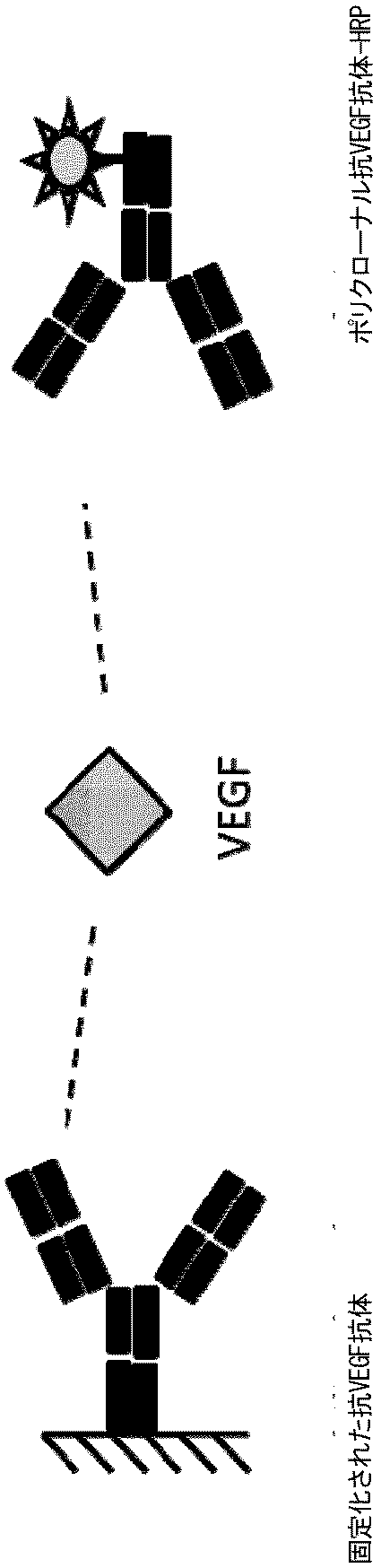
結果は図5にも示されている。 The results are also shown in FIG.
図5から、モノクローナル抗id HER3抗体M-2.11.123-IgGによる抗HER3/c-MET抗体の除去が他の抗体ほど効率的でないことを見て取ることができる。 From FIG. 5, it can be seen that removal of anti-HER3 / c-MET antibody by monoclonal anti-id HER3 antibody M-2.11.123-IgG is not as efficient as other antibodies.
薬物に結合したc-METの除去および遊離c-METの検出
抗HER3/c-MET抗体に結合したc-METを、実施例1に記載されるようにして抗イディオタイプ抗体を用いて除去した。上清を、実施例2に記載されるようにおいてc-MET ELISAにおいて測定した。
Removal of drug-bound c-MET and detection of free c-MET c-MET bound to anti-HER3 / c-MET antibody was removed using an anti-idiotype antibody as described in Example 1. . The supernatant was measured in a c-MET ELISA as described in Example 2.
実施例5
ヒト血清および緩衝液中の薬物に結合したVEGFの除去
実施例1にしたがい、抗ANG2/VEGF抗体を10/5/1/0.5/0.25および0μg/mlに希釈し、そして50 ng/mlの一定濃度のVEGFと共にインキュベートした。希釈物は、2つの異なるマトリックス:
・Low Cross緩衝液(Candor Bioscience GmbH、#100500)
・Human Pool Serum(Trina、NHS Baseマトリックス)
で生成した。
Example 5
Removal of VEGF bound to drug in human serum and buffer According to Example 1, anti-ANG2 / VEGF antibody was diluted to 10/5/1 / 0.5 / 0.25 and 0 μg / ml and constant 50 ng / ml Incubated with concentrations of VEGF. Dilutions are two different matrices:
・ Low Cross Buffer (Candor Bioscience GmbH, # 100500)
・ Human Pool Serum (Trina, NHS Base matrix)
Generated with.
サンプルを室温で1時間インキュベートした。その後、実施例1に記載されるようにしてサンプルを除去処理した。 Samples were incubated at room temperature for 1 hour. The sample was then removed as described in Example 1.
モノクローナル抗id ANG2抗体M-2.3.55-BIを使用し、VEGFと抗ANG2/VEGF抗体の複合体を捕捉した。 Monoclonal anti-id ANG2 antibody M-2.3.55-BI was used to capture the complex of VEGF and anti-ANG2 / VEGF antibody.
除去後、上清を、実施例2に記載されるようにしてVEGF ELISAにおいて測定した。 After removal, the supernatant was measured in a VEGF ELISA as described in Example 2.
除去後の上清の残存VEGF量を、いかなる2特異性抗体も含めずにインキュベートされた対照サンプル(50 ng/ml)と関連づけた。結果が、%表示の相対回収率として以下の表に示されている。 The amount of residual VEGF in the supernatant after removal was related to a control sample (50 ng / ml) incubated without any bispecific antibody. The results are shown in the table below as relative recovery in%.
表:除去前のレベルに対する、免疫除去後の緩衝液および血清サンプルの上清中の相対VEGF量
実施例6
抗HER3/c-MET抗体を用いて示される除去に対する遊離抗原濃度の影響の評価
実施例1にしたがい、抗HER3/c-MET抗体に結合したc-METの除去を、様々な濃度のc-METを含むサンプルにおいて行った。3つの異なる濃度のc-MET(5、10、50 ng/ml)を、漸増濃度の抗HER3/c-MET抗体と共に室温でインキュベートした。インキュベートは、1時間および一晩行った。その後、実施例1に記載されるようにして、これらのサンプルから抗HER3/c-MET抗体・c-MET複合体を除去した。
Example 6
Assessing the effect of free antigen concentration on removal demonstrated using anti-HER3 / c-MET antibody.According to Example 1, removal of c-MET bound to anti-HER3 / c-MET antibody was achieved using various concentrations of c-MET. Performed on samples containing MET. Three different concentrations of c-MET (5, 10, 50 ng / ml) were incubated with increasing concentrations of anti-HER3 / c-MET antibody at room temperature. Incubations were performed for 1 hour and overnight. The anti-HER3 / c-MET antibody / c-MET complex was then removed from these samples as described in Example 1.
結果が、以下の表aおよびbに、添加濃度からの各サンプルのc-MET濃度の回収率として示されている。 The results are shown in Tables a and b below as the recovery of c-MET concentration for each sample from the additive concentration.
(表a)1時間のインキュベート時間の除去後のc-METの回収率ならびに薬物(2特異性抗体)および標的(抗原)の濃度に対する依存性
(表b)一晩のインキュベート時間の除去後のc-METの回収率ならびに薬物(2特異性抗体)および標的(抗原)の濃度に対する依存性
実施例7
除去に対するVEGFの影響
2特異性抗ANG2/VEGF抗体を、100μg/mlとなるよう、500μlのカニクイザルプール血漿に添加した。この添加された血漿を各々100μlの5つのアリコートに分割した。各アリコートに、0 ng/ml〜100 ng/mlの範囲の異なる量のVEGFを添加した。
Example 7
Effect of VEGF on removal
Bispecific anti-ANG2 / VEGF antibody was added to 500 μl of cynomolgus pool plasma to 100 μg / ml. The added plasma was divided into 5 aliquots of 100 μl each. To each aliquot, different amounts of VEGF ranging from 0 ng / ml to 100 ng / ml were added.
サンプルを、抗id VEGF抗体M2.45.51-BIが結合されたストレプトアビジンコーティングされた磁気セファロースビーズで処理した。サンプルおよびビーズを周囲温度で一晩インキュベートした。 Samples were treated with streptavidin-coated magnetic sepharose beads conjugated with anti-id VEGF antibody M2.45.51-BI. Samples and beads were incubated overnight at ambient temperature.
インキュベート後、磁気分離装置を用いて磁気ビーズをサンプルから分離した。 After incubation, the magnetic beads were separated from the sample using a magnetic separator.
ビーズ処理/ビーズベースの除去後に上清中に残存する抗ANG2/VEGF抗体のレベルを、ELISAを用いて決定した。 The level of anti-ANG2 / VEGF antibody remaining in the supernatant after bead treatment / bead base removal was determined using ELISA.
ELISAにおいて、第1工程でビオチニル化VEGFをストレプトアビジンコーティングされたマイクロタイタープレート(SA-MTP)に結合させた。過剰な未結合VEGFを洗浄によって取り除いた。サンプル/標準、例えばカニクイザル血漿に添加された抗ANG2/VEGF抗体を、室温で1時間インキュベートした。洗浄後、ジゴキシゲニン化モノクローナル抗ヒトFcγ pan抗体R10Z8E9を検出のために使用し、1時間インキュベートした。洗浄後、結合したジゴキシゲニン化モノクローナル抗ヒトFcγ pan抗体R10Z8E9を、抗ジゴキシゲニン抗体セイヨウワサビペルオキシダーゼ(HRP)コンジュゲートを用いて検出した検出した。この酵素コンジュゲートは、ABTS基質の発色反応を触媒する。シグナルは、ELISAリーダーによって、405nmの波長(参照波長:490nm)で測定した。各血清サンプルの吸光値を3連で決定した。結果が以下の表に示されている。 In the ELISA, biotinylated VEGF was bound to a streptavidin-coated microtiter plate (SA-MTP) in the first step. Excess unbound VEGF was removed by washing. Anti-ANG2 / VEGF antibody added to samples / standards, eg, cynomolgus plasma, was incubated for 1 hour at room temperature. After washing, digoxigenated monoclonal anti-human Fcγ pan antibody R10Z8E9 was used for detection and incubated for 1 hour. After washing, bound digoxigeninized monoclonal anti-human Fcγ pan antibody R10Z8E9 was detected using an anti-digoxigenin antibody horseradish peroxidase (HRP) conjugate. This enzyme conjugate catalyzes the color reaction of the ABTS substrate. The signal was measured by an ELISA reader at a wavelength of 405 nm (reference wavelength: 490 nm). The absorbance value of each serum sample was determined in triplicate. The results are shown in the table below.
表:
上表に示されているように、2特異性モノクローナル抗体の除去は、サンプル中の第1の抗原(すなわち、この実施例ではVEGF)の量に非依存的である。したがって、第2の抗原(すなわち、この実施例では遊離のANG2)の量は遊離の第1の抗原の濃度決定に対する影響を有さないと結論付けることができる。 As shown in the table above, removal of the bispecific monoclonal antibody is independent of the amount of the first antigen (ie, VEGF in this example) in the sample. It can therefore be concluded that the amount of the second antigen (ie free ANG2 in this example) has no effect on the determination of the concentration of the free first antigen.
この学説により制約されることを望まないが、第1の抗原による潜在的干渉に対する堅牢性は、例えば、i)2特異性抗体に対する第1の抗原の親和性よりも高い2特異性抗体に対する親和性を有する抗イディオタイプ抗体を使用することによって、またはii)抗原としてCDRの同一部分に結合しない非中和性の抗イディオタイプ抗体を使用することによって、達成され得る。 Although not wishing to be bound by this theory, robustness against potential interference by the first antigen is, for example, i) higher affinity for bispecific antibodies than the affinity of the first antigen for bispecific antibodies. It can be achieved by using anti-idiotype antibodies that have sex, or ii) by using non-neutralizing anti-idiotype antibodies that do not bind to the same part of the CDR as the antigen.
Claims (14)
- 多特異性抗体、多特異性抗体に結合した抗原および遊離抗原を含むサンプルを、多特異性抗体の第1の結合特異性とは異なる第2の結合特異性に特異的に結合する抗イディオタイプ抗体と共にインキュベートする工程;
- 遊離抗原の存在および/または量の決定前に、抗イディタイプ抗体―多特異性抗体複合体をサンプルから除去する工程;及び、
- 多特異性抗体除去サンプル中の抗原の量を決定する工程
を含み、検出される抗原が、複合体化していない抗原または遊離抗原である点を特徴とする、前記方法。
An in vitro method for determining the presence and / or amount of an antigen of a multispecific antibody in a sample, wherein the antigen to be detected can be specifically bound by the first binding specificity of the multispecific antibody Is,
-An anti-idio that specifically binds a sample containing a multispecific antibody, an antigen bound to the multispecific antibody and a free antigen to a second binding specificity that is different from the first binding specificity of the multispecific antibody. Incubating with a type antibody;
-Removing the anti-iditype antibody-multispecific antibody complex from the sample prior to determining the presence and / or amount of free antigen; and
-Said method comprising the step of determining the amount of antigen in a multispecific antibody-removed sample, characterized in that the antigen to be detected is an uncomplexed antigen or a free antigen.
- 多特異性抗体除去サンプルを、抗原に特異的に結合する捕捉抗体と共にインキュベートして、捕捉抗体・抗原複合体を形成させる工程、
- 捕捉抗体・抗原複合体をトレーサー抗体と共にインキュベートする工程であって、それによって、捕捉抗体およびトレーサー抗体が抗原上の重複しないエピトープに結合する、工程、
- 捕捉抗体・抗原・トレーサー抗体複合体を、検出可能な標識を含む検出抗体と共にインキュベートする工程であって、それによって、検出抗体がトレーサー抗体の可変ドメインの外側のエピトープにおいてトレーサー抗体に特異的に結合する、工程、および
- 形成された捕捉抗体・抗原・トレーサー抗体複合体をサンプル中の抗原の量と相関させる工程
を含む点を特徴とする、請求項1記載の方法。 The determination of the amount of antigen is
-Incubating the multispecific antibody-removed sample with a capture antibody that specifically binds to the antigen to form a capture antibody-antigen complex;
-Incubating the capture antibody-antigen complex with the tracer antibody, whereby the capture antibody and the tracer antibody bind to non-overlapping epitopes on the antigen;
-Incubating the capture antibody-antigen-tracer antibody complex with a detection antibody comprising a detectable label, whereby the detection antibody is specific to the tracer antibody at an epitope outside the variable domain of the tracer antibody. Combine, process, and
- characterized in that it comprises a step of correlating the formed capture antibody-antigen-tracer antibody complex to the amount of antigen in the sample The method of claim 1, wherein.
- 抗イディオタイプ抗体・多特異性抗体複合体をサンプルから取り除く工程
を含む点を特徴とする、請求項1〜3のいずれか一項記載の方法。 -An anti-idio that specifically binds a sample containing a multispecific antibody, an antigen bound to the multispecific antibody and a free antigen to a second binding specificity that is different from the first binding specificity of the multispecific antibody. Incubating with a type antibody to form an anti-idiotype antibody-multispecific antibody complex, and
The method according to any one of claims 1 to 3 , comprising a step of removing the anti-idiotype antibody / multispecific antibody complex from the sample.
- 抗イディオタイプ抗体・多特異性抗体複合体をサンプルから取り除く工程、ならびに
- 多特異性抗体除去サンプル中の抗原の量を決定する工程
を含む点を特徴とする、請求項1〜4のいずれか一項に記載の方法。 -Incubating a sample containing an antigen and a multispecific antibody with an anti-idiotype antibody that specifically binds to a second binding specificity different from the first binding specificity of the multispecific antibody, to obtain an anti-idiotype Forming an antibody-multispecific antibody complex,
-Removing the anti-idiotype antibody / multispecific antibody complex from the sample, and
The method according to any one of claims 1 to 4 , characterized in that it comprises the step of determining the amount of antigen in the multispecific antibody-removed sample.
Applications Claiming Priority (3)
| Application Number | Priority Date | Filing Date | Title |
|---|---|---|---|
| EP11194350.2 | 2011-12-19 | ||
| EP11194350 | 2011-12-19 | ||
| PCT/EP2012/075991 WO2013092611A2 (en) | 2011-12-19 | 2012-12-18 | Method for the detection of free binding partner of a multispecific binder |
Publications (3)
| Publication Number | Publication Date |
|---|---|
| JP2015502547A JP2015502547A (en) | 2015-01-22 |
| JP2015502547A5 JP2015502547A5 (en) | 2016-02-04 |
| JP6203747B2 true JP6203747B2 (en) | 2017-09-27 |
Family
ID=47429825
Family Applications (1)
| Application Number | Title | Priority Date | Filing Date |
|---|---|---|---|
| JP2014547933A Active JP6203747B2 (en) | 2011-12-19 | 2012-12-18 | Method for detecting free binding partners of multispecific binders |
Country Status (11)
| Country | Link |
|---|---|
| US (1) | US9671394B2 (en) |
| EP (1) | EP2795335B1 (en) |
| JP (1) | JP6203747B2 (en) |
| KR (1) | KR20140099277A (en) |
| CN (1) | CN104040350B (en) |
| BR (1) | BR112014011535A2 (en) |
| CA (1) | CA2853917A1 (en) |
| HK (1) | HK1201920A1 (en) |
| MX (1) | MX349003B (en) |
| RU (1) | RU2633488C2 (en) |
| WO (1) | WO2013092611A2 (en) |
Cited By (1)
| Publication number | Priority date | Publication date | Assignee | Title |
|---|---|---|---|---|
| JP2017223710A (en) * | 2012-02-01 | 2017-12-21 | エフ.ホフマン−ラ ロシュ アーゲーF. Hoffmann−La Roche Aktiengesellschaft | Method of detecting binding partner of multispecific binding substance |
Families Citing this family (10)
| Publication number | Priority date | Publication date | Assignee | Title |
|---|---|---|---|---|
| US8956859B1 (en) | 2010-08-13 | 2015-02-17 | Aviex Technologies Llc | Compositions and methods for determining successful immunization by one or more vaccines |
| KR20150038046A (en) | 2012-07-13 | 2015-04-08 | 에프. 호프만-라 로슈 아게 | Method for the detection of a multispecific binder |
| EP3066469B1 (en) * | 2013-11-05 | 2017-10-11 | Roche Diagnostics GmbH | A method for determining the total amount and/or concentration of an analyte in the presence of a binding molecule as well as kits, compositions and uses relating thereto |
| EP3119805A1 (en) * | 2014-03-20 | 2017-01-25 | Wellstat Diagnostics, LLC | Antibodies and methods for the detection of cell death |
| CN107003307B (en) | 2014-12-18 | 2020-04-07 | 豪夫迈·罗氏有限公司 | Method for reducing interference |
| JP2020530554A (en) * | 2017-07-20 | 2020-10-22 | シートムエックス セラピューティクス,インコーポレイテッド | Methods and Uses for Qualitative and / or Quantitative Analysis of Activating Antibody Properties |
| CN109358192B (en) * | 2018-02-08 | 2020-05-12 | 中国科学院上海药物研究所 | Device and method for removing free drugs in anti-drug antibody detection sample, preparation method and application of device |
| EP3805757A4 (en) | 2018-06-04 | 2022-03-23 | Chugai Seiyaku Kabushiki Kaisha | Method for detecting complex |
| WO2020229378A1 (en) * | 2019-05-13 | 2020-11-19 | F. Hoffmann-La Roche Ag | Interference-suppressed pharmacokinetic immunoassay |
| CN117805397A (en) * | 2024-02-29 | 2024-04-02 | 军科正源(北京)药物研究有限责任公司 | Method for detecting free VEGF |
Family Cites Families (32)
| Publication number | Priority date | Publication date | Assignee | Title |
|---|---|---|---|---|
| JPS56128722A (en) * | 1980-03-14 | 1981-10-08 | Terumo Corp | Removal of antiacetylcholinereceptor antibody and device therefor |
| US5219730A (en) | 1982-09-28 | 1993-06-15 | New York University | Idiotype-anti-idiotype immunoassay |
| US4828981A (en) | 1983-08-24 | 1989-05-09 | Synbiotics Corporation | Immunoassays for determining Dirofilaria immitis infection using antiidiotype monoclonal antibody reagents |
| EP0170302A1 (en) | 1984-06-27 | 1986-02-05 | l'Association internationale à but scientifique, dite: Institut international de pathologie cellulaire et moléculaire | Method of immunological determination of a substance in a liquid sample by means of anti-idiotypic antibodies |
| US4699880A (en) * | 1984-09-25 | 1987-10-13 | Immunomedics, Inc. | Method of producing monoclonal anti-idiotype antibody |
| US5238808A (en) | 1984-10-31 | 1993-08-24 | Igen, Inc. | Luminescent metal chelate labels and means for detection |
| WO1987002778A1 (en) | 1985-10-22 | 1987-05-07 | Cooper-Lipotech | Solid-phase liposome immunoassay system |
| ZA898290B (en) | 1988-11-03 | 1991-06-26 | Igen Inc | Electrochemiluminescent assays |
| US5068088A (en) | 1988-11-03 | 1991-11-26 | Igen, Inc. | Method and apparatus for conducting electrochemiluminescent measurements |
| IL100866A (en) | 1991-02-06 | 1995-10-31 | Igen Inc | Method and apparatus for magnetic microparticulate based luminescence assay including plurality of magnets |
| EP0651761B1 (en) | 1992-07-13 | 2002-10-09 | Bionebraska, Inc. | Method for modification of recombinant polypeptides |
| GB9316369D0 (en) * | 1993-08-06 | 1993-09-22 | Surface Active Ltd | Diagnostic method |
| JPH11352129A (en) * | 1998-06-04 | 1999-12-24 | Mizuho Medeii:Kk | Detection device and detection method |
| US6753189B1 (en) * | 1998-06-04 | 2004-06-22 | Mizuho Medy Co., Ltd. | Detection apparatus and method for the same |
| JP4620312B2 (en) * | 1999-11-16 | 2011-01-26 | ジェネンテック, インコーポレイテッド | Eliza for VEGF |
| EP1288308A1 (en) * | 2001-08-28 | 2003-03-05 | Roche Diagnostics GmbH | A method for the determination of multiple analytes |
| GB0320459D0 (en) | 2003-09-01 | 2003-10-01 | Selective Antibodies Ltd | Assay methods and materials |
| DE102004052729A1 (en) * | 2004-10-30 | 2006-05-04 | Roche Diagnostics Gmbh | Immunocomplex-specific antibody for zero-reduction in the detection of antigen-specifically bound antibodies of a specific immunoglobulin class in array test formats |
| EP1861708A2 (en) | 2005-03-08 | 2007-12-05 | Tanox, Inc. | Methods for determining the bivalency of protein and antibody therapeutics |
| RS53387B (en) * | 2006-10-04 | 2014-10-31 | Genentech, Inc. | Elisa for vegf |
| EP1917854A1 (en) | 2006-11-06 | 2008-05-07 | F. Hoffmann-La Roche Ag | Method for producing anti idiotypic antibodies |
| US9266967B2 (en) | 2007-12-21 | 2016-02-23 | Hoffmann-La Roche, Inc. | Bivalent, bispecific antibodies |
| US20090162359A1 (en) | 2007-12-21 | 2009-06-25 | Christian Klein | Bivalent, bispecific antibodies |
| US8242247B2 (en) | 2007-12-21 | 2012-08-14 | Hoffmann-La Roche Inc. | Bivalent, bispecific antibodies |
| US8227577B2 (en) | 2007-12-21 | 2012-07-24 | Hoffman-La Roche Inc. | Bivalent, bispecific antibodies |
| CA2756244A1 (en) | 2009-04-02 | 2010-10-07 | Roche Glycart Ag | Multispecific antibodies comprising full length antibodies and single chain fab fragments |
| EP2417156B1 (en) | 2009-04-07 | 2015-02-11 | Roche Glycart AG | Trivalent, bispecific antibodies |
| WO2010136172A1 (en) | 2009-05-27 | 2010-12-02 | F. Hoffmann-La Roche Ag | Tri- or tetraspecific antibodies |
| US9676845B2 (en) | 2009-06-16 | 2017-06-13 | Hoffmann-La Roche, Inc. | Bispecific antigen binding proteins |
| US8703132B2 (en) | 2009-06-18 | 2014-04-22 | Hoffmann-La Roche, Inc. | Bispecific, tetravalent antigen binding proteins |
| CN102207054B (en) | 2010-03-31 | 2013-03-13 | 比亚迪股份有限公司 | Knock control system and method of hybrid vehicle |
| CN102207504A (en) * | 2011-03-23 | 2011-10-05 | 北京华创远航科技有限公司 | Enzyme-linked immunosorbent assay kit, and preparation method thereof |
-
2012
- 2012-12-18 MX MX2014007095A patent/MX349003B/en active IP Right Grant
- 2012-12-18 BR BR112014011535A patent/BR112014011535A2/en not_active Application Discontinuation
- 2012-12-18 KR KR1020147016700A patent/KR20140099277A/en not_active Application Discontinuation
- 2012-12-18 CN CN201280063032.5A patent/CN104040350B/en active Active
- 2012-12-18 RU RU2014128510A patent/RU2633488C2/en not_active IP Right Cessation
- 2012-12-18 WO PCT/EP2012/075991 patent/WO2013092611A2/en active Application Filing
- 2012-12-18 CA CA2853917A patent/CA2853917A1/en not_active Abandoned
- 2012-12-18 JP JP2014547933A patent/JP6203747B2/en active Active
- 2012-12-18 EP EP12806045.6A patent/EP2795335B1/en active Active
-
2014
- 2014-06-18 US US14/308,539 patent/US9671394B2/en active Active
-
2015
- 2015-03-09 HK HK15102356.7A patent/HK1201920A1/en unknown
Cited By (2)
| Publication number | Priority date | Publication date | Assignee | Title |
|---|---|---|---|---|
| JP2017223710A (en) * | 2012-02-01 | 2017-12-21 | エフ.ホフマン−ラ ロシュ アーゲーF. Hoffmann−La Roche Aktiengesellschaft | Method of detecting binding partner of multispecific binding substance |
| US10761097B2 (en) | 2012-02-01 | 2020-09-01 | Hoffmann-La Roche Inc. | Method for the detection of a binding partner of a multispecific binder |
Also Published As
| Publication number | Publication date |
|---|---|
| CA2853917A1 (en) | 2013-06-27 |
| MX2014007095A (en) | 2014-07-22 |
| EP2795335A2 (en) | 2014-10-29 |
| CN104040350B (en) | 2016-05-18 |
| RU2014128510A (en) | 2016-02-10 |
| BR112014011535A2 (en) | 2017-05-09 |
| WO2013092611A3 (en) | 2013-10-17 |
| WO2013092611A2 (en) | 2013-06-27 |
| JP2015502547A (en) | 2015-01-22 |
| MX349003B (en) | 2017-07-06 |
| KR20140099277A (en) | 2014-08-11 |
| HK1201920A1 (en) | 2015-09-11 |
| CN104040350A (en) | 2014-09-10 |
| RU2633488C2 (en) | 2017-10-12 |
| US20150072359A1 (en) | 2015-03-12 |
| EP2795335B1 (en) | 2017-09-06 |
| US9671394B2 (en) | 2017-06-06 |
Similar Documents
| Publication | Publication Date | Title |
|---|---|---|
| JP6678792B2 (en) | Methods for detecting binding partners of multispecific binders | |
| JP6203747B2 (en) | Method for detecting free binding partners of multispecific binders | |
| US9488662B2 (en) | Method for the detection of a multispecific binder | |
| US11340234B2 (en) | Method for the determination of anti-drug antibodies against an effector function suppressed human or humanized drug antibody |
Legal Events
| Date | Code | Title | Description |
|---|---|---|---|
| RD04 | Notification of resignation of power of attorney |
Free format text: JAPANESE INTERMEDIATE CODE: A7424 Effective date: 20150318 |
|
| A521 | Request for written amendment filed |
Free format text: JAPANESE INTERMEDIATE CODE: A523 Effective date: 20151208 |
|
| A621 | Written request for application examination |
Free format text: JAPANESE INTERMEDIATE CODE: A621 Effective date: 20151208 |
|
| A977 | Report on retrieval |
Free format text: JAPANESE INTERMEDIATE CODE: A971007 Effective date: 20161017 |
|
| A131 | Notification of reasons for refusal |
Free format text: JAPANESE INTERMEDIATE CODE: A131 Effective date: 20161024 |
|
| A521 | Request for written amendment filed |
Free format text: JAPANESE INTERMEDIATE CODE: A523 Effective date: 20170123 |
|
| A131 | Notification of reasons for refusal |
Free format text: JAPANESE INTERMEDIATE CODE: A131 Effective date: 20170628 |
|
| A521 | Request for written amendment filed |
Free format text: JAPANESE INTERMEDIATE CODE: A523 Effective date: 20170803 |
|
| TRDD | Decision of grant or rejection written | ||
| A01 | Written decision to grant a patent or to grant a registration (utility model) |
Free format text: JAPANESE INTERMEDIATE CODE: A01 Effective date: 20170823 |
|
| A61 | First payment of annual fees (during grant procedure) |
Free format text: JAPANESE INTERMEDIATE CODE: A61 Effective date: 20170830 |
|
| R150 | Certificate of patent or registration of utility model |
Ref document number: 6203747 Country of ref document: JP Free format text: JAPANESE INTERMEDIATE CODE: R150 |
|
| R250 | Receipt of annual fees |
Free format text: JAPANESE INTERMEDIATE CODE: R250 |
|
| R250 | Receipt of annual fees |
Free format text: JAPANESE INTERMEDIATE CODE: R250 |
|
| R250 | Receipt of annual fees |
Free format text: JAPANESE INTERMEDIATE CODE: R250 |
|
| R250 | Receipt of annual fees |
Free format text: JAPANESE INTERMEDIATE CODE: R250 |