JP6134266B2 - Antibodies that specifically bind to the microvasculature of the synovium of arthritic patients - Google Patents
Antibodies that specifically bind to the microvasculature of the synovium of arthritic patients Download PDFInfo
- Publication number
- JP6134266B2 JP6134266B2 JP2013530798A JP2013530798A JP6134266B2 JP 6134266 B2 JP6134266 B2 JP 6134266B2 JP 2013530798 A JP2013530798 A JP 2013530798A JP 2013530798 A JP2013530798 A JP 2013530798A JP 6134266 B2 JP6134266 B2 JP 6134266B2
- Authority
- JP
- Japan
- Prior art keywords
- antigen
- binding polypeptide
- substance
- synovial
- scfv
- Prior art date
- Legal status (The legal status is an assumption and is not a legal conclusion. Google has not performed a legal analysis and makes no representation as to the accuracy of the status listed.)
- Expired - Fee Related
Links
Images


Classifications
-
- C—CHEMISTRY; METALLURGY
- C07—ORGANIC CHEMISTRY
- C07K—PEPTIDES
- C07K16/00—Immunoglobulins [IGs], e.g. monoclonal or polyclonal antibodies
- C07K16/18—Immunoglobulins [IGs], e.g. monoclonal or polyclonal antibodies against material from animals or humans
-
- A—HUMAN NECESSITIES
- A61—MEDICAL OR VETERINARY SCIENCE; HYGIENE
- A61P—SPECIFIC THERAPEUTIC ACTIVITY OF CHEMICAL COMPOUNDS OR MEDICINAL PREPARATIONS
- A61P19/00—Drugs for skeletal disorders
- A61P19/02—Drugs for skeletal disorders for joint disorders, e.g. arthritis, arthrosis
-
- A—HUMAN NECESSITIES
- A61—MEDICAL OR VETERINARY SCIENCE; HYGIENE
- A61P—SPECIFIC THERAPEUTIC ACTIVITY OF CHEMICAL COMPOUNDS OR MEDICINAL PREPARATIONS
- A61P29/00—Non-central analgesic, antipyretic or antiinflammatory agents, e.g. antirheumatic agents; Non-steroidal antiinflammatory drugs [NSAID]
-
- A—HUMAN NECESSITIES
- A61—MEDICAL OR VETERINARY SCIENCE; HYGIENE
- A61P—SPECIFIC THERAPEUTIC ACTIVITY OF CHEMICAL COMPOUNDS OR MEDICINAL PREPARATIONS
- A61P37/00—Drugs for immunological or allergic disorders
- A61P37/02—Immunomodulators
-
- A—HUMAN NECESSITIES
- A61—MEDICAL OR VETERINARY SCIENCE; HYGIENE
- A61P—SPECIFIC THERAPEUTIC ACTIVITY OF CHEMICAL COMPOUNDS OR MEDICINAL PREPARATIONS
- A61P9/00—Drugs for disorders of the cardiovascular system
-
- C—CHEMISTRY; METALLURGY
- C07—ORGANIC CHEMISTRY
- C07K—PEPTIDES
- C07K16/00—Immunoglobulins [IGs], e.g. monoclonal or polyclonal antibodies
- C07K16/18—Immunoglobulins [IGs], e.g. monoclonal or polyclonal antibodies against material from animals or humans
- C07K16/28—Immunoglobulins [IGs], e.g. monoclonal or polyclonal antibodies against material from animals or humans against receptors, cell surface antigens or cell surface determinants
-
- C—CHEMISTRY; METALLURGY
- C07—ORGANIC CHEMISTRY
- C07K—PEPTIDES
- C07K2317/00—Immunoglobulins specific features
- C07K2317/20—Immunoglobulins specific features characterized by taxonomic origin
- C07K2317/21—Immunoglobulins specific features characterized by taxonomic origin from primates, e.g. man
-
- C—CHEMISTRY; METALLURGY
- C07—ORGANIC CHEMISTRY
- C07K—PEPTIDES
- C07K2317/00—Immunoglobulins specific features
- C07K2317/60—Immunoglobulins specific features characterized by non-natural combinations of immunoglobulin fragments
- C07K2317/62—Immunoglobulins specific features characterized by non-natural combinations of immunoglobulin fragments comprising only variable region components
- C07K2317/622—Single chain antibody (scFv)
Description
発明の分野
本発明は、関節炎患者の滑膜の微小血管系を特異的に標的とする抗原結合ポリペプチドに関する。そのポリペプチドおよびその結合体は、関節の血管系の画像化において、ならびに関節炎の診断および処置のために、使用され得る。
The present invention relates to antigen binding polypeptides that specifically target the synovial microvasculature of arthritic patients. The polypeptides and conjugates thereof can be used in the imaging of joint vasculature and for the diagnosis and treatment of arthritis.
発明の背景
関節リウマチ(RA)は、最も一般的な自己免疫疾患の1つであり、欧州だけで300万人超に影響を与えている、慢性疼痛の主な原因である。集団の1〜2%が関節リウマチに罹患している。Medical Expenditure Panel Survey(MEPS)データによると、米国では、2003年に関節リウマチおよび関連する関節炎の処置に費やされた総コストは、1280億ドルだった;1人あたりの平均コストは、現在、8500ドルである。毎年、関節炎およびその関連合併症は、750,000件を超える入院および3600万人の外来患者の来診を招いている。任意のタイプの関節炎に苦しんでいる人々の最大15%は、行うことができる身体活動の量の減少に悩まされている。代表的には、身体活動が減少すると、独立性および自由を欠くので、患者は、うつを発症する傾向にある。
BACKGROUND OF THE INVENTION Rheumatoid arthritis (RA) is one of the most common autoimmune diseases and is a major cause of chronic pain affecting over 3 million people in Europe alone. 1-2% of the population suffers from rheumatoid arthritis. According to the Medical Expand Panel Survey (MEPS) data, in 2003, the total cost spent in the treatment of rheumatoid arthritis and related arthritis was $ 128 billion in 2003; the average cost per person is currently It is $ 8,500. Each year, arthritis and related complications result in over 750,000 hospitalizations and 36 million outpatient visits. Up to 15% of people suffering from any type of arthritis suffer from a reduction in the amount of physical activity that can be performed. Typically, as physical activity decreases, patients tend to develop depression because they lack independence and freedom.
英国では、およそ400,000人の成人が関節リウマチを有し、関節炎は、人々が障害生活手当(Disability Living Allowance)を受ける最も一般的な状態である。50万人を超える人々が、関節炎の結果としてDLAを受け(全DLA受給者の18パーセント超に相当する)、これは、心臓疾患、脳卒中、胸部疾患およびがんの組み合わせについての合計よりも多い。 In the UK, approximately 400,000 adults have rheumatoid arthritis, and arthritis is the most common condition for people to receive disability living allowances. More than 500,000 people receive DLA as a result of arthritis (representing more than 18 percent of all DLA recipients), which is more than the sum for the combination of heart disease, stroke, chest disease and cancer .
RAは、一般に身体の両側の手首、指、膝、足および足首に影響を及ぼす、滑膜性関節の炎症性疾患である。関節および腱を裏打ちし、保護し、関節の滑らかで自由な動きを可能にする滑膜の炎症を、RAは引き起こす。滑膜の炎症は、患部の関節の腫脹を引き起こし、最終的には進行性の軟骨の破壊および骨の侵食に至り、可動範囲を損ない、変形をもたらす。 RA is an inflammatory disease of the synovial joint that generally affects the wrist, fingers, knees, feet and ankles on both sides of the body. RA causes synovial inflammation that lines and protects joints and tendons and allows for smooth and free movement of the joints. Inflammation of the synovium causes swelling of the affected joints, eventually leading to progressive cartilage destruction and bone erosion, impairing the range of motion and causing deformation.
RAは、身体の他の臓器にも影響し、深刻な障害および命に関わる合併症をもたらし得る持続的な進行性疾患である。ゆえに、RAは、関連する有意な罹患率および死亡率を有する障害の主な原因である。 RA is a persistent progressive disease that affects other organs of the body and can result in serious disability and life-threatening complications. Thus, RA is a major cause of disorders with associated significant morbidity and mortality.
RAの発病年齢は、様々であり、小児から90歳代の個体に及ぶ。西ヨーロッパおよび米国の集団におけるRAの有病率は、およそ1%であり、女性と男性の比率は3:1である。さらに、関節リウマチに関する1年間の経済的影響の総額は、西ヨーロッパにおいておよそ350億ポンドと見積もられている。 The age of onset of RA varies and ranges from children to individuals in their 90s. The prevalence of RA in the Western European and US population is approximately 1% and the ratio of women to men is 3: 1. Furthermore, the total annual economic impact on rheumatoid arthritis is estimated at approximately £ 35 billion in Western Europe.
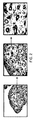
細胞レベルにおいて、滑膜は、プロテオグリカン凝集体を含むよく組織化されたマトリックス、毛細血管およびリンパ管のネットワーク、ならびに内在の線維芽細胞およびマクロファージ様細胞から構成されている。しかしながら、RAでは、滑膜は、Tヘルパー細胞、B細胞、マクロファージおよび形質細胞によって浸潤されるようになる。さらに、滑膜は、過形成、および軟骨と骨との間の境界領域において局所的に侵襲性になっており、関節軟骨、軟骨下骨および関節周囲の軟部組織の破壊を引き起こし、長期間の関節の損傷、変形および深刻な障害をもたらす(図1を参照のこと)。 At the cellular level, the synovium is composed of a well-organized matrix containing proteoglycan aggregates, a network of capillaries and lymph vessels, and endogenous fibroblasts and macrophage-like cells. However, in RA, the synovium becomes infiltrated by T helper cells, B cells, macrophages and plasma cells. Furthermore, the synovium is hyperplastic and locally invasive in the border region between cartilage and bone, causing destruction of articular cartilage, subchondral bone and surrounding soft tissue, It results in joint damage, deformity and severe damage (see FIG. 1).
現在、滑膜における血管新生(既存の血管からの新しい血管の成長)が、この疾患の病因および進行に対して有意な寄与を有することが十分に確立されている。滑膜の細胞過多が、滑膜の血管の数および密度の代償的増加を伴うので、実際に、滑膜の血管新生は、RAの他の病理学的特徴に先行し得る。 It is now well established that angiogenesis in the synovium (new blood vessel growth from existing blood vessels) has a significant contribution to the pathogenesis and progression of the disease. In fact, synovial angiogenesis may precede other pathological features of RA, as synovial hyperplasia is accompanied by a compensatory increase in the number and density of synovial vessels.
関節リウマチの処置の最終目的は、関節の損傷を防ぐこと、機能喪失を防ぐこと、およびRAに関連する疼痛を減少させることである。非ステロイド性抗炎症薬(NSAID)および疾患修飾性抗リウマチ薬(DMARD)は、現在、RA処置の主要な形態であるが、著しい副作用を伴うことが多い。NSAIDは、胃への刺激、胃腸の潰瘍および腎臓損傷を引き起こし得る。DMARDの副作用は、使用される薬物のタイプ次第である。アザチオプリンは、感染、肝臓損傷、毛髪脱落および下痢のリスクを上昇させる。シクロスポリンは、腎臓損傷、高血圧および歯肉肥厚を引き起こす。クロロキン群は、胃炎、下痢および視覚の問題を引き起こす。金塩は、舌の腫大、歯肉の出血、発疹および腎臓損傷を引き起こし得る。メトトレキサートは、肝臓損傷および骨髄抑制を引き起こし得る。スルファサラジンは、胃腸の不調およびアレルギー反応を引き起こし得る。より新しい生物反応修飾物質は、免疫系を抑制し、結核のような潜伏感染の再活性化を引き起こし得る。 The ultimate goal of rheumatoid arthritis treatment is to prevent joint damage, prevent loss of function, and reduce pain associated with RA. Nonsteroidal anti-inflammatory drugs (NSAIDs) and disease modifying anti-rheumatic drugs (DMARDs) are currently the main forms of RA treatment, but often have significant side effects. NSAIDs can cause stomach irritation, gastrointestinal ulcers and kidney damage. The side effects of DMARD depend on the type of drug used. Azathioprine increases the risk of infection, liver damage, hair loss and diarrhea. Cyclosporine causes kidney damage, hypertension and gingival thickening. The chloroquine group causes gastritis, diarrhea and visual problems. Gold salts can cause swollen tongue, gingival bleeding, rash and kidney damage. Methotrexate can cause liver damage and bone marrow suppression. Sulfasalazine can cause gastrointestinal upset and allergic reactions. Newer biological response modifiers can suppress the immune system and cause reactivation of latent infections such as tuberculosis.
RAの治療は、一連のサイトカイン、T細胞およびB細胞を標的とする組換え抗体の導入によって、この10年間で有意に改善されてきた。 The treatment of RA has been significantly improved over the last decade by the introduction of recombinant antibodies that target a range of cytokines, T cells and B cells.
最初のEtanerceptおよびその後すぐのInfliximabの認可以来、さらに3つのTNF中和抗体(Adalimumab、Certulizumab pegolおよびGolimumab)が認可された。さらに、T細胞[および/または樹状細胞](Abatacept)、B細胞(Rituximab)およびサイトカインIL−6に対するレセプター(Tocilizumab)を標的とする組換え抗体もまた、FDAによってRAの処置に対して認可された(非特許文献1)、(非特許文献2)。 Three additional TNF neutralizing antibodies (Adalimumab, Certurizumab pegol and Golimumab) have been approved since the first Etanercept and subsequent Infliximab approval. In addition, recombinant antibodies targeting receptors for T cells [and / or dendritic cells] (Abataccept), B cells (Rituximab) and cytokine IL-6 (Tocilizumab) are also approved by the FDA for treatment of RA. (Non-Patent Document 1) and (Non-Patent Document 2).
しかしながら、現在の治療の明らかな効果にもかかわらず、長期間にわたって処置から解放される緩解は、得られていない。持続性かつ高度の臨床反応は、少数によってしか達成されず(非特許文献1,上記)、およそ20〜40%の患者は、抗サイトカイン治療に反応しない(非特許文献3)。また、現在の治療薬は、関連するいくつかの悪影響(例えば、持続的な投与を望ましくないものとする感染症および悪性疾患の高リスク)を示す(非特許文献1,上記)。 However, despite the obvious effects of current therapies, no remissions have been obtained that are free from treatment over time. Persistent and high clinical response is achieved only by a small number (Non-Patent Document 1, supra), and approximately 20-40% of patients do not respond to anti-cytokine therapy (Non-Patent Document 3). In addition, current therapeutic agents exhibit some associated adverse effects (eg, high risk of infections and malignancies that make continuous administration undesirable) (Non-patent Document 1, supra).
ゆえに、RAにおいて未だ対処されていない大きな臨床的ニーズが存在し、緩解誘導の頻度がより高く、全身毒性が低い改善された安全性プロファイルを有する、代替の治療法の選択肢が必要とされている。 Therefore, there is a great clinical need that has not yet been addressed in RA, and there is a need for alternative treatment options that have an improved safety profile with a higher frequency of remission induction and lower systemic toxicity. .
発明の態様の要旨
本発明者らは、正常な組織に対しては、ほとんどまたはまったく反応性を有しないが、RA患者由来の滑膜組織の微小血管系との、血管周囲性反応を示す、scFv抗体を同定した。
SUMMARY OF EMBODIMENTS OF THE INVENTION The inventors have little or no reactivity to normal tissue, but exhibit a perivascular reaction with the microvasculature of synovial tissue from RA patients. scFv antibodies were identified.
第1の態様において、本発明は、関節炎患者の滑膜の微小血管系を特異的に標的とし、かつ配列番号1〜4からなる群より選択される1つ以上の相補性決定領域(CDR)を含む、抗原結合ポリペプチドを提供する。 In a first aspect, the present invention specifically targets one or more complementarity determining regions (CDRs) selected from the group consisting of SEQ ID NOs: 1-4, which specifically target the synovial microvasculature of arthritic patients. An antigen-binding polypeptide is provided.
その抗原結合ポリペプチドは、配列番号5〜8からなる群より選択される1つ以上の相補性決定領域(CDR)を含み得る。 The antigen-binding polypeptide can comprise one or more complementarity determining regions (CDRs) selected from the group consisting of SEQ ID NOs: 5-8.
その抗原結合ポリペプチドは、微小血管系の間質区画および/または血管周囲細胞と反応し得、血管周囲性反応を示し得る。 The antigen-binding polypeptide can react with the stromal compartment of the microvasculature and / or perivascular cells and can exhibit a perivascular reaction.
その抗原結合ポリペプチドは、配列番号9に示されるようなVH配列および/または配列番号10に示されるようなVL配列を含み得る。
The antigen binding polypeptide may comprise a VH sequence as shown in SEQ ID NO: 9 and / or a VL sequence as shown in SEQ ID NO: 10.
その抗原結合ポリペプチドは、scFvであり得る。 The antigen binding polypeptide can be an scFv.
その抗原結合ポリペプチドは、完全にヒトのポリペプチドであり得る。 The antigen binding polypeptide can be a fully human polypeptide.
その抗原結合ポリペプチドは、変形性関節症(osteoartritis)および/または関節リウマチの患者の滑膜の微小血管系を特異的に標的とし得る。 The antigen binding polypeptide can specifically target the microvasculature of the synovium of patients with osteoarthritis and / or rheumatoid arthritis.
本発明はまた、本発明の第1の態様に係る抗原結合ポリペプチドと同じエピトープに結合する、抗原結合ポリペプチドも提供する。その抗原結合ポリペプチドは、配列番号11として示されるアミノ酸配列を含む抗原結合ポリペプチドと同じエピトープに結合し得る。 The present invention also provides an antigen binding polypeptide that binds to the same epitope as the antigen binding polypeptide according to the first aspect of the invention. The antigen binding polypeptide can bind to the same epitope as the antigen binding polypeptide comprising the amino acid sequence shown as SEQ ID NO: 11.
本発明はまた、別の物質と結合している抗原結合ポリペプチドも提供する。その抗原結合ポリペプチドは、例えば、他の物質に結合体化され得る。その物質は、以下:治療用サイトカイン、抗血管新生剤、抗リウマチ薬、感光性物質または磁性ナノ粒子のうちの1つ以上を含み得る。 The invention also provides an antigen binding polypeptide that is conjugated to another agent. The antigen binding polypeptide can be conjugated, for example, to other substances. The substance may include one or more of the following: therapeutic cytokines, anti-angiogenic agents, anti-rheumatic drugs, photosensitive substances or magnetic nanoparticles.
本発明はまた、RAの処置において使用するための、関節の血管系の画像化において使用するための、および/または関節炎の診断、モニタリングもしくは予後診断において使用するための、本発明の第1の態様に係る抗原結合ポリペプチドまたはその結合体も提供する。 The present invention also provides a first of the invention for use in the treatment of RA, for use in imaging the vasculature of the joint, and / or for use in the diagnosis, monitoring or prognosis of arthritis. Also provided are antigen binding polypeptides or conjugates thereof according to embodiments.
第2の態様において、本発明は、被験体における関節炎を処置するための方法を提供し、その方法は、本発明の第1の態様に係る抗原結合ポリペプチドまたはその結合体を被験体に投与する工程を包含する。 In a second aspect, the invention provides a method for treating arthritis in a subject, the method comprising administering to the subject an antigen-binding polypeptide according to the first aspect of the invention or a conjugate thereof. The process of carrying out is included.
その方法は、以下の工程:
(i)感光性物質に結合体化された本発明の第1の態様に係る抗原結合ポリペプチドを被験体に投与する工程;
(ii)その結合体が関節の滑膜の血管系を標的とする工程;
(ii)その滑膜の血管系内で感光性物質を活性化するために、その関節に光を適用する工程
を包含し得る。
The method comprises the following steps:
(I) a step of administering to the subject the antigen-binding polypeptide according to the first aspect of the present invention conjugated to a photosensitive substance;
(Ii) the conjugate targets the synovial vasculature of the joint;
(Ii) applying light to the joint to activate the photosensitizer within the synovial vasculature.
上記方法は、以下の工程:
(i)磁性ナノ粒子に結合体化された本発明の第1の態様に係る抗原結合ポリペプチドを被験体に投与する工程;
(ii)その結合体が関節の滑膜の血管系を標的とする工程;
(ii)その滑膜の血管系内で磁性ナノ粒子を活性化するために、その関節に磁場を適用する工程
を包含し得る。
The above method comprises the following steps:
(I) administering the antigen-binding polypeptide according to the first aspect of the present invention conjugated to magnetic nanoparticles to a subject;
(Ii) the conjugate targets the synovial vasculature of the joint;
(Ii) applying a magnetic field to the joint to activate the magnetic nanoparticles in the synovial vasculature.
そのような方法において、上記物質の活性化は、既存の血管系の破壊をもたらし得る。 In such a method, activation of the substance can result in the destruction of existing vasculature.
上記方法は、別の治療薬の同時の、別個のまたは逐次的な使用を含む、組み合わせの方法であり得る。例えば、処置は、TNF−α遮断治療薬の投与も含み得る。 The method can be a combinatorial method involving simultaneous, separate or sequential use of another therapeutic agent. For example, treatment can include administration of a TNF-α blocking therapeutic.
上記方法は、変形性関節症および/または関節リウマチを処置するための方法であり得る。 The method can be a method for treating osteoarthritis and / or rheumatoid arthritis.
第3の態様において、本発明は、ある物質が滑膜の微小血管系を標的とするための方法を提供し、その方法は、その物質と本発明の第1の態様の抗原結合ポリペプチドとの結合をインビトロで形成する工程を包含する。その物質は、例えば、その抗原結合ポリペプチドに結合体化され得る。その結合は、その物質/抗原結合ポリペプチドが関節炎患者に投与されると、その物質が血管新生部位に選択的に蓄積するような結合であり得る。 In a third aspect, the invention provides a method for a substance to target the synovial microvasculature, the method comprising the substance and the antigen-binding polypeptide of the first aspect of the invention. Forming a bond of in vitro. The substance can be conjugated, for example, to the antigen-binding polypeptide. The binding can be such that when the agent / antigen binding polypeptide is administered to an arthritic patient, the agent selectively accumulates at the site of angiogenesis.
上記物質は、治療用の物質、画像化の物質または診断用の物質であり得る。例えば、その物質は、治療用サイトカイン、抗血管新生剤、抗リウマチ薬、感光性物質(photosensensitive agent)または磁性ナノ粒子であり得る。 The substance may be a therapeutic substance, an imaging substance or a diagnostic substance. For example, the substance can be a therapeutic cytokine, an anti-angiogenic agent, an anti-rheumatic drug, a photosensitive agent or a magnetic nanoparticle.
第4の態様において、本発明は、本発明の第1の態様に係る抗原結合ポリペプチドまたはその結合体をコードする核酸配列を提供する。 In a fourth aspect, the present invention provides a nucleic acid sequence encoding an antigen binding polypeptide according to the first aspect of the present invention or a conjugate thereof.
その核酸配列は、配列番号12として示される配列またはそのバリアントを含み得る。 The nucleic acid sequence can include the sequence shown as SEQ ID NO: 12, or a variant thereof.
第5の態様において、本発明は、本発明の第4の態様に係る核酸配列を含むベクターを提供する。 In a fifth aspect, the present invention provides a vector comprising a nucleic acid sequence according to the fourth aspect of the present invention.
第6の態様において、本発明は、本発明の第4の態様に係る核酸または本発明の第6の態様に係るベクターを含む宿主細胞を提供する。 In a sixth aspect, the present invention provides a host cell comprising the nucleic acid according to the fourth aspect of the present invention or the vector according to the sixth aspect of the present invention.
本発明の抗原結合ポリペプチドは、例えば、リウマチ様疾患の処置のための選択的生物製剤において使用され得る、多用途の血管標的化物質として役立つ独特のツールである。 The antigen-binding polypeptides of the present invention are unique tools that serve as versatile vascular targeting agents that can be used, for example, in selective biologics for the treatment of rheumatoid diseases.
本発明の抗原結合ポリペプチドは、関節炎の処置のための既存の組換え抗体療法に関連する問題の多くに応えている。例えば、本ポリペプチドが、正常な組織との有意な反応性なしに、滑膜組織内で血管周囲性反応を示すという事実に起因して、本ポリペプチドは、高濃度の従来のまたは生物学的な薬物を疾患部位に送達するために使用され得る。
特定の実施形態では、例えば以下が提供される:
(項目1)
関節炎患者の滑膜の微小血管系を特異的に標的とし、かつ
配列番号1〜4からなる群より選択される1つ以上の相補性決定領域(CDR)を含む、
抗原結合ポリペプチド。
(項目2)
配列番号5〜8からなる群より選択される1つ以上の相補性決定領域(CDR)を含む、項目1に記載の抗原結合ポリペプチド。
(項目3)
微小血管系の間質区画と反応する、項目1または2に記載の抗原結合ポリペプチド。
(項目4)
血管周囲細胞と反応する、前述の項目のいずれかに記載の抗原結合ポリペプチド。
(項目5)
血管周囲性反応を示す、前述の項目のいずれかに記載の抗原結合ポリペプチド。
(項目6)
配列番号9に示されるようなVL配列を含む、前述の項目のいずれかに記載の抗原結合ポリペプチド。
(項目7)
配列番号10に示されるようなVH配列を含む、前述の項目のいずれかに記載の抗原結合ポリペプチド。
(項目8)
scFvである、前述の項目のいずれかに記載の抗原結合ポリペプチド。
(項目9)
ヒト抗体である、前述の項目のいずれかに記載の抗原結合ポリペプチド。
(項目10)
変形性関節症および/または関節リウマチの患者の滑膜の微小血管系を特異的に標的とする、前述の項目のいずれかに記載の抗原結合ポリペプチド。
(項目11)
配列番号11として示されるアミノ酸配列を含む項目1または2に記載の抗原結合ポリペプチドと同じエピトープに結合する、抗原結合ポリペプチド。
(項目12)
治療用サイトカイン、抗血管新生剤、抗リウマチ薬、感光性物質または磁性ナノ粒子のうちの1つ以上と結合体化されている、前述の項目のいずれかに記載の抗原結合ポリペプチド。
(項目13)
RAの処置において使用するためのものである、前述の項目のいずれかに記載の抗原結合ポリペプチド。
(項目14)
関節の血管系の画像化において使用するためのものである、項目1〜12のいずれかに記載の抗原結合ポリペプチド。
(項目15)
関節炎の診断、モニタリングまたは予後診断において使用するためのものである、項目14に記載の抗原結合ポリペプチド。
(項目16)
被験体における関節炎を処置するための方法であって、項目1〜11のいずれかに記載の抗原結合ポリペプチドまたは項目12に記載の結合体を被験体に投与する工程を包含する、方法。
(項目17)
被験体における関節炎を処置するための方法であって、
(i)感光性物質に結合体化された項目1〜12のいずれかに記載の抗原結合ポリペプチドを被験体に投与する工程;
(ii)該結合体が関節の滑膜の血管系を標的とする工程;
(ii)該滑膜の血管系内で該感光性物質を活性化するために、該関節に光を適用する工程
を包含する、方法。
(項目18)
被験体における関節炎を処置するための方法であって、
(i)磁性ナノ粒子に結合体化された項目1〜12のいずれかに記載の抗原結合ポリペプチドを被験体に投与する工程;
(ii)該結合体が関節の滑膜の血管系を標的とする工程;
(ii)該滑膜の血管系内で該磁性ナノ粒子を活性化するために、該関節に磁場を適用する工程
を包含する、方法。
(項目19)
前記物質の活性化が、既存の血管系の破壊をもたらす、項目17または18に記載の方法。
(項目20)
TNF−α遮断治療薬の投与も包含する、項目17〜19のいずれかに記載の組み合わせの方法。
(項目21)
変形性関節症および/または関節リウマチを処置するための、項目17〜20のいずれかに記載の方法。
(項目22)
患者に投与されたときに滑膜の微小血管系を標的とする物質を生成するための方法であって、該方法は、インビトロにおいて該物質を項目1〜11のいずれかに記載の抗原結合ポリペプチドに結合体化する工程を包含する、方法。
(項目23)
前記物質が、治療用の物質、画像化の物質または診断用の物質である、項目22に記載の方法。
(項目24)
前記物質が、治療用サイトカイン、抗血管新生剤、抗リウマチ薬、感光性物質または磁性ナノ粒子である、項目22または23に記載の方法。
(項目25)
項目1〜12のいずれかに記載の抗原結合ポリペプチドをコードする核酸配列。
(項目26)
配列番号12として示される配列またはそのバリアントを含む、項目25に記載の核酸配列。
(項目27)
項目25または26に記載の核酸配列を含む、ベクター。
(項目28)
項目27に記載のベクターを含む、宿主細胞。
The antigen-binding polypeptides of the present invention address many of the problems associated with existing recombinant antibody therapies for the treatment of arthritis. For example, due to the fact that the polypeptide exhibits a perivascular reaction in synovial tissue without significant reactivity with normal tissue, the polypeptide may have a high concentration of conventional or biological Can be used to deliver typical drugs to disease sites.
In certain embodiments, for example, the following are provided:
(Item 1)
Specifically targeting the microvasculature of the synovium of patients with arthritis, and
Comprising one or more complementarity determining regions (CDRs) selected from the group consisting of SEQ ID NOs: 1-4.
Antigen binding polypeptide.
(Item 2)
The antigen-binding polypeptide according to item 1, comprising one or more complementarity determining regions (CDRs) selected from the group consisting of SEQ ID NOs: 5-8.
(Item 3)
3. Antigen binding polypeptide according to item 1 or 2, which reacts with the stromal compartment of the microvasculature.
(Item 4)
The antigen-binding polypeptide according to any of the preceding items, which reacts with perivascular cells.
(Item 5)
The antigen-binding polypeptide according to any of the preceding items, which exhibits a perivascular reaction.
(Item 6)
The antigen-binding polypeptide according to any of the preceding items, comprising a VL sequence as shown in SEQ ID NO: 9.
(Item 7)
The antigen-binding polypeptide according to any of the preceding items, comprising a VH sequence as shown in SEQ ID NO: 10.
(Item 8)
The antigen-binding polypeptide according to any of the preceding items, which is an scFv.
(Item 9)
The antigen-binding polypeptide according to any of the preceding items, which is a human antibody.
(Item 10)
The antigen-binding polypeptide according to any of the preceding items, which specifically targets the synovial microvasculature of patients with osteoarthritis and / or rheumatoid arthritis.
(Item 11)
3. An antigen-binding polypeptide that binds to the same epitope as the antigen-binding polypeptide of item 1 or 2 comprising the amino acid sequence shown as SEQ ID NO: 11.
(Item 12)
The antigen-binding polypeptide according to any of the preceding items, wherein the antigen-binding polypeptide is conjugated to one or more of a therapeutic cytokine, an anti-angiogenic agent, an anti-rheumatic drug, a photosensitive substance or a magnetic nanoparticle.
(Item 13)
An antigen-binding polypeptide according to any of the preceding items, for use in the treatment of RA.
(Item 14)
13. An antigen binding polypeptide according to any of items 1 to 12, which is for use in imaging the vascular system of joints.
(Item 15)
Item 15. The antigen-binding polypeptide according to Item 14, for use in the diagnosis, monitoring or prognosis of arthritis.
(Item 16)
A method for treating arthritis in a subject, comprising the step of administering to the subject an antigen-binding polypeptide according to any of items 1-11 or a conjugate according to item 12.
(Item 17)
A method for treating arthritis in a subject comprising:
(I) a step of administering to the subject the antigen-binding polypeptide according to any one of items 1 to 12 conjugated to a photosensitive substance;
(Ii) the conjugate targets the synovial vasculature of the joint;
(Ii) applying light to the joint to activate the photosensitive material in the vasculature of the synovium
Including the method.
(Item 18)
A method for treating arthritis in a subject comprising:
(I) a step of administering to the subject the antigen-binding polypeptide according to any one of items 1 to 12 conjugated to magnetic nanoparticles;
(Ii) the conjugate targets the synovial vasculature of the joint;
(Ii) applying a magnetic field to the joint to activate the magnetic nanoparticles within the vasculature of the synovium
Including the method.
(Item 19)
19. A method according to item 17 or 18, wherein the activation of the substance results in the destruction of existing vasculature.
(Item 20)
The combination method according to any of items 17 to 19, which also includes administration of a TNF-α blocking therapeutic agent.
(Item 21)
21. A method according to any of items 17 to 20 for treating osteoarthritis and / or rheumatoid arthritis.
(Item 22)
A method for producing a substance that targets the synovial microvasculature when administered to a patient, the method comprising: A method comprising conjugating to a peptide.
(Item 23)
23. A method according to item 22, wherein the substance is a therapeutic substance, an imaging substance or a diagnostic substance.
(Item 24)
24. A method according to item 22 or 23, wherein the substance is a therapeutic cytokine, an anti-angiogenic agent, an anti-rheumatic drug, a photosensitive substance or magnetic nanoparticles.
(Item 25)
A nucleic acid sequence encoding the antigen-binding polypeptide according to any one of items 1-12.
(Item 26)
26. A nucleic acid sequence according to item 25 comprising the sequence shown as SEQ ID NO: 12 or a variant thereof.
(Item 27)
27. A vector comprising the nucleic acid sequence of item 25 or 26.
(Item 28)
28. A host cell comprising the vector according to item 27.
詳細な説明
抗原結合ポリペプチド
本発明の第1の態様は、抗原結合ポリペプチドに関する。
DETAILED DESCRIPTION Antigen-binding polypeptide A first aspect of the invention relates to an antigen-binding polypeptide.
用語「抗原結合ポリペプチド」は、1つ以上の相補性決定領域(CDR)を含み、かつ抗体または抗体様分子と同様に抗原に結合するポリペプチドを意味するために使用される。 The term “antigen-binding polypeptide” is used to mean a polypeptide that contains one or more complementarity determining regions (CDRs) and that binds to an antigen in the same manner as an antibody or antibody-like molecule.
伝統的な抗体分子は、ジスルフィド結合によって相互に接続された4本のポリペプチド鎖:2本の重(H)鎖;および2本の軽(L)鎖を含む。各重鎖は、重鎖可変領域(VH)および重鎖定常領域を含む。重鎖定常領域は、3つのドメイン、CH1、CH2およびCH3を含む。各軽鎖は、軽鎖可変領域(VL)および軽鎖定常領域を含む。軽鎖定常領域は、1つのドメイン、CLを含む。VHおよびVL領域は、フレームワーク領域(FR)と呼ばれるより保存された領域の間に散在する、相補性決定領域(CDR)1と呼ばれる超可変性の領域にさらに細分され得る。VHおよびVLの各々は、アミノ末端からカルボキシ末端に向かって以下の順序:FR1、CDR1、FR2、CDR2、FR3、CDR3、FR4で配置される3つのCDRおよび4つのFRから構成される。 Traditional antibody molecules comprise four polypeptide chains interconnected by disulfide bonds: two heavy (H) chains; and two light (L) chains. Each heavy chain includes a heavy chain variable region (VH) and a heavy chain constant region. The heavy chain constant region includes three domains, CH1, CH2 and CH3. Each light chain includes a light chain variable region (VL) and a light chain constant region. The light chain constant region includes one domain, CL. The VH and VL regions can be further subdivided into hypervariable regions called complementarity determining regions (CDR) 1 interspersed between more conserved regions called framework regions (FR). Each of VH and VL is composed of three CDRs and four FRs arranged in the following order from the amino terminus to the carboxy terminus: FR1, CDR1, FR2, CDR2, FR3, CDR3, FR4.
伝統的な抗体分子では、重鎖と軽鎖の対形成によって、各鎖のCDRが引き合わされることにより、各Fabアームの先端に抗原結合部位を形成する単一の超可変表面が生じる。合計6つのCDRのうちの1つのサブセットだけが、抗原結合に寄与するのが通常である。例えば、抗体MOPC603が、ホスホコリンに結合するとき、軽鎖可変領域は、CDR3だけを結合部位に与えるのに対し、重鎖は3つすべてのCDRが関わる。 In traditional antibody molecules, heavy and light chain pairing attracts the CDRs of each chain, creating a single hypervariable surface that forms an antigen binding site at the tip of each Fab arm. Only one subset of a total of six CDRs usually contributes to antigen binding. For example, when antibody MOPC603 binds to phosphocholine, the light chain variable region provides only CDR3 to the binding site, whereas the heavy chain involves all three CDRs.
本抗原結合ポリペプチドは、配列番号1〜4として示される群からの2、3または4つのCDRを含み得る。 The antigen binding polypeptide may comprise 2, 3 or 4 CDRs from the group set forth as SEQ ID NOs: 1-4.
例えば、ドメイン抗体(dAb、下記を参照のこと)では、単一のVHまたはVL鎖が抗原に結合することも可能である。本抗原結合ポリペプチドは、両方のVH CDR(配列番号1および2)および/または両方のVL CDR(配列番号3および4)を含み得る。 For example, in a domain antibody (dAb, see below), a single VH or VL chain can bind to an antigen. The antigen binding polypeptide may comprise both VH CDRs (SEQ ID NOs: 1 and 2) and / or both VL CDRs (SEQ ID NOs: 3 and 4).
用語「抗体」は、インタクトな抗体、抗体のフラグメント、例えば、Fab、F(ab’)2フラグメント、ならびにその定常および/または可変領域において変異(例えば、キメラ抗体、部分的ヒト化抗体または完全ヒト化抗体を生成する変異、ならびに所望の形質、例えば、高いIL13結合性および/または低いFcR結合性を有する抗体を生成する変異)を有するインタクトな抗体およびフラグメントを包含する。 The term “antibody” includes intact antibodies, antibody fragments, eg, Fab, F (ab ′) 2 fragments, and variants in their constant and / or variable regions (eg, chimeric antibodies, partially humanized antibodies or fully human). And intact antibodies and fragments having the desired trait, for example, a mutation that produces an antibody with high IL13 binding and / or low FcR binding.
用語「フラグメント」とは、インタクトなまたは完全な抗体または抗体鎖よりも少ないアミノ酸残基を含む、抗体または抗体鎖の一部または部分のことを指す。フラグメントは、インタクトなまたは完全な抗体または抗体鎖の化学的または酵素的な処理を介して得ることができる。フラグメントは、組換え手段によっても得ることができる。結合フラグメントには、Fab、Fab’、F(ab’)2、Fabc、Fd、dAb、Fv、一本鎖、一本鎖抗体、例えば、scFv、単一ドメイン抗体(Muldermansら、2001J Biotechnol.26:230−5)および単離された相補性決定領域(CDR)が含まれる。 The term “fragment” refers to a part or portion of an antibody or antibody chain that contains fewer amino acid residues than an intact or complete antibody or antibody chain. Fragments can be obtained through chemical or enzymatic treatment of intact or complete antibodies or antibody chains. Fragments can also be obtained by recombinant means. Binding fragments include Fab, Fab ′, F (ab ′) 2, Fabc, Fd, dAb, Fv, single chain, single chain antibodies, eg, scFv, single domain antibodies (Muldermans et al., 2001 J Biotechnol. 26). : 230-5) and an isolated complementarity determining region (CDR).
Fabフラグメントは、VL、VH、CLおよびCH1ドメインからなる一価のフラグメントである。F(ab’)2フラグメントは、ヒンジ領域においてジスルフィド架橋によって連結された2つのFabフラグメントを含む二価のフラグメントである。Fdフラグメントは、VHおよびCH1ドメインからなり、Fvフラグメントは、抗体の単一アームのVLおよびVHドメインからなる。 The Fab fragment is a monovalent fragment consisting of VL, VH, CL and CH1 domains. An F (ab ') 2 fragment is a divalent fragment comprising two Fab fragments joined by a disulfide bridge in the hinge region. The Fd fragment consists of the VH and CH1 domains, and the Fv fragment consists of the single arm VL and VH domains of the antibody.
dAbフラグメントは、単独で抗体に結合することができる単一のVHドメインまたはVLドメインからなる(WO90/05144;WO03/002609)。ダイアボディなどの他の形態の一本鎖抗体も包含される。ダイアボディは、二価の二重特異性抗体であり、ここで、VHおよびVLドメインは単一のポリペプチド鎖上で発現させられるが、短すぎて同じ鎖上のそれらの2つのドメインを対形成させることができないリンカーを用いることにより、それらのドメインは別の鎖の相補的なドメインと対形成せざるを得ず、それによって、2つの抗原結合部位が生成される(例えば、Holligerら、1993,Proc.Natl.Acad.Sci.USA 90:6444−6448を参照のこと)。 A dAb fragment consists of a single VH or VL domain that can bind to an antibody alone (WO90 / 05144; WO03 / 002609). Other forms of single chain antibodies such as diabodies are also encompassed. A diabody is a bivalent bispecific antibody where the VH and VL domains are expressed on a single polypeptide chain, but are too short to pair those two domains on the same chain. By using linkers that cannot be formed, those domains must be paired with the complementary domains of another chain, thereby creating two antigen-binding sites (see, eg, Holliger et al., 1993, Proc. Natl. Acad. Sci. USA 90: 6444-6448).
実施例に記載される抗原結合ポリペプチドは、scFvフラグメントである。伝統的な抗体分子では、Fvフラグメントの2つのドメインであるVLおよびVHは、別個の遺伝子によってコードされる。しかしながら、それらは、組換え方法を用いて、VLおよびVH領域が対形成して一価の分子を形成する一本鎖Fv(scFv)として知られる単一のタンパク質鎖として生成されることを可能にする合成リンカーによって、連結され得る(Birdら、1988,Science 242:423−426)。 The antigen binding polypeptides described in the examples are scFv fragments. In traditional antibody molecules, the two domains of the Fv fragment, VL and VH, are encoded by separate genes. However, they can be produced using recombinant methods as a single protein chain known as single chain Fv (scFv) in which the VL and VH regions pair to form a monovalent molecule. Can be linked by a synthetic linker (Bird et al., 1988, Science 242: 423-426).
抗体様分子は、合成分子(例えば、SMIPおよび低分子抗体模倣物)における、CDRの別々のまたは組み合わせた使用を含む。特異性決定領域(SDR)は、抗原と直接相互作用するCDR内の残基である。そのSDRは、超可変残基に対応する。(Padlanら(1995)FASEB J.9:133−139)を参照のこと。CDRは、2つのCDR領域およびフレームワーク領域を含む低分子抗体模倣物においても使用され得る(Quiら、Nature Biotechnology Vol 25;921−929;August 2007)。 Antibody-like molecules include the separate or combined use of CDRs in synthetic molecules (eg, SMIPs and small antibody mimetics). A specificity determining region (SDR) is a residue in a CDR that interacts directly with an antigen. The SDR corresponds to a hypervariable residue. (Padlan et al. (1995) FASEB J. 9: 133-139). CDRs can also be used in small molecule antibody mimics that contain two CDR regions and a framework region (Qui et al., Nature Biotechnology Vol 25; 921-929; August 2007).
抗体またはその結合部分は、その抗体または抗体部分と1つ以上の他のタンパク質またはペプチドとの共有結合性または非共有結合性の結合によって形成されるより大きな免疫接着分子(immunoadhesion molecule)の一部でもあり得る。そのような免疫接着分子の例としては、四量体のscFv分子を生成するストレプトアビジンコア領域の使用(Kipriyanov,S.M.ら(1995)Human Antibodies and Hybridomas 6:93−101)、ならびにビオチン化された二価のscFv分子を生成する、システイン残基、マーカーペプチドおよびC末端ポリヒスチジンタグの使用(Kipriyanov,S.M.ら(1994)Mol.Immunol.31:1047−1058)が挙げられる。 An antibody or binding portion thereof is part of a larger immunoadhesion molecule formed by covalent or non-covalent binding of the antibody or antibody portion to one or more other proteins or peptides But it can be. Examples of such immunoadhesion molecules include use of the streptavidin core region to generate tetrameric scFv molecules (Kipriyanov, SM et al. (1995) Human Antibodies and Hybridomas 6: 93-101), and biotin Use of a cysteine residue, a marker peptide and a C-terminal polyhistidine tag (Kipriyanov, SM et al. (1994) Mol. Immunol. 31: 1047-1058) to generate a conjugated bivalent scFv molecule .
本発明の抗原結合ポリペプチドは、配列番号1〜4からなる群より選択される1つ以上の相補性決定領域(CDR)を含む。 The antigen-binding polypeptide of the present invention includes one or more complementarity determining regions (CDRs) selected from the group consisting of SEQ ID NOs: 1-4.
CDR CDR

所与のCDRについて、配列番号1〜4においてXで表されているアミノ酸の1つ以上は、配列番号5〜8に示されるとおりのものであり得る。 For a given CDR, one or more of the amino acids represented by X in SEQ ID NOs: 1-4 can be as shown in SEQ ID NOs: 5-8.
所与のCDRについて、Xで表されているアミノ酸の2、3、4、5個またはそれ以上は、配列番号5〜8に示されたとおりのものであり得る。 For a given CDR, 2, 3, 4, 5 or more of the amino acids represented by X can be as shown in SEQ ID NOs: 5-8.
本発明の抗原結合ポリペプチドは、配列番号5〜8からなる群より選択される少なくとも1つのCDRを含み得る。 The antigen-binding polypeptide of the present invention may comprise at least one CDR selected from the group consisting of SEQ ID NOs: 5-8.
V領域
本発明の抗原結合ポリペプチドは、配列番号1、2、5および6からなる群より選択されるCDRをVH領域の一部として含み得る。
V region The antigen-binding polypeptide of the present invention may comprise a CDR selected from the group consisting of SEQ ID NOs: 1, 2, 5 and 6 as part of the VH region.
本発明の抗原結合ポリペプチドは、配列番号3、4、7および8からなる群より選択されるCDRをVL領域の一部として含み得る。 The antigen-binding polypeptide of the present invention may comprise a CDR selected from the group consisting of SEQ ID NOs: 3, 4, 7, and 8 as part of the VL region.
本発明の抗原結合ポリペプチドは、配列番号1、2、5および6からなる群より選択されるCDRをVH領域の一部として;ならびに配列番号3、4、7および8からなる群より選択されるCDRをVL領域の一部として含み得る。 The antigen-binding polypeptide of the present invention is selected from the group consisting of SEQ ID NOs: 1, 2, 5 and 6 as part of the VH region; and selected from the group consisting of SEQ ID NOs: 3, 4, 7 and 8 CDRs may be included as part of the VL region.
本発明の抗原結合ポリペプチドは、配列番号1および2(または5および6)に対応する2つのCDRをVH領域の一部として含み得る。 An antigen-binding polypeptide of the invention may comprise two CDRs corresponding to SEQ ID NOs: 1 and 2 (or 5 and 6) as part of the VH region.
本発明の抗原結合ポリペプチドは、配列番号3および4(または7および8)に対応する2つのCDRをVL領域の一部として含み得る。 An antigen-binding polypeptide of the invention can include two CDRs corresponding to SEQ ID NOs: 3 and 4 (or 7 and 8) as part of the VL region.
本発明の抗原結合ポリペプチドは、配列番号1および2(または5および6)に対応する2つのCDRをVH領域の一部として;ならびに配列番号3および4(または7および8)に対応する2つのCDRをVL領域の一部として含み得る。 The antigen-binding polypeptides of the invention have two CDRs corresponding to SEQ ID NOs: 1 and 2 (or 5 and 6) as part of the VH region; and 2 corresponding to SEQ ID NOs: 3 and 4 (or 7 and 8) One CDR may be included as part of the VL region.
その抗原結合ポリペプチドは、配列番号9に示されるようなVH領域、または例えば、少なくとも70、80、90、95もしくは99%の配列同一性を有するそのバリアントを含み得る。
The antigen binding polypeptide may comprise a VH region as set forth in SEQ ID NO: 9, or a variant thereof having, for example, at least 70, 80, 90, 95 or 99% sequence identity.
SCFV
本抗原結合ポリペプチドは、配列番号11として示される配列、または例えば、少なくとも70、80、90、95もしくは99%の配列同一性を有するそのバリアントを有するscFvであり得る。
SCFV
The antigen binding polypeptide can be a scFv having the sequence shown as SEQ ID NO: 11, or a variant thereof having, for example, at least 70, 80, 90, 95 or 99% sequence identity.
配列比較
同一性の比較は、目視によって、またはより通常では、容易に入手可能な配列比較プログラムの助けを借りて、行われ得る。これらの商業的に入手可能なコンピュータプログラムは、2つ以上の配列間の%同一性を計算することができる。そのようなアラインメントの実行に適したコンピュータプログラムは、GCG Wisconsin Bestfitパッケージ(University of Wisconsin,U.S.A.;Devereuxら、1984,Nucleic Acids Research 12:387)である。配列比較を行うことができる他のソフトウェアの例としては、BLASTパッケージ(Ausubelら、1999 同書−Chapter 18を参照のこと)、FASTA(Atschulら、1990,J.Mol.Biol.,403−410)および比較ツールのGENEWORKS一式が挙げられるが、これらに限定されない。BLASTとFASTAの両方が、オフラインおよびオンラインの検索に利用可能である(Ausubelら、1999 同書,7−58から7−60頁を参照のこと)。しかしながら、いくつかの用途では、GCG Bestfitプログラムを使用することが好ましい。BLAST2 Sequencesと呼ばれる新しいツールもまた、タンパク質およびヌクレオチド配列を比較するために利用可能である(FEMS Microbiol Lett 1999 174(2):247−50;FEMS Microbiol Lett 1999 177(1):187−8およびtatiana@ncbi.nlm.nih.govを参照のこと)。
Sequence comparisons Identity comparisons can be made visually or, more usually, with the aid of readily available sequence comparison programs. These commercially available computer programs can calculate% identity between two or more sequences. A suitable computer program for performing such an alignment is the GCG Wisconsin Bestfit package (University of Wisconsin, USA; Devereux et al., 1984, Nucleic Acids Research 12: 387). Examples of other software that can perform sequence comparisons include the BLAST package (see Ausubel et al., 1999 ibid.-Chapter 18), FASTA (Atschul et al., 1990, J. Mol. Biol., 403-410). And a set of comparison tools GENEWORKS, including but not limited to: Both BLAST and FASTA are available for offline and online searching (see Ausubel et al., 1999 ibid, pages 7-58 to 7-60). However, for some applications it is preferred to use the GCG Bestfit program. A new tool called BLAST2 Sequences is also available for comparing protein and nucleotide sequences (FEMS Microbiol Lett 1999 174 (2): 247-50; FEMS Microbiol Lett 1999 177 (1): 187-8 and tatian @ Ncbi.nlm.nih.gov).
配列は、サイレントな変化を生成して機能的に等価な分子をもたらす、アミノ酸残基の1つ以上の欠失、挿入または置換を有し得る。活性が保持される限り、それらの残基の極性、電荷、溶解性、疎水性、親水性および/または両親媒性の性質の類似性に基づいて、計画的なアミノ酸置換が行われ得る。例えば、負に帯電したアミノ酸には、アスパラギン酸およびグルタミン酸が含まれ;正に帯電したアミノ酸には、リジンおよびアルギニンが含まれ;類似の親水性の値を有する非荷電極性の頭部を有するアミノ酸には、ロイシン、イソロイシン、バリン、グリシン、アラニン、アスパラギン、グルタミン、セリン、トレオニン、フェニルアラニンおよびチロシンが含まれる。 A sequence can have one or more deletions, insertions or substitutions of amino acid residues that produce a silent change resulting in a functionally equivalent molecule. As long as the activity is retained, planned amino acid substitutions can be made based on the similarity in polarity, charge, solubility, hydrophobicity, hydrophilicity and / or amphiphilic nature of those residues. For example, negatively charged amino acids include aspartic acid and glutamic acid; positively charged amino acids include lysine and arginine; amino acids having uncharged polar heads with similar hydrophilicity values These include leucine, isoleucine, valine, glycine, alanine, asparagine, glutamine, serine, threonine, phenylalanine and tyrosine.
保存的置換は、例えば下記の表に従って、行われ得る。第2列の同じブロックおよび好ましくは第3列の同じ行のアミノ酸が、互いに対して置換され得る。 Conservative substitutions can be made, for example according to the table below. The same block in the second column and preferably the same row of amino acids in the third column may be substituted for each other.
本抗原結合ポリペプチドは、非ヒト、キメラ、ヒト化または完全ヒトであり得る。
非ヒト抗体には、マウス、ラット、ウサギ、ヒツジ、ヤギまたは他の哺乳動物由来の、ポリクローナルまたはモノクローナル抗体調製物が含まれる。 Non-human antibodies include polyclonal or monoclonal antibody preparations derived from mice, rats, rabbits, sheep, goats or other mammals.
本明細書中で使用されるとき、用語「モノクローナル抗体」とは、構造および抗原特異性が均一である、抗体産生細胞(例えば、Bリンパ球またはB細胞)のクローン集団に由来する抗体のことを指す。用語「ポリクローナル抗体」とは、構造およびエピトープ特異性が不均一であるが共通の抗原を認識する、抗体産生細胞の異なるクローン集団を起源とする複数の抗体のことを指す。粗ポリクローナル抗体調製物は、動物を抗原で免疫化することによって得られる場合がある。 As used herein, the term “monoclonal antibody” refers to an antibody derived from a clonal population of antibody-producing cells (eg, B lymphocytes or B cells) that are homogeneous in structure and antigen specificity. Point to. The term “polyclonal antibody” refers to a plurality of antibodies originating from different clonal populations of antibody producing cells that recognize heterogeneous structures and epitope specificity but recognize a common antigen. A crude polyclonal antibody preparation may be obtained by immunizing an animal with an antigen.
キメラ抗体は、少なくとも2つの異なる種由来の配列を含む。1つの例として、組換えクローニング法を用いることにより、非ヒト抗体(すなわち、抗原で免疫化された非ヒト種において調製された抗体)由来の可変領域(抗原結合部位を含む)およびヒト免疫グロブリンに由来する定常領域が含められ得る。 A chimeric antibody comprises sequences from at least two different species. As one example, variable regions (including antigen binding sites) derived from non-human antibodies (ie, antibodies prepared in non-human species immunized with antigen) and human immunoglobulins by using recombinant cloning methods A constant region derived from can be included.
本抗原結合ポリペプチドは、ヒト化され得る。 The antigen binding polypeptide can be humanized.
非ヒト(例えば、マウス)抗体の「ヒト化」型は、レシピエントの超可変領域由来の残基が、所望の特異性、親和性および能力を有する非ヒト種(例えば、マウス、ラット、ウサギまたは非ヒト霊長類)(ドナー抗体)の超可変領域由来の残基によって置換された、ヒト免疫グロブリン(レシピエント抗体)である。いくつかの場合では、ヒト免疫グロブリンのFR残基が、対応する非ヒト残基によって置換される。さらに、ヒト化抗体は、レシピエント抗体またはドナー抗体に見られない残基を含み得る。これらの改変は、抗体の性能をさらに洗練させるために行われる。一般に、ヒト化抗体は、少なくとも1つおよび代表的には2つの可変ドメインの実質的にすべてを含み、ここで、超可変領域のすべてまたは実質的にすべては、非ヒト免疫グロブリンのものに対応し、FR領域のすべてまたは実質的にすべては、ヒト免疫グロブリン配列のものである。ヒト化抗体は、必要に応じて、免疫グロブリン定常領域(Fc)、代表的にはヒト免疫グロブリンのものの少なくとも一部分も含み得る。 “Humanized” forms of non-human (eg, murine) antibodies are non-human species (eg, mouse, rat, rabbit) in which residues from the recipient's hypervariable region have the desired specificity, affinity and ability. Or a human immunoglobulin (recipient antibody) substituted by a residue from the hypervariable region of a non-human primate (donor antibody). In some cases, FR residues of human immunoglobulin are replaced by corresponding non-human residues. Furthermore, humanized antibodies may comprise residues that are not found in the recipient or donor antibody. These modifications are made to further refine antibody performance. In general, a humanized antibody comprises substantially all of at least one and typically two variable domains, wherein all or substantially all of the hypervariable regions correspond to those of a non-human immunoglobulin. However, all or substantially all of the FR region is of human immunoglobulin sequence. The humanized antibody may optionally also comprise at least a portion of an immunoglobulin constant region (Fc), typically that of a human immunoglobulin.
本抗原結合ポリペプチドは、実施例に記載されるscFvの場合のように、完全にヒトであり得る。 The antigen binding polypeptide can be fully human, as is the case for scFv described in the Examples.
用語「ヒト抗体」は、Kabatらによって記載されたようなヒト生殖細胞系列免疫グロブリン配列に対応する可変領域および定常領域を有する抗体を含む(Kabatら(1991)Sequences of proteins of Immunological Interest,Fifth Edition,U.S.Department of Health and Human Services,NIH Publication No.91−3242を参照のこと)。本発明のヒト抗体は、ヒト生殖細胞系列免疫グロブリン配列によってコードされないアミノ酸残基(例えば、インビトロでのランダムもしくは部位特異的な変異誘発またはインビボでの体細胞変異によって導入された変異)を、例えば、CDRおよび特にCDR3に含み得る。その変異は、例えば、選択的変異誘発アプローチを用いて、導入され得る。ヒト抗体は、ヒト生殖細胞系列免疫グロブリン配列によってコードされないアミノ酸残基(例えば、活性を増強させるアミノ酸残基)で置き換えられた少なくとも1つの位置を有し得る。ヒト抗体は、CDR領域内にいくつかのアミノ酸の変更を有し得る。しかしながら、用語「ヒト抗体」は、本明細書中で使用されるとき、マウスなどの別の哺乳動物種の生殖細胞系列に由来するCDR配列がヒトフレームワーク配列に移植された抗体を含まないと意図されている。 The term “human antibody” includes antibodies having variable and constant regions corresponding to human germline immunoglobulin sequences as described by Kabat et al. (Kabat et al. (1991) Sequences of proteins of Immunological Interest, Fifth Edition). U.S. Department of Health and Human Services, NIH Publication No. 91-3242). Human antibodies of the present invention may contain amino acid residues that are not encoded by human germline immunoglobulin sequences (eg, mutations introduced by random or site-directed mutagenesis in vitro or somatic mutations in vivo), such as , CDRs and in particular CDR3. The mutation can be introduced, for example, using a selective mutagenesis approach. A human antibody can have at least one position replaced with an amino acid residue that is not encoded by a human germline immunoglobulin sequence (eg, an amino acid residue that enhances activity). Human antibodies can have several amino acid changes in the CDR regions. However, the term “human antibody” as used herein does not include antibodies in which CDR sequences derived from the germline of another mammalian species, such as a mouse, are grafted to a human framework sequence. Is intended.
完全ヒト組換え抗体は、実質的には外来配列を含まないので、治療のために使用されるとき、非ヒト(例えば、マウス)抗体、キメラ抗体またはヒト化抗体よりも免疫原性が相当に低い可能性がある。 Because fully human recombinant antibodies are substantially free of foreign sequences, they are much more immunogenic than non-human (eg, murine), chimeric or humanized antibodies when used for therapy. There is a low possibility.
反応性
本発明の抗原結合ポリペプチドは、関節炎患者の微小血管系を特異的に標的とする。例えば、その抗原結合ポリペプチドは、変形性関節症または関節リウマチ(RA)の患者の微小血管系を標的とし得る。
Reactivity The antigen-binding polypeptide of the present invention specifically targets the microvasculature of arthritic patients. For example, the antigen-binding polypeptide can target the microvasculature of patients with osteoarthritis or rheumatoid arthritis (RA).
図1に示されるように、正常な関節では、滑膜は、関節の非荷重面を裏打ちしている。関節炎において、滑膜は、Tヘルパー細胞、B細胞、マクロファージおよび形質細胞によって浸潤されるようになる。その滑膜では大規模な血管新生が生じ、有意に微小血管系が増加する。本発明の抗原結合ポリペプチドは、この滑膜の微小血管系に対して特異的な反応性を示す。 As shown in FIG. 1, in a normal joint, the synovium lines the non-loading surface of the joint. In arthritis, the synovium becomes infiltrated by T helper cells, B cells, macrophages and plasma cells. The synovium causes massive angiogenesis and significantly increases the microvasculature. The antigen-binding polypeptide of the present invention exhibits specific reactivity with the synovial microvasculature.
本抗原結合ポリペプチドは、微小血管系の間質(すなわち、結合組織)区画と反応し得る。微小血管系の間質区画は、安定しており、豊富に存在するので、その区画は、抗体に基づく標的化用途にとって魅力的である。 The antigen binding polypeptide may react with the stromal (ie, connective tissue) compartment of the microvasculature. The stromal compartment of the microvasculature is stable and abundant, making it attractive for antibody-based targeting applications.
本抗原結合ポリペプチドは、血管周囲細胞と反応し得る。ルジェー細胞または壁細胞としても知られる血管周囲細胞は、すべての毛細血管および後毛細管小静脈と反管腔側で(abluminally)結合する。血管周囲細胞特異性は、実施例に記載されるようなNG2などの血管周囲細胞特異的マーカーで二重染色することによって調べられ得る。 The antigen-binding polypeptide can react with perivascular cells. Perivascular cells, also known as Rouge cells or mural cells, bind abluminally to all capillaries and posterior capillary venules. Perivascular cell specificity can be examined by double staining with a perivascular cell specific marker such as NG2 as described in the Examples.
本抗原結合ポリペプチドは、滑膜の微小血管系に見られる平滑筋細胞の細胞表面に結合し得る。 The antigen-binding polypeptide can bind to the cell surface of smooth muscle cells found in the synovial microvasculature.
本抗原結合ポリペプチドは、血管周囲性反応を示し得、すなわち、滑膜の微小血管系内の血管の周囲の部位に優先的に結合し得る。 The antigen-binding polypeptide can exhibit a perivascular response, ie, can preferentially bind to sites surrounding blood vessels within the synovial microvasculature.
本発明の抗原結合ポリペプチドは、患者への投与後にその抗原結合ポリペプチドが他の組織(例えば、皮膚)とは対照的に、滑膜に対して優先的な結合能を示すという意味において、関節炎患者の滑膜の血管系を「特異的に標的とする」。本抗原結合ポリペプチドは、他の組織に対して、関節炎の滑膜への2、3または4倍の優先的な結合能を示し得る。 The antigen-binding polypeptide of the present invention is in the sense that after administration to a patient, the antigen-binding polypeptide exhibits preferential binding ability to the synovium, as opposed to other tissues (eg, skin), “Specifically target” the synovial vasculature of arthritic patients. The antigen-binding polypeptide may exhibit a preferential binding capacity to other tissues of 2, 3 or 4 times the arthritic synovium.
本発明の抗体結合ポリペプチドは、生命維持に必要な臓器(例えば、心臓、肝臓、肺、膵臓、大脳皮質および消化系)とは有意な反応性を示すべきではない。 The antibody-binding polypeptide of the present invention should not show significant reactivity with organs necessary for life support (eg, heart, liver, lung, pancreas, cerebral cortex and digestive system).
本発明の抗体結合ポリペプチドは、正常な組織(例えば、リンパ、胸腺、副腎、卵巣および精巣)とは有意な反応性を示すべきではない。 The antibody-binding polypeptides of the invention should not show significant reactivity with normal tissues (eg, lymph, thymus, adrenal gland, ovary and testis).
本発明の抗体結合ポリペプチドは、正常な非関節炎の関節を有意に標的とすべきではない。例えば、その抗体結合ポリペプチドは、関節炎の関節と正常な関節との組み合わせを有する関節炎患者に投与されたとき、関節炎の関節を優先的に標的とすべきである。その抗体結合ポリペプチドは、最大量の滑膜の血管新生を示している関節を優先的に標的とし得、および/またはその関節に蓄積し得る。 The antibody-binding polypeptides of the invention should not significantly target normal non-arthritic joints. For example, the antibody-binding polypeptide should preferentially target arthritic joints when administered to an arthritic patient having a combination of arthritic and normal joints. The antibody-binding polypeptide may preferentially target and / or accumulate in a joint exhibiting the greatest amount of synovial angiogenesis.
反応性および/または標的化は、抗体結合ポリペプチドに基づく診断用生成物が高いバックグラウンドレベルに起因して使用に不適となるか、または抗原結合ポリペプチドに基づく治療用生成物が低い特異性レベルに起因して使用するのに安全でなくなるかもしくは有効でなくなる場合、「有意」とみなされる。 Reactivity and / or targeting make diagnostic products based on antibody-binding polypeptides unsuitable for use due to high background levels, or low specificity for therapeutic products based on antigen-binding polypeptides It is considered “significant” if it becomes unsafe or ineffective for use due to level.
複合体/結合体
本抗原結合ポリペプチドは、リウマチ様疾患の診断および/もしくは処置または関節の血管系の画像化において使用するための別の物質と結合され得る。
Complex / Conjugate The antigen-binding polypeptide can be conjugated to another substance for use in the diagnosis and / or treatment of rheumatoid diseases or imaging of the joint vasculature.
その結合は、抗原結合ポリペプチド/物質の複合体が、関節炎の被験体に投与されると、その物質が、抗原結合ポリペプチドとの結合によって滑膜の微小血管系を標的とするような結合である。 The binding is such that when the antigen-binding polypeptide / substance complex is administered to an arthritic subject, the substance targets the synovial microvasculature by binding to the antigen-binding polypeptide. It is.
その物質は、ナノキャリア(例えば、ナノ粒子またはリポソーム)の一部であり得、またはその中にあり得る。そのナノキャリアは、本抗原結合ポリペプチドと結合され得、例えば、それでコーティングされ得る(Petros and DeSimone 2010 Nature Reviews Drug Discovery 9,p615−627;Torchilin The AAPS Journal 2007;9(2)p129−1470)。 The substance can be part of or within a nanocarrier (eg, a nanoparticle or liposome). The nanocarrier can be conjugated to, eg, coated with, the antigen-binding polypeptide (Petros and DeSimone 2010 Nature Reviews Drug Discovery 9, p615-627; Torcilin The AAPS Journal 2007) 29 (2); .
あるいは、本抗原結合ポリペプチドは、上記物質に結合体化され得る。タンパク質を結合体化するための手法は、当該分野で公知である。例えば、本抗原結合ポリペプチドおよび物質は、リンカー、通常、可撓性リンカー(例えば、ポリペプチド鎖)または化学的連結基を介して連結され得る。 Alternatively, the antigen binding polypeptide can be conjugated to the substance. Techniques for conjugating proteins are known in the art. For example, the antigen-binding polypeptide and substance can be linked via a linker, usually a flexible linker (eg, a polypeptide chain) or a chemical linking group.
上記抗原結合ポリペプチドおよび物質は、単一の核酸配列によってコードされ、融合タンパク質として一緒に発現され得る。あるいは、上記抗原結合ポリペプチドおよび物質は、別個に発現され、続いて、例えば、化学的連結物質を用いて、共に連結されてもよい。 The antigen-binding polypeptide and substance are encoded by a single nucleic acid sequence and can be expressed together as a fusion protein. Alternatively, the antigen binding polypeptide and the substance may be expressed separately and subsequently linked together, for example using a chemical linking substance.
上記物質が、それ自体が抗体またはその一部である場合、本発明の抗原結合ポリペプチドおよびその物質は、二機能性抗体などの二重特異性リガンドとして結合され得る。その物質は、例えば、RAの処置のために現在使用されている抗体(例えば、Adalimumab、Certulizumab pegol、Golimumab、Abatacept、RituximabまたはTocilizumab)に基づき得る。 When the substance is itself an antibody or a part thereof, the antigen-binding polypeptide of the present invention and the substance can be bound as a bispecific ligand such as a bifunctional antibody. The substance can be based on, for example, antibodies currently used for the treatment of RA (eg, Adalimumab, Certurizumab pegol, Golimumab, Abatacept, Rituximab or Tocilizumab).
上記物質は、以下:治療用サイトカイン、抗血管新生剤、抗リウマチ薬、感光性物質または磁性ナノ粒子のうちの1つ以上であり得る。 The substance can be one or more of the following: therapeutic cytokines, anti-angiogenic agents, anti-rheumatic drugs, photosensitive substances or magnetic nanoparticles.
上記物質は、1つ以上のサイトカインを遮断することができる場合がある。例えば、その物質は、TNFα、IL−1、IL−6、IL−15、IL−12/23、IL−17、IL−18、IL−27またはIL−32を遮断することができる場合がある。 The substance may be able to block one or more cytokines. For example, the substance may be able to block TNFα, IL-1, IL-6, IL-15, IL-12 / 23, IL-17, IL-18, IL-27 or IL-32. .
上記物質は、直接サイトカインまたはそのレセプターと(例えば、サイトカインレセプターアンタゴニストであることによって)相互作用し得る。 The substance may interact directly with the cytokine or its receptor (eg, by being a cytokine receptor antagonist).
抗血管新生剤は、3つの方法:(i)成長因子が細胞に到達するのを阻止する方法;(ii)細胞内のシグナル伝達を遮断する方法;(iii)細胞間のシグナル伝達に干渉する方法のうちの1つにおいて血管の成長を阻止するように働く。 Anti-angiogenic agents are in three ways: (i) blocking growth factors from reaching cells; (ii) blocking intracellular signaling; (iii) interfering with signaling between cells. One of the methods serves to prevent blood vessel growth.
血管内皮成長因子(VEGF)は、新しい血管の成長に関与する。それは、この成長を、内皮細胞を刺激することによって(これは、血管の壁を形成し、栄養分および酸素を組織に輸送する)、促進する。ゆえに、VEGFを阻止することによって、新しい血管の成長が阻害される。ベバシズマブ(Avastin)は、VEGFを阻止する臨床的に認可されたモノクローナル抗体治療薬である。 Vascular endothelial growth factor (VEGF) is involved in the growth of new blood vessels. It promotes this growth by stimulating endothelial cells, which form the walls of blood vessels and transport nutrients and oxygen to the tissue. Thus, blocking VEGF inhibits the growth of new blood vessels. Bevacizumab (Avastin) is a clinically approved monoclonal antibody therapeutic that blocks VEGF.
他の処置は、そうでなければ血管新生をもたらす、内皮細胞内の細胞内シグナル伝達を阻止する。1つのそのようなタイプの薬物は、Sunitinib(Sutent)などのチロシンキナーゼインヒビター(TKI)である。 Other treatments block intracellular signaling within endothelial cells that would otherwise lead to angiogenesis. One such type of drug is a tyrosine kinase inhibitor (TKI) such as Sunitinib (Sutent).
血管の形成に影響する別の処置は、細胞シグナル伝達に干渉するサリドマイドである。レナリドマイド(Revlimid)は、副作用がより少なくなるように開発されたサリドマイド薬である。 Another treatment that affects blood vessel formation is thalidomide, which interferes with cell signaling. Lenalidomide (Revlimid) is a thalidomide drug developed with fewer side effects.
抗リウマチ薬の主要なクラスには:非ステロイド性抗炎症薬(NSAID);コルチコステロイド;疾患修飾性抗リウマチ薬(DMARD);遅効性抗リウマチ薬(SAARD);および免疫抑制性細胞傷害薬が含まれる。 The major classes of anti-rheumatic drugs include: non-steroidal anti-inflammatory drugs (NSAIDs); corticosteroids; disease-modifying anti-rheumatic drugs (DMARDs); slow-acting anti-rheumatic drugs (SAARDs); and immunosuppressive cytotoxic drugs Is included.
非ステロイド性抗炎症薬(NSAID)は、炎症と疼痛の両方の症状軽減をもたらすが、関節リウマチに関連する進行性の骨および軟骨の減少には限定的な作用しか有しない。それらは、身体のプロスタグランジンの産生を遅延させることによって作用する。一般的なNSAIDとしては:イブプロフェン(Motrin、NuprinまたはAdvil)、ナプロキセン(Naprosyn、Aleve)およびインドメタシン(Indocin)が挙げられる。 Nonsteroidal anti-inflammatory drugs (NSAIDs) provide relief from both inflammation and pain symptoms, but have limited effects on the progressive bone and cartilage loss associated with rheumatoid arthritis. They act by delaying the body's production of prostaglandins. Common NSAIDs include: ibuprofen (Motrin, Nuprin or Advil), naproxen (Naprosyn, Alev) and indomethacin (Indocin).
コルチコステロイドは、非常に強力な抗炎症剤である。それらは、身体によって産生されるコルチゾンの合成アナログである。コルチコステロイドは、炎症を減少させるため、および免疫系の活性を抑制するために使用される。最もよく処方されるのは、プレドニゾンおよびデキサメタゾンである。 Corticosteroids are very powerful anti-inflammatory agents. They are synthetic analogs of cortisone produced by the body. Corticosteroids are used to reduce inflammation and to suppress immune system activity. The most commonly prescribed are prednisone and dexamethasone.
疾患修飾性抗リウマチ薬(DMARD)は、症状を処置するだけでなく、疾患プロセス自体に影響を及ぼす。DMARDは、抗炎症作用も有し、ほとんどが、がんおよびマラリアなどの他の疾患の処置から得られた。抗マラリア薬のDMARDとしては、クロロキン(Aralen)およびヒドロキシクロロキン(Plaquenil)が挙げられる。強力なDMARDとしては:メトトレキサート(Rheumatrex)、スルファサラジン、シクロスポリン、アザチオプリン(Imuran)およびシクロホスファミド(Cytoxan)、アザチオプリン、スルファサラジン、ペニシラミンおよび有機金化合物(例えば、金チオグルコース(Solganol)、金チオリンゴ酸ナトリウム(Aurolate)およびオーラノフィン(Ridaura))が挙げられる。 Disease modifying anti-rheumatic drugs (DMARDs) not only treat symptoms but also affect the disease process itself. DMARDs also have anti-inflammatory effects, most obtained from the treatment of other diseases such as cancer and malaria. Antimalarial DMARDs include chloroquine (Aralen) and hydroxychloroquine (Plaquenil). Strong DMARDs include: methotrexate (Rheumatrex), sulfasalazine, cyclosporine, azathioprine (Imuran) and cyclophosphamide (Cytoxan), azathioprine, sulfasalazine, penicillamine and organic gold compounds (eg, gold thioglucose (Solanol), gold thiomalic acid Sodium (Aurolate) and auranofin (Ridaura)).
遅効性抗リウマチ薬(SAARD)は、DMARDの特別なクラスであり、これらの薬物の作用は、遅効性であり、NSAIDの作用ほど迅速に明らかにならない。例としては、ヒドロキシクロロキンおよび金チオグルコースである。 Slow-acting anti-rheumatic drugs (SAARDs) are a special class of DMARDs, and the action of these drugs is slow-acting and is not revealed as quickly as the action of NSAIDs. Examples are hydroxychloroquine and gold thioglucose.
免疫抑制性細胞傷害薬は、NSAIDおよびSAARDによる処置が効果を有しなかった場合に、使用され得る。免疫抑制薬は、免疫系への安定化効果を有する。慢性関節炎に関連する炎症は、免疫系の機能不全に起因するので、このクラスの薬物の使用は、関節リウマチの処置にも有益であると示されている。例としては:メトトレキサート、メクロレタミン、シクロホスファミド、クロラムブシルおよびアザチオプリンである。 Immunosuppressive cytotoxic drugs can be used when treatment with NSAIDs and SAARDs had no effect. Immunosuppressive drugs have a stabilizing effect on the immune system. Since inflammation associated with chronic arthritis results from immune system dysfunction, the use of this class of drugs has also been shown to be beneficial in the treatment of rheumatoid arthritis. Examples are: methotrexate, mechloretamine, cyclophosphamide, chlorambucil and azathioprine.
上記物質は、プロドラッグ活性化酵素などの酵素であり得る。 The substance can be an enzyme such as a prodrug activating enzyme.
感光性物質は、滑膜に存在し、かつ光によって活性化されるとき、既存の血管系の破壊を引き起こす物質である。そのような物質の例は、当該分野で公知であり、例えば、Dolmansら、Nature Reviews Cancer 2003,3,380−387;Huang,Technol Cancer Res Treat.2005,4(3):283−293;Hendrichら、Knee Surg,Sports Traumatol,Arthrosc(2000)8:190−194に記載されているものである。 Photosensitive substances are substances that are present in the synovium and cause destruction of existing vasculature when activated by light. Examples of such materials are known in the art, see, eg, Dolmans et al., Nature Reviews Cancer 2003, 3, 380-387; Huang, Technol Cancer Res Treat. 2005, 4 (3): 283-293; Hendrich et al., Knee Surg, Sports Traumatol, Arthrosc (2000) 8: 190-194.
光増感剤は、照射されたとき、および酸素の存在下において、一重項酸素または反応性ラジカルなどの毒性の拡散性物質を放出する分子である。抗ED−B抗体フラグメントscFv(L19)は、眼球の血管新生のウサギモデルにおいて、新しく形成された血管に選択的に局在化する。この免疫結合体は、光増感剤に化学的に結合され、赤色光で照射されると、眼球の新生血管系の完全かつ選択的な閉塞を媒介し、対応する内皮細胞のアポトーシスを促進する。光増感剤は、ある特定の形態の加齢黄斑変性症の光線力学的療法のために、クリニックにおいて既に使用されている。 Photosensitizers are molecules that release toxic diffusible substances such as singlet oxygen or reactive radicals when irradiated and in the presence of oxygen. The anti-ED-B antibody fragment scFv (L19) selectively localizes to newly formed blood vessels in a rabbit model of ocular neovascularization. This immunoconjugate is chemically conjugated to a photosensitizer and, when illuminated with red light, mediates complete and selective occlusion of the ocular neovasculature and promotes apoptosis of the corresponding endothelial cells . Photosensitizers are already used in clinics for photodynamic therapy of certain forms of age-related macular degeneration.
上記物質は、磁性ナノ粒子であり得るか、またはそれを含み得る。その物質は、磁場によって活性化されると、既存の滑膜の微小血管系の破壊を引き起こすことができる場合がある。好適な磁性ナノ粒子は、当該分野で公知であり、例えば、Vigorら、Biomaterials.2010 Feb;31(6):1307−15に記載されているものである。 The material can be or include magnetic nanoparticles. When activated by a magnetic field, the material may be able to cause destruction of existing synovial microvasculature. Suitable magnetic nanoparticles are known in the art, for example, Vigor et al., Biomaterials. 2010 Feb; 31 (6): 1307-15.
本発明はまた、本発明の第1の態様に係る抗原結合ポリペプチドおよびある物質を含む、結合体などの複合体も提供する。 The present invention also provides a complex, such as a conjugate, comprising an antigen-binding polypeptide according to the first aspect of the invention and a substance.
その複合体は、治療的および/または診断的な使用のためのものであり得る。 The complex can be for therapeutic and / or diagnostic use.
核酸配列
本発明はまた、本発明に係る抗原結合ポリペプチドまたはその結合体をコードすることができるヌクレオチド配列も提供する。
Nucleic acid sequences The present invention also provides nucleotide sequences that can encode the antigen-binding polypeptides or conjugates thereof according to the present invention.
その核酸配列は、配列番号12として示される配列のすべてもしくは一部、または少なくとも70、80、90、95もしくは99%の配列同一性を有するそのバリアントを含み得る。 The nucleic acid sequence can include all or part of the sequence shown as SEQ ID NO: 12, or a variant thereof having at least 70, 80, 90, 95 or 99% sequence identity.
ヌクレオチド配列は、最適な宿主/宿主細胞における産生のために、コドン最適化され得る。 The nucleotide sequence can be codon optimized for production in an optimal host / host cell.
それは、単離された状態であり得るか、またはプラスミド、ベクターもしくは宿主細胞の一部として存在し得る。 It can be isolated or can be present as part of a plasmid, vector or host cell.
2つのヌクレオチド配列間の同一性パーセントは、比較目的でアラインメントされ得る各配列における位置を比較することによって、決定され得る。同一性のパーセンテージとしての表現は、比較される配列によって共有される位置における同一の核酸の数の関数のことを指す。FASTA、BLASTまたはENTREZをはじめとした様々なアラインメントアルゴリズムおよび/またはプログラムが使用され得る。FASTAおよびBLASTは、GCG配列分析パッケージ(University of Wisconsin,Madison,WIs.)の一部として入手可能であり、例えば、デフォルトの設定を用いて、使用され得る。ENTREZは、National Center for Biotechnology Information,National Library of Medicine,National Institutes of Health,Bethesda,Mdを介して入手可能である。2つの配列の同一性パーセントは、1のギャップウェイトを用いたGCGプログラムによって決定され得、例えば、各ギャップは、それが2つの配列間の単一のヌクレオチドミスマッチであるように重みづけられる。 The percent identity between two nucleotide sequences can be determined by comparing the position in each sequence that can be aligned for comparison purposes. The expression as a percentage of identity refers to a function of the number of identical nucleic acids at positions shared by the compared sequences. Various alignment algorithms and / or programs may be used including FASTA, BLAST or ENTREZ. FASTA and BLAST are available as part of the GCG sequence analysis package (University of Wisconsin, Madison, Wis.) And can be used, for example, using default settings. ENTREZ is available via National Center for Biotechnology Information, National Library of Medicine, National Institutes of Health, Bethesda, Md. The percent identity of two sequences can be determined by a GCG program with a gap weight of 1, for example, each gap is weighted so that it is a single nucleotide mismatch between the two sequences.
バリアント配列は、1つ以上のヌクレオチド置換、挿入または欠失を含み得る。挿入および欠失は、全体的に見て、コード配列の大部分が、配列番号12を基準として「インフレーム」であるようなものであり得る。ヌクレオチド置換は、遺伝暗号における縮重に起因してそのコドンが同じアミノ酸をコードするような「サイレント」な置換であり得る。 A variant sequence may contain one or more nucleotide substitutions, insertions or deletions. Insertions and deletions can be such that, on the whole, the majority of the coding sequence is “in frame” relative to SEQ ID NO: 12. Nucleotide substitutions can be “silent” substitutions whose codons encode the same amino acid due to degeneracy in the genetic code.
ヌクレオチド置換が、コードされるアミノ酸配列を変化させる場合、これらは、ポリペプチドのフレームワーク領域およびリンカー領域(すなわち、図7Bにおいてそれぞれ黒色および赤色で示される部分)に集中し得る。CDRをコードする領域(図7Bにおいて茶色で示される部分に対応する)は、比較的少ない変異を含み得る。CDRをコードする領域は、図6において赤色で示される位置、すなわち、配列番号1〜4においてアミノ酸Xで表される位置に対応する位置におけるアミノ酸に影響し、他のCDR残基には影響しない、変異だけを含むべきである。 If nucleotide substitutions alter the encoded amino acid sequence, these can be concentrated in the framework and linker regions of the polypeptide (ie, the portions shown in black and red, respectively, in FIG. 7B). The region encoding CDRs (corresponding to the portion shown in brown in FIG. 7B) can contain relatively few mutations. The region encoding CDR affects the amino acid at the position shown in red in FIG. 6, that is, the position corresponding to the position represented by amino acid X in SEQ ID NOs: 1 to 4, and does not affect other CDR residues. Should contain only mutations.
ベクター
用語「ベクター」とは、それに連結された別の核酸を輸送することができる核酸分子のことを指す。ベクターの1つのタイプは、エピソーム、すなわち、染色体外での複製が可能な核酸である。別のタイプのベクターは、宿主細胞の遺伝物質と再結合するように設計された組み込みベクターである。ベクターは、自律的に複製するもの、および組み込まれるものの両方であり得、ベクターの特性は、細胞の状況に応じて異なり得る(すなわち、ベクターは、1つの宿主細胞型では自律的に複製し、別の宿主細胞型では完全に組み込まれ得る)。作動可能に連結された発現可能な核酸の発現を指示することができるベクターは、「発現ベクター」と称される。
Vector The term “vector” refers to a nucleic acid molecule capable of transporting another nucleic acid linked thereto. One type of vector is an episome, ie, a nucleic acid capable of extra-chromosomal replication. Another type of vector is an integrating vector designed to recombine with the host cell's genetic material. Vectors can be both autonomously replicating and integrating, and the characteristics of the vector can vary depending on the cellular context (ie, the vector replicates autonomously in one host cell type, Other host cell types may be fully integrated). A vector capable of directing the expression of an operably linked expressible nucleic acid is referred to as an “expression vector”.
プラスミドは、染色体DNAから独立して複製することができる、染色体DNAとは別個の染色体外のDNA分子である。それらは通常、環状かつ二本鎖である。 A plasmid is an extrachromosomal DNA molecule separate from chromosomal DNA that can replicate independently of chromosomal DNA. They are usually cyclic and double stranded.
プラスミドは、宿主細胞においてタンパク質を発現するために使用され得る。例えば、特定のタンパク質を発現するために、細菌宿主細胞は、そのタンパク質をコードすることができるプラスミドでトランスフェクトされ得る。この用語は、DNAのより長い部分を収容できる酵母人工染色体および細菌人工染色体も包含する。 The plasmid can be used to express the protein in a host cell. For example, to express a particular protein, a bacterial host cell can be transfected with a plasmid capable of encoding that protein. The term also includes yeast artificial chromosomes and bacterial artificial chromosomes that can accommodate longer portions of DNA.
宿主細胞
本発明はさらに、本発明の抗原結合ポリペプチドを産生することができる細胞および細胞株を提供する。代表的な宿主細胞としては、細菌、酵母、哺乳動物およびヒトの細胞(例えば、CHO細胞、HEK−293細胞、HeLa細胞、CV−1細胞およびCOS細胞)が挙げられる。異種の構築物を宿主細胞に形質転換した後に、安定的な細胞株を作製するための方法は、当該分野で公知である。代表的な非哺乳動物の宿主細胞としては、昆虫細胞が挙げられる(Potterら(1993)Int.Rev.Immunol.10(2−3):103−112)。抗体は、トランスジェニック動物(Houdebine(2002)Curr.Opin.Biotechnol.13(6):625−629)およびトランスジェニック植物(Schillbergら(2003)Cell Mol.Life Sci.60(3):433−45)においても産生され得る。
Host cells The invention further provides cells and cell lines capable of producing the antigen-binding polypeptides of the invention. Representative host cells include bacteria, yeast, mammals and human cells (eg, CHO cells, HEK-293 cells, HeLa cells, CV-1 cells and COS cells). Methods for producing stable cell lines after transformation of heterologous constructs into host cells are known in the art. Representative non-mammalian host cells include insect cells (Potter et al. (1993) Int. Rev. Immunol. 10 (2-3): 103-112). Antibodies can be obtained from transgenic animals (Houdebine (2002) Curr. Opin. Biotechnol. 13 (6): 625-629) and transgenic plants (Schillberg et al. (2003) Cell Mol. Life Sci. 60 (3): 433-45. ) Can also be produced.
治療方法
本発明の抗原結合ポリペプチドは、関節炎またはリウマチ性疾患の処置において使用され得る。
Therapeutic Methods The antigen-binding polypeptides of the present invention can be used in the treatment of arthritis or rheumatic diseases.
関節炎は、通常、疼痛および硬直を伴い、感染、外傷、変性変化、自己免疫疾患または他の原因に由来する、1つ以上の関節の急性または慢性炎症を特徴とする疾患に関する一般的な用語である。 Arthritis is a general term for a disease characterized by acute or chronic inflammation of one or more joints, usually accompanied by pain and stiffness, resulting from infection, trauma, degenerative changes, autoimmune disease or other causes is there.
変形性関節炎または変性関節疾患としても知られる変形性関節症は、関節軟骨および軟骨下骨を含む関節の分解を含む機械的異常の一群である。症状は、関節痛、圧痛、硬直、ロッキング(locking)および時折、滲出を含み得る。種々の原因(遺伝性、発育性、代謝性および機械的)が、軟骨の減少をもたらすプロセスを惹起し得る。 Osteoarthritis, also known as osteoarthritis or degenerative joint disease, is a group of mechanical abnormalities including joint degradation including articular cartilage and subchondral bone. Symptoms can include joint pain, tenderness, stiffness, locking and occasionally exudation. Various causes (genetic, developmental, metabolic and mechanical) can trigger processes that lead to cartilage loss.
関節リウマチ(RA)は、慢性の全身性炎症性障害であり、これは、多くの組織および臓器に影響し得るが、主として滑膜性関節を攻撃する。そのプロセスは、滑膜細胞の過形成、過剰な滑液および滑膜におけるパンヌスの発生に続発する、滑膜の炎症反応(滑膜炎)をもたらす。この疾患プロセスの病態は、関節軟骨の破壊および関節の強直に至ることが多い。関節リウマチは、肺、心膜、胸膜および強膜における広汎性炎症、ならびに、最も一般的には皮膚の下の皮下組織における結節性病変ももたらし得る。関節リウマチの原因は不明であるが、自己免疫が、その慢性化と進行の両方において中心的役割を果たし、RAは、全身性の自己免疫疾患と考えられている。 Rheumatoid arthritis (RA) is a chronic systemic inflammatory disorder that can affect many tissues and organs but primarily attacks synovial joints. The process results in a synovial inflammatory response (synovitis) secondary to synovial cell hyperplasia, excessive synovial fluid and the occurrence of pannus in the synovium. The pathology of this disease process often leads to destruction of articular cartilage and joint stiffness. Rheumatoid arthritis can also cause diffuse inflammation in the lungs, pericardium, pleura and sclera, and most commonly nodular lesions in the subcutaneous tissue beneath the skin. The cause of rheumatoid arthritis is unknown, but autoimmunity plays a central role in both its chronicity and progression, and RA is considered a systemic autoimmune disease.
本発明の抗原結合ポリペプチドは、関節炎の処置において単独で使用され得る。本抗原結合ポリペプチドは、固有の抗血管形成活性を有し得、例えば、血管の増殖に不可欠なメディエーターを阻止することが可能であり得る。現在臨床試験中のそのような物質の例は、抗VEGF抗体およびVEGFレセプターまたはαvβ3インテグリンに対する抗体を中和することができる薬物である。 The antigen-binding polypeptides of the invention can be used alone in the treatment of arthritis. The antigen-binding polypeptide may have intrinsic anti-angiogenic activity and may be capable of, for example, blocking mediators essential for blood vessel growth. Examples of such substances currently in clinical trials are drugs that can neutralize anti-VEGF antibodies and antibodies to VEGF receptors or αvβ3 integrin.
あるいは、本抗原結合ポリペプチドは、複合体、例えば、結合体として、または別の物質との組み合わせ療法において、使用され得る(下記を参照のこと)。 Alternatively, the antigen-binding polypeptide can be used in complex, eg, as a conjugate, or in combination therapy with another agent (see below).
血管系の破壊
本発明は、既存の滑膜の微小血管系の破壊を含む方法にも関する。
Vascular Disruption The present invention also relates to a method comprising disruption of an existing synovial microvasculature.
その方法は、本発明に係る抗原結合ポリペプチドを用いて、ある物質が滑膜の微小血管系を標的とすることを含み得、その物質は、既存の微小血管系を破壊することができる。 The method can include targeting a microvasculature of the synovium using an antigen-binding polypeptide according to the present invention, which can destroy an existing microvasculature.
正常な組織および血管に対する損傷を回避するために、その物質は、滑膜の微小血管系に対して標的化した後にインサイチュで活性化され得るように、選択的に活性化可能であり得る。例えば、その物質は、感光性であり、光によって活性化され得るか;またはその物質は、磁性ナノ粒子を含み、磁場によって活性化され得る。 In order to avoid damage to normal tissue and blood vessels, the substance may be selectively activatable so that it can be activated in situ after targeting to the synovial microvasculature. For example, the material is photosensitive and can be activated by light; or the material includes magnetic nanoparticles and can be activated by a magnetic field.
組み合わせ療法
本発明の抗原結合ポリペプチド、またはその結合体の複合体は、別の治療と組み合わせて使用され得る。その2つの治療薬は、別個の、逐次の、または同時の投与についてのものであり得る。
Combination Therapy The antigen-binding polypeptides of the invention, or conjugate conjugates thereof, can be used in combination with another therapy. The two therapeutic agents can be for separate, sequential or simultaneous administration.
他の治療は、上に記載されたような、治療用サイトカイン、抗血管新生剤または抗リウマチ薬を含み得る。 Other treatments may include therapeutic cytokines, anti-angiogenic agents or anti-rheumatic agents, as described above.
本発明の抗原結合ポリペプチドは、関節炎の処置のために使用される別の組換え抗体と組み合わせて使用され得る。 The antigen-binding polypeptide of the present invention can be used in combination with another recombinant antibody used for the treatment of arthritis.
現在、一連のサイトカイン、T細胞およびB細胞を標的とする、関節リウマチ(Rheumatoid Arthitis)を処置するための使用における組換え抗体がいくつか存在する。最初のEtanerceptおよびその後すぐのInfliximabの認可以来、さらに3つのTNF中和抗体(Adalimumab、Certulizumab pegolおよびGolimumab)が認可された。さらに、T細胞[および/または樹状細胞](Abatacept)、B細胞(Rituximab)およびサイトカインIL−6に対するレセプター(Tocilizumab)を標的とする組換え抗体もまた、RAの処置に対してFDAによって認可された(Taylor and Feldmann 2009;Isaacs 2009、両方とも上記のとおり)。 Currently, there are several recombinant antibodies in use to treat rheumatoid arthritis that target a range of cytokines, T cells and B cells. Three additional TNF neutralizing antibodies (Adalimumab, Certurizumab pegol and Golimumab) have been approved since the first Etanercept and subsequent Infliximab approval. In addition, recombinant antibodies targeting receptors for T cells [and / or dendritic cells] (Abataccept), B cells (Rituximab) and cytokine IL-6 (Tocilizumab) are also approved by the FDA for the treatment of RA. (Taylor and Feldmann 2009; Isaacs 2009, both as described above).
他の処置は、腫瘍壊死因子(TNF)経路の阻止を含み得る。TNFは、炎症反応を促進し、それにより、関節リウマチなどの自己免疫障害に関連する多くの臨床上の問題を引き起こす。 Other treatments may include blocking the tumor necrosis factor (TNF) pathway. TNF promotes an inflammatory response, thereby causing many clinical problems associated with autoimmune disorders such as rheumatoid arthritis.
TNFの阻害は、モノクローナル抗体(例えば、インフリキシマブ(Remicade)、アダリムマブ(Humira)、セルトリズマブペゴール(Cimzia)およびゴリムマブ(Simponi))、またはエタネルセプト(Enbrel)などの循環レセプター融合タンパク質を用いて達成され得る。最も臨床的に有用なTNFインヒビターは、モノクローナル抗体であるが、いくつかは、キサンチン誘導体(例えば、ペントキシフィリン)およびブプロピオンなどの単純な分子である。 Inhibition of TNF is achieved using circulating receptor fusion proteins such as monoclonal antibodies (eg, Infliximab (Remicade), Adalimumab (Humira), Sertolizumab Pegor (Cimzia) and Golimumab (Simponi)), or Etanercept (Enbrel). Can be done. The most clinically useful TNF inhibitors are monoclonal antibodies, but some are simple molecules such as xanthine derivatives (eg, pentoxifylline) and bupropion.
第2世代の組換えTNF−α遮断治療薬と組み合わせた血管標的化治療の使用は、現在可能な単一の免疫治療薬でのRAの処置よりも効果的であることを証明し得る。 The use of vascular targeted therapy in combination with second generation recombinant TNF-α blocking therapeutics may prove to be more effective than treatment of RA with a single currently available immunotherapeutic.
キット
本発明は、本発明の第1の態様に係る抗原結合ポリペプチドまたはその複合体もしくは結合体を備えるキットも提供する。
Kit The present invention also provides a kit comprising the antigen-binding polypeptide according to the first aspect of the present invention or a complex or conjugate thereof.
そのキットは、被験体に投与する前に本抗原結合ポリペプチドと結合するための物質も備え得る。次いで、結合している物質/抗原結合ポリペプチドは、被験体に投与されると、滑膜の微小血管系(micovasculature)を標的とし得る。 The kit can also include a substance for binding to the antigen-binding polypeptide prior to administration to a subject. The bound substance / antigen binding polypeptide can then target the synovial microvasculature when administered to a subject.
本抗原結合ポリペプチドが、診断で使用するためのものである場合、そのキットは、さらに画像化の試薬および/または装置も備え得る。 Where the antigen-binding polypeptide is for use in diagnosis, the kit may further comprise imaging reagents and / or devices.
そのキットが、組み合わせ療法において使用するためのものである場合、そのキットは、同時の、逐次の、または別個の投与のための第2の治療薬も備え得る。 Where the kit is for use in combination therapy, the kit may also comprise a second therapeutic agent for simultaneous, sequential or separate administration.
画像化
本抗原結合ポリペプチドは、画像化の用途において、例えば、関節炎の関節の血管系を画像化する際に、使用され得る。
Imaging The antigen-binding polypeptides can be used in imaging applications, for example, in imaging arthritic joint vasculature.
現在までに、内皮細胞上または改変されたECMにおける血管新生の上質なマーカーは、少ししか知られていない。それらのマーカーの多くに関する最大の問題は、それらが、血管新生を起こしている組織において、十分に特異的な発現または有意なアップレギュレーションを欠くという点である。 To date, little is known about the fine markers of angiogenesis on endothelial cells or in modified ECMs. The biggest problem with many of these markers is that they lack sufficiently specific expression or significant up-regulation in angiogenic tissues.
いくつかのインテグリン、特にαvβ3およびαvβ5は、腫瘍および眼球の新生血管障害において、血管新生のマーカーと機能的メディエーターの両方として提案されている。インテグリンαvβ3の発現はまた、関節リウマチを有する患者由来の滑膜の血管において増大することが示された。しかしながら、最近の免疫組織化学的研究では、見かけ上は正常な組織における血管系ならびにいくつかの血管外の細胞のタイプが、血管新生を起こしている組織よりも低い強度であるが、αvβ3に対してポジティブ染色されると示された。 Several integrins, particularly αvβ3 and αvβ5, have been proposed as both angiogenic markers and functional mediators in tumor and ocular neovascular disorders. Integrin αvβ3 expression has also been shown to increase in synovial vessels from patients with rheumatoid arthritis. However, recent immunohistochemical studies have shown that the vasculature in normal tissues as well as some extravascular cell types are less intense than those undergoing angiogenesis, but against αvβ3 Positive staining.
多くの最近の研究は、新生血管形成の魅力的なマーカーとして、トランスフォーミング成長因子−βレセプター複合体の構成要素であるエンドグリン(CD105)を記載している。エンドグリンは、増殖中の内皮においてかなり高い発現を示すが、ヒトとマウスの両方の起源の正常な健常成体組織の大部分における内皮細胞も弱く染色する。エンドグリンに対するいくつかのモノクローナル抗体が、特徴づけられており、最近、腫瘍の治療および画像化のための標的化物質として試験されている。予想外にも、マウスにおいて得られた標的化の結果は、内皮細胞上の抗原が到達可能に局在化していたにもかかわらず、比較的中程度だった。 Many recent studies have described endoglin (CD105), a component of the transforming growth factor-beta receptor complex, as an attractive marker of neovascularization. Endoglin shows a fairly high expression in the proliferating endothelium, but also weakly stains endothelial cells in the majority of normal healthy adult tissues of both human and murine origin. Several monoclonal antibodies against endoglin have been characterized and have recently been tested as targeting agents for tumor therapy and imaging. Unexpectedly, the targeting results obtained in mice were relatively moderate despite antigens on the endothelial cells being reachably localized.
ゆえに、関節炎の関節の微小血管系(microvasculaturre)を画像化するための改善された物質が必要である。 Therefore, there is a need for improved materials for imaging the microvasculature of arthritic joints.
本発明の抗原結合ポリペプチドは、画像化の手法のために、例えば、蛍光標識または放射性標識で標識され得る。 The antigen-binding polypeptides of the invention can be labeled with, for example, fluorescent or radioactive labels for imaging techniques.
抗体を用いたインビボでの画像化の手法は、当該分野で周知であり、それらとしては、生物発光イメージング(BLI)および生物蛍光(biofluorescence)イメージング(BFI)が挙げられる。 In vivo imaging techniques using antibodies are well known in the art and include bioluminescence imaging (BLI) and biofluorescence imaging (BFI).
診断方法
本発明は、本発明の第1の態様に係る抗原結合ポリペプチドを用いて疾患を診断するための方法も提供する。
Diagnostic Method The present invention also provides a method for diagnosing a disease using the antigen-binding polypeptide according to the first aspect of the present invention.
本発明はまた、本発明の第1の態様に係る抗原結合ポリペプチドを用いて、疾患の進行をモニタリングするための方法および薬物処置の有効性を評価するための方法も提供する。 The present invention also provides a method for monitoring disease progression and a method for evaluating the efficacy of drug treatment using the antigen-binding polypeptide according to the first aspect of the present invention.
その疾患は、滑膜の微小血管系における変化、例えば、増加に関連し得る。その疾患は、変形性関節症または関節リウマチなどの関節炎の形態であり得る。 The disease may be related to changes in the synovial microvasculature, such as an increase. The disease can be in the form of arthritis such as osteoarthritis or rheumatoid arthritis.
背景の項で説明したように、滑膜の血管新生は、RAの他の病理学的特徴に先行する可能性があるので、本発明の抗原結合ポリペプチドは、他の症状が出現する前の初期における、RAの診断のために有用であり得る。 As explained in the background section, synovial neovascularization may precede other pathological features of RA, so that the antigen-binding polypeptides of the present invention can be used before other symptoms appear. It may be useful for the diagnosis of RA at an early stage.
上記方法は、1つまたは複数の時点において患者の関節の滑膜の微小血管系を画像化することを含み得る。 The method may include imaging the synovial microvasculature of the patient's joint at one or more time points.
標的化方法
本発明はまた、ある物質が滑膜の微小血管系を標的とするための方法も提供し、その方法は、インビトロにおいてその物質を本発明に係る抗原結合ポリペプチドと結合する工程を包含する。
Targeting Methods The present invention also provides a method for a substance to target the synovial microvasculature, comprising the step of binding the substance to an antigen-binding polypeptide according to the invention in vitro. Include.
その結合は、結合している物質/抗原結合ポリペプチドが関節炎患者に投与されると、その物質が、滑膜の微小血管系(microvasculatute)を標的とするような結合であり得る。次いで、その物質は、血管新生部位に選択的に蓄積し得る。 The binding may be such that when the bound substance / antigen binding polypeptide is administered to an arthritic patient, the substance targets the synovial microvasculature. The substance can then selectively accumulate at the site of angiogenesis.
上記物質は、例えば、本抗原結合ポリペプチドに結合体化され得る。 The substance can be conjugated, for example, to the antigen binding polypeptide.
本発明はここで、実施例によってさらに記載され、この実施例は、本発明を実施する際に当業者を助けるように役立つことを意図されており、決して本発明の範囲を限定するとは意図されていない。 The present invention will now be further described by way of examples, which are intended to help those skilled in the art in practicing the present invention and are in no way intended to limit the scope of the present invention. Not.
実施例1−scFv抗体クローンのインビボファージディスプレイ選択
本発明者らは、異種移植片内に含まれているヒト滑膜の血管系に対して特異性を有するscFv抗体クローンを作製するために、Tomlinsonライブラリーのインビボファージディスプレイスクリーニングにおいて、SCIDマウスにおける滑膜の異種移植片モデルを使用した。
Example 1-In Vivo Phage Display Selection of scFv Antibody Clones To generate scFv antibody clones with specificity for human synovial vasculature contained within xenografts, Tomlinson A synovial xenograft model in SCID mice was used in the in vivo phage display screening of the library.
未処理の(native)Tomlinsonライブラリーファージを、尾静脈を介して注射し、15分間循環させた。非特異的な未結合ファージを、通常の食塩水を用いた動物の心灌流によって循環から除去した。次いで、滑膜の移植片を動物から取り出し、ファージの回収および力価測定のために処理した。同時に、マウスが有していた皮膚組織異種移植片を、スクリーニングに対するコントロール組織として使用した。次いで、滑膜の移植片のみから回収されたファージを、TG1大腸菌において増幅し、レスキューし、その後のインビボ選択のラウンドのために調製した。滑膜特異性を有する抗体について濃縮するために、この親和性選択のプロセスをさらに3回繰り返した。 Native Tomlinson library phage was injected via the tail vein and allowed to circulate for 15 minutes. Non-specific unbound phage was removed from the circulation by cardiac perfusion of animals with normal saline. The synovial grafts were then removed from the animals and processed for phage recovery and titration. At the same time, the skin tissue xenografts that the mice had were used as control tissues for screening. Phages recovered only from synovial grafts were then amplified in TG1 E. coli, rescued, and prepared for subsequent rounds of in vivo selection. This affinity selection process was repeated three more times to enrich for antibodies with synovial specificity.
実施例2−ヒト滑膜組織に対するscFv A7の反応性
免疫組織学的分析を用いて、本発明者らは、scFv A7が、変形性関節症および関節リウマチの滑膜組織の微小血管系に対して特異的な反応性を示すことを証明した(図2)。
Example 2-Reactivity of scFv A7 to human synovial tissue Using immunohistological analysis, we have determined that scFv A7 is effective against the microvasculature of synovial tissue of osteoarthritis and rheumatoid arthritis. It was proved to show specific reactivity (FIG. 2).
実施例3−滑膜の微小血管系内のscFv A7についての標的細胞型の同定
微小血管系内のどの細胞型が、この抗体と反応し得るのかを特異的に決定するために、本発明者らは、内皮特異的マーカーであるフォン・ビルブラント因子(vWF)およびCD31ならびに血管周囲細胞特異的マーカーであるNG2とともに、scFv A7を用いて、RA滑膜組織の二重染色を行った。結果は、scFv A7および血管周囲細胞マーカーNG2によるこの組織の二重染色が、観察された細胞染色のパターンにおいて完全な重複を示すことを明らかに証明し、このことは、scFV A7が、RA滑膜組織の微小血管系の血管周囲細胞および間質の構成要素に対して反応性を有することを証明する(図3)。微小血管系(microvascularture)の間質区画は、安定しており、豊富に存在するので、この区画の構成要素は、抗体に基づく標的化の用途にとって魅力的である。
Example 3-Identification of target cell types for scFv A7 within the synovial microvasculature To specifically determine which cell types within the microvasculature can react with this antibody, the inventors Et al. Performed double staining of RA synovial tissue using scFv A7 together with endothelium-specific markers von Willebrand factor (vWF) and CD31 and perivascular cell-specific marker NG2. The results clearly demonstrate that double staining of this tissue with scFv A7 and perivascular cell marker NG2 shows complete overlap in the observed cell staining pattern, indicating that scFV A7 is It is demonstrated to be reactive against perivascular cells and stroma components of the membrane tissue microvasculature (FIG. 3). Since the stromal compartment of the microvasculature is stable and abundant, the components of this compartment are attractive for antibody-based targeting applications.
実施例4−scFv A7による滑膜組織のインビボ標的化
インビボにおいて可溶性scFv A7の反応性の特異性を確かめるために、インビボにおいてヨウ素化scFv A7がヒト滑膜組織異種移植片を標的とする能力を調べた。データから、注射の4時間後に、放射標識されたscFv A7が、SCIDマウスに移植されたヒト皮膚とは対照的に、ヒト滑膜に対して3倍の優先的な結合能を示すことから、その親のファージクローンの滑膜特異性が保持されていることが証明される。さらに、24時間後の滑膜におけるscFv A7の活性の明らかな低下にもかかわらず、この組織は、皮膚と比べて、反応性の有意な差をなおも維持している(図4)。
Example 4-In vivo targeting of synovial tissue with scFv A7 In order to confirm the specificity of the reactivity of soluble scFv A7 in vivo, the ability of iodinated scFv A7 to target human synovial tissue xenografts in vivo Examined. From the data, 4 hours after injection, radiolabeled scFv A7 shows a 3-fold preferential binding capacity for human synovium, as opposed to human skin transplanted into SCID mice, It is demonstrated that the synovial specificity of the parental phage clone is retained. Furthermore, despite the apparent decrease in scFv A7 activity in the synovium after 24 hours, this tissue still maintains a significant difference in reactivity compared to the skin (FIG. 4).
実施例5−scFv A7の反応性の特異性
scFv A7の反応性の特異性を調べるために、本発明者らは、調査用全身正常組織アレイ(whole body,normal survey tissue array)を用いて、一連の正常なヒト組織に対するこの抗体の反応性を調べた。図5に示されるデータから、scFv A7が、生命維持に必要な臓器(例えば、心臓、肺、肝臓、膵臓、大脳皮質および消化管の構成要素)に対して検出可能な反応性を示さないことが証明される。さらに、その抗体は、リンパ、胸腺、副腎、卵巣および精巣に対して反応性を示さないことから、その優れた滑膜組織特異性がさらに確かめられる。
Example 5 Specificity of Reactivity of scFv A7 To investigate the specificity of the reactivity of scFv A7, we used a whole body normal tissue array for research (where body, normal survey tissue array) The reactivity of this antibody to a series of normal human tissues was examined. From the data shown in FIG. 5, scFv A7 does not exhibit detectable reactivity to vital organs (eg, heart, lung, liver, pancreas, cerebral cortex and gastrointestinal components). Is proved. Furthermore, the antibody is not reactive against lymph, thymus, adrenal gland, ovary and testis, further confirming its superior synovial tissue specificity.
実施例6−正常なヒト滑膜に対するscFv A7の反応性
scFv A7が、細胞成分に対しても、広範囲の正常組織の微小血管系に対しても反応性を示さないことが確立されたので、正常なヒト滑膜組織の微小血管系に対するこの抗体の反応性を調べることによって、正常な組織に対するscFv A7の反応性の特異性に関する研究を広げた。これを行うために、5年間の追跡調査において関節炎の状態に進行していない、長期間の未解明の膝痛に対して関節鏡検査を受けている被験体から、正常なヒト滑膜組織を得た。図8に示される結果は、11個のサンプルの代表的な結果であり、vWF反応性によって検出された正常なヒト滑膜に見られる微小血管系が、α平滑筋アクチン反応性によって検出された間質の血管構成要素を含むことを証明している。しかしながら、scFv A7は、これらの滑膜サンプルに見られる微小血管系に対して反応性を示さない。
Example 6-Reactivity of scFv A7 to normal human synovium Since it was established that scFv A7 is not reactive to cellular components or to a wide range of normal tissue microvasculature, By examining the reactivity of this antibody to the microvasculature of normal human synovial tissue, the study on the specificity of scFv A7 reactivity to normal tissue was expanded. To do this, normal human synovial tissue is obtained from a subject undergoing arthroscopy for long-term unexplained knee pain that has not progressed to arthritic status in a 5-year follow-up. Obtained. The results shown in FIG. 8 are representative of 11 samples, where the microvasculature found in normal human synovium detected by vWF reactivity was detected by α-smooth muscle actin reactivity. Proven to contain interstitial vascular components. However, scFv A7 does not show reactivity to the microvasculature found in these synovial samples.
実施例7−他の炎症性疾患からの組織に対するscFv A7の反応性
scFv A7の反応性が、関節炎の滑膜の微小血管系に特異的であるのか、または炎症の存在に関する新生血管形成に共通の特徴に特異的であるのかを確認するために、本発明者らは、クローン病(n=7)および乾癬(n=5)を有する患者由来の組織サンプルにおけるscFv A7染色を調べた(ここで、微小血管系の存在は、抗ヒトvWFを用いて検出された)。図9に示される結果から、scFvが、クローン病または乾癬の組織に見られる微小血管系に対しては検出可能な反応性を示さないことが証明される。
Example 7-Reactivity of scFv A7 to tissues from other inflammatory diseases Is the reactivity of scFv A7 specific for the arterial synovial microvasculature or common to neovascularization with respect to the presence of inflammation? In order to confirm that it is specific for the features of the present invention, we examined scFv A7 staining in tissue samples from patients with Crohn's disease (n = 7) and psoriasis (n = 5) (here And the presence of the microvasculature was detected using anti-human vWF). The results shown in FIG. 9 demonstrate that scFv does not show detectable reactivity to the microvasculature found in Crohn's disease or psoriatic tissue.
これらの結果は、scFv A7に対する標的エピトープが、正常なヒト組織および微小血管系に存在せず、炎症状態に見られる新生血管形成において発現されないことを証明する。まとめると、これらの結果は、scFv A7が、関節炎の滑膜に見られる微小血管系に特異的であるという結論をさらに裏付ける。 These results demonstrate that the target epitope for scFv A7 is not present in normal human tissues and microvasculature and is not expressed in neovascularization seen in inflammatory conditions. Taken together, these results further support the conclusion that scFv A7 is specific for the microvasculature found in the arthritic synovium.
方法および材料
ScFvファージライブラリー
Human Single Fold scFv Libraries I+J(Tomlinson I+J)を、MRC(Medical Research Council)Resource Centre(Cambridge,UK)から入手し、選択を供給者の指示書(http://www.lifesciences.sourcebioscience.com/clone−products/proteomic−resources/human−single−fold−scfv−libraries−i−plus−j.aspxにおけるオンラインのもの)に従って行った。
Methods and Materials ScFv phage library Human Single Fold scFv Libraries I + J (Tomlinson I + J) were obtained from MRC (Medical Research Council) Resource Center (Cambridge, UK), instructions / w lifesciences.sourcebioscience.com/clone-products/proteomic-resources/human-single-fold-scfv-libraries-i-plus-j.aspx).
そのTomlinson I+Jライブラリーは、半合成であり、最も一般的なヒトの標準的な構造をコードするVH(V3−23/DP−47およびJH4b)およびVk(O12/O2/DPK9およびJk1)についての単一のヒトフレームワークに基づく。その重鎖のCDR3は、可能な限り短く設計されているが、なおも抗原結合表面を形成することができる。さらに、CDR1領域は、一定に維持されているが、側鎖の多様性が、CDR3およびCDR2領域内の位置(これは、抗原と接触し、その成熟ネイティブレパートリーにおいて高度に多様である)に組み込まれている。両方のライブラリーのサイズは、約1.4×108であり、VH生殖細胞系列遺伝子DP47は、プロテインAへの結合性を付与する。 The Tomlinson I + J library is semi-synthetic and for VH (V3-23 / DP-47 and JH4b) and Vk (O12 / O2 / DPK9 and Jk1) encoding the most common human canonical structures. Based on a single human framework. The CDR3 of the heavy chain is designed to be as short as possible, but can still form an antigen binding surface. In addition, the CDR1 region remains constant, but side chain diversity is incorporated into positions within the CDR3 and CDR2 regions that are in contact with the antigen and are highly diverse in its mature native repertoire. It is. The size of both libraries is approximately 1.4 × 10 8 and the VH germline gene DP47 confers binding to protein A.
SCIDマウスへのヒト組織の移植
4〜10週齢のベージュSCID CB−17マウスを本研究において使用した。ヒト組織(滑膜および皮膚)を、以前に記載されたように(Wahid,Bladesら、2000 Clin Exp Immunol 122:133−142)、肩関節に対して遠位の背側の位置の皮下に移植した(動物1匹あたり2つの移植片)。マウスを毎日検査し、Project License(PPL70−6109)の下で動物試験を行った。
Transplantation of human tissue into SCID mice 4-10 week old beige SCID CB-17 mice were used in this study. Human tissue (synovium and skin) is implanted subcutaneously in the dorsal position distal to the shoulder joint as previously described (Wahid, Blades et al., 2000 Clin Exp Immunol 122: 133-142). (2 grafts per animal). Mice were examined daily and animal studies were performed under Project License (PPL 70-6109).
ヒト滑膜組織を、関節置換術を受けているRAまたは変形性関節症(OA)を有する患者から得た。ヒト皮膚組織を、美容外科術を受けている患者から得た。個々の患者からインフォームドコンセントを得た。さらに、研究目的でヒト滑膜および皮膚組織を利用する倫理的承認をEthics Committee of King’s College Hospitalから得た(LREC n05/Q0703/198)。 Human synovial tissue was obtained from patients with RA or osteoarthritis (OA) undergoing joint replacement. Human skin tissue was obtained from a patient undergoing cosmetic surgery. Informed consent was obtained from individual patients. In addition, ethical approval using human synovial membrane and skin tissue for research purposes was obtained from Ethics Committee of King's College Hospital (LREC n05 / Q0703 / 198).
滑膜特異的ファージのインビボ選択
ペプチドライブラリーに対して以前に記載されたもの(Lee,Buckleyら、2002 Arthritis Rheum 46:2109−2120)と類似の実験モデルにおいて、ヒト関節炎の滑膜組織および皮膚組織の異種移植片を有するSCIDマウスにおける4ラウンドの濃縮後に、滑膜特異的ファージを単離した。簡潔には、ヒトの滑膜および皮膚組織の移植の4週間後に、200μlの無菌食塩水中に調製された1011tu(形質転換単位)のTomlinsonファージライブラリーを、SCIDマウスの尾静脈に注射した。そのファージを15分間循環させた後、動物を末端麻酔した。非特異的な未結合ファージを、通常の食塩水での動物の心灌流によって循環から除去した。次いで、移植片(および時折、マウス臓器)を取り出し、ファージ回収(トリプシンを用いる)および力価測定のために処理した。滑膜の移植片のみから回収されたファージをTG1大腸菌において増幅し、レスキューし、その後のインビボ選択のラウンドのために調製した。最後の選択ラウンドからのファージ粒子によって発現されたscFvフラグメントの完全性をPCRによって評価した。次いで、完全なscFvフラグメントの発現を保持したクローンを、可溶性scFvタンパク質の発現のためにレスキューした。
In vivo selection of synovial specific phage In an experimental model similar to that previously described for peptide libraries (Lee, Buckley et al., 2002 Arthritis Rheum 46: 2109-2120), synovial tissue and skin of human arthritis After four rounds of enrichment in SCID mice with tissue xenografts, synovial specific phages were isolated. Briefly, 4 weeks after transplantation of human synovial membrane and skin tissue, 10 11 tu (transformed unit) of Tomlinson phage library prepared in 200 μl of sterile saline was injected into the tail vein of SCID mice. . After circulating the phage for 15 minutes, the animals were terminally anesthetized. Non-specific unbound phage was removed from the circulation by cardiac perfusion of animals with normal saline. Grafts (and occasionally mouse organs) were then removed and processed for phage recovery (using trypsin) and titration. Phages recovered only from synovial grafts were amplified in TG1 E. coli, rescued and prepared for subsequent rounds of in vivo selection. The integrity of the scFv fragment expressed by the phage particles from the last selection round was assessed by PCR. The clones that retained expression of the complete scFv fragment were then rescued for expression of soluble scFv protein.
scFv A7ファージのインビボ局在化
ヒト滑膜および皮膚組織の異種移植片を有する2匹のSCIDマウスに、移植の4週間後にscFv A7ファージクローンを1011tuで注射した。ファージ粒子を15分間循環させた後、それらの動物において灌流を行い、2つの移植片内に保持されたファージ数を、ホモジナイズされた溶出液の力価測定によって測定した。続いて、皮膚移植片と滑膜移植片との間の異なったファージ局在化を定量化した。
In vivo localization of scFv A7 phage Two SCID mice with human synovial and skin tissue xenografts were injected with 10 11 tu at 4 weeks after transplantation with scFv A7 phage clones. After circulating the phage particles for 15 minutes, the animals were perfused and the number of phage retained in the two grafts was determined by titration of the homogenized eluate. Subsequently, the different phage localization between skin grafts and synovial grafts was quantified.
scFv遺伝子の配列決定
最後の濃縮ラウンドから選択されたクローンを配列決定することにより、単離されたファージのscFv挿入配列をコードするDNA配列を決定した。scFvクローンの配列決定を、scFvインサート特異的プライマーLMB3(CAGGAAACAGCTATGAC)およびpHEN seq(CTATGCGGCCCCATTCA)を用いて行った。配列決定を、Big Dye(登録商標)Terminator v3.1 Cycle Sequencingキット(Applied Biosystems)を用いて、ABI PRISM 3130 Geneticアナライザーにおいて行った。
Sequencing of the scFv gene The DNA sequence encoding the scFv insert of the isolated phage was determined by sequencing selected clones from the last enrichment round. The scFv clones were sequenced using scFv insert-specific primers LMB3 (CAGGAAACAGCTCATGAC) and pHEN seq (CTATGCGCCCCATTCA). Sequencing was performed in the ABI PRISM 3130 Genetic Analyzer using the Big Dye® Terminator v3.1 Cycle Sequencing kit (Applied Biosystems).
可溶性scFv抗体フラグメントの作製
最後の選択ラウンドのファージからレスキューされたscFvフラグメントを、上清中に可溶性scFvフラグメントを産生させるために、HB2151大腸菌において発現させた。各クローンによるscFvタンパク質の産生効率を、モノクローナルELISAにおいて評価した。続いて、scFvタンパク質を、Protein A Sepharose Fast Flow Resin(GE Healthcare)を用いたアフィニティークロマトグラフィーによって培養上清から精製した。精製された抗体を、SDS−PAGE、およびSuperdex75 HR10/30カラム(Amersham Biosciences)におけるサイズ排除クロマトグラフィーによって分析した。可溶性scFv A7を、単量体タンパク質として0.5〜1mg/mlの収量で精製した。
Generation of soluble scFv antibody fragments The scFv fragments rescued from the last selection round of phage were expressed in HB2151 E. coli to produce soluble scFv fragments in the supernatant. The production efficiency of scFv protein by each clone was evaluated in a monoclonal ELISA. Subsequently, the scFv protein was purified from the culture supernatant by affinity chromatography using Protein A Sepharose Fast Flow Resin (GE Healthcare). The purified antibody was analyzed by SDS-PAGE and size exclusion chromatography on a Superdex 75 HR10 / 30 column (Amersham Biosciences). Soluble scFv A7 was purified as monomeric protein in a yield of 0.5-1 mg / ml.
scFv抗体フラグメントのビオチン化
EZ−Link Sulfo−NHS−SS−Biotinylationキット(Perbio,Cramlington UK)を用いてscFv抗体をビオチン化した。簡潔には、ビオチン化される所望のscFvの濃縮物を、0.5〜2mlのPBS中に希釈し、20倍過剰のモル濃度の10mMスルホ−NHS−SS−ビオチンに加え、氷上で1時間インキュベートした。続いて、脱塩カラムを製造者の指示書に従って使用して、ビオチン化されたタンパク質を単離した。
Biotinylation of scFv antibody fragment The scFv antibody was biotinylated using the EZ-Link Sulfo-NHS-SS-Biotinylation kit (Perbio, Cramington, UK). Briefly, the desired scFv concentrate to be biotinylated is diluted in 0.5-2 ml PBS and added to a 20-fold molar excess of 10 mM sulfo-NHS-SS-biotin for 1 hour on ice. Incubated. Subsequently, the biotinylated protein was isolated using a desalting column according to the manufacturer's instructions.
scFv抗体フラグメントのヨウ素化
Iodogen法を用いて、Na125IでscFv抗体フラグメントを放射標識した。Iodogenで予めコーティングされたPierce Iodination Tubeを、製造者の指示書(Perbio,Cramlington UK)に従って使用した。代表的には、150μl中の25μgの精製scFvを、0.15〜0.2MBq/μgという比放射能に放射標識した。標識効率をインスタント薄層クロマトグラフィーによって試験し(代表的には90%を超える)、標識されたscFvの純度は、逆相HPLCによって測定した。
Iodination of scFv antibody fragments The scFv antibody fragments were radiolabeled with Na 125 I using the Iodogen method. A Pierce Iodination Tube pre-coated with Iodogen was used according to the manufacturer's instructions (Perbio, Cramington UK). Typically, 25 μg of purified scFv in 150 μl was radiolabeled to a specific activity of 0.15-0.2 MBq / μg. Labeling efficiency was tested by instant thin layer chromatography (typically greater than 90%) and the purity of the labeled scFv was measured by reverse phase HPLC.
可溶性scFv A7抗体フラグメントのインビボ局在化
移植の4週間後に、ヒト関節炎の滑膜および皮膚組織の2種類の異種移植片(動物1匹あたり2つの関節炎滑膜移植片および2つのヒト皮膚移植片)を有する2匹のSCIDマウスに6μgのビオチン化scFv A7を注射した。ビオチン化された抗鶏卵リゾチーム抗体フラグメントであるscFv HELを抗体ネガティブコントロールとして使用した。そのビオチン化抗体フラグメントを、総容積200μlで尾静脈を介して投与し、15分間循環させ、その後、末端麻酔下のマウスにおいて灌流を行った。続いて、マウス組織とともにヒト移植片を収集し、組織学的検査のために直ちに急凍した。関節炎の滑膜組織移植片の微小血管系への可溶性scFv A7の組織特異的局在化を、アビジン−ビオチン−HRP(ABC−HRP)複合体(Dako Ltd,Ely UK)を用いて、組織切片におけるビオチン化scFv A7の免疫組織化学的検出によって調べた。インビトロで加えられたscFv A7の反応性も、これらのサンプルにおいて調べた。
In vivo localization of soluble scFv A7 antibody fragment Two weeks after transplantation, two xenografts of human arthritic synovium and skin tissue (two arthritic synovial grafts and two human skin grafts per animal) ) Were injected with 6 μg biotinylated scFv A7. ScFv HEL, a biotinylated anti-hen egg lysozyme antibody fragment, was used as an antibody negative control. The biotinylated antibody fragment was administered via the tail vein in a total volume of 200 μl and circulated for 15 minutes, after which perfusion was performed in mice under terminal anesthesia. Subsequently, human grafts were collected along with mouse tissue and immediately snap frozen for histological examination. Tissue-specific localization of soluble scFv A7 to the microvasculature of arthritic synovial tissue grafts was performed using avidin-biotin-HRP (ABC-HRP) complex (Dako Ltd, Ely UK). Were examined by immunohistochemical detection of biotinylated scFv A7. The reactivity of scFv A7 added in vitro was also examined in these samples.
ヨウ素化scFv A7抗体フラグメントのインビボにおける標的化能力
移植の4週間後に、二重移植された5匹のSCIDマウス(動物1匹あたり2つの関節炎滑膜移植片および2つのヒト皮膚移植片)にヨウ素化scFv抗体を注射した。動物1匹あたりの注射量は、0.2Mbqという総放射能を有する1.25μgのscFvを含む、無菌食塩水中で調製された200μlという総容積だった。標識された抗体フラグメントを、尾静脈を介して投与した。注射の4時間後および24時間後にマウスを屠殺し、ガンマカウントのために移植片ならびにマウス臓器を収集した。続いて、結果を、組織重量および血液プールにおけるバックグラウンド放射能に対して補正し、総注射量に対するパーセンテージとして表した。ヨウ素化scFv HELをネガティブコントロール抗体として使用した。
In Vivo Targeting Ability of Iodinated scFv A7 Antibody Fragments Four weeks after transplantation, 5 double-grafted SCID mice (2 arthritic synovial grafts and 2 human skin grafts per animal) were treated with iodine. The injected scFv antibody was injected. The injection volume per animal was a total volume of 200 μl prepared in sterile saline containing 1.25 μg scFv with a total radioactivity of 0.2 Mbq. The labeled antibody fragment was administered via the tail vein. Mice were sacrificed at 4 and 24 hours after injection, and grafts and mouse organs were collected for gamma counting. Subsequently, results were corrected for tissue weight and background radioactivity in the blood pool and expressed as a percentage of total injection volume. Iodinated scFv HEL was used as a negative control antibody.
免疫組織化学的分析
組織の凍結切片を氷冷アセトン中で固定し、1μgのビオチン化されたscFv A7で染色した。パラフィン包埋組織を脱ろうし、続いて、抗原回復のために室温で4分間、プロテイナーゼK(Dako Ltd,UK)で処理した。scFv A7を、4μgで使用し、そのビオチン標識によって、アビジン−ビオチン−HRP(ABC−HRP)複合体(Dako Ltd,UK)を用いて検出した。組織切片内のヒト血管の存在は、抗ヒトvWF(Dako)に続いてHRP結合体化抗マウス抗体を用いて、表された。抗マウスCD31を使用して、マウス組織におけるマウス内皮血管を検出した。スライドを、ヘマトキシリンで対比染色し、Depexマウンティングメディウム(mounting medium)(Dako)とともにマウントし、光学顕微鏡(Olympus,Watford,UK)を用いて分析した。
Immunohistochemical analysis Frozen sections of tissue were fixed in ice-cold acetone and stained with 1 μg of biotinylated scFv A7. Paraffin embedded tissues were dewaxed and subsequently treated with proteinase K (Dako Ltd, UK) for 4 minutes at room temperature for antigen retrieval. scFv A7 was used at 4 μg and detected by its biotin labeling using an avidin-biotin-HRP (ABC-HRP) complex (Dako Ltd, UK). The presence of human blood vessels within the tissue section was expressed using anti-human vWF (Dako) followed by HRP-conjugated anti-mouse antibody. Anti-mouse CD31 was used to detect mouse endothelial vessels in mouse tissue. Slides were counterstained with hematoxylin, mounted with a Depex mounting medium (Dako) and analyzed using a light microscope (Olympus, Watford, UK).
免疫蛍光分析
抗体染色の前に、組織の凍結切片を氷冷アセトン中で固定した。ビオチン化されたscFv A7の反応性を、Texas Red結合体化NeutrAvidin(Invitrogen)を使用することによって検出した。抗ヒトvWF(Dako)、抗ヒトCD31(Sigma)および抗NG2抗体(Millipore)の反応性を、Alexa488またはAlexa594(Invitrogen)を用いて検出した。その後、核の対比染色のためのDAPI(Vector Labs.UK.)とともに、蛍光マウンティングメディアVectashieldにおいて切片をマウントし、Axioskop2顕微鏡(Carl Zeiss Ltd,UK)を用いて調べた。KS300画像分析ソフトウェア(Zeiss,UK)を用いて、AxioCamデジタルカラーカメラによって像を取得した。
Immunofluorescence analysis Prior to antibody staining, frozen sections of tissues were fixed in ice-cold acetone. The reactivity of biotinylated scFv A7 was detected by using Texas Red conjugated NeutrAvidin (Invitrogen). Reactivity of anti-human vWF (Dako), anti-human CD31 (Sigma) and anti-NG2 antibody (Millipore) was detected using Alexa488 or Alexa594 (Invitrogen). Subsequently, sections were mounted in the fluorescence mounting medium Vectashield together with DAPI (Vector Labs. UK.) For counterstaining of nuclei and examined using an Axioskop2 microscope (Carl Zeiss Ltd, UK). Images were acquired with an AxioCam digital color camera using KS300 image analysis software (Zeiss, UK).
組織移植片内のヒト血管系の評価および定量
ヒト移植片の血管新生の程度を評価するために、抗ヒトvWFを用いて、ヒト内皮表面を免疫組織学的に決定した。簡潔には、ヒト滑膜および皮膚移植片の切片を抗vWF抗体で染色し、免疫染色されたヒト血管の体積率((volume fraction)Vv)を、以前に記載されたポイントカウンティング法(point counting method)(Lee,Buckleyら、2002 上記)を用いて顕微鏡的に決定した。
Evaluation and quantification of human vasculature within tissue grafts To assess the extent of angiogenesis in human grafts, the human endothelial surface was determined immunohistologically using anti-human vWF. Briefly, sections of human synovium and skin grafts were stained with anti-vWF antibody, and the volume fraction of human blood vessels (volume fraction Vv) immunostained was calculated using the point counting method previously described. method) (Lee, Buckley et al., 2002 supra).
統計
結果は、平均値および標準偏差(SD)、または平均値の標準誤差(SEM)として表される。Graphpad Prismソフトウェア(Graphpad Software,San Diego USA)を用いて、通常、対応のない両側t検定によって、パラメータ分析を行った。
Statistical results are expressed as mean and standard deviation (SD), or standard error of mean (SEM). Parametric analysis was usually performed by unpaired two-tailed t-test using Graphpad Prism software (Graphpad Software, San Diego USA).
上記の明細書において言及されたすべての刊行物は、本明細書中で参考として援用される。記載された本発明の方法および系の、本発明の範囲および精神から逸脱することのない様々な改変および変更は、当業者に明らかだろう。本発明は、特定の好ましい実施形態に関連して記載されてきたが、特許請求される本発明が、そのような特定の実施形態に不当に限定されるべきではないことが理解されるべきである。実際に、分子生物学または関連分野の当業者に明らかな、本発明を実施するための記載された様式の様々な改変は、以下の特許請求の範囲内であると意図される。 All publications mentioned in the above specification are herein incorporated by reference. Various modifications and variations of the described methods and system of the invention will be apparent to those skilled in the art without departing from the scope and spirit of the invention. Although the invention has been described in connection with specific preferred embodiments, it should be understood that the invention as claimed should not be unduly limited to such specific embodiments. is there. Indeed, various modifications of the described modes for carrying out the invention which are obvious to those skilled in molecular biology or related fields are intended to be within the scope of the following claims.
Claims (23)
(i)配列番号9に示されるようなVH配列または少なくとも95%の配列同一性を有するそのバリアント;および
(ii)配列番号10に示されるようなVL配列または少なくとも95%の配列同一性を有するそのバリアント
を含む、抗原結合ポリペプチド。 Specifically targeting the microvasculature of the synovial membrane of arthritic patients, and (i) a VH sequence as shown in SEQ ID NO: 9 or a variant thereof having at least 95% sequence identity; and (ii) SEQ ID NO: 10 An antigen binding polypeptide comprising a VL sequence as shown in or a variant thereof having at least 95% sequence identity.
請求項1〜9のいずれかに記載の抗原結合ポリペプチドを含み、ここで、該抗原結合ポリペプチドは、感光性物質に結合体化されており、該感光性物質に結合体化された抗原結合ポリペプチドが関節の滑膜の血管系を標的とし;そして
該滑膜の血管系内の該感光性物質は、該関節に光を適用することにより活性化され、該物質の活性化が、既存の血管系の破壊をもたらすことを特徴とする、組成物。 A composition for treating arthritis in a subject comprising:
An antigen-binding polypeptide according to any one of claims 1 to 9, wherein the antigen-binding polypeptide is conjugated to a photosensitive substance, and the antigen is conjugated to the photosensitive substance. A binding polypeptide targets the synovial vasculature of a joint; and the photosensitive substance in the synovial vasculature is activated by applying light to the joint , A composition characterized by causing destruction of existing vasculature .
請求項1〜9のいずれかに記載の抗原結合ポリペプチドを含み、ここで、該抗原結合ポリペプチドは、磁性ナノ粒子に結合体化されており、該磁性ナノ粒子に結合体化された抗原結合ポリペプチドが関節の滑膜の血管系を標的とし;そして
該滑膜の血管系内の該磁性ナノ粒子は、該関節に磁場を適用することにより活性化され、該物質の活性化が、既存の血管系の破壊をもたらすことを特徴とする、組成物。 A composition for treating arthritis in a subject comprising:
An antigen-binding polypeptide according to claim 1, wherein the antigen-binding polypeptide is conjugated to a magnetic nanoparticle, and the antigen is conjugated to the magnetic nanoparticle. A binding polypeptide targets the synovial vasculature of the joint; and the magnetic nanoparticles in the synovial vasculature are activated by applying a magnetic field to the joint, and the activation of the substance is A composition characterized by causing destruction of existing vasculature .
Applications Claiming Priority (3)
| Application Number | Priority Date | Filing Date | Title |
|---|---|---|---|
| GB1016494.5 | 2010-09-30 | ||
| GBGB1016494.5A GB201016494D0 (en) | 2010-09-30 | 2010-09-30 | Polypeptide |
| PCT/GB2011/051854 WO2012042270A1 (en) | 2010-09-30 | 2011-09-30 | Antibody specifically binding synovial microvasculature of arthritis patients |
Publications (3)
| Publication Number | Publication Date |
|---|---|
| JP2014501490A JP2014501490A (en) | 2014-01-23 |
| JP2014501490A5 JP2014501490A5 (en) | 2014-11-13 |
| JP6134266B2 true JP6134266B2 (en) | 2017-05-24 |
Family
ID=43243319
Family Applications (1)
| Application Number | Title | Priority Date | Filing Date |
|---|---|---|---|
| JP2013530798A Expired - Fee Related JP6134266B2 (en) | 2010-09-30 | 2011-09-30 | Antibodies that specifically bind to the microvasculature of the synovium of arthritic patients |
Country Status (9)
| Country | Link |
|---|---|
| US (1) | US9416174B2 (en) |
| EP (1) | EP2621948B1 (en) |
| JP (1) | JP6134266B2 (en) |
| CN (1) | CN103403028B (en) |
| CA (1) | CA2849982A1 (en) |
| ES (1) | ES2582041T3 (en) |
| GB (1) | GB201016494D0 (en) |
| HK (1) | HK1189004A1 (en) |
| WO (1) | WO2012042270A1 (en) |
Families Citing this family (6)
| Publication number | Priority date | Publication date | Assignee | Title |
|---|---|---|---|---|
| US10562974B2 (en) * | 2013-03-13 | 2020-02-18 | University Of Kentucky Research Foundation | Methods of administering IgG1 antibodies and methods of suppressing angiogenesis |
| GB201315788D0 (en) * | 2013-09-05 | 2013-10-23 | Univ London Queen Mary | Molecule |
| US10191068B2 (en) | 2014-01-31 | 2019-01-29 | Arizona Board Of Regents On Behalf Of Arizona State University | Antibody based reagents that specifically recognize neurodegenerative disease related forms of the protein TDP-43 |
| US10092891B2 (en) * | 2014-04-25 | 2018-10-09 | University Of Florida Research Foundation, Incorporated | Controlling the activity of growth factors, particularly TGF-β, in vivo |
| GB201418626D0 (en) | 2014-10-20 | 2014-12-03 | Univ London Queen Mary | Method |
| CN110957031B (en) * | 2019-12-13 | 2023-09-22 | 重庆首厚智能科技研究院有限公司 | Disease prevention information service platform |
Family Cites Families (7)
| Publication number | Priority date | Publication date | Assignee | Title |
|---|---|---|---|---|
| AU634186B2 (en) | 1988-11-11 | 1993-02-18 | Medical Research Council | Single domain ligands, receptors comprising said ligands, methods for their production, and use of said ligands and receptors |
| US5859205A (en) * | 1989-12-21 | 1999-01-12 | Celltech Limited | Humanised antibodies |
| DK1399484T3 (en) | 2001-06-28 | 2010-11-08 | Domantis Ltd | Double-specific ligand and its use |
| US20050158321A1 (en) * | 2003-12-17 | 2005-07-21 | Entelos, Inc. | Treatment of rheumatoid arthritis with galectin-3 antagonists |
| CA2575791A1 (en) * | 2004-08-03 | 2006-02-16 | Dyax Corp. | Hk1-binding proteins |
| US20090098536A1 (en) * | 2007-10-12 | 2009-04-16 | C-A-I-R- Biosciences Gmbh | Method for subgroup analysis in subjects having or being suspected of having inflammatory disease, use of anti-p38MAPK antibodies, kits and their use |
| CN101952313B (en) * | 2007-11-27 | 2017-12-15 | 不列颠哥伦比亚大学 | 14 33 η antibody and its for diagnose and treatment of arthritis purposes |
-
2010
- 2010-09-30 GB GBGB1016494.5A patent/GB201016494D0/en not_active Ceased
-
2011
- 2011-09-30 CN CN201180057703.2A patent/CN103403028B/en not_active Expired - Fee Related
- 2011-09-30 CA CA2849982A patent/CA2849982A1/en not_active Abandoned
- 2011-09-30 US US13/877,097 patent/US9416174B2/en not_active Expired - Fee Related
- 2011-09-30 WO PCT/GB2011/051854 patent/WO2012042270A1/en active Application Filing
- 2011-09-30 ES ES11764844.4T patent/ES2582041T3/en active Active
- 2011-09-30 EP EP11764844.4A patent/EP2621948B1/en not_active Not-in-force
- 2011-09-30 JP JP2013530798A patent/JP6134266B2/en not_active Expired - Fee Related
-
2014
- 2014-02-05 HK HK14101092.9A patent/HK1189004A1/en not_active IP Right Cessation
Also Published As
| Publication number | Publication date |
|---|---|
| ES2582041T3 (en) | 2016-09-08 |
| JP2014501490A (en) | 2014-01-23 |
| GB201016494D0 (en) | 2010-11-17 |
| CN103403028A (en) | 2013-11-20 |
| WO2012042270A1 (en) | 2012-04-05 |
| EP2621948A1 (en) | 2013-08-07 |
| US20130266587A1 (en) | 2013-10-10 |
| US9416174B2 (en) | 2016-08-16 |
| CA2849982A1 (en) | 2012-04-05 |
| EP2621948B1 (en) | 2016-04-13 |
| CN103403028B (en) | 2016-08-24 |
| HK1189004A1 (en) | 2014-05-23 |
Similar Documents
| Publication | Publication Date | Title |
|---|---|---|
| AU2016225810C1 (en) | Modified Antibody Compositions, Methods of Making and Using Thereof | |
| KR101608544B1 (en) | An antigen associated with rheumatoid arthritis | |
| KR102134088B1 (en) | Antibodies and vaccines for use in treating ror1 cancers and inhibiting metastasis | |
| CN109963591B (en) | B7H3 antibody-drug conjugate and medical application thereof | |
| TWI564305B (en) | Therapeutic antibodies | |
| JP6134266B2 (en) | Antibodies that specifically bind to the microvasculature of the synovium of arthritic patients | |
| WO2021042694A1 (en) | Anti-vegf single-domain antibody and use thereof | |
| JP2017525354A (en) | High affinity PD-1 drug and method of use | |
| BR112021010402A2 (en) | Bispecific anti-pd-l1/anti-4-1bb antibodies and their use | |
| WO2016023001A1 (en) | Multispecific high affinity pd-1 agents and methods of use | |
| KR101695056B1 (en) | Humanized antibodies targeting the ec1 domain of cadherin-11 and related compositions and methods | |
| JP2003502030A (en) | Immune conjugate with high binding affinity | |
| JP2012507299A (en) | LIGHT target molecule and use thereof | |
| JP2011514314A (en) | Antibody and its derivatives | |
| US20230399409A1 (en) | Antibody targeting interleukin 36r, preparation method therefor, and application thereof | |
| EP3822290A1 (en) | Sema4d antibody, preparation method therefor and use thereof | |
| JP2020508319A (en) | Immune complexes with optimized linkers and orientation | |
| TW202339799A (en) | Inhibition of platelet aggregation using anti-human gpvi antibodies | |
| US20130287795A1 (en) | Anti-ephrin-b2 antibody and use thereof | |
| WO2005086713A2 (en) | Role of human endothelial precursor cells and vascular pericytes tumor in angiogenesis | |
| CN108431037B (en) | anti-MYL 9 antibodies | |
| US20160215047A1 (en) | Bispecific antibody against tnf-alpha and synovial microvasculature of arthritis patients | |
| CN114380913B (en) | Fully human anti-PD-L1 antibody and application thereof | |
| US20140302050A1 (en) | Targeting an amphiregulin-derived cell surface neo-epitope | |
| CN113993897A (en) | anti-TIE 2 antibodies and uses thereof |
Legal Events
| Date | Code | Title | Description |
|---|---|---|---|
| A521 | Request for written amendment filed |
Free format text: JAPANESE INTERMEDIATE CODE: A523 Effective date: 20140924 |
|
| A621 | Written request for application examination |
Free format text: JAPANESE INTERMEDIATE CODE: A621 Effective date: 20140924 |
|
| A131 | Notification of reasons for refusal |
Free format text: JAPANESE INTERMEDIATE CODE: A131 Effective date: 20151009 |
|
| A601 | Written request for extension of time |
Free format text: JAPANESE INTERMEDIATE CODE: A601 Effective date: 20160105 |
|
| A601 | Written request for extension of time |
Free format text: JAPANESE INTERMEDIATE CODE: A601 Effective date: 20160205 |
|
| A601 | Written request for extension of time |
Free format text: JAPANESE INTERMEDIATE CODE: A601 Effective date: 20160308 |
|
| A521 | Request for written amendment filed |
Free format text: JAPANESE INTERMEDIATE CODE: A523 Effective date: 20160408 |
|
| A131 | Notification of reasons for refusal |
Free format text: JAPANESE INTERMEDIATE CODE: A131 Effective date: 20160809 |
|
| A601 | Written request for extension of time |
Free format text: JAPANESE INTERMEDIATE CODE: A601 Effective date: 20161107 |
|
| A521 | Request for written amendment filed |
Free format text: JAPANESE INTERMEDIATE CODE: A523 Effective date: 20170105 |
|
| TRDD | Decision of grant or rejection written | ||
| A01 | Written decision to grant a patent or to grant a registration (utility model) |
Free format text: JAPANESE INTERMEDIATE CODE: A01 Effective date: 20170404 |
|
| A61 | First payment of annual fees (during grant procedure) |
Free format text: JAPANESE INTERMEDIATE CODE: A61 Effective date: 20170421 |
|
| R150 | Certificate of patent or registration of utility model |
Ref document number: 6134266 Country of ref document: JP Free format text: JAPANESE INTERMEDIATE CODE: R150 |
|
| LAPS | Cancellation because of no payment of annual fees |