JP6052425B2 - Contour image generating device and nuclear medicine diagnostic device - Google Patents
Contour image generating device and nuclear medicine diagnostic device Download PDFInfo
- Publication number
- JP6052425B2 JP6052425B2 JP2015543678A JP2015543678A JP6052425B2 JP 6052425 B2 JP6052425 B2 JP 6052425B2 JP 2015543678 A JP2015543678 A JP 2015543678A JP 2015543678 A JP2015543678 A JP 2015543678A JP 6052425 B2 JP6052425 B2 JP 6052425B2
- Authority
- JP
- Japan
- Prior art keywords
- contour
- image
- processing unit
- projection
- data
- Prior art date
- Legal status (The legal status is an assumption and is not a legal conclusion. Google has not performed a legal analysis and makes no representation as to the accuracy of the status listed.)
- Active
Links
Images


Classifications
-
- G—PHYSICS
- G01—MEASURING; TESTING
- G01T—MEASUREMENT OF NUCLEAR OR X-RADIATION
- G01T1/00—Measuring X-radiation, gamma radiation, corpuscular radiation, or cosmic radiation
- G01T1/16—Measuring radiation intensity
- G01T1/161—Applications in the field of nuclear medicine, e.g. in vivo counting
- G01T1/164—Scintigraphy
- G01T1/1641—Static instruments for imaging the distribution of radioactivity in one or two dimensions using one or several scintillating elements; Radio-isotope cameras
- G01T1/1647—Processing of scintigraphic data
-
- G—PHYSICS
- G06—COMPUTING OR CALCULATING; COUNTING
- G06T—IMAGE DATA PROCESSING OR GENERATION, IN GENERAL
- G06T7/00—Image analysis
- G06T7/10—Segmentation; Edge detection
- G06T7/12—Edge-based segmentation
-
- G—PHYSICS
- G06—COMPUTING OR CALCULATING; COUNTING
- G06T—IMAGE DATA PROCESSING OR GENERATION, IN GENERAL
- G06T2207/00—Indexing scheme for image analysis or image enhancement
- G06T2207/10—Image acquisition modality
- G06T2207/10072—Tomographic images
- G06T2207/10081—Computed x-ray tomography [CT]
-
- G—PHYSICS
- G06—COMPUTING OR CALCULATING; COUNTING
- G06T—IMAGE DATA PROCESSING OR GENERATION, IN GENERAL
- G06T2207/00—Indexing scheme for image analysis or image enhancement
- G06T2207/10—Image acquisition modality
- G06T2207/10072—Tomographic images
- G06T2207/10104—Positron emission tomography [PET]
-
- G—PHYSICS
- G06—COMPUTING OR CALCULATING; COUNTING
- G06T—IMAGE DATA PROCESSING OR GENERATION, IN GENERAL
- G06T2207/00—Indexing scheme for image analysis or image enhancement
- G06T2207/10—Image acquisition modality
- G06T2207/10072—Tomographic images
- G06T2207/10108—Single photon emission computed tomography [SPECT]
Landscapes
- Engineering & Computer Science (AREA)
- Physics & Mathematics (AREA)
- Health & Medical Sciences (AREA)
- General Physics & Mathematics (AREA)
- Medical Informatics (AREA)
- Theoretical Computer Science (AREA)
- Biomedical Technology (AREA)
- General Health & Medical Sciences (AREA)
- Computer Vision & Pattern Recognition (AREA)
- Nuclear Medicine, Radiotherapy & Molecular Imaging (AREA)
- Optics & Photonics (AREA)
- Life Sciences & Earth Sciences (AREA)
- High Energy & Nuclear Physics (AREA)
- Molecular Biology (AREA)
- Spectroscopy & Molecular Physics (AREA)
- Nuclear Medicine (AREA)
- Apparatus For Radiation Diagnosis (AREA)
Description
本発明は、被検体に放射性薬剤を投与し、被検体内から放出された放射線を検出して収集した収集データに基づいて、被検体の撮像対象の輪郭を抽出する輪郭画像生成装置および核医学診断装置に関する。 The present invention relates to a contour image generating apparatus and nuclear medicine that extract a contour of an imaging target of a subject based on collected data obtained by administering a radiopharmaceutical to the subject and detecting radiation collected from within the subject. The present invention relates to a diagnostic device.
従来、核医学診断装置としてPET(positron emission tomography)装置がある。PET装置は、陽電子(positron)の消滅によって発生する2本のγ線を、複数個の検出器で同時に検出したときのみ、被検体の断層画像を再構成するように構成されている。また、PET装置として被検体の乳房を撮像するマンモグラフィ用PET装置がある(例えば特許文献1参照)。マンモグラフィ用PET装置は、被検体の乳房を取り囲むように検出器が配置されており、被検体の乳房に検出器を近接させることで、検出感度の向上させることができる。 Conventionally, there is a PET (positron emission tomography) apparatus as a nuclear medicine diagnostic apparatus. The PET apparatus is configured to reconstruct a tomographic image of a subject only when two γ rays generated by the annihilation of positrons are simultaneously detected by a plurality of detectors. Also, there is a PET apparatus for mammography that images the breast of a subject as a PET apparatus (see, for example, Patent Document 1). In the mammography PET apparatus, a detector is disposed so as to surround the breast of the subject, and the detection sensitivity can be improved by bringing the detector close to the breast of the subject.
検出器で検出するγ線は、放射性薬剤が予め投与された被検体内から放出されたγ線である。放出されたγ線は、被検体の体内の組織で吸収される。PET装置は、PET画像を生成する際にγ線の吸収の影響を考慮した吸収補正を行う(例えば特許文献2参照)。吸収補正は、一般的に、X線CT装置で撮像したデータを用いたり、γ線を放出する外部線源を用いて検出したデータを用いたりして行う。しかしながら、マンモグラフィ用PET装置は、X線CT装置や外部線源を設けるのが難しい。そのため、被検体の乳房を単一の吸収体として仮定した吸収補正を行っている。この方法は、乳房の輪郭を抽出し、抽出された輪郭内に乳房と等価の吸収係数を割り当てて、吸収係数マップを生成する。そして、生成された吸収係数マップに基づき吸収補正を行っている。 The γ rays detected by the detector are γ rays released from within the subject to which the radiopharmaceutical has been administered in advance. The released γ rays are absorbed by the tissue in the body of the subject. The PET apparatus performs absorption correction considering the influence of γ-ray absorption when generating a PET image (see, for example, Patent Document 2). In general, absorption correction is performed using data captured by an X-ray CT apparatus or using data detected using an external radiation source that emits γ-rays. However, it is difficult for a mammography PET apparatus to provide an X-ray CT apparatus or an external radiation source. Therefore, absorption correction is performed assuming that the subject's breast is a single absorber. This method extracts the outline of the breast, assigns an absorption coefficient equivalent to the breast within the extracted outline, and generates an absorption coefficient map. Then, absorption correction is performed based on the generated absorption coefficient map.
なお、特許文献2には、エミッションデータから被検体の輪郭を抽出する場合、エミッションの投影データに輪郭抽出のための微分フィルタを適用し、ある閾値で物体内外の領域を分離する方法が開示されている。また、輪郭抽出の際にノイズの影響を低減させるため、膨張収縮フィルタなどのポストフィルタが適宜、利用されている。
また、非特許文献1には、次の内容が開示されている。すなわち、収集した投影データは、検出器間のギャップによるデータ欠損が多かれ少なかれ存在する。そのため、輪郭抽出する前に距離と周波数の関係に基づいたデータ補間処理や、データの存在するピクセルの値を用いた線形補間などの処理でデータ欠損を補っている。
Non-Patent
PET装置は、一般的に、被検体の撮像対象を囲むように、複数の検出器をリング状に配置したリング型の検出器ユニットを備えている。検出器ユニットは、その他に、リング状に配置した複数の検出器のうち、一部の検出器を抜き取ったような検出器ユニットが存在する。この検出器ユニットの例として、例えば、特許文献1のC型の検出器ユニットがある。このようなC型の検出器ユニットを用いた場合、輪郭が精度よく得られない問題がある。
A PET apparatus generally includes a ring-type detector unit in which a plurality of detectors are arranged in a ring shape so as to surround an imaging target of a subject. In addition, the detector unit includes a detector unit in which some detectors are extracted from a plurality of detectors arranged in a ring shape. As an example of this detector unit, for example, there is a C-type detector unit of
特許文献2に記載の方法は、投影データが完全に存在する場合、すなわちリング型の検出器ユニットの場合に適用される方法である。また、非特許文献1に記載の方法は、リング型の検出器ユニットにおける検出器間の僅かなギャップによるデータ欠損を補う場合に有効な方法である。しかしながら、C型の検出器ユニットのような、データ欠損領域が広い場合には、被検体の撮像対象の輪郭を精度よく抽出することは難しい。また、C型の検出器ユニットで収集したデータを再構成して再構成画像を生成し、再構成画像上で輪郭抽出を行う場合も、再構成画像内における被検体の乳房の形状がゆがむなどアーチファクトの影響が大きく、本来の乳房の輪郭ではなく、アーチファクトの形状を抽出してしまう。
The method described in
本発明は、このような事情に鑑みてなされたものであって、検出器が存在しないような大きなデータ欠損領域を有していても、被検体の撮像対象の輪郭を精度よく抽出した輪郭画像を生成できる輪郭画像生成装置および核医学診断装置を提供することを目的とする。 The present invention has been made in view of such circumstances, and has a contour image obtained by accurately extracting the contour of an imaging target of a subject even if it has a large data loss region where no detector exists. It is an object of the present invention to provide a contour image generation apparatus and a nuclear medicine diagnosis apparatus capable of generating a
本発明は、このような目的を達成するために、次のような構成をとる。
すなわち、本発明に係る輪郭画像生成装置は、被検体内から放出された放射線を検出して収集したエミッションデータを再構成して再構成画像を生成する再構成処理部と、前記再構成画像に対して最大値投影処理を行って複数方向の最大値投影データを生成する最大値投影処理部と、前記最大値投影データの各々に対して被検体の撮像対象の輪郭を抽出し、輪郭投影データを生成する輪郭抽出処理部と、前記輪郭投影データを逆投影して逆投影画像を生成する逆投影処理部と、前記逆投影画像に対して閾値処理を行って輪郭画像を生成する閾値処理部とを備えていること特徴とするものである。In order to achieve such an object, the present invention has the following configuration.
That is, the contour image generating apparatus according to the present invention includes a reconstruction processing unit that reconstructs emission data detected by collecting radiation emitted from within a subject and generates a reconstructed image, and the reconstructed image. A maximum value projection processing unit that generates maximum value projection data in a plurality of directions by performing maximum value projection processing on the object, and extracts the contour of the imaging target of the subject for each of the maximum value projection data, and contour projection data A contour extraction processing unit that generates a back projection processing unit that back projects the contour projection data to generate a back projection image, and a threshold processing unit that generates a contour image by performing threshold processing on the back projection image It is characterized by having.
本発明に係る輪郭画像生成装置によれば、再構成処理部は、被検体内から放出された放射線を検出して収集したエミッションデータを再構成して再構成画像を生成し、最大値投影処理部は、再構成画像に対して最大値投影処理を行って複数方向の最大値投影データを生成する。輪郭抽出処理部は、最大値投影データの各々に対して被検体の撮像対象の輪郭を抽出し、輪郭投影データを生成する。逆投影処理部は、輪郭投影データを逆投影して逆投影画像を生成し、閾値処理部は、逆投影画像に対して閾値処理を行って輪郭画像を生成する。 According to the contour image generating apparatus according to the present invention, the reconstruction processing unit reconstructs the emission data collected by detecting the radiation emitted from the subject, generates a reconstructed image, and performs maximum value projection processing. The unit performs maximum value projection processing on the reconstructed image to generate maximum value projection data in a plurality of directions. The contour extraction processing unit extracts the contour of the imaging target of the subject for each of the maximum value projection data, and generates contour projection data. The back projection processing unit back projects the contour projection data to generate a back projection image, and the threshold processing unit performs threshold processing on the back projection image to generate a contour image.
すなわち、再構成画像に対して最大値投影処理を行うことにより、本来、検出器が存在しない投影方向に対しても擬似的にデータを存在させることができる。また、単なる順投影処理ではなく最大値投影処理を行うことで、撮像対象の領域内外の境界を強調させることができる。これにより、最大値投影データにおいて、被検体の撮像対象の輪郭を精度よく抽出することができる。そして、逆投影処理により、精度よく抽出された撮像対象の輪郭を含む輪郭投影データから逆投影画像を生成し、逆投影画像に対し閾値処理を行って輪郭画像を生成する。その結果、検出器が存在しないような大きなデータ欠損領域を有していても、被検体の撮像対象の輪郭を精度よく抽出した輪郭画像を生成できる。 That is, by performing the maximum value projection process on the reconstructed image, it is possible to make the data exist in a pseudo manner even in the projection direction where the detector does not originally exist. Further, by performing maximum value projection processing instead of mere forward projection processing, it is possible to emphasize the boundary inside and outside the region to be imaged. As a result, the contour of the imaging target of the subject can be accurately extracted from the maximum value projection data. Then, a back projection image is generated from the contour projection data including the contour of the imaging target extracted with high accuracy by back projection processing, and a threshold processing is performed on the back projection image to generate a contour image. As a result, it is possible to generate a contour image in which the contour of the subject to be imaged is accurately extracted even if it has a large data missing region where no detector exists.
また、本発明に係る輪郭画像生成装置において、前記再構成処理部は、前記再構成処理部は、エミッションデータを、3次元再構成法を用いて再構成して再構成画像を生成することが好ましい。3次元再構成法を用いることで、検出器が存在しないようなデータ欠損領域があっても、データ欠損によるアーチファクトの影響を低減させた再構成画像を生成することができる。 In the contour image generating apparatus according to the present invention, the reconstruction processing unit may generate a reconstructed image by reconstructing the emission data using a three-dimensional reconstruction method. preferable. By using the three-dimensional reconstruction method, it is possible to generate a reconstructed image in which the influence of artifacts due to data loss is reduced even if there is a data loss region where no detector exists.
また、本発明に係る輪郭画像生成装置において、前記輪郭投影データに対し、抽出した輪郭を整形する輪郭整形処理を行う第1輪郭整形処理部を備え、前記逆投影処理部は、輪郭整形処理後の前記輪郭投影データを逆投影して逆投影画像を生成することが好ましい。これにより、輪郭投影データにおいて、抽出した輪郭が整えられるので、統計ノイズなどのノイズの影響を抑えた輪郭画像を生成することができる。 The contour image generation apparatus according to the present invention may further include a first contour shaping processing unit that performs a contour shaping process for shaping the extracted contour on the contour projection data, and the back projection processing unit is configured to perform post-contour shaping processing. It is preferable that a back projection image is generated by back projecting the contour projection data. Thereby, since the extracted outline is prepared in the outline projection data, an outline image in which the influence of noise such as statistical noise is suppressed can be generated.
また、本発明に係る輪郭画像生成装置において、前記輪郭画像に対し、閾値処理で抽出した輪郭を整形する輪郭整形処理を行う第2輪郭整形処理部を備えることが好ましい。これにより、輪郭画像において、閾値処理で抽出された輪郭が整えられるので、統計ノイズなどのノイズの影響を抑えた輪郭画像を生成することができる。 In the contour image generating apparatus according to the present invention, it is preferable that the contour image generating apparatus further includes a second contour shaping processing unit that performs a contour shaping process for shaping the contour extracted by the threshold processing on the contour image. Thereby, in the contour image, the contour extracted by the threshold processing is arranged, so that it is possible to generate a contour image in which the influence of noise such as statistical noise is suppressed.
また、本発明に係る輪郭画像生成装置において、前記再構成画像に対してスムージング処理を行うスムージング処理部を備え、前記最大値投影処理部は、スムージング処理後の前記再構成画像に対し、最大値投影処理を行って複数方向の最大値投影データを生成することが好ましい。これにより、エミッションデータを再構成して生成された再構成画像の統計ノイズが抑えられるので、最大値投影データにおいて、被検体の撮像対象の輪郭を精度よく抽出することができる。 The contour image generation apparatus according to the present invention further includes a smoothing processing unit that performs a smoothing process on the reconstructed image, and the maximum value projection processing unit applies a maximum value to the reconstructed image after the smoothing process. It is preferable to perform projection processing to generate maximum value projection data in a plurality of directions. As a result, the statistical noise of the reconstructed image generated by reconstructing the emission data can be suppressed, so that the contour of the imaging target of the subject can be accurately extracted from the maximum value projection data.
また、本発明に係る核医学診断装置は、リング状に配置した複数の検出器のうち一部の検出器が抜き取られたように構成され、被検体内から放出された放射線を検出する検出器ユニットと、前記検出器ユニットで検出された放射線に基づき、エミッションデータを収集するデータ収集部と、前記エミッションデータを再構成して再構成画像を生成する再構成処理部と、前記再構成画像に対して最大値投影処理を行って複数方向の最大値投影データを生成する最大値投影処理部と、前記最大値投影データの各々に対して被検体の撮像対象の輪郭を抽出し、輪郭投影データを生成する輪郭抽出処理部と、前記輪郭投影データを逆投影して逆投影画像を生成する逆投影処理部と、前記逆投影画像に対して閾値処理を行って輪郭画像を生成する閾値処理部とを備えていること特徴とするものである。 Further, the nuclear medicine diagnosis apparatus according to the present invention is configured such that a part of the plurality of detectors arranged in a ring shape is extracted, and detects the radiation emitted from the subject. A unit, a data collection unit that collects emission data based on the radiation detected by the detector unit, a reconstruction processing unit that reconstructs the emission data and generates a reconstructed image, and the reconstructed image A maximum value projection processing unit that generates maximum value projection data in a plurality of directions by performing maximum value projection processing on the object, and extracts the contour of the imaging target of the subject for each of the maximum value projection data, and contour projection data A contour extraction processing unit for generating a contour projection data, a back projection processing unit for back projecting the contour projection data to generate a back projection image, and a threshold processing for generating a contour image by performing threshold processing on the back projection image. Those characterized by comprising a part.
本発明に係る核医学診断装置によれば、検出器ユニットは、リング状に配置した複数の検出器のうち一部の検出器が抜き取られたように構成され、被検体内から放出された放射線を検出し、データ収集部は、検出器ユニットで検出された放射線に基づき、エミッションデータを収集する。再構成処理部は、エミッションデータを再構成して再構成画像を生成し、最大値投影処理部は、再構成画像に対して最大値投影処理を行って複数方向の最大値投影データを生成する。輪郭抽出処理部は、最大値投影データの各々に対して被検体の撮像対象の輪郭を抽出し、輪郭投影データを生成する。逆投影処理部は、輪郭投影データを逆投影して逆投影画像を生成し、閾値処理部は、逆投影画像に対して閾値処理を行って輪郭画像を生成する。 According to the nuclear medicine diagnostic apparatus of the present invention, the detector unit is configured such that a part of the plurality of detectors arranged in a ring shape is extracted, and the radiation emitted from within the subject. The data collection unit collects emission data based on the radiation detected by the detector unit. The reconstruction processing unit reconstructs emission data to generate a reconstructed image, and the maximum value projection processing unit performs maximum value projection processing on the reconstructed image to generate maximum value projection data in a plurality of directions. . The contour extraction processing unit extracts the contour of the imaging target of the subject for each of the maximum value projection data, and generates contour projection data. The back projection processing unit back projects the contour projection data to generate a back projection image, and the threshold processing unit performs threshold processing on the back projection image to generate a contour image.
すなわち、再構成画像に対して最大値投影処理を行うことにより、本来、検出器が存在しない投影方向に対しても擬似的にデータを存在させることができる。また、単なる順投影処理ではなく最大値投影処理を行うことで、撮像対象の領域内外の境界を強調させることができる。これにより、最大値投影データにおいて、被検体の撮像対象の輪郭を精度よく抽出することができる。そして、逆投影処理により、精度よく抽出された撮像対象の輪郭を含む輪郭投影データから逆投影画像を生成し、逆投影画像に対し閾値処理を行って輪郭画像を生成する。その結果、検出器が存在しないような大きなデータ欠損領域を有していても、被検体の撮像対象の輪郭を精度よく抽出した輪郭画像を生成できる。 That is, by performing the maximum value projection process on the reconstructed image, it is possible to make the data exist in a pseudo manner even in the projection direction where the detector does not originally exist. Further, by performing maximum value projection processing instead of mere forward projection processing, it is possible to emphasize the boundary inside and outside the region to be imaged. As a result, the contour of the imaging target of the subject can be accurately extracted from the maximum value projection data. Then, a back projection image is generated from the contour projection data including the contour of the imaging target extracted with high accuracy by back projection processing, and a threshold processing is performed on the back projection image to generate a contour image. As a result, it is possible to generate a contour image in which the contour of the subject to be imaged is accurately extracted even if it has a large data missing region where no detector exists.
本発明に係る輪郭画像生成装置および核医学診断装置によれば、検出器が存在しないような大きなデータ欠損領域を有していても、被検体の撮像対象の輪郭を精度よく抽出した輪郭画像を生成できる。 According to the contour image generation device and the nuclear medicine diagnosis device according to the present invention, a contour image obtained by accurately extracting the contour of the subject to be imaged even if the detector has a large data loss region where no detector exists. Can be generated.
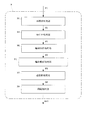
以下、図面を参照して本発明の実施例1を説明する。実施例1では、マンモグラフィ用PET装置1を核医学診断装置の一例として説明する。図1は、実施例1に係るマンモグラフィ用PET装置の概略構成を示す図である。
図1を参照する。マンモグラフィ用PET装置1は、放射性薬剤が投与された被検体M内から放出されたγ線(ガンマ線)を検出する検出器ユニット3と、検出器ユニット3で検出したγ線に基づき、エミッションデータE1を収集するデータ収集部5とを備えている。
Please refer to FIG. The
検出器ユニット3は、被検体Mの撮像対象である乳房Bを取り囲むように、複数のγ線検出器7がC状に配置されている。すなわち、検出器ユニット3は、リング状に配置した複数のγ線検出器7のうち、一部のγ線検出器7が抜き取られたように切り欠き部Aを有して構成されている。なお、γ線検出器7は、本発明の検出器に相当する。
In the
γ線検出器7は、シンチレータブロックとライトガイドと光電子増倍管とを備えている(いずれも図示省略)。シンチレータブロックは、複数個で構成され、複数個のシンチレータブロックが行列状(例えば8行×8列)に配列されている。さらに、2次元状に配置された複数のシンチレータブロックは、1段で構成された単層構造、または複数段(例えば2段)で構成された積層構造となっている。シンチレータブロックにγ線が入射すると、γ線が光に変換される。変換された光は、ライトガイドを通じてシンチレータブロックから光電子増倍管に送られる。送られた光は、光電子増倍管により電気信号に変換される。
The γ-
データ収集部5は、図示しない同時計数回路を備えている。データ収集部5は、γ線を検出して検出器ユニット3から出力される電気信号に基づき、同時計数されたと判定した1事象をエミッションデータE1として収集する。すなわち、データ収集部5は、被検体M内から180°反対方向に放出された2本のγ線を2つのγ線検出器7で一定期間内に検出したときのみに同時計数されたとみなし、その情報を収集する。そのため、エミッションデータE1は、同時計数されたγ線検出器7を結ぶ線の情報であるLOR(line of responce)情報を含んでいる。なお、エミッションデータE1は、3次元収集されるが、場合によっては2次元収集でもよい。また、エミッションデータE1は、同時計数情報を1つ1つ時系列に収集するリストモードで収集されるが、場合によっては、ヒストグラムモードで収集されてもよい。
The
データ収集部5の後段には、収集したエミッションデータE1から被検体Mの乳房Bの輪郭形状を含む画像である3次元輪郭画像Gr1を生成する輪郭画像生成部9と、輪郭画像生成部9で生成された3次元輪郭画像Gr1に基づいて吸収係数マップを生成し、この吸収係数マップを用いて、エミッションデータE1に対して吸収補正を行う吸収補正処理部11とが設けられている。
In the subsequent stage of the
吸収補正に用いられる吸収係数マップは、3次元輪郭画像Gr1の乳房Bの輪郭内に、被検体Mの乳房Bと等価な吸収係数を一様に割り当てたものである。なお、吸収係数は、予め設定されている。吸収補正処理部11は、エミッションデータE1に対して吸収補正を行い、吸収補正後のエミッションデータE2を生成する。なお、輪郭画像生成部9は、本発明の輪郭画像生成装置に相当する。
The absorption coefficient map used for the absorption correction is obtained by uniformly assigning an absorption coefficient equivalent to the breast B of the subject M within the outline of the breast B of the three-dimensional outline image Gr1. The absorption coefficient is set in advance. The absorption
更に、吸収補正部11の後段には、吸収補正後のエミッションデータE2を再構成してPET画像Gpを生成する再構成処理部13が設けられている。再構成処理部13による再構成処理は、検出器ユニット3に切り欠き部Aを有していても、精度よく再構成できる2次元または3次元再構成法が用いられる。例えば、再構成処理には、3次元逐次近似再構成法が用いられる。また、吸収補正部11は、再構成処理部13のシステムモデルに含まれていても構わない。
Furthermore, a
また、マンモグラフィ用PET装置1は、この装置1の各構成を統括的に制御する主制御部15と、再構成処理部13で生成されたPET画像を表示する表示部17と、操作者が入力設定や各種操作を行う入力部19と、PET画像などを記憶する記憶部21とを備えている。主制御部15は、中央演算処理装置(CPU)などで構成される。表示部17は、液晶モニタ等で構成される。入力部19は、キーボードやマウス等で構成される。記憶部21は、ROM(read-only memory)、RAM(random-access memory)またはハードディスク等の記憶媒体で構成される。なお、その記憶媒体は、マンモグラフィ用PET装置1に対し、着脱自在なものであってもよい。
In addition, the
〔輪郭画像生成部〕
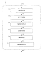
次に、本発明の特徴部分である輪郭画像生成部9の構成を具体的に説明する。図2は、実施例1に係る輪郭画像生成部9の構成を示す図である。輪郭画像生成部9は、収集したエミッションデータE1から被検体Mの乳房Bの輪郭形状を含む画像である3次元輪郭画像Gr1を生成する。[Outline image generator]
Next, the configuration of the contour image generation unit 9 which is a characteristic part of the present invention will be specifically described. FIG. 2 is a diagram illustrating a configuration of the contour image generation unit 9 according to the first embodiment. The contour image generation unit 9 generates a three-dimensional contour image Gr1 that is an image including the contour shape of the breast B of the subject M from the collected emission data E1.
まず、輪郭画像生成部9の構成を簡単に説明する。輪郭画像生成部9は、被検体Mの乳房B内から放出されたγ線を検出して収集したエミッションデータE1を、3次元再構成法に用いて再構成して3次元再構成画像G1を生成する再構成処理部31と、3次元再構成画像G1に対して最大値投影処理を行って複数方向の2次元最大値投影データP1を生成する最大値投影(MIP)処理部33と、2次元最大値投影データP1の各々に対して被検体Mの乳房Bの輪郭を抽出して2次元輪郭投影データP2を生成する輪郭抽出処理部35と、2次元輪郭投影データP2を逆投影して3次元逆投影画像G2を生成する逆投影処理部37と、3次元逆投影画像G2に対して閾値処理を行って3次元輪郭画像Grを生成する輪郭画像生成部39とを備えている。次に、これらの構成をさらに順番に説明する。
First, the configuration of the contour image generation unit 9 will be briefly described. The contour image generation unit 9 reconstructs emission data E1 collected by detecting γ-rays emitted from the breast B of the subject M using a three-dimensional reconstruction method, and generates a three-dimensional reconstruction image G1. A
再構成処理部31は、被検体Mの乳房B内から放出されたγ線を検出して収集したエミッションデータE1を、3次元再構成法を用いて再構成して3次元再構成画像G1を生成する。再構成処理部31の再構成処理には、例えば、リストモード3次元DRAMA(dynamic RAMLA)法が用いられる。また、再構成処理は、次に説明する方法を採用してもよい。
The
リストモード3次元DRAMA法は、リストモードのエミッションデータE1から直接再構成するリストモード再構成法であった。この点、サイノグラムを生成して再構成するサイノグラム再構成法であってもよい。ただし、サイノグラム再構成法は次のような問題がある。サイノグラム再構成法では、準備するサイノグラムのスライス数が多いので、リビニングなどの処理によりスライス数を減らす処理を行っている。このようなリビニングなどの処理は、解像度を劣化させてしまう問題がある。そのため、リビニングなどによる解像度の劣化のないリストモード再構成法が好ましい。 The list mode three-dimensional DRAMA method is a list mode reconstruction method in which reconstruction is performed directly from list mode emission data E1. In this regard, a sinogram reconstruction method in which a sinogram is generated and reconstructed may be used. However, the sinogram reconstruction method has the following problems. In the sinogram reconstruction method, since the number of slices of the sinogram to be prepared is large, processing for reducing the number of slices is performed by processing such as rebinning. Such processing such as binning has a problem of degrading resolution. Therefore, a list mode reconstruction method that does not degrade resolution due to rebinning or the like is preferable.
また、リストモード3次元DRAMA法では、DRAMA法であった。この点、RAMLA(row-action maximum likelihood algorithm)法やOS−EM(ordered subsets - expectation maximization)法などの逐次近似再構成法であってもよい。また、これらの逐次近似再構成法とサイノグラム再構成法とを組み合わせてもよい。 The list mode three-dimensional DRAMA method is the DRAMA method. In this respect, a successive approximation reconstruction method such as a RAMLA (row-action maximum likelihood algorithm) method or an OS-EM (ordered subsets-expectation maximization) method may be used. Further, these successive approximation reconstruction methods and sinogram reconstruction methods may be combined.
最大値投影(maximum intensity projection : MIP)処理部33(以下適宜、「MIP処理部」と称する)は、3次元再構成画像G1に対して最大値投影処理(以下適宜、「MIP処理」と称する)を行って、複数方向の2次元最大値投影データP1を生成する。 A maximum intensity projection (MIP) processing unit 33 (hereinafter appropriately referred to as “MIP processing unit”) performs maximum value projection processing (hereinafter appropriately referred to as “MIP processing”) on the three-dimensional reconstructed image G1. ) To generate two-dimensional maximum value projection data P1 in a plurality of directions.
図3(a)は、MIP処理を説明するための図である。なお、図3(a)は、図示の便宜上、3次元再構成画像G1を2次元で表しているが、奥行き方向にも複数の画素が存在しているものとする。MIP処理は、回転中心軸O周りに、予め設定された角度ごとに最大値投影データP1を生成し、例えば180°や360°回転分の複数の最大値投影データP1を得る処理である。すなわち、MIP処理は、切り欠き部Aによりγ線検出器7が存在しない投影方向(例えば符号Fの方向)を含み最大値投影データP1を生成する。また、MIP処理により投影される画素は、例えば、図3(a)の1次元の画素列Lの場合、画素列Lを構成する複数の画素のうち、画素値が最大の画素が投影される。このような処理により、図3(b)に示すような、複数方向の2次元最大値投影データP1が生成される。なお、図3(a)および図3(b)の切り欠き部Aに存在する破線は、存在しないγ線検出器7を示す。
FIG. 3A is a diagram for explaining the MIP processing. 3A shows the three-dimensional reconstructed image G1 in two dimensions for convenience of illustration, it is assumed that a plurality of pixels exist in the depth direction. The MIP processing is processing for generating maximum value projection data P1 for each preset angle around the rotation center axis O, and obtaining a plurality of maximum value projection data P1 for 180 ° or 360 ° rotation, for example. That is, in the MIP processing, the maximum value projection data P1 including the projection direction (for example, the direction of the sign F) in which the γ-
輪郭抽出処理部35は、2次元最大値投影データP1の各々に対して被検体Mの乳房B輪郭を抽出し、2次元輪郭投影データP2を生成する。輪郭抽出処理は、レベルセット(level set)法やスネーク(snake)法などの動的輪郭モデルを用いるのが好ましい。また、輪郭抽出処理は、ソーベルフィルタ(sobel filter)やプリューウィットフィルタ(prewitte filter)などの微分フィルタを利用した輪郭抽出方法でもよい。
The contour
動的輪郭モデルとは、画像上に設定した閉曲線を動的に変化させて物体の輪郭に一致させることで輪郭を抽出する方法である。図4のように、2次元最大値投影データP1上で初期の輪郭W(0)を設定し、処理を繰り返し実行することで、輪郭W(1)、輪郭W(2)、…、輪郭W(n)が順番に得られる。 The dynamic contour model is a method for extracting a contour by dynamically changing a closed curve set on an image to match the contour of an object. As shown in FIG. 4, by setting an initial contour W (0) on the two-dimensional maximum value projection data P1 and repeatedly executing the processing, the contour W (1), the contour W (2),. (N) is obtained in order.
逆投影処理部37は、輪郭抽出処理部35で生成された2次元輪郭投影データP2を逆投影して3次元逆投影画像G2を生成する。すなわち、逆投影処理部37は、最大値投影して輪郭を抽出した2次元輪郭投影データP2を、そのまま逆投影して、3次元再構成画像に戻す処理を行う。
The back
閾値処理部39は、3次元逆投影画像G2に対して閾値処理を行って3次元輪郭画像Gr1を生成する。閾値処理部39は、予め設定された閾値に基づき、閾値処理を行って乳房B内外の領域を分割する。分割された領域は2値で表される。
The
〔マンモグラフィ用PET装置の動作〕
次に、マンモグラフィ用PET装置1の動作を説明する。まず、被検体Mに放射性薬剤を投与し、マンモグラフィ用PET装置1に被検体Mを配置する。被検体Mの撮像対象である乳房Bからγ線が放出される。放出されるγ線は、180°反対方向に2本放出される。このγ線はC型の検出器ユニット3で検出される。データ収集部5は、一定期間内に2つのγ線検出器7で2本のγ線がそれぞれ検出されたことを示す事象をエミッションデータE1として収集する。[Operation of PET apparatus for mammography]
Next, the operation of the
データ収集部5で収集されたエミッションデータE1は、吸収補正処理部11により吸収補正が行われる。吸収補正は、輪郭画像生成部9で生成された輪郭画像Gr1を用いて行われる。すなわち、吸収補正処理部11は、3次元輪郭画像Gr1の輪郭内に予め設定された吸収係数を一様に割り当てた吸収マップを用いて、エミッションデータE1に対して吸収補正を行う。これにより、吸収補正後のエミッションデータE2が得られる。
The emission data E1 collected by the
吸収補正後のエミッションデータE2は、再構成処理部13により3次元のPET画像Gpに再構成される。再構成された3次元のPET画像Gpは、表示部17に表示され、また、記憶部21に記憶される。また、表示部17に表示される画像は、例えば、3次元PET画像Gpにおける任意の断面であってもよい。
The emission data E2 after the absorption correction is reconstructed into a three-dimensional PET image Gp by the
〔輪郭画像生成部の動作〕
次に、本発明の特徴部分である輪郭画像生成部9の動作を説明する。図2を参照する。輪郭画像生成部9は、データ収集部5で収集されたエミッションデータE1から3次元輪郭画像Gr1を生成する。被検体Mの乳房Bの輪郭を精度よく抽出するためには、γ線検出器7が存在しないことにより、2つのγ線検出器7間を結ぶことができなかった方向を含む全ての投影方向に対する被検体Mの乳房Bの情報が必要である。そこで、まず、エミッションデータE1を再構成して一旦画像化する。なお、エミッションデータE1は、切り欠き部Aによるデータ欠損を含んだものである。[Operation of contour image generator]
Next, the operation of the contour image generation unit 9 which is a characteristic part of the present invention will be described. Please refer to FIG. The contour image generation unit 9 generates a three-dimensional contour image Gr1 from the emission data E1 collected by the
再構成処理部31は、収集したエミッションデータE1を、3次元再構成法を用いて再構成して3次元再構成画像G1を生成する。再構成処理には、例えば、リストモード3次元DRAMA法が用いられる。3次元再構成法を用いることで、データ欠損によるアーチファクトの影響を比較的に低減させた3次元再構成画像G1を取得することができる。
The
3次元再構成画像G1の生成後、MIP処理部33は、3次元再構成画像G1に対してMIP処理を行って、複数方向の2次元最大値投影データP1を生成する。一旦、再構成した3次元再構成画像G1に対してMIP処理を行うことで、C型の検出器ユニット3のγ線検出器7が存在しない投影方向に対しても擬似的にデータを存在させることができる。また、単なる順投影処理でなくMIP処理を行うことで、乳房B領域とそれ以外の領域との境界を強調させることができる。これにより、従来法では、不可能なγ線検出器7が存在しない投影方向に対しても、後述の輪郭抽出処理部35により、被検体Mの乳房Bの輪郭を精度よく抽出することができる。
After generating the three-dimensional reconstructed image G1, the
2次元最大値投影データP1の生成後、輪郭抽出処理部35は、2次元最大値投影データP1の各々に対し、被検体Mの乳房B輪郭を抽出して2次元輪郭投影データP2を生成する。輪郭抽出は、例えばレベルセット法を用いて行われる。輪郭抽出処理部35は、境界が強調された2次元最大値投影データP1から輪郭抽出を行うので、輪郭抽出を精度よく行うことができる。
After the generation of the two-dimensional maximum value projection data P1, the contour
輪郭抽出処理部35により、輪郭抽出された2次元輪郭投影データP2は、被検体Mの乳房Bの内外の領域を2値化したもので表される。例えば、乳房Bの内側の領域を「1」とし、これ以外の乳房Bの外側の領域を「0」とする情報が与えられる。
The two-dimensional contour projection data P2 whose contour has been extracted by the contour
2次元輪郭投影データP2の生成後、逆投影処理部37は、輪郭抽出処理部35で生成された2次元輪郭投影データP2を逆投影して3次元逆投影画像G2を生成する。これにより、3次元の画像データ形式に戻す。そして、3次元逆投影画像G2の生成後、閾値処理部39は、3次元逆投影画像G2に対して閾値処理を行って、再度輪郭を抽出し、3次元輪郭画像Gr1を生成する。
After the generation of the two-dimensional contour projection data P2, the
本実施例によれば、再構成処理部31は、被検体M内から放出されたγ線を検出して収集したエミッションデータE1を、3次元再構成法を用いて再構成して3次元再構成画像G1を生成し、最大値投影処理部33は、3次元再構成画像G1に対してMIP処理を行って複数方向の2次元最大値投影データP1を生成する。輪郭抽出処理部35は、2次元最大値投影データP1の各々に対して被検体Mの乳房Bの輪郭を抽出し、2次元輪郭投影データP2を生成する。逆投影処理部37は、2次元輪郭投影データP2を逆投影して3次元逆投影画像G2を生成し、閾値処理部39は、3次元逆投影画像G2に対して閾値処理を行って3次元輪郭画像Gr1を生成する。
According to the present embodiment, the
すなわち、3次元再構成画像G1に対してMIP処理を行うことにより、本来、γ線検出器7が存在しない投影方向に対しても擬似的にデータを存在させることができる。また、単なる順投影処理ではなくMIP処理を行うことで、乳房Bの領域内外の境界を強調させることができる。これにより、2次元最大値投影データP1において、被検体Mの乳房Bの輪郭を精度よく抽出することができる。そして、逆投影処理により、精度よく抽出された乳房Bの輪郭を含む2次元輪郭投影データP2から3次元逆投影画像G2を生成し、3次元逆投影画像G2に対し閾値処理を行って3次元輪郭画像Grを生成する。その結果、γ線検出器7が存在しないような大きなデータ欠損領域を有していても、被検体Mの乳房Bの輪郭を精度よく抽出した3次元輪郭画像Gr1を生成できる。
That is, by performing MIP processing on the three-dimensional reconstructed image G1, data can exist in a pseudo manner even in a projection direction in which the γ-
また、3次元再構成画像G1を生成する際に、3次元再構成法を用いることで、γ線検出器7が存在しないようなデータ欠損領域があっても、データ欠損によるアーチファクトの影響を低減させた3次元再構成画像G1を生成することができる。
Further, by using the three-dimensional reconstruction method when generating the three-dimensional reconstruction image G1, even if there is a data loss region where the γ-
次に、図面を参照して本発明の実施例2を説明する。図5は、実施例2に係る輪郭画像生成部9の構成を示す図である。なお、上述した実施例1と重複する構成については、その説明を省略する。輪郭抽出後の2次元輪郭投影データP2には、統計ノイズが原因の誤差が含まれている。そのため、誤差が含まれた状態で逆投影しても、精度よく3次元輪郭画像Gr1を得られないことがある。
Next,
実施例2の輪郭画像生成部9は、実施例1の構成に加えて、更に、輪郭抽出後の輪郭整形処理部51を備えている。なお、輪郭整形処理部51は、本発明の第1輪郭整形処理部に相当する。
In addition to the configuration of the first embodiment, the contour image generation unit 9 of the second embodiment further includes a contour
輪郭整形処理部51は、輪郭抽出処理部35と逆投影処理部37との間に設けられており、2次元輪郭投影データP2に対し、抽出した輪郭を整形する輪郭整形処理を行う。これにより、輪郭整形処理後の2次元輪郭投影データP3が生成される。また、逆投影処理部37は、輪郭整形処理後の2次元輪郭投影データP3を逆投影して3次元逆投影画像G2を生成する。輪郭整形処理部51の輪郭整形処理は、膨張収縮フィルタやフーリエ級数近似などが用いられる。
The contour
本実施例によれば、2次元輪郭投影データP2に対し、抽出した輪郭を整形する輪郭整形処理を行う輪郭整形処理部51を備え、逆投影処理部37は、輪郭整形処理後の2次元輪郭投影データP3を逆投影して3次元逆投影画像G2を生成する。これにより、2次元輪郭投影データP2において、抽出した輪郭が整えられるので、統計ノイズなどのノイズの影響を抑えた3次元輪郭画像Gr1を生成することができる。
According to the present embodiment, the contour
次に、図面を参照して本発明の実施例3を説明する。なお、上述した実施例1と重複する構成については、その説明を省略する。図6は、実施例3に係る輪郭画像生成部9の構成を示す図である。実施例3の輪郭画像生成部9は、実施例1の構成に加えて、更に、閾値処理後の輪郭整形処理部53を備えている。なお、輪郭整形処理部53は、本発明の第2輪郭整形処理部に相当する。
Next,
輪郭整形処理部53は、閾値処理部39の後段に設けられており、3次元輪郭画像Gr1に対し、閾値処理で抽出した輪郭を整形する輪郭整形処理を行う。これにより、輪郭整形処理後の3次元輪郭画像Gr2が生成される。輪郭整形処理部53の輪郭整形処理は、膨張収縮フィルタやフーリエ級数近似などが用いられる。
The contour
本実施例によれば、3次元輪郭画像Gr1に対し、閾値処理で抽出した輪郭を整形する輪郭整形処理を行う輪郭整形処理部53を備えている。これにより、3次元輪郭画像Gr1において、閾値処理で抽出された輪郭が整えられるので、統計ノイズなどのノイズの影響を抑えた閾値処理後の3次元輪郭画像Gr2を生成することができる。
According to the present embodiment, the contour
なお、実施例3の変形例として、図7のように構成してもよい。すなわち、実施例1から3の全ての構成を組み合わせた構成である。この変形例では、輪郭抽出後および閾値処理後の両方に輪郭整形処理を適用させている。この構成により、更に、ノイズの影響を抑えた輪郭形状を得ることができる。また、3次元輪郭画像Gr2生成の一連の処理内に分散させることで、輪郭形状を整形し過ぎることなく、正確に輪郭を整形することができる。 In addition, you may comprise as FIG. 7 as a modification of Example 3. FIG. That is, this is a combination of all the configurations of the first to third embodiments. In this modification, contour shaping processing is applied both after contour extraction and after threshold processing. With this configuration, it is possible to obtain a contour shape that further suppresses the influence of noise. In addition, by dispersing in a series of processes for generating the three-dimensional contour image Gr2, the contour can be accurately shaped without excessively shaping the contour shape.
次に、図面を参照して本発明の実施例3を説明する。なお、上述した実施例1と重複する構成については、その説明を省略する。図8は、実施例4に係る輪郭画像生成部9の構成を示す図である。実施例2,3では、輪郭抽出後の輪郭形状に対し、輪郭整形処理を行って統計ノイズなどのノイズの影響を抑えていた。実施例4では、輪郭抽出前に統計ノイズの影響を抑える。すなわち、実施例4の輪郭画像生成部9は、実施例1の構成に加えて、更に、再構成処理後のスムージング処理部55を備えている。
Next,
スムージング処理部55は、再構成処理部31と最大値投影処理部33との間に設けられており、3次元再構成画像G1に対してスムージング処理を行う。これにより、スムージング処理後の3次元再構成画像G3が生成される。スムージング処理は、ガウシアンフィルタやメディアンフィルタ、ウェーブレットフィルタ、移動平均フィルタなどが用いられる。
The smoothing
本実施例によれば、3次元再構成画像G1に対してスムージング処理を行うスムージング処理部55を備え、MIP処理部33は、スムージング処理後の再構成画像G3に対し、MIP処理を行って複数方向の2次元最大値投影データP1を生成する。これにより、エミッションデータE1を再構成して生成された3次元再構成画像G1の統計ノイズが抑えられるので、2次元最大値投影データP1において、被検体Mの乳房Bの輪郭を精度よく抽出することができる。
According to the present embodiment, the smoothing
なお、実施例4の変形例として、図9のように構成してもよい。すなわち、実施例1から4の全ての構成を組み合わせた構成である。また、図9の構成から、輪郭整形処理部51,53のいずれか一方を省略した構成であってもよい。このような構成により、被検体Mの乳房Bを更に精度よく抽出した3次元輪郭画像Gr1,Gr2を生成することができる。
In addition, you may comprise as FIG. 9 as a modification of Example 4. FIG. That is, this is a combination of all the configurations of the first to fourth embodiments. Moreover, the structure which abbreviate | omitted any one of the contour
本発明は、上記実施形態に限られることはなく、下記のように変形実施することができる。 The present invention is not limited to the above embodiment, and can be modified as follows.
(1)上述した各実施例では、エミッションデータE1は、同時計数されたγ線検出器7を結ぶ線の情報であるLOR情報を含んでいた。この点、エミッションデータE1は、TOF(time of flight)情報を含んでいてもよい。TOF情報は、同時計数された消滅γ線の検出時間の差を正確に測定すれば、γ線の放出位置を求めることができるという原理に基づく検出時間情報である。
(1) In each of the above-described embodiments, the emission data E1 includes LOR information, which is information of lines connecting the γ-
輪郭画像生成部9において、再構成処理部31は、TOF情報を含むエミッションデータE1を、TOF再構成法を用いて再構成して3次元再構成画像G1を生成する。図10(a)は、TOF情報を用いたTOF再構成法を説明するための図であり、図10(b)は、図10(a)との比較で、LOR情報を用いたLOR再構成法を説明するための図である。
In the contour image generation unit 9, the
TOF再構成法は、リストモード再構成の一例である。図10(b)のLOR情報の場合は、再構成の際に、同時計数された2つのγ線検出器7間を結ぶ線に沿って一様な値V2が与えられる。一方、図10(a)のTOF情報の場合は、再構成の際に、重み付けされた値V1が与えられる。すなわち、値V1が高い場所では、γ線の放出位置Rである確率が高いことを示す。その結果、TOF再構成法による再構成により、更にアーチファクトの影響が抑えられた3次元再構成画像G1を生成することができる。
The TOF reconstruction method is an example of list mode reconstruction. In the case of the LOR information shown in FIG. 10B, a uniform value V2 is given along a line connecting the two γ-
なお、実施例1で例示したリストモード3次元DRAMA法にTOF再構成法を適用させてもよい。 Note that the TOF reconstruction method may be applied to the list mode three-dimensional DRAMA method exemplified in the first embodiment.
(2)上述した各実施例および変形例(1)では、検出器ユニット3は、リング状に配置した複数のγ線検出器7のうち1箇所でγ線検出器7を抜き取って、複数のγ線検出器7をC状に配置したものであった。例えば、検出器ユニット3は、リング状に配置した複数のγ線検出器7のうち複数箇所(例えば2箇所)でγ線検出器7を抜き取ったものでもよい。
(2) In each of the above-described embodiments and modification (1), the
(3)上述した各実施例および各変形例では、核医学診断装置の一例として、マンモグラフィ用PET装置1を説明したが、これに限定されない。例えば、核医学診断装置は、被検体Mの頭部を撮像する頭部用PET装置や、頭部用PET/CT装置、頭部用PET/SPECT装置などのモダリティであってもよい。すなわち、本発明は、比較的単一吸収体とみなせる物体を撮像する核医学診断装置に適用できる。
(3) In each of the above-described embodiments and modifications, the
(4)上述した各実施例および各変形例では、3次元輪郭画像Gr1,Gr2を生成していたが、例えば、2次元輪郭画像を生成してもよい。 (4) In each example and each modification described above, the three-dimensional contour images Gr1 and Gr2 are generated. However, for example, a two-dimensional contour image may be generated.
1 … マンモグラフィ用PET装置
3 … 検出器ユニット
5 … データ収集部
7 … γ線検出器
9 … 輪郭画像生成部
15 … 主制御部
31 … 再構成処理部
33 … MIP処理部
35 … 輪郭抽出処理部
37 … 逆投影処理部
39 … 閾値処理部
51 … 輪郭整形処理部
53 … 輪郭整形処理部
55 … スムージング処理部
M … 被検体
B … 乳房
E1 … エミッションデータ
P1 … 2次元最大値投影データ
P2 … 2次元輪郭投影データ
P3 … 輪郭整形処理後の2次元輪郭投影データ
G1 … 3次元再構成画像
G2 … 3次元逆投影画像
G3 … スムージング処理後の3次元再構成画像
Gr1 … 3次元輪郭画像
Gr2 … 輪郭整形処理後の3次元輪郭画像
A … 切り欠き部
DESCRIPTION OF
Claims (6)
前記再構成画像に対して最大値投影処理を行って複数方向の最大値投影データを生成する最大値投影処理部と、
前記最大値投影データの各々に対して被検体の撮像対象の輪郭を抽出し、輪郭投影データを生成する輪郭抽出処理部と、
前記輪郭投影データを逆投影して逆投影画像を生成する逆投影処理部と、
前記逆投影画像に対して閾値処理を行って輪郭画像を生成する閾値処理部とを備えていること特徴とする輪郭画像生成装置。A reconstruction processing unit for reconstructing emission data collected by detecting radiation emitted from within the subject, and generating a reconstructed image;
A maximum value projection processing unit that performs maximum value projection processing on the reconstructed image to generate maximum value projection data in a plurality of directions;
A contour extraction processing unit that extracts the contour of the imaging target of the subject for each of the maximum value projection data and generates contour projection data;
A back projection processing unit that back projects the contour projection data to generate a back projection image;
A contour image generating apparatus, comprising: a threshold processing unit configured to generate a contour image by performing threshold processing on the backprojected image.
前記再構成処理部は、エミッションデータを、3次元再構成法を用いて再構成して再構成画像を生成することを特徴とする輪郭画像生成装置。The contour image generation apparatus according to claim 1,
The contour image generation apparatus, wherein the reconstruction processing unit reconstructs emission data using a three-dimensional reconstruction method to generate a reconstructed image.
前記輪郭投影データに対し、抽出した輪郭を整形する輪郭整形処理を行う第1輪郭整形処理部を備え、
前記逆投影処理部は、輪郭整形処理後の前記輪郭投影データを逆投影して逆投影画像を生成することを特徴とする輪郭画像生成装置。In the outline image generation device according to claim 1 or 2,
A first contour shaping processing unit for performing contour shaping processing for shaping the extracted contour on the contour projection data;
The back projection processing unit generates a back projection image by back projecting the contour projection data after the contour shaping process.
前記輪郭画像に対し、閾値処理で抽出した輪郭を整形する輪郭整形処理を行う第2輪郭整形処理部を備えることを特徴とする輪郭画像生成装置。In the outline image generation device according to any one of claims 1 to 3,
A contour image generation apparatus, comprising: a second contour shaping processing unit that performs a contour shaping process for shaping a contour extracted by threshold processing on the contour image.
前記再構成画像に対してスムージング処理を行うスムージング処理部を備え、
前記最大値投影処理部は、スムージング処理後の前記再構成画像に対し、最大値投影処理を行って複数方向の最大値投影データを生成することを特徴とする輪郭画像生成装置。In the outline image generation device according to any one of claims 1 to 4,
A smoothing processing unit for performing a smoothing process on the reconstructed image;
The maximum value projection processing unit generates a maximum value projection data in a plurality of directions by performing maximum value projection processing on the reconstructed image after smoothing processing.
前記検出器ユニットで検出された放射線に基づき、エミッションデータを収集するデータ収集部と、
前記エミッションデータを再構成して再構成画像を生成する再構成処理部と、
前記再構成画像に対して最大値投影処理を行って複数方向の最大値投影データを生成する最大値投影処理部と、
前記最大値投影データの各々に対して被検体の撮像対象の輪郭を抽出し、輪郭投影データを生成する輪郭抽出処理部と、
前記輪郭投影データを逆投影して逆投影画像を生成する逆投影処理部と、
前記逆投影画像に対して閾値処理を行って輪郭画像を生成する閾値処理部とを備えていること特徴とする核医学診断装置。
A detector unit configured to detect some of the detectors extracted from a plurality of detectors arranged in a ring shape, and detecting radiation emitted from within the subject;
A data collection unit that collects emission data based on radiation detected by the detector unit;
A reconstruction processing unit that reconstructs the emission data and generates a reconstructed image;
A maximum value projection processing unit that performs maximum value projection processing on the reconstructed image to generate maximum value projection data in a plurality of directions;
A contour extraction processing unit that extracts the contour of the imaging target of the subject for each of the maximum value projection data and generates contour projection data;
A back projection processing unit that back projects the contour projection data to generate a back projection image;
A nuclear medicine diagnosis apparatus comprising: a threshold processing unit that performs threshold processing on the backprojected image to generate a contour image.
Applications Claiming Priority (1)
| Application Number | Priority Date | Filing Date | Title |
|---|---|---|---|
| PCT/JP2013/079018 WO2015059827A1 (en) | 2013-10-25 | 2013-10-25 | Contour image generation device and nuclear medicine diagnosis device |
Publications (2)
| Publication Number | Publication Date |
|---|---|
| JP6052425B2 true JP6052425B2 (en) | 2016-12-27 |
| JPWO2015059827A1 JPWO2015059827A1 (en) | 2017-03-09 |
Family
ID=52992463
Family Applications (1)
| Application Number | Title | Priority Date | Filing Date |
|---|---|---|---|
| JP2015543678A Active JP6052425B2 (en) | 2013-10-25 | 2013-10-25 | Contour image generating device and nuclear medicine diagnostic device |
Country Status (2)
| Country | Link |
|---|---|
| JP (1) | JP6052425B2 (en) |
| WO (1) | WO2015059827A1 (en) |
Cited By (1)
| Publication number | Priority date | Publication date | Assignee | Title |
|---|---|---|---|---|
| KR102648104B1 (en) * | 2023-09-14 | 2024-03-15 | 주식회사 스누아이랩 | Method, apparatus and computer program for measuring gap and flush |
Families Citing this family (1)
| Publication number | Priority date | Publication date | Assignee | Title |
|---|---|---|---|---|
| CN113436209B (en) * | 2021-06-23 | 2023-11-17 | 南通大学 | A new weld centerline extraction method based on layer-by-layer indentation strategy |
Citations (3)
| Publication number | Priority date | Publication date | Assignee | Title |
|---|---|---|---|---|
| JP2004174241A (en) * | 2002-11-25 | 2004-06-24 | Siemens Ag | Image forming method |
| JP2007163154A (en) * | 2005-12-09 | 2007-06-28 | Shimadzu Corp | Diagnostic imaging support device |
| WO2013038452A1 (en) * | 2011-09-15 | 2013-03-21 | 株式会社島津製作所 | Medical data processing device and radiation tomography device provided therewith |
-
2013
- 2013-10-25 WO PCT/JP2013/079018 patent/WO2015059827A1/en not_active Ceased
- 2013-10-25 JP JP2015543678A patent/JP6052425B2/en active Active
Patent Citations (3)
| Publication number | Priority date | Publication date | Assignee | Title |
|---|---|---|---|---|
| JP2004174241A (en) * | 2002-11-25 | 2004-06-24 | Siemens Ag | Image forming method |
| JP2007163154A (en) * | 2005-12-09 | 2007-06-28 | Shimadzu Corp | Diagnostic imaging support device |
| WO2013038452A1 (en) * | 2011-09-15 | 2013-03-21 | 株式会社島津製作所 | Medical data processing device and radiation tomography device provided therewith |
Cited By (2)
| Publication number | Priority date | Publication date | Assignee | Title |
|---|---|---|---|---|
| KR102648104B1 (en) * | 2023-09-14 | 2024-03-15 | 주식회사 스누아이랩 | Method, apparatus and computer program for measuring gap and flush |
| WO2025058266A1 (en) * | 2023-09-14 | 2025-03-20 | 주식회사 스누아이랩 | Vehicle step difference measurement method, apparatus, and computer program |
Also Published As
| Publication number | Publication date |
|---|---|
| WO2015059827A1 (en) | 2015-04-30 |
| JPWO2015059827A1 (en) | 2017-03-09 |
Similar Documents
| Publication | Publication Date | Title |
|---|---|---|
| US8731269B2 (en) | Method and system for substantially reducing artifacts in circular cone beam computer tomography (CT) | |
| EP1934942B1 (en) | Iterative reconstruction with enhanced noise control filtering | |
| US8903152B2 (en) | Methods and systems for enhanced tomographic imaging | |
| Takahashi et al. | Performance of a semiconductor SPECT system: comparison with a conventional Anger-type SPECT instrument | |
| EP1828977B1 (en) | Restoration of the nuclear medicine 2d planar image by iterative constrained deconvolution | |
| JP2017196404A (en) | Image processing apparatus, x-ray ct apparatus, and image processing method | |
| CN102177528B (en) | High contrast imaging and fast imaging reconstruction | |
| US8571171B2 (en) | Reconstructing a tomographic image with reduced artifacts | |
| JP2015033581A (en) | X-ray computed tomography device and medical image processing program | |
| US10222490B2 (en) | PET scanner with emission and transmission structures in a checkerboard configuration | |
| KR102616736B1 (en) | Automated motion compensation in PET imaging | |
| JP6037048B2 (en) | Contour image generating device and nuclear medicine diagnostic device | |
| JP6052425B2 (en) | Contour image generating device and nuclear medicine diagnostic device | |
| JP6615531B2 (en) | X-ray computed tomography apparatus and medical image processing apparatus | |
| US10217250B2 (en) | Multi-view tomographic reconstruction | |
| KR101493683B1 (en) | Super-resolution Apparatus and Method using LOR reconstruction based cone-beam in PET image | |
| JP6143533B2 (en) | Nuclear medicine image reconstruction device, nuclear medicine image reconstruction method, and program | |
| CN105631908A (en) | PET image reconstruction method and device | |
| JP2008267913A (en) | Nuclear medicine diagnostic apparatus and diagnostic system used therefor | |
| WO2009004571A1 (en) | Method and apparatus for image reconstruction | |
| US12318240B2 (en) | Nuclear medicine diagnostic apparatus, nuclear medicine diagnostic method, and storage medium | |
| JP2002323567A (en) | ECT equipment | |
| JP2007248121A (en) | Tomographic image contour extraction method, program, and apparatus | |
| JP2012093216A (en) | Radiation tomographic image processing system and radiation tomographic image processing method |
Legal Events
| Date | Code | Title | Description |
|---|---|---|---|
| TRDD | Decision of grant or rejection written | ||
| A01 | Written decision to grant a patent or to grant a registration (utility model) |
Free format text: JAPANESE INTERMEDIATE CODE: A01 Effective date: 20161101 |
|
| A61 | First payment of annual fees (during grant procedure) |
Free format text: JAPANESE INTERMEDIATE CODE: A61 Effective date: 20161114 |
|
| R151 | Written notification of patent or utility model registration |
Ref document number: 6052425 Country of ref document: JP Free format text: JAPANESE INTERMEDIATE CODE: R151 |