JP6050230B2 - Methods and compositions for modification of HLA loci - Google Patents
Methods and compositions for modification of HLA loci Download PDFInfo
- Publication number
- JP6050230B2 JP6050230B2 JP2013520872A JP2013520872A JP6050230B2 JP 6050230 B2 JP6050230 B2 JP 6050230B2 JP 2013520872 A JP2013520872 A JP 2013520872A JP 2013520872 A JP2013520872 A JP 2013520872A JP 6050230 B2 JP6050230 B2 JP 6050230B2
- Authority
- JP
- Japan
- Prior art keywords
- seq
- hla
- gene
- cells
- cell
- Prior art date
- Legal status (The legal status is an assumption and is not a legal conclusion. Google has not performed a legal analysis and makes no representation as to the accuracy of the status listed.)
- Active
Links
- 238000000034 method Methods 0.000 title claims description 94
- 239000000203 mixture Substances 0.000 title description 39
- 230000004048 modification Effects 0.000 title description 16
- 238000012986 modification Methods 0.000 title description 16
- 210000004027 cell Anatomy 0.000 claims description 296
- 108090000623 proteins and genes Proteins 0.000 claims description 183
- 238000003776 cleavage reaction Methods 0.000 claims description 82
- 230000007017 scission Effects 0.000 claims description 82
- 230000014509 gene expression Effects 0.000 claims description 76
- 230000004568 DNA-binding Effects 0.000 claims description 67
- 108090000765 processed proteins & peptides Proteins 0.000 claims description 60
- 101710163270 Nuclease Proteins 0.000 claims description 56
- 230000027455 binding Effects 0.000 claims description 54
- 102000004196 processed proteins & peptides Human genes 0.000 claims description 47
- 108020004414 DNA Proteins 0.000 claims description 45
- 238000012217 deletion Methods 0.000 claims description 41
- 230000037430 deletion Effects 0.000 claims description 41
- 229920001184 polypeptide Polymers 0.000 claims description 41
- 108020001507 fusion proteins Proteins 0.000 claims description 38
- 102000037865 fusion proteins Human genes 0.000 claims description 38
- 210000001744 T-lymphocyte Anatomy 0.000 claims description 37
- HCHKCACWOHOZIP-UHFFFAOYSA-N Zinc Chemical compound [Zn] HCHKCACWOHOZIP-UHFFFAOYSA-N 0.000 claims description 35
- 210000000130 stem cell Anatomy 0.000 claims description 35
- 239000011701 zinc Substances 0.000 claims description 35
- 229910052725 zinc Inorganic materials 0.000 claims description 35
- 102000040430 polynucleotide Human genes 0.000 claims description 26
- 108091033319 polynucleotide Proteins 0.000 claims description 26
- 239000002157 polynucleotide Substances 0.000 claims description 26
- 239000000427 antigen Substances 0.000 claims description 22
- 108091007433 antigens Proteins 0.000 claims description 22
- 102000036639 antigens Human genes 0.000 claims description 22
- 208000037265 diseases, disorders, signs and symptoms Diseases 0.000 claims description 22
- 108020004999 messenger RNA Proteins 0.000 claims description 20
- 108700005075 Regulator Genes Proteins 0.000 claims description 19
- 230000004913 activation Effects 0.000 claims description 18
- 239000008194 pharmaceutical composition Substances 0.000 claims description 17
- 239000012634 fragment Substances 0.000 claims description 16
- 208000035475 disorder Diseases 0.000 claims description 14
- 230000010354 integration Effects 0.000 claims description 12
- 108010019670 Chimeric Antigen Receptors Proteins 0.000 claims description 10
- 208000009329 Graft vs Host Disease Diseases 0.000 claims description 8
- 108700005089 MHC Class I Genes Proteins 0.000 claims description 8
- 238000010362 genome editing Methods 0.000 claims description 8
- 208000024908 graft versus host disease Diseases 0.000 claims description 8
- 230000002779 inactivation Effects 0.000 claims description 6
- 210000004263 induced pluripotent stem cell Anatomy 0.000 claims description 5
- 210000002901 mesenchymal stem cell Anatomy 0.000 claims description 5
- 210000001671 embryonic stem cell Anatomy 0.000 claims description 4
- 230000000415 inactivating effect Effects 0.000 claims description 4
- 210000000822 natural killer cell Anatomy 0.000 claims description 3
- 230000001537 neural effect Effects 0.000 claims description 3
- 210000004899 c-terminal region Anatomy 0.000 claims description 2
- 210000000265 leukocyte Anatomy 0.000 claims description 2
- 239000000439 tumor marker Substances 0.000 claims description 2
- 101150076489 B gene Proteins 0.000 claims 6
- 101150111062 C gene Proteins 0.000 claims 4
- 101150084750 1 gene Proteins 0.000 claims 3
- 101150109698 A2 gene Proteins 0.000 claims 3
- FWMNVWWHGCHHJJ-SKKKGAJSSA-N 4-amino-1-[(2r)-6-amino-2-[[(2r)-2-[[(2r)-2-[[(2r)-2-amino-3-phenylpropanoyl]amino]-3-phenylpropanoyl]amino]-4-methylpentanoyl]amino]hexanoyl]piperidine-4-carboxylic acid Chemical compound C([C@H](C(=O)N[C@H](CC(C)C)C(=O)N[C@H](CCCCN)C(=O)N1CCC(N)(CC1)C(O)=O)NC(=O)[C@H](N)CC=1C=CC=CC=1)C1=CC=CC=C1 FWMNVWWHGCHHJJ-SKKKGAJSSA-N 0.000 claims 1
- 210000001772 blood platelet Anatomy 0.000 claims 1
- QVVFNJUJKXWFAU-SAYMMRJXSA-N 4,6-O-[(1R)-1-carboxyethylidene]-alpha-D-galactose Chemical compound O1[C@H](O)[C@H](O)[C@@H](O)[C@H]2O[C@](C)(C(O)=O)OC[C@H]21 QVVFNJUJKXWFAU-SAYMMRJXSA-N 0.000 description 128
- 108010017070 Zinc Finger Nucleases Proteins 0.000 description 115
- 239000013598 vector Substances 0.000 description 79
- 150000007523 nucleic acids Chemical class 0.000 description 74
- 102000004169 proteins and genes Human genes 0.000 description 70
- 102000039446 nucleic acids Human genes 0.000 description 69
- 108020004707 nucleic acids Proteins 0.000 description 69
- 101710185494 Zinc finger protein Proteins 0.000 description 58
- 102100023597 Zinc finger protein 816 Human genes 0.000 description 58
- 230000004927 fusion Effects 0.000 description 39
- 239000002773 nucleotide Substances 0.000 description 39
- 125000003729 nucleotide group Chemical group 0.000 description 39
- 230000000694 effects Effects 0.000 description 29
- 108700028369 Alleles Proteins 0.000 description 26
- 239000000047 product Substances 0.000 description 26
- 238000003556 assay Methods 0.000 description 25
- 238000012239 gene modification Methods 0.000 description 25
- 210000001519 tissue Anatomy 0.000 description 25
- 108010077544 Chromatin Proteins 0.000 description 23
- 210000003483 chromatin Anatomy 0.000 description 23
- 102000052510 DNA-Binding Proteins Human genes 0.000 description 22
- 239000000499 gel Substances 0.000 description 21
- 108010074032 HLA-A2 Antigen Proteins 0.000 description 20
- 230000008685 targeting Effects 0.000 description 20
- 238000001890 transfection Methods 0.000 description 20
- 102000025850 HLA-A2 Antigen Human genes 0.000 description 19
- 230000001105 regulatory effect Effects 0.000 description 19
- 210000001151 cytotoxic T lymphocyte Anatomy 0.000 description 18
- 101100395311 Homo sapiens HLA-B gene Proteins 0.000 description 17
- 238000001415 gene therapy Methods 0.000 description 16
- 102100028976 HLA class I histocompatibility antigen, B alpha chain Human genes 0.000 description 15
- 230000005017 genetic modification Effects 0.000 description 15
- 235000013617 genetically modified food Nutrition 0.000 description 15
- 230000035772 mutation Effects 0.000 description 15
- 230000006780 non-homologous end joining Effects 0.000 description 15
- 239000013612 plasmid Substances 0.000 description 15
- 101100395312 Homo sapiens HLA-C gene Proteins 0.000 description 14
- 206010028980 Neoplasm Diseases 0.000 description 14
- 230000001413 cellular effect Effects 0.000 description 14
- 241000700605 Viruses Species 0.000 description 13
- 150000001413 amino acids Chemical class 0.000 description 13
- 238000004458 analytical method Methods 0.000 description 13
- 230000006870 function Effects 0.000 description 13
- 230000009368 gene silencing by RNA Effects 0.000 description 13
- 108700020911 DNA-Binding Proteins Proteins 0.000 description 12
- 238000002474 experimental method Methods 0.000 description 12
- 102000054766 genetic haplotypes Human genes 0.000 description 12
- 238000012546 transfer Methods 0.000 description 12
- 108010042407 Endonucleases Proteins 0.000 description 11
- 238000012413 Fluorescence activated cell sorting analysis Methods 0.000 description 11
- 108700019146 Transgenes Proteins 0.000 description 11
- 238000001727 in vivo Methods 0.000 description 11
- 239000013603 viral vector Substances 0.000 description 11
- 101710096438 DNA-binding protein Proteins 0.000 description 10
- 102000004190 Enzymes Human genes 0.000 description 10
- 108090000790 Enzymes Proteins 0.000 description 10
- 102100028971 HLA class I histocompatibility antigen, C alpha chain Human genes 0.000 description 10
- 108700002010 MHC class II transactivator Proteins 0.000 description 10
- 230000005782 double-strand break Effects 0.000 description 10
- 238000000338 in vitro Methods 0.000 description 10
- 230000002103 transcriptional effect Effects 0.000 description 10
- 241000701161 unidentified adenovirus Species 0.000 description 10
- 230000003612 virological effect Effects 0.000 description 10
- 108091026890 Coding region Proteins 0.000 description 9
- 108091028043 Nucleic acid sequence Proteins 0.000 description 9
- 238000006243 chemical reaction Methods 0.000 description 9
- 238000013461 design Methods 0.000 description 9
- 210000003958 hematopoietic stem cell Anatomy 0.000 description 9
- -1 polymerases Proteins 0.000 description 9
- 108091008146 restriction endonucleases Proteins 0.000 description 9
- 108700005092 MHC Class II Genes Proteins 0.000 description 8
- 210000003719 b-lymphocyte Anatomy 0.000 description 8
- 230000003197 catalytic effect Effects 0.000 description 8
- 230000009089 cytolysis Effects 0.000 description 8
- 230000002950 deficient Effects 0.000 description 8
- 201000010099 disease Diseases 0.000 description 8
- 239000003814 drug Substances 0.000 description 8
- 238000002744 homologous recombination Methods 0.000 description 8
- 230000006801 homologous recombination Effects 0.000 description 8
- 238000003780 insertion Methods 0.000 description 8
- 230000037431 insertion Effects 0.000 description 8
- 239000003446 ligand Substances 0.000 description 8
- 230000001404 mediated effect Effects 0.000 description 8
- 210000004986 primary T-cell Anatomy 0.000 description 8
- 238000013518 transcription Methods 0.000 description 8
- 238000011144 upstream manufacturing Methods 0.000 description 8
- 102000004533 Endonucleases Human genes 0.000 description 7
- 238000010459 TALEN Methods 0.000 description 7
- 108010043645 Transcription Activator-Like Effector Nucleases Proteins 0.000 description 7
- 125000003275 alpha amino acid group Chemical group 0.000 description 7
- 210000000349 chromosome Anatomy 0.000 description 7
- 239000000833 heterodimer Substances 0.000 description 7
- 230000003993 interaction Effects 0.000 description 7
- 239000003550 marker Substances 0.000 description 7
- 238000004806 packaging method and process Methods 0.000 description 7
- 230000008569 process Effects 0.000 description 7
- 238000010186 staining Methods 0.000 description 7
- 108010059434 tapasin Proteins 0.000 description 7
- 230000001225 therapeutic effect Effects 0.000 description 7
- 238000002054 transplantation Methods 0.000 description 7
- 238000011282 treatment Methods 0.000 description 7
- 102100024222 B-lymphocyte antigen CD19 Human genes 0.000 description 6
- 102000017420 CD3 protein, epsilon/gamma/delta subunit Human genes 0.000 description 6
- 241000196324 Embryophyta Species 0.000 description 6
- 101000980825 Homo sapiens B-lymphocyte antigen CD19 Proteins 0.000 description 6
- 101100395310 Homo sapiens HLA-A gene Proteins 0.000 description 6
- 238000002659 cell therapy Methods 0.000 description 6
- 210000005260 human cell Anatomy 0.000 description 6
- 210000004287 null lymphocyte Anatomy 0.000 description 6
- 230000001177 retroviral effect Effects 0.000 description 6
- 238000013207 serial dilution Methods 0.000 description 6
- 230000035939 shock Effects 0.000 description 6
- 238000010361 transduction Methods 0.000 description 6
- 230000026683 transduction Effects 0.000 description 6
- 241000702421 Dependoparvovirus Species 0.000 description 5
- 102100028972 HLA class I histocompatibility antigen, A alpha chain Human genes 0.000 description 5
- 108010074328 Interferon-gamma Proteins 0.000 description 5
- 102000043129 MHC class I family Human genes 0.000 description 5
- 108091054437 MHC class I family Proteins 0.000 description 5
- 108700011259 MicroRNAs Proteins 0.000 description 5
- 101150011263 Tap2 gene Proteins 0.000 description 5
- 230000033228 biological regulation Effects 0.000 description 5
- 201000011510 cancer Diseases 0.000 description 5
- 230000002759 chromosomal effect Effects 0.000 description 5
- 238000010586 diagram Methods 0.000 description 5
- 238000009510 drug design Methods 0.000 description 5
- 238000004520 electroporation Methods 0.000 description 5
- 150000002632 lipids Chemical class 0.000 description 5
- 239000002502 liposome Substances 0.000 description 5
- 238000004519 manufacturing process Methods 0.000 description 5
- 230000007246 mechanism Effects 0.000 description 5
- 230000010076 replication Effects 0.000 description 5
- 239000000126 substance Substances 0.000 description 5
- 241001430294 unidentified retrovirus Species 0.000 description 5
- 102000014914 Carrier Proteins Human genes 0.000 description 4
- 102000004127 Cytokines Human genes 0.000 description 4
- 108090000695 Cytokines Proteins 0.000 description 4
- 102100020986 DNA-binding protein RFX5 Human genes 0.000 description 4
- 102100031780 Endonuclease Human genes 0.000 description 4
- 102000011786 HLA-A Antigens Human genes 0.000 description 4
- 108010075704 HLA-A Antigens Proteins 0.000 description 4
- 241000282412 Homo Species 0.000 description 4
- 101001075432 Homo sapiens DNA-binding protein RFX5 Proteins 0.000 description 4
- 102100037850 Interferon gamma Human genes 0.000 description 4
- 241001465754 Metazoa Species 0.000 description 4
- 241000699670 Mus sp. Species 0.000 description 4
- 108091023040 Transcription factor Proteins 0.000 description 4
- 102000040945 Transcription factor Human genes 0.000 description 4
- 102100040247 Tumor necrosis factor Human genes 0.000 description 4
- 230000000735 allogeneic effect Effects 0.000 description 4
- 125000000539 amino acid group Chemical group 0.000 description 4
- 108091008324 binding proteins Proteins 0.000 description 4
- 210000001185 bone marrow Anatomy 0.000 description 4
- 238000010276 construction Methods 0.000 description 4
- 238000006471 dimerization reaction Methods 0.000 description 4
- 239000003937 drug carrier Substances 0.000 description 4
- 238000001943 fluorescence-activated cell sorting Methods 0.000 description 4
- 230000002068 genetic effect Effects 0.000 description 4
- 239000005090 green fluorescent protein Substances 0.000 description 4
- 238000001638 lipofection Methods 0.000 description 4
- 210000004698 lymphocyte Anatomy 0.000 description 4
- 210000004962 mammalian cell Anatomy 0.000 description 4
- 239000002679 microRNA Substances 0.000 description 4
- 239000003607 modifier Substances 0.000 description 4
- 230000000051 modifying effect Effects 0.000 description 4
- 210000000056 organ Anatomy 0.000 description 4
- 108020003175 receptors Proteins 0.000 description 4
- 102000005962 receptors Human genes 0.000 description 4
- 230000006798 recombination Effects 0.000 description 4
- 238000005215 recombination Methods 0.000 description 4
- 230000008439 repair process Effects 0.000 description 4
- 150000003384 small molecules Chemical class 0.000 description 4
- 230000000638 stimulation Effects 0.000 description 4
- 101150080773 tap-1 gene Proteins 0.000 description 4
- 230000035897 transcription Effects 0.000 description 4
- 230000001052 transient effect Effects 0.000 description 4
- 102100030346 Antigen peptide transporter 1 Human genes 0.000 description 3
- 230000007018 DNA scission Effects 0.000 description 3
- 241000238631 Hexapoda Species 0.000 description 3
- 102000008949 Histocompatibility Antigens Class I Human genes 0.000 description 3
- 108010088652 Histocompatibility Antigens Class I Proteins 0.000 description 3
- 108010027412 Histocompatibility Antigens Class II Proteins 0.000 description 3
- 102000018713 Histocompatibility Antigens Class II Human genes 0.000 description 3
- 101000687346 Homo sapiens PR domain zinc finger protein 2 Proteins 0.000 description 3
- 101100194594 Homo sapiens RFX5 gene Proteins 0.000 description 3
- 108091036060 Linker DNA Proteins 0.000 description 3
- 108010023335 Member 2 Subfamily B ATP Binding Cassette Transporter Proteins 0.000 description 3
- 101150076359 Mhc gene Proteins 0.000 description 3
- 108010047956 Nucleosomes Proteins 0.000 description 3
- 102100024885 PR domain zinc finger protein 2 Human genes 0.000 description 3
- 102000001253 Protein Kinase Human genes 0.000 description 3
- 101150074379 RFX5 gene Proteins 0.000 description 3
- 240000004808 Saccharomyces cerevisiae Species 0.000 description 3
- 108091027967 Small hairpin RNA Proteins 0.000 description 3
- 102100028082 Tapasin Human genes 0.000 description 3
- 210000004102 animal cell Anatomy 0.000 description 3
- 230000008901 benefit Effects 0.000 description 3
- 239000011230 binding agent Substances 0.000 description 3
- 210000004369 blood Anatomy 0.000 description 3
- 239000008280 blood Substances 0.000 description 3
- 238000005516 engineering process Methods 0.000 description 3
- 230000007613 environmental effect Effects 0.000 description 3
- 210000002443 helper t lymphocyte Anatomy 0.000 description 3
- 210000002865 immune cell Anatomy 0.000 description 3
- 208000015181 infectious disease Diseases 0.000 description 3
- 238000001802 infusion Methods 0.000 description 3
- 238000002347 injection Methods 0.000 description 3
- 239000007924 injection Substances 0.000 description 3
- 239000013642 negative control Substances 0.000 description 3
- 210000001623 nucleosome Anatomy 0.000 description 3
- 239000002245 particle Substances 0.000 description 3
- 238000002823 phage display Methods 0.000 description 3
- 229920000642 polymer Polymers 0.000 description 3
- 239000013641 positive control Substances 0.000 description 3
- 108060006633 protein kinase Proteins 0.000 description 3
- 230000009467 reduction Effects 0.000 description 3
- 238000011160 research Methods 0.000 description 3
- 206010039073 rheumatoid arthritis Diseases 0.000 description 3
- 239000004055 small Interfering RNA Substances 0.000 description 3
- 230000001629 suppression Effects 0.000 description 3
- 229940124597 therapeutic agent Drugs 0.000 description 3
- 238000002560 therapeutic procedure Methods 0.000 description 3
- 230000009261 transgenic effect Effects 0.000 description 3
- 239000003981 vehicle Substances 0.000 description 3
- MZOFCQQQCNRIBI-VMXHOPILSA-N (3s)-4-[[(2s)-1-[[(2s)-1-[[(1s)-1-carboxy-2-hydroxyethyl]amino]-4-methyl-1-oxopentan-2-yl]amino]-5-(diaminomethylideneamino)-1-oxopentan-2-yl]amino]-3-[[2-[[(2s)-2,6-diaminohexanoyl]amino]acetyl]amino]-4-oxobutanoic acid Chemical compound OC[C@@H](C(O)=O)NC(=O)[C@H](CC(C)C)NC(=O)[C@H](CCCN=C(N)N)NC(=O)[C@H](CC(O)=O)NC(=O)CNC(=O)[C@@H](N)CCCCN MZOFCQQQCNRIBI-VMXHOPILSA-N 0.000 description 2
- 239000013607 AAV vector Substances 0.000 description 2
- 108010013043 Acetylesterase Proteins 0.000 description 2
- 208000026872 Addison Disease Diseases 0.000 description 2
- 206010002556 Ankylosing Spondylitis Diseases 0.000 description 2
- 102100030343 Antigen peptide transporter 2 Human genes 0.000 description 2
- 101001030716 Arabidopsis thaliana Histone deacetylase HDT1 Proteins 0.000 description 2
- 208000023328 Basedow disease Diseases 0.000 description 2
- 208000009137 Behcet syndrome Diseases 0.000 description 2
- 206010008909 Chronic Hepatitis Diseases 0.000 description 2
- 102000008169 Co-Repressor Proteins Human genes 0.000 description 2
- 108010060434 Co-Repressor Proteins Proteins 0.000 description 2
- 208000015943 Coeliac disease Diseases 0.000 description 2
- 102000053602 DNA Human genes 0.000 description 2
- 102100036279 DNA (cytosine-5)-methyltransferase 1 Human genes 0.000 description 2
- 101150095057 DPB2 gene Proteins 0.000 description 2
- 241000255581 Drosophila <fruit fly, genus> Species 0.000 description 2
- 101000889905 Enterobacteria phage RB3 Intron-associated endonuclease 3 Proteins 0.000 description 2
- 101000889904 Enterobacteria phage T4 Defective intron-associated endonuclease 3 Proteins 0.000 description 2
- 101000889900 Enterobacteria phage T4 Intron-associated endonuclease 1 Proteins 0.000 description 2
- 208000015023 Graves' disease Diseases 0.000 description 2
- 102100031573 Hematopoietic progenitor cell antigen CD34 Human genes 0.000 description 2
- 206010019755 Hepatitis chronic active Diseases 0.000 description 2
- 108010033040 Histones Proteins 0.000 description 2
- 102000006947 Histones Human genes 0.000 description 2
- 101100382122 Homo sapiens CIITA gene Proteins 0.000 description 2
- 101000777663 Homo sapiens Hematopoietic progenitor cell antigen CD34 Proteins 0.000 description 2
- 101000615488 Homo sapiens Methyl-CpG-binding domain protein 2 Proteins 0.000 description 2
- 101001136986 Homo sapiens Proteasome subunit beta type-8 Proteins 0.000 description 2
- 101001000998 Homo sapiens Protein phosphatase 1 regulatory subunit 12C Proteins 0.000 description 2
- 241000725303 Human immunodeficiency virus Species 0.000 description 2
- 102100034349 Integrase Human genes 0.000 description 2
- 108010061833 Integrases Proteins 0.000 description 2
- 241000713666 Lentivirus Species 0.000 description 2
- 108090000364 Ligases Proteins 0.000 description 2
- 102000003960 Ligases Human genes 0.000 description 2
- 102100021299 Methyl-CpG-binding domain protein 2 Human genes 0.000 description 2
- 108060004795 Methyltransferase Proteins 0.000 description 2
- 241000714177 Murine leukemia virus Species 0.000 description 2
- 241000699666 Mus <mouse, genus> Species 0.000 description 2
- 108091061960 Naked DNA Proteins 0.000 description 2
- 108700019961 Neoplasm Genes Proteins 0.000 description 2
- 102000048850 Neoplasm Genes Human genes 0.000 description 2
- 108700019535 Phosphoprotein Phosphatases Proteins 0.000 description 2
- 102000045595 Phosphoprotein Phosphatases Human genes 0.000 description 2
- 108010004729 Phycoerythrin Proteins 0.000 description 2
- 241000235648 Pichia Species 0.000 description 2
- 102100035760 Proteasome subunit beta type-8 Human genes 0.000 description 2
- 108700040121 Protein Methyltransferases Proteins 0.000 description 2
- 102000055027 Protein Methyltransferases Human genes 0.000 description 2
- 102100035620 Protein phosphatase 1 regulatory subunit 12C Human genes 0.000 description 2
- 201000004681 Psoriasis Diseases 0.000 description 2
- 201000001263 Psoriatic Arthritis Diseases 0.000 description 2
- 208000036824 Psoriatic arthropathy Diseases 0.000 description 2
- 108700008625 Reporter Genes Proteins 0.000 description 2
- 241000235070 Saccharomyces Species 0.000 description 2
- 241000235346 Schizosaccharomyces Species 0.000 description 2
- 241000713311 Simian immunodeficiency virus Species 0.000 description 2
- 208000021386 Sjogren Syndrome Diseases 0.000 description 2
- 241000256248 Spodoptera Species 0.000 description 2
- 101800000849 Tachykinin-associated peptide 2 Proteins 0.000 description 2
- 101710183280 Topoisomerase Proteins 0.000 description 2
- 108060008682 Tumor Necrosis Factor Proteins 0.000 description 2
- 241000251539 Vertebrata <Metazoa> Species 0.000 description 2
- 108700005077 Viral Genes Proteins 0.000 description 2
- 230000009471 action Effects 0.000 description 2
- 239000012190 activator Substances 0.000 description 2
- 230000004075 alteration Effects 0.000 description 2
- 210000000612 antigen-presenting cell Anatomy 0.000 description 2
- 230000006907 apoptotic process Effects 0.000 description 2
- 238000013459 approach Methods 0.000 description 2
- 239000011324 bead Substances 0.000 description 2
- 102000015736 beta 2-Microglobulin Human genes 0.000 description 2
- 108010081355 beta 2-Microglobulin Proteins 0.000 description 2
- 230000015572 biosynthetic process Effects 0.000 description 2
- 210000003995 blood forming stem cell Anatomy 0.000 description 2
- 210000002798 bone marrow cell Anatomy 0.000 description 2
- 238000004113 cell culture Methods 0.000 description 2
- 239000003153 chemical reaction reagent Substances 0.000 description 2
- 239000003795 chemical substances by application Substances 0.000 description 2
- 210000003763 chloroplast Anatomy 0.000 description 2
- 208000025302 chronic primary adrenal insufficiency Diseases 0.000 description 2
- 238000010367 cloning Methods 0.000 description 2
- 239000002299 complementary DNA Substances 0.000 description 2
- 230000021615 conjugation Effects 0.000 description 2
- 238000012937 correction Methods 0.000 description 2
- 210000004443 dendritic cell Anatomy 0.000 description 2
- 230000001419 dependent effect Effects 0.000 description 2
- 230000004069 differentiation Effects 0.000 description 2
- 239000003085 diluting agent Substances 0.000 description 2
- 239000000539 dimer Substances 0.000 description 2
- 230000003828 downregulation Effects 0.000 description 2
- 229940079593 drug Drugs 0.000 description 2
- 210000002257 embryonic structure Anatomy 0.000 description 2
- 108010050663 endodeoxyribonuclease CreI Proteins 0.000 description 2
- 239000003623 enhancer Substances 0.000 description 2
- 210000003527 eukaryotic cell Anatomy 0.000 description 2
- 238000001502 gel electrophoresis Methods 0.000 description 2
- 238000001476 gene delivery Methods 0.000 description 2
- 235000015220 hamburgers Nutrition 0.000 description 2
- 230000001771 impaired effect Effects 0.000 description 2
- 238000011534 incubation Methods 0.000 description 2
- 230000002458 infectious effect Effects 0.000 description 2
- 230000002401 inhibitory effect Effects 0.000 description 2
- 239000012212 insulator Substances 0.000 description 2
- 230000000670 limiting effect Effects 0.000 description 2
- 238000011068 loading method Methods 0.000 description 2
- 229920002521 macromolecule Polymers 0.000 description 2
- 210000002540 macrophage Anatomy 0.000 description 2
- 230000036210 malignancy Effects 0.000 description 2
- 239000000463 material Substances 0.000 description 2
- 230000011987 methylation Effects 0.000 description 2
- 238000007069 methylation reaction Methods 0.000 description 2
- 210000003470 mitochondria Anatomy 0.000 description 2
- 210000003205 muscle Anatomy 0.000 description 2
- 230000007935 neutral effect Effects 0.000 description 2
- 210000004940 nucleus Anatomy 0.000 description 2
- 210000003463 organelle Anatomy 0.000 description 2
- 230000036961 partial effect Effects 0.000 description 2
- 125000002467 phosphate group Chemical group [H]OP(=O)(O[H])O[*] 0.000 description 2
- 230000029279 positive regulation of transcription, DNA-dependent Effects 0.000 description 2
- 238000002360 preparation method Methods 0.000 description 2
- 230000002265 prevention Effects 0.000 description 2
- 210000001236 prokaryotic cell Anatomy 0.000 description 2
- 102000021127 protein binding proteins Human genes 0.000 description 2
- 108091011138 protein binding proteins Proteins 0.000 description 2
- RXWNCPJZOCPEPQ-NVWDDTSBSA-N puromycin Chemical compound C1=CC(OC)=CC=C1C[C@H](N)C(=O)N[C@H]1[C@@H](O)[C@H](N2C3=NC=NC(=C3N=C2)N(C)C)O[C@@H]1CO RXWNCPJZOCPEPQ-NVWDDTSBSA-N 0.000 description 2
- 238000011002 quantification Methods 0.000 description 2
- 230000002829 reductive effect Effects 0.000 description 2
- 238000007634 remodeling Methods 0.000 description 2
- 230000004044 response Effects 0.000 description 2
- 238000010187 selection method Methods 0.000 description 2
- 238000012163 sequencing technique Methods 0.000 description 2
- 230000005783 single-strand break Effects 0.000 description 2
- 241000894007 species Species 0.000 description 2
- 238000010561 standard procedure Methods 0.000 description 2
- 201000000596 systemic lupus erythematosus Diseases 0.000 description 2
- 206010043554 thrombocytopenia Diseases 0.000 description 2
- 230000000699 topical effect Effects 0.000 description 2
- 230000037426 transcriptional repression Effects 0.000 description 2
- 238000013519 translation Methods 0.000 description 2
- 102000035160 transmembrane proteins Human genes 0.000 description 2
- 108091005703 transmembrane proteins Proteins 0.000 description 2
- 238000010396 two-hybrid screening Methods 0.000 description 2
- OPCHFPHZPIURNA-MFERNQICSA-N (2s)-2,5-bis(3-aminopropylamino)-n-[2-(dioctadecylamino)acetyl]pentanamide Chemical compound CCCCCCCCCCCCCCCCCCN(CC(=O)NC(=O)[C@H](CCCNCCCN)NCCCN)CCCCCCCCCCCCCCCCCC OPCHFPHZPIURNA-MFERNQICSA-N 0.000 description 1
- BFSVOASYOCHEOV-UHFFFAOYSA-N 2-diethylaminoethanol Chemical compound CCN(CC)CCO BFSVOASYOCHEOV-UHFFFAOYSA-N 0.000 description 1
- 230000005730 ADP ribosylation Effects 0.000 description 1
- 101710159080 Aconitate hydratase A Proteins 0.000 description 1
- 101710159078 Aconitate hydratase B Proteins 0.000 description 1
- 241001655883 Adeno-associated virus - 1 Species 0.000 description 1
- 241000202702 Adeno-associated virus - 3 Species 0.000 description 1
- 241000580270 Adeno-associated virus - 4 Species 0.000 description 1
- 241001634120 Adeno-associated virus - 5 Species 0.000 description 1
- 241000972680 Adeno-associated virus - 6 Species 0.000 description 1
- 241001164825 Adeno-associated virus - 8 Species 0.000 description 1
- 241000649045 Adeno-associated virus 10 Species 0.000 description 1
- 208000002267 Anti-neutrophil cytoplasmic antibody-associated vasculitis Diseases 0.000 description 1
- 108020005544 Antisense RNA Proteins 0.000 description 1
- 208000032467 Aplastic anaemia Diseases 0.000 description 1
- BHELIUBJHYAEDK-OAIUPTLZSA-N Aspoxicillin Chemical compound C1([C@H](C(=O)N[C@@H]2C(N3[C@H](C(C)(C)S[C@@H]32)C(O)=O)=O)NC(=O)[C@H](N)CC(=O)NC)=CC=C(O)C=C1 BHELIUBJHYAEDK-OAIUPTLZSA-N 0.000 description 1
- 208000003950 B-cell lymphoma Diseases 0.000 description 1
- 208000025321 B-lymphoblastic leukemia/lymphoma Diseases 0.000 description 1
- 241000894006 Bacteria Species 0.000 description 1
- 206010006187 Breast cancer Diseases 0.000 description 1
- 208000026310 Breast neoplasm Diseases 0.000 description 1
- 102100035875 C-C chemokine receptor type 5 Human genes 0.000 description 1
- 101710149870 C-C chemokine receptor type 5 Proteins 0.000 description 1
- 102100028892 Cardiotrophin-1 Human genes 0.000 description 1
- 102000011727 Caspases Human genes 0.000 description 1
- 108010076667 Caspases Proteins 0.000 description 1
- 108090000994 Catalytic RNA Proteins 0.000 description 1
- 102000053642 Catalytic RNA Human genes 0.000 description 1
- 108010001857 Cell Surface Receptors Proteins 0.000 description 1
- 102000000844 Cell Surface Receptors Human genes 0.000 description 1
- 108091035707 Consensus sequence Proteins 0.000 description 1
- 241000699800 Cricetinae Species 0.000 description 1
- 206010011732 Cyst Diseases 0.000 description 1
- 101710155335 DELLA protein SLR1 Proteins 0.000 description 1
- 108010009540 DNA (Cytosine-5-)-Methyltransferase 1 Proteins 0.000 description 1
- 102100024812 DNA (cytosine-5)-methyltransferase 3A Human genes 0.000 description 1
- 102100024810 DNA (cytosine-5)-methyltransferase 3B Human genes 0.000 description 1
- 101710123222 DNA (cytosine-5)-methyltransferase 3B Proteins 0.000 description 1
- 108010024491 DNA Methyltransferase 3A Proteins 0.000 description 1
- 102000011724 DNA Repair Enzymes Human genes 0.000 description 1
- 108010076525 DNA Repair Enzymes Proteins 0.000 description 1
- 230000004544 DNA amplification Effects 0.000 description 1
- 241000450599 DNA viruses Species 0.000 description 1
- 108010008532 Deoxyribonuclease I Proteins 0.000 description 1
- 102000007260 Deoxyribonuclease I Human genes 0.000 description 1
- 229920002307 Dextran Polymers 0.000 description 1
- 101100049549 Enterobacteria phage P4 sid gene Proteins 0.000 description 1
- 101000889899 Enterobacteria phage T4 Intron-associated endonuclease 2 Proteins 0.000 description 1
- 101800001467 Envelope glycoprotein E2 Proteins 0.000 description 1
- 101710091045 Envelope protein Proteins 0.000 description 1
- 108091029865 Exogenous DNA Proteins 0.000 description 1
- 108060002716 Exonuclease Proteins 0.000 description 1
- 108050001049 Extracellular proteins Proteins 0.000 description 1
- 241000724791 Filamentous phage Species 0.000 description 1
- 102000048120 Galactokinases Human genes 0.000 description 1
- 108700023157 Galactokinases Proteins 0.000 description 1
- 241000713813 Gibbon ape leukemia virus Species 0.000 description 1
- WQZGKKKJIJFFOK-GASJEMHNSA-N Glucose Natural products OC[C@H]1OC(O)[C@H](O)[C@@H](O)[C@@H]1O WQZGKKKJIJFFOK-GASJEMHNSA-N 0.000 description 1
- 244000068988 Glycine max Species 0.000 description 1
- 235000010469 Glycine max Nutrition 0.000 description 1
- 102000003886 Glycoproteins Human genes 0.000 description 1
- 108090000288 Glycoproteins Proteins 0.000 description 1
- 206010048748 Graft loss Diseases 0.000 description 1
- 108010017213 Granulocyte-Macrophage Colony-Stimulating Factor Proteins 0.000 description 1
- 102000004457 Granulocyte-Macrophage Colony-Stimulating Factor Human genes 0.000 description 1
- 108010043121 Green Fluorescent Proteins Proteins 0.000 description 1
- 102000004144 Green Fluorescent Proteins Human genes 0.000 description 1
- 108010045198 H-2 Antigens Proteins 0.000 description 1
- 108010086377 HLA-A3 Antigen Proteins 0.000 description 1
- 108010058607 HLA-B Antigens Proteins 0.000 description 1
- 108010052199 HLA-C Antigens Proteins 0.000 description 1
- 101100028493 Haloferax volcanii (strain ATCC 29605 / DSM 3757 / JCM 8879 / NBRC 14742 / NCIMB 2012 / VKM B-1768 / DS2) pan2 gene Proteins 0.000 description 1
- 101710154606 Hemagglutinin Proteins 0.000 description 1
- 206010062506 Heparin-induced thrombocytopenia Diseases 0.000 description 1
- 108010049606 Hepatocyte Nuclear Factors Proteins 0.000 description 1
- 102000008088 Hepatocyte Nuclear Factors Human genes 0.000 description 1
- 108010068250 Herpes Simplex Virus Protein Vmw65 Proteins 0.000 description 1
- 108091027305 Heteroduplex Proteins 0.000 description 1
- 102100039869 Histone H2B type F-S Human genes 0.000 description 1
- 102100022846 Histone acetyltransferase KAT2B Human genes 0.000 description 1
- 101000916283 Homo sapiens Cardiotrophin-1 Proteins 0.000 description 1
- 101000931098 Homo sapiens DNA (cytosine-5)-methyltransferase 1 Proteins 0.000 description 1
- 101500025419 Homo sapiens Epidermal growth factor Proteins 0.000 description 1
- 101001035372 Homo sapiens Histone H2B type F-S Proteins 0.000 description 1
- 101001047006 Homo sapiens Histone acetyltransferase KAT2B Proteins 0.000 description 1
- 101000615495 Homo sapiens Methyl-CpG-binding domain protein 3 Proteins 0.000 description 1
- 101000907912 Homo sapiens Pre-mRNA-splicing factor ATP-dependent RNA helicase DHX16 Proteins 0.000 description 1
- 101001109800 Homo sapiens Pro-neuregulin-1, membrane-bound isoform Proteins 0.000 description 1
- 101000702560 Homo sapiens Probable global transcription activator SNF2L1 Proteins 0.000 description 1
- 101001136981 Homo sapiens Proteasome subunit beta type-9 Proteins 0.000 description 1
- 101000738771 Homo sapiens Receptor-type tyrosine-protein phosphatase C Proteins 0.000 description 1
- 101000702544 Homo sapiens SWI/SNF-related matrix-associated actin-dependent regulator of chromatin subfamily A member 5 Proteins 0.000 description 1
- 102000044753 ISWI Human genes 0.000 description 1
- 206010021245 Idiopathic thrombocytopenic purpura Diseases 0.000 description 1
- 102000008394 Immunoglobulin Fragments Human genes 0.000 description 1
- 108010021625 Immunoglobulin Fragments Proteins 0.000 description 1
- 102000012330 Integrases Human genes 0.000 description 1
- 102000008070 Interferon-gamma Human genes 0.000 description 1
- 108020004684 Internal Ribosome Entry Sites Proteins 0.000 description 1
- 208000003456 Juvenile Arthritis Diseases 0.000 description 1
- 206010059176 Juvenile idiopathic arthritis Diseases 0.000 description 1
- 108090001030 Lipoproteins Proteins 0.000 description 1
- 102000004895 Lipoproteins Human genes 0.000 description 1
- 108060001084 Luciferase Proteins 0.000 description 1
- 239000005089 Luciferase Substances 0.000 description 1
- 102000006830 Luminescent Proteins Human genes 0.000 description 1
- 108010047357 Luminescent Proteins Proteins 0.000 description 1
- 102000043131 MHC class II family Human genes 0.000 description 1
- 108091054438 MHC class II family Proteins 0.000 description 1
- 241000124008 Mammalia Species 0.000 description 1
- 206010027480 Metastatic malignant melanoma Diseases 0.000 description 1
- 102000006890 Methyl-CpG-Binding Protein 2 Human genes 0.000 description 1
- 108010072388 Methyl-CpG-Binding Protein 2 Proteins 0.000 description 1
- 102100021291 Methyl-CpG-binding domain protein 3 Human genes 0.000 description 1
- 241000192041 Micrococcus Species 0.000 description 1
- 241000713869 Moloney murine leukemia virus Species 0.000 description 1
- 108010057466 NF-kappa B Proteins 0.000 description 1
- 102000003945 NF-kappa B Human genes 0.000 description 1
- 229930193140 Neomycin Natural products 0.000 description 1
- 108010008964 Non-Histone Chromosomal Proteins Proteins 0.000 description 1
- 102000006570 Non-Histone Chromosomal Proteins Human genes 0.000 description 1
- 108091092724 Noncoding DNA Proteins 0.000 description 1
- 108010077850 Nuclear Localization Signals Proteins 0.000 description 1
- 102000007999 Nuclear Proteins Human genes 0.000 description 1
- 108010089610 Nuclear Proteins Proteins 0.000 description 1
- 108020005497 Nuclear hormone receptor Proteins 0.000 description 1
- 102000007399 Nuclear hormone receptor Human genes 0.000 description 1
- 108091007494 Nucleic acid- binding domains Proteins 0.000 description 1
- 102000011931 Nucleoproteins Human genes 0.000 description 1
- 108010061100 Nucleoproteins Proteins 0.000 description 1
- 108091034117 Oligonucleotide Proteins 0.000 description 1
- 102000043276 Oncogene Human genes 0.000 description 1
- 108700020796 Oncogene Proteins 0.000 description 1
- 108700026244 Open Reading Frames Proteins 0.000 description 1
- 208000001388 Opportunistic Infections Diseases 0.000 description 1
- 101100046877 Oryza sativa subsp. japonica TRAB1 gene Proteins 0.000 description 1
- 101710093908 Outer capsid protein VP4 Proteins 0.000 description 1
- 101710135467 Outer capsid protein sigma-1 Proteins 0.000 description 1
- 102100035593 POU domain, class 2, transcription factor 1 Human genes 0.000 description 1
- 101710084414 POU domain, class 2, transcription factor 1 Proteins 0.000 description 1
- 101100440941 Petroselinum crispum CPRF1 gene Proteins 0.000 description 1
- 208000013544 Platelet disease Diseases 0.000 description 1
- 102100023390 Pre-mRNA-splicing factor ATP-dependent RNA helicase DHX16 Human genes 0.000 description 1
- 102100035764 Proteasome subunit beta type-9 Human genes 0.000 description 1
- 101710176177 Protein A56 Proteins 0.000 description 1
- 101710188315 Protein X Proteins 0.000 description 1
- 241000125945 Protoparvovirus Species 0.000 description 1
- 102000044126 RNA-Binding Proteins Human genes 0.000 description 1
- 230000004570 RNA-binding Effects 0.000 description 1
- 101710105008 RNA-binding protein Proteins 0.000 description 1
- 108091030071 RNAI Proteins 0.000 description 1
- 101150065817 ROM2 gene Proteins 0.000 description 1
- 239000012980 RPMI-1640 medium Substances 0.000 description 1
- MUPFEKGTMRGPLJ-RMMQSMQOSA-N Raffinose Natural products O(C[C@H]1[C@@H](O)[C@H](O)[C@@H](O)[C@@H](O[C@@]2(CO)[C@H](O)[C@@H](O)[C@@H](CO)O2)O1)[C@@H]1[C@H](O)[C@@H](O)[C@@H](O)[C@@H](CO)O1 MUPFEKGTMRGPLJ-RMMQSMQOSA-N 0.000 description 1
- 241000232299 Ralstonia Species 0.000 description 1
- 102100037422 Receptor-type tyrosine-protein phosphatase C Human genes 0.000 description 1
- 108020004511 Recombinant DNA Proteins 0.000 description 1
- 102000018120 Recombinases Human genes 0.000 description 1
- 108010091086 Recombinases Proteins 0.000 description 1
- 108091081062 Repeated sequence (DNA) Proteins 0.000 description 1
- 108091028664 Ribonucleotide Proteins 0.000 description 1
- 101001025539 Saccharomyces cerevisiae (strain ATCC 204508 / S288c) Homothallic switching endonuclease Proteins 0.000 description 1
- 206010070834 Sensitisation Diseases 0.000 description 1
- 241000700584 Simplexvirus Species 0.000 description 1
- 108010085012 Steroid Receptors Proteins 0.000 description 1
- 102000007451 Steroid Receptors Human genes 0.000 description 1
- 101800001271 Surface protein Proteins 0.000 description 1
- 230000005867 T cell response Effects 0.000 description 1
- 108010022394 Threonine synthase Proteins 0.000 description 1
- 208000031981 Thrombocytopenic Idiopathic Purpura Diseases 0.000 description 1
- 206010043561 Thrombocytopenic purpura Diseases 0.000 description 1
- 201000007023 Thrombotic Thrombocytopenic Purpura Diseases 0.000 description 1
- 108010073062 Transcription Activator-Like Effectors Proteins 0.000 description 1
- 102000004887 Transforming Growth Factor beta Human genes 0.000 description 1
- 108090001012 Transforming Growth Factor beta Proteins 0.000 description 1
- 206010052779 Transplant rejections Diseases 0.000 description 1
- MUPFEKGTMRGPLJ-UHFFFAOYSA-N UNPD196149 Natural products OC1C(O)C(CO)OC1(CO)OC1C(O)C(O)C(O)C(COC2C(C(O)C(O)C(CO)O2)O)O1 MUPFEKGTMRGPLJ-UHFFFAOYSA-N 0.000 description 1
- 241000700618 Vaccinia virus Species 0.000 description 1
- 206010046865 Vaccinia virus infection Diseases 0.000 description 1
- 108020005202 Viral DNA Proteins 0.000 description 1
- 108010065667 Viral Matrix Proteins Proteins 0.000 description 1
- 208000036142 Viral infection Diseases 0.000 description 1
- 241000589634 Xanthomonas Species 0.000 description 1
- PTFCDOFLOPIGGS-UHFFFAOYSA-N Zinc dication Chemical compound [Zn+2] PTFCDOFLOPIGGS-UHFFFAOYSA-N 0.000 description 1
- 108091007916 Zinc finger transcription factors Proteins 0.000 description 1
- 102000038627 Zinc finger transcription factors Human genes 0.000 description 1
- HMNZFMSWFCAGGW-XPWSMXQVSA-N [3-[hydroxy(2-hydroxyethoxy)phosphoryl]oxy-2-[(e)-octadec-9-enoyl]oxypropyl] (e)-octadec-9-enoate Chemical compound CCCCCCCC\C=C\CCCCCCCC(=O)OCC(COP(O)(=O)OCCO)OC(=O)CCCCCCC\C=C\CCCCCCCC HMNZFMSWFCAGGW-XPWSMXQVSA-N 0.000 description 1
- 238000002679 ablation Methods 0.000 description 1
- 230000002159 abnormal effect Effects 0.000 description 1
- 230000021736 acetylation Effects 0.000 description 1
- 238000006640 acetylation reaction Methods 0.000 description 1
- 230000001154 acute effect Effects 0.000 description 1
- 208000037927 alloimmune thrombocytopaenia Diseases 0.000 description 1
- 229960000723 ampicillin Drugs 0.000 description 1
- AVKUERGKIZMTKX-NJBDSQKTSA-N ampicillin Chemical compound C1([C@@H](N)C(=O)N[C@H]2[C@H]3SC([C@@H](N3C2=O)C(O)=O)(C)C)=CC=CC=C1 AVKUERGKIZMTKX-NJBDSQKTSA-N 0.000 description 1
- 238000010171 animal model Methods 0.000 description 1
- 238000000137 annealing Methods 0.000 description 1
- 239000003242 anti bacterial agent Substances 0.000 description 1
- 230000000259 anti-tumor effect Effects 0.000 description 1
- 229940088710 antibiotic agent Drugs 0.000 description 1
- 239000002246 antineoplastic agent Substances 0.000 description 1
- 201000003710 autoimmune thrombocytopenic purpura Diseases 0.000 description 1
- 230000001580 bacterial effect Effects 0.000 description 1
- 230000003115 biocidal effect Effects 0.000 description 1
- 238000001574 biopsy Methods 0.000 description 1
- 210000004271 bone marrow stromal cell Anatomy 0.000 description 1
- 229910000389 calcium phosphate Inorganic materials 0.000 description 1
- 239000001506 calcium phosphate Substances 0.000 description 1
- 235000011010 calcium phosphates Nutrition 0.000 description 1
- 150000001720 carbohydrates Chemical class 0.000 description 1
- 235000014633 carbohydrates Nutrition 0.000 description 1
- 108020001778 catalytic domains Proteins 0.000 description 1
- 125000002091 cationic group Chemical group 0.000 description 1
- 230000032823 cell division Effects 0.000 description 1
- 230000007910 cell fusion Effects 0.000 description 1
- 230000010261 cell growth Effects 0.000 description 1
- 230000006037 cell lysis Effects 0.000 description 1
- 239000002458 cell surface marker Substances 0.000 description 1
- 108091092356 cellular DNA Proteins 0.000 description 1
- 230000008859 change Effects 0.000 description 1
- 238000012512 characterization method Methods 0.000 description 1
- 230000007073 chemical hydrolysis Effects 0.000 description 1
- 238000001311 chemical methods and process Methods 0.000 description 1
- 238000002512 chemotherapy Methods 0.000 description 1
- 208000019069 chronic childhood arthritis Diseases 0.000 description 1
- 230000001684 chronic effect Effects 0.000 description 1
- 238000000749 co-immunoprecipitation Methods 0.000 description 1
- 238000000975 co-precipitation Methods 0.000 description 1
- 239000003184 complementary RNA Substances 0.000 description 1
- 238000010668 complexation reaction Methods 0.000 description 1
- 150000001875 compounds Chemical class 0.000 description 1
- 238000011109 contamination Methods 0.000 description 1
- 238000007796 conventional method Methods 0.000 description 1
- 230000000139 costimulatory effect Effects 0.000 description 1
- 239000013078 crystal Substances 0.000 description 1
- 208000031513 cyst Diseases 0.000 description 1
- 229940127089 cytotoxic agent Drugs 0.000 description 1
- 230000006378 damage Effects 0.000 description 1
- 230000007423 decrease Effects 0.000 description 1
- 230000003247 decreasing effect Effects 0.000 description 1
- 239000005547 deoxyribonucleotide Substances 0.000 description 1
- 125000002637 deoxyribonucleotide group Chemical group 0.000 description 1
- 238000011161 development Methods 0.000 description 1
- 230000018109 developmental process Effects 0.000 description 1
- 238000003745 diagnosis Methods 0.000 description 1
- 102000004419 dihydrofolate reductase Human genes 0.000 description 1
- 230000008034 disappearance Effects 0.000 description 1
- 238000010494 dissociation reaction Methods 0.000 description 1
- 230000005593 dissociations Effects 0.000 description 1
- 230000034431 double-strand break repair via homologous recombination Effects 0.000 description 1
- 238000007877 drug screening Methods 0.000 description 1
- 241001493065 dsRNA viruses Species 0.000 description 1
- 239000012636 effector Substances 0.000 description 1
- 230000013020 embryo development Effects 0.000 description 1
- 230000012202 endocytosis Effects 0.000 description 1
- 108010048367 enhanced green fluorescent protein Proteins 0.000 description 1
- 230000007071 enzymatic hydrolysis Effects 0.000 description 1
- 238000006047 enzymatic hydrolysis reaction Methods 0.000 description 1
- 210000003743 erythrocyte Anatomy 0.000 description 1
- 239000006277 exogenous ligand Substances 0.000 description 1
- 102000013165 exonuclease Human genes 0.000 description 1
- 239000013613 expression plasmid Substances 0.000 description 1
- 239000013604 expression vector Substances 0.000 description 1
- 230000001605 fetal effect Effects 0.000 description 1
- 108091006047 fluorescent proteins Proteins 0.000 description 1
- 102000034287 fluorescent proteins Human genes 0.000 description 1
- 238000009472 formulation Methods 0.000 description 1
- 229930182830 galactose Natural products 0.000 description 1
- 108091008053 gene clusters Proteins 0.000 description 1
- 238000012224 gene deletion Methods 0.000 description 1
- 239000008103 glucose Substances 0.000 description 1
- 150000004676 glycans Chemical class 0.000 description 1
- 230000013595 glycosylation Effects 0.000 description 1
- 238000006206 glycosylation reaction Methods 0.000 description 1
- 210000003714 granulocyte Anatomy 0.000 description 1
- 238000003306 harvesting Methods 0.000 description 1
- 238000010438 heat treatment Methods 0.000 description 1
- 239000000185 hemagglutinin Substances 0.000 description 1
- 208000031169 hemorrhagic disease Diseases 0.000 description 1
- 230000006195 histone acetylation Effects 0.000 description 1
- 239000000710 homodimer Substances 0.000 description 1
- 102000055650 human NRG1 Human genes 0.000 description 1
- 229940116978 human epidermal growth factor Drugs 0.000 description 1
- 238000006460 hydrolysis reaction Methods 0.000 description 1
- 230000001900 immune effect Effects 0.000 description 1
- 230000028993 immune response Effects 0.000 description 1
- 230000037451 immune surveillance Effects 0.000 description 1
- 210000000987 immune system Anatomy 0.000 description 1
- 230000036039 immunity Effects 0.000 description 1
- 238000002649 immunization Methods 0.000 description 1
- 230000003053 immunization Effects 0.000 description 1
- 238000001114 immunoprecipitation Methods 0.000 description 1
- 238000002650 immunosuppressive therapy Methods 0.000 description 1
- 238000009169 immunotherapy Methods 0.000 description 1
- 239000007943 implant Substances 0.000 description 1
- 238000010348 incorporation Methods 0.000 description 1
- 230000001939 inductive effect Effects 0.000 description 1
- 208000014674 injury Diseases 0.000 description 1
- 229960003130 interferon gamma Drugs 0.000 description 1
- 238000007917 intracranial administration Methods 0.000 description 1
- 238000007918 intramuscular administration Methods 0.000 description 1
- 238000010255 intramuscular injection Methods 0.000 description 1
- 239000007927 intramuscular injection Substances 0.000 description 1
- 238000007912 intraperitoneal administration Methods 0.000 description 1
- 238000001990 intravenous administration Methods 0.000 description 1
- 230000000366 juvenile effect Effects 0.000 description 1
- 201000002215 juvenile rheumatoid arthritis Diseases 0.000 description 1
- 210000003734 kidney Anatomy 0.000 description 1
- 210000001821 langerhans cell Anatomy 0.000 description 1
- 210000004185 liver Anatomy 0.000 description 1
- 230000004807 localization Effects 0.000 description 1
- 230000007774 longterm Effects 0.000 description 1
- 230000008774 maternal effect Effects 0.000 description 1
- 239000011159 matrix material Substances 0.000 description 1
- 239000002609 medium Substances 0.000 description 1
- 201000001441 melanoma Diseases 0.000 description 1
- 208000021039 metastatic melanoma Diseases 0.000 description 1
- 125000002496 methyl group Chemical group [H]C([H])([H])* 0.000 description 1
- 238000000520 microinjection Methods 0.000 description 1
- 238000010369 molecular cloning Methods 0.000 description 1
- 210000001616 monocyte Anatomy 0.000 description 1
- 210000000663 muscle cell Anatomy 0.000 description 1
- 238000002703 mutagenesis Methods 0.000 description 1
- 231100000350 mutagenesis Toxicity 0.000 description 1
- 230000017074 necrotic cell death Effects 0.000 description 1
- 229960004927 neomycin Drugs 0.000 description 1
- GVUGOAYIVIDWIO-UFWWTJHBSA-N nepidermin Chemical compound C([C@@H](C(=O)N[C@@H]([C@@H](C)CC)C(=O)NCC(=O)N[C@@H](CCC(O)=O)C(=O)N[C@@H](CCCNC(N)=N)C(=O)N[C@@H](CS)C(=O)N[C@@H](CCC(N)=O)C(=O)N[C@@H](CC=1C=CC(O)=CC=1)C(=O)N[C@@H](CCCNC(N)=N)C(=O)N[C@@H](CC(O)=O)C(=O)N[C@@H](CC(C)C)C(=O)N[C@@H](CCCCN)C(=O)N[C@@H](CC=1C2=CC=CC=C2NC=1)C(=O)N[C@@H](CC=1C2=CC=CC=C2NC=1)C(=O)N[C@@H](CCC(O)=O)C(=O)N[C@@H](CC(C)C)C(=O)N[C@@H](CCCNC(N)=N)C(O)=O)NC(=O)CNC(=O)[C@@H](NC(=O)[C@@H](NC(=O)[C@H](CS)NC(=O)[C@H](CC(N)=O)NC(=O)[C@H](CS)NC(=O)[C@H](C)NC(=O)[C@H](CC=1C=CC(O)=CC=1)NC(=O)[C@H](CCCCN)NC(=O)[C@H](CC(O)=O)NC(=O)[C@H](CC(C)C)NC(=O)[C@H](C)NC(=O)[C@H](CCC(O)=O)NC(=O)[C@@H](NC(=O)[C@H](CC=1C=CC(O)=CC=1)NC(=O)[C@H](CCSC)NC(=O)[C@H](CS)NC(=O)[C@@H](NC(=O)CNC(=O)[C@H](CC(O)=O)NC(=O)[C@H](CC=1NC=NC=1)NC(=O)[C@H](CC(C)C)NC(=O)[C@H](CS)NC(=O)[C@H](CC=1C=CC(O)=CC=1)NC(=O)CNC(=O)[C@H](CC(O)=O)NC(=O)[C@H](CC=1NC=NC=1)NC(=O)[C@H](CO)NC(=O)[C@H](CC(C)C)NC(=O)[C@H]1N(CCC1)C(=O)[C@H](CS)NC(=O)[C@H](CCC(O)=O)NC(=O)[C@H](CO)NC(=O)[C@H](CC(O)=O)NC(=O)[C@H](CO)NC(=O)[C@@H](N)CC(N)=O)C(C)C)[C@@H](C)CC)C(C)C)C(C)C)C1=CC=C(O)C=C1 GVUGOAYIVIDWIO-UFWWTJHBSA-N 0.000 description 1
- 210000002569 neuron Anatomy 0.000 description 1
- 101150081585 panB gene Proteins 0.000 description 1
- 238000004091 panning Methods 0.000 description 1
- 230000001717 pathogenic effect Effects 0.000 description 1
- 230000000737 periodic effect Effects 0.000 description 1
- 230000026731 phosphorylation Effects 0.000 description 1
- 238000006366 phosphorylation reaction Methods 0.000 description 1
- 244000000003 plant pathogen Species 0.000 description 1
- 230000037039 plant physiology Effects 0.000 description 1
- 239000013600 plasmid vector Substances 0.000 description 1
- 210000004180 plasmocyte Anatomy 0.000 description 1
- 229920001983 poloxamer Polymers 0.000 description 1
- 230000008488 polyadenylation Effects 0.000 description 1
- 229920001282 polysaccharide Polymers 0.000 description 1
- 239000005017 polysaccharide Substances 0.000 description 1
- 208000017426 precursor B-cell acute lymphoblastic leukemia Diseases 0.000 description 1
- 230000000644 propagated effect Effects 0.000 description 1
- 230000004853 protein function Effects 0.000 description 1
- 230000017854 proteolysis Effects 0.000 description 1
- 229950010131 puromycin Drugs 0.000 description 1
- MUPFEKGTMRGPLJ-ZQSKZDJDSA-N raffinose Chemical compound O[C@H]1[C@H](O)[C@@H](CO)O[C@@]1(CO)O[C@@H]1[C@H](O)[C@@H](O)[C@H](O)[C@@H](CO[C@@H]2[C@@H]([C@@H](O)[C@@H](O)[C@@H](CO)O2)O)O1 MUPFEKGTMRGPLJ-ZQSKZDJDSA-N 0.000 description 1
- 230000008707 rearrangement Effects 0.000 description 1
- 108010054624 red fluorescent protein Proteins 0.000 description 1
- 102000037983 regulatory factors Human genes 0.000 description 1
- 108091008025 regulatory factors Proteins 0.000 description 1
- 230000001718 repressive effect Effects 0.000 description 1
- 239000002336 ribonucleotide Substances 0.000 description 1
- 125000002652 ribonucleotide group Chemical group 0.000 description 1
- 108091092562 ribozyme Proteins 0.000 description 1
- 230000008313 sensitization Effects 0.000 description 1
- 230000003007 single stranded DNA break Effects 0.000 description 1
- 238000002741 site-directed mutagenesis Methods 0.000 description 1
- 239000007787 solid Substances 0.000 description 1
- 238000010374 somatic cell nuclear transfer Methods 0.000 description 1
- 230000009870 specific binding Effects 0.000 description 1
- 230000006641 stabilisation Effects 0.000 description 1
- 238000011105 stabilization Methods 0.000 description 1
- 150000003431 steroids Chemical class 0.000 description 1
- 238000007920 subcutaneous administration Methods 0.000 description 1
- 238000006467 substitution reaction Methods 0.000 description 1
- 239000006228 supernatant Substances 0.000 description 1
- 208000011580 syndromic disease Diseases 0.000 description 1
- 238000003786 synthesis reaction Methods 0.000 description 1
- 238000007910 systemic administration Methods 0.000 description 1
- 238000012360 testing method Methods 0.000 description 1
- ZRKFYGHZFMAOKI-QMGMOQQFSA-N tgfbeta Chemical compound C([C@H](NC(=O)[C@H](C(C)C)NC(=O)CNC(=O)[C@H](CCC(O)=O)NC(=O)[C@H](CCCNC(N)=N)NC(=O)[C@H](CC(N)=O)NC(=O)[C@H](CC(C)C)NC(=O)[C@H]([C@@H](C)O)NC(=O)[C@H](CCC(O)=O)NC(=O)[C@H]([C@@H](C)O)NC(=O)[C@H](CC(C)C)NC(=O)CNC(=O)[C@H](C)NC(=O)[C@H](CO)NC(=O)[C@H](CCC(N)=O)NC(=O)[C@@H](NC(=O)[C@H](C)NC(=O)[C@H](C)NC(=O)[C@@H](NC(=O)[C@H](CC(C)C)NC(=O)[C@@H](N)CCSC)C(C)C)[C@@H](C)CC)C(=O)N[C@@H]([C@@H](C)O)C(=O)N[C@@H](C(C)C)C(=O)N[C@@H](CC=1C=CC=CC=1)C(=O)N[C@@H](C)C(=O)N1[C@@H](CCC1)C(=O)N[C@@H]([C@@H](C)O)C(=O)N[C@@H](CC(N)=O)C(=O)N[C@@H](CCC(O)=O)C(=O)N[C@@H](C)C(=O)N[C@@H](CC=1C=CC=CC=1)C(=O)N[C@@H](CCCNC(N)=N)C(=O)N[C@@H](C)C(=O)N[C@@H](CC(C)C)C(=O)N1[C@@H](CCC1)C(=O)N1[C@@H](CCC1)C(=O)N[C@@H](CCCNC(N)=N)C(=O)N[C@@H](CCC(O)=O)C(=O)N[C@@H](CCCNC(N)=N)C(=O)N[C@@H](CO)C(=O)N[C@@H](CCCNC(N)=N)C(=O)N[C@@H](CC(C)C)C(=O)N[C@@H](CC(C)C)C(O)=O)C1=CC=C(O)C=C1 ZRKFYGHZFMAOKI-QMGMOQQFSA-N 0.000 description 1
- RYYWUUFWQRZTIU-UHFFFAOYSA-K thiophosphate Chemical group [O-]P([O-])([O-])=S RYYWUUFWQRZTIU-UHFFFAOYSA-K 0.000 description 1
- 230000010474 transient expression Effects 0.000 description 1
- 230000005945 translocation Effects 0.000 description 1
- 230000032258 transport Effects 0.000 description 1
- 108010055094 transporter associated with antigen processing (TAP) Proteins 0.000 description 1
- 230000008733 trauma Effects 0.000 description 1
- QORWJWZARLRLPR-UHFFFAOYSA-H tricalcium bis(phosphate) Chemical compound [Ca+2].[Ca+2].[Ca+2].[O-]P([O-])([O-])=O.[O-]P([O-])([O-])=O QORWJWZARLRLPR-UHFFFAOYSA-H 0.000 description 1
- 230000010415 tropism Effects 0.000 description 1
- 238000003160 two-hybrid assay Methods 0.000 description 1
- 230000034512 ubiquitination Effects 0.000 description 1
- 238000010798 ubiquitination Methods 0.000 description 1
- 241001529453 unidentified herpesvirus Species 0.000 description 1
- 230000003827 upregulation Effects 0.000 description 1
- 208000007089 vaccinia Diseases 0.000 description 1
- 230000009385 viral infection Effects 0.000 description 1
- 210000002845 virion Anatomy 0.000 description 1
- 239000000277 virosome Substances 0.000 description 1
- 238000005406 washing Methods 0.000 description 1
- 238000002689 xenotransplantation Methods 0.000 description 1
Images


Classifications
-
- C—CHEMISTRY; METALLURGY
- C07—ORGANIC CHEMISTRY
- C07K—PEPTIDES
- C07K14/00—Peptides having more than 20 amino acids; Gastrins; Somatostatins; Melanotropins; Derivatives thereof
- C07K14/435—Peptides having more than 20 amino acids; Gastrins; Somatostatins; Melanotropins; Derivatives thereof from animals; from humans
- C07K14/705—Receptors; Cell surface antigens; Cell surface determinants
- C07K14/70503—Immunoglobulin superfamily
- C07K14/70539—MHC-molecules, e.g. HLA-molecules
-
- A—HUMAN NECESSITIES
- A61—MEDICAL OR VETERINARY SCIENCE; HYGIENE
- A61K—PREPARATIONS FOR MEDICAL, DENTAL OR TOILETRY PURPOSES
- A61K35/00—Medicinal preparations containing materials or reaction products thereof with undetermined constitution
- A61K35/12—Materials from mammals; Compositions comprising non-specified tissues or cells; Compositions comprising non-embryonic stem cells; Genetically modified cells
- A61K35/14—Blood; Artificial blood
- A61K35/17—Lymphocytes; B-cells; T-cells; Natural killer cells; Interferon-activated or cytokine-activated lymphocytes
-
- A—HUMAN NECESSITIES
- A61—MEDICAL OR VETERINARY SCIENCE; HYGIENE
- A61K—PREPARATIONS FOR MEDICAL, DENTAL OR TOILETRY PURPOSES
- A61K39/00—Medicinal preparations containing antigens or antibodies
- A61K39/46—Cellular immunotherapy
- A61K39/461—Cellular immunotherapy characterised by the cell type used
- A61K39/4611—T-cells, e.g. tumor infiltrating lymphocytes [TIL], lymphokine-activated killer cells [LAK] or regulatory T cells [Treg]
-
- A—HUMAN NECESSITIES
- A61—MEDICAL OR VETERINARY SCIENCE; HYGIENE
- A61K—PREPARATIONS FOR MEDICAL, DENTAL OR TOILETRY PURPOSES
- A61K39/00—Medicinal preparations containing antigens or antibodies
- A61K39/46—Cellular immunotherapy
- A61K39/463—Cellular immunotherapy characterised by recombinant expression
- A61K39/4631—Chimeric Antigen Receptors [CAR]
-
- A—HUMAN NECESSITIES
- A61—MEDICAL OR VETERINARY SCIENCE; HYGIENE
- A61K—PREPARATIONS FOR MEDICAL, DENTAL OR TOILETRY PURPOSES
- A61K39/00—Medicinal preparations containing antigens or antibodies
- A61K39/46—Cellular immunotherapy
- A61K39/464—Cellular immunotherapy characterised by the antigen targeted or presented
- A61K39/4643—Vertebrate antigens
- A61K39/4644—Cancer antigens
- A61K39/464402—Receptors, cell surface antigens or cell surface determinants
- A61K39/464411—Immunoglobulin superfamily
- A61K39/464412—CD19 or B4
-
- A—HUMAN NECESSITIES
- A61—MEDICAL OR VETERINARY SCIENCE; HYGIENE
- A61P—SPECIFIC THERAPEUTIC ACTIVITY OF CHEMICAL COMPOUNDS OR MEDICINAL PREPARATIONS
- A61P17/00—Drugs for dermatological disorders
-
- A—HUMAN NECESSITIES
- A61—MEDICAL OR VETERINARY SCIENCE; HYGIENE
- A61P—SPECIFIC THERAPEUTIC ACTIVITY OF CHEMICAL COMPOUNDS OR MEDICINAL PREPARATIONS
- A61P17/00—Drugs for dermatological disorders
- A61P17/06—Antipsoriatics
-
- A—HUMAN NECESSITIES
- A61—MEDICAL OR VETERINARY SCIENCE; HYGIENE
- A61P—SPECIFIC THERAPEUTIC ACTIVITY OF CHEMICAL COMPOUNDS OR MEDICINAL PREPARATIONS
- A61P19/00—Drugs for skeletal disorders
-
- A—HUMAN NECESSITIES
- A61—MEDICAL OR VETERINARY SCIENCE; HYGIENE
- A61P—SPECIFIC THERAPEUTIC ACTIVITY OF CHEMICAL COMPOUNDS OR MEDICINAL PREPARATIONS
- A61P19/00—Drugs for skeletal disorders
- A61P19/02—Drugs for skeletal disorders for joint disorders, e.g. arthritis, arthrosis
-
- A—HUMAN NECESSITIES
- A61—MEDICAL OR VETERINARY SCIENCE; HYGIENE
- A61P—SPECIFIC THERAPEUTIC ACTIVITY OF CHEMICAL COMPOUNDS OR MEDICINAL PREPARATIONS
- A61P19/00—Drugs for skeletal disorders
- A61P19/04—Drugs for skeletal disorders for non-specific disorders of the connective tissue
-
- A—HUMAN NECESSITIES
- A61—MEDICAL OR VETERINARY SCIENCE; HYGIENE
- A61P—SPECIFIC THERAPEUTIC ACTIVITY OF CHEMICAL COMPOUNDS OR MEDICINAL PREPARATIONS
- A61P29/00—Non-central analgesic, antipyretic or antiinflammatory agents, e.g. antirheumatic agents; Non-steroidal antiinflammatory drugs [NSAID]
-
- A—HUMAN NECESSITIES
- A61—MEDICAL OR VETERINARY SCIENCE; HYGIENE
- A61P—SPECIFIC THERAPEUTIC ACTIVITY OF CHEMICAL COMPOUNDS OR MEDICINAL PREPARATIONS
- A61P3/00—Drugs for disorders of the metabolism
-
- A—HUMAN NECESSITIES
- A61—MEDICAL OR VETERINARY SCIENCE; HYGIENE
- A61P—SPECIFIC THERAPEUTIC ACTIVITY OF CHEMICAL COMPOUNDS OR MEDICINAL PREPARATIONS
- A61P31/00—Antiinfectives, i.e. antibiotics, antiseptics, chemotherapeutics
- A61P31/12—Antivirals
- A61P31/14—Antivirals for RNA viruses
-
- A—HUMAN NECESSITIES
- A61—MEDICAL OR VETERINARY SCIENCE; HYGIENE
- A61P—SPECIFIC THERAPEUTIC ACTIVITY OF CHEMICAL COMPOUNDS OR MEDICINAL PREPARATIONS
- A61P35/00—Antineoplastic agents
-
- A—HUMAN NECESSITIES
- A61—MEDICAL OR VETERINARY SCIENCE; HYGIENE
- A61P—SPECIFIC THERAPEUTIC ACTIVITY OF CHEMICAL COMPOUNDS OR MEDICINAL PREPARATIONS
- A61P37/00—Drugs for immunological or allergic disorders
-
- A—HUMAN NECESSITIES
- A61—MEDICAL OR VETERINARY SCIENCE; HYGIENE
- A61P—SPECIFIC THERAPEUTIC ACTIVITY OF CHEMICAL COMPOUNDS OR MEDICINAL PREPARATIONS
- A61P37/00—Drugs for immunological or allergic disorders
- A61P37/02—Immunomodulators
- A61P37/06—Immunosuppressants, e.g. drugs for graft rejection
-
- A—HUMAN NECESSITIES
- A61—MEDICAL OR VETERINARY SCIENCE; HYGIENE
- A61P—SPECIFIC THERAPEUTIC ACTIVITY OF CHEMICAL COMPOUNDS OR MEDICINAL PREPARATIONS
- A61P5/00—Drugs for disorders of the endocrine system
- A61P5/14—Drugs for disorders of the endocrine system of the thyroid hormones, e.g. T3, T4
-
- A—HUMAN NECESSITIES
- A61—MEDICAL OR VETERINARY SCIENCE; HYGIENE
- A61P—SPECIFIC THERAPEUTIC ACTIVITY OF CHEMICAL COMPOUNDS OR MEDICINAL PREPARATIONS
- A61P5/00—Drugs for disorders of the endocrine system
- A61P5/38—Drugs for disorders of the endocrine system of the suprarenal hormones
-
- A—HUMAN NECESSITIES
- A61—MEDICAL OR VETERINARY SCIENCE; HYGIENE
- A61P—SPECIFIC THERAPEUTIC ACTIVITY OF CHEMICAL COMPOUNDS OR MEDICINAL PREPARATIONS
- A61P7/00—Drugs for disorders of the blood or the extracellular fluid
- A61P7/02—Antithrombotic agents; Anticoagulants; Platelet aggregation inhibitors
-
- C—CHEMISTRY; METALLURGY
- C12—BIOCHEMISTRY; BEER; SPIRITS; WINE; VINEGAR; MICROBIOLOGY; ENZYMOLOGY; MUTATION OR GENETIC ENGINEERING
- C12N—MICROORGANISMS OR ENZYMES; COMPOSITIONS THEREOF; PROPAGATING, PRESERVING, OR MAINTAINING MICROORGANISMS; MUTATION OR GENETIC ENGINEERING; CULTURE MEDIA
- C12N15/00—Mutation or genetic engineering; DNA or RNA concerning genetic engineering, vectors, e.g. plasmids, or their isolation, preparation or purification; Use of hosts therefor
- C12N15/09—Recombinant DNA-technology
- C12N15/87—Introduction of foreign genetic material using processes not otherwise provided for, e.g. co-transformation
- C12N15/90—Stable introduction of foreign DNA into chromosome
- C12N15/902—Stable introduction of foreign DNA into chromosome using homologous recombination
- C12N15/907—Stable introduction of foreign DNA into chromosome using homologous recombination in mammalian cells
-
- C—CHEMISTRY; METALLURGY
- C07—ORGANIC CHEMISTRY
- C07K—PEPTIDES
- C07K2319/00—Fusion polypeptide
- C07K2319/80—Fusion polypeptide containing a DNA binding domain, e.g. Lacl or Tet-repressor
- C07K2319/81—Fusion polypeptide containing a DNA binding domain, e.g. Lacl or Tet-repressor containing a Zn-finger domain for DNA binding
-
- C—CHEMISTRY; METALLURGY
- C12—BIOCHEMISTRY; BEER; SPIRITS; WINE; VINEGAR; MICROBIOLOGY; ENZYMOLOGY; MUTATION OR GENETIC ENGINEERING
- C12N—MICROORGANISMS OR ENZYMES; COMPOSITIONS THEREOF; PROPAGATING, PRESERVING, OR MAINTAINING MICROORGANISMS; MUTATION OR GENETIC ENGINEERING; CULTURE MEDIA
- C12N2800/00—Nucleic acids vectors
- C12N2800/80—Vectors containing sites for inducing double-stranded breaks, e.g. meganuclease restriction sites
Description
技術分野
本開示は遺伝子発現、ゲノム工学及び遺伝子療法の分野にある。
TECHNICAL FIELD The present disclosure is in the fields of gene expression, genome engineering and gene therapy.
関連出願の相互参照
本出願は、2010年7月21日出願の米国特許仮出願61/400,009号及び2010年10月6日出願の米国特許仮出願61/404,685号の利益を請求し、それらの開示は参照によりその全体が本明細書に組み込まれる。
CROSS REFERENCE TO RELATED APPLICATIONS This application is claims the benefit of U.S. Provisional Application No. 61 / 404,685 of July 21, 2010 U.S. Provisional Patent Application No. 61 / 400,009 and October 6, 2010 filed application The disclosures of which are hereby incorporated by reference in their entirety.
連邦支援研究下に行った発明の権利に関する陳述
適応なし。
No statement on the right to invention made under federal support research .
背景
MHC抗原は、最初は移植反応において主要な役割を果たす蛋白として特徴付けされた。拒絶は移植された組織の表面上の組織適合性抗原に対して反応するT細胞により媒介され、そしてこれらの抗原の最大の群は腫瘍組織適合性抗原(MHC)である。これらの蛋白は全ての高等脊椎動物の表面で発現され、そしてマウスにおいてはH−2抗原(組織適合性−2抗原を意味する)及びヒト細胞においてはHLA抗原(ヒト白血球抗原を意味する)と称される。
Background MHC antigens were initially characterized as proteins that play a major role in transplantation reactions. Rejection is mediated by T cells that react against histocompatibility antigens on the surface of the transplanted tissue, and the largest group of these antigens are tumor histocompatibility antigens (MHC). These proteins are expressed on the surface of all higher vertebrates, and (to a human leukocyte antigen) HLA antigens in and human cells (meaning histocompatibility -2 antigen) H-2 antigens in mice It is called.
MHC蛋白はT細胞刺激において重要な役割を果たしている。抗原提示細胞(頻繁には樹状細胞)はMHC上の細胞表面上の外来性蛋白の分解性生物であるペプチドを提示する。同時刺激シグナルの存在下においては、T細胞が活性化され、そしてそれと同じペプチド/MHC複合体をやはり提示する標的細胞に作用することになる。例えば、刺激されたTヘルパー細胞は自身のMHCと共に抗原を提示するマクロファージを標的とすることになるか、又は、細胞傷害性T細胞(CTL)が外来性ウィルスペプチドをディスプレイしているウィルス感染細胞に対して作用することになる。 MHC protein plays an important role in T cell stimulation. Antigen presenting cells (often dendritic cells) present peptides that are degradable organisms of foreign proteins on the cell surface on MHC. In the presence of a costimulatory signal, T cells will be activated and act on target cells that also present the same peptide / MHC complex. For example, stimulated T helper cells will target macrophages that present antigen with their MHC, or virus-infected cells in which cytotoxic T cells (CTL) display foreign viral peptides Will act against.
MHC蛋白には2つのクラス、即ちIとIIがある。クラスIのMHC蛋白は2つの蛋白、即ち、MHC1遺伝子によりコードされる膜貫通蛋白であるα鎖、及び、MHC遺伝子クラスター内部には存在しない遺伝子によりコードされる小型細胞外蛋白であるβ2ミクログロブリン鎖のヘテロ2量体である。α鎖は3つの球状のドメインに折り畳まれ、そしてβ2ミクログロブリン鎖が会合すると、球状構造の複合体は抗体複合体と同様となる。外来性ペプチドは最も可変でもある2つの最N末端側ドメイン上に存在する。クラスIIのMHC蛋白もまたヘテロ2量体であるが、そのヘテロ2量体はMHC複合体内部の遺伝子によりコードされる2つの膜貫通蛋白を含む。クラスIMHC:抗原複合体は細胞傷害性T細胞と相互作用し、クラスIIMHCはヘルパーT細胞に対する抗原を提示する。更に又、クラスIのMHC蛋白は殆ど全ての有核細胞及び血小板(及びマウスにおける赤血球)において発現される傾向があるが、クラスIIのMHC蛋白はより選択的に発現される。典型的には、クラスIIのMHC蛋白はB細胞、いくつかのマクロファージ及び単球、ランゲルハンス細胞、及び樹状細胞の上に発現される。 There are two classes of MHC proteins: I and II. Class I MHC proteins are two proteins, an α chain that is a transmembrane protein encoded by the MHC1 gene, and a β2 microglobulin that is a small extracellular protein encoded by a gene that does not exist within the MHC gene cluster. It is a chain heterodimer. The α chain is folded into three globular domains, and when the β2 microglobulin chains are associated, the globular structure complex is similar to an antibody complex. The foreign peptide is present on the two most N-terminal domains that are also the most variable. Class II MHC proteins are also heterodimers, which contain two transmembrane proteins encoded by genes within the MHC complex. Class IMHC: antigen complexes interact with cytotoxic T cells and class II MHC present antigens to helper T cells. Furthermore, class I MHC proteins tend to be expressed in almost all nucleated cells and platelets (and erythrocytes in mice), whereas class II MHC proteins are more selectively expressed. Typically, class II MHC proteins are expressed on B cells, some macrophages and monocytes, Langerhans cells, and dendritic cells.
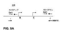
ヒトにおけるクラスIのHLA遺伝子クラスターは3つの主要な遺伝子座、即ちB、C及びA、並びに数個の副次的な遺伝子座を含む。クラスIIのHLAクラスターもまた、3つの主要な遺伝子座、即ちDP、DQ及びDRを有し、そしてクラスI及びクラスIIの遺伝子クラスターは両方とも、集団内においてクラスI及びクラスIIの遺伝子の両方の数種の異なる対立遺伝子が存在する点において多形である。更に又HLA機能における役割を果たす幾つかのアクセサリー蛋白が存在する。Tap1及びTap2サブユニットはクラスIのHLA複合体上にペプチド抗原を負荷する場合に必須であるTAPトランスポーター複合体の部分であり、そしてLMP2及びLMP7プロテオソームサブユニットはHLA上のディスプレイのための抗原からペプチドへの蛋白分解性の分解において役割を果たしている。LMP7の低下は、恐らくは安定化の欠如を介して細胞表面におけるMHCクラスIの量を低下させる(Fehling等、(1999)Science 265:1234- 1237参照)。TAP及びLMPのほかに、タパシン遺伝子も有り、その産物はTAP複合体とHLAクラスI鎖の間の架橋を形成し、そしてペプチドの負荷を増強する。タパシンの低下は障害を有するMHCクラスIアセンブリを有する細胞、低下したMHCクラスIの細胞表面発現、及び、障害を有する免疫応答をもたらす(Grandea 等、(2000)Immunity vol 13:213-222及びGarbi等、(2000)Nat Immunol 1:234-238参照)。 The class I HLA gene cluster in humans includes three major loci, namely B, C and A, and several secondary loci. Class II HLA clusters also have three major loci, namely DP, DQ and DR, and both class I and class II gene clusters are both class I and class II genes within a population. Is polymorphic in that there are several different alleles. There are also several accessory proteins that play a role in HLA function. The Tap1 and Tap2 subunits are part of the TAP transporter complex that is essential for loading peptide antigens onto class I HLA complexes, and the LMP2 and LMP7 proteosome subunits are antigens for display on HLA Plays a role in the proteolytic degradation of peptides to peptides. Reduction of LMP7 reduces the amount of MHC class I at the cell surface, presumably through lack of stabilization (see Fehling et al. (1999) Science 265: 1234-1237). In addition to TAP and LMP, there is also a tapasin gene, the product of which forms a bridge between the TAP complex and the HLA class I chain and enhances peptide loading. Reduction of tapasin results in cells with impaired MHC class I assembly, decreased cell surface expression of MHC class I, and impaired immune responses (Grandea et al. (2000) Immunity vol 13: 213-222 and Garbi Et al. (2000) Nat Immunol 1: 234-238).
クラスIの発現の調節は一般的に転写レベルにおいてであり、そして数種の刺激、例えばウィルス感染等が転写の変化を起こし得る。クラスI遺伝子はいくつかの特定の組織においてはされ、そしてこの下方調節の原因はプロモーター及び3’遺伝子間配列の内部にあると考えられる(Cohen等、(2009)PLos ONE 4(8):e6748参照)。更に又、マイクロRNAがいくつかのクラスIMHC遺伝子を調節することができる証拠も存在する(Zhu等、(2010)Am.J.Obstet Gynecol 202(6):592参照)。 Regulation of class I expression is generally at the transcriptional level, and several stimuli, such as viral infections, can cause transcriptional changes. Class I genes are found in some specific tissues, and the cause of this downregulation is thought to be internal to the promoter and 3 'intergenic sequences (Cohen et al. (2009) PLos ONE 4 (8): e6748 reference). Furthermore, there is evidence that microRNAs can regulate several class IMHC genes (see Zhu et al. (2010) Am. J. Obstet Gynecol 202 (6): 592).
クラスIIのMHC発現の調節はMHCIIエンハンセオソーム複合体の活性に依存している。エンハンセオソームの成分(エンハンセオソーム複合体の最も高度に研究されている成分の1つはRFX5遺伝子産物である(Villard等、(2000)MCB 20(10):3364-3376)参照))はほぼ普遍的にに発現され、そしてこれらの成分の発現はMHCクラスII遺伝子の組織特異的発現又はそれらのIFN−γ誘導上方調節を制御していないと考えられる。それよりは寧ろ、非DNA結合蛋白質であるCIITA(クラスIIトランスアクチベーター)として知られる蛋白質がMCHII発現のマスター制御因子として作用していると考えられる。他のエンハンセオソームメンバーとは対照的に、CIITAは組織特異的発現を呈し、IFN−γにより上方調節され、そしてMHCクラスII発現の下方調節を起こすことができる数種の細菌及びウィルスにより阻害されることがわかっている(免疫監視を免れようとする細菌の試行の部分と考えられる(LeibundGut-Landmann等、(2004)Eur.J.Immunol 34:1513-1525参照))。 Regulation of class II MHC expression is dependent on the activity of the MHCII enhanceosome complex. The components of the enhanceosome (one of the most highly studied components of the enhanceosome complex is the RFX5 gene product (see Villard et al. (2000) MCB 20 (10): 3364-3376))) It is expressed almost universally and expression of these components is thought to not control tissue-specific expression of MHC class II genes or their IFN-γ induced upregulation. Rather, it is thought that a protein known as CIITA (class II transactivator), which is a non-DNA binding protein, acts as a master regulator of MCHII expression. In contrast to other enhanceosome members, CIITA exhibits tissue-specific expression, is upregulated by IFN-γ, and is inhibited by several bacteria and viruses that can cause downregulation of MHC class II expression (See LeibundGut-Landmann et al. (2004) Eur. J. Immunol 34: 1513-1525), which is considered part of a bacterial attempt to escape immune surveillance.
クラスI又はIIの遺伝子の調節は一部の腫瘍の存在下において撹乱される場合があり、そしてそのような撹乱は患者の予後の結果に影響する場合がある。例えば、いくつかの黒色腫において、Tap1、Tap2及びHLAクラスI抗原の低下が観察されることは、原発腫瘍の場合よりも転移黒色腫においてより一般的である(p<0.05)ことがわかっている(例えばKagashita等、(1999)Am Jour of Pathol 154(3):745-754参照)。 Regulation of class I or II genes can be disrupted in the presence of some tumors, and such disruption can affect the prognostic outcome of the patient. For example, in some melanomas, reductions in Tap1, Tap2 and HLA class I antigens are more common in metastatic melanoma than in primary tumors (p <0.05). (See, for example, Kagasita et al. (1999) Am Jour of Pathol 154 (3): 745-754).
ヒトにおいては、数種の疾患への易罹患性はHLAハプロタイプと関連することが疑われている。これらの疾患にはとりわけ、アジソン病、強直性脊椎炎、ベーチェット病、バーガー病、セリアック病、慢性活動性肝炎、グレーブズ病、若年性慢性関節リューマチ、乾癬、乾癬性関節炎、慢性関節リューマチ、シェーグレン症候群、及び全身エリテマトーデスが挙げられる。 In humans, susceptibility to several diseases is suspected to be associated with HLA haplotypes. These diseases include, among others, Addison's disease, ankylosing spondylitis, Behcet's disease, Burger's disease, Celiac disease, chronic active hepatitis, Graves' disease, juvenile rheumatoid arthritis, psoriasis, psoriatic arthritis, rheumatoid arthritis, Sjogren's syndrome And systemic lupus erythematosus.
HLAハプロタイプは又、移植片拒絶において主要な役割を果たす。移植変拒絶の急性期は約1〜3週間以内に生じ得、そして通常はドナーのクラスI及びクラスIIのHLA分子に対する宿主系の感作が原因のドナー組織に対する宿主Tリンパ球の作用が関与している。大部分の場合においては、トリガーする抗原はクラスIのHLAである。最良の結果のためにはドナーをHLAハプロタイプに関して分類し、そして可能な限り完全に患者レシピエントに対してマッチさせる。しかしながら、高パーセントのHLAハプロタイプ同一性を共有することができるファミリーメンバーの間の提供でさえも、なお、良好に行えない場合が多い。したがって、レシピエント内部の移植片組織を温存するためには、拒絶を防止するための徹底した免疫抑制療法に患者を付さなければならない場合が多い。そのような療法は患者が克服困難である日和見感染による合併症及び顕著な罹患率をもたらす場合がある。 HLA haplotypes also play a major role in graft rejection. The acute phase of transplant modification rejection can occur within about 1 to 3 weeks and usually involves the action of host T lymphocytes on donor tissue due to sensitization of the host system to donor class I and class II HLA molecules. doing. In most cases, the triggering antigen is class I HLA. For best results, donors are classified with respect to HLA haplotypes and matched to patient recipients as completely as possible. However, even providing among family members that can share a high percentage of HLA haplotype identity is still often not successful. Thus, in order to preserve the graft tissue within the recipient, the patient often has to undergo a thorough immunosuppressive therapy to prevent rejection. Such therapies may result in complications and significant morbidity from opportunistic infections that are difficult for patients to overcome.
細胞療法はある特定の型の細胞(例えば腫瘍抗原に反応性のT細胞又はB細胞)がレシピエントに与えられる移植の特殊な型である。細胞療法は自系(レシピエントから誘導)又は同種異系(ドナーから誘導)の何れかである細胞を用いて行うことができ、そして細胞は非成熟細胞、例えば幹細胞、又は完全成熟の機能性の細胞、例えばT細胞であってよい。実際、ある特定の癌のようないくつかの疾患においては、T細胞は、腫瘍を根絶する試みにおいて、特定の腫瘍抗原に対するそれらのアビディティーを増大させるためにエクスビボで操作され、増殖させ、そしてその癌の型に罹患している患者内に導入され得る。これは、内因性T細胞応答が腫瘍自身により抑制される場合に特に有用である。しかしながら、同じ注意事項が、拒絶に関して更に良く知られた固形臓器移植片に対して適用されるように細胞療法に対しても適用される。ドナーのT細胞はクラスIのHLA抗原を発現し、そしてこのため、レシピエントの内因性免疫系から拒絶応答を引き出すことができる。 Cell therapy is a special type of transplant in which certain types of cells (eg, T cells or B cells reactive to tumor antigens) are given to the recipient. Cell therapy can be performed using cells that are either autologous (derived from the recipient) or allogeneic (derived from the donor), and the cells are non-mature cells, eg, stem cells, or fully mature functional Cells, such as T cells. Indeed, in some diseases, such as certain cancers, T cells are engineered and expanded ex vivo to increase their avidity against specific tumor antigens in an attempt to eradicate the tumor, and It can be introduced into patients suffering from that type of cancer. This is particularly useful when the endogenous T cell response is suppressed by the tumor itself. However, the same precautions apply to cell therapy as it applies to solid organ grafts that are better known for rejection. Donor T cells express class I HLA antigens and can therefore elicit a rejection response from the recipient's endogenous immune system.
したがって、HLA遺伝子及び細胞における遺伝子発現の操作ための組成物及び方法がなおも必要とされている。 Accordingly, there remains a need for compositions and methods for manipulating HLA genes and gene expression in cells.
HLA遺伝子複合体又はHLA遺伝子発現を操作するための方法及び組成物を本明細書において開示する。特に本発明はHLA関連障害、例えば個人のHLAハプロタイプに関連するヒトの障害を治療するためにHLA遺伝子の発現を調節するための方法及び組成物を提供する。更に又、HLA遺伝子を欠失させるか抑制することによりHLAヌル細胞、細胞フラグメント(例えば血小板)、組織又は完全生物を作成するための方法及び組成物を提供する。更に又、これらの方法及び組成物は、1つのみのHLA遺伝子、又は1つより多いHLA遺伝子に関してヌルであるか、又は全てのHLA遺伝子に対して完全にヌルである細胞,細胞フラグメント、組織又は生物を作成するために使用し得る。ある特定の実施形態において、HLAヌル細胞又は組織は移植における使用のために好都合であるヒトの細胞又は組織である。 Disclosed herein are methods and compositions for manipulating HLA gene complexes or HLA gene expression. In particular, the present invention provides methods and compositions for modulating HLA gene expression to treat HLA-related disorders, such as human disorders associated with an individual's HLA haplotype. Further provided are methods and compositions for making HLA null cells, cell fragments (eg, platelets), tissues or complete organisms by deleting or suppressing the HLA gene. Furthermore, these methods and compositions can be used for cells, cell fragments, tissues that are null for only one HLA gene, or more than one HLA gene, or completely null for all HLA genes. Or it can be used to create organisms. In certain embodiments, the HLA null cells or tissues are human cells or tissues that are convenient for use in transplantation.
即ち、1つの態様において、HLA対立遺伝子の発現を調節する操作されたDNA結合ドメイン(例えば亜鉛フィンガー蛋白質又はTALEDNA結合ドメイン蛋白質)を提供する。ある特定の実施形態において、DNA結合ドメインは非天然存在の操作された亜鉛フィンガー蛋白質並びに予備選択標的部位に結合するように自身の認識ヘリックスが改変(例えば選択及び/又は合理的設計による)されている亜鉛フィンガー蛋白質を含む。本明細書に記載する亜鉛フィンガー蛋白質の何れも、亜鉛フィンガー蛋白質1、2、3、4、5、6つ以上を包含し得、各々亜鉛フィンガーは選択された配列(例えば遺伝子)における標的サブ部位に結合する認識ヘリックスを有する。いくつかの実施形態においては、亜鉛フィンガー蛋白質の亜鉛フィンガードメインの認識ヘリックス1つ以上は非天然存在である。ある特定の実施形態において、亜鉛フィンガー蛋白質は表1に示す認識ヘリックスを有する。他の実施形態において、亜鉛フィンガー蛋白質は表2に示す標的部位に結合する。他の実施形態において、DNA結合ドメインはTALE DNA結合ドメイン(例えば天然に存在する、及び/又は非天然存在のTALE結合ドメインを含むTALE DNA結合ドメイン)を含む。
Thus, in one embodiment, an engineered DNA binding domain (eg, zinc finger protein or TALEDNA binding domain protein) that regulates expression of an HLA allele is provided. In certain embodiments, the DNA binding domain is modified (eg, by selection and / or rational design) in its recognition helix to bind to a non-naturally occurring engineered zinc finger protein and a preselected target site. Contains zinc finger proteins. Any of the zinc finger proteins described herein can include
ある特定の実施形態において、本明細書に記載するDNA結合蛋白質(例えば亜鉛フィンガー蛋白質(ZFP)又はTALE DNA結合蛋白質)は融合蛋白質の部分として調節ドメイン(又は機能性ドメイン)との作動可能な連結関係に置かれることができる。ある特定の実施形態において、調節ドメインはDNA結合ドメインとの融合のための活性化ドメイン又は抑制ドメインであり、そしてそのような融合蛋白質(例えばZEP−又はTALE−転写因子融合物すなわちそれぞれZFP−TF及びTALE−TF)を遺伝子発現の活性化又は抑制の何れかのために使用できる。いくつかの実施形態においては、HLA遺伝子又はHLA遺伝子発現のモジュレーターに優先的に結合できるリプレッサーが提供される。ある特定の実施形態において、調節ドメインの活性は、細胞の転写機序との相互作用が外因性リガンドの非存在下に起こらないように外因性の小分子又はリガンドにより調節される。そのような外部リガンドは転写機序とのZFP−TF又はTALE−TFの相互作用の程度を制御する。 In certain embodiments, a DNA binding protein (eg, zinc finger protein (ZFP) or TALE DNA binding protein) described herein is operatively linked to a regulatory domain (or functional domain) as part of a fusion protein. Can be put into a relationship. In certain embodiments, the regulatory domain is an activation or repression domain for fusion with a DNA binding domain, and such a fusion protein (eg, ZEP- or TALE-transcription factor fusion, ie, ZFP-TF, respectively). And TALE-TF) can be used for either activation or suppression of gene expression. In some embodiments, a repressor is provided that can preferentially bind to an HLA gene or a modulator of HLA gene expression. In certain embodiments, the activity of the regulatory domain is modulated by an exogenous small molecule or ligand such that interaction with cellular transcriptional mechanisms does not occur in the absence of the exogenous ligand. Such external ligands control the extent of ZFP-TF or TALE-TF interaction with the transcription mechanism.
1つの実施形態において、リプレッサーは特定のHLA A、HLA B又はHLA C転写調節領域に結合することができ、そして、上記遺伝子の1つのみにおける発現を抑制することができる。別の態様においては、HLA A、HLA B又はHLA Cに共通の転写調節領域と、3遺伝子全てが1つのDNA結合ドメイン(例えばZFP TF又はTALE TF)で調節されるように、相互作用することができるリプレッサーが提供される。別の実施形態においては、リプレッサーはHLAクラスIIの発現又は機能のレギュレーター(例えばCIITA又はRFX5)に結合することによりその活性を抑制し、そしてこれにより、HLAクラスIIの発現又は機能を抑制することができる。 In one embodiment, the repressor can bind to a specific HLA A, HLA B, or HLA C transcriptional regulatory region and can suppress expression in only one of the genes. In another embodiment, interacting with a transcriptional regulatory region common to HLA A, HLA B, or HLA C so that all three genes are regulated by a single DNA binding domain (eg, ZFP TF or TALE TF). A repressor is provided. In another embodiment, the repressor suppresses its activity by binding to a regulator of HLA class II expression or function (eg, CIITA or RFX5), and thereby suppresses HLA class II expression or function. be able to.
他の実施形態において、既知のHLAハプロタイプに優先的に結合することにより1つのみの対立遺伝子の発現を抑制する抑制性DNA結合ドメイン−転写因子融合物を提供する。 In other embodiments, an inhibitory DNA binding domain-transcription factor fusion is provided that suppresses expression of only one allele by preferentially binding to a known HLA haplotype.
別の態様において、HLA遺伝子の発現を特異的に活性化するDNA結合ドメイン−転写因子融合物を提供する。そのような融合物はレギュレーターの発現を増大させることにより、あるクラスのHLA遺伝子を上方調節し得、又は、これらの遺伝子が通常では発現されない組織において、そのようなクラスの発現を誘発し得る。別の実施形態においては、所望に応じて特定のHLA遺伝子を活性化するDNA結合ドメイン−転写因子融合物(例えばZEP TF又はTALE TF)を提供する。 In another embodiment, a DNA binding domain-transcription factor fusion that specifically activates expression of the HLA gene is provided. Such fusions can upregulate certain classes of HLA genes by increasing the expression of regulators, or can induce such classes of expression in tissues where these genes are not normally expressed. In another embodiment, a DNA binding domain-transcription factor fusion (eg, ZEP TF or TALE TF) is provided that activates specific HLA genes as desired.
別の態様において、 融合蛋白質はヌクレアーゼ(例えばZFN又はTALEN)を含む機能性ドメインとの作動可能な連結関係にある本明細書に記載するDNA結合蛋白質(例えばZFP又はTALE)を含む。ある特定の実施形態において、1つのHLA遺伝子を切断する、亜鉛フィンガーヌクレアーゼ(ZFN)又はヌクレアーゼ(TALEN)に融合したTALEDNA結合ドメインを提供する。ある特定の実施形態において、ZFN及び/又はTALENはヒトHLAクラスI遺伝子における標的部位及び/又はヒトHLAクラスII遺伝子における標的部位に結合する。いくつかの実施形態においては、これらのヌクレアーゼによるHLA遺伝子内部の切断はHLA遺伝子の永久撹乱(例えば突然変異)をもたらす。ある特定の実施形態において、ZFN及び/又はTALENの2対を用いてより大きい欠失を誘発してよい。欠失は1つのHLA遺伝子又はHLAレギュレーター遺伝子の小型部分を含むか、又はより大きいセグメントを含んでよい。いくつかの実施形態において、ZFN又はTALENにより誘発された欠失はHLA遺伝子1つ以上を欠失させ得、又は、全HLA遺伝子複合体(即ちクラスIHLA遺伝子の全て、又はHLAクラスII遺伝子の全て)を欠失させ得る。欠失はまた、HLA遺伝子のあるクラスのサブセットの欠失も包含し得る。亜鉛フィンガーDNA結合蛋白質は、1、2、3、4、5、6、またはそれ以上の亜鉛フィンガーを包含し得、各亜鉛フィンガーは、標的遺伝子中の標的サブ部位に結合する認識ヘリックスを有する。ある特定の実施形態において、亜鉛フィンガー蛋白質は4又は5又は6つのフィンガーを含み(この場合、フィンガーはF1、F2、F3、F4、F5及びF6と標記され、そしてN末端からC末端にF1からF4又はF5又はF6と順位付けされる)、そしてフィンガーは表1に示す認識領域のアミノ酸配列を含む。 In another embodiment, the fusion protein comprises a DNA binding protein (eg, ZFP or TALE) described herein in operable linkage with a functional domain comprising a nuclease (eg, ZFN or TALEN). In certain embodiments, a TALEDNA binding domain fused to a zinc finger nuclease (ZFN) or nuclease (TALEN) that cleaves one HLA gene is provided. In certain embodiments, ZFN and / or TALEN binds to a target site in a human HLA class I gene and / or a target site in a human HLA class II gene. In some embodiments, cleavage within the HLA gene by these nucleases results in permanent perturbation (eg, mutation) of the HLA gene. In certain embodiments, two pairs of ZFN and / or TALEN may be used to induce larger deletions. The deletion may include a small portion of one HLA gene or HLA regulator gene, or may include a larger segment. In some embodiments, the deletion induced by ZFN or TALEN can delete one or more HLA genes, or the entire HLA gene complex (ie, all of the class I HLA genes or all of the HLA class II genes). ) May be deleted. Deletions can also include deletion of a class of subsets of HLA genes. A zinc finger DNA binding protein can include 1, 2, 3, 4, 5, 6, or more zinc fingers, each zinc finger having a recognition helix that binds to a target subsite in the target gene. In certain embodiments, the zinc finger protein comprises 4 or 5 or 6 fingers (in this case, the fingers are labeled F1, F2, F3, F4, F5 and F6 and from F1 from N-terminal to C-terminal). And the fingers contain the amino acid sequences of the recognition regions shown in Table 1.
本明細書に記載するZFN又はTALREN蛋白質のいずれも更に切断ドメイン及び/又は切断ハーフドメイン(例えば野生型又は操作されたFokI切断ハーフドメイン)を含んでよい。したがって、本明細書に記載するZFN又はTALENの何れにおいても、ヌクレアーゼドメインは野生型のヌクレアーゼドメイン又はヌクレアーゼハーフドメイン(例えばFokI切断ハーフドメイン)を含んでよい。他の実施形態において、ZFN又はTALENは操作された(非天然存在の)ヌクレアーゼドメイン又はハーフドメイン、例えば必然的にヘテロ2量体を形成する操作されたFokI切断ハーフドメインを含む。例えば米国特許公開番号20080131962を参照のこと。 Any of the ZFN or TALREN proteins described herein may further comprise a cleavage domain and / or a cleavage half domain (eg, a wild type or engineered FokI cleavage half domain). Thus, in any of the ZFNs or TALENs described herein, the nuclease domain may comprise a wild type nuclease domain or a nuclease half domain (eg, a FokI cleavage half domain). In other embodiments, the ZFN or TALEN comprises an engineered (non-naturally occurring) nuclease domain or half domain, eg, an engineered FokI cleavage half domain that necessarily forms a heterodimer. See, for example, US Patent Publication No. 2008011962.
別の態様において、本開示は本明細書に記載する蛋白質のいずれかをコードするポリヌクレオチドを提供する。本明細書に記載するポリヌクレオチドの何れも又、HLA遺伝子内への標的化された挿入のための配列(ドナー又はパッチ配列)を含んでよい。 In another embodiment, the present disclosure provides a polynucleotide encoding any of the proteins described herein. Any of the polynucleotides described herein may also contain a sequence (donor or patch sequence) for targeted insertion into the HLA gene.
更に別の態様において、本明細書に記載するポリヌクレオチドの何れかを含む遺伝子送達ベクターを提供する。ある特定の実施形態において、ベクターはアデノウィルスベクター(例えばAd5/F35ベクター)、レンチウィルスベクター(LV)、例えば組み込みコンピテント又は組み込み欠損性のレンチウィルスベクター、又はアデノ関連ウィルスベクター(AAV)である。したがって、本明細書に記載するヌクレアーゼ少なくとも1つ(例えばZFN又はTAREN)をコードする配列、及び/又は、標的遺伝子内への標的化された組み込みのためのドナー配列を含むアデノウィルス(Ad)ベクター、LV又はアデノ関連ウィルスベクター(AAV)も提供される。ある特定の実施形態において、AdベクターはキメラAdベクター、例えばAd5/F35ベクターである。ある特定の実施形態において、レンチウィルスベクターはインテグラーゼ欠損レンチウィルスベクター(IDLV)又は組み込みコンピテントレンチウィルスベクターである。ある特定の実施形態において、ベクターはVSV−Gエンベロープを有するか、他のエンベロープを有する擬似型である。 In yet another aspect, a gene delivery vector comprising any of the polynucleotides described herein is provided. In certain embodiments, the vector is an adenoviral vector (eg, Ad5 / F35 vector), a lentiviral vector (LV), such as an integration competent or integration defective lentiviral vector, or an adeno-associated viral vector (AAV). Accordingly, an adenoviral (Ad) vector comprising a sequence encoding at least one nuclease described herein (eg, ZFN or TAREN) and / or a donor sequence for targeted integration into a target gene, LV or adeno-associated viral vectors (AAV) are also provided. In certain embodiments, the Ad vector is a chimeric Ad vector, such as an Ad5 / F35 vector. In certain embodiments, the lentiviral vector is an integrase-deficient lentiviral vector (IDLV) or an integrated competent trench virus vector. In certain embodiments, the vector has a VSV-G envelope or is pseudotyped with other envelopes.
追加の実施形態において、標的遺伝子はHLA発現を調節する(例えばヒト細胞中の)遺伝子である(HLAレギュレーター遺伝子)。ある特定の実施形態において、CTIIA、RFX5遺伝子、TAP1、TAP2又はタパシン遺伝子、又はこれらの組み合わせを調節(例えば活性化、抑制及び/又は不活性化)のために標的とする。いくつかの実施形態においては、標的とされる遺伝子はHLA遺伝子を調節することができるマイクロRNAをコードする。本明細書に記載するベクターは又、ドナー配列を含んでよい。追加の実施形態において、ドナー配列は宿主細胞にとって内在性ではないヒトHLA遺伝子又はHLAレギュレーター遺伝子を含む。いくつかの実施形態においては、目的のHLA遺伝子又はHLAレギュレーター遺伝子は内在性のHLA遺伝子又はHLAレギュレーター遺伝子の箇所内に挿入され、他の実施形態においては、目的のHLA遺伝子又はHLAレギュレーター遺伝子は無作為に選択された遺伝子座内に、又はゲノムワイドの送達の後に別個の遺伝子座内に挿入される。いくつかの実施形態においては、HLA導入遺伝子又はHLAレギュレーター導入遺伝子の挿入のための別個の遺伝子座はPPP1R12C遺伝子座である(米国特許公開番号20080299580参照)。他の実施形態において、HLA導入遺伝子又はHLAレギュレーター導入遺伝子はCCR−5遺伝子座内に挿入される。いくつかの態様において、ドナーは目的の別の核酸を含む。例示に過ぎないが、このドナーは目的のポリペプチドをコードする遺伝子を含有し得、又は、構造RNA(shRNA、miRNA、RNAi等)をコードする配列を含み得る。いくつかの実施形態において、目的のHLA遺伝子又はHLAレギュレーター遺伝子が所望の様式(例えばノックアウト、補正等)に操作されており、そしてドナー及び1つ以上の追加のZFN及び/又はTALENが別の遺伝子座(例えばAAVS1)内にドナーを挿入するために与えられている細胞が提供さる。 In additional embodiments, the target gene is a gene that modulates HLA expression (eg, in a human cell) (HLA regulator gene). In certain embodiments, the CTIIA, RFX5 gene, TAP1, TAP2 or tapasin gene, or combinations thereof are targeted for modulation (eg, activation, suppression and / or inactivation). In some embodiments, the targeted gene encodes a microRNA that can regulate the HLA gene. The vectors described herein may also include a donor sequence. In additional embodiments, the donor sequence comprises a human HLA genes or HLA regulator genes that are not within resident to the host cell. In some embodiments, HLA genes or HLA regulator gene of interest is inserted into the portion of the inner standing of HLA genes or HLA regulator gene, in other embodiments, HLA genes or HLA regulator gene of interest It is inserted into randomly selected loci or into separate loci after genome-wide delivery. In some embodiments, the separate locus for insertion of the HLA transgene or HLA regulator transgene is the PPP1R12C locus (see US Patent Publication No. 20080299580). In other embodiments, the HLA transgene or HLA regulator transgene is inserted within the CCR-5 locus. In some embodiments, the donor includes another nucleic acid of interest. By way of example only, the donor may contain a gene that encodes the polypeptide of interest, or may include a sequence that encodes a structural RNA (shRNA, miRNA, RNAi, etc.). In some embodiments, the HLA gene or HLA regulator gene of interest is engineered in the desired manner (eg, knockout, correction, etc.), and the donor and one or more additional ZFNs and / or TALENs are another gene Cells are provided that are provided to insert a donor into a locus (eg, AAVS1).
ある特定の実施形態において、単一のベクターが本明細書に記載するヌクレアーゼ(例えばZFN及び/又はTALEN)1つ以上をコードする配列及びドナー配列を含む。他の実施形態において、ドナー配列は第1のベクターに含有され、そしてヌクレアーゼコード配列は第2のベクターに存在する。 In certain embodiments, a single vector comprises a sequence encoding one or more of the nucleases described herein (eg, ZFN and / or TALEN) and a donor sequence. In other embodiments, the donor sequence is contained in a first vector and the nuclease coding sequence is present in a second vector.
更に別の態様において、 本開示は本明細書に記載する蛋白質、ポリヌクレオチド及び/又はベクターのいずれかを含む細胞(例えば単離された細胞)を提供する。ある特定の実施形態において、細胞は幹/前駆細胞、リンパ球、B細胞、又はT細胞(例えばCD4+T細胞)よりなる群から選択される。他の実施形態において、細胞は、血小板を包含するがこれに限定されない細胞フラグメントである。 In yet another aspect, the present disclosure provides a cell (eg, an isolated cell) comprising any of the proteins, polynucleotides and / or vectors described herein. In certain embodiments, the cells are selected from the group consisting of stem / progenitor cells, lymphocytes, B cells, or T cells (eg, CD4 + T cells). In other embodiments, the cell is a cell fragment, including but not limited to platelets.
別の態様において、本明細書に記載する蛋白質、ポリヌクレオチド及び/又はベクターの1つ以上を導入することにより細胞におけるHLA遺伝子又はHLAレギュレーター遺伝子を不活性化する方法を本明細書に記載する。これらの方法の何れにおいても、ヌクレアーゼは細胞性DNA配列の標的化された突然変異誘発、標的化された欠失、標的化された挿入を誘発、及び/又は、既定の染色体遺伝子座における標的化された組み換えを促進し得る。したがって、ある特定の実施形態において、ヌクレアーゼ(例えばZFN及び/又はTALEN)は標的とする遺伝子においてヌクレオチド1つ以上の欠失及び/又は挿入を行う。いくつかの実施形態において、HLA遺伝子はヌクレアーゼ(ZFN及び/又はTALEN)切断により、そしてその後の非相同末端接合により、不活性化される。他の実施形態において、標的遺伝子中のゲノム配列を、例えば、本明細書に記載するZFN(又は該ZFNをコードするベクター)の1対以上、及び/又は、TALEN1つ以上、及び、ZFN及び/又はTAKENを用いた標的化された切断の後に遺伝子内に挿入される「ドナー」配列を用いて置き換える。ドナー配列はヌクレアーゼ融合ベクター中に存在するか、別個のベクター(例えばAd、AAV又はLVベクター)中に存在してよく、或いは、異なる核酸送達機序を用いて細胞内に導入してよい。いくつかの実施形態においては、ZFN及び/又はTALENはそれらをコードするmRNAを用いて送達される。いくつかの実施形態において、エレクトロポレーション又はネイキッド核酸の送達に適する他の手法により核酸を送達してよい。 In another aspect, described herein is a method of inactivating an HLA gene or an HLA regulator gene in a cell by introducing one or more of the proteins, polynucleotides and / or vectors described herein. In any of these methods, nucleases induce targeted mutagenesis, targeted deletion, targeted insertion of cellular DNA sequences, and / or targeting at a predetermined chromosomal locus. Promoted recombination. Thus, in certain embodiments, the nuclease (eg, ZFN and / or TALEN) makes one or more nucleotide deletions and / or insertions in the targeted gene. In some embodiments, the HLA gene is inactivated by nuclease (ZFN and / or TALEN) cleavage and subsequent non-homologous end joining. In other embodiments, the genomic sequence in the target gene is, for example, one or more pairs of ZFNs (or vectors encoding the ZFNs) described herein, and / or one or more TALENs, and ZFNs and / or Alternatively, replace with a “donor” sequence that is inserted into the gene after targeted cleavage with TAKEN. The donor sequence may be present in the nuclease fusion vector, in a separate vector (eg, Ad, AAV or LV vector) or may be introduced into the cell using a different nucleic acid delivery mechanism. In some embodiments, ZFN and / or TALEN are delivered using mRNA encoding them. In some embodiments, nucleic acids may be delivered by electroporation or other techniques suitable for naked nucleic acid delivery.
別の態様において、細胞又は細胞系統においてHLA遺伝子を突然変異するため、及び/又はHLA機能を不活性化するための、DNA結合蛋白質及びその融合物を用いる方法を提供する。したがって、ヒト細胞においてHLA遺伝子を不活性化するための方法を提供し、その方法は本明細書に記載する蛋白質又はポリヌクレオチドの何れかを細胞に投与することを含む。何れかのモデル生物体においてMHC機能を改変するための方法も本明細書に記載する。 In another aspect, methods are provided for using DNA binding proteins and fusions thereof to mutate HLA genes in cells or cell lines and / or inactivate HLA function. Accordingly, provided is a method for inactivating a HLA gene in a human cell, the method comprising administering to the cell any of the proteins or polynucleotides described herein. Methods for modifying MHC function in any model organism are also described herein.
別の態様において、本明細書に記載する組成物及び方法は、例えば、何れかのHLA関連障害(即ちHLAハプロタイプに関連するもの)の治療又は防止又は改善において使用できる。方法は典型的には、(a)HLA又はHLAレギュレーター遺伝子が不活性化されるようにヌクレアーゼ(例えばZFN又はTALEN)を用いて単離された細胞(例えばT細胞又はリンパ球)中の内在性HLA遺伝子又はHLAレギュレーターを切断すること;及び(b)対象内に細胞を導入することによりHLA関連障害を治療又は防止することを含む。ある特定の実施形態において、HLA関連障害は対宿主性移植片病(GVHD)である。ヌクレアーゼはmRNAとして、蛋白質形態において、及び/又はヌクレアーゼをコードするDNA配列として導入することができる。ある特定の実施形態において、対象に導入される単離された細胞は更に、追加的なゲノム修飾、例えば組み込まれた外因性配列(例えば切断されたHLA又はHLA調節遺伝子又は異なる遺伝子、例えばセイフハーバー遺伝子内に)及び/又は追加の遺伝子、例えば1つ以上のTCR遺伝子の不活性化(例えばヌクレアーゼ媒介)を含む。外因性配列はベクター(例えばAd、AAV、LV)を介して、又はエレクトロポレーションのような手法を用いることにより、導入してよい。いくつかの態様において、組成物は単離された細胞フラグメント及び/又は分化した細胞を含んでよい。 In another aspect, the compositions and methods described herein can be used, for example, in the treatment or prevention or amelioration of any HLA-related disorder (ie, those associated with HLA haplotypes). The method is typically the inner standing in (a) HLA or nuclease as HLA regulator gene is inactivated (e.g. ZFN or Talen) isolated cells using (e.g. T-cells or lymphocytes) Cleaving the sex HLA gene or HLA regulator; and (b) treating or preventing HLA-related disorders by introducing cells into the subject. In certain embodiments, the HLA-related disorder is graft versus host disease (GVHD). Nucleases can be introduced as mRNA, in protein form and / or as DNA sequences encoding nucleases. In certain embodiments, the isolated cells introduced into the subject may further contain additional genomic modifications, such as integrated exogenous sequences (eg, truncated HLA or HLA regulatory genes or different genes, such as Safe Harbor). And / or inactivation (eg, nuclease-mediated) of additional genes, eg, one or more TCR genes. Exogenous sequences may be introduced via a vector (eg, Ad, AAV, LV) or by using techniques such as electroporation. In some embodiments, the composition may comprise isolated cell fragments and / or differentiated cells.
いくつかの実施形態において、本明細書に記載するヌクレアーゼ融合物は、誘導多能性幹細胞(iPSC)、ヒト胚性幹細胞(hES)、間葉性幹細胞(MSC)、造血幹細胞(HSC)又はニューロン幹細胞のような幹細胞を標的とするために利用してよく、これらにおいては、ヌクレアーゼ融合物の活性は欠失を含有するHLA対立遺伝子をもたらすことになる。いくつかの実施形態において、方法は1つより多いHLA遺伝子が改変されている幹細胞を作成するために使用してよい。他の実施形態において、本発明はHLAヌル表現型を有する幹細胞を生産するための方法を提供する。いくつかの態様において、幹細胞は1つ以上、又はすべてのHLAクラスII遺伝子発現に関してヌルであり得る。別の態様において、幹細胞は1つ以上、又はすべてのHLAクラスI遺伝子発現に関してヌルであり得る。いくつかの態様において、幹細胞はすべてのHLA遺伝子発現に関してヌルである。他の実施形態において、HLA遺伝子座において修飾されている幹細胞は、その後分化される。 In some embodiments, the nuclease fusions described herein are induced pluripotent stem cells (iPSC), human embryonic stem cells (hES), mesenchymal stem cells (MSC), hematopoietic stem cells (HSC) or neurons. It may be utilized to target stem cells, such as stem cells, in which the activity of the nuclease fusion will result in an HLA allele containing the deletion. In some embodiments, the method may be used to create stem cells in which more than one HLA gene has been modified. In another embodiment, the present invention provides a method for producing stem cells having an HLA null phenotype. In some embodiments, the stem cells can be null for one or more, or all HLA class II gene expression. In another embodiment, the stem cells can be null for one or more, or all HLA class I gene expression. In some embodiments, the stem cell is null for all HLA gene expression. In other embodiments, stem cells that are modified at the HLA locus are subsequently differentiated.
修飾された幹細胞を含む医薬組成物も提供される。そのような医薬組成物は予防的又は施療的に使用してよく、iPSCs、hES、MSCs、HSCs又はこれらの組み合わせ及び/又は誘導体を含んでよい。他の実施形態において、そのような修飾された幹細胞から誘導された細胞、細胞フラグメント(例えば血小板)又は組織は、そのような組織が所望に応じてHLA遺伝子座において修飾されるように提供される。いくつかの態様において、そのような細胞は部分的に分化し(例えば造血幹細胞)、他の態様においては、完全に分化した細胞が提供され(例えばリンパ球又はメガカリオサイト)、そして更に別の態様においては、分化した細胞のフラグメントが提供される。他の実施形態において、改変されたHLA又はHLAレギュレーター遺伝子を含有する幹細胞及び/又はその分化した子孫が提供され、そしてそれらは又、目的の別の遺伝子座におけるドナーDNAの欠失、改変又は挿入を包含する追加的遺伝子修飾を含有することができる。 Also provided are pharmaceutical compositions comprising the modified stem cells. Such pharmaceutical compositions may be used prophylactically or therapeutically and may include iPSCs, hES, MSCs, HSCs or combinations and / or derivatives thereof. In other embodiments, cells, cell fragments (eg, platelets) or tissue derived from such modified stem cells are provided such that such tissue is modified at the HLA locus as desired. . In some embodiments, such cells are partially differentiated (eg, hematopoietic stem cells), in other embodiments, fully differentiated cells are provided (eg, lymphocytes or megacaryosite), and yet another In embodiments, differentiated cell fragments are provided. In other embodiments, stem cells containing modified HLA or HLA regulator genes and / or their differentiated progeny are provided, and they also delete, modify or insert donor DNA at another locus of interest. Additional genetic modifications can be included.
いくつかの実施形態において、本明細書に記載するDNA結合ドメイン又は融合蛋白質(例えばZFP−TF、TALEDNA結合ドメインTF、ZFN及び/又はTALEN)で処理される細胞はCD4+T細胞又はNK細胞のような成熟細胞であってよい。そのような細胞はHLA遺伝子又はHLAレギュレーター遺伝子の調節のために本明細書に記載するDNA結合ドメインを含む蛋白質を含んでよく、又、HLA遺伝子内への欠失及び/又は挿入の導入のためのヌクレアーゼ融合物(例えばZFN又はTALEN)を含んでよい。いくつかの態様において、そのようなZFN又はTALENを含む細胞は更に、外因性DNA配列を含んでよい。いくつかの態様において、細胞療法のため、例えばT細胞移植のために成熟細胞を使用してよい。他の実施形態において、T細胞移植において使用するための細胞は目的の別の遺伝子修飾を含有する。1つの態様において、T細胞は癌マーカーに特異的な挿入されたキメラ抗原受容体(CAR)を含有する。更なる態様において、挿入されたCARはB細胞悪性疾患に特徴的なCD19マーカーに対して特異的である。そのような細胞はHLAハプロタイプにマッチさせる必要なく患者を治療するための治療用組成物中で有用であり、そしてそのため、「既製」の治療薬として、それを要する何れの患者のためにも使用できるであろう。いくつかの態様において、TCRα及び/又はTCRβ鎖をコードする遺伝子が操作されているか、又は、所望の特異性及び親和性を有するTCR鎖をコードする遺伝子が導入されている細胞を提供する。他の実施形態において、血小板減少症又は他の出血障害のような障害の治療における施療的用途のためのHLA修飾血小板を提供する。 In some embodiments, the cell treated with a DNA binding domain or fusion protein described herein (eg, ZFP-TF, TALEDNA binding domain TF, ZFN and / or TALEN) is a CD4 + T cell or an NK cell. It may be a mature cell. Such cells may comprise a protein comprising a DNA binding domain as described herein for regulation of the HLA gene or HLA regulator gene, and for the introduction of deletions and / or insertions into the HLA gene. Nuclease fusions (eg, ZFN or TALEN). In some embodiments, cells containing such ZFNs or TALENs may further include exogenous DNA sequences. In some embodiments, mature cells may be used for cell therapy, eg, for T cell transplantation. In other embodiments, the cells for use in T cell transplantation contain another genetic modification of interest. In one embodiment, the T cells contain an inserted chimeric antigen receptor (CAR) specific for a cancer marker. In a further embodiment, the inserted CAR is specific for the CD19 marker characteristic of B cell malignancy. Such cells are useful in therapeutic compositions to treat patients without the need to match HLA haplotypes and are therefore used as “off-the-shelf” therapeutics for any patient in need thereof It will be possible. In some embodiments, a cell is provided in which a gene encoding a TCRα and / or TCRβ chain has been engineered or a gene encoding a TCR chain having a desired specificity and affinity has been introduced. In other embodiments, HLA-modified platelets are provided for therapeutic use in the treatment of disorders such as thrombocytopenia or other bleeding disorders.
更なる他の態様において、本発明はHLA障害の研究のための特定のモデル系の作成のための方法及び組成物を提供する。ある特定の実施形態において、細胞及び動物系統の作成のために胚性幹細胞中で突然変異体HLA対立遺伝子を作成したモデルを提供する。ある特定の実施形態において、モデル系はインビトロの細胞系統を含み、他の実施形態においては、モデル系はトランスジェニック動物を含む。他の実施形態において、本発明は突然変異したHLA遺伝子又はHLAレギュレーターを補正するための方法及び組成物を提供し、そして又、あるHLA対立遺伝子を別のもので置き換えるための方法及び組成物を提供する。 In yet another aspect, the present invention provides methods and compositions for the creation of specific model systems for the study of HLA disorders. In certain embodiments, models are provided that have created mutant HLA alleles in embryonic stem cells for the generation of cell and animal lineages. In certain embodiments, the model system comprises an in vitro cell line, and in other embodiments, the model system comprises a transgenic animal. In other embodiments, the present invention provides methods and compositions for correcting mutated HLA genes or HLA regulators, and also methods and compositions for replacing one HLA allele with another. provide.
いくつかの実施形態において、標的対立遺伝子(例えば特異的HLAハプロタイプ)を発現マーカーでタグ付けしたHLA障害に関するモデル系を提供する。ある特定の実施形態において、突然変異体対立遺伝子(例えば突然変異体HLA又はHLAレギュレーター)をタグ付けする。ある特定の実施形態において、モデル系はインビトロの細胞系統を含み、他の実施形態においては、モデル系はトランスジェニック動物を含む。 In some embodiments, a model system for HLA disorders in which a target allele (eg, a specific HLA haplotype) is tagged with an expression marker is provided. In certain embodiments, mutant alleles (eg, mutant HLA or HLA regulator) are tagged. In certain embodiments, the model system comprises an in vitro cell line, and in other embodiments, the model system comprises a transgenic animal.
更に又、核酸及び/又はDNA結合ドメイン(又はDNA結合ドメインを含む融合蛋白質)を含有する医薬組成物も提供される。例えばある特定の組成物は、製薬上許容しうる担体又は希釈剤と組み合わせて調節配列に作動可能に連結した本明細書に記載するZFP及び/又はTALEDNA結合ドメインの1つをコードする配列を含む核酸を包含し、その場合、その調節配列は細胞中での核酸の発現又は抑制を可能にする。ある特定の実施形態において、コードされるZFP及び/又はTALEDNA結合ドメインはHLA対立遺伝子に対して特異的である。蛋白質系組成物は本明細書に記載するZFPTALEDNA結合ドメインの1つ及び製薬上許容しうる担体又は希釈剤を包含する。 Furthermore, a pharmaceutical composition containing a nucleic acid and / or a DNA binding domain (or a fusion protein containing a DNA binding domain) is also provided. For example, certain compositions include a sequence encoding one of the ZFP and / or TALEDNA binding domains described herein operably linked to a regulatory sequence in combination with a pharmaceutically acceptable carrier or diluent. Including a nucleic acid, where the regulatory sequence allows expression or repression of the nucleic acid in the cell. In certain embodiments, the encoded ZFP and / or TALEDNA binding domain is specific for the HLA allele. The protein-based composition includes one of the ZFPTALEDNA binding domains described herein and a pharmaceutically acceptable carrier or diluent.
本明細書に記載する方法の何れもインビトロ、インビボ及び/又はエクスビボで実施できる。ある特定の実施形態において、方法はエクスビボにおいて、例えば、それを要する対象を治療するために使用する前にT細胞又はNK細胞を修飾するために使用される。 Any of the methods described herein can be performed in vitro, in vivo and / or ex vivo. In certain embodiments, the method is used ex vivo, eg, to modify T cells or NK cells prior to use to treat a subject in need thereof.
以上及び他の態様は全体として開示を参照すれば当業者には自明であるはずである。 These and other aspects should be apparent to those skilled in the art upon reviewing the disclosure as a whole.
HLA遺伝子又はHLAレギュレーターを標的とするための、DNA結合ドメイン(例えばZFP及び/又はTALEDNA結合蛋白質)及びこれらのDNA結合ドメインを含む融合蛋白質(例えばZFN、TALEN、ZFP−TF及びTALE−TF)を開示する。本明細書に記載する蛋白質は特定のHLA遺伝子を抑制又は活性化し、そしてその発現を変化させることができる。同様に、本明細書に記載するDNA結合蛋白質はHLAレギュレーターを標的とすることができ、そしてその発現を調節することを介してHLA発現を変化させることができる。ZFN及び/又はTALENを包含する組成物及びHLA遺伝子を改変するための方法も開示し、提供する。これらは、操作されたDNA結合ドメイン、即ち所定の核酸標的配列に結合する非天然存在の蛋白質を使用した組成物及び方法を包含する。本明細書に記載するDNA結合ドメイン及びこれらのDNA結合ドメインを含む融合蛋白質は所望のHLA遺伝子に対して効率的及び特異的に作用することができ、特定の遺伝子の欠失及び/又は標的とされる遺伝子座内への目的の代替遺伝子の導入をもたらすことができる。この態様において標的化された細胞は治療薬、例えば移植片として使用することができ、又は、HLA遺伝子機能を研究するためのインビトロ又はインビボの何れかのモデル系を作成するために使用できる。そのような細胞は又、HLA発現に対して作用することになる化合物を求めるための、小分子又は他の型の治療薬を単離して特性化するための薬物スクリーニングツールとしても使用できる。所望のHLA遺伝子のノックアウトの後に細胞表面上で発現されるHLA遺伝子産物を変化させるために他のHLA遺伝子を挿入してよい細胞もまた作成できる。更に又、HLA遺伝子が操作されている細胞内に他の目的の遺伝子を挿入しても良い。 DNA targeting domains (eg ZFP and / or TALEDNA binding proteins) and fusion proteins containing these DNA binding domains (eg ZFN, TALEN, ZFP-TF and TALE-TF) for targeting HLA genes or HLA regulators Disclose. The proteins described herein can repress or activate specific HLA genes and alter their expression. Similarly, the DNA binding proteins described herein can target HLA regulators and can alter HLA expression through modulating its expression. Also disclosed and provided are compositions comprising ZFN and / or TALEN and methods for modifying the HLA gene. These include compositions and methods using engineered DNA binding domains, ie non-naturally occurring proteins that bind to a given nucleic acid target sequence. The DNA-binding domains described herein and fusion proteins comprising these DNA-binding domains can act efficiently and specifically on the desired HLA gene, with specific gene deletions and / or targets Introduction of the desired surrogate gene into the locus to be performed. The cells targeted in this embodiment can be used as therapeutic agents, eg, grafts, or can be used to create either in vitro or in vivo model systems to study HLA gene function. Such cells can also be used as drug screening tools to isolate and characterize small molecules or other types of therapeutics for compounds that will act on HLA expression. Cells can also be made into which other HLA genes may be inserted to alter the HLA gene product expressed on the cell surface after knockout of the desired HLA gene. Furthermore, another target gene may be inserted into the cell in which the HLA gene is manipulated.
したがって、本明細書に記載する方法及び組成物はHLA関連障害の治療のための方法を提供し、そしてこれらの方法及び組成物は標的遺伝子を調節することができる亜鉛フィンガー転写因子並びに操作された亜鉛フィンガーヌクレアーゼを含むことができる。
全般
Accordingly, the methods and compositions described herein provide methods for the treatment of HLA-related disorders, and these methods and compositions have been engineered with zinc finger transcription factors capable of modulating target genes and engineered A zinc finger nuclease can be included.
General
本明細書に開示した方法の実施、並びに組成物の調製及び使用は、特段の記載が無い限り、当該分野で知られている通り、分子生物学、生化学、クロマチン構造及び分析、コンピューター化学、細胞培養、組み換えDNA及び関連の分野における従来の手法を使用する。これらの手法は文献において十分に説明されている。例えばSambrook等、MOLECULAR CLONING:A LABORATORY MANUAL,Second edition,Cold Spring Harbor Laboratory Press,1989及びThird edition,2001;Ausubel等、CURRENT PROTOCOLS IN MOLECULAR BIOLOGY,John Wiley & Sons,New York,1987及び定期的更新;シリーズMETHODS IN ENZYMOLOGY,Academic Press,San Diego;Wolffe,CHROMATIN STRUCTURE AND FUNCTION,Third edition,Academic Press,San Diego,1998;METHODS IN ENZYMOLOGY,Vol.304,“Chromatin”(P.M.Wassarman and A.P.Wolffe,eds.),Academic Press,San Diego,1999;及びMETHODS IN MOLECULAR BIOLOGY,Vol.119,“Chromatin Protocols”(P.B.Becker,ed.)Humana Press,Totowa,1999を参照のこと。
定義
The implementation of the methods disclosed herein, and the preparation and use of the compositions, unless otherwise stated, as known in the art, include molecular biology, biochemistry, chromatin structure and analysis, computer chemistry, Conventional techniques in cell culture, recombinant DNA and related fields are used. These techniques are explained fully in the literature. For example, Sambrook et al., MOLECULAR CLONING: A LABORATORY MANUAL, Second edition, Cold Spring Harbor Laboratory Press, 1989 and Third edition, 2001; Ausubel et al., CURRENT PROTOCOLS IN MOLECULAR BIOLOGY, John Wiley & Sons, New York, 1987 and periodic updates; Series METHODS IN ENZYMOLOGY, Academic Press, San Diego; Wolffe, CHROMATIN STRUCTURE AND FUNCTION, Third edition, Academic Press, San Diego, 1998; METHODS IN ENZYMOLOGY, Vol.304, “Chromatin” (PMWassarman and APWolffe, eds.), Academic See Press, San Diego, 1999; and METHODS IN MOLECULAR BIOLOGY, Vol. 119, “Chromatin Protocols” (PBBecker, ed.) Humana Press, Totowa, 1999.
Definition
「核酸」、「ポリヌクレオチド」及び「オリゴヌクレオチド」という用語は互換的に使用され、線状又は環状の構造における、そして1本鎖又は2本鎖の形態における、デオキシリボヌクレオチド又はリボヌクレオチドを指す。本開示の目的のためには、これらの用語は重合体の長さに関して限定的であるとは解釈されない。これらの用語は天然のヌクレオチドの知られている類似体、並びに塩基、糖及び/又はホスフェート部分(例えばホスホロチオエート骨格)において修飾されているヌクレオチドを包含することができる。一般的に、特定のヌクレオチドの類似体は同じ塩基対形成特異性を有し;即ち、Aの類似体はTと塩基対形成することになる。 The terms “nucleic acid”, “polynucleotide” and “oligonucleotide” are used interchangeably and refer to deoxyribonucleotides or ribonucleotides in a linear or circular structure and in single- or double-stranded form. For purposes of this disclosure, these terms are not to be construed as limiting with respect to polymer length. These terms can include known analogs of natural nucleotides, as well as nucleotides that are modified in the base, sugar and / or phosphate moieties (eg, phosphorothioate backbone). In general, analogs of a particular nucleotide have the same base-pairing specificity; ie, analogs of A will base-pair with T.
「ポリペプチド」、「ペプチド」及び「蛋白質」という用語はアミノ酸残基の重合体を指すために互換的に使用される。この用語はまた、アミノ酸の1つ以上が相当する天然に存在するアミノ酸の化学的類似体又は修飾された誘導体であるアミノ酸重合体にも適用される。 The terms “polypeptide”, “peptide” and “protein” are used interchangeably to refer to a polymer of amino acid residues. This term also applies to amino acid polymers in which one or more of the amino acids are chemical analogs or modified derivatives of the corresponding naturally occurring amino acids.
「結合」とは巨大分子間(例えば蛋白質と核酸の間)の配列特異的、非共有結合的な相互作用を指す。全体としての相互作用が配列特異的である限り、結合相互作用の全ての成分が配列特異的(例えばDNA骨格中のホスフェート残基と接触する)である必要は無い。そのような相互作用は一般的に10-6M-1以下の解離定数(Kd)により特徴付けられる。「親和性」とは、結合の強度を指し:より高い結合親和性は、より低いKdと相関する。 “Binding” refers to a sequence-specific, non-covalent interaction between macromolecules (eg, between a protein and a nucleic acid). As long as the overall interaction is sequence specific, it is not necessary for all components of the binding interaction to be sequence specific (eg, contact with phosphate residues in the DNA backbone). Such an interaction is generally characterized by a dissociation constant (K d ) of 10 −6 M −1 or less. “Affinity” refers to the strength of binding: higher binding affinity correlates with lower K d .
「結合蛋白質」とは別の分子に非共有結合的に結合できる蛋白質である。結合蛋白質は例えばDNA分子(DNA結合蛋白質)、RNA分子(RNA結合蛋白質)及び/又は蛋白質分子(蛋白質結合蛋白質)に結合できる。蛋白質結合蛋白質の場合、それは自身に結合(ホモ2量体、ホモ3量体等)することができ、及び/又は、それは異なる蛋白質の分子1つ以上に結合できる。結合蛋白質は1つより多い種類の結合活性を有することができる。例えば、亜鉛フィンガー蛋白質はDNA結合、RNA結合及び蛋白質結合活性を有する。 A “binding protein” is a protein that can bind non-covalently to another molecule. The binding protein can bind to, for example, a DNA molecule (DNA binding protein), an RNA molecule (RNA binding protein) and / or a protein molecule (protein binding protein). In the case of a protein binding protein, it can bind to itself (homodimer, homotrimer, etc.) and / or it can bind to one or more molecules of different proteins. A binding protein can have more than one type of binding activity. For example, zinc finger proteins have DNA binding, RNA binding and protein binding activities.
「亜鉛フィンガーDNA結合蛋白質」(又は結合ドメイン)は亜鉛イオンの配位を介して自身の構造が安定化する結合ドメイン内部のアミノ酸配列の領域である亜鉛フィンガー1つ以上を介して配列特異的態様においてDNAに結合する蛋白質又はより大きな蛋白質内部のドメインである。亜鉛フィンガーDNA結合蛋白質という用語は頻繁に亜鉛フィンガー蛋白質又はZFPと略記される。 A “zinc finger DNA binding protein” (or binding domain) is a sequence-specific embodiment through one or more zinc fingers, which are regions of the amino acid sequence within the binding domain that stabilize its structure through coordination of zinc ions. A protein that binds to DNA or a domain within a larger protein. The term zinc finger DNA binding protein is often abbreviated as zinc finger protein or ZFP.
「TALE DNA結合ドメイン」又は「TALE」とはTALEリピートドメイン/単位1つ以上を含むポリペプチドである。リピートドメインはTALEのその同族体標的DNA配列への結合に関与している。単一の「リピート単位」(「リピート」とも称する)は典型的には33〜35アミノ酸長であり、そして天然に存在するTALE蛋白質内部の他のTALEリピート配列と少なくともある程度の配列相同性を呈する。 A “TALE DNA binding domain” or “TALE” is a polypeptide comprising one or more TALE repeat domains / units. The repeat domain is responsible for binding of TALE to its homologous target DNA sequence. A single “repeat unit” (also referred to as “repeat”) is typically 33-35 amino acids in length and exhibits at least some sequence homology with other TALE repeat sequences within the naturally occurring TALE protein. .
亜鉛フィンガー結合ドメインは例えば天然に存在する亜鉛フィンガー蛋白質の認識へリックス領域の操作(アミノ酸1つ以上の改変)を介して、所定のヌクレオチド配列に結合するように「操作」されることができる。同様に、TALEは例えばDNA結合に関与するアミノ酸(リピート可変ジ残基、即ちRVD残基)の操作により所定のヌクレオチド配列に結合するように「操作」されることができる。従って操作された亜鉛フィンガー蛋白質又はTALE蛋白質は非天然存在の蛋白質である。亜鉛フィンガー蛋白質及びTALEを操作するための非限定的な例は設計及び選択である。設計された蛋白質は自身の設計/組成が主に合理的な基準から生じている非天然存在の蛋白質である。設計の合理的な基準は置換規則及び既存のZFP又はTALEの設計及び結合データの情報を保存しているデータベース中の情報を処理するためのコンピューター化されたアルゴリズムの適用を包含する。例えば米国特許6,140,081;6,453,242;及び6,534,261を参照でき;又、WO98/53058;WO98/53059;WO98/53060;WO02/016536及びWO03/016496も参照のこと。 A zinc finger binding domain can be “engineered” to bind to a predetermined nucleotide sequence, for example, through manipulation of the recognition helix region (modification of one or more amino acids) of a naturally occurring zinc finger protein. Similarly, TALE can be “engineered” to bind to a given nucleotide sequence, eg, by manipulation of amino acids involved in DNA binding (repeat variable di-residues, ie RVD residues). Thus, an engineered zinc finger protein or TALE protein is a non-naturally occurring protein. Non-limiting examples for manipulating zinc finger proteins and TALE are design and selection. Designed proteins are non-naturally occurring proteins whose design / composition arises primarily from reasonable standards. Rational criteria for design include the application of computerized algorithms to process information in the database that stores the replacement rules and existing ZFP or TALE design and information on the combined data. See, for example, US Pat. Nos. 6,140,081; 6,453,242; and 6,534,261; see also WO 98/53058; WO 98/53059; WO 98/53060; .
「選択された」亜鉛フィンガー蛋白質又はTALEは、自身の生産が主に実験的プロセス、例えばファージディスプレイ、相互作用トラップ又はハイブリッド選択から生じる天然には存在しない蛋白質である。例えばUS5,789,538;US5,925,523;US6,007,988;US6,013,453;US6,200,759;WO95/19431;WO96/06166;WO98/53057;WO98/54311;WO00/27878;WO01/60970WO01/88197及びWO02/099084を参照のこと。 A “selected” zinc finger protein or TALE is a non-naturally occurring protein whose production mainly results from experimental processes such as phage display, interaction traps or hybrid selection. For example, US 5,789,538; US 5,925,523; US 6,007,988; US 6,013,453; US 6,200,759; WO 95/19431; WO 96/06166; WO 98/53057; WO 98/54311; WO 00/27878 See WO01 / 60970 WO01 / 88197 and WO02 / 099084.
「組み換え」とは2つのポリヌクレオチドの間の遺伝子情報の交換のプロセスを指す。本開示の目的のためには、「相同組み換え(HR)」とは例えば相同性指向型修復機序を介した細胞中の2本鎖切断の修復中に起こる交換の特殊化された形態を指す。このプロセスはヌクレオチド配列相同性を必要とし、「標的」分子(即ち2本鎖切断を経験したもの)の鋳型修復のために「ドナー」分子を使用し、そして「非クロスオーバー遺伝子変換」又は「ショートトラクト遺伝子変換」として多様に知られているが、その理由はそれがドナーから標的への遺伝子情報の転移をもたらすからである。特定の理論に制約することを望むののではないが、そのような転移には破壊された標的とドナーとの間に形成されるヘテロデュプレックスDNAのミスマッチ補正、及び/又は標的の部分になる遺伝子情報を再合成するためにドナーが使用される「合成依存性鎖アニーリング」及び/又は関連プロセスが関与している場合がある。そのような特殊化されたHRはしばしば、ドナーポリヌクレオチドの配列の部分又は全てが標的ポリヌクレオチド内に取り込まれるような標的分子の配列の改変をもたらす。 “Recombination” refers to the process of exchanging genetic information between two polynucleotides. For purposes of this disclosure, “homologous recombination (HR)” refers to a specialized form of exchange that occurs during repair of a double-strand break in a cell, eg, via a homology-directed repair mechanism. . This process requires nucleotide sequence homology, uses “donor” molecules for template repair of “target” molecules (ie those that have undergone double-strand breaks), and “non-crossover gene conversion” or “ Variously known as “short tract gene conversion” because it leads to transfer of genetic information from donor to target. While not wishing to be bound by any particular theory, such translocation involves mismatch correction of heteroduplex DNA formed between the destroyed target and the donor, and / or genes that become part of the target “Synthesis-dependent chain annealing” and / or related processes in which donors are used to resynthesize information may be involved. Such specialized HR often results in alteration of the sequence of the target molecule such that part or all of the sequence of the donor polynucleotide is incorporated into the target polynucleotide.
本開示の方法においては、本明細書に記載する標的化ヌクレアーゼの1つ以上が所定の部位において標的配列(例えば細胞クロマチン)中に2本鎖破壊をもたらし、そして破壊領域のヌクレオチド配列と相同性を有する「ドナー」ポリヌクレオチドが細胞内に導入されることができる。2本鎖破壊の存在はドナー配列の組み込みを促進することがわかっている。ドナー配列は物理的に組み込まれて良く、或いは、ドナーポリヌクレオチドは相同組み換えを介した破壊の修復のための鋳型として用いられることにより、ドナー中にある状態でヌクレオチド配列の全て又は部分の細胞クロマチン内への導入がもたらされる。したがって、細胞クロマチンの第1の配列を改変することができ、そしてある特定の実施形態において、ドナーポリヌクレオチド中に存在する配列に変換することができる。したがって、「置き換える」又は「置き換え」という用語の使用はあるヌクレオチド配列の別のものによる置き換え(即ち情報的意味においての配列の置き換え)を示すと理解することができ、必ずしもあるヌクレオチドの別のものによる物理的又は化学的な置き換えを必要としない。 In the methods of the present disclosure, one or more of the targeting nucleases described herein cause double-strand breaks in a target sequence (eg, cellular chromatin) at a given site and are homologous to the nucleotide sequence of the disruption region A “donor” polynucleotide having can be introduced into the cell. The presence of double-strand breaks has been found to promote donor sequence incorporation. The donor sequence may be physically incorporated, or the donor polynucleotide may be used as a template for repair of disruption via homologous recombination so that all or part of the cellular chromatin of the nucleotide sequence is present in the donor. Introductory is brought about. Thus, the first sequence of cellular chromatin can be modified and, in certain embodiments, converted to a sequence present in the donor polynucleotide. Thus, the use of the terms “replace” or “replacement” can be understood to indicate replacement of one nucleotide sequence by another (ie, replacement of a sequence in an informational sense), and not necessarily another of a nucleotide sequence. No physical or chemical replacement by is required.
本明細書に記載する方法の何れにおいても、細胞内部の追加的標的部位の追加的2本鎖切断のために、亜鉛フィンガー蛋白質の追加的対を用いることができる。 In any of the methods described herein, additional pairs of zinc finger proteins can be used for additional double-strand breaks of additional target sites inside the cell.
細胞クロマチン中の目的の領域における配列の標的化された組み換え及び/又は置き換え及び/又は改変のための方法のある特定の実施形態において、染色体配列を外因性の「ドナー」ヌクレオチド配列との相同組み換えにより改変する。そのような相同組み換えは、破壊領域に相同である配列が存在すれば、細胞クロマチン中の2本鎖破壊の存在により刺激される。 In certain embodiments of the method for targeted recombination and / or replacement and / or modification of sequences in regions of interest in cellular chromatin, homologous recombination of chromosomal sequences with exogenous “donor” nucleotide sequences To modify. Such homologous recombination is stimulated by the presence of double-strand breaks in cellular chromatin if a sequence that is homologous to the disruption region is present.
本明細書に記載する方法の何れにおいても、第1のヌクレオチド配列(「ドナー配列」)は目的の領域のゲノム配列と相同であるが同一ではない配列を含有することができ、それにより目的の領域中に非同一配列を挿入するための相同組み換えを促進することができる。したがって、ある特定の実施形態において、目的の領域の配列に相同であるドナー配列の部分は置き換えられるゲノム配列と約80〜90%(又はその間の何れかの整数)の配列同一性を呈する。他の実施形態において、ドナーとゲノム配列の間の相同性は、例えばドナーと100超の連続塩基対のゲノム配列の間のものとして僅か1ヌクレオチドのみが異なる場合に、99%より高値となる。ある特定の場合において、新しい配列が目的の領域内に導入されるように、ドナー配列の非相同部分は目的の領域中には存在しない配列を含有することがでる。これらの場合において、非相同配列は一般的に、50〜1000塩基対(又はその間の何れかの整数)の配列、又は目的の領域中の配列と相同又は同一である1000個より多い塩基対の何れかの数に隣接して配置される。他の実施形態において、ドナー配列は第1の配列と非相同であり、そして非相同組み換え機序によりゲノム内に挿入される。 In any of the methods described herein, the first nucleotide sequence (“donor sequence”) can contain a sequence that is homologous to, but not identical to, the genomic sequence of the region of interest. Homologous recombination can be promoted to insert non-identical sequences in the region. Thus, in certain embodiments, the portion of the donor sequence that is homologous to the region of interest exhibits about 80-90% (or any integer in between) sequence identity with the genomic sequence to be replaced. In other embodiments, the homology between the donor and the genomic sequence is greater than 99%, for example when only 1 nucleotide differs as between the donor and the genomic sequence of more than 100 contiguous base pairs. In certain cases, the non-homologous portion of the donor sequence can contain sequences that are not present in the region of interest, such that new sequences are introduced into the region of interest. In these cases, a non-homologous sequence is generally a sequence of 50 to 1000 base pairs (or any integer in between), or more than 1000 base pairs that are homologous or identical to sequences in the region of interest. Arranged adjacent to any number. In other embodiments, the donor sequence is non-homologous to the first sequence and is inserted into the genome by a non-homologous recombination mechanism.
本明細書に記載する方法の何れをも、目的の遺伝子の発現を撹乱させるドナー配列の標的化された組み込みにより細胞中の標的配列1つ以上の部分的又は完全な不活性化のために使用できる。部分的又は完全に不活性化された遺伝子を有する細胞系統もまた提供される。 Any of the methods described herein can be used for partial or complete inactivation of one or more target sequences in a cell by targeted integration of a donor sequence that disrupts expression of the gene of interest. it can. Cell lines with partially or fully inactivated genes are also provided.
更に又、本明細書に記載する標的化された組み込みの方法は外因性配列1つ以上を組み込むためにも使用できる。外因性核酸配列は例えば1つ以上の遺伝子又はcDNA分子、又は何れかの型のコーディング又は非コーディング配列、並びに1つ以上の制御エレメント(例えばプロモーター)を含むことができる。更に又、外因性核酸配列は1つ以上のRNA分子(例えば小型ヘアピンRNA(shRNA)、阻害RNA(RNAis)、マイクロRNA(miRNA)等)を生成し得る。 Furthermore, the targeted integration methods described herein can also be used to incorporate one or more exogenous sequences. An exogenous nucleic acid sequence can include, for example, one or more genes or cDNA molecules, or any type of coding or non-coding sequence, as well as one or more regulatory elements (eg, a promoter). Furthermore, the exogenous nucleic acid sequence can generate one or more RNA molecules (eg, small hairpin RNA (shRNA), inhibitory RNA (RNAis), microRNA (miRNA), etc.).
「切断」とは、DNA分子の共有結合骨格の破壊を指す。切断はホスホジエステル結合の酵素的又は化学的加水分解を包含するがこれらに限定されない種々の方法により開始することができる。1本鎖切断及び2本鎖切断の両方が可能であり、そして、2本鎖切断は2つの区別可能な1本鎖切断事象の結果として起こることができる。DNA切断は平滑末端又は互い違いの末端の何れかの生成をもたらす場合がある。ある特定の実施形態において、融合ポリペプチドは標的化された2本鎖DNA切断のために使用される。 “Cleavage” refers to the destruction of the covalent backbone of a DNA molecule. Cleavage can be initiated by a variety of methods including, but not limited to, enzymatic or chemical hydrolysis of phosphodiester bonds. Both single strand breaks and double strand breaks are possible, and double strand breaks can occur as a result of two distinct single strand break events. DNA cleavage may result in the production of either blunt ends or staggered ends. In certain embodiments, the fusion polypeptide is used for targeted double-stranded DNA breaks.
「切断ハーフドメイン」は、第2のポリペプチド(同じか又は異なる)と一緒になって切断活性(好ましくは2本鎖切断活性)を有する複合体を形成するポリペプチド配列である。「第1及び第2の切断ハーフドメイン」、「+及び−切断ハーフドメイン」、及び「右及び左切断ハーフドメイン」という用語は互換的に使用され、2量体化する切断ハーフドメインの対を指す。 A “cleavage half domain” is a polypeptide sequence that forms a complex with a second polypeptide (same or different) having a cleavage activity (preferably a double-strand cleavage activity). The terms “first and second cleavage half domains”, “+ and − cleavage half domains”, and “right and left cleavage half domains” are used interchangeably and refer to pairs of cleavage half domains that dimerize. Point to.
「操作された切断ハーフドメイン」とは、別の切断ハーフドメイン(例えば他の操作された切断ハーフドメイン)と強制的ヘテロ2量体を形成するように修飾されている切断ハーフドメインである。例えば参照により全体が本明細書に組み込まれる米国特許公開2005/0064474、20070218528及び2008/0131962を参照のこと。 An “engineered cleavage half domain” is a cleavage half domain that has been modified to form a forced heterodimer with another cleavage half domain (eg, another engineered cleavage half domain). See, for example, US Patent Publications 2005/0064474, 20080818528, and 2008/0131962, which are incorporated herein by reference in their entirety.
「配列」という用語は任意の長さのヌクレオチド配列を指し、これはDNA又はRNAであることができ;線状、環状又は分枝鎖であることができ、そして1本鎖又は2本鎖であることができる。「ドナー配列」という用語はゲノムに挿入されるヌクレオチド配列を指す。ドナー配列は任意の長さ、例えば2〜10000ヌクレオチド長(又はその間又はその上の何れかの整数値)、好ましくは約100〜1000ヌクレオチド長(又はその間の何れかの整数)、より好ましくは約200〜500ヌクレオチド長であることができる。 The term “sequence” refers to a nucleotide sequence of any length, which can be DNA or RNA; can be linear, circular or branched, and can be single-stranded or double-stranded. Can be. The term “donor sequence” refers to a nucleotide sequence that is inserted into the genome. The donor sequence can be of any length, eg, 2 to 10000 nucleotides long (or any integer value therebetween or above), preferably about 100 to 1000 nucleotides long (or any integer therebetween), more preferably about It can be 200-500 nucleotides in length.
「クロマチン」は、細胞ゲノムを含む核蛋白質構造である。細胞クロマチンは、核酸、主にDNA、及び蛋白質、例えばヒストン及び非ヒストン染色体蛋白質を含む。真核生物細胞クロマチンの大部分はヌクレオソームの形態で存在し、そこでは、ヌクレオソームのコアはヒストンH2A、H2B、H3及びH4の各々2つを含む8量体と会合したDNAの約150塩基対を含み;そしてリンカーDNA(生物に応じて種々の長さ)がヌクレオソームとコアの間に伸長している。ヒストンH1の分子は一般的にリンカーDNAと会合している。本開示の目的のために、「クロマチン」という用語は原核生物及び真核生物の両方の細胞核蛋白質の全ての型を包含することが意図される。細胞クロマチンは染色体及びエピソームのクロマチンの両方を包含する。 “Chromatin” is a nuclear protein structure containing the cell genome. Cellular chromatin includes nucleic acids, primarily DNA, and proteins, such as histone and non-histone chromosomal proteins. The majority of eukaryotic cell chromatin exists in the form of nucleosomes, where the core of the nucleosome contains about 150 base pairs of DNA associated with an octamer containing two of each of histones H2A, H2B, H3 and H4. Containing; and linker DNA (various lengths depending on the organism) extends between the nucleosome and the core. Histone H1 molecules are generally associated with linker DNA. For the purposes of this disclosure, the term “chromatin” is intended to encompass all types of cellular proteins, both prokaryotic and eukaryotic. Cellular chromatin includes both chromosomal and episomal chromatin.
「染色体」は、細胞のゲノムの全て又は一部分を含むクロマチン複合体である。細胞のゲノムはしばしばその核型により特性化され、それは細胞のゲノムを含む染色体の全ての集合である。細胞のゲノムは染色体1つ以上を含むことができる。 A “chromosome” is a chromatin complex that contains all or part of the genome of a cell. A cell's genome is often characterized by its karyotype, which is the entire collection of chromosomes that contain the cell's genome. The genome of a cell can contain one or more chromosomes.
「エピソーム」とは、複製する核酸、核蛋白質複合体又は細胞の染色体核型の部分ではない核酸を含む他の構造である。エピソームの例はプラスミド及びある特定のウィルスゲノムを包含する。 An “episome” is a nucleic acid that replicates, a nucleoprotein complex, or other structure that includes a nucleic acid that is not part of the chromosomal karyotype of a cell. Examples of episomes include plasmids and certain viral genomes.
「標的部位」又は「標的配列」は、結合のための十分な条件が存在すれば結合分子が結合することになる核酸の一部分を画定する核酸配列である。例えば配列5’GAATTC3’はEcoRI制限エンドヌクレアーゼに対する標的部位である。種々の標的化されたZFPに対する例となる標的部位を表2に示す。 A “target site” or “target sequence” is a nucleic acid sequence that defines a portion of a nucleic acid to which a binding molecule will bind if sufficient conditions for binding exist. For example, the sequence 5'GAATTC3 'is a target site for the EcoRI restriction endonuclease. Exemplary target sites for various targeted ZFPs are shown in Table 2.
「外因性」分子は、細胞内に通常は存在しないが、遺伝子的、生化学的又は他の方法の1つ以上により細胞内に導入できる分子である。「細胞中の通常の存在」は細胞の特定の発生段階及び環境条件に対して決定される。したがって、例えば筋肉の胚性発生の間のみ存在する分子は成熟筋細胞に対しては外因性分子である。同様に、熱ショックにより誘導される分子は非熱ショック細胞に対しては外因性分子である。外因性分子は例えば機能不全の内因性分子の機能性のバージョン又は正常機能の内因性分子の機能不全バージョンを含むことができる。 An “exogenous” molecule is a molecule that is not normally present in the cell, but can be introduced into the cell by one or more of genetic, biochemical or other methods. “Normal presence in the cell” is determined for the particular developmental stage and environmental conditions of the cell. Thus, for example, molecules that exist only during embryonic development of muscle are exogenous molecules to mature muscle cells. Similarly, molecules induced by heat shock are exogenous molecules for non-heat shock cells. An exogenous molecule can include, for example, a functional version of a dysfunctional endogenous molecule or a dysfunctional version of a normal functioning endogenous molecule.
外因性分子は特に小分子、例えばコンビナトリアル化学プロセスにより形成されるもの、又は、巨大分子、例えば蛋白質、核酸、炭水化物、脂質、糖蛋白質、リポ蛋白質、多糖類、上記分子の何れかの修飾された誘導体、又は上記分子の1つ以上を含む何れかの複合体であることができる。核酸はDNA及びRNAを包含し、1本鎖又は2本鎖であることができ;直鎖、分枝鎖又は環状であることができ;そして任意の長さであることができる。核酸は二重鎖並びに三重鎖形成核酸を形成することができるものを包含する。例えば米国特許5,176,996及び5,422,251号を参照のこと。蛋白質は、DNA結合蛋白質、転写因子、クロマチンリモデリング因子、メチル化DNA結合蛋白質、ポリメラーゼ、メチラーゼ、デメチラーゼ、アセチラーゼ、デアセチラーゼ、キナーゼ、ホスファターゼ、インテグラーゼ、リコンビナーゼ、リガーゼ、トポイソメラーゼ、ジラーゼ及びヘリカーゼを包含するが、これらに限定されない。 Exogenous molecules are particularly small molecules such as those formed by combinatorial chemical processes, or macromolecules such as proteins, nucleic acids, carbohydrates, lipids, glycoproteins, lipoproteins, polysaccharides, modified of any of the above molecules It can be a derivative, or any complex comprising one or more of the above molecules. Nucleic acids include DNA and RNA, can be single-stranded or double-stranded; can be linear, branched or circular; and can be of any length. Nucleic acids include those capable of forming duplex as well as triplex forming nucleic acids. See, for example, US Pat. Nos. 5,176,996 and 5,422,251. Proteins include DNA binding proteins, transcription factors, chromatin remodeling factors, methylated DNA binding proteins, polymerases, methylases, demethylases, acetylases, deacetylases, kinases, phosphatases, integrases, recombinases, ligases, topoisomerases, dilases and helicases However, it is not limited to these.
外因性分子は内因性分子と同じ型の分子、例えば外因性蛋白質又は核酸であることができる。例えば、外因性核酸は細胞内に導入された感染性のウィルスゲノム、プラスミド又はエピソーム、又は通常は細胞内に存在しない染色体を含むことができる。細胞内に外因性分子を導入するための方法は当該分野で知られており、脂質媒介転移(即ちリポソーム、例えば中性及びカチオン性の脂質)、エレクトロポレーション、直接の注入、細胞融合、粒子衝突、リン酸カルシウム同時沈殿、DEAEデキストラン媒介転移及びウィルスベクター媒介転移を包含するがこれらに限定されない。外因性分子はまた、内因性分子と同じ型の分子であるが、細胞の誘導元とは異なる種から誘導されることができる。例えば、ヒト核酸配列をマウス又はハムスターから元々誘導された細胞系統内に導入してよい。 The exogenous molecule can be the same type of molecule as the endogenous molecule, such as an exogenous protein or nucleic acid. For example, an exogenous nucleic acid can include an infectious viral genome, plasmid or episome introduced into a cell, or a chromosome that is not normally present in the cell. Methods for introducing exogenous molecules into cells are known in the art and include lipid-mediated transfer (ie liposomes, eg neutral and cationic lipids), electroporation, direct injection, cell fusion, particles Includes but is not limited to collisions, calcium phosphate coprecipitation, DEAE dextran mediated transfer and viral vector mediated transfer. An exogenous molecule is also a molecule of the same type as an endogenous molecule, but can be derived from a species that is different from the cell's source. For example, human nucleic acid sequences may be introduced into cell lines originally derived from mice or hamsters.
一方、「内因性」分子は特定の環境条件下の特定の発生段階における特定の細胞中に通常は存在するものである。例えば内因性核酸は染色体、ミトコンドリア、クロロプラスト又は他のオルガネラのゲノム、又は天然に存在するエピソーム核酸を含むことができる。追加的な内因性分子は例えば転写因子及び酵素を包含し得る。 On the other hand, an “endogenous” molecule is one that is normally present in a particular cell at a particular developmental stage under a particular environmental condition. For example, endogenous nucleic acids can include chromosomes, mitochondria, chloroplasts or other organelle genomes, or naturally occurring episomal nucleic acids. Additional endogenous molecules can include, for example, transcription factors and enzymes.
「融合」分子は2つ以上のサブユニット分子が好ましくは共有結合的に連結されている分子である。サブユニット分子は同じ化学的な型の分子であることができ、又は、異なる化学的な型の分子であることができる。融合分子の第1の型の例は、融合蛋白質(例えばZFPDNA結合ドメインと1つ以上の活性化ドメインの間の融合物)及び融合核酸(例えば上記融合蛋白質をコードする核酸)を包含するがこれらに限定されない。融合分子の第2の型の例は三重鎖形成核酸とポリペプチドとの間の融合物及びマイナーグルーブバインダーと核酸の間の融合物を包含するがこれらに限定されない。 A “fusion” molecule is a molecule in which two or more subunit molecules are preferably covalently linked. The subunit molecules can be the same chemical type of molecule, or can be different chemical types of molecules. Examples of the first type of fusion molecule include fusion proteins (eg, a fusion between a ZFP DNA binding domain and one or more activation domains) and fusion nucleic acids (eg, nucleic acids encoding the fusion protein). It is not limited to. Examples of the second type of fusion molecule include, but are not limited to, a fusion between a triplex-forming nucleic acid and a polypeptide and a fusion between a minor groove binder and a nucleic acid.
細胞中での融合蛋白質の発現は、細胞への融合蛋白質の送達に起因するか、又は細胞への融合蛋白質をコードするポリヌクレオチドの送達によるものであって、その場合、ポリヌクレオチドは転写され、そして転写物が翻訳されて融合蛋白質を形成する。トランススプライシング、ポリペプチド切断及びポリペプチドのライゲーションもまた細胞中での蛋白質の発現に関与している。細胞へのポリヌクレオチド及びポリペプチドの送達のための方法は本開示において別途記載する。 Expression of the fusion protein in the cell is due to delivery of the fusion protein to the cell or due to delivery of the polynucleotide encoding the fusion protein to the cell, in which case the polynucleotide is transcribed, The transcript is then translated to form a fusion protein. Trans-splicing, polypeptide cleavage and polypeptide ligation are also involved in protein expression in cells. Methods for delivery of polynucleotides and polypeptides to cells are described elsewhere in this disclosure.
「遺伝子」は、本開示の目的のためには、遺伝子産物(下記参照)をコードするDNA領域、並びに遺伝子産物の生産を調節する全てのDNA領域を包含し、そのような調製配列がコーディング及び/又は転写された配列であるか否かに関わらない。従って、遺伝子はプロモーター配列、ターミネーター、翻訳調節配列、例えばリボソーム結合部位及び内部リボソームエントリー部位、エンハンサー、サイレンサー、インシュレーター、境界エレメント、複製起点、マトリックス結合部位及び遺伝子座制御領域を包含するが、必ずしもこれらに限定されない。 “Gene” for purposes of this disclosure includes the DNA region encoding a gene product (see below), as well as any DNA region that regulates the production of the gene product, such preparative sequences coding and Whether or not it is a transcribed sequence. Thus, a gene includes promoter sequences, terminators, translational regulatory sequences such as ribosome binding sites and internal ribosome entry sites, enhancers, silencers, insulators, border elements, origins of replication, matrix binding sites, and locus control regions. It is not limited to.
「遺伝子発現」とは、遺伝子に含有される情報の遺伝子産物への変換を指す。遺伝子産物は遺伝子の直接の転写産物(例えばmRNA、tRNA、rRNA、アンチセンスRNA、リボザイム、構造RNA又は何れかの他の型のRNA)又はmRNAの翻訳により生産された蛋白質であることができる。遺伝子産物は又、キャッピング、ポリアデニル化、メチル化、及び編集のようなプロセスにより修飾されたRNA、及び例えばメチル化、アセチル化、ホスホリル化、ユビキチン化、ADPリボシル化、ミリスチル化及びグリコシル化により修飾された蛋白質を包含する。 “Gene expression” refers to the conversion of information contained in a gene into a gene product. A gene product can be a direct transcription product of a gene (eg, mRNA, tRNA, rRNA, antisense RNA, ribozyme, structural RNA or any other type of RNA) or a protein produced by translation of mRNA. Gene products are also modified by RNA, such as methylation, acetylation, phosphorylation, ubiquitination, ADP ribosylation, myristylation and glycosylation, by RNA such as capping, polyadenylation, methylation, and editing. Including the purified protein.
遺伝子発現の「調節」とは遺伝子の活性の変化を指す。発現の調節は遺伝子活性化及び遺伝子抑制を包含するがこれらに限定されない。ゲノムエディティング(例えば切断、改変、不活性化、ランダム突然変異)を用いて発現をモジュレートできる。遺伝子不活性化は本明細書に記載するZFPを包含しない細胞と比較した場合の遺伝子発現の低下を指す。即ち、遺伝子不活性かは部分的又は完全であってよい。 “Modulation” of gene expression refers to a change in the activity of a gene. Regulation of expression includes, but is not limited to, gene activation and gene repression. Genome editing (eg, truncation, modification, inactivation, random mutation) can be used to modulate expression. Gene inactivation refers to a decrease in gene expression as compared to cells that do not include the ZFPs described herein. That is, gene inactivity may be partial or complete.
「目的の領域」とは、例えば外因性分子に結合することが望まれる遺伝子、又は、遺伝子内部又はそれに隣接する非コーディング配列のような、細胞クロマチンの何れかの領域である。結合は標的化されたDNA切断及び/又は標的化された組み換えを目的とすることができる。目的の領域は染色体、エピソーム、オルガネラゲノム(例えばミトコンドリア、クロロプラスト)、又は例えば感染性ウィルスゲノム中に存在できる。目的の領域は遺伝子のコーディング領域の内部、転写された非コーディング領域、例えばリーダー配列、トレーラー配列又はイントロンの内部、又は非転写領域の内部であって、コーディング領域の上流又は下流の何れかにあることができる。目的の領域は1ヌクレオチド対のように短いか、又は2000ヌクレオチド対の長さまで、又は何れかの整数値のヌクレオチド対であることができる。 A “region of interest” is any region of cellular chromatin, such as a gene that is desired to bind to an exogenous molecule, or a non-coding sequence within or adjacent to the gene. The binding can be aimed at targeted DNA cleavage and / or targeted recombination. The region of interest can be present in the chromosome, episome, organelle genome (eg mitochondrion, chloroplast), or eg in the infectious virus genome. The region of interest is within the coding region of the gene, transcribed non-coding region, for example, within the leader sequence, trailer sequence or intron, or within the non-transcribed region, either upstream or downstream of the coding region. be able to. The region of interest can be as short as 1 nucleotide pair, or up to 2000 nucleotide pairs in length, or any integer number of nucleotide pairs.
「真核生物」細胞はカビ細胞(例えばコウボ)、植物細胞、動物細胞、哺乳類細胞及びヒト細胞(例えばT細胞)を包含するがこれらに限定されない。 “Eukaryotic” cells include, but are not limited to, mold cells (eg, yeast), plant cells, animal cells, mammalian cells and human cells (eg, T cells).
「作動可能な連結」及び「作動可能に連結した」という用語は2つ以上の成分(例えば配列エレメント)の並置に言及して互換的に使用され、この場合、成分は両方の成分が正常に機能し、そして成分の少なくとも一方が他の成分の少なくとも1つに対して発揮される機能を媒介することができる可能性を与えるように配置される。説明すれば、転写調節配列、例えばプロモーターは、転写調節配列が転写調節因子1つ以上の存在又は非存在に応答してコード配列の転写のレベルを制御する場合に、コード配列に作動可能に連結している。転写調節配列は一般的に、コード配列とはシスで作動可能に連結しているが、それに直接隣接している必要はない。例えば、エンハンサーはコード配列に作動可能に連結している転写調節配列であるが、それらが連続していなくてもよい。 The terms “operably linked” and “operably linked” are used interchangeably to refer to the juxtaposition of two or more components (eg, sequence elements), in which case both components are normally And is arranged to provide the possibility that at least one of the components can mediate the function exerted on at least one of the other components. To illustrate, a transcriptional regulatory sequence, eg, a promoter, is operably linked to a coding sequence when the transcriptional regulatory sequence controls the level of transcription of the coding sequence in response to the presence or absence of one or more transcriptional regulatory factors. doing. A transcriptional regulatory sequence is generally operably linked in cis with a coding sequence, but need not be directly adjacent to it. For example, an enhancer is a transcriptional regulatory sequence that is operably linked to a coding sequence, but they need not be contiguous.
融合ポリペプチドに関しては、「作動可能に連結」という用語は、成分の各々が、それらがそのように連結していない場合と同様の機能を他成分との連結において各成分が行うという事実を指す場合がある。例えば、ZFPDNA結合ドメインが活性化ドメインに融合している融合ポリペプチドに関しては、融合ポリペプチドにおいて、ZFPDNA結合ドメイン部分はその標的部位及び/又はその結合部位に結合することができ、活性化ドメインが遺伝子発現を上方調節できる場合に、ZFPDNA結合ドメインと活性化ドメインは作動可能な連結にある。ZFPDNA結合ドメインが切断ドメインに融合している融合ポリペプチドの場合、融合ポリペプチドにおいて、ZFPDNA結合ドメイン部分はその標的部位及び/又はその結合部位に結合することができ、切断ドメインが標的部位の近接部のDNAを切断できる場合に、ZFPDNA結合ドメインと切断ドメインは作動可能な連結にある。 With respect to fusion polypeptides, the term “operably linked” refers to the fact that each of the components performs the same function in connection with the other component as if they were not so linked. There is a case. For example, for a fusion polypeptide in which a ZFP DNA binding domain is fused to an activation domain, in the fusion polypeptide, the ZFP DNA binding domain portion can bind to its target site and / or its binding site, wherein the activation domain is The ZFP DNA binding domain and the activation domain are in operable linkage when gene expression can be upregulated. In the case of a fusion polypeptide in which a ZFP DNA binding domain is fused to a cleavage domain, in the fusion polypeptide, the ZFP DNA binding domain portion can bind to its target site and / or its binding site, and the cleavage domain is proximate to the target site. The ZFP DNA binding domain and the cleavage domain are in operable linkage when they can cleave some DNA.
蛋白質、ポリペプチド又は核酸の「機能性フラグメント」は、自身の配列が完全長の蛋白質、ポリペプチド又は核酸とは同一ではないが、なお、完全長の蛋白質、ポリペプチド又は核酸と同じ機能を保持している蛋白質、ポリペプチド又は核酸である。機能性フラグメントは相当する自然の分子より多い、少ない、又は同じ数の残基を保有することができ、及び/又は、1つ以上のアミノ酸又はヌクレオチドの置換を含有できる。核酸の機能(例えばコード機能、別の核酸にハイブリダイズする能力)を測定するための方法は当該分野で良く知られている。同様に蛋白質機能を測定するための方法も良く知られている。例えば、ポリペプチドのDNA結合機能は、例えばフィルター結合、電気泳動の運動性のシフト、又は免疫沈降アッセイにより測定できる。DNA切断はゲル電気泳動により試験できる。上出のAusubel等を参照のこと。ある蛋白質が別の蛋白質と相互作用する能力は例えば、同時免疫沈降、ツーハイブリッドアッセイ又は遺伝子的及び生化学的な相補により測定できる。例えばFields等、(1989)Nature 340:245-246;米国特許5,585,245及びPCTWO 98/44350を参照のこと。 A "functional fragment" of a protein, polypeptide or nucleic acid is not identical to a full-length protein, polypeptide or nucleic acid in its own sequence, but still retains the same function as a full-length protein, polypeptide or nucleic acid Protein, polypeptide or nucleic acid. A functional fragment can carry more, fewer, or the same number of residues as the corresponding natural molecule, and / or can contain one or more amino acid or nucleotide substitutions. Methods for measuring nucleic acid function (eg, coding function, ability to hybridize to another nucleic acid) are well known in the art. Similarly, methods for measuring protein function are well known. For example, the DNA binding function of a polypeptide can be measured by, for example, filter binding, electrophoretic mobility shift, or immunoprecipitation assay. DNA cleavage can be tested by gel electrophoresis. See Ausubel et al. Above. The ability of one protein to interact with another protein can be measured, for example, by co-immunoprecipitation, two-hybrid assays or genetic and biochemical complementation. See, for example, Fields et al. (1989) Nature 340: 245-246; US Pat. No. 5,585,245 and PCTWO 98/44350.
「ベクター」は遺伝子配列を標的細胞に転移させることができる。典型的には、「ベクターコンストラクト」、「発現ベクター」及び「遺伝子転移ベクター」は目的の遺伝子の発現を指向することができ、そして遺伝子配列を標的細胞に転移することができる任意の核酸を意味する。即ち、この用語はクローニング、及び発現ベヒクル、並びに組み込みベクターを包含する。 A “vector” is capable of transferring a gene sequence to a target cell. Typically, “vector construct”, “expression vector” and “gene transfer vector” refer to any nucleic acid capable of directing the expression of a gene of interest and capable of transferring the gene sequence to a target cell. To do. That is, the term includes cloning and expression vehicles, as well as integration vectors.
「レポーター遺伝子」又は「レポーター配列」とは好ましくは必ずしも定型的なアッセイにおいてではないが、容易に計測できる蛋白質産物を生産する何れかの配列を指す。適当なレポーター遺伝子は抗生物質耐性(例えばアンピシリン耐性、ネオマイシン耐性、G418耐性、ピューロマイシン耐性)を媒介する蛋白質をコードする配列、着色又は蛍光又はルミネセント蛋白質(例えば緑色蛍光蛋白質、増強緑色蛍光蛋白質、赤色蛍光蛋白質、ルシフェラーゼ)をコードする配列、及び増強された細胞成長及び/又は遺伝子増幅を媒介する蛋白質(例えばジヒドロフォレート還元酵素)を包含するがこれらに限定されない。エピトープタグは例えばFLAG、His、myc、Tap、HA又は何れかの検出可能なアミノ酸配列のコピー1つ以上を包含する。「発現タグ」は目的の遺伝子の発現をモニタリングするために所望の遺伝子配列に作動可能に連結していて良いレポーターをコードする配列を包含する。
DNA結合ドメイン
“Reporter gene” or “reporter sequence” preferably refers to any sequence that produces a protein product that can be easily measured, although not necessarily in a routine assay. Suitable reporter genes include sequences encoding proteins that mediate antibiotic resistance (eg, ampicillin resistance, neomycin resistance, G418 resistance, puromycin resistance), colored or fluorescent or luminescent proteins (eg, green fluorescent protein, enhanced green fluorescent protein, Including, but not limited to, sequences encoding red fluorescent protein (luciferase) and proteins that mediate enhanced cell growth and / or gene amplification (eg, dihydrofolate reductase). The epitope tag includes, for example, FLAG, His, myc, Tap, HA or one or more copies of any detectable amino acid sequence. An “expression tag” includes a sequence encoding a reporter that may be operably linked to a desired gene sequence to monitor expression of the gene of interest.
DNA binding domain
HLA遺伝子又はHLAレギュレーターを含む何れかの遺伝子における標的部位に特異的に結合するDNA結合ドメインを含む組成物を提供する。何れかのDNA結合ドメインをも本明細書に開示した組成物及び方法において使用できる。 Compositions comprising a DNA binding domain that specifically binds to a target site in any gene comprising an HLA gene or an HLA regulator are provided. Any DNA binding domain can be used in the compositions and methods disclosed herein.
ある特定の実施形態において、DNA結合ドメインは亜鉛フィンガー蛋白質を含む。好ましくは、亜鉛フィンガー蛋白質は選択された標的部位に結合するように操作されるという点において非天然存在である。例えば全て参照により全体が本明細書に組み込まれるBeerli等、(2002)Nature Biotechnol.20:135-141;Pabo等、(2001)Ann.Rev.Biochem.70:313-340;Isalan等、(2001)Nature Biotechnol.19:656-660;Segal等、(2001)Curr.Opin.Biotechnol.12:632-637;Choo等、(2000)Curr.Opin.Struct.Biol.10:411-416;米国特許6,453,242;6,534,261;6,599,692;6,503,717;6,689,558;7,030,215;6,794,136;7,067,317;7,262,054;7,070,934;7,361,635;7,253,273;及び米国特許公開番号2005/0064474;2007/0218528;2005/0267061を参照のこと。 In certain embodiments, the DNA binding domain comprises a zinc finger protein. Preferably, the zinc finger protein is non-naturally occurring in that it is engineered to bind to a selected target site. For example, Beerli et al., (2002) Nature Biotechnol. 20: 135-141; Pabo et al., (2001) Ann. Rev. Biochem. 70: 313-340; Isalan et al., (2001), all incorporated herein by reference in their entirety. Nature Biotechnol. 19: 656-660; Segal et al. (2001) Curr.Opin.Biotechnol. 12: 632-637; Choo et al. (2000) Curr.Opin.Struct.Biol. 10: 411-416; U.S. Patents 6,453,242; 6,534,261; 6,599,692; 6,503,717; 6,689,558; 7,030,215; 6,794,136; 7,067,317; 262,054; 7,070,934; 7,361,635; 7,253,273; and US Patent Publication Nos. 2005/0064474; 2007/0218528; 2005/0267061.
操作された亜鉛フィンガー結合ドメインは天然に存在する亜鉛フィンガー蛋白質と比較して新規の結合特異性を有することができる。操作方法は合理的設計及び種々の型の選択を包含するがこれらに限定されない。合理的設計は例えばトリプレット(又はクアドルプレット)のヌクレオチド配列及び個々の亜鉛フィンガーアミノ酸配列を含むデータベースを用いることを包含し、その場合、各トリプレット又はクアドルプレットのヌクレオチド配列は特定のトリプレット又はクアドルプレットの配列に結合する亜鉛フィンガーのアミノ酸配列1つ以上と会合している。例えば参照により全体が本明細書に組み込まれる共有米国特許6,453,242及び6,534,261を参照のこと。 Engineered zinc finger binding domains can have novel binding specificities compared to naturally occurring zinc finger proteins. Manipulation methods include, but are not limited to, rational design and various types of selection. Rational design includes, for example, using a database containing triplet (or quadruplet) nucleotide sequences and individual zinc finger amino acid sequences, where the nucleotide sequence of each triplet or quadruplet is a specific triplet or quadruple. It is associated with one or more amino acid sequences of zinc fingers that bind to the sequence of the druplet. See, for example, shared US Pat. Nos. 6,453,242 and 6,534,261, which are incorporated herein by reference in their entirety.
例となる選択方法、例えばファージディスプレイ及びツーハイブリッドシステムは米国特許5,789,538;5,925,523;6,007,988;6,013,453;6,410,248;6,140,466;6,200,759;及び6,242,568;並びにWO98/37186;WO98/53057;WO00/27878;WO01/88197及びGB2,338,237に開示されている。更に又、亜鉛フィンガー結合ドメインに対する結合特異性の増強は、例えば共有されているWO02/077227に記載されている。 Exemplary selection methods such as phage display and two-hybrid systems are described in U.S. Patents 5,789,538; 5,925,523; 6,007,988; 6,013,453; 466; 6,200,759; and 6,242,568; and WO 98/37186; WO 98/53057; WO 00/27878; WO 01/88197 and GB 2,338,237. Furthermore, enhanced binding specificity for the zinc finger binding domain is described, for example, in shared WO 02/077227.
更に又、上記及び他の参考文献に開示されている通り、亜鉛フィンガードメイン及び/又はマルチフィンガーの亜鉛フィンガー蛋白質は、例えば5アミノ酸長以上のリンカーを包含する何れかの適当なリンカー配列を使用して連結してよい。また6アミノ酸長以上のリンカー配列の例については例えば、米国特許6,479,626;6,903,185;及び7,153,949を参照のこと。本明細書に記載する蛋白質は、蛋白質の個々の亜鉛フィンガーの間の適当なリンカーの何れかの組み合わせを包含してよい。更に又、亜鉛フィンガー結合ドメインに対する結合特異性の増強は例えば共有しているWO02/077227に記載されている。 Furthermore, as disclosed in the above and other references, zinc finger domains and / or multi-finger zinc finger proteins use any suitable linker sequence including, for example, a linker of 5 amino acids or more. May be connected. See also, for example, US Pat. Nos. 6,479,626; 6,903,185; and 7,153,949 for examples of linker sequences longer than 6 amino acids. The proteins described herein may include any combination of suitable linkers between the individual zinc fingers of the protein. Furthermore, enhanced binding specificity for zinc finger binding domains is described, for example, in shared WO 02/077227.
標的部位の選択;ZFP及び融合蛋白質(及びそれをコードするポリヌクレオチド)の設計及び構築のための方法は当該分野で知られており、米国特許6,140,0815;789,538;6,453,242;6,534,261;5,925,523;6,007,988;6,013,453;6,200,759;WO95/19431;WO96/06166;WO98/53057;WO98/54311;WO00/27878;WO01/60970WO01/88197;WO02/099084;WO98/53058;WO98/53059;WO98/53060;WO02/016536及びWO03/016496に詳述されている。 Selection of target sites; methods for the design and construction of ZFPs and fusion proteins (and polynucleotides encoding them) are known in the art and are described in US Pat. Nos. 6,140,0815; 789,538; 6,453. , 242; 6,534,261; 5,925,523; 6,007,988; 6,013,453; 6,200,759; WO95 / 19431; WO96 / 06166; WO98 / 53057; WO98 / 54311; WO 02/60970 WO 01/88197; WO 02/099084; WO 98/53058; WO 98/53059; WO 98/53060; WO 02/016536 and WO 03/016696.
更に又、上記及び他の参考文献に開示されている通り、亜鉛フィンガードメイン及び/又はマルチフィンガーの亜鉛フィンガー蛋白質は、例えば5アミノ酸長以上のリンカーを包含する何れかの適当なリンカー配列を使用して連結してよい。また6アミノ酸長以上のリンカー配列の例については例えば、米国特許6,479,626;6,903,185;及び7,153,949を参照のこと。本明細書に記載する蛋白質は、蛋白質の個々の亜鉛フィンガーの間の適当なリンカーの何れかの組み合わせを包含してよい。 Furthermore, as disclosed in the above and other references, zinc finger domains and / or multi-finger zinc finger proteins use any suitable linker sequence including, for example, a linker of 5 amino acids or more. May be connected. See also, for example, US Pat. Nos. 6,479,626; 6,903,185; and 7,153,949 for examples of linker sequences longer than 6 amino acids. The proteins described herein may include any combination of suitable linkers between the individual zinc fingers of the protein.
或いは、DNA結合ドメインはヌクレアーゼから誘導してよい。例えばI-SceI,I-CeuI,PI-PspI,PI-Sce,I-SceIV,I-CsmI,I-PanI,I-SceII,I-PpoI,I-SceIII,I-CreI,I-TevI,I-TevII and I-TevIIIのようなホーミングエンドヌクレアーゼ及びメガヌクレアーゼの認識配列が知られている。更に又、米国特許5,420,032;米国特許6,833,252; Belfort等、(1997)Nucleic Acids Res.25:3379-3388;Dujon等、(1989)Gene 82:115-118;Perler等、(1994)Nucleic Acids Res.22,1125-1127;Jasin(1996)Trends Genet.12:224-228;Gimble等、(1996)J.Mol.Biol.263:163-180;Argast等、(1998)J.Mol.Biol.280:345-353及びNew England Biolabs catalogueも参照のこと。更に又、ホーミングエンドヌクレアーゼ及びメガヌクレアーゼのDNA結合特異性を、非天然存在の標的部位に結合するように操作できる。例えばChevalier等、(2002)Molec.Cell 10:895-905;Epinat等、(2003)Nucleic Acids Res.31:2952-2962;Ashworth等、(2006)Nature 441:656-659;Paques等、(2007)Current Gene Therapy 7:49-66;米国特許公開20070117128号を参照のこと。 Alternatively, the DNA binding domain may be derived from a nuclease. For example, I-SceI, I-CeuI, PI-PspI, PI-Sce, I-SceIV, I-CsmI, I-PanI, I-SceII, I-PpoI, I-SceIII, I-CreI, I-TevI, I Recognition sequences for homing endonucleases and meganucleases such as -TevII and I-TevIII are known. Furthermore, US Pat. No. 5,420,032; US Pat. No. 6,833,252; Belfort et al. (1997) Nucleic Acids Res. 25: 3379-3388; Dujon et al. (1989) Gene 82: 115-118; (1994) Nucleic Acids Res. 22, 1125-1127; Jasin (1996) Trends Genet. 12: 224-228; Gimble et al. (1996) J. Mol. Biol. 263: 163-180; Argast et al. (1998) ) See also J. Mol. Biol. 280: 345-353 and New England Biolabs catalogue. Furthermore, the DNA binding specificity of homing endonucleases and meganucleases can be engineered to bind to non-naturally occurring target sites. For example, Chevalier et al. (2002) Molec.Cell 10: 895-905; Epinat et al. (2003) Nucleic Acids Res. 31: 2952-2962; Ashworth et al. (2006) Nature 441: 656-659; Paques et al. (2007 ) Current Gene Therapy 7: 49-66; see US Patent Publication No. 200701117128.
ある特定の実施形態において、DNA結合ドメインは、HLA遺伝子又はHLA調節遺伝子における標的部位に結合(配列特異的な態様において)してHLAの発現を調節する、操作された亜鉛フィンガー蛋白質である。ZFPは目的の特定のハプロタイプに特異的に結合することができる。米国の集団において発見されたHLAハプロタイプ及び種々の人種によるそれらの頻度の考察に関しては、参照により本明細書に組み込まれるMaiers等、(2007)Human Immunology 68:779- 788を参照のこと。更に又、Tap1、Tap2、タパシン、CTFIIA、及びRFX5を包含するがこれらに限定されない機能性HLAレギュレーター遺伝子に結合するZFPを提供する。HLA標的部位は典型的には少なくとも1つの亜鉛フィンガーを包含するが、複数の亜鉛フィンガー(例えば2,3,4,5,6つ以上のフィンガー)を包含できる。通常は、ZFPは少なくとも3つのフィンガーを包含する。ZFPのある特定のものは4、5又は6つのフィンガーを包含する。3つのフィンガーを包含するZFPは典型的には9又は10ヌクレオチドを包含する標的部位を認識し;4つの フィンガーを包含するZFPは典型的には12〜14ヌクレオチドを包含する標的部位を認識し;6つのフィンガーを有するZFPは18〜21ヌクレオチドを包含する標的部位を認識することができる。ZFPは又、調節ドメイン1つ以上を含む融合蛋白質であることもでき、そのドメインは転写活性化又は抑制ドメインであることができる。 In certain embodiments, the DNA binding domain is an engineered zinc finger protein that binds (in a sequence-specific manner) to a target site in an HLA gene or an HLA regulatory gene to regulate HLA expression. ZFPs can specifically bind to a specific haplotype of interest. For a discussion of the HLA haplotypes found in the US population and their frequency by various races, see Maiers et al. (2007) Human Immunology 68: 779-788, incorporated herein by reference. Further provided are ZFPs that bind to functional HLA regulator genes, including but not limited to Tap1, Tap2, Tapasin, CTFIIA, and RFX5. An HLA target site typically includes at least one zinc finger, but can include multiple zinc fingers (eg, 2, 3, 4, 5, 6 or more fingers). Usually, the ZFP includes at least three fingers. Certain of the ZFPs include 4, 5 or 6 fingers. A ZFP that includes three fingers typically recognizes a target site that includes 9 or 10 nucleotides; a ZFP that includes four fingers typically recognizes a target site that includes 12 to 14 nucleotides; ZFPs with 6 fingers can recognize target sites including 18-21 nucleotides. A ZFP can also be a fusion protein comprising one or more regulatory domains, which can be transcriptional activation or repression domains.
標的化されるZFPの特定の例を表1に開示する。この表の第1の列はZFPに関する内部の参照名称(番号)であり、表2の列1の同じ名称に対応する。「F」はフィンガーを指し、そして「F」の後の数はどの亜鉛フィンガーであるかを指している(例えば「F1」はフィンガー1を指す)。
Specific examples of targeted ZFPs are disclosed in Table 1. The first column of this table is an internal reference name (number) for ZFP and corresponds to the same name in

これらの蛋白質の標的部位に関する配列を表2に開示する。表2は記載の亜鉛フィンガー蛋白質に関する標的配列を示す。ZFP認識ヘリックスが接触する標的部位におけるヌクレオチドは大文字で示し;非接触ヌクレオチドは小文字で示す。 The sequences for the target sites of these proteins are disclosed in Table 2. Table 2 shows the target sequences for the described zinc finger proteins. Nucleotides at the target site that the ZFP recognition helix contacts are shown in upper case; non-contact nucleotides are shown in lower case.
いくつかの実施形態において、DNA結合ドメインは植物病原体Xanthomonas(Boch等、(2009)Science 326:1509-1512及びMoscou and Bogdanove,(2009)Science326:1501参照)及びRalstonia(Heuer等、(2007)Applied and Environmental Microbiology 73(13):4379-4384);米国特許出願13/068,735及び米国特許公開20110145940参照)から誘導したものと同様のTALエフェクターに由来する操作されたドメインである。
In some embodiments, the DNA binding domain is a plant pathogen Xanthomonas (see Boch et al. (2009) Science 326: 1509-1512 and Moscou and Bogdanove, (2009) Science 326: 1501) and Ralstonia (Heuer et al. (2007) Applied and Environmental Microbiology 73 (13): 4379-4384); see
融合蛋白質
本明細書に記載するDNA結合蛋白質(例えばZFP又はTALE)及び非相同の調節(機能性)ドメイン(又はその機能性フラグメント)を含む融合蛋白質も提供される。共通ドメインは例えば転写因子ドメイン(アクチベーター、リプレッサー、コアクチベーター、コリプレッサー)、サイレンサー、癌遺伝子(例えばmyc、jun、fos、myb、max、mad、rel、ets、bcl、myb、mosファミリーメンバー等);DNA修復酵素及びその関連因子及びモディファイアー;DNA再配列酵素及びその関連因子及びモディファイアー;クロマチン関連蛋白質及びそれらのモディファイアー(例えばキナーゼ、アセチラーゼ及びデアセチラーゼ);及びDNA修飾酵素(例えばメチル転移酵素、トポイソメラーゼ、ヘリカーゼ、リガーゼ、キナーゼ、ホスファターゼ、ポリメラーゼ、エンドヌクレアーゼ)及びその関連因子及びモディファイアーを包含する。DNA結合ドメイン及びヌクレアーゼ切断ドメインの融合に関する詳細は参照により全体が本明細書に組み込まれる米国特許公開20050064474;20060188987及び2007/0218528を参照のこと。
Fusion proteins Also provided are fusion proteins comprising a DNA binding protein (eg, ZFP or TALE) described herein and a heterologous regulatory (functional) domain (or functional fragment thereof). Common domains include, for example, transcription factor domains (activators, repressors, coactivators, corepressors), silencers, oncogenes (eg, myc, jun, fos, myb, max, mad, rel, ets, bcl, myb, mos family DNA repair enzymes and related factors and modifiers; DNA rearrangement enzymes and related factors and modifiers; chromatin related proteins and their modifiers (eg, kinases, acetylases and deacetylases); and DNA modifying enzymes (eg, Methyltransferases, topoisomerases, helicases, ligases, kinases, phosphatases, polymerases, endonucleases) and related factors and modifiers. See US Patent Publications 20050064474; 20060188987 and 2007/0218528, which are incorporated herein by reference in their entirety for details regarding the fusion of DNA binding domains and nuclease cleavage domains.
活性化を達成するために適するドメインはHSV VP16活性化ドメイン(例えばHagmann等、J.Virol.71,5952-5962(1997)参照)核ホルモン受容体(例えばTorchia等、Curr.Opin.Cell.Biol.10:373-383(1998)参照);核因子カッパBのp65サブユニット(Bitko & Barik,J.Virol.72:5610-5618(1998)及びDoyle&Hunt,Neuroreport 8:2937-2942(1997));Liu等、Cancer Gene Ther.5:3-28(1998))、又は人工キメラ機能性ドメイン、例えばVP64(Beerli等、(1998)Proc.Natl.Acad.Sci.USA 95:14623-33)、及びデグロン(Molinari等、(1999)EMBO J.18,6439-6447)を包含する。追加の例となる活性化ドメインはOct1、Oct−2A、Sp1、AP−2及びCTF1(Seipel等、EMBO J.11,4961-4968(1992))並びにp300、CBP、PCAF、SRC1PvALF、AtHD2A及びERF−2を包含する。例えば、Robyr等、(2000)Mol.Endocrinol.14:329-347;Collingwood等、(1999)J.Mol.Endocrinol.23:255-275;Leo等、(2000)Gene 245:1-11;Manteuffel-Cymborowska(1999)Acta Biochim.Pol.46:77-89;McKenna等、(1999)J.Steroid Biochem.Mol.Biol.69:3-12;Malik等、(2000)Trends Biochem.Sci.25:277-283;及びLemon等、(1999)Curr.Opin.Genet.Dev.9:499-504を参照のこと。追加の例となる活性化ドメインは、OsGAI、HALF−1、C1、AP1、ARF−5,−6,−7及び−8、CPRF1、CPRF4、MYC−RP/GP及びTRAB1を包含するがこれらに限定されない。例えばOgawa等、(2000)Gene 245:21-29;Okanami等、(1996)Genes Cells 1:87-99;Goff等、(1991)Genes Dev.5:298-309;Cho等、(1999)Plant Mol.Biol.40:419-429;Ulmason等、(1999)Proc.Natl.Acad.Sci.USA 96:5844-5849;Sprenger-Haussels等、(2000)Plant J.22:1-8;Gong等、(1999)Plant Mol.Biol.41:33-44;及びHobo等、(1999)Proc.Natl.Acad.Sci.USA 96:15,348-15,353を参照のこと。 Suitable domains for achieving activation are HSV VP16 activation domains (see eg Hagmann et al., J. Virol. 71,5952-5962 (1997)) nuclear hormone receptors (eg Torchia et al., Curr. Opin. Cell. Biol .10: 373-383 (1998)); p65 subunit of nuclear factor kappa B (Bitko & Barik, J. Virol. 72: 5610-5618 (1998) and Doyle & Hunt, Neuroreport 8: 2937-2942 (1997)) Liu et al., Cancer Gene Ther. 5: 3-28 (1998)), or artificial chimeric functional domains such as VP64 (Beerli et al. (1998) Proc. Natl. Acad. Sci. USA 95: 14623-33), And degron (Molinari et al. (1999) EMBO J. 18, 6439-6447). Additional exemplary activation domains are Oct1, Oct-2A, Sp1, AP-2 and CTF1 (Seipel et al., EMBO J. 11, 4961-4968 (1992)) and p300, CBP, PCAF, SRC1PvALF, AtHD2A and ERF. -2. For example, Robyr et al., (2000) Mol. Endocrinol. 14: 329-347; Collingwood et al., (1999) J. Mol. Endocrinol. 23: 255-275; Leo et al., (2000) Gene 245: 1-11; Manteuffel. -Cymborowska (1999) Acta Biochim.Pol. 46: 77-89; McKenna et al. (1999) J. Steroid Biochem. Mol. Biol. 69: 3-12; Malik et al. (2000) Trends Biochem. Sci. 25: 277-283; and Lemon et al. (1999) Curr.Opin.Genet.Dev.9: 499-504. Additional exemplary activation domains include, but are not limited to, OsGAI, HALF-1, C1, AP1, ARF-5, -6, -7 and -8, CPRF1, CPRF4, MYC-RP / GP and TRAB1. It is not limited. For example, Ogawa et al. (2000) Gene 245: 21-29; Okanami et al. (1996) Genes Cells 1: 87-99; Goff et al. (1991) Genes Dev. 5: 298-309; Cho et al. (1999) Plant Mol. Biol. 40: 419-429; Ulmason et al. (1999) Proc. Natl. Acad. Sci. USA 96: 5844-5849; Sprenger-Haussels et al. (2000) Plant J. 22: 1-8; Gong et al. (1999) Plant Mol. Biol. 41: 33-44; and Hobo et al. (1999) Proc. Natl. Acad. Sci. USA 96:15, 348-15,353.
当業者には明白であるように、DNA結合ドメインと機能性ドメインとの間の融合蛋白質(又はそれをコードする核酸)の形成においては、活性化ドメイン又は活性化ドメインと相互作用する分子の何れかが機能性ドメインとして適している。活性化複合体及び/又は活性化活性(例えばヒストンアセチル化)を標的遺伝子まで動員できる本質的に如何なる分子も、融合蛋白質の活性化ドメインとして有用である。融合分子における機能性ドメインとしての使用に適するインシュレータードメイン、局在化ドメイン及びクロマチンリモデリング蛋白質、例えばISWI含有ドメイン及び/又はメチル結合ドメイン蛋白質は例えば共有米国特許出願2002/0115215及び2003/0082552及び共有WO02/44376に記載されている。 As will be apparent to those skilled in the art, in the formation of a fusion protein (or nucleic acid encoding it) between a DNA binding domain and a functional domain, either the activation domain or a molecule that interacts with the activation domain. Kaga is suitable as a functional domain. Essentially any molecule that can mobilize the activation complex and / or activation activity (eg, histone acetylation) to the target gene is useful as the activation domain of the fusion protein. Insulator domains, localization domains and chromatin remodeling proteins suitable for use as functional domains in fusion molecules such as ISWI containing domains and / or methyl binding domain proteins are described in, for example, shared US patent applications 2002/0115215 and 2003/0082552 and shared It is described in WO02 / 44376.
例となる抑制ドメインはKRABA/B、KOX、TGF−ベータ−誘導初期遺伝子(TIEG)、v−erbA、SID、MBD2、MBD3、DNMTファミリーのメンバー(例えばDNMT1、DNMT3A、DNMT3B)、Rb及びMeCP2を包含するがこれらに限定されない。例えばBird等、(1999)Cell 99:451-454;Tyler等、(1999)Cell 99:443-446;Knoepfler等、(1999)Cell 99:447-450;及びRobertson等、(2000)Nature Genet.25:338-342を参照のこと。追加の例となる抑制ドメインはROM2及びAtHD2Aを包含するがこれらに限定されない。例えばChem等、(1996)Plant Cell 8:305-321;及びWu等、(2000)Plant J.22:19-27を参照のこと。 Exemplary repressive domains include KRABA / B, KOX, TGF-beta-induced early gene (TIEG), v-erbA, SID, MBD2, MBD3, DNMT family members (eg, DNMT1, DNMT3A, DNMT3B), Rb and MeCP2. Including, but not limited to. For example, Bird et al. (1999) Cell 99: 451-454; Tyler et al. (1999) Cell 99: 443-446; Knoepfler et al. (1999) Cell 99: 447-450; and Robertson et al. (2000) Nature Genet. See 25: 338-342. Additional exemplary suppression domains include but are not limited to ROM2 and AtHD2A. See, for example, Chem et al. (1996) Plant Cell 8: 305-321; and Wu et al. (2000) Plant J. 22: 19-27.
融合分子は当該分野で良く知られているクローニング及び生化学的コンジュゲーションの方法により構築される。融合分子はDNA結合ドメイン及び機能性ドメイン(例えば転写活性化又は抑制ドメイン)を含む。融合分子は又、場合により、核局在化シグナル(例えばSV40mediumT抗原)及びエピトープタグ(例えばFLAG及びヘマグルチニン)を含む。融合蛋白質(及びそれをコードする核酸)は翻訳読み枠が融合物の成分間で温存されるように設計される。 Fusion molecules are constructed by methods of cloning and biochemical conjugation well known in the art. The fusion molecule includes a DNA binding domain and a functional domain (eg, a transcriptional activation or repression domain). The fusion molecule also optionally includes a nuclear localization signal (eg, SV40 medium T antigen) and an epitope tag (eg, FLAG and hemagglutinin). The fusion protein (and the nucleic acid that encodes it) is designed so that the translation reading frame is preserved between the components of the fusion.
一方では機能性ドメインのポリペプチド成分(又はその機能性フラグメント)、そして他方では非蛋白質DNA結合ドメイン(例えば抗生物質、インターカレーター、マイナーグルーブバインダー、核酸)との間の融合物は当該分野で知られている生化学的コンジュゲーションの方法により構築される。例えばPierce Chemical Company(Rockford,IL)Catalogueを参照のこと。マイナーグルーブバインダーとポリペプチドの間の融合物を作成するための方法及び組成物はMapp等、(2000)Proc.Natl.Acad.Sci.USA 97:3930-3935に記載されている。 Fusions between functional domain polypeptide components (or functional fragments thereof) on the one hand and non-protein DNA binding domains (eg antibiotics, intercalators, minor groove binders, nucleic acids) on the other hand are known in the art. Constructed by the method of biochemical conjugation. See, for example, Pierce Chemical Company (Rockford, IL) Catalog. Methods and compositions for making fusions between minor groove binders and polypeptides are described in Mapp et al. (2000) Proc. Natl. Acad. Sci. USA 97: 3930-3935.
ある特定の実施形態において、亜鉛フィンガー蛋白質により結合される標的部位は細胞クロマチンの接触可能な領域中に存在する。接触可能な領域は例えば共有の国際公開WO01/83732に記載される通り測定できる。標的部位が細胞クロマチンの接触可能な領域中に存在しない場合は1つ以上の接触可能な領域を共有のWO01/83793に記載される通り作成できる。追加の実施形態において、融合分子のDNA結合ドメインは、その標的部位が接触可能な領域にあるか否かに関わらず、細胞クロマチンと結合することができる。例えば、そのようなDNA結合ドメインはリンカーDNA及び/又はヌクレオソームDNAに結合できる。「パイオニア」DNA結合ドメインのこの型の例は特定のステロイド受容体中、及び、肝細胞核因子3(HNF3)中に存在する。Cordingley等、(1987)Cell 48:261-270;Pina等、(1990)Cell 60:719-731;及びCirillo等、(1998)EMBO J.17:244-254。 In certain embodiments, the target site bound by the zinc finger protein is in an accessible region of cellular chromatin. The accessible area can be measured, for example, as described in the shared international publication WO 01/83732. If the target site is not present in the accessible area of cellular chromatin, one or more accessible areas can be created as described in shared WO 01/83793. In additional embodiments, the DNA binding domain of the fusion molecule can bind to cellular chromatin regardless of whether its target site is in a accessible region. For example, such a DNA binding domain can bind to linker DNA and / or nucleosomal DNA. Examples of this type of “pioneer” DNA binding domain are present in certain steroid receptors and in hepatocyte nuclear factor 3 (HNF3). Cordingley et al. (1987) Cell 48: 261-270; Pina et al. (1990) Cell 60: 719-731; and Cirillo et al. (1998) EMBO J. 17: 244-254.
融合分子は当該分野で知られる通り、製薬上許容しうる担体と共に製剤してよい。例えばRemington's Pharmaceutical Sciences,17th ed.,1985;及び共有のWO00/42219を参照のこと。 The fusion molecule may be formulated with a pharmaceutically acceptable carrier as is known in the art. See, for example, Remington's Pharmaceutical Sciences, 17th ed., 1985; and shared WO 00/42219.
融合分子の機能性成分/ドメインは、融合分子がそのDNA結合ドメインを介して標的配列に結合すれば、遺伝子の転写に影響できる種々異なる成分の何れかから選択できる。従って、機能性成分は、種々の転写因子ドメイン、例えばアクチベーター、リプレッサー、コアクチベーター、コリプレッサー及びサイレンサーを包含することができるがこれらに限定されない。 The functional component / domain of the fusion molecule can be selected from any of a variety of components that can affect gene transcription if the fusion molecule binds to the target sequence via its DNA binding domain. Thus, functional components can include, but are not limited to, various transcription factor domains such as activators, repressors, coactivators, corepressors and silencers.
追加の例となる機能性ドメインは例えば共有の米国特許6,534,261及び米国特許出願公開2002/0160940に開示されている。 Additional exemplary functional domains are disclosed, for example, in commonly owned US Pat. No. 6,534,261 and US Patent Application Publication No. 2002/0160940.
外因性の小分子又はリガンドにより調節される機能性ドメインもまた選択してよい。例えばRheoSwitch(商標登録)技術を用いてよく、その場合、機能性ドメインは、外部のRheoChemTMリガンドの存在下のみで、その活性な構造を呈する(例えばUS20090136465参照)。したがって、ZFPは調節可能な機能性ドメインに作動可能に連結して良く、その場合、結果として生じるZFP−TFの活性は外部リガンドにより制御される。 Functional domains that are regulated by exogenous small molecules or ligands may also be selected. For example, RheoSwitch ™ technology may be used, in which case the functional domain exhibits its active structure only in the presence of an external RheoChem ™ ligand (see eg US20090136465). Thus, ZFP may be operably linked to a regulatable functional domain, in which case the resulting activity of ZFP-TF is controlled by an external ligand.
ヌクレアーゼ
ある特定の実施形態において、融合蛋白質はDNA結合ドメイン及び切断(ヌクレアーゼ)ドメインを含む。したがって、遺伝子修飾はヌクレアーゼ、例えば操作されたヌクレアーゼを用いて活性化できる。操作ヌクレアーゼ技術は天然に存在するDNA結合蛋白質の操作に基づいている。例えばテーラードDNA結合特異性を有するホーミングエンドヌクレアーゼの操作が記載されている。Chames等、(2005)Nucleic Acids Res 33(20):e178;Arnould等、(2006)J.Mol.Biol.355:443-458を参照のこと。更に又、ZFPの操作も記載されている。例えば米国特許6,534,261;6,607,882;6,824,978;6,979,539;6,933,113;7,163,824;及び7,013,219を参照のこと。
Nucleases In certain embodiments, the fusion protein comprises a DNA binding domain and a cleavage (nuclease) domain. Thus, genetic modifications can be activated using nucleases, such as engineered nucleases. Engineered nuclease technology is based on the manipulation of naturally occurring DNA binding proteins. For example, the manipulation of homing endonucleases with tailored DNA binding specificity has been described. See Chames et al. (2005) Nucleic Acids Res 33 (20): e178; Arnould et al. (2006) J. Mol. Biol. 355: 443-458. Furthermore, the operation of ZFP is also described. See, e.g., U.S. Patents 6,534,261; 6,607,882; 6,824,978; 6,979,539; 6,933,113; 7,163,824; and 7,013,219.
更に又、ZFP及び/又はTALEをヌクレアーゼドメインに融合することによりZFN及びTALENが作成されており、これは、自身の操作された(ZFP又はTALE)DNA結合ドメインを介して自身の意図する核酸標的を認識し、そしてヌクレアーゼ活性を介してDNA結合部位近傍でDNAが切断されるようにすることができる機能性の実体である。例えばKim等、(1996)Proc Nat'l Acad Sci USA 93(3):1156-1160を参照のこと。より最近では、そのようなヌクレアーゼは種々の生物のゲノムの修飾のために使用されている。例えば米国特許公開20030232410;20050208489;20050026157;20050064474;20060188987;20060063231;及び国際公開WO07/014275を参照のこと。 Furthermore, ZFNs and TALENs have been created by fusing ZFPs and / or TALEs to the nuclease domain, which is responsible for its intended nucleic acid target via its engineered (ZFP or TALE) DNA binding domain. And is a functional entity that can cause DNA to be cleaved near the DNA binding site via nuclease activity. See, for example, Kim et al. (1996) Proc Nat'l Acad Sci USA 93 (3): 1156-1160. More recently, such nucleases have been used for the modification of the genomes of various organisms. See, for example, US Patent Publications 20030232410; 20050208489; 20050026157; 20050064474; 20060188987; 20060063231; and International Publication No. WO 07/014275.
したがって、本明細書に記載する方法及び組成物は広範に適用でき、そして目的の如何なるヌクレアーゼも関与してよい。ヌクレアーゼの非限定的な例はメガヌクレアーゼ、TALEN及び亜鉛フィンガーヌクレアーゼを包含する。ヌクレアーゼは非相同DNA結合及び切断ドメイン(例えば亜鉛フィンガーヌクレアーゼ;非相同切断ドメインを有するメガヌクレアーゼDNA結合ドメイン)を含み得、又は、天然に存在するヌクレアーゼのDNA結合ドメインを選択された標的部位に結合するように改変してよい(例えば同族体結合部位とは異なる部位に結合するように操作されているメガヌクレアーゼ)。 Thus, the methods and compositions described herein are widely applicable and may involve any nuclease of interest. Non-limiting examples of nucleases include meganucleases, TALENs and zinc finger nucleases. The nuclease can comprise a heterologous DNA binding and cleavage domain (eg, a zinc finger nuclease; a meganuclease DNA binding domain with a heterologous cleavage domain) or binds the DNA binding domain of a naturally occurring nuclease to a selected target site (Eg, a meganuclease that has been engineered to bind to a site different from the homologue binding site).
ある特定の実施形態において、ヌクレアーゼはメガヌクレアーゼ(ホーミングエンドヌクレアーゼ)である。天然に存在するメガヌクレアーゼは15〜40塩基対の切断部位を認識し、一般的には4つのファミリー、即ちLAGLIDADGファミリー、GIY−YIGファミリー、His−Cystボックスファミリー及びHNHファミリーにグループ分けされる。例となるホーミングエンドヌクレアーゼはI−SceI、I−CeuI、PI−PspI、PI−Sce、I−SceIV、I−CsmI、I−PanI、I−SceII、I−PpoI、I−SceIII、I−CreI、I−TevI、I−TevII及びI−TevIIIを包含する。それらの認識部位は既知である。米国特許5,420,032;米国特許6,833,252;Belfort等、(1997)Nucleic Acids Res.25:3379-3388;Dujon等、(1989)Gene 82:115-118;Perler等、(1994)Nucleic Acids Res.22,1125-1127;Jasin(1996)Trends Genet.12:224-228;Gimble等、(1996)J.Mol.Biol.263:163-180;Argast等、(1998)J.Mol.Biol.280:345-353及びNew England Biolabs catalogueを参照のこと。 In certain embodiments, the nuclease is a meganuclease (homing endonuclease). Naturally occurring meganucleases recognize 15-40 base pair cleavage sites and are generally grouped into four families: LAGLIDADG family, GIY-YIG family, His-Cyst box family and HNH family. Exemplary homing endonucleases are I-SceI, I-CeuI, PI-PspI, PI-Sce, I-SceIV, I-CsmI, I-PanI, I-SceII, I-PpoI, I-SceIII, I-CreI , I-TevI, I-TevII and I-TevIII. Their recognition sites are known. U.S. Patent 5,420,032; U.S. Patent 6,833,252; Belfort et al. (1997) Nucleic Acids Res. 25: 3379-3388; Dujon et al. (1989) Gene 82: 115-118; Perler et al. (1994). Nucleic Acids Res. 22, 1125-1127; Jasin (1996) Trends Genet. 12: 224-228; Gimble et al. (1996) J. Mol. Biol. 263: 163-180; Argast et al. (1998) J. See Mol. Biol. 280: 345-353 and New England Biolabs catalogue.
主にLAGLIDADGファミリーに属する天然に存在するメガヌクレアーゼに由来するDNA結合ドメインは、植物、コウボ、ショウジョウバエ、哺乳類細胞及びマウスにおける部位特異的ゲノム修飾を促進するために使用されてきたが、この研究法はメガヌクレアーゼ認識配列を保存している相同遺伝子の修飾(Monet等、(1999),Biochem.Biophysics.Res.Common.255:88-93)又は認識配列が導入されている操作前ゲノム(Route等、(1994),Mol.Cell.Biol.14:8096-106;Chilton等、(2003),Plant Physiology.133:956-65;Puchta等、(1996),Proc.Natl.Acad.Sci.USA 93:5055-60;Rong等、(2002),Genes Dev.16:1568-81;Gouble等、(2006),J.Gene Med.8(5):616-622)のいずれかに限定されてきた。従って、医学的又は生物工学的に適切な部位において新規な結合特異性を呈するようにメガヌクレアーゼを操作しようとする試みが行われてきた(Porteus等、(2005),Nat.Biotechnol.23:967-73;Sussman等、(2004),J.Mol.Biol.342:31-41;Epinat等、(2003),Nucleic Acids Res.31:2952-62;Chevalier等、(2002)Molec.Cell 10:895-905;Epinat等、(2003)Nucleic Acids Res.31:2952-2962;Ashworth等、(2006)Nature 441:656-659;Paques等、(2007)Current Gene Therapy 7:49-66;米国特許公開20070117128;20060206949;20060153826;20060078552;及び20040002092)。更に又、天然に存在する又は操作されたメガヌクレアーゼ由来のDNA結合ドメインも又、非相同ヌクレアーゼ(例えばFokI)由来の切断ドメインに作動可能に連結されている。 Although DNA binding domains derived from naturally occurring meganucleases belonging primarily to the LAGLIDADG family have been used to promote site-specific genomic modifications in plants, Drosophila, Drosophila, mammalian cells and mice, this approach Is a modification of a homologous gene that stores a meganuclease recognition sequence (Monet et al., (1999), Biochem. Biophysics. Res. Common. 255: 88-93) or a pre-operational genome into which a recognition sequence has been introduced (Route et al. (1994), Mol. Cell. Biol. 14: 8096-106; Chilton et al. (2003), Plant Physiology. 133: 956-65; Puchta et al. (1996), Proc. Natl. Acad. Sci. USA 93 : 5055-60; Rong et al. (2002), Genes Dev. 16: 1568-81; Gouble et al. (2006), J. Gene Med. 8 (5): 616-622). . Thus, attempts have been made to manipulate meganucleases to exhibit novel binding specificities at medically or biotechnologically relevant sites (Porteus et al. (2005), Nat. Biotechnol. 23: 967 -73; Sussman et al., (2004), J. Mol. Biol. 342: 31-41; Epinat et al., (2003), Nucleic Acids Res. 31: 2952-62; Chevalier et al., (2002) Molec. Cell 10: 895-905; Epinat et al. (2003) Nucleic Acids Res. 31: 2952-2962; Ashworth et al. (2006) Nature 441: 656-659; Paques et al. (2007) Current Gene Therapy 7: 49-66; U.S. Patents Publication 200701117128; 2006060206949; 20060153826; 20060078552; and 2004040002092). Furthermore, a naturally occurring or engineered meganuclease-derived DNA binding domain is also operably linked to a cleavage domain derived from a heterologous nuclease (eg, FokI).
他の実施形態において、ヌクレアーゼは亜鉛フィンガーヌクレアーゼ(ZFN)又はTALEのDNA結合ドメインヌクレアーゼ融合物(TALEN)である。ZFN及びTALENは選択された遺伝子の標的部位及び切断ドメイン又は切断ハーフドメインに結合するように操作されているDNA結合ドメイン(亜鉛フィンガー蛋白質又はTALE DNA結合ドメイン)を含む。 In other embodiments, the nuclease is a zinc finger nuclease (ZFN) or a TALE DNA binding domain nuclease fusion (TALEN). ZFNs and TALENs contain a DNA binding domain (zinc finger protein or TALE DNA binding domain) that is engineered to bind to the target site and cleavage domain or cleavage half domain of the selected gene.
上記詳述した通り、亜鉛フィンガー結合ドメイン及びTALE DNA結合ドメインは、選択された配列に結合するように操作されることができる。例えばBeerli等、(2002)Nature Biotechnol.20:135-141;Pabo等、(2001)Ann.Rev.Biochem.70:313-340;Isalan等、(2001)Nature Biotechnol.19:656-660;Segal等、(2001)Curr.Opin.Biotechnol.12:632-637;Choo等、(2000)Curr.Opin.Struct.Biol.10:411-416を参照のこと。操作された亜鉛フィンガー結合ドメイン又はTALE蛋白質は天然に存在する蛋白質と比較して新しい結合特異性を有することができる。操作方法は合理的設計及び種々の型の選択を包含するがこれらに限定されない。合理的設計は例えばトリプレット(又はクアドルプレット)のヌクレオチド配列及び個々の亜鉛フィンガー又はTALEアミノ酸配列を含むデータベースを用いることを包含し、その場合、各トリプレット又はクアドルプレットのヌクレオチド配列は特定のトリプレット又はクアドルプレットの配列に結合する亜鉛フィンガー又はTALEリピート単位のアミノ酸配列1つ以上と会合している。例えば参照により全体が本明細書に組み込まれる共有米国特許6,453,242及び6,534,261を参照のこと。 As detailed above, the zinc finger binding domain and the TALE DNA binding domain can be engineered to bind to selected sequences. For example, Beerli et al. (2002) Nature Biotechnol. 20: 135-141; Pabo et al. (2001) Ann. Rev. Biochem. 70: 313-340; Isalan et al. (2001) Nature Biotechnol. 19: 656-660; Segal Et al. (2001) Curr.Opin.Biotechnol. 12: 632-637; Choo et al. (2000) Curr.Opin.Struct.Biol. 10: 411-416. Engineered zinc finger binding domains or TALE proteins can have new binding specificities compared to naturally occurring proteins. Manipulation methods include, but are not limited to, rational design and various types of selection. Rational design includes, for example, using a database containing triplet (or quadruplet) nucleotide sequences and individual zinc fingers or TALE amino acid sequences, where each triplet or quadruplet nucleotide sequence is a specific triplet. Alternatively, it is associated with one or more amino acid sequences of zinc fingers or TALE repeat units that bind to the quadruplet sequence. See, for example, shared US Pat. Nos. 6,453,242 and 6,534,261, which are incorporated herein by reference in their entirety.
例となる選択方法、例えばファージディスプレイ及びツーハイブリッド系は米国特許5,789,538;5,925,523;6,007,988;6,013,453;6,410,248;6,140,466;6,200,759;及び6,242,568;並びにWO98/37186;WO98/53057;WO00/27878;WO01/88197及びGB2,338,237に開示されている。更に又、亜鉛フィンガー結合ドメインに対する結合特異性の増強は、例えば共有されているWO02/077227に記載されている。 Exemplary selection methods such as phage display and two-hybrid systems are described in U.S. Patents 5,789,538; 5,925,523; 6,007,988; 6,013,453; 6,410,248; 466; 6,200,759; and 6,242,568; and WO 98/37186; WO 98/53057; WO 00/27878; WO 01/88197 and GB 2,338,237. Furthermore, enhanced binding specificity for the zinc finger binding domain is described, for example, in shared WO 02/077227.
標的部位の選択;及び融合蛋白質(及びそれをコードするポリヌクレオチド)の設計及び構築のための方法は当該分野で知られており、そして参照により全体が本明細書に組み込まれる米国特許出願20050064474及び20060188987に詳述されている。 Methods for the selection of target sites; and the design and construction of fusion proteins (and polynucleotides encoding them) are known in the art and are described in US Patent Application 20050064474 and incorporated herein by reference in their entirety. 20060188987 is detailed.
更に又、上記及び他の参考文献に開示されている通り、亜鉛フィンガードメイン、TALE及びマルチフィンガーの亜鉛フィンガー蛋白質は、例えば5アミノ酸長以上のリンカーを包含する何れかの適切なリンカー配列を用いて共に連結してよい。(例えばTGEKP(配列番号131)、TGGQRP(配列番号132)、TGQKP(配列番号133)及び/又はTGSQKP(配列番号134))。6アミノ酸長以上の例となるリンカー配列は例えば米国特許6,479,626;6,903,185;及び7,153,949を参照のこと。本明細書に記載する蛋白質は、蛋白質の個々の亜鉛フィンガーの間の適当なリンカーの何れの組み合わせも包含してよい。また、米国特許暫定出願61/343,729も参照のこと。 Furthermore, as disclosed in the above and other references, zinc finger domains, TALEs, and multi-finger zinc finger proteins may be used using any suitable linker sequence including, for example, a linker of 5 amino acids or more. You may connect together. (For example, TGEKP (SEQ ID NO: 131), TGGQRP (SEQ ID NO: 132), TGQKP (SEQ ID NO: 133) and / or TGSQKP (SEQ ID NO: 134)). See, eg, US Pat. Nos. 6,479,626; 6,903,185; and 7,153,949 for exemplary linker sequences that are six amino acids long or longer. The proteins described herein may include any combination of suitable linkers between the individual zinc fingers of the protein. See also US Provisional Patent Application 61 / 343,729.
ZFN、TALEN及び/又はメガヌクレアーゼのようなヌクレアーゼは又、ヌクレアーゼ(切断ドメイン、切断ハーフドメイン)を含む。上記の通り、切断ドメインはDNA結合ドメイン、例えば亜鉛フィンガーDNA結合ドメイン及びヌクレアーゼ由来の切断ドメイン又はメガヌクレアーゼDNA結合ドメイン及び異なるヌクレアーゼ由来の切断ドメインに対して非相同であってよい。非相同切断ドメインは何れのエンドヌクレアーゼ又はエキソヌクレアーゼからも得ることができる。切断ドメインの誘導元となり得る例となるエンドヌクレアーゼは制限エンドヌクレアーゼ及びホーミングエンドヌクレアーゼを包含するがこれらに限定されない。例えば2002-2003 Catalogue,New England Biolabs,Beverly,MA;and Belfort等、(1997)Nucleic Acids Res.25:3379-3388を参照のこと。DNAを切断する追加的酵素も知られている(例えばS1ヌクレアーゼ;大豆ヌクレアーゼ;膵臓DNaseI;マイクロコッカスヌクレアーゼ;コウボHOエンドヌクレアーゼ;同様にLinn等、(eds.)Nucleases,Cold Spring Harbor Laboratory Press,1993も参照のこと)。これらの酵素(又はその機能性フラグメント)の1つ以上を切断ドメイン及び切断ハーフドメインの原料として使用できる。 Nucleases such as ZFN, TALEN and / or meganucleases also include nucleases (cleavage domains, cleavage half domains). As described above, the cleavage domain may be heterologous to DNA binding domains, such as zinc finger DNA binding domains and nuclease-derived cleavage domains or meganuclease DNA binding domains and cleavage domains derived from different nucleases. The heterologous cleavage domain can be obtained from any endonuclease or exonuclease. Exemplary endonucleases that can be the source of the cleavage domain include, but are not limited to, restriction endonucleases and homing endonucleases. See, for example, 2002-2003 Catalog, New England Biolabs, Beverly, MA; and Belfort et al. (1997) Nucleic Acids Res. 25: 3379-3388. Additional enzymes that cleave DNA are also known (eg, S1 nuclease; soybean nuclease; pancreatic DNase I; micrococcus nuclease; yeast HO endonuclease; also Linn et al. (Eds.) Nucleases, Cold Spring Harbor Laboratory Press, 1993 See also). One or more of these enzymes (or functional fragments thereof) can be used as a source of cleavage domains and cleavage half domains.
同様に、切断活性のためには2量体化を必要とする上記の様な何れのヌクレアーゼ又はその部分からも、切断ハーフドメインを誘導することができる。一般的に、融合蛋白質が切断ハーフドメインを含む場合には、2つの融合蛋白質が切断のために必要になる。或いは、2つの切断ハーフドメインを含む単一の蛋白質を使用できる。2つの切断ハーフドメインを同じエンドヌクレアーゼ(又はその機能性フラグメント)から誘導することができ、又は、各切断ハーフドメインを異なるエンドヌクレアーゼ(又はその機能性フラグメント)から誘導できる。更に又、2つの融合蛋白質に関する標的部位は、2つの融合蛋白質のそれらのそれぞれの標的部位への結合が、切断ハーフドメインが例えば2量体化により機能性切断ドメインを形成できるようになる空間的方向に切断ハーフドメインを配するように、相互に対して好ましく配置される。したがって、ある特定の実施形態において、標的部位の近接端部は5〜8ヌクレオチドにより、又は15〜18ヌクレオチドにより隔てられている。しかしながら、何れかの整数個数のヌクレオチド又はヌクレオチド対が2つの標的部位の間に介在することができる(例えば2〜50ヌクレオチド対以上)。一般的に切断部位は標的部位の間に存在する。 Similarly, a cleaved half domain can be derived from any nuclease or portion thereof as described above that requires dimerization for cleaving activity. In general, if the fusion protein contains a cleavage half domain, two fusion proteins are required for cleavage. Alternatively, a single protein containing two truncated half domains can be used. The two cleaved half domains can be derived from the same endonuclease (or functional fragment thereof), or each cleaved half domain can be derived from a different endonuclease (or functional fragment thereof). Furthermore, the target sites for the two fusion proteins are spatial, allowing the binding of the two fusion proteins to their respective target sites so that the cleavage half-domain can form a functional cleavage domain, for example by dimerization. They are preferably arranged relative to each other so as to arrange the cut half domains in the direction. Thus, in certain embodiments, the proximal ends of the target site are separated by 5-8 nucleotides or by 15-18 nucleotides. However, any integer number of nucleotides or nucleotide pairs can intervene between two target sites (eg, 2-50 nucleotide pairs or more). In general, a cleavage site exists between target sites.
制限エンドヌクレアーゼ(制限酵素)は多くの種に存在し、DNAに配列特異的に結合(認識部位において)すること、及び結合部位又はその近傍においてDNAを切断することができる。ある特定の制限酵素(例えばIIS型)は認識部位から除去された部位においてDNAを切断し、そして分離可能な結合と切断のドメインを有する。例えばISS型酵素であるFokIは、1つの鎖上ではその認識部位から9ヌクレオチド、そして他方の上ではその認識部位から13ヌクレオチドにおいて、DNAの2本鎖切断を触媒する。例えば米国特許5,356,802;5,436,150及び5,487,994;並びにLi等、(1992)Proc.Natl.Acad.Sci.USA 89:4275-4279;Li等、(1993)Proc.Natl.Acad.Sci.USA 90:2764-2768;Kim等、(1994a)Proc.Natl.Acad.Sci.USA 91:883-887;Kim等、(1994b)J.Biol.Chem.269:31,978-31,982を参照のこと。したがって、1つの実施形態において、融合蛋白質は少なくとも1つのIIS型制限酵素に由来する切断ドメイン(又は切断ハーフドメイン)及び操作されていてもされていなくてもよい1つ以上の亜鉛フィンガー結合ドメインを含む。 Restriction endonucleases (restriction enzymes) are present in many species and can bind to DNA in a sequence-specific manner (at the recognition site) and cleave DNA at or near the binding site. Certain restriction enzymes (eg, Type IIS) cleave DNA at sites removed from the recognition site and have separable binding and cleavage domains. For example, FokI, an ISS-type enzyme, catalyzes double-strand breaks of DNA on one strand at 9 nucleotides from its recognition site and on the other at 13 nucleotides from its recognition site. For example, US Pat. Nos. 5,356,802; 5,436,150 and 5,487,994; and Li et al. (1992) Proc. Natl. Acad. Sci. USA 89: 4275-4279; Li et al. (1993) Proc Natl. Acad. Sci. USA 90: 2764-2768; Kim et al. (1994a) Proc. Natl. Acad. Sci. USA 91: 883-887; Kim et al. (1994b) J. Biol. Chem. See -31,982. Thus, in one embodiment, the fusion protein comprises a cleavage domain (or cleavage half-domain) derived from at least one type IIS restriction enzyme and one or more zinc finger binding domains that may or may not be engineered. Including.
切断ドメインが結合ドメインから分離可能である、例となるIIS型制限酵素はFokIである。この特定の酵素は2量体として活性である。Bitinaite等、(1998)Proc.Natl.Acad.Sci.USA 95:10,570-10,575。従って、本開示の目的のために、開示した融合蛋白質において使用されるFokI酵素の部分が切断ハーフドメインとみなされる。したがって、亜鉛フィンガー−FokI融合物を用いた細胞配列の標的化された2本鎖切断及び/又は標的化された置き換えのためには、各々がFokI切断ハーフドメインを含む2つの融合蛋白質を使用することにより触媒的に活性な切断ドメインを再建することができる。或いは、亜鉛フィンガー結合ドメイン及び2つのFokI切断ハーフドメインを含有する単一のポリペプチド分子も使用できる。亜鉛フィンガー−FokI融合物を用いた標的化された切断及び標的化された配列改変のためのパラメーターは本開示において別途記載する。 An exemplary type IIS restriction enzyme whose cleavage domain is separable from the binding domain is FokI. This particular enzyme is active as a dimer. Bitinaite et al. (1998) Proc. Natl. Acad. Sci. USA 95: 10,570-10,575. Thus, for the purposes of this disclosure, the portion of the FokI enzyme used in the disclosed fusion protein is considered a cleavage half domain. Thus, for targeted double-strand breaks and / or targeted replacement of cell sequences using zinc finger-FokI fusions, use two fusion proteins, each containing a FokI cleavage half-domain Thus, a catalytically active cleavage domain can be reconstructed. Alternatively, a single polypeptide molecule containing a zinc finger binding domain and two FokI cleavage half domains can be used. Parameters for targeted cleavage and targeted sequence modification using a zinc finger-FokI fusion are described elsewhere in this disclosure.
切断ドメイン又は切断ハーフドメインは切断活性を保持しているか、又は、多量体化(例えば2量体化)することにより機能性切断ドメインを形成する能力を保持している蛋白質の何れの部分でもあることができる。 A cleavage domain or cleavage half domain retains cleavage activity or is any part of a protein that retains the ability to form a functional cleavage domain by multimerization (eg, dimerization). be able to.
例となるIIS型の制限酵素は参照により全体が本明細書に組み込まれる国際公開WO07/014275に記載されている。追加の制限酵素もまた、分離可能な結合と切断のドメインを含有し、そしてこれらは本開示の企図するものである。例えばRoberts等、(2003)Nucleic Acids Res.31:418-420を参照のこと。 Exemplary Type IIS restriction enzymes are described in International Publication No. WO 07/014275, incorporated herein by reference in its entirety. Additional restriction enzymes also contain separable binding and cleavage domains, and these are contemplated by the present disclosure. See, for example, Roberts et al. (2003) Nucleic Acids Res. 31: 418-420.
ある特定の実施形態において、切断ドメインは、例えば全ての開示が参照により全体が本明細書に組み込まれる米国特許公開20050064474、20060188987及び20080131962に記載されている通り、ホモ2量体化を最小限にするか防止する操作された切断ハーフドメイン(2量体化ドメイン突然変異体とも称する)1つ以上を含む。FokIの446、447、479、483、484、486、487、490、491、496、498、499、500、531、534、537及び538位のアミノ酸残基は、全てFokI切断ハーフドメインの2量体化に影響するための標的である。
In certain embodiments, the cleavage domain can minimize homodimerization, for example, as described in US Patent Publications 20050064474, 200006018887, and 2008011962, the entire disclosures of which are hereby incorporated by reference in their entirety. It contains one or more engineered cleavage half-domains (also referred to as dimerization domain mutants) that either prevent or prevent. The amino acid residues at
強制的にヘテロ2量体を形成する、例となるFokIの操作された切断ハーフドメインは、第1の切断ハーフドメインがFokIの490及び538位のアミノ酸残基における突然変異を包含し、そして第2の切断ハーフドメインがアミノ酸残基486及び499における突然変異を含む対を包含する。 An exemplary FokI engineered cleavage half-domain that forcibly forms a heterodimer includes mutations in amino acid residues at positions 490 and 538 of FokI, and the first cleavage half-domain comprises Two truncated half domains include a pair containing mutations at amino acid residues 486 and 499.
したがって、1つの実施形態において、490における突然変異はGlu(E)をLys(K)で置き換え;538における突然変異は(I)をLys(K)で置き換え;486における突然変異は(Q)をGlu(E)で置き換え;そして499における突然変異はIso(I)をLys(K)で置き換える。具体的には、本明細書に記載する操作された切断ハーフドメインは、1つの切断ハーフドメインにおいて490(E→K)及び538(I→K)を突然変異させて“E490K:I538K”と標記される操作された切断ハーフドメインを形成することにより、及び、もう1つの切断ハーフドメインにおいては486(Q→E)及び499(I→L)を突然変異させて“Q486E:I499L”と標記される操作された切断ハーフドメインを形成することにより調製した。本明細書に記載する操作された切断ハーフドメインは、異常な切断が最小限とされるか根絶される強制的ヘテロ2量体突然変異体である。例えば参照により開示全体が本明細書に組み込まれる米国特許公開2008/0131962を参照のこと。ある特定の実施形態において、操作された切断ハーフドメインは、486,499及び496位(野生型FokIに対して付番)における突然変異、例えば486位の野生型Gln(Q)残基をGlu(E)残基で、499位の野生型Iso(I)残基をLeu(L)残基で、そして496位の野生型Asn(N)残基をAsp(D)又はGlu(E)残基(それぞれ「ELD」及び「ELE」ドメインと称する)で置き換える突然変異を含む。他の実施形態において、操作された切断ハーフドメインは、490,538及び537位(野生型FokIに対して付番)における突然変異、例えば490位の野生型Glu(E)残基をLys(K)残基で、538位の野生型Iso(I)残基をLys(K)残基で、そして537位の野生型His(H)残基をLys(K)残基又はArg(R)残基(それぞれ「KKK」及び「KKR」ドメインと称する)で置き換える突然変異を含む。他の実施形態において、操作された切断ハーフドメインは、490及び537位(野生型FokIに対して付番)における突然変異、例えば490位の野生型Glu(E)残基をLys(K)残基で、そして537位の野生型His(H)残基をLys(K)残基又はArg(R)残基(それぞれ「KIK」及び「KIR」ドメインと称する)で置き換える突然変異を含む。(米国特許出願12/931,660を参照)。 Thus, in one embodiment, a mutation at 490 replaces Glu (E) with Lys (K); a mutation at 538 replaces (I) with Lys (K); a mutation at 486 replaces (Q). Replace with Glu (E); and the mutation at 499 replaces Iso (I) with Lys (K). Specifically, the engineered cleavage half-domain described herein is mutated 490 (E → K) and 538 (I → K) in one cleavage half-domain and labeled “E490K: I538K”. And mutated 486 (Q → E) and 499 (I → L) in the other cleaved half domain and labeled “Q486E: I499L” Prepared by forming engineered truncated half-domains. The engineered cleavage half-domains described herein are forced heterodimeric mutants in which abnormal cleavage is minimized or eradicated. See, for example, US Patent Publication No. 2008/0131962, the entire disclosure of which is incorporated herein by reference. In certain embodiments, the engineered cleavage half-domain has mutations at positions 486, 499 and 496 (numbered relative to wild-type FokI), eg, a wild-type Gln (Q) residue at position 486 with Glu ( E) residue, the wild-type Iso (I) residue at position 499 is a Leu (L) residue, and the wild-type Asn (N) residue at position 496 is an Asp (D) or Glu (E) residue It contains mutations that are replaced with (referred to as “ELD” and “ELE” domains, respectively). In other embodiments, the engineered cleaved half-domain has mutations at positions 490, 538 and 537 (numbered relative to wild-type FokI), eg, a wild-type Glu (E) residue at position 490 with Lys (K ) Residue, the wild-type Iso (I) residue at position 538 is the Lys (K) residue, and the wild-type His (H) residue at position 537 is the Lys (K) residue or Arg (R) residue. It contains mutations that are replaced by groups (referred to as “KKK” and “KKR” domains, respectively). In other embodiments, the engineered cleavage half-domain has mutations at positions 490 and 537 (numbered relative to wild-type FokI), eg, a wild-type Glu (E) residue at position 490 with a Lys (K) residue. And a mutation that replaces the wild-type His (H) residue at position 537 with a Lys (K) residue or an Arg (R) residue (referred to as "KIK" and "KIR" domains, respectively). (See US patent application 12 / 931,660).
本明細書に記載する操作された切断ハーフドメインは何れかの適当な方法、例えば米国特許20050064474及び20080131962に記載されるような野生型切断ハーフドメイン(FokI)の部位指向性突然変異誘発により調製できる。 The engineered cleavage half-domains described herein can be prepared by any suitable method, for example, by site-directed mutagenesis of the wild-type cleavage half-domain (FokI) as described in US Patents 20050064474 and 2008011962. .
或いは、ヌクレアーゼはいわゆる「スプリット酵素」技術を用いて核酸標的部位においてインビボで組み立ててよい(例えば米国特許公開20090068164参照)。そのようなスプリット酵素の成分は別個の発現コンストラクト上で発現させてよく、又は、1つのオープンリーディングフレーム中で連結させることができ、その場合、個々の成分は例えば自己切断2Aペプチド又はIRES配列により分離される。成分は個々の亜鉛フィンガー結合ドメイン又はメガヌクレアーゼ核酸結合ドメインのドメインであってよい。 Alternatively, nucleases may be assembled in vivo at the nucleic acid target site using so-called “split enzyme” technology (see, eg, US Patent Publication 20090068164). The components of such split enzymes may be expressed on separate expression constructs or can be linked in one open reading frame, in which case the individual components are for example by self-cleaving 2A peptides or IRES sequences. To be separated. The component may be an individual zinc finger binding domain or a domain of a meganuclease nucleic acid binding domain.
ヌクレアーゼ(例えばZFN及び/又はTALEN)は例えばWO2009/042163及び20090068164に記載の通りコウボ系の染色体系において使用前に活性に関してスクリーニングできる。ヌクレアーゼ発現コンストラクトは当該分野で知られている方法を用いながら容易に設計できる。例えば米国特許公開20030232410;20050208489;20050026157;20050064474;20060188987;20060063231;及び国際公開WO07/014275を参照のこと。ヌクレアーゼの発現は構成プロモーター又は誘導プロモーター、例えばラフィノース及び/又はガラクトースの存在下で活性化(脱抑制)され、そしてグルコースの存在下で抑制されるガラクトキナーゼプロモーターの制御下にあってよい。 Nucleases (eg, ZFN and / or TALEN) can be screened for activity prior to use in the yeast system chromosomal system as described, for example, in WO2009 / 042163 and 20090068164. Nuclease expression constructs can be easily designed using methods known in the art. See, for example, US Patent Publications 20030232410; 20050208489; 20050026157; 20050064474; 20060188987; 20060063231; and International Publication No. WO 07/014275. Nuclease expression may be under the control of a galactokinase promoter that is activated (derepressed) in the presence of a constitutive or inducible promoter, such as raffinose and / or galactose, and repressed in the presence of glucose.
送達
本明細書に記載する蛋白質(例えばZFP、TALE、ZFN及び/又はTALEN)、これらをコードするポリヌクレオチド、及び蛋白質及び/又はポリヌクレオチドを含む組成物は例えば蛋白質又はmRNAの注射を包含する何れかの適当な手段により標的細胞に送達してよい。適当な細胞は真核生物及び原核生物の細胞及び/又は細胞系統を包含するがこれらに限定されない。そのような細胞又はそのような細胞から形成される細胞系統の非限定的な例はT−cells、COS、CHO(例えばCHO−S、CHO−K1、CHO−DG44、CHO−DUXB11、CHO−DUKX、CHOK1SV)、VERO、MDCK、WI38、V79、B14AF28−G3、BHK、HaK、NS0、SP2/0−Ag14、HeLa、HEK293(例えばHEK293−F、HEK293−H、HEK293−T)、及びperC6細胞並びに昆虫細胞、例えばSpodoptera fugiperda(Sf)、又はカビ細胞、例えばSaccharomyces、Pichia及びSchizosaccharomycesを包含する。ある特定の実施形態において、細胞系統はCHO−K1、MDCK又はHEK293細胞系統である。適当な細胞は又、幹細胞、例えば、胚性幹細胞、誘導多能性幹細胞(iPS細胞)、造血幹細胞、ニューロン幹細胞及び間葉系幹細胞を包含する。
Delivery Proteins described herein (eg, ZFP, TALE, ZFN, and / or TALEN), polynucleotides encoding them, and compositions comprising proteins and / or polynucleotides include, for example, any protein or mRNA injection It may be delivered to target cells by any suitable means. Suitable cells include, but are not limited to, eukaryotic and prokaryotic cells and / or cell lines. Non-limiting examples of such cells or cell lines formed from such cells are T-cells, COS, CHO (eg CHO-S, CHO-K1, CHO-DG44, CHO-DUXB11, CHO-DUKX , CHOK1SV), VERO, MDCK, WI38, V79, B14AF28-G3, BHK, HaK, NS0, SP2 / 0-Ag14, HeLa, HEK293 (eg HEK293-F, HEK293-H, HEK293-T), and perC6 cells and Insect cells such as Spodoptera fugiperda (Sf), or mold cells such as Saccharomyces, Pichia and Schizosaccharomyces. In certain embodiments, the cell line is a CHO-K1, MDCK or HEK293 cell line. Suitable cells also include stem cells such as embryonic stem cells, induced pluripotent stem cells (iPS cells), hematopoietic stem cells, neuronal stem cells and mesenchymal stem cells.
本明細書に記載するDNA結合ドメインを含む蛋白質を送達する方法は例えば全て参照により全体が本明細書に組み込まれる米国特許6,453,242;6,503,717;6,534,261;6,599,692;6,607,882;6,689,558;6,824,978;6,933,113;6,979,539;7,013,219;及び7,163,824に記載されている。 Methods for delivering proteins comprising a DNA binding domain described herein are, for example, US Pat. Nos. 6,453,242; 6,503,717; 6,534,261; 6, all incorporated herein by reference in their entirety. , 599, 692; 6,607,882; 6,689,558; 6,824,978; 6,933,113; 6,979,539; 7,013,219; and 7,163,824 ing.
本明細書に記載するDNA結合ドメイン及びこのようなDNA結合ドメインを含む融合蛋白質はまた、DNA結合蛋白質1つ以上をコードする配列を含有するベクターを用いて送達してよい。更に又、ドナー核酸もまたこれらのベクターを介して送達してよい。プラスミドベクター、レトロウィルスベクター、レンチウィルスベクター、アデノウィルスベクター、ポックスウィルスベクター;ヘルペスウィルスベクター及びアデノ関連ウィルスベクターを包含するがこれらに限定されない何れかのベクター系を使用してよい。例えば参照により全体が本明細書に組み込まれる米国特許6,534,261;6,607,882;6,824,978;6,933,113;6,979,539;7,013,219;及び7,163,824も参照のこと。更に当然ながら、これらのベクターの何れも1つ以上のDNA結合蛋白質コード配列及び/又はドナー核酸を適宜含んでよい。したがって、本明細書に記載するDNA結合蛋白質1つ以上及び適宜ドナーDNAを細胞内に導入する場合、それらは同じベクター上、又は異なるベクター上に担持されていてよい。多数のベクターを使用する場合、各ベクターは所望に応じて1つ又は多数のDNA結合蛋白質をコードする配列及びドナー核酸を含んでよい。 The DNA binding domains described herein and fusion proteins comprising such DNA binding domains may also be delivered using vectors that contain sequences encoding one or more DNA binding proteins. Furthermore, donor nucleic acids may also be delivered via these vectors. Any vector system may be used, including but not limited to plasmid vectors, retrovirus vectors, lentivirus vectors, adenovirus vectors, poxvirus vectors; herpes virus vectors and adeno-associated virus vectors. US Pat. Nos. 6,534,261; 6,607,882; 6,824,978; 6,933,113; 6,979,539; 7,013,219; and See also 7,163,824. Further, of course, any of these vectors may optionally include one or more DNA binding protein coding sequences and / or donor nucleic acids. Thus, when one or more DNA binding proteins described herein and appropriate donor DNA are introduced into a cell, they may be carried on the same vector or on different vectors. When multiple vectors are used, each vector may include a sequence encoding one or multiple DNA binding proteins and a donor nucleic acid as desired.
従来のウィルス及び非ウィルス系の遺伝子転移方法を用いて、細胞(例えば哺乳類細胞)及び標的組織中に操作されたDNA結合蛋白質をコードする核酸を導入し、そして所望によりドナーを同時導入することができる。そのような方法は又、インビトロで細胞にDNA結合蛋白質をコードする核酸を投与するためにも使用できる。ある特定の実施形態において、DNA結合蛋白質をコードする核酸をインビボ又はエクスビボの遺伝子療法用途において投与する。非ウィルスベクター送達系はDNAプラスミド、ネイキッド核酸及びリポソーム又はポロキサマーのような送達ベヒクルと複合体化した核酸を包含する。ウィルスベクター送達系はDNA及びRNAウィルスを包含し、これらは細胞への送達後はエピソーム又は組み込まれたゲノムの何れかを有する。遺伝子療法の操作法の考察に関してはAnderson,Science 256:808-813(1992);Nabel & Felgner,TIBTECH 11:211-217(1993);Mitani & Caskey,TIBTECH 11:162-166(1993);Dillon,TIBTECH 11:167-175(1993);Miller,Nature 357:455-460(1992);Van Brunt,Biotechnology 6(10):1149-1154(1988);Vigne,Restorative Neurology and Neuroscience 8:35-36(1995);Kremer & Perricaudet,British Medical Bulletin 51(1):31-44(1995);Haddada等、Current Topics in Microbiology and Immunology Doerfler and Bohm(eds.)(1995);及びYu等、Gene Therapy 1:13-26(1994)を参照のこと。 Using conventional viral and non-viral gene transfer methods, nucleic acids encoding the engineered DNA binding protein can be introduced into cells (eg, mammalian cells) and target tissues, and donors can be co-introduced as desired. it can. Such methods can also be used to administer nucleic acids encoding DNA binding proteins to cells in vitro. In certain embodiments, a nucleic acid encoding a DNA binding protein is administered in vivo or ex vivo gene therapy applications. Non-viral vector delivery systems include DNA plasmids, naked nucleic acids and nucleic acids complexed with a delivery vehicle such as liposomes or poloxamers. Viral vector delivery systems include DNA and RNA viruses, which have either an episome or an integrated genome after delivery to the cell. See Anderson, Science 256: 808-813 (1992); Nabel & Felgner, TIBTECH 11: 211-217 (1993); Mitani & Caskey, TIBTECH 11: 162-166 (1993); Dillon , TIBTECH 11: 167-175 (1993); Miller, Nature 357: 455-460 (1992); Van Brunt, Biotechnology 6 (10): 1149-1154 (1988); Vigne, Restorative Neurology and Neuroscience 8: 35-36 (1995); Kremer & Perricaudet, British Medical Bulletin 51 (1): 31-44 (1995); Haddada et al., Current Topics in Microbiology and Immunology Doerfler and Bohm (eds.) (1995); and Yu et al., Gene Therapy 1 : See 13-26 (1994).
核酸の非ウィルス送達の方法は、エレクトロポレーション、リポフェクション、マイクロインジェクション、バイオリスティック、ウイロソーム、リポソーム、イムノリポソーム、ポリカチオン又は脂質;核酸コンジュゲート、ネイキッドDNA、人工ビリオン、及びDNAの剤増強取り込みを包含する。例えばSonitron2000システム(Rich−Mar)を用いたソノポレーションもまた核酸の送達のために使用できる。 Methods for non-viral delivery of nucleic acids include electroporation, lipofection, microinjection, biolistics, virosomes, liposomes, immunoliposomes, polycations or lipids; nucleic acid conjugates, naked DNA, artificial virions, and agent-enhanced uptake of DNA Include. Sonoporation using, for example, the Sonitron 2000 system (Rich-Mar) can also be used for delivery of nucleic acids.
追加の例となる核酸送達系はAmaxa Biosystems(Cologne、Germany)、Maxcyte,Inc.(Rockville、Maryland)、BTX Molecular Delivery Systems(Holliston、MA)及びCopernicus Therapeutics Inc,(例えばUS6008336参照)により提供されるものを包含する。リポフェクションは例えば米国特許5,049,386、4,946,787及び4,897,355に記載されており、そしてリポフェクション試薬は市販されている(例えばTransfectamTM及びLipofectinTM)。ポリヌクレオチドの効率的な受容体認識リポフェクションに適するカチオン性及び中性の脂質はFelgner、WO91/17424、WO91/16024に記載のものを包含する。送達は細胞に対して(エクスビボ投与)又は標的組織に対して(インビボ投与)行うことができる。 Additional exemplary nucleic acid delivery systems are described in Amaxa Biosystems (Cologne, Germany), Maxcyte, Inc. (Rockville, Maryland), BTX Molecular Delivery Systems (Hollison, Mass.), And Copernicus Therapeutics Inc. (see, eg, US60000836). Lipofection is described, for example, in US Pat. Nos. 5,049,386, 4,946,787 and 4,897,355, and lipofection reagents are commercially available (eg, Transfectam ™ and Lipofectin ™ ). Cationic and neutral lipids suitable for efficient receptor-recognition lipofection of polynucleotides include those described in Felgner, WO 91/17424, WO 91/16024. Delivery can be to cells (ex vivo administration) or to target tissues (in vivo administration).
イムノ脂質複合体のような標的化されたリポソームを包含する脂質:核酸複合体の調製は当該分野で良く知られている(例えばCrystal,Science 270:404-410(1995);Blaese等、Cancer Gene Ther.2:291-297(1995);Behr等、Bioconjugate Chem.5:382-389(1994);Remy等、Bioconjugate Chem.5:647-654(1994);Gao等、Gene Therapy 2:710-722(1995);Ahmad等、Cancer Res.52:4817-4820(1992);米国特許4,186,183号、4,217,344号、4,235,871号、4,261,975号、4,485,054号、4,501,728号、4,774,085号、4,837,028号及び4,946,787を参照のこと)。 The preparation of lipid: nucleic acid complexes, including targeted liposomes such as immunolipid complexes, is well known in the art (eg Crystal, Science 270: 404-410 (1995); Blaese et al., Cancer Gene Ther. 2: 291-297 (1995); Behr et al., Bioconjugate Chem. 5: 382-389 (1994); Remy et al., Bioconjugate Chem. 5: 647-654 (1994); Gao et al., Gene Therapy 2: 710- 722 (1995); Ahmad et al., Cancer Res. 52: 4817-4820 (1992); U.S. Pat. Nos. 4,186,183, 4,217,344, 4,235,871, 4,261,975, 4,485,054, 4,501,728, 4,774,085, 4,837,028 and 4,946,787).
送達のさらなる方法はEnGeneIC送達ベヒクル(EDV)内に送達すべき核酸をパッケージングする使用法を包含する。これらのEDVは、抗体の1つのアームが標的組織に対する特異性を有し、そして他方がEDVに対する特異性を有している二重特異性抗体を用いて標的組織に特異的に送達される。抗体は標的細胞表面にEDVを運び、そして次にEDVがエンドサイトーシスにより細胞内に持ち込まれる。細胞内に入ると、内容物が放出される(MacDiarmid等、(2009)Nature Biotechnology vol 27(7)p.643参照)。 Additional methods of delivery include the use of packaging the nucleic acid to be delivered in an EnGene IC delivery vehicle (EDV). These EDVs are specifically delivered to the target tissue using a bispecific antibody where one arm of the antibody has specificity for the target tissue and the other has specificity for the EDV. The antibody carries EDV to the target cell surface, which is then brought into the cell by endocytosis. Upon entering the cell, the contents are released (see MacDiarmid et al. (2009) Nature Biotechnology vol 27 (7) p.643).
操作されたDNA結合蛋白質をコードする核酸及び所望によりドナーの送達のためのRNA又はDNAウィルスに基づく系の使用は、身体の特定の細胞にウィルスを標的化し、そしてウィルス担持物を核に輸送するための高度に進化したプロセスを利用している。ウィルスベクターは患者に直接投与されることができ(インビボ)、又は、それらはインビトロで細胞を処理して、修飾された細胞を患者に投与するするために使用されることもできる(エクスビボ)。DNA結合蛋白質及びドナーの送達のための従来のウィルスに基づく系は、遺伝子転移用の、レトロウィルス、レンチウィルス、アデノウィルス、アデノ関連、ワクシニア及び単純疱疹ウィルスのベクターを包含するがこれらに限定されない。宿主ゲノム中の組み込みはレトロウィルス、レンチウィルス、及びアデノ関連ウィルスの遺伝子転移方法を用いて可能となり、しばしば、挿入された導入遺伝子の長期間の発現がもたらされる。更に又、高い形質導入効率が多くの種々異なる細胞型及び標的組織において観察されている。 The use of nucleic acids encoding engineered DNA binding proteins and optionally RNA or DNA virus based systems for donor delivery target the virus to specific cells of the body and transport the virus carrier to the nucleus In order to take advantage of highly evolved processes. Viral vectors can be administered directly to the patient (in vivo) or they can be used to treat cells in vitro and administer the modified cells to the patient (ex vivo). Conventional virus-based systems for delivery of DNA binding proteins and donors include, but are not limited to, retrovirus, lentivirus, adenovirus, adeno-associated, vaccinia and herpes simplex virus vectors for gene transfer. Integration in the host genome is possible using retroviral, lentiviral, and adeno-associated viral gene transfer methods, often resulting in long term expression of the inserted transgene. Furthermore, high transduction efficiencies have been observed in many different cell types and target tissues.
レトロウィルスの向性を外来性のエンベロープ蛋白質を取り込むことにより改変し、標的細胞の可能性のある標的集団を拡大させることができる。レンチウィルスベクターは非分裂細胞に形質導入又は感染し、そして典型的には高いウィルス力価をもたらすことができるレトロウィルスベクターである。レトロウィルス遺伝子転移系の選択は標的組織に依存する。レトロウィルスベクターは外来性の配列6〜10kbまでのパッケージング容量を有するシス作用の長末端リピートを含む。最小のシス作用LTRが、後に治療遺伝子を標的細胞に組み込んで永久的な導入遺伝子発現をもたらすために使用されるベクターの複製及びパッケージングのために十分である。広範に使用されているレトロウィルスベクターはネズミ白血病ウィルス(MuLV)、テナガザル白血病ウィルス(GaLV)、サル免疫不全ウィルス(SIV)、ヒト免疫不全ウィルス(HIV)、及びこれらの組み合わせに基づくものを包含する(例えばBuchscher等、J.Virol.66:2731-2739(1992);Johann等、J.Virol.66:1635-1640(1992);Sommerfelt等、Virol.176:58-59(1990);Wilson等、J.Virol.63:2374-2378(1989);Miller等、J.Virol.65:2220-2224(1991);PCT/US94/05700参照)。 Retrovirus tropism can be altered by incorporating foreign envelope proteins to expand the potential target population of target cells. Lentiviral vectors are retroviral vectors that can transduce or infect non-dividing cells and typically result in high viral titers. The choice of retroviral gene transfer system depends on the target tissue. Retroviral vectors contain cis-acting long terminal repeats with packaging capacity of exogenous sequences up to 6-10 kb. The minimal cis-acting LTR is sufficient for the replication and packaging of the vectors that are later used to incorporate the therapeutic gene into the target cell and provide permanent transgene expression. Widely used retroviral vectors include those based on murine leukemia virus (MuLV), gibbon leukemia virus (GaLV), simian immunodeficiency virus (SIV), human immunodeficiency virus (HIV), and combinations thereof (For example, Buchscher et al., J. Virol. 66: 2731-2739 (1992); Johann et al., J. Virol. 66: 1635-1640 (1992); Sommerfelt et al., Virol. 176: 58-59 (1990); Wilson et al. J. Virol. 63: 2374-2378 (1989); Miller et al., J. Virol. 65: 2220-2224 (1991); see PCT / US94 / 05700).
一過性の発現が好ましい用途においては、アデノウィルスに基づく系を使用できる。アデノウィルス系ベクターは多くの細胞型において極めて高い形質導入効率を示すことができ、そして細胞分裂を要しない。そのようなベクターを使用して、高い力価及び高い発現レベルが得られている。このベクターは比較的簡素な系において大量に生産できる。アデノ関連ウィルス(「AAV」)ベクターもまた例えば、核酸及びペプチドのインビトロの生産において、および、インビボ及びエクスビボの遺伝子療法の操作法のために、標的核酸を有する細胞を形質導入するために使用される(例えばWest等、Virology 160:38-47(1987);米国特許4,797,368;WO93/24641;Kotin,Human Gene Therapy 5:793-801(1994);Muzyczka,J.Clin.Invest.94:1351(1994)を参照のこと)。組み換えAAVベクターの構築は多くの公開文献、例えば米国特許5,173,414;Tratschin等、Mol.Cell.Biol.5:3251-3260(1985);Tratschin,等、Mol.Cell.Biol.4:2072-2081(1984);Hermonat & Muzyczka,PNAS 81:6466-6470(1984);及びSamulski等、J.Virol.63:03822-3828(1989)に記載されている。 In applications where transient expression is preferred, adenovirus-based systems can be used. Adenoviral vectors can exhibit very high transduction efficiency in many cell types and do not require cell division. Using such vectors, high titers and high expression levels have been obtained. This vector can be produced in large quantities in a relatively simple system. Adeno-associated virus ("AAV") vectors are also used to transduce cells with target nucleic acids, for example, in the in vitro production of nucleic acids and peptides, and for in vivo and ex vivo gene therapy procedures. (Eg West et al., Virology 160: 38-47 (1987); US Pat. No. 4,797,368; WO 93/24641; Kotin, Human Gene Therapy 5: 793-801 (1994); Muzyczka, J. Clin. Invest. 94: 1351 (1994)). The construction of recombinant AAV vectors has been described in a number of published references such as US Pat. No. 5,173,414; Tratschin et al., Mol. Cell. Biol. 5: 3251-3260 (1985); Tratschin, et al., Mol. Cell. Biol. 4: 2072-2081 (1984); Hermonat & Muzyczka, PNAS 81: 6466-6470 (1984); and Samulski et al., J. Virol. 63: 03822-3828 (1989).
少なくとも6種のウィルスベクターの手順が臨床治験において遺伝子転移のために使用可能であり、これらは形質導入剤を形成するためにヘルパー細胞系統に挿入された遺伝子による欠損ベクターの相補が関与する手順を利用している。 At least six viral vector procedures can be used for gene transfer in clinical trials, which involve procedures involving the complementation of defective vectors by genes inserted into helper cell lines to form transduction agents. We are using.
pLASN及びMFG-Sは臨床治験において使用されているレトロウィルスベクターの例である(Dunbar等、Blood 85:3048-305(1995);Kohn等、Nat.Med.1:1017-102(1995);Malech等、PNAS 94:22 12133-12138(1997))。PA317/pLASNは遺伝子療法治験において使用された最初の治療用ベクターである(Blaese等、Science 270:475-480(1995))。50%以上の形質導入効率がMFG−Sパッケージベクターで観察されている(Ellem等、Immunol Immunother.44(1):10-20(1997);Dranoff等、Hum.Gene Ther.1:111-2(1997))。 pLASN and MFG-S are examples of retroviral vectors used in clinical trials (Dunbar et al., Blood 85: 3048-305 (1995); Kohn et al., Nat. Med. 1: 1017-102 (1995); Malech et al., PNAS 94:22 12133-12138 (1997)). PA317 / pLASN is the first therapeutic vector used in gene therapy trials (Blaese et al., Science 270: 475-480 (1995)). Transduction efficiencies of 50% or more have been observed with MFG-S package vectors (Ellem et al., Immunol Immunother. 44 (1): 10-20 (1997); Dranoff et al., Hum. Gene Ther. 1: 111-2 (1997)).
組み換えアデノ関連ウィルスベクター(rAAV)は欠損性及び非病原性のパルボウィルスアデノ関連2型ウィルスに基づく有望な代替遺伝子送達系である。全てのベクターは導入遺伝子発現カセットに隣接するAAV145bp逆位末端リピートのみを保有しているプラスミドから誘導される。形質導入された細胞のゲノム内への組み込みによる効率的な遺伝子転移及び安定な導入遺伝子送達がこのベクター系の重要な特徴である(Wagner等、Lancet 351:9117 1702-3(1998),Kearns等、Gene Ther.9:748-55(1996))。他のAAV血清型、例えばAAV1、AAV3、AAV4、AAV5、AAV6,AAV8、AAV9及びAAV10もまた本発明に従って使用できる。
Recombinant adeno-associated virus vector (rAAV) is a promising alternative gene delivery system based on defective and non-pathogenic parvovirus adeno-associated
複製欠損組み換えアデノウィルスベクター(Ad)は高い力価で生産することができ、そして多くの異なる種類の細胞型に容易に感染する。大部分のアデノウィルスベクターは導入遺伝子がAd E1a、E1b、および/またはE3遺伝子を置き換えるように操作されており;その後、欠失した遺伝子機能をトランスで供給するヒト293細胞中で複製欠損ベクターを増殖させる。Adベクターは肝臓、腎臓及び筋肉中に検出されるもののような非分裂性の分化した細胞を包含するインビボの組織の多数の方を形質導入することができる。従来のAdベクターは大量の担持容量を有する。臨床治験におけるAdベクターの使用の例は、筋肉内注射による抗腫瘍免疫化のためのポリヌクレオチド療法に関与した(Sterman等、Hum.Gene Ther.7:1083-9(1998))。臨床治験における遺伝子転移のためのアデノウィルスベクターの使用の追加の例はRosenecker等、Infection 24:1 5-10(1996);Sterman等、Hum.Gene Ther.9:7 1083-1089(1998);Welsh等、Hum.Gene Ther.2:205-18(1995);Alvarez等、Hum.Gene Ther.5:597-613(1997);Topf等、Gene Ther.5:507-513(1998);Sterman等、Hum.Gene Ther.7:1083-1089(1998)を包含する。 Replication-deficient recombinant adenovirus vectors (Ad) can be produced at high titers and easily infect many different types of cell types. Most adenoviral vectors are engineered so that the transgene replaces the Ad E1a, E1b, and / or E3 genes; then, replication-deficient vectors are propagated in human 293 cells that supply the deleted gene function in trans Let Ad vectors can transduce many in vivo tissues, including non-dividing, differentiated cells such as those detected in liver, kidney and muscle. Conventional Ad vectors have a large carrying capacity. An example of the use of Ad vectors in clinical trials involved polynucleotide therapy for anti-tumor immunization by intramuscular injection (Sterman et al., Hum. Gene Ther. 7: 1083-9 (1998)). Additional examples of the use of adenoviral vectors for gene transfer in clinical trials are Rosenecker et al., Infection 24: 1 5-10 (1996); Sterman et al., Hum. Gene Ther. 9: 7 1083-1089 (1998); Welsh Hum.Gene Ther. 2: 205-18 (1995); Alvarez et al., Hum.Gene Ther. 5: 597-613 (1997); Topf et al., Gene Ther. 5: 507-513 (1998); Sterman et al. Hum. Gene Ther. 7: 1083-1089 (1998).
宿主細胞に感染できるウィルス粒子を形成するためにパッケージング細胞を使用する。そのような細胞はアデノウィルスをパッケージする293細胞、及びレトロウィルスをパッケージするψ2細胞又はPA317細胞を包含する。遺伝子療法に使用されるウィルスベクターは通常はウィルス粒子内に核酸ベクターをパッケージするプロデューサー細胞系統により形成される。ベクターは典型的にはパッケージング及びその後の宿主への組み込み(適宜)のために必要な最小限のウィルス配列を含有し、その他のウィルス配列は発現すべき蛋白質をコードする発現カセットにより置き換えられている。失われたウィルス機能はパッケージング細胞系統によりトランスに供給される。例えば遺伝子療法において使用されるAAVは典型的には、パッケージング及び宿主ゲノム内への組み込みのために必要なAAVゲノム由来の逆位末端リピート(ITR)を保有するのみである。ウィルスDNAは、他のAAV遺伝子、即ちrep及びcapをコードするヘルパープラスミドを含有するがITR配列を欠いている細胞系統中にパッケージされる。細胞系統は又ヘルパーとしてのアデノウィルスによっても感染される。ヘルパーウィルスはAAVベクターの複製及びヘルパープラスミドからのAAV遺伝子の発現を促進する。ヘルパープラスミドはITR配列を欠いているために意味のある量ではパッケージされない。アデノウィルスの夾雑は例えばAAVよりもアデノウィルスがより感受性である熱処理により低減できる。 Packaging cells are used to form viral particles that can infect host cells. Such cells include 293 cells that package adenovirus, and ψ2 cells or PA317 cells that package retroviruses. Viral vectors used in gene therapy are usually formed by producer cell lines that package nucleic acid vectors within viral particles. Vectors typically contain the minimal viral sequences necessary for packaging and subsequent host integration (as appropriate), with other viral sequences replaced by expression cassettes that encode the proteins to be expressed. Yes. Lost virus function is supplied to the trans by the packaging cell line. For example, AAVs used in gene therapy typically only carry an inverted terminal repeat (ITR) from the AAV genome that is required for packaging and integration into the host genome. Viral DNA is packaged in cell lines that contain helper plasmids encoding other AAV genes, ie rep and cap, but lack the ITR sequence. Cell lines are also infected by adenovirus as a helper. Helper virus promotes AAV vector replication and AAV gene expression from helper plasmids. Helper plasmids are not packaged in meaningful amounts due to the lack of ITR sequences. Adenovirus contamination can be reduced, for example, by heat treatment where adenovirus is more sensitive than AAV.
多くの遺伝子療法用途において、遺伝子療法ベクターは特定の組織型に対して高度な特異性を持って送達されることが望ましい。従って、ウィルスベクターはウィルスの外表面上のウィルス皮膜蛋白質との融合蛋白質としてリガンドを発現することにより所与の細胞型に対する特異性を有するように修飾できる。リガンドは目的の細胞型の上に存在することがわかっている受容体に対する親和性を有するように選択される。例えばHan等、Proc.Natl.Acad.Sci.USA 92:9747-9751(1995)は、モロニーネズミ白血病ウィルスはgp70に融合したヒトヘレグリンを発現するように修飾でき、そして組み換えウィルスは、ヒト表皮成長因子受容体を発現している特定のヒト乳癌細胞に感染すると報告している。この原理は、標的細胞が受容体を発現し、そしてウィルスが細胞表面受容体に対するリガンドを含む融合蛋白質を発現する他のウィルス−標的細胞の対にも拡張することができる。例えば、実質的に如何なる選択された細胞受容体に対しても特異的結合親和性を有する抗体フラグメント(例えばFAB又はFv)をディスプレイするように糸状ファージを操作することができる。上記は主にウィルスベクターに適用されるが、同じ原理を非ウィルスベクターに対しても適用できる。そのようなベクターは、特定の標的細胞による取り込みに好都合である特異的取り込み配列を含有するように操作されることができる。 In many gene therapy applications, it is desirable that gene therapy vectors be delivered with a high degree of specificity for a particular tissue type. Thus, a viral vector can be modified to have specificity for a given cell type by expressing the ligand as a fusion protein with a viral membrane protein on the outer surface of the virus. The ligand is selected to have an affinity for a receptor known to be present on the cell type of interest. For example, Han et al., Proc. Natl. Acad. Sci. USA 92: 9747-9751 (1995), Moloney murine leukemia virus can be modified to express human heregulin fused to gp70, and the recombinant virus is human epidermal growth factor. It has been reported to infect certain human breast cancer cells expressing the receptor. This principle can be extended to other virus-target cell pairs where the target cell expresses the receptor and the virus expresses a fusion protein containing a ligand for the cell surface receptor. For example, filamentous phage can be engineered to display antibody fragments (eg, FAB or Fv) that have specific binding affinity for virtually any selected cellular receptor. The above applies primarily to viral vectors, but the same principles can be applied to non-viral vectors. Such vectors can be engineered to contain specific uptake sequences that are convenient for uptake by specific target cells.
遺伝子療法ベクターは典型的には、後述するとおり、全身投与(例えば静脈内、腹腔内、筋肉内、皮下、又は頭蓋内の注入)又は局所適用による個別の患者への投与によりインビボで送達できる。或いは、ベクターは個別患者から体外移植された細胞(例えばリンパ球、骨髄吸引物、組織生検物)、又は、ユニバーサルドナーの造血幹細胞のようなエクスビボの細胞に送達することができ、その後、通常はベクターを組み込んだ細胞を選択した後に、患者内に細胞を再移植する。 Gene therapy vectors can typically be delivered in vivo by systemic administration (eg, intravenous, intraperitoneal, intramuscular, subcutaneous, or intracranial infusion) or administration to individual patients by topical application, as described below. Alternatively, the vector can be delivered to cells ex vivo transplanted from individual patients (eg, lymphocytes, bone marrow aspirates, tissue biopsies) or ex vivo cells such as universal donor hematopoietic stem cells, which are usually Reselects cells that have incorporated the vector and then re-implants the cells into the patient.
診断、研究、移植又は遺伝子療法のためのエクスビボの細胞トランスフェクション(例えば宿主生物内へのトランスフェクトされた細胞の再注入を介する)は当該分野で良く知られている。好ましい実施形態においては、細胞を対象生物から単離し、DNA結合蛋白質核酸(遺伝子又はcDNA)でトランスフェクトし、そして対象生物(例えば患者)内に再注入により戻す。エクスビボトランスフェクションに適する種々の細胞型は当該分野で良く知られいる(患者由来の細胞をどのように単離して培養するかに関する考察については例えばFreshney等、Culture of Animal Cells,A Manual of Basic Technique(3rd ed.1994)及びその引用文献を参照)。 Ex vivo cell transfection for diagnosis, research, transplantation or gene therapy (eg, via reinfusion of transfected cells into a host organism) is well known in the art. In a preferred embodiment, cells are isolated from a subject organism, transfected with a DNA binding protein nucleic acid (gene or cDNA), and returned by reinfusion into the subject organism (eg, a patient). Various cell types suitable for ex vivo transfection are well known in the art (for a discussion on how to isolate and culture patient-derived cells, see for example Freshney et al., Culture of Animal Cells, A Manual of Basic Technique. (See 3rd ed. 1994) and references cited therein).
1つの実施形態においては、細胞トランスフェクション及び遺伝子療法のためのエクスビボの操作法において幹細胞を使用する。幹細胞を使用することの利点は、それらがインビトロで他の細胞型に分化することができる、又は、それらが哺乳類(例えば細胞のドナー)内に導入され、そこでそれらが骨髄中に定着することになることである。GM−CSF、IFN−γ及びTNF−αのようなサイトカインを用いた臨床上重要な免疫細胞型にインビトロのCD34+細胞を分化させるための方法は知られている(例えばInaba等、J.Exp.Med.176:1693-1702(1992)参照)。 In one embodiment, stem cells are used in ex vivo procedures for cell transfection and gene therapy. The advantage of using stem cells is that they can differentiate into other cell types in vitro or they are introduced into a mammal (eg, a cell donor) where they settle in the bone marrow. It is to become. Methods for differentiating in vitro CD34 + cells into clinically important immune cell types using cytokines such as GM-CSF, IFN-γ and TNF-α are known (see, for example, Inaba et al., J. Exp. Med. 176: 1693-1702 (1992)).
幹細胞は知られた方法を用いて形質導入及び分化のために単離できる。例えば幹細胞はCD4+及びCD8+(T細胞)、CD45+(panB細胞)、GR−1(顆粒球)、及びIad(分化した抗原提示細胞)のような望ましくない細胞に結合する抗体を用いて骨髄細胞をパニングすることにより骨髄細胞から単離される(Inaba等、J.Exp.Med.176:1693-1702(1992)参照)。 Stem cells can be isolated for transduction and differentiation using known methods. For example, stem cells can be used to bind bone marrow cells using antibodies that bind to undesirable cells such as CD4 + and CD8 + (T cells), CD45 + (panB cells), GR-1 (granulocytes), and Iad (differentiated antigen presenting cells). Isolated from bone marrow cells by panning (see Inaba et al., J. Exp. Med. 176: 1693-1702 (1992)).
修飾されている幹細胞もまたいくつかの実施形態において使用され得る。例えば、アポトーシスに対して耐性とされているニューロン幹細胞を治療用組成物として使用してよく、その場合、幹細胞も本発明のZFPTFを含有している。アポトーシスに対する耐性は例えば、幹細胞においてBAX−又はBAK−特異的ZFNを用いてBAX及び/又はBAKをノックアウトすること(米国特許出願12/456,043参照)により生じさせるか、又は例えばここでもカスパーゼ−6特異的ZFNを用いてカスパーゼを撹乱させたものであってよい。このような細胞はHLAを調節することがわかっているZFP TFでトランスフェクトすることができる。 Modified stem cells may also be used in some embodiments. For example, neuronal stem cells that are resistant to apoptosis may be used as a therapeutic composition, in which case the stem cells also contain the ZFPTF of the present invention. Resistance to apoptosis is produced, for example, by knocking out BAX and / or BAK using BAX- or BAK-specific ZFNs in stem cells (see US patent application 12 / 456,043) or, for example, also caspase- The caspase may be disrupted using 6-specific ZFN. Such cells can be transfected with ZFP TF, which is known to regulate HLA.
治療用DNA結合蛋白質(又はそのような蛋白質をコードする核酸)を含有するベクター(例えばレトロウィルス、アデノウィルス、リポソーム等)は又、インビボの細胞の形質導入のために生物に直接投与できる。或いは、ネイキッドDNAを投与できる。投与は注射、注入、局所適用及びエレクトロポレーションを包含するがこれらに限定されない血液又は組織細胞との究極的接触に分子を導くために通常使用される経路の何れかによる。そのような核酸を投与する適当な方法は当業者が使用でき、そして良く知られているものであり、そして特定の組成物を投与するために1つより多い経路を使用できるが、特定の経路が別の経路よりも迅速で効果的な反応を可能にする場合が多い。 Vectors containing therapeutic DNA binding proteins (or nucleic acids encoding such proteins) (eg, retroviruses, adenoviruses, liposomes, etc.) can also be administered directly to an organism for transduction of cells in vivo. Alternatively, naked DNA can be administered. Administration is by any of the routes normally used to direct a molecule to ultimate contact with blood or tissue cells, including but not limited to injection, infusion, topical application and electroporation. Appropriate methods of administering such nucleic acids are available to those skilled in the art and are well known, and more than one route can be used to administer a particular composition. Often allow for a quicker and more effective reaction than alternative routes.
造血幹細胞にDNAを導入するための方法は、例えば米国特許5,928,638に開示されている。造血幹細胞、例えばCD34+細胞内への導入遺伝子の導入に適するベクターはアデノウィルス35型を包含する。 A method for introducing DNA into hematopoietic stem cells is disclosed, for example, in US Pat. No. 5,928,638. Suitable vectors for introducing the transgene into hematopoietic stem cells such as CD34 + cells include adenovirus type 35.
免疫細胞(例えばT細胞)内への導入遺伝子の導入に適するベクターは非組み込みレンチウィルスベクターを包含する。例えばOry等、(1996)Proc.Natl.Acad.Sci.USA 93:11382-11388;Dull等、(1998)J.Virol.72:8463-8471;Zuffery等、(1998)J.Virol.72:9873-9880;Follenzi等、(2000)Nature Genetics 25:217-222を参照のこと。 Suitable vectors for introducing the transgene into immune cells (eg, T cells) include non-integrating lentiviral vectors. For example, Ory et al., (1996) Proc. Natl. Acad. Sci. USA 93: 11382-11388; Dull et al., (1998) J. Virol. 72: 8463-8471; Zuffery et al., (1998) J. Virol. 72: 9873-9880; Follenzi et al. (2000) Nature Genetics 25: 217-222.
製薬上許容しうる担体は部分的には投与される特定の組成物により、並びに組成物を投与するために使用される特定の方法により決定される。従って、後述する通り使用可能な医薬組成物の適当な製剤は広範な種類が存在する(例えばRemington’s Pharmaceutical Sciences,17th ed.,1989を参照のこと)。 Pharmaceutically acceptable carriers are determined in part by the particular composition being administered, as well as by the particular method used to administer the composition. Accordingly, there are a wide variety of suitable formulations of pharmaceutical compositions that can be used as described below (see, eg, Remington's Pharmaceutical Sciences, 17th ed., 1989).
上記の通り、開示した方法及び組成物は、原核生物細胞、カビ細胞、古細菌細胞、植物細胞、昆虫細胞、動物細胞、脊椎動物細胞、哺乳類細胞及びヒト細胞、例えばT細胞及び何れかの型の幹細胞を包含するがこれらに限定されない細胞の何れかの型においても使用できる。蛋白質発現のための適当な細胞系統は当該分野で知られており、COS、CHO(例えば、CHO−S、CHO−K1、CHO−DG44、CHO−DUXB11)、VERO、MDCK、WI38、V79、B14AF28−G3、BHK、HaK、NS0、SP2/0−Ag14、HeLa、HEK293(例えば、HEK293−F、HEK293−H、HEK293−T)、perC6、昆虫細胞、例えばSpodoptera fugiperda(Sf)、及びカビ細胞、例えばSaccharomyces、Pichia及びSchizosaccharomycesを包含するがこれらに限定されない。これらの細胞系統の子孫、変異体及び誘導体もまた使用できる。 As described above, the disclosed methods and compositions are prokaryotic cells, mold cells, archaeal cells, plant cells, insect cells, animal cells, vertebrate cells, mammalian cells and human cells, such as T cells and any type. Can be used in any type of cell, including but not limited to stem cells. Suitable cell lines for protein expression are known in the art and include COS, CHO (eg, CHO-S, CHO-K1, CHO-DG44, CHO-DUXB11), VERO, MDCK, WI38, V79, B14AF28. -G3, BHK, HaK, NS0, SP2 / 0-Ag14, HeLa, HEK293 (eg, HEK293-F, HEK293-H, HEK293-T), perC6, insect cells, eg, Spodoptera fugiperda (Sf), and mold cells, Examples include, but are not limited to, Saccharomyces, Pichia and Schizosaccharomyces. Progeny, variants and derivatives of these cell lines can also be used.
用途
開示された組成物及び方法はHLA遺伝子及び/又はHLAレギュレーターを調節することが望ましい何れの用途のためにも使用できる。特に、これらの方法及び組成物は、治療及び研究用途を包含するがこれらに限定されないHLA対立遺伝子の調節又は修飾が望ましい場合に使用できる。
Applications The disclosed compositions and methods can be used for any application where it is desirable to modulate HLA genes and / or HLA regulators. In particular, these methods and compositions can be used where modulation or modification of the HLA allele is desired, including but not limited to therapeutic and research applications.
HLAに関連する疾患及び状態は、特にアジソン病、強直性脊椎炎、ベーチェット病、バーガー病、セリアック病、慢性活動性肝炎、グレーブズ病、若年性慢性関節リューマチ、乾癬、乾癬性関節炎、慢性関節リューマチ、シェーグレン症候群、及び全身エリテマトーデスを包含する。更に又、HLA遺伝子の修飾は目的の細胞の他の遺伝子修飾と組み合わせて有用である場合がある。例えば標的細胞、例えばCTLの、キメラ抗原受容体を用いたCTLの特異性を変化させるための修飾は、大部分の必要患者で使用し得る細胞治療薬を開発するためにエクスビボのHLA修飾と組み合わせてよい。 Diseases and conditions associated with HLA include, among others, Addison's disease, ankylosing spondylitis, Behcet's disease, Burger's disease, celiac disease, chronic active hepatitis, Graves' disease, juvenile chronic rheumatoid arthritis, psoriasis, psoriatic arthritis, rheumatoid arthritis , Sjogren's syndrome, and systemic lupus erythematosus. Furthermore, modification of the HLA gene may be useful in combination with other genetic modifications of the cell of interest. For example, modification of target cells, such as CTLs, to alter the specificity of CTLs using chimeric antigen receptors combined with ex vivo HLA modifications to develop cell therapies that can be used in most patients in need It's okay.
更に又、本発明の物質及び方法は対宿主性移植片病の治療、予防又は改善において使用できる。対宿主性移植片病(GVHD)は同種異系T細胞(例えば骨髄及び/又は血液の輸液)を患者に投与する場合の一般的な合併症である。注入された物質中の機能性免疫細胞はレシピエントを「外来性」と認識し、免疫学的攻撃を仕掛ける。同種異系T細胞におけるHLA及び/又はTCRの発現を調節することにより、対宿主性移植片病の危険性が低減又は排除されるため、「薬物」として「既製」のT細胞(例えばCD19−特異的T細胞)を要時投与できる。 Furthermore, the materials and methods of the invention can be used in the treatment, prevention or amelioration of graft versus host disease. Graft-versus-host disease (GVHD) is a common complication when allogeneic T cells (eg, bone marrow and / or blood infusion) are administered to a patient. Functional immune cells in the injected material recognize the recipient as “foreign” and launch an immunological attack. Modulating the expression of HLA and / or TCR in allogeneic T cells reduces or eliminates the risk of graft-versus-host disease, so that “drugs” are “ready-made” T cells (eg, CD19− Specific T cells) can be administered as needed.
方法及び組成物は又、幹細胞内部のHLA対立遺伝子のコピーがHLA特異的又はHLAレギュレーター特異的なZFNを用いて修飾されている幹細胞組成物を包含する。例えば、HLA修飾された造血幹細胞を骨髄剥離後の患者内に導入できる。これらの改変されたHSCは拒絶に起因する移植片の損失を伴うことなく患者の再コロニー化を可能にする。導入された細胞は又、後の療法(例えば化学療法耐性)の間、伏在する疾患を治療するために役立つ他の改変も有してよい。 The methods and compositions also include stem cell compositions in which a copy of the HLA allele inside the stem cell has been modified with an HLA-specific or HLA regulator-specific ZFN. For example, HLA-modified hematopoietic stem cells can be introduced into patients after bone marrow ablation. These modified HSCs allow patient recolonization without graft loss due to rejection. The introduced cells may also have other modifications that are useful for treating underlying disease during subsequent therapy (eg, chemotherapy resistance).
本発明の方法及び組成物は又、例えば治療薬として使用するためのHLA修飾血小板の開発のためにも有用である。したがって、HLA修飾血小板は血小板減少性の障害、例えば特発性血小板減少性紫斑病、血栓性血小板減少性紫斑病及び薬物誘導性血小板減少性紫斑病(例えばヘパリン誘導性血小板減少症)を治療するために使用してよい。本発明のHLA修飾血小板で治療してよい他の血小板障害はゴシェ病、再生不良性貧血、オニアライ、胎児母体同種異型免疫性血小板減少症、ヘルプ症候群、癌及びいくつかの化学療法剤の副作用を包含する。HLA修飾血小板は又、外傷患者の緊急救命室状況における「既製」治療として使用される。 The methods and compositions of the invention are also useful for the development of HLA-modified platelets, for example for use as therapeutic agents. Thus, HLA-modified platelets are used to treat thrombocytopenic disorders such as idiopathic thrombocytopenic purpura, thrombotic thrombocytopenic purpura and drug-induced thrombocytopenic purpura (eg heparin-induced thrombocytopenia). May be used for Other platelet disorders that may be treated with the HLA-modified platelets of the present invention include Goshe's disease, aplastic anemia, niarai, fetal maternal alloimmune thrombocytopenia, help syndrome, cancer and side effects of some chemotherapeutic agents. Include. HLA-modified platelets are also used as “off-the-shelf” treatment in emergency life-room settings for trauma patients.
本発明の方法及び組成物は異種移植において使用できる。とりわけ、例示に過ぎないが、ブタMHC遺伝子が欠失及び/又はヒトHLA遺伝子で置き換えられているブタの臓器をヒトへの移植に使用することができる。有用な遺伝子突然変異を含有するブタの系統を発生させることができ(内因性MHC遺伝子が撹乱するように自身内に注入されているmRNAによりコードされるHLAターゲティングZFNを有していたブタ胚から、又は、自身のHLA遺伝子を良好にターゲティングされている細胞の核を用いたブタ胚内への体細胞核転移により)、これらの動物を最終的臓器採取のために育成してよい。これはヒトにおけるこれらの臓器の拒絶を予防し、そして移植成功の可能性を高めることになる。 The methods and compositions of the invention can be used in xenotransplantation. In particular, by way of example only, porcine organs in which the porcine MHC gene has been deleted and / or replaced with the human HLA gene can be used for transplantation into humans. Pig strains containing useful genetic mutations can be generated (from pig embryos that had HLA-targeting ZFN encoded by mRNA injected into themselves so that the endogenous MHC gene was disrupted Alternatively, these animals may be bred for final organ harvesting by somatic cell nuclear transfer into porcine embryos using the nuclei of cells well targeted to their HLA gene. This will prevent rejection of these organs in humans and increase the likelihood of successful transplantation.
本発明の方法及び組成物はまた、インビトロ及びインビボのモデル、例えばHLA又は他の障害の研究を可能にするそのような障害の動物モデルの設計及び実施のためにも有用である。 The methods and compositions of the present invention are also useful for the design and implementation of in vitro and in vivo models, such as animal models of such disorders that allow the study of HLA or other disorders.
実施例1:亜鉛フィンガー蛋白質ヌクレアーゼ(ZFN)の設計、構築及び一般の特徴付け
本質的にUrnov等、(2005)Nature 435(7042):646-651,Perez等、(2008) Nature Biotechnology 26(7):808-816に記載の通り、そして米国特許6,534,261に記載の通り、亜鉛フィンガー蛋白質を設計し、そしてプラスミド又はアデノウィルスベクターに取り込んだ。更に又、TRAC及びTRBCに標的化されたZFNに関しては米国特許仮出願61/280,863を参照のこと。表1は例となるZFPのDNA結合ドメイン内部の認識ヘリックスを示し、表2はこれらのZFPに関する標的部位を示す。ZFP認識ヘリックスにより接触される標的部位中のヌクレオチドは大文字で示し;非接触ヌクレオチドは小文字で示す。
Example 1: Design, construction and general characterization of zinc finger protein nuclease (ZFN) Essentially Urnov et al. (2005) Nature 435 (7042): 646-651, Perez et al. (2008) Nature Biotechnology 26 (7 ): 808-816 and as described in US Pat. No. 6,534,261, zinc finger proteins were designed and incorporated into plasmids or adenoviral vectors. See also US provisional application 61 / 280,863 for ZFNs targeted to TRAC and TRBC. Table 1 shows the recognition helix inside the DNA binding domain of exemplary ZFPs, and Table 2 shows the target sites for these ZFPs. Nucleotides in the target site that are contacted by the ZFP recognition helix are shown in upper case; non-contact nucleotides are shown in lower case.
実施例2:HLAクラスI遺伝子に特異的なZFN
HLA複合体はゲノムの同じ一般的区域内部に同時局在化する数種のファミリーメンバーを含有する場合が多い。図1はHLAクラスIの主な遺伝子の配置及び染色体6上に観察されるクラスIの遺伝子座の模式図である。
HLA遺伝子座に対して指向されたZFN
Example 2: ZFN specific for HLA class I genes
HLA complexes often contain several family members that co-localize within the same general region of the genome. FIG. 1 is a schematic diagram of the arrangement of the main genes of HLA class I and the class I loci observed on
ZFN directed against the HLA locus
HLAA遺伝子座を標的とするためにZFN対を作成し、いくつかはA2対立遺伝子に対して特異的に作成し、他はA3遺伝子座又は両方を標的とするように作成した。A2及びA3対立遺伝仔に関して、ZFN18889が18881と対になり、そして18889もまた24859と対になるように2つの対を試験した。ZFNの対形成の成功により所望の箇所においてDSBが形成されれば、その部位は非相同末端連結(NHEJ)を用いて頻繁に修復される。このプロセスは頻繁には修復された接合の部位におけるヌクレオチドの少数の挿入又は欠失をもたらし、そのため、接合部周辺のDNAをPCRにより増幅し、そして次にそれぞれのプライマーで増幅された産物を用いて例えば米国特許公開20080015164;20080131962及び20080159996(SurveyorTM、Transgenomic)に記載の通りCel−Iミスマッチアッセイに付した場合、これらの挿入又は欠失(まとめて「indel」と称する)の頻度は、アッセイの産物をゲル電気泳動に付せば計算することができる。したがって、A2/A3特異的対をそれらの標的に対する活性に関してCel−Iアッセイを用いて検査した。示すように細胞をGFP対照又はZFNの対の各々によりトランスフェクトした。DNAはトランスフェクション一日後に細胞から調製し、結果を図2に示す。矢印は、ZFN対を含有する試料のみにおいて切断が観察され、GFPに対して特異的なZFNで細胞をトランスフェクトした対照試料では観察されなかったことを示している。遺伝子修飾率を各レーン下部に示す。 ZFN pairs were created to target the HLAA locus, some were made specifically for the A2 allele, and others were made to target the A3 locus or both. For the A2 and A3 alleles, two pairs were tested such that ZFN18889 was paired with 18881 and 18889 was also paired with 24859. If a DSB is formed at a desired location due to successful ZFN pairing, the site is frequently repaired using non-homologous end joining (NHEJ). This process often results in a few insertions or deletions of nucleotides at the site of the repaired junction, so the DNA around the junction is amplified by PCR and then using the product amplified with the respective primer. For example, when subjected to a Cel-I mismatch assay as described in US Patent Publication Nos. 20080015164; 2008011962 and 20080159996 (Surveyor ™ , Transgenomic), the frequency of these insertions or deletions (collectively referred to as “indel”) This product can be calculated by subjecting the product to gel electrophoresis. Therefore, A2 / A3 specific pairs were tested for activity against their targets using the Cel-I assay. Cells were transfected with each of the GFP control or ZFN pairs as indicated. DNA was prepared from cells one day after transfection and the results are shown in FIG. The arrows indicate that cleavage was observed only in samples containing ZFN pairs, and not in control samples transfected with cells with ZFN specific for GFP. The gene modification rate is shown at the bottom of each lane.
HEK293細胞内にZFNをコードするプラスミドをトランスフェクトした場合、18889/18881対は約3%の遺伝子修飾を示したのに対し、18889/24859対はHLAA2遺伝子に対して約6%のNHEJを示した。A3対立遺伝子に関しては、18889/18881対は9%を示し、そして18889/24859対は10%NHEJ活性を示した。これらの対はA2及びA3対立遺伝子の両方を切断することができるため、A2/A3ダブルノックアウト細胞系統を形成することが可能である。 When transfected into HEK293 cells with a plasmid encoding ZFN, the 18889/18881 pair showed about 3% genetic modification, whereas the 18889/24859 pair showed about 6% NHEJ relative to the HLAA2 gene. It was. For the A3 allele, the 18889/18881 pair showed 9% and the 18889/24859 pair showed 10% NHEJ activity. Because these pairs can cleave both A2 and A3 alleles, it is possible to form an A2 / A3 double knockout cell line.
細胞表面上のHLAAマーカーの存在を標準的なFACS分析により分析した。要するに、A2、A3又はA2、A3が撹乱されるHEK293細胞は染色された。HLA−A2染色は抗HLA−A2PE(BD BioSciences、クローンBB7.2)により実施した。マウスIgG2bkPE(BD BioSciences)をアイソタイプ対照のために使用した。HLA−A3に関しては、本発明者等は先ずこれらの細胞をビオチニル化抗HLA−A3Ab(Abcam、クローン4i53)で、次にSA−PE(BD)で染色した。各実験の陰性対照は、HLA−A3Ab非存在でのHLA−A2−igG2bKPE、HLA−A3−SA−PE染色とした。 The presence of HLAA markers on the cell surface was analyzed by standard FACS analysis. In short, HEK293 cells in which A2, A3 or A2, A3 was disturbed were stained. HLA-A2 staining was performed with anti-HLA-A2PE (BD BioSciences, clone BB7.2). Mouse IgG2bkPE (BD BioSciences) was used for isotype control. For HLA-A3, we first stained these cells with biotinylated anti-HLA-A3 Ab (Abcam, clone 4i53) and then with SA-PE (BD). Negative controls for each experiment were HLA-A2-igG2bKPE and HLA-A3-SA-PE staining in the absence of HLA-A3Ab.
これらの実験において、全試料で一定である黒線として図中に結果を示した上記アイソタイプ対照抗体を用いて陽性対照を実施した。次に48時間IFNガンマ(600IU/mL)+TNF(10mg/mL)を添加することにより培養物のHLA発現を刺激するか、又は刺激非存在下に使用した。 In these experiments, a positive control was performed using the isotype control antibody shown in the figure as a black line that was constant for all samples. The cultures were then stimulated for HLA expression by the addition of IFN gamma (600 IU / mL) + TNF (10 mg / mL) for 48 hours or used in the absence of stimulation.
図3に示す通り、アイソタイプ対照ピークに最も近い線は刺激を欠く試料であり、シフトしたピークはIFNγ及びTNFの存在下のものである(図の凡例参照)。左側のコラムの図のセットは全て、抗HLAA2抗体でプローブし、右側コラムのものは抗HLAA3抗体でプローブされている。示すとおり、示されているHLAマーカーは相当するHLA遺伝子が機能的に撹乱されている場合にはもはや検出不可能である。 As shown in FIG. 3, the line closest to the isotype control peak is the sample lacking stimulation, and the shifted peak is in the presence of IFNγ and TNF (see figure legend). The set of figures in the left column are all probed with an anti-HLAA2 antibody and the right column is probed with an anti-HLAA3 antibody. As shown, the HLA markers shown are no longer detectable when the corresponding HLA gene is functionally disrupted.
次に、HLAAノックアウトHEK293細胞系統を分析することにより、それらがHLA−A制限CTL細胞系統により溶解できるか調べた。これらの実験のための方法は以下の通りである。標的細胞を2時間0.1mCiの51Crで標識した。10%FBS添加氷冷RPMI1640で3回洗浄した後、標識細胞を希釈し、そして96穴のv底プレート中ウェル当たり1x10³標的細胞/100μLで分注した。ペプチドの10倍連続希釈物と共に室温で30分間インキュベートした後、示すエフェクター標的比でCTLを添加した。37℃の5%CO2インキュベーターで4時間インキュベートした後、50μLの上澄みを収集し、TopCount(Perkin Elmer)上で計数した。全てのアッセイを3連で実施した。親HEK293細胞系統及びHLAノックダウンHEK293クローンに、アッセイ前48時間、600IU/mLのインターフェロン−γ(IFN−γ;R&Dsystems(商標登録))及び10ng/mLの組織壊死因子−α(TNF−α;R&Dsystems(商標登録))を投与した。特異的溶解率(%)を以下の通り計算した:((実験cpm−自発的cpm)/(最大cpm−自発的cpm))×100。これらの例においては、ペプチド抗原標的を添加することにより、何れかの機能性のHLAクラスI複合体によるディスプレイを行い、そして機能性のHLAA−ペプチド複合体の存在下においては、CTLクローンは細胞を攻撃して溶解を誘発することができる。 The HLAA knockout HEK293 cell line was then analyzed to see if they could be lysed by the HLA-A restricted CTL cell line. The method for these experiments is as follows. Target cells were labeled with 0.1 mCi of 51 Cr for 2 hours. After washing three times with ice-cold RPMI 1640 with 10% FBS, the labeled cells were diluted and dispensed at 1 × 10 3 target cells / 100 μL per well in a 96-well v-bottom plate. After incubation with a 10-fold serial dilution of the peptide for 30 minutes at room temperature, CTL was added at the indicated effector target ratio. After 4 hours incubation at 37 ° C. in a 5% CO 2 incubator, 50 μL of supernatant was collected and counted on a TopCount (Perkin Elmer). All assays were performed in triplicate. Parental HEK293 cell line and HLA knockdown HEK293 clones were subjected to 600 IU / mL interferon-γ (IFN-γ; R & Dsystems®) and 10 ng / mL tissue necrosis factor-α (TNF-α) for 48 hours prior to the assay. R & Dsystems®) was administered. Specific lysis rate (%) was calculated as follows: ((experimental cpm−spontaneous cpm) / (maximum cpm−spontaneous cpm)) × 100. In these examples, display by any functional HLA class I complex is performed by adding a peptide antigen target, and in the presence of a functional HLAA-peptide complex, the CTL clone is a cell. Can trigger lysis.
図4に示す通り、A2又はA3HLAマーカーを欠失しているHEK293クローンは、それらの同族体ペプチド抗原の存在下で、7A7PANE1/A3CTLクローン(パネルA)又はGAS2B3−5C19ORF48/A2CTLクローン(パネルB)の何れかにより誘導される溶解に対して耐性であった。 As shown in FIG. 4, HEK293 clones lacking the A2 or A3HLA marker are 7A7PANE1 / A3CTL clone (panel A) or GAS2B3-5C19ORF48 / A2CTL clone (panel B) in the presence of their cognate peptide antigens. It was resistant to lysis induced by either
ZFN媒介HLAノックアウトを一次T細胞において反復した。18889(KKR FokI変異体含有)及び24859(ELD FokI変異体含有)ZFNをコードするmRNAを以下の通りホモ接合のHLAA2遺伝子型の一次T細胞内にヌクレオフェクトした。製造元(Lonza)により供給された100μL中反応当たり各mRNA2.5〜10μgを用い、ZFNコードmRNAでAmaxaNucleofector(商標登録)システム(プログラムT20)を使用して、5x10⁶の一次T細胞(標準的方法により単離)をヌクレオフェクトした。これらの細胞を次にFACS分析することにより、HLA A2マーカーが細胞表面上に存在するかどうか、標準的方法により分析した。 ZFN-mediated HLA knockout was repeated in primary T cells. MRNA encoding 18889 (containing KKR FokI variant) and 24859 (containing ELD FokI variant) ZFNs was nucleofected into primary T cells of homozygous HLAA2 genotype as follows. 5 × 10 6 primary T cells (standard method) using 2.5 to 10 μg of each mRNA per reaction in 100 μL supplied by the manufacturer (Lonza) using the AmaxaNucleofect ™ system (program T20) with ZFN-encoding mRNA. Isolated). These cells were then analyzed by standard methods for the presence of HLA A2 markers on the cell surface by FACS analysis.
図5に示す通り、HLA−A発現を欠失している細胞のパーセントはこの投与の後に約19〜42%の範囲となった。図5Aは標準的投与条件下のHLA−A2欠失細胞のパーセントを示し、図5Bは「一過性コールドショック」投与条件を用いたHLA−A2欠失細胞のパーセントを示す(共有米国特許出願12/800,599参照)。 As shown in FIG. 5, the percentage of cells lacking HLA-A expression ranged from about 19-42% after this administration. FIG. 5A shows the percentage of HLA-A2 deficient cells under standard dosing conditions, and FIG. 5B shows the percentage of HLA-A2 deficient cells using “transient cold shock” dosing conditions (Shared US patent application). 12/800, 599).
更に又、本発明者等はHLA−A2発現の消失は、HLA−A2遺伝子のZFN媒介修飾により誘発されたことを、前述のCel−I分析により確認した。図6はCel−Iのデータを示しており、ここではZFN標的部位で決定されたパーセントNHEJ活性は3〜28%の範囲であった。この図において、「wt」は野生型FokIドメインを指し、「mut」は上記のEL/KKFokI突然変異体対を指す。これらのデータは、本発明の方法及び組成物を、HLA A発現を欠失させ、そしてHLA Aヌル細胞系統及び一次T細胞を作成するために使用できることを明らかにしている。 Furthermore, the present inventors confirmed that the disappearance of HLA-A2 expression was induced by ZFN-mediated modification of the HLA-A2 gene by the Cel-I analysis described above. FIG. 6 shows Cel-I data, where the percent NHEJ activity determined at the ZFN target site ranged from 3 to 28%. In this figure, “wt” refers to the wild type FokI domain and “mut” refers to the EL / KKFokI mutant pair described above. These data demonstrate that the methods and compositions of the present invention can be used to delete HLA A expression and create HLA A null cell lines and primary T cells.
HLAAノックアウト細胞を富化することにより存在するHLA Aヌル細胞の比率を上昇させることができる。細胞の低比率がHLA AヌルであるZFNにより処理したT細胞の集団を、フィコエリスリン(PE)をタグ付け下抗HLA−A2抗体(BDBioSciencesクローン7.2)で処理した。次に抗PE抗体(Miltenyi)をタグ付けしたビーズを用いて、製造元の指示に従ってHLA−A2を発現する細胞に結合させ、これによりHLA−A2ヌル細胞から分離した。この手法を用いると、細胞集団は、FACS分析でアッセイした場合に、5.1〜34%HLA Aヌル細胞の範囲から92〜95%HLA A2ヌルの範囲となった。
HLA B及びC遺伝子座に指向されたZFN
Enriching HLAA knockout cells can increase the proportion of HLA A null cells present. A population of T cells treated with ZFN with a low percentage of cells HLA A null was treated with anti-HLA-A2 antibody (BDBioSciences clone 7.2) tagged with phycoerythrin (PE). Next, beads tagged with anti-PE antibody (Miltenyi) were used to bind to cells expressing HLA-A2 according to the manufacturer's instructions, thereby separating them from HLA-A2 null cells. Using this approach, cell populations ranged from 5.1-34% HLA A null cells to 92-95% HLA A2 nulls when assayed by FACS analysis.
ZFNs directed at the HLA B and C loci
HLAB及びCの遺伝子座を標的とするためにZFNを構築し、そして上記の通りCel−Iアッセイを用いて試験した。 ZFNs were constructed to target the HLAB and C loci and tested using the Cel-I assay as described above.
図7に示す通り、これらのZFNはこれらの遺伝子の両方の内部において遺伝子修飾を良好に誘導することができた(ZFN25588/25589を用いたHLACノックアウトはパネルAに示し、パネルBのHLABノックアウトはZFN対25316/25317を用いて作成した)。これらの標的は遺伝子配列の内部に所在し、そのため、HLAB及びHLACノックアウトクローンを作成するために使用できる。
As shown in FIG. 7, these ZFNs were able to induce gene modifications well within both of these genes (HLAC knockout using ZFN25588 / 25589 is shown in Panel A, and HLAB knockout in Panel B is
MHCクラスI複合体の大型欠失
ZFNはまた、HLAB及びHLAC遺伝子を同時に欠失させるHLAクラスI遺伝子座による大型欠失を可能にするように設計した。ZFNのこれらのセットは、HLAB遺伝子の上流(HLAB−up)及びHLAC遺伝子の下流(HLAC−down)を切断するように設計した。これらのZFN対は、NHEJ活性によりアッセイした場合の切断の程度を調べるために、上記の通りCel−Iアッセイを用いて個別に試験した。
Large Deletion of MHC Class I Complex ZFN was also designed to allow large deletions through the HLA class I locus that simultaneously delete the HLAB and HLAC genes. These sets of ZFNs were designed to cleave upstream of the HLAB gene (HLAB-up) and downstream of the HLAC gene (HLAC-down). These ZFN pairs were individually tested using the Cel-I assay as described above to determine the extent of cleavage when assayed by NHEJ activity.
図8AはwtFokIドメイン(wt)及びEL/KKFokIドメイン(mut)の両方について、対15296/15298に関するHLAC−downの結果を示す。図8BはK562細胞におけるHLAB−up対15267/15265及び17454/17456に関する同様のセットのデータを示す。遺伝子修飾率をレーン下部に示す(「%NHEJ」)。これらのZFNを次に使用して、ZFN、これらの2つのセットの組み合わせによりHLAB及びHLAC遺伝子を欠失させることができるかどうかを試験した。図9は使用したアッセイのダイアグラムを示し(パネルA)、そして又、PCRの結果を示す(パネルB)。K562細胞を2つのセットのZFNでトランスフェクトし、そして示すようにトランスフェクション後3〜10日に細胞からDNAを単離した。
FIG. 8A shows HLAC-down results for
図9Aに示す通りHLA−B及びHLA−Cの間の領域に隣接するするプライマーを用いてPCRを実施した。良好な二重欠失により大量のDNA(約100Kb)が欠失するはずであるため、PCR反応は欠失が生じている場合にのみ成功することとなる。図9Bは2つのZFN対で処理した後のPCR反応によるゲルを示す。ゲルの左側はゲルの右側に存在する欠失PCR産物の量の大まかな定量のために使用するプラスミドの連続希釈物を示す。この図において、「wt」は野生型のFokIドメインを指し、「mut」はEL/KKFokI突然変異体対を指す。結果は存在するDNAの約5%が欠失を含有していたことを示している。 PCR was performed using primers adjacent to the region between HLA-B and HLA-C as shown in FIG. 9A. Since a good double deletion should delete large amounts of DNA (approximately 100 Kb), the PCR reaction will only succeed if a deletion has occurred. FIG. 9B shows the gel from the PCR reaction after treatment with two ZFN pairs. The left side of the gel shows a serial dilution of the plasmid used for rough quantification of the amount of deleted PCR product present on the right side of the gel. In this figure, “wt” refers to the wild-type FokI domain and “mut” refers to the EL / KKFokI mutant pair. The results show that approximately 5% of the DNA present contained deletions.
この実験から誘導されたクローンを、クラスI複合体全体、及び特にHLAB及びCの遺伝子に特定して発現を観察するために、FACS分析に付した。これはクラスIの発現を分析するためには抗クラスI抗体を、そしてHLABCを分析するためには抗HLABC抗体を用いて上記の通り実施した。結果によれば、親K562細胞系統においては、細胞の9.02%が全般的にクラスI複合体を発現しており、そして細胞の15.98%がHLAB及びCを発現していた。1つのノックアウト系統においては、クラスI発現レベルは3.88%であり、そしてHLABC発現は7.82%であった。この系統はHLABC対立遺伝子の両方に関してノックアウトを含有しているのではない可能性が高く、従って、発現が消失するのではなく単に減少するということは意外ではない。 Clones derived from this experiment were subjected to FACS analysis in order to observe the expression specific to the entire class I complex, and in particular to the HLAB and C genes. This was performed as described above using an anti-class I antibody to analyze class I expression and an anti-HLABC antibody to analyze HLABC. Results show that in the parental K562 cell line, 9.02% of cells generally expressed class I complexes and 15.98% of cells expressed HLAB and C. In one knockout line, the class I expression level was 3.88% and HLABC expression was 7.82%. This line is likely not to contain knockouts for both HLABC alleles, so it is not surprising that expression is simply reduced rather than lost.
実施例3:HLAクラスIレギュレーター遺伝子に特異的なZFN
クラスIHLA遺伝子に特異的なZFNの作用を検討することに加えて、本発明者等は、可能性のあるクラスIレギュレーター、即ちTAP1、TAP2及びタパシンに指向したZFNの作用も試験した。これらの遺伝子標的に対してZFNを作成し、そしてそれらの設計の詳細を表1及び2に示す。
Example 3: ZFN specific for HLA class I regulator gene
In addition to examining the effects of ZFNs specific for class I HLA genes, we also tested the effects of ZFNs directed against potential class I regulators, namely TAP1, TAP2 and tapasin. ZFNs were generated for these gene targets and their design details are shown in Tables 1 and 2.
ZFNをHEK293細胞にトランスフェクトし、そして上記のCel−Iアッセイにより遺伝子修飾活性に関して試験した。 ZFN was transfected into HEK293 cells and tested for gene modifying activity by the Cel-I assay described above.
図10及び11に示す通り、ZFN対はそれらの標的を修飾した、図10Aはデータが約39%の遺伝子修飾活性を示しているTAP1に特異的なZFN(対28386/28385)の結果を示す。図10BはZFN対28394/28393が約49%の効率でTap2を修飾することを示している。図11はタパシン遺伝子に対して特異的なZFN(対28406/28405及び対28404/28403)に関する同様の結果を示しており、ここではデータはそれぞれ約34%及び64%の遺伝子修飾を示している。
As shown in FIGS. 10 and 11, ZFN pairs modified their targets, FIG. 10A shows the results for TAP1-specific ZFNs (vs. 28386/28385) whose data shows about 39% gene-modifying activity. . FIG. 10B shows that
実施例4:HLAクラスII遺伝子に対して特異的なZFN
クラスI遺伝子に関して上の実施例2で記載した通り、クラスII遺伝子クラスターにおいても大型欠失を作成した。それぞれDBP2遺伝子の上流(15872及び15873)及びDRA遺伝子の下流(15909及び15910)の標的DNAを切断する2つのZFN対を特定した。各対を上記の通りK562細胞におけるCel−I分析により分析した。図12に示すCel−I分析によれば、15872/15873対に関しては、野生型(wt)FokIドメインを含有するZFNバージョンを用いた場合に13%のNHEJが観察されたのに対し、前述の通りEL/KKFokI対(mut)を用いて対を作成した場合にはNHEJ活性は約28%であった。15909/15910対に関しては、野生型(wt)FokIドメインを含有するZFNを用いた場合に6%及び11%NHEJ活性が観察されたのに対し、EL/KKFokIドメイン対(mut)の場合は14%のNHEJ活性が観察された。
Example 4: ZFN specific for HLA class II genes
Large deletions were also made in the class II gene cluster as described in Example 2 above for class I genes. Two ZFN pairs were identified that cleave the target DNA upstream (15872 and 15873) and downstream (15909 and 15910) of the DRA gene, respectively. Each pair was analyzed by Cel-I analysis in K562 cells as described above. According to the Cel-I analysis shown in FIG. 12, for the 15872/15873 pair, 13% NHEJ was observed when using the ZFN version containing the wild-type (wt) FokI domain, whereas NHEJ activity was about 28% when the pair was made using the EL / KKFokI pair (mut). For the 15909/15910 pair, 6% and 11% NHEJ activity was observed when using ZFN containing the wild type (wt) FokI domain, whereas 14 for the EL / KKFokI domain pair (mut). % NHEJ activity was observed.
2つのZFN対を共に用いながらDBP2及びDRAの間のDNAのセクションを欠失させ、そして次に上の実施例2において記載した通り欠失に隣接するプライマーによるPCRを実施した。PCR産物を分析し、連続希釈物と比較することにより存在する欠失のパーセントを推定した。図13に示す通り、存在する対立遺伝子の約0.04%が欠失の証拠を示していた。 The section of DNA between DBP2 and DRA was deleted using two ZFN pairs together, and then PCR with primers adjacent to the deletion was performed as described in Example 2 above. PCR products were analyzed and the percentage of deletions present was estimated by comparing to serial dilutions. As shown in FIG. 13, about 0.04% of alleles present showed evidence of deletion.
接合セクションに渡る接合を配列決定し、そして結果を図14に示す。6行はDPB2の上流のZFN15873の標的部位におけるゲノム参照配列を示す(ZFN結合部位そのものは下線)。5行はDRAの下流のZFN15909の結合部位付近のゲノム参照配列を示す(ZFN結合部位そのものは下線)。1〜4行は上記のPCR産物の4つの別個のサブクローンの配列を示し、両方の標的配列が存在することを明らかにしており、欠失後にZFN切断部位において2末端が接合していることを示している。7行は欠失産物のコンセンサスを示す。
The junctions across the junction section were sequenced and the results are shown in FIG.
これらの結果は、HLAクラスII複合体の部分を欠失させるために大型欠失(約700Kb)を生じさせることができることを示している。
実施例5:HLAクラスIIレギュレーター遺伝子に対して特異的なZFN
These results indicate that large deletions (approximately 700 Kb) can be generated to delete portions of the HLA class II complex.
Example 5: ZFN specific for HLA class II regulator gene
前述の通り、クラスII複合体はマスター調節分子CIITAにより調節されると考えられる。したがって、CIITA遺伝子を撹乱又は操作した場合、全体としてHLAクラスII発現を撹乱又は改変する可能性があり得る。したがって、CIITA遺伝子を標的とするようにZFN対を作成した。更に又、RFX5遺伝子産物はクラスIIエンハンスソームの部分であると考えられ、そのため、この遺伝子の撹乱もまたHLAクラスII発現を撹乱又は改変し得る。従って、この遺伝子を標的とするZFN対も作成して試験した。ZFNの両方のセットをK562細胞において上記の通りCel−Iアッセイを用いて試験した。 As mentioned above, class II complexes are thought to be regulated by the master regulatory molecule CIITA. Thus, when the CIITA gene is perturbed or manipulated, there is a potential for disrupting or altering HLA class II expression overall. Therefore, a ZFN pair was created to target the CIITA gene. Furthermore, the RFX5 gene product is thought to be part of the class II enhancesome, so that disruption of this gene can also disrupt or alter HLA class II expression. Therefore, a ZFN pair targeting this gene was also created and tested. Both sets of ZFN were tested in K562 cells using the Cel-I assay as described above.
図15に示す通り、Cel−Iミスマッチアッセイの結果は、CIITAを標的とするZFN対(15486/15487)が対立遺伝子の約15%において遺伝子修飾を誘発することができ、RFX5を標的とするZFN対(15506/15507)は対立遺伝子の約2%において遺伝子修飾を誘発したことを示している。対照反応はDNA無添加の擬似トランスフェクション及びGFP発現プラスミドを用いたトランスフェクションを包含する。 As shown in FIG. 15, the results of the Cel-I mismatch assay show that a ZFN pair (15486/15487) targeting CIITA can induce genetic modification in about 15% of alleles and a ZFN targeting RFX5 The pair (15506/15507) shows that about 2% of alleles induced genetic modification. Control reactions include mock transfection with no added DNA and transfection with a GFP expression plasmid.
次に、CIITAターゲティングZFN対をRAJI細胞内へのトランスフェクションにより試験した。これらの細胞はHLAクラスIIを発現することがわかっているリンパ芽球様細胞系統である。 Next, CIITA targeting ZFN pairs were tested by transfection into RAJI cells. These cells are a lymphoblastoid cell line known to express HLA class II.
図16に示したゲルはK562細胞とRAJI細胞のCel−I活性を対比させたものである。「K」はK562細胞における結果を示し、「R」はRAJI細胞における結果を示す。「n.c.」はトランスフェクション中に如何なるZFNも添加しなかった細胞における陰性対照を示す。結果によれば、K562細胞においては約12〜15%の遺伝子修飾活性が観察され、そしてRAJI細胞においては約1%であり、RAJI細胞におけるトランスフェクション効率が乏しいことを反映していると考えられた。
実施例6:別の一般的に修飾と組み合わせたHLAノックアウト細胞の使用
The gel shown in FIG. 16 compares the Cel-I activity of K562 cells and RAJI cells. “K” indicates the result in K562 cells, and “R” indicates the result in RAJI cells. “Nc” indicates a negative control in cells to which no ZFN was added during transfection. According to the results, about 12-15% gene-modifying activity is observed in K562 cells, and about 1% in RAJI cells, which is thought to reflect the poor transfection efficiency in RAJI cells. It was.
Example 6: Use of HLA knockout cells in combination with another commonly modified
CD19マーカーは全てのB細胞悪性疾患の95%上で発現される細胞表面マーカーである。これは造血幹細胞上やB直系外の正常組織上では発現されず、B細胞から成熟プラズマ細胞への分化時に消失する。したがって、CD19はB細胞リンパ腫及びB−ALL細胞の治療のための標的化免疫療法のための興味深い標的となっている。CD19に特異的なキメラ抗原受容体(CAR)を含有するT細胞が、トランスポゾン支援ゲノム挿入を介して作成されている(Davies等、(2010)Cancer Res 70(10):3915-24参照)。HLAマーカーの欠失は、そのような細胞療法製品を、マッチしたHLAハプロタイプを有する者のみではなく多くの患者のために使用可能とする。したがって、実施例2において上記の通りCAR−19修飾T細胞をHLAA特異的ZFNで処理した。次に細胞を上記の通りFACS分析により分析した。 The CD19 marker is a cell surface marker that is expressed on 95% of all B cell malignancies. This is not expressed on hematopoietic stem cells or normal tissues outside the B lineage, but disappears upon differentiation from B cells to mature plasma cells. Thus, CD19 has become an interesting target for targeted immunotherapy for the treatment of B-cell lymphoma and B-ALL cells. T cells containing a chimeric antigen receptor (CAR) specific for CD19 have been generated via transposon-assisted genome insertion (see Davies et al. (2010) Cancer Res 70 (10): 3915-24). The deletion of the HLA marker makes such cell therapy products usable for many patients, not just those with matched HLA haplotypes. Therefore, CAR-19 modified T cells were treated with HLAA specific ZFN as described above in Example 2. Cells were then analyzed by FACS analysis as described above.
図17に示す通り、陰性(アイソタイプ)対照抗体(マウス抗IgG2(BD BioScience))を用いた細胞の分析によれば、HLA−A2(上記)発現に関しては0.3%の陰性(非特異的結合)シグナルが観察された。擬似トランスフェクト細胞をHLA−A2抗体を用いて分析した場合(mRNA非存在)、細胞の1.6%がHLA−A2を発現しなかった。ZFN処理したT細胞のバルク集団が、細胞の17.6%がHLA−A2ヌルであることを示していたが、上の実施例2に記載のHLA−A2非発現体に関して富化した後は、細胞の95.3%が図17においてHLA−A2ヌル(「富化」を「HLA−A.ZFNバルクと比較)であった。 As shown in FIG. 17, analysis of cells with a negative (isotype) control antibody (mouse anti-IgG2 (BD BioScience)) showed 0.3% negative (non-specific) for HLA-A2 (above) expression. A binding) signal was observed. When mock transfected cells were analyzed with HLA-A2 antibody (no mRNA present), 1.6% of the cells did not express HLA-A2. The ZFN-treated bulk population of T cells showed that 17.6% of the cells were HLA-A2 null, but after enrichment for the HLA-A2 non-expressing body described in Example 2 above , 95.3% of the cells were HLA-A2 null (compare “enriched” with “HLA-A.ZFN bulk”) in FIG.
次にHLA−A2ヌルT細胞をHLA−A2特異的CTLで処理した。図18に示す通り、HLA−A2ヌルであった細胞はCTLによる溶解に対して耐性であった。これらの実験は前述したとおり実施した。使用したCTLは前述の通りGAS2B3−5C19ORF48/A2細胞(CIPPDSLLFPAエピトープ)であり(実施例2参照)、そして、HLA−A2に対して特異的なものである。 HLA-A2 null T cells were then treated with HLA-A2 specific CTL. As shown in FIG. 18, cells that were HLA-A2 null were resistant to lysis by CTL. These experiments were performed as described above. The CTL used were GAS2B3-5C19ORF48 / A2 cells (CIPPDSLLFPA epitope) as described above (see Example 2) and specific for HLA-A2.
これらのデータによれば、ZFP媒介HLAノックアウトは別の有用な遺伝子修飾を担持している細胞においてもたらすことができ、そしてそのため、これらの治療薬の広範な使用を可能にする。 According to these data, ZFP-mediated HLA knockout can be effected in cells carrying another useful genetic modification, thus allowing widespread use of these therapeutic agents.
実施例7:TCRノックアウト
上の実施例6において記載のCAR−19修飾T細胞の使用は、内在性TCRαβ発現のため、同種異系の状況においては妨害される可能性があり得る。したがって、TCRα又はTCRβ定常領鎖の何れかを撹乱するように設計されたZFN試薬を一次T細胞において試験した。ZFN対25539/25540をTCRαノックアウトのために使用し、そしてZFN対16783/16787をTCRβノックアウトのために使用した。これらの実験において、百万個の一次T細胞を上記の通りZFNコードmRNAを用いてAmaxa系を用いたヌクレオフェクションに付した。次に細胞をFACS及びCel−I分析の両方に付した。
Example 7: Use of CAR-19 modified T cells described in Example 6 on TCR knockout because the inner superficial TCRαβ expression, it can potentially be interfered in the context of allogeneic. Therefore, ZFN reagents designed to perturb either TCRα or TCRβ constant regions were tested in primary T cells. ZFN pair 25539/25540 was used for TCRα knockout and ZFN pair 16783/16787 was used for TCRβ knockout. In these experiments, 1 million primary T cells were subjected to nucleofection using the Amaxa system with ZFN-encoding mRNA as described above. Cells were then subjected to both FACS and Cel-I analysis.
図19に示す通り、CD3発現を欠失する細胞は、ヌクレオフェクトTCRZFNmRNAの非存在下の2.4%から10μgTCRβ特異的ZFNmRNAの存在下では9.4%まで増大する。10μgのTCRα特異的ZFNmRNAの存在下では、CD3陰性細胞のパーセントは28.1%まで上昇する。FACSデータは、TRCβデータを上部(「TRBCターゲティングZFN」)、そしてTCRαデータを下部(「TRACターゲティングZFN」)として示す。TCRα又はTCRβ鎖の何れかの発現の欠失を、細胞表面上の安定な提示のためには機能性TCRが必要になるCD3複合体の存在によりアッセイする。 As shown in FIG. 19, cells lacking CD3 expression increase from 2.4% in the absence of nucleofect TCRZFN mRNA to 9.4% in the presence of 10 μg TCRβ-specific ZFN mRNA. In the presence of 10 μg of TCRα-specific ZFN mRNA, the percentage of CD3 negative cells rises to 28.1%. FACS data shows TRCβ data as top (“TRBC targeting ZFN”) and TCRα data as bottom (“TRAC targeting ZFN”). Deletion of expression of either the TCRα or TCRβ chain is assayed by the presence of a CD3 complex that requires a functional TCR for stable presentation on the cell surface.
図20は一過性の低温又は標準的条件下にインキュベートした試料に対して上記の通り実施したCel−I分析の結果と共にゲルを示しており、遺伝子修飾活性パーセントデータ(各レーンの下部に示す)は、37℃でインキュベートした細胞に対して実施したFACS分析と概ね合致している。 FIG. 20 shows a gel with the results of Cel-I analysis performed as described above on samples incubated under transient low temperature or standard conditions, with percent gene modification activity data (shown at the bottom of each lane). ) Is generally consistent with FACS analysis performed on cells incubated at 37 ° C.
本明細書において言及した全ての特許、特許出願及び公開は参照によりそれらの全体が本明細書に組み込まれる。 All patents, patent applications and publications mentioned herein are hereby incorporated by reference in their entirety.
開示は理解の明確化を目的として説明及び例示により幾分詳細に提示したが、当業者の知る通り、開示の精神又は範囲から外れることなく種々の変化及び変更を行うことができる。従って、上記説明及び例は限定的と捉えてはならない。 While the disclosure has been presented in some detail by way of explanation and illustration for purposes of clarity of understanding, it will be appreciated by those skilled in the art that various changes and modifications can be made without departing from the spirit or scope of the disclosure. Accordingly, the above description and examples should not be taken as limiting.
Claims (19)
さらに、該亜鉛フィンガーDNA認識領域、及び前記ポリペプチドの結合特性は、以下:
(i) F1:QSSHLTR(配列番号1);
F2:RSDHLTT(配列番号2);
F3:RSDTLSQ(配列番号3);
F4:RSADLSR(配列番号4);
F5:QSSDLSR(配列番号5);及び
F6:RSDALTQ(配列番号6)
であり、
ここで、前記ポリペプチドは、ヒト白血球抗原(HLA)A2遺伝子内の配列番号97で示される標的配列中の大文字で示される標的部位に結合し;
(ii) F1:QKTHLAK(配列番号7);
F2:RSDTLSN(配列番号8);
F3:RKDVRIT(配列番号9);
F4:RSDHLST(配列番号10);及び
F5:DSSARKK(配列番号11)、
ここで、前記ポリペプチドは、HLA A2遺伝子内の配列番号98で示される標的配列中の大文字で示される標的部位に結合し;
(iii) F1:QNAHRKT(配列番号:12);
F2:RSDSLLR(配列番号:13);
F3:RNDDRKK(配列番号:14);
F4:RSDHLST(配列番号:10);及び
F5:DSSARKK(配列番号:11)、
ここで、前記ポリペプチドは、HLA A2遺伝子内の配列番号99で示される標的配列中の大文字で示される標的部位に結合し;
(iv) F1:SSELLNE(配列番号:23);
F2:TSSHLSR(配列番号:24);
F3:QSGDRNK(配列番号:25);
F4:RSANLAR(配列番号:26);及び
F5:RSDNLRE(配列番号:27)、
ここで、前記ポリペプチドは、HLA B遺伝子内の配列番号102で示される標的配列中の大文字で示される標的部位に結合し;,
(v) F1:QSGDLTR(配列番号28);
F2:RSDDLTR(配列番号16);
F3:DQSTLRN(配列番号29);
F4:DRSNLSR(配列番号30);及び
F5:DAFTRTR(配列番号31)、
ここで、前記ポリペプチドは、HLA B遺伝子内の配列番号103で示される標的配列中の大文字で示される標的部位に結合し;
(vi) F1:RSDNLSE(配列番号32);
F2:ASKTRKN(配列番号33);
F3:TSGNLTR(配列番号34);及び
F4:RSDALAR(配列番号35)、
ここで、前記ポリペプチドは、HLA B遺伝子内の配列番号104で示される標的配列中の大文字で示される標的部位に結合し;
(vii) F1:DRSALSR(配列番号:19);
F2:QSGNLAR(配列番号:36);
F3:DRSALSR(配列番号:19);及び
F4:QSGHLSR(配列番号:18)、
ここで、前記ポリペプチドは、HLA B遺伝子内の配列番号105で示される標的配列中の大文字で示される標的部位に結合し;
(viii) F1:RSDNLSE(配列番号:32);
F2:ASKTRKN(配列番号:33);
F3:QSGHLSR(配列番号:18);
F4:TSGHLSR(配列番号:37);及び
F5:QSGHLSR(配列番号:18)、
ここで、前記ポリペプチドは、HLA B遺伝子内の配列番号106で示される標的配列中の大文字で示される標的部位に結合し;
(ix) F1:RSADLTR(配列番号:38);
F2:QSGDLTR(配列番号:28);
F3:QSGNLAR(配列番号:36);及び
F4:QSGDLTR(配列番号:28)、
ここで、前記ポリペプチドは、HLA B遺伝子内の配列番号107で示される標的配列中の大文字で示される標的部位に結合し;
(x) F1:QSGHLSR(配列番号:18);
F2:RSDHLST(配列番号:10);
F3:QSADRTK(配列番号:39);
F4:TSGSLSR(配列番号:40);及び
F5:QSADRTK(配列番号:39)、
ここで、前記ポリペプチドは、HLA C遺伝子内の配列番号108で示される標的配列中の大文字で示される標的部位に結合し;
(xi) F1:QSGDLTR(配列番号:28);
F2:RSDHLST(配列番号:10);
F3:QSADRTK(配列番号:39);
F4:RSDNLSA(配列番号:41);及び
F5:RSDNRTT(配列番号:42)、
ここで、前記ポリペプチドは、HLA C遺伝子内の配列番号109で示される標的配列中の大文字で示される標的部位に結合し;
(xii) F1:QRSNLVR(配列番号:43);
F2:DRSALAR(配列番号:44);
F3:QSSDLRR(配列番号:20);
F4:RSDDLTR(配列番号:16);及び
F5:RSDDLTR(配列番号:16)、
ここで、前記ポリペプチドは、HLA C遺伝子内の配列番号110で示される標的配列中の大文字で示される標的部位に結合し;又は
(xiii) F1:RSDDLTR(配列番号:16);
F2:DRSDLSR(配列番号:17);
F3:QSGHLSR(配列番号:18);
F4:RSDHLSA(配列番号:45);及び
F5:ESRYLMV(配列番号:46)、
ここで、前記ポリペプチドは、HLA C遺伝子内の配列番号111で示される標的配列中の大文字で示される標的部位に結合する、
である、ポリペプチド。 An isolated polypeptide comprising a non-naturally occurring zinc finger DNA binding domain, wherein the zinc finger DNA binding domain is labeled F1, F2, F3, F4, F5 and F6 and is N-terminal to C-terminal Contains 4, 5 or 6 zinc finger DNA recognition regions, ranked F1 to F4 or F5 or F6,
Furthermore, the zinc finger DN A certified識領region, and binding properties of the polypeptide follows:
(I) F1: QSSHTLTR (SEQ ID NO: 1);
F2: RSDHLTTT (SEQ ID NO: 2);
F3: RSTDLSQ (SEQ ID NO: 3);
F4: RSADLSR (SEQ ID NO: 4);
F5: QSSDLSR (SEQ ID NO: 5); and
F6: RSDALTQ (SEQ ID NO: 6)
And
Wherein the polypeptide binds to a target site indicated by capital letters in the target sequence shown in SEQ ID NO: 97 within the human leukocyte antigen (HLA) A2 gene;
(Ii) F1: QKTHLAK (SEQ ID NO: 7);
F2: RSDTLSN (SEQ ID NO: 8);
F3: RKDVRIT (SEQ ID NO: 9);
F4: RSDHLST (SEQ ID NO: 10); and
F5: DSSARKK (SEQ ID NO: 11),
Wherein the polypeptide binds to a target site indicated by capital letters in the target sequence indicated by SEQ ID NO: 98 within the HLA A2 gene;
(Iii) F1: QNAHRKT (SEQ ID NO: 12);
F2: RSDSLLR (SEQ ID NO: 13);
F3: RNDDDRKK (SEQ ID NO: 14);
F4: RSDHLST (SEQ ID NO: 10); and
F5: DSSARKK (SEQ ID NO: 11),
Wherein the polypeptide binds to a target site indicated by capital letters in the target sequence shown in SEQ ID NO: 99 within the HLA A2 gene;
( Iv ) F1: SSELNE (SEQ ID NO: 23);
F2: TSHHLSR (SEQ ID NO: 24);
F3: QSDRDRNK (SEQ ID NO: 25);
F4: RSANLAR (SEQ ID NO: 26); and
F5: RSDNNLRE (SEQ ID NO: 27),
Wherein the polypeptide binds to a target site indicated by capital letters in the target sequence indicated by SEQ ID NO: 102 within the HLA B gene;
( V ) F1: QSDLTR (SEQ ID NO: 28);
F2: RSDLDLTR (SEQ ID NO: 16);
F3: DQSTLRN (SEQ ID NO: 29);
F4: DRSNLSR (SEQ ID NO: 30); and
F5: DAFTRTR (SEQ ID NO: 31),
Wherein the polypeptide binds to a target site indicated by capital letters in the target sequence indicated by SEQ ID NO: 103 in the HLA B gene;
( Vi ) F1: RSDNLSE (SEQ ID NO: 32);
F2: ASKTRKN (SEQ ID NO: 33);
F3: TSGNLTR (SEQ ID NO: 34); and
F4: RSDALAR (SEQ ID NO: 35),
Wherein the polypeptide binds to a target site indicated by capital letters in the target sequence indicated by SEQ ID NO: 104 within the HLA B gene;
( Vii ) F1: DRSALSR (SEQ ID NO: 19);
F2: QSGNLAR (SEQ ID NO: 36);
F3: DRSALSR (SEQ ID NO: 19); and
F4: QSGHLSR (SEQ ID NO: 18),
Wherein the polypeptide binds to a target site indicated by capital letters in the target sequence indicated by SEQ ID NO: 105 within the HLA B gene;
( Viii ) F1: RSDNLSE (SEQ ID NO: 32);
F2: ASKTRKN (SEQ ID NO: 33);
F3: QSGHLSR (SEQ ID NO: 18);
F4: TSGHLSR (SEQ ID NO: 37); and
F5: QSGHLSR (SEQ ID NO: 18),
Wherein the polypeptide binds to a target site indicated by capital letters in the target sequence indicated by SEQ ID NO: 106 within the HLA B gene;
( Ix ) F1: RSADLTR (SEQ ID NO: 38);
F2: QSGDLT (SEQ ID NO: 28);
F3: QSGNLAR (SEQ ID NO: 36); and
F4: QSGDLT (SEQ ID NO: 28),
Wherein the polypeptide binds to a target site indicated by capital letters in the target sequence indicated by SEQ ID NO: 107 in the HLA B gene;
( X ) F1: QSGHLSR (SEQ ID NO: 18);
F2: RSDHLST (SEQ ID NO: 10);
F3: QSADRTK (SEQ ID NO: 39);
F4: TSGSLSR (SEQ ID NO: 40); and
F5: QSADRTK (SEQ ID NO: 39),
Wherein the polypeptide binds to a target site indicated by capital letters in the target sequence indicated by SEQ ID NO: 108 within the HLA C gene;
( Xi ) F1: QSDLTR (SEQ ID NO: 28);
F2: RSDHLST (SEQ ID NO: 10);
F3: QSADRTK (SEQ ID NO: 39);
F4: RSDNLSA (SEQ ID NO: 41); and
F5: RSDNRTT (SEQ ID NO: 42),
Wherein the polypeptide binds to a target site indicated by capital letters in the target sequence indicated by SEQ ID NO: 109 in the HLA C gene;
( Xii ) F1: QRSNVLVR (SEQ ID NO: 43);
F2: DRSALAR (SEQ ID NO: 44);
F3: QSSDLRR (SEQ ID NO: 20);
F4: RSDLDLTR (SEQ ID NO: 16); and
F5: RSDLTRTR (SEQ ID NO: 16),
Wherein said polypeptide binds to a target site indicated by capital letters in the target sequence represented by SEQ ID NO: 110 in the HLA C gene; or
( Xiii ) F1: RSDLTR (SEQ ID NO: 16);
F2: DRSDLSR (SEQ ID NO: 17);
F3: QSGHLSR (SEQ ID NO: 18);
F4: RSDHLSA (SEQ ID NO: 45); and
F5: ESRYLMV (SEQ ID NO: 46),
Here, the polypeptide binds to a target site indicated by capital letters in the target sequence shown by SEQ ID NO: 111 in the HLA C gene.
A polypeptide.
請求項4に記載のポリヌクレオチドを細胞に導入し、それによって請求項2又は3に記載の融合蛋白質を発現し;
請求項2又は3に記載の融合蛋白質を用いてHLAクラス1遺伝子を切断し、ここで機能性ドメインはHLAクラス1遺伝子1つ以上が不活性化されるようにヌクレアーゼを含むものであること、
を含む、方法。 A method of inactivating one or more of the HLA class 1 genes in a cell ex vivo comprising:
Introducing the polynucleotide of claim 4 into a cell, thereby expressing the fusion protein of claim 2 or 3;
Cleavage of the HLA class 1 gene using the fusion protein according to claim 2 or 3, wherein the functional domain contains a nuclease so that one or more HLA class 1 genes are inactivated,
Including the method.
該単離された細胞中の内在性HLAクラス1遺伝子が、HLA又はHLA調節遺伝子が不活性化されるように請求項7記載の方法に従って切断され;及び
対象に該細胞が導入され、それによってHLA関連障害が治療又は予防される、医薬組成物。 A pharmaceutical composition for treating an HLA-related disorder in a subject comprising the cell of claim 5 comprising:
The endogenous HLA class 1 gene in the isolated cell is cleaved according to the method of claim 7 such that the HLA or HLA regulatory gene is inactivated; and the cell is introduced into the subject, thereby A pharmaceutical composition wherein an HLA-related disorder is treated or prevented.
該細胞中のHLAクラスI遺伝子の1つ以上が不活性化され;及び、
該細胞又は細胞のフラグメントが、それを必要としている患者内に移植される、医薬組成物。 A pharmaceutical composition for treating a patient in need of a cell graft comprising the cell of claim 5, comprising:
One or more of the HLA class I genes in the cell are inactivated; and
A pharmaceutical composition wherein the cells or cell fragments are transplanted into a patient in need thereof.
Applications Claiming Priority (5)
| Application Number | Priority Date | Filing Date | Title |
|---|---|---|---|
| US40000910P | 2010-07-21 | 2010-07-21 | |
| US61/400,009 | 2010-07-21 | ||
| US40468510P | 2010-10-06 | 2010-10-06 | |
| US61/404,685 | 2010-10-06 | ||
| PCT/US2011/044902 WO2012012667A2 (en) | 2010-07-21 | 2011-07-21 | Methods and compositions for modification of a hla locus |
Publications (3)
| Publication Number | Publication Date |
|---|---|
| JP2013538562A JP2013538562A (en) | 2013-10-17 |
| JP2013538562A5 JP2013538562A5 (en) | 2016-11-10 |
| JP6050230B2 true JP6050230B2 (en) | 2016-12-21 |
Family
ID=45497467
Family Applications (1)
| Application Number | Title | Priority Date | Filing Date |
|---|---|---|---|
| JP2013520872A Active JP6050230B2 (en) | 2010-07-21 | 2011-07-21 | Methods and compositions for modification of HLA loci |
Country Status (6)
| Country | Link |
|---|---|
| US (4) | US8945868B2 (en) |
| EP (1) | EP2596011B1 (en) |
| JP (1) | JP6050230B2 (en) |
| AU (1) | AU2011281062B2 (en) |
| CA (3) | CA2993567C (en) |
| WO (1) | WO2012012667A2 (en) |
Families Citing this family (161)
| Publication number | Priority date | Publication date | Assignee | Title |
|---|---|---|---|---|
| US7888121B2 (en) * | 2003-08-08 | 2011-02-15 | Sangamo Biosciences, Inc. | Methods and compositions for targeted cleavage and recombination |
| US11311574B2 (en) | 2003-08-08 | 2022-04-26 | Sangamo Therapeutics, Inc. | Methods and compositions for targeted cleavage and recombination |
| BR122019025207B1 (en) | 2009-10-22 | 2022-10-04 | Sangamo Biosciences, Inc. | NON-NATURALLY OCCURRING ZINC FINGER PROTEIN AND FUSION PROTEIN |
| US9273283B2 (en) * | 2009-10-29 | 2016-03-01 | The Trustees Of Dartmouth College | Method of producing T cell receptor-deficient T cells expressing a chimeric receptor |
| US9181527B2 (en) | 2009-10-29 | 2015-11-10 | The Trustees Of Dartmouth College | T cell receptor-deficient T cell compositions |
| CA3205636A1 (en) | 2011-04-20 | 2012-10-26 | University Of Washington Through Its Center For Commercialization | Beta-2 microglobulin-deficient cells |
| CA2841541C (en) | 2011-07-25 | 2019-11-12 | Sangamo Biosciences, Inc. | Methods and compositions for alteration of a cystic fibrosis transmembrane conductance regulator (cftr) gene |
| WO2013033626A2 (en) | 2011-08-31 | 2013-03-07 | Trustees Of Dartmouth College | Nkp30 receptor targeted therapeutics |
| US10391126B2 (en) | 2011-11-18 | 2019-08-27 | Board Of Regents, The University Of Texas System | CAR+ T cells genetically modified to eliminate expression of T-cell receptor and/or HLA |
| US10704021B2 (en) | 2012-03-15 | 2020-07-07 | Flodesign Sonics, Inc. | Acoustic perfusion devices |
| US9637739B2 (en) * | 2012-03-20 | 2017-05-02 | Vilnius University | RNA-directed DNA cleavage by the Cas9-crRNA complex |
| PL2838548T3 (en) * | 2012-04-17 | 2024-02-05 | University Of Washington Through Its Center For Commercialization | Hla class ii deficient cells, hla class i deficient cells capable of expressing hla class ii proteins, and uses thereof |
| SG10201702445TA (en) * | 2012-04-25 | 2017-04-27 | Regeneron Pharma | Nuclease-mediated targeting with large targeting vectors |
| US9790278B2 (en) | 2012-05-07 | 2017-10-17 | The Trustees Of Dartmouth College | Anti-B7-H6 antibody, fusion proteins, and methods of using the same |
| US20150017136A1 (en) * | 2013-07-15 | 2015-01-15 | Cellectis | Methods for engineering allogeneic and highly active t cell for immunotherapy |
| EP3279315A3 (en) * | 2012-05-25 | 2018-02-28 | Cellectis | Use of pre t alpha or functional variant thereof for expanding tcr alpha deficient t cells |
| US10648001B2 (en) | 2012-07-11 | 2020-05-12 | Sangamo Therapeutics, Inc. | Method of treating mucopolysaccharidosis type I or II |
| DK2906684T3 (en) | 2012-10-10 | 2020-09-28 | Sangamo Therapeutics Inc | T-CELL MODIFIING COMPOUNDS AND USES THEREOF |
| US20140115728A1 (en) | 2012-10-24 | 2014-04-24 | A. Joseph Tector | Double knockout (gt/cmah-ko) pigs, organs and tissues |
| SG11201504621RA (en) | 2012-12-17 | 2015-07-30 | Harvard College | Rna-guided human genome engineering |
| CA2904210C (en) * | 2013-03-12 | 2022-07-19 | Sangamo Biosciences, Inc. | Methods and compositions for modification of hla |
| US9499855B2 (en) | 2013-03-14 | 2016-11-22 | Elwha Llc | Compositions, methods, and computer systems related to making and administering modified T cells |
| US9587237B2 (en) | 2013-03-14 | 2017-03-07 | Elwha Llc | Compositions, methods, and computer systems related to making and administering modified T cells |
| EP2975942B1 (en) * | 2013-03-21 | 2018-08-08 | Sangamo Therapeutics, Inc. | Targeted disruption of t cell receptor genes using engineered zinc finger protein nucleases |
| EP2981607B1 (en) * | 2013-04-03 | 2020-08-19 | Memorial Sloan Kettering Cancer Center | Effective generation of tumor-targeted t-cells derived from pluripotent stem cells |
| RS58255B1 (en) | 2013-04-16 | 2019-03-29 | Regeneron Pharma | Targeted modification of rat genome |
| US11077144B2 (en) | 2013-05-13 | 2021-08-03 | Cellectis | CD19 specific chimeric antigen receptor and uses thereof |
| EP3783098A1 (en) | 2013-05-14 | 2021-02-24 | Board Of Regents, The University Of Texas System | Human application of engineered chimeric antigen receptor (car) t-cells |
| SG11201509609SA (en) | 2013-05-24 | 2015-12-30 | Univ Texas | Chimeric antigen receptor-targeting monoclonal antibodies |
| WO2015057976A1 (en) | 2013-10-17 | 2015-04-23 | Sangamo Biosciences, Inc. | Delivery methods and compositions for nuclease-mediated genome engineering in hematopoietic stem cells |
| WO2015088643A1 (en) | 2013-12-11 | 2015-06-18 | Regeneron Pharmaceuticals, Inc. | Methods and compositions for the targeted modification of a genome |
| CA2931684C (en) | 2013-12-19 | 2024-02-20 | Novartis Ag | Human mesothelin chimeric antigen receptors and uses thereof |
| US9725710B2 (en) | 2014-01-08 | 2017-08-08 | Flodesign Sonics, Inc. | Acoustophoresis device with dual acoustophoretic chamber |
| EP3105317B1 (en) | 2014-02-14 | 2018-09-19 | Cellectis | Cells for immunotherapy engineered for targeting antigen present both on immune cells and pathological cells |
| BR112016019071A8 (en) | 2014-03-11 | 2021-07-06 | Cellectis | method for preparing an engineered t-cell, engineered t-cell, and their uses |
| EP3593812A3 (en) | 2014-03-15 | 2020-05-27 | Novartis AG | Treatment of cancer using chimeric antigen receptor |
| WO2015140268A1 (en) * | 2014-03-19 | 2015-09-24 | Cellectis | Cd123 specific chimeric antigen receptors for cancer immunotherapy |
| PL3129470T3 (en) | 2014-04-07 | 2021-11-29 | Novartis Ag | Treatment of cancer using anti-cd19 chimeric antigen receptor |
| JP2017513472A (en) | 2014-04-11 | 2017-06-01 | セレクティスCellectis | Method for generating immune cells resistant to arginine and / or tryptophan depleted microenvironment |
| US10316101B2 (en) * | 2014-04-14 | 2019-06-11 | Cellectis | BCMA (CD269) specific chimeric antigen receptors for cancer immunotherapy |
| AU2015249371B2 (en) * | 2014-04-24 | 2020-04-30 | Board Of Regents, The University Of Texas System | Application of induced pluripotent stem cells to generate adoptive cell therapy products |
| RU2704283C9 (en) | 2014-06-06 | 2020-02-07 | Регенерон Фармасьютикалз, Инк. | Methods and compositions for modifying target locus |
| ES2694629T3 (en) | 2014-06-26 | 2018-12-26 | Regeneron Pharmaceuticals, Inc. | Methods and compositions for objective genetic modifications and methods of use |
| GB201412494D0 (en) | 2014-07-14 | 2014-08-27 | Ospedale San Raffaele And Fond Telethon | Vector production |
| KR102594343B1 (en) | 2014-07-21 | 2023-10-26 | 노파르티스 아게 | Treatment of cancer using a cd33 chimeric antigen receptor |
| TWI750110B (en) | 2014-07-21 | 2021-12-21 | 瑞士商諾華公司 | Treatment of cancer using humanized anti- bcma chimeric antigen receptor |
| WO2016014530A1 (en) | 2014-07-21 | 2016-01-28 | Novartis Ag | Combinations of low, immune enhancing. doses of mtor inhibitors and cars |
| US11542488B2 (en) | 2014-07-21 | 2023-01-03 | Novartis Ag | Sortase synthesized chimeric antigen receptors |
| US9757420B2 (en) | 2014-07-25 | 2017-09-12 | Sangamo Therapeutics, Inc. | Gene editing for HIV gene therapy |
| WO2016019144A2 (en) | 2014-07-30 | 2016-02-04 | Sangamo Biosciences, Inc. | Gene correction of scid-related genes in hematopoietic stem and progenitor cells |
| ES2781175T3 (en) | 2014-07-31 | 2020-08-31 | Novartis Ag | Optimized subset of T cells containing a chimeric antigen receptor |
| CA2958200A1 (en) | 2014-08-14 | 2016-02-18 | Novartis Ag | Treatment of cancer using a gfr alpha-4 chimeric antigen receptor |
| PT3183268T (en) | 2014-08-19 | 2020-05-15 | Novartis Ag | Anti-cd123 chimeric antigen receptor (car) for use in cancer treatment |
| EP3194570B1 (en) | 2014-09-16 | 2021-06-30 | Sangamo Therapeutics, Inc. | Methods and compositions for nuclease-mediated genome engineering and correction in hematopoietic stem cells |
| AU2015317608B2 (en) | 2014-09-17 | 2021-03-11 | Novartis Ag | Targeting cytotoxic cells with chimeric receptors for adoptive immunotherapy |
| MX2017006408A (en) * | 2014-11-17 | 2018-03-23 | Adicet Bio Inc | Engineered gamma delta t-cells. |
| PT3221457T (en) | 2014-11-21 | 2019-06-27 | Regeneron Pharma | Methods and compositions for targeted genetic modification using paired guide rnas |
| EP3025719B1 (en) | 2014-11-26 | 2018-09-26 | Miltenyi Biotec GmbH | Combination immunotherapy of antigen-recognizing receptors and hematopoietic cells for the treatment of diseases |
| US10889834B2 (en) | 2014-12-15 | 2021-01-12 | Sangamo Therapeutics, Inc. | Methods and compositions for enhancing targeted transgene integration |
| KR102530821B1 (en) | 2014-12-19 | 2023-05-10 | 리제너론 파마슈티칼스 인코포레이티드 | Methods and compositions for targeted genetic modification through single-step multiple targeting |
| CN107208055B (en) * | 2015-01-17 | 2021-06-15 | 浙江大学 | Modified cells causing reduced immunogenic response |
| WO2016118726A2 (en) | 2015-01-21 | 2016-07-28 | Sangamo Biosciences, Inc. | Methods and compositions for identification of highly specific nucleases |
| WO2016123100A1 (en) | 2015-01-26 | 2016-08-04 | Fate Therapeutics, Inc. | Methods and compositions for inducing hematopoietic cell differentiation |
| EP3286211A1 (en) | 2015-04-23 | 2018-02-28 | Novartis AG | Treatment of cancer using chimeric antigen receptor and protein kinase a blocker |
| US11377651B2 (en) | 2016-10-19 | 2022-07-05 | Flodesign Sonics, Inc. | Cell therapy processes utilizing acoustophoresis |
| US11708572B2 (en) | 2015-04-29 | 2023-07-25 | Flodesign Sonics, Inc. | Acoustic cell separation techniques and processes |
| US10179918B2 (en) | 2015-05-07 | 2019-01-15 | Sangamo Therapeutics, Inc. | Methods and compositions for increasing transgene activity |
| KR20170141217A (en) | 2015-05-12 | 2017-12-22 | 상가모 테라퓨틱스, 인코포레이티드 | Nuclease-mediated regulation of gene expression |
| US9957501B2 (en) | 2015-06-18 | 2018-05-01 | Sangamo Therapeutics, Inc. | Nuclease-mediated regulation of gene expression |
| EP3313176A4 (en) * | 2015-06-26 | 2019-06-26 | Indiana University Research&technology Corporation | Transgenic pigs with genetic modifications of sla |
| CA2991301A1 (en) | 2015-07-13 | 2017-01-19 | Sangamo Therapeutics, Inc. | Delivery methods and compositions for nuclease-mediated genome engineering |
| JP2018522907A (en) | 2015-08-11 | 2018-08-16 | セレクティスCellectis | Cells for immunotherapy engineered to target the CD38 antigen and to inactivate the CD38 gene |
| MX2018003491A (en) | 2015-09-23 | 2018-09-06 | Sangamo Therapeutics Inc | Htt repressors and uses thereof. |
| EP4108255A1 (en) | 2015-10-05 | 2022-12-28 | Precision Biosciences, Inc. | Genetically-modified cells comprising a modified human t cell receptor alpha constant region gene |
| US10799535B2 (en) | 2015-10-05 | 2020-10-13 | Precision Biosciences, Inc. | Engineered meganucleases with recognition sequences found in the human T cell receptor alpha constant region gene |
| MX2018004546A (en) | 2015-10-16 | 2019-04-15 | Univ Columbia | Compositions and methods for inhibition of lineage specific antigens. |
| AU2016343887B2 (en) | 2015-10-28 | 2023-04-06 | Sangamo Therapeutics, Inc. | Liver-specific constructs, factor VIII expression cassettes and methods of use thereof |
| KR20180066263A (en) | 2015-11-04 | 2018-06-18 | 페이트 세러퓨틱스, 인코포레이티드 | Method and composition for inducing hematopoietic differentiation |
| SG11201803144WA (en) | 2015-11-04 | 2018-05-30 | Fate Therapeutics Inc | Genomic engineering of pluripotent cells |
| AU2016361350B2 (en) | 2015-11-23 | 2023-04-06 | Sangamo Therapeutics, Inc. | Methods and compositions for engineering immunity |
| CA3008413A1 (en) | 2015-12-18 | 2017-06-22 | Sangamo Therapeutics, Inc. | Targeted disruption of the t cell receptor |
| AU2016374253B2 (en) | 2015-12-18 | 2021-10-21 | Sangamo Therapeutics, Inc. | Targeted disruption of the MHC cell receptor |
| ES2944597T3 (en) | 2015-12-30 | 2023-06-22 | Novartis Ag | Enhanced Efficacy Immune Effector Cell Therapies |
| BR112018014288A2 (en) | 2016-01-15 | 2018-12-18 | Univ Minnesota | methods and compositions for the treatment of neurological disease |
| CA3011049A1 (en) | 2016-02-02 | 2017-08-10 | Sangamo Therapeutics, Inc. | Compositions for linking dna-binding domains and cleavage domains |
| CA3021027A1 (en) | 2016-04-15 | 2017-10-19 | Novartis Ag | Compositions and methods for selective expression of chimeric antigen receptors |
| PT3443075T (en) | 2016-04-15 | 2022-12-16 | Memorial Sloan Kettering Cancer Center | Transgenic t cell and chimeric antigen receptor t cell compositions and related methods |
| US11214789B2 (en) | 2016-05-03 | 2022-01-04 | Flodesign Sonics, Inc. | Concentration and washing of particles with acoustics |
| CA3023993A1 (en) | 2016-05-12 | 2017-11-16 | Adicet Bio Inc. | Methods for selective expansion of .gamma..delta. t-cell populations and compositions thereof |
| WO2018013918A2 (en) | 2016-07-15 | 2018-01-18 | Novartis Ag | Treatment and prevention of cytokine release syndrome using a chimeric antigen receptor in combination with a kinase inhibitor |
| IL264792B2 (en) | 2016-08-24 | 2023-10-01 | Sangamo Therapeutics Inc | Engineered target-specific zinc finger nucleases |
| RS62758B1 (en) | 2016-08-24 | 2022-01-31 | Sangamo Therapeutics Inc | Regulation of gene expression using engineered nucleases |
| WO2018049009A2 (en) | 2016-09-07 | 2018-03-15 | Sangamo Therapeutics, Inc. | Modulation of liver genes |
| EP3519577A1 (en) | 2016-09-28 | 2019-08-07 | Novartis AG | Porous membrane-based macromolecule delivery system |
| AU2017345430A1 (en) | 2016-10-20 | 2019-05-02 | Sangamo Therapeutics, Inc. | Methods and compositions for the treatment of Fabry disease |
| WO2018081531A2 (en) | 2016-10-28 | 2018-05-03 | Ariad Pharmaceuticals, Inc. | Methods for human t-cell activation |
| CA3041668A1 (en) | 2016-10-31 | 2018-05-03 | Sangamo Therapeutics, Inc. | Gene correction of scid-related genes in hematopoietic stem and progenitor cells |
| WO2018083606A1 (en) | 2016-11-01 | 2018-05-11 | Novartis Ag | Methods and compositions for enhancing gene editing |
| WO2018127585A1 (en) | 2017-01-06 | 2018-07-12 | Txcell | Monospecific regulatory t cell population with cytotoxicity for b cells |
| EP3346001A1 (en) | 2017-01-06 | 2018-07-11 | TXCell | Monospecific regulatory t cell population with cytotoxicity for b cells |
| EP3574005B1 (en) | 2017-01-26 | 2021-12-15 | Novartis AG | Cd28 compositions and methods for chimeric antigen receptor therapy |
| EP3589647A1 (en) | 2017-02-28 | 2020-01-08 | Novartis AG | Shp inhibitor compositions and uses for chimeric antigen receptor therapy |
| EA201992232A1 (en) | 2017-03-22 | 2020-05-14 | Новартис Аг | COMPOSITIONS AND METHODS FOR IMMUNOUNCOLOGY |
| WO2018201056A1 (en) | 2017-04-28 | 2018-11-01 | Novartis Ag | Cells expressing a bcma-targeting chimeric antigen receptor, and combination therapy with a gamma secretase inhibitor |
| WO2018201051A1 (en) | 2017-04-28 | 2018-11-01 | Novartis Ag | Bcma-targeting agent, and combination therapy with a gamma secretase inhibitor |
| CN110799492B (en) | 2017-04-28 | 2023-06-27 | 爱康泰生治疗公司 | Novel carbonyl lipid and lipid nanoparticle formulations for delivery of nucleic acids |
| US11655275B2 (en) | 2017-05-03 | 2023-05-23 | Sangamo Therapeutics, Inc. | Methods and compositions for modification of a cystic fibrosis transmembrane conductance regulator (CFTR) gene |
| US11512287B2 (en) | 2017-06-16 | 2022-11-29 | Sangamo Therapeutics, Inc. | Targeted disruption of T cell and/or HLA receptors |
| US20200140896A1 (en) | 2017-06-30 | 2020-05-07 | Novartis Ag | Methods for the treatment of disease with gene editing systems |
| WO2019005957A1 (en) | 2017-06-30 | 2019-01-03 | Precision Biosciences, Inc. | Genetically-modified t cells comprising a modified intron in the t cell receptor alpha gene |
| JP2020535128A (en) | 2017-09-19 | 2020-12-03 | マサチューセッツ インスティテュート オブ テクノロジー | Compositions for Chimeric Antigen Receptor T Cell Therapy and Their Use |
| BR112020005519A2 (en) | 2017-09-20 | 2020-10-27 | The University Of British Columbia | new anti-hla-a2 antibodies and their uses |
| US20210179709A1 (en) | 2017-10-31 | 2021-06-17 | Novartis Ag | Anti-car compositions and methods |
| WO2019094725A2 (en) | 2017-11-09 | 2019-05-16 | Sangamo Therapeutics, Inc. | Genetic modification of cytokine inducible sh2-containing protein (cish) gene |
| US10785574B2 (en) | 2017-12-14 | 2020-09-22 | Flodesign Sonics, Inc. | Acoustic transducer driver and controller |
| WO2019133881A1 (en) | 2017-12-29 | 2019-07-04 | Rubius Therapeutics, Inc. | Gene editing and targeted transcriptional modulation for enginerering erythroid cells |
| US20230158070A1 (en) | 2018-01-30 | 2023-05-25 | Cellectis | Combination comprising allogeneic immune cells deficient for an antigen present on both t-cells and pathological cells and therapeutic antibody against said antigen |
| US11401512B2 (en) | 2018-02-08 | 2022-08-02 | Sangamo Therapeutics, Inc. | Engineered target specific nucleases |
| BR112020020073A2 (en) | 2018-04-05 | 2021-01-05 | Juno Therapeutics Inc | T CELLS THAT EXPRESS A RECOMBINANT RECEIVER, RELATED POLINUCLEOTIDS AND METHODS |
| CN112585276A (en) | 2018-04-05 | 2021-03-30 | 朱诺治疗学股份有限公司 | Methods of producing cells expressing recombinant receptors and related compositions |
| WO2019200122A1 (en) | 2018-04-12 | 2019-10-17 | Precision Biosciences, Inc. | Optimized engineered nucleases having specificity for the human t cell receptor alpha constant region gene |
| SG11202010116PA (en) | 2018-04-13 | 2020-11-27 | Sangamo Therapeutics France | Chimeric antigen receptor specific for interleukin-23 receptor |
| CN112313246A (en) | 2018-04-18 | 2021-02-02 | 桑格摩生物治疗股份有限公司 | Zinc finger protein compositions for modulating Huntingtin (HTT) |
| WO2019210153A1 (en) | 2018-04-27 | 2019-10-31 | Novartis Ag | Car t cell therapies with enhanced efficacy |
| US11690921B2 (en) | 2018-05-18 | 2023-07-04 | Sangamo Therapeutics, Inc. | Delivery of target specific nucleases |
| WO2019237035A1 (en) | 2018-06-08 | 2019-12-12 | Intellia Therapeutics, Inc. | Compositions and methods for immunooncology |
| CN112203725A (en) | 2018-06-13 | 2021-01-08 | 诺华股份有限公司 | BCMA chimeric antigen receptors and uses thereof |
| CA3109253A1 (en) | 2018-08-10 | 2020-02-13 | Sangamo Therapeutics France | New car constructs comprising tnfr2 domains |
| AU2019326408A1 (en) | 2018-08-23 | 2021-03-11 | Sangamo Therapeutics, Inc. | Engineered target specific base editors |
| BR112021003670A2 (en) | 2018-08-28 | 2021-05-18 | Vor Biopharma, Inc. | genetically modified hematopoietic stem cells and their uses |
| EP4234570A3 (en) | 2018-09-18 | 2023-12-13 | Sangamo Therapeutics, Inc. | Programmed cell death 1 (pd1) specific nucleases |
| EP3856763A1 (en) | 2018-09-28 | 2021-08-04 | Massachusetts Institute of Technology | Collagen-localized immunomodulatory molecules and methods thereof |
| WO2020146805A1 (en) | 2019-01-11 | 2020-07-16 | Acuitas Therapeutics, Inc. | Lipids for lipid nanoparticle delivery of active agents |
| EP3946384A1 (en) | 2019-04-02 | 2022-02-09 | Sangamo Therapeutics, Inc. | Methods for the treatment of beta-thalassemia |
| EP3959320A1 (en) | 2019-04-24 | 2022-03-02 | Novartis AG | Compositions and methods for selective protein degradation |
| CA3136742A1 (en) | 2019-05-01 | 2020-11-05 | Juno Therapeutics, Inc. | Cells expressing a chimeric receptor from a modified cd247 locus, related polynucleotides and methods |
| AU2020265741A1 (en) | 2019-05-01 | 2021-11-25 | Editas Medicine, Inc. | Cells expressing a recombinant receptor from a modified TGFBR2 Locus, related polynucleotides and methods |
| JP2022538974A (en) | 2019-06-26 | 2022-09-07 | マサチューセッツ インスチテュート オブ テクノロジー | Immunomodulatory fusion protein-metal hydroxide complexes and methods thereof |
| WO2021028359A1 (en) | 2019-08-09 | 2021-02-18 | Sangamo Therapeutics France | Controlled expression of chimeric antigen receptors in t cells |
| EP3808766A1 (en) | 2019-10-15 | 2021-04-21 | Sangamo Therapeutics France | Chimeric antigen receptor specific for interleukin-23 receptor |
| JPWO2021106832A1 (en) * | 2019-11-25 | 2021-06-03 | ||
| US20230183377A1 (en) | 2020-02-26 | 2023-06-15 | Sorrento Therapeutics, Inc. | Activatable antigen binding proteins with universal masking moieties |
| JP2023517889A (en) | 2020-03-10 | 2023-04-27 | マサチューセッツ インスティテュート オブ テクノロジー | Compositions and methods for immunotherapy of NPM1c-positive cancers |
| IL296242A (en) | 2020-03-10 | 2022-11-01 | Massachusetts Inst Technology | Methods for generating engineered memory-like nk cells and compositions thereof |
| CN115916823A (en) | 2020-03-20 | 2023-04-04 | 法国国家健康和医学研究院 | Chimeric antigen receptor specific to human CD45RC and uses thereof |
| US20230123128A1 (en) * | 2020-04-03 | 2023-04-20 | Progenitor Life Sciences | Targeting tapasin and tap complex to improve cellular immune-compatibility |
| EP4139345A1 (en) | 2020-04-24 | 2023-03-01 | Sorrento Therapeutics, Inc. | Memory dimeric antigen receptors |
| US20210338833A1 (en) | 2020-05-01 | 2021-11-04 | Massachusetts Institute Of Technology | Chimeric antigen receptor-targeting ligands and uses thereof |
| WO2021221783A1 (en) | 2020-05-01 | 2021-11-04 | Massachusetts Institute Of Technology | Methods for identifying chimeric antigen receptor-targeting ligands and uses thereof |
| KR20230022868A (en) | 2020-05-13 | 2023-02-16 | 주노 쎄러퓨티크스 인코퍼레이티드 | Method for producing donor-batch cells expressing a recombinant acceptor |
| WO2021252920A1 (en) | 2020-06-11 | 2021-12-16 | Novartis Ag | Zbtb32 inhibitors and uses thereof |
| KR20230042283A (en) | 2020-06-26 | 2023-03-28 | 주노 테라퓨틱스 게엠베하 | Engineered T cells conditionally expressing recombinant receptors, related polynucleotides and methods |
| CN116802203A (en) | 2020-11-04 | 2023-09-22 | 朱诺治疗学股份有限公司 | Cells expressing chimeric receptors from modified constant CD3 immunoglobulin superfamily chain loci, related polynucleotides and methods |
| US20240033358A1 (en) | 2020-11-13 | 2024-02-01 | Novartis Ag | Combination therapies with chimeric antigen receptor (car)-expressing cells |
| TW202305118A (en) | 2021-03-23 | 2023-02-01 | 美商艾歐凡斯生物治療公司 | Cish gene editing of tumor infiltrating lymphocytes and uses of same in immunotherapy |
| EP4341300A1 (en) | 2021-05-21 | 2024-03-27 | Cellectis S.A. | Enhancing efficacy of t-cell-mediated immunotherapy by modulating cancer-associated fibroblasts in solid tumors |
| WO2023081715A1 (en) | 2021-11-03 | 2023-05-11 | Viracta Therapeutics, Inc. | Combination of car t-cell therapy with btk inhibitors and methods of use thereof |
| WO2023081900A1 (en) | 2021-11-08 | 2023-05-11 | Juno Therapeutics, Inc. | Engineered t cells expressing a recombinant t cell receptor (tcr) and related systems and methods |
| WO2023095034A1 (en) | 2021-11-24 | 2023-06-01 | Novartis Ag | Modulators of adeno-associated virus transduction and uses thereof |
| WO2023250512A1 (en) * | 2022-06-23 | 2023-12-28 | Chroma Medicine, Inc. | Compositions and methods for epigenetic regulation of ciita expression |
| WO2024003334A1 (en) | 2022-06-30 | 2024-01-04 | Cellectis S.A. | Enhancing safety of t-cell-mediated immunotherapy |
Family Cites Families (69)
| Publication number | Priority date | Publication date | Assignee | Title |
|---|---|---|---|---|
| US789538A (en) | 1904-11-11 | 1905-05-09 | Colin E Ham | Dumb-bell. |
| US5420032A (en) | 1991-12-23 | 1995-05-30 | Universitge Laval | Homing endonuclease which originates from chlamydomonas eugametos and recognizes and cleaves a 15, 17 or 19 degenerate double stranded nucleotide sequence |
| US5487994A (en) | 1992-04-03 | 1996-01-30 | The Johns Hopkins University | Insertion and deletion mutants of FokI restriction endonuclease |
| US5436150A (en) | 1992-04-03 | 1995-07-25 | The Johns Hopkins University | Functional domains in flavobacterium okeanokoities (foki) restriction endonuclease |
| US5356802A (en) | 1992-04-03 | 1994-10-18 | The Johns Hopkins University | Functional domains in flavobacterium okeanokoites (FokI) restriction endonuclease |
| US5792632A (en) | 1992-05-05 | 1998-08-11 | Institut Pasteur | Nucleotide sequence encoding the enzyme I-SceI and the uses thereof |
| US6242568B1 (en) | 1994-01-18 | 2001-06-05 | The Scripps Research Institute | Zinc finger protein derivatives and methods therefor |
| US6140466A (en) | 1994-01-18 | 2000-10-31 | The Scripps Research Institute | Zinc finger protein derivatives and methods therefor |
| ATE310812T1 (en) | 1994-01-18 | 2005-12-15 | Scripps Research Inst | DERIVATIVES OF ZINC FINGER PROTEINS AND METHODS |
| GB9824544D0 (en) | 1998-11-09 | 1999-01-06 | Medical Res Council | Screening system |
| JP4118327B2 (en) | 1994-08-20 | 2008-07-16 | ゲンダック・リミテッド | Improvements in or related to binding proteins for DNA recognition |
| US5789538A (en) | 1995-02-03 | 1998-08-04 | Massachusetts Institute Of Technology | Zinc finger proteins with high affinity new DNA binding specificities |
| US5925523A (en) | 1996-08-23 | 1999-07-20 | President & Fellows Of Harvard College | Intraction trap assay, reagents and uses thereof |
| GB2338237B (en) | 1997-02-18 | 2001-02-28 | Actinova Ltd | In vitro peptide or protein expression library |
| GB9703369D0 (en) | 1997-02-18 | 1997-04-09 | Lindqvist Bjorn H | Process |
| GB9710807D0 (en) | 1997-05-23 | 1997-07-23 | Medical Res Council | Nucleic acid binding proteins |
| GB9710809D0 (en) | 1997-05-23 | 1997-07-23 | Medical Res Council | Nucleic acid binding proteins |
| US6410248B1 (en) | 1998-01-30 | 2002-06-25 | Massachusetts Institute Of Technology | General strategy for selecting high-affinity zinc finger proteins for diverse DNA target sites |
| JP4309051B2 (en) | 1998-03-02 | 2009-08-05 | マサチューセッツ インスティテュート オブ テクノロジー | Polyzinc finger protein with improved linker |
| US6140081A (en) | 1998-10-16 | 2000-10-31 | The Scripps Research Institute | Zinc finger binding domains for GNN |
| US7013219B2 (en) | 1999-01-12 | 2006-03-14 | Sangamo Biosciences, Inc. | Regulation of endogenous gene expression in cells using zinc finger proteins |
| US7070934B2 (en) | 1999-01-12 | 2006-07-04 | Sangamo Biosciences, Inc. | Ligand-controlled regulation of endogenous gene expression |
| US6534261B1 (en) | 1999-01-12 | 2003-03-18 | Sangamo Biosciences, Inc. | Regulation of endogenous gene expression in cells using zinc finger proteins |
| US6599692B1 (en) | 1999-09-14 | 2003-07-29 | Sangamo Bioscience, Inc. | Functional genomics using zinc finger proteins |
| US6453242B1 (en) | 1999-01-12 | 2002-09-17 | Sangamo Biosciences, Inc. | Selection of sites for targeting by zinc finger proteins and methods of designing zinc finger proteins to bind to preselected sites |
| US7030215B2 (en) | 1999-03-24 | 2006-04-18 | Sangamo Biosciences, Inc. | Position dependent recognition of GNN nucleotide triplets by zinc fingers |
| US6794136B1 (en) | 2000-11-20 | 2004-09-21 | Sangamo Biosciences, Inc. | Iterative optimization in the design of binding proteins |
| ATE309536T1 (en) | 1999-12-06 | 2005-11-15 | Sangamo Biosciences Inc | METHODS OF USING RANDOMIZED ZINC FINGER PROTEIN LIBRARIES TO IDENTIFY GENE FUNCTIONS |
| ATE483970T1 (en) | 2000-02-08 | 2010-10-15 | Sangamo Biosciences Inc | CELLS FOR DRUG DISCOVERY |
| US20020061512A1 (en) | 2000-02-18 | 2002-05-23 | Kim Jin-Soo | Zinc finger domains and methods of identifying same |
| US20030044787A1 (en) | 2000-05-16 | 2003-03-06 | Joung J. Keith | Methods and compositions for interaction trap assays |
| JP2002060786A (en) | 2000-08-23 | 2002-02-26 | Kao Corp | Germicidal stainproofing agent for hard surface |
| US7067317B2 (en) | 2000-12-07 | 2006-06-27 | Sangamo Biosciences, Inc. | Regulation of angiogenesis with zinc finger proteins |
| GB0108491D0 (en) | 2001-04-04 | 2001-05-23 | Gendaq Ltd | Engineering zinc fingers |
| WO2003016496A2 (en) | 2001-08-20 | 2003-02-27 | The Scripps Research Institute | Zinc finger binding domains for cnn |
| US7262054B2 (en) | 2002-01-22 | 2007-08-28 | Sangamo Biosciences, Inc. | Zinc finger proteins for DNA binding and gene regulation in plants |
| CN100575485C (en) | 2002-01-23 | 2009-12-30 | 犹他大学研究基金会 | Use the directed karyomit(e) mutagenesis of Zinc finger nuclease |
| CA2479858A1 (en) | 2002-03-21 | 2003-10-02 | Monika Liljedahl | Methods and compositions for using zinc finger endonucleases to enhance homologous recombination |
| US20030194704A1 (en) * | 2002-04-03 | 2003-10-16 | Penn Sharron Gaynor | Human genome-derived single exon nucleic acid probes useful for gene expression analysis two |
| US7361635B2 (en) | 2002-08-29 | 2008-04-22 | Sangamo Biosciences, Inc. | Simultaneous modulation of multiple genes |
| EP2806025B1 (en) | 2002-09-05 | 2019-04-03 | California Institute of Technology | Use of zinc finger nucleases to stimulate gene targeting |
| CA2514950A1 (en) * | 2003-01-30 | 2004-08-12 | Applera Corporation | Genetic polymorphisms associated with rheumatoid arthritis, methods of detection and uses thereof |
| US8409861B2 (en) | 2003-08-08 | 2013-04-02 | Sangamo Biosciences, Inc. | Targeted deletion of cellular DNA sequences |
| US7888121B2 (en) | 2003-08-08 | 2011-02-15 | Sangamo Biosciences, Inc. | Methods and compositions for targeted cleavage and recombination |
| US7972854B2 (en) | 2004-02-05 | 2011-07-05 | Sangamo Biosciences, Inc. | Methods and compositions for targeted cleavage and recombination |
| EP1732614B1 (en) | 2004-04-08 | 2008-12-24 | Sangamo Biosciences Inc. | Compositions for treating neuropathic and neurodegenerative conditions |
| CA2562193A1 (en) | 2004-04-08 | 2005-10-27 | Sangamo Biosciences, Inc. | Treatment of neuropathic pain with zinc finger proteins |
| US20080131962A1 (en) | 2006-05-25 | 2008-06-05 | Sangamo Biosciences, Inc. | Engineered cleavage half-domains |
| WO2006033859A2 (en) | 2004-09-16 | 2006-03-30 | Sangamo Biosciences, Inc. | Compositions and methods for protein production |
| MX2007013757A (en) | 2005-05-05 | 2008-01-24 | Univ Arizona | Sequence enabled reassembly (seer) - a novel method for visualizing specific dna sequences. |
| CA2615532C (en) * | 2005-07-26 | 2016-06-28 | Sangamo Biosciences, Inc. | Targeted integration and expression of exogenous nucleic acid sequences |
| EP2368981A3 (en) | 2005-10-18 | 2011-10-12 | Precision Biosciences | Rationally-designed meganucleases with altered sequence specificity and DNA-binding affinity |
| GB0609121D0 (en) * | 2006-05-09 | 2006-06-21 | Univ Birmingham | Peptide Therapy |
| WO2007136685A2 (en) | 2006-05-19 | 2007-11-29 | Sangamo Biosciences, Inc. | Methods and compositions for inactivation of dihydrofolate reductase |
| AU2007267874B2 (en) | 2006-05-25 | 2012-03-15 | Sangamo Therapeutics, Inc. | Methods and compositions for gene inactivation |
| WO2007138098A2 (en) * | 2006-05-31 | 2007-12-06 | Projech Science To Technology, S.L. | Animal models of tumour metastasis and toxicity |
| RU2355763C2 (en) * | 2006-09-13 | 2009-05-20 | Закрытое акционерное общество "Научно-исследовательский институт Аджиномото-Генетика" (ЗАО АГРИ) | Mutant acetolactase synthase and method of producing branched l-amino acids |
| ATE489465T1 (en) * | 2007-04-26 | 2010-12-15 | Sangamo Biosciences Inc | TARGETED INTEGRATION INTO THE PPP1R12C POSITION |
| AU2008305567B2 (en) | 2007-09-27 | 2014-04-10 | Sangamo Therapeutics, Inc. | Rapid in vivo identification of biologically active nucleases |
| NZ584848A (en) | 2007-09-28 | 2012-09-28 | Intrexon Corp | Therapeutic gene-switch constructs and bioreactors for the expression of biotherapeutic molecules, and uses thereof |
| DE102007056956B4 (en) | 2007-11-27 | 2009-10-29 | Moosbauer, Peter, Dipl.-Ing.(FH) | Circuit for controlling the power supply of a consumer and method for operating a circuit |
| EP2281050B1 (en) * | 2008-04-14 | 2014-04-02 | Sangamo BioSciences, Inc. | Linear donor constructs for targeted integration |
| US8772008B2 (en) | 2009-05-18 | 2014-07-08 | Sangamo Biosciences, Inc. | Methods and compositions for increasing nuclease activity |
| US8956828B2 (en) | 2009-11-10 | 2015-02-17 | Sangamo Biosciences, Inc. | Targeted disruption of T cell receptor genes using engineered zinc finger protein nucleases |
| PL2816112T3 (en) | 2009-12-10 | 2019-03-29 | Regents Of The University Of Minnesota | Tal effector-mediated DNA modification |
| PT2534173T (en) | 2010-02-08 | 2019-10-31 | Sangamo Therapeutics Inc | Engineered cleavage half-domains |
| EP3636766A1 (en) | 2010-05-03 | 2020-04-15 | Sangamo Therapeutics, Inc. | Compositions for linking zinc finger modules |
| WO2011146121A1 (en) | 2010-05-17 | 2011-11-24 | Sangamo Biosciences, Inc. | Novel dna-binding proteins and uses thereof |
| US9591835B2 (en) * | 2011-10-28 | 2017-03-14 | Regeneron Pharmaceuticals, Inc. | Genetically modified major histocompatibility complex animals |
-
2011
- 2011-07-21 US US13/188,417 patent/US8945868B2/en active Active
- 2011-07-21 CA CA2993567A patent/CA2993567C/en active Active
- 2011-07-21 JP JP2013520872A patent/JP6050230B2/en active Active
- 2011-07-21 WO PCT/US2011/044902 patent/WO2012012667A2/en active Application Filing
- 2011-07-21 AU AU2011281062A patent/AU2011281062B2/en active Active
- 2011-07-21 CA CA2805442A patent/CA2805442C/en active Active
- 2011-07-21 CA CA3157027A patent/CA3157027A1/en active Pending
- 2011-07-21 EP EP11810427.2A patent/EP2596011B1/en active Active
-
2015
- 2015-01-05 US US14/589,632 patent/US10072062B2/en active Active
-
2018
- 2018-07-17 US US16/037,942 patent/US10858416B2/en active Active
-
2020
- 2020-11-06 US US17/092,025 patent/US20210054046A1/en active Pending
Also Published As
| Publication number | Publication date |
|---|---|
| EP2596011A4 (en) | 2014-03-19 |
| AU2011281062A1 (en) | 2013-01-31 |
| JP2013538562A (en) | 2013-10-17 |
| US10072062B2 (en) | 2018-09-11 |
| US8945868B2 (en) | 2015-02-03 |
| CA2993567A1 (en) | 2012-01-26 |
| CA2805442C (en) | 2020-05-12 |
| US20120060230A1 (en) | 2012-03-08 |
| CA3157027A1 (en) | 2012-01-26 |
| WO2012012667A2 (en) | 2012-01-26 |
| EP2596011A2 (en) | 2013-05-29 |
| US10858416B2 (en) | 2020-12-08 |
| US20180319864A1 (en) | 2018-11-08 |
| CA2993567C (en) | 2022-06-28 |
| WO2012012667A3 (en) | 2012-05-10 |
| CA2805442A1 (en) | 2012-01-26 |
| US20150252094A1 (en) | 2015-09-10 |
| US20210054046A1 (en) | 2021-02-25 |
| EP2596011B1 (en) | 2018-10-03 |
| AU2011281062B2 (en) | 2015-01-22 |
Similar Documents
| Publication | Publication Date | Title |
|---|---|---|
| US10858416B2 (en) | Methods and compositions for modification of a HLA locus | |
| JP7462699B2 (en) | Targeted Disruption of T Cell Receptors | |
| US11285175B2 (en) | Targeted disruption of the MHC cell receptor | |
| US20220202858A1 (en) | Methods and compositions for modification of hla | |
| KR20130140054A (en) | Methods and compositions for modulating pd1 | |
| US20230295563A1 (en) | Targeted disruption of t cell and/or hla receptors | |
| US11512287B2 (en) | Targeted disruption of T cell and/or HLA receptors |
Legal Events
| Date | Code | Title | Description |
|---|---|---|---|
| A621 | Written request for application examination |
Free format text: JAPANESE INTERMEDIATE CODE: A621 Effective date: 20140630 |
|
| A131 | Notification of reasons for refusal |
Free format text: JAPANESE INTERMEDIATE CODE: A131 Effective date: 20150630 |
|
| A521 | Request for written amendment filed |
Free format text: JAPANESE INTERMEDIATE CODE: A523 Effective date: 20150928 |
|
| A02 | Decision of refusal |
Free format text: JAPANESE INTERMEDIATE CODE: A02 Effective date: 20160209 |
|
| A524 | Written submission of copy of amendment under article 19 pct |
Free format text: JAPANESE INTERMEDIATE CODE: A524 Effective date: 20160603 |
|
| A521 | Request for written amendment filed |
Free format text: JAPANESE INTERMEDIATE CODE: A821 Effective date: 20160606 |
|
| A911 | Transfer to examiner for re-examination before appeal (zenchi) |
Free format text: JAPANESE INTERMEDIATE CODE: A911 Effective date: 20160627 |
|
| A131 | Notification of reasons for refusal |
Free format text: JAPANESE INTERMEDIATE CODE: A131 Effective date: 20160913 |
|
| A524 | Written submission of copy of amendment under article 19 pct |
Free format text: JAPANESE INTERMEDIATE CODE: A524 Effective date: 20160923 |
|
| TRDD | Decision of grant or rejection written | ||
| A01 | Written decision to grant a patent or to grant a registration (utility model) |
Free format text: JAPANESE INTERMEDIATE CODE: A01 Effective date: 20161025 |
|
| A61 | First payment of annual fees (during grant procedure) |
Free format text: JAPANESE INTERMEDIATE CODE: A61 Effective date: 20161124 |
|
| R150 | Certificate of patent or registration of utility model |
Ref document number: 6050230 Country of ref document: JP Free format text: JAPANESE INTERMEDIATE CODE: R150 |
|
| R250 | Receipt of annual fees |
Free format text: JAPANESE INTERMEDIATE CODE: R250 |
|
| R250 | Receipt of annual fees |
Free format text: JAPANESE INTERMEDIATE CODE: R250 |
|
| R250 | Receipt of annual fees |
Free format text: JAPANESE INTERMEDIATE CODE: R250 |
|
| R250 | Receipt of annual fees |
Free format text: JAPANESE INTERMEDIATE CODE: R250 |
|
| R250 | Receipt of annual fees |
Free format text: JAPANESE INTERMEDIATE CODE: R250 |