JP6033883B2 - Visual field self-measuring system and visual field self-measuring method based on personal computer - Google Patents
Visual field self-measuring system and visual field self-measuring method based on personal computer Download PDFInfo
- Publication number
- JP6033883B2 JP6033883B2 JP2014548659A JP2014548659A JP6033883B2 JP 6033883 B2 JP6033883 B2 JP 6033883B2 JP 2014548659 A JP2014548659 A JP 2014548659A JP 2014548659 A JP2014548659 A JP 2014548659A JP 6033883 B2 JP6033883 B2 JP 6033883B2
- Authority
- JP
- Japan
- Prior art keywords
- visual field
- target
- monitor
- computer
- field measurement
- Prior art date
- Legal status (The legal status is an assumption and is not a legal conclusion. Google has not performed a legal analysis and makes no representation as to the accuracy of the status listed.)
- Active
Links
Images


Classifications
-
- A—HUMAN NECESSITIES
- A61—MEDICAL OR VETERINARY SCIENCE; HYGIENE
- A61B—DIAGNOSIS; SURGERY; IDENTIFICATION
- A61B3/00—Apparatus for testing the eyes; Instruments for examining the eyes
- A61B3/02—Subjective types, i.e. testing apparatus requiring the active assistance of the patient
- A61B3/024—Subjective types, i.e. testing apparatus requiring the active assistance of the patient for determining the visual field, e.g. perimeter types
-
- A—HUMAN NECESSITIES
- A61—MEDICAL OR VETERINARY SCIENCE; HYGIENE
- A61B—DIAGNOSIS; SURGERY; IDENTIFICATION
- A61B3/00—Apparatus for testing the eyes; Instruments for examining the eyes
- A61B3/0016—Operational features thereof
- A61B3/0033—Operational features thereof characterised by user input arrangements
-
- A—HUMAN NECESSITIES
- A61—MEDICAL OR VETERINARY SCIENCE; HYGIENE
- A61B—DIAGNOSIS; SURGERY; IDENTIFICATION
- A61B3/00—Apparatus for testing the eyes; Instruments for examining the eyes
- A61B3/0016—Operational features thereof
- A61B3/0041—Operational features thereof characterised by display arrangements
- A61B3/0058—Operational features thereof characterised by display arrangements for multiple images
-
- A—HUMAN NECESSITIES
- A61—MEDICAL OR VETERINARY SCIENCE; HYGIENE
- A61B—DIAGNOSIS; SURGERY; IDENTIFICATION
- A61B3/00—Apparatus for testing the eyes; Instruments for examining the eyes
- A61B3/0083—Apparatus for testing the eyes; Instruments for examining the eyes provided with means for patient positioning
-
- A—HUMAN NECESSITIES
- A61—MEDICAL OR VETERINARY SCIENCE; HYGIENE
- A61B—DIAGNOSIS; SURGERY; IDENTIFICATION
- A61B3/00—Apparatus for testing the eyes; Instruments for examining the eyes
- A61B3/0091—Fixation targets for viewing direction
-
- A—HUMAN NECESSITIES
- A61—MEDICAL OR VETERINARY SCIENCE; HYGIENE
- A61B—DIAGNOSIS; SURGERY; IDENTIFICATION
- A61B3/00—Apparatus for testing the eyes; Instruments for examining the eyes
- A61B3/18—Arrangement of plural eye-testing or -examining apparatus
Description
本記載は、個人用コンピュータを用いて検査者の視野を測定する個人用コンピュータに基づく視野自己測定システムおよび視野自己測定方法に関する。 The present description relates to a visual field self- measuring system and a visual field self- measuring method based on a personal computer that measures an inspector's visual field using a personal computer.
多様な眼科疾患のうち現在まで治療方法がない緑内障は、早期診断が非常に重要な疾患である。緑内障(glaucoma)とは、視神経萎縮症の形態を帯びながら網膜神経節細胞を含む視神経に発生する疾患であって、視野障害を伴う。
視野検査は、視神経の機能を評価することによって緑内障の診断と進行判定の重要な指標となる。視野検査を通じて測定される視野とは、一点を注目した時、目を動かさずに見ることができる範囲であり、正常人の最大の視野範囲は内方60°、外方110°、上方60°および下方75°と知られている。
Among various ophthalmic diseases, glaucoma, which has not been treated until now, is a disease in which early diagnosis is very important. Glaucoma is a disease that occurs in the optic nerve including retinal ganglion cells in the form of optic atrophy and is accompanied by visual field impairment.
Visual field examination is an important index for glaucoma diagnosis and progression assessment by evaluating optic nerve function. The visual field measured through visual field inspection is a range where one eye can be seen without moving his eyes, and the maximum visual field range of a normal person is 60 ° inward, 110 ° outward, 60 ° upward. And 75 ° below.
最近、国民健康保険公団が健康保険診療費の支給資料を分析した結果、緑内障疾患診療患者が去る2002年の20万7千名から2009年の40万1千名と、7年間で2倍に増えたと発表した。 Recently, the National Health Insurance Corporation analyzed the materials paid for health insurance medical expenses, and as a result, the number of patients treated for glaucoma disease was 207,000 in 2002 and 401,000 in 2009, doubling in 7 years. Announced that it has increased.
過去は視神経の損傷を客観的に迅速に発見するのが容易ではなかったが、最近、緑内障疾患に対する研究と医学装備技術の発達により過去に比べて視神経損傷を早期に発見できる装備が開発されて臨床で使用されている。
これと共に、一般人の健康に対する関心が高まりながら健康診断を受ける患者が増え、これを通じた選別検査で緑内障を早期に発見する可能性が高まった。
In the past, it was not easy to find optic nerve damage objectively and quickly, but recently, research on glaucoma disease and development of medical equipment technology have developed equipment that can detect optic nerve damage earlier than in the past. Used in clinical practice.
Along with this, the number of patients undergoing medical examinations has increased while interest in the health of ordinary people has increased, and the possibility of early detection of glaucoma through screening tests has increased.
したがって、定期的な視野検査を通じた緑内障の早期発見と関連知識を正確に認知して持続的に治療し、個人に適した診断と治療法に対して自律性を付与するオーダーメイド型医療方式で取り組むと、緑内障による失明を予防することができる。
緑内障診断のための視野測定装備は、眼科専門機関で有用に活用されているが、携帯性および価格側面の限界と共に消費者の需要を過剰充足している。
Therefore, it is a tailor-made medical system that provides early recognition of glaucoma through periodic visual field examinations and accurately recognizes and continuously treats it and gives autonomy to diagnosis and treatment suitable for the individual. When tackled, it can prevent blindness due to glaucoma.
Visual field measurement equipment for glaucoma diagnosis, which is usefully utilized by ophthalmological institutions, has over-satisfied consumer demand with portability and price limitations.
第一に、既存の視野測定装備は、大規格と相当な重量(例:600×580×510mm、40kg;HFA II-i Series)を有しており、視野測定装備の設置のための一定以上の空間が必要であるだけでなく、視野測定のために光の遮断を考慮した位置に視野測定装備が一度設置されると移動が容易ではないと把握された。
第二に、既存の視野測定装備は、相当な高価であるため、規模が大きい眼科専門機関(大学病院など)以外には購入が容易ではない。2012年現在、韓国内では視野測定装備を生産する企業がなく、高価にもかかわらず韓国内の眼科疾患関連機関では100%輸入して使用していると調査された。
第三に、既存の視野測定装備は、実際に使用される視野測定アルゴリズムは少数であるが、視野測定技術の発展によるアルゴリズムを全て搭載しているため、現在眼科で主に使用される機能を中心に構成される必要がある。
First, the existing visual field measurement equipment has a large standard and considerable weight (eg 600 x 580 x 510 mm, 40 kg; HFA II-i Series), which is above a certain level for installation of visual field measurement equipment. In addition to the space required for the field of view, once the field of view measurement equipment was installed at a position that considered blocking light for field of view measurement, it was understood that movement was not easy.
Secondly, the existing visual field measurement equipment is quite expensive, so it is not easy to purchase except for large-scale ophthalmic specialized institutions (such as university hospitals). As of 2012, it was investigated that there are no companies that produce visual field measurement equipment in Korea, and that even though it is expensive, it is imported and used by 100% of eye diseases related institutions in Korea.
Thirdly, the existing visual field measurement equipment has a small number of visual field measurement algorithms that are actually used, but since all the algorithms based on the development of visual field measurement technology are installed, the functions that are mainly used in ophthalmology at present are included. Need to be centrally configured.
視野検査は、検査者が視野測定領域内の中心視標に視線を固定させた状態で、多様なパターンで配列されている視標を通じて中心視野または周辺視野を検査する方式で行われる。
視野検査の間に検査者が中心視標に視線を固定する視線固定は、正確な視野検査のために必須の過程であるが、既存の視野検査装備は、アイカメラを使用して視線固定の要否を確認する方式で構成されており、検査者が能動的に視線を固定するように誘導できる視野検査方法を搭載する必要がある。
The visual field inspection is performed by a method in which the inspector inspects the central visual field or the peripheral visual field through the visual targets arranged in various patterns in a state where the line of sight is fixed to the central visual target in the visual field measurement region.
Gaze fixation, in which the inspector fixes the gaze to the central target during the visual field inspection, is an essential process for accurate visual field inspection, but the existing visual inspection equipment uses an eye camera to fix the gaze. It is configured by a method for confirming necessity, and it is necessary to mount a visual field inspection method that can guide the examiner to actively fix the line of sight.
本発明の一実施形態は、個人用コンピュータを活用して視野測定システムの小型化、設置および移動容易性を向上させると共に、自己診断を通じた緑内障早期診断の機会を増大させる個人用コンピュータに基づく視野自己測定システムを提供することにある。
本発明の他の実施形態は、視野検査の間に検査者が能動的に集中することができるように誘導する視線固定方法を提供することにある。
One embodiment of the present invention utilizes a personal computer to improve the miniaturization, installation and mobility of the visual field measurement system and to increase the opportunity for early diagnosis of glaucoma through self-diagnosis. It is to provide a self- measuring system.
Another embodiment of the present invention is to provide a gaze fixing method that guides an examiner to actively concentrate during visual field inspection.
本発明のさらに他の実施形態は、タブレット(tablet)コンピュータを連結して視野自己測定を行うことができる個人用コンピュータに基づく視野自己測定システムを提供することにある。 Yet another embodiment of the present invention is to provide a field of view self-monitoring system based on personal computer capable of performing visual field self-monitoring by connecting tablet (tablet) computer.
本発明の一実施形態に係る、個人用コンピュータに基づく視野自己測定システムは、コンピュータ本体に連結され、視野測定のために提示される指標が配列された視野測定領域を提供する第1モニターおよび視野測定進行と結果を提供する第2モニターを含むコンピュータモニターと、前記第1モニターの前方に装着されて前記第1モニターに対する外部の光を遮断して視野測定環境を提供する脱着式遮光部と、前記脱着式遮光部の後方に装着されて外部の光を遮断し、検査眼を選択する検査眼選択部と、前記検査眼選択部を装着し、検査者の顔面を支持する顔面支持部と、前記コンピュータ本体に連結され、前記視野測定領域に提示される視標に応じて検査者により操作される視標確認部と、を含む。
本発明の一実施形態に係る、個人用コンピュータに基づく視野自己測定システムは、コンピュータ本体に連結され、視野測定のために提示される指標が配列された視野測定領域と、当該視野測定領域提示後の視野測定結果と、を順次提示するコンピュータモニターとしての第1モニターと、前記第1モニターの前方に装着されて前記第1モニターに対する外部の光を遮断して視野測定環境を提供する脱着式遮光部と、前記脱着式遮光部の後方に装着されて外部の光を遮断し、検査眼を選択する検査眼選択部と、前記検査眼選択部を装着し、検査者の顔面を支持する顔面支持部と、前記コンピュータ本体に連結され、前記視野測定領域に提示される視標に応じて検査者により操作される視標確認部と、を含む。
A visual field self-measuring system based on a personal computer according to an embodiment of the present invention is connected to a computer body, and includes a first monitor and a visual field that provide a visual field measurement region in which indicators to be presented for visual field measurement are arranged. a computer monitor including a second monitor to provide a measuring progress and results, and removable shielding portion said first mounted to the front of the monitor to shut off external light with respect to the first monitor provides a visual field measurement environment, A test eye selection unit that is mounted behind the detachable light-shielding unit to block external light and selects a test eye, and a face support unit that supports the tester's face by mounting the test eye selection unit, A target confirmation unit connected to the computer main body and operated by an examiner in accordance with a target presented in the visual field measurement area.
A visual field self-measuring system based on a personal computer according to an embodiment of the present invention is connected to a computer main body, and includes a visual field measurement region in which indices to be presented for visual field measurement are arranged, and after the visual field measurement region is presented A first monitor as a computer monitor that sequentially presents the field-of-view measurement results, and a detachable light-shielding that is mounted in front of the first monitor and blocks external light to the first monitor to provide a field-of-view measurement environment A test eye selection unit that is mounted behind the removable light-shielding unit to block external light and selects an inspection eye, and a face support that supports the tester's face by mounting the test eye selection unit And a visual target confirmation unit connected to the computer main body and operated by an examiner in accordance with the visual target presented in the visual field measurement region.
前記脱着式遮光部は、前記第1モニターに示される視野測定領域を完全に含むことができる位置で前記第1モニターに密着することができる。 The detachable light-shielding part can be in close contact with the first monitor at a position where the visual field measurement region shown on the first monitor can be completely included.
前記検査眼選択部は、検査者の眼の周囲を囲んで外部光を遮断するハウジングと、ハウジングの一側に結合して検査者が検査する眼を密着する検査眼鏡筒と、前記鏡筒が前記視野測定領域の中央に一致するように結合し、前記脱着式遮光部の狭い端部に連結される連結部と、前記ハウジングの内側で前記検査眼鏡筒の外郭に備えられる遮蔽膜と、を含むことができる。 The inspection eye selection unit includes a housing that surrounds an eye of the examiner and blocks external light, an inspection eyeglass tube that is coupled to one side of the housing and closely contacts an eye to be inspected by the examiner, and the lens barrel includes A coupling portion coupled to coincide with the center of the visual field measurement region and coupled to a narrow end portion of the removable light-shielding portion; and a shielding film provided on the outer periphery of the examination spectacle tube inside the housing. Can be included.
前記検査眼選択部は、前記連結部の一側に備えられる水平維持部に支持され得る。 The examination eye selection unit may be supported by a horizontal maintaining unit provided on one side of the connection unit.
前記顔面支持部は、検査者が額を当てられる額支持部材と、前記額支持部材の高さを調節する高さ調節部材と、検査者が顎を支えられる顎支持部材と、前記顎支持部材の左右移動を調節する左右調節部材と、前記顎支持部材の高さを調節する高さ調節ねじと、前記高さ調節ねじで結合される検査者の顔の重量を支えるための支え台と、を含むことができる。 The face support unit includes a forehead support member to which an inspector applies a forehead, a height adjustment member for adjusting a height of the forehead support member, a chin support member for an examiner to support the chin, and the chin support member A left / right adjustment member that adjusts the left / right movement of the chin, a height adjustment screw that adjusts the height of the jaw support member, and a support base for supporting the weight of the face of the inspector coupled by the height adjustment screw, Can be included.
前記視標確認部は、検査者が視野測定領域に提示される中心視標の形状に応じて押す左側ボタンおよび右側ボタンと、提示される視標が見える場合に押す視標確認ボタンと、前記コンピュータ本体に連結される連結ポートと、を含むことができる。 The target confirmation unit includes a left button and a right button that the examiner presses according to the shape of the central target presented in the visual field measurement region, a target confirmation button that is pushed when the presented target is visible, A connection port connected to the computer body.
前記視野測定領域は、検査する側の眼の視野を診断することができるように構成される中心視標、視標および盲点視標を含むことができる。 The visual field measurement region may include a central visual target, a visual target, and a blind spot visual target configured to be able to diagnose the visual field of the eye to be examined.
前記中心視標は、数字、記号、文字、図形、動物形状および事物形状の中から選ばれた少なくとも一つ以上で形成され得る。 The central target may be formed of at least one selected from numerals, symbols, characters, figures, animal shapes, and thing shapes.
前記システムは、バーコードまたはRFIDで構成された検査者情報を認識することができるように検査者情報入力部をさらに含むことができる。 The system may further include an inspector information input unit so as to recognize inspector information configured with a barcode or an RFID.
前記コンピュータモニターは、前記コンピュータ本体と一体で形成されるタブレットコンピュータであってもよく、この時、前記システムは、前記タブレットコンピュータを挿入することができる機器挿入部と、前記機器挿入部の下端に連結されて前記タブレットコンピュータを傾けることができる機器角度調節部と、をさらに含むことができ、前記視標確認部は、前記タブレットコンピュータに連結され得る。 The computer monitor may be a tablet computer formed integrally with the computer main body, and at this time, the system includes a device insertion portion into which the tablet computer can be inserted, and a lower end of the device insertion portion. And a device angle adjustment unit that is connected to tilt the tablet computer, and the target confirmation unit may be connected to the tablet computer.
前記視野測定領域は、視力、黄斑変性、色盲を含む眼科検査の遂行が選択的に可能に構成され得る。 The visual field measurement region may be configured to selectively perform ophthalmic examinations including visual acuity, macular degeneration, and color blindness.
本発明の一実施形態に係る、個人用コンピュータに基づく視野自己測定方法は、使用者インターフェースによって、コンピュータ本体に視野測定領域、視野測定方法および中心視標形状を入力する入力段階と、コンピュータモニターによって、視野測定のために提示される指標が配列された視野測定領域を提供する第1モニター領域に、中心視標を提示する段階と視標または盲点視標を提示する段階とを同時に行う視標提示段階と、コンピュータによって、視標の検査が完了したかを判断し、視標検査の未完了時は、前記視標提示段階へ進む第1判断段階と、前記コンピュータによって、視標検査の完了時は、視線固定エラー率を計算する視線固定エラー率計算段階と、前記コンピュータによって、視線固定エラー率が設定値以下であるかを判断し、前記視線固定エラー率が設定値を超える場合は、入力段階を遂行する第2判断段階と、前記コンピュータによって、前記視線固定エラー率が設定値以下である場合、視野測定結果を前記コンピュータモニターの視野測定進行と結果を提供する第2モニター領域に提供し、視野診断プログラムを終了する結果提供段階と、を含むことができる。 According to an embodiment of the present invention, a visual field self- measuring method based on a personal computer includes an input step of inputting a visual field measurement region, a visual field measurement method, and a central target shape into a computer main body by a user interface, and a computer monitor. A target that simultaneously performs a step of presenting a central target and a step of presenting a target or a blind spot target in a first monitor region that provides a field-of-view measurement region in which indicators presented for visual field measurement are arranged It is judged whether the test of the target is completed by the presenting step and the computer, and when the target test is not completed, the first determination step proceeds to the target presenting step, and the target test is completed by the computer time, the line of sight fixed error rate calculation step of calculating a line-of-sight fixed error rate, by the computer, whether the line of sight fixed error rate is less than a set value Cross, and if the line of sight fixed error rate exceeds a set value, a second determination step performs the input stage, by the computer, when the line of sight fixed error rate is equal to or smaller than the set value, the computer perimetry results Providing a result of the visual field measurement progress of the monitor and providing the result to a second monitor area for providing the result, and ending the visual field diagnosis program.
前記入力段階は、前記コンピュータモニターの大きさおよび解像度の情報を自動的に把握して、前記コンピュータモニターの画面に同一の大きさで変換して変換された視野測定領域を示す変換段階をさらに含むことができる。 The input step further includes a conversion step of automatically grasping information on the size and resolution of the computer monitor , and converting the same size into the screen of the computer monitor to indicate the converted visual field measurement region. be able to.
前記視標提示段階は、互いに異なる形状の複数の中心視標を同一または互いに異なる速度で点滅して提示する段階を含むことができる。 The target presentation step may include a step of presenting a plurality of central targets having different shapes by blinking at the same or different speeds.
前記結果提供段階は、個人別に蓄積された視野測定結果に基づいて視野損傷進行程度を提示する段階をさらに含むことができる。 The result providing step may further include a step of presenting a degree of visual field damage progress based on a visual field measurement result accumulated for each individual.
このように、本発明の一実施形態によれば、個人用コンピュータを活用して視野測定システムの小型化、設置および移動容易性を向上させると共に、自己測定を通じた緑内障早期診断の機会を増大させる効果がある。また、本発明の一実施形態によれば、視野検査の間に検査者が能動的に視線を固定して、検査集中度を向上させ、検査時間を短縮させる効果がある。 As described above, according to an embodiment of the present invention, the personal computer is utilized to improve the downsizing, installation, and mobility of the visual field measurement system, and increase the chances of early diagnosis of glaucoma through self- measurement . effective. In addition, according to an embodiment of the present invention, there is an effect that the examiner actively fixes the line of sight during the visual field inspection, thereby improving the inspection concentration and shortening the inspection time.
以下、添付図面を参照して本発明の実施形態について本発明が属する技術分野における通常の知識を有する者が容易に実施することができるように詳しく説明する。しかし、本発明は、多様な異なる形態に実現することができ、ここで説明する実施形態に限定されない。図面において、本発明を明確に説明するために、説明上不要な部分は省略し、明細書全体にわたって同一または類似する構成要素については同一の参照符号を付した。 Hereinafter, embodiments of the present invention will be described in detail with reference to the accompanying drawings so that those skilled in the art to which the present invention pertains can easily implement the embodiments. However, the present invention can be implemented in a variety of different forms and is not limited to the embodiments described herein. In the drawings, in order to clearly describe the present invention, unnecessary parts for the description are omitted, and the same or similar components are denoted by the same reference numerals throughout the specification.
図1は、本発明の一実施形態に係る個人用コンピュータに基づく視野自己診断システムの全体構成図である。図1を参照すれば、一実施形態の個人用コンピュータに基づく視野自己診断システム(以下、「システム」という)は、視野診断のための視野測定領域(A)が提示される第1モニター11と、視野測定結果が提示される第2モニター12と、視野診断環境提供のための脱着式遮光部20と、検査眼選択部30と、顔面支持部40と、視標確認部50とを含む。前記第1モニター11、第2モニター12と視標確認部50は、それぞれコンピュータ本体70に連結される。
FIG. 1 is an overall configuration diagram of a visual field self-diagnosis system based on a personal computer according to an embodiment of the present invention. Referring to FIG. 1, a visual field self-diagnosis system (hereinafter referred to as “system”) based on a personal computer according to an embodiment includes a
第1モニター11は、視野診断のための視野測定領域(A)と視野診断のための使用者インターフェースを示す。第2モニター12は、第1モニター11を通じて行われる視野診断の結果をリアルタイムで示す。第1、第2モニター11、12は、コンピュータ本体70に連結される。コンピュータモニターが一つのみ使用される場合、視野測定領域と視野測定結果は、一つの第1モニター11に順次に提示され得る(図示せず)。
The
脱着式遮光部20は、第1モニター11に示される視野測定領域(A)を完全に含むことができる位置で締め金22またはクランプ(図示せず)を用いて第1モニター11に密着する。例えば、締め金22は、「Π」形態に折り曲げられて形成され得る。脱着式遮光部20は、第1モニター11に密着設置されて、検査者が視野測定領域(A)を見る時、外部の光を遮断するように形成される。
The detachable
検査眼選択部30は、検査者の単眼を検査できるように検査対象眼の反対側眼の視線を遮蔽するように形成される。検査眼選択部30は、脱着式遮光部20の末端部位で外部の光を遮断するように連結される。
顔面支持部40は、検査者の顔を固定できるように検査眼選択部30に連結される。顔面支持部40は、検査者の額と顎を密着させて顔面部を固定させ、検査者の顔面部形状に合わせられるように上下左右に調節可能に形成される。また、顔面支持部40は、検査者の姿勢が安らかな位置で顔を固定させられるように上下方向に高さ調節可能に形成される。
The inspection
The
視標確認部50は、検査者が視野測定領域(A)に提示される中心視標の形状に応じて選択的に押す左側ボタン52および右側ボタン53と、左、右側ボタン52、53を押しながら同時に提示される視標62が見える場合、順次に押す視標確認ボタン51とを備える。例えば、視標確認部50は、連結ポート55(一例として、USBポート)でコンピュータ本体70に連結され得る。中心視標は、数字、記号、文字、図形、動物形状および事物形状の中から選ばれた少なくとも一つ以上で形成され得る。
The
視野測定領域(A)は、第1モニター11に提示され、検査者の視野を測定できる視線固定方法を実現する視野診断プログラムアルゴリズムにより検査視標62を提示する。視野診断プログラムアルゴリズムは、コンピュータ本体70に搭載される。視野測定領域(A)は、同一の視野測定領域適用方法が適用されて第1モニター11の大きさおよび解像度に関係なしに同一の視野測定領域を提示する。視野測定領域(A)の中心にある中心視標61は、検査者が能動的に視線を固定できる形状で提示される。
The visual field measurement area (A) is presented on the
図2は、図1のシステムに適用される脱着式遮光部20の斜視図である。図1および図2を参照すれば、脱着式遮光部20は、ピラミッド形状の上端部が切断されて、両端部を開放した四角錘形態で構成される。脱着式遮光部20は、角部に沿って畳んだり繰り広げることができる折たたみ構造で形成される。
FIG. 2 is a perspective view of the removable
脱着式遮光部20の広い端部24は、第1モニター11に提示される視野測定領域(A)を完全に含むことができる大きさで開放形成されて、第1モニター11に付着される。
また、脱着式遮光部20の広い端部24は、外部の光が完全に遮断されるようにゴムやジェルのような材質で構成されて第1モニター11に密着する。脱着式遮光部20の狭い端部21は、検査眼選択部30と堅固に連結される部分である。
The
The
脱着式遮光部20の広い端部24は、締め金固定部23を備え、溝を形成する締め金固定部23に締め金22が固定される。締め金22は、締め金固定部23の溝に側方向に挿入されて第1モニター11の幅に沿って移動できる。
締め金22は、視野測定領域(A)が完全に含まれるようにする脱着式遮光部20の高さを考慮して上下方向に長さ調整されることもできる(図示せず)。締め金22は、柔軟であるが強度がある鉄および強化プラスチックなどの材質で構成されるため、第1モニター11の上端形状に合わせて第1モニター11を固定できる。
The
The
図3は、図1のシステムに適用される検査眼選択部30の斜視図である。図1および図3を参照すれば、検査眼選択部30は、検査者が単眼を検査できるように構成され、双眼鏡またはゴーグルと類似する形状で構成される。
検査眼選択部30は、検査者の眼の周囲を囲んで外部光を遮断するハウジング35と、ハウジング35内部に備えられて検査者が検査する眼を密着する検査眼鏡筒32と、検査眼鏡筒32が結合される連結部34とを含む。
FIG. 3 is a perspective view of the examination
The inspection
検査眼鏡筒32は、視野測定領域(A)の中央と一致するように検査眼選択部30のハウジング35の貫通口を通じて連結部34の中央に備えられる軸部36に結合する。したがって、ハウジング35は互いに結合する検査眼鏡筒32および軸部36を中心にして旋回され得る。
連結部34で軸部36の下側には、水平維持部33が備えられている。水平維持部33は、ハウジング35の外郭を支持するようにハウジング35の外郭に対応する曲面で形成される。連結部34は、脱着式遮光部20の狭い端部21に連結される。
また、検査眼鏡筒32と第1モニター11は、後ほど検査者が検査眼鏡筒32に眼を密着した時、視野測定領域(A)の盲点視標63、64が確認されない距離に位置する。
The
A horizontal maintaining
Further, the
検査眼選択部30でハウジング35は、検査眼鏡筒32を除いて遮蔽膜31で遮蔽される。つまり、検査者が検査する眼を検査眼鏡筒32に当てて視野測定領域(A)を見るようになると、反対側眼の視線は遮蔽膜31により遮蔽される。
The housing 35 is shielded by the shielding
図3は、右側眼を検査する場合を例示しており、左側眼を検査する場合には検査眼選択部30のハウジング35を水平維持部33の曲面に沿って反対側に回し、水平維持部33の端部に検査眼選択部30ハウジング35中央部がかけられるようになると、検査眼選択部30が左側眼を検査することができる水平状態を維持する。
FIG. 3 illustrates the case of inspecting the right eye, and when inspecting the left eye, the housing 35 of the inspection
図4は、図1のシステムに適用される顔面支持部40の斜視図である。図1および図4を参照すれば、顔面支持部40は検査者が額を当てられる額支持部材41と、検査者の顔の長さにより額支持部材41の高さを調節可能にする高さ調節部材42と、検査者が顎を支えられる顎支持部材43と、検査する眼により左右方向に顎支持部材43の移動が可能に設計された左右調節部材44と、検査者の顔の高さに合わせて顎支持部材43の高さを調節可能に設計された高さ調節ねじ45と、検査者の顔の重量を支えるための支え台46とを含む。
FIG. 4 is a perspective view of a
例えば、額支持部材41は、額を支持する横部材411と、横部材411に連結される縦部材412とを含む。高さ調節部材42は、縦部材412に結合する管体で形成され、縦部材412は、高さ調節部材42にねじ結合される止めねじ413により調節された高さを維持できる。
顎支持部材43は、高さ調節部材42を備え、顎を支持するように形成される上部部材431と、左右調節部材44を介在して結合する下部部材432とを含む。したがって、上部部材431は、下部部材432上で左右調節部材44の案内を受けながら左右方向に移動できる。
高さ調節ねじ45は、顎支持部材43と支え台46を連結して顎支持部材43の高さを調節する。下部部材432は、支え台46に昇降可能に結合する。下部部材432は、高さ調節ねじ45とねじ結合して(図示せず)高さ調節ねじ45の操作により昇降作動する。
図5は、図1のシステムに適用される視標確認部50の斜視図であり、図6は、図1のシステムに適用される視野診断プログラムの視野測定領域(A)の状態図である。
For example, the
The
The
FIG. 5 is a perspective view of the
図1、図5および図6を参照すれば、検査の間に中心視標61が数字「1」と「2」で提示される場合、検査者は視野測定領域(A)内の中心視標61が「1」または「2」であるのか確認して、「1」である場合、左側ボタン52を、「2」である場合、右側ボタン53を押し、中心視標61と同時に提示される視標62が見えた場合、視標確認ボタン51を順次に押す。
Referring to FIGS. 1, 5 and 6, if the
視標確認部50は、検査者の手首を楽にするために手の平を乗せられる乗せ台54をさらに備えている。視標確認部50は、USBポート55をコンピュータ本体70に連結して使用される。
視標確認部50は、音声認識部で代替できる。音声認識部は、中心視標、視標、および盲点視標の確認有無を音声を通じて入力できるようにする。
前記システムは、バーコードまたはRFIDで構成された検査者情報を認識することができるように検査者情報入力部をさらに含むことができる。
The
The
The system may further include an inspector information input unit so as to recognize inspector information configured with a barcode or an RFID.
図6を参照すれば、視野測定領域(A)は、中心視標61と、視標62と、盲点視標63、64とを含む。視標62は、検査眼選択部30の検査眼鏡筒32から視野測定領域(A)に一定の視野角が形成される所に位置する。
視野診断は、中心視標61と同時に視標62または盲点視標63、64を提示して検査者が確認した中心視標61の形状と一致するように設計された視標確認部50の左側ボタン52または右側ボタン53を押し、提示される視標62を見た場合、視標確認部50に視標確認ボタン51を順次に押すようになる。
Referring to FIG. 6, the visual field measurement region (A) includes a
In the visual field diagnosis, the left side of the
盲点視標63、64は、検査者の左眼を検査する場合、左側盲点視標63のみが提示され、右眼を検査する場合、右側盲点視標64のみが提示される。検査の間に視標62が提示される時、各眼の視野測定領域(A)を考慮して左眼を検査する場合、左側末端の2つの視標65を提示されない。右眼を検査する場合、右側末端の2つの視標66が提示されない。視野測定領域(A)で盲点視標63、64は、検査眼鏡筒32に検査者が眼を密着した時、見えない位置に提示される。
図6に示す視標62間の間隔は、検査者が検査眼鏡筒32に眼を密着した時、左右および上下方向に同一の視野角を形成するように設定される。
When the left eye of the examiner is inspected, only the left
The interval between the
図7は、本発明の一実施形態に係る視野自己診断方法のフローチャートであり、図8の(A)〜(C)は、図7の視野自己診断方法に適用される中心視標の状態図である。 7 is a flowchart of a visual field self-diagnosis method according to an embodiment of the present invention, and FIGS. 8A to 8C are state diagrams of a central target applied to the visual field self-diagnosis method of FIG. It is.
図8を参照すれば、視野検査の間に検査者の視線は中心視標611、612、613に固定されてこそ正確な検査が行われる。つまり、検査者が中心視標611、612、613を能動的に見ることができる。例えば、本発明の実施形態で数字「2」611、色がある図形612、動物や事物の形状613などが例示されている。
もし、検査者が中心視標61に視線を固定しない場合、非常に短時間に提示される数字「2」611、色がある図形612、動物や事物の形状613などを見られなくなって視標確認部50の左側ボタン52または右側ボタン53を正確に押すことができなくなる。
したがって、検査者は、中心視標611、612、613に検査眼の視線を能動的に固定する。
Referring to FIG. 8, an accurate inspection is performed only when the examiner's line of sight is fixed to the
If the examiner does not fix the line of sight to the
Therefore, the examiner actively fixes the line of sight of the examination eye to the
本実施形態のシステムは、自己診断を行うことができるように音声案内機能を含むこともできる。
図7および図8を参照すれば、一実施形態に係る視野自己診断方法は、入力段階(ST10)と、視標提示段階(ST20)と、第1判断段階(ST30)と、視線固定エラー率計算段階(ST40)と、第2判断段階(ST50)と、結果提供段階(ST60)とを含む。
The system of this embodiment can also include a voice guidance function so that self-diagnosis can be performed.
Referring to FIGS. 7 and 8, the visual field self-diagnosis method according to an embodiment includes an input stage (ST10), a target presentation stage (ST20), a first determination stage (ST30), and a gaze fixing error rate. A calculation step (ST40), a second determination step (ST50), and a result providing step (ST60) are included.
入力段階(ST10)は、使用者インターフェースを通じて、コンピュータ本体70に視野測定領域(A)、視野測定方法および中心視標形状と検査者情報を入力する。検査者情報は、バーコードまたはRFIDを用いて検査者情報入力部を通じて入力することができ、必須の検査者情報として氏名、性別、年齢が入力され、付加的な情報として視力、角膜の厚さ、眼圧、視神経乳頭陥凹比、血圧などのような情報が入力され得る。前記付加的な情報は、より正確な緑内障診断のために用いられ得る。
視野測定領域(A)入力(ST11)は、視野測定領域(A)に視標を提示する前に検査者が所望する視野測定領域を入力する。例えば、視野測定領域には中心視野または周辺視野がある。
In the input step (ST10), the visual field measurement area (A), the visual field measurement method, the central target shape, and the examiner information are input to the computer
The visual field measurement area (A) input (ST11) inputs the visual field measurement area desired by the examiner before presenting the visual target to the visual field measurement area (A). For example, the visual field measurement region includes a central visual field or a peripheral visual field.
視野測定方法入力(ST12)は、設定された視標提示間隔を入力する。視標提示間隔は、一定または任意の間隔に決定され得る。一般に視標提示間隔が一定であれば(一例として、1秒間隔)、検査者が一定の速度で確認ボタンを押そうとする習慣ができるようになり、視標が見えなかったにもかかわらず、一時的習慣により視標を見たと誤って確認ボタンを押す場合が発生し得る。したがって、視標提示間隔を任意に設定すれば、確認ボタンを一定に押そうとする習慣を最小化させることによって、より正確な検査結果を得ることができる。中心視標形状入力(ST13)は、数字または色がある形状を入力する。
視標提示段階(ST20)は、中心視標61を提示する段階(ST21)と、視標62または盲点視標63、64を提示する段階(ST22)とが同時に進行される。
The visual field measurement method input (ST12) inputs the set target presentation interval. The target presentation interval may be determined to be constant or an arbitrary interval. In general, if the target presentation interval is constant (as an example, one second interval), the examiner will be able to make a habit of pressing the confirmation button at a constant speed, even though the target is not visible When the target is viewed due to temporary habits, the confirmation button may be accidentally pressed. Therefore, if the target presentation interval is arbitrarily set, a more accurate examination result can be obtained by minimizing the habit of pressing the confirmation button constantly. The central target shape input (ST13) inputs a shape having a number or a color.
In the visual target presentation step (ST20), the step of presenting the central visual target 61 (ST21) and the step of presenting the
第1判断段階(ST30)は、すべての視標の検査が完了したかを判断する。視標検査の未完了時は、視標提示段階(ST20)へ進み、完了時は、視線固定エラー率を計算する段階(ST40)へ進む。
視線固定エラー率計算段階(ST40)で計算される視線固定エラー率は、与えられた盲点視標63、64の個数に対して検査者が盲点視標63、64を見たと反応した数の比率を示すことができる。
In the first determination step (ST30), it is determined whether inspection of all the targets has been completed. When the target test is not completed, the process proceeds to the target presentation stage (ST20). When the target test is completed, the process proceeds to the stage (ST40) for calculating the gaze fixing error rate.
The fixed line-of-sight error rate calculated in the fixed line-of-sight error rate calculation step (ST40) is the ratio of the number of respondents who have seen the blind spot targets 63 and 64 to the number of given blind spot targets 63 and 64. Can be shown.
第2判断段階(ST50)は、視線固定エラー率が設定値以下であるかを判断する。視線固定エラー率が設定値(例えば、20%)を超える場合、視野測定検査の信頼度が低いため入力段階(ST10)から再検査を行う。
結果提供段階(ST60)は、視線固定エラー率が設定値(例えば、20%)以下である場合、視野測定結果を第2モニター12に提供し、視野診断プログラムを終了する。
視野測定結果の使用者インターフェースは、視野測定領域に対して視野損傷程度に応じて数字および色で表示される検査結果を提供するので、一般人も簡単に理解することができる。視野測定結果は、遠隔で専門医に転送されることもでき、個人別に視野測定結果をインターネットに基づくサーバーに貯蔵して視野損傷進行程度を測定することもできる。
In the second determination step (ST50), it is determined whether the line-of-sight fixing error rate is equal to or less than a set value. When the line-of-sight fixed error rate exceeds a set value (for example, 20%), the reliability of the visual field measurement inspection is low, and the re-inspection is performed from the input stage (ST10).
The result providing step (ST60) provides the visual field measurement result to the
The user interface for visual field measurement results provides inspection results displayed in numbers and colors according to the degree of visual field damage to the visual field measurement region, so that the general public can easily understand. The visual field measurement result can be transmitted to a specialist remotely, or the visual field measurement result can be stored in a server based on the Internet for each individual to measure the degree of visual field damage progress.
図9は、図1のコンピュータモニターにおける同一の視野測定領域を提供する状態図である。
一実施形態のシステムにおいて第1モニター11の大きさおよび解像度に関係なしに脱着式遮光部20が視野測定領域(A)を完全に含んで第1モニター11画面に密着することができるように構成される。
FIG. 9 is a state diagram providing the same visual field measurement region in the computer monitor of FIG.
In the system of one embodiment, the detachable light-shielding
視野診断プログラムは、第1モニター11の大きさおよび解像度の情報を自動的に把握して、第1モニター11画面に同一の大きさの視野測定領域(A)を示す。
視野測定領域(A)入力(ST11)は、視野測定領域の大きさを同一に変換する変換段階をさらに含む。
The visual field diagnosis program automatically grasps information on the size and resolution of the
The visual field measurement area (A) input (ST11) further includes a conversion step for converting the size of the visual field measurement area to be the same.
例えば、第1モニター11のピクセルピッチがa(mm)であり、縦横長さがX1、Y1(cm)の大きさを有する。変換段階は、数式1および数式2のように、a(mm)ピクセルピッチの第1モニター(左側)がピクセルピッチb(mm)である第1モニター(右側)である場合にも、同一の縦横X2、Y2(cm)の大きさを有するように変換する。
[数式1]
X2=Y1×(a/b)
[数式2]
Y2=X1×(a/b)
For example, the pixel pitch of the
[Formula 1]
X2 = Y1 × (a / b)
[Formula 2]
Y2 = X1 × (a / b)
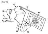
図10は、本発明の他の実施形態に係るタブレットコンピュータを連結した視野自己診断システムの全体構成図である。
図10を参照すれば、視野診断のための視野測定領域と視野測定結果が提示されるタブレットコンピュータ80と、タブレットコンピュータ80の固定のための機器挿入部81と、タブレットコンピュータ80を傾けるための機器角度調節部82と、検査者情報入力のための検査者情報入力部83と、視野診断環境提供のための脱着式遮光部20と、検査眼選択部30と、顔面支持部40と、視標確認部とを含む。前記視標確認部は、前記タブレットコンピュータ80に連結され得る。
FIG. 10 is an overall configuration diagram of a visual field self-diagnosis system in which tablet computers according to other embodiments of the present invention are connected.
Referring to FIG. 10, a tablet computer 80 in which a field-of-view measurement region and a field-of-view measurement result for visual field diagnosis are presented, a device insertion portion 81 for fixing the tablet computer 80, and a device for tilting the tablet computer 80.
機器挿入部81は、タブレットコンピュータ80がスライディング方式で挿入されて固定され得るように上下部の周縁にガイド部が形成されている。検査者情報入力部83は、バーコードまたはRFIDで構成された検査者情報を認識することができるようにバーコートリーダーまたはRFIDリーダーで構成され、前記タブレットコンピュータ80と連結されて、入力された検査者情報が前記タブレットコンピュータ80に転送され得る。
In the device insertion portion 81, guide portions are formed on the periphery of the upper and lower portions so that the tablet computer 80 can be inserted and fixed by a sliding method. The inspector
一方、図10に示されているように、本実施形態において検査眼選択部30と顔面支持部40は、一体に製作されて前記脱着式遮光部20に結合され得る。しかし、前記図1に示されているように、検査眼選択部30と顔面支持部40が別個の部品で製作されて組み立てて使用されることもできる。
On the other hand, as shown in FIG. 10, in this embodiment, the test
上述したような本発明の実施形態に係るシステムの視野測定領域は、視野検査と共に視力、黄斑変性、色盲などのような眼科検査を含むことができるように構成される。 The visual field measurement region of the system according to the embodiment of the present invention as described above is configured to include ophthalmic examinations such as visual acuity, macular degeneration, color blindness and the like in addition to the visual examination.
以上を通じて本発明の好適な実施形態について説明したが、本発明はこれに限定されるのではなく、特許請求の範囲と発明の詳細な説明および添付図面の範囲内で多様に変形して実施することが可能であり、これも本発明の範囲に属するのは当然である。 The preferred embodiments of the present invention have been described above, but the present invention is not limited to these embodiments, and various modifications may be made within the scope of the claims, the detailed description of the invention, and the accompanying drawings. Of course, this is also within the scope of the present invention.
Claims (16)
前記第1モニターの前方に装着されて前記第1モニターに対する外部の光を遮断して視野測定環境を提供する脱着式遮光部と、
前記脱着式遮光部の後方に装着されて外部の光を遮断し、検査眼を選択する検査眼選択部と、
前記検査眼選択部を装着し、検査者の顔面を支持する顔面支持部と、
前記コンピュータ本体に連結され、前記視野測定領域に提示される視標に応じて検査者により操作される視標確認部と、
を含む個人用コンピュータに基づく視野自己測定システム。 A computer monitor coupled to the computer body and including a first monitor providing a field measurement region in which indicators presented for field measurement are arranged and a second monitor providing field measurement progress and results;
A removable light shielding portion for providing a visual field measurement environment by blocking the external light with respect to the first monitor is mounted in front of the first monitor,
An inspection eye selection unit that is mounted behind the detachable light-shielding unit, blocks external light, and selects an inspection eye;
A face support unit that wears the inspection eye selection unit and supports the face of the examiner;
A target confirmation unit connected to the computer main body and operated by an examiner according to a target presented in the visual field measurement area;
Vision self-measuring system based on personal computer including.
前記第1モニターの前方に装着されて前記第1モニターに対する外部の光を遮断して視野測定環境を提供する脱着式遮光部と、
前記脱着式遮光部の後方に装着されて外部の光を遮断し、検査眼を選択する検査眼選択部と、
前記検査眼選択部を装着し、検査者の顔面を支持する顔面支持部と、
前記コンピュータ本体に連結され、前記視野測定領域に提示される視標に応じて検査者により操作される視標確認部と、
を含む個人用コンピュータに基づく視野自己測定システム。 A first monitor as a computer monitor that is connected to the computer main body and sequentially displays a field-of-view measurement area in which indicators to be presented for field-of-view measurement are arranged, and a field-of-view measurement result after the field-of-view measurement area is presented;
A detachable shading unit mounted in front of the first monitor to block external light to the first monitor and provide a visual field measurement environment;
An inspection eye selection unit that is mounted behind the detachable light-shielding unit, blocks external light, and selects an inspection eye;
A face support unit that wears the inspection eye selection unit and supports the face of the examiner;
A target confirmation unit connected to the computer main body and operated by an examiner according to a target presented in the visual field measurement area;
Vision self-measuring system based on personal computer including .
前記第1モニターに示される視野測定領域を完全に含むことができる位置で前記第1モニターに密着する、請求項1又は2に記載の個人用コンピュータに基づく視野自己測定システム。 The detachable shading part is
Said perimetry region shown in the first monitor in close contact with the first monitor in a position where it can completely contain, viewing self-monitoring system based on personal computer according to claim 1 or 2.
検査者の眼の周囲を囲んで外部光を遮断するハウジングと、
ハウジングの一側に結合して検査者が検査する眼を密着する検査眼鏡筒と、
前記検査眼鏡筒が前記視野測定領域の中央に一致するように結合し、前記脱着式遮光部の狭い端部に連結される連結部と、
前記ハウジングの内側で前記検査眼鏡筒の外郭に備えられる遮蔽膜と、
を含む、請求項1又は2に記載の個人用コンピュータに基づく視野自己測定システム。 The inspection eye selection unit includes:
A housing that surrounds the eyes of the examiner and blocks external light;
An inspection eyeglass tube that is coupled to one side of the housing and closely contacts the eye to be inspected by the inspector;
The inspection eyeglass tube is coupled so as to coincide with the center of the visual field measurement region, and is connected to a narrow end of the detachable light shielding unit,
A shielding film provided on the outer periphery of the inspection eyeglass tube inside the housing;
A personal computer based visual field self-measuring system according to claim 1 or 2 , comprising:
前記連結部の一側に備えられる水平維持部に支持される、請求項4に記載の個人用コンピュータに基づく視野自己測定システム。 The inspection eye selection unit includes:
The visual field self-measuring system based on a personal computer according to claim 4 , wherein the visual field self-measuring system is supported by a horizontal maintaining part provided on one side of the connecting part .
検査者が額を当てられる額支持部材と、
前記額支持部材の高さを調節する高さ調節部材と、
検査者が顎を支えられる顎支持部材と、
前記顎支持部材の左右移動を調節する左右調節部材と、
前記顎支持部材の高さを調節する高さ調節ねじと、
前記高さ調節ねじで結合される検査者の顔の重量を支えるための支え台と、
を含む、請求項1又は2に記載の個人用コンピュータに基づく視野自己測定システム。 The face support part is
A forehead support member to which the inspector can apply a forehead;
A height adjusting member for adjusting the height of the forehead support member;
A chin support member that allows the examiner to support the chin;
A left-right adjustment member for adjusting the left-right movement of the jaw support member;
A height adjusting screw for adjusting the height of the jaw support member;
A support for supporting the weight of the face of the examiner coupled with the height adjusting screw;
A personal computer based visual field self-measuring system according to claim 1 or 2 , comprising:
検査者が視野測定領域に提示される中心視標の形状に応じて押す左側ボタンおよび右側ボタンと、
提示される視標が見える場合に押す視標確認ボタンと、
前記コンピュータ本体に連結される連結ポートと、
を含む、請求項1又は2に記載の個人用コンピュータに基づく視野自己測定システム。 The target confirmation unit
A left button and a right button that the inspector presses according to the shape of the central target presented in the visual field measurement area, and
A target confirmation button that is pressed when the target is presented;
A connection port connected to the computer body;
A personal computer based visual field self-measuring system according to claim 1 or 2 , comprising:
検査する側の眼の視野を診断することができるように構成される中心視標、視標および盲点視標を含む、請求項1又は2に記載の個人用コンピュータに基づく視野自己測定システム。 The visual field measurement region is
3. A personal computer based visual field self-measuring system according to claim 1 or 2 , comprising a central target, a visual target and a blind spot target configured to be able to diagnose the visual field of the eye to be examined .
数字、記号、文字、図形、動物形状および事物形状の中から選ばれた少なくとも一つ以上で形成される、請求項8に記載の個人用コンピュータに基づく視野自己測定システム。 The central target is
The personal computer-based visual field self-measurement system according to claim 8 , wherein the visual field self-measurement system is based on at least one selected from numbers, symbols, characters, figures, animal shapes, and object shapes .
前記タブレットコンピュータを挿入することができる機器挿入部と、
前記機器挿入部の下端に連結されて前記タブレットコンピュータを傾けることができる機器角度調節部と、をさらに含み、
前記視標確認部は、前記タブレットコンピュータに連結されることを特徴とする、請求項2に記載の個人用コンピュータに基づく視野自己測定システム。 The first monitor is a tablet computer formed integrally with the computer main body,
A device insertion section into which the tablet computer can be inserted;
A device angle adjusting unit coupled to a lower end of the device insertion unit and capable of tilting the tablet computer;
3. The visual field self-measuring system based on a personal computer according to claim 2 , wherein the target confirmation unit is connected to the tablet computer .
コンピュータモニターによって、視野測定のために提示される指標が配列された視野測定領域を提供する第1モニター領域に、中心視標を提示する段階と視標または盲点視標を提示する段階とを同時に行う視標提示段階と、
コンピュータによって、視標の検査が完了したかを判断し、視標検査の未完了時は、前記視標提示段階へ進む第1判断段階と、
前記コンピュータによって、視標検査の完了時は、視線固定エラー率を計算する視線固定エラー率計算段階と、
前記コンピュータによって、視線固定エラー率が設定値以下であるかを判断し、前記視線固定エラー率が設定値を超える場合は、入力段階を遂行する第2判断段階と、
前記コンピュータによって、前記視線固定エラー率が設定値以下である場合、視野測定結果を前記コンピュータモニターの視野測定進行と結果を提供する第2モニター領域に提供し、視野診断プログラムを終了する結果提供段階と、
を含む個人用コンピュータに基づく視野自己測定方法。 An input stage for inputting a visual field measurement region, a visual field measurement method, and a central target shape to the computer main body by a user interface;
Simultaneously presenting a central target and a target or blind target in a first monitor region that provides a visual field measurement region in which indicators presented for visual field measurement are arranged by a computer monitor A target presentation stage to be performed;
The computer determines whether the inspection of the optotype is completed, and when the optotype inspection is not completed, a first determination stage that proceeds to the optotype presentation stage;
By the computer, upon completion of the visual target inspection, a gaze fixing error rate calculating step of calculating a gaze fixing error rate,
The computer determines whether the line-of-sight fixed error rate is equal to or lower than a set value. If the line-of-sight fixed error rate exceeds the set value, a second determining step of performing an input step;
When the visual line fixing error rate is equal to or less than a set value by the computer, the visual field measurement result is provided to the second monitor area that provides the visual field measurement progress and the result of the computer monitor, and the visual field diagnosis program is terminated. When,
Visual field self-measuring method based on personal computer including
前記コンピュータモニターの大きさおよび解像度の情報を自動的に把握して、前記コンピュータモニターの画面に同一の大きさで変換して変換された視野測定領域を示す変換段階
をさらに含む、請求項13に記載の個人用コンピュータに基づく視野自己測定方法。 The input step includes
A conversion step of automatically grasping information on the size and resolution of the computer monitor, and converting the same size into the computer monitor screen to indicate the converted visual field measurement region
14. A visual field self-measuring method based on a personal computer according to claim 13 , further comprising:
互いに異なる形状の複数の中心視標を同一または互いに異なる任意の速度で点滅して提示する段階を含む、請求項13に記載の個人用コンピュータに基づく視野自己測定方法。 The target presentation step includes:
14. The visual field self-measuring method based on a personal computer according to claim 13 , comprising a step of presenting a plurality of central targets having different shapes by blinking at an arbitrary speed different from each other .
個人別に蓄積された視野測定結果に基づいて視野損傷進行程度を提示する段階をさらに含む、請求項13に記載の個人用コンピュータに基づく視野自己測定方法。 The result providing step includes:
14. The visual field self-measuring method based on a personal computer according to claim 13 , further comprising the step of presenting a degree of visual field damage progress based on visual field measurement results accumulated for each individual.
Applications Claiming Priority (3)
| Application Number | Priority Date | Filing Date | Title |
|---|---|---|---|
| KR10-2011-0138483 | 2011-12-20 | ||
| KR20110138483A KR101245330B1 (en) | 2011-12-20 | 2011-12-20 | Pc-based visual filed self-diagnosis system and gaze fixing method |
| PCT/KR2012/011148 WO2013094995A1 (en) | 2011-12-20 | 2012-12-20 | Personal-computer-based visual-field self-diagnosis system and visual-field self-diagnosis method |
Publications (2)
| Publication Number | Publication Date |
|---|---|
| JP2015500732A JP2015500732A (en) | 2015-01-08 |
| JP6033883B2 true JP6033883B2 (en) | 2016-11-30 |
Family
ID=48182200
Family Applications (1)
| Application Number | Title | Priority Date | Filing Date |
|---|---|---|---|
| JP2014548659A Active JP6033883B2 (en) | 2011-12-20 | 2012-12-20 | Visual field self-measuring system and visual field self-measuring method based on personal computer |
Country Status (5)
| Country | Link |
|---|---|
| US (1) | US20140340642A1 (en) |
| EP (1) | EP2796088A4 (en) |
| JP (1) | JP6033883B2 (en) |
| KR (1) | KR101245330B1 (en) |
| WO (1) | WO2013094995A1 (en) |
Families Citing this family (23)
| Publication number | Priority date | Publication date | Assignee | Title |
|---|---|---|---|---|
| US8348429B2 (en) | 2008-03-27 | 2013-01-08 | Doheny Eye Institute | Optical coherence tomography device, method, and system |
| US11839430B2 (en) | 2008-03-27 | 2023-12-12 | Doheny Eye Institute | Optical coherence tomography-based ophthalmic testing methods, devices and systems |
| WO2010009450A1 (en) | 2008-07-18 | 2010-01-21 | Doheny Eye Institute | Optical coherence tomography device, method, and system |
| US9226856B2 (en) | 2013-03-14 | 2016-01-05 | Envision Diagnostics, Inc. | Inflatable medical interfaces and other medical devices, systems, and methods |
| US10772497B2 (en) | 2014-09-12 | 2020-09-15 | Envision Diagnostics, Inc. | Medical interfaces and other medical devices, systems, and methods for performing eye exams |
| EP3041399B1 (en) | 2013-09-02 | 2017-11-01 | Ocuspecto OY | Automated perimetry |
| DE102014113682A1 (en) | 2014-09-22 | 2016-03-24 | Carl Zeiss Meditec Ag | Device for visual field measurement |
| EP3349642B1 (en) * | 2015-09-17 | 2020-10-21 | Envision Diagnostics, Inc. | Medical interfaces and other medical devices, systems, and methods for performing eye exams |
| JP6063549B1 (en) * | 2015-12-17 | 2017-01-18 | 雅子 吉田 | ruler |
| KR101652739B1 (en) * | 2016-03-23 | 2016-08-31 | (주) 뷰엠테크놀로지 | Eyesight examination method, eyesight examination apparatus and download server storing a program of the eyesight examination method |
| EP3448234A4 (en) | 2016-04-30 | 2019-05-01 | Envision Diagnostics, Inc. | Medical devices, systems, and methods for performing eye exams and eye tracking |
| KR101965057B1 (en) * | 2017-05-15 | 2019-04-04 | 유니폴라 주식회사 | Vision inspection device for inspecting vision of examinee |
| CN111542258B (en) | 2017-11-07 | 2023-10-20 | 诺达尔视觉有限公司 | Method and system for alignment of ophthalmic imaging devices |
| EP3675711A4 (en) | 2017-11-07 | 2021-06-30 | Notal Vision Ltd. | Retinal imaging device and related methods |
| WO2019092639A1 (en) | 2017-11-08 | 2019-05-16 | Braineyes - Soluções De Diagnóstico E Reabilitação, Lda | Method and device for performing an eye test |
| WO2019099572A1 (en) * | 2017-11-14 | 2019-05-23 | Vivid Vision, Inc. | Systems and methods for visual field analysis |
| US10413172B2 (en) | 2017-12-11 | 2019-09-17 | 1-800 Contacts, Inc. | Digital visual acuity eye examination for remote physician assessment |
| CN108937841B (en) * | 2018-07-17 | 2020-06-26 | 遂宁市中心医院 | Ophthalmologic visual field inspection apparatus |
| US10595722B1 (en) | 2018-10-03 | 2020-03-24 | Notal Vision Ltd. | Automatic optical path adjustment in home OCT |
| US11051689B2 (en) | 2018-11-02 | 2021-07-06 | International Business Machines Corporation | Real-time passive monitoring and assessment of pediatric eye health |
| US10653311B1 (en) | 2019-06-12 | 2020-05-19 | Notal Vision Ltd. | Home OCT with automatic focus adjustment |
| US20230075963A1 (en) | 2020-02-10 | 2023-03-09 | Shimadzu Corporation | Visual function test device |
| FI129077B (en) * | 2020-06-29 | 2021-06-30 | Optomed Oyj | Contact arrangement for eye examining instrument, eye examining instrument and method of contacting between eye and eye examining instrument |
Family Cites Families (19)
| Publication number | Priority date | Publication date | Assignee | Title |
|---|---|---|---|---|
| US5946075A (en) * | 1996-05-21 | 1999-08-31 | Horn; Gerald | Vision screening system |
| JP4171995B2 (en) * | 1997-10-16 | 2008-10-29 | 潤 福原 | Flicker sensitivity distribution measuring device and computer-readable recording medium recording flicker sensitivity distribution measuring program |
| CA2333678A1 (en) * | 1999-03-31 | 2000-10-05 | Virtual-Eye.Com, Inc. | Kinetic visual field apparatus and method |
| GB0210288D0 (en) * | 2002-05-04 | 2002-06-12 | Univ Nottingham | Ocular display apparatus for assessment and measurement of and for treatment of ocular disorders, and methods therefor |
| WO2006002070A2 (en) * | 2004-06-15 | 2006-01-05 | Novavision, Inc. | Method and device for guiding a user's head during vision training |
| JP2006014766A (en) * | 2004-06-30 | 2006-01-19 | Nidek Co Ltd | Visual function examination apparatus |
| GB0513603D0 (en) * | 2005-06-30 | 2005-08-10 | Univ Aberdeen | Vision exercising apparatus |
| US7594728B2 (en) * | 2005-12-16 | 2009-09-29 | Novavision, Inc. | Adjustable device for vision testing and therapy |
| EP2076230A2 (en) * | 2006-07-25 | 2009-07-08 | Novavision, Inc. | Dynamic stimuli for visual field testing and therapy |
| US8226237B2 (en) * | 2006-09-28 | 2012-07-24 | Reichert, Inc. | Apparatus and method for monitoring the position of a subject's hand |
| US7806528B2 (en) * | 2006-12-06 | 2010-10-05 | The University Of Houston System | System and method for detecting central retinal distortions associated with macular diseases |
| JP5085957B2 (en) * | 2007-02-26 | 2012-11-28 | 株式会社ニデック | Target presentation device |
| WO2008106802A1 (en) * | 2007-03-08 | 2008-09-12 | University Of Northern British Columbia | Apparatus and method for objective perimetry visual field test |
| GB0709405D0 (en) | 2007-05-16 | 2007-06-27 | Univ Edinburgh | Testing vision |
| JP2009059257A (en) | 2007-09-03 | 2009-03-19 | Sony Corp | Information processing apparatus and information processing method, and computer program |
| US8079710B2 (en) * | 2008-01-10 | 2011-12-20 | Notal Vision Ltd. | Dual position ophthalmic apparatus |
| JP5234924B2 (en) * | 2008-03-31 | 2013-07-10 | 国立大学法人 千葉大学 | Visual function evaluation device |
| US20130112841A1 (en) * | 2011-11-09 | 2013-05-09 | Eagle Fan | Supporting apparatus |
| US20130155376A1 (en) * | 2011-12-20 | 2013-06-20 | Icheck Health Connection, Inc. | Video game to monitor visual field loss in glaucoma |
-
2011
- 2011-12-20 KR KR20110138483A patent/KR101245330B1/en active IP Right Grant
-
2012
- 2012-12-20 EP EP12860582.1A patent/EP2796088A4/en not_active Withdrawn
- 2012-12-20 JP JP2014548659A patent/JP6033883B2/en active Active
- 2012-12-20 WO PCT/KR2012/011148 patent/WO2013094995A1/en active Application Filing
- 2012-12-20 US US14/364,362 patent/US20140340642A1/en not_active Abandoned
Also Published As
| Publication number | Publication date |
|---|---|
| US20140340642A1 (en) | 2014-11-20 |
| EP2796088A1 (en) | 2014-10-29 |
| KR101245330B1 (en) | 2013-03-25 |
| EP2796088A4 (en) | 2015-12-23 |
| WO2013094995A1 (en) | 2013-06-27 |
| JP2015500732A (en) | 2015-01-08 |
Similar Documents
| Publication | Publication Date | Title |
|---|---|---|
| JP6033883B2 (en) | Visual field self-measuring system and visual field self-measuring method based on personal computer | |
| US11478142B2 (en) | Methods, apparatus, and systems for ophthalmic testing and measurement | |
| EP2079355B1 (en) | Eyeglass prescription method and system | |
| EP2949266B1 (en) | Ophthalmologic apparatus | |
| CN110573061B (en) | Ophthalmologic examination method and apparatus | |
| US9380938B2 (en) | System and methods for documenting and recording of the pupillary red reflex test and corneal light reflex screening of the eye in infants and young children | |
| CN107692960B (en) | Optical coherence tomography analysis apparatus, method and system | |
| JP2005131393A (en) | Device and method for diagnosing ocular symptom which can optically be discriminated | |
| RU2634682C1 (en) | Portable device for visual functions examination | |
| RU2480142C2 (en) | Device and method of remote evaluation of human visual analyser characteristics and carrying out training exercises for development of binocular and higher visual functions | |
| Kunumpol et al. | GlauCUTU: virtual reality visual field test | |
| US10588506B2 (en) | Device and method for the quantitative detection of disorders in the field of vision | |
| CN113413130A (en) | Small-size optometry and eyesight tester of short main light path | |
| CN215959809U (en) | Small-size optometry and eyesight tester of short main light path | |
| KR20200120488A (en) | Eye photographing device | |
| Beaton et al. | A rapid quantification of binocular misalignment without recording eye movements: Vertical and torsional alignment nulling | |
| JP3244340U (en) | Science teaching materials for optics learning | |
| Sharma et al. | 21 Low Vision Management in Retinal Diseases | |
| PATIENT | Low Vision Management in Retinal Diseases | |
| KR20220168883A (en) | Method for Screening Strabismus and Device therefor | |
| Mousavi et al. | Evaluation of visual complications among professional computer users | |
| JP2023142781A (en) | Ophthalmologic system | |
| CN117295447A (en) | Eye examination equipment matched with smart phone | |
| Rotenstreich et al. | The first prototype of chromatic pupillometer for objective perimetry in retinal degeneration patients |
Legal Events
| Date | Code | Title | Description |
|---|---|---|---|
| A621 | Written request for application examination |
Free format text: JAPANESE INTERMEDIATE CODE: A621 Effective date: 20140619 |
|
| A131 | Notification of reasons for refusal |
Free format text: JAPANESE INTERMEDIATE CODE: A131 Effective date: 20150630 |
|
| A521 | Request for written amendment filed |
Free format text: JAPANESE INTERMEDIATE CODE: A523 Effective date: 20150925 |
|
| A131 | Notification of reasons for refusal |
Free format text: JAPANESE INTERMEDIATE CODE: A131 Effective date: 20160209 |
|
| A521 | Request for written amendment filed |
Free format text: JAPANESE INTERMEDIATE CODE: A523 Effective date: 20160509 |
|
| TRDD | Decision of grant or rejection written | ||
| A01 | Written decision to grant a patent or to grant a registration (utility model) |
Free format text: JAPANESE INTERMEDIATE CODE: A01 Effective date: 20161011 |
|
| A61 | First payment of annual fees (during grant procedure) |
Free format text: JAPANESE INTERMEDIATE CODE: A61 Effective date: 20161026 |
|
| R150 | Certificate of patent or registration of utility model |
Ref document number: 6033883 Country of ref document: JP Free format text: JAPANESE INTERMEDIATE CODE: R150 |
|
| R250 | Receipt of annual fees |
Free format text: JAPANESE INTERMEDIATE CODE: R250 |
|
| R250 | Receipt of annual fees |
Free format text: JAPANESE INTERMEDIATE CODE: R250 |
|
| R250 | Receipt of annual fees |
Free format text: JAPANESE INTERMEDIATE CODE: R250 |
|
| R250 | Receipt of annual fees |
Free format text: JAPANESE INTERMEDIATE CODE: R250 |
|
| R250 | Receipt of annual fees |
Free format text: JAPANESE INTERMEDIATE CODE: R250 |