JP6007527B2 - Fundus photographing device - Google Patents
Fundus photographing device Download PDFInfo
- Publication number
- JP6007527B2 JP6007527B2 JP2012056292A JP2012056292A JP6007527B2 JP 6007527 B2 JP6007527 B2 JP 6007527B2 JP 2012056292 A JP2012056292 A JP 2012056292A JP 2012056292 A JP2012056292 A JP 2012056292A JP 6007527 B2 JP6007527 B2 JP 6007527B2
- Authority
- JP
- Japan
- Prior art keywords
- fundus
- light
- optical path
- focus
- path length
- Prior art date
- Legal status (The legal status is an assumption and is not a legal conclusion. Google has not performed a legal analysis and makes no representation as to the accuracy of the status listed.)
- Active
Links
- 230000003287 optical effect Effects 0.000 claims description 370
- 238000005259 measurement Methods 0.000 claims description 86
- 238000001514 detection method Methods 0.000 claims description 41
- 238000003384 imaging method Methods 0.000 claims description 33
- 210000004220 fundus oculi Anatomy 0.000 claims description 6
- 238000005286 illumination Methods 0.000 claims description 5
- 230000001678 irradiating effect Effects 0.000 claims 1
- 238000011156 evaluation Methods 0.000 description 42
- 238000005457 optimization Methods 0.000 description 41
- 230000007246 mechanism Effects 0.000 description 28
- 230000010287 polarization Effects 0.000 description 21
- 239000013307 optical fiber Substances 0.000 description 20
- 230000008859 change Effects 0.000 description 11
- 238000012937 correction Methods 0.000 description 10
- 238000010586 diagram Methods 0.000 description 9
- 239000006185 dispersion Substances 0.000 description 9
- 239000000835 fiber Substances 0.000 description 9
- 238000000034 method Methods 0.000 description 8
- 238000001228 spectrum Methods 0.000 description 8
- 238000004364 calculation method Methods 0.000 description 6
- 230000035945 sensitivity Effects 0.000 description 5
- 238000006073 displacement reaction Methods 0.000 description 4
- 238000012545 processing Methods 0.000 description 4
- 210000001525 retina Anatomy 0.000 description 4
- 239000000284 extract Substances 0.000 description 3
- 210000003583 retinal pigment epithelium Anatomy 0.000 description 3
- 230000003595 spectral effect Effects 0.000 description 3
- 230000015572 biosynthetic process Effects 0.000 description 2
- 210000004204 blood vessel Anatomy 0.000 description 2
- 238000006243 chemical reaction Methods 0.000 description 2
- 210000003161 choroid Anatomy 0.000 description 2
- 230000007423 decrease Effects 0.000 description 2
- 230000004069 differentiation Effects 0.000 description 2
- 238000000605 extraction Methods 0.000 description 2
- 230000006870 function Effects 0.000 description 2
- 238000012014 optical coherence tomography Methods 0.000 description 2
- 210000001747 pupil Anatomy 0.000 description 2
- 238000003325 tomography Methods 0.000 description 2
- 230000009471 action Effects 0.000 description 1
- 230000004323 axial length Effects 0.000 description 1
- 230000005540 biological transmission Effects 0.000 description 1
- 230000001427 coherent effect Effects 0.000 description 1
- 230000008878 coupling Effects 0.000 description 1
- 238000010168 coupling process Methods 0.000 description 1
- 238000005859 coupling reaction Methods 0.000 description 1
- 238000002474 experimental method Methods 0.000 description 1
- 230000005484 gravity Effects 0.000 description 1
- 230000010354 integration Effects 0.000 description 1
- 230000008569 process Effects 0.000 description 1
- 208000014733 refractive error Diseases 0.000 description 1
- 230000000630 rising effect Effects 0.000 description 1
- 238000005070 sampling Methods 0.000 description 1
- 238000004088 simulation Methods 0.000 description 1
- 238000012360 testing method Methods 0.000 description 1
- 230000009466 transformation Effects 0.000 description 1
- 230000001131 transforming effect Effects 0.000 description 1
Images
Classifications
-
- A—HUMAN NECESSITIES
- A61—MEDICAL OR VETERINARY SCIENCE; HYGIENE
- A61B—DIAGNOSIS; SURGERY; IDENTIFICATION
- A61B3/00—Apparatus for testing the eyes; Instruments for examining the eyes
- A61B3/10—Objective types, i.e. instruments for examining the eyes independent of the patients' perceptions or reactions
- A61B3/102—Objective types, i.e. instruments for examining the eyes independent of the patients' perceptions or reactions for optical coherence tomography [OCT]
-
- A—HUMAN NECESSITIES
- A61—MEDICAL OR VETERINARY SCIENCE; HYGIENE
- A61B—DIAGNOSIS; SURGERY; IDENTIFICATION
- A61B5/00—Measuring for diagnostic purposes; Identification of persons
- A61B5/0059—Measuring for diagnostic purposes; Identification of persons using light, e.g. diagnosis by transillumination, diascopy, fluorescence
- A61B5/0062—Arrangements for scanning
- A61B5/0066—Optical coherence imaging
-
- G—PHYSICS
- G01—MEASURING; TESTING
- G01B—MEASURING LENGTH, THICKNESS OR SIMILAR LINEAR DIMENSIONS; MEASURING ANGLES; MEASURING AREAS; MEASURING IRREGULARITIES OF SURFACES OR CONTOURS
- G01B9/00—Measuring instruments characterised by the use of optical techniques
- G01B9/02—Interferometers
- G01B9/02015—Interferometers characterised by the beam path configuration
- G01B9/02029—Combination with non-interferometric systems, i.e. for measuring the object
- G01B9/0203—With imaging systems
-
- G—PHYSICS
- G01—MEASURING; TESTING
- G01B—MEASURING LENGTH, THICKNESS OR SIMILAR LINEAR DIMENSIONS; MEASURING ANGLES; MEASURING AREAS; MEASURING IRREGULARITIES OF SURFACES OR CONTOURS
- G01B9/00—Measuring instruments characterised by the use of optical techniques
- G01B9/02—Interferometers
- G01B9/02041—Interferometers characterised by particular imaging or detection techniques
- G01B9/02044—Imaging in the frequency domain, e.g. by using a spectrometer
-
- G—PHYSICS
- G01—MEASURING; TESTING
- G01B—MEASURING LENGTH, THICKNESS OR SIMILAR LINEAR DIMENSIONS; MEASURING ANGLES; MEASURING AREAS; MEASURING IRREGULARITIES OF SURFACES OR CONTOURS
- G01B9/00—Measuring instruments characterised by the use of optical techniques
- G01B9/02—Interferometers
- G01B9/02055—Reduction or prevention of errors; Testing; Calibration
- G01B9/02062—Active error reduction, i.e. varying with time
- G01B9/02063—Active error reduction, i.e. varying with time by particular alignment of focus position, e.g. dynamic focussing in optical coherence tomography
-
- G—PHYSICS
- G01—MEASURING; TESTING
- G01B—MEASURING LENGTH, THICKNESS OR SIMILAR LINEAR DIMENSIONS; MEASURING ANGLES; MEASURING AREAS; MEASURING IRREGULARITIES OF SURFACES OR CONTOURS
- G01B9/00—Measuring instruments characterised by the use of optical techniques
- G01B9/02—Interferometers
- G01B9/02055—Reduction or prevention of errors; Testing; Calibration
- G01B9/02062—Active error reduction, i.e. varying with time
- G01B9/02064—Active error reduction, i.e. varying with time by particular adjustment of coherence gate, i.e. adjusting position of zero path difference in low coherence interferometry
-
- G—PHYSICS
- G01—MEASURING; TESTING
- G01B—MEASURING LENGTH, THICKNESS OR SIMILAR LINEAR DIMENSIONS; MEASURING ANGLES; MEASURING AREAS; MEASURING IRREGULARITIES OF SURFACES OR CONTOURS
- G01B9/00—Measuring instruments characterised by the use of optical techniques
- G01B9/02—Interferometers
- G01B9/02055—Reduction or prevention of errors; Testing; Calibration
- G01B9/02062—Active error reduction, i.e. varying with time
- G01B9/02067—Active error reduction, i.e. varying with time by electronic control systems, i.e. using feedback acting on optics or light
- G01B9/02068—Auto-alignment of optical elements
-
- G—PHYSICS
- G01—MEASURING; TESTING
- G01B—MEASURING LENGTH, THICKNESS OR SIMILAR LINEAR DIMENSIONS; MEASURING ANGLES; MEASURING AREAS; MEASURING IRREGULARITIES OF SURFACES OR CONTOURS
- G01B9/00—Measuring instruments characterised by the use of optical techniques
- G01B9/02—Interferometers
- G01B9/0209—Low-coherence interferometers
- G01B9/02091—Tomographic interferometers, e.g. based on optical coherence
-
- G—PHYSICS
- G01—MEASURING; TESTING
- G01B—MEASURING LENGTH, THICKNESS OR SIMILAR LINEAR DIMENSIONS; MEASURING ANGLES; MEASURING AREAS; MEASURING IRREGULARITIES OF SURFACES OR CONTOURS
- G01B2290/00—Aspects of interferometers not specifically covered by any group under G01B9/02
- G01B2290/70—Using polarization in the interferometer
Landscapes
- Health & Medical Sciences (AREA)
- Physics & Mathematics (AREA)
- Life Sciences & Earth Sciences (AREA)
- General Physics & Mathematics (AREA)
- General Health & Medical Sciences (AREA)
- Nuclear Medicine, Radiotherapy & Molecular Imaging (AREA)
- Radiology & Medical Imaging (AREA)
- Engineering & Computer Science (AREA)
- Surgery (AREA)
- Biophysics (AREA)
- Heart & Thoracic Surgery (AREA)
- Medical Informatics (AREA)
- Molecular Biology (AREA)
- Veterinary Medicine (AREA)
- Animal Behavior & Ethology (AREA)
- Biomedical Technology (AREA)
- Public Health (AREA)
- Ophthalmology & Optometry (AREA)
- Pathology (AREA)
- Automation & Control Theory (AREA)
- Optics & Photonics (AREA)
- Eye Examination Apparatus (AREA)
Description
本発明は、被検者眼眼底の断層像を撮影する眼底撮影装置に関する。 The present invention relates to a fundus imaging apparatus that captures a tomographic image of a subject's fundus.
被検者眼の眼底の断層像を撮影する光断層像撮影装置として、低コヒーレント光を用いた光断層干渉計(Optical Coherence Tomography:OCT)が知られている(特許文献1参照)。 An optical tomography (OCT) using low-coherent light is known as an optical tomographic imaging apparatus that captures a tomographic image of the fundus of a subject's eye (see Patent Document 1).
このような装置において、検者は、SLO光学系もしくは眼底カメラ光学系によって取得される眼底正面画像のフォーカス状態を利用して、眼底断層画像のフォーカス合わせを行っている。また、フォーカス調整後に、光路長調整と偏光状態の調整(ポラライザの調整)を行い、測定の最適化を行っていた。 In such an apparatus, the examiner focuses the fundus tomographic image using the focus state of the fundus front image acquired by the SLO optical system or the fundus camera optical system. Further, after the focus adjustment, the optical path length adjustment and the polarization state adjustment (polarizer adjustment) are performed to optimize the measurement.
しかしながら、従来、最適化において、光路長調整を行うためには、フォーカス調整が完了するまで、待つ必要があり、多くの時間を費やしていた。そして、最適化の制御を行う時間が長いため、検者がストレスを感じることがあった。また、被検者にとって負担であった。 However, conventionally, in the optimization, in order to perform the optical path length adjustment, it is necessary to wait until the focus adjustment is completed, and much time has been spent. And since the time for controlling the optimization is long, the examiner may feel stress. It was also a burden for the subject.
本発明は、上記問題点を鑑み、撮影条件を好適に調整し、眼底断層像を取得する眼底撮影装置を提供することを技術課題とする。 In view of the above problems, it is an object of the present invention to provide a fundus imaging apparatus that appropriately adjusts imaging conditions and acquires a fundus tomographic image.
上記課題を解決するために、本発明は以下のような構成を備えることを特徴とする。 In order to solve the above problems, the present invention is characterized by having the following configuration.
(1) 本開示の第1態様に係る眼底撮影装置は、光源から出射された光束を測定光と参照光に分割し、測定光束を被検眼眼底に導き,参照光を参照光学系に導いた後、被検眼眼底から反射された測定光と参照光との干渉状態を検出器により検出する干渉光学系と、を備え、前記検出器からの出力信号に基づいて被検眼眼底の断層画像を撮像する眼底撮影装置であって、被検眼眼底に対する合焦位置を検出するためのフォーカス検出手段と、前記測定光の光路中に配置された第1光学部材を駆動させる第1駆動手段とを備え、前記フォーカス検出手段によって検出された合焦位置に前記第1光学部材を移動させるフォーカス調整手段と、測定光と参照光との光路長差を調整するために測定光又は参照光の光路中に配置された第2光学部材を駆動させる第2駆動手段を有し、前記検出器から出力される出力信号に基づいて被検眼断層像が取得される位置に前記第2光学部材を移動させる光路長調整手段と、前記フォーカス検出手段によって被検眼眼底に対する合焦位置を検出し、検出された合焦位置に前記第1光学部材を移動させる前記フォーカス調整手段の作動中において、前記検出器によって測定光と参照光との干渉状態を検出し、前記検出器から出力される出力信号に基づいて被検眼断層像が取得される位置に前記第2光学部材を移動させる前記光路長調整手段の作動を並行して行うことを特徴とする。
(2) 本開示の第2態様に係る眼底撮影装置は、光源から出射された光束を測定光と参照光に分割し、測定光束を被検眼眼底に導き,参照光を参照光学系に導いた後、被検眼眼底から反射された測定光と参照光との干渉状態を検出器により検出する干渉光学系と、を備え、前記検出器からの出力信号に基づいて被検眼眼底の断層画像を撮像する眼底撮影装置であって、被検眼眼底に対する合焦位置を検出するためのフォーカス検出手段と、前記測定光の光路中に配置された第1光学部材を駆動させる第1駆動手段とを備え、前記フォーカス検出手段によって検出された合焦位置に前記第1光学部材を移動させるフォーカス調整手段と、測定光と参照光との光路長差を調整するために測定光又は参照光の光路中に配置された第2光学部材を駆動させる第2駆動手段を有し、前記フォーカス検出手段の作動と並行して、前記検出器から出力される出力信号に基づいて被検眼断層像が取得される位置に前記第2光学部材を移動させる光路長調整手段と、を備え、前記光路長調整手段は、前記フォーカス検出手段の作動と並行して、前記検出器から出力される出力信号に基づいて被検眼断層像が取得される位置に前記第2光学部材を移動させ、前記フォーカス調整手段によって前記第1光学部材が前記合焦位置に移動された後、前記検出器から出力される出力信号に基づいて前記第2光学部材の位置を再調整することを特徴とする。
(3) 本開示の第3態様に係る眼底撮影装置は、光源から出射された光束を測定光と参照光に分割し、測定光束を被検眼眼底に導き,参照光を参照光学系に導いた後、被検眼眼底から反射された測定光と参照光との干渉状態を検出器により検出する干渉光学系と、を備え、前記検出器からの出力信号に基づいて被検眼眼底の断層画像を撮像する眼底撮影装置であって、被検眼眼底に対する合焦位置を検出するためのフォーカス検出手段と、前記測定光の光路中に配置された第1光学部材を駆動させる第1駆動手段と、を備え、前記フォーカス検出手段によって検出された合焦位置に前記第1光学部材を移動させるフォーカス調整手段と、測定光と参照光との光路長差を調整するために測定光又は参照光の光路中に配置された第2光学部材を駆動させる第2駆動手段を有し、前記フォーカス検出手段の作動と並行して、前記検出器から出力される出力信号に基づいて被検眼断層像が取得される位置に前記第2光学部材を移動させる第1光路長調整手段と、前記フォーカス調整手段によるフォーカス調整と、前記第1光路長調整手段による光路長調整と、の双方の調整が完了したことを検出する検出手段と、前記検出手段によって、フォーカス調整及び光路長調整の双方の調整が完了したことを検出した場合に、前記検出器から出力される出力信号に基づいて前記第2光学部材の位置を再調整する第2光路長調整手段と、を備えることを特徴とする。
(1) The fundus imaging apparatus according to the first aspect of the present disclosure divides the light beam emitted from the light source into measurement light and reference light, guides the measurement light beam to the fundus of the eye to be examined, and guides the reference light to the reference optical system. And an interference optical system for detecting an interference state between the measurement light reflected from the fundus of the eye to be examined and the reference light by a detector, and taking a tomographic image of the fundus of the eye to be examined based on an output signal from the detector A fundus imaging apparatus comprising: focus detection means for detecting a focus position with respect to the fundus of the eye to be examined; and first driving means for driving a first optical member disposed in the optical path of the measurement light, Focus adjusting means for moving the first optical member to the in-focus position detected by the focus detecting means, and arranged in the optical path of the measurement light or reference light to adjust the optical path length difference between the measurement light and the reference light Driven second optical member A second driving means for the optical path length adjusting means for moving the second optical member based on an output signal outputted from the detector to a position where the subject's eye tomographic image is acquired by the focus detection means While the focus adjustment means for moving the first optical member to the detected focus position is detected, the detector detects an interference state between the measurement light and the reference light. Then, the operation of the optical path length adjusting means for moving the second optical member to the position where the tomographic image to be examined is acquired based on the output signal output from the detector is performed in parallel .
(2) The fundus imaging apparatus according to the second aspect of the present disclosure divides the light beam emitted from the light source into measurement light and reference light, guides the measurement light beam to the fundus of the eye to be examined, and guides the reference light to the reference optical system And an interference optical system for detecting an interference state between the measurement light reflected from the fundus of the eye to be examined and the reference light by a detector, and taking a tomographic image of the fundus of the eye to be examined based on an output signal from the detector A fundus imaging apparatus comprising: focus detection means for detecting a focus position with respect to the fundus of the eye to be examined; and first driving means for driving a first optical member disposed in the optical path of the measurement light, Focus adjusting means for moving the first optical member to the in-focus position detected by the focus detecting means, and arranged in the optical path of the measurement light or reference light to adjust the optical path length difference between the measurement light and the reference light Driven second optical member A second drive unit that moves the second optical member to a position where an eye tomogram to be examined is acquired based on an output signal output from the detector in parallel with the operation of the focus detection unit. An optical path length adjusting means, wherein the optical path length adjusting means is located at a position where an eye tomographic image to be examined is acquired based on an output signal output from the detector in parallel with the operation of the focus detecting means. After the second optical member is moved and the first optical member is moved to the in-focus position by the focus adjusting means, the position of the second optical member is re-established based on the output signal output from the detector. It is characterized by adjusting.
(3) The fundus imaging apparatus according to the third aspect of the present disclosure divides the light beam emitted from the light source into measurement light and reference light, guides the measurement light beam to the fundus of the eye to be examined, and guides the reference light to the reference optical system And an interference optical system for detecting an interference state between the measurement light reflected from the fundus of the eye to be examined and the reference light by a detector, and taking a tomographic image of the fundus of the eye to be examined based on an output signal from the detector A fundus imaging apparatus comprising: focus detection means for detecting a focus position with respect to the fundus of the eye to be examined; and first drive means for driving a first optical member arranged in the optical path of the measurement light. A focus adjustment means for moving the first optical member to the in-focus position detected by the focus detection means, and an optical path of the measurement light or the reference light in order to adjust an optical path length difference between the measurement light and the reference light. Drive the arranged second optical member A second driving unit that moves the second optical member to a position where an eye tomogram to be examined is acquired based on an output signal output from the detector in parallel with the operation of the focus detection unit; The first optical path length adjusting means, the focus adjusting means by the focus adjusting means, the detecting means for detecting that the adjustment of both the optical path length adjusting by the first optical path length adjusting means is completed, and the detecting means And second optical path length adjusting means for readjusting the position of the second optical member based on an output signal output from the detector when it is detected that both the focus adjustment and the optical path length adjustment are completed. And.
本発明によれば、撮影条件が好適に調整され、眼底断層像を取得できる。 According to the present invention, photographing conditions are suitably adjusted, and a fundus tomographic image can be acquired.
以下、本発明に係る実施形態を図面に基づいて説明する。図1〜図12は本実施形態に係る眼底撮影装置の構成について説明する図である。なお、本実施形態においては、被検者眼(眼E)の軸方向をZ方向、水平方向をX方向、鉛直方向をY方向として説明する。眼底の表面方向をXY方向として考えても良い。 Embodiments according to the present invention will be described below with reference to the drawings. 1 to 12 are diagrams illustrating the configuration of the fundus imaging apparatus according to the present embodiment. In the present embodiment, the axial direction of the subject's eye (eye E) will be described as the Z direction, the horizontal direction as the X direction, and the vertical direction as the Y direction. The surface direction of the fundus may be considered as the XY direction.
<概要>
本発明の実施形態に係る眼底撮影装置の概要について説明する。本実施形態に関わる眼底撮影装置(光コヒーレンストモグラフィーデバイス)1は、干渉光学系(OCT光学系)200、観察光学系300、制御部(CPU)70、を備える。
<Overview>
An outline of a fundus imaging apparatus according to an embodiment of the present invention will be described. A fundus imaging apparatus (optical coherence tomography device) 1 according to the present embodiment includes an interference optical system (OCT optical system) 200, an observation
OCT光学系200は、光スキャナ(走査部)23を備え、光源27から出射された光束を測定光と参照光に分割し、測定光束を被検眼眼底に導き,参照光を参照光学系200bに導いた後、被検眼眼底から反射された測定光と参照光との干渉状態を検出器83により検出する。そして、制御部70は、検出器83からの出力信号に基づいて、被検眼眼底の断層画像を撮像する。走査部23は、測定光の光路中に配置され、測定光を被検眼上で走査させる。
The OCT
例えば、OCT光学系200は、測定光と参照光との干渉状態として、被検眼眼底から反射された測定光と参照光とが合成された光のスペクトル情報を検出器83により検出する。そして、検出器83によって検出されたスペクトル情報をフーリエ解析することにより被検眼眼底の断層画像を撮像する。
For example, the OCT
観察光学系300は、被検眼眼底に照明光を照射する照射光学系と、眼底反射光を受光素子により受光する受光光学系と、を有し、受光素子からの出力信号に基づいて被検眼眼底の正面眼底画像を得る。例えば、SLO(スキャニングレーザオフサルモスコープ)や眼底カメラが挙げられる。
The observation
例えば、観察光学系300に(スキャニングレーザオフサルモスコープ(SLO)光学系)を用いる場合、SLO光学系は、被検眼眼底に照明光を照射する照射光学系と、眼底反射光を受光素子68により受光する受光光学系と、受光光学系に配置された第3光学部材(例えば、フォーカシングレンズ63)を駆動させる第3駆動手段(例えば、駆動機構63a)と、を有する。そして、受光素子68からの出力信号に基づいて被検眼眼底の正面眼底画像が取得される。
For example, when a (scanning laser ophthalmoscope (SLO) optical system) is used as the observation
また、本装置は、フォーカス検出手段(フォーカス検出部)と、フォーカス調整手段(フォーカス調整部)と、光路長調整手段(光路長調整部)と、を備える。 The apparatus further includes a focus detection unit (focus detection unit), a focus adjustment unit (focus adjustment unit), and an optical path length adjustment unit (optical path length adjustment unit).
フォーカス検出部は、被検眼眼底からの反射光を含む光を受光する受光素子を有し受光素子からの出力信号に基づいて被検眼眼底に対する合焦位置を検出する。 The focus detection unit has a light receiving element that receives light including light reflected from the fundus of the eye to be examined, and detects a focus position with respect to the eye fundus of the eye based on an output signal from the light receiving element.
例えば、フォーカス検出部は観察光学系300と兼用され、フォーカス検出部の動作の制御は、制御部70によって行われる。この場合、制御部70は、駆動機構63aの駆動を制御してフォーカシングレンズ63を移動させ、フォーカシングレンズ63の各位置にて取得された正面眼底画像に基づいて被検眼眼底に対する合焦位置を検出する。
For example, the focus detection unit is also used as the observation
フォーカス調整部は、被検者眼底に対する視度を補正するために測定光の光路中に配置された第1光学部材(例えば、フォーカシングレンズ24)を駆動させる第1駆動手段(例えば、駆動機構24a)を備える。例えば、フォーカス調整部の動作の制御は、制御部70によって行われる。この場合、制御部70は、フォーカス検出部によって検出された合焦位置にフォーカシングレンズ24を移動させる。
The focus adjustment unit drives a first optical member (for example, a
光路長調整部は、測定光と参照光との光路長差を調整するために測定光又は前記参照光の光路中に配置された第2光学部材(例えば、参照光の光路に配置される参照ミラー31)を駆動させる第2駆動手段(例えば、駆動機構50)を有する。なお、測定光の光路長と参照光の光路長との光路長差を変更するための構成としては、測定光の光路中に配置された第2光学部材を移動させることにより、測定光の光路長を変化させて参照光との光路長を調整するような構成としてもよい。例えば、図1の光学系において、参照ミラー31を固定とし、リレーレンズ24とファイバー端部39bとを一体的に移動させることにより参照光の光路長に対して測定光の光路長を変化させるような構成が考えられる。 例えば、光路長調整部の動作の制御は、制御部70によって行われる。この場合、制御部70は、フォーカス検出部の作動と並行して、検出器83から出力される出力信号に基づいて被検眼断層像が取得される位置に参照ミラー31を移動させる。そして、制御部70は、フォーカス調整部によってフォーカシングレンズ24が合焦位置に移動された後、検出器83から出力される出力信号に基づいて参照ミラー31の位置を再調整する。
The optical path length adjustment unit is a second optical member (for example, a reference disposed in the optical path of the reference light) disposed in the optical path of the measurement light or the reference light in order to adjust the optical path length difference between the measurement light and the reference light. It has the 2nd drive means (for example, drive mechanism 50) which drives the mirror 31). As a configuration for changing the optical path length difference between the optical path length of the measuring light and the optical path length of the reference light, the optical path of the measuring light is moved by moving the second optical member arranged in the optical path of the measuring light. A configuration may be adopted in which the optical path length with the reference light is adjusted by changing the length. For example, in the optical system of FIG. 1, the
例えば、フォーカス検出部の作動と並行して参照ミラー31の移動を行う制御としては、フォーカス検出部の作動と同時に、検出器83から出力される出力信号に基づいて被検眼断層像が取得される位置に参照ミラー31を移動させることが挙げられる。
For example, as the control for moving the
被検眼断層像が取得される位置への参照ミラー31の移動について、例えば、制御部70は、駆動機構(駆動機構)50の駆動を制御して参照ミラー31を移動させ、参照ミラー31の各位置にて検出器83から出力される出力信号に基づいて行う。
Regarding the movement of the
例えば、参照ミラー31の再調整について、例えば、制御部70は、フォーカス調整部によってフォーカシングレンズ24が合焦位置に移動された後、所定の画像領域で取得された眼底断層画像が実像か虚像かを判定し、その判定結果に応じて行う。。
For example, regarding the readjustment of the
例えば、被検眼断層像が取得される位置への参照ミラー31の移動について、例えば、制御部70は、検出器83から出力される出力信号の信号強度に基づいて、断層画像中に眼底断層像が含まれるように、光路長をラフに調整する。また、参照ミラー31の再調整について、例えば、制御部70は、検出器83から出力される出力信号に基づいて深さ方向における眼底断層像の位置情報を取得し、取得された位置情報に基づいて、眼底断層像が所定の深さ位置にて取得されるように、光路長をシビアに調整する。 本装置は、さらに、偏光調整手段(偏光調整部)を備える。偏光調整部は、偏光素子(例えば、ポラライザ33)と、偏光素子駆動手段(例えば、駆動機構34)を有する。
For example, regarding the movement of the
例えば、偏光調整部の動作の制御は、制御部70によって行われる。この場合、制御部70は、測定光の光路又は参照光の光路に配置されるポラライザ33を駆動させることにより、測定光と参照光の偏光状態を略一致させる。そして、制御部70は、参照ミラー33の位置が再調整された後に検出器83から出力される出力信号に基づき、ポラライザ33を駆動させ、偏光状態の調整を行う。
For example, the control of the operation of the polarization adjustment unit is performed by the
なお、本実施形態において、さらに、制御部70は、最適化の調整(例えば、光路長調整、フォーカス調整、偏光調整)が成功したか否かを判定するようにしてもよい。この場合、制御部70は、断層画像の輝度情報に基づいて、最適化の調整が成功したか否かを判定する。そして、制御部70は、判定結果に基づいて最適化の調整を停止させる構成が挙げられる。
In the present embodiment, the
なお、制御部70は、フォーカス調整部によって参照ミラー31が合焦位置に移動された後、断層画像中における眼底断層像の有無を判定するようにしてもよい。この場合、制御部70は、眼底断層像が無しと判定された場合、参照ミラー31の駆動を制御して参照ミラー31を再度移動させる。そして、制御部70は、参照ミラー31の各位置にて検出器83から出力される出力信号に基づいて,被検眼断層像が取得される位置に参照ミラー31を移動させる。
Note that the
<実施例>
本実施例を図面に基づいて説明する。図1は、本実施例の眼科撮影装置の光学系及び制御系を示す図である。
<Example>
A present Example is described based on drawing. FIG. 1 is a diagram illustrating an optical system and a control system of the ophthalmologic photographing apparatus according to the present embodiment.
本装置は、光コヒーレンストモグラフィーデバイス(OCTデバイス)1である。図1において、OCTデバイス1は、干渉光学系(OCT光学系)200と、観察光学系(スキャニングレーザオフサルモスコープ(SLO)光学系)300、制御部(CPU)70と、を備える。
This apparatus is an optical coherence tomography device (OCT device) 1. 1, the
OCT光学系200は、測定光学系200aと参照光学系200bを含む。また、OCT光学系200は、参照光と測定光による干渉光を周波数(波長)毎に分光し、分光された干渉光を受光手段(本実施形態においては、1次元受光素子)に受光させる分光光学系800を有する。
The OCT
ダイクロイックミラー40は、OCT光学系200に用いられる測定光源27から発せられる測定光(例えば、λ=840nm付近)を反射し、SLO光学系300に用いられるSLO光源61から発せられるレーザ光(OCT光源27とは異なる波長の光、例えば、λ=780nm付近)を透過する特性を有する。この場合、ダイクロイックミラー40は、OCT光学系200の測定光軸L1とSLO光学系300の測定光軸L2とを同軸にする。
The
まず、ダイクロイックミラー40の反射側に設けられたOCT光学系200の構成について説明する。OCT光源27はOCT光学系200の測定光及び参照光として用いられる低コヒーレントな光を発するOCT光源であり、例えばSLD光源等が用いられる。OCT光源27には、例えば、中心波長840nmで50nmの帯域を持つ光源が用いられる。26は光分割部材と光結合部材としての役割を兼用するファイバーカップラー(スプリッタ)である。OCT光源27から発せられた光は、導光路としての光ファイバ38aを介して、ファイバーカップラー26によって参照光と測定光とに分割される。測定光は光ファイバ38bを介して被検眼Eへと向かい、参照光は光ファイバ38c(ポラライザ(偏光素子)33)を介して参照ミラー31へと向かう。
First, the configuration of the OCT
測定光を被検眼Eへ向けて出射する光路には、測定光を出射する光ファイバ38bの端部39b、コリメータレンズ21、フォーカス用光学部材(フォーカシングレンズ)24、走査部(光スキャナ)23と、反射ミラー25、リレーレンズ22が配置されている。走査部23は、2つのガルバノミラーによって構成され、走査駆動機構51の駆動により、測定光源から発せられた光を眼底(被検物)上で二次元的(XY方向)に走査させるために用いられる。なお、走査部23は、例えば、AOM(音響光学素子)やレゾナントスキャナ等によって構成されていてもよい。
In the optical path for emitting the measurement light toward the eye E, the
ダイクロイックミラー40及び対物レンズ10は、OCT光学系200からのOCT測定光を被検眼眼底へと導光する導光光学系としての役割を有する。
The
フォーカシングレンズ24は、駆動機構24aの駆動によって、光軸方向に移動可能となっており、被検者眼底に対する視度を補正するために用いられる。
The focusing
光ファイバ38bの端部39bから出射した測定光は、コリメータレンズ21によってコリメートされた後、フォーカシングレンズ24を介して、走査部23に達し、2つのガルバノミラーの駆動により反射方向が変えられる。そして、走査部23で反射された測定光は、反射ミラー25で反射される。その後、測定光は、リレーレンズ22を介して、ダイクロイックミラー40で反射された後、対物レンズ10を介して、被検眼眼底に集光される。
The measurement light emitted from the
そして、眼底で反射した測定光は、対物レンズ10を介して、ダイクロイックミラー40で反射し、OCT光学系200に向かい、リレーレンズ22、反射ミラー25、走査部23の2つのガルバノミラー、フォーカシングレンズ24及びコリメータレンズ21を介して、光ファイバ38bの端部39bに入射する。端部39bに入射した測定光は、光ファイバ38b、ファイバーカップラー26、光ファイバ38dを介して、光ファイバ38dの端部84aに達する。
Then, the measurement light reflected from the fundus is reflected by the
一方、参照光を参照ミラー31に向けて出射する光路には、光ファイバ38c、参照光を出射する光ファイバ38cの端部39c、コリメータレンズ29、参照ミラー31が配置されている。光ファイバ38cは、参照光の偏光方向を変化させるため、駆動機構34により回転移動される。すなわち、光ファイバ38c及び駆動機構34は、偏光方向を調整するためのポラライザ33として用いられる。
On the other hand, an
なお、本実施形態のポラライザ33は、測定光と参照光の偏光方向を一致させるために、測定光と参照光の少なくともいずれかの偏光方向を調整する。ポラライザ33は、測定光路又は参照光路の少なくともいずれかに配置される。ポラライザ33としては、上記構成に限定されず、例えば、光軸を中心に1/2波長板又は1/4波長板の回転角を調整することによって光の偏光方向を変える構成、ファイバーに圧力を加えて変形させることによって光の偏光方向を変える構成、などが考えられる。
Note that the
また、参照ミラー駆動機構50は、参照光との光路長を調整するために参照光路中に配置された参照ミラー31を駆動させる。参照ミラー31は、本実施形態においては、参照光路中に配置され、参照光路長を変化させるべく、光軸方向に移動可能な構成となっている。
The reference
光ファイバー38cの端部39cから出射した参照光は、コリメータレンズ29で平行光束とされ、参照ミラー31で反射された後、コリメータレンズ29により集光されて光ファイバ38cの端部39cに入射する。端部39cに入射した参照光は、光ファイバ38c、光ファイバ38c(ポラライザ33)を介して、ファイバーカップラー26に達する。
The reference light emitted from the
そして、光源27から発せられた光によって前述のように生成される参照光と被検眼眼底に照射された測定光による眼底反射光は、ファイバーカップラー26にて合成され干渉光とされた後、光ファイバ38dを通じて端部84aから出射される。周波数毎の干渉信号を得るために干渉光を周波数成分に分光する分光光学系800(スペクトロメータ部)は、コリメータレンズ80、グレーティングミラー(回折格子)81、集光レンズ82、受光素子83を有する。受光素子83は、赤外域に感度を有する一次元素子(ラインセンサ)を用いている。
Then, the reference light generated as described above by the light emitted from the
ここで、端部84aから出射された干渉光は、コリメータレンズ80にて平行光とされた後、グレーティングミラー81にて周波数成分に分光される。そして、周波数成分に分光された干渉光は、集光レンズ82を介して、検出器(受光素子)83の受光面に集光する。これにより、受光素子83上で干渉縞のスペクトル情報が記録される。そして、受光素子83からの出力信号に基づいて眼の断層画像を撮像する。すなわち、そのスペクトル情報が制御部70へと入力され、フーリエ変換を用いて解析することで、被験者眼の深さ方向における情報が計測可能となる。ここで、制御部70は、走査部23により測定光を眼底上で所定の横断方向に走査することにより断層像を取得できる。例えば、X方向もしくはY方向に走査することにより、被検眼眼底のXZ面もしくはYZ面における断層像(眼底断層像)を取得できる(なお、本実施形態においては、このように測定光を眼底に対して一次元走査し、断層像を得る方式をBスキャンとする)。なお、取得された眼底断層像は、制御部70に接続されたメモリ72に記憶される。さらに、走査部23の駆動を制御して、測定光をXY方向に二次元的に走査することにより、受光素子83からの出力信号に基づき被検者眼眼底のXY方向に関する二次元動画像や被検眼眼底の三次元画像を取得することも可能である。
Here, the interference light emitted from the
参照ミラー31は、駆動機構50の駆動によって光軸方向に移動され、被検眼毎の眼軸長の違いに対応できるよう、その移動可能範囲が設定されている。図1において、参照ミラー31は、参照光の光路長が短くなる方向における移動限界位置K1から参照光の光路長が長くなる方向における移動限界位置K2までの範囲を移動可能である。
The
自動光路長調整(第1自動光路長調整(詳しくは後述する))を開始する前の参照ミラー31の初期位置(移動開始位置)は、移動限界位置K1又は移動限界位置K2に設定される。もちろん、初期化開始以前の途中位置を初期位置としてもよい。また、初期位置を任意に変更できる設定としてもよい。
The initial position (movement start position) of the
フォーカシングレンズ24は、駆動機構24aの駆動によって光軸方向に移動され、その移動可能範囲が設定されている。フォーカシングレンズ24は、第1移動限界位置(例えば、屈折力が−12Dに対応する位置、すなわち、−12Dの屈折力でフォーカスが合う位置)から第2移動限界位置(例えば、屈折力が+12Dに対応する位置)までの範囲を移動可能である。
The focusing
フォーカシングレンズ24の初期位置は、被検眼の平均的な眼屈折力に対応する位置(例えば、0Dに対応する位置)としている。もちろん、初期位置に移動させる以前の位置を初期位置としてもよい。また、初期位置を任意に変更できる設定としてもよい。第1移動限界位置、第2移動限界位置のいずれかが初期位置であってもよい。
The initial position of the focusing
光ファイバ38cは、駆動機構34の駆動によって回転移動され、その移動可能範囲が設定されている。光ファイバ38cは、第1移動限界位置(例えば、0°)から第2移動限界位置(例えば、180°)までの回転移動可能である。
The
光ファイバ38cは、第1移動限界位置から第2移動限界位置までの間の途中位置に位置されており、第2自動光路長調整完了後までは移動されない。そのため、ポラライザ33においては、途中位置が初期位置となる。
The
次に、ダイクロイックミラー40の透過方向に配置されたSLO光学系(共焦点光学系)300について説明する。SLO光学系300は、被検眼眼底の正面画像を取得するための観察光学系として用いられる。SLO光学系300は、被検眼眼底を照明する照明光学系と、該照明光学系によって照明された被検眼反射光を受光素子により受光する受光光学系とに大別され、受光素子から出力される受光信号に基づいて被検眼眼底の正面画像を得る。
Next, the SLO optical system (confocal optical system) 300 disposed in the transmission direction of the
SLO光源61は、高コヒーレントな光を発する光源であり、例えば、λ=780nmのレーザダイオード光源が用いられる。SLO光源61から発せられるレーザ光を被検眼Eに向けて出射する光路には、被検眼の屈折誤差に合わせて光軸方向に移動可能なフォーカシングレンズ63、走査駆動機構52の駆動により眼底上でXY方向に測定光を高速で走査させることが可能なガルバノミラーとポリゴンミラーとの組み合せからなる走査部64、リレーレンズ65、対物レンズ10が配置されている。また、走査部64のガルバノミラー及びポリゴンミラーの反射面は、被検眼瞳孔と略共役な位置に配置される。
The
また、SLO光源61とフォーカシングレンズ63との間には、ビームスプリッタ62が配置されている。そして、ビームスプリッタ62の反射方向には、共焦点光学系を構成するための集光レンズ66と、眼底に共役な位置に置かれる共焦点開口67と、SLO用受光素子68とが設けられている。
A
ここで、SLO光源61から発せられたレーザ光(測定光)は、ビームスプリッタ62を透過した後、フォーカシングレンズ63を介して、走査部64に達し、ガルバノミラー及びポリゴンミラーの駆動により反射方向が変えられる。そして、走査部64で反射されたレーザ光は、リレーレンズ65を介して、ダイクロイックミラー40を透過した後、対物レンズ10を介して、被検眼眼底に集光される。
Here, the laser light (measurement light) emitted from the
そして、眼底で反射したレーザ光は、対物レンズ10、リレーレンズ65、走査部64のガルバノミラー及びポリゴンミラー、フォーカシングレンズ63を経て、ビームスプリッタ62にて反射される。その後、集光レンズ66にて集光された後、共焦点開口67を介して、受光素子68によって検出される。そして、受光素子68にて検出された受光信号は制御部70へと入力される。制御部70は受光素子68にて得られた受光信号に基づいて被検眼眼底の正面画像を取得する。取得された正面画像はメモリ72に記憶される。なお、SLO画像の取得は、走査部64に設けられたガルバノミラーによるレーザ光の縦方向の走査(副走査)とポリゴンミラーによるレーザ光の横方向の走査(主走査)によって行われる。
Then, the laser light reflected from the fundus is reflected by the
制御部70は、表示モニタ75に接続され、その表示画像を制御する。また、制御部70には、メモリ72、操作部74、参照ミラー駆動機構50、フォーカシングレンズ63を光軸方向に移動させるための駆動機構63a、フォーカシングレンズ24を光軸方向に移動させるための駆動機構24a、駆動機構34、等が接続されている。
The
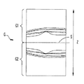
図2はOCT光学系200によって取得(形成)される断層画像の一例を示す図である。断層画像の画像データGは、光路長一致位置より奥側に対応する第1の画像データG1と、光路長一致位置より手前側に対応する第2画像データG2からなり、測定光と参照光の光路長が一致する深度位置Sに関して互いに対称な画像となっている。
FIG. 2 is a diagram illustrating an example of a tomographic image acquired (formed) by the OCT
なお、上記構成において、測定光と参照光との光路長が一致する深度位置が網膜表面より前側に形成されるように参照ミラー31が配置されると、脈絡膜側部分よりも網膜表面側の感度が高い眼底断層像(正像)が取得される。この場合、第1の画像データG1と第2画像データG2における眼底断層像は、向かい合った状態となる。この場合、第1の画像データG1において実像が取得され、第2画像データG2において虚像(ミラーイメージ)が取得される。
In the above configuration, when the
一方、測定光と参照光との光路長が一致する深度位置が網膜表面より奥側に形成されるように参照ミラー31が配置されると、網膜表面側よりも脈絡膜側部分の感度が高い眼底断層像(逆像)が取得される。この場合、第1の画像データG1と第2画像データG2における眼底断層像は、互いに反対方向を向いた状態にある。この場合、第2の画像データG2において実像が取得され、第1の画像データG1において虚像(ミラーイメージ)が取得される。
On the other hand, when the
制御部70は、例えば、断層画像の画像データGのうち、第1の画像データG1もしくは第2画像データG2のいずれかの画像データを抽出し、モニタ75の画面上に表示する。なお、本実施形態では、第1の画像データG1を抽出する設定となっている。
For example, the
本実施形態において、制御部70は、受光素子83から出力されるスペクトルデータに対しソフトウェアによる分散補正処理を施す。そして、分散補正後のスペクトルデータに基づいて深さプロファイルを得る。このため、実像と虚像との間で画質において差異が生じる。
In the present embodiment, the
例えば、実像に対する分散の影響を補正するための分散補正値として第1の分散補正値(正像用)をメモリ72から取得し、受光素子83から出力されるスペクトルデータを第1の分散補正値を用いて補正し、補正されたスペクトル強度データをフーリエ変換して断層画像データを形成する。これにより、第1の画像データG1において実像が取得されたとき、その実像は、高感度・高解像度の画像にて取得され、第1の画像データG1において虚像が取得されたとき、その虚像は、分散補正値の違いにより低解像度のぼやけた像となる。
For example, a first dispersion correction value (for normal image) is acquired from the
もちろん、これに限定されず、虚像に対するソフトウェアの分散補正が行われても良い。また、第2の画像データG2を抽出する設定であってもよいし、もちろん第1の画像データG1と第2の画像データG2の両方を抽出する設定であってもよい。また、所定のスイッチにより任意に設定されてもよい。 Of course, the present invention is not limited to this, and software dispersion correction for virtual images may be performed. Moreover, the setting which extracts the 2nd image data G2 may be sufficient, and of course the setting which extracts both the 1st image data G1 and the 2nd image data G2 may be sufficient. Moreover, you may set arbitrarily with a predetermined | prescribed switch.
<制御動作>
以上のような構成を備える装置において、その制御動作について説明する。ここで、制御部70は、OCT光学系200及びSLO光学系300を駆動制御してOCT画像及びSLO画像の各画像を1フレーム毎に取得していき、モニタ75を表示制御してモニタ75に表示されるOCT画像及びSLO画像を随時更新する。なお、検者の設定によらない最初のOCT画像の取得位置として、SLO画像の中心位置を基準とした走査位置(例えば、X方向)が設定されている。
<Control action>
The control operation of the apparatus having the above configuration will be described. Here, the
図3は、本装置における動作の流れを示すフローチャートである。検者は、図示無き固視標投影ユニットの固視標を注視するように被検者に指示した後、図示無き前眼部観察用カメラで撮影される前眼部観察像をモニタ75で見ながら、被検眼の瞳孔中心に測定光軸がくるように、図示無きジョイスティックを用いて、アライメント操作を行う。このようにして被検眼に対するアライメントが完了されると、SLO光学系300による被検眼眼底の正面画像(SLO眼底像)が取得されるようになり、モニタ75上にSLO眼底像が現れる。
FIG. 3 is a flowchart showing an operation flow in the present apparatus. After instructing the subject to gaze at the fixation target of the fixation target projection unit (not shown), the examiner views an anterior ocular segment observation image photographed by an anterior ocular segment observation camera (not shown) on the
次いで、撮影条件の最適化を行うことによって、OCT光学系200によって、検者が所望する眼底部位が高感度・高解像度で観察できるようにする。なお、本実施形態において、OCT光学系200の最適化の制御は、光路長調整、フォーカス調整、偏光状態の調整(ポラライザ調整)、の制御である。
Next, by optimizing the imaging conditions, the OCT
検者は、コントロール部74に配置された最適化開始スイッチ(Optimizeスイッチ)74aを押す。最適化開始スイッチ74aから操作信号が発せられると、制御部70は、最適化制御を開始するためのトリガ信号を発し、最適化を開始する。
The examiner presses an optimization start switch (Optimize switch) 74 a arranged in the
最適化の完了後、検者により、図示無き撮影スイッチが押されると、眼底断層像が撮影され、メモリ75に記憶される。
After the optimization is completed, when an imaging switch (not shown) is pressed by the examiner, a fundus tomographic image is captured and stored in the
<最適化制御>
図4は、本実施例に係る最適化制御について説明する図である。概して、制御部70は、初期化の制御として、参照ミラー31とフォーカシングレンズ24の位置を初期位置に設定する。初期化完了後、制御部70は、設定した初期位置から参照ミラー31を一方向に所定ステップで移動させ、第1光路長調整を行う(第1自動光路長調整)。また、第1光路長調整と並行するように、制御部70は、受光素子68から出力される受光信号によって取得されるSLO眼底像に基づいて被検眼眼底に対する合焦位置情報を取得する。合焦位置情報が取得されると、制御部70は、フォーカスシングレンズ24を合焦位置に移動させ、オートフォーカス調整(フォーカス調整)を行う。なお、合焦位置とは、観察画像として許容できる断層画像のコントラストを取得できる位置であればよく、必ずしも、フォーカス状態の最適位置である必要はない。
<Optimization control>
FIG. 4 is a diagram for explaining optimization control according to the present embodiment. Generally, the
そして、フォーカス調整完了後、制御部70は、再度、参照ミラー31を光軸方向に移動させ、光路長の再調整(光路長の微調整)をする第2光路長調整を行う。第2光路長調整完了後、制御部70は、参照光の偏光状態を調節するためのポラライザ33を駆動させ、測定光の偏光状態を調整する。
Then, after the focus adjustment is completed, the
以下に、最適化制御の一例について詳細に説明する。 Hereinafter, an example of optimization control will be described in detail.
<評価値>
本実施例において、第1自動光路長調整、ポラライザ調整は、断層画像の信号強度を検出することによって行われる。以下の説明では、信号強度を示す指標として所定の評価値Bが用いられる。
<Evaluation value>
In the present embodiment, the first automatic optical path length adjustment and the polarizer adjustment are performed by detecting the signal intensity of the tomographic image. In the following description, a predetermined evaluation value B is used as an index indicating the signal strength.
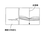
評価値Bは、B=((画像の平均最大輝度値)−(画像の背景領域の平均輝度値))/(背景領域の輝度値の標準偏差)の式より求められる。制御部70は、受光素子83からの出力信号に基づいて取得される断層画像の輝度分布データを取得する。例えば、図5は、参照ミラー、フォーカシングレンズ、ポラライザがある所定の位置に配置されている場合のモニタ75の画面上に表示された画像を示す図である。
The evaluation value B is obtained from the equation B = ((average maximum luminance value of the image) − (average luminance value of the background area of the image)) / (standard deviation of the luminance value of the background area). The
制御部70は、初めに、深さ方向(Aスキャン方向)に走査する複数の走査線を設定し、各走査線上における輝度分布データを求める。図5においては、画像を10分割し、10本の分割線を走査線としている。図6は、画像の深さ方向における輝度分布の変化を示す図である。
First, the
ここで、制御部70は、各走査線に対応する輝度分布から輝度値の最大値(以下、最大輝度値と省略する)を算出する。そして、制御部70は、眼底断層像における最大輝度値として、各走査線における最大輝度値の平均値を算出する。そして、制御部70は、眼底断層像における背景領域の平均輝度値として、各走査線における背景領域の輝度値の平均値を算出する。
Here, the
このようにして、算出された評価値Bは、第1自動光路長調整、ポラライザ調整において利用される。なお、この場合、画像データG1内の断層画像にて、評価値Bを算出することが好ましい。 Thus, the calculated evaluation value B is used in the first automatic optical path length adjustment and the polarizer adjustment. In this case, it is preferable to calculate the evaluation value B in the tomographic image in the image data G1.
<初期化>
初めに、制御部70は、初期化の制御を行う。初期化の制御は、参照ミラー31とフォーカシングレンズ24の位置を初期位置(移動開始位置)に移動させる。
<Initialization>
First, the
そして、初期化の制御が開始されると、制御部70は、移動限界位置K1又は移動限界位置K2のどちらかの位置を参照ミラー31の初期位置として選択する。なお、初期位置の決定は、初期化の制御を開始する以前の参照ミラー31の位置から移動限界位置K1又は移動限界位置K2により近い側の位置が選択される。そして、制御部70は、移動限界位置K1又は移動限界位置K2の初期位置へ参照ミラー31を移動させる。もちろん、異なる基準に基づいて、初期位置に設定するための移動方向の決定を行ってもよい。
When the initialization control is started, the
また、制御部70は、フォーカシングレンズ24を初期位置(本実施形態においては、0Dに対応する位置)へ移動させる。
Further, the
制御部70は、参照ミラー31及びフォーカシングレンズ24を初期位置へ移動させると、第1光路長調整及びフォーカス調整を開始する。以下に、各調整の制御動作について説明する。
When the
<フォーカス調整>
初期化の制御が完了すると、制御部70は、フォーカス調整の制御を開始するためのトリガ信号を発し、OCT光学系200のフォーカス調整を開始する。本実施例において、OCT光学系200のフォーカス調整は、SLO光学系300のフォーカシングレンズ63の合焦位置情報に基づいて行われる。
<Focus adjustment>
When the initialization control is completed, the
初めに、制御部70は、SLO眼底像に対するフォーカス調整を開始する。制御部70は、受光素子68から出力される受光信号によって取得されるSLO眼底像に基づいてSLO光学系300の合焦位置情報を取得し、SLO光学系300に配置されたフォーカシングレンズ63を合焦位置に移動させる(第1フォーカス調整)。
First, the
より具体的には、まず、制御部70は、受光素子68から出力される受光信号に基づいて取得されるSLO眼底像の画像データを微分処理し、微分処理した結果に基づいて微分ヒストグラム情報を取得する。すなわち、制御部70は、SLO光学系300によって取得されたSLO眼底像の画像データにエッジ抽出用(例えば、ラプラシアン変換、SOBEL等)のフィルタを掛けて輪郭画像に変換した後、輪郭画像のヒストグラムを作成する。
More specifically, first, the
図7はSLO光学系300によって取得されるSLO眼底像の画像信号を微分処理した後の微分ヒストグラムの一例を示す図である。図7において、横軸は微分の絶対値(以下、微分値と省略する)d(d=1、2、・・・254)、縦軸は各微分値に対応する画素数H(d)を、画素数がピークを示した微分値における画素数H(dp)で正規化したもの((H(d)/H(dp))を百分率(%)で表記している。なお、図7のヒストグラムにおいては、端点(d=0、d=255)の2点のデータを除外している。ここで、微分値dは、輪郭画像における輝度値を255階調で表したものである。
FIG. 7 is a diagram illustrating an example of a differential histogram after differential processing is performed on the image signal of the SLO fundus image acquired by the SLO
なお、微分ヒストグラムH(d)において、フォーカスが適正な場合、眼底の血管部位におけるエッジが先鋭化されるため、微分値の大きい方の画素数が増加し、フォーカスがずれるに従ってエッジが鈍くなるため、微分値の大きい方の画素数が低下する。 In the differential histogram H (d), when the focus is appropriate, the edge in the blood vessel region of the fundus is sharpened, so the number of pixels having a larger differential value increases, and the edge becomes dull as the focus shifts. As a result, the number of pixels having a larger differential value decreases.
ここで、制御部70は、前述のように取得されたヒストグラム情報において画像全体で所定の割合以上の画素数を持つ輝度値(微分値)の最大値を用いてSLO眼底像の結像状態(フォーカス状態)評価値を算出する。例えば、SLO眼底像の結像状態を評価するための結像状態評価値C1として、閾値S1(例えば、20%)以上での微分値の最大値Dmaxと最小値Dminの差を求める(C1=Dmax−Dmin)。なお、閾値S1は、ノイズによる影響を回避しつつ、SLO眼底像の結像状態の変化に対して評価値C1が敏感に変化するような値に設定される。なお、本実施形態において、閾値S1を20%程度に設定したのは、SLO画像全体に占める範囲の少ない眼底血管部位におけるエッジの先鋭度の変化を精度良く検出するためである。また、上記において、閾値S1以上での微分値の最大値Dmaxのみを結像状態評価値C1として設定するようにしてもよい。
Here, the
結像状態評価値C1は、フォーカシングレンズ63が合焦位置にあるとき(SLO眼底像のフォーカスが合っているとき)に高い値を示し、フォーカシングレンズ63が合焦位置からずれるに従って低くなっていくため、SLO眼底像のフォーカス状態(結像状態)の判定に用いることができる。
The imaging state evaluation value C1 shows a high value when the focusing
ここで、制御部70は、SLO光学系300の受光光学系に配置されたフォーカシングレンズ63の位置を移動させながら結像状態評価値C1をサンプリングし、サンプリング結果により合焦状態を判定し、フォーカシングレンズ63を合焦位置に駆動させる。
Here, the
例えば、制御部70は、適正なフォーカス位置を探索するべく、駆動機構63aを駆動制御して、フォーカシングレンズ63の移動可能範囲において離散的に設定された複数の移動位置にフォーカシングレンズ63を移動させ、各移動位置でのSLO眼底像を取得する。そして、制御部70は、移動位置毎に取得されたSLO眼底像それぞれの微分ヒストグラムを作成し、結像状態評価値C1をそれぞれ算出する。この場合、制御部70は、フォーカシングレンズ63を連続的に移動させていき、連続的に結像状態評価値C1を算出するようにしてもよい。
For example, the
図8は結像状態評価値C1とフォーカシングレンズ63の移動位置Z1との関係を示すグラフの一例を示す図である。図8においては、−12Dに対応する位置から順に、フォーカシングレンズ63を2Dずつプラス方向に移動させ、順次評価値C1を算出していき、+12Dに対応する位置までフォーカシングレンズ63を移動させた場合のものである。
FIG. 8 is a diagram showing an example of a graph showing the relationship between the imaging state evaluation value C1 and the moving position Z1 of the focusing
前述のようにして各フォーカス位置における評価値C1が得られたら、離散的に取得されたフォーカシングレンズ63の移動位置Z1と評価値C1の特性に対して補間処理を施し、SLO光学系300の合焦位置を検出する。例えば、フォーカシングレンズ63の移動範囲に極大値を持つような関数にて曲線近似し、この曲線において評価値C1が最大となる移動位置Z1pをSLO光学系300の合焦位置情報として取得する。なお、上記のような補間処理によってSLO光学系300の合焦位置を検出する手法としては、関数近似、重心、平均値の算出等を用いたものが考えられる。
When the evaluation value C1 at each focus position is obtained as described above, interpolation processing is performed on the characteristics of the moving position Z1 of the focusing
制御部70は、駆動機構63aを駆動制御して、前述のように取得された合焦位置情報に対応する移動位置にフォーカシングレンズ63を移動させることによりSLO眼底像に対するフォーカス調整を終了する。
The
次に、制御部70は、第1フォーカス調整の制御によるSLO光学系300の合焦位置情報に基づいてOCT光学系200のフォーカシングレンズ24を移動させる(第2フォーカス調整)。
Next, the
制御部70は、第1のオートフォーカス制御によるSLO光学系300の合焦位置情報に基づいてOCT光学系200のフォーカス位置情報を取得し、フォーカシングレンズ24を合焦位置まで移動させる(OCT画像に対するオートフォーカス)。ここで、制御部70は、第1フォーカス調整によるフォーカシングレンズ63の移動位置をOCT光学系200のフォーカス位置情報として取得し、そのフォーカス位置情報に基づいて駆動機構24aを駆動制御してフォーカシングレンズ24を合焦位置まで移動させる。
The
例えば、SLO光学系300の合焦位置が−3Dに対応する位置であれば、OCT光学系200のフォーカス位置も同様に−3Dに対応する位置になるように制御する。この場合、OCT光学系200のフォーカス位置をSLO光学系200の合焦位置に対応するフォーカス位置に設定できるように、フォーカシングレンズ63の移動位置とフォーカシングレンズ24の移動位置との間でディオプター換算による対応づけがなされている。
For example, if the focus position of the SLO
このようにしてOCT光学系200のフォーカシングレンズ24がSLO光学系300の合焦位置に対応する移動位置に移動されると、ファイバー端部39bに入射される眼底反射光が増加する。
When the focusing
<第1自動光路長調整(粗調整)>
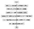
制御部70は、合焦位置の検出動作、及び検出された合焦位置へのフォーカシングレンズ63の移動動作と並行して、第1自動光路長調整(自動粗光路長調整)を行う。図9は、第1自動光路長調整の制御動作の流れを示すフローチャートである。
<First automatic optical path length adjustment (coarse adjustment)>
The
制御部70は、駆動機構50の駆動を制御して参照ミラー31を移動させると共に、参照ミラー31の各位置にて受光素子83から出力される出力信号に基づいて、眼底断層像が取得される位置に参照ミラー31を移動させる。
The
具体的には、制御部70は、初期位置にて断層画像を取得した後、初期位置とは逆の移動限界位置に向けて参照ミラー31を移動させる。例えば、参照ミラー31の初期位置として限界位置K1が選択(設定)された場合、限界位置K2に向けて方向へ移動させる。
Specifically, after acquiring the tomographic image at the initial position, the
ここで、制御部70は、参照ミラー31を所定のステップ(例えば、撮影範囲として2mmステップ)で移動させ、各移動位置における断層画像を順次取得していき、眼底断層像が取得される位置を探索していく。
Here, the
この場合、制御部70は、離散的に設定された参照ミラー31の移動位置において、参照ミラー31が停止される度に断層像を取得する。そして、制御部70は、各位置にて取得される断層画像を解析する。例えば、制御部70は、各位置にて取得される断層像の評価値Bを算出する。そして、制御部70は、参照ミラー31の位置と断層像の評価値Bとを対応付けてメモリ75に記憶する。
In this case, the
図10は、参照ミラー31の位置ごとにおける評価値Bの算出結果の一例を示す図である。横軸は、参照ミラーの位置、縦軸は、参照ミラーの各位置における評価値Bを表記したものである。
FIG. 10 is a diagram illustrating an example of a calculation result of the evaluation value B for each position of the
ここで、制御部70は、取得された参照ミラー31の位置ごとにおける評価値Bの算出結果から、評価値Bのピークを検出する。そして、制御部70は、ピークの検出位置に対応する参照ミラー31の位置をメモリ75に記憶させる。そして、制御部70は、評価値Bのピークに対応する位置へ参照ミラー31を移動させる。なお、一般的には、眼底の実像が断層画像中に現れるときの参照ミラー31の位置が、評価値Bのピークが検出される位置となる。ただし、フォーカスがあっていない場合においては、虚像が断層画像中に現れるときの参照ミラー31の位置が、評価値Bのピークが検出される位置となる場合もありえる。
Here, the
なお、本実施例において、第1自動光路長調整とフォーカス調整を並行して行っているため、第1自動光路長調整中にフォーカス調整が完了した場合、第1自動光路長に用いる断層画像のフォーカス状態が向上する。このため、第1自動光路長調整の前後で、評価値Bが変化し、ピークの検出位置が変化する場合がある。この場合においても、第1自動光路長調整では、モニタ72上のいずれかの位置に眼底断層像の少なくとも一部が表示された状態となればよいため、必ずしもピーク位置が適切に検出される必要はない。すなわち、ラフに光路長調整が行わればよいため、ピーク検出精度は、必ずしも高くなくてよい。
In this embodiment, since the first automatic optical path length adjustment and the focus adjustment are performed in parallel, when the focus adjustment is completed during the first automatic optical path length adjustment, the tomographic image used for the first automatic optical path length is adjusted. The focus state is improved. For this reason, the evaluation value B may change before and after the first automatic optical path length adjustment, and the peak detection position may change. Even in this case, in the first automatic optical path length adjustment, it is sufficient that at least a part of the fundus tomographic image is displayed at any position on the
以上のようにして光路長がラフに調整されると、モニタ72上のいずれかの位置に眼底断層像の少なくとも一部が表示された状態となる。
When the optical path length is roughly adjusted as described above, at least a part of the fundus tomographic image is displayed at any position on the
なお、本実施形態においては、参照ミラー31を所定のステップで移動させる場合、評価値Bの上昇がなくなり、下降をはじめた位置で、参照ミラー31の駆動を停止するようにしてもよい。また、制御部70は、参照ミラー31の位置ごとにおける評価値Bの算出結果からピークに対応する参照ミラーの位置を推測するようにしてもよい。(例えば、評価値Bの変化を示す近似曲線を作成する)。
In the present embodiment, when the
<第2自動光路長調整(微調整)>
図11は、第2自動光路長調整の制御動作の流れを示すフローチャートである。
<Second automatic optical path length adjustment (fine adjustment)>
FIG. 11 is a flowchart showing the flow of the control operation of the second automatic optical path length adjustment.
制御部70は、フォーカシングレンズ63が合焦位置に移動されると、第2自動光路長調整を開始する。制御部70は、受光素子83から出力される出力信号に基づいて、第1自動光路長調整によって調整された位置から参照ミラー31の位置を再調整する。
The
具体的には、フォーカス調整が完了すると、制御部70は、フォーカス調整によって取得された断層画像に基づいて、参照ミラー31を移動させる第2自動光路長調整を行う。
Specifically, when the focus adjustment is completed, the
ここで、制御部70は、画像データG1において、フォーカス調整後に取得された眼底断層像が実像か虚像かを判定する。例えば、制御部70は、深さ方向での輝度分布におけるピークに対する半値幅が所定の許容幅より小さいとき、眼底眼底像を実像と判定し、半値幅が所定の許容幅が大きいとき、眼底断層像を虚像と判定する。なお、断層像の実虚の判定については、実像と虚像との間の画質の差異が利用される手法であればよく、半値幅の他、例えば、断層像のコントラスト、断層像のエッジの立ち上がり度等が利用される。また、眼底断層像の形状が利用されてもよい。
Here, the
制御部70は、取得される眼底断層像が虚像と判定された場合、実像が取得される方向(参照光が短くなる方向)に向けて参照ミラー31を移動させる。例えば、制御部70は、光路長一致位置Sから像検出位置までの偏位量をゼロにする参照ミラー31の移動量を算出し、さらに算出された移動量の2倍分参照ミラー31を移動させる。これにより、実像のみが取得された状態となる。この場合、参照ミラー31が一定量移動されたときの偏位量を予め求めておけばよい。これにより、制御部70は、光路長断層像の深度位置から像検出位置までの偏位量が所定の偏位量となるように参照ミラー31を移動させることが可能となり、眼底断層像を所定の表示位置に表示できる。
When the acquired fundus tomographic image is determined to be a virtual image, the
なお、参照ミラー31を移動させる手段はこれに限定されるものではない。例えば、虚像と判定された場合に、予め、参照ミラー31を実像が取得される方向(参照光が短くなる方向)に向けて移動させる所定のオフセット量を設定しておく。そして、制御部70は、眼底断層像が虚像と判定された場合、参照ミラー31を所定のオフセット量分移動させる。
The means for moving the
また、取得される眼底断層像が実像と判定された場合、制御部70は、実像の位置を判定する。例えば、制御部70は、深さ方向における輝度分布のピークが検出された位置を像位置とみなし、予め設定された光路長調整位置と像位置との変位量を算出し、その変位量がなくなるように参照ミラー31を移動させる(特開2010−12111号公報参照)。
When the acquired fundus tomographic image is determined to be a real image, the
制御部70は、上記のように画像データG1の断層像に対する実虚の判定を行うと共に、さらに、画像データG1において実像と虚像が並存するか否かを並行して判定するのが好ましい。例えば、制御部70は、前述のように算出される各走査線における最大輝度値の検出位置の平均位置を眼底断層像の像位置P1として検出する。そして、制御部70は、測定光と参照光の光路長が一致する深度位置S(第1の画像データの上端位置)から像検出位置P1までの偏位量を算出する。すなわち、制御部70は、測定光と参照光の光路長が一致する深度位置Sを基準に眼底断層像の像位置を検出する。
As described above, the
そして、制御部70は、前述のように算出される眼底断層像の像位置P1が断層画像の上端付近(例えば、断層画像の上端から1/4に相当する領域)にある場合、眼底断層像の実像と虚像が並存している状態であると判定する。この場合、制御部70は、実像のみが取得される方向(参照光が短くなる方向)に向けて参照ミラー31を所定量移動させる。この場合、実像と虚像が並存している状態から実像のみが取得された状態となるまでの参照ミラー31の移動方向及び移動量を実験もしくはシミュレーションにより予め求めておき、メモリ72に記憶しておけばよい。
Then, when the image position P1 of the fundus tomographic image calculated as described above is in the vicinity of the upper end of the tomographic image (for example, an area corresponding to ¼ from the upper end of the tomographic image), the
<第1自動光路長調整とフォーカス調整のタイミング>
ここで、第2自動光路長調整の制御を開始する際に、第1自動光路長調整とフォーカス調整の完了のタイミングが異なる場合がある。例えば、第1自動光路長調整がフォーカス調整よりも早く完了する場合、第1自動光路長調整がフォーカス調整よりも遅く完了する場合、第1自動光路長調整とフォーカス調整が同時に完了する場合がある。
<Timing of first automatic optical path length adjustment and focus adjustment>
Here, when the control of the second automatic optical path length adjustment is started, the completion timing of the first automatic optical path length adjustment and the focus adjustment may be different. For example, when the first automatic optical path length adjustment is completed earlier than the focus adjustment, when the first automatic optical path length adjustment is completed later than the focus adjustment, the first automatic optical path length adjustment and the focus adjustment may be completed simultaneously. .
第1自動光路長調整がフォーカス調整よりも早く完了する場合において、第1自動光路長調整後に、フォーカス調整をしている間に、眼が動いてしまう等の原因によって、断層画像が表示されなくなる場合がある。この状態で、第2自動光路長調整を開始すると、第2自動光路長調整が失敗してしまう。 In the case where the first automatic optical path length adjustment is completed earlier than the focus adjustment, the tomographic image is not displayed due to a cause such as an eye moving during the focus adjustment after the first automatic optical path length adjustment. There is a case. If the second automatic optical path length adjustment is started in this state, the second automatic optical path length adjustment will fail.
また、第1自動光路長調整がフォーカス調整よりも遅く完了する場合において、第1自動光路長調整が完了する前に、フォーカス調整が完了してしまい、第2自動光路長調整を開始してしまう可能性がある。この場合、モニタ72上のいずれかの位置に眼底断層像の少なくとも一部が表示される前に、第2自動光路長調整を開始してしまうため、第2自動光路長調整が失敗してしまう。
Further, when the first automatic optical path length adjustment is completed later than the focus adjustment, the focus adjustment is completed before the first automatic optical path length adjustment is completed, and the second automatic optical path length adjustment is started. there is a possibility. In this case, since the second automatic optical path length adjustment is started before at least a part of the fundus tomographic image is displayed at any position on the
このため、制御部70は、第1自動光路長調整完了のタイミングとフォーカス調整完了のタイミングを検出し、第1自動光路長調整とフォーカス調整が同時に完了する場合を除いて、以下のような制御を行う。
For this reason, the
図12は、第1自動光路長調整とフォーカス調整が同時に完了しなかった場合における第2自動光路長調整の制御動作の流れを示すフローチャートである。 FIG. 12 is a flowchart showing the flow of the control operation of the second automatic optical path length adjustment when the first automatic optical path length adjustment and the focus adjustment are not completed simultaneously.
フォーカス調整が完了すると、制御部70は、第1自動光路長調整の適否を判定する。判定は、例えば、所定の閾値を設定しておき、検出値(例えば、評価値Bや輝度値等)が閾値を越えたか否かによって判定する。
When the focus adjustment is completed, the
制御部70は、調整が成功したと判定した場合、フォーカス調整後に取得された眼底断層像が実像か虚像かを判定する。制御部70は、上記記載と同様にして、取得される眼底断層像が虚像と判定された場合、実像が取得される方向(参照光が短くなる方向)に向けて参照ミラー31を移動させる。取得される眼底断層像が実像と判定された場合、制御部70は、実像の位置を判定する。例えば、制御部70は、深さ方向における輝度分布のピークが検出された位置を像位置とみなし、予め設定された光路長調整位置と像位置との変位量を算出し、その変位量がなくなるように参照ミラー31を移動させる。
When it is determined that the adjustment is successful, the
また、制御部70は、調整が失敗したと判定した場合、第1自動光路長調整の制御を再度行う。このとき、第1自動光路長調整が失敗するたびに最適化の制御を停止させてもよいし、数回第1自動光路長調整の制御が失敗した場合に、最適化の制御を停止させてもよい。また、第1自動光路長調整を再度行う際の参照ミラー31の初期位置(光路長調整用光学部材)は、移動限界位置K1又は移動限界位置K2に移動させてもよいし、先の第1自動光路長調整で行われた際の評価値Bの結果に基づいて設定してもよい。さらに、初期位置より参照ミラー31を移動させる際の移動方向においても、先の第1自動光路長調整で行われた際の評価値Bの結果に基づいて行われてもよい。
Further, when it is determined that the adjustment has failed, the
また、制御部70は、第1自動光路長調整完了時の参照ミラー31(光路長調整用光学部材)の位置から眼底断層像を探索するようにしてもよい。これにより、参照ミラー31が移動限界位置K1又は移動限界位置K2までに移動される時間が短縮される。例えば、制御部70は、完了時の位置が移動限界位置K1又は移動限界位置K2のどちらに近いかを判定し、近いと判定された限界位置に向けて参照ミラー31を移動させていき、各位置にて取得された断層画像に基づいて眼底断層像が取得される位置を探索する。なお、眼底断層像がなければ、他方の移動限界位置に向けて参照ミラー31を移動させていき、眼底断層像が取得される位置を探索する。
Further, the
また、最適化が失敗した際には、最適化が失敗した表示をモニタ75上に表示する等して、検者に再最適化を行うか否かを選択させる構成としてもよい。 Further, when the optimization fails, a configuration may be adopted in which the examiner selects whether or not to perform the re-optimization by displaying on the monitor 75 a display indicating that the optimization has failed.
制御部70は、第1自動光路長調整を再度調整した後、第1自動光路長調整の調整が成功したと判定すると、取得された眼底断層像が実像か虚像かを判定する。その後、実像か虚像かの判定結果に応じて、上記と同様の制御を行う。
If the
なお、制御部70は、第1自動光路長調整の適否を判定し、調整が失敗したと判定した場合、第1自動光路長調整とともにフォーカス調整を再度行うようにしてもよい。
Note that the
なお、フォーカス調整後に取得された眼底断層像が実像か虚像の判定及び眼底断層像の有無の判定は、どちらを先に行ってもよいし、同時に行うようにしてもよい。もちろん、一方の判定結果より、再度調整を行うような構成としてもよい。 The determination of whether the fundus tomogram acquired after focus adjustment is a real image or a virtual image and the determination of the presence or absence of a fundus tomogram may be performed first or simultaneously. Of course, it is good also as a structure which adjusts again from one determination result.
以上のように、第1自動光路長調整とフォーカス調整を並行して行った後、第2自動光路長調整を行うという手順で最適化の制御を動作させる。このとき、SLO光学系300を用いて、フォーカス調整を行い、OCT光学系200を用いて第1自動光路長調整を行っているため、フォーカス調整と第1自動光路長調整を並行して行うことができる。
As described above, after performing the first automatic optical path length adjustment and the focus adjustment in parallel, the optimization control is operated in the procedure of performing the second automatic optical path length adjustment. At this time, since the focus adjustment is performed using the SLO
また、フォーカス調整前の光路長調整では、画像から検出される輝度が弱いために、実像・虚像の判定は行うことが困難であるが、フォーカスが調整されていない状態でも、眼底断層画像より眼底断層像が取得される位置を検出することができる。すなわち、フォーカス調整が完了していない状態では、第2光路長調整は困難であるが、第1光路長調整は、行うことができる。これによって、フォーカス調整がするまで、第1光路長自動調整の開始を待つ必要がなくなり、短時間でスムーズに最適化の制御を行うことができる。すなわち、撮影条件を好適に調整することができる。 In addition, in the optical path length adjustment before the focus adjustment, it is difficult to determine a real image or a virtual image because the luminance detected from the image is weak. However, even if the focus is not adjusted, the fundus is more than the fundus tomographic image. The position where the tomographic image is acquired can be detected. That is, in the state where the focus adjustment is not completed, the second optical path length adjustment is difficult, but the first optical path length adjustment can be performed. Accordingly, it is not necessary to wait for the start of the first automatic optical path length adjustment until the focus adjustment is performed, and the optimization control can be smoothly performed in a short time. That is, it is possible to suitably adjust the shooting conditions.
<ポラライザ調整>
制御部70は、第2自動光路長調整後に受光素子83から出力される出力信号に基づき、ポラライザ33を駆動させ、偏光状態の調整を行う。
<Polarizer adjustment>
The
具体的には、制御部70は、ポラライザ33の位置を初期位置より、移動開始位置に移動させる。なお、ポラライザ33の初期位置は、第1移動限界位置から第2移動限界位置までの間の途中の位置に配置されている。なお、ポラライザ調整の際の、ポラライザ33の移動開始位置は、第1移動限界位置又は第2移動限界位置の位置となる。
Specifically, the
制御部70は、ポラライザ33を途中位置から第1移動限界位置又は第2移動限界位置のどちらかの移動開始位置を選択し、移動させる。例えば、制御部70は、第1移動限界位置を移動開始位置として選択し、ポラライザ33を移動させる。そして、制御部70は、ポラライザ33を第1移動限界位置から第2移動限界位置方向へ移動させる。なお、移動開始位置が第2移動限界位置の場合には、第1移動限界位置方向へ移動させる。そして、各移動位置におけるモニタ75の画面上の画像を順次取得していき、干渉光が強く受光できる位置(測定光と参照光の偏光状態が合う位置)を探索していく。
The
偏光状態が合う位置の探索は、離散的に設定されたポラライザ33の移動位置でポラライザ33が停止される度に、その配置位置にて取得される画像を解析し、評価値Bの算出を行う。
The search for the position where the polarization state matches is performed by analyzing the image acquired at the arrangement position and calculating the evaluation value B every time the
制御部70は、移動開始位置とは、逆の移動限界位置まで、5°ずつポラライザ33を移動させていく。なお、本実施形態では、5°ずつポラライザ33を移動させる構成としたが、これに限定されない。例えば、10°でもよいし、20°でもよく、任意に設定できる構成でもよい。
The
ここで、制御部70は、取得されたポラライザ33の位置ごとにおける評価値Bの算出結果から、ピークとなる評価値B(ピーク値)を検出し、ピーク値が検出された位置に対応する位置へポラライザ33を移動させる。以上のようにして、ポラライザ調整が完了される。
Here, the
以上のようにして、最適化の制御が完了されることにより、検者が所望する眼底部位が高感度・高解像度で観察できるようになる。 As described above, when the optimization control is completed, the fundus site desired by the examiner can be observed with high sensitivity and high resolution.
<変容例>
なお、本実施例においては、被検眼眼底の正面画像を取得するための観察光学系としてSLO光学系300を用い、SLO光学系300によって取得される正面画像に基づいてOCT光学系200の合焦位置を調整するものとしたが、これに限定されない。観察光学系としては、被検眼に赤外光を照射し、被検眼からの反射光を受光する受光素子を有し、受光素子からの受光信号に基づいて被検眼の正面画像を得るものが挙げられる。例えば、眼底カメラが挙げられる。この場合、眼底カメラによって撮像された赤外眼底像に基づいて眼底カメラ光学系の合焦位置情報を取得する(例えば、前述のSLO眼底像に基づく合焦位置検出手法の適用が考えられる)。
<Transformation example>
In this embodiment, the SLO
なお、合焦位置情報の取得方法は、上記手法に限定されない。眼底反射光を受光する受光素子から出力される受光信号に基づいて合焦位置情報が取得されるものであればよい。例えば、被検眼眼底にフォーカス用の指標(例えば、スプリット指標)を投影する投影光学系を設け、その眼底反射光による指標像(眼底反射像)を受光素子により受光し、受光素子から出力される受光信号に基づいて合焦位置情報を取得するようにしてもよい。 In addition, the acquisition method of focusing position information is not limited to the said method. Any in-focus position information may be acquired based on a light reception signal output from a light receiving element that receives fundus reflection light. For example, a projection optical system that projects a focus index (for example, a split index) on the fundus of the subject's eye is provided, and an index image (fundus reflection image) of the fundus reflected light is received by the light receiving element and output from the light receiving element. Focus position information may be acquired based on the received light signal.
なお、本実施例において、OCT光学系200からの眼底反射光を用いて、フォーカス調整を行う構成としてもよい。例えば、OCT光学系200における測定光の眼底反射光の信号を一部抽出し、抽出結果を検出する検出器を設ける。そして、検出器より検出された信号強度より、フォーカス調整を行う。例えば、フォーカス調整は、検出器から検出される信号強度がピークとなるようにフォーカス調整を行う。
In this embodiment, focus adjustment may be performed using fundus reflected light from the OCT
なお、本実施例において、第1自動光路長調整では、評価値Bに基づいて、光路長調整を行う構成としたがこれに限定されない。例えば、第2自動光路長調整のように実像と虚像の判定を行い、光路長調整を行ってもよい。この場合、第1自動光路長調整とフォーカス調整が並行して行われているため、フォーカス調整が行われていくため、実像及び虚像の判定ができるようになる。なお、第1自動光路長調整では、フォーカス調整が完了していないため、光路長の微調整を行うことができない。すなわち、フォーカス調整後、第2自動光路長調整にて、眼底断層像の像位置を検出し、実像のみが取得される方向に向けて参照ミラー31を所定量移動させ、光路長の微調整を行う。
In the present embodiment, the first automatic optical path length adjustment is configured to adjust the optical path length based on the evaluation value B, but is not limited thereto. For example, a real image and a virtual image may be determined as in the second automatic optical path length adjustment, and the optical path length adjustment may be performed. In this case, since the first automatic optical path length adjustment and the focus adjustment are performed in parallel, the focus adjustment is performed, so that a real image and a virtual image can be determined. In the first automatic optical path length adjustment, since the focus adjustment is not completed, the optical path length cannot be finely adjusted. That is, after the focus adjustment, the image position of the fundus tomographic image is detected by the second automatic optical path length adjustment, the
また、第2自動光路長調整では、実虚判定に基づいて、光路長調整を行う構成としたがこれに限定されない。例えば、第1自動光路長調整のように評価値Bに基づいて、光路長調整を行ってもよい。この場合、第1自動光路長調整では、参照ミラーを移動する際の所定ステップを長く(例えば、5mm)とり、第2自動光路長調整では、参照ミラーを移動する際の所定ステップを短く(例えば、2mm)とるようにしてもよい。これによって、第1自動光路長調整では、短時間で調整を行い、第2自動光路長調整では、精度よく調整を行うことができる。もちろん、所定ステップの間隔を任意に変更可能な構成としてもよい。 In the second automatic optical path length adjustment, the optical path length adjustment is performed based on the true / false determination, but the present invention is not limited to this. For example, the optical path length may be adjusted based on the evaluation value B as in the first automatic optical path length adjustment. In this case, in the first automatic optical path length adjustment, a predetermined step for moving the reference mirror is lengthened (for example, 5 mm), and in the second automatic optical path length adjustment, the predetermined step for moving the reference mirror is shortened (for example, 2 mm). Thus, the first automatic optical path length adjustment can be adjusted in a short time, and the second automatic optical path length adjustment can be adjusted with high accuracy. Needless to say, the predetermined step interval may be arbitrarily changed.
なお、本実施例において、SLO光学系300を用いて、フォーカス調整を完了させる構成としたがこれに限定されない。制御部70は、第1フォーカス調整の制御によるSLO光学系300の合焦位置情報に基づいてOCT光学系200のフォーカシングレンズ24を移動させる。その後、さらに、OCT光学系200によって取得される断層画像に基づいてOCT光学系200の合焦位置情報を取得し、フォーカシングレンズ24を合焦位置に移動させるようにしてもよい。これによって、フォーカス調整がより精度よく行える。
In this embodiment, the SLO
なお、以上の説明においては、最適化制御動作の際に、一部の最適化制御(第1光路長調整の適否の判定)を除いて、前の調整が完了後に次の調整に移行する構成としたがこれに限定されない。例えば、制御部70により、断層画像の輝度情報に基づいて、最適化の調整が成功したか否かを判定し、判定結果に基づいて最適化の調整を停止させるようにしてもよい。制御部70は、調整が失敗したと判定した場合、再び最適化の制御をやり直しさせる。このとき、最適化制御が失敗するたびに最適化の制御を停止させてもよいし、数回最適化制御が失敗した場合に、最適化の制御を停止させてもよい。また、最適化が失敗した際には、最適化が失敗した表示をモニタ75上に表示する等して、検者に再最適化を行うか否かを選択させる構成としてもよい。
In the above description, in the optimization control operation, except for a part of optimization control (determination of the suitability of the first optical path length adjustment), the configuration is shifted to the next adjustment after the previous adjustment is completed. However, it is not limited to this. For example, the
なお、制御部70が失敗したと判定した場合、最適化の制御のやり直しを行わせる前に、例えば、偏光状態が変化するような所定角度(例えば、90°)だけポラライザ33を回転駆動させるようにしてもよい。これによって、眼底断層像の受光される状態が変化し、再最適化が行われた場合に、最適化の制御が可能となる場合がある。
If it is determined that the
なお、以上の説明においては、最適化の制御動作として、光路長調整、フォーカス調整、ポラライザ調整を行ったがこれに限定されない。例えば、ポラライザ調整を除いてもよい。この場合、最適化にかかる時間が少なくなるが、眼底断層像の感度及び解像度は、低くなる。 In the above description, the optical path length adjustment, the focus adjustment, and the polarizer adjustment are performed as the optimization control operation. However, the present invention is not limited to this. For example, the polarizer adjustment may be omitted. In this case, the time required for optimization is reduced, but the sensitivity and resolution of the fundus tomographic image are reduced.
なお、以上の説明においては、最適化の制御動作として、第1自動光路長調整、フォーカス調整、第2自動光路長調整、ポラライザ調整の順で行われるようにしたがこれに限定されない。例えば、第1自動光路長調整及びフォーカス調整完了後から第2自動光路長調整の間に、ポラライザ調整を行ってもよい。 In the above description, the optimization control operation is performed in the order of the first automatic optical path length adjustment, the focus adjustment, the second automatic optical path length adjustment, and the polarizer adjustment, but is not limited thereto. For example, the polarizer adjustment may be performed between the completion of the first automatic optical path length adjustment and the focus adjustment and the second automatic optical path length adjustment.
なお、本実施例において、第2光路長調整の前後でフォーカス調整を行ってもよい。この場合、制御部70は、第1フォーカス調整は、第2光路長調整による光路長の微調整が可能な程度に粗く調整を行い、第2光路長調整による光路長の微調整完了後、第2フォーカス調整にて、フォーカスを一致させるようにしてもよい。
In this embodiment, focus adjustment may be performed before and after the second optical path length adjustment. In this case, the
なお、本実施例においては、フォーカス調整が完了するとともに、第2自動光路長調整が開始される構成としたがこれに限定されない。例えば、フォーカス調整及び第1自動光路長調整が完了するとともに、第2自動光路長調整が行われる構成としてもよい。 In this embodiment, the focus adjustment is completed and the second automatic optical path length adjustment is started. However, the present invention is not limited to this. For example, the focus adjustment and the first automatic optical path length adjustment may be completed and the second automatic optical path length adjustment may be performed.
なお、以上の説明においては、断層画像における輝度分布を利用して眼底断層像の実像/虚像の判定を行うものとしたが、眼底断層像の実像が取得されたときの断層画像の断面形状と眼底断層像の虚像が取得されたときの断層画像における断面形状とを比較し、その比較結果を考慮して実像/虚像の判定が可能な判定条件を設定するようにしてもよい。例えば、実像と虚像が深さ方向に対称な画像であることを利用する。より具体的には、眼底断層像の第1の画像データG1から網膜色素上皮部分を画像処理(例えば、網膜色素上皮の輝度値に対応するような所定の閾値を超える輝度値のデータを抽出する)により抽出し、抽出された網膜色素上皮部分の曲線形状に基づいて実像/虚像を判定するようにしてもよい。なお、光学部材を用いて分散補正を行う構成のものに対しても適用可能である。もちろん、光学的な分散補正とソフトウェアによる分散補正を組み合わせたものに対しても適用可能である。 In the above description, the real / virtual image of the fundus tomographic image is determined using the luminance distribution in the tomographic image. However, the cross-sectional shape of the tomographic image when the real image of the fundus tomographic image is acquired The cross-sectional shape in the tomographic image when the virtual image of the fundus tomographic image is acquired may be compared, and a determination condition capable of determining a real image / virtual image may be set in consideration of the comparison result. For example, the fact that the real image and the virtual image are symmetrical in the depth direction is used. More specifically, image processing is performed on the retinal pigment epithelium from the first image data G1 of the fundus tomogram (for example, data having a luminance value exceeding a predetermined threshold value corresponding to the luminance value of the retinal pigment epithelium is extracted. The real image / virtual image may be determined based on the extracted curved shape of the retinal pigment epithelium. Note that the present invention can also be applied to a configuration in which dispersion correction is performed using an optical member. Of course, the present invention can also be applied to a combination of optical dispersion correction and software dispersion correction.
なお、以上の説明においては、フーリエ変換後の深さプロファイルに基づいて各撮像条件が調整されたが、これに限定されない。すなわち、検出器から出力される出力信号に基づいて各撮像条件が調整さればよい。例えば、フーリエ変換前のスペクトルデータが用いられてもよい。 In the above description, each imaging condition is adjusted based on the depth profile after Fourier transform, but the present invention is not limited to this. That is, each imaging condition may be adjusted based on the output signal output from the detector. For example, spectral data before Fourier transform may be used.
なお、上記説明において、スペクトルメータを用いたスペクトルドメインOCTを例にとって説明したが、これに限定されない。例えば、波長可変光源を備えるSS−OCT(Swept source OCT)であってもよい。 In the above description, the spectrum domain OCT using a spectrum meter has been described as an example, but the present invention is not limited to this. For example, SS-OCT (Swept source OCT) provided with a wavelength variable light source may be used.
23 走査部
24 フォーカシングレンズ
24a 駆動機構
31 参照ミラー
33 ポラライザ
34 駆動機構
50 駆動機構
63 フォーカシングレンズ
63a 駆動機構
68 受光素子
70 制御部
72 メモリ
75 表示モニタ
83 検出器
200 干渉光学系
300 観察光学系
DESCRIPTION OF
Claims (4)
を備え、前記検出器からの出力信号に基づいて被検眼眼底の断層画像を撮像する眼底撮影装置であって、
被検眼眼底に対する合焦位置を検出するためのフォーカス検出手段と、前記測定光の光路中に配置された第1光学部材を駆動させる第1駆動手段とを備え、前記フォーカス検出手段によって検出された合焦位置に前記第1光学部材を移動させるフォーカス調整手段と、
測定光と参照光との光路長差を調整するために測定光又は参照光の光路中に配置された第2光学部材を駆動させる第2駆動手段を有し、前記検出器から出力される出力信号に基づいて被検眼断層像が取得される位置に前記第2光学部材を移動させる光路長調整手段と、
前記フォーカス検出手段によって被検眼眼底に対する合焦位置を検出し、検出された合焦位置に前記第1光学部材を移動させる前記フォーカス調整手段の作動中において、
前記検出器によって測定光と参照光との干渉状態を検出し、前記検出器から出力される出力信号に基づいて被検眼断層像が取得される位置に前記第2光学部材を移動させる前記光路長調整手段の作動を並行して行うことを特徴とする眼底撮影装置。 The light beam emitted from the light source is divided into measurement light and reference light, the measurement light beam is guided to the fundus of the eye to be examined, the reference light is guided to the reference optical system, and then the measurement light reflected from the fundus of the eye to be examined and the reference light An interference optical system for detecting an interference state by a detector;
A fundus imaging apparatus for capturing a tomographic image of the fundus oculi based on an output signal from the detector,
A focus detection unit for detecting a focus position with respect to the fundus of the eye to be examined; and a first drive unit for driving a first optical member disposed in the optical path of the measurement light, the detection being performed by the focus detection unit Focus adjusting means for moving the first optical member to a focus position;
A second driving means for driving the second optical member disposed in the optical path of the measuring beam or the reference beam in order to adjust the optical path length difference between the measuring beam and the reference beam, the output outputted from the detector An optical path length adjusting means for moving the second optical member to a position where an eye tomographic image to be examined is acquired based on a signal;
During the operation of the focus adjusting means for detecting the focus position with respect to the fundus of the eye to be examined by the focus detection means and moving the first optical member to the detected focus position ,
The optical path length that detects an interference state between the measurement light and the reference light by the detector and moves the second optical member to a position where an eye tomographic image to be examined is acquired based on an output signal output from the detector A fundus photographing apparatus characterized in that the adjustment means are operated in parallel .
前記フォーカス検出手段は、被検眼眼底に照明光を照射する照射光学系と、眼底反射光を受光素子により受光する受光光学系と、前記受光光学系に配置された第3光学部材を駆動させる第3駆動手段と、を有し、前記受光素子からの出力信号に基づいて被検眼眼底の正面眼底画像を得る眼底観察光学系を兼用し、前記第3駆動手段の駆動を制御して前記第3光学部材を移動させ、前記第3光学部材の各位置にて取得された正面眼底画像に基づいて被検眼眼底に対する前記合焦位置を検出することを特徴とする眼底撮影装置。 The fundus imaging apparatus according to claim 1,
The focus detection unit drives an irradiation optical system for irradiating the fundus of the eye to be examined with illumination light, a light receiving optical system for receiving fundus reflected light by a light receiving element, and a third optical member disposed in the light receiving optical system. 3, and a third funding observation optical system that obtains a front fundus image of the fundus of the eye based on an output signal from the light receiving element, and controls the driving of the third driving unit to control the third driving unit. An eye fundus photographing apparatus, wherein an in-focus position with respect to a fundus to be examined is detected based on a front fundus image acquired at each position of the third optical member by moving an optical member.
を備え、前記検出器からの出力信号に基づいて被検眼眼底の断層画像を撮像する眼底撮影装置であって、
被検眼眼底に対する合焦位置を検出するためのフォーカス検出手段と、前記測定光の光路中に配置された第1光学部材を駆動させる第1駆動手段とを備え、前記フォーカス検出手段によって検出された合焦位置に前記第1光学部材を移動させるフォーカス調整手段と、
測定光と参照光との光路長差を調整するために測定光又は参照光の光路中に配置された第2光学部材を駆動させる第2駆動手段を有し、前記フォーカス検出手段の作動と並行して、前記検出器から出力される出力信号に基づいて被検眼断層像が取得される位置に前記第2光学部材を移動させる光路長調整手段と、
を備え、
前記光路長調整手段は、前記フォーカス検出手段の作動と並行して、前記検出器から出力される出力信号に基づいて被検眼断層像が取得される位置に前記第2光学部材を移動させ、前記フォーカス調整手段によって前記第1光学部材が前記合焦位置に移動された後、前記検出器から出力される出力信号に基づいて前記第2光学部材の位置を再調整することを特徴とする眼底撮影装置。 The light beam emitted from the light source is divided into measurement light and reference light, the measurement light beam is guided to the fundus of the eye to be examined, the reference light is guided to the reference optical system, and then the measurement light reflected from the fundus of the eye to be examined and the reference light An interference optical system for detecting an interference state by a detector;
A fundus imaging apparatus for capturing a tomographic image of the fundus oculi based on an output signal from the detector,
A focus detection unit for detecting a focus position with respect to the fundus of the eye to be examined; and a first drive unit for driving a first optical member disposed in the optical path of the measurement light, the detection being performed by the focus detection unit Focus adjusting means for moving the first optical member to a focus position;
In order to adjust the optical path length difference between the measurement light and the reference light, the second optical member is disposed to drive the second optical member disposed in the optical path of the measurement light or the reference light, and is parallel to the operation of the focus detection means. And an optical path length adjusting means for moving the second optical member to a position where an eye tomographic image to be examined is acquired based on an output signal output from the detector;
With
The optical path length adjustment unit moves the second optical member to a position where an eye tomographic image to be examined is acquired based on an output signal output from the detector in parallel with the operation of the focus detection unit, After the first optical member is moved to the in-focus position by a focus adjustment unit, the position of the second optical member is readjusted based on an output signal output from the detector. apparatus.
を備え、前記検出器からの出力信号に基づいて被検眼眼底の断層画像を撮像する眼底撮影装置であって、
被検眼眼底に対する合焦位置を検出するためのフォーカス検出手段と、前記測定光の光路中に配置された第1光学部材を駆動させる第1駆動手段と、を備え、前記フォーカス検出手段によって検出された合焦位置に前記第1光学部材を移動させるフォーカス調整手段と、
測定光と参照光との光路長差を調整するために測定光又は参照光の光路中に配置された第2光学部材を駆動させる第2駆動手段を有し、前記フォーカス検出手段の作動と並行して、前記検出器から出力される出力信号に基づいて被検眼断層像が取得される位置に前記第2光学部材を移動させる第1光路長調整手段と、
前記フォーカス調整手段によるフォーカス調整と、前記第1光路長調整手段による光路長調整と、の双方の調整が完了したことを検出する検出手段と、
前記検出手段によって、フォーカス調整及び光路長調整の双方の調整が完了したことを検出した場合に、前記検出器から出力される出力信号に基づいて前記第2光学部材の位置を再調整する第2光路長調整手段と、
を備えることを特徴とする眼底撮影装置。 The light beam emitted from the light source is divided into measurement light and reference light, the measurement light beam is guided to the fundus of the eye to be examined, the reference light is guided to the reference optical system, and then the measurement light reflected from the fundus of the eye to be examined and the reference light An interference optical system for detecting an interference state by a detector;
A fundus imaging apparatus for capturing a tomographic image of the fundus oculi based on an output signal from the detector,
A focus detection unit for detecting a focus position with respect to the fundus of the eye to be inspected, and a first drive unit for driving a first optical member disposed in the optical path of the measurement light, and detected by the focus detection unit A focus adjusting means for moving the first optical member to the in-focus position;
In order to adjust the optical path length difference between the measurement light and the reference light, the second optical member is disposed to drive the second optical member disposed in the optical path of the measurement light or the reference light, and is parallel to the operation of the focus detection means. And a first optical path length adjusting means for moving the second optical member to a position where an eye tomographic image to be examined is acquired based on an output signal output from the detector;
Detection means for detecting that both the focus adjustment by the focus adjustment means and the optical path length adjustment by the first optical path length adjustment means are completed;
A second re-adjustment of the position of the second optical member based on an output signal output from the detector when the detection means detects that both the focus adjustment and the optical path length adjustment have been completed; Optical path length adjusting means;
A fundus photographing apparatus comprising:
Priority Applications (3)
| Application Number | Priority Date | Filing Date | Title |
|---|---|---|---|
| JP2012056292A JP6007527B2 (en) | 2012-03-13 | 2012-03-13 | Fundus photographing device |
| EP13159038.2A EP2638849B1 (en) | 2012-03-13 | 2013-03-13 | Fundus photographing apparatus |
| US13/800,509 US9072459B2 (en) | 2012-03-13 | 2013-03-13 | Fundus photographing apparatus |
Applications Claiming Priority (1)
| Application Number | Priority Date | Filing Date | Title |
|---|---|---|---|
| JP2012056292A JP6007527B2 (en) | 2012-03-13 | 2012-03-13 | Fundus photographing device |
Publications (3)
| Publication Number | Publication Date |
|---|---|
| JP2013188316A JP2013188316A (en) | 2013-09-26 |
| JP2013188316A5 JP2013188316A5 (en) | 2015-04-16 |
| JP6007527B2 true JP6007527B2 (en) | 2016-10-12 |
Family
ID=48428313
Family Applications (1)
| Application Number | Title | Priority Date | Filing Date |
|---|---|---|---|
| JP2012056292A Active JP6007527B2 (en) | 2012-03-13 | 2012-03-13 | Fundus photographing device |
Country Status (3)
| Country | Link |
|---|---|
| US (1) | US9072459B2 (en) |
| EP (1) | EP2638849B1 (en) |
| JP (1) | JP6007527B2 (en) |
Families Citing this family (27)
| Publication number | Priority date | Publication date | Assignee | Title |
|---|---|---|---|---|
| JP6151897B2 (en) * | 2012-08-30 | 2017-06-21 | キヤノン株式会社 | Optical tomographic imaging apparatus and control method thereof |
| JP6034169B2 (en) * | 2012-12-04 | 2016-11-30 | 株式会社トーメーコーポレーション | Eye refractive power measuring apparatus and calibration method for eye refractive power measuring apparatus |
| CN103908219B (en) * | 2013-01-08 | 2016-07-06 | 荣晶生物科技股份有限公司 | Image detecting system and the method obtaining image |
| JP6220248B2 (en) | 2013-11-29 | 2017-10-25 | キヤノン株式会社 | Ophthalmic apparatus and control method |
| JP6379486B2 (en) * | 2013-12-27 | 2018-08-29 | 東洋インキScホールディングス株式会社 | Coloring composition for color filter, and color filter |
| JP2016002381A (en) * | 2014-06-18 | 2016-01-12 | キヤノン株式会社 | Imaging apparatus and imaging method |
| JP6418817B2 (en) * | 2014-06-30 | 2018-11-07 | キヤノン株式会社 | Tomographic imaging apparatus and control method thereof |
| JP6198688B2 (en) * | 2014-07-17 | 2017-09-20 | 株式会社吉田製作所 | Probe, optical coherence tomographic image generation apparatus, and zero point correction method |
| JP2016041221A (en) * | 2014-08-19 | 2016-03-31 | 株式会社トプコン | Ophthalmological photographing device and control method thereof |
| JP6458467B2 (en) * | 2014-12-01 | 2019-01-30 | 株式会社ニデック | Ophthalmic imaging equipment |
| US10646198B2 (en) * | 2015-05-17 | 2020-05-12 | Lightlab Imaging, Inc. | Intravascular imaging and guide catheter detection methods and systems |
| JP6491540B2 (en) * | 2015-05-27 | 2019-03-27 | 株式会社トーメーコーポレーション | Optical coherence tomography and control method thereof |
| JP6562857B2 (en) | 2016-03-18 | 2019-08-21 | 株式会社吉田製作所 | Optical coherence tomographic image generation apparatus and method of using the same |
| JP6946643B2 (en) * | 2016-12-28 | 2021-10-06 | 株式会社ニデック | Optical interference tomography imaging device |
| JP2019037650A (en) | 2017-08-28 | 2019-03-14 | キヤノン株式会社 | Image acquisition device and control method of the same |
| JP7129162B2 (en) * | 2017-12-14 | 2022-09-01 | キヤノン株式会社 | fundus imaging device |
| JP2019170807A (en) * | 2018-03-29 | 2019-10-10 | キヤノン株式会社 | Imaging device and control method |
| WO2019117036A1 (en) * | 2017-12-14 | 2019-06-20 | キヤノン株式会社 | Image capturing device and control method thereof |
| JP7086688B2 (en) * | 2018-04-13 | 2022-06-20 | キヤノン株式会社 | Imaging equipment and its control method |
| JP7123628B2 (en) * | 2018-05-25 | 2022-08-23 | キヤノン株式会社 | Fundus imaging device and its control method |
| JP7179523B2 (en) * | 2018-08-06 | 2022-11-29 | キヤノン株式会社 | Fundus imaging device, fundus imaging method and program |
| JP6756873B2 (en) * | 2019-05-24 | 2020-09-16 | 株式会社トプコン | Ophthalmologic imaging equipment |
| WO2021010345A1 (en) | 2019-07-16 | 2021-01-21 | 株式会社ニデック | Ophthalmologic imaging apparatus |
| JPWO2021044982A1 (en) * | 2019-09-04 | 2021-03-11 | ||
| CN111493831B (en) * | 2020-04-24 | 2023-01-06 | 天津恒宇医疗科技有限公司 | Adaptive calibration system based on OCT light interference and working method |
| CN112155512A (en) * | 2020-09-30 | 2021-01-01 | 广东唯仁医疗科技有限公司 | Optical coherence tomography imaging equipment and control method thereof |
| EP4382034A1 (en) | 2022-12-05 | 2024-06-12 | Optos plc | Mirror alignment mechanism for ophthalmic imaging systems |
Family Cites Families (10)
| Publication number | Priority date | Publication date | Assignee | Title |
|---|---|---|---|---|
| US8348967B2 (en) | 2007-07-27 | 2013-01-08 | Ethicon Endo-Surgery, Inc. | Ultrasonic surgical instruments |
| JP5179265B2 (en) | 2008-06-02 | 2013-04-10 | 株式会社ニデック | Ophthalmic imaging equipment |
| JP5209377B2 (en) * | 2008-06-02 | 2013-06-12 | 株式会社ニデック | Fundus photographing device |
| US7824035B2 (en) * | 2008-06-02 | 2010-11-02 | Nidek Co., Ltd. | Ophthalmic photographing apparatus |
| JP5331395B2 (en) | 2008-07-04 | 2013-10-30 | 株式会社ニデック | Optical tomography system |
| JP5605999B2 (en) * | 2009-03-06 | 2014-10-15 | キヤノン株式会社 | Optical coherence tomography method and apparatus |
| DE102009041996A1 (en) * | 2009-09-18 | 2011-03-24 | Carl Zeiss Meditec Ag | Ophthalmic biometry or imaging system and method for acquiring and evaluating measurement data |
| EP2347701B1 (en) * | 2010-01-21 | 2017-01-04 | Nidek Co., Ltd | Ophthalmic photographing apparatus |
| JP5220208B2 (en) * | 2011-03-31 | 2013-06-26 | キヤノン株式会社 | Control device, imaging control method, and program |
| JP5306493B2 (en) * | 2012-01-25 | 2013-10-02 | キヤノン株式会社 | Ophthalmic apparatus, control method for ophthalmic apparatus, and program |
-
2012
- 2012-03-13 JP JP2012056292A patent/JP6007527B2/en active Active
-
2013
- 2013-03-13 US US13/800,509 patent/US9072459B2/en active Active
- 2013-03-13 EP EP13159038.2A patent/EP2638849B1/en active Active
Also Published As
| Publication number | Publication date |
|---|---|
| US20130242258A1 (en) | 2013-09-19 |
| EP2638849B1 (en) | 2018-11-21 |
| US9072459B2 (en) | 2015-07-07 |
| JP2013188316A (en) | 2013-09-26 |
| EP2638849A1 (en) | 2013-09-18 |
Similar Documents
| Publication | Publication Date | Title |
|---|---|---|
| JP6007527B2 (en) | Fundus photographing device | |
| JP5255524B2 (en) | Optical tomographic imaging device, optical tomographic image processing device. | |
| JP5331395B2 (en) | Optical tomography system | |
| JP5209377B2 (en) | Fundus photographing device | |
| JP5511437B2 (en) | Optical tomography system | |
| JP5340693B2 (en) | Ophthalmic imaging equipment | |
| JP5701660B2 (en) | Fundus photographing device | |
| JP5179265B2 (en) | Ophthalmic imaging equipment | |
| JP2010110393A (en) | Ophthalmic photographing apparatus | |
| JP6221516B2 (en) | Ophthalmic photographing apparatus and ophthalmic photographing program | |
| JP2011245183A (en) | Fundus imaging apparatus | |
| JP2018186930A (en) | Ophthalmologic imaging apparatus | |
| JP5948739B2 (en) | Fundus photographing device | |
| JP6421919B2 (en) | Ophthalmic imaging equipment | |
| JP5807371B2 (en) | Ophthalmic imaging equipment | |
| JP6946643B2 (en) | Optical interference tomography imaging device | |
| JP2016049368A (en) | Ophthalmological photographing apparatus | |
| WO2022186115A1 (en) | Oct device, and ophthalmic image processing program | |
| JP6160807B2 (en) | Ophthalmic photographing apparatus and ophthalmic photographing program | |
| EP4000501A1 (en) | Ophthalmologic imaging apparatus | |
| JP2019080804A (en) | Oct apparatus | |
| JP7119287B2 (en) | Tomographic imaging device and tomographic imaging program | |
| JP2013076587A (en) | Optical tomographic image photographing apparatus | |
| WO2022113790A1 (en) | Optical coherence tomography device and control program therefor |
Legal Events
| Date | Code | Title | Description |
|---|---|---|---|
| A521 | Request for written amendment filed |
Free format text: JAPANESE INTERMEDIATE CODE: A523 Effective date: 20150225 |
|
| A621 | Written request for application examination |
Free format text: JAPANESE INTERMEDIATE CODE: A621 Effective date: 20150225 |
|
| A977 | Report on retrieval |
Free format text: JAPANESE INTERMEDIATE CODE: A971007 Effective date: 20151222 |
|
| A131 | Notification of reasons for refusal |
Free format text: JAPANESE INTERMEDIATE CODE: A131 Effective date: 20160120 |
|
| A521 | Request for written amendment filed |
Free format text: JAPANESE INTERMEDIATE CODE: A523 Effective date: 20160310 |
|
| TRDD | Decision of grant or rejection written | ||
| A01 | Written decision to grant a patent or to grant a registration (utility model) |
Free format text: JAPANESE INTERMEDIATE CODE: A01 Effective date: 20160816 |
|
| A61 | First payment of annual fees (during grant procedure) |
Free format text: JAPANESE INTERMEDIATE CODE: A61 Effective date: 20160829 |
|
| R150 | Certificate of patent or registration of utility model |
Ref document number: 6007527 Country of ref document: JP Free format text: JAPANESE INTERMEDIATE CODE: R150 |
|
| R250 | Receipt of annual fees |
Free format text: JAPANESE INTERMEDIATE CODE: R250 |
|
| R250 | Receipt of annual fees |
Free format text: JAPANESE INTERMEDIATE CODE: R250 |
|
| R250 | Receipt of annual fees |
Free format text: JAPANESE INTERMEDIATE CODE: R250 |
|
| R250 | Receipt of annual fees |
Free format text: JAPANESE INTERMEDIATE CODE: R250 |
|
| R250 | Receipt of annual fees |
Free format text: JAPANESE INTERMEDIATE CODE: R250 |
|
| R250 | Receipt of annual fees |
Free format text: JAPANESE INTERMEDIATE CODE: R250 |


