JP5894989B2 - Apparatus, system, and method for extracting cancer cells - Google Patents
Apparatus, system, and method for extracting cancer cells Download PDFInfo
- Publication number
- JP5894989B2 JP5894989B2 JP2013523347A JP2013523347A JP5894989B2 JP 5894989 B2 JP5894989 B2 JP 5894989B2 JP 2013523347 A JP2013523347 A JP 2013523347A JP 2013523347 A JP2013523347 A JP 2013523347A JP 5894989 B2 JP5894989 B2 JP 5894989B2
- Authority
- JP
- Japan
- Prior art keywords
- nanofibers
- arrayed
- tumor
- tumor cells
- sink
- Prior art date
- Legal status (The legal status is an assumption and is not a legal conclusion. Google has not performed a legal analysis and makes no representation as to the accuracy of the status listed.)
- Active
Links
- 201000011510 cancer Diseases 0.000 title claims description 8
- 206010028980 Neoplasm Diseases 0.000 title description 45
- 238000000034 method Methods 0.000 title description 20
- 239000002121 nanofiber Substances 0.000 claims description 92
- 210000004881 tumor cell Anatomy 0.000 claims description 76
- 210000004027 cell Anatomy 0.000 claims description 29
- 230000012292 cell migration Effects 0.000 claims description 27
- QASFUMOKHFSJGL-LAFRSMQTSA-N Cyclopamine Chemical compound C1C=C2C[C@@H](O)CC[C@]2(C)[C@@H](CC2=C3C)[C@@H]1[C@@H]2CC[C@@]13O[C@@H]2C[C@H](C)CN[C@H]2[C@H]1C QASFUMOKHFSJGL-LAFRSMQTSA-N 0.000 claims description 25
- QASFUMOKHFSJGL-UHFFFAOYSA-N cyclopamine Natural products C1C=C2CC(O)CCC2(C)C(CC2=C3C)C1C2CCC13OC2CC(C)CNC2C1C QASFUMOKHFSJGL-UHFFFAOYSA-N 0.000 claims description 25
- 229920000642 polymer Polymers 0.000 claims description 20
- 238000013508 migration Methods 0.000 claims description 16
- 230000005012 migration Effects 0.000 claims description 15
- 239000000463 material Substances 0.000 claims description 12
- 231100000433 cytotoxic Toxicity 0.000 claims description 11
- 229940127089 cytotoxic agent Drugs 0.000 claims description 11
- 239000002254 cytotoxic agent Substances 0.000 claims description 11
- 231100000599 cytotoxic agent Toxicity 0.000 claims description 11
- 230000001472 cytotoxic effect Effects 0.000 claims description 11
- 230000001737 promoting effect Effects 0.000 claims description 11
- 229920001610 polycaprolactone Polymers 0.000 claims description 9
- 238000000576 coating method Methods 0.000 claims description 7
- 239000000835 fiber Substances 0.000 claims description 7
- -1 fredrelate Chemical compound 0.000 claims description 7
- 238000001727 in vivo Methods 0.000 claims description 7
- 239000004632 polycaprolactone Substances 0.000 claims description 7
- 229920001059 synthetic polymer Polymers 0.000 claims description 7
- 239000011248 coating agent Substances 0.000 claims description 6
- 239000000126 substance Substances 0.000 claims description 6
- 108090000765 processed proteins & peptides Proteins 0.000 claims description 5
- AOJJSUZBOXZQNB-TZSSRYMLSA-N Doxorubicin Chemical compound O([C@H]1C[C@@](O)(CC=2C(O)=C3C(=O)C=4C=CC=C(C=4C(=O)C3=C(O)C=21)OC)C(=O)CO)[C@H]1C[C@H](N)[C@H](O)[C@H](C)O1 AOJJSUZBOXZQNB-TZSSRYMLSA-N 0.000 claims description 4
- 229920002635 polyurethane Polymers 0.000 claims description 4
- 239000004814 polyurethane Substances 0.000 claims description 4
- 102000004196 processed proteins & peptides Human genes 0.000 claims description 4
- 102000004127 Cytokines Human genes 0.000 claims description 3
- 108090000695 Cytokines Proteins 0.000 claims description 3
- 102000010834 Extracellular Matrix Proteins Human genes 0.000 claims description 3
- 108010037362 Extracellular Matrix Proteins Proteins 0.000 claims description 3
- 102000006386 Myelin Proteins Human genes 0.000 claims description 3
- 108010083674 Myelin Proteins Proteins 0.000 claims description 3
- 210000002469 basement membrane Anatomy 0.000 claims description 3
- 210000005012 myelin Anatomy 0.000 claims description 3
- BYTORXDZJWWIKR-UHFFFAOYSA-N Hinokiol Natural products CC(C)c1cc2CCC3C(C)(CO)C(O)CCC3(C)c2cc1O BYTORXDZJWWIKR-UHFFFAOYSA-N 0.000 claims description 2
- 108010052285 Membrane Proteins Proteins 0.000 claims description 2
- 230000002457 bidirectional effect Effects 0.000 claims description 2
- 230000034994 death Effects 0.000 claims description 2
- 229960004679 doxorubicin Drugs 0.000 claims description 2
- 239000003102 growth factor Substances 0.000 claims description 2
- FVYXIJYOAGAUQK-UHFFFAOYSA-N honokiol Chemical compound C1=C(CC=C)C(O)=CC=C1C1=CC(CC=C)=CC=C1O FVYXIJYOAGAUQK-UHFFFAOYSA-N 0.000 claims description 2
- VVOAZFWZEDHOOU-UHFFFAOYSA-N honokiol Natural products OC1=CC=C(CC=C)C=C1C1=CC(CC=C)=CC=C1O VVOAZFWZEDHOOU-UHFFFAOYSA-N 0.000 claims description 2
- 210000004882 non-tumor cell Anatomy 0.000 claims description 2
- 102000018697 Membrane Proteins Human genes 0.000 claims 1
- 238000002324 minimally invasive surgery Methods 0.000 claims 1
- 239000010408 film Substances 0.000 description 35
- 239000000017 hydrogel Substances 0.000 description 23
- 102000008186 Collagen Human genes 0.000 description 22
- 108010035532 Collagen Proteins 0.000 description 22
- 229920001436 collagen Polymers 0.000 description 22
- 239000010410 layer Substances 0.000 description 12
- 102000004169 proteins and genes Human genes 0.000 description 11
- 108090000623 proteins and genes Proteins 0.000 description 11
- 238000011081 inoculation Methods 0.000 description 10
- 239000012867 bioactive agent Substances 0.000 description 9
- 125000006850 spacer group Chemical group 0.000 description 8
- 210000001519 tissue Anatomy 0.000 description 7
- 108010085895 Laminin Proteins 0.000 description 6
- 102000007547 Laminin Human genes 0.000 description 6
- 241000700159 Rattus Species 0.000 description 6
- 210000004556 brain Anatomy 0.000 description 6
- 230000001939 inductive effect Effects 0.000 description 6
- 238000011282 treatment Methods 0.000 description 6
- 206010018338 Glioma Diseases 0.000 description 5
- 230000001640 apoptogenic effect Effects 0.000 description 5
- 230000006907 apoptotic process Effects 0.000 description 5
- 230000007646 directional migration Effects 0.000 description 5
- 239000003814 drug Substances 0.000 description 5
- 239000000203 mixture Substances 0.000 description 5
- 230000000877 morphologic effect Effects 0.000 description 5
- 229920001606 poly(lactic acid-co-glycolic acid) Polymers 0.000 description 5
- 229920000936 Agarose Polymers 0.000 description 4
- 208000003174 Brain Neoplasms Diseases 0.000 description 4
- 229920001661 Chitosan Polymers 0.000 description 4
- 229940079593 drug Drugs 0.000 description 4
- 238000001523 electrospinning Methods 0.000 description 4
- 230000002147 killing effect Effects 0.000 description 4
- 108010082117 matrigel Proteins 0.000 description 4
- 230000004048 modification Effects 0.000 description 4
- 238000012986 modification Methods 0.000 description 4
- 229920001223 polyethylene glycol Polymers 0.000 description 4
- 229920002451 polyvinyl alcohol Polymers 0.000 description 4
- NQPDZGIKBAWPEJ-UHFFFAOYSA-N valeric acid Chemical compound CCCCC(O)=O NQPDZGIKBAWPEJ-UHFFFAOYSA-N 0.000 description 4
- 208000000172 Medulloblastoma Diseases 0.000 description 3
- 239000002202 Polyethylene glycol Substances 0.000 description 3
- 239000004372 Polyvinyl alcohol Substances 0.000 description 3
- 229920002988 biodegradable polymer Polymers 0.000 description 3
- 239000004621 biodegradable polymer Substances 0.000 description 3
- 229920001222 biopolymer Polymers 0.000 description 3
- DEGAKNSWVGKMLS-UHFFFAOYSA-N calcein Chemical compound O1C(=O)C2=CC=CC=C2C21C1=CC(CN(CC(O)=O)CC(O)=O)=C(O)C=C1OC1=C2C=C(CN(CC(O)=O)CC(=O)O)C(O)=C1 DEGAKNSWVGKMLS-UHFFFAOYSA-N 0.000 description 3
- 229920001577 copolymer Polymers 0.000 description 3
- 230000000694 effects Effects 0.000 description 3
- GTSMOYLSFUBTMV-UHFFFAOYSA-N ethidium homodimer Chemical compound [H+].[H+].[Cl-].[Cl-].[Cl-].[Cl-].C12=CC(N)=CC=C2C2=CC=C(N)C=C2C(C)=[N+]1CCCNCCNCCC[N+](C1=CC(N)=CC=C1C1=CC=C(N)C=C11)=C1C1=CC=CC=C1 GTSMOYLSFUBTMV-UHFFFAOYSA-N 0.000 description 3
- 238000000605 extraction Methods 0.000 description 3
- 230000036210 malignancy Effects 0.000 description 3
- 230000004899 motility Effects 0.000 description 3
- 229920005615 natural polymer Polymers 0.000 description 3
- 229960002378 oftasceine Drugs 0.000 description 3
- 238000001644 13C nuclear magnetic resonance spectroscopy Methods 0.000 description 2
- FHVDTGUDJYJELY-UHFFFAOYSA-N 6-{[2-carboxy-4,5-dihydroxy-6-(phosphanyloxy)oxan-3-yl]oxy}-4,5-dihydroxy-3-phosphanyloxane-2-carboxylic acid Chemical compound O1C(C(O)=O)C(P)C(O)C(O)C1OC1C(C(O)=O)OC(OP)C(O)C1O FHVDTGUDJYJELY-UHFFFAOYSA-N 0.000 description 2
- 239000004971 Cross linker Substances 0.000 description 2
- 108010073385 Fibrin Proteins 0.000 description 2
- 102000009123 Fibrin Human genes 0.000 description 2
- BWGVNKXGVNDBDI-UHFFFAOYSA-N Fibrin monomer Chemical compound CNC(=O)CNC(=O)CN BWGVNKXGVNDBDI-UHFFFAOYSA-N 0.000 description 2
- 108010067306 Fibronectins Proteins 0.000 description 2
- 102000016359 Fibronectins Human genes 0.000 description 2
- 108010010803 Gelatin Proteins 0.000 description 2
- 208000032612 Glial tumor Diseases 0.000 description 2
- AEMRFAOFKBGASW-UHFFFAOYSA-N Glycolic acid Chemical compound OCC(O)=O AEMRFAOFKBGASW-UHFFFAOYSA-N 0.000 description 2
- 238000012404 In vitro experiment Methods 0.000 description 2
- 102000015696 Interleukins Human genes 0.000 description 2
- 108010063738 Interleukins Proteins 0.000 description 2
- 239000004952 Polyamide Substances 0.000 description 2
- 229920002732 Polyanhydride Polymers 0.000 description 2
- 108060008245 Thrombospondin Proteins 0.000 description 2
- 102000002938 Thrombospondin Human genes 0.000 description 2
- 239000002253 acid Substances 0.000 description 2
- 229940072056 alginate Drugs 0.000 description 2
- 229920000615 alginic acid Polymers 0.000 description 2
- 235000010443 alginic acid Nutrition 0.000 description 2
- 229920000249 biocompatible polymer Polymers 0.000 description 2
- 210000004204 blood vessel Anatomy 0.000 description 2
- PFKFTWBEEFSNDU-UHFFFAOYSA-N carbonyldiimidazole Chemical compound C1=CN=CN1C(=O)N1C=CN=C1 PFKFTWBEEFSNDU-UHFFFAOYSA-N 0.000 description 2
- 230000009087 cell motility Effects 0.000 description 2
- 238000012512 characterization method Methods 0.000 description 2
- 238000010586 diagram Methods 0.000 description 2
- 238000002474 experimental method Methods 0.000 description 2
- 229950003499 fibrin Drugs 0.000 description 2
- 229920000159 gelatin Polymers 0.000 description 2
- 239000008273 gelatin Substances 0.000 description 2
- 235000019322 gelatine Nutrition 0.000 description 2
- 235000011852 gelatine desserts Nutrition 0.000 description 2
- 150000004676 glycans Chemical class 0.000 description 2
- 230000012010 growth Effects 0.000 description 2
- 208000029824 high grade glioma Diseases 0.000 description 2
- 150000001261 hydroxy acids Chemical class 0.000 description 2
- 230000028993 immune response Effects 0.000 description 2
- 238000002513 implantation Methods 0.000 description 2
- 230000006698 induction Effects 0.000 description 2
- 230000009545 invasion Effects 0.000 description 2
- JVTAAEKCZFNVCJ-UHFFFAOYSA-N lactic acid Chemical compound CC(O)C(O)=O JVTAAEKCZFNVCJ-UHFFFAOYSA-N 0.000 description 2
- 230000000670 limiting effect Effects 0.000 description 2
- 201000011614 malignant glioma Diseases 0.000 description 2
- 238000004519 manufacturing process Methods 0.000 description 2
- 229920000111 poly(butyric acid) Polymers 0.000 description 2
- 229920000747 poly(lactic acid) Polymers 0.000 description 2
- 229920002492 poly(sulfone) Polymers 0.000 description 2
- 229920002647 polyamide Polymers 0.000 description 2
- 229920001184 polypeptide Polymers 0.000 description 2
- 229920001282 polysaccharide Polymers 0.000 description 2
- 239000005017 polysaccharide Substances 0.000 description 2
- 238000002271 resection Methods 0.000 description 2
- 230000019491 signal transduction Effects 0.000 description 2
- 238000001356 surgical procedure Methods 0.000 description 2
- 229940005605 valeric acid Drugs 0.000 description 2
- 210000004885 white matter Anatomy 0.000 description 2
- RKDVKSZUMVYZHH-UHFFFAOYSA-N 1,4-dioxane-2,5-dione Chemical compound O=C1COC(=O)CO1 RKDVKSZUMVYZHH-UHFFFAOYSA-N 0.000 description 1
- 102000007469 Actins Human genes 0.000 description 1
- 108010085238 Actins Proteins 0.000 description 1
- 108010088751 Albumins Proteins 0.000 description 1
- 102000009027 Albumins Human genes 0.000 description 1
- 102400000068 Angiostatin Human genes 0.000 description 1
- 108010079709 Angiostatins Proteins 0.000 description 1
- IYMAXBFPHPZYIK-BQBZGAKWSA-N Arg-Gly-Asp Chemical compound NC(N)=NCCC[C@H](N)C(=O)NCC(=O)N[C@@H](CC(O)=O)C(O)=O IYMAXBFPHPZYIK-BQBZGAKWSA-N 0.000 description 1
- 102000004152 Bone morphogenetic protein 1 Human genes 0.000 description 1
- 108090000654 Bone morphogenetic protein 1 Proteins 0.000 description 1
- 102000000905 Cadherin Human genes 0.000 description 1
- 108050007957 Cadherin Proteins 0.000 description 1
- 102000019034 Chemokines Human genes 0.000 description 1
- 108010012236 Chemokines Proteins 0.000 description 1
- 208000028698 Cognitive impairment Diseases 0.000 description 1
- 108010014258 Elastin Proteins 0.000 description 1
- 102000016942 Elastin Human genes 0.000 description 1
- 102400001047 Endostatin Human genes 0.000 description 1
- 108010079505 Endostatins Proteins 0.000 description 1
- 206010053172 Fatal outcomes Diseases 0.000 description 1
- 108010049003 Fibrinogen Proteins 0.000 description 1
- 102000008946 Fibrinogen Human genes 0.000 description 1
- 102100023371 Forkhead box protein N1 Human genes 0.000 description 1
- 229930186217 Glycolipid Natural products 0.000 description 1
- 102000003886 Glycoproteins Human genes 0.000 description 1
- 108090000288 Glycoproteins Proteins 0.000 description 1
- 102000003693 Hedgehog Proteins Human genes 0.000 description 1
- 108090000031 Hedgehog Proteins Proteins 0.000 description 1
- 101000907576 Homo sapiens Forkhead box protein N1 Proteins 0.000 description 1
- 108060003951 Immunoglobulin Proteins 0.000 description 1
- 102000014150 Interferons Human genes 0.000 description 1
- 108010050904 Interferons Proteins 0.000 description 1
- 108010092277 Leptin Proteins 0.000 description 1
- 102000016267 Leptin Human genes 0.000 description 1
- 108090000581 Leukemia inhibitory factor Proteins 0.000 description 1
- 102000004058 Leukemia inhibitory factor Human genes 0.000 description 1
- 102000004895 Lipoproteins Human genes 0.000 description 1
- 108090001030 Lipoproteins Proteins 0.000 description 1
- 102000009571 Macrophage Inflammatory Proteins Human genes 0.000 description 1
- 108010009474 Macrophage Inflammatory Proteins Proteins 0.000 description 1
- 206010027476 Metastases Diseases 0.000 description 1
- 206010063157 Metastatic glioma Diseases 0.000 description 1
- 229920001410 Microfiber Polymers 0.000 description 1
- 108090000573 Osteocalcin Proteins 0.000 description 1
- 102000004067 Osteocalcin Human genes 0.000 description 1
- 108010077077 Osteonectin Proteins 0.000 description 1
- 102000009890 Osteonectin Human genes 0.000 description 1
- 229920003171 Poly (ethylene oxide) Polymers 0.000 description 1
- 239000004698 Polyethylene Substances 0.000 description 1
- 229920000954 Polyglycolide Polymers 0.000 description 1
- 108010039918 Polylysine Proteins 0.000 description 1
- 229920001710 Polyorthoester Polymers 0.000 description 1
- 239000004743 Polypropylene Substances 0.000 description 1
- 108010067787 Proteoglycans Proteins 0.000 description 1
- 102000016611 Proteoglycans Human genes 0.000 description 1
- 102000003800 Selectins Human genes 0.000 description 1
- 108090000184 Selectins Proteins 0.000 description 1
- 239000004809 Teflon Substances 0.000 description 1
- 229920006362 Teflon® Polymers 0.000 description 1
- 102000007000 Tenascin Human genes 0.000 description 1
- 108010008125 Tenascin Proteins 0.000 description 1
- 108060008682 Tumor Necrosis Factor Proteins 0.000 description 1
- 108010031318 Vitronectin Proteins 0.000 description 1
- 102100035140 Vitronectin Human genes 0.000 description 1
- 229920002494 Zein Polymers 0.000 description 1
- 150000007513 acids Chemical class 0.000 description 1
- 238000007792 addition Methods 0.000 description 1
- 125000000217 alkyl group Chemical group 0.000 description 1
- 125000002947 alkylene group Chemical group 0.000 description 1
- 229940024606 amino acid Drugs 0.000 description 1
- 150000001413 amino acids Chemical class 0.000 description 1
- 125000003277 amino group Chemical group 0.000 description 1
- 239000000427 antigen Substances 0.000 description 1
- 102000036639 antigens Human genes 0.000 description 1
- 108091007433 antigens Proteins 0.000 description 1
- 238000003782 apoptosis assay Methods 0.000 description 1
- 108010072041 arginyl-glycyl-aspartic acid Proteins 0.000 description 1
- FZCSTZYAHCUGEM-UHFFFAOYSA-N aspergillomarasmine B Natural products OC(=O)CNC(C(O)=O)CNC(C(O)=O)CC(O)=O FZCSTZYAHCUGEM-UHFFFAOYSA-N 0.000 description 1
- 230000000975 bioactive effect Effects 0.000 description 1
- 239000000560 biocompatible material Substances 0.000 description 1
- 230000004071 biological effect Effects 0.000 description 1
- 210000005013 brain tissue Anatomy 0.000 description 1
- 150000001720 carbohydrates Chemical class 0.000 description 1
- 235000014633 carbohydrates Nutrition 0.000 description 1
- 229920002678 cellulose Polymers 0.000 description 1
- 239000001913 cellulose Substances 0.000 description 1
- 208000030394 cerebellar neoplasm Diseases 0.000 description 1
- 125000003636 chemical group Chemical group 0.000 description 1
- 239000002975 chemoattractant Substances 0.000 description 1
- 238000002512 chemotherapy Methods 0.000 description 1
- 208000010877 cognitive disease Diseases 0.000 description 1
- 239000000512 collagen gel Substances 0.000 description 1
- 238000004891 communication Methods 0.000 description 1
- 239000002131 composite material Substances 0.000 description 1
- 238000005520 cutting process Methods 0.000 description 1
- 238000001514 detection method Methods 0.000 description 1
- 238000003745 diagnosis Methods 0.000 description 1
- 230000004069 differentiation Effects 0.000 description 1
- 229920002549 elastin Polymers 0.000 description 1
- 230000005684 electric field Effects 0.000 description 1
- 239000003792 electrolyte Substances 0.000 description 1
- 229940037395 electrolytes Drugs 0.000 description 1
- 150000002148 esters Chemical class 0.000 description 1
- 229940012952 fibrinogen Drugs 0.000 description 1
- 238000002073 fluorescence micrograph Methods 0.000 description 1
- 239000000499 gel Substances 0.000 description 1
- 208000005017 glioblastoma Diseases 0.000 description 1
- 239000005556 hormone Substances 0.000 description 1
- 229940088597 hormone Drugs 0.000 description 1
- 125000002887 hydroxy group Chemical group [H]O* 0.000 description 1
- 230000033444 hydroxylation Effects 0.000 description 1
- 238000005805 hydroxylation reaction Methods 0.000 description 1
- 210000000987 immune system Anatomy 0.000 description 1
- 102000018358 immunoglobulin Human genes 0.000 description 1
- 229940072221 immunoglobulins Drugs 0.000 description 1
- 238000000338 in vitro Methods 0.000 description 1
- 239000003112 inhibitor Substances 0.000 description 1
- 238000007641 inkjet printing Methods 0.000 description 1
- 102000006495 integrins Human genes 0.000 description 1
- 108010044426 integrins Proteins 0.000 description 1
- 229940079322 interferon Drugs 0.000 description 1
- 229940047122 interleukins Drugs 0.000 description 1
- 230000003834 intracellular effect Effects 0.000 description 1
- 238000007917 intracranial administration Methods 0.000 description 1
- 235000014655 lactic acid Nutrition 0.000 description 1
- 239000004310 lactic acid Substances 0.000 description 1
- 229940039781 leptin Drugs 0.000 description 1
- NRYBAZVQPHGZNS-ZSOCWYAHSA-N leptin Chemical compound O=C([C@H](CO)NC(=O)[C@H](CC(C)C)NC(=O)[C@H](CC(O)=O)NC(=O)[C@H](CC(C)C)NC(=O)[C@H](CCC(N)=O)NC(=O)[C@H](CC=1C2=CC=CC=C2NC=1)NC(=O)[C@H](CC(C)C)NC(=O)[C@@H](NC(=O)[C@H](CC(O)=O)NC(=O)[C@H](CCC(N)=O)NC(=O)[C@H](CC(C)C)NC(=O)[C@H](CO)NC(=O)CNC(=O)[C@H](CCC(N)=O)NC(=O)[C@@H](N)CC(C)C)CCSC)N1CCC[C@H]1C(=O)NCC(=O)N[C@@H](CS)C(O)=O NRYBAZVQPHGZNS-ZSOCWYAHSA-N 0.000 description 1
- 150000002632 lipids Chemical class 0.000 description 1
- 230000033001 locomotion Effects 0.000 description 1
- 230000007774 longterm Effects 0.000 description 1
- 210000002540 macrophage Anatomy 0.000 description 1
- 230000009401 metastasis Effects 0.000 description 1
- 239000003658 microfiber Substances 0.000 description 1
- 238000001000 micrograph Methods 0.000 description 1
- 210000004498 neuroglial cell Anatomy 0.000 description 1
- 210000002569 neuron Anatomy 0.000 description 1
- 239000002858 neurotransmitter agent Substances 0.000 description 1
- 102000039446 nucleic acids Human genes 0.000 description 1
- 108020004707 nucleic acids Proteins 0.000 description 1
- 150000007523 nucleic acids Chemical class 0.000 description 1
- 239000002773 nucleotide Substances 0.000 description 1
- 125000003729 nucleotide group Chemical group 0.000 description 1
- 230000011164 ossification Effects 0.000 description 1
- 230000003647 oxidation Effects 0.000 description 1
- 238000007254 oxidation reaction Methods 0.000 description 1
- 229940094443 oxytocics prostaglandins Drugs 0.000 description 1
- 230000037361 pathway Effects 0.000 description 1
- 229920000233 poly(alkylene oxides) Polymers 0.000 description 1
- 229940065514 poly(lactide) Drugs 0.000 description 1
- 229920001306 poly(lactide-co-caprolactone) Polymers 0.000 description 1
- 229920003229 poly(methyl methacrylate) Polymers 0.000 description 1
- 229920002239 polyacrylonitrile Polymers 0.000 description 1
- 229920001281 polyalkylene Polymers 0.000 description 1
- 229920001515 polyalkylene glycol Polymers 0.000 description 1
- 229920006149 polyester-amide block copolymer Polymers 0.000 description 1
- 229920000573 polyethylene Polymers 0.000 description 1
- 229920000656 polylysine Polymers 0.000 description 1
- 229920006254 polymer film Polymers 0.000 description 1
- 239000004926 polymethyl methacrylate Substances 0.000 description 1
- 102000040430 polynucleotide Human genes 0.000 description 1
- 108091033319 polynucleotide Proteins 0.000 description 1
- 239000002157 polynucleotide Substances 0.000 description 1
- 229920001155 polypropylene Polymers 0.000 description 1
- 229920001290 polyvinyl ester Polymers 0.000 description 1
- 229920001289 polyvinyl ether Polymers 0.000 description 1
- 229920000036 polyvinylpyrrolidone Polymers 0.000 description 1
- 235000013855 polyvinylpyrrolidone Nutrition 0.000 description 1
- 239000001267 polyvinylpyrrolidone Substances 0.000 description 1
- 238000002360 preparation method Methods 0.000 description 1
- 238000004393 prognosis Methods 0.000 description 1
- 230000005522 programmed cell death Effects 0.000 description 1
- 108060006613 prolamin Proteins 0.000 description 1
- AAEVYOVXGOFMJO-UHFFFAOYSA-N prometryn Chemical compound CSC1=NC(NC(C)C)=NC(NC(C)C)=N1 AAEVYOVXGOFMJO-UHFFFAOYSA-N 0.000 description 1
- XXRYFVCIMARHRS-UHFFFAOYSA-N propan-2-yl n-dimethoxyphosphorylcarbamate Chemical compound COP(=O)(OC)NC(=O)OC(C)C XXRYFVCIMARHRS-UHFFFAOYSA-N 0.000 description 1
- 150000003180 prostaglandins Chemical class 0.000 description 1
- 238000004445 quantitative analysis Methods 0.000 description 1
- 230000002829 reductive effect Effects 0.000 description 1
- 230000035945 sensitivity Effects 0.000 description 1
- 238000000926 separation method Methods 0.000 description 1
- 239000002356 single layer Substances 0.000 description 1
- 150000003384 small molecules Chemical class 0.000 description 1
- 238000001228 spectrum Methods 0.000 description 1
- 238000006467 substitution reaction Methods 0.000 description 1
- 208000024891 symptom Diseases 0.000 description 1
- 229940124597 therapeutic agent Drugs 0.000 description 1
- 229920001169 thermoplastic Polymers 0.000 description 1
- 239000010409 thin film Substances 0.000 description 1
- 230000004565 tumor cell growth Effects 0.000 description 1
- 230000010304 tumor cell viability Effects 0.000 description 1
- 230000004614 tumor growth Effects 0.000 description 1
- 102000003390 tumor necrosis factor Human genes 0.000 description 1
- 239000011782 vitamin Substances 0.000 description 1
- 229940088594 vitamin Drugs 0.000 description 1
- 229930003231 vitamin Natural products 0.000 description 1
- 235000013343 vitamin Nutrition 0.000 description 1
- 239000005019 zein Substances 0.000 description 1
- 229940093612 zein Drugs 0.000 description 1
Images


Classifications
-
- A—HUMAN NECESSITIES
- A61—MEDICAL OR VETERINARY SCIENCE; HYGIENE
- A61M—DEVICES FOR INTRODUCING MEDIA INTO, OR ONTO, THE BODY; DEVICES FOR TRANSDUCING BODY MEDIA OR FOR TAKING MEDIA FROM THE BODY; DEVICES FOR PRODUCING OR ENDING SLEEP OR STUPOR
- A61M5/00—Devices for bringing media into the body in a subcutaneous, intra-vascular or intramuscular way; Accessories therefor, e.g. filling or cleaning devices, arm-rests
-
- A—HUMAN NECESSITIES
- A61—MEDICAL OR VETERINARY SCIENCE; HYGIENE
- A61K—PREPARATIONS FOR MEDICAL, DENTAL OR TOILETRY PURPOSES
- A61K9/00—Medicinal preparations characterised by special physical form
- A61K9/0087—Galenical forms not covered by A61K9/02 - A61K9/7023
- A61K9/0092—Hollow drug-filled fibres, tubes of the core-shell type, coated fibres, coated rods, microtubules or nanotubes
-
- A—HUMAN NECESSITIES
- A61—MEDICAL OR VETERINARY SCIENCE; HYGIENE
- A61K—PREPARATIONS FOR MEDICAL, DENTAL OR TOILETRY PURPOSES
- A61K31/00—Medicinal preparations containing organic active ingredients
- A61K31/045—Hydroxy compounds, e.g. alcohols; Salts thereof, e.g. alcoholates
- A61K31/05—Phenols
-
- A—HUMAN NECESSITIES
- A61—MEDICAL OR VETERINARY SCIENCE; HYGIENE
- A61K—PREPARATIONS FOR MEDICAL, DENTAL OR TOILETRY PURPOSES
- A61K31/00—Medicinal preparations containing organic active ingredients
- A61K31/33—Heterocyclic compounds
- A61K31/395—Heterocyclic compounds having nitrogen as a ring hetero atom, e.g. guanethidine or rifamycins
- A61K31/435—Heterocyclic compounds having nitrogen as a ring hetero atom, e.g. guanethidine or rifamycins having six-membered rings with one nitrogen as the only ring hetero atom
- A61K31/4353—Heterocyclic compounds having nitrogen as a ring hetero atom, e.g. guanethidine or rifamycins having six-membered rings with one nitrogen as the only ring hetero atom ortho- or peri-condensed with heterocyclic ring systems
- A61K31/4355—Heterocyclic compounds having nitrogen as a ring hetero atom, e.g. guanethidine or rifamycins having six-membered rings with one nitrogen as the only ring hetero atom ortho- or peri-condensed with heterocyclic ring systems the heterocyclic ring system containing a five-membered ring having oxygen as a ring hetero atom
-
- A—HUMAN NECESSITIES
- A61—MEDICAL OR VETERINARY SCIENCE; HYGIENE
- A61K—PREPARATIONS FOR MEDICAL, DENTAL OR TOILETRY PURPOSES
- A61K31/00—Medicinal preparations containing organic active ingredients
- A61K31/33—Heterocyclic compounds
- A61K31/395—Heterocyclic compounds having nitrogen as a ring hetero atom, e.g. guanethidine or rifamycins
- A61K31/435—Heterocyclic compounds having nitrogen as a ring hetero atom, e.g. guanethidine or rifamycins having six-membered rings with one nitrogen as the only ring hetero atom
- A61K31/44—Non condensed pyridines; Hydrogenated derivatives thereof
- A61K31/4427—Non condensed pyridines; Hydrogenated derivatives thereof containing further heterocyclic ring systems
- A61K31/443—Non condensed pyridines; Hydrogenated derivatives thereof containing further heterocyclic ring systems containing a five-membered ring with oxygen as a ring hetero atom
-
- A—HUMAN NECESSITIES
- A61—MEDICAL OR VETERINARY SCIENCE; HYGIENE
- A61K—PREPARATIONS FOR MEDICAL, DENTAL OR TOILETRY PURPOSES
- A61K31/00—Medicinal preparations containing organic active ingredients
- A61K31/70—Carbohydrates; Sugars; Derivatives thereof
- A61K31/7028—Compounds having saccharide radicals attached to non-saccharide compounds by glycosidic linkages
- A61K31/7034—Compounds having saccharide radicals attached to non-saccharide compounds by glycosidic linkages attached to a carbocyclic compound, e.g. phloridzin
- A61K31/704—Compounds having saccharide radicals attached to non-saccharide compounds by glycosidic linkages attached to a carbocyclic compound, e.g. phloridzin attached to a condensed carbocyclic ring system, e.g. sennosides, thiocolchicosides, escin, daunorubicin
-
- A—HUMAN NECESSITIES
- A61—MEDICAL OR VETERINARY SCIENCE; HYGIENE
- A61K—PREPARATIONS FOR MEDICAL, DENTAL OR TOILETRY PURPOSES
- A61K47/00—Medicinal preparations characterised by the non-active ingredients used, e.g. carriers or inert additives; Targeting or modifying agents chemically bound to the active ingredient
- A61K47/30—Macromolecular organic or inorganic compounds, e.g. inorganic polyphosphates
- A61K47/34—Macromolecular compounds obtained otherwise than by reactions only involving carbon-to-carbon unsaturated bonds, e.g. polyesters, polyamino acids, polysiloxanes, polyphosphazines, copolymers of polyalkylene glycol or poloxamers
-
- A—HUMAN NECESSITIES
- A61—MEDICAL OR VETERINARY SCIENCE; HYGIENE
- A61K—PREPARATIONS FOR MEDICAL, DENTAL OR TOILETRY PURPOSES
- A61K47/00—Medicinal preparations characterised by the non-active ingredients used, e.g. carriers or inert additives; Targeting or modifying agents chemically bound to the active ingredient
- A61K47/50—Medicinal preparations characterised by the non-active ingredients used, e.g. carriers or inert additives; Targeting or modifying agents chemically bound to the active ingredient the non-active ingredient being chemically bound to the active ingredient, e.g. polymer-drug conjugates
- A61K47/51—Medicinal preparations characterised by the non-active ingredients used, e.g. carriers or inert additives; Targeting or modifying agents chemically bound to the active ingredient the non-active ingredient being chemically bound to the active ingredient, e.g. polymer-drug conjugates the non-active ingredient being a modifying agent
- A61K47/56—Medicinal preparations characterised by the non-active ingredients used, e.g. carriers or inert additives; Targeting or modifying agents chemically bound to the active ingredient the non-active ingredient being chemically bound to the active ingredient, e.g. polymer-drug conjugates the non-active ingredient being a modifying agent the modifying agent being an organic macromolecular compound, e.g. an oligomeric, polymeric or dendrimeric molecule
- A61K47/59—Medicinal preparations characterised by the non-active ingredients used, e.g. carriers or inert additives; Targeting or modifying agents chemically bound to the active ingredient the non-active ingredient being chemically bound to the active ingredient, e.g. polymer-drug conjugates the non-active ingredient being a modifying agent the modifying agent being an organic macromolecular compound, e.g. an oligomeric, polymeric or dendrimeric molecule obtained otherwise than by reactions only involving carbon-to-carbon unsaturated bonds, e.g. polyureas or polyurethanes
- A61K47/593—Polyesters, e.g. PLGA or polylactide-co-glycolide
-
- A—HUMAN NECESSITIES
- A61—MEDICAL OR VETERINARY SCIENCE; HYGIENE
- A61K—PREPARATIONS FOR MEDICAL, DENTAL OR TOILETRY PURPOSES
- A61K47/00—Medicinal preparations characterised by the non-active ingredients used, e.g. carriers or inert additives; Targeting or modifying agents chemically bound to the active ingredient
- A61K47/50—Medicinal preparations characterised by the non-active ingredients used, e.g. carriers or inert additives; Targeting or modifying agents chemically bound to the active ingredient the non-active ingredient being chemically bound to the active ingredient, e.g. polymer-drug conjugates
- A61K47/51—Medicinal preparations characterised by the non-active ingredients used, e.g. carriers or inert additives; Targeting or modifying agents chemically bound to the active ingredient the non-active ingredient being chemically bound to the active ingredient, e.g. polymer-drug conjugates the non-active ingredient being a modifying agent
- A61K47/62—Medicinal preparations characterised by the non-active ingredients used, e.g. carriers or inert additives; Targeting or modifying agents chemically bound to the active ingredient the non-active ingredient being chemically bound to the active ingredient, e.g. polymer-drug conjugates the non-active ingredient being a modifying agent the modifying agent being a protein, peptide or polyamino acid
- A61K47/64—Drug-peptide, drug-protein or drug-polyamino acid conjugates, i.e. the modifying agent being a peptide, protein or polyamino acid which is covalently bonded or complexed to a therapeutically active agent
- A61K47/6435—Drug-peptide, drug-protein or drug-polyamino acid conjugates, i.e. the modifying agent being a peptide, protein or polyamino acid which is covalently bonded or complexed to a therapeutically active agent the peptide or protein in the drug conjugate being a connective tissue peptide, e.g. collagen, fibronectin or gelatin
-
- A—HUMAN NECESSITIES
- A61—MEDICAL OR VETERINARY SCIENCE; HYGIENE
- A61K—PREPARATIONS FOR MEDICAL, DENTAL OR TOILETRY PURPOSES
- A61K9/00—Medicinal preparations characterised by special physical form
- A61K9/70—Web, sheet or filament bases ; Films; Fibres of the matrix type containing drug
-
- A—HUMAN NECESSITIES
- A61—MEDICAL OR VETERINARY SCIENCE; HYGIENE
- A61L—METHODS OR APPARATUS FOR STERILISING MATERIALS OR OBJECTS IN GENERAL; DISINFECTION, STERILISATION OR DEODORISATION OF AIR; CHEMICAL ASPECTS OF BANDAGES, DRESSINGS, ABSORBENT PADS OR SURGICAL ARTICLES; MATERIALS FOR BANDAGES, DRESSINGS, ABSORBENT PADS OR SURGICAL ARTICLES
- A61L31/00—Materials for other surgical articles, e.g. stents, stent-grafts, shunts, surgical drapes, guide wires, materials for adhesion prevention, occluding devices, surgical gloves, tissue fixation devices
- A61L31/04—Macromolecular materials
- A61L31/06—Macromolecular materials obtained otherwise than by reactions only involving carbon-to-carbon unsaturated bonds
-
- A—HUMAN NECESSITIES
- A61—MEDICAL OR VETERINARY SCIENCE; HYGIENE
- A61L—METHODS OR APPARATUS FOR STERILISING MATERIALS OR OBJECTS IN GENERAL; DISINFECTION, STERILISATION OR DEODORISATION OF AIR; CHEMICAL ASPECTS OF BANDAGES, DRESSINGS, ABSORBENT PADS OR SURGICAL ARTICLES; MATERIALS FOR BANDAGES, DRESSINGS, ABSORBENT PADS OR SURGICAL ARTICLES
- A61L31/00—Materials for other surgical articles, e.g. stents, stent-grafts, shunts, surgical drapes, guide wires, materials for adhesion prevention, occluding devices, surgical gloves, tissue fixation devices
- A61L31/08—Materials for coatings
- A61L31/10—Macromolecular materials
-
- A—HUMAN NECESSITIES
- A61—MEDICAL OR VETERINARY SCIENCE; HYGIENE
- A61L—METHODS OR APPARATUS FOR STERILISING MATERIALS OR OBJECTS IN GENERAL; DISINFECTION, STERILISATION OR DEODORISATION OF AIR; CHEMICAL ASPECTS OF BANDAGES, DRESSINGS, ABSORBENT PADS OR SURGICAL ARTICLES; MATERIALS FOR BANDAGES, DRESSINGS, ABSORBENT PADS OR SURGICAL ARTICLES
- A61L31/00—Materials for other surgical articles, e.g. stents, stent-grafts, shunts, surgical drapes, guide wires, materials for adhesion prevention, occluding devices, surgical gloves, tissue fixation devices
- A61L31/14—Materials characterised by their function or physical properties, e.g. injectable or lubricating compositions, shape-memory materials, surface modified materials
- A61L31/16—Biologically active materials, e.g. therapeutic substances
-
- A—HUMAN NECESSITIES
- A61—MEDICAL OR VETERINARY SCIENCE; HYGIENE
- A61P—SPECIFIC THERAPEUTIC ACTIVITY OF CHEMICAL COMPOUNDS OR MEDICINAL PREPARATIONS
- A61P35/00—Antineoplastic agents
-
- A—HUMAN NECESSITIES
- A61—MEDICAL OR VETERINARY SCIENCE; HYGIENE
- A61L—METHODS OR APPARATUS FOR STERILISING MATERIALS OR OBJECTS IN GENERAL; DISINFECTION, STERILISATION OR DEODORISATION OF AIR; CHEMICAL ASPECTS OF BANDAGES, DRESSINGS, ABSORBENT PADS OR SURGICAL ARTICLES; MATERIALS FOR BANDAGES, DRESSINGS, ABSORBENT PADS OR SURGICAL ARTICLES
- A61L2300/00—Biologically active materials used in bandages, wound dressings, absorbent pads or medical devices
- A61L2300/40—Biologically active materials used in bandages, wound dressings, absorbent pads or medical devices characterised by a specific therapeutic activity or mode of action
- A61L2300/416—Anti-neoplastic or anti-proliferative or anti-restenosis or anti-angiogenic agents, e.g. paclitaxel, sirolimus
-
- A—HUMAN NECESSITIES
- A61—MEDICAL OR VETERINARY SCIENCE; HYGIENE
- A61L—METHODS OR APPARATUS FOR STERILISING MATERIALS OR OBJECTS IN GENERAL; DISINFECTION, STERILISATION OR DEODORISATION OF AIR; CHEMICAL ASPECTS OF BANDAGES, DRESSINGS, ABSORBENT PADS OR SURGICAL ARTICLES; MATERIALS FOR BANDAGES, DRESSINGS, ABSORBENT PADS OR SURGICAL ARTICLES
- A61L2400/00—Materials characterised by their function or physical properties
- A61L2400/12—Nanosized materials, e.g. nanofibres, nanoparticles, nanowires, nanotubes; Nanostructured surfaces
Landscapes
- Health & Medical Sciences (AREA)
- Life Sciences & Earth Sciences (AREA)
- Chemical & Material Sciences (AREA)
- Veterinary Medicine (AREA)
- Public Health (AREA)
- General Health & Medical Sciences (AREA)
- Animal Behavior & Ethology (AREA)
- Epidemiology (AREA)
- Medicinal Chemistry (AREA)
- Pharmacology & Pharmacy (AREA)
- Engineering & Computer Science (AREA)
- Bioinformatics & Cheminformatics (AREA)
- Vascular Medicine (AREA)
- Heart & Thoracic Surgery (AREA)
- Proteomics, Peptides & Aminoacids (AREA)
- Surgery (AREA)
- Molecular Biology (AREA)
- Biomedical Technology (AREA)
- Chemical Kinetics & Catalysis (AREA)
- Inorganic Chemistry (AREA)
- Nanotechnology (AREA)
- Organic Chemistry (AREA)
- Anesthesiology (AREA)
- Hematology (AREA)
- Nuclear Medicine, Radiotherapy & Molecular Imaging (AREA)
- General Chemical & Material Sciences (AREA)
- Medicines That Contain Protein Lipid Enzymes And Other Medicines (AREA)
- Medicinal Preparation (AREA)
- Polymers & Plastics (AREA)
- Medicines Containing Material From Animals Or Micro-Organisms (AREA)
- Acyclic And Carbocyclic Compounds In Medicinal Compositions (AREA)
- Materials For Medical Uses (AREA)
- Pharmaceuticals Containing Other Organic And Inorganic Compounds (AREA)
- Prostheses (AREA)
Description
関連出願の相互参照
米国仮特許出願第61/370,630号に対し、優先権を請求する。尚、上記出願は、本明細書に参照として組み込むものとする。
Cross-reference to related applications Priority is claimed for US Provisional Patent Application No. 61 / 370,630. The above application is incorporated herein by reference.
背景
本願は、癌治療のための埋め込み型医療装置、システム、及び方法に関する。特に、本願は、癌細胞遊走を方向付けることにより、腫瘍(例えば、方向付けがなければ、手術不可能であるか、又は腫瘍の再発を招く腫瘍など)の増殖を切除、再配置、又は管理するためのシステム及び方法に関する。
BACKGROUND This application relates to implantable medical devices, systems, and methods for cancer treatment. In particular, the present application directs cancer cell migration to excise, relocate, or manage the growth of a tumor (eg, a tumor that is otherwise inoperable or leads to tumor recurrence). The present invention relates to a system and method.
髄芽腫は、小脳の高度に侵襲性の腫瘍で、最も一般的な小児悪性脳腫瘍であり、小児脳腫瘍の20〜40%を占める。小児における侵襲性頭蓋内脳腫瘍の治療は、重要な課題であり、これは、閉じた空間によってさらに複雑化することから、長期の認知障害を防ぐためには、可能な限り多くの非癌、すなわち「正常」組織を保存する必要がある。こうした場合、外科手術は複雑であり、化学療法は、細胞傷害性薬物が、正常細胞に取り囲まれた侵襲性細胞を区別して死滅させることができないため、大きな副作用を起こしやすい。 Medulloblastoma is a highly invasive tumor of the cerebellum, the most common pediatric malignant brain tumor, accounting for 20-40% of pediatric brain tumors. Treatment of invasive intracranial brain tumors in children is an important challenge, which is further complicated by closed spaces, so as many non-cancers as possible to prevent long-term cognitive impairment: It is necessary to preserve the “normal” tissue. In these cases, surgery is complex and chemotherapy is prone to significant side effects because cytotoxic drugs cannot distinguish and kill invasive cells surrounded by normal cells.
悪性グリオーマもまた、最も攻撃的で、最も難治性のタイプの癌であり、診断から1年以上生存する患者はほとんどいない。この厳しい予後は、主に、グリオーマ細胞が腫瘍塊から遊離して、正常脳組織に浸潤し、免疫検出を逃れて、通常の細胞傷害性治療に抵抗するという、比類なく侵襲性の能力によるものである。これらの腫瘍の侵襲によって、完全な外科的切除が妨げられ、再発や、急速な致死的転帰を招くことになる。 Malignant glioma is also the most aggressive and most refractory type of cancer, with few patients surviving more than one year from diagnosis. This severe prognosis is mainly due to the unprecedented invasive ability that glioma cells release from the tumor mass, infiltrate normal brain tissue, escape immune detection, and resist normal cytotoxic treatment It is. The invasion of these tumors prevents complete surgical resection, leading to recurrence and a rapid fatal outcome.
研究から、悪性グリオーマの侵襲は、ほとんどが、主要な細長い構造、例えば、脳白質線維束及び血管に沿って発生し、これらは、こうした腫瘍が広がるための「ハイウェイ」として作用する。一般に、脳白質線維束に沿った髄索が、グリオーマ細胞の接着及び遊走を促進していると考えられている。さらに、基底膜中のその他のタンパク質も、血管に沿った遊走に関連している。 From studies, malignant glioma invasion mostly occurs along major elongated structures, such as brain white matter fiber bundles and blood vessels, which act as "highways" for the spread of these tumors. In general, it is thought that the myelin along the white matter fiber bundle promotes adhesion and migration of glioma cells. In addition, other proteins in the basement membrane are also associated with migration along blood vessels.
従って、髄芽腫及びその他の悪性腫瘍、例えば、悪性グリオーマのための別の治療法を開発する必要が依然としてある。 Thus, there remains a need to develop alternative treatments for medulloblastoma and other malignancies, such as malignant gliomas.
図面の簡単な説明
概要
本発明の一実施形態は、細胞の切除又は死滅を目的として腫瘍細胞遊走を促進するための埋め込み型システムを含む。このシステムは、少なくとも1つの支持体を含み、支持体は、支持体表面に沿って腫瘍細胞遊走を方向付けるためのキュー(cue)を提供するように形成された表面を有し、少なくとも1つの支持体は、複数の配列ナノファイバーを含む。好ましくは、上記システムは、少なくとも1つの支持体によって遊走させた腫瘍細胞と接触させるための少なくとも1つの細胞傷害性物質をさらに含む。
Overview One embodiment of the present invention includes an implantable system for promoting tumor cell migration for the purpose of excising or killing cells. The system includes at least one support, the support having a surface configured to provide a cue for directing tumor cell migration along the support surface, the at least one support The support includes a plurality of arrayed nanofibers. Preferably, the system further comprises at least one cytotoxic agent for contacting tumor cells migrated by at least one support.
別の形態では、細胞の切除又は死滅を目的として腫瘍細胞遊走を促進するための埋め込み型装置が提供される。この装置は、支持体表面に沿って腫瘍細胞遊走を方向付けるためのキューを提供するように形成された表面を有する少なくとも1つのフィルムを有し、上記表面は、表面に沿った細胞の一方向又は二方向増殖をもたらすためのコーティング材料勾配を含む。 In another form, an implantable device is provided for promoting tumor cell migration for the purpose of excising or killing cells. The device has at least one film having a surface configured to provide a cue for directing tumor cell migration along the support surface, the surface being unidirectional of cells along the surface Or a coating material gradient to provide bi-directional growth.
さらに別の形態では、in vivoでの腫瘍運動を誘導するための方法が提供される。本方法は、腫瘍細胞が存在する組織部位に装置を埋め込むステップを含み、この装置は、第1の組織位置から、選択した第2位置に腫瘍細胞の遊走を向けるための物理的誘導キューを提供する1つ以上の表面構造を有する。 In yet another form, a method for inducing tumor motility in vivo is provided. The method includes implanting a device at a tissue site where tumor cells are present, the device providing a physical guidance cue to direct migration of tumor cells from a first tissue location to a selected second location. One or more surface structures.
別の形態では、患者を治療する方法が提供され、この方法は、患者の組織部位に、支持体表面に沿って腫瘍細胞遊走を方向付けるためのキュー(cue)を提供するように形成された表面を有する支持体を含む装置を埋め込むステップを含み、上記支持体は、複数の配列ナノファイバーを含む。本方法は、後に、支持体表面に沿って遊走させた腫瘍細胞を死滅させるか、又は切除するステップをさらに含む。 In another form, a method for treating a patient is provided, the method being configured to provide a cue for directing tumor cell migration along a support surface at a patient tissue site. Embedding a device comprising a support having a surface, the support comprising a plurality of arrayed nanofibers. The method further includes the step of later killing or excising tumor cells that have migrated along the support surface.
詳細な説明
本発明の実施形態は、遊走性腫瘍に、別の望ましい遊走経路、すなわち最終的にそれらの死滅又は切除をもたらす経路を提供する埋め込み型装置及びシステムを提供することによって、前述した要求に取り組むものである。特に、悪性腫瘍、特に侵襲性悪性脳腫瘍を管理するために、転移に特有の細胞運動性及び遊走の特性及び力学を有利に利用する革新的埋め込み型装置及びシステムを提供する。腫瘍抽出及び腫瘍運動を、ある位置(例えば、手術不可能な位置)から二次位置(例えば、手術可能な位置又は細胞傷害性シンク)へと誘導するために、埋め込み可能な構造を有利に用いることができる。
DETAILED DESCRIPTION Embodiments of the present invention provide the aforementioned requirements by providing implantable devices and systems that provide migrating tumors with another desirable migration path, i.e., a path that ultimately leads to their death or resection. It is something to tackle. In particular, it provides innovative implantable devices and systems that advantageously utilize metastasis-specific cell motility and migration characteristics and dynamics to manage malignancies, particularly invasive malignant brain tumors. An implantable structure is advantageously used to guide tumor extraction and tumor motion from one location (eg, inoperable location) to a secondary location (eg, operable location or cytotoxic sink) be able to.
一実施形態では、腫瘍細胞遊走を促進するための埋め込み型装置は、支持体表面に沿って腫瘍細胞遊走を方向付けるためのキュー(cue)を提供するように形成された表面を有する支持体を含む。埋め込み型装置は、最小限に侵襲性の技法で、悪性腫瘍又はその近傍に埋め込むことができるようなサイズ及び形状をしているのが望ましい。特定の実施形態では、埋め込み型装置は、腫瘍細胞を遊走させる魅力的な代替経路を提供し;腫瘍の一方向遊走を促進して;遊走腫瘍細胞を回収、捕捉する、及び/又は死滅させる、いずれかのための「シンク」を提供する。特に、形態学的又は生化学的キューは、こうしたキューの拡散を最小限にするか、又は排除することにより、原発性腫瘍位置の安定性を維持すると共に、腫瘍遊走に対する意図しない影響を軽減又は防止しながら、埋め込み型装置内への腫瘍細胞の遊走を促進する。 In one embodiment, an implantable device for promoting tumor cell migration comprises a support having a surface configured to provide a cue for directing tumor cell migration along the support surface. Including. The implantable device is preferably sized and shaped so that it can be implanted in or near a malignant tumor with minimally invasive techniques. In certain embodiments, the implantable device provides an attractive alternative route to migrate tumor cells; promotes unidirectional migration of the tumor; collects, captures and / or kills the migrated tumor cells; Provide a “sink” for either. In particular, morphological or biochemical cues maintain the stability of the primary tumor location by minimizing or eliminating the spread of such cues and reduce unintended effects on tumor migration or Promotes tumor cell migration into the implantable device while preventing.
本発明の実施形態は、一般に、支持体表面に沿って腫瘍細胞遊走を方向付ける(誘導する)ための物理的及び/又は化学的キューを提供するように形成された表面を有する支持体を含む。様々な実施形態では、物理的誘導キューは、1つ以上の支持体形態の特徴、例えば溝、ナノファイバー又はミクロファイバーの列、例えば、配列ナノファイバーからなるフィルムを含む。 Embodiments of the invention generally include a support having a surface configured to provide physical and / or chemical cues for directing (inducing) tumor cell migration along the support surface. . In various embodiments, the physical guidance cue comprises one or more features in the form of a support, such as a film consisting of grooves, rows of nanofibers or microfibers, eg, arrayed nanofibers.
物理的形態学的誘導キューによる腫瘍運動の誘導に加えて、本明細書に記載の方法及び装置は、腫瘍細胞運動又は抽出をモジュレート又は増強するのに役立ちうる、他の誘導手段、例えば、電界、化学誘因物質、及び細胞接種と、任意で組み合わせて用いてもよい。このような他の誘導手段は、in vivoでの腫瘍細胞抽出又は運動に関して適用されていないが、当該技術分野では公知であろう。 In addition to inducing tumor motility by physical morphological guidance cues, the methods and devices described herein may help with other guidance means that may help modulate or enhance tumor cell motility or extraction, such as Any combination of electric field, chemoattractant, and cell inoculation may be used. Such other induction means have not been applied for tumor cell extraction or motility in vivo, but would be known in the art.
特定の実施形態では、支持体は、複数の配列ナノファイバーを含む。複数の配列ナノファイバーは、ナノファイバーフィルム、チューブ型構造物、又はその他の好適な3次元構造の形態である。(図1参照)。特定の実施形態では、複数の配列ナノファイバーは、チューブ型構造物に配置した単一のナノファイバーフィルムの形態である。(図1、2A及び2B参照)。このようなチューブ型構造物は、例えば、ナノファイバーフィルムの構造的支持体を提供する。あるいは、チューブ型構造は、2つ以上のナノファイバーフィルムを含んでいてもよい。2つ以上のフィルムを、任意で間にスペーサー材料を挟んで、重ね合わせることにより、その上に腫瘍細胞を遊走させる多層表面を提供することもできる。(図1参照)。また別の実施形態では、ナノファイバーフィルムは、1つ以上のフィルムを腫瘍内に挿入して、腫瘍外部の地点に集め、そこに細胞を遊走させた後、これらの細胞を切除又は死滅させられるように、複雑に設計することができる。 In certain embodiments, the support comprises a plurality of arrayed nanofibers. The plurality of arrayed nanofibers is in the form of a nanofiber film, a tube-type structure, or other suitable three-dimensional structure. (See FIG. 1). In certain embodiments, the plurality of arrayed nanofibers is in the form of a single nanofiber film disposed in a tubular structure. (See FIGS. 1, 2A and 2B). Such tube-type structures provide, for example, a structural support for nanofiber films. Alternatively, the tube-type structure may include two or more nanofiber films. Two or more films, optionally with a spacer material in between, can be superimposed to provide a multilayer surface on which tumor cells migrate. (See FIG. 1). In yet another embodiment, the nanofibrous film can be excised or killed after inserting one or more films into the tumor, collecting them at a point outside the tumor, and allowing the cells to migrate there. Thus, it can be designed in a complicated manner.
本明細書で用いる「ナノファイバー」という用語は、約40nm〜約1,500nmの直径を有するファイバー、ストランド、フィブリル、又はネジ様構造物を指す。本明細書で用いる「ナノフィラメント」という用語は、「ナノファイバー」の類義語である。一実施形態では、ナノファイバーは、約200nm〜約1000nm、約400nm〜約1000nm、約500nm〜約800nm、又は約600nm〜約800nmの直径を有する。 As used herein, the term “nanofiber” refers to a fiber, strand, fibril, or screw-like structure having a diameter of about 40 nm to about 1,500 nm. As used herein, the term “nanofilament” is a synonym for “nanofiber”. In one embodiment, the nanofiber has a diameter of about 200 nm to about 1000 nm, about 400 nm to about 1000 nm, about 500 nm to about 800 nm, or about 600 nm to about 800 nm.
一実施形態では、支持体表面は、腫瘍細胞遊走のための物理的キューを提供する溝を含む。例えば、これらの溝は、ナノファイバーの直径とほぼ同じ幅、ほぼ同じ深さ、又はその半分の深さであるように寸法決定してよい。 In one embodiment, the support surface includes grooves that provide physical cues for tumor cell migration. For example, the grooves may be sized to be approximately the same width, approximately the same depth, or half as deep as the nanofiber diameter.
本明細書で用いる「配列ナノファイバー」という用語は、一軸配向を有するナノファイバーを指す。本明細書で用いる「一軸配向」という用語は、50%を超えるナノファイバーが、軸の40°以内、すなわち、軸の±20°以内に配向されている、ナノファイバーの集合体を指す。重要なのは、ナノファイバーは、構造物において、長さ数ミリメートル、例えば、2〜100mmにわたって配向されていることである。一実施形態では、ナノファイバーの少なくとも60%、少なくとも75%、又は少なくとも85%が、20°以内の一軸配向である。このような一軸配列ナノファイバーを図1Aに図示する。 As used herein, the term “arrayed nanofiber” refers to a nanofiber having a uniaxial orientation. As used herein, the term “uniaxial orientation” refers to a collection of nanofibers in which more than 50% of the nanofibers are oriented within 40 ° of the axis, ie, within ± 20 ° of the axis. Importantly, the nanofibers are oriented in the structure over a few millimeters in length, for example 2-100 mm. In one embodiment, at least 60%, at least 75%, or at least 85% of the nanofibers are uniaxially oriented within 20 °. Such a uniaxially aligned nanofiber is illustrated in FIG. 1A.
本明細書で用いる「埋め込み型装置」という用語は、装置が、in vivoでの使用、すなわち、治療、例えば、髄芽腫又は転移性グリオーマのような悪性腫瘍の治療が必要な患者への埋め込みによる使用に適していることを意味する。一実施形態では、上記装置は、他のタイプの悪性疾患の治療に用いられる。埋め込み部位は、患者の脳内であってもよい。 As used herein, the term “implantable device” refers to a device that is used in vivo, ie, implanted in a patient in need of treatment, eg, treatment of a malignant tumor such as medulloblastoma or metastatic glioma. Means suitable for use by. In one embodiment, the device is used to treat other types of malignancies. The implantation site may be in the patient's brain.
ナノファイバーは、当該技術分野では公知の方法を用いて、少なくとも1つのポリマーから形成することができる。ナノファイバーは、合成若しくは天然ポリマー、又は合成ポリマーと天然ポリマーの混合物から構成されるものでよい。ポリマーは、生分解性又は非生分解性でもよいし、又は生分解性ポリマーと非生分解性ポリマーの混合物を含んでいてもよい。特定の実施形態では、ナノファイバーは、生分解性合成ポリマーを含むのが望ましい。例えば、一実施形態では、ポリマーは、生体適合性の熱可塑性ポリマー、例えば、ヒトにおけるin vivo用途での使用に好適なポリエステル又はポリアミドである。 Nanofibers can be formed from at least one polymer using methods known in the art. Nanofibers may be composed of synthetic or natural polymers, or a mixture of synthetic and natural polymers. The polymer may be biodegradable or non-biodegradable, or may comprise a mixture of biodegradable and non-biodegradable polymers. In certain embodiments, it is desirable for the nanofibers to comprise a biodegradable synthetic polymer. For example, in one embodiment, the polymer is a biocompatible thermoplastic polymer, such as a polyester or polyamide suitable for use in human in vivo applications.
合成ポリマーの代表的例としては、以下のものがある:ポリ(ヒドロキシ酸)、例えば、ポリ(乳酸)、ポリ(グリコール酸)、及びポリ(乳酸−コ−グリコール酸)、ポリ(ラクチド)、ポリ(グリコリド)、ポリ(ラクチド−コ−グリコリド)、ポリ無水物、ポリオルトエステル、ポリアミド、ポリアルキレン、例えば、ポリエチレン及びポリプロピレン、ポリアルキレングリコール、例えば、ポリ(エチレングリコール)、ポリアルキレンオキシド、例えば、ポリ(エチレンオキシド)、ポリビニルアルコール、ポリビニルエーテル、ポリビニルエステル、ポリビニルピロリドン、ポリ(ビニルアルコール)、ポリ(酪酸)、ポリ(吉草酸)、及びポリ(ラクチド−コカプロラクトン)、これらのコポリマー及びブレンド。本明細書で用いる「誘導体」という用語は、化学基(例えば、アルキル、アルキレン)の置換、付加、ヒドロキシル化、酸化、及び当業者によって常用的に実施されるその他の改変を含むポリマーを含む。生分解性ポリマーの例としては、ヒドロキシ酸、例えば、乳酸及びグリコール酸のポリマー、並びにポリエチレングリコール(PEG)とのコポリマー、ポリ無水物、ポリ(オルト)エステル、ポリ(酪酸)、ポリ(吉草酸)、ポリ(ラクチド−コ−カプロラクトン)、これらのブレンド及びコポリマーがある。望ましくは、生分解性ポリマーナノファイバーは、ポリ(カプロラクトン)、ポリ(乳酸−コ−グリコール酸)、ポリ(アクリロニトリル)、又はこれらの組合せを含む。 Representative examples of synthetic polymers include: poly (hydroxy acids) such as poly (lactic acid), poly (glycolic acid), and poly (lactic acid-co-glycolic acid), poly (lactide), Poly (glycolide), poly (lactide-co-glycolide), polyanhydrides, polyorthoesters, polyamides, polyalkylenes such as polyethylene and polypropylene, polyalkylene glycols such as poly (ethylene glycol), polyalkylene oxides such as , Poly (ethylene oxide), polyvinyl alcohol, polyvinyl ether, polyvinyl ester, polyvinyl pyrrolidone, poly (vinyl alcohol), poly (butyric acid), poly (valeric acid), and poly (lactide-cocaprolactone), copolymers and blends thereof. As used herein, the term “derivative” includes polymers containing substitutions, additions, hydroxylations, oxidations of chemical groups (eg, alkyl, alkylene), and other modifications routinely practiced by those skilled in the art. Examples of biodegradable polymers include hydroxy acids such as polymers of lactic acid and glycolic acid, and copolymers with polyethylene glycol (PEG), polyanhydrides, poly (ortho) esters, poly (butyric acid), poly (valeric acid) ), Poly (lactide-co-caprolactone), blends and copolymers thereof. Desirably, the biodegradable polymer nanofiber comprises poly (caprolactone), poly (lactic-co-glycolic acid), poly (acrylonitrile), or combinations thereof.
好適な天然ポリマーの代表例としては、タンパク質、例えば、アルブミン、コラーゲン、ゼラチン、Matrigel、フィブリン、ポリペプチド又は自己集合ペプチドベースのヒドロゲル、及びプロラミン、例えば、ゼイン、並びに多糖、例えば、アルギン酸塩、アガロース、セルロース及びポリヒドロキシアルカン酸、例えば、ポリヒドロキシ酪酸がある。 Representative examples of suitable natural polymers include proteins such as albumin, collagen, gelatin, Matrigel, fibrin, polypeptide or self-assembling peptide-based hydrogels, and prolamins such as zein, and polysaccharides such as alginate, agarose. Cellulose and polyhydroxyalkanoic acids such as polyhydroxybutyric acid.
埋め込み型装置の構造は、一軸配向ナノファイバーのフィルム又は積み重ねた複数のこのようなフィルムを含んでいてもよい。(図1)。一実施形態では、各フィルム層は、厚さ約10μmである。これより厚い層又は薄い層を用いてもよいが、一般的には、積み重ねる、又は埋め込み型装置を組み立てる上で、取扱い及び操作が可能な厚さであるように、厚さを選択する。例えば、フィルムの厚さは、例えば、ナノファイバーがエレクトロスピニングするマンドレル又はその他の(一時的)支持体からの分離を容易にするために、手による取扱いが可能なものでよい。複数の積層ナノファイバーフィルムを含む実施形態では、各層の配向は、積層内のナノファイバー配向が、ほぼ同じ(例えば、全ての層の軸方向が実質的に同じ方向に向いている)か、又は積層内の各層のナノファイバー配向が、ある角度を成している(例えば、各層の軸方向が、実質的に直角を成している)ようにしてもよい。 The structure of the implantable device may comprise a film of uniaxially oriented nanofibers or a plurality of such films stacked. (FIG. 1). In one embodiment, each film layer is about 10 μm thick. Thicker or thinner layers may be used, but in general, the thickness is selected to be a thickness that can be handled and manipulated in stacking or assembling an implantable device. For example, the thickness of the film may be one that can be manually handled to facilitate separation from, for example, a mandrel or other (temporary) support on which the nanofibers are electrospun. In embodiments comprising a plurality of laminated nanofiber films, the orientation of each layer is such that the nanofiber orientation within the laminate is substantially the same (eg, the axial directions of all layers are substantially in the same direction), or The nanofiber orientation of each layer in the stack may be at an angle (eg, the axial direction of each layer is substantially perpendicular).
任意で、積層構造は、一軸配向したナノファイバーフィルムのいくつか又はすべての層の間にスペーサーを含む。スペーサーは、水溶性又は不水溶性、多孔性又は非多孔性であってよく、好ましくは生体適合性であり、また、生物崩壊性/生分解性であってもよい。スペーサーは、厚さが約25〜約800μmであってよい。一実施形態では、積層内の各スペーサー層は、約50〜約250μmの厚さを有する。一実施形態では、スペーサーは、ヒドロゲル、例えば、熱可逆性(すなわち、温度応答性)ヒドロゲルを含む。一実施形態では、構造物は、配向されたナノファイバーの層とヒドロゲル又は他のスペーサーの層からなる。ヒドロゲルは、例えば、アガロースヒドロゲル又は当該技術分野では公知の他のヒドロゲルであってよい。別の実施形態では、スペーサー材料は、別のゲル又はゲル様材料、例えば、ポリエチレングリコール、アガロース、アルギン酸塩、ポリビニルアルコール、コラーゲン、Matrigel、キトサン、ゼラチン、又はこれらの組合せであってもよい。 Optionally, the laminated structure includes spacers between some or all layers of the uniaxially oriented nanofiber film. The spacer may be water-soluble or water-insoluble, porous or non-porous, preferably biocompatible, and biodegradable / biodegradable. The spacer may have a thickness of about 25 to about 800 μm. In one embodiment, each spacer layer in the stack has a thickness of about 50 to about 250 μm. In one embodiment, the spacer comprises a hydrogel, such as a thermoreversible (ie, temperature responsive) hydrogel. In one embodiment, the structure consists of a layer of oriented nanofibers and a layer of hydrogel or other spacer. The hydrogel can be, for example, an agarose hydrogel or other hydrogel known in the art. In another embodiment, the spacer material may be another gel or gel-like material, such as polyethylene glycol, agarose, alginate, polyvinyl alcohol, collagen, Matrigel, chitosan, gelatin, or combinations thereof.
別の実施形態では、一軸配列ナノファイバーは、複数の層以外の形態の構造で提供される。例えば、配列ナノファイバー層は、3次元構造全体を通して均等に間隔を置いてもよいし、あるいは、1層の配列ナノファイバーをそれ自体で巻いて、チューブ型導管(例えば、図2Aに示す内腔を有する)又はチューブ型スカフォールド(図2Bに示す)を形成することにより、3次元構造を形成してもよい。配列ナノファイバー層はまた、腫瘍に近接した位置における装置の外科的埋め込み、又は最小限に侵襲性の埋め込みを容易にする、他の任意の好適な構成で作製してもよい。 In another embodiment, uniaxially aligned nanofibers are provided in a form of structure other than multiple layers. For example, the arrayed nanofiber layers may be evenly spaced throughout the three-dimensional structure, or a single layer of arrayed nanofibers may be wound on itself to form a tubular conduit (eg, the lumen shown in FIG. 2A). Or a tube-type scaffold (shown in FIG. 2B) to form a three-dimensional structure. The arrayed nanofiber layer may also be made in any other suitable configuration that facilitates surgical or minimally invasive implantation of the device in proximity to the tumor.
ナノファイバー構造は、任意で、一軸配向ナノファイバー構造物を収容する、配置する、若しくは固定する、及び/又は腫瘍細胞増殖をさらに方向付け若しくは限定するための二次構造物内に設けてもよい。この二次構造物はまた、遊走腫瘍細胞との接触から健全な組織を保護する役割も果たす。 The nanofiber structure may optionally be provided within a secondary structure to accommodate, position, or fix the uniaxially oriented nanofiber structure and / or to further direct or limit tumor cell growth. . This secondary structure also serves to protect healthy tissue from contact with migrating tumor cells.
一実施形態では、二次構造物は、ナノファイバーを収容することができるチューブ型導管であってもよい。この構造物も、望ましくは、in vivoでの使用に適した生体適合性ポリマーから製造される。ポリマーは、生分解性又は非生分解性、又はそれらの混合物でよい。一実施形態では、二次構造物は、ポリスルホン、ポリカプロラクトン、ポリウレタン、又はPLGAを含む。特定の実施形態では、二次構造は、ポリスルホン、ポリカプロラクトン、ポリウレタン、PLGA、又はこれらの組合せから製造されたチューブ型導管である。二次構造は、その具体的性能要件に応じて、実質的に軟質又は硬質であってよい。 In one embodiment, the secondary structure may be a tubular conduit that can accommodate nanofibers. This structure is also desirably made from a biocompatible polymer suitable for use in vivo. The polymer may be biodegradable or non-biodegradable, or a mixture thereof. In one embodiment, the secondary structure comprises polysulfone, polycaprolactone, polyurethane, or PLGA. In certain embodiments, the secondary structure is a tubular conduit made from polysulfone, polycaprolactone, polyurethane, PLGA, or combinations thereof. The secondary structure may be substantially soft or hard depending on its specific performance requirements.
本明細書に記載のナノファイバーは、当該技術分野では公知の実質的に任意の技法によって製造してもよい。望ましくは、ナノファイバーは、エレクトロスピニングの実施が可能な実質的にあらゆる生体適合性ポリマーを用いて、エレクトロスピニング法によって製造する。エレクトロスピニング装置は、ミリメートル範囲で配向されるファイバーを作製するために、コレクター端に回転ドラム又はその他の改変を含んでいてもよい。 The nanofibers described herein may be made by substantially any technique known in the art. Desirably, the nanofibers are produced by an electrospinning method using virtually any biocompatible polymer capable of performing electrospinning. The electrospinning device may include a rotating drum or other modification at the collector end to make fibers oriented in the millimeter range.
いくつかの実施形態では、本明細書に記載する埋め込み型装置は、腫瘍細胞の定方向遊走のためのキューを提供する生化学的キューをさらに含む。例えば、いくつかの実施形態では、生化学的キューは、腫瘍細胞の定方向遊走を促進することができる1つ以上の生物活性物質を含む、複数の一軸配列ナノファイバーのコーティング又はナノファイバーフィルムを含んでいてもよい。こうしたコーティングは、当業者には公知の手法、例えば、ナノインクジェット印刷などを用いて、支持体に施すことができる。 In some embodiments, the implantable devices described herein further include a biochemical cue that provides a cue for directed migration of tumor cells. For example, in some embodiments, the biochemical cue comprises a plurality of uniaxially aligned nanofiber coatings or nanofiber films comprising one or more bioactive agents that are capable of promoting directed migration of tumor cells. May be included. Such a coating can be applied to the support using techniques known to those skilled in the art, such as nano ink jet printing.
本明細書に用いる「生物活性物質」と言う用語は、細胞又は組織に作用を及ぼす分子を指す。生物活性物質のタイプの代表例として、治療薬、ビタミン、電解質、アミノ酸、ペプチド、ポリペプチド、タンパク質、炭水化物、脂質、多糖、核酸、ヌクレオチド、ポリヌクレオチド、糖タンパク質、リポタンパク質、糖脂質、グリコサミノグリカン、プロテオグリカン、増殖因子、分化因子、ホルモン、神経伝達物質、プロスタグランジン、免疫グロブリン、サイトカイン、及び抗原がある。これら分子の様々な組合せを用いることができる。サイトカインの例として、マクロファージ由来のケモカイン、マクロファージ炎症性タンパク質、インターロイキン、腫瘍壊死因子がある。タンパク質の例としては、線維状タンパク質(例えば、コラーゲン、エラスチン)及び接着タンパク質(例えば、アクチン、フィブリン、フィブリノーゲン、フィブロネクチン、ビトロネクチン、ラミニン、カドヘリン、セレクチン、細胞内接着分子、及びインテグリン)がある。様々なケースでは、生物活性物質は、フィブロネクチン、ラミニン、トロンボスポンジン、テネイシンC、レプチン、白血病阻害因子、RGDペプチド、抗TNF、エンドスタチン、アンギオスタチン、トロンボスポンジン、骨形成タンパク質1、骨形成タンパク質、オステオネクチン、ソマトメジン様タンパク質、オステオカルシン、インターフェロン、及びインターロイキンから選択してよい。 As used herein, the term “bioactive agent” refers to a molecule that acts on a cell or tissue. Representative examples of bioactive substance types include therapeutic agents, vitamins, electrolytes, amino acids, peptides, polypeptides, proteins, carbohydrates, lipids, polysaccharides, nucleic acids, nucleotides, polynucleotides, glycoproteins, lipoproteins, glycolipids, glycosami There are noglycans, proteoglycans, growth factors, differentiation factors, hormones, neurotransmitters, prostaglandins, immunoglobulins, cytokines, and antigens. Various combinations of these molecules can be used. Examples of cytokines include macrophage-derived chemokines, macrophage inflammatory proteins, interleukins, and tumor necrosis factors. Examples of proteins include fibrillar proteins (eg, collagen, elastin) and adhesion proteins (eg, actin, fibrin, fibrinogen, fibronectin, vitronectin, laminin, cadherin, selectin, intracellular adhesion molecule, and integrin). In various cases, the bioactive agent is fibronectin, laminin, thrombospondin, tenascin C, leptin, leukemia inhibitory factor, RGD peptide, anti-TNF, endostatin, angiostatin, thrombospondin, bone morphogenetic protein 1, bone formation The protein may be selected from osteonectin, somatomedin-like protein, osteocalcin, interferon, and interleukin.
特定の実施形態では、生物活性物質は、遊走促進作用を有するタンパク質、小分子又はバイオポリマーを含む。こうした生物活性物質の非限定的例として、ミエリン又は基底膜タンパク質、例えば、ラミニン、コラーゲン、及びMatrigelがある。ある実施形態では、生物活性物質は、様々な腫瘍細胞に魅力的な、異なるタンパク質及び分子を含む。別の態様では、生物活性物質は、荷電要素、例えば、ポリリシン又はバイオポリマー(キトサンなど)を含むが、これらは、腫瘍からの腫瘍細胞の遊走を、指定した様式で促進することができる。 In certain embodiments, the bioactive agent comprises a protein, small molecule or biopolymer that has a migration promoting effect. Non-limiting examples of such bioactive agents are myelin or basement membrane proteins such as laminin, collagen, and Matrigel. In certain embodiments, the bioactive agent comprises different proteins and molecules that are attractive to a variety of tumor cells. In another aspect, the bioactive agent includes a charged element, such as polylysine or a biopolymer (such as chitosan), which can promote tumor cell migration from the tumor in a specified manner.
いくつかの実施形態では、前述した生化学的キューは、複数のナノファイバーに均一に、又は勾配を成して適用してよい。こうした勾配は、所与面積当たりの生物活性物質の濃度又は質量の増加又は減少であってよい。(図4参照)。いくつかの実施形態では、勾配は、腫瘍細胞の一方向遊走を促進する上で役立つ。別の実施形態では、勾配は、細胞の二方向遊走を促進する。例えば、勾配は、2つの異なる細胞型の反対方向の遊走を促進するために、一方の方向でより高い濃度、及び別の方向でより低い濃度を含んでいてよい。こうした二方向促進勾配は、腫瘍部位から複数のナノファイバーに沿った一方向の腫瘍細胞遊走を限定すると共に、腫瘍部位に向かう別の細胞型の遊走を促進することができる。特定の理論に束縛される意図はないが、腫瘍部位に向けて非腫瘍細胞、例えば、腫瘍において免疫系感受性を誘発する細胞を遊走させることにより、腫瘍に対して自然の免疫応答を向ける機能を果たしうると考えられる。いくつかの実施形態では、免疫促進細胞を遺伝子的に改変することによって、腫瘍の免疫特権状態を取り除くことができる。 In some embodiments, the biochemical cues described above may be applied to multiple nanofibers uniformly or in a gradient. Such a gradient may be an increase or decrease in the concentration or mass of bioactive agent per given area. (See FIG. 4). In some embodiments, the gradient helps to promote unidirectional migration of tumor cells. In another embodiment, the gradient promotes bidirectional migration of cells. For example, the gradient may include a higher concentration in one direction and a lower concentration in the other direction to promote migration in the opposite direction of two different cell types. Such a bi-directional facilitating gradient can limit unidirectional tumor cell migration along the plurality of nanofibers from the tumor site and promote migration of another cell type toward the tumor site. While not intending to be bound by any particular theory, the ability to direct a natural immune response against a tumor by migrating non-tumor cells toward the tumor site, e.g., cells that induce immune system sensitivity in the tumor. It is considered possible. In some embodiments, the immune privileged state of the tumor can be removed by genetically modifying the immune promoting cells.
上記の実施形態には、ナノファイバーの好ましい使用を記載したが、当業者は、本明細書に記載した埋め込み型装置の実施形態が、腫瘍細胞の定方向遊走を促進するように作製された別の種類の材料を用いて製造することもできることは理解すべきである。従って、特定の実施形態では、非ナノファイバーベースの支持体を用いて、所望の位置に所望の方向で腫瘍細胞の遊走を誘導することができる。例えば、別の定方向キュー(これは、腫瘍から、細胞を死滅させる、又は切除することができる領域への細胞の形態学的誘導に基づくものでもよいし、基づかないものでもよい)と一緒に、生物活性物質の勾配を別の材料、例えば、ヒドロゲル及びポリマーフィルムにおいて、形成することができる。 While the above embodiments described the preferred use of nanofibers, those skilled in the art will recognize that the implantable device embodiments described herein have been made to promote directed migration of tumor cells. It should be understood that these types of materials can also be used to manufacture. Thus, in certain embodiments, a non-nanofiber based support can be used to induce tumor cell migration to a desired location and in a desired direction. For example, with another directed cue (which may or may not be based on the morphological induction of cells from the tumor to an area where the cells can be killed or resected) The gradient of bioactive agent can be formed in other materials, such as hydrogels and polymer films.
埋め込み型装置は、さらに細胞傷害性物質を含むのが望ましい。細胞傷害性物質は、複数のナノファイバー又はポリマー「シンク」にテザリングしても、直接結合してもよい。(図3参照)。 The implantable device preferably further includes a cytotoxic substance. The cytotoxic agent may be tethered to or directly attached to a plurality of nanofibers or polymer “sinks”. (See FIG. 3).
本明細書で用いる「細胞傷害性物質」とは、腫瘍細胞のプログラムされた細胞死を誘導することができるアポトーシス誘導薬物を意味する。いくつかの実施形態では、細胞傷害性物質は、腫瘍増殖を引き起こした突然変異シグナル伝達経路に特異的であることができる。別の実施形態では、細胞傷害性物質は、あらゆる細胞に対して細胞傷害性であってよいが、それとの結合が、健全な正常細胞が影響されるのを防ぐものである。様々な実例において、細胞傷害性物質は、シクロパミン、ホノキオール、フレグレート、ドキソルビシン、又はこれらの組合せを含みうる。一実施形態では、細胞傷害性物質は、過剰発現したソニック・ヘッジホグ(Sonic Hedgehog)経路(すなわち、突然変異したシグナル伝達経路の1種)を阻害するシクロパミンを含む。その他の細胞傷害性物質も考慮される。 As used herein, “cytotoxic agent” refers to an apoptosis-inducing drug that is capable of inducing programmed cell death of tumor cells. In some embodiments, the cytotoxic agent can be specific for the mutant signaling pathway that caused tumor growth. In another embodiment, the cytotoxic agent may be cytotoxic to any cell, but its binding prevents healthy normal cells from being affected. In various instances, the cytotoxic agent can include cyclopamine, honokiol, fragrate, doxorubicin, or combinations thereof. In one embodiment, the cytotoxic agent comprises cyclopamine that inhibits the overexpressed Sonic Hedgehog pathway (ie, one of the mutated signaling pathways). Other cytotoxic substances are also considered.
本明細書で用いるポリマー「シンク」とは、原発性(一次)腫瘍から離れた標的二次位置で腫瘍細胞を受けるように設計された埋め込み型装置の一部分を意味する。図3に示すように、ポリマーシンクは、複数のナノファイバーの第1端の遠位にある複数のナノファイバーの第2端に、それに隣接して、又はその付近に配置し、複数のナノファイバーの第1端は、腫瘍の少なくとも一部内又はそれに実質的に隣接して位置している。例えば、複数のナノファイバーの第1端は、患者の生命を危険にさらすことなく、カテーテルを用いることにより、手術不可能な部位に位置する腫瘍内に、又は腫瘍に実質的に隣接して、配置することができる。複数のナノファイバーは、除去又は外科的切除のために、より到達しやすい領域に配置したポリマー「シンク」に腫瘍遊走を向かわせる。ポリマーシンクとしての使用に適した材料の非限定的例として、各種ポリマー及びバイオポリマー薄膜、例えば、PAN−MA、PVA、PMMA、キトサン、ラミニン、コラーゲンなど、又はヒドロゲル、例えば、アガロース、Matrigel、Neurogel、コラーゲン、キトサン、若しくは各種ヒドロゲルの複合材料がある。特定の実施形態では、ポリマーシンクは、生体適合性材料(例えば、免疫応答を誘導しない)から製造されたパウチ内に配置する。パウチ材料の非限定的例として、テフロン(登録商標)がある。シンクは、埋め込まれた1つ又は複数の装置と連通していてもよい。この一例を図4に示す。 As used herein, a polymer “sink” refers to a portion of an implantable device that is designed to receive tumor cells at a target secondary location remote from the primary (primary) tumor. As shown in FIG. 3, the polymer sink is disposed at, adjacent to, or near the second end of the plurality of nanofibers distal to the first end of the plurality of nanofibers. The first end of is located within or substantially adjacent to at least a portion of the tumor. For example, the first ends of the plurality of nanofibers can be used in a tumor located at an inoperable site or substantially adjacent to the tumor by using a catheter without compromising the patient's life. Can be arranged. The multiple nanofibers direct tumor migration to a polymer “sink” placed in a more accessible area for removal or surgical excision. Non-limiting examples of materials suitable for use as polymer sinks include various polymer and biopolymer thin films such as PAN-MA, PVA, PMMA, chitosan, laminin, collagen, or hydrogels such as agarose, Matrigel, Neurogel , Collagen, chitosan, or various hydrogel composites. In certain embodiments, the polymer sink is placed in a pouch made from a biocompatible material (eg, does not induce an immune response). A non-limiting example of a pouch material is Teflon. The sink may be in communication with the embedded device or devices. An example of this is shown in FIG.
本明細書に記載する埋め込み型装置は、かなり耐久性が高く、必要とする患者への埋め込み時に、その完全性及び形態を維持することができるのが望ましい。埋め込み型装置は、最小限に侵襲性の技法を用いて埋め込むことができるような寸法及び形状をしているのが望ましい。例えば、特定の実施形態では、大きなゲージのニードル、カテーテル、又は套管計を用いて、周囲組織に対する破壊を最小限にしながら、腫瘍内に埋め込み型装置を配置することができる。あるいは、埋め込み型装置は、切開手術で埋め込むことも可能である。 It is desirable that the implantable device described herein is fairly durable and can maintain its integrity and morphology when implanted in a patient in need. The implantable device is preferably sized and shaped so that it can be implanted using minimally invasive techniques. For example, in certain embodiments, a large gauge needle, catheter, or trocar can be used to place an implantable device within a tumor with minimal disruption to surrounding tissue. Alternatively, the implantable device can be implanted by open surgery.
埋め込み型装置を用いて、原発性(一次)腫瘍部位から二次位置まで遊走する腫瘍細胞は、様々な方法を用いて、取り扱うことができる。例えば、いくつかの実施形態では、外科的切除を用いて、原発性(一次)腫瘍部位より到達しやすい二次位置から腫瘍細胞を除去することができる。別の実施形態では、細胞傷害性物質をポリマーシンク内で用いて、腫瘍細胞の切除を必要とせずに、腫瘍細胞を死滅させることができる。従って、本発明の実施形態は、悪性腫瘍、特に手術不可能な、又は細胞傷害性物質の送達が不可能な悪性腫瘍を治療するための新規かつ革新的方法を提供する。 Tumor cells that migrate from a primary (primary) tumor site to a secondary location using an implantable device can be handled using a variety of methods. For example, in some embodiments, surgical excision can be used to remove tumor cells from a secondary location that is more accessible than the primary (primary) tumor site. In another embodiment, cytotoxic agents can be used in the polymer sink to kill tumor cells without the need for tumor cell excision. Thus, embodiments of the present invention provide new and innovative methods for treating malignant tumors, particularly those that are inoperable or unable to deliver cytotoxic substances.
以下の非限定的実施例を参照することにより、本発明の説明をさらによく理解することができよう。 The description of the invention may be better understood with reference to the following non-limiting examples.
in vitro実験
腫瘍細胞遊走のための配列ナノファイバーフィルムの製造及び特性決定
当業者には公知のエレクトロスピニング法を用いて、ポリ(カプロラクトン)(PCL)から緩徐分解性ナノファイバーフィルムを製造した。細胞遊走を促進するために、ナノファイバーフィルムを細胞外マトリックスタンパク質であるラミニンでコーティングした。ラミニンは、腫瘍コア周辺でより高い濃度であることがわかったが、これは、このタンパク質が腫瘍細胞遊走に有意な役割を果たしうることを示している。
Preparation and Characterization of Arrayed Nanofiber Films for In Vitro Experimental Tumor Cell Migration Slowly degradable nanofiber films were prepared from poly (caprolactone) (PCL) using electrospinning methods known to those skilled in the art. To promote cell migration, nanofiber films were coated with laminin, an extracellular matrix protein. Laminin was found to be at higher concentrations around the tumor core, indicating that this protein may play a significant role in tumor cell migration.
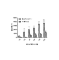
腫瘍細胞遊走に対する、配列ナノファイバーによって提供される形態学的キューの作用を決定するために、実験を実施して、配列ナノファイバーフィルム上の腫瘍細胞遊走を平滑フィルムと比較した。図5〜6に示す結果は、遊走の距離によって証明されるように、配列ナノファイバーフィルムが、平滑フィルムと比較して、有意に高い腫瘍細胞遊走を促進したことを明らかにしている。(図5:平滑フィルムへの接種から2時間後に画像化した腫瘍細胞(A)、平滑フィルムへの接種から10日後に画像化した腫瘍細胞(B)、配列ナノファイバーへの接種から2時間後に画像化した腫瘍細胞(C)、配列ナノファイバーへの接種から10日後に画像化した腫瘍細胞(D);図6:平滑フィルムと比較した配列ナノファイバー上の腫瘍細胞遊走の定量分析)。 To determine the effect of morphological cues provided by arrayed nanofibers on tumor cell migration, experiments were performed to compare tumor cell migration on aligned nanofiber films with smooth films. The results shown in FIGS. 5-6 reveal that the arrayed nanofiber film promoted significantly higher tumor cell migration compared to the smooth film, as evidenced by the distance of migration. (FIG. 5: Tumor cells imaged 2 hours after inoculation on smooth film (A), tumor cells imaged 10 days after inoculation on smooth film (B), 2 hours after inoculation with arrayed nanofibers Imaged tumor cells (C), tumor cells imaged 10 days after inoculation to arrayed nanofibers (D); FIG. 6: Quantitative analysis of tumor cell migration on arrayed nanofibers compared to smooth film).
アポトーシスヒドロゲルシンクの製造及び特性決定
スムーズンド阻害剤であるシクロパミンを評価して、腫瘍細胞にアポトーシスを誘導するのに必要な有効薬物濃度を決定した。腫瘍細胞の生存能は、様々な濃度のシクロパミンで測定した。健全な細胞(例えば、ニューロン及び神経膠)は、薬物への暴露により影響されなかった。しかし、結果は、コラーゲンヒドロゲルスカフォールドを30μMのシクロパミンと結合させるべきであることを示した。
Production and characterization of apoptotic hydrogel sinks The smoothened inhibitor cyclopamine was evaluated to determine the effective drug concentration required to induce apoptosis in tumor cells. Tumor cell viability was measured with various concentrations of cyclopamine. Healthy cells (eg, neurons and glia) were not affected by drug exposure. However, the results indicated that the collagen hydrogel scaffold should be conjugated with 30 μM cyclopamine.
コラーゲンヒドロゲルの骨格にシクロパミンを結合させることにより、細胞傷害性シンクを有するナノファイバーフィルムを製造した。架橋剤N,N’−カルボニルジイミダゾールを用いて、シクロパミン上のヒドロキシル基をコラーゲン上のアミン基と連結させた。シクロパミンがコラーゲンヒドロゲルに結合したか否かを確認するために、C13NMRを実施した。3つの条件、すなわちシクロパミン、コラーゲンに結合したシクロパミン、及び架橋剤なしのシクロパミン及びコラーゲンスカフォールドを、C13NMRを用いて分析した。実際に、シクロパミンが、コラーゲンに閉じ込められるのではなく、ヒドロゲルにテザリングしたかどうか決定するために、3番目の条件を含めた。シクロパミンは、二重結合に関与する4つの炭素を有し、これは、150から120ppmの間で出現する。シクロパミン中の二重結合における4つの炭素についての4つのピークは、シクロパミン単独のスペクトルと、コラーゲンにテザリングしたシクロパミンに存在した。しかし、シクロパミンをコラーゲンヒドロゲルと混合した場合には、ピークは存在しなかった。 A nanofiber film having a cytotoxic sink was produced by binding cyclopamine to the collagen hydrogel skeleton. The crosslinker N, N'-carbonyldiimidazole was used to link the hydroxyl group on cyclopamine with the amine group on collagen. To confirm whether cyclopamine bound to the collagen hydrogel, C 13 NMR was performed. Three conditions were analyzed using C 13 NMR: cyclopamine, cyclopamine bound to collagen, and cyclopamine and collagen scaffold without crosslinker. In fact, a third condition was included to determine if cyclopamine was tethered to the hydrogel rather than entrapped in the collagen. Cyclopamine has four carbons involved in double bonds, which appear between 150 and 120 ppm. Four peaks for the four carbons in the double bond in cyclopamine were present in the spectrum of cyclopamine alone and in cyclopamine tethered to collagen. However, no peaks were present when cyclopamine was mixed with collagen hydrogel.
腫瘍細胞をシクロパミン結合コラーゲンヒドロゲルシンク内で培養して、シクロパミンの生物活性が減少したか否か、また細胞がアポトーシスを受けたか否かを決定した。図7に示すように、細胞の明視野及び蛍光画像は、シクロパミン結合コラーゲンヒドロゲルシンク(A〜C)と比較して、アポトーシスシンク内で培養されたとき(D〜F)、明らかにアポトーシスを受けていた。(明視野像(A/D)、カルセインで染色した生存腫瘍細胞(B/E)、エチジウムホモ二量体で染色した死滅腫瘍細胞(C/F))。特に、シクロパミン結合コラーゲンヒドロゲルにおける死滅細胞の数は、コラーゲン単独のヒドロゲルにおける死滅細胞の数より多かった。 Tumor cells were cultured in cyclopamine-conjugated collagen hydrogel sinks to determine whether the biological activity of cyclopamine was reduced and whether the cells were undergoing apoptosis. As shown in FIG. 7, the bright field and fluorescence images of the cells clearly undergo apoptosis when cultured in the apoptotic sink (DF) as compared to the cyclopamine-conjugated collagen hydrogel sink (AC). It was. (Bright field image (A / D), viable tumor cells stained with calcein (B / E), dead tumor cells stained with ethidium homodimer (C / F)). In particular, the number of dead cells in the cyclopamine-conjugated collagen hydrogel was greater than the number of dead cells in the collagen-only hydrogel.
以上のin vitro実験に基づき、腫瘍細胞が、実際に形態学的キューによってさらに遊走したこと、また、アポトーシス誘導シンクが、腫瘍細胞を死滅させるのに有効でありうることを決定した。従って、予備in vitro実験を実施して、定方向細胞遊走を促進する上での本明細書に記載の埋め込み型スカフォールドの実施形態の有効性を評価した。 Based on the above in vitro experiments, it was determined that tumor cells actually migrated further by morphological cues and that an apoptosis-inducing sink could be effective in killing tumor cells. Accordingly, preliminary in vitro experiments were performed to evaluate the effectiveness of the implantable scaffold embodiments described herein in promoting directed cell migration.
in vivo実験
24匹のRowett Nude Ratに、U87mgのGFP細胞(ヒトグリア芽細胞腫細胞系である)を接種した。7日後、ラットに腫瘍細胞を接種し、5mmスカフォールドを脳内の腫瘍部位付近に埋め込んだ。腫瘍は、脳表面から深さ2mmの地点に接種した。導管は、脳表面から1.5mmの地点に埋め込んだ。
In vivo experiments Twenty-four Rowett Nude Rats were inoculated with U87 mg of GFP cells (a human glioblastoma cell line). Seven days later, rats were inoculated with tumor cells and a 5 mm scaffold was implanted near the tumor site in the brain. Tumors were inoculated 2 mm deep from the brain surface. The conduit was implanted at a point 1.5 mm from the brain surface.
スカフォールドは、10%ポリカプロラクトン(PCL)及びポリウレタン製の導管を含む。導管内に、PCL製の配列ナノファイバーを挿入した。単一のスカフォールド又は3つのスカフォールドのいずれかをラットに埋め込んだ。ラットが腫瘍の症状を呈示したとき、ラットを潅流した。接種から16〜18日後、ラットを潅流して、スカフォールドと一緒に脳を解剖した。スカフォールドと脳を横方向に切断して、50μmの切片を得た。切断した後、切片を画像化した。腫瘍細胞は、そのGFP発現によって見ることができた。図8に示すように、スカフォールドは、スカフォールドを通じた腫瘍細胞の定方向遊走を促進する上で有効であった。 The scaffold includes a conduit made of 10% polycaprolactone (PCL) and polyurethane. An arrayed nanofiber made of PCL was inserted into the conduit. Rats were implanted with either a single scaffold or three scaffolds. Rats were perfused when they exhibited tumor symptoms. 16-18 days after inoculation, the rats were perfused and the brain was dissected along with the scaffold. The scaffold and brain were cut transversely to obtain 50 μm sections. After cutting, the sections were imaged. Tumor cells could be seen by their GFP expression. As shown in FIG. 8, the scaffold was effective in promoting the directed migration of tumor cells through the scaffold.
本明細書で引用した刊行物、並びに文献で引用された材料は、参照として明示的に本明細書に組み込むものとする。本明細書に記載の方法及び装置の変更及び変形は、以上の詳細な説明から当業者には明らかになるであろう。こうした変更及び変形は、添付の特許請求の範囲内に含まれるものとする。 Publications cited herein, as well as materials cited in the literature, are expressly incorporated herein by reference. Modifications and variations of the methods and apparatus described herein will be apparent to those skilled in the art from the foregoing detailed description. Such modifications and variations are intended to be included within the scope of the appended claims.
Claims (15)
生体内で支持体の表面に沿って腫瘍細胞遊走を方向付けるためのキューを提供するように形成された表面を有する少なくとも1つの支持体であって、近位端部分及び反対側の遠位端部分を有する複数の配列ナノファイバーを含む少なくとも1つの支持体と、
前記近位端部分に接続されたポリマーシンクであって、前記遠位端部分から前記少なくとも1つの支持体を介して遊走させた腫瘍細胞を受け取るように構成されたポリマーシンクと、
前記ポリマーシンク内に配置された少なくとも1つの細胞傷害性物質であって、前記少なくとも1つの支持体を介して遊走させた腫瘍細胞に接触して当該腫瘍細胞を死滅させるように位置決めされた少なくとも1つの細胞傷害性物質と、を含み、
前記埋め込み型システムは、患者内の悪性腫瘍内に又はその近傍に切開手術又は最小侵襲手術によって埋め込まれるようなサイズ及び形状である、埋め込み型システム。 An implantable system for promoting tumor cell migration for the purpose of cell excision or death,
At least one support having a surface configured to provide a cue for directing tumor cell migration along the surface of the support in vivo, the proximal end portion and the opposite distal end At least one support comprising a plurality of arrayed nanofibers having portions ;
A polymer sink connected to the proximal end portion, the polymer sink configured to receive tumor cells migrated from the distal end portion via the at least one support;
And at least one cytotoxic material disposed within said polymer sink, at least the positioned to contact the tumor cells were allowed to migrate Ru kill the tumor cells via at least one support and one of the cytotoxic agents, only including,
The implantable system is sized and shaped to be implanted by open or minimally invasive surgery in or near a malignant tumor in a patient .
前記複数の配列ナノファイバーの長さに沿って均一に配置されている、又は、
前記複数の配列ナノファイバーの第1端から、前記複数の配列ナノファイバーの第2端まで、前記複数の配列ナノファイバーの長さに沿って、濃度勾配を呈して配置され、前記第2端は第1端から遠位にある、請求項9に記載のシステム。 The coating is
Arranged uniformly along the length of the plurality of arrayed nanofibers, or
From the first end of the plurality of arrayed nanofibers to the second end of the plurality of arrayed nanofibers, a concentration gradient is disposed along the length of the plurality of arrayed nanofibers, and the second end is The system of claim 9, distal from the first end.
腫瘍細胞の一方向遊走を促進するのに有効である、又は、
非腫瘍細胞の二方向遊走を促進するのに有効である、請求項10に記載のシステム。 The coating arranged to exhibit the concentration gradient,
Effective in promoting unidirectional migration of tumor cells, or
12. The system of claim 10, wherein the system is effective to promote bidirectional migration of non-tumor cells.
Applications Claiming Priority (3)
| Application Number | Priority Date | Filing Date | Title |
|---|---|---|---|
| US37063010P | 2010-08-04 | 2010-08-04 | |
| US61/370,630 | 2010-08-04 | ||
| PCT/US2011/046653 WO2012019049A1 (en) | 2010-08-04 | 2011-08-04 | Devices, systems, and methods for excavating cancer cells |
Publications (3)
| Publication Number | Publication Date |
|---|---|
| JP2013538079A JP2013538079A (en) | 2013-10-10 |
| JP2013538079A5 JP2013538079A5 (en) | 2014-09-11 |
| JP5894989B2 true JP5894989B2 (en) | 2016-03-30 |
Family
ID=45559831
Family Applications (1)
| Application Number | Title | Priority Date | Filing Date |
|---|---|---|---|
| JP2013523347A Active JP5894989B2 (en) | 2010-08-04 | 2011-08-04 | Apparatus, system, and method for extracting cancer cells |
Country Status (6)
| Country | Link |
|---|---|
| US (1) | US11273250B2 (en) |
| EP (2) | EP3298986A1 (en) |
| JP (1) | JP5894989B2 (en) |
| AU (1) | AU2011285639B2 (en) |
| CA (1) | CA2807483C (en) |
| WO (1) | WO2012019049A1 (en) |
Families Citing this family (28)
| Publication number | Priority date | Publication date | Assignee | Title |
|---|---|---|---|---|
| AU2006326405B2 (en) | 2005-12-13 | 2013-10-31 | President And Fellows Of Harvard College | Scaffolds for cell transplantation |
| US9770535B2 (en) | 2007-06-21 | 2017-09-26 | President And Fellows Of Harvard College | Scaffolds for cell collection or elimination |
| CA2715460C (en) | 2008-02-13 | 2020-02-18 | President And Fellows Of Harvard College | Continuous cell programming devices |
| US9370558B2 (en) | 2008-02-13 | 2016-06-21 | President And Fellows Of Harvard College | Controlled delivery of TLR agonists in structural polymeric devices |
| US9297005B2 (en) | 2009-04-13 | 2016-03-29 | President And Fellows Of Harvard College | Harnessing cell dynamics to engineer materials |
| CA2768552A1 (en) | 2009-07-31 | 2011-02-03 | President And Fellows Of Harvard College | Programming of cells for tolerogenic therapies |
| WO2011109834A2 (en) | 2010-03-05 | 2011-09-09 | President And Fellows Of Harvard College | Enhancement of skeletal muscle stem cell engrafment by dual delivery of vegf and igf-1 |
| WO2011163669A2 (en) | 2010-06-25 | 2011-12-29 | President And Fellows Of Harvard College | Co-delivery of stimulatory and inhibitory factors to create temporally stable and spatially restricted zones |
| PT2624873T (en) | 2010-10-06 | 2020-03-04 | Harvard College | Injectable, pore-forming hydrogels for materials-based cell therapies |
| US9603894B2 (en) | 2010-11-08 | 2017-03-28 | President And Fellows Of Harvard College | Materials presenting notch signaling molecules to control cell behavior |
| WO2012148684A1 (en) | 2011-04-27 | 2012-11-01 | President And Fellows Of Harvard College | Cell-friendly inverse opal hydrogels for cell encapsulation, drug and protein delivery, and functional nanoparticle encapsulation |
| ES2685327T3 (en) | 2011-04-28 | 2018-10-08 | President And Fellows Of Harvard College | Injectable preformed macroscopic three-dimensional frames for minimally invasive administration |
| US9675561B2 (en) | 2011-04-28 | 2017-06-13 | President And Fellows Of Harvard College | Injectable cryogel vaccine devices and methods of use thereof |
| EP2714073B1 (en) * | 2011-06-03 | 2021-03-10 | President and Fellows of Harvard College | In situ antigen-generating cancer vaccine |
| US20150024025A1 (en) * | 2012-03-06 | 2015-01-22 | The UABB Research Foundation | Aligned nanofibrous structures for axonal regeneration after spinal cord injury or surgery |
| HUE047973T2 (en) | 2012-04-16 | 2020-05-28 | Harvard College | Mesoporous silica compositions for modulating immune responses |
| GB201301571D0 (en) * | 2012-11-27 | 2013-03-13 | Fundanci N Pedro Barri De La Maza | Product and use |
| CN107073090A (en) | 2014-04-30 | 2017-08-18 | 哈佛学院董事会 | With reference to vaccine device and kill cancer cell method |
| CA2946948A1 (en) | 2014-04-30 | 2015-11-05 | Fundacion Ramon Dominguez | Agent, product and use |
| WO2016049627A1 (en) | 2014-09-26 | 2016-03-31 | Rensselaer Polytechnic Institute | Nanocomposite scaffold for the in vitro isolation of cells |
| EP3218020B1 (en) * | 2014-11-13 | 2021-12-01 | National University of Singapore | Tissue scaffold device and method for fabricating thereof |
| EP3250250A4 (en) | 2015-01-30 | 2019-05-22 | President and Fellows of Harvard College | Peritumoral and intratumoral materials for cancer therapy |
| CN107708756A (en) | 2015-04-10 | 2018-02-16 | 哈佛学院院长等 | Immunocyte acquisition equipment and its preparation and application |
| JP7138864B2 (en) | 2016-02-06 | 2022-09-20 | プレジデント アンド フェローズ オブ ハーバード カレッジ | Recapitulation of the hematopoietic niche to reconstitute immunity |
| CN115404196A (en) | 2016-07-13 | 2022-11-29 | 哈佛学院院长等 | Antigen presenting cell mimetic scaffolds and methods of making and using same |
| US10493233B1 (en) * | 2018-06-05 | 2019-12-03 | Duke University | Bi-directional access to tumors |
| CN112076179A (en) * | 2020-09-27 | 2020-12-15 | 成都金瑞基业生物科技有限公司 | Medical application of honokiol |
| CN115282341B (en) * | 2022-06-27 | 2023-05-23 | 华南理工大学 | Porous metal material with polypeptide bidirectional gradient distribution surface and preparation method and application thereof |
Family Cites Families (36)
| Publication number | Priority date | Publication date | Assignee | Title |
|---|---|---|---|---|
| GB2121286B (en) | 1982-06-02 | 1985-11-06 | Ethicon Inc | Improvements in synthetic vascular grafts, and methods of manufacturing such grafts |
| US5217492A (en) | 1982-09-29 | 1993-06-08 | Bio-Metric Systems, Inc. | Biomolecule attachment to hydrophobic surfaces |
| EP0432141A3 (en) | 1984-03-30 | 1991-08-21 | Osteonics Biomaterials, Inc. | A femoral stem for permanent implantation into the natural femur |
| US5053453A (en) | 1988-11-01 | 1991-10-01 | Baxter International Inc. | Thromboresistant materials and methods for making same |
| USRE36370E (en) | 1992-01-13 | 1999-11-02 | Li; Shu-Tung | Resorbable vascular wound dressings |
| US5916585A (en) | 1996-06-03 | 1999-06-29 | Gore Enterprise Holdings, Inc. | Materials and method for the immobilization of bioactive species onto biodegradable polymers |
| US6347930B1 (en) | 1997-09-11 | 2002-02-19 | Hospal R & D Int. | Device and method for manufacturing a segmented tubular capsule containing a biologically active medium |
| US6165217A (en) | 1997-10-02 | 2000-12-26 | Gore Enterprise Holdings, Inc. | Self-cohering, continuous filament non-woven webs |
| US6303136B1 (en) | 1998-04-13 | 2001-10-16 | Neurotech S.A. | Cells or tissue attached to a non-degradable filamentous matrix encapsulated by a semi-permeable membrane |
| DE69940466D1 (en) | 1998-06-05 | 2009-04-09 | Organogenesis Inc | BIOLOGICALLY MODELED IMPLANTABLE PROSTHESIS |
| US20020081732A1 (en) | 2000-10-18 | 2002-06-27 | Bowlin Gary L. | Electroprocessing in drug delivery and cell encapsulation |
| US7615373B2 (en) | 1999-02-25 | 2009-11-10 | Virginia Commonwealth University Intellectual Property Foundation | Electroprocessed collagen and tissue engineering |
| WO2000059941A1 (en) * | 1999-04-08 | 2000-10-12 | The General Hospital Corporation | Purposeful movement of human migratory cells away from an agent source |
| US6716225B2 (en) | 2001-08-02 | 2004-04-06 | Collagen Matrix, Inc. | Implant devices for nerve repair |
| US6893431B2 (en) | 2001-10-15 | 2005-05-17 | Scimed Life Systems, Inc. | Medical device for delivering patches |
| WO2003072748A2 (en) | 2002-02-22 | 2003-09-04 | University Of Washington | Bioengineered tissue substitutes |
| WO2003079985A2 (en) | 2002-03-18 | 2003-10-02 | Carnegie Mellon University | Method and apparatus for preparing biomimetic scaffold |
| WO2004005533A2 (en) | 2002-07-10 | 2004-01-15 | University Of Florida | Sol-gel derived bioactive glass polymer composite |
| US20050038498A1 (en) | 2003-04-17 | 2005-02-17 | Nanosys, Inc. | Medical device applications of nanostructured surfaces |
| US7163920B2 (en) | 2003-09-30 | 2007-01-16 | Ethicon, Inc. | Peptide with osteogenic activity |
| US7704740B2 (en) | 2003-11-05 | 2010-04-27 | Michigan State University | Nanofibrillar structure and applications including cell and tissue culture |
| US20060085063A1 (en) | 2004-10-15 | 2006-04-20 | Shastri V P | Nano- and micro-scale engineering of polymeric scaffolds for vascular tissue engineering |
| EP1855618A2 (en) * | 2005-03-07 | 2007-11-21 | Georgia Tech Research Corporation | Nanofilament scaffold for tissue regeneration |
| US7531503B2 (en) | 2005-03-11 | 2009-05-12 | Wake Forest University Health Sciences | Cell scaffold matrices with incorporated therapeutic agents |
| US20080220042A1 (en) | 2006-01-27 | 2008-09-11 | The Regents Of The University Of California | Biomolecule-linked biomimetic scaffolds |
| KR20080091827A (en) | 2006-01-27 | 2008-10-14 | 더 리전트 오브 더 유니버시티 오브 캘리포니아 | Biomimetic scaffolds |
| EP2131919A4 (en) | 2007-04-02 | 2010-10-20 | Georgia Tech Res Inst | Implantable device for communicating with biological tissue |
| WO2008137659A1 (en) | 2007-05-04 | 2008-11-13 | University Of Virginia Patent Foundation | Compositions and methods for making and using laminin nanofibers |
| US9770535B2 (en) | 2007-06-21 | 2017-09-26 | President And Fellows Of Harvard College | Scaffolds for cell collection or elimination |
| US20090043380A1 (en) * | 2007-08-09 | 2009-02-12 | Specialized Vascular Technologies, Inc. | Coatings for promoting endothelization of medical devices |
| ATE537857T1 (en) | 2007-09-21 | 2012-01-15 | Boston Scient Scimed Inc | MEDICAL DEVICES HAVING SURFACES WITH A NANOFIBER STRUCTURE |
| US20100129418A1 (en) * | 2008-11-18 | 2010-05-27 | Lowell Jeffry Lawrence | Method of inducing negative chemotaxis using an ellagitannin or gallotannin |
| US8940331B2 (en) * | 2008-11-22 | 2015-01-27 | The Board Of Trustees Of The Leland Stanford Junior University | Hydrogels, methods of making hydrogels, methods of using hydrogels, and methods of isolating, trapping, attracting, and/or killing cancer cells |
| WO2010124207A1 (en) | 2009-04-24 | 2010-10-28 | The Ohio State University | Interactive microenvironment system |
| US20110038936A1 (en) | 2009-08-17 | 2011-02-17 | Kimberly Ann Griswold | System and method for electrospun drug loaded biodegradable chemotherapy applications |
| US9572909B2 (en) | 2009-09-14 | 2017-02-21 | Virginia Commonwealth University | Electrospun nerve guides for nerve regeneration designed to modulate nerve architecture |
-
2011
- 2011-08-04 EP EP17199344.7A patent/EP3298986A1/en not_active Withdrawn
- 2011-08-04 US US13/814,009 patent/US11273250B2/en active Active
- 2011-08-04 WO PCT/US2011/046653 patent/WO2012019049A1/en active Application Filing
- 2011-08-04 EP EP11815346.9A patent/EP2600792B1/en active Active
- 2011-08-04 JP JP2013523347A patent/JP5894989B2/en active Active
- 2011-08-04 AU AU2011285639A patent/AU2011285639B2/en active Active
- 2011-08-04 CA CA2807483A patent/CA2807483C/en active Active
Also Published As
| Publication number | Publication date |
|---|---|
| WO2012019049A1 (en) | 2012-02-09 |
| AU2011285639A1 (en) | 2013-02-21 |
| EP2600792A4 (en) | 2014-04-16 |
| US20130172846A1 (en) | 2013-07-04 |
| JP2013538079A (en) | 2013-10-10 |
| CA2807483C (en) | 2019-02-26 |
| EP2600792B1 (en) | 2017-11-01 |
| EP3298986A1 (en) | 2018-03-28 |
| CA2807483A1 (en) | 2012-02-09 |
| US11273250B2 (en) | 2022-03-15 |
| AU2011285639B2 (en) | 2014-12-18 |
| EP2600792A1 (en) | 2013-06-12 |
Similar Documents
| Publication | Publication Date | Title |
|---|---|---|
| JP5894989B2 (en) | Apparatus, system, and method for extracting cancer cells | |
| CN108601646B (en) | Systems and methods for producing gastrointestinal tract tissue | |
| US11517646B2 (en) | Scaffold | |
| JP5643515B2 (en) | Implants and methods for soft tissue repair | |
| CN100457445C (en) | Biodegradable and/or biological absorbing fiber product and its use in medical application | |
| ES2897898T3 (en) | A surrogate multilayer matrix of tissues and uses thereof | |
| ES2847893T3 (en) | Biomedical patches with fibers arranged in space | |
| US8691543B2 (en) | Nanofibrous scaffold comprising immobilized cells | |
| CN101559242B (en) | A kind of can simultaneously as the nano artificial cerebral dura mater and preparation method thereof of drug controlled release system | |
| Nune et al. | Self-assembling peptide nanostructures on aligned poly (lactide-co-glycolide) nanofibers for the functional regeneration of sciatic nerve | |
| ES2946262T3 (en) | Sustained release drug sheet for the treatment of nerve injury | |
| BRPI1006250B1 (en) | dura mater and artificial production method | |
| BR112018073290B1 (en) | Resorbable non-woven graft material | |
| CN112867513A (en) | Method and apparatus for in vivo tissue regeneration on the inner surface of a hollow organ | |
| US20230218507A1 (en) | Microneedle patch for in-situ seeding of cells | |
| WO2013036531A1 (en) | Surgical sutures and methods of making and using same | |
| CN113797178A (en) | Multifunctional composite material and preparation method and application thereof | |
| JP2019503819A (en) | System and method for the generation of gastrointestinal tissue in an anastomosis or physiological location | |
| JP7044288B2 (en) | Medical sheet | |
| CN112118806A (en) | Multi-layered scaffold design using spatial distribution of cells to modulate tissue growth | |
| Chen et al. | Preparation of Poly (3-hydroxybutyrate-co-3-hydroxyvalerate) Nanofiber Catheter and Its Mechanism of Nerve Injury in Patients with Cervical Spine Injury | |
| CZ2015117A3 (en) | Process or preparing flat textile fabric of biologically degradable and biologically compatible nanofibers, especially for skin wound covers and apparatus for making the same | |
| Niu et al. | Making a Hydrophilic Interfacial Scaffold with Hierarchical Nanofibre Architecture to Enhance the Phenotypic Expression of Epithelial and Smooth Muscle Cells for Urethral Reconstruction | |
| JP2019076783A (en) | Medical patches with spatially arranged fibers | |
| Silver et al. | Pathobiology and Response to Tissue Injury |
Legal Events
| Date | Code | Title | Description |
|---|---|---|---|
| A521 | Request for written amendment filed |
Free format text: JAPANESE INTERMEDIATE CODE: A523 Effective date: 20140723 |
|
| A621 | Written request for application examination |
Free format text: JAPANESE INTERMEDIATE CODE: A621 Effective date: 20140723 |
|
| A131 | Notification of reasons for refusal |
Free format text: JAPANESE INTERMEDIATE CODE: A131 Effective date: 20150623 |
|
| A601 | Written request for extension of time |
Free format text: JAPANESE INTERMEDIATE CODE: A601 Effective date: 20150924 |
|
| A521 | Request for written amendment filed |
Free format text: JAPANESE INTERMEDIATE CODE: A523 Effective date: 20151022 |
|
| TRDD | Decision of grant or rejection written | ||
| A01 | Written decision to grant a patent or to grant a registration (utility model) |
Free format text: JAPANESE INTERMEDIATE CODE: A01 Effective date: 20160201 |
|
| A61 | First payment of annual fees (during grant procedure) |
Free format text: JAPANESE INTERMEDIATE CODE: A61 Effective date: 20160229 |
|
| R150 | Certificate of patent or registration of utility model |
Ref document number: 5894989 Country of ref document: JP Free format text: JAPANESE INTERMEDIATE CODE: R150 |
|
| R250 | Receipt of annual fees |
Free format text: JAPANESE INTERMEDIATE CODE: R250 |
|
| R250 | Receipt of annual fees |
Free format text: JAPANESE INTERMEDIATE CODE: R250 |
|
| R250 | Receipt of annual fees |
Free format text: JAPANESE INTERMEDIATE CODE: R250 |
|
| R250 | Receipt of annual fees |
Free format text: JAPANESE INTERMEDIATE CODE: R250 |
|
| R250 | Receipt of annual fees |
Free format text: JAPANESE INTERMEDIATE CODE: R250 |
|
| R250 | Receipt of annual fees |
Free format text: JAPANESE INTERMEDIATE CODE: R250 |