JP5872323B2 - X-ray CT apparatus and image processing method - Google Patents
X-ray CT apparatus and image processing method Download PDFInfo
- Publication number
- JP5872323B2 JP5872323B2 JP2012040107A JP2012040107A JP5872323B2 JP 5872323 B2 JP5872323 B2 JP 5872323B2 JP 2012040107 A JP2012040107 A JP 2012040107A JP 2012040107 A JP2012040107 A JP 2012040107A JP 5872323 B2 JP5872323 B2 JP 5872323B2
- Authority
- JP
- Japan
- Prior art keywords
- tumor
- movement information
- calculation unit
- peripheral
- ray
- Prior art date
- Legal status (The legal status is an assumption and is not a legal conclusion. Google has not performed a legal analysis and makes no representation as to the accuracy of the status listed.)
- Active
Links
- 238000003672 processing method Methods 0.000 title claims description 5
- 206010028980 Neoplasm Diseases 0.000 claims description 182
- 230000033001 locomotion Effects 0.000 claims description 172
- 230000002093 peripheral effect Effects 0.000 claims description 162
- 238000004364 calculation method Methods 0.000 claims description 152
- 230000008595 infiltration Effects 0.000 claims description 71
- 238000001764 infiltration Methods 0.000 claims description 71
- 230000000737 periodic effect Effects 0.000 claims description 24
- 230000005484 gravity Effects 0.000 claims description 15
- 230000003902 lesion Effects 0.000 claims description 14
- 230000009545 invasion Effects 0.000 claims description 5
- 230000002123 temporal effect Effects 0.000 claims description 4
- 239000000853 adhesive Substances 0.000 claims description 2
- 230000001070 adhesive effect Effects 0.000 claims description 2
- 238000002591 computed tomography Methods 0.000 description 72
- 238000012545 processing Methods 0.000 description 56
- 238000000034 method Methods 0.000 description 42
- 210000004072 lung Anatomy 0.000 description 39
- 208000007536 Thrombosis Diseases 0.000 description 21
- 210000004204 blood vessel Anatomy 0.000 description 20
- 239000013598 vector Substances 0.000 description 19
- 206010064390 Tumour invasion Diseases 0.000 description 15
- 230000009400 cancer invasion Effects 0.000 description 15
- 238000010586 diagram Methods 0.000 description 14
- 238000012937 correction Methods 0.000 description 8
- 230000011218 segmentation Effects 0.000 description 8
- 230000000241 respiratory effect Effects 0.000 description 6
- 238000013480 data collection Methods 0.000 description 5
- 238000013500 data storage Methods 0.000 description 5
- 238000003384 imaging method Methods 0.000 description 5
- 210000000056 organ Anatomy 0.000 description 4
- 238000001514 detection method Methods 0.000 description 3
- 239000000284 extract Substances 0.000 description 3
- 210000000779 thoracic wall Anatomy 0.000 description 3
- 230000002159 abnormal effect Effects 0.000 description 2
- 210000000188 diaphragm Anatomy 0.000 description 2
- 210000001370 mediastinum Anatomy 0.000 description 2
- 238000010606 normalization Methods 0.000 description 2
- 230000008855 peristalsis Effects 0.000 description 2
- 238000007781 pre-processing Methods 0.000 description 2
- 206010006322 Breath holding Diseases 0.000 description 1
- 210000000621 bronchi Anatomy 0.000 description 1
- 230000007423 decrease Effects 0.000 description 1
- 230000003247 decreasing effect Effects 0.000 description 1
- 230000000694 effects Effects 0.000 description 1
- 210000003238 esophagus Anatomy 0.000 description 1
- 230000010365 information processing Effects 0.000 description 1
- 239000004973 liquid crystal related substance Substances 0.000 description 1
- 230000003211 malignant effect Effects 0.000 description 1
- 238000012986 modification Methods 0.000 description 1
- 230000004048 modification Effects 0.000 description 1
- 210000004224 pleura Anatomy 0.000 description 1
- 238000009877 rendering Methods 0.000 description 1
- 230000035945 sensitivity Effects 0.000 description 1
- 230000001360 synchronised effect Effects 0.000 description 1
- 210000001519 tissue Anatomy 0.000 description 1
Images


Classifications
-
- G—PHYSICS
- G06—COMPUTING; CALCULATING OR COUNTING
- G06T—IMAGE DATA PROCESSING OR GENERATION, IN GENERAL
- G06T7/00—Image analysis
- G06T7/20—Analysis of motion
- G06T7/246—Analysis of motion using feature-based methods, e.g. the tracking of corners or segments
-
- G—PHYSICS
- G06—COMPUTING; CALCULATING OR COUNTING
- G06T—IMAGE DATA PROCESSING OR GENERATION, IN GENERAL
- G06T7/00—Image analysis
- G06T7/0002—Inspection of images, e.g. flaw detection
- G06T7/0012—Biomedical image inspection
- G06T7/0014—Biomedical image inspection using an image reference approach
- G06T7/0016—Biomedical image inspection using an image reference approach involving temporal comparison
-
- G—PHYSICS
- G06—COMPUTING; CALCULATING OR COUNTING
- G06T—IMAGE DATA PROCESSING OR GENERATION, IN GENERAL
- G06T2207/00—Indexing scheme for image analysis or image enhancement
- G06T2207/10—Image acquisition modality
- G06T2207/10072—Tomographic images
- G06T2207/10081—Computed x-ray tomography [CT]
-
- G—PHYSICS
- G06—COMPUTING; CALCULATING OR COUNTING
- G06T—IMAGE DATA PROCESSING OR GENERATION, IN GENERAL
- G06T2207/00—Indexing scheme for image analysis or image enhancement
- G06T2207/30—Subject of image; Context of image processing
- G06T2207/30004—Biomedical image processing
- G06T2207/30096—Tumor; Lesion
Landscapes
- Engineering & Computer Science (AREA)
- Theoretical Computer Science (AREA)
- Computer Vision & Pattern Recognition (AREA)
- Physics & Mathematics (AREA)
- General Physics & Mathematics (AREA)
- Health & Medical Sciences (AREA)
- General Health & Medical Sciences (AREA)
- Medical Informatics (AREA)
- Nuclear Medicine, Radiotherapy & Molecular Imaging (AREA)
- Radiology & Medical Imaging (AREA)
- Quality & Reliability (AREA)
- Multimedia (AREA)
- Apparatus For Radiation Diagnosis (AREA)
- Image Processing (AREA)
- Image Analysis (AREA)
Description
本発明の実施形態は、X線CT装置及び画像処理方法に関する。 Embodiments described herein relate generally to an X-ray CT apparatus and an image processing method.
従来、X線CT(Computed Tomography)装置を用いた被検体内の撮像手法がいくつか知られている。例えば、息止め状態、呼期状態や吸期状態の被検体内を撮像する手法が知られている。また、被検体の呼吸状態を特定することなく、複数の時相で被検体内を撮像する手法も知られている。また、近年では、被検体内を広範囲かつ3次元に画像化できるX線CT装置が知られており、かかるX線CT装置を用いて被検体内を撮像される場合もある。このようなX線CT装置は、例えば、被検体内の腫瘍を撮像する際に用いられたりする。 Conventionally, several imaging methods in a subject using an X-ray CT (Computed Tomography) apparatus are known. For example, a technique for imaging the inside of a subject in a breath holding state, an expiration state, or an inhalation state is known. There is also known a technique of imaging the inside of a subject at a plurality of time phases without specifying the respiratory state of the subject. In recent years, X-ray CT apparatuses that can image a subject in a wide range and three-dimensionally are known, and the subject can be imaged using such an X-ray CT apparatus. Such an X-ray CT apparatus is used, for example, when imaging a tumor in a subject.
本発明が解決しようとする課題は、病変部と周辺部位との関係性を示す情報を提示することができるX線CT装置及び画像処理方法を提供することである。 The problem to be solved by the present invention is to provide an X-ray CT apparatus and an image processing method capable of presenting information indicating the relationship between a lesioned part and a peripheral site.
実施形態のX線CT装置は、生成部と、特定部と、移動情報算出部と、相対関係算出部とを有する。生成部は、被検体を透過したX線に基づいて、時相の異なる被検体内の画像データを生成する。特定部は、前記生成部によって生成された時相の異なる被検体内の画像データから、病変部の位置と該病変部の周辺に位置する周辺部位の位置とを特定する。移動情報算出部は、前記特定部によって特定された病変部及び周辺部位の位置に基づいて、病変部及び周辺部位の移動に関する移動情報を算出する。相対関係算出部は、前記移動情報算出部によって算出された前記病変部の移動情報と前記周辺部位の移動情報との相対関係を算出する。 The X-ray CT apparatus of the embodiment includes a generation unit, a specifying unit, a movement information calculation unit, and a relative relationship calculation unit. The generation unit generates image data in the subject having different time phases based on the X-rays transmitted through the subject. The specifying unit specifies the position of the lesioned part and the position of the peripheral part located around the lesioned part from the image data in the subject having different time phases generated by the generating unit. The movement information calculation unit calculates movement information related to the movement of the lesion part and the peripheral part based on the positions of the lesion part and the peripheral part specified by the specifying part. The relative relationship calculation unit calculates a relative relationship between the movement information of the lesion and the movement information of the peripheral part calculated by the movement information calculation unit.
(第1の実施形態)
X線CT装置は、X線管から被検体にX線を照射し、被検体を透過したX線を検出器により検出することで、被検体内における組織形態情報を示すX線CT画像の再構成を行う装置である。第1の実施形態に係るX線CT装置は、X線CT画像の再構成を行うとともに、腫瘍が肺野等の所定の周辺部位に浸潤している度合いを示す「浸潤度」を算出する。なお、以下の実施形態において、周辺部位とは、腫瘍周辺に位置する臓器等を示し、例えば、肺野、胸膜、気管支、食道、胸壁、縦隔、横隔膜等を示す。
(First embodiment)
The X-ray CT apparatus irradiates a subject with X-rays from an X-ray tube, and detects X-rays transmitted through the subject with a detector, thereby reproducing an X-ray CT image indicating tissue morphology information in the subject. A device that performs the configuration. The X-ray CT apparatus according to the first embodiment reconstructs an X-ray CT image and calculates an “invasion degree” indicating the degree to which a tumor has infiltrated a predetermined peripheral site such as a lung field. In the following embodiments, the peripheral site indicates an organ or the like located in the vicinity of the tumor, for example, lung field, pleura, bronchi, esophagus, chest wall, mediastinum, diaphragm, or the like.
図1を用いて、第1の実施形態に係るX線CT装置の構成について説明する。図1は、第1の実施形態に係るX線CT装置1の構成例を示す図である。図1に例示するように、第1の実施形態に係るX線CT装置1は、架台装置10と、寝台装置20と、コンソール装置100とを有する。
The configuration of the X-ray CT apparatus according to the first embodiment will be described with reference to FIG. FIG. 1 is a diagram illustrating a configuration example of an X-ray CT apparatus 1 according to the first embodiment. As illustrated in FIG. 1, the X-ray CT apparatus 1 according to the first embodiment includes a
架台装置10は、被検体PにX線を照射し、被検体Pを透過したX線を検出してコンソール装置100に出力する。かかる架台装置10は、高電圧発生部11と、X線管12と、X線検出器13と、データ収集部14と、回転フレーム15と、架台駆動部16と、架台寝台制御部17とを有する。
The
高電圧発生部11は、架台寝台制御部17による制御に従って、X線管12に対して高電圧を供給する。X線管12は、高電圧発生部11から供給される高電圧によってX線を発生する真空管であり、回転フレーム15の回転に伴って、被検体Pに対してX線を照射する。すなわち、高電圧発生部11は、X線管12に供給する管電圧や管電流を調整することで、被検体Pに対して照射されるX線量を調整する。
The
X線検出器13は、被検体Pを透過したX線を検出する2次元アレイ型検出器(面検出器)であり、複数チャンネル分のX線検出素子を配してなる検出素子列が被検体Pの体軸方向(図1に示すZ軸方向)に沿って複数列配列されている。具体的には、第1の実施形態におけるX線検出器13は、被検体Pの体軸方向に沿って320列など多列に配列されたX線検出素子を有し、例えば、被検体Pの肺や心臓を含む範囲など、広範囲に被検体Pを透過したX線を検出することが可能である。
The
データ収集部14は、X線検出器13によって検出されたX線を用いて投影データを生成し、生成した投影データをコンソール装置100の画像処理部140に送信する。回転フレーム15は、被検体Pを中心にして、高速でかつ連続的に回転する円環状のフレームであり、X線管12及びX線検出器13が対向して配置される。
The
架台駆動部16は、架台寝台制御部17による制御に従って、架台を駆動する。具体的には、架台駆動部16は、モータの駆動によって回転フレーム15を高速に連続回転させ、被検体Pを中心とした円軌道上でX線管12及びX線検出器13を連続回転させる。架台寝台制御部17は、後述するスキャン制御部160による制御に従って、高電圧発生部11、架台駆動部16及び寝台駆動部21を制御する。
The
なお、第1の実施形態では、2次元アレイ型検出器(面検出器)であるX線検出器13を用いることにより、被検体Pの位置を固定したままで回転フレーム15を回転させて被検体Pを円軌道にてスキャンするコンベンショナルスキャンを複数の時相において実行する。すなわち、上記のデータ収集部14は、X線検出器13によって検出されたX線を用いて、スキャンされた時相の異なる複数の3次元投影データを収集し、収集した3次元投影データを画像処理部140に送信する。
In the first embodiment, by using the
寝台装置20は、撮影対象の被検体Pを載置する台であり、寝台駆動部21と、天板22とを有する。寝台駆動部21は、架台寝台制御部17による制御に従って、モータの駆動によって、天板22を被検体Pの体軸方向に連続して往復移動する。天板22は、被検体Pを載置する板である。
The
コンソール装置100は、操作者によるX線CT装置1の操作を受け付けるとともに、架台装置10によって収集された投影データからX線CT画像を再構成する。具体的には、コンソール装置100は、入力部110と、表示部120と、システム制御部130と、画像処理部140と、画像データ記憶部150と、スキャン制御部160とを有する。
The console device 100 accepts an operation of the X-ray CT apparatus 1 by an operator and reconstructs an X-ray CT image from the projection data collected by the
入力部110は、X線CT装置1の操作者が各種指示や各種設定の入力に用いるマウスやキーボードなどを有し、操作者から受け付けた指示や設定の情報を、システム制御部130に転送する。例えば、入力部110は、操作者から腫瘍の浸潤度を算出する旨の操作や、X線CT画像を再構成する際の再構成条件の入力操作等を受け付ける。表示部120は、LCD(Liquid Crystal Display)などのディスプレイであり、各種情報を表示する。例えば、表示部120は、画像データ記憶部150によって記憶されているX線CT画像や、操作者から各種指示を受け付けるためのGUI(Graphical User Interface)などを表示する。
The input unit 110 includes a mouse and a keyboard used by the operator of the X-ray CT apparatus 1 to input various instructions and various settings, and transfers instructions and setting information received from the operator to the
システム制御部130は、架台装置10、寝台装置20及びコンソール装置100を制御することによって、X線CT装置1全体の制御を行う。例えば、システム制御部130は、スキャン制御部160を制御して3次元投影データを収集させる。また、例えば、システム制御部130は、画像処理部140を制御して3次元投影データからX線CT画像を再構成させる。
The
画像処理部140は、データ収集部14から受信した3次元投影データに対して各種処理を行う。具体的には、画像処理部140は、データ収集部14から受信した3次元投影データに対して感度補正などの前処理を行い、前処理後の3次元投影データを逆投影処理することで、3次元X線CT画像(以下、「ボリュームデータ」と表記する場合がある)を再構成する。そして、画像処理部140は、再構成後のボリュームデータを画像データ記憶部150に格納する。また、画像処理部140は、例えば、SVR(Shaded Volume Rendering)法等により立体感のあるX線CT画像を生成したり、任意面の断面画像を生成して、生成したX線CT画像を画像データ記憶部150に格納する。
The
なお、第1の実施形態における架台装置10は、複数の時相においてコンベンショナルスキャンを実行するので、画像処理部140は、各時相における3次元投影データに対して、上記の画像再構成処理を行う。また、第1の実施形態に係る画像処理部140は、X線CT画像を生成するだけでなく、腫瘍の浸潤度を算出する処理についても行う。画像処理部140による浸潤度算出処理については、後に詳述する。
Since the
画像データ記憶部150は、画像処理部140によって再構成されたボリュームデータやX線CT画像等を記憶する。スキャン制御部160は、システム制御部130から指示されたスキャン条件に基づき架台寝台制御部17を制御する。
The image
上記の通り、第1の実施形態に係るX線CT装置1は、腫瘍の浸潤度を算出する処理を行う。この点について簡単に説明すると、「正常な部位(臓器等)」と「腫瘍等の異常な部位」とは、基本的には動きが異なると考えられる。例えば、腫瘍は、悪性化して所定の周辺部位に浸潤するほど固くなり所定の周辺部位から剥がれにくくなる。その結果、かかる腫瘍は、呼吸動や蠕動による正常な他の周辺部位の動きと同調しにくくなる。一方、所定の周辺部位への浸潤度が低い腫瘍は、かかる所定の周辺部位から剥がれやすく、正常な他の周辺部位の動きと同調しやすい。すなわち、腫瘍及び周辺部位の動きにより、腫瘍が所定の周辺部位に浸潤している度合いである浸潤度を推定することができる。 As described above, the X-ray CT apparatus 1 according to the first embodiment performs a process of calculating the degree of tumor infiltration. Briefly describing this point, it is considered that “normal sites (organs and the like)” and “abnormal sites such as tumors” basically have different movements. For example, a tumor becomes so hard that it becomes malignant and infiltrates into a predetermined peripheral site, and becomes difficult to peel off from the predetermined peripheral site. As a result, such a tumor becomes difficult to synchronize with the movement of other normal peripheral parts due to respiratory movement or peristalsis. On the other hand, a tumor having a low degree of infiltration into a predetermined peripheral site is easily peeled off from the predetermined peripheral site and easily synchronized with the movement of other normal peripheral sites. That is, it is possible to estimate the degree of infiltration, which is the degree to which the tumor has infiltrated a predetermined peripheral site, by the movement of the tumor and the peripheral site.
第1の実施形態に係るX線CT装置1は、複数の時相で生成されたボリュームデータを解析することにより、腫瘍の移動に関する移動情報と、かかる腫瘍の周辺に位置する周辺部位の移動に関する移動情報とを算出し、双方の移動情報の関係に基づいて、腫瘍の浸潤度を算出する。以下に、図2〜図5を用いて、X線CT装置1による浸潤度算出処理について詳細に説明する。 The X-ray CT apparatus 1 according to the first embodiment relates to movement information relating to movement of a tumor and movement of peripheral parts located around the tumor by analyzing volume data generated in a plurality of time phases. The movement information is calculated, and the degree of tumor invasion is calculated based on the relationship between the movement information of both. Below, the infiltration degree calculation process by the X-ray CT apparatus 1 is demonstrated in detail using FIGS.
図2は、第1の実施形態における画像処理部140の構成例を示す図である。図2に例示するように、第1の実施形態における画像処理部140は、特定部141と、移動情報算出部142と、相対関係算出部143とを有する。なお、図2には、ボリュームデータを生成する処理部や、表示用のX線CT画像を生成する処理部については図示することを省略する。
FIG. 2 is a diagram illustrating a configuration example of the
特定部141は、画像処理部140において再構成されたボリュームデータ内の腫瘍の位置と、かかる腫瘍の周辺部位の位置とを特定する。第1の実施形態における画像処理部140は、時相の異なる複数のボリュームデータを再構成するので、特定部141は、各時相に対応するボリュームデータ毎に、腫瘍及び周辺部位の位置を特定する処理を行う。
The specifying
具体的には、特定部141は、画像処理部140において再構成された各ボリュームデータに対して、CT値が空間的に連続する領域を抽出する領域拡張(region growing)法や形状テンプレートを用いたパターンマッチング法などを用いてセグメンテーション処理を行うことにより、各領域を抽出する。続いて、特定部141は、抽出した各領域に対して、腫瘍及び周辺部位の形状テンプレートを用いたパターンマッチング法や、腫瘍及び周辺部位の輝度値のプロファイルを用いた手法等により、ボリュームデータに含まれる腫瘍及び周辺部位の位置を特定する。
Specifically, the specifying
移動情報算出部142は、特定部141によって特定された腫瘍及び周辺部位の位置に基づいて、腫瘍及び周辺部位の移動に関する移動情報を算出する。「移動情報」とは、腫瘍や臓器の位置の時間変動および位置変動を示す。なお、第1の実施形態では、「移動情報」は、所定の基準点から腫瘍や臓器等の部位までの相対距離の時間変動を示すものとする。なお、所定の基準点は、例えば、ボリュームデータ内の任意の点や、横隔膜や肺尖部等の部位等である。
The movement information calculation unit 142 calculates movement information related to the movement of the tumor and the surrounding part based on the position of the tumor and the surrounding part specified by the specifying
移動情報算出部142による処理について具体的に説明すると、移動情報算出部142は、まず、画像処理部140において再構成された時相が連続する2個のボリュームデータの位置合わせを行う。このとき、移動情報算出部142は、2個のボリュームデータ全体の位置合わせを行ってもよいし、所定の基準点に対して2個のボリュームデータの位置を合わせてもよい。
The processing by the movement information calculation unit 142 will be described in detail. First, the movement information calculation unit 142 performs alignment of two volume data having successive time phases reconstructed by the
続いて、移動情報算出部142は、位置合わせ後の2個のボリュームデータを比較することにより、腫瘍及び各周辺部位の移動方向及び移動量を示す動きベクトルを算出する。このとき、移動情報算出部142は、ボリュームデータのボクセル(voxel)毎に、かかるボクセルの動きベクトルを算出する。例えば、移動情報算出部142は、腫瘍の移動方向及び移動量を算出する場合には、2個のボリュームデータを比較することにより、ボリュームデータのうち腫瘍を示す領域内のボクセル毎に、かかるボクセルの動きベクトルを算出する。また、移動情報算出部142は、周辺部位(例えば、肺野)の移動方向及び移動量を算出する場合には、2個のボリュームデータを比較することにより、ボリュームデータのうち周辺部位(例えば、肺野)を示す領域内のボクセル毎に、かかるボクセルの動きベクトルを算出する。なお、移動情報算出部142は、例えば、トラッキング等の位置合わせ時の情報をもとにボクセルの動きベクトルを算出することができる。 Subsequently, the movement information calculation unit 142 calculates a motion vector indicating the movement direction and the movement amount of the tumor and each peripheral part by comparing the two volume data after the alignment. At this time, the movement information calculation unit 142 calculates the motion vector of the voxel for each voxel of the volume data. For example, when the movement information calculation unit 142 calculates a movement direction and a movement amount of a tumor, the movement information calculation unit 142 compares the two pieces of volume data so that each voxel in the region indicating the tumor in the volume data is subjected to the voxel. The motion vector of is calculated. Further, when calculating the movement direction and amount of movement of the peripheral part (for example, lung field), the movement information calculation unit 142 compares the two volume data, thereby calculating the peripheral part (for example, For each voxel in the region indicating the lung field, a motion vector of the voxel is calculated. Note that the movement information calculation unit 142 can calculate a voxel motion vector based on information at the time of alignment such as tracking, for example.
続いて、移動情報算出部142は、各ボクセルの動きベクトルを、ボリュームデータのxyz座標系におけるx軸方向、y軸方向、z軸方向の動きベクトルに分解する。これにより、移動情報算出部142は、腫瘍及び各周辺部位のx軸方向、y軸方向、z軸方向の移動量をボクセル毎に得ることができる。例えば、x軸方向の動きベクトルに注目すると、かかる動きベクトルの始点から終点までボクセルがx軸方向に移動した移動量を得ることができる。 Subsequently, the movement information calculation unit 142 decomposes the motion vector of each voxel into motion vectors in the x-axis direction, the y-axis direction, and the z-axis direction in the xyz coordinate system of the volume data. Thereby, the movement information calculation unit 142 can obtain the movement amount of the tumor and each peripheral site in the x-axis direction, the y-axis direction, and the z-axis direction for each voxel. For example, when attention is paid to the motion vector in the x-axis direction, it is possible to obtain a movement amount in which the voxel has moved in the x-axis direction from the start point to the end point of the motion vector.
移動情報算出部142は、画像処理部140において時相が連続する複数のボリュームデータのうちの2つのボリュームデータを用いて、その複数のボリュームデータすべてについて、x軸方向、y軸方向、z軸方向の動きベクトルを算出する処理を行う。そして、移動情報算出部142は、x軸方向、y軸方向、z軸方向毎に、基準点からボクセルまでの相対距離の時間変動を算出する。このような相対距離の時間変動は、周期関数とみなすことができる。これは、人体内の各部位の動きは、呼吸動や蠕動によって発生するからである。なお、以下では、「基準点からXまでの相対距離の時間変動」を単に「Xの距離変動」と表記する場合がある。
The movement information calculation unit 142 uses two volume data of the plurality of volume data whose time phases are continuous in the
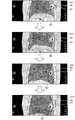
図3及び図4を用いて、移動情報算出部142による処理について説明する。図3は、肺野周辺の画像例を示す図であり、図4は、移動情報算出部142によって算出された移動情報の一例を示す図である。 Processing by the movement information calculation unit 142 will be described with reference to FIGS. 3 and 4. FIG. 3 is a diagram illustrating an example of an image around the lung field, and FIG. 4 is a diagram illustrating an example of movement information calculated by the movement information calculation unit 142.
図3に示した例では、X線CT画像G11、G12、G13、G14の順に撮像されたものとする。なお、図3では、説明を簡単にするために、断面画像を示している。また、図3に示した例において、腫瘍A1は所定の周辺部位A2に浸潤しており、腫瘍A1の周辺に位置する他の周辺部位B1は肺野である。 In the example shown in FIG. 3, it is assumed that the X-ray CT images G11, G12, G13, and G14 are captured in this order. In FIG. 3, a cross-sectional image is shown for ease of explanation. In the example shown in FIG. 3, the tumor A1 has infiltrated into a predetermined peripheral part A2, and the other peripheral part B1 located around the tumor A1 is a lung field.
図3に例示するように、X線CT画像G11、G12、G13、G14の順に、腫瘍A1及び周辺部位B1の位置が変化していることが分かる。移動情報算出部142は、図4に例示するように、このような腫瘍A1や周辺部位B1の距離変動をボクセル毎に算出する。なお、図4は、x軸方向における各部位を示す所定のボクセルの距離変動を示すものとするが、移動情報算出部142は、y軸方向及びz軸方向についても各部位の距離変動を算出する。また、移動情報算出部142は、1個のボクセルのみだけでなく、各部位に含まれる各ボクセルについても距離変動を算出する。 As illustrated in FIG. 3, it can be seen that the positions of the tumor A1 and the peripheral site B1 change in the order of the X-ray CT images G11, G12, G13, and G14. As illustrated in FIG. 4, the movement information calculation unit 142 calculates such a distance variation of the tumor A1 and the peripheral site B1 for each voxel. 4 shows the distance fluctuation of a predetermined voxel indicating each part in the x-axis direction, the movement information calculation unit 142 calculates the distance fluctuation of each part also in the y-axis direction and the z-axis direction. To do. Further, the movement information calculation unit 142 calculates the distance variation not only for one voxel but also for each voxel included in each part.
図4に示した例では、移動情報算出部142は、腫瘍(図3中のA1)を示すボクセル、周辺部位である肺野(図3中のB1)を示すボクセル、対側部位を示すボクセル、胸壁を示すボクセルについて、基準点(図4中に示す「基線」)からの相対距離を算出している。ここで、「対側部位」について説明すると、人体には、肺野のように略左右対称に存在する部位(以下、「対称部位」と表記する場合がある)がある。図4に示した「対側部位」とは、腫瘍の周辺部位が対称部位である場合に、かかる周辺部位の反対側に存在する部位を示す。例えば、図4に示した周辺部位が右肺野である場合には、「対側部位」は、左肺野を示す。かかる「対側部位」は、浸潤度を算出する際にも用いられるが、この点については後述する。 In the example illustrated in FIG. 4, the movement information calculation unit 142 includes a voxel indicating a tumor (A1 in FIG. 3), a voxel indicating a surrounding lung field (B1 in FIG. 3), and a voxel indicating a contralateral site. For the voxel indicating the chest wall, the relative distance from the reference point ("baseline" shown in FIG. 4) is calculated. Here, the “opposite part” will be described. The human body has a part that exists substantially symmetrically like a lung field (hereinafter, sometimes referred to as “symmetric part”). The “contralateral site” shown in FIG. 4 indicates a site that exists on the opposite side of the peripheral site when the peripheral site of the tumor is a symmetrical site. For example, when the peripheral site shown in FIG. 4 is the right lung field, the “opposite site” indicates the left lung field. This “contralateral part” is also used when calculating the degree of infiltration, which will be described later.
このように、移動情報算出部142は、時相が異なる複数のボリュームデータを用いて、腫瘍、周辺部位、対側部位等の距離変動をボクセル毎、かつ、xyz軸方向毎に算出する。 As described above, the movement information calculation unit 142 calculates the variation in the distance of the tumor, the peripheral region, the contralateral region, and the like for each voxel and for each xyz axis direction using a plurality of volume data having different time phases.
なお、上記例では、移動情報算出部142がボクセル毎に距離変動を算出する例を示したが、かかるボクセルは、任意のサイズでよい。例えば、移動情報算出部142は、「1×1×1ボクセル」毎に距離変動を算出してもよいし、「4×4×4ボクセル」毎に距離変動を算出してもよい。そして、このようなボクセルのサイズは、操作者等によって決定されてもよい。また、移動情報算出部142は、各部位の全てのボクセルについて距離変動を算出しなくてもよい。例えば、移動情報算出部142は、各部位から所定数のボクセル領域を抽出し、抽出したボクセル領域について距離変動を算出してもよい。 In the above example, the movement information calculation unit 142 calculates the distance variation for each voxel. However, the voxel may be of any size. For example, the movement information calculation unit 142 may calculate the distance variation for each “1 × 1 × 1 voxel” or may calculate the distance variation for each “4 × 4 × 4 voxel”. The size of such a voxel may be determined by an operator or the like. Moreover, the movement information calculation part 142 does not need to calculate the distance fluctuation | variation about all the voxels of each site | part. For example, the movement information calculation unit 142 may extract a predetermined number of voxel regions from each part and calculate a distance variation for the extracted voxel regions.
図2の説明に戻って、相対関係算出部143は、移動情報算出部142によって算出された腫瘍の移動情報と周辺部位の移動情報との相対関係を算出する。さらに、相対関係算出部143は、かかる相対関係に基づいて、腫瘍が周辺部位に浸潤している浸潤度を算出する。 Returning to the description of FIG. 2, the relative relationship calculation unit 143 calculates the relative relationship between the movement information of the tumor calculated by the movement information calculation unit 142 and the movement information of the peripheral site. Furthermore, the relative relationship calculation unit 143 calculates the degree of infiltration that the tumor has infiltrated into the peripheral site based on the relative relationship.
具体的には、相対関係算出部143は、腫瘍を示すボクセルの周期的な距離変動と、腫瘍の周辺部位(図3の例では、周辺部位B1)を示すボクセルの周期的な距離変動とが類似していないほど、腫瘍がかかる所定の周辺部位(図3の例では、周辺部位A2)に浸潤している浸潤度を高く算出する。言い換えれば、相対関係算出部143は、腫瘍を示すボクセルの周期的な距離変動と、腫瘍の周辺部位(図3の例では、周辺部位B1)を示すボクセルの周期的な距離変動とが類似しているほど、腫瘍がかかる所定の周辺部位(図3の例では、周辺部位A2)に浸潤している浸潤度を低く算出する。これは、上記の通り、「正常な部位」と「腫瘍等の異常な部位」とは、基本的には動きが異なると考えられ、腫瘍が所定の周辺部位(図3の例では、周辺部位A2)に浸潤しているほど、腫瘍と正常な周辺部位(図3の例では、周辺部位B1)との周期的な距離変動は類似しなくなるからである。 Specifically, the relative relationship calculation unit 143 includes the periodic distance variation of the voxel indicating the tumor and the periodic distance variation of the voxel indicating the peripheral portion of the tumor (the peripheral portion B1 in the example of FIG. 3). The degree of infiltration infiltrating a predetermined peripheral site (in the example of FIG. 3, the peripheral site A2) where the tumor is so high that the degree of similarity is not high is calculated. In other words, the relative relationship calculation unit 143 is similar to the periodic distance fluctuation of the voxel indicating the tumor and the periodic distance fluctuation of the voxel indicating the peripheral part of the tumor (the peripheral part B1 in the example of FIG. 3). The degree of infiltration infiltrating a predetermined peripheral site (in the example of FIG. 3, the peripheral site A2) where the tumor is applied is calculated to be lower. As described above, it is considered that “normal site” and “abnormal site such as tumor” basically move differently, and the tumor is a predetermined peripheral site (in the example of FIG. 3, the peripheral site). This is because as the infiltration into A2) increases, the periodic distance fluctuation between the tumor and the normal peripheral site (in the example of FIG. 3, the peripheral site B1) becomes less similar.
相対関係算出部143による処理の一例について、図4を用いて説明する。相対関係算出部143は、移動情報算出部142によって図4に例示した距離変動が算出された場合に、比較対象とする腫瘍及び周辺部位の周期的な距離変動について、振幅や周期等を算出する。例えば、相対関係算出部143は、図4に例示するように、腫瘍の周期的な距離変動の振幅「ΔdA」及び周期「TA」と、周辺部位の周期的な距離変動の振幅「ΔdB」及び周期「TB」とを算出する。このようにして、相対関係算出部143は、腫瘍を示す各ボクセル及び周辺部位を示す各ボクセルについて、振幅及び周期を算出する。 An example of processing by the relative relationship calculation unit 143 will be described with reference to FIG. When the distance variation illustrated in FIG. 4 is calculated by the movement information calculation unit 142, the relative relationship calculation unit 143 calculates an amplitude, a period, and the like for the periodic distance variation of the tumor to be compared and the peripheral part. . For example, as illustrated in FIG. 4, the relative relationship calculation unit 143 includes the amplitude “ΔdB” and the period “TA” of the periodic distance variation of the tumor, and the amplitude “ΔdB” of the periodic distance variation of the peripheral part. The period “TB” is calculated. In this way, the relative relationship calculation unit 143 calculates the amplitude and period for each voxel indicating a tumor and each voxel indicating a peripheral region.
そして、相対関係算出部143は、例えば、腫瘍を示す各ボクセルの振幅の平均値と周期の平均値を算出するとともに、周辺部位を示す各ボクセルの振幅の平均値と周期の平均値を算出する。そして、相対関係算出部143は、腫瘍の平均振幅及び平均周期と、周辺部位の平均振幅及び平均周期とを比較し、双方が異なるほど、腫瘍が所定の周辺部位に浸潤していると判定する。すなわち、かかる腫瘍の浸潤度を高く算出する。一方で、腫瘍の平均振幅及び平均周期と、周辺部位の平均振幅及び平均周期が近似であるほど、かかる腫瘍が所定の周辺部位に浸潤していないと判定し、かかる腫瘍の浸潤度を低く算出する。 Then, for example, the relative relationship calculation unit 143 calculates the average value and the average value of the period of each voxel indicating the tumor, and calculates the average value and the average value of the period of each voxel indicating the peripheral region. . Then, the relative relationship calculation unit 143 compares the average amplitude and average period of the tumor with the average amplitude and average period of the peripheral part, and determines that the tumor is infiltrating the predetermined peripheral part as the two differ. . That is, the degree of tumor infiltration is calculated to be high. On the other hand, as the average amplitude and average period of the tumor and the average amplitude and average period of the peripheral part are approximate, it is determined that the tumor does not infiltrate the predetermined peripheral part, and the degree of invasion of the tumor is calculated low To do.
そして、相対関係算出部143は、x軸方向、y軸方向、z軸方向の各方向について同様の処理を行い、全方向で算出した浸潤度に基づいて、各ボクセルの浸潤度を算出する。例えば、相対関係算出部143は、全方向で算出した浸潤度の平均値または最大値をボクセルの浸潤度とする。 Then, the relative relationship calculation unit 143 performs the same process for each of the x-axis direction, the y-axis direction, and the z-axis direction, and calculates the infiltration degree of each voxel based on the infiltration degree calculated in all directions. For example, the relative relationship calculation unit 143 sets the average value or the maximum value of the infiltration degree calculated in all directions as the infiltration degree of the voxel.
ただし、相対関係算出部143による処理は上記例に限られない。以下に、相対関係算出部143による処理の他の例をいくつか説明する。例えば、相対関係算出部143は、腫瘍を示す各ボクセルの振幅の平均値と周期の平均値を算出する。そして、相対関係算出部143は、腫瘍の平均振幅及び平均周期と、周辺部位を示すボクセルの振幅及び周期とをボクセル毎に比較する。ここで、相対関係算出部143は、周辺部位のボクセルの振幅及び周期が腫瘍の平均振幅及び平均周期と近似であるほど、かかるボクセルが示す周辺部位の領域に腫瘍が浸潤していると判定する。すなわち、かかるボクセルの浸潤度を高く算出する。一方で、ボクセルの振幅及び周期が腫瘍の平均振幅及び平均周期と異なるほど、かかるボクセルが示す周辺部位の領域に腫瘍が浸潤していないと判定し、かかるボクセルの浸潤度を低く算出する。 However, the processing by the relative relationship calculation unit 143 is not limited to the above example. Several other examples of processing by the relative relationship calculation unit 143 will be described below. For example, the relative relationship calculation unit 143 calculates the average value of the amplitude and the average value of the period of each voxel indicating a tumor. Then, the relative relationship calculation unit 143 compares the average amplitude and average period of the tumor with the amplitude and period of the voxel indicating the peripheral site for each voxel. Here, the relative relationship calculation unit 143 determines that the tumor is infiltrating into the region of the peripheral portion indicated by the voxel as the amplitude and cycle of the voxel in the peripheral portion are approximate to the average amplitude and average cycle of the tumor. . That is, the infiltration degree of the voxel is calculated to be high. On the other hand, as the amplitude and period of the voxel differ from the average amplitude and average period of the tumor, it is determined that the tumor has not infiltrated into the region of the peripheral site indicated by the voxel, and the infiltration degree of the voxel is calculated to be low.
例えば、図3に示した例において、相対関係算出部143は、周辺部位B1に対する腫瘍A1の浸潤度を算出する場合、周辺部位B1のうち、腫瘍A1の近傍に位置する領域ほど高い浸潤度を算出し、腫瘍A1から離れた領域ほど低い浸潤度を算出することが考えられる。 For example, in the example illustrated in FIG. 3, when the relative relationship calculation unit 143 calculates the infiltration degree of the tumor A1 with respect to the peripheral part B1, the region located near the tumor A1 in the peripheral part B1 has a higher infiltration degree. It is conceivable to calculate and calculate a lower degree of infiltration in a region farther from the tumor A1.
また、相対関係算出部143は、周辺部位B1を示すボクセルのみを用いて、かかる周辺部位B1への腫瘍の浸潤度を算出してもよい。例えば、同一の周辺部位B1のうち、腫瘍近傍の領域の動きと、腫瘍から離れている領域の動きとは異なる。これは、腫瘍が浸潤し始めた領域は、固くなり始めるので、腫瘍が浸潤していない領域と異なる動きをし始める。したがって、相対関係算出部143は、同一の周辺部位B1内で、各ボクセルの距離変動を比較することで浸潤度を算出することができる。例えば、相対関係算出部143は、周辺部位B1を示す複数のボクセルの平均振幅と平均周期を算出するとともに、周辺部位B1を示すボクセル毎に振幅及び周期の標準偏差を算出し、算出した標準偏差が所定の閾値の範囲外である場合には、かかるボクセルの浸潤度を高く算出する。 Moreover, the relative relationship calculation part 143 may calculate the infiltration degree of the tumor to this peripheral site | part B1, using only the voxel which shows the peripheral site | part B1. For example, in the same peripheral part B1, the movement of the area near the tumor is different from the movement of the area away from the tumor. This is because the area where the tumor begins to infiltrate begins to harden, so it begins to move differently than the area where the tumor does not. Accordingly, the relative relationship calculation unit 143 can calculate the degree of infiltration by comparing the distance variation of each voxel within the same peripheral region B1. For example, the relative relationship calculation unit 143 calculates an average amplitude and an average period of a plurality of voxels indicating the peripheral part B1, and calculates a standard deviation of the amplitude and period for each voxel indicating the peripheral part B1, and calculates the standard deviation Is outside the predetermined threshold range, the infiltration degree of the voxel is calculated to be high.
また、相対関係算出部143は、周辺部位A2に対する腫瘍A1の浸潤度を算出する場合、腫瘍の周期的な距離変動と周辺部位B1の周期的な距離変動との位相差(図4中に示す「Δt」)を用いて、浸潤度を算出してもよい。具体的には、相対関係算出部143は、周辺部位B1との位相差が大きい腫瘍ほど、周辺部位A2への浸潤度を高く算出し、周辺部位B1との位相差が小さい腫瘍ほど、周辺部位A2への浸潤度を低く算出する。 Moreover, when calculating the infiltration degree of the tumor A1 with respect to the peripheral site A2, the relative relationship calculating unit 143 has a phase difference between the periodic distance variation of the tumor and the periodic distance variation of the peripheral site B1 (shown in FIG. 4). The degree of infiltration may be calculated using “Δt”). Specifically, the relative relationship calculation unit 143 calculates a higher degree of infiltration into the peripheral part A2 as the tumor has a larger phase difference from the peripheral part B1, and the peripheral part as the tumor having a smaller phase difference from the peripheral part B1. The degree of infiltration into A2 is calculated low.
また、浸潤度の算出に用いる周辺部位が対称部位である場合には、相対関係算出部143は、対側部位を示す各ボクセルの動きベクトルを用いて、周辺部位を示す各ボクセルの動きベクトルを正規化してもよい。この点については、対称部位である肺野を例に挙げて説明する。肺野は、横隔膜近傍の方が肺尖部よりも移動量が大きい。したがって、本来は腫瘍が浸潤していない肺野であっても、移動量(振幅)が異なるので、横隔膜近傍と肺尖部とで異なる浸潤度が算出される可能性がある。 In addition, when the peripheral part used for calculating the infiltration degree is a symmetric part, the relative relationship calculation unit 143 uses the motion vector of each voxel indicating the contralateral part to calculate the motion vector of each voxel indicating the peripheral part. You may normalize. This point will be described by taking the lung field, which is a symmetric part, as an example. The lung field moves more in the vicinity of the diaphragm than in the apex of the lung. Therefore, even in a lung field where the tumor is not infiltrated originally, the amount of movement (amplitude) is different, so that different degrees of infiltration may be calculated between the vicinity of the diaphragm and the apex of the lung.
そこで、相対関係算出部143は、例えば、浸潤度の算出時に周辺部位として右肺野を用いる場合に、左肺野を示す各ボクセルの動きベクトルを算出し、かかる動きベクトルを用いて、横隔膜近傍と肺尖部との振幅が同一になるような補正係数を求める。例えば、相対関係算出部143は、移動量の大きい横隔膜近傍に対しては、移動量を小さくする補正係数を算出し、移動量の小さい肺尖部に対しては、移動量を大きくする補正係数を算出する。 Therefore, for example, when the right lung field is used as the peripheral region when calculating the infiltration degree, the relative relationship calculation unit 143 calculates the motion vector of each voxel indicating the left lung field, and uses the motion vector to calculate the vicinity of the diaphragm. And a correction coefficient so that the amplitude of the lung apex is the same. For example, the relative relationship calculation unit 143 calculates a correction coefficient for decreasing the movement amount for the vicinity of the diaphragm having a large movement amount, and a correction coefficient for increasing the movement amount for the lung apex portion having a small movement amount. Is calculated.
そして、相対関係算出部143は、算出した補正係数を用いて、右肺野を示す各ボクセルの移動量を正規化する。これにより、相対関係算出部143は、正確な浸潤度を算出することが可能になる。なお、相対関係算出部143は、横隔膜近傍から肺尖部への動きの伝播から各部位(横隔膜近傍や肺尖部)での移動量を推定した上で正規化処理を行ってもよい。 Then, the relative relationship calculation unit 143 normalizes the movement amount of each voxel indicating the right lung field using the calculated correction coefficient. Accordingly, the relative relationship calculation unit 143 can calculate an accurate infiltration degree. Note that the relative relationship calculation unit 143 may perform normalization processing after estimating the amount of movement in each part (near the diaphragm or the lung apex) from the propagation of motion from the vicinity of the diaphragm to the apex of the lung.
ここで、対側部位である左肺野を用いて補正係数を算出する理由を説明する。一般的に、右肺野と左肺野とは、同様の動きをすることが考えられる。また、一般的に、右肺野と左肺野の双方の同じ位置に腫瘍が同時に付着するケースはほとんどないといってよい。このため、相対関係算出部143は、健常であると考えられる左肺野の動きベクトルを用いて補正係数を算出する。かかる補正係数により右肺野の動きベクトルを正規化した場合、右肺野が健常であれば、右肺野の各領域の動きベクトルの大きさは略同一になると考えられる。しかし、腫瘍が浸潤し始めている領域は、健常である領域と異なる動きをする。このため、右肺野の各領域における正規化後の動きベクトルを比較すると、腫瘍が浸潤し始めている領域を正確に算出することが可能になる。 Here, the reason why the correction coefficient is calculated using the left lung field as the contralateral part will be described. Generally, it is considered that the right lung field and the left lung field move in the same manner. In general, it can be said that there are almost no cases in which tumors simultaneously attach to the same position in both the right lung field and the left lung field. For this reason, the relative relationship calculation unit 143 calculates the correction coefficient using the motion vector of the left lung field that is considered to be healthy. When the motion vector of the right lung field is normalized by such a correction coefficient, the size of the motion vector of each region of the right lung field is considered to be substantially the same if the right lung field is healthy. However, the area where the tumor begins to infiltrate behaves differently than the healthy area. For this reason, when the motion vectors after normalization in each region of the right lung field are compared, it is possible to accurately calculate the region where the tumor has started to infiltrate.
また、相対関係算出部143は、算出した浸潤度の高低が表されたX線CT画像を生成し、表示部120に表示制御する。例えば、相対関係算出部143は、各ボクセルの振幅、周期からファンクショナルマップを作成し、各振幅及び各周期に対応したグレースケールやカラースケールを適用したX線CT画像を生成する。
Further, the relative relationship calculation unit 143 generates an X-ray CT image in which the calculated degree of infiltration is expressed and controls display on the
次に、図5を用いて、第1の実施形態に係るX線CT装置1による浸潤度算出処理の手順について説明する。図5は、第1の実施形態に係るX線CT装置1による浸潤度算出処理手順を示すフローチャートである。 Next, the procedure of the infiltration degree calculation process by the X-ray CT apparatus 1 according to the first embodiment will be described with reference to FIG. FIG. 5 is a flowchart showing an infiltration degree calculation processing procedure by the X-ray CT apparatus 1 according to the first embodiment.
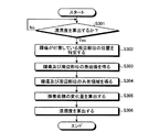
図5に示すように、X線CT装置1の画像処理部140は、浸潤度を算出するか否かを判定する(ステップS101)。例えば、画像処理部140は、入力部110を介して操作者から浸潤度を算出する旨の操作が行われたか否かを判定する。ここで、浸潤度を算出しない場合には(ステップS101否定)、画像処理部140は、待機状態となる。なお、浸潤度を算出することが予め決められている場合には、画像処理部140は、ステップS101の処理を行わなくてもよい。
As shown in FIG. 5, the
一方、浸潤度を算出する場合には(ステップS101肯定)、画像処理部140の特定部141は、ボリュームデータ内の腫瘍の位置と、かかる腫瘍の周辺部位の位置とを特定する(ステップS102)。
On the other hand, when calculating the degree of infiltration (Yes at Step S101), the specifying
続いて、移動情報算出部142は、ボリュームデータが再構成されるたびに、基準点から腫瘍までの相対距離、及び、基準部位から周辺部位までの相対距離をボクセル毎に算出し(ステップS103)、異なる時相のボリュームデータを比較することにより、各ボクセルの周期的な距離変動を算出する(ステップS104)。 Subsequently, each time the volume data is reconstructed, the movement information calculation unit 142 calculates the relative distance from the reference point to the tumor and the relative distance from the reference part to the peripheral part for each voxel (step S103). By comparing the volume data of different time phases, the periodic distance fluctuation of each voxel is calculated (step S104).
続いて、相対関係算出部143は、比較対象とする腫瘍及び周辺部位の周期的な距離変動について、振幅や周期等を算出する(ステップS105)。このとき、相対関係算出部143は、周辺部位が対称部位である場合には(ステップS106肯定)、対側部位を示す各ボクセルの動きベクトルを用いて、周辺部位の振幅及び周期を正規化する(ステップS107)。 Subsequently, the relative relationship calculation unit 143 calculates an amplitude, a period, and the like for the periodic distance variation between the tumor to be compared and the peripheral part (step S105). At this time, if the peripheral part is a symmetric part (Yes in step S106), the relative relationship calculation unit 143 normalizes the amplitude and period of the peripheral part using the motion vector of each voxel indicating the opposite part. (Step S107).
そして、相対関係算出部143は、ステップS107において算出した振幅及び位相を用いて、腫瘍を示すボクセルの周期的な距離変動と、腫瘍の周辺部位を示すボクセルの周期的な距離変動との類似度を比較することで浸潤度を算出する(ステップS108)。 Then, using the amplitude and phase calculated in step S107, the relative relationship calculation unit 143 uses the similarity between the periodic distance variation of the voxel indicating the tumor and the periodic distance variation of the voxel indicating the peripheral region of the tumor. Are compared to calculate the degree of infiltration (step S108).
上述したように、第1の実施形態によれば、特定部141が、時相の異なる被検体内のボリュームデータから、腫瘍の位置と腫瘍の周辺に位置する周辺部位の位置とを特定し、移動情報算出部142が、特定部141によって特定された腫瘍及び周辺部位の位置に基づいて、腫瘍及び周辺部位の移動に関する移動情報を算出し、相対関係算出部143が、移動情報算出部142によって算出された腫瘍の移動情報と周辺部位の移動情報との関係に基づいて、病変部である腫瘍と腫瘍の周辺部位との関係性を示す情報として、腫瘍が周辺部位に浸潤している度合いを示す浸潤度を算出するので、客観的な腫瘍の浸潤度を提示することができる。
As described above, according to the first embodiment, the specifying
例えば、図3に示した例において、操作者等の人が、X線CT画像G11〜G14を見比べることにより、腫瘍A1の浸潤度を判断できるとも考えられる。しかし、人がX線CT画像G11〜G14を見ただけでは、動きの異なる領域を詳細に把握することが困難であり、主観的な浸潤度の判断しかできない。一方、上記のX線CT装置1では、腫瘍及び周辺部位の動きを比較することにより、客観的な腫瘍の浸潤度を算出することが可能になる。 For example, in the example shown in FIG. 3, it is considered that a person such as an operator can determine the degree of infiltration of the tumor A1 by comparing the X-ray CT images G11 to G14. However, if a person only sees the X-ray CT images G <b> 11 to G <b> 14, it is difficult to grasp in detail the regions with different motions, and only a subjective infiltration degree can be determined. On the other hand, in the X-ray CT apparatus 1 described above, it is possible to calculate an objective tumor invasion degree by comparing the movement of the tumor and the surrounding region.
なお、上記第1の実施形態では、周期的な距離変動の振幅や周期等を用いて、腫瘍の浸潤度を算出する例を示したが、腫瘍と周辺部位との距離の時間変動に基づいて、腫瘍の浸潤度を算出してもよい。この点について具体的に説明すると、腫瘍が周辺部位に浸潤しているほど、腫瘍がかかる周辺部位に一体化するので、腫瘍と周辺部位との距離は変動しないと考えられる。一方で、腫瘍が周辺部位に浸潤していないほど、腫瘍は周辺部位から剥がれやすいので、周辺部位の動きに伴って、腫瘍と周辺部位との距離は変動すると考えられる。 In the first embodiment, an example in which the degree of tumor infiltration is calculated using the amplitude or period of periodic distance fluctuation has been shown. However, based on the temporal fluctuation of the distance between the tumor and the surrounding site. Alternatively, the degree of tumor invasion may be calculated. This point will be specifically described. It is considered that the distance between the tumor and the peripheral site does not change because the tumor is infiltrated into the peripheral site and becomes integrated with the peripheral site. On the other hand, as the tumor does not infiltrate into the peripheral site, the tumor is more easily peeled off from the peripheral site. Therefore, it is considered that the distance between the tumor and the peripheral site varies with the movement of the peripheral site.
そこで、例えば、移動情報算出部142は、移動情報として、腫瘍の重心と周辺部位との距離の時間変動を算出する。そして、相対関係算出部143は、移動情報算出部142によって算出された腫瘍の重心と周辺部位との距離が所定の時間内(例えば、呼吸周期内)に変動しないほど、かかる周辺部位への腫瘍の浸潤度を高く算定し、腫瘍の重心と周辺部位との距離が所定の時間内に変動するほど、かかる周辺部位への腫瘍の浸潤度を低く算定する。 Therefore, for example, the movement information calculation unit 142 calculates the time variation of the distance between the center of gravity of the tumor and the peripheral site as movement information. Then, the relative relationship calculation unit 143 causes the tumor to the surrounding region so that the distance between the center of gravity of the tumor calculated by the movement information calculation unit 142 and the surrounding region does not vary within a predetermined time (for example, within the respiratory cycle). As the distance between the center of gravity of the tumor and the surrounding site varies within a predetermined time, the degree of tumor invasion into the surrounding site is calculated to be lower.
図6に、上記例の浸潤度算出処理の手順を示す。図6に示すように、特定部141は、浸潤度を算出する場合に(ステップS201肯定)、ボリュームデータ内の腫瘍の位置と、かかる腫瘍の周辺部位の位置とを特定する(ステップS202)。続いて、移動情報算出部142は、ボリュームデータが再構成されるたびに、腫瘍の重心と周辺部位との距離を算出し(ステップS203)、異なる時相のボリュームデータを比較することにより、腫瘍の重心と周辺部位との距離の周期的な時間変動を算出する(ステップS204)。そして、相対関係算出部143は、腫瘍の重心と周辺部位との距離が変動量に基づいて、周辺部位への腫瘍の浸潤度を算出する(ステップS205)。
FIG. 6 shows the procedure of the infiltration degree calculation process of the above example. As illustrated in FIG. 6, when calculating the infiltration degree (Yes at Step S201), the specifying
なお、上記例において、移動情報算出部142は、腫瘍の重心と周辺部位との距離の最大変動量を、所定の時間内(例えば、呼吸周期内)の周辺部位の最大移動量により正規化してもよい。具体的には、腫瘍の接着面のサイズは、周辺部位の移動に伴って変化する場合がある。例えば、周辺部位の移動量が大きいほど、かかる周辺部位に付着している腫瘍の接着面のサイズの変化量が大きくなる場合がある。このような場合、腫瘍の浸潤度と関係なく、周辺部位の移動に伴って、腫瘍の重心と周辺部位との距離が変動する場合がある。そこで、移動情報算出部142は、周辺部位の最大移動量を算出し、かかる最大移動量が大きいほど、腫瘍の重心と周辺部位との距離の最大変動量が小さくなるように正規化し、最大移動量が小さいほど、腫瘍の重心と周辺部位との距離の最大変動量が大きくなるように正規化してもよい。 In the above example, the movement information calculation unit 142 normalizes the maximum fluctuation amount of the distance between the center of gravity of the tumor and the peripheral part by the maximum movement amount of the peripheral part within a predetermined time (for example, within the respiratory cycle). Also good. Specifically, the size of the adhesion surface of the tumor may change as the peripheral site moves. For example, the larger the amount of movement of the peripheral part, the larger the amount of change in the size of the adhesion surface of the tumor attached to the peripheral part. In such a case, the distance between the center of gravity of the tumor and the peripheral site may vary as the peripheral site moves, regardless of the degree of tumor invasion. Therefore, the movement information calculation unit 142 calculates the maximum movement amount of the peripheral part, normalizes the maximum movement amount so that the maximum fluctuation amount of the distance between the center of gravity of the tumor and the peripheral part decreases as the maximum movement amount increases. You may normalize so that the largest variation | change_quantity of the distance of the gravity center of a tumor and a peripheral site | part may become large, so that quantity is small.
また、上記例において、移動情報算出部142は、腫瘍の重心と周辺部位の表面との最大距離と、腫瘍の最大半径とを比較することにより、腫瘍の浸潤度を算出してもよい。例えば、腫瘍の最大半径が、腫瘍の重心と周辺部位の表面との最大距離よりも大きい場合には、腫瘍が周辺部位内に入り込んでいることになる。この場合には、腫瘍の浸潤度が高いことが考えられる。したがって、移動情報算出部142は、腫瘍の重心と周辺部位の表面との最大距離と、腫瘍の最大半径との比に基づいて、腫瘍の浸潤度を算出してもよい。 In the above example, the movement information calculation unit 142 may calculate the degree of tumor invasion by comparing the maximum distance between the center of gravity of the tumor and the surface of the peripheral site and the maximum radius of the tumor. For example, when the maximum radius of the tumor is larger than the maximum distance between the center of gravity of the tumor and the surface of the peripheral site, the tumor has entered the peripheral site. In this case, it is considered that the degree of tumor infiltration is high. Therefore, the movement information calculation unit 142 may calculate the degree of tumor invasion based on the ratio of the maximum distance between the center of gravity of the tumor and the surface of the peripheral site and the maximum radius of the tumor.
また、上記第1の実施形態において、相対関係算出部143は、腫瘍の移動情報と周辺部位の移動情報との相対関係を算出する処理を行った後に、かかる相対関係から腫瘍の浸潤度を算出する処理を行わなくてもよい。そして、相対関係算出部143は、例えば、腫瘍の移動情報と周辺部位の移動情報との相対関係を示す情報を表示部120に表示制御してもよい。この例の場合であっても、操作者は、表示部120に表示された相対関係を確認することにより、客観的な腫瘍の浸潤度を判断することができる。
In the first embodiment, the relative relationship calculation unit 143 performs a process of calculating the relative relationship between the movement information of the tumor and the movement information of the peripheral site, and then calculates the degree of tumor invasion from the relative relationship. There is no need to perform the process. Then, for example, the relative relationship calculation unit 143 may display-control information indicating the relative relationship between the movement information of the tumor and the movement information of the peripheral site on the
また、上記第1の実施形態では、病変部として腫瘍を例に挙げて説明したが、上記のX線CT装置1は、腫瘍以外の病変部と周辺部位との関係を示す情報を算出してもよい。以下に、血栓が血管に密着している密着度を算出する例について説明する。 In the first embodiment, the tumor is described as an example of the lesion. However, the X-ray CT apparatus 1 calculates information indicating the relationship between the lesion other than the tumor and the peripheral site. Also good. Below, the example which calculates the contact | adhesion degree which the blood clot is closely_contact | adhered to the blood vessel is demonstrated.
まず、特定部141は、画像処理部140において再構成された各ボリュームデータに対して、領域拡張法や形状テンプレートを用いたパターンマッチング法などを用いてセグメンテーション処理を行うことにより各領域を抽出し、抽出した各領域に対して血管及び血栓の形状テンプレートを用いたパターンマッチング法や、血管及び血栓の輝度値のプロファイルを用いた手法等により、ボリュームデータに含まれる血管及び血栓の位置を特定する。また、移動情報算出部142は、特定部141によって特定された血管及び血栓の位置に基づいて、血管及び血栓の移動情報を算出する。
First, the specifying
そして、相対関係算出部143は、移動情報算出部142によって算出された血管の移動情報と血栓の移動情報との相対関係を算出し、算出した相対関係から血栓が血管に密着している密着度を算出する。例えば、相対関係算出部143は、血管の移動情報と血栓の移動情報とが類似しているほど、血栓の密着度を高く算出し、血管の移動情報と血栓の移動情報とが類似していないほど、血栓の密着度を低く算出する。これにより、第1の実施形態に係るX線CT装置1は、血管及び血栓の移動情報に基づいて、血管に対する血栓の密着度、言い換えれば、血栓が血管から剥がれやすいか否かを示す指標値を提示することができる。 Then, the relative relationship calculation unit 143 calculates the relative relationship between the blood vessel movement information and the thrombus movement information calculated by the movement information calculation unit 142, and the degree of adhesion that the thrombus is in close contact with the blood vessel from the calculated relative relationship. Is calculated. For example, as the blood vessel movement information and the thrombus movement information are more similar, the relative relationship calculation unit 143 calculates the clot adhesion degree higher, and the blood vessel movement information and the thrombus movement information are not similar. The lower the degree of adhesion of the thrombus is calculated. Thereby, the X-ray CT apparatus 1 according to the first embodiment, based on the movement information of the blood vessels and the thrombus, indicates the degree of adhesion of the thrombus to the blood vessel, in other words, the index value indicating whether the thrombus is easily detached from the blood vessel. Can be presented.
(第2の実施形態)
上記第1の実施形態では、腫瘍や周辺部位の移動情報に基づいて、腫瘍の浸潤度を算出する例を示した。第2の実施形態では、腫瘍と周辺部位との接着面積に基づいて、腫瘍の浸潤度を算出する例について説明する。
(Second Embodiment)
In the said 1st Embodiment, the example which calculates the infiltration degree of a tumor was shown based on the movement information of a tumor or a peripheral region. 2nd Embodiment demonstrates the example which calculates the infiltration degree of a tumor based on the adhesion area of a tumor and a peripheral region.
図7は、第2の実施形態における画像処理部240の構成例を示す図である。なお、第2の実施形態に係るX線CT装置の構成は、図1に示したX線CT装置1の構成と同様であるので、ここでは説明を省略する。図7に示すように画像処理部240は、特定部241と、接着面積算出部242と、相対関係算出部243とを有する。
FIG. 7 is a diagram illustrating a configuration example of the
特定部241は、画像処理部240において再構成されたボリュームデータ内の腫瘍と、かかる腫瘍が付着していると想定される周辺部位とを特定する。特定部241は、各時相に対応するボリュームデータ毎に、同様の処理を行う。なお、特定部241は、特定部141と同様に、セグメンテーション処理等を行うことにより各領域を抽出し、パターンマッチング法等により、ボリュームデータに含まれる腫瘍及び周辺部位の位置を特定する。
The specifying
接着面積算出部242は、特定部241によって特定された腫瘍が周辺部位に接着している面の接着面積を時相毎に算出する。例えば、接着面積算出部242は、特定部241によるセグメンテーション処理の結果から、腫瘍の表面像と、かかる腫瘍が付着している周辺部位の表面像とを得ることができる。そして、接着面積算出部242は、腫瘍の表面像と周辺部位の表面像との共有領域を、腫瘍が周辺部位に接着している面の接着面積として算出できる。
The adhesion
また、例えば、接着面積算出部242は、腫瘍が肺野に付着している場合等には、特定部241によるセグメンテーション処理等の結果、腫瘍が抜けた状態の肺野のボリュームデータを得ることができる。したがって、接着面積算出部242は、かかるボリュームデータの空洞化している領域を腫瘍が接着している接着面積として算出することができる。かかる手法は、例えば、胸壁、縦隔や横隔膜等と腫瘍との接着面積を算出する場合に用いることができる。
In addition, for example, when the tumor is attached to the lung field, the adhesion
相対関係算出部243は、接着面積算出部242によって算出された接着面積の変動量が小さいほど、周辺部位への腫瘍の浸潤度を高く算出し、接着面積の変動量が大きいほど、周辺部位への腫瘍の浸潤度を低く算出する。
The relative
図8及び図9を用いて、相対関係算出部243による処理の一例を説明する。図8は、腫瘍と肺野との接着面積の一例を示す図であり、図9は、接着面積の変動量の一例を示す図である。
An example of processing by the relative
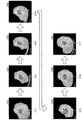
図8に示した例では、X線CT画像G21、G22、G23、G24、G25、G26、G27の順に撮像されたものとする。なお、図8では、説明を簡単にするために、断面画像を示している。 In the example shown in FIG. 8, it is assumed that X-ray CT images G21, G22, G23, G24, G25, G26, and G27 are taken in this order. In FIG. 8, a cross-sectional image is shown for ease of explanation.
接着面積算出部242は、図8に例示したX線CT画像G21〜G27が撮像された場合に、腫瘍の表面像と周辺部位の表面像との共有領域を、腫瘍が周辺部位に接着している面の接着面積として算出する。ここでは、接着面積算出部242は、図8に例示するように、接着面積C11〜C17を算出する。
When the X-ray CT images G21 to G27 illustrated in FIG. 8 are captured, the adhesion
そして、相対関係算出部243は、図9に例示するように、接着面積算出部242によって算出された接着面積を時系列に沿ってプロットすることにより、接着面積の時間変動を得る。そして、相対関係算出部243は、所定の時間内(例えば、呼吸周期内)における接着面積の最大変動量ΔAを算出し、算出した最大変動量ΔAが小さいほど周辺部位への腫瘍の浸潤度を高く算出し、最大変動量ΔAが大きいほど周辺部位への腫瘍の浸潤度を低く算出する。
Then, as illustrated in FIG. 9, the relative
次に、図10を用いて、第2の実施形態に係るX線CT装置による浸潤度算出処理の手順について説明する。図10は、第2の実施形態に係るX線CT装置による浸潤度算出処理手順を示すフローチャートである。 Next, the procedure of the infiltration degree calculation process by the X-ray CT apparatus according to the second embodiment will be described with reference to FIG. FIG. 10 is a flowchart showing an infiltration degree calculation processing procedure by the X-ray CT apparatus according to the second embodiment.
図10に示すように、画像処理部240は、浸潤度を算出するか否かを判定する(ステップS301)。ここで、浸潤度を算出しない場合には(ステップS301否定)、画像処理部240は、待機状態となる。
As shown in FIG. 10, the
一方、浸潤度を算出する場合には(ステップS301肯定)、画像処理部240の特定部241は、ボリュームデータ内の腫瘍と腫瘍が付着していると想定される周辺部位とを特定する(ステップS302)。
On the other hand, when calculating the degree of infiltration (Yes at Step S301), the specifying
続いて、接着面積算出部242は、特定部241によるセグメンテーション処理の結果から、腫瘍及び周辺部位の表面像を取得する(ステップS303)。そして、接着面積算出部242は、腫瘍の表面像と周辺部位の表面像との共有領域を取得する(ステップS304)。これにより、接着面積算出部242は、共有領域の面積を算出することにより、腫瘍が周辺部位に接着している面の接着面積を算出する。
Subsequently, the adhesion
続いて、相対関係算出部243は、接着面積算出部242によって算出された接着面積の所定時間内における変動量を算出する(ステップS305)。そして、相対関係算出部243は、接着面積の変動量に基づいて、周辺部位への腫瘍の浸潤度を算出する(ステップS306)。
Subsequently, the relative
上述したように、第2の実施形態によれば、特定部241が、時相の異なる被検体内の画像データから、腫瘍の位置と腫瘍が付着していると想定される周辺部位の位置とを特定し、接着面積算出部242が、特定部241によって特定された腫瘍が周辺部位に接着している面の接着面積を時相毎に算出し、相対関係算出部243が、接着面積算出部242によって算出された接着面積の変動量が小さいほど、腫瘍が周辺部位に浸潤している度合いを示す浸潤度を高く算出するので、接着面積を算出するだけで、客観的な腫瘍の浸潤度を提示することができる。
As described above, according to the second embodiment, the specifying
なお、上記第2の実施形態では、病変部として腫瘍を例に挙げて説明したが、第2の実施形態に係るX線CT装置は、腫瘍以外の病変部と周辺部位との関係を示す情報を算出してもよい。以下に、血栓が血管に密着している密着度を算出する例について説明する。 In the second embodiment, the tumor has been described as an example of the lesion. However, the X-ray CT apparatus according to the second embodiment provides information indicating the relationship between the lesion other than the tumor and the peripheral site. May be calculated. Below, the example which calculates the contact | adhesion degree which the blood clot is closely_contact | adhered to the blood vessel is demonstrated.
まず、特定部241は、特定部141と同様にセグメンテーション処理等を行うことにより、ボリュームデータに含まれる血管及び血栓の位置を特定する。また、接着面積算出部242は、例えば、特定部241によるセグメンテーション処理の結果から、血管の表面像と血栓の表面像との共有領域を、血栓が血管に接着している面の接着面積として算出する。そして、相対関係算出部243は、接着面積算出部242によって算出された接着面積の変動量が小さいほど、血管に対する血栓の密着度を高く算出し、接着面積の変動量が大きいほど、血管に対する血栓の密着度を低く算出する。これにより、第2の実施形態に係るX線CT装置は、血管と血栓との接着面積の変動量に基づいて、血管に対する血栓の密着度、言い換えれば、血栓が血管から剥がれやすいか否かを示す指標値を提示することができる。
First, the specifying
また、上記の実施形態では、X線CT装置が、浸潤度の高低が表されたX線CT画像を表示部120に表示する例を示した。しかし、X線CT装置は、X線CT画像だけでなく、移動情報算出部142によって算出された移動情報(図4等)、接着面積算出部242によって算出された接着面積に関する情報、浸潤度等を表示部120に表示制御してもよい。また、X線CT装置は、これらの各種情報を所定の記憶部に記憶してもよい。
In the above embodiment, the X-ray CT apparatus displays an example in which the X-ray CT image showing the degree of infiltration is displayed on the
また、上記の実施形態では、X線CT装置がパターンマッチング法等の情報処理を行うことにより腫瘍及び周辺部位の位置を特定する例を示した。X線CT装置は、X線CT画像を表示部120に表示した後に、入力部110を介して操作者から腫瘍及び周辺部位の位置を指定する操作を受け付けてもよい。かかる場合には、X線CT装置は、操作者による指定操作に従って、腫瘍及び周辺部位の位置に特定する。
In the above embodiment, the X-ray CT apparatus performs the information processing such as the pattern matching method to identify the position of the tumor and the peripheral site. After displaying the X-ray CT image on the
また、上記の実施形態では、X線CT装置が連続する2個のボリュームデータを随時比較することにより移動情報を算出する例を示した。しかし、X線CT装置は、全てのボリュームデータについて処理を行う必要はなく、処理対象のボリュームデータを間引きした上で移動情報を算出してもよい。 In the above-described embodiment, an example in which the movement information is calculated by the X-ray CT apparatus comparing two pieces of continuous volume data as needed is shown. However, the X-ray CT apparatus does not need to perform processing on all volume data, and may calculate movement information after thinning out volume data to be processed.
また、上記の実施形態では、X線CT装置がボリュームデータを用いて移動情報を算出する例を示した。しかし、2次元の画像データを用いて移動情報を算出することで、腫瘍の浸潤度を算出してもよい。 In the above embodiment, an example in which the X-ray CT apparatus calculates movement information using volume data has been shown. However, the degree of tumor invasion may be calculated by calculating movement information using two-dimensional image data.
また、上記の実施形態では、X線CT装置がコンベンショナルスキャンを行う場合を例に挙げて説明した。しかし、上記の実施形態は、多時相においてボリュームデータを再構成することができるヘリカルスキャンを行うX線CT装置にも適用することができる。 In the above embodiment, the case where the X-ray CT apparatus performs conventional scanning has been described as an example. However, the above embodiment can also be applied to an X-ray CT apparatus that performs a helical scan that can reconstruct volume data in multiple time phases.
また、上記の実施形態は、距離変動等の周期的な情報を得るほど多時相で撮像処理を行えないX線CT装置にも適用することができる。例えば、腫瘍や周辺部位の移動量の最大値を、第1の実施形態で説明した振幅「Δd」として用いることにより、腫瘍の浸潤度を算出することもできる。 The above-described embodiment can also be applied to an X-ray CT apparatus that cannot perform imaging processing in multiple time phases such that periodic information such as distance variation is obtained. For example, the degree of tumor invasion can be calculated by using the maximum value of the amount of movement of the tumor or the surrounding region as the amplitude “Δd” described in the first embodiment.
以上説明したとおり、第1及び第2の実施形態によれば、病変部と周辺部位との関係性を示す情報を提示することができる。 As described above, according to the first and second embodiments, it is possible to present information indicating the relationship between a lesioned part and a peripheral part.
本発明のいくつかの実施形態を説明したが、これらの実施形態は、例として提示したものであり、発明の範囲を限定することは意図していない。これら実施形態は、その他の様々な形態で実施されることが可能であり、発明の要旨を逸脱しない範囲で、種々の省略、置き換え、変更を行うことができる。これら実施形態やその変形は、発明の範囲や要旨に含まれると同様に、特許請求の範囲に記載された発明とその均等の範囲に含まれるものである。 Although several embodiments of the present invention have been described, these embodiments are presented by way of example and are not intended to limit the scope of the invention. These embodiments can be implemented in various other forms, and various omissions, replacements, and changes can be made without departing from the spirit of the invention. These embodiments and their modifications are included in the scope and gist of the invention, and are also included in the invention described in the claims and the equivalents thereof.
1 X線CT装置
140 画像処理部
141 特定部
142 移動情報算出部
143 相対関係算出部
240 画像処理部
241 特定部
242 接着面積算出部
243 相対関係算出部
DESCRIPTION OF SYMBOLS 1
Claims (11)
前記生成部によって生成された時相の異なる被検体内の画像データから、病変部の位置と該病変部の周辺に位置する周辺部位の位置とを特定する特定部と、
前記特定部によって特定された病変部及び周辺部位の位置に基づいて、病変部及び周辺部位の移動に関する移動情報を算出する移動情報算出部と、
前記移動情報算出部によって算出された前記病変部の移動情報と前記周辺部位の移動情報との相対関係を算出する相対関係算出部と、
を有する、X線CT装置。 A generating unit that generates image data in a subject having different time phases based on X-rays transmitted through the subject;
From the image data in the subject with different time phases generated by the generating unit, a specifying unit that specifies the position of the lesioned part and the position of the peripheral part located around the lesioned part,
A movement information calculation unit that calculates movement information related to movement of the lesion part and the peripheral part based on the position of the lesion part and the peripheral part specified by the specification part;
A relative relationship calculation unit that calculates a relative relationship between the movement information of the lesion portion calculated by the movement information calculation unit and the movement information of the peripheral site;
X-ray CT apparatus.
前記移動情報算出部は、前記腫瘍及び前記周辺部位の位置に基づいて、腫瘍及び周辺部位の移動に関する移動情報を算出し、
前記相対関係算出部は、前記腫瘍の移動情報と前記周辺部位の移動情報との相対関係に基づいて、前記腫瘍が前記周辺部位に浸潤している度合いを示す浸潤度を算出する、
請求項1に記載のX線CT装置。 The specifying unit specifies a position of a tumor that is the lesioned part and a position of a peripheral part located around the tumor,
The movement information calculation unit calculates movement information related to movement of the tumor and the peripheral part based on the position of the tumor and the peripheral part,
The relative relationship calculation unit calculates an infiltration degree indicating a degree of infiltration of the tumor into the peripheral site based on a relative relationship between the movement information of the tumor and the movement information of the peripheral site.
The X-ray CT apparatus according to claim 1.
前記相対関係算出部は、前記移動情報算出部によって算出された移動情報と前記腫瘍の移動情報との関係に基づいて、前記領域毎に、該領域に腫瘍が浸潤している浸潤度を算出する、
請求項2に記載のX線CT装置。 The movement information calculation unit calculates movement information of the area for each area in the peripheral part,
The relative relationship calculation unit calculates, for each region, an infiltration degree in which the tumor has infiltrated based on the relationship between the movement information calculated by the movement information calculation unit and the movement information of the tumor. ,
The X-ray CT apparatus according to claim 2.
前記相対関係算出部は、前記移動情報算出部によって算出された前記腫瘍の周期的な位置変動と前記周辺部位の周期的な位置変動との類似度に基づいて、前記浸潤度を算出する、
請求項2に記載のX線CT装置。 The movement information calculation unit calculates, as the movement information, a periodic position fluctuation of the tumor and a periodic position fluctuation of the peripheral part,
The relative relationship calculation unit calculates the degree of infiltration based on the similarity between the periodic position variation of the tumor calculated by the movement information calculation unit and the periodic position variation of the peripheral site,
The X-ray CT apparatus according to claim 2.
請求項4に記載のX線CT装置。 The relative relationship calculation unit includes at least one of an amplitude difference, a period difference, or a phase difference between the periodic position variation of the tumor calculated by the movement information calculation unit and the periodic position variation of the peripheral site. To determine the degree of similarity of both position variations based on
The X-ray CT apparatus according to claim 4.
前記相対関係算出部は、前記移動情報算出部によって算出された前記腫瘍の重心と前記周辺部位との距離の時間変動に基づいて、前記浸潤度を算出する、
請求項2に記載のX線CT装置。 The movement information calculation unit calculates, as the movement information, a distance between the center of gravity of the tumor and a peripheral part to which the tumor is attached,
The relative relationship calculation unit calculates the degree of infiltration based on the temporal variation in the distance between the center of gravity of the tumor and the peripheral site calculated by the movement information calculation unit.
The X-ray CT apparatus according to claim 2.
前記相対関係算出部は、前記移動情報算出部によって算出された対側部位の移動情報を用いて、前記周辺部位の移動情報を正規化した後に、前記浸潤度を算出する、
請求項2に記載のX線CT装置。 The movement information calculation unit calculates movement information of a contralateral part facing the peripheral part when the peripheral part is a symmetrical part that exists substantially symmetrically in the human body,
The relative relationship calculation unit calculates the infiltration degree after normalizing the movement information of the peripheral part using the movement information of the contralateral part calculated by the movement information calculation unit.
The X-ray CT apparatus according to claim 2.
前記病変部及び周辺部位の位置に基づいて、病変部及び周辺部位の移動に関する移動情報を算出し、
前記病変部の移動情報と前記周辺部位の移動情報との相対関係を算出する、
ことを含む、画像処理方法。 From the image data in the subject corresponding to a plurality of time phases, the position of the lesioned part and the position of the peripheral part located around the lesioned part are identified,
Based on the position of the lesioned part and the surrounding part, calculating movement information regarding the movement of the lesioned part and the surrounding part,
Calculating the relative relationship between the movement information of the lesioned part and the movement information of the peripheral part,
An image processing method.
前記生成部によって生成された時相の異なる被検体内の画像データから、病変部の位置と該病変部が付着していると想定される周辺部位の位置とを特定する特定部と、
前記特定部によって特定された病変部が前記周辺部位に接着している面の接着面積を時相毎に算出する接着面積算出部と、
を有する、X線CT装置。 A generating unit that generates image data in a subject having different time phases based on X-rays transmitted through the subject;
From the image data in the subject with different time phases generated by the generation unit, a specifying unit for specifying the position of the lesioned part and the position of the peripheral part assumed to be attached to the lesioned part,
An adhesion area calculation unit that calculates, for each phase, an adhesion area of a surface where the lesion part identified by the identification unit adheres to the peripheral site;
X-ray CT apparatus.
請求項9に記載のX線CT装置。 The degree of invasion indicating the degree that the tumor is infiltrating into the peripheral site is reduced as the variation amount of the adhesive area where the tumor that is the lesion site calculated by the adhesion area calculating unit is adhered to the peripheral site is small. It further has a relative relationship calculation unit that calculates high,
The X-ray CT apparatus according to claim 9.
前記病変部が前記周辺部位に接着している面の接着面積を時相毎に算出する、
ことを含む、画像処理方法。 From the image data in the subject corresponding to a plurality of time phases, the position of the lesioned part and the position of the peripheral part where the lesioned part is assumed to be attached are identified,
Calculate the adhesion area of the surface where the lesion is adhered to the peripheral site for each phase,
An image processing method.
Priority Applications (1)
| Application Number | Priority Date | Filing Date | Title |
|---|---|---|---|
| JP2012040107A JP5872323B2 (en) | 2011-03-29 | 2012-02-27 | X-ray CT apparatus and image processing method |
Applications Claiming Priority (3)
| Application Number | Priority Date | Filing Date | Title |
|---|---|---|---|
| JP2011072805 | 2011-03-29 | ||
| JP2011072805 | 2011-03-29 | ||
| JP2012040107A JP5872323B2 (en) | 2011-03-29 | 2012-02-27 | X-ray CT apparatus and image processing method |
Related Child Applications (1)
| Application Number | Title | Priority Date | Filing Date |
|---|---|---|---|
| JP2016000744A Division JP6486842B2 (en) | 2011-03-29 | 2016-01-05 | X-ray CT apparatus and image processing apparatus |
Publications (2)
| Publication Number | Publication Date |
|---|---|
| JP2012213604A JP2012213604A (en) | 2012-11-08 |
| JP5872323B2 true JP5872323B2 (en) | 2016-03-01 |
Family
ID=46025381
Family Applications (2)
| Application Number | Title | Priority Date | Filing Date |
|---|---|---|---|
| JP2012040107A Active JP5872323B2 (en) | 2011-03-29 | 2012-02-27 | X-ray CT apparatus and image processing method |
| JP2016000744A Active JP6486842B2 (en) | 2011-03-29 | 2016-01-05 | X-ray CT apparatus and image processing apparatus |
Family Applications After (1)
| Application Number | Title | Priority Date | Filing Date |
|---|---|---|---|
| JP2016000744A Active JP6486842B2 (en) | 2011-03-29 | 2016-01-05 | X-ray CT apparatus and image processing apparatus |
Country Status (4)
| Country | Link |
|---|---|
| US (1) | US9230334B2 (en) |
| EP (1) | EP2506216B1 (en) |
| JP (2) | JP5872323B2 (en) |
| CN (1) | CN102727235B (en) |
Families Citing this family (22)
| Publication number | Priority date | Publication date | Assignee | Title |
|---|---|---|---|---|
| KR102070427B1 (en) * | 2012-08-08 | 2020-01-28 | 삼성전자주식회사 | Method and Apparatus for tracking the position of tumor |
| DE102012221930A1 (en) * | 2012-11-30 | 2014-06-05 | Siemens Aktiengesellschaft | Method and PET system for locating an object |
| JP6253992B2 (en) * | 2014-01-09 | 2017-12-27 | 富士通株式会社 | Organ position estimation apparatus, organ position estimation apparatus control method, and organ position estimation apparatus control program |
| JP6510189B2 (en) | 2014-06-23 | 2019-05-08 | キヤノンメディカルシステムズ株式会社 | Medical image processing device |
| JP6532206B2 (en) | 2014-10-01 | 2019-06-19 | キヤノン株式会社 | Medical image processing apparatus, medical image processing method |
| CN105982685A (en) | 2015-03-03 | 2016-10-05 | 东芝医疗系统株式会社 | Medical image processing device and method and medical image diagnosing device and method |
| US20160310094A1 (en) * | 2015-04-27 | 2016-10-27 | National Cancer Center | Medical image processing apparatus |
| JP6755468B2 (en) * | 2015-04-27 | 2020-09-16 | 国立研究開発法人国立がん研究センター | Medical image processing device |
| EP3324852A1 (en) * | 2015-07-17 | 2018-05-30 | Koninklijke Philips N.V. | Guidance for lung cancer radiation |
| JP6565422B2 (en) * | 2015-07-24 | 2019-08-28 | 富士通株式会社 | Image processing program, image processing apparatus, and image processing method |
| CN107949330B (en) * | 2015-09-02 | 2021-04-20 | 富士通株式会社 | Video interpretation support program, cross-sectional image generation device, and cross-sectional image generation method |
| US11138735B2 (en) | 2017-10-17 | 2021-10-05 | Canon Medical Systems Corporation | Image processing apparatus and medical image taking apparatus |
| JP7166870B2 (en) * | 2017-10-17 | 2022-11-08 | キヤノンメディカルシステムズ株式会社 | Image processing device and medical imaging device |
| JP7173747B2 (en) * | 2018-04-11 | 2022-11-16 | キヤノンメディカルシステムズ株式会社 | medical image processor |
| JP7114348B2 (en) * | 2018-06-06 | 2022-08-08 | キヤノンメディカルシステムズ株式会社 | Tumor position display device and radiotherapy system |
| CN110033019B (en) * | 2019-03-06 | 2021-07-27 | 腾讯科技(深圳)有限公司 | Method and device for detecting abnormality of human body part and storage medium |
| JP7270453B2 (en) | 2019-04-26 | 2023-05-10 | キヤノン株式会社 | Image processing device, image processing method and program |
| JP6748762B2 (en) * | 2019-05-23 | 2020-09-02 | キヤノン株式会社 | Medical image processing apparatus and medical image processing method |
| JP7015351B2 (en) * | 2020-08-06 | 2022-02-02 | キヤノン株式会社 | Medical image processing device, medical image processing method |
| JP7500360B2 (en) * | 2020-09-11 | 2024-06-17 | キヤノン株式会社 | Information processing device, information processing method, and program |
| JP2022109778A (en) * | 2021-01-15 | 2022-07-28 | キヤノン株式会社 | Information processing device, information processing method and program |
| EP4295778A4 (en) * | 2021-02-22 | 2024-07-24 | Fujifilm Corp | Medical image processing device, medical image processing method, and program |
Family Cites Families (18)
| Publication number | Priority date | Publication date | Assignee | Title |
|---|---|---|---|---|
| EP1225545A1 (en) * | 2001-01-23 | 2002-07-24 | Koninklijke Philips Electronics N.V. | Image processing method for tracking the deformation of an organ in time |
| JP2003180697A (en) * | 2001-12-18 | 2003-07-02 | Olympus Optical Co Ltd | Ultrasonic diagnostic equipment |
| GB2414357A (en) * | 2004-05-18 | 2005-11-23 | Medicsight Plc | Nodule boundary detection |
| US8989349B2 (en) * | 2004-09-30 | 2015-03-24 | Accuray, Inc. | Dynamic tracking of moving targets |
| JP3932303B2 (en) | 2005-05-13 | 2007-06-20 | 独立行政法人放射線医学総合研究所 | Organ dynamics quantification method, apparatus, organ position prediction method, apparatus, radiation irradiation method, apparatus, and organ abnormality detection apparatus |
| WO2006126303A1 (en) * | 2005-05-27 | 2006-11-30 | Hitachi Medical Corporation | Velocity measuring method and velocity measuring device using the same |
| JP4861647B2 (en) | 2005-06-23 | 2012-01-25 | 株式会社東芝 | Medical image diagnostic apparatus and image reconstruction method |
| WO2007083745A1 (en) * | 2006-01-20 | 2007-07-26 | Hitachi Medical Corporation | Elastic image display method and elastic image display |
| WO2007133932A2 (en) * | 2006-05-11 | 2007-11-22 | Koninklijke Philips Electronics, N.V. | Deformable registration of images for image guided radiation therapy |
| JP2008036284A (en) * | 2006-08-09 | 2008-02-21 | Toshiba Corp | Medical image composition method and its apparatus |
| JP5242094B2 (en) * | 2007-07-17 | 2013-07-24 | 株式会社東芝 | Medical image photographing apparatus, medical image processing apparatus, and medical image processing program |
| JP5198883B2 (en) * | 2008-01-16 | 2013-05-15 | 富士フイルム株式会社 | Tumor area size measuring method, apparatus and program |
| JP2009178185A (en) * | 2008-01-29 | 2009-08-13 | Fujifilm Corp | Medical imaging apparatus and medical imaging method |
| JP2010069099A (en) * | 2008-09-19 | 2010-04-02 | Toshiba Corp | Image processing apparatus and x-ray computed tomography apparatus |
| JP2010075549A (en) * | 2008-09-26 | 2010-04-08 | Toshiba Corp | Image processor |
| JP5575388B2 (en) * | 2008-12-03 | 2014-08-20 | 株式会社東芝 | Image display apparatus and X-ray CT apparatus |
| JP5066233B2 (en) * | 2010-08-02 | 2012-11-07 | 国立大学法人神戸大学 | Diagnostic imaging processing device |
| JP5517828B2 (en) * | 2010-08-17 | 2014-06-11 | 株式会社モリタ製作所 | Hollow waveguide and laser therapy device |
-
2012
- 2012-02-27 JP JP2012040107A patent/JP5872323B2/en active Active
- 2012-03-27 US US13/431,097 patent/US9230334B2/en active Active
- 2012-03-28 CN CN201210086890.3A patent/CN102727235B/en active Active
- 2012-03-29 EP EP12162302.9A patent/EP2506216B1/en active Active
-
2016
- 2016-01-05 JP JP2016000744A patent/JP6486842B2/en active Active
Also Published As
| Publication number | Publication date |
|---|---|
| EP2506216B1 (en) | 2016-02-10 |
| US9230334B2 (en) | 2016-01-05 |
| JP6486842B2 (en) | 2019-03-20 |
| JP2012213604A (en) | 2012-11-08 |
| US20120250966A1 (en) | 2012-10-04 |
| EP2506216A2 (en) | 2012-10-03 |
| EP2506216A3 (en) | 2014-01-22 |
| CN102727235A (en) | 2012-10-17 |
| CN102727235B (en) | 2015-04-15 |
| JP2016093531A (en) | 2016-05-26 |
Similar Documents
| Publication | Publication Date | Title |
|---|---|---|
| JP6486842B2 (en) | X-ray CT apparatus and image processing apparatus | |
| EP2528040B1 (en) | Analysis of corresponding radiographies | |
| US10413253B2 (en) | Method and apparatus for processing medical image | |
| US9202301B2 (en) | Medical image display apparatus and X-ray diagnosis apparatus | |
| EP3264985B1 (en) | Tomography imaging apparatus and method of reconstructing tomography image | |
| CN108289651B (en) | System for tracking the ultrasonic probe in physical feeling | |
| EP1699361B1 (en) | System for guiding a medical instrument in a patient body | |
| TW202416901A (en) | System and method for lung-volume-gated x-ray imaging | |
| KR20140096919A (en) | Method and Apparatus for medical image registration | |
| TWI840465B (en) | System and method for determining radiation parameters and non-transitory computer-readable storage medium thereof | |
| KR20140126815A (en) | Method, apparatus and system for tracing deformation of organ in respiration cycle | |
| US20140093030A1 (en) | X-ray ct system, image display device, and image display method | |
| US20220036545A1 (en) | Breast mapping and abnormality localization | |
| KR20160061248A (en) | Apparatus for processing medical image and method for processing medical image thereof | |
| US9636076B2 (en) | X-ray CT apparatus and image processing method | |
| JP2012000448A (en) | Image processor and x-ray ct apparatus | |
| WO2007066096A2 (en) | Interventional device location method and apparatus | |
| KR101525040B1 (en) | Method and Apparatus of Generation of reference image for determining scan range of pre-operative images | |
| US20230045275A1 (en) | Methods and system for guided device insertion during medical imaging | |
| EP4014875B1 (en) | Method for controlling a medical imaging examination of a subject, medical imaging system and computer-readable data storage medium | |
| US11123025B2 (en) | Iso-centering in C-arm computer tomography | |
| JP2009136573A (en) | Kinetics photographing system | |
| WO2019057863A1 (en) | Ct lung elastography with a ventilation assist system |
Legal Events
| Date | Code | Title | Description |
|---|---|---|---|
| A621 | Written request for application examination |
Free format text: JAPANESE INTERMEDIATE CODE: A621 Effective date: 20150204 |
|
| A711 | Notification of change in applicant |
Free format text: JAPANESE INTERMEDIATE CODE: A712 Effective date: 20150610 |
|
| RD01 | Notification of change of attorney |
Free format text: JAPANESE INTERMEDIATE CODE: A7421 Effective date: 20151102 |
|
| A977 | Report on retrieval |
Free format text: JAPANESE INTERMEDIATE CODE: A971007 Effective date: 20151130 |
|
| TRDD | Decision of grant or rejection written | ||
| A01 | Written decision to grant a patent or to grant a registration (utility model) |
Free format text: JAPANESE INTERMEDIATE CODE: A01 Effective date: 20151215 |
|
| A61 | First payment of annual fees (during grant procedure) |
Free format text: JAPANESE INTERMEDIATE CODE: A61 Effective date: 20160113 |
|
| R150 | Certificate of patent or registration of utility model |
Ref document number: 5872323 Country of ref document: JP Free format text: JAPANESE INTERMEDIATE CODE: R150 |
|
| S111 | Request for change of ownership or part of ownership |
Free format text: JAPANESE INTERMEDIATE CODE: R313117 |
|
| R350 | Written notification of registration of transfer |
Free format text: JAPANESE INTERMEDIATE CODE: R350 |
|
| S533 | Written request for registration of change of name |
Free format text: JAPANESE INTERMEDIATE CODE: R313533 |
|
| R350 | Written notification of registration of transfer |
Free format text: JAPANESE INTERMEDIATE CODE: R350 |