JP5763631B2 - Stimulus-responsive carrier for MPI-induced drug delivery - Google Patents
Stimulus-responsive carrier for MPI-induced drug delivery Download PDFInfo
- Publication number
- JP5763631B2 JP5763631B2 JP2012518140A JP2012518140A JP5763631B2 JP 5763631 B2 JP5763631 B2 JP 5763631B2 JP 2012518140 A JP2012518140 A JP 2012518140A JP 2012518140 A JP2012518140 A JP 2012518140A JP 5763631 B2 JP5763631 B2 JP 5763631B2
- Authority
- JP
- Japan
- Prior art keywords
- shell structure
- composition
- drug
- contrast agent
- magnetic
- Prior art date
- Legal status (The legal status is an assumption and is not a legal conclusion. Google has not performed a legal analysis and makes no representation as to the accuracy of the status listed.)
- Expired - Fee Related
Links
Images


Classifications
-
- A—HUMAN NECESSITIES
- A61—MEDICAL OR VETERINARY SCIENCE; HYGIENE
- A61K—PREPARATIONS FOR MEDICAL, DENTAL OR TOILETRY PURPOSES
- A61K9/00—Medicinal preparations characterised by special physical form
- A61K9/0002—Galenical forms characterised by the drug release technique; Application systems commanded by energy
- A61K9/0009—Galenical forms characterised by the drug release technique; Application systems commanded by energy involving or responsive to electricity, magnetism or acoustic waves; Galenical aspects of sonophoresis, iontophoresis, electroporation or electroosmosis
-
- A—HUMAN NECESSITIES
- A61—MEDICAL OR VETERINARY SCIENCE; HYGIENE
- A61P—SPECIFIC THERAPEUTIC ACTIVITY OF CHEMICAL COMPOUNDS OR MEDICINAL PREPARATIONS
- A61P41/00—Drugs used in surgical methods, e.g. surgery adjuvants for preventing adhesion or for vitreum substitution
Landscapes
- Health & Medical Sciences (AREA)
- Public Health (AREA)
- Veterinary Medicine (AREA)
- General Health & Medical Sciences (AREA)
- Medicinal Chemistry (AREA)
- Pharmacology & Pharmacy (AREA)
- Life Sciences & Earth Sciences (AREA)
- Animal Behavior & Ethology (AREA)
- Chemical & Material Sciences (AREA)
- Engineering & Computer Science (AREA)
- Bioinformatics & Cheminformatics (AREA)
- Epidemiology (AREA)
- General Chemical & Material Sciences (AREA)
- Chemical Kinetics & Catalysis (AREA)
- Surgery (AREA)
- Nuclear Medicine, Radiotherapy & Molecular Imaging (AREA)
- Organic Chemistry (AREA)
- Medicines Containing Antibodies Or Antigens For Use As Internal Diagnostic Agents (AREA)
- Medicinal Preparation (AREA)
- Pharmaceuticals Containing Other Organic And Inorganic Compounds (AREA)
Description
本発明は、空洞部を形成するシェル構造を有する組成物であって、上記シェル構造は薬物を有し、当該組成物は少なくとも1つの造影剤と関連し、上記シェル構造は、外部刺激が与えられるとその内容物を外部に放出することができ、上記造影剤は磁性粒子映像法(MPI)によって検出され得る磁性粒子を有し、上記造影剤に含まれる上記磁性粒子の少なくとも5%(w/w)よりも多くは、少なくとも10−18m2Aの磁気モーメントを有し、上記磁性粒子は、好ましくは、Fe、Co、Ni、Zn若しくはMn、これらの合金又はこれらのいずれかの酸化物によって構成された当該組成物に関する。本発明は、更に、薬物の制御送達のためのキャリアとしてのそのような組成物又は空洞部を形成するシェル構造を有する組成物であって、上記シェル構造は薬物を有し、当該組成物は少なくとも1つの造影剤と関連し、上記造影剤は磁性粒子映像法(MPI)によって検出されることが可能であり、上記シェル構造は、外部刺激が与えられるとその内容物を外部に放出することができる当該組成物の使用、及びそのような組成物のMPIによる検出又は位置の特定を有する薬物送達プロセスの制御のためのデータ収集方法に関する。更なる観点では、本発明は、病的状態を治療するためのそのような組成物であって、上記治療は刺激の付与による薬物の放出を有する当該組成物に関する。 The present invention is a composition having a shell structure forming a cavity, wherein the shell structure has a drug, the composition is associated with at least one contrast agent, and the shell structure is provided with an external stimulus. And the contents can be released to the outside, the contrast agent having magnetic particles that can be detected by magnetic particle imaging (MPI), and at least 5% (w / W) has a magnetic moment of at least 10 −18 m 2 A and the magnetic particles are preferably Fe, Co, Ni, Zn or Mn, alloys thereof or the oxidation of any of these It is related with the said composition comprised by the thing. The present invention further includes such a composition as a carrier for controlled delivery of a drug or a composition having a shell structure forming a cavity, the shell structure having a drug, the composition comprising: Associated with at least one contrast agent, the contrast agent can be detected by magnetic particle imaging (MPI), and the shell structure releases its contents to the exterior when an external stimulus is applied It relates to a method of collecting data for the use of such a composition capable of controlling and the control of a drug delivery process with MPI detection or localization of such composition. In a further aspect, the present invention relates to such a composition for treating a pathological condition, said treatment having a drug release upon application of a stimulus.
薬物送達(drug delivery)は、人間又は動物に対する医学的効果を得るために薬剤的に、治療的に又は診断上有用な化合物を投与する種々の方法又はプロセスを集めたものである。薬物送達技術は、製品の有効性及び安全性並びに患者の利便性及び服薬順守を改善するために、薬物の放出プロファイル、吸収、分布及び/又は薬物の消失を変更することができる。古典的薬物送達は、とりわけ、非侵襲的な経口、局所、経粘膜及び吸入ルートを使用する。典型的な薬物送達の戦略は、望ましくない生体内分布及び毒性のために患者に大きな副作用を引き起こすことが多い薬物の全身投与がベースとなっている。そのような全身の手法の大きな欠点は、治療の有効性が、一方では病変又は標的組織又は器官内における最小限の必要薬物濃度に、他方では体内の非標的部における薬物の毒性作用に依存することである。 Drug delivery is a collection of various methods or processes for administering a pharmaceutically, therapeutically or diagnostically useful compound to obtain a medical effect on humans or animals. Drug delivery technology can modify drug release profile, absorption, distribution and / or drug disappearance to improve product efficacy and safety and patient convenience and compliance. Classical drug delivery uses, inter alia, non-invasive oral, topical, transmucosal and inhalation routes. Typical drug delivery strategies are based on systemic administration of drugs that often cause significant side effects in patients due to undesirable biodistribution and toxicity. A major drawback of such systemic approaches is that the effectiveness of the treatment depends on the one hand on the minimum required drug concentration in the lesion or target tissue or organ, on the other hand on the toxic effects of the drug in non-target areas in the body. That is.
この問題を克服するために、薬物送達の分野における新たな一面として、キャリア、例えば、リポソーム又は高分子ミセルキャリアを使用した局所的及び刺激性薬物送達が開発された。この技術は、薬剤の局所濃度が増大し、同時に、全身性副作用が避けられるので、古典的な全身的疾患治療プロトコルよりも大きな利点をもたらす。従って、局所的薬物送達は、特に手術のような他の治療手段が適していない又は非常に危険である場合に、多くの病気の又は病的状態に対する選択肢であり得る。 To overcome this problem, local and stimulating drug delivery using carriers such as liposomes or polymeric micelle carriers has been developed as a new aspect in the field of drug delivery. This technique offers significant advantages over classic systemic disease treatment protocols because the local concentration of the drug is increased while at the same time systemic side effects are avoided. Thus, local drug delivery can be an option for many illnesses or pathological conditions, especially when other therapeutic measures such as surgery are not suitable or very dangerous.
キャリアを使用した薬物送達は、典型的には、最初に選択した薬物又は物質をキャリアに取り込み、続いて、外部トリガ、例えば、温度又は圧力の刺激が与えられると、所望の場所において上記取り込まれた物質又は薬物を放出することにより行われる(Torchilin,2005,Nature Reviews Drug Discovery,4,145-160)。 Drug delivery using a carrier typically incorporates the initially selected drug or substance into the carrier, followed by the uptake at the desired location when an external trigger, such as a temperature or pressure stimulus, is applied. (Torchilin, 2005, Nature Reviews Drug Discovery, 4,145-160).
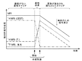
キャリアを使用した薬物送達に関する技術は、造影剤の使用とうまく組み合わせられてきた。例えば、刺激応答性リポソームは、リポソームの内腔に造影剤を封入することにより磁気共鳴映像法(MRI)造影剤と結び付けられた(McDannold et al.,2004,Radiology,230,743-752)。MRIは、診断の目的で病院においてよく用いられる重要な診断技術であり、高い空間分解能での軟部組織の非侵襲的画像化を可能にする。この技術は、全ての組織において全身に高い濃度で存在するバルク水分子の画像化に基づいている。造影剤としてガドリニウム又はマンガンイオンの錯体が用いられ、これは、水分子のプロトンの縦緩和時間(T1)及び横緩和時間(T2)を減らす。その能力のために、MRIは、キャリア構造体に含まれる物質、例えば薬物の送達の監視を可能にすることが分かっている。 Techniques for drug delivery using carriers have been successfully combined with the use of contrast agents. For example, stimuli-responsive liposomes have been linked to magnetic resonance imaging (MRI) contrast agents by encapsulating the contrast agent in the lumen of the liposome (McDannold et al., 2004, Radiology, 230, 743-752). MRI is an important diagnostic technique often used in hospitals for diagnostic purposes and allows non-invasive imaging of soft tissue with high spatial resolution. This technique is based on the imaging of bulk water molecules that are present at high concentrations throughout the body in all tissues. A complex of gadolinium or manganese ions is used as a contrast agent, which reduces the longitudinal relaxation time (T 1 ) and transverse relaxation time (T 2 ) of protons in water molecules. Because of its capabilities, MRI has been found to allow monitoring of the delivery of substances, eg drugs, contained in the carrier structure.
しかしながら、そのような手法では、キャリア内における初期の造影剤濃度が非常に高く、著しいT2の短縮及び拡散作用のために治療介入の当初はこれらキャリアの濃度を容易に決定することができない。専ら加熱すると、T1造影剤は、MRIにおける陽性の造影を与えるために遊離し、希釈される。同様の事柄が、薬物伝送キャリアと19Fトレーサ又はT2 *磁気共鳴(MR)造影剤を有する造影剤との組み合わせに当てはまる。これらの代替の手法は、薬物の放出の前又は後の造影に関する問題を与え、概して定量化可能な信号値を与えない。従って、これまで臨床的に用いられてきた画像化方法は、薬物送達のために刺激応答性のキャリアを用いる際、治療プロセス全体にわたって定量的情報を与えるのには適していない。 However, in such an approach, the initial concentration of the contrast agent is very high in the carrier, the initial therapeutic intervention for reducing and diffusing effect significant T 2 can not determine the concentration of these carriers easily. When exclusively heated, T 1 contrast agent, free to give a contrast positive in MRI, is diluted. Similar considerations apply to combinations of drug delivery carriers and 19 F tracers or contrast agents with T 2 * magnetic resonance (MR) contrast agents. These alternative approaches present problems with contrast before or after drug release and generally do not give quantifiable signal values. Thus, the imaging methods that have been used clinically so far are not suitable for providing quantitative information throughout the treatment process when using stimulus-responsive carriers for drug delivery.
従って、治療プロセス全体にわたって定量的情報を与える画像誘導による薬物送達の効率的で信頼性の高い方法及びそのような方法を実行するための手段が必要である。 Therefore, there is a need for an efficient and reliable method of image-guided drug delivery that provides quantitative information throughout the treatment process and a means for performing such a method.
本発明は、この要求に応え、刺激応答性キャリアを介した磁性粒子映像法(MPI)誘導による薬物送達のための手段及び方法を提供する。上記目的は、特に、空洞部を形成するシェル構造を有する組成物であって、上記シェル構造は薬物を有し、当該組成物は少なくとも1つの造影剤と関連し、上記シェル構造は、外部刺激が与えられるとその内容物を外部に放出することができ、上記造影剤は磁性粒子映像法(MPI)によって検出され得る磁性粒子を有し、上記造影剤に含まれる上記磁性粒子の少なくとも5%(w/w)よりも多くは、少なくとも10−18m2Aの磁気モーメントを有し、上記磁性粒子は、好ましくは、Fe、Co、Ni、Zn若しくはMn、これらの合金又はこれらのいずれかの酸化物によって構成され、より好ましくは、Fe2O3又はFe3O4によって構成された当該組成物によって達成される。 The present invention addresses this need and provides means and methods for drug delivery by stimulating magnetic particle imaging (MPI) via stimulus-responsive carriers. The object is in particular a composition having a shell structure forming a cavity, the shell structure having a drug, the composition being associated with at least one contrast agent, the shell structure being an external stimulus Can be released to the outside, and the contrast agent has magnetic particles that can be detected by magnetic particle imaging (MPI), and is at least 5% of the magnetic particles contained in the contrast agent More than (w / w) has a magnetic moment of at least 10 −18 m 2 A, and the magnetic particles are preferably Fe, Co, Ni, Zn or Mn, alloys thereof or any of these And more preferably achieved by the composition comprising Fe 2 O 3 or Fe 3 O 4 .
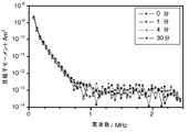
そのような組成物は、刺激応答性キャリアの有利な性質、すなわち、適切な信号、刺激又は行為が与えられた後、所定の場所で物質、特に薬物を放出する可能性と、高感度及び高分解能をもたらす非線形再磁化解析から磁性ナノ粒子の空間的分布の直接的な検出を可能にする磁性粒子映像法(MPI)の技術の有利な性質とを兼ね備えている。特に、MPIによって検出され得る造影剤により生成されるMPI画像化信号は、上記組成物又はキャリアへの造影剤の取り込みによって影響を及ぼされないことが分かった。また、上記信号は、キャリアからの造影剤が放出される際、影響を受けないままであることが分かった。従って、MRIを使用する方法とは対照的に、そのような組成物は、薬物の放出の前には磁性粒子映像法を用いて定量的に追跡され、薬物放出後に組成物の内容物の更なる分布が追跡される。 Such a composition has the advantageous properties of a stimulus-responsive carrier, i.e. the possibility of releasing substances, in particular drugs, in place after being given the appropriate signal, stimulus or action, and high sensitivity and high sensitivity. It combines the advantageous properties of magnetic particle imaging (MPI) technology that allows direct detection of the spatial distribution of magnetic nanoparticles from non-linear remagnetization analysis that provides resolution. In particular, it has been found that MPI imaging signals generated by contrast agents that can be detected by MPI are not affected by the incorporation of contrast agents into the composition or carrier. It has also been found that the signal remains unaffected when contrast agent is released from the carrier. Thus, in contrast to methods using MRI, such compositions are tracked quantitatively using magnetic particle imaging prior to drug release, and the contents of the composition are updated after drug release. The distribution is tracked.
本発明の好ましい実施の形態では、上記磁性粒子の少なくとも5%(w/w)よりも多くは、粒子当たり10ミリ秒よりも少ない再磁化時間を有している。 In a preferred embodiment of the invention, at least 5% (w / w) of the magnetic particles have a remagnetization time of less than 10 milliseconds per particle.
本発明の更に好ましい実施の形態では、上記造影剤は、上記シェル構造の外側若しくは内部と関連しているか、上記薬物と関連しているか又は上記シェル構造の上記空洞部内に埋め込まれている。 In a further preferred embodiment of the invention, the contrast agent is associated with the exterior or interior of the shell structure, associated with the drug, or embedded within the cavity of the shell structure.
本発明の他の好ましい実施の形態では、上記シェル構造は、リポソーム、ポリマソーム、ナノカプセル又はそれらの任意の混合物を構成している。特に好ましい実施の形態では、上記シェルは、感熱性又は感圧性材料を有している。 In another preferred embodiment of the present invention, the shell structure constitutes a liposome, polymersome, nanocapsule or any mixture thereof. In a particularly preferred embodiment, the shell comprises a heat sensitive or pressure sensitive material.
本発明の更に好ましい実施の形態では、上述した外部刺激は、孔部を形成する及び/又は前記シェル構造を分解することができる。 In a further preferred embodiment of the invention, the external stimulus described above can form a hole and / or disassemble the shell structure.
本発明の他の好ましい実施の形態では、上記外部刺激は、温度の上昇、温度の低下、圧力の上昇及び/又は圧力の低下である。 In another preferred embodiment of the invention, the external stimulus is a temperature increase, a temperature decrease, a pressure increase and / or a pressure decrease.
他の観点では、本発明は、薬物の制御送達のためのキャリアとしての(i)空洞部を形成するシェル構造を有する組成物であって、上記シェル構造は薬物を有し、当該組成物は少なくとも1つの造影剤と関連し、上記造影剤は磁性粒子映像法(MPI)によって検出されることが可能であり、上記シェル構造は、外部刺激が与えられるとその内容物を外部に放出することができる当該組成物又は(ii)本明細書において上記に定義された組成物の使用に関係がある。 In another aspect, the present invention provides a composition having (i) a shell structure that forms a cavity as a carrier for controlled delivery of a drug, the shell structure having a drug, the composition comprising: Associated with at least one contrast agent, the contrast agent can be detected by magnetic particle imaging (MPI), and the shell structure releases its contents to the exterior when an external stimulus is applied Or (ii) the use of a composition as defined herein above which is capable of
本発明の他の好ましい実施の形態では、上記制御送達は、MPIを用いる検出又は位置の特定を有する。本発明の更なる代替の形態では、上記制御送達は、MPI及び磁気共鳴映像法(MRI)を用いる検出又は位置の特定を有する。 In another preferred embodiment of the invention, the controlled delivery comprises detection or localization using MPI. In a further alternative form of the invention, the controlled delivery comprises detection or localization using MPI and magnetic resonance imaging (MRI).
本発明の他の好ましい実施の形態では、薬物の制御送達のためのキャリアとしての(i)空洞部を形成するシェル構造を有する組成物であって、上記シェル構造は薬物を有し、当該組成物は少なくとも1つの造影剤と関連し、上記造影剤は磁性粒子映像法(MPI)によって検出されることが可能であり、上記シェル構造は、外部刺激が与えられるとその内容物を外部に放出することができる当該組成物又は(ii)本明細書において上記に定義された組成物の使用において、制御される放出(controlled release)が、外部刺激の付与を介した上記シェル構造の内容物の放出を更に有する。本発明の特に好ましい実施の形態では、上記外部刺激は、温度の上昇、温度の低下、圧力の上昇及び/又は圧力の低下の刺激である。 In another preferred embodiment of the present invention, (i) a composition having a shell structure forming a cavity as a carrier for controlled delivery of a drug, the shell structure having a drug, the composition The object is associated with at least one contrast agent, which can be detected by magnetic particle imaging (MPI), and the shell structure releases its contents to the outside when an external stimulus is applied. Or (ii) in the use of a composition as defined herein above, controlled release of the contents of the shell structure through the application of an external stimulus. It further has a release. In a particularly preferred embodiment of the invention, the external stimulus is a temperature increase, a temperature decrease, a pressure increase and / or a pressure decrease stimulus.
更なる観点では、本発明は、シェル構造の内容物を放出する外部刺激が与えられる前、与えられている間及び/又は与えられた後に、(i)空洞部を形成するシェル構造を有する組成物であって、上記シェル構造は薬物を有し、当該組成物は少なくとも1つの造影剤と関連し、上記造影剤はMPIによって検出されることが可能であり、上記シェル構造は、外部刺激が与えられるとその内容物を外部に放出することができる当該組成物又は(ii)本明細書において上記に定義された組成物のMPIによる検出又は位置の特定を有する薬物送達プロセスの制御のためのデータ収集方法に関係がある。 In a further aspect, the present invention provides a composition having a shell structure that forms a cavity before, during and / or after an external stimulus that releases the contents of the shell structure. The shell structure has a drug, the composition is associated with at least one contrast agent, the contrast agent can be detected by MPI, and the shell structure has an external stimulus For the control of a drug delivery process with MPI detection or localization of the composition, or (ii) a composition as defined herein above, whose contents can be released to the exterior when given It is related to the data collection method.
本発明の更に好ましい実施の形態では、上記薬物送達プロセスの制御のためのデータ収集方法は、MPI及び更にMRIによる検出又は位置の特定を有する。 In a further preferred embodiment of the invention, the data collection method for the control of the drug delivery process comprises MPI and further MRI detection or localization.
本発明の更に好ましい実施の形態では、上記に定義された薬物送達プロセスの制御のためのデータ収集方法は、追加のステップとして、外部刺激の付与を介した上記シェル構造の内容物の放出を有する。本発明の特に好ましい実施の形態では、上記外部刺激は、温度の上昇、温度の低下、圧力の上昇及び/又は圧力の低下の刺激である。 In a further preferred embodiment of the invention, the data collection method for the control of the drug delivery process defined above comprises the release of the contents of the shell structure via the application of an external stimulus as an additional step. . In a particularly preferred embodiment of the invention, the external stimulus is a temperature increase, a temperature decrease, a pressure increase and / or a pressure decrease stimulus.
更なる観点では、本発明は、病的状態を治療するための組成物であって、空洞部を形成するシェル構造を有し、上記シェル構造は薬物を有し、当該組成物は少なくとも1つの造影剤と関連し、上記造影剤はMPIによって検出されることが可能であり、上記シェル構造は、外部刺激が与えられるとその内容物を外部に放出することができる当該組成物又は本明細書において上記に定義された当該組成物に関係がある。 In a further aspect, the present invention provides a composition for treating a pathological condition having a shell structure forming a cavity, the shell structure having a drug, the composition comprising at least one In connection with a contrast agent, the contrast agent can be detected by MPI, and the shell structure is capable of releasing its contents to the exterior upon application of an external stimulus, or the present specification. In relation to the composition as defined above.
本発明の他の好ましい実施の形態では、上記薬物は、刺激が与えられることにより投与されることとなり、上記刺激は、局所熱システム、電界、磁場、焦点式超音波の照射及び/又は高周波の照射を介して伝達され、上記シェル構造から外部への上記薬物の放出をもたらす。 In another preferred embodiment of the invention, the drug is to be administered by applying a stimulus, the stimulus being a local thermal system, electric field, magnetic field, focused ultrasound irradiation and / or radio frequency. It is transmitted via irradiation, resulting in the release of the drug out of the shell structure.
本発明の他の好ましい実施の形態では、上記組成物は、MPIを用いて検出可能又は位置特定可能である。本発明の更なる好ましい実施の形態では、上記組成物は、MPI及びMRIを用いて検出可能又は位置特定可能である In another preferred embodiment of the invention, the composition is detectable or localizable using MPI. In a further preferred embodiment of the invention, the composition is detectable or localizable using MPI and MRI.
本発明は、刺激応答性の組成物又はキャリアを介した磁性粒子映像法(MPI)誘導による薬物送達のための手段及び方法に関連している。 The present invention relates to means and methods for magnetic particle imaging (MPI) -induced drug delivery via stimuli-responsive compositions or carriers.
本発明は特定の実施の形態に関して説明されるが、この説明は限定的な意味に解釈されるべきではない。 While this invention will be described with respect to particular embodiments, this description should not be construed in a limiting sense.
本発明の例示的な実施の形態を詳細に説明する前に、本発明を理解するために重要な定義が与えられる。 Before describing exemplary embodiments of the present invention in detail, important definitions are provided for understanding the present invention.
本明細書及び添付の特許請求の範囲において用いられる場合、「a」及び「an」の単数形は、文脈がそれ以外のことを明らかに規定する場合を除いて、対応する複数形も含んでいる。 As used in this specification and the appended claims, the singular forms “a” and “an” also include the corresponding plural unless the context clearly dictates otherwise. Yes.
本発明に関しては、「約」という用語は、当業者が当該特徴の技術的効果を依然として確実にすると理解する正確さの幅を意味している。この用語は、典型的には、示された数値からの±20%、好ましくは±15%、より好ましくは±10%、更に好ましくは±5%のずれを示すものである。 In the context of the present invention, the term “about” means a range of accuracy that one of ordinary skill in the art understands will still ensure the technical effect of the feature. This term typically indicates a deviation of ± 20%, preferably ± 15%, more preferably ± 10%, even more preferably ± 5% from the indicated numerical value.
「有する」という用語は限定的ではないことを理解されたい。本発明のために、「より成る」という用語は、「有する」という用語の好ましい実施の形態であるとみなされる。以下において、あるグループが少なくともある数の具体的表現を有するように規定される場合、これは、好ましくはこれらの具体的表現のみより成るグループを含むことも意味する。 It should be understood that the term “comprising” is not limiting. For the purposes of the present invention, the term “consisting of” is considered to be a preferred embodiment of the term “having”. In the following, when a group is defined to have at least a certain number of specific expressions, this also means that it preferably includes groups consisting only of these specific expressions.
また、明細書及び特許請求の範囲における「第1」、「第2」、「第3」又は「(a)」、「(b)」、「(c)」、「(d)」等の用語及びこれらに類する用語は、類似する要素を区別するために用いられており、必ずしも連続する順序又は発生順を説明するために用いられるものではない。そのように用いられている用語は適切な状況下において置き換え可能であり、本明細書で説明される本発明の実施の形態は、本明細書中に説明されている又は示されている順序ではない他の順序で動作可能であることを理解されたい。 In addition, “first”, “second”, “third” or “(a)”, “(b)”, “(c)”, “(d)”, etc. in the specification and claims. Terms and like terms are used to distinguish between similar elements and are not necessarily used to describe a sequential or chronological order. The terms so used are interchangeable under appropriate circumstances, and the embodiments of the invention described herein are in the order described or shown herein. It should be understood that it can operate in other orders.
「第1」、「第2」、「第3」又は「(a)」、「(b)」、「(c)」、「(d)」等の用語が使用又は方法のステップに関係がある場合、本明細書で上記又は以下に記載の適用においてそれ以外のことが示されていない限り、ステップ間に時間又は時間間隔の統一性は存在しない。すなわち、各ステップは、同時に行われてもよいし、そのようなステップの間に秒、分、時間、日、週、月又は更には年の時間間隔が存在してもよい。 Terms such as “first”, “second”, “third” or “(a)”, “(b)”, “(c)”, “(d)” are used or related to method steps In some cases, there is no unity of time or time interval between steps, unless otherwise indicated herein in the application described above or below. That is, each step may be performed simultaneously, and there may be time intervals of seconds, minutes, hours, days, weeks, months or even years between such steps.
本発明は、異なっていてもよいものとして本明細書において説明される特定の方法論、プロトコル、試薬等に限定されないことを理解されたい。本明細書において用いられる専門用語は、単に特定の実施の形態を説明する目的であり、専ら添付の特許請求の範囲により限定される本発明の範囲を限定するように意図されてはいないことも理解されたい。特に定義されていない限り、本明細書において用いられる技術用語及び科学用語は、当業者により一般に理解されている意味と同じ意味を有している。 It is to be understood that the invention is not limited to the particular methodologies, protocols, reagents, etc. described herein as being different. The terminology used herein is for the purpose of describing particular embodiments only and is not intended to be limiting of the scope of the invention, which is limited solely by the scope of the appended claims. I want you to understand. Unless defined otherwise, technical and scientific terms used herein have the same meaning as commonly understood by one of ordinary skill in the art.
上記に示したように、本発明は、一観点では、空洞部を形成するシェル構造を有する組成物であって、上記シェル構造は薬物を有し、当該組成物は少なくとも1つの造影剤と関連し、上記シェル構造は、外部刺激が与えられるとその内容物を外部に放出することができ、上記造影剤は磁性粒子映像法(MPI)によって検出され得る磁性粒子を有し、上記造影剤に含まれる上記磁性粒子の少なくとも5%(w/w)よりも多くは、少なくとも10−18m2Aの磁気モーメントを有し、上記磁性粒子は、好ましくは、Fe、Co、Ni、Zn若しくはMn、これらの合金又はこれらのいずれかの酸化物によって構成され、より好ましくは、Fe2O3又はFe3O4によって構成された当該組成物に関係している。 As indicated above, the present invention, in one aspect, is a composition having a shell structure that forms a cavity, the shell structure having a drug, the composition being associated with at least one contrast agent. The shell structure can release the contents to the outside when an external stimulus is applied. The contrast agent has magnetic particles that can be detected by magnetic particle imaging (MPI). More than at least 5% (w / w) of the magnetic particles contained have a magnetic moment of at least 10 −18 m 2 A, and the magnetic particles are preferably Fe, Co, Ni, Zn or Mn , These alloys or any of these oxides, more preferably related to such compositions composed of Fe 2 O 3 or Fe 3 O 4 .
本明細書において用いられる「シェル構造」という用語は、典型的には同一の又は類似した化学的、物理的及び/又は生物学的性質を持つ小さい単位又はエンティティから成るエンベロープ型構造を意味する。更に、上記エンベロープ型構造は、空洞部を形成する。すなわち、内部から外部環境を排除し、従って、外部と内部との環境、状態等の境界としての役割を果たす。本発明に係るシェル構造は、好ましくは、疎水性層により構成されている。この層は、単層又は二重層である。二重層構造の各サイドは、異なる性質を有している及び/又は異なるシェル形成単位により構成されている。好ましくは、両サイドが、シェル構造又は膜の内部の方を向いた疎水性の尾部構造を有している。上記シェル構造は、多重層の形態又は単層の形態を有しており、例えば、小さい若しくは大きい多重層小胞、小さい単層小胞又は大きい単層小胞を構成している。上記シェル構造は、任意の適切な形態又は寸法であり、例えば、球形であってもよく、楕円形であってもよく、円形又はナシ形、ダンベル型、扁平形、ピラミッド形等であってもよい。上記シェル構造は、好ましくは、自己集合能を有している。 As used herein, the term “shell structure” refers to an envelope-type structure consisting of small units or entities that typically have the same or similar chemical, physical and / or biological properties. Furthermore, the envelope-type structure forms a cavity. That is, it excludes the external environment from the inside, and thus serves as a boundary between the environment and the state between the outside and the inside. The shell structure according to the present invention is preferably constituted by a hydrophobic layer. This layer is a single layer or a double layer. Each side of the bilayer structure has different properties and / or is composed of different shell forming units. Preferably, both sides have a hydrophobic tail structure facing towards the inside of the shell structure or membrane. The shell structure has a multilamellar form or a monolayer form, for example, a small or large multilamellar vesicle, a small monolamellar vesicle or a large monolamellar vesicle. The shell structure may be any suitable form or size, for example, may be spherical, oval, circular or pear, dumbbell, flat, pyramid, etc. Good. The shell structure preferably has a self-assembly ability.
本発明の典型的な実施の形態では、上記シェル形成単位は、疎水性尾部及び親水性頭部により構成されている。シェル構造の内部又は空洞部は、好ましくは、親水性の環境、例えば、水溶液である。代替として、シェル構造の空洞部は、親水性の環境である。シェル構造の空洞部の環境は、外部と同じ又は異なる環境条件を有している。本明細書において用いられる「環境条件」という用語は、pH、有機又は無機イオンの濃度、1つ又はそれ以上の塩の存在、浸透圧の存在等を意味する。例えば、シェル構造の空洞部内のpHは、外部のpHよりも低いか、それと同じであるか、又はそれよりも高く、シェル構造内には、浸透圧が存在する又は浸透圧平衡が存在する等である。 In an exemplary embodiment of the invention, the shell forming unit is composed of a hydrophobic tail and a hydrophilic head. The interior or cavity of the shell structure is preferably a hydrophilic environment, such as an aqueous solution. Alternatively, the shell cavity is a hydrophilic environment. The environment of the cavity of the shell structure has the same or different environmental conditions as the outside. As used herein, the term “environmental conditions” means pH, concentration of organic or inorganic ions, presence of one or more salts, presence of osmotic pressure, and the like. For example, the pH in the cavity of the shell structure is lower, the same as or higher than the external pH, osmotic pressure exists or osmotic balance exists in the shell structure, etc. It is.
シェル形成単位に加えて、シェルは、追加の機能を与える更なる要素を有している。そのような追加の要素の例は標的エンティティであり、これは、シェル構造の化学的、物理的及び/又は生物学的性質を変更する適合要素又は安定化若しくは不安定化要素によるシェル構造の相互作用及び/又は認識を可能にする。これらの要素は、典型的にはシェル構造の外側又は外表面に存在し、シェル構造の内部及び/又はシェル構造の空洞部に突き出ていてもよいし、突き出ていなくてもよい。特定の組織型、特定の器官、細胞若しくは細胞型又は体、特に動物若しくは人間の体の特定の部分にシェル構造の標的を定めることを可能にする要素が特に好ましい。例えば、標的エンティティの存在は、肝臓、腎臓、肺、心臓、膵臓、胆汁、脾臓、リンパ組織、皮膚、脳、筋肉等のような器官にシェル構造及び従って組成物全体の標的を定めることをもたらす。代替として、標的エンティティの存在は、特定の細胞型、例えば、表面において相互作用タンパク質又は認識可能なタンパク質を発現させるがん性細胞に標的を定めることをもたらす。本発明の好ましい実施の形態では、上記シェル構造は、シェル構造の外側及び/又は内側において相互作用面を与えるタンパク質若しくはペプチド又はその断片を有している。そのようなタンパク質又はペプチド要素の例は、受容体分子に結合することができるリガンド、リガンド又は他の受容体と相互作用することができる受容体分子、抗原又はアビジン、ストレプトアビジン、ニュートラアビジン、レクチンと相互作用することができる抗体、抗体断片又はその誘導体である。また、例えば、タンパク質、ペプチド若しくはシェル構造単位等のようなビオチン化化合物の形態で存在するか、又はシェル構造自体の内部若しくは外側に存在するビオチンのような結合相互作用物質の存在も本発明により想定されている。上記シェル構造は、また、シェル構造の表面に存在する及び/又はシェル構造及び/又はシェル構造の空洞部に突き出た適合する相互作用物質、例えば、ビタミンを結合するタンパク質又は抗原等と相互作用することができるビタミン又は抗原を有している。 In addition to the shell forming unit, the shell has additional elements that provide additional functionality. An example of such an additional element is a target entity, which is a mutual interaction of the shell structure with a conforming element or stabilizing or destabilizing element that alters the chemical, physical and / or biological properties of the shell structure. Allows action and / or recognition. These elements are typically present on the outer or outer surface of the shell structure and may or may not protrude into the shell structure and / or into the cavity of the shell structure. Particular preference is given to elements that make it possible to target shell structures to specific tissue types, specific organs, cells or cell types or bodies, in particular to specific parts of the animal or human body. For example, the presence of the target entity results in targeting the shell structure and thus the entire composition to organs such as liver, kidney, lung, heart, pancreas, bile, spleen, lymphoid tissue, skin, brain, muscle, etc. . Alternatively, the presence of the target entity results in targeting a specific cell type, eg, cancerous cells that express interacting or recognizable proteins on the surface. In a preferred embodiment of the present invention, the shell structure has a protein or peptide or a fragment thereof that provides an interaction surface outside and / or inside the shell structure. Examples of such protein or peptide elements are ligands that can bind to receptor molecules, receptor molecules that can interact with ligands or other receptors, antigens or avidin, streptavidin, neutravidin, lectins Antibody, antibody fragment or derivative thereof. In addition, the presence of a binding interaction substance such as biotin which exists in the form of a biotinylated compound such as a protein, peptide or shell structural unit or which exists inside or outside the shell structure itself is also according to the present invention. Assumed. The shell structure also interacts with compatible interactors present on the surface of the shell structure and / or protruding into the shell structure and / or the cavity of the shell structure, such as proteins or antigens that bind vitamins, etc. Have vitamins or antigens that can.
上記シェル構造は、また、追加の化合物、好ましくは、シェル構造の安定性及び/又は循環寿命(circulatory life)を高める、生体内分布に影響を与える、免疫学的挙動を変更する等の化合物によって覆われている。そのようなコーティングの例は、炭水化物分子の存在、好ましくは、糖鎖付加パターン、より好ましくは当業者には既知の組織又は細胞型に典型的な生物学的に関連した糖鎖付加パターンの存在、又はシェル構造の外側層又は外部におけるPEG(ポリエチレングリコール)の存在を含んでいる。ポリエチレングリコール2000の使用が特に好ましい。オリゴグリセロール(OG)基の使用が更に特に好ましい。OGで修飾された感熱性リポソームの使用についての一例は、Lindner等の2008,Journal of Controlled Release, 125, 112-120である。 The shell structure may also be added by additional compounds, preferably compounds that increase the stability and / or circulatory life of the shell structure, affect biodistribution, alter immunological behavior, etc. Covered. Examples of such coatings are the presence of carbohydrate molecules, preferably glycosylation patterns, more preferably the presence of biologically relevant glycosylation patterns typical of tissues or cell types known to those skilled in the art. Or the presence of PEG (polyethylene glycol) in the outer layer or outside of the shell structure. The use of polyethylene glycol 2000 is particularly preferred. The use of oligoglycerol (OG) groups is even more particularly preferred. An example for the use of thermosensitive liposomes modified with OG is Lindner et al. 2008, Journal of Controlled Release, 125, 112-120.
シェル構造の典型的なサイズは、約30nmから約1000nmまでである。約50nmから約400nmの間のサイズが好ましい。 The typical size of the shell structure is from about 30 nm to about 1000 nm. A size between about 50 nm and about 400 nm is preferred.
本明細書において用いられる「シェル構造は薬物を有する」という表現は、薬物が、シェル構造の空洞部、シェル構造の表面、外側と内側とのシェル形成境界領域、例えば、単層又は二重層の境界自体又は同時にこれらの区画の1つ若しくはそれ以上に、例えば、境界部を超えて外側からシェル構造の空洞部内に広がって、境界部からシェル構造の空洞部内に広がって又は境界部から外部に広がって存在することを意味する。上記薬物は、追加として、本明細書において上述した修飾、例えば、グリコシル化、ビオチン化、PEGによるコーティング等の1つ又はそれ以上に従って修飾され得る。代替として、薬物は、シェル構造の表面、境界領域若しくは空洞部に存在することができるように化学的又は生物学的に修飾される。上記薬物は、モノマ、オリゴマ又はポリマとして存在する。上記の存在は、浸透圧の状況、シェル構造の電荷又は当業者には既知の任意の他の適切なパラメータに従って調節される。薬物に加えて、当業者には既知の任意の適切なアクセサリ分子、例えば、安定化分子、アジュバント、分解酵素の阻害剤、電荷安定剤、構造安定剤、塩、緩衝剤、抗酸化剤、キレート剤、色素、例えば蛍光色素、画像化化合物等がシェル構造に含まれ得る。 As used herein, the expression “shell structure has drug” means that the drug has a cavity in the shell structure, the surface of the shell structure, a shell-forming boundary region between the outside and inside, eg, a single layer or a double layer. The boundary itself or simultaneously one or more of these compartments, e.g., extending beyond the boundary from the outside into the shell structure cavity, extending from the boundary into the shell structure cavity, or from the boundary to the outside It means to exist in an extended manner. The drug may additionally be modified according to one or more of the modifications described herein above, for example glycosylation, biotinylation, coating with PEG, and the like. Alternatively, the drug is chemically or biologically modified so that it can be present on the surface, boundary region or cavity of the shell structure. The drug exists as a monomer, oligomer or polymer. The presence is adjusted according to the osmotic pressure situation, the charge of the shell structure or any other suitable parameter known to those skilled in the art. In addition to drugs, any suitable accessory molecule known to those skilled in the art, for example, stabilizing molecules, adjuvants, inhibitors of degrading enzymes, charge stabilizers, structure stabilizers, salts, buffers, antioxidants, chelates Agents, dyes such as fluorescent dyes, imaging compounds, and the like can be included in the shell structure.
本明細書において用いられる「薬物」という用語は、処置、治療、予防、未然防止(prevention)又は病状、例えば病気若しくは疾患の診断に用いられる、又は、それ以外には、物理的、心理的又は精神的に健康な状態を向上させるために用いられる任意の物理的、化学的又は生物学的物質を意味する。この用語は、また、化粧的用途の物質も意味し、これは、栄養補給の目的又はこれらの観点の任意の組み合わせに役立つ。好ましい実施の形態では、この用語は、生物学的活性物質を意味する。ここで用いられる「生物学的活性物質」という用語は、治療薬物、内因性分子及び薬理学的活性物質、例えば、抗体,栄養分子(nutritional molecule),化粧的物質(cosmetic agent),診断用物質及び他の画像化用造影剤を含む生物学的に活性な化学物質を意味する。また、活性物質の薬理学的に許容可能な塩を含む活性物質も含まれる。 As used herein, the term “drug” is used for treatment, therapy, prevention, prevention or diagnosis of a medical condition, such as a disease or disorder, or otherwise physical, psychological or Means any physical, chemical or biological substance used to improve mental health. The term also refers to a substance for cosmetic use, which serves for nutritional purposes or any combination of these aspects. In preferred embodiments, the term refers to a biologically active substance. The term “biologically active substance” as used herein refers to therapeutic drugs, endogenous molecules and pharmacologically active substances such as antibodies, nutritional molecules, cosmetic agents, diagnostic substances. And biologically active chemicals including other imaging contrast agents. Also included are active substances that include pharmacologically acceptable salts of the active substances.
薬物の例は、ポリヌクレオチド、アンチセンスヌクレオチド(遺伝子治療物質)、RNA分子、DNA分子、siRNA分子、miRNA分子等のような核酸、糖質、タンパク質又はペプチド、小分子、脂質、リポ多糖類、非ペプチド又は非タンパク質薬物を有している。本発明の範囲内において、ポリマの性質の薬物を採用することが可能であるが、1500g/molよりも小さい又は更に500g/molよりも小さい相対的に小さい分子量の薬物を採用することも可能である。 Examples of drugs include nucleic acids such as polynucleotides, antisense nucleotides (gene therapy substances), RNA molecules, DNA molecules, siRNA molecules, miRNA molecules, carbohydrates, proteins or peptides, small molecules, lipids, lipopolysaccharides, Has non-peptide or non-protein drugs. Within the scope of the present invention, it is possible to employ a drug of the nature of a polymer, but it is also possible to employ a relatively low molecular weight drug of less than 1500 g / mol or even less than 500 g / mol. is there.
従って、本発明に関連して考えられる生物学的活性物質は、治療又は予防効果のある任意の化合物を含んでいる。上記化合物は、組織増殖、細胞増殖、細胞分化に影響を及ぼす若しくは関与する化合物、免疫反応のような生物学的作用を引き起こすことができる化合物又は1つ若しくはそれ以上の生物学的プロセスにおいて任意の他の役割を果たす化合物であり得る。非限定的な例のリストは、(抗細菌剤、抗ウイルス剤及び抗真菌剤を含む)抗菌剤、抗ウイルス剤、抗腫瘍物質、トロンビン阻害剤、抗血栓剤、血栓溶解剤、線維素溶解剤、血管攣縮阻害剤、カルシウムチャネル遮断剤、血管拡張剤、抗高血圧剤、抗菌剤、抗生剤、表面糖たんぱく質受容体の阻害剤、抗血小板物質、抗有糸分裂剤、微小管阻害剤、抗分泌剤、アクチン阻害剤、リモデリング阻害剤、抗代謝剤、(抗血管新生剤を含む)抗増殖剤、抗がん化学療法剤、抗炎症ステロイド又は非ステロイド性抗炎症剤、免疫抑制剤、成長ホルモン拮抗剤、成長因子、ドーパミン作動薬、放射線治療薬、細胞外基質成分、ACE阻害剤、遊離基捕捉剤、キレート剤、抗酸化剤、抗ポリメラーゼ及び光線力学的治療剤を含んでいる。 Accordingly, biologically active substances contemplated in connection with the present invention include any compound that has a therapeutic or prophylactic effect. The compound may be a compound that affects or participates in tissue proliferation, cell proliferation, cell differentiation, a compound capable of causing a biological effect such as an immune response or any one or more biological processes. It can be a compound that plays other roles. A list of non-limiting examples includes antibacterial agents (including antibacterial agents, antiviral agents and antifungal agents), antiviral agents, antitumor substances, thrombin inhibitors, antithrombotic agents, thrombolytic agents, fibrinolysis Agents, vasospasm inhibitors, calcium channel blockers, vasodilators, antihypertensive agents, antibacterial agents, antibiotics, inhibitors of surface glycoprotein receptors, antiplatelet substances, antimitotic agents, microtubule inhibitors, Antisecretory agent, actin inhibitor, remodeling inhibitor, antimetabolite, antiproliferative agent (including antiangiogenic agent), anticancer chemotherapeutic agent, anti-inflammatory steroid or nonsteroidal anti-inflammatory agent, immunosuppressant Contains growth hormone antagonists, growth factors, dopamine agonists, radiotherapeutic agents, extracellular matrix components, ACE inhibitors, free radical scavengers, chelating agents, antioxidants, anti-polymerases and photodynamic therapeutic agents .
相対的に小さいペプチドは、アミノ酸の数によって分類して呼ばれる(例えば、ジ、トリ、テトラペプチド)。相対的に少数のアミド結合を持つペプチドはオリゴペプチドとも呼ばれ(50個のアミノ酸まで)、相対的に多数のペプチド(50個よりも多いアミノ酸)はポリペプチド又はタンパク質と呼ばれる。アミノ酸残基のポリマであることに加えて、あるタンパク質は、更に、所謂四次構造、必ずしもアミド結合によって化学的に結合されてはいないが、静電気力及びファンデルワールス力のような当業者には一般的に知られている力によって結合された幾つかのポリペプチドの集合体によって特徴付けられる。本明細書において用いられるペプチド、タンパク質又はその混合物という用語は、全ての上述した可能性を含むこととなる。一般に、タンパク質及び/又はペプチドは、成長因子である。 Relatively small peptides are referred to by classification according to the number of amino acids (eg, di, tri, tetrapeptide). Peptides with a relatively small number of amide bonds are also called oligopeptides (up to 50 amino acids) and relatively large numbers of peptides (more than 50 amino acids) are called polypeptides or proteins. In addition to being a polymer of amino acid residues, certain proteins may also have a so-called quaternary structure, not necessarily chemically linked by amide bonds, but to those skilled in the art such as electrostatic forces and van der Waals forces. Is characterized by a collection of several polypeptides joined together by a generally known force. As used herein, the term peptide, protein or mixture thereof will include all the possibilities described above. In general, proteins and / or peptides are growth factors.
シェル構造に有利に含まれるペプチド、タンパク質又はペプチド若しくはタンパク質を有するエンティティの他の例は、限定されるものではないが以下のものを含む免疫原性ペプチド又は免疫原性タンパク質を含んでいるが、これに限定されない。 Other examples of peptides, proteins or entities having peptides or proteins that are advantageously included in the shell structure include, but are not limited to, immunogenic peptides or immunogenic proteins including: It is not limited to this.
ジフテリア毒素又は破傷風毒素のような毒素。 Toxins such as diphtheria toxin or tetanus toxin.
アデノウイルス、エプスタインバーウイルス、A型肝炎ウイルス、B型肝炎ウイルス、ヘルペスウイルス、HIV−1、HIV−2、HTLV−III、インフルエンザウイルス、日本脳炎ウイルス、麻疹ウイルス、乳頭腫ウイルス、パラミクソウイルス、ポリオウイルス、狂犬病ウイルス、風疹ウイルス、ワクシニア(天然痘)ウイルス及び黄熱病ウイルスのようなウイルス表面抗原又はウイルスの一部。 Adenovirus, Epstein-Barr virus, hepatitis A virus, hepatitis B virus, herpes virus, HIV-1, HIV-2, HTLV-III, influenza virus, Japanese encephalitis virus, measles virus, papilloma virus, paramyxovirus, Virus surface antigens or parts of viruses such as poliovirus, rabies virus, rubella virus, vaccinia virus and yellow fever virus.
百日咳菌、ヘリコバクターピロリ菌、破傷風菌、ジフテリア菌、大腸菌、インフルエンザ菌、クレブシエラ種、レジオネラニューモフィラ、ウシ結核菌、ハンセン菌、ヒト結核菌、淋菌、髄膜炎菌、プロテウス種、緑膿菌、サルモネラ種、赤痢菌種、黄色ブドウ球菌、化膿連鎖球菌、コレラ菌又はペスト菌のような細菌表面抗原又は細菌の一部。 Bordetella pertussis, Helicobacter pylori, Tetanus, Diphtheria, Escherichia coli, Haemophilus influenzae, Klebsiella species, Legionella pneumophila, M. bovine, Hansen, M. tuberculosis, Neisseria gonorrhoeae, N. meningitidis, Proteus, Pseudomonas aeruginosa, Bacterial surface antigens or parts of bacteria such as Salmonella species, Shigella species, Staphylococcus aureus, Streptococcus pyogenes, Vibrio cholerae or Plasmodium pestis.
三日熱マラリア原虫(マラリア)、熱帯熱マラリア原虫(マラリア)、卵形マラリア原虫(マラリア)、四日熱マラリア原虫(マラリア)、熱帯リーシュマニア(リーシュマニア症)、ドノバンリーシュマニア(リーシュマニア症)、ブラジルリーシュマニア(リーシュマニア症)、ローデシアトリパノソーマ(睡眠病)、ガンビアトリパノソーマ(睡眠病)、クルーズトリパノソーマ(シャーガス病)、マンソン住血吸虫(住血吸虫症)、ビルハイツ住血吸虫(住血吸虫症)、日本住血吸虫(住血吸虫症)、旋毛虫(旋毛虫病)、十二指腸糞線虫(鉤虫症)、バンクロフト糸状虫(フィラリア症)、マレー糸状虫(フィラリア症)、ロア糸状虫(フィラリア症)、常在糸状虫(フィラリア症)、メジナ虫(フィラリア症)又は回旋糸状虫(フィラリア症)のような病気を引き起こす寄生生物の表面抗原又は寄生生物の一部。 Plasmodium falciparum (malaria), Plasmodium falciparum (malaria), Oval malaria parasite (malaria), Plasmodium falciparum (malaria), tropical leishmania (leishmaniasis), donovan leishmania (leishmaniasis) ), Brazilian leishmania (Leishmaniasis), Rhodesia trypanosoma (sleeping sickness), Gambia trypanosoma (sleeping sickness), Cruise trypanosoma (Chagas disease), Schistosoma mansoni (schistosomiasis), Schistosoma japonicum (schistosomiasis), Schistosoma japonicum (Schistosomiasis), Trichinella (Trichinella disease), Duodenum dungworm (helminthiasis), Bancroft filamentous worm (filariasis), Malay filamentous filariasis (filariasis), Loa filamentous worm (filariasis) , Resident filamentous worms (filariasis), medina worms (filariasis) or rotifers ( Some of such parasites surface antigens or parasites that cause diseases like Iraria disease).
IgG、IgA、IgM、抗狂犬病免疫グロブリン及び/又は抗ワクシニア免疫グロブリンのような免疫グロブリン。 Immunoglobulins such as IgG, IgA, IgM, anti-rabies immunoglobulin and / or anti-vaccinia immunoglobulin.
ボツリヌス抗毒素、ジフテリア抗毒素、ガス壊疽抗毒素又は破傷風抗毒素のような抗毒素。 An antitoxin such as a botulinum antitoxin, diphtheria antitoxin, gas gangrene antitoxin or tetanus antitoxin.
口蹄疫に対する免疫反応を引き出す抗原、卵胞刺激ホルモン、プロラクチン、アンギオゲニン、上皮成長因子、カルシトニン、エリスロポエチン、甲状腺刺激放出ホルモン、インスリン、成長ホルモン、インスリン様成長因子1及び2、骨格成長因子、ヒト絨毛性ゴナドトロピン、黄体形成ホルモン、神経成長因子、副腎皮質成長ホルモン(ACTH)、黄体形成ホルモン放出ホルモン(LHRH)、副甲状腺ホルモン(PTH)、甲状腺刺激ホルモン放出ホルモン(TRH)、バソプレシン、コレシストキニン並びに副腎皮質刺激ホルモン放出ホルモンのようなホルモン及び成長因子、インターフェロン、インターロイキン、コロニー刺激因子及び腫瘍壊死因子のようなサイトカイン、ウロキナーゼ、プラスミノゲン活性化因子のような線維素溶解酵素、プロテインC、第VIII因子、第IX因子、第VII因子又はアンチトロンビンIIIのような凝固因子を含む免疫原性ペプチド又は免疫原性タンパク質を含んでいる。
Antigens that elicit immune responses against foot-and-mouth disease, follicle stimulating hormone, prolactin, angiogenin, epidermal growth factor, calcitonin, erythropoietin, thyroid stimulating release hormone, insulin, growth hormone, insulin-
他のタンパク質又はペプチドの例は、アルブミン、心房性ナトリウム利尿因子、レニン、スーパーオキシドジスムターゼ、α1−アンチトリプシン、肺サーファクタントタンパク質、バシトラシン、ベスタチン、シクロスポリン、デルタ睡眠誘発ペプチド(DSIP)、エンドルフィン、グルカゴン、グラミシジン、メラノサイト抑制因子、ニューロテンシン、オキシトシン、ソマトスタチン、テプロチド、血清胸腺因子、サイモシン、DDAVP、デルモルフィン、メチオニンエンケファリン、ペプチドグリカン、サチエチン、チモペンチン、フィブリン分解産物、デス−エンケファリン−αエンドルフィン、性腺刺激ホルモン放出ホルモン、ロイプロリド、α−MSH又はメトケファミド。 Examples of other proteins or peptides are albumin, atrial natriuretic factor, renin, superoxide dismutase, α1-antitrypsin, pulmonary surfactant protein, bacitracin, bestatin, cyclosporine, delta sleep-inducing peptide (DSIP), endorphin, glucagon, Gramicidin, melanocyte inhibitory factor, neurotensin, oxytocin, somatostatin, teprotide, serum thymus factor, thymosin, DDAVP, delmorphin, methionine enkephalin, peptidoglycan, thymopentin, fibrin degradation product, des-enkephalin-α endorphin, gonadotropin release Hormone, leuprolide, α-MSH or methokefamide.
アルトレタミン、フルオロウラシル、アムサクリン、ヒドロキシカルバミド、アスパラギナーゼ、イフォスファミド、ブレオマイシン、ロムスチン、ブスルファン、メルファラン、クロラムブシル、メルカプトプリン、クロルメチン、メトトレキサート、シスプラチン、ミトマイシン、シクロホスファミド、プロカルバジン、シタラビン、テニポシド、ダカルバジン、チオテーパ、ダクチノマイシン、チオグアニン、ダウノルビシン、トレオスルファン、ドキソルビシン、チオホスファミド、エストラムシン、ヴィンブラスチン、エトグルシド、ヴィンクリスチン、エトポシド、ヴィンデシン又はパクリタクセルのような抗腫瘍剤。 Artretamine, fluorouracil, amsacrine, hydroxycarbamide, asparaginase, ifosfamide, bleomycin, lomustine, busulfan, melphalan, chlorambucil, mercaptopurine, chlormethine, methotrexate, cisplatin, mitomycin, cyclophosphamide, procarbazine, cytarazine, carbazine, teniposide, dacarbazine Anti-tumor agents such as dactinomycin, thioguanine, daunorubicin, treosulfan, doxorubicin, thiophosphamide, estramcin, vinblastine, etoglucid, vincristine, etoposide, vinducine or paclitaxel.
アンピシリン、ナフシリン、アモキシシリン、オキサシリン、アズロシリン、ペニシリンG、カルベニシリン、ペニシリンV、ジクロキサシリン、フェネチシリン、フロキサシリン、ピペラシリン、メシリナム、スルベニシリン、メチシリン、チカルシリン、メズロシリンのような抗生物質と、セファクロール、セファロチン、セファドロキシル、セファピリン、セファマンドール、セフラジン、セファトリジン、セフスロジン、セファゾリン、セフタジジム、セフォラニド、セフトリアキソン、セフォキシチン、セフロキシム、セファセトリル、ラタモキセフ又はセファレキシンのようなセファロスポリン系抗生物質と、アミカシン、ネオマイシン、ジベカシン、カナマイシン、ゲンタマイシン、ネチルマイシン又はトブラマイシンのようなアミノグリコシド系抗生物質と、アンホテリシンB、ノボビオシン、バシトラシン、ニスタチン、クリンダマイシン、ポリミキシン、コリスチン、ロヴァマイシン、エリトロマイシン、スペクチノマイシン、リンコマイシン又はバンコマイシンのようなマクロライド系抗生物質と、クロルテトラサイクリン、オキシテトラサイクリン、デメクロサイクリン、ロリテトラサイクリン、ドキシサイクリン、テトラサイクリン又はミノサイクリンのようなテトラサイクリン系抗生物質と、クロランフェニコール、リファマイシン、リファンピシン又はチアンフェニコールのような他の抗生物質とを有する抗菌剤。 Antibiotics such as ampicillin, nafcillin, amoxicillin, oxacillin, azurocillin, penicillin G, carbenicillin, penicillin V, dicloxacillin, pheneticillin, floxacillin, piperacillin, mesilinum, sulfenicillin, methicillin, ticarcillin, phaceroline Cephalosporin antibiotics such as cefamandole, cefradine, cephatridine, ceftrosin, cefazolin, ceftazidime, cefolanide, ceftriaxone, cefoxitin, cefuroxime, cefacetril, latamoxef or cephalexin, and amikacin, neomycin, dibekacin, kanamycin, , Like netilmycin or tobramycin Aminoglycoside antibiotics, amphotericin B, novobiocin, bacitracin, nystatin, clindamycin, polymyxin, colistin, lovamicin, erythromycin, spectinomycin, lincomycin or vancomycin, and chlortetracycline, An antibacterial agent comprising a tetracycline antibiotic such as oxytetracycline, demeclocycline, loritetracycline, doxycycline, tetracycline or minocycline and another antibiotic such as chloramphenicol, rifamycin, rifampicin or thianphenicol.
スルホンアミド系、スルファジアジン、スルファメチゾール、スルファジメトキシン、スルファメトキサゾール、スルファジミジン、スルファメトキシピリダジン、スルファフラゾール、スルファフェナゾール、スルファレン、スルフイソミジン、スルファメラジン及びスルフイソキサゾール及びスルファメトキサゾール又はスルファメトロールを伴うトリメトプリムのような化学療法剤。 Sulfonamide, sulfadiazine, sulfamethizole, sulfadimethoxine, sulfamethoxazole, sulfadimidine, sulfamethoxypyridazine, sulfafurazole, sulfaphenazole, sulfalene, sulfisomidine, sulfamazine and sulfisoxazole and sulfa A chemotherapeutic agent such as trimethoprim with methoxazole or sulfametrole.
メタナミン、キノロン(ノルフロキサシン、シノキサシン)、ナリジクス酸、ニトロ化合物(ニトロフラントイン、ニフルトイノール)又はオキソリニック酸のような尿路防腐薬。 Urinary tract preservatives such as methanamine, quinolone (norfloxacin, sinoxacin), nalidixic acid, nitro compounds (nitrofurantoin, niflutoinol) or oxolinic acid.
メトロニダゾールのような嫌気性感染症用薬剤。 Drugs for anaerobic infections such as metronidazole.
アミノサリチル酸、イソニアジド、シクロセリン、リファンピシン、エタンブトール、チオカルライド、エチオナミド又はヴァイオマイシンのような結核用薬剤。 Tuberculosis drugs such as aminosalicylic acid, isoniazid, cycloserine, rifampicin, ethambutol, thiocarlide, etionamide, or viomycin.
アミチオゾン、リファンピシン、クロファジミン、ナトリウムスルホキソン、ジアミノジフェニルスルホン(DDS、ダプソン) のようならい病用薬剤。 Drugs for epilepsy such as amithiozone, rifampicin, clofazimine, sodium sulfoxone, diaminodiphenylsulfone (DDS, dapsone).
抗真菌剤:アンホテリシンB、ケトコナゾール、クロトリマゾール、ミコナゾール、エコナゾール、ナタマイシン、フルシトシン、ニスタチン及びグリセオフルビンのような抗真菌剤。 Antifungal agents: Antifungal agents such as amphotericin B, ketoconazole, clotrimazole, miconazole, econazole, natamycin, flucytosine, nystatin and griseofulvin.
アシクロビール、イドクスウリジン、アマンチジン、メチサゾン、サイタラビン、ヴィダラビン又はガンシクロビールのような抗ウイルス剤。 Antiviral agents such as acyclovir, idoxuridine, amantidine, methisazone, cytarabine, vidarabine or gancyclovir.
クロロキン、インドキノール、クリオキノール、メトロニダゾール、デヒドロエメチン、パロモマイシン、ジロキサニド、フロアテチニダゾール及びエメチンのようなアメーバ症の化学療法剤。 Chemotherapy for amoebia such as chloroquine, indoquinol, clioquinol, metronidazole, dehydroemetine, paromomycin, diloxanide, floor tetinidazole and emetine.
クロロキン、ピリメタミン、ヒドロキシクロロキン、キニン、メフロキン、スルファドキシン/ピリメタミン、ペンタミジン、ナトリウムスラミン、プリマキン、トリメトプリム、プログアニルのような抗マラリア剤。 Antimalarials such as chloroquine, pyrimethamine, hydroxychloroquine, quinine, mefloquine, sulfadoxine / pyrimethamine, pentamidine, sodium suramin, primaquine, trimethoprim, proguanil.
アンチモンカリウムタートレート、ニリダゾール、アンチモンナトリウムジメルカプトスクシネート、オキサムニキン、ベフェニウム、ピペラジン、ジクロロフェン、プラジクアンテル、ジエチルカルバマジン、ピランテルパルモエート、ヒカントン、ピリビウムパモエート、レバミゾール、スチボフェン、メベンダゾール、テトラミゾール、メトリホネート、チオベンダゾール又はニクロサミドのような抗蠕虫病剤。 Antimony potassium tartrate, niridazole, antimony sodium dimercaptosuccinate, oxamniquin, bephenium, piperazine, dichlorophen, praziquantel, diethylcarbamazine, pyranterpalmoate, hicanton, pyribium pamoate, levamisole, stibofene, mebendazole, tetramizole , Anti-helminthic agents such as metholyphonate, thiobendazole or niclosamide.
アセチルサリチル酸、メフェナミック酸、アクロフェナック、ナプロキセン、アゾプロパノン、ニフルミック酸、ベンジダミン、オキシフェンブタゾン、ジクロフェナック、ピロキシカム、フェノプロフェン、ピルプロフェン、フルルビフロフェン、ナトリウムサリサイクレート、イブプロフェンスリンダック、インドメタシン、チアプロフェニック酸、ケトプロフェン又はトルメチンのような抗炎症剤。 Acetylsalicylic acid, mefenamic acid, acrofenac, naproxen, azopropanone, niflumic acid, benzidamine, oxyphenbutazone, diclofenac, piroxicam, fenoprofen, pyrprofen, flurbiflofen, sodium salicylate, ibuprofen Lindac, indomethacin, Anti-inflammatory agents such as thiaprofenic acid, ketoprofen or tolmetin.
コルヒチン又はアロプリノールのような抗痛風剤。 Anti-gout agents such as colchicine or allopurinol.
アルフェンタニル、メタドン、ベジトラミド、モルフィン、ブプレノルフィン、ニコモルフィン、ブトルファノール、ペンタゾシン、コデイン、ペチジン、デキシトロモラミド、ピリトラニド、デキシトロプロポキシフェン、スフェンタニル又はフェンタニルのような中枢作用性(オポイド)鎮痛剤。 Centrally acting (opoid) analgesics such as alfentanil, methadone, vegitramide, morphine, buprenorphine, nicomorphin, butorphanol, pentazocine, codeine, pethidine, dextromolamide, pyritlanide, dextropropoxyphene, sufentanil or fentanyl.
アルチカイン、メピバカイン、ブピバカイン、プリロカイン、エチドカイン、プロカイン、リドカイン又はテトラカインのような局所麻酔剤。 Local anesthetics such as articaine, mepivacaine, bupivacaine, prilocaine, etidocaine, procaine, lidocaine or tetracaine.
アマンチジン、ジフェンヒドラミン、アポモルフィン、エトプロパジン、ベンズトロピンメシレート、レルゴトリル、ビペリデン、レヴォドーパ、ブロモクリプチン、リスライド、カルビドーパ、メチキセン、クロルフェノキサミン、オルフェナドリン、シクリミン、プロシクリジン、デキセチミド又はトリヘキシフェニジルのようなパーキンソン病用薬剤。 Amantidine, diphenhydramine, apomorphine, etopropazine, benztropine mesylate, lergotolyl, biperidene, levodopa, bromocriptine, lyslide, carbidopa, methixene, chlorphenoxamine, orphenadrine, cyclimine, procyclidine, dexetimide or trihexyphenidone Drug for disease.
バクロフェン、カリソプロドール、クロルメザノン、クロルゾキサゾン、シクロベンザプリン、ダントロレン、ジアゼパム、フェバルバメート、メフェノキサロン、メフェネシン、メトキサロン、メトカルバモール又はトルペリゾンのような中枢性筋弛緩剤。 Central muscle relaxants such as baclofen, carisoprodol, chlormezzanone, chlorzoxazone, cyclobenzaprine, dantrolene, diazepam, fevalbamate, mefenoxalone, mephenesin, methoxalone, metcarbamol or tolperisone.
コルチゾール、デゾキシコルチコステロン及びフルロヒドロコルチゾンのようなミネラロコルチコステロイドと、ベクロメタゾン、ベータメタゾン、コルチゾン、デキサメタゾン、フルオシノロン、フルオシノニド、フルオコルトロン、フルオロメトロン、フルプレドニゾロン、フルランドレノリド、ハルシノニド、ヒドロコルチゾン、メドリゾン、メチルプレドニゾロン、パラメタゾン、プレドニゾロン、プレドニゾン及びトリアンシノロン(アセトニド)のようなグルココルチコステロイドとを有するコルチコステロイド系、ダナゾール、フルオキシメステロン、メステロロン、メチルテストステロン、テストステロン及びこれらの塩のような治療に用いられるアンドロゲンステロイドと、カルステロン、ナンドロロン及びその塩、ドロモスタノロン、オキサンドロロン、エチレストレノール、オキシメトロン、メタンドリオール、スタノゾロール、メタンドロステノロン並びにテストラクトンのような治療に用いられるタンパク質同化ステロイドと、シプロテロンアセテートのような抗アンドロゲンとを有するアンドロゲン系、ジエチルスチルベストロール、エストラジオール、エストリオール、エチニレストラジオール、メストラノール又はキネストロールのような治療に用いられるエストロゲンステロイドと、クロロトリアニゼン、クロミフェン、エタモキシトリフェトール、ナホキシジン及びタモキシフェンのような抗エストロゲンと、アリルエストレノール、デソゲストレル、ジメチステロン、ジドロゲステロン、エチニルエストレノール、エチステロン、エチナジオールジアセテート、エチノジオール、ヒドロキシプロゲステロン、レヴォノルゲストレル、リネストレノール、メドロキシプロゲステロン、メゲストロールアセテート、ノレチンドロン、ノレチステロン、ノレチノドレル、ノルゲストレル及びプロゲステロンのようなプロゲスチンとを有するエストロゲン系。 Mineralocorticosteroids such as cortisol, dezoxycorticosterone and flurohydrocortisone and beclomethasone, betamethasone, cortisone, dexamethasone, fluocinolone, fluocinonide, fluocortron, fluorometholone, fluprednisolone, flulandrenolide, halcinonide, Corticosteroids with glucocorticosteroids such as hydrocortisone, medorizone, methylprednisolone, parameterzone, prednisolone, prednisone and trianthinolone (acetonide), danazol, fluoxymesterone, mestelolone, methyltestosterone, testosterone and these Androgenic steroids used for treatments such as salt, carosterone, nandrolone and its salts, dromo Androgenic system with anabolic steroids used in therapy such as tanolone, oxandrolone, ethylestrenol, oxymetholone, methandriol, stanozolol, methandrostenolone and test lactone, and antiandrogen such as cyproterone acetate , Estrogen steroids used in therapy such as diethylstilbestrol, estradiol, estriol, ethinilestradiol, mestranol or quinestrol, and antistimulants such as chlorotrianizen, clomiphene, ethamoxytritol, naphthoxidine and tamoxifen Estrogen, allylestrenol, desogestrel, dimethosterone, didrogesterone, ethinylestrenol, etisterone, etinadio An estrogen system with progestins such as rubiacetate, ethinodiol, hydroxyprogesterone, levonorgestrel, linestrenol, medroxyprogesterone, megestrol acetate, noretindrone, noretisterone, noretinodrel, norgestrel and progesterone.
レヴォチロニン及びリオチロニンのような治療に用いられる甲状腺薬剤と、カルビマゾール、メチマゾール、メチルチオウラシル又はプロピルチオウラシルのような治療に用いられる抗甲状腺薬剤とを有する甲状腺薬剤である。 A thyroid drug having a thyroid drug used in therapy such as levothyronin and liothyronine and an antithyroid drug used in therapy such as carbimazole, methimazole, methylthiouracil or propylthiouracil.
好ましい治療薬は、がん(例えば、抗腫瘍)及び心臓血管疾患の分野におけるものである。 Preferred therapeutic agents are those in the fields of cancer (eg antitumor) and cardiovascular disease.
シェル構造の形成に適した親油性薬物の誘導体の作製方法は、当業者には既知であり、例えば、リン脂質の脂肪酸鎖への治療薬の共有結合について述べられている米国特許第5,534,499号公報から知られている。 Methods for making derivatives of lipophilic drugs suitable for the formation of shell structures are known to those skilled in the art and are described, for example, in US Pat. No. 5,534, which describes the covalent attachment of therapeutic agents to phospholipid fatty acid chains. , 499.
本発明の薬物は、また、プロドラッグでもあり得る。本発明は、任意の適切な薬物の組み合わせ、例えば、本明細書において上述した薬物の任意の組み合わせも想定している。 The drug of the present invention may also be a prodrug. The present invention also contemplates any suitable drug combination, eg, any combination of drugs described herein above.
本明細書において用いられる「その内容物を外部に放出することができる」という表現は、少なくとも空洞部に含まれる要素の浸出及び/又はシェル構造の全体の崩壊を可能にする程度まで溶解、崩壊又は開放されるシェル構造の能力を意味する。上記浸出は、一部分又は全部である。すなわち、約10、20、30、40、50、60、70、80、90又は100%までの割合の空洞部の内容物が、シェル構造の外部に与えられる。シェル構造の溶解、開放又は崩壊プロセスは、永久的又は可逆的である。特に、自己組織化することができるシェル構造の要素が用いられる場合、可逆的崩壊プロセスが生じる。可逆的崩壊が生じると、ペイロードが空洞部内又はシェル自体の内側に存在しない状態の中空のシェル構造が残る。崩壊又は開放は、更に、刺激の持続時間、タイプ及び形態に依存する。例えば、一回の時間的に制限された刺激は、刺激が終了すると、シェル構造の不可逆的、永久的崩壊又は元の形及び/又は大きさに戻る又は異なる形及び/又は大きさであるが、類似した全体な構造に戻るシェル構造の可逆的崩壊若しくは開放をもたらす。好ましくは、時間的に制限された刺激は、シェル構造の時間的に制限された開放をもたらし、これは空洞部の内容物の一部の放出を可能にする。放出される内容物の部分は、刺激の持続時間に比例するか又はそれに依存する。 As used herein, the expression “its contents can be released to the outside” means at least to the extent that it allows leaching of the elements contained in the cavity and / or the entire collapse of the shell structure. Or the ability of the shell structure to be opened. The leaching is part or all. That is, approximately 10, 20, 30, 40, 50, 60, 70, 80, 90, or up to 100% of the cavity content is imparted to the exterior of the shell structure. The dissolution, opening or collapse process of the shell structure is permanent or reversible. In particular, when shell-structured elements that can self-assemble are used, a reversible decay process occurs. When reversible collapse occurs, a hollow shell structure remains with no payload in the cavity or inside the shell itself. The collapse or release further depends on the duration, type and form of the stimulus. For example, a single time limited stimulus may be an irreversible, permanent collapse of the shell structure or return to its original shape and / or size or a different shape and / or size when the stimulus ends. Resulting in reversible collapse or opening of the shell structure back to a similar overall structure. Preferably, the time limited stimulation results in a time limited opening of the shell structure, which allows the release of a portion of the contents of the cavity. The portion of the content that is released is proportional to or dependent on the duration of the stimulus.
本明細書において用いられる「外部刺激」という用語は、シェル構造又は組成物に由来するものではなく、本明細書において上記に定義された放出を引き起こすことができる本発明の組成物又はシェル構造の局所部における任意の状態の変化を意味する。そのような状態の変化は、温度、圧力、pH、イオン濃度、流体の移動(movement)のような1つ又はそれ以上のパラメータの変化、磁場変化、電界変化、不安定分子の存在等である。外部からであるために、上記刺激は、シェル構造の外部、組成物の外部、組成物が局在する組織若しくは器官の外部又は有機体若しくは体全体の外部から生じる。好ましくは、上記刺激は、例えば作用する場所において生理学的及び/又は生化学的状態に順応する適切な装置又はデバイスによって与えられる。特に好ましい刺激は、高密度焦点式超音波(HIFU)、高強度高周波(RF)照射又は高速スイッチングの磁場により生成される。これらの刺激は、温度変化、圧力変化又は温度圧力変化の発生をもたらす。更に好ましい代替案では、磁性粒子映像化することができるデバイスが、例えば、用いられるエネルギーの強度又は量を調節することによりそのような刺激を発生させるために用いられる。 The term “external stimulus” as used herein is not derived from a shell structure or composition, but of the composition or shell structure of the present invention capable of causing release as defined herein above. It means an arbitrary change of state in the local part. Such state changes are temperature, pressure, pH, ion concentration, one or more parameter changes such as fluid movement, magnetic field changes, electric field changes, presence of unstable molecules, etc. . Being external, the irritation arises from outside the shell structure, outside the composition, outside the tissue or organ where the composition is localized, or outside the organism or whole body. Preferably, the stimulation is provided by a suitable apparatus or device that adapts to physiological and / or biochemical conditions, for example at the place of action. Particularly preferred stimuli are generated by high intensity focused ultrasound (HIFU), high intensity radio frequency (RF) irradiation or fast switching magnetic fields. These stimuli result in the occurrence of temperature changes, pressure changes or temperature pressure changes. In a further preferred alternative, a device capable of magnetic particle imaging is used to generate such a stimulus, for example by adjusting the intensity or amount of energy used.
本明細書において用いられる「造影剤」という用語は、磁性粒子映像法(MPI)によって検出され得る任意の適切な造影剤を意味する。好ましくは、この用語は、個々に又はグループとして磁性粒子映像法によって検出可能な少なくとも1つの磁性粒子、より好ましくは、異なる又は同じ磁性粒子の結合又は数を有する又はより成る薬剤に関係がある。 The term “contrast agent” as used herein means any suitable contrast agent that can be detected by magnetic particle imaging (MPI). Preferably, the term relates to an agent having or consisting of at least one magnetic particle that can be detected by magnetic particle imaging, individually or as a group, more preferably, different or the same magnetic particle binding or number.
本明細書において用いられる「磁性粒子映像法」又は「MPI」という用語は、強磁性材料の磁化曲線の非直線性及び粒子磁化が特定の磁場強度において飽和状態になることに依存する技術を意味する。磁性造影剤の再磁化は、典型的には、磁性粒子又は磁性組成物その体積及び磁気異方性、全体としての粒子の大きさ及び磁心の大きさ(磁性粒子が1つよりも多い個々の磁心を有する場合には複数の磁心の大きさ)並びにその分布等のようなパラメータに依存する。この用語は、特に、幾つかの空間次元、例えば、ゼロ次元、一次元、二次元又は三次元における上述した磁性粒子映像法の技術又はGleich等の2005,Nature,435,1214-1217から引き出せる技術を意味する。ゼロ次元磁性粒子映像法の一例は、典型的には画像を再構築することなく再磁化信号を与える磁性粒子分光法(MPS)である。一次元磁性粒子映像法の例は、sattel等の2009, Journal of Physics D: Applied physics,42,1-5に記載されている片面デバイスを用いる取得方法である。二次元磁性粒子映像法の例は、上記一次元磁性粒子映像法を二次元に広げた際に実行可能な取得方法である。三次元磁性粒子映像法の例は、古典的MPIである。 As used herein, the term “magnetic particle imaging” or “MPI” refers to a technique that relies on the nonlinearity of the magnetization curve of a ferromagnetic material and the particle magnetization becoming saturated at a particular magnetic field strength. To do. The remagnetization of a magnetic contrast agent typically involves the volume and magnetic anisotropy of the magnetic particle or magnetic composition, the overall particle size and the magnetic core size (individuals with more than one magnetic particle). In the case of having a magnetic core, it depends on parameters such as the size of a plurality of magnetic cores) and its distribution. This term refers in particular to the magnetic particle imaging technique described above in several spatial dimensions, for example zero, one, two or three dimensions, or a technique that can be derived from Gleich et al. 2005, Nature, 435, 1214-1217. Means. An example of a zero-dimensional magnetic particle imaging method is magnetic particle spectroscopy (MPS) that typically provides a remagnetization signal without reconstructing the image. An example of a one-dimensional magnetic particle imaging method is the acquisition method using a single-sided device described in sattel et al. 2009, Journal of Physics D: Applied physics, 42, 1-5. An example of a two-dimensional magnetic particle imaging method is an acquisition method that can be executed when the one-dimensional magnetic particle imaging method is expanded in two dimensions. An example of 3D magnetic particle imaging is classical MPI.
本明細書において用いられる「磁性粒子映像法によって検出され得る」という表現は、本明細書において上述したMPIによる造影剤の検出、好ましくは診断に適切な又は高分解能の検出を可能にする造影剤における1つ又はそれ以上のパラメータの存在を意味する。1つのそのようなパラメータは、少なくとも1つの磁性粒子としての、好ましくは、異なる又は同じ磁性粒子の結合又は数としての造影剤の固有性(identity)である。上記結合は、例えば、1、2、3、4、5、6、7、8、9、10、15、20又は100までの異なる磁性粒子を有する。本明細書において用いられる「異なる」という用語は、大きさの違い、質量の違い、磁気モーメントの違い、組成の違い、磁気異方性の違い、再磁化時間の違い等又はこれらの違いの組み合わせを意味する。 As used herein, the expression “can be detected by magnetic particle imaging” means the detection of a contrast agent by MPI as described herein above, preferably a diagnostic agent suitable for diagnosis or capable of high resolution detection. Means the presence of one or more parameters in One such parameter is the identity of the contrast agent as at least one magnetic particle, preferably as a combination or number of different or the same magnetic particles. The bond has, for example, up to 1, 2, 3, 4, 5, 6, 7, 8, 9, 10, 15, 20 or 100 different magnetic particles. As used herein, the term “different” refers to a difference in size, a difference in mass, a difference in magnetic moment, a difference in composition, a difference in magnetic anisotropy, a difference in remagnetization time, etc., or a combination of these differences. Means.
好ましくは、本発明に係る造影剤は、本発明に係る組成物に含まれる造影剤に含まれる磁性粒子の少なくとも5%(w/w)よりも多くを有し、これは、少なくとも10−18m2A、より好ましくは、2×10−18、4×10−18、6×10−18若しくは8×10−18m2Aの又は、更により好ましくは、少なくとも10−17m2Aの磁気モーメントを有している。より好ましくは、上記造影剤に含まれる磁性粒子の6、7、8、9、10、15、20、25、30、35、40、45、50、55、60、65、70%、更により好ましくは、80、90、95%又は更には100%(w/w)が、少なくとも10−18m2Aの磁気モーメントを有している。本発明の他の実施の形態では、本発明に係る組成物に含まれる造影剤に含まれる個々の磁性粒子数の5%が、少なくとも10−18m2Aの磁気モーメントを有している。より好ましくは、本発明に係る組成物に含まれる造影剤に含まれる個々の磁性粒子数の6、7、8、9、10、15、20、25、30、35、40、45、50、55、60、65、70%、更により好ましくは、80、90、95%又は更には100%が、少なくとも10−18m2Aの磁気モーメントを有している。このパラメータは、当業者には既知の任意の適切な方法に従って測定又は検査される。好ましくは、Kotitz等の1995、J. of Magnetism and Magnetic Materials、 149、 42-46に記載されている方法が用いられる。この方法は、また、磁性材料の分野において既知の追加の検査又は分析と組み合わせられ得る。 Preferably, the contrast agent according to the invention has at least more than 5% (w / w) of the magnetic particles contained in the contrast agent comprised in the composition according to the invention, which is at least 10 −18. m 2 A, more preferably 2 × 10 −18 , 4 × 10 −18 , 6 × 10 −18 or 8 × 10 −18 m 2 A, or even more preferably at least 10 −17 m 2 A. Has a magnetic moment. More preferably, 6, 7, 8, 9, 10, 15, 20, 25, 30, 35, 40, 45, 50, 55, 60, 65, 70% of the magnetic particles contained in the contrast agent, and even more Preferably 80, 90, 95% or even 100% (w / w) have a magnetic moment of at least 10 −18 m 2 A. In another embodiment of the invention, 5% of the number of individual magnetic particles contained in the contrast agent contained in the composition according to the invention has a magnetic moment of at least 10 −18 m 2 A. More preferably, the number of individual magnetic particles contained in the contrast agent contained in the composition according to the present invention is 6, 7, 8, 9, 10, 15, 20, 25, 30, 35, 40, 45, 50, 55, 60, 65, 70%, even more preferably 80, 90, 95% or even 100% have a magnetic moment of at least 10 −18 m 2 A. This parameter is measured or tested according to any suitable method known to those skilled in the art. The method described in Kotitz et al. 1995, J. of Magnetism and Magnetic Materials, 149, 42-46 is preferably used. This method can also be combined with additional tests or analyzes known in the field of magnetic materials.
本発明に係る造影剤に含まれる磁性粒子の大きさは、約5nmから50nmの径の間で異なる。好ましくは、磁性粒子の大きさは、約15、20、25、30又は35nmである。15nmよりも大きい径が最も好ましい。本発明の好ましい実施の形態では、本発明に係る組成物に含まれる造影剤に含まれる磁性粒子の少なくとも5%(w/w)よりも多くが、約5nmないし50nmの、好ましくは、15、20、25、30又は35nmの、より好ましくは、15nmよりも大きいサイズを有している。より好ましくは、上記造影剤に含まれる磁性粒子の6、7、8、9、10、15、20、25、30、35、40、45、50、55、60、65、70%、更により好ましくは、80、90、95%又は更には100%(w/w)が、約5nmないし50nmの、好ましくは、15、20、25、30又は35nmの、より好ましくは、15nmよりも大きいサイズを有している。本発明の他の実施の形態では、本発明に係る組成物に含まれる造影剤に含まれる個々の磁性粒子数の5%が、約5nmないし50nmの、好ましくは、15、20、25、30又は35nmの、より好ましくは、15nmよりも大きいサイズを有している。より好ましくは、本発明に係る組成物に含まれる造影剤に含まれる個々の磁性粒子数の6、7、8、9、10、15、20、25、30、35、40、45、50、55、60、65、70%、更により好ましくは、80、90、95%又は更には100%が、約5nmないし50nmの、好ましくは、15、20、25、30又は35nmの、より好ましくは、15nmよりも大きいサイズを有している。このパラメータは、当業者には既知の任意の適切な方法に従って測定又は検査される。好ましくは、Kotitz等の1995、 J. of Magnetism and Magnetic Materials、 149、 42-46に記載されている方法が用いられる。この方法は、また、磁性材料の分野において既知の追加の検査又は分析と組み合わせられ得る。他の好ましい方法は、透過電子顕微鏡法である。粒子の大きさを測定するための透過電子顕微鏡法の使用は、当業者には既知である。 The size of the magnetic particles contained in the contrast agent according to the present invention varies between about 5 nm and 50 nm in diameter. Preferably, the size of the magnetic particles is about 15, 20, 25, 30 or 35 nm. A diameter greater than 15 nm is most preferred. In a preferred embodiment of the invention, at least 5% (w / w) of the magnetic particles contained in the contrast agent contained in the composition according to the invention are about 5 nm to 50 nm, preferably 15, It has a size of 20, 25, 30 or 35 nm, more preferably larger than 15 nm. More preferably, 6, 7, 8, 9, 10, 15, 20, 25, 30, 35, 40, 45, 50, 55, 60, 65, 70% of the magnetic particles contained in the contrast agent, and even more Preferably, 80, 90, 95% or even 100% (w / w) is a size of about 5 nm to 50 nm, preferably 15, 20, 25, 30 or 35 nm, more preferably greater than 15 nm. have. In another embodiment of the invention, 5% of the number of individual magnetic particles contained in the contrast agent contained in the composition according to the invention is about 5 nm to 50 nm, preferably 15, 20, 25, 30. Or having a size of 35 nm, more preferably greater than 15 nm. More preferably, the number of individual magnetic particles contained in the contrast agent contained in the composition according to the present invention is 6, 7, 8, 9, 10, 15, 20, 25, 30, 35, 40, 45, 50, 55, 60, 65, 70%, even more preferably 80, 90, 95% or even 100% is about 5 nm to 50 nm, preferably 15, 20, 25, 30 or 35 nm, more preferably , Having a size larger than 15 nm. This parameter is measured or tested according to any suitable method known to those skilled in the art. Preferably, the method described in Kotitz et al., 1995, J. of Magnetism and Magnetic Materials, 149, 42-46 is used. This method can also be combined with additional tests or analyzes known in the field of magnetic materials. Another preferred method is transmission electron microscopy. The use of transmission electron microscopy to measure particle size is known to those skilled in the art.
代替として、本発明に係る造影剤に含まれる磁性粒子の再磁化時間が、粒子当たり約12から0.1ミリ秒の間、好ましくは、粒子当たり約10から0.5ミリ秒の間で異なり、より好ましくは、粒子当たり10又は8ミリ秒よりも少ない。本発明の好ましい実施の形態では、本発明に係る組成物に含まれる造影剤に含まれる磁性粒子の少なくとも5%(w/w)よりも多くが、粒子当たり約12から0.1ミリ秒の、好ましくは、粒子当たり約10から0.5ミリ秒の、より好ましくは、粒子当たり10又は8ミリ秒よりも少ない再磁化時間を有する。より好ましくは、上記造影剤に含まれる磁性粒子の6、7、8、9、10、15、20、25、30、35、40、45、50、55、60、65、70%、更により好ましくは、80、90、95%又は更には100%が、粒子当たり約12から0.1ミリ秒の、好ましくは、粒子当たり約10から0.5ミリ秒の、より好ましくは、粒子当たり10又は8ミリ秒よりも少ない再磁化時間を有する。本発明の他の実施の形態では、本発明に係る組成物に含まれる造影剤に含まれる個々の磁性粒子数の5%が、粒子当たり約12から0.1ミリ秒の、好ましくは、粒子当たり約10から0.5ミリ秒の、より好ましくは、粒子当たり10又は8ミリ秒よりも少ない再磁化時間を有する。より好ましくは、本発明に係る組成物に含まれる造影剤に含まれる個々の磁性粒子数の6、7、8、9、10、15、20、25、30、35、40、45、50、55、60、65、70%、更により好ましくは、80、90、95%又は更には100%が、粒子当たり約12から0.1ミリ秒の、好ましくは、粒子当たり約10から0.5ミリ秒の、より好ましくは、粒子当たり10又は8ミリ秒よりも少ない再磁化時間を有する。このパラメータは、当業者には既知の任意の適切な方法に従って測定又は検査される。好ましくは、Kotitz等の1995、J.of Magnetism and Magnetic Materials、 149、 42-46に記載されている方法が用いられる。この方法は、また、磁性材料の分野において既知の追加の検査又は分析と組み合わせられ得る。 Alternatively, the remagnetization time of the magnetic particles contained in the contrast agent according to the present invention varies between about 12 and 0.1 milliseconds per particle, preferably between about 10 and 0.5 milliseconds per particle. More preferably, less than 10 or 8 milliseconds per particle. In a preferred embodiment of the present invention, at least 5% (w / w) of the magnetic particles contained in the contrast agent contained in the composition according to the present invention is about 12 to 0.1 milliseconds per particle. Preferably having a remagnetization time of about 10 to 0.5 milliseconds per particle, more preferably less than 10 or 8 milliseconds per particle. More preferably, 6, 7, 8, 9, 10, 15, 20, 25, 30, 35, 40, 45, 50, 55, 60, 65, 70% of the magnetic particles contained in the contrast agent, and even more Preferably, 80, 90, 95% or even 100% is about 12 to 0.1 milliseconds per particle, preferably about 10 to 0.5 milliseconds per particle, more preferably 10 per particle. Or having a remagnetization time of less than 8 milliseconds. In another embodiment of the invention, 5% of the number of individual magnetic particles contained in the contrast agent contained in the composition according to the invention is about 12 to 0.1 milliseconds per particle, preferably particles. It has a remagnetization time of about 10 to 0.5 milliseconds per particle, more preferably less than 10 or 8 milliseconds per particle. More preferably, the number of individual magnetic particles contained in the contrast agent contained in the composition according to the present invention is 6, 7, 8, 9, 10, 15, 20, 25, 30, 35, 40, 45, 50, 55, 60, 65, 70%, even more preferably 80, 90, 95% or even 100% is about 12 to 0.1 milliseconds per particle, preferably about 10 to 0.5 per particle. It has a remagnetization time of milliseconds, more preferably less than 10 or 8 milliseconds per particle. This parameter is measured or tested according to any suitable method known to those skilled in the art. Preferably, the method described in Kotitz et al. 1995, J. of Magnetism and Magnetic Materials, 149, 42-46 is used. This method can also be combined with additional tests or analyzes known in the field of magnetic materials.
これらのパラメータの1つ又はそれ以上は、本発明に係る1つの磁性粒子中に存在する又は与えられる。例えば、本発明に係る磁性粒子は、本明細書において上記に定義された磁気モーメント及び/又は本明細書において上記に定義された大きさ及び/又は本明細書において上記に定義された再磁化時間を示す。本発明の具体的な実施の形態では、本発明に係る磁性粒子は、(i)少なくとも10−18m2Aの磁気モーメント、15nmよりも大きいサイズを有し、10又は8ミリ秒よりも少ない再磁化時間を示す。代替として、(ii)本発明に係る磁性粒子は、少なくとも10−18m2Aの磁気モーメント及び15nmよりも大きいサイズを有する。代替として、(iii)本発明に係る磁性粒子は、少なくとも10−18m2Aの磁気モーメントを有し、10又は8ミリ秒よりも少ない再磁化時間を示す。代替として、(iv)本発明に係る磁性粒子は、15nmよりも大きいサイズを有し、10又は8ミリ秒よりも少ない再磁化時間を示す。本発明の更なる代替案では、本発明に係る組成物に含まれる造影剤に含まれる磁性粒子の5%(w/w)が、(i)ないし(iv)において上記に定義されたパラメータの組み合わせを示す。本発明の更なる代替案では、本発明に係る組成物に含まれる造影剤に含まれる磁性粒子の6、7、8、9、10、15、20、25、30、35、40、45、50、55、60、65、70%、更により好ましくは、80、90、95%又は更には100%(w/w)が、(i)ないし(iv)において上記に定義されたパラメータの組み合わせを示す。本発明の更に他の代替案では、本発明に係る組成物に含まれる造影剤に含まれる個々の磁性粒子数の5%が、(i)ないし(iv)において上記に定義されたパラメータの組み合わせを示す。更に他の代替の実施の形態では、本発明に係る組成物に含まれる造影剤に含まれる個々の磁性粒子数の6、7、8、9、10、15、20、25、30、35、40、45、50、55、60、65、70%、更により好ましくは、80、90、95%又は更には100%が、(i)ないし(iv)において上記に定義されたパラメータの組み合わせを示す。
One or more of these parameters are present or given in one magnetic particle according to the present invention. For example, a magnetic particle according to the present invention may have a magnetic moment as defined hereinabove and / or a size as defined hereinabove and / or a remagnetization time as defined hereinabove. Indicates. In a specific embodiment of the invention, the magnetic particles according to the invention have (i) a magnetic moment of at least 10 −18 m 2 A, a size greater than 15 nm and less than 10 or 8 milliseconds. Remagnetization time is shown. Alternatively (ii) the magnetic particles according to the invention have a magnetic moment of at least 10 −18 m 2 A and a size greater than 15 nm. Alternatively (iii) the magnetic particles according to the invention have a magnetic moment of at least 10 −18 m 2 A and exhibit a remagnetization time of less than 10 or 8 milliseconds. Alternatively, (iv) magnetic particles according to the present invention have a size greater than 15 nm and exhibit a remagnetization time of less than 10 or 8 milliseconds. In a further alternative of the present invention, 5% (w / w) of the magnetic particles contained in the contrast agent contained in the composition according to the present invention may have the parameters defined above in (i) to (iv). Indicates a combination. In a further alternative of the present invention, the
本発明に係る磁性粒子は、当業者には既知の任意の適切な材料によって構成されている。好ましくは、上記粒子は、磁性材料によって、より好ましくは、Fe、Co、Ni、Zn、Mn等又はその化学的誘導体によって構成されている。本発明によって好ましく想定されている典型的な誘導体は、合金又は金属の酸化物、例えば、Fe、Co、Ni、Zn若しくはMnの合金又は酸化物、又はその任意の組み合わせである。鉄の酸化物、例えば、Fe2O3又はFe3O4が特に好ましい。フェライト材料又はドープ材料によって構成された磁性材料、例えば、Co、Ni、Zn又はMn:FexOyもまた本発明によって想定されている。 The magnetic particles according to the present invention are composed of any suitable material known to those skilled in the art. Preferably, the particles are composed of a magnetic material, more preferably Fe, Co, Ni, Zn, Mn or the like or a chemical derivative thereof. Typical derivatives that are preferably envisaged by the present invention are alloys or metal oxides, such as alloys, oxides of Fe, Co, Ni, Zn or Mn, or any combination thereof. Particularly preferred are iron oxides such as Fe 2 O 3 or Fe 3 O 4 . Magnetic material constituted by a ferrite material or doping material, for example, Co, Ni, Zn or Mn: Fe x O y are also contemplated by the present invention.
本発明の好ましい実施の形態では、本明細書において上述した造影剤は、本明細書において上記に定義されたシェル構造の外側又は内部と関連しているか、本明細書において上記に定義された薬物と関連しているか又は上記シェル構造の空洞部内に埋め込まれている。本明細書において用いられる「関連する」という用語は、シェル構成要素内又はシェル構成要素間における、すなわち、シェル構造の外側と内側との境界領域における又はシェル構造の内側、すなわち、シェル構造の空洞部内における造影剤とシェル構造の外側の構造との空間共有の観点での持続(perpetuation)を意味する。この関連は、例えば造影剤がシェル構造の空洞部に埋め込まれている場合、エンティティの単なる共同表現(co-representation)である。そのような状況では、造影剤と他の含まれる要素、特に本発明に係る薬物との結合又は一体性は存在しない。代替として、造影剤は、シェル構造の空洞部に埋め込まれている又は存在する場合、シェル構造の空洞部に存在する他の化合物、例えば、本明細書において上述した1つ又はそれ以上の薬物と結合、例えば、共有結合又はファンデルワールス力若しくはイオン力による結合をし得る。シェル構造の構成単位、例えば、膜構成要素等への造影剤の結合もまた本発明によって想定されている。対応する結合は、共有結合性であるか、ファンデルワールス力又はイオン力を介しており、好ましくは共有結合である。本発明の代替の実施の形態では、上記造影剤は、シェル構造の表面に又は上記シェル構造に固定され、外部に向けられた要素、例えば、タンパク質ドメイン、ペプチド、糖成分、ビオチン、アビジン等に結合する。対応する結合もまた、共有結合性であるか、ファンデルワールス力又はイオン力を介しており、好ましくは、共有結合である。 In a preferred embodiment of the invention, the contrast agent described herein above is associated with the outside or the inside of the shell structure as defined herein above, or the drug as defined herein above. Or embedded within the cavity of the shell structure. As used herein, the term “related” refers to shell components within or between shell components, ie, at the boundary region between the outside and inside of the shell structure, or inside the shell structure, ie, the cavity of the shell structure. Perpetuation in terms of space sharing between the contrast agent in the part and the structure outside the shell structure. This association is simply a co-representation of the entity, for example when the contrast agent is embedded in the cavity of the shell structure. In such a situation, there is no binding or unity between the contrast agent and other included elements, in particular the drug according to the invention. Alternatively, the contrast agent, when embedded or present in the cavity of the shell structure, may be combined with other compounds present in the cavity of the shell structure, such as one or more drugs described herein above. Bonds, for example, covalent bonds or bonds by van der Waals or ionic forces may be used. Conjugation of contrast agents to shell structural units, such as membrane components, is also contemplated by the present invention. The corresponding bond is covalent or is via van der Waals or ionic forces, preferably a covalent bond. In an alternative embodiment of the invention, the contrast agent is attached to the surface of the shell structure or to the shell structure and directed outwardly, such as protein domains, peptides, sugar components, biotin, avidin, etc. Join. Corresponding bonds are also covalent or via van der Waals or ionic forces, preferably covalent bonds.
本発明の他の好ましい実施の形態では、本明細書において定義されたシェル構造は、当業者には既知の1つ又はそれ以上の適切な両親媒性分子によって構成されている。そのような分子の例は、脂質、リン脂質、炭化水素ベースの界面活性剤、コレステロール、糖脂質、胆汁酸、サポニン、脂肪酸、合成両親媒性ブロック共重合体、卵黄リン脂質のような天然産物等である。リン脂質及び合成ブロック共重合体が特に好ましい。本発明の特に好ましい実施の形態では、本発明に係るシェル構造は、リポソーム、ミセル、ポリマソーム、ナノカプセル又はそれらの任意の混合物、より好ましくは、本明細書において上記に定義された両親媒性分子を有する任意のそのような構造を構成している。 In another preferred embodiment of the invention, the shell structure defined herein is constituted by one or more suitable amphiphilic molecules known to those skilled in the art. Examples of such molecules are lipids, phospholipids, hydrocarbon-based surfactants, cholesterol, glycolipids, bile acids, saponins, fatty acids, synthetic amphiphilic block copolymers, natural products such as egg yolk phospholipids Etc. Particularly preferred are phospholipids and synthetic block copolymers. In a particularly preferred embodiment of the invention, the shell structure according to the invention comprises a liposome, micelle, polymersome, nanocapsule or any mixture thereof, more preferably an amphiphilic molecule as defined herein above. Any such structure having:
本明細書において用いられる「リポソーム」という用語は、典型的には脂質、特にリン脂質でできているベシクル型、すなわち、水性の環境において二重層を伴う構造のような膜を形成する分子を意味する。リポソームに関連して用いられる好ましいリン脂質は、ホスファチジルエタノールアミン、ホスファチジルコリン、卵ホスファチジルエタノールアミン、ジオレオイルホスファチジルエタノールアミンを含んでいる。リン脂質のMPPC、DPPC、DPPE−PEG2000又はリサミンローダミン(Liss Rhod)PEが特に好ましい。リポソームは、典型的には、対応する溶解可能な化合物、例えば、本明細書において上記に定義された親水性の薬物の輸送に用いられる水を含んだ親水性の空洞部を有している。本発明のリポソーム内における薬物の封入又はパッケージングは、当該技術分野における任意の従来の方法を用いて行われる。リポソームは、典型的には球形である。しかしながら、本発明において用いる場合、そのような球形のキャリアが、非球形にされ得る。例えば、リポソームのケースでは、これは、リポソームを高張緩衝液、従って、リポソームの内側における溶液と比較してより高い浸透圧の緩衝液に対する透析プロセスにさらすことにより行われる。上記透析は、リポソームの内側からバルク溶液への正味の水の拡散を引き起こす。これは、リポソームの総内部体積を減少させる。リポソームの表面積は一定のままであるので、体積の減少は、リポソームを変形させ、ディスク形、葉巻形のような非球面形状又は任意の他の非球面形状にする。 As used herein, the term “liposome” means a molecule that forms a membrane such as a vesicle type, typically made of lipids, particularly phospholipids, ie, a structure with a bilayer in an aqueous environment. To do. Preferred phospholipids used in connection with liposomes include phosphatidylethanolamine, phosphatidylcholine, egg phosphatidylethanolamine, dioleoylphosphatidylethanolamine. The phospholipids MPPC, DPPC, DPPE-PEG2000 or Liss Rhod PE are particularly preferred. Liposomes typically have a hydrophilic cavity containing a corresponding dissolvable compound, such as water used to transport a hydrophilic drug as defined herein above. Encapsulation or packaging of the drug in the liposomes of the invention is performed using any conventional method in the art. Liposomes are typically spherical. However, when used in the present invention, such a spherical carrier can be made non-spherical. For example, in the case of liposomes, this is done by subjecting the liposome to a dialysis process against a hypertonic buffer, and therefore a higher osmotic buffer compared to the solution inside the liposome. The dialysis causes net water diffusion from the inside of the liposomes into the bulk solution. This reduces the total internal volume of the liposomes. Since the surface area of the liposomes remains constant, the reduction in volume causes the liposomes to deform into an aspheric shape, such as a disc shape, cigar shape, or any other aspheric shape.
リポソームは、当業者には既知の任意の適切な方法により、例えば、米国特許第6,726,925号公報に説明されているものと類似した調合に従って作製される。リポソームの作製の場合、好ましくは、作製されるリポソームの薬物ロードが内部水相により与えられる水/油/水(W/O/W)の二重エマルションを利用するマイクロカプセル化ベシクル(MCV)法が用いられる。この方法は、親水性薬物分子を運搬するリポソームの作製に特に適している。 Liposomes are made by any suitable method known to those skilled in the art, for example, according to a formulation similar to that described in US Pat. No. 6,726,925. For the production of liposomes, preferably a microencapsulated vesicle (MCV) method utilizing a water / oil / water (W / O / W) double emulsion in which the drug load of the produced liposomes is provided by an internal aqueous phase. Is used. This method is particularly suitable for the production of liposomes that carry hydrophilic drug molecules.
本明細書において用いられる「ミセル」という用語は、同様に、典型的には、単分子層構造にまとめられる脂質、特にリン脂質でできてきるベシクル型を意味する。ミセルは、典型的には、対応する溶解可能な化合物、例えば、本明細書において上記に定義された疎水性の薬物の輸送に用いられる疎水性の内部又は空洞部を有している。 As used herein, the term “micelle” similarly refers to a vesicle type that is typically made up of lipids, particularly phospholipids, organized into a monolayer structure. A micelle typically has a hydrophobic interior or cavity that is used to transport a corresponding dissolvable compound, eg, a hydrophobic drug as defined herein above.
本明細書において用いられる「ナノカプセル」という用語は、薄い高分子膜により囲まれた油性、水性又はガス状のコア部によって構成される超顕微鏡的コロイド状薬物キャリアシステムを意味する。平易に言うと、これは、親油性薬物が溶解した油滴より成る。この油性のコア部は、球状のポリママトリクスにより囲まれている。ナノカプセルは、当該分野において既知の任意の適切な技術、例えば、モノマの界面重合又は予め形成されたポリマの界面ナノ堆積に従って生成される。ナノカプセルは、ナノベシクル又はナノ粒子の形態及び粘調度(consistency)を有している。「ナノ粒子」は、5nmよりも小さくない球体を含んでいるが、これに限定されない。ナノ粒子は、典型的には空洞部を含んでいない。 As used herein, the term “nanocapsule” refers to a supermicroscopic colloidal drug carrier system composed of an oily, aqueous or gaseous core surrounded by a thin polymer membrane. Put simply, this consists of oil droplets in which the lipophilic drug is dissolved. This oily core is surrounded by a spherical polymer matrix. Nanocapsules are produced according to any suitable technique known in the art, such as interfacial polymerization of monomers or interfacial nanodeposition of preformed polymers. Nanocapsules have the form and consistency of nanovesicles or nanoparticles. “Nanoparticles” include, but are not limited to, spheres not smaller than 5 nm. Nanoparticles typically do not contain cavities.
本明細書において用いられる「ポリマソーム」という用語は、典型的には、ブロック共重合体両親媒性物質、すなわち、脂質の両親媒性と類似した両親媒性を有する合成両親媒性物質によって構成されたベシクル型を意味する。(より親水性の頭部及びより疎水性の尾部を有する)両親媒性によって、ブロック共重合体は、リポソームに似た頭−尾及び尾−頭の二層構造に自己組織化することができる。リポソームと比較して、ポリマソームは、典型的には1000から100,000、好ましくは2500から50,000、より好ましくは5000から25000に及ぶ数平均分子量を持つ非常に大きい分子量を有しており、典型的には、化学的により安定であり、漏出しにくく、生体膜に干渉する傾向が少なく、より低い臨界凝集濃度のためにあまり動的ではない。これらの性質は、より小さいオプソニン作用及びより長い循環時間をもたらす。ブロック共重合体の両親媒性に関連して用いられる「より親水性の」及び「より疎水性の」という表現は、相対的な意味で用いられる。すなわち、ブロック間の極性の差が本発明に係るポリマソームの形成に十分であれば、両者は、どちらかが親水性であり、どちらかが疎水性である。水が取り入れられる空洞部の生成を考慮して、ポリマのより親水性の末端が本質的に親水性であることが好ましい。更に、薬物キャリアとして使用することを考慮して、疎水性薬物もまたポリマソームに組み込まれることが望ましい。この目的のために、ポリマの疎水性の末端は本質的に疎水性であることが好ましい。ブロック共重合体の両親媒性は、好ましくは、より親水性の単量体単位によって構成されたブロック(A)とより疎水性の単位によって構成されたブロック(B)とを有するとともに、AnBm(n及びmは、5から5000まで、好ましくは10ないし1000、より好ましくは10ないし500の整数である。)の一般的な構造を持つブロック共重合体の形で実現される。1つ又はそれ以上の更なる単位又はブロック、例えば、AnCpBm(n及びmは上述した通りであり、pは、5から5000まで、好ましくは10ないし1000、より好ましくは10ないし500の整数である。)の一般的な構造を持つ三元重合体(terpolymer)を生じさせるために中間的な親水性を持つ単位Cが組み込まれることも考えられる。いかなるブロックもが、それ自体、共重合体である。すなわち、それぞれ必要な親水性及び疎水性の異なる単量体単位を有する。ブロック自体はホモポリマであることが好ましい。ブロックのいすれか、特に、より親水性のブロックが、電荷を運ぶ。電荷の数及び型は、当該環境のpHに依存する。いかなるブロックについての正電荷及び/又は負電荷の任意の組み合わせもが本発明により考えられている。 As used herein, the term “polymersome” is typically constituted by a block copolymer amphiphile, ie, a synthetic amphiphile having an amphiphilic character similar to that of a lipid. Vesicle type. By amphiphilicity (with a more hydrophilic head and a more hydrophobic tail), the block copolymer can self-assemble into a liposome-like head-to-tail and tail-to-head bilayer structure. . Compared to liposomes, polymersomes typically have a very high molecular weight with a number average molecular weight ranging from 1000 to 100,000, preferably 2500 to 50,000, more preferably 5000 to 25000, Typically, it is chemically more stable, less prone to leakage, less prone to interfering with biological membranes, and less dynamic due to the lower critical aggregation concentration. These properties result in smaller opsonization and longer circulation times. The expressions “more hydrophilic” and “more hydrophobic” used in connection with the amphiphilic nature of the block copolymer are used in a relative sense. That is, if the difference in polarity between the blocks is sufficient for the formation of the polymersome according to the present invention, one of them is hydrophilic and the other is hydrophobic. In view of the formation of cavities into which water is taken in, it is preferred that the more hydrophilic ends of the polymer are essentially hydrophilic. In addition, it is desirable that hydrophobic drugs are also incorporated into polymersomes for use as drug carriers. For this purpose, it is preferred that the hydrophobic end of the polymer is hydrophobic in nature. Amphiphilic block copolymers, with preferably has a more blocks constituted by a hydrophilic monomer unit (A) and blocks configured by a more hydrophobic units (B), A n It is realized in the form of a block copolymer having a general structure of B m (n and m are 5 to 5000, preferably 10 to 1000, more preferably an integer of 10 to 500). One or more further units or blocks, for example A n C p B m, where n and m are as described above, p is from 5 to 5000, preferably from 10 to 1000, more preferably from 10 to It is also conceivable to incorporate intermediate hydrophilic units C in order to produce terpolymers with a general structure of 500). Any block is itself a copolymer. That is, it has monomer units having different hydrophilicity and hydrophobicity, respectively. The block itself is preferably a homopolymer. Any of the blocks, especially the more hydrophilic blocks, carry the charge. The number and type of charges depends on the pH of the environment. Any combination of positive and / or negative charges for any block is contemplated by the present invention.
薬物送達のための薬剤における適用性を考慮して、高分子ブロックは薬剤的に許容可能なポリマでできていることが好ましい。この例は、例えば、米国特許出願公開番号2005/0048110公報に開示されているようなポリマソームである。ポリマソーム様構造は、好ましくは、空洞部を囲むシェル構造を形成する性質を本質的に有するブロック三元重合体のようなブロック共重合体をベースにして生成される。 In view of applicability in drugs for drug delivery, the polymer block is preferably made of a pharmaceutically acceptable polymer. An example of this is a polymersome as disclosed, for example, in US Patent Application Publication No. 2005/0048110. The polymersome-like structure is preferably generated on the basis of a block copolymer such as a block terpolymer which essentially has the property of forming a shell structure surrounding the cavity.
本発明に係る造影剤の使用と組み合わせて、シェルの高分子特性は、種々の所望の単位を採用することによって有利に用いられる。従って、例えば、更なるコントラストの向上を達成するために、ポリマ自体は、強磁性の単位の取り込み、金属、金属合金若しくは金属酸化物を伴う高分子の単位の強化又はそれらの組み合わせによって常磁性にされる。この手法の一例は、鉄若しくは鉄酸化物含有脂質をリポソーム若しくはポリマソーム構造に含ませることによる強化又は鉄若しくは鉄酸化物含有共重合体の使用である。金属ポリマ(metallopolymer)に関する一般的な参考文献は、D.Wohrle, A. D. Pamogailo“Metal Complexes and Metals in Macromolecules” Wiley-VCH: Weinheim, 2003及びR. D. Archer“Inorganic and Organometallic Polymers” Wiley-VCH: New York, 2001から得られる。好ましくは、上記金属ポリマは、高磁性モーメントを持つ1つ又は種々のタイプの磁気単位を有している。磁気単位は、用いられる脂質又は高分子骨格の一部であってもよいし、又は金属を封入するリガンドにポリマ鎖を結合させるリンカーを介してポリマ鎖に結合されていてもよい。 In combination with the use of the contrast agent according to the invention, the polymer properties of the shell are advantageously used by adopting various desired units. Thus, for example, to achieve further contrast enhancement, the polymer itself can be made paramagnetic by incorporating ferromagnetic units, strengthening polymeric units with metals, metal alloys or metal oxides, or combinations thereof. Is done. An example of this approach is the strengthening by including iron or iron oxide-containing lipids in liposomes or polymersome structures or the use of iron or iron oxide-containing copolymers. General references on metallopolymers include D. Wohrle, AD Pamogailo “Metal Complexes and Metals in Macromolecules” Wiley-VCH: Weinheim, 2003 and RD Archer “Inorganic and Organometallic Polymers” Wiley-VCH: New York, Obtained from 2001. Preferably, the metal polymer has one or various types of magnetic units with a high magnetic moment. The magnetic unit may be part of the lipid or polymer backbone used, or may be bound to the polymer chain through a linker that binds the polymer chain to a ligand that encapsulates the metal.
ポリマソームは、更に、マクロファージの取り込みを起こしにくいので、長く循環している。この性質は、対応するコーティング及び/又は表面修飾によって強化又は変更される。 Furthermore, polymersomes circulate long because they are less likely to take up macrophages. This property is enhanced or altered by corresponding coatings and / or surface modifications.
本発明の更なる実施の形態では、ポリマソームは半透性である。ここで用いられる「半透性」という用語は、選択的に透過性、部分的に透過性又は透過性が異なるシェル構造の性質を意味する。これは、ある分子又はイオンが拡散によって通過することを可能にする、完全には開放されている壁部ではないという意味で基本的には閉じられ、好ましくはほとんど閉じられた壁部(このケースでは、空洞部を囲んでいるシェル)である構造を示している。 In a further embodiment of the invention, the polymersome is semipermeable. As used herein, the term “semipermeable” refers to the nature of a shell structure that is selectively permeable, partially permeable or otherwise permeable. This is basically closed in the sense that it is not a completely open wall that allows certain molecules or ions to pass through by diffusion (preferably in this case a closed wall). Shows a structure which is a shell surrounding the cavity.
本発明のポリマソームは、具体的な実施の形態では、生分解性及び/又は環境感受性でもある。この挙動は、共重合体ブロックの化学的構造によって制御される又は影響を受ける。 The polymersomes of the present invention are also biodegradable and / or environmentally sensitive in specific embodiments. This behavior is controlled or influenced by the chemical structure of the copolymer block.
ポリマソームに関連して説明された同様の修飾は、リポソーム、ミセル若しくはナノカプセル又は当業者には既知の任意の他の適切なシェル構造に関しても行われ得る。 Similar modifications described in connection with polymersomes can also be made with respect to liposomes, micelles or nanocapsules or any other suitable shell structure known to those skilled in the art.
シェル構造、特にポリマソーム及びその製造についての更なる詳細は、Antonietti等の2003、Adv.Mater.、15、No.16又はSoo等の2004、J. Pol.Sci., Part B: Polymer Phisics、Vol.42、923-938から得られる。 For further details on shell structures, in particular polymersomes and their production, see Antonietti et al. 2003, Adv. Mater., 15, No. 16 or Soo et al. 2004, J. Pol. Sci., Part B: Polymer Phisics, Vol. .42, 923-938.
構造のタイプ及び/又は構成要素又はリポソーム、ミセル、ポリマソーム及び/又はナノカプセルは、本発明の具体的な実施の形態では、例えば、シェル構造又は組成物の所望のサイズ、標的のタイプ、疎水性の程度、pH、イオン濃度等に従って適宜に混合される。 Structure types and / or components or liposomes, micelles, polymersomes and / or nanocapsules, in specific embodiments of the invention, for example, the desired size of the shell structure or composition, target type, hydrophobicity Is appropriately mixed according to the degree, pH, ion concentration and the like.
本発明の更に好ましい実施の形態では、本発明に係るシェル構造、例えば、リポソーム、ミセル、ポリマソーム又はナノカプセルは、環境感受性材料を有している。ここで用いられる「環境感受性材料」という用語は、外部からの影響又は刺激によって影響を受けるシェル構造全体又はシェル構造に含まれる構成要素の材料を意味する。上記影響は、例えば、シェル構造の完全性の変化、特に、シェル構造の崩壊又はシェル構造の部分的破壊である。そのような外部からの影響又は刺激は、温度変化、特に加熱、圧力変化、pH、イオン濃度、流体の移動、高周波照射の使用、焦点式超音波照射の使用、磁場変化、電界変化、高周波照射、不安定分子の存在等を含んでいる。そのような刺激の典型的な例は、腫瘍細胞内の典型的には低下したpHである。特に好ましい刺激は、高密度焦点式超音波(HIFU)、高強度高周波(RF)照射又は高速スイッチングの磁場により生成される。これらの刺激は、温度変化、圧力変化又は温度圧力変化の発生をもたらす。 In a further preferred embodiment of the invention, the shell structure according to the invention, for example liposomes, micelles, polymersomes or nanocapsules, comprises an environmentally sensitive material. As used herein, the term “environmentally sensitive material” means the entire shell structure or a component material included in the shell structure that is affected by external influences or stimuli. The effect is, for example, a change in the integrity of the shell structure, in particular a collapse of the shell structure or a partial destruction of the shell structure. Such external influences or stimuli are temperature changes, especially heating, pressure changes, pH, ion concentration, fluid movement, use of high frequency irradiation, use of focused ultrasonic irradiation, magnetic field change, electric field change, high frequency irradiation. , Including the presence of unstable molecules. A typical example of such a stimulus is the typically reduced pH within tumor cells. Particularly preferred stimuli are generated by high intensity focused ultrasound (HIFU), high intensity radio frequency (RF) irradiation or fast switching magnetic fields. These stimuli result in the occurrence of temperature changes, pressure changes or temperature pressure changes.
更に、環境感受性は、シェル構造の生体内変性(biodegenerable nature)又は生分解性に起因するものである。従って、シェル構造の生体内変性又は生分解性を制御又は予測すると、構造の完全性が弱められ又は破壊され、これは、本明細書において上述した薬物分子の放出をもたらす。 Furthermore, environmental sensitivity is due to the biodegenerable nature or biodegradability of the shell structure. Thus, controlling or predicting the biodegeneration or biodegradability of the shell structure will weaken or destroy the structural integrity, which results in the release of drug molecules as described herein above.
シェル構造に感熱性及び/又は感圧性の材料を使用することが特に好ましい。本明細書において用いられる「感熱性材料」という用語は、シェル構造の物理的又は化学的状態がその温度に依存する材料を意味する。典型的には、感熱性材料は、関心のある分子、例えば薬物をパッケージし、標準的な体温(例えば、約37℃)では損傷を受けていないが、対象物によって許容され得ない任意の他の平熱ではない体温において破壊、開放又は崩壊される。熱的に引き起こされる薬物の放出、すなわち、シェル構造の開放又は崩壊は、好ましくは、約40℃、41℃、42℃、43℃、44℃、45℃、46℃、47℃、48℃又は50℃の温度で起こり、好ましくは約42度で起こる。感熱性材料は、とりわけ、感熱性の微粒子及びナノ粒子、感熱性ポリマソーム、感熱性リポソーム又は感熱性ナノカプセルを含んでいる。 It is particularly preferred to use a heat-sensitive and / or pressure-sensitive material for the shell structure. As used herein, the term “thermosensitive material” means a material whose physical or chemical state of the shell structure depends on its temperature. Typically, the thermosensitive material packages the molecule of interest, such as a drug, and is not damaged at standard body temperatures (eg, about 37 ° C.), but any other that cannot be tolerated by the subject. It is destroyed, opened or collapsed at body temperature that is not normal heat. Thermally triggered release of the drug, i.e., opening or collapse of the shell structure is preferably about 40 ° C, 41 ° C, 42 ° C, 43 ° C, 44 ° C, 45 ° C, 46 ° C, 47 ° C, 48 ° C or It occurs at a temperature of 50 ° C., preferably at about 42 degrees. Thermosensitive materials include, among others, thermosensitive microparticles and nanoparticles, thermosensitive polymersomes, thermosensitive liposomes or thermosensitive nanocapsules.
感熱性リポソームは、MPPC、DPPC、DPPE−PEG2000若しくはリサミンローダミンPE又はこれらの任意の組み合わせによって構成されている。10(MPPC):85(DPPC):5(DPPE−PEG2000)の割合が特に好ましい。10(MPPC):84.9(DPPC):5(DPPE−PEG2000):0.1(リサミンローダミンPE)の割合が更に好ましい。 The thermosensitive liposome is composed of MPPC, DPPC, DPPE-PEG2000, lissamine rhodamine PE, or any combination thereof. A ratio of 10 (MPPC): 85 (DPPC): 5 (DPPE-PEG2000) is particularly preferred. The ratio of 10 (MPPC): 84.9 (DPPC): 5 (DPPE-PEG2000): 0.1 (risamine rhodamine PE) is more preferable.
感熱性材料の破壊、開放又は崩壊を促進するよう感熱性材料の温度を上げるために必要な熱は、組織の種類、器官、表面と標的領域との距離等の状況に合わせて変化する。熱は、当業者には既知の任意の生理学的に許容可能なやり方で加えられ、好ましくは、高度な局所温熱療法を引き起こすことができる集束エネルギー源を用いることにより加えられる。上記エネルギーは、例えば、マイクロ波、超音波、磁気誘導、赤外線又は光エネルギーによって与えられる。 The heat required to raise the temperature of the heat sensitive material to promote the destruction, release or collapse of the heat sensitive material will vary depending on the type of tissue, the organ, the distance between the surface and the target area, and the like. Heat is applied in any physiologically acceptable manner known to those skilled in the art, and preferably by using a focused energy source that can cause advanced local hyperthermia. The energy is given by, for example, microwave, ultrasonic, magnetic induction, infrared or light energy.
本明細書において用いられる「感圧性材料」という用語は、シェル構造の物理的又は化学的状態が当該材料に対する圧力に依存する材料を意味する。典型的には、感圧性材料は、関心のある分子、例えば薬物をパッケージし、通常の圧力では損傷を受けていないが、任意の他の圧力において破壊、開放又は崩壊される。圧力は、シェル構造内から又は外部からもたらされる。局所的圧力の変化は、温度のような他のパラメータの変化と組み合わせて引き起こされる。例えば、局所的温度を上昇させることにより、シェル構造の崩壊又は開放をもたらすシェル構造の圧力もまた確立される。圧力の変更は、マイクロ波、超音波又は磁気誘導によって与えられる。 As used herein, the term “pressure sensitive material” means a material whose physical or chemical state of the shell structure depends on the pressure on the material. Typically, a pressure sensitive material packages a molecule of interest, such as a drug, and is not damaged at normal pressure, but is destroyed, released or collapsed at any other pressure. Pressure can come from within the shell structure or from outside. Changes in local pressure are caused in combination with changes in other parameters such as temperature. For example, by increasing the local temperature, the shell structure pressure that results in the collapse or release of the shell structure is also established. The change in pressure is given by microwave, ultrasonic or magnetic induction.
本発明の更なる実施の形態では、本明細書において上記に定義された外部刺激、例えば、温度、圧力、pH、イオン濃度、流体の移動のような1つ又はそれ以上のパラメータの変化、磁場変化、電界変化、高周波照射の使用、焦点式超音波照射の使用、不安定分子の存在等は、孔部を形成する及び/又は上記シェル構造を分解することができる。本明細書において用いられる「孔部を形成する」という用語は、シェル構造におけるホールの生成、好ましくは、空洞部から外部への薬物分子の流出を可能にするサイズのホールの生成を意味する。また、そのような孔部を介して、造影剤、例えば、本発明に係る磁性粒子の流出が可能になる。上記孔部は、一時的に存在するか又は永久的である。すなわち、孔部は、刺激が終了した後に閉じられるか、又は刺激が終了した後も開いたままである。本明細書において用いられる「シェル構造を分解する」という用語は、シェル構造の完全な崩壊を意味する。シェル構造の崩壊は、空洞部に含まれる化合物の放出及び膜領域又はシェル構造の境界部自体に含まれる化合物、例えば、薬物分子の放出をもたらす。シェル構造の分解は、好ましくは、不可逆的である。すなわち、シェル構造は、刺激が終了した後にリフォーム又は再形成されない。本発明の代替の実施の形態では、例えば、自己組織化することができる構成単位が含まれている場合、シェル構造の分解は可逆的である。シェル構造の孔部の形成と分解とは、また、組み合わせられてもよく、例えば、まず、あるタイプの刺激を介して孔部が形成され、続いて、異なるタイプの刺激が与えられることによりシェル構造が完全に崩壊される。そのような手法は、例えば、一方はシェル構造の空洞部に含まれ、他の薬物は膜又はシェル構造自体に含まれる2つの異なるタイプの薬剤を放出するために使用される。上記放出プロセスは、それらの開始に関連して分離される。すなわち、最初に、孔部の形成が引き起こされ、ある期間の後、例えば数分後に、シェル構造の分解が引き起こされる。 In further embodiments of the present invention, external stimuli as defined herein above, such as changes in one or more parameters such as temperature, pressure, pH, ion concentration, fluid movement, magnetic field Changes, electric field changes, use of high frequency irradiation, use of focused ultrasound irradiation, presence of unstable molecules, etc. can form pores and / or decompose the shell structure. As used herein, the term “form pores” means the creation of holes in the shell structure, preferably the creation of holes of a size that allows drug molecules to flow out of the cavity. Also, the contrast agent, for example, the magnetic particles according to the present invention can flow out through such a hole. The hole is temporarily present or permanent. That is, the hole is closed after the stimulus has ended or remains open after the stimulus has ended. As used herein, the term “decomposes the shell structure” means complete collapse of the shell structure. The collapse of the shell structure results in the release of the compound contained in the cavity and the release of the compound, eg, drug molecule, contained in the membrane region or the boundary of the shell structure itself. The decomposition of the shell structure is preferably irreversible. That is, the shell structure is not reformed or reshaped after stimulation is complete. In an alternative embodiment of the invention, the decomposition of the shell structure is reversible, for example when it contains building blocks that can self-assemble. The formation and disassembly of the shell structure pores may also be combined, for example, the shell is formed by first forming the pores through one type of stimulus and then applying different types of stimuli. The structure is completely destroyed. Such an approach is used, for example, to release two different types of drugs, one contained in the cavity of the shell structure and the other drug contained in the membrane or the shell structure itself. The release processes are separated in relation to their initiation. That is, first, the formation of a hole is triggered, and after a period of time, for example after a few minutes, the decomposition of the shell structure is triggered.
本発明の好ましい実施の形態では、上記外部刺激は、温度の上昇、温度の低下、圧力の上昇又は圧力の低下である。本明細書において用いられる「上昇」という用語は、1%、2%、3%、4%、5%、6%、7%又はそれ以上のデフォルトすなわち標準の温度又は圧力の増大を意味する。本明細書において用いられる「低下」という用語は、1%、2%、3%、4%、5%又はそれ以上のデフォルトすなわち標準の温度又は圧力の減少を意味する。上記「デフォルトすなわち標準の温度」という用語は、例えば、人間の場合には約37℃の典型的な体温を意味する。この典型的な体温は、当業者が知っているように他の有機体、例えば、哺乳類では異なる。上記「デフォルトすなわち標準の圧力」という用語は、典型的な内部体圧、例えば、血管若しくは動脈内の圧力又は器官若しくは組織内の圧力を意味している。 In a preferred embodiment of the present invention, the external stimulus is a temperature increase, a temperature decrease, a pressure increase or a pressure decrease. As used herein, the term “rise” means a default or standard temperature or pressure increase of 1%, 2%, 3%, 4%, 5%, 6%, 7% or more. As used herein, the term “decrease” means a default or standard temperature or pressure decrease of 1%, 2%, 3%, 4%, 5% or more. The term “default or standard temperature” means a typical body temperature of, for example, about 37 ° C. for humans. This typical body temperature is different in other organisms, such as mammals, as is known to those skilled in the art. The term “default or standard pressure” refers to typical internal body pressure, such as pressure in blood vessels or arteries or pressure in organs or tissues.
更なる観点では、本発明は、薬物の制御送達のためのキャリアとしての(i)空洞部を形成するシェル構造を有する組成物であって、上記シェル構造は薬物を有し、当該組成物は少なくとも1つの造影剤と関連し、上記造影剤は磁性粒子映像法(MPI)によって検出されることが可能であり、上記シェル構造は、外部刺激が与えられるとその内容物を外部に放出することができる当該組成物又は(ii)本明細書において上記に定義された組成物の使用に関連している。本明細書において用いられる「薬物の制御送達」という用語は、好ましくは磁性粒子映像法による本発明に係る組成物の位置の認定又は検出及び/又は本発明に係る組成物の動きの検出に関連している。従って、本明細書において述べられた組成物、すなわち、薬物、例えば、本明細書において上述した薬物の1つ又はそれ以上を有するシェル構造は、選択した部位への上記薬物の輸送又は送達に用いられ得る。そのような標的部位又は選択部位の輸送、選別及び/又は認定は、例えば、リガンド、抗体、抗原等のようなシェル構造に含まれる相互作用物質分子の存在により又は開始点、すなわち、制御送達の初めにおける組成物の局在により影響を及ぼされ制御され及び/又は引き起こされる。そのような開始点の例は、典型的には医薬組成物の投与に、特に造影剤を与えるために用いられる人間又は動物の体の全ての主要な入り口点である。心臓血管系、例えば、動脈、静脈又は任意の適切な血管における開始点が好ましい。動物又は人間の体の器官又は組織、例えば、肝臓、肺、脾臓、心臓、脳、筋組織等における開始点も好ましい。従って、本発明に係る組成物は、輸送され、この輸送中、開始点から開始点の付近又は開始点からある距離、例えば、数cm、10cm、50cm、75cm等若しくは1mかそれ以上の距離の標的点まで監視及び制御される。従って、本発明に係る組成物は、動物又は人間の体の全体又は一部、例えば、10%、20%、30%、40%、50%、60%等を横断する。そのような輸送又は送達は監視、追跡され、輸送プロセスは、磁性粒子映像法(MPI)によって検出され得る造影剤、好ましくは、本明細書において上記に定義された磁気粒子を有する造影剤によって観察及び記録される。例えばMPSのようなゼロ次元MPI又は古典的三次元MPIによって検出可能なMPI信号は、MPIの画像化ボクセル又は測定ボリューム内において受け取られ、従って、造影剤及びその結果、該造影剤と関係のある組成物又はシェル構造の定量的決定を可能にする。従って、上記信号は、画像化ボクセル内の全体の造影剤濃度の定量的な反映は測定として用いられる。特に、本明細書において用いられる「制御」は、ある部位における局所の造影剤又は磁気粒子の絶対濃度を決定する可能性、すなわち、定義された場所における造影剤及び従って組成物及び従って薬物の濃度の定量的決定の可能性を意味している。 In a further aspect, the present invention provides a composition having (i) a shell structure that forms a cavity as a carrier for controlled delivery of a drug, the shell structure having a drug, the composition comprising: Associated with at least one contrast agent, the contrast agent can be detected by magnetic particle imaging (MPI), and the shell structure releases its contents to the exterior when an external stimulus is applied Or (ii) related to the use of a composition as defined herein above. As used herein, the term “controlled delivery of drug” preferably relates to the identification or detection of the position of the composition according to the invention and / or the detection of the movement of the composition according to the invention, preferably by magnetic particle imaging. doing. Accordingly, a composition described herein, ie, a drug, eg, a shell structure having one or more of the drugs described herein above, is used for transport or delivery of the drug to a selected site. Can be. Transport, sorting and / or qualification of such target or selected sites may be due to, for example, the presence of interactor molecules contained in the shell structure such as ligands, antibodies, antigens, etc. It is influenced and controlled and / or caused by the localization of the composition in the beginning. Examples of such starting points are all the main entry points of the human or animal body that are typically used for the administration of pharmaceutical compositions, in particular for providing contrast agents. A starting point in the cardiovascular system, such as an artery, vein or any suitable blood vessel is preferred. Also preferred are starting points in organs or tissues of the animal or human body, such as liver, lung, spleen, heart, brain, muscle tissue and the like. Accordingly, the composition according to the present invention is transported and during this transport, a distance from the start point to the vicinity of the start point, or a distance from the start point, for example, several centimeters, 10 cm, 50 cm, 75 cm, or 1 m or more. Monitored and controlled to the target point. Thus, the composition according to the present invention traverses the whole or part of the animal or human body, eg 10%, 20%, 30%, 40%, 50%, 60%, etc. Such transport or delivery is monitored and tracked, and the transport process is observed by a contrast agent that can be detected by magnetic particle imaging (MPI), preferably a contrast agent having magnetic particles as defined herein above. And recorded. An MPI signal that can be detected by a zero-dimensional MPI such as MPS or a classical three-dimensional MPI is received in the MPI imaging voxel or measurement volume and is therefore related to the contrast agent and consequently the contrast agent. Allows quantitative determination of composition or shell structure. Thus, the signal is used as a measure of the quantitative reflection of the overall contrast agent concentration in the imaging voxel. In particular, “control” as used herein is the possibility to determine the absolute concentration of local contrast agent or magnetic particles at a site, ie the concentration of contrast agent and thus composition and thus drug at a defined location. Means the possibility of quantitative determination.
信号の検出は、アプローチの必要性、使用時の装置の能力、医療的健康管理状況における時間管理、組成物の組成及び/又は大きさ及び/又は造影剤の固有性及び性質及び/又は薬物の固有性及び性質等に依存して任意の適切な時点において行われる。例えば、信号は、1ミリ秒毎から60分毎までの間、例えば、1ミリ秒、2ミリ秒、5ミリ秒、10ミリ秒、20ミリ秒、30ミリ秒、50ミリ秒、100ミリ秒、200ミリ秒、500ミリ秒、700ミリ秒、1秒、5秒、10秒、20秒、30秒、40秒、50秒、1分、2分、5分、7分、10分、15分、20分、30分毎等で検出される。従って、信号は、当業者には既知の適切なデバイス、ツール又はプログラムを用いて記録及び解析される。 The detection of the signal may be based on the necessity of the approach, the capability of the device at the time of use, time management in the medical health care situation, the composition and / or size of the composition and / or the identity and nature of the contrast agent and / or Depending on the uniqueness and nature, etc. For example, the signal can be from 1 millisecond to every 60 minutes, for example, 1 millisecond, 2 milliseconds, 5 milliseconds, 10 milliseconds, 20 milliseconds, 30 milliseconds, 50 milliseconds, 100 milliseconds. 200 milliseconds, 500 milliseconds, 700 milliseconds, 1 second, 5 seconds, 10 seconds, 20 seconds, 30 seconds, 40 seconds, 50 seconds, 1 minute, 2 minutes, 5 minutes, 7 minutes, 10 minutes, 15 Detected every minute, every 20 minutes, every 30 minutes, etc. Thus, the signal is recorded and analyzed using suitable devices, tools or programs known to those skilled in the art.
信号の品質を最適化する又は向上させるために、信号の品質に依存して、MPIの受信パラメータが適応される又は変化する。従って、得られるパラメータ又は情報は、更なる今後の利用のための改善された開始情報としても用いられ得る。 Depending on the quality of the signal, the MPI reception parameters are adapted or changed in order to optimize or improve the signal quality. Thus, the obtained parameters or information can also be used as improved starting information for further future use.
本発明の更に好ましい実施の形態では、制御送達は、MPI及び追加として磁気共鳴映像法(MRI)を用いる本明細書において上記に定義された組成物の検出又は位置の特定を有している。従って、MPIのための本明細書において上記に定義された造影剤、特に磁性粒子は、典型的には全身に高い濃度で存在するバルク水分子の画像化に基づく磁気共鳴映像法にも用いられ得る。本発明の具体的な実施の形態では、MRI用の典型的に好適な造影剤、例えば、化学シフト造影剤、例えば、lipoCEST1H造影剤、ガドリニウム若しくはマンガン錯体造影剤、MRI鉄酸化物粒子造影剤又は好ましくは化学シフト造影剤と組み合わせられた19Fトレーサが、MPIにより検出可能な造影剤とともに用いられる。これらの造影剤の1つ又はそれ以上は、本明細書において上記に定義された組成物又はシェル構造に含まれている。本明細書において上記に定義された組成物を本明細書において上記に定義された1つ又はそれ以上の薬物を有し得るMRI造影剤を有する組成物とともに使用することも本発明によって想定されている。これらの組成物は、一緒に用いられる場合、好ましくは有機体に同じ有機体における開始点を持つべきであり、更なる実施の形態では、追加として、同じ又は類似したサイズ及び/又は同じ又は類似した、すなわち、同じシェル構造構成要素、例えば、脂質、リン脂質、共重合体によって構成された組成物及び/又は同じ又は類似した質量を有し、従って、生体系内、典型的には、動物又は人間の体内において類似した又は同一の分布パターンが生成される。 In a further preferred embodiment of the invention, the controlled delivery comprises the detection or localization of the composition as defined herein above using MPI and additionally magnetic resonance imaging (MRI). Thus, the contrast agents defined above in this specification for MPI, in particular magnetic particles, are also used in magnetic resonance imaging based on the imaging of bulk water molecules that are typically present in high concentrations throughout the body. obtain. In specific embodiments of the present invention, typically suitable contrast agents for MRI, such as chemical shift contrast agents, such as lipoCEST 1 H contrast agents, gadolinium or manganese complex contrast agents, MRI iron oxide particle contrast agents. A 19 F tracer combined with an agent or preferably a chemical shift contrast agent is used with a contrast agent detectable by MPI. One or more of these contrast agents are included in the composition or shell structure as defined herein above. It is also contemplated by the present invention to use a composition as defined herein above with a composition having an MRI contrast agent that may have one or more drugs as defined herein above. Yes. These compositions, when used together, should preferably have the same organism with a starting point in the organism, and in further embodiments, additionally, the same or similar size and / or the same or similar I.e., having the same shell structure component, e.g., a composition constituted by lipids, phospholipids, copolymers and / or the same or similar mass, and thus within a biological system, typically an animal Alternatively, a similar or identical distribution pattern is generated in the human body.
本発明の更に好ましい実施の形態では、薬物の送達制御のためのキャリアとしての本明細書において上述した空洞部を形成するシェル構造を有する組成物の使用は、更に、外部刺激の付与を介した上記シェル構造の内容物の放出を有している。従って、本発明に係る薬物を有する組成物の輸送又は分布及び/又は位置特定の調査監視(surveillance)に続いて、所定の標的領域又は選択部位に達すると又は代替として所定の時間が経過した後、上位薬物の周囲への放出が引き起こされる。代替又は追加として、放出プロセス自体が、シェル構造の空洞部内の造影剤、シェル構造自体に結合した造影剤又はシェル構造から放出された薬物に結合した造影剤の存在に基づいてMPIによって観察又は制御される。正確な位置特定及び造影剤の結合様式に依存して、選択部位内又はその付近の薬物分子の分布、造影剤自体の分布又は放出後のシェル構造の残留物の分布が検出される。 In a further preferred embodiment of the invention, the use of a composition having a shell structure forming a cavity as described herein above as a carrier for drug delivery control is further mediated through the application of external stimuli. It has a release of the contents of the shell structure. Thus, following transport or distribution and / or localization surveillance of a composition with a drug according to the present invention, after reaching a predetermined target area or selected site or alternatively after a predetermined time has elapsed , The release of the upper drug to the surroundings is caused. Alternatively or additionally, the release process itself is observed or controlled by MPI based on the presence of contrast agent in the cavity of the shell structure, contrast agent bound to the shell structure itself, or contrast agent bound to the drug released from the shell structure. Is done. Depending on the exact location and the mode of binding of the contrast agent, the distribution of drug molecules in or near the selected site, the distribution of the contrast agent itself or the distribution of the residual shell structure after release is detected.
本明細書において上述したMPIとMRIとの組み合わせが特に好ましく、MPIは、好ましくは、ある部位における局所粒子の絶対濃度を決定するために組成物からの放出が引き起こされると使用され、MRIは、それ自体好ましくは、薬物放出イベントを視覚化するために用いられる。この手法は、MPI及びMRIにより検出され得る1種類の造影剤のみ、例えば、リゾビストのような種々の大きさ又は種々の磁気モーメント若しくは再磁化時間の磁気粒子を有する造影剤を用いて、又は、本明細書において上記に定義された典型的なMPI造影剤と典型的なMRI造影剤とを組み合わせることにより行われる。薬物の放出前、放出中及び/又は放出後のMPI及び/又はMRIによる検出を介して得られるデータ及び情報は、放出自体の制御のために、例えば、放つ刺激を増やす、放つ刺激を抑制又は停止する等のためにフィードバック情報として更に用いられる。従って、得られたMPI及び特にMRIのデータがゆっくりとした又は不完全な放出を示した場合、与えられる刺激は変更される。すなわち、時間若しくは強度が増やされる又は一度若しくは数回繰り返される。代替として、上記放出プロセスは、例えば、薬剤の約10%、20%、30%、40%、50%、60%、70%又は80%が放出された後、好ましくは孔部を塞ぐこと又はシェル構造の再構成をもたらす刺激を終了することによって中断される。そのような部分的に放出された組成物は更に輸送及び分散され、これはMPIによって制御及び監視される。その後、選択した異なる場所又は部位において放出プロセスが続行され、再び放出又はシェル構造のペイロードの全部若しくは一部、例えば、約10%、20%、30%、40%、50%、60%、70%等がもたらされる。この部分的な放出は、一回又はそれ以上の回数、例えば1回、2回、3回、4回、5回又は10回まで繰り返される。 The combination of MPI and MRI as described herein above is particularly preferred, and MPI is preferably used when the release from the composition is triggered to determine the absolute concentration of local particles at a site, As such, it is preferably used to visualize drug release events. This approach uses only one type of contrast agent that can be detected by MPI and MRI, eg, contrast agents with magnetic particles of various sizes or various magnetic moments or remagnetization times, such as Rhizovist, or This is done by combining a typical MPI contrast agent as defined herein above with a typical MRI contrast agent. Data and information obtained through detection by MPI and / or MRI before, during and / or after drug release can be used to control the release itself, e.g. It is further used as feedback information for stopping or the like. Thus, if the obtained MPI and especially MRI data show a slow or incomplete release, the applied stimulus is altered. That is, the time or intensity is increased or repeated once or several times. Alternatively, the release process preferably closes the pore after, for example, about 10%, 20%, 30%, 40%, 50%, 60%, 70% or 80% of the drug has been released or It is interrupted by terminating the stimulus that results in the reconfiguration of the shell structure. Such partially released compositions are further transported and dispersed, which is controlled and monitored by MPI. Thereafter, the release process continues at different selected locations or sites and again all or part of the release or shell structure payload, eg, about 10%, 20%, 30%, 40%, 50%, 60%, 70 % And so on. This partial release is repeated one or more times, for example once, twice, three times, four times, five times or ten times.
外部刺激は、例えば、温度変化、特に加熱、圧力変化、pH、イオン濃度、流体の移動、高周波照射の使用、焦点式超音波照射の使用、磁場変化、電界変化、高周波照射の使用及び/又は不安定分子の存在を含む本明細書において上述した刺激である。シェル構造からの内容物の放出に関する好ましい外部刺激は、本明細書において上記に定義された温度の上昇、温度の低下、圧力の上昇及び/又は圧力の低下の刺激である。そのような刺激は、当業者には既知である生理学的に許容可能なやり方で与えられ、好ましくは、高度な局所温熱療法を引き起こすことができる集束エネルギー源を用いることにより与えられる。上記圧力の刺激は、当業者に既知の任意の適切な技術によって、例えば、マイクロ波、超音波又は磁気誘導によって与えられる。特に好ましい刺激は、高密度焦点式超音波(HIFU)、高強度高周波(RF)の照射又は高速スイッチングの磁場により生成される。これらの刺激は、温度変化、圧力変化又は温度圧力変化の発生をもたらす。 External stimuli can include, for example, temperature changes, particularly heating, pressure changes, pH, ion concentration, fluid movement, use of high frequency irradiation, use of focused ultrasound irradiation, magnetic field changes, electric field changes, use of high frequency irradiation and / or These are the stimuli described herein above, including the presence of unstable molecules. Preferred external stimuli for the release of contents from the shell structure are temperature rises, temperature drops, pressure rises and / or pressure drop stimuli as defined herein above. Such stimulation is provided in a physiologically acceptable manner known to those skilled in the art and is preferably provided by using a focused energy source that can cause advanced local hyperthermia. The pressure stimulation is provided by any suitable technique known to those skilled in the art, for example, by microwave, ultrasound or magnetic induction. Particularly preferred stimuli are generated by high intensity focused ultrasound (HIFU), high intensity radio frequency (RF) irradiation or fast switching magnetic fields. These stimuli result in the occurrence of temperature changes, pressure changes or temperature pressure changes.
更なる観点では、本発明は、(i)空洞部を形成するシェル構造を有する組成物であって、上記シェル構造は薬物を有し、当該組成物は少なくとも1つの造影剤と関連し、上記造影剤は磁性粒子映像法(MPI)によって検出されることが可能であり、上記シェル構造は、外部刺激が与えられるとその内容物を外部に放出することができる当該組成物又は(ii)本明細書において上記に定義された組成物のMPIによる検出又は位置の特定を有する薬物送達プロセスの制御のためのデータ収集方法であって、上記組成物は、外部刺激が与えられる前、与えられている間及び/又は与えられた後に、上記シェル構造の上記内容物を放出する当該方法に関連している。本明細書において用いられる「薬物送達プロセスの制御のためのデータ収集」という表現は、好ましくは磁性粒子映像法を用いた本発明に係る組成物の場所及び所在及び/又は本発明に係る組成物の動きに関する情報の蓄積に関係がある。動物又は人間の体の全体又は一部、例えば、10%、20%、30%、40%、50%、60%等を横断する本発明に係る組成物は、監視、追跡され、輸送プロセスの状況及び速さは、磁性粒子映像法(MPI)によって検出され得る造影剤、好ましくは、本明細書において上記に定義された磁気粒子を有する造影剤によって観察及び記録される。例えばMPSのようなゼロ次元MPI又は古典的三次元MPIによって検出可能なMPI信号は、MPIの画像化ボクセル又は測定ボリューム内において受け取られ、従って、造影剤及びその結果、該造影剤と関係のある組成物又はシェル構造の定量的決定を可能にする。従って、上記信号は、画像化ボクセル内の全体の造影剤濃度の定量的な反映又は測定として、すなわち、組成物の定義又は粒子の位置の特定に関するデータ入力として用いられる。特に、本明細書において用いられる「制御」は、ある部位における局所の造影剤又は磁気粒子の絶対濃度を決定する可能性、すなわち、好ましくは生態系内、例えば動物又は人間の体内の定義された場所における造影剤及び従って組成物及び従って薬物の濃度の定量的決定の可能性を意味している。本明細書において用いられる「薬物送達プロセス」は、以下の連続するステップ又は行為、すなわち、本発明に係る組成物の生態系、例えば動物又は人間の体への導入、上記生体内における組成物の分散又は輸送及び生態系又は体の所定の領域、区域、器官、組織、細胞層、構造等への到着を有する連続するステップ又は行為のうちの少なくとも1つのステップを意味する。上記組成物の位置の特定及び上記組成物中の造影剤の濃度は、特に、本明細書において上述したように外部刺激が与えられる前、すなわち、選択した部位への到着までに及び/又は外部刺激が与えられている間及び/又は外部刺激が与えられた後に監視される。これらのステップのそれぞれは、MPIによって監視、記録、解析及び操作される。従って、得られた情報は、薬物放出の決定のため又は診断の目的のために用いられ得る。 In a further aspect, the present invention provides (i) a composition having a shell structure that forms a cavity, wherein the shell structure comprises a drug, the composition being associated with at least one contrast agent, The contrast agent can be detected by magnetic particle imaging (MPI), and the shell structure is capable of releasing its contents to the outside when applied with an external stimulus, or (ii) the book A data collection method for the control of a drug delivery process with MPI detection or localization of a composition as defined above in the specification, wherein the composition is given before an external stimulus is given. Related to the method of releasing the contents of the shell structure during and / or after being applied. As used herein, the expression “data collection for control of the drug delivery process” preferably refers to the location and location of the composition according to the invention and / or the composition according to the invention using magnetic particle imaging. Related to the accumulation of information on the movement of Compositions according to the present invention that traverse the whole or part of an animal or human body, eg 10%, 20%, 30%, 40%, 50%, 60%, etc., are monitored, tracked and transported The situation and speed are observed and recorded by a contrast agent that can be detected by magnetic particle imaging (MPI), preferably a contrast agent having magnetic particles as defined herein above. An MPI signal that can be detected by a zero-dimensional MPI such as MPS or a classical three-dimensional MPI is received in the MPI imaging voxel or measurement volume and is therefore related to the contrast agent and consequently the contrast agent. Allows quantitative determination of composition or shell structure. Thus, the signal is used as a quantitative reflection or measurement of the overall contrast agent concentration in the imaging voxel, i.e. as a data input relating to the definition of the composition or the location of the particles. In particular, "control" as used herein has the potential to determine the absolute concentration of local contrast agent or magnetic particles at a site, i.e. preferably defined within an ecosystem, such as an animal or human body. It means the possibility of quantitative determination of the concentration of the contrast agent and thus the composition and thus the drug in the place. As used herein, “drug delivery process” refers to the following sequential steps or actions: introduction of a composition according to the present invention into an ecosystem, such as an animal or human body, the composition of the composition in vivo. By dispersal or transport and at least one of the steps or actions that have arrival in a given region, area, organ, tissue, cell layer, structure, etc. of an ecosystem or body. The location of the composition and the concentration of the contrast agent in the composition can be determined in particular before an external stimulus is applied, i.e. by arrival at a selected site and / or external as described herein above. It is monitored while a stimulus is being applied and / or after an external stimulus has been applied. Each of these steps is monitored, recorded, analyzed and manipulated by MPI. Thus, the information obtained can be used for determination of drug release or for diagnostic purposes.
本発明の具体的な実施の形態では、生態系内の全組成物の全て又はある割合、例えば、20%、40%、60%、80%の位置の特定及び分散についてのデータが収集される。従って、得られた情報は、本明細書において上述したような一般的な開始点から始まる組成物の動き及び分布の実態を与える。代替として、上記情報は、組成物又は関連する薬物が全身に広がったかどうかを調べるため又は開始材料、すなわち開始点における組成物の何パーセントが選択した行き先、例えば、特定の器官又は組織に達したかを検出するために用いられる。 In a specific embodiment of the invention, data is collected about the location and distribution of all or some percentage of the total composition in the ecosystem, eg 20%, 40%, 60%, 80%. . Thus, the information obtained gives a picture of the movement and distribution of the composition starting from the general starting point as described herein above. Alternatively, the above information has been reached to determine whether the composition or related drug has spread throughout the body, or what percentage of the starting material, i.e. the composition at the starting point, has reached the selected destination, e.g. a particular organ or tissue Used to detect.
本発明の特に好ましい実施の形態では、上記薬物送達プロセスの制御のためのデータ収集方法は、MPI及びMRIによる本明細書において上記に定義された組成物の検出又は位置の特定を含んでいる。MPIとMRIとを組み合わせて使用すること及び対応する適用例及び利点は、本明細書において上記に述べられている。 In a particularly preferred embodiment of the invention, the data collection method for the control of the drug delivery process comprises the detection or localization of the composition as defined herein above by MPI and MRI. The combined use of MPI and MRI and the corresponding applications and advantages are described above in this specification.
本発明の更なる実施の形態では、上記薬物送達プロセスの制御のためのデータ収集方法は、追加のステップとして、外部刺激の付与を介した上記シェル構造の上記内容物の放出を有している。内容物、特に本明細書において上記に定義された薬物の放出は、本明細書において上述した組成物の検出及び位置の特定の間に得られるデータと連携している。すなわち、放出は標的部位又は選択部位に達した時に引き起こされる。また、放出プロセス自体の監視及び上記の本発明の使用に関連して説明された組成物、すなわちシェル構造の所在の監視が行われる。用いられる刺激は、好ましくは、本明細書において上述した温度の上昇、温度の低下、圧力の上昇及び/又は圧力の低下の刺激である。 In a further embodiment of the invention, the data collection method for the control of the drug delivery process comprises the release of the contents of the shell structure via the application of an external stimulus as an additional step. . The release of the contents, in particular the drug as defined herein above, is linked to the data obtained during the detection and localization of the composition as described herein above. That is, release is triggered when the target site or selected site is reached. There is also a monitoring of the release process itself and the location of the composition described in connection with the use of the invention above, i.e. the shell structure. The stimulus used is preferably a temperature increase, temperature decrease, pressure increase and / or pressure decrease stimulus as described herein above.
更なる実施の形態では、本発明は、外部刺激、好ましくは、選択した行き先又は位置における温度の上昇、温度の低下、圧力の上昇及び/又は圧力の低下の刺激の付与及び外部刺激の付与を介した上記シェル構造の内容物の放出の前、間及び/又は後に、(i)空洞部を形成するシェル構造を有する組成物であって、上記シェル構造は薬物を有し、当該組成物は少なくとも1つの造影剤と関連し、上記造影剤はMPIによって検出されることが可能であり、上記シェル構造は、外部刺激が与えられるとその内容物を外部に放出することができる当該組成物又は(ii)本明細書において上記に定義された組成物のMPIによる検出又は位置の特定を含む制御された薬物送達プロセスを有する、好ましくは動物又は人間の体の病的状態又は罹患した器官若しくは組織の治療及び/又は診断のための方法に関係がある。この方法は、適切な部位における本明細書において上述した組成物の例えば血管への導入と、該組成物の動きの監視と、第2の部位における薬物ペイロードの放出、好ましくは放出の行為の監視を伴う放出とを有している。上記方法は、代替として、組成物の動きを監視するステップ及び第2の部位において薬物ペイロードを放出するステップのみを有する。上記方法は、代替として、適切な部位において本明細書において上述した組成物を例えば血管に導入するステップ及び第2の部位において薬物ペイロードを放出するステップのみを有する。 In further embodiments, the present invention provides external stimuli, preferably applying temperature stimuli at selected destinations or locations, decreasing temperatures, increasing pressure and / or decreasing pressure stimuli, and applying external stimuli. Before, during and / or after the release of the contents of the shell structure via (i) a composition having a shell structure forming a cavity, the shell structure having a drug, the composition comprising: Associated with at least one contrast agent, the contrast agent can be detected by MPI, and the shell structure is capable of releasing its contents to the exterior when external stimuli are applied, or (Ii) a pathological condition or morbidity of the animal or human body, preferably having a controlled drug delivery process comprising MPI detection or localization of the composition as defined herein above It was related to a method for the treatment and / or diagnosis of an organ or tissue. This method involves the introduction of a composition as described herein above, eg, into a blood vessel, at a suitable site, monitoring the movement of the composition, and the release of a drug payload, preferably the act of release, at a second site. With release. The method alternatively has only the steps of monitoring the movement of the composition and releasing the drug payload at the second site. The method alternatively has only the steps of introducing the composition described herein above at a suitable site, eg, into a blood vessel, and releasing the drug payload at a second site.
他の観点では、本発明は、病的状態を治療するための組成物であって、空洞部を形成するシェル構造を有し、上記シェル構造は薬物を有し、当該組成物は少なくとも1つの造影剤と関連し、上記造影剤はMPIによって検出されることが可能であり、上記シェル構造は、外部刺激が与えられるとその内容物を外部に放出することができる当該組成物又は本明細書において上記に定義された当該組成物に関連している。 In another aspect, the present invention is a composition for treating a pathological condition, having a shell structure forming a cavity, the shell structure having a drug, the composition comprising at least one In connection with a contrast agent, the contrast agent can be detected by MPI, and the shell structure is capable of releasing its contents to the exterior upon application of an external stimulus, or the present specification. In relation to the composition as defined above.
更なる実施の形態では、本発明は、病的状態を診断するための組成物であって、空洞部を形成するシェル構造を有し、上記シェル構造は薬物を有し、当該組成物は少なくとも1つの造影剤と関連し、上記造影剤はMPIによって検出されることが可能であり、上記シェル構造は、外部刺激が与えられるとその内容物を外部に放出することができる当該組成物又は本明細書において上記に定義された当該組成物に関連している。 In a further embodiment, the present invention provides a composition for diagnosing a pathological condition, wherein the composition has a shell structure forming a cavity, the shell structure having a drug, and the composition has at least In connection with one contrast agent, the contrast agent can be detected by MPI, and the shell structure can release the contents of the composition or book when external stimuli are applied. It relates to the composition as defined above in the description.
更に他の実施の形態では、本発明は、空洞部を形成するシェル構造を有し、上記シェル構造は薬物を有し、当該組成物は少なくとも1つの造影剤と関連し、上記造影剤はMPIによって検出されることが可能であり、上記シェル構造は、外部刺激が与えられるとその内容物を外部に放出することができる病的状態の治療のための医薬組成物又は本明細書において上記に定義された病的状態の治療のための医薬組成物の作製方法に関連している。 In yet another embodiment, the invention has a shell structure that forms a cavity, the shell structure having a drug, the composition is associated with at least one contrast agent, and the contrast agent is MPI. Wherein the shell structure is a pharmaceutical composition for the treatment of a pathological condition that can release its contents to the exterior when external stimuli are applied, or as described herein above. It relates to a method of making a pharmaceutical composition for the treatment of defined pathological conditions.
更に他の実施の形態では、本発明は、空洞部を形成するシェル構造を有し、上記シェル構造は薬物を有し、当該組成物は少なくとも1つの造影剤と関連し、上記造影剤はMPIによって検出されることが可能であり、上記シェル構造は、外部刺激が与えられるとその内容物を外部に放出することができる病的状態の診断のための医薬組成物又は本明細書において上記に定義された病的状態の診断のための医薬組成物の作製方法に関連している。 In yet another embodiment, the invention has a shell structure that forms a cavity, the shell structure having a drug, the composition is associated with at least one contrast agent, and the contrast agent is MPI. The shell structure can be detected by a pharmaceutical composition for diagnosis of a pathological condition that can release its contents to the exterior when an external stimulus is applied, or as described herein above. It relates to a method of making a pharmaceutical composition for the diagnosis of defined pathological conditions.
本明細書において用いられる「病的状態」という用語は、任意のタイプの病気、疾患、組織又は器官の機能不全等を意味し、これは本明細書において上記に定義された組成物によって標的を設定可能である。例えば、病的状態は、罹患した領域若しくは区域又は機能不全の区域が心臓血管系とつながりがある場合、特に、心臓血管系が本発明に係る組成物又はシェル構造を通す場合に標的を設定可能である。典型的な例は、上記組成物が血管に注入された場合に到達し得る全ての病気である。代替として、病的状態は、罹患した領域若しくは区域又は機能不全の区域がリンパ系とつながりがある場合、特に、心臓血管系が本発明に係る組成物又はシェル構造を通す場合に標的を設定可能である。更なる代替案では、病的状態は、罹患した領域若しくは区域又は機能不全の区域が脳脊髄液系とつながりがある場合、特に、脳脊髄液系が本発明に係る組成物又はシェル構造を通す場合に標的を設定可能である。 As used herein, the term “pathological condition” means any type of disease, disorder, tissue or organ dysfunction, etc., which is targeted by the composition as defined herein above. It can be set. For example, a pathological condition can be targeted when the affected area or area or dysfunctional area is connected to the cardiovascular system, especially when the cardiovascular system passes the composition or shell structure of the present invention. It is. Typical examples are all diseases that can be reached when the composition is injected into a blood vessel. Alternatively, the pathological condition can be targeted when the affected area or area or dysfunctional area is linked to the lymphatic system, especially when the cardiovascular system passes the composition or shell structure of the present invention It is. In a further alternative, the pathological condition is that the affected area or area or area of dysfunction is connected to the cerebrospinal fluid system, in particular the cerebrospinal fluid system passes through the composition or shell structure according to the invention. The target can be set in some cases.
本発明に係る組成物を用いて治療され得る病的状態は、免疫系の欠損又は疾患、例えば、免疫細胞の増殖、分化又は動員(走化性)を含んでいるが、これらに限定されない。また、造血細胞の欠損又は疾患も含まれる。免疫不全症候群の例は、血液中のタンパク質の疾患(例えば、無ガンマグロブリン血症、異常ガンマグロブリン血症)、毛細血管拡張性運動失調症、一般的な種々の免疫不全症、ディジョージ症候群、HIV感染症、HTLV−BLV感染症、白血球粘着不全症、リンパ球減少症、貪食殺菌機能障害、重度複合免疫不全症(SCID)、ウィスコットアルドリッチ障害、貧血症、血小板減少症又はヘモグロビン尿症を含んでいるが、これらに限定されない。 Pathological conditions that can be treated using the compositions of the present invention include, but are not limited to, immune system deficiencies or diseases, such as immune cell proliferation, differentiation or mobilization (chemotaxis). Also included are hematopoietic cell defects or diseases. Examples of immunodeficiency syndromes include protein disorders in the blood (eg, agammaglobulinemia, dysgammaglobulinemia), telangiectasia ataxia, various common immunodeficiencies, DiGeorge syndrome, HIV infection, HTLV-BLV infection, leukocyte adhesion deficiency, lymphopenia, phagocytic bactericidal dysfunction, severe combined immunodeficiency (SCID), Wiscott Aldrich disorder, anemia, thrombocytopenia or hemoglobinuria Including, but not limited to.
更に、血液凝固障害(例えば、無フィブリノゲン血症、因子欠乏症)又は血小板異常(例えば、血小板減少症)、心臓発作(梗塞)、脳卒中又は梗塞前の状態が含まれる。 Further included are blood coagulation disorders (eg afibrinogenemia, factor deficiency) or platelet abnormalities (eg thrombocytopenia), heart attacks (infarcts), stroke or pre-infarction conditions.
更に、動脈動脈瘻、動静脈瘻、脳動静脈奇形、先天性心臓欠陥、肺動脈閉鎖症及びシミター症候群のような心臓血管の病気、障害又は疾患及び/又は心臓血管の異常が含まれる。先天性心臓欠陥は、大動脈縮窄、三心房心、冠血管異常、十字交差心臓、右胸心、動脈管開存、エブステイン奇形、アイゼンメンゲル症候群、左心低形成症候群、左胸心、ファロー四徴症、大血管転位症、両大血管右室起始症、三尖弁閉鎖、総動脈幹開存及び大動脈肺動脈中隔欠損、心内膜床欠損症、リュタンバシェ症候群、ファロー三徴、心室中隔欠損症のような心中隔欠損を含んでいる。心臓血管の病気、障害及び/又は疾患は、また、不整脈、カルチノイド心疾患、高心拍出量、低心拍出量、心タンポナーデ、(細菌性心内膜炎を含む)心内膜炎、心室瘤、心停止、鬱血性心不全、鬱血性心筋症、発作性呼吸困難、心臓性浮腫、心肥大、鬱血性心筋症、左室肥大、右室肥大、梗塞後心破裂、心室中隔破裂、心臓弁膜症、心筋疾患、心筋虚血、心嚢液貯留、(収縮性心膜炎及び結核性心膜炎を含む)心膜炎、心膜気腫、心膜切開後症候群、肺性心、リウマチ性心疾患、心室機能不全、充血、心疾患系妊娠合併症、シミター症候群、心血管梅毒及び心血管結核のような心疾患も含んでいる。不整脈は、洞不整脈、心房細動、心房粗動、徐脈、期外収縮、アダムス・ストークス症候群、脚ブロック、洞房ブロック、QT延長症候群、副収縮、ラウン・ギャノン・レバイン症候群、マハイム型早期興奮症候群、ウォルフ・パーキンソン・ホワイト症候群、洞不全症候群、頻脈及び心室細動を含んでいる。頻脈は、発作性頻脈、上室性頻脈、頻拍性心室調律、房室結節リエントリ性頻脈、異所性心房頻脈、異所性接合部頻脈、洞房結節リエントリ性頻脈、洞性頻脈、多形性心室頻脈(Torsades de Pointes)及び心室頻脈を含んでいる。心臓弁膜症は、大動脈弁閉鎖不全、大動脈弁狭窄、心雑音、大動脈弁逸脱、僧帽弁逸脱、三尖弁逸脱、僧帽弁閉鎖不全、僧帽弁狭窄、肺動脈閉鎖、肺動脈弁閉鎖不全、肺動脈弁狭窄、三尖弁閉鎖、三尖弁閉鎖不全及び三尖弁狭窄を含んでいる。心筋疾患は、アルコール性心筋症、鬱血性心筋症、肥大型心筋症、大動脈弁下狭窄、肺動脈弁下狭窄、拘束型心筋症、シャーガス心筋症、心内膜線維弾性症、心内膜心筋線維症、カーンズ症候群、心筋再灌流傷害及び心筋炎を含んでいる。心筋虚血は、狭心症、冠動脈瘤、冠動脈硬化、冠動脈血栓、冠動脈攣縮、心筋梗塞及び心筋気絶のような冠疾患を含んでいる。心臓血管疾患は、動脈瘤、血管形成異常、血管腫症、細菌性血管腫症、ヒッペル・リンダウ病、クリッペル・トレノネー・ウェーバー症候群、スタージ・ウェーバー症候群、血管神経性浮腫、大動脈疾患、高安動脈炎、ルリッシュ症候群、動脈閉塞性疾患、動脈炎、動脈内膜炎、結核性多発動脈炎、脳血管性の病気、障害及び/又は疾患、糖尿病性血管障害、糖尿病性網膜症、塞栓症、血栓症、肢端紅痛症、痔核、肝静脈閉塞性疾患、高血圧、低血圧、虚血、抹消血管疾患、静脈炎、肺静脈閉塞性疾患、レイノー病、クレスト症候群、網膜静脈閉塞症、シミター症候群、上大静脈症候群、末梢血管拡張、末梢血管拡張性運動失調、遺伝性出血性末梢血管拡張、精索静脈瘤、静脈瘤、静脈瘤性潰瘍、脈管炎及び静脈不全のような血管疾患を含んでいる。動脈瘤は、解離性大動脈瘤、偽性動脈瘤、感染性動脈瘤、破裂動脈瘤、大動脈瘤、脳動脈瘤、冠動脈瘤、心室瘤及び腸骨動脈瘤を含んでいる。動脈閉塞性疾患は、動脈硬化症、間欠性跛行、頸動脈狭窄、線維筋性形成異常、腸間膜脈管閉塞、もやもや病、腎動脈閉塞、網膜動脈閉塞及び閉塞性血栓血管炎を含んでいる。脳血管性の病気、障害及び/又は疾患は、頸動脈疾患、脳アミロイド血管症、脳動脈瘤、脳無酸素症、脳動脈硬化、脳動静脈奇形、大脳動脈疾患、脳卒中及び脳血栓、頸動脈血栓、静脈洞血栓、ワレンベルグ症候群、脳溢血、硬膜外血腫、硬膜下血腫、くも膜下出血、脳梗塞、(一過性を含む)脳虚血、鎖骨下動脈盗血症候群、脳室周囲白質軟化症、血管性頭痛、群発頭痛、片頭痛及び椎骨脳底動脈循環不全を含んでいる。塞栓症は、空気塞栓、羊水塞栓、コレステロール塞栓、青趾症候群、脂肪塞栓症、肺塞栓症及び血栓塞栓症を含んでいる。血栓症は、冠動脈血栓症、肝静脈血栓症、網膜静脈閉塞、頸動脈血栓症、静脈洞血栓症、ワレンベルグ症候群及び静脈血栓症を含んでいる。虚血は、脳虚血、虚血性大腸炎、コンパートメント症候群、前コンパートメント症候群、心筋虚血、再灌流傷害及び末梢肢虚血を含んでいる。血管炎は、大動脈炎、動脈炎、ベーチェット症候群、チャーグ・ストラウス症候群、皮膚粘膜リンパ節症候群、閉塞性血栓血管炎、過敏性血管炎、シェーンライン・ヘノッホ紫斑病、アレルギー性皮膚血管炎及びウェゲナー肉芽腫症を含んでいる。 Further included are cardiovascular diseases, disorders or diseases and / or cardiovascular abnormalities such as arterial artery fistula, arteriovenous fistula, cerebral arteriovenous malformation, congenital heart defects, pulmonary atresia and scimitar syndrome. Congenital heart defects include aortic constriction, three atrial heart, coronary anomaly, cross-cross heart, right thoracic heart, patent ductus arteriosus, ebstein malformation, Eisenmenger syndrome, left heart hypoplasia syndrome, left thoracic heart, Fallot IV Symptoms, transposition of large blood vessels, right ventricular origin of both large blood vessels, tricuspid valve closure, patency of the common arterial trunk and aortopulmonary septal defect, endocardial floor defect, Rutambasch syndrome, Fallot trilogy, ventricular Includes a septal defect such as a septal defect. Cardiovascular diseases, disorders and / or diseases can also include arrhythmias, carcinoid heart disease, high cardiac output, low cardiac output, cardiac tamponade, endocarditis (including bacterial endocarditis), Ventricular aneurysm, cardiac arrest, congestive heart failure, congestive cardiomyopathy, paroxysmal dyspnea, cardiac edema, cardiac hypertrophy, congestive cardiomyopathy, left ventricular hypertrophy, right ventricular hypertrophy, post-infarction heart rupture, ventricular septal rupture, Valvular heart disease, myocardial disease, myocardial ischemia, pericardial effusion, pericarditis (including contractile pericarditis and tuberculous pericarditis), pericardial emphysema, postpericardiotomy syndrome, pulmonary heart, rheumatism It also includes heart diseases such as congenital heart disease, ventricular dysfunction, hyperemia, cardiovascular pregnancy complications, scimitar syndrome, cardiovascular syphilis and cardiovascular tuberculosis. Arrhythmia is sinus arrhythmia, atrial fibrillation, atrial flutter, bradycardia, extrasystole, Adams-Stokes syndrome, leg block, sinoatrial block, QT prolongation syndrome, accessory contraction, Raun-Ganon-Levine syndrome, Maheim type preexcitation Syndrome, Wolf Parkinson-White syndrome, sinus failure syndrome, tachycardia and ventricular fibrillation. Tachycardia is paroxysmal tachycardia, supraventricular tachycardia, tachycardiac ventricular rhythm, atrioventricular nodal reentry tachycardia, ectopic atrial tachycardia, ectopic junction tachycardia, sinoatrial node reentry Includes tachycardia, sinus tachycardia, polymorphic ventricular tachycardia (Torsades de Pointes) and ventricular tachycardia. Valvular heart disease is aortic regurgitation, aortic stenosis, heart murmur, aortic prolapse, mitral prolapse, tricuspid prolapse, mitral regurgitation, mitral stenosis, pulmonary closure, pulmonary regurgitation, Includes pulmonary valve stenosis, tricuspid valve closure, tricuspid valve insufficiency and tricuspid stenosis. Myocardial diseases include alcoholic cardiomyopathy, congestive cardiomyopathy, hypertrophic cardiomyopathy, sub-aortic stenosis, sub-pulmonary stenosis, restrictive cardiomyopathy, Chagas cardiomyopathy, endocardial fibroelastosis, endocardial myocardial fiber Disease, Kearns syndrome, myocardial reperfusion injury and myocarditis. Myocardial ischemia includes coronary diseases such as angina pectoris, coronary aneurysm, coronary atherosclerosis, coronary thrombus, coronary artery spasm, myocardial infarction and myocardial stunning. Cardiovascular diseases include aneurysms, angiogenesis, hemangiomatosis, bacterial hemangiomatosis, Hippel-Lindau disease, Klippel-Trenonnay-Weber syndrome, Sturge-Weber syndrome, vascular neuroedema, aortic disease, Takayasu arteritis , Lullish syndrome, arterial occlusive disease, arteritis, arteritis endocarditis, polyarteritis tuberculosis, cerebrovascular disease, disorder and / or disease, diabetic vascular disorder, diabetic retinopathy, embolism, thrombosis Limb redness, hemorrhoids, hepatic venous occlusive disease, hypertension, hypotension, ischemia, peripheral vascular disease, phlebitis, pulmonary venous occlusive disease, Raynaud's disease, Crest syndrome, retinal venous occlusion, scimitar syndrome, Includes vascular diseases such as superior vena cava syndrome, peripheral vasodilation, peripheral vasodilatory ataxia, hereditary hemorrhagic peripheral vasodilation, varicocele, varicose veins, varicose ulcers, vasculitis and venous insufficiency There. Aneurysms include dissecting aortic aneurysms, pseudoaneurysms, infectious aneurysms, ruptured aneurysms, aortic aneurysms, cerebral aneurysms, coronary aneurysms, ventricular aneurysms and iliac aneurysms. Arterial occlusive diseases include arteriosclerosis, intermittent claudication, carotid artery stenosis, fibromuscular dysplasia, mesenteric vascular occlusion, moyamoya disease, renal artery occlusion, retinal artery occlusion and occlusive thromboangiitis Yes. Cerebrovascular diseases, disorders and / or diseases include carotid artery disease, cerebral amyloid angiopathy, cerebral aneurysm, cerebral anoxia, cerebral arteriosclerosis, cerebral arteriovenous malformation, cerebral artery disease, stroke and cerebral thrombus, carotid artery Thrombus, sinus thrombosis, Wallenberg syndrome, cerebral overflow, epidural hematoma, subdural hematoma, subarachnoid hemorrhage, cerebral infarction, (including transient) cerebral ischemia, subclavian artery steal syndrome, periventricular white matter Includes softening, vascular headache, cluster headache, migraine and vertebral basilar artery circulatory failure. Embolism includes air embolism, amniotic fluid embolism, cholesterol embolism, blue coral syndrome, fat embolism, pulmonary embolism and thromboembolism. Thrombosis includes coronary artery thrombosis, hepatic venous thrombosis, retinal vein occlusion, carotid artery thrombosis, sinus thrombosis, Wallenberg syndrome and venous thrombosis. Ischemia includes cerebral ischemia, ischemic colitis, compartment syndrome, precompartment syndrome, myocardial ischemia, reperfusion injury and peripheral limb ischemia. Vasculitis is aortitis, arteritis, Behcet's syndrome, Churg-Strauss syndrome, mucocutaneous lymph node syndrome, occlusive thromboangiitis, irritable vasculitis, Schönlein-Hennoch purpura, allergic dermatovasculitis and Wegener's granulation It includes tumors.
更に、アジソン病、溶血性貧血、抗リン脂質症候群、関節リウマチ、皮膚炎、アレルギー性脳脊髄炎、糸球体腎炎、グッドパスチャー症候群、グレーブス病、多発性硬化症、重症筋無力症、神経炎、眼炎、水疱性類天疱瘡、天疱瘡、多腺性内分泌障害、紫斑病、ライター病、スティフマン症候群、自己免疫性甲状腺炎、全身性エリテマトーデス、自己免疫性肺炎症、ギラン・バレー症候群、インスリン依存性糖尿病又は自己免疫性炎症性眼疾患のような自己免疫障害が含まれる。追加として、喘息(特に、アレルギー喘息)又は他の呼吸器官の問題のようなアレルギー反応及び状態と、腹部、骨、乳房、消化器系、肝臓、膵臓、腹膜、内分泌腺(副腎、副甲状腺、下垂体、睾丸、卵巣、胸腺、甲状腺)、眼球、頭部及び頸部、神経(中枢及び末梢)、リンパ系、骨盤、皮膚、軟組織、脾臓、胸部並びに尿生殖路に位置する新生物、がん又は腫瘍のような新生物、がん又は腫瘍を含む過剰増殖性障害とが含まれる。治療され得る過剰増殖性障害の更なる例は、高ガンマグロブリン血症、リンパ増殖性疾患、異常タンパク血症、紫斑病、サルコイドーシス、セザリー症候群、ワルデンシュトレームマクログロブリン血症、ゴーシェ病、組織球増殖症及び先に列挙した器官系に位置する任意の他の過剰増殖性疾患である。 Furthermore, Addison's disease, hemolytic anemia, antiphospholipid syndrome, rheumatoid arthritis, dermatitis, allergic encephalomyelitis, glomerulonephritis, Goodpasture syndrome, Graves' disease, multiple sclerosis, myasthenia gravis, neuritis, Ophthalmitis, bullous pemphigoid, pemphigus, multiglandular endocrine disorder, purpura, Reiter's disease, stiff man syndrome, autoimmune thyroiditis, systemic lupus erythematosus, autoimmune lung inflammation, Guillain-Barre syndrome, insulin dependence Autoimmune disorders such as diabetes mellitus or autoimmune inflammatory eye diseases are included. In addition, allergic reactions and conditions such as asthma (especially allergic asthma) or other respiratory problems and abdominal, bone, breast, digestive system, liver, pancreas, peritoneum, endocrine glands (adrenal glands, parathyroid glands, Pituitary gland, testis, ovary, thymus, thyroid), eyeball, head and neck, nerves (central and peripheral), lymphatic system, pelvis, skin, soft tissue, spleen, thorax and urogenital tract neoplasms And hyperproliferative disorders including cancer or tumors. Further examples of hyperproliferative disorders that can be treated are hypergammaglobulinemia, lymphoproliferative disorder, dysproteinemia, purpura, sarcoidosis, Sezary syndrome, Waldenstrom macroglobulinemia, Gaucher disease, tissue Cytoproliferative disease and any other hyperproliferative disease located in the organ system listed above.
更に、アルツハイマー病、パーキンソン病、ハンチントン病、トゥレット症候群、脳炎、脱髄性疾患、末梢神経障害、外傷、先天性形成異常、脊髄損傷、虚血、動脈瘤、出血、統合失調症、躁病、認知症、偏執症、強迫障害、鬱病、パニック障害、学習障害、ALS、精神病、摂食障害、睡眠パターン障害、平衡障害又は知覚障害を含む変容行動を含むがこれらに限定されない神経変性疾患の状態、行動障害又は炎症性疾患が含まれる。 Furthermore, Alzheimer's disease, Parkinson's disease, Huntington's disease, Tourette syndrome, encephalitis, demyelinating disease, peripheral neuropathy, trauma, congenital dysplasia, spinal cord injury, ischemia, aneurysm, bleeding, schizophrenia, mania, cognition Neurodegenerative disease conditions including, but not limited to, metastatic behaviors including, but not limited to, paranoia, paranoia, obsessive compulsive disorder, depression, panic disorder, learning disorder, ALS, psychosis, eating disorder, sleep pattern disorder, balance disorder or sensory disorder Behavioral disorders or inflammatory diseases are included.
追加として、感染症によって引き起こされる病的状態が含まれる。ウイルスが、疾患又は症状引き起こす感染因子の一例である。ウイルスの例は、以下のDNA及びRNAウイルス系、すなわち、アルボウイルス、アデノウイルス科、アレナウイルス科、アルテリウイルス科、ビマウイルス科、ブニヤウイルス科、カリシウイルス科、シルコウイルス科、コロナウイルス科、フラビウイルス科、ヘパドナウイルス科(肝炎)、(サイトメガロウイルス、単純ヘルペス、帯状疱疹のような)ヘルペスウイルス、モノネガウイルス(例えば、パラミクソウイルス、麻疹ウイルス属、ラブドウイルス科)、オルトミクソウイルス科(例えば、インフルエンザ)、パポバウイルス科、パルボウイルス科、ピコルナウイルス科、(天然痘又はワクシニアのような)ポックスウイルス科、レオウイルス科(例えば、ロタウイルス)、レトロウイルス科(HTLV−I、HTLV−II、)及びトガウイルス科を含むがこれらに限定されない。これらの科に含まれるウイルスは、関節炎、細気管支炎、脳炎、眼感染症(例えば、結膜炎、角膜炎)、慢性疲労症候群、肝炎(A、B、C、E、慢性活動性、デルタ)、髄膜炎、日和見感染症(例えば、エイズ)、肺炎、バーキットリンパ腫、水痘、出血熱、麻疹、流行性鼻下腺炎、パラインフルエンザ、狂犬病、風邪、ポリオ、白血病、風疹、性感染症、皮膚疾患(例えば、カポジ、疣贅)及びウイルス血症を含むがこれらに限定されない種々の疾患又は症状を引き起こす。 In addition, pathological conditions caused by infections are included. A virus is an example of an infectious agent that causes a disease or condition. Examples of viruses are the following DNA and RNA virus systems: Arbovirus, Adenoviridae, Arenaviridae, Arteriviridae, Vimaviridae, Bunyaviridae, Caliciviridae, Silkoviridae, Coronaviridae, Flavivirus Family, hepadnaviridae (hepatitis), herpesvirus (such as cytomegalovirus, herpes simplex, herpes zoster), mononegavirus (eg, paramyxovirus, measles virus genus, rhabdoviridae), orthomyxoviridae (Eg influenza), Papovaviridae, Parvoviridae, Picornaviridae, Poxviridae (such as smallpox or vaccinia), Reoviridae (eg rotavirus), Retroviridae (HTLV-I, HTLV) -II ) And including Togaviridae but are not limited to these. Viruses included in these families include arthritis, bronchiolitis, encephalitis, eye infections (eg, conjunctivitis, keratitis), chronic fatigue syndrome, hepatitis (A, B, C, E, chronic activity, delta), Meningitis, opportunistic infections (eg AIDS), pneumonia, Burkitt lymphoma, chickenpox, hemorrhagic fever, measles, epidemic rhinitis, parainfluenza, rabies, cold, polio, leukemia, rubella, sexually transmitted diseases, Causes various diseases or conditions including but not limited to skin diseases (eg Kaposi, warts) and viremia.
同様に、グラム陰性及びグラム陽性のバクテリア科及び真菌のような細菌又は真菌因子によって引き起こされる感染症:放線菌目(例えば、コリネバクテリウム、マイコバクテリウム、ノカルジア)、アスペルギルス症、バシラス科(例えば、炭疽菌、クロストリジウム属)、バクテロイデス科、ブラストミセス症、ボルデテラ属、ボレリア属、ブルセラ症、カンジダ症、カンピロバクター属、コクシジオイデス症、クリプトコッカス症、皮膚真菌症、腸内細菌科(クレブシエラ属、サルモネラ属、セラチア属、エルシニア属)、エリジペロスリクス属、ヘリコバクター属、レジオネラ症、レプトスピラ症、リステリア菌、マイコプラズマ目、ナイセリア科(例えば、アシネトバクター属、淋病、髄膜炎菌性)、バスツレラ科感染症(例えば、アクチノバチルス、ヘモフィルス、パスツレラ)、ジュードモナス菌、リケッチア科、クラミジア科、梅毒及びブドウ球菌が含まれる。これらの細菌又は真菌系は、以下の疾患又は症状、すなわち、菌血症、心内膜炎、眼感染症(結膜炎、結核、ブドウ膜炎)、歯肉炎、日和見感染症(例えば、エイズ関連感染症)、爪囲炎、プロステーシス関連感染症、ライター病、百日咳又は蓄膿のような気道感染症、敗血症、ライム病、猫引っかき病、赤痢、パラチフス、食中毒、腸チフス、肺炎、淋病、クラミジア、梅毒、ジフテリア、らい病、ヨーネ病、結核、ループス、ボツリヌス中毒症、壊疽、破傷風、膿痂疹、リウマチ熱、猩紅熱、性感染症、皮膚疾患(例えば、蜂巣炎、皮膚真菌症)、毒血症、尿路感染症又は創傷感染を含むがこれらに限定されない疾患又は症状引き起こす。 Similarly, infections caused by bacteria or fungal factors such as Gram-negative and Gram-positive bacterial families and fungi: Actinomycetes (eg Corynebacterium, Mycobacterium, Nocardia), Aspergillosis, Bacillus (eg Anthrax, Clostridium), Bacteroides, Blast myces, Bordetella, Borrelia, Brucellosis, Candidiasis, Campylobacter, Coccidioidomycosis, Cryptococcosis, Dermatomycosis, Enterobacteriaceae (Klebsiella, Salmonella) , Serratia, Yersinia), Eridiperosrix, Helicobacter, Legionellosis, Leptospirosis, Listeria monocytogenes, Mycoplasma, Neisseriaceae (eg, Acinetobacter, Gonorrhea, Neisseria meningitidis), Basturella family infections (For example, Actinobacillus, Haemophilus, Pasteurella), Jude Monas bacteria, Rickettsia family, Chlamydia family, include syphilis and Staphylococcus aureus. These bacteria or fungal systems can cause the following diseases or symptoms: bacteremia, endocarditis, eye infections (conjunctivitis, tuberculosis, uveitis), gingivitis, opportunistic infections (eg AIDS related infections) Symptom), peritonitis, prosthesis-related infections, Reiter's disease, respiratory tract infections such as whooping cough or acne, sepsis, Lyme disease, cat scratch disease, dysentery, paratyphoid, food poisoning, typhoid, pneumonia, mania, chlamydia, syphilis Diphtheria, leprosy, Johne's disease, tuberculosis, lupus, botulism, gangrene, tetanus, impetigo, rheumatic fever, scarlet fever, sexually transmitted diseases, skin diseases (eg cellulitis, dermatomycosis), toxemia, Causes a disease or condition including but not limited to urinary tract infection or wound infection.
更に、大腸菌、表皮ブドウ球菌、黄色ブドウ球菌、大便連鎖球菌、肺炎桿菌、緑膿菌、エンテロコッカス・フェシウム、肺炎連鎖球菌、スタフィロコッカス・キャピティス、クレブシエラ・オキシトカ、ストレプトコッカス・アガラクチア、ミラビリス変形菌、スタフィロコッカス・コーニイ、スタフィロコッカス・ヘモリチカス、アシネトバクターバウマンニ、腸球菌属、プロテウス・ブルガリス、セラチアマルセッセンス、スタフィロコッカス・ワーネリ、スタフィロコッカス・ホミニス、ストレプトコッカス・アンギノサス、ストレプトコッカス・ミティス、スタフィロコッカス・アウリクラーリス、スタフィロコッカス・レンタス、G群β溶血連鎖球菌、F群β溶血連鎖球菌、ストレプトコッカス・ゴルドニ、D群連鎖球菌、ストレプトコッカス・オラリス、ストレプトコッカス・パラサンギス、ストレプトコッカス・サリバリウス、シトロバクター・フロインディ、リステリア・モノサイトゲネス、ミクロコッカス・ルテウス、アシネトバクター・ジュニィ、バチルス・セレウス、バクテロイデス・カッカエ、バクテロイデス・ユニフォルミス、バクテロイデス・ブルガタス、ウェルシュ菌、偽ジフテリア菌、コリネバクテリウム属、コリネバクテリウム・ウレアリティクム、フソバクテリウム・ヌクレアタム、ミクロコッカス属、パスツレラ・ムルトシラ、プロテウニバクテリウム・アクネス、ラルストニア・ピッケティ、サルモネラ系パラチフスB菌又はエルシニア・エンテロコリチカによって引き起こされる感染症又は疾患が含まれる。 Furthermore, Escherichia coli, Staphylococcus epidermidis, Staphylococcus aureus, Staphylococcus aureus, Klebsiella pneumoniae, Pseudomonas aeruginosa, Enterococcus faecium, Streptococcus pneumoniae, Staphylococcus capitis, Klebsiella oxytoka, Streptococcus agalactia, Mirabilis deformed bacterium, Staphylococcus Pyrococcus cornii, Staphylococcus haemolyticus, Acinetobacter baumannii, Enterococcus, Proteus bulgaris, Serratia marcescens, Staphylococcus waneri, Staphylococcus hominis, Streptococcus anginosas, Streptococcus mittis Pyrococcus auricularis, Staphylococcus lentus, Group G β hemolytic streptococci, Group F β hemolytic streptococci, Streptococcus gordonii, Group D streptococci, Streptococcus Tococcus oralis, Streptococcus parasangis, Streptococcus salivarius, Citrobacter freundi, Listeria monocytogenes, Micrococcus luteus, Acinetobacter junii, Bacillus cereus, Bacteroides kayae, Bacteroides uniforum Fungus, pseudo-Diphtheria, Corynebacterium, Corynebacterium urealyticum, Fusobacterium nucleatum, Micrococcus, Pasteurella multosila, Proteunibacterium acnes, Ralstonia picketi, Salmonella paratyphi B or Yersinia Infections or diseases caused by Enterocolitica are included.
加えて、治療されるアメーバ症、バベシア症、コクシジウム症、クリプトスポリジウム症、二核アメーバ症、媾疫、外寄生生物、ジアルジア症、蠕虫症、リーシュマニア症、タイレリア症、トキソプラズマ症、トリパノソーマ症及びトリコモナスを含むがこれらに限定されない寄生虫疾患によって引き起こされる感染症、疾患又は症状が含まれる。これらの寄生生物は、疥癬、ツツガムシ病、眼感染症、腸疾患(例えば、赤痢、ジアルジア症)、肝疾患、肺疾患、日和見感染症(例えば、エイズ関連日和見感染症)、マラリア、妊娠合併症及びトキソプラズマ症を含むがこれらに限定されない種々の疾患又は症状を引き起こす。 In addition, amebiasis, babesiosis, coccidiosis, cryptosporidiosis, binuclear amebiasis, epidemics, ectoparasites, giardiasis, helminthiasis, leishmaniasis, theileriasis, toxoplasmosis, trypanosomiasis and Included are infections, diseases or conditions caused by parasitic diseases including but not limited to Trichomonas. These parasites include scabies, tsutsugamushi disease, eye infections, intestinal diseases (eg, dysentery, giardiasis), liver diseases, lung diseases, opportunistic infections (eg, AIDS-related opportunistic infections), malaria, pregnancy complications And causes various diseases or conditions including but not limited to toxoplasmosis.
本明細書において上述した病的状態の治療は、他の治療法、例えば、口、静脈、鼻等を介して既知の薬剤又は医薬組成品、例えば、同じ病気又は関連した病的状態の治療に効果的であると知られた薬剤を適用する古典的治療法と組み合わせられ得る。例えば、古典的治療法は病気の側面を全身的に治療するために用いられ、本発明の係る組成物は、同時に又は同じ治療計画において病的状態を局所的に治療するために用いられる。 Treatment of a pathological condition as described herein above may be used for other treatments such as treatment of known drugs or pharmaceutical compositions, such as mouth, vein, nose, etc., for example, the same disease or related pathological condition. It can be combined with classic therapies that apply drugs that are known to be effective. For example, classical therapies are used to systemically treat disease aspects, and such compositions of the invention are used to treat pathological conditions locally, either simultaneously or in the same treatment plan.
本発明の特に好ましい実施の形態では、上記組成物、例えば、本明細書において定義された医薬若しくは診断用組成物又はその中に含まれる薬物は、上記組成物又はシェル構造から外部への前記薬物の放出をもたらす刺激が与えられることにより投与されることとなる。上記刺激は、好ましくは外部刺激であり、より好ましくは温度の上昇若しくは低下又は圧力の上昇若しくは低下である。そのような刺激は、任意の適切な技術又は当業者には既知の装置によって、例えば、局所熱システム、電界、磁場、焦点式超音波の照射及び/又は高周波の照射を介して伝達される。特に好ましい刺激は、高密度焦点式超音波(HIFU)、高強度高周波(RF)照射又は高速スイッチングの磁場により生成される。これらの刺激は、温度変化、圧力変化又は温度圧力変化の発生をもたらす。 In a particularly preferred embodiment of the invention, said composition, for example a pharmaceutical or diagnostic composition as defined herein or a drug contained therein, is said drug from said composition or shell structure to the outside. Will be administered by providing a stimulus that results in the release of. The stimulus is preferably an external stimulus, more preferably an increase or decrease in temperature or an increase or decrease in pressure. Such stimulation is transmitted by any suitable technique or device known to those skilled in the art, for example, via a local thermal system, electric field, magnetic field, focused ultrasound irradiation and / or radio frequency irradiation. Particularly preferred stimuli are generated by high intensity focused ultrasound (HIFU), high intensity radio frequency (RF) irradiation or fast switching magnetic fields. These stimuli result in the occurrence of temperature changes, pressure changes or temperature pressure changes.
例えば局所熱システムを介した熱刺激の使用が特に好ましい。更なる好ましい実施の形態では、熱刺激を与えることもまた、局所温熱療法及びその結果として生じる治療効果に基づく追加の治療手段と組み合わせられる。 Particularly preferred is the use of thermal stimulation, for example via a local thermal system. In a further preferred embodiment, providing thermal stimulation is also combined with additional therapeutic measures based on local hyperthermia and the resulting therapeutic effects.
更に好ましい実施の形態では、本発明に係る組成物、例えば、本明細書において定義された医薬又は診断用組成物又はその中に含まれる薬物は、本明細書において上述したMPI又はMPIとMRIとの組み合わせにより検出可能である。従って、治療前、治療中及び/又は治療後の上記組成物の局在が、決定又は検出される。更に、組成物又はシェル構造の残留物の所在が、投与ステップの後に検出される。そのような検出は、排泄の速さ、生体力学プロセスの評価、対応する毒性パラメータの評価等に有用である。更に、上記投与ステップ、すなわち、薬物の放出は、例えば放出の程度に依存するフィードバックループを介して放出中制御され、影響を及ぼされる又は操作される。このプロセス自体は、本明細書において上述したように制御される。 In a further preferred embodiment, a composition according to the invention, for example a pharmaceutical or diagnostic composition as defined herein or a drug contained therein, comprises MPI or MPI and MRI as defined herein above. Can be detected by a combination of Accordingly, the localization of the composition before, during and / or after treatment is determined or detected. Furthermore, the location of the composition or residue of the shell structure is detected after the administration step. Such detection is useful for speed of excretion, evaluation of biomechanical processes, evaluation of corresponding toxicity parameters, and the like. Furthermore, the administration step, i.e. the release of the drug, is controlled, influenced or manipulated during the release, e.g. via a feedback loop depending on the degree of release. This process itself is controlled as described herein above.
本発明の更に具体的な実施の形態では、本発明の組成物は、アブレーションプロセス中、例えば、特定の組織、好ましくはがん性組織又は器官の部分のアブレーションプロセス中に用いられる。そのようなアブレーションは、当業者には既知の任意の適切な手段により、例えば、高密度焦点式超音波(HIFU)及び/又はMRI技術によって行われる。従って、本発明の組成物は、焼灼されるべき領域内において標的とされる及び/又は位置特定される。その後、上記組成物は、焼灼された組織において捕捉される。上記組成物中に存在する造影剤は、組成物の内側に保たれるか、又は例えば刺激又はアブレーションプロセス自体を介して放出される。造影剤の捕捉のため、MPIが焼灼された領域を区別するために、すなわち、焼灼された領域を正式に定義するために用いられる。この情報は、その後の診断又は治療ステップ、例えば、アブレーションプロセスの繰り返しに用いられる。更に、上記組成物は、焼灼の目的に従って治療効果を与える薬物を有しており、例えば、化学療法剤又は抗がん剤が局所化され、その後、放出される。 In a more specific embodiment of the invention, the composition of the invention is used during the ablation process, for example during the ablation process of specific tissues, preferably cancerous tissues or parts of organs. Such ablation is performed by any suitable means known to those skilled in the art, for example, by high intensity focused ultrasound (HIFU) and / or MRI techniques. Accordingly, the compositions of the present invention are targeted and / or located within the area to be ablated. The composition is then captured in the ablated tissue. The contrast agent present in the composition is kept inside the composition or released, for example, via the stimulation or ablation process itself. For contrast agent capture, MPI is used to distinguish the ablated area, ie, formally define the ablated area. This information is used for subsequent diagnostic or therapeutic steps, eg, repetition of the ablation process. Further, the composition has a drug that provides a therapeutic effect according to the purpose of cauterization, for example, a chemotherapeutic or anticancer agent is localized and then released.
以下の実施例及び図面は、例示の目的のために与えられる。従って、実施例及び図面は限定するように解釈されるべきではないことが理解される。当業者は、明らかに本明細書で挙げられた原理の更なる変更を目論むことができるであろう。 The following examples and figures are given for illustrative purposes. Accordingly, it should be understood that the examples and drawings should not be construed as limiting. Those skilled in the art will clearly be able to envision further modifications of the principles listed herein.
実施例1−DNAをロードした感熱性リポソームの作製
典型的なDNAをロードした感熱性リポソームを作製するために、6.3mg(8.5μmol)のDPPC、0.5mg(1.0μmol)のMPPC、1.4mg(0.5μmol)のDPPE−PEG2000及びCHCl3中において1mg/mLのLiss Rhod PE溶液25μLをCHCl3中に溶解し、10mMの濃度の脂質を有する1.0mLのCHCl3溶液を得た。DNA(ニシン精液、シグマアルドリッチ社)は、HEPESバッファ(135mMNaCl、20mMHEPES、pH7.40)、リゾビスト原液又は両者の混合物中に溶解した。そのようにして得た水溶液0.3mLをCHCl3溶液と混合し、0.3:1の水/油(W/O)比を得た。以下の表1に説明されているように水相の構成を変化させた。

得られた上記混合物は、20kHzの周波数、108Wの振幅、20℃の温度で5分間QEX600超音波処理デバイスを用いて超音波処理した。得られたW/Oエマルションは、25mLの三角フラスコ中において8mLのHEPESバッファ溶液に流し込んだ。粗リポソーム溶液をもたらすCHCl3のゆっくりとした蒸発を可能にするために、この混合物を室温で一晩攪拌し、わずかな封入されていないリゾビスト粒子を得た。これらは、次のステップにおいてゲル浸透クロマトグラフィ(GPC)によって除去した。 The resulting mixture was sonicated using a QEX600 sonication device at a frequency of 20 kHz, an amplitude of 108 W, and a temperature of 20 ° C. for 5 minutes. The resulting W / O emulsion was poured into 8 mL HEPES buffer solution in a 25 mL Erlenmeyer flask. To allow slow evaporation of CHCl 3 resulting in a crude liposome solution, the mixture was stirred overnight at room temperature to give a few unencapsulated resovist particles. These were removed by gel permeation chromatography (GPC) in the next step.
GPC(カラムの寸法は、長さ11cm、径3cm、用いた最初のセファクリルS−1000の懸濁液90mL)の準備をするために、セファクリルS−1000(GEヘルスケア社)により通常のガラスカラムをロードした。カラムは、HEPESバッファ(135mMNaCl、20mMHEPES、pH7.40)の1つのカラム体積で2回洗浄した。5mLの粗リポソーム溶液をゲルベッドに注意深くロードした。このゲルベッドの上部は1mLのバッファで2回洗浄し、カラムはバッファで満たした。それぞれ2mLの部分を採取した。分離は、図3に示されているように動的光散乱(DLS)及び紫外−可視分光法により制御された。DLSはリポソームの検出に特に好適であり、DNAが260nmにおいて特徴的な吸収ピークを有しているので、紫外−可視分光法はDNAの存在を評価するのに非常に適している。遊離DNAからのリポソームの良好な分離が、アガロースゲル電気泳動法により確認された。1.5gのアガロースを50mLのバッファ(0.09Mトリスホウ酸塩/0.09Mホウ酸/0.001MEDTA)に溶解することにより、3%アガロースゲルを調製した。溶液が透明になるまで懸濁液を電子レンジで煮沸した。得られた溶液を約50℃まで冷却した。0.5μg/mLのEB溶液を得るために、この溶液にエチジウムブロマイド(EB)を加えた。そのために、50mLのアガロース溶液に2.5μLのEB(10mg/mL)を加えた。気泡が形成されないように混合物を注意深く振った。ゲルをカセットにロードし、15分間セットした。サンプルをロードした後、50Vで40分間電気泳動を行った。ゲルのUV吸収は、UV濃度測定により視覚化された(図4参照)。 In order to prepare GPC (column dimensions are 11 cm long, 3 cm in diameter, 90 mL of the first Sephacryl S-1000 suspension used), Sephacryl S-1000 (GE Healthcare) used a conventional glass column. Loaded. The column was washed twice with one column volume of HEPES buffer (135 mM NaCl, 20 mM HEPES, pH 7.40). 5 mL of the crude liposome solution was carefully loaded onto the gel bed. The top of the gel bed was washed twice with 1 mL buffer and the column was filled with buffer. A 2 mL portion was collected for each. The separation was controlled by dynamic light scattering (DLS) and UV-visible spectroscopy as shown in FIG. DLS is particularly suitable for the detection of liposomes, and since DNA has a characteristic absorption peak at 260 nm, UV-visible spectroscopy is very suitable for assessing the presence of DNA. Good separation of liposomes from free DNA was confirmed by agarose gel electrophoresis. A 3% agarose gel was prepared by dissolving 1.5 g of agarose in 50 mL of buffer (0.09 M trisborate / 0.09 M boric acid / 0.001 MEDTA). The suspension was boiled in the microwave until the solution became clear. The resulting solution was cooled to about 50 ° C. Ethidium bromide (EB) was added to this solution to obtain a 0.5 μg / mL EB solution. To that end, 2.5 μL of EB (10 mg / mL) was added to 50 mL of agarose solution. The mixture was shaken carefully so that no bubbles were formed. The gel was loaded into the cassette and set for 15 minutes. After loading the sample, electrophoresis was performed at 50V for 40 minutes. The UV absorption of the gel was visualized by UV concentration measurement (see FIG. 4).
これらの条件下では、リポソームはゲル上で動かず、遊離したDNAは遊離したニシン精液のバッファ溶液とともにロードした参照線Aにおいて観察されたように動いた。封入されていない遊離したDNAは、UV解析によって既に決定されたように約36から48mLの溶出体積について観察されたが、リポソームを含有しているより初期の部分ではDNAは観察されなかった。おそらく、リポソームに封入されたDNAは、EBによって十分に染色されず、帯電した極性分子であり、それ自体はDNA分子に達するように脂質二重層を横切ってリポソーム中に拡散することができなかった。 Under these conditions, the liposomes did not move on the gel and the released DNA moved as observed in the reference line A loaded with the free herring semen buffer solution. Unencapsulated free DNA was observed for an elution volume of about 36 to 48 mL as previously determined by UV analysis, but no DNA was observed in the earlier part containing liposomes. Presumably, the DNA encapsulated in the liposomes was not sufficiently stained by EB and was a charged polar molecule that itself could not diffuse into the liposome across the lipid bilayer to reach the DNA molecule. .
実施例2−DNAをロードした感熱性リポソームの代替の作製
上記のようにアガロースゲル上のリポソームの存在を視覚化するために脂質組成物に(DPPCの0.1%を置換した0.1%)Liss Rhod PE蛍光脂質を加えたことが異なるが、実施例1と同様にしてリポソームを作製した。選択された初期脂質濃度は10mM(CHCl3)であり、内部の水部分を形成するために用いられる30mg/mLのDNAを含有するDNA溶液を用いた。得られた精製リポソーム溶液は100kDaアミコン遠心分離ユニットを用いて10倍濃縮された。
Example 2-Alternative preparation of heat-sensitive liposomes loaded with DNA To visualize the presence of liposomes on an agarose gel as described above, the lipid composition (0.1% substituting 0.1% of DPPC) ) Liposomes were prepared in the same manner as in Example 1 except that Liss Rhod PE fluorescent lipid was added. The initial lipid concentration selected was 10 mM (CHCl 3 ) and a DNA solution containing 30 mg / mL DNA used to form the internal water portion was used. The resulting purified liposome solution was concentrated 10 times using a 100 kDa Amicon centrifuge unit.
温度誘導性のDNAの送達は、上記溶液を30分間50℃に加熱することによりテストした。封入されたDNAの放出の効率を調べるため、加熱前及び加熱後に溶液試料のゲル電気泳動を行った(図5参照)。加熱前(ラインA)は、ゲルの起源において1つの主な点のみが残っており、弱いバックグランド信号が各ラインにわたって検出された。加熱後(ラインB)は、放出されたDNAに対応する強い追加の点がはっきりと検出された。両方の試料において、ゲルネットワーク中で動かないリポソームの存在が脂質二重層の蛍光標識によって確認された。30分間の加熱後、DNAは明らかに放出された。 Temperature-induced DNA delivery was tested by heating the solution to 50 ° C. for 30 minutes. In order to examine the efficiency of release of the encapsulated DNA, gel electrophoresis of the solution sample was performed before and after heating (see FIG. 5). Before heating (line A), only one major point remained in the gel origin and a weak background signal was detected across each line. After heating (line B), a strong additional point corresponding to the released DNA was clearly detected. In both samples, the presence of liposomes that did not move in the gel network was confirmed by fluorescent labeling of the lipid bilayer. After 30 minutes of heating, the DNA was clearly released.
実施例3−薬物放出の確認
ロードする水相中においてDNAとリゾビストとの混合物を用いて実施例1において説明したようなDNA/リゾビスト誘導性のリポソームを作製した。選択された初期脂質濃度は10mM(CHCl3)であり、使用した内部の水部分は、15mg/mLのDNA及びリゾビスト(0.25mMFe)を含んでいた(表1参照)。精製後、試料は100kDaアミコン遠心分離ユニットを用いて10倍濃縮された。
Example 3-Confirmation of drug release DNA / lysovist-inducible liposomes as described in Example 1 were made using a mixture of DNA and resorvist in the loading aqueous phase. The initial lipid concentration selected was 10 mM (CHCl 3 ), and the internal water portion used contained 15 mg / mL DNA and Rhizovist (0.25 mM Fe) (see Table 1). After purification, the sample was concentrated 10 times using a 100 kDa Amicon centrifuge unit.
リポソーム脂質二重層の融解相転移温度は、示差走査熱量測定(DSC)により決定した。試料は、15℃/分の加熱及び冷却速度で20℃と60℃との間における加熱/冷却サイクルに曝され、関連する熱流量を監視した。得られたサーモグラム(図6参照)から、融解相転移温度は2つの連続する熱サイクルにおいて40.8℃であると決定され、これは、この脂質組成物に関して予想される41℃の融解相転移と十分に一致していた。 The melting phase transition temperature of the liposomal lipid bilayer was determined by differential scanning calorimetry (DSC). Samples were exposed to a heating / cooling cycle between 20 ° C. and 60 ° C. at a heating and cooling rate of 15 ° C./min and the associated heat flow was monitored. From the resulting thermogram (see FIG. 6), the melting phase transition temperature was determined to be 40.8 ° C. in two successive thermal cycles, which is the expected 41 ° C. melting phase for this lipid composition. It was in good agreement with metastasis.
DNA−リゾビストをロードしたリポソームを有する同じ溶液の他の試料は、30分間50℃で加熱した。試料は、加熱開始後、0から30分に間の種々の時点で採取し、ゲル電気泳動に曝した。図7に示されているように、封入されたDNAの大部分は、30秒後に既に放出された。DNAの放出は、本質的には1分以内に完了した。DNAの放出は、更に、31PNMR分光法を用いて証明された(図8)。55℃で30分間加熱した後、DNA−リゾビストをロードしたリポソームの緩衝液のスペクトルを得た。加熱前、リポソーム内のDNAのリン原子の広範囲のラインの広がりのために信号は検出されなかった。感熱性リポソームの融解転移温度よりも高い温度に加熱した後のみDNAのリンの磁気共鳴が明らかになり、従って、感熱性リポソームの融解相転移温度よりも高い温度でのDNAの放出が証明された。 Another sample of the same solution with liposomes loaded with DNA-lysovist was heated at 50 ° C. for 30 minutes. Samples were taken at various time points between 0 and 30 minutes after the start of heating and exposed to gel electrophoresis. As shown in FIG. 7, most of the encapsulated DNA was already released after 30 seconds. The release of DNA was essentially complete within 1 minute. DNA release was further demonstrated using 31 P NMR spectroscopy (FIG. 8). After heating at 55 ° C. for 30 minutes, a spectrum of a buffer solution of liposomes loaded with DNA-lysovist was obtained. Prior to heating, no signal was detected due to the broad line broadening of the phosphorous atom of DNA in the liposome. Only after heating to a temperature higher than the melting transition temperature of the thermosensitive liposomes, the magnetic resonance of the DNA phosphorus was revealed, thus demonstrating the release of DNA at a temperature higher than the melting phase transition temperature of the thermosensitive liposome. .
DNA−リゾビストをロードしたリポソームからのDNAの温度誘導性の放出を確認した後、第2のステップでは、リゾビストの温度誘導性の放出を調べた。この目的に向かって、各溶液の縦緩和時間(T1)が温度の関数としてNMR分光法によってモニタリングされた。0.5K/分の加熱速度でRTから55℃まで加熱するステップと、その後、RTまで冷却するステップとを有する2つの連続する加熱サイクルが実行された(図9)。第1の加熱の際、脂質二重層の融解相転移温度の付近で緩和速度R1(R1=1/T1)の著しい増加が測定され、封入されたリゾビスト粒子の放出が示された。55℃から25℃までの冷却の際は、R1は同じく融解相転移温度の付近で始めの初期値よりも著しく高い最終値に達した(1.5s−1対0.8s−1)。この結果は、リゾビストの放出は効果的であったが、リゾビスト粒子の中にはリポソームの内部に残っているものもあったことを示している。更なる加熱サイクルが行われ、融解相転移温度の付近における残りのR1の変化は、リポソーム内に封入された幾らかの残存するリゾビスト粒子の存在による可能性が最も高いことを示した。内在するリポソーム水は、融解相転移温度よりも高い温度においてのみバルク水とやりとりする状態にあり、これは、第2の加熱サイクルにおいてさえものR1の残りの増加を説明するのに好適である。 After confirming the temperature-induced release of DNA from the liposomes loaded with DNA-lysovist, in the second step, the temperature-induced release of the resorvist was examined. To this end, the longitudinal relaxation time (T 1 ) of each solution was monitored by NMR spectroscopy as a function of temperature. Two successive heating cycles were carried out with heating from RT to 55 ° C. at a heating rate of 0.5 K / min, followed by cooling to RT (FIG. 9). During the first heating, a significant increase in the relaxation rate R 1 (R 1 = 1 / T 1 ) was measured near the melting phase transition temperature of the lipid bilayer, indicating the release of encapsulated resovist particles. Upon cooling from 55 ° C. to 25 ° C., R 1 also reached a final value significantly higher than the initial initial value in the vicinity of the melting phase transition temperature (1.5 s −1 vs. 0.8 s −1 ). This result indicates that the release of resovist was effective, but some resovist particles remained inside the liposomes. Further heating cycles were performed, indicating that the remaining R 1 change near the melting phase transition temperature was most likely due to the presence of some remaining resovist particles encapsulated within the liposomes. The underlying liposomal water is in communication with bulk water only at temperatures above the melting phase transition temperature, which is suitable to account for the remaining increase in R 1 even in the second heating cycle. .
図10にまとめられているように、その融解相転移温度よりも高い温度におけるリポソームキャリアからの鉄酸化物粒子の放出を、クライオTEM解析において単独で確認した。加熱前、リゾビストを高くロードした感熱性リポソームが存在し、遊離したリゾビスト粒子は観察されなかった(A)。50℃で1分間加熱した後(B)、封入されたリゾビスト粒子の一部が放出された。その結果、封入されていないリゾビストと、満たされたリポソーム及び中身が空のリポソームの両方とが観察された。同じ温度での加熱の30分後(C)、中身が空のリポソーム及び遊離したリゾビストのみが観察され、これは全てのリゾビスト粒子が放出されたことを示唆している。この結果は、上記ゲルの解析及びNMRの経験に基づいて得られる結論を裏付けている。 As summarized in FIG. 10, the release of iron oxide particles from the liposome carrier at a temperature higher than its melting phase transition temperature was independently confirmed in a cryo TEM analysis. Before heating, there were thermosensitive liposomes loaded with a high amount of resovist, and no liberated resovist particles were observed (A). After heating at 50 ° C. for 1 minute (B), some of the encapsulated resovist particles were released. As a result, unencapsulated resovist and both filled liposomes and empty liposomes were observed. After 30 minutes of heating at the same temperature (C), only empty liposomes and free resovist were observed, suggesting that all resovist particles were released. This result supports the conclusions obtained based on the gel analysis and NMR experience.
MRIとは対照的に、各磁性粒子分光法(MPS、0次元MPI)の実験において
信号強度の変化は観察されなかった。リポソーム内のリゾビストの閉じ込め及び同じリポソーム内のDNAとリゾビストとの共同の閉じ込めは、鉄の総量(従って、全粒子濃度)に標準化されると、信号強度の変化をもたらさない。その結果、感熱性リポソームから封入されたリゾビストの放出は、MPS信号の変化を生じさせなかった。
In contrast to MRI, no change in signal intensity was observed in each magnetic particle spectroscopy (MPS, zero-dimensional MPI) experiment. The confinement of the resovist within the liposome and the joint confinement of DNA and resovist within the same liposome does not result in a change in signal intensity when normalized to the total amount of iron (and thus the total particle concentration). As a result, the release of encapsulated resovist from thermosensitive liposomes did not cause a change in MPS signal.
詳細には、DNA及びリゾビストナノ粒子がロードされたリポソームの緩衝液を50℃で加熱した。試料は、種々の時点で採取し、氷浴中でRTまで素早く冷却した。DNA/リゾビストをロードした感熱性リポソームの測定されたMPS信号は、加熱した際、著しい変化を示さず、従って、感熱性リポソームからの封入されたリゾビストの放出は、図11に明らかに示されているようにMPS信号に影響を及ぼさなかった。 Specifically, a buffer solution of liposomes loaded with DNA and Rhizovist nanoparticles was heated at 50 ° C. Samples were taken at various times and quickly cooled to RT in an ice bath. The measured MPS signal of DNA / lysovist loaded thermosensitive liposomes shows no significant change when heated, so the release of encapsulated rhizobist from thermosensitive liposomes is clearly shown in FIG. As shown, the MPS signal was not affected.
Claims (10)
Applications Claiming Priority (3)
| Application Number | Priority Date | Filing Date | Title |
|---|---|---|---|
| EP09164301.5 | 2009-07-01 | ||
| EP09164301 | 2009-07-01 | ||
| PCT/IB2010/052918 WO2011001351A1 (en) | 2009-07-01 | 2010-06-25 | Stimuli-responsive carriers for mpi-guided drug delivery |
Publications (3)
| Publication Number | Publication Date |
|---|---|
| JP2012532120A JP2012532120A (en) | 2012-12-13 |
| JP2012532120A5 JP2012532120A5 (en) | 2013-08-08 |
| JP5763631B2 true JP5763631B2 (en) | 2015-08-12 |
Family
ID=42847651
Family Applications (1)
| Application Number | Title | Priority Date | Filing Date |
|---|---|---|---|
| JP2012518140A Expired - Fee Related JP5763631B2 (en) | 2009-07-01 | 2010-06-25 | Stimulus-responsive carrier for MPI-induced drug delivery |
Country Status (6)
| Country | Link |
|---|---|
| US (1) | US20120100079A1 (en) |
| EP (1) | EP2448557A1 (en) |
| JP (1) | JP5763631B2 (en) |
| CN (1) | CN102470098A (en) |
| RU (1) | RU2561035C2 (en) |
| WO (1) | WO2011001351A1 (en) |
Families Citing this family (25)
| Publication number | Priority date | Publication date | Assignee | Title |
|---|---|---|---|---|
| US9393198B2 (en) | 2010-03-22 | 2016-07-19 | Signpath Pharma Inc. | Intravenous curcumin and derivatives for treatment of neurodegenerative and stress disorders |
| US10532045B2 (en) | 2013-12-18 | 2020-01-14 | Signpath Pharma, Inc. | Liposomal mitigation of drug-induced inhibition of the cardiac IKr channel |
| US10117881B2 (en) | 2011-06-03 | 2018-11-06 | Signpath Pharma, Inc. | Protective effect of DMPC, DMPG, DMPC/DMPG, LYSOPG and LYSOPC against drugs that cause channelopathies |
| US10449193B2 (en) | 2011-06-03 | 2019-10-22 | Signpath Pharma Inc. | Protective effect of DMPC, DMPG, DMPC/DMPG, lysoPG and lysoPC against drugs that cause channelopathies |
| US8753674B2 (en) * | 2011-06-03 | 2014-06-17 | Signpath Pharma Inc. | Liposomal mitigation of drug-induced long QT syndrome and potassium delayed-rectifier current |
| US10238602B2 (en) | 2011-06-03 | 2019-03-26 | Signpath Pharma, Inc. | Protective effect of DMPC, DMPG, DMPC/DMPG, LysoPG and LysoPC against drugs that cause channelopathies |
| US10349884B2 (en) | 2011-06-03 | 2019-07-16 | Sighpath Pharma Inc. | Liposomal mitigation of drug-induced inhibition of the cardiac ikr channel |
| WO2012169973A1 (en) * | 2011-06-09 | 2012-12-13 | Agency For Science, Technology And Research | A core-shell nanoparticle |
| ITRM20120169A1 (en) | 2012-04-19 | 2013-10-20 | Consiglio Nazionale Ricerche | RELEASE DEVICE FOR CHEMICAL SPECIES WITH OPTICAL CONTROL |
| WO2014036534A1 (en) | 2012-08-31 | 2014-03-06 | University Of North Texas Health Science Center | Curcumin-er, a liposomal-plga sustained release nanocurcumin for minimizing qt prolongation for cancer therapy |
| WO2014090313A1 (en) * | 2012-12-13 | 2014-06-19 | Universitaet Ulm | Nanoparticle with a molecularly imprinted coating |
| JP2016509032A (en) * | 2013-02-20 | 2016-03-24 | トパス ゲーエムベーハー | Oral contrast medium for diagnostic examination of digestive tract using MPI |
| CN103357027B (en) * | 2013-08-01 | 2016-02-10 | 孙博 | A kind of nano-microcapsule with MRI trace effect |
| KR20150026613A (en) * | 2013-09-03 | 2015-03-11 | 삼성전자주식회사 | Liposome comprising hydrophobic material and imaging agent, and use thereof |
| GB201509934D0 (en) * | 2015-06-08 | 2015-07-22 | King S College London | Nanoparticles |
| EP3448374A4 (en) | 2016-04-27 | 2020-01-08 | Signpath Pharma Inc. | Prevention of drug-induced atrio-ventricular block |
| EP3484357B1 (en) | 2016-07-12 | 2021-04-14 | Magnetic Insight, Inc. | Magnetic particle imaging using rotatable magnet |
| EP3589956A1 (en) * | 2017-03-03 | 2020-01-08 | Baxalta Incorporated | Methods for determining potency of adeno-associated virus preparations |
| KR102024273B1 (en) | 2017-03-27 | 2019-11-04 | 주식회사 피에프디 | Device and method for drug action management |
| US11045552B2 (en) | 2017-04-04 | 2021-06-29 | International Business Machines Corporation | Stimulus-responsive micellar carrier |
| CN107320738B (en) * | 2017-07-12 | 2020-06-12 | 福州大学 | Trimanganese tetroxide-lactalbumin nanospheres and preparation and application thereof |
| JP6934833B2 (en) | 2018-03-30 | 2021-09-15 | 三菱パワー株式会社 | Water quality monitoring system, steam turbine system equipped with it, and water quality monitoring method |
| JP2022523753A (en) | 2019-03-13 | 2022-04-26 | マグネティック・インサイト・インコーポレイテッド | Magnetic particle operation |
| CN112043682B (en) * | 2020-09-19 | 2022-03-11 | 新乡医学院 | Magnetic nano-drug carrier based on porous gadolinium-doped iron oxide nanocluster and preparation method thereof |
| US11839458B2 (en) | 2021-02-08 | 2023-12-12 | Vanberbilt University | Magnetic particle imaging using an ultrasonic driving field |
Family Cites Families (15)
| Publication number | Priority date | Publication date | Assignee | Title |
|---|---|---|---|---|
| US5411730A (en) * | 1993-07-20 | 1995-05-02 | Research Corporation Technologies, Inc. | Magnetic microparticles |
| US5534499A (en) | 1994-05-19 | 1996-07-09 | The University Of British Columbia | Lipophilic drug derivatives for use in liposomes |
| US6726925B1 (en) | 1998-06-18 | 2004-04-27 | Duke University | Temperature-sensitive liposomal formulation |
| US6835394B1 (en) | 1999-12-14 | 2004-12-28 | The Trustees Of The University Of Pennsylvania | Polymersomes and related encapsulating membranes |
| WO2004023981A2 (en) * | 2002-09-11 | 2004-03-25 | Duke University | Methods and compositions for blood pool identification, drug distribution quantification and drug release verification |
| US8097283B2 (en) * | 2004-01-15 | 2012-01-17 | Mount Sinai School Of Medicine | Methods and compositions for imaging |
| WO2005077330A1 (en) * | 2004-02-17 | 2005-08-25 | The University Of Melbourne | Electromagnetic radiation addressable materials |
| EP1738773A1 (en) * | 2005-06-29 | 2007-01-03 | Schering AG | Composition comprising magnetic iron oxide particles and use thereof in medical imaging |
| TWI314465B (en) * | 2005-12-30 | 2009-09-11 | Ind Tech Res Inst | Thermosensitive nanostructure for hyperthermia treatment |
| CZ301067B6 (en) * | 2006-02-24 | 2009-10-29 | Ústav makromolekulární chemie AV CR | Iron oxide-based superparamagnetic nanoparticles with modified surface, process of their preparation and use |
| KR20080035926A (en) * | 2006-10-20 | 2008-04-24 | 재단법인서울대학교산학협력재단 | Use of core-shell gold nanoparticle which contains magnetic nanoparticles for mri t2 contrast agent, cancer diagnotics and therapy |
| WO2008099346A1 (en) * | 2007-02-16 | 2008-08-21 | Koninklijke Philips Electronics N. V. | Method and separator system for separating magnetic particles, separator column for use in a separator system |
| JP2009051752A (en) * | 2007-08-24 | 2009-03-12 | Hitachi Ltd | Magnetic particle-containing drug carrier and therapeutic device using the same |
| US9216220B2 (en) * | 2008-04-10 | 2015-12-22 | Board Of Regents, The University Of Texas System | Compositions and methods for thermo-sensitive nanoparticles and magnetic nanoparticles |
| TWI374761B (en) * | 2008-05-13 | 2012-10-21 | Univ Nat Chiao Tung | Method for forming a drug container having the magnetic nano single-crystalline capsule |
-
2010
- 2010-06-25 RU RU2012103347/15A patent/RU2561035C2/en not_active IP Right Cessation
- 2010-06-25 JP JP2012518140A patent/JP5763631B2/en not_active Expired - Fee Related
- 2010-06-25 US US13/379,056 patent/US20120100079A1/en not_active Abandoned
- 2010-06-25 CN CN2010800299507A patent/CN102470098A/en active Pending
- 2010-06-25 WO PCT/IB2010/052918 patent/WO2011001351A1/en active Application Filing
- 2010-06-25 EP EP10733045A patent/EP2448557A1/en not_active Withdrawn
Also Published As
| Publication number | Publication date |
|---|---|
| RU2012103347A (en) | 2013-08-10 |
| WO2011001351A1 (en) | 2011-01-06 |
| RU2561035C2 (en) | 2015-08-20 |
| US20120100079A1 (en) | 2012-04-26 |
| EP2448557A1 (en) | 2012-05-09 |
| JP2012532120A (en) | 2012-12-13 |
| CN102470098A (en) | 2012-05-23 |
Similar Documents
| Publication | Publication Date | Title |
|---|---|---|
| JP5763631B2 (en) | Stimulus-responsive carrier for MPI-induced drug delivery | |
| Hosta-Rigau et al. | Cholesterol–a biological compound as a building block in bionanotechnology | |
| Zenych et al. | Nanomedicine progress in thrombolytic therapy | |
| JP5802555B2 (en) | Chelated amphiphilic polymers | |
| Waheed et al. | Engineering nano-drug biointerface to overcome biological barriers toward precision drug delivery | |
| Cavalli et al. | Preparation and in vitro characterization of chitosan nanobubbles as theranostic agents | |
| EP0662005B1 (en) | Microparticle preparations made from biodegradable copolymers | |
| Kakar et al. | Magnetic microspheres as magical novel drug delivery system: A review | |
| JP5604432B2 (en) | MRI contrast enhanced drug carrier | |
| RU2577291C2 (en) | Lipid bilayer carrier for medicinal and imaging agents | |
| Borden et al. | Reverse engineering the ultrasound contrast agent | |
| JP2009519894A (en) | Protein nanoparticles and uses thereof | |
| WO2011096230A1 (en) | Magnetic particles, process for producing same, and medicinal preparation including magnetic particles | |
| Szuplewska et al. | Magnetic field-assisted selective delivery of doxorubicin to cancer cells using magnetoliposomes as drug nanocarriers | |
| Abraham et al. | Engineering approaches of smart, bio-inspired vesicles for biomedical applications | |
| EP2067485A1 (en) | Drug carrier providing MRI contrast enhancement | |
| Millán et al. | New erythrocyte-related delivery systems for biomedical applications | |
| CN103619802B (en) | Lipid stand-in compound and uses thereof | |
| Barbosa et al. | Nanotechnology applied in drug delivery | |
| Bhattacharya et al. | A conceptual review on micro bubbles | |
| Kannadasan et al. | A review: nano particle drug delivery system | |
| Marianecci et al. | Niosomes | |
| Munjal | Nanoparticles-preparation, technology, evaluation and used in targeted drug delivery system | |
| Sharma et al. | Magnetic microsphere an emerging drug delivery system | |
| Lu et al. | Nanoparticles with transformable physicochemical properties for overcoming biological barriers |
Legal Events
| Date | Code | Title | Description |
|---|---|---|---|
| A521 | Request for written amendment filed |
Free format text: JAPANESE INTERMEDIATE CODE: A523 Effective date: 20130624 |
|
| A621 | Written request for application examination |
Free format text: JAPANESE INTERMEDIATE CODE: A621 Effective date: 20130624 |
|
| RD02 | Notification of acceptance of power of attorney |
Free format text: JAPANESE INTERMEDIATE CODE: A7422 Effective date: 20130624 |
|
| A977 | Report on retrieval |
Free format text: JAPANESE INTERMEDIATE CODE: A971007 Effective date: 20140725 |
|
| A131 | Notification of reasons for refusal |
Free format text: JAPANESE INTERMEDIATE CODE: A131 Effective date: 20140815 |
|
| A521 | Request for written amendment filed |
Free format text: JAPANESE INTERMEDIATE CODE: A523 Effective date: 20141107 |
|
| TRDD | Decision of grant or rejection written | ||
| A01 | Written decision to grant a patent or to grant a registration (utility model) |
Free format text: JAPANESE INTERMEDIATE CODE: A01 Effective date: 20150515 |
|
| A61 | First payment of annual fees (during grant procedure) |
Free format text: JAPANESE INTERMEDIATE CODE: A61 Effective date: 20150611 |
|
| R150 | Certificate of patent or registration of utility model |
Ref document number: 5763631 Country of ref document: JP Free format text: JAPANESE INTERMEDIATE CODE: R150 |
|
| R250 | Receipt of annual fees |
Free format text: JAPANESE INTERMEDIATE CODE: R250 |
|
| LAPS | Cancellation because of no payment of annual fees |